JP5960598B2 - HER3 binding polypeptide - Google Patents
HER3 binding polypeptide Download PDFInfo
- Publication number
- JP5960598B2 JP5960598B2 JP2012536757A JP2012536757A JP5960598B2 JP 5960598 B2 JP5960598 B2 JP 5960598B2 JP 2012536757 A JP2012536757 A JP 2012536757A JP 2012536757 A JP2012536757 A JP 2012536757A JP 5960598 B2 JP5960598 B2 JP 5960598B2
- Authority
- JP
- Japan
- Prior art keywords
- her3
- seq
- binding polypeptide
- binding
- her2
- Prior art date
- Legal status (The legal status is an assumption and is not a legal conclusion. Google has not performed a legal analysis and makes no representation as to the accuracy of the status listed.)
- Expired - Fee Related
Links
- 102100029986 Receptor tyrosine-protein kinase erbB-3 Human genes 0.000 title claims description 316
- 101710100969 Receptor tyrosine-protein kinase erbB-3 Proteins 0.000 title claims description 315
- 230000027455 binding Effects 0.000 title claims description 311
- 108090000765 processed proteins & peptides Proteins 0.000 title claims description 246
- 229920001184 polypeptide Polymers 0.000 title claims description 239
- 102000004196 processed proteins & peptides Human genes 0.000 title claims description 239
- 239000003446 ligand Substances 0.000 claims description 76
- 125000005647 linker group Chemical group 0.000 claims description 26
- 102000052116 epidermal growth factor receptor activity proteins Human genes 0.000 claims description 24
- 108700015053 epidermal growth factor receptor activity proteins Proteins 0.000 claims description 24
- YOHYSYJDKVYCJI-UHFFFAOYSA-N n-[3-[[6-[3-(trifluoromethyl)anilino]pyrimidin-4-yl]amino]phenyl]cyclopropanecarboxamide Chemical compound FC(F)(F)C1=CC=CC(NC=2N=CN=C(NC=3C=C(NC(=O)C4CC4)C=CC=3)C=2)=C1 YOHYSYJDKVYCJI-UHFFFAOYSA-N 0.000 claims description 23
- 238000001727 in vivo Methods 0.000 claims description 20
- 230000003993 interaction Effects 0.000 claims description 16
- 239000003814 drug Substances 0.000 claims description 14
- 238000004519 manufacturing process Methods 0.000 claims description 14
- 238000011282 treatment Methods 0.000 claims description 14
- 230000008878 coupling Effects 0.000 claims description 12
- 238000010168 coupling process Methods 0.000 claims description 12
- 238000005859 coupling reaction Methods 0.000 claims description 12
- 238000001514 detection method Methods 0.000 claims description 9
- 238000000746 purification Methods 0.000 claims description 9
- 238000000338 in vitro Methods 0.000 claims description 8
- 206010006187 Breast cancer Diseases 0.000 claims description 6
- 208000026310 Breast neoplasm Diseases 0.000 claims description 6
- 206010033128 Ovarian cancer Diseases 0.000 claims description 6
- 206010061535 Ovarian neoplasm Diseases 0.000 claims description 6
- 239000000178 monomer Substances 0.000 claims description 6
- 108020001580 protein domains Proteins 0.000 claims description 6
- 229940124597 therapeutic agent Drugs 0.000 claims description 6
- 125000001433 C-terminal amino-acid group Chemical group 0.000 claims description 4
- 125000001429 N-terminal alpha-amino-acid group Chemical group 0.000 claims description 4
- 208000020816 lung neoplasm Diseases 0.000 claims description 4
- 206010009944 Colon cancer Diseases 0.000 claims description 3
- 208000037845 Cutaneous squamous cell carcinoma Diseases 0.000 claims description 3
- 206010014733 Endometrial cancer Diseases 0.000 claims description 3
- 206010014759 Endometrial neoplasm Diseases 0.000 claims description 3
- 208000032612 Glial tumor Diseases 0.000 claims description 3
- 206010018338 Glioma Diseases 0.000 claims description 3
- 206010058467 Lung neoplasm malignant Diseases 0.000 claims description 3
- 208000000172 Medulloblastoma Diseases 0.000 claims description 3
- 208000010191 Osteitis Deformans Diseases 0.000 claims description 3
- 208000027868 Paget disease Diseases 0.000 claims description 3
- 206010061902 Pancreatic neoplasm Diseases 0.000 claims description 3
- 206010060862 Prostate cancer Diseases 0.000 claims description 3
- 208000000236 Prostatic Neoplasms Diseases 0.000 claims description 3
- 208000005718 Stomach Neoplasms Diseases 0.000 claims description 3
- 208000029742 colonic neoplasm Diseases 0.000 claims description 3
- 206010017758 gastric cancer Diseases 0.000 claims description 3
- 201000005202 lung cancer Diseases 0.000 claims description 3
- 208000015486 malignant pancreatic neoplasm Diseases 0.000 claims description 3
- 208000027202 mammary Paget disease Diseases 0.000 claims description 3
- 201000001441 melanoma Diseases 0.000 claims description 3
- 208000029986 neuroepithelioma Diseases 0.000 claims description 3
- 201000002528 pancreatic cancer Diseases 0.000 claims description 3
- 208000008443 pancreatic carcinoma Diseases 0.000 claims description 3
- 108091033319 polynucleotide Proteins 0.000 claims description 3
- 102000040430 polynucleotide Human genes 0.000 claims description 3
- 239000002157 polynucleotide Substances 0.000 claims description 3
- 201000010106 skin squamous cell carcinoma Diseases 0.000 claims description 3
- 230000006641 stabilisation Effects 0.000 claims description 3
- 238000011105 stabilization Methods 0.000 claims description 3
- 201000011549 stomach cancer Diseases 0.000 claims description 3
- 238000002560 therapeutic procedure Methods 0.000 claims description 3
- 206010044412 transitional cell carcinoma Diseases 0.000 claims description 3
- 208000000102 Squamous Cell Carcinoma of Head and Neck Diseases 0.000 claims description 2
- 208000014070 Vestibular schwannoma Diseases 0.000 claims description 2
- 208000004064 acoustic neuroma Diseases 0.000 claims description 2
- 201000000459 head and neck squamous cell carcinoma Diseases 0.000 claims description 2
- 125000003275 alpha amino acid group Chemical group 0.000 claims 11
- 101001012157 Homo sapiens Receptor tyrosine-protein kinase erbB-2 Proteins 0.000 claims 9
- 102100030086 Receptor tyrosine-protein kinase erbB-2 Human genes 0.000 claims 9
- 208000024770 Thyroid neoplasm Diseases 0.000 claims 1
- 210000002445 nipple Anatomy 0.000 claims 1
- 201000002510 thyroid cancer Diseases 0.000 claims 1
- 210000004027 cell Anatomy 0.000 description 133
- 108090000623 proteins and genes Proteins 0.000 description 56
- 150000001413 amino acids Chemical group 0.000 description 40
- 206010028980 Neoplasm Diseases 0.000 description 37
- 102000004169 proteins and genes Human genes 0.000 description 35
- 238000000034 method Methods 0.000 description 30
- 108010088751 Albumins Proteins 0.000 description 28
- 102000009027 Albumins Human genes 0.000 description 27
- 125000000539 amino acid group Chemical group 0.000 description 27
- 102000005962 receptors Human genes 0.000 description 27
- 108020003175 receptors Proteins 0.000 description 27
- 235000018102 proteins Nutrition 0.000 description 26
- 102000048238 Neuregulin-1 Human genes 0.000 description 25
- 108090000556 Neuregulin-1 Proteins 0.000 description 25
- 238000004458 analytical method Methods 0.000 description 25
- 230000014509 gene expression Effects 0.000 description 23
- 238000002474 experimental method Methods 0.000 description 21
- 238000000684 flow cytometry Methods 0.000 description 21
- 230000006870 function Effects 0.000 description 21
- 239000012634 fragment Substances 0.000 description 20
- 238000002198 surface plasmon resonance spectroscopy Methods 0.000 description 19
- 201000011510 cancer Diseases 0.000 description 18
- 238000012163 sequencing technique Methods 0.000 description 17
- 108010090804 Streptavidin Proteins 0.000 description 15
- 238000003556 assay Methods 0.000 description 15
- 239000000243 solution Substances 0.000 description 15
- 239000013598 vector Substances 0.000 description 15
- 238000002965 ELISA Methods 0.000 description 13
- 239000013642 negative control Substances 0.000 description 13
- 238000005406 washing Methods 0.000 description 13
- 238000010494 dissociation reaction Methods 0.000 description 12
- 230000005593 dissociations Effects 0.000 description 12
- 238000002347 injection Methods 0.000 description 12
- 239000007924 injection Substances 0.000 description 12
- 230000008685 targeting Effects 0.000 description 12
- 241000588724 Escherichia coli Species 0.000 description 11
- 235000001014 amino acid Nutrition 0.000 description 11
- 230000001225 therapeutic effect Effects 0.000 description 11
- 239000012099 Alexa Fluor family Substances 0.000 description 10
- FAPWRFPIFSIZLT-UHFFFAOYSA-M Sodium chloride Chemical compound [Na+].[Cl-] FAPWRFPIFSIZLT-UHFFFAOYSA-M 0.000 description 10
- 210000004899 c-terminal region Anatomy 0.000 description 10
- 238000003384 imaging method Methods 0.000 description 10
- 239000012528 membrane Substances 0.000 description 10
- 239000002245 particle Substances 0.000 description 10
- PEDCQBHIVMGVHV-UHFFFAOYSA-N Glycerine Chemical compound OCC(O)CO PEDCQBHIVMGVHV-UHFFFAOYSA-N 0.000 description 9
- HEMHJVSKTPXQMS-UHFFFAOYSA-M Sodium hydroxide Chemical compound [OH-].[Na+] HEMHJVSKTPXQMS-UHFFFAOYSA-M 0.000 description 9
- 238000011534 incubation Methods 0.000 description 9
- 230000026731 phosphorylation Effects 0.000 description 9
- 238000006366 phosphorylation reaction Methods 0.000 description 9
- 239000011230 binding agent Substances 0.000 description 8
- 238000010276 construction Methods 0.000 description 8
- 230000004927 fusion Effects 0.000 description 8
- 239000000463 material Substances 0.000 description 8
- 230000011664 signaling Effects 0.000 description 8
- 239000006228 supernatant Substances 0.000 description 8
- 230000000903 blocking effect Effects 0.000 description 7
- 239000000872 buffer Substances 0.000 description 7
- 239000013604 expression vector Substances 0.000 description 7
- 239000002609 medium Substances 0.000 description 7
- 239000013641 positive control Substances 0.000 description 7
- 239000011324 bead Substances 0.000 description 6
- 238000005119 centrifugation Methods 0.000 description 6
- 238000010367 cloning Methods 0.000 description 6
- 125000000151 cysteine group Chemical group N[C@@H](CS)C(=O)* 0.000 description 6
- 238000003745 diagnosis Methods 0.000 description 6
- 238000009826 distribution Methods 0.000 description 6
- 230000000694 effects Effects 0.000 description 6
- 239000008273 gelatin Substances 0.000 description 6
- 229920000159 gelatin Polymers 0.000 description 6
- NOESYZHRGYRDHS-UHFFFAOYSA-N insulin Chemical compound N1C(=O)C(NC(=O)C(CCC(N)=O)NC(=O)C(CCC(O)=O)NC(=O)C(C(C)C)NC(=O)C(NC(=O)CN)C(C)CC)CSSCC(C(NC(CO)C(=O)NC(CC(C)C)C(=O)NC(CC=2C=CC(O)=CC=2)C(=O)NC(CCC(N)=O)C(=O)NC(CC(C)C)C(=O)NC(CCC(O)=O)C(=O)NC(CC(N)=O)C(=O)NC(CC=2C=CC(O)=CC=2)C(=O)NC(CSSCC(NC(=O)C(C(C)C)NC(=O)C(CC(C)C)NC(=O)C(CC=2C=CC(O)=CC=2)NC(=O)C(CC(C)C)NC(=O)C(C)NC(=O)C(CCC(O)=O)NC(=O)C(C(C)C)NC(=O)C(CC(C)C)NC(=O)C(CC=2NC=NC=2)NC(=O)C(CO)NC(=O)CNC2=O)C(=O)NCC(=O)NC(CCC(O)=O)C(=O)NC(CCCNC(N)=N)C(=O)NCC(=O)NC(CC=3C=CC=CC=3)C(=O)NC(CC=3C=CC=CC=3)C(=O)NC(CC=3C=CC(O)=CC=3)C(=O)NC(C(C)O)C(=O)N3C(CCC3)C(=O)NC(CCCCN)C(=O)NC(C)C(O)=O)C(=O)NC(CC(N)=O)C(O)=O)=O)NC(=O)C(C(C)CC)NC(=O)C(CO)NC(=O)C(C(C)O)NC(=O)C1CSSCC2NC(=O)C(CC(C)C)NC(=O)C(NC(=O)C(CCC(N)=O)NC(=O)C(CC(N)=O)NC(=O)C(NC(=O)C(N)CC=1C=CC=CC=1)C(C)C)CC1=CN=CN1 NOESYZHRGYRDHS-UHFFFAOYSA-N 0.000 description 6
- 230000001404 mediated effect Effects 0.000 description 6
- 238000000926 separation method Methods 0.000 description 6
- 238000010186 staining Methods 0.000 description 6
- 239000003053 toxin Substances 0.000 description 6
- 231100000765 toxin Toxicity 0.000 description 6
- 108700012359 toxins Proteins 0.000 description 6
- 102000001301 EGF receptor Human genes 0.000 description 5
- 108010071390 Serum Albumin Proteins 0.000 description 5
- 102000007562 Serum Albumin Human genes 0.000 description 5
- 241000191965 Staphylococcus carnosus Species 0.000 description 5
- -1 X 20 Chemical compound 0.000 description 5
- 238000012512 characterization method Methods 0.000 description 5
- 229960005091 chloramphenicol Drugs 0.000 description 5
- WIIZWVCIJKGZOK-RKDXNWHRSA-N chloramphenicol Chemical compound ClC(Cl)C(=O)N[C@H](CO)[C@H](O)C1=CC=C([N+]([O-])=O)C=C1 WIIZWVCIJKGZOK-RKDXNWHRSA-N 0.000 description 5
- 229940079593 drug Drugs 0.000 description 5
- 238000002372 labelling Methods 0.000 description 5
- 239000013612 plasmid Substances 0.000 description 5
- 230000004044 response Effects 0.000 description 5
- 239000012146 running buffer Substances 0.000 description 5
- 238000012216 screening Methods 0.000 description 5
- 239000011780 sodium chloride Substances 0.000 description 5
- 230000009466 transformation Effects 0.000 description 5
- 210000004881 tumor cell Anatomy 0.000 description 5
- 102000014914 Carrier Proteins Human genes 0.000 description 4
- 108020004705 Codon Proteins 0.000 description 4
- 108010001336 Horseradish Peroxidase Proteins 0.000 description 4
- 241000699666 Mus <mouse, genus> Species 0.000 description 4
- 239000002202 Polyethylene glycol Substances 0.000 description 4
- CDBYLPFSWZWCQE-UHFFFAOYSA-L Sodium Carbonate Chemical compound [Na+].[Na+].[O-]C([O-])=O CDBYLPFSWZWCQE-UHFFFAOYSA-L 0.000 description 4
- 108010006785 Taq Polymerase Proteins 0.000 description 4
- 238000002835 absorbance Methods 0.000 description 4
- 108091008324 binding proteins Proteins 0.000 description 4
- 238000001311 chemical methods and process Methods 0.000 description 4
- 239000003153 chemical reaction reagent Substances 0.000 description 4
- 235000018417 cysteine Nutrition 0.000 description 4
- XUJNEKJLAYXESH-UHFFFAOYSA-N cysteine Natural products SCC(N)C(O)=O XUJNEKJLAYXESH-UHFFFAOYSA-N 0.000 description 4
- 239000000032 diagnostic agent Substances 0.000 description 4
- 229940039227 diagnostic agent Drugs 0.000 description 4
- 239000000539 dimer Substances 0.000 description 4
- 230000009977 dual effect Effects 0.000 description 4
- 238000005734 heterodimerization reaction Methods 0.000 description 4
- 238000012744 immunostaining Methods 0.000 description 4
- 238000012933 kinetic analysis Methods 0.000 description 4
- 230000037361 pathway Effects 0.000 description 4
- 238000002823 phage display Methods 0.000 description 4
- 108010025221 plasma protein Z Proteins 0.000 description 4
- 229920001223 polyethylene glycol Polymers 0.000 description 4
- 239000000523 sample Substances 0.000 description 4
- 239000007790 solid phase Substances 0.000 description 4
- 230000009870 specific binding Effects 0.000 description 4
- 230000000638 stimulation Effects 0.000 description 4
- 230000004083 survival effect Effects 0.000 description 4
- 210000001519 tissue Anatomy 0.000 description 4
- 108020004414 DNA Proteins 0.000 description 3
- KCXVZYZYPLLWCC-UHFFFAOYSA-N EDTA Chemical compound OC(=O)CN(CC(O)=O)CCN(CC(O)=O)CC(O)=O KCXVZYZYPLLWCC-UHFFFAOYSA-N 0.000 description 3
- 108060006698 EGF receptor Proteins 0.000 description 3
- 102000004190 Enzymes Human genes 0.000 description 3
- 108090000790 Enzymes Proteins 0.000 description 3
- WSFSSNUMVMOOMR-UHFFFAOYSA-N Formaldehyde Chemical compound O=C WSFSSNUMVMOOMR-UHFFFAOYSA-N 0.000 description 3
- 101001010819 Homo sapiens Receptor tyrosine-protein kinase erbB-3 Proteins 0.000 description 3
- 102000008100 Human Serum Albumin Human genes 0.000 description 3
- 108091006905 Human Serum Albumin Proteins 0.000 description 3
- 102000004877 Insulin Human genes 0.000 description 3
- 108090001061 Insulin Proteins 0.000 description 3
- ZDXPYRJPNDTMRX-VKHMYHEASA-N L-glutamine Chemical compound OC(=O)[C@@H](N)CCC(N)=O ZDXPYRJPNDTMRX-VKHMYHEASA-N 0.000 description 3
- 229920001213 Polysorbate 20 Polymers 0.000 description 3
- 102100033237 Pro-epidermal growth factor Human genes 0.000 description 3
- 108010088160 Staphylococcal Protein A Proteins 0.000 description 3
- 241000191967 Staphylococcus aureus Species 0.000 description 3
- 108700011201 Streptococcus IgG Fc-binding Proteins 0.000 description 3
- 101710120037 Toxin CcdB Proteins 0.000 description 3
- 239000007983 Tris buffer Substances 0.000 description 3
- 230000002378 acidificating effect Effects 0.000 description 3
- 230000004913 activation Effects 0.000 description 3
- 239000011543 agarose gel Substances 0.000 description 3
- 230000001580 bacterial effect Effects 0.000 description 3
- 229960002685 biotin Drugs 0.000 description 3
- 239000011616 biotin Substances 0.000 description 3
- 239000008280 blood Substances 0.000 description 3
- 239000003795 chemical substances by application Substances 0.000 description 3
- 238000013461 design Methods 0.000 description 3
- 238000006471 dimerization reaction Methods 0.000 description 3
- 208000037265 diseases, disorders, signs and symptoms Diseases 0.000 description 3
- 108020001507 fusion proteins Proteins 0.000 description 3
- 102000037865 fusion proteins Human genes 0.000 description 3
- 239000000833 heterodimer Substances 0.000 description 3
- 238000010348 incorporation Methods 0.000 description 3
- 229940125396 insulin Drugs 0.000 description 3
- 238000002955 isolation Methods 0.000 description 3
- BPHPUYQFMNQIOC-NXRLNHOXSA-N isopropyl beta-D-thiogalactopyranoside Chemical compound CC(C)S[C@@H]1O[C@H](CO)[C@H](O)[C@H](O)[C@H]1O BPHPUYQFMNQIOC-NXRLNHOXSA-N 0.000 description 3
- 238000005259 measurement Methods 0.000 description 3
- 238000002156 mixing Methods 0.000 description 3
- 239000000203 mixture Substances 0.000 description 3
- 102000035118 modified proteins Human genes 0.000 description 3
- 108091005573 modified proteins Proteins 0.000 description 3
- 239000008188 pellet Substances 0.000 description 3
- 239000012071 phase Substances 0.000 description 3
- 239000000256 polyoxyethylene sorbitan monolaurate Substances 0.000 description 3
- 235000010486 polyoxyethylene sorbitan monolaurate Nutrition 0.000 description 3
- 230000010076 replication Effects 0.000 description 3
- 239000000126 substance Substances 0.000 description 3
- 238000012360 testing method Methods 0.000 description 3
- LENZDBCJOHFCAS-UHFFFAOYSA-N tris Chemical compound OCC(N)(CO)CO LENZDBCJOHFCAS-UHFFFAOYSA-N 0.000 description 3
- 230000004614 tumor growth Effects 0.000 description 3
- YBJHBAHKTGYVGT-ZKWXMUAHSA-N (+)-Biotin Chemical compound N1C(=O)N[C@@H]2[C@H](CCCCC(=O)O)SC[C@@H]21 YBJHBAHKTGYVGT-ZKWXMUAHSA-N 0.000 description 2
- FWBHETKCLVMNFS-UHFFFAOYSA-N 4',6-Diamino-2-phenylindol Chemical compound C1=CC(C(=N)N)=CC=C1C1=CC2=CC=C(C(N)=N)C=C2N1 FWBHETKCLVMNFS-UHFFFAOYSA-N 0.000 description 2
- 239000012103 Alexa Fluor 488 Substances 0.000 description 2
- 102100022524 Alpha-1-antichymotrypsin Human genes 0.000 description 2
- 102100033312 Alpha-2-macroglobulin Human genes 0.000 description 2
- 101100067974 Arabidopsis thaliana POP2 gene Proteins 0.000 description 2
- 241000283707 Capra Species 0.000 description 2
- 102000004127 Cytokines Human genes 0.000 description 2
- 108090000695 Cytokines Proteins 0.000 description 2
- 229920002307 Dextran Polymers 0.000 description 2
- 241001198387 Escherichia coli BL21(DE3) Species 0.000 description 2
- 206010015866 Extravasation Diseases 0.000 description 2
- 108010049003 Fibrinogen Proteins 0.000 description 2
- 102000008946 Fibrinogen Human genes 0.000 description 2
- 241000287828 Gallus gallus Species 0.000 description 2
- 108010010803 Gelatin Proteins 0.000 description 2
- 108090000288 Glycoproteins Proteins 0.000 description 2
- 102000003886 Glycoproteins Human genes 0.000 description 2
- 102000013271 Hemopexin Human genes 0.000 description 2
- 108010026027 Hemopexin Proteins 0.000 description 2
- 101100118549 Homo sapiens EGFR gene Proteins 0.000 description 2
- 101001109800 Homo sapiens Pro-neuregulin-1, membrane-bound isoform Proteins 0.000 description 2
- 229930182816 L-glutamine Natural products 0.000 description 2
- 102000003960 Ligases Human genes 0.000 description 2
- 108090000364 Ligases Proteins 0.000 description 2
- 108090001060 Lipase Proteins 0.000 description 2
- 241000124008 Mammalia Species 0.000 description 2
- 241000699670 Mus sp. Species 0.000 description 2
- 239000000020 Nitrocellulose Substances 0.000 description 2
- 241000283973 Oryctolagus cuniculus Species 0.000 description 2
- 102000038030 PI3Ks Human genes 0.000 description 2
- 108091007960 PI3Ks Proteins 0.000 description 2
- 206010033701 Papillary thyroid cancer Diseases 0.000 description 2
- 239000004698 Polyethylene Substances 0.000 description 2
- 102000007584 Prealbumin Human genes 0.000 description 2
- 108010071690 Prealbumin Proteins 0.000 description 2
- 108010015078 Pregnancy-Associated alpha 2-Macroglobulins Proteins 0.000 description 2
- 102000007056 Recombinant Fusion Proteins Human genes 0.000 description 2
- 108010008281 Recombinant Fusion Proteins Proteins 0.000 description 2
- 101100123851 Saccharomyces cerevisiae (strain ATCC 204508 / S288c) HER1 gene Proteins 0.000 description 2
- 238000011481 absorbance measurement Methods 0.000 description 2
- 230000009471 action Effects 0.000 description 2
- 238000007792 addition Methods 0.000 description 2
- 108010091628 alpha 1-Antichymotrypsin Proteins 0.000 description 2
- 108010050122 alpha 1-Antitrypsin Proteins 0.000 description 2
- 102000015395 alpha 1-Antitrypsin Human genes 0.000 description 2
- 229940024142 alpha 1-antitrypsin Drugs 0.000 description 2
- 150000001412 amines Chemical class 0.000 description 2
- 230000008901 benefit Effects 0.000 description 2
- 230000004071 biological effect Effects 0.000 description 2
- 230000006287 biotinylation Effects 0.000 description 2
- 238000007413 biotinylation Methods 0.000 description 2
- 210000004369 blood Anatomy 0.000 description 2
- 210000000481 breast Anatomy 0.000 description 2
- 239000005018 casein Substances 0.000 description 2
- BECPQYXYKAMYBN-UHFFFAOYSA-N casein, tech. Chemical compound NCCCCC(C(O)=O)N=C(O)C(CC(O)=O)N=C(O)C(CCC(O)=N)N=C(O)C(CC(C)C)N=C(O)C(CCC(O)=O)N=C(O)C(CC(O)=O)N=C(O)C(CCC(O)=O)N=C(O)C(C(C)O)N=C(O)C(CCC(O)=N)N=C(O)C(CCC(O)=N)N=C(O)C(CCC(O)=N)N=C(O)C(CCC(O)=O)N=C(O)C(CCC(O)=O)N=C(O)C(COP(O)(O)=O)N=C(O)C(CCC(O)=N)N=C(O)C(N)CC1=CC=CC=C1 BECPQYXYKAMYBN-UHFFFAOYSA-N 0.000 description 2
- 235000021240 caseins Nutrition 0.000 description 2
- 230000010261 cell growth Effects 0.000 description 2
- 239000013592 cell lysate Substances 0.000 description 2
- 230000001413 cellular effect Effects 0.000 description 2
- 230000005754 cellular signaling Effects 0.000 description 2
- 238000012412 chemical coupling Methods 0.000 description 2
- 238000006243 chemical reaction Methods 0.000 description 2
- 238000001142 circular dichroism spectrum Methods 0.000 description 2
- 150000001875 compounds Chemical class 0.000 description 2
- 238000011161 development Methods 0.000 description 2
- 230000018109 developmental process Effects 0.000 description 2
- 238000010586 diagram Methods 0.000 description 2
- 239000003085 diluting agent Substances 0.000 description 2
- 238000004520 electroporation Methods 0.000 description 2
- 238000005516 engineering process Methods 0.000 description 2
- 230000036251 extravasation Effects 0.000 description 2
- 229940012952 fibrinogen Drugs 0.000 description 2
- 238000001502 gel electrophoresis Methods 0.000 description 2
- 235000019322 gelatine Nutrition 0.000 description 2
- 235000011852 gelatine desserts Nutrition 0.000 description 2
- 102000055650 human NRG1 Human genes 0.000 description 2
- 230000003902 lesion Effects 0.000 description 2
- 230000005291 magnetic effect Effects 0.000 description 2
- 230000014759 maintenance of location Effects 0.000 description 2
- 238000002844 melting Methods 0.000 description 2
- 230000008018 melting Effects 0.000 description 2
- 230000035772 mutation Effects 0.000 description 2
- 108010087904 neutravidin Proteins 0.000 description 2
- 229920001220 nitrocellulos Polymers 0.000 description 2
- 208000002154 non-small cell lung carcinoma Diseases 0.000 description 2
- 108020004707 nucleic acids Proteins 0.000 description 2
- 102000039446 nucleic acids Human genes 0.000 description 2
- 150000007523 nucleic acids Chemical class 0.000 description 2
- 230000003287 optical effect Effects 0.000 description 2
- 230000002018 overexpression Effects 0.000 description 2
- 230000035515 penetration Effects 0.000 description 2
- 239000003504 photosensitizing agent Substances 0.000 description 2
- 230000008569 process Effects 0.000 description 2
- 239000000047 product Substances 0.000 description 2
- 238000003259 recombinant expression Methods 0.000 description 2
- 238000011160 research Methods 0.000 description 2
- 108091008146 restriction endonucleases Proteins 0.000 description 2
- 210000002966 serum Anatomy 0.000 description 2
- 230000019491 signal transduction Effects 0.000 description 2
- 230000009131 signaling function Effects 0.000 description 2
- 229910000029 sodium carbonate Inorganic materials 0.000 description 2
- 238000002415 sodium dodecyl sulfate polyacrylamide gel electrophoresis Methods 0.000 description 2
- DAEPDZWVDSPTHF-UHFFFAOYSA-M sodium pyruvate Chemical compound [Na+].CC(=O)C([O-])=O DAEPDZWVDSPTHF-UHFFFAOYSA-M 0.000 description 2
- 208000030045 thyroid gland papillary carcinoma Diseases 0.000 description 2
- IHIXIJGXTJIKRB-UHFFFAOYSA-N trisodium vanadate Chemical class [Na+].[Na+].[Na+].[O-][V]([O-])([O-])=O IHIXIJGXTJIKRB-UHFFFAOYSA-N 0.000 description 2
- 208000029729 tumor suppressor gene on chromosome 11 Diseases 0.000 description 2
- MTCFGRXMJLQNBG-REOHCLBHSA-N (2S)-2-Amino-3-hydroxypropansäure Chemical compound OC[C@H](N)C(O)=O MTCFGRXMJLQNBG-REOHCLBHSA-N 0.000 description 1
- JKMHFZQWWAIEOD-UHFFFAOYSA-N 2-[4-(2-hydroxyethyl)piperazin-1-yl]ethanesulfonic acid Chemical compound OCC[NH+]1CCN(CCS([O-])(=O)=O)CC1 JKMHFZQWWAIEOD-UHFFFAOYSA-N 0.000 description 1
- QKNYBSVHEMOAJP-UHFFFAOYSA-N 2-amino-2-(hydroxymethyl)propane-1,3-diol;hydron;chloride Chemical compound Cl.OCC(N)(CO)CO QKNYBSVHEMOAJP-UHFFFAOYSA-N 0.000 description 1
- IVLXQGJVBGMLRR-UHFFFAOYSA-N 2-aminoacetic acid;hydron;chloride Chemical compound Cl.NCC(O)=O IVLXQGJVBGMLRR-UHFFFAOYSA-N 0.000 description 1
- UAIUNKRWKOVEES-UHFFFAOYSA-N 3,3',5,5'-tetramethylbenzidine Chemical compound CC1=C(N)C(C)=CC(C=2C=C(C)C(N)=C(C)C=2)=C1 UAIUNKRWKOVEES-UHFFFAOYSA-N 0.000 description 1
- 206010005003 Bladder cancer Diseases 0.000 description 1
- 102000004506 Blood Proteins Human genes 0.000 description 1
- 108010017384 Blood Proteins Proteins 0.000 description 1
- 241000283690 Bos taurus Species 0.000 description 1
- 102100021935 C-C motif chemokine 26 Human genes 0.000 description 1
- 102000000844 Cell Surface Receptors Human genes 0.000 description 1
- 108010001857 Cell Surface Receptors Proteins 0.000 description 1
- 108010035563 Chloramphenicol O-acetyltransferase Proteins 0.000 description 1
- 108010028780 Complement C3 Proteins 0.000 description 1
- 102000016918 Complement C3 Human genes 0.000 description 1
- 108010028778 Complement C4 Proteins 0.000 description 1
- 238000011537 Coomassie blue staining Methods 0.000 description 1
- 102000053602 DNA Human genes 0.000 description 1
- 102000012410 DNA Ligases Human genes 0.000 description 1
- 108010061982 DNA Ligases Proteins 0.000 description 1
- 108010014303 DNA-directed DNA polymerase Proteins 0.000 description 1
- 102000016928 DNA-directed DNA polymerase Human genes 0.000 description 1
- 108090000204 Dipeptidase 1 Proteins 0.000 description 1
- 102000012545 EGF-like domains Human genes 0.000 description 1
- 108050002150 EGF-like domains Proteins 0.000 description 1
- 238000012286 ELISA Assay Methods 0.000 description 1
- 102100031780 Endonuclease Human genes 0.000 description 1
- 108010042407 Endonucleases Proteins 0.000 description 1
- 101800003838 Epidermal growth factor Proteins 0.000 description 1
- 241000620209 Escherichia coli DH5[alpha] Species 0.000 description 1
- 101000897493 Homo sapiens C-C motif chemokine 26 Proteins 0.000 description 1
- 102000008394 Immunoglobulin Fragments Human genes 0.000 description 1
- 108010021625 Immunoglobulin Fragments Proteins 0.000 description 1
- DCXYFEDJOCDNAF-REOHCLBHSA-N L-asparagine Chemical compound OC(=O)[C@@H](N)CC(N)=O DCXYFEDJOCDNAF-REOHCLBHSA-N 0.000 description 1
- AYFVYJQAPQTCCC-GBXIJSLDSA-N L-threonine Chemical compound C[C@@H](O)[C@H](N)C(O)=O AYFVYJQAPQTCCC-GBXIJSLDSA-N 0.000 description 1
- OUYCCCASQSFEME-QMMMGPOBSA-N L-tyrosine Chemical compound OC(=O)[C@@H](N)CC1=CC=C(O)C=C1 OUYCCCASQSFEME-QMMMGPOBSA-N 0.000 description 1
- 206010027476 Metastases Diseases 0.000 description 1
- 241001465754 Metazoa Species 0.000 description 1
- 101100501693 Mus musculus Erbb3 gene Proteins 0.000 description 1
- 101710135898 Myc proto-oncogene protein Proteins 0.000 description 1
- 102100038895 Myc proto-oncogene protein Human genes 0.000 description 1
- 241001481166 Nautilus Species 0.000 description 1
- 101800000675 Neuregulin-2 Proteins 0.000 description 1
- 108091034117 Oligonucleotide Proteins 0.000 description 1
- 229910019142 PO4 Inorganic materials 0.000 description 1
- 241000609499 Palicourea Species 0.000 description 1
- 102100022668 Pro-neuregulin-2, membrane-bound isoform Human genes 0.000 description 1
- 102000004022 Protein-Tyrosine Kinases Human genes 0.000 description 1
- 108090000412 Protein-Tyrosine Kinases Proteins 0.000 description 1
- 239000012979 RPMI medium Substances 0.000 description 1
- 230000010799 Receptor Interactions Effects 0.000 description 1
- 102000004278 Receptor Protein-Tyrosine Kinases Human genes 0.000 description 1
- 108090000873 Receptor Protein-Tyrosine Kinases Proteins 0.000 description 1
- 239000006146 Roswell Park Memorial Institute medium Substances 0.000 description 1
- 238000012300 Sequence Analysis Methods 0.000 description 1
- 241000191940 Staphylococcus Species 0.000 description 1
- 241000191982 Staphylococcus hyicus Species 0.000 description 1
- 241000194017 Streptococcus Species 0.000 description 1
- 108700025695 Suppressor Genes Proteins 0.000 description 1
- 108700005078 Synthetic Genes Proteins 0.000 description 1
- 108020005038 Terminator Codon Proteins 0.000 description 1
- 101710150448 Transcriptional regulator Myc Proteins 0.000 description 1
- 208000007097 Urinary Bladder Neoplasms Diseases 0.000 description 1
- VWQVUPCCIRVNHF-OUBTZVSYSA-N Yttrium-90 Chemical compound [90Y] VWQVUPCCIRVNHF-OUBTZVSYSA-N 0.000 description 1
- 230000001594 aberrant effect Effects 0.000 description 1
- 238000001042 affinity chromatography Methods 0.000 description 1
- 230000009824 affinity maturation Effects 0.000 description 1
- 239000000556 agonist Substances 0.000 description 1
- 230000004075 alteration Effects 0.000 description 1
- 229960000723 ampicillin Drugs 0.000 description 1
- AVKUERGKIZMTKX-NJBDSQKTSA-N ampicillin Chemical compound C1([C@@H](N)C(=O)N[C@H]2[C@H]3SC([C@@H](N3C2=O)C(O)=O)(C)C)=CC=CC=C1 AVKUERGKIZMTKX-NJBDSQKTSA-N 0.000 description 1
- 230000003321 amplification Effects 0.000 description 1
- 238000004873 anchoring Methods 0.000 description 1
- 239000005557 antagonist Substances 0.000 description 1
- 239000000427 antigen Substances 0.000 description 1
- 108091007433 antigens Proteins 0.000 description 1
- 102000036639 antigens Human genes 0.000 description 1
- 239000002246 antineoplastic agent Substances 0.000 description 1
- 238000013459 approach Methods 0.000 description 1
- 239000007640 basal medium Substances 0.000 description 1
- 230000033228 biological regulation Effects 0.000 description 1
- 235000020958 biotin Nutrition 0.000 description 1
- 239000006161 blood agar Substances 0.000 description 1
- 238000009529 body temperature measurement Methods 0.000 description 1
- 238000004364 calculation method Methods 0.000 description 1
- 229940022399 cancer vaccine Drugs 0.000 description 1
- 238000009566 cancer vaccine Methods 0.000 description 1
- 230000000711 cancerogenic effect Effects 0.000 description 1
- 229940041514 candida albicans extract Drugs 0.000 description 1
- 231100000315 carcinogenic Toxicity 0.000 description 1
- 238000000423 cell based assay Methods 0.000 description 1
- 238000004113 cell culture Methods 0.000 description 1
- 230000003915 cell function Effects 0.000 description 1
- 210000000170 cell membrane Anatomy 0.000 description 1
- 230000004663 cell proliferation Effects 0.000 description 1
- 230000015861 cell surface binding Effects 0.000 description 1
- 210000002421 cell wall Anatomy 0.000 description 1
- 230000008859 change Effects 0.000 description 1
- 239000002738 chelating agent Substances 0.000 description 1
- 231100000481 chemical toxicant Toxicity 0.000 description 1
- 229940044683 chemotherapy drug Drugs 0.000 description 1
- 231100000196 chemotoxic Toxicity 0.000 description 1
- 230000002604 chemotoxic effect Effects 0.000 description 1
- 239000011248 coating agent Substances 0.000 description 1
- 238000000576 coating method Methods 0.000 description 1
- 238000012790 confirmation Methods 0.000 description 1
- 231100000433 cytotoxic Toxicity 0.000 description 1
- 230000001472 cytotoxic effect Effects 0.000 description 1
- 230000008260 defense mechanism Effects 0.000 description 1
- 230000003111 delayed effect Effects 0.000 description 1
- 230000001419 dependent effect Effects 0.000 description 1
- 239000003599 detergent Substances 0.000 description 1
- 238000013154 diagnostic monitoring Methods 0.000 description 1
- 238000002405 diagnostic procedure Methods 0.000 description 1
- 230000004069 differentiation Effects 0.000 description 1
- 230000029087 digestion Effects 0.000 description 1
- 238000010790 dilution Methods 0.000 description 1
- 239000012895 dilution Substances 0.000 description 1
- 201000010099 disease Diseases 0.000 description 1
- 231100000673 dose–response relationship Toxicity 0.000 description 1
- 239000012636 effector Substances 0.000 description 1
- 238000010828 elution Methods 0.000 description 1
- 230000009088 enzymatic function Effects 0.000 description 1
- 229940116977 epidermal growth factor Drugs 0.000 description 1
- 210000002919 epithelial cell Anatomy 0.000 description 1
- 210000000981 epithelium Anatomy 0.000 description 1
- 239000003797 essential amino acid Substances 0.000 description 1
- 235000020776 essential amino acid Nutrition 0.000 description 1
- 238000011156 evaluation Methods 0.000 description 1
- 230000001747 exhibiting effect Effects 0.000 description 1
- 235000013861 fat-free Nutrition 0.000 description 1
- 101150023575 fer3 gene Proteins 0.000 description 1
- 230000001605 fetal effect Effects 0.000 description 1
- 238000000799 fluorescence microscopy Methods 0.000 description 1
- 239000007850 fluorescent dye Substances 0.000 description 1
- 230000037433 frameshift Effects 0.000 description 1
- 239000012520 frozen sample Substances 0.000 description 1
- 125000000524 functional group Chemical group 0.000 description 1
- 239000000499 gel Substances 0.000 description 1
- 230000002068 genetic effect Effects 0.000 description 1
- 230000012010 growth Effects 0.000 description 1
- 238000013537 high throughput screening Methods 0.000 description 1
- 108010045676 holotransferrin Proteins 0.000 description 1
- 102000057750 human ERBB3 Human genes 0.000 description 1
- 125000001165 hydrophobic group Chemical group 0.000 description 1
- 230000000521 hyperimmunizing effect Effects 0.000 description 1
- 101150026046 iga gene Proteins 0.000 description 1
- 230000001900 immune effect Effects 0.000 description 1
- 230000028993 immune response Effects 0.000 description 1
- 238000010166 immunofluorescence Methods 0.000 description 1
- 230000003308 immunostimulating effect Effects 0.000 description 1
- 230000002637 immunotoxin Effects 0.000 description 1
- 239000002596 immunotoxin Substances 0.000 description 1
- 231100000608 immunotoxin Toxicity 0.000 description 1
- 229940051026 immunotoxin Drugs 0.000 description 1
- 230000006872 improvement Effects 0.000 description 1
- 230000006698 induction Effects 0.000 description 1
- 208000015181 infectious disease Diseases 0.000 description 1
- 230000005764 inhibitory process Effects 0.000 description 1
- 238000003780 insertion Methods 0.000 description 1
- 230000037431 insertion Effects 0.000 description 1
- 230000031146 intracellular signal transduction Effects 0.000 description 1
- 229960000318 kanamycin Drugs 0.000 description 1
- 229930027917 kanamycin Natural products 0.000 description 1
- SBUJHOSQTJFQJX-NOAMYHISSA-N kanamycin Chemical compound O[C@@H]1[C@@H](O)[C@H](O)[C@@H](CN)O[C@@H]1O[C@H]1[C@H](O)[C@@H](O[C@@H]2[C@@H]([C@@H](N)[C@H](O)[C@@H](CO)O2)O)[C@H](N)C[C@@H]1N SBUJHOSQTJFQJX-NOAMYHISSA-N 0.000 description 1
- 229930182823 kanamycin A Natural products 0.000 description 1
- 238000004989 laser desorption mass spectroscopy Methods 0.000 description 1
- 238000002898 library design Methods 0.000 description 1
- 238000012417 linear regression Methods 0.000 description 1
- 238000004020 luminiscence type Methods 0.000 description 1
- 210000004072 lung Anatomy 0.000 description 1
- OHSVLFRHMCKCQY-NJFSPNSNSA-N lutetium-177 Chemical compound [177Lu] OHSVLFRHMCKCQY-NJFSPNSNSA-N 0.000 description 1
- 239000006166 lysate Substances 0.000 description 1
- 239000012139 lysis buffer Substances 0.000 description 1
- 210000004216 mammary stem cell Anatomy 0.000 description 1
- 230000035800 maturation Effects 0.000 description 1
- 230000007246 mechanism Effects 0.000 description 1
- 229910052751 metal Inorganic materials 0.000 description 1
- 239000002184 metal Substances 0.000 description 1
- 230000009401 metastasis Effects 0.000 description 1
- 230000005012 migration Effects 0.000 description 1
- 238000013508 migration Methods 0.000 description 1
- 235000013336 milk Nutrition 0.000 description 1
- 239000008267 milk Substances 0.000 description 1
- 210000004080 milk Anatomy 0.000 description 1
- 230000004048 modification Effects 0.000 description 1
- 238000012986 modification Methods 0.000 description 1
- 238000012544 monitoring process Methods 0.000 description 1
- 208000007538 neurilemmoma Diseases 0.000 description 1
- 230000009871 nonspecific binding Effects 0.000 description 1
- 238000010899 nucleation Methods 0.000 description 1
- 238000003199 nucleic acid amplification method Methods 0.000 description 1
- 238000002515 oligonucleotide synthesis Methods 0.000 description 1
- 210000000056 organ Anatomy 0.000 description 1
- 230000005298 paramagnetic effect Effects 0.000 description 1
- 230000000149 penetrating effect Effects 0.000 description 1
- 238000010647 peptide synthesis reaction Methods 0.000 description 1
- NBIIXXVUZAFLBC-UHFFFAOYSA-K phosphate Chemical compound [O-]P([O-])([O-])=O NBIIXXVUZAFLBC-UHFFFAOYSA-K 0.000 description 1
- 239000010452 phosphate Substances 0.000 description 1
- 229920001983 poloxamer Polymers 0.000 description 1
- 229920000642 polymer Polymers 0.000 description 1
- 229920000136 polysorbate Polymers 0.000 description 1
- 229940002612 prodrug Drugs 0.000 description 1
- 239000000651 prodrug Substances 0.000 description 1
- 230000035755 proliferation Effects 0.000 description 1
- 230000001737 promoting effect Effects 0.000 description 1
- 210000002307 prostate Anatomy 0.000 description 1
- 238000012514 protein characterization Methods 0.000 description 1
- 230000004850 protein–protein interaction Effects 0.000 description 1
- XNSAINXGIQZQOO-SRVKXCTJSA-N protirelin Chemical compound NC(=O)[C@@H]1CCCN1C(=O)[C@@H](NC(=O)[C@H]1NC(=O)CC1)CC1=CN=CN1 XNSAINXGIQZQOO-SRVKXCTJSA-N 0.000 description 1
- 238000000163 radioactive labelling Methods 0.000 description 1
- 239000011541 reaction mixture Substances 0.000 description 1
- 230000009467 reduction Effects 0.000 description 1
- 230000008929 regeneration Effects 0.000 description 1
- 238000011069 regeneration method Methods 0.000 description 1
- 238000005070 sampling Methods 0.000 description 1
- 238000003118 sandwich ELISA Methods 0.000 description 1
- 230000028327 secretion Effects 0.000 description 1
- 238000010187 selection method Methods 0.000 description 1
- 238000002864 sequence alignment Methods 0.000 description 1
- 238000013207 serial dilution Methods 0.000 description 1
- 150000003384 small molecules Chemical class 0.000 description 1
- 229940054269 sodium pyruvate Drugs 0.000 description 1
- 239000007787 solid Substances 0.000 description 1
- 238000010532 solid phase synthesis reaction Methods 0.000 description 1
- 125000006850 spacer group Chemical group 0.000 description 1
- 238000009987 spinning Methods 0.000 description 1
- 206010041823 squamous cell carcinoma Diseases 0.000 description 1
- 208000017572 squamous cell neoplasm Diseases 0.000 description 1
- 238000010561 standard procedure Methods 0.000 description 1
- 238000003860 storage Methods 0.000 description 1
- 239000004094 surface-active agent Substances 0.000 description 1
- JRMUNVKIHCOMHV-UHFFFAOYSA-M tetrabutylammonium bromide Chemical compound [Br-].CCCC[N+](CCCC)(CCCC)CCCC JRMUNVKIHCOMHV-UHFFFAOYSA-M 0.000 description 1
- ZSLUVFAKFWKJRC-FTXFMUIASA-N thorium-227 Chemical compound [227Th] ZSLUVFAKFWKJRC-FTXFMUIASA-N 0.000 description 1
- 238000004448 titration Methods 0.000 description 1
- 231100000331 toxic Toxicity 0.000 description 1
- 230000002588 toxic effect Effects 0.000 description 1
- 239000003440 toxic substance Substances 0.000 description 1
- 238000012546 transfer Methods 0.000 description 1
- 230000005945 translocation Effects 0.000 description 1
- 239000013638 trimer Substances 0.000 description 1
- 241001515965 unidentified phage Species 0.000 description 1
- 230000003827 upregulation Effects 0.000 description 1
- 201000005112 urinary bladder cancer Diseases 0.000 description 1
- VBEQCZHXXJYVRD-GACYYNSASA-N uroanthelone Chemical compound C([C@@H](C(=O)N[C@H](C(=O)N[C@@H](CS)C(=O)N[C@@H](CC(N)=O)C(=O)N[C@@H](CS)C(=O)N[C@H](C(=O)N[C@@H]([C@@H](C)CC)C(=O)NCC(=O)N[C@@H](CC=1C=CC(O)=CC=1)C(=O)N[C@@H](CO)C(=O)NCC(=O)N[C@@H](CC(O)=O)C(=O)N[C@@H](CCCNC(N)=N)C(=O)N[C@@H](CS)C(=O)N[C@@H](CCC(N)=O)C(=O)N[C@@H]([C@@H](C)O)C(=O)N[C@@H](CCCNC(N)=N)C(=O)N[C@@H](CC(O)=O)C(=O)N[C@@H](CC(C)C)C(=O)N[C@@H](CCCNC(N)=N)C(=O)N[C@@H](CC=1C2=CC=CC=C2NC=1)C(=O)N[C@@H](CC=1C2=CC=CC=C2NC=1)C(=O)N[C@@H](CCC(O)=O)C(=O)N[C@@H](CC(C)C)C(=O)N[C@@H](CCCNC(N)=N)C(O)=O)C(C)C)[C@@H](C)O)NC(=O)[C@H](CO)NC(=O)[C@H](CC(O)=O)NC(=O)[C@H](CC(C)C)NC(=O)[C@H](CO)NC(=O)[C@H](CCC(O)=O)NC(=O)[C@@H](NC(=O)[C@H](CC=1NC=NC=1)NC(=O)[C@H](CCSC)NC(=O)[C@H](CS)NC(=O)[C@@H](NC(=O)CNC(=O)CNC(=O)[C@H](CC(N)=O)NC(=O)[C@H](CC(C)C)NC(=O)[C@H](CS)NC(=O)[C@H](CC=1C=CC(O)=CC=1)NC(=O)CNC(=O)[C@H](CC(O)=O)NC(=O)[C@H](CC=1C=CC(O)=CC=1)NC(=O)[C@H](CO)NC(=O)[C@H](CO)NC(=O)[C@H]1N(CCC1)C(=O)[C@H](CS)NC(=O)CNC(=O)[C@H]1N(CCC1)C(=O)[C@H](CC=1C=CC(O)=CC=1)NC(=O)[C@H](CO)NC(=O)[C@@H](N)CC(N)=O)C(C)C)[C@@H](C)CC)C1=CC=C(O)C=C1 VBEQCZHXXJYVRD-GACYYNSASA-N 0.000 description 1
- LSGOVYNHVSXFFJ-UHFFFAOYSA-N vanadate(3-) Chemical class [O-][V]([O-])([O-])=O LSGOVYNHVSXFFJ-UHFFFAOYSA-N 0.000 description 1
- 210000003273 vestibular nerve Anatomy 0.000 description 1
- 238000012800 visualization Methods 0.000 description 1
- XLYOFNOQVPJJNP-UHFFFAOYSA-N water Substances O XLYOFNOQVPJJNP-UHFFFAOYSA-N 0.000 description 1
- 238000001262 western blot Methods 0.000 description 1
- 239000012138 yeast extract Substances 0.000 description 1
Images


Classifications
-
- C—CHEMISTRY; METALLURGY
- C07—ORGANIC CHEMISTRY
- C07K—PEPTIDES
- C07K16/00—Immunoglobulins [IGs], e.g. monoclonal or polyclonal antibodies
- C07K16/18—Immunoglobulins [IGs], e.g. monoclonal or polyclonal antibodies against material from animals or humans
- C07K16/32—Immunoglobulins [IGs], e.g. monoclonal or polyclonal antibodies against material from animals or humans against translation products of oncogenes
-
- A—HUMAN NECESSITIES
- A61—MEDICAL OR VETERINARY SCIENCE; HYGIENE
- A61P—SPECIFIC THERAPEUTIC ACTIVITY OF CHEMICAL COMPOUNDS OR MEDICINAL PREPARATIONS
- A61P35/00—Antineoplastic agents
-
- A—HUMAN NECESSITIES
- A61—MEDICAL OR VETERINARY SCIENCE; HYGIENE
- A61P—SPECIFIC THERAPEUTIC ACTIVITY OF CHEMICAL COMPOUNDS OR MEDICINAL PREPARATIONS
- A61P43/00—Drugs for specific purposes, not provided for in groups A61P1/00-A61P41/00
-
- C—CHEMISTRY; METALLURGY
- C07—ORGANIC CHEMISTRY
- C07K—PEPTIDES
- C07K14/00—Peptides having more than 20 amino acids; Gastrins; Somatostatins; Melanotropins; Derivatives thereof
- C07K14/195—Peptides having more than 20 amino acids; Gastrins; Somatostatins; Melanotropins; Derivatives thereof from bacteria
- C07K14/305—Peptides having more than 20 amino acids; Gastrins; Somatostatins; Melanotropins; Derivatives thereof from bacteria from Micrococcaceae (F)
- C07K14/31—Peptides having more than 20 amino acids; Gastrins; Somatostatins; Melanotropins; Derivatives thereof from bacteria from Micrococcaceae (F) from Staphylococcus (G)
-
- C—CHEMISTRY; METALLURGY
- C07—ORGANIC CHEMISTRY
- C07K—PEPTIDES
- C07K14/00—Peptides having more than 20 amino acids; Gastrins; Somatostatins; Melanotropins; Derivatives thereof
- C07K14/435—Peptides having more than 20 amino acids; Gastrins; Somatostatins; Melanotropins; Derivatives thereof from animals; from humans
- C07K14/705—Receptors; Cell surface antigens; Cell surface determinants
- C07K14/71—Receptors; Cell surface antigens; Cell surface determinants for growth factors; for growth regulators
-
- C—CHEMISTRY; METALLURGY
- C07—ORGANIC CHEMISTRY
- C07K—PEPTIDES
- C07K16/00—Immunoglobulins [IGs], e.g. monoclonal or polyclonal antibodies
- C07K16/18—Immunoglobulins [IGs], e.g. monoclonal or polyclonal antibodies against material from animals or humans
- C07K16/28—Immunoglobulins [IGs], e.g. monoclonal or polyclonal antibodies against material from animals or humans against receptors, cell surface antigens or cell surface determinants
- C07K16/2863—Immunoglobulins [IGs], e.g. monoclonal or polyclonal antibodies against material from animals or humans against receptors, cell surface antigens or cell surface determinants against receptors for growth factors, growth regulators
-
- C—CHEMISTRY; METALLURGY
- C07—ORGANIC CHEMISTRY
- C07K—PEPTIDES
- C07K2318/00—Antibody mimetics or scaffolds
- C07K2318/20—Antigen-binding scaffold molecules wherein the scaffold is not an immunoglobulin variable region or antibody mimetics
-
- C—CHEMISTRY; METALLURGY
- C07—ORGANIC CHEMISTRY
- C07K—PEPTIDES
- C07K2319/00—Fusion polypeptide
- C07K2319/20—Fusion polypeptide containing a tag with affinity for a non-protein ligand
- C07K2319/21—Fusion polypeptide containing a tag with affinity for a non-protein ligand containing a His-tag
-
- C—CHEMISTRY; METALLURGY
- C07—ORGANIC CHEMISTRY
- C07K—PEPTIDES
- C07K2319/00—Fusion polypeptide
- C07K2319/31—Fusion polypeptide fusions, other than Fc, for prolonged plasma life, e.g. albumin
-
- C—CHEMISTRY; METALLURGY
- C07—ORGANIC CHEMISTRY
- C07K—PEPTIDES
- C07K2319/00—Fusion polypeptide
- C07K2319/70—Fusion polypeptide containing domain for protein-protein interaction
- C07K2319/705—Fusion polypeptide containing domain for protein-protein interaction containing a protein-A fusion
Description
本発明は、ヒト上皮成長因子受容体3(本明細書ではHER3と呼ぶ)に結合するポリペプチド並びに画像化及び治療における上記ポリペプチドの使用に関する。本発明はまた、HER3及びHER2、又はHER3及びEGFRの両方に対する結合親和性を有する二重特異性リガンドに関する。 The present invention relates to polypeptides that bind to human epidermal growth factor receptor 3 (referred to herein as HER3) and the use of such polypeptides in imaging and therapy. The invention also relates to bispecific ligands that have binding affinity for HER3 and HER2, or both HER3 and EGFR.
EGFR(ErbB1又はHER1)、ErbB2(HER2)、ErbB3(ERBB3又はHER3)及びErbB4(HER4)を含む膜貫通チロシンキナーゼ受容体の上皮増殖因子ファミリーは、細胞内シグナル伝達経路の複雑なネットワークを通した重要な細胞機能(例えば細胞増殖、生存、分化及び遊走)の調節に関与する。今日では、これらの受容体の異常な発現及びシグナル伝達がいくつかの型のがんの発生及び進行に関連するということが十分に確立されており、それによりこれらの受容体は新規ながん治療の開発のための重要な標的となっている。これまでに、EGFR及びHER2受容体が最も広く研究されており、いくつかの標的の薬剤はヒトがんの診断又は処置について行政当局により認可されている。HER3はチロシンキナーゼ活性を欠いており、従ってそのシグナル伝達機能を発揮するために他のErbB受容体と(例えばHER2と)ヘテロ二量体を形成しなければならない(非特許文献1)。HER2−HER3ヘテロ二量体は潜在的に発がん性の単位であると考えられており(非特許文献2)、そしてHER3はHER2の好ましいヘテロ二量化パートナーとして近年注目が高まっている。HER2との二量化はPI3K/Akt経路の活性化並びに腫瘍細胞生存及び増殖の促進をもたらす(非特許文献3)。HER3は、乳がん(非特許文献4)、卵巣がん及び膀胱がん(非特許文献5)を含む多数のヒトのがんにおいて発現され、そしていくつかの肺がん(非特許文献6)及び前立腺がん(非特許文献7)を含む他のがん(非特許文献8)におけるシグナル伝達において重要な役割を果たす。さらに、高レベルの受容体発現は、HER2を過剰発現する患者と比較して有意に短い生存期間と関連するので、HER3発現は予後的な価値を有する(非特許文献9、非特許文献10)。実際HER3は、ErbB受容体−PI3Kシグナル伝達軸のリガンド誘導活性化における主要なノードであると提案されてきた(非特許文献11)。
The epidermal growth factor family of transmembrane tyrosine kinase receptors, including EGFR (ErbB1 or HER1), ErbB2 (HER2), ErbB3 (ERBB3 or HER3) and ErbB4 (HER4), has passed through a complex network of intracellular signaling pathways It is involved in the regulation of important cell functions (eg cell proliferation, survival, differentiation and migration). Today, it is well established that aberrant expression and signal transduction of these receptors are associated with the development and progression of several types of cancer, which makes these receptors novel cancers. It has become an important target for therapeutic development. To date, EGFR and HER2 receptors have been most extensively studied, and several targeted drugs have been approved by governmental authorities for the diagnosis or treatment of human cancer. HER3 lacks tyrosine kinase activity, and therefore must form a heterodimer with other ErbB receptors (eg, HER2) in order to exert its signaling function (Non-Patent Document 1). The HER2-HER3 heterodimer is considered to be a potentially carcinogenic unit (Non-Patent Document 2), and HER3 has recently gained attention as a preferred heterodimerization partner for HER2. Dimerization with HER2 results in activation of the PI3K / Akt pathway and promotion of tumor cell survival and proliferation (Non-Patent Document 3). HER3 is expressed in many human cancers including breast cancer (Non-patent document 4), ovarian cancer and bladder cancer (Non-patent document 5), and several lung cancers (Non-patent document 6) and prostate It plays an important role in signal transduction in other cancers (Non-Patent Document 8), including cancer (Non-Patent Document 7). Furthermore, HER3 expression has prognostic value because high levels of receptor expression are associated with significantly shorter survival times compared to patients overexpressing HER2 (Non-patent
より最近に認可された治療の比較的大部分はEGFRに向けられたものであり、そしてHER2受容体はモノクローナル抗体に基づく。この新しいクラスの生物学的薬剤のがん治療における成功の背後にある理由の1つは、低分子化学薬を使用して以前には達成できなかった新しく独特な作用機構を抗体が提供するということである。より伝統的なアゴニスト(例えば細胞表面受容体の刺激)及びアンタゴニスト(例えば天然のタンパク質−タンパク質相互作用のブロック)の治療効果に加えて、抗体はまた、様々な範囲の免疫学的防御機構を特異的にがん性細胞に向けるために、さらには種々の画像化又は治療用抱合体(例えば化学療法薬、放射性核種及び毒素)の特異的送達を達成するための標的化剤として使用され得る。ErbBファミリーの十分に研究されたEGFR及びHER2受容体メンバーと対照的に、抗HER3抗体の使用に関しては比較的少ない報告しかない。 A relatively large portion of the more recently approved treatments are directed to EGFR and the HER2 receptor is based on a monoclonal antibody. One of the reasons behind the success of this new class of biological drugs in cancer treatment is that antibodies provide new and unique mechanisms of action that could not previously be achieved using small molecule chemicals. That is. In addition to the therapeutic effects of more traditional agonists (eg, stimulation of cell surface receptors) and antagonists (eg, blocking natural protein-protein interactions), antibodies also identify a range of immunological defense mechanisms. Can be used as targeting agents to achieve specific delivery of various imaging or therapeutic conjugates (eg, chemotherapeutic drugs, radionuclides and toxins) to specifically target cancerous cells. In contrast to the well-studied EGFR and HER2 receptor members of the ErbB family, there are relatively few reports regarding the use of anti-HER3 antibodies.
しかしUllrich及び共同研究者らは、抗HER3モノクローナル抗体が乳がんの細胞モデルにおいてHER3仲介シグナル伝達を阻害するということを報告しており(非特許文献12)、そして現在では2つのモノクローナル抗HER3抗体が第1相臨床試験にある(AMG 888、非特許文献13、及びMM−121、Schoeberlら、上記)。
However, Ulrich and co-workers have reported that anti-HER3 monoclonal antibodies inhibit HER3-mediated signaling in breast cancer cell models (Non-Patent Document 12), and now two monoclonal anti-HER3 antibodies are It is in a
しかし、全長モノクローナル抗体を使用するいくつかの成功したがん治療研究が報告されているが、このクラスの薬剤は固形腫瘍を標的とするために(診断的なペイロードの目的でも治療的なペイロードの目的でもなく)常に最適というわけではない。治療効果は、腫瘍全体の薬物の有効な分布に依存し、そして分子画像化は、腫瘍取り込みと周囲の正常組織との間の高い比率に依存する。腫瘍浸透率(血管外漏出を含む)は分子の大きさと逆の関係があるので、比較的大きなIgG分子は本質的に乏しい組織分布及び浸透能を有する。さらに、分子画像化については、抗体の非常に長いインビボでの半減期が比較的高い血液シグナルを生じ、それにより比較的少ない腫瘍対血液コントラストを生じる。 However, although some successful cancer treatment studies using full-length monoclonal antibodies have been reported, this class of drugs is targeted to target solid tumors (for therapeutic payload purposes, It is not always optimal) The therapeutic effect depends on the effective distribution of the drug throughout the tumor, and molecular imaging depends on the high ratio between tumor uptake and surrounding normal tissue. Since tumor penetration (including extravasation) is inversely related to molecular size, relatively large IgG molecules have inherently poor tissue distribution and penetrating ability. Furthermore, for molecular imaging, the antibody's very long in vivo half-life results in a blood signal that results in a relatively low tumor-to-blood contrast.
HER3はEGFファミリーの他のメンバーと同じ腫瘍細胞で発現され得るので、HER3及びEGFファミリーの別のメンバーを標的にする二重特異性分子の製造は、近年いくらかの興味を集めている。このような二重特異性分子は、例えば分子画像化適用における標的化及びHER3と腫瘍上に発現される別の抗原との同時標的化のための標的化ビヒクルとして利用され得る。 Since HER3 can be expressed in the same tumor cells as other members of the EGF family, the production of bispecific molecules targeting HER3 and another member of the EGF family has gained some interest in recent years. Such bispecific molecules can be utilized, for example, as a targeting vehicle for targeting in molecular imaging applications and for co-targeting HER3 with another antigen expressed on the tumor.
しかし、2つの別々の標的に結合する二重特異性モノクローナル抗体を製造することは複雑である。4つの必要なポリペプチド鎖をコードする遺伝子が細胞において産生される場合、10もの異なる組み合わせが可能であり、そして1つの組み合わせのみが望まれる二重特異性抗体を示す。従って、二重特異的結合という構想は、抗体を使用して十分に探査されていない。代わりに、抗体フラグメント及び他の結合分子が二重特異性分子(二重特異性Z変異体を含む)を作製するためにより広く利用されてきた(非特許文献14)。 However, producing bispecific monoclonal antibodies that bind to two separate targets is complex. If genes encoding the four required polypeptide chains are produced in a cell, as many as ten different combinations are possible, and only one combination is desired indicating a bispecific antibody. Thus, the concept of bispecific binding has not been fully explored using antibodies. Instead, antibody fragments and other binding molecules have been more widely used to create bispecific molecules, including bispecific Z variants (Non-Patent Document 14).
発明の説明
例えばHER3を発現する細胞の標的化、このようなHER3を発現する細胞の分子画像化及びHER3関連状態の処置において使用され得る新規なHER3結合剤を提供することが本発明の目的である。
DESCRIPTION OF THE INVENTION It is an object of the present invention to provide novel HER3 binding agents that can be used, for example, in targeting HER3 expressing cells, molecular imaging of such HER3 expressing cells and treatment of HER3-related conditions. is there.
本発明のさらなる目的は、HER2及びHER3の両方を発現する病変に対する高い特異性を有する標的化剤を提供することである。 A further object of the present invention is to provide targeting agents with high specificity for lesions that express both HER2 and HER3.
一局面によれば、HER3結合モチーフ(BM)を含むHER3結合ポリペプチドが提供され、該モチーフは、
i) EX2X3X4A X6X7EIW X11LPNL X16X17X18QX20 X21AFIX25 X26LX28D、
[ここで、互いに独立して、
X2はR、K、L、W及びVから選択され;
X3はR、H、K、M、S、W、Y及びVから選択され;
X4はA、R、N、D、Q、E、G、H、I、L、K、M、S、T、W及びVから選択され;
X6はA、R、Q、M、S、T及びYから選択され;
X7はA、E、G、H、K、F、S、T、W、Y及びVから選択され;
X11はA、N、D、Q、E、I、L及びTから選択され;
X16はN及びTから選択され;
X17はA、R、N、Q、K、P、S、T及びVから選択され;
X18はA、R、N、E、F、D、Q、G、H、I、L、K、S、T、W、Y及びVから選択され;
X20はA、R及びKから選択され;
X21はA、R、N、G、H、S及びVから選択され;
X25はA、R、E、G、I、K、S、T及びVから選択され;
X26はS及びKから選択され;
X28はA、D、Q、E、F、P、S、T、W及びYから選択される];
及び
ii) i)において定義された配列に対して少なくとも90%の同一性を有するアミノ酸配列、
から選択されるアミノ酸配列からなり、
ここでポリペプチドはHER3の細胞外ドメイン(HER3−ECD)に結合する。
According to one aspect, a HER3 binding polypeptide comprising a HER3 binding motif (BM) is provided, the motif comprising:
i) EX 2 X 3 X 4 A X 6 X 7 EIW X 11 LPNL X 16 X 17 X 18 QX 20 X 21 AFIX 25 X 26 LX 28 D,
[Where, independently of each other,
X 2 is selected from R, K, L, W and V;
X 3 is selected from R, H, K, M, S, W, Y and V;
X 4 is selected from A, R, N, D, Q, E, G, H, I, L, K, M, S, T, W and V;
X 6 is selected from A, R, Q, M, S, T and Y;
X 7 is selected from A, E, G, H, K, F, S, T, W, Y and V;
X 11 is selected from A, N, D, Q, E, I, L and T;
X 16 is selected from N and T;
X 17 is selected from A, R, N, Q, K, P, S, T and V;
X 18 is selected from A, R, N, E, F, D, Q, G, H, I, L, K, S, T, W, Y and V;
X 20 is selected from A, R and K;
X 21 is selected from A, R, N, G, H, S and V;
X 25 is selected from A, R, E, G, I, K, S, T and V;
X 26 is selected from S and K;
X 28 is A, D, Q, E, F, P, S, T, is selected from W and Y];
And ii) an amino acid sequence having at least 90% identity to the sequence defined in i),
Consisting of an amino acid sequence selected from
Here, the polypeptide binds to the extracellular domain of HER3 (HER3-ECD).
本明細書において記載されるポリペプチドは、HER3によく結合するという点でHER3に対する良好な結合親和性を示す。配列に関連したHER3結合ポリペプチドのクラスの上記の定義は、選択実験においてHER3とのそれらの相互作用について選択された親骨格の多数の無作為なポリペプチド変異体の分析に基づく。同定されたHER3結合モチーフ又は「BM」は、親骨格の結合領域に相当し、この領域は3ヘリックスバンドルタンパク質ドメイン(three−helical bundle protein domain)内の2つのアルファヘリックスを構成する。親骨格において、2つのBMヘリックスの変更されたアミノ酸残基は、抗体の定常Fc部分との相互作用のための結合表面を構成する。結合表面の無作為な変更及びその後の変異体の選択により、結合表面のFc相互作用能がHER3との相互作用の能力で置き換えられた。 The polypeptides described herein exhibit good binding affinity for HER3 in that they bind well to HER3. The above definition of the class of HER3 binding polypeptides related to the sequence is based on the analysis of a number of random polypeptide variants of the parent backbone selected for their interaction with HER3 in selection experiments. The identified HER3 binding motif or “BM” corresponds to the binding region of the parent skeleton, which constitutes two alpha helices within the three-helical bundle protein domain. In the parent scaffold, the altered amino acid residues of the two BM helices constitute a binding surface for interaction with the constant Fc portion of the antibody. Random alteration of the binding surface and subsequent selection of mutants replaced the ability of the binding surface to interact with Fc with the ability to interact with HER3.
当業者に理解されるように、本明細書に定義されるポリペプチドのHER3結合能のように、ポリペプチドの機能はポリペプチドの三次構造に依存する。従って、その三次構造及び機能に大きな影響を与えずにポリペプチドのアミノ酸配列に小さな変化を生成することが可能である。従って一実施態様において、本ポリペプチドは、結果として生じた配列がi)により定義されるクラスに属する配列に対して少なくとも90%同一である、例えばi)により定義されるクラスに属する配列に対して少なくとも93%同一、例えば少なくとも97%同一であるように改変された、i)BMの変異体を含む。いくつかの実施態様において、小さな変化は本明細書において開示されるHER3結合ポリペプチドの配列の全ての位置において作製され得る。他の実施態様において、小さな変化は、骨格アミノ酸残基とも示される不変位置においてのみ作製され得る。このような場合において、変化は可変位置、すなわち「X」で示される位置(例えば上記の定義されたBMのX2、X3、X4、X6、X7、X11、X16、X17、X18、X20、X21、X25、X26及びX28)において許容されていない。例えば、アミノ酸残基の特定の機能による分類(例えば疎水性、親水性、極性など)に属するアミノ酸残基は、同じ機能グループからの別のアミノ酸残基と交換され得るということが可能である。 As will be appreciated by those skilled in the art, the function of a polypeptide depends on the tertiary structure of the polypeptide, such as the ability of the polypeptide as defined herein to bind HER3. Thus, it is possible to produce small changes in the amino acid sequence of a polypeptide without significantly affecting its tertiary structure and function. Thus, in one embodiment, the polypeptide is at least 90% identical to a sequence belonging to the class defined by i), such as for a sequence belonging to the class defined by i). I) variants of BM that have been modified to be at least 93% identical, such as at least 97% identical. In some embodiments, small changes can be made at all positions in the sequences of the HER3 binding polypeptides disclosed herein. In other embodiments, small changes can only be made at invariant positions, also indicated as backbone amino acid residues. In such a case, the change is a variable position, i.e., the position indicated by "X" (e.g. X 2 , X 3 , X 4 , X 6 , X 7 , X 11 , X 16 , X 17 , X 18 , X 20 , X 21 , X 25 , X 26 and X 28 ). For example, it is possible that an amino acid residue belonging to a particular function classification of amino acid residues (eg, hydrophobicity, hydrophilicity, polarity, etc.) can be exchanged for another amino acid residue from the same functional group.
本明細書及び添付の特許請求の範囲を通じて使用される用語「%の同一性(同一性%)」は以下のようにして算出される。CLUSTAL Wアルゴリズム(Thompson、J.D.、Higgins、D.G.and Gibson、T.J.、Nucleic Acids Research、22: 4673−4680(1994))を使用して問い合わせ配列を対象配列と整列させる。整列された配列の最短のものに対応する範囲にわたって比較を行う。整列された配列の最短のものは、いくつかの場合には標的配列であるかもしれない(例えば29アミノ酸残基のHER3結合モチーフ)。他の場合、問い合わせ配列が整列された配列の最短のものを構成するかもしれない。問い合わせ配列は、例えば少なくとも10アミノ酸残基、例えば少なくとも20アミノ酸残基からなるものであり得る。各位置におけるアミノ酸残基を比較し、そして標的配列において同一の対応を有する問い合わせ配列中の位置のパーセントを同一性%として報告する。 The term “% identity (% identity)” used throughout this specification and the appended claims is calculated as follows. Align the query sequence with the target sequence using the CLUSTAL W algorithm (Thompson, JD, Higgins, DG and Gibson, TJ, Nucleic Acids Research, 22: 4673-4680 (1994)) . A comparison is made over the range corresponding to the shortest of the aligned sequences. The shortest of the aligned sequences may in some cases be the target sequence (eg, a 29 amino acid residue HER3 binding motif). In other cases, the query sequence may constitute the shortest of the aligned sequences. The query sequence can be, for example, composed of at least 10 amino acid residues, such as at least 20 amino acid residues. The amino acid residues at each position are compared and the percentage of positions in the query sequence that have the same correspondence in the target sequence is reported as% identity.
本明細書及び添付の特許請求の範囲を通じて使用される用語「%の類似性(類似性%)」は以下のやり方で計算される。配列アラインメント及び比較を基本的に同一性%の計算に関して記載したように行う。しかし「類似性」は以下のように解釈されるべきである。2つのアミノ酸残基は、それらがアミノ酸残基の同じグループに属する場合に類似と見なされる。アミノ酸残基のグループの非限定的な例は、アミノ酸残基Ala、Val、Phe、Pro、Leu、Ile、Trp、Met及びCysを含む疎水性グループ;アミノ酸残基Lys、Arg及びHisを含む塩基性グループ;アミノ酸残基Glu及びAspを含む酸性グループ;非荷電アミノ酸残基Gln、Asn、Ser、Thr及びTyrを含む親水性グループ;並びにアミノ酸残基Glyを含む天然グループである。従って、各位置におけるアミノ酸残基を比較し、そして標的配列中に類似した対応を有する問い合わせ配列中の位置のパーセントを類似性%として報告する。 As used throughout this specification and the appended claims, the term "% similarity (% similarity)" is calculated in the following manner. Sequence alignments and comparisons are basically performed as described for the percent identity calculation. However, “similarity” should be interpreted as follows. Two amino acid residues are considered similar if they belong to the same group of amino acid residues. Non-limiting examples of groups of amino acid residues are hydrophobic groups including amino acid residues Ala, Val, Phe, Pro, Leu, Ile, Trp, Met and Cys; bases including amino acid residues Lys, Arg and His An acidic group containing amino acid residues Glu and Asp; a hydrophilic group containing uncharged amino acid residues Gln, Asn, Ser, Thr and Tyr; and a natural group containing amino acid residues Gly. Therefore, the amino acid residues at each position are compared and the percentage of positions in the query sequence that have a similar correspondence in the target sequence is reported as% similarity.
この説明全体を通して、本発明に従う代替の実施態様は、特定の同一性パーセントの代わりに、対応する類似性パーセントを満たす。他の代替の実施態様は、特定の同一性パーセントに加えて、各配列についての好ましい同一性パーセントの群から選択される別のより高い類似性パーセント満たす。例えば、配列は別の配列に対して70%の類似性であってもよく;又はそれは別の配列に対して70%の同一性であってもよく;又は別の配列に対して70%の同一性及び90%の類似性であってもよい。 Throughout this description, alternative embodiments in accordance with the present invention satisfy a corresponding percent similarity instead of a particular percent identity. Other alternative embodiments meet another higher percent similarity selected from the group of preferred percent identity for each sequence in addition to the particular percent identity. For example, a sequence may be 70% similar to another sequence; or it may be 70% identical to another sequence; or 70% to another sequence There may be identity and 90% similarity.
上に開示されるポリペプチドの一実施態様において、X2はR、K及びWから選択される。 In one embodiment of the above disclosed polypeptide, X 2 is selected from R, K and W.
上に開示されるポリペプチドのさらなる実施態様において、X3はR、K、S及びYから選択される。 In a further embodiment of the above disclosed polypeptide, X 3 is selected from R, K, S and Y.
上に開示されるポリペプチドのさらなる実施態様において、X4はA、R、N、Q、H、I、L、K、S、T及びVから選択される。 In a further embodiment of the above disclosed polypeptide, X 4 is selected from A, R, N, Q, H, I, L, K, S, T and V.
上に開示されるポリペプチドのさらなる実施態様において、X6はA、S、T及びYから選択される。 In a further embodiment of the above disclosed polypeptide, X 6 is selected from A, S, T and Y.
上に開示されるポリペプチドのさらなる実施態様において、X7はA、G、F、W、Y及びVから選択される。 In a further embodiment of the above disclosed polypeptide, X 7 is selected from A, G, F, W, Y and V.
上に開示されるポリペプチドのさらなる実施態様において、X11はQ及びEから選択される。 In a further embodiment of the above disclosed polypeptide, X 11 is selected from Q and E.
上に開示されるポリペプチドのさらなる実施態様において、X17はR、Q、T及びVから選択される。 In a further embodiment of the above disclosed polypeptide, X 17 is selected from R, Q, T and V.
上に開示されるポリペプチドのさらなる実施態様において、X18はR、Q、G、H、I、L、S、T、W、Y及びVから選択される。 In a further embodiment of the above disclosed polypeptide, X 18 is selected from R, Q, G, H, I, L, S, T, W, Y and V.
上に開示されるポリペプチドのさらなる実施態様において、X20はR及びKから選択される。 In a further embodiment of the above disclosed polypeptide, X 20 is selected from R and K.
上に開示されるポリペプチドのさらなる実施態様において、X21はA及びGから選択される。 In a further embodiment of the above disclosed polypeptide, X 21 is selected from A and G.
上に開示されるポリペプチドのさらなる実施態様において、X25はA、R、G、K及びSから選択される。 In a further embodiment of the above disclosed polypeptide, X 25 is selected from A, R, G, K and S.
上に開示されるポリペプチドのさらなる実施態様において、X28はA、Q、E、F、S、W及びYから選択される。 In a further embodiment of the above disclosed polypeptide, X 28 is selected from A, Q, E, F, S, W and Y.
上に開示されるポリペプチドのさらなる実施態様において、X2はR及びKから選択される。 In a further embodiment of the above disclosed polypeptide, X 2 is selected from R and K.
上に開示されるポリペプチドのさらなる実施態様において、X3はK及びYから選択される。 In a further embodiment of the above disclosed polypeptide, X 3 is selected from K and Y.
上に開示されるポリペプチドのさらなる実施態様において、X4はA、R、N、Q、H、L、K、S、T及びVから選択される。 In a further embodiment of the above disclosed polypeptide, X 4 is selected from A, R, N, Q, H, L, K, S, T and V.
上に開示されるポリペプチドのさらなる実施態様において、X6はT及びYから選択される。 In a further embodiment of the above disclosed polypeptide, X 6 is selected from T and Y.
上に開示されるポリペプチドのさらなる実施態様において、X7はA、G、F、Y及びVから選択される。 In a further embodiment of the above disclosed polypeptide, X 7 is selected from A, G, F, Y and V.
上に開示されるポリペプチドのさらなる実施態様において、X17はR、Q及びVから選択される。 In a further embodiment of the above disclosed polypeptide, X 17 is selected from R, Q and V.
上に開示されるポリペプチドのさらなる実施態様において、X18はR、Q、I、L、T、Y及びVから選択される。 In a further embodiment of the above disclosed polypeptide, X 18 is selected from R, Q, I, L, T, Y and V.
上に開示されるポリペプチドのさらなる実施態様において、X25はA、G、K及びSから選択される。 In a further embodiment of the above disclosed polypeptide, X 25 is selected from A, G, K and S.
上に開示されるポリペプチドのさらなる実施態様において、X28はA、Q、E、F、S及びWから選択される。 In a further embodiment of the above disclosed polypeptide, X 28 is selected from A, Q, E, F, S and W.
上で開示されるHER3結合ポリペプチドの一実施態様において、該ポリペプチドはHER3結合モチーフ(BM)を含み、このモチーフは、
i) EX2X3X4A YX7EIW X11LPNL X16X17X18QX20 X21AFIX25 X26LX28D、
[ここで、互いに独立して、
X2はK、R及びWから選択され;
X3はK、R、及びYから選択され;
X4はA、D、E、G、H、I、K、L、M、N、Q、R、S、T、V及びWから選択され;
X7はA、E、F、G、H、K、S、T、V、W及びYから選択され;
X11はE及びQから選択され;
X16はN及びTから選択され;
X17はR、T及びVから選択され;
X18はR、T及びVから選択され;
X20はK及びRから選択され;
X21はA及びGから選択され;
X25はA、G、K、R及びSから選択され;
X26はS及びKから選択され;
X28はA、D、E、F、P、Q、S、T、W及びYから選択される];
及び
ii) i)において定義される配列に対して少なくとも93%の同一性を有するアミノ酸配列、から選択されるアミノ酸配列からなり、ここで該ポリペプチドは、HER3の細胞外ドメインに結合する。
In one embodiment of the HER3 binding polypeptide disclosed above, the polypeptide comprises a HER3 binding motif (BM),
i) EX 2 X 3 X 4 A YX 7 EIW X 11 LPNL X 16 X 17 X 18 QX 20 X 21 AFIX 25 X 26 LX 28 D,
[Where, independently of each other,
X 2 is selected from K, R and W;
X 3 is selected from K, R, and Y;
X 4 is selected from A, D, E, G, H, I, K, L, M, N, Q, R, S, T, V and W;
X 7 is selected from A, E, F, G, H, K, S, T, V, W and Y;
X 11 is selected from E and Q;
X 16 is selected from N and T;
X 17 is selected from R, T and V;
X 18 is selected from R, T and V;
X 20 is selected from K and R;
X 21 is selected from A and G;
X 25 is selected from A, G, K, R and S;
X 26 is selected from S and K;
X 28 is A, D, E, F, P, Q, S, T, is selected from W and Y];
And ii) an amino acid sequence selected from an amino acid sequence having at least 93% identity to the sequence defined in i), wherein the polypeptide binds to the extracellular domain of HER3.
上に開示されるポリペプチドのさらなる実施態様において、X2X3はKY、RY、WK及びWRから選択される。 In a further embodiment of the above disclosed polypeptide, X 2 X 3 is selected from KY, RY, WK and WR.
上に開示されるポリペプチドのさらなる実施態様において、X11はQである。 In a further embodiment of the above disclosed polypeptide, X 11 is Q.
上に開示されるポリペプチドのさらなる実施態様において、X17X18はRV、RT、VR及びRRから選択される。 In a further embodiment of the above disclosed polypeptide, X 17 X 18 is selected from RV, RT, VR and RR.
上に開示されるポリペプチドのさらなる実施態様において、X20はKである。 In a further embodiment of the above disclosed polypeptide, X 20 is K.
上に開示されるポリペプチドのさらなる実施態様において、X21はAである。 In a further embodiment of the above disclosed polypeptide, X 21 is A.
上に開示されるポリペプチドのさらなる実施態様において、X2X3はKY及びRYから選択される。 In a further embodiment of the above disclosed polypeptide, X 2 X 3 is selected from KY and RY.
上に開示されるポリペプチドのさらなる実施態様において、X4はK、L、S及びTから選択される。 In a further embodiment of the above disclosed polypeptide, X 4 is selected from K, L, S and T.
上に開示されるポリペプチドのさらなる実施態様において、X7はF、G及びYから選択される。 In a further embodiment of the above disclosed polypeptide, X 7 is selected from F, G and Y.
上に開示されるポリペプチドのさらなる実施態様において、X17X18はRV、RT及びVRから選択される。 In a further embodiment of the above disclosed polypeptide, X 17 X 18 is selected from RV, RT and VR.
上に開示されるポリペプチドのさらなる実施態様において、X25はA、G及びSから選択される。 In a further embodiment of the above disclosed polypeptide, X 25 is selected from A, G and S.
上で開示されるポリペプチドのなおさらなる実施態様において、アミノ酸配列i)は、以下の条件I、II、III及びIV:
I) X2X3はKY及びRYから選択される;
II) X11はQである;
III) X20はKである;
IV) X21はAである、
の少なくとも1つを満たす。
In yet a further embodiment of the polypeptide disclosed above, the amino acid sequence i) has the following conditions I, II, III and IV:
I) X 2 X 3 is selected from KY and RY;
II) X 11 is Q;
III) X 20 is K;
IV) X 21 is A,
Satisfy at least one of the following.
アミノ酸配列i)は、例えば4つの条件I、II、III及びIVのうち少なくとも2つ、例えば少なくとも3つ、又は全てを満たし得る。 The amino acid sequence i) can satisfy, for example, at least two, for example at least three, or all of four conditions I, II, III and IV.
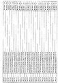
以下の実験項に詳細に記載されるように、HER3結合変異体の選択は、個々のHER3結合モチーフ(BM)配列の同定をもたらした。これらの配列は、この局面に従うHER3結合ポリペプチドの個々の実施態様を構成する。個々のHER3結合モチーフの配列を図1に、そして配列番号1〜334として示す。この局面のいくつかの実施態様において、BM配列i)は、配列番号1〜配列番号66のいずれか1つから選択され、例えば配列番号22〜66のいずれか1つから、例えば配列番号23〜25、配列番号27〜28、配列番号32、配列番号35〜36、配列番号40、配列番号42、配列番号44〜45、配列番号53〜54及び配列番号56から選択される。 As described in detail in the experimental section below, the selection of HER3 binding mutants resulted in the identification of individual HER3 binding motif (BM) sequences. These sequences constitute individual embodiments of HER3 binding polypeptides according to this aspect. The sequences of individual HER3 binding motifs are shown in FIG. In some embodiments of this aspect, BM sequence i) is selected from any one of SEQ ID NO: 1 to SEQ ID NO: 66, for example from any one of SEQ ID NO: 22 to 66, for example SEQ ID NO: 23 to 25, SEQ ID NO: 27-28, SEQ ID NO: 32, SEQ ID NO: 35-36, SEQ ID NO: 40, SEQ ID NO: 42, SEQ ID NO: 44-45, SEQ ID NO: 53-54 and SEQ ID NO: 56.
特定の実施態様において、HER3結合モチーフ(BM)はこのようにして3ヘリックスバンドルタンパク質ドメインの一部を形成する。例えば、BMは該3ヘリックスバンドルタンパク質ドメイン内で相互接続ループを有する2つのアルファヘリックスを本質的に構成し得る。これらの実施態様において、本発明のHER3結合ポリペプチドは:
i) K−[BM]−DPSQS XaXbLLXc EAKKL NDXdQ;
[ここで
[BM]は上で定義されたHER3結合モチーフであり;
XaはA及びSから選択され;
XbはN及びEから選択され;
XcはA、S及びCから選択され;
XdはA及びSから選択される];
及び
ii) 上で定義される配列のいずれか1つに対して少なくとも80%の同一性を有するアミノ酸配列、
から選択されるアミノ酸配列を含み得る。上記アミノ酸配列は、上で定義された配列のいずれか1つに対して少なくとも82%、例えば少なくとも84%、例えば少なくとも86%、例えば少なくとも88%、例えば少なくとも90%、例えば少なくとも92%、例えば少なくとも94%、例えば少なくとも96%、例えば少なくとも98%の同一性を有し得る。
In certain embodiments, the HER3 binding motif (BM) thus forms part of a three helix bundle protein domain. For example, a BM can essentially constitute two alpha helices with interconnecting loops within the three helix bundle protein domain. In these embodiments, the HER3 binding polypeptides of the invention are:
i) K- [BM] -DPSQS X a X b LLX c EAKKL NDX d Q;
[Where [BM] is the HER3 binding motif defined above;
X a is selected from A and S;
X b is selected from N and E;
X c is selected from A, S and C;
X d is selected from A and S];
And ii) an amino acid sequence having at least 80% identity to any one of the sequences defined above,
An amino acid sequence selected from The amino acid sequence is at least 82%, such as at least 84%, such as at least 86%, such as at least 88%, such as at least 90%, such as at least 92%, such as at least It may have 94% identity, such as at least 96%, such as at least 98%.
上で定義されたHER3結合ポリペプチドの一実施態様において、XaはAであり;XbはNであり;XcはAであり、そしてXdはAである。 In one embodiment of the HER3 binding polypeptide defined above, X a is A; X b is N; X c is A and X d is A.
上で定義されたHER3結合ポリペプチドのさらなる実施態様において、XaはAであり;XbはNであり;XcはCであり、そしてXdはAである。 In a further embodiment of the HER3 binding polypeptide defined above, X a is A; X b is N; X c is C and X d is A.
上で定義されたHER3結合ポリペプチドのさらなる実施態様において、XaはSであり;XbはEであり;XcはSであり、そしてXdはSである。 In a further embodiment of the HER3 binding polypeptide defined above, X a is S; X b is E; X c is S and X d is S.
上で定義されたHER3結合ポリペプチドのさらなる実施態様において、XaはSであり;XbはEであり;XcはCであり、そしてXdはSである。 In a further embodiment of the HER3 binding polypeptide defined above, X a is S; X b is E; X c is C and X d is S.
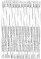
なおさらなる実施態様において、上で定義されるHER3結合ポリペプチドのアミノ酸配列は、配列番号335〜668から選択され、特に配列番号335〜400から、例えば配列番号356〜400から、例えば配列番号357〜359、配列番号361〜362、配列番号366、配列番号369〜370、配列番号374、配列番号376、配列番号378〜379、配列番号387〜388及び配列番号390から選択される。 In a still further embodiment, the amino acid sequence of a HER3 binding polypeptide as defined above is selected from SEQ ID NO: 335-668, in particular from SEQ ID NO: 335-400, for example from SEQ ID NO: 356-400, for example SEQ ID NO: 357- 359, SEQ ID NO: 361-362, SEQ ID NO: 366, SEQ ID NO: 369-370, SEQ ID NO: 374, SEQ ID NO: 376, SEQ ID NO: 378-379, SEQ ID NO: 387-388 and SEQ ID NO: 390.
特定の実施態様において、上記3ヘリックスバンドルタンパク質ドメインは細菌受容体タンパク質のドメインから選択される。このようなドメインの非限定的な例は、黄色ブドウ球菌(Staphylococcus aureus)由来のプロテインAの5つの異なる3ヘリックスドメイン、例えばドメインB、及びその誘導体である。いくつかの実施態様において、3ヘリックスバンドルタンパク質ドメインはプロテインZの変異体であり、これはブドウ球菌プロテインAの上記ドメインBから誘導される。 In certain embodiments, the three-helix bundle protein domain is selected from domains of bacterial receptor proteins. Non-limiting examples of such domains are five different 3-helix domains of protein A from Staphylococcus aureus, such as domain B, and derivatives thereof. In some embodiments, the 3-helix bundle protein domain is a variant of protein Z, which is derived from domain B of staphylococcal protein A.
従って、さらなる実施態様において:
i) YAK−[BM]−DPSQS SELLXc EAKKL NDSQA P;
[ここで[BM]は上で定義されたHER3結合モチーフであり、そして
XcはS及びCから選択される];及び
ii) i)において定義される配列のいずれか1つに対して少なくとも80%の同一性を有するアミノ酸配列、
から選択されるアミノ酸配列を含むHER3結合ポリペプチドが提供される。
Thus, in a further embodiment:
i) YAK- [BM] -DPSQS SELLX c EAKKL NDSQA P;
[Where [BM] is a HER3 binding motif as defined above and X c is selected from S and C]; and ii) at least for any one of the sequences defined in i) An amino acid sequence having 80% identity,
HER3 binding polypeptides comprising an amino acid sequence selected from are provided.
あるいは:
i) FNK−[BM]−DPSQS ANLLXc EAKKL NDAQA P;
[ここで[BM]は上で定義されたHER3結合モチーフであり、そして
XcはA及びCから選択される];及び
ii) 上記i)において定義された配列のいずれか1つに対して少なくとも80%の同一性を有するアミノ酸配列、
から選択されるアミノ酸配列を含むHER3結合ポリペプチドが提供される。
Or:
i) FNK- [BM] -DPSQS ANNLX c EAKKL NDAQA P;
[Where [BM] is a HER3 binding motif as defined above and X c is selected from A and C]; and ii) for any one of the sequences defined in i) above An amino acid sequence having at least 80% identity,
HER3 binding polypeptides comprising an amino acid sequence selected from are provided.
上で考察したように、上記のアミノ酸配列と比較して小さな変化を含むポリペプチドもまた本出願の範囲内である。従って、いくつかの実施態様において、上で定義されたHER3結合ポリペプチドは、例えば、本明細書中に記載される配列に対して、少なくとも82%、少なくとも83%、少なくとも85%、少なくとも87%、少なくとも89%、少なくとも91%、少なくとも92%、少なくとも94%、少なくとも96%又は少なくとも98%同一である配列を有し得る。 As discussed above, polypeptides that contain minor changes compared to the amino acid sequences described above are also within the scope of this application. Thus, in some embodiments, a HER3 binding polypeptide as defined above is, for example, at least 82%, at least 83%, at least 85%, at least 87% relative to a sequence described herein. A sequence that is at least 89%, at least 91%, at least 92%, at least 94%, at least 96% or at least 98% identical.
いくつかの実施態様において及び以下の実験項において開示されるように、HER3結合モチーフは58アミノ酸のポリペプチドの一部を形成し得る。このようなポリペプチドは、例えば配列番号669〜1002のいずれか1つから選択される配列、特に配列番号669〜734から選択される配列、例えば配列番号690〜734から、例えば配列番号691〜693、配列番号695〜696、配列番号700、配列番号703〜704、配列番号708、配列番号710、配列番号712〜713、配列番号721、配列番号722及び配列番号724から選択される配列を含み得る。 In some embodiments and as disclosed in the experimental section below, the HER3 binding motif may form part of a 58 amino acid polypeptide. Such a polypeptide may be, for example, a sequence selected from any one of SEQ ID NOs: 669-1002, particularly a sequence selected from SEQ ID NOs: 669-734, such as from SEQ ID NOs: 690-734, eg, SEQ ID NOs: 691-693 , SEQ ID NO: 695-696, SEQ ID NO: 700, SEQ ID NO: 703-704, SEQ ID NO: 708, SEQ ID NO: 710, SEQ ID NO: 712-713, SEQ ID NO: 721, SEQ ID NO: 722 and SEQ ID NO: 724. .
本明細書に記載されるいずれかの局面に従うHER3結合ポリペプチドは、相互作用のKD値が多くとも1×10⁻⁶M、又は例えば多くとも1×10⁻⁷M、例えば多くとも1×10⁻⁸MであるようにHER3に結合し得る。本明細書において使用される用語「HER3結合」及び「HER3に対する結合親和性」は、例えばBiacore機器(GE Healthcare)におけるような表面プラズモン共鳴技術の使用により試験され得るポリペプチドの特性を指す。HER3結合親和性は、例えばHER3をBiacore機器のセンサーチップに固定し、そして試験しようとするポリペプチドを含有するサンプルをチップ上に通す実験において試験され得る。あるいは以下の実施例に記載されるように、HER3結合親和性は細胞上(on−cell)実験で試験され得る。細胞上実験は、例えば様々な濃度の標識されたHER3を試験しようとするポリペプチドを提示している細胞とともにインキュベートすることにより行われ得る。その後、細胞集団を蛍光細胞分析分離システム(FACS)におけるようなフローサイトメーターで分析し得、そして平均蛍光強度データを決定し得る。続いて平均蛍光をHER3濃度に対してプロットし、そして見かけの平衡解離定数KDを決定するために1部位結合モデルにフィッティングし得る。KD測定において使用されるHER3又はそのフラグメントは、例えば配列番号1007で表されるアミノ酸配列(HER3−ECD)を含み得る。
HER3 binding polypeptide according to any of the aspects described herein, at most the K D value of the
以下の実施例において説明されるように、HER3結合変異体の選択は、例えばタンパク質骨格の未処理の変異体の選択のためのファージディスプレイ、続いて場合により親和性成熟HER3結合変異体の選択のための親和性成熟及び細胞ディスプレイにより達成され得る。しかし、ファージベースでも、細胞ベースでもその他でも、いずれの選択システムもHER3結合ポリペプチドの選択に使用され得ることが理解される。以下の実施例において、グラム陽性細菌スタフィロコッカス・カルノーサス(Staphylococcus carnosus)での組み換えタンパク質の細胞表面ディスプレイのための発現系を使用した(Jonsson et al、Biotechnol Applied Biochem 54:93−103(2009))。しかし、ファージ粒子の大きさを比較して、より大きなサイズの細胞は一般的にハイスループットスクリーニング及び細胞提示タンパク質ライブラリーからの選択のための蛍光細胞分析分離(FACS)の使用を可能にする。 As described in the examples below, selection of HER3 binding mutants may include selection of phage display for selection of, for example, unprocessed variants of the protein backbone, followed by optional selection of affinity matured HER3 binding mutants. Can be achieved by affinity maturation and cell display. However, it is understood that any selection system, whether phage-based, cell-based or otherwise, can be used to select HER3-binding polypeptides. In the following examples, an expression system for cell surface display of recombinant proteins in the Gram-positive bacterium Staphylococcus carnosus was used (Jonsson et al, Biotechnol Applied Biochem 54: 93-103 (2009)). ). However, by comparing the size of the phage particles, larger size cells generally allow the use of fluorescent cell analytical separation (FACS) for high throughput screening and selection from cell display protein libraries.
IgG分子と比較して、本発明のHER3結合ポリペプチドは比較的小さく、これにより、HER3結合ポリペプチドを広範囲の適用において有用にするだけでなく、固相ペプチド合成を使用した製造が可能になり得る。固相合成経路は、例えば分子画像化における放射標識に使用されるキレート剤のような非生物学的基の部位特異的結合を容易にする。 Compared to IgG molecules, the HER3 binding polypeptides of the present invention are relatively small, which not only makes the HER3 binding polypeptides useful in a wide range of applications, but also allows production using solid phase peptide synthesis. obtain. The solid phase synthesis pathway facilitates site-specific attachment of non-biological groups such as chelators used for radiolabeling in molecular imaging, for example.
従って、当業者には当然のことながら、様々な修飾及び/又は付加が、本発明の範囲から逸脱することなくポリペプチドを特定の適用に合わせるために、本明細書に開示されるいずれかの局面にしたがってなされ得る。例えば、いずれかの局面に従うHER3結合ポリペプチドは、さらなるC末端アミノ酸及び/又はN末端アミノ酸を含み得る。このようなポリペプチドは、ポリペプチド鎖におけるかなり最初の方及び/又は最後の方の位置に、すなわちN末端及び/又はC末端にさらなるアミノ酸残基を有するポリペプチドとして理解されるべきである。従って、HER3結合ポリペプチドは、いずれかの適切な数のさらなるアミノ酸残基、例えば少なくとも1つの付加的なアミノ酸残基を含み得る。付加的アミノ酸残基はそれぞれ、例えばポリペプチドの製造、精製、インビボ若しくはインビトロでの安定化、カップリング、又は検出を改善するために、独立して又は集合的に付加され得る。このような付加的アミノ酸残基は、化学的カップリングの目的のために付加された1つ又はそれ以上のアミノ酸残基を含み得る。この一例はシステイン残基の付加である。このような付加的アミノ酸残基はまた、ポリペプチドの精製又は検出のための「タグ」、例えばタグに対して特異的な抗体との相互作用のため、又はHis6タグの場合には固定化金属アフィニティークロマトグラフィー(IMAC)のための、His6タグ又は「myc」(c−myc)タグ又は「FLAG」タグを生じ得る。 Thus, it will be appreciated by those skilled in the art that various modifications and / or additions may be made to any of the herein disclosed to tailor the polypeptide to a particular application without departing from the scope of the invention. Can be made according to aspects. For example, a HER3 binding polypeptide according to any aspect may include additional C-terminal amino acids and / or N-terminal amino acids. Such a polypeptide is to be understood as a polypeptide having additional amino acid residues at the very first and / or last position in the polypeptide chain, ie at the N-terminus and / or C-terminus. Thus, a HER3 binding polypeptide may comprise any suitable number of additional amino acid residues, for example at least one additional amino acid residue. Each additional amino acid residue may be added independently or collectively, for example, to improve polypeptide production, purification, in vivo or in vitro stabilization, coupling, or detection. Such additional amino acid residues may include one or more amino acid residues added for chemical coupling purposes. An example of this is the addition of cysteine residues. Such additional amino acid residues may also be immobilized on “tags” for polypeptide purification or detection, eg, for interaction with antibodies specific for the tag, or in the case of His 6 tags. Can generate His 6 or “myc” (c-myc) or “FLAG” tags for metal affinity chromatography (IMAC).
上で考察されたさらなるアミノ酸を、化学的連結を用いて(公知の有機化学方法を使用して)、又は融合タンパク質としてのHER3結合ポリペプチドの発現のような他の手段を用いてHER3結合ポリペプチドにカップリングさせ得る。 The additional amino acids discussed above can be added to the HER3-binding polypeptide using chemical ligation (using known organic chemistry methods) or using other means such as expression of HER3-binding polypeptides as fusion proteins. It can be coupled to a peptide.
上で考察されたさらなるアミノ酸は、例えば1つ又はそれ以上のポリペプチドドメインを含み得る。さらなるポリペプチドドメインは、HER3結合ポリペプチドに別の機能、例えば別の結合機能、又は酵素的機能、又は毒素機能(例えばイムノトキシン)、又は蛍光シグナル機能、又はその組み合わせを与え得る。 Additional amino acids discussed above may include, for example, one or more polypeptide domains. The additional polypeptide domain may provide the HER3 binding polypeptide with another function, such as another binding function, or an enzymatic function, or a toxin function (eg, an immunotoxin), or a fluorescent signal function, or a combination thereof.
いくつかの実施態様において、さらなるポリペプチドドメインは、インビボでHER3結合ポリペプチドの半減期を増加させる半減期延長部分を含み得る。当業者に理解されるように、増加した、又は延長された半減期は、特定の分子の血液からの遅延されたクリアランスを意味する。半減期延長部分は、例えば血清アルブミンへのインビボでの結合を可能にするペプチド又はタンパク質を含み得る。特に、半減期延長部分はアルブミン結合部分であり得る。アルブミン結合部分は、例えば天然に存在するポリペプチド、又はそのアルブミン結合フラグメント、又は改変ポリペプチドからなるものであってもよい。改変ポリペプチドは、新しいか又は増強された特性、例えばアルブミンのような分子に対する結合親和性を付与する目的で、それをタンパク質工学技術、例えば部位特異的又は無作為化されたアプローチでの変異及び変更を受けさせることにより、天然に存在する出発ポリペプチドから誘導され得る。 In some embodiments, the additional polypeptide domain can include a half-life extending moiety that increases the half-life of the HER3 binding polypeptide in vivo. As will be appreciated by those skilled in the art, increased or extended half-life means delayed clearance of a particular molecule from the blood. The half-life extending moiety can include, for example, a peptide or protein that allows in vivo binding to serum albumin. In particular, the half-life extending moiety can be an albumin binding moiety. The albumin binding portion may comprise, for example, a naturally occurring polypeptide, or an albumin binding fragment thereof, or a modified polypeptide. A modified polypeptide is a protein engineering technique, such as mutation in a site-specific or randomized approach, for the purpose of conferring new or enhanced properties, such as binding affinity for molecules such as albumin. By being altered, it can be derived from a naturally occurring starting polypeptide.
このような改変アルブミン結合ポリペプチドは、例えばアルブミンに対するその特異的結合親和性について選択された、タンパク質骨格の変異体であってもよい。特定の実施態様において、タンパク質骨格は、連鎖球菌プロテインGのドメイン又はその誘導体、例えば連鎖球菌株G148由来のプロテインGのドメインGA1、ドメインGA2及びドメインGA3、特にドメインGA3から選択され得る。従って、HER3結合ポリペプチドの一実施態様において、さらなるアミノ酸は、連鎖球菌プロテインGのアルブミン結合ドメイン(ABD)、又はその誘導体を含む。本発明のHER3結合ポリペプチドにおけるさらなるポリペプチドドメインとして含まれ得るアルブミン結合ドメインの一例は、配列番号1005で示される。適切なアルブミン結合ドメインの他の例はWO2009/016043に開示される。このようなABD延長ポリペプチドはインビボで血清アルブミンに結合し、そしてそのより長い半減期から利益を得、これによりポリペプチド自体の正味の半減期が増加する(例えばWO91/01743を参照のこと)。 Such a modified albumin binding polypeptide may be a variant of the protein backbone, selected, for example, for its specific binding affinity for albumin. In a particular embodiment, the protein backbone can be selected from domains of streptococcal protein G or derivatives thereof, such as domain GA1, domain GA2 and domain GA3 of protein G from streptococcal strain G148, in particular domain GA3. Thus, in one embodiment of a HER3 binding polypeptide, the additional amino acid comprises the albumin binding domain (ABD) of streptococcal protein G, or a derivative thereof. An example of an albumin binding domain that may be included as an additional polypeptide domain in the HER3 binding polypeptides of the invention is shown in SEQ ID NO: 1005. Other examples of suitable albumin binding domains are disclosed in WO2009 / 016043. Such ABD extended polypeptides bind to serum albumin in vivo and benefit from its longer half-life, thereby increasing the net half-life of the polypeptide itself (see, eg, WO 91/01743). .
本発明に従うHER3結合ポリペプチドが上で定義されるアルブミン結合部分を含む場合、HER3結合(bindnig)ポリペプチドの全体の大きさは比較的小さい。例えばヒト対象のような哺乳動物対象に投与された場合、HER3結合ポリペプチドのアルブミン結合部分は、血清アルブミンと非共有結合で結合し、そしてそれによりポリペプチドは減少した腎クリアランス及び上皮細胞での増加した再循環から利益が得られ得る。しかし組織浸透は、血清アルブミンの血管外漏出特性に起因して速いままである。さらに、半減期延長部分を含むHER3結合ポリペプチドは、対応する半減期延長部分の無いポリペプチドと比較して、インビボで延長された半減期を示すだけでなく、インビボで減少した免疫応答を示す(例えばWO2005/097202を参照のこと)。 When the HER3 binding polypeptide according to the invention comprises an albumin binding moiety as defined above, the overall size of the HER3 binding polypeptide is relatively small. For example, when administered to a mammalian subject, such as a human subject, the albumin binding portion of a HER3 binding polypeptide binds non-covalently to serum albumin, and thereby the polypeptide has reduced renal clearance and in epithelial cells. Benefits can be gained from increased recirculation. However, tissue penetration remains fast due to the extravasation properties of serum albumin. Furthermore, a HER3 binding polypeptide comprising a half-life extending moiety not only exhibits an in vivo extended half-life, but also exhibits a reduced immune response in vivo compared to a polypeptide without a corresponding half-life extending moiety. (See, for example, WO 2005/097220).
さらなるポリペプチドドメインは、同じ結合機能を有するHER3結合ポリペプチドを生じ得る。従って、さらなる実施態様において、そのアミノ酸配列は同じであっても異なっていてもよい少なくとも2つのHER3結合ポリペプチド単量体単位を含むHER3結合ポリペプチドが提供される。ポリペプチドの多量体形態は、それぞれがHER3結合モチーフを有し、かつそれぞれが多量体内の「単量体」を形成する、適切な数のドメインを含み得る。これらのドメインは全て同じアミノ酸配列を有していてもよいが、あるいはそれらが異なるアミノ酸配列を有していてもよい。特に、本発明のHER3結合ポリペプチドは、ホモ二量体又はヘテロ二量体を形成し得る。 Additional polypeptide domains can result in HER3 binding polypeptides having the same binding function. Accordingly, in a further embodiment, there is provided a HER3 binding polypeptide comprising at least two HER3 binding polypeptide monomer units, the amino acid sequences of which may be the same or different. Multimeric forms of polypeptides can include an appropriate number of domains, each having a HER3 binding motif and each forming a “monomer” within the multimer. These domains may all have the same amino acid sequence, or they may have different amino acid sequences. In particular, the HER3 binding polypeptides of the present invention may form homodimers or heterodimers.
上記のさらなるポリペプチドドメイン(単数又は複数)は、公知の有機化学方法を使用して共有結合カップリングによりHER3結合ポリペプチドに結合され得る。あるいは、さらなるポリペプチドドメイン(単数又は複数)を含むHER3結合ポリペプチドを1つ又はそれ以上の融合ポリペプチドとして、例えばポリペプチドのの組み換え発現のための系において発現させてもよいし、直接的又はリンカー(例えばアミノ酸リンカー)を介して、いずれかの他のやり方で結合させてもよい。 The additional polypeptide domain (s) described above can be bound to the HER3 binding polypeptide by covalent coupling using known organic chemistry methods. Alternatively, a HER3 binding polypeptide comprising additional polypeptide domain (s) may be expressed as one or more fusion polypeptides, eg, in a system for recombinant expression of the polypeptide, or directly Or it may be attached in any other way via a linker (eg an amino acid linker).
HER3結合ポリペプチドの一実施態様において、本ポリペプチドは天然に存在するHER3リガンドと同じエピトープに結合するか、又はそれに十分に接近して天然に存在するHER3リガンド(例えばニューレグリン−1及び2)のHER3への結合をブロックする。従ってHER3結合ポリペプチドは、HER3遮断薬として、かつ/又はインビボでのHER3媒介細胞シグナル伝達を阻害するために使用され得る。 In one embodiment of a HER3 binding polypeptide, the polypeptide binds to the same epitope as a naturally occurring HER3 ligand or is sufficiently close to a naturally occurring HER3 ligand (eg, neuregulin-1 and 2). Blocks binding to HER3. Accordingly, HER3-binding polypeptides can be used as HER3-blockers and / or to inhibit HER3-mediated cell signaling in vivo.
さらなる局面において、上記のHER3結合ポリペプチドをコードするポリヌクレオチドが提供される。このようなポリヌクレオチドを含む発現ベクターは、例えば宿主細胞における発現によりHER3結合ポリペプチドの生産を可能にし得る。 In a further aspect, a polynucleotide encoding the above HER3 binding polypeptide is provided. An expression vector comprising such a polynucleotide may allow production of a HER3 binding polypeptide, for example by expression in a host cell.
HER3結合ポリペプチドは、様々な医療、獣医学及び診断適用における従来の抗体又は低分子量物質に対する代替として使用され得る。当業者は、本発明のポリペプチドが、試薬のHER3に対する親和性を必要とするいずれかの方法において有用であり得るということを理解するだろう。従って、HER3結合ポリペプチドは、このような方法における検出試薬、捕捉試薬又は分離試薬として使用され得る。さらに、HER3結合ポリペプチドは、例えば哺乳動物対象における細胞表面でのHER3発現を特徴とする疾患の存在を明らかにし、診断し、そして試験するために診断法において使用され得る。従って、HER3発現は、診断上、本発明のポリペプチドの使用によりインビボでモニタリングされ得る。診断モニタリングは、例えば分子画像化によるHER3発現の判断のための診断用核種の送達により達成され得る。本発明に従うHER3結合ポリペプチドはまた、それ自身だけで治療剤若しくは診断剤として、又はHER3タンパク質に対する直接的(例えば、イットリウム−90、ルテチウム−177及びトリウム−227のような放射性核種、マイタンシノイドのような毒素を含む毒性分子)若しくは間接的(例えばがんワクチン、免疫賦活性分子)な効果を有する他の治療剤若しくは診断剤を標的とするための手段として有用であり得る。治療効果は、受容体相互作用をブロックすることにより、あるいは腫瘍細胞へ有効なペイロード(payload)を送達するための標的としてHER3結合ポリペプチドを使用することにより、潜在的に媒介され得る。 HER3-binding polypeptides can be used as an alternative to conventional antibodies or low molecular weight substances in various medical, veterinary and diagnostic applications. One skilled in the art will appreciate that the polypeptides of the present invention may be useful in any method that requires the affinity of the reagent for HER3. Accordingly, HER3-binding polypeptides can be used as detection, capture or separation reagents in such methods. In addition, HER3-binding polypeptides can be used in diagnostic methods to reveal, diagnose, and test the presence of diseases characterized by, for example, cell surface HER3 expression in mammalian subjects. Accordingly, HER3 expression can be monitored in vivo by diagnostic use of the polypeptides of the invention. Diagnostic monitoring can be accomplished by delivery of a diagnostic nuclide for determination of HER3 expression, for example by molecular imaging. HER3-binding polypeptides according to the invention are also themselves therapeutic or diagnostic agents or directly against HER3 proteins (eg radionuclides such as yttrium-90, lutetium-177 and thorium-227, maytansinoids). Toxic molecules including toxins) or other therapeutic or diagnostic agents having indirect (eg cancer vaccine, immunostimulatory molecules) effects may be useful as a means for targeting. The therapeutic effect can potentially be mediated by blocking receptor interactions or by using a HER3 binding polypeptide as a target for delivering an effective payload to tumor cells.
さらなる局面において、上で定義されたHER3結合ポリペプチド;HER2結合ポリペプチド;及びHER3結合ポリペプチドとHER2結合ポリペプチドとを連結するための連結部分を含む、HER3及びHER2に対する結合親和性を有するリガンドが提供される。このようなリガンドは、2つの異なる結合機能を含む二重特異性ヘテロ二量体の例として解釈されるべきであり:1つは上で開示されるポリペプチドによりもたらされるHER3に対する結合機能であり、そして1つはHER2結合ポリペプチドによりもたらされるHER2に対する結合機能である。当然のことながら、本発明に従うHER3結合ポリペプチドは、リガンド中に含まれ得る。2つの結合機能は、2つの別々の単量体単位としてリガンドに含まれる。 In a further aspect, a ligand having binding affinity for HER3 and HER2, comprising a HER3-binding polypeptide as defined above; a HER2-binding polypeptide; and a linking moiety for linking the HER3-binding polypeptide and the HER2-binding polypeptide Is provided. Such a ligand should be construed as an example of a bispecific heterodimer containing two different binding functions: one is the binding function to HER3 provided by the polypeptide disclosed above And one is the binding function for HER2 provided by the HER2 binding polypeptide. Of course, a HER3 binding polypeptide according to the invention may be included in a ligand. Two binding functions are included in the ligand as two separate monomer units.
このような本発明に従う二重特異性リガンドは、HER3及びHER2の両方が関与するがんの検出、特徴付け及び/又は診断において有用であり得る。検出、特徴付け及び/又は診断は、例えばガンマ線若しくは陽電子放出核種を使用する分子画像により、又は近赤外蛍光プローブを使用することにより達成され得る。上記のように、HER3はHER2についての好ましいヘテロ二量化パートナーであり、そしてHER2の過剰発現を特徴とする腫瘍の増殖を促進するために重要である。HER2は公知のリガンドがなく、リガンド誘導シグナル伝達に関してHERファミリーの他の受容体に依存すると現在では考えられている。しかし本発明に従う二重特異性リガンドは、診断のためだけでなく、標的化治療ペイロード適用にも使用され得る。ペイロード適用において使用される化合物としては、とりわけ、放射性核種、プロドラッグ酵素、サイトカイン、化学毒性分子、毒素及び光増感剤が挙げられる。標的化ペイロード適用については、正常組織と疾患組織との間の高度な識別が非常に重要である。 Such bispecific ligands according to the present invention may be useful in the detection, characterization and / or diagnosis of cancers involving both HER3 and HER2. Detection, characterization and / or diagnosis can be achieved, for example, by molecular imaging using gamma rays or positron emitting nuclides, or by using near infrared fluorescent probes. As mentioned above, HER3 is a preferred heterodimerization partner for HER2 and is important for promoting the growth of tumors characterized by HER2 overexpression. HER2 has no known ligand and is now believed to depend on other receptors in the HER family for ligand-induced signaling. However, the bispecific ligands according to the present invention can be used not only for diagnosis but also for targeted therapeutic payload applications. Compounds used in payload applications include radionuclides, prodrug enzymes, cytokines, chemotoxic molecules, toxins and photosensitizers, among others. For targeted payload applications, a high degree of discrimination between normal and diseased tissue is very important.
2つの異なる受容体に結合する二重特異性リガンドに含まれる両方の結合ポリペプチドの親和性を微調整することにより、リガンドは両方の受容体を同時に発現している病変にのみ優先的に位置し、そして蓄積し得る。従って、HER2及びHER3結合機能は、それらの個々の標的に対して2つの同等に強力な結合機能を有するリガンドをもたらし得る。従って、HER3結合ポリペプチドのHER3に対する結合親和性は、HER2結合ポリペプチドのHER2に対する結合親和性とほぼ同位であり得る。一実施態様において、HER2結合ポリペプチドは、相互作用のKD値が多くとも1×10⁻⁶M、例えば多くとも1×10⁻⁷M、例えば多くとも1×10⁻⁸MであるようにHER2に結合し得る。別の実施態様において、HER2結合ポリペプチドのHER2に対する結合は、対応するHER3結合ポリペプチドのHER3に対する結合と同程度に強力であるかより強力であり得る。従って、HER2結合ポリペプチドとHER2との間の相互作用のKD値は、多くてHER3結合ポリペプチドとHER3との間の相互作用のKD値である。HER3結合ポリペプチドとHER3との間の相互作用のKD値は、HER2結合ポリペプチドとHER2との間の相互作用のKD値の例えば5倍、10倍又は100倍であり得る。
By fine-tuning the affinity of both binding polypeptides in a bispecific ligand that binds to two different receptors, the ligand is preferentially located only on lesions that express both receptors simultaneously. And can accumulate. Thus, HER2 and HER3 binding functions can result in ligands with two equally strong binding functions for their individual targets. Thus, the binding affinity of a HER3 binding polypeptide to HER3 can be approximately the same as the binding affinity of a HER2 binding polypeptide to HER2. In one embodiment, HER2 binding polypeptide, at most the K D value of the
一実施態様において、二重特異性リガンドのHER2結合ポリペプチドは改変ポリペプチドである。HER2結合ポリペプチドは、HER2に対するその特異的結合親和性について選択された、タンパク質骨格の変異体である改変ポリペプチドであり得る。タンパク質骨格の非限定的な例は、ブドウ球菌プロテインA、並びにそのドメイン及び誘導体からなる群より選択される。いくつかの実施態様において、HER2結合ポリペプチドは、黄色ブドウ球菌(Staphylococcus aureus)由来のプロテインAのドメインBから誘導された改変タンパク質である。この改変タンパク質は、特にプロテインZ誘導体を含んでいてもよく、これは上記プロテインAのドメインBから誘導されたプロテインZの変異体である。 In one embodiment, the dual specific ligand HER2 binding polypeptide is a modified polypeptide. A HER2 binding polypeptide may be a modified polypeptide that is a variant of the protein backbone selected for its specific binding affinity for HER2. Non-limiting examples of protein backbones are selected from the group consisting of staphylococcal protein A and its domains and derivatives. In some embodiments, the HER2-binding polypeptide is a modified protein derived from domain B of protein A from Staphylococcus aureus. This modified protein may in particular comprise a protein Z derivative, which is a variant of protein Z derived from domain B of protein A above.
リガンドのいくつかの実施態様において、改変HER2結合タンパク質は、
i) 配列番号1003に対応する、
YAKEM RNAYW EIALL PNLTN QQKRA FIRKL YDDPS QSSEL LSEAK KLNDS Q;及び
ii) i)において定義される配列に対して少なくとも80%の同一性を有するアミノ酸配列、
から選択されるアミノ酸配列を含む。
In some embodiments of the ligand, the modified HER2 binding protein is
i) corresponding to SEQ ID NO: 1003,
YAKEM RNAYW EIALL PNLTN QQKRA FIRKL YDDPS QSSEL LSEAK KLNDS Q; and ii) an amino acid sequence having at least 80% identity to the sequence defined in i),
An amino acid sequence selected from
例えば1から45個までのアミノ酸を含むペプチドリンカーによりHER3結合ポリペプチド及びHER2結合ポリペプチドが連結され得る。連結部分は特にHER3結合ポリペプチドとHER2結合ポリペプチドのN末端及び/又はC末端のカップリングのために配置され得る。このようにして生じる連結は、C末端とC末端との連結、C末端とN末端との連結又はN末端とC末端の連結から選択され得る。連結部分のHER3結合ポリペプチド及びHER2結合ポリペプチドへのカップリングは、例えば化学的結合(公知の有機化学方法を使用する)を用いて、又は融合タンパク質としてのリガンドの発現のようないずれかの他の手段により達成され得る。 For example, a HER3 binding polypeptide and a HER2 binding polypeptide may be linked by a peptide linker comprising from 1 to 45 amino acids. The linking moiety may in particular be arranged for coupling at the N-terminus and / or C-terminus of the HER3 binding polypeptide and the HER2 binding polypeptide. The linkage thus generated can be selected from C-terminal to C-terminal linkage, C-terminal to N-terminal linkage or N-terminal to C-terminal linkage. Coupling of the linking moiety to the HER3 binding polypeptide and HER2 binding polypeptide can be done either by chemical coupling (using known organic chemistry methods) or by expression of the ligand as a fusion protein, for example. It can be achieved by other means.
リガンドは、上記のように、インビボでのリガンドの半減期を延長するための半減期延長部分をさらに含み得る。半減期の延長は、多数の種々の半減期延長部分により達成され得、そしてこのような部分の非限定的な例は、アルブミン、HER3結合ポリペプチドに関連して上で定義されたようなアルブミン結合部分、IgGからのFc部分、及びポリエチレングリコール(PEG)のようなポリマー化合物から選択される。哺乳動物血清において最も豊富なタンパク質である血清アルブミンへのリガンドのカップリングは、インビボでのリガンドの半減期の延長をもたらし得る。 The ligand may further comprise a half-life extending moiety for extending the half-life of the ligand in vivo, as described above. Half-life extension can be achieved by a number of different half-life extending moieties, and non-limiting examples of such moieties include albumin, albumin as defined above in connection with HER3 binding polypeptides. It is selected from binding moieties, Fc moieties from IgG, and polymeric compounds such as polyethylene glycol (PEG). Coupling of a ligand to serum albumin, the most abundant protein in mammalian serum, can result in an increased half-life of the ligand in vivo.
特に、アルブミン結合部分は、
i) 配列番号1005で示される、
LAEAK VLANR ELDKY GVSDF YKRLI NKAKT VEGVE ALKLH ILAAL P;及び
ii) i)において定義された配列に対して少なくとも80%の同一性を有するアミノ酸配列、
から選択されるアミノ酸配列を含むアルブミン結合部分のような、連鎖球菌プロテインGのドメインGA3から誘導された改変タンパク質であり得る。
In particular, the albumin binding moiety is
i) represented by SEQ ID NO: 1005,
An amino acid sequence having at least 80% identity to the sequence defined in LAEAK VLAN ELDKY GVSDF YKRLI NKAKT VEGVE ALKLH ILAAL P; and ii) i);
It may be a modified protein derived from domain GA3 of streptococcal protein G, such as an albumin binding moiety comprising an amino acid sequence selected from
半減期延長部分は、例えば公知の有機化学方法を使用して共有結合カップリングによりリガンドの残部に接続されても、他のやり方で直接、又は多数のアミノ酸を含むリンカーを介して結合されてもよい。半減期延長部分がアルブミン又はアルブミン結合部分により表される場合、リガンドはそれ自体1つ又はそれ以上の融合ポリペプチドとしてポリペプチドの組み換え発現のための系において発現されてもよい。一実施態様において、半減期延長部分は、リガンドに存在するシステイン残基を介してリガンドの残部に結合する。特に、半減期延長部分は、結合モチーフから離れた部位に導入された、HER3又はHER2結合ポリペプチドのシステイン残基に結合され得る。別の実施態様において、半減期延長部分は連結部分を介してリガンドの残部に結合する。 The half-life extending moiety may be attached to the remainder of the ligand by covalent coupling, for example using known organic chemistry methods, or otherwise attached directly or through a linker containing multiple amino acids. Good. Where the half-life extending moiety is represented by albumin or an albumin binding moiety, the ligand may itself be expressed in the system for recombinant expression of the polypeptide as one or more fusion polypeptides. In one embodiment, the half-life extending moiety is attached to the remainder of the ligand via a cysteine residue present in the ligand. In particular, the half-life extending moiety can be linked to a cysteine residue of a HER3 or HER2 binding polypeptide introduced at a site away from the binding motif. In another embodiment, the half-life extending moiety is attached to the remainder of the ligand via a linking moiety.
特定の実施態様において、連結部分は上で定義された半減期延長部分を含む。従って、アルブミン、アルブミン結合部分又はPEGは例えばHER3結合ポリペプチドをHER2結合ポリペプチドに接続するために使用され得る。 In certain embodiments, the linking moiety comprises a half-life extending moiety as defined above. Thus, albumin, albumin binding moiety or PEG can be used, for example, to connect a HER3 binding polypeptide to a HER2 binding polypeptide.
いくつかの実施態様において、本発明に従うリガンドは、さらなるC末端又はN末端アミノ酸を含み得る。このようなさらなるアミノ酸は、HER3結合ポリペプチドに関して上で記載したように、例えばポリペプチドの生産、精製、インビボ若しくはインビトロでの安定化、カップリング又は検出を改善するために、個別に、又は集合的に付加され得る。特定の実施態様において、さらなるアミノ酸は1つ又はそれ以上のポリペプチドドメインを構成し得る。このようなさらなるポリペプチドドメインの機能は、既にリガンドに含まれている機能と同じであっても異なっていてもよい。さらなるポリペプチドドメインは、例えば別のHER3結合機能及び/又は別のHER2結合機能を有するリガンドを生じ得る。従って、本発明に従う二重特異性リガンドは、HER3結合機能の多量体及び/又はHER2結合機能の多量体を含み得る。多量体結合機能を含む二重特異性リガンドの一例は、2つのHER3結合ポリペプチド及び2つのHER2結合ポリペプチドを含むリガンドである。 In some embodiments, the ligand according to the invention may comprise an additional C-terminal or N-terminal amino acid. Such additional amino acids may be individually or assembled as described above for HER3-binding polypeptides, eg, to improve polypeptide production, purification, in vivo or in vitro stabilization, coupling or detection. Can be added. In certain embodiments, the additional amino acids may constitute one or more polypeptide domains. The function of such additional polypeptide domain may be the same as or different from the function already contained in the ligand. The additional polypeptide domain may give rise to a ligand having, for example, another HER3 binding function and / or another HER2 binding function. Thus, a bispecific ligand according to the present invention may comprise a multimer of HER3 binding function and / or a multimer of HER2 binding function. An example of a bispecific ligand that includes a multimeric binding function is a ligand that includes two HER3 binding polypeptides and two HER2 binding polypeptides.
上で定義されたリガンドは、一実施態様において、インビボでのHER2及びHER3の二量化を防止し得る。より詳細には、リガンドは、例えば哺乳動物対象の細胞表面で発現されたHER2及び/又はHER3に結合することにより、対象におけるHER3仲介シグナル伝達を阻害し得る。リガンドの二重特異性に起因して、これは、それらがもはやシグナル伝達を行うことができないような位置において2つの受容体に結合してブロックし得る。当業者には当然のことながら、従って本発明のリガンドは、細胞表面上のHER2及び/又はHER3の発現、特に過剰発現に関連する状態の処置のために利用され得る。 The ligands defined above may, in one embodiment, prevent HER2 and HER3 dimerization in vivo. More particularly, the ligand may inhibit HER3-mediated signaling in the subject, for example by binding to HER2 and / or HER3 expressed on the cell surface of a mammalian subject. Due to the dual specificity of the ligand, it can bind and block the two receptors at positions where they can no longer signal. As will be appreciated by those skilled in the art, the ligands of the invention can thus be utilized for the treatment of conditions associated with HER2 and / or HER3 expression on the cell surface, particularly overexpression.
しかしHER3はEGFRともヘテロ二量体を形成し得、それ故PI3K/Akt経路を活性化する活性シグナル伝達単位を形成する(Engelman et al、Proc Natl Acad Sci 102(10):3788−93(2005))。実際、HER3は、明らかにErbBリガンドの上方調節のために非小細胞肺がん(NSCLC)において過剰発現されるが(Fujimoto et al、Cancer Res 65(24):11478−85(2005))、一方でHER3は正常肺上皮で発現されない。従って、関連する局面において、上で定義されたHER3結合ポリペプチド;EGFR結合ポリペプチド;及びHER3結合ポリペプチドとEGFR結合ポリペプチドとを連結するための連結部分を含む、HER3及びEGFRに対する結合親和性を有するリガンドが提供される。当然のことながら、本発明の上記の局面において記載される特徴は、本発明のこの局面にも適用可能である。特に、このような二重特異性リガンドは、HER3及びEGFRを標的にし得、そしてHER3及びHER2に対する二重特異性リガンド結合に関して上で記載した作用様式と類似しているか又は同じ作用様式で作用する。HER3及びEGFRに対する二重特異性リガンド結合の例は、
i) 配列番号1004に相当する、
YAKEM WIAWE EIRNL PNLNG WQMTA FIAKL LDDPS QSSEL LSEAK KLNDS Q;及び
ii) i)において定義される配列に対して少なくとも80%の同一性を有するアミノ酸配列、
から選択されるEGFR結合ポリペプチドを含み得る。
However, HER3 can also form heterodimers with EGFR, thus forming an active signaling unit that activates the PI3K / Akt pathway (Engelman et al, Proc Natl Acad Sci 102 (10): 3788-93 (2005). )). Indeed, HER3 is clearly overexpressed in non-small cell lung cancer (NSCLC) due to upregulation of ErbB ligand (Fujimoto et al, Cancer Res 65 (24): 11478-85 (2005)), HER3 is not expressed in normal lung epithelium. Thus, in a related aspect, binding affinity for HER3 and EGFR comprising a HER3 binding polypeptide as defined above; an EGFR binding polypeptide; and a linking moiety for linking the HER3 binding polypeptide and the EGFR binding polypeptide. A ligand is provided. Of course, the features described in the above aspects of the invention are also applicable to this aspect of the invention. In particular, such bispecific ligands can target HER3 and EGFR and act in a similar or the same mode of action as described above for bispecific ligand binding to HER3 and HER2. . Examples of dual specific ligand binding to HER3 and EGFR are:
i) corresponding to SEQ ID NO: 1004,
YAKEM WIAWE EIRNL PNLNG WQMTA FIAKL LDPPS QSSEL LSEAK KLNDS Q; and ii) an amino acid sequence having at least 80% identity to the sequence defined in i),
An EGFR binding polypeptide selected from:
HER3及びEGFR結合リガンドにおいて使用され得るEGFR結合ポリペプチドの他の例はWO2007/065635に開示される。 Other examples of EGFR binding polypeptides that can be used in HER3 and EGFR binding ligands are disclosed in WO2007 / 065635.
他の実施態様において、本発明に従うHER3結合ポリペプチド又は二重特異性リガンドは、治療剤又は診断剤に、インビボ及びインビトロの両方で、HER3を発現する細胞、特にHER3を過剰発現する細胞を標的にさせる際に使用される。上で考察したように、本発明のリガンドは、HER3及び/若しくはHER2、又はHER3及び/若しくはEGFRを発現するか又は過剰発現する細胞を標的にする際に利用され得る。従って関連する局面において、本明細書において定義されるHER3結合ポリペプチド、又は上で定義されるリガンド、及び治療剤の組み合わせが提供される。治療剤の非限定的な例は、細胞傷害性ペイロード、例えば放射性核種、低分子量薬物又は毒素から選択される。このような治療剤は、化学的に結合されるか、そうでなければHER3結合ポリペプチド又はリガンドに結合される。 In other embodiments, a HER3 binding polypeptide or bispecific ligand according to the invention targets a therapeutic or diagnostic agent to cells that express HER3, particularly cells that overexpress HER3, both in vivo and in vitro. Used when making As discussed above, the ligands of the invention can be utilized in targeting cells that express or overexpress HER3 and / or HER2, or HER3 and / or EGFR. Accordingly, in a related aspect, provided is a combination of a HER3 binding polypeptide as defined herein, or a ligand as defined above, and a therapeutic agent. Non-limiting examples of therapeutic agents are selected from cytotoxic payloads such as radionuclides, low molecular weight drugs or toxins. Such therapeutic agents are chemically conjugated or otherwise conjugated to a HER3 binding polypeptide or ligand.
本発明に従う組み合わせ、HER3結合ポリペプチド又はリガンドはさらに、治療における、例えばHER3関連状態、例えばがん疾患、例えば結腸がん、子宮内膜がん、胃がん、神経膠腫、乳がん、膵臓がん、頭頸部扁平上皮がん、肺がん、黒色種、髄芽腫、神経上皮腫、卵巣がん、パジェット病、乳頭様甲状腺がん、前立腺がん、皮膚扁平上皮がん、移行上皮がん及び前庭神経鞘腫から選択される、哺乳動物(例えばヒト対象)におけるHER3関連状態の処置のための、医薬として使用され得る。従って、HER3関連状態、例えば上述のHER3関連状態の処置のための医薬の製造のための本発明のHER3結合ポリペプチド又はリガンドの使用もまた、本発明の範囲内である。 The combination, HER3-binding polypeptide or ligand according to the invention is further in therapy, for example in a HER3-related condition, such as a cancer disease such as colon cancer, endometrial cancer, stomach cancer, glioma, breast cancer, pancreatic cancer, Head and neck squamous cell carcinoma, lung cancer, melanoma, medulloblastoma, neuroepithelioma, ovarian cancer, Paget disease, papillary thyroid cancer, prostate cancer, cutaneous squamous cell carcinoma, transitional cell carcinoma and vestibular nerve It can be used as a medicament for the treatment of a HER3-related condition in a mammal (eg, a human subject) selected from schwannomas. Accordingly, the use of the HER3 binding polypeptides or ligands of the present invention for the manufacture of a medicament for the treatment of HER3-related conditions, eg HER3-related conditions as described above, is also within the scope of the present invention.
関連する局面において、HER3結合ポリペプチド、又は上記のリガンド若しくは組み合わせを、それを必要とする哺乳動物に投与することを含む、HER3関連状態の処置方法が提供される。結果として、本処置方法において、対象は、本発明に従うHER3結合ポリペプチド、リガンド又は組み合わせで処置される。この方法のより特定の実施態様において、HER3結合ポリペプチド、リガンド又は組み合わせの、対象の細胞表面で発現されたHER3に対する結合は、受容体活性化を阻害する。この結合は、例えばHER3仲介シグナル伝達を阻害し得る。本処置方法の一実施態様において、上記HER3関連状態は、がん疾患、例えば結腸がん、子宮内膜がん、胃がん、神経膠腫、乳がん、膵臓がん、頭頸部扁平上皮がん、肺がん、黒色種、髄芽腫、神経上皮腫、卵巣がん、パジェット病、乳頭様甲状腺がん、前立腺がん、皮膚扁平上皮がん、移行上皮がん及び前庭神経鞘腫から選択される。 In a related aspect, there is provided a method of treating a HER3-related condition comprising administering a HER3-binding polypeptide, or a ligand or combination as described above, to a mammal in need thereof. As a result, in the present method of treatment, the subject is treated with a HER3 binding polypeptide, ligand or combination according to the present invention. In a more particular embodiment of this method, binding of a HER3 binding polypeptide, ligand or combination to HER3 expressed on the cell surface of the subject inhibits receptor activation. This binding can, for example, inhibit HER3-mediated signaling. In one embodiment of this treatment method, the HER3-related condition is a cancer disease such as colon cancer, endometrial cancer, gastric cancer, glioma, breast cancer, pancreatic cancer, head and neck squamous cell cancer, lung cancer. Melanoma, medulloblastoma, neuroepithelioma, ovarian cancer, Paget's disease, papillary thyroid cancer, prostate cancer, cutaneous squamous cell carcinoma, transitional cell carcinoma and vestibular schwannoma.
別の実施態様において、本発明のリガンドを、それを必要とする哺乳動物対象に投与することを含む、HER3及び/又はHER2関連状態の処置方法が提供される。 In another embodiment, there is provided a method of treating HER3 and / or HER2-related conditions comprising administering a ligand of the invention to a mammalian subject in need thereof.
当業者に理解されるように、そして上で考察したように、本発明に従うHER3結合ポリペプチド又はリガンドは、HER3又はHER3及び/若しくはHER2関連状態の診断のような診断において使用され得る。本発明に従うHER3結合ポリペプチドは、診断剤と別々に使用されても組み合わせて使用されてもよい。このような組み合わせは、HER3関連状態の診断において、例えばHER3を過剰発現する細胞の分子画像化において使用され得る。 As will be appreciated by those skilled in the art and as discussed above, HER3 binding polypeptides or ligands according to the present invention may be used in diagnostics such as diagnosis of HER3 or HER3 and / or HER2-related conditions. The HER3 binding polypeptides according to the invention may be used separately or in combination with a diagnostic agent. Such combinations can be used in the diagnosis of HER3-related conditions, for example in the molecular imaging of cells that overexpress HER3.
本発明はここで以下の非限定的な実施例によりさらに説明される。 The invention will now be further illustrated by the following non-limiting examples.
実施例1:HER3結合ポリペプチドの選択及びスクリーニング
材料及び方法
HER3及びHSAの標識: 組み換えヒトHER3/Fcキメラ(R&D Systems、#348−RB−050)(本明細書ではHER3−Fcと示される)のビオチン化を、Biotin−XXマイクロスケールタンパク質標識キット(Invitrogen、#B30010)を使用して供給業者の推奨にしたがって行った。HER3の細胞外ドメイン(配列番号1007)(本明細書ではHER3−ECDと示される)を、EZ−LinkTM−Sulfo−NHS−LC−LC−Biotin(Pierce、#21338)を使用して供給業者の推奨にしたがってビオチン化した。ヒト血清アルブミン(HSA; Sigma、#A−3782)を、Alexa Fluor(R) 647スクシンイミジルエステル(Invitrogen、#A20006)を使用して供給業者の推奨にしたがって蛍光標識した。タンパク質緩衝液をPBS(10mMホスフェート、137mM NaCl、2.68mM KCl、pH7.4)又は過剰のビオチン若しくはフルオロフォアを除去するために0.1%Pluronic(R)F108 NF界面活性剤(PBSP; BASF Corporation、#30085231)を補充したPBSに変更した。
HER3結合ポリペプチドのファージディスプレイ選択: Groenwall et al、J Biotechnol 128:162−183(2007)に記載されるように、ファージミドpAffi1/pAY00065において構築されたバクテリオファージで提示されるZ分子のランダム変異体のライブラリー(Zlib2002と示される)を、HER3−ECD−結合ポリペプチドの選択のために使用した。ファージミドライブラリーから、さらには選択ラウンドの間に、ファージストックの製造を以前に記載された手順(Nord et al、Nat Biotech 15:772−777(1997); Hansson et al、Immunotechnology 4:237−252(1999))にしたがってヘルパーファージM13K07(New England Biolabs、#NO315S)を使用して行った。大腸菌(Escherichia coli)アンバー抑圧遺伝子系統RR1ΔM15(Ruether、Nucleic Acids Res 10:5765−5772、1982)を全てのクローニング手順のために、そしてファージ産生のための宿主として使用した。
Example 1: HER3-binding polypeptide selection and screening materials and methods
HER3 and HSA labeling : Biotinylation of a recombinant human HER3 / Fc chimera (R & D Systems, # 348-RB-050) (designated here as HER3-Fc) was performed using the Biotin-XX microscale protein labeling kit (Invitrogen). , # B30010) according to the supplier's recommendations. The extracellular domain of HER3 (SEQ ID NO: 1007) (denoted herein as HER3-ECD) was purchased using EZ-Link ™ -Sulfo-NHS-LC-LC-Biotin (Pierce, # 21338). Biotinylated according to the recommendations of Human serum albumin (HSA; Sigma, # A- 3782) were fluorescently labeled according to the supplier's recommendations using the Alexa Fluor (R) 647 succinimidyl ester (Invitrogen, # A20006). Protein buffer PBS (10 mM phosphate, 137mM NaCl, 2.68mM KCl, pH7.4 ) or in excess of 0.1% in order to remove the biotin or fluorophores Pluronic (R) F108 NF Surfactant (PBSP; BASF Changed to PBS supplemented with Corporation, # 30085231).
Phage display selection of HER3 binding polypeptides: Random variants of Z molecules displayed in bacteriophage constructed in phagemid pAffi1 / pAY00065 as described in Groenwall et al, J Biotechnol 128: 162-183 (2007) The library (designated Zlib2002) was used for selection of HER3-ECD-binding polypeptides. From phagemid libraries, and during selection rounds, the production of phage stocks has been described previously (Nord et al, Nat Biotech 15: 772-777 (1997); Hansson et al, Immunotechnology 4: 237-252. (1999)) using the helper phage M13K07 (New England Biolabs, # NO315S). The Escherichia coli amber suppressor gene line RR1ΔM15 (Ruether, Nucleic Acids Res 10: 5765-5972, 1982) was used for all cloning procedures and as a host for phage production.
ファージ選択を、溶液でビオチン化HER3タンパク質及びストレプトアビジン被覆常磁性ビーズ(Dynabeads(R) M−280ストレプトアビジン、Dynal #112.06)を、結合したファージの捕捉のために使用して行った。さらに、ファージ選択を、固相においてMaxiSorp immunotubes(Nunc)に固定化されたHER3−ECDを使用して行った。ファージ粒子の非特異的結合を、全てのチューブ及びビーズをPBST 0.1−ゼラチン(0.1%Tween−20及び0.1%ゼラチンを補充したPBS)で予備処理することにより最少にした。選択を4サイクルで行った。全ての選択をPBST 0.1−ゼラチン中でかつ体積1mlで行った。 The phage selection, biotinylated HER3 protein and streptavidin-coated paramagnetic beads (Dynabeads (R) M-280 Streptavidin, Dynal # 112.06) in solution was performed using for capture of bound phage. In addition, phage selection was performed using HER3-ECD immobilized on MaxiSorp immunotubes (Nunc) in the solid phase. Nonspecific binding of phage particles was minimized by pretreating all tubes and beads with PBST 0.1-gelatin (PBS supplemented with 0.1% Tween-20 and 0.1% gelatin). Selection was done in 4 cycles. All selections were made in PBST 0.1-gelatin and in a volume of 1 ml.
溶液で行われた選択については、4サイクルの選択を室温(RT)にて異なる濃度のビオチン化組み換えHER3−ECDを標的タンパク質として使用して行った。サイクル1及び2で使用したファージストックを、ストレプトアビジン結合ファージを除去するために0.1mgストレプトアビジン被覆ビーズとともに1時間プレインキュベートした。サイクル1において、Zlib2002ライブラリーからのファージ粒子を、100nMのビオチン化HER3−ECDと共にPBST 0.1−ゼラチン中で2時間インキュベートした。それに続く選択サイクルを2つの選択トラックに分割し、そのために標的濃度をサイクル2について50(トラック1)又は20nM(トラック2)まで、サイクル3について20(トラック1)又は10nM(トラック2)まで、そしてサイクル4について20(トラック1)又は5nM(トラック2)まで下げた。結合したファージをストレプトアビジン被覆M−280 Dynabeads(R)を用いて捕捉し、ビーズ1ミリグラムあたりHER3−ECD4μgを固定化させた。
For selections made in solution, four cycles of selection were performed using different concentrations of biotinylated recombinant HER3-ECD as the target protein at room temperature (RT). The phage stock used in
固相での選択については、immunotubeに1ml HER3−ECD(50mM炭酸ナトリウム(sodium carbonat)緩衝液中10μg/ml、pH 9.6)を1.5時間室温にて固定化させ、その後3ml PBST 0.1−ゼラチンを用いて1時間室温にてブロックした。ファージストックをこのチューブに加え、そして2時間インキュベートした。
For selection on the solid phase, 1 ml HER3-ECD (10 μg / ml in 50 mM sodium carbonate buffer, pH 9.6) was immobilized on the immunotube for 1.5 hours at room temperature, followed by 3
両方の選択ストラテジーのために、PBST 0.1−ゼラチンでの洗浄回数を、選択ストリンジェンシーを高めるためにラウンド間で増やした。従って、洗浄をサイクル1において2回、サイクル2において3回、サイクル3において6回、そしてサイクル4において12回行った。結合したファージ粒子を0.1Mグリシン−HCl(pH2.2)で溶出し、続いてすぐに1M Tris−HCl(pH8)で中和した。ファージ含有溶出液を対数期RR1ΔM15細胞に感染させるために使用し、そしてファージミド粒子を、ヘルパーファージM13K07を使用して感染した細胞からレスキューした。選択過程を、各選択ラウンドの前及び標的タンパク質からの溶出後にファージストックの滴定によりモニタリングした。ファージ溶液の段階希釈を、対数期RR1ΔM15細胞の感染のために使用した。
For both selection strategies, the number of washes with PBST 0.1-gelatin was increased between rounds to increase the selection stringency. Therefore, washing was performed twice in
Z変異体のELISAスクリーニング: Z変異体分子が実際にHER3と相互作用するかどうかを試験するために、2つの異なるELISAを行い、一方は標的タンパク質としてビオチン化HER3−ECDを使用し、そして2つ目はHER3−Fcを使用した。上で記載したように製造された単一コロニーを、100μg/mlアンピシリン及び0.1mMイソプロピル−β−D−1−チオガラクトピラノシド(IPTG)を補充した1ml TSB−YE培地(30g/l TSB、5g/l酵母抽出物)中でディープウェルプレート(Nunc #278752)において播種することによりZ変異体を生成した。ネガティブコントロールとして、インスリン結合Z変異体(Z00801)を播種し、そして各プレートにおいて増殖させた。これらのプレートを18〜24時間37℃でインキュベートした。インキュベーション後に、−20℃での貯蔵のために15%グリセロールを含む96ウェルプレートに各培養物を少量移すことにより複製のプレートを作製した。残りの細胞を遠心分離によりペレット化し、300μl PBST 0.05(0.05% Tween 20を補充したPBS)中に再懸濁し、そして細胞の周辺質部分を放出させるために−80℃で凍結した。凍結したサンプルを水浴で解凍し、そして細胞を遠心分離によりペレット化した。周辺質上清は、連鎖球菌株G148由来のプロテインGのGA3からのアルブミン結合ドメインへの融合物としてのZ変異体(AQHDEALE−[Z#####]−VDYV−[ABD]−YVPGとして表される)を含有していた(Groenwall et al、前出)。Z#####は個々のZ変異体を指す。 Z variant ELISA screening: To test whether a Z variant molecule actually interacts with HER3, two different ELISAs were performed, one using biotinylated HER3-ECD as the target protein and 2 The second used HER3-Fc. Single colonies produced as described above were treated with 1 ml TSB-YE medium (30 g / l) supplemented with 100 μg / ml ampicillin and 0.1 mM isopropyl-β-D-1-thiogalactopyranoside (IPTG). Z variants were generated by seeding in deep well plates (Nunc # 278752) in TSB, 5 g / l yeast extract). As a negative control, an insulin-binding Z mutant (Z00801) was seeded and grown on each plate. These plates were incubated at 37 ° C. for 18-24 hours. After incubation, duplicate plates were made by transferring small amounts of each culture to a 96-well plate containing 15% glycerol for storage at -20 ° C. The remaining cells were pelleted by centrifugation, resuspended in 300 μl PBST 0.05 (PBS supplemented with 0.05% Tween 20) and frozen at −80 ° C. to release the periplasmic portion of the cells. . The frozen sample was thawed in a water bath and the cells were pelleted by centrifugation. Periplasmic supernatant was obtained as a Z variant (AQHDEALE- [Z #####]-VDYV- [ABD] -YVPG as a fusion from GA3 to the albumin binding domain of protein G from Streptococcus strain G148. (Groenwall et al, supra). Z ###### refers to individual Z variants.
ハーフエリア96ウェルELISAプレート(Costar #3690)を、6μg/mlヒト血清アルブミン(HSA、Sigma #A3782)を含有する50μl/ウェルのコーティング緩衝液(50mM炭酸ナトリウム、pH9.6)で被覆し、そして終夜(ON)インキュベートした。HSA溶液を捨てて、2%脱脂粉乳溶液(Semper AB)を補充したPBST 0.1 100μlを用いてウェルを1時間室温にてブロックした。ブロッキング溶液を廃棄し、そして周辺質溶液50μlを各ウェルに加え、そして1.5時間室温でゆっくりと震盪しながらインキュベートした。上清を捨てて、ウェルをPBST 0.05で4回洗浄した。PBST 0.05中0.5μg/mlの濃度のHER3−Fc 50μl又はPBS中1μg/mlの濃度のビオチン化HER3−ECDを各ウェルに加えた。プレートを1.5時間室温でインキュベートし、続いてPBST 0.05で4回洗浄した。HER3−Fcプレートにおいて、西洋ワサビペルオキシダーゼ(HRP)で標識されPBST0.05で1:4000に希釈されたヒトFcに対する抗体(DAKO Cytomation、#P0214)をウェルに加え、そして1時間室温でインキュベートした。ビオチン化HER3−ECDを含むプレートにおいて、HRPと結合したストレプトアビジン(DAKO、#P0397)を各ウェルに加えてPBST 0.05で1:5000に希釈した。上で記載したように洗浄した後、50μl ImmunoPure TMB基質(Pierce #34021)をウェルに加え、そしてプレートを製造者の推奨に従って処理した。ブロッキングから読み取りまでの全ての工程を、Tecan Genesis Freedom 200ロボット(Tecan Group LTD)で行った。ウェルの吸光度を450nmでELISAリーダーTecan Ultra 384(Tecan)で読み取り、そしてMagellan v.5.0ソフトウェア(Tecan)を用いて評価した。
Half area 96 well ELISA plate (Costar # 3690) is coated with 50 μl / well coating buffer (50 mM sodium carbonate, pH 9.6) containing 6 μg / ml human serum albumin (HSA, Sigma # A3782), and Incubated overnight (ON). The HSA solution was discarded and the wells were blocked for 1 hour at room temperature with 100 μl PBST 0.1 supplemented with 2% non-fat dry milk solution (Semper AB). The blocking solution was discarded and 50 μl of periplasmic solution was added to each well and incubated for 1.5 hours at room temperature with gentle shaking. The supernatant was discarded and the wells were washed 4 times with PBST 0.05. 50 μl of HER3-Fc at a concentration of 0.5 μg / ml in PBST 0.05 or biotinylated HER3-ECD at a concentration of 1 μg / ml in PBS was added to each well. Plates were incubated for 1.5 hours at room temperature followed by 4 washes with PBST 0.05. In a HER3-Fc plate, antibodies against human Fc (DAKO Cytomation, # P0214) labeled with horseradish peroxidase (HRP) and diluted 1: 4000 with PBST0.05 were added to the wells and incubated for 1 hour at room temperature. In a plate containing biotinylated HER3-ECD, streptavidin conjugated with HRP (DAKO, # P0397) was added to each well and diluted 1: 5000 with PBST 0.05. After washing as described above, 50 μl ImmunoPure TMB substrate (Pierce # 34021) was added to the wells and the plates were processed according to manufacturer's recommendations. All steps from blocking to reading were performed on a
配列決定: ELISAスクリーニングに基づいて、陽性と考えられる全てのクローンの一部を配列決定のために選別した。PCRフラグメントを、標準的なPCRプログラム並びにプライマーAFFI−21(5’−tgcttccggctcgtatgttgtgtg)及びAFFI−22(5'−cggaaccagagccaccaccgg)を使用して単一コロニーから増幅した。増幅されたフラグメントの配列決定を、製造者のプロトコルに従って使用されるビオチン化オリゴヌクレオチドAFFI−72(5'−ビオチン−cggaaccagagccaccaccgg)及びBigDye(R)ターミネーターv3.1サイクルシークエンスキット(Applied Biosystems)を使用して行った。配列決定反応混合物を、磁気ストレプトアビジン被覆ビーズへの結合により、Magnatrix 8000(Magnetic Biosolution)を使用して精製し、そしてABI PRISM(R)3100 Genetic Analyzer(PE Applied Biosystems)で分析した。配列決定結果をインポートし、そしてALD LIMS NautilusTM 2003 R2 B3ソフトウェア(Thermo Electronics Corp.)を用いて分析した。 Sequencing: Based on ELISA screening, a portion of all clones considered positive were selected for sequencing. PCR fragments were amplified from single colonies using standard PCR programs and primers AFFI-21 (5′-tgcttccggctctgtgtgtgtgtgt) and AFFI-22 (5′-cggaccaccagaccaccaccaccg). Using the sequencing of the amplified fragments, biotinylated oligonucleotide AFFI-72 used according to the manufacturer's protocol (5'-biotin -Cggaaccagagccaccaccgg) and BigDye (R) Terminator v3.1 cycle sequencing kit (Applied Biosystems) I went there. Sequencing reaction mixture, by binding to magnetic streptavidin coated beads were purified using Magnatrix 8000 the (Magnetic Biosolution), and analyzed with ABI PRISM (R) 3100 Genetic Analyzer (PE Applied Biosystems). Sequencing results were imported and analyzed using ALD LIMS Nautilus ™ 2003 R2 B3 software (Thermo Electronics Corp.).
ドットブロット分析: 生成されたHER3結合Z変異体を、特異性についてドットブロット分析によりアルファ−2マクログロブリン(MP biomedicals/Cappel、#55833)、アルファ−1酸性糖タンパク質(RDI、#RDI−SCP−153−1)、アルファ−1抗キモトリプシン(RDI、#RDI−SCP−159−0)、アルファ−1−抗トリプシン(RDI、#RDI−SCP−165−5)、補体C3(RDI、#RDI−SCP−150−0)、補体C4(RDI、#RDI−SCP−151−0)、フィブリノゲン(Enzyme research、#031015E)、ハプトグロブリン(RDI、#RDI−SCP−119−1)、ヘモペキシン(Agilent)、ホロ−トランスフェリン(Sigma、#T0665)、ヒトIgA(Bethyl、#P80−102)、マウスIgEカッパ(BD、#55079)、ヒトIgG(Sigma、#G4386)、ヒトIgM(Sigma、#I8260)、ヒト血清アルブミン(HSA、Sigma、#A3782)、ニュートラアビジン(Pierce、#31000)、ストレプトアビジン(Pierce、#21122)、トランスサイレチン(Sigma、#P1742)、HER2−ECD(配列番号1008)、HER3−ECD(配列番号1007)、HER3−Fc(R&D Systems)、HER4−ECD(配列番号1009)、HER4−Fc(R&D Systems)に対して試験した。 Dot blot analysis: The generated HER3 binding Z variants were analyzed for alpha-2 macroglobulin (MP biomedicals / Cappel, # 55833), alpha-1 acidic glycoprotein (RDI, # RDI-SCP-) by dot blot analysis for specificity. 153-1), alpha-1 antichymotrypsin (RDI, # RDI-SCP-159-0), alpha-1-antitrypsin (RDI, # RDI-SCP-165-5), complement C3 (RDI, #RDI) -SCP-150-0), complement C4 (RDI, # RDI-SCP-151-0), fibrinogen (Enzyme research, # 031015E), haptoglobulin (RDI, # RDI-SCP-119-1), hemopexin ( Agilent), holo-transfer (Sigma, # T0665), human IgA (Bethyl, # P80-102), mouse IgE kappa (BD, # 55079), human IgG (Sigma, # G4386), human IgM (Sigma, # I8260), human serum albumin ( HSA, Sigma, # A3782), neutravidin (Pierce, # 31000), streptavidin (Pierce, # 21122), transthyretin (Sigma, # P1742), HER2-ECD (SEQ ID NO: 1008), HER3-ECD (sequence) No. 1007), HER3-Fc (R & D Systems), HER4-ECD (SEQ ID NO: 1009), HER4-Fc (R & D Systems).
可溶性タンパク質を含有する周辺質上清を得るために、Z変異体をELISAスクリーニングで記載された方法と本質的に同じ方法で製造した。上清を0.45μmメンブレンを使用してろ過した。 In order to obtain periplasmic supernatants containing soluble proteins, Z variants were produced in essentially the same manner as described in the ELISA screen. The supernatant was filtered using a 0.45 μm membrane.
ニトロセルロースメンブレン(Invitrogen)に各タンパク質1μlを、それぞれ0.29mg/ml及び0.14mg/mlの濃度を有していたHER3−ECD及びHER4−ECDを除いて0.25mg/mlの濃度で用いてドットを打った。これらのメンブレンを0.5%カゼイン(ブロッキング液)を補充したPBST0.1中で終夜4℃でブロックした。溶液を除去した後、これらのメンブレンを1時間、0.5%カゼインを補充した周辺質上清と共にインキュベートした。これらのメンブレンをPBST 0.1中で非常に短く3回、そして4×5分洗浄した。Z変異体を、PBSTブロッキング液中で1:5000に希釈された全てのZ変異体(社内で製造)に共通のエピトープに対するABD融合超免疫ポリクローナルウサギIgを用いて1時間室温でインキュベートすることにより検出した。上記のように洗浄した後、ブロッキング溶液で1:5000希釈された、HRPと複合体化されたウサギIgGに対する抗体(DAKO Cytomation、#P0448)をメンブレンに加え、続いてこれらのメンブレンを1時間室温でインキュベートした。メンブレンを洗浄し、PBSですすぎ、そしてSupersignal(Pierce #34075)と共に浸漬した。発光をChemiImager 5500(Alpha Innotech Corp.)を用いて撮影した。 1 μl of each protein was used on a nitrocellulose membrane (Invitrogen) at a concentration of 0.25 mg / ml except for HER3-ECD and HER4-ECD, which had concentrations of 0.29 mg / ml and 0.14 mg / ml, respectively. And hit a dot. These membranes were blocked overnight at 4 ° C. in PBST0.1 supplemented with 0.5% casein (blocking solution). After removing the solution, the membranes were incubated with periplasmic supernatant supplemented with 0.5% casein for 1 hour. These membranes were washed very briefly three times in PBST 0.1 and 4 × 5 minutes. By incubating the Z variant for 1 hour at room temperature with ABD-fused hyperimmune polyclonal rabbit Ig against an epitope common to all Z variants (manufactured in-house) diluted 1: 5000 in PBST blocking solution Detected. After washing as above, antibodies against rabbit IgG complexed with HRP (DAKO Cytomation, # P0448) diluted 1: 5000 in blocking solution were added to the membrane, followed by 1 hour at room temperature. Incubated with. The membrane was washed, rinsed with PBS, and soaked with Supersignal (Pierce # 34075). Luminescence was photographed using a ChemiImager 5500 (Alpha Innotech Corp.).
Z変異体のサブクローニング: 二量体Z変異体をpAffi1/pAY00065ベクターから増幅した。異なるプライマー対を使用してPCRを行い、そして得られた遺伝子フラグメントを精製し、そしてリガーゼ緩衝液中でハイブリダイズさせた。ハイブリダイズした遺伝子フラグメントをpAY00430ベクターでサブクローニングし、N末端His6タグ及びC末端システインを生じた(His6−(Z#####)2−Cys)。HER3結合Z変異体をダイマーとしてサブクローニングし、そして発現ベクターによりコードされる構築物は、MGSSHHHHHHLQ−[Z#####][Z#####]−VDCであった。3つの部分のライゲーションを、両方の挿入フラグメントをベクター中に同じ工程で挿入するために使用した。ハイブリダイズした遺伝子フラグメント及びAccI−消化して脱リン酸化した発現ベクターをリガーゼ緩衝液中で連結し、エレクトロコンピテント(electrocompetent)E.coli TOP10細胞中にエレクトロポレーション処理した。形質転換細胞を、50μg/mlのカナマイシンを補充したTBABプレート(30g/lトリプトース(tryptose)血液寒天基礎培地)上に広げ、続いて37℃で終夜インキュベートした。コロニーをPCRを使用してスクリーニングし、そしてPCRフラグメントの長さをアガロースゲルで確認した。配列を確認するために、上記のように配列決定を行った。プラスミドDNAストックを配列決定したクローンから調製し、そして−80℃に置いた。さらに、E.coli BL21(DE3)細胞をエレクトロポレーションによりプラスミドを用いて形質転換した。 Subcloning of Z variant: The dimeric Z variant was amplified from the pAffi1 / pAY00065 vector. PCR was performed using different primer pairs and the resulting gene fragments were purified and hybridized in ligase buffer. The hybridized gene fragment was subcloned with the pAY00430 vector to generate an N-terminal His 6 tag and a C-terminal cysteine (His 6- (Z #####) 2 -Cys). The HER3-binding Z variant was subcloned as a dimer and the construct encoded by the expression vector was MGSSHHHHHLQ- [Z ######] [Z ######]-VDC. Three-part ligation was used to insert both inserts into the vector in the same step. The hybridized gene fragment and the AccI-digested and dephosphorylated expression vector were ligated in ligase buffer and electrocompetent E. coli. electroporation into E. coli TOP10 cells. Transformed cells were spread on TBAB plates (30 g / l tryptose blood agar basal medium) supplemented with 50 μg / ml kanamycin and subsequently incubated at 37 ° C. overnight. Colonies were screened using PCR, and the length of the PCR fragment was confirmed on an agarose gel. To confirm the sequence, sequencing was performed as described above. Plasmid DNA stock was prepared from sequenced clones and placed at -80 ° C. Further, E.I. E. coli BL21 (DE3) cells were transformed with the plasmid by electroporation.
結果
HER3結合ポリペプチドのファージディスプレイ選択: 4ラウンドのファージディスプレイ選択を、ビオチン化又は非ビオチン化ヒトHER3−ECDに対して行った。4回の選択サイクルを、2つの異なる標的濃度でビオチン化標的を含む溶液で、又は非ビオチン化標的を含む固相で行った。溶液では、ファージ粒子標的複合体がストレプトアビジン被覆ビーズに捕捉された。各選択サイクルについて、洗浄回数を増やした。ファージ粒子力価及び収率を、各選択サイクル後に計算した。ファージ粒子収率(選択された(out)ファージ粒子/導入した(in)ファージ粒子)は、3回目又は最後のサイクルで増加し、標的結合クローンの濃縮を示していた。
result
Phage display selection of HER3 binding polypeptides: Four rounds of phage display selection were performed on biotinylated or non-biotinylated human HER3-ECD. Four selection cycles were performed with solutions containing biotinylated targets at two different target concentrations, or with solid phases containing non-biotinylated targets. In solution, phage particle target complexes were captured on streptavidin-coated beads. For each selection cycle, the number of washes was increased. Phage particle titer and yield were calculated after each selection cycle. The phage particle yield (selected (out) phage particles / introduced (in) phage particles) increased at the third or last cycle, indicating an enrichment of target binding clones.
Z変異体のELISAスクリーニング: 4サイクルの選択後に得られたクローンを96ウェルプレートで作製し、そしてHER3−ECD及びHER3−Fc−結合活性についてELISAでスクリーニングした。各選択トラックから合計で93のクローンをスクリーニングした。吸光度測定は、選択方法と無関係に、約90%のクローンがHER3に対して陽性(ネガティブコントロールのシグナルの2倍の応答として定義される)であることを示した。インスリンに結合するZ変異体分子を、ネガティブ及びポジティブコントロール実験において使用した。ポジティブコントロールをビオチン化インスリンを用いて検出し、そしてネガティブコントロールについては標的タンパク質を除いた。 ELISA screening of Z variants: Clones obtained after 4 cycles of selection were made in 96 well plates and screened by ELISA for HER3-ECD and HER3-Fc-binding activity. A total of 93 clones were screened from each selected track. Absorbance measurements showed that about 90% of the clones were positive for HER3 (defined as a response twice that of the negative control signal) regardless of the selection method. Z variant molecules that bind to insulin were used in negative and positive control experiments. Positive controls were detected using biotinylated insulin and target proteins were excluded for negative controls.
配列決定: ELISAスクリーニングにおいてHER3−Fcに対して様々な(variying)シグナル吸光度値を有することに基づいて選択されたクローンについて配列決定を行い、従って配列決定のための多くの異なる代表的なバインダーを得た。合計で135のHER3ポジティブクローンを配列決定した。クローンの半分は合計23の新しいクローンを生じるいくつかのコピーにおいて見出され、それらのうち2つはバックグラウンドバインダーと同一であった。各変異体に固有の識別番号#####を付与し、そして個々の変異体をZ#####と呼ぶ。58量体Z変異体のアミノ酸配列を図1及び配列表に配列番号669〜689として載せる。これらのZ変異体の推定HER3結合モチーフを図1及び配列表に配列番号1〜21として載せる。これらの各Z変異体内で完全3ヘリックスバンドルを構成すると予測される49量体ポリペプチドのアミノ酸配列を図1及び配列表に配列番号335〜355として載せる。 Sequencing: Sequencing was performed on selected clones based on having different signal absorbance values for HER3-Fc in an ELISA screen, and thus many different representative binders for sequencing. Obtained. A total of 135 HER3 positive clones were sequenced. Half of the clones were found in several copies resulting in a total of 23 new clones, two of which were identical to the background binder. Each variant is given a unique identification number #### and the individual variant is called Z ####. The amino acid sequence of the 58-mer Z variant is listed as SEQ ID NOs: 669-689 in FIG. The putative HER3 binding motifs of these Z variants are listed as SEQ ID NOs: 1-21 in FIG. 1 and the sequence listing. The amino acid sequence of a 49-mer polypeptide predicted to constitute a complete three-helix bundle within each of these Z variants is listed as SEQ ID NOs: 335 to 355 in FIG. 1 and the sequence listing.
ドットブロット分析: 配列決定された独特なHER3結合ポリペプチド(AQHDEALE−[Z#####]−VDYV−[ABD]−YVPGの形態)のうち16の特異性をドットブロット分析を使用して試験した。23の異なるタンパク質をニトロセルロースメンブレン上にブロットした。これらの23のタンパク質には、16の高度に豊富なヒト血清タンパク質、HER2−ECD、HER3−ECD及びHER3−Fc並びにHER4−ECD及びHER4−Fcが含まれていた。Z01824以外の全てのZ変異体融合タンパク質はHSAと同様にHER3−ECD及びHER3−Fcに特異的に結合した。従って、試験したZ変異体の特異性は十分なものであった(図2)。 Dot Blot Analysis: Specificity of 16 of the sequenced unique HER3 binding polypeptides (form AQHDEALE- [Z #####]-VDYV- [ABD] -YVPG) using dot blot analysis Tested. 23 different proteins were blotted onto a nitrocellulose membrane. These 23 proteins included 16 highly abundant human serum proteins, HER2-ECD, HER3-ECD and HER3-Fc and HER4-ECD and HER4-Fc. All Z mutant fusion proteins other than Z01824 specifically bound to HER3-ECD and HER3-Fc as well as HSA. Therefore, the specificity of the Z variants tested was sufficient (FIG. 2).
Z変異体のサブクローニング: 16の独特なクローンを二量体として発現ベクターpAY004308でサブクローニングするために選択した。クローニングは、15の二量体のHER3結合Z変異体を生じた(Z01748、Z01749、Z01751、Z01753、Z01814、Z01815、Z01817、Z01820、Z01821、Z01826、Z01828、Z01830、Z02009、Z02010及びZ02011の二量体)。 Subcloning of Z variants: Sixteen unique clones were selected for subcloning with the expression vector pAY004308 as dimers. Cloning yielded 15 dimeric HER3 binding Z variants (Z01748, Z01749, Z01751, Z01753, Z01814, Z01815, Z01817, Z01820, Z01821, Z01826, Z01828, Z01830, Z02009, Z02011 and Z02011) body).
実施例2: Z変異体の産生及び特徴付け
材料及び方法
タンパク質発現及び精製:実施例1においてサブクローニングした形質転換したE.coli BL21(DE3)培養物を、TSB−YEで約1の光学密度まで増殖させた。次いで1M IPTGを最終濃度0.5mMまで加えることによりタンパク質発現を誘導した。培養物を誘導の5時間後に遠心分離により採取した。上清を廃棄し、そして細胞ペレットを集めて−20℃で貯蔵した。
Example 2: Production and characterization of Z variants Materials and methods
Protein expression and purification: transformed E. subcloned in Example 1. E. coli BL21 (DE3) cultures were grown to an optical density of approximately 1 with TSB-YE. Protein expression was then induced by adding 1M IPTG to a final concentration of 0.5 mM. Cultures were harvested by
HER3結合Z変異体を、1.5ml Ni−NTA Superflow Columns(Qiagen)で変性条件下で細胞ペレットから精製し、そしてバッファーをPBSに交換してPD−10カラム(GE Healthcare)を使用した。精製したZ変異体をアリコートに分けて−80℃で貯蔵した。 The HER3 binding Z mutant was purified from the cell pellet under denaturing conditions with 1.5 ml Ni-NTA Superflow Columns (Qiagen) and the buffer was changed to PBS and a PD-10 column (GE Healthcare) was used. The purified Z mutant was aliquoted and stored at -80 ° C.
タンパク質特徴付け: 精製されたZ変異体(His6−(Z#####)2−Cys形態)の濃度を280nmにおける吸光度測定により決定した。純度を10ウェル4〜12% NuPAGETMゲル(Invitrogen)でのSDS−PAGE分析でクマシーブルー染色を使用して見積もった。精製したZ変異体のアイデンティティを確認し、分子量を決定するために、LC/MS分析をAgilent1100LC/MSDシステム(Agilent Technologies)で行った。 Protein characterization: The concentration of the purified Z variant (His 6- (Z ####) 2 -Cys form) was determined by absorbance measurement at 280 nm. Purity was estimated using Coomassie blue staining with SDS-PAGE analysis on 10-well 4-12% NuPAGE ™ gel (Invitrogen). LC / MS analysis was performed on an Agilent 1100 LC / MSD system (Agilent Technologies) to confirm the identity of the purified Z variant and determine molecular weight.
CD分析: 精製したZ変異体を解凍し、そしてPBS中0.5mg/mlに希釈した。それぞれの希釈したZ変異体について、250〜195nmでのCDスペクトルを20℃で得た。さらに、温度可変測定(VTM)を、融点(Tm)を決定するために行った。VTMにおいて、5℃/分の温度勾配で温度を20から90℃に上げながら吸光度を220nmで測定した。CD測定を、光路長1mmを有するセルを使用してJasco J−810分光偏光計(Jasco Scandinavia AB)で行った。 CD analysis: The purified Z variant was thawed and diluted to 0.5 mg / ml in PBS. A CD spectrum from 250 to 195 nm was obtained at 20 ° C. for each diluted Z variant. Furthermore, variable temperature measurements (VTM) were performed to determine the melting point (Tm). In the VTM, the absorbance was measured at 220 nm while raising the temperature from 20 to 90 ° C. with a temperature gradient of 5 ° C./min. CD measurements were performed on a Jasco J-810 spectropolarimeter (Jasco Scandinavia AB) using a cell with an optical path length of 1 mm.
蛍光免疫染色: HER3ポジティブヒト乳腺細胞株AU565(ATCC、#CRL−2351)及びHER3ネガティブヒト卵巣がん細胞株SKOV−3(Ecacc、#91091004)を供給業者に推奨されるように培養した。アッセイ前日に、25000個の細胞をマルチウェルスライド(スライドあたり8ウェル、Histolab #ER−201−1B)の各ウェルに加え、そして終夜CO2インキュベーターで板状形態を呈するように増殖させた。パスツールピペットを用いてスライド上に液滴を加えることによりこれらのスライドをPBSで穏やかに洗浄した。 Fluorescent immunostaining: HER3 positive human breast cell line AU565 (ATCC, # CRL-2351) and HER3 negative human ovarian cancer cell line SKOV-3 (Ecacc, # 91009004) were cultured as recommended by the supplier. The day before the assay, 25000 cells were added to each well of a multiwell slide (8 wells per slide, Histolab # ER-201-1B) and allowed to grow overnight in a CO 2 incubator to exhibit a plate-like morphology. The slides were gently washed with PBS by adding droplets onto the slides using a Pasteur pipette.
2つの実験を行った。1つ目の実験では、AU565細胞を11の異なるHER3特異的Z変異体で染色した;His6−(Z01748)2−Cys、His6−(Z01749)2−Cys、His6−(Z01751)2−Cys、His6−(Z01753)2−Cys、His6−(Z01814)2−Cys、His6−(Z01815)2−Cys、His6−(Z01817)2−Cys、His6−(Z01820)2−Cys、His6−(Z01828)2−Cys、His6−(Z01830)2−Cys及びHis6−(Z02011)2−Cys。2つ目の実験は、His6−(Z01814)2−Cys及びHis6−(Z01820)2−Cysを用いて、そして(Z01154)2をネガティブコントロールZ変異体として用いて行った。染色を、HER3ポジティブAU565及びHER3ネガティブSKOV−3細胞を用いてスライド上で行った。R&D Systemsからの抗HER3抗体(#AF234)をポジティブコントロールとして両方の実験に
含めた。
Two experiments were performed. In the first experiment, AU565 cells were stained with 11 different HER3-specific Z variants; His 6- (Z01748) 2 -Cys, His 6- (Z01749) 2 -Cys, His 6- (Z01751) 2 -Cys, His 6- (Z01753) 2 -Cys, His 6- (Z01814) 2 -Cys, His 6- (Z01815) 2 -Cys, His 6- (Z01817) 2 -Cys, His 6- (Z01820) 2 -Cys, His 6- (Z01828) 2 -Cys, His 6- (Z01830) 2 -Cys and His 6- (Z02011) 2 -Cys. The second experiment was performed using His 6- (Z01814) 2 -Cys and His 6- (Z01820) 2 -Cys and using (Z01154) 2 as a negative control Z mutant. Staining was performed on slides with HER3 positive AU565 and HER3 negative SKOV-3 cells. An anti-HER3 antibody (# AF234) from R & D Systems was included in both experiments as a positive control.
両方の実験において、Z変異体をPBS中15μg/mlの濃度でウェルに加え、そして試薬の全ての希釈はPBS中で行い、全ての洗浄も同様にした。1時間室温でのインキュベーション後、マルチウェルスライドを上記のように穏やかに洗浄し、そしてヤギ抗Affibody分子Ig(Affibody AB、#20.1000.01.0005)を各ウェルに5μg/mlの濃度で加えた。45分室温でのインキュベーション後、スライドを上記のように穏やかに洗浄し、そして5μg/mlのニワトリ抗ヤギAlexa Fluor488(Invitrogen、#A21467)を各ウェルに加えた。さらに45分のインキュベーション後、スライドを穏やかに洗浄した。ポジティブコントロール、ヤギ抗HER3抗体を15μg/mlの濃度で、続いて5μg/mlのニワトリ抗ヤギAlexa Fluor488を使用した。染色が完了した後、PBS中3%ホルムアルデヒド(Sigma、#1365)中で10分間室温で固定した。次いでスライドを2回すすぎ、乾燥し、そしてDAPI(4',6−ジアミジン−2−フェニルインドール、Vector laboratories、#H1200)を含有する退色防止液を用いて封入した。生体画像化用のビデオカメラを備えたLEICA DMLA顕微鏡を使用して染色を記録した。 In both experiments, the Z variant was added to the wells at a concentration of 15 μg / ml in PBS, and all dilutions of the reagents were done in PBS and all washes were similar. After 1 hour incubation at room temperature, the multiwell slides were gently washed as described above and goat anti-Affibody molecule Ig (Affibody AB, # 20.1000.01.0005) was added to each well at a concentration of 5 μg / ml. added. After 45 minutes incubation at room temperature, the slides were gently washed as described above, and 5 μg / ml chicken anti-goat Alexa Fluor 488 (Invitrogen, # A21467) was added to each well. After an additional 45 minutes incubation, the slides were gently washed. Positive control, goat anti-HER3 antibody at a concentration of 15 μg / ml, followed by 5 μg / ml chicken anti-goat Alexa Fluor 488 was used. After staining was complete, they were fixed in 3% formaldehyde in PBS (Sigma, # 1365) for 10 minutes at room temperature. The slides were then rinsed twice, dried and encapsulated with an antifade solution containing DAPI (4 ′, 6-diamidine-2-phenylindole, Vector laboratories, # H1200). Staining was recorded using a LEICA DMLA microscope equipped with a video camera for bioimaging.
フローサイトメトリーを使用した細胞上親和性順位付け: Z変異体Z01751、Z01753、Z01814及びZ01820をコードする全長遺伝子配列を、それらの対応するpAY00430ベクター構築物からPCR増幅し、そして制限酵素XhoI及びSalI(New England Biolabs)で予め消化したブドウ球菌ディスプレイベクターpSCZ1(Kronqvist et al、Protein Eng Des Sel 21:247−255(2008))に結合させた。E.coli株RR1ΔM15をプラスミド構築及び製造のための宿主として使用し、そして以前に記載されたプロトコル(Loefblom et al、J Appl Microbiol 102:736−747(2007))に従ってこれらの構築物をエレクトロコンピテントなスタフィロコッカス・カルノーサス(Staphylococcus carnosus)TM300(Goetz、Soc Appl Bacteriol Symp Ser.19:49−53(1990))に形質転換した。独立して4つの異なるZ変異体を提示しているブドウ球菌細胞を、20μg/mlクロラムフェニコールを補充したTSB−YE 10mlに播種し、そして終夜37℃及び150rpmで増殖させた。培養物から、106個の細胞を1ml PBSPで洗浄した。これらの細胞を遠心分離(3500xg、4℃、6分)によりペレット化し、そして異なる濃度のビオチン化HER3−Fc(5、20、50及び100nM;ビオチン化については実施例1を参照のこと)を含有するPBSP100μl中に再懸濁した。穏やかに混合しながら室温で1時間インキュベーションすることにより平衡結合に達した。細胞を氷冷PBSP 1mlで1回洗浄し、続いて氷上で1.25μg/ml Alexa Fluor(R)488複合体化ストレパビジン(strepavidin)(Invitrogen、#S32354)及び225nM Alexa Fluor(R)647複合体化HSA(実施例1を参照のこと)を含有する氷冷PBSP 100μl中で40分間インキュベートした。氷冷PBSP1mlで1回洗浄した後、細胞をフローサイトメトリー分析の前に氷冷PBSP300μlに再懸濁した。平均蛍光強度(MFI)を、FACS Vantage SE(BD Biosciences)フローサイトメーターを使用して測定した。
Cellular affinity ranking using flow cytometry: Full-length gene sequences encoding Z variants Z01751, Z01753, Z01814 and Z01820 are PCR amplified from their corresponding pAY00430 vector constructs and the restriction enzymes XhoI and SalI ( New England Biolabs) was ligated to the staphylococcal display vector pSCZ1 (Kronqvist et al, Protein Eng Des Sel 21: 247-255 (2008)) previously digested. E. E. coli strain RR1ΔM15 is used as a host for plasmid construction and production, and these constructs are electrocompetent according to a previously described protocol (Loefblom et al, J Appl Microbiol 102: 736-747 (2007)). It was transformed into Staphylococcus carnosus TM300 (Goetz, Soc Appl Bacteriol Symp. Ser. 19: 49-53 (1990)). Staphylococcal cells displaying 4 different Z variants independently were seeded in 10 ml TSB-YE supplemented with 20 μg / ml chloramphenicol and grown overnight at 37 ° C. and 150 rpm. From the culture, 10 6 cells were washed with 1 ml PBSP. These cells are pelleted by centrifugation (3500 × g, 4 ° C., 6 min) and different concentrations of biotinylated HER3-Fc (5, 20, 50 and 100 nM; see Example 1 for biotinylation). Resuspended in 100 μl of containing PBSP. Equilibrium binding was reached by incubation for 1 hour at room temperature with gentle mixing. The cells were washed once with ice-
結果
タンパク質発現及び精製: 15の二量体Z変異体分子(His6−(Z#####)2−Cysの形態)は許容しうる産生レベルの可溶性タンパク質を生じ、そして産生されたバッチの純度は、基準を満たさなかった2つのZ変異体(His6−(Z01826)2−Cys及びHis6−(Z02009)2−Cys)を除いて、SDS−PAGE分析により90%を超えると見積もられた。LC/MS分析により、全ての純粋なZ変異体分子について正確な分子量が確認された。
CD分析: CDスペクトルは、Z変異体分子がα−ヘリックス構造を20℃で有していることを示した。この結果は融点(Tm)を決定した温度可変測定(表1)でも確認された。
result
Protein expression and purification: 15 dimeric Z variant molecules (in the form His 6- (Z ####) 2 -Cys) give acceptable production levels of soluble protein and The purity is estimated to be over 90% by SDS-PAGE analysis, excluding the two Z variants that did not meet the criteria (His 6- (Z01826) 2 -Cys and His 6- (Z02009) 2 -Cys). It was. LC / MS analysis confirmed the correct molecular weight for all pure Z variant molecules.
CD analysis: The CD spectrum showed that the Z mutant molecule had an α-helical structure at 20 ° C. This result was also confirmed by temperature variable measurement (Table 1) that determined the melting point (Tm).

蛍光免疫染色: 蛍光免疫顕微鏡検査法を、ヒトがん細胞において発現される天然HER3に対する特異性及び結合活性の特徴付けのために使用した。実施例1のELISAにおいてポジティブシグナルを示す11のクローンを、N末端His6−タグ及びC末端システインに融合された二量体として製造した。各Z変異体をHER3ポジティブAU565細胞(ヒト乳房乳腺細胞株)でインキュベートした。抗HER3抗体をポジティブコントロールとして使用した。染色手順の後に細胞を固定した。11のバインダーのうち、6つは膜結合染色パターンを生じた。バックグラウンドはいくつかのZ変異体について高く、そしていくつかの場合にはそれらが特異的であるか否かを判別することが困難であった。2つのHER3特異的分子、His6−(Z01814)2−Cys及びHis6−(Z01820)2−Cysを、HER3ポジティブAU565細胞並びにHER2ポジティブ及びHER3ネガティブSKOV−3細胞(ヒト卵巣がん細胞株)の両方に対するさらなる分析のために選択した。明確な細胞膜染色がAU565で得られたがSKOV−3細胞では得られず、HER2よりもHER3に対するZ変異体の選択性を示した(図3)。 Fluorescence immunostaining: Fluorescence immunomicroscopy was used to characterize the specificity and binding activity for native HER3 expressed in human cancer cells. Eleven clones showing a positive signal in the ELISA of Example 1 were produced as dimers fused to an N-terminal His 6 -tag and a C-terminal cysteine. Each Z variant was incubated with HER3 positive AU565 cells (human breast mammary cell line). Anti-HER3 antibody was used as a positive control. Cells were fixed after the staining procedure. Of the 11 binders, 6 produced a membrane-bound staining pattern. The background was high for some Z variants and in some cases it was difficult to determine whether they were specific. Two HER3-specific molecules, His 6- (Z01814) 2 -Cys and His 6- (Z01820) 2 -Cys, were converted into HER3 positive AU565 cells and HER2 positive and HER3 negative SKOV-3 cells (human ovarian cancer cell line). Selected for further analysis on both. Clear cell membrane staining was obtained with AU565 but not with SKOV-3 cells, indicating the selectivity of the Z variant for HER3 over HER2 (FIG. 3).
フローサイトメトリーを使用した細胞上親和性順位付け: HER3結合分子をさらに特徴付けし、そして細菌細胞での機能発現を確認するために、蛍光免疫染色においてHER3に対する特異性を示す4つのHER3結合分子を、ブドウ球菌細胞ディスプレイ及びフローサイトメトリーを使用する細胞上親和性順位付け実験のために使用した。4つの変異体の単量体構築物を、その後のブドウ球菌宿主への形質転換のためにブドウ球菌ディスプレイベクターにサブクローニングした。4つのZ変異体を提示しているブドウ球菌細胞をそれぞれ、5〜100nMの範囲に及ぶ4つの濃度のビオチン化HER3−Fcとともにインキュベートした。フローサイトメトリーを使用して細胞を分析し、4つ全ての変異体についてクローン間での相対的親和性において有意な差異はなく、細胞表面上での効果的な発現及びHER3への特異的結合が明らかとなった(データは示されていない)。ビオチン化HER2を実験においてネガティブコントロールとして使用して交差特異性は観察されず、実施例1における周辺質上清を用いて行われたドットブロット及びこの実施例における蛍光免疫染色の結果を裏付けた。 Cellular affinity ranking using flow cytometry: Four HER3 binding molecules exhibiting specificity for HER3 in fluorescent immunostaining to further characterize HER3 binding molecules and confirm functional expression in bacterial cells Were used for on-cell affinity ranking experiments using staphylococcal cell display and flow cytometry. The four mutant monomer constructs were subcloned into a staphylococcal display vector for subsequent transformation into a staphylococcal host. Staphylococcal cells displaying four Z variants were each incubated with four concentrations of biotinylated HER3-Fc ranging from 5-100 nM. Cells were analyzed using flow cytometry and there was no significant difference in relative affinities between clones for all four variants, effective expression on the cell surface and specific binding to HER3 Became clear (data not shown). Cross-specificity was not observed using biotinylated HER2 as a negative control in the experiment, confirming the results of dot blots performed with periplasmic supernatant in Example 1 and fluorescent immunostaining in this example.
実施例3: HER3結合Z変異体の成熟ライブラリーの設計及び構築
この実施例において、成熟ライブラリーを構築した。このライブラリーをHER3結合ポリペプチドの選択のために使用した。成熟ライブラリーからの選択は通常、増加した親和性を有するバインダーを生じると期待される(Orlova et al、Cancer Res 66(8):4339−48(2006)。しかしモノヌクレオチド及び縮重コドンを使用する伝統的なオリゴヌクレオチド合成技術は、ブドウ球菌系統がアンバー停止コドンを抑制することができないために設計を制限し得る。この研究において、無作為化二本鎖リンカーをSlonomics(R)技術により代わりに生成し、これはその後に構成される二本鎖DNAのライゲーション及び制限を使用したトリヌクレオチドビルディングブロックの無作為化セットの組み込みを可能にする。
Example 3: Design and construction of a mature library of HER3 binding Z variants In this example, a mature library was constructed. This library was used for selection of HER3 binding polypeptides. Selection from a mature library is usually expected to yield binders with increased affinity (Orlova et al, Cancer Res 66 (8): 4339-48 (2006), but using mononucleotides and degenerate codons) traditional oligonucleotide synthesis techniques that may limit the design to Staphylococcus aureus strains can not be suppressed amber stop codon. Alternatively in this study, a randomized, double-stranded linker by Slonomics (R) technology This allows the incorporation of a randomized set of trinucleotide building blocks using ligation and restriction of subsequently constructed double stranded DNA.
材料及び方法
ライブラリー設計: ライブラリーは、実施例1及び2に記載されるHER3結合Z変異体の配列に基づくものであった。新しいライブラリーにおいて、Z分子骨格における13の可変位置は、配列番号1〜21において定義されるZ変異体配列の結合モチーフに基づいたストラテジーにしたがって、特定のアミノ酸残基に偏っていた。制限部位XhoI及びNheIが隣接した、HER3結合ポリペプチドのヘリックス1及び2において部分的に無作為化された位置をコードする131bpの二本鎖リンカー、例えば5’−GAA NNN NNN NNN GCG NNN NNN GAG ATC TGG NNN TTA CCT AAC TTA AAC NNN NNN CAA NNN NNN GCC TTC ATC NNN AGT TTA NNN GAT GAC CCA AGC CAA AGC GCT AAC TT−3’(無作為化コドンはNNNと示される)の混合物を含有するSlonoMax(R)ライブラリーをSloning BioTechnology GmbHに発注した。13の可変Z位置についての新しいライブラリーにおけるアミノ酸残基の理論的分布を表3に示す:
Materials and methods
Library design: The library was based on the sequence of the HER3 binding Z variants described in Examples 1 and 2. In the new library, the 13 variable positions in the Z molecular backbone were biased to specific amino acid residues according to a strategy based on the Z variant sequence binding motif defined in SEQ ID NOs: 1-21. A 131 bp double-stranded linker that encodes a partially randomized position in
ライブラリー構築及びクローニング: Phusion DNAポリメラーゼ(Finnzymes、#F530L)を使用して8サイクルのPCRの間にライブラリーを増幅し、そしてプールした生成物をQIAquick PCR精製キット(QIAGEN)を用いて供給業者の推奨にしたがって精製した。無作為化ライブラリーフラグメントの精製したプールを制限酵素XhoI及びNheI(New England Biolabs)で消化し、そして2%アガロースゲルでの分取ゲル電気泳動を使用して精製した。E.coli系統RR1ΔM15を、Jetstar Maxi Kit(Genomed、#220020)を用いたブドウ球菌ディスプレイベクターpSCZ1(図4A)のプラスミド産生のための宿主系統として使用した。 Library construction and cloning: The library was amplified during 8 cycles of PCR using Phusion DNA polymerase (Finzymes, # F530L) and the pooled product was supplied using the QIAquick PCR purification kit (QIAGEN) Purified according to recommendations. The purified pool of randomized library fragments was digested with restriction enzymes XhoI and NheI (New England Biolabs) and purified using preparative gel electrophoresis on a 2% agarose gel. E. The E. coli strain RR1ΔM15 was used as a host strain for plasmid production of the staphylococcal display vector pSCZ1 (FIG. 4A) using the Jetstar Maxi Kit (Genomed, # 220020).
このベクターを同じ酵素XhoI及びNheIで消化し、そして1%アガロースゲルでの分取ゲル電気泳動を使用して精製した。pSCZ1と無作為化ライブラリーフラグメントとのライゲーションを、1:4のベクター対フラグメントのモル比のT4 DNAリガーゼ(New England Biolabs、#M0202T)を使用して行った。ライゲーション混合物を、QIAquick PCR精製キットを使用して供給業者の推奨に従って、エレクトロコンピテントなE.coli DH5α細胞(Invitrogen、#18263−012)への形質転換前に精製した。形質転換後、さらには終夜の増殖後に直接プレーティングした個々のクローンを、配列確認のためにBigDyeサーモサイクルシークエンシング(Thermo Cycle Sequencing)反応及びABI Prism 3700機器(PE Applied Biosystems)を使用してPCR増幅した。プラスミドをJetstar Maxi Kitを使用したE.coliの終夜培養から製造し、そしてエレクトロコンピテントなS.カルノーサス(S.carnosus)に以前に記載されたように(Loefblom et al、前出)形質転換した。ブドウ球菌提示ライブラリーを本明細書以下ではSc:ZHER3LIBと示す。 This vector was digested with the same enzymes XhoI and NheI and purified using preparative gel electrophoresis on a 1% agarose gel. Ligation of pSCZ1 with randomized library fragments was performed using a 1: 4 vector to fragment molar ratio of T4 DNA ligase (New England Biolabs, # M0202T). The ligation mixture was electrolyzed with E. coli using the QIAquick PCR purification kit according to the supplier's recommendations. Purified prior to transformation into E. coli DH5α cells (Invitrogen, # 18263-012). Individual clones plated directly after transformation and even after overnight growth were PCRed using the BigDye Thermo Cycle Sequencing reaction and ABI Prism 3700 instrument (PE Applied Biosystems) for sequence confirmation. Amplified. The plasmid was transformed into E. coli using a Jetstar Maxi Kit. produced from an overnight culture of E. coli and electrocompetent S. coli. Transformation was performed as previously described in S. carnosus (Loefblom et al, supra). The staphylococcal display library is referred to as Sc: Z HER3LIB hereinafter.
ライブラリー品質分析: Sc:ZHER3LIBのアリコート(少なくともライブラリーサイズの10倍、すなわち1.3×10⁸より多い)を、20μg/mlクロラムフェニコールを含む100ml TSB−YEに播種し、そして終夜37℃及び150rpmで増殖させた。16時間後、107個の細胞を1ml PBSPで1回洗浄した。細胞を遠心分離(3500xg、4℃、6分)でペレット化し、そして225nM Alexa Fluor(R)647結合体化HSAを含有するPBSPに再懸濁し、そして1時間室温にて暗所でインキュベートした。1ml氷冷PBSPで1回洗浄した後、フローサイトメトリー分析の前に細胞を300μl氷冷PBSPに再懸濁した。平均蛍光強度(MFI)を、FACS Vantage SE(BD Biosciences)フローサイトメーターを使用して測定した。 Library quality analysis: Sc: Z HER3LIB aliquots (at least 10 times the library size, ie greater than 1.3 × 10 8 ) were seeded in 100 ml TSB-YE containing 20 μg / ml chloramphenicol, and Grow overnight at 37 ° C. and 150 rpm. After 16 hours, 10 7 cells were washed once with 1 ml PBSP. Cells centrifugation (3500xg, 4 ℃, 6 minutes) were pelleted by and 225nM resuspended in Alexa Fluor (R) 647 containing conjugated HSA PBSP, and incubated in the dark at room temperature for 1 hour. After washing once with 1 ml ice-cold PBSP, cells were resuspended in 300 μl ice-cold PBSP prior to flow cytometry analysis. Mean fluorescence intensity (MFI) was measured using a FACS Vantage SE (BD Biosciences) flow cytometer.
結果
ライブラリー構築及びクローニング: 新しいライブラリーを、確認された結合特性(実施例1及び2)を有する一組のHER3結合Z変異体(配列番号669〜689)に基づいて設計した。設計されたライブラリーの理論的なサイズは7.4×10⁸のZ変異体であった。DNAフラグメントのライブラリーをブドウ球菌発現ベクター中にクローニングし、そしてS.カルノーサス(S.carnosus)に形質転換して約1.3×10⁷の個々のクローンを含有する細胞提示ライブラリーを生成した。個々のライブラリーメンバーの配列分析により、理論的設計に従ったコドンの分布、並びに予期しないコドン、多重挿入及びフレームシフトの低い比率が示された。
ライブラリー品質分析: Z変異体成熟ライブラリーが細菌表面に機能的に提示されたことを確認するために、ライブラリーからのブドウ球菌細胞を蛍光標識したHSAと共にインキュベートし、そしてフローサイトメトリーを使用して分析した。結果は、ライブラリーの約72%が細胞表面で機能的ABP融合を有する全長タンパク質を発現したことを示した(データは示されていない)。従ってHER3結合ポリペプチドの成熟ライブラリーは首尾よく構築された。
result
Library construction and cloning: A new library was designed based on a set of HER3 binding Z variants (SEQ ID NO: 669-689) with confirmed binding properties (Examples 1 and 2). The theoretical size of the designed library was 7.4 × 10 8 Z variants. A library of DNA fragments was cloned into a staphylococcal expression vector and Transformation into S. carnosus produced a cell display library containing approximately 1.3 × 10 7 individual clones. Sequence analysis of individual library members showed a codon distribution according to the theoretical design, and a low ratio of unexpected codons, multiple insertions and frameshifts.
Library quality analysis: To confirm that the Z variant maturation library was functionally presented on the bacterial surface, staphylococcal cells from the library were incubated with fluorescently labeled HSA and using flow cytometry And analyzed. The results showed that about 72% of the library expressed full-length proteins with functional ABP fusions on the cell surface (data not shown). Thus, a mature library of HER3 binding polypeptides has been successfully constructed.
実施例4: ブドウ球菌表面ディスプレイライブラリーからのZ変異体の選択、スクリーニング及び特徴付け
材料及び方法
細胞標識及びFACSを使用したブドウ球菌細胞選別: Sc:ZHER3LIBのアリコート(ライブラリーサイズの少なくとも10倍、すなわち1.3×10⁸より多い)を、20μg/mlクロラムフェニコールを含むTSB−YE 100mlに播種し、そして終夜37℃及び150rpmで増殖させた。16時間後、細胞(その後のサンプリング数の少なくとも4倍)を1ml PBSPで洗浄した。細胞を遠心分離(3500×g、4℃、6分)でペレット化し、そしてビオチン化HER3−Fcを含有するPBSP中に再懸濁し、そして室温で穏やかに混合しながら2時間インキュベートして平衡結合に到達させた。その後細胞を氷冷PBSPで洗浄し、続いて1.25μg/ml Alexa Fluor(R)488結合体化ストレプトアビジン(Invitrogen)及び225nM Alexa Fluor(R)647結合体化HSAを含有する1ml PBSP中で1時間氷上で暗所にてインキュベートした。氷冷PBSP1mlでの最終洗浄工程の後、細胞を選別前に氷冷PBSPに再懸濁した。細胞をFACSVantage SE(BD Biosciences)フローサイトメーターを使用して選別した。ソートゲートは 最も高いAlexa Fluor(R)488対Alexa Fluor(R)647蛍光強度比を示すZ変異体提示細胞の上部(top)フラクション(典型的に0.1%)を選別するように設定した。細胞を直接0.5ml TSB−YE培地中に選別し、そしてその後10μg/mlクロラムフェニコールを含有するTSBに播種し、そして37℃で16時間、標識及びFACSの次のラウンドのため、増殖により単離された細胞を増幅するためにインキュベートした。この手順を4回繰り返した。
Example 4: Selection, screening and characterization of Z variants from staphylococcal surface display libraries Materials and methods
Staphylococcal cell sorting using cell labeling and FACS: Sc: Z HER3LIB aliquots (at least 10 times the library size, ie greater than 1.3 × 10 8 ), TSB- containing 20 μg / ml chloramphenicol YE was seeded in 100 ml and grown overnight at 37 ° C. and 150 rpm. After 16 hours, cells (at least 4 times the number of subsequent samplings) were washed with 1 ml PBSP. Cells are pelleted by centrifugation (3500 × g, 4 ° C., 6 minutes) and resuspended in PBSP containing biotinylated HER3-Fc and incubated for 2 hours with gentle mixing at room temperature for equilibrium binding To reach. Thereafter the cells were washed with ice-cold PBSP, followed by 1.25μg / ml Alexa Fluor (R) 488 -conjugated streptavidin (Invitrogen) and 225nM Alexa Fluor (R) 647 in 1 ml PBSP containing conjugated HSA Incubated for 1 hour on ice in the dark. After a final washing step with 1 ml ice-cold PBSP, the cells were resuspended in ice-cold PBSP before sorting. Cells were sorted using a FACSVantage SE (BD Biosciences) flow cytometer. Sorting gate was set to sort the highest Alexa Fluor (R) 488 pairs Alexa Fluor (R) 647 fluorescence intensity of the Z variants presenting cell that shows a ratio upper (top) fraction (typically 0.1%) . Cells were sorted directly into 0.5 ml TSB-YE medium and then seeded in TSB containing 10 μg / ml chloramphenicol and grown for 16 hours at 37 ° C. for the next round of labeling and FACS The cells isolated by were incubated to amplify. This procedure was repeated 4 times.
配列決定: 個々のブドウ球菌クローンの配列決定を、実施例3のライブラリー構築において記載した細胞選別サイクル3及び4の後に行った。
Sequencing: Sequencing of individual staphylococcal clones was performed after
細胞上親和性順位付け及びK D 決定: HER3−Fcをビオチン化し、そして実施例1において記載されるようにHSAをAlexa Fluor(R)647と結合体化した。異なるZ変異体を提示しているブドウ球菌細胞をそれぞれ10ml TSB−YE及び20μg/mlクロラムフェニコールに播種し、そして終夜37℃及び150rpmで増殖させた。培養物から、106個の細胞を1ml PBSPで洗浄した。細胞を遠心分離(3500×g、4℃、6分)でペレット化し、そして推定KDに及ぶ異なる濃度のビオチン化HER3−Fc(Z05405、Z05413、Z05416及びZ05417について135、90、27、9、2.7、0.9、0.45、0.22、0.11及び0.054nM;Z01820について670、337、110、45、18、6.7、2.2、1.1、0.37及び0.27nM)を含有するPBSPに再懸濁した。穏やかに混合しながら室温で1時間インキュベーションすることにより平衡結合に達した。細胞を氷冷PBSP1mlで洗浄し、続いて1.25μg/mlAlexa Fluor(R)488結合体化ストレプトアビジン(Invitrogen)、そして順位付け実験では225nM Alexa Fluor(R)647結合体化HSAを含有する氷冷PBSP100μl中で氷上で40分間インキュベートした。氷冷PBSP1mlで洗浄した後、フローサイトメトリー分析前に細胞を氷冷PBSP300μlに再懸濁した。平均蛍光強度(MFI)をFACS Vantage SE(BD Biosciences)フローサイトメーターを使用して測定した。 Cells on the affinity ranking and K D determined: HER3-Fc was biotinylated and conjugated with Alexa Fluor (R) 647 of HSA, as described in Example 1. Staphylococcal cells displaying different Z variants were seeded in 10 ml TSB-YE and 20 μg / ml chloramphenicol, respectively, and grown overnight at 37 ° C. and 150 rpm. From the culture, 10 6 cells were washed with 1 ml PBSP. Cells centrifuged (3500 × g, 4 ℃, 6 minutes) were pelleted by, and estimated K extends to D different concentrations of biotinylated HER3-Fc (Z05405, Z05413, Z05416 and Z05417 for 135,90,27,9, 2.7, 0.9, 0.45, 0.22, 0.11 and 0.054 nM; for Z01820, 670, 337, 110, 45, 18, 6.7, 2.2, 1.1,. 37 and 0.27 nM) was resuspended in PBSP. Equilibrium binding was reached by incubation for 1 hour at room temperature with gentle mixing. Cells were washed with ice-cold PBSP1ml, followed by 1.25μg / mlAlexa Fluor (R) 488-conjugated streptavidin (Invitrogen), and in ranking experiments containing 225nM Alexa Fluor (R) 647 conjugated HSA ice Incubated for 40 minutes on ice in 100 μl cold PBSP. After washing with 1 ml ice-cold PBSP, the cells were resuspended in 300 μl ice-cold PBSP before flow cytometric analysis. Mean fluorescence intensity (MFI) was measured using a FACS Vantage SE (BD Biosciences) flow cytometer.
結果
改善されたZ変異体の単離のためのフローサイトメトリー選別: 成熟HER3結合Z変異体の単離のために、ブドウ球菌ライブラリーを、細胞増殖による増幅の交互のラウンドを含むラウンドの蛍光細胞分析分離にかけた。
result
Flow cytometric selection for improved Z variant isolation: For isolation of mature HER3-binding Z variants, round fluorescent cells containing alternating rounds of amplification by cell growth Analytical separation was performed.
手短には、細胞をビオチン化HER3−Fcと共に実施例1及び2からのZ変異体の推定KDと比較して約10倍低い濃度でインキュベートした。その後細胞を洗浄し、そしてその後の細胞に結合したHER3の蛍光媒介検出のための蛍光標識したストレプトアビジン、さらには表面発現レベルのモニタリングのための蛍光標識したHSAと共にインキュベートした。結合したHER3の解離速度を小さくするために、二次的試薬及びHSAのインキュベーションを氷上で行った。さらに洗浄した後、標識された細胞ライブラリーをフローサイトメーターでスクリーニング及び選別した。標的濃度、選別パラメーター及びソートゲートに関する選択ストリンジェンシーは、各選別ラウンドとともに増加し、そして典型的には、最も高い標的結合対表面発現比を示すライブラリーの上位0.1%を、増幅及びその後の選別ラウンドのためにゲーティングして単離した。フローサイトメーターでのライブラリーの標的結合特性の可視化により、各選別ラウンドでのHER3ポジティブクローンの濃縮そして最終ラウンドでは本質的にHER3ポジティブクローンのみであることが明らかとなった(図5)。FACSの4ラウンドまでの後、単離された細胞を個々の候補の配列決定及び特徴付けのために半固形培地に広げた。
配列決定: 576の個々のクローンを配列決定し、結果として443の配列が可読であり、そのうち45のクローンは同じ選別ラウンドにおいて1回以上出現した(配列番号690〜734)。各変異体に固有の識別番号#####を付与し、そして個々の変異体をZ#####と呼ぶ。58量体Z変異体のアミノ酸配列を図1及び配列表に配列番号690〜1002として載せる。これらのZ変異体の推定HER3結合モチーフを図1及び配列表に配列番号22〜334として載せる。これらの各Z変異体内で完全3ヘリックスバンドルを構成すると予測される49量体ポリペプチドのアミノ酸配列を図1及び配列表に配列番号356〜668として載せる。
Briefly, cells were incubated at about 10-fold lower concentration compared to the estimated K D in the Z variants from Example 1 and 2 with biotinylated HER3-Fc. Cells were then washed and incubated with fluorescently labeled streptavidin for fluorescence-mediated detection of HER3 bound to subsequent cells, as well as fluorescently labeled HSA for monitoring surface expression levels. In order to reduce the rate of dissociation of bound HER3, incubation of secondary reagents and HSA was performed on ice. After further washing, the labeled cell library was screened and selected with a flow cytometer. Selection stringency with respect to target concentration, selection parameters and sort gate increases with each selection round, and typically the top 0.1% of the library showing the highest target binding to surface expression ratio is amplified and subsequently Gated and isolated for rounds of selection. Visualization of the target binding properties of the library on a flow cytometer revealed enrichment of HER3 positive clones in each selection round and essentially only HER3 positive clones in the final round (FIG. 5). After up to 4 rounds of FACS, the isolated cells were spread on semi-solid media for sequencing and characterization of individual candidates.
Sequencing: 576 individual clones were sequenced, resulting in 443 sequences readable, of which 45 clones appeared more than once in the same selection round (SEQ ID NO: 690-734). Each variant is given a unique identification number #### and the individual variant is called Z ####. The amino acid sequence of the 58-mer Z variant is listed as SEQ ID NOs: 690-1002 in FIG. The putative HER3 binding motifs of these Z variants are listed as SEQ ID NOs: 22-334 in FIG. 1 and the sequence listing. The amino acid sequence of a 49-mer polypeptide predicted to constitute a complete three-helix bundle within each of these Z variants is listed as SEQ ID NOS: 356-668 in FIG. 1 and the sequence listing.
細胞上親和性順位付け: 配列決定結果に基づいて1回より多く出現した45のクローンを、FER3に対する親和性に関して変異体を順位付けするために全細胞ELISAアッセイにかけた。ブドウ球菌細胞集団をフローサイトメトリーを使用して分析し、そしてブドウ球菌細胞において表面提示された実施例1及び2からの1つのZ変異体(Z01753)を比較のために分析に含めた。全ての45の候補がアッセイにおいてHER3結合に関してポジティブであり、さらに、43は実施例1及び2からのZ変異体と比較してより高いシグナルを示した(図6A〜D)。アッセイにおいて最も高いシグナルは配列決定後に最も頻繁に出現した4つのクローンで見られ、フローサイトメトリー選別におけるZ変異体の定量的な単離が示された。 Affinity ranking on cells: Forty-five clones that appeared more than once based on sequencing results were subjected to a whole-cell ELISA assay to rank mutants for affinity for FER3. The staphylococcal cell population was analyzed using flow cytometry, and one Z variant from Examples 1 and 2 (Z01753) surface-presented in staphylococcal cells was included in the analysis for comparison. All 45 candidates were positive for HER3 binding in the assay, and 43 showed a higher signal compared to the Z variants from Examples 1 and 2 (FIGS. 6A-D). The highest signal in the assay was seen in the four most frequently occurring clones after sequencing, indicating quantitative isolation of Z variants in flow cytometric sorting.
K D 決定: 見掛けの平衡解離定数(KD)を、4つのZ変異(図4B)並びに実施例1及び2からの1つのHER3結合Z変異体(Z01820)について細胞で決定した。4つの変異体及びZ01820を提示しているブドウ球菌細胞を、推定KDに及ぶ様々な濃度の標識HER3−Fcでインキュベートした。細胞集団をフローサイトメーターで分析し、続いて平均蛍光強度データをHER3−Fc濃度に対してプロットし、そしてKDを決定するために1部位結合モデルにフィッティングした。全ての4つの成熟Z変異体は低いナノモルの見掛けのKDを示した。最も強いバインダーは、実施例1及び2からのZ変異体と比較してHER3−Fcに対する親和性において15倍の改善を示した(表3)。 K D determined: apparent equilibrium dissociation constant (K D), were determined in cell for four Z variants (Figure 4B), as well as HER3 binding Z variants one from Example 1 and 2 (Z01820). Four variants and Z01820 staphylococcal cells presenting and incubated with various concentrations of labeled HER3-Fc ranging estimated K D. The cell populations were analyzed by flow cytometer and subsequently plots the average fluorescence intensity data to HER3-Fc concentration and fitted to one-site binding model to determine the K D. All four of the mature Z variants showed an apparent K D of the low nanomolar. The strongest binder showed a 15-fold improvement in affinity for HER3-Fc compared to the Z variants from Examples 1 and 2 (Table 3).
実施例5:Z変異体の表面プラズモン共鳴K D 決定
材料及び方法
表面プラズモン共鳴分析: 表面プラズモン共鳴(SPR)分析をHER3結合Z変異体Z05405、Z05413、Z05416及びZ05417についてBIAcoreTM 2000機器(GE Healthcare)で行った。ヒトHER3−Fc(R&D Systems)及びマウスHER3−Fc(R&D systems)をCM−5センサーチップ(GE Healthcare)上でNHS/ECDアミンカップリング化学により固定化した。固定化は10mM NaAc(pH4.5)中で30μl/分の流量で、そして10μg/mlの濃度で行い、4000RUの固定化レベルを目標とした。ランニングバッファーとしてPBSPを20μl/分(別の記載がなければ)の流量で、そして再生のために5mM NaOHを使用した。
Example 5: surface plasmon Z variants resonance K D determined Materials and Methods
Surface Plasmon Resonance Analysis: Surface plasmon resonance (SPR) analysis was performed on a BIAcore ™ 2000 instrument (GE Healthcare) for HER3 binding Z variants Z05405, Z05413, Z05416 and Z05417. Human HER3-Fc (R & D Systems) and mouse HER3-Fc (R & D systems) were immobilized by NHS / ECD amine coupling chemistry on a CM-5 sensor chip (GE Healthcare). Immobilization was performed in 10 mM NaAc (pH 4.5) at a flow rate of 30 μl / min and at a concentration of 10 μg / ml, targeting an immobilization level of 4000 RU. PBSP was used as a running buffer at a flow rate of 20 μl / min (unless otherwise stated) and 5 mM NaOH for regeneration.
動力学の決定のために、4つの異なる濃度(1.5〜67nM、PBSP中で希釈)の4つのHER3結合Z変異体250μlを、CM−5チップ表面上に固定化したHER3−Fc上に注入した。5mM NaOH 15μlの4回の注入及びランニングバッファを用いた徹底的な洗浄により表面を再生した。各サンプルを二つ組で測定し、そしてブランク表面からの応答を各濃度での各Z変異体の応答から差し引いた。平均会合速度(kon)及び解離速度(koff)に基づいてKD値を決定するために、得られたセンサーグラム(sensorgrams)を1部位結合モデルにフィッティングした。 For kinetic determination, 250 μl of 4 HER3 binding Z variants at 4 different concentrations (1.5-67 nM, diluted in PBSP) were loaded onto HER3-Fc immobilized on the CM-5 chip surface. Injected. The surface was regenerated by four injections of 15 μl of 5 mM NaOH and thorough washing with a running buffer. Each sample was measured in duplicate, and the response from the blank surface was subtracted from the response of each Z variant at each concentration. To determine the K D values based on the average association rate (k on) and dissociation rates (k off), were fitted obtained sensorgram a (sensorgrams) to 1 site binding model.
対照実験: HER3−FcのFc部分に対する親和性について選択されたのではないことを確認するために、各Z変異体(50nM)を、各サンプル100μl(上記の濃度で)をバイオセンサーチップ表面に固定化されたHER3−Fc上に注入する前に、10倍モル濃度過剰のヒトポリクローナルIgG(500nM)と共に1時間室温でインキュベートした。さらなる対照実験において、センサーチップ表面に固定化されたHER3−ECD上にZ変異体を注入した。 Control experiments: To confirm that they were not selected for their affinity for the Fc portion of HER3-Fc, each Z variant (50 nM), 100 μl of each sample (at the above concentrations) on the biosensor chip surface. Prior to injection onto immobilized HER3-Fc, a 10-fold molar excess of human polyclonal IgG (500 nM) was incubated for 1 hour at room temperature. In a further control experiment, the Z variant was injected onto HER3-ECD immobilized on the sensor chip surface.
結果
表面プラズモン共鳴分析: 4つのZ変異体Z05405、Z05413、Z05416及びZ05417の解離平衡定数(KD)をSPR技術により決定した。1部位結合モデルに対する非線形回帰を使用してZ変異体の会合速度及び解離速度から親和性を決定し、Z05405について1.61nM、Z05413について0.78nM、Z05416について0.78nM、そしてZ05417について0.69nMの解離定数を得た(図7、表4)。表4において、全ての値は同日に行われた各濃度の二つ組の平均である。
result
Surface Plasmon Resonance Analysis: The dissociation equilibrium constants (K D ) of four Z variants Z05405, Z05413, Z05416 and Z05417 were determined by SPR technique. Affinity was determined from the association and dissociation rates of Z mutants using non-linear regression on a one-site binding model, 1.61 nM for Z05405, 0.78 nM for Z05413, 0.78 nM for Z05416, and 0. A dissociation constant of 69 nM was obtained (FIG. 7, Table 4). In Table 4, all values are the average of duplicates of each concentration performed on the same day.
対照実験: HER3結合Z変異体の分析はヒトIgG Fc領域に融合されたHER3を用いて行ったので、Z変異体がHER3−FcのFc部分に対する親和性について選択されたのではないことを確認するために2つの対照実験を行った。IgGと共にプレインキュベートしたZ変異体とZ変異体のみを含有するサンプルとの間に応答の差異は見られなかった(データは示していない)。さらに、HER3−ECDを有する表面上に注入されたZ変異体は、全ての4つの変異体に対して特異的結合を示した(データは示していない)。総合すると、これら2つの実験は、FcがHER3−Fc上のZ変異体の結合部位の一部ではないということを明確に示す。 Control experiment: HER3-binding Z variants were analyzed using HER3 fused to the human IgG Fc region, confirming that Z variants were not selected for affinity for the Fc portion of HER3-Fc Two control experiments were performed to do this. There was no difference in response between the Z variant pre-incubated with IgG and the sample containing only the Z variant (data not shown). Furthermore, the Z mutant injected on the surface with HER3-ECD showed specific binding to all four mutants (data not shown). Taken together, these two experiments clearly show that Fc is not part of the Z variant binding site on HER3-Fc.
実施例6: ヘレグリン及びHER3結合Z変異体についての競合アッセイ
この実施例において、HER3結合Z変異体がリガンドヘレグリンのHER3に対する結合をブロックする能力を競合アッセイにおいてSPRにより調べた。
Example 6: Competition assay for heregulin and HER3 binding Z variants In this example, the ability of HER3 binding Z variants to block the binding of the ligand heregulin to HER3 was examined by SPR in a competition assay.
材料及び方法
天然リガンドヘレグリン(HRG1−β1、EGF様ドメイン、R&D Systems)と4つのHER3特異的Z変異体Z05405、Z05413、Z05416及びZ05417との間の競合を、SPRを使用して2つの異なるアッセイで分析した。
Materials and Methods Competition between the natural ligand heregulin (HRG1-β1, EGF-like domain, R & D Systems) and the four HER3-specific Z mutants Z05405, Z05413, Z05416 and Z05417 using two different SPRs Analyzed by assay.
第一のアッセイにおいて、2.5nM HER3−Fcを、100nMの4つのHER3特異的Z変異体の各々(40倍モル濃度過剰)とともに1時間室温でプレインキュベートした。その後、HER3−Fc及びZ変異体の各混合物75μlを、ヘレグリンが固定化されたセンサーチップ表面上に注入した。さらに、Z変異体とプレインキュベートしていないHER3−Fcを、ヘレグリンが固定化されたチップ表面上に注入した。 In the first assay, 2.5 nM HER3-Fc was preincubated for 1 hour at room temperature with 100 nM of each of the 4 HER3-specific Z variants (40-fold molar excess). Thereafter, 75 μl of each mixture of HER3-Fc and Z variants was injected onto the sensor chip surface on which heregulin was immobilized. Furthermore, HER3-Fc that was not preincubated with the Z mutant was injected onto the chip surface on which heregulin was immobilized.
第二のアッセイにおいて、5つの異なる濃度(0〜10nM)の各Z変異体100μlを、HER3が固定されたセンサーチップ表面上に注入し、続いてすぐに250nMヘレグリン100μlを注入した(BIAcore COINJECTコマンドを使用)。全てのサンプルを二つ組で注入し、そしてHER2特異的Z変異体を、同じ設定を使用してネガティブコントロールとして注入した。5mM NaOHの2回の注入及びランニングバッファでの徹底的な洗浄で表面を再生させた。 In the second assay, 100 μl of each Z variant at 5 different concentrations (0-10 nM) was injected onto the surface of the sensor chip on which HER3 was immobilized, followed immediately by 100 μl of 250 nM heregulin (BIAcore COINJECT command). use). All samples were injected in duplicate, and the HER2-specific Z variant was injected as a negative control using the same settings. The surface was regenerated with two injections of 5 mM NaOH and thorough washing with running buffer.
結果
HER3特異的Z変異体Z05405、Z05413、Z05416及びZ05417の天然HER3リガンドヘレグリンとのHER3に対する競合をSPR技術により調べた。
Results Competition of HER3 specific Z variants Z05405, Z05413, Z05416 and Z05417 with the natural HER3 ligand heregulin for HER3 was investigated by SPR technique.
チップ表面上に固定化されたヘレグリンを使用した第一のアッセイの結果は、Z変異体とプレインキュベートしていないHER3−Fcを注入した場合と比較してZ変異体とともにプレインキュベートしたHER3−Fcを注入した場合に、応答レベルのほとんど完全な減少を示した(図8A)。 The results of the first assay using heregulin immobilized on the chip surface showed that HER3-Fc preincubated with the Z variant compared to injecting HER3-Fc not preincubated with the Z variant. Showed almost complete reduction in response level (FIG. 8A).
チップ表面上に固定化されたHER3−Fcを使用した第二のアッセイの結果は、HER3受容体とその天然リガンドとの間の相互作用と濃度依存的に競合することを示した(図8B)。同じパターンがマウスHER3−Fcについて観察された(データは示していない)。ネガティブコントロールとヘレグリンとの間の競合はヒト又はマウスHER3のいずれについても観察することができなかった(データは示していない)。 The results of a second assay using HER3-Fc immobilized on the chip surface showed that it competed in a concentration-dependent manner with the interaction between the HER3 receptor and its natural ligand (FIG. 8B). . The same pattern was observed for mouse HER3-Fc (data not shown). Competition between the negative control and heregulin could not be observed for either human or mouse HER3 (data not shown).
これらの結果は、HER3特異的Z変異体が天然リガンドヘレグリンと同じHER3上の結合部位と相互作用し、それ故リガンドと受容体との間の結合とインビトロで競合し得るということを示した。この効果は、今後の治療的なインビボ適用において利用されるかもしれない。 These results indicated that the HER3-specific Z variant interacts with the same binding site on HER3 as the natural ligand heregulin and therefore can compete in vitro with binding between the ligand and the receptor. . This effect may be exploited in future therapeutic in vivo applications.
実施例7: インビトロでのヘレグリン誘導HER3リン酸化の阻害
この実施例では、HER3結合Z変異体のヘレグリン誘導HER3リン酸化を妨害する能力を細胞アッセイで調べた。
Example 7: Inhibition of heregulin-induced HER3 phosphorylation in vitro In this example, the ability of a HER3-binding Z variant to interfere with heregulin-induced HER3 phosphorylation was examined in a cellular assay.
材料及び方法
刺激: MCF−7(ACC115、DSMZ)乳がん細胞を、L−グルタミン(BE17−605E、Cambrex)、非必須アミノ酸(BE13−114E、Cambrex)、ピルビン酸ナトリウム(BE13−115E、Cambrex)及び10%ウシ胎児血清(FBS、10108−165、Gibco)を補充したRPMI培地(BE12−167F、Lonza)で培養した。刺激の前日に、1×10⁶の細胞を6mmペトリ皿(430168、BD Biosciences)に上記の培地5ml中で播種した。
Materials and methods
Stimulation: MCF-7 (ACC115, DSMZ) breast cancer cells were treated with L-glutamine (BE17-605E, Cambrex), non-essential amino acids (BE13-114E, Cambrex), sodium pyruvate (BE13-115E, Cambrex) and 10% bovine. The cells were cultured in RPMI medium (BE12-167F, Lonza) supplemented with fetal serum (FBS, 10108-165, Gibco). The day before stimulation, 1 × 10 6 cells were seeded in 5 mm of the above medium in 6 mm Petri dishes (430168, BD Biosciences).
刺激の1時間前に、培地をRPMI+L−グルタミン+2%透析FBS(26400−036、Gibco)からなる培地に変更した。HER3のリン酸化を5nMヘレグリン(HRG1−β、396−HB、R&D systems)を用いて誘導した。それぞれN末端hisタグ及びC末端システインを含むHER3結合Z変異体Z05416及びZ05417(His6−Z05416−cys及びHis6−Z05417−cys)を、10又は100倍モル濃度過剰のヘレグリンと同時に加えた。taqポリメラーゼ特異的Z変異体(His6−Z01155−cys)をネガティブコントロールとして使用して、そして上記と同じ手順に従って加えた。 One hour before stimulation, the medium was changed to a medium consisting of RPMI + L-glutamine + 2% dialyzed FBS (26400-036, Gibco). HER3 phosphorylation was induced using 5 nM heregulin (HRG1-β, 396-HB, R & D systems). HER3 binding Z mutants Z05416 and Z05417 (His 6 -Z05416-cys and His 6 -Z05417-cys), each containing an N-terminal his tag and a C-terminal cysteine, were added simultaneously with a 10 or 100-fold molar excess of heregulin. Taq polymerase specific Z mutant (His 6 -Z01155-cys) was used as a negative control and was added according to the same procedure as above.
10分間37℃でインキュベーションした後、ペトリ皿を氷上に置き、続いて氷冷PBSで洗浄することにより細胞プロセスを停止させた。洗浄後、1mM活性化オルトバナデート(450243、Sigma)を含有するPBS 2mlを各皿に加え、そして細胞を細胞スクレーパーを使用して剥離した。細胞溶液を10mlのチューブに移し、そして細胞を1000rpmでの3分間の予冷遠心分離で回転させることによりペレット化した。乾燥ペレットを氷冷溶解緩衝液100μl(1% NP−40(Sigma I3021)、20mM Tris(pH8.0)、137mM NaCl、10%グリセロール、2mM EDTA、1mM活性化ナトリウムオルトバナデート)に溶解して細胞を溶解させた。溶解物を回転させて4℃にて30分間インキュベートし、そして13000rpmで15分間遠心分離した。上清を集めてさらなる分析まで−80℃で保存した。 After incubation for 10 minutes at 37 ° C., the cell process was stopped by placing the Petri dish on ice followed by washing with ice-cold PBS. After washing, 2 ml of PBS containing 1 mM activated orthovanadate (450243, Sigma) was added to each dish and the cells were detached using a cell scraper. The cell solution was transferred to a 10 ml tube and the cells were pelleted by spinning in a 3 minute pre-cooled centrifuge at 1000 rpm. The dried pellet was dissolved in 100 μl of ice-cold lysis buffer (1% NP-40 (Sigma I3021), 20 mM Tris (pH 8.0), 137 mM NaCl, 10% glycerol, 2 mM EDTA, 1 mM activated sodium orthovanadate). Cells were lysed. Lysates were spun and incubated at 4 ° C. for 30 minutes and centrifuged at 13000 rpm for 15 minutes. The supernatant was collected and stored at −80 ° C. until further analysis.
pHER3−ELISA: リン酸化HER3タンパク質(pHER3)の細胞溶解物中の存在を、Duo−Set IC phospo ErbB3サンドイッチELISA(DYC−1769、R&D Systems)を使用して分析した。 pHER3-ELISA: The presence of phosphorylated HER3 protein (pHER3) in cell lysates was analyzed using a Duo-Set IC phospo ErbB3 sandwich ELISA (DYC-1769, R & D Systems).
96ウェルハーフエリアプレート(Costar 3690)を終夜50μl/ウェルのPBS中に希釈された2μg/mlの捕捉抗体で被覆した。次の日、プレートを自動化ELISA洗浄機でPBS+0.05%Tween(PBST)で4回洗浄した。1% BSAを含有するPBS(100μl/ウェル)で2時間室温にてウェルをブロックし、そして以前に記載したように洗浄した。 A 96 well half area plate (Costar 3690) was coated overnight with 2 μg / ml capture antibody diluted in 50 μl / well PBS. The next day, the plates were washed 4 times with PBS + 0.05% Tween (PBST) in an automated ELISA washer. Wells were blocked with PBS containing 1% BSA (100 μl / well) for 2 hours at room temperature and washed as previously described.
IC Diluent #12(1% NP−40、20mM Tris(pH8.0)、137mM NaCl、10%グリセロール、2mM EDTA、及び1mM活性化ナトリウムオルトバナデート)で2回又は4回希釈した50μl/ウェルの細胞溶解物を加えた。製造者により提供されたポジティブコントロールもウェルに加えた。続いてプレートを2時間インキュベートした。 50 μl / well diluted 2 or 4 times with IC Diluent # 12 (1% NP-40, 20 mM Tris (pH 8.0), 137 mM NaCl, 10% glycerol, 2 mM EDTA, and 1 mM activated sodium orthovanadate) Cell lysate was added. Positive controls provided by the manufacturer were also added to the wells. The plate was subsequently incubated for 2 hours.
洗浄後、製造者のプロトコルにしたがってIC diluent #14(20mM Tris、137mM NaCl、0.05% Tween 20、0.1%BSA、pH7.2−7.4)で希釈した50μl/ウェルの検出抗体を加え、そしてプレートをさらに2時間インキュベートした。インキュベーション後に、プレートを洗浄し、そして50μl/ウェルのTMB(Immunopure、Termo Fisher Scientific)を加えた。20分後に50μl/ウェルの2M H2SO4を加えることにより反応を停止させ、そして吸光度を450nmでVictor3 ELISAプレートリーダーを使用して測定した。
After washing, 50 μl / well of detection antibody diluted with IC diluent # 14 (20 mM Tris, 137 mM NaCl, 0.05
結果
HER3特異的Z変異体Z05416及びZ05417を、ネガティブコントロール(taqポリメラーゼ特異的Z01155)と一緒に、HER3のヘレグリン誘導リン酸化をブロックするそれらの能力について評価した。pHER3の相対含有量(%)を、ヘレグリン刺激培養物(ヘレグリン刺激培養物から得られたOD値)に対するブロックされた培養物(Z変異体を含有する培養物から得られたOD値)の関係として決定した。図9は、HER3結合変異体Z05416及びZ05417が用量依存的様式でヘレグリン誘導リン酸化を阻害し、一方でtaqポリメラーゼ結合変異体Z01155はいずれの効果も有していなかったことを示す。
Results The HER3-specific Z variants Z05416 and Z05417 were evaluated for their ability to block heregulin-induced phosphorylation of HER3 along with a negative control (taq polymerase specific Z01155). Relationship of relative content (%) of pHER3 to blocked culture (OD value obtained from culture containing Z variant) to heregulin stimulated culture (OD value obtained from heregulin stimulated culture) As determined. FIG. 9 shows that HER3 binding mutants Z05416 and Z05417 inhibited heregulin-induced phosphorylation in a dose-dependent manner, whereas the taq polymerase binding mutant Z01155 did not have any effect.
実施例8: HER3及びHER2に結合する二重特異性Z変異体
背景の項に記載したように、HER3はHER2にとって好ましいヘテロ二量体化パートナーであり、そしてHER2はリガンド誘導シグナル伝達に関してHERファミリーの他の受容体に依存している。二重特異性HER3及びHER2標的化リガンド(例えばHER3−HER2結合Z変異体)は、両方の受容体を発現している腫瘍において受容体シグナル促進腫瘍成長をブロックするかもしれない。
Example 8: Bispecific Z variant binding to HER3 and HER2 As described in the background section, HER3 is a preferred heterodimerization partner for HER2, and HER2 is a HER family for ligand-induced signaling Depends on other receptors. Bispecific HER3 and HER2 targeting ligands (eg HER3-HER2 binding Z variants) may block receptor signal-enhanced tumor growth in tumors expressing both receptors.
場合により様々なリンカー長を含む二重特異性分子のための種々の分子構築物を考慮した。構築物の例は、HER2結合ポリペプチドに対してHER3結合ポリペプチドをN末端又はC末端に位置づけることである。リンカーが2つの結合ポリペプチドの間に導入される場合、それは1〜60アミノ酸残基、例えば1〜45アミノ酸を含み得る。さらに、二重特異性構築物は、半減期延長部分、例えばアルブミン融合、アルブミン結合部分又はPEGのような分子へのカップリングを分子のN末端若しくはC末端部分又はリンカー領域内に備えていてもよい。あるいは、半減期延長部分はそれ自体で結合ポリペプチド間のリンカー、又はスペーサーとして利用され得る。 Various molecular constructs for bispecific molecules, optionally containing various linker lengths, were considered. An example of a construct is positioning the HER3 binding polypeptide at the N-terminus or C-terminus relative to the HER2 binding polypeptide. When a linker is introduced between two binding polypeptides, it can comprise 1-60 amino acid residues, for example 1-45 amino acids. Further, the bispecific construct may comprise a half-life extending moiety, eg, an albumin fusion, albumin binding moiety or coupling to a molecule such as PEG, in the N-terminal or C-terminal part of the molecule or in the linker region. . Alternatively, the half-life extending moiety can itself be utilized as a linker between binding polypeptides or as a spacer.
材料及び方法
二量体二重特異性Z変異体分子を、Friedman et al、Biotechnol Appl Biochem、54(2):121−31(2009)に以前に報告されたように操作した。FriedmanらはHERファミリーの2つのメンバーに対して特異的な四量体Z変異体の製造について記載しているが、HER3及びHER2二重特異性Z変異体の構築は、原理上は同じプロトコルに従う。当業者は、Friedmanらに基づく手順を適用する方法を用意に理解するだろう。HER3及びHER2に結合するヘテロ二量体Z変異体の構築については、Jonssonらにより記載される手順(前出)に従うことにより、2つの結合ポリペプチドの間に40アミノ酸のペプチドリンカーを使用してHER3結合変異体をコードする遺伝子フラグメントを、HER2結合Z変異体をコードする遺伝子フラグメントとインフレームで遺伝子融合した。
Materials and Methods Dimeric bispecific Z mutant molecules were engineered as previously reported in Friedman et al, Biotechnol Appl Biochem, 54 (2): 121-31 (2009). Friedman et al. Describe the production of tetrameric Z variants specific for two members of the HER family, but the construction of HER3 and HER2 bispecific Z variants follows the same protocol in principle. . Those skilled in the art will readily understand how to apply a procedure based on Friedman et al. For the construction of heterodimeric Z variants that bind to HER3 and HER2, using a 40 amino acid peptide linker between the two binding polypeptides by following the procedure described by Jonsson et al. (Supra). The gene fragment encoding the HER3 binding variant was gene fused in-frame with the gene fragment encoding the HER2 binding Z variant.
二重特異性タンパク質をコードする遺伝子を、HER3−HER2結合Z変異体をコードするベクターのクローニングにより構築した。HER3結合Z変異体は、配列番号669〜1002から選択され、特に配列番号669〜734から選択される配列、例えば配列番号690〜734から、例えば配列番号691〜693、配列番号695〜696、配列番号700、配列番号703〜704、配列番号708、配列番号710、配列番号712〜713、配列番号721、配列番号722及び配列番号724から選択され、そしてHER2結合Z変異体は配列番号1003から選択される。HER2及びHER3結合Z変異体は、異なる受容体に対して異なる親和性を有し得る。構築物は同じ受容体に対する様々な親和性を有するZ変異体をコードするベクターから作製されてもよい。二重特異性HER3−HER2結合Z変異体をコードするベクターを、適切なエンドヌクレアーゼ(例えばSfiI、AccI、PstI、BamHI、New England Biolabs)を使用して切断し、そしてHER3−HER2特異的Z変異体をコードする遺伝子フラグメントを回収し、そしてライゲーションして二重特異性分子をコードする遺伝子を形成した。HER3及びHER2結合ポリペプチドは遺伝子フラグメント内で個々に単量体単位を形成しても多量体単位、例えば二量体、三量体又は四量体を形成してもよい。 A gene encoding a bispecific protein was constructed by cloning a vector encoding a HER3-HER2 binding Z variant. HER3 binding Z variants are selected from SEQ ID NOs: 669-1022, in particular sequences selected from SEQ ID NOs: 669-734, eg from SEQ ID NO: 690-734, eg SEQ ID NO: 691-693, SEQ ID NO: 695-696, sequence Selected from SEQ ID NO: 700, SEQ ID NO: 703-704, SEQ ID NO: 708, SEQ ID NO: 710, SEQ ID NO: 712-713, SEQ ID NO: 721, SEQ ID NO: 722, and SEQ ID NO: 724, and the HER2 binding Z variant is selected from SEQ ID NO: 1003 Is done. HER2 and HER3 binding Z variants may have different affinities for different receptors. Constructs may be made from vectors encoding Z variants with various affinities for the same receptor. The vector encoding the bispecific HER3-HER2 binding Z variant is cleaved using an appropriate endonuclease (eg SfiI, AccI, PstI, BamHI, New England Biolabs) and HER3-HER2-specific Z mutations The gene fragment encoding the body was recovered and ligated to form the gene encoding the bispecific molecule. HER3 and HER2 binding polypeptides may individually form monomer units within a gene fragment or may form multimeric units, such as dimers, trimers or tetramers.
ペプチドリンカーの導入のために、リンカー領域をコードする遺伝子フラグメント(例えばGGGS又はGGGGSGGGGLVGLGSGGGGS)の倍数を異なるリンカー長を有する構築物を作製するために導入する。リンカー領域はアルブミン遺伝子又はアルブミン結合ドメイン(ABD)のための遺伝子も含有し得る。ここでは、ABD遺伝子をPCRを使用して増幅し、そしてリンカーコード単位を導入する。この後、BamHI制限消化し、そしてHER結合ポリペプチド間で二量体HER3−HER2二重特異性遺伝子中にライゲーションする。これは本質的に、Jonssonら(前出)により三量体TNF構築物の作製について記載されたように行われるが、Jonssonにおいて記載される中心TNF分子をABDに置き換える。あるいは、HER3−HER2結合Z変異体をコードする遺伝子フラグメントを、種々の形式で二重特異性分子をクローニングするるための所望のリンカー領域及び制限部位も導入する適切なPCRプライマーを使用して増幅する。二重特異性分子の種々の形式は、例えばN末端若しくはC末端の位置づけ、結合Z変異体間の様々な長さのリンカー及び/又は中間のABD遺伝子である。所望の二重特異性分子をコードする遺伝子の構築についての別の可能性は、単に完全合成遺伝子をそのようなサービスを提供している企業(例えばGeneart AG)から購入することである。 For the introduction of peptide linkers, multiples of the gene fragment encoding the linker region (eg GGGS or GGGGSGGGGLVGLGSGGGGS) are introduced to create constructs with different linker lengths. The linker region may also contain a gene for the albumin gene or albumin binding domain (ABD). Here, the ABD gene is amplified using PCR and a linker coding unit is introduced. This is followed by BamHI restriction digestion and ligation between HER-binding polypeptides into the dimeric HER3-HER2 bispecific gene. This is essentially done as described by Jonsson et al. (Supra) for the generation of trimeric TNF constructs, but replacing the central TNF molecule described in Jonsson with ABD. Alternatively, a gene fragment encoding a HER3-HER2 binding Z variant is amplified using appropriate PCR primers that also introduce the desired linker region and restriction sites for cloning bispecific molecules in various formats. To do. Various forms of bispecific molecules are eg N-terminal or C-terminal positioning, linkers of various lengths between linked Z variants and / or intermediate ABD genes. Another possibility for the construction of a gene encoding the desired bispecific molecule is simply to purchase a fully synthetic gene from a company that provides such a service (eg Geneart AG).
結果として得られた、HER3結合Z変異体、HER2結合Z変異体、ペプチドリンカー及び場合によりABDをコードする遺伝子フラグメントを、発現ベクターにライゲーションし、発現細胞(例えばE.coli)に形質転換する。タンパク質を例えば実施例2に記載されるように発現させる。さらなるhis−tag及び/又は標識目的のための独特のシステインをコードする発現ベクターを使用し、そして発現されたタンパク質を例えば実施例2に記載されるように精製する。タンパク質発現及び精製に使用される方法は、当業者に公知の標準的な方法である。 The resulting gene fragment encoding HER3 binding Z mutant, HER2 binding Z mutant, peptide linker and optionally ABD is ligated into an expression vector and transformed into an expression cell (eg, E. coli). The protein is expressed, for example, as described in Example 2. An expression vector encoding a unique cysteine for further his-tag and / or labeling purposes is used, and the expressed protein is purified, for example, as described in Example 2. The methods used for protein expression and purification are standard methods known to those skilled in the art.
次いで得られた二重特異性タンパク質を、Friedmanら(前出)に記載されるように、バイオセンサー分析(Biacore、GE Healthcare)を使用することによりインビトロでの同時結合について試験する。ヒトHER3−Fc(hHER3−Fc)及びヒトHER2−Fc(hHER2−Fc、両方ともR&D Systemsから)を、アミンカップリングによりCM5センサーチップ(GE Healthcare)のカルボキシル化デキストラン層上に製造者の指示に従って固定化した。別のフローセル表面を活性化し、そして参照表面として不活性化した。二重特異性HER3−HER2分子をランニングバッファ、HBS(10mM Hepes、150mM NaCl、3.4mM EDTA及び0.005%界面活性剤P20、pH7.4)中で希釈し、その後結合分析を25℃で行った。第一の実験において、二重特異性HER3−HER2分子を125nMから2μMの範囲に及ぶ濃度で全ての表面に30μl/分の流量で注入した。HER3結合Z変異体及びHER2結合Z変異体もまたコントロールとして注入した。各注入後に、10mM HCl 10mlを注入することによりフローセルを再生した。 The resulting bispecific protein is then tested for simultaneous binding in vitro by using biosensor analysis (Biacore, GE Healthcare) as described by Friedman et al. (Supra). Human HER3-Fc (hHER3-Fc) and human HER2-Fc (hHER2-Fc, both from R & D Systems) are coupled to the carboxylated dextran layer of the CM5 sensor chip (GE Healthcare) by amine coupling according to the manufacturer's instructions. Immobilized. Another flow cell surface was activated and deactivated as a reference surface. Bispecific HER3-HER2 molecules are diluted in running buffer, HBS (10 mM Hepes, 150 mM NaCl, 3.4 mM EDTA and 0.005% detergent P20, pH 7.4), after which binding analysis is performed at 25 ° C. went. In the first experiment, bispecific HER3-HER2 molecules were injected at a flow rate of 30 μl / min on all surfaces at concentrations ranging from 125 nM to 2 μM. HER3 binding Z mutant and HER2 binding Z mutant were also injected as controls. After each injection, the flow cell was regenerated by injecting 10 ml of 10 mM HCl.
第二の実験において、5μM HER3−HER2分子を、hHER3−Fc又はhHER2−Fc(R&D Systems)がアミンカップリングによりCM5センサーチップのカルボキシル化デキストラン層上に固定化されたフローセル表面上に注入した。1分の短い解離時間の後、115nM hHER3−Fc及びhHER2−Fc(ランニングバッファで希釈)を注入し、そして二重特異性分子のそれらの標的両方に同時に結合する能力をモニタリングした。 In a second experiment, 5 μM HER3-HER2 molecules were injected onto the flow cell surface where hHER3-Fc or hHER2-Fc (R & D Systems) were immobilized on the carboxylated dextran layer of the CM5 sensor chip by amine coupling. After a short dissociation time of 1 minute, 115 nM hHER3-Fc and hHER2-Fc (diluted with running buffer) were injected and the ability of bispecific molecules to bind to both their targets simultaneously was monitored.
蛍光標識した二重特異性HER3−HER2分子を、HER3を発現しHER2を発現していない細胞、又はHER2を発現しHER3を発現していない細胞のいずれかとインキュベートすることにより、HER3−HER2分子の生存細胞での結合能力を、蛍光顕微鏡及びフローサイトメトリー分析を使用してFriedmanら(前出)に記載されるようにモニタリングした。それぞれの受容体に対する異なる親和性を有する異なる構築物が、細胞での異なる受容体発現レベルを区別するかどうかを調べるために特異性の差異を同時に調べた。 By incubating the fluorescently labeled bispecific HER3-HER2 molecule with either HER3-expressing cells that do not express HER2, or HER2-expressing cells that do not express HER3, HER3-HER2 molecules Binding capacity in viable cells was monitored as described by Friedman et al. (Supra) using fluorescence microscopy and flow cytometry analysis. The difference in specificity was examined simultaneously to see if different constructs with different affinities for each receptor differentiated different receptor expression levels in the cells.
細胞に対する結合二重特異性Z変異体の生物学的効果を、細胞培養物を0.05nMから5000nMまでの範囲にわたる様々な量の二重特異性Z変異体に曝露し、そして細胞増殖速度及び生存を分析し、そして重要な細胞シグナル伝達経路のリン酸化パターンをウェスタンブロット分析を使用して、HER2特異的Z変異体についてEkerljungらにより(Tumour Biol 27(4):201−10(2006)及びBiochem Biophys Res Commun 377(2):489−94(2008)において)記載されるように分析することにより試験した。試験することができる細胞株としては、MDA−MB−361、NCI−N87、CALU−3及びSKOV−3が挙げられる。またHER3形質導入BT−474及びZR75細胞を使用することもできる。 The biological effects of bound bispecific Z variants on cells were determined by exposing cell cultures to varying amounts of bispecific Z variants ranging from 0.05 nM to 5000 nM and cell growth rates and Survival was analyzed and phosphorylation patterns of key cell signaling pathways were analyzed by Ekerjung et al. (Tumour Biol 27 (4): 201-10 (2006) and for HER2-specific Z variants using Western blot analysis. Biochem Biophys Res Commun 377 (2): 489-94 (2008)) and tested by analysis as described. Cell lines that can be tested include MDA-MB-361, NCI-N87, CALU-3 and SKOV-3. HER3 transduced BT-474 and ZR75 cells can also be used.
インビボでのHER3−HER2 Z変異体の効果を試験するために、二重特異性構築物を腫瘍標的化及び腫瘍における生物学的効果について評価した。二重特異性HER3−HER2結合Z変異体を以前に記載されたように(Ahlgren et al、Bioconjug Chem 19(1):235−43(2008))放射標識し、そしてインビトロでの細胞取り込み、保持及び内部移行について、記載された方法(Steffen et al、Cancer Biother Radiopharm 20(3):239−48(2005))を使用して特徴付けした。HER3及びHER2受容体を発現している異種移植された腫瘍(例えばMDA−MB−361、NCI−N87、CALU−3及びSKOV−3)を有する動物を、体内分布研究において放射標識したHER3−HER2結合Z変異体を注射し、そして腫瘍取り込み及び他の器官との対比についてモニタリングした。半減期延長部分を有していない構築物については、異種移植されたマウスにおける放射能の分布を、典型的には放射標識二重特異性HER3−HER2結合Z変異体の注射後1、2、4、8、24、48及び72時間の時点で評価した。半減期延長部分を有する構築物については、放射能の分布を典型的には放射標識された二重特異性HER3−HER2結合Z変異体の注射後1、4、12、24、48、72、168及び332時間の時点で、本質的にTolmachevらにより記載されるように(Cancer Res 67(6):2773−82(2007))評価した。放射標識二重特異性Z変異体の取り込みを、HER2及び/又はHER3結合Z変異体単独の取り込みと比較した。二重特異性Z変異体が、単一特異性の相当するものと比較した場合にインビボで腫瘍において優れた取り込み及び保持及び/又は特異性を有する場合、それらの二重特異性変異体は標的化ペイロード治療に適切であり得る。このような治療は、二重特異性分子に融合又は結合体化された潜在的なエフェクター機能、例えば放射性核種、毒性化学分子、毒素、サイトカイン又は光増感剤を利用するだろう。 To test the effects of HER3-HER2 Z variants in vivo, bispecific constructs were evaluated for tumor targeting and biological effects in tumors. Bispecific HER3-HER2 binding Z mutants are radiolabeled as previously described (Ahlgren et al, Bioconjug Chem 19 (1): 235-43 (2008)) and cell uptake and retention in vitro And internalization were characterized using the method described (Steffen et al, Cancer Biother Radiopharm 20 (3): 239-48 (2005)). HER3-HER2 radiolabeled animals with xenografted tumors expressing HER3 and HER2 receptors (eg MDA-MB-361, NCI-N87, CALU-3 and SKOV-3) in biodistribution studies Bound Z variants were injected and monitored for tumor uptake and contrast with other organs. For constructs that do not have a half-life extending moiety, the distribution of radioactivity in xenografted mice is typically measured after 1, 2, 4 after injection of the radiolabeled bispecific HER3-HER2 binding Z variant. , 8, 24, 48 and 72 hours. For constructs with half-life extending moieties, the distribution of radioactivity is typically 1, 4, 12, 24, 48, 72, 168 after injection of the radiolabeled bispecific HER3-HER2 binding Z variant. And evaluated at 332 hours essentially as described by Tolmachev et al. (Cancer Res 67 (6): 2773-82 (2007)). Incorporation of radiolabeled bispecific Z variants was compared to incorporation of HER2 and / or HER3 binding Z variants alone. Bispecific variants are targeted if they have superior uptake and retention and / or specificity in tumors in vivo when compared to their monospecific counterparts May be appropriate for the treatment of conjugated payloads. Such treatment will utilize potential effector functions fused or conjugated to bispecific molecules such as radionuclides, toxic chemical molecules, toxins, cytokines or photosensitizers.
二重特異性Z変異体を、インビボでの内因性の効果についてさらに特徴付けした。マウスに、両方の受容体を発現している腫瘍細胞、又はコントロールとして、受容体の一方のみを発現しているか受容体をどちらも発現していない腫瘍細胞を移植し、そして二重特異性Z変異体の単回又は複数回の注射を行う。腫瘍成長を経時的に、そして注射緩衝液のみで活性化合物(すなわち二重特異性HER3−HER2結合分子)を注射されていないビヒクル群を含むコントロール群に対してモニタリングした、 The bispecific Z mutant was further characterized for endogenous effects in vivo. Mice are transplanted with tumor cells expressing both receptors, or as a control, tumor cells expressing only one of the receptors or neither receptor, and bispecific Z Single or multiple injections of the variant are performed. Tumor growth was monitored over time and relative to a control group including a vehicle group that was not injected with active compound (ie bispecific HER3-HER2 binding molecule) with injection buffer alone,
実施例9:HER3及びEGFRに対する二重特異性を有するZ変異体
導入部に記載したように、HER3はHER2にについての好ましいヘテロ二量化パートナーである。しかし、特定の条件では、EGF受容体(EGFR又はHER1)に対する重要な二量化パートナーでもある。二重特異性HER3及びEGFR標的化リガンドを使用することにより、両方の受容体を発現している腫瘍が標的にされ、そして腫瘍促進受容体シグナル伝達がブロックされ得る。
Example 9: HER3 is a preferred heterodimerization partner for HER2, as described in the Z variant introduction with dual specificity for HER3 and EGFR . However, under certain conditions it is also an important dimerization partner for the EGF receptor (EGFR or HER1). By using bispecific HER3 and EGFR targeting ligands, tumors expressing both receptors can be targeted and tumor-promoting receptor signaling can be blocked.
二重特異性HER3及びEGFR結合Z変異体は、本質的に上記実施例8に記載されるように操作される。得られた構築物を、HER3及びEGFR受容体を発現する細胞株(例えばA431細胞)において、実施例8で二重特異性HER3及びHER2結合Z変異体について記載される方法と類似の方法で試験することができる。 The bispecific HER3 and EGFR binding Z variants are engineered essentially as described in Example 8 above. The resulting construct is tested in a manner similar to that described in Example 8 for the bispecific HER3 and HER2 binding Z variants in cell lines expressing HER3 and EGFR receptors (eg A431 cells). be able to.
Claims (23)
i) EX2X3X4A YX7EIW X11LPNL X16X17X18QX20 X21AFIX25 X26LX28D、
[ここで、互いに独立して、
X2はK、R及びWから選択され;
X3はK、R、及びYから選択され;
X4は、A、D、E、G、H、I、K、L、M、Q、R、S、T、V及びWから選択され;
X7はA、E、F、G、H、K、S、T、V、W及びYから選択され;
X11はE及びQから選択され;
X16はN及びTから選択され;
X17はR、T及びVから選択され;
X18はR、T及びVから選択され;
X20はK及びRから選択され;
X21はA及びGから選択され;
X25はA、G、K、R及びSから選択され;
X26はS及びKから選択され;
X28はA、D、E、F、P、Q、S、T、W及びYから選択される];
及び
ii) i)において定義された配列に対して少なくとも93%の同一性を有するアミノ酸配列、
から選択されるアミノ酸配列からなり、ここで該ポリペプチドは、相互作用のKD値が多くとも1×10⁻⁶MであるようにHER3の細胞外ドメインに結合する、上記HER3結合ポリペプチド。 A HER3 binding polypeptide comprising a HER3 binding motif (BM), wherein the motif is
i) EX 2 X 3 X 4 A YX 7 EIW X 11 LPNL X 16 X 17 X 18 QX 20 X 21 AFIX 25 X 26 LX 28 D,
[Where, independently of each other,
X 2 is selected from K, R and W;
X 3 is selected from K, R, and Y;
X 4 is, A, D, is selected E, G, H, I, K, L, M, Q, R, S, T, of V and W;
X 7 is selected from A, E, F, G, H, K, S, T, V, W and Y;
X 11 is selected from E and Q;
X 16 is selected from N and T;
X 17 is selected from R, T and V;
X 18 is selected from R, T and V;
X 20 is selected from K and R;
X 21 is selected from A and G;
X 25 is selected from A, G, K, R and S;
X 26 is selected from S and K;
X 28 is A, D, E, F, P, Q, S, T, is selected from W and Y];
as well as
ii) an amino acid sequence having at least 93% identity to the sequence defined in i),
Consisting of an amino acid sequence selected from, wherein the polypeptide, at most the K D value of the interaction binds to the extracellular domain of HER3 such that 1 × 10 -6 M, the HER3 binding polypeptide.
[ここで、
[BM]は、請求項1〜3のいずれか1項において定義されるHER3結合モチーフであり;
XaはA及びSから選択され;
XbはN及びEから選択され;
XcはA、S及びCから選択され;
XdはA及びSから選択される]
及び
ii) 上で定義された配列のいずれか1つに対して少なくとも94%の同一性を有するアミノ酸配列、
から選択されるアミノ酸配列を含む、請求項1〜4のいずれか1項に記載のHER3結合ポリペプチド。 i) K- [BM] -DPSQS X a X b LLX c EAKKL NDX d Q;
[here,
[BM] is a HER3 binding motif as defined in any one of claims 1-3;
X a is selected from A and S;
X b is selected from N and E;
X c is selected from A, S and C;
X d is selected from A and S]
as well as
ii) an amino acid sequence having at least 94% identity to any one of the sequences defined above;
The HER3 binding polypeptide of any one of Claims 1-4 containing the amino acid sequence selected from these.
〜9のいずれか1項に記載のHER3結合ポリペプチド。 Binding to HER3 such that at most the K D value of the interaction at 1 × 10 -7 M, claim 1
HER3 binding polypeptide of any one of -9.
〜9のいずれか1項に記載のHER3結合ポリペプチド。 Binding to HER3 such that at most the K D value of the interaction at 1 × 10 -8 M, claim 1
HER3 binding polypeptide of any one of -9.
HER2に結合する、請求項15に記載のリガンド。 HER2 binding polypeptide binds to HER2 such that at most the K D value of the interaction at 1 × 10 -6 M, the ligand of claim 15.
HER2に結合する、請求項15に記載のリガンド。 HER2 binding polypeptide binds to HER2 such that at most the K D value of the interaction at 1 × 10 -7 M, the ligand of claim 15.
HER2に結合する、請求項15に記載のリガンド。 HER2 binding polypeptide binds to HER2 such that at most the K D value of the interaction at 1 × 10 -8 M, the ligand of claim 15.
Applications Claiming Priority (3)
| Application Number | Priority Date | Filing Date | Title |
|---|---|---|---|
| EP09175025.7 | 2009-11-04 | ||
| EP09175025 | 2009-11-04 | ||
| PCT/SE2010/051164 WO2011056124A1 (en) | 2009-11-04 | 2010-10-27 | Her3 binding polypeptides |
Publications (3)
| Publication Number | Publication Date |
|---|---|
| JP2013509861A JP2013509861A (en) | 2013-03-21 |
| JP2013509861A5 JP2013509861A5 (en) | 2013-12-05 |
| JP5960598B2 true JP5960598B2 (en) | 2016-08-02 |
Family
ID=43302000
Family Applications (1)
| Application Number | Title | Priority Date | Filing Date |
|---|---|---|---|
| JP2012536757A Expired - Fee Related JP5960598B2 (en) | 2009-11-04 | 2010-10-27 | HER3 binding polypeptide |
Country Status (4)
| Country | Link |
|---|---|
| US (1) | US10208128B2 (en) |
| EP (1) | EP2496598B1 (en) |
| JP (1) | JP5960598B2 (en) |
| WO (1) | WO2011056124A1 (en) |
Families Citing this family (26)
| Publication number | Priority date | Publication date | Assignee | Title |
|---|---|---|---|---|
| EP2190863B1 (en) | 2007-07-31 | 2015-09-02 | Affibody AB | New albumin binding compositions, methods and uses |
| US20120165650A1 (en) * | 2010-12-22 | 2012-06-28 | General Electric Company | Her2 binders |
| JP5680671B2 (en) | 2009-12-22 | 2015-03-04 | ロシュ グリクアート アーゲー | Anti-HER3 antibody and use thereof |
| PL2635607T3 (en) | 2010-11-05 | 2020-05-18 | Zymeworks Inc. | Stable heterodimeric antibody design with mutations in the fc domain |
| WO2012116453A1 (en) | 2011-03-03 | 2012-09-07 | Zymeworks Inc. | Multivalent heteromultimer scaffold design and constructs |
| WO2013063702A1 (en) | 2011-11-04 | 2013-05-10 | Zymeworks Inc. | Stable heterodimeric antibody design with mutations in the fc domain |
| US9499634B2 (en) | 2012-06-25 | 2016-11-22 | Zymeworks Inc. | Process and methods for efficient manufacturing of highly pure asymmetric antibodies in mammalian cells |
| JP6498601B2 (en) | 2012-07-13 | 2019-04-10 | ザイムワークス,インコーポレイテッド | Multivalent heteromultimeric scaffold designs and constructs |
| WO2014053586A1 (en) | 2012-10-05 | 2014-04-10 | Affibody Ab | Her3 binding polypeptides |
| EP2912054B1 (en) | 2012-10-25 | 2017-06-21 | Affibody AB | Albumin binding polypeptide |
| PL2912051T3 (en) | 2012-10-25 | 2018-08-31 | Affibody Ab | Method for the separation of proteins containing an albumin-binding domain |
| US9914785B2 (en) | 2012-11-28 | 2018-03-13 | Zymeworks Inc. | Engineered immunoglobulin heavy chain-light chain pairs and uses thereof |
| EP2935335B1 (en) * | 2012-12-19 | 2020-10-28 | Affibody AB | New polypeptides |
| US9180185B2 (en) | 2013-01-11 | 2015-11-10 | Hoffman-La Roche Inc. | Combination therapy of anti-HER3 antibodies |
| WO2014140366A1 (en) | 2013-03-15 | 2014-09-18 | Affibody Ab | New polypeptides |
| CN116987154A (en) * | 2013-08-28 | 2023-11-03 | 阿菲博迪公司 | Binding polypeptides with mutated scaffolds |
| AU2014357292B2 (en) | 2013-11-27 | 2020-06-25 | Zymeworks Bc Inc. | Bispecific antigen-binding constructs targeting HER2 |
| CN106459212B (en) * | 2014-02-28 | 2021-07-13 | 美勒斯公司 | Antibodies that bind to ERBB-2 and ERBB-3 |
| AU2015223566B2 (en) | 2014-02-28 | 2020-10-08 | Merus N.V. | Antibodies that bind EGFR and ErbB3 |
| WO2015173248A1 (en) * | 2014-05-14 | 2015-11-19 | F. Hoffmann-La Roche Ag | Her3/her2 bispecific antibodies binding to the beta-hairpin of her3 and domain ii of her2 |
| BR112017004189A2 (en) * | 2014-09-17 | 2017-12-12 | Affibody Ab | fcn-binding dimer, fusion protein or conjugate, and, composition |
| CN113512122A (en) * | 2014-12-22 | 2021-10-19 | 西雅图免疫公司 | Bispecific tetravalent antibodies and methods of making and using same |
| EP3322735A4 (en) | 2015-07-15 | 2019-03-13 | Zymeworks Inc. | Drug-conjugated bi-specific antigen-binding constructs |
| NZ742290A (en) | 2015-10-23 | 2019-11-29 | Merus Nv | Binding molecules that inhibit cancer growth |
| JP2020511993A (en) | 2017-03-31 | 2020-04-23 | メルス ナムローゼ フェンノートシャップ | ErbB-2 and ErbB3 binding bispecific antibodies for use in the treatment of cells carrying the NRG1 fusion gene |
| MA49846A (en) | 2017-08-09 | 2020-06-17 | Merus Nv | ANTIBODIES THAT BOUND TO EGFR AND CMET |
Family Cites Families (12)
| Publication number | Priority date | Publication date | Assignee | Title |
|---|---|---|---|---|
| SE509359C2 (en) | 1989-08-01 | 1999-01-18 | Cemu Bioteknik Ab | Use of stabilized protein or peptide conjugates for the preparation of a drug |
| US7332580B2 (en) * | 2002-04-05 | 2008-02-19 | The Regents Of The University Of California | Bispecific single chain Fv antibody molecules and methods of use thereof |
| US7332585B2 (en) * | 2002-04-05 | 2008-02-19 | The Regents Of The California University | Bispecific single chain Fv antibody molecules and methods of use thereof |
| DE602004018141D1 (en) * | 2003-07-04 | 2009-01-15 | Affibody Ab | POLYPEPTIDES WITH BINDING SAFFINITY FOR HER2 |
| JP5634008B2 (en) | 2004-04-06 | 2014-12-03 | アフィボディ・アーベー | New uses and methods |
| US8008453B2 (en) * | 2005-08-12 | 2011-08-30 | Amgen Inc. | Modified Fc molecules |
| GB0524788D0 (en) | 2005-12-05 | 2006-01-11 | Affibody Ab | Polypeptides |
| JP5564266B2 (en) * | 2007-02-16 | 2014-07-30 | メリマック ファーマシューティカルズ インコーポレーティッド | Antibodies against ERBB3 and uses thereof |
| US8426557B2 (en) | 2007-08-03 | 2013-04-23 | Affibody Ab | IGF-1R binding polypeptides and their use |
| EP2072525A1 (en) | 2007-12-21 | 2009-06-24 | Affibody AB | New polypeptides having affinity for HER2 |
| WO2014053586A1 (en) | 2012-10-05 | 2014-04-10 | Affibody Ab | Her3 binding polypeptides |
| CN117442748A (en) * | 2014-10-02 | 2024-01-26 | 希望之城公司 | Multivalent mimotopes, mimotope binding antibodies and uses thereof |
-
2010
- 2010-10-27 EP EP10775936.7A patent/EP2496598B1/en active Active
- 2010-10-27 WO PCT/SE2010/051164 patent/WO2011056124A1/en active Application Filing
- 2010-10-27 US US13/505,493 patent/US10208128B2/en active Active
- 2010-10-27 JP JP2012536757A patent/JP5960598B2/en not_active Expired - Fee Related
Also Published As
| Publication number | Publication date |
|---|---|
| EP2496598A1 (en) | 2012-09-12 |
| JP2013509861A (en) | 2013-03-21 |
| WO2011056124A1 (en) | 2011-05-12 |
| US10208128B2 (en) | 2019-02-19 |
| US20120270801A1 (en) | 2012-10-25 |
| EP2496598B1 (en) | 2017-08-02 |
Similar Documents
| Publication | Publication Date | Title |
|---|---|---|
| JP5960598B2 (en) | HER3 binding polypeptide | |
| EP2231860B1 (en) | Polypeptide derived from protein a and able to bind pdgf | |
| EP1973934B1 (en) | Polypeptides | |
| Kronqvist et al. | Combining phage and staphylococcal surface display for generation of ErbB3-specific Affibody molecules | |
| EP2183275B1 (en) | Igf-1r binding polypeptides and their use | |
| AU2016295024B2 (en) | Her2 binding proteins based on di-ubiquitin muteins | |
| EP1641818A1 (en) | Polypeptides having binding affinity for her2 | |
| US9187535B2 (en) | Polypeptide derived from protein A and able to bind PDGF | |
| CN106488930B (en) | Novel polypeptides | |
| JP6440618B2 (en) | HER3 binding polypeptide | |
| WO2014076179A1 (en) | New polypeptide | |
| WO2016059057A1 (en) | Vegfr-2 binding polypeptides | |
| JP2022504472A (en) | HER2-binding tetrameric polypeptide | |
| EP3325515B1 (en) | Novel binding proteins based on di-ubiquitin muteins and methods for generation | |
| EP3325514B1 (en) | Her2 binding proteins based on di-ubiquitin muteins | |
| WO2016059068A1 (en) | Vegfr-2 binding polypeptides |
Legal Events
| Date | Code | Title | Description |
|---|---|---|---|
| A521 | Request for written amendment filed |
Free format text: JAPANESE INTERMEDIATE CODE: A523 Effective date: 20131018 |
|
| A621 | Written request for application examination |
Free format text: JAPANESE INTERMEDIATE CODE: A621 Effective date: 20131018 |
|
| A131 | Notification of reasons for refusal |
Free format text: JAPANESE INTERMEDIATE CODE: A131 Effective date: 20150113 |
|
| A601 | Written request for extension of time |
Free format text: JAPANESE INTERMEDIATE CODE: A601 Effective date: 20150413 |
|
| A521 | Request for written amendment filed |
Free format text: JAPANESE INTERMEDIATE CODE: A523 Effective date: 20150511 |
|
| A02 | Decision of refusal |
Free format text: JAPANESE INTERMEDIATE CODE: A02 Effective date: 20151027 |
|
| A521 | Request for written amendment filed |
Free format text: JAPANESE INTERMEDIATE CODE: A523 Effective date: 20160226 |
|
| A911 | Transfer to examiner for re-examination before appeal (zenchi) |
Free format text: JAPANESE INTERMEDIATE CODE: A911 Effective date: 20160415 |
|
| TRDD | Decision of grant or rejection written | ||
| A01 | Written decision to grant a patent or to grant a registration (utility model) |
Free format text: JAPANESE INTERMEDIATE CODE: A01 Effective date: 20160614 |
|
| A61 | First payment of annual fees (during grant procedure) |
Free format text: JAPANESE INTERMEDIATE CODE: A61 Effective date: 20160623 |
|
| R150 | Certificate of patent or registration of utility model |
Ref document number: 5960598 Country of ref document: JP Free format text: JAPANESE INTERMEDIATE CODE: R150 |
|
| R250 | Receipt of annual fees |
Free format text: JAPANESE INTERMEDIATE CODE: R250 |
|
| R250 | Receipt of annual fees |
Free format text: JAPANESE INTERMEDIATE CODE: R250 |
|
| R250 | Receipt of annual fees |
Free format text: JAPANESE INTERMEDIATE CODE: R250 |
|
| R250 | Receipt of annual fees |
Free format text: JAPANESE INTERMEDIATE CODE: R250 |
|
| LAPS | Cancellation because of no payment of annual fees |