JP5860736B2 - Apparatus and method for determining platelet function - Google Patents
Apparatus and method for determining platelet function Download PDFInfo
- Publication number
- JP5860736B2 JP5860736B2 JP2012057627A JP2012057627A JP5860736B2 JP 5860736 B2 JP5860736 B2 JP 5860736B2 JP 2012057627 A JP2012057627 A JP 2012057627A JP 2012057627 A JP2012057627 A JP 2012057627A JP 5860736 B2 JP5860736 B2 JP 5860736B2
- Authority
- JP
- Japan
- Prior art keywords
- chamber
- liquid
- platelet
- sample
- partition member
- Prior art date
- Legal status (The legal status is an assumption and is not a legal conclusion. Google has not performed a legal analysis and makes no representation as to the accuracy of the status listed.)
- Active
Links
- 238000000034 method Methods 0.000 title claims description 25
- 238000005259 measurement Methods 0.000 claims description 115
- 239000000523 sample Substances 0.000 claims description 102
- 239000007788 liquid Substances 0.000 claims description 100
- 238000005192 partition Methods 0.000 claims description 73
- 230000000694 effects Effects 0.000 claims description 22
- 239000000126 substance Substances 0.000 claims description 20
- 239000000463 material Substances 0.000 claims description 11
- 230000036961 partial effect Effects 0.000 claims description 10
- 239000011148 porous material Substances 0.000 claims description 8
- 239000012488 sample solution Substances 0.000 claims description 7
- 239000012190 activator Substances 0.000 claims description 6
- 239000003112 inhibitor Substances 0.000 claims description 4
- 238000000691 measurement method Methods 0.000 claims description 3
- UXVMQQNJUSDDNG-UHFFFAOYSA-L Calcium chloride Chemical compound [Cl-].[Cl-].[Ca+2] UXVMQQNJUSDDNG-UHFFFAOYSA-L 0.000 claims 1
- 229910001628 calcium chloride Inorganic materials 0.000 claims 1
- 239000001110 calcium chloride Substances 0.000 claims 1
- 230000000630 rising effect Effects 0.000 claims 1
- 210000004027 cell Anatomy 0.000 description 105
- 210000004369 blood Anatomy 0.000 description 22
- 239000008280 blood Substances 0.000 description 22
- 208000010110 spontaneous platelet aggregation Diseases 0.000 description 20
- 238000012360 testing method Methods 0.000 description 10
- 230000000740 bleeding effect Effects 0.000 description 9
- 210000002381 plasma Anatomy 0.000 description 9
- 230000023597 hemostasis Effects 0.000 description 8
- 239000012528 membrane Substances 0.000 description 8
- 230000002829 reductive effect Effects 0.000 description 7
- XTWYTFMLZFPYCI-KQYNXXCUSA-N 5'-adenylphosphoric acid Chemical compound C1=NC=2C(N)=NC=NC=2N1[C@@H]1O[C@H](COP(O)(=O)OP(O)(O)=O)[C@@H](O)[C@H]1O XTWYTFMLZFPYCI-KQYNXXCUSA-N 0.000 description 6
- 102000008186 Collagen Human genes 0.000 description 6
- 108010035532 Collagen Proteins 0.000 description 6
- 208000007536 Thrombosis Diseases 0.000 description 6
- 229920001436 collagen Polymers 0.000 description 6
- UCTWMZQNUQWSLP-VIFPVBQESA-N (R)-adrenaline Chemical compound CNC[C@H](O)C1=CC=C(O)C(O)=C1 UCTWMZQNUQWSLP-VIFPVBQESA-N 0.000 description 5
- 229930182837 (R)-adrenaline Natural products 0.000 description 5
- 229960005139 epinephrine Drugs 0.000 description 5
- 210000004623 platelet-rich plasma Anatomy 0.000 description 5
- 238000002835 absorbance Methods 0.000 description 4
- 230000002776 aggregation Effects 0.000 description 4
- 238000004220 aggregation Methods 0.000 description 4
- 239000003146 anticoagulant agent Substances 0.000 description 4
- 230000008901 benefit Effects 0.000 description 4
- 230000023555 blood coagulation Effects 0.000 description 4
- 230000007423 decrease Effects 0.000 description 4
- 238000003745 diagnosis Methods 0.000 description 4
- 238000000338 in vitro Methods 0.000 description 4
- 239000004033 plastic Substances 0.000 description 4
- 102000004169 proteins and genes Human genes 0.000 description 4
- 108090000623 proteins and genes Proteins 0.000 description 4
- QZAYGJVTTNCVMB-UHFFFAOYSA-N serotonin Chemical compound C1=C(O)C=C2C(CCN)=CNC2=C1 QZAYGJVTTNCVMB-UHFFFAOYSA-N 0.000 description 4
- 238000002834 transmittance Methods 0.000 description 4
- GMVPRGQOIOIIMI-UHFFFAOYSA-N (8R,11R,12R,13E,15S)-11,15-Dihydroxy-9-oxo-13-prostenoic acid Natural products CCCCCC(O)C=CC1C(O)CC(=O)C1CCCCCCC(O)=O GMVPRGQOIOIIMI-UHFFFAOYSA-N 0.000 description 3
- BSYNRYMUTXBXSQ-UHFFFAOYSA-N Aspirin Chemical compound CC(=O)OC1=CC=CC=C1C(O)=O BSYNRYMUTXBXSQ-UHFFFAOYSA-N 0.000 description 3
- 239000002033 PVDF binder Substances 0.000 description 3
- 206010073391 Platelet dysfunction Diseases 0.000 description 3
- 229960000711 alprostadil Drugs 0.000 description 3
- 238000004599 local-density approximation Methods 0.000 description 3
- 229920002981 polyvinylidene fluoride Polymers 0.000 description 3
- 230000008569 process Effects 0.000 description 3
- GMVPRGQOIOIIMI-DWKJAMRDSA-N prostaglandin E1 Chemical compound CCCCC[C@H](O)\C=C\[C@H]1[C@H](O)CC(=O)[C@@H]1CCCCCCC(O)=O GMVPRGQOIOIIMI-DWKJAMRDSA-N 0.000 description 3
- XEYBRNLFEZDVAW-UHFFFAOYSA-N prostaglandin E2 Natural products CCCCCC(O)C=CC1C(O)CC(=O)C1CC=CCCCC(O)=O XEYBRNLFEZDVAW-UHFFFAOYSA-N 0.000 description 3
- 230000036962 time dependent Effects 0.000 description 3
- KWPACVJPAFGBEQ-IKGGRYGDSA-N (2s)-1-[(2r)-2-amino-3-phenylpropanoyl]-n-[(3s)-1-chloro-6-(diaminomethylideneamino)-2-oxohexan-3-yl]pyrrolidine-2-carboxamide Chemical compound C([C@@H](N)C(=O)N1[C@@H](CCC1)C(=O)N[C@@H](CCCNC(N)=N)C(=O)CCl)C1=CC=CC=C1 KWPACVJPAFGBEQ-IKGGRYGDSA-N 0.000 description 2
- 239000005552 B01AC04 - Clopidogrel Substances 0.000 description 2
- 239000005528 B01AC05 - Ticlopidine Substances 0.000 description 2
- KRKNYBCHXYNGOX-UHFFFAOYSA-K Citrate Chemical compound [O-]C(=O)CC(O)(CC([O-])=O)C([O-])=O KRKNYBCHXYNGOX-UHFFFAOYSA-K 0.000 description 2
- 206010053567 Coagulopathies Diseases 0.000 description 2
- PHEDXBVPIONUQT-UHFFFAOYSA-N Cocarcinogen A1 Natural products CCCCCCCCCCCCCC(=O)OC1C(C)C2(O)C3C=C(C)C(=O)C3(O)CC(CO)=CC2C2C1(OC(C)=O)C2(C)C PHEDXBVPIONUQT-UHFFFAOYSA-N 0.000 description 2
- 101000783577 Dendroaspis angusticeps Thrombostatin Proteins 0.000 description 2
- 101000783578 Dendroaspis jamesoni kaimosae Dendroaspin Proteins 0.000 description 2
- OHCQJHSOBUTRHG-KGGHGJDLSA-N FORSKOLIN Chemical compound O=C([C@@]12O)C[C@](C)(C=C)O[C@]1(C)[C@@H](OC(=O)C)[C@@H](O)[C@@H]1[C@]2(C)[C@@H](O)CCC1(C)C OHCQJHSOBUTRHG-KGGHGJDLSA-N 0.000 description 2
- 241001465754 Metazoa Species 0.000 description 2
- 108010003541 Platelet Activating Factor Proteins 0.000 description 2
- 108010081391 Ristocetin Proteins 0.000 description 2
- 108090000190 Thrombin Proteins 0.000 description 2
- 108090000166 Thrombin receptors Proteins 0.000 description 2
- 102000003790 Thrombin receptors Human genes 0.000 description 2
- WLMZTKAZJUWXCB-KQYNXXCUSA-N [(2r,3s,4r,5r)-5-(6-amino-2-methylsulfanylpurin-9-yl)-3,4-dihydroxyoxolan-2-yl]methyl phosphono hydrogen phosphate Chemical compound C12=NC(SC)=NC(N)=C2N=CN1[C@@H]1O[C@H](COP(O)(=O)OP(O)(O)=O)[C@@H](O)[C@H]1O WLMZTKAZJUWXCB-KQYNXXCUSA-N 0.000 description 2
- 230000002159 abnormal effect Effects 0.000 description 2
- 229960001138 acetylsalicylic acid Drugs 0.000 description 2
- 230000003213 activating effect Effects 0.000 description 2
- UCTWMZQNUQWSLP-UHFFFAOYSA-N adrenaline Chemical compound CNCC(O)C1=CC=C(O)C(O)=C1 UCTWMZQNUQWSLP-UHFFFAOYSA-N 0.000 description 2
- 229940127219 anticoagulant drug Drugs 0.000 description 2
- 229940127218 antiplatelet drug Drugs 0.000 description 2
- YZXBAPSDXZZRGB-DOFZRALJSA-N arachidonic acid Chemical compound CCCCC\C=C/C\C=C/C\C=C/C\C=C/CCCC(O)=O YZXBAPSDXZZRGB-DOFZRALJSA-N 0.000 description 2
- 230000015572 biosynthetic process Effects 0.000 description 2
- 210000000601 blood cell Anatomy 0.000 description 2
- BGTFCAQCKWKTRL-YDEUACAXSA-N chembl1095986 Chemical compound C1[C@@H](N)[C@@H](O)[C@H](C)O[C@H]1O[C@@H]([C@H]1C(N[C@H](C2=CC(O)=CC(O[C@@H]3[C@H]([C@@H](O)[C@H](O)[C@@H](CO)O3)O)=C2C=2C(O)=CC=C(C=2)[C@@H](NC(=O)[C@@H]2NC(=O)[C@@H]3C=4C=C(C(=C(O)C=4)C)OC=4C(O)=CC=C(C=4)[C@@H](N)C(=O)N[C@@H](C(=O)N3)[C@H](O)C=3C=CC(O4)=CC=3)C(=O)N1)C(O)=O)=O)C(C=C1)=CC=C1OC1=C(O[C@@H]3[C@H]([C@H](O)[C@@H](O)[C@H](CO[C@@H]5[C@H]([C@@H](O)[C@H](O)[C@@H](C)O5)O)O3)O[C@@H]3[C@H]([C@@H](O)[C@H](O)[C@@H](CO)O3)O[C@@H]3[C@H]([C@H](O)[C@@H](CO)O3)O)C4=CC2=C1 BGTFCAQCKWKTRL-YDEUACAXSA-N 0.000 description 2
- 229960003009 clopidogrel Drugs 0.000 description 2
- GKTWGGQPFAXNFI-HNNXBMFYSA-N clopidogrel Chemical compound C1([C@H](N2CC=3C=CSC=3CC2)C(=O)OC)=CC=CC=C1Cl GKTWGGQPFAXNFI-HNNXBMFYSA-N 0.000 description 2
- 230000035602 clotting Effects 0.000 description 2
- 230000009852 coagulant defect Effects 0.000 description 2
- 230000015271 coagulation Effects 0.000 description 2
- 238000005345 coagulation Methods 0.000 description 2
- 239000011248 coating agent Substances 0.000 description 2
- 238000000576 coating method Methods 0.000 description 2
- 239000000306 component Substances 0.000 description 2
- 239000000470 constituent Substances 0.000 description 2
- 238000013461 design Methods 0.000 description 2
- 239000000835 fiber Substances 0.000 description 2
- 239000012530 fluid Substances 0.000 description 2
- KPSZWAJWFMFMFF-UHFFFAOYSA-N hept-5-enoic acid Chemical compound CC=CCCCC(O)=O KPSZWAJWFMFMFF-UHFFFAOYSA-N 0.000 description 2
- 238000001727 in vivo Methods 0.000 description 2
- 239000000411 inducer Substances 0.000 description 2
- 230000002401 inhibitory effect Effects 0.000 description 2
- 239000000203 mixture Substances 0.000 description 2
- 230000003287 optical effect Effects 0.000 description 2
- PHEDXBVPIONUQT-RGYGYFBISA-N phorbol 13-acetate 12-myristate Chemical compound C([C@]1(O)C(=O)C(C)=C[C@H]1[C@@]1(O)[C@H](C)[C@H]2OC(=O)CCCCCCCCCCCCC)C(CO)=C[C@H]1[C@H]1[C@]2(OC(C)=O)C1(C)C PHEDXBVPIONUQT-RGYGYFBISA-N 0.000 description 2
- 239000000106 platelet aggregation inhibitor Substances 0.000 description 2
- 229950004257 ristocetin Drugs 0.000 description 2
- 229940076279 serotonin Drugs 0.000 description 2
- 230000001225 therapeutic effect Effects 0.000 description 2
- 229960004072 thrombin Drugs 0.000 description 2
- 229960005001 ticlopidine Drugs 0.000 description 2
- PHWBOXQYWZNQIN-UHFFFAOYSA-N ticlopidine Chemical compound ClC1=CC=CC=C1CN1CC(C=CS2)=C2CC1 PHWBOXQYWZNQIN-UHFFFAOYSA-N 0.000 description 2
- 108010047303 von Willebrand Factor Proteins 0.000 description 2
- 102100036537 von Willebrand factor Human genes 0.000 description 2
- 229960001134 von willebrand factor Drugs 0.000 description 2
- 101710163968 Antistasin Proteins 0.000 description 1
- OYPRJOBELJOOCE-UHFFFAOYSA-N Calcium Chemical compound [Ca] OYPRJOBELJOOCE-UHFFFAOYSA-N 0.000 description 1
- 241000283707 Capra Species 0.000 description 1
- 208000024172 Cardiovascular disease Diseases 0.000 description 1
- 229920004934 Dacron® Polymers 0.000 description 1
- SUZLHDUTVMZSEV-UHFFFAOYSA-N Deoxycoleonol Natural products C12C(=O)CC(C)(C=C)OC2(C)C(OC(=O)C)C(O)C2C1(C)C(O)CCC2(C)C SUZLHDUTVMZSEV-UHFFFAOYSA-N 0.000 description 1
- 229940123900 Direct thrombin inhibitor Drugs 0.000 description 1
- KCXVZYZYPLLWCC-UHFFFAOYSA-N EDTA Chemical compound OC(=O)CN(CC(O)=O)CCN(CC(O)=O)CC(O)=O KCXVZYZYPLLWCC-UHFFFAOYSA-N 0.000 description 1
- 206010014513 Embolism arterial Diseases 0.000 description 1
- LMHIPJMTZHDKEW-XQYLJSSYSA-M Epoprostenol sodium Chemical compound [Na+].O1\C(=C/CCCC([O-])=O)C[C@@H]2[C@@H](/C=C/[C@@H](O)CCCCC)[C@H](O)C[C@@H]21 LMHIPJMTZHDKEW-XQYLJSSYSA-M 0.000 description 1
- 229940123583 Factor Xa inhibitor Drugs 0.000 description 1
- 108010049003 Fibrinogen Proteins 0.000 description 1
- 102000008946 Fibrinogen Human genes 0.000 description 1
- HTTJABKRGRZYRN-UHFFFAOYSA-N Heparin Chemical compound OC1C(NC(=O)C)C(O)OC(COS(O)(=O)=O)C1OC1C(OS(O)(=O)=O)C(O)C(OC2C(C(OS(O)(=O)=O)C(OC3C(C(O)C(O)C(O3)C(O)=O)OS(O)(=O)=O)C(CO)O2)NS(O)(=O)=O)C(C(O)=O)O1 HTTJABKRGRZYRN-UHFFFAOYSA-N 0.000 description 1
- 102000007625 Hirudins Human genes 0.000 description 1
- 108010007267 Hirudins Proteins 0.000 description 1
- 239000004677 Nylon Substances 0.000 description 1
- 239000004695 Polyether sulfone Substances 0.000 description 1
- 239000004743 Polypropylene Substances 0.000 description 1
- 206010041899 Stab wound Diseases 0.000 description 1
- 208000024248 Vascular System injury Diseases 0.000 description 1
- 208000012339 Vascular injury Diseases 0.000 description 1
- 230000002745 absorbent Effects 0.000 description 1
- 239000002250 absorbent Substances 0.000 description 1
- 239000011358 absorbing material Substances 0.000 description 1
- 230000009471 action Effects 0.000 description 1
- 230000004913 activation Effects 0.000 description 1
- 239000000853 adhesive Substances 0.000 description 1
- 230000001070 adhesive effect Effects 0.000 description 1
- 239000005557 antagonist Substances 0.000 description 1
- 230000002785 anti-thrombosis Effects 0.000 description 1
- 230000010100 anticoagulation Effects 0.000 description 1
- 229960004676 antithrombotic agent Drugs 0.000 description 1
- 229940114079 arachidonic acid Drugs 0.000 description 1
- 235000021342 arachidonic acid Nutrition 0.000 description 1
- KXNPVXPOPUZYGB-XYVMCAHJSA-N argatroban Chemical compound OC(=O)[C@H]1C[C@H](C)CCN1C(=O)[C@H](CCCN=C(N)N)NS(=O)(=O)C1=CC=CC2=C1NC[C@H](C)C2 KXNPVXPOPUZYGB-XYVMCAHJSA-N 0.000 description 1
- 229960003856 argatroban Drugs 0.000 description 1
- 230000035559 beat frequency Effects 0.000 description 1
- 230000005540 biological transmission Effects 0.000 description 1
- 230000017531 blood circulation Effects 0.000 description 1
- 208000015294 blood coagulation disease Diseases 0.000 description 1
- 239000012503 blood component Substances 0.000 description 1
- 238000009534 blood test Methods 0.000 description 1
- 239000011575 calcium Substances 0.000 description 1
- 229910052791 calcium Inorganic materials 0.000 description 1
- 230000001413 cellular effect Effects 0.000 description 1
- 229920002678 cellulose Polymers 0.000 description 1
- 238000005119 centrifugation Methods 0.000 description 1
- 239000000919 ceramic Substances 0.000 description 1
- 239000003795 chemical substances by application Substances 0.000 description 1
- OHCQJHSOBUTRHG-UHFFFAOYSA-N colforsin Natural products OC12C(=O)CC(C)(C=C)OC1(C)C(OC(=O)C)C(O)C1C2(C)C(O)CCC1(C)C OHCQJHSOBUTRHG-UHFFFAOYSA-N 0.000 description 1
- 229960005188 collagen Drugs 0.000 description 1
- 230000007547 defect Effects 0.000 description 1
- 238000001514 detection method Methods 0.000 description 1
- 238000011161 development Methods 0.000 description 1
- 238000002405 diagnostic procedure Methods 0.000 description 1
- 229940019332 direct factor xa inhibitors Drugs 0.000 description 1
- 108010061260 draculin Proteins 0.000 description 1
- 229940079593 drug Drugs 0.000 description 1
- 239000003814 drug Substances 0.000 description 1
- 238000002651 drug therapy Methods 0.000 description 1
- 230000008482 dysregulation Effects 0.000 description 1
- 229960001123 epoprostenol Drugs 0.000 description 1
- 210000003743 erythrocyte Anatomy 0.000 description 1
- 229940012952 fibrinogen Drugs 0.000 description 1
- 239000011521 glass Substances 0.000 description 1
- 239000008187 granular material Substances 0.000 description 1
- 230000002439 hemostatic effect Effects 0.000 description 1
- 229960002897 heparin Drugs 0.000 description 1
- 229920000669 heparin Polymers 0.000 description 1
- 229940006607 hirudin Drugs 0.000 description 1
- WQPDUTSPKFMPDP-OUMQNGNKSA-N hirudin Chemical compound C([C@@H](C(=O)N[C@@H](CCC(O)=O)C(=O)N[C@@H](CCC(O)=O)C(=O)N[C@@H]([C@@H](C)CC)C(=O)N1[C@@H](CCC1)C(=O)N[C@@H](CCC(O)=O)C(=O)N[C@@H](CCC(O)=O)C(=O)N[C@@H](CC=1C=CC(OS(O)(=O)=O)=CC=1)C(=O)N[C@@H](CC(C)C)C(=O)N[C@@H](CCC(N)=O)C(O)=O)NC(=O)[C@H](CC(O)=O)NC(=O)CNC(=O)[C@H](CC(O)=O)NC(=O)[C@H](CC(N)=O)NC(=O)[C@H](CC=1NC=NC=1)NC(=O)[C@H](CO)NC(=O)[C@H](CCC(N)=O)NC(=O)[C@H]1N(CCC1)C(=O)[C@H](CCCCN)NC(=O)[C@H]1N(CCC1)C(=O)[C@@H](NC(=O)CNC(=O)[C@H](CCC(O)=O)NC(=O)CNC(=O)[C@@H](NC(=O)[C@@H](NC(=O)[C@H]1NC(=O)[C@H](CCC(N)=O)NC(=O)[C@H](CC(N)=O)NC(=O)[C@H](CCCCN)NC(=O)[C@H](CCC(O)=O)NC(=O)CNC(=O)[C@H](CC(O)=O)NC(=O)[C@H](CO)NC(=O)CNC(=O)[C@H](CC(C)C)NC(=O)[C@H]([C@@H](C)CC)NC(=O)[C@@H]2CSSC[C@@H](C(=O)N[C@@H](CCC(O)=O)C(=O)NCC(=O)N[C@@H](CO)C(=O)N[C@@H](CC(N)=O)C(=O)N[C@H](C(=O)N[C@H](C(NCC(=O)N[C@@H](CCC(N)=O)C(=O)NCC(=O)N[C@@H](CC(N)=O)C(=O)N[C@@H](CCCCN)C(=O)N2)=O)CSSC1)C(C)C)NC(=O)[C@H](CC(C)C)NC(=O)[C@H]1NC(=O)[C@H](CC(C)C)NC(=O)[C@H](CC(N)=O)NC(=O)[C@H](CCC(N)=O)NC(=O)CNC(=O)[C@H](CO)NC(=O)[C@H](CCC(O)=O)NC(=O)[C@H]([C@@H](C)O)NC(=O)[C@@H](NC(=O)[C@H](CC(O)=O)NC(=O)[C@@H](NC(=O)[C@H](CC=2C=CC(O)=CC=2)NC(=O)[C@@H](NC(=O)[C@@H](N)C(C)C)C(C)C)[C@@H](C)O)CSSC1)C(C)C)[C@@H](C)O)[C@@H](C)O)C1=CC=CC=C1 WQPDUTSPKFMPDP-OUMQNGNKSA-N 0.000 description 1
- 229960002240 iloprost Drugs 0.000 description 1
- HIFJCPQKFCZDDL-ACWOEMLNSA-N iloprost Chemical compound C1\C(=C/CCCC(O)=O)C[C@@H]2[C@@H](/C=C/[C@@H](O)C(C)CC#CC)[C@H](O)C[C@@H]21 HIFJCPQKFCZDDL-ACWOEMLNSA-N 0.000 description 1
- 238000011065 in-situ storage Methods 0.000 description 1
- 230000006698 induction Effects 0.000 description 1
- 230000001939 inductive effect Effects 0.000 description 1
- 238000003780 insertion Methods 0.000 description 1
- 230000037431 insertion Effects 0.000 description 1
- 210000000265 leukocyte Anatomy 0.000 description 1
- 230000000670 limiting effect Effects 0.000 description 1
- 238000004519 manufacturing process Methods 0.000 description 1
- 230000001404 mediated effect Effects 0.000 description 1
- 229960002137 melagatran Drugs 0.000 description 1
- DKWNMCUOEDMMIN-PKOBYXMFSA-N melagatran Chemical compound C1=CC(C(=N)N)=CC=C1CNC(=O)[C@H]1N(C(=O)[C@H](NCC(O)=O)C2CCCCC2)CC1 DKWNMCUOEDMMIN-PKOBYXMFSA-N 0.000 description 1
- 239000011859 microparticle Substances 0.000 description 1
- 208000010125 myocardial infarction Diseases 0.000 description 1
- 239000000615 nonconductor Substances 0.000 description 1
- 229920001778 nylon Polymers 0.000 description 1
- 239000002245 particle Substances 0.000 description 1
- 238000005375 photometry Methods 0.000 description 1
- 230000035790 physiological processes and functions Effects 0.000 description 1
- 229920000728 polyester Polymers 0.000 description 1
- 229920006393 polyether sulfone Polymers 0.000 description 1
- -1 polypropylene Polymers 0.000 description 1
- 229920001155 polypropylene Polymers 0.000 description 1
- 238000002360 preparation method Methods 0.000 description 1
- 230000002265 prevention Effects 0.000 description 1
- 108090000765 processed proteins & peptides Proteins 0.000 description 1
- 230000009076 regulation of hemostasis Effects 0.000 description 1
- 238000011160 research Methods 0.000 description 1
- 150000003839 salts Chemical class 0.000 description 1
- 238000011896 sensitive detection Methods 0.000 description 1
- 238000000926 separation method Methods 0.000 description 1
- 239000001509 sodium citrate Substances 0.000 description 1
- NLJMYIDDQXHKNR-UHFFFAOYSA-K sodium citrate Chemical compound O.O.[Na+].[Na+].[Na+].[O-]C(=O)CC(O)(CC([O-])=O)C([O-])=O NLJMYIDDQXHKNR-UHFFFAOYSA-K 0.000 description 1
- 238000003756 stirring Methods 0.000 description 1
- 238000001356 surgical procedure Methods 0.000 description 1
- 238000010998 test method Methods 0.000 description 1
- 238000002560 therapeutic procedure Methods 0.000 description 1
- DBDCNCCRPKTRSD-UHFFFAOYSA-N thieno[3,2-b]pyridine Chemical compound C1=CC=C2SC=CC2=N1 DBDCNCCRPKTRSD-UHFFFAOYSA-N 0.000 description 1
- 229940125670 thienopyridine Drugs 0.000 description 1
- 239000002175 thienopyridine Substances 0.000 description 1
- 239000003868 thrombin inhibitor Substances 0.000 description 1
- 210000005166 vasculature Anatomy 0.000 description 1
- 210000003462 vein Anatomy 0.000 description 1
- XLYOFNOQVPJJNP-UHFFFAOYSA-N water Substances O XLYOFNOQVPJJNP-UHFFFAOYSA-N 0.000 description 1
Images


Classifications
-
- B—PERFORMING OPERATIONS; TRANSPORTING
- B01—PHYSICAL OR CHEMICAL PROCESSES OR APPARATUS IN GENERAL
- B01L—CHEMICAL OR PHYSICAL LABORATORY APPARATUS FOR GENERAL USE
- B01L3/00—Containers or dishes for laboratory use, e.g. laboratory glassware; Droppers
- B01L3/50—Containers for the purpose of retaining a material to be analysed, e.g. test tubes
- B01L3/502—Containers for the purpose of retaining a material to be analysed, e.g. test tubes with fluid transport, e.g. in multi-compartment structures
- B01L3/5021—Test tubes specially adapted for centrifugation purposes
-
- G—PHYSICS
- G01—MEASURING; TESTING
- G01N—INVESTIGATING OR ANALYSING MATERIALS BY DETERMINING THEIR CHEMICAL OR PHYSICAL PROPERTIES
- G01N33/00—Investigating or analysing materials by specific methods not covered by groups G01N1/00 - G01N31/00
- G01N33/48—Biological material, e.g. blood, urine; Haemocytometers
- G01N33/50—Chemical analysis of biological material, e.g. blood, urine; Testing involving biospecific ligand binding methods; Immunological testing
- G01N33/86—Chemical analysis of biological material, e.g. blood, urine; Testing involving biospecific ligand binding methods; Immunological testing involving blood coagulating time or factors, or their receptors
-
- B—PERFORMING OPERATIONS; TRANSPORTING
- B01—PHYSICAL OR CHEMICAL PROCESSES OR APPARATUS IN GENERAL
- B01L—CHEMICAL OR PHYSICAL LABORATORY APPARATUS FOR GENERAL USE
- B01L2300/00—Additional constructional details
- B01L2300/06—Auxiliary integrated devices, integrated components
- B01L2300/0681—Filter
-
- B—PERFORMING OPERATIONS; TRANSPORTING
- B01—PHYSICAL OR CHEMICAL PROCESSES OR APPARATUS IN GENERAL
- B01L—CHEMICAL OR PHYSICAL LABORATORY APPARATUS FOR GENERAL USE
- B01L2400/00—Moving or stopping fluids
- B01L2400/04—Moving fluids with specific forces or mechanical means
- B01L2400/0403—Moving fluids with specific forces or mechanical means specific forces
- B01L2400/0409—Moving fluids with specific forces or mechanical means specific forces centrifugal forces
Description
本発明は、血液凝固診断の分野、より正確には血小板機能診断の分野に属し、遠心分析器内で血小板機能を決定するための装置及び方法に関する。 The present invention belongs to the field of blood coagulation diagnosis, more precisely to the field of platelet function diagnosis, and relates to an apparatus and method for determining platelet function in a centrifugal analyzer.
まず、血管系における血液の流動性を確保し、次に血管外失血が血餅の形成によって確実に防止されるようにする生理的過程は、止血作用という用語に包含される。止血作用の調節には、複数の蛋白因子、及び、例えば血小板(栓球)のような細胞成分も関与する。血管損傷の場合、まず血小板が内皮下コラーゲン上に集積する。この粘着はフォンウィルブランド因子(VWF)のような粘着性蛋白質が媒介する。粘着過程中、血小板は活性化され、その顆粒から媒介物質を遊離するが、その結果、さらなる血小板の凝集及び活性化の増大が誘発される。これによって、一次血管壁閉塞(一次止血作用)が生じ、これが血漿凝固系のさらなる反応(二次止血作用)によって安定化されるだけである。これらの過程の調節不全は、血栓形成傾向または出血傾向を引き起こすことになり、その重症度によっては、生死にかかわる結果になる可能性がある。 First, the physiological process of ensuring blood fluidity in the vasculature and then ensuring that extravascular blood loss is prevented by clot formation is encompassed by the term hemostatic action. Regulation of hemostasis also involves multiple protein factors and cellular components such as platelets (pluggol). In the case of vascular injury, platelets first accumulate on the subendothelial collagen. This adhesion is mediated by adhesive proteins such as von Willebrand factor (VWF). During the adhesion process, platelets are activated and release mediators from their granules, which in turn induces further platelet aggregation and increased activation. This results in a primary vessel wall occlusion (primary hemostasis), which is only stabilized by a further reaction of the plasma clotting system (secondary hemostasis). Dysregulation of these processes can lead to a tendency to thrombosis or bleeding, and depending on its severity, can result in life or death.
血液凝固診断においてさまざまな生体外試験法が開発されており、これらを用いて、患者の血液が適正に凝固することができるか否か、もしくは、凝固欠陥があるか否かを決定することが可能である。凝固欠陥の場合、最適な治療的処置を選択できるようにするため、この欠陥の原因に関してより多くの正確な情報を得ることが必要になる場合が多い。対象を絞ったやり方で検査することが可能な凝固系の重要な副次機能は、ほぼ血小板の機能によって決まる一次止血作用である。 Various in vitro test methods have been developed for blood coagulation diagnosis, which can be used to determine whether a patient's blood can coagulate properly or whether there is a coagulation defect Is possible. In the case of coagulation defects, it is often necessary to obtain more accurate information about the cause of this defect so that the optimal therapeutic treatment can be selected. An important secondary function of the coagulation system that can be examined in a targeted manner is primary hemostasis, which is largely determined by platelet function.
栓球または血小板の機能を決定することは、血液凝固診断における従来の目的であり、例えば循環器病の早期検出といった多様な臨床状況において、遺伝性または後天性血小板機能欠陥を診断するため、外科的介入の前に出血による合併症を除外するため、あるいは、抗血栓療法をモニタするために重要である。心筋梗塞または脳卒中のような動脈血栓塞栓症事象の予防及び治療には、血小板の凝集を抑制する薬物療法が主として用いられる。血小板凝集抑制効果のある最も広く普及している薬剤は、アセチルサリチル酸(ASA、アスピリン(登録商標))及びチエノピリジンクロピドグレル及びチクロピジンである。 Determining the function of the plugs or platelets is a traditional goal in blood clotting diagnostics, such as surgery to diagnose hereditary or acquired platelet dysfunction in a variety of clinical situations, such as early detection of cardiovascular disease. Important to rule out complications from bleeding prior to physical intervention or to monitor antithrombotic therapy. For the prevention and treatment of arterial thromboembolism events such as myocardial infarction or stroke, drug therapy that suppresses platelet aggregation is mainly used. The most widespread drugs that have an inhibitory effect on platelet aggregation are acetylsalicylic acid (ASA, Aspirin®) and thienopyridine clopidogrel and ticlopidine.
先行技術において、血小板機能を検査するさまざまな方法が開示されている。出血時間の測定は、一次止血作用を検出する生体内試験において世界的である。出血時間は、患者に小さな切り傷あるいは刺し傷を負わせ、出血が止まるまでの時間を測定することによって求められる。これは、標準化するのは難しい、主として緊急事態で一次止血作用の概要を得るために用いられる大まかな情報をもたらす試験である。血小板凝集インヒビターを摂取すると、出血時間が増すことになる。出血時間測定の欠点は、通常の出血時間の場合に血小板機能欠陥を除外することができないという点にある。 In the prior art, various methods for examining platelet function have been disclosed. The measurement of bleeding time is worldwide in in vivo tests that detect primary hemostasis. Bleeding time is determined by measuring the time it takes for the patient to bleed, with a small cut or stab wound. This is a test that provides rough information that is difficult to standardize and is used primarily to obtain an overview of primary hemostasis in emergency situations. Taking a platelet aggregation inhibitor increases bleeding time. The disadvantage of measuring bleeding time is that platelet dysfunction cannot be ruled out in the case of normal bleeding time.
さまざまな生体外試験法によって、血小板機能欠陥のはるかに高感度の検出が可能になる。これらの方法において、血小板の凝集は、通常、全血試料または血小板濃縮血漿(PRP)試料中において活性化物質の添加及び/または剪断力の印加で誘発され、その凝集反応が測定される。血小板凝集の誘発に用いられる、最も一般的に用いられる活性化物質は、次の通りである。ADP(アデノシン5′−二リン酸)、コラーゲン、エピネフリン(アドレナリン)、リストセチン、及び、それらのさまざまな組合せ、さらに、トロンビン、トロンビン受容体活性化蛋白質(TRAP)、及び、セロトニン。生体内における血小板凝集の重要な誘因となる剪断力を生体外で加えるため、例えば、血小板試料を撹拌するとか、血小板を導くかまたは血小板に加圧して、小径のカニューレまたはアパーチャに血小板試料を通すといったさまざまな方法が利用されている。 A variety of in vitro testing methods allow much more sensitive detection of platelet dysfunction. In these methods, platelet aggregation is usually induced in whole blood samples or platelet-rich plasma (PRP) samples by the addition of activators and / or the application of shear forces, and the aggregation reaction is measured. The most commonly used activators used to induce platelet aggregation are as follows. ADP (adenosine 5'-diphosphate), collagen, epinephrine (adrenaline), ristocetin, and various combinations thereof, as well as thrombin, thrombin receptor activating protein (TRAP), and serotonin. To apply shear forces that are an important trigger for platelet aggregation in vivo in vitro, for example, stirring the platelet sample, guiding the platelet, or pressurizing the platelet and passing the platelet sample through a small diameter cannula or aperture Various methods are used.
ボルン血小板凝集測定法とも呼ばれる従来の光透過血小板凝集測定法(LTA)の場合、血小板濃縮血漿中における血小板凝集効率が、凝集誘発物質の存在下で血小板凝集計によって測光法的に計測される。凝集体形成の結果として、PRP試料の光透過率が増し、従って、光透過率を測定すると、例えば凝集体形成率を求めることが可能になる。光透過率による血小板凝集測定によれば、薬用に用いられる血小板凝集インヒビターの治療効果を検出することも可能になる。光透過率による血小板凝集測定の欠点は、試料材料として血小板濃縮血漿だけしか利用できないという点にある。血小板濃縮血漿は、例えば赤血球または白血球といった血液の重要な構成成分が欠乏しているだけではなく、時間のかかる、間違いの生じやすい試料調製も必要になる。 In the case of the conventional light-transmitting platelet aggregation measurement method (LTA), also called the Born platelet aggregation measurement method, the platelet aggregation efficiency in the platelet-concentrated plasma is measured photometrically by a platelet aggregometer in the presence of an aggregation inducer. As a result of the formation of aggregates, the light transmittance of the PRP sample increases. Therefore, when the light transmittance is measured, for example, the aggregate formation rate can be obtained. According to platelet aggregation measurement based on light transmittance, it is possible to detect the therapeutic effect of a platelet aggregation inhibitor used for medicinal purposes. A disadvantage of platelet aggregation measurement by light transmittance is that only platelet-enriched plasma can be used as a sample material. Platelet-enriched plasma not only lacks important blood components such as red blood cells or white blood cells, but also requires time-consuming and error-prone sample preparation.
VerifyNow(登録商標)システム(Accumetrics)は、全血試料における血小板機能の検査を可能にする、光透過率による血小板凝集測定法を発展させたものである。このシステムでは、血小板の凝集反応がフィブリノーゲンでコーティングした微小粒子の添加によって強められる。 The VerifyNow (R) System (Accumetics) is an evolution of a method for measuring platelet aggregation by light transmission that allows examination of platelet function in whole blood samples. In this system, platelet aggregation is enhanced by the addition of fibrinogen-coated microparticles.
血小板機能を測定する全く異なる試験原理が、血小板機能分析器(PFA−100(登録商標)、PFA−200 Siemens Healthcare Diagnostics)によって実現される。これは、一次止血作用が流動条件下で、従って強い剪断力の存在下において測定される生体外全血試験において普遍的であり、自動化され、標準化されている。比較的小さい動脈血管において関連する流動条件及び剪断力をシミュレートするため、約−40mbarの負圧が特殊測定セル内において発生させられ、試料リザーバ内にあるクェン酸塩添加全血が直径約200μmの毛細管を流れる。毛細管は、負圧によって血液が通される毛細管状の中央開口(アパーチャ)が含まれている、例えば膜のような仕切り部材によって閉鎖された測定室内へ開いている。通常は、血小板凝集を誘発する1つ又は複数の活性化物質が膜の少なくともアパーチャまわりの領域に添加されて、これを通過する血液がアパーチャ領域内の凝集誘発物質と接触するようになっている。血小板の粘着と凝集が誘発される結果として、アパーチャ領域に血小板の栓(血餅)が形成され、それが膜の開口を閉じ、血流を止める。このシステムの場合、膜開口を密閉するのに要する時間が測定される。このいわゆる閉鎖時間は、血小板の機能効率と相関している。閉鎖時間に基づいて血小板機能を決定する方法に用いられる測定セルについては、例えば、特許文献1に記載がある。一例として、コラーゲン(Col)をコーティングした、さらには、ADPまたはエピネフリン(Epi)をコーティングした膜を有する測定セルが利用される。さまざまな仕切り部材及びそれらの生産及び利用については、例えば、特許文献2に記載がある。
A completely different test principle for measuring platelet function is realized by a platelet function analyzer (PFA-100®, PFA-200 Siemens Healthcare Diagnostics). This is universal, automated and standardized in in vitro whole blood tests where primary hemostasis is measured under flow conditions and thus in the presence of strong shear forces. To simulate the associated flow conditions and shear forces in relatively small arterial vessels, a negative pressure of about −40 mbar is generated in the special measurement cell, and the kennate-added whole blood in the sample reservoir is about 200 μm in diameter. Flowing through the capillary. The capillary opens into a measurement chamber closed by a partition member such as a membrane, which includes a capillary-shaped central aperture (aperture) through which blood is passed by negative pressure. Typically, one or more activators that induce platelet aggregation are added to at least the area around the aperture of the membrane so that blood passing through it contacts the aggregation inducer in the aperture area. . As a result of the induction of platelet adhesion and aggregation, platelet plugs (blood clots) are formed in the aperture area, which close the membrane opening and stop blood flow. For this system, the time required to seal the membrane opening is measured. This so-called closing time correlates with the functional efficiency of platelets. For example, Patent Document 1 discloses a measurement cell used in a method for determining platelet function based on a closing time. As an example, a measuring cell having a membrane coated with collagen (Col) and further coated with ADP or epinephrine (Epi) is used. Various partition members and their production and use are described in
さらに、血小板機能を決定するもう1つの試験原理は、フィルタに血液または血小板濃縮血漿を強制的に通過させることをベースにしたものである。 In addition, another test principle for determining platelet function is based on forcing blood or platelet-enriched plasma through a filter.
非特許文献1には、いわゆるフィルタブリーディング時間(FBT)試験の記載がある。この方法では、定圧(約150mmHg)で全血が導かれて、ポリエステル繊維フィルタ(Dacron(登録商標))に通される。血小板凝集によって、フィルタ孔が塞がれ、流量が低下する。フィルタブリーディング時間FBTは、流れ始めから、流量が1滴/30秒未満にまで降下するまでの間に経過する時間である。 Non-Patent Document 1 describes a so-called filter bleeding time (FBT) test. In this method, whole blood is guided at a constant pressure (about 150 mmHg) and passed through a polyester fiber filter (Dacron®). Due to platelet aggregation, the filter hole is blocked and the flow rate is reduced. The filter bleeding time FBT is the time that elapses from the start of flow until the flow rate drops to less than 1 drop / 30 seconds.
特許文献3には、Uchiyama他によるFBT試験の開発について記載がある。この場合、全血試料が、正圧によって繊維のメッシュから構成されるフィルタに通される。フィルタは、さまざまな寸法の孔を有しており、直径が10μmまでの粒子の通過を可能にする。この効果は、わずか20〜100mmHgの低圧で試料をフィルタに通すことが可能になるという点である。比較的大きい血小板凝集体は、孔を塞ぎ、次第に試料材料の通過を妨げるようになる。流量を測定するか、または、濾過した溶離液中の血小板数と濾過していない試料中の血小板数を比較すると、血小板の凝集効率、従って血小板機能を洞察することが可能になる。
フィルタに血液または血小板濃縮血漿を強制的に通過させることをベースにした血小板機能を決定するもう1つの方法は、いわゆる、残留試験Homburg(RTH)である(非特許文献2及び3参照)。この方法では、全血または血小板濃縮血漿が遠心力(110×gで10分間)によってPorex(登録商標)フィルタユニットに通されるが、このフィルタユニットは高さが2.3mmで、孔寸法は16〜22μmである。試料をフィルタに通した前と後の血小板数の差が求められ、残留指数(RI%)が計算される。フィルタにおける血小板残留量の減少は、血小板機能の損失を示唆している。フィルタにおける血小板残留量の増加は、血小板活性の強化を示唆している。
Another method for determining platelet function based on the forced passage of blood or platelet-enriched plasma through a filter is the so-called residual test Homburg (RTH) (see Non-Patent
最後に言及した2つの方法の欠点は、実際に試験を実施するのに加えて、各試料毎に血小板数を2回測定しなければならない点にある。このため、第1に特殊な分析機器が必要になり、第2に全ての試料を数回処理しなければならない。 The disadvantage of the last two methods mentioned is that in addition to actually carrying out the test, the platelet count has to be measured twice for each sample. For this reason, first, a special analytical instrument is required, and secondly, all samples must be processed several times.
自動血液凝固診断のためのさまざまな市販の計器(凝固分析器)には、遠心分離ユニットが含まれている。遠心分離ユニットは、通常、キュベットロータから構成され、キュベットロータには分光光度計が配置されており、キュベットロータの回転中に試料が測光法的に測定される。従って、遠心分離ユニットを有する市販の計器で実施可能な血小板診断法を提供することが特に望ましい。 Various commercial instruments (coagulation analyzers) for automatic blood coagulation diagnosis include a centrifuge unit. The centrifuge unit is usually composed of a cuvette rotor, and a spectrophotometer is arranged on the cuvette rotor, and a sample is measured photometrically while the cuvette rotor is rotating. Accordingly, it is particularly desirable to provide a platelet diagnostic method that can be implemented with a commercial instrument having a centrifuge unit.
従って、本発明の課題は、遠心分析器を用いて血小板機能の信頼性の高い単純で迅速な決定を可能にする、血小板機能を決定するための装置及び方法を提供することにある。 Accordingly, it is an object of the present invention to provide an apparatus and method for determining platelet function that enables a reliable and simple determination of platelet function using a centrifugal analyzer.
この課題は、独立請求項の技術的特徴によって解決される。独立請求項には、本発明のさらなる実施形態が詳細に記載されている。 This problem is solved by the technical features of the independent claims. In the independent claims, further embodiments of the invention are described in detail.
本発明は、血小板機能を決定するための測定セルに関し、測定セルには、下記の構成要素、つまり
a.血小板含有液体試料を収容する第1のチャンバと、
b.測定セルに遠心力が作用すると第1のチャンバからの液体試料を捕獲する第2のチャンバと、
c.第1のチャンバと第2のチャンバとを互いに分離する多孔性仕切り部材とが含まれ、
仕切り部材には、血小板活性に影響する少なくとも1つの可溶性物質が含まれている。
The present invention relates to a measurement cell for determining platelet function, which comprises the following components: a. A first chamber containing a platelet-containing liquid sample;
b. A second chamber for capturing a liquid sample from the first chamber when a centrifugal force acts on the measurement cell;
c. A porous partition member separating the first chamber and the second chamber from each other;
The partition member contains at least one soluble substance that affects platelet activity.
用語「栓球」と「血小板」とは、同義語として用いられている。 The terms “plugball” and “platelet” are used as synonyms.
用語「血小板含有液体試料」は、人間または動物の血小板を含有する液体試料、特に、全血、血小板濃縮血漿(PRP)、または、他の血小板標本を意味するものと理解すべきである。全血試料は、人間または動物の静脈から新しく採取された抗凝固処理血液であると望ましい。全血は抗凝固剤の添加により抗凝固されると望ましい。下記は抗凝固に用いるのに適している。例えば3.2または3.8%緩衝クェン酸ナトリウム溶液のような緩衝カルシウム結合クェン酸溶液、EDTA、ヘパリン、及び、例えばヒルジン、PPACK(D−Phe−Pro−Arg−クロロメチルケトン、HCl)アルガトロバン、及び、メラガトランのような天然または合成の直接トロンビンインヒビター、または、例えばアンチスタシン、ダニ用抗凝固ペプチド、ヤギン、ドラキュリン、GGACK(H−Glu−Glu−Arg−クロロメチルケトン)、ジアミジノ第Xa因子インヒビター、及び、モノベンザミジン第Xa因子インヒビターのような天然または合成の直接第Xa因子インヒビター。 The term “platelet-containing liquid sample” is to be understood as meaning a liquid sample containing human or animal platelets, in particular whole blood, platelet-rich plasma (PRP) or other platelet specimens. The whole blood sample is preferably anticoagulated blood freshly collected from a human or animal vein. Whole blood is preferably anticoagulated by the addition of anticoagulants. The following are suitable for use in anticoagulation. Buffered calcium-bound citrate solutions such as 3.2 or 3.8% buffered sodium citrate solutions, EDTA, heparin, and eg hirudin, PPACK (D-Phe-Pro-Arg-chloromethyl ketone, HCl) argatroban And natural or synthetic direct thrombin inhibitors such as melagatran, or antistasin, mite anticoagulant peptide, goat, draculin, GGACK (H-Glu-Glu-Arg-chloromethyl ketone), diamidino Xa Natural or synthetic direct factor Xa inhibitors such as factor inhibitors and monobenzamidine factor Xa inhibitors.
用語「多孔性仕切り部材」は、第1のチャンバと第2のチャンバとを互いに完全に分離し、個々の血液細胞の通過を許すが、血液細胞凝集体、特に、凝集した血小板からなる血小板凝集体の通過を阻止する材料から構成されたデバイダを意味するものと理解すべきである。このため、この材料は、2〜20μmの孔サイズを有すると好ましいが、特に好ましくは5μmである。さらに、仕切り部材は無傷のものである、すなわち、いかなるタイプの貫通孔、切り欠き、または、開口もないものである。 The term “porous partition member” completely separates the first and second chambers from each other and allows the passage of individual blood cells, but does not allow blood cell aggregates, in particular platelet aggregation consisting of aggregated platelets. It should be understood to mean a divider composed of a material that prevents the passage of aggregates. For this reason, this material preferably has a pore size of 2 to 20 μm, particularly preferably 5 μm. In addition, the partition member is intact, i.e. without any type of through-holes, notches or openings.
多孔性仕切り部材には、血小板活性に影響を及ぼし、血小板含有液体試料を仕切り部材に接触させると、その液体試料に溶融可能な少なくとも1つの物質が含まれている。 The porous partition member includes at least one substance that affects the platelet activity and can be melted in the liquid sample when the platelet-containing liquid sample is brought into contact with the partition member.
仕切り部材は、膜の形態で具現化するのが望ましい。望ましい材料は液体吸収剤であり、従って、血小板活性に影響を及ぼす物質は溶解して用いることが可能である。特に望ましい材料は、セルロースエステル、セラミック、ナイロン、ポリプロピレン、ポリエーテルスルホン、ポリフッ化ビニリデン(PVDF)である。望ましい物質で濡らすか/に浸した多孔性仕切り部材は乾燥させるのが望ましい。液体試料が仕切り部材に接触する結果として、物質が仕切り部材から溶解し、血小板含有試料と混合する。 The partition member is preferably embodied in the form of a membrane. A desirable material is a liquid absorbent, and therefore substances that affect platelet activity can be dissolved and used. Particularly desirable materials are cellulose ester, ceramic, nylon, polypropylene, polyethersulfone, polyvinylidene fluoride (PVDF). It is desirable to dry porous partitions that are wetted / soaked with the desired material. As a result of the liquid sample coming into contact with the partition member, the substance dissolves from the partition member and mixes with the platelet-containing sample.
用語「血小板活性に影響を及ぼす物質」は、血小板の凝集を誘発または抑制することが可能である物質を含む。 The term “substance that affects platelet activity” includes substances that are capable of inducing or inhibiting platelet aggregation.
実施形態の1つでは、多孔性仕切り部材に、好ましくは下記グループからの少なくとも1つの血小板活性化物質が含まれている。ADP(アデノシン5′−二リン酸塩)、2−MeSADP(2−メチルチオアデノシン 5′−二リン酸)、コラーゲン、エピネフリン、リストセチン、トロンビン、TRAP(トロンビン受容体活性化蛋白質)、アラキドン酸、U46619((Z)−7−[(1S、4R、5R、6S)−5−[(E、3S)−3−ヒドロキシオクト−1−エニル]−3−オキサビシクロ[2.2.1]ペンタ−6−イル]ヘプト−5−エン酸)、PMA(ホルボール12−ミリスタート13−アセタート)、及び、セロトニン。 In one embodiment, the porous partition member preferably includes at least one platelet activator from the following group. ADP (adenosine 5'-diphosphate), 2-MeSADP (2-methylthioadenosine 5'-diphosphate), collagen, epinephrine, ristocetin, thrombin, TRAP (thrombin receptor activating protein), arachidonic acid, U46619 ((Z) -7-[(1S, 4R, 5R, 6S) -5-[(E, 3S) -3-hydroxyoct-1-enyl] -3-oxabicyclo [2.2.1] penta- 6-yl] hept-5-enoic acid), PMA (phorbol 12-myristate 13-acetate), and serotonin.
もう1つの実施形態では、多孔性仕切り部材に、好ましくは下記グループからの少なくとも1つの血小板インヒビターが含まれている。プロスタグランジン E1(PGE1)、プロスタグランジンI2、フォルスコリン、イロプロスト、及び、シカプロスト。 In another embodiment, the porous partition member preferably includes at least one platelet inhibitor from the following group. Prostaglandin E1 (PGE1), prostaglandin I2, forskolin, iloprost and decaprost.
もう1つの実施形態では、仕切り部材に、血小板活性化物質グループ及び血小板インヒビターグループからの1つ又は複数の物質を含むことが可能である。一例として、ADPとPGE1の組合せが、例えばクロピドグレルまたはチクロピジンのような、P2Y(12)拮抗剤のグループからの抗血栓治療薬で治療中の患者から得られた試料中において血小板活性を測定するのに特に適している。当業者には、どの物質または物質の組合せを用いて、血小板活性を測定することができるかが明らかである。 In another embodiment, the partition member can include one or more substances from the platelet activating substance group and the platelet inhibitor group. As an example, a combination of ADP and PGE1 measures platelet activity in a sample obtained from a patient being treated with an antithrombotic agent from a group of P2Y (12) antagonists, such as clopidogrel or ticlopidine. Especially suitable for. It will be clear to those skilled in the art which substances or combinations of substances can be used to measure platelet activity.
本発明による測定セルは、仕切り部材によって第1のチャンバと第2のチャンバとが互いに完全に(すなわち中空体の直径全体にわたって)分離されるように、多孔性仕切り部材をぴったりと挿入することが可能な、一体型の好ましくは円筒状または円錐状の中空体であることが望ましい。一体型中空体は、内径が約100μm〜1cmの管が望ましい。第1のチャンバは血小板含有液体試料を収容する働きをし、このために開口を有している。第2のチャンバは、測定セルに遠心力が作用すると第1のチャンバから仕切り部材を通過してくる液体試料を捕獲する働きをする。 The measuring cell according to the invention allows the porous partition member to be inserted snugly so that the partition member separates the first chamber and the second chamber completely from one another (ie over the entire diameter of the hollow body). It is desirable to have a unitary, preferably cylindrical or conical hollow body. The integral hollow body is preferably a tube having an inner diameter of about 100 μm to 1 cm. The first chamber serves to receive a platelet-containing liquid sample and has an opening for this purpose. The second chamber functions to capture the liquid sample passing through the partition member from the first chamber when centrifugal force acts on the measurement cell.
代替案として、本発明による測定セルは、片側が開いていて、第2のチャンバを形成する第1の中空体と、両側が開いていて、第1のチャンバを形成する第2の中空体とから構成される2部分からなる反応槽とすることが可能である。2つの中空体は、第2の中空体の開口が第1の中空体の開口上に位置するように相互接続される。多孔性仕切り部材は、これら2つの中空体間に正確に取り付けられるか、あるいは、第1の中空体の開口内もしくは第1の中空体と向かい合った第2の中空体の開口内に取り付けられる。 As an alternative, a measuring cell according to the invention comprises a first hollow body that is open on one side and forms a second chamber, and a second hollow body that is open on both sides and forms a first chamber. It is possible to set it as the reaction tank which consists of 2 parts comprised from these. The two hollow bodies are interconnected such that the opening of the second hollow body is located on the opening of the first hollow body. The porous partition member is attached precisely between these two hollow bodies or in the opening of the first hollow body or in the opening of the second hollow body facing the first hollow body.
一体型反応槽の構成部分(管)または複数部分からなる反応槽の構成部分(中空体)は、光透過性材料、好ましくはプラスチックまたはガラスから構成されるのが望ましい。 The constituent part (tube) of the integrated reaction tank or the constituent part (hollow body) of the reaction tank composed of a plurality of parts is desirably made of a light transmissive material, preferably plastic or glass.
本発明による測定セルの望ましい実施形態によれば、第2のチャンバは、多孔性仕切り部材から見て第2のチャンバの長手軸線に沿って片側の近位および片側の遠位を有し、第2のチャンバの内部に、第2のチャンバのスペースを第1の部分と第2の部分とに細分する手段を有している。第1の部分は、仕切り部材を通過する試料液を捕獲して、輸送する働きをする。試料液は、第2のチャンバの片側の近位から片側の遠位に輸送される。第2の部分は、第2のチャンバ内に捕獲された試料液の液量を測定する働きをする。第1の部分と第2の部分は、捕獲されて輸送される試料液を第1の部分から第2の部分へ送り込むことができるように、第2のチャンバの片側の遠位に相互接続される。第2のチャンバ内のスペースを第1の部分(捕獲領域)と第2の部分(測定領域)とに細分することは、遠心分離中に仕切り部材を通過する試料液が第2のチャンバ全体を無制御に濡らすことがない利点がある。仕切り部材を通過する試料液が第2のチャンバ全体を無制御に濡らすと、第2の測定チャンバにおける充填レベルまたは別のパラメータの測定に誤りを生じさせる虞れがある。第2のチャンバ内のスペースを第1の部分(捕獲領域)と第2の部分(測定領域)に細分することによって、的確に試料液を捕獲し、集めて、第2のチャンバの遠位領域に輸送することが確実になり、その結果、試料液が測定セル先端の第2の部分(測定領域)において底部(遠位)から上部(近位)まで確実に溜まるようになる。 According to a preferred embodiment of the measuring cell according to the invention, the second chamber has one side proximal and one side distal along the longitudinal axis of the second chamber as viewed from the porous partition member, The second chamber has means for subdividing the space of the second chamber into a first portion and a second portion. The first part functions to capture and transport the sample liquid passing through the partition member. The sample liquid is transported from the proximal side of one side of the second chamber to the distal side of the one side. The second portion serves to measure the amount of the sample liquid captured in the second chamber. The first part and the second part are interconnected distally on one side of the second chamber so that the captured and transported sample liquid can be pumped from the first part to the second part. The Subdividing the space in the second chamber into a first part (capture region) and a second part (measurement region) means that the sample liquid that passes through the partition member during the centrifugal separation of the entire second chamber. There is an advantage of not getting wet without control. If the sample liquid passing through the partition member wets the entire second chamber in an uncontrolled manner, it may cause an error in the measurement of the filling level or another parameter in the second measurement chamber. By subdividing the space in the second chamber into a first part (capture region) and a second part (measurement region), the sample liquid is accurately captured and collected, and the distal region of the second chamber As a result, the sample liquid is reliably accumulated from the bottom (distal) to the top (proximal) in the second portion (measurement region) of the measurement cell tip.
第2のチャンバ内のスペースを第1の部分と第2の部分とに細分する手段には、さまざまな設計の可能性がある。 There are various design possibilities for the means to subdivide the space in the second chamber into a first part and a second part.
1つの単純な実施形態では、第2のチャンバ内のスペースを第1の部分と第2の部分とに細分する手段は、多孔性仕切り部材に対して角度のついた面を有することもでき、あるいは、ただ単純に第2のチャンバの片側の近位から片側の遠位内に延びる角度つきの面を有する連続傾斜部材から構成されていてもよい。 In one simple embodiment, the means for subdividing the space in the second chamber into a first part and a second part can also have an angled surface with respect to the porous partition member; Alternatively, it may simply consist of a continuous ramp member having an angled surface extending from the proximal side of one side of the second chamber into the distal side of the one side.
もう1つの実施形態では、第2のチャンバ内のスペースを第1の部分と第2の部分とに細分する手段は、多孔性仕切り部材に対して角度のついた面を有する第1の部分区画と、この第1の部分区画に続いて、第2のチャンバ内のスペースを区切る壁に対してほぼ平行に延び、前記壁と共に管状構造を形成する第2の部分区画を有しており、この管状構造は第2のチャンバの片側の遠位内に延びている。この実施形態では、第2のチャンバの第1の部分(捕獲領域)のスペースが第2のチャンバ内において最小に縮小され、第2のチャンバ内のできるだけ大きいスペースを第2の部分(測定領域)のために利用可能になるので有利である。例えば、これは、可能性のある最大量の利用可能領域が、測定セルに配置される測定装置のために利用可能になる点で有利である。 In another embodiment, the means for subdividing the space in the second chamber into a first portion and a second portion is a first partial section having an angled surface with respect to the porous partition member. And, following this first partial section, has a second partial section that extends substantially parallel to the wall separating the space in the second chamber and forms a tubular structure with said wall, The tubular structure extends into the distal end of one side of the second chamber. In this embodiment, the space of the first portion (capture region) of the second chamber is reduced to a minimum in the second chamber, and the largest possible space in the second chamber is reduced to the second portion (measurement region). It is advantageous because it becomes available for. For example, this is advantageous in that the maximum possible amount of available area becomes available for the measuring device placed in the measuring cell.
もう1つの実施形態では、第2のチャンバ内のスペースを第1の部分と第2の部分とに細分する手段は、漏斗形状を有し、漏斗形状手段の管状領域が第2のチャンバの片側の遠位内に延びている。 In another embodiment, the means for subdividing the space in the second chamber into a first part and a second part has a funnel shape, and the tubular region of the funnel-shaped means is on one side of the second chamber. Extends into the distal end.
第2のチャンバ内のスペースを第1の部分と第2の部分とに細分する手段を有する測定セルの1つの実施形態は、第2のチャンバの第2の部分(測定領域)が、充填レベルを電気的に測定する手段、好ましくは複数の電極対を有するように設計される。 One embodiment of the measurement cell having means for subdividing the space in the second chamber into a first part and a second part is that the second part (measurement region) of the second chamber is filled with a filling level. Is designed to have a plurality of electrode pairs.
本発明による測定セルのもう1つの実施形態は、第2のチャンバ内のスペースを第1の部分と第2の部分と第3の部分とに細分する手段を有している。第1の部分(捕獲領域)は、仕切り部材を通過する試料液を捕獲し、第2のチャンバの長手軸線に沿って片側の近位から片側の遠位に輸送する働きをする。第1の部分は、第2のチャンバの片側の遠位において第2の部分に接続され、捕獲されて輸送される試料液を第1の部分から第2の部分に送り込むことができる。第2の部分(測定領域)は、第2のチャンバ内に捕獲される試料液の流速を測定する働きをする。第2の部分(測定領域)は、第2のチャンバの片側の遠位から片側の近位に向かって立ち上がり、その端部に第3の部分に接続する開口を有する毛細管を構成している。第3の部分(オーバフロー領域)は、第2の部分からの試料液のためのオーバフロー室の働きをする。測定セルのこの実施形態は、試料液の流速の測定に基づいて血小板機能を決定するのに特に適している。 Another embodiment of the measuring cell according to the invention comprises means for subdividing the space in the second chamber into a first part, a second part and a third part. The first portion (capture region) serves to capture the sample liquid passing through the partition member and transport it from the proximal side of one side to the distal side of the one side along the longitudinal axis of the second chamber. The first part is connected to the second part distally on one side of the second chamber, and the sample liquid to be captured and transported can be pumped from the first part to the second part. The second part (measurement region) serves to measure the flow rate of the sample liquid captured in the second chamber. The second portion (measurement region) rises from the distal end of one side of the second chamber toward the proximal side of the one side, and constitutes a capillary having an opening connected to the third portion at the end thereof. The third part (overflow region) serves as an overflow chamber for the sample liquid from the second part. This embodiment of the measurement cell is particularly suitable for determining platelet function based on measurement of the sample fluid flow rate.
本発明のもう1つの課題は、遠心分析器としての、本発明による少なくとも2つの測定セルを有する装置に関する。その実施形態に応じて、このような装置は100個までの測定セルを有することが可能である。このような装置の利点は、いくつかの血小板機能決定を同時に実施できることにある。そのプロセスにおいて、1つの試料の複数のアリコットまたはいくつかの異なる試料のアリコットを同時に決定することが可能である。複数の測定セルは、その構造設計に関して、または、血小板活性に影響する物質による仕切り部材のコーティングに関して同じにすることも、あるいは異なるようにすることも可能である。 Another subject of the invention relates to a device having at least two measuring cells according to the invention as a centrifugal analyzer. Depending on the embodiment, such a device can have up to 100 measurement cells. The advantage of such a device is that several platelet function determinations can be performed simultaneously. In that process, multiple aliquots of one sample or aliquots of several different samples can be determined simultaneously. The plurality of measurement cells can be the same or different with respect to their structural design or with respect to the coating of the partition member with a substance that affects platelet activity.
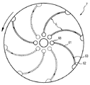
遠心分析器としての、本発明による少なくとも2つの測定セルを有する装置の実施形態の1つは、測定セルが弧状にかつ放射状に配置されるディスクである。下記文章中において、このような装置は測定セルロータとも呼ばれる。 One embodiment of the device having at least two measuring cells according to the invention as a centrifuge analyzer is a disk in which the measuring cells are arranged in an arc and radially. In the text below, such a device is also called a measuring cell rotor.
測定セルロータの1つの特殊な実施形態では、測定セルはその長手軸線に沿って直線形状ではなく、湾曲した形状を有している。この利点は、測定セルロータの回転中、測定セル内において一定流量状態が支配的になる点にある。測定セルの少なくとも仕切り部材を含む領域は、測定セルの残りの領域より小さい直径を有するのが望ましい。この領域は、約50〜500μmの直径を有するのが望ましい。 In one particular embodiment of the measuring cell rotor, the measuring cell has a curved shape rather than a linear shape along its longitudinal axis. This advantage lies in the fact that a constant flow state becomes dominant in the measurement cell during rotation of the measurement cell rotor. The area of the measurement cell including at least the partition member preferably has a smaller diameter than the remaining area of the measurement cell. This region preferably has a diameter of about 50-500 μm.
本発明による測定セルロータは、例えば、光透過性プラスチックから構成し、かつ、上部および部から組み立てることもでき、あるいは、そのいずれかとすることも可能である。測定セルロータを組み立てた状態で互いに向かい合う上部と下部の面は陥凹部及び/または隆起部を有し、それによって所望の測定セル形状を形成することが可能である。上部には、各測定セル毎にピペット孔が含まれており、それを通して測定セルの第1のチャンバに試料材料を注入することが可能である。上部と下部を組み立てる前に、多孔性仕切り部材は、個別ユニットとして全ての測定セルに別個に挿入することもでき、あるいは、上部と下部の間に同心配置され、その結果ロータの全ての測定セルを通る適正な多孔性材料製の連続ストリップの形態で挿入することも可能である。血小板活性に影響する1つ又は複数の物質による多孔性材料のコーティングは、個別仕切り部材または連続ストリップの挿入前に行うこともでき、あるいは、その場で、すなわち、仕切り部材または連続ストリップが既に上部または下部に結合された状態にある時に実施することも可能である。 The measuring cell rotor according to the invention can be made of, for example, a light-transmitting plastic and can be assembled from the top and the part, or either. The upper and lower surfaces facing each other in the assembled state of the measurement cell rotor have recesses and / or ridges, thereby forming a desired measurement cell shape. In the upper part, a pipette hole is included for each measurement cell, through which the sample material can be injected into the first chamber of the measurement cell. Before assembling the upper and lower parts, the porous partition can be inserted separately in all measuring cells as a separate unit, or concentrically placed between the upper and lower parts, so that all measuring cells of the rotor It is also possible to insert it in the form of a continuous strip made of a suitable porous material through. The coating of the porous material with one or more substances affecting the platelet activity can also take place prior to the insertion of the individual partition member or continuous strip, or in situ, ie the partition member or continuous strip already on top. Alternatively, it can be carried out when it is connected to the lower part.
少なくとも部分的に光透過性材料から構成される測定セルロータによって、第2のチャンバ内に捕獲された液体の量を測定するか、あるいは、測光法によって仕切り部材を通る試料液の流速を測定することが可能になる。このため、測定セルロータの上方に1つ又は複数の光源、好ましくは発光ダイオード(LED)を配置し、測定セルロータの下方にそれぞれ関連する光検出器を配置して(またはこの逆にして)、光がビームとなって回転面に対して垂直に測定セルの測定領域を通ることができるようにするのが望ましい。 Measuring the amount of liquid trapped in the second chamber by means of a measuring cell rotor composed at least partly of a light transmissive material, or measuring the flow rate of the sample liquid through the partition member by photometry Is possible. For this purpose, one or more light sources, preferably light emitting diodes (LEDs), are arranged above the measurement cell rotor, and the associated photodetectors are arranged below the measurement cell rotor (or vice versa) to provide light. It is desirable to be able to pass through the measurement area of the measurement cell perpendicular to the plane of rotation.
本発明のもう1つの課題は、本発明による1つの測定セルまたは本発明による少なくとも2つの測定セルを含む装置が利用される、血小板機能を決定する方法である。この方法には、少なくとも
i.本発明による測定セルの第1のチャンバに血小板含有液体試料を充填するステップと、
ii.液体試料を充填した測定セルに遠心力を加えるステップと、
iii.第2のチャンバ内に捕獲された液体量を測定するか、または、仕切り部材を通る試料液の流速を測定するステップが
含まれている。
Another subject of the invention is a method for determining platelet function in which a measuring cell according to the invention or a device comprising at least two measuring cells according to the invention is utilized. The method includes at least i. Filling the first chamber of the measuring cell according to the invention with a platelet-containing liquid sample;
ii. Applying a centrifugal force to a measurement cell filled with a liquid sample;
iii. Measuring the amount of liquid trapped in the second chamber or measuring the flow rate of the sample liquid through the partition member is included.
第1のチャンバに作用する遠心力の結果として第1のチャンバから仕切り部材を通り、第2のチャンバ内に捕獲されている液体の量(または液体体積)は、血小板機能と逆相関を有している。 The amount of liquid (or liquid volume) that passes through the partition member as a result of the centrifugal force acting on the first chamber and is trapped in the second chamber has an inverse correlation with platelet function. ing.
仕切り部材を通り第2のチャンバ内に捕獲されている試料液の流速も、血小板機能と逆相関を有している。 The flow rate of the sample liquid captured in the second chamber through the partition member also has an inverse correlation with the platelet function.
血小板機能が弱まる結果として血小板の凝集が減少し、仕切り部材の孔の閉塞が減ると、所定の時間間隔内に仕切り部材を通過できる試料液の量が増加し、従って、流速または流量が増し、それに伴って第2のチャンバ内に捕獲される液体量も増加する。 When platelet aggregation is reduced as a result of weak platelet function and blockage of the partition member is reduced, the amount of sample liquid that can pass through the partition member within a predetermined time interval increases, and thus the flow rate or flow rate increases. Accordingly, the amount of liquid trapped in the second chamber also increases.
血小板機能が強まる結果として血小板の凝集が増加し、仕切り部材の孔の閉塞が増えると、所定の時間間隔内に仕切り部材を通過できる試料液の量が減少し、従って、流速または流量が減り、それに伴って第2のチャンバ内に捕獲される液体量も減少する。 As platelet function increases as a result of increased platelet function and the blockage of the partition member increases, the amount of sample liquid that can pass through the partition member within a given time interval decreases, thus reducing the flow rate or flow rate, As a result, the amount of liquid trapped in the second chamber also decreases.
本発明によれば、測定セルに加えられる遠心力は、50〜2000×gの範囲内であると望ましい。遠心力は、従来の遠心力ユニットによって加えることが可能である。 According to the present invention, the centrifugal force applied to the measurement cell is desirably in the range of 50 to 2000 × g. Centrifugal force can be applied by a conventional centrifugal force unit.
仕切り部材を通過し第2のチャンバ内に捕獲されている液体の量は、さまざまな方法で測定可能である。 The amount of liquid that passes through the partition member and is trapped in the second chamber can be measured in various ways.
第1の実施形態では、第2のチャンバ内に捕獲される液体の量は測光法的に測定される。このため、本発明による測定セルにおける第2のチャンバの少なくとも1つの部分区画は、光透過性材料から構成される。さらに、複数の光源、好ましくは発光ダイオード(LED)と、それぞれの光源に対応した複数の光検出器が、第2のチャンバの長手軸線に沿って、また、本発明による測定セルの回転面または本発明による測定セルロータの回転面に対して垂直に配置されている。測定中、光源が光を放出し、その強度Iが、それぞれ光源に割り当てられた検出器によって測定される。試料液が光源と検出器との間に位置する場合、光強度は低下する。吸光度E=−log(It/I0)(It=時間tにおける光強度、I0=時間0における光強度)は、光源と検出器との間の吸光材料の量に比例する。第2のチャンバの長手軸線に対する光源の相対的位置決めによって、仕切り部材を通って流れる試料体積に関する表示が可能になる。光源−検出器対の数によって、種々の体積の識別能が規定される。最も単純な場合(n=3)、正常か異常かに関して計数的表示ができるだけであり、n=6の場合、5つの体積を識別することが可能になる。所定のポイントにおける吸光度が規定のしきい値を超えるか(ビーム光路内の試料液)、あるいはこのしきい値未満か(ビーム光路内の空気)が測定される。容器の底部から検出器までの距離と容器内の液体量との間には単純な関係がある。検出器が、吸光度の大きい手段を有する半径rの円筒形容器の底部から距離hの位置にある場合、容器内の液体の最少量はr2πhで表わされる。経時での吸光度を測定すると、時間依存的に液体量を求めることが可能になる{V=V(t)}。時間に関して液体の総量を数学的に微分する(dV(t)/dt)結果として、時間依存性流速が求められる。 In the first embodiment, the amount of liquid trapped in the second chamber is measured photometrically. For this purpose, at least one partial section of the second chamber in the measuring cell according to the invention is composed of a light transmissive material. Furthermore, a plurality of light sources, preferably light emitting diodes (LEDs), and a plurality of photodetectors corresponding to the respective light sources, can be provided along the longitudinal axis of the second chamber and in the plane of rotation of the measuring cell according to the invention or It is arranged perpendicular to the plane of rotation of the measuring cell rotor according to the invention. During the measurement, the light source emits light and its intensity I is measured by a detector assigned to each light source. When the sample liquid is located between the light source and the detector, the light intensity decreases. Absorbance E = −log (I t / I 0 ) (I t = light intensity at time t, I 0 = light intensity at time 0) is proportional to the amount of light absorbing material between the light source and the detector. Relative positioning of the light source with respect to the longitudinal axis of the second chamber allows an indication of the sample volume flowing through the partition member. Depending on the number of light source-detector pairs, different volume discrimination capabilities are defined. In the simplest case (n = 3), there can only be a numerical indication as to whether it is normal or abnormal, and when n = 6, it is possible to identify five volumes. It is measured whether the absorbance at a given point exceeds a specified threshold (sample liquid in the beam optical path) or below this threshold (air in the beam optical path). There is a simple relationship between the distance from the bottom of the container to the detector and the amount of liquid in the container. When the detector is at a distance h from the bottom of a cylindrical container of radius r with a means of high absorbance, the minimum amount of liquid in the container is represented by r 2 πh. When the absorbance over time is measured, the amount of liquid can be determined in a time-dependent manner {V = V (t)}. As a result of mathematically differentiating the total amount of liquid with respect to time (dV (t) / dt), a time-dependent flow rate is determined.
第2の実施形態では、第2のチャンバ内に捕獲される液体量は、電気的に求められる。このため、充填レベルを検出するためにさまざまな長さを有する複数の電極対が、第2の試験チャンバに配置される。第2のチャンバ内の液体体積が増すにつれて、電極が試料液とますます接触する。容器の底部から電極までの距離と容器内の液体量との間には単純な関係がある。電極の数によって、種々の体積の識別能が規定される。最も単純な場合(n=3)、正常か異常かに関して計数的表示ができるだけであり、n=6の場合、5つの体積を識別することが可能になる。最も単純な場合、測定は導電率によって行われる(オーム測定)。電極間に空気が存在し、電極間に動作電位が存在する場合、空気は電気絶縁体なので、電流は流れない。しかしながら、チャンバが試料液で充填されると、溶解塩によって血液または血漿が導電性になるので、その結果、電極間のそれぞれの回路を閉じることが可能になる。 In the second embodiment, the amount of liquid trapped in the second chamber is determined electrically. For this purpose, a plurality of electrode pairs with different lengths are arranged in the second test chamber in order to detect the filling level. As the liquid volume in the second chamber increases, the electrode becomes increasingly in contact with the sample liquid. There is a simple relationship between the distance from the bottom of the container to the electrode and the amount of liquid in the container. Depending on the number of electrodes, the discrimination capacity of various volumes is defined. In the simplest case (n = 3), there can only be a numerical indication as to whether it is normal or abnormal, and when n = 6, it is possible to identify five volumes. In the simplest case, the measurement is made by conductivity (ohmic measurement). When air is present between the electrodes and an operating potential is present between the electrodes, no current flows because air is an electrical insulator. However, when the chamber is filled with the sample solution, the dissolved salt makes the blood or plasma conductive, so that each circuit between the electrodes can be closed.
第3の実施形態では、流速がレーザドップラ風速測定法(LDA)によって測定される。このため、本発明による測定セルの第2のチャンバの少なくとも1つの部分区画は、光透過性材料から構成される。レーザビームを2つのビームに分割し、それらのアライメントをとって、第2のチャンバのある領域で交差するようにする。ビームが交差する測定ポイントに干渉パターンが生じる。検出器が、流動する試料液によって生じる2つの散乱波を測定する。測定信号は2つの散乱波の重ね合わせであり、その結果、ドップラ効果によってうなりが生じ、そのうなり周波数(ドップラ周波数)は流動する試料液の速度に比例する。 In the third embodiment, the flow velocity is measured by laser Doppler wind velocity measurement (LDA). For this purpose, at least one partial section of the second chamber of the measuring cell according to the invention is composed of a light transmissive material. The laser beam is split into two beams and aligned so that they intersect at an area of the second chamber. Interference patterns occur at the measurement points where the beams intersect. The detector measures two scattered waves generated by the flowing sample liquid. The measurement signal is a superposition of two scattered waves. As a result, a beat occurs due to the Doppler effect, and the beat frequency (Doppler frequency) is proportional to the velocity of the flowing sample liquid.
第2のチャンバ内に捕獲された液体の量は、所定の時間間隔にわたって連続して測定することが可能である。このため、仕切り部材を通る時間依存性流量が測定される。 The amount of liquid trapped in the second chamber can be measured continuously over a predetermined time interval. For this reason, the time-dependent flow rate through the partition member is measured.
代替案として、第2のチャンバ内に捕獲された液体の量は、定められた時間に1回測定することも可能である。一例として、測定は終点を測定することによって実施可能である。このため、ある特定時間に仕切り部材を通過した液体の総量が求められる。 As an alternative, the amount of liquid trapped in the second chamber can be measured once at a defined time. As an example, the measurement can be performed by measuring the end point. For this reason, the total amount of liquid that has passed through the partition member at a specific time is obtained.
未知の試料における血小板機能は、その試料からの測定結果と既知の血小板活性に関する1つ又は複数の対照の測定結果とを比較することによって決定するのが望ましい。対照/較正物質は、健康な人間から供与された血液/血漿の集合体が望ましい。中央値は、この集合体からの試料について測定された測定結果から求めるのが望ましく、未知の試料からの測定結果がそれに関係付けられる。 Platelet function in an unknown sample is desirably determined by comparing the measurement from that sample with the measurement of one or more controls for known platelet activity. The control / calibrator is preferably a blood / plasma collection provided by a healthy person. The median is preferably determined from the measurement results measured for samples from this assembly, and the measurement results from unknown samples are related thereto.
仕切り部材を通過する試料液の量は、血小板機能に反比例するので、第2のチャンバ内に捕獲される液体量Vの逆数(1/V)が、血小板機能の尺度として特に適している。未知の試料の逆数1/Vがあらかじめ設定された中央しきい値未満の場合、それは血小板機能の弱まった試料であり、出血の恐れがある。未知の試料の逆数1/Vがあらかじめ設定された中央しきい値を超える場合、それは血小板機能の強まった試料であり、血栓症の恐れがある。 Since the amount of the sample liquid that passes through the partition member is inversely proportional to the platelet function, the reciprocal (1 / V) of the liquid amount V captured in the second chamber is particularly suitable as a measure of the platelet function. If the reciprocal 1 / V of the unknown sample is less than a preset median threshold, it is a sample with weakened platelet function and may bleed. If the reciprocal 1 / V of an unknown sample exceeds a preset median threshold, it is a sample with increased platelet function and there is a risk of thrombosis.
本発明は、後述する例示の図を用いてさらに詳細に説明される。ここでは、それらの図が説明的な性格のものであって、本発明の限定を意図したものでは全くない点に留意すべきである。 The present invention will be described in more detail with reference to the following exemplary drawings. It should be noted here that the figures are illustrative in nature and are not intended to limit the invention in any way.
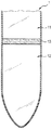
図1
図1には、本発明による測定セル(1)の1つの実施形態が示されている。測定セル(1)には、血小板含有液体試料を収容する第1のチャンバ(11)と、測定セルに遠心力が作用すると第1のチャンバから液体試料を捕獲する第2のチャンバ(12)とが含まれている。測定セルには、さらに、中空体の直径全体にわたって第1のチャンバと第2のチャンバとを互いに分離する多孔性仕切り部材(13)も含まれている。仕切り部材(13)には、血小板活性に影響する少なくとも1つの可溶性物質が含まれている。
FIG.
FIG. 1 shows one embodiment of a measuring cell (1) according to the invention. The measurement cell (1) includes a first chamber (11) that contains a platelet-containing liquid sample, and a second chamber (12) that captures the liquid sample from the first chamber when centrifugal force acts on the measurement cell. It is included. The measurement cell further includes a porous partition member (13) that separates the first chamber and the second chamber from each other over the entire diameter of the hollow body. The partition member (13) contains at least one soluble substance that affects platelet activity.
図2
図2には、本発明による測定セル(2)のもう1つの実施形態が示されている。測定セル(2)には、血小板含有液体試料を収容する第1のチャンバ(21)と、測定セルに遠心力が作用すると第1のチャンバから液体試料を捕獲する第2のチャンバ(22)とが含まれている。測定セルには、さらに、中空体の直径全体にわたって第1のチャンバと第2のチャンバとを互いに分離する多孔性仕切り部材(23)も含まれている。仕切り部材(23)には、血小板活性に影響する少なくとも1つの可溶性物質が含まれている。
FIG.
FIG. 2 shows another embodiment of the measuring cell (2) according to the invention. The measurement cell (2) includes a first chamber (21) that contains a platelet-containing liquid sample, and a second chamber (22) that captures the liquid sample from the first chamber when centrifugal force acts on the measurement cell. It is included. The measurement cell further includes a porous partition member (23) that separates the first chamber and the second chamber from each other over the entire diameter of the hollow body. The partition member (23) contains at least one soluble substance that affects platelet activity.
さらに、ここに示す測定セル(2)は、第2のチャンバ(22)内に、第2のチャンバ内のスペースを第1の部分(221)と第2の部分(222)とに細分する傾斜板状部材の形態をなす手段(24)を含んでいる。 Further, the measurement cell (2) shown here has an inclination in the second chamber (22) that subdivides the space in the second chamber into a first part (221) and a second part (222). Means (24) in the form of a plate-like member are included.
第1の部分(221)は、仕切り部材を通過する試料液を捕獲して、輸送する働きをする。第2の部分(222)は、第2のチャンバ(22)内に捕獲された試料液(29)の液量を測定する働きをする。第1の部分と第2の部分は、測定セルの先端近くにおいてのみ、捕獲されて輸送される試料液を第1の部分から第2の部分へ送り込むことができるように、第2のチャンバの片側の遠位(すなわち、鎖線の下方)で相互接続される。この利点は、遠心分離中に仕切り部材(23)を通過する試料液が、第2のチャンバ全体(24)を無制御に濡らすことがなく、試料液が、的確に捕獲され、集められて、第2のチャンバの遠位領域に輸送される点にある。輸送された試料液(29)は、その結果、第2のチャンバ(22)内に、特に、測定セル先端の底部(遠位)から上部(近位)まで溜まることになる。 The first part (221) functions to capture and transport the sample liquid passing through the partition member. The second part (222) serves to measure the amount of the sample liquid (29) captured in the second chamber (22). The first part and the second part are arranged in the second chamber so that the sample liquid to be captured and transported can be pumped from the first part to the second part only near the tip of the measurement cell. Interconnected distally on one side (ie below the dashed line). This advantage is that the sample liquid passing through the partition member (23) during centrifugation does not wet the entire second chamber (24) uncontrollably, and the sample liquid is accurately captured and collected, It is in the point of being transported to the distal region of the second chamber. The transported sample liquid (29) consequently accumulates in the second chamber (22), in particular from the bottom (distal) to the top (proximal) of the measurement cell tip.
さらに、ここに示す測定セル(2)には、充填レベルを検出するために長さの異なる複数の電極(25)が含まれている。第2のチャンバ(22)内における、特に、第2のチャンバ(22)の第2の部分(222)内における液体レベルが上昇するにつれて、試料液に接触する電極(25)の個数がますます多くなる。 Further, the measurement cell (2) shown here includes a plurality of electrodes (25) of different lengths in order to detect the filling level. As the liquid level in the second chamber (22) increases, particularly in the second part (222) of the second chamber (22), the number of electrodes (25) in contact with the sample solution increases. Become more.
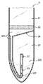
図3
図3には、本発明による測定セル(3)のもう1つの実施形態が示されている。測定セル(3)には、血小板含有液体試料を収容する第1のチャンバ(31)と、測定セルに遠心力が作用すると第1のチャンバから液体試料を捕獲する第2のチャンバ(32)とが含まれている。測定セルには、さらに、中空体の直径全体にわたって第1のチャンバと第2のチャンバとを互いに分離する多孔性仕切り部材(33)も含まれている。仕切り部材(33)には、血小板活性に影響する少なくとも1つの可溶性物質が含まれている。
FIG.
FIG. 3 shows another embodiment of the measuring cell (3) according to the invention. The measurement cell (3) includes a first chamber (31) that contains a platelet-containing liquid sample, and a second chamber (32) that captures the liquid sample from the first chamber when centrifugal force acts on the measurement cell. It is included. The measurement cell further includes a porous partition member (33) that separates the first chamber and the second chamber from each other over the entire diameter of the hollow body. The partition member (33) contains at least one soluble substance that affects platelet activity.
さらに、ここに示す測定セル(3)は、第2のチャンバ(32)内に、第2のチャンバ内のスペースを第1の部分(321)と第2の部分(322)とに細分する手段(34)を含んでいる。 Furthermore, the measurement cell (3) shown here is a means for subdividing the space in the second chamber into a first part (321) and a second part (322) in the second chamber (32). (34) is included.
第2のチャンバ(32)内のスペースを第1の部分と第2の部分とに細分する手段(34)は、多孔性仕切り部材に対してある角度をなす面を有する第1の部分区画(341)を具備している。この手段(34)は、さらに、第1の部分区画(341)に続いて、第2のチャンバ(32)内のスペースを区切る壁(36)に対してほぼ平行に延び、前記壁と共に管状構造を形成する第2の部分区画(342)を有しており、この管状構造は第2のチャンバ(32)の片側の遠位内に延びている。この実施形態では、第2のチャンバの第1の部分(321)つまり捕獲領域のスペースが第2のチャンバ(32)内において最小に縮小され、第2のチャンバ(32)内のできるだけ大きいスペースを第2の部分(322)つまり測定領域のために利用可能になるので有利である。例えば、これは、可能性のある最大量の利用可能領域が、測定セルに配置される測定装置のために利用可能になるという点で有利である。 The means (34) for subdividing the space in the second chamber (32) into a first part and a second part is a first partial section having a surface that forms an angle with respect to the porous partition member ( 341). This means (34) further extends substantially parallel to the wall (36) separating the space in the second chamber (32) following the first partial section (341), and is tubular with the wall. The tubular structure extends into the distal portion of one side of the second chamber (32). In this embodiment, the space of the first chamber (321) or capture area of the second chamber is reduced to a minimum in the second chamber (32), and the largest possible space in the second chamber (32) is obtained. Advantageously, it becomes available for the second part (322) or measurement area. For example, this is advantageous in that the maximum possible amount of available area becomes available for the measuring device placed in the measuring cell.
さらに、ここに示す測定セル(3)には、充填レベルを検出するために長さの異なる複数の電極(35)が含まれている。第2のチャンバ(32)内における、特に、第2のチャンバ(32)の第2の部分(322)における液体レベルが上昇するにつれて、試料液(39)に接触する電極(35)の個数がますます多くなる。 Furthermore, the measurement cell (3) shown here includes a plurality of electrodes (35) of different lengths in order to detect the filling level. As the liquid level in the second chamber (32), in particular in the second part (322) of the second chamber (32) increases, the number of electrodes (35) in contact with the sample liquid (39) increases. Increasingly.
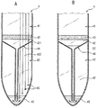
図4
図4には、本発明による測定セル(4)のもう1つの実施形態が示されている。測定セル(4)には、血小板含有液体試料を収容する第1のチャンバ(41)と、測定セルに遠心力が作用すると第1のチャンバから液体試料を捕獲する第2のチャンバ(42)とが含まれている。測定セルには、さらに、中空体の直径全体にわたって第1のチャンバと第2のチャンバとを互いに分離する多孔性仕切り部材(43)も含まれている。仕切り部材(43)には、血小板活性に影響する少なくとも1つの可溶性物質が含まれている。
FIG.
FIG. 4 shows another embodiment of the measuring cell (4) according to the invention. The measurement cell (4) includes a first chamber (41) that contains a platelet-containing liquid sample, and a second chamber (42) that captures the liquid sample from the first chamber when centrifugal force acts on the measurement cell. It is included. The measurement cell further includes a porous partition member (43) that separates the first chamber and the second chamber from each other over the entire diameter of the hollow body. The partition member (43) contains at least one soluble substance that affects platelet activity.
さらに、ここに示す測定セル(4)は、第2のチャンバ(42)内に、第2のチャンバ内のスペースを第1の部分(421)と第2の部分(422)とに細分する手段(44)を含んでいる。 Furthermore, the measurement cell (4) shown here is a means for subdividing the space in the second chamber into a first part (421) and a second part (422) in the second chamber (42). (44) is included.
第2のチャンバ(42)内のスペースを第1の部分(421)と第2の部分(422)とに細分する手段(44)は、この場合、漏斗形状を有し、漏斗形状手段の管状領域が第2のチャンバ(42)の片側の遠位内に延びている。 The means (44) for subdividing the space in the second chamber (42) into a first part (421) and a second part (422) in this case has a funnel shape and is tubular in the funnel shape means. A region extends into the distal end of one side of the second chamber (42).
図4のAに示す測定セル(4)には、充填レベルを検出するために長さの異なる複数の電極(45)が含まれている。 The measurement cell (4) shown in FIG. 4A includes a plurality of electrodes (45) having different lengths in order to detect the filling level.
図4のBに示す測定セル(4)において、漏斗形状手段(44)の少なくとも管状領域は、測定セルの壁であり、光透過性材料から構成されている。測定セルのこの実施形態は、測定セルの第2のチャンバ内における試料液(49)の流速を測定するのに特に適している。従って、流速は、漏斗形状手段(44)の管状領域において、測定セルの長手軸線に沿って配置された複数の光源と複数の光検出器(不図示)を用いて測光法的に測定される。代替案として、漏斗形状手段(44)の管状領域内の流速は、レーザドップラ風速測定法(LDA)によって測定することも可能である。 In the measurement cell (4) shown in FIG. 4B, at least the tubular region of the funnel-shaped means (44) is the wall of the measurement cell and is made of a light transmissive material. This embodiment of the measuring cell is particularly suitable for measuring the flow rate of the sample liquid (49) in the second chamber of the measuring cell. Accordingly, the flow velocity is measured photometrically using a plurality of light sources and a plurality of photodetectors (not shown) arranged along the longitudinal axis of the measurement cell in the tubular region of the funnel-shaped means (44). . As an alternative, the flow velocity in the tubular region of the funnel shaped means (44) can also be measured by laser Doppler anemometry (LDA).
図5
図5には、本発明による測定セル(5)のもう1つの実施形態が示されている。測定セル(5)には、血小板含有液体試料を収容する第1のチャンバ(51)と、測定セルに遠心力が作用すると第1のチャンバから液体試料を捕獲する第2のチャンバ(52)とが含まれている。測定セルには、さらに、中空体の直径全体にわたって第1のチャンバと第2のチャンバとを互いに分離する多孔性仕切り部材(53)も含まれている。仕切り部材(53)には、血小板活性に影響する少なくとも1つの可溶性物質が含まれている。
FIG.
FIG. 5 shows another embodiment of the measuring cell (5) according to the invention. The measurement cell (5) includes a first chamber (51) that contains a platelet-containing liquid sample, and a second chamber (52) that captures the liquid sample from the first chamber when centrifugal force acts on the measurement cell. It is included. The measurement cell further includes a porous partition member (53) that separates the first chamber and the second chamber from each other over the entire diameter of the hollow body. The partition member (53) contains at least one soluble substance that affects platelet activity.
さらに、ここに示す測定セル(5)は、第2のチャンバ(52)内に、第2のチャンバ内のスペースを第1の部分(521)と第2の部分(522)と第3の部分(523)とに細分する手段(54)を有している。第1の部分(521)つまり捕獲領域は、仕切り部材を通過する試料液を捕獲し、第2のチャンバ(52)の長手軸線に沿って片側の近位から片側の遠位に輸送する働きをする。第1の部分(521)は、第2のチャンバ(52)の片側の遠位において第2の部分(522)に接続されており、捕獲されて輸送される試料液を第1の部分(521)から第2の部分(522)に送り込むことができる。第2の部分(522)つまり測定領域は、第2のチャンバ(52)内に捕獲された試料液の流速を測定する働きをする。ここで、第2の部分(522)は、第2のチャンバ(52)の片側の遠位から片側の近位に向かって立ち上がり、その端部に第3の部分(523)に接続する開口(57)を有する毛細管を構成している。第3の部分(523)つまりオーバフロー領域は、第2の部分(522)からの試料液(59)のためのオーバフロー室の働きをする。測定セルのこの実施形態は、試料液の流速の測定に基づいて血小板機能を決定するのに特に適している。 Furthermore, the measurement cell (5) shown here has a second part (521), a second part (522), and a third part in the second chamber (52). Means (54) for subdividing into (523). The first portion (521), ie, the capture region, serves to capture the sample liquid passing through the partition member and transport it from the proximal side on one side to the distal side on the other side along the longitudinal axis of the second chamber (52). To do. The first part (521) is connected to the second part (522) at a distal end of one side of the second chamber (52), and the sample liquid to be captured and transported is transferred to the first part (521). ) To the second part (522). The second part (522), i.e., the measurement region, serves to measure the flow rate of the sample liquid captured in the second chamber (52). Here, the second portion (522) rises from the distal end of one side of the second chamber (52) toward the proximal portion of the one side, and an opening (at the end thereof) is connected to the third portion (523). 57). The third part (523) or overflow region serves as an overflow chamber for the sample liquid (59) from the second part (522). This embodiment of the measurement cell is particularly suitable for determining platelet function based on measurement of the sample fluid flow rate.
図6
図6には、複数の測定セル(6)が弧状に配置された本発明による測定セルロータ(7)の平面図が示されているが、各測定セル(6)には、血小板含有液体試料を収容する第1のチャンバ(61)と、測定セルに遠心力が作用すると第1のチャンバから液体試料を捕獲する第2のチャンバ(62)とが含まれている。測定セルには、さらに、中空体の直径全体にわたって第1のチャンバと第2のチャンバとを互いに分離する多孔性仕切り部材(63)も含まれている。仕切り部材(63)には、血小板活性に影響する少なくとも1つの可溶性物質が含まれている。
FIG.
FIG. 6 shows a plan view of a measurement cell rotor (7) according to the present invention in which a plurality of measurement cells (6) are arranged in an arc, and each measurement cell (6) contains a platelet-containing liquid sample. A first chamber (61) for housing is included, and a second chamber (62) for capturing a liquid sample from the first chamber when centrifugal force acts on the measurement cell. The measurement cell further includes a porous partition member (63) that separates the first chamber and the second chamber from each other over the entire diameter of the hollow body. The partition member (63) contains at least one soluble substance that affects platelet activity.
測定セル(6)は、その長手軸線に沿って湾曲した形状を有している。測定セル(6)において仕切り部材(63)を含む領域は、測定セルの残りの領域よりも直径が小さい。測定セルロータ(7)の上側には、ピペット孔(68)が設けられており、それを通して測定セル(6)の第1のチャンバ(61)に試料材料を注入することが可能である。矢印は測定セルロータ(7)の回転方向を示している。 The measurement cell (6) has a shape curved along its longitudinal axis. The area including the partition member (63) in the measurement cell (6) has a smaller diameter than the remaining area of the measurement cell. A pipette hole (68) is provided on the upper side of the measurement cell rotor (7), through which the sample material can be injected into the first chamber (61) of the measurement cell (6). The arrow indicates the direction of rotation of the measurement cell rotor (7).
下記の典型的な実施形態は、本発明による方法を例証する働きをするものであって、限定するものとみなすべきではない。 The following exemplary embodiments serve to illustrate the method according to the invention and should not be regarded as limiting.
例1:本発明による全血試料における血小板活性の決定
本発明による測定セルが下記のように作製された:円錐形状の遠心分離管(透明プラスチックで造られた50mlのFalcon管、Becton Dickson製)がほぼ中ほどで切断された。PVDF膜を有する使い捨てのフィルタアタッチメント(Millipore Millex(登録商標)−SV、孔寸法5μm)が切断したFalcon管の開口に取り付けられた。本発明に従い、コラーゲンとエピネフリンを含む(それぞれ、0.5mg/ml)血小板活性化物質混合物を用いてPDVF膜の前処理が行われた。このため、Millipore Millex(登録商標)−SV使い捨てフィルタアタッチメントに0.8mlのこの混合物を塗布し、その後空気乾燥させた。対照として、Millipore Millex(登録商標)−SV使い捨てフィルタアタッチメントを0.8mlの水で濡らし、その後空気乾燥させた。次に、ピストン及び注射針のないプラスチック製の使い捨て注射器(Omnifix(登録商標)、5ml、B.Braun Melsungen AG)が、切断したFalcon管に取り付けられたMillipore Millex(登録商標)−SV使い捨てフィルタアタッチメントに取り付けられた。
Example 1: Determination of platelet activity in a whole blood sample according to the invention A measuring cell according to the invention was made as follows: a conical centrifuge tube (50 ml Falcon tube made of clear plastic, manufactured by Becton Dickson) Was cut in the middle. A disposable filter attachment with a PVDF membrane (Millipore Millex®-SV, pore size 5 μm) was attached to the cut Falcon tube opening. In accordance with the present invention, a PDVF membrane was pretreated with a platelet activator mixture containing collagen and epinephrine (each 0.5 mg / ml). For this purpose, 0.8 ml of this mixture was applied to a Millipore Millex®-SV disposable filter attachment and then air dried. As a control, a Millipore Millex®-SV disposable filter attachment was wetted with 0.8 ml water and then air dried. Next, a Millipore Millex®-SV disposable filter attachment in which a plastic disposable syringe (Omnifix®, 5 ml, B. Braun Melsungen AG) without a piston and needle is attached to the cut Falcon tube. Attached to.
次に、1.5mlの正常なクェン酸塩添加血が測定セルの第1のチャンバすなわち使い捨て注射器に注入され、遠心分離機(Rotixa R50、Andreas Hettich GmbH & Co.KG)によって、22℃で50×gの遠心作用が75秒間にわたって測定セルに加えられた。第2のチャンバすなわち切断したFalcon管で捕獲された液体の量は、容積測定法で求められた。 Next, 1.5 ml of normal citrate blood is injected into the first chamber or disposable syringe of the measuring cell and 50 ° C. at 22 ° C. by means of a centrifuge (Rotixa R50, Andreas Hetich GmbH & Co. KG). A xg centrifuge action was applied to the measuring cell for 75 seconds. The amount of liquid trapped in the second chamber or cut Falcon tube was determined volumetrically.
表1から明らかなように、血小板活性化物質、コラーゲン、及び、エピネフリンを含浸させた仕切り部材を用いる結果として、血液試料に誘発される血小板凝集のために試料液の流量が低減することになる。 As is apparent from Table 1, as a result of using a partition member impregnated with platelet-activating substance, collagen, and epinephrine, the flow rate of the sample solution is reduced due to platelet aggregation induced in the blood sample. .

1、2、3、4、5、6 測定セル
7 測定セルロータ
11、21、31、41、51、61 試料を収容する第1のチャンバ
12、22、32、42、52、62 第1のチャンバからの試料を捕獲する第2のチャンバ
13、23、33、43、53、63 仕切り部材
24、34、44、54 第2のチャンバ内のスペースを細分する手段
221、321、421、521 第2のチャンバの第1の部分(捕獲領域)
222、322、422、522 第2のチャンバの第2の部分(測定領域)
25、35、45 電極
29、39、49、59 試料液
341 第2のチャンバ内のスペースを細分する手段の第1の区画
342 第2のチャンバ内のスペースを細分する手段の第2の区画
36 壁
523 第2のチャンバの第3の部分(オーバフロー領域)
57 開口
68 ピペット孔
1, 2, 3, 4, 5, 6
222, 322, 422, 522 Second portion of second chamber (measurement region)
25, 35, 45
57
Claims (21)
a.血小板含有液体試料を収容する第1のチャンバ(11、21、31、41、51、61)と、
b.前記測定セルに遠心力が作用すると前記第1のチャンバから前記液体試料を捕獲する第2のチャンバ(12、22、32、42、52、62)と、
c.前記第1のチャンバと前記第2のチャンバとを互いに分離する多孔性仕切り部材(13、23、33、43、53、63)とが含まれ、
前記多孔性仕切り部材(13、23、33、43、53、63)は、前記血小板の活性度に影響する少なくとも1つの可溶性物質を含むことを特徴とする測定セル。 A measuring cell (1, 2, 3, 4, 5, 6) for determining platelet function,
a. A first chamber (11, 21, 31, 41, 51, 61) containing a platelet-containing liquid sample;
b. A second chamber (12, 22, 32, 42, 52, 62) for capturing the liquid sample from the first chamber when a centrifugal force acts on the measurement cell;
c. A porous partition member (13, 23, 33, 43, 53, 63) for separating the first chamber and the second chamber from each other;
The measurement cell, wherein the porous partition member (13, 23, 33, 43, 53, 63) contains at least one soluble substance that affects the activity of the platelets.
前記第2のチャンバ(22、32、42、52)が、その内部に、前記第2のチャンバ内のスペースを第1の部分(221、321、421、521)と第2の部分(222、322、422、522)とに細分する手段(24、34、44、54)を有し、
・前記第1の部分が、前記多孔性仕切り部材を通過する試料液を捕獲して、輸送する働きをし、
・前記第2の部分が、前記第2のチャンバ内に捕獲された前記試料液の液量を測定する働きをし、
・前記第1の部分と第2の部分が、前記捕獲されて輸送される試料液を前記第1の部分から前記第2の部分へ送り込むことができるように、前記第2のチャンバの片側の遠位で相互接続されている
請求項1から7のいずれか1つに記載の測定セル(2、3、4、5)。 The second chamber (22, 32, 42, 52) has one side proximal and one side distal along the longitudinal axis of the second chamber, starting from the porous partition member;
The second chamber (22, 32, 42, 52) has a space in the second chamber inside the first part (221, 321, 421, 521) and the second part (222, 322, 422, 522) and subdividing means (24, 34, 44, 54),
- the first part, serves to said sample liquid passing through the porous partition member captured, transported,
· The second portion serves to measure the amount of liquid in the sample solution trapped in the second chamber,
- the first portion and the second portion, the sample liquid to be transported is the captured so that it can be fed from said first portion to said second portion, on one side of the second chamber Measurement cell (2, 3, 4, 5) according to any one of the preceding claims, interconnected distally.
・前記多孔性仕切り部材に対して角度をなす平面を持つ第1の部分区画(341)を有し、
・前記第1の部分区画(341)に続いて、前記第2のチャンバ内のスペースを区切る壁(36)に対してほぼ平行に延び、前記壁(36)と共に管状構造を形成する第2の部分区画(342)を有し、この管状構造が前記第2のチャンバの片側の遠位内に延びる
請求項8に記載の測定セル(3)。 The means (34) for subdividing the space in the second chamber (32) into the first part (321) and the second part (322);
- having a first subblock having a plane forming an angle (341) relative to the porous partition member,
A second section extending substantially parallel to the wall (36) delimiting the space in the second chamber following the first partial section (341) and forming a tubular structure with the wall (36); The measurement cell (3) according to claim 8, comprising a partial section (342), the tubular structure extending into the distal end on one side of the second chamber.
前記第2のチャンバ(52)が、その内部に、前記第2のチャンバ内のスペースを第1の部分(521)と第2の部分(522)と第3の部分(523)とに細分する手段(54)を有し、
・前記第1の部分(521)が前記多孔性仕切り部材(53)を通過する試料液を捕獲して、輸送する働きをし、前記試料液が前記第2のチャンバ(52)の長手軸線に沿って前記片側の近位から前記片側の遠位に送り込まれ、
・前記第2の部分(522)が前記第2のチャンバ(52)内に捕獲された前記試料液(59)の流速を測定する働きをし、
・前記第3の部分(523)が前記第2の部分(522)からの前記試料液のためのオーバフロー室の働きをし、
・前記第1の部分(521)と第2の部分(522)とが、捕獲されて輸送される前記試料液を前記第1の部分(521)から前記第2の部分(522)へ送り込むことができるように、前記第2のチャンバの前記片側の遠位で相互接続され、
・前記第2の部分(522)が、前記第2のチャンバの片側の遠位から片側の近位の方向に立ち上がり、その端部に前記第3の部分(523)に接続する開口(57)を有する毛細管を構成している
請求項1から6のいずれか1つに記載の測定セル(5)。 The second chamber (52) begins at the porous partition member and has one proximal and one distal along the longitudinal axis of the second chamber;
The second chamber (52) subdivides the space in the second chamber into a first part (521), a second part (522), and a third part (523). Means (54),
- the first portion (521) is captured sample liquid passing through the porous partition member (53), serves to transport to the longitudinal axis of the sample solution and the second chamber (52) Along the one side proximal to the one side distal,
· Acts that said second portion (522) to measure the flow rate of said sample solution trapped in the second chamber (52) in (59),
- the third part (523) is the function of overflow chamber for the sample liquid from the second portion (522),
- the first portion and (521) and a second portion (522), but feeding the sample liquid to be transported are captured from the first portion (521) to said second portion (522) Interconnected distally on the one side of the second chamber so that
· The second portion (522) is, the rising from one side of the distal of the second chamber in the direction of the proximal side, openings (57) connected to the third portion at its end (523) The measurement cell (5) according to any one of claims 1 to 6, wherein a capillary tube is configured.
i.請求項1から13のいずれか1つに記載の測定セルの第1のチャンバに血小板含有液体試料を充填するステップと、
ii.前記液体試料を充填した前記測定セルに遠心力を加えるステップと、
iii.前記第2のチャンバ内に捕獲された液体量を測定するか、または、前記測定セルの前記第2のチャンバ内の前記試料液の流速を測定するステップとが含まれ、
前記捕獲された液体の量及び前記流速が前記血小板機能と逆相関を有している
方法。 A method for determining platelet function comprising:
i. Filling the first chamber of the measuring cell according to any one of claims 1 to 13 with a platelet-containing liquid sample;
ii. Applying a centrifugal force to the measurement cell filled with the liquid sample;
iii. Measuring the amount of liquid trapped in the second chamber, or measuring the flow rate of the sample liquid in the second chamber of the measurement cell,
The amount of the captured liquid and the flow rate have an inverse correlation with the platelet function.
The method according to any one of claims 15 to 20, wherein the centrifugal force applied to the measuring cell is in the range of 50 to 2000 xg.
Applications Claiming Priority (2)
| Application Number | Priority Date | Filing Date | Title |
|---|---|---|---|
| EP11158195.5 | 2011-03-15 | ||
| EP11158195A EP2500095A1 (en) | 2011-03-15 | 2011-03-15 | Method and devices for determining the platelet function in a centrifugal analyser |
Publications (3)
| Publication Number | Publication Date |
|---|---|
| JP2012194181A JP2012194181A (en) | 2012-10-11 |
| JP2012194181A5 JP2012194181A5 (en) | 2015-03-12 |
| JP5860736B2 true JP5860736B2 (en) | 2016-02-16 |
Family
ID=44359752
Family Applications (1)
| Application Number | Title | Priority Date | Filing Date |
|---|---|---|---|
| JP2012057627A Active JP5860736B2 (en) | 2011-03-15 | 2012-03-14 | Apparatus and method for determining platelet function |
Country Status (5)
| Country | Link |
|---|---|
| US (1) | US9205424B2 (en) |
| EP (2) | EP2500095A1 (en) |
| JP (1) | JP5860736B2 (en) |
| CN (1) | CN102759614B (en) |
| ES (1) | ES2691695T3 (en) |
Families Citing this family (6)
| Publication number | Priority date | Publication date | Assignee | Title |
|---|---|---|---|---|
| US8617468B2 (en) | 2011-10-18 | 2013-12-31 | Chrono-Log Corporation | Platelet aggregation test and device |
| JP6012455B2 (en) * | 2012-12-21 | 2016-10-25 | シーメンス ヘルスケア ダイアグノスティクス プロダクツ ゲゼルシヤフト ミツト ベシユレンクテル ハフツング | Method for standardizing measurement results in a system for measuring platelet function |
| KR101497193B1 (en) * | 2012-12-31 | 2015-03-03 | 고려대학교 산학협력단 | Apparatus and method for monitoring platelet function and drug response using centrifugation-based microfluidics |
| CN204182492U (en) * | 2013-12-05 | 2015-03-04 | 刘玥萌 | A kind of centrifugal filtration tube |
| CN104359746B (en) * | 2014-11-19 | 2018-03-16 | 白银市第一人民医院 | Cell mass collector and the method for collecting cell in humoral specimen |
| CN110095521B (en) | 2019-05-13 | 2022-08-23 | 京东方科技集团股份有限公司 | Centrifuge tube, detection system and detection method |
Family Cites Families (18)
| Publication number | Priority date | Publication date | Assignee | Title |
|---|---|---|---|---|
| JPS5256993A (en) * | 1975-10-31 | 1977-05-10 | Jiometsuto Inc | Method of and instrument for measuring solidifying rate |
| JPS5889859U (en) * | 1981-12-11 | 1983-06-17 | 東亜医用電子株式会社 | Clotting time measurement device |
| DE3247815C2 (en) * | 1982-12-23 | 1985-10-17 | Gustav Viktor Rudolf Prof. London Born | Device for measuring the bleeding time in vitro |
| GB2175691A (en) | 1985-04-26 | 1986-12-03 | Brien John Richard O | Bleeding time measurement |
| DE3739247C2 (en) * | 1987-11-19 | 1996-11-21 | Dade Int Inc | Bleeding time measuring device |
| US5888826A (en) * | 1994-06-30 | 1999-03-30 | Dade Behring Inc. | Combination reagent holding and test device |
| ATE208493T1 (en) | 1994-06-30 | 2001-11-15 | Dade Behring Inc | BIOACTIVE POROUS SEPARATIONS |
| ATE210829T1 (en) * | 1996-03-18 | 2001-12-15 | Diagnostische Forsch Stiftung | PARTICLE IMMUNOASSAY WITH COMPACT MATRIX |
| ATE281238T1 (en) | 1996-03-22 | 2004-11-15 | Dade Behring Inc | COMBINED REAGENT COLLECTION AND TEST ARRANGEMENT |
| US20120156278A1 (en) * | 1997-06-24 | 2012-06-21 | Cascade Medical Enterprises, Llc | Systems and methods for preparing autologous fibrin glue |
| US7087177B2 (en) * | 2004-04-16 | 2006-08-08 | Baxter International Inc. | Methods for determining flow rates of biological fluids |
| US7708152B2 (en) * | 2005-02-07 | 2010-05-04 | Hanuman Llc | Method and apparatus for preparing platelet rich plasma and concentrates thereof |
| EP1693109A1 (en) * | 2005-02-21 | 2006-08-23 | Hexal Ag | Container for separating tumor cells |
| CN100467075C (en) * | 2005-08-09 | 2009-03-11 | 郭经纬 | A multidirectional centrifuge type arrangement for continuous separating blood components and method |
| US20070102374A1 (en) | 2005-11-04 | 2007-05-10 | Gambro, Inc. | Blood processing apparatus with controlled cell capture chamber and method background of the invention |
| CN100429517C (en) * | 2006-09-29 | 2008-10-29 | 暨南大学 | Setting-up method for cell membrance solid phase chromatography model for sieving Chinese medicine platelet aggregation resisting active component |
| EP2053387A1 (en) * | 2007-10-22 | 2009-04-29 | Centre National de la Recherche Scientifique | Test device for platelet aggregation detection |
| US8187475B2 (en) * | 2009-03-06 | 2012-05-29 | Biomet Biologics, Llc | Method and apparatus for producing autologous thrombin |
-
2011
- 2011-03-15 EP EP11158195A patent/EP2500095A1/en not_active Withdrawn
-
2012
- 2012-03-05 EP EP12158003.9A patent/EP2500096B1/en active Active
- 2012-03-05 ES ES12158003.9T patent/ES2691695T3/en active Active
- 2012-03-14 JP JP2012057627A patent/JP5860736B2/en active Active
- 2012-03-15 CN CN201210068133.3A patent/CN102759614B/en active Active
- 2012-03-15 US US13/421,468 patent/US9205424B2/en active Active
Also Published As
| Publication number | Publication date |
|---|---|
| JP2012194181A (en) | 2012-10-11 |
| EP2500096A1 (en) | 2012-09-19 |
| CN102759614A (en) | 2012-10-31 |
| EP2500095A1 (en) | 2012-09-19 |
| EP2500096B1 (en) | 2018-07-18 |
| US9205424B2 (en) | 2015-12-08 |
| ES2691695T3 (en) | 2018-11-28 |
| CN102759614B (en) | 2017-09-26 |
| US20120237960A1 (en) | 2012-09-20 |
Similar Documents
| Publication | Publication Date | Title |
|---|---|---|
| JP5860736B2 (en) | Apparatus and method for determining platelet function | |
| JP5937612B2 (en) | Blood collection device containing blood stabilizer | |
| JP7250744B2 (en) | Detection and Classification of Anticoagulants Using Coagulation Analysis | |
| CA2340365C (en) | Novel method of platelet function analysis using platelet count | |
| US8895259B2 (en) | Method for determination of platelet function under flow conditions | |
| JP4989292B2 (en) | Method and apparatus for measuring platelet function under flow conditions | |
| WO2015159516A1 (en) | Blood state analysis device, blood state analysis system, blood state analysis method, and storage device | |
| JP4630053B2 (en) | Cartridge for monitoring the function of a device for testing the function of platelets, method for monitoring function and use of test fluid | |
| EP1581107A2 (en) | System and method for in vitro bleeding time testing | |
| JP2012163562A (en) | Determination of state of centrifugally separated blood using measured pressure | |
| JP2007167123A (en) | Vacuum blood collection tube | |
| JPH11511261A (en) | Blood factor assay | |
| Stephen et al. | Analytical comparison between microhematocrit and automated methods for packed cell volume (PCV) determination | |
| JP6239603B2 (en) | Blood coagulation test method and apparatus | |
| US20150338330A1 (en) | Method for Comprehensive Assessment of Platelet Aggregation | |
| RU2601111C1 (en) | Method for assessing hemostatic activity | |
| SU1734020A1 (en) | Method of determining adhesion of thrombocytes | |
| JP6012455B2 (en) | Method for standardizing measurement results in a system for measuring platelet function | |
| EP1600777A2 (en) | A single-tube method and system for platelet function analysis |
Legal Events
| Date | Code | Title | Description |
|---|---|---|---|
| A521 | Request for written amendment filed |
Free format text: JAPANESE INTERMEDIATE CODE: A523 Effective date: 20150122 |
|
| A621 | Written request for application examination |
Free format text: JAPANESE INTERMEDIATE CODE: A621 Effective date: 20150122 |
|
| A977 | Report on retrieval |
Free format text: JAPANESE INTERMEDIATE CODE: A971007 Effective date: 20151126 |
|
| TRDD | Decision of grant or rejection written | ||
| A01 | Written decision to grant a patent or to grant a registration (utility model) |
Free format text: JAPANESE INTERMEDIATE CODE: A01 Effective date: 20151208 |
|
| A61 | First payment of annual fees (during grant procedure) |
Free format text: JAPANESE INTERMEDIATE CODE: A61 Effective date: 20151221 |
|
| R150 | Certificate of patent or registration of utility model |
Ref document number: 5860736 Country of ref document: JP Free format text: JAPANESE INTERMEDIATE CODE: R150 |
|
| R250 | Receipt of annual fees |
Free format text: JAPANESE INTERMEDIATE CODE: R250 |
|
| R250 | Receipt of annual fees |
Free format text: JAPANESE INTERMEDIATE CODE: R250 |
|
| R250 | Receipt of annual fees |
Free format text: JAPANESE INTERMEDIATE CODE: R250 |
|
| R250 | Receipt of annual fees |
Free format text: JAPANESE INTERMEDIATE CODE: R250 |
|
| R250 | Receipt of annual fees |
Free format text: JAPANESE INTERMEDIATE CODE: R250 |
|
| R250 | Receipt of annual fees |
Free format text: JAPANESE INTERMEDIATE CODE: R250 |