JP5833796B2 - Implant - Google Patents
Implant Download PDFInfo
- Publication number
- JP5833796B2 JP5833796B2 JP2015514464A JP2015514464A JP5833796B2 JP 5833796 B2 JP5833796 B2 JP 5833796B2 JP 2015514464 A JP2015514464 A JP 2015514464A JP 2015514464 A JP2015514464 A JP 2015514464A JP 5833796 B2 JP5833796 B2 JP 5833796B2
- Authority
- JP
- Japan
- Prior art keywords
- implant
- mesh
- layers
- bioactive
- glass
- Prior art date
- Legal status (The legal status is an assumption and is not a legal conclusion. Google has not performed a legal analysis and makes no representation as to the accuracy of the status listed.)
- Active
Links
- 239000007943 implant Substances 0.000 title claims description 105
- 239000000463 material Substances 0.000 claims description 42
- 230000000975 bioactive effect Effects 0.000 claims description 30
- 239000000835 fiber Substances 0.000 claims description 27
- 229920005989 resin Polymers 0.000 claims description 21
- 239000011347 resin Substances 0.000 claims description 21
- 239000002245 particle Substances 0.000 claims description 19
- 239000011159 matrix material Substances 0.000 claims description 14
- 239000005313 bioactive glass Substances 0.000 claims description 13
- 239000000203 mixture Substances 0.000 claims description 13
- 239000012530 fluid Substances 0.000 claims description 11
- 238000005520 cutting process Methods 0.000 claims description 10
- 239000011521 glass Substances 0.000 claims description 9
- 239000003365 glass fiber Substances 0.000 claims description 5
- 229910052588 hydroxylapatite Inorganic materials 0.000 claims description 3
- XYJRXVWERLGGKC-UHFFFAOYSA-D pentacalcium;hydroxide;triphosphate Chemical compound [OH-].[Ca+2].[Ca+2].[Ca+2].[Ca+2].[Ca+2].[O-]P([O-])([O-])=O.[O-]P([O-])([O-])=O.[O-]P([O-])([O-])=O XYJRXVWERLGGKC-UHFFFAOYSA-D 0.000 claims description 3
- 230000000717 retained effect Effects 0.000 claims description 3
- CERQOIWHTDAKMF-UHFFFAOYSA-M Methacrylate Chemical compound CC(=C)C([O-])=O CERQOIWHTDAKMF-UHFFFAOYSA-M 0.000 claims description 2
- 239000001506 calcium phosphate Substances 0.000 claims description 2
- QORWJWZARLRLPR-UHFFFAOYSA-H tricalcium bis(phosphate) Chemical compound [Ca+2].[Ca+2].[Ca+2].[O-]P([O-])([O-])=O.[O-]P([O-])([O-])=O QORWJWZARLRLPR-UHFFFAOYSA-H 0.000 claims description 2
- 229940078499 tricalcium phosphate Drugs 0.000 claims description 2
- 229910000391 tricalcium phosphate Inorganic materials 0.000 claims description 2
- 235000019731 tricalcium phosphate Nutrition 0.000 claims description 2
- 210000000988 bone and bone Anatomy 0.000 description 24
- 210000001519 tissue Anatomy 0.000 description 11
- 239000004744 fabric Substances 0.000 description 10
- 239000000178 monomer Substances 0.000 description 8
- VYPSYNLAJGMNEJ-UHFFFAOYSA-N Silicium dioxide Chemical compound O=[Si]=O VYPSYNLAJGMNEJ-UHFFFAOYSA-N 0.000 description 7
- 239000000853 adhesive Substances 0.000 description 6
- 230000001070 adhesive effect Effects 0.000 description 6
- 210000004027 cell Anatomy 0.000 description 6
- HWSSEYVMGDIFMH-UHFFFAOYSA-N 2-[2-[2-(2-methylprop-2-enoyloxy)ethoxy]ethoxy]ethyl 2-methylprop-2-enoate Chemical compound CC(=C)C(=O)OCCOCCOCCOC(=O)C(C)=C HWSSEYVMGDIFMH-UHFFFAOYSA-N 0.000 description 5
- 239000002131 composite material Substances 0.000 description 5
- 125000003055 glycidyl group Chemical group C(C1CO1)* 0.000 description 5
- 230000007547 defect Effects 0.000 description 4
- 230000000694 effects Effects 0.000 description 4
- 239000003607 modifier Substances 0.000 description 4
- 210000000130 stem cell Anatomy 0.000 description 4
- 238000001356 surgical procedure Methods 0.000 description 4
- 230000008901 benefit Effects 0.000 description 3
- 239000000919 ceramic Substances 0.000 description 3
- 201000010099 disease Diseases 0.000 description 3
- 208000037265 diseases, disorders, signs and symptoms Diseases 0.000 description 3
- 239000000945 filler Substances 0.000 description 3
- 239000000499 gel Substances 0.000 description 3
- 208000014674 injury Diseases 0.000 description 3
- 229920000642 polymer Polymers 0.000 description 3
- 230000008733 trauma Effects 0.000 description 3
- BAPJBEWLBFYGME-UHFFFAOYSA-N Methyl acrylate Chemical compound COC(=O)C=C BAPJBEWLBFYGME-UHFFFAOYSA-N 0.000 description 2
- RTAQQCXQSZGOHL-UHFFFAOYSA-N Titanium Chemical compound [Ti] RTAQQCXQSZGOHL-UHFFFAOYSA-N 0.000 description 2
- 239000008280 blood Substances 0.000 description 2
- 210000004369 blood Anatomy 0.000 description 2
- 230000017531 blood circulation Effects 0.000 description 2
- 230000008468 bone growth Effects 0.000 description 2
- 239000000084 colloidal system Substances 0.000 description 2
- 230000001054 cortical effect Effects 0.000 description 2
- 238000011161 development Methods 0.000 description 2
- 230000018109 developmental process Effects 0.000 description 2
- 210000002950 fibroblast Anatomy 0.000 description 2
- 230000012010 growth Effects 0.000 description 2
- 229910052751 metal Inorganic materials 0.000 description 2
- 239000002184 metal Substances 0.000 description 2
- 229910044991 metal oxide Inorganic materials 0.000 description 2
- 150000004706 metal oxides Chemical class 0.000 description 2
- 238000000034 method Methods 0.000 description 2
- 238000006116 polymerization reaction Methods 0.000 description 2
- 230000001172 regenerating effect Effects 0.000 description 2
- 230000008439 repair process Effects 0.000 description 2
- 239000000741 silica gel Substances 0.000 description 2
- 229910002027 silica gel Inorganic materials 0.000 description 2
- 239000010936 titanium Substances 0.000 description 2
- 229910052719 titanium Inorganic materials 0.000 description 2
- UEKHZPDUBLCUHN-UHFFFAOYSA-N 2-[[3,5,5-trimethyl-6-[2-(2-methylprop-2-enoyloxy)ethoxycarbonylamino]hexyl]carbamoyloxy]ethyl 2-methylprop-2-enoate Chemical compound CC(=C)C(=O)OCCOC(=O)NCCC(C)CC(C)(C)CNC(=O)OCCOC(=O)C(C)=C UEKHZPDUBLCUHN-UHFFFAOYSA-N 0.000 description 1
- ZCYVEMRRCGMTRW-UHFFFAOYSA-N 7553-56-2 Chemical compound [I] ZCYVEMRRCGMTRW-UHFFFAOYSA-N 0.000 description 1
- 208000010392 Bone Fractures Diseases 0.000 description 1
- 206010068975 Bone atrophy Diseases 0.000 description 1
- 208000035473 Communicable disease Diseases 0.000 description 1
- 208000010859 Creutzfeldt-Jakob disease Diseases 0.000 description 1
- 239000004593 Epoxy Substances 0.000 description 1
- DGAQECJNVWCQMB-PUAWFVPOSA-M Ilexoside XXIX Chemical compound C[C@@H]1CC[C@@]2(CC[C@@]3(C(=CC[C@H]4[C@]3(CC[C@@H]5[C@@]4(CC[C@@H](C5(C)C)OS(=O)(=O)[O-])C)C)[C@@H]2[C@]1(C)O)C)C(=O)O[C@H]6[C@@H]([C@H]([C@@H]([C@H](O6)CO)O)O)O.[Na+] DGAQECJNVWCQMB-PUAWFVPOSA-M 0.000 description 1
- VVQNEPGJFQJSBK-UHFFFAOYSA-N Methyl methacrylate Chemical compound COC(=O)C(C)=C VVQNEPGJFQJSBK-UHFFFAOYSA-N 0.000 description 1
- 208000006670 Multiple fractures Diseases 0.000 description 1
- 206010028980 Neoplasm Diseases 0.000 description 1
- AMFGWXWBFGVCKG-UHFFFAOYSA-N Panavia opaque Chemical compound C1=CC(OCC(O)COC(=O)C(=C)C)=CC=C1C(C)(C)C1=CC=C(OCC(O)COC(=O)C(C)=C)C=C1 AMFGWXWBFGVCKG-UHFFFAOYSA-N 0.000 description 1
- 239000004965 Silica aerogel Substances 0.000 description 1
- 150000001252 acrylic acid derivatives Chemical class 0.000 description 1
- 239000012190 activator Substances 0.000 description 1
- 239000013543 active substance Substances 0.000 description 1
- 239000000654 additive Substances 0.000 description 1
- 238000004026 adhesive bonding Methods 0.000 description 1
- 238000011316 allogeneic transplantation Methods 0.000 description 1
- 210000003484 anatomy Anatomy 0.000 description 1
- 230000000844 anti-bacterial effect Effects 0.000 description 1
- 230000000845 anti-microbial effect Effects 0.000 description 1
- 210000001367 artery Anatomy 0.000 description 1
- 239000000560 biocompatible material Substances 0.000 description 1
- 239000012620 biological material Substances 0.000 description 1
- 230000033558 biomineral tissue development Effects 0.000 description 1
- 210000002798 bone marrow cell Anatomy 0.000 description 1
- 230000010072 bone remodeling Effects 0.000 description 1
- 210000000845 cartilage Anatomy 0.000 description 1
- 210000001612 chondrocyte Anatomy 0.000 description 1
- 238000004040 coloring Methods 0.000 description 1
- 238000007428 craniotomy Methods 0.000 description 1
- 238000004132 cross linking Methods 0.000 description 1
- 239000000412 dendrimer Substances 0.000 description 1
- 229920000736 dendritic polymer Polymers 0.000 description 1
- 239000003814 drug Substances 0.000 description 1
- 229940079593 drug Drugs 0.000 description 1
- 210000001671 embryonic stem cell Anatomy 0.000 description 1
- 238000002297 emergency surgery Methods 0.000 description 1
- 230000002708 enhancing effect Effects 0.000 description 1
- 125000003700 epoxy group Chemical group 0.000 description 1
- 239000003733 fiber-reinforced composite Substances 0.000 description 1
- 239000002657 fibrous material Substances 0.000 description 1
- 239000003102 growth factor Substances 0.000 description 1
- 238000011534 incubation Methods 0.000 description 1
- 239000003999 initiator Substances 0.000 description 1
- 238000003780 insertion Methods 0.000 description 1
- 230000037431 insertion Effects 0.000 description 1
- 229910052740 iodine Inorganic materials 0.000 description 1
- 239000011630 iodine Substances 0.000 description 1
- 229920000554 ionomer Polymers 0.000 description 1
- 238000002595 magnetic resonance imaging Methods 0.000 description 1
- 210000002901 mesenchymal stem cell Anatomy 0.000 description 1
- 150000002734 metacrylic acid derivatives Chemical class 0.000 description 1
- 150000002739 metals Chemical class 0.000 description 1
- 244000005700 microbiome Species 0.000 description 1
- 238000012986 modification Methods 0.000 description 1
- 230000004048 modification Effects 0.000 description 1
- 230000000399 orthopedic effect Effects 0.000 description 1
- 210000000963 osteoblast Anatomy 0.000 description 1
- 230000002138 osteoinductive effect Effects 0.000 description 1
- 230000035699 permeability Effects 0.000 description 1
- 230000000704 physical effect Effects 0.000 description 1
- 229920000058 polyacrylate Polymers 0.000 description 1
- 229920000647 polyepoxide Polymers 0.000 description 1
- 229920000728 polyester Polymers 0.000 description 1
- 239000003505 polymerization initiator Substances 0.000 description 1
- 230000008569 process Effects 0.000 description 1
- 102000004169 proteins and genes Human genes 0.000 description 1
- 108090000623 proteins and genes Proteins 0.000 description 1
- 230000005855 radiation Effects 0.000 description 1
- 239000012783 reinforcing fiber Substances 0.000 description 1
- 238000011160 research Methods 0.000 description 1
- 230000011664 signaling Effects 0.000 description 1
- 229910002028 silica xerogel Inorganic materials 0.000 description 1
- 239000000377 silicon dioxide Substances 0.000 description 1
- 239000011734 sodium Substances 0.000 description 1
- 229910052708 sodium Inorganic materials 0.000 description 1
- 210000004872 soft tissue Anatomy 0.000 description 1
- 238000003980 solgel method Methods 0.000 description 1
- 239000004753 textile Substances 0.000 description 1
- 230000001225 therapeutic effect Effects 0.000 description 1
- 150000003673 urethanes Chemical class 0.000 description 1
- 238000009941 weaving Methods 0.000 description 1
Images


Classifications
-
- A—HUMAN NECESSITIES
- A61—MEDICAL OR VETERINARY SCIENCE; HYGIENE
- A61L—METHODS OR APPARATUS FOR STERILISING MATERIALS OR OBJECTS IN GENERAL; DISINFECTION, STERILISATION OR DEODORISATION OF AIR; CHEMICAL ASPECTS OF BANDAGES, DRESSINGS, ABSORBENT PADS OR SURGICAL ARTICLES; MATERIALS FOR BANDAGES, DRESSINGS, ABSORBENT PADS OR SURGICAL ARTICLES
- A61L31/00—Materials for other surgical articles, e.g. stents, stent-grafts, shunts, surgical drapes, guide wires, materials for adhesion prevention, occluding devices, surgical gloves, tissue fixation devices
- A61L31/12—Composite materials, i.e. containing one material dispersed in a matrix of the same or different material
- A61L31/125—Composite materials, i.e. containing one material dispersed in a matrix of the same or different material having a macromolecular matrix
-
- A—HUMAN NECESSITIES
- A61—MEDICAL OR VETERINARY SCIENCE; HYGIENE
- A61F—FILTERS IMPLANTABLE INTO BLOOD VESSELS; PROSTHESES; DEVICES PROVIDING PATENCY TO, OR PREVENTING COLLAPSING OF, TUBULAR STRUCTURES OF THE BODY, e.g. STENTS; ORTHOPAEDIC, NURSING OR CONTRACEPTIVE DEVICES; FOMENTATION; TREATMENT OR PROTECTION OF EYES OR EARS; BANDAGES, DRESSINGS OR ABSORBENT PADS; FIRST-AID KITS
- A61F2/00—Filters implantable into blood vessels; Prostheses, i.e. artificial substitutes or replacements for parts of the body; Appliances for connecting them with the body; Devices providing patency to, or preventing collapsing of, tubular structures of the body, e.g. stents
- A61F2/02—Prostheses implantable into the body
- A61F2/08—Muscles; Tendons; Ligaments
-
- A—HUMAN NECESSITIES
- A61—MEDICAL OR VETERINARY SCIENCE; HYGIENE
- A61F—FILTERS IMPLANTABLE INTO BLOOD VESSELS; PROSTHESES; DEVICES PROVIDING PATENCY TO, OR PREVENTING COLLAPSING OF, TUBULAR STRUCTURES OF THE BODY, e.g. STENTS; ORTHOPAEDIC, NURSING OR CONTRACEPTIVE DEVICES; FOMENTATION; TREATMENT OR PROTECTION OF EYES OR EARS; BANDAGES, DRESSINGS OR ABSORBENT PADS; FIRST-AID KITS
- A61F2/00—Filters implantable into blood vessels; Prostheses, i.e. artificial substitutes or replacements for parts of the body; Appliances for connecting them with the body; Devices providing patency to, or preventing collapsing of, tubular structures of the body, e.g. stents
- A61F2/02—Prostheses implantable into the body
- A61F2/28—Bones
-
- A—HUMAN NECESSITIES
- A61—MEDICAL OR VETERINARY SCIENCE; HYGIENE
- A61L—METHODS OR APPARATUS FOR STERILISING MATERIALS OR OBJECTS IN GENERAL; DISINFECTION, STERILISATION OR DEODORISATION OF AIR; CHEMICAL ASPECTS OF BANDAGES, DRESSINGS, ABSORBENT PADS OR SURGICAL ARTICLES; MATERIALS FOR BANDAGES, DRESSINGS, ABSORBENT PADS OR SURGICAL ARTICLES
- A61L27/00—Materials for grafts or prostheses or for coating grafts or prostheses
- A61L27/40—Composite materials, i.e. containing one material dispersed in a matrix of the same or different material
- A61L27/44—Composite materials, i.e. containing one material dispersed in a matrix of the same or different material having a macromolecular matrix
- A61L27/446—Composite materials, i.e. containing one material dispersed in a matrix of the same or different material having a macromolecular matrix with other specific inorganic fillers other than those covered by A61L27/443 or A61L27/46
-
- A—HUMAN NECESSITIES
- A61—MEDICAL OR VETERINARY SCIENCE; HYGIENE
- A61L—METHODS OR APPARATUS FOR STERILISING MATERIALS OR OBJECTS IN GENERAL; DISINFECTION, STERILISATION OR DEODORISATION OF AIR; CHEMICAL ASPECTS OF BANDAGES, DRESSINGS, ABSORBENT PADS OR SURGICAL ARTICLES; MATERIALS FOR BANDAGES, DRESSINGS, ABSORBENT PADS OR SURGICAL ARTICLES
- A61L31/00—Materials for other surgical articles, e.g. stents, stent-grafts, shunts, surgical drapes, guide wires, materials for adhesion prevention, occluding devices, surgical gloves, tissue fixation devices
- A61L31/02—Inorganic materials
- A61L31/026—Ceramic or ceramic-like structures, e.g. glasses
-
- A—HUMAN NECESSITIES
- A61—MEDICAL OR VETERINARY SCIENCE; HYGIENE
- A61L—METHODS OR APPARATUS FOR STERILISING MATERIALS OR OBJECTS IN GENERAL; DISINFECTION, STERILISATION OR DEODORISATION OF AIR; CHEMICAL ASPECTS OF BANDAGES, DRESSINGS, ABSORBENT PADS OR SURGICAL ARTICLES; MATERIALS FOR BANDAGES, DRESSINGS, ABSORBENT PADS OR SURGICAL ARTICLES
- A61L31/00—Materials for other surgical articles, e.g. stents, stent-grafts, shunts, surgical drapes, guide wires, materials for adhesion prevention, occluding devices, surgical gloves, tissue fixation devices
- A61L31/14—Materials characterised by their function or physical properties, e.g. injectable or lubricating compositions, shape-memory materials, surface modified materials
- A61L31/16—Biologically active materials, e.g. therapeutic substances
-
- A—HUMAN NECESSITIES
- A61—MEDICAL OR VETERINARY SCIENCE; HYGIENE
- A61L—METHODS OR APPARATUS FOR STERILISING MATERIALS OR OBJECTS IN GENERAL; DISINFECTION, STERILISATION OR DEODORISATION OF AIR; CHEMICAL ASPECTS OF BANDAGES, DRESSINGS, ABSORBENT PADS OR SURGICAL ARTICLES; MATERIALS FOR BANDAGES, DRESSINGS, ABSORBENT PADS OR SURGICAL ARTICLES
- A61L2300/00—Biologically active materials used in bandages, wound dressings, absorbent pads or medical devices
- A61L2300/10—Biologically active materials used in bandages, wound dressings, absorbent pads or medical devices containing or releasing inorganic materials
- A61L2300/102—Metals or metal compounds, e.g. salts such as bicarbonates, carbonates, oxides, zeolites, silicates
-
- A—HUMAN NECESSITIES
- A61—MEDICAL OR VETERINARY SCIENCE; HYGIENE
- A61L—METHODS OR APPARATUS FOR STERILISING MATERIALS OR OBJECTS IN GENERAL; DISINFECTION, STERILISATION OR DEODORISATION OF AIR; CHEMICAL ASPECTS OF BANDAGES, DRESSINGS, ABSORBENT PADS OR SURGICAL ARTICLES; MATERIALS FOR BANDAGES, DRESSINGS, ABSORBENT PADS OR SURGICAL ARTICLES
- A61L2430/00—Materials or treatment for tissue regeneration
- A61L2430/02—Materials or treatment for tissue regeneration for reconstruction of bones; weight-bearing implants
Landscapes
- Health & Medical Sciences (AREA)
- Chemical & Material Sciences (AREA)
- Life Sciences & Earth Sciences (AREA)
- Veterinary Medicine (AREA)
- Public Health (AREA)
- General Health & Medical Sciences (AREA)
- Engineering & Computer Science (AREA)
- Animal Behavior & Ethology (AREA)
- Heart & Thoracic Surgery (AREA)
- Vascular Medicine (AREA)
- Epidemiology (AREA)
- Transplantation (AREA)
- Oral & Maxillofacial Surgery (AREA)
- Surgery (AREA)
- Biomedical Technology (AREA)
- Composite Materials (AREA)
- Medicinal Chemistry (AREA)
- Materials Engineering (AREA)
- Inorganic Chemistry (AREA)
- Orthopedic Medicine & Surgery (AREA)
- Cardiology (AREA)
- Dermatology (AREA)
- Molecular Biology (AREA)
- Rehabilitation Therapy (AREA)
- Rheumatology (AREA)
- Ceramic Engineering (AREA)
- Materials For Medical Uses (AREA)
- Prostheses (AREA)
Description
本発明は、繊維から構成される(made of)少なくとも2個の層と、該少なくとも2個の層の間に配置された(arranged)、生物活性材料の少なくとも1個の層とを含むインプラントに関する。 The present invention relates to an implant comprising at least two layers made of fibers and at least one layer of bioactive material arranged between the at least two layers. .
粒子状フィラーまたは強化用(reinforcing)繊維から構成される、強化された複合材料(composite)の使用は、既に知られている。最高水準の繊維強化複合材料は、高い強さ特性を与え、且つ、複合材料のための多相樹脂マトリクスを選択することによって、該複合材料の取扱い特徴を、相当に改善することができる。 The use of reinforced composites composed of particulate filler or reinforcing fibers is already known. The highest level of fiber reinforced composite material provides high strength properties and by selecting a multiphase resin matrix for the composite material, the handling characteristics of the composite material can be significantly improved.
他方、多くの発展が、生物活性材料(すなわち、生物活性セラミクス、ガラス、およびゾル−ゲル処理によるシリカ)で生じた。該材料が組織と接触して配置された後、これらの材料は、生体適合材料の表面に対して、例えば骨の接着を達成するために用いることができる。生物活性ガラスの更なる長所は、例えば骨の感染した副鼻洞中に存在している微生物に対する、その抗菌性の効果である。文書WO 2004/049904号は、人体組織工学(tissue engineering)のための、生物活性の、再吸収可能な足場(scaffolds)を開示する。該足場は、編み込まれた生物活性ガラス繊維を含み、細胞(例えば、線維芽細胞および軟骨芽細胞)のインキュベートを含んでも良い、生物活性ガラスのメッシュから構成される。 On the other hand, many developments have occurred with bioactive materials (ie, bioactive ceramics, glass, and silica with sol-gel processing). After the materials are placed in contact with the tissue, these materials can be used to achieve, for example, bone adhesion to the surface of the biocompatible material. A further advantage of bioactive glass is its antibacterial effect, for example, on microorganisms present in the infected sinuses of the bone. Document WO 2004/049904 discloses bioactive, resorbable scaffolds for tissue engineering. The scaffold is comprised of a bioactive glass mesh that includes braided bioactive glass fibers and may include incubation of cells (eg, fibroblasts and chondroblasts).
外科的視点からは、生体適合材料研究、およびそれらの臨床応用法および組織工学における加速する進歩にもかかわらず、骨、軟骨と軟部組織の個々の置換は、腫瘍、外傷学および組織再建手術においてで不充分である。新しい種類の材料の発達に対するニーズおよび徴候は、同種移植の使用の不利益から生じる。伝染性の病気(HIV、クロイツフェルト(Creutzfeld)−ヤコブ病、その他)に対するリスクは、同種移植に関連がある。金属は生物活性ないし骨誘導性を有せず、それらの使用は、隣接した骨の応力遮蔽現象と骨萎縮を生じる。患者の病気を診断する際に、金属インプラントは、磁気共鳴映像法(MRI)においてもシビア問題を引き起こす。これらの主要な不利益は、大きな臨床シリーズにおいて文書で充分に立証されている。 From a surgical point of view, despite the rapid progress in biomaterial research and their clinical application and tissue engineering, individual replacements of bone, cartilage and soft tissue are in tumor, trauma and tissue reconstruction surgery. Is insufficient. The needs and signs for the development of new types of materials arise from the disadvantages of using allografts. The risk for infectious diseases (HIV, Creutzfeld-Jakob disease, etc.) is associated with allogeneic transplantation. Metals are not bioactive or osteoinductive and their use results in stress shielding and bone atrophy of adjacent bone. In diagnosing a patient's disease, metal implants also cause severe problems in magnetic resonance imaging (MRI). These major disadvantages are well documented in large clinical series.
したがって、医療用の代替的なインプラントの必要性が、なお存在する。 Thus, there is still a need for alternative medical implants.
本発明の目的は、上記にリストした欠点がない、または、少なくともこれらの不利益を最小とした、生物学的に適合性の材料を提供することである。具体的には、本発明の目的は、医学、歯学および外科的な使用(例えば骨欠損の修復、骨の骨折(fractured)部固定に対する骨移植のため)に有益な材料を提供することである。 The object of the present invention is to provide a biologically compatible material which does not have the disadvantages listed above or at least minimizes these disadvantages. Specifically, it is an object of the present invention to provide materials useful for medical, dental and surgical uses (eg for bone defect repair, bone grafting for fractured bone fixation). .
本発明は、繊維から構成される少なくとも2個の層と、該少なくとも2個の層の間に配置された、生物活性材料の少なくとも1個の層とを含むインプラントに関する。 The present invention relates to an implant comprising at least two layers composed of fibers and at least one layer of bioactive material disposed between the at least two layers.
本発明に従う典型的インプラントは、繊維から構成される少なくとも2個の層と、該少なくとも2個の層の間に配置された、生物活性材料とを含む。該層の少なくとも1個の層は、3〜100μmの直径を有するガラス繊維から構成されるメッシュから、少なくとも主として形成され、そして、該メッシュのサイズは、生物活性材料がインプラント内で保持されるように選択される。更に、該層は、ポリエステル類、エポキシ、アクリレート、およびそれらの混合物からなる群から選択された樹脂から構成されるマトリクスに埋められ(embedded)、該層はインプラントの輪郭に沿って、互いに接着される(attached)。更にまた、生物活性材料は、生物活性ガラス、ヒドロキシアパタイト、リン酸三カルシウム(tricalcium phosphate)、およびそれらの混合物からなる群から選択される。 A typical implant according to the present invention comprises at least two layers composed of fibers and a bioactive material disposed between the at least two layers. At least one of the layers is formed at least primarily from a mesh composed of glass fibers having a diameter of 3-100 μm, and the size of the mesh is such that the bioactive material is retained within the implant. Selected. Furthermore, the layer is embedded in a matrix composed of a resin selected from the group consisting of polyesters, epoxies, acrylates, and mixtures thereof, and the layers are bonded together along the contour of the implant. Attached. Furthermore, the bioactive material is selected from the group consisting of bioactive glass, hydroxyapatite, tricalcium phosphate, and mixtures thereof.
本発明に従うインプラントは、表面のうちの少なくとも1つが、少なくとも主にメッシュから形成されているため、このように毛細管効果を利用する。実際に、少なくとも1個のメッシュと生物活性材料の使用のために、インプラントの構造は毛細管の効果が強化されるようなものとなり、このため、両方の表面が目の細かい布(tightly woven cloth)またはフィルムから構成されるよりも、流体がより良くインプラントの内部に浸透することができ、このため改善された骨内への成長に至る。加えて、メッシュの開口は、インプラントの種々の方向からの体液の浸透が生ずることを可能とするが、これは、該流体の浸透が、動脈からの血流の方向に対して敏感でないことを意味する。 The implant according to the invention makes use of the capillary effect in this way, since at least one of its surfaces is at least mainly made of mesh. Indeed, for the use of at least one mesh and bioactive materials, structures of the implant becomes like effect capillary is enhanced, Thus, fine fabric both surfaces of the eye (tightly woven cloth) Or, rather than composed of a film, fluid can better penetrate the interior of the implant, thus leading to improved bone growth. In addition, the mesh openings allow fluid permeation from various directions of the implant to occur, which means that the fluid permeation is not sensitive to the direction of blood flow from the artery. means.
インプラントは、メッシュから構成される両方の外面がを有していてもよく、あるいは、これらの表面のうちの1つがフィルムまたは目が細かい布から構成されていてもよい。他の表面がメッシュから構成されていないとき、インプラントが、一旦そのあるべき位置にある場合に、該表面が典型的には外側の表面である。インプラントは、2個を超える層(例えば3、4または5個の層)をも含むことができる。一態様によれば、該層の厚さは、約500〜700μmである。インプラントの厚みは、例えば、それが交換されるべき骨の厚みに依存する。最も典型的には、最大厚さ10mmは、5個の層で達成される。いくつかの層が使用されるとき、中間のもの(すなわち最も外部の層と対比しての内部の層)は、メッシュで構成されていることが望ましい。好ましい態様によれば、全ての層は樹脂で含浸される(すなわち、マトリクス中に埋められる)。選択される樹脂は、個々の層において、同じであっても良く、または異なっていても良い。更に、いくつかの(several)層が使用されるとき、最も外部の2つのみは、インプラントの輪郭に沿って互いに接着されていても良く、あるいは、他の層(中間層)の全てまたはいくつかは、同様の方法で互いに接着されていても良い。 The implant may have both outer surfaces comprised of a mesh, or one of these surfaces may be comprised of a film or a fine cloth. When the other surface is not composed of a mesh, the surface is typically the outer surface once the implant is in its position. The implant can also include more than two layers (eg, 3, 4 or 5 layers). According to one aspect, the thickness of the layer is about 500-700 μm. The thickness of the implant depends, for example, on the thickness of the bone to be replaced. Most typically, a maximum thickness of 10 mm is achieved with 5 layers. When several layers are used, it is desirable that the middle one (ie the inner layer as opposed to the outermost layer) is composed of a mesh. According to a preferred embodiment, all layers are impregnated with resin (i.e. buried in the matrix). The selected resin may be the same or different in the individual layers. Further, when several layers are used, only the outermost two may be bonded together along the contour of the implant, or all or some of the other layers (intermediate layers) They may be bonded together in the same way.
この明細書において、「硬化させる」(curing)とは、重合および/または架橋(crosslinking)を意味する。「マトリクス」とは、組成物の連続的な相(phase)と理解され、「未硬化マトリクス」とは、それが変形可能な状態にあるが、それは本質的には(essentially)変形しない状態まで硬化(curedまたはhardened)される可能性を有することを意味する。未硬化マトリクスは、多少の長鎖を既に含んでいても良いが、それは本質的には、未だ重合され、および/又は架橋されていない。「プリプレグ」とは、それが半製品(semi-manufactured)の生成物、すなわち、それが重合されていないか、または部分的にのみ重合されているが、未だ更に変形可能なものを意味する。樹脂を硬化させることにより複合材料に至るが、ここで、硬化された樹脂がマトリクスを形成する。 In this specification, “curing” means polymerization and / or crosslinking. A “matrix” is understood as a continuous phase of the composition, and an “uncured matrix” is in a state where it is deformable but essentially not deformed. It means having the possibility of being cured (cured or hardened). The uncured matrix may already contain some long chains, but it is essentially not yet polymerized and / or crosslinked. By “prepreg” is meant a semi-manufactured product, ie, it is unpolymerized or only partially polymerized, but still more deformable. Curing the resin leads to a composite material, where the cured resin forms a matrix.
インプラントの層は少なくとも主にメッシュから形成されるが、これは、層の表面の少なくとも55%がメッシュから構成されることを意味する。好ましくは、表面の少なくとも60、65、70、75、80、85、90または95%が、メッシュから構成される。後述するように、該層はゾーンを含むことができ、該ゾーンにおいて、層がメッシュ以外の形態(例えば、目が細かい布または連続繊維)にある。典型的には、これらのゾーンが、インプラントをカットするか、または曲げるために使用される。最も好ましくは、層は、これらのゾーンを除いてメッシュから構成される。場合により、層の輪郭は、連続繊維から構成されていても良い。これが、例えば、骨(そして、従って接着)が重要なストレス下にある領域において、それらが骨に接着されるようなインプラントにおいて、使用することができる。したがって、連続繊維は、そこにおいて骨への接着が生ずる輪郭を強化する。 The layer of the implant is at least predominantly formed from a mesh, which means that at least 55% of the surface of the layer is composed of a mesh. Preferably at least 60, 65, 70, 75, 80, 85, 90 or 95% of the surface is composed of mesh. As described below, the layer can include zones in which the layer is in a form other than a mesh (eg, fine-grained fabric or continuous fiber). Typically these zones are used to cut or bend the implant. Most preferably, the layer is composed of mesh except for these zones. In some cases, the contour of the layer may be composed of continuous fibers. This can be used, for example, in implants where bone (and hence adhesion) is bonded to bone in areas where it is under significant stress. Thus, the continuous fibers reinforce the contours where adhesion to the bone occurs.
本発明の一態様によれば、繊維は、不活性ガラス繊維および生物活性ガラス繊維からなる群から選択される。他の態様によれば、ガラス繊維は、E−ガラス、S−ガラス、R−ガラス、C−ガラス、または生物活性ガラスの、ガラス組成物から構成される。 According to one aspect of the invention, the fibers are selected from the group consisting of inert glass fibers and bioactive glass fibers. According to another aspect, the glass fibers are composed of a glass composition of E-glass, S-glass, R-glass, C-glass, or bioactive glass.
更に他の態様によれば、繊維の直径は、4〜25μmである。繊維の直径は、例えば、3、5、6、10、15、20、25、30、40、45、50、60、70または、80μmから、5、6、10、15、20、25、30、40、45、50、60、70、80、90または100μmまでであることができる。ナノメートルのスケールの繊維、すなわち、断面直径で、200〜1000nmの間を変化するものでも、使用できる。 According to yet another aspect, the fiber diameter is 4-25 μm. The diameter of the fiber is, for example, 3, 5, 6, 10, 15, 20, 25, 30, 40, 45, 50, 60, 70 or 80 μm to 5, 6, 10, 15, 20, 25, 30. , 40, 45, 50, 60, 70, 80, 90 or up to 100 μm. Nanometer scale fibers, i.e. those with a cross-sectional diameter varying between 200 and 1000 nm can also be used.
生物活性材料は、主にメッシュからなる(consisting mainly of)2個の層の間に適切に挿入可能な、如何なる形態であっても良い。それは、例えば、モノリスの形で、または、粒子状の形であることができる。粒子とは、それは最大の寸法(dimension)が、最小の寸法と比べて、5倍以下の大きさを意味する実体(entities)である。したがって、それは、切断された(chopped)短い繊維の形でもあっても良い。粒子が使用されるとき、層がインプラント内部にそれらを保持することができるように、それらのサイズは、層のメッシュのサイズより小さい。生物活性材料は、モノリス、またはちょうど2、3または、4個の大きな粒子の形であっても良い。多少の可能性がある粒子サイズは、10〜1000μmである。粒径は、例えば、10、20、50、100、150、200、250、300、400、500、650、700または、800μmから、20、50、100、150、200、250、300、400、500、650、700、800、900または、1000μmまでであることができる。 Bioactive material may mainly consist of mesh (consisting mainly of) suitably be inserted between the two layers, it is in any form. It can be, for example, in the form of a monolith or in particulate form. A particle is an entity whose maximum dimension means five times less than the minimum dimension. It may therefore be in the form of short fibers that are chopped. When the particles are used, their size is smaller than the size of the mesh of the layer so that the layer can hold them inside the implant. The bioactive material may be in the form of a monolith or just 2, 3 or 4 large particles. Some possible particle sizes are 10 to 1000 μm. The particle size is, for example, 10, 20, 50, 100, 150, 200, 250, 300, 400, 500, 650, 700, or 800 μm, 20, 50, 100, 150, 200, 250, 300, 400, It can be up to 500, 650, 700, 800, 900 or 1000 μm.
メッシュの層が流体を通さないように、生物活性材料は、粘性がある流体の形であっても良い。すなわち、インプラントは、独立した請求項にリストされたものに加えて、そのような生物活性材料を含むことができる。流体は、非常に粘性がある流体、または流体形状のコロイドでありえる。コロイドとは、物質が他の物質中に(through)、顕微鏡的に均一に分散したことを意味する。生物活性材料は、当然に、これらの形(例えば、流体における粒子の組合せ)のいくつか(several)であっても良い。好ましくは、生物活性材料は、生物活性ガラスである。 The bioactive material may be in the form of a viscous fluid so that the mesh layer is impermeable to fluid. That is, the implant can include such bioactive materials in addition to those listed in the independent claims. The fluid can be a very viscous fluid or a fluid-shaped colloid. Colloid means that the material is dispersed microscopically and uniformly in other materials. The bioactive material may of course be in some of these forms (eg, a combination of particles in a fluid). Preferably, the bioactive material is bioactive glass.
一態様によれば、メッシュのサイズは、メッシュ、含浸樹脂の量および粘度の編み(weaving)プロセスによって最適化することができる。一態様によれば、メッシュのサイズは、好ましくは、粒子の最小直径より、1〜5マイクロメートル少ないものである。メッシュのサイズは、例えば9〜999μmであっても良い。メッシュのサイズは、従って、例えば1、2、3、5、7、9、10、15、20、50、100、150、200、250、300、400、500、650、700、800、900μmから、2、3、5、7、9、10、15、20、50、100、150、200、250、300、400、500、650、700、800、900、950または1000μmまでであっても良い。 According to one aspect, the size of the mesh can be optimized by a weaving process of the mesh, the amount of impregnating resin and the viscosity. According to one aspect, the size of the mesh is preferably 1-5 micrometers less than the minimum diameter of the particles. The size of the mesh may be, for example, 9 to 999 μm. The size of the mesh is therefore for example from 1, 2, 3, 5, 7, 9, 10, 15, 20, 50, 100, 150, 200, 250, 300, 400, 500, 650, 700, 800, 900 μm , also der to 2,3,5,7,9,10,15,20,50,100,150,200,250,300,400,500,650,700,800,900,950 or 1000μm good.
更なる態様によれば、メッシュの2個の層は、少なくとも1本の切断ライン(cutting line)にも沿って、互いに接着される。該切断ラインは、例えば一方向性の連続繊維で形成することができる。 According to a further aspect, the two layers of mesh are bonded together along at least one cutting line. The cutting line can be formed of, for example, unidirectional continuous fibers.
接着ゾーン、すなわち、層が一緒に接着されるべきインプラントの一部は、幅において変化することができる。大きな接着ゾーンの長所は、インプラントが意図された使用に適合するために、より小さく切断できることであり、なお生物活性材料がインプラント内で保持されるため、それは機能的なままである。 The bonding zone, ie the part of the implant where the layers are to be bonded together, can vary in width. The advantage of a large adhesion zone is that the implant can be cut smaller to fit the intended use, and it still remains functional because the bioactive material is retained within the implant.
接着ゾーンの位置決めも、重要であり、意図された使用に応じて変化することもできる。例えば、それが1を超えるパーツ(例えば2、3、4、5または6個のパーツ)を有し、各パーツが接着ゾーン(すなわち切断ライン)によって他のパーツから分離されるように、インプラントを形成することができる。インプラントをより容易に曲げるために、または、インプラントから1または複数のパーツを分離するために、例えば、該パーツ間の接着ゾーンを使用することができる。従って、インプラントを移植する直前にユーザーが如何なるサイズを必要とするかを決めなければならないように、汎用性の(versatile)インプラントを形成することができる。これは特に救急手術にとって重要であり、もはやインプラントの異なるサイズのストックを保持する必要がないため、コストを低下させるとも思われている。これらのインプラントの貯蔵寿命(shelf life)は、当然に使用される組成物コンポーネントにもよるが、約1年であると思われている。
The positioning of the adhesive zone is also important and can vary depending on the intended use. For example, an implant can be made so that it has more than one part (
インプラントの輪郭(すなわち、該輪郭に沿った接着ゾーン)は、例えば骨ネジ(bone screw)によりインプラントを配置するための接着を容易にするために、メッシュの両方の層を通して伸びるホールを含むこともできる。同様のホールは、必要に応じて、切断線においても与えることができる。更に、インプラントの輪郭に沿った大きな接着ゾーンが使用されるとき、より小さいサイズに切断されたときでも、インプラントがなお容易に取り付け可能であるように、それは端から異なる距離において一連のホールを備えることができる。 The contour of the implant (ie, an adhesive zone along the contour) may also include holes extending through both layers of the mesh to facilitate adhesion for placement of the implant, for example by bone screws. it can. Similar holes can be provided at the cutting line as required. Furthermore, when a large adhesion zone along the contour of the implant is used, it comprises a series of holes at different distances from the end so that the implant can still be easily attached even when cut to a smaller size. be able to.
インプラントは、その構造および材料において均質(homogeneous)であっても良く、または、それは異なるロケーションにおいて異なる材料および/又は物性(properties)を含むことができる。例えば、以下の1以上を変えることは、可能である:メッシュのサイズ、マトリクス材、マトリクスの量、繊維の材料、繊維の直径または生物活性材料。これは、例えば、インプラントの異なる位置における異なる強度を与えることができよう。 An implant may be homogenous in its structure and material, or it may include different materials and / or properties at different locations. For example, it is possible to vary one or more of the following: mesh size, matrix material, matrix amount, fiber material, fiber diameter or bioactive material. This could give different strengths at different locations of the implant, for example.
好ましいマトリクス材は、アクリレートポリマーである。樹脂が硬化されたとき、マトリクスが形成される。一態様によれば、マトリクス樹脂は、置換および非置換のジメタクリレートおよびメタクリレートからなる群から選択される。多少、特に有利なマトリクス材料(モノマー類)は、メチルアクリレート、メチルメタクリレート、メタクリレート官能基化(functionalized)されたデンドリマー、グリシジルジメタクリレート(ビスGMA)、トリエチレングリコールジメタクリレート(TEGDMA)、およびウレタンジメタクリレート(UDMA)である。これらの材料は、ブレンドとして使用することができ、且つそれらは、ポリマー相互侵入型(interpenetrating)ネットワーク(IPNS)を形成することもできる。それらは、薬剤に類似した接触効果を与える(allow for)ような、生物活性分子により官能基化することができる。モノマーおよびポリマーの組合せも使用されるために適当であり、それは、樹脂系の放射線不透明性を増大させる際に追加的な利益を与える、抗菌性側鎖基(side group)を含有するヨウ素による樹脂系の修飾を含む。 A preferred matrix material is an acrylate polymer. When the resin is cured, a matrix is formed. According to one aspect, the matrix resin is selected from the group consisting of substituted and unsubstituted dimethacrylates and methacrylates. Somewhat particularly advantageous matrix materials (monomers) are methyl acrylate, methyl methacrylate, methacrylate-functionalized dendrimers, glycidyl dimethacrylate (bis-GMA), triethylene glycol dimethacrylate (TEGDMA), and urethane dimers. Methacrylate (UDMA). These materials can be used as blends and they can also form polymer interpenetrating networks (IPNS). They can be functionalized with bioactive molecules that allow contact effects similar to drugs. Monomer and polymer combinations are also suitable for use, which are iodine based resins containing antimicrobial side groups that provide additional benefits in increasing the radiopacity of the resin system Includes system modifications.
樹脂の粘度は、それがメッシュ構造を妨害しないようなものである。樹脂粘度およびメッシュのサイズの多少の例は、下記に記載する。 The viscosity of the resin is such that it does not interfere with the mesh structure. Some examples of resin viscosity and mesh size are described below.
インプラントは、修飾剤(modifier)粒子を更に含むことができる。これらの修飾剤粒子は、例えば生物活性であってもよく、例えば、インプラントの骨誘導性を改善することができる。該粒子は、粒子状フィラーまたは繊維の形態であることができる。インプラントにおけるこれらの修飾剤粒子の重量は、例えば10〜60重量%でありことができ、例えば、5、10、15、20、35または50重量%から、10、15、20、35、50、55、60または75重量%であることができる。 The implant can further include modifier particles. These modifier particles may be bioactive, for example, and may improve, for example, the osteoinductivity of the implant. The particles can be in the form of particulate fillers or fibers. The weight of these modifier particles in the implant can be, for example, 10-60% by weight, for example from 5, 10, 15, 20, 35 or 50% to 10, 15, 20, 35, 50, It can be 55, 60 or 75% by weight.
一態様によれば、修飾剤粒子は、生物活性セラミクス、生物活性ガラス、シリカゲル、チタンゲル、シリカキセロゲル、シリカエーロ(aero)ゲル、ナトリウムシリカガラス、チタンゲル類、生物活性ガラスのイオノマー、ヒドロキシアパタイト、Ca/P−ドープのシリカゲル、およびそれらの混合物からなる群から選択される。上記材料の如何なる組合せも、当然に使用することができる。急速なミネラル化作用が必要なときには、生物活性ガラスにゾル−ゲル処理されたシリカ粒子を付与することが好ましい。 According to one aspect, the modifier particles are bioactive ceramics, bioactive glass, silica gel, titanium gel, silica xerogel, silica aero gel, sodium silica glass, titanium gels, ionomers of bioactive glass, hydroxyapatite, Ca / Selected from the group consisting of P-doped silica gel, and mixtures thereof. Any combination of the above materials can of course be used. When rapid mineralization is required, it is preferable to apply sol-gel treated silica particles to the bioactive glass.
本発明に従うインプラントは、追加的な粒子状フィラー材、例えば金属酸化物、セラミクス、ポリマー、およびそれらの混合物、を更に含むことができる。金属酸化物は、例えば、放射線またはX線不透明な材料として、または、着色化材料として使用することができる。 The implant according to the invention can further comprise additional particulate filler materials, such as metal oxides, ceramics, polymers, and mixtures thereof. The metal oxide can be used, for example, as a radiation or X-ray opaque material or as a coloring material.
インプラントは、治療活性を有する活性剤または細胞、例えば幹細胞、例えば成長因子および/又はシグナリング分子等のタンパク質、を含むことができる。血液生成骨髄細胞、線維芽細胞、骨芽細胞、再生性細胞、幹細胞(例えば、ES細胞(embryonic stem cells)、間充織幹細胞または脂肪幹細胞)を含む細胞の数(several)種類は、インプラントに接種することができる。ES細胞は、ヒト由来のものであっても、そうでなくても良い。インプラントに接種される幹細胞は、生体外の(ex vivo)バイオリアクターにおいて、形成された組織をその最終的な位置に挿入する前の体の他の部分において、または、再生ないし再建的な処置が必要な場所で直接に、活発に培養することができる。インプラントは、そのプロセス可能性を増強する添加剤(例えば重合開始剤)を含むことができる。インプラントの材料は、生体吸収性のもの(bioresorpable)、生物分解性、生体内安定性のもの(biostable)、およびこれらの混合物いずれであっても良い。 The implant can include active agents or cells having therapeutic activity, such as stem cells, eg, proteins such as growth factors and / or signaling molecules. The number of cells, including blood-producing bone marrow cells, fibroblasts, osteoblasts, regenerative cells, stem cells (eg ES cells (embryonic stem cells, mesenchymal stem cells or adipose stem cells)) Can be inoculated. ES cells may or may not be derived from humans. Stem cells inoculated into the implant can be used in ex vivo bioreactors, in other parts of the body prior to insertion of the formed tissue into its final location, or as a regenerative or reconstructive procedure. It can be actively cultured directly where it is needed. The implant can include additives (eg, polymerization initiators) that enhance its processability. The implant material may be bioresorbable, biodegradable, biostable, or a mixture thereof.
インプラントは、複数の層の間で、堅く、本質的に非圧縮である、相互連結可能な(interconnective)パーツをも含むことができる。それらが離れて間隔をあけたままであると期待されるため、これらの相互連結可能なパーツは従って、該材料が曲げられたとき、複数の層が互いに接触しないことを確実にする。これは、次いで、毛細管の効果および骨内への成長に関して、インプラントの物性が本質的に無傷(intact)のままであることを確実にする。 The implant can also include interconnective parts that are rigid and essentially uncompressed between the layers. These interconnectable parts thus ensure that the layers do not contact each other when the material is bent, as they are expected to remain spaced apart. This in turn ensures that the physical properties of the implant remain essentially intact with respect to capillary effects and growth into bone.
インプラントのサイズおよび形は、意図された使用に従って選択される。インプラントの直径は、例えば、10から350mmまでであることができる。形は、円形、楕円、四角、その他等の如何なる適当な形であって良い。インプラントは、2個の層に関して本質的に対称形である横断面をも有することができる。すなわち、それらはインプラントの全部の幅に本質的に沿って、離れて等しく間隔をあけられる。インプラントは、図面に関連して、更に詳細に説明されるように異なる形も有することができる。インプラントは、従って、他の表面に対して、エクステンション、および本質的にフラットな上面(または下面)表面を有することができる。そのような形は、特に、手術後のバー(bur)ホールを充填するための頭蓋(cranial)用途に適している。 The size and shape of the implant is selected according to the intended use. The diameter of the implant can be, for example, from 10 to 350 mm. The shape may be any suitable shape such as a circle, ellipse, square, etc. The implant can also have a cross section that is essentially symmetrical with respect to the two layers. That is, they are equally spaced apart, essentially along the entire width of the implant. The implant can also have different shapes as will be explained in more detail in connection with the drawings. The implant can thus have an extension and an essentially flat upper (or lower) surface relative to other surfaces. Such a shape is particularly suitable for cranial applications for filling bur holes after surgery.
インプラントは、トラウマ、欠損または病気の手術の後の骨の再構築のために使用することができる。スケルトンのダメージを受け、または欠損した部分のインプラント再建は、隣接した組織からのインプラントへの血液および骨形成細胞の同時浸透による、骨の残りの部分の解剖学的形状および充分な機械的サポートの即時の修復を供給することによって実行される。典型的には、眼窩底骨(bony orbital floor)およびアゴの骨の再建において、ニーズは、神経外科手術およびトラウマの後の頭蓋冠の骨欠損の修復にある。しかしながら、インプラントは、骨の壊れた部分の固定においてのみならず、整形外科および脊柱手術においても使用することができる。病気によって弱められた長骨の存在下において、または皮質骨のパーツが失われた時に、インプラントは長骨を強化して、皮質骨が失われた開口をカバーするために使用できる。組織工学応用では、望ましい形に形成されたインプラントは、インプラントの最終的な場所への適用の前に患者の隣接した組織において、またはバイオリアクターにおいて形成された組織を載せる(loaded with)ことができる。 Implants can be used for bone remodeling after trauma, defect or disease surgery. Reconstruction of the skeleton-damaged or missing part is due to the simultaneous osmosis of blood and bone-forming cells from the adjacent tissue into the implant and the anatomy of the rest of the bone and sufficient mechanical support. Performed by providing immediate repair. Typically, in the reconstruction of the bony orbital floor and jaw bone, the need lies in repairing the bone defect of the calvaria after neurosurgery and trauma. However, implants can be used not only in fixing broken bones, but also in orthopedic and spinal surgery. In the presence of a long bone that has been weakened by the disease, or when a cortical bone part is lost, the implant can be used to strengthen the long bone and cover the opening in which the cortical bone has been lost. In tissue engineering applications, an implant formed into the desired shape can be loaded with tissue formed in the patient's adjacent tissue or in a bioreactor prior to application to the final location of the implant. .
インプラントは、好ましくは以下のように製造される。ツーピースの型は、インプラントの両側のための形を与えるように、半透明の型材料から製造される。典型的には、インプラントの損傷を受けた組織の血液循環と接触しようとする内面が、メッシュ状(mesh-like)であるのに対して、その外面はより厚く、メッシュ状でない。流体および/又は組織によるインプラントのより良い浸透性が好ましい場合には、外面もメッシュ状の材料で構成される。外面のための繊維織物(fiber fabric)は、典型的にモノマー樹脂系により完全に含浸され、該繊維織物は型に当てられる。生物活性ガラスの粒子は、このように形成される外面層の内面の上に注がれる。インプラントのためのメッシュのような内面を製造するために、メッシュ状の繊維織物は、モノマー樹脂で含浸される。繊維織物におけるモノマー樹脂とその粘度の量を変えることによって、内側ラミネートの開口のサイズは、変動する可能性がある。適当な粘度の多少の例は、以下の通りである。モノマー樹脂、グリシジルジメタクリレートおよびトリエチレングリコールジメタクリレートの粘度は、純粋なグリシジルジメタクリレートの550Pa.sから、トリエチレングリコールジメタクリレートの50Pa.sまでの間を変化することができる。グリシジルジメタクリレートおよびトリエチレングリコールジメタクリレートの50%:50%の混合物は、180のPa.sの粘度を有することができ、該樹脂は300マイクロメートルである開口のサイズがある繊維メッシュを含浸させるのに用いることができる。グリシジルジメタクリレートの割合を増大させることによって、混合物の粘度は増加し、繊維メッシュのより大きな開口は、300マイクロメートルの最終的なメッシュ(開口)サイズを有させるために用いることができる。該粘度は、25℃の温度に対して与えられる。 The implant is preferably manufactured as follows. The two-piece mold is manufactured from a translucent mold material to give the shape for both sides of the implant. Typically, the inner surface that attempts to contact the blood circulation of the damaged tissue of the implant is mesh-like, whereas the outer surface is thicker and not mesh-like. If better permeability of the implant by fluid and / or tissue is preferred, the outer surface is also composed of a mesh-like material. The fiber fabric for the outer surface is typically completely impregnated with the monomer resin system, and the fiber fabric is applied to the mold. Bioactive glass particles are poured onto the inner surface of the outer layer thus formed. In order to produce a mesh-like inner surface for an implant, the mesh-like fiber fabric is impregnated with a monomer resin. By varying the amount of monomer resin and its viscosity in the textile fabric, the size of the opening in the inner laminate can vary. Some examples of suitable viscosities are as follows. The viscosity of the monomer resin, glycidyl dimethacrylate and triethylene glycol dimethacrylate is 550 Pa. Of pure glycidyl dimethacrylate. s of triethylene glycol dimethacrylate from 50 Pa.s. can vary between up to s. A 50%: 50% mixture of glycidyl dimethacrylate and triethylene glycol dimethacrylate has a Pa. The resin can be used to impregnate a fiber mesh with an aperture size of 300 micrometers. By increasing the proportion of glycidyl dimethacrylate, the viscosity of the mixture increases and the larger openings in the fiber mesh can be used to have a final mesh (opening) size of 300 micrometers. The viscosity is given for a temperature of 25 ° C.
メッシュ状の織物がインプラント外層ラミネートおよび生物活性粒子の上に配置され、次いで型システムが閉じられる。半透明の型材料を通して、モノマー樹脂系の最初重合が、光によって開始される。インプラントのモノマー樹脂の感光性開始剤および活性剤のシステムは、最初に重合されるであろう。型は開けられ、そして、初めに重合されたインプラントは型から外され、その硬化は、インプラントを仕上げる(輪郭その他をラウンディングする)前に、真空で、且つより高い温度で完了される。 A mesh fabric is placed over the implant outer layer laminate and the bioactive particles, and then the mold system is closed. Through the translucent mold material, the initial polymerization of the monomer resin system is initiated by light. The implant monomer resin photosensitive initiator and activator system will be polymerized first. The mold is opened and the initially polymerized implant is removed from the mold, and its curing is completed in a vacuum and at a higher temperature before finishing the implant (rounding the contours, etc.).
本発明の多少の態様は、同封の図面中において更に詳細に説明されるが、それは請求項を制限するものとして解釈すべきでない。参照符号も、請求項を制限するものとして解釈すべきでない。 Some aspects of the invention are described in further detail in the enclosed drawings, which should not be construed as limiting the claims. Any reference signs should not be construed as limiting the claim.
図面の詳細な説明 Detailed description of the drawings
以下において、同じ参照符号が、異なる態様および/又は図面中で、同一または類似するコンポーネントに使用される。 In the following, the same reference signs are used for the same or similar components in different aspects and / or drawings.
図1は、第1の態様によるインプラントを模式的に示す。この態様においては、インプラントは、2個の層、繊維メッシュから構成される第2の下層2および第1の上層1を含む。これらの層は、インプラントの輪郭3に沿って互いに接着され、生物活性粒子4は、該層の間に配置される。輪郭3は、例えば骨ネジでインプラントを場所に取り付けることを容易にするために、層1および2を通して延びるホール5をも含む。
FIG. 1 schematically shows an implant according to a first aspect. In this embodiment, the implant comprises two layers, a second lower layer 2 and a first upper layer 1 composed of a fiber mesh. These layers are adhered to each other along the
図2は、第2の態様によるインプラントを模式的に示す。この態様においては、インプラントは、2個の層(開放ホールの繊維織物で強化された複合材料メッシュから主に構成される第2の下層2および第1の上層1)からなる眼窩(orbital)のプレートである。これらの層は、一方向性の長い繊維7から構成される切断ライン6をも有する。該層は、切断ライン6に沿ってのみならずインプラントの輪郭3にも沿って、互いに接着される。生物活性粒子4は、層の間に配置される。図3は、異なる角度(すなわち、層に垂直)からの、第2の態様によれば、インプラントを模式的に示す。この図において、切断ライン6がインプラントの一端から他に伸びる連続一方向性の繊維7を含むことがわかる。この図は、層のメッシュのサイズが、粒子4のサイズより如何に小さいかについても示す。この図は、輪郭3に沿った接着ゾーンの幅をも示す。
FIG. 2 schematically shows an implant according to a second aspect. In this embodiment, the implant is an orbital consisting of two layers, a second lower layer 2 and a first upper layer 1 mainly composed of a composite mesh reinforced with an open-hole fiber fabric. It is a plate. These layers also have cutting
図4は、第3の態様によるインプラントを模式的に示す。この態様においては、切断ライン6は、該層の残り同じ材料から構成され、単に層を互いに接着させることによって形成される。
FIG. 4 schematically shows an implant according to a third aspect. In this embodiment, the
図5は、第4の態様によるインプラントを模式的に示す。この態様においては、インプラントは、開頭の後の骨フラップ(flaps)のための固定用突出部(fixation stub)である。骨に対するインプラントの良好な接着性を与えるために、接着ゾーン3は、この態様において相当に(quite)大きい。固定ネジ(この図の半ホールとして示される)のために、接着ゾーン3は、2つのホール8、8‘を有する。第1の上層1は、この態様において本質的にフラットであり、第2の下層2は、第1の層1の下で、エクステンション9を形成する。エクステンション9のサイズおよび形は、頭蓋冠骨のバーホールと、本質的に同一である。これらのエクステンションは、骨内への成長を強化するための生物活性粒子4をも含む。
FIG. 5 schematically shows an implant according to a fourth aspect. In this embodiment, the implant is a fixation stub for bone flaps after craniotomy. In order to provide good adhesion of the implant to the bone, the
図6は、異なる角度からの第4の態様によるインプラントを模式的に示し、固定ネジのための2つのホール8、8‘を明確に示している。
FIG. 6 schematically shows the implant according to the fourth aspect from different angles, clearly showing the two
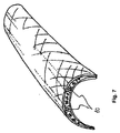
図7は、第5の態様によるインプラントを模式的に示す。この態様においては、インプラントは、長骨の骨欠損のためのカバープレートである。該インプラントは、材料が曲げられるとき、良好な骨内への成長を与えるために、複数の層が離れて間隔をあけられたままであるべき領域において、互いと接触しないことを確実する相互連結可能なパーツ10をも含む。 FIG. 7 schematically shows an implant according to a fifth aspect. In this embodiment, the implant is a cover plate for a long bone defect. The implant can be interconnected to ensure that multiple layers do not contact each other in areas that should remain spaced apart in order to provide good bone growth when the material is bent In addition, a special part 10 is included.
Claims (8)
前記層のうちの少なくとも1個は、少なくとも主にメッシュから形成され、該メッシュは3〜100μmの直径を有するガラス繊維から構成され、
該メッシュの開口のサイズは、前記生物活性材料がインプラント内で(within)保持されるように選択され、
前記層は、置換または非置換のジメタクリレートおよびメタクリレートからなる群から選択される樹脂から構成されるマトリクス中に埋められ;
該層は、インプラントの輪郭に沿って、互いに接着され;
前記生物活性材料は、粒子状の、生物活性ガラス、ヒドロキシアパタイト、リン酸三カルシウム、およびそれらの混合物からなる群から選択されることを特徴とするインプラント。 An implant comprising at least two layers made of fibers and a bioactive material disposed between the at least two layers;
At least one of the layers is formed at least mainly from a mesh, the mesh being composed of glass fibers having a diameter of 3 to 100 μm,
The size of the mesh opening is selected such that the bioactive material is retained in the implant;
Said layer is embedded in a matrix composed of a resin selected from the group consisting of substituted or unsubstituted dimethacrylate and methacrylate;
The layers are adhered to each other along the contour of the implant;
The implant is characterized in that the bioactive material is selected from the group consisting of particulate bioactive glass, hydroxyapatite, tricalcium phosphate, and mixtures thereof.
Applications Claiming Priority (3)
| Application Number | Priority Date | Filing Date | Title |
|---|---|---|---|
| EP12169943.3A EP2668967B1 (en) | 2012-05-30 | 2012-05-30 | An implant |
| EP12169943.3 | 2012-05-30 | ||
| PCT/EP2013/060980 WO2013178637A1 (en) | 2012-05-30 | 2013-05-28 | An implant |
Publications (2)
| Publication Number | Publication Date |
|---|---|
| JP2015517859A JP2015517859A (en) | 2015-06-25 |
| JP5833796B2 true JP5833796B2 (en) | 2015-12-16 |
Family
ID=48613576
Family Applications (1)
| Application Number | Title | Priority Date | Filing Date |
|---|---|---|---|
| JP2015514464A Active JP5833796B2 (en) | 2012-05-30 | 2013-05-28 | Implant |
Country Status (20)
| Country | Link |
|---|---|
| US (1) | US9084844B2 (en) |
| EP (1) | EP2668967B1 (en) |
| JP (1) | JP5833796B2 (en) |
| KR (1) | KR101500951B1 (en) |
| CN (1) | CN104349799B (en) |
| AU (1) | AU2013269704B2 (en) |
| CA (1) | CA2865602C (en) |
| DK (1) | DK2668967T3 (en) |
| ES (1) | ES2485381T3 (en) |
| HK (1) | HK1202271A1 (en) |
| IL (1) | IL234571A (en) |
| MX (1) | MX2014014167A (en) |
| MY (1) | MY166494A (en) |
| NZ (1) | NZ630312A (en) |
| PL (1) | PL2668967T3 (en) |
| PT (1) | PT2668967E (en) |
| RU (1) | RU2589839C1 (en) |
| SG (1) | SG11201404950UA (en) |
| WO (1) | WO2013178637A1 (en) |
| ZA (1) | ZA201406102B (en) |
Families Citing this family (17)
| Publication number | Priority date | Publication date | Assignee | Title |
|---|---|---|---|---|
| US7567129B2 (en) * | 2007-06-29 | 2009-07-28 | Intel Corporation | Monolithic flexible power amplifier using integrated tunable matching networks |
| USD751202S1 (en) * | 2014-04-15 | 2016-03-08 | Karl Leibinger Medizintechnik Gmbh & Co. Kg | Mesh for orbital reconstruction |
| EP3115025A1 (en) * | 2015-07-08 | 2017-01-11 | Skulle Implants OY | Orthopedic implant |
| FI126495B (en) | 2015-11-04 | 2017-01-13 | Traceray Oy | Bone implants |
| KR101854648B1 (en) * | 2016-05-04 | 2018-06-20 | 한국세라믹기술원 | Bioactive glass fabric type bone morphogen and manufacturing method of the same |
| US10676713B2 (en) | 2016-05-27 | 2020-06-09 | Corning Incorporated | Bioactive borophosphate glasses |
| US20170342383A1 (en) | 2016-05-27 | 2017-11-30 | Corning Incorporated | Lithium disilicate glass-ceramic compositions and methods thereof |
| US10751367B2 (en) | 2016-05-27 | 2020-08-25 | Corning Incorporated | Bioactive glass microspheres |
| US10647962B2 (en) | 2016-05-27 | 2020-05-12 | Corning Incorporated | Bioactive aluminoborate glasses |
| AU2017339120A1 (en) * | 2016-10-05 | 2019-04-18 | Bonalive Biomaterials Oy | A bone implant |
| KR102000455B1 (en) * | 2017-06-02 | 2019-07-16 | 한국세라믹기술원 | Fabric type bone morphogen comprising bioactive glass fiber and manufacturing method of the same |
| KR102005757B1 (en) * | 2017-06-02 | 2019-07-31 | 한국세라믹기술원 | Bio ceramic for structural body comprising bioactive glass fiber and manufacturing method of the same |
| WO2019108556A1 (en) | 2017-11-28 | 2019-06-06 | Corning Incorporated | Bioactive glass compositions and dentin hypersensitivity remediation |
| EP3717426A1 (en) | 2017-11-28 | 2020-10-07 | Corning Incorporated | Chemically strengthened bioactive glass-ceramics |
| WO2019108557A1 (en) | 2017-11-28 | 2019-06-06 | Corning Incorporated | Bioactive borate glass and methods thereof |
| WO2019108558A1 (en) | 2017-11-28 | 2019-06-06 | Corning Incorporated | High liquidus viscosity bioactive glass |
| KR102198270B1 (en) * | 2018-11-09 | 2021-01-06 | 주식회사 커스메디 | Implant for Restoring and Fixing Maxillofacial defects |
Family Cites Families (13)
| Publication number | Priority date | Publication date | Assignee | Title |
|---|---|---|---|---|
| US5626861A (en) * | 1994-04-01 | 1997-05-06 | Massachusetts Institute Of Technology | Polymeric-hydroxyapatite bone composite |
| JP2000126280A (en) * | 1998-08-18 | 2000-05-09 | Asahi Optical Co Ltd | Production of sheet for bone prosthesis and sheet for bone prosthesis |
| JP4280968B2 (en) * | 2002-09-30 | 2009-06-17 | タキロン株式会社 | Implant complex |
| US20050118236A1 (en) * | 2002-12-03 | 2005-06-02 | Gentis Inc. | Bioactive, resorbable scaffolds for tissue engineering |
| US20100189764A1 (en) * | 2005-03-22 | 2010-07-29 | Jonathan Thomas | Bioactive mesh |
| US20060293760A1 (en) * | 2005-06-24 | 2006-12-28 | Dedeyne Patrick G | Soft tissue implants with improved interfaces |
| CN1850299A (en) * | 2006-04-26 | 2006-10-25 | 中南大学 | High-strength biological-active glass ceramic-resin composite and its preparing method thereof |
| JP2008013672A (en) * | 2006-07-06 | 2008-01-24 | Kyoto Institute Of Technology | Composite material containing cured epoxy resin porous material and fiber |
| US7997901B2 (en) * | 2008-03-25 | 2011-08-16 | Pentron Clinical Technologies, Llc | Fiber reinforced composite post |
| FI20095084A0 (en) * | 2009-01-30 | 2009-01-30 | Pekka Vallittu | Composite and its use |
| US8647614B2 (en) * | 2009-02-18 | 2014-02-11 | Orthovita, Inc. | Method for stabilizing vertebral body architecture |
| FI20096351A0 (en) * | 2009-12-18 | 2009-12-18 | Pekka Vallittu | implant |
| CN103976883B (en) * | 2014-04-30 | 2015-10-21 | 中国科学院化学研究所 | A kind of artificial teeth composite and its production and use |
-
2012
- 2012-05-30 ES ES12169943.3T patent/ES2485381T3/en active Active
- 2012-05-30 DK DK12169943.3T patent/DK2668967T3/en active
- 2012-05-30 PT PT121699433T patent/PT2668967E/en unknown
- 2012-05-30 PL PL12169943T patent/PL2668967T3/en unknown
- 2012-05-30 EP EP12169943.3A patent/EP2668967B1/en active Active
-
2013
- 2013-05-28 CN CN201380020011.XA patent/CN104349799B/en active Active
- 2013-05-28 MY MYPI2014702391A patent/MY166494A/en unknown
- 2013-05-28 RU RU2014150778/15A patent/RU2589839C1/en active
- 2013-05-28 MX MX2014014167A patent/MX2014014167A/en active IP Right Grant
- 2013-05-28 US US14/380,692 patent/US9084844B2/en active Active
- 2013-05-28 JP JP2015514464A patent/JP5833796B2/en active Active
- 2013-05-28 SG SG11201404950UA patent/SG11201404950UA/en unknown
- 2013-05-28 CA CA2865602A patent/CA2865602C/en active Active
- 2013-05-28 KR KR1020147023874A patent/KR101500951B1/en active IP Right Grant
- 2013-05-28 WO PCT/EP2013/060980 patent/WO2013178637A1/en active Application Filing
- 2013-05-28 AU AU2013269704A patent/AU2013269704B2/en active Active
- 2013-05-28 NZ NZ630312A patent/NZ630312A/en unknown
-
2014
- 2014-08-20 ZA ZA2014/06102A patent/ZA201406102B/en unknown
- 2014-09-10 IL IL234571A patent/IL234571A/en active IP Right Grant
-
2015
- 2015-03-20 HK HK15102844.7A patent/HK1202271A1/en unknown
Also Published As
| Publication number | Publication date |
|---|---|
| ZA201406102B (en) | 2016-03-30 |
| MY166494A (en) | 2018-06-27 |
| RU2589839C1 (en) | 2016-07-10 |
| AU2013269704A1 (en) | 2014-09-18 |
| KR20140114452A (en) | 2014-09-26 |
| KR101500951B1 (en) | 2015-03-13 |
| HK1202271A1 (en) | 2015-09-25 |
| MX2014014167A (en) | 2015-02-04 |
| US9084844B2 (en) | 2015-07-21 |
| CN104349799B (en) | 2016-05-18 |
| WO2013178637A1 (en) | 2013-12-05 |
| NZ630312A (en) | 2015-06-26 |
| ES2485381T3 (en) | 2014-08-13 |
| US20150032224A1 (en) | 2015-01-29 |
| EP2668967A1 (en) | 2013-12-04 |
| DK2668967T3 (en) | 2014-07-07 |
| SG11201404950UA (en) | 2014-09-26 |
| IL234571A (en) | 2015-05-31 |
| CN104349799A (en) | 2015-02-11 |
| CA2865602C (en) | 2016-07-26 |
| CA2865602A1 (en) | 2013-12-05 |
| EP2668967B1 (en) | 2014-05-14 |
| PT2668967E (en) | 2014-07-30 |
| AU2013269704B2 (en) | 2015-12-03 |
| PL2668967T3 (en) | 2014-09-30 |
| JP2015517859A (en) | 2015-06-25 |
Similar Documents
| Publication | Publication Date | Title |
|---|---|---|
| JP5833796B2 (en) | Implant | |
| Vallittu | Bioactive glass-containing cranial implants: an overview | |
| RU2522255C2 (en) | Composite and use thereof | |
| KR102614449B1 (en) | orthopedic implants | |
| RU2661994C2 (en) | Method for coating and coated surface, coating and implant containing such coating |
Legal Events
| Date | Code | Title | Description |
|---|---|---|---|
| A524 | Written submission of copy of amendment under article 19 pct |
Free format text: JAPANESE INTERMEDIATE CODE: A524 Effective date: 20150129 |
|
| A529 | Written submission of copy of amendment under article 34 pct |
Free format text: JAPANESE INTERMEDIATE CODE: A529 Effective date: 20141128 |
|
| A621 | Written request for application examination |
Free format text: JAPANESE INTERMEDIATE CODE: A621 Effective date: 20150129 |
|
| A871 | Explanation of circumstances concerning accelerated examination |
Free format text: JAPANESE INTERMEDIATE CODE: A871 Effective date: 20150129 |
|
| A975 | Report on accelerated examination |
Free format text: JAPANESE INTERMEDIATE CODE: A971005 Effective date: 20150428 |
|
| A131 | Notification of reasons for refusal |
Free format text: JAPANESE INTERMEDIATE CODE: A131 Effective date: 20150526 |
|
| A521 | Request for written amendment filed |
Free format text: JAPANESE INTERMEDIATE CODE: A523 Effective date: 20150826 |
|
| TRDD | Decision of grant or rejection written | ||
| A01 | Written decision to grant a patent or to grant a registration (utility model) |
Free format text: JAPANESE INTERMEDIATE CODE: A01 Effective date: 20150929 |
|
| A61 | First payment of annual fees (during grant procedure) |
Free format text: JAPANESE INTERMEDIATE CODE: A61 Effective date: 20151029 |
|
| R150 | Certificate of patent or registration of utility model |
Ref document number: 5833796 Country of ref document: JP Free format text: JAPANESE INTERMEDIATE CODE: R150 |
|
| R250 | Receipt of annual fees |
Free format text: JAPANESE INTERMEDIATE CODE: R250 |
|
| R250 | Receipt of annual fees |
Free format text: JAPANESE INTERMEDIATE CODE: R250 |
|
| R250 | Receipt of annual fees |
Free format text: JAPANESE INTERMEDIATE CODE: R250 |
|
| R250 | Receipt of annual fees |
Free format text: JAPANESE INTERMEDIATE CODE: R250 |
|
| R250 | Receipt of annual fees |
Free format text: JAPANESE INTERMEDIATE CODE: R250 |
|
| R250 | Receipt of annual fees |
Free format text: JAPANESE INTERMEDIATE CODE: R250 |