JP5829366B1 - Endoscopic treatment tool - Google Patents
Endoscopic treatment tool Download PDFInfo
- Publication number
- JP5829366B1 JP5829366B1 JP2015532198A JP2015532198A JP5829366B1 JP 5829366 B1 JP5829366 B1 JP 5829366B1 JP 2015532198 A JP2015532198 A JP 2015532198A JP 2015532198 A JP2015532198 A JP 2015532198A JP 5829366 B1 JP5829366 B1 JP 5829366B1
- Authority
- JP
- Japan
- Prior art keywords
- sheath
- slider
- main body
- endoscope
- proximal end
- Prior art date
- Legal status (The legal status is an assumption and is not a legal conclusion. Google has not performed a legal analysis and makes no representation as to the accuracy of the status listed.)
- Active
Links
- 238000012277 endoscopic treatment Methods 0.000 title description 2
- 238000011282 treatment Methods 0.000 claims abstract description 41
- 239000000463 material Substances 0.000 claims description 19
- 238000003780 insertion Methods 0.000 description 37
- 230000037431 insertion Effects 0.000 description 37
- 230000002093 peripheral effect Effects 0.000 description 10
- 210000003811 finger Anatomy 0.000 description 9
- 230000001105 regulatory effect Effects 0.000 description 8
- 210000003813 thumb Anatomy 0.000 description 8
- 238000005452 bending Methods 0.000 description 4
- 238000004891 communication Methods 0.000 description 4
- 210000004932 little finger Anatomy 0.000 description 4
- 238000001574 biopsy Methods 0.000 description 3
- 238000003384 imaging method Methods 0.000 description 3
- 239000004696 Poly ether ether ketone Substances 0.000 description 2
- 239000000853 adhesive Substances 0.000 description 2
- 230000001070 adhesive effect Effects 0.000 description 2
- 239000000956 alloy Substances 0.000 description 2
- 229910052751 metal Inorganic materials 0.000 description 2
- 239000002184 metal Substances 0.000 description 2
- 238000000034 method Methods 0.000 description 2
- 238000012986 modification Methods 0.000 description 2
- 230000004048 modification Effects 0.000 description 2
- 229920002530 polyetherether ketone Polymers 0.000 description 2
- YCKRFDGAMUMZLT-UHFFFAOYSA-N Fluorine atom Chemical compound [F] YCKRFDGAMUMZLT-UHFFFAOYSA-N 0.000 description 1
- 239000004677 Nylon Substances 0.000 description 1
- WAIPAZQMEIHHTJ-UHFFFAOYSA-N [Cr].[Co] Chemical class [Cr].[Co] WAIPAZQMEIHHTJ-UHFFFAOYSA-N 0.000 description 1
- 229920000122 acrylonitrile butadiene styrene Polymers 0.000 description 1
- 229910045601 alloy Inorganic materials 0.000 description 1
- MAYCNCJAIFGQIH-UHFFFAOYSA-N buta-1,3-diene 5-phenylpenta-2,4-dienenitrile Chemical compound C=CC=C.N#CC=CC=CC1=CC=CC=C1 MAYCNCJAIFGQIH-UHFFFAOYSA-N 0.000 description 1
- 229920006026 co-polymeric resin Polymers 0.000 description 1
- 238000012217 deletion Methods 0.000 description 1
- 230000037430 deletion Effects 0.000 description 1
- 238000010586 diagram Methods 0.000 description 1
- 230000000694 effects Effects 0.000 description 1
- 239000013013 elastic material Substances 0.000 description 1
- 229920001971 elastomer Polymers 0.000 description 1
- 239000011737 fluorine Substances 0.000 description 1
- 229910052731 fluorine Inorganic materials 0.000 description 1
- 238000005286 illumination Methods 0.000 description 1
- 230000003902 lesion Effects 0.000 description 1
- 239000007788 liquid Substances 0.000 description 1
- 210000004072 lung Anatomy 0.000 description 1
- 238000011328 necessary treatment Methods 0.000 description 1
- 229910001000 nickel titanium Inorganic materials 0.000 description 1
- 229920001778 nylon Polymers 0.000 description 1
- 238000012634 optical imaging Methods 0.000 description 1
- 229920006122 polyamide resin Polymers 0.000 description 1
- 229920005672 polyolefin resin Polymers 0.000 description 1
- 229920005989 resin Polymers 0.000 description 1
- 239000011347 resin Substances 0.000 description 1
- 239000010935 stainless steel Substances 0.000 description 1
- 229910001220 stainless steel Inorganic materials 0.000 description 1
- 229920002803 thermoplastic polyurethane Polymers 0.000 description 1
- 238000003466 welding Methods 0.000 description 1
Images


Classifications
-
- A—HUMAN NECESSITIES
- A61—MEDICAL OR VETERINARY SCIENCE; HYGIENE
- A61B—DIAGNOSIS; SURGERY; IDENTIFICATION
- A61B10/00—Other methods or instruments for diagnosis, e.g. instruments for taking a cell sample, for biopsy, for vaccination diagnosis; Sex determination; Ovulation-period determination; Throat striking implements
- A61B10/02—Instruments for taking cell samples or for biopsy
- A61B10/0233—Pointed or sharp biopsy instruments
- A61B10/0283—Pointed or sharp biopsy instruments with vacuum aspiration, e.g. caused by retractable plunger or by connected syringe
-
- A—HUMAN NECESSITIES
- A61—MEDICAL OR VETERINARY SCIENCE; HYGIENE
- A61B—DIAGNOSIS; SURGERY; IDENTIFICATION
- A61B1/00—Instruments for performing medical examinations of the interior of cavities or tubes of the body by visual or photographical inspection, e.g. endoscopes; Illuminating arrangements therefor
- A61B1/00112—Connection or coupling means
- A61B1/00121—Connectors, fasteners and adapters, e.g. on the endoscope handle
- A61B1/00128—Connectors, fasteners and adapters, e.g. on the endoscope handle mechanical, e.g. for tubes or pipes
-
- A—HUMAN NECESSITIES
- A61—MEDICAL OR VETERINARY SCIENCE; HYGIENE
- A61B—DIAGNOSIS; SURGERY; IDENTIFICATION
- A61B1/00—Instruments for performing medical examinations of the interior of cavities or tubes of the body by visual or photographical inspection, e.g. endoscopes; Illuminating arrangements therefor
- A61B1/00131—Accessories for endoscopes
- A61B1/00133—Drive units for endoscopic tools inserted through or with the endoscope
-
- A—HUMAN NECESSITIES
- A61—MEDICAL OR VETERINARY SCIENCE; HYGIENE
- A61B—DIAGNOSIS; SURGERY; IDENTIFICATION
- A61B1/00—Instruments for performing medical examinations of the interior of cavities or tubes of the body by visual or photographical inspection, e.g. endoscopes; Illuminating arrangements therefor
- A61B1/00131—Accessories for endoscopes
- A61B1/00137—End pieces at either end of the endoscope, e.g. caps, seals or forceps plugs
-
- A—HUMAN NECESSITIES
- A61—MEDICAL OR VETERINARY SCIENCE; HYGIENE
- A61B—DIAGNOSIS; SURGERY; IDENTIFICATION
- A61B1/00—Instruments for performing medical examinations of the interior of cavities or tubes of the body by visual or photographical inspection, e.g. endoscopes; Illuminating arrangements therefor
- A61B1/012—Instruments for performing medical examinations of the interior of cavities or tubes of the body by visual or photographical inspection, e.g. endoscopes; Illuminating arrangements therefor characterised by internal passages or accessories therefor
- A61B1/018—Instruments for performing medical examinations of the interior of cavities or tubes of the body by visual or photographical inspection, e.g. endoscopes; Illuminating arrangements therefor characterised by internal passages or accessories therefor for receiving instruments
-
- A—HUMAN NECESSITIES
- A61—MEDICAL OR VETERINARY SCIENCE; HYGIENE
- A61B—DIAGNOSIS; SURGERY; IDENTIFICATION
- A61B17/00—Surgical instruments, devices or methods, e.g. tourniquets
- A61B17/34—Trocars; Puncturing needles
- A61B17/3478—Endoscopic needles, e.g. for infusion
-
- A—HUMAN NECESSITIES
- A61—MEDICAL OR VETERINARY SCIENCE; HYGIENE
- A61B—DIAGNOSIS; SURGERY; IDENTIFICATION
- A61B17/00—Surgical instruments, devices or methods, e.g. tourniquets
- A61B17/32—Surgical cutting instruments
- A61B17/3205—Excision instruments
- A61B17/32056—Surgical snare instruments
-
- A—HUMAN NECESSITIES
- A61—MEDICAL OR VETERINARY SCIENCE; HYGIENE
- A61B—DIAGNOSIS; SURGERY; IDENTIFICATION
- A61B17/00—Surgical instruments, devices or methods, e.g. tourniquets
- A61B17/00234—Surgical instruments, devices or methods, e.g. tourniquets for minimally invasive surgery
- A61B2017/00292—Surgical instruments, devices or methods, e.g. tourniquets for minimally invasive surgery mounted on or guided by flexible, e.g. catheter-like, means
- A61B2017/00296—Surgical instruments, devices or methods, e.g. tourniquets for minimally invasive surgery mounted on or guided by flexible, e.g. catheter-like, means mounted on an endoscope
-
- A—HUMAN NECESSITIES
- A61—MEDICAL OR VETERINARY SCIENCE; HYGIENE
- A61B—DIAGNOSIS; SURGERY; IDENTIFICATION
- A61B17/00—Surgical instruments, devices or methods, e.g. tourniquets
- A61B17/00234—Surgical instruments, devices or methods, e.g. tourniquets for minimally invasive surgery
- A61B2017/00292—Surgical instruments, devices or methods, e.g. tourniquets for minimally invasive surgery mounted on or guided by flexible, e.g. catheter-like, means
- A61B2017/00336—Surgical instruments, devices or methods, e.g. tourniquets for minimally invasive surgery mounted on or guided by flexible, e.g. catheter-like, means with a protective sleeve, e.g. retractable or slidable
-
- A—HUMAN NECESSITIES
- A61—MEDICAL OR VETERINARY SCIENCE; HYGIENE
- A61B—DIAGNOSIS; SURGERY; IDENTIFICATION
- A61B17/00—Surgical instruments, devices or methods, e.g. tourniquets
- A61B2017/00477—Coupling
-
- A—HUMAN NECESSITIES
- A61—MEDICAL OR VETERINARY SCIENCE; HYGIENE
- A61B—DIAGNOSIS; SURGERY; IDENTIFICATION
- A61B17/00—Surgical instruments, devices or methods, e.g. tourniquets
- A61B17/22—Implements for squeezing-off ulcers or the like on the inside of inner organs of the body; Implements for scraping-out cavities of body organs, e.g. bones; Calculus removers; Calculus smashing apparatus; Apparatus for removing obstructions in blood vessels, not otherwise provided for
- A61B17/221—Gripping devices in the form of loops or baskets for gripping calculi or similar types of obstructions
- A61B2017/2217—Gripping devices in the form of loops or baskets for gripping calculi or similar types of obstructions single wire changing shape to a gripping configuration
-
- A—HUMAN NECESSITIES
- A61—MEDICAL OR VETERINARY SCIENCE; HYGIENE
- A61B—DIAGNOSIS; SURGERY; IDENTIFICATION
- A61B17/00—Surgical instruments, devices or methods, e.g. tourniquets
- A61B17/34—Trocars; Puncturing needles
- A61B2017/347—Locking means, e.g. for locking instrument in cannula
Abstract
本内視鏡処置具は、内視鏡のチャンネルに挿通可能に構成されたシースと、シースの先端部に設けられた処置部と、シースに進退可能に挿通されるとともに先端部が処置部に接続され、シースに対して進退することで処置部を動作させる長尺部材と、シースの基端部に設けられた操作部と、を備え、操作部は、シースに接続された操作部本体と、操作部本体に対してシースの軸線方向に沿って移動可能に設けられ、長尺部材の基端部が接続されたスライダと、操作部本体に設けられ、スライダをスライダの位置によらず一定の力で基端側に向かって付勢する定荷重バネ部と、を有する。The endoscope treatment tool includes a sheath configured to be able to be inserted into a channel of an endoscope, a treatment portion provided at a distal end portion of the sheath, and is inserted into the sheath so as to be able to advance and retreat, and the distal end portion is a treatment portion. A long member that is connected and moves the treatment portion by moving forward and backward with respect to the sheath; and an operation portion provided at a proximal end portion of the sheath, the operation portion including an operation portion main body connected to the sheath; The slider is provided so as to be movable along the axial direction of the sheath with respect to the operation unit main body, and is provided with the slider to which the proximal end portion of the long member is connected. And a constant load spring portion that is urged toward the base end side by the force of.
Description
本発明は、組織に処置を行う内視鏡処置具に関する。本願は、2013年12月11日に、日本に出願された特願2013−256426号に基づき優先権を主張し、その内容をここに援用する。 The present invention relates to an endoscope treatment tool for performing treatment on a tissue. This application claims priority on December 11, 2013 based on Japanese Patent Application No. 2013-256426 for which it applied to Japan, and uses the content here.
従来、内視鏡による観察を行いながら、内視鏡に形成されたチャンネル内に内視鏡処置具を挿通させて様々な処置が行われている。
この種の内視鏡処置具として、例えば特許文献1に記載された超音波用穿刺針が知られている。Conventionally, various treatments are performed by inserting an endoscope treatment tool into a channel formed in an endoscope while performing observation with an endoscope.
As this type of endoscope treatment tool, for example, an ultrasonic puncture needle described in
超音波用穿刺針は、シースと、このシースの基端部に配置された操作部と、この操作部を介してシース内に進退自在に挿通配置された針管とで主に構成されている。操作部は、棒状に形成されシースの基端部が取付けられた操作部本体と、操作部本体の長手方向の中間部に長手方向に沿って移動可能に設けられたスライダとを備えている。スライダには操作部本体に固定するための固定ネジが設けられ、スライダに針管の基端部が取付けられている。 The ultrasonic puncture needle is mainly composed of a sheath, an operation portion arranged at the proximal end portion of the sheath, and a needle tube that is inserted and disposed in the sheath so as to be able to advance and retreat through the operation portion. The operation portion includes an operation portion main body formed in a rod shape and attached with a proximal end portion of the sheath, and a slider provided at a middle portion in the longitudinal direction of the operation portion main body so as to be movable along the longitudinal direction. The slider is provided with a fixing screw for fixing to the operation portion main body, and the proximal end portion of the needle tube is attached to the slider.
このように構成された超音波用穿刺針を使用するときには、超音波内視鏡の内視鏡挿入部を口などから体内に挿入する。超音波内視鏡が有する光学撮像機構や超音波走査機構により観察しながら生検を行う部位を特定する。
操作部本体に対してスライダを基端側に移動させ(引き戻し)、シース内に針管を収容した状態で固定ネジを締めておく。術者などの使用者は、一方の手で超音波内視鏡の内視鏡操作部を把持し、内視鏡操作部に設けられた基端口金からチャンネル内に超音波用穿刺針のシースを挿入する。内視鏡挿入部からシースを突出させたら、他方の手で超音波用穿刺針の固定ネジを緩め、スライダを押し込んでシースの先端部から針管を突出させる。針管を突出させるためにスライダを押し込む長さは、例えば40mm程度である。
このときには、使用者は一方の手で内視鏡操作部、他方の手で超音波用穿刺針の操作部を把持してスライダを操作する。このため、力を作用させる力点となるスライダと、支点となる超音波用穿刺針と基端口金との接続部とが比較的離間し、操作が安定しない。When using the ultrasonic puncture needle configured as described above, the endoscope insertion portion of the ultrasonic endoscope is inserted into the body through the mouth or the like. A site to be biopsied is specified while observing with an optical imaging mechanism or an ultrasonic scanning mechanism of the ultrasonic endoscope.
The slider is moved to the proximal end side (retracted) with respect to the operation unit main body, and the fixing screw is tightened in a state where the needle tube is accommodated in the sheath. A user such as an operator grasps the endoscope operation part of the ultrasonic endoscope with one hand, and the sheath of the ultrasonic puncture needle is inserted into the channel from the base end cap provided in the endoscope operation part. Insert. When the sheath is protruded from the endoscope insertion portion, the fixing screw of the ultrasonic puncture needle is loosened with the other hand, the slider is pushed in, and the needle tube is protruded from the distal end portion of the sheath. The length for pushing the slider to project the needle tube is, for example, about 40 mm.
At this time, the user operates the slider while holding the endoscope operating portion with one hand and the operating portion of the ultrasonic puncture needle with the other hand. For this reason, the slider serving as a power point for applying a force and the connecting portion between the ultrasonic puncture needle serving as a fulcrum and the proximal end cap are relatively separated, and the operation is not stable.
シースから針管を突出させた後は、針管の先端を、生検を行う対象組織に到達させる。続いて、針管の管路にシリンジなどを接続して針管内を吸引し、針管の先端から対象組織の細胞などを吸引して採取する。 After protruding the needle tube from the sheath, the tip of the needle tube is allowed to reach the target tissue to be biopsied. Subsequently, a syringe or the like is connected to the conduit of the needle tube, the inside of the needle tube is sucked, and the cells of the target tissue are sucked and collected from the tip of the needle tube.
内視鏡のチャンネル内に挿通させた内視鏡処置具の操作部を操作する際に、力点と支点とが比較的離間するという問題を解消するために、公知のマイクロピペットなどに用いられている操作部を内視鏡処置具に用いることが検討されている。この操作部では、操作部本体の基端面よりも基端側に突出するようにスライダが設けられ、スライダを基端側に向かって付勢するためにつるまきバネが設けられている。使用者は、例えば、手の人差指から小指までの指で操作部本体を把持し、つるまきバネの付勢力に抗してスライダを先端側に親指で押し込むことでシースから針管を突出させる。
この操作部を用いた場合、力点となるスライダと支点となる操作部本体とが一本の手に収まって比較的接近し、内視鏡処置具の操作部の操作を安定して行うことができる。スライダから親指を離すと、つるまきバネの付勢力によりシース内に針管が収容される。When operating the operation part of the endoscope treatment tool inserted into the channel of the endoscope, it is used for a known micropipette or the like in order to solve the problem that the force point and the fulcrum are relatively separated from each other. It has been studied to use an operating part for an endoscope treatment tool. In this operation part, a slider is provided so as to protrude from the base end surface of the operation part main body to the base end side, and a helical spring is provided to urge the slider toward the base end side. For example, the user grasps the operation unit main body with the fingers from the index finger to the little finger of the hand, and pushes the slider toward the distal end side with the thumb against the biasing force of the helical spring, thereby causing the needle tube to protrude from the sheath.
When this operation unit is used, the slider serving as the power point and the operation unit main body serving as the fulcrum can be accommodated in one hand and relatively approached, and the operation unit of the endoscope treatment tool can be stably operated. it can. When the thumb is released from the slider, the needle tube is accommodated in the sheath by the biasing force of the helical spring.
しかし、この操作部では、スライダを押し込むのにしたがってつるまきバネから受ける反力は大きくなり、スライダを最も押し込んだ状態でスライダを保持しにくい。 However, in this operation unit, the reaction force received from the helical spring increases as the slider is pushed in, and it is difficult to hold the slider when the slider is pushed in the most.
本発明は、このような問題点に鑑みてなされたものであって、スライダを押し込んだ状態で容易に保持することができる内視鏡処置具を提供することを目的とする。 The present invention has been made in view of such problems, and an object of the present invention is to provide an endoscope treatment instrument that can be easily held in a state where a slider is pushed in.
本発明の第一の態様に係る内視鏡処置具は、内視鏡のチャンネルに挿通可能に構成されたシースと、前記シースの先端部に設けられた処置部と、前記シースに進退可能に挿通されるとともに先端部が前記処置部に接続され、前記シースに対して進退することで前記処置部を動作させる長尺部材と、前記シースの基端部に設けられた操作部と、を備え、前記操作部は、前記シースに接続された操作部本体と、前記操作部本体に対して前記シースの軸線方向に沿って移動可能に設けられ、前記長尺部材の基端部が接続されたスライダと、前記操作部本体に設けられ、前記スライダを前記スライダの位置によらず一定の力で基端側に向かって付勢する定荷重バネ部と、を有し、前記定荷重バネ部は、長尺状に形成されたバネ材と、前記バネ材の基端部を収容する収容部と、を有し、前記収容部は、前記操作部本体の基端側に前記操作部本体から側方に突出するように設けられ、前記バネ材の先端部は、前記長尺部材に沿って先端側に延び、前記スライダまたは前記長尺部材に接続されている。 An endoscope treatment tool according to a first aspect of the present invention includes a sheath configured to be inserted through an endoscope channel, a treatment portion provided at a distal end portion of the sheath, and capable of moving forward and backward with the sheath. A long member that is inserted and has a distal end connected to the treatment portion, and moves the treatment portion by moving forward and backward with respect to the sheath; and an operation portion provided at a proximal end portion of the sheath. The operation unit is provided so as to be movable along the axial direction of the sheath with respect to the operation unit main body connected to the sheath and the operation unit main body, and the proximal end portion of the long member is connected A slider, and a constant load spring portion that is provided in the operation portion main body and biases the slider toward the base end side with a constant force regardless of the position of the slider, and the constant load spring portion is The elongated spring material and the proximal end of the spring material An accommodating portion for accommodating the spring member, and the accommodating portion is provided on the proximal end side of the operation portion main body so as to protrude laterally from the operation portion main body, and the distal end portion of the spring material is formed of the long portion. It extends to the tip side along the scale member and is connected to the slider or the long member .
本発明の第二の態様によれば、第一の態様に係る内視鏡処置具において、前記処置部および前記長尺部材は、針管として一体に構成され、前記スライダには、前記針管の管路に連通する貫通孔が形成され、前記スライダの基端には、前記シースの軸線上に位置する受け面が設けられ、前記貫通孔の基端部の開口は、前記スライダの基端の前記シースの軸線上からずれた位置に形成されていてもよい。 According to a second aspect of the present invention, in the endoscope treatment instrument according to the first aspect, the treatment portion and the long member are integrally configured as a needle tube, and the slider is provided with a tube of the needle tube. A through hole communicating with the path is formed, and a receiving surface located on an axis of the sheath is provided at a base end of the slider, and an opening at a base end portion of the through hole is formed at the base end of the slider. It may be formed at a position shifted from the axis of the sheath.
上記態様の内視鏡処置具によれば、スライダを押し込んだ状態で容易に保持することができる。 According to the endoscope treatment tool of the above aspect, the slider can be easily held in a pressed state.
以下、本発明に係る内視鏡処置具の一実施形態を、内視鏡処置具が穿刺針(吸引生検針)である場合を例示して、図1から図10を参照しながら説明する。なお、各図は模式的なものであり、図面を見やすくするため、各構成要素の厚さや寸法の比率は適宜異ならせてある。
まず、本実施形態における穿刺針とともに用いられる内視鏡について説明する。以下では、内視鏡が内視鏡挿入部の前方を観察可能ないわゆる直視型の内視鏡の例を説明するが、内視鏡の構成はこれに限定されず、内視鏡が内視鏡挿入部の側方を観察可能ないわゆる側視型の内視鏡であってもよい。Hereinafter, an embodiment of an endoscope treatment tool according to the present invention will be described with reference to FIGS. 1 to 10 exemplifying a case where the endoscope treatment tool is a puncture needle (aspiration biopsy needle). Each figure is schematic, and thicknesses and dimensional ratios of each component are appropriately changed in order to make the drawing easy to see.
First, the endoscope used with the puncture needle in this embodiment will be described. In the following, an example of a so-called direct-view type endoscope in which the endoscope can observe the front of the endoscope insertion portion will be described. However, the configuration of the endoscope is not limited to this, and the endoscope is A so-called side-view type endoscope that can observe the side of the mirror insertion portion may be used.
図1は、本実施形態に用いられる内視鏡100の説明図である。図1に示すように、内視鏡100は公知の構成の内視鏡であり、軟性であって長尺の内視鏡挿入部110と、内視鏡操作部120とを備えている。内視鏡操作部120は、内視鏡挿入部110の基端部に設けられている。
内視鏡挿入部110の先端側には、湾曲操作可能な湾曲部111が設けられている。湾曲部111には不図示の操作ワイヤが取り付けられている。操作ワイヤは、内視鏡挿入部110内に挿通され、内視鏡操作部120まで延びている。内視鏡挿入部110の先端面には、図示はしないが、ライトガイド、およびCCDを有する撮像ユニットが露出した状態で設けられている。
内視鏡挿入部110には、内視鏡挿入部110に沿ってチャンネル112が形成されている。チャンネル112の先端部は、内視鏡挿入部110の先端面に開口している。FIG. 1 is an explanatory diagram of an
A
A
内視鏡操作部120には、アングルノブ121と、スイッチ122とが設けられている。アングルノブ121は、前述の操作ワイヤを操作するために設けられている。アングルノブ121を操作することで、湾曲部111を所望の方向に湾曲させることができる。スイッチ122は、図示しない光源、モニタや、前述の撮像ユニットなどを操作するために設けられている。
The
内視鏡操作部120の先端側には、鉗子挿入口金123が設けられている。鉗子挿入口金123には、挿通孔123aが形成されている。鉗子挿入口金123の挿通孔123aは、チャンネル112に連通している。
鉗子挿入口金123には、ゴムなどの弾性を有する材料で形成されたアダプタ鉗子栓125が嵌合されている。アダプタ鉗子栓125は、アダプタ本体126と、係止部127と、フランジ128とを有している。アダプタ本体126は、筒状に形成されている。係止部127は、アダプタ本体126の外周面の基端部に設けられている。フランジ128は、アダプタ本体126の外周面の長手方向の中間部に設けられている。
アダプタ本体126の挿通孔126aの内径は、鉗子挿入口金123の挿通孔123aの内径よりもわずかに小さい。アダプタ鉗子栓125には、鉗子挿入口金123側から挿通孔126aを通して外部に液体が流れ出るのを防止する不図示の逆止弁が設けられている。A
An adapter forceps plug 125 made of an elastic material such as rubber is fitted to the
The inner diameter of the
図2は、本実施形態の穿刺針1の側面の断面図である。図3は、本実施形態の穿刺針1の基端側の平面の断面図である。図4は、本実施形態の穿刺針1の背面図である。図2から図4に示すように、本実施形態の穿刺針1は、シース10と、針管15と、操作部20とを備えている。針管15は、シース10に進退可能に挿通されている。操作部20は、シース10の基端部に設けられている。
FIG. 2 is a side sectional view of the
シース10は、内視鏡100のチャンネル112に挿通可能な外径を有する。
シース10は、ポリエーテルエーテルケトン(PEEK)、フッ素系樹脂、オレフィン系樹脂、ウレタン系樹脂、およびナイロン系(ポリアミド系)樹脂や、金属製のコイルなどで形成することができる。シース10の基端部には、外径が拡径された拡径部11が設けられている。The
The
針管15は、管状に形成されている。針管15の外周面の基端部には、鍔部16が設けられている。針管15の先端は、シース10の軸線(中心軸線)Cに対して斜めに切り落とされることで、生体組織に刺入できるように鋭利に形成されている。
針管15および鍔部16を形成する材料は、可撓性を有しており、且つ、外力により曲げられても容易に直線状態に復元する弾性を有する材料であることが好ましい。このような材料としては、ステンレス合金、ニッケルチタン合金、コバルトクロム合金などの合金材料を挙げることができる。
針管15は、処置部15b、および、長尺部材15cを有する。処置部15bは、シース10の先端部に設けられた針先である。長尺部材15cは、先端部が処置部15bに接続された針本体である。本実施形態の針管15は、処置部15bと長尺部材15cとが一体に構成されている。長尺部材15cは、シース10に対して進退することで処置部15bを動作させる。すなわち、長尺部材15cは、後述するように処置部15bにより対象組織を回収させる部材である。The
The material that forms the
The
操作部20は、操作部本体21と、スライダ22と、定荷重バネ部23とを有している。操作部本体21は、シース10の基端部に接続されている。スライダ22は、針管15の基端部が接続されている。定荷重バネ部23は、操作部本体21に設けられている。
操作部本体21は、筒状に形成され、操作部本体21の内周面における軸線C方向の中間部には径方向内方に突出した縮径部26が形成されている。操作部本体21の外周面(側面)21aの基端部には、操作部本体21の径方向外方に突出した支持部27が設けられている。操作部本体21、縮径部26、および支持部27は、ABS樹脂(スチレン−アクリロニトリル−ブタジエン共重合樹脂)などにより一体に形成されている。The
The operation portion
図5は、本実施形態の穿刺針1の基端側の側面図である。図3および図5に示すように、操作部本体21にはスリット28が形成されている。スリット28は、外周面21aから内周面に達するように形成されている。スリット28は、スライドスリット28aと、係止スリット28bとを有している。スライドスリット28aは、軸線Cに平行に延びている。係止スリット28bは、スライドスリット28aに連通し、スライドスリット28aの基端部から周方向に延びている。スライドスリット28aの幅と係止スリット28bの幅とは等しい。
FIG. 5 is a side view of the proximal end side of the
図6は、本実施形態の穿刺針1の要部の斜視図である。図7は、本実施形態の穿刺針1のロック部材32の平面図である。図8は、本実施形態の穿刺針1の作用を説明する断面図である。図2および図6に示すように、操作部本体21の縮径部26の先端面には、先端側に延びる支持パイプ31が固定されている。支持パイプ31は、ステンレス鋼などの金属で形成することができる。支持パイプ31には、シース10が挿通されている。支持パイプ31の外径は、内視鏡100の鉗子挿入口金123の挿通孔123aの内径よりもわずかに小さい。
操作部本体21の先端部には、板状のロック部材32が軸線Cに直交する方向へスライド自在に設けられている。ロック部材32には、図7に示すように、大径孔32aと、小径孔32bと、連通孔32cとが形成されている。小径孔32bは、大径孔32aよりも内径が小さい。連通孔32cは、大径孔32aと小径孔32bとを連通させる。
図2および図6に示すように、ロック部材32の大径孔32a、小径孔32b、および連通孔32c内には支持パイプ31が挿通可能である。すなわち、ロック部材32の大径孔32a、小径孔32b、および連通孔32cからなる孔内に支持パイプ31を挿通させた状態で、操作部本体21に対してロック部材32を軸線Cに直交する方向へスライドさせることができる。FIG. 6 is a perspective view of a main part of the
A plate-
As shown in FIGS. 2 and 6, the
図2に示すように、操作部本体21に対してロック部材32を小径孔32b側にスライドさせたときには、大径孔32aが軸線C上に配置される。この場合、アダプタ鉗子栓125にロック部材32は係合されない。
一方で、小径孔32b側にスライドさせたロック部材32の大径孔32aにアダプタ鉗子栓125のアダプタ本体126を挿通させ、図8に示すように、フランジ128にロック部材32を接触させつつ、操作部本体21に対してロック部材32を大径孔32a側にスライドさせる。その結果、小径孔32bの縁部が係止部127に係合されてアダプタ鉗子栓125に対するロック部材32の基端側への移動が規制され、アダプタ鉗子栓125にロック部材32が取付けられる。As shown in FIG. 2, when the
On the other hand, the adapter
シース10は、図2および図3に示すように、操作部本体21の縮径部26内に挿通され、且つ、拡径部11を縮径部26の基端面に接触させた状態で縮径部26に不図示の接着剤などにより固定されている。
シース10の拡径部11と、針管15の鍔部16との間には、リング状の規制部材35が配置されている。規制部材35の筒孔内には、針管15が挿通されている。図3に示すように、規制部材35には、連結部材36の第一端が接続されている。連結部材36の第二端はスリット28に挿通され、外部に引き出されている。連結部材36の第二端には、スリット28の幅よりも大径のツマミ37が固定されている。
これらスリット28、規制部材35、連結部材36、ツマミ37で状態切替え部38を構成する。As shown in FIGS. 2 and 3, the
A ring-shaped regulating
The
スライダ22は円柱状に形成され、スライダ22の外径は、縮径部26よりも基端側の操作部本体21の内径よりも小さい。スライダ22の基端には、軸線C上に位置する受け面22aが設けられている。受け面22aの基端側は、外部に露出している。スライダ22には、貫通孔22bが形成されている。貫通孔22bの先端部は軸線C上に配置され、針管15の管路15aに連通している。貫通孔22bは基端側に向かうにしたがって軸線Cから離間するように折れ曲がってスライダ22の基端に開口している。すなわち、貫通孔22bの基端部の開口22cは、軸線C上からずれた位置に設けられている。
本実施形態では、スライダ22の周方向の一部に、径方向外方および基端側に延出させた接続部40を設け、貫通孔22bの基端側を接続部40内に形成している。これにより、貫通孔22bの開口22cは、受け面22aよりも基端側に配置されている。
接続部40の基端部には、公知のシリンジなどが着脱可能に構成されている。The
In the present embodiment, a connecting
A known syringe or the like is detachably attached to the proximal end portion of the connecting
図2に示すように、定荷重バネ部23は、バネ材43と、ドラム(収容部)44とを有している。バネ材43は、線状や帯状などの長尺状に形成されている。ドラム44は、バネ材43の基端部を収容する。
バネ材43としては、公知の定荷重ばね(例えば、サンコースプリング社製 商品名コンストン)等を好適に用いることができる。ドラム44は、例えば外形が直方体の箱状に形成され、一面に開口44aが形成されている。図4に示すように、ドラム44は、操作部本体21の基端側に操作部本体21の外周面21aから側方(操作部本体21の径方向外方)に突出し、且つ、図2に示すように、開口44aが軸線C側に向くように配置され、支持部27に接着剤やネジなどにより固定されている。
バネ材43の基端部はドラム44内で巻回されている。バネ材43の先端部は、ドラム44の開口44aを通して針管15に沿って先端側に延び、鍔部16を介して針管15に接続されている。バネ材43の先端部は、図示はしないが溶接などにより針管15に接続されている。バネ材43は、針管15を介してスライダ22を、操作部本体21に対するスライダ22の軸線C方向の位置によらず一定の力(ほぼ一定の力も含む)で基端側に向かって付勢する。As shown in FIG. 2, the constant
As the
The base end portion of the
図3に示すように、シース10の基端部に規制部材35が接触し、連結部材36の第二端がスリット28のスライドスリット28aの先端側に配置された状態を状態切替え部38の非規制状態という。状態切替え部38の非規制状態においては、図2に示すようにシース10内に針管15が収容された状態から、図8に示すように規制部材35に針管15の鍔部16が接触するまで操作部本体21に対してスライダ22を押し込む(先端側に移動させる)ことができる。
このように、状態切替え部38を非規制状態にしたときには、操作部本体21に対してスライダ22が、スライダ22が最も引き戻された図2に示す位置(所定位置)Qよりも先端側に移動可能となる。すなわち、スライダ22は操作部本体21に対して軸線C方向に沿って移動可能となる。スライダ22を押し込んだ場合およびスライダ22を引き戻した場合のいずれにおいても、操作部本体21の基端面よりもスライダ22の基端面の方が基端側に突出している。スライダ22を押し込んだ場合には、操作部本体21内にスライダ22の先端側が配置される。As shown in FIG. 3, a state in which the regulating
As described above, when the
図9は、本実施形態の穿刺針1の側面の断面図である。図10は、本実施形態の穿刺針1の基端側の側面図である。図10に示すように、連結部材36の第二端がスリット28の係止スリット28bに配置された状態を状態切替え部38の規制状態という。状態切替え部38の規制状態においては、図9に示すように、非規制状態よりも規制部材35が基端側に移動し、且つ、図10に示すように、係止スリット28bにより連結部材36が軸線C方向に係止される。スライダ22が基端側に最も引き戻された位置Qにおいて鍔部16の先端面に規制部材35が接触しているため、操作部本体21に対してスライダ22が位置Qよりも先端側に移動するのが規制される。
このように、ツマミ37を操作してスリット28内で連結部材36の第二端が配置されている位置を変えることで、状態切替え部38を規制状態と非規制状態との間で切り替えることができる。FIG. 9 is a side sectional view of the
Thus, by operating the
次に、以上のように構成された穿刺針1の作用について説明する。以下では、肺の深部に位置する病変を対象組織として回収する生検の処置を例に説明する。
使用者が、内視鏡100のスイッチ122を操作して光源を動作させると、光源から発せられた照明光はライトガイドに導かれて内視鏡挿入部110の前方を照明する。撮像ユニットで取得された内視鏡挿入部110の前方の画像は、モニタに表示される。使用者はモニタに表示された画像を確認しながら、内視鏡100の内視鏡挿入部110を患者の体内に挿入する。アングルノブ121を操作して湾曲部111を適宜湾曲させながら、内視鏡挿入部110を挿入する。生検を行う部位に内視鏡挿入部110の先端面を対向させる。Next, the operation of the
When the user operates the
続いて、穿刺針1のスライダ22を引き戻してシース10内に針管15を収容させ、ロック部材32を小径孔32b側にスライドさせる。状態切替え部38を規制状態にしておく。スライダ22の貫通孔22b側から針管15の管路15a内に、スタイレットを挿通しておくことが好ましい。
使用者は、一方の手で内視鏡操作部120を把持し、内視鏡100のアダプタ鉗子栓125の挿通孔126a、および鉗子挿入口金123の挿通孔123aに、穿刺針1のシース10および支持パイプ31を挿入する。このとき、シース10によりアダプタ鉗子栓125の逆止弁が移動する。アダプタ鉗子栓125と支持パイプ31との間が、アダプタ鉗子栓125の弾性力により水密に封止される。
支持パイプ31の外径および鉗子挿入口金123の挿通孔123aの内径は前述のように設定されているため、挿通孔123a内で支持パイプ31が摺動し、鉗子挿入口金123に支持パイプ31が確実に支持される。Subsequently, the
The user grasps the
Since the outer diameter of the
ロック部材32の大径孔32aにアダプタ鉗子栓125のアダプタ本体126を挿通させ、ロック部材32を大径孔32a側にスライドさせることで、アダプタ鉗子栓125にロック部材32を取付ける。状態切替え部38を非規制状態にする。
使用者は、図8に示すように、操作部本体21の支持部27よりも先端側の部分を他方の手P10の人差指P11から小指P12で把持し、親指P13をスライダ22の受け面22aに当てる。他方の手P10でスライダ22を押し込み、シース10から針管15を突出させる。The adapter
As shown in FIG. 8, the user grasps the tip side of the operation portion
スライダ22の受け面22aおよび針管15が軸線C上に配置されていることで、受け面22aに作用させた先端側への力が針管15に効果的に伝達される。接続部40および貫通孔22bの基端部の開口22cは軸線C上からずれた位置に設けられているため、使用者が受け面22aを操作するときに支障にならない。
力点となるスライダ22と支点となる操作部本体21とが使用者の他方の手P10に収まるため、操作部20の操作を安定して行うことができる。使用者は、スライダ22の受け面22aを親指P13で押すときに、支持部27を介して定荷重バネ部23に人差指P11を掛けることで、人差指P11で操作部本体21を基端側に向かって支持しつつ、受け面22aを先端側に向かって確実に押すことができる。
定荷重バネ部23によりスライダ22が基端側に向かって付勢される力は一定であるため、スライダ22を押し込んでも親指P13が受け面22aから受ける反力は大きくならない。Since the receiving
Since the
Since the force with which the
針管15の先端を組織P20に穿刺し、生検を行う対象組織P21へと押し進める。針管15の管路15a内に入り込んだ生検対象でない組織をスタイレットで押出し、針管15およびスライダ22からスタイレットを引き抜く。スライダ22の接続部40にシリンジなどを取付け、シリンジを操作して貫通孔22b内および管路15a内を吸引する。
対象組織P21は、管路15a内および貫通孔22b内を通してシリンジ内に入る。必要な量の対象組織P21が採取できたらスライダ22を引き戻し、シース10内に針管15を収容させる。これにより、組織P20から針管15が抜ける。ロック部材32を小径孔32b側にスライドさせて、アダプタ鉗子栓125とロック部材32との係合を解除する。内視鏡100のチャンネル112から穿刺針1のシース10を抜く。
内視鏡100を患者から引き抜き、必要な処置をして、一連の手技を終了する。The tip of the
The target tissue P21 enters the syringe through the
The
以上説明したように、本実施形態の穿刺針1によれば、定荷重バネ部23により、スライダ22の軸線C方向の位置によらずスライダ22は一定の力で基端側に向かって付勢される。このため、スライダ22を押し込む長さが長い場合であっても押し込むのに必要な力が大きくならず、スライダ22を押し込んだ状態で容易に保持することができる。スライダ22から親指P13を離すと、定荷重バネ部23によりスライダ22が基端側に位置Qまで移動する。使用者はスライダ22を押し込む操作をするだけで、スライダ22を引き戻す操作は自動で行われるため、操作部20の操作性を向上させることができる。使用者がスライダ22から親指P13を離すことで、定荷重バネ部23によりシース10内に針管15を収容させることができる。これにより、内視鏡100のチャンネル112が針管15により破損するのを回避できる。
ドラム44は、操作部本体21の基端側に操作部本体21の外周面21aから側方に突出するように設けられている。これにより、使用者は、操作部本体21における支持部27よりも先端側の部分を人差指P11から小指P12で把持し、且つ、支持部27を介して定荷重バネ部23に人差指P11を掛けることができる。したがって、使用者は、スライダ22の受け面22aを親指P13で押すときに、人差指P11から小指P12で操作部本体21を確実に把持し、人差指P11で操作部本体21を基端側に向かって支持することができる。As described above, according to the
The
軸線C上にスライダ22の受け面22aが配置されていることで、受け面22aに作用させた先端側への力を針管15に効果的に伝達させることができる。接続部40および貫通孔22bの基端部の開口22cは軸線C上からずれた位置に設けられているため、受け面22aを操作するときに接続部40および開口22cが支障になることを抑制することができる。
状態切替え部38を非規制状態にすることで、スライダ22が位置Qよりも先端側に移動できるようになり、シース10内に針管15を収容させたりシース10から針管15を突出させたりする操作を自在に行うことができる。一方で、状態切替え部38を規制状態にすることでスライダ22が位置Qよりも先端側に移動するのを規制し、シース10から針管15を意図せずに突出させることを防止できる。Since the receiving
By making the
以上、本発明の一実施形態について図面を参照して詳述したが、具体的な構成はこの実施形態に限られるものではなく、本発明の要旨を逸脱しない範囲の構成の変更、組み合わせ、削除なども含まれる。
例えば、本実施形態では、バネ材43の先端部は針管15に接続されている例を示したが、バネ材43の先端部はスライダ22に接続されていてもよい。
操作部20には状態切替え部38は備えられなくてもよい。As mentioned above, although one embodiment of the present invention has been described in detail with reference to the drawings, the specific configuration is not limited to this embodiment, and modifications, combinations, and deletions within a scope that does not depart from the gist of the present invention. Etc. are also included.
For example, in the present embodiment, the tip portion of the
The
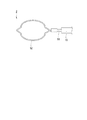
本実施形態では、内視鏡処置具は穿刺針1である例を示した。しかし、内視鏡処置具はこれに限定されず、スネア、鉗子、クリップユニットを体内に導入するための内視鏡用結紮装置などでもよい。
例えば、内視鏡処置具が図11に示すスネア2である場合には、シース10に挿通された操作ワイヤ51が長尺部材に該当し、操作ワイヤ51の先端部に接続されたループワイヤ52が処置部に該当する。In the present embodiment, an example in which the endoscope treatment tool is the
For example, when the endoscope treatment tool is the
以上、本発明の各実施形態を説明したが、本発明の技術範囲は上記実施形態に限定されるものではなく、本発明の趣旨を逸脱しない範囲において各実施形態における構成要素の組み合わせを変えたり、各構成要素に種々の変更を加えたり、削除したりすることが可能である。本発明は前述した説明によって限定されることはなく、添付のクレームの範囲によってのみ限定される。 The embodiments of the present invention have been described above. However, the technical scope of the present invention is not limited to the above embodiments, and combinations of components in the embodiments may be changed without departing from the spirit of the present invention. Various changes can be added to or deleted from each component. The present invention is not limited by the above description, but only by the scope of the appended claims.
スライダを押し込んだ状態で容易に保持することができる内視鏡処置具を提供できる。 An endoscope treatment tool that can be easily held in a state where the slider is pushed in can be provided.
1 穿刺針(内視鏡処置具)
2 スネア(内視鏡処置具)
10 シース
15 針管
15a 管路
15b 処置部
15c 長尺部材
20 操作部
21 操作部本体
21a 外周面(側面)
22 スライダ
22a 受け面
22b 貫通孔
22c 開口
23 定荷重バネ部
43 バネ材
44 ドラム(収容部)
51 操作ワイヤ(長尺部材)
52 ループワイヤ(処置部)
100 内視鏡
112 チャンネル
C 軸線1 Puncture needle (endoscopic instrument)
2 Snare (endoscopic treatment tool)
DESCRIPTION OF
22
51 Operation wire (long member)
52 Loop wire (treatment section)
100
Claims (2)
前記シースの先端部に設けられた処置部と、
前記シースに進退可能に挿通されるとともに先端部が前記処置部に接続され、前記シースに対して進退することで前記処置部を動作させる長尺部材と、
前記シースの基端部に設けられた操作部と、
を備え、
前記操作部は、
前記シースに接続された操作部本体と、
前記操作部本体に対して前記シースの軸線方向に沿って移動可能に設けられ、前記長尺部材の基端部が接続されたスライダと、
前記操作部本体に設けられ、前記スライダを前記スライダの位置によらず一定の力で基端側に向かって付勢する定荷重バネ部と、を有し、
前記定荷重バネ部は、
長尺状に形成されたバネ材と、
前記バネ材の基端部を収容する収容部と、
を有し、
前記収容部は、前記操作部本体の基端側に前記操作部本体から側方に突出するように設けられ、
前記バネ材の先端部は、前記長尺部材に沿って先端側に延び、前記スライダまたは前記長尺部材に接続されている内視鏡処置具。 A sheath configured to be inserted through the endoscope channel;
A treatment section provided at the distal end of the sheath;
A long member that is inserted into the sheath so as to be able to advance and retract, and a distal end portion is connected to the treatment portion, and moves the treatment portion by moving forward and backward with respect to the sheath;
An operation portion provided at a proximal end portion of the sheath;
With
The operation unit is
An operation unit main body connected to the sheath;
A slider provided so as to be movable along the axial direction of the sheath with respect to the operation portion main body, and a slider to which a proximal end portion of the long member is connected;
A constant load spring portion provided in the operation portion main body and biasing the slider toward the base end side with a constant force regardless of the position of the slider ;
The constant load spring portion is
A spring material formed in a long shape;
An accommodating portion for accommodating a proximal end portion of the spring material;
Have
The accommodating portion is provided on the proximal end side of the operation portion main body so as to protrude laterally from the operation portion main body,
The distal end portion of the spring material extends to the distal end side along the long member, and is an endoscope treatment tool connected to the slider or the long member .
前記スライダには、前記針管の管路に連通する貫通孔が形成され、
前記スライダの基端には、前記シースの軸線上に位置する受け面が設けられ、
前記貫通孔の基端部の開口は、前記スライダの基端の前記シースの軸線上からずれた位置に形成されている請求項1に記載の内視鏡処置具。 The treatment portion and the elongated member are integrally configured as a needle tube,
The slider is formed with a through hole communicating with the conduit of the needle tube.
The proximal end of the slider is provided with a receiving surface located on the axis of the sheath,
The endoscope treatment tool according to claim 1, wherein the opening of the base end portion of the through hole is formed at a position shifted from the axis of the sheath at the base end of the slider.
Priority Applications (1)
| Application Number | Priority Date | Filing Date | Title |
|---|---|---|---|
| JP2015532198A JP5829366B1 (en) | 2013-12-11 | 2014-12-10 | Endoscopic treatment tool |
Applications Claiming Priority (4)
| Application Number | Priority Date | Filing Date | Title |
|---|---|---|---|
| JP2013256426 | 2013-12-11 | ||
| JP2013256426 | 2013-12-11 | ||
| PCT/JP2014/082762 WO2015087939A1 (en) | 2013-12-11 | 2014-12-10 | Endoscope treatment instrument |
| JP2015532198A JP5829366B1 (en) | 2013-12-11 | 2014-12-10 | Endoscopic treatment tool |
Publications (2)
| Publication Number | Publication Date |
|---|---|
| JP5829366B1 true JP5829366B1 (en) | 2015-12-09 |
| JPWO2015087939A1 JPWO2015087939A1 (en) | 2017-03-16 |
Family
ID=53371238
Family Applications (1)
| Application Number | Title | Priority Date | Filing Date |
|---|---|---|---|
| JP2015532198A Active JP5829366B1 (en) | 2013-12-11 | 2014-12-10 | Endoscopic treatment tool |
Country Status (5)
| Country | Link |
|---|---|
| US (1) | US20160135795A1 (en) |
| EP (1) | EP3081140A4 (en) |
| JP (1) | JP5829366B1 (en) |
| CN (1) | CN105392415B (en) |
| WO (1) | WO2015087939A1 (en) |
Families Citing this family (7)
| Publication number | Priority date | Publication date | Assignee | Title |
|---|---|---|---|---|
| WO2017153810A1 (en) | 2016-03-10 | 2017-09-14 | Gyrus Acmi, Inc. D.B.A. Olympus Surgical Technologies America | Surgical tool |
| JP2019201673A (en) * | 2016-08-18 | 2019-11-28 | オリンパス株式会社 | Movement/retreat aid for treatment instrument and endoscope system |
| WO2018220850A1 (en) * | 2017-06-02 | 2018-12-06 | オリンパス株式会社 | Endoscope puncture needle |
| JP7007480B2 (en) * | 2018-07-06 | 2022-01-24 | オリンパス株式会社 | Endoscope |
| JP2020185037A (en) * | 2019-05-10 | 2020-11-19 | 株式会社Lake・E2 | Injector for endoscope |
| CN110477999B (en) * | 2019-08-29 | 2021-07-02 | 湖南瀚德微创医疗科技有限公司 | Surgical clamp capable of outputting constant clamping force |
| JP2022052257A (en) * | 2020-09-23 | 2022-04-04 | 富士フイルム株式会社 | Treatment instrument for endoscope, and endoscopic system |
Citations (2)
| Publication number | Priority date | Publication date | Assignee | Title |
|---|---|---|---|---|
| JP2000245737A (en) * | 1999-03-04 | 2000-09-12 | Plastic Honda:Kk | Biopsy needle |
| JP2005052408A (en) * | 2003-08-05 | 2005-03-03 | Olympus Corp | Treatment instrument for endoscope |
Family Cites Families (14)
| Publication number | Priority date | Publication date | Assignee | Title |
|---|---|---|---|---|
| US5289963A (en) * | 1991-10-18 | 1994-03-01 | United States Surgical Corporation | Apparatus and method for applying surgical staples to attach an object to body tissue |
| US5779680A (en) * | 1991-11-27 | 1998-07-14 | Yoon; Inbae | Retractable safety needle instrument with movable safety member |
| CA2117744A1 (en) * | 1993-10-14 | 1995-04-15 | David T. Green | Gas powered apparatus for applying surgical fasteners to body tissue |
| US6666847B2 (en) * | 2001-05-18 | 2003-12-23 | Us Endoscopy Group, Inc. | Duodenoscope needle |
| EP1386633A1 (en) * | 2002-08-02 | 2004-02-04 | Sergio Restelli | Safety catheter |
| AU2003268453B2 (en) * | 2002-09-06 | 2009-02-05 | Conmed Endoscopic Technologies, Inc. | Endoscopic band ligator |
| JP3890013B2 (en) | 2002-12-05 | 2007-03-07 | オリンパス株式会社 | Ultrasound puncture needle |
| US7390329B2 (en) * | 2004-05-07 | 2008-06-24 | Usgi Medical, Inc. | Methods for grasping and cinching tissue anchors |
| US7291134B2 (en) * | 2004-10-25 | 2007-11-06 | P. Rowan Smith, Jr. | Medical connector |
| US9259533B2 (en) * | 2008-03-31 | 2016-02-16 | Covidien Lp | Safety needle with spring biased retraction mechanism |
| US20130211374A1 (en) * | 2008-04-02 | 2013-08-15 | Hugh Hetherington | Injection Control Device for Proportional Injection, Extraction during the Syringe's Insertion, Retraction |
| JP2014087378A (en) * | 2011-02-23 | 2014-05-15 | Olympus Medical Systems Corp | Treatment tool for endoscope |
| JP6021484B2 (en) * | 2011-08-04 | 2016-11-09 | オリンパス株式会社 | Medical manipulator |
| CN203060390U (en) * | 2013-01-29 | 2013-07-17 | 上海威尔逊光电仪器有限公司 | Endoscopic injection needle |
-
2014
- 2014-12-10 EP EP14869428.4A patent/EP3081140A4/en not_active Withdrawn
- 2014-12-10 CN CN201480041171.7A patent/CN105392415B/en active Active
- 2014-12-10 WO PCT/JP2014/082762 patent/WO2015087939A1/en active Application Filing
- 2014-12-10 JP JP2015532198A patent/JP5829366B1/en active Active
-
2016
- 2016-01-19 US US15/001,019 patent/US20160135795A1/en not_active Abandoned
Patent Citations (2)
| Publication number | Priority date | Publication date | Assignee | Title |
|---|---|---|---|---|
| JP2000245737A (en) * | 1999-03-04 | 2000-09-12 | Plastic Honda:Kk | Biopsy needle |
| JP2005052408A (en) * | 2003-08-05 | 2005-03-03 | Olympus Corp | Treatment instrument for endoscope |
Also Published As
| Publication number | Publication date |
|---|---|
| EP3081140A1 (en) | 2016-10-19 |
| CN105392415A (en) | 2016-03-09 |
| EP3081140A4 (en) | 2017-09-20 |
| JPWO2015087939A1 (en) | 2017-03-16 |
| CN105392415B (en) | 2017-09-22 |
| US20160135795A1 (en) | 2016-05-19 |
| WO2015087939A1 (en) | 2015-06-18 |
Similar Documents
| Publication | Publication Date | Title |
|---|---|---|
| JP5829366B1 (en) | Endoscopic treatment tool | |
| JP5797361B1 (en) | Endoscopic treatment tool | |
| CN109069169B (en) | Medical systems, devices, and related methods | |
| US20070167868A1 (en) | Ergonomic needle tissue harvesting instrument not requiring a stylet | |
| WO2012111761A1 (en) | Endoscope, and treatment instrument for endoscope | |
| JP6093849B2 (en) | Endoscopic surgery device | |
| EP3626186A1 (en) | Retrieval basket apparatus | |
| JP2007289673A (en) | Treatment tool for endoscope | |
| JP2007111541A (en) | Endoscope apparatus and connecting device for endoscope | |
| CN110996752B (en) | Medical systems, devices, and related methods | |
| JP2007020868A (en) | Puncture needle for endoscope | |
| US20180296066A1 (en) | Medical clip and treatment tool system | |
| WO2019014055A1 (en) | Devices and methods for suture placement | |
| JP6562330B2 (en) | Endoscopic treatment tool | |
| JP3631403B2 (en) | Puncture needle system | |
| JP2006187471A (en) | Treatment instrument for endoscope | |
| WO2016002835A1 (en) | Medical puncture device | |
| US11045078B2 (en) | Treatment instrument insertion tool | |
| WO2016021269A1 (en) | Tissue collection system | |
| CN112512397A (en) | Endoscope system and curved needle delivery method | |
| JP5319043B2 (en) | Endoscopic puncture needle and endoscope apparatus | |
| CN108882923B (en) | Treatment tool for endoscope | |
| KR101868442B1 (en) | Endoclip device | |
| JP2017099810A (en) | Treatment instrument insertion tool | |
| JP2005329078A (en) | Puncture needle device for endoscope |
Legal Events
| Date | Code | Title | Description |
|---|---|---|---|
| TRDD | Decision of grant or rejection written | ||
| A975 | Report on accelerated examination |
Free format text: JAPANESE INTERMEDIATE CODE: A971005 Effective date: 20150916 |
|
| A01 | Written decision to grant a patent or to grant a registration (utility model) |
Free format text: JAPANESE INTERMEDIATE CODE: A01 Effective date: 20150924 |
|
| A61 | First payment of annual fees (during grant procedure) |
Free format text: JAPANESE INTERMEDIATE CODE: A61 Effective date: 20151021 |
|
| R151 | Written notification of patent or utility model registration |
Ref document number: 5829366 Country of ref document: JP Free format text: JAPANESE INTERMEDIATE CODE: R151 |
|
| S531 | Written request for registration of change of domicile |
Free format text: JAPANESE INTERMEDIATE CODE: R313531 |
|
| R350 | Written notification of registration of transfer |
Free format text: JAPANESE INTERMEDIATE CODE: R350 |
|
| R250 | Receipt of annual fees |
Free format text: JAPANESE INTERMEDIATE CODE: R250 |
|
| R250 | Receipt of annual fees |
Free format text: JAPANESE INTERMEDIATE CODE: R250 |
|
| R250 | Receipt of annual fees |
Free format text: JAPANESE INTERMEDIATE CODE: R250 |
|
| R250 | Receipt of annual fees |
Free format text: JAPANESE INTERMEDIATE CODE: R250 |
|
| R250 | Receipt of annual fees |
Free format text: JAPANESE INTERMEDIATE CODE: R250 |
|
| R250 | Receipt of annual fees |
Free format text: JAPANESE INTERMEDIATE CODE: R250 |