JP5768422B2 - Blood coagulation system analysis method and blood coagulation system analyzer - Google Patents
Blood coagulation system analysis method and blood coagulation system analyzer Download PDFInfo
- Publication number
- JP5768422B2 JP5768422B2 JP2011058810A JP2011058810A JP5768422B2 JP 5768422 B2 JP5768422 B2 JP 5768422B2 JP 2011058810 A JP2011058810 A JP 2011058810A JP 2011058810 A JP2011058810 A JP 2011058810A JP 5768422 B2 JP5768422 B2 JP 5768422B2
- Authority
- JP
- Japan
- Prior art keywords
- blood
- coagulation
- dielectric constant
- platelet
- blood coagulation
- Prior art date
- Legal status (The legal status is an assumption and is not a legal conclusion. Google has not performed a legal analysis and makes no representation as to the accuracy of the status listed.)
- Active
Links
Images
Classifications
-
- G—PHYSICS
- G01—MEASURING; TESTING
- G01N—INVESTIGATING OR ANALYSING MATERIALS BY DETERMINING THEIR CHEMICAL OR PHYSICAL PROPERTIES
- G01N33/00—Investigating or analysing materials by specific methods not covered by groups G01N1/00 - G01N31/00
- G01N33/48—Biological material, e.g. blood, urine; Haemocytometers
- G01N33/50—Chemical analysis of biological material, e.g. blood, urine; Testing involving biospecific ligand binding methods; Immunological testing
- G01N33/86—Chemical analysis of biological material, e.g. blood, urine; Testing involving biospecific ligand binding methods; Immunological testing involving blood coagulating time or factors, or their receptors
-
- G—PHYSICS
- G01—MEASURING; TESTING
- G01N—INVESTIGATING OR ANALYSING MATERIALS BY DETERMINING THEIR CHEMICAL OR PHYSICAL PROPERTIES
- G01N33/00—Investigating or analysing materials by specific methods not covered by groups G01N1/00 - G01N31/00
- G01N33/48—Biological material, e.g. blood, urine; Haemocytometers
- G01N33/483—Physical analysis of biological material
- G01N33/487—Physical analysis of biological material of liquid biological material
- G01N33/49—Blood
- G01N33/4905—Determining clotting time of blood
-
- G—PHYSICS
- G01—MEASURING; TESTING
- G01N—INVESTIGATING OR ANALYSING MATERIALS BY DETERMINING THEIR CHEMICAL OR PHYSICAL PROPERTIES
- G01N27/00—Investigating or analysing materials by the use of electric, electrochemical, or magnetic means
- G01N27/02—Investigating or analysing materials by the use of electric, electrochemical, or magnetic means by investigating impedance
- G01N27/22—Investigating or analysing materials by the use of electric, electrochemical, or magnetic means by investigating impedance by investigating capacitance
- G01N27/221—Investigating or analysing materials by the use of electric, electrochemical, or magnetic means by investigating impedance by investigating capacitance by investigating the dielectric properties
Landscapes
- Health & Medical Sciences (AREA)
- Life Sciences & Earth Sciences (AREA)
- Engineering & Computer Science (AREA)
- Biomedical Technology (AREA)
- Hematology (AREA)
- Chemical & Material Sciences (AREA)
- Physics & Mathematics (AREA)
- Immunology (AREA)
- Molecular Biology (AREA)
- Urology & Nephrology (AREA)
- Medicinal Chemistry (AREA)
- Food Science & Technology (AREA)
- Pathology (AREA)
- Analytical Chemistry (AREA)
- Biochemistry (AREA)
- General Health & Medical Sciences (AREA)
- General Physics & Mathematics (AREA)
- Ecology (AREA)
- Biophysics (AREA)
- Biotechnology (AREA)
- Cell Biology (AREA)
- Microbiology (AREA)
- Investigating Or Analysing Biological Materials (AREA)
- Investigating Or Analyzing Materials By The Use Of Electric Means (AREA)
Description
本技術は、血液凝固系解析方法および血液凝固系解析装置に関する。より詳しくは、血液の凝固過程で測定される複素誘電率スペクトルに基づいて血液の凝固能に関する情報を取得する血液凝固系解析方法に関する。 The present technology relates to a blood coagulation system analysis method and a blood coagulation system analysis apparatus. More specifically, the present invention relates to a blood coagulation system analysis method for acquiring information related to blood coagulation ability based on a complex dielectric constant spectrum measured in the blood coagulation process.
血栓症のリスクを有する患者あるいは健常者に対し、抗血小板凝集薬または抗凝固薬を予防的に投薬することが行われている。血栓リスクを有する患者には、例えば、糖尿病、動脈硬化症、癌、心疾患、呼吸器疾患などの患者や、周術期の患者、免疫抑制剤を服用中の患者などが含まれる。また、血栓リスクを有する健常者には、妊婦や高齢者が含まれる。抗血小板凝集薬にはアセチルサリチル酸などが、抗凝固薬にはワルファリンやヘパリン、活性化血液凝固第X因子(Factor Xa)阻害剤などが用いられている。 An antiplatelet aggregating agent or an anticoagulant is prophylactically administered to a patient at risk of thrombosis or a healthy person. Patients having a risk of thrombosis include, for example, patients with diabetes, arteriosclerosis, cancer, heart disease, respiratory disease, perioperative patients, patients taking immunosuppressants, and the like. In addition, healthy people having a risk of blood clot include pregnant women and elderly people. Acetylsalicylic acid or the like is used as an antiplatelet agglutinating agent, and warfarin or heparin, an activated blood coagulation factor Xa (Factor Xa) inhibitor or the like is used as an anticoagulant.
血栓症に対する抗血小板凝集薬または抗凝固薬の予防的投与では、投薬量が過剰である場合に出血リスクが増大するという副作用がある。この副作用を防ぎつつ、十分な予防効果を得るためには、被投薬者の血液凝固能を適時に評価して、薬剤および投薬量を適切に選択、設定する投薬管理が必要となる。 Prophylactic administration of antiplatelet or anticoagulant drugs against thrombosis has the side effect of increasing the risk of bleeding when the dosage is excessive. In order to obtain a sufficient preventive effect while preventing this side effect, it is necessary to perform medication management in which the blood coagulation ability of the subject is evaluated in a timely manner, and drugs and dosages are appropriately selected and set.
血液凝固能検査としては、国際標準化比プロトロンビン時間(Prothrombin Time-International Normalized Ratio: PT-INR)および活性化部分トロンボプラスチン時間(Activated Partial Thromboplastin Time: APTT)などの手法がある。また、血小板凝集能検査としては、血液を遠心分離して得られる多血小板血漿(Platelet Rich Plasma: PRP)に血小板の凝集を誘発する物質を添加し、凝集に伴う透過光度あるいは吸光度の変化を測定することにより、凝集能の良否を判定する手法がある。 As blood coagulation ability tests, there are methods such as international standardized ratio prothrombin time (Prothrombin Time-International Normalized Ratio: PT-INR) and activated partial thromboplastin time (APTT). In addition, as a platelet agglutination test, a substance that induces platelet aggregation is added to platelet rich plasma (PRP) obtained by centrifuging blood, and the change in transmitted light intensity or absorbance associated with the aggregation is measured. By doing so, there is a method for judging the quality of the aggregation ability.
本技術に関連して、特許文献1には、血液の誘電率から血液凝固に関する情報を取得する技術が開示されており、「一対の電極と、上記一対の電極に対して交番電圧を所定の時間間隔で印加する印加手段と、上記一対の電極間に配される血液の誘電率を測定する測定手段と、血液に働いている抗凝固剤作用が解かれた以後から上記時間間隔で測定される血液の誘電率を用いて、血液凝固系の働きの程度を解析する解析手段と、を有する血液凝固系解析装置」が記載されている。この血液凝固系解析装置では、血液が粘弾性という力学的観点で固まり始める時期よりも前の誘電率の時間変化によって、早期の血液凝固系の働きを解析することができる。
In relation to this technique,
PT−INRおよびAPTTなどの従来の血液凝固能検査では、実質的には抗凝固薬の過剰投与による血液凝固能の低下に伴う出血リスクしか評価できず、血液凝固能の亢進に伴う血栓リスクの評価はできない。また、PRPを用いた既存の血小板凝集能検査は、遠心分離手順が必須となり、該手順中に血小板が活性化してしまうことにより正確な検査結果が得られず、操作も煩雑である。 Conventional blood coagulation tests, such as PT-INR and APTT, can only evaluate the risk of bleeding associated with a decrease in blood coagulation ability due to excessive administration of anticoagulant drugs, and the risk of thrombosis associated with increased blood coagulation ability. Evaluation is not possible. In addition, the existing platelet agglutination ability test using PRP requires a centrifugation procedure, and the platelets are activated during the procedure, so that an accurate test result cannot be obtained and the operation is complicated.
そこで、本技術は、全血を用いて血液凝固能の亢進および低下のいずれをも簡便に評価可能な血液凝固系解析方法を提供することを主な目的とする。 Thus, the main object of the present technology is to provide a blood coagulation system analysis method that can easily evaluate both the increase and decrease of blood coagulation ability using whole blood.
上記課題解決のため、本技術は、血小板を活性化あるいは不活化する物質を血液に添加することにより該血液の凝固過程で測定される複素誘電率スペクトルに生じる変化に基づいて、前記血液の凝固能に関する情報を取得する血液凝固系解析方法を提供する。
この血液凝固系解析方法では、前記物質に血小板活性化物質を用いた場合には、該物質による血小板活性化に伴って前記複素誘電率スペクトルに生じる変化に基づき、血液中に不活性な状態で含まれる血小板の凝固能に関する情報を取得することができる。
また、前記物質に血小板不活化物質を用いた場合には、該物質による血小板不活化に伴って前記複素誘電率スペクトルに生じる変化に基づき、血液中に活性な状態で含まれる血小板の凝固能に関する情報を取得することができる。
この血液凝固系解析方法は、アセチルサリチル酸およびワルファリン、ヘパリン、活性化血液凝固第X因子阻害剤などの抗血小板凝集薬および/または抗凝固薬を投与された被検者において薬効を評価するために好適に用いられる。
In order to solve the above problems, the present technology is based on a change that occurs in a complex permittivity spectrum measured in the blood coagulation process by adding a substance that activates or inactivates platelets to blood. The present invention provides a blood coagulation system analysis method that obtains information on performance.
In this blood coagulation system analysis method, when a platelet activating substance is used as the substance, it is in an inactive state in the blood based on a change that occurs in the complex dielectric constant spectrum due to platelet activation by the substance. Information on the coagulation ability of the contained platelets can be acquired.
Further, when a platelet inactivating substance is used as the substance, it relates to the clotting ability of platelets contained in the active state in the blood based on the change that occurs in the complex permittivity spectrum due to the platelet inactivation by the substance. Information can be acquired.
This blood coagulation system analysis method is used to evaluate the efficacy of a subject who has been administered an antiplatelet aggregating agent and / or an anticoagulant such as acetylsalicylic acid and warfarin, heparin, and activated blood coagulation factor X inhibitor. Preferably used.
また、本技術は、血小板を活性化あるいは不活化する物質が添加された血液の凝固過程で測定された複素誘電率スペクトルと、前記物質が添加されていない血液の凝固過程で測定された複素誘電率スペクトルとのスペクトルパターンの相違に基づき、血液の凝固能を判定する解析部を備える血液凝固系解析装置も提供する。 In addition, the present technology provides a complex permittivity spectrum measured in the coagulation process of blood to which a substance that activates or inactivates platelets is added, and a complex dielectric constant measured in the process of coagulation of blood to which the substance is not added. There is also provided a blood coagulation system analysis apparatus including an analysis unit for determining blood coagulation ability based on the difference in spectrum pattern from the rate spectrum.
本技術において、「複素誘電率」の用語は、複素誘電率に等価な電気量をも包含するものとする。複素誘電率に等価な電気量としては、複素インピーダンス、複素アドミッタンス、複素キャパシタンス、複素コンダクタンスなどがあり、これらは単純な電気量変換によって相互に変換可能である。また、「複素誘電率」の測定には、実数部のみあるいは虚数部のみの測定も含まれるものとする。 In the present technology, the term “complex dielectric constant” includes an electric quantity equivalent to the complex dielectric constant. Examples of the electric quantity equivalent to the complex permittivity include complex impedance, complex admittance, complex capacitance, complex conductance, and the like, which can be converted into each other by simple electric quantity conversion. The measurement of “complex dielectric constant” includes measurement of only the real part or only the imaginary part.
本技術により、全血を用いて血液凝固能の亢進および低下のいずれをも簡便に評価可能な血液凝固系解析方法が提供される。 The present technology provides a blood coagulation system analysis method that can easily evaluate both the increase and decrease in blood coagulation ability using whole blood.
以下、本技術を実施するための好適な形態について図面を参照しながら説明する。なお、以下に説明する実施形態は、本技術の代表的な実施形態の一例を示したものであり、これにより本技術の範囲が狭く解釈されることはない。なお、説明は以下の順序で行う。
1.血液凝固系解析方法
(1)測定手順
(2)解析手順
(2−1)血小板活性化剤を用いる場合
(2−2)血小板不活化剤を用いる場合
2.血液凝固系解析装置
(1)装置の全体構成
(2)解析部
Hereinafter, preferred embodiments for carrying out the present technology will be described with reference to the drawings. In addition, embodiment described below shows an example of typical embodiment of this technique, and, thereby, the scope of this technique is not interpreted narrowly. The description will be given in the following order.
1. 1. Blood coagulation system analysis method (1) Measurement procedure (2) Analysis procedure (2-1) When using a platelet activating agent (2-2) When using a platelet inactivating agent Blood coagulation system analyzer (1) Overall configuration of the device (2) Analysis unit
1.血液凝固系解析方法
(1)測定手順
測定手順では、血液に電圧を印加するための電極を備えた容器内に解析対象とする血液(以下、「サンプル血」と称する)を保持し、電極に交番電流を印加して、サンプル血の複素誘電率を測定する。
1. Blood Coagulation System Analysis Method (1) Measurement Procedure In the measurement procedure, blood to be analyzed (hereinafter referred to as “sample blood”) is held in a container equipped with an electrode for applying a voltage to blood, An alternating current is applied to measure the complex dielectric constant of the sample blood.
本手順では、サンプル血について、血小板を活性化あるいは不活化する物質を添加した条件と添加していない条件で測定を行う。血小板を活性化する物質(以下、「血小板活性化剤」と称する)には、アデノシン二リン酸(Adenosine diphosphate:ADP)やコラーゲン、アラキドン酸、エピネフリン、リストセチン、トロンボキサンA2(TXA2)、アドレナリンなどを用いることができる。また、血小板を不活化する物質(以下、「血小板不活化剤」と称する)には、アセチルサリチル酸、GPIIb/IIIa阻害剤、ホスホジエステラーゼ阻害剤、チエノピリジン誘導体、プロスタグランジン製剤などを用いることができる。以下、血小板活性化剤および血小板不活化剤を総称して「血小板活性化剤等」という。 In this procedure, the sample blood is measured under the conditions with and without the addition of a substance that activates or inactivates platelets. Substances that activate platelets (hereinafter referred to as “platelet activators”) include adenosine diphosphate (ADP), collagen, arachidonic acid, epinephrine, ristocetin, thromboxane A 2 (TXA 2 ), Adrenaline or the like can be used. In addition, acetylsalicylic acid, GPIIb / IIIa inhibitor, phosphodiesterase inhibitor, thienopyridine derivative, prostaglandin preparation and the like can be used as a substance that inactivates platelets (hereinafter referred to as “platelet inactivating agent”). Hereinafter, the platelet activating agent and the platelet inactivating agent are collectively referred to as “platelet activating agent and the like”.
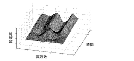
測定結果は、周波数および時間、誘電率を各座標軸とする三次元の複素誘電率スペクトル(図1)、あるいは周波数および時間、誘電率から選択される2つを各座標軸とする二次元の複素誘電率スペクトル(図2)として得ることができる。図中Z軸は、各時間および各周波数における複素誘電率の実数部を示している。 The measurement result is a three-dimensional complex dielectric constant spectrum (FIG. 1) having frequency, time and dielectric constant as coordinate axes, or two-dimensional complex dielectric having two coordinates selected from frequency, time and dielectric constant as coordinate axes. It can be obtained as a rate spectrum (FIG. 2). In the figure, the Z axis indicates the real part of the complex permittivity at each time and at each frequency.
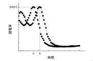
図2は、図1に示す三次元スペクトルを周波数760kHzで切り出した二次元スペクトルに対応する。図2中符号(A)は赤血球の連銭形成に伴うピークであり、(B)は血液凝固過程に伴うピークである。 FIG. 2 corresponds to a two-dimensional spectrum obtained by cutting out the three-dimensional spectrum shown in FIG. 1 at a frequency of 760 kHz. In FIG. 2, symbol (A) is a peak associated with the formation of red blood cells and (B) is a peak associated with the blood coagulation process.
本発明者らは、上記特許文献1において、血液の誘電率の時間的変化が血液の凝固過程を反映することを明らかにしている。従って、本手順で得られる複素誘電率スペクトルは血液の凝固能を定量的に表す指標となるものであり、その変化に基づけば凝固時間、凝固速度、凝固強度などの血液の凝固能に関する情報を得ることが可能である。
In the above-mentioned
なお、本手順に先立って、サンプル血の採取手順が行われる場合がある。採取手順では、血液凝固系の解析対象とする被検者から通常の方法に従って採血を行う。 Note that a sample blood collection procedure may be performed prior to this procedure. In the collection procedure, blood is collected from a subject to be analyzed for the blood coagulation system according to a normal method.
(2)解析手順
解析手順では、測定手順で測定されたサンプル血の複素誘電率スペクトルに基づいて、サンプル血の凝固能に関する情報を取得する。具体的には、まず、血小板活性化剤等を添加したサンプル血で測定された複素誘電率スペクトルと添加していないサンプル血で測定された複素誘電率スペクトルとを比較する。そして、血小板活性化剤等の添加により生じた複素誘電率スペクトルの変化に基づいて、サンプル血の凝固能に関する情報を取得する。
(2) Analysis procedure In the analysis procedure, information on the coagulation ability of the sample blood is acquired based on the complex dielectric constant spectrum of the sample blood measured in the measurement procedure. Specifically, first, the complex permittivity spectrum measured with the sample blood to which the platelet activating agent or the like is added is compared with the complex permittivity spectrum measured with the sample blood not added. And the information regarding the coagulation ability of sample blood is acquired based on the change of the complex dielectric constant spectrum produced by addition of a platelet activator or the like.
血小板活性化剤等を添加していないサンプル血で測定された複素誘電率スペクトルは、複数の要因が関与する血液の凝固能を総合的に反映していると考えられる。すなわち、複素誘電率スペクトルには、血小板の凝固能による血液凝固、血漿や血球成分の凝固作用による血液凝固、測定中に起こり得る血沈の影響による血液凝固が総合的に反映されている。本技術に係る血液凝固系解析方法では、血小板活性化剤等を用いることにより、これらの要因の中から特に血小板の凝固能に関する情報を切り分けて取得することが可能とされている。以下、血小板活性化剤を用いる場合と血小板不活化剤を用いる場合とに分けて、解析手順の具体的な内容を説明する。 It is considered that the complex permittivity spectrum measured in the sample blood to which the platelet activating agent or the like is not added comprehensively reflects the blood coagulation ability involving a plurality of factors. That is, the complex permittivity spectrum comprehensively reflects blood coagulation due to the coagulation ability of platelets, blood coagulation due to the coagulation action of plasma and blood cell components, and blood coagulation due to blood sedimentation that may occur during measurement. In the blood coagulation system analysis method according to the present technology, by using a platelet activator or the like, it is possible to isolate and acquire information on the coagulation ability of platelets from among these factors. Hereinafter, the specific contents of the analysis procedure will be described separately for the case of using a platelet activating agent and the case of using a platelet inactivating agent.
(2−1)血小板活性化剤を用いる場合
血液に血小板活性化剤を添加すると、血小板の活性化によって血液凝固反応が加速し、血液凝固時間が短縮する。このため、血小板活性化剤を添加した血液の複素誘電率スペクトルでは、添加していない血液の複素誘電率スペクトルに比べて、血液凝固に伴うスペクトルピークpが出現するまでの時間(血液凝固時間)tが短縮する(図3参照)。図中、符号p0およびt0は血小板活性化剤を添加しない場合のスペクトルピークと血液凝固時間を、符号p1およびt1は血小板活性化剤を添加した場合のスペクトルピークと血液凝固時間を示す。
(2-1) When using a platelet activating agent When a platelet activating agent is added to blood, the blood clotting reaction is accelerated by the activation of platelets, and the blood clotting time is shortened. For this reason, in the complex permittivity spectrum of blood to which platelet activator is added, the time until the spectrum peak p accompanying blood coagulation appears (blood clotting time) compared to the complex permittivity spectrum of blood not added t is shortened (see FIG. 3). In the figure, symbols p 0 and t 0 indicate the spectrum peak and blood clotting time when the platelet activating agent is not added, and symbols p 1 and t 1 indicate the spectrum peak and blood clotting time when the platelet activating agent is added. Show.
従って、この血液凝固時間tの短縮幅△t(t0−t1)に基づけば、サンプル血中に不活性な状態で含まれていた血小板の凝固能の程度についての情報を得ることができる。すなわち、サンプル血中に含まれる不活性な血小板の凝固能が高い場合、血小板活性化剤により血小板を活性化したサンプル血では血液凝固反応が大幅に加速し、血液凝固時間も大きく短縮する。一方、サンプル血中に含まれる不活性な血小板の凝固能が低い場合、血小板活性化剤により血小板を活性化しても血液凝固反応の反応速度がほとんど変化しないため、血液凝固時間の短縮もみられない。 Therefore, based on the shortening width Δt (t 0 -t 1 ) of the blood coagulation time t, information on the degree of the coagulation ability of platelets contained in the sample blood in an inactive state can be obtained. . That is, when the coagulation ability of inactive platelets contained in the sample blood is high, the blood coagulation reaction is greatly accelerated and the blood coagulation time is greatly shortened in the sample blood in which platelets are activated by the platelet activator. On the other hand, if the clotting ability of inactive platelets contained in the sample blood is low, the blood coagulation reaction rate is hardly changed even when platelets are activated by a platelet activator, so the blood clotting time is not shortened. .
正常な凝固能を有することが予め分かっている血液を用いて、基準となる血液凝固時間の短縮幅△ts(基準値)を設定しておけば、サンプル血の血液凝固時間の短縮幅△tが△ts(基準値)よりも大きいか小さいかによって血小板の凝固能の良否を判定できる。すなわち、サンプル血中の血液凝固時間の短縮幅△tが△ts(基準値)よりも大きい場合には血小板の凝固能は高いと評価でき、逆に小さい場合には血小板の凝固能が低いと評価できる。 If blood that is known to have normal clotting ability is used in advance and a reference blood coagulation time reduction width Δt s (reference value) is set, the blood coagulation time reduction width of sample blood Δ The quality of platelet coagulation ability can be determined based on whether t is larger or smaller than Δt s (reference value). That is, when the shortening width Δt of the blood coagulation time in the sample blood is larger than Δt s (reference value), it can be evaluated that the platelet coagulation ability is high, and conversely, when it is small, the platelet coagulation ability is low. Can be evaluated.
血小板機能異常症や血小板減少症の患者や、アセチルサリチル酸などの抗血小板凝集薬やワルファリンやヘパリン、活性化血液凝固第X因子(Factor Xa)阻害剤などの抗凝固薬の服用者では血小板の凝固能(止血能力)が低下し出血リスクが高まる。このため、これらの患者等では血小板の凝固能を適時に評価しながら疾患管理や投薬管理を行う必要がある。 Platelet clotting in patients with platelet dysfunction or thrombocytopenia, or those who take anti-platelet agglutinating drugs such as acetylsalicylic acid, anti-coagulants such as warfarin or heparin, or activated blood coagulation factor Xa (Factor Xa) inhibitors Ability (hemostatic ability) decreases and bleeding risk increases. For this reason, it is necessary for these patients and the like to perform disease management and medication management while evaluating platelet coagulation ability in a timely manner.
本技術に係る血液凝固系解析方法では、上述のように血小板活性化剤の添加による複素誘電率スペクトルの変化(具体的には、血液凝固時間の短縮幅△t)に基づき、血小板の凝固能を簡便に評価できる。このため、本技術に係る血液凝固系解析方法は、血小板機能異常症や血小板減少症の患者の血小板機能の評価のために有用である。また、本技術に係る血液凝固系解析方法を用いて、抗血小板凝集薬あるいは抗凝固薬の服用者の血小板機能を評価すれば、過剰投薬による凝固能の過度な減少や、投薬量不足による凝固能の亢進状態の持続などを把握するなどの薬効評価も可能となる。 In the blood coagulation system analysis method according to the present technology, as described above, the platelet coagulation ability is based on the change in the complex permittivity spectrum due to the addition of the platelet activator (specifically, the reduction width Δt of the blood coagulation time). Can be easily evaluated. For this reason, the blood coagulation system analysis method according to the present technology is useful for evaluating the platelet function of patients with platelet dysfunction or thrombocytopenia. In addition, if the platelet function of an antiplatelet agglutinant or anticoagulant is evaluated using the blood coagulation system analysis method according to the present technology, the coagulation ability may be excessively decreased due to overdose or coagulation due to insufficient dosage. It is also possible to evaluate the efficacy of the drug, such as grasping the sustained state of enhanced performance.
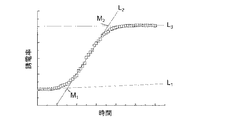
ここでは、複素誘電率スペクトルの変化として、血液凝固に伴うスペクトルピークpが出現するまでの時間(血液凝固時間)tの変化を解析のための指標に用い、血小板活性化剤の添加による血液凝固時間tの短縮幅△tに基づいて血小板の凝固能を評価する方法を説明した。本技術に係る血液凝固系解析方法において、解析のための指標となる複素誘電率スペクトルの変化は、複素誘電率スペクトルから抽出される特徴量の変化であれば特に限定されない。具体的な特徴量としては例えば以下が挙げられる。これらの特徴量の変化幅について予め設定した基準値と、血小板活性化剤の添加によりサンプル血の複素誘電率スペクトルから抽出される特徴量に生じる変化幅によって血小板の凝固能の良否を判定できる。 Here, as a change in the complex permittivity spectrum, a change in time (blood clotting time) t until the appearance of a spectrum peak p accompanying blood clotting is used as an index for analysis, and blood clotting by adding a platelet activating agent is performed. A method for evaluating the clotting ability of platelets based on the shortening width Δt of time t has been described. In the blood coagulation system analysis method according to the present technology, the change in the complex permittivity spectrum serving as an index for analysis is not particularly limited as long as it is a change in the feature amount extracted from the complex permittivity spectrum. Specific examples of the feature amount include the following. The quality of platelet coagulation ability can be determined based on a reference value set in advance for the change width of these feature values and the change width generated in the feature values extracted from the complex permittivity spectrum of the sample blood by adding the platelet activator.
複素誘電率スペクトルを示す曲線に対して引いた外挿線(図4および図5、符号L1〜L4)、外挿線の交点(符号M1〜M4)の座標、外挿線の傾き、複素誘電率スペクトルを示す曲線に対して引いた接線の傾き(誘電率の微分値)、所定の誘電率E(例えば最大値、局大値、中間値など)を与える時間T、これらの組み合わせ。三次元の複素誘電率スペクトルを画像パターンとして解析して得た特徴量。前記画像パターンを再構成可能な関数式を用いたパラメータフィッティングにより得た特徴量。スペクトルデータ中の多数のデータを用いたクラスター解析により得た特徴量。 Extrapolation lines (FIGS. 4 and 5, reference symbols L 1 to L 4 ) drawn with respect to the curve indicating the complex dielectric constant spectrum, coordinates of the extrapolation line intersections (reference symbols M 1 to M 4 ), and extrapolation lines Inclination, slope of tangent line drawn with respect to curve showing complex dielectric constant spectrum (differential value of dielectric constant), time T giving predetermined dielectric constant E (for example, maximum value, local value, intermediate value, etc.), these combination. Features obtained by analyzing a three-dimensional complex permittivity spectrum as an image pattern. A feature amount obtained by parameter fitting using a functional expression capable of reconstructing the image pattern. Features obtained by cluster analysis using multiple data in the spectrum data.
(2−2)血小板不活化剤を用いる場合
血液に血小板不活化剤を添加すると、血液中に含まれる活性化した血小板による血液凝固作用が抑制され、凝固反応速度が低下し、血液凝固時間が延長する。このため、血小板不活化剤を添加した血液の複素誘電率スペクトルでは、添加していない血液の複素誘電率スペクトルに比べて、血液凝固に伴うスペクトルピークpが出現するまでの時間(血液凝固時間)tが遅延する(図6参照)。図中、符号p0およびt0は血小板不活化剤を添加しない場合のスペクトルピークと血液凝固時間を、符号p2およびt2は血小板不活化剤を添加した場合のスペクトルピークと血液凝固時間を示す。
(2-2) In the case of using a platelet inactivating agent When a platelet inactivating agent is added to blood, the blood coagulation action by activated platelets contained in the blood is suppressed, the coagulation reaction rate is reduced, and the blood coagulation time is reduced. Extend. For this reason, in the complex permittivity spectrum of blood to which a platelet inactivating agent is added, the time until the appearance of a spectrum peak p accompanying blood coagulation is compared to the complex permittivity spectrum of blood to which blood is not added (blood clotting time). t is delayed (see FIG. 6). In the figure, the symbols p 0 and t 0 indicate the spectrum peak and blood clotting time when the platelet inactivating agent is not added, and the symbols p 2 and t 2 indicate the spectrum peak and blood clotting time when the platelet inactivating agent is added. Show.
従って、この血液凝固時間tの遅延幅△t(t2−t0)に基づけば、サンプル血中に活性な状態で含まれていた血小板の凝固能の程度についての情報を得ることができる。すなわち、血小板不活化剤の添加により血液凝固時間が大きく遅延した場合、サンプル血中の血小板の凝固能が高く、サンプル血中に多量の活性化した血小板が含まれているといえる。一方、血小板不活化剤を添加しても血液凝固時間がほとんど変化しない場合、サンプル血中の血小板の凝固能が低く、サンプル血中に活性化した血小板がほとんど含まれていないといえる。 Therefore, based on the delay width Δt (t 2 −t 0 ) of the blood coagulation time t, information about the degree of platelet coagulation ability contained in the sample blood in an active state can be obtained. That is, when the blood clotting time is greatly delayed by the addition of the platelet inactivating agent, it can be said that the platelet blood in the sample blood has a high clotting ability and the sample blood contains a large amount of activated platelets. On the other hand, if the blood coagulation time hardly changes even when the platelet inactivating agent is added, it can be said that the platelet coagulation ability in the sample blood is low and the activated blood is hardly contained in the sample blood.
正常な凝固能を有することが予め分かっている血液を用いて、基準となる血液凝固時間の遅延幅△ts(基準値)を設定しておけば、サンプル血の血液凝固時間の遅延幅△tが△ts(基準値)よりも大きいか小さいかによって血小板の凝固能の良否を判定できる。すなわち、サンプル血中の血液凝固時間の遅延幅△tが△ts(基準値)よりも大きい場合には血小板の凝固能は高いと評価でき、逆に小さい場合には血小板の凝固能が低いと評価できる。 If blood that is known in advance to have normal clotting ability is used and a reference blood coagulation time delay width Δt s (reference value) is set, the blood coagulation time delay width Δ of the sample blood The quality of platelet coagulation ability can be determined based on whether t is larger or smaller than Δt s (reference value). That is, if the delay time Δt of the blood coagulation time in the sample blood is larger than Δt s (reference value), it can be evaluated that the platelet coagulation ability is high, and conversely, if it is small, the platelet coagulation ability is low. Can be evaluated.
本技術に係る血液凝固系解析方法では、このように血小板不活化剤の添加による複素誘電率スペクトルの変化(具体的には、血液凝固時間の遅延幅△t)に基づき、血小板の凝固能を簡便に評価できる。このため、本技術に係る血液凝固系解析方法は、血栓症のリスクを有する糖尿病などの患者や妊婦などの健常者の血小板機能を評価して、本来活性化されるべきでない血小板がどの程度活性化されているのかを調べるために有用である。 In the blood coagulation system analysis method according to the present technology, the coagulation ability of platelets is determined based on the change in the complex dielectric constant spectrum (specifically, the delay time Δt of the blood coagulation time) due to the addition of the platelet inactivating agent. It can be easily evaluated. For this reason, the blood coagulation system analysis method according to the present technology evaluates the platelet function of a healthy subject such as a diabetic patient or a pregnant woman who has a risk of thrombosis, and the degree of activity of platelets that should not be activated originally. It is useful for investigating whether or not
なお、本技術に係る血液凝固系解析方法において、解析のための指標となる複素誘電率スペクトルの変化は、複素誘電率スペクトルから抽出される特徴量の変化であればよく、血液凝固時間の遅延幅に限定されない点は上述の通りである。 In the blood coagulation system analysis method according to the present technology, the change in the complex dielectric constant spectrum serving as an index for analysis may be a change in the feature amount extracted from the complex dielectric constant spectrum, and the delay in blood coagulation time. The points not limited to the width are as described above.
2.血液凝固系解析装置
(1)装置の全体構成
図7に、本技術に係る血液凝固系解析装置の概略構成を示す。
2. Blood Coagulation System Analyzer (1) Overall Configuration FIG. 7 shows a schematic configuration of a blood coagulation system analyzer according to the present technology.
血液凝固系解析装置は、血液を保持するサンプルカートリッジ2と、サンプルカートリッジ2に保持された血液に電圧を印加する一対の電極11,12と、電極11,12に交流電圧を印加する電源3と、血液の誘電率を測定する測定部41と、を備える。測定部41は、測定部41からの測定結果の出力を受けて血液の凝固能を判定する解析部42とともに、信号処理部4を構成している。
The blood coagulation system analyzing apparatus includes a sample cartridge 2 that holds blood, a pair of
サンプルカートリッジ2には、保持された血液に血小板活性化剤等を添加するための薬剤導入口を設けてもよい。血液は、予め血小板活性化剤等と混合した後にサンプルカートリッジ2に収容してもよい。 The sample cartridge 2 may be provided with a drug introduction port for adding a platelet activator or the like to the retained blood. The blood may be stored in the sample cartridge 2 after previously mixed with a platelet activating agent or the like.
電源3は、測定を開始すべき命令を受けた時点または電源が投入された時点を開始時点として電圧を印加する。具体的には、電源3は、設定される測定間隔ごとに、電極11,12に対して所定の周波数の交流電圧を印加する。
The
測定部41は、測定を開始すべき命令を受けた時点または電源が投入された時点を開始時点として複素誘電率およびその周波数分散などを測定する。具体的には、例えば誘電率が測定される場合、測定部41は、電極11,12間における電流またはインピーダンスを所定周期で測定し、測定値から誘電率を導出する。誘電率の導出には、電流またはインピーダンスと誘電率との関係を示す既知の関数や関係式が用いられる。
The
解析部42には、測定部41から導出された誘電率を示すデータ(以下、「誘電率データ」とも称する)が測定間隔ごとに与えられる。解析部42は、測定部41から与えられる誘電率データを受けて血液の凝固能判定等を開始する。解析部42は、凝固能判定等の結果および誘電率データの一方または双方を通知する。この通知は、例えば、グラフ化してモニタに表示あるいは所定の媒体に印刷することにより行われる。
Data indicating the dielectric constant derived from the measurement unit 41 (hereinafter also referred to as “dielectric constant data”) is given to the
(2)解析部
次に、解析部42による血液の凝固能の判定ステップの具体例を説明する。
(2) Analysis Unit Next, a specific example of the determination step of blood coagulation ability by the
解析部42は、測定部41から出力される誘電率データに基づいて、血小板活性化剤等を添加したサンプル血で測定された複素誘電率スペクトルと添加していないサンプル血で測定された複素誘電率スペクトルとの比較処理を行い、スペクトルパターンの相違に基づいてサンプル血の凝固能を判定する。
Based on the dielectric constant data output from the
まず、解析部42は、血小板活性化剤等を添加したサンプル血で測定された複素誘電率スペクトルと添加していないサンプル血で測定された複素誘電率スペクトルのスペクトルパターンを比較する。スペクトルパターンの比較は比較対象とする複素誘電率スペクトルから抽出した特徴量に基づいて行うことができ、この特徴量の違いからスペクトルパターンの相違を検出できる。特徴量には、例えば、血液凝固に伴うスペクトルピークが出現するまでの時間(血液凝固時間)が用いられる。
First, the
この場合、解析部42は、血小板活性化剤等を添加したサンプル血の血液凝固時間の短縮幅(あるいは遅延幅)△tが基準値(△ts)よりも大きいか小さいかによって血小板の凝固能の良否を判定する。すなわち、血小板活性化剤を添加したサンプル血中の血液凝固時間の短縮幅△tが△ts(基準値)よりも大きい場合には血小板の凝固能は高いと判定し、逆に小さい場合には血小板の凝固能が低いと判定する。また、あるいは、血小板不活化剤を添加したサンプル血中の血液凝固時間の遅延幅△tが△ts(基準値)よりも大きい場合には血小板の凝固能は高いと判定し、逆に小さい場合には血小板の凝固能が低いと判定する。
In this case, the
[凝固能の亢進]
解析部42は、血小板活性化剤等を添加していないサンプル血の血液凝固時間tが基準値(ts)よりも短い場合、血液凝固能が高いと判定し、結果を通知する。この結果は、血栓症のリスクとしてモニタや所定の媒体に表示され得る。
[Enhanced coagulation ability]
When the blood coagulation time t of the sample blood to which no platelet activating agent or the like is added is shorter than the reference value (t s ), the
この場合、血小板活性化剤を添加したサンプル血中の血液凝固時間の短縮幅△tが△ts(基準値)よりも大きいときには、解析部42は、血小板の凝固能が高いと判定し、血液凝固能の亢進が血小板の凝固能の異常な上昇によるものであると判定する。この判定結果は、血小板機能の異常による血栓症リスクとしてモニタや所定の媒体に表示され得る。
In this case, when the shortening width Δt of the blood coagulation time in the sample blood to which the platelet activating agent is added is larger than Δt s (reference value), the
他方、血小板活性化剤を添加したサンプル血中の血液凝固時間の短縮幅△tが△ts(基準値)よりも小さいときには、解析部42は、血小板の凝固能は低いと判定し、血液凝固能の亢進が血小板機能とは無関係に生じているものであると判定する。この判定結果は、血小板機能以外の異常による血栓症リスクとしてモニタや所定の媒体に表示され得る。
On the other hand, when the shortening width Δt of the blood coagulation time in the sample blood to which the platelet activator is added is smaller than Δt s (reference value), the
[凝固能の低下]
一方、解析部42は、血小板活性化剤等を添加していないサンプル血の血液凝固時間tが基準値(ts)よりも長い場合、解析部42は、血液凝固能が低いと判定し、結果を通知する。この結果は、出血傾向のリスクとしてモニタや所定の媒体に表示され得る。
[Decrease in coagulation ability]
On the other hand, the
この場合、血小板活性化剤を添加したサンプル血中の血液凝固時間の短縮幅△tが△ts(基準値)よりも小さいときには、解析部42は、血小板の凝固能が低いと判定し、血液凝固能の低下が血小板の凝固能の異常な低下によるものであると判定する。この判定結果は、血小板機能の異常による出血傾向リスクとしてモニタや所定の媒体に表示され得る。
In this case, when the shortening width Δt of the blood coagulation time in the sample blood to which the platelet activating agent is added is smaller than Δt s (reference value), the analyzing
他方、血小板活性化剤を添加したサンプル血中の血液凝固時間の短縮幅△tが△ts(基準値)よりも大きいときには、解析部42は、血小板の凝固能は高いと判定し、血液凝固能の低下が血小板機能とは無関係に生じているものであると判定する。この判定結果は、血小板機能以外の異常による出血傾向リスクとしてモニタや所定の媒体に表示され得る。
On the other hand, when the shortening width Δt of the blood coagulation time in the sample blood to which the platelet activating agent is added is larger than Δt s (reference value), the
ここでは、血小板活性化剤等を添加したサンプル血で測定された複素誘電率スペクトルと添加していないサンプル血で測定された複素誘電率スペクトルのスペクトルパターンの比較を血液凝固に伴うスペクトルピークが出現するまでの時間(血液凝固時間)基づいて行う例を説明した。スペクトルパターンの比較のための特徴量は、複素誘電率スペクトルから抽出される特徴量の変化であれば特に限定されない。具体的な特徴量としては例えば以下が挙げられる。解析部42は、これらの特徴量の変化幅について予め保持された基準値と、血小板活性化剤の添加によりサンプル血の複素誘電率スペクトルから抽出される特徴量に生じる変化幅とによってサンプル血の凝固能を判定する。
Here, the spectrum peak of the blood coagulation appears by comparing the spectrum pattern of the complex dielectric constant spectrum measured in the sample blood with added platelet activator, etc. with the complex dielectric constant spectrum measured in the sample blood not added. The example performed based on the time until blood (blood clotting time) has been described. The feature amount for comparing the spectral patterns is not particularly limited as long as the feature amount is extracted from the complex permittivity spectrum. Specific examples of the feature amount include the following. The
複素誘電率スペクトルを示す曲線に対して引いた外挿線(図4および図5、符号L1〜L4)、外挿線の交点(符号M1〜M4)の座標、外挿線の傾き、複素誘電率スペクトルを示す曲線に対して引いた接線の傾き(誘電率の微分値)、所定の誘電率E(例えば最大値、局大値、中間値など)を与える時間T、これらの組み合わせ。三次元の複素誘電率スペクトルを画像パターンとして解析して得た特徴量。前記画像パターンを再構成可能な関数式を用いたパラメータフィッティングにより得た特徴量。スペクトルデータ中の多数のデータを用いたクラスター解析により得た特徴量。 Extrapolation lines (FIGS. 4 and 5, reference symbols L 1 to L 4 ) drawn with respect to the curve indicating the complex dielectric constant spectrum, coordinates of the extrapolation line intersections (reference symbols M 1 to M 4 ), and extrapolation lines Inclination, slope of tangent line drawn with respect to curve showing complex dielectric constant spectrum (differential value of dielectric constant), time T giving predetermined dielectric constant E (for example, maximum value, local value, intermediate value, etc.), these combination. Features obtained by analyzing a three-dimensional complex permittivity spectrum as an image pattern. A feature amount obtained by parameter fitting using a functional expression capable of reconstructing the image pattern. Features obtained by cluster analysis using multiple data in the spectrum data.
以上のように、本技術に係る血液凝固系解析装置は、血液凝固能の亢進および低下の有無を判定し、それらが血小板機能の異常によるものであるかどうかをも判定する。従って、本技術に係る血液凝固系解析装置によれば、血栓症のリスクが示唆された場合に、抗血小板療法を行うべきなのか、抗凝固療法を行うべきなのかを判断するための重要な情報が得られる。 As described above, the blood coagulation system analyzer according to the present technology determines whether blood coagulation ability is increased or decreased, and also determines whether or not they are due to abnormal platelet function. Therefore, according to the blood coagulation system analysis apparatus according to the present technology, when the risk of thrombosis is suggested, it is important to determine whether antiplatelet therapy or anticoagulation therapy should be performed. Information is obtained.
<試験例1>
1.血小板活性化剤を用いた血液凝固系解析方法
健常者および血小板減少症の患者のサンプル血を用いて、血小板活性化剤の添加が及ぼす血液の凝固能への影響を検討した。
<Test Example 1>
1. Blood coagulation system analysis method using platelet activator Using blood samples of healthy subjects and patients with thrombocytopenia, the effect of the addition of platelet activator on blood coagulation ability was examined.
(1)材料と方法
(1−1)採血およびサンプル調製
クエン酸ナトリウムを抗凝固剤として処理した真空採血管を用いて、健常者および血小板減少症の患者(血液1μL中の血小板数1.2×10⁴個)から採血を行い、全血サンプルとした。
また、全血サンプルから血球および血小板を分離し、血小板除去血漿を調製した。サンプル血を500G、5分の条件で遠心分離し、血球と多血小板血漿(PRP)とに分離した。血漿を2000G、30分の条件で遠心分離し、血小板を沈降させて、血小板除去血漿(PPP)を得た。
(1) Material and method (1-1) Blood collection and sample preparation Using a vacuum blood collection tube treated with sodium citrate as an anticoagulant, healthy subjects and patients with thrombocytopenia (1.2 platelet count in 1 μL of blood) Blood was collected from 4 × 10) to obtain a whole blood sample.
In addition, blood cells and platelets were separated from the whole blood sample to prepare platelet-free plasma. Sample blood was centrifuged at 500 G for 5 minutes to separate blood cells and platelet rich plasma (PRP). Plasma was centrifuged at 2000 G for 30 minutes, and platelets were sedimented to obtain platelet-depleted plasma (PPP).
(1−2)誘電測定
37℃に保温したサンプル血に0.25M塩化カルシウム水溶液を添加(血液1mLあたり85μL)し、血液凝固反応を開始させた。血小板活性化剤(ADP)は、塩化カルシウム水溶液に混合して添加した(血液1mLあたり10μM)。血液凝固反応開始直後、インピーダンスアナライザー(アジレント社、4294A)を用いて、温度37℃、周波数域40Hz〜110MHz、測定時間間隔1分の条件で60分間測定を行った。
(1-2) Dielectric measurement A 0.25M calcium chloride aqueous solution was added to sample blood kept at 37 ° C. (85 μL per mL of blood) to initiate a blood coagulation reaction. Platelet activating agent (ADP) was mixed with calcium chloride aqueous solution and added (10 μM per mL of blood). Immediately after the start of the blood coagulation reaction, measurement was performed for 60 minutes using an impedance analyzer (Agilent, 4294A) under conditions of a temperature of 37 ° C., a frequency range of 40 Hz to 110 MHz, and a measurement time interval of 1 minute.
(1−3)レオメーターによる測定
自由減衰振動型のレオメーターを用いて測定した。粘弾性の変化を観測することで凝固時間を求め、誘電測定による結果と比較した。
(1-3) Measurement by rheometer The measurement was performed using a free-damping vibration type rheometer. The coagulation time was determined by observing the change in viscoelasticity and compared with the results from dielectric measurements.
(2)結果
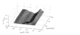
図8にADPを添加していない健常者のサンプル血の誘電応答結果を、図9にADPを添加した健常者のサンプル血の誘電応答結果を示す。図中Z軸は、各時刻および各周波数における複素誘電率の実数部を、時刻ゼロ(測定開始直後)での各周波数における複素誘電率の実数部で除算し、規格化して表示している。図8と図9を比較すると、ADPの添加によって誘電応答の三次元パターンが明らかに変化しているのが分かる。
(2) Results FIG. 8 shows the dielectric response results of the sample blood of a healthy person to which ADP was not added, and FIG. 9 shows the dielectric response results of the sample blood of a healthy person to which ADP was added. In the figure, the Z-axis is normalized by dividing the real part of the complex dielectric constant at each time and each frequency by the real part of the complex dielectric constant at each time at time zero (immediately after the start of measurement). Comparing FIG. 8 and FIG. 9, it can be seen that the three-dimensional pattern of the dielectric response is clearly changed by the addition of ADP.
図10に、周波数10.7MHzにおける複素誘電率の時間変化を示す。符号(−)はADPを添加していないサンプル血の誘電応答、符号(+)はADPを添加したサンプル血の誘電応答を示す。ADPを添加した場合、血液凝固に対応する誘電率のステップ状変化は、ADPを添加しない場合に比較して、短時間側にシフトしていることが分かる。このようなADP添加による凝固時間の短縮は、複数の健常者のサンプル血で共通して観測された。ADP添加時の凝固時間の平均値は17分、非添加時の凝固時間の平均値は31分であった。 FIG. 10 shows the time change of the complex dielectric constant at a frequency of 10.7 MHz. The sign (−) indicates the dielectric response of the sample blood not added with ADP, and the sign (+) indicates the dielectric response of the sample blood added with ADP. It can be seen that when ADP is added, the stepwise change in dielectric constant corresponding to blood coagulation is shifted to a shorter time than when ADP is not added. Such shortening of the coagulation time due to the addition of ADP was commonly observed in the sample blood of a plurality of healthy subjects. The average value of the coagulation time when ADP was added was 17 minutes, and the average value of the coagulation time when no ADP was added was 31 minutes.
図11に、血小板減少症患者のサンプル血の周波数10.7MHzにおける複素誘電率の時間変化を示す。符号(−)はADPを添加していないサンプル血の誘電応答、符号(+)はADPを添加したサンプル血の誘電応答を示す。ADP添加によって凝固時間の短縮がみられるものの、凝固時間は38分であり、健常者の凝固時間の平均値17分と比べて明らかに長いことが分かった。これはサンプル血中の血小板数が少ないために、ADPによる血液凝固反応の促進が限定的であったためと考えられる。 In FIG. 11, the time change of the complex dielectric constant in the frequency of 10.7 MHz of the sample blood of a thrombocytopenia patient is shown. The sign (−) indicates the dielectric response of the sample blood not added with ADP, and the sign (+) indicates the dielectric response of the sample blood added with ADP. Although the coagulation time was shortened by the addition of ADP, the coagulation time was 38 minutes, which was clearly longer than the average value of 17 minutes for healthy individuals. This is presumably because the promotion of blood coagulation reaction by ADP was limited because the number of platelets in the sample blood was small.
レオメーターによる補足実験の結果を図12および図13に示す。図12は、ADP添加(+)および非添加(−)の健常者のサンプル血の粘弾性の時間変化を示す。また、図13は、血小板除去血漿(PPP)に分離後洗浄した赤血球を添加した血小板除去血にADPを添加した場合(+)と添加しない場合(−)の粘弾性の時間変化を示している。健常者のサンプル血を用いた実験では、誘電測定による結果と同様に、ADP添加による凝固時間の短縮がみられた。一方、血小板除去血を用いた実験では、ADP添加による凝固時間の変化は観察されなかった。 The results of a supplementary experiment with a rheometer are shown in FIGS. FIG. 12 shows changes over time in viscoelasticity of sample blood of healthy subjects with ADP added (+) and without added (−). Moreover, FIG. 13 shows the time change of viscoelasticity when ADP is added to platelet-removed blood to which platelets removed after washing into platelet-removed plasma (PPP) are added (+) and not added (−). . In experiments using sample blood of healthy subjects, shortening of the coagulation time due to the addition of ADP was observed, similar to the result of dielectric measurement. On the other hand, in the experiment using platelet-removed blood, no change in coagulation time due to the addition of ADP was observed.
以上の本試験例の結果から、血小板活性化剤の添加により生じる複素誘電率スペクトルの変化に基づけば、サンプル血中の血小板の凝固能を簡便に評価できることが明らかとなった。 From the results of the present test example described above, it became clear that the platelet coagulation ability in the sample blood can be easily evaluated based on the change in the complex dielectric constant spectrum caused by the addition of the platelet activator.
本技術に係る血液凝固系解析方法によれば、血液凝固能の亢進および低下の有無を判定し、それらが血小板の凝固能に関連するものであるかどうかを判定できる。糖尿病、動脈硬化症、癌、心疾患、呼吸器疾患などの患者や周術期の患者、免疫抑制剤を服用中の患者、妊婦や高齢者などの血栓リスクを評価するために有用である。また、これらの患者や健常者に抗血小板凝集薬または抗凝固薬を予防的に投薬する際、過剰投与による出血傾向リスクを評価するためにも役立てられ得る。 According to the blood coagulation system analysis method according to the present technology, it is possible to determine whether blood coagulation ability is increased or decreased, and to determine whether they are related to platelet coagulation ability. It is useful for assessing the risk of blood clots in patients with diabetes, arteriosclerosis, cancer, heart disease, respiratory disease, patients in the perioperative period, patients taking immunosuppressants, pregnant women and the elderly. In addition, when prophylactic administration of antiplatelet agglutinating drugs or anticoagulants to these patients and healthy individuals, it can also be used to evaluate the risk of bleeding tendency due to overdose.
11,12:電極、2:サンプルカートリッジ、3:電源、4:信号処理部、41:測定部、42:解析部
DESCRIPTION OF
Claims (8)
該物質による血小板活性化に伴って前記複素誘電率に生じる変化に基づき、前記血液中に不活性な状態で含まれる血小板の凝固能に関する情報を取得する請求項1記載の血液凝固系解析方法。 The substance activates platelets;
The blood coagulation system analysis method according to claim 1, wherein information on the coagulation ability of platelets contained in the blood in an inactive state is acquired based on a change that occurs in the complex dielectric constant due to platelet activation by the substance.
前記変化に基づき、抗血小板凝集薬および/または抗凝固薬の薬効を評価する請求項2記載の血液凝固系解析方法。 The blood is collected from a subject who has been administered an antiplatelet aggregating agent and / or an anticoagulant;
The blood coagulation system analysis method according to claim 2, wherein the efficacy of the antiplatelet agglutinating drug and / or anticoagulant is evaluated based on the change.
該物質による血小板不活化に伴って前記複素誘電率に生じる変化に基づき、前記血液中に活性な状態で含まれる血小板の凝固能に関する情報を取得する請求項1記載の血液凝固系解析方法。 The substance inactivates platelets;
The blood coagulation system analysis method according to claim 1, wherein information on the coagulation ability of platelets contained in the blood in an active state is acquired based on a change that occurs in the complex dielectric constant due to platelet inactivation by the substance.
Priority Applications (6)
| Application Number | Priority Date | Filing Date | Title |
|---|---|---|---|
| JP2011058810A JP5768422B2 (en) | 2011-03-17 | 2011-03-17 | Blood coagulation system analysis method and blood coagulation system analyzer |
| EP17209032.6A EP3321680B1 (en) | 2011-03-17 | 2012-02-16 | Blood coagulation system analyzing method and blood coagulation system analyzing device |
| EP12001027.7A EP2500726B1 (en) | 2011-03-17 | 2012-02-16 | Blood coagulation system analyzing method and blood coagulation system analyzing device |
| US13/403,652 US8735163B2 (en) | 2011-03-17 | 2012-02-23 | Blood coagulation system analyzing method and blood coagulation system analyzing device |
| CN201210065695.2A CN102680523B (en) | 2011-03-17 | 2012-03-09 | Blood coagulation system analysis method and blood coagulation system analytical equipment |
| US14/201,051 US8999243B2 (en) | 2011-03-17 | 2014-03-07 | Blood coagulation system analyzing method and blood coagulation system analyzing device |
Applications Claiming Priority (1)
| Application Number | Priority Date | Filing Date | Title |
|---|---|---|---|
| JP2011058810A JP5768422B2 (en) | 2011-03-17 | 2011-03-17 | Blood coagulation system analysis method and blood coagulation system analyzer |
Publications (3)
| Publication Number | Publication Date |
|---|---|
| JP2012194087A JP2012194087A (en) | 2012-10-11 |
| JP2012194087A5 JP2012194087A5 (en) | 2014-04-10 |
| JP5768422B2 true JP5768422B2 (en) | 2015-08-26 |
Family
ID=45655063
Family Applications (1)
| Application Number | Title | Priority Date | Filing Date |
|---|---|---|---|
| JP2011058810A Active JP5768422B2 (en) | 2011-03-17 | 2011-03-17 | Blood coagulation system analysis method and blood coagulation system analyzer |
Country Status (4)
| Country | Link |
|---|---|
| US (2) | US8735163B2 (en) |
| EP (2) | EP2500726B1 (en) |
| JP (1) | JP5768422B2 (en) |
| CN (1) | CN102680523B (en) |
Cited By (3)
| Publication number | Priority date | Publication date | Assignee | Title |
|---|---|---|---|---|
| WO2017130528A1 (en) | 2016-01-29 | 2017-08-03 | ソニー株式会社 | Blood coagulation system analysis device, blood coagulation system analysis system, blood coagulation system analysis method, and method for determining parameter for blood coagulation system analysis device |
| WO2017141508A1 (en) | 2016-02-17 | 2017-08-24 | ソニー株式会社 | Platelet aggregation activity analysis device, platelet aggregation activity analysis system, platelet aggregation activity analysis program, and platelet aggregation activity analysis method |
| WO2018066207A1 (en) | 2016-10-05 | 2018-04-12 | ソニー株式会社 | Platelet aggregation ability analyzing method, platelet aggregation ability analyzing device, program for platelet aggregation ability analysis, and platelet aggregation ability analyzing system |
Families Citing this family (29)
| Publication number | Priority date | Publication date | Assignee | Title |
|---|---|---|---|---|
| JP5768422B2 (en) * | 2011-03-17 | 2015-08-26 | ソニー株式会社 | Blood coagulation system analysis method and blood coagulation system analyzer |
| JP5982976B2 (en) * | 2012-04-13 | 2016-08-31 | ソニー株式会社 | Blood coagulation system analyzer, blood coagulation system analysis method and program thereof |
| JPWO2014112227A1 (en) | 2013-01-18 | 2017-01-19 | ソニー株式会社 | Electrical characteristic measuring device |
| EP2950087B1 (en) * | 2013-01-28 | 2020-11-04 | Sony Corporation | Impedance measuring device for biological samples and impedance measuring system for biological samples |
| US10527605B2 (en) | 2013-03-13 | 2020-01-07 | Sony Corporation | Blood condition analyzing device, blood condition analyzing system, and blood condition analyzing program |
| US10648936B2 (en) | 2013-03-15 | 2020-05-12 | Sony Corporation | Blood condition analyzing device, blood condition analyzing system, blood conditon analyzing method, and blood condition analyzing program for causing computer to execute the method |
| WO2014156371A1 (en) | 2013-03-29 | 2014-10-02 | ソニー株式会社 | Blood state analysis device, blood state analysis system, blood state analysis method, and program |
| KR20150133716A (en) * | 2013-03-29 | 2015-11-30 | 소니 주식회사 | Blood state evaluation device, blood state evaluation system, blood state evaluation method, and program |
| JP6569209B2 (en) | 2014-01-07 | 2019-09-04 | ソニー株式会社 | Electrical measurement cartridge, electrical measurement apparatus, and electrical measurement method |
| US20170030891A1 (en) * | 2014-04-17 | 2017-02-02 | Sony Corporation | Blood condition analysis device, blood condition analysis system, blood condition analysis method, and program |
| US9995701B2 (en) | 2014-06-02 | 2018-06-12 | Case Western Reserve University | Capacitive sensing apparatuses, systems and methods of making same |
| JP2016024161A (en) * | 2014-07-24 | 2016-02-08 | ソニー株式会社 | Electrical measurement cartridge, electrical measurement device for biological sample, electrical measurement system for biological sample, and electrical measurement method for biological sample |
| JP2016061731A (en) | 2014-09-19 | 2016-04-25 | ソニー株式会社 | Electrical characteristic measuring device and program |
| US20180052181A1 (en) * | 2015-03-13 | 2018-02-22 | Sony Corporation | Sample for use in electrical characteristic measurement, electrical characteristic measurement method, and electrical characteristic measurement device |
| JP6563681B2 (en) * | 2015-05-08 | 2019-08-21 | シスメックス株式会社 | Sample analyzer, blood coagulation analyzer, sample analysis method, and computer program |
| US11175252B2 (en) | 2016-01-15 | 2021-11-16 | Case Western Reserve University | Dielectric sensing for blood characterization |
| AU2017207027B2 (en) * | 2016-01-15 | 2021-11-18 | Case Western Reserve University | Dielectric sensing for sample characterization |
| US20190041379A1 (en) * | 2016-02-10 | 2019-02-07 | Sony Corporation | Sample for measurement of electric characteristics, electric characteristic measuring apparatus, and electric characteristic measuring method |
| US10962495B2 (en) | 2016-03-29 | 2021-03-30 | Sony Corporation | Blood coagulation system analysis system, blood coagulation system analysis method, and blood coagulation system analysis program |
| US20190101526A1 (en) | 2016-03-30 | 2019-04-04 | Sony Corporation | Blood state analysis apparatus, blood state analysis system, blood state analysis method, and program |
| JP6973585B2 (en) * | 2016-10-05 | 2021-12-01 | ソニーグループ株式会社 | Platelet agglutination ability analysis method, platelet agglutination ability analyzer, platelet agglutination ability analysis program and platelet agglutination ability analysis system |
| US11650196B2 (en) | 2017-01-06 | 2023-05-16 | Sony Corporation | Blood coagulation system analysis apparatus, blood coagulation system analysis system, blood coagulation system analysis method, blood coagulation system analysis program, blood loss prediction apparatus, blood loss prediction system, blood loss prediction method, and blood loss prediction program |
| JP6876911B2 (en) * | 2017-02-21 | 2021-05-26 | ソニーグループ株式会社 | Cartridge for electrical measurement, electrical measuring device and electrical measuring method |
| JP6849630B2 (en) * | 2018-04-20 | 2021-03-24 | 日本電信電話株式会社 | Component concentration measuring device and component concentration measuring method |
| CN108872615B (en) * | 2018-04-26 | 2021-08-06 | 迪瑞医疗科技股份有限公司 | Coupling type blood coagulation testing system and method |
| JP7135620B2 (en) * | 2018-09-07 | 2022-09-13 | ソニーグループ株式会社 | Blood coagulation system analysis device, blood coagulation system analysis method, and blood coagulation system analysis program |
| JP7230429B2 (en) * | 2018-10-25 | 2023-03-01 | ソニーグループ株式会社 | Platelet aggregation analysis device, platelet aggregation analysis method, and platelet aggregation analysis system |
| US11774388B2 (en) | 2019-04-02 | 2023-10-03 | Case Western Reserve University | Dielectric sensing to characterize hemostatic dysfunction |
| US11408844B2 (en) | 2019-04-02 | 2022-08-09 | Case Western Reserve University | Dielectric sensing to characterize hemostatic dysfunction |
Family Cites Families (17)
| Publication number | Priority date | Publication date | Assignee | Title |
|---|---|---|---|---|
| JPS62153761A (en) * | 1985-12-27 | 1987-07-08 | Sumitomo Bakelite Co Ltd | Method for measuring blood clotting time |
| RU1720386C (en) * | 1988-07-29 | 1995-03-27 | Харьковский государственный университет | Method of determination of aggregation capabilities of thrombocytes |
| RU2061952C1 (en) * | 1989-12-29 | 1996-06-10 | Харьковский государственный университет | Device for examining platelets aggregation process |
| US6432657B1 (en) * | 1997-07-23 | 2002-08-13 | Tokuyama Corporation | Method for determining coagulation parameters |
| US20070243632A1 (en) * | 2003-07-08 | 2007-10-18 | Coller Barry S | Methods for measuring platelet reactivity of patients that have received drug eluting stents |
| US7790362B2 (en) * | 2003-07-08 | 2010-09-07 | Accumetrics, Inc. | Controlled platelet activation to monitor therapy of ADP antagonists |
| DE602004027409D1 (en) * | 2003-10-21 | 2010-07-08 | Inspire Pharmaceuticals Inc | TETRAHYDROFURO® 3,4-DEDIOXOL COMPOUNDS AND COMPOSITIONS AND METHOD FOR INHIBITING THE TROMBOZYTE AGGREGATION |
| US7335648B2 (en) * | 2003-10-21 | 2008-02-26 | Inspire Pharmaceuticals, Inc. | Non-nucleotide composition and method for inhibiting platelet aggregation |
| US20080063566A1 (en) * | 2004-09-03 | 2008-03-13 | Mitsubishi Chemical Corporation | Sensor Unit and Reaction Field Cell Unit and Analyzer |
| JP4267592B2 (en) * | 2005-05-23 | 2009-05-27 | 潤 川崎 | Antithrombotic drug efficacy test method |
| JPWO2006137510A1 (en) * | 2005-06-24 | 2009-01-22 | 小野薬品工業株式会社 | Bleeding reducing agent for cerebrovascular disorder |
| EP1890155A1 (en) * | 2006-08-17 | 2008-02-20 | F. Hoffmann-Roche AG | Methods and means for determining platelet function and diagnosing platelet-related and cardiovascular disorders |
| ATE502304T1 (en) * | 2006-08-17 | 2011-04-15 | Dianeering Diagnostics Engineering And Res Gmbh | METHOD AND MEANS FOR DETERMINING PLATELE FUNCTION AND FOR DIAGNOSING PLATELE-RELATED DISORDERS AND CARDIOVASCULAR DISEASES |
| JP5691168B2 (en) * | 2009-01-08 | 2015-04-01 | ソニー株式会社 | Blood coagulation system analyzer, blood coagulation system analysis method and program |
| JP2011058810A (en) | 2009-09-07 | 2011-03-24 | Konica Minolta Holdings Inc | Translation mechanism, method of manufacturing translation mechanism, interferometer and spectroscope |
| JP5549484B2 (en) * | 2010-09-01 | 2014-07-16 | ソニー株式会社 | Sample cartridge and apparatus for measuring electrical properties of liquid samples |
| JP5768422B2 (en) * | 2011-03-17 | 2015-08-26 | ソニー株式会社 | Blood coagulation system analysis method and blood coagulation system analyzer |
-
2011
- 2011-03-17 JP JP2011058810A patent/JP5768422B2/en active Active
-
2012
- 2012-02-16 EP EP12001027.7A patent/EP2500726B1/en not_active Not-in-force
- 2012-02-16 EP EP17209032.6A patent/EP3321680B1/en active Active
- 2012-02-23 US US13/403,652 patent/US8735163B2/en active Active
- 2012-03-09 CN CN201210065695.2A patent/CN102680523B/en active Active
-
2014
- 2014-03-07 US US14/201,051 patent/US8999243B2/en not_active Expired - Fee Related
Cited By (3)
| Publication number | Priority date | Publication date | Assignee | Title |
|---|---|---|---|---|
| WO2017130528A1 (en) | 2016-01-29 | 2017-08-03 | ソニー株式会社 | Blood coagulation system analysis device, blood coagulation system analysis system, blood coagulation system analysis method, and method for determining parameter for blood coagulation system analysis device |
| WO2017141508A1 (en) | 2016-02-17 | 2017-08-24 | ソニー株式会社 | Platelet aggregation activity analysis device, platelet aggregation activity analysis system, platelet aggregation activity analysis program, and platelet aggregation activity analysis method |
| WO2018066207A1 (en) | 2016-10-05 | 2018-04-12 | ソニー株式会社 | Platelet aggregation ability analyzing method, platelet aggregation ability analyzing device, program for platelet aggregation ability analysis, and platelet aggregation ability analyzing system |
Also Published As
| Publication number | Publication date |
|---|---|
| CN102680523B (en) | 2018-04-03 |
| US20140186217A1 (en) | 2014-07-03 |
| EP2500726A1 (en) | 2012-09-19 |
| US8999243B2 (en) | 2015-04-07 |
| US8735163B2 (en) | 2014-05-27 |
| EP2500726B1 (en) | 2017-12-27 |
| JP2012194087A (en) | 2012-10-11 |
| EP3321680B1 (en) | 2020-10-28 |
| EP3321680A1 (en) | 2018-05-16 |
| CN102680523A (en) | 2012-09-19 |
| US20120238026A1 (en) | 2012-09-20 |
Similar Documents
| Publication | Publication Date | Title |
|---|---|---|
| JP5768422B2 (en) | Blood coagulation system analysis method and blood coagulation system analyzer | |
| EP2836827B1 (en) | Blood coagulation system analyzer, blood coagulation system analysis method and program | |
| EP2980570B1 (en) | Blood state evaluation system, blood state evaluation method, and program | |
| EP2980571A1 (en) | Blood state analysis device, blood state analysis system, blood state analysis method, and program | |
| CN108603888B (en) | Platelet aggregation activity analysis apparatus, platelet aggregation activity analysis system, platelet aggregation activity analysis program, and platelet aggregation activity analysis method | |
| JP2017523411A (en) | Detection and classification of anticoagulants using coagulation analysis | |
| JP6750443B2 (en) | Platelet aggregation analysis method, platelet aggregation analysis device, platelet aggregation analysis program, and platelet aggregation analysis system | |
| US10962495B2 (en) | Blood coagulation system analysis system, blood coagulation system analysis method, and blood coagulation system analysis program | |
| JP7230429B2 (en) | Platelet aggregation analysis device, platelet aggregation analysis method, and platelet aggregation analysis system | |
| JP6973585B2 (en) | Platelet agglutination ability analysis method, platelet agglutination ability analyzer, platelet agglutination ability analysis program and platelet agglutination ability analysis system |
Legal Events
| Date | Code | Title | Description |
|---|---|---|---|
| A521 | Request for written amendment filed |
Free format text: JAPANESE INTERMEDIATE CODE: A523 Effective date: 20140226 |
|
| A621 | Written request for application examination |
Free format text: JAPANESE INTERMEDIATE CODE: A621 Effective date: 20140226 |
|
| A977 | Report on retrieval |
Free format text: JAPANESE INTERMEDIATE CODE: A971007 Effective date: 20140922 |
|
| A131 | Notification of reasons for refusal |
Free format text: JAPANESE INTERMEDIATE CODE: A131 Effective date: 20141111 |
|
| A521 | Request for written amendment filed |
Free format text: JAPANESE INTERMEDIATE CODE: A523 Effective date: 20150113 |
|
| TRDD | Decision of grant or rejection written | ||
| A01 | Written decision to grant a patent or to grant a registration (utility model) |
Free format text: JAPANESE INTERMEDIATE CODE: A01 Effective date: 20150526 |
|
| A61 | First payment of annual fees (during grant procedure) |
Free format text: JAPANESE INTERMEDIATE CODE: A61 Effective date: 20150608 |
|
| R151 | Written notification of patent or utility model registration |
Ref document number: 5768422 Country of ref document: JP Free format text: JAPANESE INTERMEDIATE CODE: R151 |
|
| R250 | Receipt of annual fees |
Free format text: JAPANESE INTERMEDIATE CODE: R250 |
|
| R250 | Receipt of annual fees |
Free format text: JAPANESE INTERMEDIATE CODE: R250 |
|
| R250 | Receipt of annual fees |
Free format text: JAPANESE INTERMEDIATE CODE: R250 |


