JP5734856B2 - Pharmaceuticals and compositions used for the treatment of cancer and fibrotic diseases and uses thereof - Google Patents
Pharmaceuticals and compositions used for the treatment of cancer and fibrotic diseases and uses thereof Download PDFInfo
- Publication number
- JP5734856B2 JP5734856B2 JP2011530354A JP2011530354A JP5734856B2 JP 5734856 B2 JP5734856 B2 JP 5734856B2 JP 2011530354 A JP2011530354 A JP 2011530354A JP 2011530354 A JP2011530354 A JP 2011530354A JP 5734856 B2 JP5734856 B2 JP 5734856B2
- Authority
- JP
- Japan
- Prior art keywords
- peptide
- pharmaceutical composition
- cancer
- liver
- pdgf
- Prior art date
- Legal status (The legal status is an assumption and is not a legal conclusion. Google has not performed a legal analysis and makes no representation as to the accuracy of the status listed.)
- Expired - Fee Related
Links
Images


Classifications
-
- C—CHEMISTRY; METALLURGY
- C07—ORGANIC CHEMISTRY
- C07K—PEPTIDES
- C07K14/00—Peptides having more than 20 amino acids; Gastrins; Somatostatins; Melanotropins; Derivatives thereof
- C07K14/435—Peptides having more than 20 amino acids; Gastrins; Somatostatins; Melanotropins; Derivatives thereof from animals; from humans
- C07K14/475—Growth factors; Growth regulators
- C07K14/49—Platelet-derived growth factor [PDGF]
-
- A—HUMAN NECESSITIES
- A61—MEDICAL OR VETERINARY SCIENCE; HYGIENE
- A61P—SPECIFIC THERAPEUTIC ACTIVITY OF CHEMICAL COMPOUNDS OR MEDICINAL PREPARATIONS
- A61P1/00—Drugs for disorders of the alimentary tract or the digestive system
- A61P1/16—Drugs for disorders of the alimentary tract or the digestive system for liver or gallbladder disorders, e.g. hepatoprotective agents, cholagogues, litholytics
-
- A—HUMAN NECESSITIES
- A61—MEDICAL OR VETERINARY SCIENCE; HYGIENE
- A61P—SPECIFIC THERAPEUTIC ACTIVITY OF CHEMICAL COMPOUNDS OR MEDICINAL PREPARATIONS
- A61P11/00—Drugs for disorders of the respiratory system
-
- A—HUMAN NECESSITIES
- A61—MEDICAL OR VETERINARY SCIENCE; HYGIENE
- A61P—SPECIFIC THERAPEUTIC ACTIVITY OF CHEMICAL COMPOUNDS OR MEDICINAL PREPARATIONS
- A61P13/00—Drugs for disorders of the urinary system
- A61P13/12—Drugs for disorders of the urinary system of the kidneys
-
- A—HUMAN NECESSITIES
- A61—MEDICAL OR VETERINARY SCIENCE; HYGIENE
- A61P—SPECIFIC THERAPEUTIC ACTIVITY OF CHEMICAL COMPOUNDS OR MEDICINAL PREPARATIONS
- A61P35/00—Antineoplastic agents
-
- A—HUMAN NECESSITIES
- A61—MEDICAL OR VETERINARY SCIENCE; HYGIENE
- A61P—SPECIFIC THERAPEUTIC ACTIVITY OF CHEMICAL COMPOUNDS OR MEDICINAL PREPARATIONS
- A61P35/00—Antineoplastic agents
- A61P35/04—Antineoplastic agents specific for metastasis
-
- A—HUMAN NECESSITIES
- A61—MEDICAL OR VETERINARY SCIENCE; HYGIENE
- A61P—SPECIFIC THERAPEUTIC ACTIVITY OF CHEMICAL COMPOUNDS OR MEDICINAL PREPARATIONS
- A61P43/00—Drugs for specific purposes, not provided for in groups A61P1/00-A61P41/00
-
- A—HUMAN NECESSITIES
- A61—MEDICAL OR VETERINARY SCIENCE; HYGIENE
- A61P—SPECIFIC THERAPEUTIC ACTIVITY OF CHEMICAL COMPOUNDS OR MEDICINAL PREPARATIONS
- A61P7/00—Drugs for disorders of the blood or the extracellular fluid
-
- A—HUMAN NECESSITIES
- A61—MEDICAL OR VETERINARY SCIENCE; HYGIENE
- A61P—SPECIFIC THERAPEUTIC ACTIVITY OF CHEMICAL COMPOUNDS OR MEDICINAL PREPARATIONS
- A61P9/00—Drugs for disorders of the cardiovascular system
-
- A—HUMAN NECESSITIES
- A61—MEDICAL OR VETERINARY SCIENCE; HYGIENE
- A61K—PREPARATIONS FOR MEDICAL, DENTAL OR TOILETRY PURPOSES
- A61K38/00—Medicinal preparations containing peptides
-
- C—CHEMISTRY; METALLURGY
- C07—ORGANIC CHEMISTRY
- C07K—PEPTIDES
- C07K2299/00—Coordinates from 3D structures of peptides, e.g. proteins or enzymes
Description
本発明は線維症疾患、原発性癌、癌転移の予防及び治療に使用する同じ3−D構造を持つペプチドとその活性誘導体の組成物に関する。このペプチドとその活性誘導体は、細胞外レベルでPDGFがそのレセプターと結合するのを阻止することにより、PDGFR信号伝達を阻害する。 The present invention relates to a composition of a peptide having the same 3-D structure and an active derivative thereof used for the prevention and treatment of fibrosis disease, primary cancer, and cancer metastasis. This peptide and its active derivatives inhibit PDGFR signaling by blocking PDGF binding to its receptor at the extracellular level.
1)線維症疾患とPDGF及びPDGFRファミリー
外的因子により刺激または損害を受けると、器官(または組織)肝線維症、心筋、腹膜線維症、椎間線維症、骨髄線維症、肺線維症、腎線維症等の異常な線維症を形成する。異常線維症は多くの病気を引き起こす。例えば、肝線維症は肝硬変、肝臓癌の原因となり、腎線維症は様々な尿細管や尿細管間質性病変の悪性転換をもたらし、後期腎不全の主要な原因である。肺線維症は肺コンプライアンス疾患、肺容量の減少、増殖性機能障害疾患、VA/VQ不均衡、さらには肺不全に至らせ、5年以内生存確率は50%より少ない。
1) Fibrosis disease and PDGF and PDGFR family When stimulated or damaged by external factors, organ (or tissue) liver fibrosis, myocardium, peritoneal fibrosis, intervertebral fibrosis, myelofibrosis, pulmonary fibrosis, kidney Forms abnormal fibrosis such as fibrosis. Abnormal fibrosis causes many diseases. For example, liver fibrosis causes cirrhosis and liver cancer, and renal fibrosis leads to malignant transformation of various tubules and tubulointerstitial lesions, and is a major cause of late renal failure. Pulmonary fibrosis leads to lung compliance disease, decreased lung volume, proliferative dysfunction disease, VA / VQ imbalance, and even lung failure, with a probability of survival less than 50% within 5 years.
研究によると、肝線維症、骨髄線維症、肺線維症、及び腎線維症の原因は、PDGFファミリー及びPDGFRの過剰発現と関わっている。 Studies have shown that causes of liver fibrosis, myelofibrosis, pulmonary fibrosis, and renal fibrosis are associated with overexpression of the PDGF family and PDGFR.
慢性肝疾患の三段階は、1) 肝炎、2) 肝線維症、及び3) 肝硬変と進む。慢性線維症は肝洞様毛細血管の重要な構造に障害を発生させ、肝機能障害を発生させ、場合により肝硬変まで進行する。肝線維症を治療できないと、場合により肝硬変にまで進行する。 The three stages of chronic liver disease proceed with 1) hepatitis, 2) liver fibrosis, and 3) cirrhosis. Chronic fibrosis causes damage to important structures of the sinusoidal capillaries, causes liver dysfunction, and sometimes progresses to cirrhosis. If hepatic fibrosis cannot be treated, it may progress to cirrhosis.
炎症により肝細胞が死ぬか影響されると、肝線維症が細胞外基質(ECM)の不均衡により形成され、内皮下Disse腔内部には繊維性コラーゲンI型が堆積する傾向を示す。ウィルス肝炎、化学的傷害、及び脂肪肝は、全て破壊的肝臓病である肝線維症に至る場合がある。伝染病研究報告によると、ウィルス感染した肝炎患者の1/4は慢性肝炎患者になる。慢性肝炎患者全体のうち、5-20%が肝硬変を発病する。肝硬変患者のうち、50%は場合によっては肝臓癌となる。このため肝線維症が形成されると、普通の条件で逆転させるのは極めて困難である。肝線維症は肝機能障害に至り、人体の健康に損害を与える。 When hepatocytes die or are affected by inflammation, hepatic fibrosis is formed due to an extracellular matrix (ECM) imbalance, and fibrous collagen type I tends to accumulate inside the subendothelial Disse space. Viral hepatitis, chemical injury, and fatty liver can all lead to liver fibrosis, a devastating liver disease. According to infectious disease research reports, 1/4 of virally infected hepatitis patients become chronic hepatitis patients. Of all chronic hepatitis patients, 5-20% develop cirrhosis. Of patients with cirrhosis, 50% have liver cancer in some cases. For this reason, once liver fibrosis is formed, it is extremely difficult to reverse it under normal conditions. Liver fibrosis leads to liver dysfunction and damages human health.
多くの異なる種類の肝細胞が肝線維症に影響するとはいえ、肝星細胞(HSC)が病気の進展に中心的機能を果たしている。 Although many different types of hepatocytes affect liver fibrosis, hepatic stellate cells (HSCs) play a central role in disease progression.
健康な肝臓では、HSCの量は極めて小さく、HSC対正常な肝細胞の比率は1:20、HSCの容積は肝臓の容積の1.4%である。HSCには主として次の機能がある。1) ビタミンAの貯蔵と代謝、2) 主にタイプI、III、及びVIコラーゲンのECM堆積物の少量合成及び分泌、3) SEC(類洞内皮細胞)を囲むように突発、内皮細胞を補助し、類洞の大きさを調節、 4)非コラーゲン糖蛋白質及び蛋白質炭水化物を合成する。 In a healthy liver, the amount of HSC is very small, the ratio of HSC to normal hepatocytes is 1:20, and the volume of HSC is 1.4% of the liver volume. HSC mainly has the following functions. 1) Vitamin A storage and metabolism 2) Minor synthesis and secretion of mainly ECM deposits of type I, III, and VI collagen 3) Suddenly surrounding SEC (sinusoidal endothelial cells), assisting endothelial cells 4) synthesize non-collagen glycoproteins and protein carbohydrates.
研究によると、肝線維症は、肝組織内でのHSC活性化が原因である。mRNAと蛋白質レベルでのPDGF-CとPDGFレセプター(PDGFR)の過剰発現は、初期事象のひとつである。活性化HSCと筋繊維芽細胞は、数種類の前線サイトカインと成長因子を生成し、これらがパラクリンと自己分泌効果により線維症プロセスを永続させる線維化プロセスである。PDGF-BB とTGF-β1は、線維形成の2つの主因子である。両方の成長因子の発現が増大すると、コラーゲンとTIMP-1とTIMP-2分泌の過剰発現を誘発してコラーゲン分解を阻害する。これらの全ての事象は、肝組織内のECM堆積とコラーゲンの大量堆積の不均衡を発生させる。これが、HSCを筋線維芽細胞状(MFB)に変える。このため、周囲の肝細胞がコラーゲンで包囲され、これらの肝細胞が正常に機能できなくなる。 Studies show that liver fibrosis is caused by HSC activation in liver tissue. Overexpression of PDGF-C and PDGF receptor (PDGFR) at the mRNA and protein level is one of the early events. Activated HSCs and myofibroblasts produce several types of frontal cytokines and growth factors, which are fibrotic processes that perpetuate the fibrotic process through paracrine and autocrine effects. PDGF-BB and TGF-β1 are the two main factors of fibrosis. Increased expression of both growth factors induces overexpression of collagen and TIMP-1 and TIMP-2 secretion to inhibit collagen degradation. All of these events create an imbalance between ECM deposition in the liver tissue and massive collagen deposition. This turns HSC into myofibroblastic (MFB). For this reason, surrounding hepatocytes are surrounded by collagen, and these hepatocytes cannot function normally.
肝線維症が進展すると、肝組織充血と脂肪肝の形成に至り、肝臓癌となることもある。このため、肝線維症を予防または治療できる薬品は、肝臓癌のためのよい補助治療である。 When hepatic fibrosis progresses, hepatic hyperemia and fatty liver formation may result, resulting in liver cancer. For this reason, drugs that can prevent or treat liver fibrosis are good adjuvant treatments for liver cancer.
血小板由来成長因子(PDGF)ファミリーには4種類あり(PDGF-A、 -B、 -C、 -D)、さらに2つのレセプター: PDGFR-αと-βを持つ。傷の治癒、アテローム硬化症、線維症、及び悪性腫瘍で重要な役割を果たす。PDGF-Cは、最近発見されたサイトカインである。ホモ二量体PDGF-CCを形成し、間葉細胞に対して、PDGF−AA、PDGF−AB、及び、PDGF−BBよりも強力な生体活性がある (Gilbertson その他 JBC 276(29)、 27406 (2001))。 There are four types of platelet-derived growth factor (PDGF) family (PDGF-A, -B, -C, -D) and two additional receptors: PDGFR-α and -β. It plays an important role in wound healing, atherosclerosis, fibrosis, and malignancy. PDGF-C is a recently discovered cytokine. Forms homodimeric PDGF-CC and has stronger bioactivity against mesenchymal cells than PDGF-AA, PDGF-AB, and PDGF-BB (Gilbertson et al. JBC 276 (29), 27406 ( 2001)).
リアルタイムPCRテクノロジーを使用して、Breitkopfのチームは初代培養された分化転換HSC細胞におけるPDGF-Cの発現特性を分析した。これは、PDGF-BB またはTGF-β1による細胞の刺激があるかないかを見る肝線維形成の試験管内モデルシステムである。HSC細胞がMFBに分化転換すると、PDGF-C mRNAが強く誘発された。2日目から8日目まで PDGF-Cが最大5倍増となった。この研究によると、PDGF-Cは肝線維形成で特定の機能を果たすようである (Breitkopf その他、サイトカイン 31、 349 (2005))。 Using real-time PCR technology, the Breitkopf team analyzed the expression characteristics of PDGF-C in primary cultured transdifferentiated HSC cells. This is an in vitro model system of hepatic fibrosis that looks at whether cells are stimulated by PDGF-BB or TGF-β1. When HSC cells transdifferentiated into MFB, PDGF-C mRNA was strongly induced. PDGF-C increased up to 5 times from the 2nd day to the 8th day. According to this study, PDGF-C appears to perform a specific function in liver fibrosis (Breitkopf et al., Cytokines 31, 349 (2005)).
PDGF-Cは、多ドメイン蛋白質(345個の アミノ酸)であり、N末端ドメイン (残基46−163)を構成し、ニューロピリン-1のCUBドメイン、NP-1 及び C末端ドメイン (残基235-245、 GFD、成長因子ドメイン)とは相同性であり、他のPDGFメンバーとの間には23%の相同性がある。これら2つのドメインは、血液中でプロテアーゼ消化により分離されることができる。PDGF-Cは、PDGFR-αとPDGFR-βに直接結合することができる。拮抗実験と免疫原性析出実験によると、PDGF−CC (PDGF-C ホモ二量体)は、PDGFR-α とPDGFR-βによく結合する。 (Gilbertson その他 JBC 276(29)、 27406 (2001))。さらに、PDGF-CCは、PDGFR -αとPDGFR-βのチロシン リン酸化反応を強力に活性化する。PDGF-CCのGFD ドメインは、PDGFR α/βまたはPDGFRβ/βホモ二量体、及びPDGFRα/βヘテロニ量体によく結合できる。 PDGF-C is a multi-domain protein (345 amino acids) that constitutes the N-terminal domain (residues 46-163), the neuropilin-1 CUB domain, NP-1 and C-terminal domain (residues 235 -245, GFD, growth factor domain) and 23% homology with other PDGF members. These two domains can be separated in the blood by protease digestion. PDGF-C can directly bind to PDGFR-α and PDGFR-β. According to antagonism and immunogenic precipitation experiments, PDGF-CC (PDGF-C homodimer) binds well to PDGFR-α and PDGFR-β. (Gilbertson et al. JBC 276 (29), 27406 (2001)). In addition, PDGF-CC strongly activates tyrosine phosphorylation of PDGFR-α and PDGFR-β. The GFD domain of PDGF-CC can bind well to PDGFR α / β or PDGFRβ / β homodimer and PDGFRα / β heterodimer.
腎線維症は、腎尿細管と細胞間物質の病理的変化の結果である。腎線維症は、あらゆる腎疾患と大規模な腎不全の一般的経路である。病理的特性は、ECM堆積と糸球体細胞の損失に見られる。腎疾患の開始と進展は複雑であり、PDGF、TGF-β、結合組織増殖因子(CTGF)等の多くの因子が関わる。PDGFは、メサンギウム細胞増殖や腎尿細管と細胞間物質の線維症を誘発することにより腎疾患に影響する。健全なラットにPDGF-AAを腎乳頭状部に追加したところ、細胞移動に影響する。PDGF-BBは、腎尿細管と細胞間物質で若干発現し、DNA 合成と有糸分裂を誘発する。PDGF-CCは、糸球体内皮細胞、糸球体内皮細胞s、血管平滑筋細胞、糸球体毛細管内皮細胞に発現する。糸球体間質細胞、腎上皮細胞、及び間質性細胞が損傷を受けると、PDGF-CC発現が高まる。PSGF-CCの有糸分裂活性作用は、PDGF-AAより強くPDGF-BBより弱い。 Renal fibrosis is the result of pathological changes in renal tubules and intercellular substances. Renal fibrosis is a common route of all kidney disease and massive renal failure. Pathological features are seen in ECM deposition and glomerular cell loss. The onset and progression of kidney disease is complex and involves many factors such as PDGF, TGF-β, and connective tissue growth factor (CTGF). PDGF affects renal disease by inducing mesangial cell proliferation and fibrosis of renal tubules and intercellular substances. Adding PDGF-AA to the renal papillary area in healthy rats affects cell migration. PDGF-BB is slightly expressed in renal tubules and intercellular substances and induces DNA synthesis and mitosis. PDGF-CC is expressed in glomerular endothelial cells, glomerular endothelial cells s, vascular smooth muscle cells, and glomerular capillary endothelial cells. PDGF-CC expression is increased when glomerular stromal cells, renal epithelial cells, and stromal cells are damaged. The mitogenic activity of PSGF-CC is stronger than PDGF-AA and weaker than PDGF-BB.
慢性腎炎症プロセス中には、間質線維芽細胞細胞が増殖し、その活性化形態は筋線維芽細胞 (MyoF)である。MyoFは、α-平滑筋アクチン(α-SMA)を発現することができ、尿細管間質性線維症に寄与する。PDGF-AAまたはPDGF-BBをマウス尿細管間質性物質に7日間連続して注入したところ、PDGF-AAは効果が無いがPDGF-BBが尿細管間質性線維症を注入量に依存して誘発する。Liその他は、PDGF-CCが繊維芽細胞細胞の増殖原因であることを発見した。研究データの実証するところによると、PDGF-CC異常発現が尿細管間質性線維症に連関することがわかっている。 During the chronic renal inflammation process, stromal fibroblast cells proliferate and their activated form is myofibroblasts (MyoF). MyoF can express α-smooth muscle actin (α-SMA) and contributes to tubulointerstitial fibrosis. When PDGF-AA or PDGF-BB was infused into mouse tubulointerstitium for 7 consecutive days, PDGF-AA had no effect, but PDGF-BB caused tubulointerstitial fibrosis depending on the dose injected To trigger. Li et al. Found that PDGF-CC is responsible for fibroblast cell proliferation. Research data show that PDGF-CC abnormal expression is associated with tubulointerstitial fibrosis.
抗Thy1.1腎炎マウスでは、PDGFRチロシンキナーゼ阻害因子が、糸球体間質細胞増殖、活性化糸球体間質細胞量及びType IVコラーゲン堆積を相当減少させた。そのメカニズムは、STI571がATPのチロシンキナーゼへの結合を阻害するため、リン酸化反応と生体情報伝達が抑制される。上記のデータが実証したところでは、PDGFR経路への阻害因子は、糸球体硬化を相当容易にする。 In anti-Thy1.1 nephritis mice, PDGFR tyrosine kinase inhibitor significantly reduced glomerular stromal cell proliferation, activated glomerular stromal cell mass and Type IV collagen deposition. The mechanism is that STI571 inhibits the binding of ATP to tyrosine kinase, thus suppressing phosphorylation and biological information transmission. As the above data demonstrates, inhibitors to the PDGFR pathway make glomerulosclerosis much easier.
病理的状態のもとでは、PDGF発現は増大する。過剰発現したPDGFは、尿細管間質性腎細胞形質転換、炎症性細胞浸潤、及びサイトカイン生成を引き起こし、尿細管間質性線維症と悪性腎疾患に至らせる。PDGF合成または活性の減少または抑制は、腎線維症の治療にある影響がある。 Under pathological conditions, PDGF expression is increased. Overexpressed PDGF causes tubulointerstitial renal cell transformation, inflammatory cell infiltration, and cytokine production leading to tubulointerstitial fibrosis and malignant renal disease. Reduction or inhibition of PDGF synthesis or activity has some impact on the treatment of renal fibrosis.
PDGF-BまたはPDGF-Dの注入または過剰発現は、血管糸球体間質増殖及び腎線維症を誘発する。干渉実験では、PDGF-Cが尿細管間質性線維症を誘発することが実証された。PDGF-B、-Dは、血管糸球体間質増殖と腎線維症の主要因である。従って、PDGFファミリーは、腎疾患の成長因子であり、糸球体間質細胞の増殖を強く刺激する。 Infusion or overexpression of PDGF-B or PDGF-D induces vascular glomerular stromal proliferation and renal fibrosis. Interference experiments have demonstrated that PDGF-C induces tubulointerstitial fibrosis. PDGF-B and -D are the main causes of vascular glomerular stromal proliferation and renal fibrosis. Thus, the PDGF family is a growth factor for kidney disease and strongly stimulates proliferation of glomerular stromal cells.
今のところ、この分野は、肝線維症、腎線維症、及び肺線維症等の線維症疾患の予防と治療のための効果的な製品を必要としている。 At present, this field requires effective products for the prevention and treatment of fibrotic diseases such as liver fibrosis, renal fibrosis, and pulmonary fibrosis.
2)癌
PDGFRの二量体化と自己リン酸化反応は、レセプター-リガンド相互作用により発生する。リン酸化チロシン残基は、特定アミノ酸残基と結合すると、細胞間信号伝達分子のSrc 相同2ドメイン(SH2)と相互作用する。これらに含まれるものには、ホスホリパーゼγ(PLC-γ)、Ras GTPase活性化蛋白質(Ras-GAP)、PI3Kのp85サブユニット、成長因子レセプター結合蛋白質2 (Grb 2)、Syp (チロシン固有 ホスファターゼ)、Src相同及びコラーゲン蛋白質(Shc)、さらにSrcがある。なかでも、ミトーゲン活性化蛋白質キナーゼファミリーの一種 (ERKやJNK)、及びフォーカルアドヒージョンキナーゼ (FAK、 インテグリン信号伝達 経路の仲介者)等の下流信号伝達分子を活性化することにより、これらの信号伝達分子がさらに生体情報伝達経路に伝達する。これらの信号は、核に入り、1組の即時初期応答遺伝子の発現を刺激する。この遺伝子は、PDGF誘発プロセスを仲介する。このプロセスには細胞周期、細胞移動、転換がある。
2) Cancer
PDGFR dimerization and autophosphorylation occur through receptor-ligand interactions. When phosphorylated tyrosine residues bind to specific amino acid residues, they interact with the
これまでの20年間の研究を見ると、PDGFがヒト腫瘍で主な役割を果たすことが判明した。生体でのv-sis発癌遺伝子産物の過剰発現(p28v-sis)またはこれらのレセプターを発現する細胞内PDGF-Bは、転換を推進する。このため、自己分泌メカニズムが腫瘍発生にあることを示している。最近の研究では、癌発生時パラクリンPDGF信号伝達が重要な役割を果たすことがわかっている。これは、上皮-間質相互作用が調節されることによる。ヌードマウスを使い実証されたことは、間質細胞のPDGF活性化が不死ヒトケラチン生成細胞の腫瘍発生変換に帰結することである。PDGF免疫染色の増加は、軟組織主用と後期乳腫瘍で検出された。 Looking at previous 20 years of research, PDGF has been shown to play a major role in human tumors. Overexpression of v-sis oncogene products in vivo (p28v-sis) or intracellular PDGF-B expressing these receptors drives conversion. This indicates that the autocrine mechanism is in tumor development. Recent studies have shown that paracrine PDGF signaling plays an important role during cancer development. This is due to the regulation of epithelial-stromal interactions. What has been demonstrated using nude mice is that stromal cell PDGF activation results in oncogenic transformation of immortal human keratinocytes. Increased PDGF immunostaining was detected in primary soft tissue and late breast tumors.
PDGF-またはPDGFR欠如マウスの致死フェノ種類には、心臓血管と血液学的欠陥がある。血管内皮細胞で生産されたPDGFは、PDGFRを発現する血管平滑筋細胞/血管周囲細胞前駆体の補充および増殖を推進する。PDGF/PDGFRパラクリン信号伝達ループが仲介する走化性及びミトーゲン活動は、血管の形成、支脈、維持には欠かせない。胚形成と同様、PDGFは、ヒト腫瘍では血管形成に重要な役割を果たす。腫瘍成長と転移に必要とされる腫瘍血管形成は、細胞種類と細胞外因子を多くともなう複雑な高度に調整されたプロセスである。PDGFも、血管形成と腫瘍転移に関わっている。このため、PDGF信号伝達経路を阻害すると、原発性と転移性癌を軽減する可能性がある。(Yu その他、 J of Biochem and Mol Bio、 36(1)、 49 (2003))。 The lethal phenotype of mice lacking PDGF- or PDGFR has cardiovascular and hematological defects. PDGF produced in vascular endothelial cells drives recruitment and proliferation of vascular smooth muscle cells / perivascular progenitors that express PDGFR. Chemotaxis and mitogenic activity mediated by the PDGF / PDGFR paracrine signaling loop are essential for blood vessel formation, branching and maintenance. Like embryogenesis, PDGF plays an important role in angiogenesis in human tumors. Tumor angiogenesis required for tumor growth and metastasis is a complex, highly regulated process involving many cell types and extracellular factors. PDGF is also involved in angiogenesis and tumor metastasis. Therefore, inhibition of the PDGF signaling pathway may reduce primary and metastatic cancers. (Yu et al., J of Biochem and Mol Bio, 36 (1), 49 (2003)).
本発明の目的は、PDGFR-αまたは-βの細胞外ドメインに結合する活性があるが、それ自体はニ量体化しないペプチドの成分またはその断片またはその相同(homologous)ペプチド、またはその誘導体を提供し、その用途はヒトと動物における線維症疾患と癌(原発性と転移性癌)を予防し治療することにある。 It is an object of the present invention to include a component of a peptide or fragment thereof or a homologous peptide, or a derivative thereof, that is active in binding to the extracellular domain of PDGFR-α or -β but does not dimerize itself. And its use is in preventing and treating fibrotic diseases and cancer (primary and metastatic cancer) in humans and animals.
この特許の発明者らは、一連のペプチド配列を発見した。これらはPDGF-C (PDGF-CのC末端)のGFD ドメインにあるペプチド配列断片であり、PDGF-CのGFDドメインにあるペプチド配列を基質として用いる一部のアミノ酸変成を伴う一連のペプチドである。これらのペプチドは3-D構造を持ち、PDGFR-αまたは-βに結合でき、PDGFファミリーメンバーの活性を持たないものである(二量体化不可)。このことから、これらのペプチドは、PDGFファミリーとPDGFRの結合を阻害し下流信号伝達経路を遮断するため、ヒトと動物の線維症疾患と癌(原発性と転移性癌)の予防と治療のための機能を実現できる。 The inventors of this patent have discovered a series of peptide sequences. These are peptide sequence fragments in the GFD domain of PDGF-C (PDGF-C C-terminus), and a series of peptides with partial amino acid modification using the peptide sequence in the PDGF-C GFD domain as a substrate. . These peptides have a 3-D structure, can bind to PDGFR-α or -β, and do not have the activity of a PDGF family member (non-dimerization). Thus, these peptides inhibit the binding of the PDGF family and PDGFR and block downstream signaling pathways, for the prevention and treatment of human and animal fibrosis and cancer (primary and metastatic cancer). Can be realized.
本発明は、同じ3-D 構造を持つペプチドまたはその変異体またはその他の活性誘導体を提供する。本発明によるペプチドには、SEQ ID NO:1の配列が含まれる。 The present invention provides peptides having the same 3-D structure or variants or other active derivatives thereof. The peptide according to the invention comprises the sequence SEQ ID NO: 1.
本発明による実施例の一つは、ペプチドを下から構成されるグループから選択する。 One embodiment according to the present invention selects peptides from the group consisting of the following.
(i)SEQ ID NO:2の配列、
(ii)SEQ ID NO:2の配列(任意のシステイン残基がセリン残基に変化している);
(iii)SEQ ID NO:3の配列、または
(vi)SEQ ID NO:3の配列(任意のシステイン残基がセリン残基に変化している)。
(I) SEQ ID NO: 2 sequence,
(Ii) the sequence of SEQ ID NO: 2 (any cysteine residue is changed to a serine residue);
(Iii) the sequence of SEQ ID NO: 3, or (vi) the sequence of SEQ ID NO: 3 (any cysteine residue is changed to a serine residue).
本発明の実施例の一つにおいて、上記のペプチドは、天然材料、または化学的合成により精製された遺伝子組換源から得られる。 In one embodiment of the present invention, the peptides described above are obtained from natural sources or genetically modified sources purified by chemical synthesis.
本発明はさらに、本発明によるペプチドをコード化するヌクレオチド配列を提供する。 The invention further provides a nucleotide sequence encoding a peptide according to the invention.
本発明はさらに、ヒトまたは動物組織の線維症および癌の予防または治療のための医薬の製造における本発明によるペプチドの使用を提供する。 The invention further provides the use of a peptide according to the invention in the manufacture of a medicament for the prevention or treatment of fibrosis and cancer of human or animal tissue.
本発明の実施例の一つにおいて、前記組織は、ヒトと動物の肝臓、腎、または肺を含む。 In one embodiment of the invention, the tissue includes human and animal liver, kidney, or lung.
本発明の実施例の一つにおいて、前記癌は、ヒトと動物の原発性と転移性癌を含む。 In one embodiment of the present invention, the cancer includes human and animal primary and metastatic cancers.
本発明はさらに、ヒトまたは動物組織の線維症および癌の予防および治療のための医薬組成物を提供する。その構成は、本発明によるペプチドと薬学的に許容可能な担体、例えばBSA、 PEG、アルブミンを含む。 The present invention further provides pharmaceutical compositions for the prevention and treatment of fibrosis and cancer of human or animal tissue. Its composition comprises a peptide according to the invention and a pharmaceutically acceptable carrier such as BSA, PEG, albumin.
本発明の実施例の一つにおいて、医薬組成物は、Gleevecに限らず、その他の化学療法剤をさらに含む。 In one embodiment of the present invention, the pharmaceutical composition further includes other chemotherapeutic agents, not limited to Gleevec.
本発明の実施例の一つにおいて、医薬組成物は、経口投与、皮下注射投与、皮内注射投与、筋肉内注射投与、血管内注射投与および任意のその他の投与、例えば経鼻投与のかたちをとる。 In one embodiment of the invention, the pharmaceutical composition is in the form of oral administration, subcutaneous injection administration, intradermal injection administration, intramuscular injection administration, intravascular injection administration and any other administration, such as nasal administration. Take.
PDGF-Cの活性ドメインの位置は、C末端GFD (成長因子ドメイン、 113 アミノ酸)にある。この特許の発明者らは、1次配列アラインメント、2-Dと3-D構造コンピュータシミュレーションを用いて、一連のペプチド配列を設計した。これらのペプチドは、ニ量体を形成しないが、まだPDGFR細胞外ドメインには結合できる特性を持つ。このため、PDGFとPDGFR 信号伝達トランスダクション経路を遮断し、線維症疾患と癌(原発性と転移性癌)を予防し治療も可能になる。 The position of the active domain of PDGF-C is in the C-terminal GFD (growth factor domain, 113 amino acids). The inventors of this patent designed a series of peptide sequences using primary sequence alignment, 2-D and 3-D structural computer simulations. These peptides do not form dimers but still have the property of binding to the PDGFR extracellular domain. This blocks PDGF and PDGFR signaling transduction pathways to prevent and treat fibrosis and cancer (primary and metastatic cancer).
この発明は、SEQ ID NO:1の配列を含むペプチドを提供する。 This invention provides a peptide comprising the sequence of SEQ ID NO: 1.
本発明によるペプチドは、組換型、天然、または合成型のいずれでもよい。詳細には、ペプチドは、原核生物または真核生物源(例えば、細菌、イースト、植物、昆虫、または哺乳類細胞)からの天然材料、または化学的合成、遺伝子組換源をもとに精製したものである。 The peptides according to the invention may be either recombinant, natural or synthetic. Specifically, peptides are natural materials from prokaryotic or eukaryotic sources (e.g., bacteria, yeast, plants, insects, or mammalian cells) or purified from chemical synthesis, genetically modified sources. It is.
この発明は、アミノ酸SEQ ID NO:1 ペプチドのアミノ酸配列を提供する。さらに、SEQ ID NO:1 (PDGFRの細胞外ドメインと結合するがニ量体は形成しない)と同じ機能を果たすペプチド配列を提供する。これらの突然変異ペプチドには、これらに限られないが、以下のものが含まれる:ペプチド N末端および/またはC末端での1個または複数の(1-20、 1-10、 1-5、または1-3)アミノ酸欠失、1個または複数の(20以内、10以内、または 5以内)アミノ酸の挿入、置換および/または付加、配列の従来型突然変異。例えば、この分野では、同様の活性アミノ酸(疎水性残基で置換された疎水性残基または酸性残基で置換された酸性残基または塩基性残基で置換された塩基性残基)によるアミノ酸突然変異は、ペプチドの機能を変えない。ペプチドN末端および/またはC末端への1個または複数の残基の付加は、普通はペプチドの特性を変えない。請求されるペプチド配列も、SEQ ID NO:1 ペプチドの活性誘導体を含む。 This invention provides the amino acid sequence of the amino acid SEQ ID NO: 1 peptide. In addition, a peptide sequence is provided that performs the same function as SEQ ID NO: 1, which binds to the extracellular domain of PDGFR but does not form a dimer. These mutant peptides include, but are not limited to: Peptide One or more (1-20, 1-10, 1-5, N-terminal and / or C-terminal) Or 1-3) amino acid deletions, one or more (within 20, within 10 or within 5) amino acid insertions, substitutions and / or additions, conventional mutations in the sequence. For example, in this field, amino acids with similar active amino acids (hydrophobic residues substituted with hydrophobic residues or acidic residues substituted with acidic residues or basic residues substituted with basic residues) Mutations do not change the function of the peptide. The addition of one or more residues to the peptide N-terminus and / or C-terminus usually does not change the properties of the peptide. The claimed peptide sequence also includes active derivatives of the SEQ ID NO: 1 peptide.
本特許の実施例では、ペプチド配列は下のペプチドから選択され、これらを含む。 In the examples of this patent, the peptide sequences are selected from and include the peptides below.
(i)SEQ ID NO:2の配列、
(ii)SEQ ID NO:2の配列(任意のシステイン残基がセリン残基に変化している)、
(iii)SEQ ID NO:3の配列、または
(vi)SEQ ID NO:3の配列(任意のシステイン残基がセリン残基に変化している)。
(I) SEQ ID NO: 2 sequence,
(Ii) the sequence of SEQ ID NO: 2 (any cysteine residue is changed to a serine residue),
(Iii) the sequence of SEQ ID NO: 3, or (vi) the sequence of SEQ ID NO: 3 (any cysteine residue is changed to a serine residue).
この特許はさらに、ヒトと動物の線維症疾患と癌(原発性と転移性癌)の予防と治療のためのこれらのペプチドの使用を提供する。 This patent further provides the use of these peptides for the prevention and treatment of human and animal fibrotic diseases and cancers (primary and metastatic cancers).
この発明は、半減期とその他の活性を増加させるために適切な医薬結合、例えばBSA、PEG、アルブミンを持つペプチド製剤を提供する。これらの新しいペプチド結合物は、ヒトと動物の線維症疾患と癌(原発性と転移性癌)を予防し治療するために使用できる。 The present invention provides peptide formulations with suitable pharmaceutical linkages such as BSA, PEG, albumin to increase half-life and other activities. These new peptide conjugates can be used to prevent and treat human and animal fibrotic diseases and cancers (primary and metastatic cancers).
さらに、この発明は、次の(a)と(b)を含む製剤を提供する。(a)本特許におけるペプチド、結合体、またはその他の組合せの安全かつ薬学的な有効量;(b)薬学的に許容可能な担体。上記ペプチドの投与量は、一回に普通は10 マイクログラムから100ミリグラム/投与、または、100マイクログラムから50ミリグラム/投与以内、または、1000マイクログラムから10ミリグラム/投与、または、3000マイクログラムから5000マイクログラム/投与である。 Furthermore, this invention provides a preparation comprising the following (a) and (b). (a) a safe and pharmaceutically effective amount of a peptide, conjugate, or other combination in this patent; (b) a pharmaceutically acceptable carrier. The dosage of the peptide is usually from 10 micrograms to 100 milligrams / dose, or within 100 micrograms to 50 milligrams / dose, or from 1000 micrograms to 10 milligrams / dose, or from 3000 micrograms at a time. 5000 micrograms / dose.
1つの実施例では、本発明にいう「有効投与量」は、標的疾患または状態の治療、軽減、または予防を目的とする投与量、あるいは治療または予防効果を示す投与量を意味する。患者ごとの正確な有効投与量は、患者の肉体の性質、健康状態、疾患特性と付随する治療の組合せに従って判断されることとなる。このことから、前もって正確な投与量は確定できない。個別の状況について、医師が適切な実験と計算を用いて特定の患者に対して最終的な有効投与量を決める。 In one embodiment, “effective dose” as used in the present invention means a dose intended to treat, alleviate or prevent a target disease or condition, or a dose which exhibits a therapeutic or prophylactic effect. The exact effective dose for each patient will be determined according to the combination of the patient's physical properties, health status, disease characteristics and associated treatment. For this reason, an accurate dose cannot be determined in advance. For each individual situation, the physician will determine the final effective dose for a particular patient using appropriate experiments and calculations.
本特許の目的のためには、検体ごとの上記のペプチドの有効投与量は、約1マイクログラムから100ミリグラム/kg/日または100マイクログラムから50ミリグラム/kg/日である。 さらに、上記のペプチドは、Gleevecに限らないが、その他の化学療法剤と組合わせて治療に使用できる。 For purposes of this patent, an effective dosage of the above peptides per specimen is about 1 microgram to 100 milligrams / kg / day or 100 micrograms to 50 milligrams / kg / day. Furthermore, the above peptides are not limited to Gleevec, but can be used for treatment in combination with other chemotherapeutic agents.
医薬組成物は、さらに薬学的に許容可能な担体も含む。「薬学的に許容可能な担体」はそれ自体が危険な抗体を誘発せず、または、検体に過剰な毒性を誘発しないものとする。これらの担体は、この技術を熟知する技術者には周知である。Remingtonの Pharmaceutical Sciences (Mark Pub. Co.、N.J.1991)では、これらの担体が詳しく論じられている。これらの担体には、限定するものではないが、食塩水、バッファー、グルコース、水、グリセロール、エタノール、補助剤およびその他の組合せが含まれる。 The pharmaceutical composition further comprises a pharmaceutically acceptable carrier. A “pharmaceutically acceptable carrier” shall not itself elicit dangerous antibodies or induce excessive toxicity in an analyte. These carriers are well known to those skilled in the art. Remington's Pharmaceutical Sciences (Mark Pub. Co., N.J. 1991) discuss these carriers in detail. These carriers include, but are not limited to, saline, buffer, glucose, water, glycerol, ethanol, adjuvants and other combinations.
製剤中の薬学的に許容可能な担体には、液体、例えば、水、食塩水、グリセロール、及び、エタノールがある。さらに、これらの担体は、界面活性剤、乳化剤、またはpH バッファーのような機能を有してよい。 Pharmaceutically acceptable carriers in the formulation include liquids such as water, saline, glycerol and ethanol. In addition, these carriers may have functions such as surfactants, emulsifiers, or pH buffers.
普通は、製剤は注射でありえる。例えば、液体投与または服用前に液体担体を追加する固形投与が挙げられる。 Usually the formulation can be an injection. For example, liquid administration or solid administration in which a liquid carrier is added before taking.
上記のペプチドが最適組成であると、線維症疾患と癌の治療および予防のために検体に投与できる。検体は、ヒトと動物でありうる。 When the above peptides are in optimal composition, they can be administered to a specimen for the treatment and prevention of fibrotic diseases and cancer. The specimen can be a human and an animal.
この特許では、投与方法は経口、皮下注射、皮内注射、筋肉内注射、血管内注射とその他の投与方法、例えば経鼻投与がありうる。投与ペースは、1回投与または複数回投与でありうる。 In this patent, the administration method can be oral, subcutaneous injection, intradermal injection, intramuscular injection, intravascular injection and other administration methods such as nasal administration. The dosing pace can be single dose or multiple doses.
下では例を用いてこの発明を詳しく説明する。これらの例は、この特許を説明するためのみであり、これらに限定されない。実験記録が含まれない場合、実験は普通の条件下でメーカーの指示の下に行なわれた。 In the following, the present invention will be described in detail using examples. These examples are only for the purpose of illustrating this patent and are not limiting. If no experiment log was included, the experiment was conducted under normal conditions under the manufacturer's instructions.
実施例1:SEQ ID NO:1の配列の固相合成 (手動)
原料と研究試薬:
アミノ酸 原料
Fmoc-L-Ala-OH、Fmoc-L-Arg(Pbf)-OH、Fmoc-L-Asn(Trt)-OH、Fmoc-L-Asp(OtBu)-OH、Fmoc-L-Cys(Trt)-OH、Fmoc-L-Glu(OtBu)-OH、Fmoc-L-Gln(Trt)-OH、Fmoc-L-Gly-OH、Fmoc-L-Ile-OH、Fmoc-L-Leu-OH、Fmoc-L-Lys(Boc)-OH、Fmoc-L-Met-OH、Fmoc-L-Phe-OH、Fmoc-L-Pro-OH、Fmoc-L-Ser (tBu)-OH、Fmoc-L-Thr(tBu)-OH、Fmoc-L-Trp(Boc)-OH、Fmoc-L-Tyr (tBu)-OH、Fmoc-His(Trt)-OH、Fmoc-L-Val-OH(Suzhou Tian-ma Medical Group、Final Chemical 社製)
研究試薬:HBTU(Suzhou Tian-ma Medical Group、Final chemical 社製)、HOBT(Suzhou Tian-ma Medical Group、Final chemical 社製)、DIEA(Sinopharm、Shanghai Chemical Reagents有限公司社製)。
Example 1: Solid phase synthesis of the sequence of SEQ ID NO: 1 (manual)
Raw materials and research reagents:
Amino acid raw material
Fmoc-L-Ala-OH, Fmoc-L-Arg (Pbf) -OH, Fmoc-L-Asn (Trt) -OH, Fmoc-L-Asp (OtBu) -OH, Fmoc-L-Cys (Trt)- OH, Fmoc-L-Glu (OtBu) -OH, Fmoc-L-Gln (Trt) -OH, Fmoc-L-Gly-OH, Fmoc-L-Ile-OH, Fmoc-L-Leu-OH, Fmoc- L-Lys (Boc) -OH, Fmoc-L-Met-OH, Fmoc-L-Phe-OH, Fmoc-L-Pro-OH, Fmoc-L-Ser (tBu) -OH, Fmoc-L-Thr ( tBu) -OH, Fmoc-L-Trp (Boc) -OH, Fmoc-L-Tyr (tBu) -OH, Fmoc-His (Trt) -OH, Fmoc-L-Val-OH (Suzhou Tian-ma Medical Group Manufactured by Final Chemical)
Research reagents : HBTU (Suzhou Tian-ma Medical Group, Final chemical), HOBT (Suzhou Tian-ma Medical Group, Final chemical), DIEA (Sinopharm, Shanghai Chemical Reagents Co., Ltd.).
溶媒:DMF(Dikma社製)、DCM(Dikma社製)、アセトニトリル(Fisher社製)
樹脂:2-クロロトリチルクロリド樹脂(天津南開合成技術有限公司社製)
ピペリジン(Sinopharm、Shanghai Chemical Reagents有限公司社製)
TFA(J.T.Baker社製)、TIS(ALDRICH社製)、EDT 、TIS(ALDRICH社製)
窒素(上海Biou Gas Industrial Co.社製 )
無水エチルエーテル(Shanghai Shiyi Chemical Reagent有限公司社製)
化学はかり(Beijing Saiduoli Balance Co. Ltd.社製)
装置:
SYMPHONY、12チャンネルペプチド合成装置 (機種:SYMPHONY、ソフトウェア:バージョン.201.メーカー:Protein Technologies Inc)
島津 HPLC (ソフトウェア:Class-VP. 直列システム、メーカー:島津)
LABCONCO Lypholize(機種:Freezone Plus. 6、メーカー:LABCONCO、
遠心装置(Shanghai Anting Scientific Equipment Co.、 機種:TDL-40B)
SEQ ID NO:1の配列の固相合成 (手動)
1)樹脂膨張
2-クロロトリチルクロリド樹脂を反応容器に注入し、次に DMF (15ml/g)を添加のうえ30分攪拌。
Solvent : DMF (Dikma), DCM (Dikma), acetonitrile (Fisher)
Resin : 2-chlorotrityl chloride resin (manufactured by Tianjin Nankai Synthetic Technology Co., Ltd.)
Piperidine (Sinopharm, manufactured by Shanghai Chemical Reagents Co., Ltd.)
TFA (manufactured by JTBaker), TIS (manufactured by ALDRICH), EDT, TIS (manufactured by ALDRICH)
Nitrogen (manufactured by Shanghai Biou Gas Industrial Co.)
Anhydrous ethyl ether (Shanghai Shiyi Chemical Reagent Co., Ltd.)
Chemical scale (manufactured by Beijing Saiduoli Balance Co. Ltd.)
apparatus:
SYMPHONY, 12-channel peptide synthesizer (Model: SYMPHONY, Software: Version.201. Manufacturer: Protein Technologies Inc)
Shimadzu HPLC (Software: Class-VP. Series system, Manufacturer: Shimadzu)
LABCONCO Lypholize (Model: Freezone Plus. 6, Manufacturer: LABCONCO,
Centrifuge (Shanghai Anting Scientific Equipment Co., Model: TDL-40B)
Solid phase synthesis of SEQ ID NO: 1 sequence (manual)
1) Resin expansion
Pour 2-chlorotrityl chloride resin into the reaction vessel, then add DMF (15 ml / g) and stir for 30 minutes.
2) 第1アミノ酸の結合
溶媒をサンドフィルターで濾過する。Fmoc-L-Gly-OHを3倍モル過剰、及びDMFを反応容器に添加する。DMFに溶解して30分間攪拌。
2) Binding of the first amino acid Filter the solvent through a sand filter. Fmoc-L-Gly-OH is added in a 3-fold molar excess and DMF is added to the reaction vessel. Dissolve in DMF and stir for 30 minutes.
3) 脱保護
DMFを排出する。20% ピペリジン-DMF溶液(15ml/g)を添加して5分間静置。次に溶媒を排出する。次に20% ピペリジン-DMF溶液(15ml/g)を再度追加、15分間静置。
3) Deprotection
DMF is discharged. Add 20% piperidine-DMF solution (15 ml / g) and let stand for 5 minutes. The solvent is then drained. Next, add 20% piperidine-DMF solution (15 ml / g) again and let stand for 15 minutes.
4) モニタリング
ピペリジン溶媒を排出。樹脂ビーズをチューブに移動する。エタノールで3回洗い出す。次に、ニンヒドリン1滴、KCN1滴とフェノール溶液1滴を添加する。105C−110Cで加熱する。濃青に変色(陽性反応)。
4) Monitoring Piperidine solvent is discharged. Move the resin beads to the tube.
5) 洗浄
DMF(10ml/g)で2回、メタノールで2回、DMF(10ml/g)で2回洗浄。
5) Cleaning
Wash twice with DMF (10 ml / g), twice with methanol, and twice with DMF (10 ml / g).
6) 凝縮
方法 a: 保護アミノ酸(FOMC-Asp-OH)の3倍モル過剰とHBTUをDMFに溶解する。この溶液を反応容器に添加して、直ちにNMM 10倍モル過剰を添加。30分間反応させる。
方法 b: 保護アミノ酸3倍モル過剰(FOMC-Asp-OH)とHOBTをDMFに溶解する。溶液を反応容器に加え、直ちにadd3モル過剰DICを添加する。30分間反応させる。
6) Condensation Method a: A 3-fold molar excess of protected amino acid (FOMC-Asp-OH) and HBTU are dissolved in DMF. Add this solution to the reaction vessel and immediately add a 10-fold molar excess of NMM. Incubate for 30 minutes.
Method b: 3 fold molar excess of protected amino acid (FOMC-Asp-OH) and HOBT are dissolved in DMF. Add the solution to the reaction vessel and immediately add 3 molar excess DIC. Incubate for 30 minutes.
7) 洗浄
DMF (10ml/g) で1回、メタノール (10ml/g)で2回、最後にDMF(10ml/g) で2回洗浄。
7) Cleaning
Wash once with DMF (10 ml / g), twice with methanol (10 ml / g) and finally twice with DMF (10 ml / g).
8) 繰り返し
SEQ ID NO:1の配列に従い、ステップ2 からステップ 7を後続アミノ酸ごとに繰り返す。
8) Repeat
9) 最終アミノ酸
最終アミノ酸結合と脱保護後の樹脂洗浄方法は、下の通りである。
以下の試薬で洗浄する。
DMF(10ml/g)で2回、メタノール(10ml/g) で2回、 DMF(10ml/g)で2回、DCM(10ml/g)で2回。次に溶媒を排出し、樹脂を真空ろ過により10分間乾燥する。
9) Final amino acid The final amino acid bond and the resin washing method after deprotection are as follows.
Wash with the following reagents:
2 times with DMF (10 ml / g), 2 times with methanol (10 ml / g), 2 times with DMF (10 ml / g), 2 times with DCM (10 ml / g). The solvent is then drained and the resin is dried by vacuum filtration for 10 minutes.
10) 樹脂からのペプチド切断
切断試薬: TFA 94.5%;H2O 2.5%;EDT 2.5%;TIS 1%
切断時間: 2 時間。
10) Peptide cleavage from resin Cleavage reagent: TFA 94.5%; H 2 O 2.5%; EDT 2.5%;
Cutting time: 2 hours.
11) 乾燥と洗浄
窒素で上記の切断溶液を吹き付け乾燥して、エーテルで6回洗浄。室温で粗ペプチドを空気乾燥する。
11) Drying and washing Blow dry the above cutting solution with nitrogen and wash with
12) HPLCによる粗生産物の精製
粗ペプチドを水またはアセトニトリル少量に溶解。粗ペプチドは下の手順で精製される。
12) Purification of crude product by HPLC Dissolve the crude peptide in a small amount of water or acetonitrile. The crude peptide is purified by the following procedure.
ポンプ A : 0.1% トリフルオロ酢酸を100% 水に溶解。 Pump A: 0.1% trifluoroacetic acid is dissolved in 100% water.
ポンプ B : 0.1% トリフルオロ酢酸を100% アセトニトリルに溶解。 Pump B: Dissolve 0.1% trifluoroacetic acid in 100% acetonitrile.
総流量: 1.0ml/min
容積: 30μl
波長: 220nm
勾配: 時間(分) A B
0.500 90% 10%
30.00 20% 80%
30.10 停止
<検出装置 A>
カラム: Venusi MRC-ODS C18 30x250mm。
Total flow rate: 1.0ml / min
Volume: 30μl
Wavelength: 220nm
Gradient: Time (minutes) A B
0.500 90% 10%
30.00 20% 80%
30.10 Stop
<Detection device A>
Column: Venusi MRC-ODS C18 30x250mm.
13) 精製後に溶液をフリーズドライして最終生産物を得る。 13) Freeze-dry the solution after purification to obtain the final product.
14) 検証
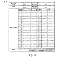
最終生産物は、HPLCを用いて純度を検査した (97.1%、 図1)。分子量は、質量分光測定により検査した。理論分子量は 5203.98、実測分子量は 5206.1 (図2)。最終生産物の配列は、中国科学院上海ライフサイエンス研究所プロテオミクス分析センターにより実施された(図3)。
14) Verification The final product was checked for purity using HPLC (97.1%, Figure 1). The molecular weight was examined by mass spectrometry. The theoretical molecular weight is 5203.98 and the measured molecular weight is 5206.1 (Figure 2). The final product sequence was performed by the Proteomics Analysis Center, Shanghai Life Science Research Institute of Chinese Academy of Sciences (Figure 3).
15) 保管
白いパウダーは密封され−20 Cで保管された。
15) Storage White powder was sealed and stored at -20C.
実施例2:自動ペプチド合成装置 (SYMPHONY 合成装置)によるSEQ ID NO:1ペプチド合成
記録:
1〕ソフトウェアで、保護アミノ酸溶液、凝縮試薬、及び切断試薬の必要量を計算する。適量のDMFとDCMを装置の対応するボトルに添加する。
Example 2: SEQ ID NO: 1 peptide synthesis with an automated peptide synthesizer (SYMPHONY synthesizer) Records:
1) Calculate the required amount of protected amino acid solution, condensing reagent, and cleavage reagent in the software. Add appropriate amounts of DMF and DCM to the corresponding bottles in the instrument.
2〕 100μmol FMOC- L-Gly-2-クロロトリチルクロリド樹脂を反応容器に添加する。15mg 遠心装置チューブを導管に入れて切断溶液を集積する。 2] Add 100 μmol FMOC- L-Gly-2-chlorotrityl chloride resin to the reaction vessel. Place the 15 mg centrifuge tube into the conduit to collect the cutting solution.
3〕プログラムの編集: 樹脂膨張時間は概して30分、脱保護時間は5分と15分 (2回)、凝縮時間が30分、及び切断時間は2時間とする。 3] Editing the program: Resin expansion time is generally 30 minutes, deprotection time is 5 and 15 minutes (twice), condensation time is 30 minutes, and cutting time is 2 hours.
4〕装置の電源を投入してプログラムを実行する。 4] Turn on the device and execute the program.
5〕最後に、切断溶液をエーテルで沈殿させ、次に遠心分離し吹き付け乾燥、次にHPLCで粗ペプチドを精製する。最終生産物は純度をHPLCで検査した (97.1%、 図1)。その分子量 (SEQ ID NO:1) は質量分光測定により測定した。理論分子量は5203.98、実測分子量は5206.1 (図2)であった。最終生産物の配列は、中国科学院上海ライフサイエンス研究所プロテオミクス分析センターにより確認された(図3)。 5) Finally, the cleavage solution is precipitated with ether, then centrifuged and spray dried, and then the crude peptide is purified by HPLC. The final product was checked for purity by HPLC (97.1%, Figure 1). Its molecular weight (SEQ ID NO: 1) was measured by mass spectrometry. The theoretical molecular weight was 5203.98, and the measured molecular weight was 5206.1 (FIG. 2). The sequence of the final product was confirmed by the Proteomics Analysis Center, Shanghai Life Science Research Institute, Chinese Academy of Sciences (Figure 3).
実施例3:ペプチドSeq. No.1の 肝臓 HSC 細胞量と活性への効果
材料と方法:
1.オスSDラット5匹、重量(250±25)g、DMEM 培地、パンクレアチン (EDTAを含む)、リポフェクタミン 2000、トリゾール、新生小牛血清(Invitrogenブランド)、プロテイナーゼE (プロナーゼ)、コラーゲナーゼ B、DNA 酵素(Roche社)、Nycodenz (Sigma社)、抗体p-FAK Tyr397、デスミンとα-平滑筋アクチン(α-SMA)、モノクロナール抗体(Santa Cruz 社)。
RT-PCRキット(MBI社)。
Example 3: Effect of peptide Seq. No. 1 on liver HSC cell mass and activity Materials and methods:
1. 5 male SD rats, weight (250 ± 25) g, DMEM medium, pancreatin (including EDTA), lipofectamine 2000, trizol, newborn calf serum (Invitrogen brand), proteinase E (pronase), collagenase B, DNA Enzyme (Roche), Nycodenz (Sigma), antibody p-FAK Tyr397, desmin and α-smooth muscle actin (α-SMA), monoclonal antibody (Santa Cruz).
RT-PCR kit (MBI).
2.培地HSC
SDラットから得たHSCを、プロナーゼ-コラーゲナーゼとニコデンツにより作成された勾配上で遠心分離した。1.5×10⁵/ cm2で HSC細胞を 6ウェルプレート、または培養皿(100 mm径)に設置。培地は、20% 新生小牛血清によるDMEMとした。HSC細胞の純度は、ビタミンA自己蛍光と抗デスミン免疫細胞化学実験により同定した。細胞生存能力は、トリパンブルー染色により同定した。第1世代HSCの純度と生存能力は、それぞれ90%と95%であった。HSCは、無ECM条件下であれば自己活性的である。つまり、HSCは、α-SMAを発現でき、ビタミンA降下が消滅する。細胞融合後、活性化HSCはトリプシンにより開放されて増殖する。
2. Medium HSC
HSCs obtained from SD rats were centrifuged on a gradient created by Pronase-Collagenase and Nicodents. Place HSC cells at 1.5 × 10 5 / cm2 in a 6-well plate or culture dish (100 mm diameter). The medium was DMEM with 20% newborn calf serum. The purity of HSC cells was identified by vitamin A autofluorescence and anti-desmin immunocytochemistry experiments. Cell viability was identified by trypan blue staining. The purity and viability of the first generation HSC were 90% and 95%, respectively. HSC is autoactive under ECM-free conditions. In other words, HSC can express α-SMA and the vitamin A drop disappears. After cell fusion, activated HSCs are released by trypsin and proliferate.
3.MTT アッセイ
ペプチド SEQ ID NO:1、(1μM)、24時間、48時間、または72時間。MTT アッセイを使用してHSC細胞の増殖を調べる。
3. MTT Assay Peptide SEQ ID NO: 1, (1 μM), 24 hours, 48 hours, or 72 hours. Examine HSC cell proliferation using the MTT assay.
4.α-SMA mRNA発現を調べるためのRT-PCR試験
トリゾール試薬を使用して総RNAを抽出、次に2段階RT-PCRをキット説明書に従って実施した。
Four. RT-PCR test to examine α-SMA mRNA expression Total RNA was extracted using Trizol reagent and then two-step RT-PCR was performed according to kit instructions.
α-SMA上流プライマー: 5’- AAGAGGAAGACA GCA CAG C TC-3’、
下流プライマー: 5’- GATGGATGGGAAAACAGC C-3’、
最終生産物: 101 bp α-SMA cDNA 断片。
α-SMA upstream primer: 5'- AAGAGGAAGACA GCA CAG C TC-3 ',
Downstream primer: 5'-GATGGATGGGAAAACAGC C-3 ',
Final product: 101 bp α-SMA cDNA fragment.
GAPDH上流プライマー:5’- ACCACAGTCCATGCCATC AC-3’、
下流プライマー: 5’- TCCACCACCCTGTTGCTGTA-3’、
最終生産物: 452bp GAPDH cDNA 断片。
GAPDH upstream primer: 5'-ACCACAGTCCATGCCATC AC-3 ',
Downstream primer: 5'-TCCACCACCCTGTTGCTGTA-3 ',
Final product: 452 bp GAPDH cDNA fragment.
5.α-SMA蛋白質発現を試験するためのウエスタンブロット法
細胞を集積して細胞リーシスバッファーを添加し、 総蛋白質を抽出する。総蛋白質量をBradfordアッセイにより同定した。40μg 総蛋白質を10% SDS ポリアクリルアミドジェルを用いて検査した。抗α-SMAモノクロナール抗体を用いてジェルを検査した。
Five. Western blot for testing α-SMA protein expression Accumulate cells, add cell lysis buffer, and extract total protein. Total protein mass was identified by Bradford assay. 40 μg total protein was examined using 10% SDS polyacrylamide gel. Gels were examined using anti-α-SMA monoclonal antibody.
6. 統計的計算
結果は x ±s、SPSS 10.0ソフトウェア、P <0.05による。
6. Statistical calculation Results are from x ± s, SPSS 10.0 software, P <0.05.
結果
1. ペプチドSEQ ID NO:1 のHSC 細胞の増殖への効果
24 時間後、1μMでのペプチドSEQ ID NO:1の抑制率は、45.5 % ±5.8 %である。48または72 時間後、1μM でのペプチドSEQ ID NO:1の抑制率は、それぞれ、61.8 % ±4.3 %と85.6 % ±5.8 %である。
result
1. Effect of peptide SEQ ID NO: 1 on proliferation of HSC cells
After 24 hours, the inhibition rate of peptide SEQ ID NO: 1 at 1 μM is 45.5% ± 5.8%. After 48 or 72 hours, the inhibition rates of peptide SEQ ID NO: 1 at 1 μM are 61.8% ± 4.3% and 85.6% ± 5.8%, respectively.
2.α-SMA mRNAの発現へのペプチドSEQ ID NO:1の効果
静止状態HSCとは異なり、α-SMA mRNA発現は、活性化HSCの重要な特性である。RT-PCR実験によれば、48時間後にペプチドSEQ ID NO:1は、α-SMA mRNA発現を抑制する(図4)。
2. Effect of peptide SEQ ID NO: 1 on the expression of α-SMA mRNA Unlike quiescent HSC, α-SMA mRNA expression is an important property of activated HSC. According to RT-PCR experiments, the peptide SEQ ID NO: 1 represses α-SMA mRNA expression after 48 hours (FIG. 4).
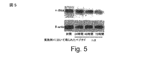
3. α-SMA 蛋白質発現へのペプチドSEQ ID NO:1の効果
ウエスタンブロット法実験によると、ペプチドSEQ ID NO:1は、48 時間にはα-SMA 蛋白質発現を減少させ始め、72 時間にはα-SMA蛋白質発現を著しく減少させる(図5)。
3. Effect of peptide SEQ ID NO: 1 on α-SMA protein expression According to Western blot experiments, peptide SEQ ID NO: 1 began to decrease α-SMA protein expression in 48 hours and α-SMA protein in 72 hours. Significantly decreases SMA protein expression (Figure 5).
実施例4: 肝線維症の動物モデルにおけるペプチドSEQ ID NO:1の効果実験
肝臓損傷に対するペプチドSEQ ID NO:1の保護的効果を評価するため、効果性評価のため慢性肝線維症モデル(ラットCCl4 モデル) を使用した。
Example 4: Experiment of the effect of peptide SEQ ID NO: 1 in an animal model of liver fibrosis To evaluate the protective effect of peptide SEQ ID NO: 1 on liver injury, a chronic liver fibrosis model (rat CCl4 model) was used.
結果により実証されたことは、治療高投与量(50μg/mg)と予防低投与量(10μg/mg)でのペプチドSEQ ID NO:1は、ラットCCl4 モデルでは、肝臓損傷パラメーターを著しく減少できる。治療群と予防群では、SEQ ID NO:1は多くの肝臓パラメーター(P<0.01 、P<0.05)を改善した。治療高投与量群(総蛋白質、アルブミン、血清グルタミン酸ピルビン酸トランスアミナーゼ (SGPT)、アスパラギン酸アミノトランスフェラーゼ、アルカリホスファターゼ、グルコース)、治療低投与量群(総蛋白質、アルブミン、グルコース)、予防高投与量群(総蛋白質、アルブミン、SGPT、アスパラギン酸アミノトランスフェラーゼ、グルコース)、予防低投与量群(総蛋白質、アルブミン、SGPT、グルコース)。 The results demonstrated that the peptide SEQ ID NO: 1 at high therapeutic dose (50 μg / mg) and low prophylactic dose (10 μg / mg) can significantly reduce liver damage parameters in the rat CCl4 model. In the treatment and prevention groups, SEQ ID NO: 1 improved many liver parameters (P <0.01, P <0.05). High therapeutic dose group (total protein, albumin, serum glutamate pyruvate transaminase (SGPT), aspartate aminotransferase, alkaline phosphatase, glucose), low therapeutic dose group (total protein, albumin, glucose), prophylactic high dose group (Total protein, albumin, SGPT, aspartate aminotransferase, glucose), preventive low dose group (total protein, albumin, SGPT, glucose).
対照群に比較すると、 ヒドロキシプロリンは慢性CCl4 肝臓損傷ネズミモデルで著しく増加する(P<0.05)。ヒドロキシプロリン量は、様々な治療群で低減した。治療高投与量群で最善な効果を得た(P<0.05)。 Compared to the control group, hydroxyproline is significantly increased in the chronic CCl4 liver injury murine model (P <0.05). Hydroxyproline levels were reduced in various treatment groups. The best effect was obtained in the high treatment dose group (P <0.05).
組織学レポートによると、CCl4モデル群の肝組織は、構造の乱れを示した。肝細胞の80%以上に著しい脂肪症が見られ、肝臓細胞に多くの脂肪空洞が見られた。1つのケースでは、肝臓内繊維組織の表皮過形成がある。治療高投与量群では、2ケースで脂肪症と肝細胞(>80%)に脂肪空洞が発現し、2ケースが脂肪症と肝細胞(50-60%)内脂肪空洞が少なくなっており、4ケースはさらに少ない脂肪症と肝細胞(<30%)内脂肪空洞を示した。予防低投与量群全てのケースはより少ない脂肪症と肝細胞(60-80%)内脂肪空洞を示した。予防高投与量群では、5ケースがより少ない脂肪症と肝細胞(60-80%)内脂肪空洞を示し、3ケースはさらに少ない脂肪症と肝細胞(50-60%)内脂肪空洞を示した。 According to the histology report, the liver tissue of the CCl4 model group showed structural disorder. Significant steatosis was seen in more than 80% of hepatocytes, and many fat cavities were found in the liver cells. In one case, there is epidermal hyperplasia of the fibrous tissue in the liver. In the high therapeutic dose group, fat cavities develop in steatosis and hepatocytes (> 80%) in 2 cases, and fat cavities and fat cavities in hepatocytes (50-60%) decrease in 2 cases, Four cases showed less steatosis and hepatocyte (<30%) fat cavities. All cases in the prophylactic low dose group showed less steatosis and hepatocyte (60-80%) internal fat cavities. In the high prophylactic dose group, 5 cases showed less steatosis and hepatocyte (60-80%) fat cavities, and 3 cases showed less steatosis and hepatocyte (50-60%) fat cavities. It was.
まとめると、ペプチドSEQ ID NO:1は、ラット内でCCl4 が引き起こす肝臓損傷を改善し、血清生化学パラメーター、肝臓ヒドロキシプロリン量、及び肝臓の病理学的特徴を改善する。 In summary, peptide SEQ ID NO: 1 improves liver damage caused by CCl4 in rats and improves serum biochemical parameters, liver hydroxyproline levels, and liver pathological features.
試薬と方法
1〕薬剤
ペプチド SEQ ID NO:1(実施例2);
Zhengda Tianqing社製注射用薬剤。
正の対照薬剤、Jiangsu Zhengda Tianqing Pharmaceutical Co. Ltd.製Gan-li-xin(グリチルリチン酸二アンモニウム)注射、10ml:50mg。
2〕動物
SDラット、180-220g、メスとオス(1:1)
3〕主な試薬
CCl4、Shanghai Lingfeng Chemical Reagents Co., Ltd製。Lot No: 061101;
ゴマ油、精製ピーナツオイル;
ヒドロキシプロリン試験キット(南京建成社)
4〕主装置
BS210S 化学はかり (0.1mg〜10g) (German Sartorius社製);
752C UV-Vis 分光光度計(上海第三分析装置社製) ;
遠心装置 (北京医療遠心装置社);
自動生化学試験装置(OLYMPUS Au 800、日本);
FEJ-200分析天秤(0.1〜200g)(福州Furi Hengzhibao Electric Co. Ltd.社製)
5〕実験
Rat CCl4 肝臓損傷モデル。
Reagents and methods
1] Drug Peptide SEQ ID NO: 1 (Example 2);
An injectable drug manufactured by Zhengda Tianqing.
Positive control drug, Gan-li-xin (diammonium glycyrrhizinate) injection from Jiangsu Zhengda Tianqing Pharmaceutical Co. Ltd., 10 ml: 50 mg.
2) animals
SD rat, 180-220g, female and male (1: 1)
3) Main reagents
CCl4, made by Shanghai Lingfeng Chemical Reagents Co., Ltd. Lot No: 061101;
Sesame oil, refined peanut oil;
Hydroxyproline test kit (Nanjing Kenseisha)
4] Main equipment
BS210S chemical scale (0.1mg-10g) (German Sartorius);
752C UV-Vis spectrophotometer (manufactured by Shanghai Third Analyzer);
Centrifuge (Beijing Medical Centrifuge Company);
Automatic biochemical test equipment (
FEJ-200 analytical balance (0.1-200g) (Fuzhou Furi Hengzhibao Electric Co. Ltd.)
5) Experiment
Rat CCl4 liver injury model.
投与群:
110ラット、重量180-220g、7 群:
(1)対照群: 食塩水、sc、2ml/kg、10ラット
(2)モデル群: 40% CCl4、sc、2ml/kg、17ラット
(3)Gan-li-xin 群: 25mg/kg、iv、10ml/kg、15ラット
(4)予防、高投与量群: 50μgペプチドSEQ ID NO:1 /kg、iv、10ml/kg、17ラット
(5)予防、低投与量群: 10μgペプチドSEQ ID NO:1 /kg、iv、10ml/kg、17ラット
(6)治療、高投与量群: 100μgペプチドSEQ ID NO:1 /kg、iv、10ml/kg、17ラット
(7)治療、高投与量群: 20μgペプチドSEQ ID NO:1 /kg、iv、10ml/kg、17ラット。
Administration group:
110 rats, weight 180-220g, 7 groups:
(1) Control group: Saline, sc, 2 ml / kg, 10 rats (2) Model group: 40% CCl4, sc, 2 ml / kg, 17 rats (3) Gan-li-xin group: 25 mg / kg, iv , 10 ml / kg, 15 rats (4) Prevention, high dose group: 50 μg peptide SEQ ID NO: 1 / kg, iv, 10 ml / kg, 17 rats (5) Prevention, low dose group: 10 μg peptide SEQ ID NO : 1 / kg, iv, 10ml / kg, 17 rats (6) Treatment, high dose group: 100μg peptide SEQ ID NO: 1 / kg, iv, 10ml / kg, 17 rats (7) Treatment, high dose group : 20 μg peptide SEQ ID NO: 1 / kg, iv, 10 ml / kg, 17 rats.
モデル構成と投与方法
対照群を除き、40% CCl4 を皮下に週2回 (火曜日と金曜日) 群(2)〜(6)に注射、薬剤用量(0.2 ml/100 g、最初に0.5ml/100g)。CCl4 モデル構成は6週間行なった。モデル構成と平行して、50μg/kgと10μg/kgペプチドSEQ ID NO:1を、群(4)と(5)にそれぞれ6週間1日に1回静脈注射した。群 (3)、(6)、(7)については、第 5週からGan-li-xin 25mg/kg、100μg/kgと20μg/kgでペプチドSEQ ID NO:1 をそれぞれ2週間連続して1日1回注射した。
Model structure and administration method Except for the control group, 40% CCl4 was subcutaneously injected twice a week (Tuesday and Friday) into groups (2) to (6), drug dose (0.2 ml / 100 g, initially 0.5 ml / 100 g ). CCl4 model configuration was performed for 6 weeks. In parallel with the model configuration, 50 μg / kg and 10 μg / kg peptide SEQ ID NO: 1 were intravenously injected once a day for 6 weeks into groups (4) and (5), respectively. For groups (3), (6) and (7), from
試験パラメーター
全てのラットは、モデル構成と薬剤注射中に週に一回体重を測定した。体重に従い、薬剤投与を調整した。最終回CCl4注射から24 時間後に、肝臓重量パラメーターを試験した。
Test parameters All rats were weighed once a week during model construction and drug injection. Drug administration was adjusted according to body weight. Liver weight parameters were tested 24 hours after the last CCl4 injection.
血液を大腿動脈から抽出し、血清を分離した。次のパラメーターを試験した: 血清アラニンアミノトランスフェラーゼ (ALT)、アスパラギン酸アミノトランスフェラーゼ(AST)、ビリルビン (TB)、アルカリホスファターゼ (ALP)、総蛋白質(TP)、アルブミン (ALB)、グロブリン(G)、アルブミン/グロブリン比(A/G)、グルコース (GLU)、総コレステロール(TCH)、トリグリセリド(TG)。 Blood was extracted from the femoral artery and serum was separated. The following parameters were tested: serum alanine aminotransferase (ALT), aspartate aminotransferase (AST), bilirubin (TB), alkaline phosphatase (ALP), total protein (TP), albumin (ALB), globulin (G), Albumin / globulin ratio (A / G), glucose (GLU), total cholesterol (TCH), triglycerides (TG).
肝臓ホモジネート: コレステロール(TCH)、トリグリセリド(TG)、アラニンアミノトランスフェラーゼ (ALT)、アスパラギン酸アミノトランスフェラーゼ(AST)を試験する。 Liver homogenates: Cholesterol (TCH), triglycerides (TG), alanine aminotransferase (ALT), aspartate aminotransferase (AST) are tested.
200g 肝臓。110 Cで乾燥後、ヒドロキシプロリン(HPA)を検査。
肝臓の一部は組織学的実験用にフォルマリン漬けとした。
200g liver. Tested for hydroxyproline (HPA) after drying at 110 C.
Part of the liver was immersed in formalin for histological experiments.
実験結果
1〕ネズミの体重、肝臓重量、および肝臓パラメーターへの薬剤効果
対照群と比較すると、CCl4肝臓損傷群の肝臓パラメーターは著しく増加した (P<0.01)。様々な治療群でこれらのパラメーターは改善し、治療高投与量群と予防低投与量群では統計的有意性の差異があった(P<0.05)。下の表1(*vs 対照群、# vs CCl4 モデル群)を参照されたい。

1] Drug effects on rat body weight, liver weight, and liver parameters The liver parameters in the CCl4 liver injury group were significantly increased compared to the control group (P <0.01). These parameters improved in various treatment groups, and there was a difference in statistical significance between the high treatment and low prophylactic dose groups (P <0.05). See Table 1 below (* vs control group, # vs CCl4 model group).
2)ネズミ生化学パラメーターへのへの薬剤効果
対照群と比較すると、CCl4肝臓損傷動物モデル群は、血清アラニンアミノトランスフェラーゼ、アスパラギン酸アミノトランスフェラーゼ、およびアルカリホスファターゼが増加、総蛋白質、アルブミン、グルコース、およびトリグリセリドが減少した。Gan-li-xin 群 (12.5mg/kg) の上記パラメーターがある程度改善され、アスパラギン酸アミノトランスフェラーゼ(AST)とグルコース (GLU)(P<0.05)の統計的有意性も改善された。ペプチドSEQ ID NO:1 群 (治療と予防群) でも上記パラメーターは改善され、統計的有意性も改善された (CCl4 モデル群より):治療高投与量群(TP、ALB、ALT、AST、ALP、GLU)、治療低投与量群(TP、ALB、GLU)、予防高投与量群(TP、ALB、ALT、AST、GLU)、予防低投与量群(TP、ALB、AST、GLU)。詳細は表2を参照されたい(*vs 対照群 、# vs CCl4 モデル群)。
3)ネズミ肝臓内ヒドロキシプロリン量への薬剤効果
対照群と比較すると、ヒドロキシプロリン量はCCl4損傷肝臓モデル群(P<0.05)では著しく増加した。各治療群でヒドロキシプロリン量は減少し、治療高投与量群のそれとは統計的有意性の差異があった(P<0.05)。表3を参照されたい(*vs 対照群、#vs CCl4 モデル群)。
4)組織学的実験
対照群は、規則正しい構造を示した。CCl4モデル群の肝組織は、構造の乱れを示した。肝細胞の80%以上が脂肪症を示し、肝臓細胞の脂肪空洞を発現した。1つのケースでは、肝臓内繊維組織の表皮過形成がある。治療高投与量群では2ケースで脂肪症と肝細胞(>80%)脂肪空洞が見られ、2ケースでは脂肪症が少なくなったのと肝細胞内脂肪空洞が減少(50-60%)、4ケースはさらに少ない脂肪症と肝細胞内脂肪空洞(<30%)を示した。予防低投与量群の全ケースで、より少ない脂肪症と肝細胞内脂肪空洞(60-80%)が示された。予防高投与量群においては、5ケースがより少ない脂肪症と肝細胞内脂肪空洞(60-80%)を示し、さらに、3つのケースでは肝細胞の脂肪症と脂肪空洞はさらに少なかった(50-60%)。
4) Histological experiment The control group showed a regular structure. The liver tissue of the CCl4 model group showed structural disturbance. More than 80% of hepatocytes showed steatosis and expressed fatty cavities in liver cells. In one case, there is epidermal hyperplasia of the fibrous tissue in the liver. In the high-dose group, steatosis and hepatocyte (> 80%) fat cavities were seen in 2 cases, and in 2 cases, steatosis was reduced and the intrahepatic fat cavities decreased (50-60%), Four cases showed less steatosis and hepatocyte fat cavities (<30%). All cases in the low prophylaxis group showed less steatosis and hepatocyte fat cavities (60-80%). In the prophylactic high dose group, 5 cases showed less steatosis and hepatocyte fat cavities (60-80%), and 3 cases had fewer hepatocyte steatosis and fat cavities (50 -60%).
実施例5:ペプチドSEQ ID No:1の生体抗癌効果性評価
装置
デジタルウォーターバス: HH-4 (国華電力社製)
インキュベーター: HERA 細胞150 (Thermo Electron Corporation社製)
顕微鏡: BDS200-PH (重慶 Aote 光学装置社製)
卓上遠心装置: TGL-16G (Shanghai Surgical Device Co.社製)
クリーンベンチ: SW-CJ-IFD (蘇浄集団Antai社製)
ボーテックス: XW-80A (上海医科大学装置社製)
逆位の位相差顕微鏡: XSZ-D2 (重慶光学装置社製)
マイクロプレートリーダー:モデル-550 (Bio-Rad社製)
天秤: HC-TP-12 (Tianjin Balance 装置社製)
1.細胞株
ヒト肝臓癌細胞株(SMMC-7721、 BEL-7402、 BEL-7402)は国家医療科学院細胞バンクと中国科学院上海細胞株研究所から購入した。
Example 5: Evaluation of biological anticancer efficacy of peptide SEQ ID No: 1
Device Digital Water Bath: HH-4 (Kokuhua Electric)
Incubator: HERA cell 150 (Thermo Electron Corporation)
Microscope: BDS200-PH (Chongqing Aote Optics Co., Ltd.)
Tabletop centrifuge: TGL-16G (manufactured by Shanghai Surgical Device Co.)
Clean bench: SW-CJ-IFD (manufactured by Sujo Group Antai)
Vortex: XW-80A (manufactured by Shanghai Medical University Equipment Company)
Inverted phase contrast microscope: XSZ-D2 (Chongqing Optical Equipment Co., Ltd.)
Microplate reader: Model-550 (Bio-Rad)
Balance: HC-TP-12 (manufactured by Tianjin Balance Equipment)
1. Cell lines Human liver cancer cell lines (SMMC-7721, BEL-7402, BEL-7402) were purchased from National Medical Science Cell Bank and Chinese Academy of Sciences Shanghai Cell Line Research Institute.
1.細胞培養
全細胞株は37C、5% CO2、飽和湿度中のインキュベーターで培養した。培地は、10%加熱失活FBS、ペニシリン100μ/ml、ストレプトマイシン 100 μ/mlを含むRPMI1640媒地とした。48 時間後に培地を交換した。細胞は、0.25%トリプシンで遊離されて増殖した。実験で使用した細胞は対数期であった。細胞生存能力は、トリパンブルー染色により同定された。
1. Cell culture All cell lines were cultured in an incubator at 37C, 5% CO2, saturated humidity. The medium was RPMI1640 medium containing 10% heat-inactivated FBS,
2.方法
対数期細胞を採取し、 0.125%トリプシン+ 0.01% EDTAで遊離させ、細胞を2-4 X 10⁴ 細胞/mlまで希釈する。細胞は96 ウェルプレート(180 μl/ウェル)に設置され、37 C CO2 インキュベーターで24時間培養された。培地を交換後、ペプチドSEQ ID No:1 をウェルに添加(20μl/ウェル)して、72時間培養した。MTTを96 ウェルプレート(20 μl/ウェル)に添加し、4 時間培養した。培地を除去し、DMSO (150μl/ウェル)を添加した。プレートは10分間振とうした。7つの濃度点 (0.1〜10 μM) を検査した。570 nMでの吸光度をウェル毎に測定し、IC50を計算した。
2. Methods Log phase cells are harvested, released with 0.125% trypsin + 0.01% EDTA, and the cells are diluted to 2-4 × 10 4 cells / ml. The cells were placed in a 96-well plate (180 μl / well) and cultured for 24 hours in a 37 C CO2 incubator. After changing the medium, peptide SEQ ID No: 1 was added to the wells (20 μl / well) and cultured for 72 hours. MTT was added to a 96-well plate (20 μl / well) and cultured for 4 hours. The medium was removed and DMSO (150 μl / well) was added. The plate was shaken for 10 minutes. Seven concentration points (0.1-10 μM) were examined. Absorbance at 570 nM was measured per well and IC50 was calculated.
IC50 = (ペプチド無しのウェルにおけるO.D.−ペプチド有りのウェルにおけるO.D.)/ペプチド無しのウェルにおけるO.D.
3.結果
実施例1 からのペプチド SEQ ID No:1 は、SMMC-7721、BEL-7402、およびBEL-7404において、それぞれ1.25、1.78、および2.33μMにおけるIC50では著しく肝臓癌細胞の増殖を阻害した。
IC50 = (OD in wells without peptide-OD in wells with peptide) / OD in wells without peptide
3. Results The peptide SEQ ID No: 1 from Example 1 significantly inhibited the growth of liver cancer cells in SMMC-7721, BEL-7402, and BEL-7404 with IC50 at 1.25, 1.78, and 2.33 μM, respectively.
実施例6: 生体での抗癌効果実験
実験動物: メス BALB/cA ヌードマウス、35-40 日齢、重量18-22 g。上海Silaike実験動物 Co. Ltd. 認証番号: SCXK(Shanghai)2007-0005から供給された。
Example 6: Anticancer effect experiment in vivo Experimental animal: female BALB / cA nude mouse, 35-40 days old, weight 18-22 g. Shanghai Silaike Experimental Animal Co. Ltd. Certification number: SCXK (Shanghai) 2007-0005.
異種移植ヌードマウスの腫瘍は、100〜300mm3 まで成長した。腫瘍マウスを異なる群に分け、薬剤比較試験を実施した。下の実験では、負の対照群に12匹のヌードマウス、試験薬剤を投与する6匹ヌードマウス群として行なった。腫瘍直径は毎週3回計測され、マウス重量も同時に測定した。 Tumors in xenograft nude mice grew to 100-300 mm 3 . Tumor mice were divided into different groups and drug comparison studies were performed. In the experiment below, the negative control group was a group of 12 nude mice and a group of 6 nude mice administered with the test drug. Tumor diameter was measured 3 times a week and mouse weight was also measured simultaneously.
T/C (相対的腫瘍抑制率)%を下の通り計算した。 T / C (relative tumor suppression rate)% was calculated as follows.
(1)腫瘍容積TVを下の通り計算した。
TV = 1/2×a×b2
aとbは長さと幅である。
(1) Tumor volume TV was calculated as follows.
TV = 1/2 × a × b 2
a and b are length and width.
(2)相対的腫瘍量RTVを下の通り計算した。
RTV = TVt/TV0
TV0は薬剤使用開始時の腫瘍量 (d0)、TVt は後の時点での腫瘍の量。
(2) The relative tumor volume RTV was calculated as follows.
RTV = TV t / TV 0
TV 0 is the tumor volume at the start of drug use (d 0 ), and TV t is the tumor volume at a later time.
(3)相対的腫瘍抑制率T/C(%)を下の通り計算した。 (3) The relative tumor suppression rate T / C (%) was calculated as follows.
T/C(%)= TRTV/CRTV×100
TRTV:治療グループRTV 、CRTV:負の対照RTV
1)肝臓癌
実施例1および2からのペプチドSEQ ID No:1は、異種移植肝臓癌ヌードマウスモデル(H22) を1 mg/kgでは抑制率67.8%により抑制した。これは、正の対照であるタキソールの抑制率(10 mg/kg)に近い。1 mg/kgでは、実施例1のペプチドSEQ ID No:1は、異種移植肝臓癌ヌードマウスモデル(BEL-7402)を39.7%で抑制した。
T / C (%) = T RTV / C RTV × 100
T RTV : Treatment group RTV, C RTV : Negative control RTV
1) Liver cancer The peptide SEQ ID No: 1 from Examples 1 and 2 inhibited the xenograft liver cancer nude mouse model (H22) at 1 mg / kg with an inhibition rate of 67.8%. This is close to the inhibition rate (10 mg / kg) of the positive control taxol. At 1 mg / kg, the peptide SEQ ID No: 1 of Example 1 inhibited the xenograft liver cancer nude mouse model (BEL-7402) at 39.7%.
ペプチドSEQ ID No:1は、尾静脈(IV)を通して投与され、投与量は0.5mg/kg、0.25mg/kg、0.125mg/kg、および0.0625mg/kgを一週間に6回とした。ドセタキセルを正の対照として、投与率は20mg/kg、週に一回とした。負の対照は食塩水とした。異種移植ヌードマウスを3週間治療した。 Peptide SEQ ID No: 1 was administered via the tail vein (IV), with dosages of 0.5 mg / kg, 0.25 mg / kg, 0.125 mg / kg, and 0.0625 mg / kg six times per week. Docetaxel was used as a positive control, and the dose rate was 20 mg / kg once a week. The negative control was saline. Xenograft nude mice were treated for 3 weeks.
ヒト肝臓癌BEL-7402ヌードマウスにおけるペプチドSEQ ID No:1 0.5mg/kg、0.25mg/kg、0.125mg/kg でのT/C(%)は、それぞれ64.90、69.06、62.10となった。最善のT/C (%)は、投与量0.0625mg/kgで58.56%を得た。このことから、ペプチドSEQ ID No:1は肝臓癌のインビボでの増殖を抑制できる。 T / C (%) at peptide SEQ ID No: 1 0.5 mg / kg, 0.25 mg / kg, and 0.125 mg / kg in human liver cancer BEL-7402 nude mice was 64.90, 69.06, and 62.10. The best T / C (%) was obtained at 58.56% at a dose of 0.0625 mg / kg. From this, the peptide SEQ ID No: 1 can suppress the growth of liver cancer in vivo.
2)胃癌
ペプチド SEQ ID No:1は、尾静脈(IV)を通して、投与量1mg/kg、0.5mg/kg、0.25mg/kg、0.125mg/kg、および0.0625mg/kgで一週間に6回注入された。5-FUを正の対照として、投与量25mg/kgで週に一回投与した。負の対照には食塩水を与えた。異種移植ヌードマウスを3週間治療した。
2) Gastric cancer Peptide SEQ ID No: 1 through the tail vein (IV) at doses of 1 mg / kg, 0.5 mg / kg, 0.25 mg / kg, 0.125 mg / kg, and 0.0625 mg /
ヒト胃癌SGC-7901 ヌードマウスにペプチドSEQ ID No:1を 0.125mg/kg で投与したT/C(%)は、47.66%であった。5-FUでのT/C(%)は、68.71%であった。このことから、ペプチドSEQ ID No:1 はインビボでの胃癌増殖を強く抑制する。 Human gastric cancer SGC-7901 The T / C (%) of peptide SEQ ID No: 1 administered to nude mice at 0.125 mg / kg was 47.66%. The T / C (%) with 5-FU was 68.71%. From this, the peptide SEQ ID No: 1 strongly suppresses the growth of gastric cancer in vivo.
3)乳癌
ペプチドSEQ ID No:1を尾静脈(IV)から投与量2mg/kg、1mg/kg、0.5mg/kgで、一週間に6回投与した。ドセタキセルを正の対照とし、投与量は20mg/kg、週に一回投与した。負の対照は食塩水を与えた。異種移植ヌードマウスは、4週間治療された。
3) Breast cancer The peptide SEQ ID No: 1 was administered 6 times a week at a dose of 2 mg / kg, 1 mg / kg, 0.5 mg / kg from the tail vein (IV). Docetaxel was used as a positive control, and the dose was 20 mg / kg, administered once a week. The negative control received saline. Xenograft nude mice were treated for 4 weeks.
ヒト乳癌MDA-MB-435 ヌードマウスにおいて、0.5mg/kg でペプチドSEQ ID No:1を投与したT/C(%) は、49.40%であった。ドセタキセルでの T/C(%) は、16.51%であった。以上から、ペプチドSEQ ID No:1は、インビボでの乳癌増殖を強く抑制しうる。 In human breast cancer MDA-MB-435 nude mice, the T / C (%) of peptide SEQ ID No: 1 administered at 0.5 mg / kg was 49.40%. The T / C (%) for docetaxel was 16.51%. From the above, the peptide SEQ ID No: 1 can strongly suppress breast cancer growth in vivo.
4)肺癌
ペプチド SEQ ID No:1を、尾静脈(IV)を通して投与量0.5mg/kg、0.25mg/kg、0.125mg/kgで、一週間に6回投与した。ドセタキセルを正の対照として、投与率は20mg/kgで週に一回投与した。負の対照は食塩水を与えた。異種移植ヌードマウスが3週間治療された。
4) Lung cancer Peptide SEQ ID No: 1 was administered 6 times a week at doses of 0.5 mg / kg, 0.25 mg / kg, and 0.125 mg / kg through the tail vein (IV). Docetaxel was administered as a positive control at a dose rate of 20 mg / kg once a week. The negative control received saline. Xenograft nude mice were treated for 3 weeks.
ヒト肺癌A549ヌードマウスでペプチドSEQ ID No:1を0.25mg/kg、0.125mg/kgで投与したT/C(%)は、47.98および48.96%になった。ドセタキセルでのT/C(%)は、18.60%であった。このことから、ペプチドSEQ ID No:1は、インビボでの肺癌成長を強く抑制できる。 The T / C (%) of the peptide SEQ ID No: 1 administered at 0.25 mg / kg and 0.125 mg / kg in human lung cancer A549 nude mice was 47.98 and 48.96%. The T / C (%) with docetaxel was 18.60%. From this, the peptide SEQ ID No: 1 can strongly suppress lung cancer growth in vivo.
異種移植腫瘍マウスモデルの全てにおいて、マウスの体重は異なる濃度のペプチドSEQ ID No:1の影響を受けなかった。これにより、ペプチドSEQ ID No:1の安全なプロファイルが実証された。正の対照である化学療法剤により、マウスの体重は大きく影響された。 In all xenograft tumor mouse models, mouse body weight was not affected by different concentrations of peptide SEQ ID No: 1. This demonstrated a safe profile for peptide SEQ ID No: 1. The positive control chemotherapeutic agent greatly affected the body weight of the mice.
実施例7: 肝臓癌細胞遊走能力の抑制
実施例1のペプチド SEQ ID No:1は、ヒト肝臓癌細胞BEL-7402の基底膜からの遊走を 0.01、0.1、または1 mg/ml (12時間培養)で抑制する。
Example 7: Inhibition of liver cancer cell migration ability Peptide of Example 1 SEQ ID No: 1 indicates that the migration of human liver cancer cell BEL-7402 from the basement membrane is 0.01, 0.1, or 1 mg / ml (cultured for 12 hours) ).
図6の示すところは、ペプチドと癌細胞を混ぜて12 時間培養した後、ペプチドSEQ ID No:1が基底膜への癌細胞付着を抑制したことである。図7は、ペプチドと癌細胞を混ぜて12時間培養した後、ペプチドSEQ ID No:1 基底膜を通る癌細胞の遊走能力を抑制する様子を示す。 FIG. 6 shows that the peptide SEQ ID No: 1 suppressed cancer cell adhesion to the basement membrane after the peptide and cancer cells were mixed and cultured for 12 hours. FIG. 7 shows that the peptide and cancer cells were mixed and cultured for 12 hours, and then the ability of cancer cells to migrate through the peptide SEQ ID No: 1 basement membrane was suppressed.
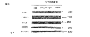
実施例8: PDGFRの細胞間Ras-Raf 信号伝達経路におけるペプチドSEQ ID No:1の阻害効果
様々な濃度のペプチドSEQ ID No:1をヒト臍帯静脈内皮細胞(HUVEC)とともに6時間培養した。蛋白質をこれらの細胞から抽出して、ウエスタンブロット法で処理した。p-stat1 用抗体: 対リン酸化-Tyr701抗体; p-MEK1/2用抗体: リン酸化-Ser217/221 ペプチド抗体; p-Erk1/2用抗体: リン酸化-Ser202/204 抗体。すべてのレーンに同量のタンパク質を負荷するために、ベータアクチンを対照とした。ウエスタンブロット法により、濃度10μg/ml (2μM)ではペプチドSEQ ID No:1が大きくErk1/2 のリン酸化反応を抑制したことが示された。これは、Ras-Raf経路の下流である。
以下に、本願の当初の特許請求の範囲に記載された発明を付記する。
[1]
SEQ ID NO:1の配列を含むペプチド、その変異体またはその活性誘導体。
[2]
[1]に記載のペプチドであって、下記からなる群より選択されるペプチド:
(i)SEQ ID NO:2の配列、
(ii)SEQ ID NO:2の配列(任意のシステイン残基がセリン残基に変化している)、
(iii)SEQ ID NO: 3の配列、または
(vi)SEQ ID NO:3の配列(任意のシステイン残基がセリン残基に変化している)。
[3]
[2]に記載のペプチドであって、前記ペプチドは、組換型ペプチド、天然ペプチドおよび合成ペプチドからなる群より選択されるペプチド。
[4]
[1]に記載のペプチドをコードするヌクレオチド配列。
[5]
ヒトまたは動物の組織線維症の予防または治療のための医薬の調製における、[1]または[2]に記載のペプチドの使用。
[6]
[5]に記載の使用であって、前記組織は、ヒトまたは動物の肝臓、腎または肺を含む使用。
[7]
ヒトまたは動物の原発性癌および癌転移の予防または治療のための医薬の調製における、[1]または[2]に記載のペプチドの使用。
[8]
ヒトまたは動物の組織線維症の予防および治療のための医薬組成物であって、該医薬組成物は、[1]または[2]に記載のペプチドと薬学的に許容可能な担体を含む医薬組成物。
[9]
[8]に記載の医薬組成物であって、前記医薬組成物は、ヒトまたは動物の組織線維症を予防および/または治療するその他の医薬をさらに含む医薬組成物。
[10]
[8]に記載の医薬組成物であって、前記医薬組成物は、経口投与、皮下注射投与、皮内注射投与、筋肉内注射投与、血管内注射投与、または経鼻投与のようないずれかのその他の投与の形態である医薬組成物。
[11]
ヒトまたは動物の癌を治療するための医薬組成物であって、該医薬組成物は、[1]または[2]に記載のペプチドと薬学的に許容可能な担体を含む医薬組成物。
[12]
[11]に記載の医薬組成物であって、前記医薬組成物は、ヒトまたは動物の癌を治療するその他の医薬をさらに含む医薬組成物。
[13]
[11]に記載の医薬組成物であって、前記医薬組成物は、Gleevec等のその他の化学療法剤をさらに含む医薬組成物。
[14]
[8]または[11]に記載の医薬組成物の投与方法であって、[1]または[2]に記載のペプチドを投与するステップを含み、前記ペプチドは、筋肉内、静脈内、皮下、経口、直腸挿入、及び経皮からなる群より選択される態様で投与される投与方法。
Example 8: Inhibitory Effect of Peptide SEQ ID No: 1 on Intercellular Ras-Raf Signaling Pathway of PDGFR Various concentrations of peptide SEQ ID No: 1 were cultured with human umbilical vein endothelial cells (HUVEC) for 6 hours. Protein was extracted from these cells and processed by Western blot. p-stat1 antibody: anti-phosphorylated-Tyr701 antibody; p-MEK1 / 2 antibody: phosphorylated-Ser217 / 221 peptide antibody; p-Erk1 / 2 antibody: phosphorylated-Ser202 / 204 antibody. Beta actin was used as a control to load all lanes with the same amount of protein. Western blotting showed that peptide SEQ ID No: 1 significantly inhibited Erk1 / 2 phosphorylation at a concentration of 10 μg / ml (2 μM). This is downstream of the Ras-Raf pathway.
The invention described in the scope of the original claims of the present application will be added below.
[1]
A peptide comprising the sequence of SEQ ID NO: 1, a variant thereof or an active derivative thereof.
[2]
The peptide according to [1], which is selected from the group consisting of:
(I) SEQ ID NO: 2 sequence,
(Ii) the sequence of SEQ ID NO: 2 (any cysteine residue is changed to a serine residue),
(Iii) SEQ ID NO: 3 sequence, or
(Vi) SEQ ID NO: 3 sequence (any cysteine residue is changed to a serine residue).
[3]
The peptide according to [2], wherein the peptide is selected from the group consisting of a recombinant peptide, a natural peptide, and a synthetic peptide.
[4]
A nucleotide sequence encoding the peptide according to [1].
[5]
Use of the peptide according to [1] or [2] in the preparation of a medicament for the prevention or treatment of human or animal tissue fibrosis.
[6]
[5] The use according to [5], wherein the tissue comprises human or animal liver, kidney or lung.
[7]
Use of the peptide according to [1] or [2] in the preparation of a medicament for the prevention or treatment of primary cancer or cancer metastasis in humans or animals.
[8]
A pharmaceutical composition for the prevention and treatment of human or animal tissue fibrosis, the pharmaceutical composition comprising the peptide according to [1] or [2] and a pharmaceutically acceptable carrier. object.
[9]
[8] The pharmaceutical composition according to [8], wherein the pharmaceutical composition further comprises other medicaments for preventing and / or treating tissue fibrosis in humans or animals.
[10]
[8] The pharmaceutical composition according to [8], wherein the pharmaceutical composition is any of oral administration, subcutaneous injection administration, intradermal injection administration, intramuscular injection administration, intravascular injection administration, and nasal administration. Pharmaceutical compositions that are other forms of administration of
[11]
A pharmaceutical composition for treating human or animal cancer, wherein the pharmaceutical composition comprises the peptide according to [1] or [2] and a pharmaceutically acceptable carrier.
[12]
[11] The pharmaceutical composition according to [11], wherein the pharmaceutical composition further comprises other medicaments for treating human or animal cancer.
[13]
[11] The pharmaceutical composition according to [11], wherein the pharmaceutical composition further comprises another chemotherapeutic agent such as Gleevec.
[14]
[8] or [11] is a method for administering a pharmaceutical composition comprising the step of administering the peptide of [1] or [2], wherein the peptide is intramuscular, intravenous, subcutaneous, A method of administration administered in a mode selected from the group consisting of oral, rectal insertion, and transdermal.
Claims (12)
Applications Claiming Priority (3)
| Application Number | Priority Date | Filing Date | Title |
|---|---|---|---|
| CN200810200937.8 | 2008-10-09 | ||
| CN2008102009378A CN101392026B (en) | 2008-10-09 | 2008-10-09 | Polypeptide for preventing and treating fibrotic disease and liver cancer |
| PCT/CN2009/074048 WO2010040305A1 (en) | 2008-10-09 | 2009-09-21 | Pharmaceutical being used for treating cancer and fibrosis disease and the composition and uses thereof |
Publications (3)
| Publication Number | Publication Date |
|---|---|
| JP2012504941A JP2012504941A (en) | 2012-03-01 |
| JP2012504941A5 JP2012504941A5 (en) | 2012-11-08 |
| JP5734856B2 true JP5734856B2 (en) | 2015-06-17 |
Family
ID=40492557
Family Applications (1)
| Application Number | Title | Priority Date | Filing Date |
|---|---|---|---|
| JP2011530354A Expired - Fee Related JP5734856B2 (en) | 2008-10-09 | 2009-09-21 | Pharmaceuticals and compositions used for the treatment of cancer and fibrotic diseases and uses thereof |
Country Status (5)
| Country | Link |
|---|---|
| US (1) | US8637467B2 (en) |
| EP (1) | EP2344528B1 (en) |
| JP (1) | JP5734856B2 (en) |
| CN (1) | CN101392026B (en) |
| WO (1) | WO2010040305A1 (en) |
Families Citing this family (2)
| Publication number | Priority date | Publication date | Assignee | Title |
|---|---|---|---|---|
| CN101392026B (en) * | 2008-10-09 | 2011-11-09 | 黄岚 | Polypeptide for preventing and treating fibrotic disease and liver cancer |
| CN111701020A (en) * | 2020-06-08 | 2020-09-25 | 南通大学 | Molecular target for treating PDGF-induced hepatic fibrosis |
Family Cites Families (9)
| Publication number | Priority date | Publication date | Assignee | Title |
|---|---|---|---|---|
| US5595756A (en) * | 1993-12-22 | 1997-01-21 | Inex Pharmaceuticals Corporation | Liposomal compositions for enhanced retention of bioactive agents |
| US20030211994A1 (en) * | 1998-09-30 | 2003-11-13 | Ludwig Institute For Cancer Research | Composition and method for modulating vasculogenesis or angiogenesis |
| CN1330664A (en) * | 1998-09-30 | 2002-01-09 | 路德维格癌症研究所 | Platelet-derived growth factor C, DNA coding therefor and uses thereof |
| EP1141293B1 (en) * | 1998-12-22 | 2007-08-01 | Janssen Pharmaceutica N.V. | Vascular endothelial growth factor-x |
| CN1355209A (en) * | 2000-11-24 | 2002-06-26 | 复旦大学 | Polypeptide-Ca-dependent Cl ion channel 16.72 and polynucleotide for coding it |
| EP1513870A4 (en) * | 2002-05-31 | 2006-06-07 | Childrens Hosp Medical Center | Cftr modifier genes and expressed polypeptides useful in treating cystic fibrosis and methods and products for detecting and/or identifying same |
| US20040248796A1 (en) * | 2003-02-04 | 2004-12-09 | Kari Alitalo | VEGF-B and PDGF modulation of stem cells |
| WO2007022287A2 (en) | 2005-08-15 | 2007-02-22 | Vegenics Limited | Modified vegf and pdgf with improved angiogenic properties |
| CN101392026B (en) | 2008-10-09 | 2011-11-09 | 黄岚 | Polypeptide for preventing and treating fibrotic disease and liver cancer |
-
2008
- 2008-10-09 CN CN2008102009378A patent/CN101392026B/en not_active Expired - Fee Related
-
2009
- 2009-09-21 WO PCT/CN2009/074048 patent/WO2010040305A1/en active Application Filing
- 2009-09-21 EP EP09818769.3A patent/EP2344528B1/en not_active Not-in-force
- 2009-09-21 JP JP2011530354A patent/JP5734856B2/en not_active Expired - Fee Related
- 2009-09-21 US US13/123,424 patent/US8637467B2/en not_active Expired - Fee Related
Also Published As
| Publication number | Publication date |
|---|---|
| EP2344528B1 (en) | 2013-12-18 |
| US20110263513A1 (en) | 2011-10-27 |
| CN101392026A (en) | 2009-03-25 |
| JP2012504941A (en) | 2012-03-01 |
| WO2010040305A1 (en) | 2010-04-15 |
| EP2344528A1 (en) | 2011-07-20 |
| EP2344528A4 (en) | 2012-03-28 |
| CN101392026B (en) | 2011-11-09 |
| US8637467B2 (en) | 2014-01-28 |
Similar Documents
| Publication | Publication Date | Title |
|---|---|---|
| ES2281704T3 (en) | PROCEDURES AND COMPOUNDS TO INHIBIT THE GROWTH OF NEOPLASSIC CELLS. | |
| US20090036369A1 (en) | Anti-tumor agents comprising r-spondins | |
| EP2552470B1 (en) | Peptides for promoting angiogenesis and an use thereof | |
| US20200062811A1 (en) | Yap protein inhibiting polypeptide and application thereof | |
| JP2022544481A (en) | Applications of polypeptides or derivatives thereof | |
| Wang et al. | A novel and low-toxic peptide DR3penA alleviates pulmonary fibrosis by regulating the MAPK/miR-23b-5p/AQP5 signaling axis | |
| CN106063928B (en) | Application of polypeptide or derivative thereof in treating hypertensive myocardial hypertrophy | |
| CN105315350B (en) | Antiangiogenic polypeptide mPEG-Mal-Cys-AS16 | |
| JP5734856B2 (en) | Pharmaceuticals and compositions used for the treatment of cancer and fibrotic diseases and uses thereof | |
| He et al. | ErMiao San inhibits angiogenesis in rheumatoid arthritis by suppressing JAK/STAT signaling pathways | |
| ES2330918T3 (en) | INHIBITOR OF THE ACTIVATOR OF THE GROWTH FACTOR OF HEPATOCITS TO USE IN THE MODULATION OF ANGIOGENESIS AND CARDIOVASCULARIZATION. | |
| KR20120094867A (en) | Composition for angiogenesis inhibiting comprising mini-pegylated anti-flt-1 peptide | |
| CA2494542A1 (en) | Preventing secondary lymphedema with vegf-d dna | |
| JP4094814B2 (en) | Angiogenesis inhibitor | |
| CN107629114B (en) | Polypeptide, derivative thereof and application thereof in preparation of anti-pulmonary fibrosis drugs | |
| CN107446024B (en) | Polypeptide DIP-13 capable of antagonizing RNA binding activity of DDX3 protein and application thereof | |
| KR101123130B1 (en) | Inhibitors of cell migration, invasion, or angiogenesis by blocking the function of PTK7 protein | |
| WO2011119008A2 (en) | Peptides for promoting angiogenesis and an use thereof | |
| KR102216566B1 (en) | Composition for cancer treatment comprising VEGF deep blocker that inhibits tumor angiogenesis and method for manufacturing the same | |
| WO2020036987A1 (en) | Peptides and compositions for targeted treatment and imaging | |
| KR20190104352A (en) | Substances that promote angiogenesis and methods and uses thereof | |
| Tesoro et al. | TITTLE: Cardioprotective Effect of New NIL10 Nanoparticles Through IL10 Receptor/STAT3 Activation in Mice and Pigs Subjected Acute Myocardial Infarction | |
| WO2013166944A1 (en) | Rtn4b-related polypeptide, and, preparation and applications thereof | |
| KR100729210B1 (en) | Peptide with a enhanced inhibitory activity against VEGF and a remedy thereof | |
| Lee et al. | Lipid Nanoparticle Formulated Protein Transduction Domain-Bone Morphogenetic Protein-2 Enhances Wound Healing in Type 1 Diabetic Mice |
Legal Events
| Date | Code | Title | Description |
|---|---|---|---|
| A521 | Request for written amendment filed |
Free format text: JAPANESE INTERMEDIATE CODE: A523 Effective date: 20120921 |
|
| A621 | Written request for application examination |
Free format text: JAPANESE INTERMEDIATE CODE: A621 Effective date: 20120921 |
|
| A131 | Notification of reasons for refusal |
Free format text: JAPANESE INTERMEDIATE CODE: A131 Effective date: 20140325 |
|
| A521 | Request for written amendment filed |
Free format text: JAPANESE INTERMEDIATE CODE: A523 Effective date: 20140617 |
|
| A131 | Notification of reasons for refusal |
Free format text: JAPANESE INTERMEDIATE CODE: A131 Effective date: 20141014 |
|
| A521 | Request for written amendment filed |
Free format text: JAPANESE INTERMEDIATE CODE: A523 Effective date: 20150114 |
|
| TRDD | Decision of grant or rejection written | ||
| A01 | Written decision to grant a patent or to grant a registration (utility model) |
Free format text: JAPANESE INTERMEDIATE CODE: A01 Effective date: 20150317 |
|
| A61 | First payment of annual fees (during grant procedure) |
Free format text: JAPANESE INTERMEDIATE CODE: A61 Effective date: 20150415 |
|
| R150 | Certificate of patent or registration of utility model |
Ref document number: 5734856 Country of ref document: JP Free format text: JAPANESE INTERMEDIATE CODE: R150 |
|
| LAPS | Cancellation because of no payment of annual fees |