JP5480921B2 - Antibodies and related molecules that bind to 161P2F10B protein - Google Patents
Antibodies and related molecules that bind to 161P2F10B protein Download PDFInfo
- Publication number
- JP5480921B2 JP5480921B2 JP2012009690A JP2012009690A JP5480921B2 JP 5480921 B2 JP5480921 B2 JP 5480921B2 JP 2012009690 A JP2012009690 A JP 2012009690A JP 2012009690 A JP2012009690 A JP 2012009690A JP 5480921 B2 JP5480921 B2 JP 5480921B2
- Authority
- JP
- Japan
- Prior art keywords
- 161p2f10b
- antibody
- amino acid
- cells
- sequence
- Prior art date
- Legal status (The legal status is an assumption and is not a legal conclusion. Google has not performed a legal analysis and makes no representation as to the accuracy of the status listed.)
- Active
Links
- 108090000623 proteins and genes Proteins 0.000 title claims description 363
- 102000004169 proteins and genes Human genes 0.000 title claims description 279
- 206010028980 Neoplasm Diseases 0.000 claims description 189
- 241000282414 Homo sapiens Species 0.000 claims description 172
- 238000000034 method Methods 0.000 claims description 162
- 201000011510 cancer Diseases 0.000 claims description 123
- 239000000427 antigen Substances 0.000 claims description 85
- 108091007433 antigens Proteins 0.000 claims description 84
- 102000036639 antigens Human genes 0.000 claims description 84
- 230000027455 binding Effects 0.000 claims description 80
- 238000011282 treatment Methods 0.000 claims description 72
- 239000002157 polynucleotide Substances 0.000 claims description 67
- 102000040430 polynucleotide Human genes 0.000 claims description 67
- 108091033319 polynucleotide Proteins 0.000 claims description 67
- 239000012634 fragment Substances 0.000 claims description 63
- 238000003556 assay Methods 0.000 claims description 56
- 239000000523 sample Substances 0.000 claims description 52
- 239000013598 vector Substances 0.000 claims description 40
- 239000000203 mixture Substances 0.000 claims description 34
- 229940127089 cytotoxic agent Drugs 0.000 claims description 30
- 239000003814 drug Substances 0.000 claims description 30
- 239000012472 biological sample Substances 0.000 claims description 27
- 239000000126 substance Substances 0.000 claims description 25
- 239000002246 antineoplastic agent Substances 0.000 claims description 22
- 230000004614 tumor growth Effects 0.000 claims description 18
- 229940124597 therapeutic agent Drugs 0.000 claims description 14
- 239000003053 toxin Substances 0.000 claims description 12
- 231100000765 toxin Toxicity 0.000 claims description 12
- 238000004519 manufacturing process Methods 0.000 claims description 10
- 239000003550 marker Substances 0.000 claims description 9
- 239000008194 pharmaceutical composition Substances 0.000 claims description 2
- 102000017727 Immunoglobulin Variable Region Human genes 0.000 claims 7
- 108010067060 Immunoglobulin Variable Region Proteins 0.000 claims 7
- 239000013068 control sample Substances 0.000 claims 2
- 238000012258 culturing Methods 0.000 claims 1
- 210000004027 cell Anatomy 0.000 description 310
- 235000018102 proteins Nutrition 0.000 description 267
- 125000003275 alpha amino acid group Chemical group 0.000 description 250
- 235000001014 amino acid Nutrition 0.000 description 173
- 229940024606 amino acid Drugs 0.000 description 169
- 150000001413 amino acids Chemical class 0.000 description 168
- 108090000765 processed proteins & peptides Proteins 0.000 description 155
- 150000007523 nucleic acids Chemical group 0.000 description 114
- 102000039446 nucleic acids Human genes 0.000 description 106
- 108020004707 nucleic acids Proteins 0.000 description 106
- 210000004602 germ cell Anatomy 0.000 description 102
- 102000004196 processed proteins & peptides Human genes 0.000 description 88
- 239000002299 complementary DNA Substances 0.000 description 83
- 230000014509 gene expression Effects 0.000 description 81
- 210000001519 tissue Anatomy 0.000 description 53
- 108020004414 DNA Proteins 0.000 description 47
- 108020004999 messenger RNA Proteins 0.000 description 47
- 229920001184 polypeptide Polymers 0.000 description 46
- 241001465754 Metazoa Species 0.000 description 37
- 230000000694 effects Effects 0.000 description 36
- 230000004044 response Effects 0.000 description 36
- 230000001404 mediated effect Effects 0.000 description 33
- 201000010982 kidney cancer Diseases 0.000 description 32
- 239000000047 product Substances 0.000 description 32
- 208000008839 Kidney Neoplasms Diseases 0.000 description 31
- 206010038389 Renal cancer Diseases 0.000 description 31
- 230000002163 immunogen Effects 0.000 description 31
- 230000005764 inhibitory process Effects 0.000 description 30
- 239000003446 ligand Substances 0.000 description 30
- 239000002773 nucleotide Substances 0.000 description 29
- 125000003729 nucleotide group Chemical group 0.000 description 29
- 230000001225 therapeutic effect Effects 0.000 description 29
- 201000010099 disease Diseases 0.000 description 28
- 208000037265 diseases, disorders, signs and symptoms Diseases 0.000 description 28
- 230000012010 growth Effects 0.000 description 28
- 229960005486 vaccine Drugs 0.000 description 28
- 108020004705 Codon Proteins 0.000 description 27
- 206010060862 Prostate cancer Diseases 0.000 description 26
- 238000002512 chemotherapy Methods 0.000 description 26
- 150000001875 compounds Chemical class 0.000 description 26
- 238000006467 substitution reaction Methods 0.000 description 26
- 208000000236 Prostatic Neoplasms Diseases 0.000 description 24
- 102000003745 Hepatocyte Growth Factor Human genes 0.000 description 23
- 108090000100 Hepatocyte Growth Factor Proteins 0.000 description 23
- 230000037396 body weight Effects 0.000 description 23
- 230000000875 corresponding effect Effects 0.000 description 22
- 210000001744 T-lymphocyte Anatomy 0.000 description 21
- UDMBCSSLTHHNCD-UHFFFAOYSA-N Coenzym Q(11) Natural products C1=NC=2C(N)=NC=NC=2N1C1OC(COP(O)(O)=O)C(O)C1O UDMBCSSLTHHNCD-UHFFFAOYSA-N 0.000 description 20
- 238000011579 SCID mouse model Methods 0.000 description 20
- OIRDTQYFTABQOQ-KQYNXXCUSA-N adenosine Chemical compound C1=NC=2C(N)=NC=NC=2N1[C@@H]1O[C@H](CO)[C@@H](O)[C@H]1O OIRDTQYFTABQOQ-KQYNXXCUSA-N 0.000 description 20
- 230000034994 death Effects 0.000 description 20
- 231100000517 death Toxicity 0.000 description 20
- 239000013615 primer Substances 0.000 description 20
- 238000012360 testing method Methods 0.000 description 20
- 210000004881 tumor cell Anatomy 0.000 description 20
- 108091032973 (ribonucleotides)n+m Proteins 0.000 description 19
- 230000015572 biosynthetic process Effects 0.000 description 19
- 230000028993 immune response Effects 0.000 description 19
- 210000002307 prostate Anatomy 0.000 description 19
- 238000004458 analytical method Methods 0.000 description 18
- 238000001514 detection method Methods 0.000 description 18
- 241000699670 Mus sp. Species 0.000 description 17
- 210000001151 cytotoxic T lymphocyte Anatomy 0.000 description 17
- 230000006870 function Effects 0.000 description 17
- 230000002401 inhibitory effect Effects 0.000 description 17
- LNQVTSROQXJCDD-UHFFFAOYSA-N adenosine monophosphate Natural products C1=NC=2C(N)=NC=NC=2N1C1OC(CO)C(OP(O)(O)=O)C1O LNQVTSROQXJCDD-UHFFFAOYSA-N 0.000 description 16
- 230000004048 modification Effects 0.000 description 16
- 238000012986 modification Methods 0.000 description 16
- 230000010355 oscillation Effects 0.000 description 16
- 150000003384 small molecules Chemical class 0.000 description 16
- 241000699666 Mus <mouse, genus> Species 0.000 description 15
- 125000000539 amino acid group Chemical group 0.000 description 15
- 230000005855 radiation Effects 0.000 description 15
- 241000124008 Mammalia Species 0.000 description 14
- FAPWRFPIFSIZLT-UHFFFAOYSA-M Sodium chloride Chemical compound [Na+].[Cl-] FAPWRFPIFSIZLT-UHFFFAOYSA-M 0.000 description 14
- 239000003153 chemical reaction reagent Substances 0.000 description 14
- -1 combrestatin Chemical compound 0.000 description 14
- 230000005012 migration Effects 0.000 description 14
- 238000013508 migration Methods 0.000 description 14
- 108060003951 Immunoglobulin Proteins 0.000 description 13
- 108091028043 Nucleic acid sequence Proteins 0.000 description 13
- 238000013459 approach Methods 0.000 description 13
- 230000004071 biological effect Effects 0.000 description 13
- 230000003197 catalytic effect Effects 0.000 description 13
- 230000010261 cell growth Effects 0.000 description 13
- 102000018358 immunoglobulin Human genes 0.000 description 13
- 108010082117 matrigel Proteins 0.000 description 13
- 238000001356 surgical procedure Methods 0.000 description 13
- 239000002126 C01EB10 - Adenosine Substances 0.000 description 12
- 101001012157 Homo sapiens Receptor tyrosine-protein kinase erbB-2 Proteins 0.000 description 12
- 108091034117 Oligonucleotide Proteins 0.000 description 12
- 102100030086 Receptor tyrosine-protein kinase erbB-2 Human genes 0.000 description 12
- 229960005305 adenosine Drugs 0.000 description 12
- UDMBCSSLTHHNCD-KQYNXXCUSA-N adenosine 5'-monophosphate Chemical compound C1=NC=2C(N)=NC=NC=2N1[C@@H]1O[C@H](COP(O)(O)=O)[C@@H](O)[C@H]1O UDMBCSSLTHHNCD-KQYNXXCUSA-N 0.000 description 12
- 230000000692 anti-sense effect Effects 0.000 description 12
- 230000000295 complement effect Effects 0.000 description 12
- 238000001943 fluorescence-activated cell sorting Methods 0.000 description 12
- 238000003384 imaging method Methods 0.000 description 12
- 230000003053 immunization Effects 0.000 description 12
- 230000035772 mutation Effects 0.000 description 12
- 210000000056 organ Anatomy 0.000 description 12
- 235000004279 alanine Nutrition 0.000 description 11
- 238000009175 antibody therapy Methods 0.000 description 11
- 229940079593 drug Drugs 0.000 description 11
- 238000005516 engineering process Methods 0.000 description 11
- 229940022353 herceptin Drugs 0.000 description 11
- 238000002649 immunization Methods 0.000 description 11
- 230000000977 initiatory effect Effects 0.000 description 11
- 238000001959 radiotherapy Methods 0.000 description 11
- 229960000575 trastuzumab Drugs 0.000 description 11
- 102000004190 Enzymes Human genes 0.000 description 10
- 108090000790 Enzymes Proteins 0.000 description 10
- QNAYBMKLOCPYGJ-REOHCLBHSA-N L-alanine Chemical compound C[C@H](N)C(O)=O QNAYBMKLOCPYGJ-REOHCLBHSA-N 0.000 description 10
- FFEARJCKVFRZRR-BYPYZUCNSA-N L-methionine Chemical compound CSCC[C@H](N)C(O)=O FFEARJCKVFRZRR-BYPYZUCNSA-N 0.000 description 10
- 108020004459 Small interfering RNA Proteins 0.000 description 10
- 229940022399 cancer vaccine Drugs 0.000 description 10
- 210000004443 dendritic cell Anatomy 0.000 description 10
- 238000003745 diagnosis Methods 0.000 description 10
- 210000002889 endothelial cell Anatomy 0.000 description 10
- 229940088598 enzyme Drugs 0.000 description 10
- 102000037865 fusion proteins Human genes 0.000 description 10
- 108020001507 fusion proteins Proteins 0.000 description 10
- 238000009396 hybridization Methods 0.000 description 10
- 238000000338 in vitro Methods 0.000 description 10
- 229960004452 methionine Drugs 0.000 description 10
- 238000002360 preparation method Methods 0.000 description 10
- 239000004055 small Interfering RNA Substances 0.000 description 10
- 108700012359 toxins Proteins 0.000 description 10
- 230000009261 transgenic effect Effects 0.000 description 10
- 238000001262 western blot Methods 0.000 description 10
- 208000026310 Breast neoplasm Diseases 0.000 description 9
- 206010009944 Colon cancer Diseases 0.000 description 9
- 238000006243 chemical reaction Methods 0.000 description 9
- 239000002254 cytotoxic agent Substances 0.000 description 9
- 238000012217 deletion Methods 0.000 description 9
- 230000037430 deletion Effects 0.000 description 9
- 239000011521 glass Substances 0.000 description 9
- 230000001965 increasing effect Effects 0.000 description 9
- 229930182817 methionine Natural products 0.000 description 9
- 238000012544 monitoring process Methods 0.000 description 9
- 239000002987 primer (paints) Substances 0.000 description 9
- 230000002829 reductive effect Effects 0.000 description 9
- 238000012216 screening Methods 0.000 description 9
- 238000011830 transgenic mouse model Methods 0.000 description 9
- KDCGOANMDULRCW-UHFFFAOYSA-N 7H-purine Chemical compound N1=CNC2=NC=NC2=C1 KDCGOANMDULRCW-UHFFFAOYSA-N 0.000 description 8
- 108700028369 Alleles Proteins 0.000 description 8
- 206010005003 Bladder cancer Diseases 0.000 description 8
- 108091003079 Bovine Serum Albumin Proteins 0.000 description 8
- 206010006187 Breast cancer Diseases 0.000 description 8
- 238000002965 ELISA Methods 0.000 description 8
- 108010021625 Immunoglobulin Fragments Proteins 0.000 description 8
- 102000008394 Immunoglobulin Fragments Human genes 0.000 description 8
- 108700026244 Open Reading Frames Proteins 0.000 description 8
- DBMJMQXJHONAFJ-UHFFFAOYSA-M Sodium laurylsulphate Chemical compound [Na+].CCCCCCCCCCCCOS([O-])(=O)=O DBMJMQXJHONAFJ-UHFFFAOYSA-M 0.000 description 8
- IQFYYKKMVGJFEH-XLPZGREQSA-N Thymidine Chemical compound O=C1NC(=O)C(C)=CN1[C@@H]1O[C@H](CO)[C@@H](O)C1 IQFYYKKMVGJFEH-XLPZGREQSA-N 0.000 description 8
- 208000007097 Urinary Bladder Neoplasms Diseases 0.000 description 8
- 210000000612 antigen-presenting cell Anatomy 0.000 description 8
- 229940098773 bovine serum albumin Drugs 0.000 description 8
- 238000009566 cancer vaccine Methods 0.000 description 8
- 230000001413 cellular effect Effects 0.000 description 8
- 231100000599 cytotoxic agent Toxicity 0.000 description 8
- 239000007850 fluorescent dye Substances 0.000 description 8
- 210000000987 immune system Anatomy 0.000 description 8
- 230000003993 interaction Effects 0.000 description 8
- 230000003834 intracellular effect Effects 0.000 description 8
- 238000005259 measurement Methods 0.000 description 8
- 230000007246 mechanism Effects 0.000 description 8
- 230000000069 prophylactic effect Effects 0.000 description 8
- 102000005962 receptors Human genes 0.000 description 8
- 108020003175 receptors Proteins 0.000 description 8
- 238000003757 reverse transcription PCR Methods 0.000 description 8
- XLYOFNOQVPJJNP-UHFFFAOYSA-N water Substances O XLYOFNOQVPJJNP-UHFFFAOYSA-N 0.000 description 8
- 239000006144 Dulbecco’s modified Eagle's medium Substances 0.000 description 7
- 101710163270 Nuclease Proteins 0.000 description 7
- 206010033128 Ovarian cancer Diseases 0.000 description 7
- 108010072866 Prostate-Specific Antigen Proteins 0.000 description 7
- 102100038358 Prostate-specific antigen Human genes 0.000 description 7
- 208000006265 Renal cell carcinoma Diseases 0.000 description 7
- NKANXQFJJICGDU-QPLCGJKRSA-N Tamoxifen Chemical compound C=1C=CC=CC=1C(/CC)=C(C=1C=CC(OCCN(C)C)=CC=1)/C1=CC=CC=C1 NKANXQFJJICGDU-QPLCGJKRSA-N 0.000 description 7
- 210000000481 breast Anatomy 0.000 description 7
- 230000004927 fusion Effects 0.000 description 7
- 230000030279 gene silencing Effects 0.000 description 7
- 230000013595 glycosylation Effects 0.000 description 7
- 238000006206 glycosylation reaction Methods 0.000 description 7
- 210000002443 helper t lymphocyte Anatomy 0.000 description 7
- 210000004408 hybridoma Anatomy 0.000 description 7
- 230000005847 immunogenicity Effects 0.000 description 7
- 238000003780 insertion Methods 0.000 description 7
- 230000037431 insertion Effects 0.000 description 7
- 238000002955 isolation Methods 0.000 description 7
- 210000003734 kidney Anatomy 0.000 description 7
- 230000011987 methylation Effects 0.000 description 7
- 238000007069 methylation reaction Methods 0.000 description 7
- 239000013642 negative control Substances 0.000 description 7
- 241000894007 species Species 0.000 description 7
- 238000002560 therapeutic procedure Methods 0.000 description 7
- 238000011277 treatment modality Methods 0.000 description 7
- 201000005112 urinary bladder cancer Diseases 0.000 description 7
- 238000010600 3H thymidine incorporation assay Methods 0.000 description 6
- 108010047041 Complementarity Determining Regions Proteins 0.000 description 6
- 238000012413 Fluorescence activated cell sorting analysis Methods 0.000 description 6
- ZHNUHDYFZUAESO-UHFFFAOYSA-N Formamide Chemical compound NC=O ZHNUHDYFZUAESO-UHFFFAOYSA-N 0.000 description 6
- 206010058467 Lung neoplasm malignant Diseases 0.000 description 6
- OKKJLVBELUTLKV-UHFFFAOYSA-N Methanol Chemical compound OC OKKJLVBELUTLKV-UHFFFAOYSA-N 0.000 description 6
- 238000000636 Northern blotting Methods 0.000 description 6
- 206010061535 Ovarian neoplasm Diseases 0.000 description 6
- 108090001050 Phosphoric Diester Hydrolases Proteins 0.000 description 6
- 102000004861 Phosphoric Diester Hydrolases Human genes 0.000 description 6
- 108020004511 Recombinant DNA Proteins 0.000 description 6
- 230000024932 T cell mediated immunity Effects 0.000 description 6
- 108700019146 Transgenes Proteins 0.000 description 6
- ISAKRJDGNUQOIC-UHFFFAOYSA-N Uracil Chemical compound O=C1C=CNC(=O)N1 ISAKRJDGNUQOIC-UHFFFAOYSA-N 0.000 description 6
- JLCPHMBAVCMARE-UHFFFAOYSA-N [3-[[3-[[3-[[3-[[3-[[3-[[3-[[3-[[3-[[3-[[3-[[5-(2-amino-6-oxo-1H-purin-9-yl)-3-[[3-[[3-[[3-[[3-[[3-[[5-(2-amino-6-oxo-1H-purin-9-yl)-3-[[5-(2-amino-6-oxo-1H-purin-9-yl)-3-hydroxyoxolan-2-yl]methoxy-hydroxyphosphoryl]oxyoxolan-2-yl]methoxy-hydroxyphosphoryl]oxy-5-(5-methyl-2,4-dioxopyrimidin-1-yl)oxolan-2-yl]methoxy-hydroxyphosphoryl]oxy-5-(6-aminopurin-9-yl)oxolan-2-yl]methoxy-hydroxyphosphoryl]oxy-5-(6-aminopurin-9-yl)oxolan-2-yl]methoxy-hydroxyphosphoryl]oxy-5-(6-aminopurin-9-yl)oxolan-2-yl]methoxy-hydroxyphosphoryl]oxy-5-(6-aminopurin-9-yl)oxolan-2-yl]methoxy-hydroxyphosphoryl]oxyoxolan-2-yl]methoxy-hydroxyphosphoryl]oxy-5-(5-methyl-2,4-dioxopyrimidin-1-yl)oxolan-2-yl]methoxy-hydroxyphosphoryl]oxy-5-(4-amino-2-oxopyrimidin-1-yl)oxolan-2-yl]methoxy-hydroxyphosphoryl]oxy-5-(5-methyl-2,4-dioxopyrimidin-1-yl)oxolan-2-yl]methoxy-hydroxyphosphoryl]oxy-5-(5-methyl-2,4-dioxopyrimidin-1-yl)oxolan-2-yl]methoxy-hydroxyphosphoryl]oxy-5-(6-aminopurin-9-yl)oxolan-2-yl]methoxy-hydroxyphosphoryl]oxy-5-(6-aminopurin-9-yl)oxolan-2-yl]methoxy-hydroxyphosphoryl]oxy-5-(4-amino-2-oxopyrimidin-1-yl)oxolan-2-yl]methoxy-hydroxyphosphoryl]oxy-5-(4-amino-2-oxopyrimidin-1-yl)oxolan-2-yl]methoxy-hydroxyphosphoryl]oxy-5-(4-amino-2-oxopyrimidin-1-yl)oxolan-2-yl]methoxy-hydroxyphosphoryl]oxy-5-(6-aminopurin-9-yl)oxolan-2-yl]methoxy-hydroxyphosphoryl]oxy-5-(4-amino-2-oxopyrimidin-1-yl)oxolan-2-yl]methyl [5-(6-aminopurin-9-yl)-2-(hydroxymethyl)oxolan-3-yl] hydrogen phosphate Polymers Cc1cn(C2CC(OP(O)(=O)OCC3OC(CC3OP(O)(=O)OCC3OC(CC3O)n3cnc4c3nc(N)[nH]c4=O)n3cnc4c3nc(N)[nH]c4=O)C(COP(O)(=O)OC3CC(OC3COP(O)(=O)OC3CC(OC3COP(O)(=O)OC3CC(OC3COP(O)(=O)OC3CC(OC3COP(O)(=O)OC3CC(OC3COP(O)(=O)OC3CC(OC3COP(O)(=O)OC3CC(OC3COP(O)(=O)OC3CC(OC3COP(O)(=O)OC3CC(OC3COP(O)(=O)OC3CC(OC3COP(O)(=O)OC3CC(OC3COP(O)(=O)OC3CC(OC3COP(O)(=O)OC3CC(OC3COP(O)(=O)OC3CC(OC3COP(O)(=O)OC3CC(OC3COP(O)(=O)OC3CC(OC3COP(O)(=O)OC3CC(OC3CO)n3cnc4c(N)ncnc34)n3ccc(N)nc3=O)n3cnc4c(N)ncnc34)n3ccc(N)nc3=O)n3ccc(N)nc3=O)n3ccc(N)nc3=O)n3cnc4c(N)ncnc34)n3cnc4c(N)ncnc34)n3cc(C)c(=O)[nH]c3=O)n3cc(C)c(=O)[nH]c3=O)n3ccc(N)nc3=O)n3cc(C)c(=O)[nH]c3=O)n3cnc4c3nc(N)[nH]c4=O)n3cnc4c(N)ncnc34)n3cnc4c(N)ncnc34)n3cnc4c(N)ncnc34)n3cnc4c(N)ncnc34)O2)c(=O)[nH]c1=O JLCPHMBAVCMARE-UHFFFAOYSA-N 0.000 description 6
- 238000007792 addition Methods 0.000 description 6
- 210000004369 blood Anatomy 0.000 description 6
- 239000008280 blood Substances 0.000 description 6
- 239000013592 cell lysate Substances 0.000 description 6
- 230000004663 cell proliferation Effects 0.000 description 6
- 239000003795 chemical substances by application Substances 0.000 description 6
- 239000000562 conjugate Substances 0.000 description 6
- 230000007423 decrease Effects 0.000 description 6
- 239000001963 growth medium Substances 0.000 description 6
- 230000036039 immunity Effects 0.000 description 6
- 238000002991 immunohistochemical analysis Methods 0.000 description 6
- 239000003112 inhibitor Substances 0.000 description 6
- 210000001165 lymph node Anatomy 0.000 description 6
- 230000036210 malignancy Effects 0.000 description 6
- 230000003211 malignant effect Effects 0.000 description 6
- 239000002609 medium Substances 0.000 description 6
- 206010061289 metastatic neoplasm Diseases 0.000 description 6
- 230000002018 overexpression Effects 0.000 description 6
- 210000002966 serum Anatomy 0.000 description 6
- 239000011780 sodium chloride Substances 0.000 description 6
- 239000007787 solid Substances 0.000 description 6
- 239000000243 solution Substances 0.000 description 6
- 230000000638 stimulation Effects 0.000 description 6
- 101800001318 Capsid protein VP4 Proteins 0.000 description 5
- 208000031404 Chromosome Aberrations Diseases 0.000 description 5
- 108091026890 Coding region Proteins 0.000 description 5
- 208000001333 Colorectal Neoplasms Diseases 0.000 description 5
- 102000053602 DNA Human genes 0.000 description 5
- 102000001301 EGF receptor Human genes 0.000 description 5
- 108060006698 EGF receptor Proteins 0.000 description 5
- 241000282412 Homo Species 0.000 description 5
- 241000699660 Mus musculus Species 0.000 description 5
- 102000043276 Oncogene Human genes 0.000 description 5
- 108010067902 Peptide Library Proteins 0.000 description 5
- 108010029485 Protein Isoforms Proteins 0.000 description 5
- 102000001708 Protein Isoforms Human genes 0.000 description 5
- 241000700159 Rattus Species 0.000 description 5
- 108010008281 Recombinant Fusion Proteins Proteins 0.000 description 5
- 102000007056 Recombinant Fusion Proteins Human genes 0.000 description 5
- 102100027609 Rho-related GTP-binding protein RhoD Human genes 0.000 description 5
- AYFVYJQAPQTCCC-UHFFFAOYSA-N Threonine Natural products CC(O)C(N)C(O)=O AYFVYJQAPQTCCC-UHFFFAOYSA-N 0.000 description 5
- 239000004473 Threonine Substances 0.000 description 5
- 239000002671 adjuvant Substances 0.000 description 5
- 230000003302 anti-idiotype Effects 0.000 description 5
- 239000000074 antisense oligonucleotide Substances 0.000 description 5
- 238000012230 antisense oligonucleotides Methods 0.000 description 5
- 230000006907 apoptotic process Effects 0.000 description 5
- 210000001106 artificial yeast chromosome Anatomy 0.000 description 5
- 210000003719 b-lymphocyte Anatomy 0.000 description 5
- 230000033228 biological regulation Effects 0.000 description 5
- BQRGNLJZBFXNCZ-UHFFFAOYSA-N calcein am Chemical compound O1C(=O)C2=CC=CC=C2C21C1=CC(CN(CC(=O)OCOC(C)=O)CC(=O)OCOC(C)=O)=C(OC(C)=O)C=C1OC1=C2C=C(CN(CC(=O)OCOC(C)=O)CC(=O)OCOC(=O)C)C(OC(C)=O)=C1 BQRGNLJZBFXNCZ-UHFFFAOYSA-N 0.000 description 5
- 238000004422 calculation algorithm Methods 0.000 description 5
- 125000003178 carboxy group Chemical group [H]OC(*)=O 0.000 description 5
- 230000001419 dependent effect Effects 0.000 description 5
- 238000011161 development Methods 0.000 description 5
- 230000018109 developmental process Effects 0.000 description 5
- 208000028715 ductal breast carcinoma in situ Diseases 0.000 description 5
- 230000008482 dysregulation Effects 0.000 description 5
- 230000003511 endothelial effect Effects 0.000 description 5
- 238000013537 high throughput screening Methods 0.000 description 5
- 238000001114 immunoprecipitation Methods 0.000 description 5
- 238000007901 in situ hybridization Methods 0.000 description 5
- 238000011534 incubation Methods 0.000 description 5
- 238000010253 intravenous injection Methods 0.000 description 5
- 201000007270 liver cancer Diseases 0.000 description 5
- 208000014018 liver neoplasm Diseases 0.000 description 5
- 210000004072 lung Anatomy 0.000 description 5
- 201000005202 lung cancer Diseases 0.000 description 5
- 208000020816 lung neoplasm Diseases 0.000 description 5
- 210000004962 mammalian cell Anatomy 0.000 description 5
- 238000010369 molecular cloning Methods 0.000 description 5
- 108091008819 oncoproteins Proteins 0.000 description 5
- 208000008443 pancreatic carcinoma Diseases 0.000 description 5
- 125000002924 primary amino group Chemical group [H]N([H])* 0.000 description 5
- 230000008569 process Effects 0.000 description 5
- 238000004393 prognosis Methods 0.000 description 5
- 150000003212 purines Chemical class 0.000 description 5
- 230000001105 regulatory effect Effects 0.000 description 5
- 238000012552 review Methods 0.000 description 5
- 230000011664 signaling Effects 0.000 description 5
- 239000001509 sodium citrate Substances 0.000 description 5
- 230000004083 survival effect Effects 0.000 description 5
- 238000003786 synthesis reaction Methods 0.000 description 5
- 230000035897 transcription Effects 0.000 description 5
- 238000013518 transcription Methods 0.000 description 5
- 210000003606 umbilical vein Anatomy 0.000 description 5
- MTCFGRXMJLQNBG-REOHCLBHSA-N (2S)-2-Amino-3-hydroxypropansäure Chemical compound OC[C@H](N)C(O)=O MTCFGRXMJLQNBG-REOHCLBHSA-N 0.000 description 4
- 108010035532 Collagen Proteins 0.000 description 4
- 102000008186 Collagen Human genes 0.000 description 4
- KCXVZYZYPLLWCC-UHFFFAOYSA-N EDTA Chemical compound OC(=O)CN(CC(O)=O)CCN(CC(O)=O)CC(O)=O KCXVZYZYPLLWCC-UHFFFAOYSA-N 0.000 description 4
- DHMQDGOQFOQNFH-UHFFFAOYSA-N Glycine Chemical compound NCC(O)=O DHMQDGOQFOQNFH-UHFFFAOYSA-N 0.000 description 4
- 108010074032 HLA-A2 Antigen Proteins 0.000 description 4
- 102000025850 HLA-A2 Antigen Human genes 0.000 description 4
- 102000008949 Histocompatibility Antigens Class I Human genes 0.000 description 4
- 108010002350 Interleukin-2 Proteins 0.000 description 4
- 102000000588 Interleukin-2 Human genes 0.000 description 4
- 208000037396 Intraductal Noninfiltrating Carcinoma Diseases 0.000 description 4
- 206010073094 Intraductal proliferative breast lesion Diseases 0.000 description 4
- ROHFNLRQFUQHCH-YFKPBYRVSA-N L-leucine Chemical compound CC(C)C[C@H](N)C(O)=O ROHFNLRQFUQHCH-YFKPBYRVSA-N 0.000 description 4
- KZSNJWFQEVHDMF-BYPYZUCNSA-N L-valine Chemical compound CC(C)[C@H](N)C(O)=O KZSNJWFQEVHDMF-BYPYZUCNSA-N 0.000 description 4
- ROHFNLRQFUQHCH-UHFFFAOYSA-N Leucine Natural products CC(C)CC(N)C(O)=O ROHFNLRQFUQHCH-UHFFFAOYSA-N 0.000 description 4
- 108091054437 MHC class I family Proteins 0.000 description 4
- 108700018351 Major Histocompatibility Complex Proteins 0.000 description 4
- 241001529936 Murinae Species 0.000 description 4
- 229930193140 Neomycin Natural products 0.000 description 4
- PXHVJJICTQNCMI-UHFFFAOYSA-N Nickel Chemical compound [Ni] PXHVJJICTQNCMI-UHFFFAOYSA-N 0.000 description 4
- 108091005461 Nucleic proteins Proteins 0.000 description 4
- 206010061902 Pancreatic neoplasm Diseases 0.000 description 4
- 108010085249 Purinergic P2 Receptors Proteins 0.000 description 4
- 102000007466 Purinergic P2 Receptors Human genes 0.000 description 4
- 108010009413 Pyrophosphatases Proteins 0.000 description 4
- 102000009609 Pyrophosphatases Human genes 0.000 description 4
- 238000012228 RNA interference-mediated gene silencing Methods 0.000 description 4
- 238000010240 RT-PCR analysis Methods 0.000 description 4
- 108700008625 Reporter Genes Proteins 0.000 description 4
- MTCFGRXMJLQNBG-UHFFFAOYSA-N Serine Natural products OCC(N)C(O)=O MTCFGRXMJLQNBG-UHFFFAOYSA-N 0.000 description 4
- KZSNJWFQEVHDMF-UHFFFAOYSA-N Valine Natural products CC(C)C(N)C(O)=O KZSNJWFQEVHDMF-UHFFFAOYSA-N 0.000 description 4
- 230000002159 abnormal effect Effects 0.000 description 4
- 239000002253 acid Substances 0.000 description 4
- 230000004913 activation Effects 0.000 description 4
- 230000003321 amplification Effects 0.000 description 4
- 230000000259 anti-tumor effect Effects 0.000 description 4
- 230000010056 antibody-dependent cellular cytotoxicity Effects 0.000 description 4
- 230000000890 antigenic effect Effects 0.000 description 4
- 229940120638 avastin Drugs 0.000 description 4
- 229940049706 benzodiazepine Drugs 0.000 description 4
- 239000000872 buffer Substances 0.000 description 4
- 230000001275 ca(2+)-mobilization Effects 0.000 description 4
- 238000004113 cell culture Methods 0.000 description 4
- 230000008859 change Effects 0.000 description 4
- 230000002759 chromosomal effect Effects 0.000 description 4
- 238000010367 cloning Methods 0.000 description 4
- 229920001436 collagen Polymers 0.000 description 4
- 208000029742 colonic neoplasm Diseases 0.000 description 4
- 230000021615 conjugation Effects 0.000 description 4
- 231100000433 cytotoxic Toxicity 0.000 description 4
- 230000001472 cytotoxic effect Effects 0.000 description 4
- 230000003013 cytotoxicity Effects 0.000 description 4
- 231100000135 cytotoxicity Toxicity 0.000 description 4
- 230000002950 deficient Effects 0.000 description 4
- 201000007273 ductal carcinoma in situ Diseases 0.000 description 4
- 239000012636 effector Substances 0.000 description 4
- 230000007515 enzymatic degradation Effects 0.000 description 4
- 230000005284 excitation Effects 0.000 description 4
- YFHXZQPUBCBNIP-UHFFFAOYSA-N fura-2 Chemical compound CC1=CC=C(N(CC(O)=O)CC(O)=O)C(OCCOC=2C(=CC=3OC(=CC=3C=2)C=2OC(=CN=2)C(O)=O)N(CC(O)=O)CC(O)=O)=C1 YFHXZQPUBCBNIP-UHFFFAOYSA-N 0.000 description 4
- 230000009368 gene silencing by RNA Effects 0.000 description 4
- 230000002068 genetic effect Effects 0.000 description 4
- 230000001900 immune effect Effects 0.000 description 4
- 230000002998 immunogenetic effect Effects 0.000 description 4
- 230000008595 infiltration Effects 0.000 description 4
- 238000001764 infiltration Methods 0.000 description 4
- 230000009545 invasion Effects 0.000 description 4
- 210000003292 kidney cell Anatomy 0.000 description 4
- 208000015486 malignant pancreatic neoplasm Diseases 0.000 description 4
- 239000012528 membrane Substances 0.000 description 4
- 208000037819 metastatic cancer Diseases 0.000 description 4
- 230000003278 mimic effect Effects 0.000 description 4
- 229960004927 neomycin Drugs 0.000 description 4
- 238000003199 nucleic acid amplification method Methods 0.000 description 4
- 210000001672 ovary Anatomy 0.000 description 4
- 201000002528 pancreatic cancer Diseases 0.000 description 4
- 239000002245 particle Substances 0.000 description 4
- 238000002823 phage display Methods 0.000 description 4
- 238000000159 protein binding assay Methods 0.000 description 4
- 230000004850 protein–protein interaction Effects 0.000 description 4
- 239000002213 purine nucleotide Substances 0.000 description 4
- 230000001696 purinergic effect Effects 0.000 description 4
- 238000011002 quantification Methods 0.000 description 4
- 210000005084 renal tissue Anatomy 0.000 description 4
- 230000019491 signal transduction Effects 0.000 description 4
- NLJMYIDDQXHKNR-UHFFFAOYSA-K sodium citrate Chemical compound O.O.[Na+].[Na+].[Na+].[O-]C(=O)CC(O)(CC([O-])=O)C([O-])=O NLJMYIDDQXHKNR-UHFFFAOYSA-K 0.000 description 4
- 230000020382 suppression by virus of host antigen processing and presentation of peptide antigen via MHC class I Effects 0.000 description 4
- 230000002195 synergetic effect Effects 0.000 description 4
- 230000008685 targeting Effects 0.000 description 4
- RCINICONZNJXQF-MZXODVADSA-N taxol Chemical compound O([C@@H]1[C@@]2(C[C@@H](C(C)=C(C2(C)C)[C@H](C([C@]2(C)[C@@H](O)C[C@H]3OC[C@]3([C@H]21)OC(C)=O)=O)OC(=O)C)OC(=O)[C@H](O)[C@@H](NC(=O)C=1C=CC=CC=1)C=1C=CC=CC=1)O)C(=O)C1=CC=CC=C1 RCINICONZNJXQF-MZXODVADSA-N 0.000 description 4
- 238000013519 translation Methods 0.000 description 4
- 230000004565 tumor cell growth Effects 0.000 description 4
- 239000004474 valine Substances 0.000 description 4
- 230000002792 vascular Effects 0.000 description 4
- 230000003612 virological effect Effects 0.000 description 4
- 238000005406 washing Methods 0.000 description 4
- FDKXTQMXEQVLRF-ZHACJKMWSA-N (E)-dacarbazine Chemical compound CN(C)\N=N\c1[nH]cnc1C(N)=O FDKXTQMXEQVLRF-ZHACJKMWSA-N 0.000 description 3
- CSCPPACGZOOCGX-UHFFFAOYSA-N Acetone Chemical compound CC(C)=O CSCPPACGZOOCGX-UHFFFAOYSA-N 0.000 description 3
- 102100034278 Annexin A6 Human genes 0.000 description 3
- 108090000656 Annexin A6 Proteins 0.000 description 3
- 108020000948 Antisense Oligonucleotides Proteins 0.000 description 3
- 125000001433 C-terminal amino-acid group Chemical group 0.000 description 3
- 206010048610 Cardiotoxicity Diseases 0.000 description 3
- 241000702421 Dependoparvovirus Species 0.000 description 3
- WSFSSNUMVMOOMR-UHFFFAOYSA-N Formaldehyde Chemical compound O=C WSFSSNUMVMOOMR-UHFFFAOYSA-N 0.000 description 3
- 102100041003 Glutamate carboxypeptidase 2 Human genes 0.000 description 3
- 108010017213 Granulocyte-Macrophage Colony-Stimulating Factor Proteins 0.000 description 3
- 102100039620 Granulocyte-macrophage colony-stimulating factor Human genes 0.000 description 3
- 102000018713 Histocompatibility Antigens Class II Human genes 0.000 description 3
- 101000892862 Homo sapiens Glutamate carboxypeptidase 2 Proteins 0.000 description 3
- XUJNEKJLAYXESH-REOHCLBHSA-N L-Cysteine Chemical compound SC[C@H](N)C(O)=O XUJNEKJLAYXESH-REOHCLBHSA-N 0.000 description 3
- CKLJMWTZIZZHCS-REOHCLBHSA-N L-aspartic acid Chemical compound OC(=O)[C@@H](N)CC(O)=O CKLJMWTZIZZHCS-REOHCLBHSA-N 0.000 description 3
- 108091054438 MHC class II family Proteins 0.000 description 3
- 206010027476 Metastases Diseases 0.000 description 3
- ZDZOTLJHXYCWBA-VCVYQWHSSA-N N-debenzoyl-N-(tert-butoxycarbonyl)-10-deacetyltaxol Chemical compound O([C@H]1[C@H]2[C@@](C([C@H](O)C3=C(C)[C@@H](OC(=O)[C@H](O)[C@@H](NC(=O)OC(C)(C)C)C=4C=CC=CC=4)C[C@]1(O)C3(C)C)=O)(C)[C@@H](O)C[C@H]1OC[C@]12OC(=O)C)C(=O)C1=CC=CC=C1 ZDZOTLJHXYCWBA-VCVYQWHSSA-N 0.000 description 3
- 125000000729 N-terminal amino-acid group Chemical group 0.000 description 3
- 108091061960 Naked DNA Proteins 0.000 description 3
- 229930012538 Paclitaxel Natural products 0.000 description 3
- 239000004698 Polyethylene Substances 0.000 description 3
- RWRDLPDLKQPQOW-UHFFFAOYSA-N Pyrrolidine Chemical compound C1CCNC1 RWRDLPDLKQPQOW-UHFFFAOYSA-N 0.000 description 3
- 208000015634 Rectal Neoplasms Diseases 0.000 description 3
- 108010084592 Saporins Proteins 0.000 description 3
- 238000002105 Southern blotting Methods 0.000 description 3
- 101800001271 Surface protein Proteins 0.000 description 3
- 241000700618 Vaccinia virus Species 0.000 description 3
- 108700025316 aldesleukin Proteins 0.000 description 3
- 239000003098 androgen Substances 0.000 description 3
- 238000011230 antibody-based therapy Methods 0.000 description 3
- 235000003704 aspartic acid Nutrition 0.000 description 3
- OQFSQFPPLPISGP-UHFFFAOYSA-N beta-carboxyaspartic acid Natural products OC(=O)C(N)C(C(O)=O)C(O)=O OQFSQFPPLPISGP-UHFFFAOYSA-N 0.000 description 3
- 229960000397 bevacizumab Drugs 0.000 description 3
- 238000001574 biopsy Methods 0.000 description 3
- 229930195731 calicheamicin Natural products 0.000 description 3
- HXCHCVDVKSCDHU-LULTVBGHSA-N calicheamicin Chemical compound C1[C@H](OC)[C@@H](NCC)CO[C@H]1O[C@H]1[C@H](O[C@@H]2C\3=C(NC(=O)OC)C(=O)C[C@](C/3=C/CSSSC)(O)C#C\C=C/C#C2)O[C@H](C)[C@@H](NO[C@@H]2O[C@H](C)[C@@H](SC(=O)C=3C(=C(OC)C(O[C@H]4[C@@H]([C@H](OC)[C@@H](O)[C@H](C)O4)O)=C(I)C=3C)OC)[C@@H](O)C2)[C@@H]1O HXCHCVDVKSCDHU-LULTVBGHSA-N 0.000 description 3
- 150000001720 carbohydrates Chemical group 0.000 description 3
- 231100000259 cardiotoxicity Toxicity 0.000 description 3
- 230000030833 cell death Effects 0.000 description 3
- 230000000973 chemotherapeutic effect Effects 0.000 description 3
- 239000011651 chromium Substances 0.000 description 3
- 230000008878 coupling Effects 0.000 description 3
- 238000010168 coupling process Methods 0.000 description 3
- 238000005859 coupling reaction Methods 0.000 description 3
- 230000009260 cross reactivity Effects 0.000 description 3
- 235000018417 cysteine Nutrition 0.000 description 3
- XUJNEKJLAYXESH-UHFFFAOYSA-N cysteine Natural products SCC(N)C(O)=O XUJNEKJLAYXESH-UHFFFAOYSA-N 0.000 description 3
- 231100000673 dose–response relationship Toxicity 0.000 description 3
- 238000011156 evaluation Methods 0.000 description 3
- 239000013604 expression vector Substances 0.000 description 3
- 125000000524 functional group Chemical group 0.000 description 3
- 239000003102 growth factor Substances 0.000 description 3
- 210000005003 heart tissue Anatomy 0.000 description 3
- 229910052739 hydrogen Inorganic materials 0.000 description 3
- 230000002209 hydrophobic effect Effects 0.000 description 3
- 238000010324 immunological assay Methods 0.000 description 3
- 238000009169 immunotherapy Methods 0.000 description 3
- 238000001727 in vivo Methods 0.000 description 3
- 230000001939 inductive effect Effects 0.000 description 3
- 238000011068 loading method Methods 0.000 description 3
- 239000006166 lysate Substances 0.000 description 3
- 210000001161 mammalian embryo Anatomy 0.000 description 3
- 238000007726 management method Methods 0.000 description 3
- 238000013507 mapping Methods 0.000 description 3
- 229960004961 mechlorethamine Drugs 0.000 description 3
- HAWPXGHAZFHHAD-UHFFFAOYSA-N mechlorethamine Chemical class ClCCN(C)CCCl HAWPXGHAZFHHAD-UHFFFAOYSA-N 0.000 description 3
- 208000011575 metastatic malignant neoplasm Diseases 0.000 description 3
- 208000010658 metastatic prostate carcinoma Diseases 0.000 description 3
- 210000003205 muscle Anatomy 0.000 description 3
- 230000003472 neutralizing effect Effects 0.000 description 3
- 229960001592 paclitaxel Drugs 0.000 description 3
- 210000005105 peripheral blood lymphocyte Anatomy 0.000 description 3
- 230000026731 phosphorylation Effects 0.000 description 3
- 238000006366 phosphorylation reaction Methods 0.000 description 3
- 230000004962 physiological condition Effects 0.000 description 3
- 229920000642 polymer Polymers 0.000 description 3
- 229940002612 prodrug Drugs 0.000 description 3
- 239000000651 prodrug Substances 0.000 description 3
- 230000004853 protein function Effects 0.000 description 3
- 238000000746 purification Methods 0.000 description 3
- 208000015347 renal cell adenocarcinoma Diseases 0.000 description 3
- 230000003362 replicative effect Effects 0.000 description 3
- 238000011160 research Methods 0.000 description 3
- 238000002741 site-directed mutagenesis Methods 0.000 description 3
- 230000000391 smoking effect Effects 0.000 description 3
- 238000002415 sodium dodecyl sulfate polyacrylamide gel electrophoresis Methods 0.000 description 3
- 238000010561 standard procedure Methods 0.000 description 3
- 239000000758 substrate Substances 0.000 description 3
- 230000009885 systemic effect Effects 0.000 description 3
- 229960001603 tamoxifen Drugs 0.000 description 3
- 230000001988 toxicity Effects 0.000 description 3
- 231100000419 toxicity Toxicity 0.000 description 3
- 238000012546 transfer Methods 0.000 description 3
- 241000701161 unidentified adenovirus Species 0.000 description 3
- 241001430294 unidentified retrovirus Species 0.000 description 3
- 229940035893 uracil Drugs 0.000 description 3
- WOWDZACBATWTAU-FEFUEGSOSA-N (2s)-2-[[(2s)-2-(dimethylamino)-3-methylbutanoyl]amino]-n-[(3r,4s,5s)-1-[(2s)-2-[(1r,2r)-3-[[(1s,2r)-1-hydroxy-1-phenylpropan-2-yl]amino]-1-methoxy-2-methyl-3-oxopropyl]pyrrolidin-1-yl]-3-methoxy-5-methyl-1-oxoheptan-4-yl]-n,3-dimethylbutanamide Chemical compound CC(C)[C@H](N(C)C)C(=O)N[C@@H](C(C)C)C(=O)N(C)[C@@H]([C@@H](C)CC)[C@H](OC)CC(=O)N1CCC[C@H]1[C@H](OC)[C@@H](C)C(=O)N[C@H](C)[C@@H](O)C1=CC=CC=C1 WOWDZACBATWTAU-FEFUEGSOSA-N 0.000 description 2
- IAKHMKGGTNLKSZ-INIZCTEOSA-N (S)-colchicine Chemical compound C1([C@@H](NC(C)=O)CC2)=CC(=O)C(OC)=CC=C1C1=C2C=C(OC)C(OC)=C1OC IAKHMKGGTNLKSZ-INIZCTEOSA-N 0.000 description 2
- SVUOLADPCWQTTE-UHFFFAOYSA-N 1h-1,2-benzodiazepine Chemical compound N1N=CC=CC2=CC=CC=C12 SVUOLADPCWQTTE-UHFFFAOYSA-N 0.000 description 2
- VPFUWHKTPYPNGT-UHFFFAOYSA-N 3-(3,4-dihydroxyphenyl)-1-(5-hydroxy-2,2-dimethylchromen-6-yl)propan-1-one Chemical compound OC1=C2C=CC(C)(C)OC2=CC=C1C(=O)CCC1=CC=C(O)C(O)=C1 VPFUWHKTPYPNGT-UHFFFAOYSA-N 0.000 description 2
- QLHLYJHNOCILIT-UHFFFAOYSA-N 4-o-(2,5-dioxopyrrolidin-1-yl) 1-o-[2-[4-(2,5-dioxopyrrolidin-1-yl)oxy-4-oxobutanoyl]oxyethyl] butanedioate Chemical compound O=C1CCC(=O)N1OC(=O)CCC(=O)OCCOC(=O)CCC(=O)ON1C(=O)CCC1=O QLHLYJHNOCILIT-UHFFFAOYSA-N 0.000 description 2
- STQGQHZAVUOBTE-UHFFFAOYSA-N 7-Cyan-hept-2t-en-4,6-diinsaeure Natural products C1=2C(O)=C3C(=O)C=4C(OC)=CC=CC=4C(=O)C3=C(O)C=2CC(O)(C(C)=O)CC1OC1CC(N)C(O)C(C)O1 STQGQHZAVUOBTE-UHFFFAOYSA-N 0.000 description 2
- DCXYFEDJOCDNAF-UHFFFAOYSA-N Asparagine Natural products OC(=O)C(N)CC(N)=O DCXYFEDJOCDNAF-UHFFFAOYSA-N 0.000 description 2
- DWRXFEITVBNRMK-UHFFFAOYSA-N Beta-D-1-Arabinofuranosylthymine Natural products O=C1NC(=O)C(C)=CN1C1C(O)C(O)C(CO)O1 DWRXFEITVBNRMK-UHFFFAOYSA-N 0.000 description 2
- 241000283690 Bos taurus Species 0.000 description 2
- 206010055113 Breast cancer metastatic Diseases 0.000 description 2
- 241000282472 Canis lupus familiaris Species 0.000 description 2
- 241000283707 Capra Species 0.000 description 2
- OKTJSMMVPCPJKN-UHFFFAOYSA-N Carbon Chemical group [C] OKTJSMMVPCPJKN-UHFFFAOYSA-N 0.000 description 2
- 108010022366 Carcinoembryonic Antigen Proteins 0.000 description 2
- 102100025475 Carcinoembryonic antigen-related cell adhesion molecule 5 Human genes 0.000 description 2
- 201000009030 Carcinoma Diseases 0.000 description 2
- DLGOEMSEDOSKAD-UHFFFAOYSA-N Carmustine Chemical compound ClCCNC(=O)N(N=O)CCCl DLGOEMSEDOSKAD-UHFFFAOYSA-N 0.000 description 2
- 108090000994 Catalytic RNA Proteins 0.000 description 2
- 102000053642 Catalytic RNA Human genes 0.000 description 2
- 206010008805 Chromosomal abnormalities Diseases 0.000 description 2
- 108700010070 Codon Usage Proteins 0.000 description 2
- 108020004635 Complementary DNA Proteins 0.000 description 2
- 108091029523 CpG island Proteins 0.000 description 2
- 108091029430 CpG site Proteins 0.000 description 2
- 229920001076 Cutan Polymers 0.000 description 2
- 102000008130 Cyclic AMP-Dependent Protein Kinases Human genes 0.000 description 2
- 108010049894 Cyclic AMP-Dependent Protein Kinases Proteins 0.000 description 2
- 102000004127 Cytokines Human genes 0.000 description 2
- 108090000695 Cytokines Proteins 0.000 description 2
- 230000004544 DNA amplification Effects 0.000 description 2
- IAZDPXIOMUYVGZ-UHFFFAOYSA-N Dimethylsulphoxide Chemical compound CS(C)=O IAZDPXIOMUYVGZ-UHFFFAOYSA-N 0.000 description 2
- AOJJSUZBOXZQNB-TZSSRYMLSA-N Doxorubicin Chemical compound O([C@H]1C[C@@](O)(CC=2C(O)=C3C(=O)C=4C=CC=C(C=4C(=O)C3=C(O)C=21)OC)C(=O)CO)[C@H]1C[C@H](N)[C@H](O)[C@H](C)O1 AOJJSUZBOXZQNB-TZSSRYMLSA-N 0.000 description 2
- 101100497384 Drosophila melanogaster CASK gene Proteins 0.000 description 2
- 241000196324 Embryophyta Species 0.000 description 2
- 241000282326 Felis catus Species 0.000 description 2
- WHUUTDBJXJRKMK-UHFFFAOYSA-N Glutamic acid Natural products OC(=O)C(N)CCC(O)=O WHUUTDBJXJRKMK-UHFFFAOYSA-N 0.000 description 2
- 108010070675 Glutathione transferase Proteins 0.000 description 2
- 239000004471 Glycine Substances 0.000 description 2
- WZUVPPKBWHMQCE-UHFFFAOYSA-N Haematoxylin Chemical compound C12=CC(O)=C(O)C=C2CC2(O)C1C1=CC=C(O)C(O)=C1OC2 WZUVPPKBWHMQCE-UHFFFAOYSA-N 0.000 description 2
- 102100029100 Hematopoietic prostaglandin D synthase Human genes 0.000 description 2
- 108010047761 Interferon-alpha Proteins 0.000 description 2
- 102000006992 Interferon-alpha Human genes 0.000 description 2
- 102000013462 Interleukin-12 Human genes 0.000 description 2
- 108010065805 Interleukin-12 Proteins 0.000 description 2
- 108090000862 Ion Channels Proteins 0.000 description 2
- 102000004310 Ion Channels Human genes 0.000 description 2
- WHUUTDBJXJRKMK-VKHMYHEASA-N L-glutamic acid Chemical compound OC(=O)[C@@H](N)CCC(O)=O WHUUTDBJXJRKMK-VKHMYHEASA-N 0.000 description 2
- AGPKZVBTJJNPAG-WHFBIAKZSA-N L-isoleucine Chemical compound CC[C@H](C)[C@H](N)C(O)=O AGPKZVBTJJNPAG-WHFBIAKZSA-N 0.000 description 2
- 241000713666 Lentivirus Species 0.000 description 2
- 239000012097 Lipofectamine 2000 Substances 0.000 description 2
- GQYIWUVLTXOXAJ-UHFFFAOYSA-N Lomustine Chemical compound ClCCN(N=O)C(=O)NC1CCCCC1 GQYIWUVLTXOXAJ-UHFFFAOYSA-N 0.000 description 2
- 102100028389 Melanoma antigen recognized by T-cells 1 Human genes 0.000 description 2
- 229930192392 Mitomycin Natural products 0.000 description 2
- NWIBSHFKIJFRCO-WUDYKRTCSA-N Mytomycin Chemical compound C1N2C(C(C(C)=C(N)C3=O)=O)=C3[C@@H](COC(N)=O)[C@@]2(OC)[C@@H]2[C@H]1N2 NWIBSHFKIJFRCO-WUDYKRTCSA-N 0.000 description 2
- 244000061176 Nicotiana tabacum Species 0.000 description 2
- 235000002637 Nicotiana tabacum Nutrition 0.000 description 2
- 241000283973 Oryctolagus cuniculus Species 0.000 description 2
- 238000010222 PCR analysis Methods 0.000 description 2
- 239000002033 PVDF binder Substances 0.000 description 2
- 102000007079 Peptide Fragments Human genes 0.000 description 2
- 108010033276 Peptide Fragments Proteins 0.000 description 2
- 108091093037 Peptide nucleic acid Proteins 0.000 description 2
- 239000002202 Polyethylene glycol Substances 0.000 description 2
- 108010039491 Ricin Proteins 0.000 description 2
- ZSJLQEPLLKMAKR-UHFFFAOYSA-N Streptozotocin Natural products O=NN(C)C(=O)NC1C(O)OC(CO)C(O)C1O ZSJLQEPLLKMAKR-UHFFFAOYSA-N 0.000 description 2
- RYYWUUFWQRZTIU-UHFFFAOYSA-N Thiophosphoric acid Chemical class OP(O)(S)=O RYYWUUFWQRZTIU-UHFFFAOYSA-N 0.000 description 2
- XSQUKJJJFZCRTK-UHFFFAOYSA-N Urea Chemical compound NC(N)=O XSQUKJJJFZCRTK-UHFFFAOYSA-N 0.000 description 2
- 108010073929 Vascular Endothelial Growth Factor A Proteins 0.000 description 2
- 102000005789 Vascular Endothelial Growth Factors Human genes 0.000 description 2
- 108010019530 Vascular Endothelial Growth Factors Proteins 0.000 description 2
- JXLYSJRDGCGARV-WWYNWVTFSA-N Vinblastine Natural products O=C(O[C@H]1[C@](O)(C(=O)OC)[C@@H]2N(C)c3c(cc(c(OC)c3)[C@]3(C(=O)OC)c4[nH]c5c(c4CCN4C[C@](O)(CC)C[C@H](C3)C4)cccc5)[C@@]32[C@H]2[C@@]1(CC)C=CCN2CC3)C JXLYSJRDGCGARV-WWYNWVTFSA-N 0.000 description 2
- 238000002835 absorbance Methods 0.000 description 2
- 150000007513 acids Chemical class 0.000 description 2
- RJURFGZVJUQBHK-UHFFFAOYSA-N actinomycin D Natural products CC1OC(=O)C(C(C)C)N(C)C(=O)CN(C)C(=O)C2CCCN2C(=O)C(C(C)C)NC(=O)C1NC(=O)C1=C(N)C(=O)C(C)=C2OC(C(C)=CC=C3C(=O)NC4C(=O)NC(C(N5CCCC5C(=O)N(C)CC(=O)N(C)C(C(C)C)C(=O)OC4C)=O)C(C)C)=C3N=C21 RJURFGZVJUQBHK-UHFFFAOYSA-N 0.000 description 2
- 229960005310 aldesleukin Drugs 0.000 description 2
- 230000009435 amidation Effects 0.000 description 2
- 238000007112 amidation reaction Methods 0.000 description 2
- 230000033115 angiogenesis Effects 0.000 description 2
- 239000002870 angiogenesis inducing agent Substances 0.000 description 2
- 230000002280 anti-androgenic effect Effects 0.000 description 2
- 239000000051 antiandrogen Substances 0.000 description 2
- 229940030495 antiandrogen sex hormone and modulator of the genital system Drugs 0.000 description 2
- 239000003972 antineoplastic antibiotic Substances 0.000 description 2
- 235000009582 asparagine Nutrition 0.000 description 2
- 229960001230 asparagine Drugs 0.000 description 2
- 230000001580 bacterial effect Effects 0.000 description 2
- 150000001557 benzodiazepines Chemical class 0.000 description 2
- IQFYYKKMVGJFEH-UHFFFAOYSA-N beta-L-thymidine Natural products O=C1NC(=O)C(C)=CN1C1OC(CO)C(O)C1 IQFYYKKMVGJFEH-UHFFFAOYSA-N 0.000 description 2
- 230000000975 bioactive effect Effects 0.000 description 2
- 210000000988 bone and bone Anatomy 0.000 description 2
- 238000002725 brachytherapy Methods 0.000 description 2
- 238000004364 calculation method Methods 0.000 description 2
- 208000035269 cancer or benign tumor Diseases 0.000 description 2
- 229910052799 carbon Inorganic materials 0.000 description 2
- 231100000504 carcinogenesis Toxicity 0.000 description 2
- 239000000969 carrier Substances 0.000 description 2
- 230000003915 cell function Effects 0.000 description 2
- 230000004709 cell invasion Effects 0.000 description 2
- 230000006037 cell lysis Effects 0.000 description 2
- 239000002738 chelating agent Substances 0.000 description 2
- DQLATGHUWYMOKM-UHFFFAOYSA-L cisplatin Chemical compound N[Pt](N)(Cl)Cl DQLATGHUWYMOKM-UHFFFAOYSA-L 0.000 description 2
- 229960004316 cisplatin Drugs 0.000 description 2
- 210000001072 colon Anatomy 0.000 description 2
- 238000004132 cross linking Methods 0.000 description 2
- 239000013078 crystal Substances 0.000 description 2
- 239000003145 cytotoxic factor Substances 0.000 description 2
- 229960003901 dacarbazine Drugs 0.000 description 2
- 230000006378 damage Effects 0.000 description 2
- 229960000975 daunorubicin Drugs 0.000 description 2
- STQGQHZAVUOBTE-VGBVRHCVSA-N daunorubicin Chemical compound O([C@H]1C[C@@](O)(CC=2C(O)=C3C(=O)C=4C=CC=C(C=4C(=O)C3=C(O)C=21)OC)C(C)=O)[C@H]1C[C@H](N)[C@H](O)[C@H](C)O1 STQGQHZAVUOBTE-VGBVRHCVSA-N 0.000 description 2
- 230000003247 decreasing effect Effects 0.000 description 2
- 230000017858 demethylation Effects 0.000 description 2
- 238000010520 demethylation reaction Methods 0.000 description 2
- LOKCTEFSRHRXRJ-UHFFFAOYSA-I dipotassium trisodium dihydrogen phosphate hydrogen phosphate dichloride Chemical compound P(=O)(O)(O)[O-].[K+].P(=O)(O)([O-])[O-].[Na+].[Na+].[Cl-].[K+].[Cl-].[Na+] LOKCTEFSRHRXRJ-UHFFFAOYSA-I 0.000 description 2
- 229940000406 drug candidate Drugs 0.000 description 2
- 210000001671 embryonic stem cell Anatomy 0.000 description 2
- 230000002255 enzymatic effect Effects 0.000 description 2
- VJJPUSNTGOMMGY-MRVIYFEKSA-N etoposide Chemical compound COC1=C(O)C(OC)=CC([C@@H]2C3=CC=4OCOC=4C=C3[C@@H](O[C@H]3[C@@H]([C@@H](O)[C@@H]4O[C@H](C)OC[C@H]4O3)O)[C@@H]3[C@@H]2C(OC3)=O)=C1 VJJPUSNTGOMMGY-MRVIYFEKSA-N 0.000 description 2
- 229960005420 etoposide Drugs 0.000 description 2
- 238000002710 external beam radiation therapy Methods 0.000 description 2
- OVBPIULPVIDEAO-LBPRGKRZSA-N folic acid Chemical compound C=1N=C2NC(N)=NC(=O)C2=NC=1CNC1=CC=C(C(=O)N[C@@H](CCC(O)=O)C(O)=O)C=C1 OVBPIULPVIDEAO-LBPRGKRZSA-N 0.000 description 2
- 238000001476 gene delivery Methods 0.000 description 2
- 235000013922 glutamic acid Nutrition 0.000 description 2
- 229960002989 glutamic acid Drugs 0.000 description 2
- 239000004220 glutamic acid Substances 0.000 description 2
- 238000002744 homologous recombination Methods 0.000 description 2
- 230000006801 homologous recombination Effects 0.000 description 2
- 229940088597 hormone Drugs 0.000 description 2
- 239000005556 hormone Substances 0.000 description 2
- 230000028996 humoral immune response Effects 0.000 description 2
- 230000016784 immunoglobulin production Effects 0.000 description 2
- 230000002055 immunohistochemical effect Effects 0.000 description 2
- 239000000367 immunologic factor Substances 0.000 description 2
- 230000001976 improved effect Effects 0.000 description 2
- 230000006872 improvement Effects 0.000 description 2
- 238000000099 in vitro assay Methods 0.000 description 2
- 238000001802 infusion Methods 0.000 description 2
- 208000030776 invasive breast carcinoma Diseases 0.000 description 2
- AGPKZVBTJJNPAG-UHFFFAOYSA-N isoleucine Natural products CCC(C)C(N)C(O)=O AGPKZVBTJJNPAG-UHFFFAOYSA-N 0.000 description 2
- 229960000310 isoleucine Drugs 0.000 description 2
- 230000002147 killing effect Effects 0.000 description 2
- 210000000265 leukocyte Anatomy 0.000 description 2
- 230000000670 limiting effect Effects 0.000 description 2
- 239000011159 matrix material Substances 0.000 description 2
- 229910052751 metal Inorganic materials 0.000 description 2
- 239000002184 metal Substances 0.000 description 2
- 230000009401 metastasis Effects 0.000 description 2
- 238000007855 methylation-specific PCR Methods 0.000 description 2
- 229960004857 mitomycin Drugs 0.000 description 2
- LBWFXVZLPYTWQI-IPOVEDGCSA-N n-[2-(diethylamino)ethyl]-5-[(z)-(5-fluoro-2-oxo-1h-indol-3-ylidene)methyl]-2,4-dimethyl-1h-pyrrole-3-carboxamide;(2s)-2-hydroxybutanedioic acid Chemical compound OC(=O)[C@@H](O)CC(O)=O.CCN(CC)CCNC(=O)C1=C(C)NC(\C=C/2C3=CC(F)=CC=C3NC\2=O)=C1C LBWFXVZLPYTWQI-IPOVEDGCSA-N 0.000 description 2
- ZTHRQJQJODGZHV-UHFFFAOYSA-N n-phenylpropanamide Chemical compound CCC(=O)NC1=CC=CC=C1 ZTHRQJQJODGZHV-UHFFFAOYSA-N 0.000 description 2
- 229910052759 nickel Inorganic materials 0.000 description 2
- 230000036961 partial effect Effects 0.000 description 2
- 230000001575 pathological effect Effects 0.000 description 2
- 230000037361 pathway Effects 0.000 description 2
- 230000000737 periodic effect Effects 0.000 description 2
- 210000005259 peripheral blood Anatomy 0.000 description 2
- 239000011886 peripheral blood Substances 0.000 description 2
- 239000002953 phosphate buffered saline Substances 0.000 description 2
- 230000035790 physiological processes and functions Effects 0.000 description 2
- 239000013612 plasmid Substances 0.000 description 2
- 229920001223 polyethylene glycol Polymers 0.000 description 2
- 229920002704 polyhistidine Polymers 0.000 description 2
- 229920002981 polyvinylidene fluoride Polymers 0.000 description 2
- 230000035755 proliferation Effects 0.000 description 2
- 201000001514 prostate carcinoma Diseases 0.000 description 2
- 208000023958 prostate neoplasm Diseases 0.000 description 2
- 230000009145 protein modification Effects 0.000 description 2
- 235000021251 pulses Nutrition 0.000 description 2
- 230000002285 radioactive effect Effects 0.000 description 2
- 238000003127 radioimmunoassay Methods 0.000 description 2
- 206010038038 rectal cancer Diseases 0.000 description 2
- 201000001275 rectum cancer Diseases 0.000 description 2
- 230000009467 reduction Effects 0.000 description 2
- 230000001177 retroviral effect Effects 0.000 description 2
- 108091092562 ribozyme Proteins 0.000 description 2
- 238000010845 search algorithm Methods 0.000 description 2
- 239000000333 selective estrogen receptor modulator Substances 0.000 description 2
- 229940095743 selective estrogen receptor modulator Drugs 0.000 description 2
- 210000000582 semen Anatomy 0.000 description 2
- 230000035945 sensitivity Effects 0.000 description 2
- 210000003491 skin Anatomy 0.000 description 2
- 239000001488 sodium phosphate Substances 0.000 description 2
- 229910000162 sodium phosphate Inorganic materials 0.000 description 2
- 230000009870 specific binding Effects 0.000 description 2
- 230000004936 stimulating effect Effects 0.000 description 2
- 229960001052 streptozocin Drugs 0.000 description 2
- ZSJLQEPLLKMAKR-GKHCUFPYSA-N streptozocin Chemical compound O=NN(C)C(=O)N[C@H]1[C@@H](O)O[C@H](CO)[C@@H](O)[C@@H]1O ZSJLQEPLLKMAKR-GKHCUFPYSA-N 0.000 description 2
- 229910052717 sulfur Inorganic materials 0.000 description 2
- 229940063683 taxotere Drugs 0.000 description 2
- NRUKOCRGYNPUPR-QBPJDGROSA-N teniposide Chemical compound COC1=C(O)C(OC)=CC([C@@H]2C3=CC=4OCOC=4C=C3[C@@H](O[C@H]3[C@@H]([C@@H](O)[C@@H]4O[C@@H](OC[C@H]4O3)C=3SC=CC=3)O)[C@@H]3[C@@H]2C(OC3)=O)=C1 NRUKOCRGYNPUPR-QBPJDGROSA-N 0.000 description 2
- 229960001278 teniposide Drugs 0.000 description 2
- 229940104230 thymidine Drugs 0.000 description 2
- WYWHKKSPHMUBEB-UHFFFAOYSA-N tioguanine Chemical compound N1C(N)=NC(=S)C2=C1N=CN2 WYWHKKSPHMUBEB-UHFFFAOYSA-N 0.000 description 2
- 238000001890 transfection Methods 0.000 description 2
- 230000009466 transformation Effects 0.000 description 2
- RYFMWSXOAZQYPI-UHFFFAOYSA-K trisodium phosphate Chemical compound [Na+].[Na+].[Na+].[O-]P([O-])([O-])=O RYFMWSXOAZQYPI-UHFFFAOYSA-K 0.000 description 2
- 230000005740 tumor formation Effects 0.000 description 2
- 241001515965 unidentified phage Species 0.000 description 2
- 230000002485 urinary effect Effects 0.000 description 2
- 210000002700 urine Anatomy 0.000 description 2
- 229910052720 vanadium Inorganic materials 0.000 description 2
- 229960003048 vinblastine Drugs 0.000 description 2
- JXLYSJRDGCGARV-XQKSVPLYSA-N vincaleukoblastine Chemical compound C([C@@H](C[C@]1(C(=O)OC)C=2C(=CC3=C([C@]45[C@H]([C@@]([C@H](OC(C)=O)[C@]6(CC)C=CCN([C@H]56)CC4)(O)C(=O)OC)N3C)C=2)OC)C[C@@](C2)(O)CC)N2CCC2=C1NC1=CC=CC=C21 JXLYSJRDGCGARV-XQKSVPLYSA-N 0.000 description 2
- 229960004528 vincristine Drugs 0.000 description 2
- OGWKCGZFUXNPDA-XQKSVPLYSA-N vincristine Chemical compound C([N@]1C[C@@H](C[C@]2(C(=O)OC)C=3C(=CC4=C([C@]56[C@H]([C@@]([C@H](OC(C)=O)[C@]7(CC)C=CCN([C@H]67)CC5)(O)C(=O)OC)N4C=O)C=3)OC)C[C@@](C1)(O)CC)CC1=C2NC2=CC=CC=C12 OGWKCGZFUXNPDA-XQKSVPLYSA-N 0.000 description 2
- OGWKCGZFUXNPDA-UHFFFAOYSA-N vincristine Natural products C1C(CC)(O)CC(CC2(C(=O)OC)C=3C(=CC4=C(C56C(C(C(OC(C)=O)C7(CC)C=CCN(C67)CC5)(O)C(=O)OC)N4C=O)C=3)OC)CN1CCC1=C2NC2=CC=CC=C12 OGWKCGZFUXNPDA-UHFFFAOYSA-N 0.000 description 2
- 239000013603 viral vector Substances 0.000 description 2
- 239000012130 whole-cell lysate Substances 0.000 description 2
- MWWSFMDVAYGXBV-MYPASOLCSA-N (7r,9s)-7-[(2r,4s,5s,6s)-4-amino-5-hydroxy-6-methyloxan-2-yl]oxy-6,9,11-trihydroxy-9-(2-hydroxyacetyl)-4-methoxy-8,10-dihydro-7h-tetracene-5,12-dione;hydrochloride Chemical compound Cl.O([C@@H]1C[C@@](O)(CC=2C(O)=C3C(=O)C=4C=CC=C(C=4C(=O)C3=C(O)C=21)OC)C(=O)CO)[C@H]1C[C@H](N)[C@H](O)[C@H](C)O1 MWWSFMDVAYGXBV-MYPASOLCSA-N 0.000 description 1
- FPVKHBSQESCIEP-UHFFFAOYSA-N (8S)-3-(2-deoxy-beta-D-erythro-pentofuranosyl)-3,6,7,8-tetrahydroimidazo[4,5-d][1,3]diazepin-8-ol Natural products C1C(O)C(CO)OC1N1C(NC=NCC2O)=C2N=C1 FPVKHBSQESCIEP-UHFFFAOYSA-N 0.000 description 1
- JUDOLRSMWHVKGX-UHFFFAOYSA-N 1,1-dioxo-1$l^{6},2-benzodithiol-3-one Chemical compound C1=CC=C2C(=O)SS(=O)(=O)C2=C1 JUDOLRSMWHVKGX-UHFFFAOYSA-N 0.000 description 1
- LEBVLXFERQHONN-UHFFFAOYSA-N 1-butyl-N-(2,6-dimethylphenyl)piperidine-2-carboxamide Chemical compound CCCCN1CCCCC1C(=O)NC1=C(C)C=CC=C1C LEBVLXFERQHONN-UHFFFAOYSA-N 0.000 description 1
- VSNHCAURESNICA-NJFSPNSNSA-N 1-oxidanylurea Chemical compound N[14C](=O)NO VSNHCAURESNICA-NJFSPNSNSA-N 0.000 description 1
- QXLQZLBNPTZMRK-UHFFFAOYSA-N 2-[(dimethylamino)methyl]-1-(2,4-dimethylphenyl)prop-2-en-1-one Chemical compound CN(C)CC(=C)C(=O)C1=CC=C(C)C=C1C QXLQZLBNPTZMRK-UHFFFAOYSA-N 0.000 description 1
- FDSYTWVNUJTPMA-UHFFFAOYSA-N 2-[3,9-bis(carboxymethyl)-3,6,9,15-tetrazabicyclo[9.3.1]pentadeca-1(15),11,13-trien-6-yl]acetic acid Chemical compound C1N(CC(O)=O)CCN(CC(=O)O)CCN(CC(O)=O)CC2=CC=CC1=N2 FDSYTWVNUJTPMA-UHFFFAOYSA-N 0.000 description 1
- OTLLEIBWKHEHGU-UHFFFAOYSA-N 2-[5-[[5-(6-aminopurin-9-yl)-3,4-dihydroxyoxolan-2-yl]methoxy]-3,4-dihydroxy-6-(hydroxymethyl)oxan-2-yl]oxy-3,5-dihydroxy-4-phosphonooxyhexanedioic acid Chemical compound C1=NC=2C(N)=NC=NC=2N1C(C(C1O)O)OC1COC1C(CO)OC(OC(C(O)C(OP(O)(O)=O)C(O)C(O)=O)C(O)=O)C(O)C1O OTLLEIBWKHEHGU-UHFFFAOYSA-N 0.000 description 1
- RTQWWZBSTRGEAV-PKHIMPSTSA-N 2-[[(2s)-2-[bis(carboxymethyl)amino]-3-[4-(methylcarbamoylamino)phenyl]propyl]-[2-[bis(carboxymethyl)amino]propyl]amino]acetic acid Chemical compound CNC(=O)NC1=CC=C(C[C@@H](CN(CC(C)N(CC(O)=O)CC(O)=O)CC(O)=O)N(CC(O)=O)CC(O)=O)C=C1 RTQWWZBSTRGEAV-PKHIMPSTSA-N 0.000 description 1
- FBUTXZSKZCQABC-UHFFFAOYSA-N 2-amino-1-methyl-7h-purine-6-thione Chemical compound S=C1N(C)C(N)=NC2=C1NC=N2 FBUTXZSKZCQABC-UHFFFAOYSA-N 0.000 description 1
- LKKMLIBUAXYLOY-UHFFFAOYSA-N 3-Amino-1-methyl-5H-pyrido[4,3-b]indole Chemical compound N1C2=CC=CC=C2C2=C1C=C(N)N=C2C LKKMLIBUAXYLOY-UHFFFAOYSA-N 0.000 description 1
- FWMNVWWHGCHHJJ-SKKKGAJSSA-N 4-amino-1-[(2r)-6-amino-2-[[(2r)-2-[[(2r)-2-[[(2r)-2-amino-3-phenylpropanoyl]amino]-3-phenylpropanoyl]amino]-4-methylpentanoyl]amino]hexanoyl]piperidine-4-carboxylic acid Chemical group C([C@H](C(=O)N[C@H](CC(C)C)C(=O)N[C@H](CCCCN)C(=O)N1CCC(N)(CC1)C(O)=O)NC(=O)[C@H](N)CC=1C=CC=CC=1)C1=CC=CC=C1 FWMNVWWHGCHHJJ-SKKKGAJSSA-N 0.000 description 1
- 102100030310 5,6-dihydroxyindole-2-carboxylic acid oxidase Human genes 0.000 description 1
- 101710163881 5,6-dihydroxyindole-2-carboxylic acid oxidase Proteins 0.000 description 1
- IDPUKCWIGUEADI-UHFFFAOYSA-N 5-[bis(2-chloroethyl)amino]uracil Chemical compound ClCCN(CCCl)C1=CNC(=O)NC1=O IDPUKCWIGUEADI-UHFFFAOYSA-N 0.000 description 1
- 208000030507 AIDS Diseases 0.000 description 1
- 108010066676 Abrin Chemical group 0.000 description 1
- 208000003200 Adenoma Diseases 0.000 description 1
- 206010001233 Adenoma benign Diseases 0.000 description 1
- 229920001817 Agar Polymers 0.000 description 1
- 229920000936 Agarose Polymers 0.000 description 1
- 108020004491 Antisense DNA Proteins 0.000 description 1
- 208000019901 Anxiety disease Diseases 0.000 description 1
- 239000004475 Arginine Substances 0.000 description 1
- 208000002109 Argyria Diseases 0.000 description 1
- 206010003445 Ascites Diseases 0.000 description 1
- 241000972773 Aulopiformes Species 0.000 description 1
- NOWKCMXCCJGMRR-UHFFFAOYSA-N Aziridine Chemical class C1CN1 NOWKCMXCCJGMRR-UHFFFAOYSA-N 0.000 description 1
- 102100035526 B melanoma antigen 1 Human genes 0.000 description 1
- 208000003950 B-cell lymphoma Diseases 0.000 description 1
- MLDQJTXFUGDVEO-UHFFFAOYSA-N BAY-43-9006 Chemical compound C1=NC(C(=O)NC)=CC(OC=2C=CC(NC(=O)NC=3C=C(C(Cl)=CC=3)C(F)(F)F)=CC=2)=C1 MLDQJTXFUGDVEO-UHFFFAOYSA-N 0.000 description 1
- 241000304886 Bacilli Species 0.000 description 1
- 241000193738 Bacillus anthracis Species 0.000 description 1
- 108010006654 Bleomycin Proteins 0.000 description 1
- 229940045513 CTLA4 antagonist Drugs 0.000 description 1
- 102100025570 Cancer/testis antigen 1 Human genes 0.000 description 1
- 101710132601 Capsid protein Proteins 0.000 description 1
- 208000024172 Cardiovascular disease Diseases 0.000 description 1
- 241001092081 Carpenteria Species 0.000 description 1
- 102000014914 Carrier Proteins Human genes 0.000 description 1
- 108010078791 Carrier Proteins Proteins 0.000 description 1
- 102000052052 Casein Kinase II Human genes 0.000 description 1
- 108010010919 Casein Kinase II Proteins 0.000 description 1
- 241000700199 Cavia porcellus Species 0.000 description 1
- 206010057248 Cell death Diseases 0.000 description 1
- 241000282693 Cercopithecidae Species 0.000 description 1
- 108010012236 Chemokines Proteins 0.000 description 1
- 102000019034 Chemokines Human genes 0.000 description 1
- 102220540671 Chitotriosidase-1_D80N_mutation Human genes 0.000 description 1
- 101710094648 Coat protein Proteins 0.000 description 1
- 102000012422 Collagen Type I Human genes 0.000 description 1
- 108010022452 Collagen Type I Proteins 0.000 description 1
- 206010010099 Combined immunodeficiency Diseases 0.000 description 1
- 208000035473 Communicable disease Diseases 0.000 description 1
- 102000000989 Complement System Proteins Human genes 0.000 description 1
- 108010069112 Complement System Proteins Proteins 0.000 description 1
- 108091035707 Consensus sequence Proteins 0.000 description 1
- 241000699800 Cricetinae Species 0.000 description 1
- 108700032819 Croton tiglium crotin II Proteins 0.000 description 1
- CMSMOCZEIVJLDB-UHFFFAOYSA-N Cyclophosphamide Chemical compound ClCCN(CCCl)P1(=O)NCCCO1 CMSMOCZEIVJLDB-UHFFFAOYSA-N 0.000 description 1
- UHDGCWIWMRVCDJ-CCXZUQQUSA-N Cytarabine Chemical compound O=C1N=C(N)C=CN1[C@H]1[C@@H](O)[C@H](O)[C@@H](CO)O1 UHDGCWIWMRVCDJ-CCXZUQQUSA-N 0.000 description 1
- 241000701022 Cytomegalovirus Species 0.000 description 1
- 108020001019 DNA Primers Proteins 0.000 description 1
- 239000003155 DNA primer Substances 0.000 description 1
- 239000003298 DNA probe Substances 0.000 description 1
- 238000001712 DNA sequencing Methods 0.000 description 1
- 102000052510 DNA-Binding Proteins Human genes 0.000 description 1
- 101710096438 DNA-binding protein Proteins 0.000 description 1
- 108010092160 Dactinomycin Proteins 0.000 description 1
- 108010016626 Dipeptides Proteins 0.000 description 1
- 206010061818 Disease progression Diseases 0.000 description 1
- 241000991587 Enterovirus C Species 0.000 description 1
- 241000283086 Equidae Species 0.000 description 1
- 241000701959 Escherichia virus Lambda Species 0.000 description 1
- 208000010201 Exanthema Diseases 0.000 description 1
- 102000009109 Fc receptors Human genes 0.000 description 1
- 108010087819 Fc receptors Proteins 0.000 description 1
- 101710145505 Fiber protein Proteins 0.000 description 1
- 229920001917 Ficoll Polymers 0.000 description 1
- GHASVSINZRGABV-UHFFFAOYSA-N Fluorouracil Chemical compound FC1=CNC(=O)NC1=O GHASVSINZRGABV-UHFFFAOYSA-N 0.000 description 1
- 208000000666 Fowlpox Diseases 0.000 description 1
- 241000700662 Fowlpox virus Species 0.000 description 1
- 102100039554 Galectin-8 Human genes 0.000 description 1
- 108700004714 Gelonium multiflorum GEL Proteins 0.000 description 1
- 102100021181 Golgi phosphoprotein 3 Human genes 0.000 description 1
- 229940123011 Growth factor receptor antagonist Drugs 0.000 description 1
- 108010035452 HLA-A1 Antigen Proteins 0.000 description 1
- 108010036972 HLA-A11 Antigen Proteins 0.000 description 1
- 108010013476 HLA-A24 Antigen Proteins 0.000 description 1
- 108010086377 HLA-A3 Antigen Proteins 0.000 description 1
- 108010004141 HLA-B35 Antigen Proteins 0.000 description 1
- 108010091938 HLA-B7 Antigen Proteins 0.000 description 1
- 241000238631 Hexapoda Species 0.000 description 1
- 101000874316 Homo sapiens B melanoma antigen 1 Proteins 0.000 description 1
- 101000856237 Homo sapiens Cancer/testis antigen 1 Proteins 0.000 description 1
- 101000608769 Homo sapiens Galectin-8 Proteins 0.000 description 1
- 101000578784 Homo sapiens Melanoma antigen recognized by T-cells 1 Proteins 0.000 description 1
- 101000628547 Homo sapiens Metalloreductase STEAP1 Proteins 0.000 description 1
- 101000897042 Homo sapiens Nucleotide pyrophosphatase Proteins 0.000 description 1
- 206010020751 Hypersensitivity Diseases 0.000 description 1
- 101000829171 Hypocrea virens (strain Gv29-8 / FGSC 10586) Effector TSP1 Proteins 0.000 description 1
- 101800000324 Immunoglobulin A1 protease translocator Proteins 0.000 description 1
- 102000009786 Immunoglobulin Constant Regions Human genes 0.000 description 1
- 108010009817 Immunoglobulin Constant Regions Proteins 0.000 description 1
- 108010054477 Immunoglobulin Fab Fragments Proteins 0.000 description 1
- 102000001706 Immunoglobulin Fab Fragments Human genes 0.000 description 1
- 108700005091 Immunoglobulin Genes Proteins 0.000 description 1
- 206010061218 Inflammation Diseases 0.000 description 1
- 102100039688 Insulin-like growth factor 1 receptor Human genes 0.000 description 1
- 101710184277 Insulin-like growth factor 1 receptor Proteins 0.000 description 1
- 102100040018 Interferon alpha-2 Human genes 0.000 description 1
- 108010079944 Interferon-alpha2b Proteins 0.000 description 1
- 108091092195 Intron Proteins 0.000 description 1
- DCXYFEDJOCDNAF-REOHCLBHSA-N L-asparagine Chemical compound OC(=O)[C@@H](N)CC(N)=O DCXYFEDJOCDNAF-REOHCLBHSA-N 0.000 description 1
- ZDXPYRJPNDTMRX-VKHMYHEASA-N L-glutamine Chemical compound OC(=O)[C@@H](N)CCC(N)=O ZDXPYRJPNDTMRX-VKHMYHEASA-N 0.000 description 1
- FBOZXECLQNJBKD-ZDUSSCGKSA-N L-methotrexate Chemical compound C=1N=C2N=C(N)N=C(N)C2=NC=1CN(C)C1=CC=C(C(=O)N[C@@H](CCC(O)=O)C(O)=O)C=C1 FBOZXECLQNJBKD-ZDUSSCGKSA-N 0.000 description 1
- OUYCCCASQSFEME-QMMMGPOBSA-N L-tyrosine Chemical compound OC(=O)[C@@H](N)CC1=CC=C(O)C=C1 OUYCCCASQSFEME-QMMMGPOBSA-N 0.000 description 1
- 239000002147 L01XE04 - Sunitinib Substances 0.000 description 1
- 239000005511 L01XE05 - Sorafenib Substances 0.000 description 1
- 108090001090 Lectins Proteins 0.000 description 1
- 102000004856 Lectins Human genes 0.000 description 1
- 108010000817 Leuprolide Proteins 0.000 description 1
- 108010028921 Lipopeptides Proteins 0.000 description 1
- 208000007433 Lymphatic Metastasis Diseases 0.000 description 1
- KDXKERNSBIXSRK-UHFFFAOYSA-N Lysine Natural products NCCCCC(N)C(O)=O KDXKERNSBIXSRK-UHFFFAOYSA-N 0.000 description 1
- 239000004472 Lysine Substances 0.000 description 1
- 108010010995 MART-1 Antigen Proteins 0.000 description 1
- 238000000719 MTS assay Methods 0.000 description 1
- 231100000070 MTS assay Toxicity 0.000 description 1
- 101710125418 Major capsid protein Proteins 0.000 description 1
- 102000018697 Membrane Proteins Human genes 0.000 description 1
- 108010052285 Membrane Proteins Proteins 0.000 description 1
- 102100026712 Metalloreductase STEAP1 Human genes 0.000 description 1
- 206010027459 Metastases to lymph nodes Diseases 0.000 description 1
- 206010050513 Metastatic renal cell carcinoma Diseases 0.000 description 1
- 108090000143 Mouse Proteins Proteins 0.000 description 1
- 108010008707 Mucin-1 Proteins 0.000 description 1
- 102100034256 Mucin-1 Human genes 0.000 description 1
- 208000034578 Multiple myelomas Diseases 0.000 description 1
- OVBPIULPVIDEAO-UHFFFAOYSA-N N-Pteroyl-L-glutaminsaeure Natural products C=1N=C2NC(N)=NC(=O)C2=NC=1CNC1=CC=C(C(=O)NC(CCC(O)=O)C(O)=O)C=C1 OVBPIULPVIDEAO-UHFFFAOYSA-N 0.000 description 1
- LKJPYSCBVHEWIU-UHFFFAOYSA-N N-[4-cyano-3-(trifluoromethyl)phenyl]-3-[(4-fluorophenyl)sulfonyl]-2-hydroxy-2-methylpropanamide Chemical compound C=1C=C(C#N)C(C(F)(F)F)=CC=1NC(=O)C(O)(C)CS(=O)(=O)C1=CC=C(F)C=C1 LKJPYSCBVHEWIU-UHFFFAOYSA-N 0.000 description 1
- 102000048850 Neoplasm Genes Human genes 0.000 description 1
- 108700019961 Neoplasm Genes Proteins 0.000 description 1
- 108090000189 Neuropeptides Proteins 0.000 description 1
- 108700001237 Nucleic Acid-Based Vaccines Proteins 0.000 description 1
- 108091007494 Nucleic acid- binding domains Proteins 0.000 description 1
- 101710141454 Nucleoprotein Proteins 0.000 description 1
- 108010047956 Nucleosomes Proteins 0.000 description 1
- 102100021969 Nucleotide pyrophosphatase Human genes 0.000 description 1
- 102000015636 Oligopeptides Human genes 0.000 description 1
- 108010038807 Oligopeptides Proteins 0.000 description 1
- 108700020796 Oncogene Proteins 0.000 description 1
- 241000283283 Orcinus orca Species 0.000 description 1
- 241000282577 Pan troglodytes Species 0.000 description 1
- 241001494479 Pecora Species 0.000 description 1
- 108010043958 Peptoids Proteins 0.000 description 1
- OAICVXFJPJFONN-UHFFFAOYSA-N Phosphorus Chemical group [P] OAICVXFJPJFONN-UHFFFAOYSA-N 0.000 description 1
- 206010035226 Plasma cell myeloma Diseases 0.000 description 1
- 102100038124 Plasminogen Human genes 0.000 description 1
- 241000276498 Pollachius virens Species 0.000 description 1
- 208000006994 Precancerous Conditions Diseases 0.000 description 1
- 241000288906 Primates Species 0.000 description 1
- 101710083689 Probable capsid protein Proteins 0.000 description 1
- ONIBWKKTOPOVIA-UHFFFAOYSA-N Proline Natural products OC(=O)C1CCCN1 ONIBWKKTOPOVIA-UHFFFAOYSA-N 0.000 description 1
- 101710120463 Prostate stem cell antigen Proteins 0.000 description 1
- 102100036735 Prostate stem cell antigen Human genes 0.000 description 1
- 102000001253 Protein Kinase Human genes 0.000 description 1
- 102000003923 Protein Kinase C Human genes 0.000 description 1
- 108090000315 Protein Kinase C Proteins 0.000 description 1
- 108010076504 Protein Sorting Signals Proteins 0.000 description 1
- 208000001647 Renal Insufficiency Diseases 0.000 description 1
- 101150054980 Rhob gene Proteins 0.000 description 1
- 108091028664 Ribonucleotide Proteins 0.000 description 1
- 108090000829 Ribosome Inactivating Proteins Proteins 0.000 description 1
- 241000283984 Rodentia Species 0.000 description 1
- 241000293871 Salmonella enterica subsp. enterica serovar Typhi Species 0.000 description 1
- 241000287219 Serinus canaria Species 0.000 description 1
- 101710173693 Short transient receptor potential channel 1 Proteins 0.000 description 1
- BQCADISMDOOEFD-UHFFFAOYSA-N Silver Chemical compound [Ag] BQCADISMDOOEFD-UHFFFAOYSA-N 0.000 description 1
- 241000710960 Sindbis virus Species 0.000 description 1
- 206010041067 Small cell lung cancer Diseases 0.000 description 1
- DWAQJAXMDSEUJJ-UHFFFAOYSA-M Sodium bisulfite Chemical compound [Na+].OS([O-])=O DWAQJAXMDSEUJJ-UHFFFAOYSA-M 0.000 description 1
- 208000005718 Stomach Neoplasms Diseases 0.000 description 1
- NINIDFKCEFEMDL-UHFFFAOYSA-N Sulfur Chemical compound [S] NINIDFKCEFEMDL-UHFFFAOYSA-N 0.000 description 1
- 230000006052 T cell proliferation Effects 0.000 description 1
- 230000005867 T cell response Effects 0.000 description 1
- 229940123237 Taxane Drugs 0.000 description 1
- FOCVUCIESVLUNU-UHFFFAOYSA-N Thiotepa Chemical compound C1CN1P(N1CC1)(=S)N1CC1 FOCVUCIESVLUNU-UHFFFAOYSA-N 0.000 description 1
- 108091023040 Transcription factor Proteins 0.000 description 1
- 102000040945 Transcription factor Human genes 0.000 description 1
- 101710195626 Transcriptional activator protein Proteins 0.000 description 1
- 206010066901 Treatment failure Diseases 0.000 description 1
- 239000007983 Tris buffer Substances 0.000 description 1
- LVTKHGUGBGNBPL-UHFFFAOYSA-N Trp-P-1 Chemical compound N1C2=CC=CC=C2C2=C1C(C)=C(N)N=C2C LVTKHGUGBGNBPL-UHFFFAOYSA-N 0.000 description 1
- 102000003425 Tyrosinase Human genes 0.000 description 1
- 206010046865 Vaccinia virus infection Diseases 0.000 description 1
- 241000700647 Variola virus Species 0.000 description 1
- 229940122803 Vinca alkaloid Drugs 0.000 description 1
- 108700005077 Viral Genes Proteins 0.000 description 1
- 238000002441 X-ray diffraction Methods 0.000 description 1
- 238000012452 Xenomouse strains Methods 0.000 description 1
- 230000003187 abdominal effect Effects 0.000 description 1
- 238000002679 ablation Methods 0.000 description 1
- 238000010317 ablation therapy Methods 0.000 description 1
- 230000005856 abnormality Effects 0.000 description 1
- RJURFGZVJUQBHK-IIXSONLDSA-N actinomycin D Chemical compound C[C@H]1OC(=O)[C@H](C(C)C)N(C)C(=O)CN(C)C(=O)[C@@H]2CCCN2C(=O)[C@@H](C(C)C)NC(=O)[C@H]1NC(=O)C1=C(N)C(=O)C(C)=C2OC(C(C)=CC=C3C(=O)N[C@@H]4C(=O)N[C@@H](C(N5CCC[C@H]5C(=O)N(C)CC(=O)N(C)[C@@H](C(C)C)C(=O)O[C@@H]4C)=O)C(C)C)=C3N=C21 RJURFGZVJUQBHK-IIXSONLDSA-N 0.000 description 1
- 230000009471 action Effects 0.000 description 1
- 230000003213 activating effect Effects 0.000 description 1
- 238000001261 affinity purification Methods 0.000 description 1
- 239000008272 agar Substances 0.000 description 1
- 239000000556 agonist Substances 0.000 description 1
- 150000001294 alanine derivatives Chemical class 0.000 description 1
- 238000012867 alanine scanning Methods 0.000 description 1
- 125000000217 alkyl group Chemical group 0.000 description 1
- 229940045714 alkyl sulfonate alkylating agent Drugs 0.000 description 1
- 150000008052 alkyl sulfonates Chemical class 0.000 description 1
- 229940100198 alkylating agent Drugs 0.000 description 1
- 239000002168 alkylating agent Substances 0.000 description 1
- 108010001818 alpha-sarcin Proteins 0.000 description 1
- VREFGVBLTWBCJP-UHFFFAOYSA-N alprazolam Chemical compound C12=CC(Cl)=CC=C2N2C(C)=NN=C2CN=C1C1=CC=CC=C1 VREFGVBLTWBCJP-UHFFFAOYSA-N 0.000 description 1
- 150000001412 amines Chemical class 0.000 description 1
- 239000004037 angiogenesis inhibitor Substances 0.000 description 1
- 229940121369 angiogenesis inhibitor Drugs 0.000 description 1
- 210000004102 animal cell Anatomy 0.000 description 1
- 238000000137 annealing Methods 0.000 description 1
- 239000005557 antagonist Substances 0.000 description 1
- 238000011123 anti-EGFR therapy Methods 0.000 description 1
- 230000002424 anti-apoptotic effect Effects 0.000 description 1
- 230000001093 anti-cancer Effects 0.000 description 1
- 229940046836 anti-estrogen Drugs 0.000 description 1
- 230000001833 anti-estrogenic effect Effects 0.000 description 1
- 230000003388 anti-hormonal effect Effects 0.000 description 1
- 230000000340 anti-metabolite Effects 0.000 description 1
- 230000006023 anti-tumor response Effects 0.000 description 1
- 239000000611 antibody drug conjugate Substances 0.000 description 1
- 229940049595 antibody-drug conjugate Drugs 0.000 description 1
- 230000009227 antibody-mediated cytotoxicity Effects 0.000 description 1
- 238000011319 anticancer therapy Methods 0.000 description 1
- 238000011394 anticancer treatment Methods 0.000 description 1
- 230000030741 antigen processing and presentation Effects 0.000 description 1
- 238000009166 antihormone therapy Methods 0.000 description 1
- 229940100197 antimetabolite Drugs 0.000 description 1
- 239000002256 antimetabolite Substances 0.000 description 1
- 229940045696 antineoplastic drug podophyllotoxin derivative Drugs 0.000 description 1
- 239000003816 antisense DNA Substances 0.000 description 1
- 230000036506 anxiety Effects 0.000 description 1
- 230000001640 apoptogenic effect Effects 0.000 description 1
- 238000003782 apoptosis assay Methods 0.000 description 1
- 239000007864 aqueous solution Substances 0.000 description 1
- ODKSFYDXXFIFQN-UHFFFAOYSA-N arginine Natural products OC(=O)C(N)CCCNC(N)=N ODKSFYDXXFIFQN-UHFFFAOYSA-N 0.000 description 1
- 210000004436 artificial bacterial chromosome Anatomy 0.000 description 1
- 125000003118 aryl group Chemical group 0.000 description 1
- 150000001507 asparagine derivatives Chemical class 0.000 description 1
- 238000002820 assay format Methods 0.000 description 1
- 108010044540 auristatin Proteins 0.000 description 1
- 108010056028 auromycin Proteins 0.000 description 1
- 229960002756 azacitidine Drugs 0.000 description 1
- 230000004888 barrier function Effects 0.000 description 1
- 239000011324 bead Substances 0.000 description 1
- 230000009286 beneficial effect Effects 0.000 description 1
- 230000008901 benefit Effects 0.000 description 1
- 125000000188 beta-D-glucosyl group Chemical group C1([C@H](O)[C@@H](O)[C@H](O)[C@H](O1)CO)* 0.000 description 1
- 239000012867 bioactive agent Substances 0.000 description 1
- 238000003766 bioinformatics method Methods 0.000 description 1
- 239000003181 biological factor Substances 0.000 description 1
- 230000008827 biological function Effects 0.000 description 1
- 239000000090 biomarker Substances 0.000 description 1
- 229910052797 bismuth Inorganic materials 0.000 description 1
- JCXGWMGPZLAOME-UHFFFAOYSA-N bismuth atom Chemical compound [Bi] JCXGWMGPZLAOME-UHFFFAOYSA-N 0.000 description 1
- 201000001531 bladder carcinoma Diseases 0.000 description 1
- 210000002459 blastocyst Anatomy 0.000 description 1
- 229960001561 bleomycin Drugs 0.000 description 1
- OYVAGSVQBOHSSS-UAPAGMARSA-O bleomycin A2 Chemical compound N([C@H](C(=O)N[C@H](C)[C@@H](O)[C@H](C)C(=O)N[C@@H]([C@H](O)C)C(=O)NCCC=1SC=C(N=1)C=1SC=C(N=1)C(=O)NCCC[S+](C)C)[C@@H](O[C@H]1[C@H]([C@@H](O)[C@H](O)[C@H](CO)O1)O[C@@H]1[C@H]([C@@H](OC(N)=O)[C@H](O)[C@@H](CO)O1)O)C=1N=CNC=1)C(=O)C1=NC([C@H](CC(N)=O)NC[C@H](N)C(N)=O)=NC(N)=C1C OYVAGSVQBOHSSS-UAPAGMARSA-O 0.000 description 1
- 238000010504 bond cleavage reaction Methods 0.000 description 1
- 210000004556 brain Anatomy 0.000 description 1
- 238000000339 bright-field microscopy Methods 0.000 description 1
- 229960003150 bupivacaine Drugs 0.000 description 1
- 239000006227 byproduct Substances 0.000 description 1
- 210000004899 c-terminal region Anatomy 0.000 description 1
- 238000010804 cDNA synthesis Methods 0.000 description 1
- 238000002619 cancer immunotherapy Methods 0.000 description 1
- 239000002775 capsule Substances 0.000 description 1
- 239000004202 carbamide Substances 0.000 description 1
- 150000001718 carbodiimides Chemical class 0.000 description 1
- 235000014633 carbohydrates Nutrition 0.000 description 1
- 125000002915 carbonyl group Chemical group [*:2]C([*:1])=O 0.000 description 1
- 229960005243 carmustine Drugs 0.000 description 1
- 229940097647 casodex Drugs 0.000 description 1
- 238000012219 cassette mutagenesis Methods 0.000 description 1
- 239000003054 catalyst Substances 0.000 description 1
- 125000002091 cationic group Chemical group 0.000 description 1
- 230000008568 cell cell communication Effects 0.000 description 1
- 230000023402 cell communication Effects 0.000 description 1
- 230000024245 cell differentiation Effects 0.000 description 1
- 230000022534 cell killing Effects 0.000 description 1
- 230000012292 cell migration Effects 0.000 description 1
- 230000005859 cell recognition Effects 0.000 description 1
- 229940030156 cell vaccine Drugs 0.000 description 1
- 230000003833 cell viability Effects 0.000 description 1
- 230000007969 cellular immunity Effects 0.000 description 1
- 238000012512 characterization method Methods 0.000 description 1
- 239000002975 chemoattractant Substances 0.000 description 1
- 210000004978 chinese hamster ovary cell Anatomy 0.000 description 1
- 229960004630 chlorambucil Drugs 0.000 description 1
- JCKYGMPEJWAADB-UHFFFAOYSA-N chlorambucil Chemical compound OC(=O)CCCC1=CC=C(N(CCCl)CCCl)C=C1 JCKYGMPEJWAADB-UHFFFAOYSA-N 0.000 description 1
- 210000000349 chromosome Anatomy 0.000 description 1
- 238000003200 chromosome mapping Methods 0.000 description 1
- 208000009060 clear cell adenocarcinoma Diseases 0.000 description 1
- 238000003776 cleavage reaction Methods 0.000 description 1
- 238000012411 cloning technique Methods 0.000 description 1
- 238000011260 co-administration Methods 0.000 description 1
- 230000004186 co-expression Effects 0.000 description 1
- 229960001338 colchicine Drugs 0.000 description 1
- 238000011284 combination treatment Methods 0.000 description 1
- 238000012875 competitive assay Methods 0.000 description 1
- 230000009827 complement-dependent cellular cytotoxicity Effects 0.000 description 1
- 230000004540 complement-dependent cytotoxicity Effects 0.000 description 1
- 238000012790 confirmation Methods 0.000 description 1
- 230000001268 conjugating effect Effects 0.000 description 1
- 239000000470 constituent Substances 0.000 description 1
- 238000010924 continuous production Methods 0.000 description 1
- 208000029078 coronary artery disease Diseases 0.000 description 1
- 239000003431 cross linking reagent Substances 0.000 description 1
- 125000004122 cyclic group Chemical group 0.000 description 1
- 229960004397 cyclophosphamide Drugs 0.000 description 1
- 229960000684 cytarabine Drugs 0.000 description 1
- 230000002380 cytological effect Effects 0.000 description 1
- 230000009089 cytolysis Effects 0.000 description 1
- 229960000640 dactinomycin Drugs 0.000 description 1
- 238000007405 data analysis Methods 0.000 description 1
- 239000003398 denaturant Substances 0.000 description 1
- 239000005547 deoxyribonucleotide Substances 0.000 description 1
- 125000002637 deoxyribonucleotide group Chemical group 0.000 description 1
- 230000030609 dephosphorylation Effects 0.000 description 1
- 238000006209 dephosphorylation reaction Methods 0.000 description 1
- 230000001627 detrimental effect Effects 0.000 description 1
- 229960000633 dextran sulfate Drugs 0.000 description 1
- 238000002405 diagnostic procedure Methods 0.000 description 1
- 235000014113 dietary fatty acids Nutrition 0.000 description 1
- 239000000539 dimer Substances 0.000 description 1
- 238000006471 dimerization reaction Methods 0.000 description 1
- SLPJGDQJLTYWCI-UHFFFAOYSA-N dimethyl-(4,5,6,7-tetrabromo-1h-benzoimidazol-2-yl)-amine Chemical compound BrC1=C(Br)C(Br)=C2NC(N(C)C)=NC2=C1Br SLPJGDQJLTYWCI-UHFFFAOYSA-N 0.000 description 1
- 230000005750 disease progression Effects 0.000 description 1
- 238000010494 dissociation reaction Methods 0.000 description 1
- 230000005593 dissociations Effects 0.000 description 1
- 229960003668 docetaxel Drugs 0.000 description 1
- 230000003828 downregulation Effects 0.000 description 1
- 229960004679 doxorubicin Drugs 0.000 description 1
- 238000009509 drug development Methods 0.000 description 1
- 238000002651 drug therapy Methods 0.000 description 1
- 229960005501 duocarmycin Drugs 0.000 description 1
- VQNATVDKACXKTF-XELLLNAOSA-N duocarmycin Chemical compound COC1=C(OC)C(OC)=C2NC(C(=O)N3C4=CC(=O)C5=C([C@@]64C[C@@H]6C3)C=C(N5)C(=O)OC)=CC2=C1 VQNATVDKACXKTF-XELLLNAOSA-N 0.000 description 1
- 229930184221 duocarmycin Natural products 0.000 description 1
- 238000004520 electroporation Methods 0.000 description 1
- 230000008030 elimination Effects 0.000 description 1
- 238000003379 elimination reaction Methods 0.000 description 1
- 230000010595 endothelial cell migration Effects 0.000 description 1
- 239000003623 enhancer Substances 0.000 description 1
- 230000002708 enhancing effect Effects 0.000 description 1
- 108010028531 enomycin Proteins 0.000 description 1
- 230000006862 enzymatic digestion Effects 0.000 description 1
- 230000008029 eradication Effects 0.000 description 1
- 229940082789 erbitux Drugs 0.000 description 1
- 239000000328 estrogen antagonist Substances 0.000 description 1
- 230000001076 estrogenic effect Effects 0.000 description 1
- ZMMJGEGLRURXTF-UHFFFAOYSA-N ethidium bromide Chemical compound [Br-].C12=CC(N)=CC=C2C2=CC=C(N)C=C2[N+](CC)=C1C1=CC=CC=C1 ZMMJGEGLRURXTF-UHFFFAOYSA-N 0.000 description 1
- 229960005542 ethidium bromide Drugs 0.000 description 1
- 201000005884 exanthem Diseases 0.000 description 1
- 208000021045 exocrine pancreatic carcinoma Diseases 0.000 description 1
- 239000002095 exotoxin Substances 0.000 description 1
- 231100000776 exotoxin Toxicity 0.000 description 1
- 238000010195 expression analysis Methods 0.000 description 1
- 239000013613 expression plasmid Substances 0.000 description 1
- 239000000284 extract Substances 0.000 description 1
- 239000000194 fatty acid Substances 0.000 description 1
- 229930195729 fatty acid Natural products 0.000 description 1
- 150000004665 fatty acids Chemical class 0.000 description 1
- 230000002349 favourable effect Effects 0.000 description 1
- 210000004996 female reproductive system Anatomy 0.000 description 1
- 238000000684 flow cytometry Methods 0.000 description 1
- ODKNJVUHOIMIIZ-RRKCRQDMSA-N floxuridine Chemical compound C1[C@H](O)[C@@H](CO)O[C@H]1N1C(=O)NC(=O)C(F)=C1 ODKNJVUHOIMIIZ-RRKCRQDMSA-N 0.000 description 1
- 229960000961 floxuridine Drugs 0.000 description 1
- 229960000390 fludarabine Drugs 0.000 description 1
- GIUYCYHIANZCFB-FJFJXFQQSA-N fludarabine phosphate Chemical compound C1=NC=2C(N)=NC(F)=NC=2N1[C@@H]1O[C@H](COP(O)(O)=O)[C@@H](O)[C@@H]1O GIUYCYHIANZCFB-FJFJXFQQSA-N 0.000 description 1
- 229960002949 fluorouracil Drugs 0.000 description 1
- 229960000304 folic acid Drugs 0.000 description 1
- 235000019152 folic acid Nutrition 0.000 description 1
- 239000011724 folic acid Substances 0.000 description 1
- 238000009472 formulation Methods 0.000 description 1
- 239000003205 fragrance Substances 0.000 description 1
- 230000005714 functional activity Effects 0.000 description 1
- 230000002538 fungal effect Effects 0.000 description 1
- 206010017758 gastric cancer Diseases 0.000 description 1
- 229960003297 gemtuzumab ozogamicin Drugs 0.000 description 1
- 230000005861 gene abnormality Effects 0.000 description 1
- 238000012224 gene deletion Methods 0.000 description 1
- 108091006104 gene-regulatory proteins Proteins 0.000 description 1
- 102000034356 gene-regulatory proteins Human genes 0.000 description 1
- 230000037442 genomic alteration Effects 0.000 description 1
- 238000009650 gentamicin protection assay Methods 0.000 description 1
- 239000003862 glucocorticoid Substances 0.000 description 1
- ZDXPYRJPNDTMRX-UHFFFAOYSA-N glutamine Natural products OC(=O)C(N)CCC(N)=O ZDXPYRJPNDTMRX-UHFFFAOYSA-N 0.000 description 1
- 150000004676 glycans Chemical class 0.000 description 1
- 208000019622 heart disease Diseases 0.000 description 1
- 206010073071 hepatocellular carcinoma Diseases 0.000 description 1
- 231100000844 hepatocellular carcinoma Toxicity 0.000 description 1
- 125000000623 heterocyclic group Chemical group 0.000 description 1
- 125000000487 histidyl group Chemical group [H]N([H])C(C(=O)O*)C([H])([H])C1=C([H])N([H])C([H])=N1 0.000 description 1
- 230000003054 hormonal effect Effects 0.000 description 1
- 238000001794 hormone therapy Methods 0.000 description 1
- 230000005745 host immune response Effects 0.000 description 1
- 230000004727 humoral immunity Effects 0.000 description 1
- 150000001469 hydantoins Chemical class 0.000 description 1
- 239000001257 hydrogen Substances 0.000 description 1
- 125000002887 hydroxy group Chemical group [H]O* 0.000 description 1
- 230000006607 hypermethylation Effects 0.000 description 1
- 230000014200 hypermethylation of CpG island Effects 0.000 description 1
- 206010020718 hyperplasia Diseases 0.000 description 1
- 238000009802 hysterectomy Methods 0.000 description 1
- 229960001001 ibritumomab tiuxetan Drugs 0.000 description 1
- 229960001101 ifosfamide Drugs 0.000 description 1
- HOMGKSMUEGBAAB-UHFFFAOYSA-N ifosfamide Chemical compound ClCCNP1(=O)OCCCN1CCCl HOMGKSMUEGBAAB-UHFFFAOYSA-N 0.000 description 1
- 210000002865 immune cell Anatomy 0.000 description 1
- 239000012642 immune effector Substances 0.000 description 1
- 238000003018 immunoassay Methods 0.000 description 1
- 238000003119 immunoblot Methods 0.000 description 1
- 229940127121 immunoconjugate Drugs 0.000 description 1
- 238000003365 immunocytochemistry Methods 0.000 description 1
- 238000010166 immunofluorescence Methods 0.000 description 1
- 238000003364 immunohistochemistry Methods 0.000 description 1
- 229940088592 immunologic factor Drugs 0.000 description 1
- 229940121354 immunomodulator Drugs 0.000 description 1
- 239000012133 immunoprecipitate Substances 0.000 description 1
- 230000001024 immunotherapeutic effect Effects 0.000 description 1
- 239000012535 impurity Substances 0.000 description 1
- 238000005462 in vivo assay Methods 0.000 description 1
- 238000011065 in-situ storage Methods 0.000 description 1
- 230000006698 induction Effects 0.000 description 1
- 230000006882 induction of apoptosis Effects 0.000 description 1
- 208000015181 infectious disease Diseases 0.000 description 1
- 230000002458 infectious effect Effects 0.000 description 1
- 230000004054 inflammatory process Effects 0.000 description 1
- 238000002347 injection Methods 0.000 description 1
- 239000007924 injection Substances 0.000 description 1
- 230000010354 integration Effects 0.000 description 1
- 230000000968 intestinal effect Effects 0.000 description 1
- 238000007918 intramuscular administration Methods 0.000 description 1
- 238000007912 intraperitoneal administration Methods 0.000 description 1
- 230000002601 intratumoral effect Effects 0.000 description 1
- 238000001990 intravenous administration Methods 0.000 description 1
- 201000006370 kidney failure Diseases 0.000 description 1
- 238000002372 labelling Methods 0.000 description 1
- 150000002596 lactones Chemical class 0.000 description 1
- 239000010410 layer Substances 0.000 description 1
- 239000002523 lectin Substances 0.000 description 1
- 231100000518 lethal Toxicity 0.000 description 1
- 230000001665 lethal effect Effects 0.000 description 1
- 208000032839 leukemia Diseases 0.000 description 1
- GFIJNRVAKGFPGQ-LIJARHBVSA-N leuprolide Chemical compound CCNC(=O)[C@@H]1CCCN1C(=O)[C@H](CCCNC(N)=N)NC(=O)[C@H](CC(C)C)NC(=O)[C@@H](CC(C)C)NC(=O)[C@@H](NC(=O)[C@H](CO)NC(=O)[C@H](CC=1C2=CC=CC=C2NC=1)NC(=O)[C@H](CC=1N=CNC=1)NC(=O)[C@H]1NC(=O)CC1)CC1=CC=C(O)C=C1 GFIJNRVAKGFPGQ-LIJARHBVSA-N 0.000 description 1
- 229960004338 leuprorelin Drugs 0.000 description 1
- 230000029226 lipidation Effects 0.000 description 1
- 239000002502 liposome Substances 0.000 description 1
- 239000007788 liquid Substances 0.000 description 1
- 230000004807 localization Effects 0.000 description 1
- 229960002247 lomustine Drugs 0.000 description 1
- 230000007774 longterm Effects 0.000 description 1
- 201000005296 lung carcinoma Diseases 0.000 description 1
- 235000018977 lysine Nutrition 0.000 description 1
- 239000006249 magnetic particle Substances 0.000 description 1
- 238000012423 maintenance Methods 0.000 description 1
- 238000013411 master cell bank Methods 0.000 description 1
- 229960001786 megestrol Drugs 0.000 description 1
- RQZAXGRLVPAYTJ-GQFGMJRRSA-N megestrol acetate Chemical compound C1=C(C)C2=CC(=O)CC[C@]2(C)[C@@H]2[C@@H]1[C@@H]1CC[C@@](C(C)=O)(OC(=O)C)[C@@]1(C)CC2 RQZAXGRLVPAYTJ-GQFGMJRRSA-N 0.000 description 1
- SGDBTWWWUNNDEQ-LBPRGKRZSA-N melphalan Chemical compound OC(=O)[C@@H](N)CC1=CC=C(N(CCCl)CCCl)C=C1 SGDBTWWWUNNDEQ-LBPRGKRZSA-N 0.000 description 1
- 229960001924 melphalan Drugs 0.000 description 1
- 238000002844 melting Methods 0.000 description 1
- 230000008018 melting Effects 0.000 description 1
- GLVAUDGFNGKCSF-UHFFFAOYSA-N mercaptopurine Chemical compound S=C1NC=NC2=C1NC=N2 GLVAUDGFNGKCSF-UHFFFAOYSA-N 0.000 description 1
- 229960001428 mercaptopurine Drugs 0.000 description 1
- 230000037353 metabolic pathway Effects 0.000 description 1
- 229960000485 methotrexate Drugs 0.000 description 1
- 238000001531 micro-dissection Methods 0.000 description 1
- 238000012737 microarray-based gene expression Methods 0.000 description 1
- 238000000386 microscopy Methods 0.000 description 1
- 239000004005 microsphere Substances 0.000 description 1
- 230000027939 micturition Effects 0.000 description 1
- 208000024191 minimally invasive lung adenocarcinoma Diseases 0.000 description 1
- 230000002438 mitochondrial effect Effects 0.000 description 1
- 230000000394 mitotic effect Effects 0.000 description 1
- 238000002156 mixing Methods 0.000 description 1
- 108010010621 modeccin Proteins 0.000 description 1
- 239000003607 modifier Substances 0.000 description 1
- 238000007479 molecular analysis Methods 0.000 description 1
- 230000009149 molecular binding Effects 0.000 description 1
- 238000002625 monoclonal antibody therapy Methods 0.000 description 1
- 125000004573 morpholin-4-yl group Chemical group N1(CCOCC1)* 0.000 description 1
- 238000012243 multiplex automated genomic engineering Methods 0.000 description 1
- 210000000663 muscle cell Anatomy 0.000 description 1
- 238000002703 mutagenesis Methods 0.000 description 1
- 231100000350 mutagenesis Toxicity 0.000 description 1
- 230000007498 myristoylation Effects 0.000 description 1
- VMGAPWLDMVPYIA-HIDZBRGKSA-N n'-amino-n-iminomethanimidamide Chemical compound N\N=C\N=N VMGAPWLDMVPYIA-HIDZBRGKSA-N 0.000 description 1
- 239000005445 natural material Substances 0.000 description 1
- 230000001338 necrotic effect Effects 0.000 description 1
- 230000035407 negative regulation of cell proliferation Effects 0.000 description 1
- 230000007935 neutral effect Effects 0.000 description 1
- 229940080607 nexavar Drugs 0.000 description 1
- 231100000957 no side effect Toxicity 0.000 description 1
- 229940085033 nolvadex Drugs 0.000 description 1
- 239000012457 nonaqueous media Substances 0.000 description 1
- 231100000252 nontoxic Toxicity 0.000 description 1
- 230000003000 nontoxic effect Effects 0.000 description 1
- 238000007899 nucleic acid hybridization Methods 0.000 description 1
- 229940023146 nucleic acid vaccine Drugs 0.000 description 1
- 239000002777 nucleoside Substances 0.000 description 1
- 210000001623 nucleosome Anatomy 0.000 description 1
- 230000030648 nucleus localization Effects 0.000 description 1
- 229940046166 oligodeoxynucleotide Drugs 0.000 description 1
- 231100000590 oncogenic Toxicity 0.000 description 1
- 230000002246 oncogenic effect Effects 0.000 description 1
- 238000011275 oncology therapy Methods 0.000 description 1
- 238000009806 oophorectomy Methods 0.000 description 1
- 230000003287 optical effect Effects 0.000 description 1
- 238000005457 optimization Methods 0.000 description 1
- 230000002611 ovarian Effects 0.000 description 1
- 210000003101 oviduct Anatomy 0.000 description 1
- 125000004430 oxygen atom Chemical group O* 0.000 description 1
- 239000003002 pH adjusting agent Substances 0.000 description 1
- 210000000496 pancreas Anatomy 0.000 description 1
- 239000013610 patient sample Substances 0.000 description 1
- 229960001744 pegaspargase Drugs 0.000 description 1
- 108010001564 pegaspargase Proteins 0.000 description 1
- 229960002340 pentostatin Drugs 0.000 description 1
- FPVKHBSQESCIEP-JQCXWYLXSA-N pentostatin Chemical compound C1[C@H](O)[C@@H](CO)O[C@H]1N1C(N=CNC[C@H]2O)=C2N=C1 FPVKHBSQESCIEP-JQCXWYLXSA-N 0.000 description 1
- 238000010647 peptide synthesis reaction Methods 0.000 description 1
- 239000000816 peptidomimetic Substances 0.000 description 1
- 230000008447 perception Effects 0.000 description 1
- 102000013415 peroxidase activity proteins Human genes 0.000 description 1
- 108040007629 peroxidase activity proteins Proteins 0.000 description 1
- 239000000546 pharmaceutical excipient Substances 0.000 description 1
- 229940124531 pharmaceutical excipient Drugs 0.000 description 1
- 239000012071 phase Substances 0.000 description 1
- 238000009520 phase I clinical trial Methods 0.000 description 1
- 108010076042 phenomycin Proteins 0.000 description 1
- 125000002467 phosphate group Chemical group [H]OP(=O)(O[H])O[*] 0.000 description 1
- 150000003904 phospholipids Chemical class 0.000 description 1
- 229910052698 phosphorus Inorganic materials 0.000 description 1
- 239000011574 phosphorus Substances 0.000 description 1
- 239000003600 podophyllotoxin derivative Substances 0.000 description 1
- 230000008488 polyadenylation Effects 0.000 description 1
- 102000054765 polymorphisms of proteins Human genes 0.000 description 1
- 210000004896 polypeptide structure Anatomy 0.000 description 1
- 229920001451 polypropylene glycol Polymers 0.000 description 1
- 229920001282 polysaccharide Polymers 0.000 description 1
- 239000005017 polysaccharide Substances 0.000 description 1
- 239000001267 polyvinylpyrrolidone Substances 0.000 description 1
- 229920000036 polyvinylpyrrolidone Polymers 0.000 description 1
- 235000013855 polyvinylpyrrolidone Nutrition 0.000 description 1
- 239000011148 porous material Substances 0.000 description 1
- 230000003389 potentiating effect Effects 0.000 description 1
- 239000003755 preservative agent Substances 0.000 description 1
- 210000004986 primary T-cell Anatomy 0.000 description 1
- CPTBDICYNRMXFX-UHFFFAOYSA-N procarbazine Chemical compound CNNCC1=CC=C(C(=O)NC(C)C)C=C1 CPTBDICYNRMXFX-UHFFFAOYSA-N 0.000 description 1
- 229960000624 procarbazine Drugs 0.000 description 1
- 238000012545 processing Methods 0.000 description 1
- 230000002250 progressing effect Effects 0.000 description 1
- 229940087463 proleukin Drugs 0.000 description 1
- 230000002035 prolonged effect Effects 0.000 description 1
- 210000005267 prostate cell Anatomy 0.000 description 1
- 208000021046 prostate intraepithelial neoplasia Diseases 0.000 description 1
- 238000011471 prostatectomy Methods 0.000 description 1
- 230000004224 protection Effects 0.000 description 1
- 238000002331 protein detection Methods 0.000 description 1
- 108060006633 protein kinase Proteins 0.000 description 1
- 230000012743 protein tagging Effects 0.000 description 1
- 239000000649 purine antagonist Substances 0.000 description 1
- 239000003790 pyrimidine antagonist Substances 0.000 description 1
- 150000003230 pyrimidines Chemical class 0.000 description 1
- 239000001397 quillaja saponaria molina bark Substances 0.000 description 1
- 238000009801 radical cystectomy Methods 0.000 description 1
- 206010037844 rash Diseases 0.000 description 1
- 230000009257 reactivity Effects 0.000 description 1
- 230000008707 rearrangement Effects 0.000 description 1
- 238000010188 recombinant method Methods 0.000 description 1
- 230000000306 recurrent effect Effects 0.000 description 1
- 239000013074 reference sample Substances 0.000 description 1
- 230000022983 regulation of cell cycle Effects 0.000 description 1
- 230000009703 regulation of cell differentiation Effects 0.000 description 1
- 208000010639 renal pelvis urothelial carcinoma Diseases 0.000 description 1
- 238000009877 rendering Methods 0.000 description 1
- BOLDJAUMGUJJKM-LSDHHAIUSA-N renifolin D Natural products CC(=C)[C@@H]1Cc2c(O)c(O)ccc2[C@H]1CC(=O)c3ccc(O)cc3O BOLDJAUMGUJJKM-LSDHHAIUSA-N 0.000 description 1
- 238000003571 reporter gene assay Methods 0.000 description 1
- 238000002271 resection Methods 0.000 description 1
- 108091008146 restriction endonucleases Proteins 0.000 description 1
- 238000010839 reverse transcription Methods 0.000 description 1
- 230000002441 reversible effect Effects 0.000 description 1
- 239000002336 ribonucleotide Substances 0.000 description 1
- 125000002652 ribonucleotide group Chemical group 0.000 description 1
- 235000019515 salmon Nutrition 0.000 description 1
- 150000003839 salts Chemical class 0.000 description 1
- 229930182490 saponin Natural products 0.000 description 1
- 150000007949 saponins Chemical class 0.000 description 1
- 230000007017 scission Effects 0.000 description 1
- 238000007423 screening assay Methods 0.000 description 1
- 238000013515 script Methods 0.000 description 1
- 230000003248 secreting effect Effects 0.000 description 1
- 230000028327 secretion Effects 0.000 description 1
- 238000004062 sedimentation Methods 0.000 description 1
- 238000012163 sequencing technique Methods 0.000 description 1
- 208000002491 severe combined immunodeficiency Diseases 0.000 description 1
- 230000036299 sexual function Effects 0.000 description 1
- 229910052709 silver Inorganic materials 0.000 description 1
- 239000004332 silver Substances 0.000 description 1
- 210000004927 skin cell Anatomy 0.000 description 1
- 231100000046 skin rash Toxicity 0.000 description 1
- 208000000587 small cell lung carcinoma Diseases 0.000 description 1
- FQENQNTWSFEDLI-UHFFFAOYSA-J sodium diphosphate Chemical compound [Na+].[Na+].[Na+].[Na+].[O-]P([O-])(=O)OP([O-])([O-])=O FQENQNTWSFEDLI-UHFFFAOYSA-J 0.000 description 1
- 235000010267 sodium hydrogen sulphite Nutrition 0.000 description 1
- 239000012064 sodium phosphate buffer Substances 0.000 description 1
- 229940048086 sodium pyrophosphate Drugs 0.000 description 1
- 210000004988 splenocyte Anatomy 0.000 description 1
- 230000006641 stabilisation Effects 0.000 description 1
- 238000011105 stabilization Methods 0.000 description 1
- 238000010186 staining Methods 0.000 description 1
- 150000003431 steroids Chemical class 0.000 description 1
- 201000011549 stomach cancer Diseases 0.000 description 1
- 235000000346 sugar Nutrition 0.000 description 1
- 150000008163 sugars Chemical class 0.000 description 1
- 239000011593 sulfur Substances 0.000 description 1
- 125000004434 sulfur atom Chemical group 0.000 description 1
- 229960002812 sunitinib malate Drugs 0.000 description 1
- 229940034785 sutent Drugs 0.000 description 1
- 208000024891 symptom Diseases 0.000 description 1
- 238000007910 systemic administration Methods 0.000 description 1
- 229940037128 systemic glucocorticoids Drugs 0.000 description 1
- 238000009121 systemic therapy Methods 0.000 description 1
- 208000001608 teratocarcinoma Diseases 0.000 description 1
- 150000003505 terpenes Chemical class 0.000 description 1
- 210000001550 testis Anatomy 0.000 description 1
- 235000019818 tetrasodium diphosphate Nutrition 0.000 description 1
- 239000001577 tetrasodium phosphonato phosphate Substances 0.000 description 1
- 238000011285 therapeutic regimen Methods 0.000 description 1
- 229960001196 thiotepa Drugs 0.000 description 1
- 150000003587 threonine derivatives Chemical class 0.000 description 1
- 229960003087 tioguanine Drugs 0.000 description 1
- JOXIMZWYDAKGHI-UHFFFAOYSA-N toluene-4-sulfonic acid Chemical compound CC1=CC=C(S(O)(=O)=O)C=C1 JOXIMZWYDAKGHI-UHFFFAOYSA-N 0.000 description 1
- 231100000331 toxic Toxicity 0.000 description 1
- 230000002588 toxic effect Effects 0.000 description 1
- 108091006106 transcriptional activators Proteins 0.000 description 1
- 230000007704 transition Effects 0.000 description 1
- 206010044412 transitional cell carcinoma Diseases 0.000 description 1
- 230000014621 translational initiation Effects 0.000 description 1
- 238000002054 transplantation Methods 0.000 description 1
- 230000032258 transport Effects 0.000 description 1
- 238000011269 treatment regimen Methods 0.000 description 1
- LENZDBCJOHFCAS-UHFFFAOYSA-N tris Chemical compound OCC(N)(CO)CO LENZDBCJOHFCAS-UHFFFAOYSA-N 0.000 description 1
- 239000003656 tris buffered saline Substances 0.000 description 1
- HRXKRNGNAMMEHJ-UHFFFAOYSA-K trisodium citrate Chemical compound [Na+].[Na+].[Na+].[O-]C(=O)CC(O)(CC([O-])=O)C([O-])=O HRXKRNGNAMMEHJ-UHFFFAOYSA-K 0.000 description 1
- 229940038773 trisodium citrate Drugs 0.000 description 1
- 230000005748 tumor development Effects 0.000 description 1
- 238000000539 two dimensional gel electrophoresis Methods 0.000 description 1
- 238000003160 two-hybrid assay Methods 0.000 description 1
- OUYCCCASQSFEME-UHFFFAOYSA-N tyrosine Natural products OC(=O)C(N)CC1=CC=C(O)C=C1 OUYCCCASQSFEME-UHFFFAOYSA-N 0.000 description 1
- 241000701447 unidentified baculovirus Species 0.000 description 1
- 241000712461 unidentified influenza virus Species 0.000 description 1
- 229960001055 uracil mustard Drugs 0.000 description 1
- 210000000626 ureter Anatomy 0.000 description 1
- 201000000334 ureter transitional cell carcinoma Diseases 0.000 description 1
- 208000010570 urinary bladder carcinoma Diseases 0.000 description 1
- 210000004291 uterus Anatomy 0.000 description 1
- 229940125575 vaccine candidate Drugs 0.000 description 1
- 208000007089 vaccinia Diseases 0.000 description 1
- 238000010200 validation analysis Methods 0.000 description 1
- 239000003981 vehicle Substances 0.000 description 1
- GBABOYUKABKIAF-GHYRFKGUSA-N vinorelbine Chemical compound C1N(CC=2C3=CC=CC=C3NC=22)CC(CC)=C[C@H]1C[C@]2(C(=O)OC)C1=CC([C@]23[C@H]([C@]([C@H](OC(C)=O)[C@]4(CC)C=CCN([C@H]34)CC2)(O)C(=O)OC)N2C)=C2C=C1OC GBABOYUKABKIAF-GHYRFKGUSA-N 0.000 description 1
- 229960002066 vinorelbine Drugs 0.000 description 1
- 239000002699 waste material Substances 0.000 description 1
- 239000000080 wetting agent Substances 0.000 description 1
- 210000005253 yeast cell Anatomy 0.000 description 1
- 229910052727 yttrium Inorganic materials 0.000 description 1
- VWQVUPCCIRVNHF-UHFFFAOYSA-N yttrium atom Chemical compound [Y] VWQVUPCCIRVNHF-UHFFFAOYSA-N 0.000 description 1
Images


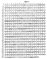


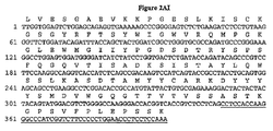

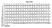


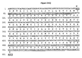


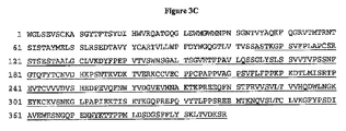


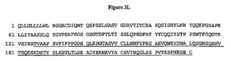


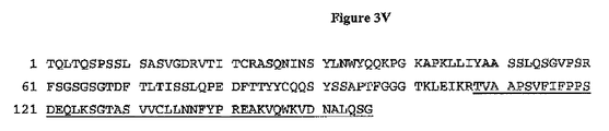


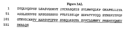


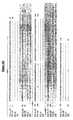

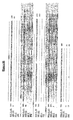


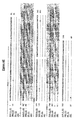


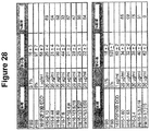
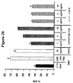


Classifications
-
- C—CHEMISTRY; METALLURGY
- C07—ORGANIC CHEMISTRY
- C07K—PEPTIDES
- C07K16/00—Immunoglobulins [IGs], e.g. monoclonal or polyclonal antibodies
- C07K16/18—Immunoglobulins [IGs], e.g. monoclonal or polyclonal antibodies against material from animals or humans
- C07K16/28—Immunoglobulins [IGs], e.g. monoclonal or polyclonal antibodies against material from animals or humans against receptors, cell surface antigens or cell surface determinants
- C07K16/30—Immunoglobulins [IGs], e.g. monoclonal or polyclonal antibodies against material from animals or humans against receptors, cell surface antigens or cell surface determinants from tumour cells
-
- A—HUMAN NECESSITIES
- A61—MEDICAL OR VETERINARY SCIENCE; HYGIENE
- A61P—SPECIFIC THERAPEUTIC ACTIVITY OF CHEMICAL COMPOUNDS OR MEDICINAL PREPARATIONS
- A61P31/00—Antiinfectives, i.e. antibiotics, antiseptics, chemotherapeutics
-
- A—HUMAN NECESSITIES
- A61—MEDICAL OR VETERINARY SCIENCE; HYGIENE
- A61P—SPECIFIC THERAPEUTIC ACTIVITY OF CHEMICAL COMPOUNDS OR MEDICINAL PREPARATIONS
- A61P35/00—Antineoplastic agents
-
- A—HUMAN NECESSITIES
- A61—MEDICAL OR VETERINARY SCIENCE; HYGIENE
- A61P—SPECIFIC THERAPEUTIC ACTIVITY OF CHEMICAL COMPOUNDS OR MEDICINAL PREPARATIONS
- A61P43/00—Drugs for specific purposes, not provided for in groups A61P1/00-A61P41/00
-
- A—HUMAN NECESSITIES
- A61—MEDICAL OR VETERINARY SCIENCE; HYGIENE
- A61K—PREPARATIONS FOR MEDICAL, DENTAL OR TOILETRY PURPOSES
- A61K39/00—Medicinal preparations containing antigens or antibodies
- A61K2039/505—Medicinal preparations containing antigens or antibodies comprising antibodies
-
- C—CHEMISTRY; METALLURGY
- C07—ORGANIC CHEMISTRY
- C07K—PEPTIDES
- C07K2317/00—Immunoglobulins specific features
- C07K2317/20—Immunoglobulins specific features characterized by taxonomic origin
- C07K2317/24—Immunoglobulins specific features characterized by taxonomic origin containing regions, domains or residues from different species, e.g. chimeric, humanized or veneered
-
- C—CHEMISTRY; METALLURGY
- C07—ORGANIC CHEMISTRY
- C07K—PEPTIDES
- C07K2317/00—Immunoglobulins specific features
- C07K2317/70—Immunoglobulins specific features characterized by effect upon binding to a cell or to an antigen
- C07K2317/73—Inducing cell death, e.g. apoptosis, necrosis or inhibition of cell proliferation
Description
(発明の分野)
本明細書に記載される本発明は、161P2F10Bと称されるタンパク質に結合する抗体、ならびにその結合フラグメントおよびそれから操作された分子に関する。本発明はさらに、161P2F10Bを発現する癌の処置において有用な、診断方法、予後方法、予防方法および治療方法、ならびに組成物に関する。
(Field of Invention)
The invention described herein relates to antibodies that bind to a protein termed 161P2F10B, as well as binding fragments thereof and molecules engineered therefrom. The present invention further relates to diagnostic, prognostic, prophylactic and therapeutic methods, and compositions useful in the treatment of cancers that express 161P2F10B.
(発明の背景)
癌は、冠動脈疾患(coronary disease)に次いで第2の主要なヒトの死因である。世界中で、毎年何百万人もの人々が、癌で亡くなっている。the American Cancer Societyによって報告されるように、米国内のみで、癌は、年間50万人を超える人々の死亡を引き起こし、1年に120万人を超える新規症例が診断されている。心疾患による死亡が有意に減少している一方で、癌によってもたらされる死亡は、一般に増加している。次世紀の早い時期に、癌は、死亡の主要な原因となることが予測される。
(Background of the Invention)
Cancer is the second leading cause of human death following coronary disease. Worldwide, millions of people die from cancer each year. As reported by the American Cancer Society, in the United States alone, cancer causes more than 500,000 deaths annually and more than 1.2 million new cases are diagnosed per year. While deaths from heart disease are significantly reduced, deaths caused by cancer are generally increasing. Early in the next century, cancer is predicted to be a major cause of death.
世界的に、幾種かの癌が、主要な死因として際立っている。特に、肺、前立腺、胸、結腸、膵臓、卵巣および膀胱の癌腫は、癌による死亡の主要な原因を代表する。これらおよび実質的に全ての他の癌腫は、共通の致死性の特徴を共有する。ほとんど例外なく、癌腫からの転移性疾患は、致命的である。さらに、その原発性癌を最初に生き残った癌患者についてでさえ、通常の経験として、その人生が劇的に変化することが示されている。多くの癌患者は、再発もしくは処置の失敗の可能性の認識によって駆り立てられる、強い不安を経験する。多くの癌患者は、処置後の物理的衰弱を経験する。さらに、多くの癌患者は、再発を経験する。 Worldwide, several types of cancer stand out as the leading cause of death. In particular, lung, prostate, breast, colon, pancreas, ovarian and bladder carcinomas represent the leading cause of cancer deaths. These and virtually all other carcinomas share common lethal characteristics. With few exceptions, metastatic disease from carcinoma is fatal. Furthermore, even for cancer patients who first survived the primary cancer, normal experience has shown that their lives change dramatically. Many cancer patients experience strong anxiety driven by the perception of possible recurrence or treatment failure. Many cancer patients experience physical weakness after treatment. In addition, many cancer patients experience a recurrence.
世界的に、前立腺癌は、男性において4番目に多い癌である。北米および北欧において、前立腺癌は、男性における圧倒的に最も一般的な癌であり、男性における第2の主要な死因である。米国内のみで、年間30,000人を有に超える男性が、この疾患で死亡する。肺癌に次いで第2位である。これらの数値の大きさにもかかわらず、転移性前立腺癌についての有効な処置は未だ存在しない。外科的前立腺切除、放射線治療、ホルモン除去(hormone ablation)治療、外科的去勢および化学療法は、主要な処置様式であり続けている。不幸なことに、これらの処置は、多くは有効でなく、そしてしばしば、望ましくない結果を伴う。 Worldwide, prostate cancer is the fourth most common cancer in men. In North America and Northern Europe, prostate cancer is by far the most common cancer in men and the second leading cause of death in men. In the United States alone, more than 30,000 men annually die from the disease. It is second only after lung cancer. Despite the magnitude of these numbers, there is still no effective treatment for metastatic prostate cancer. Surgical prostatectomy, radiation therapy, hormone ablation therapy, surgical castration and chemotherapy continue to be the main treatment modalities. Unfortunately, these treatments are often ineffective and often have undesirable consequences.
診断の最前線において、前立腺腫瘍マーカー(初期段階の限局性腫瘍を正確に検出し得る)がないことは、疾患の診断および管理において深刻な制限を残す。血清前立腺特異的抗原(PSA)アッセイが非常に有用な手段であるが、しかし、特異性および一般的有用性は、いくつかの重要な考慮を欠くと広くみなされている。 At the forefront of diagnosis, the absence of prostate tumor markers (which can accurately detect early-stage localized tumors) leaves serious limitations in disease diagnosis and management. Serum prostate specific antigen (PSA) assay is a very useful tool, but specificity and general utility are widely regarded as lacking some important considerations.
前立腺癌についてのさらなる特異的マーカーの同定における進歩は、マウスにおいて疾患の異なった段階を再現可能である前立腺癌異種移植片の産生によって、改善されている。LAPC(Los Angeles Prostate Cancer)異種移植片は、重症複合型免疫不全(SCID)マウスにおいて継代を生き残った前立腺癌異種移植片であり、アンドロゲン依存からアンドロゲン非依存への移行を模倣する能力を示している(非特許文献1)。より近年同定された前立腺癌マーカーとしては、PCTA−1(非特許文献2)、前立腺特異的膜(PSM)抗原(非特許文献3)、STEAP(非特許文献4)、および前立腺幹細胞抗原(161P2F10B)(非特許文献5)が挙げられる。 Advances in identifying additional specific markers for prostate cancer have been improved by the production of prostate cancer xenografts that can reproduce different stages of the disease in mice. LAPCs (Los Angeles Prostate Cancer) xenografts are prostate cancer xenografts that survived passage in severe combined immunodeficiency (SCID) mice and show the ability to mimic the transition from androgen-dependent to androgen-independent (Non-Patent Document 1). More recently identified prostate cancer markers include PCTA-1 (Non-patent document 2), prostate-specific membrane (PSM) antigen (Non-patent document 3), STEAP (Non-patent document 4), and prostate stem cell antigen (161P2F10B). (Non-Patent Document 5).
以前に同定されているマーカー(例えば、PSA、PSM、PCTAおよび161P2F10B)は、前立腺癌の診断および処置のための努力を促進してきたが、診断および治療のさらなる改善のために、前立腺癌および関連する癌についてのさらなるマーカーおよび治療標的の同定が必要とされている。 Previously identified markers (eg, PSA, PSM, PCTA and 161P2F10B) have facilitated efforts for the diagnosis and treatment of prostate cancer, but for further improvements in diagnosis and therapy, prostate cancer and related There is a need for the identification of additional markers and therapeutic targets for cancers that do.
腎細胞癌(RCC)は、成人の悪性疾患の約3%の原因となっている。一旦、腺腫が2〜3cmの直径に達すると、悪性の可能性がある。成人において、2つの主要な悪性腎腫瘍は、腎細胞腺癌および腎盤もしくは尿管の移行上皮癌である。腎細胞腺癌の発症率は、1988年に米国内で、29,000件を超える症例であり、そして11,600人を超える患者が、この疾患で亡くなっていると見積もられる。移行性上皮癌は、より頻度が低く、発症率はおよそ米国内で年間500件である。 Renal cell carcinoma (RCC) is responsible for about 3% of adult malignancies. Once an adenoma reaches a diameter of 2-3 cm, it may be malignant. In adults, the two main malignant renal tumors are renal cell adenocarcinoma and transitional cell carcinoma of the renal pelvis or ureter. The incidence of renal cell adenocarcinoma is estimated to be more than 29,000 cases in the United States in 1988, and more than 11,600 patients die from the disease. Transitional cell carcinoma is less frequent, with an incidence of approximately 500 cases per year in the United States.
外科手術は、腎細胞腺癌について何十年にもわたって主要な治療であった。近年まで、転移性疾患は、いかなる全身治療に対しても難治性であった。全身治療(特に免疫治療)の近年の進歩により、転移性腎細胞癌腫は、耐久性のある応答の可能性を有する適切な患者において、積極的にアプローチされ得る。しかし、これらの患者について、有効な治療の必要性がまだ残る。 Surgery has been the main treatment for decades for renal cell adenocarcinoma. Until recently, metastatic disease was refractory to any systemic treatment. With recent advances in systemic therapy (especially immunotherapy), metastatic renal cell carcinoma can be aggressively approached in appropriate patients with the potential for a durable response. However, there remains a need for effective treatment for these patients.
米国における癌の全ての新規の症例の中で、膀胱癌は、男性において約5%を表し(5番目に多い新生物)、そして女性において3%を表す(8番目に多い新生物)。発症率は、高齢の人口が増加すると同時に、ゆっくりと増加している。1998年に、推定54,500件の症例(男性39,500件および女性15,000件を含む)が存在した。米国における年齢調整した発症率は、男性について100,000人あたり32件、そして女性において100,000人あたり8件である。過去の男性/女性比は3:1であったが、これは、女性の喫煙傾向に関連して減少しているかもしれない。1998年に、膀胱癌で推定11,000件(78,000人は男性であり、そして3,900人は女性である)の死亡が存在した。膀胱癌発症率および死亡率は、年齢に伴って急激に増加し、そして人口がより高齢化するに従って課題が増大する。 Among all new cases of cancer in the United States, bladder cancer represents about 5% in men (5th most common neoplasm) and 3% in women (8th most common neoplasm). Incidence is increasing slowly as the elderly population increases. In 1998 there were an estimated 54,500 cases (including 39,500 males and 15,000 females). Age-adjusted incidence in the United States is 32 cases per 100,000 people for men and 8 cases per 100,000 people for women. The historical male / female ratio was 3: 1, which may be reduced in relation to women's smoking tendency. In 1998, there were an estimated 11,000 deaths (78,000 were male and 3,900 female) in bladder cancer. Bladder cancer incidence and mortality increase rapidly with age, and challenges increase as the population ages.
ほとんどの膀胱癌は、膀胱において再発する。膀胱癌は、膀胱の経尿道的切除(TUR)と膀胱内の化学療法もしくは免疫治療との組み合わせによって扱われる。膀胱癌の多病巣性かつ再発性の性質は、TURの限界を示す。最も筋肉侵襲性の癌は、TURのみによっては治癒されない。根治的膀胱切除および尿路変更術は、癌を消滅させる最も有効な手段であるが、排尿機能および性機能に否定できない影響を有する。膀胱癌患者についての有効な処置様式の重大な必要性が存在し続ける。 Most bladder cancers recur in the bladder. Bladder cancer is treated by a combination of transurethral resection (TUR) of the bladder and intravesical chemotherapy or immunotherapy. The multifocal and recurrent nature of bladder cancer indicates a limitation of TUR. Most muscle invasive cancers are not cured by TUR alone. Radical cystectomy and urinary diversion are the most effective means of eliminating cancer, but have undeniable effects on micturition and sexual function. There continues to be a significant need for effective treatment modalities for patients with bladder cancer.
推定130,200件の結腸直腸癌が、米国内で2000年に発症した(93,800件の結腸癌および36,400件の直腸癌を含む)。結腸直腸癌は、男性および女性において、3番目に一般的な癌である。1992〜1996年の間に、発症率は有意に低下している(一年あたり−2.1%)。研究は、これらの低下は、スクリーニングおよびポリープ切除の増加により、ポリープが侵襲性癌に進行するのを防ぐことに起因していることを示唆する。2000年には、推定56,300件(47,700件は結腸癌、86,00件は直腸癌)の死亡が存在し、米国の癌による死亡の約11%の原因であった。 An estimated 130,200 colorectal cancers occurred in 2000 in the United States (including 93,800 colon cancers and 36,400 rectal cancers). Colorectal cancer is the third most common cancer in men and women. Between 1992 and 1996, the incidence has declined significantly (−2.1% per year). Studies suggest that these declines are due to the increased screening and polypectomy preventing polyps from progressing to invasive cancers. In 2000, there were an estimated 56,300 deaths (47,700 colon cancer and 86,000 rectal cancer), accounting for about 11% of cancer deaths in the United States.
現在、外科手術は、結腸直腸癌および拡散していない癌についての最も一般的な形態であり、しばしば治癒可能である。化学療法もしくは化学療法+放射線は、腸壁に深く穴が開いている癌もしくはリンパ節まで広がっている癌を有するほとんどの患者に、術前もしくは術後に与えられる。永久的人工肛門形成術(体内排泄物の除去のための腹部開口の作製)が、結腸癌のために、時折必要とされ、そして直腸癌のためにはまれに必要とされる。結腸直腸癌患者についての有効な処置様式の必要性が、存在し続ける。 Currently, surgery is the most common form for colorectal cancer and non-diffusing cancer and is often curable. Chemotherapy or chemotherapy plus radiation is given before or after surgery to most patients with cancers that are deeply perforated in the intestinal wall or that have spread to lymph nodes. Permanent colostomy (creating an abdominal opening for removal of bodily waste) is sometimes required for colon cancer and rarely required for rectal cancer. There remains a need for effective treatment modalities for patients with colorectal cancer.
2000年に、肺癌および気管支癌の推定164,1000件の新規の症例が存在し、米国の癌診断の14%の原因であった。肺癌および気管支癌の発症率は、男性において有意に低下している(1984年における86.5%の高さから、1996年における70.0%まで)。1990年代に、女性における増加の速度が遅くなり始めた。1996年には、女性における発症率は、100,000人あたり42.3%であった。 In 2000, there were an estimated 164,000 new cases of lung and bronchial cancer, accounting for 14% of US cancer diagnoses. The incidence of lung and bronchial cancer is significantly reduced in men (from a height of 86.5% in 1984 to 70.0% in 1996). In the 1990s, the rate of increase in women began to slow. In 1996, the incidence in women was 42.3% per 100,000 people.
肺癌および気管支癌は、2000年に推定156,900件の死亡をもたらし、全ての癌による死亡の28%の原因であった。1992〜1996年の間、肺癌の死亡率は、男性において有意に低下した(年間−1.7%)一方で、女性の死亡率はなお有意に増加していた(年間0.9%)。1987年から、毎年、乳癌よりも肺癌によって、より多くの女性(40歳以上)が死亡しており、女性における癌による死亡の主要な原因であった。肺癌の発症率および死亡率の減少は、ここ30年間にわたる喫煙率の低下に起因する可能性が最も高い。しかし、女性における喫煙傾向の減少は、男性におけるそれよりも大きく劣る。成人の煙草使用における低下は遅くなっているが、若年者における煙草使用が再び増加していることが懸念される。 Lung and bronchial cancers resulted in an estimated 156,900 deaths in 2000, accounting for 28% of all cancer deaths. Between 1992 and 1996, lung cancer mortality was significantly reduced in men (-1.7% per year), while female mortality was still significantly increased (0.9% per year). Since 1987, more women (over 40 years old) have died from lung cancer than breast cancer every year, which has been the leading cause of cancer death in women. The decrease in lung cancer incidence and mortality is most likely due to the decline in smoking rates over the last 30 years. However, the reduction in smoking tendency in women is much worse than that in men. Although the decline in tobacco use among adults is slowing, there are concerns that tobacco use in young people is increasing again.
肺癌および気管支癌についての処置選択肢は、癌の種類および段階によって決定され、外科手術、放射線治療、および化学療法が挙げられる。多くの限局性癌について、外科手術は、通常、選り抜きの処置である。疾患は、通常、発見された時までに拡散しているので、放射線治療および化学療法は、しばしば外科手術と組み合わされる必要がある。化学療法のみもしくは放射線と組み合わせた化学療法は、小細胞肺癌についての選り抜きの処置である。このレジメンにおいて、患者の大きな割合は、寛解を経験し、この寛解は、いくつかの場合では長く続く。しかし、肺癌および気管支癌についての、有効な処置および診断アプローチについての継続的な必要性が存在し続ける。 Treatment options for lung cancer and bronchial cancer are determined by the type and stage of the cancer and include surgery, radiation therapy, and chemotherapy. For many localized cancers, surgery is usually a selective procedure. Because the disease is usually diffused by the time it is discovered, radiation therapy and chemotherapy often need to be combined with surgery. Chemotherapy alone or in combination with radiation is the treatment of choice for small cell lung cancer. In this regimen, a large proportion of patients experience remission, which in some cases lasts long. However, there is a continuing need for effective treatment and diagnostic approaches for lung and bronchial cancer.
推定182,800件の侵襲性の乳癌の新規の症例が、2000年の間に米国内の女性の間で発症すると予測された。さらに、約1,400件の乳癌の新規の症例が、2000年に男性において診断されることが予測された。1980年代の年間約4%の増加の後、女性における乳癌発症率は、1990年代には100,000人あたり110.6件まで低下した。 An estimated 182,800 new cases of invasive breast cancer were predicted to develop among women in the United States during 2000. In addition, approximately 1,400 new cases of breast cancer were predicted to be diagnosed in men in 2000. After an annual increase of about 4% in the 1980s, the incidence of breast cancer in women dropped to 110.6 cases per 100,000 in the 1990s.
米国のみで、2000年に、乳癌に起因する推定41,200件の死亡(40,800件は女性、400件は男性)が存在した。乳癌は、女性における癌による死亡の間で2番目のランクである。最も最近のデータによれば、死亡率は、白人および黒人の両方で、1992〜1996年の間に有意に低下しており、若い女性の間で最も大きく低下した。これらの低下は、おそらく、初期の発見および改善された処置の結果であった。 In the United States alone, in 2000 there were an estimated 41,200 deaths (40,800 women, 400 men) due to breast cancer. Breast cancer ranks second among cancer deaths in women. According to the most recent data, mortality was significantly decreased between 1992 and 1996 in both whites and blacks, with the greatest decrease among young women. These declines were probably the result of early discovery and improved treatment.
医学的状況および患者の好みを考慮し、乳癌の処置は、ランペクトミー(腫瘍の局所的除去)および腕下のリンパ節の除去;乳房切除(乳房の外科的除去)および腕下のリンパ節の除去;放射線治療;化学療法;あるいはホルモン療法を包含し得る。しばしば、2以上の方法が、組み合わせて使用される。多くの研究は、所期段階の疾患について、ランペクトミー+放射線治療の後の長期生存率は、改変型定型的乳房切除の後の生存率と類似することを示している。再建技術の顕著な進歩は、乳房切除後の乳房再建についてのいくつかの選択肢を提供する。近年、このような再建は、乳房切除と同時に成されている。 Taking into account the medical situation and patient preference, breast cancer treatments include lampectomy (local removal of the tumor) and removal of the lymph nodes under the arm; mastectomy (surgical removal of the breast) and removal of the lymph nodes under the arm Radiation therapy; chemotherapy; or hormonal therapy. Often two or more methods are used in combination. Many studies have shown that for the stage of disease, long-term survival after lampectomy plus radiation therapy is similar to survival after modified routine mastectomy. Significant advances in reconstruction techniques offer several options for breast reconstruction after mastectomy. In recent years, such reconstruction has been done simultaneously with mastectomy.
十分な量の周囲の正常乳房組織と共の、インサイチュ腺管癌(ductal carcinoma in situ:DCIS)の局所的切除は、DCISの局所的再発を予防し得る。乳房への放射線および/またはタモキシフェンは、残存する乳房組織におけるDCISの発症の確率を低下させ得る。DCISが未処置のままである場合、DCISは侵襲性乳癌に発達し得るので、このことは重要である。それにもかかわらず、これらの処置には、深刻な副作用または続発症が存在する。従って、有効な乳癌処置の必要性が存在する。 Local excision of a ductal carcinoma in situ (DCIS) with a sufficient amount of surrounding normal breast tissue can prevent local recurrence of DCIS. Radiation to the breast and / or tamoxifen can reduce the probability of developing DCIS in the remaining breast tissue. This is important because DCIS can develop into invasive breast cancer if DCIS remains untreated. Nevertheless, these treatments have serious side effects or sequelae. Thus, there is a need for effective breast cancer treatment.
推定23,100件の卵巣癌の新規症例が、2000年に米国内で存在した。これは、女性の間での癌による死亡の4%の原因であり、婦人科の癌の間で2番目のランクである。1992〜1996年の間に、卵巣癌発症率は、有意に低下した。卵巣癌の結果として、2000年に、推定14,000件の死亡が存在した。卵巣癌は、女性生殖系のいかなる他の癌よりも多くの死亡を引き起こす。 An estimated 23,100 new cases of ovarian cancer were present in the United States in 2000. This is responsible for 4% of cancer deaths among women and ranks second among gynecological cancers. Between 1992 and 1996, the incidence of ovarian cancer decreased significantly. There were an estimated 14,000 deaths in 2000 as a result of ovarian cancer. Ovarian cancer causes more deaths than any other cancer in the female reproductive system.
外科手術、放射線治療、および化学療法は、卵巣癌の処置の選択肢である。外科手術は、通常、1つもしくは両方の卵巣およびファローピウス管の除去(卵管卵巣摘出)、ならびに子宮の除去(子宮摘出)を包含する。いくつかの非常に初期の腫瘍において、特に子供を持つことを望む若い女性においては、関与する卵巣のみを除去する。進行した疾患においては、全ての腹内疾患を除去し、化学療法の効果を高める試みが行われる。卵巣癌についての有効な処置選択肢の重要な必要性が、存在し続ける。 Surgery, radiation therapy, and chemotherapy are treatment options for ovarian cancer. Surgery usually involves removal of one or both ovaries and fallopian tubes (tubal ovariectomy) and uterus removal (hysterectomy). In some very early tumors, especially in young women who want to have children, only the ovaries involved are removed. In advanced disease, attempts are made to eliminate all intraabdominal diseases and enhance the effectiveness of chemotherapy. An important need for effective treatment options for ovarian cancer continues to exist.
推定28,300件の新規の膵臓癌の症例が、2000年に米国内で存在した。過去20年間にわたって、膵臓癌の確率は、男性において低下している。女性における確率は、およそ一定にとどまっているが、低下し始めているかもしれない。膵臓癌は、2000年に米国内において、推定28,200件の死亡を引き起こした。過去20年にわたって、男性における死亡率において、わずかではあるが有意な低下(約−0.9%)が存在しており、一方、女性の間ではわずかに増加している。 An estimated 28,300 new cases of pancreatic cancer were present in the United States in 2000. Over the past 20 years, the probability of pancreatic cancer has declined in men. Probabilities in women remain roughly constant, but may begin to decline. Pancreatic cancer caused an estimated 28,200 deaths in the United States in 2000. Over the past 20 years, there has been a slight but significant decrease (about -0.9%) in mortality in men, while there has been a slight increase among women.
外科手術、放射線治療、および化学療法は、膵臓癌についての処置選択肢である。これらの処置選択肢は、多くの患者において、生存を延長し得、そして/または症状を緩和し得るが、ほとんどの場合、治癒しない確率が高い。癌についての、さらなる治療および診断の選択肢の重要な必要性が、存在する。これらの選択肢としては、抗体、ワクチンおよび低分子の使用が、処置様式として挙げられる。さらにまた、これらの様式を、診断、検出、モニタリング、ならびに癌の処置および研究の全ての分野における技術のさらなる段階に対する研究ツールとして使用することが、必要とされる。 Surgery, radiation therapy, and chemotherapy are treatment options for pancreatic cancer. These treatment options can prolong survival and / or alleviate symptoms in many patients, but most likely do not cure. There is an important need for additional treatment and diagnostic options for cancer. These options include the use of antibodies, vaccines and small molecules as treatment modalities. Furthermore, there is a need to use these modalities as research tools for further stages of technology in all areas of diagnosis, detection, monitoring, and cancer treatment and research.
モノクローナル抗体(mAb)(非特許文献6)の治療有用性が、認識されている。モノクローナル抗体は、現在、移植術、癌、感染性疾患、心臓血管疾患および炎症における治療として認められている。異なるイソ型は、異なるエフェクター機能を有する。このような機能における相違は、種々の免疫グロブリンイソ型についての異なる3次元構造に反映される。(非特許文献7)。 The therapeutic utility of monoclonal antibodies (mAb) (Non-Patent Document 6) has been recognized. Monoclonal antibodies are currently recognized as treatments in transplantation, cancer, infectious diseases, cardiovascular diseases and inflammation. Different isoforms have different effector functions. Such differences in function are reflected in the different three-dimensional structures for the various immunoglobulin isoforms. (Non-patent document 7).
マウスは、免疫化のために便利であり、かつ、ほとんどのヒト抗原を外来物質として認識するので、治療可能性を有するヒト標的に対するmAbは、代表的に、マウス起源である。しかし、マウスmAbは、ヒト治療薬として、固有の不利な点を有する。これらは、より頻繁な投薬を必要とする。なぜなら、mAbは、ヒトにおいて、ヒト抗体よりも短い循環半減期を有するからである。より重要なことには、マウス抗体のヒト免疫系への繰り返し投与は、マウスタンパク質を外来物質として認識することによって、ヒト免疫系の応答を引き起こし、そしてヒト抗マウス抗体(HAMA)応答を産生する。このようなHAMA応答は、アレルギー反応および系からのマウス抗体の迅速な排出をもたらし得、それによって、マウス抗体による処置を役立たずにしてしまう。このような影響を避けるために、マウス内でヒト免疫系を作製する試みが、企画されてきた。 Since mice are convenient for immunization and recognize most human antigens as foreign substances, mAbs against human targets with therapeutic potential are typically of mouse origin. However, mouse mAbs have inherent disadvantages as human therapeutics. These require more frequent dosing. This is because mAbs have a shorter circulating half-life in humans than human antibodies. More importantly, repeated administration of mouse antibodies to the human immune system causes a response of the human immune system by recognizing mouse proteins as foreign substances and produces a human anti-mouse antibody (HAMA) response. . Such HAMA responses can lead to allergic reactions and rapid elimination of mouse antibodies from the system, thereby rendering treatment with mouse antibodies useless. In order to avoid such effects, attempts have been made to create the human immune system in mice.
最初の試みは、抗原にヒト配列を有する抗体で応答可能であるトランスジェニックマウスを作製することを望んだが、(非特許文献8)、入手可能なクローニングビヒクルによって安定的に維持されるDNAの量は、限られていた。酵母人工染色体(YAC)クローニングベクターの使用は、ヒトIg遺伝子座の大型生殖系列フラグメントをトランスジェニック動物内に導入するための道をもたらした。本質的に、ヒトゲノム中で見出される同じ間隔で配置されるヒトのV領域遺伝子、D領域遺伝子、およびJ領域遺伝子の大部分、ならびにヒト定常領域が、YACを用いてマウス内に導入された。1種のこのようなトランスジェニックマウス系統は、XenoMouse(r)マウスとして公知であり、そしてAbgenix,Inc.(Fremont CA)から市販されている。 The first attempt was to create a transgenic mouse that can respond to an antigen with a human sequence antibody (Non-Patent Document 8), but the amount of DNA stably maintained by available cloning vehicles. Was limited. The use of yeast artificial chromosome (YAC) cloning vectors has provided a way for introducing large germline fragments of the human Ig locus into transgenic animals. In essence, most of the human V region genes, D region genes, and J region genes located at the same intervals found in the human genome, as well as human constant regions, were introduced into mice using YAC. One such transgenic mouse strain is known as the XenoMouse (r) mouse and is described by Abgenix, Inc. (Fremont CA).
(発明の要旨)
本発明は、161P2F10Bタンパク質および161P2F10Bタンパク質のポリペプチドフラグメトに結合する抗体、ならびにその結合フラグメントおよびそれから操作された分子を提供する。本発明は、ポリクローナル抗体およびモノクローナル抗体、マウスおよび他の哺乳動物の抗体、キメラ抗体、ヒト化抗体および完全ヒト抗体、ならびに検出可能なマーカーもしくは治療因子で標識された抗体を含む。特定の実施形態において、図3の全核酸配列がコードされないこと、および/または図2の全アミノ酸配列が調製されないという条件が存在する。特定の実施形態において、図3の全核酸配列がコードされ、そして/または図2のアミノ酸配列が調製される(そのいずれかはそれぞれのヒト単位用量形態である)。
(Summary of the Invention)
The present invention provides antibodies that bind to 161P2F10B protein and polypeptide fragment of 161P2F10B protein, as well as binding fragments thereof and molecules engineered therefrom. The invention includes polyclonal and monoclonal antibodies, murine and other mammalian antibodies, chimeric antibodies, humanized and fully human antibodies, and antibodies labeled with a detectable marker or therapeutic agent. In certain embodiments, there are conditions that the entire nucleic acid sequence of FIG. 3 is not encoded and / or that the entire amino acid sequence of FIG. 2 is not prepared. In certain embodiments, the entire nucleic acid sequence of FIG. 3 is encoded and / or the amino acid sequence of FIG. 2 is prepared, either of which is a respective human unit dose form.
本発明はさらに、種々の生物学的サンプル中の161P2F10Bポリヌクレオチドおよびタンパク質の存在および状態を検出する方法、ならびに161P2F10Bを発現する細胞を同定するための方法を提供する。本発明の一実施形態は、増殖調節不全のある形式(例えば、癌)を有するかまたは増殖調節不全のある形式を有すると考えられる組織サンプルまたは血液サンプル中の161P2F10B遺伝子産物をモニタリングするための方法を提供する。 The present invention further provides methods for detecting the presence and status of 161P2F10B polynucleotides and proteins in various biological samples, and methods for identifying cells that express 161P2F10B. One embodiment of the invention is a method for monitoring a 161P2F10B gene product in a tissue or blood sample that has a form of growth dysregulation (eg, cancer) or is believed to have a form of growth dysregulation. I will provide a.
本発明はさらに、種々の免疫原性組成物もしくは治療用組成物、および161P2F10Bを発現する癌(例えば、表Iに列挙される組織の癌)を処置するためのストラテジー(転写、翻訳、プロセシングまたは161P2F10Bおよび癌ワクチンの機能を阻害することを目的とする治療を含む)を提供する。一局面において、本発明は、ヒト被験体において161P2F10Bを発現する癌を処置するための組成物、およびその組成物を包含する方法を提供し、ここで、この組成物は、ヒトへの使用に適切なキャリア、および161P2F10Bの生成もしくは機能を阻害する1つもしくは1つよりも多い因子のヒト単位用量を含む。好ましくは、上記キャリアは一意的なヒトキャリアである。本発明の別の局面において、上記因子は、161P2F10Bタンパク質と免疫反応性である部分である。そのような部分の非限定的な例としては、抗体(例えば、単鎖抗体、モノクローナル抗体、ポリクローナル抗体、ヒト化抗体、キメラ抗体またはヒト抗体)、その機能的等価物(天然に存在するものであろうと合成物であろうと)、およびそれらの組み合わせが挙げられるが、これらに限定されない。上記抗体は、診断用部分または治療用部分に結合され得る。別の局面において、上記因子は、本明細書に定義されるような低分子である。 The invention further provides various immunogenic or therapeutic compositions and strategies (transcription, translation, processing or for treating cancers expressing 161P2F10B (eg, cancers of tissues listed in Table I)). 161P2F10B and treatment aimed at inhibiting the function of cancer vaccines). In one aspect, the present invention provides a composition for treating cancer expressing 161P2F10B in a human subject, and a method comprising the composition, wherein the composition is for use in humans. A suitable carrier and a human unit dose of one or more factors that inhibit the production or function of 161P2F10B. Preferably, the carrier is a unique human carrier. In another aspect of the present invention, the factor is a moiety that is immunoreactive with 161P2F10B protein. Non-limiting examples of such moieties include antibodies (eg, single chain antibodies, monoclonal antibodies, polyclonal antibodies, humanized antibodies, chimeric antibodies or human antibodies), functional equivalents thereof (naturally occurring Including, but not limited to, and combinations thereof. The antibody can be conjugated to a diagnostic moiety or a therapeutic moiety. In another aspect, the factor is a small molecule as defined herein.
(発明の詳細な説明)
(節の概要)
I.)定義
II.)161P2F10Bポリヌクレオチド
II.A.)161P2F10Bポリヌクレオチドの使用
II.A.1.)遺伝子異常のモニタリング
II.A.2.)アンチセンス実施形態
II.A.3.)プライマーおよびプライマー対
II.A.4.)161P2F10Bコード核酸分子の単離
II.A.5.)組換え核酸分子および宿主−ベクター系
III.)161P2F10B関連タンパク質
III.A.)モチーフを有するタンパク質の実施形態
III.B.)161P2F10B関連タンパク質の発現
III.C.)161P2F10B関連タンパク質の改変
III.D.)161P2F10B関連タンパク質の用途
IV.)161P2F10B抗体
V.)161P2F10B細胞性免疫応答
VI.)161P2F10Bトランスジェニック動物
VII.)161P2F10Bの検出のための方法
VIII.)161P2F10B関連遺伝子およびこれらの産物の状態をモニターする
ための方法
IX.)161P2F10Bと相互作用する分子の同定
X.)治療法および組成物
X.A.)抗癌ワクチン
X.B.)抗体ベースの治療に関する標的としての161P2F10B
X.C.)細胞性免疫応答のための標的としての161P2F10B
X.C.1. ミニ遺伝子ワクチン
X.C.2. CTLペプチドとヘルパーペプチドとの組み合わせ
X.C.3. CTLペプチドとT細胞感作薬剤との組み合わせ
X.C.4. CTLペプチドおよび/またはHTLペプチドが適用されたDC
を含むワクチン組成物
X.D.)養子免疫療法
X.E.)治療目的または予防目的でのワクチン投与
XI.)161P2F10Bの診断実施形態および予後実施形態
XI.A.)161P2F10Bタンパク質機能の阻害
XI.B.)細胞内抗体を用いた161P2F10Bの阻害
XI.C.)組み換えタンパク質による161P2F10Bの阻害
XI.D.)161P2F10Bの転写または翻訳の阻害
XI.E.)治療戦略のための一般的な考慮事項
XII.)161P2F10Bのモジュレーターの同定、特徴付けおよび使用
XIII.)RNAiおよび小さい干渉RNA(siRNA)の治療的使用
XIV.)キット/製造物品。
(Detailed description of the invention)
(Summary of section)
I. ) Definition II. ) 161P2F10B polynucleotide II. A. ) Use of 161P2F10B polynucleotides II. A. 1. ) Monitoring of gene abnormality II. A. 2. ) Antisense Embodiment II. A. 3. ) Primers and primer pairs II. A. 4). ) Isolation of 161P2F10B-encoding nucleic acid molecule II. A. 5. ) Recombinant nucleic acid molecules and host-vector systems III. ) 161P2F10B-related protein III. A. ) Embodiments of proteins with motifs III. B. ) Expression of 161P2F10B-related protein III. C. ) Modification of 161P2F10B related protein III. D. ) Use of 161P2F10B related protein IV. ) 161P2F10B antibody ) 161P2F10B cellular immune response VI. ) 161P2F10B transgenic animal VII. ) Method for detection of 161P2F10B VIII. ) Methods for monitoring the status of 161P2F10B related genes and their products IX. ) Identification of molecules that interact with 161P2F10B X. ) Treatment methods and compositions X. A. ) Anti-cancer vaccine X. B. ) 161P2F10B as a target for antibody-based therapy
X. C. ) 161P2F10B as a target for cellular immune response
X. C. 1. Minigene vaccine X. C. 2. Combination of CTL peptide and helper peptide X. C. 3. Combination of CTL peptide and T cell sensitizer X. C. 4). DC to which CTL peptide and / or HTL peptide is applied
A vaccine composition comprising X. D. ) Adoptive immunotherapy X. E. ) Vaccine administration for therapeutic or prophylactic purposes XI. ) 161P2F10B diagnostic and prognostic embodiments XI. A. ) Inhibition of 161P2F10B protein function XI. B. ) Inhibition of 161P2F10B with intracellular antibodies XI. C. ) Inhibition of 161P2F10B by recombinant protein XI. D. ) Inhibition of 161P2F10B transcription or translation XI. E. ) General Considerations for Treatment Strategy XII. ) Identification, characterization and use of modulators of 161P2F10B XIII. ) Therapeutic use of RNAi and small interfering RNA (siRNA) XIV. ) Kit / manufactured article.
I.)定義
他に定義されない限り、本明細書において使用されるすべての技術用語、記号、および他の科学用語もしくは術語は、本発明が関する分野の当業者によって一般的に理解される意味を有することが、意図される。いくつかの場合において、一般的に理解される意味を有する用語が、明確さのため、および/またはすぐに参照するために、本明細書において定義される。本明細書においてそのような定義を含めることは、当該分野において一般的に理解されることに対する実質的な差異を示すと必ずしも解釈されるべきではない。本明細書において記載または参照される技術および手順のうちの多くは、当業者によって十分に理解され、そして当業者によって従来の方法(例えば、Sambrookら、Molecular Cloning:A Laboratory Manual 2nd.edition(1989) Cold Spring Harbor Laboratory Press,Cold Spring Harbor,N.Yに記載される広く使用されている分子クローニング方法論)を使用して、一般的に使用される。適切な場合、市販のキットおよび試薬の使用を含む手順が、他に記載されない限りは、製造業者が規定したプロトコルおよび/またはパラメーターに従って、一般的には実行される。
I. ) Definitions Unless defined otherwise, all technical terms, symbols, and other scientific terms or terms used herein have the meaning commonly understood by one of ordinary skill in the art to which this invention pertains. Is intended. In some cases, terms having a commonly understood meaning are defined herein for clarity and / or for quick reference. The inclusion of such definitions in this specification should not necessarily be construed as indicating a substantial difference to what is commonly understood in the art. Many of the techniques and procedures described or referenced herein are well understood by those skilled in the art and are known to those skilled in the art (eg, Sambrook et al., Molecular Cloning: A Laboratory Manual 2nd. Edition (1989)). ) Commonly used, using the widely used molecular cloning methodology described in Cold Spring Harbor Laboratory Press, Cold Spring Harbor, NY. Where appropriate, procedures involving the use of commercially available kits and reagents are generally performed according to manufacturer defined protocols and / or parameters unless otherwise stated.
用語「進行型癌」、「局所進行型癌」、「進行型疾患」および「局所進行型疾患」とは、関連する組織被膜(capsule)を通って伸長した癌を意味し、この用語は、米泌尿器学会(American Urological Association(AUA))のシステムでステージCの疾患、Whitmore−JewettのシステムでステージC1〜C2の疾患、およびTNM(腫瘍、結節、転移)システムでステージT3〜T4およびN+の疾患を包含する。一般に、局所進行型疾患を有する患者に対して、手術は勧められない。これらの患者は、臨床的に限局化した(器官限定的な)癌と比較して、実質的に好ましくない結果を有する。 The terms “advanced cancer”, “locally advanced cancer”, “advanced disease” and “locally advanced disease” mean a cancer that has extended through the associated tissue capsule, Stage C disease in the American Urological Association (AUA) system, stage C1-C2 disease in the Whitmore-Jewett system, and stages T3-T4 and N + in the TNM (tumor, nodule, metastasis) system Includes diseases. In general, surgery is not recommended for patients with locally advanced disease. These patients have substantially unfavorable results compared to clinically localized (organ-limited) cancer.
「ネイティブグリコシル化パターンの変更」とは、ネイティブ配列の161P2F10Bにおいて見出される1つ以上の糖質部分を(基礎となるグリコシル化部位を除去すること、または化学的手段および/もしくは酵素的手段によってグリコシル化を欠失させることのいずれかによって)欠失させること、ならびに/あるいは、そのネイティブ配列の161P2F10B中には存在しない1つ以上のグリコシル化部位を付加することを意味するために、本明細書における目的のために意図される。さらに、上記の句は、ネイティブタンパク質のグリコシル化の性質的変化(存在する種々の糖質部分の性質および比率の変化を含む)を包含する。 “Modification of the native glycosylation pattern” refers to the removal of one or more carbohydrate moieties found in the native sequence 161P2F10B (by removing the underlying glycosylation site or by chemical and / or enzymatic means. In this context to mean deleting and / or adding one or more glycosylation sites that are not present in the native sequence 161P2F10B. Intended for purposes in Furthermore, the above phrases encompass changes in the glycosylation properties of the native protein, including changes in the nature and proportion of the various carbohydrate moieties present.
用語「アナログ」とは、別の分子(例えば、161P2F10B関連タンパク質)と構造的に類似するかまたは類似もしくは対応する属性を共有する、分子を指す。例えば、161P2F10Bタンパク質のアナログは、161P2F10Bに特異的に結合する抗体もしくはT細胞によって、特異的に結合され得る。 The term “analog” refers to a molecule that is structurally similar or shares similar or corresponding attributes with another molecule (eg, 161P2F10B-related protein). For example, an analog of 161P2F10B protein can be specifically bound by an antibody or T cell that specifically binds to 161P2F10B.
用語「抗体」とは、他に明確に示されない限りは、その最も広い意味で使用される。従って、「抗体」は、天然に存在しても、人工生成されてもよい(例えば、従来のハイブリドーマ技術によって生成されたモノクローナル抗体)。抗161P2F10B抗体は、モノクローナル抗体およびポリクローナル抗体、ならびにこれらの抗体の抗原結合ドメインおよび/または1つ以上の相補性決定領域を含むフラグメントを包含する。本明細書において使用される場合、用語「抗体」とは、161P2F10Bに特異的に結合し、かつ/または望ましい生物学的活性を示す、任意の形態の抗体もしくはそのフラグメントを指す。この用語「抗体」は、モノクローナル抗体(全長モノクローナル抗体を含む)、ポリクローナル抗体、多重特異性抗体(例えば、二重特異性抗体)、および抗体フラグメントを、それらが161P2F10Bに特異的に結合し、かつ/または望ましい生物学的活性を示す限り、具体的には網羅する。任意の特異的抗体が、本明細書において提供される方法および組成物において使用され得る。従って、一実施形態において、用語「抗体」とは、軽鎖免疫グロブリン分子由来の少なくとも1つの可変領域と、重鎖分子由来の少なくとも1つの可変領域と(これらは、組み合わされると、標的抗原の特異的結合部位を形成する)を含む分子を包含する。一実施形態において、上記抗体は、IgG抗体である。例えば、上記抗体は、IgG1抗体、IgG2抗体、IgG3抗体、またはIgG4抗体である。本方法および組成物において有用な抗体は、細胞培養物中、ファージ中、または種々の動物(ウシ、ウサギ、ヤギ、マウス、ラット、ハムスター、モルモット、ヒツジ、イヌ、ネコ、サル、チンパンジー、類人猿が挙げられるがこれらに限定はされない)中で生成され得る。従って、一実施形態において、本発明の抗体は、哺乳動物抗体である。ファージ技術が、最初の抗体を単離するため、または変化した特異性特徴もしくは変化したアビディティ特徴を有する改変体を生成するために、使用され得る。そのような技術は、慣用的であり、当該分野で周知である。一実施形態において、上記抗体は、当該分野で公知である組換え手段によって生成される。例えば、組換え抗体は、宿主細胞を、その抗体をコードするDNA配列を含むベクターでトランスフェクトすることによって、生成され得る。1つ以上のベクターが、少なくとも1つのVL領域および1つのVH領域を発現するDNA配列を、その宿主細胞においてトランスフェクトするために使用され得る。抗体生成および抗体産生についての組換え手段の例示的記載としては、Delves,ANTIBO
DY PRODUCTION:ESSENTIAL TECHNIQUES(Wiley,1997);Shephardら、MONOCLONAL ANTIBODIES(Oxford University Press,2000);Goding,MONOCLONAL ANTIBODIES:PRINCIPLESおよびPRACTICE(Academic Press,1993);CURRENT PROTOCOLS IN IMMUNOLOGY(John Wiley & Sons,最新版)が挙げられる。本発明の抗体は、望ましい機能を媒介することにおいて上記抗体の効力をより増加するために、組換え手段によって改変され得る。従って、組換え手段を使用する置換によって改変され得ることは、本発明の範囲内にある。代表的には、上記置換は、保存的置換である。例えば、上記抗体の定常領域中の少なくとも1つのアミノ酸が、異なる残基で置換され得る。例えば、米国特許第5,624,821号、米国特許第6,194,551号、出願公開WO 9958572;およびAngalら、Mol.Immunol.30:105−08(1993)を参照のこと。アミノ酸における改変としては、アミノ酸の欠失、付加、置換が挙げられる。いくつかの場合において、そのような変化は、望ましくない活性(例えば、補体依存性細胞傷害性)を低減するために使用され得る。頻繁に、上記抗体は、検出可能なシグナルを提供する物質を(共有結合または非共有結合のいずれかで)結合することによって、標識される。広範な種類の標識および結合体化技術が、公知であり、そして科学文献および特許文献の両方において広範に報告されている。これらの抗体は、正常な161P2F10Bまたは欠損型161P2F10Bに対する結合についてスクリーニングされ得る。例えば、ANTIBODY ENGINEERING:A PRACTICAL APPROACH(Oxford University Press,1996)を参照のこと。望ましい生物学的活性を有する適切な抗体は、以下のインビトロアッセイ(増殖、遊走、接着、軟寒天増殖、脈管形成、細胞間連絡、アポトーシス、輸送、シグナル伝達が挙げられるがこれらに限定はされない)および以下のインビボアッセイ(例えば、腫瘍増殖の阻害)にて同定され得る。本明細書において提供される抗体はまた、診断
適用においても有用であり得る。捕捉抗体または非中和抗体として、これらの抗体は、特定の抗原に、その抗原のレセプター結合も生物学的活性も阻害することなく結合する能力について、スクリーニングされ得る。中和抗体として、上記抗体は、競合的結合アッセイにおいて有用であり得る。それらの抗体はまた、161P2F10Bまたはそのレセプターを定量するために使用され得る。
The term “antibody” is used in its broadest sense unless specifically indicated otherwise. Thus, “antibodies” may be naturally occurring or artificially produced (eg, monoclonal antibodies produced by conventional hybridoma technology). Anti-161P2F10B antibodies include monoclonal and polyclonal antibodies, as well as fragments comprising the antigen binding domain and / or one or more complementarity determining regions of these antibodies. As used herein, the term “antibody” refers to any form of antibody or fragment thereof that specifically binds to 161P2F10B and / or exhibits the desired biological activity. The term “antibody” refers to monoclonal antibodies (including full length monoclonal antibodies), polyclonal antibodies, multispecific antibodies (eg, bispecific antibodies), and antibody fragments that specifically bind 161P2F10B, and Specifically, so long as it exhibits the desired biological activity. Any specific antibody can be used in the methods and compositions provided herein. Thus, in one embodiment, the term “antibody” refers to at least one variable region derived from a light chain immunoglobulin molecule and at least one variable region derived from a heavy chain molecule (which, when combined, comprise the target antigen). Molecules that form a specific binding site). In one embodiment, the antibody is an IgG antibody. For example, the antibody is an IgG1, IgG2, IgG3, or IgG4 antibody. Antibodies useful in the present methods and compositions can be found in cell cultures, phage, or various animals (bovine, rabbit, goat, mouse, rat, hamster, guinea pig, sheep, dog, cat, monkey, chimpanzee, ape). But not limited thereto). Accordingly, in one embodiment, the antibody of the present invention is a mammalian antibody. Phage technology can be used to isolate the initial antibody or to generate variants with altered specificity characteristics or altered avidity characteristics. Such techniques are conventional and well known in the art. In one embodiment, the antibody is produced by recombinant means known in the art. For example, a recombinant antibody can be produced by transfecting a host cell with a vector containing a DNA sequence encoding the antibody. One or more vectors can be used to transfect in the host cell a DNA sequence that expresses at least one VL region and one VH region. Exemplary descriptions of antibody production and recombinant means for antibody production include Delves, ANTIBO
DY PRODUCTION: ESSENTIAL TECHNIQUES (Wiley, 1997); Shephard, et al., MONOCLONAL ANTIBODIES (Oxford University Press, 2000); Goding, MONOCLONAL ANTIBODIES: PRINCIPLES and PRACTICE (Academic Press, 1993); CURRENT PROTOCOLS IN IMMUNOLOGY (John Wiley & Sons, latest Edition). The antibodies of the invention can be modified by recombinant means to further increase the efficacy of the antibody in mediating a desired function. Thus, it is within the scope of the present invention to be modified by substitution using recombinant means. Typically, the substitution is a conservative substitution. For example, at least one amino acid in the constant region of the antibody can be replaced with a different residue. See, for example, US Pat. No. 5,624,821, US Pat. No. 6,194,551, published application WO 9958572; and Angal et al., Mol. Immunol. 30: 105-08 (1993). Examples of the amino acid modification include amino acid deletion, addition, and substitution. In some cases, such changes can be used to reduce undesirable activity (eg, complement dependent cytotoxicity). Frequently, the antibody is labeled by binding (either covalently or non-covalently) a substance that provides a detectable signal. A wide variety of labels and conjugation techniques are known and have been extensively reported in both scientific and patent literature. These antibodies can be screened for binding to normal 161P2F10B or defective 161P2F10B. See, for example, ANTIBODY ENGINEERING: A PRACTICAL APPROACH (Oxford University Press, 1996). Suitable antibodies with the desired biological activity include, but are not limited to, the following in vitro assays (growth, migration, adhesion, soft agar growth, angiogenesis, cell-cell communication, apoptosis, transport, signal transduction) ) And the following in vivo assays (eg, inhibition of tumor growth). The antibodies provided herein can also be useful in diagnostic applications. As capture or non-neutralizing antibodies, these antibodies can be screened for the ability to bind to a particular antigen without inhibiting the receptor binding or biological activity of that antigen. As neutralizing antibodies, the antibodies can be useful in competitive binding assays. These antibodies can also be used to quantify 161P2F10B or its receptor.
「抗体フラグメント」とは、その標的に結合する免疫グロブリン分子の可変領域の少なくとも一部(すなわち、抗原結合領域)として定義される。一実施形態において、これは、具体的には、1つの抗161P2F10B抗体およびそのクローン(アゴニスト、アンタゴニスト、および中和抗体を含む)、ならびにポリエピトープ特異性を有する抗161P2F10B抗体組成物を網羅する。本方法および組成物の抗体は、モノクローナルであっても、ポリクローナルであってもよい。抗体は、抗原結合抗体フラグメントの形態(Fabフラグメント、F(ab’)2フラグメント、一本鎖可変領域などが挙げられる)であり得る。インタクトな分子のフラグメントが、当該分野で周知である方法を使用して生成され得、それには、酵素消化および組換え手段が挙げられる。 An “antibody fragment” is defined as at least a portion of the variable region of an immunoglobulin molecule that binds to its target (ie, an antigen binding region). In one embodiment, this specifically covers one anti-161P2F10B antibody and its clones (including agonists, antagonists, and neutralizing antibodies), and anti-161P2F10B antibody compositions with polyepitope specificity. The antibodies of the present methods and compositions may be monoclonal or polyclonal. The antibody can be in the form of an antigen-binding antibody fragment, including Fab fragments, F (ab ′) 2 fragments, single chain variable regions, and the like. Intact molecular fragments can be generated using methods well known in the art, including enzymatic digestion and recombinant means.
本明細書において使用される場合、上記「抗原」の任意の形態が、161P2F10Bに特異的な抗体を生成するために使用され得る。従って、その惹起する抗原は、1つのエピトープであっても、複数のエピトープであっても、またはタンパク質全体単独であっても、当該分野で公知の1つ以上の免疫原性増殖因子と組み合わせたタンパク質全体であってもよい。その惹起する抗原は、単離された全長タンパク質であっても、細胞表面タンパク質(例えば、その抗原の少なくとも一部でトランスフェクトされた細胞で免疫する)であっても、可溶性タンパク質(例えば、そのタンパク質の細胞外ドメイン部分のみで免疫する)であってもよい。上記抗原は、遺伝子改変細胞において生成され得る。上記抗原をコードするDNAは、ゲノムDNAであっても、非ゲノムDNA(例えば、cDNA)であってもよく、それは、細胞外ドメインの少なくとも一部をコードする。本明細書において使用される場合、用語「部分」とは、適切な場合には、目的の抗原の免疫原性エピトープを構成するために、最小限の数のアミノ酸を指す。目的の細胞の形質転換のために適切な任意の遺伝子ベクターが、使用され得、それには、アデノウイルスベクター、プラスミド、および非ウイルスベクター,(例えば、カチオン性脂質)が挙げられるがこれらに限定はされない。一実施形態において、本明細書における方法および組成物の抗体は、目的の161P2F10Bの細胞外ドメインの少なくとも一部に特異的に結合する。 As used herein, any form of the “antigen” can be used to generate antibodies specific for 161P2F10B. Thus, the antigen that is raised may be one epitope, multiple epitopes, or the entire protein alone, combined with one or more immunogenic growth factors known in the art. It may be the whole protein. The initiating antigen can be an isolated full-length protein or a cell surface protein (eg, immunize with a cell transfected with at least a portion of the antigen), a soluble protein (eg, its May be immunized only with the extracellular domain portion of the protein). The antigen can be generated in genetically modified cells. The DNA encoding the antigen may be genomic DNA or non-genomic DNA (eg, cDNA), which encodes at least a portion of the extracellular domain. As used herein, the term “portion” refers to a minimal number of amino acids, where appropriate, to constitute an immunogenic epitope of the antigen of interest. Any gene vector suitable for transformation of the cell of interest can be used, including but not limited to adenoviral vectors, plasmids, and non-viral vectors, such as cationic lipids. Not. In one embodiment, the antibodies of the methods and compositions herein specifically bind to at least a portion of the extracellular domain of 161P2F10B of interest.
本明細書において提供される抗体またはその抗原結合フラグメントは、「生物活性因子」に結合体化され得る。本明細書において使用される場合、用語「生物活性因子」とは、上記抗原に結合し、かつ/または細胞死滅毒素を増強するために望ましい生物学的効果を増強もしくは媒介する、合成化合物もしくは天然に存在する化合物を指す。 An antibody or antigen-binding fragment thereof provided herein can be conjugated to a “bioactive factor”. As used herein, the term “bioactive agent” refers to a synthetic compound or natural substance that binds to the antigen and / or enhances or mediates a desired biological effect to enhance cell killing toxins. Refers to a compound present in
一実施形態において、本発明において有用な結合フラグメントは、生物学的に活性なフラグメントである。本明細書において使用される場合、用語「生物学的に活性な」とは、望ましい抗原性エピトープに結合可能であり、かつ生物学的効果を直接的もしくは間接的に発揮可能である、抗体もしくは抗体フラグメントを指す。直接的効果としては、増殖シグナルの調節、刺激、および/もしくは阻害;抗アポトーシスシグナルの調節、刺激、および/もしくは阻害;アポトーシスシグナルもしくは壊死シグナルの調節、刺激、および/もしくは阻害;ADCCカスケードの調節、刺激、および/もしくは阻害;ならびにCDCカスケードの調節、刺激、および/もしくは阻害;が挙げられるが、これらに限定はされない。 In one embodiment, a binding fragment useful in the present invention is a biologically active fragment. As used herein, the term “biologically active” means an antibody or an antibody capable of binding to a desired antigenic epitope and exerting a biological effect directly or indirectly. Refers to an antibody fragment. Direct effects include modulation, stimulation and / or inhibition of proliferation signals; regulation, stimulation and / or inhibition of anti-apoptotic signals; regulation, stimulation and / or inhibition of apoptotic or necrotic signals; regulation of the ADCC cascade , Stimulation, and / or inhibition; and modulation, stimulation, and / or inhibition of the CDC cascade.
「二重特異性」抗体はまた、本方法および組成物において有用である。本明細書において使用される場合、用語「二重特異性抗体」とは、少なくとも2つの異なる抗原性エピトープに対する結合特異性を有する抗体(代表的には、モノクローナル抗体)を指す。一実施形態において、上記エピトープは、同じ抗原に由来する。別の実施形態において、上記エピトープは、2つの異なる抗原に由来する。二重特異性抗体を生成するための方法は、当該分野で公知である。例えば、二重特異性抗体は、2つの重鎖/軽鎖対の同時発現を使用して、組換え生成され得る。例えば、Milsteinら、Nature 305:537−39(1983)を参照のこと。あるいは、二重特異性抗体は、化学的結合を使用して調製され得る。例えば、Brennanら、Science 229:81(1985)を参照のこと。二重特異性抗体は、二重特異性抗体フラグメントを包含する。例えば、Hollingerら、Proc.Natl.Acad.Sci.U.S.A.90:6444−48(1993),Gruberら、J.Immunol.152:5368(1994)を参照のこと。 “Bispecific” antibodies are also useful in the present methods and compositions. As used herein, the term “bispecific antibody” refers to an antibody (typically a monoclonal antibody) that has binding specificities for at least two different antigenic epitopes. In one embodiment, the epitopes are derived from the same antigen. In another embodiment, the epitope is derived from two different antigens. Methods for generating bispecific antibodies are known in the art. For example, bispecific antibodies can be produced recombinantly using the co-expression of two heavy / light chain pairs. See, for example, Milstein et al., Nature 305: 537-39 (1983). Alternatively, bispecific antibodies can be prepared using chemical linkage. See, for example, Brennan et al., Science 229: 81 (1985). Bispecific antibodies include bispecific antibody fragments. See, for example, Hollinger et al., Proc. Natl. Acad. Sci. U. S. A. 90: 6444-48 (1993), Gruber et al. Immunol. 152: 5368 (1994).
本明細書におけるモノクローナル抗体は、具体的には、重鎖および/または軽鎖の部分が、特定の種に由来する抗体または特定の抗体クラスもしくは抗体サブクラスに属する抗体中の対応配列と同じ又は相同であるが、その鎖の残りは、別の種に由来する抗体または別の抗体クラスもしくは抗体サブクラスに属する抗体中の対応配列と同じ又は相同である、「キメラ」抗体;ならびにそのような抗体のフラグメント;を、それらが標的抗原に特異的に結合し、かつ/または望ましい生物学的活性を示す限りは、包含する。(米国特許第4,816,567号;およびMorrisonら、Proc.Natl.Acad.Sci.USA 81:6851−6855(1984))。 A monoclonal antibody herein specifically has a heavy and / or light chain portion that is the same or homologous to a corresponding sequence in an antibody derived from a particular species or an antibody belonging to a particular antibody class or antibody subclass. Wherein the remainder of the chain is the same or homologous to a corresponding sequence in an antibody from another species or an antibody belonging to another antibody class or antibody subclass; Fragments as long as they specifically bind to the target antigen and / or exhibit the desired biological activity. (U.S. Pat. No. 4,816,567; and Morrison et al., Proc. Natl. Acad. Sci. USA 81: 6851-6855 (1984)).
用語「化学療法剤」とは、腫瘍増殖を阻害することにおいて有効である、すべての化合物を指す。化学療法剤の非限定的例としては、アルキル化剤(例えば、ナイトロジェンマスタード、エチレンイミン化合物、およびアルキルスルホネート);代謝拮抗物質(例えば、葉酸、プリンアンタゴニストもしくはピリミジンアンタゴニスト);有糸分裂インヒビター(例えば、ビンカアルカロイド、およびポドフィロトキシン誘導体);細胞傷害性抗生物質;DNA発現を損なうかまたは阻害する化合物;ならびに増殖因子レセプターアンタゴニストが挙げられる。さらに、化学療法剤としては、細胞毒性因子(本明細書中に定義されるようなもの)、抗体、生物学的分子および低分子が挙げられる。 The term “chemotherapeutic agent” refers to any compound that is effective in inhibiting tumor growth. Non-limiting examples of chemotherapeutic agents include alkylating agents (eg, nitrogen mustard, ethyleneimine compounds, and alkyl sulfonates); antimetabolites (eg, folic acid, purine antagonists or pyrimidine antagonists); mitotic inhibitors ( For example, vinca alkaloids and podophyllotoxin derivatives); cytotoxic antibiotics; compounds that impair or inhibit DNA expression; and growth factor receptor antagonists. In addition, chemotherapeutic agents include cytotoxic agents (as defined herein), antibodies, biological molecules and small molecules.
用語「コドン最適化配列」とは、使用頻度が約20%未満である任意のコドンを置換することによって、特定の宿主種に対して最適化されたヌクレオチド配列を指す。偽のポリアデニル化配列の除去、エキソン/イントロンスプライシングシグナルの除去、トランスポゾン様反復の除去、および/またはGC含量の最適化を、コドン最適化に加えて行うことによって所定の宿主種における発現に対して最適化されているヌクレオチド配列は、本明細書において「発現増強配列」という。 The term “codon optimized sequence” refers to a nucleotide sequence that is optimized for a particular host species by replacing any codon that is less than about 20% in frequency of use. For expression in a given host species by removing spurious polyadenylation sequences, exon / intron splicing signals, removing transposon-like repeats, and / or optimizing GC content in addition to codon optimization An optimized nucleotide sequence is referred to herein as an “expression enhancing sequence”.
「コンビナトリアルライブラリー」とは、多数の化学的「構造ブロック」(例えば、試薬)を組み合わせることによって、化学合成または生物学的合成によって生成された多岐の化合物の収集物である。例えば、直線状コンビナトリアル化学ライブラリー(例えば、ポリペプチド(例えば、ムテイン)ライブラリー)が、アミノ酸と呼ばれる一組の化学的構造ブロックを、所定の化合物の長さ(すなわち、ポリペプチド化合物中のアミノ酸の長さ)についての可能なあらゆる様式で組み合わせることによって、形成される。多数の化合物が、化学的構造ブロックのそのようなコンビナトリアル混合を介して合成される(Gallopら、J.Med.Chem.37(9):1233−1251(1994))。 A “combinatorial library” is a collection of diverse compounds generated by chemical or biological synthesis by combining a number of chemical “structural blocks” (eg, reagents). For example, a linear combinatorial chemical library (eg, a polypeptide (eg, mutein) library) defines a set of chemical structural blocks called amino acids as the length of a given compound (ie, the amino acids in a polypeptide compound). By combining in all possible ways. A number of compounds are synthesized through such combinatorial mixing of chemical building blocks (Gallop et al., J. Med. Chem. 37 (9): 1233-1251 (1994)).
コンビナトリアルライブラリーの調製およびスクリーニングは、当業者にとって周知である。そのようなコンビナトリアル化学ライブラリーとしては、ペプチドライブラリー(例えば、米国特許第5,010,175号,Furka,Pept.Prot.Res.37:487−493(1991),Houghtonら、Nature,354:84−88(1991))、ペプトイドライブラリー(PCT公開番号WO 91/19735)、コードペプチドライブラリー(PCT公開番号WO 93/20242)、ランダムバイオオリゴマーライブラリー(PCT公開番号WO 92/00091)、ベンゾジアゼピンライブラリー(米国特許第5,288,514号)、ダイバーソーマー(diversomer)(例えば、ヒダントイン、ベンゾジアゼピンおよびジペプチド)ライブラリー(Hobbsら、Proc.Nat.Acad.Sci.USA 90:6909−6913(1993))、ビニロガス(vinylogous)ポリペプチドライブラリー(Hagiharaら、J.Amer.Chem.Soc.114:6568(1992))、β−D−グルコース骨格を有する非ペプチド性ペプチド模倣物(Hirschmannら、J.Amer.Chem.Soc.114:9217−9218(1992))、低分子化合物ライブラリーの類似有機合成(Chenら、J.Amer.Chem.Soc.116:2661(1994))、オリゴカルバメートライブラリー(Choら、Science 261:1303(1993))、および/またはペプチジルホスホネートライブラリー(Campbellら、J.Org.Chem.59:658(1994))(一般的には、Gordonら、J.Med.Chem.37:1385(1994)を参照のこと);核酸ライブラリー(例えば、Stratagene,Corp.を参照のこと);ペプチド核酸ライブラリー(例えば、米国特許第5,539,083号を参照のこと);抗体ライブラリー(例えば、Vaughnら、Nature Biotechnology 14(3):309−314(1996)およびPCT/US96/10287を参照のこと);糖質ライブラリー(例えば、Liangら、Science 274:1520−1522(1996)、および米国特許第5,593,853号を参照のこと);ならびに有機低分子ライブラリー(例えば、ベンゾジアゼピン,Baum,C&EN,Jan 18,page 33(1993);イソプレノイド、米国特許第5,569,588号;チアゾリジノンおよびメタチアザノン、米国特許第5,549,974号;ピロリジン,米国特許第5,525,735号および米国特許第5,519,134号;モルホリノ化合物,米国特許第5,506,337号;ベンゾジアゼピン,米国特許第5,288,514号;などを参照のこと)が挙げられるが、これらに限定はされない。
The preparation and screening of combinatorial libraries is well known to those skilled in the art. Such combinatorial chemical libraries include peptide libraries (eg, US Pat. No. 5,010,175, Furka, Pept. Prot. Res. 37: 487-493 (1991), Houghton et al., Nature, 354: 84-88 (1991)), peptoid library (PCT publication number WO 91/19735), coding peptide library (PCT publication number WO 93/20242), random biooligomer library (PCT publication number WO 92/00091) , Benzodiazepine libraries (US Pat. No. 5,288,514), diversomers (eg, hydantoins, benzodiazepines and dipeptides) libraries (Hobbs et al., Proc. N.). t. Acad. Sci. USA 90: 6909-6913 (1993)), vinylogous polypeptide library (Hagihara et al., J. Amer. Chem. Soc. 114: 6568 (1992)), β-D-glucose backbone Non-peptidic peptidomimetics (Hirschmann et al., J. Amer. Chem. Soc. 114: 9217-9218 (1992)), similar organic synthesis of small molecule library (Chen et al., J. Amer. Chem. Soc 116: 2661 (1994)), oligocarbamate libraries (Cho et al., Science 261: 1303 (1993)), and / or peptidylphosphonate libraries (Campbell et al., J. Org. Chem). 59: 658 (1994)) (see generally Gordon et al., J. Med. Chem. 37: 1385 (1994)); nucleic acid libraries (see, eg, Stratagene, Corp.); Peptide nucleic acid libraries (see, eg, US Pat. No. 5,539,083); antibody libraries (eg, Vaughn et al., Nature Biotechnology 14 (3): 309-314 (1996) and PCT / US96 / 10287) Carbohydrate libraries (see, eg, Liang et al., Science 274: 1520-1522 (1996), and US Pat. No. 5,593,853); and small organic molecule libraries (eg, , Benzodiazepine, Baum, C & E N,
コンビナトリアルライブラリーの調製のためのデバイスが、市販されている(例えば、357 NIPS,390 NIPS,Advanced Chem Tech,Louisville KY;Symphony,Rainin,Woburn,MA;433A,Applied Biosystems,Foster City,CA;9050,Plus,Millipore,Bedford,NIAを参照のこと)。多数の周知のロボットシステムもまた、溶液相化学のために開発されている。これらのシステムは、自動化ワークステーション(例えば、Takeda Chemical Industries,LTD.(Osaka,Japan)によって開発された自動合成装置)およびロボットアームを使用する多くのロボットシステム(Zymate H,Zymark Corporation,Hopkinton,Mass.;Orca,Hewlett−Packard,Palo Alto,Calif.)(これは、化学者によって実施される手動合成操作を模倣する)を含む。上記デバイスのいずれも、本発明に関して使用するために適切である。これらのデバイスが本明細書において考察されるように作動し得るようにするこれらのデバイスに対する改変(存在する場合)の性質および実施は、当業者にとって明らかである。さらに、多数のコンビナトリアルライブラリーは、それ自体が、市販されている(例えば、ComGenex,Princeton,NJ;Asinex,Moscow,RU;Tripos,Inc.,St.Louis,MO;ChemStar,Ltd,Moscow,RU;3D Pharmaceuticals,Exton,PA;Martek Biosciences,Columbia,MD;などを参照のこと)。
Devices for the preparation of combinatorial libraries are commercially available (eg, 357 NIPS, 390 NIPS, Advanced Chem Tech, Louisville KY; Symphony, Rainin, Woburn, MA; 433A,
本明細書において使用される場合、用語「保存的置換」とは、当業者にとって公知であるアミノ酸置換を指し、それは、生じる分子の生物学的活性を変化させることなく一般的にはなされ得る。当業者は、一般に、ポリペプチドの非必須領域における一アミノ酸置換は、生物学的活性を実質的には変化させないことを認識する(例えば、Watsonら、MOLECULAR BIOLOGY OF THE GENE,The Benjamin/Cummings Pub.Co.,p.224(4th Edition 1987)を参照のこと)。そのような例示的置換は、好ましくは、表III(a−b)に示される置換に従ってなされる。例えば、そのような変化としては、イソロイシン(I)、バリン(V)、およびロイシン(L)のうちのいずれかを、これらの疎水性アミノ酸のうちの他のいずれかで置換すること;グルタミン酸(E)をアスパラギン酸(D)で置換することおよびその逆;アスパラギン(N)をグルタミン(Q)で置換することおよびその逆;ならびにスレオニン(T)をセリン(S)で置換することが挙げられる。他の置換もまた、特定のアミノ酸の環境およびそのタンパク質の三次構造におけるそのアミノ酸の役割に依存して、保存的であると考えられ得る。例えば、グリシン(G)およびアラニン(A)は、頻繁に互換可能であり得る。アラニン(A)およびバリン(V)も同様であり得る。メチオニン(M)(これは、比較的疎水性である)は、ロイシンおよびイソロイシンと頻繁に互換可能であり得、時には、バリンで互換可能であり得る。リジン(K)およびアルギニン(R)は、そのアミノ酸残基の有意な特徴がその電荷であり、かつこれらの2つのアミノ酸残基の異なるpKが有意ではない位置において、頻繁に互換可能である。なお他の変化が、特定の環境においては「保存的」であると考えられ得る(例えば、本明細書中の表III(a);第13−15頁「Biochemistry」2nd ED.Lubert Stryer編(Stanford University);Henikoffら、PNAS 1992 Vol 89 10915−10919;Leiら、J Biol Chem 1995 May 19;270(20):11882−6を参照のこと)。他の置換もまた、許容可能であり、そして経験的にかまたは公知の保存的置換に従って決定され得る。
As used herein, the term “conservative substitution” refers to an amino acid substitution known to those skilled in the art, which can generally be made without altering the biological activity of the resulting molecule. One skilled in the art recognizes that generally a single amino acid substitution in a non-essential region of a polypeptide does not substantially alter biological activity (eg, Watson et al., MOLECULAR BIOLOGY OF THE GENE, The Benjamin / Cummings Pub. Co., p. 224 (see 4th Edition 1987). Such exemplary substitutions are preferably made according to the substitutions shown in Table III (ab). For example, such changes include substituting any of isoleucine (I), valine (V), and leucine (L) with any of these hydrophobic amino acids; glutamic acid ( Substitution of E) with aspartic acid (D) and vice versa; substitution of asparagine (N) with glutamine (Q) and vice versa; and substitution of threonine (T) with serine (S). . Other substitutions can also be considered conservative, depending on the environment of the particular amino acid and the role of that amino acid in the tertiary structure of the protein. For example, glycine (G) and alanine (A) can frequently be interchangeable. Alanine (A) and valine (V) may be similar. Methionine (M), which is relatively hydrophobic, can be frequently compatible with leucine and isoleucine, and sometimes can be compatible with valine. Lysine (K) and arginine (R) are frequently interchangeable at positions where the significant feature of their amino acid residues is their charge and the different pKs of these two amino acid residues are not significant. Still other changes may be considered “conservative” in certain circumstances (eg, Table III (a) herein; pages 13-15 “Biochemistry” 2nd ED. Lubert Stryer (ed. Stanford University); see Henikoff et al., PNAS 1992
用語「細胞毒性因子」とは、細胞の活性、細胞の機能を阻害もしくは防止し、かつ/または細胞の破壊を引き起こす、物質を指す。この用語は、反応性同位体、化学療法剤、および毒素(例えば、細菌起源、真菌起源、植物起源、または動物起源の、低分子毒素もしくは酵素活性毒素(そのフラグメントおよび/または改変体を含む))を包含することが意図される。細胞毒性因子の例としては、アウリスタチン(auristatin)、アウリスタチンE(auristatin e)、アウロマイシン(auromycin)、メイタンシノイド(maytansinoid)、イットリウム、ビスマス、リシン、リシンA鎖、コンブレスタチン(combrestatin)、デュオカルマイシン(duocarmycin)、ドロスタチン(dolostatin)、ドキソルビシン、ダウノルビシン、タキソール、シスプラチン、cc1065、エチジウムブロミド、マイトマイシン、エトポシド、テノポシド、ビンクリスチン、ビンブラスチン、コルヒチン、ジヒドロキシアントラシンジオン、アクチノマイシン、ジフテリア毒素、Pseudomonas体外毒素(PE)A、PE40、アブリン、アブリンA鎖、モデクシン(modeccin)A鎖、α−サルシン、ゲロニン(gelonin)、マイトジェリン(mitogellin)、レトストリクトシン(retstrictocin)、フェノマイシン(phenomycin)、エノマイシン(enomycin)、キュリシン(curicin)、クロチン(crotin)、カリケアマイシン(calicheamicin)、Sapaonaria officinalisインヒビター、および糖質コルチコイド、ならびに他の化学療法剤および放射性同位体(例えば、At211、I131、I125、Y90、Re186、Re188、Sm153、Bi212またはBi213、P32、およびLuの放射性同位体(Lu177が挙げられる)が挙げられるが、これらに限定はされない。抗体はまた、プロドラッグをその活性形態へと変換可能である抗癌プロドラッグ活性化酵素に結合体化され得る。
The term “cytotoxic factor” refers to a substance that inhibits or prevents cellular activity, cellular function and / or causes destruction of cells. The term includes reactive isotopes, chemotherapeutic agents, and toxins (eg, small molecule toxins or enzymatically active toxins of bacterial origin, fungal origin, plant origin, or animal origin, including fragments and / or variants thereof) ). Examples of cytotoxic factors include auristatin, auristatin E, auromycin, maytansinoid, yttrium, bismuth, ricin, ricin A chain, combrestatin , Duocarmycin, dorostatin, doxorubicin, daunorubicin, taxol, cisplatin, cc1065, ethidium bromide, mitomycin, etoposide, tenoposide, vincristine, vinblastine, colchicine, dihydroxyanthracin dion, actinosis m Exotoxin (PE) A, PE40, Ab Phosphorus, abrin A chain, modeccin A chain, α-sarcin, gelonin, mitellolin, retstructocin, phenomycin, enomycin, urin , Crotin, calicheamicin, Saponiaria officinalis inhibitors, and glucocorticoids, and other chemotherapeutic agents and radioisotopes (eg, At 211 , I 131 , I 125 , Y 90 , Re 186 ,
本明細書において使用される場合、用語「ダイアボディ(diabody)」とは、2つの抗原結合部位を含む小さい抗体フラグメントを指し、このフラグメントは、同じポリペプチド鎖中に軽鎖可変ドメイン(VL)に結合体化している重鎖可変ドメイン(VH)を含む(VH−VL)。同じ鎖の2つのドメイン間での対合を可能にするには短すぎるリンカーを使用することによって、それらのドメインは、別の鎖の相補的ドメインと対合させられ、2つの抗原結合部位を生成する。ダイアボディは、例えば、EP 404,097;WO93/11161;およびHollingerら、Proc.Natl.Acad.Sci.USA 90:6444−48(1993)において、より完全に記載される。 As used herein, the term “diabody” refers to a small antibody fragment comprising two antigen binding sites, which fragment is a light chain variable domain (V L ) in the same polypeptide chain. ) Contains a heavy chain variable domain (V H ) conjugated to (VH-VL). By using linkers that are too short to allow pairing between two domains of the same chain, those domains are paired with the complementary domains of another chain and the two antigen binding sites are separated. Generate. Diabodies are described, for example, in EP 404,097; WO 93/11161; and Hollinger et al., Proc. Natl. Acad. Sci. USA 90: 6444-48 (1993), more fully described.
「遺伝子産物」とは、ペプチド/タンパク質、またはmRNAを示すために本明細書において使用される。例えば、「本発明の遺伝子産物」は、時には、本明細書において、「癌アミノ酸配列」、「癌タンパク質」、「表Iにおいて列挙される癌のタンパク質」、「癌mRNA」、「表Iにおいて列挙される癌のmRNA」などとも呼ばれる。一実施形態において、上記癌タンパク質は、図1の核酸によってコードされる。上記癌タンパク質は、フラグメント、あるいは図1の核酸によってコードされる全長タンパク質であり得る。一実施形態において、癌アミノ酸配列は、配列同一性または配列類似性を決定するために使用される。別の実施形態において、上記配列は、図1の核酸によってコードされるタンパク質の天然に存在する対立遺伝子改変体である。別の実施形態において、上記配列は、本明細書中にさらに記載されるような、配列改変体である。 “Gene product” is used herein to indicate a peptide / protein or mRNA. For example, a “gene product of the invention” is sometimes referred to herein as “cancer amino acid sequence”, “cancer protein”, “cancer proteins listed in Table I”, “cancer mRNA”, “Table I”. Also referred to as “listed cancer mRNA”. In one embodiment, the oncoprotein is encoded by the nucleic acid of FIG. The oncoprotein can be a fragment or a full-length protein encoded by the nucleic acid of FIG. In one embodiment, the cancer amino acid sequence is used to determine sequence identity or sequence similarity. In another embodiment, the sequence is a naturally occurring allelic variant of the protein encoded by the nucleic acid of FIG. In another embodiment, the sequence is a sequence variant, as further described herein.
「ヘテロ結合体」抗体は、本方法および組成物において有用である。本明細書において使用される場合、用語「ヘテロ結合体抗体」とは、共有結合した2つの抗体を指す。そのような抗体は、合成タンパク質化学における公知方法(架橋剤が挙げられる)を使用して調製され得る。例えば、米国特許第4,676,980号を参照のこと。 “Heteroconjugate” antibodies are useful in the present methods and compositions. As used herein, the term “heteroconjugate antibody” refers to two covalently linked antibodies. Such antibodies can be prepared using known methods in synthetic protein chemistry, including cross-linking agents. See, for example, US Pat. No. 4,676,980.
特定の核酸またはタンパク質生成物の存在、不在、定量、もしくは他の特性のための「ハイスループットスクリーニング」アッセイが、当業者にとって周知である。同様に、結合アッセイおよびレポーター遺伝子アッセイも、同様に周知である。従って、例えば、米国特許第5,559,410号は、タンパク質のハイスループットスクリーニング方法を開示する;米国特許第5,585,639号は、核酸結合についての(すなわち、アレイにおける)ハイスループットスクリーニング方法を開示する;一方、米国特許第5,576,220号および米国特許第5,541,061号は、リガンド/抗体結合についてのハイスループットスクリーニング方法を開示する。 “High-throughput screening” assays for the presence, absence, quantification, or other properties of a particular nucleic acid or protein product are well known to those skilled in the art. Similarly, binding assays and reporter gene assays are similarly well known. Thus, for example, US Pat. No. 5,559,410 discloses a high-throughput screening method for proteins; US Pat. No. 5,585,639 discloses a high-throughput screening method for nucleic acid binding (ie, in an array). While US Pat. No. 5,576,220 and US Pat. No. 5,541,061 disclose high-throughput screening methods for ligand / antibody binding.
さらに、ハイスループットスクリーニングシステムは、市販されている(例えば、Amersham Biosciences,Piscataway,NJ;Zymark Corp.,Hopkinton,MA;Air Technical Industries,Mentor,OH;Beckman Instruments,Inc. Fullerton,CA;Precision Systems,Inc.,Natick,MA;etc.を参照のこと)。これらのシステムは、代表的には、手順全体(すべてのサンプルおよび試薬のピペッティング、液体分配、時限インキュベーション、およびそのアッセイのために適切な検出器におけるマイクロプレートの最終読出しを含む)を自動化する。これらの構造化可能なシステムは、ハイスループット、および迅速な開始、ならびに高度の柔軟性およびカスタム化を提供する。そのようなシステムの製造業者は、種々のハイスループットシステムのための詳細なプロトコルを提供する。従って、例えば、Zymark Corp.は、遺伝子転写の調節、リガンド結合などを検出するためのスクリーニングシステムを記載する、技術的発表を提供する。 In addition, high-throughput screening systems are commercially available (eg, Amersham Biosciences, Piscataway, NJ; Zymark Corp., Hopkinton, MA; Air Technical Industries, Mentor, OH; Beckman Institute, Inc., Inc., Natick, MA; see etc.). These systems typically automate the entire procedure, including pipetting all samples and reagents, liquid dispensing, timed incubation, and final reading of the microplate in the appropriate detector for the assay. . These configurable systems provide high throughput and quick start, as well as a high degree of flexibility and customization. The manufacturers of such systems provide detailed protocols for various high throughput systems. Thus, for example, Zymark Corp. Provides a technical publication describing screening systems for detecting gene transcription regulation, ligand binding, and the like.
用語「ホモログ」とは、例えば、対応する位置において同じであるかまたは類似する化学的残基の配列を有することによって、別の分子に対して相同性を示す分子を指す。 The term “homolog” refers to a molecule that exhibits homology to another molecule, for example, by having a sequence of chemical residues that are the same or similar at corresponding positions.
一実施形態において、本明細書において提供される抗体は、「ヒト抗体」である。本明細書において使用される場合、用語「ヒト抗体」とは、軽鎖配列および重鎖配列の基本的に全体の配列(相補性決定領域(CDR)を含む)がヒト遺伝子由来である、抗体を指す。一実施形態において、ヒトモノクローナル抗体は、トリオーマ技術、ヒトB細胞技術(例えば、Kozborら、Immunol.Today 4:72(1983)を参照のこと)、EBV形質転換技術(例えば、Coleら、MONOCLONAL ANTIBODYおよびCANCER THERAPY 77−96(1985)を参照のこと)、またはファージディスプレイの使用(例えば、Marksら、J.Mol.Biol.222:581(1991)を参照のこと)によって、調製される。具体的な実施形態において、上記ヒト抗体は、トランスジェニックマウスにおいて生成される。そのような部分的または完全なヒト抗体を生成するための技術は、当該分野において公知であり、そのような任意の技術が、使用され得る。特定の好ましい実施形態に従って、完全ヒト抗体配列が、ヒト重鎖抗体遺伝子およびヒト軽鎖抗体遺伝子を発現するように操作されたトランスジェニックマウスにおいて生成される。ヒト抗体を生成するトランスジェニックマウスおよびその子孫を調製することに関する例示的記載は、出願番号WO 02/43478および米国特許第6,657,103号(Abgenix)において見出される。その後、望ましい抗体を生成するトランスジェニックマウス由来のB細胞が、その抗体の連続的生成のためのハイブリドーマ細胞株を生成するために、融合され得る。例えば、米国特許第
5,569,825号;同第5,625,126号;同第5,633,425号;同第5,661,016号;および同第5,545,806号;ならびにJakobovits,Adv.Drug Del.Rev.31:33−42(1998);Greenら、J.Exp.Med.188:483−95(1998)を参照のこと。
In one embodiment, the antibodies provided herein are “human antibodies”. As used herein, the term “human antibody” refers to an antibody in which essentially the entire light chain and heavy chain sequence (including complementarity determining regions (CDRs)) is derived from a human gene. Point to. In one embodiment, human monoclonal antibodies can be produced using trioma technology, human B cell technology (see, eg, Kozbor et al., Immunol. Today 4:72 (1983)), EBV transformation technology (eg, Cole et al., MONOCLONAL ANTIBODY And CANCER THERAPY 77-96 (1985)) or by use of phage display (see, eg, Marks et al., J. Mol. Biol. 222: 581 (1991)). In a specific embodiment, the human antibody is generated in a transgenic mouse. Techniques for generating such partially or fully human antibodies are known in the art, and any such technique can be used. According to certain preferred embodiments, fully human antibody sequences are generated in transgenic mice engineered to express human heavy chain antibody genes and human light chain antibody genes. An exemplary description regarding the preparation of transgenic mice that produce human antibodies and their progeny is found in application number WO 02/43478 and US Pat. No. 6,657,103 (Abgenix). Thereafter, B cells from the transgenic mouse producing the desired antibody can be fused to produce a hybridoma cell line for continuous production of that antibody. For example, U.S. Patent Nos. 5,569,825; 5,625,126; 5,633,425; 5,661,016; and 5,545,806; Jakobovits, Adv. Drug Del. Rev. 31: 33-42 (1998); Green et al., J. Biol. Exp. Med. 188: 483-95 (1998).
「ヒト白血球抗原」または「HLA」とは、ヒトのクラスIまたはクラスIIの主要組織適合遺伝子複合体(MHC)タンパク質である(例えば、Stitesら、IMMUNOLOGY,8TH ED.,Lange Publishing,Los Altos,CA(1994)を参照のこと)。 “Human leukocyte antigen” or “HLA” is a human class I or class II major histocompatibility complex (MHC) protein (eg, Stites et al., IMMUNOLOGY, 8TH ED., Lange Publishing, Los Altos, (See CA (1994)).
本明細書において使用される場合、用語「ヒト化抗体」とは、非ヒト(例えば、マウス)抗体由来の配列ならびにヒト抗体由来の配列を含む、抗体の形態を指す。そのような抗体は、非ヒト免疫グロブリンに由来する最小限の配列を含む、キメラ抗体である。一般に、上記ヒト化抗体は、少なくとも1つ(代表的には2つ)の可変ドメインのうちの実質的にすべてを含み、その超可変ループのうちのすべてまたは実質的にすべてが、非ヒト免疫グロブリンのものに対応し、FR領域のうちのすべてまたは実質的にすべてが、ヒト免疫グロブリン配列のものである。上記ヒト化抗体はまた、必要に応じて、免疫グロブリン定常領域の少なくとも一部(Fc)(代表的には、ヒト免疫グロブリンのもの)を含む。例えば、Cabilly、米国特許第4,816,567;Queenら、(1989)Proc.Nat’l Acad.Sci.USA 86:10029−10033;およびANTIBODY ENGINEERING:A PRACTICAL APPROACH(Oxford University Press 1996)を参照のこと。 As used herein, the term “humanized antibody” refers to forms of antibodies that contain sequences from non-human (eg, murine) antibodies as well as sequences from human antibodies. Such antibodies are chimeric antibodies that contain minimal sequence derived from non-human immunoglobulin. Generally, the humanized antibody comprises substantially all of at least one (typically two) variable domains, and all or substantially all of its hypervariable loops are non-human immune And all or substantially all of the FR regions are of human immunoglobulin sequences. The humanized antibody also optionally includes at least a portion (Fc) of an immunoglobulin constant region (typically that of a human immunoglobulin). See, eg, Cabilly, US Pat. No. 4,816,567; Queen et al. (1989) Proc. Nat'l Acad. Sci. USA 86: 10029-10033; and ANTIBODY ENGINEERING: A PRACTICAL APPROACH (Oxford University Press 1996).
用語「ハイブリダイズする」、「ハイブリダイズしている」、「ハイブリダイズ」などは、ポリヌクレオチドの文脈において使用される場合、従来のハイブリダイゼーション条件(好ましくは、例えば、50%ホルムアルデヒド/6XSSC/0.1% SDS/100μg/ml ssDNA中におけるハイブリダイゼーション(ハイブリダイゼーションのための温度は、37℃よりも高く、0.1XSSC/0.1% SDSにおける洗浄のための温度は、55℃よりも高い))を指す。 The terms “hybridize”, “hybridize”, “hybridize” and the like when used in the context of polynucleotides are conventional hybridization conditions (preferably, for example, 50% formaldehyde / 6XSSC / 0 Hybridization in 1% SDS / 100 μg / ml ssDNA (temperature for hybridization is higher than 37 ° C., temperature for washing in 0.1XSSC / 0.1% SDS is higher than 55 ° C. )).
句「単離された」または「生物学的に純粋な」とは、ある物質のネイティブ状態において見出される通常はその物質に付随する成分を実質的または基本的に含まない、物質を指す。従って、本発明に従う単離されたペプチドは、好ましくはそのペプチドのインサイチュ環境においてそのペプチドに通常は付随する物質を含まない。例えば、ポリヌクレオチドは、161P2F10B遺伝子以外の遺伝子に対応するかもしくは相補的であるか、またはその161P2F10B遺伝子産物以外のポリペプチドもしくはそのフラグメントをコードする、混入ポリヌクレオチドから実質的に分離されている場合に、「単離されている」と言われる。当業者は、単離された161P2F10Bポリヌクレオチドを得るための核酸単離手順を容易に使用し得る。タンパク質は、例えば、161P2F10Bタンパ
ク質に通常は付随する細胞構成物から161P2F10Bタンパク質を取り出すために物理的方法、機械的方法、または化学的方法が使用される場合に、「単離される」と言われる。当業者は、単離された161P2F10Bタンパク質を得るために標準的な精製方法
を容易に使用し得る。あるいは、単離されたタンパク質は、化学的手段によって調製され得る。
The phrase “isolated” or “biologically pure” refers to a substance that is substantially or essentially free from components normally associated with the substance found in the native state of the substance. Thus, an isolated peptide according to the present invention is preferably free of substances normally associated with the peptide in the in situ environment of the peptide. For example, the polynucleotide corresponds to or is complementary to a gene other than the 161P2F10B gene, or is substantially separated from contaminating polynucleotides that encode a polypeptide or fragment thereof other than the 161P2F10B gene product Are said to be “isolated”. One skilled in the art can readily use nucleic acid isolation procedures to obtain isolated 161P2F10B polynucleotides. A protein is said to be “isolated” if, for example, a physical, mechanical, or chemical method is used to remove the 161P2F10B protein from the cellular constituents normally associated with the 161P2F10B protein. One skilled in the art can readily use standard purification methods to obtain isolated 161P2F10B protein. Alternatively, isolated protein can be prepared by chemical means.
適切な「標識」としては、放射性核種、酵素、基質、補因子、インヒビター、蛍光部分、化学発光部分、磁気粒子などが挙げられる。そのような標識の使用を教示する特許としては、米国特許第3,817,837号;同第3,850,752号;同第3,939,350号;同第3,996,345号;同第4,277,437号;同第4,275,149号;および同第4,366,241号が挙げられる。さらに、本明細書において提供される抗体は、蛍光体(fluorobody)の抗原結合成分として有用であり得る。例えば、Zeytunら、Nat.Biotechnol.21:1473−79(2003)を参照のこと。 Suitable “labels” include radionuclides, enzymes, substrates, cofactors, inhibitors, fluorescent moieties, chemiluminescent moieties, magnetic particles, and the like. Patents that teach the use of such labels include: US Pat. Nos. 3,817,837; 3,850,752; 3,939,350; 3,996,345; Nos. 4,277,437; 4,275,149; and 4,366,241. Furthermore, the antibodies provided herein can be useful as an antigen binding component of a fluorobody. For example, Zeytun et al., Nat. Biotechnol. 21: 1473-79 (2003).
用語「哺乳動物」とは、哺乳動物として分類される任意の生物(マウス、ラット、ウサギ、イヌ、ネコ、ウシ、ウマ、およびヒトが挙げられる)を指す。本発明の一実施形態において、上記哺乳動物は、マウスである。本発明の別の実施形態において、上記哺乳動物は、ヒトである。 The term “mammal” refers to any organism classified as a mammal, including mice, rats, rabbits, dogs, cats, cows, horses, and humans. In one embodiment of the invention, the mammal is a mouse. In another embodiment of the invention, the mammal is a human.
用語「転移性前立腺癌」および「転移性疾患」とは、局所的リンパ節または遠位部位に広がっている前立腺を意味し、そして、AUAシステムにおけるステージDの疾患およびTNMシステムにおけるステージTxNxM+を包含することを意味する。 The terms “metastatic prostate cancer” and “metastatic disease” mean the prostate that has spread to local lymph nodes or distant sites and includes stage D disease in the AUA system and stage TxNxM + in the TNM system It means to do.
用語「モジュレーター」または「試験化合物」または「薬物候補」または文法的等価物は、本明細書において使用される場合、癌の表現型または癌配列(例えば、核酸配列もしくはタンパク質配列)の発現または癌配列の効果(例えば、シグナル伝達、遺伝子発現、タンパク質相互作用など)を直接的もしくは間接的に変化させる能力について試験されるべき、任意の分子(例えば、タンパク質、オリゴペプチド、有機低分子、多糖、ポリヌクレオチドなど)を指す。一局面において、モジュレーターは、本発明の癌タンパク質の効果を中和する。「中和する」とは、タンパク質の活性が、結果として生じる細胞に対する効果とともに阻害もしくはブロックされることが、意味される。別の局面において、モジュレーターは、本発明の遺伝子およびその対応するタンパク質の効果を、そのタンパク質のレベルを正常化することによって中和する。好ましい実施形態において、モジュレーターは、発現プロフィール、本明細書において提供される核酸またはタンパク質の発現プロフィール、または下流エフェクター経路を変化させる。一実施形態において、上記モジュレーターは、癌の表現型を、例えば、正常な組織フィンガープリントへと抑制する。別の実施形態において、モジュレーターは、癌の表現型を誘導した。一般的に、複数のアッセイ混合物が、種々の因子濃度を用いて並行して実施されて、その種々の濃度に対する差次的応答が得られる。代表的には、これらの濃度のうちの1つは、ネガティブコントロール(すなわち、ゼロ濃度または検出レベル未満)として役立つ。 The term “modulator” or “test compound” or “drug candidate” or grammatical equivalent, as used herein, expresses cancer phenotype or cancer sequence (eg, nucleic acid sequence or protein sequence) or cancer. Any molecule (e.g. protein, oligopeptide, small organic molecule, polysaccharide, Polynucleotide etc.). In one aspect, the modulator neutralizes the effect of the oncoprotein of the present invention. “Neutralize” means that the activity of the protein is inhibited or blocked along with the resulting effect on the cell. In another aspect, the modulator neutralizes the effect of the gene of the invention and its corresponding protein by normalizing the level of the protein. In preferred embodiments, the modulator alters the expression profile, the expression profile of a nucleic acid or protein provided herein, or a downstream effector pathway. In one embodiment, the modulator suppresses the cancer phenotype, eg, to a normal tissue fingerprint. In another embodiment, the modulator induced a cancer phenotype. In general, multiple assay mixtures are run in parallel with different factor concentrations to obtain a differential response to the different concentrations. Typically, one of these concentrations serves as a negative control (ie, zero concentration or less than the detection level).
モジュレーター、薬物候補、または試験化合物は、多数の化学物質のクラスを包含するが、代表的には、これらは、有機分子(好ましくは、100ダルトン以上約2,500ダルトン未満の分子量を有する有機低分子化合物)である。好ましい低分子は、2000D未満または1500D未満または1000D未満または500D未満である。候補因子は、タンパク質との構造的相互作用(特に水素結合)のために必要な官能基を含み、代表的には、少なくともアミン基、カルボニル基、ヒドロキシル基、またはカルボキシル基を含み、好ましくはこれらの官能基のうちの少なくとも2つを含む。この候補因子は、しばしば、上記の官能基のうちの1つ以上を備える、環状炭素構造もしくは複素環式構造、および/または芳香環構造もしくは多芳香環構造を含む。モジュレーターはまた、生体分子(例えば、ペプチド、糖、脂肪酸、ステロイド、プリン、ピリミジン、それらの誘導体、それらの構造アナログ、もしくはそれらの組み合わせ)を含む。特に好ましいのは、ペプチドである。ある種類のモジュレーターは、ペプチドであり、例えば、約5アミノ酸〜約35アミノ酸であり、約5アミノ酸〜約20アミノ酸が好ましく、約7アミノ酸〜約15アミノ酸が特に好ましい。好ましくは、上記癌調節タンパク質は、可溶性であり、非膜貫通領域を含み、かつ/または溶解性を補助するためのN末端Cysを有する。一実施形態において、上記フラグメントのC末端は、遊離酸として維持され、そのN末端は、カップリング(すなわち、システインに対するカップリング)を補助するための遊離酸である。一実施形態において、本発明の癌タンパク質は、本明細書において考察されるような免疫原性因子に結合体化される。一実施形態において、上記癌タンパク質は、BSAに結合体化される。例えば好ましい長さの本発明のペプチドは、それよりも長いペプチド/タンパク質を生成するために互いにかまたは他のアミノ酸に連結され得る。上記調節ペプチドは、上記で概説されるような天然に存在するタンパク質の消化物、ランダムペプチド、または「偏向した(biased)」ランダムペプチドであり得る。好ましい実施形態において、ペプチド/タンパク質ベースのモジュレーターは、本明細書において定義されるような抗体およびそのフラグメントである。 Modulators, drug candidates, or test compounds encompass many chemical classes, but typically they are organic molecules (preferably organic low molecular weights having a molecular weight of greater than or equal to 100 and less than about 2,500 daltons. Molecular compound). Preferred small molecules are less than 2000D or less than 1500D or less than 1000D or less than 500D. Candidate factors include functional groups necessary for structural interaction (especially hydrogen bonding) with proteins, and typically include at least amine, carbonyl, hydroxyl, or carboxyl groups, preferably these At least two of the functional groups. This candidate agent often comprises a cyclic carbon or heterocyclic structure and / or an aromatic or polyaromatic ring structure with one or more of the above functional groups. Modulators also include biomolecules (eg, peptides, sugars, fatty acids, steroids, purines, pyrimidines, derivatives thereof, structural analogs thereof, or combinations thereof). Particularly preferred are peptides. One type of modulator is a peptide, for example from about 5 amino acids to about 35 amino acids, preferably from about 5 amino acids to about 20 amino acids, particularly preferably from about 7 amino acids to about 15 amino acids. Preferably, the cancer regulatory protein is soluble, includes a non-transmembrane region and / or has an N-terminal Cys to aid solubility. In one embodiment, the C-terminus of the fragment is maintained as a free acid and its N-terminus is a free acid to aid in coupling (ie, coupling to cysteine). In one embodiment, the oncoproteins of the invention are conjugated to an immunogenic factor as discussed herein. In one embodiment, the oncoprotein is conjugated to BSA. For example, preferred lengths of the peptides of the invention can be linked to each other or to other amino acids to produce longer peptides / proteins. The regulatory peptide can be a digest of a naturally occurring protein as outlined above, a random peptide, or a “biased” random peptide. In a preferred embodiment, the peptide / protein based modulator is an antibody and fragments thereof as defined herein.
癌のモジュレーターはまた、核酸であり得る。核酸調節因子は、天然に存在する核酸、ランダム核酸、または「偏向した(biased)」ランダム核酸であり得る。例えば、原核生物または真核生物のゲノム消化物が、タンパク質について上記で概説されたアプローチと同様のアプローチにおいて、使用され得る。 The modulator of cancer can also be a nucleic acid. Nucleic acid modulators can be naturally occurring nucleic acids, random nucleic acids, or “biased” random nucleic acids. For example, prokaryotic or eukaryotic genomic digests can be used in an approach similar to that outlined above for proteins.
用語「モノクローナル抗体」とは、本明細書において使用される場合、実質的に均一な抗体の集団から得られた抗体(すなわち、その集団を含む個々の抗体は、微量で存在し得る起こり得る天然の変異以外は同じである)を指す。モノクローナル抗体は、非常に特異的であり、1つの抗原性エピトープに対する。対照的に、従来の(ポリクローナル)抗体調製物は、代表的には、種々のエピトープに対する(すなわち、それに対して特異的な)複数の抗体を含む。一実施形態において、上記ポリクローナル抗体は、複数の抗原性エピトープを含む1つの抗原との種々のエピトープ特異性、アフィニティ、またはアビディティを有する、複数のモノクローナル抗体を含む。修飾語「モノクローナル」とは、実質的に均一な抗体集団から得られている抗体の特徴を示し、これは、特定の何らかの方法によるその抗体の生成を必要とするとは解釈されるべきではない。例えば、本発明に従って使用されるべきモノクローナル抗体は、Kohlerら、Nature 256:495(1975)によって最初に記載されたハイブリドーマ方法によって生成され得るか、または組換えDNA方法(例えば、米国特許第4,816,567号を参照のこと)によって生成され得る。上記「モノクローナル抗体」はまた、例えば、Clacksonら、Nature 352:624−628(1991)およびMarksら、J.Mol.Biol.222:581−597(1991)に記載された技術を使用して、ファージ抗体ライブラリーから単離され得る。これらのモノクローナル抗体は、通常はELISAによって決定される、通常は少なくともKd約1μM、より通常は少なくとも約300nM、代表的には少なくとも約30nM、好ましくは少なくとも約10nM、より好ましくは少なくとも約3nM以上で結合する。 The term “monoclonal antibody” as used herein refers to antibodies that are obtained from a substantially homogeneous population of antibodies (ie, individual antibodies comprising that population may be present in trace amounts, as possible natural It is the same except for the mutation of). Monoclonal antibodies are very specific and are directed against one antigenic epitope. In contrast, conventional (polyclonal) antibody preparations typically include multiple antibodies directed against (ie, specific for) various epitopes. In one embodiment, the polyclonal antibody comprises a plurality of monoclonal antibodies having various epitope specificities, affinities, or avidities with a single antigen comprising a plurality of antigenic epitopes. The modifier “monoclonal” indicates the character of the antibody as being obtained from a substantially homogeneous antibody population, which should not be construed as requiring production of the antibody by any particular method. For example, monoclonal antibodies to be used in accordance with the present invention can be generated by the hybridoma method first described by Kohler et al., Nature 256: 495 (1975), or by recombinant DNA methods (eg, US Pat. 816,567). The “monoclonal antibodies” are also described, for example, in Clackson et al., Nature 352: 624-628 (1991) and Marks et al. Mol. Biol. 222: 581-597 (1991) can be used to isolate from a phage antibody library. These monoclonal antibodies are usually determined by ELISA, usually at least about 1 μM Kd, more usually at least about 300 nM, typically at least about 30 nM, preferably at least about 10 nM, more preferably at least about 3 nM or more. Join.
「モチーフ」とは、161P2F10B関連タンパク質の生物学的モチーフにおける場合、特定の機能(例えば、タンパク質間相互作用、タンパク質−DNA相互作用など)もしくは修飾(例えば、リン酸化、グリコシル化、もしくはアミド化される)もしくは局在化(例えば、分泌配列、核局在化配列など)に関連するタンパク質の一次配列のアミノ酸形成部分、または免疫原性(体液性もしくは細胞性のいずれか)と相関する配列の任意のパターンを指す。モチーフは、連続的であり得るか、または特定の機能または特性に一般的に相関する特定の位置に整列され得るかのいずれかである。HLAモチーフの文脈において、「モチーフ」とは、規定された長さのペプチド(通常は、クラスI HLAモチーフについて約8アミノ酸〜約13アミノ酸のペプチド、クラスII HLAモチーフについて約6アミノ酸〜約25アミノ酸)における残基パターン(これらは、特定のHLA分子によって認識される)を指す。HLA結合のためのペプチドモチーフは、代表的には、各ヒトHLA対立遺伝子によってコードされる各タンパク質について異なり、かつ一次アンカー残基および二次アンカー残基のパターンが異なる。頻繁に出現するモチーフが、表Vに示される。 “Motif” refers to a specific function (eg, protein-protein interaction, protein-DNA interaction, etc.) or modification (eg, phosphorylation, glycosylation, or amidation) in the biological motif of a 161P2F10B-related protein. Or the amino acid-forming part of the primary sequence of a protein related to localization (eg, secretory sequence, nuclear localization sequence, etc.), or sequences that correlate with immunogenicity (either humoral or cellular) Refers to any pattern. Motifs can either be continuous or aligned at specific locations that generally correlate with specific functions or properties. In the context of an HLA motif, a “motif” is a defined length peptide (usually a peptide of about 8 amino acids to about 13 amino acids for a class I HLA motif, about 6 amino acids to about 25 amino acids for a class II HLA motif. ) Refers to residue patterns (recognized by specific HLA molecules). Peptide motifs for HLA binding are typically different for each protein encoded by each human HLA allele and differ in the pattern of primary and secondary anchor residues. Frequently appearing motifs are shown in Table V.
「薬学的賦形剤」とは、アジュバント、キャリア、pH調整剤、および緩衝剤、張度調整剤、湿潤剤、保存剤などの物質を包含する。 “Pharmaceutical excipient” includes substances such as adjuvants, carriers, pH adjusting agents, and buffers, tonicity adjusting agents, wetting agents, preservatives and the like.
「薬学的に受容可能な」とは、ヒトまたは他の哺乳動物と生理学的に適合性である、非毒性および/または不活性の組成を指す。 “Pharmaceutically acceptable” refers to a non-toxic and / or inert composition that is physiologically compatible with a human or other mammal.
用語「ポリヌクレオチド」とは、長さが少なくとも10塩基もしくは10塩基対のヌクレオチド(リボヌクレオチドもしくはデオキシリボヌクレオチドのいずれか、またはいずれかの型のヌクレオチドの改変形態)のポリマー形態を意味し、これは、一本鎖形態および二本鎖形態のDNAおよび/またはRNAを包含する。当該分野において、この用語は、「オリゴヌクレオチド」としばしば互換的に使用される。ポリヌクレオチドは、本明細書において開示される配列を含み、その配列において、チミジン(T)(例えば、図1において示される)はまたウラシル(U)でもあり得る;この定義は、DNAとRNAとの間の化学構造の差(特に、RNAにおける4種の主要な塩基のうちの1つがチミジン(T)の代わりにウラシル(U)であるという知見)に関係がある。 The term “polynucleotide” means a polymeric form of nucleotides of at least 10 bases or 10 base pairs in length (either ribonucleotides or deoxyribonucleotides, or modified forms of either type of nucleotide). Single-stranded and double-stranded forms of DNA and / or RNA. In the art, this term is often used interchangeably with “oligonucleotide”. A polynucleotide includes a sequence disclosed herein, in which the thymidine (T) (eg, shown in FIG. 1) can also be uracil (U); this definition includes DNA and RNA (Particularly the finding that one of the four major bases in RNA is uracil (U) instead of thymidine (T)).
用語「ポリペプチド」とは、少なくとも約4アミノ酸、少なくとも約5アミノ酸、少なくとも約6アミノ酸、少なくとも約7アミノ酸、または少なくとも約8アミノ酸のポリマーを意味する。本明細書全体を通して、アミノ酸に関する標準的な3文記号または1文字記号が、使用される。当該分野において、この用語は、「ペプチド」または「タンパク質」としばしば互換可能である。 The term “polypeptide” means a polymer of at least about 4 amino acids, at least about 5 amino acids, at least about 6 amino acids, at least about 7 amino acids, or at least about 8 amino acids. Throughout this specification, standard three sentence symbols or single letter symbols for amino acids are used. In the art, this term is often interchangeable with “peptide” or “protein”.
HLA「一次アンカー残基」とは、免疫原性ペプチドとHLA分子との間の接触点を提供すると理解される、ペプチド配列に沿った特定の位置にあるアミノ酸である。規定された長さのペプチド中にある1〜3個(通常は2個の)一次アンカー残基が、免疫原性ペプチドについての「モチーフ」を一般的には規定する。これらの残基は、HLA分子のペプチド結合溝と密接に接触して適合すると理解され、その側鎖は、その結合溝の特定のポケット中に埋没している。一実施形態において、例えば、HLAクラスI分子についての一次アンカー残基は、本発明に従う8残基、9残基、10残基、11残基、または12残基のペプチドエピトープの2位(アミノ末端位置から)およびカルボキシ末端位置にある。あるいは、別の実施形態において、HLAクラスII分子に結合するペプチドの一次アンカー残基は、ペプチドの末端に対してではなく互いに対して、間隔を空けており、そのペプチドは、一般的には長さが少なくとも9アミノ酸である。各モチーフおよびスーパーモチーフについての一次アンカー位置が、表IV(a)に示される。例えば、アナログペプチドは、表IVに示される一次アンカー位置および/または二次アンカー位置における特定の残基の存在もしくは不在を変化させることによって、生成され得る。そのようなアナログは、特定のHLAモチーフまたはHLAスーパーモチーフを含むペプチドの結合親和性および/または集団範囲を調節するために使用される。 An HLA “primary anchor residue” is an amino acid at a particular position along the peptide sequence that is understood to provide a point of contact between the immunogenic peptide and the HLA molecule. One to three (usually two) primary anchor residues in a defined length peptide generally define a “motif” for an immunogenic peptide. These residues are understood to be in close contact with the peptide binding groove of the HLA molecule and its side chains are buried in specific pockets of the binding groove. In one embodiment, for example, the primary anchor residue for an HLA class I molecule is position 2 (amino) of an 8, 9, 10, 11, or 12 residue peptide epitope according to the invention. From the terminal position) and at the carboxy terminal position. Alternatively, in another embodiment, the primary anchor residues of a peptide that binds to an HLA class II molecule are spaced relative to each other rather than to the end of the peptide, and the peptide is generally long Is at least 9 amino acids. The primary anchor positions for each motif and supermotif are shown in Table IV (a). For example, analog peptides can be generated by changing the presence or absence of specific residues at the primary and / or secondary anchor positions shown in Table IV. Such analogs are used to modulate the binding affinity and / or population range of peptides containing a particular HLA motif or HLA supermotif.
「放射性同位体」としては、以下が挙げられるが、これらに限定はされない(非限定的例示的使用もまた、表IV(I)に示される)。 “Radioactive isotopes” include, but are not limited to, the following (non-limiting exemplary uses are also shown in Table IV (I)).
「ランダム化」または文法的等価物によって、本明細書において核酸およびタンパク質に対して適用される場合、各核酸およびペプチドが、それぞれ、基本的にランダムなヌクレオチドおよびアミノ酸からなることが意味される。これらのランダムなペプチド(または本明細書において考察される核酸)は、任意の位置においてヌクレオチドまたはアミノ酸を組み込み得る。その合成プロセスは、ランダム化タンパク質またはランダム化核酸を生成して、その配列の長さにわたって可能な組み合わせのうちのすべてまたはほとんどの形成を可能にし、それによってランダム化候補生物活性タンパク質様因子のライブラリーを形成するように、設計され得る。 As applied herein to nucleic acids and proteins by “randomization” or grammatical equivalents, it is meant that each nucleic acid and peptide consists essentially of random nucleotides and amino acids, respectively. These random peptides (or nucleic acids discussed herein) can incorporate nucleotides or amino acids at any position. The synthesis process generates a randomized protein or randomized nucleic acid, allowing the formation of all or most of the possible combinations over the length of the sequence, thereby live the randomized candidate bioactive protein-like factor It can be designed to form a rally.
一実施形態において、ライブラリーは、「完全にランダム化されており」、どの位置にも、配列の優先性も不変性もない。別の実施形態において、上記ライブラリーは、「偏向した(biased)ランダム」ライブラリーである。すなわち、その配列中のいくつかの位置が一定のままであるか、または限定数の可能性から選択されるかのいずれかである。例えば、そのヌクレオチドまたはアミノ酸残基は、規定された種類の例えば疎水性アミノ酸、親水性残基、立体的に偏向した(小さいかもしくは大きいかのいずれかである)残基において、核酸結合ドメインの生成;架橋のためのシステインの生成;SH−3ドメインのためのプロリン;リン酸化のためのセリン残基、チロシン残基、もしくはヒスチジン残基;などまたはプリンなどに対してランダム化される。 In one embodiment, the library is “fully randomized” and has no sequence preference or invariance at any position. In another embodiment, the library is a “biased random” library. That is, some positions in the sequence remain constant or are selected from a limited number of possibilities. For example, the nucleotide or amino acid residue of a nucleic acid binding domain of a defined type, such as a hydrophobic amino acid, a hydrophilic residue, or a sterically biased (either small or large) residue. Generation; Cysteine for cross-linking; Proline for SH-3 domain; Serine, tyrosine, or histidine residues for phosphorylation;
「組換え」DNA分子またはRNA分子は、インビトロでの分子操作に供された、DNA分子またはRNA分子である。 A “recombinant” DNA or RNA molecule is a DNA or RNA molecule that has been subjected to molecular manipulation in vitro.
本明細書において使用される場合、用語「単鎖Fv」または「scFv」または「単鎖」抗体とは、抗体のVHドメインおよびVLドメインを含む抗体フラグメントを指し、これらのドメインは、一本のポリペプチド鎖中に存在する。一般的に、上記Fvポリペプチドは、VHドメインとVLドメインとの間に、sFvが抗原結合のための望ましい構造を形成するのを可能にするポリペプチドリンカーをさらに含む。sFvの概説について、Pluckthun,THE PHARMACOLOGY OF MONOCLONAL ANTIBODY,vol.113,RosenburgおよびMoore編、Springer−Verlag,New York,pp.269−315(1994)を参照のこと。 As used herein, the term “single chain Fv” or “scFv” or “single chain” antibody refers to an antibody fragment comprising the V H and V L domains of an antibody, wherein these domains are Present in the polypeptide chain of the book. Generally, the Fv polypeptide between the V H and V L domains further comprises a polypeptide linker enables the sFv to form the desired structure for antigen binding. For an overview of sFv, see Plugthun, THE PHARMALOGY OF MONOCLONAL ANTIBODY, vol. 113, edited by Rosenburg and Moore, Springer-Verlag, New York, pp. 269-315 (1994).
「低分子」の非限定的例としては、161P2F10Bと結合もしくは相互作用する化合物;161P2F10Bタンパク質に結合して好ましくはその機能を阻害するリガンド(ホルモン、神経ペプチド、ケモカイン、臭気物質、リン脂質、およびそれらの機能的等価物が挙げられる)が挙げられる。そのような非限定的な低分子は、好ましくは、約10kDa未満、より好ましくは約9kDa未満、約8kDa未満、約7kDa未満、約6kDa未満、約5kDa未満、または約4kDakDa未満の分子量を有する。特定の実施形態において、161P2F10Bタンパク質に物理的に会合する低分子は、天然に存在する代謝経路において見出されず、かつ/または非水性溶液中よりも水溶液中で可溶性である。 Non-limiting examples of “small molecules” include compounds that bind or interact with 161P2F10B; ligands that bind to and preferably inhibit the function of 161P2F10B protein (hormones, neuropeptides, chemokines, odorants, phospholipids, and Their functional equivalents). Such non-limiting small molecules preferably have a molecular weight of less than about 10 kDa, more preferably less than about 9 kDa, less than about 8 kDa, less than about 7 kDa, less than about 6 kDa, less than about 5 kDa, or less than about 4 kDa. In certain embodiments, small molecules that physically associate with the 161P2F10B protein are not found in naturally occurring metabolic pathways and / or are more soluble in aqueous solutions than in non-aqueous solutions.
本明細書において使用される場合、用語「特異的」とは、標的抗原エピトープに対する上記抗体の選択的結合を指す。抗体は、適切な抗原に対する結合を、所定の条件の組の下で、無関係の抗原または抗原混合物に対する結合と比較することによって、結合特異性について試験され得る。上記抗体は、その適切な抗原に対して、無関係の抗原もしくは抗原混合物に対してよりも少なくとも2倍、少なくとも5倍、少なくとも7倍、好ましくは少なくとも10倍結合する場合に、その抗体は特異的であると考えられる。一実施形態において、特異的抗体は、161P2F10B抗原にのみ結合するが無関係の抗原には結合しない抗体である。別の実施形態において、特異的抗体は、ヒト161P2F10B抗原に結合するが、161P2F10B抗原と70%、75%、80%、85%、90%、91%、92%、93%、94%、95%、96%、97%、98%、99%またはそれ以上のアミノ酸相同性を有する非ヒト161P2F10B抗原とは結合しない、抗体である。別の実施形態において、特異的抗体は、ヒト161P2F10B抗原に結合しかつマウス161P2F10B抗原に結合するが、そのヒト抗原に対しての方が高い程度の結合を有する、抗体である。別の実施形態において、特異的抗体は、ヒト161P2F10B抗原に結合しかつ霊長類161P2F10B抗原に結合するが、そのヒト抗原に対しての方が高い程度の結合を有する、抗体である。別の実施形態において、上記特異的抗体は、ヒト161P2F10B抗原および任意の非ヒト161P2F10B抗原に結合するが、そのヒト抗原に対しての方が高い程度の結合を有する。
As used herein, the term “specific” refers to the selective binding of the antibody to a target antigen epitope. Antibodies can be tested for binding specificity by comparing binding to the appropriate antigen to binding to an irrelevant antigen or antigen mixture under a set of predetermined conditions. An antibody is specific if it binds to its appropriate antigen at least 2-fold, at least 5-fold, at least 7-fold, preferably at least 10-fold than to an irrelevant antigen or antigen mixture. It is thought that. In one embodiment, the specific antibody is an antibody that binds only to 161P2F10B antigen but not to unrelated antigens. In another embodiment, the specific antibody binds to human 161P2F10B antigen, but with
ハイブリダイゼーション反応の「ストリンジェンシー」とは、当業者によって容易に決定可能であり、一般的には、プローブの長さ、洗浄温度、および塩濃度に依存する経験的計算である。一般に、より長いプローブ程、適切なアニーリングのためにはより高い温度を必要とし、一方、より短いプローブ程、より低い温度を必要とする。ハイブリダイゼーションは、一般的には、相補鎖がその融解温度よりも低い環境において存在する場合に変性核酸配列がリアニールする能力に依存する。プローブとハイブリダイズ可能な配列との間の望ましい相同性の程度が高い程、使用され得る相対温度は、より高くなる。結果として、より高い相対温度ほど、反応条件をよりストリンジェントにする傾向があり、一方、より低い温度ほどそうではない傾向があることになる。ハイブリダイゼーション反応のストリンジェンシーについてのさらなる詳細および説明について、Ausubelら、Current Protocols in Molecular Biology,Wiley Interscience Publishers,(1995)を参照のこと。 The “stringency” of a hybridization reaction can be readily determined by one skilled in the art and is generally an empirical calculation that depends on the length of the probe, the wash temperature, and the salt concentration. In general, longer probes require higher temperatures for proper annealing, while shorter probes require lower temperatures. Hybridization generally depends on the ability of the denatured nucleic acid sequence to reanneal when the complementary strand is present in an environment below its melting temperature. The higher the degree of desired homology between the probe and hybridizable sequence, the higher the relative temperature that can be used. As a result, higher relative temperatures tend to make the reaction conditions more stringent, while lower temperatures tend to be less. For further details and explanation of the stringency of hybridization reactions, see Ausubel et al., Current Protocols in Molecular Biology, Wiley Interscience Publishers, (1995).
「ストリンジェントな条件」または「高ストリンジェンシー条件」とは、本明細書において定義される場合、(1)洗浄のために低いイオン強度および高い温度(例えば、50℃における、0.015M塩化ナトリウム/0.0015Mクエン酸ナトリウム/0.1%ドデシル硫酸ナトリウム)を使用すること;(2)ハイブリダイゼーションの間に変性剤(例えば、ホルムアミド(例えば、42℃における、50%(v/v)ホルムアミド+0.1%ウシ血清アルブミン/0.1% Ficoll/0.1%ポリビニルピロリドン/50mMリン酸ナトリウム緩衝液(pH 6.5)+750mM塩化ナトリウム、75mMクエン酸ナトリウム)を使用すること;または(3)50%ホルムアミド、5×SSC(0.75M NaCl,0.075Mクエン酸ナトリウム)、50mMリン酸ナトリウム(pH6.8)、0.1%ピロリン酸ナトリウム、5×デンハート溶液、超音波処理済みサケ精子DNA(50μg/ml)、0.1%SDS、および10%硫酸デキストランを42℃にて使用し、42℃の0.2×SSC(塩化ナトリウム/クエン酸ナトリウム)中で洗浄し、50%ホルムアミド中で55℃にて洗浄し、その後、EDTAを含む0.1×SSCからなる高ストリンジェンシー洗浄を55℃で行うこと;によって同定されるが、これらに限定はされない。「中程度にストリンジェントな条件」とは、Sambrookら、Molecular Cloning:A Laboratory Manual,New York:Cold Spring Harbor Press,1989によって記載されるものが挙げられるが、これらに限定はされず、これには、上記の条件よりも低いストリンジェントである洗浄溶液およびハイブリダイゼーション条件(例えば、温度、イオン強度、およびSDS %)が挙げられる。中程度にストリンジェントな条件の例は、1%ウシ血清アルブミン、0.5Mリン酸ナトリウム(pH7.5)、1.25mM EDTA,および7% SDS、5×SSC(150mM NaCl、15mMクエン酸三ナトリウム)を含む溶液における65℃で一晩のインキュベーション;およびその後の2×SSC/1% SDS(50℃)および0.2×SSC/0.1% SDS(50℃)におけるフィルターの洗浄である。当業者は、必要な場合には、温度、イオン強度などを、プローブ、長さなどの要因に適合するように調整する方法を認識する。 “Stringent conditions” or “high stringency conditions” as defined herein are (1) low ionic strength and high temperature for washing (eg, 0.015 M sodium chloride at 50 ° C. /0.0015M sodium citrate / 0.1% sodium dodecyl sulfate); (2) denaturants (eg, 50% (v / v) formamide at 42 ° C., for example, during hybridization) + 0.1% bovine serum albumin / 0.1% Ficoll / 0.1% polyvinylpyrrolidone / 50 mM sodium phosphate buffer (pH 6.5) +750 mM sodium chloride, 75 mM sodium citrate); or (3 ) 50% formamide, 5 × SSC (0.75 M NaCl, 0. 75 M sodium citrate), 50 mM sodium phosphate (pH 6.8), 0.1% sodium pyrophosphate, 5 × Denhart solution, sonicated salmon sperm DNA (50 μg / ml), 0.1% SDS, and 10 % Dextran sulfate at 42 ° C., washed in 42 ° C. 0.2 × SSC (sodium chloride / sodium citrate), washed in 50% formamide at 55 ° C., then 0% with EDTA Perform high stringency wash consisting of 1 × SSC at 55 ° C., but not limited to, “moderately stringent conditions” means Sambrook et al., Molecular Cloning: A Laboratory Manual. , New York: Cold Spring Harbor Pr ss, 1989, including but not limited to washing solutions and hybridization conditions that are less stringent than those described above (eg, temperature, ionic strength, and SDS). Examples of moderately stringent conditions are 1% bovine serum albumin, 0.5 M sodium phosphate (pH 7.5), 1.25 mM EDTA, and 7% SDS, 5 × SSC (150 mM). Overnight incubation at 65 ° C. in a solution containing NaCl, 15 mM trisodium citrate); and subsequent 2 × SSC / 1% SDS (50 ° C.) and 0.2 × SSC / 0.1% SDS (50 ° C.) If necessary, the person skilled in the art can adjust the temperature, ionic strength, etc. Recognize how to adjust to fit factors such as length and length.
HLA「スーパーモチーフ」は、2つの以上のHLA対立遺伝子によって共有されるペプチド結合特異性である。種々の人種集団におけるHLAスーパータイプの全体的な表現型頻度が、表IV(f)に示される。種々のスーパータイプの非限定的構成要素は、以下の通りである:
A2:A*0201、A*0202、A*0203、A*0204、A*0205、A*0206、A*6802、A*6901、A*0207
A3:A3、A11、A31、A*3301、A*6801、A*0301、A*1101、A*3101
B7:B7、B*3501−03、B*51、B*5301、B*5401、B*5501、B*5502、B*5601、B*6701、B*7801、B*0702、B*5101、B*5602
B44:B*3701、B*4402、B*4403、B*60(B*4001)、B61(B*4006)
A1:A*0102、A*2604、A*3601、A*4301、A*8001
A24:A*24、A*30、A*2403、A*2404、A*3002、A*3003
B27:B*1401−02、B*1503、B*1509、B*1510、B*1518、B*3801−02、B*3901、B*3902、B*3903−04、B*4801−02、B*7301、B*2701−08
B58:B*1516、B*1517、B*5701、B*5702、B58
B62:B*4601、B52、B*1501(B62)、B*1502(B75)、B*1513(B77)。
異なるHLAスーパータイプの組み合わせによって提供される集団範囲計算値は、表IV(g)に示される。
An HLA “supermotif” is a peptide binding specificity shared by two or more HLA alleles. The overall phenotypic frequencies of HLA supertypes in various racial populations are shown in Table IV (f). Non-limiting components of the various supertypes are as follows:
A2: A * 0201, A * 0202, A * 0203, A * 0204, A * 0205, A * 0206, A * 6802, A * 6901, A * 0207
A3: A3, A11, A31, A * 3301, A * 6801, A * 0301, A * 1101, A * 3101
B7: B7, B * 3501-03,
B44: B * 3701, B * 4402, B * 4403, B * 60 (B * 4001), B61 (B * 4006)
A1: A * 0102, A * 2604, A * 3601, A * 4301, A * 8001
A24: A * 24, A * 30, A * 2403, A * 2404, A * 3002, A * 3003
B27: B * 1401-02, B * 1503, B * 1509, B * 1510, B * 1518, B * 3801-02,
B58: B * 1516, B * 1517, B * 5701, B * 5702, B58
B62: B * 4601, B52, B * 1501 (B62), B * 1502 (B75), B * 1513 (B77).
Population range calculations provided by different HLA supertype combinations are shown in Table IV (g).
本明細書において使用される場合、「処置するため」または「治療的」および文法的に関連する用語は、疾患の何らかの結果についての何らかの改善(例えば、生存の延長、罹患率の低下、および/または代替的治療様式の副産物である副作用の減弱)を指す;当該分野において容易に認識されるように、疾患の完全な根絶は、処置行為のための必要要件ではないにも関わらず、好ましい。 As used herein, the terms “to treat” or “therapeutic” and grammatically related terms refer to any improvement in any outcome of the disease (eg, prolonged survival, reduced morbidity, and / or Or the reduction of side effects that are a by-product of alternative treatment modalities; as is readily recognized in the art, complete eradication of the disease is preferred, although not a necessary requirement for treatment action.
「トランスジェニック動物」(例えば、マウスまたはラット)は、トランスジーンを含む細胞を有する動物であり、そのトランスジーンは、出生前(例えば、胚段階)にてその動物中またはその動物の祖先中に導入されたものである。「トランスジーン」とは、トランスジェニック動物が発生する細胞のゲノム中に組み込まれるDNAである。 A “transgenic animal” (eg, a mouse or rat) is an animal having cells that contain the transgene, and the transgene is present in the animal or its ancestors before birth (eg, at the embryonic stage). It has been introduced. A “transgene” is a DNA that is integrated into the genome of a cell from which a transgenic animal develops.
本明細書において使用される場合、HLAまたは細胞免疫応答の「ワクチン」は、本発明の1つ以上のペプチドを含むかまたはコードする、組成物である。そのようなワクチンの多数の実施形態(例えば、1つ以上の個々のペプチドのカクテル;ポリエピトープペプチドによって含まれる本発明の1つ以上のペプチド;または、そのような個々のペプチドもしくはポリペプチドをコードする核酸(例えば、ポリエピトープペプチドをコードするミニジーン)が、存在する。上記の「1つ以上のペプチド」は、本発明の1個〜150個以上の任意の単位整数全体(例えば、少なくとも2個、3個、4個、5個、6個、7個、8個、9個、10個、11個、12個、13個、14個、15個、16個、17個、18個、19個、20個、21個、22個、23個、24個、25個、26個、27個、28個、29個、30個、31個、32個、33個、34個、35個、36個、37個、38個、39個、40個、41個、42個、43個、44個、45個、46個、47個、48個、49個、50個、55個、60個、65個、70個、75個、80個、85個、90個、95個、100個、105個、110個、115個、120個、125個、130個、135個、140個、145個、または150個以上)のペプチドを包含し得る。上記のペプチドまたはポリペプチドは、必要に応じて、例えば、脂質化、標的化配列もしくは他の配列の付加によって、改変され得る。本発明のHLAクラスIペプチドは、HLAクラスIIペプチドと混合もしくは連結され得て、細胞傷害性Tリンパ球およびヘルパーTリンパ球の両方の活性化が促進され得る。HLAワクチンはまた、ペプチドでパルスした抗原提示細胞(例えば、樹状細胞)も含み得る。 As used herein, a “vaccine” of HLA or cellular immune response is a composition that includes or encodes one or more peptides of the present invention. Numerous embodiments of such vaccines (eg, one or more individual peptide cocktails; one or more peptides of the invention encompassed by a polyepitope peptide; or encoding such individual peptides or polypeptides) There is a nucleic acid (eg, a minigene encoding a polyepitope peptide) wherein the “one or more peptides” refers to an entire unit integer (eg, at least 2) of 1 to 150 or more of the present invention. 3, 4, 5, 6, 7, 8, 9, 10, 11, 12, 13, 14, 15, 16, 17, 18, 19 20, 20, 21, 23, 24, 25, 26, 27, 28, 29, 30, 31, 32, 33, 34, 35, 36, 37, 38, 39 40, 41, 42, 43, 44, 45, 46, 47, 48, 49, 50, 55, 60, 65, 70, 75, 80 , 85, 90, 95, 100, 105, 110, 115, 120, 125, 130, 135, 140, 145, or 150 or more) peptides The peptides or polypeptides described above can be modified as necessary, for example, by lipidation, targeting sequences or addition of other sequences, HLA class I peptides of the present invention can be combined with HLA class II peptides. Can be mixed or linked to promote activation of both cytotoxic and helper T lymphocytes HLA vaccines also include antigen-presenting cells pulsed with peptides (eg, dendritic cells) It can be seen.
用語「改変体」とは、記載された型または基準からの変化を示す分子(例えば、具体的に記載されるタンパク質(例えば、図1に示される161P2F10Bタンパク質)の対応する位置において1つ以上の異なるアミノ酸残基をタンパク質)を指す。アナログは、改変体タンパク質の一例である。スプライスアイソフォームおよび一ヌクレオチド多型(SNP)が、改変体のさらなる例である。 The term “variant” refers to one or more molecules at a corresponding position of a molecule (eg, a specifically described protein (eg, 161P2F10B protein shown in FIG. 1)) that exhibits a change from the described type or reference. Different amino acid residues refer to proteins). An analog is an example of a variant protein. Splice isoforms and single nucleotide polymorphisms (SNPs) are further examples of variants.
本発明の「161P2F10B関連タンパク質」は、本明細書において具体的に同定されるもの、ならびに本明細書において概説される方法もしくは当該分野で容易に利用可能な方法に従って過度な実験を行わずに単離/生成および特徴付けされ得る、対立遺伝子改変体、保存的置換改変体、アナログ、およびホモログを包含する。異なる161P2F10Bタンパク質の部分またはそのフラグメントを合わせる融合タンパク質、ならびに161P2F10Bタンパク質と異種ポリペプチドとの融合タンパク質もまた、包含される。そのような161P2F10Bタンパク質は、集合的に、161P2F10B関連タンパク質、本発明のタンパク質、または161P2F10Bと呼ばれる。用語「161P2F10B関連タンパク質」とは、4アミノ酸、5アミノ酸、6アミノ酸、7アミノ酸、8アミノ酸、9アミノ酸、10アミノ酸、11アミノ酸、12アミノ酸、13アミノ酸、14アミノ酸、15アミノ酸、16アミノ酸、17アミノ酸、18アミノ酸、19アミノ酸、20アミノ酸、21アミノ酸、22アミノ酸、23アミノ酸、24アミノ酸、25アミノ酸、もしくは25アミノ酸よりも多くの161P2F10Bタンパク質配列のポリペプチドフラグメント;あるいは少なくとも30アミノ酸、少なくとも35アミノ酸、少なくとも40アミノ酸、少なくとも45アミノ酸、少なくとも50アミノ酸、少なくとも55アミノ酸、少なくとも60アミノ酸、少なくとも65アミノ酸、少なくとも70アミノ酸、少なくとも80アミノ酸、少なくとも85アミノ酸、少なくとも90アミノ酸、少なくとも95アミノ酸、少なくとも100アミノ酸、少なくとも105アミノ酸、少なくとも110アミノ酸、少なくとも115アミノ酸、少なくとも120アミノ酸、少なくとも125アミノ酸、少なくとも130アミノ酸、少なくとも135アミノ酸、少なくとも140アミノ酸、少なくとも145アミノ酸、少なくとも150アミノ酸、少なくとも155アミノ酸、少なくとも160アミノ酸、少なくとも165アミノ酸、少なくとも170アミノ酸、少なくとも175アミノ酸、少なくとも180アミノ酸、少なくとも185アミノ酸、少なくとも190アミノ酸、少なくとも195アミノ酸、少なくとも200アミノ酸、少なくとも225アミノ酸、少なくとも250アミノ酸、少なくとも275アミノ酸、少なくとも300アミノ酸、少なくとも325アミノ酸、少なくとも330アミノ酸、少なくとも335アミノ酸、少なくとも339アミノ酸以上の161P2F10Bタンパク質配列のポリペプチドフラグメントを指す。 The “161P2F10B-related protein” of the present invention is a protein that is specifically identified herein, as well as those that are not undue experimentation according to the methods outlined herein or methods readily available in the art. Includes allelic variants, conservative substitution variants, analogs, and homologs that can be released / generated and characterized. Also included are fusion proteins that combine portions of different 161P2F10B proteins or fragments thereof, as well as fusion proteins of 161P2F10B proteins and heterologous polypeptides. Such 161P2F10B proteins are collectively referred to as 161P2F10B related proteins, proteins of the invention, or 161P2F10B. The term “161P2F10B-related protein” means 4 amino acids, 5 amino acids, 6 amino acids, 7 amino acids, 8 amino acids, 9 amino acids, 10 amino acids, 11 amino acids, 12 amino acids, 13 amino acids, 14 amino acids, 15 amino acids, 16 amino acids, 17 amino acids , 18 amino acids, 19 amino acids, 20 amino acids, 21 amino acids, 22 amino acids, 23 amino acids, 24 amino acids, 25 amino acids, or a polypeptide fragment of 161P2F10B protein sequence of more than 25 amino acids; or at least 30 amino acids, at least 35 amino acids, at least 40 amino acids, at least 45 amino acids, at least 50 amino acids, at least 55 amino acids, at least 60 amino acids, at least 65 amino acids, at least 70 amino acids, at least 80 amino acids, at least 85 amino acids, at least 90 amino acids, at least 95 amino acids, at least 100 amino acids, at least 105 amino acids, at least 110 amino acids, at least 115 amino acids, at least 120 amino acids, at least 125 amino acids, at least 130 amino acids, at least 135 amino acids, at least 140 amino acids , At least 145 amino acids, at least 150 amino acids, at least 155 amino acids, at least 160 amino acids, at least 165 amino acids, at least 170 amino acids, at least 175 amino acids, at least 180 amino acids, at least 185 amino acids, at least 190 amino acids, at least 195 amino acids, at least 200 amino acids, at least 225 amino acids, less Also 250 refers amino acids, at least 275 amino acids, at least 300 amino acids, at least 325 amino acids, at least 330 amino acids, at least 335 amino acids, a polypeptide fragment of at least 339 amino acids or more 161P2F10B protein sequences.
II.)161P2F10Bポリヌクレオチド
本発明の一局面は、161P2F10B遺伝子、161P2F10B mRNAおよび/または161P2F10Bコード配列のうちのすべてもしくは一部と対応するかもしくは相補的なポリヌクレオチド(好ましくは単離された形態である)(161P2F10B関連タンパク質をコードするポリヌクレオチドおよびそのフラグメント、DNA、RNA、DNA/RNAハイブリッド、および関連分子;161P2F10B遺伝子配列もしくは161P2F10B mRNA配列またはその一部に対して相補的な、ポリヌクレオチドもしくはオリゴヌクレオチド;161P2F10B遺伝子、161P2F10B mRNA、161P2F10Bコードポリヌクレオチドにハイブリダイズする、ポリヌクレオチドまたはオリゴヌクレオチド(集合的に、「161P2F10Bポリヌクレオチド」)を含む)を提供する。すべての場合、この節において言及される場合、Tはまた、図1におけるUであり得る。
II. ) 161P2F10B polynucleotides One aspect of the invention is a polynucleotide (preferably in isolated form) that corresponds to or is complementary to all or part of the 161P2F10B gene, 161P2F10B mRNA and / or 161P2F10B coding sequence. (Polynucleotides encoding 161P2F10B-related proteins and fragments thereof, DNA, RNA, DNA / RNA hybrids and related molecules; polynucleotides or oligonucleotides complementary to 161P2F10B gene sequences or 161P2F10B mRNA sequences or parts thereof; 161P2F10B gene, 161P2F10B mRNA, polynucleotide hybridizing to 161P2F10B encoding polynucleotide Reotides or oligonucleotides (collectively including “161P2F10B polynucleotides”) are provided. In all cases, T can also be U in FIG. 1, as mentioned in this section.
161P2F10Bポリヌクレオチドの実施形態としては、図1に示される配列を有する161P2F10Bポリヌクレオチド;図1に示される161P2F10Bのヌクレオチド配列(TはUである);図1に示される配列を有するポリヌクレオチドのうちの少なくとも10個連続するヌクレオチド;または図1に示される配列を有するポリヌクレオチド(TはUである)のうちの少なくとも10個連続するヌクレオチドが挙げられる。 161P2F10B polynucleotides include: 161P2F10B polynucleotide having the sequence shown in FIG. 1; 161P2F10B nucleotide sequence shown in FIG. 1 (T is U); among polynucleotides having the sequence shown in FIG. At least 10 contiguous nucleotides; or at least 10 contiguous nucleotides of a polynucleotide having the sequence shown in FIG. 1 (T is U).
161P2F10Bタンパク質の比較的長い部分をコードするポリヌクレオチドもまた、本発明の範囲内にある。例えば、図1もしくは図3において示される161P2F10Bタンパク質もしくは「改変体」のうちのほぼアミノ酸1(もしくは20もしくは30もしくは40など)〜ほぼアミノ酸20(もしくは30もしくは40もしくは50など)をコードするポリヌクレオチドが、当該分野で周知である種々の技術によって生成され得る。これらのポリヌクレオチドフラグメントは、図1に示される161P2F10B配列のうちの任意の部分を含み得る。 Polynucleotides that encode a relatively long portion of the 161P2F10B protein are also within the scope of the invention. For example, a polynucleotide encoding approximately amino acid 1 (or 20 or 30 or 40 or the like) to approximately amino acid 20 (or 30 or 40 or 50 or the like) of the 161P2F10B protein or “variant” shown in FIG. 1 or FIG. Can be generated by various techniques well known in the art. These polynucleotide fragments can include any portion of the 161P2F10B sequence shown in FIG.
II.A.)161P2F10Bポリヌクレオチドの使用
II.A.1. 遺伝子異常のモニタリング
前段落のポリヌクレオチドは、多数の異なる特定の使用を有する。上記ヒト161P2F10B遺伝子は、「161P2F10Bの染色体マッピング」と題する実施例において示される染色体位置にマッピングされる。例えば、上記161P2F10B遺伝子は、この染色体にマッピングされるので、上記161P2F10Bタンパク質の種々の領域をコードするポリヌクレオチドが、この染色体位置の細胞遺伝学的異常(例えば、種々の癌に関連すると同定される異常)を特徴付けるために使用される。特定の遺伝子において、種々の染色体異常(再配列を含む)が、多数の種々の癌における頻繁な細胞遺伝学的異常として同定されている(例えば、Krajinovicら、Mutat.Res.382(3−4):81−83(1998);Johanssonら、Blood 86(10):3905−3914(1995)およびFingerら、P.N.A.S.85(23):9158−9162(1988)を参照のこと)。従って、上記161P2F10Bタンパク質の特定領域をコードするポリヌクレオチドは、悪性疾患表現型に寄与し得る161P2F10Bをコードする染色体領域中の細胞遺伝学的異常を、以前に可能であったよりも正確に示すために、使用され得る。この文脈において、これらのポリヌクレオチドは、より微妙でかつ一般的ではない染色体異常を同定するために、染色体スクリーニングの感度を拡張するための当該分野における必要性を満たす(例えば、Evansら、Am.J.Obstet.Gynecol 171(4):1055−1057(1994)を参照のこと)。
II. A. ) Use of 161P2F10B polynucleotides II. A. 1. Monitoring genetic abnormalities The polynucleotides of the previous paragraph have a number of different specific uses. The human 161P2F10B gene is mapped to the chromosomal location indicated in the example entitled “161P2F10B Chromosome Mapping”. For example, because the 161P2F10B gene maps to this chromosome, polynucleotides encoding various regions of the 161P2F10B protein are identified as being associated with cytogenetic abnormalities at this chromosomal location (eg, associated with various cancers). Used to characterize anomalies). In certain genes, various chromosomal abnormalities (including rearrangements) have been identified as frequent cytogenetic abnormalities in a number of different cancers (eg, Krajinovic et al., Mutat. Res. 382 (3-4 ): 81-83 (1998); see Johansson et al., Blood 86 (10): 3905-3914 (1995) and Finger et al., PNA S. 85 (23): 9158-9162 (1988). about). Thus, the polynucleotide encoding a particular region of the 161P2F10B protein is intended to more accurately indicate cytogenetic abnormalities in the chromosomal region encoding 161P2F10B that may contribute to a malignant disease phenotype than previously possible. Can be used. In this context, these polynucleotides meet the need in the art to extend the sensitivity of chromosomal screening to identify more subtle and less common chromosomal abnormalities (see, eg, Evans et al., Am. J. Obstet.Gynecol 171 (4): 1055-1057 (1994)).
さらに、161P2F10Bは、前立腺癌および他の癌において高度に発現されることが示されたので、161P2F10Bポリヌクレオチドは、癌性組織に対して正常組織における161P2F10B遺伝子の状態を評価する方法において使用される。代表的には、上記161P2F10Bタンパク質の特定領域をコードするポリヌクレオチドは、その161P2F10B遺伝子の特定領域(例えば、1つ以上のモチーフを含む領域)における混乱(perturbation)(例えば、欠失、挿入、点変異、または抗原の喪失を生じる変化など)を評価するために使用される。例示的アッセイとしては、RT−PCRアッセイならびに一本鎖高次構造多型(SSCP)分析(例えば、Marrogiら、J.Cutan.Pathol.26(8):369−378(1999)を参照のこと)が挙げられ、これらは両方とも、あるタンパク質の特定領域をコードするポリヌクレオチドを使用して、そのタンパク質中のこれらの領域を試験する。 Furthermore, since 161P2F10B has been shown to be highly expressed in prostate and other cancers, 161P2F10B polynucleotides are used in methods to assess the status of the 161P2F10B gene in normal tissues versus cancerous tissues . Typically, a polynucleotide encoding a specific region of the 161P2F10B protein has a perturbation (eg, deletion, insertion, point) in a specific region (eg, a region containing one or more motifs) of the 161P2F10B gene. Mutations, or changes that result in loss of antigen). Exemplary assays include RT-PCR assays as well as single chain conformational polymorphism (SSCP) analysis (see, eg, Marrogi et al., J. Cutan. Pathol. 26 (8): 369-378 (1999)). Both of which use a polynucleotide encoding a particular region of a protein to test those regions in the protein.
II.A.2. アンチセンス実施形態
本明細書において開示される発明の他の具体的に企図される核酸関連実施形態は、ゲノムDNA、cDNA、リボザイム、およびアンチセンス分子、ならびに代替的骨格に基づくかまたは代替的塩基を含む核酸分子(天然供給源に由来しても合成されてもよい)であり、161P2F10BのRNAもしくはタンパク質の発現を阻害可能な分子を含む。例えば、アンチセンス分子は、RNAであっても、他の分子(ペプチド核酸(PNA)または非核酸分子(例えば、塩基対依存性様式でDNAもしくはRNAに特異的に結合するホスホロチオエート誘導体))であってもよい。当業者は、上記161P2F10Bポリヌクレオチドおよび本明細書において開示されるポリヌクレオチド配列を使用してこれらの種類の核酸分子を容易に取得し得る。
II. A. 2. Antisense Embodiments Other specifically contemplated nucleic acid related embodiments of the invention disclosed herein include genomic DNA, cDNA, ribozymes, and antisense molecules, and alternative backbone based or alternative bases. Nucleic acid molecules (which may be derived from natural sources or synthesized) and include molecules capable of inhibiting the expression of 161P2F10B RNA or protein. For example, antisense molecules may be RNA, but other molecules (peptide nucleic acids (PNA) or non-nucleic acid molecules (eg, phosphorothioate derivatives that specifically bind to DNA or RNA in a base-pair dependent manner)). May be. One skilled in the art can readily obtain these types of nucleic acid molecules using the 161P2F10B polynucleotides described above and the polynucleotide sequences disclosed herein.
アンチセンス技術は、細胞中に位置する標的ポリヌクレオチドに結合する外因性オリゴヌクレオチドの投与を包含する。用語「アンチセンス」とは、そのようなオリゴヌクレオチドが、その細胞内標的(例えば、161P2F10B)に対して相補的であるという事実を指す。例えば、Jack Cohen,Oligodeoxynucleotides,Antisense Inhibitors of Gene Expression,CRC Press,1989;およびSynthesis 1:1−5(1988)を参照のこと。本発明の161P2F10Bアンチセンスオリゴヌクレオチドは、誘導体(例えば、S−オリゴヌクレオチド(ホスホロチオエート誘導体もしくはSオリゴ(Jack Cohen(前出)を参照のこと)(これは、増強された癌細胞増殖阻害作用を示す))を包含する。S−オリゴ(ヌクレオシドホスホロチオエート)は、そのリン酸基の非架橋酸素原子がイオウ原子で置換されている、オリゴヌクレオチド(O−オリゴ)の等電性アナログである。本発明のS−オリゴは、対応するO−オリゴを、3H−1,2−ベンゾジチオール−3−オン−1,1−ジオキシド(これは、イオウ転移試薬である)で処理することによって、調製され得る。例えば、Iyer,R.P.ら、J.Org.Chem.55:4693−4698(1990);およびIyer,R.P.ら、J.Am.Chem.Soc.112:1253−1254(1990)を参照のこと。本発明のさらなる161P2F10Bアンチセンスオリゴヌクレオチドは、当該分野で公知であるモルホリノアンチセンスオリゴヌクレオチド(例えば、Partridgeら、1996,Antisense & Nucleic Acid Drug Development 6:169−175を参照のこと)を含む。 Antisense technology involves the administration of exogenous oligonucleotides that bind to a target polynucleotide located in the cell. The term “antisense” refers to the fact that such an oligonucleotide is complementary to its intracellular target (eg, 161P2F10B). See, for example, Jack Cohen, Oligodeoxynucleotides, Antisense Inhibitors of Gene Expression, CRC Press, 1989; and Synthesis 1: 1-5 (1988). 161P2F10B antisense oligonucleotides of the invention are derivatives (eg, S-oligonucleotides (see phosphorothioate derivatives or S oligos (see Jack Cohen (supra)) (which show enhanced cancer cell growth inhibitory activity S-oligo (nucleoside phosphorothioate) is an isoelectric analog of an oligonucleotide (O-oligo) in which the non-bridging oxygen atom of its phosphate group is replaced with a sulfur atom. S-oligos can be prepared by treating the corresponding O-oligos with 3H-1,2-benzodithiol-3-one-1,1-dioxide, which is a sulfur transfer reagent. For example, Iyer, R.P., et al., J. Org.Chem.55: 4693-4698 (1990). And Iyer, RP, et al., J. Am.Chem.Soc.112: 1253-1254 (1990) Additional 161P2F10B antisense oligonucleotides of the present invention are morpholinoanti-antigens known in the art. Sense oligonucleotides (see, for example, Partridge et al., 1996, Antisense & Nucleic Acid Drug Development 6: 169-175).
本発明の161P2F10Bアンチセンスオリゴヌクレオチドは、代表的には、161P2F10Bゲノム配列もしくは対応するmRNAの最初の100個の5’コドンもしくは最後の100個の3’コドンと相補的でありかつそれと安定にハイブリダイズする、RNAもしくはDNAであり得る。絶対的相補性は必要ではないが、高度の相補性が、好ましい。この領域に対するオリゴヌクレオチドの使用は、プロテインキナーゼの他の調節サブユニットを特定するmRNAに対してではなく161P2F10B mRNAに対する選択的ハイブリダイゼーションを可能にする。一実施形態において、本発明の161P2F10Bアンチセンスオリゴヌクレオチドは、161P2F10B mRNAにハイブリダイズする配列を有するアンチセンスDNA分子の15マー〜30マーフラグメントである。必要に応じて、161P2F10Bアンチセンスオリゴヌクレオチドは、161P2F10Bの最初の10個の5’コドンまたは最後の10個の3’コドンにおける領域に対して相補的な30マーオリゴヌクレオチドである。
あるいは、アンチセンス分子は161P2F10B発現阻害においてリボザイムを用いるように修飾される。例えば、L.A.Couture & D.T.Stinchcomb; Trends Genet 12:510-515(1996)を参照。
The 161P2F10B antisense oligonucleotides of the invention are typically complementary to and stable to hybridize to the first 100 5 ′ codons or the last 100 3 ′ codons of a 161P2F10B genomic sequence or corresponding mRNA. It can be RNA or DNA that soy. Absolute complementarity is not required, but a high degree of complementarity is preferred. The use of oligonucleotides for this region allows selective hybridization to 161P2F10B mRNA rather than to mRNA specifying other regulatory subunits of protein kinases. In one embodiment, the 161P2F10B antisense oligonucleotide of the invention is a 15- to 30-mer fragment of an antisense DNA molecule having a sequence that hybridizes to 161P2F10B mRNA. Optionally, the 161P2F10B antisense oligonucleotide is a 30-mer oligonucleotide complementary to a region in the first 10 5 ′ codons or the last 10 3 ′ codons of 161P2F10B.
Alternatively, antisense molecules are modified to use ribozymes in inhibiting 161P2F10B expression. For example, L.M. A. Couture & D. T. T. et al. See Stinchcomb; Trends Genet 12: 510-515 (1996).
II.A.3. プライマーおよびプライマー対
本発明のこれらのヌクレオチドのさらなる具体的実施形態としては、本発明のポリヌクレオチドもしくはその特定部分の特異的増幅を可能にする、プライマーおよびプライマー対;ならびに本発明の核酸もしくはその任意の部分に対して選択的もしくは特異的にハイブリダイズするプローブが挙げられる。プローブは、検出可能なマーカー(例えば、放射性同位体、蛍光化合物、生体発光化合物、化学発光化合物、金属キレート剤、または酵素)で標識され得る。そのようなプローブおよびプライマーは、サンプル中の161P2F10Bポリヌクレオチドの存在を検出するため、および161P2F10Bタンパク質を発現する細胞を検出するための手段として、使用される。
II. A. 3. Primers and primer pairs Further specific embodiments of these nucleotides of the present invention include primers and primer pairs that allow specific amplification of the polynucleotides of the present invention or specific portions thereof; and nucleic acids of the present invention or any thereof And a probe that hybridizes selectively or specifically to the above-mentioned part. The probe can be labeled with a detectable marker (eg, a radioisotope, fluorescent compound, bioluminescent compound, chemiluminescent compound, metal chelator, or enzyme). Such probes and primers are used as a means to detect the presence of 161P2F10B polynucleotide in a sample and to detect cells that express 161P2F10B protein.
そのようなプローブの例としては、図1に示されるヒト161P2F10B cDNA配列のうちのすべてまたは一部を含むポリペプチドが挙げられる。161P2F10B mRNAを特異的に増幅可能なプライマー対の例もまた、実施例に記載される。当業者によって理解されるように、非常に多数の種々のプライマーおよびプローブが、本明細書において提供される配列に基づいて調製され得、そして161P2F10B mRNAを増幅および/または検出するために有効に使用され得る。 Examples of such probes include polypeptides comprising all or part of the human 161P2F10B cDNA sequence shown in FIG. Examples of primer pairs capable of specifically amplifying 161P2F10B mRNA are also described in the Examples. As will be appreciated by those skilled in the art, a large number of different primers and probes can be prepared based on the sequences provided herein and effectively used to amplify and / or detect 161P2F10B mRNA. Can be done.
本発明の161P2F10Bポリヌクレオチドは、種々の目的(161P2F10B遺伝子、161P2F10B mRNAもしくはそれらのフラグメントの増幅および/もしくは検出のためのプローブおよびプライマーとしての使用;前立腺癌および他の癌の診断および/もしくは予後判定のための試薬としての使用;161P2F10Bポリペプチドの発現を指向可能なコード配列としての使用;161P2F10B遺伝子および/もしくは161P2F10B転写物の発現を調節もしくは阻害するためのツールとしての使用;ならびに治療剤としての使用が挙げられるがこれらに限定はされない)のために有用である。 161P2F10B polynucleotides of the present invention may be used for various purposes (probes and primers for amplification and / or detection of 161P2F10B gene, 161P2F10B mRNA or fragments thereof; diagnosis and / or prognosis of prostate cancer and other cancers) Use as a coding sequence capable of directing the expression of 161P2F10B polypeptide; use as a tool to modulate or inhibit the expression of 161P2F10B gene and / or 161P2F10B transcript; and as a therapeutic agent Use, including but not limited to).
本発明は、天然に存在する供給源(例えば、ヒトまたは他の哺乳動物)から161P2F10Bもしくは161P2F10B関連核酸配列を同定および単離するための本明細書において記載される任意のプローブの使用、ならびに単離された核酸配列自体(これは、使用されるプローブにおいて見出される配列のうちのすべてもしくはほとんどを含む)を包含する。 The invention relates to the use of any of the probes described herein for identifying and isolating 161P2F10B or 161P2F10B related nucleic acid sequences from naturally occurring sources (eg, humans or other mammals) and Includes the separated nucleic acid sequence itself, which includes all or most of the sequence found in the probe used.
II.A.4. 161P2F10Bコード核酸分子の単離
本明細書において記載される161P2F10B cDNA配列は、161P2F10B遺伝子産物の単離、ならびに161P2F10B遺伝子産物ホモログをコードするポリヌクレオチド、161P2F10B遺伝子産物の選択的スプライスアイソフォーム、161P2F10B遺伝子産物の対立遺伝子改変体、161P2F10B遺伝子産物の変異形態、ならびに161P2F10B関連タンパク質のアナログをコードするポリヌクレオチドの単離を包含する。161P2F10B遺伝子をコードする全長cDNAを単離するために使用され得る種々の分子クローニング方法が、周知である(例えば、Sambrook,J.ら、Molecular Cloning:A Laboratory Manual,2d edition,Cold Spring Harbor Press,New York,1989;Current Protocols in Molecular Biology. Ausubelら、Eds.,WileyおよびSons,1995を参照のこと)。例えば、λファージクローニング方法論が、市販のクローニングシステム(例えば、Lambda ZAP Express,Stratagene)を使用して、簡便に使用され得る。161P2F10B遺伝子cDNAを含むファージクローンは、標識された161P2F10B cDNAまたはそのフラグメントでプロービングすることによって、同定され得る。例えば、一実施形態において、161P2F10B cDNA(例えば、図1)またはその一部が、合成され得、そして161P2F10B遺伝子に対応する重複cDNAおよび全長cDNAを回収するためのプローブとして使用され得る。161P2F10B遺伝子自体が、ゲノムDNAライブラリー、細菌人工染色体ライブラリー(BAC)、酵母人工染色体ライブラリー(YAC)などを、161P2F10B DNAプローブまたは161P2F10B DNAプライマーを用いてスクリーニングすることによって、単離され得る。
II. A. 4). 161P2F10B Encoding Nucleic Acid Molecule Isolation 161P2F10B cDNA sequences described herein include isolation of 161P2F10B gene product, as well as polynucleotide encoding 161P2F10B gene product homolog, 161P2F10B gene product alternative splice isoform, 161P2F10B gene product The isolation of polynucleotides that encode analogs of 161P2F10B-related proteins, as well as mutant forms of the 161P2F10B gene product. Various molecular cloning methods that can be used to isolate the full length cDNA encoding the 161P2F10B gene are well known (eg, Sambrook, J. et al., Molecular Cloning: A Laboratory Manual, 2d edition, Cold Spring Harbor Press, New York, 1989; Current Protocols in Molecular Biology. Ausubel et al., Eds., Wiley and Sons, 1995). For example, λ phage cloning methodology can be conveniently used using a commercially available cloning system (eg, Lambda ZAP Express, Stratagene). Phage clones containing 161P2F10B gene cDNA can be identified by probing with a labeled 161P2F10B cDNA or fragment thereof. For example, in one embodiment, 161P2F10B cDNA (eg, FIG. 1) or a portion thereof can be synthesized and used as a probe to recover overlapping cDNA and full-length cDNA corresponding to the 161P2F10B gene. The 161P2F10B gene itself can be isolated by screening genomic DNA libraries, bacterial artificial chromosome libraries (BAC), yeast artificial chromosome libraries (YAC), etc., using 161P2F10B DNA probes or 161P2F10B DNA primers.
II.A.5. 組換え核酸分子および宿主−ベクター系
本発明はまた、161P2F10Bポリヌクレオチド、そのフラグメント、アナログもしくはホモログを含む、組換えDNA分子もしくは組換えRNA分子(ファージ、プラスミド、ファージミド、コスミド、YAC、BAC、ならびに当該分野で周知である種々のウイルスベクターおよび非ウイルスベクターが挙げられるがこれらに限定はされない)、ならびにそのような組換えDNA分子もしくはRNA分子で形質転換もしくはトランスフェクトされた細胞を提供する。そのような分子を生成するための方法は、周知である(例えば、Sambrookら、1989,(前出)を参照のこと)。
II. A. 5. Recombinant nucleic acid molecules and host-vector systems The present invention also includes recombinant DNA or recombinant RNA molecules (phages, plasmids, phagemids, cosmids, YACs, BACs, and 161P2F10B polynucleotides, fragments, analogs or homologs thereof). Various viral and non-viral vectors well known in the art, including but not limited to), and cells transformed or transfected with such recombinant DNA or RNA molecules are provided. Methods for generating such molecules are well known (see, eg, Sambrook et al., 1989, supra).
本発明はさらに、161P2F10Bポリヌクレオチド、そのフラグメント、そのアナログもしくはホモログを含む組換えDNA分子を、適切な原核生物宿主細胞もしくは真核生物宿主細胞中に含む、宿主−ベクター系を提供する。適切な真核生物宿主細胞の例としては、酵母細胞、植物細胞、または動物細胞(例えば、哺乳動物細胞もしくは昆虫細胞(例えば、バキュロウイルス感染性細胞(例えば、Sf9細胞もしくはHighFive細胞)))が挙げられる。適切な哺乳動物細胞の例としては、種々の前立腺癌細胞株(例えば、DU145およびTsuPr1、他のトランスフェクト可能な前立腺癌細胞株もしくは形質導入可能な前立腺癌細胞株、一次細胞(PrEC))、ならびに組換えタンパク質の発現のために慣用的に使用される多数の哺乳動物細胞(例えば、COS細胞、CHO細胞、293細胞、293T細胞)が挙げられる。より具体的には、161P2F10Bまたはそのフラグメント、アナログもしくはホモログのコード配列を含むポリヌクレオチドが、当該分野で慣用的に使用されかつ広範に公知である多数の宿主−ベクター系を使用して、161P2F10Bタンパク質またはそのフラグメントを生成するために使用され得る。 The invention further provides a host-vector system comprising a recombinant DNA molecule comprising a 161P2F10B polynucleotide, fragment thereof, analog or homolog thereof in a suitable prokaryotic or eukaryotic host cell. Examples of suitable eukaryotic host cells include yeast cells, plant cells, or animal cells (eg, mammalian cells or insect cells (eg, baculovirus infectious cells (eg, Sf9 cells or HighFive cells))). Can be mentioned. Examples of suitable mammalian cells include various prostate cancer cell lines (eg, DU145 and TsuPr1, other transfectable or transducible prostate cancer cell lines, primary cells (PrEC)), As well as numerous mammalian cells conventionally used for expression of recombinant proteins (eg, COS cells, CHO cells, 293 cells, 293T cells). More specifically, 161P2F10B protein, using a number of host-vector systems in which a polynucleotide comprising 161P2F10B or a fragment, analog or homologous coding sequence thereof is conventionally used and widely known in the art, is used. Or it can be used to generate fragments thereof.
161P2F10Bタンパク質またはそのフラグメントの発現のために適切な広範囲の宿主−ベクター系が、利用可能である。例えば、Sambrookら、1989,(前出);Current Protocols in Molecular Biology,1995,(前出)を参照のこと。哺乳動物発現のための好ましいベクターとしては、pcDNA 3.1 myc−His−タグ(Invitrogen)およびレトロウイルスベクターpSR tkneo(Mullerら、1991,MCB 11:1785)が挙げられるが、これらに限定はされない。これらの発現ベクターを使用して、161P2F10Bは、いくつかの前立腺癌細胞株および非前立腺細胞株(例えば、293細胞、293T細胞、rat−1細胞、NIH 3T3細胞、およびTsuPr1細胞が挙げられる)において発現され得る。本発明の宿主−ベクター系は、161P2F10Bタンパク質またはそのフラグメントの生成のために有用である。そのような宿主−ベクター系は、161P2F10Bおよび161P2F10B変異もしくは161P2F10Bアナログの機能的特性を研究するために、使用され得る。 A wide range of host-vector systems suitable for the expression of 161P2F10B protein or fragments thereof are available. See, for example, Sambrook et al., 1989, (supra); Current Protocols in Molecular Biology, 1995, (supra). Preferred vectors for mammalian expression include, but are not limited to, pcDNA 3.1 myc-His-tag (Invitrogen) and retroviral vector pSR tkneo (Muller et al., 19991, MCB 11: 1785). . Using these expression vectors, 161P2F10B is used in several prostate cancer cell lines and non-prostate cell lines, including 293 cells, 293T cells, rat-1 cells, NIH 3T3 cells, and TsuPr1 cells. Can be expressed. The host-vector system of the present invention is useful for the production of 161P2F10B protein or fragments thereof. Such host-vector systems can be used to study the functional properties of 161P2F10B and 161P2F10B mutations or 161P2F10B analogs.
組換えヒト161P2F10Bタンパク質、または、そのアナログもしくはホモログもしくはフラグメントは、161P2F10B関連ヌクレオチドをコードする構築物を用いてトランスフェクトした哺乳動物細胞により産生され得る。例えば、293T細胞が、161P2F10B、または、そのフラグメント、アナログもしくはホモログをコードする発現プラスミドを用いてトランスフェクトされ得、161P2F10B関連タンパク質がこの293T細胞において発現され、そして、組換え161P2F10Bタンパク質が、標準的な精製方法(例えば、抗161P2F10B抗体を用いるアフィニティ精製)を用いて単離される。別の実施形態において、161P2F10Bコード配列が、レトロウイルスベクターpSRαMSVtkneo内にサブクローニングされ、そして、161P2F10B発現細胞株を樹立するために、種々の哺乳動物細胞株(例えば、NIH 3T3、TsuPr1、293およびrat−1)に感染させるために使用される。当該分野で周知である種々の他の発現系もまた、使用され得る。161P2F10Bコード配列にインフレームで連結されたリーダーペプチドをコードする発現構築物が、分泌形態の組換え161P2F10Bタンパク質を作製するために使用され得る。 Recombinant human 161P2F10B protein, or an analog or homologue or fragment thereof, can be produced by mammalian cells transfected with a construct encoding 161P2F10B related nucleotides. For example, 293T cells can be transfected with an expression plasmid encoding 161P2F10B, or a fragment, analog or homologue thereof, 161P2F10B related proteins are expressed in the 293T cells, and recombinant 161P2F10B protein is a standard Isolation methods (eg, affinity purification using anti-161P2F10B antibody). In another embodiment, the 161P2F10B coding sequence is subcloned into the retroviral vector pSRαMSVtkneo and various mammalian cell lines (eg, NIH 3T3, TsuPr1, 293 and rat-) are used to establish 161P2F10B expressing cell lines. Used to infect 1). Various other expression systems well known in the art can also be used. An expression construct encoding a leader peptide linked in-frame to the 161P2F10B coding sequence can be used to produce a secreted form of the recombinant 161P2F10B protein.
本明細書において考察されるように、遺伝暗号における冗長性が、161P2F10B遺伝子配列におけるバリエーションを可能にする。特に、特定の宿主種が、しばしば、特定のコドンの選択性(preference)を有することが当該分野で公知であり、従って、所望の宿主に対して好ましい、開示される配列を適合させることができる。例えば、好ましいアナログコドン配列は、代表的には、高頻度のコドンと置き換えられた、希少コドン(すなわち、所望の宿主の公知配列において約20%未満の使用頻度を有するコドン)を有する。特定の種についてのコドンの選択性は、例えば、インターネット上で(例えば、URL dna.affrc.go.jp/〜nakamura/codon.htmlにおいて)利用可能なコドン使用頻度表を利用することによって計算される。 As discussed herein, redundancy in the genetic code allows variation in the 161P2F10B gene sequence. In particular, it is known in the art that a particular host species often has a particular codon preference and can therefore be adapted to the disclosed sequences, which are preferred for the desired host. . For example, preferred analog codon sequences typically have rare codons (ie, codons having a usage frequency of less than about 20% in the known sequence of the desired host) replaced with high frequency codons. The codon selectivity for a particular species is calculated, for example, by using a codon usage table available on the Internet (eg, at the URL dna.affrc.go.jp/~nakamura/codon.html). The
さらなる配列の改変が、細胞の宿主におけるタンパク質の発現を増強することが公知である。これらとしては、偽ポリアデニル化シグナルをコードする配列、エキソン/イントロンスプライス部位シグナルをコードする配列、トランスポゾン様リピートをコードする配列、および/または、遺伝子発現にとって有害である他のこのような十分に特徴付けられた配列を排除することが挙げられる。配列のGC含量は、宿主細胞において発現される公知の遺伝子を参照して計算された所定の細胞の宿主についての平均的なレベルに調節される。可能である場合、配列は、推定されるmRNAのヘアピン二次構造を避けるように改変される。他の有用な改変としては、Kozak,Mol.Cell Biol.,9:5073−5080(1989)に記載されたような、オープンリーディングフレームの開始点における翻訳開始コンセンサス配列の付加が挙げられる。当業者は、真核生物のリボソームが、もっぱらその5’近位にあるAUGコドンにおいて翻訳を開始するという一般的なルールが、希な状況下でのみ廃止されることを理解する(例えば、Kozak PNAS 92(7):2662−2666,(1995)およびKozak NAR 15(20):8125−8148(1987)を参照のこと)。 Further sequence modifications are known to enhance protein expression in cellular hosts. These include sequences encoding pseudopolyadenylation signals, sequences encoding exon / intron splice site signals, sequences encoding transposon-like repeats, and / or other such well-characterized features that are detrimental to gene expression. Excluding attached sequences. The GC content of the sequence is adjusted to an average level for the host of a given cell calculated with reference to known genes expressed in the host cell. Where possible, the sequence is modified to avoid putative mRNA hairpin secondary structure. Other useful modifications include Kozak, Mol. Cell Biol. 9: 5073-5080 (1989), adding a translation initiation consensus sequence at the start of the open reading frame. One skilled in the art understands that the general rule that a eukaryotic ribosome initiates translation exclusively at its 5 ′ proximal AUG codon is abolished only in rare circumstances (eg, Kozak PNAS 92 (7): 2666-2666, (1995) and Kozak NAR 15 (20): 8125-8148 (1987)).
III.)161P2F10B関連タンパク質
本発明の別の局面は、161P2F10B関連タンパク質を提供する。161P2F10Bタンパク質の特定の実施形態は、図1(好ましくは図1A)に示されるヒト161P2F10Bのアミノ酸配列の全体もしくは一部分を有するポリペプチドを含む。あるいは、161P2F10Bタンパク質の実施形態は、図1に示される161P2F10Bのアミノ酸配列中に変更を有する、改変体、ホモログまたはアナログのポリペプチドを含む。
III. ) 161P2F10B-related protein Another aspect of the present invention provides a 161P2F10B-related protein. A specific embodiment of the 161P2F10B protein comprises a polypeptide having all or part of the amino acid sequence of human 161P2F10B shown in FIG. 1 (preferably FIG. 1A). Alternatively, embodiments of the 161P2F10B protein comprise a variant, homologue or analog polypeptide having changes in the amino acid sequence of 161P2F10B shown in FIG.
161P2F10Bポリペプチドの実施形態としては、以下が挙げられる:図1に示される配列を有する161P2F10Bポリペプチド、図1に示される161P2F10Bのポリヌクレオチド配列(ここで、TがUである)によってコードされるペプチド;図1に示される配列を有するポリペプチドをコードする少なくとも10個の連続するヌクレオチド;または、図1に示される配列(ここで、TがUである)を有するポリヌクレオチドによってコードされる少なくとも10個の連続するペプチド。 Embodiments of 161P2F10B polypeptides include the following: 161P2F10B polypeptide having the sequence shown in FIG. 1, encoded by the polynucleotide sequence of 161P2F10B shown in FIG. 1, where T is U A peptide; at least 10 contiguous nucleotides encoding a polypeptide having the sequence shown in FIG. 1; or at least encoded by a polynucleotide having the sequence shown in FIG. 1 where T is U. 10 consecutive peptides.
アミノ酸の略称は、表IIに提供される。保存的アミノ酸置換は、タンパク質の立体配置も機能も変更することなく、タンパク質において頻繁に起こり得る。本発明のタンパク質は、1個、2個、3個、4個、5個、6個、7個、8個、9個、10個、11個、12個、13個、14個、15個の保存的置換を含み得る。 Abbreviations for amino acids are provided in Table II. Conservative amino acid substitutions can occur frequently in proteins without changing the configuration or function of the protein. The protein of the present invention is 1, 2, 3, 4, 5, 6, 7, 8, 9, 10, 11, 12, 13, 14, 15 Of conservative substitutions.
本明細書において開示される本発明の実施形態は、広範な種々の、当該分野で一般に認められた、161P2F10Bタンパク質の改変体またはアナログ(例えば、アミノ酸の挿入、欠失および置換を有するポリペプチド)を含む。161P2F10B改変体は、当該分野で公知の方法(例えば、部位特異的変異誘発、アラニンスキャニングおよびPCR変異誘発)を用いて作製され得る。部位特異的変異誘発(Carterら、Nucl.Acids Res.,13:4331(1986);Zollerら、Nucl.Acids Res.,10:6487(1987))、カセット変異誘発(Wellsら、Gene,34:315(1985))、制限部位選択的変異誘発(Wellsら、Philos.Trans.R.Soc.London SerA,317:415(1986))、または他の公知技術が、161P2F10B改変体DNAを生成するために、クローニングしたDNAに対して行われ得る。 Embodiments of the invention disclosed herein include a wide variety of commonly recognized variants or analogs of 161P2F10B protein (eg, polypeptides having amino acid insertions, deletions and substitutions). including. 161P2F10B variants can be made using methods known in the art (eg, site-directed mutagenesis, alanine scanning and PCR mutagenesis). Site-directed mutagenesis (Carter et al., Nucl. Acids Res., 13: 4331 (1986); Zoller et al., Nucl. Acids Res., 10: 6487 (1987)), cassette mutagenesis (Wells et al., Gene, 34: 315 (1985)), restriction site-directed mutagenesis (Wells et al., Philos. Trans. R. Soc. London SerA, 317: 415 (1986)), or other known techniques to produce 161P2F10B variant DNA Alternatively, it can be performed on the cloned DNA.
スキャニングアミノ酸(scanning amino acid)分析もまた、特定の生物学的活性(例えば、タンパク質−タンパク質相互作用)に関与する連続配列に沿って、1以上のアミノ酸を同定するために使用され得る。とりわけ、好ましいスキャニングアミノ酸は、比較的小さな中性アミノ酸である。このようなアミノ酸としては、アラニン、グリシン、セリンおよびシステインが挙げられる。代表的には、アラニンは、β炭素を超える側鎖を排除し、そして、改変体の主鎖の立体配置を変更する可能性が低いので、アラニンが、この群の中で好ましいスキャニングアミノ酸である。また、代表的には、アラニンは、最も一般的なアミノ酸であるので、アラニンが好ましい。さらに、アラニンは、埋もれた位置および露出された位置の両方において頻繁に見られる(Creighton,The Proteins,(W.H.Freeman & Co.,N.Y.);Chothia,J.Mol.Biol.,150:1(1976))。アラニンの置換が、適切な量の改変体をもたらさない場合、等電子の(isosteric)アミノ酸が使用され得る。 Scanning amino acid analysis can also be used to identify one or more amino acids along a contiguous sequence involved in a particular biological activity (eg, protein-protein interaction). In particular, the preferred scanning amino acids are relatively small neutral amino acids. Such amino acids include alanine, glycine, serine and cysteine. Typically, alanine is the preferred scanning amino acid in this group because alanine eliminates side chains beyond the beta carbon and is unlikely to alter the backbone configuration of the variant. . Typically, alanine is preferred because alanine is the most common amino acid. Moreover, alanine is frequently found in both buried and exposed positions (Creighton, The Proteins, (WH Freeman & Co., NY); Chothia, J. Mol. Biol. 150: 1 (1976)). If the substitution of alanine does not result in the appropriate amount of modification, an isosteric amino acid can be used.
本明細書において定義されるように、161P2F10B改変体、アナログ、またはホモログは、図1のアミノ酸配列を有する161P2F10Bタンパク質と「交差反応性」である、少なくとも1つのエピトープを有するという顕著な特質を有する。この文において使用される場合、「交差反応性」とは、161P2F10B改変体に特異的に結合する抗体またはT細胞もまた、図1に示されるアミノ酸配列を有する161P2F10Bタンパク質に特異的に結合することを意味する。ポリペプチドが、もはや、はじめの(starting)161P2F10Bタンパク質に特異的に結合する抗体またはT細胞によって認識され得る、あらゆるエピトープを含まない場合、このポリペプチドは、図1に示されるタンパク質の改変体ではなくなる。当業者は、タンパク質を認識する抗体が、可変サイズのエピトープに結合し、そして、約4個もしくは5個のアミノ酸(連続していてもそうでなくてもよい)のオーダーのグループ分けが、最小のエピトープにおけるアミノ酸の代表的な数であるとみなされることを理解する。例えば、Nairら、J.Immunol 2000 165(12):6949−6955;Hebbesら、Mol Immunol(1989)26(9):865−73;Schwartzら、J Immunol(1985)135(4):2598−608を参照のこと。 As defined herein, 161P2F10B variants, analogs, or homologs have the distinct characteristic of having at least one epitope that is “cross-reactive” with the 161P2F10B protein having the amino acid sequence of FIG. . As used in this sentence, “cross-reactivity” means that an antibody or T cell that specifically binds to a 161P2F10B variant also specifically binds to a 161P2F10B protein having the amino acid sequence shown in FIG. Means. If the polypeptide no longer contains any epitope that can be recognized by an antibody or T cell that specifically binds to the starting 161P2F10B protein, the polypeptide is a variant of the protein shown in FIG. Disappear. Those skilled in the art will recognize that antibodies recognizing proteins bind to variable-size epitopes and that groupings on the order of about 4 or 5 amino acids (which may or may not be contiguous) are minimal. It is understood that this is considered to be a representative number of amino acids in the epitope. For example, Nair et al. See Immunol 2000 165 (12): 6949-6955; Hebbes et al., Mol Immunol (1989) 26 (9): 865-73; Schwartz et al., J Immunol (1985) 135 (4): 2598-608.
161P2F10B関連タンパク質改変体の他のクラスは、図1のアミノ酸配列またはそのフラグメントと、70%、75%、80%、85%または90%以上の類似性を共有する。161P2F10Bタンパク質改変体またはアナログの別の特定のクラスは、本明細書中に記載されるか、または、当該分野において現在公知である、161P2F10Bの生物学的モチーフの1つ以上を含む。従って、本発明により包含されるのは、はじめのフラグメントに対して変更された機能的(例えば、免疫学的)特性を有する161P2F10Bフラグメントのアナログ(核酸またはアミノ酸)である。現在当該分野の技術の一部であるか、または、当該分野の技術の一部となるモチーフは、図1の核酸配列またはアミノ酸配列に適用されることが理解されるべきである。 Other classes of 161P2F10B related protein variants share more than 70%, 75%, 80%, 85% or 90% similarity with the amino acid sequence of FIG. 1 or fragments thereof. Another specific class of 161P2F10B protein variants or analogs includes one or more of the biological motifs of 161P2F10B described herein or currently known in the art. Thus, encompassed by the present invention are analogs (nucleic acids or amino acids) of 161P2F10B fragments that have altered functional (eg, immunological) properties relative to the original fragment. It should be understood that motifs that are currently part of the art or are part of the art apply to the nucleic acid or amino acid sequence of FIG.
本明細書において考察されるように、本願発明の実施形態は、図1に示される161P2F10Bタンパク質の全長より少ないアミノ酸配列を含むポリペプチドを包含する。例えば、代表的な本発明の実施形態は、図1に示される161P2F10Bタンパク質の任意の4個、5個、6個、7個、8個、9個、10個、11個、12個、13個、14個、15個以上の連続するアミノ酸を有するペプチド/タンパク質を含む。 As discussed herein, embodiments of the present invention encompass polypeptides comprising less than the full length of the 161P2F10B protein shown in FIG. For example, representative embodiments of the present invention include any 4, 5, 6, 7, 8, 9, 10, 11, 12, 13 of the 161P2F10B protein shown in FIG. Peptides / proteins having 1, 14, 15 or more consecutive amino acids.
161P2F10B関連タンパク質は、標準的なペプチド合成技術を用いて、または、当該分野で周知である化学的切断法を用いて作製される。あるいは、組換え法を用いて、161P2F10B関連タンパク質をコードする核酸分子を作製し得る。一実施形態において、核酸分子は、161P2F10Bタンパク質の規定されるフラグメント(または、その改変体、ホモログもしくはアナログ)を作製するための手段を提供する。 161P2F10B-related proteins are made using standard peptide synthesis techniques or using chemical cleavage methods well known in the art. Alternatively, recombinant methods can be used to produce nucleic acid molecules that encode 161P2F10B-related proteins. In one embodiment, the nucleic acid molecule provides a means for generating a defined fragment (or variant, homolog or analog thereof) of 161P2F10B protein.
III.A.)モチーフを有するタンパク質の実施形態
本明細書において開示される、さらなる例示的な本発明の実施形態は、図1に示される161P2F10Bポリペプチド配列内に含まれる1つ以上の生物学的モチーフのアミノ酸残基を含む161P2F10Bポリペプチドを含む。当該分野において公知である種々のモチーフ、および、タンパク質は、多数の公的に利用可能なインターネットサイト(例えば、BIMAS)によって、このようなモチーフの存在について評価され得る。
III. A. ) Protein Embodiments Having Motifs Further exemplary embodiments of the invention disclosed herein are amino acids of one or more biological motifs contained within the 161P2F10B polypeptide sequence shown in FIG. Includes 161P2F10B polypeptide containing residues. Various motifs and proteins known in the art can be evaluated for the presence of such motifs by numerous publicly available internet sites (eg, BIMAS).
全ての161P2F10B改変体タンパク質のうちのモチーフを有する部分配列は、先に開示されている。 The partial sequences having the motif of all 161P2F10B variant proteins have been disclosed previously.
表IV(h)は、いくつかの頻繁に生じるpfam検索に基づくモチーフを示す(URLアドレスpfam.wustl.edu/を参照のこと)。表IV(h)の縦列は、(1)モチーフ名の略称、(2)モチーフファミリーの異なるメンバー間で見られる%同一性、(3)モチーフの名称または説明、および(4)最も一般的な機能を列挙する;モチーフが、位置と関連性がある場合、位置情報も含まれる。 Table IV (h) shows a motif based on several frequently occurring pfam searches (see URL address pfam.ustl.edu/). The columns in Table IV (h) are: (1) Motif name abbreviation, (2)% identity found between different members of the motif family, (3) Motif name or description, and (4) Most common List functions; if the motif is related to position, position information is also included.
先に考察された1つ以上の161P2F10Bモチーフを含むポリペプチドは、先に考察された161P2F10Bモチーフが、成長の調節不全に関連するという観察を考慮すると、悪性の表現型の特定の特徴を説明するのに有用である。なぜならば、161P2F10Bは、特定の癌において過剰発現されているからである(例えば、表Iを参照のこと)。例えば、カゼインキナーゼII、cAMPおよびcamp依存性プロテインキナーゼ、ならびに、プロテインキナーゼCが、悪性の表現型の発生に関与することが公知の酵素である(例えば、Chenら、Lab Invest.,78(2):165−174(1998);Gaiddonら、Endocrinology 136(10):4331−4338(1995);Hallら、Nucleic Acids Research 24(6):1119−1126(1996);Peterzielら、Oncogene 18(46):6322−6329(1999)およびO’Brian,Oncol.Rep.5(2):305−309(1998)を参照のこと)。さらに、グリコシル化およびミリストイル化の両方は、また癌および癌の進行に関連するタンパク質の改変である(例えば、Dennisら、Biochem.Biophys.Acta 1473(1):21−34(1999);Rajuら、Exp.Cell Res.235(1):145−154(1997)を参照のこと)。アミド化は、また癌および癌の進行に関連する別のタンパク質の改変である(例えば、Trestonら、J.Natl.Cancer Inst.Monogr.(13):169−175(1992)を参照のこと)。 Polypeptides containing one or more 161P2F10B motifs discussed above explain certain features of the malignant phenotype, given the observation that the 161P2F10B motifs discussed above are associated with growth dysregulation Useful for. This is because 161P2F10B is overexpressed in certain cancers (see, eg, Table I). For example, casein kinase II, cAMP and camp-dependent protein kinase, and protein kinase C are enzymes known to be involved in the development of malignant phenotypes (eg, Chen et al., Lab Invest., 78 (2 ): 165-174 (1998); Gaiddon et al., Endocrinology 136 (10): 4331-4338 (1995); Hall et al., Nucleic Acids Research 24 (6): 1119-1126 (1996); Peterziel et al., Oncogene 1846. ): 6322-6329 (1999) and O'Brian, Oncol. Rep. 5 (2): 305-309 (1998)). In addition, both glycosylation and myristoylation are also protein modifications associated with cancer and cancer progression (eg, Dennis et al., Biochem. Biophys. Acta 1473 (1): 21-34 (1999); Raju et al. Exp. Cell Res. 235 (1): 145-154 (1997)). Amidation is another protein modification also associated with cancer and cancer progression (see, eg, Treston et al., J. Natl. Cancer Inst. Monogr. (13): 169-175 (1992)). .
別の実施形態において、本発明のタンパク質は、当該分野で一般に認められた方法に従って同定された1つ以上の免疫反応性エピトープ(例えば、先に開示されるペプチド)を含む。CTLエピトープは、特定のHLA対立遺伝子に最適に結合し得る161P2F10Bタンパク質内のペプチドを同定するために特定のアルゴリズムを用いて決定され得る(例えば、表IV;EpimatrixTMおよびEpimerTM,Brown University,およびBIMAS)。さらに、HLA分枝に対する十分な結合親和性を有し、そして、免疫原性エピトープであることと相関しているペプチドを同定するためのプロセスは、当該分野で周知であり、そして、過度の実験を伴わずに行われる。さらに、免疫原性エピトープであるペプチドを同定するためのプロセスは、当該分野で周知であり、そして、インビトロもしくはインビボのいずれかにおける過度の実験を伴わずに行われる。 In another embodiment, the proteins of the invention comprise one or more immunoreactive epitopes (eg, previously disclosed peptides) identified according to methods generally accepted in the art. CTL epitopes can be determined using specific algorithms to identify peptides within the 161P2F10B protein that can optimally bind to specific HLA alleles (eg, Table IV; Epimatrix ™ and Epimer ™ , Brown University, and BIMAS). In addition, processes for identifying peptides that have sufficient binding affinity for HLA branches and correlate with being an immunogenic epitope are well known in the art and are undue experimentation It is done without. In addition, processes for identifying peptides that are immunogenic epitopes are well known in the art and are performed without undue experimentation either in vitro or in vivo.
また、当該分野で公知であるのは、免疫原性を調節するために、このようなエピトープのアナログを作製するための原理である。例えば、CTLモチーフまたはHTLモチーフを有するエピトープから開始する(例えば、表IVのHLAクラスIおよびHLAクラスIIのモチーフ/スーパーモチーフを参照のこと)。このエピトープは、特定の位置のうちの1つにおいて1つのアミノ酸を置換し、そして、そのアミノ酸を、その位置について別の特定されるアミノ酸で置き換えることによってアナログ化される。例えば、表IVに規定される残基に基づくと、任意の他の残基(例えば、好ましい残基)に有利となるように、有害な残基を置換して除くこと;好ましい残基で、あまり好ましくない残基を置換すること;または、別の好ましい残基で、もともと存在する好ましい残基を置換することができる。置換は、ペプチド内の主なアンカー位置、または他の位置において起こり得る;例えば、表IVを参照のこと。 Also known in the art is the principle for creating analogs of such epitopes in order to modulate immunogenicity. For example, start with an epitope having a CTL motif or an HTL motif (see, eg, HLA class I and HLA class II motifs / supermotifs in Table IV). This epitope is analogized by replacing one amino acid at one of the specified positions and replacing that amino acid with another specified amino acid for that position. For example, based on the residues specified in Table IV, replacing and removing harmful residues in favor of any other residue (eg, preferred residues); Substituting a less preferred residue; or another preferred residue can replace an existing preferred residue. Substitution can occur at the main anchor position within the peptide, or at other positions; see, for example, Table IV.
種々の参考文献が、目的のタンパク質およびそのアナログにおけるエピトープの同定および作製に関する技術を示している。例えば、Chesnutら、に対するWO 97/33602;Sette,Immunogenetics 1999 50(3−4):201−212;Setteら、J.Immunol.2001 166(2):1389−1397;Sidneyら、Hum.Immunol.1997 58(1):12−20;Kondoら、Immunogenetics 1997 45(4):249−258;Sidneyら、J.Immunol.1996 157(8):3480−90;およびFalkら、Nature 351:290−6(1991);Huntら、Science 255:1261−3(1992);Parkerら、J.Immu
nol.149:3580−7(1992);Parkerら、J.Immunol.152:163−75(1994));Kastら、1994 152(8):3904−12;Borras−Cuestaら、Hum.Immunol.2000 61(3):266−278;Alexanderら、J.Immunol.2000 164(3);164(3):1625−1633;Alexanderら、PMID:7895164,UI:95202582;O’Sullivanら、J.Immunol.1991 147(8):2663−2669;Alexanderら、Immunity 1994 1(9):751−761ならびにAlexanderら、Immunol.Res.1998 18(2):79−92を参照のこと。
Various references indicate techniques for identifying and generating epitopes in the protein of interest and its analogs. See, eg, Chesnut et al., WO 97/33602; Sette, Immunogenetics 1999 50 (3-4): 201-212; Sette et al. Immunol. 2001 166 (2): 1389-1397; Sidney et al., Hum. Immunol. 1997 58 (1): 12-20; Kondo et al., Immunogenetics 1997 45 (4): 249-258; Sidney et al., J. Biol. Immunol. 1996 157 (8): 3480-90; and Falk et al., Nature 351: 290-6 (1991); Hunt et al., Science 255: 1261-3 (1992); Parker et al., J. Biol. Immu
nol. 149: 3580-7 (1992); Parker et al., J. Biol. Immunol. 152: 163-75 (1994)); Kast et al., 1994 152 (8): 3904-12; Borras-Cuesta et al., Hum. Immunol. 2000 61 (3): 266-278; Alexander et al., J. Biol. Immunol. 2000 164 (3); 164 (3): 1625-1633; Alexander et al., PMID: 7895164, UI: 95202582; O'Sullivan et al., J. Biol. Immunol. 1991 147 (8): 2663-2669; Alexander et al., Immunity 1994 1 (9): 751-761 and Alexander et al., Immunol. Res. 1998 18 (2): 79-92.
関連する本発明の実施形態は、表IV(a)、表IV(b)、表IV(c)、表IV(d)および表IV(h)に示される異なるモチーフの組み合わせを含むポリペプチド、ならびに/または、先に開示される1つ以上の推定CTLエピトープ、ならびに/または、表XLVIII〜表LIの1つ以上の推定HTLエピトープ、ならびに/または、当該分野で公知のT細胞の1つ以上の結合モチーフを包含する。好ましい実施形態は、モチーフ内、または、ポリペプチドの介在配列内のいずれにも、挿入も欠失も置換も含まない。さらに、これらのモチーフのいずれかの側に、多数のN末端および/またはC末端のいずれかのアミノ酸残基を含む実施形態も、(例えば、モチーフが位置する、ポリペプチド構造の大部分を含むためには)望ましくあり得る。代表的には、モチーフのいずれかの側にあるN末端および/またはC末端のアミノ酸残基の数は、約1アミノ酸残基と約100アミノ酸残基との間であり、好ましくは、5アミノ酸残基と約50アミノ酸残基との間である
。
Related embodiments of the invention are polypeptides comprising a combination of different motifs shown in Table IV (a), Table IV (b), Table IV (c), Table IV (d) and Table IV (h), And / or one or more putative CTL epitopes disclosed above and / or one or more putative HTL epitopes of Table XLVIII to Table LI and / or one or more T cells known in the art Of the binding motif. Preferred embodiments do not include insertions, deletions or substitutions, either within the motif or within the intervening sequence of the polypeptide. Furthermore, embodiments that include multiple N-terminal and / or C-terminal amino acid residues on either side of these motifs also include the majority of the polypeptide structure (eg, where the motif is located). May be desirable). Typically, the number of N-terminal and / or C-terminal amino acid residues on either side of the motif is between about 1 amino acid residue and about 100 amino acid residues, preferably 5 amino acids Between residues and about 50 amino acid residues.
161P2F10B関連タンパク質は、多くの形態、好ましくは、単離された形態で具体化される。精製161P2F10Bタンパク質分子は、抗体、T細胞もしくは他のリガンドに対する161P2F10Bの結合を損ねる他のタンパク質または分子を実質的に含まない。単離および精製の性質および程度は、意図される用途に依存する。161P2F10B関連タンパク質の実施形態は、精製161P2F10B関連タンパク質、および、機能的な可溶性161P2F10B関連タンパク質を含む。一実施形態において、機能的な可溶性161P2F10Bタンパク質またはそのフラグメントは、抗体、T細胞または他のリガンドにより結合される能力を保持する。 161P2F10B-related proteins are embodied in many forms, preferably isolated forms. A purified 161P2F10B protein molecule is substantially free of other proteins or molecules that impair the binding of 161P2F10B to antibodies, T cells or other ligands. The nature and degree of isolation and purification will depend on the intended use. Embodiments of 161P2F10B related proteins include purified 161P2F10B related proteins and functional soluble 161P2F10B related proteins. In one embodiment, the functional soluble 161P2F10B protein or fragment thereof retains the ability to be bound by antibodies, T cells or other ligands.
本発明はまた、図1に示される161P2F10Bアミノ酸配列の生物学的に活性なフラグメントを含む161P2F10Bタンパク質を提供する。このようなタンパク質は、はじめの161P2F10Bタンパク質の特性(例えば、はじめの161P2F10Bタンパク質に付随するエピトープに特異的に結合する抗体の作製を誘発する能力;このような抗体により結合される能力;HTLもしくはCTLの活性化を誘発する能力;および/または、またはじめのタンパク質に特異的に結合するHTLもしくはCTLにより認識される能力)を示す。 The invention also provides a 161P2F10B protein comprising a biologically active fragment of the 161P2F10B amino acid sequence shown in FIG. Such proteins have properties of the original 161P2F10B protein (eg, the ability to induce the production of antibodies that specifically bind to epitopes associated with the original 161P2F10B protein; the ability to be bound by such antibodies; HTL or CTL Ability to induce activation; and / or also the ability to be recognized by HTL or CTL that specifically bind to the original protein).
特に興味のある構造を含む161P2F10B関連ポリペプチドが、当該分野で周知である種々の分析技術(例えば、Chou−Fasman法、Garnier−Robson法、Kyte−Doolittle法、Eisenberg法、Karplus−Schultz法、またはJameson−Wolf分析が挙げられる)を用いて、もしくは、免疫原性に基づいて、推定および/または同定され得る。このような構造を含むフラグメントは、サブユニット特異的な抗161P2F10B抗体もしくはT細胞を作製する際、または、161P2F10Bに結合する細胞因子を同定する際に、特に有用である。例えば、Hopp,T.P.およびWoods,K.R.(1981,Proc.Natl.Acad.Sci.U.S.A.78:3824−3828)の方法を用いて、親水性プロフィールが作製され、そして、免疫原性ペプチドフラグメントが同定され得る。Kyte,J.およびDoolittle,R.F.(1982,J.Mol.Biol.157:105−132)の方法を用いて、疎水性プロフィールが作製され、そして、免疫原性ペプチドフラグメントが同定され得る。Janin J.(1979,Nature 277:491−492)の方法を用いて、アクセス可能残基の割合(%)のプロフィールが作製され、そして、免疫原性ペプチドフラグメントが同定され得る。Bhaskaran R.,Ponnuswamy P.K.(1988,Int.J.Pept.Protein Res.32:242−255)の方法を用いて、平均可撓性(Flexibility)プロフィールが作製され、そして、免疫原性ペプチドフラグメントが同定され得る。Deleage,G.,Roux B.(1987,Protein Engineering 1:289−294)の方法を用いて、β−ターンプロフィールが作製され、そして、免疫原性ペプチドフラグメントが同定され得る。 161P2F10B-related polypeptides containing structures of particular interest may be obtained from various analytical techniques well known in the art (eg, Chou-Fasman method, Garnier-Robson method, Kyte-Doolittle method, Eisenberg method, Karplus-Schultz method, or Can be estimated and / or identified based on immunogenicity, including Jameson-Wolf analysis). Fragments containing such structures are particularly useful in generating subunit-specific anti-161P2F10B antibodies or T cells, or in identifying cellular factors that bind to 161P2F10B. For example, Hopp, T .; P. And Woods, K .; R. (1981, Proc. Natl. Acad. Sci. USA 78: 3824-3828) can be used to generate hydrophilic profiles and to identify immunogenic peptide fragments. Kyte, J. et al. And Doolittle, R .; F. (1982, J. Mol. Biol. 157: 105-132) can be used to generate a hydrophobic profile and identify immunogenic peptide fragments. Janin J. Using the method of (1979, Nature 277: 491-492), a profile of percent accessible residues can be generated and immunogenic peptide fragments can be identified. Bhaskaran R.D. Ponuswamy P. K. (1988, Int. J. Pept. Protein Res. 32: 242-255) can be used to generate a Flexibility profile and identify immunogenic peptide fragments. Deleage, G.M. , Roux B. (1987, Protein Engineering 1: 289-294) can be used to generate a β-turn profile and identify immunogenic peptide fragments.
CTLエピトープは、特定されたHLA対立遺伝子(例えば、BIMASおよびSYFPEITHI)へ最適に結合することができる161P2F10Bタンパク質内のペプチドを同定するために、特定のアルゴリズムを使用して決定され得る。これを例示すると、ヒトMHCクラスI分子(例えば、HLA−A1、HLA−A2、HLA−A3、HLA−A11、HLA−A24、HLA−B7およびHLA−B35)の状況において提示される161P2F10B由来のペプチドエピトープが予測された。具体的には、161P2F10Bタンパク質の完全アミノ酸配列および他の改変体の関連部分(すなわち、HLAクラスI予測については、点変異またはエキソン接合部のいずれかの側にある9つの隣接する残基、およびHLAクラスII予測については、その改変体に対応する、点変異またはエキソン接合部のいずれかの側に14個の隣接する残基)を、Bioinformatics and Molecular Analysis Sectionにおいて見出されるHLAペプチドモチーフ検索アルゴリズムに入力した。 CTL epitopes can be determined using specific algorithms to identify peptides within the 161P2F10B protein that can optimally bind to specified HLA alleles (eg, BIMAS and SYFPEITHI). Exemplifying this is from 161P2F10B presented in the context of human MHC class I molecules (eg HLA-A1, HLA-A2, HLA-A3, HLA-A11, HLA-A24, HLA-B7 and HLA-B35). A peptide epitope was predicted. Specifically, the complete amino acid sequence of 161P2F10B protein and related portions of other variants (ie, for HLA class I prediction, nine adjacent residues on either side of the point mutation or exon junction, and For HLA class II predictions, 14 adjacent residues on either side of the point mutations or exon junctions corresponding to the variant) are added to the HLA peptide motif search algorithm found in Bioinformatics and Molecular Analysis Section. I input it.
このHLAペプチドモチーフ検索アルゴリズムは、HLAクラスI分子、特に、HLA−A2の溝における特異的ペプチド配列の結合に基づいて、Ken Parker博士によって開発された(see,例えば、Falkら,Nature 351:290−6(1991);Huntら,Science 255:1261−3(1992);Parkerら,J.Immunol.149:3580−7(1992);Parkerら,J.Immunol.152:163−75(1994)を参照のこと)。このアルゴリズムは、HLA−A2ならびに多くの他のHLAクラスI分子への推定される結合について、完全タンパク質配列から8マー、9マー、および10マーのペプチドの位置および順位付けを可能にする。多くのHLAクラスI結合ペプチドは、8マー、9マー、10マーまたは11マーである。例えば、クラスI HLA−A2に関して、このエピトープは、好ましくは、2位にロイシン(L)もしくはメチオニン(M)を、およびC末端にバリン(V)もしくはロイシン(L)を含む(例えば、Parkerら,J.Immunol.149:3580−7(1992)を参照のこと)。161P2F10Bの推定される結合ペプチドの選択された結果が示されている。結合スコアは、pH6.5において37℃での、ペプチドを含む複合体の解離の推定半減期(estimated half time)に対応する。最高の結合スコアを有するペプチドは、最大の期間にわたって細胞表面上のHLAクラスIに最も強く結合されることが推定され、従って、T細胞認識のための最良の免疫原性標的を示す。
This HLA peptide motif search algorithm was developed by Dr. Ken Parker based on the binding of specific peptide sequences in the groove of HLA class I molecules, particularly HLA-A2 (see, eg, Falk et al., Nature 351: 290. -6 (1991); Hunt et al., Science 255: 1261-3 (1992); Parker et al., J. Immunol. 149: 3580-7 (1992); Parker et al., J. Immunol. 152: 163-75 (1994). checking). This algorithm allows the positioning and ranking of 8-mer, 9-mer, and 10-mer peptides from the complete protein sequence for putative binding to HLA-A2 and many other HLA class I molecules. Many HLA class I binding peptides are 8-mer, 9-mer, 10-mer or 11-mer. For example, for class I HLA-A2, this epitope preferably comprises leucine (L) or methionine (M) at
HLA対立遺伝子へのペプチドの実際の結合は、抗原プロセシング欠損細胞株T2上のHLA発現の安定化によって評価され得る(例えば、Xueら,Prostate 30:73−8(1997)およびPeshwaら,Prostate 36:129−38(1998)を参照のこと)。特定のペプチドの免疫原性は、抗原提示細胞(例えば、樹状細胞)の存在下でのCD8+細胞毒性Tリンパ球(CTL)の刺激によってインビトロで評価され得る。 The actual binding of the peptide to the HLA allele can be assessed by stabilization of HLA expression on the antigen processing deficient cell line T2 (eg, Xue et al., Prostate 30: 73-8 (1997) and Peshwa et al., Prostate 36). : 129-38 (1998)). The immunogenicity of a particular peptide can be assessed in vitro by stimulation of CD8 + cytotoxic T lymphocytes (CTLs) in the presence of antigen presenting cells (eg, dendritic cells).
BIMASサイト、EpimerTMサイトおよびEpimatrixTMサイトによって推定されるか、または当該分野で利用可能なHLAクラスIモチーフもしくはHLAクラスIIモチーフによって特定されるか、または当該技術の一部(例えば、表IVに記載される)になっている全てのエピトープは、本発明に従って、161P2F10Bタンパク質に「適用」されるべきである。この状況において使用される場合、「適用」されるとは、関連分野の当業者によって認識されるように、例えば、視覚的にもしくはコンピューターベースのパターン発見法によって、161P2F10Bタンパク質が評価される手段を意味する。HLAクラスIモチーフを有する、8アミノ酸残基、9アミノ酸残基、10アミノ酸残基、または11アミノ酸残基の、161P2F10Bタンパク質の全ての部分配列、またはHLAクラスIIモチーフを有する9個以上のアミノ酸残基の部分配列は、本発明の範囲内である。 Estimated by the BIMAS site, Epimer ™ site and Epimatrix ™ site, or identified by HLA class I or HLA class II motifs available in the art, or part of the technology (eg in Table IV) All epitopes that are described) should be “applied” to the 161P2F10B protein in accordance with the present invention. As used in this context, “applied” is a means by which 161P2F10B protein is assessed, eg, visually or by computer-based pattern discovery, as will be appreciated by those skilled in the relevant art. means. All partial sequences of 161P2F10B protein with 8 amino acid residues, 9 amino acid residues, 10 amino acid residues, or 11 amino acid residues having an HLA class I motif, or 9 or more amino acid residues having an HLA class II motif Subgroups of groups are within the scope of the present invention.
III.B.)161P2F10B関連タンパク質の発現
以下の例において記載される実施形態において、161P2F10Bは、市販の発現ベクター(例えば、C末端の6XHisおよびMYCタグを有する161P2F10BをコードするCMV駆動発現ベクター(pcDNA3.1/mycHIS,InvitrogenまたはTag5,GenHunter Corporation,Nashville TN))でトランスフェクトされた細胞(例えば、293T細胞)において都合よく発現され得る。このTag5ベクターは、トランスフェクトされる細胞において分泌型161P2F10Bタンパク質の生成を促進するために使用され得るIgGK分泌シグナルを提供する。培養培地中の分泌型HISタグ化161P2F10Bは、例えば、標準技術を用いるニッケルカラムを使用して、精製され得る。
III. B. ) Expression of 161P2F10B-related proteins In the embodiments described in the examples below, 161P2F10B is a commercial expression vector (eg, a CMV-driven expression vector (pcDNA3.1 / mycHIS encoding 161P2F10B with C-terminal 6XHis and MYC tags). , Invitrogen or Tag5, GenHunter Corporation, Nashville TN)). This Tag5 vector provides an IgGK secretion signal that can be used to promote the production of secreted 161P2F10B protein in transfected cells. Secreted HIS-tagged 161P2F10B in the culture medium can be purified, for example, using a nickel column using standard techniques.
III.C.)161P2F10B関連タンパク質の改変
161P2F10B関連タンパク質の改変(例えば、共有結合改変)は、本発明の範囲内に含まれる。共有結合改変の1つの型は、161P2F10Bポリペプチドの標的化アミノ酸残基と、有機性誘導体化剤(これは、選択された側鎖または161P2F10Bタンパク質のN末端残基もしくはC末端残基と反応することができる)とを反応させることを包含する。本発明の範囲内に含まれる161P2F10Bポリペプチドの共有結合改変の別の型は、本発明のタンパク質のネイティブグリコシル化パターンを変更することを包含する。161P2F10Bの共有結合改変の別の型は、161P2F10Bポリペプチドを、種々の非タンパク質ポリマー(例えば、ポリエチレングリコール(PEG)、ポリプロピレングリコール、またはポリオキシアルキレン)のうちの1つに、米国特許第4,640,835号;同第4,496,689号;同第4,301,144号;同第4,670,417号;同第4,791,192号または同第4,179,337号に記載される様式で、連結することを包含する。
III. C. ) Modification of 161P2F10B-related proteins Modifications of 161P2F10B-related proteins (eg, covalent modifications) are included within the scope of the present invention. One type of covalent modification is a reactive amino acid residue of 161P2F10B polypeptide and an organic derivatizing agent (which reacts with a selected side chain or N-terminal or C-terminal residue of 161P2F10B protein) Can be reacted). Another type of covalent modification of the 161P2F10B polypeptide included within the scope of the invention involves altering the native glycosylation pattern of the protein of the invention. Another type of covalent modification of 161P2F10B is the modification of 161P2F10B polypeptide to one of a variety of non-protein polymers (eg, polyethylene glycol (PEG), polypropylene glycol, or polyoxyalkylene). No. 640,835; No. 4,496,689; No. 4,301,144; No. 4,670,417; No. 4,791,192 or No. 4,179,337 Includes linking in the manner described.
本発明の161P2F10B関連タンパク質はまた、別の異種ポリペプチドまたはアミノ酸配列に融合された161P2F10Bを含むキメラ分子を形成するように改変され得る。このようなキメラ分子は、化学的に合成されてもよいし、組換え的に合成されてもよい。キメラ分子は、別の腫瘍関連抗原またはそのフラグメントに融合された本発明のタンパク質を有し得る。あるいは、本発明に従うタンパク質は、161P2F10B配列(アミノ酸または核酸)のフラグメントの融合物を含み得る。その結果、その長さにわたって、図1に示されるアミノ酸配列または核酸配列に直接に相同的でない分子が作られる。このようなキメラ分子は、161P2F10Bの同じ複数の部分配列を含み得る。キメラ分子は、161P2F10B関連タンパク質とポリヒスチジンエピトープタグとの融合物を含み得る。このポリヒスチジンエピトープタグは、固定化ニッケルが、サイトカインとまたは増殖因子と選択的に結合し得るエピトープを提供する。このエピトープタグは、一般に、161P2F10Bタンパク質のアミノ末端またはカルボキシル末端に配置される。別の実施形態において、このキメラ分子は、161P2F10B関連タンパク質と免疫グロブリンまたは免疫グロブリンの特定の領域との融合物を含み得る。キメラ分子の二価形態(「イムノアドヘシン」ともいわれる)に関して、このような融合物は、IgG分子のFc領域に関してであり得る。このIg融合物は、好ましくは、Ig分子内の少なくとも1つの可変領域の代わりに、161P2F10Bポリペプチドの可溶性の(膜貫通ドメインが欠失されているかまたは不活性化されている)形態の置換を含む。好ましい実施形態において、この免疫グロブリン融合物は、IgGI分子のヒンジ領域とCH2領域とCH3領域、またはヒンジ領域とCH1領域とCH2領域とCH3領域を含む。免疫グロブリン融合物の精製に関して、例えば、米国特許第5,428,130号(1995年6月27日発行)を参照のこと。 The 161P2F10B related proteins of the invention can also be modified to form a chimeric molecule comprising 161P2F10B fused to another heterologous polypeptide or amino acid sequence. Such chimeric molecules may be synthesized chemically or recombinantly. A chimeric molecule may have a protein of the invention fused to another tumor associated antigen or fragment thereof. Alternatively, a protein according to the invention may comprise a fusion of fragments of a 161P2F10B sequence (amino acid or nucleic acid). The result is a molecule that is not directly homologous to the amino acid or nucleic acid sequence shown in FIG. 1 over its length. Such a chimeric molecule can comprise the same multiple subsequences of 161P2F10B. The chimeric molecule may comprise a fusion of 161P2F10B related protein and a polyhistidine epitope tag. This polyhistidine epitope tag provides an epitope on which the immobilized nickel can selectively bind to cytokines or growth factors. This epitope tag is generally placed at the amino terminus or carboxyl terminus of the 161P2F10B protein. In another embodiment, the chimeric molecule may comprise a fusion of 161P2F10B related protein and an immunoglobulin or a specific region of an immunoglobulin. With respect to the bivalent form of the chimeric molecule (also referred to as “immunoadhesin”), such a fusion may be with respect to the Fc region of an IgG molecule. This Ig fusion preferably replaces a soluble (transmembrane domain deleted or inactivated) form of 161P2F10B polypeptide instead of at least one variable region within the Ig molecule. Including. In a preferred embodiment, the immunoglobulin fusion comprises the hinge region and CH2 region and CH3 region, or hinge region, CH1 region, CH2 region, and CH3 region of an IgGI molecule. For purification of immunoglobulin fusions, see, eg, US Pat. No. 5,428,130 (issued June 27, 1995).
III.D.)161P2F10B関連タンパク質の用途
本発明のタンパク質は、多くの種々の具体的な用途を有する。161P2F10Bは、前立腺癌および他の癌において高く発現されるので、161P2F10B関連タンパク質は、正常組織 対 癌組織における161P2F10B遺伝子産物の状態(status)を評価する方法において使用される。それによって、悪性表現型を評価する。代表的には、161P2F10Bタンパク質の特定の領域に由来するポリペプチドは、これらの領域(例えば、1つ以上のモチーフを含む領域)における混乱(例えば、欠失、層移入、点変異など)の存在を評価するために使用される。例示的アッセイは、正常組織 対 癌組織におけるこの領域の特性を評価するため、またはこのエピトープに対する免疫応答を誘発させるために、161P2F10Bポリペプチド配列内に含まれる生物学的モチーフのうちの1つ以上のアミノ酸残基を含む161P2F10B関連タンパク質を標的とする、抗体またはT細胞を利用する。あるいは、161P2F10Bタンパク質中の生物学的モチーフのうちの1つ以上のアミノ酸残基を含む161P2F10B関連タンパク質は、161P2F10Bのその領域と相互作用する因子についてスクリーニングするために使用される。
III. D. ) Uses of 161P2F10B-related proteins The proteins of the present invention have many different specific uses. Since 161P2F10B is highly expressed in prostate and other cancers, 161P2F10B-related proteins are used in methods to assess the status of the 161P2F10B gene product in normal versus cancerous tissues. Thereby assessing the malignant phenotype. Typically, polypeptides derived from specific regions of the 161P2F10B protein have the presence of confusion (eg, deletion, layer transfer, point mutation, etc.) in these regions (eg, regions that contain one or more motifs) Used to evaluate. An exemplary assay is one or more of the biological motifs contained within the 161P2F10B polypeptide sequence to characterize this region in normal versus cancerous tissue or to elicit an immune response against this epitope. Antibodies or T cells that target 161P2F10B-related proteins containing the amino acid residues are utilized. Alternatively, a 161P2F10B-related protein comprising one or more amino acid residues of the biological motif in the 161P2F10B protein is used to screen for factors that interact with that region of 161P2F10B.
161P2F10Bタンパク質フラグメント/部分配列は、161P2F10Bまたはその特定の構造ドメインに結合する薬剤または細胞因子を同定するために、ドメイン特異的抗体(例えば、161P2F10Bタンパク質の細胞外エピトープまたは細胞内エピトープを認識する抗体)を生成および特徴付けすることにおいて、ならびに種々の治療および診断の状況(診断アッセイ、癌ワクチンおよびこのようなワクチンを調製する方法が挙げられるが、これらに限定されない)において、特に有用である。 The 161P2F10B protein fragment / subsequence is a domain specific antibody (eg, an antibody that recognizes an extracellular or intracellular epitope of the 161P2F10B protein) to identify agents or cellular factors that bind to 161P2F10B or specific structural domains thereof. Are particularly useful in various therapeutic and diagnostic situations, including but not limited to diagnostic assays, cancer vaccines and methods of preparing such vaccines.
161P2F10B遺伝子、またはそのアナログ、ホモログもしくはフラグメントによってコードされるタンパク質は、種々の用途(抗体の生成、および161P2F10B遺伝子産物に結合するリガンドおよび他の薬剤および細胞成分を同定するための方法において、が挙げられるが、これらに限定されない)を有する。161P2F10Bタンパク質またはそのフラグメントに対して惹起された抗体は、診断アッセイおよび予後アッセイにおいて、ならびに161P2F10Bタンパク質の発現によって特徴づけられるヒトの癌(例えば、表Iに列挙されるもの)の管理における画像化方法論において有用である。このような抗体は、細胞内で発現され得、このような癌を有する患者を処置する方法において使用され得る。161P2F10B関連核酸または161P2F10B関連タンパク質はまた、HTL応答またはCTL応答を発生させることにおいて使用される。 The protein encoded by the 161P2F10B gene, or analogs, homologues or fragments thereof, may be used in a variety of applications (in the production of antibodies and methods for identifying ligands and other agents and cellular components that bind to the 161P2F10B gene product). (But not limited to). Antibodies raised against 161P2F10B protein or fragments thereof are imaging methodologies in diagnostic and prognostic assays and in the management of human cancers (eg, those listed in Table I) characterized by expression of 161P2F10B protein. Useful in. Such antibodies can be expressed intracellularly and used in methods for treating patients with such cancers. 161P2F10B related nucleic acids or 161P2F10B related proteins are also used in generating HTL or CTL responses.
161P2F10Bタンパク質の検出のために有用な種々の免疫学的アッセイが使用される。これらのアッセイとしては、種々の型のラジオイムノアッセイ、酵素結合イムノソルベントアッセイ(ELISA)、酵素結合免疫蛍光アッセイ(ELIFA)、免疫細胞化学法などが挙げられるが、これらに限定されない。抗体は、標識され得、そして(例えば、ラジオシンチグラフィー画像化法において)161P2F10B発現細胞を検出することができる免疫学的画像化試薬として使用され得る。161P2F10Bタンパク質はまた、本明細書でさらに記載されるように、癌ワクチンを生成することにおいて特に有用である。 Various immunological assays useful for the detection of 161P2F10B protein are used. These assays include, but are not limited to, various types of radioimmunoassay, enzyme-linked immunosorbent assay (ELISA), enzyme-linked immunofluorescence assay (ELIFA), immunocytochemistry, and the like. The antibody can be labeled and used as an immunological imaging reagent capable of detecting 161P2F10B expressing cells (eg, in radioscintigraphic imaging methods). The 161P2F10B protein is also particularly useful in generating cancer vaccines, as further described herein.
IV.)161P2F10B抗体
本発明の別の局面は、161P2F10B関連タンパク質に結合する抗体を提供する。好ましい抗体は、生理的条件下で、161P2F10B関連タンパク質に特異的に結合しかつ161P2F10B関連タンパク質ではないペプチドにもタンパク質にも結合しない(または弱くしか結合しない)。この状況において、生理学的条件の例としては、以下が挙げられる:1)リン酸緩衝化生理食塩水;2)Tris緩衝化生理食塩水(25mM Trisおよび150mM NaClを含む);または通常生理食塩水(0.9% NaCl);4)動物血清(例えば、ヒト血清);または5)1)〜4)のいずれかの組み合わせ;これらの反応は、好ましくは、pH7.5で生じるか、代わりにpH7.0〜8.0の範囲で、または代わりにpH6.5〜8.5の範囲で生じる;また、これらの反応は、4℃〜37℃の間の温度で生じる。例えば、161P2F10Bを結合する抗体は、161P2F10B関連タンパク質(例えば、そのホモログもしくはアナログ)を結合し得る。
IV. ) 161P2F10B Antibody Another aspect of the invention provides an antibody that binds to a 161P2F10B-related protein. Preferred antibodies specifically bind to 161P2F10B-related proteins and bind to peptides or proteins that are not 161P2F10B-related proteins (or bind weakly) under physiological conditions. In this context, examples of physiological conditions include: 1) phosphate buffered saline; 2) Tris buffered saline (including 25 mM Tris and 150 mM NaCl); or normal saline (0.9% NaCl); 4) animal serum (eg, human serum); or 5) any combination of 1) -4); these reactions preferably occur at pH 7.5 or alternatively These reactions occur at a temperature between 4 ° C. and 37 ° C., and occur in the range of pH 7.0-8.0, or alternatively in the range of pH 6.5-8.5. For example, an antibody that binds 161P2F10B can bind a 161P2F10B-related protein (eg, a homolog or analog thereof).
本発明の161P2F10B抗体は、特に、癌(例えば、表Iを参照のこと)の診断アッセイおよび予後アッセイ、ならびに画像化方法論において有用である。同様に、このような抗体は、前立腺癌の処置、診断、および/または予後において、および161P2F10Bがまた、これらの他の癌において発現されるかまたは過剰発現される程度まで、他の癌の処置、診断、および/または予後において有用である。さらに、細胞内で発現される抗体(例えば、単鎖抗体)は、161P2F10Bの発現が関係する癌(例えば、進行したもしくは転移性の前立腺癌または他の進行した癌もしくは転移性の癌)を処置することにおいて治療上有用である。 The 161P2F10B antibodies of the present invention are particularly useful in cancer (see, eg, Table I) diagnostic and prognostic assays, and imaging methodologies. Similarly, such antibodies may treat other cancers in prostate cancer treatment, diagnosis, and / or prognosis, and to the extent that 161P2F10B is also expressed or overexpressed in these other cancers. Useful in diagnosis, and / or prognosis. In addition, antibodies expressed within cells (eg, single chain antibodies) treat cancers that involve 161P2F10B expression (eg, advanced or metastatic prostate cancer or other advanced or metastatic cancers). Is therapeutically useful.
本発明はまた、161P2F10Bおよび変異161P2F10B関連タンパク質の検出および定量に有用な種々の免疫学的アッセイを提供する。このようなアッセイは、適切な場合、161P2F10B関連タンパク質を認識および結合することが可能な1つ以上の161P2F10B抗体を含む。これらのアッセイは、当該分野で周知の種々の免疫学的アッセイ形式(種々の型のラジオイムノアッセイ、酵素結合イムノソルベントアッセイ(ELISA)、酵素結合免疫蛍光アッセイ(ELIFA)などが挙げられるが、これらに限定されない)の範囲内で行われる。 The present invention also provides various immunological assays useful for the detection and quantification of 161P2F10B and mutant 161P2F10B related proteins. Such assays include, where appropriate, one or more 161P2F10B antibodies capable of recognizing and binding 161P2F10B related proteins. These assays include various immunological assay formats well known in the art, including various types of radioimmunoassay, enzyme-linked immunosorbent assay (ELISA), enzyme-linked immunofluorescent assay (ELIFA), etc. (But not limited).
本発明の免疫学的非抗体アッセイはまた、T細胞免疫原性アッセイ(阻害性または刺激性)ならびに主要組織適合遺伝子複合体(MHC)結合アッセイを包含する。 The immunological non-antibody assays of the present invention also include T cell immunogenicity assays (inhibitory or stimulatory) as well as major histocompatibility complex (MHC) binding assays.
さらに、161P2F10Bを発現する腎臓癌および他の癌を検出することができる免疫学的画像化方法はまた、本発明によって提供され、これらの方法としては、標識161P2F10B抗体を使用するラジオシンチグラフィー画像化法が挙げられるが、これらに限定されない。このようなアッセイは、161P2F10B発現癌(例えば、腎臓癌)の検出、モニタリング、および予後において臨床上有用である。 Furthermore, immunological imaging methods capable of detecting kidney cancer and other cancers expressing 161P2F10B are also provided by the present invention, including radioscintigraphic imaging using labeled 161P2F10B antibody. Law, but not limited to. Such assays are clinically useful in the detection, monitoring, and prognosis of 161P2F10B expressing cancers (eg, kidney cancer).
161P2F10B抗体はまた、161P2F10B関連タンパク質を生成するための方法、ならびに161P2F10Bホモログおよび関連分子を単離するための方法において使用される。例えば、161P2F10B関連タンパク質を精製する方法は、固体マトリクスに結合された161P2F10B抗体を、161P2F10B抗体が161P2F10B関連タンパク質に結合するような条件下で、161P2F10B関連タンパク質を含む溶解物または他の溶液とともにインキュベートする工程;この固体マトリクスを洗浄して、不純物を排除する工程;ならびに161P2F10B関連タンパク質を結合した抗体を溶出する工程を包含する。本発明に従う161P2F10B抗体の他の用途は、161P2F10Bタンパク質を模倣する抗イディオタイプ抗体を生成することを包含する。 161P2F10B antibodies are also used in methods for producing 161P2F10B related proteins, and for isolating 161P2F10B homologs and related molecules. For example, a method for purifying 161P2F10B-related protein involves incubating 161P2F10B antibody bound to a solid matrix with a lysate or other solution containing 161P2F10B-related protein under conditions such that 161P2F10B antibody binds to 161P2F10B-related protein. Washing the solid matrix to eliminate impurities; and eluting the antibody bound to 161P2F10B-related protein. Other uses of 161P2F10B antibodies in accordance with the present invention include generating anti-idiotype antibodies that mimic 161P2F10B proteins.
抗体を調製するための種々の方法が、当該分野で周知である。例えば、抗体は、単離されたまたは免疫結合体の形態で、161P2F10B関連タンパク質、ペプチド、またはフラグメントを使用して、適切な哺乳動物宿主を免疫することによって、調製され得る(Antibodies:A Laboratory Manual,CSH Press,編,Harlow,and Lane(1988);Harlow,Antibodies,Cold Spring Harbor Press,NY(1989))。さらに、161P2F10Bの融合タンパク質も使用され得る(例えば、161P2F10B GST−融合タンパク質)。特定の実施形態において、図1のアミノ酸配列の全てまたは大部分を含むGST融合タンパク質が生成され、次いで、適切な抗体を生成するために、免疫原として使用される。別の実施形態において、161P2F10B関連タンパク質が合成され、免疫原として使用される。 Various methods for preparing antibodies are well known in the art. For example, antibodies can be prepared by immunizing a suitable mammalian host using 161P2F10B-related protein, peptide, or fragment in isolated or immunoconjugate form (Antibodies: A Laboratory Manual). , CSH Press, ed., Harlow, and Lane (1988); Harlow, Antibodies, Cold Spring Harbor Press, NY (1989)). In addition, 161P2F10B fusion proteins can also be used (eg, 161P2F10B GST-fusion proteins). In certain embodiments, a GST fusion protein comprising all or most of the amino acid sequence of FIG. 1 is generated and then used as an immunogen to generate a suitable antibody. In another embodiment, 161P2F10B related protein is synthesized and used as an immunogen.
さらに、当該分野で公知の裸のDNAでの免疫技術が、コードされる免疫原に対する免疫応答を生成するために、(精製161P2F10B関連タンパク質または161P2F10B発現細胞ありまたはなしで)使用されている(総説に関しては、Donnellyら,1997,Ann.Rev.Immunol.15:617−648を参照のこと)。 Furthermore, immunization techniques with naked DNA known in the art have been used (with or without purified 161P2F10B-related protein or 161P2F10B-expressing cells) to generate an immune response against the encoded immunogen (reviews). (See Donnelly et al., 1997, Ann. Rev. Immunol. 15: 617-648).
図1に示される161P2F10Bタンパク質のアミノ酸配列は、抗体を生成するための161P2F10Bタンパク質の特定の領域を選択するために、分析され得る。例えば、161P2F10Bアミノ酸配列の疎水性および親水性分析は、161P2F10B構造における親水性領域を同定するために使用される。免疫原性構造を示す161P2F10Bタンパク質の領域、ならびに他の領域およびドメインは、当該分野で公知の種々の他の方法(例えば、Chou−Fasman分析、Garnier−Robson分析、Kyte−Doolittle分析、Eisenberg分析、Karplus−Schultz分析またはJameson−Wolf分析)を使用して容易に同定され得る。親水性プロフィールは、Hopp,T.P.およびWoods,K.R.,1981,Proc.Natl.Acad.Sci.U.S.A.78:3824−3828の方法を使用して生成され得る。ヒドロパシー(Hydropathicity)プロフィールは、Kyte,J.およびDoolittle,R.F.,1982,J.Mol.Biol.157:105−132の方法を使用して生成され得る。パーセント(%)接近可能残基プロフィールは、Janin J.,1979,Nature 277:491−492の方法を用いて生成され得る。平均可撓性プロフィールは、Bhaskaran R.,Ponnuswamy P.K.,1988,Int.J.Pept.Protein Res.32:242−255の方法を使用して生成され得る。β−ターンプロフィールは、Deleage,G.,Roux B.,1987,Protein Engineering 1:289−294の方法を使用して生成され得る。従って、これらのプログラムまたは方法のいずれかによって同定される各領域は、本発明の範囲内である。161P2F10B抗体の生成のための好ましい方法は、本明細書で提供される実施例によってさらに例示される。免疫原として使用するためのタンパク質またはポリペプチドを調製するための方法は、当該分野で周知である。タンパク質と、キャリア(例えば、BSA、KLHまたは他のキャリアタンパク質)との免疫原性結合体を調製するための方法もまた、当該分野で周知である。いくらかの状況において、例えば、カルボジイミド試薬を使用する直接的結合が、使用される;他の場合において、連結試薬(例えば、Pierce Chemical Co.,Rockford,ILによって供給されるもの)が有効である。161P2F10B免疫原の投与は、当該分野で理解されるように、しばしば、適切なアジュバントの使用とともに、適切な期間にわたる注射によって行われる。免疫スケジュールの間に、抗体力価は、抗体形成の正確さを決定するために調べられ得る。 The amino acid sequence of the 161P2F10B protein shown in FIG. 1 can be analyzed to select specific regions of the 161P2F10B protein to generate antibodies. For example, hydrophobicity and hydrophilicity analysis of the 161P2F10B amino acid sequence is used to identify hydrophilic regions in the 161P2F10B structure. Regions of the 161P2F10B protein that exhibit immunogenic structures, as well as other regions and domains, can be obtained by various other methods known in the art (eg, Chou-Fasman analysis, Garnier-Robson analysis, Kyte-Doolittle analysis, Eisenberg analysis, Can be easily identified using Karplus-Schultz analysis or Jameson-Wolf analysis). The hydrophilic profile is described in Hopp, T .; P. And Woods, K .; R. , 1981, Proc. Natl. Acad. Sci. U. S. A. 78: 3824-3828. Hydropathic profiles are described in Kyte, J. et al. And Doolittle, R .; F. , 1982, J. et al. Mol. Biol. 157: 105-132. The percent (%) accessible residue profile is shown in Janin J. et al. 1979, Nature 277: 491-492. The average flexibility profile was determined by Bhaskaran R.C. Ponuswamy P. K. , 1988, Int. J. et al. Pept. Protein Res. 32: 242-255. The β-turn profile is described in Release, G. et al. , Roux B. , 1987, Protein Engineering 1: 289-294. Thus, each region identified by any of these programs or methods is within the scope of the present invention. Preferred methods for the generation of 161P2F10B antibodies are further illustrated by the examples provided herein. Methods for preparing proteins or polypeptides for use as immunogens are well known in the art. Methods for preparing immunogenic conjugates of proteins and carriers (eg, BSA, KLH or other carrier proteins) are also well known in the art. In some situations, for example, direct coupling using a carbodiimide reagent is used; in other cases, a linking reagent (such as that supplied by Pierce Chemical Co., Rockford, IL) is effective. Administration of the 161P2F10B immunogen is often performed by injection over an appropriate period of time, with the use of an appropriate adjuvant, as is understood in the art. During the immunization schedule, antibody titers can be examined to determine the accuracy of antibody formation.
161P2F10Bモノクローナル抗体は、当該分野で周知の種々の手段によって生成され得る。例えば、望ましいモノクローナル抗体を分泌する不死化細胞株は、一般に知られるように、KohlerおよびMilsteinの標準的ハイブリドーマ技術または抗体生成B細胞を不死化する改変法を使用して調製される。望ましい抗体を分泌する不死化細胞株は、抗原が161P2F10B関連タンパク質であるイムノアッセイによってスクリーニングされる。適切な不死化細胞培養物が同定される場合、その細胞は、拡大され得、抗体がインビトロ培養物または腹水のいずれかから生成され得る。 161P2F10B monoclonal antibodies can be generated by various means well known in the art. For example, immortalized cell lines that secrete the desired monoclonal antibody are prepared using standard hybridoma technology from Kohler and Milstein or a modified method that immortalizes antibody-producing B cells, as is generally known. Immortalized cell lines that secrete the desired antibody are screened by immunoassay where the antigen is a 161P2F10B-related protein. If a suitable immortal cell culture is identified, the cells can be expanded and antibodies can be generated from either in vitro cultures or ascites.
本発明の抗体またはフラグメントはまた、組換え手段によって生成され得る。161P2F10Bタンパク質の望ましい領域に特異的に結合する領域はまた、複数種起源の抗体を移植した、キメラ領域または相補性決定領域(CDR)の状況において生成され得る。ヒト化またはヒトの161P2F10B抗体はまた、生成され得、治療的状況における使用について好ましい。非ヒト抗体CDRのうちの1つ以上を対応するヒト抗体フラグメントで置換することによってマウスおよび他の非ヒト抗体をヒト化するための方法は、周知である(例えば、Jonesら,1986,Nature 321:522−525;Riechmannら,1988,Nature 332:323−327;Verhoeyenら,1988,Science 239:1534−1536を参照のこと)。Carterら,1993,Proc.Natl.Acad.Sci.USA 89:4285およびSimsら,1993,J.Immunol.151:2296もまた参照のこと。 The antibodies or fragments of the invention can also be produced by recombinant means. Regions that specifically bind to the desired region of 161P2F10B protein can also be generated in the context of chimeric regions or complementarity determining regions (CDRs) grafted with antibodies from multiple species. Humanized or human 161P2F10B antibodies can also be generated and are preferred for use in therapeutic situations. Methods for humanizing murine and other non-human antibodies by replacing one or more of the non-human antibody CDRs with corresponding human antibody fragments are well known (eg, Jones et al., 1986, Nature 321). : 522-525; Riechmann et al., 1988, Nature 332: 323-327; Verhoeyen et al., 1988, Science 239: 1534-1536). Carter et al., 1993, Proc. Natl. Acad. Sci. USA 89: 4285 and Sims et al., 1993, J. MoI. Immunol. See also 151: 2296.
完全ヒトモノクローナル抗体を生成する方法としては、ファージディスプレイおよびト
ランスジェニック法が挙げられる(例えば、総説に関しては、Vaughanら,1998,Nature Biotechnology 16:535−539を参照のこと)。完全ヒト161P2F10Bモノクローナル抗体は、大きなヒトIg遺伝子コンビナトリアルライブラリー(すなわち、ファージディスプレイ)を使用するクローニング技術を使用して生成され得る(GriffithsおよびHoogenboom,Building an in vitro immune system:human antibodies from phage display libraries:Protein Engineering of Antibody Molecules for Prophylactic and Therapeutic Applications、Man,Clark,M.(編),Nottingham Academic,pp45−64(1993);BurtonおよびBarbas,Human Antibodies from combinatorial libraries.同書,pp65−82)。完全ヒト161P2F10Bモノクローナル抗体はまた、PCT特許出願 WO98/24893、KucherlapatiおよびJakobovitsら(1997年12月3日公開)において記載されるように(Jakobovits,1998,Exp.Opin.Invest.Drugs 7(4):607−614;米国特許第6,162,963号(2000年12月19日発行);同第6,150,584号(2000年11月12日発行);および同第6,114,598号(2000年9月5日発行もまた参照のこと)、ヒト免疫グロブリン遺伝子座を含むように操作されているトランスジェニックマウスを使用して生成され得る。この方法は、ファージディスプレイ技術で要求されるインビトロ操作を回避し、高親和性の真性(authentic)ヒト抗体を効率的に生成する。
Methods for generating fully human monoclonal antibodies include phage display and transgenic methods (see, for example, Vaughan et al., 1998, Nature Biotechnology 16: 535-539 for a review). Fully human 161P2F10B monoclonal antibodies can be generated using cloning techniques using large human Ig gene combinatorial libraries (ie, phage display) (Griffiths and Hoogenboom, Building an in vitro immune system: human antigens : Protein Engineering of Antibodies Moleculars for Prophylactic and Therapeutic Applications, Man, Clark, M. (ed.), Nottingham Academic, pp 45-64 (on); , Human Antibodies from combinatorial libraries. Ibid., Pp65-82). The fully human 161P2F10B monoclonal antibody is also described in (Jakobovits, 1998, Exp. Opin. Invest. Drugs 7 (4), as described in PCT patent application WO 98/24893, Kucherlapati and Jakobovits et al. (Published Dec. 3, 1997). : 607-614; U.S. Patent No. 6,162,963 (issued December 19, 2000); 6,150,584 (issued November 12, 2000); and 6,114,598. (See also published September 5, 2000), transgenic mice that have been engineered to contain human immunoglobulin loci, which are required in phage display technology. High affinity with avoiding in vitro operation Generating a intrinsic (authentic) human antibodies efficiently.
161P2F10B抗体と、161P2F10B関連タンパク質との反応性は、適切な場合、161P2F10B関連タンパク質、161P2F10B発現細胞その抽出物を用いて、多くの周知の手段(ウエスタンブロット、免疫沈降、ELISA、およびFACS分析が挙げられる)によって確立され得る。161P2F10B抗体またはそのフラグメントは、検出可能なマーカーで標識され得るか、または第2の分子に結合体化され得る。適切な検出可能なマーカーとしては、放射性同位体、蛍光化合物、生物発光化合物、化学発光化合物、金属キレート剤または酵素が挙げられるが、これらに限定されない。さらに、2つ以上の161P2F10Bエピトープに対して特異的な二重特異性抗体は、当該分野で一般に公知の方法を使用して生成される。ホモ二量体抗体はまた、当該分野で公知の架橋技術(例えば、Wolffら,Cancer Res.53:2560−2565)によって生成され得る。 The reactivity of the 161P2F10B antibody with 161P2F10B related proteins, where appropriate, includes a number of well-known means (including Western blot, immunoprecipitation, ELISA, and FACS analysis) using 161P2F10B related proteins, 161P2F10B expressing cells and extracts thereof. Established). The 161P2F10B antibody or fragment thereof can be labeled with a detectable marker or conjugated to a second molecule. Suitable detectable markers include, but are not limited to, radioisotopes, fluorescent compounds, bioluminescent compounds, chemiluminescent compounds, metal chelators or enzymes. In addition, bispecific antibodies specific for two or more 161P2F10B epitopes are generated using methods generally known in the art. Homodimeric antibodies can also be generated by cross-linking techniques known in the art (eg, Wolff et al., Cancer Res. 53: 2560-2565).
一実施形態において、本発明は、Ha16−1(3,5)18、Ha16−1(1)11、H16−1.93、H16−9.69として同定され、アメリカンタイプカルチャーコレクション(ATCC、P.O.Box 1549,Manassas,VA 20108)へ2006年3月28日付けで(フェデラルエクスプレスで)送られ、それぞれ、アクセッション番号PTA−7452およびPTA−7450およびPTA−7449およびPTA−7451と割り当てられたハイブリドーマにより産生されるモノクローナル抗体を提供する。
In one embodiment, the present invention is identified as Ha16-1 (3,5) 18, Ha16-1 (1) 11, H16-1.93, H16-9.69, and the American Type Culture Collection (ATCC, P .O.Box 1549, Manassas, VA 20108) to March 28, dated in 2006 (at Federal Express) is sent, respectively, assigned the accession number PTA- 7452 and PTA- 7450 and PTA- 7449 and PTA- 7451 Monoclonal antibodies produced by the obtained hybridomas are provided.
V.)161P2F10B細胞性免疫応答
T細胞が抗原を認識する機構は、詳述されている。効果的なペプチドエピトープワクチン、本発明の組成物は、全世界の集団の非常に広い区分(segment)で、治療的免疫応答または予防的免疫応答を誘導する。細胞性免疫応答を誘導する本発明の組成物の価値および有効性の理解のために、免疫学関連技術の簡単な総説が、提供される。
V. ) 161P2F10B Cellular Immune Response The mechanism by which T cells recognize antigens has been described in detail. An effective peptide epitope vaccine, the composition of the present invention, induces a therapeutic or prophylactic immune response in a very broad segment of the global population. In order to understand the value and effectiveness of the compositions of the present invention for inducing a cellular immune response, a brief review of immunology related techniques is provided.
HLA分子とペプチド抗原との複合体は、HLA拘束T細胞によって認識されるリガンドとして作用する(Buus,S.ら,Cell 47:1071,1986;Babbitt,B.P.ら,Nature 317:359,1985;Townsend,A.およびBodmer,H.,Annu.Rev.Immunol.7:601,1989;Germain,R.N.,Annu.Rev.Immunol.11:403,1993)。1個のアミノ酸置換された抗原アナログの研究、ならびに内因的に結合した、天然にプロセシングされるペプチドの配列決定を通じて、HLA抗原分子への特異的結合に必須のモチーフに対応する重要な残基が同定された。そしてこの残基は、表IVに示される(例えば、Southwoodら,J.Immunol.160:3363,1998;Rammenseeら,Immunogenetics 41:178,1995;Rammenseeら,SYFPEITHI(ワールドワイドウェブ URL(134.2.96.221/scripts.hlaserver.dll/home.htm)を介してアクセスのこと);Sette,A.およびSidney,J.Curr.Opin.Immunol.10:478,1998;Engelhard,V.H.,Curr.Opin.Immunol.6:13,1994;Sette,A.およびGrey,H.M.,Curr.Opin.Immunol.4:79,1992;Sinigaglia,F.およびHammer,J.Curr.Biol.6:52,1994;Ruppertら,Cell 74:929−937,1993;Kondoら,J.Immunol.155:4307−4312,1995;Sidneyら,J.Immunol.157:3480−3490,1996;Sidneyら,Human Immunol.45:79−93,1996;Sette,A.およびSidney,J.Immunogenetics 1999 Nov;50(3−4):201−12,総説もまた参照のこと)。 Complexes of HLA molecules and peptide antigens act as ligands recognized by HLA-restricted T cells (Bus, S. et al., Cell 47: 1071, 1986; Babbitt, BP et al., Nature 317: 359, 1985; Townsend, A. and Bodmer, H., Annu. Rev. Immunol.7: 601, 1989; Germain, RN, Annu.Rev. Immunol.11: 403, 1993). Through the study of single amino acid substituted antigen analogs, as well as the sequencing of endogenously processed naturally processed peptides, key residues corresponding to motifs essential for specific binding to HLA antigen molecules are identified. Identified. This residue is shown in Table IV (see, eg, Southwood et al., J. Immunol. 160: 3363, 1998; Ramsensee et al., Immunogenetics 41: 178, 1995; Ramsensee et al., SYFPEITHI (World Wide Web URL (134.2 96.221 / scripts.hlaserber.dll / home.html); Sette, A. and Sidney, J. Curr. Opin. Immunol. 10: 478, 1998, Engelhard, VH. Curr.Opin.Immunol.6: 13, 1994; Sette, A. and Grey, HM, Curr.Opin.Immunol.4: 79, 1992; Gaglia, F. and Hammer, J. Curr. Biol.6: 52, 1994; Ruppert et al., Cell 74: 929-937, 1993; Kondo et al., J. Immunol.155: 4307-4312, 1995; Sidney et al., J. Immunol.157: 3480-3490, 1996; Sidney et al., Human Immunol.45: 79-93, 1996; Sette, A. and Sidney, J. Immunogenetics 1999 Nov; 50 (3-4): 201-12, review. See also).
さらに、HLA−ペプチド複合体のx線結晶解析によって、対立遺伝子特異的様式において、ペプチドリガンドが有する残基に合うHLA分子のペプチド結合裂/溝内のポケットが明らかになった;次に、これらの残基は、これらの残基が存在するペプチドのHLA結合能を決定する(例えば、Madden,D.R.Annu.Rev.Immunol.13:587,1995;Smithら,Immunity 4:203,1996;Fremontら,Immunity 8:305,1998;Sternら,Structure 2:245,1994;Jones,E.Y.Curr.Opin.Immunol.9:75,1997;Brown,J.H.ら,Nature 364:33,1993;Guo,H.C.ら,Proc.Natl.Acad.Sci.USA 90:8053,1993;Guo,H.C.ら,Nature 360:364,1992;Silver,M.L.ら,Nature 360:367,1992;Matsumura,M.ら,Science 257:927,1992;Maddenら,Cell 70:1035,1992;Fremont,D.H.ら,Science 257:919,1992;Saper,M.A.,Bjorkman,P.J.およびWiley,D.C.,J.Mol.Biol.219:277,1991を参照のこと)。 Furthermore, x-ray crystallographic analysis of the HLA-peptide complex revealed a peptide bond cleavage / groove pocket of the HLA molecule that matches the residues of the peptide ligand in an allele-specific manner; Residues determine the HLA binding ability of peptides in which these residues are present (eg Madden, DR Annu. Rev. Immunol. 13: 587, 1995; Smith et al., Immunity 4: 203, 1996). Fremont et al., Immunity 8: 305, 1998; Stern et al., Structure 2: 245, 1994; Jones, EY Curr. Opin. Immunol. 9:75, 1997; 33, 1993; Guo, HC et al. USA 90: 8053, 1993; Guo, HC et al., Nature 360: 364, 1992; Silver, ML et al., Nature 360: 367, 1992; , Science 257: 927, 1992; Madden et al., Cell 70: 1035, 1992; Fremont, DH et al., Science 257: 919, 1992; C., J. Mol. Biol. 219: 277, 1991).
従って、クラスIおよびクラスIIの対立遺伝子特異的HLA結合モチーフ、またはクラスIもしくはクラスIIのスーパーモチーフの定義は、特定のHLA抗原への結合と相関するタンパク質内の領域の同定を可能にする。 Thus, the definition of class I and class II allele-specific HLA binding motifs, or class I or class II supermotifs, allows the identification of regions within proteins that correlate with binding to specific HLA antigens.
従って、HLAモチーフ同定のプロセスによって、エピトープベースのワクチンについての候補が同定された;このような候補は、結合親和性および/またはエピトープおよびその対応するHLA分子の会合の期間を決定するために、HLAペプチド結合アッセイによってさらに評価され得る。さらなる確認作業が、これらのワクチン候補の中で、集団網羅、および/または免疫原性の観点で好ましい特徴を有するエピトープを選択するために、行われ得る。 Thus, the process of HLA motif identification has identified candidates for epitope-based vaccines; such candidates can be used to determine binding affinity and / or duration of association of the epitope and its corresponding HLA molecule. It can be further evaluated by an HLA peptide binding assay. Further confirmation work can be performed to select among these vaccine candidates epitopes that have favorable characteristics in terms of population coverage and / or immunogenicity.
種々のストラテジーが、細胞免疫原性を評価するために利用され得る。これらのストラテジーとしては、以下が挙げられる:
1) 正常個体からの初代T細胞培養物の評価(例えば、Wentworth,P.A.ら,Mol.Immunol.32:603,1995;Celis,E.ら,Proc.Natl.Acad.Sci.USA 91:2105,1994;Tsai,V.ら,J.Immunol.158:1796,1997;Kawashima,I.ら,Human Immunol.59:1,1998を参照のこと)。この手順は、抗原提示細胞の存在下で、試験ペプチドで正常被験体からの末梢血リンパ球(PBL)をインビトロで数週間にわたって刺激することを包含する。このペプチドに対して特異的なT細胞は、この時間の間に活性状態になり、ペプチドで感作した標的細胞に関連する例えば、リンホカイン放出アッセイまたは51Cr放出アッセイを使用して検出される。
A variety of strategies can be utilized to assess cellular immunogenicity. These strategies include the following:
1) Evaluation of primary T cell cultures from normal individuals (eg Wentworth, PA et al., Mol. Immunol. 32: 603, 1995; Celis, E. et al., Proc. Natl. Acad. Sci. USA 91). : 2105, 1994; Tsai, V. et al., J. Immunol. 158: 1796, 1997; Kawashima, I. et al., Human Immunol. 59: 1, 1998). This procedure involves stimulating peripheral blood lymphocytes (PBL) from a normal subject with a test peptide in vitro for several weeks in the presence of antigen presenting cells. T cells specific for this peptide become active during this time and are detected using, for example, a lymphokine release assay or a 51 Cr release assay associated with target cells sensitized with the peptide.
2) HLAトランスジェニックマウスの免疫(例えば、Wentworth,P.A.ら,J.Immunol.26:97,1996;Wentworth,P.A.ら,Int.Immunol.8:651,1996;Alexander,J.ら,J.Immunol.159:4753,1997を参照のこと)。例えば、このような方法において、不完全フロイントアジュバント中のペプチドが、HLAトランスジェニックマウスへ皮下投与される。免疫して数週間後、脾細胞が取り出され、試験ペプチドの存在下で約1週間にわたってインビトロで培養される。例えば、ペプチドで感作した標的細胞および内因的に生成された抗原を発現する標的細胞を必要とする51Cr放出アッセイを使用して、ペプチド特異的T細胞が検出される。 2) Immunization of HLA transgenic mice (eg Wentworth, PA et al., J. Immunol. 26:97, 1996; Wentworth, PA et al., Int. Immunol. 8: 651, 1996; Alexander, J Et al., J. Immunol. 159: 4753, 1997). For example, in such a method, peptides in incomplete Freund's adjuvant are administered subcutaneously to HLA transgenic mice. Several weeks after immunization, splenocytes are removed and cultured in vitro for about a week in the presence of the test peptide. For example, peptide specific T cells are detected using 51 Cr release assays that require target cells sensitized with peptides and target cells that express endogenously generated antigens.
3) 有効にワクチン接種された免疫患者からおよび/または慢性的に疾患の患者からのT細胞応答の惹起(recall)の実証(例えば、Rehermann,B.ら,J.Exp.Med.181:1047,1995;Doolan,D.L.ら,Immunity 7:97,1997;Bertoni,R.ら,J.Clin.Invest.100:503,1997;Threlkeld,S.C.ら,J.Immunol.159:1648,1997;Diepolder,H.M.ら,J.Virol.71:6011,1997を参照のこと)。従って、惹起応答は、疾患に起因して抗原に曝され、従って、「天然に」免疫応答を発生させた被験体に由来するか、または抗原に対してワクチン接種した患者に由来するPBLを培養することによって検出される。被験体に由来するPBLは、「未処理(naive)」T細胞と比較して、「記憶」T細胞の活性化を可能にするために、試験ペプチドと抗原提示細胞(APC)との存在下で、インビトロで1〜2週間培養される。培養期間の終わりに、アッセイ(ペプチド感作標的に関連する51Cr放出、T細胞増殖、またはリンホカイン放出が挙げられる)を用いて、T細胞活性を検出する。 3) Demonstration of the recurrence of T cell responses from effectively vaccinated immunized patients and / or from chronically diseased patients (eg, Rehermann, B. et al., J. Exp. Med. 181: 1047). Doolan, DL, et al., Immunity 7:97, 1997; Bertoni, R., et al., J. Clin. Invest. 100: 503, 1997, Threllkeld, SC, et al., J. Immunol. 1648, 1997; see Diider, HM et al., J. Virol. 71: 6011, 1997). Thus, an elicited response is exposed to an antigen due to a disease and thus cultured PBLs from a subject who has generated a “naturally” immune response or from a patient vaccinated against the antigen. To be detected. A PBL derived from a subject is present in the presence of a test peptide and an antigen presenting cell (APC) to enable activation of a “memory” T cell compared to a “native” T cell. And cultured in vitro for 1-2 weeks. At the end of the culture period, assays are used to detect T cell activity, including 51 Cr release associated with peptide sensitized targets, T cell proliferation, or lymphokine release.
VI.)161P2F10Bトランスジェニック動物
161P2F10B関連タンパク質をコードする核酸はまた、トランスジェニック動物または「ノックアウト」動物のいずれかを生成するために使用され得る。次に、これら動物は、治療的に有用な試薬の開発およびスクリーニングにおいて有用である。確立された技術に従って、161P2F10BをコードするcDNAは、161P2F10BをコードするゲノムDNAをクローニングするために使用され得る。このクローニングされたゲノム配列は、その後、161P2F10BをコードするDNAを発現する細胞を含むトランスジェニック動物を生成するために使用され得る。トランスジェニック動物、特にマウスまたはラットのような動物を生成するための方法は、当該分野においては従来通りであり、例えば、米国特許第4,736,866号(1988年4月12日発行)、および同第4,870,009号(1989年9月26日発行)に記載される。代表的には、特定の細胞が、組織特異的エンハンサーを組み込む161P2F10Bトランスジーンについての標的となる。
VI. ) 161P2F10B transgenic animals Nucleic acids encoding 161P2F10B related proteins can also be used to generate either transgenic or “knock-out” animals. These animals are then useful in the development and screening of therapeutically useful reagents. In accordance with established techniques, cDNA encoding 161P2F10B can be used to clone genomic DNA encoding 161P2F10B. This cloned genomic sequence can then be used to generate transgenic animals containing cells that express DNA encoding 161P2F10B. Methods for generating transgenic animals, particularly animals such as mice or rats, are conventional in the art, such as US Pat. No. 4,736,866 (issued April 12, 1988), And No. 4,870,009 (issued September 26, 1989). Typically, specific cells are targeted for the 161P2F10B transgene that incorporates tissue-specific enhancers.
161P2F10Bをコードするトランスジーンのコピーを含むトランスジェニック動物は、161P2F10BをコードするDNAの増大した発現の効果を試験するために使用され得る。このような動物は、例えば、その過剰発現と関連する病的状態(condition)からの防御を付与すると考えられた試薬のための試験動物として使用され得る。本発明のこの局面に従って、動物は、試薬で処置され、このトランスジーンを有する未処置動物と比較した場合、病的状態(condition)の減少した発生率は、この病的状態(condition)についての潜在的な治療的介入を示す。 A transgenic animal comprising a copy of the transgene encoding 161P2F10B can be used to test the effect of increased expression of DNA encoding 161P2F10B. Such animals can be used, for example, as test animals for reagents thought to confer protection from pathological conditions associated with its overexpression. In accordance with this aspect of the invention, the animals are treated with a reagent and the reduced incidence of morbidity is greater for this morbidity when compared to untreated animals with this transgene. Indicates potential therapeutic intervention.
あるいは、161P2F10Bの非ヒトホモログは、161P2F10Bをコードする内因性遺伝子と、動物の胚細胞に導入された161P2F10Bをコードする変化したゲノムDNAとの間の相同組換えの結果として、遺伝子欠損または161P2F10Bをコードする変化した遺伝子を有する161P2F10B「ノックアウト」動物を構築するために使用され得る。例えば、161P2F10BをコードするcDNAはまた、確立された技術に従って、161P2F10BをコードするゲノムDNAをクローニングするために使用され得る。161P2F10BをコードするゲノムDNAの一部分は、欠失され得るかまたは別の遺伝子(例えば、組み込みをモニターするために使用され得る選択マーカーをコードする遺伝子)で置換され得る。代表的には、数キロベースの変化していない隣接DNA(5’末端および3’末端両方において)が、ベクター中に含まれる(例えば、相同組換えベクターの説明については、ThomasおよびCapecchi,Cell,51:503(1987)を参照のこと)。このベクターは、(例えば、エレクトロポレーションによって)胚性幹細胞株に導入され、導入されたDNAが相同的に内因性DNAと組み換わった細胞が選択される(例えば、Liら,Cell,69:915(1992)を参照のこと)。この選択された細胞は、その後、動物(例えば、マウスまたはラット)の胚盤胞に注射され、凝集キメラを形成する(例えば、Bradley,Teratocarcinomas and Embryonic Stem Cells:A Practical Approach,E.J.Robertson,編(IRL,Oxford,1987),pp.113−152を参照のこと)。キメラ胚は、その後、適切な偽妊娠雌性創始動物に移植され得、胚が、「ノックアウト」動物を作り出すようにされ得る。それらの生殖細胞において相同組換えされたDNAを有する子孫を、標準的な技術によって同定し得、その動物の全ての細胞が、相同組換えされたDNAを含む動物を繁殖させるために使用し得る。ノックアウト動物は、例えば、特定の病的状態(condition)に対して防御する能力について、または161P2F10Bポリペプチドがないことに起因する病的状態(condition)の発生について、特徴づけられ得る。 Alternatively, the non-human homologue of 161P2F10B encodes a gene deletion or 161P2F10B as a result of homologous recombination between the endogenous gene encoding 161P2F10B and an altered genomic DNA encoding 161P2F10B introduced into an animal embryo cell. Can be used to construct 161P2F10B “knockout” animals with altered genes. For example, cDNA encoding 161P2F10B can also be used to clone genomic DNA encoding 161P2F10B according to established techniques. A portion of genomic DNA encoding 161P2F10B can be deleted or replaced with another gene (eg, a gene encoding a selectable marker that can be used to monitor integration). Typically, several kilobases of unaltered flanking DNA (both at the 5 'and 3' ends) are included in the vector (see, eg, Thomas and Capecchi, Cell for a description of homologous recombination vectors). 51: 503 (1987)). This vector is introduced into an embryonic stem cell line (eg, by electroporation) and cells in which the introduced DNA is homologously recombined with endogenous DNA are selected (eg, Li et al., Cell, 69: 915 (1992)). The selected cells are then injected into animal (eg, mouse or rat) blastocysts to form aggregate chimeras (eg, Bradley, Teratocarcinomas and Embryonic Stem Cells: A Practical Approach, E. J. Robertson). , Ed. (IRL, Oxford, 1987), pp. 113-152). The chimeric embryo can then be transplanted into an appropriate pseudopregnant female founder animal so that the embryo creates a “knockout” animal. Offspring having DNA homologously recombined in their germ cells can be identified by standard techniques, and all cells of the animal can be used to breed animals that contain the homologously recombined DNA. . Knockout animals can be characterized, for example, for the ability to protect against a particular morbidity or for the development of a morbidity due to the absence of 161P2F10B polypeptide.
VII.)161P2F10Bの検出のための方法
本発明の別の局面は、161P2F10Bポリヌクレオチドおよび161P2F10B関連タンパク質を検出するための方法、ならびに161P2F10Bを発現する細胞を同定するための方法に関する。161P2F10Bの発現プロフィールによって、これが、転移した疾患についての診断マーカーになる。従って、161P2F10B遺伝子産物の状態は、種々の要因(進行した病期の疾患に対する罹りやすさ、進行の速度、および/または腫瘍の攻撃性が挙げられる)を推定するために有用な情報を提供する。本明細書で詳細に議論されるように、患者サンプルにおける161P2F10B遺伝子産物の状態は、当該分野で周知の種々のプロトコル(免疫組織化学分析、種々のノーザンブロッティング技術(インサイチュハイブリダイゼーション、RT−PCR分析(例えば、レーザー捕捉微小切開サンプル(laser capture micro−dissected sample)上での)が挙げられる)、ウエスタンブロット分析および組織アレイ分析が挙げられる)によって分析され得る。
VII. ) Methods for Detection of 161P2F10B Another aspect of the present invention relates to methods for detecting 161P2F10B polynucleotides and 161P2F10B-related proteins, and methods for identifying cells that express 161P2F10B. The expression profile of 161P2F10B makes this a diagnostic marker for metastatic disease. Thus, the status of the 161P2F10B gene product provides useful information for estimating various factors, including susceptibility to advanced stage disease, rate of progression, and / or tumor aggressiveness. . As discussed in detail herein, the status of the 161P2F10B gene product in patient samples is determined by various protocols well known in the art (immunohistochemical analysis, various Northern blotting techniques (in situ hybridization, RT-PCR analysis). (For example, on a laser capture micro-detected sample), Western blot analysis and tissue array analysis).
より具体的には、本発明は、生物学的サンプル(例えば、血清、骨、前立腺、および他の組織、尿、精液、細胞調製物など)における161P2F10Bポリヌクレオチドの検出のためのアッセイを提供する。検出可能な161P2F10Bポリヌクレオチドとしては、例えば、161P2F10B遺伝子またはそのフラグメント、161P2F10B mRNA、選択的スプライス改変体の161P2F10B mRNA、および161P2F10Bポリヌクレオチドを含む組換えDNA分子もしくは組換えRNA分子が挙げられる。161P2F10Bポリヌクレオチドの存在を増幅および/または検出するための多くの方法が、当該分野で周知であり、かつ本発明のこの局面の実施において使用され得る。 More specifically, the present invention provides assays for the detection of 161P2F10B polynucleotides in biological samples (eg, serum, bone, prostate, and other tissues, urine, semen, cell preparations, etc.). . Detectable 161P2F10B polynucleotides include, for example, 161P2F10B genes or fragments thereof, 161P2F10B mRNA, alternative splice variant 161P2F10B mRNA, and recombinant DNA molecules or recombinant RNA molecules comprising 161P2F10B polynucleotides. Many methods for amplifying and / or detecting the presence of 161P2F10B polynucleotide are well known in the art and can be used in the practice of this aspect of the invention.
一実施形態において、生物学的サンプルにおいて161P2F10B mRNAを検出するための方法は、少なくとも1つのプライマーを用いて、逆転写によりサンプルからcDNAを生成する工程;センスプライマーおよびアンチセンスプライマーとして161P2F10Bポリヌクレオチドを使用して、このように生成されたcDNAを増幅して、その中の161P2F10B cDNAを増幅する工程;ならびにこの増幅された161P2F10B cDNAの存在を検出する工程を包含する。必要に応じて、この増幅された161P2F10B cDNAの配列が決定され得る。 In one embodiment, a method for detecting 161P2F10B mRNA in a biological sample comprises generating cDNA from the sample by reverse transcription using at least one primer; using 161P2F10B polynucleotide as a sense primer and an antisense primer And amplifying the thus generated cDNA to amplify the 161P2F10B cDNA therein; and detecting the presence of the amplified 161P2F10B cDNA. If necessary, the sequence of this amplified 161P2F10B cDNA can be determined.
別の実施形態において、生物学的サンプルにおいて161P2F10B遺伝子を検出する方法は、まず、ゲノムDNAを、このサンプルから単離する工程;この単離されたゲノムDNAを、センスプライマーおよびアンチセンスプライマーとしての161P2F10Bポリヌクレオチドを用いて増幅する工程;ならびにこの増幅された161P2F10B遺伝子の存在を検出する工程を包含する。適切なセンスプローブおよびアンチセンスプローブの組み合わせの任意の数が、161P2F10Bヌクレオチド配列から設計され得(例えば、図1を参照のこと)、この目的で使用され得る。 In another embodiment, a method of detecting a 161P2F10B gene in a biological sample first comprises isolating genomic DNA from the sample; using the isolated genomic DNA as a sense primer and an antisense primer. Amplifying with a 161P2F10B polynucleotide; and detecting the presence of the amplified 161P2F10B gene. Any number of appropriate sense and antisense probe combinations can be designed from the 161P2F10B nucleotide sequence (see, eg, FIG. 1) and used for this purpose.
本発明はまた、組織または他の生物学的サンプル(例えば、血清、精液、骨、前立腺、尿、細胞調製物など)における161P2F10Bタンパク質の存在を検出するためのアッセイを提供する。161P2F10B関連タンパク質を検出するための方法はまた、周知であり、これらの方法としては、例えば、免疫沈降、免疫組織化学分析、ウエスタンブロット分析、分子結合アッセイ、ELISA、ELIFAなどが挙げられる。例えば、生物学的サンプルにおいて161P2F10B関連タンパク質の存在を検出する方法は、まず、このサンプルと、161P2F10B抗体、その161P2F10B反応性フラグメント、または161P2F10B抗体の抗原結合領域を含む組換えタンパク質とを接触させる工程;その後、このサンプルにおいて161P2F10B関連タンパク質の結合を検
出する工程を包含する。
The present invention also provides an assay for detecting the presence of 161P2F10B protein in tissue or other biological samples (eg, serum, semen, bone, prostate, urine, cell preparations, etc.). Methods for detecting 161P2F10B-related proteins are also well known and include, for example, immunoprecipitation, immunohistochemical analysis, Western blot analysis, molecular binding assays, ELISA, ELIFA, and the like. For example, a method for detecting the presence of a 161P2F10B-related protein in a biological sample first comprises contacting this sample with a 161P2F10B antibody, a 161P2F10B reactive fragment thereof, or a recombinant protein comprising the antigen binding region of the 161P2F10B antibody. Then detecting the binding of 161P2F10B-related protein in the sample.
161P2F10Bを発現する細胞を同定するための方法はまた、本発明の範囲内である。一実施形態において、161P2F10B遺伝子を発現する細胞を同定するためのアッセイは、この細胞における161P2F10B mRNAの存在を検出する工程を包含する。細胞において特定のmRNAを検出するための方法は周知であり、この方法としては、例えば、相補的DNAプローブを用いるハイブリダイゼーションアッセイ(例えば、標識161P2F10Bリボプローブを用いるインサイチュハイブリダイゼーション、ノーザンブロットおよび関連技術)および種々の核酸増幅アッセイ(例えば、161P2F10Bに対して特異的な相補的プライマーを用いるRT−PCR)、ならびに他の増幅型検出法(例えば、分枝したDNA、SISBA、TMAなど)が挙げられる。あるいは、161P2F10B遺伝子を発現する細胞を同定するためのアッセイは、この細胞においてまたはこの細胞によって分泌される161P2F10B関連タンパク質の存在を検出する工程を包含する。タンパク質の検出のための種々の方法は、当該分野で周知であり、161P2F10B関連タンパク質および161P2F10B関連タンパク質を発現する細胞の検出のために使用される。 Methods for identifying cells that express 161P2F10B are also within the scope of the invention. In one embodiment, the assay for identifying a cell that expresses the 161P2F10B gene comprises detecting the presence of 161P2F10B mRNA in the cell. Methods for detecting specific mRNAs in cells are well known and include, for example, hybridization assays using complementary DNA probes (eg, in situ hybridization using labeled 161P2F10B riboprobe, Northern blot and related techniques). ) And various nucleic acid amplification assays (eg, RT-PCR using complementary primers specific for 161P2F10B), as well as other amplified detection methods (eg, branched DNA, SISBA, TMA, etc.) . Alternatively, an assay for identifying a cell that expresses a 161P2F10B gene includes detecting the presence of a 161P2F10B-related protein secreted in or by the cell. Various methods for the detection of proteins are well known in the art and are used for the detection of 161P2F10B related proteins and cells expressing 161P2F10B related proteins.
161P2F10B発現分析はまた、161P2F10B遺伝子発現を調節する因子を同定および評価するためのツールとして有用である。例えば、161P2F10B発現は腎臓癌において顕著にアップレギュレートされ、表Iに列挙される組織の癌において発現される。癌細胞における161P2F10B発現または161P2F10B過剰発現を阻害するある分子または生物学的因子の同定は、治療上、価値がある。例えば、このような因子は、RT−PCR、核酸ハイブリダイゼーションまたは抗体結合によって、161P2F10B発現を定量化するスクリーンを使用することによって同定され得る。 161P2F10B expression analysis is also useful as a tool to identify and evaluate factors that regulate 161P2F10B gene expression. For example, 161P2F10B expression is markedly upregulated in kidney cancer and is expressed in cancers of tissues listed in Table I. The identification of certain molecules or biological factors that inhibit 161P2F10B expression or 161P2F10B overexpression in cancer cells is therapeutically valuable. For example, such factors can be identified by using a screen that quantifies 161P2F10B expression by RT-PCR, nucleic acid hybridization or antibody binding.
VIII.)161P2F10B関連遺伝子およびこれらの産物の状態をモニターするための方法
腫瘍形成は、細胞増殖が徐々に無調節になり、細胞が正常な生理学的状態から前癌性状態、その後、癌状態へと進行する多段階プロセスであることが公知である(例えば、Alersら,Lab Invest.77(5):437−438(1997)およびIsaacsら,Cancer Surv.23:19−32(1995)を参照のこと)。この状況において、無調節になった細胞増殖の証拠(例えば、癌における異常な161P2F10B発現)に関して、生物学的サンプルを試験することは、病的状態(例えば、癌)が、治療選択肢がより制限されるかもしくは予後がより悪化する段階へと進行してしまう前に、このような異常な生理状態の初期の検出を可能にする。このような試験において、目的の生物学的サンプルにおける161P2F10Bの状態が、例えば、対応する正常サンプル(例えば、その病状に罹患していないその個体由来のサンプルまたは代わりにその病状に罹患していない別の個体由来のサンプル)における161P2F10Bの状態と比較され得る。生物学的サンプルにおける161P2F10Bの状態における(正常サンプルと比較した場合の)変化は、無調節になった細胞増殖の証拠を提供する。正常サンプルとしての、病状に罹患していない生物学的サンプルを使用することに加えて、サンプルにおいて161P2F10B状態を比較するために、所定の基準値(例えば、所定の正常レベルのmRNA発現)がまた使用され得る(例えば、Greverら,J.Comp.Neurol.1996 Dec 9;376(2):306−14および米国特許第5,837,501号を参照のこと)。
VIII. ) Methods for monitoring the status of 161P2F10B-related genes and their products Tumor formation is a gradual dysregulation of cell growth, and cells progress from a normal physiological state to a pre-cancerous state and then to a cancer state. (See, eg, Allers et al., Lab Invest. 77 (5): 437-438 (1997) and Isaacs et al., Cancer Surv. 23: 19-32 (1995)). ). In this situation, testing a biological sample for evidence of dysregulated cell proliferation (eg, abnormal 161P2F10B expression in cancer) is more pathological (eg, cancer) but more limited in treatment options. This allows early detection of such an abnormal physiological condition before being advanced to a stage where the prognosis is worsened. In such a test, the status of 161P2F10B in the biological sample of interest is, for example, a corresponding normal sample (eg, a sample from that individual who is not afflicted with the condition or alternatively is not afflicted with the condition). And the state of 161P2F10B in a sample from an individual). Changes in the state of 161P2F10B in biological samples (as compared to normal samples) provide evidence of unregulated cell growth. In addition to using a biological sample that is not affected by a disease state as a normal sample, a predetermined reference value (eg, a predetermined normal level of mRNA expression) is also used to compare 161P2F10B status in the sample. (See, eg, Grever et al., J. Comp. Neurol. 1996
この文脈における用語「状態(status)」とは、その分野に受け入れられている意味に従って使用され、遺伝子およびその産物の状態(condition)または状態(state)をいう。代表的には、当業者は、遺伝子およびその産物の状態(condition)または状態(state)を評価するために多くのパラメーターを使用する。これらとしては、発現された遺伝子産物の位置(161P2F10B発現細胞の位置を含む)、ならびに発現された遺伝子産物(例えば、161P2F10B mRNA、161P2F10Bポリヌクレオチドおよび161P2F10Bポリペプチド)のレベルおよび生物学的活性が挙げられるが、これらに限定されない。代表的には、161P2F10Bの状態(status)の変化は、161P2F10Bおよび/または161P2F10B発現細胞の位置の変化、ならびに/あるいは161P2F10B mRNAおよび/または161P2F10Bタンパク質の発現の増大を包含する。 The term “status” in this context is used according to the accepted meaning in the field and refers to the condition or state of a gene and its product. Typically, those skilled in the art use a number of parameters to assess the condition or state of a gene and its products. These include the location of the expressed gene product (including the location of 161P2F10B expressing cells), and the level and biological activity of the expressed gene product (eg, 161P2F10B mRNA, 161P2F10B polynucleotide and 161P2F10B polypeptide). However, it is not limited to these. Typically, changes in status of 161P2F10B include changes in the location of 161P2F10B and / or 161P2F10B expressing cells and / or increased expression of 161P2F10B mRNA and / or 161P2F10B protein.
サンプルにおける161P2F10B状態(status)は、当該分野で周知の多くの手段(免疫組織化学分析、インサイチュハイブリダイゼーション、レーザー捕捉マイクロ切開サンプル上でのRT−PCR分析、ウエスタンブロット分析、および組織アレイ分析が挙げられるが、これらに限定されない)によって分析され得る。161P2F10B遺伝子および遺伝子産物の状態(status)を評価するための代表的なプロトコルは、例えば、Ausubelら編,1995,Current Protocols In Molecular Biology,Units 2(Northern Blotting)、4(Southern Blotting)、15(Immunoblotting)および18(PCR Analysis)において見出される。従って、生物
学的サンプルにおける161P2F10Bの状態(status)は、当業者が利用する種々の方法(ゲノムサザン分析(例えば、161P2F10B遺伝子における混乱(perturbation)を試験するために)、161P2F10B mRNAのノーザン分析および/またはPCR分析(例えば、161P2F10B mRNAのポリヌクレオチド配列または発現レベルにおける変化を試験するために)、ならびにウエスタン分析および/または免疫組織化学分析(例えば、ポリペプチド配列の変化、サンプル内のポリペプチド位置の変化、161P2F10Bタンパク質の発現レベルの変化および/または161P2F10Bタンパク質とポリペプチド結合パートナーとの会合を試験するために)が挙げられるが、これらに限定されない)によって、評価される。検出可能な161P2F10Bポリヌクレオチドとしては、例えば、161P2F10B遺伝子またはそのフラグメント、161P2F10B mRNA、選択的スプライス改変体、161P2F10B mRNA、および161P2F10Bポリヌクレオチドを含む組換えDNA分子もしくは組換えRNA分子が挙げられる。
161P2F10B status in the sample includes many means well known in the art (immunohistochemical analysis, in situ hybridization, RT-PCR analysis on laser-captured microdissection samples, Western blot analysis, and tissue array analysis). (But not limited to). Representative protocols for assessing 161P2F10B gene and gene product status are described, for example, in Ausubel et al., 1995, Current Protocols In Molecular Biology, Units 2 (Northern Blotting), 4 (Southern Blotting) (Immunoblotting) and 18 (PCR Analysis). Thus, 161P2F10B status in biological samples can be determined by various methods utilized by those skilled in the art (eg, genomic Southern analysis (eg, to test perturbation in the 161P2F10B gene), Northern analysis of 161P2F10B mRNA, and And / or PCR analysis (eg, to test changes in the polynucleotide sequence or expression level of 161P2F10B mRNA), and Western analysis and / or immunohistochemical analysis (eg, changes in polypeptide sequence, polypeptide position in the sample) A change in the expression level of 161P2F10B protein and / or association of 161P2F10B protein with a polypeptide binding partner), By not limited to these), it is evaluated. Detectable 161P2F10B polynucleotides include, for example, 161P2F10B genes or fragments thereof, 161P2F10B mRNA, alternative splice variants, 161P2F10B mRNA, and recombinant DNA molecules or recombinant RNA molecules comprising 161P2F10B polynucleotides.
161P2F10Bの発現プロフィールによって、これが局所的疾患および/もしくは転移した疾患のための診断マーカーになり、生物学的サンプルの増殖または腫瘍形成の可能性に関する情報を提供する。特に、161P2F10Bの状態(status)は、特定の疾患段階、進行、および/または腫瘍の攻撃性に対する係りやすさを推定するために有用な情報を提供する。本発明は、161P2F10B状態(status)を決定するための方法およびアッセイ、ならびに161P2F10Bを発現する癌(例えば、表Iに列挙される組織の癌)を診断するための方法およびアッセイを提供する。例えば、161P2F10B mRNAは、正常腎臓組織と比較して、腎臓癌および他の癌において非常に高く発現されるので、生物学的サンプルにおいて161P2F10B mRNA転写物もしくは161P2F10Bタンパク質のレベルを評価するアッセイは、161P2F10B無調節と関連する疾患を診断するために使用され得、適切な治療選択肢を決めることにおいて有用な予後情報を提供し得る。 The expression profile of 161P2F10B makes it a diagnostic marker for local and / or metastatic disease and provides information on the potential for growth or tumor formation of biological samples. In particular, 161P2F10B status provides information useful for estimating susceptibility to a particular disease stage, progression, and / or tumor aggressiveness. The present invention provides methods and assays for determining 161P2F10B status and methods and assays for diagnosing cancers that express 161P2F10B (eg, cancers of tissues listed in Table I). For example, because 161P2F10B mRNA is very highly expressed in kidney cancer and other cancers compared to normal kidney tissue, an assay to assess the level of 161P2F10B mRNA transcript or 161P2F10B protein in a biological sample is 161P2F10B It can be used to diagnose diseases associated with dysregulation and can provide useful prognostic information in determining appropriate treatment options.
161P2F10Bの発現状態(status)は、形成異常細胞、前癌細胞および癌細胞の存在、段階および位置、疾患の種々の段階に対する係りやすさの推定、ならびに/または腫瘍の攻撃性を判断するための情報を提供する。さらに、発現プロフィールによって、これが、転移した疾患についての画像化試薬として有用になる。結論として、本発明の局面は、生物学的サンプル(例えば、無調節になった細胞増殖によって特徴づけられる病状(例えば、癌)に罹患している個体またはこれに罹患していると疑われる個体に由来するサンプル)における161P2F10Bの状態(status)を試験するための、種々の分子的予後方法および診断方法に関する。 The expression status of 161P2F10B is used to determine the presence, stage and location of dysplastic cells, precancerous cells and cancer cells, estimation of susceptibility to various stages of disease, and / or tumor aggressiveness Provide information. Furthermore, the expression profile makes it useful as an imaging reagent for metastatic disease. In conclusion, an aspect of the present invention is that an individual suffering from or suspected of suffering from a biological sample (eg, a disease state characterized by unregulated cell growth (eg, cancer)) For molecular prognostic and diagnostic methods for testing the status of 161P2F10B in samples derived from
上記のように、生物学的サンプルにおける161P2F10Bの状態(status)は、当該分野で周知の多くの手順によって試験され得る。例えば、身体の特定の位置から採取した生物学的サンプルにおける161P2F10Bの状態(status)は、161P2F10B発現細胞(例えば、161P2F10B mRNAまたは161P2F10Bタンパク質を発現する細胞)の存在または非存在についてサンプルを評価することによって試験され得る。この試験は、無調節になった細胞増殖(例えば、161P2F10B発現細胞がこのような細胞を通常含まない生物学的サンプル(例えば、リンパ節)において見出される場合)の証拠を提供し得る。なぜなら、生物学的サンプルにおける161P2F10Bの状態(status)のこのような変化は、しばしば、無調節になった細胞増殖と関連する。具体的には、無調節になった細胞増殖の1つの指標は、元の器官(例えば、前立腺)から身体の異なる領域(例えば、リンパ節)への癌細胞の転移である。この状況において、無調節になった細胞増殖の証拠は重要である。なぜなら、不顕性のリンパ節転移が、前立腺癌を有する患者の実質的割合において検出され、かつこのような転移が疾患進行の既知の予測因子(predictor)と関連するからである(例えば、Murphyら,Prostate 42(4):315−317(2000);Suら,Semin.Surg.Oncol.18(1):17−28(2000)およびFreemanら,J Urol 1995 Aug 154(2 Pt 1):474−8)を参照のこと。 As described above, the status of 161P2F10B in a biological sample can be tested by a number of procedures well known in the art. For example, the status of 161P2F10B in a biological sample taken from a specific location in the body is to assess the sample for the presence or absence of 161P2F10B expressing cells (eg, cells expressing 161P2F10B mRNA or 161P2F10B protein) Can be tested. This test can provide evidence of dysregulated cell growth (eg, when 161P2F10B expressing cells are found in a biological sample that does not normally contain such cells (eg, lymph nodes)). Because, such changes in 161P2F10B status in biological samples are often associated with unregulated cell growth. Specifically, one indication of unregulated cell growth is metastasis of cancer cells from the original organ (eg, prostate) to a different area of the body (eg, lymph nodes). In this situation, evidence of unregulated cell growth is important. This is because subclinical lymph node metastases are detected in a substantial proportion of patients with prostate cancer, and such metastases are associated with known predictors of disease progression (eg, Murphy). Prostate 42 (4): 315-317 (2000); Su et al., Semin. Surg. Oncol. 18 (1): 17-28 (2000) and Freeman et al., J Urol 1995 Aug 154 (2 Pt 1): 474-8).
一局面において、本発明は、無調節になった細胞増殖(例えば、過形成または癌)と関連した疾患を有すると疑われる個体に由来する細胞によって発現される161P2F10B遺伝子産物の状態(status)を決定し、その後、このように決定された状態(status)を、対応する正常サンプルにおいて161P2F10B遺伝子産物の状態(status)と比較することによって、161P2F10B遺伝子産物をモニターするための方法を提供する。正常サンプルと比較した、試験サンプルにおける161P2F10B遺伝子産物の存在または非存在は、その個体の細胞内で無調節になった細胞増殖の存在の指標を提供する。 In one aspect, the invention relates to the status of a 161P2F10B gene product expressed by cells derived from an individual suspected of having a disease associated with dysregulated cell growth (eg, hyperplasia or cancer). A method is provided for monitoring 161P2F10B gene product by determining and then comparing the status thus determined to the status of 161P2F10B gene product in the corresponding normal sample. The presence or absence of the 161P2F10B gene product in the test sample compared to the normal sample provides an indication of the presence of cell growth that is unregulated in the cells of the individual.
別の局面において、本発明は、個体における癌の存在を決定することにおいて有用なアッセイを提供し、このアッセイは、試験細胞または組織サンプルにおける161P2F10B mRNAまたは161P2F10Bタンパク質の、対応する正常細胞または正常組織における発現レベルと比較した、発現の有意な増加を検出する工程を包含する。161P2F10B mRNAの存在は、例えば、組織(表Iにおいて列挙される組織が挙げられるが、これらに限定されない)において評価され得る。これらの組織におけるいずれかにおける有意な161P2F10B発現の存在は、癌の出現、存在および/または重篤度を示すために有用である。なぜなら、対応する正常組織は、161P2F10B mRNAを発現するか、またはより低いレベルでこれを発現するからである。 In another aspect, the invention provides an assay useful in determining the presence of cancer in an individual, the assay corresponding to 161P2F10B mRNA or 161P2F10B protein in a test cell or tissue sample, the corresponding normal cell or normal tissue. Detecting a significant increase in expression compared to the expression level in. The presence of 161P2F10B mRNA can be assessed, for example, in tissues, including but not limited to those listed in Table I. The presence of significant 161P2F10B expression in any of these tissues is useful to indicate the appearance, presence and / or severity of cancer. This is because the corresponding normal tissue expresses 161P2F10B mRNA or at a lower level.
関連する実施形態において、161P2F10B状態(status)は、核酸レベルではなく、タンパク質レベルで決定される。例えば、このような方法は、試験組織サンプルにおける細胞によって発現される161P2F10Bタンパク質のレベルを決定する工程、およびこのように決定されたレベルを、対応する正常サンプルにおいて発現される161P2F10Bのレベルと比較する工程を包含する。一実施形態において、例えば、免疫組織化学法を用いて161P2F10Bタンパク質の存在が評価される。161P2F10B抗体または161P2F10Bタンパク質発現を検出することができる結合パートナーは、この目的で、当該分野で周知の種々のアッセイ形式において使用される。 In a related embodiment, 161P2F10B status is determined at the protein level rather than at the nucleic acid level. For example, such a method determines the level of 161P2F10B protein expressed by cells in a test tissue sample, and compares the level thus determined to the level of 161P2F10B expressed in a corresponding normal sample. Process. In one embodiment, the presence of 161P2F10B protein is assessed using, for example, immunohistochemistry. A binding partner capable of detecting 161P2F10B antibody or 161P2F10B protein expression is used for this purpose in various assay formats well known in the art.
さらなる実施形態において、生物学的サンプルにおける161P2F10Bヌクレオチド配列および161P2F10Bアミノ酸配列の状態(status)が、これらの分子の構造における混乱を同定するために、評価され得る。これらの混乱は、挿入、欠失、置換などを含み得る。このような評価は有用である。なぜなら、ヌクレオチド配列およびアミノ酸配列における混乱が、無調節になった増殖表現型(growth dysregulated phenotype)と関連する多数のタンパク質において観察されるからである(例えば、Marrogiら,1999,J.Cutan.Pathol.26(8):369−378を参照のこと)。例えば、161P2F10Bの配列における変異は、腫瘍の存在または発癌補助作用(promotion)を示し得る。従って、このようなアッセイは、161P2F10Bにおける変異が機能の潜在的な喪失もしくは腫瘍増殖における増大を示す場合に、診断上の価値があり、予測上の価値がある。 In further embodiments, the status of the 161P2F10B nucleotide sequence and 161P2F10B amino acid sequence in biological samples can be evaluated to identify perturbations in the structure of these molecules. These disruptions can include insertions, deletions, substitutions, and the like. Such an evaluation is useful. This is because perturbations in nucleotide and amino acid sequences are observed in many proteins associated with an unregulated growth dysregulated phenotype (see, eg, Marrogi et al., 1999, J. Cutan. Pathol). .26 (8): 369-378). For example, a mutation in the sequence of 161P2F10B may indicate the presence of a tumor or an oncogenic promotion. Thus, such an assay is diagnostic and predictive when a mutation in 161P2F10B indicates a potential loss of function or an increase in tumor growth.
ヌクレオチド配列およびアミノ酸配列における混乱を観察するための広範な種々のアッセイは、当該分野で周知である。例えば、161P2F10B遺伝子産物の核酸配列またはアミノ酸配列の大きさおよび構造は、本明細書で議論されるノーザンプロトコル、サザンプロトコル、ウエスタンプロトコル、PCRプロトコルおよびDNA配列決定プロトコルによって観察される。さらに、ヌクレオチド配列およびアミノ酸配列における混乱を観察する他の方法(例えば、一本鎖コンホメーション多型分析)が、当該分野で周知である(例えば、米国特許第5,382,510号(1999年9月7日発行)および同第5,952,170号(1995年1月17日発行)を参照のこと)。 A wide variety of assays for observing disruptions in nucleotide and amino acid sequences are well known in the art. For example, the size and structure of the nucleic acid or amino acid sequence of the 161P2F10B gene product is observed by the Northern, Southern, Western, PCR, and DNA sequencing protocols discussed herein. In addition, other methods of observing confusion in nucleotide and amino acid sequences (eg, single-stranded conformation polymorphism analysis) are well known in the art (eg, US Pat. No. 5,382,510 (1999)). (See September 7, 1995) and No. 5,952,170 (issued January 17, 1995).
さらに、生物学的サンプルにおける161P2F10B遺伝子のメチル化状態(status)が試験され得る。遺伝子の5’調節領域におけるCpG島の異常な脱メチル化および/または過剰メチル化は、しばしば、不死化細胞および形質転換細胞で起こり、種々の遺伝子の変化した発現を生じ得る。例えば、πクラスのグルタチオンS−トランスフェラーゼ(正常前立腺において発現されるが、90%より高い前立腺癌腫においては発現されないタンパク質)のプロモーター過剰メチル化は、この遺伝子の転写を永久的にサイレントにするようであり、そしてこれは、前立腺癌腫において最も頻繁に検出されるゲノム変化である(De Marzoら,Am.J.Pathol.155(6):1985−1992(1999))。さらに、この変化は、高度の前立腺上皮内新生物(PIN)の症例の少なくとも70%において存在する(Brooksら,Cancer Epidemiol.Biomarkers Prev.,1998,7:531−536)。別の例において、LAGE−I腫瘍特異的遺伝子(これは正常前立腺において発現しないが、前立腺癌のうちの25〜50%において発現される)の発現は、リンパ芽球様細胞(lymphoblastoid cell)において、デオキシ−アザシチジンによって誘導される。このことは、腫瘍発現が脱メチル化に起因していることを示唆する(Letheら,Int.J.Cancer 76(6):903−908(1998))。遺伝子のメチル化状態(status)を試験するための種々のアッセイは、当該分野で周知である。例えば、サザンハイブリダイゼーションアプローチにおいて、CpG島のメチル化状態(status)を評価するためにメチル化CpG部位を含む配列を切断できないメチル化感受性制限酵素が利用され得る。さらに、MSP(メチル化特異的PCR)は、所定の遺伝子のCpG島に存在するCpG部位全てのメチル化状態(status)を迅速にプロフィールし得る。この手順は、亜硫酸水素ナトリウムによるDNAの最初の改変(これは、全ての非メチル化シトシンをウラシルに変換する)、続いて、非メチル化DNAに対して、メチル化に特異的なプライマーを使用する増幅を伴う。メチル化妨害を伴うプロトコルは、例えば、Current Protocols In Molecular Biology,Unit 12,Frederick M.Ausubelら編,1995において見出され得る。
In addition, the methylation status of the 161P2F10B gene in biological samples can be tested. Abnormal demethylation and / or hypermethylation of CpG islands in the 5 'regulatory region of genes often occurs in immortalized and transformed cells and can result in altered expression of various genes. For example, promoter hypermethylation of the π class glutathione S-transferase (a protein that is expressed in normal prostate but not in prostate cancer higher than 90%) appears to make transcription of this gene permanently silent. Yes, and this is the most frequently detected genomic alteration in prostate carcinoma (De Marzo et al., Am. J. Pathol. 155 (6): 1985-1992 (1999)). Furthermore, this change is present in at least 70% of cases of advanced prostate intraepithelial neoplasia (PIN) (Brooks et al., Cancer Epidemiol. Biomarkers Prev., 1998, 7: 531-536). In another example, the expression of a LAGE-I tumor-specific gene (which is not expressed in normal prostate but is expressed in 25-50% of prostate cancer) is expressed in lymphoblastoid cells (lymphoblastoid cells). Induced by deoxy-azacytidine. This suggests that tumor expression is due to demethylation (Lethe et al., Int. J. Cancer 76 (6): 903-908 (1998)). Various assays for testing the methylation status of genes are well known in the art. For example, in a Southern hybridization approach, a methylation sensitive restriction enzyme that cannot cleave a sequence containing a methylated CpG site can be utilized to assess the methylation status of CpG islands. Furthermore, MSP (methylation specific PCR) can rapidly profile the methylation status of all CpG sites present on CpG islands of a given gene. This procedure involves the first modification of DNA with sodium bisulfite (which converts all unmethylated cytosines to uracil), followed by a primer specific for methylation against unmethylated DNA. Accompanied by amplification. Protocols involving methylation interference are described in, for example, Current Protocols In Molecular Biology,
遺伝子増幅は、161P2F10B状態(status)を評価するためのさらなる方法である。遺伝子増幅は、本明細書で提供される配列に基づいて、サンプル中で、直接、例えば、mRNAの転写物を定量するために、従来のサザンブロッティングまたはノーザンブロッティング(Thomas,1980,Proc.Natl.Acad.Sci.USA,77:5201 5205)、ドットブロッティング(DNA分析)、または適切に標識されたプローブを使用するインサイチュハイブリダイゼーションによって測定される。あるいは、特定の二重鎖(DNA二重鎖、RNA二重鎖、およびDNAとRNAのハイブリッド二重鎖またはDNAとタンパク質の二重鎖が挙げられる)を認識する抗体が使用される。その後、この抗体は標識され、表面での二重鎖の形成の際に、この二重鎖に結合した抗体の存在が検出され得るように、二重鎖が表面に結合されるアッセイが行われる。 Gene amplification is an additional method for assessing 161P2F10B status. Gene amplification is based on the sequences provided herein, directly in a sample, eg, to quantify transcripts of mRNA, conventional Southern or Northern blotting (Thomas, 1980, Proc. Natl. Acad. Sci. USA, 77: 5201 5205), dot blotting (DNA analysis), or in situ hybridization using appropriately labeled probes. Alternatively, antibodies are used that recognize specific duplexes, including DNA duplexes, RNA duplexes, and DNA-RNA hybrid duplexes or DNA-protein duplexes. The antibody is then labeled and an assay is performed in which the duplex is bound to the surface so that upon the formation of the duplex on the surface, the presence of the antibody bound to the duplex can be detected. .
生検で得られた組織または末梢血は、161P2F10B発現を検出するために、例えば、ノーザンブロット、ドットブロット、またはRT−PCR分析を用いて、癌細胞の存在について都合よくアッセイされ得る。RT−PCRで増幅可能な161P2F10B mRNAの存在は、癌の存在の指標を提供する。RT−PCRアッセイは、当該分野で周知である。末梢血における腫瘍細胞についてのRT−PCR検出アッセイは、現在、多くのヒト固形腫瘍の診断および管理における使用について評価されている最中である。前立腺癌の分野において、これらは、PSAおよびPSMを発現する細胞の検出のためのRT−PCRアッセイを包含する(Verkaikら,1997,Urol.Res.25:373−384;Ghosseinら,1995,J.Clin.Oncol.13:1195−2000;Hestonら,1995,Clin.Chem.41:1687−1688)。 Tissue or peripheral blood obtained from the biopsy can be conveniently assayed for the presence of cancer cells using, for example, Northern blot, dot blot, or RT-PCR analysis to detect 161P2F10B expression. The presence of 161P2F10B mRNA that can be amplified by RT-PCR provides an indication of the presence of cancer. RT-PCR assays are well known in the art. RT-PCR detection assays for tumor cells in peripheral blood are currently being evaluated for use in the diagnosis and management of many human solid tumors. In the field of prostate cancer, these include RT-PCR assays for the detection of cells expressing PSA and PSM (Verkaik et al., 1997, Urol. Res. 25: 373-384; Ghossein et al., 1995, J Clin. Oncol. 13: 1195-2000; Heston et al., 1995, Clin. Chem.
本発明のさらなる局面は、ある個体が有する、癌を発生させることに関する感受性の評価である。一実施形態において、癌に対する罹りやすさを推定するための方法は、組織サンプルにおいて161P2F10B mRNAまたは161P2F10Bタンパク質を検出する工程を包含する。その存在は、癌に対する罹りやすさを示し、ここで161P2F10B mRNA発現の程度は、その罹りやすさの程度と相関する。具体的な実施形態において、前立腺または他の組織における161P2F10Bの存在は、このサンプル中の161P2F10Bの存在に対して試験される。これは、前立腺癌の罹りやすさ(または前立腺腫瘍の出現または存在)の指標を提供する。同様に、分子の構造における混乱(例えば、挿入、欠失、置換など)を同定するために、生物学的サンプル中の161P2F10Bヌクレオチド配列および161P2F10Bアミノ酸配列の完全性が評価され得る。サンプル中の161P2F10B遺伝子産物の1種以上の混乱の存在は、癌の罹りやすさ(または腫瘍の出現または存在)の指標である。 A further aspect of the present invention is an assessment of the susceptibility an individual has with respect to developing cancer. In one embodiment, the method for estimating susceptibility to cancer includes detecting 161P2F10B mRNA or 161P2F10B protein in a tissue sample. Its presence indicates susceptibility to cancer, where the degree of 161P2F10B mRNA expression correlates with the degree of susceptibility. In a specific embodiment, the presence of 161P2F10B in the prostate or other tissue is tested for the presence of 161P2F10B in this sample. This provides an indication of prostate cancer susceptibility (or the appearance or presence of a prostate tumor). Similarly, the integrity of 161P2F10B nucleotide sequences and 161P2F10B amino acid sequences in a biological sample can be assessed to identify disruptions in the structure of the molecule (eg, insertions, deletions, substitutions, etc.). The presence of one or more disruptions of the 161P2F10B gene product in the sample is an indication of cancer susceptibility (or tumor appearance or presence).
本発明はまた、腫瘍の攻撃性を測定するための方法を包含する。一実施形態において、腫瘍の攻撃性を測定するための方法は、腫瘍細胞によって発現された161P2F10B mRNAまたは161P2F10Bタンパク質のレベルを決定する工程、このように決定されたレベルと、同じ個体から採取された対応する正常組織または正常組織参照サンプルにおいて発現された161P2F10B mRNAまたは161P2F10Bタンパク質のレベルとを比較する工程を包含する。ここで正常サンプルと比較して、腫瘍サンプルにおける161P2F10B mRNAまたは161P2F10Bタンパク質の発現の程度は、攻撃性の程度を示す。特定の実施形態において、腫瘍の攻撃性は、161P2F10Bが腫瘍細胞において発現される程度を決定することによって評価され、より高い発現レベルは、より攻撃性の高い腫瘍を示す。別の実施形態は、これらの分子の構造における混乱(例えば、挿入、欠失、置換など)を同定するために、生物学的サンプルにおいて161P2F10Bヌクレオチド配列および161P2F10Bアミノ酸配列の完全性を評価することである。1種以上の混乱の存在は、より攻撃性の高い腫瘍を示す。 The invention also includes a method for measuring tumor aggressiveness. In one embodiment, a method for measuring tumor aggressiveness is obtained from the same individual as determining the level of 161P2F10B mRNA or 161P2F10B protein expressed by tumor cells, the level thus determined Comparing the level of 161P2F10B mRNA or 161P2F10B protein expressed in the corresponding normal tissue or normal tissue reference sample. Here, compared to the normal sample, the degree of expression of 161P2F10B mRNA or 161P2F10B protein in the tumor sample indicates the degree of aggressiveness. In certain embodiments, tumor aggressiveness is assessed by determining the extent to which 161P2F10B is expressed in tumor cells, with higher expression levels indicating more aggressive tumors. Another embodiment is to assess the integrity of the 161P2F10B nucleotide sequence and 161P2F10B amino acid sequence in a biological sample to identify perturbations (eg, insertions, deletions, substitutions, etc.) in the structure of these molecules. is there. The presence of one or more disruptions indicates a more aggressive tumor.
本発明の別の実施形態は、個体における悪性疾患の進行を経時的に観察するための方法に関する。一実施形態において、個体における悪性疾患の進行を経時的に観察するための方法は、腫瘍のサンプルにおいて、細胞によって発現される161P2F10B mRNAまたは161P2F10Bタンパク質のレベルを決定する工程、このように決定されたレベルと、異なるときに同じ個体から採取された等価な組織サンプルにおいて発現される161P2F10B mRNAまたは161P2F10Bタンパク質のレベルと比較する工程を包含し、ここで経時的な、腫瘍サンプルにおける161P2F10B mRNAまたは161P2F10Bタンパク質の発現の程度は、癌の進行に関する情報を提供する。特定の実施形態において、癌の進行は、腫瘍細胞における161P2F10B発現を経時的に決定することによって評価される。ここで時間を経て増大される発現は、その癌の進行を示す。また、これらの分子の構造における混乱(例えば、挿入、欠失、置換など)を同定するために、生物学的サンプルにおいて161P2F10Bヌクレオチド配列および161P2F10Bアミノ酸配列の完全性が評価され得る。ここで1種以上の混乱の存在は、癌の進行を示す。 Another embodiment of the invention relates to a method for observing the progression of malignancy in an individual over time. In one embodiment, a method for observing the progression of malignancy over time in an individual is determined in this way, determining the level of 161P2F10B mRNA or 161P2F10B protein expressed by cells in a sample of tumor. Comparing levels to levels of 161P2F10B mRNA or 161P2F10B protein expressed in equivalent tissue samples taken from the same individual at different times, wherein over time, 161P2F10B mRNA or 161P2F10B protein in tumor samples is included. The degree of expression provides information about cancer progression. In certain embodiments, cancer progression is assessed by determining 161P2F10B expression in tumor cells over time. Here, increased expression over time indicates the progression of the cancer. Also, the integrity of the 161P2F10B nucleotide sequence and 161P2F10B amino acid sequence can be assessed in biological samples to identify perturbations in the structure of these molecules (eg, insertions, deletions, substitutions, etc.). The presence of one or more confusions here indicates cancer progression.
上記の診断アプローチは、当該分野で公知の広範な種々の予後プロトコルおよび診断プロトコルのうちのいずれか1つと組み合わされ得る。例えば、本発明の別の実施形態は、組織サンプルの状態(status)を診断および予後予測(prognosticate)のための手段として、161P2F10B遺伝子および161P2F10B遺伝子産物の発現(または161P2F10B遺伝子および161P2F10B遺伝子産物における混乱)と、悪性疾患と関連する要因との間の符合を観察するための方法に関する。悪性疾患と関連する広く種々の要因が、利用され得る(例えば、悪性疾患と関連する遺伝子の発現(前立腺癌などに関する例えば、PSA、161P2F10BおよびPSMの発現)ならびに肉眼で見える細胞学的観察(例えば、Bockingら,1984,Anal.Quant.Cytol.6(2):74−88;Epstein,1995,Hum.Pathol.26(2):223−9;Thorsonら,1998,Mod.Pathol.11(6):543−51;Baisdenら,1999,Am.J.Surg.Pathol.23(8):918−24を参照のこと))。161P2F10B遺伝子および161P2F10B遺伝子産物の発現(または161P2F10B遺伝子および161P2F10B遺伝子産物における混乱)と悪性疾患と関連する別の要因との間の符合を観察するための方法は、有用である。なぜなら、例えば、疾患と符合する1セットの特定の要因の存在は、組織サンプル状態(status)を診断および予後予測するために重要な情報を提供するからである。 The above diagnostic approach can be combined with any one of a wide variety of prognostic and diagnostic protocols known in the art. For example, another embodiment of the present invention provides for expression of 161P2F10B and 161P2F10B gene products (or perturbation in 161P2F10B and 161P2F10B gene products as a means for diagnosing and prognosing tissue sample status) And a method for observing the agreement between factors associated with malignant diseases. A wide variety of factors associated with malignancies can be utilized (eg, expression of genes associated with malignancy (eg, expression of PSA, 161P2F10B and PSM with respect to prostate cancer, etc.) and macroscopic cytological observations (eg, Bocking et al., 1984, Anal.Quant.Cytol.6 (2): 74-88; Epstein, 1995, Hum.Pathol.26 (2): 223-9; Torson et al., 1998, Mod.Pathol.11 (6 ): 543-51; Baisden et al., 1999, Am. J. Surg. Pathol. 23 (8): 918-24))). Methods for observing the agreement between 161P2F10B gene and 161P2F10B gene product expression (or perturbations in 161P2F10B and 161P2F10B gene products) and other factors associated with malignancy are useful. For example, the presence of a set of specific factors consistent with disease provides important information for diagnosing and prognosing tissue sample status.
161P2F10B mRNAまたは161P2F10Bタンパク質の発現を検出および定量するための方法は、本明細書に記載されており、標準的な核酸およびタンパク質の検出および定量の技術は、当該分野で周知である。161P2F10B mRNAの検出および定量のための標準的方法としては、標識161P2F10Bリボプローブを使用するインサイチュハイブリダイゼーション、161P2F10Bポリヌクレオチドプローブを使用するノーザンブロットおよび関連技術、161P2F10Bに対して特異的なプライマーを使用するRT−PCR分析、および他の増幅型検出法(例えば、分枝したDNA、SISBA、TMAなど)が挙げられる。特定の実施形態において、半定量的RT−PCRは、161P2F10B mRNA発現を検出および定量するために使用される。161P2F10Bを増幅し得る、任意の数のプライマー(本明細書で具体的に記載される種々のプライマーが挙げられるが、これらに限定されない)が、この目的で使用され得る。特定の実施形態において、野生型161P2F10Bタンパク質と特異的に反応性のポリクローナル抗体またはモノクローナル抗体は、生検により得られた組織の免疫組織化学的アッセイにおいて使用され得る。 Methods for detecting and quantifying 161P2F10B mRNA or 161P2F10B protein expression are described herein, and standard nucleic acid and protein detection and quantification techniques are well known in the art. Standard methods for detection and quantification of 161P2F10B mRNA include in situ hybridization using labeled 161P2F10B riboprobe, Northern blot using 161P2F10B polynucleotide probe and related techniques, and primers specific for 161P2F10B RT-PCR analysis and other amplified detection methods (eg, branched DNA, SISBA, TMA, etc.). In certain embodiments, semi-quantitative RT-PCR is used to detect and quantify 161P2F10B mRNA expression. Any number of primers that can amplify 161P2F10B, including but not limited to the various primers specifically described herein, can be used for this purpose. In certain embodiments, polyclonal or monoclonal antibodies specifically reactive with wild type 161P2F10B protein can be used in immunohistochemical assays of tissues obtained by biopsy.
IX.)161P2F10Bと相互作用する分子の同定
本明細書で開示される161P2F10Bタンパク質および161P2F10B核酸配列は、当業者が161P2F10Bと相互作用するタンパク質、低分子および他の因子、ならびに当該分野で受け入れられた種々のプロトコルのうちのいずれか1つを介して、161P2F10Bによって活性化される経路を同定することを可能にする。例えば、いわゆる相互作用トラップシステム(「ツーハイブリッドアッセイ」ともいわれる)のうちの1つが利用され得る。いくつかのシステムにおいて、分子は、レポーター遺伝子の発現を指向する転写因子と相互作用して再構成し、その際に、レポーター遺伝子の発現が、アッセイされる。他のシステムは、真核生物転写活性化因子の再構成を介して、インビトロでタンパク質間相互作用を同定する。例えば、米国特許第5,955,280号(1999年9月21日発行)、同第5,925,523号(1999年7月20日発行)、同第5,846,722号(1998年12月8日発行)および同第6,004,746号(1999年12月21日発行)を参照のこと。アルゴリズムはまた、タンパク質機能のゲノムベースの推定のために当該分野で入手可能である(例えば、Marcotteら,Nature 402:4 November 1999,83−86を参照のこと)。
IX. ) Identification of molecules that interact with 161P2F10B The 161P2F10B protein and 161P2F10B nucleic acid sequences disclosed herein are those proteins, small molecules and other factors that one skilled in the art interacts with 161P2F10B, as well as a variety of art accepted Through any one of the protocols, it is possible to identify the pathway activated by 161P2F10B. For example, one of the so-called interaction trap systems (also referred to as “two-hybrid assays”) can be utilized. In some systems, the molecule interacts and reconstitutes with a transcription factor that directs the expression of the reporter gene, wherein the expression of the reporter gene is assayed. Other systems identify protein-protein interactions in vitro through reconstitution of eukaryotic transcriptional activators. For example, US Pat. Nos. 5,955,280 (issued on September 21, 1999), 5,925,523 (issued on July 20, 1999), and 5,846,722 (1998) December 8) and 6,004,746 (issued December 21, 1999). Algorithms are also available in the art for genome-based estimation of protein function (see, eg, Marcotte et al., Nature 402: 4 November 1999, 83-86).
あるいは、161P2F10Bタンパク質配列と相互作用する分子を同定するために、ペプチドライブラリーがスクリーニングされ得る。このような方法において、161P2F10Bに結合するペプチドは、アミノ酸の無作為の集合および制御された集合をコードするライブラリーをスクリーニングすることによって同定される。このライブラリーによってコードされるペプチドは、バクテリオファージコートタンパク質の融合タンパク質として発現され、このバクテリオファージ粒子は、その後、161P2F10Bタンパク質に対してスクリーニングされる。 Alternatively, peptide libraries can be screened to identify molecules that interact with the 161P2F10B protein sequence. In such methods, peptides that bind to 161P2F10B are identified by screening a library that encodes a random and controlled set of amino acids. The peptides encoded by this library are expressed as a fusion protein of bacteriophage coat protein, and the bacteriophage particles are then screened against 161P2F10B protein.
従って、広範な種々の用途(例えば、治療用試薬または診断用試薬)を有するペプチドは、予測されたリガンドまたはレセプター分子の構造に関する事前の情報が何らなくとも、同定される。161P2F10Bタンパク質配列と相互作用する分子を同定するために使用され得る代表的なペプチドライブラリーおよびスクリーニング法は、例えば、米国特許第5,723,286号(1998年3月3日発行)および同第5,733,731号(1998年3月31日発行)に開示されている。 Thus, peptides having a wide variety of uses (eg, therapeutic or diagnostic reagents) are identified without any prior information regarding the structure of the predicted ligand or receptor molecule. Exemplary peptide libraries and screening methods that can be used to identify molecules that interact with the 161P2F10B protein sequence are described, for example, in US Pat. No. 5,723,286 (issued Mar. 3, 1998) and No. 5,733,731 (issued on Mar. 31, 1998).
あるいは、161P2F10Bを発現する細胞株は、161P2F10Bによって媒介されるタンパク質間相互作用を同定するために使用される。このような相互作用は、免疫沈降技術を用いて試験され得る(例えば、Hamilton B.J.ら Biochem.Biophys.Res.Commun.1999,261:646−51を参照のこと)。161P2F10Bタンパク質は、抗161P2F10B抗体を用いて161P2F10B発現細胞株から免疫沈降され得る。あるいは、His−タグに対する抗体が、161P2F10BおよびHis−タグ(上記のベクター)の融合タンパク質を発現するように操作された細胞株において使用され得る。この免疫沈降された複合体は、ウエスタンブロッティング、タンパク質の35S−メチオニン標識、タンパク質微小配列決定、銀染色および二次元電気泳動法のような手順によってタンパク質会合について試験され得る。 Alternatively, a cell line expressing 161P2F10B is used to identify protein-protein interactions mediated by 161P2F10B. Such interactions can be tested using immunoprecipitation techniques (see, eg, Hamilton BJ et al. Biochem. Biophys. Res. Commun. 1999, 261: 646-51). 161P2F10B protein can be immunoprecipitated from a 161P2F10B expressing cell line using an anti-161P2F10B antibody. Alternatively, antibodies to the His-tag can be used in cell lines engineered to express a fusion protein of 161P2F10B and His-tag (the above vector). This immunoprecipitated complex can be tested for protein association by procedures such as Western blotting, 35 S-methionine labeling of proteins, protein microsequencing, silver staining and two-dimensional electrophoresis.
161P2F10Bと相互作用する低分子およびリガンドは、このようなスクリーニングアッセイの関連する実施形態を通じて同定され得る。例えば、タンパク質機能を妨害する低分子が同定され得、これらの分子としては、161P2F10Bの、リン酸化および脱リン酸化、細胞周期調節の指標としてのDNA分子もしくはRNA分子との相互作用、二次メッセンジャーシグナル伝達または腫瘍発生を媒介する能力を妨害する分子が挙げられる。同様に、161P2F10B関連イオンチャネル、タンパク質ポンプ、または細胞連絡機能を調節する低分子が同定され、161P2F10Bを発現する癌を有する患者を処置するために使用される(例えば、Hille,B.,Ionic Channels of Excitable Membranes 第2版,Sinauer Assoc.,Sunderland,MA,1992を参照のこと)。さらに、161P2F10B機能を調節するリガンドは、これらが161P2F10Bに結合してレポーター構築物を活性化する能力に基づいて、同定され得る。代表的な方法は、例えば、米国特許第5,928,868号(1999年7月27日発行)において議論されており、この方法としては、少なくとも1種のリガンドが低分子であるハイブリッドリガンドを形成するための方法が挙げられる。例示的実施形態において、161P2F10BとDNA結合タンパク質の融合タンパク質を発現するように操作された細胞は、ハイブリッドリガンド/低分子の融合タンパク質およびcDNAライブラリー転写活性化因子タンパク質を同時発現するために使用される。この細胞はさらに、レポーター遺伝子を含み、その発現は、互いに第1の融合タンパク質および第2の融合タンパク質の近接(このハイブリッドリガンドが、両方のハイブリッドタンパク質上の標的部位に結合する場合にのみ生じる事象)に左右される。レポーター遺伝子を発現するそれらの細胞が選択され、未知の低分子または未知のリガンドが同定される。この方法は、161P2F10Bを活性化するかまたは阻害するモジュレーターを同定する手段を提供する。 Small molecules and ligands that interact with 161P2F10B can be identified through related embodiments of such screening assays. For example, small molecules that interfere with protein function can be identified, including 161P2F10B phosphorylation and dephosphorylation, interaction with DNA or RNA molecules as indicators of cell cycle regulation, second messengers Molecules that interfere with the ability to mediate signal transduction or tumor development. Similarly, small molecules that modulate 161P2F10B-related ion channels, protein pumps, or cell communication functions have been identified and used to treat patients with cancers that express 161P2F10B (eg, Hille, B., Ionic Channels). of Excitable Membranes 2nd edition, Sinauer Assoc., Sunderland, MA, 1992). Furthermore, ligands that modulate 161P2F10B function can be identified based on their ability to bind to 161P2F10B and activate the reporter construct. Exemplary methods are discussed, for example, in US Pat. No. 5,928,868 (issued July 27, 1999), which includes hybrid ligands in which at least one ligand is a small molecule. The method for forming is mentioned. In an exemplary embodiment, cells engineered to express a 161P2F10B and DNA binding protein fusion protein are used to co-express a hybrid ligand / small molecule fusion protein and a cDNA library transcriptional activator protein. The The cell further comprises a reporter gene, the expression of which is in close proximity to each other of the first and second fusion proteins (an event that occurs only when the hybrid ligand binds to a target site on both hybrid proteins) ). Those cells that express the reporter gene are selected and an unknown small molecule or an unknown ligand is identified. This method provides a means to identify modulators that activate or inhibit 161P2F10B.
本発明の一実施形態は、図1に示される161P2F10Bアミノ酸配列と相互作用する分子をスクリーニングする方法を包含し、この方法は、分子集団と、161P2F10Bアミノ酸配列とを接触させる工程、この分子集団および161P2F10Bアミノ酸配列を、相互作用を促進する条件下で相互作用させる工程、161P2F10Bアミノ酸配列と相互作用する分子の存在を決定する工程、次いで、161P2F10Bアミノ酸配列と相互作用しない分子と、161P2F10Bアミノ酸配列と相互作用する分子とを分離する工程を包含する。特定の実施形態において、この方法は、161P2F10Bアミノ酸配列と相互作用する分子を精製し、特徴付け、同定する工程をさらに包含する。この同定された分子は、161P2F10Bによって発揮される機能を調節するために使用され得る。好ましい実施形態において、この161P2F10Bアミノ酸配列は、ペプチドライブラリーと接触される。 One embodiment of the present invention includes a method of screening for molecules that interact with the 161P2F10B amino acid sequence shown in FIG. 1, which comprises contacting the molecular population with the 161P2F10B amino acid sequence, Interacting the 161P2F10B amino acid sequence under conditions that promote interaction, determining the presence of a molecule that interacts with the 161P2F10B amino acid sequence, then interacting with the 161P2F10B amino acid sequence Separating the acting molecule. In certain embodiments, the method further comprises purifying, characterizing, and identifying molecules that interact with the 161P2F10B amino acid sequence. This identified molecule can be used to modulate the function exerted by 161P2F10B. In a preferred embodiment, the 161P2F10B amino acid sequence is contacted with a peptide library.
X.)治療法および組成物
制限されたセットの組織において通常発現されるが、表Iに列挙されるもののような癌においても発現されるタンパク質としての161P2F10Bの同定は、このような癌の処置に対する多くの治療的アプローチを切り開く。
X. ) Therapeutic Methods and Compositions The identification of 161P2F10B as a protein that is normally expressed in a limited set of tissues but is also expressed in cancers such as those listed in Table I is much for the treatment of such cancers. Open up a therapeutic approach.
注目すべきは、標的とされた抗腫瘍治療が、標的とされたタンパク質が正常組織で、さらに生命時に必要な(vital)正常器官組織で発現される場合にすら有用であったことである。生命維持に必要な器官は、生命を維持するために必須の器官(例えば、心臓または結腸)である。生命維持に必要ではない器官は、個体がなお生存することができることにおいて除去され得る器官である。生命維持に必要ではない器官の例は、卵巣、乳房、および前立腺である。 It should be noted that the targeted anti-tumor therapy was useful even when the targeted protein was expressed in normal tissues and even in vital normal organ tissues. An organ necessary for life support is an organ essential for maintaining life (for example, heart or colon). Organs that are not necessary for life support are organs that can be removed in that the individual can still survive. Examples of organs that are not necessary for life support are the ovary, breast, and prostate.
例えば、Herceptin(登録商標)は、HER2、HER2/neu、およびerb−b−2として種々に知られるタンパク質と免疫反応性の抗体からなる、FDAにより認可された医薬品である。これは、Genentechによって市販されており、商業的に成功した抗腫瘍因子であった。Herceptin(登録商標)の売上高は、2002年には、ほぼ4億ドルにも達した。Herceptin(登録商標)は、HER2陽性の転移性乳癌に関する処置法である。しかし、HER2の発現は、このような腫瘍に制限されている。この同じタンパク質が、多くの正常組織において発現されている。特に、HER2/neuは、正常腎臓および心臓中に存在することが知られており、従って、これらの組織は、Herceptinの全てのヒトレシピエントに存在する。正常腎臓においてHER2/neuが存在することはまた、Latif,Z.ら,B.J.U.International(2002)89:5−9によって確認されている。(腎細胞癌が、Herceptinのような抗HER2抗体の好ましい適応症であるか否かを評価した)この文献において示されているように、タンパク質およびmRNAの両方が、良性腎組織において生成されている。顕著なことには、HER2/neuタンパク質は、良性腎組織において強く過剰発現されていた。 For example, Herceptin® is an FDA approved pharmaceutical consisting of antibodies immunoreactive with proteins known variously as HER2, HER2 / neu, and erb-b-2. This was marketed by Genentech and was a commercially successful anti-tumor factor. Herceptin® sales reached nearly $ 400 million in 2002. Herceptin® is a treatment for HER2 positive metastatic breast cancer. However, HER2 expression is restricted to such tumors. This same protein is expressed in many normal tissues. In particular, HER2 / neu is known to be present in normal kidney and heart, and thus these tissues are present in all human recipients of Herceptin. The presence of HER2 / neu in normal kidneys is also noted in Latif, Z. et al. Et al. J. et al. U. International (2002) 89: 5-9. (Assessed whether renal cell carcinoma is a preferred indication for an anti-HER2 antibody such as Herceptin) As shown in this document, both protein and mRNA are produced in benign renal tissue. Yes. Remarkably, the HER2 / neu protein was strongly overexpressed in benign kidney tissue.
HER2/neuが、このような生命維持に必要な組織(例えば、心臓および腎臓)において発現されているという事実にも拘わらず、Herceptinは、非常に有用であるので、FDAは認可し、商業的に成功した薬物である。心臓組織に対するHerceptinの効果、すなわち「心臓毒性」は、処置に対する副作用に過ぎなかった。患者が、Herceptin単独で処置された場合、有意な心臓毒性が、非常に低い割合の患者で起こった。心臓毒性を最小限にするために、HER2/neuでの処置に関するより厳密な参加要件が存在する。心臓状態に対する素因のような要因が、処置を行う前に評価される。 Despite the fact that HER2 / neu is expressed in such life-sustaining tissues (eg, heart and kidney), Herceptin is so useful that FDA has approved and commercialized it. Is a successful drug. The effect of Herceptin on heart tissue, or “cardiotoxicity”, was only a side effect on treatment. When patients were treated with Herceptin alone, significant cardiotoxicity occurred in a very low percentage of patients. There are stricter participation requirements for treatment with HER2 / neu to minimize cardiotoxicity. Factors such as predisposition to the heart condition are evaluated before treatment is performed.
特に注目すべきは、腎臓組織が、正常な発現を示すことが指摘されているが、おそらく、心臓組織よりさらに高い発現であり、腎臓は、どんなHerceptin副作用も認めない。さらに、HER2が発現される正常組織の多様なアレイのうち、いかなる副作用もほとんど存在しない。心臓組織のみが全てのかなりの副作用をとにかく発現した。腎臓のような組織は、HER2/neu発現が特に顕著であるが、いかなる副作用の根拠でもなかった。 Of particular note is that kidney tissue has been shown to show normal expression, but probably higher expression than heart tissue, and the kidney does not recognize any Herceptin side effects. Furthermore, among the diverse array of normal tissues in which HER2 is expressed, there are almost no side effects. Only heart tissue developed any significant side effects anyway. In tissues such as the kidney, HER2 / neu expression is particularly prominent, but was not the basis for any side effects.
さらに、有利な治療上の効果は、上皮増殖因子レセプター(EGFR)を標的とする抗腫瘍治療に関して見出された;Erbitux(ImClone)。EGFRはまた、多くの正常組織において発現される。抗EGFR治療の使用後に、正常組織において非常に制限された副作用が存在した。EGFR処置で生じる全身の副作用は、重篤な皮膚の発疹である。これは、処置を受けている患者の100%において観察された。 In addition, beneficial therapeutic effects have been found for anti-tumor treatments targeting epidermal growth factor receptor (EGFR); Erbitux (ImClone). EGFR is also expressed in many normal tissues. There were very limited side effects in normal tissues after use of anti-EGFR therapy. A systemic side effect that occurs with EGFR treatment is a severe skin rash. This was observed in 100% of patients receiving treatment.
従って、正常組織における、さらに生命維持に必要な正常組織における標的タンパク質の発現は、このタンパク質がまた過剰発現される特定の腫瘍に対する治療剤としての、このタンパク質に対する標的化薬剤の有用性を覆さない。例えば、生命維持に必要な器官における発現は、それ自体かつ自然に有害ではない。さらに、前立腺および卵巣のような重要でないと考えられる器官は、死亡率に影響を及ぼすことなく除去され得る。最後に、いくつかの生命維持に必要な器官は、免疫寛容(immunoprivilege)に起因して、正常器官発現によって影響を受けない。免疫寛容器官は、血液−器官関門によって血液から保護されている期間であり、よって、免疫両方に影響を受けない。免疫寛容器官の例は、脳および精巣である。 Thus, the expression of a target protein in normal tissues and in normal tissues necessary for life support does not reverse the usefulness of targeted drugs against this protein as therapeutic agents for specific tumors in which the protein is also overexpressed. . For example, expression in organs necessary for life support is not itself and naturally harmful. In addition, organs considered insignificant, such as the prostate and ovary, can be removed without affecting mortality. Finally, some vital organs are not affected by normal organ expression due to immunoprivilege. The immune container is the period that is protected from the blood by the blood-organ barrier and is therefore not affected by both immunity. Examples of immune container are the brain and testis.
従って、161P2F10Bタンパク質の活性を阻害する治療的アプローチは、161P2F10Bを発現する癌に罹患している患者に有用である。これらの治療的アプローチは、一般に、3つのクラスに分けられる。第1のクラスは、腫瘍細胞増殖に関するので、161P2F10B機能を調節し、よって、腫瘍細胞増殖の阻害もしくは遅延をもたらすかまたは腫瘍細胞の死滅を誘導する。第2のクラスは、161P2F10Bタンパク質の結合パートナーもしくは他のタンパク質との、161P2F10Bタンパク質の結合または会合を阻害するための種々の方法を包含する。第3のクラスは、161P2F10B遺伝子の転写または161P2F10B mRNAの翻訳を阻害するための種々の方法を包含する。 Accordingly, therapeutic approaches that inhibit the activity of 161P2F10B protein are useful for patients suffering from cancer that express 161P2F10B. These therapeutic approaches are generally divided into three classes. The first class relates to tumor cell growth and thus modulates 161P2F10B function, thus leading to inhibition or delay of tumor cell growth or induction of tumor cell death. The second class includes various methods for inhibiting the binding or association of 161P2F10B protein with the binding partner of 161P2F10B protein or other proteins. The third class encompasses various methods for inhibiting transcription of 161P2F10B gene or translation of 161P2F10B mRNA.
X.A.)抗癌ワクチン
本発明は、161P2F10B関連タンパク質または161P2F10B関連核酸を含む癌ワクチンを提供する。161P2F10Bの発現を考慮すると、癌ワクチンは、非標的組織に対して最小限の影響しか伴わないか、または何ら影響を及ぼさずに、161P2F10Bを発現する癌を妨げ、かつ/または161P2F10Bを発現する癌を処置する。抗癌治療として、細媒介性体液性免疫応答を生成するワクチンにおいて腫瘍抗原を生成することは、当該分野で周知であり、ヒトPSMA免疫原および齧歯類PAP免疫原を使用して、前立腺癌において使用されてきた(Hodgeら,1995,Int.J.Cancer 63:231−237;Fongら,1997,J.Immunol.159:3113−3117)。
X. A. ) Anti-cancer vaccine The present invention provides a cancer vaccine comprising 161P2F10B-related protein or 161P2F10B-related nucleic acid. Given the expression of 161P2F10B, cancer vaccines prevent cancers that express 161P2F10B and / or express 161P2F10B with minimal or no effect on non-target tissues To treat. Generating tumor antigens in vaccines that generate a mediated humoral immune response as an anti-cancer treatment is well known in the art, using human PSMA and rodent PAP immunogens to produce prostate cancer (Hodge et al., 1995, Int. J. Cancer 63: 231-237; Fong et al., 1997, J. Immunol. 159: 3113-3117).
このような方法は、161P2F10B関連タンパク質、または161P2F10Bコード核酸分子および161P2F10B免疫原(これは、代表的には、多くのT細胞エピトープまたは抗体を含む)を発現しかつ提示することができる組換えベクターを使用することによって容易に実施され得る。当業者は、免疫反応性エピトープの送達のための種々のワクチンシステムが当該分野で公知であることを理解している(例えば、Herylnら,Ann Med 1999 Feb 31(1):66−78;Maruyamaら,Cancer Immunol Immunother 2000 Jun49(3):123−32))。簡潔には、哺乳動物において免疫応答(例えば、細胞媒介性および/または体液性)を発生させるこのような方法は、以下の工程を包含する:この哺乳動物の免疫系を、免疫反応性エピトープ(例えば、図1に示される161P2F10Bタンパク質中に存在するエピトープまたはそのアナログもしくはホモログ)に曝し、この哺乳動物が、そのエピトープに対して特異的な免疫応答を発生させる(例えば、そのエピトープを特異的に認識する抗体を発生させる)ようにする工程。 Such methods include recombinant vectors capable of expressing and displaying 161P2F10B-related proteins, or 161P2F10B-encoding nucleic acid molecules and 161P2F10B immunogens, which typically include many T cell epitopes or antibodies. Can be easily implemented. Those skilled in the art understand that various vaccine systems for the delivery of immunoreactive epitopes are known in the art (eg, Herln et al., Ann Med 1999 Feb 31 (1): 66-78; Maruyama). Et al., Cancer Immunol Immunoother 2000 Jun49 (3): 123-32)). Briefly, such a method for generating an immune response (eg, cell-mediated and / or humoral) in a mammal includes the following steps: The mammal's immune system is expressed as an immunoreactive epitope ( For example, exposure to an epitope present in the 161P2F10B protein shown in FIG. 1 or an analog or homologue thereof, the mammal generates an immune response specific for that epitope (eg, specifically Generating a recognizing antibody).
161P2F10Bタンパク質全体、その免疫原性領域または免疫原性エピトープは、種々の手段によって組み合わされ、送達され得る。このようなワクチン組成物としては、例えば、リポペプチド(例えば、Vitiello,A.ら,J.Clin.Invest.95:341,1995)、ポリ(DL−ラクチド−co−グリコリド)(「PLG」)ミクロスフェアに囲まれたペプチド組成物(例えば、Eldridgeら,Molec.Immunol.28:287−294,1991:Alonsoら,Vaccine 12:299−306,1994;Jonesら,Vaccine 13:675−681,1995を参照のこと)、免疫刺激複合体(ISCOMS)中に含まれるペプチド組成物(例えば、Takahashiら,Nature 344:873−875,1990;Huら,Clin Exp Immunol.113:235−243,1998を参照のこと)、多重抗原ペプチドシステム(MAP)(例えば、Tam,J.P.,Proc.Natl.Acad.Sci.U.S.A.85:5409−5413,1988;Tam,J.P.,J.Immunol.Methods 196:17−32,1996を参照のこと)、多価ペプチドとして処方されるペプチド;衝撃送達システム(ballistic delivery system)において使用するためのペプチド(代表的には、結晶化ペプチド)、ウイルス送達ベクター(Perkus,M.E.ら,Concepts in vaccine development,Kaufmann,S.H.E.,編,p.379,1996;Chakrabarti,S.ら,Nature 320:535,1986;Hu,S.L.ら,Nature 320:537,1986;Kieny,M.−P.ら,AIDS Bio/Technology 4:790,1986;Top,F.H.ら,J.Infect.Dis.124:148,1971;Chanda,P.K.ら,Virology 175:535,1990)、ウイルス起源もしくは合成起源の粒子(例えば、Kofler,N.ら,J.Immunol.Methods.192:25,1996;Eldridge,J.H.ら,Sem.Hematol.30:16,1993;Falo,L.D.,Jr.ら,Nature Med.7:649,1995)、アジュバント(Warren,H.S.,Vogel,F.R.,およびChedid,L.A. Annu.Rev.Immunol.4:369,1986;Gupta,R.K.ら,Vaccine 11:293,1993)、リポソーム(Reddy,R.ら,J.Immunol.148:1585,1992;Rock,K.L.,Immunol.Today 17:131,1996)、または裸のcDNAもしくは粒子吸着cDNA(Ulmer,J.B.ら,Science 259:1745,1993;Robinson,H.L.,Hunt,L.A.,およびWebster,R.G.,Vaccine 11:957,1993;Shiver,J.W.ら,Concepts in vaccine development,Kaufmann,S.H.E.,編,p.423,1996;Cease,K.B.,およびBerzofsky,J.A.,Annu.Rev.Immunol.12:923,1994ならびにEldridge,J.H.ら,Sem.Hematol.30:16,1993)が挙げられ得る。Avant Immunotherapeutics,Inc.(Needham,Massachusetts)のような毒素標的化送達技術(レセプター媒介性標的化としても公知)がまた、使用され得る。 The entire 161P2F10B protein, its immunogenic regions or immunogenic epitopes can be combined and delivered by various means. Such vaccine compositions include, for example, lipopeptides (eg, Vitiello, A. et al., J. Clin. Invest. 95: 341, 1995), poly (DL-lactide-co-glycolide) (“PLG”) Peptide compositions surrounded by microspheres (eg Eldridge et al., Molec. Immunol. 28: 287-294, 1991: Alonso et al., Vaccine 12: 299-306, 1994; Jones et al., Vaccine 13: 675-681, 1995). See, for example, Takahashi et al., Nature 344: 873-875, 1990; Hu et al., Clin Exp Immunol. 113: 235-243, 1). 998), multiple antigen peptide systems (MAP) (eg, Tam, JP, Proc. Natl. Acad. Sci. USA 85: 5409-5413, 1988; Tam, J. et al. P., J. Immunol. Methods 196: 17-32, 1996), peptides formulated as multivalent peptides; peptides for use in ballistic delivery systems (typically Crystallized peptides), viral delivery vectors (Perkus, ME, et al., Concepts in vaccine development, Kaufmann, SH, E., ed., P. 379, 1996; Hu, SL, et al., Nature 320: 537, 1986; Kieny, MP, et al., AIDS Bio / Technology 4: 790, 1986; Top, F. H., et al., J. Infect.Dis. 124: 148, 1971; Chanda, P.K. et al., Virology 175: 535, 1990), particles of viral or synthetic origin (eg, Kofler, N. et al., J. Immunol. Methods. 192: 25, 1996; Eldridge, JH et al., Sem.Hematol.30: 16, 1993; Falo, LD, Jr. et al., Nature Med. 7: 649, 1995), adjuvant (Warren, HS, Vogel, F. R. , And Chedid, L .; A. Annu. Rev. Immunol. 4: 369, 1986; Gupta, R .; K. Vaccine 11: 293, 1993), liposomes (Reddy, R., et al., J. Immunol. 148: 1585, 1992; Rock, KL, Immunol. Today 17: 131, 1996), or naked cDNA or Particle adsorbed cDNA (Ulmer, JB, et al., Science 259: 1745, 1993; Robinson, HL, Hunt, LA, and Webster, RG, Vaccine 11: 957, 1993; Shiver, JW et al., Concepts in vaccine development, Kaufmann, SH, ed., P. 423, 1996; Case, KB, and Berzofsky, JA, Annu. Rev. Immunol. 12: 923,1994 and Eldridge, J. H. et al., Sem.Hematol.30:. 16,1993) may be mentioned. Avant Immunotherapeutics, Inc. Toxin targeted delivery techniques (also known as receptor mediated targeting) such as (Needham, Massachusetts) can also be used.
161P2F10B関連癌を有する患者において、本発明のワクチン組成物はまた、免疫アジュバント(例えば、IL−2、IL−12、GM−CSFなど)と組み合わせた使用を含む、癌に使用される他の処置(例えば、外科手術、化学療法、薬物療法、放射線療法など)とともに使用され得る。 In patients with 161P2F10B associated cancers, the vaccine compositions of the invention may also include other treatments used for cancer, including use in combination with immune adjuvants (eg, IL-2, IL-12, GM-CSF, etc.). (Eg, surgery, chemotherapy, drug therapy, radiation therapy, etc.).
(細胞ワクチン:)
CTLエピトープは、対応するHLA対立遺伝子を結合する161P2F10Bタンパク質内のペプチド同定するために特定のアルゴリズムを用いて決定され得る(例えば、Brown University、BIMASおよびSYFPEITHI)。好ましい実施形態において、161P2F10B免疫原は、当該分野で周知の技術を用いて同定される1つ以上のアミノ酸配列(例えば、先に開示される表に示される配列)あるいはHLAクラスIモチーフ/スーパーモチーフによって特定される8アミノ酸、9アミノ酸、10アミノ酸もしくは11アミノ酸のペプチド(例えば、表IV(A)、表IV(D)、もしくは表IV(E))および/またはHLAクラスIIモチーフ/スーパーモチーフを含む少なくとも9アミノ酸のペプチド(例えば、表IV(B)または表IV(C))を含む。当該分野で認識されているように、このHLAクラスI結合溝は、特定のサイズ範囲のみのペプチドが、溝に嵌って結合し得るように本質的に末端で閉じており、一般に、HLAクラスIエピトープは、8アミノ酸長、9アミノ酸長、10アミノ酸長、または11アミノ酸長である。対照的に、このHLAクラスII結合溝は、本質的に末端が開いている;従って、約9つ以上のアミノ酸のペプチドが、HLAクラスII分子によって結合され得る。HLAクラスIとHLAクラスIIとの間の結合溝の差異に起因して、HLAクラスIモチーフは、特定の長さである。すなわち、クラスIモチーフの2位は、このペプチドのアミノからカルボキシ方向で、2番目のアミノ酸である。クラスIIモチーフにおけるアミノ酸位置は、全体のペプチドではなく、互いに対してのみ相対的であるのみである。すなわち、さらなるアミノ酸が、モチーフ保有配列のアミノ末端および/またはカルボキシル末端に付加され得る。HLAクラスIIエピトープは、しばしば、9アミノ酸長、10アミノ酸長、11アミノ酸長、12アミノ酸長、13アミノ酸長、14アミノ酸長、15アミノ酸長、16アミノ酸長、17アミノ酸長、18アミノ酸長、19アミノ酸長、20アミノ酸長、21アミノ酸長、22アミノ酸長、23アミノ酸長、24アミノ酸、または25アミノ酸長であるか、または25アミノ酸より長い。
(Cellular vaccine :)
CTL epitopes can be determined using specific algorithms to identify peptides within the 161P2F10B protein that bind the corresponding HLA allele (eg, Brown University, BIMAS and SYFPEITHI). In a preferred embodiment, the 161P2F10B immunogen is one or more amino acid sequences identified using techniques well known in the art (eg, sequences shown in the tables disclosed above) or HLA class I motif / supermotif A peptide of 8 amino acids, 9 amino acids, 10 amino acids or 11 amino acids (eg, Table IV (A), Table IV (D), or Table IV (E)) and / or an HLA class II motif / supermotif identified by Including peptides of at least 9 amino acids (eg, Table IV (B) or Table IV (C)). As recognized in the art, this HLA class I binding groove is essentially closed at the end so that peptides of only a specific size range can fit into the groove and bind. An epitope is 8 amino acids long, 9 amino acids long, 10 amino acids long, or 11 amino acids long. In contrast, the HLA class II binding groove is essentially open at the end; thus, peptides of about 9 or more amino acids can be bound by HLA class II molecules. Due to the difference in the binding groove between HLA class I and HLA class II, the HLA class I motif is a specific length. That is,
哺乳動物において免疫応答を生成するための広範な種々の方法が、当該分野で公知である(例えば、ハイブリドーマの生成における第一の工程として)。哺乳動物において免疫応答を生成する方法は、この哺乳動物の免疫系を、免疫応答が生成されるように、タンパク質(例えば、161P2F10Bタンパク質)上の免疫原性エピトープに曝す工程を包含する。代表的実施形態は、宿主を、十分な量の少なくとも1つの161P2F10B B細胞または細胞毒性T細胞エピトープもしくはそのアナログと接触させ;少なくとも1つの周期的間隔の後、上記宿主と、161P2F10B B細胞または細胞毒性T細胞エピトープもしくはそのアナログと再び接触させることによって、宿主において161P2F10Bに対する免疫応答を生成するための方法からなる。特定の実施形態は、161P2F10B関連タンパク質または人工の複数エピトープペプチドに対して免疫応答を発生させる工程からなり、この方法は、ワクチン調製物中の161P2F10B免疫原(例えば、161P2F10Bタンパク質またはそのペプチドフラグメント、161P2F10B融合タンパク質もしくはアナログなど)を、ヒトまたは別の哺乳動物に投与する工程を包含する。代表的には、このようなワクチン調製物は、適切なアジュバント(例えば、米国特許第6,146,635号を参照のこと)またはユニバーサルヘルパーエピトープ(universal helper epitope)(例えば、PADRETMペプチド(Epimmune Inc.,San Diego,CA;例えば、Alexanderら,J.Immunol.2000 164(3);164(3):1625−1633;Alexanderら,Immunity 1994 1(9):751−761およびAlexanderら,Immunol.Res.1998 18(2):79−92を参照のこと)をさらに含む。代替法は、161P2F10B免疫原に対して、個体において免疫応答を生成する工程:その個体の身体の筋肉または皮膚に、インビボで、161P2F10B免疫原をコードするDNA配列を含むDNA分子を投与する工程であって、このDNA配列は、このDNA配列の発現を制御する調節配列に作動可能に連結されている、工程を包含し;ここでこのDNA分子は、細胞によって取り込まれ、このDNA配列は、上記細胞において発現され、免疫応答が、この免疫原に対して生成される(例えば、米国特許第5,962,428号を参照のこと)。必要に応じて、遺伝子ワクチン促進因子(facilitator)(例えば、陰イオン性脂質;サポニン;レクチン;エストロゲン様化合物;ヒドロキシル化低級アルキル;ジメチルスルホキシド;および尿素)がまた、投与される。さらに、標的抗原に対する応答を生成するために、161P2F10Bを模倣する抗イディオタイプ抗体が、投与され得る。 A wide variety of methods for generating an immune response in a mammal are known in the art (eg, as a first step in the generation of a hybridoma). Methods for generating an immune response in a mammal include exposing the mammalian immune system to an immunogenic epitope on a protein (eg, 161P2F10B protein) such that an immune response is generated. Exemplary embodiments contact a host with a sufficient amount of at least one 161P2F10B B cell or cytotoxic T cell epitope or analog thereof; after at least one periodic interval, the host and 161P2F10B B cell or cell Consists of a method for generating an immune response against 161P2F10B in a host by recontacting with a toxic T cell epitope or analog thereof. Certain embodiments consist of generating an immune response against a 161P2F10B-related protein or an artificial multi-epitope peptide, the method comprising a 161P2F10B immunogen in a vaccine preparation (eg, 161P2F10B protein or peptide fragment thereof, 161P2F10B Administering a fusion protein or analog) to a human or another mammal. Typically, such vaccine preparations are prepared using suitable adjuvants (see, eg, US Pat. No. 6,146,635) or universal helper epitopes (eg, PADRETM peptide (Epimmune Inc. San Diego, CA; for example, Alexander et al., J. Immunol. 2000 164 (3); 164 (3): 1625-1633; Alexander et al., Immunity 1994 1 (9): 751-761 and Alexander et al., Immunol. Res., 1998 18 (2): 79-92) An alternative method is to generate an immune response in an individual against the 161P2F10B immunogen: Administering to a body muscle or skin in vivo a DNA molecule comprising a DNA sequence encoding a 161P2F10B immunogen, the DNA sequence operably linked to a regulatory sequence that controls the expression of the DNA sequence Wherein the DNA molecule is taken up by the cell, the DNA sequence is expressed in the cell, and an immune response is generated against the immunogen (eg, US patents). (See 5,962,428.) Optionally, a genetic vaccine facilitator (eg, anionic lipid; saponin; lectin; estrogenic compound; hydroxylated lower alkyl; dimethyl sulfoxide; and Urea) is also administered, and in order to generate a response to the target antigen, Anti-idiotypic antibodies that mimic the 61P2F10B may be administered.
(核酸ワクチン:)
本発明のワクチン組成物は、核酸媒介様式を包含する。本発明のタンパク質をコードするDNAまたはRNAが、患者に投与され得る。遺伝子免疫法は、161P2F10Bを発現する癌細胞に対して指向される、予防的または治療的な体液性免疫応答および細胞性免疫応答を生成するために使用され得る。161P2F10B関連タンパク質/免疫原および適切な調節配列をコードするDNAを含む構築物は、筋肉または皮膚の細胞がこの構築物を取り込み、そのコードされた161P2F10Bタンパク質/免疫原を発現するように、個体の筋肉または皮膚に直接注射され得る。あるいは、ワクチンは、161P2F10B関連タンパク質を含む。161P2F10B関連タンパク質免疫原の発現は、161P2F10Bタンパク質を有する細胞に対する予防的または治療的な体液性免疫および細胞性免疫の生成を生じる。当該分野で公知の種々の予防的および治療的な遺伝子免疫技術が、使用され得る(総説に関しては、インターネットアドレス genweb.comにおいて発表された情報および参考文献を参照のこと)。核酸ベースの送達は、例えば、Wolffら,Science 247:1465(1990)、ならびに米国特許第5,580,859号;同第5,589,466号;同第5,804,566号;同第5,739,118号;同第5,736,524号;同第5,679,647号;WO 98/04720に記載される。DNAベースの送達技術の例としては、「裸のDNA」、促進された(ブピバカイン、ポリマー、ペプチド媒介性)送達、陽イオン性脂質複合体、および粒子媒介性(「遺伝子銃」)または圧力媒介性送達(例えば、米国特許第5,922,687号を参照のこと)が挙げられる。
(Nucleic acid vaccine :)
The vaccine composition of the present invention encompasses a nucleic acid-mediated mode. DNA or RNA encoding the protein of the invention can be administered to a patient. Genetic immunization can be used to generate prophylactic or therapeutic humoral and cellular immune responses directed against cancer cells expressing 161P2F10B. A construct comprising DNA encoding a 161P2F10B-related protein / immunogen and appropriate regulatory sequences may be obtained from an individual's muscle or so that muscle or skin cells take up the construct and express the encoded 161P2F10B protein / immunogen. It can be injected directly into the skin. Alternatively, the vaccine comprises 161P2F10B related protein. Expression of 161P2F10B-related protein immunogen results in the generation of prophylactic or therapeutic humoral and cellular immunity against cells with 161P2F10B protein. Various prophylactic and therapeutic gene immunization techniques known in the art can be used (for review, see information and references published at the internet address genweb.com). Nucleic acid-based delivery is described, for example, by Wolff et al., Science 247: 1465 (1990), and US Pat. Nos. 5,580,859; 5,589,466; 5,804,566; No. 5,739,118; No. 5,736,524; No. 5,679,647; WO 98/04720. Examples of DNA-based delivery techniques include “naked DNA”, facilitated (bupivacaine, polymer, peptide-mediated) delivery, cationic lipid complexes, and particle-mediated (“gene gun”) or pressure-mediated Sex delivery (see, eg, US Pat. No. 5,922,687).
治療的または予防的な免疫の目的で、本発明のタンパク質は、ウイルスベクターまたは細菌ベクターを介して発現され得る。本発明の実施において使用され得る種々のウイルス遺伝子送達系としては、ワクシニアウイルス、鶏痘ウイルス、カナリア痘瘡ウイルス、アデノウイルス、インフルエンザウイルス、ポリオウイルス、アデノ随伴ウイルス、レンチウイルス、およびシンドビス・ウイルス(例えば、Restifo,1996,Curr.Opin.Immunol.8:658−663;Tsangら J.Natl.Cancer Inst.87:982−990(1995)を参照のこと)が挙げられるが、これらに限定されない。非ウイルス送達系はまた、161P2F10B関連タンパク質をコードする裸のDNAを、患者に(例えば、筋肉内または皮内で)導入して、抗腫瘍応答を誘導することによって使用され得る。 For therapeutic or prophylactic immunization purposes, the proteins of the invention can be expressed via viral or bacterial vectors. Various viral gene delivery systems that can be used in the practice of the present invention include vaccinia virus, fowlpox virus, canary variola virus, adenovirus, influenza virus, poliovirus, adeno-associated virus, lentivirus, and Sindbis virus (eg, Ristifo, 1996, Curr. Opin. Immunol. 8: 658-663; Tsang et al., J. Natl. Cancer Inst. 87: 982-990 (1995)), but is not limited thereto. Non-viral delivery systems can also be used by introducing naked DNA encoding 161P2F10B-related protein into a patient (eg, intramuscularly or intradermally) to induce an anti-tumor response.
ワクシニアウイルスは、例えば、本発明のペプチドをコードするヌクレオチド配列を発現するためにベクターとして使用される。宿主へ導入する際に、この組換えワクシニアウイルスは、タンパク質、免疫原性ペプチドを発現し、それによって、宿主免疫応答を誘発する。ワクシニアベクターおよび免疫プロトコルにおいて有用な方法は、例えば、米国特許第4,722,848号に記載される。別のベクターは、BCG(カルメット・ゲラン桿菌)である。BCGベクターは、Stoverら,Nature 351:456−460(1991)において記載される。本発明のペプチドの治療的投与または免疫のために有用な、広範な種々の他のベクター(例えば、アデノウイルスベクター、アデノ随伴ウイルスベクター、レトロウイルスベクター、Salmonella typhiベクター、解毒した炭疽毒素ベクターなど)は、本明細書の記載から当業者に明らかである。 Vaccinia virus is used, for example, as a vector to express a nucleotide sequence encoding a peptide of the invention. Upon introduction into the host, this recombinant vaccinia virus expresses a protein, an immunogenic peptide, thereby inducing a host immune response. Methods useful in vaccinia vectors and immunization protocols are described, for example, in US Pat. No. 4,722,848. Another vector is BCG (Bacilli Calmette Guerin). BCG vectors are described in Stover et al., Nature 351: 456-460 (1991). A wide variety of other vectors (eg, adenovirus vectors, adeno-associated virus vectors, retrovirus vectors, Salmonella typhi vectors, detoxified anthrax toxin vectors, etc.) useful for therapeutic administration or immunization of the peptides of the invention. Will be apparent to those skilled in the art from the description herein.
従って、遺伝子送達系は、161P2F10B関連核酸分子を送達するために使用される。一実施形態において、全長ヒト161P2F10B cDNAが使用される。別の実施形態において、特定の細胞毒性Tリンパ球(CTL)エピトープおよび/または抗体エピトープをコードする161P2F10B核酸分子が、使用される。 Thus, gene delivery systems are used to deliver 161P2F10B related nucleic acid molecules. In one embodiment, full length human 161P2F10B cDNA is used. In another embodiment, 161P2F10B nucleic acid molecules encoding specific cytotoxic T lymphocyte (CTL) epitopes and / or antibody epitopes are used.
(エキソビボワクチン)
種々のエキソビボストラテジーがまた、免疫応答を生成するために使用され得る。1つのアプローチは、161P2F10B抗原を患者の免疫系に提示するために、抗原提示細胞(APC)(例えば、樹状細胞(DC))の使用を包含する。樹状細胞は、MHCクラスI分子およびMHCクラスII分子、B7 共刺激因子、ならびにIL−12を発現し、よって、高度に専門化された抗原提示細胞である。前立腺癌において、前立腺特異的膜抗原(PSMA)のペプチドを適用した自己由来の樹状細胞は、前立腺癌患者の免疫系を刺激するために、フェーズI臨床試験において使用されている(Tjoaら,1996,Prostate 28:65−69;Murphyら,1996,Prostate 29:371−380)。従って、樹状細胞は、MHCクラスI分子またはMHCクラスII分子の状況においてT細胞に161P2F10Bペプチドを提示するために使用され得る。一実施形態において、自己由来の樹状細胞には、MHCクラスI分子および/またはMHCクラスII分子に結合することができる161P2F10Bペプチドが適用される。別の実施形態において、樹状細胞には、完全161P2F10Bタンパク質が適用される。なお別の実施形態は、当該分野で公知の種々の実行ベクター(implementing vector)(例えば、アデノウイルス(Arthurら,1997,Cancer Gene Ther.4:17−25)、レトロウイルス(Hendersonら,1996,Cancer Res.56:3763−3770)、レンチウイルス、アデノ随伴ウイルス、DNAトランスフェクション(Ribasら,1997,Cancer Res.57:2865−2869)、または腫瘍由来RNAトランスフェクション(Ashleyら,1997,J.Exp.Med.186:1177−1182))を用いて樹状細胞における161P2F10B遺伝子の過剰発現を操作する工程を包含する。161P2F10Bを発現する細胞はまた、免疫モジュレーター(例えば、GM−CSF)を発現するために操作され得、免疫因子として使用され得る。
(Ex vivo vaccine)
A variety of ex vivo strategies can also be used to generate an immune response. One approach involves the use of antigen presenting cells (APC) (eg, dendritic cells (DC)) to present 161P2F10B antigen to the patient's immune system. Dendritic cells express MHC class I and MHC class II molecules, B7 costimulators, and IL-12, and are thus highly specialized antigen presenting cells. In prostate cancer, autologous dendritic cells applied with a peptide of prostate specific membrane antigen (PSMA) have been used in phase I clinical trials to stimulate the immune system of prostate cancer patients (Tjoa et al., 1996, Prostate 28: 65-69; Murphy et al., 1996, Prostate 29: 371-380). Thus, dendritic cells can be used to present 161P2F10B peptide to T cells in the context of MHC class I or MHC class II molecules. In one embodiment, autologous dendritic cells are applied with 161P2F10B peptide capable of binding to MHC class I and / or MHC class II molecules. In another embodiment, the complete 161P2F10B protein is applied to dendritic cells. Yet another embodiment includes various implementation vectors known in the art (eg, adenovirus (Arthur et al., 1997, Cancer Gene Ther. 4: 17-25), retrovirus (Henderson et al., 1996, Cancer Res. 56: 3763-3770), lentivirus, adeno-associated virus, DNA transfection (Ribas et al., 1997, Cancer Res. 57: 2865-2869), or tumor-derived RNA transfection (Ashley et al., 1997, J. Biol. Exp. Med. Cells that express 161P2F10B can also be engineered to express immune modulators (eg, GM-CSF) and used as immune factors.
X.B.)抗体ベースの治療に関する標的としての161P2F10B
161P2F10Bは、抗体ベースの治療ストラテジーに関する魅力的な標的である。多くの抗体のストラテジーが、細胞外分子および細胞内分子の両方について当該分野で公知である(例えば、補体およびADCC媒介死滅ならびにイントラボディー(intrabody)の使用)。161P2F10Bは、対応する正常細胞に対して種々の鶏痘の癌細胞によって発現されるので、161P2F10B免疫反応性組成物の全身投与が準備され、これは、非標的器官および非標的組織への免疫反応性組成物の結合によって引き起こされる毒性の、非特異的かつ/または非標的効果なしに、優れた感受性を示す。161P2F10Bのドメインと特異的に反応する抗体は、毒素もしくは治療剤との結合体としてか、または細胞増殖もしくは細胞機能を阻害することができる裸の抗体としてかのいずれかで、161P2F10B発現癌を全身的に処置するために有用である。
X. B. ) 161P2F10B as a target for antibody-based therapy
161P2F10B is an attractive target for antibody-based therapeutic strategies. Many antibody strategies are known in the art for both extracellular and intracellular molecules (eg, use of complement and ADCC-mediated killing and intrabodies). Since 161P2F10B is expressed by various fowlpox cancer cells against corresponding normal cells, systemic administration of 161P2F10B immunoreactive composition is prepared, which is an immune response to non-target organs and non-target tissues It exhibits excellent sensitivity without the non-specific and / or non-target effects of toxicity caused by the binding of the sex composition. An antibody that specifically reacts with the domain of 161P2F10B can induce a 161P2F10B expressing cancer systemically, either as a conjugate with a toxin or therapeutic agent, or as a naked antibody capable of inhibiting cell proliferation or function. Useful for therapeutic treatment.
161P2F10B抗体は、その抗体が161P2F10Bに結合して、機能(例えば、結合パートナーとの相互作用)を調節し、結果的に腫瘍細胞の破壊を媒介し、かつ/または腫瘍細胞の増殖を阻害するように、患者に導入され得る。このような抗体が治療効果を発揮する機構としては、補体媒介性細胞溶解、抗体依存性細胞毒性、161P2F10Bの生理学的機能の調節、リガンド結合またはシグナル伝達経路の阻害、腫瘍細胞分化の調節、腫瘍新脈管形成因子プロフィールの変化、ならびに/あるいはアポトーシスが挙げられ得る。 The 161P2F10B antibody binds to 161P2F10B and modulates function (eg, interaction with a binding partner), resulting in mediating tumor cell destruction and / or inhibiting tumor cell growth. Can be introduced into the patient. The mechanisms by which such antibodies exert therapeutic effects include complement-mediated cell lysis, antibody-dependent cytotoxicity, modulation of physiological functions of 161P2F10B, inhibition of ligand binding or signaling pathways, modulation of tumor cell differentiation, Changes in tumor angiogenic factor profile and / or apoptosis may be mentioned.
当業者は、抗体が、免疫原性分子(例えば、図1に示される161P2F10B配列の免疫原性領域)を特異的に標的としかつ結合するために使用され得ることを理解する。さらに、当業者は、抗体を細胞毒性因子に結合体化することは慣用的であることを理解する(例えば、Sleversら Blood 93:11 3678−3684(June 1,1999)を参照のこと)。細胞毒性薬剤および/または治療剤が、この細胞に、例えば、これら薬剤を、その細胞によって発現される分子(例えば、161P2F10B)に対して特異的な抗体に結合体化することによって、直接送達される場合、細胞毒性薬剤は、これらの細胞に対してその既知の生物学的効果(すなわち、細胞毒性)を発揮する。 One skilled in the art understands that antibodies can be used to specifically target and bind immunogenic molecules (eg, the immunogenic region of the 161P2F10B sequence shown in FIG. 1). Furthermore, one of ordinary skill in the art will understand that it is conventional to conjugate antibodies to cytotoxic agents (see, eg, Slivers et al. Blood 93:11 3678-3684 (June 1, 1999)). Cytotoxic agents and / or therapeutic agents are delivered directly to the cells, for example, by conjugating the agents to antibodies specific for molecules expressed by the cells (eg, 161P2F10B). A cytotoxic agent exerts its known biological effect (ie, cytotoxicity) on these cells.
細胞を死滅させるために抗体−細胞毒性薬剤結合体を用いるための、広範な種々の組成物および方法は、当該分野で公知である。癌の状況において、代表的方法は、腫瘍を有する動物に、発現され、細胞表面に結合するように接近可能であるかまたは細胞表面に局在するマーカー(例えば、161P2F10B)に結合する標的化薬剤(例えば、抗161P2F10B抗体)に連結された、選択された細胞毒性薬剤および/または治療剤を含む、生物学的に有効な量の結合体を投与する工程を包含する。代表的実施形態は、細胞毒性薬剤および/または治療剤を、161P2F10Bを発現する細胞に送達する方法であり、この方法は、細胞毒性薬剤を、161P2F10Bエピトープに免疫特異的に結合する抗体に結合体化する工程、およびこの細胞を、抗体−薬剤結合体に曝す工程を包含する。別の例示的実施形態は、転移した癌を患っていると疑われる個体を処置する方法であり、この方法は、この個体に、細胞毒性薬剤および/または治療剤に結合体化した、治療上有効な量の抗体を含む薬学的組成物を非経口的に投与する工程を包含する。 A wide variety of compositions and methods for using antibody-cytotoxic drug conjugates to kill cells are known in the art. In the context of cancer, an exemplary method is to target a drug that binds to a marker (eg, 161P2F10B) that is expressed in a tumor-bearing animal that is accessible or localized to the cell surface. Administering a biologically effective amount of the conjugate comprising a selected cytotoxic agent and / or therapeutic agent linked to (eg, an anti-161P2F10B antibody). An exemplary embodiment is a method of delivering a cytotoxic agent and / or therapeutic agent to a cell that expresses 161P2F10B, which conjugates the cytotoxic agent to an antibody that immunospecifically binds to a 161P2F10B epitope. And exposing the cell to an antibody-drug conjugate. Another exemplary embodiment is a method of treating an individual suspected of having metastasized cancer, wherein the method comprises treating the individual with a therapeutic agent conjugated to a cytotoxic agent and / or therapeutic agent. Administering parenterally a pharmaceutical composition comprising an effective amount of the antibody.
抗161P2F10B抗体を使用する癌免疫療法は、他の型の癌(結腸癌(Arlenら,1998,Crit.Rev.Immunol.18:133−138)、多発性骨髄腫(Ozakiら,1997,Blood 90:3179−3186、Tsunenariら,1997,Blood 90:2437−2444)、胃癌(Kasprzykら,1992,Cancer Res.52:2771−2776)、B細胞リンパ腫(Funakoshiら,1996,J.Immunother.Emphasis Tumor Immunol.19:93−101)、白血病(Zhongら,1996,Leuk.Res.20:581−589)、結腸直腸癌(Mounら,1994,Cancer Res.54:6160−6166;Veldersら,1995,Cancer Res.55:4398−4403)、および乳癌(Shepardら,1991,J.Clin.Immunol.11:117−127)が挙げられるが、これらに限定されない)の処置において首尾よく使用されてきた種々のアプローチに従って行われ得る。いくつかの治療アプローチは、それぞれ、毒素または放射性同位体への裸の抗体の結合体化(例えば、抗CD20抗体へのY91もしくはI131の結合体化(例えば、ZevalinTM、IDEC Pharmaceuticals Corp.またはBexxarTM、Coulter Pharmaceuticals)を包含するが、他のものは、抗体および他の治療剤(例えば、HerceptinTM(trastuzu MAb)とパクリタキセル(Genentech,Inc.)の同時投与を包含する。抗体は、治療剤に結合体化され得る。例えば、腎臓癌を処置するために、161P2F10B抗体は、放射線、化学療法またはホルモン除去(hormone ablation)とともに、施され得る。また、抗体はまた、カリケアマイシン(例えば、MylotargTM(Wyeth−Ayerst,Madison,NJ)、抗腫瘍抗生物質カリケアマイシンに結合体化された組換えヒト化IgG4 κ抗体)またはメイタンシノイド(例えば、タキサンベースの腫瘍活性化プロドラッグ、TAP、プラットフォーム、ImmunoGen,Cambridge,MA、例えば、米国特許第5,416,064号もまた参照のこと)またはオーリスタチン(Auristatin)E(Nat Biotechnol.2003 Jul;21(7):778−84(Seattle Genetics))のような毒素に結合体化され得る。 Cancer immunotherapy using an anti-161P2F10B antibody includes other types of cancer (colon cancer (Arlen et al., 1998, Crit. Rev. Immunol. 18: 133-138), multiple myeloma (Ozaki et al., 1997, Blood 90). : 3179-3186, Tsunanari et al., 1997, Blood 90: 2437-2444), gastric cancer (Kasprzyk et al., 1992, Cancer Res. 52: 2771-2776), B cell lymphoma (Funakoshi et al., 1996, J. Immunother. Immunol. 19: 93-101), leukemia (Zhong et al., 1996, Leuk. Res. 20: 581-589), colorectal cancer (Moun et al., 1994, Cancer Res. 54: 6160-6). 66; Velders et al., 1995, Cancer Res. 55: 4398-4403), and breast cancer (including but not limited to Shepard et al., 1991, J. Clin. Immunol. 11: 117-127). It can be done according to various approaches that have been used successfully. Some therapeutic approaches include conjugation of naked antibodies to toxins or radioisotopes (eg, conjugation of Y 91 or I 131 to anti-CD20 antibodies (see, eg, Zevalin ™ , IDEC Pharmaceuticals Corp. Or include Bexar ™ , Coulter Pharmaceuticals, but others include co-administration of antibodies and other therapeutic agents (eg, Herceptin ™ (trastuzu MAb) and paclitaxel (Genentech, Inc.). For example, to treat kidney cancer, the 161P2F10B antibody can be administered with radiation, chemotherapy or hormonal ablation, and the antibody can also be conjugated to a therapeutic agent. Also, calicheamicin (eg, Mylotarg ™ (Wyeth-Ayerst, Madison, NJ), recombinant humanized IgG 4 κ antibody conjugated to the antitumor antibiotic calicheamicin) or maytansinoids (eg, taxanes). Base tumor-activating prodrugs, TAP, platforms, ImmunoGen, Cambridge, MA, see also, for example, US Pat. No. 5,416,064) or Auristatin E (Nat Biotechnol. 2003 Jul; 21 (7): can be conjugated to toxins such as 778-84 (Seattle Genetics).
161P2F10B抗体療法は、癌の全ての段階で有用であるが、抗体療法は、進行した癌または転移性癌において特に適切であり得る。本発明の抗体療法による処置は、1回以上の化学療法を受けたことがある患者に関して指示される。あるいは、本発明の抗体療法は、化学療法による処置を受けたことがない患者のための化学療法レジメンまたは放射線レジメンと組み合わされる。さらに、抗体療法は、特に、化学療法剤の毒性にあまり耐えられない患者にとって、付随する化学療法の減少した用量の使用を可能にし得る。Fanら(Cancer Res.53:4637−4642,1993)、Prewettら(International J. of Onco.9:217−224,1996)、およびHancockら(Cancer Res.51:4575−4580,1991)は、化学療法剤と一緒に種々の抗体を使用することを記載している。 Although 161P2F10B antibody therapy is useful at all stages of cancer, antibody therapy may be particularly appropriate in advanced cancer or metastatic cancer. Treatment with the antibody therapy of the present invention is indicated for patients who have received more than one chemotherapy. Alternatively, the antibody therapy of the invention is combined with a chemotherapeutic or radiation regimen for patients who have never been treated with chemotherapy. Furthermore, antibody therapy may allow the use of reduced doses of concomitant chemotherapy, especially for patients who are less tolerant of chemotherapeutic agent toxicity. Fan et al. (Cancer Res. 53: 4637-4642, 1993), Prewett et al. (International J. of Onco. 9: 217-224, 1996), and Hancock et al. (Cancer Res. 51: 4575-4580, 1991) are: It describes the use of various antibodies with chemotherapeutic agents.
161P2F10B抗体療法は、癌の全ての段階で有用であるが、抗体療法は、進行した癌または転移性癌において特に適切であり得る。本発明の抗体療法による処置は、1回以上の化学療法を受けたことがある患者に関して指示される。あるいは、本発明の抗体療法は、化学療法による処置を受けたことがない患者のための化学療法レジメンまたは放射線レジメンと組み合わされる。さらに、抗体療法は、特に、化学療法剤の毒性にあまり耐えられない患者にとって、付随する化学療法の減少した用量の使用を可能にし得る。 Although 161P2F10B antibody therapy is useful at all stages of cancer, antibody therapy may be particularly appropriate in advanced cancer or metastatic cancer. Treatment with the antibody therapy of the present invention is indicated for patients who have received more than one chemotherapy. Alternatively, the antibody therapy of the invention is combined with a chemotherapeutic or radiation regimen for patients who have never been treated with chemotherapy. Furthermore, antibody therapy may allow the use of reduced doses of concomitant chemotherapy, especially for patients who are less tolerant of chemotherapeutic agent toxicity.
癌患者は、161P2F10B発現の存在およびレベルについて、好ましくは、腫瘍組織の免疫組織化学的評価、定量的161P2F10B画像化、または161P2F10B発現の存在および程度を確実に示す他の技術を用いて、評価され得る。腫瘍生検または外科手術標本の免疫組織化学分析は、この目的で好ましい。腫瘍組織の免疫組織化学分析のための方法は、当該分野で周知である。 Cancer patients are evaluated for the presence and level of 161P2F10B expression, preferably using immunohistochemical assessment of tumor tissue, quantitative 161P2F10B imaging, or other techniques that reliably indicate the presence and extent of 161P2F10B expression. obtain. Immunohistochemical analysis of tumor biopsies or surgical specimens is preferred for this purpose. Methods for immunohistochemical analysis of tumor tissue are well known in the art.
前立腺および他の癌を処置する抗161P2F10Bモノクローナル抗体は、腫瘍に対して強力な免疫応答を起こす抗体または直接細胞毒性である抗体を包含する。この点に関して、抗161P2F10Bモノクローナル抗体(MAb)は、補体媒介性細胞毒性機構または抗体媒介性細胞毒性(ADCC)機構のいずれかによって、腫瘍細胞溶解を誘発し得る。これらの機構はともに、補体タンパク質上のエフェクター細胞Fcレセプター部位との相互作用のために、免疫グロブリン分子のインタクトなFc部分を要する。さらに、腫瘍増殖に対して直接的に生物学的効果を発揮する抗161P2F10B MAbは、161P2F10Bを発現する癌を処置するために有用である。細胞毒性MAbが直接的に作用する機構としては、以下が挙げられる:細胞増殖の阻害、細胞分化の調節、腫瘍新脈管形成因子プロフィールの調節、およびアポトーシスの誘導。特定の抗161P2F10B MAbが抗腫瘍効果を発揮する機構は、ADCC、ADMMC、補体媒介性細胞溶解などのような、細胞死を評価する当該分野で一般に公知の任意の数のインビトロアッセイを用いて評価することである。 Anti-161P2F10B monoclonal antibodies that treat prostate and other cancers include antibodies that produce a strong immune response against the tumor or antibodies that are directly cytotoxic. In this regard, anti-161P2F10B monoclonal antibody (MAb) can induce tumor cell lysis by either a complement-mediated cytotoxicity mechanism or an antibody-mediated cytotoxicity (ADCC) mechanism. Both of these mechanisms require the intact Fc portion of an immunoglobulin molecule for interaction with effector cell Fc receptor sites on complement proteins. Furthermore, anti-161P2F10B MAbs that exert a biological effect directly on tumor growth are useful for treating cancers that express 161P2F10B. The mechanisms by which cytotoxic MAbs act directly include: inhibition of cell proliferation, regulation of cell differentiation, regulation of tumor angiogenic factor profiles, and induction of apoptosis. The mechanism by which a particular anti-161P2F10B MAb exerts an anti-tumor effect can be determined using any number of in vitro assays commonly known in the art for assessing cell death, such as ADCC, ADMMC, complement-mediated cytolysis, etc. It is to evaluate.
ある患者において、マウスおよび他の非ヒトモノクローナル抗体、またはヒト/マウスキメラMAbを使用すると、非ヒト抗体に対する中程度から強い免疫応答が誘導され得る。このことは、循環からの抗体のクリアランスおよび低下した効力を生じ得る。最も重篤な症例においては、このような免疫応答は、潜在的に、腎不全を引き起こし得る、免疫複合体の大規模な形成をもたらし得る。従って、本発明の治療法において使用される好ましいモノクローナル抗体は、161P2F10B抗原を標的にするために高親和性で特異的に結合するが、患者において低い抗原性しか示さないかまたは全く抗原性を示さない、完全なヒト抗体またはヒト化抗体のいずれかである。 In some patients, the use of murine and other non-human monoclonal antibodies, or human / mouse chimeric MAbs, can induce a moderate to strong immune response against non-human antibodies. This can result in clearance of the antibody from the circulation and reduced efficacy. In the most severe cases, such immune responses can potentially lead to extensive formation of immune complexes that can cause renal failure. Accordingly, preferred monoclonal antibodies used in the therapeutic methods of the invention specifically bind with high affinity to target the 161P2F10B antigen, but show low or no antigenicity in patients. None, either fully human or humanized antibodies.
本発明の治療法は、単一の抗161P2F10B MAb、および種々のMAbの組み合わせ(すなわち、カクテル)の投与を企図する。このようなMAbカクテルは、これらカクテルが、異なるエピトープを標的とするMAbを含み、種々のエフェクター機構を利用するかまたは細胞毒性MAbを、免疫エフェクター機能に依存するMAbと直接組み合わせる限りは、特定の利点を有し得る。このような組み合わされたMAbは、相乗的治療効果を示し得る。さらに、161P2F10B MAbは、他の治療様式と同時に施され得る。他の治療様式としては、種々の化学療法剤、アンドロゲンブロッカー、免疫モジュレーター(例えば、IL−2、GM−CSF)、外科手術または放射線が挙げられるが、これらに限定されない。この161P2F10B MAbは、それらの「裸の」形態または結合体化されていない形態で投与されるか、あるいは治療剤がこれらMabに結合体化され得る。 The therapeutic methods of the present invention contemplate administration of a single anti-161P2F10B MAb, and combinations (ie, cocktails) of various MAbs. Such MAb cocktails include MAbs that target different epitopes, as long as they utilize various effector mechanisms or combine cytotoxic MAbs directly with MAbs that depend on immune effector function. May have advantages. Such combined MAbs may exhibit a synergistic therapeutic effect. In addition, 161P2F10B MAb can be administered concurrently with other treatment modalities. Other treatment modalities include, but are not limited to, various chemotherapeutic agents, androgen blockers, immune modulators (eg, IL-2, GM-CSF), surgery or radiation. The 161P2F10B MAbs can be administered in their “naked” or unconjugated form, or a therapeutic agent can be conjugated to these Mabs.
161P2F10B Mab処方物は、腫瘍細胞に抗体を送達することができる任意の経路を介して投与される。投与経路としては、静脈内、腹腔内、筋肉内、腫瘍内、皮内などが挙げられるが、これらに限定されない。処置は、一般に、代表的には約0.1mg/kg体重、0.2mg/kg体重、0.3mg/kg体重、0.4mg/kg体重、0.5mg/kg体重、0.6mg/kg体重、0.7mg/kg体重、0.8mg/kg体重、0.9mg/kg体重、1mg/kg体重、2mg/kg体重、3mg/kg体重、4mg/kg体重、5mg/kg体重、6mg/kg体重、7mg/kg体重、8mg/kg体重、9mg/kg体重、10mg/kg体重、15mg/kg体重、20mg/kg体重、または25mg/kg体重の範囲の用量での、許容可能な投与経路(例えば、静脈内注射(IV))を介する抗161P2F10B Mab調製物の反復投与を包含する。一般に、1週間あたり、10〜1000mg MAbの範囲の用量が、有効かつ十分に寛容される。 The 161P2F10B Mab formulation is administered via any route that can deliver the antibody to the tumor cells. Administration routes include, but are not limited to, intravenous, intraperitoneal, intramuscular, intratumoral, intradermal, and the like. Treatment is typically about 0.1 mg / kg body weight, 0.2 mg / kg body weight, 0.3 mg / kg body weight, 0.4 mg / kg body weight, 0.5 mg / kg body weight, 0.6 mg / kg. Body weight, 0.7 mg / kg body weight, 0.8 mg / kg body weight, 0.9 mg / kg body weight, 1 mg / kg body weight, 2 mg / kg body weight, 3 mg / kg body weight, 4 mg / kg body weight, 5 mg / kg body weight, 6 mg / Acceptable routes of administration at doses ranging from kg body weight, 7 mg / kg body weight, 8 mg / kg body weight, 9 mg / kg body weight, 10 mg / kg body weight, 15 mg / kg body weight, 20 mg / kg body weight, or 25 mg / kg body weight Includes repeated administration of anti-161P2F10B Mab preparation via (eg, intravenous injection (IV)). In general, doses in the range of 10-1000 mg MAb per week are effective and well tolerated.
転移性乳癌の処置におけるHerceptinTM MAbでの臨床経験に基づくと、約4mg/kg患者体重のIVの初期負荷投与量、続いて、1週間にMAb調製物の約2mg/kg IVの投与量が、許容可能な投与レジメンを示す。好ましくは、この初期負荷投与量は、90分間以上の注入として投与される。周期的な維持用量が、初期投与量が十分寛容されたことを条件として、30分間以上の注入として投与される。当業者により認識されるように、種々の要因が、特定の症例における理想的な用量レジメンに影響を及ぼし得る。このような要因としては、例えば、使用されるAbまたはMAbの結合親和性および半減期、患者における161P2F10B発現の程度、循環する分離した161P2F10B抗原の程度、望ましい定常状態の抗原濃度レベル、処置頻度、および本発明の方法と組み合わせて使用される化学療法剤もしくは他の薬剤の影響、ならびに特定の患者の健康状態(status)が挙げられる。 Based on clinical experience with Herceptin ™ MAb in the treatment of metastatic breast cancer, an initial loading dose of IV of about 4 mg / kg patient body weight is followed by a dose of about 2 mg / kg IV of MAb preparation per week. Indicates an acceptable dosing regimen. Preferably, this initial loading dose is administered as an infusion over 90 minutes. A periodic maintenance dose is administered as an infusion over 30 minutes, provided that the initial dose is well tolerated. As will be appreciated by those skilled in the art, various factors can affect the ideal dosage regimen in a particular case. Such factors include, for example, the binding affinity and half-life of the Ab or MAb used, the degree of 161P2F10B expression in the patient, the degree of circulating isolated 161P2F10B antigen, the desired steady state antigen concentration level, the frequency of treatment, And the effects of chemotherapeutic or other drugs used in combination with the methods of the present invention, as well as the status of a particular patient.
必要に応じて、患者は、最も有効な投与レジメンなどの決定を補助するために、所定のサンプル中の161P2F10Bレベル(例えば、循環する161P2F10B抗原および/または161P2F10B発現細胞のレベル)について評価されるべきである。このような評価はまた、治療の間にわたってモニタリング目的で使用され、他のパラメーター(例えば、膀胱癌治療における尿細胞学および/または免疫細胞レベル、あるいは類似性、前立腺癌治療における血清PSAレベル)の評価と組み合わせて、治療的成功を測定するために有用である。 If necessary, patients should be evaluated for 161P2F10B levels in a given sample (eg, the level of circulating 161P2F10B antigen and / or 161P2F10B expressing cells) to aid in determining the most effective dosing regimen, etc. It is. Such assessments are also used for monitoring purposes throughout the treatment, and other parameters (eg, urinary cytology and / or immune cell levels in bladder cancer treatment, or similarity, serum PSA levels in prostate cancer treatment) In combination with evaluation, it is useful for measuring therapeutic success.
抗イディオタイプ抗161P2F10B抗体はまた、161P2F10B関連タンパク質を発現する細胞に対する免疫応答を誘導するためのワクチンとして、抗癌治療において使用され得る。特に、抗イディオタイプ抗体の生成は、当該分野で周知である;この方法論は、161P2F10B関連タンパク質上のエピトープを模倣する抗イディオタイプ抗161P2F10B抗体を生成するために容易に適合され得る(例えば、Wagnerら,1997,Hybridoma 16:33−40;Foonら,1995,J.Clin.Invest.96:334−342;Herlynら,1996,Cancer Immunol.Immunother.43:65−76を参照のこと)。このような抗イディオタイプ抗体は、癌ワクチンストラテジーにおいて使用され得る。 Anti-idiotype anti-161P2F10B antibodies can also be used in anti-cancer therapy as a vaccine to induce an immune response against cells that express 161P2F10B-related proteins. In particular, the generation of anti-idiotype antibodies is well known in the art; this methodology can be readily adapted to generate anti-idiotype anti-161P2F10B antibodies that mimic epitopes on 161P2F10B-related proteins (eg, Wagner). Et al., 1997, Hybridoma 16: 33-40; Foon et al., 1995, J. Clin. Invest. 96: 334-342; Herlyn et al., 1996, Cancer Immunol.Immunother.43: 65-76). Such anti-idiotype antibodies can be used in cancer vaccine strategies.
本発明の目的は、161P2F10Bを発現する腫瘍細胞の増殖を阻害するかまたは遅らせる161P2F10B Mabを提供することである。本発明のさらなる目的は、新脈管形成および他の生物学的機能を阻害するための方法を提供し、それによって、哺乳動物、好ましくはヒトにおいて、このような161P2F10B Mabを用いて、特に、他の薬物または免疫学的に活性な処置(Avastin(登録商標)(ベバシズマブ)、Sutent(登録商標)(リンゴ酸スニチニブ)、Nexavar(登録商標)(ソラフィニブトシレート)、Taxotere(登録商標)(ドセタキセル)、インターロイキン−2(別名 Proleukin(登録商標)、IL−2またはアルデスロイキン)、あるいはインターフェロンアルファ(インターフェロン−アルファ−2a、またはインターフェロン−アルファ−2b)ならびに腎臓および他の癌を処置するために当該分野で公知の他のものが挙げられるが、これらに限定されない)と組み合わせてこのような161P2F10B Mabを用いて、腫瘍増殖を減少させることである。 It is an object of the present invention to provide a 161P2F10B Mab that inhibits or slows the growth of tumor cells expressing 161P2F10B. A further object of the present invention provides a method for inhibiting angiogenesis and other biological functions, thereby using such 161P2F10B Mabs in mammals, preferably humans, in particular, Other drugs or immunologically active treatments (Avastin® (bevacizumab), Sutent® (sunitinib malate), Nexavar® (sorafinib tosylate), Taxotere® (Docetaxel), interleukin-2 (also known as Proleukin®, IL-2 or Aldesleukin), or interferon alpha (interferon-alpha-2a, or interferon-alpha-2b) and kidney and other cancers Public in the field for Using such 161P2F10B Mabs in combination with (but not limited to) others known to reduce tumor growth.
一実施形態において、腫瘍(ヒト腫瘍を含む)が、化学療法剤または放射線またはこれらの組み合わせとともに、161P2F10B抗体で処置される場合、相乗効果がある。言い換えると、161P2F10B抗体による腫瘍増殖の阻害は、化学療法剤または放射線またはこれらの組み合わせと組み合わされる場合、予測されるよりも高められる。相乗効果は、例えば、161P2F10B抗体のみの処置または161P2F10B抗体および化学療法剤または放射線による処置の相加効果から予測されるよりも、組み合わせにより大きな腫瘍増殖阻害により示され得る。好ましくは、相乗効果は、寛解が、裸の161P2F10B抗体での処置または161P2F10B抗体および化学療法剤もしくは放射線の付加的組み合わせを用いる処置のいずれかから予測されない場合に、癌の寛解によって実証される。 In one embodiment, there is a synergistic effect when tumors (including human tumors) are treated with 161P2F10B antibody with chemotherapeutic agents or radiation or combinations thereof. In other words, inhibition of tumor growth by 161P2F10B antibody is higher than expected when combined with chemotherapeutic agents or radiation or combinations thereof. A synergistic effect may be shown by greater tumor growth inhibition in combination than would be expected, for example, from treatment with 161P2F10B antibody alone or treatment with 161P2F10B antibody and chemotherapeutic agent or radiation. Preferably, a synergistic effect is demonstrated by cancer remission if remission is not expected from either treatment with a naked 161P2F10B antibody or treatment with an additional combination of 161P2F10B antibody and a chemotherapeutic agent or radiation.
161P2F10B抗体および化学療法もしくは放射線の組み合わせ、またはその両方を使用して腫瘍細胞の増殖を阻害するための方法は、化学療法もしくは放射線療法、およびこれらの組み合わせを始める前、その間またはその後に(すなわち、化学療法および/もしくは放射線療法を始める前とその間か、始める前と後か、その間と後か、または始める前と間と後)、161P2F10B抗体を投与する工程を包含する。例えば、この161P2F10B抗体は、代表的には、放射線療法および/または化学療法を開始する1日間〜60日間前、好ましくは、3日間〜40日間前、より好ましくは、5日間〜12日間前に、投与される。しかし、処置プロトコルおよび特定の患者の要求に依存して、この方法は、最も有効な処置を提供し、最終的には、患者の生命を延ばす様式において行われる
。
Methods for inhibiting tumor cell growth using a combination of 161P2F10B antibody and chemotherapy or radiation, or both, before, during or after chemotherapy or radiation therapy (ie, Administration of 161P2F10B antibody before and during, and before and after, during and after, or before and during and after chemotherapy and / or radiation therapy. For example, the 161P2F10B antibody is typically administered from 1 day to 60 days, preferably from 3 days to 40 days, more preferably from 5 days to 12 days before starting radiation therapy and / or chemotherapy. Administered. However, depending on the treatment protocol and specific patient requirements, this method provides the most effective treatment and is ultimately performed in a manner that extends the life of the patient.
化学療法剤の投与は、非経口経路および経口経路によって全身を含め、種々の方法で達成され得る。一実施形態において、この161P2F10B抗体および化学療法剤は、別々の分子として投与される。別の実施形態において、この161P2F10B抗体は、例えば、化学療法剤へ結合体化によって、化学療法剤に結合される(標題「ヒト抗161P2F10B抗体のインビボでの使用を介したヒト癌腫の処置および診断のためのヒト臨床試験」の実施例を参照のこと、および標題「抗体ベースの治療に関する標的としての161P2F10B」の節も参照のこと)。化学療法剤または化学療法の特定の例としては、シスプラチン、ダカルバジン(DTIC)、ダクチノマイシン、メクロレタミン(ナイトロジェンマスタード)、ストレプトゾトシン、シクロホスファミド、カルムスチン(BCNU)、ロムスチン(CCNU)、ドキソルビシン(アドリアマイシン)、ダウノルビシン、プロカルバジン、マイトマイシン、シタラビン、エトポシド、メトトレキサート、5−フルオロウラシル、ビンブラスチン、ビンクリスチン、ブレオマイシン、パクリタキセル(タキソール)、トセタキセル(タキソテール)、アルデスロイキン(aldesleukin)、アスパラギナーゼ、ブスルファン、カルボプラチン、クラドリビン(cladribine)、ダカルバジン、フロクスウリジン、フルダラビン、ヒドロキシウレア、イホスファミド、インターフェロンα、ロイプロリド、メゲストロール、メルファラン、メルカプトプリン、プリカマイシン、ミトーテン、ペグアスパルガーゼ(pegaspargase)、ペントスタチン、ピポブロマン、プリカマイシン、ストレプトゾトシン、タモキシフェン、テニポシド、テストラクトン、チオグアニン、チオテパ、ウラシルマスタード、ビノレルビン、クロラムブシル、タキソールおよびこれらの組み合わせが挙げられる。 Administration of chemotherapeutic agents can be accomplished in a variety of ways, including systemic by parenteral and oral routes. In one embodiment, the 161P2F10B antibody and chemotherapeutic agent are administered as separate molecules. In another embodiment, the 161P2F10B antibody is conjugated to the chemotherapeutic agent, eg, by conjugation to a chemotherapeutic agent (titled “Treatment and Diagnosis of Human Cancer Through In Vivo Use of Human Anti-161P2F10B Antibody”). See also the examples in "Human Clinical Trials for" and the section "161P2F10B as a target for antibody-based therapy"). Specific examples of chemotherapeutic agents or chemotherapy include cisplatin, dacarbazine (DTIC), dactinomycin, mechlorethamine (nitrogen mustard), streptozotocin, cyclophosphamide, carmustine (BCNU), lomustine (CCNU), doxorubicin ( Adriamycin), daunorubicin, procarbazine, mitomycin, cytarabine, etoposide, methotrexate, 5-fluorouracil, vinblastine, vincristine, bleomycin, paclitaxel (taxol), tocetaxel (taxotere), aldesleukin b, asparagine b ), Dacarbazine, floxuridine, fludarabine Hydroxyurea, ifosfamide, interferon alpha, leuprolide, megestrol, melphalan, mercaptopurine, pricamycin, mitoten, pegaspargase, pentostatin, pipbloman, pricamycin, streptozotocin, tamoxifen, teniposide, test lactone, thioguanine Thiotepa, uracil mustard, vinorelbine, chlorambucil, taxol and combinations thereof.
161P2F10B Mabと組み合わせて使用される放射線源は、処置される患者に対して外部にあるかまたは内部にあるかのいずれかであり得る。この放射線源が患者に対して外部にある場合、この療法は、外部ビーム放射線療法(EBRT)として公知である。放射線源が患者に対して内部にある場合、この処置は、近接放射線療法(BT)といわれる。 The radiation source used in combination with the 161P2F10B Mab can be either external or internal to the patient being treated. If this radiation source is external to the patient, this therapy is known as external beam radiation therapy (EBRT). If the radiation source is internal to the patient, this procedure is referred to as brachytherapy (BT).
上記の治療レジメンはさらに、さらなる癌処置因子および/またはレジメン(例えば、さらなる化学治療、癌ワクチン、シグナル伝達インヒビター、異常な細胞増殖もしくは癌を処置するのに有用な因子、IGF−1Rに結合することによって腫瘍増殖を阻害する抗体(例えば、WO/2005/092380(Pfizer)に記載されるような抗CTLA−4抗体)または他のリガンド、ならびにサイトカイン)と組み合わされ得る。 The above therapeutic regimen further binds to additional cancer treatment factors and / or regimens (eg, additional chemotherapy, cancer vaccines, signaling inhibitors, factors useful for treating abnormal cell growth or cancer, IGF-1R. Can be combined with antibodies that inhibit tumor growth (eg, anti-CTLA-4 antibodies as described in WO / 2005/092380 (Pfizer)) or other ligands, as well as cytokines.
哺乳動物がさらなる化学療法に供される場合、上記の化学療法剤が使用され得る。さらに、増殖因子インヒビター、生物学的反応モディファイアー、抗ホルモン治療、選択的エストロゲンレセプター調節因子(SERM)、新脈管形成インヒビター、および抗アンドロゲンが使用され得る。例えば、抗ホルモン(例えば、抗エストロゲン(例えば、Nolvadex(タモキシフェン))または抗アンドロゲン(例えば、Casodex(4’−シアノ−3−(4−フルオロフェニルスルホニル)−2−ヒドロキシ−2−メチル−3’−(トリフルオロメチル)プロピオンアニリド)))が使用され得る。 The chemotherapeutic agents described above can be used when the mammal is subjected to further chemotherapy. In addition, growth factor inhibitors, biological response modifiers, anti-hormone therapy, selective estrogen receptor modulators (SERM), angiogenesis inhibitors, and antiandrogens can be used. For example, antihormones (eg, antiestrogens (eg, Nolvadex (tamoxifen)) or antiandrogens (eg, Casodex (4′-cyano-3- (4-fluorophenylsulfonyl) -2-hydroxy-2-methyl-3 ′) -(Trifluoromethyl) propionanilide))) can be used.
本発明の特定の実施形態において、上記方法は、癌ワクチンと組み合わされ得る。有用なワクチンは、限定ではなく、癌関連抗原(例えば、BAGE、癌胎児性抗原(CEA)、EBV、GAGE、gp100(とりわけ、gp100:209−217およびgp100:280−288が挙げられる)、HBV、HER−2/neu、HPV、HCV、MAGE、マンマグロビン(mammaglobin)、MART−1/Melan−A、Mucin−1、NY−ESO−1、プロテイナーゼ−3、PSA、RAGE、TRP−1、TRP−2、チロシナーゼ(例えば、チロシナーゼ:368−376)、WT−1)、GM−CSF DNAおよび細胞ベースのワクチン、樹状細胞ワクチン、組換えウイルス(例えば、ワクシニアウイルス)ワクチン、ならびに熱ショックタンパク質(HSP)ワクチンから構成されるワクチンであり得る。有用なワクチンはまた、腫瘍ワクチン(例えば、黒色腫細胞の形をなすもの)を含み、自己由来または同種であり得る。ワクチンは、ペプチド、DNAまたは細胞ベースであり得る。これらの種々の因子は組み合わされ得、その結果、とりわけ、gp100ペプチド、チロシナーゼおよびMART−1を含む組み合わせが抗体とともに投与され得る。 In certain embodiments of the invention, the method can be combined with a cancer vaccine. Useful vaccines include, but are not limited to, cancer-associated antigens such as BAGE, carcinoembryonic antigen (CEA), EBV, GAGE, gp100 (especially gp100: 209-217 and gp100: 280-288), HBV , HER-2 / neu, HPV, HCV, MAGE, mammaglobin, MART-1 / Melan-A, Mucin-1, NY-ESO-1, proteinase-3, PSA, RAGE, TRP-1, TRP -2, tyrosinase (eg tyrosinase: 368-376), WT-1), GM-CSF DNA and cell-based vaccine, dendritic cell vaccine, recombinant virus (eg vaccinia virus) vaccine, and heat shock protein ( HSP) vaccine It may be a vaccine. Useful vaccines also include tumor vaccines (eg, in the form of melanoma cells) and can be autologous or allogeneic. Vaccines can be peptide, DNA or cell based. These various factors can be combined, so that, among others, a combination comprising gp100 peptide, tyrosinase and MART-1 can be administered with the antibody.
ワクチンは、幹細胞移植の前、または幹細胞移植の後に投与され得、化学療法がレジメンの一部である場合、ワクチンは、化学療法の前に投与され得る。特定の実施形態において、本発明の抗体はまた、化学療法の前に投与され得る。ワクチンはまた、幹細胞移植の後に投与され得、特定の実施形態においては、抗体と同時に投与され得る。 The vaccine can be administered before or after stem cell transplant, and if chemotherapy is part of the regimen, the vaccine can be administered before chemotherapy. In certain embodiments, the antibodies of the invention can also be administered prior to chemotherapy. The vaccine can also be administered after stem cell transplantation, and in certain embodiments can be administered concurrently with the antibody.
上記の処置はまた、シグナル伝達インヒビター(例えば、EGFR(上皮細胞増殖因子)応答を阻害し得る因子(例えば、EGFR抗体、EGF抗体およびEGFRインヒビターである分子);VEGF(血管内皮増殖因子)インヒビター(例えば、VEGFレセプターおよびVEGFを阻害し得る分子);ならびにerbB2レセプターインヒビター(例えば、erbB2レセプターに結合する有機分子または抗体))とともに使用され得る
。
The above treatments also include signaling inhibitors (eg, factors that can inhibit EGFR (epidermal growth factor) response (eg, EGFR antibodies, EGF antibodies and molecules that are EGFR inhibitors); VEGF (vascular endothelial growth factor) inhibitors ( For example, VEGF receptors and molecules that can inhibit VEGF); and erbB2 receptor inhibitors (eg, organic molecules or antibodies that bind to erbB2 receptors)).
EGFRインヒビターは、例えば、WO95/19970(1995年7月27日公開)、WO98/14451(1998年4月9日公開)、WO98/02434(1998年1月22日公開)および米国特許第5,747,498号(1998年5月5日発行)に記載され、そのような物質が本明細書に記載されるように本発明で使用され得る。EGFRを阻害する因子としては、モノクローナル抗体ERBITUX(ImClone Systems Incorporated(New York,N.Y.))およびABX−EGF(Abgenix Inc.(Fremont,Calif.))、化合物ZD−1839(AstraZeneca)、BIBX−1382(Boehringer Ingelheim)、MDX−447(Medarex Inc.(Annand
ale,New.Jersey))、およびOLX−103(Merck & Co.(Whitehouse Station,N.J.))、VRCTC−310(Ventech Research)ならびにEGF融合毒素(Seragen Inc.(Hopkinton,Mass.))が挙げられるが、これらに限定されない。これらおよび他のEGFRを阻害する因子が、本発明で使用され得る。
EGFR inhibitors are described, for example, in WO 95/19970 (published July 27, 1995), WO 98/14451 (published April 9, 1998), WO 98/02434 (published January 22, 1998) and US Pat. 747,498 (issued May 5, 1998), and such materials may be used in the present invention as described herein. Factors that inhibit EGFR include monoclonal antibodies ERBITUX (ImClone Systems Incorporated (New York, NY)) and ABX-EGF (Abgenix Inc. (Fremont, Calif.)), Compound ZD-1839 (AstraBen, AstraBen, XABBen) -1382 (Boehringer Ingelheim), MDX-447 (Medarex Inc. (Annand
ale, New. Jersey)), and OLX-103 (Merck & Co. (Whitehouse Station, NJ)), VRCTC-310 (Ventech Research) and EGF fusion toxin (Seragen Inc. (Hopkinton, Mass.)). However, it is not limited to these. These and other EGFR inhibiting agents can be used in the present invention.
VEGFインヒビター(例えば、SU−5416およびSU−6668(Sugen Inc.(South San Francisco,Calif.)))は、抗体と組み合わせて使用され得る。VEGFインヒビターは、例えば、WO99/24440(1999年5月20日公開)、PCT国際出願PCT/IB99/00797(1999年5月3日)WO95/21613(1995年8月17日)、WO99/61422(1999年12月2日公開)、米国特許第5,834,504号(1998年11月10日発行)、WO98/50356(1998年11月12日公開)、米国特許第5,883,113号(1999年3月16日発行)、米国特許第5,886,020号(1999年3月23日発行)、米国特許第5,792,783号(1998年8月11日発行)、WO99/10349(1999年3月4日公開)、WO97/32856(1997年9月12日)、WO97/22596(1997年6月26日公開)、WO98/544093(1998年12月3日公開)、WO98/02438(1998年1月22日公開)、WO99/16755(1999年4月8日公開)、およびWO98/02437(1998年1月22日公開)に記載される。本発明において有用ないくつかの特定のVEGFインヒビターの他の例は、IM862(Cytran Inc.(Kirkland,Wash));IMC−1C11 Imclone抗体およびアンギオザイム(angiozyme)、Ribozyme(Boulder,Colo.)およびChiron(Emeryville,Clif.)製の合成リボザイムである。 VEGF inhibitors (eg, SU-5416 and SU-6668 (Sugen Inc. (South San Francisco, Calif.))) Can be used in combination with antibodies. Examples of VEGF inhibitors are WO99 / 24440 (published on May 20, 1999), PCT international application PCT / IB99 / 00797 (May 3, 1999), WO95 / 21613 (August 17, 1995), WO99 / 61422. (Published on Dec. 2, 1999), U.S. Pat. No. 5,834,504 (issued on Nov. 10, 1998), WO 98/50356 (published on Nov. 12, 1998), U.S. Pat. No. 5,883,113. (Issued on March 16, 1999), US Pat. No. 5,886,020 (issued on March 23, 1999), US Pat. No. 5,792,783 (issued on August 11, 1998), WO99 / 10349 (published March 4, 1999), WO97 / 32856 (September 12, 1997), WO97 / 22596 (June 1997) 6)), WO98 / 544093 (published on December 3, 1998), WO98 / 02438 (published on January 22, 1998), WO99 / 16755 (published on April 8, 1999), and WO98 / 02437 (1998). Published on January 22, 2012). Other examples of some specific VEGF inhibitors useful in the present invention include IM862 (Cytran Inc. (Kirkland, Wash)); IMC-1C11 Imclone antibody and angiozyme, Ribozyme (Boulder, Colo.) And A synthetic ribozyme manufactured by Chiron (Emeryville, Clift.).
erbB2レセプターインヒビター(例えば、GW−282974(Glaxo Wellcome)、およびモノクローナル抗体AR−209(Aronex Pharmaceuticals Inc.(The Woodlands,Tex.))および2B−1(Chiron))はさらに、抗体(例えば、WO98/02434(1998年1月22日公開)、WO99/35146(1999年7月15日公開)、WO99/35132(1999年7月15日公開)、WO98/02437(1998年1月22日公開)、WO97/13760(1997年4月17日公開)、WO95/19970(1995年7月27日公開)、米国特許第5,587,458号(1996年12月24日発行)および米国特許第5,877,305号(1999年3月2日発行)に記載されるもの)と組み合わされ得る。本発明において有用なerbB2レセプターインヒビターはまた、EP1029853(2000年8月23日公開)、およびWO00/44728(2000年8月3日公開)に記載される。上記のPCT出願、米国特許、および米国仮特許出願に記載されるerbB2レセプターインヒビター化合物および物質、ならびにerbB2レセプターを阻害する他の化合物および物質は、本発明に従って抗体とともに使用され得る。 erbB2 receptor inhibitors (eg, GW-282974 (Glaxo Wellcome)) and monoclonal antibodies AR-209 (Aronex Pharmaceuticals Inc. (The Woodlands, Tex.)) and 2B-1 (Chiron) are also antibodies (eg, WO98 / 02434 (published January 22, 1998), WO99 / 35146 (published July 15, 1999), WO99 / 35132 (published July 15, 1999), WO98 / 02437 (published January 22, 1998), WO 97/13760 (published April 17, 1997), WO 95/19970 (published July 27, 1995), US Pat. No. 5,587,458 (issued December 24, 1996) and US Pat. 87 May be combined with 305 No. (1999 March 2 issue) those described). The erbB2 receptor inhibitors useful in the present invention are also described in EP1029853 (published August 23, 2000) and WO 00/44728 (published August 3, 2000). The erbB2 receptor inhibitor compounds and substances described in the above PCT applications, US patents, and US provisional patent applications, as well as other compounds and substances that inhibit erbB2 receptors, can be used with antibodies in accordance with the present invention.
本発明の処置レジメンはまた、IGF−1R(インスリン様増殖因子1レセプター)に結合することによって腫瘍増殖を阻害する抗体または他のリガンドと組み合わされ得る。本発明で使用され得る特定の抗IGF−1R抗体としては、PCT出願 PCT/US01/51113(2001年12月20日出願、そしてWO02/053596として公開)に記載されるものが挙げられる。
The treatment regimens of the invention can also be combined with antibodies or other ligands that inhibit tumor growth by binding to IGF-1R (insulin-
抗新脈管形成因子(例えば、MMP−2(マトリックスメタロプロテイナーゼ2)インヒビター、MMP−9(マトリックスメタロプロテイナーゼ9)インヒビター、およびCOX−II(シクロオキシゲナーゼII)インヒビター)と組み合わされ得る本明細書に記載される処置レジメンは、本発明の方法において抗体と併用され得る。有用なCOX−IIの例としては、CELEBREX(セレコキシブ)、バルデコキシブ、およびロフェコキシブが挙げられる。 Described herein can be combined with anti-angiogenic factors such as MMP-2 (matrix metalloproteinase 2) inhibitors, MMP-9 (matrix metalloproteinase 9) inhibitors, and COX-II (cyclooxygenase II) inhibitors). The treatment regimes that can be used can be combined with antibodies in the methods of the invention. Examples of useful COX-II include CELEBREX (celecoxib), valdecoxib, and rofecoxib.
X.C.)細胞性免疫応答のための標的としての161P2F10B
本明細書で記載される免疫原性有効量の1つ以上のHLA結合ペプチドを含むワクチンおよびワクチンを調製する方法は、本発明のさらなる実施形態である。さらに、本発明に従うワクチンは、本願ペプチドのうちの1つ以上の組成物を含む。ペプチドは、個々にワクチン中に存在し得る。あるいは、このペプチドは、同じペプチドの複数のコピーを含むホモポリマーとしてか、または種々のペプチドのヘテロポリマーとして存在し得る。ポリマーは、増大した免疫学的反応の利点を有し、異なるペプチドエピトープが、ポリマーを構成するために使用される場合、病原性生物または免疫応答のために標的とされる腫瘍関連ペプチドの異なる抗原決定基と反応する抗体および/またはCTLを誘導するさらなる能力を有する。この組成物は、天然に存在する抗原領域であり得るか、あるいは例えば、組換えによって調製され得るかまたは化学合成によって調製され得る。
X. C. ) 161P2F10B as a target for cellular immune response
Vaccines comprising an immunogenic effective amount of one or more HLA-binding peptides as described herein and methods of preparing the vaccines are further embodiments of the invention. Furthermore, the vaccine according to the present invention comprises one or more compositions of the present peptides. The peptides can be present individually in the vaccine. Alternatively, the peptide can exist as a homopolymer containing multiple copies of the same peptide or as a heteropolymer of various peptides. Polymers have the advantage of increased immunological response, and different antigens of tumor associated peptides targeted for pathogenic organisms or immune responses when different peptide epitopes are used to construct the polymer Has the additional ability to induce antibodies and / or CTLs that react with determinants. The composition can be a naturally occurring antigenic region or can be prepared, for example, recombinantly or by chemical synthesis.
本発明のワクチンとともに使用され得るキャリアは、当該分野で周知であり、例えば、サイログロブリン、アルブミン(例えば、ヒト血清アルブミン)、破傷風トキソイド、ポリアミノ酸(例えば、ポリL−リジン、ポリL−グルタミン酸)、インフルエンザタンパク質、B型肝炎ウイルスコアタンパク質などが挙げられる。ワクチンは、生理学的に許容可能な(すなわち受容可能な)希釈剤(例えば、水、または生理食塩水(好ましくはリン酸緩衝化生理食塩水))を含み得る。ワクチンはまた、代表的には、アジュバントを含む。アジュバント(例えば、不完全フロイントアジュバント、リン酸アルミニウム、水酸化アルミニウム、またはミョウバン)が、当該分野で周知の物質の例である。さらに、本明細書で開示されるように、CTL応答は、本発明のペプチドを、脂質(例えば、トリパルミトイル−S−グリセリルシステイニルセリル−セリン(P3CSS)に結合体化することによって、刺激され得る。しかし、合成シトシンホスホロチオール化グアニン含有(CpG)オリゴヌクレオチドのようなアジュバントが、CTL応答を10〜100倍高めることが分かった(例えば、DavilaおよびCelis,J.Immunol.165:539−547(2000)を参照のこと)。 Carriers that can be used with the vaccines of the invention are well known in the art and include, for example, thyroglobulin, albumin (eg, human serum albumin), tetanus toxoid, polyamino acids (eg, poly L-lysine, poly L-glutamic acid), Influenza proteins, hepatitis B virus core proteins, and the like. The vaccine can include a physiologically acceptable (ie, acceptable) diluent (eg, water or saline, preferably phosphate buffered saline). Vaccines also typically include an adjuvant. Adjuvants (eg, incomplete Freund's adjuvant, aluminum phosphate, aluminum hydroxide, or alum) are examples of substances well known in the art. Further, as disclosed herein, CTL responses are achieved by conjugating peptides of the invention to lipids such as tripalmitoyl-S-glycerylcysteinyl seryl-serine (P 3 CSS). However, adjuvants such as synthetic cytosine phosphorothiolated guanine-containing (CpG) oligonucleotides have been found to increase CTL responses by 10-100 fold (eg, Davila and Celis, J. Immunol. 165). : 539-547 (2000)).
本発明に従って、注射経路、エアロゾル経路、経口経路、経皮経路、筋肉内経路、胸膜腔内経路、髄腔内経路、または他の適切な経路を介して、ペプチド組成物で免疫する際に、宿主の免疫系は、所望の抗原に対して特異的な多量のCTLおよび/またはHTLを生成することによって、ワクチンに応答する。結果的に、宿主は、161P2F10B抗原を発現または過剰発現する細胞のより後期の発生に対して少なくとも部分的に免疫することになるか、または抗原が腫瘍関連抗原であった場合に、少なくともいくらかの治療的利益を導く。 In immunization with a peptide composition via the injection route, aerosol route, oral route, transdermal route, intramuscular route, intrapleural route, intrathecal route, or other suitable route according to the present invention, The host immune system responds to the vaccine by producing large amounts of CTL and / or HTL specific for the desired antigen. As a result, the host will be at least partially immunized against later development of cells that express or overexpress the 161P2F10B antigen, or at least some if the antigen is a tumor associated antigen. Lead therapeutic benefit.
いくつかの実施形態において、クラスIペプチド成分と、標的抗原に対して指向される中和抗体および/またはヘルパーT細胞応答を誘導または促進する成分とを組み合わせることは望ましいことであり得る。このような組成物の好ましい実施形態は、本発明に従って、クラスIエピトープおよびクラスIIエピトープを含む。このような組成物の代替的実施形態は、本発明に従って、交差反応性HTLエピトープ(例えば、PADRETM(Epimmune,San Diego,CA)分子(例えば、米国特許第5,736,142号において記載される)とともに、クラスIエピトープおよび/またはクラスIIエピトープを含む。 In some embodiments, it may be desirable to combine a class I peptide component with a neutralizing antibody directed against the target antigen and / or a component that induces or promotes a helper T cell response. Preferred embodiments of such compositions comprise class I and class II epitopes according to the present invention. Alternative embodiments of such compositions are described in accordance with the present invention in cross-reactive HTL epitope (eg, PADRE ™ (Epimmune, San Diego, CA) molecules (eg, US Pat. No. 5,736,142). A class I epitope and / or a class II epitope.
本発明のワクチンはまた、本発明のペプチドを提示するためのビヒクルとして、抗原提示細胞(APC)(例えば、樹状細胞(DC))を含み得る。ワクチン組成物は、樹状細胞の不死化および採取の後にインビトロで作製され得る。それによって、樹状細胞の負荷は、インビトロで生じる。例えば、樹状細胞は、例えば、本発明に従うミニ遺伝子でトランスフェクトされるか、またはペプチドが適用される。次いで、その樹状細胞は、インビボで免疫応答を誘発するために、患者に投与され得る。ワクチン組成物(DNAベースかまたはペプチドベースかのいずれか)はまた、樹状細胞不死化と組み合わせてインビボで投与され得、それによって樹状細胞の負荷は、インビボで行われる。 The vaccine of the present invention may also include antigen presenting cells (APC) (eg, dendritic cells (DC)) as vehicles for presenting the peptides of the present invention. The vaccine composition can be made in vitro after dendritic cell immortalization and harvesting. Thereby, dendritic cell loading occurs in vitro. For example, dendritic cells are transfected, for example, with a minigene according to the invention or a peptide is applied. The dendritic cells can then be administered to a patient to elicit an immune response in vivo. Vaccine compositions (either DNA-based or peptide-based) can also be administered in vivo in combination with dendritic cell immortalization, whereby dendritic cell loading is performed in vivo.
好ましくは、以下の原理は、ワクチンにおいて使用するためにポリエピトープ組成物に含めるためかまたはワクチンに含められるおよび/またはミニ遺伝子のような核酸によってコードされる別個のエピトープを選択するための一連のエピトープを選択する場合に利用される。以下の原理の各々は、選択を行うためにバランスをとることが好ましい。所定のワクチン組成物中に組み込まれるべき複数のエピトープは、エピトープが由来するネイティブ抗原において順に連続してもよいが、必ずしもそうでなくてもよい。 Preferably, the following principles are used to select a series of epitopes for inclusion in a polyepitope composition for use in a vaccine or for inclusion in a vaccine and / or encoded by a nucleic acid such as a minigene. Used when selecting an epitope. Each of the following principles is preferably balanced to make a selection. The plurality of epitopes to be incorporated into a given vaccine composition may be sequential in the native antigen from which the epitope is derived, but this is not necessarily so.
1.)投与した際に、腫瘍クリアランスと相関することが観察される、免疫応答を模倣するエピトープが、選択される。HLAクラスIについては、これは、少なくとも1つの腫瘍関連抗原(TAA)に由来する3〜4個のエピトープを含む。HLAクラスIIについては、類似の原理が、採用される;繰り返すと、3〜4個のエピトープが少なくとも1つのTAAから選択される(例えば、Rosenbergら,Science 278:1447−1450を参照のこと)。1つのTAAに由来するエピトープは、頻繁に発現されるTAAの変動する発現パターンを有する腫瘍を標的とするワクチンを生成するために、1個以上のさらなるTAAに由来するエピトープと組み合わせて使用され得る。 1. ) Epitopes that mimic the immune response that are observed to correlate with tumor clearance when administered are selected. For HLA class I, this includes 3-4 epitopes derived from at least one tumor associated antigen (TAA). For HLA class II, a similar principle is employed; to reiterate, 3-4 epitopes are selected from at least one TAA (see, eg, Rosenberg et al., Science 278: 1447-1450). . Epitopes from one TAA can be used in combination with one or more additional TAA-derived epitopes to generate vaccines that target tumors with a variable expression pattern of frequently expressed TAAs .
2.)免疫原性と相関することが確認された必須の結合親和性を有するエピトープが選択される:HLAクラスIについては、500nM以下、しばしば200nM以下のIC50;およびクラスIIについては、1000nM以下のIC50。 2. ) Epitopes with the essential binding affinity confirmed to correlate with immunogenicity are selected: for HLA class I no more than 500 nM, often no more than 200 nM IC 50 ; and for class II no more than 1000 nM IC 50 .
3.)十分なスーパーモチーフ保有ペプチド、または十分な一連の対立遺伝子特異的モチーフ保有ペプチドが、広く集団を網羅するために選択される。例えば、少なくとも80%集団を網羅することが好ましい。モンテカルロ分析(当該分野で公知の統計的評価法)が、集団網羅の幅、重複を評価するために採用され得る。 3. ) Sufficient supermotif-bearing peptides, or a sufficient set of allele-specific motif-bearing peptides are selected to cover a broad population. For example, it is preferable to cover at least 80% of the population. Monte Carlo analysis (statistical evaluation methods known in the art) can be employed to assess the breadth and overlap of population coverage.
4.)癌関連抗原に由来するエピトープを選択する場合、患者が、ネイティブエピトープに対する寛容性を発生させ得るので、アナログを選択することがしばしば有用である。 4). When selecting epitopes derived from cancer-associated antigens, it is often useful to select analogs because patients can develop tolerance to the native epitope.
5.)「ネスト化(nested)エピトープ」といわれるエピトープが、特に関連している。ネスト化エピトープは、少なくとも2個のエピトープが、所定のペプチド配列中に重複している場合に存在する。ネスト化ペプチド配列は、B細胞エピトープ、HLAクラスIエピトープおよび/またはHLAクラスIIエピトープを含み得る。ネスト化エピトープを提供する場合、一般的な目的は、1配列につき最大数のエピトープを提供することである。従って、局面は、そのペプチドにおけるアミノ末端エピトープのアミノ末端およびカルボキシル末端エピトープのカルボキシル末端より長いペプチドを提供することは避けることである。複数エピトープ配列(例えば、ネスト化エピトープを含む配列)を提供する場合、一般に、病的特性または他の有害な生物学的特性を有さないことを保証するために、その配列をスクリーニングすることが重要である。 5. ) Of particular relevance are epitopes referred to as “nested epitopes”. Nested epitopes exist when at least two epitopes overlap in a given peptide sequence. The nested peptide sequence can comprise a B cell epitope, an HLA class I epitope and / or an HLA class II epitope. When providing nested epitopes, the general goal is to provide the maximum number of epitopes per sequence. Thus, an aspect is to avoid providing a peptide that is longer than the amino terminus of the amino terminal epitope and the carboxyl terminus of the carboxyl terminal epitope in the peptide. When providing a multi-epitope sequence (eg, a sequence comprising a nested epitope), generally the sequence can be screened to ensure that it does not have pathological or other deleterious biological properties. is important.
6.)ポリエピトープタンパク質が作製される場合、またはミニ遺伝子を作製する場合、目的は、目的のエピトープを包含する最小のペプチドを生成することである。この原理は、ネスト化エピトープを含むペプチドを選択する場合に採用されるものとは同じでない場合、類似である。しかし、人工ポリエピトープペプチドを使用すると、大きさを最小にする目的は、ポリエピトープタンパク質中のエピトープ間に任意のスペーサー配列を組み込む必要性に対してバランスがとられる。スペーサーアミノ酸残基は、例えば、接合部エピトープ(免疫系によって認識されるが、標的抗原中に存在しない、エピトープに並置して人工的に作製されるのみであるエピトープ)を避けるために、またはエピトープ間の切断を促進し、それによってエピトープ提示を高めるために、導入され得る。接合部エピトープは、一般に、レシピエントが、その非ネイティブエピトープに対する免疫応答を生成し得るので、回避されるべきである。「優性エピトープ」である接合部エピトープが特に考慮される。優性エピトープは、他のエピトープに対する免疫応答が、減少されるかまたは抑制されるそのような集中的な(zealous)応答をもたらし得る。 6). ) When a polyepitope protein is made, or when a minigene is made, the goal is to produce the smallest peptide that encompasses the epitope of interest. This principle is similar if not the same as that employed when selecting peptides containing nested epitopes. However, using artificial polyepitope peptides, the goal of minimizing size is balanced against the need to incorporate any spacer sequence between epitopes in the polyepitope protein. Spacer amino acid residues are, for example, to avoid junctional epitopes (epitopes that are recognized by the immune system but are not present in the target antigen, only artificially created in juxtaposition with the epitope) or epitope Can be introduced to promote cleavage between and thereby enhance epitope presentation. Junction epitopes should generally be avoided because the recipient can generate an immune response against that non-native epitope. Particularly contemplated are junctional epitopes that are “dominant epitopes”. A dominant epitope can result in such a zealous response in which the immune response to other epitopes is reduced or suppressed.
7.)同じ標的タンパク質の多数の改変体の配列が存在する場合、潜在的なペプチドエピトープはまた、それらの保存性に基づいて選択され得る。例えば、保存性に関する基準は、HLAクラスI結合ペプチド全体の配列またはクラスII結合ペプチドの9マーコア全体が、特定のタンパク質抗原に対して評価される配列の指定された割合が保存されていると規定し得る。 7). ) If there are multiple variants of the same target protein, potential peptide epitopes can also be selected based on their conservation. For example, conservation criteria specify that the entire sequence of HLA class I binding peptides or the entire 9 mer core of class II binding peptides is conserved in a specified percentage of the sequence evaluated against a particular protein antigen. Can do.
X.C.1. ミニ遺伝子ワクチン
複数のエピトープの同時の送達を可能にする多くの種々のアプローチが、利用可能である。本発明のペプチドをコードする核酸は、本発明の特に有用な実施形態である。ミニ遺伝子中に含めるためのエピトープは、好ましくは、前節において示された基準に従って選択される。本発明のペプチドをコードする核酸を投与する好ましい手段は、本発明の1つまたは複数のエピトープを含むペプチドをコードするミニ遺伝子構築物を利用する。
X. C. 1. Minigene vaccines A number of different approaches are available that allow for the simultaneous delivery of multiple epitopes. Nucleic acids encoding the peptides of the present invention are particularly useful embodiments of the present invention. The epitope for inclusion in the minigene is preferably selected according to the criteria set forth in the previous section. A preferred means of administering a nucleic acid encoding a peptide of the present invention utilizes a minigene construct encoding a peptide comprising one or more epitopes of the present invention.
複数エピトープミニ遺伝子の使用は、以下に、そしてIshiokaら,J.Immunol.162:3915−3925,1999;An,L.およびWhitton,J.L.,J.Virol.71:2292,1997;Thomson,S.A.ら,J.Immunol.157:822,1996;Whitton,J.L.ら,J.Virol.67:348,1993;Hanke,R.ら,Vaccine 16:426,1998に記載される。例えば、161P2F10B由来のスーパーモチーフ保有エピトープおよび/またはモチーフ保有エピトープをコードする複数エピトープDNAプラスミド、PADRETM ユニバーサルヘルパーT細胞エピトープまたは161P2F10B由来の複数HTLエピトープ、ならびに小胞体移動シグナル配列が、操作され得る。ワクチンはまた、他のTAAに由来するエピトープを含み得る。 The use of multi-epitope minigenes is described below and by Ishioka et al. Immunol. 162: 3915-3925, 1999; An, L .; And Whitton, J .; L. , J .; Virol. 71: 2292, 1997; Thomson, S .; A. J. et al. Immunol. 157: 822, 1996; Whitton, J. et al. L. J. et al. Virol. 67: 348, 1993; Hanke, R .; Et al., Vaccine 16: 426, 1998. For example, a supermotif-bearing epitope from 161P2F10B and / or a multi-epitope DNA plasmid encoding a motif-bearing epitope, a PADRE ™ universal helper T cell epitope or a multiple HTL epitope from 161P2F10B, and an endoplasmic reticulum migration signal sequence can be engineered. Vaccines can also include epitopes derived from other TAAs.
複数エピトープのミニ遺伝子の免疫原性は、試験されるエピトープに対するCTL誘導応答の大きさを評価するために、トランスジェニックマウスにおいて確認され得る。さらに、DNAコードエピトープのインビボでの免疫原性は、DNAプラスミドでトランスフェクトされた標的細胞に対する特定のCTL系統のインビトロ応答と相関し得る。従って、これらの実験は、このミニ遺伝子は、1.)CTL応答を生成し、かつ2.)コードされるエピトープを発現するCTLで認識された細胞を誘導するように働くことを示し得る。 The immunogenicity of a multi-epitope minigene can be confirmed in transgenic mice to assess the magnitude of the CTL-induced response to the epitope being tested. Furthermore, the in vivo immunogenicity of DNA-encoded epitopes can be correlated with the in vitro response of specific CTL lines to target cells transfected with DNA plasmids. Therefore, these experiments show that this minigene is 1. ) Generate a CTL response, and 2. ) It can be shown to act to induce cells recognized by CTLs that express the encoded epitope.
例えば、ヒト細胞における発現に関して選択されたエピトープをコードするDNA配列(ミニ遺伝子)を作製するために、これらエピトープのアミノ酸配列が、逆翻訳され得る。ヒトコドン用法の表は、各アミノ酸についてのコドン選択性をガイドするために使用され得る。これらエピトープをコードするDNA配列は、翻訳された場合に、連続ポリペプチド配列が作製されるように、直ぐ隣にあり得る。発現および/または免疫原性を最適化するために、さらなる要素が、ミニ遺伝子設計中に組み込まれ得る。逆翻訳されかつミニ遺伝子配列に含められ得るアミノ酸配列の例としては、HLAクラスIエピトープ、HLAクラスIIエピトープ、抗体エピトープ、ユビキチン化シグナル配列、および/または小胞体標的化シグナルが挙げられる。さらに、CTLエピトープおよびHTLエピトープのHLA提示は、合成(例えば、ポリ−アラニン)またはCTLエピトープもしくはHTLエピトープの隣に天然に存在する隣接配列を含めることによって改善され得る;これらエピトープを含むこれらのより大きなペプチドは、本発明の範囲内である。 For example, the amino acid sequences of these epitopes can be back-translated to create DNA sequences (minigenes) that encode selected epitopes for expression in human cells. The human codon usage table can be used to guide the codon selectivity for each amino acid. The DNA sequences encoding these epitopes can be immediately adjacent so that when translated, a continuous polypeptide sequence is created. Additional elements can be incorporated into the minigene design to optimize expression and / or immunogenicity. Examples of amino acid sequences that can be back-translated and included in a minigene sequence include HLA class I epitopes, HLA class II epitopes, antibody epitopes, ubiquitination signal sequences, and / or endoplasmic reticulum targeting signals. In addition, HLA presentation of CTL and HTL epitopes can be improved by including synthetic (eg, poly-alanine) or naturally occurring flanking sequences next to CTL epitopes or HTL epitopes; Large peptides are within the scope of the present invention.
このミニ遺伝子配列は、ミニ遺伝子の(+)鎖および(−)鎖をコードするオリゴヌクレオチドを組み立てることによって、DNAに変換され得る。重複するオリゴヌクレオチド(30〜100塩基長)は、合成され、リン酸化され、精製され、周知の技術を用いて適切な条件下でアニールされ得る。このオリゴヌクレオチドの末端は、例えば、T4 DNAリガーゼを用いて連結され得る。その後、エピトープポリペプチドをコードするこの合成ミニ遺伝子は、望ましい発現ベクターにクローニングされ得る。 This minigene sequence can be converted to DNA by assembling oligonucleotides that encode the (+) and (−) strands of the minigene. Overlapping oligonucleotides (30-100 bases long) can be synthesized, phosphorylated, purified, and annealed under appropriate conditions using well-known techniques. The ends of the oligonucleotide can be ligated using, for example, T4 DNA ligase. This synthetic minigene encoding the epitope polypeptide can then be cloned into a desired expression vector.
当業者に周知の標準的な調節配列は、好ましくは、標的細胞における発現を確実にするために、ベクターに含まれる。いくつかのベクター要素が、望ましい:ミニ遺伝子挿入のための下流のクローニング部位を有するプロモーター;有効な転写終結のためのポリアデニル化シグナル;E.coli複製起点;およびE.coli選択マーカー(例えば、アンピシリン耐性またはカナマイシン耐性)。多くのプロモーターが、この目的で使用され得る(例えば、ヒトサイトメガロウイルス(hCMV)プロモーター)。他の適切なプロモーター配列については、例えば、米国特許第5,580,859号および同第5,589,466号を参照のこと。 Standard regulatory sequences well known to those skilled in the art are preferably included in the vector to ensure expression in the target cells. Several vector elements are desirable: a promoter with a downstream cloning site for minigene insertion; a polyadenylation signal for efficient transcription termination; E. coli origin of replication; E. coli selectable markers (eg ampicillin resistance or kanamycin resistance). A number of promoters can be used for this purpose (eg, human cytomegalovirus (hCMV) promoter). See, for example, US Pat. Nos. 5,580,859 and 5,589,466 for other suitable promoter sequences.
さらなるベクターの改変は、ミニ遺伝子発現および免疫原性を最適化するために望ましいことであり得る。いくつかの場合において、イントロンは、効率的な遺伝子発現に必要とされ、1つ以上の合成イントロンまたは天然に存在するイントロンが、ミニ遺伝子の転写される領域に組み込まれ得る。mRNA安定化配列および哺乳動物細胞における複製のための配列を含めることもまた、ミニ遺伝子発現を増大させるために考慮され得る。 Further vector modifications may be desirable to optimize minigene expression and immunogenicity. In some cases, introns are required for efficient gene expression and one or more synthetic or naturally occurring introns can be incorporated into the transcribed region of the minigene. Inclusion of mRNA stabilizing sequences and sequences for replication in mammalian cells can also be considered to increase minigene expression.
一旦発現ベクターが選択されると、このミニ遺伝子は、プロモーターの下流にあるポリリンカー領域にクローニングされる。このプラスミドは、適切なE.coli株に形質転換され、DNAが、標準的技術を用いて調製される。ミニ遺伝子の方向およびDNA配列、ならびにこのベクターに含まれる全ての他の要素は、制限マップ分析およびDNA配列分析を用いて確認される。正確なプラスミドを有する細菌細胞が、マスター細胞バンクおよび作業用細胞バンクとして保存され得る。 Once an expression vector is selected, this minigene is cloned into the polylinker region downstream of the promoter. This plasmid is suitable E. coli. E. coli strains are transformed and DNA is prepared using standard techniques. The orientation of the minigene and the DNA sequence, as well as all other elements contained in the vector, are confirmed using restriction map analysis and DNA sequence analysis. Bacterial cells with the correct plasmid can be stored as a master cell bank and a working cell bank.
さらに、免疫刺激配列(ISSまたはCpG)は、DNAワクチンの免疫原性において役割を果たすようである。これらの配列は、免疫原性を増強することが望まれる場合、ミニ遺伝子コード配列の外側において、ベクター中に含まれ得る。 Furthermore, immunostimulatory sequences (ISS or CpG) appear to play a role in the immunogenicity of DNA vaccines. These sequences can be included in the vector outside the minigene coding sequence if it is desired to enhance immunogenicity.
いくつかの実施形態において、ミニ遺伝子がコードするエピトープおよび(免疫原性を高めるかまたは低下させるために含まれる)第2のタンパク質の両方の生成を可能にするバイシストロン発現ベクターが使用され得る。同時発現される場合に、免疫応答を有利なことに高め得るタンパク質またはポリペプチドの例としては、サイトカイン(例えば、IL−2、IL−12、GM−CSF)、サイトカイン誘導分子(例えば、LeIF)、同時刺激分子、またはHTL応答に関しては、全DR結合タンパク質(PADRETM,Epimmune,San Diego,CA)が挙げられる。ヘルパー(HTL)エピトープは、細胞内標的化シグナルに連結され得、そして発現されるCTLエピトープとは別個に発現され得る;このことは、HTLエピトープが、CTLエピトープのものとは異なる細胞区画を指向すること可能にする。要求される場合、これは、HTLエピトープをHLAクラスII経路へのより効率的な進入を促進し得、それによって、HTL誘導を改善する。HTL誘導またはCTL誘導とは対照的に、免疫抑制分子(例えば、TGF−β)の同時発現による免疫応答を具体的に減少させることは、特定の疾患において有益であり得る。 In some embodiments, a bicistronic expression vector that allows for the generation of both the epitope encoded by the minigene and a second protein (included to increase or decrease immunogenicity) can be used. Examples of proteins or polypeptides that can advantageously enhance the immune response when co-expressed include cytokines (eg, IL-2, IL-12, GM-CSF), cytokine-inducing molecules (eg, LeIF) For costimulatory molecules, or HTL responses, all DR binding proteins (PADRE ™ , Epimmune, San Diego, Calif.) Can be mentioned. Helper (HTL) epitopes can be linked to intracellular targeting signals and expressed separately from the expressed CTL epitope; this directs the HTL epitope to a different cellular compartment than that of the CTL epitope. Make it possible to do. If required, this can facilitate more efficient entry of HTL epitopes into the HLA class II pathway, thereby improving HTL induction. In contrast to HTL induction or CTL induction, specifically reducing the immune response by co-expression of immunosuppressive molecules (eg, TGF-β) may be beneficial in certain diseases.
プラスミドDNAの治療に役立つ量は、例えば、E.coliにおける醗酵、その後の精製によって、生成され得る。作業用細胞バンクからのアリコートが、周知の技術に従って、増殖培地に接種するために使用され、振盪フラスコもしくはバイオリアクター中で飽和するまで増殖される。プラスミドDNAは、QIAGEN,Inc.(Valencia,California)によって供給される固相陰イオン交換樹脂のような標準的生物分離技術を使用して精製され得る。必要であれば、スーパーコイルドDNAが、ゲル電気泳動法または他の方法を用いて開環状の形態および直線状の形態から単離され得る。 Useful amounts for treatment of plasmid DNA are described in, for example, E. It can be produced by fermentation in E. coli followed by purification. An aliquot from the working cell bank is used to inoculate the growth medium according to well-known techniques and grown to saturation in a shake flask or bioreactor. Plasmid DNA is available from QIAGEN, Inc. It can be purified using standard bioseparation techniques such as solid phase anion exchange resin supplied by (Valencia, California). If necessary, supercoiled DNA can be isolated from open and linear forms using gel electrophoresis or other methods.
精製されたプラスミドDNAは、種々の処方を用いて注射のために調製され得る。これらの中で最も単純なものは、滅菌リン酸緩衝化生理食塩水(PBS)での凍結乾燥DNAの再構成である。このアプローチは、「裸のDNA」として公知であり、現在、臨床試験において筋肉内(IM)投与に関して使用されている。ミニ遺伝子DNAワクチンの免疫量法的な効果を最大にするために、精製されたプラスミドDNAを処方するための代替法が望ましい。種々の方法が記載されてきており、新たな技術が利用可能になっている可能性がある。陽イオン性脂質、糖脂質、および融合誘導性(fusogenic)リポソームがまた、処方において使用され得る(例えば、WO 93/24640;Mannino & Gould−Fogerite,BioTechniques 6(7):682(1988);米国特許第5,279,833号;WO 91/06309;およびFelgner,ら,Proc.Nat’l Acad.Sci.USA 84:7413(1987)によって記載されるものを参照のこと)。さらに、まとめて、防御的相互作用性非凝集化合物(protective,interactive,non−condensing compound;PINC)といわれるペプチドおよび化合物はまた、精製したプラスミドDNAに対して複合体を形成して、安定性、筋肉内分散、または特定の器官もしくは細胞型への輸送のような変数に影響を及ぼし得る。 Purified plasmid DNA can be prepared for injection using a variety of formulations. The simplest of these is the reconstitution of lyophilized DNA with sterile phosphate buffered saline (PBS). This approach is known as “naked DNA” and is currently used for intramuscular (IM) administration in clinical trials. In order to maximize the immunostoichiometric effect of minigene DNA vaccines, alternative methods for formulating purified plasmid DNA are desirable. Various methods have been described and new technologies may be available. Cationic lipids, glycolipids, and fusogenic liposomes can also be used in formulations (eg, WO 93/24640; Manno & Gold-Fogateite, BioTechniques 6 (7): 682 (1988); USA No. 5,279,833; WO 91/06309; and Felgner, et al., Proc. Nat'l Acad. Sci. USA 84: 7413 (1987)). Furthermore, in summary, peptides and compounds referred to as protective, interactive, non-condensing compound (PINC) also form a complex with purified plasmid DNA to form a stable, It can affect variables such as intramuscular dispersion or transport to specific organs or cell types.
標的細胞感作は、ミニ遺伝子によってコードされるCTLエピトープの発現およびHLAクラスI提示についての機能的アッセイとして使用され得る。例えば、このプラスミドDNAは、標準的なCTLクロム放出アッセイについての標的として適切な哺乳動物細胞株に導入される。使用されるトランスフェクション法は、最終的な処方物に依存する。エレクトロポレーションが、「裸の」DNAに対して使用され得るのに対して、陽イオン性脂質は、インビトロトランスフェクションにおいて指向することを可能にする。緑色蛍光タンパク質(GFP)を発現するプラスミドは、蛍光活性化セルソーティング(FACS)を用いてトランスフェクトされた細胞を富化することを可能にするために同時にトランスフェクトされ得る。これらの細胞は、次いで、クロム−51(51Cr)標識され、エピトープ特異的CTL系統についての標的細胞として使用され;51Cr放出によって検出される細胞溶解は、ミニ遺伝子によってコードされるCTLエピトープの生成およびHLA提示の両方を示す。HTLエピトープの発現は、HTL活性を評価するアッセイを使用して、類似の様式において評価され得る。 Target cell sensitization can be used as a functional assay for the expression and HLA class I presentation of CTL epitopes encoded by minigenes. For example, this plasmid DNA is introduced into a mammalian cell line suitable as a target for a standard CTL chromium release assay. The transfection method used will depend on the final formulation. Electroporation can be used on “naked” DNA, whereas cationic lipids allow to be directed in in vitro transfection. A plasmid expressing green fluorescent protein (GFP) can be co-transfected to allow enrichment of transfected cells using fluorescence activated cell sorting (FACS). These cells are then chromium--51 (51 Cr) labeled target cells as is used for epitope-specific CTL lines; cytolysis detected by a 51 Cr release is the CTL epitope encoded by the minigene Both generation and HLA presentation are shown. Expression of HTL epitopes can be assessed in a similar manner using assays that assess HTL activity.
インビボでの免疫原性は、ミニ遺伝子DNA処方物の機能的試験に関する第2のアプローチである。適切なヒトHLAタンパク質を発現するトランスジェニックマウスは、DNA生成物で免疫される。用量および投与経路は、処方物に依存する(例えば、PBS中のDNAに関してはIM、脂質複合体化DNAに関しては腹腔内(i.p.))。免疫して21日後、脾細胞を採取し、試験される各エピトープをコードするペプチドの存在下で1週間にわたって再刺激する。その後、CTLエフェクター細胞に関して、標準的技術を用いて、ペプチドを負荷した51Cr−標識標的細胞の細胞溶解のアッセイを行う。ペプチドエピトープ(ミニ遺伝子によってコードされるエピトープに対応する)が負荷されたHLAによって感作された標的細胞の溶解は、CTLのインビボ誘導について、DNAワクチン機能を実証する。HTLエピトープの免疫原性は、類似の様式で、トランスジェニックマウスにおいて確認される。 In vivo immunogenicity is a second approach for functional testing of minigene DNA formulations. Transgenic mice expressing the appropriate human HLA protein are immunized with the DNA product. The dose and route of administration will depend on the formulation (eg IM for DNA in PBS, intraperitoneal (ip) for lipid complexed DNA). 21 days after immunization, splenocytes are harvested and restimulated for 1 week in the presence of the peptide encoding each epitope to be tested. The CTL effector cells are then assayed for cytolysis of 51 Cr-labeled target cells loaded with peptides using standard techniques. Lysis of target cells sensitized by HLA loaded with a peptide epitope (corresponding to the epitope encoded by the minigene) demonstrates DNA vaccine function for in vivo induction of CTL. The immunogenicity of the HTL epitope is confirmed in transgenic mice in a similar manner.
あるいは、この核酸は、例えば、米国特許第5,204,253号に記載されるように、衝撃送達を使用して投与され得る。この技術を使用して、DNAのみから構成される粒子が投与される。さらなる代替的実施形態において、DNAは、粒子(例えば、金粒子)に付着され得る。 Alternatively, the nucleic acid can be administered using impact delivery, for example, as described in US Pat. No. 5,204,253. Using this technique, particles composed solely of DNA are administered. In a further alternative embodiment, the DNA can be attached to particles (eg, gold particles).
ミニ遺伝子はまた、当該分野で周知の他の細菌またはウイルスによる送達系を用いて送達され得る。例えば、本発明のエピトープをコードする発現構築物が、ウイルスベクター(例えば、ワクシニア)に組み込まれ得る。 Minigenes can also be delivered using other bacterial or viral delivery systems well known in the art. For example, an expression construct encoding an epitope of the invention can be incorporated into a viral vector (eg, vaccinia).
X.C.2. CTLペプチドとヘルパーペプチドとの組み合わせ
本発明のCTLペプチドを含むワクチン組成物は、望ましい属性(例えば、改善された血清半減期、拡げられた集団網羅または高められた免疫原性)を提供するために、改変され得る(例えば、アナログにされ得る)。
X. C. 2. Combinations of CTL Peptides and Helper Peptides Vaccine compositions comprising the CTL peptides of the present invention are intended to provide desirable attributes (eg, improved serum half-life, expanded population coverage or increased immunogenicity). Can be modified (eg, analogized).
例えば、ペプチドがCTL活性を誘導する能力は、Tヘルパー細胞応答を誘導し得る少なくとも1つのエピトープを含む配列にこのペプチドを連結することによって、高められ得る。CTLペプチドが、Tヘルパーペプチドに直接連結され得るが、しばしば、CTLエピトープ/HTLエピトープ結合体が、スペーサー分子によって連結される。このスペーサーは、代表的には、生理学的条件下で実質的に荷電していない比較的小さな中性分子(例えば、アミノ酸またはアミノ酸模倣物)から構成される。このスペーサーは、代表的には、例えば、Ala、Gly、または非極性アミノ酸または中性極性アミノ酸の他の中性スペーサーから選択される。必要に応じて、存在するスペーサーが、必ずしも同じ残基から構成される必要はなく、よって、ヘテロオリゴマーまたはホモオリゴマーであってもよいことが理解される。存在する場合、スペーサーは、通常、少なくとも1つ以上の残基、より通常は、3〜6個の残基、およびときおり10個以上の残基である。このCTLペプチドエピトープは、Tヘルパーペプチドエピトープに、直接に、またはCTLペプチドのアミノ末端またはカルボキシ末端のいずれかでスペーサーを介して連結され得る。免疫原性ペプチドのアミノ末端またはTヘルパーペプチドのアミノ末端は、アセチル化され得る。 For example, the ability of a peptide to induce CTL activity can be enhanced by linking the peptide to a sequence comprising at least one epitope that can induce a T helper cell response. CTL peptides can be linked directly to T helper peptides, but often CTL epitope / HTL epitope conjugates are linked by a spacer molecule. This spacer is typically composed of relatively small neutral molecules (eg, amino acids or amino acid mimetics) that are not substantially charged under physiological conditions. This spacer is typically selected from, for example, Ala, Gly, or other neutral spacers of nonpolar or neutral polar amino acids. It will be appreciated that the spacers present need not necessarily be composed of the same residues, if desired, and thus may be hetero-oligomers or homo-oligomers. When present, the spacer is usually at least one or more residues, more usually 3 to 6 residues, and sometimes 10 or more residues. This CTL peptide epitope can be linked to the T helper peptide epitope either directly or via a spacer at either the amino terminus or the carboxy terminus of the CTL peptide. The amino terminus of the immunogenic peptide or the amino terminus of the T helper peptide can be acetylated.
HTLペプチドエピトープはまた、それらの生物学的特性を変化させるために改変され得る。例えば、それらは、それらのプロテアーゼに対する耐性を増すために、従って、それらの血清半減期を延ばすために、D−アミノ酸を含むように改変され得るか、またはそれらは、それらの生物学的活性を増すために、他の分子(例えば、脂質、タンパク質、炭水化物など)に結合体化され得る。例えば、Tヘルパーペプチドは、アミノ末端またはカルボキシ末端のいずれかにおいて1個以上のパルミチン酸鎖に結合体化され得る。 HTL peptide epitopes can also be modified to change their biological properties. For example, they can be modified to include D-amino acids to increase their resistance to proteases, and thus extend their serum half-life, or they can enhance their biological activity. To increase, it can be conjugated to other molecules (eg, lipids, proteins, carbohydrates, etc.). For example, a T helper peptide can be conjugated to one or more palmitic acid chains at either the amino terminus or the carboxy terminus.
X.C.3. CTLペプチドとT細胞感作薬剤との組み合わせ
いくつかの実施形態において、Bリンパ球またはTリンパ球を感作する少なくとも1つの成分を含む本発明の薬学的組成物中に含まれることが望ましい。脂質は、CTLをインビボで感作することができる薬剤であると同定された。例えば、パルミチン酸残基は、リジン残基のe−アミノ基およびα−アミノ基に結合され得、次いで、例えば、1つ以上の連結残基(例えば、Gly、Gly−Gly−、Ser、Ser−Serなど)を介して免疫原性ペプチドに連結され得る。この脂質化ペプチドは、次いで、ミセルまたは粒子中に直接投与され得るか、リポソームに組み込まれ得るか、またはアジュバント(例えば、不完全フロイントアジュバント)中に乳化され得る。好ましい実施形態において、特に有効な免疫原性組成物は、Lysのe−アミノ基およびα−アミノ基に結合されたパルミチン酸(これは、連結(例えば、Ser−Ser)を介して、免疫原性ペプチドのアミノ末端に結合されている)を含む。
X. C. 3. Combinations of CTL Peptides and T Cell Sensitizing Agents In some embodiments, it is desirable to include in a pharmaceutical composition of the invention comprising at least one component that sensitizes B lymphocytes or T lymphocytes. Lipids have been identified as agents that can sensitize CTLs in vivo. For example, a palmitic acid residue can be attached to the e-amino and α-amino groups of a lysine residue and then, for example, one or more linking residues (eg, Gly, Gly-Gly-, Ser, Ser -Ser etc.) can be linked to the immunogenic peptide. This lipidated peptide can then be administered directly in micelles or particles, incorporated into liposomes, or emulsified in an adjuvant (eg, incomplete Freund's adjuvant). In a preferred embodiment, a particularly effective immunogenic composition comprises a palmitic acid linked to a Lys e-amino group and an α-amino group (which is linked to an immunogen via a linkage (eg, Ser-Ser)). Is attached to the amino terminus of the sex peptide.
CTL応答の脂質感作の別の例として、E.coliリポタンパク質(例えば、トリパルミトイル−S−グリセリルシステイニルセリル−セリン(P3CSS))が、適切なペプチドに共有結合される場合、ウイルス特異的CTLを感作するために使用され得る(例えば、Deresら,Nature 342:561,1989を参照のこと)。本発明のペプチドは、P3CSSに連結され得、例えば、リポペプチドは、標的抗原に対する免疫応答を特異的に感作するために個体に投与され得る。さらに、中和抗体の誘導がまた、P3CSS結合体化エピトープで感作され得るので、2つのこのような組成物が、体液媒介性応答および細胞媒介性応答の両方をより効率的に誘発するために組み合わされ得る。 As another example of lipid sensitization of CTL response, E. coli lipoproteins (eg, tripalmitoyl-S-glycerylcysteinyl seryl-serine (P 3 CSS)) can be used to sensitize virus-specific CTLs when covalently linked to the appropriate peptide ( See, for example, Deres et al., Nature 342: 561, 1989). The peptides of the invention can be linked to P 3 CSS, for example, lipopeptides can be administered to an individual to specifically sensitize an immune response against a target antigen. Furthermore, since the induction of neutralizing antibodies can also be sensitized with a P 3 CSS conjugated epitope, two such compositions more efficiently elicit both humoral and cell mediated responses. Can be combined to
X.C.4. CTLペプチドおよび/またはHTLペプチドが適用されたDCを含むワクチン組成物
本発明に従うワクチン組成物の一実施形態は、エピトープ保有ペプチドのカクテルを患者の血液に由来するPBMCまたは患者の血液から単離されたDCにエキソビボで投与することを包含する。DCを採取することを容易にするための医薬が使用され得、例えば、ProgenipoietinTM(Pharmacia−Monsanto,St.Louis,MO)またはGM−CSF/IL−4である。DCにペプチドを適用した後でかつ患者に再度注入する前に、このDCは、結合していないペプチドを除去するために洗浄される。この実施形態において、ワクチンは、ペプチドが適用されたDCを含み、このDCは、それらの表面上のHLA分子と複合体化した、適用されたペプチドエピトープを提示する。
X. C. 4). Vaccine Composition Comprising DC Applied CTL Peptide and / or HTL Peptide One embodiment of a vaccine composition according to the present invention is a cocktail of epitope-bearing peptides isolated from PBMC or patient blood derived from patient blood. Administration to ex vivo DCs. A medicament for facilitating harvesting of DCs can be used, for example, Progenipoietin ™ (Pharmacia-Monsanto, St. Louis, MO) or GM-CSF / IL-4. After applying the peptide to the DC and before reinjecting into the patient, the DC is washed to remove unbound peptide. In this embodiment, the vaccine comprises DC to which the peptide has been applied, which presents the applied peptide epitope complexed with HLA molecules on their surface.
このDCには、ペプチドのカクテルがエキソビボで適用され、このペプチドのいくつかは、161P2F10Bに対するCTL応答を刺激する。必要に応じて、ヘルパーT細胞(HTL)ペプチド(例えば、天然の拘束HLAクラスIIペプチドまたは人工的に緩く拘束されたHLAクラスIIペプチド)は、CTL応答を促進するために含められ得る。従って、本発明に従うワクチンは、161P2F10Bを発現または過剰発現する癌を処置するために使用される。 To this DC, a cocktail of peptides is applied ex vivo, some of which stimulate a CTL response to 161P2F10B. Optionally, helper T cell (HTL) peptides (eg, naturally restricted HLA class II peptides or artificially loosely restricted HLA class II peptides) can be included to promote CTL responses. Thus, the vaccine according to the invention is used to treat cancers that express or overexpress 161P2F10B.
X.D.)養子免疫療法
抗原性161P2F10B関連ペプチドは、同様にエキソビボでCTL応答および/またはHTL応答を誘起するために使用される。得られたCTL細胞またはHTL細胞は、他の従来の型の治療に応答しないか、または本発明に従う治療用ワクチンペプチドもしくは核酸に対して応答しない患者における腫瘍を処置するために使用され得る。特定の抗原に対するエキソビボでのCTL応答またはHTL応答は、この患者の、または遺伝的に適合性のCTL前駆細胞もしくはHTL前駆細胞を、組織培養において、抗原提示細胞(APC)(例えば、樹状細胞)の供給源、および適切な免疫原性ペプチドと一緒にインキュベートすることによって誘導される。前駆細胞が活性化されかつエフェクター細胞に発展される適切なインキュベーション期間(代表的には、約7〜28日間)の後、この細胞を患者に注入して戻すと、これらの細胞は、それらの特異的標的細胞(例えば、腫瘍細胞)を破壊する(CTL)かまたはその破壊を促進する(HTL)。トランスフェクトした樹状細胞はまた、抗原提示細胞として使用され得る。
X. D. ) Adoptive immunotherapy Antigenic 161P2F10B related peptides are also used to elicit CTL and / or HTL responses ex vivo. The resulting CTL or HTL cells can be used to treat tumors in patients who do not respond to other conventional types of therapy or do not respond to therapeutic vaccine peptides or nucleic acids according to the present invention. An ex vivo CTL response or HTL response to a particular antigen can be obtained from this patient's or genetically compatible CTL progenitor cells or HTL progenitor cells in tissue culture with antigen presenting cells (APC) (eg, dendritic cells). ), And by incubating with the appropriate immunogenic peptide. After an appropriate incubation period (typically about 7-28 days) in which the progenitor cells are activated and developed into effector cells, the cells are injected back into the patient and the cells become Destruct specific target cells (eg, tumor cells) (CTL) or promote its destruction (HTL). Transfected dendritic cells can also be used as antigen presenting cells.
X.E.)治療目的または予防目的でのワクチン投与
本発明の薬学的組成物およびワクチン組成物は、代表的には、161P2F10Bを発現または過剰発現する癌を処置および/または予防するために使用される。治療適用において、ペプチド組成物および/または核酸組成物は、抗原に対する有効なB細胞、CTLおよび/またはHTL応答を誘発しかつ症状および/もしくは合併症を治癒または少なくとも部分的に停止もしくは遅延させるために十分な量で、患者に投与される。このことを達成するために十分な量は、「治療上有効な用量」として規定される。この用途に有効な量は、例えば、投与される特定の組成物、投与様式、処置される疾患の病期および重篤度、患者の体重および全身の健康状態、ならびに指示する医師の判断に依存する。
X. E. ) Vaccine administration for therapeutic or prophylactic purposes The pharmaceutical and vaccine compositions of the present invention are typically used to treat and / or prevent cancers that express or overexpress 161P2F10B. In therapeutic applications, peptide and / or nucleic acid compositions induce effective B cell, CTL, and / or HTL responses to antigens and cure or at least partially arrest or delay symptoms and / or complications. To the patient in a sufficient amount. An amount adequate to accomplish this is defined as a “therapeutically effective dose”. Effective amounts for this use depend, for example, on the particular composition being administered, the mode of administration, the stage and severity of the disease being treated, the weight and general health of the patient, and the judgment of the instructing physician To do.
薬学的組成物に関して、本発明の免疫原性ペプチド、またはこれをコードするDNAは、一般に、161P2F10Bを発現する腫瘍を既に有する個体に投与される。このペプチドまたはこれをコードするDNAは、個々に、または1つ以上のペプチド配列の融合物として投与され得る。患者は、免疫原性ペプチドで別個に、または適切な場合、外科手術のような他の処置とともに処置され得る。 With respect to pharmaceutical compositions, the immunogenic peptides of the invention, or DNA encoding the same, are generally administered to an individual who already has a tumor that expresses 161P2F10B. The peptide or the DNA encoding it can be administered individually or as a fusion of one or more peptide sequences. Patients can be treated separately with immunogenic peptides or, if appropriate, with other treatments such as surgery.
治療的用途に関して、投与は、一般に、161P2F10B関連癌の最初の診断において始まるべきである。この後に、少なくとも症状が実質的に和らぐまで、その一定期間にわたって、ブースティング用量が継続される。患者に送達されるワクチン組成物のこの実施形態(すなわち、ペプチドカクテル、ポリエピトープポリペプチド、ミニ遺伝子、またはTAA特異的CTLもしくは適用した樹状細胞のような実施形態が挙げられるが、これらに限定されない)は、疾患の病期または患者の健康状態(status)に従って変動し得る。例えば、161P2F10Bを発現する腫瘍を有する患者において、161P2F10B特異的CTLを含むワクチンは、代替的実施形態より進行した疾患を有する患者において腫瘍細胞を死滅させることにおいてより有効であり得る。 For therapeutic use, administration should generally begin in the initial diagnosis of 161P2F10B associated cancer. This is followed by a boosting dose over that period of time, at least until symptoms are substantially relieved. This embodiment of the vaccine composition delivered to the patient includes (but is not limited to) embodiments such as peptide cocktails, polyepitope polypeptides, minigenes, or TAA-specific CTLs or applied dendritic cells. Not) may vary according to the stage of the disease or the patient's status. For example, in a patient having a tumor that expresses 161P2F10B, a vaccine comprising 161P2F10B-specific CTL may be more effective in killing tumor cells in a patient with a disease that has progressed from alternative embodiments.
一般に、細胞毒性T細胞応答を効率的に刺激するに十分な世紆余様式によって送達されるペプチドエピトープの量を提供することが十分である;ヘルパーT細胞応答を刺激する組成物はまた、本発明のこの実施形態に従って与えられ得る。 In general, it is sufficient to provide an amount of peptide epitope delivered in a manner that is sufficient to efficiently stimulate a cytotoxic T cell response; compositions that stimulate a helper T cell response are also disclosed herein. According to this embodiment.
最初の治療的免疫のための投与量は、一般に、低い方の値が、約1μg、5μg、50μg、500μg、または1,000μgであり、高い方の値が、約10,000μg;20,000μg;30,000μg;または50,000μgである単位投与量範囲において見出される。ヒトについての投与量の値は、代表的には、70kgの患者につき、約500μg〜約50,000μgの範囲である。患者の血液から得られたCTLおよびHTLの比活性を測定することによって決定される場合、数週間〜数ヶ月間にわたるブースティングレジメンに従って、約1.0μg〜約50,000μgの間のブースティング投与量が、患者の応答および状態に依存して投与され得る。投与は、少なくとも臨床的症状または検査試験が、その新生物がその後一定期間の間に排除されたかまたは減少したことを示すまで継続されるべきである。投与量、投与経路、および投与スケジュールが、当該分野で公知の方法論に従って調節される。 The dosage for the first therapeutic immunization is generally about 1 μg, 5 μg, 50 μg, 500 μg, or 1,000 μg for the lower value and about 10,000 μg for the higher value; 20,000 μg 30,000 μg; or 50,000 μg is found in the unit dose range. Dosage values for humans typically range from about 500 μg to about 50,000 μg for a 70 kg patient. A boosting dose of between about 1.0 μg and about 50,000 μg, as determined by measuring the specific activity of CTL and HTL obtained from the patient's blood, according to a boosting regime ranging from weeks to months. The amount can be administered depending on the patient's response and condition. Administration should continue at least until clinical symptoms or laboratory tests indicate that the neoplasm has subsequently been eliminated or reduced over a period of time. Dosage amount, route of administration, and schedule of administration are adjusted according to methodologies known in the art.
特定の実施形態において、本発明のペプチドおよび組成物は、重篤な疾患病期、すなわち、生命が脅かされているかまたは潜在的に生命が脅かされる状況において使用される。このような場合、本発明の好ましい組成物中の最小量の本質的でない物質および上記ペプチドの相対的な非毒性性質の結果として、これらの述べられた投与量に対して、これらのペプチド組成物の実質的に過剰量を投与することが可能でありかつ処置している医師によって望ましいと感じられ得る。 In certain embodiments, the peptides and compositions of the invention are used in severe disease stages, ie, life-threatening or potentially life-threatening situations. In such cases, as a result of the minimal amount of non-essential substances in the preferred compositions of the present invention and the relative non-toxic nature of the peptides, these peptide compositions for these stated doses A substantial excess of can be administered and may be desirable by the treating physician.
本発明のワクチン組成物はまた、純粋に予防因子として使用され得る。一般に、初期の予防免疫のための投与量は、一般に、低い方の値が、約1μg、5μg、50μg、500μg、または1000μgであり、高い方の値が、約10,000μg;20,000μg;30,000μg;または50,000μgである単位投薬量範囲において見出される。ヒトに関する投与量の値は、代表的には、70kgの患者につき、約500μg〜約50,000μgの範囲である。この次に、ワクチンの初回投与から約4週間〜約6ヶ月後の規定された間隔において投与されるペプチドの約1.0μg〜約50,000μgの間のブースティング投与量が、続けられる。ワクチンの免疫原性は、患者血液のサンプルから得られたCTLおよびHTLの比活性を測定することによって評価され得る。 The vaccine composition of the present invention can also be used purely as a prophylactic agent. In general, the dose for initial prophylactic immunization generally has a lower value of about 1 μg, 5 μg, 50 μg, 500 μg, or 1000 μg, and a higher value of about 10,000 μg; 20,000 μg; Found in unit dosage ranges that are 30,000 μg; or 50,000 μg. Dosage values for humans typically range from about 500 μg to about 50,000 μg for a 70 kg patient. This is followed by a boosting dose of between about 1.0 μg to about 50,000 μg of peptide administered at defined intervals from about 4 weeks to about 6 months after the initial dose of vaccine. The immunogenicity of a vaccine can be assessed by measuring the specific activity of CTL and HTL obtained from a sample of patient blood.
治療処置のための薬学的組成物は、非経口投与、局所投与、経口投与、経鼻投与、髄腔内投与、または局所投与(例えば、クリーム剤または軟膏剤として)について意図される。好ましくは、この薬学的組成物は、非経口的に、例えば、静脈内に、皮下に、皮内に、または筋肉内に、投与される。従って、本発明は、受容可能なキャリア、好ましくは水性キャリア中に溶解または懸濁された免疫原性ペプチドの溶液を含む、非経口投与のための組成物を提供する。 Pharmaceutical compositions for therapeutic treatment are intended for parenteral administration, topical administration, oral administration, nasal administration, intrathecal administration, or topical administration (eg, as a cream or ointment). Preferably, the pharmaceutical composition is administered parenterally, for example intravenously, subcutaneously, intradermally or intramuscularly. Accordingly, the present invention provides a composition for parenteral administration comprising a solution of an immunogenic peptide dissolved or suspended in an acceptable carrier, preferably an aqueous carrier.
種々の水性キャリアが、使用され得る(例えば、水、緩衝化した水、0.8% 生理食塩水、0.3% グリシン、ヒアルロン酸など)。これらの組成物は、従来の周知の滅菌技術によって滅菌されてもよいし、滅菌濾過されてもよい。得られた水溶液は、そのまま使用するためにパッケージされてもよいし、凍結乾燥されてもよい。この凍結乾燥調製物は、投与前に滅菌用液と合わせられる。 A variety of aqueous carriers can be used (eg, water, buffered water, 0.8% saline, 0.3% glycine, hyaluronic acid, etc.). These compositions may be sterilized by conventional, well-known sterilization techniques, or may be sterile filtered. The resulting aqueous solution may be packaged for use as is or lyophilized. This lyophilized preparation is combined with a sterilizing solution prior to administration.
この組成物は、適切な生理学的状態に必要とされる場合、薬学的に受容可能な補助物質を含み得る(例えば、pH調節剤および緩衝化剤、張度調節剤、湿潤剤、保存剤など、例えば、酢酸ナトリウム、乳酸ナトリウム、塩化ナトリウム、塩化カリウム、塩化カルシウム、ソルビタンモノラウレート、トリエタノールアミンオレエートなど)。 The composition may contain pharmaceutically acceptable auxiliary substances as required for appropriate physiological conditions (eg, pH adjusting and buffering agents, tonicity adjusting agents, wetting agents, preservatives, etc.). , For example, sodium acetate, sodium lactate, sodium chloride, potassium chloride, calcium chloride, sorbitan monolaurate, triethanolamine oleate, etc.).
薬学的処方物中の本発明のペプチドの濃度は、広範に、すなわち、約0.1%未満、通常は、2%または少なくとも約2重量%〜20重量%程度ないし50重量%以上で変動し得、選択される特定の投与様式に従って、主に、流体の体積、速度などによって選択される。 The concentration of the peptide of the invention in the pharmaceutical formulation varies widely, i.e. less than about 0.1%, usually 2% or at least about 2% to 20% or more to 50% or more. Depending on the particular mode of administration chosen and selected, it will be selected primarily by the volume, rate, etc. of the fluid.
組成物のヒトの単位用量形態は、代表的には、受容可能なキャリアの、一実施形態においては、水性キャリアのヒト単位用量を含む薬学的組成物中に含まれ、ヒトへこのような組成物を投与するために使用される、当業者に既知の体積/量で投与される(例えば、Remington’s Pharmaceutical Sciences,第17版,A.Gennaro,編者,Mack Publishing Co.,Easton,Pennsylvania,1985を参照のこと)。例えば、初回免疫のためのペプチド用量は、70kgの患者に対して約1μg〜約50,000μg、一般に、100〜5,000μgであり得る。例えば、核酸に関しては、初回免疫は、複数部位において0.5〜5mgの量において、IMで(またはSCもしくはIDで)投与される、裸の核酸の形態である発現ベクターを使用して行われ得る。この核酸(0.1〜1000μg)はまた、遺伝子銃を用いて投与され得る。3〜4週間のインキュベーション期間後に、ブースター用量が投与される。このブースターは、5〜107〜5×10⁹pfuの用量において投与される組換え鶏痘ウイルスであり得る。 The human unit dosage form of the composition is typically contained in a pharmaceutical composition comprising a human unit dose of an acceptable carrier, and in one embodiment an aqueous carrier, such composition to humans. Administered in volumes / amounts known to those skilled in the art (eg, Remington's Pharmaceutical Sciences, 17th Edition, A. Gennaro, Editor, Mack Publishing Co., Easton, Pennsylvania, 1985). For example, the peptide dose for the first immunization may be about 1 μg to about 50,000 μg, generally 100 to 5,000 μg for a 70 kg patient. For example, for nucleic acids, the first immunization is performed using an expression vector in the form of a naked nucleic acid that is administered IM (or SC or ID) in an amount of 0.5-5 mg at multiple sites. obtain. The nucleic acid (0.1-1000 μg) can also be administered using a gene gun. A booster dose is administered after an incubation period of 3-4 weeks. The booster can be a recombinant fowlpox virus administered at a dose of 5-10 7 to 5 × 10 9 pfu.
抗体に関して、処置は、一般に、静脈内注射(IV)のような受容可能な投与経路を介した、代表的には、約0.1〜約10mg/kg体重の範囲の用量での、抗161P2F10B抗体調製物の反復投与を包含する。一般に、10〜500mg MAb/週の範囲の用量が有効であり、かつ十分に寛容される。さらに、約4mg/kg患者体重のIVの初期負荷投与量、続いて、1週間に約2mg/kg IVの抗161P2F10B MAb調製物の用量は、受容可能な投薬レジメンの代表である。当業者によって認識されるように、種々の要因が、特定の症例における理想的な用量に影響を及ぼし得る。このような要因としては、例えば、組成物の半減期、Abの結合親和性、物質の免疫原性、患者における161P2F10B発現の程度、遊離した161P2F10B抗原の循環の程度、望ましい定常状態の濃度レベル、処置頻度、および本発明の処置方法と合わせて使用される化学療法剤もしくは他の薬剤の影響、ならびに特定の患者の健康状態(status)。非限定的な好ましいヒトの単位用量は、例えば、500μg〜1mg、1mg〜50mg、50mg〜100mg、100mg〜200mg、200mg〜300mg、400mg〜500mg、500mg〜600mg、600mg〜700mg、700mg〜800mg、800mg〜900mg、900mg〜1g、または1mg〜700mgである。特定の実施形態において、この用量は、2〜5mg/kg体重の範囲であり、例えば、その後、1〜3mg/kgの1週間の用量;1週間の用量で、0.5mg、1mg、2mg、3mg、4mg、5mg、6mg、7mg、8mg、9mg、10mg/kg体重の1週間の用量が、例えば、2週間、3週間または4週間続き;1週間の用量で、例えば、0.5〜10mg/kg体重が、2週間、3週間、または4週間続き;1週間に体表面積で、225mg m2、250mg m2、275mg m2、300mg m2、325mg m2、350mg m2、375mg m2、400mg m2;1週間に体表面積で1〜600mg m2;1週間に体表面積で225〜400mg m2;これらの用量の後に、2週間、3週間、4週間、5週間、6週間、7週間、8週間、9週間、19週間、11週間、12週間、または12週間以上にわたって1週間の用量が続く。
For antibodies, treatment is generally via an acceptable route of administration, such as intravenous injection (IV), typically at a dose ranging from about 0.1 to about 10 mg / kg body weight. Includes repeated administration of antibody preparations. In general, doses in the range of 10-500 mg MAb / week are effective and well tolerated. In addition, an initial loading dose of IV of about 4 mg / kg patient weight, followed by a dose of anti-161P2F10B MAb preparation of about 2 mg / kg IV per week is representative of an acceptable dosing regimen. As will be appreciated by those skilled in the art, various factors can affect the ideal dose in a particular case. Such factors include, for example, composition half-life, Ab binding affinity, substance immunogenicity, degree of 161P2F10B expression in the patient, degree of circulation of free 161P2F10B antigen, desired steady state concentration level, The frequency of treatment and the effects of chemotherapeutic or other agents used in conjunction with the treatment methods of the invention, and the particular patient's status. Non-limiting preferred human unit doses are, for example, 500 μg to 1 mg, 1 mg to 50 mg, 50 mg to 100 mg, 100 mg to 200 mg, 200 mg to 300 mg, 400 mg to 500 mg, 500 mg to 600 mg, 600 mg to 700 mg, 700 mg to 800 mg, 800 mg. -900 mg, 900 mg-1 g, or 1 mg-700 mg. In certain embodiments, this dose ranges from 2-5 mg / kg body weight, for example, then a weekly dose of 1-3 mg / kg; a weekly dose of 0.5 mg, 1 mg, 2 mg, A weekly dose of 3 mg, 4 mg, 5 mg, 6 mg, 7 mg, 8 mg, 9 mg, 10 mg / kg body weight, for example, lasts for 2 weeks, 3 weeks or 4 weeks; / kg body weight, 2 weeks, 3 weeks, or 4 weeks lasted; body surface area weekly, 225mg m 2, 250mg m 2 ,
一実施形態において、ポリヌクレオチドのヒトの単位用量形態は、任意の治療効果を提供する適切な投与量範囲または有効量を含む。当業者によって認識されるように、治療効果は、多くの要因に依存し、これらの要因としては、ポリヌクレオチドの配列、ポリヌクレオチドの分子量、および投与経路が挙げられる。投与量は、一般に、当該分野で公知の種々のパラメーター(例えば、症状の重篤度、患者の病歴など)に従って、医師または他の健康管理専門家によって選択される。一般に、約20塩基のポリヌクレオチドに関して、投与量範囲は以下から、例えば、独立して選択される下限から(例えば、約0.1mg/kg、0.25mg/kg、0.5mg/kg、1mg/kg、2mg/kg、5mg/kg、10mg/kg、20mg/kg、30mg/kg、40mg/kg、50mg/kg、60mg/kg、70mg/kg、80mg/kg、90mg/kg、100mg/kg、200mg/kg、300mg/kg、400mg/kg、または500mg/kg)、独立して選択される上限(下限より大きく、約60mg/kg、80mg/kg、100mg/kg、200mg/kg、300mg/kg、400mg/kg、500mg/kg、750mg/kg、1000mg/kg、1500mg/kg、2000mg/kg、3000mg/kg、4000mg/kg、5000mg/kg、6000mg/kg、7000mg/kg、8000mg/kg、9000mg/kg、または10,000mg/kg)まで選択され得る。例えば、用量は、以下のいずれかあたりであり得る:0.1〜100mg/kg、0.1〜50mg/kg、0.1〜25mg/kg、0.1〜10mg/kg、1〜500mg/kg、100〜400mg/kg、200〜300mg/kg、1〜100mg/kg、100〜200mg/kg、300〜400mg/kg、400〜500mg/kg、500〜1000mg/kg、500〜5000mg/kg、または500〜10,000mg/kg。一般に、非経口投与経路は、罹患組織へのヌクレオチドのより直接的な適用と比較して、長さが大きくなるポリヌクレオチドがそうであるように、より高い用量のポリヌクレオチドを必要とし得る。
In one embodiment, the human unit dosage form of the polynucleotide comprises a suitable dosage range or effective amount that provides any therapeutic effect. As will be appreciated by those skilled in the art, the therapeutic effect depends on many factors, including the sequence of the polynucleotide, the molecular weight of the polynucleotide, and the route of administration. The dosage is generally selected by a physician or other health care professional according to various parameters known in the art (eg, severity of symptoms, patient medical history, etc.). In general, for a polynucleotide of about 20 bases, the dosage range is from the following, for example, from an independently selected lower limit (eg, about 0.1 mg / kg, 0.25 mg / kg, 0.5 mg / kg, 1 mg / Kg, 2 mg / kg, 5 mg / kg, 10 mg / kg, 20 mg / kg, 30 mg / kg, 40 mg / kg, 50 mg / kg, 60 mg / kg, 70 mg / kg, 80 mg / kg, 90 mg / kg, 100 mg /
一実施形態において、ヒトのT細胞の単位用量形態は、任意の治療効果を提供する適切な投与量範囲または有効量を含む。当業者によって認識されるように、治療効果は、多くの要因に依存する。投与量は、一般に、当該分野で公知の種々のパラメーター(例えば、症状の重篤度、患者の病歴など)に従って、医師または他の健康管理専門家によって選択される。用量は、約104細胞〜約106細胞、約106細胞〜約108細胞、約108細胞〜約1011細胞、または約108細胞〜約5×10¹⁰細胞であり得る。用量はまた、約106細胞/m2〜約1010細胞/m2、または約106細胞/m2〜約108細胞/m2であり得る。 In one embodiment, the unit dosage form of human T cells comprises an appropriate dosage range or effective amount that provides any therapeutic effect. As will be appreciated by those skilled in the art, the therapeutic effect depends on many factors. The dosage is generally selected by a physician or other health care professional according to various parameters known in the art (eg, severity of symptoms, patient medical history, etc.). The dose can be about 10 4 cells to about 10 6 cells, about 10 6 cells to about 10 8 cells, about 10 8 cells to about 10 11 cells, or about 10 8 cells to about 5 × 10 10 cells. The dose can also be about 10 6 cells / m 2 to about 10 10 cells / m 2 , or about 10 6 cells / m 2 to about 10 8 cells / m 2 .
本発明のタンパク質、および/またはこのタンパク質をコードする核酸はまた、リポソームを介して投与され得る。このリポソームはまた、1)特定の組織(例えば、リンパ組織)にこのタンパク質を標的化する;2)疾患細胞に選択的に標的化する;または3)ペプチド組成物の半減期を増大させるように、働き得る。リポソームは、エマルジョン、発泡体(foam)、ミセル、不溶性単層、脂質結晶、リン脂質分散物、ラメラ層などを含む。これらの調製物において、送達されるペプチドは、リポソームの一部として、単独で、またはリンパ細胞の中で優勢なレセプターに結合する分子(例えば、CD45抗原に結合するモノクローナル抗体)または他の治療用組成物もしくは免疫原性組成物と組み合わせて、組み込まれる。従って、望ましい本発明のペプチドが満たされるかまたは修飾されるかのいずれかのリポソームは、リンパ球の部位へと指向され得、ここでリポソームは、ペプチド組成物を送達する。本発明に従う使用のためのリポソームは、標準的な小胞形成脂質(これは、一般に、中性リン脂質および負に荷電したリン脂質およびステロール(例えば、コレステロール)を含む)から形成される。脂質の選択は、一般に、例えば、リポソームの大きさ、酸に対する不安定性、および血中でのリポソームの安定性の考慮事項によってガイドされる。種々の方法が、例えば、Szokaら,Ann.Rev.Biophys.Bioeng.9:467(1980)、および米国特許第4,235,871号、同第4,501,728号、同第4,837,028号および同第5,019,369号において記載されるように、リポソームを調製することに利用される。 The protein of the invention and / or the nucleic acid encoding this protein can also be administered via liposomes. The liposomes also 1) target this protein to specific tissues (eg, lymphoid tissues); 2) selectively target diseased cells; or 3) increase the half-life of the peptide composition Can work. Liposomes include emulsions, foams, micelles, insoluble monolayers, lipid crystals, phospholipid dispersions, lamellar layers and the like. In these preparations, the delivered peptide is a part of the liposome, alone or in a molecule that binds to the predominant receptor in lymphocytes (eg, a monoclonal antibody that binds to the CD45 antigen) or other therapeutic agent. Incorporated in combination with a composition or immunogenic composition. Thus, liposomes that are either filled or modified with the desired peptides of the invention can be directed to the site of lymphocytes, where the liposomes deliver the peptide composition. Liposomes for use in accordance with the present invention are formed from standard vesicle-forming lipids, which generally include neutral phospholipids and negatively charged phospholipids and sterols (eg, cholesterol). Lipid selection is generally guided, for example, by considerations of liposome size, acid instability, and liposome stability in blood. Various methods are described, for example, in Szoka et al., Ann. Rev. Biophys. Bioeng. 9: 467 (1980), and as described in U.S. Pat. Nos. 4,235,871, 4,501,728, 4,837,028 and 5,019,369. It is used to prepare liposomes.
免疫系の細胞を標的とするために、リポソームへ組み込まれるリガンドとしては、例えば、望ましい免疫系細胞の細胞表面決定基に対して特異的な抗体またはそのフラグメントが挙げられる。ペプチドを含むリポソーム懸濁液は、とりわけ、投与様式、送達されるペプチド、および処置されている疾患の病期に従って変動する用量において、静脈内に、局所的に(locally)、局所的に(topically)など、投与され得る。 Ligands that are incorporated into liposomes to target cells of the immune system include, for example, antibodies or fragments thereof that are specific for the cell surface determinants of the desired immune system cells. Liposomal suspensions containing peptides can be administered intravenously, locally, topically, in doses that vary according to the mode of administration, the peptide being delivered, and the stage of the disease being treated, among others. And the like.
固体組成物に関して、従来の非毒性固体キャリアが使用され得る。これらのキャリアとしては、例えば、薬学等級のマンニトール、ラクトース、澱粉、ステアリン酸マグネシウム、サッカリンナトリウム、タルク、セルロース、グルコース、スクロース、炭酸マグネシウムなどが挙げられる。経口投与のために、薬学的に受容可能な非毒性組成物が、通常使用される賦形剤(例えば、上記に列挙されているキャリア)および一般に、活性成分(すなわち、本発明の1種以上のペプチド)を10〜95%(およびより好ましくは、25%〜75%の濃度で)を組み込むことによって形成される。 For solid compositions, conventional non-toxic solid carriers can be used. These carriers include, for example, pharmaceutical grades of mannitol, lactose, starch, magnesium stearate, sodium saccharin, talc, cellulose, glucose, sucrose, magnesium carbonate and the like. For oral administration, a pharmaceutically acceptable non-toxic composition contains commonly used excipients (eg, the carriers listed above) and generally the active ingredient (ie, one or more of the present invention). Of peptide) at a concentration of 10-95% (and more preferably at a concentration of 25% -75%).
エアロゾル投与のために、免疫原性ペプチドは、好ましくは、界面活性剤およびプロペラントと一緒に、微細に分割された形態において供給される。ペプチドの代表的なパーセンテージは、約0.01重量%〜20重量%、好ましくは約1重量%〜10重量%である。界面活性剤は、当然のことながら、非毒性出なければならず、より好ましくは、プロペラント中で可溶性でなければならない。このような因子の代表は、脂肪族多価アルコールまたはその環状無水物との約6〜22個の炭素原子を含む脂肪酸(例えば、カプロン酸、オクタン酸、ラウリン酸、パルミチン酸、ステアリン酸、リノール酸、リノレン酸、オレステリン酸(olesteric acid)およびオレイン酸など)のエステルもしくは部分エステルである。混合エステル(例えば、混合グリセリドまたは天然グリセリド)が使用され得る。界面活性剤は、組成物の約0.1重量%〜20重量%、好ましくは、約0.25〜5重量%を構成し得る。この組成物のバランスは、通常はプロペラントである。キャリアがまた、例えば、経鼻送達のためのレシチンのように、望ましい場合には含まれ得る。 For aerosol administration, the immunogenic peptide is preferably supplied in finely divided form together with a surfactant and a propellant. A typical percentage of peptides is about 0.01% to 20% by weight, preferably about 1% to 10% by weight. The surfactant must of course be non-toxic and more preferably should be soluble in the propellant. Representative of such factors are fatty acids containing about 6-22 carbon atoms with aliphatic polyhydric alcohols or cyclic anhydrides thereof (eg, caproic acid, octanoic acid, lauric acid, palmitic acid, stearic acid, linoleic acid). And esters or partial esters of acids, linolenic acid, olesteric acid and oleic acid. Mixed esters (eg, mixed or natural glycerides) can be used. The surfactant may constitute about 0.1% to 20% by weight of the composition, preferably about 0.25 to 5%. The balance of this composition is usually propellant. A carrier can also be included if desired, such as, for example, lecithin for nasal delivery.
XI.)161P2F10Bの診断実施形態および予後実施形態
本明細書で開示されるように、161P2F10Bポリヌクレオチド、161P2F10Bポリペプチド、161P2F10B反応性細胞毒性T細胞(CTL)、161P2F10B反応性ヘルパーT細胞(HTL)および抗161P2F10Bポリペプチド抗体が、無調節になった細胞増殖(例えば、癌、特に表Iに列挙されている癌(例えば、組織発現のその特定のパターンおよび例えば、実施例標題「正常組織、および患者標本における161P2F10Bの発現分析」において記載されるような特定の癌における過剰発現の両方を参照のこと))と関連する状態を試験する、周知の診断アッセイ、予後アッセイ、および治療アッセイにおいて使用され得る。
XI. ) 161P2F10B Diagnostic and Prognostic Embodiments As disclosed herein, 161P2F10B polynucleotides, 161P2F10B polypeptides, 161P2F10B reactive cytotoxic T cells (CTL), 161P2F10B reactive helper T cells (HTL) and anti 161P2F10B polypeptide antibody may be associated with dysregulated cell growth (eg, cancer, particularly those listed in Table I (eg, that particular pattern of tissue expression and eg the title “Normal tissue, and patient specimens”). See both overexpression in specific cancers as described in "Expression analysis of 161P2F10B in") and can be used in well-known diagnostic, prognostic, and therapeutic assays to test conditions associated with.
161P2F10Bは、前立腺関連抗原PSA(前立腺癌の存在を同定およびミニターするために長年にわたって医者によって使用されてきた典型的なマーカー)と類似であり得る(例えば、Merrillら,J.Urol.163(2):503−5120(2000);Polascikら,J.Urol.Aug;162(2):293−306(1999)およびFortierら,J.Nat.Cancer Inst.91(19):1635−1640(1999)を参照のこと)。種々の他の診断マーカーはまた、p53およびK−rasを含む類似の状況において使用される(例えば、Tulchinskyら,Int J Mol Med 1999 Jul 4(1):99−102およびMinimotoら,Cancer Detect Prev 2000;24(1):1−12を参照のこと)。従って、161P2F10Bポリヌクレオチドおよび161P2F10Bポリペプチド(ならびにこれらの分子の存在を同定するために使用される161P2F10Bポリヌクレオチドプローブおよび抗161P2F10B抗体)およびこれらの特性の開示は、当業者が、例えば、癌と関連する状態の試験に関する種々の診断アッセイにおいて使用されるものと類似の方法においてこれらの分子を利用することを可能にする。 161P2F10B may be similar to the prostate-associated antigen PSA (a typical marker that has been used by physicians over the years to identify and minimize the presence of prostate cancer) (eg, Merrill et al., J. Urol. 163 (2 ): 503-5120 (2000); Polasik et al., J. Urol. Aug; 162 (2): 293-306 (1999) and Fortier et al., J. Nat. Cancer Inst. 91 (19): 1635-1640 (1999). )checking). A variety of other diagnostic markers are also used in similar situations involving p53 and K-ras (eg, Tulckinsky et al., Int J Mol Med 1999 Jul 4 (1): 99-102 and Minimoto et al., Cancer Detect Prev. 2000; 24 (1): 1-12). Accordingly, the disclosure of 161P2F10B polynucleotides and 161P2F10B polypeptides (and 161P2F10B polynucleotide probes and anti-161P2F10B antibodies used to identify the presence of these molecules) and their properties are disclosed by those skilled in the art, for example, associated with cancer It makes it possible to utilize these molecules in a manner similar to that used in various diagnostic assays for the testing of certain conditions.
161P2F10Bポリヌクレオチド、161P2F10Bポリペプチド、161P2F10B反応性T細胞および161P2F10B抗体を利用する診断方法の代表的実施形態は、例えば、PSAポリヌクレオチド、PSAポリペプチド、PSA反応性T細胞およびPSA抗体を使用する、十分に確立された診断アッセイからの方法と類似である。例えば、PSA過剰発現または前立腺癌の転移をモニターする方法において、PSA mRNAの存在および/またはレベルを観察するために、PSAポリヌクレオチドがプローブとして(例えば、ノーザン分析において(例えば、Shariefら,Biochem.Mol.Biol.Int.33(3):567−74(1994)を参照のこと))およびプライマーとして(例えば、PCR分析において(例えば、Okegawaら,J.Urol.163(4):1189−1190(2000)を参照のこと))使用されるのと同じように、本明細書に記載される161P2F10Bポリヌクレオチドは、161P2F10B過剰発現または腎臓癌ならびにこの遺伝子を発現する他の癌の転移を検出するために、同じ方法において利用され得る。あるいは、PSAポリペプチドが、PSAタンパク質過剰発現をモニターする方法(例えば、Stephanら,Urology 55(4):560−3(2000)を参照のこと)または前立腺細胞の転移(例えば、Alanenら,Pathol.Res.Pract.192(3):233−7(1996)を参照のこと)をモニターする方法において、PSAタンパク質の存在および/またはレベルを観察するために使用され得る、PSAに対して特異的な抗体を生成するために、使用されるのと同じように、本明細書に記載の161P2F10Bポリペプチドも、161P2F10B過剰発現、または腎臓細胞およびこの遺伝子を発現する他の癌の細胞の転移を検出することにおいて使用するための抗体を生成するために、利用され得る。 Exemplary embodiments of diagnostic methods utilizing 161P2F10B polynucleotides, 161P2F10B polypeptides, 161P2F10B reactive T cells and 161P2F10B antibodies use, for example, PSA polynucleotides, PSA polypeptides, PSA reactive T cells and PSA antibodies. Similar to methods from well-established diagnostic assays. For example, in methods of monitoring PSA overexpression or prostate cancer metastasis, PSA polynucleotides can be used as probes (eg, in Northern analysis (see, eg, Sharief et al., Biochem.) To observe the presence and / or level of PSA mRNA. Mol. Biol. Int. 33 (3): 567-74 (1994)) and in primers (eg, in PCR analysis (eg, Okagawa et al., J. Urol. 163 (4): 1189-1190). (See 2000))) As used, the 161P2F10B polynucleotides described herein detect 161P2F10B overexpression or kidney cancer as well as metastasis of other cancers that express this gene. Because in the same way It may be utilized. Alternatively, PSA polypeptides can be used to monitor PSA protein overexpression (see, eg, Stephan et al., Urology 55 (4): 560-3 (2000)) or prostate cell metastasis (eg, Alanen et al., Pathol). Res.Pract.192 (3): 233-7 (1996)), which can be used to observe the presence and / or level of PSA protein The 161P2F10B polypeptide described herein also detects 161P2F10B overexpression or metastasis of kidney cells and cells of other cancers that express this gene, similar to those used to generate intact antibodies. Utilize to generate antibodies for use in It can be.
具体的には、転移は、起源器官(例えば、肺または前立腺など)から身体の異なる領域(例えば、リンパ節)への癌細胞の移動を要するので、161P2F10Bポリヌクレオチドおよび/または161P2F10Bポリペプチドを発現する細胞の存在について生物学的サンプルを試験するアッセイが、転移の証拠を提供するために使用され得る。例えば、161P2F10B発現細胞を通常含まない組織からの生物学的サンプルが、161P2F10B発現細胞を含むことが分かった場合、この発見は、転移を示す。 Specifically, because metastasis requires the migration of cancer cells from the source organ (eg, lung or prostate) to a different region of the body (eg, lymph nodes), express 161P2F10B polynucleotide and / or 161P2F10B polypeptide Assays that test biological samples for the presence of cells that do can be used to provide evidence of metastasis. For example, if a biological sample from a tissue that does not normally contain 161P2F10B expressing cells is found to contain 161P2F10B expressing cells, this finding indicates metastasis.
あるいは、例えば、通常161P2F10Bを発現しないかまたは異なるレベルで161P2F10Bを発現する生物学的サンプル中の細胞が、161P2F10Bを発現するかまたは増大した161P2F10B発現を有する(例えば、添付の図面において示される、表Iに列挙された癌および患者サンプルなどにおける161P2F10B発現を参照のこと)ことが分かった場合、161P2F10Bポリヌクレオチドおよび/または161P2F10Bポリペプチドが、癌の証拠を提供するために使用され得る。このようなアッセイにおいて、当業者は、生物学的サンプルを、(161P2F10Bに加えて)第2の組織限定マーカーの存在について試験することによって、転移の補助的な証拠を得ることを望み得る。 Alternatively, for example, cells in a biological sample that normally do not express 161P2F10B or express 161P2F10B at different levels have 161P2F10B expression or increased 161P2F10B expression (eg, as shown in the attached figures, table 161P2F10B polynucleotides and / or 161P2F10B polypeptides can be used to provide evidence of cancer if it is found (see 161P2F10B expression, such as in cancer and patient samples listed in I). In such assays, one skilled in the art may wish to obtain additional evidence of metastasis by testing biological samples (in addition to 161P2F10B) for the presence of a second tissue restricted marker.
組織切片内の161P2F10Bポリペプチドの存在を同定するために免疫組織化学を利用すると、その組織内の特定の細胞の変化した状態が示され得る。抗体が、癌細胞において発現されるポリペプチドの位置を突き止める能力は、疾患、疾患の病期、進行および/または腫瘍攻撃性の存在を診断する方法であることが、当該分野で十分に理解されている。このような抗体はまた、対応する非悪性組織と比較して、癌細胞内のポリペプチドの変化した分布を検出し得る。 Utilizing immunohistochemistry to identify the presence of a 161P2F10B polypeptide within a tissue section can indicate an altered state of a particular cell within that tissue. It is well understood in the art that the ability of an antibody to locate a polypeptide expressed in cancer cells is a method of diagnosing disease, disease stage, progression and / or presence of tumor aggressiveness. ing. Such antibodies can also detect altered distribution of polypeptides within cancer cells compared to corresponding non-malignant tissues.
161P2F10Bポリペプチドおよび免疫原性組成物はまた、疾患状態(state)における変化したタンパク質細胞内局在の現象に鑑みると、有用である。正常から疾患状態(state)までの細胞の変化は、細胞形態において変化を引き起こし、しばしば、細胞内タンパク質局在/分布における変化と関連する。例えば、正常細胞において偏った様式(polarized manner)で発現される細胞膜タンパク質は、疾患によって変化し得、細胞表面全体にわたって、偏っていない様式(non−polar manner)でのタンパク質の分布を生じる。 161P2F10B polypeptides and immunogenic compositions are also useful in view of the phenomenon of altered protein subcellular localization in disease states. Changes in cells from normal to disease state cause changes in cell morphology and are often associated with changes in intracellular protein localization / distribution. For example, cell membrane proteins that are expressed in a polarized manner in normal cells can be altered by the disease, resulting in a protein distribution in a non-polar manner across the cell surface.
疾患状態(state)における変化した細胞内タンパク質局在の現象は、免疫組織化学的手段の使用によって、MUC1タンパク質発現およびHer2タンパク質発現とともに実証された。正常上皮細胞は、糖タンパク質のいくらの核上の位置に加えて、MUC1の代表的な先端分布を有するのに対して、悪性の病変部は、しばしば、無極の染色パターンを示す(Diazら,The Breast Journal,7;40−45(2001);Zhangら,Clinical Cancer Research,4;2669−2676(1998):Caoら,The Journal of Histochemistry and Cytochemistry,45:1547−1557(1997))。さらに、正常乳房上皮は、Her2タンパク質に対して陰性であるかまたは基底外側分布のみを示すのに対して、悪性細胞は、細胞表面全体にわたってタンパク質を発現し得る(De Potterら,International Journal of Cancer,44;969−974(1989):McCormickら,117;935−943(2002))。あるいは、タンパク質の分布は、罹患状態(state)における散在性の細胞質発現を含むように表面のみの位置から変化し得る。このような例は、MUC1(Diazら,The Breast Journal,7:40−45(2001))で認められ得る。 The phenomenon of altered intracellular protein localization in the disease state was demonstrated along with MUC1 protein expression and Her2 protein expression by use of immunohistochemical means. Normal epithelial cells have a typical apical distribution of MUC1 in addition to some nuclear location of glycoproteins, whereas malignant lesions often show an apolar staining pattern (Diaz et al., The Breast Journal, 7; 40-45 (2001); Zhang et al., Clinical Cancer Research, 4; 2669-2676 (1998): Cao et al. In addition, normal breast epithelium is negative for Her2 protein or shows only basolateral distribution, whereas malignant cells can express the protein across the cell surface (De Potter et al., International Journal of Cancer). , 44; 969-974 (1989): McCorick et al., 117; 935-943 (2002)). Alternatively, the protein distribution may vary from a surface-only location to include diffuse cytoplasmic expression in the diseased state. Such an example can be seen in MUC1 (Diaz et al., The Breast Journal, 7: 40-45 (2001)).
免疫組織化学法によって検出される場合、細胞中のタンパク質の位置/分布における変化はまた、特定の処置様式の都合に関する価値ある情報を提供し得る。この最後の点は、タンパク質が正常組織において細胞内に存在し得るが、悪性細胞において細胞表面に存在得る状況によって例示される;細胞表面位置は、その細胞を、抗体ベースの診断レジメンおよび処置レジメンに都合よく反応しやすくする。タンパク質位置のこのような変化は、161P2F10Bに関して存在する場合、この161P2F10Bタンパク質およびこれに関する免疫応答は、非常に有用である。161P2F10B組成物の使用によって、当業者が、重要な診断決定および治療決定を行うことが可能になる。 Changes in the location / distribution of proteins in cells, as detected by immunohistochemistry, can also provide valuable information regarding the convenience of a particular treatment modality. This last point is illustrated by the situation where the protein can be present intracellularly in normal tissue, but can be present on the cell surface in malignant cells; the cell surface location identifies the cell as an antibody-based diagnostic and treatment regimen. Make it easier to react to. When such changes in protein location are present with 161P2F10B, this 161P2F10B protein and the immune response associated therewith are very useful. The use of the 161P2F10B composition allows one skilled in the art to make important diagnostic and therapeutic decisions.
161P2F10Bに対して特異的な免疫組織化学試薬はまた、このポリペプチドが、161P2F10Bが通常は生成されない組織において見られる場合に161P2F10Bを発現する腫瘍の転移を検出するために有用である。 Immunohistochemical reagents specific for 161P2F10B are also useful for detecting metastasis of tumors that express 161P2F10B when this polypeptide is found in tissues where 161P2F10B is not normally produced.
従って、161P2F10Bポリペプチドおよびこれに対する免疫応答から生じる抗体は、当業者に公知の診断目的、予後目的、予防目的および/または治療目的のような種々の重要な状況において有用である。 Accordingly, 161P2F10B polypeptides and antibodies resulting from immune responses thereto are useful in a variety of important situations such as diagnostic, prognostic, prophylactic and / or therapeutic purposes known to those skilled in the art.
さらに、本発明の161P2F10B関連タンパク質または161P2F10B関連ポリヌクレオチドは、161P2F10Bの過剰発現によって特徴づけられる病的状態(condition)を処置するために使用され得る。例えば、図1のアミノ酸配列または核酸配列、それらのいずれかのフラグメントは、161P2F10B抗原に対する免疫応答を生成するために使用され得る。161P2F10Bと反応する抗体または他の分子は、この分子の機能を調節するために使用され得、それによって治療上の利益を提供し得る。 Furthermore, the 161P2F10B-related protein or 161P2F10B-related polynucleotide of the present invention can be used to treat a pathological condition characterized by overexpression of 161P2F10B. For example, the amino acid sequence or nucleic acid sequence of FIG. 1, any fragment thereof, can be used to generate an immune response against the 161P2F10B antigen. An antibody or other molecule that reacts with 161P2F10B can be used to modulate the function of this molecule, thereby providing a therapeutic benefit.
XI.A.)161P2F10Bタンパク質機能の阻害
本発明は、161P2F10Bのその結合パートナーへの結合または161P2F10Bの他のタンパク質との会合を阻害するための種々の方法および組成物、ならびに161P2F10B機能を阻害するための方法を包含する。
XI. A. ) Inhibition of 161P2F10B protein function The present invention encompasses various methods and compositions for inhibiting the binding of 161P2F10B to its binding partner or association of 161P2F10B with other proteins, and methods for inhibiting 161P2F10B function To do.
XI.B.)細胞内抗体を用いた161P2F10Bの阻害
1つのアプローチにおいて、161P2F10Bに特異的に結合する単鎖抗体をコードする組み換えベクターが、遺伝子移入技術を介して161P2F10B発現細胞内に導入される。従って、コードされる単鎖抗161P2F10B抗体が、細胞内で発現され、161P2F10Bタンパク質に結合し、それによってその機能を阻害する。そのような細胞内単鎖抗体を設計する方法は、周知である。そのような細胞内抗体(「イントラボディ(intrabody)としてもまた公知」)は、細胞内の特定の区画に対して特異的に標的化され、このことによって活性阻害処置が集中される場所を制御する。この技術は、当該分野に首尾よく適用されている(総説としては、RichardsonおよびMarasco,1995,TIBTECH 第13巻を参照のこと)。イントラボディは、そうでなければ豊富な細胞表面レセプターの発現を実質的に排除することが示されている。(例えば、Richardsonら、1995,Proc.Natl.Acad.Sci.USA 92:3137−3141;Beerliら、1994,J.Biol.Chem.289:23931−23936;Deshaneら、1994,Gene Ther.1:332−337を参照のこと)。
XI. B. ) Inhibition of 161P2F10B using intracellular antibodies In one approach, a recombinant vector encoding a single chain antibody that specifically binds to 161P2F10B is introduced into 161P2F10B expressing cells via gene transfer techniques. Thus, the encoded single chain anti-161P2F10B antibody is expressed intracellularly and binds to the 161P2F10B protein, thereby inhibiting its function. Methods for designing such intracellular single chain antibodies are well known. Such intracellular antibodies (also known as “intrabodies”) are specifically targeted to specific compartments within the cell, thereby controlling where the activity-inhibiting treatment is concentrated To do. This technique has been successfully applied in the field (for a review, see Richardson and Marasco, 1995, TIBTECH Volume 13). Intrabodies have been shown to substantially eliminate otherwise abundant cell surface receptor expression. (See, eg, Richardson et al., 1995, Proc. Natl. Acad. Sci. USA 92: 3137-3141; Beerli et al., 1994, J. Biol. Chem. 289: 23931-23936; Deshane et al., 1994, Gene Ther. 1: 332-337).
単鎖抗体は、柔軟なリンカーポリペプチドによって結合された重鎖および軽鎖の可変ドメインを含み、単一のポリペプチドとして発現される。必要に応じて、単鎖抗体は、軽鎖定常領域に結合された単鎖可変領域フラグメントとして発現される。イントラボディを所望の細胞内区画に正確に標的化させるために、周知の細胞内トラフィッキング(trafficking)シグナルがそのような単鎖抗体をコードする組み換えポリヌクレオチドベクター内に設計する。例えば、小胞体(ER)に標的化されたイントラボディは、リーダーペプチド、および必要に応じてC末端ER保持シグナル(例えば、KDELアミノ酸モチーフ)を組み込むように設計される。核において活性を発揮するように意図されたイントラボディは、核局在化シグナルを含むように設計される。イントラボディを細胞膜の細胞質側に係留するために、脂質部分がイントラボディに結合される。イントラボディはまた、サイトゾルにおいて機能を発揮するように標的化され得る。例えば、サイトゾルイントラボディが、サイトゾル内の因子を隔離し、それによってその因子が元々の細胞内での目的地へ輸送されることを防ぐために使用される。 Single chain antibodies contain heavy and light chain variable domains joined by a flexible linker polypeptide and are expressed as a single polypeptide. Optionally, single chain antibodies are expressed as single chain variable region fragments joined to a light chain constant region. In order to accurately target the intrabody to the desired intracellular compartment, well-known intracellular trafficking signals are designed into recombinant polynucleotide vectors encoding such single chain antibodies. For example, intrabodies targeted to the endoplasmic reticulum (ER) are designed to incorporate a leader peptide and optionally a C-terminal ER retention signal (eg, a KDEL amino acid motif). Intrabodies intended to exert activity in the nucleus are designed to contain a nuclear localization signal. In order to anchor the intrabody to the cytoplasmic side of the cell membrane, a lipid moiety is attached to the intrabody. Intrabodies can also be targeted to exert function in the cytosol. For example, cytosolic intrabodies are used to sequester a factor in the cytosol, thereby preventing the factor from being transported to its original intracellular destination.
一実施形態において、イントラボディは、核内で161P2F10Bを捕捉し、それによって161P2F10Bの核内での活性を防ぐために使用される。所望の標的化を達成するために、核標的化シグナルがそのような161P2F10Bイントラボディ内に設計される。そのようなイントラボディは、特定の161P2F10Bドメインに特異的に結合するように設計される。別の実施形態において、161P2F10Bタンパク質に特異的に結合するサイトゾルイントラボディが、161P2F10Bが核へのアクセスを獲得し、それによって161P2F10Bが核内で任意の生物学的活性を発揮するのを防ぐ(例えば、161P2F10Bが他の因子と転写複合体を形成するのを防ぐ)ために使用される。 In one embodiment, the intrabody is used to capture 161P2F10B in the nucleus, thereby preventing 161P2F10B activity in the nucleus. In order to achieve the desired targeting, a nuclear targeting signal is designed into such a 161P2F10B intrabody. Such intrabodies are designed to specifically bind to a specific 161P2F10B domain. In another embodiment, a cytosolic intrabody that specifically binds to 161P2F10B protein prevents 161P2F10B from gaining access to the nucleus, thereby preventing 161P2F10B from exerting any biological activity in the nucleus ( For example, 161P2F10B is used to prevent the formation of transcription complexes with other factors).
XI.C.)組み換えタンパク質による161P2F10Bの阻害
別のアプローチにおいて、組み換え分子が161P2F10Bに結合し、それによって161P2F10B機能を阻害する。例えば、これらの組み換え分子は、161P2F10Bがその結合パートナーにアクセス/結合すること、または他のタンパク質と会合することを防ぐかまたは阻害する。そのような組み換え分子は、例えば、161P2F10B特異的抗体分子の反応性部分を含み得る。特定の実施形態において、161P2F10B結合パートナーの161P2F10B結合ドメインが二量体の融合タンパク質内に設計され、それによってこの融合タンパク質はヒトIgG(例えば、ヒトIgG1)のFc部分に連結された2つの161P2F10Bリガンド結合ドメインを含む。そのようなIgG部分は、例えばCH2ドメインおよびCH3ドメインならびにヒンジ領域を含むが、CH
1ドメインは含まない。そのような二量体融合タンパク質は、161P2F10Bの発現に関連する癌に罹患している患者に可溶性形態で投与され、それによってその二量体タンパク質が161P2F10Bに特異的に結合し、161P2F10Bの結合パートナーとの相互作用をブロックする。そのような二量体融合タンパク質はさらに、公知の抗体連結技術を使用して多量体タンパク質と組み合わされる。
XI. C. ) Inhibition of 161P2F10B by recombinant proteins In another approach, a recombinant molecule binds to 161P2F10B, thereby inhibiting 161P2F10B function. For example, these recombinant molecules prevent or inhibit 161P2F10B from accessing / binding to its binding partner or associating with other proteins. Such recombinant molecules can include, for example, the reactive portion of a 161P2F10B specific antibody molecule. In certain embodiments, the 161P2F10B binding domain of the 161P2F10B binding partner is engineered into a dimeric fusion protein, whereby the fusion protein is linked to the two 161P2F10B ligands linked to the Fc portion of human IgG (eg, human IgG1). Contains a binding domain. Such IgG moieties include, for example, the CH2 and CH3 domains and the hinge region, but CH
One domain is not included. Such a dimeric fusion protein is administered in a soluble form to a patient suffering from cancer associated with the expression of 161P2F10B, whereby the dimeric protein specifically binds to 161P2F10B and the binding partner of 161P2F10B Block interaction with. Such dimeric fusion proteins are further combined with multimeric proteins using known antibody linking techniques.
XI.D.)161P2F10Bの転写または翻訳の阻害
本発明はまた、161P2F10B遺伝子の転写を阻害するための種々の方法および組成物を包含する。同様に、本発明はまた、161P2F10B mRNAのタンパク質への翻訳を阻害するための方法および組成物を提供する。
XI. D. ) Inhibition of 161P2F10B Transcription or Translation The present invention also encompasses various methods and compositions for inhibiting transcription of the 161P2F10B gene. Similarly, the present invention also provides methods and compositions for inhibiting the translation of 161P2F10B mRNA into protein.
1つのアプローチにおいて、161P2F10Bの転写を阻害する方法は、161P2F10B遺伝子を161P2F10Bアンチセンスポリヌクレオチドと接触させる工程を包含する。別のアプローチにおいて、161P2F10B mRNAの翻訳を阻害する方法は、161P2F10B mRNAをアンチセンスポリヌクレオチドと接触させる工程を包含する。別のアプローチにおいて、161P2F10B特異的リボザイムが、161P2F10Bメッセージを切断し、それによって翻訳を阻害するために使用される。そのようなアンチセンスおよびリボザイムベースの方法はまた、161P2F10B遺伝子(例えば、161P2F10Bプロモーターおよび/またはエンハンサーエレメント)の調節領域にも適用され得る。同様に、161P2F10B遺伝子転写因子を阻害し得るタンパク質は、161P2F10B mRNA転写を阻害するために使用される。上述の方法において有用な種々のポリヌクレオチドおよび組成物は、上で説明されている。転写および翻訳を阻害するためのアンチセンスおよびリボザイム分子の使用は、当該分野において周知である。 In one approach, a method of inhibiting transcription of 161P2F10B includes contacting the 161P2F10B gene with a 161P2F10B antisense polynucleotide. In another approach, a method of inhibiting translation of 161P2F10B mRNA includes contacting 161P2F10B mRNA with an antisense polynucleotide. In another approach, 161P2F10B specific ribozymes are used to cleave the 161P2F10B message, thereby inhibiting translation. Such antisense and ribozyme-based methods can also be applied to the regulatory regions of 161P2F10B genes (eg, 161P2F10B promoter and / or enhancer element). Similarly, proteins that can inhibit the 161P2F10B gene transcription factor are used to inhibit 161P2F10B mRNA transcription. Various polynucleotides and compositions useful in the above methods are described above. The use of antisense and ribozyme molecules to inhibit transcription and translation is well known in the art.
161P2F10B転写活性化因子に干渉することによって161P2F10Bの転写を阻害する他の因子はまた、161P2F10Bを発現している癌を処置するためにも有用である。同様に、161P2F10Bプロセッシングに干渉する因子が、161P2F10Bを発現する癌の処置に有用である。そのような因子を使用する癌の処置方法もまた、本発明の範囲内にある。 Other factors that inhibit 161P2F10B transcription by interfering with the 161P2F10B transcriptional activator are also useful for treating cancers expressing 161P2F10B. Similarly, factors that interfere with 161P2F10B processing are useful in the treatment of cancers that express 161P2F10B. Cancer treatment methods using such factors are also within the scope of the present invention.
XI.E.)治療戦略のための一般的な考慮事項
遺伝子移入および遺伝子治療技術は、161P2F10Bを合成している腫瘍細胞へ治療的ポリヌクレオチド分子(すなわち、アンチセンス、リボザイム、イントラボディをコードするポリヌクレオチドおよび他の161P2F10B阻害分子)を送達するために使用され得る。多数の遺伝子治療アプローチが、当該分野において公知である。161P2F10Bアンチセンスポリヌクレオチド、リボザイム、161P2F10B転写に干渉し得る因子などをコードする組み換えベクターが、そのような遺伝子治療アプローチを使用して腫瘍細胞を標的化するために送達され得る。
XI. E. ) General Considerations for Therapeutic Strategy Gene transfer and gene therapy techniques are used to treat therapeutic polynucleotide molecules (ie, polynucleotides encoding antisense, ribozymes, intrabodies and others) into tumor cells synthesizing 161P2F10B. Of 161P2F10B inhibitory molecules). A number of gene therapy approaches are known in the art. Recombinant vectors encoding 161P2F10B antisense polynucleotides, ribozymes, factors that can interfere with 161P2F10B transcription, and the like can be delivered to target tumor cells using such gene therapy approaches.
上記治療アプローチは、広範に種々の外科的、化学療法的または放射線治療的レジメンのうちの任意の1つと併用され得る。本発明の治療的アプローチは、化学療法(または他の治療)の減少した用量および/または低頻度の投与での使用を可能にし得る。これは、全ての患者に対して、特に化学療法剤の毒性に十分に耐性ではない患者に対して利点である。 The above therapeutic approaches can be combined with any one of a wide variety of surgical, chemotherapeutic or radiotherapeutic regimens. The therapeutic approach of the present invention may allow use with reduced doses and / or infrequent administration of chemotherapy (or other treatment). This is an advantage for all patients, especially for patients who are not sufficiently resistant to the toxicity of chemotherapeutic agents.
特定の組成物(例えば、アンチセンス、リボザイム、イントラボディ)またはそのような組成物の組み合わせの抗腫瘍活性は、種々のインビトロアッセイ系およびインビボ活性系を使用して評価され得る。治療活性を評価するインビトロアッセイとしては、細胞増殖アッセイ、軟寒天アッセイおよび腫瘍促進活性の指標となる他のアッセイ、治療組成物が161P2F10Bの結合パートナーへの結合を阻害する程度を決定し得る結合アッセイなどが挙げられる。 The antitumor activity of a particular composition (eg, antisense, ribozyme, intrabody) or a combination of such compositions can be assessed using various in vitro assay systems and in vivo activity systems. In vitro assays for evaluating therapeutic activity include cell proliferation assays, soft agar assays, and other assays indicative of tumor promoting activity, binding assays that can determine the extent to which the therapeutic composition inhibits binding of 161P2F10B to a binding partner. Etc.
インビボでは、161P2F10B治療組成物の効果が、適した動物モデルにおいて評価され得る。例えば、異種腎臓癌モデルが使用され得、ここでヒト前立腺癌外植片または継代された異種移植片組織が免疫障害マウス(例えば、ヌードマウスまたはSCIDマウス)に導入される(Kleinら、1997,Nature Medicine 3:402−408)。例えば、PCT特許出願WO98/16628および米国特許第6,107,540号は、原発腫瘍、微小転移巣、および後期疾患の特徴である造骨性転移の形成の発症を繰り返すことができるヒト前立腺癌の異種移植片モデルを記載する。腫瘍形成、腫瘍退行または転移などを測定するアッセイを使用して、効力が予測され得る。 In vivo, the effect of 161P2F10B therapeutic composition can be evaluated in a suitable animal model. For example, a xenogeneic kidney cancer model can be used, where human prostate cancer explants or passaged xenograft tissue are introduced into immune-compromised mice (eg, nude mice or SCID mice) (Klein et al., 1997). , Nature Medicine 3: 402-408). For example, PCT patent application WO 98/16628 and US Pat. No. 6,107,540 describe human prostate cancer that can repeat the onset of primary tumors, micrometastases, and osteogenic metastases that are characteristic of late stage disease. A xenograft model of is described. Efficacy can be predicted using assays that measure tumor formation, tumor regression, or metastasis.
アポトーシスの促進を評価するインビボアッセイは、治療組成物の評価において有用である。一実施形態において、治療組成物で処置された腫瘍保持マウス由来の異種移植片が、アポトーシス巣の存在について試験され得、未処置のコントロールの異種移植片保持マウスと比較され得る。処置されたマウスの腫瘍においてアポトーシス巣が見られる程度は、上記組成物の治療効果の指標を提供する。 In vivo assays that evaluate the promotion of apoptosis are useful in evaluating therapeutic compositions. In one embodiment, xenografts from tumor-bearing mice treated with the therapeutic composition can be tested for the presence of apoptotic foci and compared to untreated control xenograft-bearing mice. The extent to which apoptotic foci are seen in the tumors of treated mice provides an indication of the therapeutic effect of the composition.
上述の方法の実施において使用される治療組成物は、所望の送達方法のために適したキャリアを含む薬学的組成物に処方され得る。適したキャリアは、その治療組成物と組み合わされた場合にその治療組成物の抗腫瘍機能を維持し、患者の免疫系と一般に非反応性である任意の材料を含む。例としては、任意の多数の標準的薬学的キャリア(例えば、滅菌リン酸緩衝化生理食塩水溶液、静菌水など)が挙げられるが、これらに限定されない(一般的に、Remington’s Pharmaceutical Sciences 16th Edition,A.Osal編、1980を参照のこと)。 The therapeutic composition used in the practice of the above-described method can be formulated into a pharmaceutical composition comprising a carrier suitable for the desired delivery method. Suitable carriers include any material that, when combined with the therapeutic composition, maintains the anti-tumor function of the therapeutic composition and is generally non-reactive with the patient's immune system. Examples include, but are not limited to, any number of standard pharmaceutical carriers (eg, sterile phosphate buffered saline solution, bacteriostatic water, etc.) (generally Remington's Pharmaceutical Sciences 16th Edition, A. Osal, 1980).
治療処方物は可溶化され、その治療組成物を腫瘍部位に送達することができる任意の経路を介して投与され得る。投与の潜在的に効率的な経路としては、静脈内、非経口、腹腔内、筋肉内、腫瘍内、皮内、臓器内、同所などが挙げられるが、これらに限定されない。静脈注射のために好ましい処方物は、保存静菌水、滅菌非保存水の溶液中にその治療組成物を含み、そして/または0.9%の滅菌注射用塩化ナトリウム(USP)を含む塩化ポリビニルまたはポリエチレンバッグにおいて希釈される。治療タンパク質調製物は、凍結乾燥され、滅菌粉末として(好ましくは、真空下で)保管され、次いで注射前に静菌水(例えば、ベンジルアルコール保存料を含む)または滅菌水において再構成される。 The therapeutic formulation can be solubilized and administered via any route that can deliver the therapeutic composition to the tumor site. Potentially efficient routes of administration include, but are not limited to, intravenous, parenteral, intraperitoneal, intramuscular, intratumoral, intradermal, intraorgan, orthotopic and the like. Preferred formulations for intravenous injection include polyvinyl chloride containing the therapeutic composition in a solution of preserved bacteriostatic water, sterile non-preserved water and / or 0.9% sterile injectable sodium chloride (USP). Or diluted in polyethylene bags. The therapeutic protein preparation is lyophilized and stored as a sterile powder (preferably under vacuum) and then reconstituted in bacteriostatic water (eg, containing benzyl alcohol preservatives) or sterile water prior to injection.
上述の方法を使用した癌の処置のための用量および投与プロトコルは、その方法および標的の癌によって異なり、一般に、当該分野において認識される多数の他の因子に依存す
る。
Doses and administration protocols for the treatment of cancer using the methods described above will depend on the method and target cancer and generally depend on a number of other factors recognized in the art.
XII.)161P2F10Bのモジュレーターの同定、特徴付けおよび使用
(モジュレーターを同定および使用する方法)
一実施形態において、特定の発現プロフィールを誘導または抑制し、特定のパスウェイを抑制または誘導し、好ましくはそれによって関連する表現型を生成するモジュレーターを同定するために、スクリーニングが実施される。別の実施形態において、特定の状態において重要な差示的に発現した遺伝子を同定する場合、個々の遺伝子の発現を改変する(増加させるかまたは減少させるかのいずれか)モジュレーターを同定するために、スクリーニングが実施される。別の実施形態において、示差的に発現した遺伝子の発現産物の生物学的機能を改変するモジュレーターを同定するために、スクリーニングが実施される。再度、特定の状態において遺伝子の重要性が同定されている場合、結合し、そして/またはその遺伝子産物の生物学的活性を調節する因子を同定するために、スクリーニングが実施される。
XII. ) Identification, characterization and use of modulators of 161P2F10B (Methods for identifying and using modulators)
In one embodiment, screening is performed to identify modulators that induce or suppress specific expression profiles, suppress or induce specific pathways, and thereby thereby generate related phenotypes. In another embodiment, when identifying differentially expressed genes that are important in a particular state, to identify modulators that either alter (increase or decrease) the expression of individual genes Screening is performed. In another embodiment, screening is performed to identify modulators that alter the biological function of the expression product of a differentially expressed gene. Again, if the importance of a gene in a particular condition has been identified, screening is performed to identify factors that bind and / or modulate the biological activity of the gene product.
さらに、候補因子に応答して誘導される遺伝子に対して、スクリーニングが行われる。モジュレーター(癌の発現パターンを抑制して正常の発現パターンを導くモジュレーター、または正常組織のような遺伝子の発現を導く癌遺伝子のモジュレーター)を同定した後、上記因子に応答して特異的に調節される遺伝子を同定するために、スクリーニングが実施される。正常組織と因子で処置された癌組織との間の発現プロフィールの比較により、正常組織または癌組織において発現していないが、因子で処置された組織で発現している(または、その逆)遺伝子が明らかになる。これらの因子特異的な配列が同定され、癌遺伝子またはタンパク質のための本明細書中に記載される方法によって使用される。特に、これらの配列およびその配列がコードするタンパク質は、因子で処理された細胞をマーキングまたは同定するのに使用される。さらに、抗体が因子で誘導されたタンパク質に対して惹起され、処置された癌組織サンプルに対する新規の治療剤を標的化するのに使用される。 In addition, screening is performed for genes that are induced in response to candidate factors. After identifying a modulator (a modulator that suppresses the expression pattern of cancer and leads to a normal expression pattern, or a modulator of an oncogene that leads to the expression of a gene such as normal tissue), it is specifically regulated in response to the above factors Screening is performed to identify the genes to be detected. Genes that are not expressed in normal tissue or cancer tissue but are expressed in factor treated tissue (or vice versa) by comparison of expression profiles between normal tissue and cancer tissue treated with factor Becomes clear. These factor specific sequences are identified and used by the methods described herein for oncogenes or proteins. In particular, these sequences and the proteins they encode are used to mark or identify cells treated with factors. In addition, antibodies are raised against factor-induced proteins and used to target new therapeutic agents against treated cancer tissue samples.
(モジュレーター関連同定およびスクリーニングアッセイ)
(遺伝子発現関連アッセイ)
本発明のタンパク質、核酸および抗体は、スクリーニングアッセイにおいて使用される。癌関連タンパク質、抗体、核酸、修飾タンパク質およびこれらの配列を含む細胞が、ポリペプチドの発現プロフィールである「遺伝子発現プロフィール」または生物学的機能の改変についての候補薬物の効果を評価するようなスクリーニングアッセイにおいて使用される。一実施形態において、発現プロフィールが使用され、好ましくは、候補因子で処置された後の遺伝子発現プロフィールのモニタリングを可能にするハイスループットスクリーニング技術と組み合わされる。(例えば、Davis,GFら、J Biol Screen 7:69(2002);Zlokarnikら、Science 279:84−8(1998);Heid,Genome Res 6:986−94,1996を参照のこと)。
(Modulator-related identification and screening assays)
(Gene expression related assay)
The proteins, nucleic acids and antibodies of the invention are used in screening assays. Screening where cancer-associated proteins, antibodies, nucleic acids, modified proteins and cells containing these sequences evaluate the effect of a candidate drug on a “gene expression profile” that is an expression profile of a polypeptide or an alteration in biological function Used in the assay. In one embodiment, an expression profile is used, preferably combined with a high throughput screening technique that allows monitoring of the gene expression profile after being treated with a candidate agent. (See, eg, Davis, GF et al., J Biol Screen 7:69 (2002); Zlokarnik et al., Science 279: 84-8 (1998); Heid, Genome Res 6: 986-94, 1996).
癌タンパク質、抗体、核酸、改変タンパク質およびネイティブな癌タンパク質もしくはネイティブな癌遺伝子または改変された癌タンパク質もしくは改変された癌遺伝子を含む細胞が、スクリーニングアッセイにおいて使用される。すなわち、本発明は、本発明の癌タンパク質の癌表現型または物理学的機能を調節する組成物をスクリーニングするための方法を包含する。これは、遺伝子それ自体に対して行われるか、または「遺伝子発現プロフィール」もしくは生物学的機能に対する候補薬物の効果を評価することによって行われる。一実施形態において、発現プロフィールが使用され、好ましくは、候補因子での処置後のモニタリングを可能にするためのハイスループットスクリーニングと組み合わせて使用される(Zlokamik、上述を参照のこと)。 Cancer proteins, antibodies, nucleic acids, modified proteins and cells containing native or native oncogenes or modified oncogenes or oncogenes are used in screening assays. That is, the present invention includes a method for screening for a composition that modulates the cancer phenotype or physical function of the cancer protein of the present invention. This can be done on the gene itself or by assessing the effect of the candidate drug on the “gene expression profile” or biological function. In one embodiment, an expression profile is used, preferably in combination with a high-throughput screen to allow monitoring after treatment with a candidate agent (see Zlokamik, supra).
種々のアッセイが、本発明の遺伝子およびタンパク質に関して実施される。アッセイは、個々の核酸またはタンパク質レベルについて行われる。すなわち、特定の遺伝子を癌においてアップレギュレートしていると同定した後、遺伝子発現を調節する能力または本発明の癌タンパク質への結合について、試験化合物がスクリーニングされる。本明細書中の文脈における「調節」とは、遺伝子発現における増加または減少を包含する。調節の好ましい量は、正常の組織対癌に罹患している組織の遺伝子発現の元々の変化に依存し、少なくとも10%の変化であり、好ましくは50%、より好ましくは100〜300%、そしていくつかの実施形態では、300〜1000%以上である。従って、遺伝子が正常組織に対して癌組織において4倍の増加を示す場合、約4倍の減少がしばしば所望され;同様に、正常組織に対して癌組織において10倍減少している場合は、試験化合物による10倍の増加の目標値がしばしば所望される。癌組織において見られる遺伝子発現の型を悪化させるモジュレーターもまた、例えばさらなる分析におけるアップレギュレートされた標的として、有用である。 Various assays are performed on the genes and proteins of the invention. Assays are performed on individual nucleic acid or protein levels. That is, after identifying a particular gene as being up-regulated in cancer, test compounds are screened for the ability to modulate gene expression or binding to a cancer protein of the invention. “Regulation” in the context herein includes an increase or decrease in gene expression. The preferred amount of modulation depends on the original change in gene expression of normal tissue versus tissue suffering from cancer, with a change of at least 10%, preferably 50%, more preferably 100-300%, and In some embodiments, it is 300-1000% or higher. Thus, if the gene shows a 4-fold increase in cancer tissue relative to normal tissue, a reduction of about 4-fold is often desired; similarly, if the gene is reduced 10-fold in cancer tissue relative to normal tissue, A target value of a 10-fold increase with the test compound is often desired. Modulators that exacerbate the type of gene expression found in cancer tissues are also useful, for example, as upregulated targets in further analysis.
遺伝子発現の量は、核酸プローブおよび遺伝子発現レベルの定量を使用してモニタリングされるか、あるいは遺伝子産物自体が、例えば癌タンパク質に対する抗体および標準的な免疫アッセイの使用によりモニタリングされる。プロテオミクスおよび分離技術により、発現の定量が可能になる。 The amount of gene expression is monitored using nucleic acid probes and quantification of gene expression levels, or the gene product itself is monitored, for example, by using antibodies against cancer proteins and standard immunoassays. Proteomics and separation techniques allow quantification of expression.
(遺伝子発現を調節する化合物を同定するための発現モニタリング)
一実施形態において、遺伝子発現モニタリング(すなわち、発現プロフィール)が、多数の実体に対して同時にモニタリングされる。そのようなプロフィールは、代表的に、1つ以上の図1の遺伝子を包含する。この実施形態において、癌の核酸プローブがバイオチップに付着され、特定の細胞における癌配列を検出および定量する。あるいは、PCRが使用され得る。このようにして、一連の、例えばマイクロタイターウェルのプレートが、所望のウェルにおいて分注されたプライマーと一緒に使用され得る。次いでPCR反応が実施され、各ウェルに対して分析される。
(Expression monitoring to identify compounds that regulate gene expression)
In one embodiment, gene expression monitoring (ie, expression profile) is monitored for multiple entities simultaneously. Such a profile typically includes one or more of the genes of FIG. In this embodiment, cancer nucleic acid probes are attached to a biochip to detect and quantify cancer sequences in specific cells. Alternatively, PCR can be used. In this way, a series of, for example, microtiter well plates can be used with primers dispensed in the desired wells. A PCR reaction is then performed and analyzed for each well.
発現モニタリングは、1つ以上の癌関連配列(例えば、図1に列挙されているポリヌクレオチド配列)の発現を変更する化合物を同定するために実施される。一般的に、分析の前に、試験モジュレーターが細胞に加えられる。さらに、癌を調節するか、本発明の癌タンパク質を調節するか、本発明の癌タンパク質に結合するか、または本発明の癌タンパク質と抗体または他の結合パートナーとの結合を妨害する因子を同定するために、スクリーニングもまた提供される。 Expression monitoring is performed to identify compounds that alter the expression of one or more cancer associated sequences (eg, the polynucleotide sequences listed in FIG. 1). Generally, a test modulator is added to the cells prior to analysis. In addition, identify factors that modulate cancer, modulate the cancer protein of the present invention, bind to the cancer protein of the present invention, or interfere with the binding of the cancer protein of the present invention to an antibody or other binding partner To do so, screening is also provided.
一実施形態において、ハイスループットスクリーニング法は、多数の潜在的治療化合物(候補化合物)を含むライブラリーを提供する工程を包含する。次いでそのような「コンビナトリアル化学ライブラリー」が1つ以上のアッセイにおいてスクリーニングされ、所望の独特の活性を示すライブラリメンバー(具体的な化学種またはサブクラス)を同定する。このようにして同定された化合物は、従来の「リード化合物」としてか、スクリーニング用の化合物としてか、または治療剤として機能し得る。 In one embodiment, the high-throughput screening method involves providing a library containing a large number of potential therapeutic compounds (candidate compounds). Such “combinatorial chemical libraries” are then screened in one or more assays to identify library members (specific chemical species or subclasses) that exhibit the desired unique activity. The compounds thus identified can function as conventional “lead compounds”, as screening compounds, or as therapeutic agents.
特定の実施形態において、潜在的モジュレーターのコンビナトリアルライブラリーが、癌ポリペプチドに結合する能力または活性を調節する能力についてスクリーニングされる。従来は、有用な特性を有する新規化学物質は、いくつかの望ましい特性または活性(例えば、活性を阻害する)を有する化合物(「リード化合物」と呼ばれる)を同定し、そのリード化合物の改変体を作製し、そしてそれら改変体化合物の特性および活性を評価することによって生成されている。しばしば、ハイスループットスクリーニング(HTS)法が、そのような分析のために使用される。 In certain embodiments, combinatorial libraries of potential modulators are screened for the ability to bind to or modulate activity of a cancer polypeptide. Traditionally, new chemicals with useful properties have identified compounds (referred to as “lead compounds”) that have some desirable property or activity (eg, inhibits activity) and identified variants of that lead compound. Has been generated and evaluated by evaluating the properties and activities of these variant compounds. Often, high throughput screening (HTS) methods are used for such analyses.
上述のように、遺伝子発現モニタリングは、候補モジュレーター(例えば、タンパク質、核酸または低分子)を試験するために慣習的に使用されている。候補因子が添加され、細胞が一定期間インキュベートされた後、標的配列を含むサンプルが、例えばバイオチップに加えられて分析される。 As noted above, gene expression monitoring is routinely used to test candidate modulators (eg, proteins, nucleic acids or small molecules). After the candidate agent is added and the cells are incubated for a period of time, a sample containing the target sequence is added to the biochip, for example, and analyzed.
必要とされる場合は、上記標的配列が、公知の技術を使用して調製される。例えば、サンプルが処理されて、公知の溶解緩衝液、エレクトロポレーションなどを使用して細胞を溶解し、精製し、そして/または増幅(例えば、適切に実施されるPCR)する。例えば、ヌクレオチドに共有結合された標識と一緒にインビトロ転写が実施される。一般的に、核酸は、ビオチン−FITCもしくはPEで、またはcy3もしくはcy5で標識される
。
If required, the target sequence is prepared using known techniques. For example, the sample is processed to lyse, purify, and / or amplify (eg, appropriately performed PCR) cells using known lysis buffers, electroporation, and the like. For example, in vitro transcription is performed with a label covalently attached to a nucleotide. In general, nucleic acids are labeled with biotin-FITC or PE, or with cy3 or cy5.
標的配列は、例えば、蛍光シグナル、化学発光シグナル、化学的シグナルまたは放射能シグナルで標識され得、標的配列のプローブへの特異的結合を検出する手段を提供し得る。この標識は、酵素(例えば、アルカリホスファターゼまたは西洋ワサビペルオキシダーゼ)であり得る。この酵素は、適切な基質を提供されると、検出される産物を産生する。あるいは、この標識は、結合するが酵素によって触媒または改変されない標識化合物または低分子(例えば、酵素インヒビター)である。この標識はまた、ストレプトアビジンに特異的に結合する部分または化合物(例えば、エピトープタグまたはビオチン)であり得る。ビオチンの例では、ストレプトアビジンが上述のように標識され、それによって結合した標的配列についての検出可能なシグナルを提供する。結合していない標識ストレプトアビジンは、代表的に、分析の前に除去される。 The target sequence can be labeled with, for example, a fluorescent signal, a chemiluminescent signal, a chemical signal, or a radioactive signal, and can provide a means of detecting specific binding of the target sequence to the probe. The label can be an enzyme such as alkaline phosphatase or horseradish peroxidase. This enzyme produces the product to be detected when provided with the appropriate substrate. Alternatively, the label is a labeled compound or small molecule (eg, an enzyme inhibitor) that binds but is not catalyzed or modified by the enzyme. The label can also be a moiety or compound that specifically binds to streptavidin (eg, an epitope tag or biotin). In the biotin example, streptavidin is labeled as described above, thereby providing a detectable signal for the bound target sequence. Unbound labeled streptavidin is typically removed prior to analysis.
当業者には明白なように、これらのアッセイは、直接的ハイブリダイゼーションアッセイであってもよいし、「サンドイッチアッセイ」を含んでもよい。この「サンドイッチアッセイ」は、多数のプローブの使用を包含し、米国特許第5,681,702号;同第5,597,909号;同第5,545,730号;同第5,594,117号;同第5,591,584号;同第5,571,670号;同第5,580,731号;同第5,571,670号;同第5,591,584号;同第5,624,802号;同第5,635,352号;同第5,594,118号;同第5,359,100号;同第5,124,246号;および同第5,681,697号に概説される。この実施形態においては、一般的に、標的核酸は上で概説したように調製され、次いで複数の核酸プローブを含むバイオチップに、ハイブリダイゼーション複合体の形成を可能にする条件下で加えられる。 As will be apparent to those skilled in the art, these assays may be direct hybridization assays or may include “sandwich assays”. This “sandwich assay” involves the use of multiple probes, including US Pat. Nos. 5,681,702; 5,597,909; 5,545,730; No. 117; No. 5,591,584; No. 5,571,670; No. 5,580,731; No. 5,571,670; No. 5,591,584; 5,624,802; 5,635,352; 5,594,118; 5,359,100; 5,124,246; and 5,681, 697. In this embodiment, the target nucleic acid is generally prepared as outlined above and then added to a biochip containing a plurality of nucleic acid probes under conditions that allow formation of a hybridization complex.
種々のハイブリダイゼーション条件が、本発明において使用される。この条件としては、上述のような高ストリンジェンシー条件、中ストリンジェンシー条件および低ストリンジェンシー条件が挙げられる。このアッセイは、一般的に、標的の存在下でのみ標識プローブハイブリダイゼーション複合体の形成が可能であるストリンジェンシー条件下で行われる。ストリンジェンシーは、熱力学的に可変な工程パラメーターを変えることによって制御され得る。このパラメーターとしては、温度、ホルムアミド濃度、塩濃度、カオトロピック塩濃度pH、有機溶媒濃度などであるが、これらに限定されない。これらのパラメーターはまた、米国特許第5,681,697号に一般的に概説されているように、非特異的結合を制御するためにも使用される。従って、非特異的結合を減らすために、特定の工程をより高いストリンジェンシーにおいて実施することが望ましくあり得る。 A variety of hybridization conditions are used in the present invention. This condition includes high stringency conditions, medium stringency conditions and low stringency conditions as described above. This assay is generally performed under stringency conditions that allow the formation of labeled probe hybridization complexes only in the presence of the target. Stringency can be controlled by changing thermodynamically variable process parameters. The parameters include, but are not limited to, temperature, formamide concentration, salt concentration, chaotropic salt concentration pH, and organic solvent concentration. These parameters are also used to control non-specific binding, as generally outlined in US Pat. No. 5,681,697. Therefore, it may be desirable to perform certain steps at higher stringency to reduce non-specific binding.
本明細書中に概説される反応は、種々の方法において達成され得る。その反応の成分は、以下に概説される好ましい実施形態において、同時に加えられてもよいし、異なる順序で別々に加えられてもよい。さらに、この反応は、種々の他の試薬を含み得る。これらとしては、最適なハイブリダイゼーションおよび検出を容易にし、そして/または非特異的相互作用もしくはバックグラウンドの相互作用を低減し得る、塩、緩衝液、中性タンパク質(例えば、アルブミン)、界面活性剤などが挙げられる。それ以外にアッセイの効率を向上させる試薬(例えば、プロテアーゼインヒビター、ヌクレアーゼインヒビター、抗菌剤など)もまた、サンプル調製法および標的の純度に依存して、適切に使用され得る。そのアッセイデータが分析され、遺伝子発現プロフィールを形成している個々の遺伝子の発現レベルおよび状態間での発現レベルにおける変化を決定する。 The reactions outlined herein can be accomplished in a variety of ways. The components of the reaction may be added simultaneously in the preferred embodiments outlined below, or may be added separately in a different order. In addition, the reaction can include a variety of other reagents. These include salts, buffers, neutral proteins (eg, albumin), surfactants that can facilitate optimal hybridization and detection and / or reduce non-specific or background interactions. Etc. Other reagents that improve the efficiency of the assay (eg, protease inhibitors, nuclease inhibitors, antimicrobial agents, etc.) may also be used appropriately, depending on the sample preparation method and the purity of the target. The assay data is analyzed to determine the changes in expression levels between individual genes and states that form a gene expression profile.
(生物学的活性関連アッセイ)
本発明は、本発明の癌関連遺伝子またはタンパク質の活性を調節する化合物を同定またはスクリーニングする方法を提供する。この方法は、本発明の癌タンパク質を含む細胞に、上で規定されたような試験化合物を加える工程を包含する。この細胞は、本発明の癌タンパク質をコードする組み換え核酸を含む。別の実施形態において、候補因子のライブラリーが、複数の細胞について試験される。
(Biological activity-related assay)
The present invention provides methods for identifying or screening for compounds that modulate the activity of the cancer-related genes or proteins of the present invention. This method comprises the step of adding a test compound as defined above to a cell comprising a cancer protein of the invention. This cell contains a recombinant nucleic acid encoding the oncoprotein of the present invention. In another embodiment, the library of candidate factors is tested on a plurality of cells.
一局面において、このアッセイは、生理学的シグナル(例えば、ホルモン、抗体、ペプチド、抗原、サイトカイン、成長因子、活動電位、化学療法剤を含む薬理学的因子、放射能、発癌物質または他の細胞(すなわち、細胞−細胞接触))の存在下もしくは非存在下、またはその生理学的シグナルに前に曝露されるかもしくは後に曝露されるかにおいて、評価される。別の例では、決定は、細胞周期プロセスの異なる段階においてなされる。この方法で、本発明の遺伝子またはタンパク質を調節する化合物が同定される。薬理学的活性を有する化合物は、本発明の癌タンパク質の活性を増強または妨害し得る。一旦同定されると、類似の構造が評価されて、その化合物の重要な構造的特徴を同定する。 In one aspect, the assay may include physiological signals such as hormones, antibodies, peptides, antigens, cytokines, growth factors, action potentials, pharmacological agents including chemotherapeutic agents, radioactivity, carcinogens or other cells ( That is, it is evaluated in the presence or absence of cell-cell contact)), or whether it is exposed to the physiological signal before or after. In another example, the determination is made at different stages of the cell cycle process. In this way, compounds that modulate the gene or protein of the invention are identified. A compound having pharmacological activity may enhance or interfere with the activity of the oncoprotein of the present invention. Once identified, similar structures are evaluated to identify important structural features of the compound.
一実施形態において、癌細胞分裂を調節(例えば、阻害)する方法が提供され;この方法は、癌モジュレーターの投与を包含する。別の実施形態において、癌を調節(例えば、阻害)する方法が提供され;この方法は、癌モジュレーターの投与を包含する。さらなる実施形態において、癌を有する細胞または個体を処置する方法が提供され;この方法は、癌モジュレーターの投与を包含する。 In one embodiment, a method of modulating (eg, inhibiting) cancer cell division is provided; the method includes administration of a cancer modulator. In another embodiment, a method of modulating (eg, inhibiting) cancer is provided; the method includes administration of a cancer modulator. In a further embodiment, a method of treating a cell or individual having cancer is provided; the method includes administration of a cancer modulator.
一実施形態において、本発明の遺伝子を発現する細胞の状態を調節する方法が提供される。本明細書中で使用される場合、状態は、当該分野で容認されている細胞のパラメーター(例えば、成長、増殖、生存、機能、アポトーシス、老化、局在、酵素活性、シグナル伝達など)を含む。一実施形態において、癌インヒビターは、上で議論されたような抗体である。別の実施形態において、この癌インヒビターは、アンチセンス分子である。本明細書中に記載されるように、種々の細胞成長アッセイ、細胞増殖アッセイ、および細胞転移アッセイが当業者には公知である。 In one embodiment, a method for modulating the state of a cell expressing a gene of the invention is provided. As used herein, a condition includes cell parameters accepted in the art (eg, growth, proliferation, survival, function, apoptosis, senescence, localization, enzyme activity, signaling, etc.). . In one embodiment, the cancer inhibitor is an antibody as discussed above. In another embodiment, the cancer inhibitor is an antisense molecule. As described herein, various cell growth assays, cell proliferation assays, and cell transfer assays are known to those of skill in the art.
(モジュレーターを同定するためのハイスループットスクリーニング)
適したモジュレーターを同定するためのアッセイは、ハイスループットスクリーニングに適用可能である。従って、好ましいアッセイは、癌遺伝子の転写の増強または阻害、ポリペプチド発現の阻害または増強、およびポリペプチド活性の阻害または増強を検出する。
(High-throughput screening to identify modulators)
Assays to identify suitable modulators are applicable to high throughput screening. Accordingly, preferred assays detect oncogene transcription enhancement or inhibition, polypeptide expression inhibition or enhancement, and polypeptide activity inhibition or enhancement.
一実施形態において、ハイスループットスクリーニング法において評価されるモジュレーターはタンパク質であり、しばしば天然に存在するタンパク質または天然に存在するタンパク質のフラグメントである。従って、例えばタンパク質を含む細胞抽出物またはタンパク質性細胞抽出物のランダム消化物もしくは直接消化物が使用される。この方法において、タンパク質のライブラリーが、本発明のスクリーニング法のために作製される。この実施形態において特に好ましいのは、細菌、菌類、ウイルスおよび哺乳動物タンパク質のライブラリーであり、後者が好ましく、そしてヒトタンパク質が特に好ましい。特に有用な試験化合物は、上記標的が属すタンパク質のクラス(例えば、酵素に対する基質、またはリガンドおよびレセプター)に関する。 In one embodiment, the modulator evaluated in the high-throughput screening method is a protein, often a naturally occurring protein or a fragment of a naturally occurring protein. Thus, for example, cell extracts containing proteins or random or direct digests of proteinaceous cell extracts are used. In this method, a library of proteins is generated for the screening method of the present invention. Particularly preferred in this embodiment are libraries of bacterial, fungal, viral and mammalian proteins, the latter being preferred and human proteins being particularly preferred. Particularly useful test compounds relate to the class of protein to which the target belongs (eg, substrates for enzymes, or ligands and receptors).
(モジュレーターを同定および特徴付けするための軟寒天増殖およびコロニー形成の使用)
正常細胞は、接着および増殖のために固形基材を必要とする。細胞が形質転換される場合は、細胞はこの表現型を失い、基材から離れて増殖する。例えば、形質転換された細胞は、攪拌された懸濁培地または半固形培地(例えば、半固形寒天または軟寒天)において増殖し得る。この形質転換された細胞は、腫瘍抑制遺伝子でトランスフェクションされた場合、正常の表現型を再生し、再度、接着して増殖するための固形基材を必要とする。アッセイにおける軟寒天増殖またはコロニー形成は、宿主細胞中で発現されたときに異常な細胞増殖および形質転換を阻害する、癌配列のモジュレーターを同定するために使用される。モジュレーターは、固形培地または半固形培地(例えば、寒天)中で懸濁されて増殖する宿主細胞の能力を、低減または排除する。
(Use of soft agar growth and colonization to identify and characterize modulators)
Normal cells require a solid substrate for adhesion and growth. When cells are transformed, they lose this phenotype and grow away from the substrate. For example, transformed cells can grow in agitated suspension or semi-solid media (eg, semi-solid or soft agar). This transformed cell, when transfected with a tumor suppressor gene, regenerates the normal phenotype and again requires a solid substrate to adhere and grow. Soft agar growth or colony formation in the assay is used to identify modulators of cancer sequences that inhibit abnormal cell growth and transformation when expressed in host cells. Modulators reduce or eliminate the ability of host cells to suspend and grow in solid or semi-solid media (eg, agar).
懸濁アッセイにおける軟寒天増殖またはコロニー形成のための技術は、Freshney,Culture of Animal Cells a Manual of Basic Technique(第3版、1994)に記載されている。上述のGarkavtsevら(1996)の方法のセクションもまた参照のこと。 Techniques for soft agar growth or colony formation in suspension assays are described in Freshney, Culture of Animal Cells a Manual of Basic Technique (3rd edition, 1994). See also the method section of Garkavtsev et al. (1996) above.
(モジュレーターを同定および特徴付けするための接触阻害および増殖)
正常細胞は、代表的に、他の細胞に触れるまでは、細胞培養中において平坦かつ整理されたパターンで増殖する。細胞が互いに触れ合うと、細胞は接触阻害され、増殖を止める。しかしながら、形質転換された細胞は接触阻害されず、乱れた病巣において高密集度で増殖し続ける。従って、形質転換された細胞は、対応する正常細胞よりも、より高い飽和密集度まで成長する。これは、病巣における細胞の乱れた単層の形成によって、形態学的に検出される。あるいは、飽和密集地点における(3H)−チミジンでの標識指標が、成長の密集阻害を測定するために使用され、同様にMTTまたはAlmarブルーアッセイが細胞の成長能力およびモジュレーターのそれに影響を及ぼす能力を明らかにする。上述のFreshney(1994)を参照のこと。形質転換された細胞は、腫瘍抑制遺伝子でトランスフェクションされた場合、正常の表現型を再生し得、接触阻害され、より低い密集度で成長するようになる。
(Contact inhibition and proliferation to identify and characterize modulators)
Normal cells typically grow in a flat and organized pattern in cell culture until they touch other cells. When cells touch each other, they are contact-inhibited and stop growing. However, transformed cells are not contact-inhibited and continue to grow at high confluence in disturbed lesions. Thus, transformed cells grow to a higher saturation density than corresponding normal cells. This is detected morphologically by the formation of a disordered monolayer of cells in the lesion. Alternatively, a labeling indicator with (3H) -thymidine at a saturated confluence point is used to measure growth confluence inhibition, as well as the ability of the MTT or Almar blue assay to affect the growth potential of the cell and that of the modulator. To clarify. See Freshney (1994), supra. Transformed cells, when transfected with a tumor suppressor gene, can regenerate the normal phenotype, become contact-inhibited, and grow at a lower density.
このアッセイにおいて、飽和密集地点における(3H)−チミジンでの標識指標が、成長の密集阻害を測定する好ましい方法である。形質転換された宿主細胞は、癌関連配列でトランスフェクションされ、非制限培地条件において飽和密集度まで24時間成長される。(3H)−チミジンで標識されている細胞のパーセンテージが、取り込まれたcpmによって決定される。 In this assay, the labeling indicator with (3H) -thymidine at the saturation point is a preferred method for measuring growth inhibition. Transformed host cells are transfected with cancer-associated sequences and grown for 24 hours to saturation density in non-restricted medium conditions. The percentage of cells labeled with (3H) -thymidine is determined by incorporated cpm.
接触非依存性の成長が、異常な細胞増殖および形質転換につながる癌配列のモジュレーターを同定するために使用される。モジュレーターは、接触から独立した成長を低減または排除し、細胞を正常な表現型に戻す。 Contact-independent growth is used to identify modulators of cancer sequences that lead to abnormal cell proliferation and transformation. Modulators reduce or eliminate growth independent of contact and return cells to a normal phenotype.
(モジュレーターを同定および特徴付けするための成長因子または血清依存性の評価)
形質転換された細胞は、その正常対応物よりも低い血清依存性を有する(例えば、Temin,J.Natl.Cancer Inst.37:167−175(1966);Eagleら、J.Exp.Med 131:836−879(1970));Freshney(上述)を参照のこと)。これは、形質転換された細胞による種々の成長因子の放出に部分的に起因する。形質転換された細胞の成長因子または血清依存性の程度が、コントロールの程度と比較され得る。例えば、細胞の成長因子または血清依存性は、本発明の癌関連配列を調節する化合物を同定および特徴づけするための方法において、モニタリングされる。
(Evaluation of growth factor or serum dependence to identify and characterize modulators)
Transformed cells have a lower serum dependence than their normal counterparts (eg Temin, J. Natl. Cancer Inst. 37: 167-175 (1966); Eagle et al., J. Exp. Med 131: 836-879 (1970)); see Freshney (supra). This is due in part to the release of various growth factors by transformed cells. The degree of growth factor or serum dependence of the transformed cells can be compared to the degree of control. For example, cell growth factor or serum dependence is monitored in a method for identifying and characterizing compounds that modulate the cancer-associated sequences of the present invention.
(モジュレーターを同定および特徴付けするための腫瘍特異的マーカーレベルの使用)
腫瘍細胞は、対応する正常細胞よりも増加した量の特定の因子(本明細書中では以後「腫瘍特異的マーカー」と呼ぶ)を放出する。例えば、プラスミノーゲン活性化因子(PA)が、正常脳細胞よりも高レベルでヒトグリオーマから放出される(例えば、Gullino,Angiogenesis,Tumor Vascularization,and Potential Interference with Tumor Growth,Biological Responses in Cancer,pp.178−184(Mihich編、1985)を参照のこと)。同様に、腫瘍血管新生因子(TAF)が、正常細胞よりも腫瘍細胞において高レベルで放出される。例えば、Folkman,Angiogenesis and Cancer,Sem.Cancer Biol.(1992)を参照のこと。一方、bFGFは内皮腫瘍から放出される(Ensoli,Bら)。
(Use of tumor-specific marker levels to identify and characterize modulators)
Tumor cells release an increased amount of certain factors (hereinafter referred to as “tumor-specific markers”) over corresponding normal cells. For example, plasminogen activator (PA) is released from human gliomas at higher levels than normal brain cells (eg, Gullino, Angiogenesis, Tumor Vascularization, and Potential Interference with Tumor Growth, Biologics, Biologics). 178-184 (see Mich, edited by 1985)). Similarly, tumor angiogenic factor (TAF) is released at higher levels in tumor cells than in normal cells. See, for example, Folkman, Angiogenesis and Cancer, Sem. Cancer Biol. (1992). On the other hand, bFGF is released from endothelial tumors (Ensoli, B et al.).
これらの因子の放出を測定する種々の技術が、上述のFreshney(1994)に記載されている。Unklessら、J.Biol.Chem.249:4295−4305(1974);Strickland & Beers,J.Biol.Chem.251:5694−5702(1976);Whurら、Br.J.Cancer 42:305 312(1980);Gullino,Angiogenesis,Tumor Vascularization,and Potential Interference with Tumor Growth,Biological Responses in Cancer,pp.178−184(Mihich(編)1985);Freshney,Anticancer Res.5:111−130(1985)もまた、参照のこと。たとえば、腫瘍特異的マーカーレベルは、本発明の癌関連配列を調節する化合物を同定および特徴付けする方法においてモニタリングされる。 Various techniques for measuring the release of these factors are described in Freshney (1994), supra. Uncless et al. Biol. Chem. 249: 4295-4305 (1974); Strickland & Beers, J. Am. Biol. Chem. 251: 5694-5702 (1976); Whur et al., Br. J. et al. Cancer 42: 305 312 (1980); Gullino, Angiogenesis, Tumor Vascularization, and Potential Interference with Tumor Growth, Biological Responses in Cancer, pp. 178-184 (Mich (ed.) 1985); Freshney, Anticancer Res. 5: 111-130 (1985). For example, tumor-specific marker levels are monitored in a method for identifying and characterizing compounds that modulate cancer-associated sequences of the invention.
(モジュレーターを同定および特徴付けするためのマトリゲルへの侵襲性)
マトリゲルまたは細胞外マトリクス構成物質への侵襲性の程度は、癌関連配列を調節する化合物を同定および特徴付けするためのアッセイにおいて使用され得る。腫瘍細胞は、悪性度と、マトリゲルまたは何らかの他の細胞外マトリクス構成物質への細胞の侵襲性との間に、正の相関を示す。このアッセイにおいて、代表的に発癌性細胞が、宿主細胞として使用される。これらの宿主細胞における腫瘍抑制遺伝子の発現は、その宿主細胞の侵襲性を減少させる。上述のCancer Res.1999;59:6010;Freshney(1994)に記載されている技術が、使用され得る。簡潔に述べると、宿主細胞の侵襲性のレベルは、マトリゲルまたは何らかの他の細胞外マトリクス構成物質でコーティングされたフィルターを使用して測定される。ゲル内への侵入またはフィルターの遠位側を通る侵入が、侵襲性としてランク付けされ、細胞数もしくは移動した距離によって組織学的に、または細胞を125Iで予め標識し、そしてそのフィルターの遠位側もしくはディッシュの底部上で放射能をカウントすることによってランク付けされる。例えば、上述のFreshney(1984)を参照のこと。
(Invasion to Matrigel to identify and characterize modulators)
The degree of invasiveness to matrigel or extracellular matrix components can be used in assays to identify and characterize compounds that modulate cancer-associated sequences. Tumor cells show a positive correlation between malignancy and the invasiveness of the cells to Matrigel or some other extracellular matrix constituent. In this assay, typically oncogenic cells are used as host cells. Expression of tumor suppressor genes in these host cells decreases the invasiveness of the host cell. The above-mentioned Cancer Res. 1999; 59: 6010; Freshney (1994) can be used. Briefly, the level of host cell invasiveness is measured using a filter coated with Matrigel or some other extracellular matrix constituent. Intrusion into the gel or through the distal side of the filter is ranked as invasive, histologically by cell number or distance traveled, or pre-labeled cells with 125 I and the filter farther away. Rank by counting radioactivity on the top side or on the bottom of the dish. See, for example, Freshney (1984) above.
(モジュレーターを同定および特徴付けするための腫瘍成長の評価)
細胞成長についての癌関連配列の効果は、トランスジェニック生物または免疫抑制生物において試験される。トランスジェニック生物は、種々の当該分野において認められている方法において調製される。例えば、癌遺伝子が破壊されているかまたは癌遺伝子が挿入されているノックアウトトランスジェニック生物(例えば、マウスのような哺乳動物)が作製される。ノックアウトトランスジェニックマウスは、相同組み換えを介したマウスゲノムにおける内在性癌遺伝子部位へのマーカー遺伝子または他の異種遺伝子の挿入によって作製される。そのようなマウスはまた、内在性癌遺伝子を、その癌遺伝子の変異したバージョンで置換するか、またはその内在性癌遺伝子を例えば発癌物質に曝露することによって変異させることにより、作製され得る。
(Evaluation of tumor growth to identify and characterize modulators)
The effect of cancer associated sequences on cell growth is tested in transgenic or immunosuppressed organisms. Transgenic organisms are prepared in a variety of art-recognized methods. For example, a knockout transgenic organism (eg, a mammal such as a mouse) in which an oncogene is disrupted or an oncogene is inserted is produced. Knockout transgenic mice are created by insertion of a marker gene or other heterologous gene into an endogenous oncogene site in the mouse genome via homologous recombination. Such mice can also be made by replacing an endogenous oncogene with a mutated version of the oncogene or mutating the endogenous oncogene, for example by exposure to a carcinogen.
トランスジェニックキメラ動物(例えば、マウス)を調製するために、DNA構築物が胚性幹細胞の核内に導入される。新規に操作された遺伝的損傷を含む細胞が宿主マウスの胚内に注入され、これがレシピエントの雌に再移植される。これらの胚のいくつかが、生殖細胞を有するキメラマウスへと発生し、その細胞のいくつかが変異細胞株から由来する。従って、そのキメラマウスを交配することによって、その導入された遺伝的損傷を含有するマウスの新規系統を得ることが可能である(例えば、Capecchiら、Science 244:1288(1989)を参照のこと)。キメラマウスは、2002年4月2日に発行された米国特許第6,365,797号;2000年8月22日に発行された米国特許第6,107,540号;Hoganら、Manipulating the Mouse Embryo:A laboratory Manual,Cold Spring Harbor Laboratory(1988)およびTeratocarcinomas and Embryonic Stem Cells:A Practical Approach,Robertson編、IRL Press,Washington,D.C.,(1987)に従って導かれ得る。 In order to prepare a transgenic chimeric animal (eg, a mouse), a DNA construct is introduced into the nucleus of embryonic stem cells. Cells containing the newly engineered genetic lesion are injected into the host mouse embryo and reimplanted into the recipient female. Some of these embryos develop into chimeric mice with germ cells, some of which are derived from mutant cell lines. Thus, by crossing the chimeric mice, it is possible to obtain a new strain of mice containing the introduced genetic damage (see, eg, Capecchi et al., Science 244: 1288 (1989)). . Chimeric mice are described in US Pat. No. 6,365,797 issued April 2, 2002; US Pat. No. 6,107,540 issued August 22, 2000; Hogan et al., Manipulating the Mouse. Embryo: A laboratory Manual, Cold Spring Harbor Laboratory (1988) and Teratocarcinomas and Embryonic Stem Cells: A Practical Approch, Robert IR, edited by Robert IR. C. , (1987).
あるいは、種々の免疫抑制または免疫欠損宿主動物が使用され得る。例えば、遺伝的に無胸腺の「ヌード」マウス(例えば、Giovanellaら、J.Natl.Cancer Inst.52:921(1974)を参照のこと)、SCIDマウス、胸腺除去(thymectornized)マウス、または放射線放射マウス(例えば、Bradleyら、Br.J.Cancer 38:263(1978)、Selbyら、Br.J.Cancer 41:52(1980)を参照のこと)が、宿主として使用され得る。同系宿主に注入された移植可能な腫瘍細胞(代表的に、約106細胞)は、高割合の場合において侵襲性の腫瘍を産生するが、類似する起源の正常細胞は産生しない。侵襲性の腫瘍を発症した宿主において、癌関連配列を発現している細胞が、皮下注射または同所的(orthotopically)に注入される。次いで、マウスが、コントロール群および処置された(例えば、モジュレーターで処置された)実験群を含む群に分けられる。適した時間(好ましくは、4〜8週間)の経過後、腫瘍の成長が(例えば、容積によって、またはその2つの最大直径もしくは重量によって)測定され、コントロールと比較される。(例えば、スチューデントT検定を使用して)統計学的に有意な減少を有する腫瘍は、阻害された成長を有するとされる。 Alternatively, various immunosuppressed or immunodeficient host animals can be used. For example, genetically athymic “nude” mice (see, eg, Giovanella et al., J. Natl. Cancer Inst. 52: 921 (1974)), SCID mice, thymized mice, or radiation Mice (see, for example, Bradley et al., Br. J. Cancer 38: 263 (1978), Selby et al., Br. J. Cancer 41:52 (1980)) can be used as hosts. Implantable tumor cells (typically about 10 6 cells) injected into a syngeneic host produce invasive tumors in high proportions but do not produce normal cells of similar origin. In hosts that have developed invasive tumors, cells expressing cancer-associated sequences are injected subcutaneously or orthotopically. The mice are then divided into groups including a control group and an experimental group treated (eg, treated with a modulator). After a suitable time (preferably 4-8 weeks), tumor growth is measured (eg by volume or by its two largest diameters or weights) and compared to a control. Tumors that have a statistically significant decrease (eg, using Student's T test) are said to have inhibited growth.
(モジュレーターを同定および特徴付けするためのインビトロアッセイ)
調節活性を有する化合物を同定するためのアッセイは、インビトロで実施され得る。例えば、癌ポリペプチドがまず潜在的モジュレーターと接触され、そして適した時間(例えば、0.5〜48時間)インキュベートされる。一実施形態において、この癌ポリペプチドレベルが、タンパク質またはmRNAのレベルを測定することによって、インビトロで決定される。タンパク質のレベルは、その癌ポリペプチドまたはそのフラグメントに選択的に結合する抗体を用いて、ウエスタンブロッティング、ELISAなどのようなイムノアッセイを使用して測定される。mRNAの測定のためには、例えば、PCR、LCRを使用した増幅またはハイブリダイゼーションアッセイ(例えば、ノーザンブロッティング、RNAse保護、ドットブロッティング)が好ましい。タンパク質またはmRNAのレベルは、本明細書中に記載されるように、直接的または間接的に標識された検出因子(例えば、蛍光分子によって標識された核酸もしくは放射能により標識された核酸、放射能により標識された抗体もしくは酵素的に標識された抗体など)を使用して検出される。
(In vitro assay to identify and characterize modulators)
Assays to identify compounds with modulatory activity can be performed in vitro. For example, a cancer polypeptide is first contacted with a potential modulator and incubated for a suitable time (eg, 0.5 to 48 hours). In one embodiment, the cancer polypeptide level is determined in vitro by measuring the level of protein or mRNA. Protein levels are measured using immunoassays such as Western blotting, ELISA, etc., with antibodies that selectively bind to the cancer polypeptide or fragment thereof. For the measurement of mRNA, for example, PCR, amplification using LCR or hybridization assay (for example, Northern blotting, RNAse protection, dot blotting) is preferable. The level of protein or mRNA can be detected directly or indirectly labeled detection agents (eg, nucleic acids labeled with fluorescent molecules or radioactively labeled nucleic acids, radioactivity, as described herein). For example, an antibody labeled with or enzymatically labeled antibody).
あるいは、レポーター遺伝子系が、ルシフェラーゼ、緑色蛍光タンパク質、CATまたはP−galのようなレポーター遺伝子に操作可能に連結された癌タンパク質プロモーターを使用して考案され得る。このレポーター構築物は、代表的には、細胞内にトランスフェクションされる。潜在的モジュレーターでの処理後、レポーター遺伝子の転写、翻訳または活性の量が、当該分野における当業者に公知の標準的な技術に従って測定される(Davis GF(上述);Gonzalez,J. & Negulescu,P.Curr.Opin.Biotechnol.1998:9:624)。 Alternatively, a reporter gene system can be devised using an oncoprotein promoter operably linked to a reporter gene such as luciferase, green fluorescent protein, CAT or P-gal. This reporter construct is typically transfected into the cell. After treatment with a potential modulator, the amount of transcription, translation or activity of the reporter gene is measured according to standard techniques known to those skilled in the art (Davis GF (supra); Gonzalez, J. & Neglescu, P. Curr. Opin. Biotechnol. 1998: 9: 624).
上で概説したように、インビトロスクリーニングは、個々の遺伝子または遺伝子産物について行われる。すなわち、特定の異なって発現している遺伝子を特定の状態において重要なものとして同定し、その遺伝子またはその遺伝子産物の発現のモジュレータのスクリーニングが実施される。 As outlined above, in vitro screening is performed on individual genes or gene products. That is, a specific differentially expressed gene is identified as important in a specific state, and a screening for a modulator of expression of that gene or its gene product is performed.
一実施形態において、具体的な遺伝子の発現のモジュレーターに対するスクリーニングが実施される。代表的に、ただ1つまたは数個の遺伝子の発現が評価される。別の実施形態において、異なって発現したタンパク質に結合する化合物をまず見つけるために、スクリーニングが設計される。次いで、これらの化合物は、異なって発現した活性を調節する能力について評価される。さらに、一旦初期候補化合物が同定されると、改変体がさらにスクリーニングされ得、構造活性関係をさらに評価し得る。 In one embodiment, screening for modulators of specific gene expression is performed. Typically, the expression of only one or several genes is evaluated. In another embodiment, a screen is designed to first find compounds that bind to differentially expressed proteins. These compounds are then evaluated for their ability to modulate differentially expressed activity. Furthermore, once the initial candidate compound is identified, the variants can be further screened and the structure-activity relationship can be further evaluated.
(モジュレーターを同定および特徴付けするための結合アッセイ)
本発明に従う結合アッセイにおいて、本発明の精製または単離された遺伝子産物が、一般的に使用される。例えば、本発明のタンパク質に対する抗体が生成され、そしてイムノアッセイが行われて、タンパク質の量および/または局在を決定する。あるいは、癌タンパク質を含む細胞が、アッセイにおいて使用される。
(Binding assays to identify and characterize modulators)
In a binding assay according to the invention, the purified or isolated gene product of the invention is generally used. For example, antibodies to the proteins of the invention are generated and immunoassays are performed to determine the amount and / or localization of the protein. Alternatively, cells containing oncoproteins are used in the assay.
従って、上記方法は、本発明の癌タンパク質と、リガンドのような候補化合物とを組み合わせる工程、およびその化合物の本発明の癌タンパク質への結合を決定する工程を包含する。好ましい実施形態ではヒト癌タンパク質が使用され;癌のヒト疾患の動物モデルもまた、開発および使用される。同様に、他の類似哺乳動物タンパク質もまた、当業者によって適切に使用され得る。さらに、いくつかの実施形態において、改変体または誘導体癌タンパク質が使用される。 Thus, the method includes combining the cancer protein of the present invention with a candidate compound such as a ligand, and determining the binding of that compound to the cancer protein of the present invention. In a preferred embodiment, human cancer proteins are used; animal models of human disease of cancer are also developed and used. Similarly, other similar mammalian proteins can also be used appropriately by those skilled in the art. Further, in some embodiments, variant or derivative oncoproteins are used.
一般的に、本発明の癌タンパク質またはリガンドは、不溶性の支持体に非拡散性で結合される。この支持体は、例えば、単離されたサンプル受容領域(マイクロタイタープレート、アレイなど)を有し得る。この不溶性支持体は、組成物が結合され得る任意の組成から作製され得、可溶性材料から容易に分離され、そしてその他にはスクリーニングの全体的な方法に適合性である。そのような支持体の表面は、固形であっても多孔性であってもよく、任意の簡便な形状である。 In general, the oncoproteins or ligands of the invention are non-diffusively bound to an insoluble support. The support can have, for example, isolated sample receiving areas (microtiter plates, arrays, etc.). This insoluble support can be made from any composition to which the composition can be bound, is easily separated from the soluble material, and is otherwise compatible with the overall method of screening. The surface of such a support may be solid or porous and has any convenient shape.
適した不溶性支持体の例としては、マイクロタイタープレート、アレイ、メンブレンおよびビーズが挙げられる。これらは、代表的に、ガラス、プラスチック(例えば、ポリスチレン)、多糖類、ナイロン、ニトロセルロース、またはTeflonTMなどである。少量の試薬およびサンプルを使用して同時に多数のアッセイが実施され得るので、マイクロタイタープレートおよびアレイが特に簡便である。上記組成物のその支持体への結合の具体的な様式は、それがその試薬および本発明の全体的な方法に適合し、その組成物の活性を維持し、そして拡散性ではない限りは、重要ではない。結合の好ましい方法としては、そのタンパク質をその支持体に付着させたときにリガンド結合部位または活性化配列のいずれも立体的にブロックしない抗体の使用、「粘着性」またはイオン性の支持体への直接結合、化学的架橋、表面上でのタンパク質または因子の合成などが挙げられる。そのタンパク質またはリガンド/結合因子のその支持体への結合後、過剰量の非結合物質が洗浄によって除去される。次いで、サンプル受容領域が、ウシ血清アルブミン(BSA)、カゼインまたは他の無害のタンパク質もしくは他の部分と一緒にインキュベーションすることによって、ブロッキングされる。 Examples of suitable insoluble supports include microtiter plates, arrays, membranes and beads. These are typically glass, plastic (eg, polystyrene), polysaccharides, nylon, nitrocellulose, or Teflon ™ . Microtiter plates and arrays are particularly convenient because multiple assays can be performed simultaneously using small amounts of reagents and samples. The specific mode of binding of the composition to its support is as long as it is compatible with the reagent and the overall method of the invention, maintains the activity of the composition, and is not diffusible. It does not matter. Preferred methods of binding include the use of antibodies that do not sterically block either the ligand binding site or the activating sequence when the protein is attached to the support, to “sticky” or ionic supports. Direct binding, chemical cross-linking, synthesis of proteins or factors on the surface and the like. After binding of the protein or ligand / binding agent to the support, excess amounts of unbound material are removed by washing. The sample receiving area is then blocked by incubating with bovine serum albumin (BSA), casein or other innocuous protein or other moiety.
一旦、本発明の癌タンパク質が上記支持体に結合すると、試験化合物がそのアッセイに加えられる。あるいは、候補結合因子がその支持体に結合され、次いで本発明の癌タンパク質が加えられる。結合因子としては、特異的抗体、化学的ライブラリーのスクリーニングにおいて同定された非天然結合因子、ペプチドアナログなどが挙げられる。 Once the oncoprotein of the invention is bound to the support, a test compound is added to the assay. Alternatively, a candidate binding agent is bound to the support and then the oncoprotein of the invention is added. Binding agents include specific antibodies, non-natural binding factors identified in chemical library screening, peptide analogs, and the like.
特に興味深いものは、ヒト細胞に対して低い毒性を有する因子を同定するためのアッセイである。広範に種々のアッセイがこの目的のために使用され得、このアッセイとしては、増殖アッセイ、cAMPアッセイ、標識インビトロタンパク質−タンパク質結合アッセイ、電気泳動移動度シフトアッセイ、タンパク質結合に対するイムノアッセイ、機能的アッセイ(リン酸化アッセイなど)などが挙げられる。 Of particular interest are assays for identifying agents that have low toxicity to human cells. A wide variety of assays can be used for this purpose, including proliferation assays, cAMP assays, labeled in vitro protein-protein binding assays, electrophoretic mobility shift assays, immunoassays for protein binding, functional assays ( Phosphorylation assay, etc.).
試験化合物(リガンド、結合因子、モジュレータなど)の本発明の癌タンパク質への結合の決定は、多数の方法において行われ得る。この試験化合物は標識され得、そして結合は直接的に、例えば本発明の癌タンパク質の全てまたは一部を固形支持体に付着し、標識(例えば、蛍光標識)された候補化合物を加え、過剰量の試薬を洗い流し、そして標識がその固形支持体上に存在するか否かを決定することによって、決定される。種々のブロッキングおよび洗浄工程が、適切に使用され得る。 Determining the binding of a test compound (ligand, binding agent, modulator, etc.) to an oncoprotein of the invention can be done in a number of ways. The test compound can be labeled and binding can be performed directly, for example by attaching all or part of a cancer protein of the invention to a solid support, adding a labeled (eg, fluorescently labeled) candidate compound, and adding an excess amount This is determined by washing away the reagents and determining whether the label is present on the solid support. Various blocking and washing steps can be used as appropriate.
特定の実施形態において、1つの構成要素のみが標識され、例えば、本発明のタンパク質またはリガンドが標識される。あるいは、1つ以上の構成要素が、異なる標識(例えば、タンパク質に対しては125Iで化合物に対しては蛍光プローブ)で標識される。近接性試薬(例えば、消光剤またはエネルギー転移試薬)もまた、有用である。 In certain embodiments, only one component is labeled, eg, a protein or ligand of the invention is labeled. Alternatively, one or more components are labeled with different labels (eg, 125 I for proteins and a fluorescent probe for compounds). Proximity reagents (eg, quenchers or energy transfer reagents) are also useful.
(モジュレーターを同定および特徴付けするための競合的結合)
一実施形態において、「試験化合物」の結合は、「競合因子」との競合的結合アッセイによって決定される。この競合因子は、標的分子(例えば、本発明の癌タンパク質)に結合する結合部分である。競合因子は、抗体、ペプチド、結合パートナー、リガンドなどの化合物を含む。特定の状況下で、この試験化合物とこの競合因子との競合的結合は、試験化合物と取って代わる。一実施形態において、この試験化合物は標識される。試験化合物と競合因子のいずれか、またはその双方が、結合するのに十分な時間、そのタンパク質に加えられる。最適な活性を促進する温度(代表的に、4℃と40℃との間)において、インキュベーションが実施される。インキュベーション時間は、代表的に、例えば迅速なハイスループットスクリーニングを促進するために最適化され;代表的には、0時間と1時間との間が十分である。過剰量の試薬は、一般的に、除去されるかまたは洗い流される。次いで第二の成分が加えられ、標識された成分の存在または非存在が続いて結合を示す。
(Competitive binding to identify and characterize modulators)
In one embodiment, the binding of the “test compound” is determined by a competitive binding assay with a “competitor”. This competitor is a binding moiety that binds to a target molecule (eg, a cancer protein of the invention). Competitors include compounds such as antibodies, peptides, binding partners and ligands. Under certain circumstances, competitive binding between the test compound and the competitor replaces the test compound. In one embodiment, the test compound is labeled. Either the test compound and the competitor, or both, are added to the protein for a time sufficient to bind. Incubations are performed at a temperature that promotes optimal activity (typically between 4 and 40 ° C.). Incubation times are typically optimized, for example to facilitate rapid high-throughput screening; typically between 0 and 1 hour is sufficient. Excess reagent is generally removed or washed away. The second component is then added and the presence or absence of the labeled component is subsequently indicative of binding.
一実施形態において、上記競合因子は最初に加えられ、次いで試験化合物が加えられる。その競合因子と置き換わることが、その試験化合物が癌タンパク質に結合しており、従ってその癌タンパク質に結合することができ、そしてその癌タンパク質の活性を潜在的に調節し得ることの指標である。この実施形態において、いずれの成分も標識され得る。従って、例えば競合因子が標識された場合、試験化合物洗浄後の溶液における標識の存在は、試験化合物によって置き換わったことを示す。あるいは、試験化合物が標識された場合は、支持体上の標識の存在が置き換わりを示す。 In one embodiment, the competitor is added first, followed by the test compound. Displacement with the competitor is an indication that the test compound is bound to the oncoprotein and can therefore bind to the oncoprotein and potentially modulate the activity of the oncoprotein. In this embodiment, any component can be labeled. Thus, for example, when the competitor is labeled, the presence of the label in the solution after the test compound wash indicates that it has been replaced by the test compound. Alternatively, if the test compound is labeled, the presence of the label on the support indicates a replacement.
代替の実施形態において、上記試験化合物が最初に加えられ、続いて競合因子とインキュベーションされて洗浄される。競合因子による結合の非存在は、その競合因子よりも高い親和性で、その試験化合物が癌タンパク質に結合することを示す。従って、その試験化合物が標識された場合は、競合因子の結合の欠如と組み合わせて、支持体上での標識の存在は、試験化合物が本発明の癌タンパク質に結合し、それゆえ潜在的に本発明の癌タンパク質を調節することを示す。 In an alternative embodiment, the test compound is added first, followed by incubation with the competitor and washing. The absence of binding by the competitor indicates that the test compound binds to the oncoprotein with a higher affinity than the competitor. Thus, when the test compound is labeled, the presence of the label on the support, combined with the lack of binding of the competitor, indicates that the test compound binds to the cancer protein of the invention and is therefore potentially present. It shows that the oncoprotein of the invention is regulated.
従って、競合的結合法は、本発明の癌タンパク質の活性を調節し得る因子を同定するための差示的なスクリーニングを包含する。この実施形態において、この方法は、第一のサンプルにおいて癌タンパク質と競合因子とを組み合わせる工程を包含する。第二のサンプルは、試験化合物、癌タンパク質および競合因子を含む。競合因子の結合は、両方のサンプルに対して決定され、その2つのサンプル間の結合における差異が、その癌タンパク質に結合することができ、そしてその活性を潜在的に調節し得る因子の存在を示す。すなわち、第一のサンプルに対して第二のサンプルにおいてその競合因子の結合が異なっている場合は、その因子は、その癌タンパク質に結合することができる。 Thus, competitive binding methods include differential screening to identify factors that can modulate the activity of the oncoproteins of the invention. In this embodiment, the method includes combining the oncoprotein and the competitor in the first sample. The second sample contains test compounds, oncoproteins and competitors. The binding of the competitor is determined for both samples, and the difference in binding between the two samples indicates the presence of factors that can bind to the oncoprotein and potentially modulate its activity. Show. That is, if the binding of the competitor is different in the second sample relative to the first sample, the factor can bind to the oncoprotein.
あるいは、差示的なスクリーニングが、ネイティブ癌タンパク質に結合するが、改変された癌タンパク質には結合し得ない薬物候補を同定するために使用される。例えば、その癌タンパク質の構造が、部位と相互作用する因子(この因子は、部位が改変されたタンパク質には一般的に結合しない)を合成するための理論的薬物設計において、モデルにされ、使用される。さらに、ネイティブ癌タンパク質の活性に影響するそのような薬物候補はまた、そのようなタンパク質の活性を増強するかまたは低減するかのいずれかの能力について薬物をスクリーニングすることによっても、同定される。 Alternatively, differential screening is used to identify drug candidates that bind to the native oncoprotein but cannot bind to the modified oncoprotein. For example, the structure of the oncoprotein is modeled and used in theoretical drug design to synthesize factors that interact with the site, which generally do not bind to proteins with modified sites. Is done. In addition, such drug candidates that affect the activity of native oncoproteins are also identified by screening drugs for the ability to either enhance or reduce the activity of such proteins.
ポジティブコントロールおよびネガティブコントロールが、上記アッセイにおいて使用され得る。好ましいコントロールおよび試験サンプルは、統計的に有意な結果を得るために、少なくとも三連で実施される。全てのサンプルのインキュベーションは、上記因子を上記タンパク質へ結合させるのに十分な時間行う。インキュベーション後、サンプルを、非特異的に結合した物質がなくなるように洗浄し、そして結合した、一般的には標識された因子の量が決定される。例えば、放射能が使用される場合は、そのサンプルはシンチレーションカウンターにおいて計数され、結合した化合物の量を決定する。 Positive controls and negative controls can be used in the assay. Preferred controls and test samples are performed at least in triplicate to obtain statistically significant results. All samples are incubated for a time sufficient to allow the agent to bind to the protein. Following incubation, the sample is washed to remove any non-specifically bound material and the amount of generally labeled agent bound is determined. For example, if radioactivity is used, the sample is counted in a scintillation counter to determine the amount of compound bound.
種々の他の試薬が、上記スクリーニングアッセイに含まれ得る。これらとしては、最適なタンパク質−タンパク質結合を促進し、そして/または非特異的相互作用またはバックグラウンドの相互作用を低減させるのに使用される塩、中性タンパク質(例えば、アルブミン、界面活性剤など)のような試薬が挙げられる。それ以外には、そのアッセイの効率を向上させる試薬(例えば、プロテアーゼインヒビター、ヌクレアーゼインヒビター、抗菌剤など)もまた、使用され得る。成分の混合物は、必要な結合を提供する順序で加えられる。 A variety of other reagents may be included in the screening assay. These include salts, neutral proteins (eg, albumin, surfactants, etc.) used to promote optimal protein-protein binding and / or reduce non-specific or background interactions. ) And the like. In addition, reagents that improve the efficiency of the assay (eg, protease inhibitors, nuclease inhibitors, antibacterial agents, etc.) can also be used. The mixture of ingredients is added in an order that provides the requisite binding.
(本発明のタンパク質をダウンレギュレートまたは阻害するためのポリヌクレオチドの使用)
癌のポリヌクレオチドモジュレータが、WO 91/04753に記載されるように、リガンド結合分子との結合体の形成によって、標的ヌクレオチド配列を含有する細胞内に導入され得る。適したリガンド結合分子としては、細胞表面レセプター、成長因子、他のサイトカイン、または細胞表面レセプターに結合する他のリガンドが挙げられるが、これらに限定されない。好ましくは、リガンド結合分子の結合体は、そのリガンド結合分子の、対応する分子またはレセプターに結合する能力を実質的に妨害しないし、センスオリゴヌクレオチドもしくはアンチセンスオリゴヌクレオチドまたはそれらの結合体化バージョンの細胞への侵入をブロックもしない。あるいは、癌のポリヌクレオチドモジュレータは、WO 90/10448に記載されるように、例えばポリヌクレオチド−脂質複合体の形成によって、標的核酸配列を含有する細胞内へ導入され得る。処理の方法に加えて、アンチセンス分子の使用またはノックアウトおよびノックインモデルもまた、上で議論されたようなスクリーニングアッセイにおいて使用され得ることが理解される。
(Use of polynucleotides to down-regulate or inhibit proteins of the invention)
Cancer polynucleotide modulators can be introduced into cells containing the target nucleotide sequence by formation of a conjugate with a ligand binding molecule, as described in WO 91/04753. Suitable ligand binding molecules include, but are not limited to, cell surface receptors, growth factors, other cytokines, or other ligands that bind to cell surface receptors. Preferably, the conjugate of the ligand-binding molecule does not substantially interfere with the ability of the ligand-binding molecule to bind to the corresponding molecule or receptor, and is a sense oligonucleotide or antisense oligonucleotide or a conjugated version thereof. Nor does it block entry into the cell. Alternatively, cancer polynucleotide modulators can be introduced into cells containing the target nucleic acid sequence, eg, by formation of polynucleotide-lipid complexes, as described in WO 90/10448. In addition to methods of processing, it is understood that the use of antisense molecules or knockout and knockin models can also be used in screening assays as discussed above.
(阻害性ヌクレオチドおよびアンチセンスヌクレオチド)
特定の実施形態において、癌関連タンパク質の活性は、アンチセンスポリヌクレオチドまたは阻害性の小さい核RNA(snRNA)(すなわち、本発明の癌タンパク質、mRNA、またはその部分配列のようなコーディングmRNA核酸配列に相補的であり、かつ好ましくは特異的にハイブリダイズし得る核酸)の使用によって、ダウンレギュレートされるかまたは完全に阻害される。アンチセンスポリヌクレオチドのそのmRNAへの結合は、そのmRNAの翻訳および/または安定性を低減させる。
(Inhibitory nucleotides and antisense nucleotides)
In certain embodiments, the activity of the cancer-associated protein is linked to an antisense polynucleotide or an inhibitory small nuclear RNA (snRNA) (ie, a coding mRNA nucleic acid sequence, such as a cancer protein of the invention, mRNA, or a subsequence thereof). By the use of nucleic acids that are complementary and preferably capable of specifically hybridizing) are down-regulated or completely inhibited. Binding of the antisense polynucleotide to the mRNA reduces the translation and / or stability of the mRNA.
本発明の内容において、アンチセンスポリヌクレオチドは、天然に生じるヌクレオチド、または天然に生じるサブユニットから形成される合成種もしくはそれらに類似するホモログを包含し得る。アンチセンスポリヌクレオチドはまた、改変された糖部分または内部糖連結を有し得る。これらの例は、ホスホロチオエートおよび当該分野で公知の他の硫黄含有種である。アナログは、それが本発明のヌクレオチドと効率的にハイブリダイズするように機能する限り、本発明によって企図される。例えば、Isis Pharmaceuticals,Carlsbad,CA;Sequitor,Inc.,Natick,MA.を参照のこと。 In the context of the present invention, antisense polynucleotides can include naturally occurring nucleotides, or synthetic species formed from naturally occurring subunits or homologs similar thereto. Antisense polynucleotides can also have modified sugar moieties or internal sugar linkages. Examples of these are phosphorothioates and other sulfur-containing species known in the art. An analog is contemplated by the present invention as long as it functions to efficiently hybridize with the nucleotides of the present invention. See, for example, Isis Pharmaceuticals, Carlsbad, CA; Natick, MA. checking ...
そのようなアンチセンスポリヌクレオチドは、組み換え手段を使用して容易に合成され得るか、またはインビトロで合成され得る。そのような合成のための装置は、Applied Biosystemsを含むいくつかの供給源から販売されている。ホスホロチオエートおよびアルキル化誘導体のような他のオリゴヌクレオチドの調製は、当業者に周知である。 Such antisense polynucleotides can be readily synthesized using recombinant means or can be synthesized in vitro. Equipment for such synthesis is commercially available from a number of sources including Applied Biosystems. The preparation of other oligonucleotides such as phosphorothioates and alkylated derivatives is well known to those skilled in the art.
本明細書中で使用されるアンチセンス分子は、アンチセンスオリゴヌクレオチドまたはセンスオリゴヌクレオチドを包含する。センスオリゴヌクレオチドは、例えば、アンチセンス鎖に結合することによって転写をブロックするために使用され得る。このアンチセンスオリゴヌクレオチドおよびセンスオリゴヌクレオチドは、癌分子に対する標的mRNA(センス)またはDNA(アンチセンス)配列に結合することができる一本鎖核酸配列(RNAまたはDNAのいずれか)からなる。本発明に従うアンチセンスオリゴヌクレオチドまたはセンスオリゴヌクレオチドは、一般的に、少なくとも約12ヌクレオチド、好ましくは約12〜30ヌクレオチドのフラグメントからなる。所定のタンパク質をコードするcDNA配列に基づいた、アンチセンスオリゴヌクレオチドまたはセンスオリゴヌクレオチドを誘導する能力は、例えば、Stein &Cohen(Cancer Res.48:2659(1988)およびvan der Krolら(BioTechniques 6:958(1988))に記載されている。 As used herein, antisense molecules include antisense oligonucleotides or sense oligonucleotides. Sense oligonucleotides can be used, for example, to block transcription by binding to the antisense strand. The antisense and sense oligonucleotides consist of a single-stranded nucleic acid sequence (either RNA or DNA) that can bind to a target mRNA (sense) or DNA (antisense) sequence for a cancer molecule. An antisense oligonucleotide or sense oligonucleotide according to the present invention generally consists of a fragment of at least about 12 nucleotides, preferably about 12-30 nucleotides. The ability to derive antisense or sense oligonucleotides based on cDNA sequences encoding a given protein is described, for example, by Stein & Cohen (Cancer Res. 48: 2659 (1988) and van der Krol et al. (BioTechniques 6: 958). (1988)).
(リボザイム)
アンチセンスポリヌクレオチドに加えて、リボザイムが、癌関連ヌクレオチド配列の転写を標的および阻害するために使用され得る。リボザイムは、他のRNA分子を触媒的に切断するRNA分子である。グループIリボザイム、ハンマーヘッド型リボザイム、ヘアピン型リボザイム、RNase P、およびアックスヘッド(axhead)リボザイムを含む異なる種類のリボザイムが記載されている(異なるリボザイムの特性の一般的な総説については、例えば、Castanottoら、Adv.in Pharmacology 25:289−317(1994)を参照のこと)。
(Ribozyme)
In addition to antisense polynucleotides, ribozymes can be used to target and inhibit transcription of cancer-associated nucleotide sequences. Ribozymes are RNA molecules that catalytically cleave other RNA molecules. Different types of ribozymes have been described, including group I ribozymes, hammerhead ribozymes, hairpin ribozymes, RNase P, and axhead ribozymes (for a general review of the properties of different ribozymes, see, for example, Castanonotto Et al., Adv. In Pharmacology 25: 289-317 (1994)).
ヘアピン型リボザイムの一般的特徴は、例えば、Hampelら、Nucl.Acids Res.18:299−304(1990);欧州特許出願第0360257号;米国特許第5,254,678号に記載されている。調製方法は、当業者に周知である(例えば、WO 94/26877;Ojwangら、Proc.Natl.Acad.Sci.USA 90:6340−6344(1993);Yamadaら、Human Gene Therapy 1:39−45(1994);Leavittら、Proc.Natl.Acad Sci.USA 92:699−703(1995);Leavittら、Human Gene Therapy 5:1151−120(1994);およびYamadaら、Virology 205:121−126(1994)を参照のこと)。 General features of hairpin ribozymes are described, for example, in Hampel et al., Nucl. Acids Res. 18: 299-304 (1990); European Patent Application No. 0360257; US Pat. No. 5,254,678. Methods of preparation are well known to those skilled in the art (eg, WO 94/26877; Ojwang et al., Proc. Natl. Acad. Sci. USA 90: 6340-6344 (1993); Yamada et al., Human Gene Therapy 1: 39-45). (1994); Leavitt et al., Proc. Natl. Acad Sci. USA 92: 699-703 (1995); Leavitt et al., Human Gene Therapy 5: 1151-120 (1994); and Yamada et al., Virology 205: 121-126 (1994); 1994)).
(表現型スクリーニングにおけるモジュレーターの使用)
一実施形態において、試験化合物は、関連する癌発現プロフィールを有する癌細胞の集団に投与される。本明細書中において、「投与」または「接触」とは、モジュレーターが、取り込まれて細胞内で作用するかまたは細胞表面において作用するかに関わらず、モジュレーターがその細胞に対して作用するのを可能にするような様式で、その細胞に加えられることを意味する。いくつかの実施形態において、タンパク質性因子(すなわち、ペプチド)をコードする核酸が、アデノウイルス構築物またはレトロウイルス構築物のようなウイルス構築物内に入れられ、細胞に加えられ、その結果、そのペプチド因子の発現が達成される(例えば、PCT US97/01019)。制御可能な遺伝子治療系もまた、使用され得る。一旦、そのモジュレーターが細胞に投与されると、その細胞は所望される場合は洗浄され、そして好ましい生理学的条件下である程度の時間インキュベートされる。次いで、その細胞が回収され、新しい遺伝子発現プロフィールが生成される。従って、例えば癌組織が、癌表現型を調節(誘導または抑制)する因子についてスクリーニングされる。発現プロフィールの少なくとも1つ(好ましくは、多く)の遺伝子における変化が、その因子が癌の活性について効果を有することを示す。同様に、生物学的機能またはシグナル伝達経路が変わっていることは、モジュレーター活性の指標である。癌の表現型についてそのような特徴を規定することにより、その表現型を変更する新しい薬物についてのスクリーニングが考案される。このアプローチでは、薬物の標的は既知である必要はなく、元来の遺伝子/タンパク質発現スクリーニングプラットフォームにおいて提示されている必要はなく、その標的タンパク質についての転写のレベルも変化する必要もない。モジュレーター阻害機能は、代理マーカーとして機能する。
(Use of modulators in phenotypic screening)
In one embodiment, the test compound is administered to a population of cancer cells having an associated cancer expression profile. In the present specification, “administration” or “contact” means that a modulator acts on a cell regardless of whether the modulator is taken up and acts in the cell or on the cell surface. It is meant to be added to the cell in a manner that allows it. In some embodiments, a nucleic acid encoding a proteinaceous factor (ie, a peptide) is placed in a viral construct, such as an adenoviral construct or a retroviral construct, and added to a cell so that the peptide factor's Expression is achieved (eg, PCT US97 / 01019). Controllable gene therapy systems can also be used. Once the modulator is administered to the cell, the cell is washed if desired and incubated for some time under favorable physiological conditions. The cells are then harvested and a new gene expression profile is generated. Thus, for example, cancer tissue is screened for factors that modulate (inducible or suppress) the cancer phenotype. A change in at least one (preferably many) gene of the expression profile indicates that the factor has an effect on the activity of the cancer. Similarly, a change in biological function or signaling pathway is an indication of modulator activity. By defining such characteristics for cancer phenotypes, screening for new drugs that alter that phenotype is devised. In this approach, the target of the drug need not be known, need not be presented in the original gene / protein expression screening platform, and the level of transcription for that target protein need not change. The modulator inhibition function functions as a surrogate marker.
上で概説したように、スクリーニングは、遺伝子または遺伝子産物を評価するために行われる。すなわち、特定の状態において重要なものとして特定の異なって発現した遺伝子を同定した場合、その遺伝子またはその遺伝子産物のいずれかのモジュレータのスクリーニングが実施される。 As outlined above, screening is performed to evaluate a gene or gene product. That is, if a particular differentially expressed gene is identified as important in a particular state, a modulator screen for either that gene or its gene product is performed.
(本発明のペプチドに影響するモジュレーターの使用)
癌ポリペプチド活性およびその癌の表現型の測定が、種々のアッセイを使用して実施される。例えば、癌ポリペプチドの機能に対するモジュレーターの効果は、上述のパラメーターを試験することによって測定される。活性に影響する生理学的な変化が、本発明のポリペプチドに対する試験化合物の影響を評価するために使用される。機能的な結果がインタクトな細胞または動物を使用して決定される場合は、種々の効果が、例えば、固形腫瘍と関連する癌、腫瘍成長、腫瘍転移、心血管新生、ホルモン放出、既知および特徴付けされていない遺伝的マーカーに対する転写変化(例えば、ノザンブロッティングによる)、細胞成長またはpH変化のような細胞代謝における変化、およびcGNIPのような細胞内二次メッセンジャーにおける変化のような場合において、評価され得る。
(Use of modulators affecting the peptides of the invention)
Measurement of cancer polypeptide activity and its cancer phenotype is performed using a variety of assays. For example, the effect of a modulator on the function of a cancer polypeptide is measured by testing the parameters described above. Physiological changes that affect activity are used to assess the effect of the test compound on the polypeptides of the invention. When functional outcome is determined using intact cells or animals, various effects can be found, for example, cancer associated with solid tumors, tumor growth, tumor metastasis, cardiovascularization, hormone release, known and characteristic Evaluation in cases such as transcriptional changes to unlabeled genetic markers (eg, by Northern blotting), changes in cellular metabolism such as cell growth or pH changes, and changes in intracellular second messengers such as cGNIP Can be done.
(癌関連配列を同定および特徴付けする方法)
種々の遺伝子配列の発現が、癌と相関する。従って、変異体または改変体癌遺伝子に基づく疾患が決定される。一実施形態において、本発明は、改変体癌遺伝子を含む細胞を同定する方法、例えば、細胞における少なくとも1つの内在性癌遺伝子の配列の全てまたは一部の存在を決定する方法を提供する。これは、多数の配列決定技術を使用して達成される。本発明は、個体における癌遺伝子型を同定する方法、例えば、その個体における本発明の少なくとも1つの遺伝子の配列の全てまたは一部を決定する方法を包含する。これは、一般的に、その個体の少なくとも1つの組織(例えば、表Iに示される組織)において行われ、そして多数の組織またはその組織の異なるサンプルの評価を包含する。この方法は、配列決定した遺伝子の配列を、既知の癌遺伝子(すなわち、野生型遺伝子)とを比較し、ファミリーのメンバー、相同性、変異または改変体を決定する工程を包含する。次いで、その遺伝子の全てまたは一部の配列が、既知の癌遺伝子の配列と比較され得、何らかの差異が存在するか否かを決定し得る。これは、多数の公知の相同性プログラム(例えば、BLAST、Bestfitなど)を使用して行われる。患者における癌遺伝子と既知の遺伝子との間の配列における差異の存在は、本明細書中に概説されるように、疾患状態または疾患状態に対する傾向と相関する。
(Methods for identifying and characterizing cancer-related sequences)
The expression of various gene sequences correlates with cancer. Accordingly, diseases based on mutant or modified oncogenes are determined. In one embodiment, the present invention provides a method for identifying a cell comprising a variant oncogene, for example, a method for determining the presence of all or part of a sequence of at least one endogenous oncogene in a cell. This is accomplished using a number of sequencing techniques. The present invention includes a method for identifying a cancer genotype in an individual, for example, a method for determining all or part of the sequence of at least one gene of the present invention in that individual. This is generally done in at least one tissue of the individual (eg, the tissue shown in Table I) and involves the evaluation of multiple tissues or different samples of that tissue. This method involves comparing the sequence of the sequenced gene with a known oncogene (ie, a wild type gene) to determine family members, homologies, mutations or variants. The sequence of all or part of the gene can then be compared to the sequence of a known oncogene to determine if any differences exist. This is done using a number of known homology programs (eg BLAST, Bestfit, etc.). The presence of a difference in sequence between an oncogene and a known gene in a patient correlates with a disease state or tendency to a disease state, as outlined herein.
好ましい実施形態において、その癌遺伝子は、ゲノムにおける癌遺伝子のコピー数を決定するためにプローブとして使用される。この癌遺伝子は、その癌遺伝子の染色体中の局在を決定するためにプローブとして使用される。染色体中の局在のような情報は、特に染色体の異常性(例えば、転座)などがその癌遺伝子座において同定される場合、診断または予後診断を提供することにおいて用途が見出される。 In a preferred embodiment, the oncogene is used as a probe to determine the copy number of the oncogene in the genome. This oncogene is used as a probe to determine the location of the oncogene in the chromosome. Information such as localization in the chromosome finds use in providing a diagnosis or prognosis, particularly when chromosomal abnormalities (eg, translocations) and the like are identified at that oncogene locus.
XIII.)RNAiおよび小さい干渉RNA(siRNA)の治療的使用
本発明はまた、siRNAオリゴヌクレオチド、具体的には5’’UTR領域の161P2F10Bコード領域の少なくともフラグメントを包含する二重鎖RNA、もしくは相補体、または161P2F10B配列に特異的な任意のアンチセンスオリゴヌクレオチドに関する。一実施形態において、そのようなオリゴヌクレオチドは、161P2F10Bの機能を解明するために使用されるか、または161P2F10Bの機能または発現のモジュレーターをスクリーニングもしくは評価するために使用される。別の実施形態において、161P2F10Bの遺伝子発現が、siRNAトランスフェクションを使用することにより低減され、抗原を内在的に発現する形質転換した癌細胞の増殖能を顕著に減少させ;特異的な161P2F10B siRNAで処理された細胞は、例えば細胞の生存度の代謝読み出しによって測定すると、生存度の減少を示し、このことは、その増殖能の低減と相関する。従って、161P2F10B siRNA組成物は、161P2F10Bタンパク質の核酸ORF配列に対応するsiRNA(二重鎖RNA)またはその部分配列を含み;これらの部分配列は、一般的に、5、6、7、8、9、10、11、12、13、14、15、16、17、18、19、20、21、22、23、24、25、26、27、28、29、30、31、32、33、34、35または35より多くの連続RNAヌクレオチド長であり、mRNAコード配列の少なくとも一部に相補的で、一部に非相補的である。好ましい実施形態において、この部分配列は、19〜25ヌクレオチド長であり、最も好ましくは21〜23ヌクレオチド長である。
XIII. ) Therapeutic use of RNAi and small interfering RNA (siRNA) The present invention also includes siRNA oligonucleotides, specifically double-stranded RNA comprising at least a fragment of the 161P2F10B coding region of the 5 ″ UTR region, or complement, Or any antisense oligonucleotide specific for the 161P2F10B sequence. In one embodiment, such oligonucleotides are used to elucidate the function of 161P2F10B, or are used to screen or evaluate modulators of 161P2F10B function or expression. In another embodiment, 161P2F10B gene expression is reduced by using siRNA transfection, significantly reducing the proliferative capacity of transformed cancer cells that endogenously express the antigen; with specific 161P2F10B siRNA Treated cells show a decrease in viability as measured, for example, by metabolic readout of cell viability, which correlates with a decrease in their proliferative capacity. Accordingly, the 161P2F10B siRNA composition comprises siRNA (double stranded RNA) corresponding to the nucleic acid ORF sequence of 161P2F10B protein or a partial sequence thereof; these partial sequences are generally 5, 6, 7, 8, 9 10, 11, 12, 13, 14, 15, 16, 17, 18, 19, 20, 21, 22, 23, 24, 25, 26, 27, 28, 29, 30, 31, 32, 33, 34 , 35 or more than 35 contiguous RNA nucleotides in length, complementary to at least part of the mRNA coding sequence and non-complementary to part. In a preferred embodiment, this subsequence is 19-25 nucleotides long, most preferably 21-23 nucleotides long.
RNA干渉は、インビトロおよびインビボで遺伝子をサイレンシングさせるための新規のアプローチであり、それゆえ小さい二重鎖RNA(siRNA)は、治療剤として価値がある。特異的な遺伝子活性をサイレンシングさせるためのsiRNAの力は、疾患の動物モデルに提供され、同様にヒトにおいても使用されている。例えば、特定の標的に対するsiRNAを有するマウスにsiRNAの溶液を流体力学注入すると、治療的に効果的であることが証明されている。 RNA interference is a novel approach for silencing genes in vitro and in vivo, and thus small double-stranded RNA (siRNA) is valuable as a therapeutic agent. The power of siRNA to silence specific gene activity is provided in animal models of disease and is used in humans as well. For example, hydrodynamic injection of siRNA solutions into mice with siRNA against a particular target has proven to be therapeutically effective.
Songらによる先駆けの研究により、ある型の完全に天然の核酸(小さい干渉RNA(siRNA))が、さらなる化学的改変なしでさえ、治療剤として作用することを示している(Song,E.ら、“RNA interference targeting Fas protects mice from fulminant hepatitis”Nat.Med.9(3):347−51(2003))。この研究は、動物内へのsiRNAの注入が疾患を軽減し得るという、最初のインビボでの証拠を提供した。このケースにおいて、著者らは、FASタンパク質(免疫応答の間に過剰活性化されて、肝細胞および他の細胞の死を誘導する細胞死レセプター)をサイレンシングするように設計されたsiRNAを、マウスへ注射した。翌日、その動物に、Fasに特異的な抗体を与えた。コントロールのマウスは、数日のうちに急性肝不全で死亡したが、siRNA処置されたマウスのうち80%以上は、深刻な疾患はないままで、生存した。その肝細胞の80%〜90%は、裸のsiRNAオリゴヌクレオチドを取り込んでいた。さらに、そのRNA分子は、3週間後に効果を失う前に、10日間機能した。 A pioneering study by Song et al. Shows that a type of fully natural nucleic acid (small interfering RNA (siRNA)) acts as a therapeutic agent, even without further chemical modification (Song, E. et al. "RNA interference targeting Fa protections mice fulminant hepatitis" Nat. Med. 9 (3): 347-51 (2003)). This study provided the first in vivo evidence that injecting siRNA into animals can alleviate the disease. In this case, the authors used siRNAs designed to silence FAS proteins, cell death receptors that are overactivated during the immune response and induce the death of hepatocytes and other cells. Was injected. The next day, the animals were given an antibody specific for Fas. Control mice died of acute liver failure within a few days, but more than 80% of siRNA-treated mice survived without serious disease. 80% to 90% of the hepatocytes incorporated naked siRNA oligonucleotides. Furthermore, the RNA molecule functioned for 10 days before losing effect after 3 weeks.
ヒト治療における使用のために、siRNAは、長続きするRNAi活性を誘導する効率的な系によって送達される。臨床使用のための大多数の警告(caveat)は、適切な細胞へsiRNAを送達することである。肝細胞は、外因性RNAへ特に感受性である。現在、肝臓に位置する標的は魅力的である。なぜなら、肝臓は、核酸分子およびウイルスベクターによって容易に標的とされ得るからである。しかしながら、他の組織および臓器の標的も、同様に好ましい。 For use in human therapy, siRNA is delivered by an efficient system that induces long-lasting RNAi activity. The majority of caveats for clinical use are delivering siRNA to the appropriate cells. Hepatocytes are particularly sensitive to exogenous RNA. Currently, targets located in the liver are attractive. This is because the liver can be easily targeted by nucleic acid molecules and viral vectors. However, other tissue and organ targets are equally preferred.
細胞膜を横切る通過を促進する化合物とsiRNAとの処方物が、治療におけるsiRNAの投与を改善するために使用される。ヌクレアーゼに耐性であり、かつ血清安定性を有し、同時にRNAi効果の強化された持続期間を有する、化学的に改変された合成siRNAが、さらなる実施形態である。 Formulations of compounds and siRNA that facilitate passage across cell membranes are used to improve administration of siRNA in therapy. A chemically modified synthetic siRNA that is resistant to nucleases and that has serum stability and at the same time has an enhanced duration of the RNAi effect is a further embodiment.
従って、siRNA技術は、癌(例えば、表1に列挙されるようなもの)を有する個体への161P2F10Bに対するsiRNA分子の送達によって、ヒト悪性腫瘍に対して治療的である。siRNAのそのような投与は、161P2F10Bを発現する癌細胞の成長の低減につながり、抗腫瘍治療を提供し、罹患率および/または悪性腫瘍に関連する
死亡率を低下させる。
Thus, siRNA technology is therapeutic for human malignancies by delivery of siRNA molecules against 161P2F10B to individuals with cancer (eg, as listed in Table 1). Such administration of siRNA leads to a reduction in the growth of cancer cells expressing 161P2F10B, provides anti-tumor treatment and reduces morbidity and / or mortality associated with malignancy.
遺伝子産物ノックダウンのこの様式の有効性は、インビトロまたはインビボで測定された場合に、顕著である。インビトロでの有効性は、161P2F10Bタンパク質の発現減少が検出するためのインビトロ法が使用される場合、培養中の細胞(上述の通り)に、または癌患者生検のアリコートにsiRNAを使用することにより、容易に実証可能である。 The effectiveness of this mode of gene product knockdown is significant when measured in vitro or in vivo. In vitro efficacy is achieved by using siRNA in cells in culture (as described above) or in aliquots of cancer patient biopsies when in vitro methods are used to detect decreased expression of 161P2F10B protein. It can be easily demonstrated.
XIV.)キット/製造物品
本明細書中で記載される研究室用途、予後用途、予防用途、診断用途および治療用途における使用のために、キットが本発明の範囲内に含まれる。そのようなキットは、1つ以上の容器(例えば、バイアル、チューブなど)を受容するために区画化されたキャリア、包装、または容器を備え得る。各容器は、使用(例えば、本明細書中に記載される使用)のための説明書を含む標識または挿入物と一緒に、上記方法において使用される別個の要素を含む。例えば、この容器は、検出可能に標識されるプローブまたは検出可能に標識され得るプローブを含み得る。そのようなプローブは、本発明のタンパク質または遺伝子またはメッセンジャー(message)にそれぞれ特異的な抗体またはポリヌクレオチドであり得る。この方法が標的核酸を検出するために核酸ハイブリダイゼーションを利用する場合、上記キットはまた、標的核酸配列の増幅のためのヌクレオチドを含む容器を有し得る。キットは、レポーター分子に結合した、ビオチン結合タンパク質(例えば、アビジンまたはストレプトアビジン)のようなレポーター(例えば、酵素、蛍光または放射能標識)を含む容器を備え得;そのようなレポーターは、例えば核酸または抗体と一緒に使用され得る。このキットは、図1、図2もしくは図3におけるアミノ酸配列またはそれらのアナログ、あるいはそのようなアミノ酸配列をコードする核酸分子の全てまたは一部を含み得る。
XIV. ) Kits / Manufactured Articles Kits are included within the scope of the present invention for use in the laboratory, prognostic, preventive, diagnostic and therapeutic applications described herein. Such a kit may comprise a carrier, packaging, or container that is compartmentalized to receive one or more containers (eg, vials, tubes, etc.). Each container includes a separate element used in the above method, together with a label or insert containing instructions for use (eg, the use described herein). For example, the container can include a probe that is detectably labeled or a probe that can be detectably labeled. Such probes can be antibodies or polynucleotides specific for the protein or gene or message of the invention, respectively. If the method utilizes nucleic acid hybridization to detect the target nucleic acid, the kit may also have a container that contains nucleotides for amplification of the target nucleic acid sequence. The kit may comprise a container containing a reporter (eg, an enzyme, fluorescent or radioactive label) such as a biotin binding protein (eg, avidin or streptavidin) coupled to a reporter molecule; Or it can be used with antibodies. The kit may comprise all or part of the amino acid sequence in FIG. 1, FIG. 2 or FIG. 3 or an analog thereof, or a nucleic acid molecule encoding such an amino acid sequence.
本発明のキットは、代表的に、上記のような容器およびそれと連結した1つ以上の他の容器を備える。この他の容器は、経済的または使用者の観点から望ましい物質を含む。この物質としては、例えば、緩衝液、希釈剤、フィルター、針、シリンジ、キャリア、包装、容器、バイアル、および/もしくは含有物を列挙するチューブ標識、および/または使用のための説明書ならびに使用のための説明書を有する包装挿入物が挙げられる。 The kit of the present invention typically comprises a container as described above and one or more other containers connected thereto. This other container contains materials desirable from an economic or user standpoint. This material may include, for example, buffer labels, diluents, filters, needles, syringes, carriers, packaging, containers, vials, and / or tube labels that list contents, and / or instructions for use and use. Packaging inserts with instructions for use.
標識は、組成物が特定の治療用途または非治療的用途(例えば、予後用途、予防用途、または研究用途)のために使用されることを示すために、容器上または容器と一緒に存在し得、そして本明細書中に記載されるように、インビボまたはインビトロでの使用のいずれかについての指示を示し得る。指示および/または他の情報は、キットと一緒にかまたはキット上に含まれる挿入物または標識上にも含まれ得る。この標識はまた、容器上または容器に付けられ得る。標識を形成する文字(letter)、数字または他の文字(character)が容器自体に形作るかまたはエッチングされた場合、標識がその容器上に存在し得;標識が例えば包装挿入物としてその容器もまた保持する容器またはキャリア内に存在する場合は、標識は容器に付けられ得る。この標識は、上記組成物が、表Iに示される組織の状態(例えば、新生組織形成)の診断、処置、予防または予後のために使用され得ることを示し得る。 The label may be present on or with the container to indicate that the composition is used for a specific therapeutic or non-therapeutic application (eg, prognostic, prophylactic, or research application). And, as described herein, can provide instructions for either in vivo or in vitro use. Instructions and / or other information may also be included on the insert or label included with or on the kit. This label can also be affixed on or to the container. If letters, numbers or other characters that form the label are formed or etched into the container itself, the label may be present on the container; the label may also be present as a packaging insert, for example. If present in a holding container or carrier, the label can be attached to the container. This label may indicate that the composition can be used for diagnosis, treatment, prevention or prognosis of the tissue conditions shown in Table I (eg, neoplasia).
用語「キット」および「製造物品」は、同義語として使用され得る。 The terms “kit” and “article of manufacture” may be used synonymously.
本発明の別の実施形態において、組成物(例えば、アミノ酸配列、低分子、核酸配列、および/または抗体)を含む製造物品(例えば、表Iに示されるもののような組織の新生組織形成の診断、予後、予防および/処置のために有用な物質)が提供される。この製造物品は、代表的には、少なくとも1つの容器および少なくとも1つの標識を備える。適した容器としては、例えば、ボトル、バイアル、シリンジおよび試験チューブが挙げられる。この容器は、例えばガラス、金属またはプラスチックのような種々の材料から形成され得る。この容器は、アミノ酸配列、低分子、核酸配列、細胞集団および/または抗体を保持し得る。一実施形態において、その容器は、細胞のmRNA発現プロフィールを試験するのに使用するためのポリヌクレオチドを、この目的のために使用される試薬と共に保持する。別の実施形態において、容器は、細胞および組織における161P2F10Bのタンパク質発現の評価において使用するため、ならびに関連する研究目的、予後目的、診断目的および治療目的のために、抗体、その結合フラグメントまたは特異的結合タンパク質を含み;そのような使用のための説明書および/または指示書が、そのような容器上に備えられるかまたはその容器内に備えられ得、これらの目的のために使用される試薬および他の組成物またはツールも同様であり得る。別の実施形態において、容器は、細胞性免疫応答または体液性免疫応答を惹起するための物質を、関連する説明書および/または指示書と一緒に含む。別の実施形態において、容器は、細胞毒性T細胞(CTL)またはヘルパーT細胞(HTL)のような養子免疫療法のための物質を、関連する説明書および/または指示書と一緒に含み;そのような目的のために使用される試薬またはツールもまた、含まれ得る。 In another embodiment of the invention, a diagnosis of neoplasia of a tissue, such as those shown in Table I, including an article of manufacture comprising a composition (eg, amino acid sequence, small molecule, nucleic acid sequence, and / or antibody). Useful substances for prognosis, prevention and / or treatment). The article of manufacture typically comprises at least one container and at least one label. Suitable containers include, for example, bottles, vials, syringes, and test tubes. The container can be formed from a variety of materials such as glass, metal or plastic. The container may hold amino acid sequences, small molecules, nucleic acid sequences, cell populations and / or antibodies. In one embodiment, the container holds a polynucleotide for use in testing a cellular mRNA expression profile, along with reagents used for this purpose. In another embodiment, the container is an antibody, binding fragment or specific thereof for use in assessing protein expression of 161P2F10B in cells and tissues and for related research, prognostic, diagnostic and therapeutic purposes. Instructions and / or instructions for such use may be provided on or in such a container, the reagents used for these purposes and Other compositions or tools can be similar. In another embodiment, the container comprises a substance for eliciting a cellular or humoral immune response, along with associated instructions and / or instructions. In another embodiment, the container comprises a substance for adoptive immunotherapy, such as cytotoxic T cells (CTL) or helper T cells (HTL), along with associated instructions and / or instructions; Reagents or tools used for such purposes may also be included.
あるいは上記容器はまた、状態を処置、診断、予後または予防するために有効な組成物を保持し得、そして滅菌のアクセスポートを有し得る(例えば、その容器は、皮下注射針によって貫通可能なストッパーを有する静脈注射用溶液バッグまたはバイアルであり得る)。上記組成物における活性因子は、特異的に161P2F10Bに結合することができる抗体および161P2F10Bの機能を調節することができる抗体であり得る。 Alternatively, the container can also hold a composition effective for treating, diagnosing, prognosing or preventing the condition and can have a sterile access port (eg, the container can be penetrated by a hypodermic needle. It can be an intravenous solution bag or vial with a stopper). The active factor in the composition can be an antibody that can specifically bind to 161P2F10B and an antibody that can modulate the function of 161P2F10B.
上記製造物品はさらに、薬学的に受容可能な緩衝液(例えば、リン酸緩衝化生理食塩水、リンゲル液および/またはデキストロース溶液)を含む第二の容器を含み得る。それはさらに、経済的および使用者の観点から望ましい他の物質を含み得、これには他の緩衝液、希釈剤、フィルター、攪拌棒、針、シリンジならびに/または使用のための指示書および/もしくは説明書が挙げられる。 The article of manufacture may further include a second container containing a pharmaceutically acceptable buffer (eg, phosphate buffered saline, Ringer's solution and / or dextrose solution). It may further include other materials desirable from an economic and user standpoint, including other buffers, diluents, filters, stir bars, needles, syringes and / or instructions for use and / or Instructions are given.
本発明の種々の局面が、以下の数個の実施例によって、さらに説明および例示される。これらはいずれも、本発明の範囲を制限することを意図しない。 Various aspects of the invention are further illustrated and illustrated by the following few examples. None of these are intended to limit the scope of the present invention.
(実施例1)
(正常組織および患者標本における161P2F10B改変体の発現分析)
161P2F10Bおよび161P2F10B改変体の発現を正常組織 対 患者癌組織において比較するために、RT−PCR実験を、正常組織および患者癌組織を使用して行なった。第1のcDNA鎖を、正常の胃、正常の脳、正常の心臓、正常の肝臓、正常の骨格筋、正常の精巣、正常の前立腺、正常の膀胱、正常の腎臓、正常の結腸、正常の肺、正常の膵臓、ならびに前立腺癌患者、膀胱癌患者、腎癌患者、結腸癌患者、肺癌患者、膵臓癌患者由来の癌標本のプール、前立腺癌異種移植片(LAPC−4AD、LAPC−4AI、LAPC−9ADおよびLAPC−9AI)のプール、および2人の患者のリンパ節への前立腺転移のプールから産生した。正規化を、アクチンに対するプライマーを使用したPCRによって行なった。161P2F10Bに対するプライマーを使用した半定量PCRを、26サイクルおよび30サイクルの増幅で行った。サンプルを、アガロースゲル上で電気泳動し、そしてPCR産物を、Alphalmagerソフトウェアを使用して定量した。
Example 1
(Expression analysis of 161P2F10B variants in normal tissues and patient specimens)
In order to compare the expression of 161P2F10B and 161P2F10B variants in normal versus patient cancer tissue, RT-PCR experiments were performed using normal tissue and patient cancer tissue. The first strand of cDNA is extracted from normal stomach, normal brain, normal heart, normal liver, normal skeletal muscle, normal testis, normal prostate, normal bladder, normal kidney, normal colon, normal Lung, normal pancreas, and prostate cancer patients, bladder cancer patients, kidney cancer patients, colon cancer patients, lung cancer patients, pools of cancer specimens from pancreatic cancer patients, prostate cancer xenografts (LAPC-4AD, LAPC-4AI, LAPC-9AD and LAPC-9AI) and a pool of prostate metastases to the lymph nodes of two patients. Normalization was performed by PCR using primers for actin. Semi-quantitative PCR using primers for 161P2F10B was performed with 26 and 30 cycles of amplification. Samples were electrophoresed on agarose gels and PCR products were quantified using Alphamager software.
腎癌の明細胞癌のパネル、腎癌の乳頭状癌のパネル、および子宮患者癌標本における161P2F10Bの発現は、以前に示されている。第1のcDNA鎖を、上記患者標本から調製した。正規化を、アクチンに対するプライマーを使用したPCRによって行なった。161P2F10Bに対するプライマーを使用した半定量PCRを、26サイクルおよび30サイクルの増幅で行った。サンプルを、アガロースゲル上で電気泳動し、そしてPCR産物を、Alphalmagerソフトウェアを使用して定量した。発現を、欠如、低い、中程度または強いとして記録した。結果は、94.7%の明細胞腎臓癌、62.5%の乳頭状腎細胞癌、および61.5%の子宮癌における161P2F10Bの発現を示す。 Expression of 161P2F10B in a panel of clear cell carcinoma of kidney cancer, a panel of papillary carcinoma of kidney cancer, and a uterine patient cancer specimen has been previously shown. The first cDNA strand was prepared from the patient specimen. Normalization was performed by PCR using primers for actin. Semi-quantitative PCR using primers for 161P2F10B was performed with 26 and 30 cycles of amplification. Samples were electrophoresed on agarose gels and PCR products were quantified using Alphamager software. Expression was recorded as absent, low, moderate or strong. The results show 161P2F10B expression in 94.7% clear cell renal cancer, 62.5% papillary renal cell carcinoma, and 61.5% uterine cancer.
正常組織における161P2F10Bの制限された発現および腎癌、腎癌転移、ならびに前立腺癌、膀胱癌、結腸癌、肺癌、膵臓癌、骨癌、リンパ腫、子宮癌、乳癌、および卵巣癌において検出された161P2F10Bのアップレギュレーションは、161P2F10Bがヒトの癌のための治療標的および診断マーカーであることを示唆する。 161P2F10B restricted expression in normal tissues and 161P2F10B detected in kidney cancer, renal cancer metastasis, and prostate cancer, bladder cancer, colon cancer, lung cancer, pancreatic cancer, bone cancer, lymphoma, uterine cancer, breast cancer, and ovarian cancer Up-regulation suggests that 161P2F10B is a therapeutic target and diagnostic marker for human cancer.
(実施例2)
(161P2F10Bのスプライス改変体)
転写改変体は、同一の遺伝子由来の成熟mRNAの改変体であり、これは選択的転写および選択的スプライシングによって生じる。選択的転写物は、同一の遺伝子由来の転写物であるが、異なる点から転写を開始する。スプライス改変体は、同一の転写物から異なってスプライシングされたmRNA改変体である。真核生物では、ゲノムDNAから複数のエキソン遺伝子が転写されると、最初のRNAがスプライシングされて機能的mRNAを産生する。これは、エキソンのみを有し、アミノ酸配列への翻訳のために使用され得る。従って、遺伝子が0〜多数の選択的転写物を有し得るとすると、各転写物は、0〜多数のスプライス改変体を有し得る。各転写改変体は、独特のエキソン構成を有し、元々の転写物とは異なるコード領域および/または非コード領域部分(5’末端または3’末端)を有し得る。転写改変体は、類似のタンパク質または異なるタンパク質をコードすることができ、そのようなタンパク質は、同一の機能、類似の機能、または異なる機能を有する。この改変体タンパク質は、同時に同一の組織で発現することもできるし、同時に異なる組織で発現することもできるし、異なる時期に同一の組織で発現することもできるし、異なる時期に異なる組織で発現することもできる。転写改変体によってコードされるタンパク質は、類似または異なる細胞内または細胞外局在(例えば、分泌型対細胞内)を有し得る。
(Example 2)
(Splice variant of 161P2F10B)
A transcriptional variant is a variant of a mature mRNA derived from the same gene, which results from alternative transcription and alternative splicing. Selective transcripts are transcripts from the same gene, but initiate transcription from different points. Splice variants are mRNA variants that are spliced differently from the same transcript. In eukaryotes, when multiple exon genes are transcribed from genomic DNA, the first RNA is spliced to produce functional mRNA. This has only exons and can be used for translation into amino acid sequences. Thus, if a gene can have 0-multiple alternative transcripts, each transcript can have 0-multiple splice variants. Each transcriptional variant has a unique exon organization and may have different coding and / or non-coding region portions (5 ′ or 3 ′ end) from the original transcript. Transcriptional variants can encode similar or different proteins, and such proteins have the same function, similar functions, or different functions. This variant protein can be expressed in the same tissue at the same time, can be expressed in different tissues at the same time, can be expressed in the same tissue at different times, or expressed in different tissues at different times You can also Proteins encoded by transcriptional variants may have similar or different intracellular or extracellular localization (eg, secreted versus intracellular).
転写改変体は、種々の当該分野において許容されている方法によって同定される。例えば、選択的転写物およびスプライス改変体は、全長クローニングによって、または全長転写物およびEST配列を使用することによって、同定される。第一に、全てのヒトESTを、互いに直接的または間接的な同一性を示すクラスターへ群別した。第二に、同一のクラスターにおけるESTを、さらにサブクラスターに群別し、コンセンサス配列をアセンブルした。元々の遺伝子配列を、このコンセンサス配列または他の全長配列と比較する。各コンセンサス配列は、その遺伝子の潜在的なスプライス改変体である。改変体がまだ全長のクローンではないと同定された場合でも、その改変体の一部は、当該分野で公知の技術を使用して、抗原産生のため、または全長スプライス改変体のさらなるクローニングのために非常に有用である。 Transcriptional variants are identified by a variety of art accepted methods. For example, alternative transcripts and splice variants are identified by full-length cloning or by using full-length transcripts and EST sequences. First, all human ESTs were grouped into clusters showing direct or indirect identity with each other. Second, ESTs in the same cluster were further grouped into sub-clusters and a consensus sequence was assembled. The original gene sequence is compared to this consensus sequence or other full-length sequence. Each consensus sequence is a potential splice variant of that gene. Even if the variant is still identified as not a full-length clone, a portion of the variant may be used for antigen production or for further cloning of a full-length splice variant using techniques known in the art. Very useful to.
さらに、ゲノム配列に基づいて転写改変体を同定するコンピュータープログラムが、当該分野において利用可能である。ゲノムベースの転写改変体同定プログラムとしては、FgenesH(A.SalamovおよびV.Solovyev,「Ab initio gene finding in Drosophila genomic DNA」,Genome Research.2000年4月;10(4):516−22);GrailならびにGenScanが挙げられる。スプライス改変体同定プロトコルの一般的な考察については、Southan,C.,A genomic perspective on human proteases,FEBS Lett.2001年6月8日;498(2−3):214−8;de Souza,S.J.ら、Identification of human chromosome 22 transcribed sequences with ORF expressed sequence tags,Proc.Natl Acad Sci USA.2000年11月7日;97(23):12690−3を参照のこと。
In addition, computer programs that identify transcriptional variants based on genomic sequences are available in the art. As a genome-based transcriptional variant identification program, FgenesH (A. Salamov and V. Solovyev, “Ab initio gene finding in Drosophila genomic DNA”, Genome Research. April 2000; 10 (4): 516-22: Grail as well as GenScan. For a general discussion of splice variant identification protocols, see Southan, C .; , A genomic perspective on human protocols, FEBS Lett. 2001 Jun. 8; 498 (2-3): 214-8; de Souza, S .; J. et al. Identification of
転写改変体のパラメーターをさらに確認するために、全長クローニング、プロテオーム検証、PCRベースの検証、および5’RACE検証などの種々の技術が、当該分野において利用可能である(例えば、プロテオーム検証については:Brennan,S.O.ら、Albumin banks peninsula:a new termination variant characterized by electrospray mass spectrometry,Biochem Biophys Acta.1999年8月17日;1433(1−2):321−6;Ferranti P,ら、Differential splicing of pre−messenger RNA produces multiple forms of mature caprine alpha(s1)−casein,Eur J Biochem.1997年10月1日;249(1):1−7;PCRベースの検証については:Wellmann S,ら、Specific reverse transcription−PCR quantification of vascular endothelial growth factor(VEGF)splice variants by LightCycler technology,Clin Chem.2001年4月;47(4):654−60;Jia,H.P.ら、Discovery of new human beta−defensins using a genomics−based approach,Gene.2001年1月24日;263(1−2):211−8;PCRベースの検証および5’RACE検証については:Brigle,K.E.ら、Organization of the murine reduced folate carrier gene and identification of variant splice forms,Biochem Biophys Acta.1997年8月7日;1353(2):191−8を参照のこと)。 Various techniques such as full-length cloning, proteome validation, PCR-based validation, and 5′RACE validation are available in the art to further confirm the parameters of the transcript variant (eg, for proteome validation: Brennan, S.O., et al., Albuminbanks peninsula: a new termination variant characterized by electrospray mass spectrometry, biochem Biophys Acta. splicing of pre-messenger RNA products multipl forms of maturity capsule alpha (s1) -casein, Eur J Biochem. Oct. 1, 1997; 249 (1): 1-7; for PCR-based validation: Wellmann S, et al., specific reverse-transcription PCR-PCR vascular molecular growth factor (VEGF) splice variants by LightCycler technology, Clin Chem. April 2001; 47 (4): 654-set esfine et al, Discow, et al.approach, Gene. Jan. 24, 2001; 263 (1-2): 211-8; For PCR-based and 5'RACE verification: Brigle, KE et al., Organization of the reduced reduced carrier gene. and identification of varian splice forms, Biochem Biophys Acta. August 7, 1997; see 1353 (2): 191-8).
ゲノム領域が癌においては調節されていることが、当該分野において知られている。このゲノム領域が特定の癌において調節されている場合、その遺伝子の選択的転写物またはスプライス改変体も同様に調節されている。161P2F10Bが、癌に関連する具体的な発現プロフィールを有することが、本明細書中で開示される。例えばこれらの1つ以上の組織において、および特定のさらなる組織においても同様に、161P2F10Bの選択的転写物およびスプライス改変体もまた癌に関与する。従って、この改変体は、腫瘍関連マーカー/抗原として機能する。 It is known in the art that genomic regions are regulated in cancer. If this genomic region is regulated in a particular cancer, the selective transcript or splice variant of that gene is regulated as well. It is disclosed herein that 161P2F10B has a specific expression profile associated with cancer. For example, in one or more of these tissues and in certain additional tissues as well, alternative transcripts and splice variants of 161P2F10B are also involved in cancer. Thus, this variant functions as a tumor associated marker / antigen.
161P2F10Bのアミノ酸配列および核酸配列は、図1に改変体ベースで改変体を示す。 The amino acid sequence and nucleic acid sequence of 161P2F10B shows the variant on a variant basis in FIG.
(実施例3)
(161P2F10Bの一塩基多型)
一塩基多型(SNP)は、特異的な位置でのヌクレオチド配列における1個の塩基対の変化である。ゲノム上のどの点においても、4つの可能なヌクレオチド塩基対:A/T、C/G、G/CおよびT/Aが存在する。遺伝型とは、個体のゲノム中の1つ以上の位置の特定の塩基対配列をいう。ハプロタイプとは、しばしば1つの遺伝子の文脈または数種の厳密に連鎖した遺伝子の文脈において、同じDNA分子(またはより高等な生物体における同じ染色体)上の1つより多い位置の塩基対配列をいう。cDNA上に生じるSNPをcSNPと呼ぶ。これらのcSNPは、遺伝子によってコードされるタンパク質のアミノ酸を変える可能性があり、それゆえ、そのタンパク質の機能を変える可能性がある。いくつかのSNPは、遺伝性疾患を引き起こし;他のものは、個体間の環境因子(食事および薬物を含む)に対する表現型および反応における定量的なバリエーションに寄与する。したがって、SNPおよび/または対立遺伝子の組み合わせ(ハプロタイプと呼ばれる)の存在は、多くの用途(例えば、遺伝性疾患の診断、薬物反応および投薬量の決定、疾患に応答可能な遺伝子の同定、および個体間の遺伝的関連性の分析)を有する(P.Nowotny,J.M.KwonおよびA.M.Goate,「SNP analysis to dissect human traits」,Curr.Opin.Neurobiol.2001年10月;11(5):637−641;M.PirmohamedおよびB.K.Park,「Genetic susceptibility to adverse drug reactions」,Trends Pharmacol.Sci.2001年6月;22(6):298−305;J.H.Riley,C.J.Allan,E.LaiおよびA.Roses,「The use of single nucleotide polymorphisms in the isolation of common disease genes」,Pharmacogenomics.2000年2月;1(1):39−47;R.Judson,J.C.StephensおよびA.Windemuth,「The predictive power of haplotypes in clinical response」,Pharmacogenomics.2000年2月;1(1):15−26)。
(Example 3)
(Single nucleotide polymorphism of 161P2F10B)
A single nucleotide polymorphism (SNP) is a single base pair change in a nucleotide sequence at a specific position. There are four possible nucleotide base pairs at any point on the genome: A / T, C / G, G / C, and T / A. A genotype refers to a specific base pair sequence at one or more positions in an individual's genome. A haplotype refers to a base pair sequence of more than one position on the same DNA molecule (or the same chromosome in a higher organism), often in the context of one gene or in the context of several closely linked genes. . The SNP that occurs on the cDNA is called cSNP. These cSNPs may change the amino acids of the protein encoded by the gene and thus may change the function of the protein. Some SNPs cause hereditary diseases; others contribute to quantitative variation in phenotype and response to environmental factors (including diet and drugs) between individuals. Thus, the presence of a combination of SNPs and / or alleles (called haplotypes) has many uses (eg, diagnosis of hereditary diseases, determination of drug response and dosage, identification of genes that can respond to diseases, and individuals (P. Nowotny, JM Kwon and AM Goate, “SNP analysis to detect human traits”, Curr. Opin. Neurobiol. October 2001; 11 ( 5): 637-641; M. Pirmohamed and BK Park, "Genetic suspectability to advertise drug reactions", Trends Pharmacol. Sci. June 2001; 22 (6): 29 -305; JH Riley, C. J. Allan, E. Lai, and A. Roses, "The use of single nucleotides in the isolation of common diseases genes", 2000, Pharmaco. 1 39-47; R. Judson, J. C. Stephens and A. Windemuth, "The preferential power of haplotypes in clinical response", Pharmacogenomics. February 2000: 1 (1);
SNPは、当該分野において許容されている種々の方法によって同定される(P.Bean,「The promising voyage of SNP target discovery」,Am.Clin.Lab.2001年10月〜11月;20(9):18−20;K.M.Weiss,「In search of human variation」,Genome Res.1998年7月;8(7):691−697;M.M.She,「Enabling large−scale pharmacogenetic studies by high−throughput mutation detection and genotyping technologies」,Clin.Chem.2001年2月;47(2):164−172)。例えば、SNPは、ゲルベースの方法(例えば、制限酵素断片長多型(RFLP)および変性剤濃度勾配ゲル電気泳動(DGGE))によって多型を示すDNAフラグメントを配列決定することによって、同定される。SNPはまた、異なる個体由来のDNAサンプルを直接配列決定することによって、または異なるDNAサンプル由来の配列を比較することによっても、発見され得る。公的データベースおよび私有のデータベースにおける配列データの急速な蓄積により、コンピュータープログラムを使用して配列を比較することによってもまた、SNPは発見され得る(Z.Gu,L.HillierおよびP.Y.Kwok,「Single nucleotide polymorphism hunting in cyberspace」,Hum.Mutat.1998;12(4):221−225)。SNPを検証することができ、そして個体の遺伝型またはハプロタイプを、種々の方法(直接的配列決定およびハイスループットマイクロアレイを含む)によって決定することができる(P.Y.Kwok,「Methods for genotyping single nucleotide polymorphisms」,Annu.Rev.Genomics Hum.Genet.2001;2:235−258;M.Kokoris,K.Dix,K.Moynihan,J.Mathis,B.Erwin,P.Grass,B.HinesおよびA.Duesterhoeft,「High−throughput SNP genotyping with the Masscode system」,Mol.Diagn.2000年12月;5(4):329−340)。 SNPs are identified by a variety of methods accepted in the art (P. Bean, “The proposing voice of SNP target discovery”, Am. Clin. Lab. October-November 2001; 20 (9) 18-20; KM Weiss, “In search of human variation”, Genome Res. July 1998; 8 (7): 691-697; MM She, “Enabling large-scale pharmacogenetics study”. high-throughput mutation detection and genotyping technologies ", Clin. Chem. February 2001; 47 2): 164-172). For example, SNPs are identified by sequencing DNA fragments that exhibit polymorphism by gel-based methods (eg, restriction enzyme fragment length polymorphism (RFLP) and denaturing gradient gel electrophoresis (DGGE)). SNPs can also be discovered by directly sequencing DNA samples from different individuals or by comparing sequences from different DNA samples. Due to the rapid accumulation of sequence data in public and private databases, SNPs can also be discovered by comparing sequences using computer programs (Z. Gu, L. Hillier and P. Y. Kwok). "Single nucleotide polymorphism hunting in cyberspace", Hum. Mutat. 1998; 12 (4): 221-225). SNPs can be verified, and an individual's genotype or haplotype can be determined by various methods (including direct sequencing and high-throughput microarrays) (PY Kwok, “Methods for genotyping single” nucleotide polymorphisms ", Annu.Rev.Genomics Hum.Genet.2001; 2: 235-258; M.Kokoris, K.Dix, K.Moynihan, J.Mathis, B.Erwin, P.Grass, A.H. ins. Duesterhoeft, “High-throughput SNP genetyping with the Masscode system”, Mol. 12 May 2000; 5 (4): 329-340).
上述の方法を使用して、4個のSNPを、408(A/G)、2502(A/G)、2663(A/C)および3233(A/C)の位置にて、元々の転写物(161P2F10B v.1)において同定した。代替の対立遺伝子を有する転写物またはタンパク質を、それぞれ、改変体161P2F10B v.2,v.3,v.4、およびv.5として設計した。個別に考察されるが、上記SNPのこれらの対立遺伝子は、異なる組み合わせ(ハプロタイプ)および上記SNPの配列構成(sequence context)を含む転写改変体のいずれか1つ(例えば、161P2F10B v.7)において生じ得る。 Using the method described above, four SNPs were transferred to the original transcript at positions 408 (A / G), 2502 (A / G), 2663 (A / C) and 3233 (A / C). (161P2F10B v.1). Transcripts or proteins with alternative alleles were transformed into variant 161P2F10B v. Respectively. 2, v. 3, v. 4, and v. Designed as 5. Although discussed individually, these alleles of the SNP are in different combinations (haplotypes) and any one of the transcriptional variants containing the sequence context of the SNP (eg 161P2F10B v.7). Can occur.
161P2F10Bアミノ酸および核酸配列を、図1に改変体ベースで改変体を示す。 The 161P2F10B amino acid and nucleic acid sequence is shown in FIG. 1 on a variant basis.
(実施例4)
(原核生物系における組換え161P2F10Bの産生)
原核生物細胞において組換え161P2F10Bおよび161P2F10B改変体を発現させるために、全長または一部の161P2F10Bおよび161P2F10B改変体のcDNA配列を、当該分野において公知の種々の発現ベクターのうちのいずれか1つにクローニングする。161P2F10B改変体の以下の領域の1つ以上が発現される:図1に提示される全長配列、または161P2F10B由来の任意の8、9、10、11、12、13、14、15、16、17、18、19、20、21、22、23、24、25、26、27、28、29、30またはそれ以上の連続するアミノ酸、それらの改変体もしくははアナログ。
Example 4
(Production of recombinant 161P2F10B in prokaryotic systems)
To express recombinant 161P2F10B and 161P2F10B variants in prokaryotic cells, the full-length or partial 161P2F10B and 161P2F10B variant cDNA sequences are cloned into any one of various expression vectors known in the art. To do. One or more of the following regions of the 161P2F10B variant are expressed: the full-length sequence presented in FIG. 1, or any 8, 9, 10, 11, 12, 13, 14, 15, 16, 17 from 161P2F10B , 18, 19, 20, 21, 22, 23, 24, 25, 26, 27, 28, 29, 30 or more consecutive amino acids, variants or analogs thereof.
(インビトロ転写および翻訳構築物)
pCRII:インサイチュ研究でRNAに対する161P2F10BのセンスRNAプローブおよびアンチセンスRNAプローブを産生するために、161P2F10B cDNAの全てまたはそのフラグメントのいずれかをコードするpCRII構築物(Invitrogen,Carlsbad CA)を産生する。このpCRIIベクターは、インサイチュハイブリダイゼーション実験におけるRNAのプローブとして使用するために、161P2F10B cDNAの転写を駆動するための、挿入物に隣接するSp6プロモーターおよびT7プロモーターを有する。これらのプローブは、161P2F10Bの細胞発現および組織発現をRNAレベルで分析するために使用する。161P2F10B遺伝子のcDNAアミノ酸コード領域を示す転写された161P2F10B RNAを、161P2F10Bタンパク質の合成のために、TnTTM Coupled Reticulolysate System(Promega,Corp.,Madison,WI)のようなインビトロ翻訳系において使用する。
(In vitro transcription and translation constructs)
pCRII: In order to generate 161P2F10B sense and antisense RNA probes for RNA in situ, a pCRII construct (Invitrogen, Carlsbad CA) encoding either all of 161P2F10B cDNA or a fragment thereof is generated. This pCRII vector has Sp6 and T7 promoters adjacent to the insert to drive transcription of 161P2F10B cDNA for use as a probe of RNA in in situ hybridization experiments. These probes are used to analyze 161P2F10B cell and tissue expression at the RNA level. The transcribed 161P2F10B RNA representing the cDNA amino acid coding region of the 161P2F10B gene is used in an in vitro translation system, such as the TnT ™ Coupled Reticulolyate System (Promega, Corp., Madison, WI), for the synthesis of 161P2F10B protein.
(細菌構築物)
pGEX構築物:細菌において、グルタチオンS−トランスフェラーゼ(GST)タンパク質に融合した組換え161P2F10Bタンパク質を産生するために、161P2F10B cDNAタンパク質コード配列の全てまたは一部を、pGEXファミリーのGST融合ベクター(Amersham Pharmacia Biotech,Piscataway,NJ)にクローニングする。これらの構築物により、GSTがアミノ末端に融合され、6個のヒスチジンエピトープ(6×His)がカルボキシ末端に融合した組換え161P2F10Bタンパク質の制御された発現が可能になる。このGSTおよび6×Hisタグにより、適切なアフィニティーマトリックスを用いて、誘導された細菌からその組換え融合タンパク質を精製することが可能になり、抗GST抗体および抗His抗体によるその融合タンパク質の認識が可能になる。この6×Hisタグは、クローニングプライマーの、例えばオープンリディングフレーム(ORF)の3’末端において、6個のヒスチジンのコドンを加えることによって、産生される。タンパク質分解部位(例えば、pGEX−6P−1におけるPreScissionTM認識部位)が利用され得、その結果161P2F10B関連タンパク質からGSTタグを切断することが可能になる。アンピシリン耐性遺伝子およびpBR322複製起点により、選択およびE.coli内でのpGEXプラスミドの維持が可能になる。
(Bacterial construct)
pGEX construct: In order to produce recombinant 161P2F10B protein fused to glutathione S-transferase (GST) protein in bacteria, all or part of the 161P2F10B cDNA protein coding sequence is transformed into a GST fusion vector of the pGEX family (Amersham Pharmacia Biotech, To Piscataway, NJ). These constructs allow for controlled expression of recombinant 161P2F10B protein in which GST is fused to the amino terminus and six histidine epitopes (6 × His) are fused to the carboxy terminus. The GST and 6 × His tag allow the recombinant fusion protein to be purified from the induced bacteria using an appropriate affinity matrix, and recognition of the fusion protein by anti-GST and anti-His antibodies. It becomes possible. The 6 × His tag is generated by adding six histidine codons at the 3 ′ end of the cloning primer, eg, open reading frame (ORF). A proteolytic site (eg, the PreScience ™ recognition site in pGEX-6P-1) can be utilized, thus allowing the GST tag to be cleaved from the 161P2F10B associated protein. Selection and E. coli by ampicillin resistance gene and pBR322 origin of replication. It is possible to maintain the pGEX plasmid in E. coli.
pMAL構築物:マルトース結合タンパク質(MBP)に融合した組換え161P2F10Bタンパク質を細菌内で産生するために、161P2F10B cDNAコード配列の全てまたは一部を、pMAL−c2XおよびpMAL−p2Xベクター(New England Biolabs,Beverly,MA)内にクローニングすることによって、MBP遺伝子に融合させる。これらの構築物は、MBPがアミノ末端に融合され、かつ6×Hisエピトープタグがカルボキシ末端に融合した組換え161P2F10Bタンパク質配列の制御された発現を可能にする。このMBPおよび6×Hisタグにより、適切なアフィニティーマトリックスを用いた、誘導された細菌からのその組換えタンパク質の精製が可能になり、抗MBP抗体および抗His抗体でのその融合タンパク質の認識が可能になる。6×Hisエピトープタグは、6個のヒスチジンのコドンを3’クローニングプライマーに加えることによって、産生する。ファクターXa認識部位により、161P2F10BからのpMALタグの切断が可能になる。このpMAL−c2XベクターおよびpMAL−p2Xベクターは、それぞれ細胞質および周辺質において組換えタンパク質を発現するように最適化されている。周辺質の発現は、ジスルフィド結合によるタンパク質のフォールディングを亢進する。 pMAL construct: In order to produce recombinant 161P2F10B protein fused to maltose binding protein (MBP) in bacteria, all or part of the 161P2F10B cDNA coding sequence is transformed into pMAL-c2X and pMAL-p2X vectors (New England Biolabs, Beverly , MA) by fusing to the MBP gene. These constructs allow controlled expression of the recombinant 161P2F10B protein sequence with MBP fused to the amino terminus and a 6 × His epitope tag fused to the carboxy terminus. This MBP and 6 × His tag allows purification of the recombinant protein from induced bacteria using an appropriate affinity matrix, and recognition of the fusion protein with anti-MBP and anti-His antibodies become. The 6 × His epitope tag is generated by adding 6 histidine codons to the 3 ′ cloning primer. The factor Xa recognition site allows cleavage of the pMAL tag from 161P2F10B. The pMAL-c2X and pMAL-p2X vectors are optimized to express recombinant proteins in the cytoplasm and periplasm, respectively. Periplasmic expression enhances protein folding by disulfide bonds.
pET構築物:細菌細胞内で161P2F10Bを発現させるために、161P2F10B cDNAコード配列の全てまたは一部を、pETファミリーのベクター(Novagen,Madison,WI)にクローニングする。これらのベクターは、溶解性を強化するタンパク質(例えば、NusAおよびチオレドキシン(Trx))、ならびに組換えタンパク質の精製および検出を助けるエピトープタグ(例えば、6×HisおよびS−TagTM)と一緒に、およびそれらなしで、細菌内での組換え161P2F10Bタンパク質の厳密に制御された発現を可能にする。例えば、構築物は、pET NusA融合系43.1を使用して作製され、その結果、161P2F10Bタンパク質領域がNusAへのアミノ末端融合物として発現される。 pET construct: To express 161P2F10B in bacterial cells, all or part of the 161P2F10B cDNA coding sequence is cloned into the pET family of vectors (Novagen, Madison, WI). These vectors, along with proteins that enhance solubility (eg, NusA and thioredoxin (Trx)), and epitope tags (eg, 6 × His and S-Tag ™ ) that aid in purification and detection of recombinant proteins, And without them, allow tightly controlled expression of recombinant 161P2F10B protein in bacteria. For example, the construct is made using the pET NusA fusion system 43.1 so that the 161P2F10B protein region is expressed as an amino-terminal fusion to NusA.
(酵母構築物)
pESC構築物:組換えタンパク質の産生および機能的研究のために酵母種Saccharomyces cerevisiaeにおいて161P2F10Bを発現するために、161P2F10B cDNAコード配列の全てまたは一部を、各々が4つの選択マーカー(HIS3、TRP1、LEU2およびURA3)のうちの1つを有するpESCファミリーのベクター(Stratagene,La Jolla,CA)にクローニングする。これらのベクターは、同一の酵母細胞内において、FlagTMまたはMycエピトープタグのいずれかを含む2つまでの異なる遺伝子またはクローニングされた配列の、同一のプラスミド由来の制御された発現を可能にする。この系は、161P2F10Bのタンパク質−タンパク質相互作用を確認するのに有用である。加えて、酵母における発現は、真核生物細胞において発現した場合に見られるのと類似した翻訳後修飾(例えば、グリコシル化およびリン酸化)をもたらす。
(Yeast construct)
pESC construct: In order to express 161P2F10B in the yeast species Saccharomyces cerevisiae for the production and functional studies of recombinant proteins, all or part of the 161P2F10B cDNA coding sequence, each with four selectable markers (HIS3, TRP1, LEU2) And URA3) into a vector of the pESC family (Stratagene, La Jolla, Calif.). These vectors allow the controlled expression from the same plasmid of up to two different genes or cloned sequences containing either Flag ™ or Myc epitope tags in the same yeast cell. This system is useful for confirming the protein-protein interaction of 161P2F10B. In addition, expression in yeast results in post-translational modifications (eg, glycosylation and phosphorylation) similar to those seen when expressed in eukaryotic cells.
pESP構築物:酵母種Saccharomyces pombeにおいて161P2F10Bを発現するために、161P2F10B cDNAタンパク質コード配列の全てまたは一部を、pESPファミリーのベクターにクローニングする。これらのベクターは、アミノ末端またはカルボキシ末端のいずれかに、組換えタンパク質の精製を助けるGSTが融合された161P2F10Bタンパク質配列の制御された高レベルの発現を可能にする。FlagTMエピトープタグにより、抗FlagTM抗体での組換えタンパク質の検出が可能になる。 pESP construct: To express 161P2F10B in the yeast species Saccharomyces pombe, all or part of the 161P2F10B cDNA protein coding sequence is cloned into a vector of the pESP family. These vectors allow for a controlled high level expression of 161P2F10B protein sequences fused to either the amino terminus or the carboxy terminus with GST to aid in purification of the recombinant protein. The Flag ™ epitope tag allows detection of recombinant protein with anti-Flag ™ antibody.
(実施例5)
(高等真核生物系における組換え161P2F10Bの産生)
(A.哺乳動物構築物)
真核生物細胞において組換え161P2F10Bを発現させるために、161P2F10B cDNA配列の全長もしくは一部、またはその改変体が、当該分野で公知の種々の発現ベクターのいずれか1つにクローニングされ得る。161P2F10Bの以下の1つ以上の領域が、これらの構築物において発現される:161P2F10B v.1由来のアミノ酸1〜875、あるいは8、9、10、11、12、13、14、15、16、17、18、19、20、21、22、23、24、25、26、27、28、29、30個またはそれ以上の連続するアミノ酸のいずれか、それらの161P2F10B改変体もしくはアナログ。
(Example 5)
(Production of recombinant 161P2F10B in higher eukaryotic systems)
(A. Mammalian construct)
To express recombinant 161P2F10B in eukaryotic cells, the full length or part of the 161P2F10B cDNA sequence, or a variant thereof, can be cloned into any one of a variety of expression vectors known in the art. One or more of the following regions of 161P2F10B are expressed in these constructs: 161P2F10B v.
上記構築物は、広範な種々の哺乳動物細胞(例えば、293T細胞)のいずれか1つにトランスフェクトすることができる。トランスフェクトされた293T細胞溶解物を、本明細書中で記載されるように、抗161P2F10Bポリクローナル血清で精査することができる。 The construct can be transfected into any one of a wide variety of mammalian cells (eg, 293T cells). Transfected 293T cell lysates can be probed with anti-161P2F10B polyclonal serum as described herein.
pcDNA4/HisMax構築物:哺乳動物細胞内で161P2F10Bを発現させるために、161P2F10Bの161P2F10B ORFまたはその一部を、pcDNA4/HisMax Version A(Invitrogen,Carlsbad,CA)にクローニングする。タンパク質発現は、サイトメガロウイルス(CMV)プロモーターおよびSP16翻訳エンハンサーによって駆動される。組換えタンパク質は、アミノ末端に融合されたXpressTMおよび6個のヒスチジン(6×His)エピトープを有する。pcDNA4/HisMaxベクターはまた、ラージT抗原を発現する細胞株におけるエピソームの複製および単一ベクターのレスキュー(rescue)のためのSV40複製起点と一緒に、mRNAの安定性を強化するためのウシ成長ホルモン(BGH)ポリアデニル化シグナルおよび転写終結配列も有する。Zeocine耐性遺伝子によって、このタンパク質を発現する哺乳動物細胞の選択が可能になり、アンピシリン耐性遺伝子およびColE1複製起点により、E.coliにおけるプラスミドの選択および維持が可能になる。 pcDNA4 / HisMax construct: To express 161P2F10B in mammalian cells, 161P2F10B's 161P2F10B ORF or a portion thereof is cloned into pcDNA4 / HisMax Version A (Invitrogen, Carlsbad, Calif.). Protein expression is driven by the cytomegalovirus (CMV) promoter and SP16 translational enhancer. The recombinant protein has Xpress ™ fused to the amino terminus and six histidine (6 × His) epitopes. The pcDNA4 / HisMax vector is also a bovine growth hormone to enhance mRNA stability, along with an SV40 origin of replication for episomal replication and single vector rescue in cell lines expressing large T antigen. It also has a (BGH) polyadenylation signal and transcription termination sequence. The Zeocine resistance gene allows selection of mammalian cells that express this protein, and the ampicillin resistance gene and the ColE1 origin of replication allow E. coli to express E. coli. Allows selection and maintenance of plasmids in E. coli.
pcDNA3.1/MycHis構築物:哺乳動物細胞内で161P2F10Bを発現させるために、コンセンサスKozak翻訳開始部位を含む161P2F10Bの161P2F10B ORFまたはその一部を、pcDNA3.1/MycHis Version A(Invitrogen,Carlsbad,CA)にクローニングした。タンパク質発現は、サイトメガロウイルス(CMV)プロモーターによって駆動される。この組換えタンパク質は、カルボキシ末端に融合したmycエピトープおよび6×Hisエピトープを有する。pcDNA3.1/MycHisベクターはまた、ラージT抗原を発現する細胞株におけるエピソームの複製および単一ベクターのレスキューのためのSV40複製起点と一緒に、mRNAの安定性を強化するためのウシ成長ホルモン(BGH)ポリアデニル化シグナルおよび転写終結配列も有する。このタンパク質を発現する哺乳動物細胞の選択を可能にするので、ネオマイシン耐性遺伝子を使用することができ、そしてアンピシリン耐性遺伝子およびColE1複製起点により、E.coliにおけるプラスミドの選択および維持が可能になる。 pcDNA3.1 / MycHis construct: In order to express 161P2F10B in mammalian cells, the 161P2F10B 161P2F10B ORF containing a consensus Kozak translation initiation site or a part thereof was used as pcDNA3.1 / MycHis Version A (Invitrogen, CA) Cloned into. Protein expression is driven by the cytomegalovirus (CMV) promoter. This recombinant protein has a myc epitope and a 6 × His epitope fused to the carboxy terminus. The pcDNA3.1 / MycHis vector is also a bovine growth hormone to enhance mRNA stability, along with an SV40 origin of replication for episomal replication and single vector rescue in large T antigen expressing cell lines. BGH) also has a polyadenylation signal and a transcription termination sequence. A neomycin resistance gene can be used as it allows selection of mammalian cells expressing this protein, and the ampicillin resistance gene and the ColE1 origin of replication allow E. coli to replicate. Allows selection and maintenance of plasmids in E. coli.
pcDNA3.1/CT−GFP−TOPO構築物:哺乳動物細胞において161P2F10Bを発現させ、その組換えタンパク質の蛍光を使用した検出を可能にするために、コンセンサスKozak翻訳開始部位を含む161P2F10B ORFまたはその一部を、pcDNA3.1/CT−GFP−TOPO(Invitrogen,CA)にクローニングする。タンパク質発現は、サイトメガロウイルス(CMV)プロモーターによって駆動される。この組換えタンパク質は、カルボキシ末端に、非浸潤性のインビボ検出および細胞生物学研究を促進する緑色蛍光タンパク質(GFP)を有する。このpcDNA3.1CT−GFP−TOPOベクターはまた、ラージT抗原を発現する細胞株におけるエピソームの複製および単一ベクターのレスキューのためのSV40複製起点と一緒に、mRNAの安定性を強化するためのウシ成長ホルモン(BGH)ポリアデニル化シグナルおよび転写終結配列も有する。ネオマイシン耐性遺伝子により、このタンパク質を発現する哺乳動物細胞の選択が可能になり、アンピシリン耐性遺伝子およびColE1複製起点により、E.coliにおけるプラスミドの選択および維持が可能になる。アミノ末端にGFP融合物を有するさらなる構築物は、161P2F10Bタンパク質の全長に及ぶpcDNA3.1/NT−GFP−TOPOにおいて作製される。 pcDNA3.1 / CT-GFP-TOPO construct: 161P2F10B ORF containing a consensus Kozak translation start site to express 161P2F10B in mammalian cells and allow detection using fluorescence of the recombinant protein Is cloned into pcDNA3.1 / CT-GFP-TOPO (Invitrogen, CA). Protein expression is driven by the cytomegalovirus (CMV) promoter. This recombinant protein has a green fluorescent protein (GFP) at the carboxy terminus that facilitates non-invasive in vivo detection and cell biology studies. This pcDNA3.1CT-GFP-TOPO vector is also used to enhance mRNA stability, along with the SV40 replication origin for episomal replication and single vector rescue in large T antigen expressing cell lines. It also has a growth hormone (BGH) polyadenylation signal and a transcription termination sequence. The neomycin resistance gene allows selection of mammalian cells that express this protein, and the ampicillin resistance gene and the ColE1 origin of replication allow E. coli to express E. coli. Allows selection and maintenance of plasmids in E. coli. A further construct with a GFP fusion at the amino terminus is made in pcDNA3.1 / NT-GFP-TOPO spanning the entire length of 161P2F10B protein.
PAPtag:161P2F10BのORFまたはその一部を、pAPtag−5(GenHunter Corp.Nashville,TN)にクローニングする。この構築物は、IgGκシグナル配列を161P2F10Bタンパク質のアミノ末端に融合させながら、161P2F10Bタンパク質のカルボキシ末端にアルカリホスファターゼ融合をもたらす。アミノ末端IgGκシグナル配列を有するアルカリホスファターゼが161P2F10Bタンパク質のアミノ末端に融合した構築物もまた、産生される。得られた組換えタンパク質は、トランスフェクトされた哺乳動物細胞の培地への分泌に最適化されており、161P2F10Bタンパク質と相互作用するリガンドまたはレセプターのようなタンパク質の同定に使用され得る。タンパク質発現は、サイトメガロウイルス(CMV)プロモーターによって駆動され、この組換えタンパク質はまた、検出または精製を促進するカルボキシ末端に融合したmycエピトープおよび6×Hisエピトープを有する。ベクター中に存在するZeocin耐性遺伝子によって、この組換えタンパク質を発現する哺乳動物細胞の選択が可能になり、アンピシリン耐性遺伝子によりE.coliにおけるプラスミドの選択が可能になる。 The ORF of PAPtag: 161P2F10B or part thereof is cloned into pAPtag-5 (GenHunter Corp. Nashville, TN). This construct results in an alkaline phosphatase fusion to the carboxy terminus of the 161P2F10B protein, while fusing the IgGκ signal sequence to the amino terminus of the 161P2F10B protein. A construct is also produced in which alkaline phosphatase with an amino terminal IgG kappa signal sequence is fused to the amino terminus of the 161P2F10B protein. The resulting recombinant protein is optimized for secretion into the medium of transfected mammalian cells and can be used to identify proteins such as ligands or receptors that interact with the 161P2F10B protein. Protein expression is driven by the cytomegalovirus (CMV) promoter, and this recombinant protein also has a myc epitope and a 6 × His epitope fused to the carboxy terminus to facilitate detection or purification. The Zeocin resistance gene present in the vector allows selection of mammalian cells that express this recombinant protein, and the ampicillin resistance gene allows E. coli to be selected. allows selection of plasmids in E. coli.
ptag5:161P2F10BのORFまたはその一部を、pTag5にクローニングした。このベクターは、pAPtagと似ているが、アルカリホスファターゼ融合を伴わない。この構築物は、IgGκシグナル配列をアミノ末端に有し、検出およびアフィニティー精製を容易にするmycタグおよび6×Hisエピトープタグをカルボキシ末端に有する、161P2F10Bタンパク質を産生する。得られた組換え161P2F10Bタンパク質は、トランスフェクトされた細胞の培地への分泌のために最適化されており、161P2F10Bタンパク質と相互作用するリガンドまたはレセプターのようなタンパク質を同定するための免疫原またはリガンドとして使用される。タンパク質発現は、CMVプロモーターにより駆動される。ベクター中に存在するZeocin耐性遺伝子によって、この組換えタンパク質を発現する哺乳動物細胞の選択が可能になり、アンピシリン耐性遺伝子によりE.coliにおけるプラスミドの選択が可能になる。 The ptag5: 161P2F10B ORF or a portion thereof was cloned into pTag5. This vector is similar to pAPtag but without alkaline phosphatase fusion. This construct produces a 161P2F10B protein with an IgGκ signal sequence at the amino terminus and a myc tag and 6 × His epitope tag at the carboxy terminus to facilitate detection and affinity purification. The resulting recombinant 161P2F10B protein is optimized for secretion into the medium of transfected cells and is an immunogen or ligand for identifying proteins such as ligands or receptors that interact with 161P2F10B protein Used as. Protein expression is driven by the CMV promoter. The Zeocin resistance gene present in the vector allows selection of mammalian cells that express this recombinant protein, and the ampicillin resistance gene allows E. coli to be selected. allows selection of plasmids in E. coli.
PsecFc:161P2F10BのORFまたはその一部を、psecFcにクローニングした。このpsecFcベクターは、ヒト免疫グロブリンG1(IgG)Fc(ヒンジ、CH2、CH3領域)を、pSecTag2(Invitrogen,California)にクローニングすることによって、構築した。この構築物は、IgGKシグナル配列をN末端に融合させながら、161P2F10Bタンパク質のカルボキシ末端においてIgG1 Fc融合物を産生する。マウスIgG1 Fc領域を使用する161P2F10B融合物もまた、使用される。得られた組換え161P2F10Bタンパク質は、トランスフェクトされた哺乳動物細胞の培地への分泌のために最適化されており、免疫原として、あるいは161P2F10Bタンパク質と相互作用するリガンドまたはレセプターのようなタンパク質を同定するために使用され得る。タンパク質発現は、CMVプロモーターにより駆動される。そのベクター中に存在するハイグロマイシン耐性遺伝子によって、その組換えタンパク質を発現する哺乳動物細胞の選択が可能になり、アンピシリン耐性遺伝子により、E.coliにおけるプラスミドの選択が可能になる。 PsecFc: 161P2F10B ORF or a portion thereof was cloned into psecFc. This psecFc vector was constructed by cloning human immunoglobulin G1 (IgG) Fc (hinge, CH2, CH3 region) into pSecTag2 (Invitrogen, California). This construct produces an IgG1 Fc fusion at the carboxy terminus of the 161P2F10B protein, with the IgGK signal sequence fused to the N-terminus. A 161P2F10B fusion using the mouse IgG1 Fc region is also used. The resulting recombinant 161P2F10B protein is optimized for secretion into the medium of transfected mammalian cells and identifies proteins such as ligands or receptors that interact with or interact with the 161P2F10B protein Can be used to Protein expression is driven by the CMV promoter. The hygromycin resistance gene present in the vector allows selection of mammalian cells that express the recombinant protein, and the ampicillin resistance gene allows E. coli to select E. coli. allows selection of plasmids in E. coli.
pSRα構築物:161P2F10Bを構成的に発現する哺乳動物細胞株を産生するために、161P2F10BのORFまたはその一部を、pSRα構築物にクローニングした。アンホトロピックレトロウイルスおよびエコトロピックなレトロウイルスを、pSRα構築物の293T−10A1パッケージング株(packaging line)へのトランスフェクション、またはpSRαおよびヘルパープラスミド(欠失したパッケージング配列を含む)の293細胞への同時トランスフェクションにより、それぞれ産生した。レトロウイルスは、種々の哺乳動物細胞株を感染するのに使用され、クローニングした遺伝子(161P2F10B)の宿主細胞株への組み込みをもたらす。タンパク質発現は、長い末端反復配列(LTR)によって駆動される。ベクター中に存在するネオマイシン耐性遺伝子により、そのタンパク質を発現する哺乳動物細胞の選択が可能となり、そしてColE1複製起点により、E.coliにおけるプラスミドの選択および維持が可能になる。レトロウイルスベクターは、その後、例えばPC3、NIH 3T3、TsuPr1、293またはrat−1細胞を使用して、種々の細胞株の感染および産生のために使用され得る。 pSRα construct: To produce a mammalian cell line that constitutively expresses 161P2F10B, the 161P2F10B ORF or a portion thereof was cloned into the pSRα construct. Transfection of amphotropic and ecotropic retroviruses into the 293T-10A1 packaging line of the pSRα construct or the pSRα and helper plasmids (including deleted packaging sequences) into 293 cells Each was produced by co-transfection. Retroviruses are used to infect various mammalian cell lines, resulting in integration of the cloned gene (161P2F10B) into the host cell line. Protein expression is driven by a long terminal repeat (LTR). The neomycin resistance gene present in the vector allows selection of mammalian cells that express the protein, and the ColE1 origin of replication allows E. coli to replicate. Allows selection and maintenance of plasmids in E. coli. Retroviral vectors can then be used for infection and production of various cell lines using, for example, PC3, NIH 3T3, TsuPr1, 293 or rat-1 cells.
抗Flag抗体を使用する検出を可能にするために、FLAGTMタグのようなエピトープタグを161P2F10B配列のカルボキシ末端に融合したさらなるpSRα構築物を、作製する。例えば、FLAGTM配列5’ GAT TAC AAG GAT GAC GAC GAT AAG 3’(配列番号165)を、ORFの3’末端においてクローニングプライマーに付加する。さらなるpSRα構築物を、全長161P2F10Bタンパク質のアミノ末端およびカルボキシ末端両方のGFPならびにmyc/6×His融合タンパク質を産生するために、作製する。
To enable detection using anti-Flag antibodies, an additional pSRα construct is created in which an epitope tag, such as a FLAG ™ tag, is fused to the carboxy terminus of the 161P2F10B sequence. For example, the FLAG ™ sequence 5 ′ GAT TAC AAG GAT GAC
さらなるウイルスベクター:さらなる構築物を、161P2F10Bのウイルス媒介性の送達および発現のために作製する。高レベルの161P2F10B発現を導く高いウイルス力価は、アデノウイルスベクターおよびヘルペスアンプリコンベクターのようなウイルス送達系において達成される。161P2F10Bコード配列またはそのフラグメントを、PCRによって増幅し、AdEasyシャトルベクター(Stratagene)にサブクローニングする。製造者の説明書に従って、組換えおよびウイスルのパッケージングを実施し、アデノウイルスベクターを産生する。あるいは、161P2F10Bコード配列またはそのフラグメントを、HSV−1ベクター(Imgenex)にクローニングし、ヘルペスウイルスベクターを産生する。その後、このウイルスベクターを、PC3細胞、NIH 3T3細胞、293細胞またはrat−1細胞のような種々の細胞株の感染のために使用する。 Additional viral vectors: Additional constructs are made for virus-mediated delivery and expression of 161P2F10B. High viral titers leading to high levels of 161P2F10B expression are achieved in viral delivery systems such as adenoviral vectors and herpes amplicon vectors. The 161P2F10B coding sequence or fragment thereof is amplified by PCR and subcloned into the AdEasy shuttle vector (Stratagene). Recombinant and viral packaging is performed according to the manufacturer's instructions to produce adenoviral vectors. Alternatively, the 161P2F10B coding sequence or fragment thereof is cloned into the HSV-1 vector (Imgenex) to produce a herpesvirus vector. This viral vector is then used for infection of various cell lines such as PC3 cells, NIH 3T3 cells, 293 cells or rat-1 cells.
制御発現系:哺乳動物細胞における161P2F10Bの発現を制御するために、161P2F10Bのコード配列またはその一部を、T−Rex System(Invitrogen)、the GeneSwitch System(Invitrogen)および厳しき制御されたEcdysone System(Sratagene)のような、制御された哺乳動物発現系にクローニングする。これらの系により、組換え161P2F10Bの時間的および濃度依存的な効果の研究が可能になる。その後、これらのベクターを、PC3細胞、NIH 3T3細胞、293細胞またはrat−1細胞のような種々の細胞株において161P2F10Bの発現を制御するために使用する。 Controlled expression system: To control the expression of 161P2F10B in mammalian cells, the coding sequence of 161P2F10B, or a part thereof, was transferred to T-Rex System (Invitrogen), the GeneSwitch System (Invitrogen), and tightly controlled EcdysystemSne In a controlled mammalian expression system such as These systems allow the study of the temporal and concentration dependent effects of recombinant 161P2F10B. These vectors are then used to control 161P2F10B expression in various cell lines such as PC3 cells, NIH 3T3 cells, 293 cells or rat-1 cells.
(B.バキュロウイルス発現系)
バキュロウイルス発現系において組換え161P2F10Bタンパク質を産生するために、161P2F10BのORFまたはその一部を、バキュロウイルス移入ベクターであるpBlueBac 4.5(Invitrogen)にクローニングする。このベクターは、N末端にHisタグを提供する。具体的には、pBlueBac−161P2F10Bを、ヘルパープラスミドであるpBac−N−Blue(Invitrogen)と一緒にSF9(Spodoptera frugiperda)昆虫細胞に同時トランスフェクションし、組換えバキュロウイルスを産生する(詳細については、Invitrogenの指示マニュアルを参照のこと)。次いでバキュロウイルスを、細胞上清から収集し、プラークアッセイによって精製する。
(B. Baculovirus expression system)
To produce the recombinant 161P2F10B protein in a baculovirus expression system, the 161P2F10B ORF or a portion thereof is cloned into the baculovirus transfer vector pBlueBac 4.5 (Invitrogen). This vector provides a His tag at the N-terminus. Specifically, pBlueBac-161P2F10B is co-transfected into SF9 (Spodoptera frugiperda) insect cells together with the helper plasmid pBac-N-Blue (Invitrogen) to produce recombinant baculovirus (for details, see See Invitrogen's instruction manual). Baculovirus is then collected from the cell supernatant and purified by plaque assay.
次いで、組換え161P2F10Bタンパク質を、精製バキュロウイルスでのHighFive昆虫細胞(Invitrogen)の感染によって産生する。組換え161P2F10Bタンパク質は、抗161P2F10B抗体または抗Hisタグ抗体を使用して検出され得る。161P2F10Bタンパク質を精製し、種々の細胞ベースのアッセイにおいて、または161P2F10Bに特異的なポリクローナル抗体もしくはモノクローナル抗体を産生するための免疫原として使用することができる。 Recombinant 161P2F10B protein is then produced by infection of HighFive insect cells (Invitrogen) with purified baculovirus. Recombinant 161P2F10B protein can be detected using anti-161P2F10B antibody or anti-His tag antibody. 161P2F10B protein can be purified and used in various cell-based assays or as an immunogen to produce polyclonal or monoclonal antibodies specific for 161P2F10B.
(161P2F10Bオルソログのための発現ベクター)
161P2F10Bのマウスオルソログおよびサルオルソログを、pcDNA3.1/MycHis Version A(Invitrogen,Carlsbad,CA)にクローニングした。タンパク質発現は、サイトメガロウイルス(CMV)プロモーターによって駆動される。組換えタンパク質は、カルボキシ末端に融合したmycエピトープおよび6×Hisエピトープを有する。これらのベクターにより、モノクローナル抗ヒト161P2F10B抗体の交差反応性をアッセイするための、161P2F10Bオルソログの発現が可能になる。
(Expression vector for 161P2F10B ortholog)
161P2F10B mouse and monkey orthologs were cloned into pcDNA3.1 / MycHis Version A (Invitrogen, Carlsbad, CA). Protein expression is driven by the cytomegalovirus (CMV) promoter. The recombinant protein has a myc epitope and a 6 × His epitope fused to the carboxy terminus. These vectors allow the expression of 161P2F10B orthologs to assay the cross-reactivity of monoclonal anti-human 161P2F10B antibodies.
161P2F10Bのマウスオルソログおよびサルオルソログもまた、pSRα構築物にクローニングした。pSRα構築物により、161P2F10Bオルソログを構成的に発現する哺乳動物細胞株の産生が可能になる。タンパク質発現は、サイトメガロウイルス(CMV)プロモーターによって駆動される。組換えタンパク質は、カルボキシ末端に融合したmycエピトープおよび6×Hisエピトープを有する。これらのベクターにより、モノクローナル抗ヒト161P2F10B抗体の交差反応性をアッセイするため、および161P2F10Bオルソログの機能的活性を研究するための、161P2F10Bオルソログの発現が可能になる。アンホトロピックレトロウイルスおよびエコトロピックレトロウイルスを、pSRα構築物の293T−10A1パッケージング株(packaging line)へのトランスフェクション、またはpSRαおよびヘルパープラスミド(欠失したパッケージング配列を含む)の293細胞への同時トランスフェクションにより、それぞれ産生した。このレトロウイルスは、種々の哺乳動物細胞株の感染に使用され、クローニングされた遺伝子(161P2F10Bオルソログ)の宿主細胞株への組み込みをもたらす。 161P2F10B mouse and monkey orthologs were also cloned into the pSRα construct. The pSRα construct allows the production of mammalian cell lines that constitutively express the 161P2F10B ortholog. Protein expression is driven by the cytomegalovirus (CMV) promoter. The recombinant protein has a myc epitope and a 6 × His epitope fused to the carboxy terminus. These vectors allow the expression of 161P2F10B orthologs to assay the cross-reactivity of monoclonal anti-human 161P2F10B antibodies and to study the functional activity of 161P2F10B orthologs. Transfecting amphotropic and ecotropic retroviruses into the 293T-10A1 packaging line of the pSRα construct or co-transfection of pSRα and helper plasmids (including the missing packaging sequence) into 293 cells Each was produced by transfection. This retrovirus is used to infect various mammalian cell lines, resulting in integration of the cloned gene (161P2F10B ortholog) into the host cell line.
(実施例6)
(抗原性プロフィールおよび二次構造)
161P2F10Bおよび161P2F10B改変体のアミノ酸プロフィールは、ExPasy分子生物学サーバ上の(.expasy.ch/cgi−bin/protscale.pl)のWorld Wide Web上のProtScaleウェブサイトにアクセスして見つけた。
(Example 6)
(Antigenic profile and secondary structure)
The amino acid profiles of 161P2F10B and 161P2F10B variants were found by accessing the ProtScale website on the World Wide Web at (.expasy.ch / cgi-bin / protscale.pl) on the ExPassy molecular biology server.
これらのプロフィール:親水性(Hopp T.P.,Woods K.R.,1981.Proc.Natl.Acad.Sci.U.S.A.78:3824−3828);疎水性(Kyte J.,Doolittle R.F.,1982.J.Mol.Biol.157:105−132);接触性残基パーセンテージ(Janin J.,1979 Nature 277:491−492);平均柔軟性(Bhaskaran R.およびPonnuswamy P.K.,1988.Int.J.Pept.Protein Res.32:242−255);β−ターン(Deleage,G.,Roux B.1987 Protein Engineering 1:289−294);および必要に応じて当該分野で利用可能な他のもの(例えばProtScaleウェブサイト上にあるようなもの)を、各161P2F10B改変体タンパク質の抗原性領域を同定するために使用した。161P2F10B改変体の上述のアミノ酸プロフィールの各々は、分析のための以下のProtScaleパラメーターを使用して産生した:1)ウインドウサイズ9;2)ウインドウの中央と比較してウインドウの端の100%の重み;および3)0と1との間になるように正規化されたアミノ酸プロフィール値。
These profiles: hydrophilic (Hopp TP, Woods KR, 1981. Proc. Natl. Acad. Sci. USA 78: 3824-3828); hydrophobic (Kyte J., Doolittle) R.F., 1982. J. Mol.Biol.157: 105-132); contacting residue percentage (Janin J., 1979 Nature 277: 491-492); average flexibility (Bhaskaran R. and Ponwaswamy P.). K., 1988. Int. J. Pept. Protein Res. 32: 242-255); β-turn (Deleage, G., Roux B. 1987 Protein Engineering 1: 289-294); Available at Others (e.g. such as the one on the ProtScale website) was used to identify antigenic regions of each 161P2F10B variant protein. Each of the above mentioned amino acid profiles of the 161P2F10B variant was generated using the following ProtScale parameters for analysis: 1)
親水性プロフィール、疎水性プロフィールおよび接触性残基パーセンテージプロフィールは、親水性アミノ酸のストレッチを決定するために使用した(すなわち、値が、親水性プロフィールおよび接触性残基パーセンテージプロフィールについて0.5より大きく、疎水性プロフィールについて0.5未満)。そのような領域は、水性環境に曝されており、タンパク質の表面に存在し、そしてそれゆえ免疫認識(例えば、抗体による)のために利用可能である傾向がある。 The hydrophilic profile, hydrophobic profile and contact residue percentage profile were used to determine the stretch of hydrophilic amino acids (ie, the value is greater than 0.5 for the hydrophilic profile and the contact residue percentage profile). , Less than 0.5 for hydrophobic profile). Such regions tend to be exposed to an aqueous environment, present on the surface of the protein, and therefore available for immune recognition (eg, by antibodies).
平均柔軟性プロフィールおよびβ−ターンプロフィールは、βシートおよびαへリックスのような二次構造に束縛されないアミノ酸のストレッチを決定する(すなわち、値が、β−ターンプロフィールおよび平均柔軟性プロフィールについて0.5より大きい)。そのような領域もまた、タンパク質上に曝されており、それゆえ免疫認識(例えば、抗体による)がアクセス可能である傾向がある。 The average flexibility profile and β-turn profile determine the stretch of amino acids that are not constrained to secondary structures such as β-sheets and α-helices (ie, values of 0. 1 for the β-turn profile and average flexibility profile). Greater than 5). Such regions are also exposed on proteins and therefore tend to be accessible for immune recognition (eg, by antibodies).
例えば上述のプロフィールによって示された161P2F10B改変体タンパク質の抗原性配列を、治療的または診断的抗161P2F10B抗体を産生するための免疫原(ペプチドまたはそれをコードする核酸のいずれか)を調製するのに使用する。この免疫原は、図1に列挙される161P2F10Bタンパク質改変体由来の、5、6、7、8、9、10、11、12、13、14、15、16、17、18、19、20、21、22、23、24、25、30、35、40、45、50個または50個を超える連続するアミノ酸、またはそれらをコードする対応する核酸のいずれかであり得る。図1に列挙した161P2F10Bタンパク質改変体のアミノ酸プロフィールは、その改変体が示された改変体と同一の配列を含むため、推測することができる。具体的には、本発明のペプチド免疫原は、親水性プロフィールにおいて0.5より大きい値を有するアミノ酸位置を含む、図1のアミノ酸の少なくとも5個(それ以上の整数)のアミノ酸のペプチド領域;疎水性プロフィールにおいて0.5未満の値を有するアミノ酸位置を含む、図1のアミノ酸の少なくとも5個(それ以上の整数)のアミノ酸のペプチド領域;接触性残基プロフィールにおいて0.5より大きい値を有するアミノ酸位置を含む、図1のアミノ酸の少なくとも5個(それ以上の整数)のアミノ酸のペプチド領域;平均柔軟性プロフィールにおいて0.5より大きい値を有するアミノ酸位置を含む、図1のアミノ酸の少なくとも5個(それ以上の整数)のアミノ酸のペプチド領域;およびβ−ターンプロフィールにおいて0.5より大きい値を有するアミノ酸位置を含む、図1のアミノ酸の少なくとも5個(それ以上の整数)のアミノ酸のペプチド領域を含み得る。本発明のペプチド免疫原はまた、上述のいずれかをコードする核酸も含み得る。 For example, the antigenic sequence of 161P2F10B variant protein shown by the above profile can be used to prepare an immunogen (either a peptide or a nucleic acid encoding it) to produce a therapeutic or diagnostic anti-161P2F10B antibody. use. This immunogen is derived from the 161P2F10B protein variants listed in FIG. 1, 5, 6, 7, 8, 9, 10, 11, 12, 13, 14, 15, 16, 17, 18, 19, 20, It can be any of 21, 22, 23, 24, 25, 30, 35, 40, 45, 50 or more than 50 consecutive amino acids, or the corresponding nucleic acids that encode them. The amino acid profile of the 161P2F10B protein variant listed in FIG. 1 can be inferred because the variant contains the same sequence as the indicated variant. Specifically, the peptide immunogen of the present invention comprises a peptide region of at least 5 (an integer greater than) amino acids of FIG. 1 comprising amino acid positions having a value greater than 0.5 in the hydrophilic profile; A peptide region of at least 5 (integer integer) amino acids of FIG. 1 comprising amino acid positions having a value of less than 0.5 in the hydrophobic profile; values greater than 0.5 in the contact residue profile A peptide region of at least 5 (an integer greater than) amino acids of the amino acids of FIG. 1 comprising amino acid positions having; at least of the amino acids of FIG. 1 comprising amino acid positions having a value greater than 0.5 in the average flexibility profile A peptide region of 5 (integer) amino acids; and 0 in the β-turn profile. It includes an amino acid position having a value greater than, may comprise a peptide region of amino acids of at least 5 amino acids of Figure 1 (more integer). The peptide immunogens of the invention can also include a nucleic acid encoding any of the above.
本発明の全ての免疫原(ペプチドまたは核酸)は、ヒト単位用量形態において具現化され得るか、またはヒト生理学に適合した薬学的賦形剤を含む組成物によって含まれ得る。 All immunogens (peptides or nucleic acids) of the present invention can be embodied in human unit dosage forms or can be comprised by a composition comprising pharmaceutical excipients compatible with human physiology.
161P2F10Bおよび161P2F10B改変体の二次構造(すなわち、αへリックス、伸びたストランド、およびランダムコイルの予測される存在および局在)は、HNN−Hierarchical Neural Network法(NPS@:Network Protein Sequence Analysis TIBS 2000 March Vol.25,No 3[291]:147−150 Combet C.,Blanchet C.,Geourjon C.およびDeleage G.)を使用して一次アミノ酸配列から予測する。HNNは、World Wide Web上のExPasy分子生物学サーバからアクセスされる。この分析は、161P2F10B改変体1が31.31%のαへリックス、11.31%の伸びたストランドおよび57.37%のランダムコイルから構成されることを示している。
The secondary structure of 161P2F10B and 161P2F10B variants (ie, the predicted presence and localization of α-helices, extended strands, and random coils) was determined by the HNN-Herarchical Neural Network method (NPS @: Network Protein Analysis Analysis 2000). March Vol. 25, No 3 [291]: 147-150 Combit C., Branchet C., Geourjon C. and Release G.). The HNN is accessed from an ExPassy molecular biology server on the World Wide Web. This analysis shows that
161P2F10B改変体タンパク質における膜貫通ドメインの潜在的な存在についての分析を、にあるWorld Wide Web上のExPasy分子生物学サーバからアクセスされる種々の膜貫通予測アルゴリズムを使用して実行した。 Analysis for the potential presence of the transmembrane domain in the 161P2F10B variant protein was performed using various transmembrane prediction algorithms accessed from the ExPasy molecular biology server on the World Wide Web.
(実施例7)
(161P2F10Bポリクローナル抗体の産生)
ポリクローナル抗体は、例えば、免疫化剤、および、所望される場合、アジュバントの1回以上の注射により、哺乳動物において惹起され得る。代表的に、免疫化剤および/またはアジュバントは、複数回の皮下もしくは腹腔内への注射によって、哺乳動物に注射される。全長161P2F10Bタンパク質改変体で免疫化することに加え、アミノ酸配列の解析に基づいて、抗原性があり、そして、免疫化された宿主の免疫系による認識に有効である特徴を含む免疫原の設計において、コンピュータのアルゴリズムが用いられる(「抗原性プロフィールおよび二次構造」という表題の実施例を参照のこと)。このような領域は、β−ターンの立体配置において、親水性かつ可撓性であり、そして、タンパク質の表面上に露出されることが予測される。
(Example 7)
(Production of 161P2F10B polyclonal antibody)
Polyclonal antibodies can be raised in a mammal, for example, by one or more injections of an immunizing agent and, if desired, an adjuvant. Typically, the immunizing agent and / or adjuvant is injected into the mammal by multiple subcutaneous or intraperitoneal injections. In addition to immunizing with a full-length 161P2F10B protein variant, in the design of an immunogen comprising features that are antigenic and effective for recognition by the immune system of the immunized host based on amino acid sequence analysis A computer algorithm is used (see the example entitled “Antigenic profile and secondary structure”). Such regions are predicted to be hydrophilic and flexible in the β-turn configuration and exposed on the surface of the protein.
例えば、161P2F10Bタンパク質改変体の親水性の可撓性β−ターン領域を含む組換え細菌融合タンパク質もしくはペプチドは、「161P2F10Bモノクローナル抗体(MAb)の作製」という表題の実施例に記載されるように、New Zealand Whiteウサギにおいてポリクローナル抗体を、または、モノクローナル抗体を作製するための抗原として使用される。例えば、161P2F10B改変体1において、このうな領域としては、アミノ酸43〜93、100〜134、211〜246、567〜492、500〜517およびアミノ酸810〜870が挙げられるが、これらに限定されない。
For example, a recombinant bacterial fusion protein or peptide comprising a hydrophilic flexible β-turn region of a 161P2F10B protein variant is described in the example entitled “Generation of 161P2F10B monoclonal antibody (MAb)” as follows: Polyclonal antibodies are used in New Zealand White rabbits or as antigens for making monoclonal antibodies. For example, in
使用され得る他の組換え細菌融合タンパク質としては、マルトース結合タンパク質、LacZ、チオレドキシン、NusA、または、免疫グロブリン定常領域が挙げられる(「原核生物系における組換え161P2F10Bの産生」という表題の節と、Current Protocols In Molecular Biology,第2巻,第16部,Frederick M.Ausubulら編,1995;Linsley,P.S.,Brady,W.,Urnes,M.,Grosmaire,L.,Damle,N.,およびLedbetter,L.(1991)J.Exp.Med.174,561−566を参照のこと)。
Other recombinant bacterial fusion proteins that may be used include maltose binding protein, LacZ, thioredoxin, NusA, or immunoglobulin constant region (see section entitled "Production of recombinant 161P2F10B in prokaryotic systems" Current Protocols In Molecular Biology,
細菌に由来する融合タンパク質に加え、哺乳動物が発現したタンパク質抗原もまた使用される。これらの抗原は、Tag5およびFc−融合ベクターのような哺乳動物発現ベクターから発現され(「真核生物系における組換え161P2F10Bの産生」という表題の節を参照のこと)、そして、天然のタンパク質に見られる糖タンパク質のような翻訳後修飾を保持する。一実施形態において、アミノ酸45〜875を、Tag5哺乳動物分泌ベクターにクローニングした。この組換えタンパク質を、この組換えベクターを安定して発現する293T細胞の組織培養上清から、金属キレートクロマトグラフィーにより精製した。精製したTag5 161P2F10Bタンパク質を、次いで、免疫原として使用した。 In addition to fusion proteins derived from bacteria, protein antigens expressed by mammals are also used. These antigens are expressed from mammalian expression vectors such as Tag5 and Fc-fusion vectors (see section entitled “Production of Recombinant 161P2F10B in Eukaryotic Systems”) and native proteins Retains post-translational modifications such as those found in glycoproteins. In one embodiment, amino acids 45-875 were cloned into a Tag5 mammalian secretion vector. This recombinant protein was purified by metal chelate chromatography from the tissue culture supernatant of 293T cells stably expressing this recombinant vector. Purified Tag5 161P2F10B protein was then used as an immunogen.
免疫化プロトコルの間に、宿主動物の免疫応答を増強するアジュバント中に抗原を混合または乳化することが有用である。アジュバントの例としては、完全フロイントアジュバント(CFA)およびMPL−TDMアジュバント(モノホスホリル脂質A、合成トレハロースジコリュノミコレート(dicorynomycolate))が挙げられるがこれらに限定されない。 During the immunization protocol, it is useful to mix or emulsify the antigen in an adjuvant that enhances the immune response of the host animal. Examples of adjuvants include, but are not limited to, complete Freund's adjuvant (CFA) and MPL-TDM adjuvant (monophosphoryl lipid A, synthetic trehalose dicorynomycolate).
代表的なプロトコルにおいて、ウサギを、最初に、完全フロイントアジュバント(CFA)中に混合したKLHに結合体化させた、200μgまで(代表的には、100〜200μg)の融合タンパク質またはペプチドで、皮下で免疫化する。次いで、ウサギに、2週間毎に、200μgまで(代表的には、100〜200μg)の不完全フロイントアジュバント(IFA)中の免疫原を皮下注射する。各免疫化の7〜10日後、試験血液を採取して、そして、ELISAにより抗血清の力価をモニタリングするために使用する。 In a typical protocol, rabbits are first injected subcutaneously with up to 200 μg (typically 100-200 μg) of fusion protein or peptide conjugated to KLH mixed in complete Freund's adjuvant (CFA). Immunize with. Rabbits are then injected subcutaneously every 2 weeks with up to 200 μg (typically 100-200 μg) of immunogen in incomplete Freund's adjuvant (IFA). Seven to 10 days after each immunization, test blood is collected and used to monitor antiserum titers by ELISA.
免疫血清(例えば、アミノ酸58〜538をコードするTag5 161P2F10Bでの免疫化に由来するウサギ血清)の反応性および特異性を試験するために、それぞれの全長161P2F10B改変体cDNAを、pCDA3.1myc−his発現ベクター(Invitrogen,「真核生物系における組換え161P2F10Bの産生」という表題の実施例を参照のこと)にクローンニングする。293T細胞内へと構築物を移入させた後、細胞の溶解物を、抗改変体血清および抗His抗体(Santa Cruz Biotechnologies,Santa Cruz,CA)でプローブして、ウェスタンブロット技術を用いて、変性した改変体タンパク質に対する特異的な反応性を決定する。さらに、免疫血清を、蛍光顕微鏡法、フローサイトメトリーならびに293Tおよび他の組換え161P2F10B改変体を発現する細胞に対する免疫沈降により試験して、ネイティブなタンパク質の特異的な認識を決定する。内因的に161P2F10Bを発現する細胞を用いる、ウェスタンブロット、免疫沈降、蛍光顕微鏡法、およびフローサイトメトリー技術もまた、反応性および特異性を試験するために行なわれる。 To test the reactivity and specificity of immune sera (eg, rabbit sera derived from immunization with Tag5 161P2F10B encoding amino acids 58-538), each full length 161P2F10B variant cDNA was transformed into pCDA3.1myc-his. Clone into an expression vector (Invitrogen, see the example entitled "Production of recombinant 161P2F10B in a eukaryotic system"). After transfer of the construct into 293T cells, cell lysates were probed with anti-variant serum and anti-His antibody (Santa Cruz Biotechnologies, Santa Cruz, Calif.) And denatured using Western blot technology. Determine the specific reactivity to the variant protein. In addition, immune sera are tested by fluorescence microscopy, flow cytometry and immunoprecipitation against cells expressing 293T and other recombinant 161P2F10B variants to determine specific recognition of the native protein. Western blots, immunoprecipitation, fluorescence microscopy, and flow cytometry techniques using cells that endogenously express 161P2F10B are also performed to test reactivity and specificity.
Tag5 161P2F10Bで免疫化したウサギからの抗血清を、まず、AffiGelマトリックス(BioRad)に共有結合させたGSTタンパク質のカラムを通すことによりアフィニティー精製する。次いで、この抗血清を、AffiGelマトリックスに共有結合させたMBP−161P2F10B融合タンパク質から構成されるカラムを通すことによって、アフィニティー精製する。次いで、この血清を、さらに、プロテインGアフィニティークロマトグラフィーにより精製して、IgG画分を単離する。他のHis−タグ化抗原およびペプチドで免疫化したウサギに由来する血清、ならびに、融合パートナーを欠く血清を、元のタンパク質免疫原から構成されるか、ペプチドを含まないカラムマトリックスを通すことによってアフィニティー精製する。 Antisera from rabbits immunized with Tag5 161P2F10B is first affinity purified by passing through a column of GST protein covalently linked to an AffiGel matrix (BioRad). The antiserum is then affinity purified by passing through a column composed of MBP-161P2F10B fusion protein covalently linked to an AffiGel matrix. The serum is then further purified by protein G affinity chromatography to isolate the IgG fraction. Sera from rabbits immunized with other His-tagged antigens and peptides, as well as sera lacking fusion partners, are affinityd by passing through a column matrix composed of the original protein immunogen or containing no peptides. Purify.
(実施例8)
(161P2F10Bモノクローナル抗体(MAb)の作製)
一実施形態において、161P2F10Bおよび161P2F10B改変体に対する治療用モノクローナル抗体(MAb)は、161P2F10Bもしくは161P2F10B改変体に結合するか、内部移行させるか、これらの生物学的な機能を妨害するか、または、調節する、各タンパク質に特異的なエピトープと反応するか、改変体の間に共通する配列に対して特異的なエピトープと反応する抗体(例えば、リガンド、基質および結合パートナーとの相互作用を妨害する抗体)を含む。このようなMAbを作製するための免疫源としては、161P2F10Bタンパク質の細胞外ドメインまたは全配列をコードするか、もしくは含むように設計されたもの、機能的モチーフを含むと予測される領域、および、アミノ酸配列のコンピュータ解析から、抗原性であると予測される161P2F10Bタンパク質改変体の領域が挙げられる。免疫源としては、ペプチド、組換えタンパク質(例えば、tag5−161P2F10B(哺乳動物により発現させた、精製Hisタグ化タンパク質))が挙げられる。さらに、細胞を、レトロウイルスの形質導入によって、高いレベルの161P2F10B改変体1を発現するように加工した(例えば、RAT1−161P2F10Bは、マウスを免疫するために使用する)。
(Example 8)
(Preparation of 161P2F10B monoclonal antibody (MAb))
In one embodiment, therapeutic monoclonal antibodies (MAbs) against 161P2F10B and 161P2F10B variants bind to, internalize, interfere with, or modulate 161P2F10B or 161P2F10B variants. An antibody that reacts with an epitope specific for each protein or reacts with an epitope specific for a sequence common among variants (eg, an antibody that interferes with the interaction with the ligand, substrate and binding partner) )including. Immunogens for making such MAbs include those that are designed to encode or contain the extracellular domain or the entire sequence of 161P2F10B protein, regions predicted to contain functional motifs, and A region of the 161P2F10B protein variant that is predicted to be antigenic from a computer analysis of the amino acid sequence can be mentioned. Examples of the immunogen include peptides and recombinant proteins (for example, tag5-161P2F10B (purified His-tagged protein expressed by mammals)). In addition, cells were engineered to express high levels of
161P2F10Bに対するMAbを作製するために、第一に、マウスを、代表的には、5〜50μgのタンパク質免疫源、または、適切なアジュバント中で混合した106〜107個の161P2F10B発現細胞を用いてフットパッド(FP)において免疫する。FP免疫化に適切なアジュバントの例は、最初のFP注射についてはTiterMax(Sigma)であり、その後の免疫化についてはミョウバンゲルである。最初の注射後、屠殺する時点まで、マウスを、1週間に2回免疫化する。屠殺の際に、リンパ節を、取り出し、それらのB細胞を、エレクトロセルフュージョン(electrocell fusion)のために回収する。 To make MAbs against 161P2F10B, first, mice are typically used with 10 6 to 10 7 161P2F10B expressing cells mixed in 5-50 μg protein immunogen, or appropriate adjuvant. Immunize in the footpad (FP). An example of a suitable adjuvant for FP immunization is TiterMax (Sigma) for the first FP injection and alum gel for subsequent immunization. Mice are immunized twice a week after the first injection until the time of sacrifice. At the time of sacrifice, lymph nodes are removed and their B cells are harvested for electrocell fusion.
免疫化プロトコルの間に、試験血を採取して、免疫応答の力価および特異性をモニタリングする。ほとんどの場合、ELISA、ウェスタンブロッティング、免疫沈降、蛍光顕微鏡法またはフローサイトメトリー解析により決定した場合に、一旦適切な反応性および特異性が得られると、次いで、エレクトロセルフュージョン(BTX,ECM2000)を用いて融合およびハイブリドーマ作製を行う。 During the immunization protocol, test blood is collected to monitor the titer and specificity of the immune response. In most cases, once appropriate reactivity and specificity is obtained, as determined by ELISA, Western blotting, immunoprecipitation, fluorescence microscopy or flow cytometry analysis, electrocell fusion (BTX, ECM2000) is then performed. Used for fusion and hybridoma production.
一実施形態において、本発明は、Ha16−1(3,5)18、Ha16−1(2,4)4、Ha16−1(3,5)56、Ha16−1(1)11、H16−1.68、H16−1.93、H16−7.8、H16−9.10、H16−9.44、H16−9.69、Ha16−1(1)23、Ha16−1(3,5)36、H16−1.52、H16−1.67、H16−1.86、H16−7.213、H16−9.33、Ha16−l(3,5)27.1、H16−1.61.1、H16−1(3,5)5、H16−7.200、Ha16−1(3,5)42、H16−9.65、H16−1.29.1.1、H16−3.4、H16−1.92.1.1、Ha16−1(3,5)19およびH16−1.80と指定されるモノクローナル抗体を提供する。 In one embodiment, the present invention relates to Ha16-1 (3,5) 18, Ha16-1 (2,4) 4, Ha16-1 (3,5) 56, Ha16-1 (1) 11, H16-1 .68, H16-1.93, H16-7.8, H16-9.10, H16-9.44, H16-9.69, Ha16-1 (1) 23, Ha16-1 (3,5) 36 , H16-1.52, H16-1.67, H16-1.86, H16-7.213, H16-9.33, Ha16-1 (3,5) 27.1, H16-1.61.1 , H16-1 (3,5) 5, H16-7.200, Ha16-1 (3,5) 42, H16-9.65, H16-1.29.1.1, H16-3.4, H16 Monochrome designated as -1.92.1.1, Ha16-1 (3,5) 19 and H16-1.80 To provide a monoclonal antibody.
上に列挙した抗体は、フローサイトメトリーによって細胞表面161P2F10Bと、またはELISAによって固定化した161P2F10Bと反応および結合することが示された。 The antibodies listed above were shown to react and bind to cell surface 161P2F10B by flow cytometry or 161P2F10B immobilized by ELISA.
161P2F10Bに対するMAbを、XenoMouse(登録商標)技術を用いて作製し、このマウスにおいては、マウスの重鎖遺伝子座およびκ軽鎖遺伝子座が不活性化されており、そして、ヒト重鎖およびκ軽鎖の免疫グロブリン遺伝子座の大部分が挿入されている。ヒトγ1を産生するXenoMouseのRAT1−161P2F10B細胞による免疫化から、Ha16−1(3,5)18、Ha16−1(2,4)4、Ha16−1(3,5)56、Ha16−1(1)11、Ha16−1(1)23、Ha16−1(3,5)36、Ha16−1(3,5)27.1、Ha16−1(3,5)42、およびHa16−1(3,5)19と称されるMAbを作製した。ヒトγ1を産生するXenoMouseの精製したtag5−161P2F10Bによる免疫化から、H16−1.68、H16−1.93、H16−1.52、H16−1.67、H16−1.86、H16−1.61.1、H16−1(3,5)5、H16−1.29.1.1、H16−1.80およびH16−1.92.1.1と称されるMAbを、作製した。ヒトγ2を産生するXenoMiceのtag5−161P2F10Bによる免疫化から、H16−7.8、H16−9.10、H16−9.44、H16−9.69、H16−7.213、H16−9.33およびH16−7.200と称されるMAbを、作製した。 MAb against 161P2F10B was generated using XenoMouse® technology in which the mouse heavy chain and kappa light chain loci were inactivated and human heavy and kappa light Most of the immunoglobulin locus in the chain is inserted. From immunization of XenoMouse producing human γ1 with RAT1-161P2F10B cells, Ha16-1 (3,5) 18, Ha16-1 (2,4) 4, Ha16-1 (3,5) 56, Ha16-1 ( 1) 11, Ha16-1 (1) 23, Ha16-1 (3,5) 36, Ha16-1 (3,5) 27.1, Ha16-1 (3,5) 42, and Ha16-1 (3 5) A MAb called 19 was prepared. From immunization with purified tag5-161P2F10B of XenoMouse producing human γ1, H16-1.68, H16-1.93, H16-1.52, H16-1.67, H16-1.86, H16-1 MAbs referred to as .61.1, H16-1 (3,5) 5, H16-1.29.1.1, H16-1.80 and H16-1.92.1.1 were made. From immunization with human γ2 producing XenoMice tag5-161P2F10B, H16-7.8, H16-9.10, H16-9.44, H16-9.69, H16-7.213, H16-9.33. And a MAb designated H16-7.200 was made.
161P2F10B MAbであるHa16−1(3,5)18、Ha16−1(2,4)4、Ha16−1(3,5)56、Ha16−1(1)11、H16−1.68、H16−1.93、H16−7.8、H16−9.10、H16−9.44、H16−9.69、Ha16−1(1)23、Ha16−1(3,5)36、H16−1.52、H16−1.67、H16−1.86、H16−7.213、H16−9.33、Ha16−1(3,5)27.1、H16−1.61.1、H16−1(3,5)5、H16−7.200、Ha16−1(3,5)42、H16−9.65、H16−1.29.1.1、H16−3.4、H16−1.92.1.1、およびHa16−l(3,5)19は、組換え161P2F10B発現細胞および癌異種移植片細胞において発現される内因性細胞表面161P2F10Bに対して特異的に結合する(図6および図7)。 161P2F10B MAbs Ha16-1 (3,5) 18, Ha16-1 (2,4) 4, Ha16-1 (3,5) 56, Ha16-1 (1) 11, H16-1.68, H16- 1.93, H16-7.8, H16-9.10, H16-9.44, H16-9.69, Ha16-1 (1) 23, Ha16-1 (3,5) 36, H16-1. 52, H16-1.67, H16-1.86, H16-7.213, H16-9.33, Ha16-1 (3,5) 27.1, H16-1.61.1, H16-1 ( 3,5) 5, H16-7.200, Ha16-1 (3,5) 42, H16-9.65, H16-1.29.1.1, H16-3.4, H16-1.92. 1.1 and Ha16-l (3,5) 19 are recombinant 161P2F10B expression cells And specifically binds to endogenous cell surface 161P2F10B expressed in cancer xenograft cells (FIGS. 6 and 7).
Ha16−1(3,5)18、Ha16−1(1)11、H16−1.93、H16−9.69と称される抗体を産生するハイブリドーマを、2006年3月28日にAmerican Type Culture Collection(ATCC),P.O.Box 1549, Manassas,VA 20108に送り(Federal Expressを介して)、そしてそれらは、それぞれ、アクセッション番号PTA−7452、PTA−7450、PTA−7449、PTA−7451が割り当てられた。 Hybridomas producing antibodies designated as Ha16-1 (3,5) 18, Ha16-1 (1) 11, H16-1.93, H16-9.69 were obtained on March 28, 2006 in the American Type Culture. Collection (ATCC), P.M. O. Box 1549, Manassas, sent to VA 20108 (via Federal Express), and they, respectively, accession numbers PTA- 7452, PTA- 7450, PTA- 7449 , PTA- 7451 was assigned.
抗161P2F10B MAbであるHa16−1(3,5)18、Ha16−1(2,4)4、Ha16−1(3,5)56、Ha16−1(1)11、H16−1.68、H16−1.93、H16−7.8、H16−9.10、H16−9.44、H16−9.69、Ha16−1(1)23、Ha16−1(3,5)36、H16−1.52、H16−1.67、H16−1.86、H16−7.213、H16−9.33、Ha16−1(3,5)27.1、H16−1.61.1、H16−1(3,5)5、H16−7.200、Ha16−1(3,5)42、H16−9.65、H16−1.29.1.1、H16−3.4、H16−1.92.1.1、Ha16−1(3,5)19およびH16−1.80の配列をコードするDNAを、それぞれのハイブリドーマ細胞からTrizol試薬(Life Technologies,Gibco BRL)を用いてmRNAを単離した後に決定した。 Anti-161P2F10B MAbs Ha16-1 (3,5) 18, Ha16-1 (2,4) 4, Ha16-1 (3,5) 56, Ha16-1 (1) 11, H16-1.68, H16 -1.93, H16-7.8, H16-9.10, H16-9.44, H16-9.69, Ha16-1 (1) 23, Ha16-1 (3,5) 36, H16-1 .52, H16-1.67, H16-1.86, H16-7.213, H16-9.33, Ha16-1 (3,5) 27.1, H16-1.61.1, H16-1 (3,5) 5, H16-7.200, Ha16-1 (3,5) 42, H16-9.65, H16-1.29.1.1, H16-3.4, H16-1.92 1.1, coding for sequences of Ha16-1 (3,5) 19 and H16-1.80 DNA was determined Trizol reagent from the respective hybridoma cells (Life Technologies, Gibco BRL) the mRNA using after isolating.
総RNAを精製し、定量した。Gibco BRL Superscript Preamplificationシステムを用いて、オリゴ(dT)12 18プライミングにより、第1のcDNA鎖を総RNAから作製した。第1のcDNA鎖を、ヒト免疫グロブリン可変重鎖プライマーおよびヒト免疫グロブリン可変軽鎖プライマーを用いて増幅した。PCR産物を、pCRScriptベクター(Stratagene,La Jolla)内にクローニングした。いくつかのクローンを配列決定し、そして、可変重鎖領域および可変軽鎖領域を決定した。 Total RNA was purified and quantified. The first cDNA strand was made from total RNA by oligo (dT) 12 18 priming using a Gibco BRL Superscript Preamplification system. The first cDNA strand was amplified using a human immunoglobulin variable heavy chain primer and a human immunoglobulin variable light chain primer. The PCR product was cloned into the pCRScript vector (Stratagene, La Jolla). Several clones were sequenced and the variable heavy and light chain regions were determined.
可変重鎖領域および可変軽鎖領域の核酸配列およびアミノ酸配列を、図2および図3に列挙する。161P2F10B抗体の、生殖系列V−D−J配列に対するアラインメントを、図4A〜図4Zおよび図5A〜図5Xに示す。 The nucleic acid and amino acid sequences of the variable heavy region and variable light region are listed in FIGS. The alignment of the 161P2F10B antibody to the germline VDJ sequence is shown in FIGS. 4A-4Z and 5A-5X.
(実施例9)
(161P2F10B抗体のスクリーニングおよび特徴付け)
「161P2F10Bモノクローナル抗体(MAb)の作製」という表題の実施例に示される手順を用いて作製した抗体を、ELISA、FACS、表面プラスモン共鳴(BIAcore)(「SPR」)による親和性の順位付け、エピトープグルーピング、組換え161P2F10Bおよび細胞表面上に発現する161P2F10Bに対する親和性を含む、アッセイの組み合わせを用いて、スクリーニング、同定および特徴付けした。
Example 9
(Screening and characterization of 161P2F10B antibody)
Antibodies produced using the procedure shown in the example titled “Generation of 161P2F10B Monoclonal Antibody (MAb)” were subjected to affinity ranking by ELISA, FACS, surface plasmon resonance (BIAcore) (“SPR”), A combination of assays, including epitope grouping, recombinant 161P2F10B and affinity for 161P2F10B expressed on the cell surface was used to screen, identify and characterize.
(A.FACSによる161P2F10BヒトMAbのスクリーニング)
161P2F10Bに対するMAbについての最初のハイブリドーマスクリーニングを、FACS分析により行う。プロトコルは、以下の通りであった:50μl/ウェルのハイブリドーマ上清(希釈なし(neat))または(連続希釈した)精製抗体を、96ウェルのFACSプレートに加え、そして、161P2F10Bを発現する細胞(内因性または組換え、50,000細胞/ウェル)と混合した。この混合物を、4℃にて2時間インキュベートする。インキュベーションの終了時に、細胞をFACS緩衝液で洗浄し、そして、100μlの検出抗体(抗hIgG−PE)と共に、4℃にて45分間インキュベートする。インキュベーションの終了時に、細胞をFACS緩衝液で洗浄し、ホルムアルデヒドで固定し、そして、FACScanを用いて分析する。データを、CellQuest Proソフトウェアを用いて分析する。
(A. Screening of 161P2F10B human MAb by FACS)
Initial hybridoma screening for MAbs against 161P2F10B is performed by FACS analysis. The protocol was as follows: 50 μl / well hybridoma supernatant (neat) or (serial diluted) purified antibody was added to a 96-well FACS plate and cells expressing 161P2F10B ( Endogenous or recombinant, 50,000 cells / well). This mixture is incubated at 4 ° C. for 2 hours. At the end of the incubation, the cells are washed with FACS buffer and incubated with 100 μl of detection antibody (anti-hIgG-PE) at 4 ° C. for 45 minutes. At the end of the incubation, the cells are washed with FACS buffer, fixed with formaldehyde and analyzed using a FACScan. Data is analyzed using CellQuest Pro software.
最初のスクリーニングから陽性であると同定されたハイブリドーマを、24ウェルプレートに移し、そして、確認のスクリーニングのために上清を回収する。確認のスクリーニングは、Caki−161P2F10B/Caki−neo、HepG2(ヒト肝臓癌細胞株)、KU812(ヒト前骨髄球細胞株),SKRC−01(ヒト腎腫瘍細胞株)に対するFACS分析、およびTag5−161P2F10Bを使用するELISAを含んだ。 Hybridomas identified as positive from the initial screen are transferred to a 24-well plate and the supernatant is collected for confirmation screening. Confirmation screening included FACS analysis for Caki-161P2F10B / Caki-neo, HepG2 (human liver cancer cell line), KU812 (human promyelocyte cell line), SKRC-01 (human kidney tumor cell line), and Tag5-161P2F10B An ELISA was used.
(B.ELISAによる161P2F10BヒトMAbのスクリーニング)
161P2F10B MAbを、抗体アイソタイプを決定するためにELISAによってスクリーニングした。使用したプロトコルは、以下の通りである。ELISAプレートを、Tag5−161P2F10B−ECDまたは抗hIgG抗体を用いてコーティングした。試験抗体の数種のセットを、そのプレートに添加し、そして1時間インキュベートした。そのプレートを洗浄して結合していない抗体を洗い流した後、結合した抗体を、以下のHRP結合体化検出抗体によって検出した:抗hIgG1、抗hIgG2、抗hlgK、および抗hlgL。
(B. Screening of 161P2F10B human MAb by ELISA)
161P2F10B MAb was screened by ELISA to determine antibody isotype. The protocol used is as follows. ELISA plates were coated with Tag5-161P2F10B-ECD or anti-hIgG antibody. Several sets of test antibodies were added to the plate and incubated for 1 hour. After washing the plate to wash away unbound antibody, bound antibody was detected by the following HRP-conjugated detection antibodies: anti-hIgG1, anti-hIgG2, anti-hlgK, and anti-hlgL.
(C.SPRによる161P2F10BヒトMAbのスクリーニング)
SPRは、タンパク質−タンパク質相互作用の動態および親和性の同定およびリアルタイムの同定を可能にし、したがって本発明の標的抗原に対するMAbの選択および特徴付けにおいて有用な技術である。SPR分析を、ハイブリドーマ上清および161P2F10Bに対する精製MAbをスクリーニングおよび特徴付けるために使用する。SPRバイオセンサー(BIAcore 3000)による161P2F10Bに対するMAbについてのハイブリドーマスクリーニングを、以下の通りに行う:50μl/ウェルのハイブリドーマ上清(希釈なし)を、泳動緩衝液(HBS−P、10ug/ml BSA)によって1.5〜2μg/mlに希釈し、96ウェルプレート(BIAcore)に添加し、そしてMAb(20μl)を、CM5センサーチップの表面上に共有結合で固定化したヤギ抗ヒトFcγ pAb上に捕捉した。3(3)種のMAbを含むハイブリドーマ上清を、フローセルのチャンネル2、3および4において泳動(サイクル)を通じて試験し、チャンネル1を、非特異的結合に対する参照として準備する。それぞれ個々のチャンネルにおける捕捉されたMAbに対する抗原結合を測定する前に、60μlの泳動緩衝液を、捕捉されたMAbベースラインにおけるドリフトに対する参照として機能するように、20μl/分の流速にてチップ表面上に注入する。次いで、60マイクロリットル(60μl)の150nMの精製組換え161P2F10Bを、抗原結合を測定するために20μl/分の同じ流速にてチップ表面上に注入する。MAbに対する抗原結合の各サイクルに次いで、表面の任意の捕捉されたMAbをはがすために100mMリン酸の注入によって表面の再生(1分間)を行う。
(C. Screening of 161P2F10B human MAb by SPR)
SPR enables the identification and real-time identification of the kinetics and affinity of protein-protein interactions and is therefore a useful technique in the selection and characterization of MAbs against the target antigens of the present invention. SPR analysis is used to screen and characterize the hybridoma supernatant and purified MAb against 161P2F10B. Hybridoma screening for MAb against 161P2F10B by SPR biosensor (BIAcore 3000) is performed as follows: 50 μl / well of hybridoma supernatant (no dilution) is run with running buffer (HBS-P, 10 ug / ml BSA). Dilute to 1.5-2 μg / ml, add to 96 well plate (BIAcore) and capture MAb (20 μl) on goat anti-human Fcγ pAb covalently immobilized on the surface of CM5 sensor chip . Hybridoma supernatants containing 3 (3) species MAbs are tested through migration (cycles) in
データ分析を、BiaEvaluation 4.1およびCLAMPソフトウェア(MyszkaおよびMorton,1998)を使用して行う。参照を減算し、そして応答を捕捉されたMAbのレベルに対して正規化した後、データを、1:1結合モデルを使用して国際的に適合させる。 Data analysis is performed using BiaEvaluation 4.1 and CLAMP software (Myszka and Morton, 1998). After subtracting the reference and normalizing the response to the level of captured MAb, the data are fit internationally using a 1: 1 binding model.
親和性を、会合速度定数および解離速度定数から算出する。当業者にとって明らかであるように、遅い解離速度は、一般に、MAbに対するより高い全体的な親和性を示す。予備的な親和性データおよび解離速度を、161P2F10Bに対する治療用MAbについての選択判定基準の基礎として使用する。 The affinity is calculated from the association rate constant and the dissociation rate constant. As will be apparent to those skilled in the art, slow dissociation rates generally indicate a higher overall affinity for MAbs. Preliminary affinity data and dissociation rates are used as the basis for selection criteria for therapeutic MAbs against 161P2F10B.
(D.FACSによるエピトープグルーピング)
161P2F10B抗体を、UG−K3細胞(ヒト腎臓癌細胞株)またはKU812細胞における結合パターンを評価することによって、エピトープに基いてグルーピングした。簡単に述べると、少量の各抗体を、ビオチン化し;次いで、このビオチン化した抗体の各々を、過剰(100倍)量のビオチン化していない抗体の存在下で、KU812と共に、4℃にて1時間インキュベートした。当業者は、過剰量の抗体が、同じエピトープに結合する場合、ビオチン化した抗体と競合することを理解する。インキュベーションの終了時に、細胞を洗浄し、そして、ストレプトアビジン−PEと共に4℃にて45分間インキュベートした。結合していないストレプトアビジン−PEを洗浄して除いた後、細胞を、FACSを用いて分析した。MFI値を、CellQuest Proソフトウェアを使用して入手し、そしてデータ分析のために使用した。図8に示すように、25(25)種の161P2F10B MAbを、UG−K3細胞を使用してエピトープグルーピングした。細胞は、自己競合(100%競合)を示すために強調され、そして、これらの細胞におけるMFI値は、ビオチン化した抗体の各々についてのバックグラウンドコントロールである。さらに、無色の細胞は、2つの抗体が互いに競合すること(低いMFI)を示し、灰色で強調した細胞(高いMFI)は、2つの抗体が、2つの別個のエピトープに結合することを示す。この結果は、同じ結合パターンを有する抗体は、複数の抗体の中でも、同じエピトープに結合すること、および試験した抗体の中には、16種のエピトープ群が存在することを示す。
(D. Epitope grouping by FACS)
161P2F10B antibodies were grouped based on epitopes by assessing the binding pattern in UG-K3 cells (human kidney cancer cell line) or KU812 cells. Briefly, a small amount of each antibody is biotinylated; then each of the biotinylated antibodies is then combined with KU812 in the presence of an excess (100-fold) amount of non-biotinylated antibody at 4 ° C. Incubated for hours. One skilled in the art understands that an excess of antibody competes with a biotinylated antibody if it binds to the same epitope. At the end of the incubation, the cells were washed and incubated with streptavidin-PE for 45 minutes at 4 ° C. After washing away unbound streptavidin-PE, the cells were analyzed using FACS. MFI values were obtained using CellQuest Pro software and used for data analysis. As shown in FIG. 8, 25 (25) species 161P2F10B MAbs were epitope grouped using UG-K3 cells. Cells are highlighted to show self-competition (100% competition) and the MFI value in these cells is a background control for each of the biotinylated antibodies. In addition, colorless cells indicate that the two antibodies compete with each other (low MFI) and cells highlighted in gray (high MFI) indicate that the two antibodies bind to two distinct epitopes. This result shows that antibodies with the same binding pattern bind to the same epitope among multiple antibodies and that there are 16 epitope groups among the tested antibodies.
(E.161P2F10B MAbのドメインマッピング)
MAbの結合エピトープを含む161P2F10Bタンパク質の領域を同定するために、161P2F10B細胞外ドメインの部分をコードする数種の構築物を、作製し、そして免疫沈降実験において使用した。161P2F10Bの完全な細胞外ドメイン(ECD)(アミノ酸46〜875)、ソマトメジン−b様ドメイン(アミノ酸46〜157)、触媒ドメイン(アミノ酸158〜558)、または触媒的なヌクレアーゼドメイン(アミノ酸158〜875)のいずれかをコードするTag5発現構築物を、293T細胞にトランスフェクトし、そして細胞溶解物を、作製した。次いでこれらの溶解物を、示した161P2F10B MAbまたはコントロールMAbを用いた免疫沈降に使用した。次いで、免疫沈降物のウェスタンブロッティングを、各組換えタンパク質上に存在するHisエピトープタグを認識する抗HisポリクローナルAbを使用して実施した。各組換えタンパク質の特定の分子量バンドを、その溶解物の直線的なウェスタンブロッティングによって同定した。各MAbの結合エピトープを含むドメインを、各MAbによって免疫沈降された組換えタンパク質のパターンの同定によって決定する。全長ECDおよびソマトメジン−b様ドメインに結合するMAbは、ソマトメジン−b様ドメインに対してマッピングされる。全長ECDおよび触媒ドメイン(しかし、触媒性+ヌクレアーゼのドメインではない)に結合するMAbは、触媒ドメインに対してマッピングされる。全長ECDおよび触媒性+ヌクレアーゼのドメイン(しかし、触媒ドメインではない)に結合するMAbは、ヌクレアーゼドメインに対してマッピングされる。このような分析を、図9に提示する。このようなデータは、SPR競合データと組み合わせた場合、図10に提示するような類似または重複するエピトープに結合するMAbを伴うグルーピングにおいて有用である。
(Domain mapping of E.161P2F10B MAb)
In order to identify the region of 161P2F10B protein that contains the binding epitope of MAb, several constructs encoding portions of the 161P2F10B extracellular domain were generated and used in immunoprecipitation experiments. 161P2F10B complete extracellular domain (ECD) (amino acids 46-875), somatomedin-b-like domain (amino acids 46-157), catalytic domain (amino acids 158-558), or catalytic nuclease domain (amino acids 158-875) Tag5 expression constructs encoding either of these were transfected into 293T cells and cell lysates were made. These lysates were then used for immunoprecipitation with the indicated 161P2F10B MAb or control MAb. Western blotting of the immunoprecipitate was then performed using an anti-His polyclonal Ab that recognizes the His epitope tag present on each recombinant protein. A specific molecular weight band for each recombinant protein was identified by linear western blotting of the lysate. The domain containing the binding epitope for each MAb is determined by identifying the pattern of recombinant proteins immunoprecipitated by each MAb. MAbs that bind to full-length ECD and somatomedin-b-like domains are mapped to somatomedin-b-like domains. MAbs that bind to the full-length ECD and the catalytic domain (but not the catalytic + nuclease domain) are mapped to the catalytic domain. MAbs that bind to the full-length ECD and the catalytic + nuclease domain (but not the catalytic domain) are mapped to the nuclease domain. Such an analysis is presented in FIG. Such data is useful in groupings with MAbs that bind to similar or overlapping epitopes as presented in FIG. 10, when combined with SPR competition data.
(F.FACSによる親和性決定)
33種のヒト161P2F10B MAbを、161P2F10Bを発現する数種の細胞株(HepG2、KU812、SKRC−01およびRXF393)上の161P2F10Bに対するその結合親和性について試験した。簡単にいうと、精製抗体の23の1:2連続希釈系列を、161P2F10B発現細胞(50,000細胞/ウェル)と共に、4℃で一晩、167nM〜0.01pMの最終濃度でインキュベートした。インキュベーションの最後に、細胞を洗浄し、そして抗hIgG−PE検出抗体と共に4℃で45分間インキュベートした。結合していない検出抗体を洗い流した後、細胞を、FACSによって分析した。各点のMFI値を、Cell Quest Proソフトウェアを用いて得、このMFI値を、Graphpad Prismソフトウェアおよび1部位結合(双曲線)方程式を用いた親和性算出のために使用した。13(13)種の抗体のの親和性の値の要約を、表VIに示す。
(F. Affinity determination by FACS)
Thirty-three human 161P2F10B MAbs were tested for their binding affinity for 161P2F10B on several cell lines (HepG2, KU812, SKRC-01 and RXF393) expressing 161P2F10B. Briefly, 23 1: 2 serial dilution series of purified antibodies were incubated with 161P2F10B expressing cells (50,000 cells / well) at 4 ° C. overnight at a final concentration of 167 nM to 0.01 pM. At the end of the incubation, the cells were washed and incubated with anti-hIgG-PE detection antibody at 4 ° C. for 45 minutes. After washing away unbound detection antibody, cells were analyzed by FACS. MFI values for each point were obtained using Cell Quest Pro software, and this MFI value was used for affinity calculations using Graphpad Prism software and one-site binding (hyperbola) equations. A summary of the affinity values of the 13 (13) antibodies is shown in Table VI.
(G.SPRによる親和性決定)
精製抗ヒト161P2F10B MAbのパネルを、SPRによって精製組換え161P2F10Bに対するそれらの結合親和性について試験した。簡単にいうと、各精製ヒトMAbを、CM5センサーチップ表面上に捕捉する。平均で約150RUの各MAbを、全てのサイクルにおいて捕捉する。1nM〜100nMの範囲の組換え161P2F10Bの一連の5〜6個の希釈物を、CLAMPソフトウェア(MyszkaおよびMorton,1998)を使用して1:1相互作用モデルに国際的に適合される結合曲線(センサーグラム(sensogram))を作成するためにこのような表面上に注入する。KDとして表される数種の161P2F10B MAbの親和性は、方程式KD=kdiss/kassocを使用して解離速度定数および会合速度定数によって規定され、それらを、表VIIに示す。親和性データおよび解離速度は、FACS(第F部、上記を参照のこと)による親和性分析と共に、161P2F10Bに対するMAbの選択判定基準の部分であった。
(G. Affinity determination by SPR)
A panel of purified anti-human 161P2F10B MAbs were tested for their binding affinity to purified recombinant 161P2F10B by SPR. Briefly, each purified human MAb is captured on the CM5 sensor chip surface. On average approximately 150 RU of each MAb is captured in every cycle. A series of 5-6 dilutions of recombinant 161P2F10B ranging from 1 nM to 100 nM was used to form a binding curve that was internationally fitted to a 1: 1 interaction model using CLAMP software (Myszka and Morton, 1998). Injection onto such a surface to create a sensogram. The affinity of several 161P2F10B MAbs, expressed as K D , is defined by the dissociation rate constant and the association rate constant using the equation K D = k diss / k assoc, which are shown in Table VII. Affinity data and dissociation rates were part of the MAb selection criteria for 161P2F10B, along with affinity analysis by FACS (Part F, see above).
(H.マウス161P2F10Bとの交差反応性)
MAbを、マウスl61P2F10Bオルソロジーと反応するそれらの能力についてスクリーニングおよび特徴付けした。この特性は、マウス動物モデルを使用する場合、細胞および組織における161P2F10BのMAb結合の結果を理解するのに有用である。次いでマウス161P2F10B遺伝子を、クローニングし、293T細胞に一過性にトランスフェクトした。161P2F10B MAbとマウス161P2F10Bとの交差反応性を試験するために、その抗体を、マウス161P2F10Bを発現する293−T細胞、またはネガティブコントロールとしてneo遺伝子のみを発現する293T細胞と一緒にインキュベートし、Ku812細胞を、ヒト161P2F10Bに対するMAb結合についてのポジティブコントロールとして使用した。特異的認識を、抗hIgG−PE二次検出抗体を使用して決定した。種の交差反応性およびヒト161P2F10Bに対する結合を示す代表的なヒストグラムを、図11に提示する。この結果は、8(8)種の抗体がマウス161P2F10Bと交差反応することを示す。
(H. Cross-reactivity with mouse 161P2F10B)
MAbs were screened and characterized for their ability to react with the mouse 161P2F10B orthology. This property is useful for understanding the results of 161P2F10B MAb binding in cells and tissues when using mouse animal models. The mouse 161P2F10B gene was then cloned and transiently transfected into 293T cells. To test the cross-reactivity of 161P2F10B MAb with mouse 161P2F10B, the antibody was incubated with 293T cells expressing mouse 161P2F10B, or 293T cells expressing only the neo gene as a negative control, and Ku812 cells Was used as a positive control for MAb binding to human 161P2F10B. Specific recognition was determined using an anti-hIgG-PE secondary detection antibody. A representative histogram showing species cross-reactivity and binding to human 161P2F10B is presented in FIG. This result indicates that 8 (8) antibodies cross-react with mouse 161P2F10B.
(I.サル161P2F10Bとの交差反応性)
161P2F10B MAbを、サル起源の161P2F10Bと反応するそれらの能力についてスクリーニングおよび特徴付けした。この特性は、毒性学の目的のために、異なるサル種由来の組織における161P2F10Bの発現を理解するのに有用である。カニクイザル161P2F10B遺伝子を、クローニングおよび配列決定した。ヒト161P2F10Bに対する相同性は、100%である。全ての161P2F10B MAbの161P2F10Bを発現するサル組織との交差反応性は、161P2F10Bを発現するヒト起源の細胞および組織との交差反応性と等しい。
(I. Cross-reactivity with monkey 161P2F10B)
161P2F10B MAbs were screened and characterized for their ability to react with 161P2F10B of monkey origin. This property is useful for understanding the expression of 161P2F10B in tissues from different monkey species for toxicological purposes. The cynomolgus monkey 161P2F10B gene was cloned and sequenced. The homology to human 161P2F10B is 100%. The cross-reactivity of all 161P2F10B MAbs with monkey tissue expressing 161P2F10B is equivalent to the cross-reactivity with cells and tissues of human origin expressing 161P2F10B.
(実施例10)
(抗体免疫媒介性細胞傷害)
ADCC(抗体依存性細胞性細胞障害)は、特定の細胞表面抗原を標的する抗体に結合された細胞に対する免疫媒介性の溶解性攻撃である。免疫細胞は、細胞死をもたらす溶解性攻撃を誘発する白血球、単球、およびNK細胞の表面上のFcaレセプターに対する結合によって抗体のFc部分を認識する。簡単にいうと、標的抗原161P2F10Bを発現するように操作したCaki細胞を、インビトロで51クロムと1時間インキュベートする。新鮮な培地で洗浄した後、その標識した細胞を、異なるエフェクター対標的細胞比(E:T比)にて、16P2F10Bに対する2.5mg/mlヒトMAbおよび新鮮に単離した末梢血単球細胞と一緒にインキュベートする。37℃にて4時間後、その細胞を、穏やかに遠心分離し、そして死細胞から放出された51Crを含む上清を、Betaカウンターにおいてカウントする。
(Example 10)
(Antibody immune-mediated cytotoxicity)
ADCC (antibody-dependent cellular cytotoxicity) is an immune-mediated lytic attack on cells bound to antibodies that target specific cell surface antigens. Immune cells recognize the Fc portion of an antibody by binding to Fca receptors on the surface of leukocytes, monocytes, and NK cells that trigger a lytic attack that results in cell death. Briefly, Caki cells engineered to express the target antigen 161P2F10B are incubated with 51 chromium for 1 hour in vitro. After washing with fresh media, the labeled cells were combined with 2.5 mg / ml human MAb against 16P2F10B and freshly isolated peripheral blood monocytic cells at different effector to target cell ratios (E: T ratio). Incubate together. After 4 hours at 37 ° C., the cells are gently centrifuged and the supernatant containing 51Cr released from dead cells is counted in a Beta counter.
その結果は、抗体依存性細胞死滅が、エフェクター対標的(E:T)細胞比が増大した場合に増大したことを示す。50:1より大きいE:T比において、HA16−1.80およびHA16−1.93が、Caki−AGS−16細胞の特異的死滅を示した一方で、g−2アイソタイプを有するHa16−9.69は、そのアッセイにおいて活性を示さなかった。そのアッセイの特異性を、無関係のIgG1コントロールMAb(H3−1.4)および抗体の非存在下での標的細胞およびエフェクター細胞のインキュベーション(Cell+PBMC)が細胞死滅を引き起こさなかったことを示すことによって決定した(表VIIIを参照のこと)。 The results indicate that antibody-dependent cell killing increased when the effector to target (E: T) cell ratio was increased. At an E: T ratio greater than 50: 1, HA16-1.80 and HA16-1.93 showed specific killing of Caki-AGS-16 cells, while Ha16-9. 69 showed no activity in the assay. The specificity of the assay was determined by showing that incubation of target and effector cells in the absence of an irrelevant IgG1 control MAb (H3-1.4) and antibody (Cell + PBMC) did not cause cell death. (See Table VIII).
(実施例11)
(二次死滅を媒介する抗体)
161P2F10Bに対するMAbは、KU−812細胞において、サポリン(saporin)依存性死滅を媒介する。KU−812細胞は、高レベルの内因性161P2F10Bを発現するCML細胞株である。KU−812細胞(1ウェルあたり3000個の細胞)を、1日目に、96ウェルプレート中に播種した。翌日、2倍過剰量のサポリン毒素(Advanced Targeting Systems,San Diego,CA)と結合体化した抗ヒト(Hum−Zap)ポリクローナル抗体と共に2×濃度の表示の一次抗体を含有する等容量の培地を、各ウェルに添加した。その細胞を、5日間37℃でインキュベートした。インキュベーション期間の最後に、MTS(Promega)を各ウェルに添加し、そしてインキュベーションをさらに4時間続けた。450nMにおける光学濃度を、決定した。
(Example 11)
(Antibodies that mediate secondary death)
MAb against 161P2F10B mediates saporin-dependent killing in KU-812 cells. KU-812 cells are a CML cell line that expresses high levels of endogenous 161P2F10B. KU-812 cells (3000 cells per well) were seeded on
図12における結果は、161P2F10B MAb HA16−9.69、HA16−1.18およびHA16−1.93がKU−812細胞においてサポリン依存性細胞傷害を媒介した一方で、コントロール(非特異的ヒトIgG1(H3−1.4)および別の161P2F10B MAb(HA16−7.200))が効果を有さなかったことを示す。これらの結果は、薬物または細胞毒性タンパク質が適切な161P2F10B MAbを使用してKU−812および他の161P2F10B発現細胞に対して選択的に送達され得ることを示す。 The results in FIG. 12 show that 161P2F10B MAbs HA16-9.69, HA16-1.18 and HA16-1.93 mediated saporin-dependent cytotoxicity in KU-812 cells while control (non-specific human IgG1 ( H3-1.4) and another 161P2F10B MAb (HA16-7.200)) had no effect. These results indicate that drugs or cytotoxic proteins can be selectively delivered to KU-812 and other 161P2F10B expressing cells using the appropriate 161P2F10B MAb.
(実施例12)
(F(Ab’)2フラグメントの産生)
MAbのF(Ab’)2フラグメントの産生は、二価抗原結合部位を有するが免疫エフェクターFcドメインを欠くMAb分子の効果を、インビトロもしくはインビボの治療モデルにおいて研究するために有用である。一般に、そのプロトコルは、以下の通りである。20mM酢酸ナトリウム緩衝液(pH4.5)中のMAb20mgを、固定化したペプシン(Pierce.Rockford IL)と共に、またはこれなしで、指定時間にわたってインキュベートする。インタクトなMAbおよび消化したFcフラグメントを、プロテインAクロマトグラフィーによって除去する。この試薬は、161P2F10B発現腫瘍を有する動物を処置するために使用され得る。この抗体フラグメントによって観察される抗腫瘍活性は、免疫依存性機構によって媒介される活性から、固有の生物学的活性を識別し得る。
(Example 12)
(Production of F (Ab ′) 2 fragment)
Production of F (Ab ′) 2 fragments of MAbs is useful for studying the effects of MAb molecules that have a bivalent antigen binding site but lack an immune effector Fc domain in in vitro or in vivo therapeutic models. In general, the protocol is as follows. Incubate 20 mg of MAb in 20 mM sodium acetate buffer (pH 4.5) with or without immobilized pepsin (Pierce. Rockford IL) for the specified time. Intact MAbs and digested Fc fragments are removed by protein A chromatography. This reagent can be used to treat animals with 161P2F10B expressing tumors. The anti-tumor activity observed by this antibody fragment can distinguish intrinsic biological activity from activity mediated by immune-dependent mechanisms.
(実施例13)
(組換えDNA方法を用いるヒトMAbの発現)
トランスフェクトされた細胞において161P2F10B MAbを組換え的に発現させるため、161P2F10B MAb可変重鎖および161P2F10B MAb可変軽鎖の配列を、それぞれヒト重鎖IgG1および軽鎖Igκ定常領域の上流にクローニングする。完全161P2F10B MAbヒト重鎖カセットおよび完全161P2F10B MAbヒト軽鎖カセットを、クローニングベクター中のCMVプロモーター/エンハンサーの下流にクローニングする。ポリアデニル化部位は、MAbコード配列の下流に含まれる。組換え161P2F10B MAb発現構築物を、293T細胞、Cos細胞およびCHO細胞の中にトランスフェクトする。組換え細胞から分泌された161P2F10B MAbを、細胞表面161P2F10Bに対する結合について評価する。
(Example 13)
(Expression of human MAb using recombinant DNA method)
To recombinantly express 161P2F10B MAb in transfected cells, the sequences of 161P2F10B MAb variable heavy chain and 161P2F10B MAb variable light chain are cloned upstream of human heavy chain IgG1 and light chain Igκ constant regions, respectively. The complete 161P2F10B MAb human heavy chain cassette and the complete 161P2F10B MAb human light chain cassette are cloned downstream of the CMV promoter / enhancer in the cloning vector. A polyadenylation site is included downstream of the MAb coding sequence. Recombinant 161P2F10B MAb expression construct is transfected into 293T, Cos and CHO cells. 161P2F10B MAbs secreted from recombinant cells are evaluated for binding to cell surface 161P2F10B.
(実施例14)
(161P2F10Bの腫瘍増殖促進についてのインビボアッセイ)
腫瘍細胞の増殖における161P2F10Bタンパク質の効果を、161P2F10Bを発現する細胞または161P2F10Bを欠く細胞の腫瘍発生および腫瘍増殖を評価することによって、インビボで評価する。例えば、SCIDマウスに、1×106個の、空tkNeoベクターもしくは161P2F10Bを含む腎癌細胞株(例えば、UG−K3)を、側腹部に皮下注射する。少なくとも以下の2つのストラテジーを、使用し得る:(1)ウイルス(例えば、ポリオーマウイルス、鶏痘ウイルス(英国特許第2,211,504号(1989年7月5日公開)、アデノウイルス(例えば、アデノウイルス2)、ウシパピローマウイルス、トリサルコーマウイルス、サイトメガロウイルス、レトロウイルス、B型肝炎ウイルスおよびシミアンウイルス40(SV40))のゲノムまたは異種の哺乳動物プロモーター(例えば、アクチンプロモーターまたは免疫グロブリンプロモーター)から得られるプロモーター(例えば、構成的プロモーター)の調節下における構成的な161P2F10B発現(但し、このようなプロモーターが宿主細胞系と適合性である)、および(2)誘導性のベクターシステムの制御(例えば、エクジソン、テトラサイクリンなど)下における調節された発現(但し、このようなプロモーターが宿主細胞系と適合性である)。その後、腫瘍体積を、161P2F10B発現細胞がより速い速度で増殖するか否か、および161P2F10B発現細胞によってもたらされた腫瘍が変化した病原力の特性(例えば、増強した転移、血管新生、化学療法剤に対する減少した応答性)を示すか否かを決定するために、触診可能な腫瘍の出現におけるキャリパーの測定によってモニタリングし、そして長期にわたって追跡する。
(Example 14)
(In vivo assay for promoting tumor growth of 161P2F10B)
The effect of 161P2F10B protein on tumor cell growth is assessed in vivo by assessing tumor development and tumor growth of cells expressing 161P2F10B or cells lacking 161P2F10B. For example, SCID mice are injected subcutaneously in the flank with 1 × 10 6 renal cancer cell lines (eg, UG-K3) containing empty tkNeo vector or 161P2F10B. At least the following two strategies may be used: (1) viruses (eg, polyoma virus, fowlpox virus (UK Patent No. 2,211,504 (published July 5, 1989)), adenovirus (eg, Adenovirus 2), bovine papillomavirus, avian sarcoma virus, cytomegalovirus, retrovirus, hepatitis B virus and simian virus 40 (SV40)) or heterologous mammalian promoter (eg, actin promoter or immunoglobulin) Constitutive 161P2F10B expression under the control of a promoter derived from (eg, a constitutive promoter), provided that such a promoter is compatible with the host cell system, and (2) an inducible vector system Control (eg Regulated expression under ecdysone, tetracycline, etc., provided that such a promoter is compatible with the host cell system, after which the tumor volume is determined whether 161P2F10B expressing cells grow at a faster rate, and To determine whether tumors caused by 161P2F10B expressing cells exhibit altered virulence properties (eg, enhanced metastasis, angiogenesis, decreased responsiveness to chemotherapeutic agents), palpable tumors Monitor by caliper measurements at the appearance of and follow over time.
さらに、マウスは、161P2F10Bが腎臓における局所的な増殖における効果を有するか否か、および161P2F10Bが転移する細胞の能力に影響するか否かを決定するために、同じ場所に1×10⁵個の同じ細胞を移植され得る(Miki Tら、Oncol Res.2001;12:209;Fu Xら、Int J Cancer.1991、49:938)。腫瘍形成および腫瘍増殖における161P2F10Bの効果を、腎臓腫瘍細胞または肝臓腫瘍細胞を脛骨内に注射することによって評価し得る。 In addition, mice have 1 × 10 5 cells in the same location to determine whether 161P2F10B has an effect on local proliferation in the kidney and whether 161P2F10B affects the ability of cells to metastasize. The same cells can be transplanted (Miki T et al., Oncol Res. 2001; 12: 209; Fu X et al., Int J Cancer. 1991, 49: 938). The effect of 161P2F10B on tumor formation and tumor growth can be assessed by injecting kidney tumor cells or liver tumor cells into the tibia.
このアッセイはまた、候補の治療用組成物(例えば、161P2F10B細胞内抗体(intrabody)、161P2F10Bアンチセンス分子およびリボザイム)の161P2F10B抑制効果を決定するのに有用である。 This assay is also useful in determining the 161P2F10B inhibitory effect of candidate therapeutic compositions (eg, 161P2F10B intracellular antibodies (intrabody), 161P2F10B antisense molecules and ribozymes).
(実施例15)
(インビボにおける腫瘍の161P2F10Bモノクローナル抗体媒介性の阻害)
腫瘍組織の細胞表面における161P2F10Bの顕著な発現(正常組織における制限的な発現を伴う)は、161P2F10Bを、抗体療法の良好な標的にする。同様に、161P2F10Bは、T細胞ベースの免疫療法の標的である。したがって、ヒト腎癌の異種移植片マウスモデルおよびヒト肝臓癌の異種移植片マウスモデルにおける161P2F10B MAbの治療的有効性を、RXF−393およびHepG2のような細胞株ならびにヒト腎明細胞の異種移植片モデル(例えば、UG−K3)を使用することによって評価する。
(Example 15)
(161P2F10B monoclonal antibody-mediated inhibition of tumors in vivo)
The remarkable expression of 161P2F10B (with restricted expression in normal tissues) on the cell surface of tumor tissue makes 161P2F10B a good target for antibody therapy. Similarly, 161P2F10B is a target for T cell-based immunotherapy. Accordingly, the therapeutic efficacy of 161P2F10B MAbs in human kidney cancer xenograft mouse models and human liver cancer xenograft mouse models was demonstrated in cell lines such as RXF-393 and HepG2, and human kidney clear cell xenografts. Evaluate by using a model (eg UG-K3).
腫瘍増殖および転移の形成に対する抗体の有効性を研究する(例えば、マウスの腎臓癌または肝臓癌の同所性異種移植片モデルにおいて)。これらの抗体は、この実施例において議論されるように、当業者に認識されるように、治療的様式(modality)に対して結合体化されなくても結合体化されてもよい。161P2F10B MAbは、腎癌の異種移植片および肝癌の異種移植片の両方の形成を阻害する。161P2F10B MAbはまた、確立された同所性の腫瘍の増殖を遅延させ、そして腫瘍を有するマウスの生存を延長させる。これらの結果は、局所性かつ進行した段階の腎臓癌、肝臓癌および表Iに示される癌の処置における、161P2F10B MAbの有用性を示す。 The effectiveness of antibodies on tumor growth and metastasis formation is studied (eg, in an orthotopic xenograft model of mouse kidney or liver cancer). These antibodies may be conjugated or conjugated to a therapeutic modality, as will be appreciated by those skilled in the art, as discussed in this example. The 161P2F10B MAb inhibits the formation of both renal and liver cancer xenografts. 161P2F10B MAb also delays the growth of established orthotopic tumors and prolongs the survival of mice with tumors. These results demonstrate the usefulness of 161P2F10B MAb in the treatment of local and advanced stages of kidney cancer, liver cancer and the cancers shown in Table I.
161P2F10B MAbの投与は、確立された同所性の腫瘍増殖の遅延および遠位部位への転移の阻害をもたらし、腫瘍を有するマウスの生存において有意な延長を生じる。これらの研究は、161P2F10Bが免疫療法の魅力的な標的であること示し、そして局所性かつ転移性の腎臓癌および肝臓癌の処置に関する、161P2F10B MAbの治療的潜在性を実証する。この実施例は、非結合体化161P2F10BMAbが、SCIDマウスにおいて成長するヒト腎臓腫瘍の異種移植片の増殖を阻害するのに有効であり、したがって、このような有効なMAbの組み合わせがまた有効であることを示す。 Administration of 161P2F10B MAb results in a delay in established orthotopic tumor growth and inhibition of metastasis to distant sites, resulting in a significant extension in the survival of tumor-bearing mice. These studies show that 161P2F10B is an attractive target for immunotherapy and demonstrate the therapeutic potential of 161P2F10B MAb for the treatment of local and metastatic kidney and liver cancer. This example demonstrates that unconjugated 161P2F10BMAb is effective in inhibiting the growth of human kidney tumor xenografts growing in SCID mice, and thus such effective combinations of MAbs are also effective It shows that.
(複数の161P2F10B MAbを使用した腫瘍の阻害)
(材料および方法)
(161P2F10Bモノクローナル抗体)
モノクローナル抗体を、表題「161P2F10Bモノクローナル抗体(MAb)の産生」の実施例に記載されるように、161P2F10Bに対して産生した。これらのMAbを、161P2F10Bに結合するそれらの能力について、ELISA、ウェスタンブロット、FACS、および免疫沈降によって特徴付ける。ELISAおよびウェスタン分析によって決定されるような、161P2F10B MAbに対するエピトープのマッピングデータは、161P2F10Bタンパク質上のエピトープを認める。これらの抗体を用いて、正常および癌の組織および細胞の免疫組織化学的な分析を行う。
Tumor inhibition using multiple 161P2F10B MAbs
(Materials and methods)
(161P2F10B monoclonal antibody)
Monoclonal antibodies were raised against 161P2F10B as described in the example of the title “Production of 161P2F10B monoclonal antibody (MAb)”. These MAbs are characterized for their ability to bind 161P2F10B by ELISA, Western blot, FACS, and immunoprecipitation. The epitope mapping data for 161P2F10B MAb, as determined by ELISA and Western analysis, recognizes the epitope on 161P2F10B protein. These antibodies are used for immunohistochemical analysis of normal and cancerous tissues and cells.
MAbを、プロテインG SepharoseクロマトグラフィーまたはプロテインA
Sepharoseクロマトグラフィーによって、腹水またはハイブリドーマ組織の培養上清から精製し、PBSに対して透析し、フィルター濾過し、そして−20℃に保存する。タンパク質の決定を、Bradfordアッセイ(Bio−Rad、Hercules、CA)によって行う。治療用モノクローナル抗体または個々のMAbの混合物を含むカクテルを調製し、そしてUG−K3腫瘍異種移植片およびRXF−393腫瘍異種移植片の、皮下注射または同所性の注射を受けるマウスの処置のために使用する。
MAb is purified by Protein G Sepharose chromatography or Protein A
Purified from ascites or hybridoma tissue culture supernatants by Sepharose chromatography, dialyzed against PBS, filtered and stored at -20 ° C. Protein determination is performed by Bradford assay (Bio-Rad, Hercules, CA). For the treatment of mice that receive a cocktail containing therapeutic monoclonal antibodies or a mixture of individual MAbs and receive subcutaneous or orthotopic injections of UG-K3 tumor xenografts and RXF-393 tumor xenografts Used for.
(細胞株および異種移植片)
腎癌細胞株のRXF−393およびSKRC01ならびに肝臓癌細胞株のHepG2(American Type Culture Collection)を、それぞれ、L−グルタミンおよび10% FBSを補充したRPMIおよびDMEMにおいて維持する。
(Cell lines and xenografts)
Renal cancer cell lines RXF-393 and SKRC01 and liver cancer cell line HepG2 (American Type Culture Collection) are maintained in RPMI and DMEM supplemented with L-glutamine and 10% FBS, respectively.
UG−K3異種移植片を、皮下(s.c.)外套針移植(Craft,N.,et al,Nat Med.1999,5:280)によって6〜8週齢の雄ICR−重症複合免疫不全(SCID)マウス(Taconic Farms)において継代する。UG−K3腫瘍細胞の単一細胞懸濁物を、Craftらに記載されるように調整する。他の細胞株も、同様に使用する。 UG-K3 xenografts were transformed into 6-8 week old male ICR-severe combined immunodeficiency by subcutaneous (sc) trocar implantation (Craft, N., et al, Nat Med. 1999, 5: 280). (SCID) Passage in mice (Taconic Farms). A single cell suspension of UG-K3 tumor cells is prepared as described in Craft et al. Other cell lines are used as well.
(異種移植片マウスモデル)
皮下(s.c.)腫瘍を、Matrigel(Collaborative Research)と1:1の希釈度にて混合した1×10⁶個の癌細胞の、雄SCIDマウスの右側腹部における注射によってもたらす。腫瘍形成に対する抗体の効力を試験するために、すなわち、抗体の注射を、腫瘍細胞の注射と同じ日に始める。コントロールとして、マウスに、精製したマウスIgG(ICN)もしくはPBS;または細胞中で発現されない無関係の抗原を認識する精製したMAbのいずれかを注射する。予備的な研究では、腫瘍増殖において、マウスIgGまたはPBSの間に、違いは見出されない。腫瘍のサイズを、キャリパーの測定によって決定し、そしてその腫瘍体積を、長さ×幅×高さとして算出する。直径1.5cmより大きい皮下腫瘍を有するマウスを、屠殺する。
(Xenograft mouse model)
Subcutaneous (sc) tumors are brought about by injection in the right flank of male SCID mice of 1 × 10 6 cancer cells mixed at a 1: 1 dilution with Matrigel (Collaborative Research). In order to test the efficacy of the antibody against tumor formation, ie the antibody injection is started on the same day as the tumor cell injection. As a control, mice are injected with either purified mouse IgG (ICN) or PBS; or purified MAb that recognizes an irrelevant antigen that is not expressed in cells. Preliminary studies have found no difference between mouse IgG or PBS in tumor growth. Tumor size is determined by caliper measurements and the tumor volume is calculated as length x width x height. Mice with subcutaneous tumors larger than 1.5 cm in diameter are sacrificed.
同所性の注射を、ケタミン/キシラジンを使用することによる麻酔下で行う。腎臓の同所性の研究に関して、切開を、腎臓をさらすために腹部を介して行い、そしてMatrigelと混合したUG−K3腫瘍細胞またはRXF−393腫瘍細胞(2×106個)を、10μlの容量で腎臓被膜に注射する。腫瘍増殖をモニタリングするために、マウスを触診する。そのマウスを、i.p.に注射される161P2F10B MAbまたはコントロールによる適切な処置のために群に分ける。 Orthotopic injections are performed under anesthesia by using ketamine / xylazine. For the orthotopic study of the kidney, an incision is made through the abdomen to expose the kidney, and 10 μl of UG-K3 or RXF-393 tumor cells (2 × 10 6 ) mixed with Matrigel. Inject the kidney capsule in a volume. Mice are palpated to monitor tumor growth. The mouse i. p. 161P2F10B MAb injected into or divided into groups for appropriate treatment with controls.
(161P2F10B MAbは、161P2F10Bを発現する異種移植片の腫瘍の増殖を阻害する)
腫瘍形成に対する161P2F10B MAbの効果を、UG−K3同所性モデルおよびRXF−393同所性モデルを使用することによって試験する。s.c.腫瘍モデルと比較すると、この同所性モデル(マウスの腎臓において、それぞれ、直接的な腫瘍細胞の注射を必要とする)は、局所的な腫瘍増殖、遠位部位における転移の発生、マウスの健康の悪化、およびその後の死を生じる。これらの特徴は、同所性モデルをヒトの疾患の進行をより代表するものにし、そしてこれらの特徴は、本発明者らが臨床的に関連する終末点におけるMAbの治療効果を追跡すること可能にする。
(161P2F10B MAb inhibits tumor growth of xenografts expressing 161P2F10B)
The effect of 161P2F10B MAb on tumorigenesis is tested by using the UG-K3 orthotopic model and the RXF-393 orthotopic model. s. c. Compared to the tumor model, this orthotopic model (requiring direct tumor cell injections in the mouse kidney, respectively) shows local tumor growth, the occurrence of metastases at distant sites, mouse health Worsening and subsequent death. These features make the orthotopic model more representative of human disease progression, and these features allow us to track the therapeutic effects of MAbs at clinically relevant endpoints To.
したがって、腫瘍細胞を、マウス腎臓に注射し、そして2日後、これらのマウスを、2つの群に分け、そして1週間に3回で2週間から5週間にわたって以下によって処置する:a)250〜1000μgの161P2F10B MAb、またはb)コントロール抗体。 Therefore, tumor cells are injected into the mouse kidney and after 2 days these mice are divided into two groups and treated 3 times a week for 2 to 5 weeks by: a) 250-1000 μg 161P2F10B MAb, or b) control antibody.
上記同所性の癌モデルの主要な利点は、転移の発生を研究するための性能である。確立された同所性の腫瘍を有するマウスにおける転移の形成を、組織切片に対するIHC分析によって研究する。 A major advantage of the orthotopic cancer model is the ability to study the occurrence of metastases. The formation of metastases in mice with established orthotopic tumors is studied by IHC analysis on tissue sections.
異種移植片の癌モデルの別の利点は、新生血管形成および新脈管形成を研究するための性能である。腫瘍増殖は、部分的に、新しい血管の発達に依存する。毛細血管系および血液のネットワークの発達は、宿主由来のものであるが、新生血管系(neovasculature)の惹起および構築は、異種移植片の腫瘍によって調節される(Davidoffら、Clin Cancer Res.(2001)7:2870;Solesvikら、Eur J Cancer Clin Oncol.(1984)20:1295)。新生血管形成に対する抗体および低分子の効果を、腫瘍組織およびそれらを囲む微小環境のIHC分析によるような当該分野で公知の手順に従って研究する。 Another advantage of the xenograft cancer model is the ability to study neovascularization and angiogenesis. Tumor growth depends in part on the development of new blood vessels. Although the development of the capillary and blood networks is host-derived, the initiation and assembly of neovasculature is regulated by xenograft tumors (Davidoff et al., Clin Cancer Res. (2001). 7: 2870; Solesvik et al., Eur J Cancer Clin Oncol. (1984) 20: 1295). The effects of antibodies and small molecules on neovascularization are studied according to procedures known in the art such as by IHC analysis of tumor tissues and the microenvironment surrounding them.
確立された同所性の腫瘍を有するマウスに、4週間にわたる161P2F10B MAbまたはコントロール抗体のいずれかの注射を投与する。両方の群におけるマウスは、高い全身腫瘍組織量を確立して、マウスの肺における高頻度の転移の形成を確実にし得る。その後、マウスを殺し、そしてそれらの膀胱、肝臓、骨および肺を、腫瘍細胞の存在についてIHC分析によって分析する。これらの研究は、異種移植片マウスモデルにおける腎癌のイニシエーションおよび進行に対する、抗161P2F10B抗体の広範な抗腫瘍効力を示す。抗161P2F10B抗体は、腫瘍の腫瘍形成を阻害し、そして既に確立された腫瘍の増殖を遅延させ、そして処置したマウスの生存を延長させる。さらに、161P2F10B MAbは、大きい全身腫瘍組織量の存在下でさえ、局所的な腎臓腫瘍の遠位部位への拡散に対する劇的な抑制効果を示す。したがって、161P2F10B MAbは、主要な臨床的に関連する終末点(腫瘍増殖)、生存の延長、および健康に対して有効である。 Mice with established orthotopic tumors are administered injections of either 161P2F10B MAb or control antibody over 4 weeks. Mice in both groups can establish a high systemic tumor burden and ensure the formation of frequent metastases in the mouse lungs. The mice are then sacrificed and their bladder, liver, bone and lung are analyzed by IHC analysis for the presence of tumor cells. These studies show the broad antitumor efficacy of anti-161P2F10B antibodies against renal cancer initiation and progression in a xenograft mouse model. Anti-161P2F10B antibody inhibits tumor tumorigenesis and delays the growth of already established tumors and prolongs the survival of treated mice. Furthermore, 161P2F10B MAb shows a dramatic inhibitory effect on the spread of local kidney tumors to the distal site, even in the presence of large systemic tumor tissue mass. Therefore, 161P2F10B MAbs are effective against major clinically relevant endpoints (tumor growth), prolonged survival, and health.
(マウスにおける腎細胞癌の増殖に対する161P2F10B MAbの効果)
以下のプロトコルを使用した。患者由来の明細胞腎臓癌UG−K3細胞(2.0×10⁶個の細胞)を、雄SCIDマウスに対して皮下に注射した。そのマウスを、群(各群においてn=10)へとランダム化し、そして処置を、示したように、処置MAbまたはアイソタイプMAbコントロールによって0日目において腹腔内(i.p.)に開始した。動物を、研究の27日目まで1週間に2回で合計8用量にわたって750μg/用量にて処置した。腫瘍増殖を、示したように、3〜4日毎にキャリパーの測定を使用してモニタリングした。この結果は、161P2F10B MAb H16−1.29.1.1、Ha16−1(3,5)27.1、H16−9.69およびHa16−1(1)11がSCIDマウスにおいて皮下に移植したヒト腎臓癌異種移植片UG−K3の増殖を統計学的に有意(p<0.05)に阻害したことを示す(図13)。
(Effect of 161P2F10B MAb on the growth of renal cell carcinoma in mice)
The following protocol was used. Patient-derived clear cell kidney cancer UG-K3 cells (2.0 × 10 6 cells) were injected subcutaneously into male SCID mice. The mice were randomized into groups (n = 10 in each group) and treatment was initiated intraperitoneally (ip) at
別の実験において、ヒト腎臓癌UG−K3腫瘍細胞(2.0×10⁶個の細胞)を、雄SCIDマウスに対して皮下に注射した。そのマウスを、群(各群においてn=10匹のマウス)へとランダム化し、そして処置を、示したように、処置MAbまたはアイソタイプMAbコントロールによって0日目において腹腔内(i.p.)に開始した。動物を、研究の20日目まで1週間に2回で合計6用量にわたって処置した。腫瘍増殖を、示したように、3〜4日毎にキャリパーの測定を使用してモニタリングした。この結果は、161P2F10B MAb Ha16−11(3,5)27およびHa16−1(3,5)18がSCIDマウスにおいて皮下に移植したヒト腎臓癌異種移植片UG−K3の増殖を統計学的に有意(p<0.05)に阻害したことを示す(図14)。
In another experiment, human kidney cancer UG-K3 tumor cells (2.0 × 10 6 cells) were injected subcutaneously into male SCID mice. The mice were randomized into groups (n = 10 mice in each group) and treatment was intraperitoneally (ip) at
別の実験において、ヒト腎臓癌RXF−393腫瘍細胞(2.0×10⁶個の細胞)を、雄SCIDマウスに対して皮下に注射した。そのマウスを、群(各群においてn=10)へとランダム化し、そして処置を、示したように、処置MAbまたはアイソタイプMAbコントロールによって0日目において腹腔内(i.p.)に開始した。動物を、研究の22日目まで1週間に2回で合計7用量にわたって400μg/用量にて処置した。腫瘍増殖を、示したように、3〜4日毎にキャリパーの測定を使用してモニタリングした。この結果は、161P2F10B MAb Ha16−1(3,5)18(P<0.01)、H16−1.68(P<0.05)およびH16−9.44(P<0.05)がSCIDマウスにおいて皮下に移植したヒト腎臓癌異種移植片RXF−393の増殖を統計学的に有意に阻害した(図15)。
In another experiment, human kidney cancer RXF-393 tumor cells (2.0 × 10 6 cells) were injected subcutaneously into male SCID mice. The mice were randomized into groups (n = 10 in each group) and treatment was initiated intraperitoneally (ip) at
別の実験において、ヒト腎臓癌SKRC−01腫瘍細胞(2.5×10⁶個の細胞)を、雄SCIDマウスに対して皮下に注射した。そのマウスを、群(各群においてn=10)へとランダム化し、そして処置を、示したように、処置MAbまたはアイソタイプMAbコントロールによって0日目において腹腔内(i.p.)に開始した。動物を、研究の22日目まで1週間に2回で合計7用量にわたって250μg/用量にて処置した。腫瘍増殖を、示したように、3〜4日毎にキャリパーの測定を使用してモニタリングした。この結果は、161P2F10B MAb H16−1.68(P<0.05)、H16−7.8(P<0.05)、H16−9.44(P<0.05)およびH16−3.4(P<0.01)がSCIDマウスにおいて皮下に移植したヒト腎臓癌異種移植片SKRC−01の増殖を統計学的に有意に阻害したことを示した(図16)。
In another experiment, human kidney cancer SKRC-01 tumor cells (2.5 × 10 6 cells) were injected subcutaneously into male SCID mice. The mice were randomized into groups (n = 10 in each group) and treatment was initiated intraperitoneally (ip) at
別の実験において、ヒト腎臓癌UG−K3腫瘍細胞(2.0×10⁶個の細胞)を、雄SCIDマウスに対して皮下に注射した。そのマウスを、群(各群においてn=10)へとランダム化し、そして処置を、示したように、処置MAbまたはアイソタイプMAbコントロールによって0日目において腹腔内(i.p.)に開始した。動物を、研究の19日目まで1週間に2回で合計6用量にわたって750μg/用量にて処置した。腫瘍増殖を、示したように、3〜4日毎にキャリパーの測定を使用してモニタリングした。この結果は、161P2F10B MAb H16−1.29.1.1(P<0.05)、Ha16−1(3,5)56(P<0.01)およびHa16−1(2,4)4(P<0.05)がSCIDマウスにおいて皮下に移植したヒト腎臓癌異種移植片UG−K3の増殖を統計学的に有意に阻害したことを示す(図17)。
In another experiment, human kidney cancer UG-K3 tumor cells (2.0 × 10 6 cells) were injected subcutaneously into male SCID mice. The mice were randomized into groups (n = 10 in each group) and treatment was initiated intraperitoneally (ip) at
別の実験において、ヒト腎臓癌UG−K3腫瘍細胞(2.0×10⁶個の細胞)を、雄SCIDマウスに対して皮下に注射した。そのマウスを、群(各群においてn=10)へとランダム化し、そして処置を、示したように、処置MAbまたはアイソタイプMAbコントロールによって0日目において腹腔内(i.p.)に開始した。動物を、研究の22日目まで1週間に2回で合計7用量にわたって500μg/用量にて処置した。腫瘍増殖を、示したように、3〜4日毎にキャリパーの測定を使用してモニタリングした。この結果は、161P2F10B MAb H16−1.93およびHa16−1(3,5)18がSCIDマウスにおいて皮下に移植したヒト腎臓癌異種移植片UG−K3の増殖を統計学的に有意(p<0.05)に阻害したことを示す(図18)。
In another experiment, human kidney cancer UG-K3 tumor cells (2.0 × 10 6 cells) were injected subcutaneously into male SCID mice. The mice were randomized into groups (n = 10 in each group) and treatment was initiated intraperitoneally (ip) at
(マウスにおけるヒト腎臓癌の増殖に対する161P2F10B MAbのカクテルの効果)
この実験において、ヒト腎臓癌UG−K3腫瘍細胞(2.0×10⁶個の細胞)を、雄SCIDマウスに対して皮下に注射した。そのマウスを、群(各群においてn=10)へとランダム化し、そして処置を、示したように、0日目において腹腔内(i.p.)に開始した。161P2F10B MAb処置のために、各200μgにおける3種のMAbを、各投薬にて一緒にプールした。動物を、研究の27日目まで1週間に2回で合計7用量にわたって処置した。腫瘍増殖を、示したように、3〜4日毎にキャリパーの測定を使用してモニタリングした。この結果は、161P2F10B MAbのカクテルによる組み合わせ処置がSCIDマウスにおいて皮下に移植したヒト腎臓癌異種移植片UG−K3の増殖を統計学的に有意(p<0.05)に阻害したことを示す(図19)。
Effect of 161P2F10B MAb cocktail on the growth of human kidney cancer in mice
In this experiment, human kidney cancer UG-K3 tumor cells (2.0 × 10 6 cells) were injected subcutaneously into male SCID mice. The mice were randomized into groups (n = 10 in each group) and treatment was initiated intraperitoneally (ip) on
(マウスにおけるヒト腎臓癌異種移植片の増殖に対する161P2F10B MAbとAvastin(登録商標)(ベバシズマブ)との組み合わせ処置の効果)
この実験において、ヒト腎臓癌UG−K3腫瘍細胞(2.0×10⁶個の細胞)を、雄SCIDマウスに対して皮下に注射した。そのマウスを、群(各群においてn=10)へとランダム化し、そして処置を、示したように、処置MAb、Avastin、またはアイソタイプMAbコントロールによって0日目において腹腔内(i.p.)に開始した。動物を、研究の18日目まで1週間に2回で合計6用量にわたって処置した。腫瘍増殖を、示したように、3〜4日毎にキャリパーの測定を使用してモニタリングした。
(Effect of combined treatment of 161P2F10B MAb and Avastin® (bevacizumab) on growth of human kidney cancer xenografts in mice)
In this experiment, human kidney cancer UG-K3 tumor cells (2.0 × 10 6 cells) were injected subcutaneously into male SCID mice. The mice were randomized into groups (n = 10 in each group) and treatment was intraperitoneally (ip) at
この結果は、161P2F10B MAb H16−1.93およびHa16−1(3,5)18.1が、Avastin(登録商標)(ベバシズマブ)と組み合わせた場合にSCIDマウスにおいて皮下に移植したヒト腎臓癌異種移植片UG−K3の増殖を統計学的に有意(p<0.01)に阻害したことを示す(図20)。 The results show that 161P2F10B MAbs H16-1.93 and Ha16-1 (3,5) 18.1 were transplanted subcutaneously in SCID mice when combined with Avastin® (bevacizumab). It shows that the growth of the piece UG-K3 was statistically significantly (p <0.01) inhibited (FIG. 20).
これらの実験の結果は、161P2F10B MAbが表Iに示す癌を処置および管理するための治療目的および診断目的に使用され得る。 The results of these experiments can be used for therapeutic and diagnostic purposes for 161P2F10B MAbs to treat and manage the cancers shown in Table I.
(実施例16)
(ヒトにおける抗161P2F10B抗体の治療的使用および診断的使用)
抗161P2F10Bモノクローナル抗体を、ヒトにおいて、診断目的、予防目的、予後目的および/または治療目的のために、安全かつ有効に使用する。抗161P2F10B MAbを用いた癌の組織および癌の異種移植片のウェスタンブロットならびに免疫組織化学的な分析は、癌腫において、強い広範な染色を示すが、正常組織において、有意に、より低いレベル、または検出不可能なレベルを示す。癌腫および転移性の疾患における161P2F10Bの検出は、診断的指標および/または予後の指標としてのMAbの有用性を示す。したがって、抗161P2F10B抗体を診断用途(例えば、腎臓の生検標本の免疫組織化学)に使用して、疑わしい患者から癌を検出する。
(Example 16)
(Therapeutic and diagnostic uses of anti-161P2F10B antibodies in humans)
Anti-161P2F10B monoclonal antibodies are used safely and effectively in humans for diagnostic, prophylactic, prognostic and / or therapeutic purposes. Western blots and immunohistochemical analysis of cancer tissues and cancer xenografts with anti-161P2F10B MAb show strong extensive staining in carcinomas, but significantly lower levels in normal tissues, or Indicates an undetectable level. Detection of 161P2F10B in carcinomas and metastatic disease indicates the usefulness of MAbs as diagnostic and / or prognostic indicators. Thus, anti-161P2F10B antibodies are used for diagnostic applications (eg, immunohistochemistry of kidney biopsy specimens) to detect cancer from suspect patients.
フローサイトメトリーによって決定されるように、抗161P2F10B MAbは、癌腫細胞に対して特異的に結合する。したがって、抗161P2F10B抗体を、161P2F10Bの発現を示す局所的かつ転移性の癌の検出のために、診断的に全身を画像化する用途(例えば、放射免疫シンチグラフィー(radioimmunoscintigraphy))および放射免疫療法(例えば、Potamianos S.ら、Anticancer Res 20(2A):925−948(2000)を参照のこと)に使用する。アルカリ性ホスホジエステラーゼB10について見られる(Meerson、N.R.、Hepatology 27:563−568(1998))ような、161P2F10Bの細胞外環境への細胞外ドメインの脱落(shedding)または放出(例えば、アルカリホスホジエステラーゼB10(Meerson,N.R.,Hepatology 27:563−568(1998))に関して見られるもの)は、疑わしい患者から得た血清サンプルおよび/または尿サンプルにおける、抗161P2F10B抗体による161P2F10Bの診断的検出を可能にする。 Anti-161P2F10B MAbs specifically bind to carcinoma cells as determined by flow cytometry. Thus, anti-161P2F10B antibodies can be used for diagnostic whole body imaging (eg, radioimmunoscintigraphy) and radioimmunotherapy (radioimmunotherapy) for detection of local and metastatic cancers that express 161P2F10B. For example, Potamianos S. et al., Anticancer Res 20 (2A): 925-948 (2000)). 161P2F10B extracellular domain shedding or release (eg, alkaline phosphodiesterase B10, as seen for alkaline phosphodiesterase B10 (Meerson, NR, Hepatology 27: 563-568 (1998)). (As seen for Meerson, N.R., Hepatology 27: 563-568 (1998)) enables diagnostic detection of 161P2F10B by anti-161P2F10B antibody in serum and / or urine samples from suspect patients To.
161P2F10Bを特異的に結合する抗161P2F10B抗体を、161P2F10Bを発現する癌の処置のための治療用途に使用する。抗161P2F10B抗体を、結合体化していない様式およびこの抗体が当該分野で周知である種々の治療的様式または画像化様式(例えば、プロドラッグ、酵素または放射性同位体)の1つに結合される結合体化した形態として使用する。前臨床試験において、結合体化していない抗161P2F10B抗体および結合体化した抗161P2F10B抗体を、SCIDマウスの癌の異種移植片モデル(例えば、腎癌モデル(例えば、表題「インビボにおける腫瘍の161P2F10Bモノクローナル抗体媒介性の阻害」の実施例を参照のこと))における腫瘍の予防および増殖阻害の効力について試験する。結合体化した抗161P2F10B抗体および結合体化していない抗161P2F10B抗体のいずれかを、単独または以下の実施例に記載されるような他の処置との組み合わせのいずれかで、ヒトの臨床試験における治療的様式として使用する。 Anti-161P2F10B antibodies that specifically bind 161P2F10B are used in therapeutic applications for the treatment of cancers that express 161P2F10B. An anti-161P2F10B antibody is conjugated in a non-conjugated manner and one of a variety of therapeutic or imaging modalities (eg, prodrugs, enzymes or radioisotopes) for which the antibody is well known in the art Used as an integrated form. In preclinical studies, unconjugated anti-161P2F10B antibody and conjugated anti-161P2F10B antibody were tested for cancer xenograft model of SCID mice (eg, renal cancer model (eg, “161P2F10B monoclonal antibody of tumor in vivo” See Example of “Mediated Inhibition”)) for tumor prevention and growth inhibition efficacy. Treatment of conjugated anti-161P2F10B antibody and non-conjugated anti-161P2F10B antibody, either alone or in combination with other treatments as described in the examples below, in human clinical trials Use as a formal style.
(実施例17)
(161P2F10B MAbの使用によるヒトの癌腫の処置および診断のための、ヒトの臨床試験)
161P2F10B上のエピトープを認識する抗体を、本発明に従って使用し、そして特定の腫瘍(好ましくは、表Iに記載されるようなもの)の処置において使用した。これらの指標の各々と関連して、3つの臨床的アプローチを、首尾よく実行する。
(Example 17)
(Human clinical trials for the treatment and diagnosis of human carcinomas using 161P2F10B MAb)
Antibodies that recognize epitopes on 161P2F10B were used according to the present invention and in the treatment of certain tumors (preferably those described in Table I). In connection with each of these indicators, three clinical approaches are successfully implemented.
(I)補助的療法:補助的療法において、患者を、化学療法剤もしくは抗腫瘍性薬剤お
よび/または放射線療法あるいはそれらの組み合わせと組み合わせた161P2F10B
MAb(むき出し(naked)または薬剤と結合体化したかのいずれか)によって処置する。原発癌の標的(例えば、表Iに記載される癌)を、161P2F10B MAb
を標準的な一次治療および二次治療に加えることによって標準的なプロトコルの下で処置する。プロトコルの設計は、以下の例によって評価されるような有効性に取り組む:原発性または転移性の病変の腫瘍塊の縮小、上昇した無増悪生存、全生存、患者の健康の改善、疾患の安定化、ならびに標準的な化学療法および他の生物学的薬剤の常用量を減少させる能力が挙げられるが、これらに限定されない。これらの投薬量の減少は、化学療法剤または生物学的薬剤の用量関連毒性を減少させることによって、さらなる療法および/または長期の療法を可能にする。161P2F10B MAbを、化学療法剤または抗腫瘍性薬剤と組み合わせて、数種の補助的な臨床試験において利用する。
(I) Adjunctive therapy: In an adjunct therapy, the patient is combined with a chemotherapeutic or anti-neoplastic agent and / or radiation therapy or a combination thereof 161P2F10B
Treat with MAb (either naked or conjugated with drug). Primary cancer targets (eg, cancers listed in Table I) were identified as 161P2F10B MAbs
Is treated under standard protocols by adding to the standard primary and secondary therapies. Protocol design addresses efficacy as assessed by the following examples: shrinking tumor mass of primary or metastatic lesions, increased progression-free survival, overall survival, improving patient health, stabilizing disease And the ability to reduce standard doses of standard chemotherapy and other biological agents, including but not limited to. These dosage reductions allow for additional and / or long-term therapies by reducing the dose-related toxicity of chemotherapeutic or biological agents. 161P2F10B MAb is utilized in several auxiliary clinical trials in combination with chemotherapeutic or anti-neoplastic agents.
(II)単独療法:腫瘍の単独療法における161P2F10B MAb(むき出し(naked)または結合体化したかのいずれか)の使用に関連して、この抗体を、化学療法剤または抗腫瘍性薬剤を伴わずに、患者に投与する。一実施形態において、単独療法を、広い転移性の疾患を有する末期の癌患者において臨床的に行う。プロトコルの設計は、以下の例によって評価されるような有効性に取り組む:原発性または転移性の病変の腫瘍塊の縮小、上昇した無増悪生存、全生存、患者の健康の改善、疾患の安定化、ならびに標準的な化学療法および他の生物学的薬剤の常用量を減少させる能力が挙げられるが、これらに限定されない。 (II) Monotherapy: In connection with the use of 161P2F10B MAb (either naked or conjugated) in tumor monotherapy, this antibody, without chemotherapeutic or anti-neoplastic agents, Administer to patients. In one embodiment, monotherapy is performed clinically in end-stage cancer patients with a wide range of metastatic disease. Protocol design addresses efficacy as assessed by the following examples: shrinking tumor mass of primary or metastatic lesions, increased progression-free survival, overall survival, improving patient health, stabilizing disease And the ability to reduce standard doses of standard chemotherapy and other biological agents, including but not limited to.
(III)造影剤(imaging agent):放射性核種(例えば、ヨウ素またはイットリウム(I131、Y90))を抗161P2F10B抗体に結合することによって、放射性標識した抗体を、診断剤および/または造影剤として利用する。このような役割において、この標識した抗体は、固形腫瘍、および161P2F10Bを発現する細胞の転移性の病変の両方に局在化する。造影剤としての161P2F10B MAbの使用に関連して、この抗体を、前外科的なスクリーニング、および腫瘍が残存しそして/または復帰することを決定するための術後の追跡調査の両方の場合に、固形腫瘍の外科的処置についての補助剤として使用する。一実施形態において、(111In)−161P2F10B抗体を、(類推によって)161P2F10Bを発現する癌腫を有する患者において、ヒトの第1相臨床試験中で造影剤として使用する(例えば、Divgiら、J.Natl.Cancer Inst.83:97−104(1991)を参照のこと)。患者を、標準的な対向型(anterior and posterior)γカメラによって追跡する。この結果は、原発性の病変および転移性の病変が同定されることを示す。 (III) Imaging agent: Radiolabeled antibody as diagnostic and / or contrast agent by binding radionuclide (eg iodine or yttrium (I 131 , Y 90 )) to anti-161P2F10B antibody Use. In such a role, the labeled antibody localizes to both solid tumors and metastatic lesions of cells expressing 161P2F10B. In connection with the use of 161P2F10B MAb as a contrast agent, this antibody is used in both pre-surgical screening and post-surgical follow-up to determine that the tumor remains and / or returns. Used as an adjunct for surgical treatment of solid tumors. In one embodiment, (111 an In) the -161P2F10B antibodies in patients with carcinomas expressing (by analogy) 161P2F10B, in Phase I clinical trials in humans for use as a contrast agent (e.g., Divgi et al, J. Natl. Cancer Inst. 83: 97-104 (1991)). The patient is tracked by a standard antiand and gamma camera. This result indicates that primary and metastatic lesions are identified.
(投薬)
投薬レジメンを、最適な所望の応答を提供するように調整し得る。例えば、単一のボーラスを、投与し得、いくつかの分割用量を、時間にわたって投与し得るか、またはその用量は、示されるように、治療状況の難局に比例して減少または増大し得る。投与の容易さおよび投薬の均一性のために投薬単位形態で非経口組成物を処方することが、特に有利である。本明細書中で使用される場合、投薬単位形態とは、処置される哺乳動物被験体に対する単一の投薬として適した物理的に分離した単位をいう;所定の量の活性化合物を含む各単位は、必要とされる薬学的キャリアと関係して所望の治療効果をもたらすように算出される。本発明の投薬単位形態の明細は、(a)その抗体の特有の特徴および達成される特定の治療効果または予防効果、ならびに(b)個体における処置の感受性に関するこのような活性化合物を配合する分野に固有の制限によって指示され、そしてそれらに直接依存する。
(dosage)
Dosage regimens may be adjusted to provide the optimum desired response. For example, a single bolus can be administered and several divided doses can be administered over time, or the dose can be reduced or increased in proportion to the difficulty of the treatment situation, as indicated. It is especially advantageous to formulate parenteral compositions in dosage unit form for ease of administration and uniformity of dosage. As used herein, dosage unit form refers to a physically discrete unit suitable as a single dosage for the mammalian subject being treated; each unit containing a predetermined amount of active compound. Is calculated to produce the desired therapeutic effect in relation to the required pharmaceutical carrier. The specification of the dosage unit form of the invention is the field of formulating such active compounds for (a) the specific characteristics of the antibody and the specific therapeutic or prophylactic effect achieved and (b) the sensitivity of the treatment in the individual. Directed by the limitations inherent in and directly dependent on them.
本発明に従って組み合わせて投与される抗体の治療的に有効な量についての例示の非限定的な範囲は、少なくとも1mg/kgか、少なくとも5mg/kgか、少なくとも10mg/kgか、10mg/kgよりも多いか、または少なくとも15mg/kgである(例えば、1〜21mg/kg、または例えば、5〜21mg/kg、または例えば、5〜18mg/kg、または例えば、10〜18mg/kg、または例えば、15mg/kg)。本発明の高用量の実施形態は、10mg/kgよりも多い投薬量に関する。投薬量の値が緩和されるべき状態の型および重篤度によって変動し得、そして単一用量または複数用量を含み得ることに、留意すべきである。任意の特定の被験体に関して、特定の投薬レジメンが個体のニーズ、および組成物を投与するか、組成物の投与を監督する者の専門的な判断にしたがって時間と共に調整されるべきであること、ならびに本明細書中に示される投薬量の範囲は、単なる例示であり、そして特許請求の範囲の組成物の範囲または実施を限定することを意図しないことが、さらに理解されるべきである。 Exemplary non-limiting ranges for therapeutically effective amounts of antibodies administered in combination according to the present invention are at least 1 mg / kg, at least 5 mg / kg, at least 10 mg / kg, or more than 10 mg / kg. High or at least 15 mg / kg (eg 1-21 mg / kg, or such as 5-21 mg / kg, or such as 5-18 mg / kg, or such as 10-18 mg / kg, or such as 15 mg / Kg). High dose embodiments of the invention relate to dosages greater than 10 mg / kg. It should be noted that dosage values can vary depending on the type and severity of the condition to be alleviated and can include single or multiple doses. For any particular subject, the particular dosage regimen should be adjusted over time according to the individual's needs and the professional judgment of the person administering or supervising administration of the composition; It is further to be understood that the dosage ranges set forth herein are exemplary only and are not intended to limit the scope or practice of the claimed compositions.
(臨床開発計画(CDP))
このCDPは、補助的療法、単独療法に関連した161P2F10B MAbによる処置および/または造影剤としての161P2F10B MAbを追求し、そして開発する。試験は、最初に安全性を示し、その後、反復用量における効力を確認する。試験を、標準的な化学療法と161P2F10B MAbを加えた標準的な療法とを比較する非盲検である。認識される通り、患者の記録と合わせて利用され得る1つの非限定的な判定基準は、生検によって決定されるような、患者の腫瘍における161P2F10Bの発現レベルである。
(Clinical Development Plan (CDP))
This CDP pursues and develops 161P2F10B MAb as a contrast agent, treatment with 161P2F10B MAb associated with monotherapy and / or as a contrast agent. The study initially shows safety and then confirms efficacy at repeated doses. The study is open-label comparing standard chemotherapy with standard therapy plus 161P2F10B MAb. As will be appreciated, one non-limiting criterion that can be utilized in conjunction with patient records is the expression level of 161P2F10B in the patient's tumor, as determined by biopsy.
任意のタンパク質または抗体の注入に基づく治療と同様に、安全上の問題は、一次的に以下に関する:(i)サイトカイン放出の症候群(すなわち、低血圧、発熱、震え、悪寒);(ii)その物質に対する免疫原性の応答の発生(すなわち、その抗体治療に対する患者によるヒト抗体の発生、またはHAHA反応);および(iii)161P2F10Bを発現する正常細胞に対する毒性。標準的な検査および追跡調査を、これらの安全上の問題の各々をモニタリングするために利用する。161P2F10B MAbは、ヒトへの投与において安全であることが見出される。 As with any protein or antibody infusion-based therapy, safety issues primarily relate to: (i) the syndrome of cytokine release (ie, hypotension, fever, tremor, chills); (ii) that Generation of an immunogenic response to the substance (ie generation of a human antibody by the patient to the antibody treatment, or HAHA response); and (iii) toxicity to normal cells expressing 161P2F10B. Standard testing and follow-up are utilized to monitor each of these safety issues. 161P2F10B MAb is found to be safe for human administration.
(実施例18)
(161P2F10Bの機能的研究)
(A.RNA干渉(RNAi))
RNAiは、二本鎖RNA(dsRNA)によって活性化される転写後の遺伝子抑制機構である。RNAiは、タンパク質発現の変化、およびその後の遺伝子機能の変化を生じる、特異的なmRNAの分解を誘導する。RNAi技術は、目的とする遺伝子をサイレンシングするために哺乳動物細胞において首尾よく使用されている。哺乳動物細胞において、短い干渉(short interfering)RNAまたはsiRNAといわれるこれらのdsRNAは、標的配列特異的なmRNAの分解をもたらすRNAi経路を活性化する。Elbashir S.M.ら、Duplexes of 21−nucleotide RNAs Mediate RNA interference in Cultured Mammalian Cells、Nature 411(6836):494−8(2001)を参照のこと。
(Example 18)
(Functional study of 161P2F10B)
(A. RNA interference (RNAi))
RNAi is a post-transcriptional gene repression mechanism activated by double-stranded RNA (dsRNA). RNAi induces specific mRNA degradation that results in changes in protein expression and subsequent changes in gene function. RNAi technology has been successfully used in mammalian cells to silence genes of interest. In mammalian cells, these dsRNAs, referred to as short interfering RNAs or siRNAs, activate the RNAi pathway leading to target sequence-specific mRNA degradation. Elbashir S. et al. M.M. Duplexes of 21-nucleotide RNAs Mediate RNA interference in Cultured Mammalian Cells, Nature 411 (6836): 494-8 (2001).
したがって、RNAiを使用して、161P2F10B抗原の機能を調査した。161P2F10Bに対して特異的なsiRNAを産生するために、決定的な分子パラメータ(G:C含量、融解温度など)を示し、そして細胞に導入される場合に161P2F10Bタンパク質の発現レベルを著しく減少させる能力を有するオリゴヌクレオチドを予測するアルゴリズムを、使用した。この実施例に従って、使用される161P2F10B siRNA組成は、161P2F10Bタンパク質の核酸ORF配列、5’非翻訳配列または3’非翻訳配列あるいはその下位配列に対応する。したがって、一般的に、5個、6個、7個、8個、9個、10個、11個、12個、13個、14個、15個、16個、17個、18個、19個、20個、21個、22個、23個、24個、25個、26個、27個、28個、29個、30個、31個、32個、33個、34個、35個または35個より多い隣接RNAヌクレオチド長であるsiRNA下位配列を、この様式において使用する。これらのsiRNA配列は、mRNAコード配列の少なくとも一部に対して、相補的かつ非相補的である。好ましい実施形態において、siRNA配列は、19〜25ヌクレオチド長であり、最も好ましくは21〜23ヌクレオチド長である。好ましい実施形態において、これらのsiRNAは、161P2F10B抗原のタンパク質を発現する細胞における161P2F10B抗原の実質的なノックダウンを達成し、そして161P2F10B標的の阻害を模倣することによって特定の疾患において見出される表現型を逆転する可能性を有する。したがって、遺伝子ノックダウンを機能的な細胞表現型に関連付けることは、妥当な結果を引き出し、そして毒性または他の非特異的効果を排除するために重要である。 Therefore, RNAi was used to investigate the function of 161P2F10B antigen. Ability to show critical molecular parameters (G: C content, melting temperature, etc.) to produce siRNA specific for 161P2F10B and significantly reduce the expression level of 161P2F10B protein when introduced into cells An algorithm was used to predict oligonucleotides with In accordance with this example, the 161P2F10B siRNA composition used corresponds to the nucleic acid ORF sequence, 5 'untranslated sequence or 3' untranslated sequence or subsequence thereof of the 161P2F10B protein. Therefore, in general, 5, 6, 7, 8, 9, 10, 11, 12, 13, 14, 15, 16, 17, 18, 19, 20, 21, 22, 23, 24, 25, 26, 27, 28, 29, 30, 31, 32, 33, 34, 35, or 35 SiRNA subsequences that are more than one adjacent RNA nucleotide length are used in this manner. These siRNA sequences are complementary and non-complementary to at least a portion of the mRNA coding sequence. In a preferred embodiment, the siRNA sequence is 19-25 nucleotides long, most preferably 21-23 nucleotides long. In preferred embodiments, these siRNAs achieve substantial knockdown of 161P2F10B antigen in cells expressing the protein of 161P2F10B antigen and mimic the phenotype found in specific diseases by mimicking inhibition of 161P2F10B target. Has the possibility of reversing. Thus, associating gene knockdown with a functional cell phenotype is important for eliciting reasonable results and eliminating toxicity or other non-specific effects.
本発明者らのアプローチを検証するために、RNAiトランスフェクションの際の161P2F10Bサイレンシングのレベルを、蛍光標示式細胞分取器(FACS)を使用した細胞表面染色およびウェスタンブロッティング(WB)の両方によってHepG2肝臓癌細胞において評価した。以下のサンプルを、調製した:LF2k(トランスフェクション試薬LF2kのみによって処理した細胞)、コントロールsiRNA二重鎖CT1および161P2F10B特異的二重鎖Qa(その配列を、下に開示する)。哺乳動物siRNAトランスフェクション:siRNAトランスフェクションの前日、細胞株を、培地(10% FBS w/o抗生物質を含むRPMI 1640)において、80μl中に2×10³細胞/ウェル(96ウェルプレート形式)にてプレートした。161P2F10B特異的siRNAオリゴと並行して、以下の配列を、コントロールとして全ての実験に含めた:a)Lipofectamine 2000(Invitrogen,Carlsbad,CA)およびアニーリング緩衝液(LF2k)によって偽トランスフェクトした細胞;b)CT1非特異的siRNA(標的された配列:5’−AATTCTCCGAACGTGTCACGT−3’)(配列番号166);c)Eg5特異的siRNA(標的された配列:5’−AACTGAAGACCTGAAGACAATAA−3’)(配列番号167)および161P2F10B特異的siRNA Qa(標的された配列5’−AACCTCATGGCTGGAAGAAAA−3’)(配列番号168)。それらのsiRNAを、表示の濃度および1μg/ml Lipofectamine 2000最終濃度にて使用した。
To validate our approach, the level of 161P2F10B silencing during RNAi transfection was determined by both cell surface staining and Western blotting (WB) using a fluorescence activated cell sorter (FACS). Evaluated in HepG2 liver cancer cells. The following samples were prepared: LF2k (cells treated with transfection reagent LF2k alone), control siRNA duplex CT1 and 161P2F10B specific duplex Qa (the sequence of which is disclosed below). Mammalian siRNA transfection: The day before siRNA transfection, cell lines were transferred to 2 × 10 3 cells / well (96-well plate format) in 80 μl in medium (RPMI 1640 with 10% FBS w / o antibiotic). And plate. In parallel with the 161P2F10B specific siRNA oligo, the following sequences were included in all experiments as controls: a) Cells mock-transfected with Lipofectamine 2000 (Invitrogen, Carlsbad, Calif.) And annealing buffer (LF2k); b ) CT1 non-specific siRNA (targeted sequence: 5′-AATTCTCCGAACGTGCACGT-3 ′) (SEQ ID NO: 166); c) Eg5-specific siRNA (targeted sequence: 5′-AACTGAAGACCCTGAAGACATAAA-3 ′) (SEQ ID NO: 167) ) And 161P2F10B-specific siRNA Qa (targeted
手順は、以下の通りであった:上記siRNAを、まず0.1μM(10倍濃縮)におけるOPTIMEM(無血清トランスフェクション培地、Invitrogen)に希釈し、そしてRTにて5〜10分間インキュベートした。Lipofectamine 2000を、全数のトランスフェクションのために10μg/ml(10倍濃縮)にて希釈し、そして室温(RT)にて5〜10分間インキュベートした。適切な量の希釈した10倍濃縮のLipofectamine 2000を、希釈した10倍濃縮siRNAを1:1で混合し、そしてRTにて20〜30秒間インキュベートした(5倍濃縮のトランスフェクション溶液)。20(20)μlの5倍濃縮のトランスフェクション溶液を、各サンプルに添加し、そして分析前に37℃にて96時間インキュベートした。全ての場合において、その細胞を、161P2F10B特異的PEで標識したMAb 97A6(Beckman−Coulter)を用いたFACSまたはWBによる分析前に72時間インキュベートした。161P2F10B siRNA二重鎖Qaは、FACSおよびWBの両方によって検出した場合、標的された遺伝子を効率的にダウンレギュレートした(図21)。 The procedure was as follows: The siRNA was first diluted in OPTIMEM (serum-free transfection medium, Invitrogen) at 0.1 μM (10-fold concentration) and incubated at RT for 5-10 minutes. Lipofectamine 2000 was diluted at 10 μg / ml (10-fold concentrated) for total transfection and incubated at room temperature (RT) for 5-10 minutes. An appropriate amount of diluted 10-fold concentrated Lipofectamine 2000 was mixed 1: 1 with diluted 10-fold concentrated siRNA and incubated at RT for 20-30 seconds (5-fold concentrated transfection solution). 20 (20) μl of 5 × concentrated transfection solution was added to each sample and incubated for 96 hours at 37 ° C. before analysis. In all cases, the cells were incubated for 72 hours prior to analysis by FACS or WB using MAb 97A6 (Beckman-Coulter) labeled with 161P2F10B specific PE. 161P2F10B siRNA duplex Qa efficiently down-regulated targeted genes when detected by both FACS and WB (FIG. 21).
本発明の別の実施形態は、細胞増殖における161P2F10Bの役割を分析する方法である。細胞増殖制御の喪失は、癌性細胞の特徴である;したがって、細胞増殖アッセイにおける特異的な161P2F10Bタンパク質サイレンシングの役割に、取り組んだ。標識した前駆物質(すなわち、3H−チミジン)のDNAへの取り込みは、培養物中で生じる細胞分裂の量に直接比例する。HepG2細胞を、異なる濃度(200nM〜20pMの範囲)のコントロールsiRNA二重鎖(ネガティブコントロールCT1およびポジティブコントロールEg5)、および161P2F10B siRNA二重鎖Qaによって処理し、そして細胞増殖に対するそれらの影響を、3H−チミジン取り込みアッセイによって評価した。図22Aに示すデータは、161P2F10Bタンパク質レベルがこれらの条件下でDNAを合成する細胞の能力に対する影響に対応するので、161P2F10Bの細胞表面レベル(図21)が、細胞増殖と相関することを示す。 Another embodiment of the invention is a method of analyzing the role of 161P2F10B in cell proliferation. Loss of cell growth control is characteristic of cancerous cells; therefore, we addressed the role of specific 161P2F10B protein silencing in cell proliferation assays. The incorporation of labeled precursor (ie, 3 H-thymidine) into DNA is directly proportional to the amount of cell division that occurs in the culture. HepG2 cells, different concentrations control siRNA duplex (200NM~20pM range) (negative control CT1 and positive controls Eg5), and treated with 161P2F10B siRNA duplex Qa, and their effect on cell proliferation, 3 Evaluated by H-thymidine incorporation assay. The data shown in FIG. 22A shows that the cell surface level of 161P2F10B (FIG. 21) correlates with cell proliferation because 161P2F10B protein levels correspond to the effect on the ability of cells to synthesize DNA under these conditions.
細胞増殖を測定するために使用した別の方法は、クローン原性/コロニー形成アッセイである。このアッセイにおいて、規定した数の細胞を、適切なマトリックス上にプレートし、そしてsiRNA処理後の増殖期間後に形成されたコロニーの数およびサイズを、評価する。161P2F10B陽性HepG2細胞または161P2F10B陰性UMUC3細胞を、以下のsiRNA二重鎖のLF2kによって処理した:CT1、Eg5および161P2F10B Qa。図22Bに示すように、161P2F10B Qa二重鎖は、内因的に161P2F10Bを発現する細胞においてのみ形成されたコロニーの数およびサイズの両方を減少させ、そしてUMUC3陰性細胞において効果を有さず、これは、先に観察された細胞増殖に対する効果と相関した。概して、このデータは、癌の表現型の重要な特徴である細胞の増殖(growth)および増殖(proliferation)が癌細胞における161P2F10Bの発現によって調節されることを示す。 Another method used to measure cell proliferation is the clonogenic / colony formation assay. In this assay, a defined number of cells are plated on an appropriate matrix and the number and size of colonies formed after the growth period after siRNA treatment is evaluated. 161P2F10B positive HepG2 cells or 161P2F10B negative UMUC3 cells were treated with LF2k of the following siRNA duplexes: CT1, Eg5 and 161P2F10B Qa. As shown in FIG. 22B, 161P2F10B Qa duplex reduces both the number and size of colonies formed only in cells that endogenously express 161P2F10B and has no effect in UMUC3 negative cells Correlated with the previously observed effects on cell proliferation. In general, this data indicates that cell growth and proliferation, which are important features of the cancer phenotype, are regulated by the expression of 161P2F10B in cancer cells.
細胞の移動は、広範な種々の生物学的現象において中心的な役割を果たす。成体の生物体において、細胞の移動は、正常な生理学的状態および病理学的状態の両方において顕著に残存する。転移において、腫瘍細胞は、最初の腫瘍塊から移動して身体の他の領域に局在する。細胞の移動は、数種の異なる方法によって評価され、それらの方法の中で、最も受容される方法は、Boydenチャンバーアッセイである。このアッセイは、多孔性の膜(膜は、必要な場合、細胞外マトリックスによってコーティングされ得る)を備える中空のプラスチック製トランスウェルチャンバーを使用する。このチャンバーを、化学誘引物質を加えた培地を含み得るより大きいウェル上に懸濁する。細胞を、そのチャンバーの内部に配置し、そして細孔を通ってその膜の他の側面に移動させる;次いで、移動性の細胞を、染色し、そしてカウントする。細胞の移動の調節における161P2F10Bの潜在的な役割に、取り組んだ。選択した161P2F10B Qa二重鎖を、Boydenチャンバー移動アッセイにおいて、内因的に161P2F10Bを発現する2つの細胞株(HepG2およびA−704)で試験した。図23に示すように、オリゴQaによる161P2F10Bサイレンシングは、A−704(腎明細胞癌)系およびHepG2(肝臓癌)系の両方において、コントロールオリゴCT1と比較してコラーゲンIでコーティングした挿入部分における細胞の移動のレベルを著しく減少させた。 Cell migration plays a central role in a wide variety of biological phenomena. In adult organisms, cell migration remains prominent in both normal physiological and pathological states. In metastasis, tumor cells migrate from the original tumor mass and localize in other areas of the body. Cell migration is assessed by several different methods, of which the most accepted method is the Boyden chamber assay. This assay uses a hollow plastic transwell chamber with a porous membrane (the membrane can be coated with an extracellular matrix if necessary). The chamber is suspended on a larger well that can contain medium with chemoattractant. Cells are placed inside the chamber and migrated through the pores to the other side of the membrane; migrating cells are then stained and counted. Addressed the potential role of 161P2F10B in regulating cell migration. Selected 161P2F10B Qa duplexes were tested in two cell lines (HepG2 and A-704) that endogenously express 161P2F10B in a Boyden chamber migration assay. As shown in FIG. 23, 161P2F10B silencing by oligo Qa is performed in collagen A-coated inserts compared to control oligo CT1 in both A-704 (clear cell renal cell carcinoma) and HepG2 (liver cancer) strains. Significantly reduced the level of cell migration.
細胞の移動における161P2F10Bの役割をさらに裏付けるために、本発明者らは、この表現型の基礎になる分子機構を調査しようと考えた。細胞の運動を駆動する主要なシグナル伝達ネットワークの1つは、Rho/Rac/Cdc42経路である。Rhoは、細胞外シグナルに応じてアクチン細胞骨格の再構成によって細胞の形態および運動を調節する小さいGTPasesのファミリーのメンバーである。活性なRhoは、アクチンベースの構造(張力線維および接着斑)の安定性を増大させる。Rho活性の脱調節は、アクチン細胞骨格を再構成することによる細胞増殖および細胞運動の両方における変化ならびに遺伝子の発現制御に関連している。本発明者らは、161P2F10B発現がRho活性を調節するか否かを、細胞溶解物由来の活性なGTP−Rhoに特異的に結合し、沈殿するが、不活性なGDP−Rhoには特異的に結合せず、沈殿しないRhotekinタンパク質を使用するRhotekinプルダウンアッセイを使用して評価した。図23Bは、161P2F10Bサイレンシングが、HepG2細胞上の161P2F10Bの細胞表面発現を著しく減少させ、かつ同時に、肝細胞増殖因子(HGF)誘導性のRho B活性化(Rho B−GTP)を減少させることを示す。まとめると、図23における結果は、161P2F10BがRho−ファミリーGTPases(Rho B)の活性化を解して癌細胞の移動において役割を果たすこと、および161P2F10Bが腫瘍形成の間の161P2F10B発現癌細胞の転移に重要であることを示す。 To further support the role of 161P2F10B in cell migration, the inventors sought to investigate the molecular mechanism underlying this phenotype. One of the major signaling networks that drive cell movement is the Rho / Rac / Cdc42 pathway. Rho is a member of a family of small GTPases that regulate cell morphology and motility by reorganizing the actin cytoskeleton in response to extracellular signals. Active Rho increases the stability of actin-based structures (tensile fibers and adhesion plaques). Deregulation of Rho activity is associated with changes in both cell proliferation and cell motility by reorganizing the actin cytoskeleton and regulation of gene expression. We specifically determine whether 161P2F10B expression modulates Rho activity specifically binds to and precipitates active GTP-Rho from cell lysates, but is specific for inactive GDP-Rho. It was evaluated using a Rhotekin pull-down assay using a Rhotekin protein that does not bind to and does not precipitate. FIG. 23B shows that 161P2F10B silencing significantly reduces cell surface expression of 161P2F10B on HepG2 cells and at the same time reduces hepatocyte growth factor (HGF) -induced Rho B activation (Rho B-GTP) Indicates. In summary, the results in FIG. 23 show that 161P2F10B plays a role in cancer cell migration by deactivating Rho-family GTPases (Rho B) and that 161P2F10B metastasizes 161P2F10B expressing cancer cells during tumor formation To show that it is important.
161P2F10B遺伝子に関して、RNAi研究は、癌の経路における遺伝子産物の寄与の理解を容易にする。このような活性なRNAi分子は、活性な抗腫瘍治療薬であるMAbについてスクリーニングするための同定アッセイにおける用途を有する。さらに、siRNAを、数種の癌型(表1に列挙したものが挙げられる)の悪性増殖を減少させるために治療薬として癌患者に投与する。161P2F10Bが、細胞の生存、細胞増殖、腫瘍形成またはアポトーシスにおいて役割を果たす場合、161P2F10Bを、診断目的、予後の目的、予防目的および/または治療目的のための標的として使用する。 For the 161P2F10B gene, RNAi studies facilitate understanding of gene product contributions in the cancer pathway. Such active RNAi molecules have use in identification assays to screen for MAbs, which are active anti-tumor therapeutics. In addition, siRNA is administered to cancer patients as a therapeutic to reduce the malignant growth of several cancer types, including those listed in Table 1. If 161P2F10B plays a role in cell survival, cell proliferation, tumorigenesis or apoptosis, 161P2F10B is used as a target for diagnostic, prognostic, prophylactic and / or therapeutic purposes.
(B.161P2F10B発現細胞における161P2F10B調節性のATP誘導性Ca2+動員アッセイ)
161P2F10Bは、プリンヌクレオチドATPを加水分解し得る細胞表面ピロホスファターゼ/ホスホジエステラーゼ(PDE)である。アミノ酸205におけるスレオニンからアラニンへのPDE触媒ドメインの変異(T205A)は、161P2F10BのPDE活性を排除する(図24)。ATPに加えて、他のプリンベースのアナログは、161P2F10Bの酵素活性に対する基質であることが公知である。細胞外環境に存在するATPは、種々の生物学的応答を調節し、それらのうちのいくつかは、細胞内貯留からのCa2+動員によってシグナル伝達を誘導するP2Y G−タンパク質共役レセプター(GPCR)の結合および活性化、ならびに細胞外供給源からのCa2+流動を調節するP2Xリガンド開口型Ca2+チャンネルによって媒介される(McLarnon,J.G.(2005)J.NeuroSci.Res.81 :349−356を参照のこと)。したがって、161P2F10Bを発現する細胞におけるカルシウム動員に対するATPの効果を、調査した。Caki細胞株における161P2F10Bの過剰発現は、プリンヌクレオチドおよびプリンヌクレオチドアナログによる処理に応じた細胞質のカルシウム動員において、注目すべき違いをもたらす。本発明者らは、単一細胞の画像化を使用して、プリンリガンドATP、ATPγ−S(加水分解不可能なATPアナログ)、AMPおよびアデノシンに応じた、野生型161P2F10B(wt)、PDE活性を欠いた161P2F10B T205A変異体のいずれかを発現するCaki−1細胞、またはネオマイシン耐性遺伝子(neo)のみを発現するコントロール細胞の細胞内Ca2+動員を記録した。その結果を、図25に提示する。
B. 161P2F10B-regulated ATP-induced Ca2 + mobilization assay in 161P2F10B-expressing cells
161P2F10B is a cell surface pyrophosphatase / phosphodiesterase (PDE) that can hydrolyze the purine nucleotide ATP. Mutation of the PDE catalytic domain from threonine to alanine at amino acid 205 (T205A) eliminates the PDE activity of 161P2F10B (FIG. 24). In addition to ATP, other purine-based analogs are known to be substrates for the enzymatic activity of 161P2F10B. ATP present in the extracellular environment regulates various biological responses, some of which are P2Y G-protein coupled receptors (GPCRs) that induce signaling by Ca2 + mobilization from intracellular pools. Mediated by P2X ligand-gated Ca2 + channels that regulate binding and activation, and Ca2 + flux from extracellular sources (see McLarnon, JG (2005) J. NeuroSci. Res. 81: 349-356). ) Therefore, the effect of ATP on calcium mobilization in cells expressing 161P2F10B was investigated. Overexpression of 161P2F10B in Caki cell lines makes a notable difference in cytoplasmic calcium mobilization in response to treatment with purine nucleotides and purine nucleotide analogs. We have used wild-cell 161P2F10B (wt), PDE activity in response to purine ligands ATP, ATPγ-S (non-hydrolyzable ATP analog), AMP and adenosine using single cell imaging. Intracellular Ca2 + mobilization was recorded in Caki-1 cells expressing either 161P2F10B T205A mutant lacking or control cells expressing only the neomycin resistance gene (neo). The results are presented in FIG.
上記実験を、以下の通りに行った:Caki−1−neo細胞、またはCaki−1−161P2F10B細胞、またはCaki−1−161P2F10B T205A変異細胞を、コラーゲンによって予めコーティングしたチャンバーのあるカバーガラス上に一晩プレートした;細胞を、無血清培地において洗浄し、そしてfura−2色素(無血清培地中に3uM)によって37℃にて30分間負荷し、次いで洗浄し、そして無血清培地において37℃にてさらに30分間インキュベートした。次いで培地を、リンガー溶液(20mM HEPES(pH7.4)、130mM NaCl、5mM KCl、3mM CaC12、2mM MgCl2、10mMグルコース)によって置換し、そして細胞を、20×対物レンズ、LAMBDA−10Bフィルターホイール(Sutter)に適合した340/380nm励起フィルター、FURA−2測定のためのフィルターキューブに適合した二色性(ビームスプリッター)フィルターおよび放出フィルター(CHROMA)を備える倒立顕微鏡(Nikon 2000TS)における細胞内Ca2+[Ca2+]iのレシオメトリックイメージング(ratiometric imaging)によって分析した。レシオ画像を、MetaFluor 6.2イメージングソフトウェア(Molecular Devices Corp.)の制御下でORCA−ER Hamamatsu CCDカメラを使用して交代性の波長(340/380nm)にて画像の対を獲得することによって得た。
The above experiments were carried out as follows: Caki-1-neo cells, Caki-1-161P2F10B cells, or Caki-1-161P2F10B T205A mutant cells were placed on a cover glass with a chamber pre-coated with collagen. Cells were washed in serum-free medium and loaded with fura-2 dye (3 uM in serum-free medium) at 37 ° C. for 30 minutes, then washed and in serum-free medium at 37 ° C. Incubate for another 30 minutes. The medium was then replaced with Ringer's solution (20 mM HEPES (pH 7.4), 130 mM NaCl, 5 mM KCl, 3 mM CaC12, 2 mM MgCl2, 10 mM glucose) and the cells were replaced with a 20 × objective lens, LAMBDA-10B filter wheel (Stutter). 340/380 nm excitation filter adapted to), dichroic (beam splitter) filter adapted to the filter cube for FURA-2 measurement, and
全ての研究した化合物を、リンガー溶液において400uMストックとして調製し、そして細胞を含むチャンバーに100μMの最終濃度まで添加した。ATPは、3つ全ての細胞株において頑強なCa2+動員応答を誘発したが、この応答の動態およびパターンは、それらの間で著しく異なった:Neoおよび161P2F10Bの酵素的に「死んだ」変異体の両方は、高い振幅および頻度を伴うカルシウムの振動を示したが、野生型161P2F10B(wt)発現細胞は、より低い頻度およびより長い持続時間である最小限の振動の波を伴って時間と共に次第に衰える最初のCa2+流動の応答を示した。ATPの加水分解不可能なアナログであるATPγSは、3つ全ての細胞株において類似する「波様(wave−like)」の振動性応答を誘導した。総合すると、これらのデータは、161P2F10Bのピロホスファターゼ/ホスホジエステラーゼ酵素活性が、AMPとピロリン酸とへの切断による細胞外培地中のATPの枯渇を介して161P2F10Bを発現するCaki−1細胞におけるCa2+振動の抑制を媒介することを示唆する。さらに、アデノシンおよびアデノシン一リン酸(AMP)(ATPの潜在的な代謝産物)は、上記細胞株のいずれにおいてもカルシウム応答を誘発せず、これは、その応答が、GPCRのATP活性型P2Yファミリーによって媒介されるが、アデノシンまたはAMPによって活性化されるレセプターによっては媒介されないことを示唆した。 All studied compounds were prepared as 400 uM stock in Ringer's solution and added to the chamber containing the cells to a final concentration of 100 μM. ATP elicited robust Ca2 + mobilization responses in all three cell lines, but the kinetics and pattern of this response differed significantly between them: Neo and 161P2F10B enzymatically “dead” mutants. Both showed calcium oscillations with high amplitude and frequency, but wild-type 161P2F10B (wt) -expressing cells gradually decay with time with minimal oscillation waves that are less frequent and longer duration The initial Ca2 + flux response was shown. ATPγS, a non-hydrolyzable analog of ATP, induced a similar “wave-like” oscillatory response in all three cell lines. Taken together, these data indicate that 161P2F10B pyrophosphatase / phosphodiesterase enzyme activity is indicative of Ca2 + oscillations in Caki-1 cells expressing 161P2F10B via depletion of ATP in the extracellular medium by cleavage into AMP and pyrophosphate. Suggest mediating suppression. Furthermore, adenosine and adenosine monophosphate (AMP) (a potential metabolite of ATP) do not elicit a calcium response in any of the above cell lines, which is an ATP-activated P2Y family of GPCRs Suggests that it is not mediated by receptors activated by adenosine or AMP.
Ca2+動員アッセイおよびP2Yレセプターによって活性化される細胞内シグナリング経路のモニタリングは、161P2F10BのPDE活性を調節し、161P2F10Bを発現する細胞および組織におけるATPおよび他のプリンヌクレオチドの応答の抑制または変化をもたらす化合物、薬物、抗体、およびタンパク質のスクリーニングを可能にする。161P2F10B変異体またはNeo細胞におけるカルシウム振動の重要性 対 野生型16P2F10Bにおける滑らかで持続性の応答の重要性は、正常の回復したカルシウム応答が細胞の増大した増殖能と相関することである(Schreiber,R.,2005,J.Membrane Biol.205:129−137を参照のこと)。カルシウム動員は、カルモジュリンおよびCamKII細胞増殖成分の活性化をもたらし、それらは、細胞増殖経路のRas−Raf軸の刺激を誘導する(Agell,N.、Bachs,O.、Rocamora,N.およびVillalonga,P.,2002,Cellular Signaling 14:649−654を参照のこと)。したがって、カルシウムは、細胞増殖プログラムの刺激において重要な要素である。P2YレセプターのATP媒介性の活性化は、ホスホリパーゼC活性化および下流のシグナル伝達事象をもたらす(Communi,D.ら,2000,Cellular Signaling 12:351−360を参照のこと)。細胞外のATPは、細胞外ATP依存性の酵素および輸送体にエネルギーを提供する役割を果たし、DNA構成要素(building block)であり、そしてまた、増殖するように細胞を刺激する。さらに、161P2F10BのPDE活性は、腫瘍細胞増殖ための重要な養分であるATP由来の代謝産物(ADP、AMP、アデノシン、無機リン酸塩)および他のプリンヌクレオチドを腫瘍細胞に供給する。まとめると、この結果は、161P2F10Bが、腫瘍細胞の増殖および生存に影響を及ぼす細胞外ATPに対する調節機能を提供することを示す。 Ca2 + mobilization assay and monitoring of intracellular signaling pathways activated by P2Y receptors modulate PDE activity of 161P2F10B, resulting in suppression or alteration of ATP and other purine nucleotide responses in cells and tissues expressing 161P2F10B Allows screening of drugs, antibodies, and proteins. The Importance of Calcium Oscillation in 161P2F10B Mutants or Neo Cells vs. The Importance of a Smooth and Sustained Response in Wild-type 16P2F10B is that Normal Recovered Calcium Response Correlates with Increased Cell Proliferative Capacity (Schreiber, R., 2005, J. Membrane Biol. 205: 129-137). Calcium mobilization results in the activation of calmodulin and CamKII cell growth components, which induce stimulation of the Ras-Raf axis of the cell growth pathway (Agel, N., Bachs, O., Rocamora, N. and Villalonga, P., 2002, Cellular Signaling 14: 649-654). Thus, calcium is an important element in stimulating cell proliferation programs. ATP-mediated activation of the P2Y receptor results in phospholipase C activation and downstream signaling events (see Communi, D. et al., 2000, Cellular Signaling 12: 351-360). Extracellular ATP serves to provide energy to extracellular ATP-dependent enzymes and transporters, is a DNA building block, and also stimulates cells to proliferate. Furthermore, the PDE activity of 161P2F10B supplies tumor cells with ATP-derived metabolites (ADP, AMP, adenosine, inorganic phosphate) and other purine nucleotides, which are important nutrients for tumor cell growth. Taken together, this result indicates that 161P2F10B provides a regulatory function for extracellular ATP that affects tumor cell growth and survival.
(C.161P2F10Bの組換え細胞外ドメイン(ECD)によるヒト臍帯静脈内皮細胞の増強した新脈管形成(管形成)およびMAbによる調節)
新脈管形成は、腫瘍の増殖、栄養および酸素負荷のために、腫瘍の維持において重要な事象である。内皮細胞の増殖および移動は、新脈管形成の特徴であり、そしてこのような細胞の刺激は、血管内皮増殖因子(VEGF)を含む複数の血清増殖因子を必要とする。161P2F10Bが一次内皮の新脈管形成を容易にするか否かに取り組むために、アッセイを確立して、Matrigel(登録商標)上にプレートした場合の内皮網(管)の形成に対する161P2F10Bの細胞外ドメイン(ECD)(アミノ酸46〜875)の効果を検出した。ヒト臍帯静脈内皮細胞(HUVEC)またはヒト肺微小血管内皮細胞(HMVEC)を、培地中の10% FBSにおいて70%集密度(継代2〜継代6)まで増殖させた。細胞を、1:1のトリプシン:PBSまたは10mM EDTAにおいて解離させ、洗浄し、そしてEBM−0.1% FBSに再懸濁した。組換え161P2F10B ECD(表題「真核生物系における組換え161P2F10Bの産生」の実施例に記載した)を、上記細胞に添加し、次いでMatrigel(登録商標)の上で37℃にて8〜16時間インキュベートした。管形成を、顕微鏡法によって定量し、そして写真によってデータ収集した。図26は、161P2F10B ECDが10% FBSによる処理によって産生された場合と同様に内皮管の形成を誘導したが、より少ない閉じた(closed)網が161P2F10B ECDを使用して観察され、一方でコントロールECDは、0.1% FBSコントロールを上回って管を産生しなかったことを示す。図27は、161P2F10BのPDE酵素活性を不活化する161P2F10Bの触媒ドメインの変異(T205A)または推定上のインテグリン結合RGD配列の変異(D80E)のいずれかが減少した管形成をもたらすことを示す。これらのデータは、161P2F10BのPDE活性がHUVECの観察された管形成に必要とされることを示す。まとめると、これらの結果は、161P2F10Bタンパク質が、腫瘍増殖、腫瘍細胞の浸潤および/または腫瘍の新脈管形成のための内皮の活性化を容易にするそのPDE活性による新脈管形成の促進に関与することを示す。
(C. Enhanced angiogenesis (tube formation) of human umbilical vein endothelial cells by recombinant extracellular domain (ECD) of 161P2F10B and modulation by MAb)
Angiogenesis is an important event in tumor maintenance due to tumor growth, nutrition and oxygen load. Endothelial cell proliferation and migration is characteristic of angiogenesis, and stimulation of such cells requires multiple serum growth factors including vascular endothelial growth factor (VEGF). To address whether 161P2F10B facilitates angiogenesis of primary endothelium, an assay was established and 161P2F10B's extracellular for endothelial network (tubule) formation when plated on Matrigel® The effect of domain (ECD) (amino acids 46-875) was detected. Human umbilical vein endothelial cells (HUVEC) or human lung microvascular endothelial cells (HMVEC) were grown to 10% FBS in culture medium to 70% confluence (
(D.161P2F10Bの組換え細胞外ドメイン(ECD)による内皮細胞の増強した移動)
増強した移動は、新脈管形成および癌細胞の表現型の両方の特徴である。正常内皮細胞の移動に対する161P2F10Bタンパク質の効果に取り組むために、HUVECを、161P2F10B ECDによって処理し、そして無血清条件(0.1% BSA)においてアッセイした。その細胞を、0.5% FBSにおいて一晩増殖させ、洗浄し、次いでVEGF、10% FBSまたは161P2F10B ECDによって処理した細胞と比較した。その細胞を、Boydenチャンバーアッセイによってトランスウェルにおける移動について評価した。図29における結果は、HUVEC細胞の移動が内皮増殖因子VEGFまたは10% FBS(ポジティブコントロールs)および漸増濃度の組換え161P2F10B ECDによって増大したことを示す。これらの結果は、161P2F10Bタンパク質によって処理されるHUVEC細胞が低い血清条件において増強した移動能を有することを示す。したがって、161P2F10B発現細胞は、インビボにおける内皮の移動についての増大した可能性を引き起こす。
(D. Enhanced migration of endothelial cells by recombinant extracellular domain (ECD) of 161P2F10B)
Enhanced migration is a feature of both angiogenesis and the cancer cell phenotype. To address the effect of 161P2F10B protein on normal endothelial cell migration, HUVECs were treated with 161P2F10B ECD and assayed in serum-free conditions (0.1% BSA). The cells were grown overnight in 0.5% FBS, washed, and then compared to cells treated with VEGF, 10% FBS or 161P2F10B ECD. The cells were evaluated for migration in the transwell by Boyden chamber assay. The results in FIG. 29 show that HUVEC cell migration was increased by endothelial growth factor VEGF or 10% FBS (positive control s) and increasing concentrations of recombinant 161P2F10B ECD. These results indicate that HUVEC cells treated with 161P2F10B protein have enhanced migration ability at low serum conditions. Thus, 161P2F10B expressing cells cause an increased likelihood for endothelial migration in vivo.
(E.KU−812細胞における161P2F10Bの二量体化)
161P2F10Bタンパク質は、タンパク質のホモ二量体化に関与し得る異なる細胞外ドメイン(例えば、ソマトメジンB様ドメイン)を含む。細胞表面上の161P2F10Bの可能な二量体状態を調査するために、161P2F10Bを内因的に発現するKU−812細胞を、化学的架橋剤であるエチレングリコールビス[スクシニミジルスクシネート](EGS)を用いて、異なる濃度にて30分間処理した。次いで、その細胞を溶解し、細胞タンパク質を、SDS−PAGEに供し、次いで161P2F10Bに対してウェスタンブロッティングした。図36における結果は、漸増濃度のEGS処理によって、161P2F10Bの単量体形態(約100kDa)が消失した一方で、約200kDaにおいて新規のバンドが観察されたことを示す。200kDaバンドは、161P2F10Bタンパク質の二量体形態と相関する。EGS剤は、互いの間の距離が16.1オングストロームである分子を架橋して安定な二量体または多量体を形成する。このデータは、KU−812細胞上の161P2F10B分子が非常に接近して存在し、その結果、それらが化学的な架橋の前にその細胞表面上で既に二量体であることを示す。161P2F10Bが他のタンパク質と無作為に二量体を形成し得るという仮説は、二量体の分子量種のみが特異的なウェスタンブロットにおいて観察され、そして他の分子量バンドは観察されなかったという観察によって排除される。概して、この結果は、161P2F10Bが細胞表面上で二量体であること、およびこの特性が、腫瘍細胞の表面上で発現した場合に、161P2F10Bの完全な酵素活性および他の機能的活性に必要され得ることを示す。
(E. Dimerization of 161P2F10B in KU-812 cells)
The 161P2F10B protein contains different extracellular domains (eg, somatomedin B-like domain) that may be involved in protein homodimerization. To investigate the possible dimeric state of 161P2F10B on the cell surface, KU-812 cells endogenously expressing 161P2F10B were treated with the chemical crosslinker ethylene glycol bis [succinimidyl succinate] (EGS). ) For 30 minutes at different concentrations. The cells were then lysed and cellular proteins were subjected to SDS-PAGE and then Western blotted against 161P2F10B. The results in FIG. 36 show that the monomeric form of 161P2F10B (approximately 100 kDa) disappeared with increasing concentrations of EGS, while a new band was observed at approximately 200 kDa. The 200 kDa band correlates with the dimeric form of 161P2F10B protein. EGS agents crosslink molecules that are 16.1 angstroms apart from each other to form stable dimers or multimers. This data indicates that 161P2F10B molecules on KU-812 cells are present in close proximity, so that they are already dimers on the cell surface prior to chemical cross-linking. The hypothesis that 161P2F10B can form dimers randomly with other proteins is due to the observation that only dimeric molecular weight species were observed in specific Western blots and no other molecular weight bands were observed. Eliminated. In general, this result indicates that 161P2F10B is a dimer on the cell surface and that this property is required for the full enzymatic activity and other functional activities of 161P2F10B when expressed on the surface of tumor cells. Show you get.
(実施例19)
(161P2F10B MAbの阻害研究)
(A.MAbは、161P2F10B発現細胞において細胞の増殖、生存およびアポトーシスを阻害する)
腫瘍細胞の正常細胞と比較して増強した増殖およびS期への進入は、癌細胞の表現型の特徴である。細胞の増殖速度に対する161P2F10Bの調節の効果に取り組むために、161P2F10B遺伝子を内因的に発現する癌細胞株(腎明細胞癌株SK−RC−01およびRXF−393;肝臓癌株HepG2;前骨髄球株KU−812)を、161P2F10Bタンパク質に対するMAbの非存在下または存在下における3H−チミジン取り込みアッセイ(細胞増殖アッセイ)において評価した。その細胞を、10% FBSにおいて増殖させ、MAbと一緒にインキュベートし、次いで3H−チミジン取り込みアッセイによって処理の18〜96時間後における増殖について評価した。図30における結果は、MAbが161P2F10B抗原を発現する細胞の増殖を阻害し;一方でアイソタイプが一致するコントロールMAbが細胞増殖に影響しなかったことを示す。同様の結果を、161P2F10Bを発現するRXF−393癌細胞、HepG2癌細胞およびKU−812癌細胞に関して得た。さらに、腎明細胞株RXF−393細胞を、MTS生存アッセイにおいて評価した(図31)。そのMTSアッセイは、テトラゾリウム化合物[3−(4,5−ジメチルチアゾール−2−イル)−5−(3−カルボキシメトキシフェニル)−2−(4−スルホフェニル)−2H−テトラゾリウム,不活性塩;MTS(b)]および電子結合試薬(フェナジンエトサルフェート;PES)に基く、増殖アッセイ、細胞傷害アッセイまたは化学受容性アッセイにおいて生存可能な細胞の数を決定するための比色法である。アッセイを、少量のSolution Reagentを培養ウェルに直接添加し、1〜4時間インキュベートし、次いで96ウェルプレートリーダーを用いて490nmにおける吸光度を記録することによって行った。490nmにおける吸光度の量によって測定した場合の有色のホルマザン産物の量は、培養において生存する細胞のミトコンドリア活性および/または数に直接比例する。図31に示すように、161P2F10Bに対するMAbは、RXF−393細胞の生存を阻害し、それは、細胞増殖の阻害と相関した。細胞生存能力のMAb調節のさらなる試験として、MTS生存アッセイにおいて試験した細胞を、溶解し、そしてまた、細胞のアポトーシスを測定する細胞死ELISAにおいて試験した。細胞中の断片化したDNAを観察するための方法は、ヒストンと複合体形成したDNAフラグメントのイムノアッセイ(すなわち、細胞死検出ELISA)による免疫学的検出であり、そのイムノアッセイは、アポトーシス細胞の細胞質中のヒストンと複合体形成したDNAフラグメント(モノヌクレオソームおよびオリゴヌクレオソーム)の豊富さを測定する。図31において、増殖および生存の両方を阻害した同じMAbはまた、増大したアポトーシスを誘導した。これらの結果は、161P2F10B抗原を発現する細胞が、高い血清条件における減少した増殖能および生存、ならびに増大したアポトーシスのプログラムをもたらす161P2F10Bタンパク質に対するMAbによって調節されることを確認する。したがって、インビボで腫瘍細胞として増殖する可能性を有する161P2F10B発現癌細胞は、161P2F10Bに対するMAbによって増殖に関して調節され得る。
(Example 19)
(Inhibition study of 161P2F10B MAb)
(A. MAb inhibits cell proliferation, survival and apoptosis in 161P2F10B expressing cells)
Enhanced proliferation and entry into S phase of tumor cells compared to normal cells are characteristic of cancer cell phenotypes. To address the effects of 161P2F10B modulation on cell growth rate, cancer cell lines endogenously expressing 161P2F10B gene (clear cell carcinoma lines SK-RC-01 and RXF-393; liver cancer lines HepG2; promyelocytes) Strain KU-812) was evaluated in a 3H-thymidine incorporation assay (cell proliferation assay) in the absence or presence of MAbs against 161P2F10B protein. The cells were grown in 10% FBS, incubated with MAb, and then evaluated for growth 18-96 hours after treatment by 3H-thymidine incorporation assay. The results in FIG. 30 show that MAbs inhibited the growth of cells expressing 161P2F10B antigen; whereas isotype-matched control MAbs did not affect cell growth. Similar results were obtained for RXF-393, HepG2 and KU-812 cancer cells expressing 161P2F10B. In addition, renal clear cell line RXF-393 cells were evaluated in the MTS survival assay (FIG. 31). The MTS assay consists of tetrazolium compound [3- (4,5-dimethylthiazol-2-yl) -5- (3-carboxymethoxyphenyl) -2- (4-sulfophenyl) -2H-tetrazolium, an inert salt; A colorimetric method for determining the number of viable cells in proliferation, cytotoxicity or chemosensitivity assays based on MTS (b)] and an electron binding reagent (phenazine etosulphate; PES). The assay was performed by adding a small amount of Solution Reagent directly to the culture wells, incubating for 1-4 hours, and then recording the absorbance at 490 nm using a 96 well plate reader. The amount of colored formazan product as measured by the amount of absorbance at 490 nm is directly proportional to the mitochondrial activity and / or number of cells surviving in culture. As shown in FIG. 31, MAb against 161P2F10B inhibited RXF-393 cell survival, which correlated with inhibition of cell proliferation. As a further test of MAb modulation of cell viability, cells tested in the MTS survival assay were lysed and also tested in a cell death ELISA that measures cell apoptosis. A method for observing fragmented DNA in cells is immunological detection of DNA fragments complexed with histones by immunoassay (ie, cell death detection ELISA), which immunoassay is present in the cytoplasm of apoptotic cells. The abundance of DNA fragments (mononucleosomes and oligonucleosomes) complexed with histones is measured. In FIG. 31, the same MAb that inhibited both proliferation and survival also induced increased apoptosis. These results confirm that cells expressing 161P2F10B antigen are regulated by MAbs against 161P2F10B protein resulting in a reduced proliferative capacity and survival in high serum conditions and an increased apoptosis program. Thus, 161P2F10B expressing cancer cells that have the potential to grow as tumor cells in vivo can be modulated for growth by MAbs against 161P2F10B.
(B.MAbは、161P2F10Bの組換え細胞外ドメイン(ECD)によって刺激
したヒト臍帯静脈内皮細胞の増強した新脈管形成(管形成)を阻害する)
図28は、インビトロ新脈管形成(管形成)アッセイにおけるHUVEC細胞およびECDと161P2F10Bに対する20μg/mlのMAbとのインキュベーションが、そのアッセイにおいて使用したMAbに依存して、網形成の33〜78%の阻害をもたらしたことを示す。同じ濃度におけるアイソタイプコントロールMAbは、管形成を阻害しなかった。さらに、図32に示すように、161P2F10B MAbによる新脈管形成の阻害は、用量依存的である。まとめると、これらの結果は、161P2F10Bタンパク質が、腫瘍増殖、腫瘍細胞の浸潤および/または腫瘍の新脈管形成のための内皮の活性化を容易にするそのPDE活性による新脈管形成の促進に関与すること、および161P2F10Bに対するMAbが、このような活性を阻害することを示す。
(B. MAb inhibits enhanced angiogenesis (tube formation) of human umbilical vein endothelial cells stimulated by 161P2F10B recombinant extracellular domain (ECD))
FIG. 28 shows that incubation of HUVEC cells and ECD with 20 μg / ml MAb against 161P2F10B in an in vitro angiogenesis (tube formation) assay resulted in 33-78% of reticulation depending on the MAb used in the assay. It shows that the inhibition was caused. Isotype control MAbs at the same concentration did not inhibit tube formation. Furthermore, as shown in FIG. 32, inhibition of angiogenesis by 161P2F10B MAb is dose dependent. Taken together, these results indicate that 161P2F10B protein promotes angiogenesis through its PDE activity that facilitates tumor growth, tumor cell invasion and / or endothelium activation for tumor angiogenesis. Involvement and MAb against 161P2F10B inhibit such activity.
(C.161P2F10B MAbによる細胞の移動および浸潤の阻害)
増強した移動および浸潤は、癌細胞の表現型の特徴である。移動に対する161P2F10Bに対するMAbの効果に取り組むために、161P2F10Bタンパク質を内因的に発現するHepG2細胞(肝臓癌)およびA−704細胞(腎明細胞癌)を、トランスウェル移動アッセイにおいて評価した。その細胞を、コントロールMAbまたは161P2F10Bに対する3種のMAb(各25ug/ml)のプールと一緒にインキュベートし、そしてトランスウェルチャンバーを通して24時間移動させた。図33における結果は、161P2F10Bに対するMAbプールが、HepG2細胞の移動を阻害した一方で、コントロールMAbが、移動を阻害しなかったことを示す。図34に示すように、A−704腎明細胞の移動の阻害もまた、観察された。移動の増強が、8ng/ml肝細胞増殖因子(HGF)による細胞の処理によって両方の細胞株において見られた。両方の細胞株に関して、増強したHGF誘導性の移動は、161P2F10Bに対するMAbによって阻害された(図33および34)。細胞外マトリックスに浸潤する細胞の能力に取り組むために、A−704細胞を、Matrigel(登録商標)によってコーティングしたトランスウェルチャンバーに播種した。このアッセイは、そのアッセイにおいて動いている場合に点数化されるために細胞がMatrigel(登録商標)を通して移動する必要があることを除いて、移動アッセイと基本的に同様である。これは、腫瘍細胞が、Matrigel(登録商標)中の細胞外マトリックスタンパク質を分解し、そして細胞の移動を容易にする酵素(すなわち、マトリックスメタロプロテアーゼ(例えば、MMP,ADAMなど))を発現するという点で、腫瘍細胞の特性である。図35における結果は、A−704細胞が浸潤する能力を有すること、およびMAb H16−1.93がこの細胞の表現型に対して阻害活性を示すことを示す。さらに、その細胞のHGF処理は、MAb H16−1.93によって阻害される、Matrigel(登録商標)において浸潤するそれらの能力を増大する。
(C. Inhibition of cell migration and invasion by 161P2F10B MAb)
Enhanced migration and invasion are characteristic of cancer cell phenotypes. To address the effect of MAb on 161P2F10B on migration, HepG2 cells (liver cancer) and A-704 cells (clear renal cell carcinoma) endogenously expressing 161P2F10B protein were evaluated in a transwell migration assay. The cells were incubated with a pool of 3 MAbs (25 ug / ml each) against control MAb or 161P2F10B and allowed to migrate through the transwell chamber for 24 hours. The results in FIG. 33 show that the MAb pool for 161P2F10B inhibited HepG2 cell migration while the control MAb did not inhibit migration. As shown in FIG. 34, inhibition of migration of A-704 renal clear cells was also observed. Enhanced migration was seen in both cell lines by treatment of the cells with 8 ng / ml hepatocyte growth factor (HGF). For both cell lines, enhanced HGF-induced migration was inhibited by MAb against 161P2F10B (FIGS. 33 and 34). To address the ability of cells to invade the extracellular matrix, A-704 cells were seeded in Matrigel® coated transwell chambers. This assay is basically similar to the migration assay, except that cells need to migrate through Matrigel® to be scored when moving in that assay. This means that tumor cells express enzymes (ie matrix metalloproteases (eg MMP, ADAM, etc.)) that degrade extracellular matrix proteins in Matrigel® and facilitate cell migration. In terms of the characteristics of the tumor cells. The results in FIG. 35 show that A-704 cells have the ability to invade and that MAb H16-1.93 shows inhibitory activity against the phenotype of this cell. Furthermore, HGF treatment of the cells increases their ability to invade in Matrigel®, which is inhibited by MAb H16-1.93.
インビトロ機能の結果およびMAbの阻害研究は、161P2F10Bタンパク質が、増殖の促進、生存の延長、移動および浸潤の増強、ならびに新脈管形成活性を有することを示す。これらの活性は、インビトロでアッセイした場合、161P2F10Bに対するMAbによって阻害された。このような細胞機能および生化学的機能は、内皮細胞の毛細管床の形成を刺激することによる腫瘍の確立および維持に必須である(Ferrara,N.およびKerbel,R.S.,2005 Nature 438:967−974を参照のこと)。この増大した新脈管形成は、増殖する腫瘍の増大した栄養および酸素負荷の手段を提供し、そして悪性疾患において161P2F10Bを標的するための治療的介入ストラテジーを与える。161P2F10Bタンパク質はまた、細胞の移動および浸潤(特に、そのタンパク質を発現する細胞に関して)において役割を果たし、そしてまた、内皮細胞に対する化学誘引物質およびそのリガンドを発現する他の細胞に対する化学誘引物質として機能する。161P2F10Bタンパク質のホスホジエステラーゼ/ピロホスファターゼ(PDE)活性は、腫瘍の微小環境におけるATPの濃度を変化させて、新脈管形成を容易にし、そして栄養的利益を提供するために重要であり得る。細胞の移動および浸潤は、腫瘍細胞の転移の可能性に重要であり、そして遠隔組織へのそれらのホーミングおよび動員を容易にする。転移における161P2F10Bタンパク質の発現は、161P2F10B発現細胞の移動性の表現型が転移する強力な可能性をこのような細胞に対して与えることを示す。インビトロでの161P2F10Bのアッセイした機能の全ては、MAbによって阻害される。 In vitro functional results and MAb inhibition studies indicate that 161P2F10B protein has enhanced proliferation, prolonged survival, enhanced migration and invasion, and angiogenic activity. These activities were inhibited by MAbs against 161P2F10B when assayed in vitro. Such cellular and biochemical functions are essential for tumor establishment and maintenance by stimulating the formation of a capillary bed of endothelial cells (Ferrara, N. and Kerbel, RS, 2005 Nature 438: 967-974). This increased angiogenesis provides a means of increased nutrient and oxygen load for growing tumors and provides a therapeutic intervention strategy to target 161P2F10B in malignancies. The 161P2F10B protein also plays a role in cell migration and invasion, particularly with respect to cells that express the protein, and also functions as a chemoattractant for endothelial cells and other cells that express its ligand. To do. The phosphodiesterase / pyrophosphatase (PDE) activity of the 161P2F10B protein may be important to alter the concentration of ATP in the tumor microenvironment to facilitate angiogenesis and provide nutritional benefits. Cell migration and invasion are important for the possibility of tumor cell metastasis and facilitate their homing and mobilization to distant tissues. The expression of 161P2F10B protein in metastasis indicates that the mobility phenotype of 161P2F10B expressing cells provides a strong possibility for such cells to metastasize. All of the assayed functions of 161P2F10B in vitro are inhibited by MAbs.
(実施例20)
(癌患者標本における161P2F10Bタンパク質のIHCによる検出)
肝臓癌患者由来の腫瘍標本における161P2F10Bタンパク質の発現を、抗体M16−41(3)50を使用して検出した。簡単にいうと、凍結組織を、6ミクロンの切片に切断し、そしてその切片を、スライドガラスに乗せた。その切片を、室温にて2時間乾燥し、アセトン中で8分間固定し、次いで乾燥させた。次いで切片を、161P2F10B抗体であるM16−41(3)50において室温にて3時間インキュベートした。そのスライドを、緩衝液中で3回洗浄し、そしてDAKO EnVision+TMペルオキシダーゼに結合体化したヤギ抗マウス免疫グロブリン二次抗体(DAKO Corporation,Carpenteria,CA)と一緒に1時間にわたってさらにインキュベートした。次いでその切片を、緩衝液中で洗浄し、DABキット(SIGMA Chemicals)を使用して現像し、ヘマトキシリンを使用して対比染色し、そして明視野顕微鏡法によって分析した。この結果は、肝細胞癌(37Aおよび37B)の腫瘍細胞における161P2F10Bの発現を示し、その発現は、その細胞において茶色の呈色で示される。M16−41(3)50におけるインキューべーションを伴わない連続切片は、染色を有さなかった(37Cおよび37D)。これらの結果は、161P2F10Bがヒト肝臓癌において発現されること、およびこの抗原に対する抗体が診断試薬として有用であることを示す(図37)。
(Example 20)
(Detection of 161P2F10B protein in cancer patient specimens by IHC)
Expression of 161P2F10B protein in tumor specimens from liver cancer patients was detected using antibody M16-41 (3) 50. Briefly, frozen tissue was cut into 6 micron sections and the sections were placed on a glass slide. The sections were dried at room temperature for 2 hours, fixed in acetone for 8 minutes and then dried. The sections were then incubated for 3 hours at room temperature in 161P2F10B antibody M16-41 (3) 50. The slides were washed three times in buffer and further incubated with a goat anti-mouse immunoglobulin secondary antibody conjugated to DAKO EnVision + TM peroxidase (DAKO Corporation, Carpenteria, Calif.) For 1 hour. The sections were then washed in buffer, developed using a DAB kit (SIGMA Chemicals), counterstained using hematoxylin, and analyzed by bright field microscopy. This result shows the expression of 161P2F10B in tumor cells of hepatocellular carcinoma (37A and 37B), which expression is shown in brown color in the cells. Serial sections without incubation in M16-41 (3) 50 had no staining (37C and 37D). These results indicate that 161P2F10B is expressed in human liver cancer and that antibodies against this antigen are useful as diagnostic reagents (FIG. 37).
これらの結果は、161P2F10Bが、癌における診断用途、予後用途および治療用途のための標的であることを示す。 These results indicate that 161P2F10B is a target for diagnostic, prognostic and therapeutic applications in cancer.
本願全体を通して、種々のウェブサイトのデータコンテンツ、刊行物、特許出願および特許が、参照される。ウェブサイトは、それらのユニフォームリソースロケーター(Uniform Resource Locator)、すなわち、URL(ワールドワイドウェブ上のアドレス)によって参照される。これらの参照の各々の開示は、本明細書によってその全体が本明細書中に参考として援用される。 Throughout this application, various website data content, publications, patent applications and patents are referenced. Websites are referenced by their Uniform Resource Locator, or URL (address on the World Wide Web). The disclosure of each of these references is hereby incorporated by reference herein in its entirety.
本発明は、本明細書中に開示される実施形態によって範囲が限定されるべきではなく、この実施形態は、本発明の個々の局面の単なる例示として意図され、そして機能的に等価である任意の実施形態は、本発明の範囲内である。本明細書中に記載されるものに加えて、本発明のモデルおよび方法に対する種々の改変は、前述の説明および教示によって当業者に明らかとなり、そして同様に本発明の範囲内に含まれることを意図される。このような改変または他の実施形態は、本発明の正確な範囲および精神から逸脱することなく実施され得る。 The present invention should not be limited in scope by the embodiments disclosed herein, which are intended as merely illustrative of individual aspects of the invention and are functionally equivalent This embodiment is within the scope of the present invention. In addition to those described herein, various modifications to the models and methods of the present invention will become apparent to those skilled in the art from the foregoing description and teachings, and are similarly included within the scope of the present invention. Intended. Such modifications or other embodiments can be practiced without departing from the precise scope and spirit of the invention.
(表)
(表I:悪性である場合に161P2F10Bを発現する組織)
前立腺
腎臓
結腸
肺
卵巣
乳房
リンパ腫
骨
子宮
膵臓
肝臓
(table)
(Table I: Tissues that express 161P2F10B when malignant)
Prostate kidney colon lung ovary breast lymphoma bone uterus pancreas liver

Claims (18)
(a)軽鎖可変領域を含む配列をコードする請求項5記載のポリヌクレオチドを含むベクターと、重鎖可変領域を含む配列をコードする請求項7記載のポリヌクレオチドを含むベクターとで形質転換された細胞;または
(b)軽鎖可変領域を含む配列をコードする請求項5記載のポリヌクレオチドと、重鎖可変領域を含む配列をコードする請求項7記載のポリヌクレオチドの両方を含むベクターで形質転換された細胞。 A method for producing an antibody comprising an antigen binding site that specifically binds to 161P2F10B protein (SEQ ID NO: 4) , or a Fab, F (ab ′) 2 or Fv fragment thereof, comprising the following cells (a) or (b A method comprising culturing):
(A) transformed with a vector comprising the polynucleotide of claim 5 encoding a sequence comprising a light chain variable region and a vector comprising the polynucleotide of claim 7 encoding a sequence comprising a heavy chain variable region. Or (b) a vector comprising both a polynucleotide comprising a light chain variable region and a polynucleotide comprising a heavy chain variable region. Transformed cells.
生物学的サンプルおよびコントロールサンプルを、該161P2F10Bタンパク質と特異的に結合する請求項1〜3のいずれか一項記載の抗体またはその抗原結合性フラグメント、または請求項12記載の抗体またはそのFab、F(ab’) 2 もしくはFvフラグメントと接触させる工程;ならびに、該生物学的サンプルおよび該コントロールサンプル中に存在する該161P2F10Bタンパク質を含む物質と該抗体との複合体の量を決定する工程。 A method for detecting 161P2F10B protein (SEQ ID NO: 4) in a biological sample, comprising the following steps:
The antibody or antigen-binding fragment thereof according to any one of claims 1 to 3, wherein the biological sample and the control sample specifically bind to the 161P2F10B protein , or the antibody or Fab thereof, F, F according to claim 12. (Ab ′) contacting with 2 or Fv fragment ; and determining the amount of a complex of the antibody and the substance comprising the 161P2F10B protein present in the biological sample and the control sample.
Applications Claiming Priority (4)
| Application Number | Priority Date | Filing Date | Title |
|---|---|---|---|
| US66758805P | 2005-03-31 | 2005-03-31 | |
| US60/667,588 | 2005-03-31 | ||
| US70097505P | 2005-07-20 | 2005-07-20 | |
| US60/700,975 | 2005-07-20 |
Related Parent Applications (1)
| Application Number | Title | Priority Date | Filing Date |
|---|---|---|---|
| JP2009074057A Division JP2009171982A (en) | 2005-03-31 | 2009-03-25 | Antibodies and related molecules that bind to 161p2f10b proteins |
Publications (2)
| Publication Number | Publication Date |
|---|---|
| JP2012090642A JP2012090642A (en) | 2012-05-17 |
| JP5480921B2 true JP5480921B2 (en) | 2014-04-23 |
Family
ID=37054217
Family Applications (3)
| Application Number | Title | Priority Date | Filing Date |
|---|---|---|---|
| JP2008504501A Active JP4324636B2 (en) | 2005-03-31 | 2006-03-31 | Antibodies and related molecules that bind to 161P2F10B protein |
| JP2009074057A Pending JP2009171982A (en) | 2005-03-31 | 2009-03-25 | Antibodies and related molecules that bind to 161p2f10b proteins |
| JP2012009690A Active JP5480921B2 (en) | 2005-03-31 | 2012-01-20 | Antibodies and related molecules that bind to 161P2F10B protein |
Family Applications Before (2)
| Application Number | Title | Priority Date | Filing Date |
|---|---|---|---|
| JP2008504501A Active JP4324636B2 (en) | 2005-03-31 | 2006-03-31 | Antibodies and related molecules that bind to 161P2F10B protein |
| JP2009074057A Pending JP2009171982A (en) | 2005-03-31 | 2009-03-25 | Antibodies and related molecules that bind to 161p2f10b proteins |
Country Status (13)
| Country | Link |
|---|---|
| US (3) | US20090022663A1 (en) |
| EP (3) | EP2444099A1 (en) |
| JP (3) | JP4324636B2 (en) |
| KR (1) | KR20080003390A (en) |
| AU (1) | AU2006230563B8 (en) |
| BR (1) | BRPI0609655A2 (en) |
| CA (1) | CA2603093A1 (en) |
| HK (1) | HK1253391A1 (en) |
| IL (1) | IL186385A0 (en) |
| NO (1) | NO20075103L (en) |
| NZ (1) | NZ562453A (en) |
| RU (1) | RU2413735C2 (en) |
| WO (1) | WO2006105488A2 (en) |
Families Citing this family (15)
| Publication number | Priority date | Publication date | Assignee | Title |
|---|---|---|---|---|
| WO2003040340A2 (en) | 2001-11-07 | 2003-05-15 | Agensys, Inc. | Nucleic acid and corresponding protein entitled 161p2f10b useful in treatment and detection of cancer |
| EP2444099A1 (en) * | 2005-03-31 | 2012-04-25 | Agensys, Inc. | Antibodies and related molecules that bind to 161P2F10B proteins |
| US8350009B2 (en) * | 2005-03-31 | 2013-01-08 | Agensys, Inc. | Antibodies and related molecules that bind to 161P2F10B proteins |
| JPWO2008029807A1 (en) * | 2006-09-06 | 2010-01-21 | 独立行政法人科学技術振興機構 | NOVEL POLYPEPTIDE HAVING DAMAGE TO CANCER, METHOD FOR SCREENING THE SAME, AND USE THEREOF |
| MX2012008884A (en) * | 2010-02-08 | 2012-08-31 | Agensys Inc | Antibody drug conjugates (adc) that bind to 161p2f10b proteins. |
| EP2547965B1 (en) * | 2010-03-15 | 2020-11-04 | Klatu Networks | Systems and methods for monitoring, inferring state of health, and optimizing efficiency of refrigeration systems |
| JP2013533732A (en) * | 2010-05-03 | 2013-08-29 | ジェネンテック, インコーポレイテッド | Compositions and methods for tumor diagnosis and treatment |
| AU2011312417B2 (en) | 2010-09-29 | 2015-08-20 | Agensys, Inc. | Antibody drug conjugates (ADC) that bind to 191P4D12 proteins |
| WO2014089177A2 (en) | 2012-12-04 | 2014-06-12 | Massachusetts Institute Of Technology | Compounds, conjugates and compositions of epipolythiodiketopiperazines and polythiodiketopiperazines |
| US10918627B2 (en) | 2016-05-11 | 2021-02-16 | Massachusetts Institute Of Technology | Convergent and enantioselective total synthesis of Communesin analogs |
| US11932650B2 (en) | 2017-05-11 | 2024-03-19 | Massachusetts Institute Of Technology | Potent agelastatin derivatives as modulators for cancer invasion and metastasis |
| US10640508B2 (en) | 2017-10-13 | 2020-05-05 | Massachusetts Institute Of Technology | Diazene directed modular synthesis of compounds with quaternary carbon centers |
| AU2020232605A1 (en) | 2019-03-01 | 2021-10-21 | Xencor, Inc. | Heterodimeric antibodies that bind ENPP3 and CD3 |
| WO2020247054A1 (en) | 2019-06-05 | 2020-12-10 | Massachusetts Institute Of Technology | Compounds, conjugates, and compositions of epipolythiodiketopiperazines and polythiodiketopiperazines and uses thereof |
| WO2023236099A1 (en) * | 2022-06-08 | 2023-12-14 | Zhejiang Shimai Pharmaceutical Co., Ltd. | Antibodies against enpp3 and uses thereof |
Family Cites Families (145)
| Publication number | Priority date | Publication date | Assignee | Title |
|---|---|---|---|---|
| NL154598B (en) | 1970-11-10 | 1977-09-15 | Organon Nv | PROCEDURE FOR DETERMINING AND DETERMINING LOW MOLECULAR COMPOUNDS AND PROTEINS THAT CAN SPECIFICALLY BIND THESE COMPOUNDS AND TEST PACKAGING. |
| US3817837A (en) | 1971-05-14 | 1974-06-18 | Syva Corp | Enzyme amplification assay |
| US4179337A (en) | 1973-07-20 | 1979-12-18 | Davis Frank F | Non-immunogenic polypeptides |
| US3939350A (en) | 1974-04-29 | 1976-02-17 | Board Of Trustees Of The Leland Stanford Junior University | Fluorescent immunoassay employing total reflection for activation |
| US3996345A (en) | 1974-08-12 | 1976-12-07 | Syva Company | Fluorescence quenching with immunological pairs in immunoassays |
| US4235871A (en) | 1978-02-24 | 1980-11-25 | Papahadjopoulos Demetrios P | Method of encapsulating biologically active materials in lipid vesicles |
| US4277437A (en) | 1978-04-05 | 1981-07-07 | Syva Company | Kit for carrying out chemically induced fluorescence immunoassay |
| US4275149A (en) | 1978-11-24 | 1981-06-23 | Syva Company | Macromolecular environment control in specific receptor assays |
| JPS6023084B2 (en) | 1979-07-11 | 1985-06-05 | 味の素株式会社 | blood substitute |
| US4366241A (en) | 1980-08-07 | 1982-12-28 | Syva Company | Concentrating zone method in heterogeneous immunoassays |
| US4640835A (en) | 1981-10-30 | 1987-02-03 | Nippon Chemiphar Company, Ltd. | Plasminogen activator derivatives |
| US4722848A (en) | 1982-12-08 | 1988-02-02 | Health Research, Incorporated | Method for immunizing animals with synthetically modified vaccinia virus |
| US4870009A (en) | 1982-11-22 | 1989-09-26 | The Salk Institute For Biological Studies | Method of obtaining gene product through the generation of transgenic animals |
| US4501728A (en) | 1983-01-06 | 1985-02-26 | Technology Unlimited, Inc. | Masking of liposomes from RES recognition |
| US4816567A (en) | 1983-04-08 | 1989-03-28 | Genentech, Inc. | Recombinant immunoglobin preparations |
| US4496689A (en) | 1983-12-27 | 1985-01-29 | Miles Laboratories, Inc. | Covalently attached complex of alpha-1-proteinase inhibitor with a water soluble polymer |
| US4736866B1 (en) | 1984-06-22 | 1988-04-12 | Transgenic non-human mammals | |
| US5430136A (en) | 1984-10-16 | 1995-07-04 | Chiron Corporation | Oligonucleotides having selectably cleavable and/or abasic sites |
| US5019369A (en) | 1984-10-22 | 1991-05-28 | Vestar, Inc. | Method of targeting tumors in humans |
| US5506337A (en) | 1985-03-15 | 1996-04-09 | Antivirals Inc. | Morpholino-subunit combinatorial library and method |
| EP0206448B1 (en) | 1985-06-19 | 1990-11-14 | Ajinomoto Co., Inc. | Hemoglobin combined with a poly(alkylene oxide) |
| US4676980A (en) | 1985-09-23 | 1987-06-30 | The United States Of America As Represented By The Secretary Of The Department Of Health And Human Services | Target specific cross-linked heteroantibodies |
| US4791192A (en) | 1986-06-26 | 1988-12-13 | Takeda Chemical Industries, Ltd. | Chemically modified protein with polyethyleneglycol |
| US5001225A (en) * | 1986-12-08 | 1991-03-19 | Georgetown University | Monoclonal antibodies to a pan-malarial antigen |
| US4837028A (en) | 1986-12-24 | 1989-06-06 | Liposome Technology, Inc. | Liposomes with enhanced circulation time |
| DE3883899T3 (en) | 1987-03-18 | 1999-04-22 | Sb2 Inc | CHANGED ANTIBODIES. |
| US5359100A (en) | 1987-10-15 | 1994-10-25 | Chiron Corporation | Bifunctional blocked phosphoramidites useful in making nucleic acid mutimers |
| US5124246A (en) | 1987-10-15 | 1992-06-23 | Chiron Corporation | Nucleic acid multimers and amplified nucleic acid hybridization assays using same |
| GB8724885D0 (en) | 1987-10-23 | 1987-11-25 | Binns M M | Fowlpox virus promotors |
| US5254678A (en) | 1987-12-15 | 1993-10-19 | Gene Shears Pty. Limited | Ribozymes |
| US5010175A (en) | 1988-05-02 | 1991-04-23 | The Regents Of The University Of California | General method for producing and selecting peptides with specific properties |
| CA1340323C (en) | 1988-09-20 | 1999-01-19 | Arnold E. Hampel | Rna catalyst for cleaving specific rna sequences |
| US5225538A (en) | 1989-02-23 | 1993-07-06 | Genentech, Inc. | Lymphocyte homing receptor/immunoglobulin fusion proteins |
| EP0462145B1 (en) | 1989-03-07 | 1994-04-27 | Genentech, Inc. | Covalent conjugates of lipid and oligonucleotide |
| US5703055A (en) | 1989-03-21 | 1997-12-30 | Wisconsin Alumni Research Foundation | Generation of antibodies through lipid mediated DNA delivery |
| DE3920358A1 (en) | 1989-06-22 | 1991-01-17 | Behringwerke Ag | BISPECIFIC AND OLIGO-SPECIFIC, MONO- AND OLIGOVALENT ANTI-BODY CONSTRUCTS, THEIR PRODUCTION AND USE |
| WO1991004753A1 (en) | 1989-10-02 | 1991-04-18 | Cetus Corporation | Conjugates of antisense oligonucleotides and therapeutic uses thereof |
| US5208020A (en) | 1989-10-25 | 1993-05-04 | Immunogen Inc. | Cytotoxic agents comprising maytansinoids and their therapeutic use |
| DE69031305T2 (en) | 1989-11-03 | 1998-03-26 | Univ Vanderbilt | METHOD FOR GENERATING FUNCTIONAL FOREIGN GENES IN VIVO |
| US6150584A (en) | 1990-01-12 | 2000-11-21 | Abgenix, Inc. | Human antibodies derived from immunized xenomice |
| CA2050918A1 (en) | 1990-01-12 | 1991-07-13 | Raju Kucherlapati | Generation of xenogeneic antibodies |
| US6657103B1 (en) | 1990-01-12 | 2003-12-02 | Abgenix, Inc. | Human antibodies derived from immunized xenomice |
| US6673986B1 (en) | 1990-01-12 | 2004-01-06 | Abgenix, Inc. | Generation of xenogeneic antibodies |
| US5279833A (en) | 1990-04-04 | 1994-01-18 | Yale University | Liposomal transfection of nucleic acids into animal cells |
| US5204253A (en) | 1990-05-29 | 1993-04-20 | E. I. Du Pont De Nemours And Company | Method and apparatus for introducing biological substances into living cells |
| IE66205B1 (en) | 1990-06-14 | 1995-12-13 | Paul A Bartlett | Polypeptide analogs |
| US5723286A (en) | 1990-06-20 | 1998-03-03 | Affymax Technologies N.V. | Peptide library and screening systems |
| DK1013666T3 (en) | 1990-06-27 | 2006-08-14 | Univ Princeton | Protein complex p53 / p90 |
| US5650489A (en) | 1990-07-02 | 1997-07-22 | The Arizona Board Of Regents | Random bio-oligomer library, a method of synthesis thereof, and a method of use thereof |
| US5545806A (en) | 1990-08-29 | 1996-08-13 | Genpharm International, Inc. | Ransgenic non-human animals for producing heterologous antibodies |
| US5633425A (en) | 1990-08-29 | 1997-05-27 | Genpharm International, Inc. | Transgenic non-human animals capable of producing heterologous antibodies |
| US5661016A (en) | 1990-08-29 | 1997-08-26 | Genpharm International Inc. | Transgenic non-human animals capable of producing heterologous antibodies of various isotypes |
| US5625126A (en) | 1990-08-29 | 1997-04-29 | Genpharm International, Inc. | Transgenic non-human animals for producing heterologous antibodies |
| ES2108048T3 (en) | 1990-08-29 | 1997-12-16 | Genpharm Int | PRODUCTION AND USE OF LOWER TRANSGENIC ANIMALS CAPABLE OF PRODUCING HETEROLOGICAL ANTIBODIES. |
| US5587458A (en) | 1991-10-07 | 1996-12-24 | Aronex Pharmaceuticals, Inc. | Anti-erbB-2 antibodies, combinations thereof, and therapeutic and diagnostic uses thereof |
| US5733731A (en) | 1991-10-16 | 1998-03-31 | Affymax Technologies N.V. | Peptide library and screening method |
| AU3178993A (en) | 1991-11-25 | 1993-06-28 | Enzon, Inc. | Multivalent antigen-binding proteins |
| ATE419355T1 (en) | 1992-02-06 | 2009-01-15 | Novartis Vaccines & Diagnostic | MARKER FOR CANCER AND BIOSYNTHETIC BINDING PROTEIN FOR IT |
| US5573905A (en) | 1992-03-30 | 1996-11-12 | The Scripps Research Institute | Encoded combinatorial chemical libraries |
| US5541061A (en) | 1992-04-29 | 1996-07-30 | Affymax Technologies N.V. | Methods for screening factorial chemical libraries |
| IL105914A0 (en) | 1992-06-04 | 1993-10-20 | Univ California | Methods and compositions for in vivo gene therapy |
| US5288514A (en) | 1992-09-14 | 1994-02-22 | The Regents Of The University Of California | Solid phase and combinatorial synthesis of benzodiazepine compounds on a solid support |
| US6177401B1 (en) | 1992-11-13 | 2001-01-23 | Max-Planck-Gesellschaft Zur Forderung Der Wissenschaften | Use of organic compounds for the inhibition of Flk-1 mediated vasculogenesis and angiogenesis |
| US5559410A (en) | 1992-12-17 | 1996-09-24 | Valeo Systemes D'essuyage | Device for controlling the function of an electric starting motor for a windshield wiper of a rear window of an automotive vehicle and for supplying an electric motor particularly to activate the windshield |
| US5576220A (en) | 1993-02-19 | 1996-11-19 | Arris Pharmaceutical Corporation | Thin film HPMP matrix systems and methods for constructing and displaying ligands |
| KR960702518A (en) | 1993-05-17 | 1996-04-27 | 린다 에스, 스티븐슨 | RIBOZYME GENE THERAPY FOR HIV INFECTION AND AIDS |
| US5837501A (en) | 1993-07-09 | 1998-11-17 | Akzo Nobel N.V. | Nucleic acid quantitation by co-amplification of target with multiple internal controls |
| US5804566A (en) | 1993-08-26 | 1998-09-08 | The Regents Of The University Of California | Methods and devices for immunizing a host through administration of naked polynucleotides with encode allergenic peptides |
| US5679647A (en) | 1993-08-26 | 1997-10-21 | The Regents Of The University Of California | Methods and devices for immunizing a host against tumor-associated antigens through administration of naked polynucleotides which encode tumor-associated antigenic peptides |
| ES2318848T3 (en) | 1993-09-14 | 2009-05-01 | Pharmexa Inc. | PEPTIDES THAT JOIN PAN DR TO POWER THE IMMUNE RESPONSE. |
| US5681697A (en) | 1993-12-08 | 1997-10-28 | Chiron Corporation | Solution phase nucleic acid sandwich assays having reduced background noise and kits therefor |
| CH686982A5 (en) | 1993-12-16 | 1996-08-15 | Maurice Stroun | Method for diagnosis of cancers. |
| US5519134A (en) | 1994-01-11 | 1996-05-21 | Isis Pharmaceuticals, Inc. | Pyrrolidine-containing monomers and oligomers |
| IL112248A0 (en) | 1994-01-25 | 1995-03-30 | Warner Lambert Co | Tricyclic heteroaromatic compounds and pharmaceutical compositions containing them |
| US5593853A (en) | 1994-02-09 | 1997-01-14 | Martek Corporation | Generation and screening of synthetic drug libraries |
| US5539083A (en) | 1994-02-23 | 1996-07-23 | Isis Pharmaceuticals, Inc. | Peptide nucleic acid combinatorial libraries and improved methods of synthesis |
| US5739118A (en) | 1994-04-01 | 1998-04-14 | Apollon, Inc. | Compositions and methods for delivery of genetic material |
| US5525735A (en) | 1994-06-22 | 1996-06-11 | Affymax Technologies Nv | Methods for synthesizing diverse collections of pyrrolidine compounds |
| US5549974A (en) | 1994-06-23 | 1996-08-27 | Affymax Technologies Nv | Methods for the solid phase synthesis of thiazolidinones, metathiazanones, and derivatives thereof |
| WO1996002561A1 (en) | 1994-07-20 | 1996-02-01 | The General Hospital Corporation | Interaction trap systems for detecting protein interactions |
| US5597909A (en) | 1994-08-25 | 1997-01-28 | Chiron Corporation | Polynucleotide reagents containing modified deoxyribose moieties, and associated methods of synthesis and use |
| US5580731A (en) | 1994-08-25 | 1996-12-03 | Chiron Corporation | N-4 modified pyrimidine deoxynucleotides and oligonucleotide probes synthesized therewith |
| US5681702A (en) | 1994-08-30 | 1997-10-28 | Chiron Corporation | Reduction of nonspecific hybridization by using novel base-pairing schemes |
| US5736524A (en) | 1994-11-14 | 1998-04-07 | Merck & Co.,. Inc. | Polynucleotide tuberculosis vaccine |
| US5962428A (en) | 1995-03-30 | 1999-10-05 | Apollon, Inc. | Compositions and methods for delivery of genetic material |
| JPH11502717A (en) | 1995-04-11 | 1999-03-09 | ザ ジェネラル ホスピタル コーポレーション | Reverse two hybrid system |
| US5922687A (en) | 1995-05-04 | 1999-07-13 | Board Of Trustees Of The Leland Stanford Junior University | Intracellular delivery of nucleic acids using pressure |
| US5747498A (en) | 1996-05-28 | 1998-05-05 | Pfizer Inc. | Alkynyl and azido-substituted 4-anilinoquinazolines |
| US5880141A (en) | 1995-06-07 | 1999-03-09 | Sugen, Inc. | Benzylidene-Z-indoline compounds for the treatment of disease |
| ATE483733T1 (en) | 1995-06-14 | 2010-10-15 | Univ California | HIGH AFFINE HUMAN ANTIBODIES AGAINST TUMOR ANTIGENS |
| US5585639A (en) | 1995-07-27 | 1996-12-17 | Hewlett-Packard Company | Optical scanning apparatus |
| US5569588A (en) | 1995-08-09 | 1996-10-29 | The Regents Of The University Of California | Methods for drug screening |
| GB9520822D0 (en) | 1995-10-11 | 1995-12-13 | Wellcome Found | Therapeutically active compounds |
| GB9624482D0 (en) | 1995-12-18 | 1997-01-15 | Zeneca Phaema S A | Chemical compounds |
| CU22559A1 (en) | 1996-01-17 | 1999-05-03 | Ct Ingenieria Genetica Biotech | EXPRESSION SYSTEM OF HETEROLOGICAL ANTIGENS IN E. COLI AS FUSION PROTEINS |
| AU1707497A (en) | 1996-01-23 | 1997-08-20 | Rigel Pharmaceuticals, Inc. | Methods for screening for transdominant intracellular effector peptides and rna molecules |
| US6291455B1 (en) | 1996-03-05 | 2001-09-18 | Zeneca Limited | 4-anilinoquinazoline derivatives |
| EP0914142B9 (en) | 1996-03-11 | 2012-01-04 | Epimmune Inc. | Peptides with increased binding affinity for at least three hla-a3-like molecules |
| US5928868A (en) | 1996-04-26 | 1999-07-27 | Massachusetts Institute Of Technology | Three hybrid screening assay |
| ATE227283T1 (en) | 1996-07-13 | 2002-11-15 | Glaxo Group Ltd | CONDENSED HETEROCYCLIC COMPOUNDS AS PROTEIN KINASE INHIBITORS |
| AU3693697A (en) | 1996-07-13 | 1998-02-09 | Glaxo Group Limited | Bicyclic heteroaromatic compounds as protein tyrosine kinase inhibitors |
| HRP970371A2 (en) | 1996-07-13 | 1998-08-31 | Kathryn Jane Smith | Heterocyclic compounds |
| JP2000516090A (en) | 1996-07-26 | 2000-12-05 | スローン―ケッタリング インスティチュート フォー キャンサー リサーチ | Methods and reagents for genetic immunization |
| US5925523A (en) | 1996-08-23 | 1999-07-20 | President & Fellows Of Harvard College | Intraction trap assay, reagents and uses thereof |
| ID18494A (en) | 1996-10-02 | 1998-04-16 | Novartis Ag | PIRAZOLA DISTRIBUTION IN THE SEQUENCE AND THE PROCESS OF MAKING IT |
| CA2271234C (en) | 1996-10-15 | 2011-04-26 | The Regents Of The University Of California | Animal models of human prostate cancer progression |
| US6365797B1 (en) | 1997-10-15 | 2002-04-02 | The Regents Of The University Of California | Mice models of human prostate cancer progression |
| US5846722A (en) | 1996-10-16 | 1998-12-08 | Terrapin Technologies, Inc. | System to detect small molecule/peptide interaction |
| ATE549918T1 (en) | 1996-12-03 | 2012-04-15 | Amgen Fremont Inc | HUMAN ANTIBODIES THAT EXPRESSLY BIND HUMAN TNF ALPHA |
| US6323321B1 (en) * | 1997-03-05 | 2001-11-27 | Eberhard-Karls-Universitat Tubingen Universitatsklinikum | Antibody 97A6 |
| JP2002511852A (en) | 1997-05-07 | 2002-04-16 | スージェン・インコーポレーテッド | 2-indolinone derivatives as modulators of protein kinase activity |
| AU734009B2 (en) | 1997-05-30 | 2001-05-31 | Merck & Co., Inc. | Novel angiogenesis inhibitors |
| JP4959049B2 (en) | 1997-08-22 | 2012-06-20 | アストラゼネカ・ユーケイ・リミテッド | Oxyindolylquinazoline derivatives as angiogenesis inhibitors |
| EP1017682A4 (en) | 1997-09-26 | 2000-11-08 | Merck & Co Inc | Novel angiogenesis inhibitors |
| EA005889B1 (en) | 1997-11-11 | 2005-06-30 | Пфайзер Продактс Инк. | Thienopyrimidine and thienopyridine derivatives useful as anticancer agents |
| GB9800575D0 (en) | 1998-01-12 | 1998-03-11 | Glaxo Group Ltd | Heterocyclic compounds |
| GB9800569D0 (en) | 1998-01-12 | 1998-03-11 | Glaxo Group Ltd | Heterocyclic compounds |
| US6194551B1 (en) | 1998-04-02 | 2001-02-27 | Genentech, Inc. | Polypeptide variants |
| GB9809951D0 (en) | 1998-05-08 | 1998-07-08 | Univ Cambridge Tech | Binding molecules |
| CA2332150A1 (en) | 1998-05-15 | 1999-11-25 | Rami Skaliter | Mechanical stress induced genes, expression products therefrom, and uses thereof |
| TR200003514T2 (en) | 1998-05-29 | 2002-05-21 | Sugen Inc. | Pyrrole substituted 2-indolinone protein kinase inhibitors |
| UA60365C2 (en) | 1998-06-04 | 2003-10-15 | Пфайзер Продактс Інк. | Isothiazole derivatives, a method for preparing thereof, a pharmaceutical composition and a method for treatment of hyperproliferative disease of mammal |
| AU1208100A (en) | 1998-10-15 | 2000-05-01 | Genetics Institute Inc. | Secreted expressed sequence tags (sests) |
| US20020137139A1 (en) * | 1999-01-12 | 2002-09-26 | Byatt John C | Nucleic acid and other molecules associated with lactation and muscle and fat deposition |
| JP3270834B2 (en) | 1999-01-27 | 2002-04-02 | ファイザー・プロダクツ・インク | Heteroaromatic bicyclic derivatives useful as anticancer agents |
| UA71945C2 (en) | 1999-01-27 | 2005-01-17 | Pfizer Prod Inc | Substituted bicyclic derivatives being used as anticancer agents |
| CN101073668A (en) * | 1999-04-28 | 2007-11-21 | 德克萨斯大学董事会 | Antibody compositions for selectively inhibiting vegf |
| ATE280389T1 (en) | 1999-05-19 | 2004-11-15 | Univ Eberhard Karls | USE OF AN ANTIBODY FOR DETECTING BASOPHILS AND/OR MAST CELLS |
| EP1290217A2 (en) | 2000-02-04 | 2003-03-12 | Aeomica, Inc. | Methods and apparatus for predicting, confirming, and displaying functional information derived from genomic sequence |
| WO2001060860A2 (en) | 2000-02-17 | 2001-08-23 | Millennium Predictive Medicine, Inc. | Genes differentially expressed in human prostate cancer and their use |
| US6436703B1 (en) | 2000-03-31 | 2002-08-20 | Hyseq, Inc. | Nucleic acids and polypeptides |
| JP3523245B1 (en) | 2000-11-30 | 2004-04-26 | メダレックス,インコーポレーテッド | Transgenic chromosome-introduced rodents for the production of human antibodies |
| PE20020801A1 (en) | 2001-01-05 | 2002-09-06 | Pfizer | ANTIBODIES AGAINST INSULIN-LIKE GROWTH FACTOR RECEPTOR |
| CA2442820A1 (en) | 2001-03-29 | 2002-10-10 | Van Andel Institute | Microarray gene expression profiling in clear cell renal cell carcinoma: prognosis and drug target identification |
| EP2280030A3 (en) * | 2001-04-10 | 2011-06-15 | Agensys, Inc. | Nucleic acids and corresponding proteins useful in the detection and treatment of various cancers |
| US20030191073A1 (en) * | 2001-11-07 | 2003-10-09 | Challita-Eid Pia M. | Nucleic acid and corresponding protein entitled 161P2F10B useful in treatment and detection of cancer |
| WO2003004514A1 (en) | 2001-07-06 | 2003-01-16 | Origene Technologies, Inc | Small intestine and colon genes |
| WO2003016475A2 (en) | 2001-08-14 | 2003-02-27 | The General Hospital Corporation | Nucleic acid and amino acid sequences involved in pain |
| WO2003040340A2 (en) * | 2001-11-07 | 2003-05-15 | Agensys, Inc. | Nucleic acid and corresponding protein entitled 161p2f10b useful in treatment and detection of cancer |
| DE10160921B4 (en) * | 2001-12-06 | 2005-09-22 | Eberhard-Karls-Universität Tübingen Universitätsklinikum | A method of detecting allergies and using an antibody to detect allergies |
| JP5356648B2 (en) | 2003-02-20 | 2013-12-04 | シアトル ジェネティックス, インコーポレイテッド | Anti-CD70 antibody-drug conjugates and their use for the treatment of cancer and immune disorders |
| JP2007530526A (en) | 2004-03-26 | 2007-11-01 | ファイザー・プロダクツ・インク | Use of anti-CTLA-4 antibody |
| EP2444099A1 (en) | 2005-03-31 | 2012-04-25 | Agensys, Inc. | Antibodies and related molecules that bind to 161P2F10B proteins |
| TWI552751B (en) | 2011-06-20 | 2016-10-11 | H 朗德貝克公司 | Method of administration of 4-((1r,3s)-6-chloro-3-phenyl-indan-1-yl)-1,2,2-trimethyl-piperazine and the salts thereof in the treatment of schizophrenia |
| CN104010774B (en) | 2011-09-15 | 2017-10-13 | 康富真信息技术股份有限公司 | System and method for automatically generating robot program |
-
2006
- 2006-03-31 EP EP11194271A patent/EP2444099A1/en not_active Withdrawn
- 2006-03-31 AU AU2006230563A patent/AU2006230563B8/en not_active Ceased
- 2006-03-31 CA CA002603093A patent/CA2603093A1/en not_active Abandoned
- 2006-03-31 NZ NZ562453A patent/NZ562453A/en unknown
- 2006-03-31 RU RU2007140311/10A patent/RU2413735C2/en not_active IP Right Cessation
- 2006-03-31 JP JP2008504501A patent/JP4324636B2/en active Active
- 2006-03-31 EP EP17191319.7A patent/EP3300739A3/en not_active Withdrawn
- 2006-03-31 EP EP06749164A patent/EP1863848A4/en not_active Withdrawn
- 2006-03-31 WO PCT/US2006/012314 patent/WO2006105488A2/en active Application Filing
- 2006-03-31 BR BRPI0609655-7A patent/BRPI0609655A2/en not_active IP Right Cessation
- 2006-03-31 KR KR1020077025334A patent/KR20080003390A/en not_active Application Discontinuation
-
2007
- 2007-10-07 IL IL186385A patent/IL186385A0/en unknown
- 2007-10-09 NO NO20075103A patent/NO20075103L/en not_active Application Discontinuation
-
2008
- 2008-08-21 US US12/196,039 patent/US20090022663A1/en not_active Abandoned
-
2009
- 2009-03-25 JP JP2009074057A patent/JP2009171982A/en active Pending
- 2009-04-30 US US12/433,760 patent/US7811565B2/en active Active
-
2010
- 2010-09-14 US US12/881,461 patent/US8236310B2/en active Active
-
2012
- 2012-01-20 JP JP2012009690A patent/JP5480921B2/en active Active
-
2018
- 2018-10-04 HK HK18112688.2A patent/HK1253391A1/en unknown
Also Published As
| Publication number | Publication date |
|---|---|
| AU2006230563A1 (en) | 2006-10-05 |
| US8236310B2 (en) | 2012-08-07 |
| JP2012090642A (en) | 2012-05-17 |
| US20090022663A1 (en) | 2009-01-22 |
| EP3300739A2 (en) | 2018-04-04 |
| NO20075103L (en) | 2007-12-28 |
| KR20080003390A (en) | 2008-01-07 |
| JP2009171982A (en) | 2009-08-06 |
| RU2413735C2 (en) | 2011-03-10 |
| US7811565B2 (en) | 2010-10-12 |
| AU2006230563B2 (en) | 2010-06-03 |
| US20090214556A1 (en) | 2009-08-27 |
| JP4324636B2 (en) | 2009-09-02 |
| WO2006105488A2 (en) | 2006-10-05 |
| HK1253391A1 (en) | 2019-06-14 |
| CA2603093A1 (en) | 2006-10-05 |
| EP3300739A3 (en) | 2018-07-18 |
| AU2006230563B8 (en) | 2010-06-17 |
| RU2007140311A (en) | 2009-05-10 |
| EP1863848A4 (en) | 2009-09-23 |
| JP2008538182A (en) | 2008-10-16 |
| AU2006230563A2 (en) | 2010-02-11 |
| EP1863848A2 (en) | 2007-12-12 |
| NZ562453A (en) | 2010-04-30 |
| IL186385A0 (en) | 2011-08-01 |
| WO2006105488A3 (en) | 2009-04-16 |
| EP2444099A1 (en) | 2012-04-25 |
| US20110020353A1 (en) | 2011-01-27 |
| BRPI0609655A2 (en) | 2010-03-16 |
Similar Documents
| Publication | Publication Date | Title |
|---|---|---|
| JP5480921B2 (en) | Antibodies and related molecules that bind to 161P2F10B protein | |
| JP5518711B2 (en) | Antibodies and related molecules that bind to 24P4C12 protein | |
| US7427399B2 (en) | Antibodies and related molecules that bind to 161P2F10B proteins | |
| US7541442B2 (en) | Antibodies and related molecules that bind to PSCA proteins | |
| US8039597B2 (en) | Antibodies and related molecules that bind to 24P4C12 proteins | |
| JP4651663B2 (en) | Antibodies and related molecules that bind to PSCA proteins | |
| US20050221400A1 (en) | Antibodies and related molecules that bind to PSCA proteins | |
| KR101282396B1 (en) | Antibodies that bind to psca proteins for diagnosis of cancer | |
| AU2006237616B2 (en) | Antibodies and related molecules that bind to PSCA proteins | |
| US20090068098A1 (en) | Antibodies and related molecules that bind to 58p1d12 proteins | |
| CA2742088A1 (en) | Antibodies that bind psca proteins for diagnosis of cancer | |
| US8350009B2 (en) | Antibodies and related molecules that bind to 161P2F10B proteins | |
| US20100047166A1 (en) | Antibodies and related molecules that bind to 58p1d12 proteins | |
| BRPI0520902B1 (en) | MONOCLONAL ANTIBODY OR ANTIGEN-BINDING FRAGMENT OF THE SAME THAT BINDS TO PSCA PROTEINS FOR THE DIAGNOSIS OF CANCER, METHOD FOR PRODUCTION THEREOF, HYBRIDOMA, POLYNUCLEOTIDE, VECTOR, COMPOSITION, ASSAY TO DETECT THE PRESENCE OF A PSCA PROTEIN AND METHOD OF DETECTION OF ONE PSCA PROTEIN | |
| WO2010022279A1 (en) | Antibodies and related molecules that bind to 58p1d12 proteins |
Legal Events
| Date | Code | Title | Description |
|---|---|---|---|
| A621 | Written request for application examination |
Free format text: JAPANESE INTERMEDIATE CODE: A621 Effective date: 20120217 |
|
| A521 | Request for written amendment filed |
Free format text: JAPANESE INTERMEDIATE CODE: A523 Effective date: 20130111 |
|
| A131 | Notification of reasons for refusal |
Free format text: JAPANESE INTERMEDIATE CODE: A131 Effective date: 20130711 |
|
| A601 | Written request for extension of time |
Free format text: JAPANESE INTERMEDIATE CODE: A601 Effective date: 20131010 |
|
| A602 | Written permission of extension of time |
Free format text: JAPANESE INTERMEDIATE CODE: A602 Effective date: 20131016 |
|
| A521 | Request for written amendment filed |
Free format text: JAPANESE INTERMEDIATE CODE: A523 Effective date: 20140114 |
|
| TRDD | Decision of grant or rejection written | ||
| A01 | Written decision to grant a patent or to grant a registration (utility model) |
Free format text: JAPANESE INTERMEDIATE CODE: A01 Effective date: 20140210 |
|
| A61 | First payment of annual fees (during grant procedure) |
Free format text: JAPANESE INTERMEDIATE CODE: A61 Effective date: 20140214 |
|
| R150 | Certificate of patent or registration of utility model |
Ref document number: 5480921 Country of ref document: JP Free format text: JAPANESE INTERMEDIATE CODE: R150 |
|
| R250 | Receipt of annual fees |
Free format text: JAPANESE INTERMEDIATE CODE: R250 |
|
| R250 | Receipt of annual fees |
Free format text: JAPANESE INTERMEDIATE CODE: R250 |
|
| R250 | Receipt of annual fees |
Free format text: JAPANESE INTERMEDIATE CODE: R250 |
|
| R250 | Receipt of annual fees |
Free format text: JAPANESE INTERMEDIATE CODE: R250 |
|
| R250 | Receipt of annual fees |
Free format text: JAPANESE INTERMEDIATE CODE: R250 |
|
| R250 | Receipt of annual fees |
Free format text: JAPANESE INTERMEDIATE CODE: R250 |
|
| R250 | Receipt of annual fees |
Free format text: JAPANESE INTERMEDIATE CODE: R250 |
|
| R250 | Receipt of annual fees |
Free format text: JAPANESE INTERMEDIATE CODE: R250 |