JP5480467B2 - Automatic protocol assistance equipment - Google Patents
Automatic protocol assistance equipment Download PDFInfo
- Publication number
- JP5480467B2 JP5480467B2 JP2007177874A JP2007177874A JP5480467B2 JP 5480467 B2 JP5480467 B2 JP 5480467B2 JP 2007177874 A JP2007177874 A JP 2007177874A JP 2007177874 A JP2007177874 A JP 2007177874A JP 5480467 B2 JP5480467 B2 JP 5480467B2
- Authority
- JP
- Japan
- Prior art keywords
- computer
- ray
- exposures
- exposure
- generation subsystem
- Prior art date
- Legal status (The legal status is an assumption and is not a legal conclusion. Google has not performed a legal analysis and makes no representation as to the accuracy of the status listed.)
- Expired - Fee Related
Links
- 238000000034 method Methods 0.000 claims description 32
- 238000013459 approach Methods 0.000 claims description 8
- 238000004422 calculation algorithm Methods 0.000 claims description 4
- 238000005457 optimization Methods 0.000 claims description 3
- 230000005855 radiation Effects 0.000 claims 1
- 238000003384 imaging method Methods 0.000 description 11
- 238000012545 processing Methods 0.000 description 7
- 238000012360 testing method Methods 0.000 description 6
- 238000005070 sampling Methods 0.000 description 2
- 241001465754 Metazoa Species 0.000 description 1
- 210000003484 anatomy Anatomy 0.000 description 1
- 238000000429 assembly Methods 0.000 description 1
- 230000000712 assembly Effects 0.000 description 1
- 210000000988 bone and bone Anatomy 0.000 description 1
- 238000004364 calculation method Methods 0.000 description 1
- 238000011976 chest X-ray Methods 0.000 description 1
- 238000004590 computer program Methods 0.000 description 1
- 238000011161 development Methods 0.000 description 1
- 238000003745 diagnosis Methods 0.000 description 1
- 238000002059 diagnostic imaging Methods 0.000 description 1
- 230000000694 effects Effects 0.000 description 1
- 238000012986 modification Methods 0.000 description 1
- 230000004048 modification Effects 0.000 description 1
- 230000003287 optical effect Effects 0.000 description 1
- 238000013021 overheating Methods 0.000 description 1
- 238000011160 research Methods 0.000 description 1
- 210000004872 soft tissue Anatomy 0.000 description 1
- 210000001519 tissue Anatomy 0.000 description 1
Images

Classifications
-
- A—HUMAN NECESSITIES
- A61—MEDICAL OR VETERINARY SCIENCE; HYGIENE
- A61B—DIAGNOSIS; SURGERY; IDENTIFICATION
- A61B6/00—Apparatus for radiation diagnosis, e.g. combined with radiation therapy equipment
- A61B6/48—Diagnostic techniques
- A61B6/488—Diagnostic techniques involving pre-scan acquisition
-
- A—HUMAN NECESSITIES
- A61—MEDICAL OR VETERINARY SCIENCE; HYGIENE
- A61B—DIAGNOSIS; SURGERY; IDENTIFICATION
- A61B6/00—Apparatus for radiation diagnosis, e.g. combined with radiation therapy equipment
- A61B6/02—Devices for diagnosis sequentially in different planes; Stereoscopic radiation diagnosis
- A61B6/025—Tomosynthesis
-
- A—HUMAN NECESSITIES
- A61—MEDICAL OR VETERINARY SCIENCE; HYGIENE
- A61B—DIAGNOSIS; SURGERY; IDENTIFICATION
- A61B6/00—Apparatus for radiation diagnosis, e.g. combined with radiation therapy equipment
- A61B6/54—Control of apparatus or devices for radiation diagnosis
- A61B6/542—Control of apparatus or devices for radiation diagnosis involving control of exposure
- A61B6/544—Control of apparatus or devices for radiation diagnosis involving control of exposure dependent on patient size
-
- A—HUMAN NECESSITIES
- A61—MEDICAL OR VETERINARY SCIENCE; HYGIENE
- A61B—DIAGNOSIS; SURGERY; IDENTIFICATION
- A61B6/00—Apparatus for radiation diagnosis, e.g. combined with radiation therapy equipment
- A61B6/50—Clinical applications
- A61B6/508—Clinical applications for non-human patients
Landscapes
- Health & Medical Sciences (AREA)
- Life Sciences & Earth Sciences (AREA)
- Medical Informatics (AREA)
- Engineering & Computer Science (AREA)
- Radiology & Medical Imaging (AREA)
- Biomedical Technology (AREA)
- Biophysics (AREA)
- Nuclear Medicine, Radiotherapy & Molecular Imaging (AREA)
- Optics & Photonics (AREA)
- Pathology (AREA)
- Physics & Mathematics (AREA)
- High Energy & Nuclear Physics (AREA)
- Heart & Thoracic Surgery (AREA)
- Molecular Biology (AREA)
- Surgery (AREA)
- Animal Behavior & Ethology (AREA)
- General Health & Medical Sciences (AREA)
- Public Health (AREA)
- Veterinary Medicine (AREA)
- Apparatus For Radiation Diagnosis (AREA)
Description
本発明は一般的には、X線の方法及び装置に関し、さらに具体的には、自動プロトコル・アシスタンス(助言)を提供する方法及び装置に関する。 The present invention relates generally to x-ray methods and apparatus, and more particularly to methods and apparatus for providing automatic protocol assistance.
X線トモシンセシスでは、撮像対象に対して一定範囲のX線ビーム配向にわたって一連の低線量X線画像が取得される。異なる配向から対象を見るこの検査は、最終画像に深さ情報を盛り込むことを可能にする。深さ情報は、言うまでもなく、従来の(投影)X線撮像では入手不可能である。 In X-ray tomosynthesis, a series of low-dose X-ray images are acquired over a range of X-ray beam orientations relative to the object being imaged. This examination of viewing objects from different orientations allows depth information to be included in the final image. Depth information is, of course, not available with conventional (projection) X-ray imaging.
1回のトモシンセシス検査で取得される投影数の典型的な範囲は、フレーム・レートが6fpsまでの場合には41〜61である。X線経路に沿った組織厚みは、異なる角度では大幅に変化するため、自動曝射制御(AEC)モードにおいては曝射遮断(カットオフ)の起こる危険性が大きい。曝射遮断が生ずると、画質が劣化する。従って、トモシンセシスは典型的には、固定モードの曝射を用いている。
しかしながら、固定モードでの曝射にはワークフローの問題がある。患者間で一貫した画質を保証するために、取得手法は様々な患者厚みに基づいて調節されるべきである。さもないと、曝射不足又は曝射過多が生じ易い。前者では画質が劣化し、後者では患者の被曝過多の危険性がある。しかしながら、異なる患者毎に適正な手法を推定することは極めて困難な作業である。米国の今日の専門家は経験レベルのばらつきが大きい。経験の少ない専門家の場合には、単一エネルギでの胸部X線では再撮影が行なわれる場合もある。トモシンセシスでは、再撮影が増大し得る。従って、患者のための手法を選択するときの専門家の経験への依存を最小限にすることは、米国ウィスコンシン州のGE Healthcareによる自動曝射制御(AEC)のように、製造者にとって実効的な方策となりそうである。しかしながら、上で述べたように、AECモードでは曝射遮断の危険性が大きい。 However, there is a workflow problem with exposure in fixed mode. To ensure consistent image quality between patients, the acquisition technique should be adjusted based on various patient thicknesses. Otherwise, underexposure or overexposure tends to occur. In the former, the image quality is deteriorated, and in the latter, there is a risk that the patient is overexposed. However, it is extremely difficult to estimate an appropriate method for each different patient. Today's experts in the United States vary widely in experience. For specialists with little experience, re-imaging may be performed with chest X-rays with a single energy. In tomosynthesis, re-imaging can be increased. Therefore, minimizing the reliance on expert experience when choosing a procedure for patients is effective for manufacturers, such as Automatic Exposure Control (AEC) by GE Healthcare, Wisconsin, USA It seems to be a good measure. However, as described above, there is a great risk of exposure blockage in the AEC mode.
従って、異なる患者毎に最適化された手法を達成することを支援するプロトコル・アシスタンスを自動的に提供する方法及び装置を開発することにより、このワークフローの問題に対処することが望ましい。 Accordingly, it is desirable to address this workflow issue by developing a method and apparatus that automatically provides protocol assistance to help achieve an optimized approach for different patients.
一観点では、自動プロトコル・アシスタンスを提供する方法が提供される。この方法は、対象の走査での曝射回数を決定するステップと、対象の低線量AEC曝射撮影を取得するか又はPAビュー及びAPビューの少なくとも一方を取得するステップと、初期手法を算出するステップと、この初期手法を用いて走査を実行するか又は走査を実行しないかを自動的に決定するステップとを含んでいる。 In one aspect, a method for providing automatic protocol assistance is provided. The method determines the number of exposures in the scan of the object, acquires a low-dose AEC exposure image of the object or acquires at least one of a PA view and an AP view, and calculates an initial method. And automatically determining whether or not to perform a scan using this initial approach.
他の観点では、システムが、X線源と、この線源から放出されるX線を受光するように配置されているX線検出器と、線源及び検出器に結合されて動作するコンピュータとを含んでいる。コンピュータは、対象の走査での曝射回数を決定し、対象の低線量AEC曝射撮影を取得するか又はPAビュー及びAPビューの少なくとも一方を取得し、初期手法を算出し、この初期手法を用いて走査を実行するか又は走査を実行しないかを決定するように構成されている。 In another aspect, a system includes an X-ray source, an X-ray detector arranged to receive X-rays emitted from the source, and a computer operating in conjunction with the source and detector. Is included. The computer determines the number of exposures in the scan of the object, acquires a low-dose AEC exposure image of the object, or acquires at least one of a PA view and an AP view, calculates an initial method, and calculates the initial method. And configured to determine whether to perform a scan or not.
さらにもう一つの観点では、対象の走査での曝射回数を決定し、対象の低線量AEC曝射撮影を取得するか又はPAビュー及びAPビューの少なくとも一方を取得し、初期手法を算出し、この初期手法を用いて走査を実行するか又は走査を実行しないかを決定することをコンピュータに指令するように構成されているプログラムを組み入れたコンピュータ読み取り可能な媒体が提供される。 In yet another aspect, determine the number of exposures in the scan of the subject, obtain a low-dose AEC exposure shot of the subject, or obtain at least one of a PA view and an AP view, calculate an initial method, A computer readable medium is provided that incorporates a program configured to instruct a computer to use this initial approach to determine whether to perform a scan or not.
本書では、例えば限定しないがX線トモシンセシス・システムのようなイメージング・システムに有用な方法及び装置について記載する。これらの装置及び方法は図面に関して説明され、図面では類似の参照符号が全図面において同じ構成要素を指す。かかる図面は制限的ではなく説明的であることを意図しており、本発明の装置及び方法の実施形態の一例の説明を容易にするために本書に含まれている。X線トモシンセシス・システムの設定で説明するが、本発明の利益はX線源を有する全てのシステムに齎されると思量される。 This document describes methods and apparatus useful in imaging systems such as, but not limited to, x-ray tomosynthesis systems. These devices and methods are described with reference to the drawings, wherein like reference numerals refer to the same elements throughout the drawings. Such drawings are intended to be illustrative rather than restrictive and are included herein to facilitate the description of an exemplary embodiment of the apparatus and method of the present invention. Although described in the setting up of an X-ray tomosynthesis system, the benefits of the present invention are believed to be deceived by all systems having an X-ray source.
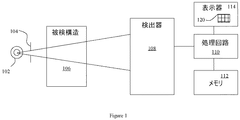
図1は、例示的なX線トモシンセシス・イメージング・システム100を示す。イメージング・システム100は、X線源102及びコリメータ104を含んでおり、被検構造106にX線フォトンを照射する。例として、X線源102はX線管であってよく、被検構造106は患者、試験用ファントム又は他の被験無生物体であってよい。
FIG. 1 illustrates an exemplary X-ray tomosynthesis imaging system 100. The imaging system 100 includes an
X線イメージング・システム100はまた、処理回路110に結合された検出器108を含んでいる。処理回路110(例えばマイクロコントローラ、マイクロプロセッサ、又はカスタムASIC等)は、メモリ112及び表示装置114に結合されている。メモリ112(例えばフレキシブル・ディスク、又はネットワーク若しくはインターネットのような他のディジタル・ソース等のコンピュータ読み取り可能な媒体から命令及び/又はデータを読み取るフレキシブル・ディスク・ドライブ、CD−ROMドライブ、DVDドライブ、光磁気ディスク(MOD)装置、又はイーサネット装置(「イーサネット」は商標)等のネットワーク接続装置を含めたその他任意のディジタル装置、並びに開発中のディジタル手段等の1又は複数を含む)は、撮像データを記憶する。
The x-ray imaging system 100 also includes a
メモリ112はまた、本書に記載される作用を具現化するために処理回路110によって実行される命令を含んだコンピュータ・プログラムを記憶することができる。処理回路110は、装置114に表示するための画像を形成する。本書にさらに詳細に記載されるように、画像120は様々な構造(例えば軟組織、骨)を表わし得る。検出器108は、例えばフラット・パネル型固体画像検出器であってよいが、メモリ112にディジタル形態で記憶された従来のフィルム画像が処理されてもよい。一実施形態では、処理回路110はファームウェア(図示されていない)に記憶されている命令を実行する。一般的には、プロセッサが、後述する工程を実行するようにプログラムされる。
The
言うまでもなく、本書に記載される方法は、システム100での実施に制限されてはおらず、イメージング・システムのその他多くの形式及び変形と関連して利用することができる。一実施形態では、処理回路110は、本書に記載される作用を果たすようにプログラムされているコンピュータであり、従って、本書で用いられるコンピュータとの用語は当技術分野でコンピュータと呼ばれている集積回路のみに限らず、コンピュータ、プロセッサ、マイクロコントローラ、マイクロコンピュータ、プログラマブル論理コントローラ、特定応用向け集積回路、及び他のプログラム可能な回路を広範に指している。本書に記載される方法は、人間の患者の設定において記載されるが、本発明の利益は、小動物研究に典型的に用いられるシステムのような人間以外を対象とするイメージング・システムにも齎されると思量される。
Of course, the methods described herein are not limited to implementation in the system 100 and can be utilized in connection with many other types and variations of imaging systems. In one embodiment, the
本書で用いる場合には、単数形で記載されており単数不定冠詞を冠した要素またはステップとの用語は、排除を明記していない限りかかる要素又はステップを複数備えることを排除しないものと理解されたい。さらに、本発明の「一実施形態」に対する参照は、所載の特徴を同様に組み入れている他の実施形態の存在を排除すると解釈されることを意図していない。 As used in this document, the term element or step written in the singular and followed by a singular indefinite article is understood not to exclude the inclusion of a plurality of such elements or steps unless explicitly stated otherwise. I want. Furthermore, references to “one embodiment” of the present invention are not intended to be interpreted as excluding the existence of other embodiments that also incorporate the recited features.
また、本書で用いられる「画像を再構成する」との表現は、画像を表わすデータが生成されるが可視画像は形成されないような本発明の実施形態を排除するものではない。従って、本書で用いられる「画像」との用語は、可視画像及び可視画像を表わすデータの両方を広く指す。但し、多くの実施形態は少なくとも1枚の可視画像を形成する(か又は形成するように構成されている)。 Further, the expression “reconstruct an image” used in this document does not exclude embodiments of the present invention in which data representing an image is generated but a visible image is not formed. Thus, the term “image” as used herein broadly refers to both the visible image and the data representing the visible image. However, many embodiments form (or are configured to form) at least one visible image.
本書に記載される方法は医療の設定において記載されるが、産業の設定、又は例えば限定しないが空港若しくは他の運輸拠点での手荷物走査システムのような運輸の設定で典型的に用いられるシステム等のような非医用イメージング・システムにおいても、本発明の利益が齎されると思量される。 The methods described in this document are described in medical settings, such as systems typically used in industrial settings or, for example, but not limited to transportation settings such as baggage scanning systems at airports or other transportation points, etc. Even in non-medical imaging systems such as these, it is believed that the benefits of the present invention are appreciated.
自動プロトコル・アシスタンスの方法及びアルゴリズムは、一実施形態によれば下記のステップを有する(図2を参照されたい)。 The automatic protocol assistance method and algorithm has the following steps according to one embodiment (see FIG. 2):
1.曝射回数を決定する。このステップを実行するためには二つの方法がある。 1. Determine the number of exposures. There are two ways to perform this step.
a.一実施形態では、検査時間がどれだけの長さになるかを操作者が入力する(患者がどれだけの長さの時間にわたって保息していられるか等に基づいて)。もう一つの実施形態では、操作者が、患者がどれだけの長さの時間にわたって保息していられるか(1回又は複数)を入力し、システムが検出器サンプリング・レート(秒当たりフレーム数すなわちfps)に基づいて最大曝射回数を自動的に算出する。 a. In one embodiment, the operator enters how long the examination time will be (based on how long the patient can be in breath, etc.). In another embodiment, the operator inputs how long (or more times) the patient can be held, and the system uses the detector sampling rate (frames per second or The maximum number of exposures is automatically calculated based on (fps).
曝射回数=患者保息時間(1回又は複数)
/検出器サンプリング・レート(fps) (1)
b.予め決定された曝射回数を用いる(システム構成ファイル等に記憶されている)。
Number of exposures = Patient repose time (one or more)
/ Detector sampling rate (fps) (1)
b. A predetermined number of exposures is used (stored in a system configuration file or the like).
2.患者の低線量AEC曝射撮影(「プリショット」)を取得する。代替的には、PA(後方/前方)ビュー又はAP(前方/後方)ビューを診断に用いることができ、この場合には、用いられる曝射は解剖学的ビュー及び患者の寸法に対する公称値となる。 2. Acquire a low-dose AEC exposure (“pre-shot”) of the patient. Alternatively, PA (posterior / anterior) views or AP (anterior / posterior) views can be used for diagnosis, in which case the exposure used is nominal with respect to the anatomical view and patient dimensions. Become.
3.プリショットに基づいて初期手法を算出する。この計算は、下記の式を用いることができる(但し、必ず用いなければならない訳ではない)。 3. An initial method is calculated based on the pre-shot. This calculation can use the following equation (however, it is not always necessary).
曝射当たりの最適手法(mAs)
=AEC曝射手法(mAs)*k/曝射回数 (2)
式中、kは構成自在のファクタであり、応用又は解剖学的構造/ビューについて微調整することができる。kは、事前知識データベース及び患者全線量要件から導くことができる。
Optimal method per exposure (mAs)
= AEC exposure method (mAs) * k / number of exposures (2)
Where k is a configurable factor and can be fine-tuned for application or anatomy / view. k can be derived from prior knowledge databases and patient total dose requirements.
4.初期手法をX線発生サブシステム(例えば発生器及び管等)へ伝達する。 4). The initial technique is communicated to an X-ray generation subsystem (eg, generator and tube).
5.X線発生サブシステムがこの初期手法を受け入れる場合には、そのまま進んでX線源を作動させ、データを取得する。 5. If the X-ray generation subsystem accepts this initial approach, it proceeds and activates the X-ray source to acquire data.
6.X線発生サブシステムがこの初期手法を拒絶する場合には、ユーザ・インタフェイスを介して操作者に下記の選択肢を掲げて決定させる。 6). If the X-ray generation subsystem rejects this initial approach, the operator is prompted to make the following choices via the user interface.
a.X線発生サブシステムがその熱状態(すなわち管の過熱等)から復帰するまで待機する。 a. Wait for the x-ray generation subsystem to recover from its thermal state (ie, tube overheating, etc.).
b.X線発生サブシステムが現状で受け入れられるようなさらに低い曝射当たりのmAsを示唆するが、曝射回数は保つ。 b. It suggests a lower per-exposure mAs that the x-ray generation subsystem is currently acceptable, but keeps the number of exposures.
c.X線発生サブシステムが現状で受け入れられるようなさらに少ない曝射回数を示唆するが、曝射当たりのmAsは保つ。 c. The X-ray generation subsystem suggests a lower number of exposures that are currently acceptable, but keeps the mAs per exposure.
d.X線発生サブシステムとの実時間ネゴシエーションに基づいて、自動最適化アルゴリズムを介して、X線発生サブシステムが現状で受け入れられるようなさらに低い曝射当たりのmAs及びさらに少ない曝射回数を示唆する。 d. Based on real-time negotiation with the x-ray generation subsystem, suggests lower mAs per exposure and fewer exposure times that the x-ray generation subsystem is currently acceptable via an automatic optimization algorithm .
7.システムは、ユーザ・インタフェイスにおいて、操作者が毎回の検査毎に又は既定の選択(1又は複数)を設定することのいずれかによって上に掲げた選択肢からの選択を行なうための柔軟性を提供する。 7). The system provides the user interface with the flexibility to make a selection from the options listed above, either at each test or by setting a default selection (s) To do.
一つの技術的効果は、本書に記載した方法及び装置が、自動プロトコル・アシスタンスを介して操作者間のばらつきを最小限に抑えることにより、ディジタル・トモシンセシスでのワークフローを改善すると共に画質を高めることである。 One technical effect is that the methods and apparatus described herein improve the workflow and increase image quality in digital tomosynthesis by minimizing operator-to-operator variation through automated protocol assistance. It is.
以上、実施形態の例について詳細に説明した。これらのアセンブリ及び方法は、本書に記載される特定の実施形態に限定されてはおらず、各々のアセンブリ及び/又は方法の構成要素を、本書に記載される他の構成要素と独立且つ別個に利用してよい。 The example of the embodiment has been described in detail above. These assemblies and methods are not limited to the specific embodiments described herein, and each assembly and / or method component is utilized independently and separately from the other components described herein. You can do it.
本発明を様々な特定の実施形態について記載したが、当業者であれば、特許請求の範囲の要旨及び範囲内で改変を施して本発明を実施し得ることを認められよう。また、図面の符号に対応する特許請求の範囲中の符号は、単に本願発明の理解をより容易にするために用いられているものであり、本願発明の範囲を狭める意図で用いられたものではない。そして、本願の特許請求の範囲に記載した事項は、明細書に組み込まれ、明細書の記載事項の一部となる。 While the invention has been described in terms of various specific embodiments, those skilled in the art will recognize that the invention can be practiced with modification within the spirit and scope of the claims. Further, the reference numerals in the claims corresponding to the reference numerals in the drawings are merely used for easier understanding of the present invention, and are not intended to narrow the scope of the present invention. Absent. The matters described in the claims of the present application are incorporated into the specification and become a part of the description items of the specification.
100 イメージング・システム
102 X線源
104 コリメータ
106 被検構造
108 検出器
110 処理回路
112 メモリ
114 表示装置
120 画像
DESCRIPTION OF SYMBOLS 100
Claims (8)
X線検出器(108)であって、トモシンセシス走査において前記X線源(102)が前記X線検出器(108)に対して移動する間、前記X線源(102)が前記X線検出器(108)に対して異なる方向に配向され、前記X線源(102)から放出されるX線を受光するように配置されている前記X線検出器(108)と、
前記線源及び前記検出器に結合されて動作するコンピュータ(110)と
を備えたシステム(100)であって、前記コンピュータ(110)は、
対象の走査でのトモシンセシス曝射回数を決定するステップと、
前記対象の自動曝射制御曝射撮影を取得するか又は後方/前方(PA)ビュー及び前方/後方(AP)ビューの少なくとも一方を取得するステップと、
複数の前記トモシンセシス曝射の各々について初期手法を算出するステップと、
前記複数の初期手法を、該複数の初期手法を受け入れ又は拒絶することができるX線発生サブシステムへ伝達することにより、前記複数の初期手法を用いて前記複数のトモシンセシス曝射を実行するか又は前記複数のトモシンセシス曝射を実行しないかを決定するステップとを実行するように構成されている、システム(100)。 An X-ray source (102);
An X-ray detector (108), wherein the X-ray source (102) moves to the X-ray detector while the X-ray source (102) moves relative to the X-ray detector (108) in tomosynthesis scanning . The X-ray detector (108) oriented in a different direction relative to (108) and arranged to receive X-rays emitted from the X- ray source (102) ;
A system (100) comprising a computer (110) operating in conjunction with the radiation source and the detector, the computer (110) comprising:
Determining the number of tomosynthesis exposures in the scan of the subject;
Obtaining an automatic exposure control exposure shot of the subject or obtaining at least one of a rear / front (PA) view and a front / back (AP) view;
Calculating an initial approach for each of a plurality of said tomosynthesis exposure,
Performing the plurality of tomosynthesis exposures using the plurality of initial techniques by communicating the plurality of initial techniques to an x-ray generation subsystem capable of accepting or rejecting the plurality of initial techniques; or A system (100) configured to perform the step of determining whether to perform the plurality of tomosynthesis exposures.
The computer (110) is also lower than the initial method in which the X-ray generation subsystem is currently accepted via an automatic optimization algorithm based on real-time negotiation with the X-ray generation subsystem The system (100) of claim 5, wherein the system (100) is configured to suggest mAs per exposure and fewer exposure times than determined.
Applications Claiming Priority (2)
| Application Number | Priority Date | Filing Date | Title |
|---|---|---|---|
| US11/489,174 | 2006-07-19 | ||
| US11/489,174 US7292675B1 (en) | 2006-07-19 | 2006-07-19 | Automatic protocol assistance methods and apparatus |
Publications (3)
| Publication Number | Publication Date |
|---|---|
| JP2008023329A JP2008023329A (en) | 2008-02-07 |
| JP2008023329A5 JP2008023329A5 (en) | 2011-08-18 |
| JP5480467B2 true JP5480467B2 (en) | 2014-04-23 |
Family
ID=38653461
Family Applications (1)
| Application Number | Title | Priority Date | Filing Date |
|---|---|---|---|
| JP2007177874A Expired - Fee Related JP5480467B2 (en) | 2006-07-19 | 2007-07-06 | Automatic protocol assistance equipment |
Country Status (3)
| Country | Link |
|---|---|
| US (1) | US7292675B1 (en) |
| JP (1) | JP5480467B2 (en) |
| DE (1) | DE102007033668A1 (en) |
Families Citing this family (9)
| Publication number | Priority date | Publication date | Assignee | Title |
|---|---|---|---|---|
| JP2010240063A (en) * | 2009-04-02 | 2010-10-28 | Shimadzu Corp | Radiographic apparatus |
| JP5572040B2 (en) * | 2009-09-28 | 2014-08-13 | 富士フイルム株式会社 | Radiography equipment |
| DE102010035920A1 (en) | 2010-08-31 | 2012-03-01 | Siemens Aktiengesellschaft | Method for displaying a predetermined volume section of an examination object by means of a tomosynthesis device and corresponding tomosynthesis device |
| WO2014203938A1 (en) * | 2013-06-18 | 2014-12-24 | キヤノン株式会社 | Tomosynthesis-imaging control device, imaging device, imaging system, control method, and program for causing computer to execute control method |
| US9610057B2 (en) | 2014-06-16 | 2017-04-04 | General Electric Company | System and method for determining X-ray exposure parameters |
| US9615803B2 (en) | 2014-06-16 | 2017-04-11 | General Electric Company | System and method for determining X-ray exposure parameters |
| DE102017203025A1 (en) * | 2017-02-24 | 2018-08-30 | Siemens Healthcare Gmbh | A method of assisting in planning a magnetic resonance examination on a patient having a magnetic resonance apparatus, and a magnetic resonance apparatus for performing the method |
| US10779791B2 (en) * | 2018-03-16 | 2020-09-22 | General Electric Company | System and method for mobile X-ray imaging |
| JP7086805B2 (en) | 2018-09-27 | 2022-06-20 | 富士フイルム株式会社 | Tomosynthesis imaging device and how to operate it |
Family Cites Families (6)
| Publication number | Priority date | Publication date | Assignee | Title |
|---|---|---|---|---|
| US4454606A (en) | 1983-05-23 | 1984-06-12 | General Electric Company | Reconfigurable x-ray AEC compensation |
| US6192105B1 (en) | 1998-11-25 | 2001-02-20 | Communications & Power Industries Canada Inc. | Method and device to calibrate an automatic exposure control device in an x-ray imaging system |
| US6931098B2 (en) * | 2002-03-08 | 2005-08-16 | Ge Medical Systems Global Technology Company, Llc | Method and system for dual or multiple energy imaging |
| JP4522044B2 (en) | 2002-11-15 | 2010-08-11 | キヤノン株式会社 | Radiography equipment |
| US7123684B2 (en) * | 2002-11-27 | 2006-10-17 | Hologic, Inc. | Full field mammography with tissue exposure control, tomosynthesis, and dynamic field of view processing |
| DE102005022544A1 (en) * | 2005-05-17 | 2006-11-23 | Siemens Ag | Method and device for recording a digital X-ray image |
-
2006
- 2006-07-19 US US11/489,174 patent/US7292675B1/en active Active
-
2007
- 2007-07-06 JP JP2007177874A patent/JP5480467B2/en not_active Expired - Fee Related
- 2007-07-17 DE DE102007033668A patent/DE102007033668A1/en not_active Withdrawn
Also Published As
| Publication number | Publication date |
|---|---|
| DE102007033668A1 (en) | 2008-01-24 |
| US7292675B1 (en) | 2007-11-06 |
| JP2008023329A (en) | 2008-02-07 |
Similar Documents
| Publication | Publication Date | Title |
|---|---|---|
| JP5480467B2 (en) | Automatic protocol assistance equipment | |
| JP5738510B2 (en) | Image acquisition and processing chain for dual energy radiation imaging using a portable flat panel detector | |
| US20120155609A1 (en) | System and method of low dose exposure aided positioning (leap) for digital radiography | |
| US9014450B2 (en) | Method and apparatus for filtering projection images | |
| JP5028528B2 (en) | X-ray CT system | |
| JP2006150080A (en) | Angiographic x-ray diagnostic device for rotational angiography | |
| JP2007144174A (en) | Method and system for automatically determining regions in scanned object | |
| JP6906905B2 (en) | X-ray diagnostic equipment | |
| JP2012110710A (en) | Region of interest determination for x-ray imaging | |
| JP2009226219A (en) | Method and apparatus for correcting multi-modality imaging data | |
| JP6475138B2 (en) | Control device, radiographic image capturing device, radiographic image capturing method, and radiographic image capturing program | |
| JP6824133B2 (en) | Image processing equipment, image processing method, and image processing program | |
| EP2457512A1 (en) | System and method for including and correcting subject orientation data in digital radiographic images | |
| JP2022173271A (en) | Device for imaging object | |
| Lell et al. | Computed tomography 2.0: new detector technology, AI, and other developments | |
| JP2010115349A (en) | Radiation tomographic apparatus | |
| KR102379067B1 (en) | Medical image apparatus and method for processing medical image | |
| JP2009047602A (en) | Positron emission computerd tomograph, attenuation map creating device, and attenuation map creating program | |
| JP6466057B2 (en) | Medical diagnostic imaging equipment | |
| JP2017000664A (en) | Image processing system, tomographic image generation system, and program | |
| JP5238296B2 (en) | X-ray apparatus and rotational imaging method | |
| JP6142172B2 (en) | Beam hardening correction apparatus, beam hardening correction method, and X-ray imaging apparatus | |
| JP2021191389A (en) | Processing device, operation method of processing device, and operation program of processing device | |
| JP6873831B2 (en) | Medical image diagnostic equipment, medical image processing equipment and medical image processing program | |
| CN107809954B (en) | Display of depth position of computer tomography slice image relative to object to be imaged |
Legal Events
| Date | Code | Title | Description |
|---|---|---|---|
| A521 | Request for written amendment filed |
Free format text: JAPANESE INTERMEDIATE CODE: A523 Effective date: 20100702 |
|
| A621 | Written request for application examination |
Free format text: JAPANESE INTERMEDIATE CODE: A621 Effective date: 20100702 |
|
| RD04 | Notification of resignation of power of attorney |
Free format text: JAPANESE INTERMEDIATE CODE: A7424 Effective date: 20100702 |
|
| A521 | Request for written amendment filed |
Free format text: JAPANESE INTERMEDIATE CODE: A523 Effective date: 20110628 |
|
| RD02 | Notification of acceptance of power of attorney |
Free format text: JAPANESE INTERMEDIATE CODE: A7422 Effective date: 20110628 |
|
| RD04 | Notification of resignation of power of attorney |
Free format text: JAPANESE INTERMEDIATE CODE: A7424 Effective date: 20110708 |
|
| A131 | Notification of reasons for refusal |
Free format text: JAPANESE INTERMEDIATE CODE: A131 Effective date: 20130312 |
|
| A521 | Request for written amendment filed |
Free format text: JAPANESE INTERMEDIATE CODE: A523 Effective date: 20130418 |
|
| A131 | Notification of reasons for refusal |
Free format text: JAPANESE INTERMEDIATE CODE: A131 Effective date: 20131015 |
|
| A521 | Request for written amendment filed |
Free format text: JAPANESE INTERMEDIATE CODE: A523 Effective date: 20131119 |
|
| TRDD | Decision of grant or rejection written | ||
| A01 | Written decision to grant a patent or to grant a registration (utility model) |
Free format text: JAPANESE INTERMEDIATE CODE: A01 Effective date: 20140121 |
|
| A61 | First payment of annual fees (during grant procedure) |
Free format text: JAPANESE INTERMEDIATE CODE: A61 Effective date: 20140214 |
|
| R150 | Certificate of patent or registration of utility model |
Ref document number: 5480467 Country of ref document: JP Free format text: JAPANESE INTERMEDIATE CODE: R150 |
|
| R250 | Receipt of annual fees |
Free format text: JAPANESE INTERMEDIATE CODE: R250 |
|
| R250 | Receipt of annual fees |
Free format text: JAPANESE INTERMEDIATE CODE: R250 |
|
| R250 | Receipt of annual fees |
Free format text: JAPANESE INTERMEDIATE CODE: R250 |
|
| R250 | Receipt of annual fees |
Free format text: JAPANESE INTERMEDIATE CODE: R250 |
|
| LAPS | Cancellation because of no payment of annual fees |