JP5384120B2 - Therapeutic agent for periodontal diseases and alveolar bone defects caused by surgery - Google Patents
Therapeutic agent for periodontal diseases and alveolar bone defects caused by surgery Download PDFInfo
- Publication number
- JP5384120B2 JP5384120B2 JP2008558139A JP2008558139A JP5384120B2 JP 5384120 B2 JP5384120 B2 JP 5384120B2 JP 2008558139 A JP2008558139 A JP 2008558139A JP 2008558139 A JP2008558139 A JP 2008558139A JP 5384120 B2 JP5384120 B2 JP 5384120B2
- Authority
- JP
- Japan
- Prior art keywords
- decoy
- nfκb
- bone
- alveolar bone
- nfκb decoy
- Prior art date
- Legal status (The legal status is an assumption and is not a legal conclusion. Google has not performed a legal analysis and makes no representation as to the accuracy of the status listed.)
- Active
Links
Images


Classifications
-
- A—HUMAN NECESSITIES
- A61—MEDICAL OR VETERINARY SCIENCE; HYGIENE
- A61K—PREPARATIONS FOR MEDICAL, DENTAL OR TOILETRY PURPOSES
- A61K9/00—Medicinal preparations characterised by special physical form
- A61K9/0012—Galenical forms characterised by the site of application
- A61K9/0053—Mouth and digestive tract, i.e. intraoral and peroral administration
- A61K9/0063—Periodont
-
- A—HUMAN NECESSITIES
- A61—MEDICAL OR VETERINARY SCIENCE; HYGIENE
- A61K—PREPARATIONS FOR MEDICAL, DENTAL OR TOILETRY PURPOSES
- A61K31/00—Medicinal preparations containing organic active ingredients
- A61K31/70—Carbohydrates; Sugars; Derivatives thereof
- A61K31/7088—Compounds having three or more nucleosides or nucleotides
- A61K31/713—Double-stranded nucleic acids or oligonucleotides
-
- A—HUMAN NECESSITIES
- A61—MEDICAL OR VETERINARY SCIENCE; HYGIENE
- A61P—SPECIFIC THERAPEUTIC ACTIVITY OF CHEMICAL COMPOUNDS OR MEDICINAL PREPARATIONS
- A61P1/00—Drugs for disorders of the alimentary tract or the digestive system
- A61P1/02—Stomatological preparations, e.g. drugs for caries, aphtae, periodontitis
-
- A—HUMAN NECESSITIES
- A61—MEDICAL OR VETERINARY SCIENCE; HYGIENE
- A61P—SPECIFIC THERAPEUTIC ACTIVITY OF CHEMICAL COMPOUNDS OR MEDICINAL PREPARATIONS
- A61P19/00—Drugs for skeletal disorders
-
- A—HUMAN NECESSITIES
- A61—MEDICAL OR VETERINARY SCIENCE; HYGIENE
- A61P—SPECIFIC THERAPEUTIC ACTIVITY OF CHEMICAL COMPOUNDS OR MEDICINAL PREPARATIONS
- A61P19/00—Drugs for skeletal disorders
- A61P19/08—Drugs for skeletal disorders for bone diseases, e.g. rachitism, Paget's disease
-
- A—HUMAN NECESSITIES
- A61—MEDICAL OR VETERINARY SCIENCE; HYGIENE
- A61P—SPECIFIC THERAPEUTIC ACTIVITY OF CHEMICAL COMPOUNDS OR MEDICINAL PREPARATIONS
- A61P29/00—Non-central analgesic, antipyretic or antiinflammatory agents, e.g. antirheumatic agents; Non-steroidal antiinflammatory drugs [NSAID]
-
- A—HUMAN NECESSITIES
- A61—MEDICAL OR VETERINARY SCIENCE; HYGIENE
- A61P—SPECIFIC THERAPEUTIC ACTIVITY OF CHEMICAL COMPOUNDS OR MEDICINAL PREPARATIONS
- A61P31/00—Antiinfectives, i.e. antibiotics, antiseptics, chemotherapeutics
- A61P31/04—Antibacterial agents
-
- A—HUMAN NECESSITIES
- A61—MEDICAL OR VETERINARY SCIENCE; HYGIENE
- A61P—SPECIFIC THERAPEUTIC ACTIVITY OF CHEMICAL COMPOUNDS OR MEDICINAL PREPARATIONS
- A61P43/00—Drugs for specific purposes, not provided for in groups A61P1/00-A61P41/00
-
- C—CHEMISTRY; METALLURGY
- C12—BIOCHEMISTRY; BEER; SPIRITS; WINE; VINEGAR; MICROBIOLOGY; ENZYMOLOGY; MUTATION OR GENETIC ENGINEERING
- C12N—MICROORGANISMS OR ENZYMES; COMPOSITIONS THEREOF; PROPAGATING, PRESERVING, OR MAINTAINING MICROORGANISMS; MUTATION OR GENETIC ENGINEERING; CULTURE MEDIA
- C12N15/00—Mutation or genetic engineering; DNA or RNA concerning genetic engineering, vectors, e.g. plasmids, or their isolation, preparation or purification; Use of hosts therefor
- C12N15/09—Recombinant DNA-technology
- C12N15/11—DNA or RNA fragments; Modified forms thereof; Non-coding nucleic acids having a biological activity
- C12N15/113—Non-coding nucleic acids modulating the expression of genes, e.g. antisense oligonucleotides; Antisense DNA or RNA; Triplex- forming oligonucleotides; Catalytic nucleic acids, e.g. ribozymes; Nucleic acids used in co-suppression or gene silencing
-
- A—HUMAN NECESSITIES
- A61—MEDICAL OR VETERINARY SCIENCE; HYGIENE
- A61K—PREPARATIONS FOR MEDICAL, DENTAL OR TOILETRY PURPOSES
- A61K48/00—Medicinal preparations containing genetic material which is inserted into cells of the living body to treat genetic diseases; Gene therapy
-
- C—CHEMISTRY; METALLURGY
- C12—BIOCHEMISTRY; BEER; SPIRITS; WINE; VINEGAR; MICROBIOLOGY; ENZYMOLOGY; MUTATION OR GENETIC ENGINEERING
- C12N—MICROORGANISMS OR ENZYMES; COMPOSITIONS THEREOF; PROPAGATING, PRESERVING, OR MAINTAINING MICROORGANISMS; MUTATION OR GENETIC ENGINEERING; CULTURE MEDIA
- C12N2310/00—Structure or type of the nucleic acid
- C12N2310/10—Type of nucleic acid
- C12N2310/13—Decoys
Landscapes
- Health & Medical Sciences (AREA)
- Life Sciences & Earth Sciences (AREA)
- Chemical & Material Sciences (AREA)
- Engineering & Computer Science (AREA)
- General Health & Medical Sciences (AREA)
- Organic Chemistry (AREA)
- Medicinal Chemistry (AREA)
- Pharmacology & Pharmacy (AREA)
- Animal Behavior & Ethology (AREA)
- Public Health (AREA)
- Veterinary Medicine (AREA)
- Bioinformatics & Cheminformatics (AREA)
- Genetics & Genomics (AREA)
- Nuclear Medicine, Radiotherapy & Molecular Imaging (AREA)
- Molecular Biology (AREA)
- General Chemical & Material Sciences (AREA)
- Chemical Kinetics & Catalysis (AREA)
- Biomedical Technology (AREA)
- General Engineering & Computer Science (AREA)
- Biotechnology (AREA)
- Epidemiology (AREA)
- Zoology (AREA)
- Wood Science & Technology (AREA)
- Biochemistry (AREA)
- Physical Education & Sports Medicine (AREA)
- Microbiology (AREA)
- Physiology (AREA)
- Biophysics (AREA)
- Plant Pathology (AREA)
- Physics & Mathematics (AREA)
- Nutrition Science (AREA)
- Rheumatology (AREA)
- Orthopedic Medicine & Surgery (AREA)
- Pain & Pain Management (AREA)
- Oncology (AREA)
- Communicable Diseases (AREA)
- Pharmaceuticals Containing Other Organic And Inorganic Compounds (AREA)
- Medicines That Contain Protein Lipid Enzymes And Other Medicines (AREA)
- Medicinal Preparation (AREA)
Description
本発明は、歯周病、特に歯周炎、歯肉炎、歯根膜炎及び歯槽膿漏等を処置するための新しい治療薬及び治療方法に関する。本発明の治療、予防または改善剤は、歯周病による歯槽骨の破壊及び縮小、虫歯からの根先病巣による歯槽骨欠損、並びに外科的手術により生じる歯槽骨欠損に対し有用である。 The present invention relates to a new therapeutic agent and a therapeutic method for treating periodontal disease, particularly periodontitis, gingivitis, periodontitis and alveolar pyorrhea. The therapeutic, prophylactic or ameliorating agent of the present invention is useful for alveolar bone destruction and reduction due to periodontal disease, alveolar bone loss due to root lesions from caries, and alveolar bone loss caused by surgical operation.
主な歯周病としては、歯周炎及び歯肉炎が知られているが、これらはいずれも慢性的な炎症性疾患であり、進行すると、歯周組織が破壊され、歯槽骨が骨吸収により減退して歯を支えられなくなる場合がある。また、虫歯が進行すると、その根先病巣から歯槽骨に穴があいたり、また外科的な治療により歯槽骨に穴があいたりして、歯槽骨が欠損した状態になることがある。このような歯周病の治療方法は未だ開発途上にある。 Periodontitis and gingivitis are known as the main periodontal diseases, both of which are chronic inflammatory diseases, and as they progress, the periodontal tissue is destroyed and the alveolar bone is absorbed by bone resorption. It may decline and become unable to support the teeth. In addition, when the caries progresses, the alveolar bone may be punctured from the root lesion, or the alveolar bone may be punctured by surgical treatment, and the alveolar bone may be lost. Such treatment methods for periodontal disease are still under development.
NFκB(nuclear factor kappa B)は、サイトカインや接着因子等、免疫反応に関する遺伝子の発現を調節する役割を持つ一群の転写因子の総称であり、NFκBがゲノム遺伝子上の結合部位に結合すると、免疫反応に関する遺伝子が過剰に発現する。このため、NFκBは、免疫反応が原因となるアトピー性皮膚炎や関節リウマチ等のアレルギー性疾患、自己免疫疾患、さらには心筋梗塞等の虚血性疾患や動脈硬化等の各種疾患に関与することが知られている。このNFκBに対するデコイ(おとり型医薬)を投与することにより、対象となる転写因子の活性を低下させ、該転写因子に起因して起きる疾患の治療や予防を行なうことが知られている(例えば、特許文献1及び特許文献2参照)。さらにまた、NFκBデコイが、骨髄細胞から破骨細胞への分化に対して抑制的に作用し、骨形成・骨吸収の平衡状態の破綻に起因する疾患、特に骨粗鬆症の予防または治療に有効であることが知られている(特許文献3)。
NFκB (nuclear factor kappa B) is a collective term for a group of transcription factors that play a role in regulating the expression of genes related to immune responses, such as cytokines and adhesion factors. When NFκB binds to a binding site on a genomic gene, the immune response The gene related to is overexpressed. For this reason, NFκB may be involved in allergic diseases such as atopic dermatitis and rheumatoid arthritis caused by immune reactions, autoimmune diseases, ischemic diseases such as myocardial infarction, and various diseases such as arteriosclerosis. Are known. It is known that administration of this NFκB decoy (bait-type medicine) reduces the activity of the transcription factor of interest and treats or prevents diseases caused by the transcription factor (for example, (See
歯周炎及び歯肉炎は炎症反応であり、その患部に炎症性サイトカインが動員されており、そこにNFκBの活性化が関与していると考えられている(非特許文献1及び2)。非特許文献1には、健康なヒト歯肉繊維芽細胞(healthy human gingival fibroblast)のin vitro培養において、インターロイキン(IL)-6、IL-8及び単球遊走蛋白(MCP)-1の炎症性サイトカインがリポ多糖(LPS)により誘導されること、並びに、このLPSによる炎症性サイトカイン誘導がN-アラキドン酸エタノールアミン(アナンダミド;AEA)により抑制されること、さらにLPSによりNFκBの活性化が誘導されるがこのNFκBの活性化もAEAにより抑制されることが記載されている。しかしながら、何らかの歯周病を患った患者の組織については、歯肉炎または歯周炎を持つ患者から採取したヒト歯肉繊維芽細胞で、健康なヒト歯肉繊維芽細胞に比べてAEAのレセプターであるカンナビノイドレセプター(CB)-1及びCB-2の発現が増大していることが確認されているのみである。したがって、同文献からは、何らかの歯周病を有する組織において、AEAが炎症抑制の効果を有するのかどうかすら不明であり、ましてNFκBの活性化を抑制することにより歯周病の症状を緩和することができるのかどうかは全く不明である。
Periodontitis and gingivitis are inflammatory reactions, and inflammatory cytokines are mobilized in the affected area, and it is considered that activation of NFκB is involved (Non-patent
非特許文献2には、慢性的歯周炎患者の患部歯肉組織内では、健康な歯肉組織内に比べて、活性型NFκBが多く存在し、NFκBの阻害分子であるIκBが減少していることが記載されている。しかしながら、NFκBの活性化と歯周炎の発症及び進行との関連性は全く開示されていない。また、NFκBの活性化を抑制により歯周炎を治癒することも示されていない。
以上のように、歯周病または歯周炎における骨の損失の回復については、上記非特許文献にも何ら示唆されておらず、有効な手段は確立されていない。
本発明は、歯周病(periodontal disease)の治療、予防または改善のための医薬、あるいは方法を提供することを課題とする。また本発明は、歯槽骨の欠損(alveolar bone defect)の治療、予防または改善のための医薬、あるいは方法の提供を課題とする。 An object of the present invention is to provide a medicine or a method for treating, preventing or ameliorating periodontal disease. Another object of the present invention is to provide a medicine or a method for treating, preventing or ameliorating alveolar bone defect.
本発明によりNFκBデコイの投与が、歯周病モデルにおいて歯槽骨の吸収を抑制し、歯周病骨欠損モデルにおいて歯槽骨の修復を促進させることが示された。その結果、本発明者らは、NFκBデコイが歯周病、及び外科手術等による歯槽骨欠損の治療、予防、及び改善剤として有用であることを見出した。本発明は、具体的には以下の発明に関するものである。
〔1〕NFκBデコイを有効成分として含有する、歯周病、及び外科手術による歯槽骨欠損のいずれかまたは両方の治療、予防または改善剤。
〔2〕歯周病が、歯周炎、歯肉炎、歯根膜炎、及び虫歯による根先病巣からなる群より選択される、〔1〕記載の治療、予防または改善剤。
〔3〕注射剤である、〔1〕または〔2〕に記載の治療、予防または改善剤。
〔4〕コラーゲン基材にNFκBデコイが付着した剤形である、〔1〕−〔3〕のいずれかに記載の治療、予防または改善剤。
〔5〕NFκBデコイに含まれる塩基間結合の少なくとも1つはホスホロチオエート結合である、〔1〕−〔4〕のいずれかに記載の治療、予防または改善剤。
〔6〕NFκBデコイに含まれる塩基間結合の全てがホスホロチオエート結合である、〔5〕記載の治療、予防または改善剤。
〔7〕NFκBデコイが、CCTTGAAGGGATTTCCCTCC(配列番号1)からなるオリゴヌクレオチドと、これに完全に相補的な配列からなるオリゴヌクレオチドとが二本鎖形成した二本鎖オリゴヌクレオチドである、〔1〕−〔6〕のいずれかに記載の治療、予防または改善剤。
〔8〕NFκBデコイを、歯周病、及び外科手術による歯槽骨欠損のいずれかまたは両方に罹患した対象に投与する工程を含む、歯周病、及び外科手術による歯槽骨欠損のいずれかまたは両方を治療、予防または改善する方法。
〔9〕歯周病が、歯周炎、歯肉炎、歯根膜炎、及び虫歯による根先病巣からなる群より選択される、〔8〕記載の方法。
〔10〕NFκBデコイを注射によって投与する、〔8〕または〔9〕に記載の方法。
〔11〕コラーゲン基材が付着したNFκBデコイを投与する工程を含む、〔8〕−〔10〕のいずれかに記載の方法。
〔12〕NFκBデコイに含まれる塩基間結合の少なくとも1つはホスホロチオエート結合である、〔8〕−〔11〕のいずれかに記載の方法。
〔13〕NFκBデコイに含まれる塩基間結合の全てがホスホロチオエート結合である、〔12〕記載の方法。
〔14〕NFκBデコイが、CCTTGAAGGGATTTCCCTCC(配列番号1)からなるオリゴヌクレオチドと、これに完全に相補的な配列からなるオリゴヌクレオチドとが二本鎖形成した二本鎖オリゴヌクレオチドである、〔8〕−〔13〕のいずれかに記載の方法。
〔15〕歯周病、及び外科手術による歯槽骨欠損のいずれかまたは両方の治療、予防または改善する薬剤を製造するための、NFκBデコイの使用。
〔16〕歯周病が、歯周炎、歯肉炎、歯根膜炎、及び虫歯による根先病巣からなる群より選択される、〔15〕記載の使用。
〔17〕注射剤である、〔15〕または〔16〕に記載の使用。
〔18〕コラーゲン基材にNFκBデコイが付着した剤形である、〔15〕−〔17〕のいずれかに記載の使用。
〔19〕NFκBデコイに含まれる塩基間結合の少なくとも1つはホスホロチオエート結合である、〔15〕−〔18〕のいずれかに記載の使用。
〔20〕NFκBデコイに含まれる塩基間結合の全てがホスホロチオエート結合である、〔19〕記載の使用。
〔21〕NFκBデコイが、CCTTGAAGGGATTTCCCTCC(配列番号1)からなるオリゴヌクレオチドと、これに完全に相補的な配列からなるオリゴヌクレオチドとが二本鎖形成した二本鎖オリゴヌクレオチドである、〔15〕−〔20〕のいずれかに記載の使用。
あるいは本発明は、歯周病、及び外科手術による歯槽骨欠損のいずれかまたは両方を治療、予防または改善するためのNFκBデコイを提供する。本発明のNFκBデコイは、具体的には、歯周病、及び外科手術による歯槽骨欠損のいずれかまたは両方を治療、予防または改善するためのNFκBデコイであって、局所に注射によって投与するためのNFκBデコイを含む。According to the present invention, it has been shown that administration of NFκB decoy suppresses alveolar bone resorption in a periodontal disease model and promotes alveolar bone repair in a periodontal bone defect model. As a result, the present inventors have found that NFκB decoy is useful as an agent for the treatment, prevention, and improvement of periodontal disease and alveolar bone defects caused by surgery. The present invention specifically relates to the following inventions.
[1] An agent for treating, preventing or ameliorating one or both of periodontal disease and surgical alveolar bone defect, comprising NFκB decoy as an active ingredient.
[2] The treatment, prevention, or improvement agent according to [1], wherein the periodontal disease is selected from the group consisting of periodontitis, gingivitis, periodontitis, and root lesions caused by caries.
[3] The therapeutic, prophylactic or improving agent according to [1] or [2], which is an injection.
[4] The therapeutic, prophylactic or improving agent according to any one of [1] to [3], which is a dosage form in which NFκB decoy is attached to a collagen base material.
[5] The therapeutic, prophylactic or ameliorating agent according to any one of [1] to [4], wherein at least one of the interbase linkages contained in the NFκB decoy is a phosphorothioate linkage.
[6] The therapeutic, prophylactic or ameliorating agent according to [5], wherein all of the interbase linkages contained in the NFκB decoy are phosphorothioate linkages.
[7] The NFκB decoy is a double-stranded oligonucleotide in which an oligonucleotide consisting of CCTTGAAGGGATTTCCCTCC (SEQ ID NO: 1) and an oligonucleotide consisting of a completely complementary sequence are formed into a double strand, [1] − [6] The therapeutic, prophylactic or ameliorating agent according to any one of [6].
[8] Either or both of periodontal disease and surgical alveolar bone defect comprising the step of administering NFκB decoy to a subject suffering from either or both of periodontal disease and surgical alveolar bone defect How to treat, prevent or improve.
[9] The method according to [8], wherein the periodontal disease is selected from the group consisting of periodontitis, gingivitis, periodontitis and root lesions due to caries.
[10] The method according to [8] or [9], wherein the NFκB decoy is administered by injection.
[11] The method according to any one of [8] to [10], comprising a step of administering an NFκB decoy to which a collagen base material is adhered.
[12] The method according to any one of [8] to [11], wherein at least one of the interbase bonds contained in the NFκB decoy is a phosphorothioate bond.
[13] The method according to [12], wherein all of the interbase bonds contained in the NFκB decoy are phosphorothioate bonds.
[14] The NFκB decoy is a double-stranded oligonucleotide in which an oligonucleotide consisting of CCTTGAAGGGATTTCCCTCC (SEQ ID NO: 1) and an oligonucleotide consisting of a completely complementary sequence are formed into a double strand. [8] − [13] The method according to any one of [13].
[15] Use of NFκB decoy for producing a drug for treating, preventing or ameliorating either or both of periodontal disease and alveolar bone defect caused by surgery.
[16] The use according to [15], wherein the periodontal disease is selected from the group consisting of periodontitis, gingivitis, periodontitis and root lesions due to caries.
[17] The use according to [15] or [16], which is an injection.
[18] The use according to any one of [15] to [17], which is a dosage form in which NFκB decoy is attached to a collagen base material.
[19] The use according to any one of [15] to [18], wherein at least one of the interbase bonds contained in the NFκB decoy is a phosphorothioate bond.
[20] The use according to [19], wherein all of the interbase linkages contained in the NFκB decoy are phosphorothioate linkages.
[21] The NFκB decoy is a double-stranded oligonucleotide in which an oligonucleotide consisting of CCTTGAAGGGATTTCCCTCC (SEQ ID NO: 1) and an oligonucleotide consisting of a completely complementary sequence are formed into a double strand, [15] − [20] The use according to any of [20].
Alternatively, the present invention provides an NFκB decoy for treating, preventing or ameliorating either or both of periodontal disease and surgical alveolar bone defect. The NFκB decoy of the present invention is specifically an NFκB decoy for treating, preventing or ameliorating either or both of periodontal disease and surgical alveolar bone defect, and is administered locally by injection. Contains NFκB decoy.
本明細書中、「歯周病(peridontal disease)」という用語は、歯周炎(periodontal infection)、歯肉炎(gingival inflammation)、歯根膜炎(dental periostitis)、歯槽膿漏(alveolar pyorrhea)、及びこれらの炎症病変による歯槽骨(alveolar bone)の破壊・縮小、並びに、虫歯からの根先病巣による歯槽骨欠損(alveolar bone defect caused by apical disease by root infection from dental caries)等を包含する、歯周組織における病変の総称として用いる。また、虫歯が進行した場合、根先病巣から歯槽骨が破壊され歯槽骨の欠損が起こりうるが、このような症状も、本明細書における「歯周病」に包含されるものと定義する。 As used herein, the term “peridontal disease” refers to periodontal infection, gingival inflammation, dental periostitis, alveolar pyorrhea, and Periodontal bone damage caused by alveolar bone caused by these inflammatory lesions, and alveolar bone defect caused by apical disease by root infection from dental caries Used as a general term for lesions in tissues. Further, when the caries progresses, the alveolar bone may be destroyed from the root lesion and the alveolar bone may be lost. Such a symptom is also defined as “periodontal disease” in the present specification.
ここで、「歯周炎」とは、根尖性歯周炎、若年性歯周炎、単純性歯周炎及び複雑性歯周炎等、あらゆる歯周炎を包含し、さらに抜歯(extraction of a tooth)、腫瘍(tumor)及び変形症(deformity)(例えば、顎変形症)の治療、再建(reconstruction)、インプラント(implant)及び矯正学的治療(orthodontic treatment)を目的とした口腔外科手術(oral surgery)をはじめとする、顎及び歯周領域のあらゆる外科手術後に伴う創傷による炎症も含む。一般に、歯周炎は、進行すると歯槽骨の破壊・縮小を起こす。一方、本明細書における「歯肉炎」は、歯肉症、壊死性潰瘍性歯肉炎(急性及び再発性のものを含む)、糖尿病性歯肉炎、増殖性歯肉炎(白血病性のものを含む)、ホルモン性歯肉炎、形質細胞性歯肉炎、化膿性歯肉炎、紡錘菌スピロヘータ歯肉炎、ならびにジフェニルヒダントイン歯肉炎等の薬剤に起因する歯肉炎等、あらゆる種類の歯肉炎を包含する。また、本明細書における「歯根膜炎」は、歯肉炎から進展して起きるものと、根先病巣から進展して起こるものとを含むが、本発明の治療、予防または改善剤は、いずれの歯根膜炎にも有効であり、特に歯肉炎から進展した歯根膜炎に有効である。 Here, “periodontitis” includes all periodontitis such as apical periodontitis, juvenile periodontitis, simple periodontitis, and complex periodontitis, and further, extraction of Oral surgery for the treatment, reconstruction, implant and orthodontic treatment of a tooth, tumor and deformity (eg jaw deformity) Including inflammation due to wounds associated with any surgery in the jaw and periodontal region, including oral surgery). In general, periodontitis causes destruction and reduction of alveolar bone as it progresses. On the other hand, “gingivitis” in the present specification includes gingivitis, necrotizing ulcerative gingivitis (including acute and recurrent gingivitis), diabetic gingivitis, proliferative gingivitis (including leukemia), It includes all types of gingivitis, such as gingivitis caused by drugs such as hormonal gingivitis, plasma cell gingivitis, purulent gingivitis, fusiform spirochete gingivitis, and diphenylhydantoin gingivitis. In addition, “periodontitis” in the present specification includes those that develop from gingivitis and those that develop from a root lesion, but the therapeutic, preventive or ameliorating agent of the present invention is any of It is also effective for periodontitis, particularly effective for periodontitis that has developed from gingivitis.
本明細書中、「外科手術」は、抜歯(extraction of a tooth)、腫瘍(tumor)及び変形症(deformity)(例えば、顎変形症)の治療、再建(reconstruction)、インプラント(implant)及び矯正学的治療(orthodontic treatment)を目的とした口腔外科手術(oral surgery)をはじめとする、顎及び歯周領域のあらゆる外科手術を包含する。本発明の治療、予防または改善剤は、上記外科手術による歯槽骨欠損の治療、予防及び改善に有用である。また、上記外科手術のうち、特にインプラント植立時、腫瘍摘出手術、顎変形症の治療のための外科手術、顎骨手術(例えば、上または下顎前突症の治療のためのもの)等の術後の創傷の治癒促進にも有効である。
本明細書において「歯槽骨欠損」とは、外科手術等により歯槽骨の一部を欠損または破損した状態をいう。As used herein, “surgery” refers to the treatment of extraction of a tooth, tumor and deformity (eg, jaw deformity), reconstruction, implant and correction. Includes all jaw and periodontal surgery, including oral surgery for orthodontic treatment. The therapeutic, prophylactic or ameliorating agent of the present invention is useful for the treatment, prevention and amelioration of alveolar bone defects caused by the above surgery. In addition, among the above-mentioned surgical operations, especially at the time of implant setting, surgery for removing the tumor, surgery for treatment of jaw deformity, jaw surgery (for example, treatment for upper or mandibular prognathism), etc. It is also effective in promoting healing of wounds.
As used herein, “alveolar bone defect” refers to a state in which a portion of the alveolar bone has been lost or damaged by surgery or the like.
遺伝子の発現は、遺伝子の転写調節領域に結合する転写調節因子により制御されている。NFκBは、1986年にB細胞での免疫グロブリンκ軽鎖遺伝子の発現にかかわるエンハンサーに結合する転写因子として同定された、主にp65とp50のサブユニットからなるヘテロ二量体であり(Cell 46 (1986) 705-16)、「NFκB」との用語は、当分野において、構造的に関連し、且つ進化的に保存された転写因子の活性を持つ蛋白質の総称として用いられている。NFκBは、サイトカイン(インターロイキン(IL)-1、IL-6、IL-8及び腫瘍壊死因子(TNF)等)、血管新生因子(血管内皮増殖因子(VEGF)等)、細胞結着因子(細胞間接着分子(ICAM)-1及び血管細胞接着分子(VCAM)-1等)、酵素(シクロオキシゲナーゼ(COX)-2及び一酸化窒素合成酵素(NOS)等)及び抗アポトーシス因子(bcl-2及びサバイビン等)等の遺伝子の転写を開始する種々のDNA配列に対して結合する(Ann Rev Immunol 14 (1996) 649-683;Immunol Today 19 (1998) 80-88; Trends Mol Med 8 (2002) 385-389; Nat Rev Cancer 2 (2002) 301-10)。 Gene expression is controlled by transcriptional regulatory factors that bind to the transcriptional regulatory region of the gene. NFκB is a heterodimer consisting mainly of p65 and p50 subunits, identified in 1986 as a transcription factor that binds to an enhancer involved in the expression of immunoglobulin κ light chain genes in B cells (Cell 46 (1986) 705-16), the term “NFκB” is used in the art as a generic term for proteins that have the activity of structurally related and evolutionarily conserved transcription factors. NFκB is a cytokine (interleukin (IL) -1, IL-6, IL-8 and tumor necrosis factor (TNF) etc.), angiogenic factor (vascular endothelial growth factor (VEGF) etc.), cell binding factor (cell Intercellular adhesion molecule (ICAM) -1 and vascular cell adhesion molecule (VCAM) -1 etc.), enzymes (cyclooxygenase (COX) -2 and nitric oxide synthase (NOS) etc.) and anti-apoptotic factors (bcl-2 and survivin) Etc.) (Ann Rev Immunol 14 (1996) 649-683; Immunol Today 19 (1998) 80-88; Trends Mol Med 8 (2002) 385- 389; Nat Rev Cancer 2 (2002) 301-10).
NFκBの結合配列(結合領域)は、種々の文献により公知である(例えば、「分子細胞生物学辞典」(東京化学同人 1997年発行)第891頁参照)。具体的な結合配列としては、GGGRHTYYHC(RはAまたはG; YはCまたはT; HはA,CまたはT)(配列番号:2)が挙げられる。例えば、gggatttccc(配列番号:3)及びgggactttcc(配列番号:4)が含まれるが、本発明はこれらに限定されるものではない。 The binding sequence (binding region) of NFκB is known from various literatures (see, for example, “Molecular Cell Biology Dictionary” (published by Tokyo Kagaku Dojin 1997), page 891). Specific binding sequences include GGGRHTYYHC (R is A or G; Y is C or T; H is A, C or T) (SEQ ID NO: 2). Examples include gggatttccc (SEQ ID NO: 3) and gggactttcc (SEQ ID NO: 4), but the present invention is not limited thereto.
本明細書中、「NFκBデコイ」は、細胞内で転写因子NFκBと結合し、NFκBのゲノム上の対となる結合配列との結合を阻害することにより、NFκBの活性を抑制する分子を指す。デコイ(decoy)とは、英語で「おとり」の意味であり、当分野においては、ある物質が本来結合または作用する物質に似せた構造を有する物質を「デコイ」と呼んでいる。ゲノム遺伝子上の結合領域に結合する転写因子のデコイとしては、主として該結合領域と同じ塩基配列を有する二本鎖オリゴヌクレオチドが知られている(特許文献1、特許3392143号公報、WO95/11687号公報)。このようなオリゴヌクレオチドから成るデコイの共存下では、転写因子の分子のうちの一部は、本来結合すべきゲノム遺伝子上の結合領域に結合せずに、オリゴヌクレオチドデコイに結合する。このため、本来結合すべきゲノム遺伝子上の結合領域に結合する転写因子の分子数が減少し、その結果、転写因子の活性が低下することになる。この場合、オリゴヌクレオチドは、本物のゲノム遺伝子上の結合領域の偽物(おとり)として機能して転写因子を結合するため、デコイと呼ばれる。NFκBに対するオリゴヌクレオチドデコイも種々知られており、様々な薬理効果が確認されている(特開2005-160464号公報;WO96/35430;WO02/066070;WO03/043663;WO03/082331;WO03/099339;WO04/026342;WO05/004913;WO05/004914)。
In the present specification, “NFκB decoy” refers to a molecule that suppresses the activity of NFκB by binding to the transcription factor NFκB in the cell and inhibiting binding to a binding sequence on the genome of NFκB. Decoy means “decoy” in English, and in this field, a substance having a structure resembling a substance to which a substance originally binds or acts is called “decoy”. As a decoy of a transcription factor that binds to a binding region on a genomic gene, a double-stranded oligonucleotide having the same base sequence as the binding region is mainly known (
デコイにおいては、一般的に、NFκB結合配列(結合領域またはコンセンサス配列、コア配列とも称される)の両端に、さらに別のヌクレオチドが連結されている。該ヌクレオチド部分は付加配列と呼ばれる場合がある。各末端の該ヌクレオチド部分は、1以上の塩基からなり、好ましくは1〜20ヌクレオチド、より好ましくは1〜10ヌクレオチド、最も好ましくは1〜7ヌクレオチドからなる。 In a decoy, generally, another nucleotide is linked to both ends of an NFκB binding sequence (also referred to as a binding region or consensus sequence, or core sequence). The nucleotide portion may be referred to as an additional sequence. The nucleotide portion at each end consists of one or more bases, preferably 1 to 20 nucleotides, more preferably 1 to 10 nucleotides, and most preferably 1 to 7 nucleotides.
これに限定される訳ではないが、本発明において特に好ましいとデコイとして二本鎖のオリゴヌクレオチドが挙げられる。二本鎖は、完全に相補的な配列からなるものが好ましいが、転写因子が結合し得る限り、1または数個(好ましくは1または2個)の非相補的な塩基対を含んでいてもよい。即ち、5'-末端付加配列-結合配列-末端付加配列-3'という構成を有するセンス鎖オリゴヌクレオチドと、それに完全に相補的なアンチセンス鎖ヌクレオチドとからなる二本鎖オリゴヌクレオチドが、本発明における典型的なデコイとして挙げられる。ここで、両末端付加配列の間の結合配列の数は限定されず、複数の結合配列が、タンデムに、直接または1若しくは数個のヌクレオチドを挟んで連結されていてもよい。 Although it is not necessarily limited to this, when it is especially preferable in this invention, a double-stranded oligonucleotide is mentioned as a decoy. The duplex is preferably composed of completely complementary sequences, but may contain 1 or several (preferably 1 or 2) non-complementary base pairs as long as a transcription factor can bind. Good. That is, a double-stranded oligonucleotide consisting of a sense strand oligonucleotide having a configuration of 5′-terminal addition sequence-binding sequence-terminal addition sequence-3 ′ and an antisense strand nucleotide that is completely complementary thereto, As a typical decoy. Here, the number of binding sequences between the additional sequences at both ends is not limited, and a plurality of binding sequences may be linked in tandem directly or with one or several nucleotides in between.
デコイを構成するオリゴヌクレオチドは、DNAでもRNAでもよく、1つ以上の修飾された塩基を含有していてもよい。例えば、ホスホロチオエート、メチルホスホネート、ホスホロジチオエート、ホスホロアミデート、ボラノホスフェート、メトキシエチルホスホネート、及びモルホリノホスホロアミデード等のバックボーン修飾された核酸、ペプチド核酸(peptide nucleic acid: PNA)、ロックド核酸(locked nucleic acid: LNA)、並びにジニトロフェニル(DNP)化及びO-メチル化された塩基を含む核酸が挙げられる。本発明においては、オリゴヌクレオチド中のかかる修飾を施すデオキシリボヌクレオシドを、リボヌクレオシドとして該オリゴヌクレオチドを合成して、そのリボヌクレオシドに修飾を施すことが可能である。例えば、O-メチル化、DNP化等は、通常リボヌクレオシドに対する修飾であり、かかる方法を用いることが好ましい場合がある。中でも、ホスホロチオエート化された塩基(即ち、ヌクレオシド間の結合がホスホロチオエート結合であること)を含むオリゴヌクレオチドが好ましい。オリゴヌクレオチドを構成する塩基の全てが修飾されていても、いずれか1つ、またはそれ以上の塩基が修飾されていてもよい。
The oligonucleotide constituting the decoy may be DNA or RNA, and may contain one or more modified bases. For example, phosphorothioates, Mechiruhosuho ne over preparative, phosphorodithioate, phosphoramidate, boranophosphate, methoxyethyl phosphodiester ne over preparative, and morpholino phosphoramidite Dade backbone modified nucleic acids such as peptide nucleic acids (peptide nucleic acid : PNA), locked nucleic acid (LNA), and nucleic acids containing dinitrophenyl (DNP) and O-methylated bases. In the present invention, a deoxyribonucleoside subjected to such modification in an oligonucleotide can be synthesized as a ribonucleoside, and the oligonucleotide can be modified to modify the ribonucleoside. For example, O-methylation, DNP and the like are usually modifications to ribonucleosides, and it may be preferable to use such a method. Among them, an oligonucleotide containing a phosphorothioated base (that is, a bond between nucleosides is a phosphorothioate bond) is preferable. All of the bases constituting the oligonucleotide may be modified, or any one or more bases may be modified.
たとえば、次の塩基配列を含むオリゴヌクレオチドとその相補鎖からなる2本鎖を含むヌクレオチド鎖を本発明ににおけるNFκBデコイとして用いることができる。上記のように、本発明のNFκBデコイは、次の塩基配列に対して別のヌクレオチドを連結することができる。更に、転写因子が結合しうる限り、少なくとも1つの修飾された塩基を含むことができる。あるいはヘアピン構造などによって2本鎖構造を維持している限り、ヌクレオチド鎖は1本鎖であることもできる。
5'-CCTTGAAGGGATTTCCCTCC-3'(配列番号:5)
5'-AGTTGAGGACTTTCCAGGC-3'(配列番号:7)
5'-AGTTGAGGGGACTTTCCCAGGC-3'(配列番号:9)For example, a nucleotide chain comprising a double strand consisting of an oligonucleotide comprising the following base sequence and its complementary strand can be used as the NFκB decoy in the present invention. As described above, the NFκB decoy of the present invention can link another nucleotide to the following base sequence. Furthermore, it can contain at least one modified base as long as the transcription factor can bind. Alternatively, as long as the double-stranded structure is maintained by a hairpin structure or the like, the nucleotide chain can be a single strand.
5'-CCTTGAAGGGATTTCCCTCC-3 '(SEQ ID NO: 5)
5'-AGTTGAGGACTTTCCAGGC-3 '(SEQ ID NO: 7)
5'-AGTTGAGGGGACTTTCCCAGGC-3 '(SEQ ID NO: 9)
本発明に用いる好ましいNFκBデコイの例としては、
5'-CCTTGAAGGGATTTCCCTCC-3'(配列番号:5)
3'-GGAACTTCCCTAAAGGGAGG-5'(配列番号:6)
の相補的二本鎖オリゴヌクレオチドからなるもの、
5'-AGTTGAGGACTTTCCAGGC-3'(配列番号:7)
3'-TCAACTCCTGAAAGGTCCG-5'(配列番号:8)
の相補的二本鎖オリゴヌクレオチドからなるもの、及び
5'-AGTTGAGGGGACTTTCCCAGGC-3'(配列番号:9)
3'-TCAACTCCCCTGAAAGGGTCCG-5'(配列番号:10)
の相補的二本鎖オリゴヌクレオチドからなるものが挙げられる。Examples of preferred NFκB decoys used in the present invention include:
5'-CCTTGAAGGGATTTCCCTCC-3 '(SEQ ID NO: 5)
3'-GGAACTTCCCTAAAGGGAGG-5 '(SEQ ID NO: 6)
Consisting of a complementary double-stranded oligonucleotide of
5'-AGTTGAGGACTTTCCAGGC-3 '(SEQ ID NO: 7)
3'-TCAACTCCTGAAAGGTCCG-5 '(SEQ ID NO: 8)
Consisting of a complementary double stranded oligonucleotide of
5'-AGTTGAGGGGACTTTCCCAGGC-3 '(SEQ ID NO: 9)
3'-TCAACTCCCCTGAAAGGGTCCG-5 '(SEQ ID NO: 10)
And those consisting of complementary double-stranded oligonucleotides.
しかしながら、本発明のデコイは、二本のオリゴヌクレオチド鎖より構成されるものに限定されない。例えば、環状一本鎖のオリゴヌクレオチド鎖であって、NFκB結合配列とその相補配列との両方を含有し、これらの配列が分子内結合により二本鎖を形成しているダンベル型(リボン型またはステイプル型とも呼ばれる)、また非環状一本鎖のオリゴヌクレオチド鎖からなるヘアピン型のデコイ等、二本鎖デコイ以外のものであっても、NFκB結合配列とその相補配列とが水素結合してなる二本鎖部分にNFκBが結合し、それによりNFκBによる転写活性化が抑制される限り、本発明のデコイとして使用し得る。
However, the decoy of the present invention is not limited to one composed of two oligonucleotide chains. For example, a circular single-stranded oligonucleotide chain containing both an NFκB binding sequence and its complementary sequence, and these sequences form a double strand by intramolecular binding (ribbon type or NFκB binding sequence and its complementary sequence are hydrogen- bonded even if it is something other than double-stranded decoy, such as hairpin decoy consisting of non-cyclic single-stranded oligonucleotide chain. As long as NFκB binds to the double-stranded portion, thereby suppressing transcriptional activation by NFκB, it can be used as the decoy of the present invention.
本発明の剤には、1または複数のNFκBデコイが、有効成分として含まれる。本剤に含まれるデコイは、含まれるデコイ同士が互いの作用を阻害しない限り、1種類のものに限定されない。 The agent of the present invention contains one or more NFκB decoys as active ingredients. The decoy contained in this agent is not limited to one type as long as the contained decoys do not inhibit each other's action.
本発明に用いられるデコイは、公知の核酸合成法により製造することができる。例えば、ホスホアミダイド法(Am J Chem Soc 103 (1981) 3185-3191)、ホスファイト・トリエステル法(Nature 310 (1984) 105-111)等、種々の慣用の方法が公知である。また、必要に応じ、DNAシンセサイザー等を利用してもよい。 The decoy used in the present invention can be produced by a known nucleic acid synthesis method. For example, various conventional methods such as the phosphoamidide method (Am J Chem Soc 103 (1981) 3185-3191) and the phosphite triester method (Nature 310 (1984) 105-111) are known. Moreover, you may utilize a DNA synthesizer etc. as needed.
あるオリゴヌクレオチドが転写因子に結合し、デコイとして機能するか否かは、公知の結合活性試験により確認することができる。NFκBについての結合活性試験には、例えば、TransAM NFκB p65 Transcription Factor Assay Kit(ACTIVE MOTIF社)を用いることができる。該キットに添付の資料に基づいて、または当業者が日常的に行う程度のプロトコルの改変により、容易に実施することができる。即ち、当業者であれば、例えば、公知のNFκBデコイ、または本明細書中において特定の配列により示されるデコイを適宜改変した(例えば、デコイを構成する塩基の置換、付加、挿入、削除、修飾等による)オリゴヌクレオチドが、デコイとして機能するかどうかを容易に調べることができる。従って、このような観点からも、本発明において用いられるNFκBデコイは、例えば、実施例によりその活性が確認されたオリゴヌクレオチドに限定されるものではない。 Whether a certain oligonucleotide binds to a transcription factor and functions as a decoy can be confirmed by a known binding activity test. For the binding activity test for NFκB, for example, TransAM NFκB p65 Transcription Factor Assay Kit (ACTIVE MOTIF) can be used. It can be easily carried out based on the data attached to the kit, or by modifying the protocol to the extent that a person skilled in the art routinely performs. That is, a person skilled in the art appropriately modified, for example, a known NFκB decoy or a decoy indicated by a specific sequence in the present specification (for example, substitution, addition, insertion, deletion, modification of a base constituting the decoy) It can be easily examined whether the oligonucleotide functions as a decoy. Therefore, also from such a viewpoint, the NFκB decoy used in the present invention is not limited to the oligonucleotide whose activity has been confirmed by the examples.
実施例に示すように、本発明者らによる歯槽膿漏モデル及び骨欠損モデルを用いた実験により、NFκBデコイが、骨代謝機転において過剰な炎症反応及び骨吸収を抑制し、新生骨形成を促進し、早期の修復機転を促すことが示された。このようなNFκBデコイの歯槽骨欠損の治癒過程での効果は、NFκBによる転写活性化を抑制する作用によると考えられることから、デコイ以外のNFκBの転写活性化を抑制する物質であっても、本願発明の剤と同じような効果が得られるものと考えられる。そこで、本発明は別の態様として、これらに限定される訳ではないが、アンチセンス、リボザイム、アプタマー、siRNA等の細胞内の転写活性を制御できることが知られる公知の成分を含む、歯周病、外科手術等の歯槽骨の欠損を伴う疾病等の治療、予防または改善のための方法及び医薬を提供するものである。結合するヌクレオチド配列が知られている転写因子に対するアンチセンス、リボザイム、アプタマー、及びsiRNAの製造方法は、当分野において周知である。従って、当業者であれば、周知技術に基づき、種々の結合配列が明らかにされているNFκBについて、適当なアンチセンス、リボザイム、アプタマー及びsiRNAを容易に作成することができる。 As shown in the examples, NFκB decoy suppresses excessive inflammatory reaction and bone resorption and promotes new bone formation in bone turnover by experiments using alveolar pus leakage model and bone defect model by the present inventors. It was shown to encourage early repair. Because the effect of NFκB decoy in the healing process of alveolar bone defects is thought to be due to the action of suppressing transcriptional activation by NFκB, even if it is a substance that suppresses transcriptional activation of NFκB other than decoy, It is considered that the same effect as the agent of the present invention can be obtained. Thus, as another aspect, the present invention is not limited to these, but includes periodontal diseases including known components that are known to be able to control intracellular transcriptional activity such as antisense, ribozyme, aptamer, siRNA and the like. The present invention provides a method and a medicament for the treatment, prevention, or improvement of diseases associated with alveolar bone defects such as surgery. Methods for producing antisense, ribozyme, aptamer, and siRNA against transcription factors whose nucleotide sequences to bind are known are well known in the art. Therefore, those skilled in the art can easily prepare appropriate antisense, ribozyme, aptamer and siRNA for NFκB whose various binding sequences have been clarified based on well-known techniques.
本発明は、NFκBデコイを有効成分として含有する、歯周病、及び外科手術による歯槽骨欠損の治療、予防または改善剤に関する。ここで「治療」とは、歯周病及び外科手術による、歯槽骨の欠損及びそれに伴う諸症状を完全に治癒することだけでなく、少なくともそれらの症状の一部を改善することを含む。また「予防」とは、歯周病が起こる前、または外科手術前若しくは直後の投与により、歯周病及び外科手術による、歯槽骨の欠損及びそれに伴う諸症状の少なくとも一部を抑制することを指す。さらに、「予防」には、一度治療した後に、再度の発症を防ぐための投与が含まれる。そして「改善」とは、歯周病及び外科手術による、歯槽骨の欠損及びそれに伴う諸症状の少なくも一部の症状を緩和することを指す。 The present invention relates to a therapeutic, preventive or ameliorating agent for periodontal diseases and alveolar bone defects caused by surgery, which contains NFκB decoy as an active ingredient. “Treatment” as used herein includes not only completely healing alveolar bone defects and associated symptoms due to periodontal disease and surgery, but also improving at least some of those symptoms. In addition, “prevention” means to suppress at least part of alveolar bone loss and associated symptoms caused by periodontal disease and surgery by administration before periodontal disease occurs or before or immediately after surgery. Point to. Further, “prevention” includes administration to prevent re-onset after treatment. “Improvement” refers to alleviating at least some symptoms of alveolar bone loss and associated symptoms due to periodontal disease and surgery.
本発明の治療、予防または改善剤は、例えば、注射投与、塗布投与、埋没投与により投与することができる。したがって、本発明の治療、予防または改善剤は、注射剤、塗布投与または埋没投与のための軟膏剤または埋没剤とすることができる。骨欠損の治療剤としては、注射剤または埋没剤が好ましい。しかしながら、本発明はこれに限定されるものではなく、歯周組織に局所投与できる剤形、及び投与方法であればいずれの剤形及び投与方法を採用することができ、当業者であれば任意に選択することができる。 The therapeutic, prophylactic or ameliorating agent of the present invention can be administered by, for example, injection administration, application administration, or implantation administration. Therefore, the therapeutic, prophylactic or ameliorating agent of the present invention can be an ointment or an embedding agent for injection, application administration or embedding administration. As a therapeutic agent for bone defects, an injection or an implant is preferable. However, the present invention is not limited to this, and any dosage form and administration method can be adopted as long as it is a dosage form and administration method that can be locally administered to periodontal tissue. Can be selected.
注射剤とする場合、注射剤の組成として製薬上許容されうる担体(液体)にNFκBデコイを溶解したものを用いることができる。担体の例としては、生理的リン酸緩衝液(PBS)またはコラーゲン溶液等が挙げられる。しかしながら、本発明は、これらの担体に限定される訳ではなく、必要に応じ、公知のいかなる担体を利用することもできる。 In the case of an injection, a composition in which NFκB decoy is dissolved in a pharmaceutically acceptable carrier (liquid) can be used as the composition of the injection. Examples of the carrier include a physiological phosphate buffer (PBS) or a collagen solution. However, the present invention is not limited to these carriers, and any known carrier can be used as necessary.
軟膏剤とする場合、軟膏剤の組成として製薬上許容されうる基材、例えば、これらに限定される訳ではないが、ポリエチレングリコール、グリセリン等に、NFκBデコイを混入したものを用いることができる。 In the case of an ointment, a base material that is pharmaceutically acceptable as the composition of the ointment, such as, but not limited to, polyethylene glycol, glycerin and the like mixed with NFκB decoy can be used.
埋没剤とする場合、これらに限定される訳ではないが、コラーゲン、ゼラチン、ヒドロキシアパタイト等の公知の埋没剤に適した基材に、NFκBデコイを付着または混入させてそれを患部に埋没投与することができる。 When it is used as an implant, it is not limited to these, but NFκB decoy is attached to or mixed in a base material suitable for known implants such as collagen, gelatin, hydroxyapatite, etc., and then implanted in the affected area. be able to.
中でも歯周病、特に歯肉炎及び歯槽膿漏の治療または改善のため本発明の剤を用いる場合は、歯と歯茎(歯肉)の間に歯周ポケットができていれば、本発明の剤をそこに注入することもできる。このような場合、剤形としては、軟膏剤または埋没剤がより好ましい。あるいは歯肉部分に局所投与するための注射剤も、本発明における好ましい剤型である。 In particular, when the agent of the present invention is used for the treatment or improvement of periodontal disease, particularly gingivitis and alveolar pus leakage, the agent of the present invention is used if a periodontal pocket is formed between the teeth and gums (gingiva). It can also be injected there. In such a case, the ointment or the implant is more preferable as the dosage form. Alternatively, an injection for local administration to the gingival part is also a preferred dosage form in the present invention.
いずれの剤形を採るにせよ、剤中に含有されるNFκBデコイが徐々に放出されうるような基材及び担体がより好ましい。当業者であれば、当技術分野で使用されているものの中から、好ましい基材及び担体を選択することができる。例えば、ゼラチンおよびコラーゲンなどが好ましい。他の好ましい担体としては、例えばAteloGeneTM Local Use(KOKEN)等が挙げられるが、これに限定はされない。すなわち、NFκBデコイと核酸導入剤を含む医薬組成物は、本発明の治療、予防または改善剤として好ましい。より具体的には、NFκBデコイと核酸導入剤を含む、注射によって歯肉に投与するための医薬組成物は、本発明の治療、予防または改善剤として好ましい。核酸導入剤とは、ゼラチン、コラーゲン、あるいはアテロコラーゲンなどが含まれる。あるいは本発明は、NFκBデコイの、歯周病および歯槽骨欠損のいずれか、または両方の治療のための医薬組成物の製造における使用に関する。あるいは本発明は、NFκBデコイの、歯周病および歯槽骨欠損のいずれか、または両方の治療における使用を提供する。本発明における治療、予防または改善剤には、その他、必要に応じ、種々の賦形剤、安定化剤、滑面沢剤、添加剤等を加えることができる。Whatever dosage form is adopted, a substrate and a carrier that can gradually release the NFκB decoy contained in the agent are more preferable. One skilled in the art can select preferred substrates and carriers from those used in the art. For example, gelatin and collagen are preferable. Other preferred carriers include, for example, AteloGene ™ Local Use (KOKEN), but are not limited thereto. That is, a pharmaceutical composition containing an NFκB decoy and a nucleic acid introduction agent is preferable as the therapeutic, preventive or improving agent of the present invention. More specifically, a pharmaceutical composition containing NFκB decoy and a nucleic acid introduction agent for administration to the gingiva by injection is preferred as the therapeutic, prophylactic or ameliorating agent of the present invention. Examples of the nucleic acid introduction agent include gelatin, collagen, and atelocollagen. Alternatively, the present invention relates to the use of NFκB decoys in the manufacture of a pharmaceutical composition for the treatment of either or both of periodontal disease and alveolar bone defect. Alternatively, the present invention provides the use of NFκB decoys in the treatment of either or both of periodontal disease and alveolar bone defects. In addition to the treatment, prevention or improvement agent in the present invention, various excipients, stabilizers, smoothing agents, additives and the like can be added as necessary.
なおアテロコラーゲンとは、コラーゲンからテロペプチドを除去したものである。テロペプチドはペプシンなどのタンパク質分解酵素でコラーゲンを処理することにより、コラーゲンから除去することができる。テロペプチドは抗原性が強いため、その除去によってより安全性の高いコラーゲンとすることができる。アテロコラーゲンは、たとえば、ヌクレオチド鎖(すなわちNFκBデコイ)と混合することによって両者からなる複合体とすることができる。複合体を形成するためのアテロコラーゲンは、粒子状あるいは繊維状であることができる。粒子状のアテロコラーゲンは、たとえば300nm〜300mcm、あるいは300nm〜30mcmの大きさとすることができる。適当な緩衝液中で、コラーゲンが溶解しない温度条件下でヌクレオチド鎖を混合することによって、複合体が形成される。このとき、コラーゲンの濃度、塩濃度、温度などの条件によって、複合体の形状や大きさを調節することができる。コラーゲンとヌクレオチド鎖の配合比は、たとえば1:1〜1:100の間で適宜選択することができる。 Atelocollagen is a product obtained by removing telopeptide from collagen. Telopeptides can be removed from collagen by treating the collagen with a proteolytic enzyme such as pepsin. Since telopeptide is highly antigenic, it can be made safer by removing it. Atelocollagen can be made into a complex composed of both by mixing with a nucleotide chain (ie, NFκB decoy), for example. Atelocollagen for forming the complex can be in the form of particles or fibers. The particulate atelocollagen can have a size of 300 nm to 300 mcm, or 300 nm to 30 mcm, for example. A complex is formed by mixing the nucleotide chains in a suitable buffer under temperature conditions that do not dissolve the collagen. At this time, the shape and size of the complex can be adjusted by conditions such as collagen concentration, salt concentration, and temperature. The mixing ratio of collagen and nucleotide chain can be appropriately selected between 1: 1 and 1: 100, for example.
本発明のデコイを有効成分として含む剤は、ヒト及びその他の哺乳動物における歯槽骨欠損の治療、予防または改善に用いることができる。すなわち本発明は、NFκBデコイを、歯周病、及び外科手術による歯槽骨欠損のいずれかまたは両方を患った対象に投与する工程を含む、歯周病の治療、および改善のいずれか、または両方のための方法を提供する。本発明において、NFκBデコイは、好ましくは注射によって歯肉に投与される。NFκBデコイを核酸導入剤とともに投与することもできる。あるいは本発明は、歯槽骨の欠損部位にNFκBデコイを投与する工程を含む、歯槽骨欠損の治療、および改善のいずれか、または両方のための方法を提供する。本発明において、NFκBデコイは、好ましくは、治療すべき欠損部位に埋没投与される。NFκBデコイを、核酸導入剤との組成物として投与することもできる。ゼラチン、コラーゲン、あるいはアテロコラーゲンなどの核酸導入剤を埋没投与するための組成物に配合することができる。埋没投与とは、欠損部位に投与すべき医薬組成物を局所的に投与し保持させることを言う。外科的に欠損部位に医薬組成物を投与することもできるし、あるいは注射によって欠損部位に埋没投与することもできる。 The agent containing the decoy of the present invention as an active ingredient can be used for treatment, prevention or improvement of alveolar bone defect in humans and other mammals. That is, the present invention is directed to treating or improving periodontal disease, or both, comprising administering NFκB decoy to a subject suffering from either or both of periodontal disease and surgical alveolar bone defect. Provide a way for. In the present invention, the NFκB decoy is preferably administered to the gingiva by injection. NFκB decoy can be administered together with a nucleic acid introduction agent. Alternatively, the present invention provides a method for either or both of treating and ameliorating an alveolar bone defect comprising the step of administering an NFκB decoy to the site of alveolar bone defect. In the present invention, the NFκB decoy is preferably administered implanted in the defect site to be treated. NFκB decoy can also be administered as a composition with a nucleic acid transducing agent. A nucleic acid-introducing agent such as gelatin, collagen, or atelocollagen can be blended in a composition for implantation. Implanted administration refers to locally administering and retaining a pharmaceutical composition to be administered to a defect site. The pharmaceutical composition can be surgically administered to the defect site, or can be implanted in the defect site by injection.
投与される本剤中に含有されるNFκBデコイの量は、投与される対象の年齢、体重、症状、投与方法及び経路等の条件によって異なる。当業者であれば、これらの諸条件を勘案した上で、適当な投与量を決定することができる。例えば、成人(60kgあたり)一日当り、0.05〜1000mg、好ましくは0.1〜100mgを、1〜複数回に分けて投与することができる。ヒト以外の投与対象についても、体重、症状の重篤度等を勘案して、当業者が適当な投与量を決定することができる。 The amount of NFκB decoy contained in this drug to be administered varies depending on conditions such as age, weight, symptom, administration method and route of the subject to be administered. A person skilled in the art can determine an appropriate dose in consideration of these various conditions. For example, 0.05 to 1000 mg, preferably 0.1 to 100 mg per adult (per 60 kg) per day can be administered in one or more divided doses. For administration subjects other than humans, an appropriate dosage can be determined by those skilled in the art in consideration of body weight, severity of symptoms, and the like.
本発明の剤は、NFκBデコイに加えて、NFκBデコイの作用が阻害されない限り、歯周病及び歯槽骨の欠損を伴う外科手術時に使用可能なその他の有効成分を含むことができる。または、そのような有効成分を含む別の製剤と共に、若しくは時間差で投与することにより併用してもよい。本剤と併用できる成分として、抗生物質、止血剤等が例示される。抗生物質としては、例えばペニシリン、エリスロマイシン及びテトラサイクリン等が挙げられる。特に、テトラサイクリンの併用は好ましい。また、止血剤として、トラネキサム酸が挙げられる。これらの抗生物質及び止血剤の投与の用法、並びに用量は当業者に周知であり、本剤と併用する場合にも、これら周知の情報に基づいて、適当な用量等を決定することができる。
以下の実施例において、本発明を例証するが、本発明はこれら実施例の記載に限定されるわけではない。In addition to the NFκB decoy, the agent of the present invention can contain other active ingredients that can be used during surgery involving periodontal disease and alveolar bone loss, as long as the action of the NFκB decoy is not inhibited. Or you may use together with another formulation containing such an active ingredient, or by administering by a time difference. Examples of components that can be used in combination with this drug include antibiotics and hemostatic agents. Examples of antibiotics include penicillin, erythromycin, and tetracycline. In particular, the combined use of tetracycline is preferable. Moreover, tranexamic acid is mentioned as a hemostatic agent. The usage and dose of administration of these antibiotics and hemostatic agents are well known to those skilled in the art, and appropriate doses can be determined based on these well-known information when used in combination with this drug.
The following examples illustrate the invention, but the invention is not limited to the description of these examples.
以下の実施例では、歯周病モデルとして2種の動物モデルを用いた。1つは、歯槽膿漏モデルとして、歯垢堆積によって歯肉炎を誘発し、歯槽骨の吸収(退縮)を引き起こす、ビーグル犬を用いた歯肉溝内綿糸結紮法モデル(本明細書中、糸巻きモデルとも称する)である。かかるモデルの作製のため、日歯周誌36(4)(1994)823-833に記載の方法に基づいて、絹糸を用い、左右の上顎及び下顎第二切歯の歯肉溝内にそれぞれ糸(3号絹糸)を結紮した。歯肉溝内に絹糸を結紮することにより、口腔内の自浄作用が停滞し歯垢が堆積し歯肉炎を誘発、歯槽骨の吸収(退縮)が導かれる。 In the following examples, two animal models were used as periodontal disease models. One is a model of alveolar pus leakage, which induces gingivitis by plaque deposition and causes alveolar bone resorption (retraction). Intragingival crevice cotton ligation model using beagle dog (in this specification, a pincushion model) Also called). In order to produce such a model, silk thread was used based on the method described in Japanese Journal of Periodontology 36 (4) (1994) 823-833, and the thread ( No. 3 silk thread) was ligated. By ligating silk thread into the gingival crevice, the self-cleaning action in the oral cavity stagnate, plaque builds up and induces gingivitis, leading to alveolar bone resorption (retraction).
もう1つのモデルは、根先病巣や外科的手術により生じる骨欠損のモデル(本明細書中、骨欠損モデルとも称する)である。かかるモデルの作製は、J Periodont Res 38 (2003) 97-103に記載の方法に基づいて、静脈麻酔下にて、左右の下顎臼歯の根分岐部相当の歯槽骨に歯科用エンジンを用いたバーで骨欠損を作製した。 Another model is a model of a bone defect caused by a root lesion or surgical operation (also referred to herein as a bone defect model). This model was prepared using a dental engine based on the alveolar bone corresponding to the root bifurcation of the left and right lower molars under intravenous anesthesia based on the method described in J Periodont Res 38 (2003) 97-103. A bone defect was created.
本実施例においては、NFκBデコイとしてCCTTGAAGGGATTTCCCTCC(配列番号:1)の配列を有するオリゴヌクレオチド、及びこの配列に完全に相補的な配列からなるオリゴヌクレオチドを含む二本鎖デコイであって、全ての塩基間結合がホスホロチオエート化修飾されているものを用いた。また、スクランブルデコイとしては、CATGTCGTCACTGCGCTCAT(配列番号:11)の配列及びこれに完全に相補的な配列を有する二本のオリゴヌクレオチドからなる二本鎖デコイであって、全ての塩基間結合がホスホロチオエート化修飾されているものを用いた。かかる配列からなる二本鎖デコイは、細胞内に導入してもNFκBの活性に干渉しないことが知られている(例えば、特許文献1〜3参照)。
In this example, a double-stranded decoy comprising an oligonucleotide having the sequence of CCTTGAAGGGATTTCCCTCC (SEQ ID NO: 1) as an NFκB decoy, and an oligonucleotide consisting of a sequence completely complementary to this sequence, all bases The interlinkage was modified with phosphorothioate. The scrambled decoy is a double-stranded decoy composed of two oligonucleotides having the sequence of CATGTCGTCACTGCGCTCAT (SEQ ID NO: 11) and a sequence completely complementary thereto, and all interbase linkages are phosphorothioated. A modified one was used. It is known that a double-stranded decoy comprising such a sequence does not interfere with the activity of NFκB even when introduced into a cell (see, for example,
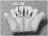
(1)糸巻きモデルにおける歯周病に対するNFκBデコイオリゴヌクレオチドの効果
全12匹(1ヶ月モデル、2ヶ月モデル、3ヶ月モデル各4匹ずつ)のビーグル犬(雄、10〜12ヶ月齢)を使用した。歯周病の発症機序としては、歯周ポケットに食べ物や歯垢が堆積することによって感染を起こし歯周病へと発展する。その際、炎症によって歯槽骨の吸収(退縮)が起こる。したがって、本モデルのように糸を巻くことで、歯周ポケットに食べ物や歯垢が堆積しやすくなり、歯周病が引き起こされる。図1にその模式図を示す。図1において、左図は糸を結紮したモデル作製時の模式図であり、時間がたつと右図のように歯槽骨の吸収(退縮)が起こる。 (1) Effect of NFκB decoy oligonucleotide on periodontal disease in a wound-cuff model All 12 beagle dogs (male, 10-12 months old) were used. . As the onset mechanism of periodontal disease, food and plaque accumulate in the periodontal pocket, causing infection and developing into periodontal disease. At this time, resorption of alveolar bone occurs due to inflammation. Therefore, by winding a thread like this model, it becomes easy for food and plaque to accumulate in a periodontal pocket, and periodontal disease is caused. FIG. 1 shows a schematic diagram thereof. In FIG. 1, the left figure is a schematic diagram at the time of making a model in which yarns are ligated. As time passes, the alveolar bone is absorbed (retracted) as shown in the right figure.
そこで、本モデルを作製後、左側上顎下顎第二切歯の歯根歯肉部分にNFκBデコイを、右側上顎下顎第二切歯の歯根歯肉部分にはコントロールとしてスクランブルデコイを、それぞれ 1 mg/部位の用量にて2週間毎に注射により投与した。使用したデコイは、デコイ溶液(TEバッファーに溶解)とAteloGeneTM Local Use(KOKEN)キットのAteloGeneTMと体積比で1:1の割合で混ぜ合わせ、最終的にデコイが1 mg/100μlになるように調製して用いた。Therefore, after preparing this model, NFκB decoy was applied to the root gingival part of the left upper mandibular second incisor, and scrambled decoy was applied to the root gingival part of the right upper mandibular second incisor as a control at a dose of 1 mg / site. Administered by injection every 2 weeks. The decoy used was mixed with the Decoy solution (dissolved in TE buffer) and the AteloGene ™ Local Use (KOKEN) kit AteloGene ™ at a volume ratio of 1: 1, so that the final decoy was 1 mg / 100 μl. Prepared and used.
1ヶ月後、2ヶ月後及び3ヶ月後にそれぞれ4匹ずつの標本を作製した。歯根長の評価方法は、解剖後の摘出試料により露出歯根長を直接測定するもの、飼育観察期間中を通じて行うレントゲン撮影による評価、DEXA(Dual Energy X-ray absorptiometry;X線骨密度測定装置)による骨密度の測定の三通りで行った。 Four specimens were prepared after 1 month, 2 months and 3 months, respectively. The evaluation method of the root length is to directly measure the exposed root length with the dissected specimen after dissection, evaluation by X-ray photography performed during the breeding observation period, DEXA (Dual Energy X-ray absorptiometry; X-ray bone density measuring device) The bone density was measured in three ways.
図2の写真は、モデル作製1ヶ月後に得た摘出試料(下顎)の付着軟組織を次亜塩素酸ナトリウムによって取り除き、歯根露出面を見やすくしたものである。下顎左右第二切歯の露出した歯根長を測定・比較すると、スクランブルデコイを投与した右第二切歯部では歯槽骨退縮(歯根露出;2本の破線で挟まれた部分)が認められたが、NFκBデコイを投与した左第二切歯の歯槽骨では骨吸収(退縮)が抑制されていた。 The photograph in FIG. 2 shows the exposed surface of the tooth root that is easy to see by removing the attached soft tissue from the extracted sample (mandible) one month after the model was prepared with sodium hypochlorite. When the exposed root lengths of the left and right second incisors were measured and compared, alveolar bone retraction (exposed root; part sandwiched between two broken lines) was observed in the right second incisor that was administered scrambled decoy However, bone resorption (retraction) was suppressed in the alveolar bone of the left second incisor administered NFκB decoy.
さらに、同様にモデル作製1ヶ月後の上下顎左右第二切歯の歯根長の測定を行った(図3)。測定部位は、上顎は第二切歯近心側、下顎は第二切歯遠心側とした。上下顎共にNFκBデコイを投与した左第二切歯部の歯槽骨(図3中、矢印で指している側)は温存され歯根露出(図3中、黒線)は抑制されたが、スクランブルデコイを投与した右第二切歯の歯槽骨は退縮が進み歯根露出(黒線)が顕著であった。図4は、モデル作製1ヶ月、2ヶ月、及び3ヶ月後の上下顎左右第二切歯の露出歯根長を示すグラフである。全体において経時的に露出長は長くなるが、より短い方が骨吸収が抑えられ、露出が抑制されている事を示す。1ヶ月モデル、2ヶ月モデルの全てにおいて、NFκBデコイ投与とスクランブルデコイ投与との間に歯根露出長の差が認められた。その結果、NFκBデコイ投与群の方が、露出が抑制されることが示された。 Similarly, the root lengths of the left and right maxillary left and right incisors were measured one month after model preparation (Fig. 3). The measurement site was the mesial side of the second incisor for the upper jaw and the distal side of the second incisor for the lower jaw. The alveolar bone (side indicated by the arrow in Fig. 3) of the left second incisor that was administered NFκB decoy in both upper and lower jaws was preserved and the root exposure (black line in Fig. 3) was suppressed, but the scrambled decoy In the alveolar bone of the right second incisor administered, the retraction progressed and the root exposure (black line) was remarkable. FIG. 4 is a graph showing the exposed root length of the upper and lower jaw left and right second incisors after 1 month, 2 months, and 3 months of model preparation. Although the exposure length increases over time as a whole, a shorter one indicates that bone resorption is suppressed and exposure is suppressed. In all of the 1-month model and 2-month model, a difference in the exposed root length was observed between NFκB decoy administration and scrambled decoy administration. As a result, it was shown that the exposure was suppressed in the NFκB decoy administration group.
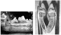
さらにまた、モデル作製2ヶ月後の解剖時に撮影した上顎部のレントゲン写真(図5)においても、NFκBデコイを投与した上顎左第二切歯(右側の矢印で指している部分)の歯槽骨先端(黒線)が、スクランブルデコイを投与した右第二切歯(左側の矢印で指している部分)の歯槽骨先端(白線)よりも骨吸収(退縮)が抑制されていることが判明した。
Furthermore, in the X-ray photograph of the maxilla taken at the time of
同様に、モデル作製2ヶ月後の解剖時に撮影した下顎部のレントゲン写真(図6)においても、上顎同様、NFκBデコイを投与した左第二切歯(右側の矢印で指している部分)の歯槽骨の骨吸収(退縮)が、スクランブルデコイを投与した右第二切歯部(左側の矢印で指している部分)よりも抑制されていると認められた。 Similarly, in the X-ray photograph of the lower jaw taken at the time of anatomy two months after model creation (Fig. 6), as in the upper jaw, the alveoli of the left second incisor (the part indicated by the right arrow) administered with NFκB decoy It was recognized that bone resorption (retraction) of the bone was suppressed more than the right second incisor (the part indicated by the left arrow) to which scrambled decoy was administered.
次に、歯槽骨の吸収を定量化してNFκB デコイとスクランブルデコイの効果を比較した。レントゲン撮影した場合、撮影角度により映像が伸縮するため、測定絶対値では経時的な比較ができない。そこで、対照として、長さに変化のない歯根長を使用することとした(図7参照)。具体的には、歯頸からの歯根の長さで、歯槽骨の長さを割り、『歯槽骨長/歯根長比』を求めた。モデル作製から1ヶ月、2ヶ月及び3ヶ月後の歯槽骨長/歯根長比を『残存歯槽骨長/歯根長比』と呼ぶ。飼育期間が長くなるほど歯槽骨の吸収(退縮)が自然に進行し、残存歯槽骨長/歯根長比は減少するが、NFκBデコイ投与群では経時的な骨吸収進行が、スクランブルデコイ投与群と比べて抑制され、NFκBデコイが歯槽骨の温存に貢献していることが示された(図8)。
Next, the effects of NFκB decoy and scrambled decoy were compared by quantifying alveolar bone resorption. When X-ray photography is performed, the image expands and contracts depending on the photographing angle, so the measurement absolute value cannot be compared over time. Therefore, as a control, the root length with no change in length was used (see FIG. 7). Specifically, the length of the alveolar bone was divided by the length of the root from the tooth neck, and the “alveolar bone length / root length ratio” was determined. The alveolar bone length /
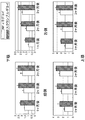
さらに、別の歯槽骨吸収の評価方法として、DEXA(Dual Energy X-ray absorptiometry;X線骨密度測定装置)分析をおこなった。解析画像を図9に示す。測定部位は、上顎は第二切歯近心側、下顎は第二切歯遠心側(白枠で囲われた部分)とした。モデル作製1、2及び3ヶ月後の結果を図10に示す。NFκB デコイ投与群では、スクランブルデコイ投与群に比べて歯槽骨の骨密度が上昇しており、NFκBデコイが歯槽骨の吸収(退縮)を抑制し、骨梁温存効果を有すると認められた。 Furthermore, as another evaluation method for alveolar bone resorption, DEXA (Dual Energy X-ray absorptiometry) analysis was performed. An analysis image is shown in FIG. The measurement site was the mesial side of the second incisor for the upper jaw and the distal side of the second incisor (the part surrounded by a white frame) for the lower jaw. The results after 1, 2, and 3 months of model production are shown in FIG. In the NFκB decoy administration group, the bone mineral density of the alveolar bone increased compared to the scrambled decoy administration group, and it was recognized that the NFκB decoy suppressed alveolar bone resorption (retraction) and had a trabecular preservation effect.
さらに、この糸巻きモデル犬において、モデル作成2ヶ月後の歯肉溝浸出液中のIL−6を測定した。IL−6は炎症を惹起するサイトカインの一種であり、破骨細胞を分化誘導させて骨破壊を起こす引き金となることが良く知られている。すなわち、歯肉溝滲出液中のIL−6量は、炎症の指標であるとともに、骨破壊の予測因子でもある。
IL−6は、常法にしたがってELISAで測定した。その結果を表1および図11に示す。Furthermore, IL-6 in the
IL-6 was measured by ELISA according to a conventional method. The results are shown in Table 1 and FIG.

上顎および下顎とも、コントロール群のスクランブルデコイ投与群に比べて、NFκBデコイ投与群で、歯肉溝滲出液中のIL−6が低減されていた。この結果からも、NFκBデコイが歯周病の炎症を抑え、骨破壊を抑制することがわかった。
なお本明細書において引用された全ての先行技術文献は、参照として本明細書に組み入れられる。In both the maxilla and the mandible, IL-6 in the gingival crevicular fluid was reduced in the NFκB decoy administration group compared to the control group scrambled decoy administration group. This result also showed that NFκB decoy suppressed periodontal inflammation and suppressed bone destruction.
It should be noted that all prior art documents cited in the present specification are incorporated herein by reference.
(2)歯周病骨欠損モデルにおけるNFκBデコイオリゴヌクレオチドの効果
本実施例に用いたビーグル犬(雄、10〜12ヶ月齢)骨欠損モデルは、静脈麻酔下、左右それぞれに、近心小臼歯部より歯肉弁を作製して歯槽骨を露出させ、大臼歯歯根分岐部に歯科用バーにて直径5mmの3壁性骨欠損を作製したものである。 左臼歯の骨欠損部位にはコラーゲン基材(AteloGeneTM)に混入したNFκB デコイを、右臼歯の骨欠損部位には同様に混入したスクランブルデコイを、それぞれ埋没投与した。使用したデコイは、上記実施例と同様1 mg/100μlになるように調製して用いた。デコイの埋没投与後、歯肉弁を復位して縫合した。手術後2週間で歯肉弁は治癒し、外部からの感染刺激等は遮断された。また、手術直後から術後4週間までの間、2週間間隔でレントゲン撮影することにより、骨欠損修復過程を観察した(図12)。 (2) Effect of NFκB decoy oligonucleotide in periodontal disease bone defect model The beagle dog (male, 10-12 months old) bone defect model used in this example is a mesial premolar part on both sides under intravenous anesthesia. A gingival flap was prepared to expose the alveolar bone, and a three-walled bone defect with a diameter of 5 mm was prepared with a dental bar at the root of the molar root. NFκB decoy mixed with a collagen base material (AteloGene ™ ) was implanted into the bone defect site of the left molar, and scrambled decoy mixed in the same manner was implanted into the bone region of the right molar. The decoy used was prepared and used at 1 mg / 100 μl as in the above example. After decoy implantation, the gingival flap was repositioned and sutured. Two weeks after the operation, the gingival flap healed and external infection stimuli were blocked. In addition, the bone defect repair process was observed by radiographing at intervals of 2 weeks from immediately after surgery to 4 weeks after surgery (Fig. 12).
術後1ヶ月の時点で顎骨を取出して横断切面を観察したところ、NFκBデコイを投与した右側の皮質骨の厚みが増していることが確認された(図13)。また、この術後1ヶ月のモデルの骨欠損部位を比較したところ、NFκBデコイ投与部位では、欠損部の表面に完全な皮質骨が形成されているが、スクランブルデコイ投与部位では柔らかくもろい皮質骨しか観察されなかった(図14)。 One month after the operation, the jawbone was taken out and the cross section was observed, and it was confirmed that the right cortical bone to which NFκB decoy was administered had increased in thickness (FIG. 13). In addition, when comparing the bone defect sites of the model one month after the operation, the cortical bone was completely formed on the surface of the defect at the site where NFκB decoy was administered, but only soft and brittle cortical bone was found at the site where scrambled decoy was administered. Not observed (Figure 14).
この骨欠損部の回復を数値的に評価するため、DEXAによる骨密度測定を行った。歯科用レントゲンにて確認される骨欠損部位(図15左図○で囲った部位)を、DEXAにて解析した。図15右図にその解析画面を示す。解析は、術後1ヶ月、2ヶ月及び3ヶ月のモデル動物について行った。NFκB デコイ投与部位及びスクランブルデコイ投与部位の両方において、骨密度は、治癒機転により経時的に増加するが、両群間には修復速度に差が見られた。NFκBデコイを投与した部位の方が、スクランブルデコイを投与した部位に比して約1ヶ月早い治癒機転が認められた(図16)。 In order to numerically evaluate the recovery of this bone defect, bone density was measured by DEXA. The bone defect site confirmed by the dental X-ray (the site surrounded by the left circle in FIG. 15) was analyzed by DEXA. The analysis screen is shown in the right figure of FIG. Analysis was performed on model animals at 1 month, 2 months, and 3 months after surgery. Bone density increased over time at both the NFκB decoy administration site and the scrambled decoy administration site, but there was a difference in the repair rate between the two groups. The site of administration of NFκB decoy showed a healing mechanism that was about one month earlier than the site of administration of scrambled decoy (FIG. 16).
さらに、骨欠損部の回復をX線マイクロCT(小動物用:SHIMADZU Kyoto Japan、X-ray CT system SMX-100CT-SV)及び解析ソフト(X-ray Image Viewer Version 4.0)を用いて解析した。本解析では、画像イメージの他、骨密度に相当する単位体積あたりの骨梁占有率等が測定可能である。術後1ヶ月、2ヶ月及び3ヶ月の時点で解析した。レントゲン写真により特定された骨欠損相当部をX線マイクロCTに供し、CT画像を得た。術後1ヶ月の画像を図17に示す。図17中、円で囲んだ部分が海綿骨であり、それぞれの円が囲む面積はいずれも等しい。NFκBデコイを投与した側では骨欠損部に白く新生骨が写っているが、スクランブルデコイを投与した側では欠損部の修復が遅れて骨梁が疎なために黒く写っている。術後2ヶ月の画像を図18に示す。NFκBデコイを投与した側では円で囲んだ部分に占める新生骨の比率が増加し、ほぼ正常海綿骨梁と同程度にまで修復過程が達している。一方、スクランブルデコイを投与した側では欠損部の修復が遅れており、黒く写る空洞の割合(未修復部分)が多い。 Further, recovery of the bone defect was analyzed using X-ray micro CT (for small animals: SHIMADZU Kyoto Japan, X-ray CT system SMX-100CT-SV) and analysis software (X-ray Image Viewer Version 4.0). In this analysis, the trabecular occupancy rate per unit volume corresponding to the bone density can be measured in addition to the image image. Analysis was performed at 1 month, 2 months, and 3 months after surgery. The part corresponding to the bone defect identified by the X-ray photograph was subjected to X-ray micro CT, and a CT image was obtained. An image of one month after the operation is shown in FIG. In FIG. 17, the portion surrounded by a circle is cancellous bone, and the area surrounded by each circle is the same. On the side administered NFκB decoy, new bone appears white in the bone defect part, but on the side administered scrambled decoy, repair of the defect part is delayed and the trabecular bone is sparse so that it appears black. An image of 2 months after the operation is shown in FIG. On the side of NFκB decoy administration, the proportion of new bone in the circled area has increased, and the repair process has reached almost the same level as normal cancellous trabecular bone. On the other hand, on the side to which scrambled decoy is administered, the repair of the defect is delayed, and there are many ratios of cavities appearing black (unrepaired parts).
この解析結果を数値的に評価するため、骨欠損部における新生骨の占める割合を骨梁/測定空間体積比として求め、得られた値をグラフ化した(図19)。NFκB デコイを投与した側では経時的に骨量が増加し、術後2ヶ月にほぼプラトーに達するのに対して、スクランブルデコイを投与した側では治癒過程に大きな改善は認められなかった。 In order to evaluate the analysis result numerically, the ratio of the new bone in the bone defect portion was obtained as a trabecular / measurement space volume ratio, and the obtained value was graphed (FIG. 19). On the side of NFκB decoy administration, bone mass increased over time and reached a plateau almost 2 months after the operation, whereas on the side of scrambled decoy administration, no significant improvement was observed in the healing process.
本発明者らによる歯槽膿漏モデル及び骨欠損モデルを用いた実験により、NFκBによる転写活性化を抑制することによって、NFκBデコイが、骨代謝機転において過剰な炎症反応及び骨吸収を抑制し、新生骨形成を促進し、早期の修復機転を促すと考えられた。このようなNFκBデコイの歯槽骨欠損の治癒過程での効果に基づき、本発明は、歯周病、及び外科手術等による歯槽骨の欠損を伴う疾病等の治療、予防または改善のための新規手段を提供する。 Through experiments using the alveolar pyorrhea model and bone defect model by the present inventors, by suppressing transcriptional activation by NFκB, NFκB decoy suppresses excessive inflammatory reaction and bone resorption in bone metabolism, and is newly born. It was thought to promote bone formation and promote early repair. Based on the effect of such NFκB decoy in the healing process of alveolar bone defects, the present invention provides novel means for treating, preventing or improving periodontal diseases and diseases associated with alveolar bone defects caused by surgery or the like. I will provide a.
Claims (6)
Priority Applications (1)
| Application Number | Priority Date | Filing Date | Title |
|---|---|---|---|
| JP2008558139A JP5384120B2 (en) | 2007-02-16 | 2008-02-15 | Therapeutic agent for periodontal diseases and alveolar bone defects caused by surgery |
Applications Claiming Priority (4)
| Application Number | Priority Date | Filing Date | Title |
|---|---|---|---|
| JP2007036135 | 2007-02-16 | ||
| JP2007036135 | 2007-02-16 | ||
| JP2008558139A JP5384120B2 (en) | 2007-02-16 | 2008-02-15 | Therapeutic agent for periodontal diseases and alveolar bone defects caused by surgery |
| PCT/JP2008/052482 WO2008099906A1 (en) | 2007-02-16 | 2008-02-15 | Therapeutic agent for periodontal disease and alveolar bone loss due to surgery |
Publications (2)
| Publication Number | Publication Date |
|---|---|
| JPWO2008099906A1 JPWO2008099906A1 (en) | 2010-05-27 |
| JP5384120B2 true JP5384120B2 (en) | 2014-01-08 |
Family
ID=39690131
Family Applications (1)
| Application Number | Title | Priority Date | Filing Date |
|---|---|---|---|
| JP2008558139A Active JP5384120B2 (en) | 2007-02-16 | 2008-02-15 | Therapeutic agent for periodontal diseases and alveolar bone defects caused by surgery |
Country Status (4)
| Country | Link |
|---|---|
| US (1) | US20100105762A1 (en) |
| EP (1) | EP2127681A4 (en) |
| JP (1) | JP5384120B2 (en) |
| WO (1) | WO2008099906A1 (en) |
Families Citing this family (4)
| Publication number | Priority date | Publication date | Assignee | Title |
|---|---|---|---|---|
| US20080312145A1 (en) * | 2004-12-16 | 2008-12-18 | Anges Mg, Inc. | Agent for Regulating Bone Formation |
| WO2010082600A1 (en) * | 2009-01-15 | 2010-07-22 | サンスター株式会社 | Agent for maintaining healthy state of periodontal tissue comprising oleuropein and degraded product thereof |
| AU2016206491A1 (en) * | 2015-01-16 | 2017-08-31 | University Of Iowa Research Foundation | Methods to prevent or treat periodontitis or peri-implantitis |
| US11904006B2 (en) | 2019-12-11 | 2024-02-20 | University Of Iowa Research Foundation | Poly(diaminosulfide) particle-based vaccine |
Citations (4)
| Publication number | Priority date | Publication date | Assignee | Title |
|---|---|---|---|---|
| JP2003522107A (en) * | 1998-07-10 | 2003-07-22 | オステオスクリーン,インコーポレイテッド | Inhibitors of proteasome activity to stimulate bone and hair growth |
| WO2005056020A2 (en) * | 2003-12-02 | 2005-06-23 | Corgentech, Inc. | Nf-kb oligonucleotide decoy molecules |
| JP2005160464A (en) * | 2003-12-02 | 2005-06-23 | Corgentech Inc | NF-kappaB OLIGONUCLEOTIDE DECOY MOLECULE |
| WO2006064886A1 (en) * | 2004-12-16 | 2006-06-22 | Anges Mg, Inc. | Agent for regulating bone formation |
Family Cites Families (14)
| Publication number | Priority date | Publication date | Assignee | Title |
|---|---|---|---|---|
| US3392143A (en) | 1967-05-15 | 1968-07-09 | Gen Electric | Polyamide compositions |
| US5354557A (en) * | 1988-04-08 | 1994-10-11 | Stryker Corporation | Osteogenic devices |
| DK0732929T3 (en) | 1993-10-29 | 2008-09-01 | Brigham & Womens Hospital | Therapeutic use of cis element derivatives in vivo |
| DK0824918T3 (en) * | 1995-05-12 | 2007-06-04 | Anges Mg Inc | Treatment and prevention of diseases caused by NF-kappa B |
| US7615373B2 (en) * | 1999-02-25 | 2009-11-10 | Virginia Commonwealth University Intellectual Property Foundation | Electroprocessed collagen and tissue engineering |
| EP1362600B1 (en) | 2001-02-20 | 2008-04-02 | AnGes MG, Inc. | TOPICAL USE OF NF-kB DECOYS FOR TREATING ATOPIC DERMATITIS |
| CN1313158C (en) * | 2001-06-20 | 2007-05-02 | 大日本住友制药株式会社 | Method of promoting nucleic acid transfer |
| US20050175539A1 (en) | 2001-11-22 | 2005-08-11 | Ryuichi Morishita | Compositions inhibiting rejection in organ transplantation and method of using the same |
| WO2003063911A1 (en) | 2002-02-01 | 2003-08-07 | Anges Mg, Inc. | Decoy-containing pharmaceutical compositions and method of using the same |
| US20060135449A1 (en) | 2002-03-29 | 2006-06-22 | Yoshiki Sawa | Decoy compositions for treating and preventing brain diseases and disorders |
| WO2003099339A1 (en) | 2002-05-29 | 2003-12-04 | Anges Mg, Inc. | Decoy composition for treating and preventing inflammatory disease |
| US20060233815A1 (en) | 2002-09-20 | 2006-10-19 | Anges Mg. Inc | Agents for protection from neointimal formation in grafts comprising an nfkappab decoy |
| AU2003252493A1 (en) | 2003-07-09 | 2005-01-28 | Anges Mg, Inc. | Pharmaceutical composition containing decoy and method of using the same |
| US8158685B2 (en) * | 2004-07-02 | 2012-04-17 | Gregory Gene Steiner | Method for bone growth |
-
2008
- 2008-02-15 US US12/449,522 patent/US20100105762A1/en not_active Abandoned
- 2008-02-15 JP JP2008558139A patent/JP5384120B2/en active Active
- 2008-02-15 EP EP08711312A patent/EP2127681A4/en not_active Withdrawn
- 2008-02-15 WO PCT/JP2008/052482 patent/WO2008099906A1/en active Application Filing
Patent Citations (4)
| Publication number | Priority date | Publication date | Assignee | Title |
|---|---|---|---|---|
| JP2003522107A (en) * | 1998-07-10 | 2003-07-22 | オステオスクリーン,インコーポレイテッド | Inhibitors of proteasome activity to stimulate bone and hair growth |
| WO2005056020A2 (en) * | 2003-12-02 | 2005-06-23 | Corgentech, Inc. | Nf-kb oligonucleotide decoy molecules |
| JP2005160464A (en) * | 2003-12-02 | 2005-06-23 | Corgentech Inc | NF-kappaB OLIGONUCLEOTIDE DECOY MOLECULE |
| WO2006064886A1 (en) * | 2004-12-16 | 2006-06-22 | Anges Mg, Inc. | Agent for regulating bone formation |
Non-Patent Citations (2)
| Title |
|---|
| JPN6008012220; AMBILI R. et al.: 'Expression of activated transcription factor nuclear factor-kappa B in periodontally diseased tissue' J.Periodontol Vol.76, No.7, 2005, p.1148-1153 * |
| JPN6013012803; FEBS LETTERS 580, 2006, 613-619 * |
Also Published As
| Publication number | Publication date |
|---|---|
| EP2127681A1 (en) | 2009-12-02 |
| EP2127681A4 (en) | 2011-10-12 |
| US20100105762A1 (en) | 2010-04-29 |
| JPWO2008099906A1 (en) | 2010-05-27 |
| WO2008099906A1 (en) | 2008-08-21 |
Similar Documents
| Publication | Publication Date | Title |
|---|---|---|
| Abbas et al. | Evaluation of corticotomy-facilitated orthodontics and piezocision in rapid canine retraction | |
| Kim et al. | Effects of corticision on paradental remodeling in orthodontic tooth movement | |
| Alikhani et al. | Saturation of the biological response to orthodontic forces and its effect on the rate of tooth movement | |
| Bardet et al. | Claudin‐16 deficiency impairs tight junction function in ameloblasts, leading to abnormal enamel formation | |
| Shepherd et al. | Root coverage using acellular dermal matrix and comparing a coronally positioned tunnel with and without platelet‐rich plasma: A pilot study in humans | |
| Santamaria et al. | The influence of local anatomy on the outcome of treatment of gingival recession associated with non‐carious cervical lesions | |
| Peker Tekdal et al. | The effect of piezoelectric surgery implant osteotomy on radiological and molecular parameters of peri‐implant crestal bone loss: a randomized, controlled, split‐mouth trial | |
| Akcan et al. | Gingival recession treatment with concentrated growth factor membrane: a comparative clinical trial | |
| Henriques et al. | Application of subepithelial connective tissue graft with or without enamel matrix derivative for root coverage: a split-mouth randomized study | |
| Kirschneck et al. | Comparative assessment of mouse models for experimental orthodontic tooth movement | |
| Tanomaru-Filho et al. | Evaluation of periapical repair following retrograde filling with different root-end filling materials in dog teeth with periapical lesions | |
| Lee et al. | Microscopic analysis of molar–incisor malformation | |
| Haghighati et al. | A comparative study of two root-coverage techniques with regard to interdental papilla dimension as a prognostic factor. | |
| JP5384120B2 (en) | Therapeutic agent for periodontal diseases and alveolar bone defects caused by surgery | |
| Walsh et al. | Histology of NeoMTA Plus and Quick-Set2 in contact with pulp and periradicular tissues in a canine model | |
| Alves et al. | Local delivery of EGF–liposome mediated bone modeling in orthodontic tooth movement by increasing RANKL expression | |
| Alsino et al. | The effectiveness of periodontally accelerated osteogenic orthodontics (PAOO) in accelerating tooth movement and supporting alveolar bone thickness during orthodontic treatment: a systematic review | |
| Shope et al. | Developmental pathology and pedodontology | |
| Morkmued et al. | Deficiency of the SMOC2 matricellular protein impairs bone healing and produces age-dependent bone loss | |
| Zhou et al. | Study of bone remodeling in corticotomy‐assisted orthodontic tooth movement in rats | |
| Ramnarayan et al. | Management of idiopathic gingival fibromatosis: report of a case and literature review | |
| Li et al. | Differences in accelerated tooth movement promoted by recombinant human parathyroid hormone after mandibular ramus osteotomy | |
| Oh et al. | Periodontal regenerative therapy in endo-periodontal lesions: a retrospective study over 5 years | |
| Zheng et al. | Evaluation of recombinant human bone morphogenetic protein-2 in mandibular distraction osteogenesis in rabbits: Effect of dosage and number of doses on formation of bone | |
| Gul Amuk et al. | Effects of mesenchymal stem cell transfer on orthodontically induced root resorption and orthodontic tooth movement during orthodontic arch expansion protocols: an experimental study in rats |
Legal Events
| Date | Code | Title | Description |
|---|---|---|---|
| A521 | Request for written amendment filed |
Free format text: JAPANESE INTERMEDIATE CODE: A523 Effective date: 20110201 |
|
| A621 | Written request for application examination |
Free format text: JAPANESE INTERMEDIATE CODE: A621 Effective date: 20110201 |
|
| A131 | Notification of reasons for refusal |
Free format text: JAPANESE INTERMEDIATE CODE: A131 Effective date: 20130318 |
|
| A521 | Request for written amendment filed |
Free format text: JAPANESE INTERMEDIATE CODE: A523 Effective date: 20130514 |
|
| A131 | Notification of reasons for refusal |
Free format text: JAPANESE INTERMEDIATE CODE: A131 Effective date: 20130610 |
|
| TRDD | Decision of grant or rejection written | ||
| A01 | Written decision to grant a patent or to grant a registration (utility model) |
Free format text: JAPANESE INTERMEDIATE CODE: A01 Effective date: 20130902 |
|
| A61 | First payment of annual fees (during grant procedure) |
Free format text: JAPANESE INTERMEDIATE CODE: A61 Effective date: 20131002 |
|
| R150 | Certificate of patent or registration of utility model |
Ref document number: 5384120 Country of ref document: JP Free format text: JAPANESE INTERMEDIATE CODE: R150 Free format text: JAPANESE INTERMEDIATE CODE: R150 |
|
| R250 | Receipt of annual fees |
Free format text: JAPANESE INTERMEDIATE CODE: R250 |
|
| R250 | Receipt of annual fees |
Free format text: JAPANESE INTERMEDIATE CODE: R250 |
|
| R250 | Receipt of annual fees |
Free format text: JAPANESE INTERMEDIATE CODE: R250 |
|
| R250 | Receipt of annual fees |
Free format text: JAPANESE INTERMEDIATE CODE: R250 |
|
| R250 | Receipt of annual fees |
Free format text: JAPANESE INTERMEDIATE CODE: R250 |
|
| R250 | Receipt of annual fees |
Free format text: JAPANESE INTERMEDIATE CODE: R250 |
|
| R250 | Receipt of annual fees |
Free format text: JAPANESE INTERMEDIATE CODE: R250 |
|
| R250 | Receipt of annual fees |
Free format text: JAPANESE INTERMEDIATE CODE: R250 |