JP5364954B2 - NMR measurement method - Google Patents
NMR measurement method Download PDFInfo
- Publication number
- JP5364954B2 JP5364954B2 JP2008240398A JP2008240398A JP5364954B2 JP 5364954 B2 JP5364954 B2 JP 5364954B2 JP 2008240398 A JP2008240398 A JP 2008240398A JP 2008240398 A JP2008240398 A JP 2008240398A JP 5364954 B2 JP5364954 B2 JP 5364954B2
- Authority
- JP
- Japan
- Prior art keywords
- relaxation time
- nucleus
- longitudinal magnetization
- spin
- nmr
- Prior art date
- Legal status (The legal status is an assumption and is not a legal conclusion. Google has not performed a legal analysis and makes no representation as to the accuracy of the status listed.)
- Active
Links
- 238000000034 method Methods 0.000 title claims description 86
- 238000005481 NMR spectroscopy Methods 0.000 title claims description 55
- 230000005415 magnetization Effects 0.000 claims description 174
- 238000000655 nuclear magnetic resonance spectrum Methods 0.000 claims description 112
- 238000005259 measurement Methods 0.000 claims description 52
- 238000009792 diffusion process Methods 0.000 claims description 49
- 238000001228 spectrum Methods 0.000 claims description 47
- 238000011084 recovery Methods 0.000 claims description 21
- 238000000691 measurement method Methods 0.000 claims description 17
- 239000007787 solid Substances 0.000 claims description 10
- 230000003993 interaction Effects 0.000 claims description 6
- 230000001678 irradiating effect Effects 0.000 claims description 6
- 230000008569 process Effects 0.000 claims description 5
- 238000001208 nuclear magnetic resonance pulse sequence Methods 0.000 claims description 4
- 230000005284 excitation Effects 0.000 claims description 3
- 239000000203 mixture Substances 0.000 description 34
- 238000000926 separation method Methods 0.000 description 22
- HVYWMOMLDIMFJA-DPAQBDIFSA-N cholesterol Chemical compound C1C=C2C[C@@H](O)CC[C@]2(C)[C@@H]2[C@@H]1[C@@H]1CC[C@H]([C@H](C)CCCC(C)C)[C@@]1(C)CC2 HVYWMOMLDIMFJA-DPAQBDIFSA-N 0.000 description 18
- 238000004458 analytical method Methods 0.000 description 16
- 238000012546 transfer Methods 0.000 description 16
- UFHFLCQGNIYNRP-UHFFFAOYSA-N Hydrogen Chemical compound [H][H] UFHFLCQGNIYNRP-UHFFFAOYSA-N 0.000 description 14
- 229910052739 hydrogen Inorganic materials 0.000 description 14
- 239000001257 hydrogen Substances 0.000 description 14
- OKTJSMMVPCPJKN-UHFFFAOYSA-N Carbon Chemical compound [C] OKTJSMMVPCPJKN-UHFFFAOYSA-N 0.000 description 11
- 229910052799 carbon Inorganic materials 0.000 description 11
- 238000001460 carbon-13 nuclear magnetic resonance spectrum Methods 0.000 description 11
- 238000000914 diffusion-ordered spectroscopy Methods 0.000 description 11
- TUFPZQHDPZYIEX-UHFFFAOYSA-N alpha-Santonin Natural products C1CC2(C)C=CC(=O)C=C2C2C1C(C)C(=O)O2 TUFPZQHDPZYIEX-UHFFFAOYSA-N 0.000 description 9
- XJHDMGJURBVLLE-BOCCBSBMSA-N alpha-santonin Chemical compound C([C@]1(C)CC2)=CC(=O)C(C)=C1[C@@H]1[C@@H]2[C@H](C)C(=O)O1 XJHDMGJURBVLLE-BOCCBSBMSA-N 0.000 description 9
- 235000012000 cholesterol Nutrition 0.000 description 9
- 229940074353 santonin Drugs 0.000 description 9
- 150000001721 carbon Chemical class 0.000 description 8
- 150000002431 hydrogen Chemical class 0.000 description 6
- 125000002496 methyl group Chemical group [H]C([H])([H])* 0.000 description 6
- 230000008859 change Effects 0.000 description 4
- 150000001875 compounds Chemical class 0.000 description 4
- 239000013078 crystal Substances 0.000 description 4
- 238000010586 diagram Methods 0.000 description 4
- 238000002075 inversion recovery Methods 0.000 description 4
- 239000002245 particle Substances 0.000 description 4
- 230000000694 effects Effects 0.000 description 3
- 238000001644 13C nuclear magnetic resonance spectroscopy Methods 0.000 description 2
- 239000004698 Polyethylene Substances 0.000 description 2
- 238000009826 distribution Methods 0.000 description 2
- 238000002474 experimental method Methods 0.000 description 2
- 239000005416 organic matter Substances 0.000 description 2
- -1 polyethylene Polymers 0.000 description 2
- 229920000573 polyethylene Polymers 0.000 description 2
- 238000000746 purification Methods 0.000 description 2
- 239000000126 substance Substances 0.000 description 2
- 125000004429 atom Chemical group 0.000 description 1
- 230000015572 biosynthetic process Effects 0.000 description 1
- 239000006227 byproduct Substances 0.000 description 1
- 238000006243 chemical reaction Methods 0.000 description 1
- 239000007806 chemical reaction intermediate Substances 0.000 description 1
- 239000007795 chemical reaction product Substances 0.000 description 1
- 239000013256 coordination polymer Substances 0.000 description 1
- 230000008878 coupling Effects 0.000 description 1
- 238000010168 coupling process Methods 0.000 description 1
- 238000005859 coupling reaction Methods 0.000 description 1
- 239000004205 dimethyl polysiloxane Substances 0.000 description 1
- 235000013870 dimethyl polysiloxane Nutrition 0.000 description 1
- 239000010419 fine particle Substances 0.000 description 1
- 239000013081 microcrystal Substances 0.000 description 1
- CXQXSVUQTKDNFP-UHFFFAOYSA-N octamethyltrisiloxane Chemical compound C[Si](C)(C)O[Si](C)(C)O[Si](C)(C)C CXQXSVUQTKDNFP-UHFFFAOYSA-N 0.000 description 1
- 238000004987 plasma desorption mass spectroscopy Methods 0.000 description 1
- 229920000435 poly(dimethylsiloxane) Polymers 0.000 description 1
- 238000000425 proton nuclear magnetic resonance spectrum Methods 0.000 description 1
- 230000003595 spectral effect Effects 0.000 description 1
- 238000003786 synthesis reaction Methods 0.000 description 1
- 230000009466 transformation Effects 0.000 description 1
Images


Classifications
-
- G—PHYSICS
- G01—MEASURING; TESTING
- G01R—MEASURING ELECTRIC VARIABLES; MEASURING MAGNETIC VARIABLES
- G01R33/00—Arrangements or instruments for measuring magnetic variables
- G01R33/20—Arrangements or instruments for measuring magnetic variables involving magnetic resonance
- G01R33/44—Arrangements or instruments for measuring magnetic variables involving magnetic resonance using nuclear magnetic resonance [NMR]
- G01R33/46—NMR spectroscopy
- G01R33/4641—Sequences for NMR spectroscopy of samples with ultrashort relaxation times such as solid samples
-
- G—PHYSICS
- G01—MEASURING; TESTING
- G01N—INVESTIGATING OR ANALYSING MATERIALS BY DETERMINING THEIR CHEMICAL OR PHYSICAL PROPERTIES
- G01N24/00—Investigating or analyzing materials by the use of nuclear magnetic resonance, electron paramagnetic resonance or other spin effects
- G01N24/08—Investigating or analyzing materials by the use of nuclear magnetic resonance, electron paramagnetic resonance or other spin effects by using nuclear magnetic resonance
-
- G—PHYSICS
- G01—MEASURING; TESTING
- G01R—MEASURING ELECTRIC VARIABLES; MEASURING MAGNETIC VARIABLES
- G01R33/00—Arrangements or instruments for measuring magnetic variables
- G01R33/20—Arrangements or instruments for measuring magnetic variables involving magnetic resonance
- G01R33/44—Arrangements or instruments for measuring magnetic variables involving magnetic resonance using nuclear magnetic resonance [NMR]
- G01R33/46—NMR spectroscopy
- G01R33/4608—RF excitation sequences for enhanced detection, e.g. NOE, polarisation transfer, selection of a coherence transfer pathway
-
- G—PHYSICS
- G01—MEASURING; TESTING
- G01R—MEASURING ELECTRIC VARIABLES; MEASURING MAGNETIC VARIABLES
- G01R33/00—Arrangements or instruments for measuring magnetic variables
- G01R33/20—Arrangements or instruments for measuring magnetic variables involving magnetic resonance
- G01R33/44—Arrangements or instruments for measuring magnetic variables involving magnetic resonance using nuclear magnetic resonance [NMR]
- G01R33/46—NMR spectroscopy
- G01R33/4633—Sequences for multi-dimensional NMR
-
- G—PHYSICS
- G01—MEASURING; TESTING
- G01R—MEASURING ELECTRIC VARIABLES; MEASURING MAGNETIC VARIABLES
- G01R33/00—Arrangements or instruments for measuring magnetic variables
- G01R33/20—Arrangements or instruments for measuring magnetic variables involving magnetic resonance
- G01R33/44—Arrangements or instruments for measuring magnetic variables involving magnetic resonance using nuclear magnetic resonance [NMR]
- G01R33/48—NMR imaging systems
- G01R33/50—NMR imaging systems based on the determination of relaxation times, e.g. T1 measurement by IR sequences; T2 measurement by multiple-echo sequences
-
- G—PHYSICS
- G01—MEASURING; TESTING
- G01R—MEASURING ELECTRIC VARIABLES; MEASURING MAGNETIC VARIABLES
- G01R33/00—Arrangements or instruments for measuring magnetic variables
- G01R33/20—Arrangements or instruments for measuring magnetic variables involving magnetic resonance
- G01R33/44—Arrangements or instruments for measuring magnetic variables involving magnetic resonance using nuclear magnetic resonance [NMR]
- G01R33/48—NMR imaging systems
- G01R33/54—Signal processing systems, e.g. using pulse sequences ; Generation or control of pulse sequences; Operator console
- G01R33/56—Image enhancement or correction, e.g. subtraction or averaging techniques, e.g. improvement of signal-to-noise ratio and resolution
- G01R33/5602—Image enhancement or correction, e.g. subtraction or averaging techniques, e.g. improvement of signal-to-noise ratio and resolution by filtering or weighting based on different relaxation times within the sample, e.g. T1 weighting using an inversion pulse
-
- G—PHYSICS
- G01—MEASURING; TESTING
- G01R—MEASURING ELECTRIC VARIABLES; MEASURING MAGNETIC VARIABLES
- G01R33/00—Arrangements or instruments for measuring magnetic variables
- G01R33/20—Arrangements or instruments for measuring magnetic variables involving magnetic resonance
- G01R33/44—Arrangements or instruments for measuring magnetic variables involving magnetic resonance using nuclear magnetic resonance [NMR]
- G01R33/48—NMR imaging systems
- G01R33/54—Signal processing systems, e.g. using pulse sequences ; Generation or control of pulse sequences; Operator console
- G01R33/56—Image enhancement or correction, e.g. subtraction or averaging techniques, e.g. improvement of signal-to-noise ratio and resolution
- G01R33/5605—Image enhancement or correction, e.g. subtraction or averaging techniques, e.g. improvement of signal-to-noise ratio and resolution by transferring coherence or polarization from a spin species to another, e.g. creating magnetization transfer contrast [MTC], polarization transfer using nuclear Overhauser enhancement [NOE]
Landscapes
- Physics & Mathematics (AREA)
- High Energy & Nuclear Physics (AREA)
- Spectroscopy & Molecular Physics (AREA)
- General Physics & Mathematics (AREA)
- Condensed Matter Physics & Semiconductors (AREA)
- Life Sciences & Earth Sciences (AREA)
- Health & Medical Sciences (AREA)
- Chemical & Material Sciences (AREA)
- Analytical Chemistry (AREA)
- Biochemistry (AREA)
- General Health & Medical Sciences (AREA)
- Immunology (AREA)
- Pathology (AREA)
- Investigating Or Analysing Biological Materials (AREA)
Description
本発明は、複数の成分がそれぞれ微結晶を成して混在している固体試料や、単一の分子であっても複数の結晶系が混在している固体試料や、結晶成分と非結晶成分が混在している固体試料など、複数のドメインを有する試料のNMRスペクトルをドメインごとに分離して測定する際に使用されるNMR測定方法に関する。 The present invention relates to a solid sample in which a plurality of components are mixed together in a microcrystal, a solid sample in which a plurality of crystal systems are mixed even with a single molecule, a crystalline component and an amorphous component The present invention relates to an NMR measurement method used when an NMR spectrum of a sample having a plurality of domains, such as a solid sample in which is mixed, is separated and measured for each domain.
混合物を成分ごとに分離して観測するNMR測定手法、および逆ラプラス変換を解析に利用する手法の一例として、DOSY法を以下に説明する。DOSY法は、近年のNMR装置の精度向上および処理ソフトの改良により、広い分野で利用されるようになってきたNMR測定方法であり、もともとは、1965年、スタッカーとターナーによって提唱されたNMR測定方法の1つである(非特許文献1)。 The DOSY method will be described below as an example of an NMR measurement method that separates and observes a mixture for each component and a method that uses inverse Laplace transform for analysis. The DOSY method is an NMR measurement method that has come to be used in a wide range of fields due to recent improvements in the accuracy of NMR equipment and improvements in processing software. Originally, the NMR measurement proposed by Stacker and Turner in 1965 This is one of the methods (Non-Patent Document 1).
DOSY法の実験においては、複数種類の分子の混合物である試料であっても、分子の拡散係数の違いを利用することによって、成分分子ごとに分離してNMRスペクトルを測定することができる。 In the experiment of the DOSY method, even for a sample that is a mixture of a plurality of types of molecules, the NMR spectrum can be measured separately for each component molecule by utilizing the difference in the diffusion coefficient of the molecules.
混合物のNMRスペクトルは、通常、図1に示すように、各成分のスペクトルの和として観測される。一方、分子の拡散係数は、各分子種に固有の値を持っている。そのため、混合物のNMRスペクトルを分子の拡散係数により分類すると、結果としてNMRスペクトルを分子種ごとに分離して観測することができるのである。 The NMR spectrum of the mixture is usually observed as the sum of the spectra of each component as shown in FIG. On the other hand, the diffusion coefficient of molecules has a unique value for each molecular species. Therefore, if the NMR spectrum of the mixture is classified by the molecular diffusion coefficient, the NMR spectrum can be separated and observed for each molecular species as a result.
拡散係数の測定は、拡散計測時間を変化させながらNMRスペクトルを複数回観測することにより行なわれる。通常のDOSY法では、図2に示すように、拡散計測時間が長くなるほど、また拡散速度が速くなるほど、NMRスペクトルの信号強度は大きく減衰するので、拡散速度の速いものほど信号強度の減衰が速く、拡散速度の遅いものほど信号強度の減衰が遅い。 The diffusion coefficient is measured by observing the NMR spectrum a plurality of times while changing the diffusion measurement time. In the normal DOSY method, as shown in FIG. 2, the longer the diffusion measurement time and the faster the diffusion rate, the more the signal intensity of the NMR spectrum attenuates. Therefore, the faster the diffusion rate, the faster the signal intensity decays. The slower the diffusion rate, the slower the signal intensity decays.
この変化を解析することにより、拡散係数が求まる。拡散係数の違いによるNMRスペクトルの分離には、逆ラプラス変換が用いられる。逆ラプラス変換を用いることにより、拡散係数の値の位置に先鋭化したピークが現れ、スペクトルの解析が容易になる。図3は、1つのピーク強度に注目して逆ラプラス変換を行なった例である。 By analyzing this change, the diffusion coefficient can be obtained. Reverse Laplace transform is used for the separation of the NMR spectrum by the difference in diffusion coefficient. By using the inverse Laplace transform, a sharpened peak appears at the position of the diffusion coefficient value, and the spectrum can be easily analyzed. FIG. 3 shows an example in which the inverse Laplace transform is performed paying attention to one peak intensity.
信号強度が拡散によって同じような減衰挙動を示すピークを逆ラプラス変換によってグループ分けすると、図4に示すように、拡散の遅い成分Aのスペクトルと、拡散の速い成分Bのスペクトルとの2つの群に分離することができる。 When peaks whose signal strengths exhibit similar attenuation behavior due to diffusion are grouped by inverse Laplace transform, as shown in FIG. 4, two groups of a spectrum of a component A having a low diffusion and a spectrum of a component B having a high diffusion are obtained. Can be separated.
DOSY法においては、逆ラプラス変換を用いて拡散係数の違いによりスペクトルを分離することで、スペクトルを成分ごとに分離して観測することができる。これは、各分子が固有の拡散係数を持つことを利用した分離法である。 In the DOSY method, the spectrum can be separated for each component and observed by separating the spectrum based on the difference in the diffusion coefficient using inverse Laplace transform. This is a separation method using the fact that each molecule has a unique diffusion coefficient.
次に、DOSY法の他に、緩和時間の違いを利用して複数の成分を分離する測定法が知られているので、それを以下に3つほど紹介・列挙する。 Next, in addition to the DOSY method, there are known measurement methods that use a difference in relaxation time to separate a plurality of components. The following three are introduced and listed.
(1)13C炭素核の縦磁化緩和時間の違いを利用して複数の成分を分離する測定法。 (1) A measurement method in which a plurality of components are separated using the difference in longitudinal magnetization relaxation time of 13 C carbon nuclei.
13C炭素核の縦磁化緩和時間の違いを指標にしてNMR信号を分離する方法である。緩和計測時間を変化させながら13C-NMRスペクトルを観測すると、信号強度の変化は緩和時間の違いを反映する。 This is a method for separating NMR signals using the difference in longitudinal magnetization relaxation time of 13 C carbon nuclei as an index. When a 13 C-NMR spectrum is observed while changing the relaxation measurement time, the change in signal intensity reflects the difference in relaxation time.
図5にポリエチレンの実測データ(非特許文献2より引用)を示す。この測定では反転復活法を用いており、反転させた信号がどのぐらいの時間で復活するかを観測することにより13C炭素核の縦磁化緩和時間を測定している。 FIG. 5 shows measured data of polyethylene (cited from Non-Patent Document 2). In this measurement, the inversion recovery method is used, and the longitudinal magnetization relaxation time of 13 C carbon nuclei is measured by observing how long the inverted signal recovers.
0.025sの緩和計測時間(τ)の時には35ppmの信号および31ppmの信号はともに反転されている。次に、緩和計測時間が10sになると、どちらの信号も正の強度になるが、その間の挙動は異なる。 At the relaxation measurement time (τ) of 0.025 s, both the 35 ppm signal and the 31 ppm signal are inverted. Next, when the relaxation measurement time is 10 s, both signals have positive intensities, but the behavior during that time is different.
31ppmの信号は、0.1sの緩和計測時間ですでに正の強度に復活しているにも関わらず、35ppmの信号は、1sの緩和計測時間まで待たないと正の強度に復活しない。この測定を通じて、31ppmの信号と35ppmの信号が異なる緩和時間を持つ信号として分類することができる。 Although the 31ppm signal has already returned to positive intensity with a relaxation measurement time of 0.1s, the 35ppm signal does not return to positive intensity without waiting for the relaxation measurement time of 1s. Through this measurement, the 31 ppm signal and the 35 ppm signal can be classified as signals with different relaxation times.
13C炭素核の縦磁化緩和時間は、分子の局所的な運動モードにより強く影響を受ける。そのため、13C炭素核の縦磁化緩和時間による信号の分離は、分子の局所的な運動性の違いを反映した分離法ということになる。 The longitudinal magnetization relaxation time of 13 C carbon nuclei is strongly influenced by the local motion mode of the molecule. Therefore, the signal separation based on the longitudinal magnetization relaxation time of 13 C carbon nuclei is a separation method that reflects the difference in local mobility of molecules.
(2)1H水素核の横磁化緩和時間の違い、および1H水素核から13C炭素核への磁化移動を利用して信号を分離する測定法。 (2) 1 differences in transverse magnetization relaxation times of the H nuclei, and 1 measurement method of separating signals by using magnetization transfer from H nuclei to 13 C nuclei.
この方法では、横磁化緩和時間の計測時間を変化させながらスペクトルを観測する。観測するときには1H水素核から13C炭素核への磁化移動を行ない、13C炭素核でスペクトルを観測する。1H-NMRスペクトルは線幅が広い信号が重なり合ってしまうため、分離がよくない。それに対して13C-NMRスペクトルは線幅が細く、さまざまな信号を分離して観測することができる。 In this method, the spectrum is observed while changing the measurement time of the transverse magnetization relaxation time. When observing, the magnetization is transferred from the 1 H hydrogen nucleus to the 13 C carbon nucleus, and the spectrum is observed at the 13 C carbon nucleus. The 1 H-NMR spectrum is not well separated because signals with wide line widths overlap. On the other hand, the 13 C-NMR spectrum has a narrow line width, and various signals can be separated and observed.
したがって、信号分離という観点で、1H水素核から13C炭素核への磁化移動、および磁化移動に引き続く13C炭素核での信号観測は有用である。1H水素核の横磁化緩和時間の測定データはFourier変換を行ない、スペクトルとして観測する。 Therefore, from the viewpoint of signal separation, the magnetization transfer from the 1 H hydrogen nucleus to the 13 C carbon nucleus and the signal observation at the 13 C carbon nucleus following the magnetization transfer are useful. The measured data of transverse magnetization relaxation time of 1 H hydrogen nucleus is subjected to Fourier transform and observed as a spectrum.
Fourier変換を行なうことにより、横磁化緩和の速い成分は線幅の広いピークとして現れ、遅い成分は線幅の狭いピークとして現れる。時間領域の信号をFourier変換してスペクトルとして表示するのはNMRの慣習に従った処理であり、この処理により信号の分離が良くなるといった効果はない。 By performing Fourier transform, a component with fast transverse magnetization relaxation appears as a peak with a wide line width, and a slow component appears as a peak with a narrow line width. The Fourier transform of the time domain signal and displaying it as a spectrum is a process according to the convention of NMR, and this process has no effect of improving signal separation.
図6に、この測定法(a)および実測データ(b)を示す(ともに非特許文献3より引用)。(a)に“CP”と記述がある部分が磁化移動である。(b)をみると、13C軸方向には3本のピークが分離して観測されていることがわかる。 FIG. 6 shows this measurement method (a) and actual measurement data (b) (both are cited from Non-Patent Document 3). The part where “CP” is described in (a) is the magnetization transfer. As can be seen from (b), three peaks are observed separately in the 13 C-axis direction.
この分離は、1H水素核から13C炭素核への磁化移動、および13C炭素核でのNMRスペクトルの観測により実現された。一方、PDMSとラベルされている信号の1H軸方向のスペクトルは非常に細いのに対して、PSとラベルされている信号のスペクトルは幅広いものとなっている。このように、1H軸側のスペクトルの線幅から信号を分類することができる。 This separation was realized by magnetization transfer from 1 H hydrogen nucleus to 13 C carbon nucleus and observation of NMR spectrum at 13 C carbon nucleus. On the other hand, while the spectrum of the 1 H-axis direction of the signal being PDMS and label very thin, the spectrum of the signal being PS and labels has a wide range of things. Thus, signals can be classified from the line width of the spectrum on the 1 H axis side.
1H水素核の横磁化緩和時間は、13C炭素核の縦磁化緩和時間と同様に分子の局所的な運動モードにより強く影響を受ける。そのため、1H水素核の横磁化緩和時間による信号の分離は、分子の局所的な運動性の違いを反映した分離法ということになる。 The transverse magnetization relaxation time of 1 H hydrogen nuclei is strongly influenced by the local motion mode of the molecule, similarly to the longitudinal magnetization relaxation time of 13 C carbon nuclei. Therefore, signal separation based on the transverse magnetization relaxation time of 1 H hydrogen nuclei is a separation method that reflects differences in local mobility of molecules.
(3)1H水素核の縦磁化緩和時間を13C炭素核へと磁化移動することにより、13C炭素核のNMRスペクトルとして測定する手法。 (3) by magnetization transfer and of 1 H hydrogen nuclei longitudinal magnetization relaxation time of the 13 C carbon nucleus, a technique for measuring the NMR spectrum of 13 C carbon nucleus.
この方法は、図7に示すように、1H水素核の縦磁化緩和時間の観測を行なった後、1H水素核から13C炭素核への磁化移動を行ない、13C炭素核のNMRスペクトルとして観測を行なう手法であり、1H水素核の縦磁化緩和時間測定の結果は、13C-NMRスペクトルの強度変化として現れる(非特許文献4)。 This method, as shown in FIG. 7, 1 after performing the observation of the longitudinal magnetization relaxation times of the H nuclei, 1 performs magnetization transfer from H nuclei to 13 C nuclei, 13 NMR spectrum of C nuclei The results of longitudinal magnetization relaxation time measurement of 1 H hydrogen nuclei appear as intensity changes in the 13 C-NMR spectrum (Non-patent Document 4).
尚、1H水素核の縦磁化緩和時間は、1H水素核と1H水素核の間の同種核間スピン拡散により、分子内では均一になっている。このような事実、および上記(1)〜(3)の測定手法については、非特許文献5に詳しくまとめられている。 Incidentally, 1 H hydrogen nuclei longitudinal magnetization relaxation time of the by homonuclear between spin diffusion between of the 1 H hydrogen nucleus and 1 H hydrogen nuclei are uniform within the molecule. Such facts and the measurement methods (1) to (3) are summarized in detail in Non-Patent Document 5.
混合物から成る溶液試料のNMRスペクトルを観測するにあたっては、前述のようにDOSY法が最も広く用いられている。DOSY法を用いることにより、成分ごとにNMRスペクトルを分離することができるからである。DOSY法は溶液中における分子の並進拡散係数の違いを利用してスペクトルの分離を実行する。しかしながら、固体試料においては並進拡散が存在しない。そのため、固体試料に対しては、DOSY法は適用できないという問題があった。 In observing the NMR spectrum of a solution sample made of a mixture, the DOSY method is most widely used as described above. This is because the NMR spectrum can be separated for each component by using the DOSY method. The DOSY method performs spectral separation by utilizing the difference in translational diffusion coefficient of molecules in a solution. However, there is no translational diffusion in the solid sample. Therefore, there is a problem that the DOSY method cannot be applied to a solid sample.
一方、上記(1)に示した手法を用いれば、13C炭素核の縦磁化緩和時間の違いを利用して、混合物の信号を分離することができる。ところが、13C炭素核の縦磁化緩和時間は、分子の局所的な運動性を反映しているので、この測定法による分離は、分子の局所的な運動性の違いによる分離となる。そのため、分子種による分類とは必ずしもならない。 On the other hand, if the method shown in (1) above is used, the signal of the mixture can be separated using the difference in the longitudinal magnetization relaxation time of 13 C carbon nuclei. However, the longitudinal magnetization relaxation time of 13 C carbon nuclei reflects the local mobility of the molecule, so that the separation by this measurement method is due to the difference in the local mobility of the molecule. Therefore, it is not necessarily classified by molecular species.
具体的には、分子にメチル基が含まれているような場合、メチル基の運動性は非常に高いので、同じ分子からの他の信号とメチル基の信号は、別々に分離されて観測されてしまうという問題があった。 Specifically, when the molecule contains a methyl group, the mobility of the methyl group is so high that other signals from the same molecule and the methyl group signal are observed separately. There was a problem that.
また、上記(2)に示した手法を用いれば、1H水素核の横磁化緩和時間の違いを利用して、混合物の信号を分離することができる。ところが、13C炭素核の縦磁化緩和時間による分離と同様に、この方法もまた分子の局所的な運動性の違いによる分離となる。そのため、分子種による分離とは必ずしもならない。 Moreover, if the method shown in (2) above is used, the signal of the mixture can be separated using the difference in transverse magnetization relaxation time of 1 H hydrogen nuclei. However, like the separation due to the longitudinal magnetization relaxation time of 13 C carbon nuclei, this method is also a separation due to a difference in local mobility of molecules. Therefore, separation by molecular species is not necessarily performed.
具体的には、分子にメチル基が含まれているような場合、メチル基の運動性は非常に高いので、同じ分子からの他の信号とメチル基の信号は、別々に分離されて観測されてしまうという問題があった。 Specifically, when the molecule contains a methyl group, the mobility of the methyl group is so high that other signals from the same molecule and the methyl group signal are observed separately. There was a problem that.
また、上記(3)に示した手法を用いれば、1H水素核の縦磁化緩和測定、および1H水素核から13C炭素核への磁化移動の実験により、1H水素核の縦磁化緩和時間を、その1H水素核が所属している分子の13C-NMRの信号強度変化として取り出すことができる。通常、この測定法は、純品の試料に対してのみ適用されるが、もし混合物にこの測定法を適用することができれば、効果は大きい。 Further, by using the technique shown in the above (3), 1 longitudinal magnetization relaxation measurement of H nuclei, and 1 by magnetization transfer experiments from H nuclei to 13 C nuclei, 1 H nuclei of longitudinal magnetization relaxation The time can be extracted as the change in signal intensity of 13 C-NMR of the molecule to which the 1 H hydrogen nucleus belongs. Usually, this measurement method is applied only to a pure sample, but if this measurement method can be applied to a mixture, the effect is great.
スピン拡散によりスピンI(通常は1H水素核)が分子内において均一な縦磁化緩和時間を持つとき、スピンIのスペクトルをスピンIの縦磁化緩和時間で分類することができれば、スピンIのスペクトルは分子種ごとに分離することができる。 If spin I (usually 1 H hydrogen nuclei) has a uniform longitudinal magnetization relaxation time in the molecule by spin diffusion, if the spin I spectrum can be classified by the spin I longitudinal magnetization relaxation time, the spin I spectrum Can be separated by molecular species.
しかしながら、スピンIのスペクトルは、スピン拡散のために幅の広い特徴のないスペクトルとなる。そのため、スピンIの縦磁化緩和時間でスピンIのスペクトルを分離することは困難である。 However, the spectrum of spin I becomes a spectrum without a wide characteristic due to spin diffusion. Therefore, it is difficult to separate the spectrum of the spin I by the longitudinal magnetization relaxation time of the spin I.
また、仮に分離が可能であっても、得られるスペクトルは幅の広い特徴のないスピンIのスペクトルであり、情報量が少ないスペクトルである。情報量の多いスペクトルを得るためには高分解能スペクトルの観測が必須である。 Even if separation is possible, the spectrum obtained is a spectrum of spin I having no wide features and a small amount of information. In order to obtain a spectrum with a large amount of information, observation of a high resolution spectrum is essential.
本発明の目的は、上述した点に鑑み、スピンI(通常は1H水素核)の縦緩和時間の違いに基づき、スピンIに由来するNMRスペクトル、またはスピンIと結合したスピンS(通常は13C炭素核)に由来するNMRスペクトルを、比較的簡単な方法で分子種ごとに分離して観測することのできる、混合物の固体試料に用いて好適なNMR測定方法を提供することにある。 In view of the above points, the object of the present invention is based on the difference in the longitudinal relaxation time of spin I (usually 1 H hydrogen nucleus), the NMR spectrum derived from spin I, or spin S coupled to spin I (usually It is an object of the present invention to provide an NMR measurement method suitable for use in a solid sample of a mixture, in which an NMR spectrum derived from ( 13 C carbon nucleus) can be separated and observed for each molecular species by a relatively simple method.
この目的を達成するため、本発明にかかるNMR測定方法は、複数の成分がそれぞれのドメイン内でスピン拡散によりそれぞれ均一な縦磁化緩和時間を有しているときに、各成分のNMRスペクトルを分離して測定するNMR測定方法であって、
前記均一な縦磁化緩和時間を有している核に縦磁化緩和を起こさせるパルスを照射する第1の工程と、
所定時間tを置いて、前記均一な縦磁化緩和時間を有している核のスピン拡散を切断して高分解能NMRスペクトルを測定する第2の工程と、
tを変えて第1の工程と第2の工程を繰り返して、複数の高分解能NMRスペクトルを取得する第3の工程と、
第3の工程により得られた複数の高分解能NMRスペクトルを、縦磁化緩和時間に依存して回復するNMR信号強度の回復速度の違いに基づいて、逆ラプラス変換法により縦磁化緩和時間の値ごとに分類することにより、スペクトルの展開軸を横軸とし縦磁化緩和時間を縦軸とする2次元に展開されたNMRスペクトルを得て、この2次元に展開されたNMRスペクトルの特定の縦磁化緩和時間を示す部分のスライスを求めることにより、前記複数成分のうちの特定の成分のNMRスペクトルを分離して取得する第4の工程と
を備えたことを特徴としている。
In order to achieve this object, the NMR measurement method according to the present invention separates the NMR spectrum of each component when each component has a uniform longitudinal magnetization relaxation time by spin diffusion within each domain. NMR measurement method for measuring,
Irradiating a pulse that causes longitudinal magnetization relaxation to nuclei having the uniform longitudinal magnetization relaxation time;
A second step of measuring a high resolution NMR spectrum by cutting off the spin diffusion of the nucleus having the uniform longitudinal magnetization relaxation time after a predetermined time t;
changing the t and repeating the first step and the second step to obtain a plurality of high resolution NMR spectra;
Based on the difference in the recovery speed of the NMR signal intensity that recovers a plurality of high-resolution NMR spectra obtained by the third step depending on the longitudinal magnetization relaxation time, the value of the longitudinal magnetization relaxation time is determined by the inverse Laplace transform method. To obtain a two-dimensionally expanded NMR spectrum in which the horizontal axis of the spectrum is the horizontal axis and the longitudinal magnetization relaxation time is the vertical axis, and a specific longitudinal magnetization relaxation of the two-dimensionally expanded NMR spectrum is obtained. And a fourth step of obtaining an NMR spectrum of a specific component of the plurality of components separately by obtaining a slice of a portion indicating time.
本発明の別のNMR測定方法は、
複数の成分がそれぞれのドメイン内でスピン拡散によりそれぞれ均一な縦磁化緩和時間を有しているときに、各成分のNMRスペクトルを分離して測定するNMR測定方法であって、
前記均一な縦磁化緩和時間を有している第1の核に縦磁化緩和を起こさせるパルスを照射する第1の工程と、
所定時間tを置いて、前記第1の核の励起エネルギーを高分解能でNMRスペクトルを測定可能な第2の核に磁化移動させて、第2の核の高分解能NMRスペクトルを測定する第2の工程と、
tを変えて第1の工程と第2の工程を繰り返して、複数の第2の核の高分解能NMRスペクトルを取得する第3の工程と、
第3の工程により得られた複数の第2の核の高分解能NMRスペクトルを、縦磁化緩和時間に依存して回復するNMR信号強度の回復速度の違いに基づいて、逆ラプラス変換法により縦磁化緩和時間の値ごとに分類することにより、スペクトルの展開軸を横軸とし縦磁化緩和時間を縦軸とする第2の核の2次元に展開されたNMRスペクトルを得て、この2次元に展開されたNMRスペクトルの特定の縦磁化緩和時間を示す部分のスライスを求めることにより、前記複数成分のうちの特定の成分のNMRスペクトルを分離して取得する第4の工程とを備えたことを特徴としている。
Another NMR measurement method of the present invention is as follows:
When a plurality of components have uniform longitudinal magnetization relaxation times by spin diffusion in each domain, the NMR measurement method separates and measures the NMR spectrum of each component,
Irradiating a pulse for causing longitudinal magnetization relaxation to the first nucleus having the uniform longitudinal magnetization relaxation time;
A second time of measuring the high-resolution NMR spectrum of the second nucleus by moving the excitation energy of the first nucleus to the second nucleus capable of measuring the NMR spectrum with high resolution after a predetermined time t. Process,
changing the t and repeating the first step and the second step to obtain a high-resolution NMR spectrum of the plurality of second nuclei;
Based on the difference in the recovery speed of the NMR signal intensity for recovering the high-resolution NMR spectra of the plurality of second nuclei obtained in the third step depending on the longitudinal magnetization relaxation time, longitudinal magnetization is performed by the inverse Laplace transform method. By classifying each relaxation time value, a two-dimensionally expanded NMR spectrum of the second nucleus is obtained with the horizontal axis of the spectrum as the horizontal axis and the vertical magnetization relaxation time as the vertical axis. And a fourth step of separating and acquiring the NMR spectrum of the specific component of the plurality of components by obtaining a slice of a portion indicating a specific longitudinal magnetization relaxation time of the NMR spectrum obtained. It is said.
本発明のNMR測定方法によれば、
複数の成分がそれぞれのドメイン内でスピン拡散によりそれぞれ均一な縦磁化緩和時間を有しているときに、各成分のNMRスペクトルを分離して測定するNMR測定方法であって、
前記均一な縦磁化緩和時間を有している核に縦磁化緩和を起こさせるパルスを照射する第1の工程と、
所定時間tを置いて、前記均一な縦磁化緩和時間を有している核のスピン拡散を切断して高分解能NMRスペクトルを測定する第2の工程と、
tを変えて第1の工程と第2の工程を繰り返して、複数の高分解能NMRスペクトルを取得する第3の工程と、
第3の工程により得られた複数の高分解能NMRスペクトルを、縦磁化緩和時間に依存して回復するNMR信号強度の回復速度の違いに基づいて、逆ラプラス変換法により縦磁化緩和時間の値ごとに分類することにより、スペクトルの展開軸を横軸とし縦磁化緩和時間を縦軸とする2次元に展開されたNMRスペクトルを得て、この2次元に展開されたNMRスペクトルの特定の縦磁化緩和時間を示す部分のスライスを求めることにより、前記複数成分のうちの特定の成分のNMRスペクトルを分離して取得する第4の工程と
を備えたので、
スピンI(通常は1H水素核)の縦緩和時間の違いに基づき、スピンIに由来するNMRスペクトルを、比較的簡単な方法で分子種ごとに分離して観測することのできる固体試料に用いて好適なNMR測定方法を提供することが可能になった。
According to the NMR measurement method of the present invention,
When a plurality of components have uniform longitudinal magnetization relaxation times by spin diffusion in each domain, the NMR measurement method separates and measures the NMR spectrum of each component,
Irradiating a pulse that causes longitudinal magnetization relaxation to nuclei having the uniform longitudinal magnetization relaxation time;
A second step of measuring a high resolution NMR spectrum by cutting off the spin diffusion of the nucleus having the uniform longitudinal magnetization relaxation time after a predetermined time t;
changing the t and repeating the first step and the second step to obtain a plurality of high resolution NMR spectra;
Based on the difference in the recovery speed of the NMR signal intensity that recovers a plurality of high-resolution NMR spectra obtained by the third step depending on the longitudinal magnetization relaxation time, the value of the longitudinal magnetization relaxation time is determined by the inverse Laplace transform method. To obtain a two-dimensionally expanded NMR spectrum in which the horizontal axis of the spectrum is the horizontal axis and the longitudinal magnetization relaxation time is the vertical axis, and a specific longitudinal magnetization relaxation of the two-dimensionally expanded NMR spectrum is obtained. Since the fourth step of separating and acquiring the NMR spectrum of a specific component of the plurality of components by obtaining a slice of a portion indicating time,
Based on the difference in the longitudinal relaxation time of spin I (usually 1 H hydrogen nucleus), the NMR spectrum derived from spin I is used for a solid sample that can be separated and observed for each molecular species by a relatively simple method. It has become possible to provide a suitable NMR measurement method.
また、第2の本発明のNMR測定方法によれば、
複数の成分がそれぞれのドメイン内でスピン拡散によりそれぞれ均一な縦磁化緩和時間を有しているときに、各成分のNMRスペクトルを分離して測定するNMR測定方法であって、
前記均一な縦磁化緩和時間を有している第1の核に縦磁化緩和を起こさせるパルスを照射する第1の工程と、
所定時間tを置いて、前記第1の核の励起エネルギーを高分解能でNMRスペクトルを測定可能な第2の核に磁化移動させて、第2の核の高分解能NMRスペクトルを測定する第2の工程と、
tを変えて第1の工程と第2の工程を繰り返して、複数の第2の核の高分解能NMRスペクトルを取得する第3の工程と、
第3の工程により得られた複数の第2の核の高分解能NMRスペクトルを、縦磁化緩和時間に依存して回復するNMR信号強度の回復速度の違いに基づいて、逆ラプラス変換法により縦磁化緩和時間の値ごとに分類することにより、スペクトルの展開軸を横軸とし縦磁化緩和時間を縦軸とする第2の核の2次元に展開されたNMRスペクトルを得て、この2次元に展開されたNMRスペクトルの特定の縦磁化緩和時間を示す部分のスライスを求めることにより、前記複数成分のうちの特定の成分のNMRスペクトルを分離して取得する第4の工程とを備えたので、
スピンI(通常は1H水素核)の縦緩和時間の違いに基づき、スピンIと結合したスピンS(通常は13C炭素核)に由来するNMRスペクトルを、比較的簡単な方法で分子種ごとに分離して観測することのできる固体試料に用いて好適なNMR測定方法を提供することが可能になった。
Moreover, according to the NMR measurement method of the second present invention,
When a plurality of components have uniform longitudinal magnetization relaxation times by spin diffusion in each domain, the NMR measurement method separates and measures the NMR spectrum of each component,
Irradiating a pulse for causing longitudinal magnetization relaxation to the first nucleus having the uniform longitudinal magnetization relaxation time;
A second time of measuring the high-resolution NMR spectrum of the second nucleus by moving the excitation energy of the first nucleus to the second nucleus capable of measuring the NMR spectrum with high resolution after a predetermined time t. Process,
changing the t and repeating the first step and the second step to obtain a high-resolution NMR spectrum of the plurality of second nuclei;
Based on the difference in the recovery speed of the NMR signal intensity for recovering the high-resolution NMR spectra of the plurality of second nuclei obtained in the third step depending on the longitudinal magnetization relaxation time, longitudinal magnetization is performed by the inverse Laplace transform method. By classifying each relaxation time value, a two-dimensionally expanded NMR spectrum of the second nucleus is obtained with the horizontal axis of the spectrum as the horizontal axis and the vertical magnetization relaxation time as the vertical axis. A fourth step of separating and acquiring the NMR spectrum of a specific component of the plurality of components by obtaining a slice of a portion showing a specific longitudinal magnetization relaxation time of the NMR spectrum obtained,
Based on the difference in longitudinal relaxation time of spin I (usually 1 H hydrogen nucleus), NMR spectra derived from spin S (usually 13 C carbon nuclei) coupled to spin I can be obtained for each molecular species in a relatively simple manner. Therefore, it is possible to provide a suitable NMR measurement method for a solid sample that can be observed separately.
以下、図面を参照して、本発明の実施の形態を説明する。本発明においては、スピンI(通常は1H水素核)の縦磁化緩和時間に基づいて、高分解能NMRスペクトルを分類する。スピンIの縦磁化緩和時間は、多成分の混合物系においても、同一成分で構成された各微粒子内では、均一な緩和時間を持っているものと仮定する。 Embodiments of the present invention will be described below with reference to the drawings. In the present invention, high resolution NMR spectra are classified based on the longitudinal magnetization relaxation time of spin I (usually 1 H hydrogen nucleus). The longitudinal magnetization relaxation time of the spin I is assumed to have a uniform relaxation time in each fine particle composed of the same component even in a multi-component mixture system.
すなわち、同一成分で構成された粒子内では、スピン拡散により1H水素核は均一な縦磁化緩和時間T1を持つが、異なる成分で構成された粒子間ではスピン拡散が起きないので、1H水素核は成分ごと(粒子ごと)にそれぞれ異なる縦磁化緩和時間T1を持つという性質があるためである。 That is, in the made up of the same component particle, but with A 1 H hydrogen nuclei uniform longitudinal magnetization relaxation time by spin diffusion T 1, since does not occur spin diffusion among composed of different component particles, 1 H This is because hydrogen nuclei have the property of having different longitudinal magnetization relaxation times T 1 for each component (each particle).
このような均一な縦磁化緩和時間の粒子(ドメイン)を持つ系は、核スピンを有する同種核原子が密に存在しているときに、同種核間のスピン拡散によって実現される。このときのNMRスペクトルは、スピン拡散により幅の広い特徴のないスペクトルとなる。 Such a system having particles (domains) having a uniform longitudinal magnetization relaxation time is realized by spin diffusion between homogeneous nuclei when there are dense homonuclear atoms having nuclear spins. The NMR spectrum at this time becomes a spectrum without a wide feature due to spin diffusion.
本発明においては、スピン拡散により各成分が成分ごとに均一な縦磁化緩和時間を持った状態で緩和時間測定を行なうことにより、縦磁化緩和時間を指標として複数の成分から成る混合物を成分ごとに分類する。縦磁化緩和時間による分類は、逆ラプラス変換を用いて、先鋭化したピークとして得るものとする。 In the present invention, by performing relaxation time measurement in a state where each component has a uniform longitudinal magnetization relaxation time for each component by spin diffusion, a mixture composed of a plurality of components is used for each component using the longitudinal magnetization relaxation time as an index. Classify. The classification based on the longitudinal magnetization relaxation time is obtained as a sharpened peak using inverse Laplace transform.
本発明においては、(1)核スピンのスピン拡散を抑制することにより高分解能NMRスペクトルを実現する、または、(2)高分解能NMRスペクトルが観測できる核へと磁化を移動することにより高分解能NMRスペクトルを実現する、という手法を取ることにより、前記目標を達成する。 In the present invention, (1) high resolution NMR spectrum is realized by suppressing spin diffusion of nuclear spins, or (2) high resolution NMR is transferred by moving magnetization to nuclei where high resolution NMR spectra can be observed. The goal is achieved by taking a technique of realizing a spectrum.
最初に、スピンIの縦磁化緩和時間によりスピンIの高分解能NMRスペクトルを分離する手法を説明する。この分離を実行するために、図8のフローチャートに示すNMR測定を行なう。このNMR測定により、スピンIの縦磁化緩和時間の測定結果を、スピンIのスペクトルを通じて測定することができる。 First, a method for separating the high resolution NMR spectrum of the spin I by the longitudinal magnetization relaxation time of the spin I will be described. In order to perform this separation, the NMR measurement shown in the flowchart of FIG. 8 is performed. By this NMR measurement, the measurement result of the longitudinal magnetization relaxation time of the spin I can be measured through the spectrum of the spin I.
複数の成分から成る混合物のNMRスペクトルを測定すると、スピンIの高分解能NMRスペクトルは、各成分のNMRスペクトルの和となる。本測定で得られるスピンIの高分解能NMRスペクトルは、スピンIの縦磁化緩和時間により分類することができる。 When the NMR spectrum of a mixture composed of a plurality of components is measured, the high resolution NMR spectrum of Spin I is the sum of the NMR spectra of the respective components. The high resolution NMR spectrum of spin I obtained by this measurement can be classified by the longitudinal magnetization relaxation time of spin I.
すなわち、通常のNMR測定では、熱平衡の時に最も強いNMR信号を与えることが知られている。その理由は、2つの準位間において、ボルツマン分布の占拠数の差が熱平衡状態の時に最も大きくなるためである。 That is, it is known that in the normal NMR measurement, the strongest NMR signal is given at the time of thermal equilibrium. This is because the difference in the number of occupied Boltzmann distributions between the two levels is the largest when in a thermal equilibrium state.
従って、縦磁化緩和時間の測定を行なうと、スピンIの縦磁化緩和時間が短いものほど熱平衡状態に戻る時間が短く、その結果、スピンIのNMRスペクトル強度の回復が速い。逆に、スピンIの縦磁化緩和時間が長いものほど熱平衡状態に戻る時間が長く、その結果、スピンIのNMRスペクトル強度の回復が遅い。 Therefore, when the longitudinal magnetization relaxation time is measured, the shorter the longitudinal magnetization relaxation time of the spin I, the shorter the time for returning to the thermal equilibrium state, and as a result, the recovery of the NMR spectrum intensity of the spin I is quicker. Conversely, the longer the longitudinal magnetization relaxation time of spin I, the longer it takes to return to the thermal equilibrium state, and as a result, recovery of the NMR spectrum intensity of spin I is slower.
従って、スピンIの高分解能NMRスペクトルを測定するのと並行させて、スピンIの縦磁化緩和時間測定を実行し、このスピンIの縦磁化緩和時間の違いに由来するNMR信号強度の回復速度の違いに逆ラプラス変換を適用すれば、異なる縦磁化緩和時間ごとに分離したスピンIのNMRスペクトルを得ることができる。 Accordingly, in parallel with the measurement of the high resolution NMR spectrum of spin I, the longitudinal magnetization relaxation time measurement of spin I is performed, and the recovery rate of the NMR signal intensity derived from the difference in the longitudinal magnetization relaxation time of spin I is measured. If reverse Laplace transform is applied to the difference, it is possible to obtain spin I NMR spectra separated at different longitudinal magnetization relaxation times.
混合物の各成分の中で、スピンIが均一な縦磁化緩和時間を持っているとき、縦磁化緩和時間による分離は、成分ごとによる分離と同じ意味を持つ。すなわち、縦磁化緩和時間で分離することにより、高分解能NMRスペクトルを成分ごとに分離することができる。 When the spin I has a uniform longitudinal magnetization relaxation time among the components of the mixture, separation by the longitudinal magnetization relaxation time has the same meaning as separation by each component. That is, by separating by the longitudinal magnetization relaxation time, a high resolution NMR spectrum can be separated for each component.
スピンIの具体的な例としては、有機物中の1H核や19F核が挙げられる。これらの核を含む化合物中では、同種核間相互作用により、1H核や19F核の縦磁化緩和時間は、化合物の中では均一になる。観測は、この同種核間相互作用を切断することにより行なわれる。これにより、高分解能NMRスペクトルの観測が可能となる。 Specific examples of the spin I include 1 H nuclei and 19 F nuclei in organic matter. In compounds containing these nuclei, the longitudinal magnetization relaxation times of 1 H nuclei and 19 F nuclei are uniform among the compounds due to the interaction between the same nuclei. Observation is performed by breaking this homonuclear interaction. Thereby, observation of a high resolution NMR spectrum becomes possible.
図9に示すように、複数の成分から成る混合物の1H核もしくは19F核の高分解能NMRスペクトルは、各成分の1H核もしくは19F核の高分解能NMRスペクトルとして観測することができる。 As shown in FIG. 9, the high resolution NMR spectrum of 1 H nucleus or 19 F nucleus of the mixture composed of a plurality of components can be observed as the high resolution NMR spectrum of 1 H nucleus or 19 F nucleus of each component.
図10は、本測定に用いられるパルスシーケンスのタイムチャートを模式的に示したものである。上段がスピンIの縦磁化緩和時間の測定に反転回復法を用いた例、下段がスピンIの縦磁化緩和時間の測定に飽和回復法を用いた例である。 FIG. 10 schematically shows a time chart of a pulse sequence used for the main measurement. The upper row shows an example using the reversal recovery method for measuring the longitudinal magnetization relaxation time of the spin I, and the lower row shows an example using the saturation recovery method for measuring the longitudinal magnetization relaxation time of the spin I.
反転パルスまたは飽和パルスを混合物試料に印加した後、所定の待ち時間t(縦磁化緩和測定時間)を置いてスピンIの高分解能NMRスペクトルを測定する。この待ち時間tを徐々に変えながら、スピンIの高分解能NMRスペクトルを繰り返し測定することで、スピンIの縦磁化緩和時間に依存してスピンIの高分解能NMRスペクトルの信号強度が回復していく様子を観測することができる。尚、測定法の詳細は次の通りである。 After applying an inversion pulse or a saturation pulse to the mixture sample, a high-resolution NMR spectrum of Spin I is measured after a predetermined waiting time t (longitudinal magnetization relaxation measurement time). By gradually measuring the high resolution NMR spectrum of the spin I while gradually changing the waiting time t, the signal intensity of the high resolution NMR spectrum of the spin I is recovered depending on the longitudinal magnetization relaxation time of the spin I. You can observe the situation. The details of the measurement method are as follows.
〈スピンIの縦磁化緩和時間測定〉
スペクトルの分離にスピンIのスピン-格子緩和時間(T1)を用いる場合(単純に縦磁化緩和時間と言う場合には、こちらを指すことがほとんどである)。
<Measurement of longitudinal magnetization relaxation time of spin I>
When spin-lattice relaxation time (T 1 ) of spin I is used for spectrum separation (when simply referred to as longitudinal magnetization relaxation time, this is almost always indicated).
反転回復法を用いてスピンIの縦磁化緩和時間測定を行なう。 The longitudinal magnetization relaxation time of spin I is measured using the inversion recovery method.
飽和回復法を用いてスピンIの縦磁化緩和時間測定を行なう。
スペクトルの分離にスピンIの回転系縦磁化緩和時間(T1ρ)を用いる場合。
The longitudinal magnetization relaxation time of spin I is measured using the saturation recovery method.
When the longitudinal system relaxation time (T 1ρ ) of spin I is used to separate the spectra.
スピンロックによりスピンIの回転系縦磁化緩和時間測定を行なう。
いずれの測定法を用いても、緩和時間測定終了時におけるスピンIの磁化の大きさが、緩和時間計測パラメーターに応じて変化する。この測定法は、NMR測定において広く行なわれている測定法である。
Spin longitudinal rotation relaxation time measurement of spin I is performed by spin lock.
Regardless of which measurement method is used, the magnitude of the magnetization of the spin I at the end of the relaxation time measurement changes according to the relaxation time measurement parameter. This measurement method is a measurement method widely used in NMR measurement.
〈スピンIの高分解能NMRスペクトル測定〉
前述した同種核間相互作用を切断することにより、スピンIの高分解能NMRスペクトルを実現できる。同種核間相互作用の切断は、RF磁場の適切な照射、もしくは試料の高速回転、もしくはRF磁場と試料の高速回転を同時に適用することにより実現される。
<High resolution NMR spectrum measurement of Spin I>
A high-resolution NMR spectrum of spin I can be realized by cutting the above-mentioned homonuclear interaction. The disconnection of the homonuclear interaction is realized by applying appropriate irradiation of the RF magnetic field, high-speed rotation of the sample, or simultaneous application of high-speed rotation of the RF magnetic field and the sample.
〈逆ラプラス変換〉
通常は、緩和時間の解析は、線形のフィッティングを用いて行なう。そのため、複数の成分が存在するときには、解析が困難なことがあった。拡散係数の解析に関しても状況は同様であり、解析はやや面倒であった。
<Reverse Laplace transform>
Normally, relaxation time analysis is performed using linear fitting. Therefore, when there are a plurality of components, analysis may be difficult. The situation was the same for the analysis of the diffusion coefficient, and the analysis was somewhat troublesome.
しかしながら、拡散係数に関しては逆ラプラス変換を適用することにより、拡散係数の位置にピークをもつスペクトルとして変換できることがDOSY法により示された。逆ラプラス変換は、同様にして緩和時間解析にも適用することができ、緩和時間の位置にピークを示すスペクトルを得ることができる。 However, it has been shown by the DOSY method that the diffusion coefficient can be converted into a spectrum having a peak at the position of the diffusion coefficient by applying the inverse Laplace transform. The inverse Laplace transform can be similarly applied to relaxation time analysis, and a spectrum showing a peak at the position of the relaxation time can be obtained.
次に、スピンS(例としては、13C核、15N核、29Si核、31P核など、NMRで高分解能スペクトルを取得可能な、1H核以外のさまざまな核種が考えられる。ここでは13C核を取り上げる)の高分解能NMRスペクトルをスピンI(主に1H)の縦磁化緩和時間により分類する手法を説明する。 Next, various nuclides other than 1 H nuclei that can acquire high-resolution spectra by NMR, such as spin S (for example, 13 C nuclei, 15 N nuclei, 29 Si nuclei, 31 P nuclei, etc., are considered. Let us explain how to classify the high-resolution NMR spectrum of 13 C nuclei by the longitudinal magnetization relaxation time of spin I (mainly 1 H).
この分類を実行するために、図11のフローチャートに示すNMR測定を行なう。すなわち、スピンIの縦磁化緩和時間を測定しながら、スピンIの励起されたエネルギーをスピンIと結合したスピンSに移動させ(磁化移動)、スピンSの高分解能NMRスペクトルを通じて、スピンIの縦磁化緩和時間を測定する。 In order to execute this classification, the NMR measurement shown in the flowchart of FIG. 11 is performed. That is, while measuring the longitudinal magnetization relaxation time of the spin I, the excited energy of the spin I is moved to the spin S coupled with the spin I (magnetization transfer), and the spin I longitudinal I is passed through the high-resolution NMR spectrum of the spin S. Measure the magnetization relaxation time.
複数の成分から成る混合物のNMRスペクトルを測定すると、スピンSの高分解能NMRスペクトルは、各成分のNMRスペクトルの和となる。本測定で得られるスピンSのNMRスペクトルは、スピンIの縦磁化緩和時間により分類することができる。 When the NMR spectrum of the mixture composed of a plurality of components is measured, the high-resolution NMR spectrum of the spin S is the sum of the NMR spectra of the respective components. The NMR spectrum of spin S obtained in this measurement can be classified by the longitudinal magnetization relaxation time of spin I.
すなわち、通常のNMR測定では、熱平衡の時に最も強いNMR信号を与えることが知られている。その理由は、2つの準位間において、ボルツマン分布の占拠数の差が熱平衡状態の時に最も大きくなるためである。 That is, it is known that in the normal NMR measurement, the strongest NMR signal is given at the time of thermal equilibrium. This is because the difference in the number of occupied Boltzmann distributions between the two levels is the largest when in a thermal equilibrium state.
従って、縦磁化緩和時間の測定を行なうと、スピンIの縦磁化緩和時間が短いものほど熱平衡状態に戻る時間が短く、その結果、スピンIに結合しているスピンSのNMRスペクトル強度の回復は速い。逆にスピンIの縦磁化緩和時間が長いものほど熱平衡状態に戻る時間が長く、その結果、スピンIに結合しているスピンSのNMRスペクトル強度の回復は遅い。 Therefore, when the longitudinal magnetization relaxation time is measured, the shorter the longitudinal magnetization relaxation time of the spin I, the shorter the time for returning to the thermal equilibrium state. As a result, the recovery of the NMR spectrum intensity of the spin S coupled to the spin I is reduced. fast. Conversely, the longer the longitudinal magnetization relaxation time of the spin I, the longer it takes to return to the thermal equilibrium state, and as a result, the recovery of the NMR spectrum intensity of the spin S coupled to the spin I is slow.
従って、このスピンIに由来するスピンSのNMR信号強度の回復速度の違いに逆ラプラス変換を適用すれば、異なる縦磁化緩和時間ごとに分離したスピンSの高分解能NMRスペクトルを得ることができる。 Therefore, if reverse Laplace transform is applied to the difference in the recovery speed of the NMR signal intensity of the spin S derived from the spin I, a high-resolution NMR spectrum of the spin S separated at different longitudinal magnetization relaxation times can be obtained.
混合物の各成分の中で、スピンIが均一な縦磁化緩和時間を持っているとき、縦磁化緩和時間による分離は、成分ごとによる分離と同じ意味を持つ。すなわち、スピンIの縦磁化緩和時間で分離することにより、スピンSのNMRスペクトルを成分ごとに分離することができる。 When the spin I has a uniform longitudinal magnetization relaxation time among the components of the mixture, separation by the longitudinal magnetization relaxation time has the same meaning as separation by each component. That is, by separating the spin I by the longitudinal magnetization relaxation time, the NMR spectrum of the spin S can be separated for each component.
スピンIの具体的な例としては、有機物中の1H核や19F核が挙げられる。これらの核を含む化合物中では、同種核間相互作用により、1H核や19F核の縦磁化緩和時間は、化合物の中では均一になる。 Specific examples of the spin I include 1 H nuclei and 19 F nuclei in organic matter. In compounds containing these nuclei, the longitudinal magnetization relaxation times of 1 H nuclei and 19 F nuclei are uniform among the compounds due to the interaction between the same nuclei.
観測は、1H核もしくは19F核の縦磁化緩和時間測定を行ないながら、磁化を13C核などのスピンSに移動させる。これにより、13C核などの高分解能NMRスペクトルを通じて、1H核もしくは19F核の縦磁化緩和時間を測定することができる。 The observation is performed by measuring the longitudinal magnetization relaxation time of 1 H nucleus or 19 F nucleus and moving the magnetization to the spin S such as 13 C nucleus. Thereby, the longitudinal magnetization relaxation time of 1 H nucleus or 19 F nucleus can be measured through a high-resolution NMR spectrum of 13 C nucleus or the like.
この計測結果に逆ラプラス変換を適用することにより、1H核もしくは19F核の縦磁化緩和時間に基づいて、13C核などの高分解能NMRスペクトルを分離することができる。 By applying the inverse Laplace transform to this measurement result, it is possible to separate high-resolution NMR spectra such as 13 C nuclei based on the longitudinal magnetization relaxation time of 1 H nuclei or 19 F nuclei.
以上のようにして、図12に示すように、複数の成分から成る混合物の13C核の高分解能NMRスペクトルは、各成分の13C核の高分解能NMRスペクトルとして分離して観測することができる。 As described above, as shown in FIG. 12, the high-resolution NMR spectrum of the 13 C nucleus of the mixture composed of a plurality of components can be separately observed as the high-resolution NMR spectrum of the 13 C nucleus of each component. .
図13は、本測定に用いられるパルスシーケンスのタイムチャートを模式的に示したものである。上段がスピンI(1H核)の縦磁化緩和時間の測定に反転回復法を用いた様子、下段がスピンIと結合しているスピンS(13C核)の高分解能NMRスペクトルを磁化移動法により測定した様子である。尚、図示しないが、スピンI(1H核)の縦磁化緩和時間の測定には、飽和回復法を用いても良い。 FIG. 13 schematically shows a time chart of a pulse sequence used for the main measurement. The upper part shows the use of the inversion recovery method for the measurement of the longitudinal magnetization relaxation time of the spin I ( 1 H nucleus), and the lower part shows the high-resolution NMR spectrum of the spin S ( 13 C nucleus) coupled with the spin I. It is a state that was measured by. Although not shown, a saturation recovery method may be used to measure the longitudinal magnetization relaxation time of the spin I ( 1 H nucleus).
1H核用のチャンネルから反転パルスを混合物試料に印加した後、所定の待ち時間t(縦磁化緩和測定時間)を置いてスピンIの磁化をスピンSに移動させる。この磁化移動後のスピンSの高分解能NMRスペクトルを13C核用のチャンネルで観測する。観測時には1H核用のチャンネルから混合物試料にスピンIをデカップリングするための高周波を照射する。これにより、スピンIの結合に由来する信号の広幅化がなくなり、スピンSの高分解能NMRスペクトルを観測することができる。 After applying an inversion pulse from the 1 H nucleus channel to the mixture sample, the magnetization of the spin I is moved to the spin S with a predetermined waiting time t (longitudinal magnetization relaxation measurement time). A high-resolution NMR spectrum of the spin S after this magnetization transfer is observed with a channel for 13 C nuclei. At the time of observation, a high frequency for decoupling spin I is irradiated from the channel for 1 H nucleus to the mixture sample. Thereby, the broadening of the signal derived from the spin I coupling is eliminated, and the high-resolution NMR spectrum of the spin S can be observed.
実際の測定では、縦磁化緩和を起こさせるための待ち時間tを徐々に変えながら、スピンSの高分解能NMRスペクトルを繰り返し測定することで、スピンIの縦磁化緩和時間に依存してスピンSの高分解能NMRスペクトルの信号強度が回復していく様子を観測することができる。尚、測定法の詳細は次の通りである。 In actual measurement, the high resolution NMR spectrum of the spin S is repeatedly measured while gradually changing the waiting time t for causing the longitudinal magnetization relaxation, so that the spin S depends on the longitudinal magnetization relaxation time of the spin I. It can be observed that the signal intensity of the high-resolution NMR spectrum is recovered. The details of the measurement method are as follows.
〈スピンIの縦磁化緩和時間測定〉
スペクトルの分離にスピンIのスピン-格子緩和時間(T1)を用いる場合(単純に縦磁化緩和時間と言う場合には、こちらを指すことがほとんどである)。
<Measurement of longitudinal magnetization relaxation time of spin I>
When spin-lattice relaxation time (T 1 ) of spin I is used for spectrum separation (when simply referred to as longitudinal magnetization relaxation time, this is almost always indicated).
反転回復法を用いてスピンIの縦磁化緩和時間測定を行なう。 The longitudinal magnetization relaxation time of spin I is measured using the inversion recovery method.
飽和回復法を用いてスピンIの縦磁化緩和時間測定を行なう。
スペクトルの分離にスピンIの回転系縦磁化緩和時間(T1ρ)を用いる場合。
The longitudinal magnetization relaxation time of spin I is measured using the saturation recovery method.
When the longitudinal system relaxation time (T 1ρ ) of spin I is used to separate the spectra.
スピンロックによりスピンIの回転系縦磁化緩和時間測定を行なう。
いずれの測定法を用いても、緩和時間測定終了時におけるスピンIの磁化の大きさが、緩和時間計測パラメーターに応じて変化する。この測定はNMR測定において広く行われている測定法である。
Spin longitudinal rotation relaxation time measurement of spin I is performed by spin lock.
Regardless of which measurement method is used, the magnitude of the magnetization of the spin I at the end of the relaxation time measurement changes according to the relaxation time measurement parameter. This measurement is a measurement method widely used in NMR measurement.
〈スピンIからスピンSへの磁化移動〉
スピンIの縦磁化緩和時間測定終了時に残っているスピンIの磁化をスピンSへと移す。この手法は異種核間磁化移動手法と呼ばれ、NMR測定において広く用いられている。この磁化移動により、緩和時間パラメーターにより変調を受けるスピンIの磁化の大きさは、スピンSの磁化の大きさとして観測されるようになる。
<Magnetic transfer from spin I to spin S>
The magnetization of the spin I remaining at the end of the measurement of the longitudinal magnetization relaxation time of the spin I is transferred to the spin S. This method is called a magnetization transfer method between different nuclei and is widely used in NMR measurement. Due to this magnetization transfer, the magnitude of the magnetization of the spin I that is modulated by the relaxation time parameter is observed as the magnitude of the magnetization of the spin S.
すなわちスピンSの磁化の大きさの変化を解析することにより、間接的にスピンIの緩和時間を解析する事ができる。 That is, by analyzing the change in the magnitude of the magnetization of the spin S, the relaxation time of the spin I can be analyzed indirectly.
〈スピンSの高分解能NMRスペクトルの測定〉
スピンIから磁化移動によりスピンSへと移された磁化の時間発展を、スピンSの高分解能NMRスペクトルとして観測する。
<Measurement of high resolution NMR spectrum of spin S>
The time evolution of the magnetization transferred from the spin I to the spin S by the magnetization transfer is observed as a high-resolution NMR spectrum of the spin S.
〈逆ラプラス変換〉
通常は、緩和時間の解析は、線形のフィッティングを用いて行なう。そのため、複数の成分が存在するときには、解析が困難なことがあった。拡散係数の解析に関しても状況は同様であり、解析はやや面倒であった。
<Reverse Laplace transform>
Normally, relaxation time analysis is performed using linear fitting. Therefore, when there are a plurality of components, analysis may be difficult. The situation was the same for the analysis of the diffusion coefficient, and the analysis was somewhat troublesome.
しかしながら、拡散係数に関しては逆ラプラス変換を適用することにより、拡散係数の位置にピークをもつスペクトルとして変換できることがDOSY法により示された。逆ラプラス変換は、同様にして緩和時間解析にも適用することができ、緩和時間の位置にピークを示すスペクトルを得ることができる。 However, it has been shown by the DOSY method that the diffusion coefficient can be converted into a spectrum having a peak at the position of the diffusion coefficient by applying the inverse Laplace transform. The inverse Laplace transform can be similarly applied to relaxation time analysis, and a spectrum showing a peak at the position of the relaxation time can be obtained.
本発明のポイントは、スピン拡散により各成分内では均一な縦磁化緩和時間を持っている状態でスピンIの縦磁化緩和時間測定を行ない、測定は高分解能NMRスペクトルとして測定することである。 The point of the present invention is that the longitudinal magnetization relaxation time of the spin I is measured in a state where each component has a uniform longitudinal magnetization relaxation time by spin diffusion, and the measurement is performed as a high resolution NMR spectrum.
実施例1〜2においては、スピンIが密に存在しておりスピン拡散により均一な縦磁化緩和時間が自動的に達成される状況での測定を示した。一方、スピンIが疎であってもスピン拡散を人工的に促進できることが報告されている(例:13C-13C間の距離相関が得られるDARR(Dipolar Assisted Rotational Resonance)法など)。 In Examples 1 and 2, the measurement was performed in a situation where spins I exist densely and a uniform longitudinal magnetization relaxation time is automatically achieved by spin diffusion. On the other hand, it has been reported that spin diffusion can be artificially promoted even when the spin I is sparse (eg, a DARR (Dipolar Assisted Rotational Resonance) method in which a distance correlation between 13 C and 13 C is obtained).
従って、スピンIの縦磁化緩和時間測定時にスピン拡散を人工的に促進させ、均一な縦磁化緩和時間を人工的に達成し、その後スピンIの高分解能NMRスペクトルを観測する実施方法が考えられる。 Therefore, an implementation method is conceivable in which spin diffusion is artificially promoted when measuring the longitudinal magnetization relaxation time of spin I, a uniform longitudinal magnetization relaxation time is artificially achieved, and then a high-resolution NMR spectrum of spin I is observed.
また、スピンIの縦磁化緩和時間測定時にスピン拡散を人工的に促進させ、均一な緩和時間を人工的に達成し、その後スピンIからスピンSへと磁化を移動させスピンSの高分解能NMRスペクトルを観測する実施方法も考えられる。 In addition, spin diffusion is artificially promoted during measurement of the longitudinal magnetization relaxation time of spin I, a uniform relaxation time is artificially achieved, and then the magnetization is transferred from spin I to spin S so that a high resolution NMR spectrum of spin S is obtained. An implementation method of observing
結局、本発明の効果は次の通りである。 After all, the effects of the present invention are as follows.
(1)複数の成分からなる混合物のNMRスペクトルを成分ごとに分離して測定できるようになるために、混合物を精製する必要がなく、成分別に測定を行なえる。 (1) Since the NMR spectrum of a mixture composed of a plurality of components can be measured separately for each component, it is not necessary to purify the mixture, and measurement can be performed for each component.
(2)緩和時間解析に逆ラプラス変換を用いることにより、線形のフィッティングなどの複雑な操作が不要になり、複数の成分が重なり合っている場合にも容易に緩和時間解析が可能になる。 (2) By using inverse Laplace transform for relaxation time analysis, complicated operations such as linear fitting are not required, and relaxation time analysis can be easily performed even when a plurality of components overlap.
(3)混合物の精製が不要になるので、合成途中の反応物を副生成物や反応中間体が混ざったままで測定できる。反応の確認に有用に利用できる。 (3) Since purification of the mixture becomes unnecessary, the reaction product during the synthesis can be measured with the by-products and reaction intermediates mixed. It can be usefully used for confirming the reaction.
(4)同一分子であっても結晶系が異なる場合には異なる緩和時間を示す。そのため、製薬業界で注目されている複数の結晶からなる物質(結晶多系を示す物質)の解析に利用できる。 (4) Even with the same molecule, different relaxation times are shown when the crystal systems are different. Therefore, it can be used for analysis of a substance composed of a plurality of crystals (substance showing a polycrystal system) that has been attracting attention in the pharmaceutical industry.
(5)結晶と非晶部分が混ざっている試料のそれぞれの成分のスペクトルを分離して観測することができる。
[測定例]
(5) The spectrum of each component of the sample in which the crystal and the amorphous part are mixed can be separated and observed.
[Measurement example]
実際にサントニンとコレステロールの混合物で測定した結果を示す。混合物の分離には1Hのスピン-格子緩和時間(縦磁化緩和時間)を用いた。通常の有機物と同様に、サントニンおよびコレステロールは、それぞれの分子内では均一の1Hの縦磁化緩和時間を示す。しかしながら、サントニンとコレステロールの1Hの縦磁化緩和時間は異なる。これを前提としてスペクトルの分離を行なった。 The results of actual measurement with a mixture of santonin and cholesterol are shown. A 1 H spin-lattice relaxation time (longitudinal magnetization relaxation time) was used to separate the mixture. Like normal organics, santonin and cholesterol show uniform 1 H longitudinal magnetization relaxation times within their respective molecules. However, the 1 H longitudinal magnetization relaxation times of santonin and cholesterol are different. Based on this assumption, spectra were separated.
緩和時間の解析には逆ラプラス変換を用いた。高分解能NMRスペクトルの測定には13C-NMRを用いた。測定は1Hの縦磁化緩和時間測定に続いて1Hから13Cへの磁化移動を行ない、13Cの高分解能NMRスペクトルを測定することにより実行した。 The inverse Laplace transform was used for the relaxation time analysis. 13 C-NMR was used to measure the high-resolution NMR spectrum. The measurement was carried out by measuring the 1 H longitudinal magnetization relaxation time followed by the magnetization transfer from 1 H to 13 C and measuring a 13 C high-resolution NMR spectrum.
通常の13C-NMRスペクトルは、図12の「混合物の13C-NMRスペクトル」に示すように、サントニンの13C-NMRスペクトルとコレステロールの13C-NMRスペクトルの重ね合わせとなる。 The normal 13 C-NMR spectrum is a superposition of the 13 C-NMR spectrum of santonin and the 13 C-NMR spectrum of cholesterol as shown in “ 13 C-NMR spectrum of mixture” in FIG.
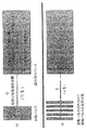
図14の下側に縦磁化緩和時間解析の結果を示す。逆ラプラス変換を用いることにより、縦磁化緩和時間の位置にピークを示すスペクトルとして現れる。縦軸が緩和時間を示すが、明瞭に二つの成分に分離していることがわかる。ひとつはサントニン(スペクトル下側)であり、ひとつはコレステロール(スペクトル上側)である。 The result of longitudinal magnetization relaxation time analysis is shown at the bottom of FIG. By using the inverse Laplace transform, a spectrum showing a peak appears at the position of the longitudinal magnetization relaxation time. Although the vertical axis shows the relaxation time, it can be seen that the two components are clearly separated. One is santonin (lower spectrum) and one is cholesterol (upper spectrum).
それぞれの縦磁化緩和時間に対してとったスライスを、図15および図16に示す。図15の上側は、3.06秒の縦磁化緩和時間を示す部分を抜き出したスライスである。図15の下側に、純品のコレステロールのみを測定した13C-NMRスペクトルを示す。図15の上側は、混合物から得たスペクトルであるにもかかわらず、コレステロールの信号のみが観測されていることがわかる。 The slices taken for each longitudinal magnetization relaxation time are shown in FIGS. The upper side of FIG. 15 is a slice obtained by extracting a portion showing a longitudinal magnetization relaxation time of 3.06 seconds. The lower part of FIG. 15 shows a 13 C-NMR spectrum obtained by measuring only pure cholesterol. The upper side of FIG. 15 shows that only a cholesterol signal is observed despite the spectrum obtained from the mixture.
図16の上側は、2.2秒の縦磁化緩和時間を示す部分を抜き出したスライスである。図16の下側に、純品のサントニンのみを測定した13C-NMRスペクトルを示す。図16の上側は、混合物から得たスペクトルであるにもかかわらず、サントニンの信号のみが観測されていることがわかる。 The upper side of FIG. 16 is a slice obtained by extracting a portion showing a longitudinal magnetization relaxation time of 2.2 seconds. The lower part of FIG. 16 shows a 13 C-NMR spectrum obtained by measuring only pure santonin. The upper side of FIG. 16 shows that only the signal of santonin is observed despite the spectrum obtained from the mixture.
以上のように、複数の成分からなる混合物であっても、精製することなく混合物のままで、本発明の手法を用いることにより、それぞれの成分ごとに分離してNMRスペクトルを観測できることが示された。 As described above, it is shown that even if the mixture is composed of a plurality of components, the NMR spectrum can be observed separately for each component by using the method of the present invention as it is without purification. It was.
混合物のNMR測定に広く利用できる。 It can be widely used for NMR measurement of mixtures.
Claims (9)
前記均一な縦磁化緩和時間を有している核に縦磁化緩和を起こさせるパルスを照射する第1の工程と、
所定時間tを置いて、前記均一な縦磁化緩和時間を有している核のスピン拡散を切断して高分解能NMRスペクトルを測定する第2の工程と、
tを変えて第1の工程と第2の工程を繰り返して、複数の高分解能NMRスペクトルを取得する第3の工程と、
第3の工程により得られた複数の高分解能NMRスペクトルを、縦磁化緩和時間に依存して回復するNMR信号強度の回復速度の違いに基づいて、逆ラプラス変換法により縦磁化緩和時間の値ごとに分類することにより、スペクトルの展開軸を横軸とし縦磁化緩和時間を縦軸とする2次元に展開されたNMRスペクトルを得て、この2次元に展開されたNMRスペクトルの特定の縦磁化緩和時間を示す部分のスライスを求めることにより、前記複数成分のうちの特定の成分のNMRスペクトルを分離して取得する第4の工程と
を備えたことを特徴とするNMR測定方法。 A solid sample having a plurality of domains, in which a plurality of components each have a uniform longitudinal magnetization relaxation time by spin diffusion within each domain ,
Irradiating a pulse that causes longitudinal magnetization relaxation to nuclei having the uniform longitudinal magnetization relaxation time;
A second step of measuring a high resolution NMR spectrum by cutting off the spin diffusion of the nucleus having the uniform longitudinal magnetization relaxation time after a predetermined time t;
changing the t and repeating the first step and the second step to obtain a plurality of high resolution NMR spectra;
Based on the difference in the recovery speed of the NMR signal intensity that recovers a plurality of high-resolution NMR spectra obtained by the third step depending on the longitudinal magnetization relaxation time, the value of the longitudinal magnetization relaxation time is determined by the inverse Laplace transform method. To obtain a two-dimensionally expanded NMR spectrum in which the horizontal axis of the spectrum is the horizontal axis and the longitudinal magnetization relaxation time is the vertical axis, and a specific longitudinal magnetization relaxation of the two-dimensionally expanded NMR spectrum is obtained. An NMR measurement method comprising: a fourth step of separating and acquiring an NMR spectrum of a specific component of the plurality of components by obtaining a slice of a portion indicating time .
前記均一な縦磁化緩和時間を有している第1の核に縦磁化緩和を起こさせるパルスを照射する第1の工程と、
所定時間tを置いて、前記第1の核の励起エネルギーを高分解能でNMRスペクトルを測定可能な第2の核に磁化移動させて、第2の核の高分解能NMRスペクトルを測定する第2の工程と、
tを変えて第1の工程と第2の工程を繰り返して、複数の第2の核の高分解能NMRスペクトルを取得する第3の工程と、
第3の工程により得られた複数の第2の核の高分解能NMRスペクトルを、縦磁化緩和時間に依存して回復するNMR信号強度の回復速度の違いに基づいて、逆ラプラス変換法により縦磁化緩和時間の値ごとに分類することにより、スペクトルの展開軸を横軸とし縦磁化緩和時間を縦軸とする第2の核の2次元に展開されたNMRスペクトルを得て、この2次元に展開されたNMRスペクトルの特定の縦磁化緩和時間を示す部分のスライスを求めることにより、前記複数成分のうちの特定の成分のNMRスペクトルを分離して取得する第4の工程とを備えたことを特徴とするNMR測定方法。 A solid sample having a plurality of domains, in which a plurality of components each have a uniform longitudinal magnetization relaxation time by spin diffusion within each domain ,
Irradiating a pulse for causing longitudinal magnetization relaxation to the first nucleus having the uniform longitudinal magnetization relaxation time;
A second time of measuring the high-resolution NMR spectrum of the second nucleus by moving the excitation energy of the first nucleus to the second nucleus capable of measuring the NMR spectrum with high resolution after a predetermined time t. Process,
changing the t and repeating the first step and the second step to obtain a high-resolution NMR spectrum of the plurality of second nuclei;
Based on the difference in the recovery speed of the NMR signal intensity for recovering the high-resolution NMR spectra of the plurality of second nuclei obtained in the third step depending on the longitudinal magnetization relaxation time, longitudinal magnetization is performed by the inverse Laplace transform method. By classifying each relaxation time value, a two-dimensionally expanded NMR spectrum of the second nucleus is obtained with the horizontal axis of the spectrum as the horizontal axis and the vertical magnetization relaxation time as the vertical axis. And a fourth step of separating and acquiring the NMR spectrum of the specific component of the plurality of components by obtaining a slice of a portion indicating a specific longitudinal magnetization relaxation time of the NMR spectrum obtained . NMR measurement method.
Priority Applications (3)
| Application Number | Priority Date | Filing Date | Title |
|---|---|---|---|
| JP2008240398A JP5364954B2 (en) | 2008-09-19 | 2008-09-19 | NMR measurement method |
| US12/561,484 US8072213B2 (en) | 2008-09-19 | 2009-09-17 | NMR measurement method |
| EP09252227A EP2166369B1 (en) | 2008-09-19 | 2009-09-18 | NMR measurement method |
Applications Claiming Priority (1)
| Application Number | Priority Date | Filing Date | Title |
|---|---|---|---|
| JP2008240398A JP5364954B2 (en) | 2008-09-19 | 2008-09-19 | NMR measurement method |
Publications (2)
| Publication Number | Publication Date |
|---|---|
| JP2010071839A JP2010071839A (en) | 2010-04-02 |
| JP5364954B2 true JP5364954B2 (en) | 2013-12-11 |
Family
ID=41478913
Family Applications (1)
| Application Number | Title | Priority Date | Filing Date |
|---|---|---|---|
| JP2008240398A Active JP5364954B2 (en) | 2008-09-19 | 2008-09-19 | NMR measurement method |
Country Status (3)
| Country | Link |
|---|---|
| US (1) | US8072213B2 (en) |
| EP (1) | EP2166369B1 (en) |
| JP (1) | JP5364954B2 (en) |
Families Citing this family (9)
| Publication number | Priority date | Publication date | Assignee | Title |
|---|---|---|---|---|
| JP5790511B2 (en) * | 2012-01-18 | 2015-10-07 | 新日鐵住金株式会社 | Quantitative determination of fluorine with fluorapatite |
| US9568574B2 (en) | 2012-03-12 | 2017-02-14 | Bruker Biospin Corporation | Pulse sequence for homonuclear J-decoupling during NMR data acquisition |
| US10031255B2 (en) * | 2014-03-24 | 2018-07-24 | Schlumberger Technology Corporation | Multi-dimensional nuclear magnetic resonance methods for characterizing fluids |
| US10613176B2 (en) * | 2014-05-19 | 2020-04-07 | The United States Of America, As Represented By The Secretary, Department Of Health And Human Services | Magnetic resonance 2D relaxometry reconstruction using partial data |
| EP3414557B1 (en) * | 2016-02-12 | 2019-11-27 | Bruker Biospin Corp. | Rapid quantification of components in solid mixtures of chemicals via time-domain nmr spectroscopy |
| US10890685B2 (en) * | 2017-08-11 | 2021-01-12 | Schlumberger Technology Corporation | Apparatus and methods for determining properties of hydrogen-containing samples using nuclear magnetic resonance |
| US10724975B2 (en) | 2017-08-11 | 2020-07-28 | Schlumberger Technology Corporation | Apparatus and methods for determining properties of liquid-bearing solids using nuclear magnetic resonance |
| CN110749614B (en) * | 2019-11-11 | 2022-12-27 | 南京大学 | Method for rapidly and quantitatively detecting organic phosphorus in soil |
| JP7111310B2 (en) * | 2019-12-11 | 2022-08-02 | 日本電子株式会社 | Nuclear magnetic resonance measurement method and apparatus |
Family Cites Families (5)
| Publication number | Priority date | Publication date | Assignee | Title |
|---|---|---|---|---|
| JPS58176536A (en) * | 1982-04-08 | 1983-10-17 | Jeol Ltd | Method for measuring nuclear magnetic resonance |
| EP1367406B1 (en) * | 2002-05-29 | 2007-02-14 | Bruker BioSpin AG | Slow diffusion and flow of molecules measured by pulsed field gradient NMR using longitudinal magnetisation of non-proton isotopes |
| WO2007030832A2 (en) * | 2005-09-09 | 2007-03-15 | State Or Oregon Acting By And Through The State Board Of Higher Education Of Behalf Of The University Of Oregon | High impedance differential input preamplifier and antenna for magnetic resonance systems |
| JP4358814B2 (en) * | 2005-11-09 | 2009-11-04 | 花王株式会社 | Sample analysis method |
| US7894891B2 (en) * | 2006-01-24 | 2011-02-22 | Schlumberger Technology Corporation | Diffusion-based magnetic resonance methods for characterizing bone structure |
-
2008
- 2008-09-19 JP JP2008240398A patent/JP5364954B2/en active Active
-
2009
- 2009-09-17 US US12/561,484 patent/US8072213B2/en active Active
- 2009-09-18 EP EP09252227A patent/EP2166369B1/en active Active
Also Published As
| Publication number | Publication date |
|---|---|
| US20100072995A1 (en) | 2010-03-25 |
| US8072213B2 (en) | 2011-12-06 |
| EP2166369A2 (en) | 2010-03-24 |
| JP2010071839A (en) | 2010-04-02 |
| EP2166369B1 (en) | 2013-03-20 |
| EP2166369A3 (en) | 2010-10-13 |
Similar Documents
| Publication | Publication Date | Title |
|---|---|---|
| JP5364954B2 (en) | NMR measurement method | |
| Blümich | Introduction to compact NMR: A review of methods | |
| Larsen et al. | 29Si and 17O (Q) CPMG-MAS solid-state NMR experiments as an optimum approach for half-integer nuclei having long T1 relaxation times | |
| Favier et al. | Recovering lost magnetization: polarization enhancement in biomolecular NMR | |
| Ciobanu et al. | 3D MR microscopy with resolution 3.7 μm by 3.3 μm by 3.3 μm | |
| Kulik et al. | Electron dipole–dipole interaction in ESEEM of nitroxide biradicals | |
| Mertes et al. | Distribution of tunnel splittings in Mn 12 acetate | |
| Lapina et al. | Practical aspects of 51V and 93Nb solid-state NMR spectroscopy and applications to oxide materials | |
| Ashbrook et al. | Motional broadening: an important distinction between multiple-quantum and satellite-transition MAS NMR of quadrupolar nuclei | |
| Stamps et al. | Difftrain: a novel approach to a true spectroscopic single-scan diffusion measurement | |
| Wu et al. | Ultrafast 2D COSY with constant-time phase-modulated spatial encoding | |
| Zhang et al. | Selective excitation enables assignment of proton resonances and 1H-1H distance measurement in ultrafast magic angle spinning solid state NMR spectroscopy | |
| CN104237820A (en) | Method for acquiring two-dimensional J-resolved spectroscopy of magnetic resonance by single sweeping | |
| Merlet et al. | Spin–spin coupling edition in chiral liquid crystal NMR solvent | |
| Marcó et al. | Perfect 1JCH-resolved HSQC: Efficient measurement of one-bond proton-carbon coupling constants along the indirect dimension | |
| WO2018166250A1 (en) | Ultra-fast nuclear magnetic resonance two-dimensional j spectrum method for resisting non-uniform magnetic field | |
| US7550970B2 (en) | Multidimensional NMR spectroscopy of a hyperpolarized sample | |
| Hu et al. | Sensitivity-enhanced phase-corrected ultra-slow magic angle turning using multiple-echo data acquisition | |
| Altenhof et al. | Broadband adiabatic inversion experiments for the measurement of longitudinal relaxation time constants | |
| Farooq et al. | Rapid parameter optimization of low signal‐to‐noise samples in NMR spectroscopy using rapid CPMG pulsing during acquisition: application to recycle delays | |
| Nath et al. | Enantiodiscrimination and extraction of short and long range homo-and hetero-nuclear residual dipolar couplings by a spin selective correlation experiment | |
| EP2650690B1 (en) | Method for NMR Measurements on Quadrupolar Nuclei | |
| Zhang et al. | Fast high-resolution 2D NMR spectroscopy in inhomogeneous fields via Hadamard frequency encoding and spatial encoding | |
| Ying-Bo et al. | Two-dimensional Nuclear Magnetic Resonance Spectroscopy with Parallel Acquisition of 1H-1H and 19F-19F Correlations | |
| Meyer et al. | Viva la resolución! Enhancing the resolution of 1H NMR spectra by broadband homonuclear decoupling |
Legal Events
| Date | Code | Title | Description |
|---|---|---|---|
| A621 | Written request for application examination |
Free format text: JAPANESE INTERMEDIATE CODE: A621 Effective date: 20110506 |
|
| A711 | Notification of change in applicant |
Free format text: JAPANESE INTERMEDIATE CODE: A712 Effective date: 20110523 |
|
| A131 | Notification of reasons for refusal |
Free format text: JAPANESE INTERMEDIATE CODE: A131 Effective date: 20130521 |
|
| A521 | Request for written amendment filed |
Free format text: JAPANESE INTERMEDIATE CODE: A523 Effective date: 20130722 |
|
| TRDD | Decision of grant or rejection written | ||
| A01 | Written decision to grant a patent or to grant a registration (utility model) |
Free format text: JAPANESE INTERMEDIATE CODE: A01 Effective date: 20130813 |
|
| A61 | First payment of annual fees (during grant procedure) |
Free format text: JAPANESE INTERMEDIATE CODE: A61 Effective date: 20130823 |
|
| R150 | Certificate of patent or registration of utility model |
Ref document number: 5364954 Country of ref document: JP Free format text: JAPANESE INTERMEDIATE CODE: R150 Free format text: JAPANESE INTERMEDIATE CODE: R150 |
|
| R250 | Receipt of annual fees |
Free format text: JAPANESE INTERMEDIATE CODE: R250 |
|
| R250 | Receipt of annual fees |
Free format text: JAPANESE INTERMEDIATE CODE: R250 |
|
| R250 | Receipt of annual fees |
Free format text: JAPANESE INTERMEDIATE CODE: R250 |
|
| R250 | Receipt of annual fees |
Free format text: JAPANESE INTERMEDIATE CODE: R250 |
|
| R250 | Receipt of annual fees |
Free format text: JAPANESE INTERMEDIATE CODE: R250 |
|
| R250 | Receipt of annual fees |
Free format text: JAPANESE INTERMEDIATE CODE: R250 |
|
| S111 | Request for change of ownership or part of ownership |
Free format text: JAPANESE INTERMEDIATE CODE: R313111 |
|
| R350 | Written notification of registration of transfer |
Free format text: JAPANESE INTERMEDIATE CODE: R350 |