JP5328146B2 - Medical image processing apparatus, medical image processing method and program - Google Patents
Medical image processing apparatus, medical image processing method and program Download PDFInfo
- Publication number
- JP5328146B2 JP5328146B2 JP2007333193A JP2007333193A JP5328146B2 JP 5328146 B2 JP5328146 B2 JP 5328146B2 JP 2007333193 A JP2007333193 A JP 2007333193A JP 2007333193 A JP2007333193 A JP 2007333193A JP 5328146 B2 JP5328146 B2 JP 5328146B2
- Authority
- JP
- Japan
- Prior art keywords
- medical image
- information
- doctor
- lesion
- medical
- Prior art date
- Legal status (The legal status is an assumption and is not a legal conclusion. Google has not performed a legal analysis and makes no representation as to the accuracy of the status listed.)
- Active
Links
Images


Classifications
-
- G—PHYSICS
- G16—INFORMATION AND COMMUNICATION TECHNOLOGY [ICT] SPECIALLY ADAPTED FOR SPECIFIC APPLICATION FIELDS
- G16H—HEALTHCARE INFORMATICS, i.e. INFORMATION AND COMMUNICATION TECHNOLOGY [ICT] SPECIALLY ADAPTED FOR THE HANDLING OR PROCESSING OF MEDICAL OR HEALTHCARE DATA
- G16H40/00—ICT specially adapted for the management or administration of healthcare resources or facilities; ICT specially adapted for the management or operation of medical equipment or devices
- G16H40/20—ICT specially adapted for the management or administration of healthcare resources or facilities; ICT specially adapted for the management or operation of medical equipment or devices for the management or administration of healthcare resources or facilities, e.g. managing hospital staff or surgery rooms
-
- G—PHYSICS
- G06—COMPUTING; CALCULATING OR COUNTING
- G06Q—INFORMATION AND COMMUNICATION TECHNOLOGY [ICT] SPECIALLY ADAPTED FOR ADMINISTRATIVE, COMMERCIAL, FINANCIAL, MANAGERIAL OR SUPERVISORY PURPOSES; SYSTEMS OR METHODS SPECIALLY ADAPTED FOR ADMINISTRATIVE, COMMERCIAL, FINANCIAL, MANAGERIAL OR SUPERVISORY PURPOSES, NOT OTHERWISE PROVIDED FOR
- G06Q10/00—Administration; Management
- G06Q10/10—Office automation; Time management
-
- G—PHYSICS
- G06—COMPUTING; CALCULATING OR COUNTING
- G06V—IMAGE OR VIDEO RECOGNITION OR UNDERSTANDING
- G06V40/00—Recognition of biometric, human-related or animal-related patterns in image or video data
- G06V40/10—Human or animal bodies, e.g. vehicle occupants or pedestrians; Body parts, e.g. hands
-
- G—PHYSICS
- G16—INFORMATION AND COMMUNICATION TECHNOLOGY [ICT] SPECIALLY ADAPTED FOR SPECIFIC APPLICATION FIELDS
- G16H—HEALTHCARE INFORMATICS, i.e. INFORMATION AND COMMUNICATION TECHNOLOGY [ICT] SPECIALLY ADAPTED FOR THE HANDLING OR PROCESSING OF MEDICAL OR HEALTHCARE DATA
- G16H30/00—ICT specially adapted for the handling or processing of medical images
- G16H30/20—ICT specially adapted for the handling or processing of medical images for handling medical images, e.g. DICOM, HL7 or PACS
Description
本発明は、医用画像の読影を支援するための医用画像処理技術に関するものである。 The present invention relates to a medical image processing technique for supporting interpretation of medical images.
医療の分野においては、被検者を撮像することにより得られた医用画像のデジタル化が実現されている。これにより、X線装置、CR装置、CT装置、MRI装置、PET装置、超音波装置、OCT装置等の医用撮像装置を用いて撮像された医用画像をモニタ表示させることが可能となっている。そして、医師は、このモニタ表示された医用画像を読影することで、病変部の状態や経時変化について診断を行っている。なお、ここでいうCRとは、Computed Radiographyの略であり、CTとは、Computed Tomographyの略であり、MRIとは、Magnetic Resonance Imagingの略である。また、PETとは、Positron Emission Tomographyの略であり、OCTとは、Optical Coherence Tomographyの略である。 In the medical field, digitization of medical images obtained by imaging a subject has been realized. Thereby, it is possible to display on a monitor a medical image captured using a medical imaging apparatus such as an X-ray apparatus, CR apparatus, CT apparatus, MRI apparatus, PET apparatus, ultrasonic apparatus, or OCT apparatus. Then, the doctor interprets the medical image displayed on the monitor to diagnose the state of the lesion and the change over time. Here, CR is an abbreviation for Computed Radiography, CT is an abbreviation for Computed Tomography, and MRI is an abbreviation for Magnetic Resonance Imaging. PET is an abbreviation for Positron Emission Tomography, and OCT is an abbreviation for Optical Coherence Tomography.
一方、従来より、読影時の医師の作業負担の軽減を目的として、医用画像を解析し、自動的に病変を検出することが可能なコンピュータ支援診断装置(以下、CADと称す)と呼ばれる医用画像処理装置が開発されている。 On the other hand, a medical image called a computer-aided diagnosis apparatus (hereinafter referred to as CAD) that can analyze a medical image and automatically detect a lesion for the purpose of reducing the workload of a doctor during interpretation. Processing equipment has been developed.
当該CADでは、入力された医用画像に基づいて、癌等を表す異常な腫留陰影や、高濃度の微小石灰化陰影等を病変として検出することができる。このように、医師の読影作業の一部を自動化させることにより、読影時の医師の作業負担を大幅に軽減することができる。 The CAD can detect abnormal tumor shadows representing cancer and the like, high-density microcalcification shadows, and the like as lesions based on the input medical image. Thus, by automating a part of the doctor's interpretation work, it is possible to greatly reduce the doctor's work load at the time of interpretation.
更に、読影精度の向上を目的として、例えば、特開2006−130049号公報では、CADの検出結果に基づいて、悪性度の高い病変が含まれる医用画像から順に読影できるように読影順序を並べ替える構成が提案されている。当該構成によれば、例えば、一日の中で医師の疲労の少ない時間帯に悪性度の高い病変が含まれる医用画像を読影できるよう読影順序を並べ替えることで、病変部の見落としを低減させることができる。 Furthermore, for the purpose of improving the interpretation accuracy, for example, in Japanese Patent Laid-Open No. 2006-130049, the interpretation order is rearranged so that interpretation can be performed in order from a medical image including a lesion with high malignancy based on the CAD detection result. A configuration is proposed. According to this configuration, for example, by rearranging the interpretation order so that a medical image including a highly malignant lesion can be interpreted in a time period in which the doctor is less fatigued during the day, the oversight of the lesion is reduced. be able to.
更に、特開2004−216008号公報では、上述のように悪性度に応じて読影順序を並べ替えることに加え、同じ部位を連続して読影することができるように、読影順序を並べ替える構成が提案されている。当該構成によれば、病変の見落としを更に低減させることが可能となる。
しかしながら、上記各特許文献に開示された発明は、いずれも一人の医師が全ての医用画像を読影することを前提としたものであるである。しかし、一般に病院やセンター等には複数の医師が在籍しており、この場合、これらの医師が複数の医用画像を分担して読影することも少なくない。 However, the inventions disclosed in the above patent documents are all based on the premise that one doctor interprets all medical images. However, in general, a plurality of doctors are enrolled in hospitals, centers, and the like. In this case, these doctors often share and interpret a plurality of medical images.
ここで、このように複数の医師が在籍している場合、一般に、各々の医師は互いに専門分野が異なっているか、あるいは専門分野が同じであっても、経験年数等に差があるケースが多い。このため、複数の医用画像を各医師にどのように分担するかは、病院やセンター全体の作業効率や、読影精度に大きな影響を与える。 Here, when multiple doctors are enrolled in this way, in general, there are many cases where each doctor has a different field of expertise, even if the field of expertise is the same, even if the field of expertise is the same. . For this reason, how a plurality of medical images are assigned to each doctor greatly affects the work efficiency and interpretation accuracy of the entire hospital or center.
本発明は上記課題に鑑みてなされたものであり、複数の医用画像を複数の医師が分担して読影する場合において、全体の作業効率を向上させるとともに、読影精度の向上を図ることを目的とする。 The present invention has been made in view of the above problem, and aims to improve the overall working efficiency and improve the interpretation accuracy when a plurality of doctors share and interpret a plurality of medical images. To do.
上記の目的を達成するために本発明に係る医用画像処理装置は以下のような構成を備える。即ち、
医用撮像装置を用いて被検者を撮像することにより得られた複数の医用画像を複数の医師が分担して読影できるよう分担先を決定する医用画像処理装置であって、
前記複数の医用画像のうち、分担の対象となる医用画像について解析を行い、解析結果を出力する解析手段と、
前記分担の対象となる医用画像の撮像に用いられた医用撮像装置に関する情報を取得する取得手段と、
特定の病変の読影に対する前記各医師の適性を示す情報と特定の医用撮像装置による撮像により得られた医用画像の読影に対する前記各医師の適性を示す情報とを前記各医師の識別情報と関連付けて記憶する記憶手段と、
前記解析手段による解析結果に病変に関する情報が含まれているか否かを判定する判定手段と、
前記判定手段により前記解析結果に病変に関する情報が含まれていると判定された場合には、前記分担の対象となる医用画像の分担先を、特定の病変の読影に対する各医師の適性を示す情報と前記病変の識別難易度と前記識別情報とに基づいて決定し、前記判定手段により前記解析結果に病変に関する情報が含まれていないと判定された場合には、前記分担の対象となる医用画像の分担先を、特定の医用撮像装置による撮像により得られた医用画像の読影に対する各医師の適性を示す情報と前記医用撮像装置に関する情報と前記識別情報とに基づいて決定する決定手段とを備える。
In order to achieve the above object, a medical image processing apparatus according to the present invention has the following configuration. That is,
A medical image processing apparatus that determines a sharing destination so that a plurality of doctors can share and interpret a plurality of medical images obtained by imaging a subject using a medical imaging apparatus,
Analyzing the medical image to be shared among the plurality of medical images, and outputting an analysis result; and
Acquisition means for acquiring information relating to a medical imaging apparatus used for imaging a medical image to be shared;
Information indicating the suitability of each doctor for interpretation of a specific lesion and information indicating the suitability of each doctor for interpretation of a medical image obtained by imaging by a specific medical imaging apparatus are associated with the identification information of each doctor. Storage means for storing;
Determination means for determining whether or not the analysis result by the analysis means includes information about a lesion;
If the determination means determines that information about a lesion is included in the analysis result, information indicating the applicability of each doctor to the interpretation of a specific lesion is determined as the sharing destination of the medical image to be shared the determined based on the identification difficulty with the identification information of the lesion and, wherein when it is determined that no information about the lesion included in the analysis result by the determination means, the medical image to be the share A determination means for determining the sharing destination based on information indicating the suitability of each doctor for interpretation of a medical image obtained by imaging by a specific medical imaging device, information on the medical imaging device, and the identification information. .
本発明によれば、複数の医用画像を複数の医師が分担して読影する場合において、全体の作業効率を向上させるとともに、読影精度の向上を図ることが可能となる。 According to the present invention, when a plurality of doctors share and interpret a plurality of medical images, the overall work efficiency can be improved and the interpretation accuracy can be improved.
以下、本発明を実施するための最良の形態について図面を用いて詳細に説明する。 The best mode for carrying out the present invention will be described below in detail with reference to the drawings.
[第1の実施形態]
1.医用画像処理システムの構成
図1は、本発明の第1の実施形態にかかる医用画像処理装置を備える、医用画像処理システム100の構成を示す図である。
[First Embodiment]
1. Configuration of Medical Image Processing System FIG. 1 is a diagram showing a configuration of a medical
図1に示すように、医用画像処理システムは、医用画像処理装置101と医用撮像装置102と画像データベース103とを備え、ネットワーク104を介して互いに通信可能に接続されている。
As shown in FIG. 1, the medical image processing system includes a medical
医用撮像装置102は、被検者(患者)を撮像し医用画像を生成する装置であり、X線装置、CR装置、CT装置、MRI装置、PET装置、超音波診断装置、OCT装置等が含まれる。
The
画像データベース103は、医用撮像装置102にて撮像された医用画像に付帯情報を付加して保存する。付帯情報には、、撮像モダリティ、撮像日時、緊急度、読影期限、検査目的、撮像部位、既往症、年齢、性別、喫煙歴、所見、主訴、検査結果、病院名、患者の氏名・生年月日などが含まれる。
The
医用画像処理装置101は、医用撮像装置102にて撮像された医用画像や画像データベース103に保存された医用画像を表示するとともに、表示された医用画像に基づいて医師が読影した結果を文字情報として入力し、レポートを作成するための装置である。
The medical
2.医用画像処理装置101のハードウェア構成
図2は医用画像処理装置101のハードウェア構成を示した図である。図2に示すように、医用画像処理装置101は、構成要素として、CPU(中央演算処理装置)201、入力装置203、表示装置204、メモリ202、ディスク205を備える。
2. Hardware Configuration of Medical
CPU201は、各種制御プログラムを実行するとともに、医用画像処理装置101の各構成要素の動作を制御する。入力装置203は医師によるポインティング入力及び文字等の入力を受け付ける。表示装置204は、CPU201による各種制御プログラムの実行結果を表示する。表示装置204には、例えばCRTモニタや液晶モニタ等が含まれる。
The
メモリ202は、所定の制御プログラムを格納したり、制御プログラム実行時に作業領域を提供したりする。ディスク205は、オペレーティングシステム(OS)206、周辺機器のデバイスドライブ207、ならびに本発明に係る医用画像処理方法を実現するための制御プログラム(「医用画像処理プログラム」208と称す)等の各種制御プログラムを格納する。
The
3.医用画像処理プログラム208の全体機能ブロック
図3は、医用画像処理プログラム208により実現される機能を示した機能ブロック図である。図3に示すように、医用画像処理プログラム208は、画像データベース103に保存された医用画像の解析を行う画像解析部301と、読影を行う医師(読影医)を登録する登録部302と、読影医の情報を管理する管理部303とを備える。
3. Overall Function Block of Medical
また、画像解析部301における解析結果と管理部303により管理された読影医の情報とに基づいて、読影すべき複数の医用画像を各読影医に分担するための分担先を決定する選択部304を備える。選択部304では、更に、各読影医に分担された医用画像に対応する患者IDを配列した患者リストを作成する。
In addition, based on the analysis result in the
医用画像処理プログラム208は、更に、選択部304で生成された各読影医ごとの患者リストをディスク205内に記憶する記憶部305と、該患者リストを表示装置204に表示するための出力部306を備える。以下、各部の詳細について説明する。
The medical
4.画像解析部
画像解析部301は、医用画像を解析し、臓器領域および/或いは病変検出を行う(病変確率、悪性度、識別難易度、医学的重要度などの病変に関する情報を解析結果として出力する)。
4). Image Analysis Unit The
4.1 臓器領域の検出
臓器領域の検出とは、例えば胸部CT画像の場合、肺野、横隔膜、気管支、肺動脈、肺静脈などの領域を検出することをいう。更に、検出した肺野を、上葉、中葉、下葉、の各区域に分類することも含む。ただし、検出する臓器領域の種類はこれに限定されるものではない。
4.1 Detection of Organ Area Detection of an organ area refers to detection of areas such as a lung field, a diaphragm, a bronchus, a pulmonary artery, and a pulmonary vein in the case of a chest CT image. Furthermore, it includes classifying the detected lung field into upper lobe, middle lobe, and lower lobe areas. However, the type of organ area to be detected is not limited to this.
なお、医用画像から臓器領域を検出するための方法としては種々の方法が挙げられるが、本実施形態では、例えば動的輪郭法の一種であるレベルセット法を用いるものとする。レベルセット法の場合、検出対象とする臓器領域の次元よりも一次元高いレベルセット関数を定義し、検出対象とする臓器領域をそのゼロ等高線であるとみなす。そして、レベルセット方程式と呼ばれる以下の発展方程式に基づいてこの関数を更新することで、輪郭を制御し臓器領域を検出する。 Various methods can be used as a method for detecting an organ region from a medical image. In this embodiment, for example, a level set method, which is a kind of dynamic contour method, is used. In the level set method, a level set function that is one dimension higher than the dimension of the organ area to be detected is defined, and the organ area to be detected is regarded as its zero contour line. Then, by updating this function based on the following evolution equation called a level set equation, the contour is controlled and the organ region is detected.
φt+F|Vφ|=0
ここで、φtはレベルセット関数を時間軸方向に1次微分した値、Fは輪郭の成長速度、|Vφ|はレベルセット関数の勾配の絶対値を表している。このようにして、医用画像から臓器領域が検出される。
φ t + F | V φ | = 0
Here, φ t represents a value obtained by first-order differentiation of the level set function in the time axis direction, F represents the growth rate of the contour, and | V φ | represents the absolute value of the gradient of the level set function. In this way, the organ region is detected from the medical image.
なお、臓器領域を検出するための方法はレベルセット法に限定されるものではなく、例えば、閾値処理による方法、領域拡張法、動的輪郭法、クラスタ化による方法、グラフ最小切断法などを用いてもよい。あるいはその他の方法を用いてもよい。 Note that the method for detecting an organ region is not limited to the level set method. For example, a threshold processing method, a region expansion method, a dynamic contour method, a clustering method, a graph minimum cutting method, or the like is used. May be. Alternatively, other methods may be used.
また、これらの方法を、検出対象とする臓器領域に応じて切り替えて使用するようにしてもよい。 Further, these methods may be used by switching according to the organ region to be detected.
更に、臓器領域の検出にあたっては、画像特徴量のみを使用するのではなく、確率アトラスや人体形状モデルなどを利用するようにしても良い。 Furthermore, in detecting the organ region, a probability atlas or a human body shape model may be used instead of using only the image feature amount.
4.2 病変検出
臓器領域から病変検出を行うにあたり、画像解析部301では、まず、フィルタ処理や、パターンマッチング処理あるいは過去画像または平均形状画像等と医用画像とのレジストレーション処理等を行い、差分を検出することで、異常部を検出する。
4.2 In detecting a lesion from a lesion detection organ region, the
具体的には、フィルタ処理では、形状を考慮したフィルタやこう配ベクトルの向きを考慮したフィルタなどを用いる。 Specifically, in the filter processing, a filter that considers the shape or a filter that considers the direction of the gradient vector is used.
そして、これらの処理のいずれか、あるいはその他の処理を用いて、異常部を検出し特徴量を抽出する。 Then, using any of these processes or other processes, an abnormal part is detected and a feature amount is extracted.
なお、このとき抽出される特徴量としては、CT値平均、CT値分散・標準偏差、CT値最大・最小、コントラスト、エネルギー、エントロピー等の、画素値に基づく特徴量が挙げられる。また、円形度、表面積、体積、球形度、不規則度、平均曲率、主曲率、ガウス曲率、最大径等の、形状に基づく特徴量が挙げられる。本実施形態では、上述した特徴量の少なくとも一つ、或いはその組み合わせが抽出されるものとする。 The feature quantity extracted at this time includes feature quantities based on pixel values such as CT value average, CT value variance / standard deviation, CT value maximum / minimum, contrast, energy, entropy, and the like. Moreover, the feature-value based on shapes, such as circularity, surface area, volume, sphericity, irregularity, average curvature, main curvature, Gaussian curvature, maximum diameter, is mentioned. In the present embodiment, it is assumed that at least one of the above-described feature amounts or a combination thereof is extracted.
画像解析部301では、更に、検出した異常部が病変であるのか否か(病変確率)、病変である場合には病変の種類、さらには、病変の良悪を分類するための悪性度ならびに医学的重要度を識別するための処理を行う。
The
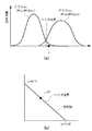
なお、ここでは検出した異常部が病変であるのか否か、あるいは病変の悪性度(良性か悪性か)及び識別難易度を識別するための処理を、最小二乗法とベイズ決定則を用いて行う場合について、図4を用いて説明する。識別する悪性度のクラスは、クラスω1とクラスω2とし、図4(a)の特徴空間及び図4(b)の判別空間を用いて説明する。 Here, the process for identifying whether or not the detected abnormal part is a lesion, or the malignancy (benign or malignant) of the lesion and the difficulty of identification is performed using a least square method and a Bayes decision rule. The case will be described with reference to FIG. The class of malignancy to be identified is class ω 1 and class ω 2 and will be described using the feature space in FIG. 4A and the discrimination space in FIG. 4B.
入力されたあるパターンx(対象とする医用画像の特徴量)は、最適写像yi *=Φ*(xi)により第i成分をクラスωiのベイズ事後確率とするベイズ確率ベクトルに移される。 An input pattern x (a feature quantity of a target medical image) is transferred to a Bayes probability vector having an i- th component as a Bayes posterior probability of class ω i by an optimal mapping y i * = Φ * (x i ). .
特徴空間でのベイズ境界は、(c−1)次元では単純な重心分割境界となり、判別空間では単純な線形識別境界となる。tiは判別空間における各クラスの代表点である。判別平面上でy*とtiとの二乗距離は、下式のようになる。 The Bayes boundary in the feature space becomes a simple center-of-gravity division boundary in the (c-1) dimension, and becomes a simple linear identification boundary in the discrimination space. t i is a representative point of each class in the discrimination space. Square distance between y * and t i on discrimination plane is as shown in the following equation.
Di=||y*−ti||2=||y*||−2P(ωi|x)+1
このため、y*とtiとの二乗距離が最小となるクラスを選択すれば、入力されたパターンxがどのクラスに属するかを識別することが出来る。
D i = || y * −t i || 2 = || y * || −2P (ω i | x) +1
Therefore, by selecting a class squared distance between y * and t i is minimized, it is possible to identify whether the input pattern x belongs to which class.
入力されたパターンxとクラスωiとの距離をD1、クラスω2との距離をD2とすると、尤度比を用いてベイズ境界との距離を求めることが出来る。ここで、尤度比はD1/D2で表現される。つまり、尤度比が大きいほどクラスω2に属する確率が高くなり、尤度比が小さいほどクラスω1に属する確率が高くなる。また、1に近いほどベイズ境界に近くなる。ベイズ境界に近いパターンxは画像解析部301の誤識別の可能性もありうる。また、読影医が読影を行う場合に判断が困難である可能性があるため、ベイズ境界から一定の距離内にある病変を含む医用画像にはフラグを設定し、識別困難であるとする。ここでは、例えば、識別困難である尤度比の閾値Thを0.9<Th<1.1と設定する。
When the distance between the input pattern x and the class ω i is D 1 and the distance between the class ω 2 is D 2 , the distance from the Bayes boundary can be obtained using the likelihood ratio. Here, the likelihood ratio is expressed as D 1 / D 2 . That is, the greater the likelihood ratio, the higher the probability of belonging to class ω 2, and the smaller the likelihood ratio, the higher the probability of belonging to class ω 1 . The closer to 1, the closer to the Bayesian boundary. The pattern x close to the Bayes boundary may be misidentified by the
このようにして、医用画像から抽出した特徴量を用いて識別処理を行うことが出来る。なお、ここでは識別処理として、最小二乗法とベイズ決定則を用いて行う場合について説明したが、本発明はこれに限定されない。例えば、線形判別法、サポートベクターマシン、AdaBoost、ニューラルネットワークなどを用いてもよい。 In this way, identification processing can be performed using the feature amount extracted from the medical image. Although the case where the identification process is performed using the least square method and the Bayes decision rule has been described here, the present invention is not limited to this. For example, a linear discriminant method, a support vector machine, AdaBoost, or a neural network may be used.
5.登録部
次に、登録部302について説明する。登録部302では、画像解析部301の解析結果に基づいて医用画像を分担して読影する読影医の登録ならびに読影医情報の登録を行う。読影医の登録は、読影医が医用画像の読影を行う際に、医用画像処理装置101にログインすることにより行われる。登録した読影医の削除は、医用画像処理装置101からのログアウトすることにより行われる。
5). Registration Unit Next, the
一方、読影医情報の登録は、事前に登録することも、読影中に登録することも、或いは学習によって登録することもできるものとする。また、登録は本人、或いは権限を持った第三者が出来るように構成されているものとする。なお、読影医情報の登録は、例えば、別に備えた記憶端末にアクセスして読影医情報を取得することにより登録できるようにしても良い。 On the other hand, the interpretation doctor information can be registered in advance, registered during interpretation, or registered by learning. In addition, it is assumed that registration is configured so that the person or an authorized third party can do the registration. Note that the interpretation doctor information may be registered by, for example, accessing a separate storage terminal and acquiring the interpretation doctor information.
6.管理部
次に、管理部303について説明する。登録部302で登録された読影医情報は、管理部303により管理される。読影医情報は、読影医が医用画像処理装置101からログアウトした後であっても、管理部303により読影医のID情報と共にディスクに記録されている。
6). Management Unit Next, the
管理部303で管理される読影医情報は、選択部304が医用画像を各読影医に分担する際の分担先の決定に用いられる。
The interpretation doctor information managed by the
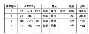
図5は、管理部303において管理される読影医情報の一例を示す図である。図5に示すように、読影医情報には、読影医の得意とするモダリティ(特定の医用撮像装置により得られた医用画像の読影に対する各読影医の適性を示す情報)が含まれる。また、読影医の専門分野や、得意とする部位・疾病、経験年数、役職など、特定の病変の読影に対する各読影医の適性を示す情報が含まれる。
FIG. 5 is a diagram illustrating an example of image interpretation doctor information managed by the
7. 選択部
次に、選択部304における処理の流れについて図6のフローチャートを用いて説明する。ステップS601では、解析結果として画像解析部301が検出した臓器領域、病変確率、病変の種類、識別難易度、悪性度、医学的重要度の少なくとも一つ、或いはそれらの組み合わせを取得する。
7). Selection unit will be described with reference to the flowchart of FIG. 6 for a flow of processing in the
ステップS602では、医用画像の付帯情報として、撮像モダリティ(医用画像装置に関する情報)、撮像日時、緊急度、読影期限(時期的制限に関する情報)、検査目的、撮像部位、既往症、年齢、性別、喫煙歴、所見、主訴、検査結果を取得する。なお、取得する付帯情報は、これらのうちの少なくとも一つ、或いはそれらの組み合わせであるものとする。 In step S602, the incidental information of the medical image includes imaging modality (information regarding the medical imaging apparatus), imaging date / time, urgency, interpretation period (information regarding timing restrictions), examination purpose, imaging site, past disease, age, sex, smoking Get history, findings, chief complaints, and test results. The accompanying information to be acquired is assumed to be at least one of these or a combination thereof.
ステップS603では、管理部303から、現在読影を行っている読影医についての読影医情報を取得する。読影医情報には、読影医の専門分野や得意とするモダリティ、得意とする部位・疾病、経験年数、役職のうちの少なくとも一つ、或いはそれらの組み合わせが含まれる。
In step S603, the interpreting doctor information about the interpreting doctor who is currently performing interpretation is acquired from the
ステップS604では、記憶部305から、現在読影を行っている読影医の読影進行状況に関する情報を取得する。読影進行状況に関する情報には、患者リストに溜まっている患者数、1患者の医用画像を読影するのに要する読影時間の平均値・最大値・最小値の少なくとも一つ、或いはそれらの組み合わせが含まれる。
In step S <b> 604, information related to the interpretation progress of the interpretation doctor who is currently interpreting is acquired from the
ステップS605では、ステップS601からステップS604において取得した情報に基づいて、分担の対象とする医用画像について読影医を決定する。なお、分担の対象とする医用画像について読影医を決定するための処理(読影医決定処理)の詳細は、後述する。 In step S605, an interpreting doctor is determined for the medical image to be shared based on the information acquired in steps S601 to S604. The details of the process for determining the interpretation doctor for the medical image to be shared (interpretation doctor determination process) will be described later.
ステップS606では、ステップS605における読影医決定処理の結果に基づいて、各読影医毎に、振り分けられた医用画像に対応する患者を一覧表示するための患者リストを作成する(患者リスト作成処理)。なお、患者リスト作成処理の詳細は、後述する。 In step S606, a patient list for displaying a list of patients corresponding to the allocated medical images is created for each interpreting doctor based on the result of the interpreting doctor determining process in step S605 (patient list creating process). Details of the patient list creation process will be described later.
7.1 読影医決定処理(ステップS605)の詳細その1
図7(a)は、読影医決定処理(ステップS605)の詳細な処理の流れを示すフローチャートである。ステップS701では、分担の対象となる医用画像について、画像解析部301において病変が検出されているか否かを判定する。
7.1 Details of interpretation doctor determination process (step S605)
FIG. 7A is a flowchart showing a detailed processing flow of the interpretation doctor determination processing (step S605). In step S701, it is determined whether or not a lesion is detected in the
ステップS701において、病変が検出されていないと判定された場合には、ステップS702に進み、分担の対象となる医用画像を、対応するモダリティが設定された一般の読影医に振り分ける。 If it is determined in step S701 that no lesion has been detected, the process advances to step S702, and the medical image to be shared is distributed to general radiographers set with corresponding modalities.
一方、ステップS701において、病変が検出されていると判定された場合には、ステップS703に進み、当該病変が画像解析部301において識別困難な病変であると判定されているか否かを判定する。
On the other hand, if it is determined in step S701 that a lesion has been detected, the process advances to step S703 to determine whether or not the lesion is determined to be difficult to identify in the
ステップS703において、識別困難な病変であると判定されていた場合には、ステップS704に進み、分担の対象となる医用画像を、対応する専門の熟練の読影医に振り分ける。 If it is determined in step S703 that the lesion is difficult to identify, the process proceeds to step S704, where the medical image to be shared is distributed to the corresponding specialized expert interpretation doctor.
一方、ステップS703において、識別困難な病変ではないと判定されていた場合には、ステップS705に進み、分担の対象となる医用画像を、対応する専門の読影医に振り分ける。 On the other hand, if it is determined in step S703 that the lesion is not difficult to identify, the process proceeds to step S705, and the medical image to be shared is distributed to the corresponding specialized interpretation doctor.
このようにして読影医決定処理を行うことにより、分担の対象となる医用画像が適切な読影医に振り分けられることとなる。 By performing the interpretation doctor determination process in this way, the medical image to be shared is distributed to an appropriate interpretation doctor.
なお、上記読影医決定処理の具体例について図5の読影医情報の一例と対応付けながら説明すると以下のようになる。 A specific example of the interpreting doctor determination process will be described below in association with an example of the interpreting doctor information shown in FIG.
例えば、胸部CT画像において、肺癌が検出され、画像解析部301で識別困難でないと判定されていた(フラグが設定されていなかった)場合には、病変が検出された部位を専門とする読影医として、読影医1〜3が選択される。
For example, if lung cancer is detected in a chest CT image and it is determined by the
一方、胸部CT画像において、肺癌が検出され、画像解析部301で識別困難であると判定されていた(フラグが設定されていた)場合には、病変が検出された部位を専門とする熟練の読影医として、読影医1が選択される。
On the other hand, when lung cancer is detected in the chest CT image and it has been determined that the
一方、胸部CT画像において、病変が検出されていなかった場合は、当該医用画像を撮像したモダリティを読影医情報に設定している読影医として、読影医1〜4が選択される。
On the other hand, when no lesion is detected in the chest CT image, the interpreting
7.2 読影医決定処理(ステップS605)の詳細その2
図7(b)は、読影医決定処理のその他の詳細な処理の流れを示すフローチャートである。ステップS711では、分担の対象となる医用画像より、画像解析部301において病変が検出されているか否かを判定する。
7.2 Details of interpretation doctor decision processing (step S605)
FIG. 7B is a flowchart showing a flow of other detailed processing of the interpretation doctor determination processing. In step S711, it is determined whether a lesion is detected in the
ステップS711において、病変が検出されていないと判定された場合には、ステップS712に進み、分担の対象となる医用画像を、モダリティが設定された一般の読影医に振り分ける。 If it is determined in step S711 that a lesion has not been detected, the process proceeds to step S712, and the medical image to be shared is distributed to general radiographers set with modalities.
一方、ステップS711において、病変が検出されていると判定された場合には、ステップS713に進み、当該病変が、医学的重要度の高い病変であるか否かを判定する。医学的重要度とは、命に関わる可能性が高いか否かを示す度合いであり、命に関わる可能性が高い病変は医学的重要度が高い病変であると判定される。 On the other hand, if it is determined in step S711 that a lesion has been detected, the process advances to step S713 to determine whether the lesion is a lesion with high medical importance. The medical importance is a degree indicating whether or not there is a high possibility of being related to life, and a lesion having a high possibility of being related to life is determined to be a lesion having a high medical importance.
ステップS713において、医学的重要度が高い病変であると判定された場合には、ステップS714に進み、分担の対象となる医用画像を、専門の熟練の読影医に振り分ける。 If it is determined in step S713 that the lesion has a high medical importance, the process proceeds to step S714, and the medical image to be shared is assigned to a specialized expert interpretation doctor.
一方、ステップS713において、医学的重要度の低い病変であると判定された場合には、ステップS715に進み、分担の対象となる医用画像を、専門の読影医に振り分ける。 On the other hand, if it is determined in step S713 that the lesion has a low medical importance, the process proceeds to step S715, and the medical image to be shared is assigned to a specialized interpretation doctor.
このようにして読影医決定処理を行うことにより、分担の対象となる医用画像が適切な読影医に振り分けられることとなる。 By performing the interpretation doctor determination process in this way, the medical image to be shared is distributed to an appropriate interpretation doctor.
なお、上記読影医決定処理の具体例について図5の読影医情報の一例と対応付けながら説明すると以下のようになる。 A specific example of the interpreting doctor determination process will be described below in association with an example of the interpreting doctor information shown in FIG.
例えば、胸部CT画像において、肺癌が検出された場合には、読影医1、あるいは読影医3が選択される。
For example, when lung cancer is detected in a chest CT image, the interpreting
一方、胸部CT画像において、肺炎が検出されていた場合には、読影医2が選択される。一般に、肺炎と肺癌では、肺癌の方が医学的重要度が高く、良性と悪性では悪性の方がさらに医学的重要度は高いからである。
On the other hand, if pneumonia has been detected in the chest CT image, the
7.3 患者リスト作成処理(ステップS606)の詳細
図8は、読影医毎に設定する患者リストの一例を示す図である。患者リストは、読影医と振り分けられた医用画像IDとの対応関係を、読影する順序に配列したリストである。患者リストは、読影医情報に基づいて作成されるため、読影医毎に異なる。
7.3 Details of Patient List Creation Processing (Step S606) FIG. 8 is a diagram showing an example of a patient list set for each interpretation doctor. The patient list is a list in which correspondences between the interpretation doctors and the medical image IDs assigned are arranged in the order of interpretation. Since the patient list is created based on the interpretation doctor information, it differs for each interpretation doctor.
患者リストに配列された医用画像に対応する患者IDの順序は、医用画像の付帯情報と画像解析部301における解析結果をそれぞれ読影医情報と比較することでスコアを算出し、該算出されたスコアに基づいて決定される。
The order of patient IDs corresponding to the medical images arranged in the patient list is calculated by comparing the incidental information of the medical image and the analysis result in the
具体的には、緊急度や識別難易度、病変の医学的重要度等に応じて、スコアにプラスのポイントが付与される。 Specifically, a positive point is given to the score according to the degree of urgency, the difficulty of identification, the medical importance of the lesion, and the like.
そして、算出されたスコアが高い医用画像に対応する患者IDほど、患者リストの上位に配列される。例えば、胸部CT画像で肺癌が検出された場合と、胸部CT画像で肺炎が検出された場合とでは、肺癌が検出された医用画像に対応する患者IDの方が患者リストの上位に配列される。 A patient ID corresponding to a medical image having a higher calculated score is arranged higher in the patient list. For example, when lung cancer is detected in the chest CT image and when pneumonia is detected in the chest CT image, the patient ID corresponding to the medical image in which lung cancer is detected is arranged higher in the patient list. .
なお、スコアの算出に際しては、読影医の経験や役職に応じてプラスするポイントに重み付けをし、熟練の読影医ほどポイントが高くなるように構成してもよい。それにより、複数の読影医に同じ医用画像が振り分けられた場合において、一般の読影医より、熟練の読影医の方が患者リストの上位に設定されるので、より専門性の高い読影医に、識別困難な病変が含まれる医用画像を読影させることが可能となる。 In calculating the score, the points to be added may be weighted according to the experience and position of the interpreting doctor, and the points may be higher for the skilled interpreting doctor. As a result, in the case where the same medical image is distributed to a plurality of interpreting doctors, the expert interpreting doctor is set higher in the patient list than the general interpreting doctor. Medical images containing lesions that are difficult to identify can be read.
なお、図8の例では、読影医2と読影医3の患者リストの下位に、ともに患者Xと患者Yが配列されている。これは、熟練医以外の読影医に同じ医用画像を配列した例である。この場合、どちらかの読影医が先に読影を行えば、患者X、Yは他の読影医の患者リストからは削除されるものとする。
In the example of FIG. 8, the patient X and the patient Y are arranged at the lower level of the patient list of the interpreting
このように、患者リストの順序は一度決定されても、それで固定されるものではなく、読影医の読影進行状況に応じて患者リストの順序を入れ替えるように構成されているものとする。当該構成によれば、例えば、緊急度が高い医用画像が、期限となっている時刻の一定時間前までに、読影が開始されていなかった場合には、当該医用画像に対応する患者IDを患者リストの上位に順序を入れ替えるといった制御を行うことが可能となる。 Thus, even if the order of a patient list is determined once, it is not fixed by it, but it shall be comprised so that the order of a patient list may be changed according to the interpretation progress of an interpreting physician. According to this configuration, for example, in a case where a medical image with a high degree of urgency has not been read before a certain time before the time limit, the patient ID corresponding to the medical image is assigned to the patient. It is possible to perform control such as changing the order to the top of the list.
8.記憶部
次に、記憶部305について説明する。記憶部305では、選択部304で作成された読影医ごとの患者リストを記憶する。記憶部305は、特定の読影医のみに振り分けられた医用画像と、多数の読影医に振り分けられた医用画像についてディスク205に記憶しておく。多数の読影医に振り分けられた医用画像については、最初に誰かが読影を行えば、他の読影医の患者リストからは削除される。
8). Storage Unit Next, the
9.出力部
次に、出力部306について説明する。出力部306では、選択部304において作成された患者リストの順序で医用画像を表示装置204に表示する。なお、患者リストは、読影医や第三者が読影順序を確認出来るように表示しても良いし、表示せずに内部に記憶しておくようにしても良い。
9. Output Unit Next, the
表示される患者リスト、或いは医用画像には、特定の読影医にしか振り分けられていない医用画像と、多数の読影医に振り分けられている医用画像とで、違いが分かるようにマークが表示される。 In the displayed patient list or medical image, a mark is displayed so that a difference can be seen between a medical image assigned only to a specific interpreting doctor and a medical image assigned to a large number of interpreting doctors. .
また、緊急度や読影期限が設定されている医用画像についても、違いが分かるようにマークが表示されるものとする。 In addition, it is assumed that a mark is displayed so that a difference can be understood for a medical image in which an urgency level or an interpretation time limit is set.
図9は、出力部306により表示装置204に表示された患者リストの一例を示す図である。図9の例では、患者リスト上で、多数の読影医に振り分けられている医用画像に対応する患者IDには違いがわかるようにマークが表示される。なお、マークの形状や色は、特にこれに限定されるものではなく、他との違いが認識出来るものであれば何でも良い。
FIG. 9 is a diagram illustrating an example of a patient list displayed on the
なお、患者リストの表示方法は、図9に示すように、読影医毎に医用画像に対応する患者IDを読影順に並べて表示しても良いし、図10に示すように、医用画像に対応する患者IDに読影医IDを並べて表示しても良い。このように表示することで、各患者を担当する、或いは担当した読影医の一覧を確認することが可能となる。 As shown in FIG. 9, the patient list display method may display the patient IDs corresponding to the medical images for each of the interpreting doctors in the order of interpretation, or correspond to the medical images as shown in FIG. The interpretation doctor ID may be displayed side by side on the patient ID. By displaying in this way, it is possible to check a list of interpretation doctors who are in charge of each patient or in charge of.
各患者を担当する予定の読影医を並べて表示する場合には、選択部304において決定された読影医の順序で並べても良いし、役職や経験などの読影医情報に基づいて並べても良い。
When displaying the doctors who are scheduled to handle each patient side by side, they may be arranged in the order of the doctors determined by the
医用画像は、CRT、液晶ディスプレイ、プラズマディスプレイ等に出力される。また、患者リストは、CRT、液晶ディスプレイ、プラズマディスプレイ等の表示装置204に表示されても良いし、不図示のプリンタを用いて紙媒体に印刷して出力されても良い。なお、図2に示した出力部306は、複数あっても良い。
The medical image is output to a CRT, a liquid crystal display, a plasma display, or the like. The patient list may be displayed on a
以上の説明から明らかなように、本実施形態にかかる医用画像処理装置では、画像解析部301における医用画像の解析結果と管理部303にて管理される読影医情報とに基づいて、医用画像を読影する際の分担先を決定する構成とした。
As apparent from the above description, in the medical image processing apparatus according to the present embodiment, a medical image is obtained based on the analysis result of the medical image in the
この結果、複数の読影医で分担して読影を行う際の、全体の作業効率を向上させることができるとともに、読影精度を向上させることが可能となった。 As a result, it is possible to improve the overall work efficiency and to improve the interpretation accuracy when the interpretation is performed by a plurality of interpretation doctors.
[第2の実施形態]
上記第1の実施形態では、画像解析部301において識別困難であると判定された(フラグが設定されている)医用画像を、熟練の読影医か、或いは病変が検出された部位を専門としている専門の読影医に振り分けることとした。
[Second Embodiment]
In the first embodiment, the medical image determined to be difficult to identify (set a flag) by the
しかしながら、本発明はこれに限定されず、例えば、識別困難な医用画像が教育目的のために研修医や若手の読影医に振り分けられるように構成してもよい。あるいは、研修医などを指導する立場である指導医に対して、研修医が読影を行った医用画像が自動的に振り分けられるように構成してもよい。 However, the present invention is not limited to this. For example, a medical image that is difficult to identify may be configured to be distributed to a trainee or a young interpretation doctor for educational purposes. Or you may comprise so that the medical image which the interpreter interpreted can be automatically distributed with respect to the instructor who is the position which instruct | indicates a resident.
また、上記第1の実施形態では、画像解析部301において病変が検出されなかった医用画像を、対応するモダリティが設定された一般の読影医に振り分けることとした。
In the first embodiment, a medical image in which no lesion is detected by the
しかしながら、本発明はこれに限定されない。例えば、研修医や若手の読影医が、得意とする部位や得意とするモダリティを設定できないように構成し、研修医や若手の読影医に、病変が検出されなかった医用画像を全て読影させるようにしても良い。 However, the present invention is not limited to this. For example, it is configured so that the trainee or young interpreting doctor cannot set the site or modality that he / she is good at, and the trainer or young interpreting doctor interprets all medical images for which no lesion has been detected. Anyway.
また、上記第1の実施形態では、読影医情報として登録された情報を同列に扱うこととしたが、本発明はこれに限定されず、例えば、優先順位が設定出来るように構成し、読影医情報として登録された情報に重みをつけて取り扱うようにしてもよい。 In the first embodiment, the information registered as the interpretation doctor information is handled in the same row. However, the present invention is not limited to this. For example, the interpretation doctor is configured so that the priority order can be set. Information registered as information may be handled with a weight.
具体的には、例えば、モダリティでは、CT画像を“1”、MRI画像を“2”と設定し、得意とする部位では、胸部を“1”、腹部を“2”、頭部を“3”などと設定する。そして、優先順位が設定されている場合、当該優先順位に応じて重みを設定する。 Specifically, for example, in the modality, the CT image is set to “1”, the MRI image is set to “2”, and in the region that is good, the chest is “1”, the abdomen is “2”, and the head is “3”. And so on. When the priority is set, the weight is set according to the priority.
なお、上記優先順位の設定は一例であり、これ以外に、胸部や腹部といった分類のもとで優先順位の設定を行っても良いし、あるいは、胸部の場合には肺を“1”、心臓を“2”といったように、詳細に優先順位の設定を行ってもよい。 Note that the above priority setting is merely an example. In addition, priority setting may be performed based on classification such as chest and abdomen, or in the case of the chest, the lung is set to “1”, the heart The priority order may be set in detail such as “2”.
このように、本実施形態にかかる医用画像処理装置は、解析結果と読影医情報とに基づいて、医用画像を最適に振り分けることを基本とし、その際の設定基準は病院やセンターごとの診断基準に基づいて、任意に設定出来るよう構成されているものとする。 As described above, the medical image processing apparatus according to the present embodiment is based on the optimal distribution of medical images based on the analysis result and the interpretation medical doctor information, and the setting criteria at that time are diagnostic criteria for each hospital or center. Based on the above, it is assumed that it can be arbitrarily set.
また、上記第1の実施形態では、読影医決定処理において患者リストを作成し、それに基づいて読影を行うこととしたが、本発明はこれに限定されず、読影中に患者リストの読影順序を動的に変更できるするように構成しても良い。 In the first embodiment, the patient list is created in the interpretation doctor determination process, and the interpretation is performed based on the patient list. However, the present invention is not limited to this, and the interpretation order of the patient list during the interpretation is determined. You may comprise so that it can change dynamically.
例えば、研修医と指導医とでダブルリーディングを行う場合においては、研修医が読影を行った後に、指導医が読影を行うのが望ましい。そのため、研修医が読影を終了した時点で、指導医の患者リストに、研修医が読影を行った医用画像に対応する患者IDを自動的に追加するように構成してもよい。 For example, in the case where double reading is performed between the resident doctor and the supervisor, it is desirable that the mentor interprets after the resident interprets. For this reason, when the resident interprets the interpretation, the patient ID corresponding to the medical image that the resident interprets may be automatically added to the patient list of the instructing doctor.
図11にその一例を示す。図11(a)は研修医である読影医2が患者Gの医用画像の読影を行う前の患者リストを示している。図11(b)は、読影医2が患者Gの医用画像の読影終了後に、指導医である読影医1の患者リストに患者Gが追加された例を示している。
An example is shown in FIG. FIG. 11A shows a patient list before the interpreting
また、他の例として、読影を途中で終了した場合、或いは読影進行状況が滞っている場合には、特定の読影医に振り分けられた医用画像を、他の読影医に振り分け直すことが必要となってくる。そのため、このような場合には、当該医用画像に対応する患者IDを、熟練或いは専門の読影医の患者リストに追加するように構成してもよい。なお、この場合、特定の読影医以外に振り分けられていた場合には、追加する処理は行わないように構成してもよい。 As another example, if the interpretation is terminated in the middle or if the progress of the interpretation is delayed, it is necessary to redistribute the medical image assigned to the specific interpreting doctor to another interpreting doctor. It becomes. Therefore, in such a case, the patient ID corresponding to the medical image may be added to the patient list of skilled or specialized interpretation doctors. Note that in this case, it may be configured not to perform the additional processing when the information is assigned to a doctor other than a specific interpretation doctor.
[第3の実施形態]
上記第1及び第2の実施形態では、画像解析部における解析結果と読影医情報とに基づいて、読影精度向上の観点から医用画像を最適な読影医に振り分ける構成とした。
[Third Embodiment]
In the first and second embodiments, the configuration is such that medical images are distributed to the most appropriate interpretation doctor from the viewpoint of improving the interpretation accuracy based on the analysis result in the image analysis unit and the interpretation doctor information.
しかしながら、本発明はこれに限定されず、医用画像の付帯情報と読影医のスケジュール情報とに基づいて、読影タイミングの最適化の観点から医用画像を読影医に振り分けるように構成してもよい。以下、本実施形態の詳細について説明する。 However, the present invention is not limited to this, and the medical image may be distributed to the interpreting doctor from the viewpoint of optimizing the interpretation timing based on the supplementary information of the medical image and the schedule information of the interpreting doctor. Details of this embodiment will be described below.
なお、本実施形態における医用画像処理装置のハードウェア構成は、上記第1の実施形態と同じであるため、ここでは説明を省略する。また、医用画像処理プログラムが実行されることにより実現される機能のうち、管理部と選択部を除く各部の機能は、上記第1の実施形態と同じであるため、ここでは説明を省略する。以下、管理部と選択部における処理について、上記第1の実施形態との差異点を中心に説明する。 Note that the hardware configuration of the medical image processing apparatus in the present embodiment is the same as that in the first embodiment, and thus the description thereof is omitted here. In addition, among the functions realized by executing the medical image processing program, the functions of the respective units other than the management unit and the selection unit are the same as those in the first embodiment, and thus description thereof is omitted here. Hereinafter, the processing in the management unit and the selection unit will be described focusing on differences from the first embodiment.
1.管理部
まず、管理部について説明する。図12は管理部303が管理している読影医情報の一例を示す図である。管理部303では、読影医情報として、読影医のタイムスケジュール(医用画像の読影可能な時期に関する情報)も合わせて管理する。タイムスケジュールには、読影医が読影を行う日時が設定されている。なお、タイムスケジュールは1日単位で自動的に更新されるように構成しても良いし、数日分がまとめて更新されるように構成しても良いものとする。
1. Management Unit First, the management unit will be described. FIG. 12 is a diagram illustrating an example of the interpreting doctor information managed by the
2.選択部
次に、選択部304の処理について図13のフローチャートを用いて説明する。ステップS601からステップS603は、第1の実施形態(図6)と同様である。
2. Selection unit will be described with reference to the flowchart of FIG. 13 for the processing of the
ステップS1304では、選択部304は、管理部303により登録されている読影医のタイムスケジュールを取得する。
In step S <b> 1304, the
ステップS1305では、選択部304は、取得した解析結果と読影医のタイムスケジュールとから、分担の対象とする医用画像を読影する読影医を決定する。
In step S1305, the
ステップS606では、ステップS1305における読影医決定処理の結果に基づいて、各読影医毎に、振り分けられた医用画像に対応する患者を一覧表示するための患者リストを作成する。 In step S606, a patient list for displaying a list of patients corresponding to the allocated medical images is created for each of the interpreting doctors based on the result of the interpreting doctor determining process in step S1305.
2.1 読影医決定処理(ステップS1305)の詳細
図14は、読影医決定処理の詳細な処理の流れを示すフローチャートである。ステップS1401では、医用画像の付帯情報に緊急度が設定されているか否かを判定する。ステップS1401において、緊急度が設定されていると判定された場合には、ステップS1402に進み、現在読影中の読影医の中から振り分ける読影医を決定する。
2.1 Details of Interpreting Doctor Determination Process (Step S1305) FIG. 14 is a flowchart showing a detailed processing flow of the interpreting doctor determination process. In step S1401, it is determined whether or not the urgency level is set in the supplementary information of the medical image. If it is determined in step S1401 that the degree of urgency is set, the process advances to step S1402 to determine an interpreting doctor to be assigned from among the interpreting doctors currently interpreting.
一方、ステップS1401において、緊急度が設定されていないと判定された場合には、ステップS1403に進み、医用画像の付帯情報に読影期限が設定されているか否かを判定する。 On the other hand, if it is determined in step S1401 that the urgency level is not set, the process advances to step S1403 to determine whether or not an interpretation time limit is set in the incidental information of the medical image.
ステップS1403において、読影期限が設定されていると判定された場合には、ステップS1404に進み、読影期限中に読影を行う読影医の中から振り分ける読影医を決定する。 If it is determined in step S1403 that an interpretation deadline has been set, the process advances to step S1404 to determine an interpreting doctor to be assigned among the interpreting doctors who perform the interpretation during the interpretation deadline.
一方、緊急度及び読影期限の両方が設定されていない場合には、ステップS1405に進み、設定時間以内に読影を行う予定の読影医の中から決定する。ここで、設定時間は病院や読影センター毎に設定できるものとし、例えば、6時間、12時間、24時間以内などと設定出来るものとする。 On the other hand, if neither the urgency level nor the interpretation time limit has been set, the process proceeds to step S1405, and a decision is made from among the interpreting doctors scheduled to perform interpretation within the set time. Here, it is assumed that the set time can be set for each hospital or interpretation center, for example, 6 hours, 12 hours, within 24 hours, or the like.
以上の説明から明らかなように、本実施形態では、医用撮像装置によって得られた医用画像に付加された付帯情報と、読影医の読影についてのタイムスケジュールとに基づいて、複数の医用画像を最適な読影医に振り分けることとした。この結果、複数の医用画像を複数の読影医で読影するにあたり、最適な読影タイミングで読影を行うことが可能となる。 As is clear from the above description, in this embodiment, a plurality of medical images are optimized based on the incidental information added to the medical image obtained by the medical imaging apparatus and the time schedule for the interpretation of the interpretation doctor. It was decided to assign it to the appropriate interpretation doctor. As a result, when a plurality of medical images are interpreted by a plurality of interpretation doctors, it is possible to interpret the images at an optimal interpretation timing.
[第4の実施形態]
上記各実施形態では、複数の医用画像を複数の読影医に振り分ける場合の振り分けを最適化するための処理について説明したが、本発明はこれに限定されず、各読影医に最適に振り分けられた医用画像の読影順序を最適化するように構成してもよい。
[Fourth Embodiment]
In each of the above-described embodiments, the processing for optimizing the distribution when distributing a plurality of medical images to a plurality of interpreting doctors has been described. However, the present invention is not limited to this, and is optimally allocated to each interpreting doctor. You may comprise so that the interpretation order of a medical image may be optimized.
図15は、本実施形態にかかる医用画像処理プログラムにより実現される機能を示した機能ブロック図である。図3との差異点は、新たに画像順序設定部1506が追加された点である。以下、画像順序設定部の処理について説明する。
FIG. 15 is a functional block diagram showing functions realized by the medical image processing program according to the present embodiment. The difference from FIG. 3 is that an image
画像順序設定部1506では、読影医の設定した条件で、患者リストの読影順序を設定する。例えば、読影医は、識別難易度順、悪性度順、読影期限順、撮像時刻順、モダリティ別、撮像部位別、病変検出部位別、ランダム、のいずれかから読影順序を指定することが出来る。
The image
図16に読影医の設定によって、並べ変わる患者リストの例を示す。図16(a)は、医用画像処理装置101が自動的に設定した患者リストの例で、図16(b)は、読影医の設定によって読影順序が並べ変わった後の患者リストの例である。
FIG. 16 shows an example of a patient list that is rearranged according to the setting of the interpretation doctor. FIG. 16A is an example of a patient list automatically set by the medical
識別難易度順では、画像解析部301の識別結果から、クラス間のベイズ境界に近いパターン順に患者リストを並べ替える。
In the order of the identification difficulty level, the patient list is rearranged in the pattern order close to the Bayes boundary between classes from the identification result of the
また、悪性度順では、画像解析部301で悪性度の識別を行った際の、クラスω1を悪性クラスとすると、尤度比が大きい順に並べ替える。
Further, in the order of malignancy, when class ω 1 when the
また、読影期限順では、医用画像の付帯情報に読影期限が設定されていた場合に、読影期限の残り時間が短い順に並べ替える。また、撮像時刻順では、医用画像が各モダリティで撮像された時刻の順序で並べ替える。また、モダリティ別は、X線装置、CT装置、MRI装置などの撮像モダリティ別に順序を並べ替える。 Also, in the order of interpretation deadlines, when the interpretation deadlines are set in the supplementary information of the medical image, the remaining time of the interpretation deadlines is sorted in ascending order. Further, in the order of the imaging time, the medical images are rearranged in the order of the times when the medical images are captured by the modalities. In addition, for each modality, the order is rearranged for each imaging modality such as an X-ray apparatus, a CT apparatus, and an MRI apparatus.
また、撮像部位別は、胸部、乳房、腹部などの撮像部位別に順序を並べ替える。また、病変検出部位別は、画像解析部301で病変が検出された部位毎に順序を並べ替える。つまり、医用画像の撮像範囲が胸部だった場合には、肺に病変を検出した医用画像と、心血管系に病変を検出した医用画像とではそれぞれ別々に並べ替える。なお、上記並べ替えは、いずれも昇順、降順の設定ができるものとする。
In addition, for each imaging region, the order is rearranged for each imaging region such as the chest, breast, and abdomen. Further, for each lesion detection site, the order is rearranged for each site where a lesion is detected by the
ただし、その場合であっても、医用画像処理装置が特定の読影医のみに設定した医用画像に対応する患者IDについては患者リストの上位に配列されるものとする。例えば、図12の読影医3の場合、タイムスケジュールが13:00〜15:00に設定されているので、その時間内にしか読影をすることが出来ない。そのため、選択部304では、特定の読影医のみに設定した医用画像を時間内に読影出来るように、優先的に患者リストの上位に配列されるものとする。
However, even in this case, the patient ID corresponding to the medical image set by the medical image processing apparatus only for a specific interpretation doctor is arranged at the top of the patient list. For example, in the case of
以上の説明から明らかなように、本実施形態では、各読影医に最適に振り分けられた医用画像を、各読影医が最適な順序に並べ替えることが可能となる。この結果、読影精度を向上させ、見落としを少なくすることが可能となる。 As is clear from the above description, in this embodiment, it is possible for each image interpreting doctor to rearrange the medical images optimally assigned to each image interpreting doctor in the optimal order. As a result, it is possible to improve interpretation accuracy and reduce oversight.
[他の実施形態]
なお、本発明は、複数の機器(例えばホストコンピュータ、インタフェース機器、リーダ、プリンタなど)から構成されるシステムに適用しても、一つの機器からなる装置(例えば、複写機、ファクシミリ装置など)に適用してもよい。
[Other Embodiments]
Note that the present invention can be applied to a system (for example, a copier, a facsimile machine, etc.) consisting of a single device even when applied to a system composed of a plurality of devices (for example, a host computer, interface device, reader, printer, etc.) You may apply.
また、本発明の目的は、前述した実施形態の機能を実現するソフトウェアのプログラムコードを記録したコンピュータ読取可能な記憶媒体を、システムあるいは装置に供給するよう構成することによっても達成されることはいうまでもない。この場合、そのシステムあるいは装置のコンピュータ(またはCPUやMPU)が記憶媒体に格納されたプログラムコードを読出し実行することにより、上記機能が実現されることとなる。なお、この場合、そのプログラムコードを記憶した記憶媒体は本発明を構成することになる。 In addition, the object of the present invention can also be achieved by supplying a computer-readable storage medium that records software program codes for realizing the functions of the above-described embodiments to a system or apparatus. Not too long. In this case, the above functions are realized by the computer (or CPU or MPU) of the system or apparatus reading and executing the program code stored in the storage medium. In this case, the storage medium storing the program code constitutes the present invention.
プログラムコードを供給するための記憶媒体としては、例えば、フロッピ(登録商標)ディスク、ハードディスク、光ディスク、光磁気ディスク、CD−ROM、CD−R、磁気テープ、不揮発性のメモリカード、ROMなどを用いることができる。 As a storage medium for supplying the program code, for example, a floppy (registered trademark) disk, hard disk, optical disk, magneto-optical disk, CD-ROM, CD-R, magnetic tape, nonvolatile memory card, ROM, or the like is used. be able to.
また、コンピュータが読出したプログラムコードを実行することにより、前述した実施形態の機能が実現される場合に限られない。例えば、そのプログラムコードの指示に基づき、コンピュータ上で稼働しているOS(オペレーティングシステム)などが実際の処理の一部または全部を行い、その処理によって前述した実施形態の機能が実現される場合も含まれることは言うまでもない。 Further, the present invention is not limited to the case where the functions of the above-described embodiments are realized by executing the program code read by the computer. For example, an OS (operating system) running on a computer performs part or all of actual processing based on an instruction of the program code, and the functions of the above-described embodiments may be realized by the processing. Needless to say, it is included.
さらに、記憶媒体から読出されたプログラムコードが、コンピュータに挿入された機能拡張ボードやコンピュータに接続された機能拡張ユニットに備わるメモリに書込まれた後、前述した実施形態の機能が実現される場合も含まれる。つまり、プログラムコードがメモリに書込まれた後、そのプログラムコードの指示に基づき、その機能拡張ボードや機能拡張ユニットに備わるCPUなどが実際の処理の一部または全部を行い、その処理によって実現される場合も含まれる。 Furthermore, after the program code read from the storage medium is written in a memory provided in a function expansion board inserted into the computer or a function expansion unit connected to the computer, the functions of the above-described embodiments are realized. Is also included. That is, after the program code is written in the memory, the CPU or the like provided in the function expansion board or function expansion unit performs part or all of the actual processing based on the instruction of the program code, and is realized by the processing. It is also included.
Claims (13)
前記複数の医用画像のうち、分担の対象となる医用画像について解析を行い、解析結果を出力する解析手段と、
前記分担の対象となる医用画像の撮像に用いられた医用撮像装置に関する情報を取得する取得手段と、
特定の病変の読影に対する前記各医師の適性を示す情報と特定の医用撮像装置による撮像により得られた医用画像の読影に対する前記各医師の適性を示す情報とを前記各医師の識別情報と関連付けて記憶する記憶手段と、
前記解析手段による解析結果に病変に関する情報が含まれているか否かを判定する判定手段と、
前記判定手段により前記解析結果に病変に関する情報が含まれていると判定された場合には、前記分担の対象となる医用画像の分担先を、特定の病変の読影に対する各医師の適性を示す情報と前記病変の識別難易度と前記識別情報とに基づいて決定し、前記判定手段により前記解析結果に病変に関する情報が含まれていないと判定された場合には、前記分担の対象となる医用画像の分担先を、特定の医用撮像装置による撮像により得られた医用画像の読影に対する各医師の適性を示す情報と前記医用撮像装置に関する情報と前記識別情報とに基づいて決定する決定手段と
を備えることを特徴とする医用画像処理装置。 A medical image processing apparatus that determines a sharing destination so that a plurality of doctors can share and interpret a plurality of medical images obtained by imaging a subject using a medical imaging apparatus,
Analyzing the medical image to be shared among the plurality of medical images, and outputting an analysis result; and
Acquisition means for acquiring information relating to a medical imaging apparatus used for imaging a medical image to be shared;
Information indicating the suitability of each doctor for interpretation of a specific lesion and information indicating the suitability of each doctor for interpretation of a medical image obtained by imaging by a specific medical imaging apparatus are associated with the identification information of each doctor. Storage means for storing;
Determination means for determining whether or not the analysis result by the analysis means includes information about a lesion;
If the determination means determines that information about a lesion is included in the analysis result, information indicating the applicability of each doctor to the interpretation of a specific lesion is determined as the sharing destination of the medical image to be shared And when the determination means determines that the analysis result does not include information regarding the lesion, the medical image to be shared Determining means for determining the sharing destination based on information indicating the suitability of each doctor for interpretation of a medical image obtained by imaging by a specific medical imaging device, information on the medical imaging device, and the identification information. A medical image processing apparatus.
前記判定手段は、前記解析結果に病変に関する情報が含まれていると判定した場合、更に、前記識別難易度が所定値以上であるか否かの判定を行い、
前記決定手段は、前記判定手段により前記識別難易度が前記所定値以上であると判定された場合に、前記分担の対象となる医用画像の分担先を、前記記憶手段に記憶された、前記各医師の経験年数を示す情報と前記各医師が専門とする被検者の部位を示す情報と前記識別情報とに基づいて決定し、前記判定手段により前記識別難易度が前記所定値未満であると判定された場合に、前記分担の対象となる医用画像の分担先を、前記記憶手段に記憶された、前記各医師が専門とする被検者の部位を示す情報及び前記識別情報に基づいて決定することを特徴とする請求項1に記載の医用画像処理装置。 Information indicating the suitability of each doctor for the interpretation of the specific lesion includes information indicating the part of the subject that each doctor specializes and information indicating the years of experience of each doctor,
When the determination means determines that the analysis result includes information about a lesion, the determination means further determines whether or not the identification difficulty is a predetermined value or more,
The determining means, when the determination means determines that the identification difficulty level is equal to or greater than the predetermined value, the sharing destination of the medical image to be shared is stored in the storage means, It is determined on the basis of information indicating the years of experience of doctors, information indicating the part of the subject that each doctor specializes in, and the identification information, and the identification difficulty is less than the predetermined value by the determination means When it is determined, the sharing destination of the medical image to be shared is determined based on the information stored in the storage unit and indicating the part of the subject specialized for each doctor and the identification information The medical image processing apparatus according to claim 1, wherein:
前記記憶手段は、更に、医用画像の読影可能な時期に関する情報を前記識別情報と関連付けて記憶し、
前記決定手段は、更に、前記時期に関する情報と前記制限情報取得手段により取得された時期的制限に関する情報と前記識別情報とに基づいて、前記対象とする医用画像の分担先を決定することを特徴とする請求項1に記載の医用画像処理装置。 Of the plurality of medical images, further comprising restriction information acquisition means for acquiring information related to a temporal restriction on interpretation of the medical image added to the medical image to be shared,
The storage means further stores information relating to the time when the medical image can be interpreted in association with the identification information,
The determination means further determines a share destination of the target medical image based on the information on the time, the information on the time restriction acquired by the restriction information acquisition means, and the identification information. The medical image processing apparatus according to claim 1.
前記複数の医用画像のうち、分担の対象となる医用画像について解析を行い、前記医用画像に含まれる病変の識別難易度を含む解析結果を出力する解析手段と、
前記各医師が専門とする被検者の部位を示す情報と前記各医師の経験年数を示す情報と前記各医師の識別情報とを関連付けて記憶する記憶手段と、
前記解析結果に含まれる前記識別難易度が所定値以上の場合、前記分担の対象となる医用画像の分担先を、前記各医師が専門とする被検者の部位を示す情報と前記各医師の経験年数を示す情報と前記識別情報とに基づいて決定し、前記解析結果に含まれる前記識別難易度が所定値未満の場合、前記分担の対象となる医用画像の分担先を、前記各医師が専門とする被検者の部位を示す情報及び前記識別情報に基づいて決定する決定手段と
を備えることを特徴とする医用画像処理装置。 A medical image processing apparatus that determines a sharing destination so that a plurality of doctors can share and interpret a plurality of medical images obtained by imaging a subject,
Analyzing means for analyzing a medical image to be shared among the plurality of medical images, and outputting an analysis result including an identification difficulty level of a lesion included in the medical image;
Storage means for storing information indicating the part of the subject that each doctor specializes in, information indicating the years of experience of each doctor, and identification information of each doctor;
When the identification difficulty level included in the analysis result is equal to or greater than a predetermined value, information indicating the part of the subject that each doctor specializes in the sharing destination of the medical image to be shared, and each doctor Based on the information indicating the years of experience and the identification information, when the identification difficulty level included in the analysis result is less than a predetermined value, each doctor determines the sharing destination of the medical image to be shared A medical image processing apparatus comprising: information indicating a part of a subject to be specialized and a determination unit that determines based on the identification information.
複数の医師から、前記解析手段による解析結果に基づいて前記医用画像の読影を行う医師を決定する決定手段と、を備え、
前記解析結果は病変の有無を示す情報と前記病変の識別難易度を示す情報とを含み、
前記決定手段は、前記病変の有無を示す情報が前記医用画像に病変が含まれていることを示している場合に、前記病変の識別難易度を示す情報に基づいて前記医用画像の読影を行う医師を決定し、前記病変の有無を示す情報が前記医用画像に病変が含まれていないことを示している場合に、前記医用画像の撮像に用いられた医用撮像装置に関する情報に基づいて前記医用画像の読影を行う医師を決定することを特徴とする医用画像処理装置。 Analysis means for analyzing a medical image obtained by imaging the subject;
A plurality of doctors, and a determination unit that determines doctors that interpret the medical image based on the analysis result of the analysis unit , and
Before SL analysis result includes information indicating identification difficulty of the lesion and information indicating the presence or absence of a lesion,
The determination means interprets the medical image based on the information indicating the degree of difficulty in identifying the lesion when the information indicating the presence or absence of the lesion indicates that the medical image includes a lesion. A medical doctor is determined, and when the information indicating the presence or absence of the lesion indicates that the medical image does not include a lesion, the medical image is based on information about the medical imaging device used for imaging the medical image. medical image processing apparatus you and determines a physician to perform interpretation of the image.
複数の医師から、前記解析手段による解析結果に含まれる病変の識別難易度を示す情報に基づいて前記医用画像の読影を行う医師を決定する決定手段と
を備え、
前記解析結果は、病変の有無を示す情報と前記病変の識別難易度を示す情報とを含み、
前記決定手段は、前記病変の有無を示す情報と前記病変の識別難易度を示す情報とに基づいて前記医用画像の読影を行う医師を決定することを特徴とする医用画像処理装置。 Analysis means for analyzing a medical image obtained by imaging the subject;
Determining means for determining a doctor who interprets the medical image based on information indicating a difficulty level of identifying a lesion included in the analysis result by the analysis means from a plurality of doctors ;
Before SL analysis result includes information indicating identification difficulty of information and the lesion indicating the presence or absence of a lesion,
It said determining means information and the lesion identification difficulty information and medical image processing apparatus you and determines a physician to perform interpretation of the medical image based on showing the indicating the presence or absence of the lesion.
複数の医師から、前記解析手段による解析結果に含まれる病変の識別難易度を示す情報に基づいて前記医用画像の読影を行う医師を決定する決定手段と、
特定の病変の読影に対する前記各医師の適性を示す情報と前記各医師の識別情報とを関連付けて記憶する記憶手段と、を備え、
前記決定手段は、前記病変の識別難易度を示す情報と前記記憶手段に記憶された情報とを用いて前記医用画像の読影を行う医師を決定することを特徴とする医用画像処理装置。 Analysis means for analyzing a medical image obtained by imaging the subject;
A determination unit for determining a doctor who interprets the medical image based on information indicating a difficulty level of identifying a lesion included in the analysis result by the analysis unit from a plurality of doctors;
And a storage means for storing in association with the identification information of each physician the information indicating the suitability individual physician for interpretation of a specific lesion,
It said determining means, medical image processing apparatus characterized by determining a physician to perform interpretation of the medical image by using the stored information to the information and the storage means indicating the identification difficulty of the lesion.
複数の医師から、前記解析工程における解析結果に含まれる病変の識別難易度を示す情報に基づいて前記医用画像の読影を行う医師を決定する決定工程と
を備え、
前記解析結果は、病変の有無を示す情報と前記病変の識別難易度を示す情報とを含み、
前記決定工程は、前記病変の有無を示す情報と前記病変の識別難易度を示す情報とに基づいて前記医用画像の読影を行う医師を決定することを特徴とする医用画像処理方法。 An analysis step for analyzing a medical image obtained by imaging the subject;
A determination step of determining a doctor who interprets the medical image based on information indicating a difficulty level of identifying a lesion included in the analysis result in the analysis step from a plurality of doctors ,
Before SL analysis result includes information indicating identification difficulty of information and the lesion indicating the presence or absence of a lesion,
Said determining step, medical image processing method shall be the determining means determines the physician to perform interpretation of the medical image based on the information indicating the identification difficulty of information and the lesion indicating the presence or absence of the lesion.
Priority Applications (5)
| Application Number | Priority Date | Filing Date | Title |
|---|---|---|---|
| JP2007333193A JP5328146B2 (en) | 2007-12-25 | 2007-12-25 | Medical image processing apparatus, medical image processing method and program |
| US12/810,141 US8560341B2 (en) | 2007-12-25 | 2008-10-10 | Medical image processing apparatus, medical image processing method, and program |
| PCT/JP2008/068475 WO2009081643A1 (en) | 2007-12-25 | 2008-10-10 | Medical image processor, medical image processing method and program |
| US13/748,718 US20130136326A1 (en) | 2007-12-25 | 2013-01-24 | Medical image processing apparatus, medical image processing method, and program |
| US15/647,818 US10552672B2 (en) | 2007-12-25 | 2017-07-12 | Medical image processing apparatus, medical image processing method, and program |
Applications Claiming Priority (1)
| Application Number | Priority Date | Filing Date | Title |
|---|---|---|---|
| JP2007333193A JP5328146B2 (en) | 2007-12-25 | 2007-12-25 | Medical image processing apparatus, medical image processing method and program |
Publications (3)
| Publication Number | Publication Date |
|---|---|
| JP2009157527A JP2009157527A (en) | 2009-07-16 |
| JP2009157527A5 JP2009157527A5 (en) | 2011-01-06 |
| JP5328146B2 true JP5328146B2 (en) | 2013-10-30 |
Family
ID=40800959
Family Applications (1)
| Application Number | Title | Priority Date | Filing Date |
|---|---|---|---|
| JP2007333193A Active JP5328146B2 (en) | 2007-12-25 | 2007-12-25 | Medical image processing apparatus, medical image processing method and program |
Country Status (3)
| Country | Link |
|---|---|
| US (3) | US8560341B2 (en) |
| JP (1) | JP5328146B2 (en) |
| WO (1) | WO2009081643A1 (en) |
Families Citing this family (33)
| Publication number | Priority date | Publication date | Assignee | Title |
|---|---|---|---|---|
| JP5178119B2 (en) | 2007-09-28 | 2013-04-10 | キヤノン株式会社 | Image processing apparatus and image processing method |
| JP5328146B2 (en) | 2007-12-25 | 2013-10-30 | キヤノン株式会社 | Medical image processing apparatus, medical image processing method and program |
| US20130151284A1 (en) * | 2010-08-23 | 2013-06-13 | Koninklijke Philips Electronics N.V. | Assigning cases to case evaluators based on dynamic evaluator profiles |
| JP5670695B2 (en) * | 2010-10-18 | 2015-02-18 | ソニー株式会社 | Information processing apparatus and method, and program |
| JP5846925B2 (en) * | 2012-01-18 | 2016-01-20 | 株式会社日立メディコ | Medical image display device |
| JP6226510B2 (en) | 2012-01-27 | 2017-11-08 | キヤノン株式会社 | Image processing system, processing method, and program |
| JP2013164755A (en) * | 2012-02-10 | 2013-08-22 | Crust Inc | Diagnosis support system, diagnosis support program and diagnosis support method |
| US10264972B2 (en) * | 2012-05-21 | 2019-04-23 | International Business Machines Corporation | Dispensing drugs from a companion diagnostic linked smart pill |
| JP6320802B2 (en) * | 2014-03-14 | 2018-05-09 | キヤノンメディカルシステムズ株式会社 | Interpretation processing device |
| US10585940B2 (en) | 2014-05-12 | 2020-03-10 | Koninklijke Philips N.V. | Method and system for computer-aided patient stratification based on case difficulty |
| JP6026591B2 (en) * | 2014-08-01 | 2016-11-16 | キヤノンマーケティングジャパン株式会社 | Interpretation request management system and control method therefor, interpretation request management apparatus and control method therefor, and program |
| JP6336543B2 (en) * | 2014-08-01 | 2018-06-06 | キヤノンマーケティングジャパン株式会社 | Information processing apparatus, control method therefor, information processing system, and program |
| JP2016150090A (en) | 2015-02-17 | 2016-08-22 | キヤノン株式会社 | Imaging apparatus and control method of the same |
| JP6747777B2 (en) * | 2015-04-15 | 2020-08-26 | キヤノンメディカルシステムズ株式会社 | Medical information processing system |
| US11664111B2 (en) | 2015-10-30 | 2023-05-30 | Koninklijke Philips N.V. | Image context aware medical recommendation engine |
| WO2019102917A1 (en) * | 2017-11-21 | 2019-05-31 | 富士フイルム株式会社 | Radiologist determination device, method, and program |
| US10783634B2 (en) | 2017-11-22 | 2020-09-22 | General Electric Company | Systems and methods to deliver point of care alerts for radiological findings |
| US10799189B2 (en) | 2017-11-22 | 2020-10-13 | General Electric Company | Systems and methods to deliver point of care alerts for radiological findings |
| US11049250B2 (en) * | 2017-11-22 | 2021-06-29 | General Electric Company | Systems and methods to deliver point of care alerts for radiological findings |
| KR101898580B1 (en) * | 2018-01-22 | 2018-09-13 | 주식회사 뷰노 | Method for facilitating image view and apparatus using the same |
| JP6948966B2 (en) * | 2018-02-28 | 2021-10-13 | 富士フイルム株式会社 | Diagnostic support system, diagnostic support method, and program |
| WO2020066132A1 (en) * | 2018-09-27 | 2020-04-02 | 富士フイルム株式会社 | Device, method, and program for assisting medical image diagnosis |
| JP7192372B2 (en) * | 2018-10-05 | 2022-12-20 | コニカミノルタ株式会社 | Information processing device, medical image display device and program |
| CN112822973A (en) | 2018-10-10 | 2021-05-18 | 佳能株式会社 | Medical image processing apparatus, medical image processing method, and program |
| JP7109345B2 (en) * | 2018-11-20 | 2022-07-29 | 富士フイルム株式会社 | Priority determination device, method and program |
| JP7159025B2 (en) * | 2018-11-30 | 2022-10-24 | 富士フイルムヘルスケア株式会社 | Diagnostic device and diagnostic method |
| CN109686444A (en) | 2018-12-27 | 2019-04-26 | 上海联影智能医疗科技有限公司 | System and method for medical image classification |
| JP7271277B2 (en) | 2019-04-10 | 2023-05-11 | キヤノンメディカルシステムズ株式会社 | Medical image processing device and medical image processing system |
| KR20220069946A (en) | 2019-09-27 | 2022-05-27 | 홀로직, 인크. | AI system to predict read time and read complexity for reviewing 2D/3D breast images |
| JP6788723B2 (en) * | 2019-11-27 | 2020-11-25 | 株式会社トプコン | Ophthalmic examination system and ophthalmic examination management server |
| JP7467109B2 (en) | 2019-12-26 | 2024-04-15 | キヤノンメディカルシステムズ株式会社 | Task allocation support device, task allocation support system, task allocation support program, and task allocation support method |
| JP7415787B2 (en) | 2020-05-15 | 2024-01-17 | コニカミノルタ株式会社 | medical imaging system |
| JP7443929B2 (en) | 2020-05-25 | 2024-03-06 | コニカミノルタ株式会社 | Medical diagnosis support device, medical diagnosis support program, and medical diagnosis support method |
Family Cites Families (27)
| Publication number | Priority date | Publication date | Assignee | Title |
|---|---|---|---|---|
| JP2638631B2 (en) * | 1988-12-01 | 1997-08-06 | ザ・ユニバーシテイ・オブ・シカゴ | Image processing method and apparatus |
| JP3263111B2 (en) * | 1992-01-29 | 2002-03-04 | 株式会社東芝 | Image storage communication system and terminal device thereof |
| JP3450371B2 (en) * | 1993-04-12 | 2003-09-22 | 株式会社東芝 | Medical diagnosis support system |
| JP3192834B2 (en) * | 1993-07-23 | 2001-07-30 | 株式会社東芝 | Reference image preparation support device |
| JPH10295646A (en) * | 1997-04-23 | 1998-11-10 | Hitachi Medical Corp | Image diagnostic device for medical use |
| JPH10305015A (en) * | 1997-05-09 | 1998-11-17 | Toshiba Iyou Syst Eng Kk | Image displaying method utilizing after-image effect and device therefor |
| JP2000237185A (en) * | 1999-02-17 | 2000-09-05 | Toshiba Iyo System Engineering Kk | Medical image diagnosis supporting device and medical image diagnosis supporting method |
| JP2002109053A (en) * | 2000-09-27 | 2002-04-12 | Toshiba Corp | Radiogram interpretation fee charging system, radiogram interpretation fee charging method, radiogram interpretation service mediation system and radiogram interpretation service mediation method |
| JP3495327B2 (en) * | 2000-11-20 | 2004-02-09 | 株式会社東芝 | Image acquisition devices, databases and workstations |
| JP4662517B2 (en) * | 2001-01-09 | 2011-03-30 | 株式会社日立メディコ | Diagnosis support system |
| JP2002329190A (en) * | 2001-04-26 | 2002-11-15 | Doctor Net:Kk | Remote image reading system, its method, method for storing and managing medical image, and medical image server |
| JP4087640B2 (en) * | 2002-05-14 | 2008-05-21 | 富士フイルム株式会社 | Disease candidate information output system |
| JP2004105398A (en) * | 2002-09-18 | 2004-04-08 | Fuji Photo Film Co Ltd | System and method for medical image display |
| JP4218347B2 (en) | 2003-01-17 | 2009-02-04 | コニカミノルタホールディングス株式会社 | Diagnostic imaging support device |
| JP2004280693A (en) * | 2003-03-18 | 2004-10-07 | Fuji Photo Film Co Ltd | Image management equipment |
| JP2005065728A (en) * | 2003-08-25 | 2005-03-17 | Fuji Photo Film Co Ltd | Similar image retrieval system |
| JP2005149108A (en) * | 2003-11-14 | 2005-06-09 | Konica Minolta Medical & Graphic Inc | Medical image information management system and method |
| JP2005218796A (en) * | 2004-02-09 | 2005-08-18 | Matsushita Electric Ind Co Ltd | Medical image processor and medical image processing method |
| JP2006130049A (en) | 2004-11-05 | 2006-05-25 | Fuji Photo Film Co Ltd | Method, system, and program for supporting image reading |
| JP2006172131A (en) * | 2004-12-15 | 2006-06-29 | Kojin Minowa | Medical image diagnosis management apparatus, method and system |
| JP2006268075A (en) * | 2005-03-22 | 2006-10-05 | Hitachi Medical Corp | Remote diagnostic reading system |
| US7979383B2 (en) * | 2005-06-06 | 2011-07-12 | Atlas Reporting, Llc | Atlas reporting |
| JP2007066016A (en) * | 2005-08-31 | 2007-03-15 | Fujifilm Corp | Diagnostic reading report generation apparatus |
| JP2007312918A (en) * | 2006-05-24 | 2007-12-06 | Konica Minolta Medical & Graphic Inc | Evaluator |
| US10169533B2 (en) | 2006-11-24 | 2019-01-01 | Compressus, Inc. | Virtual worklist for analyzing medical images |
| US10540731B2 (en) | 2006-11-24 | 2020-01-21 | Compressus Inc. | Pre-fetching patient data for virtual worklists |
| JP5328146B2 (en) | 2007-12-25 | 2013-10-30 | キヤノン株式会社 | Medical image processing apparatus, medical image processing method and program |
-
2007
- 2007-12-25 JP JP2007333193A patent/JP5328146B2/en active Active
-
2008
- 2008-10-10 US US12/810,141 patent/US8560341B2/en active Active
- 2008-10-10 WO PCT/JP2008/068475 patent/WO2009081643A1/en active Application Filing
-
2013
- 2013-01-24 US US13/748,718 patent/US20130136326A1/en not_active Abandoned
-
2017
- 2017-07-12 US US15/647,818 patent/US10552672B2/en active Active
Also Published As
| Publication number | Publication date |
|---|---|
| JP2009157527A (en) | 2009-07-16 |
| US20100280842A1 (en) | 2010-11-04 |
| US10552672B2 (en) | 2020-02-04 |
| US8560341B2 (en) | 2013-10-15 |
| US20170372132A1 (en) | 2017-12-28 |
| WO2009081643A1 (en) | 2009-07-02 |
| US20130136326A1 (en) | 2013-05-30 |
Similar Documents
| Publication | Publication Date | Title |
|---|---|---|
| JP5328146B2 (en) | Medical image processing apparatus, medical image processing method and program | |
| US20210158531A1 (en) | Patient Management Based On Anatomic Measurements | |
| KR101887194B1 (en) | Method for facilitating dignosis of subject based on medical imagery thereof, and apparatus using the same | |
| US20190220978A1 (en) | Method for integrating image analysis, longitudinal tracking of a region of interest and updating of a knowledge representation | |
| CN102395975B (en) | Clinical Decision Support Systems and method | |
| KR101874348B1 (en) | Method for facilitating dignosis of subject based on chest posteroanterior view thereof, and apparatus using the same | |
| US11393587B2 (en) | Systems and user interfaces for enhancement of data utilized in machine-learning based medical image review | |
| JP5178119B2 (en) | Image processing apparatus and image processing method | |
| Sharma et al. | Artificial intelligence in diagnostic imaging: status quo, challenges, and future opportunities | |
| CN111563523A (en) | COPD classification using machine trained anomaly detection | |
| US20230237782A1 (en) | Systems and user interfaces for enhancement of data utilized in machine-learning based medical image review | |
| JP7082993B2 (en) | Medical image processing equipment, methods and programs, diagnostic support equipment, methods and programs, and medical support systems and methods | |
| US10918309B2 (en) | Artificial intelligence-based COPD assessment | |
| CN113841171A (en) | System and method for automating clinical workflow decisions and generating priority read indicators | |
| US8737699B2 (en) | Combinational computer aided diagnosis | |
| CN111226287B (en) | Method, system, program product and medium for analyzing medical imaging data sets | |
| WO2019146358A1 (en) | Learning system, method, and program | |
| JP7034306B2 (en) | Region segmentation device, method and program, similarity determination device, method and program, and feature quantity derivation device, method and program | |
| JP7192372B2 (en) | Information processing device, medical image display device and program | |
| CN103168305A (en) | System and method for dynamic growing of a patient database with cases demonstrating special characteristics | |
| Mumuni et al. | A SWOT analysis of artificial intelligence in diagnostic imaging in the developing world: making a case for a paradigm shift | |
| US20210220055A1 (en) | Anatomic or physiological state data clustering | |
| US11580643B2 (en) | System for facilitating medical image interpretation | |
| US20240013524A1 (en) | Information processing apparatus, method of operating information processing apparatus, and program for operating information processing apparatus | |
| Araujo-Filho et al. | Artificial Intelligence and Cardiac Imaging: We need to talk about this |
Legal Events
| Date | Code | Title | Description |
|---|---|---|---|
| A521 | Request for written amendment filed |
Free format text: JAPANESE INTERMEDIATE CODE: A523 Effective date: 20101112 |
|
| A621 | Written request for application examination |
Free format text: JAPANESE INTERMEDIATE CODE: A621 Effective date: 20101112 |
|
| A131 | Notification of reasons for refusal |
Free format text: JAPANESE INTERMEDIATE CODE: A131 Effective date: 20120330 |
|
| A521 | Request for written amendment filed |
Free format text: JAPANESE INTERMEDIATE CODE: A523 Effective date: 20120529 |
|
| A131 | Notification of reasons for refusal |
Free format text: JAPANESE INTERMEDIATE CODE: A131 Effective date: 20130115 |
|
| A521 | Request for written amendment filed |
Free format text: JAPANESE INTERMEDIATE CODE: A523 Effective date: 20130314 |
|
| A131 | Notification of reasons for refusal |
Free format text: JAPANESE INTERMEDIATE CODE: A131 Effective date: 20130408 |
|
| A521 | Request for written amendment filed |
Free format text: JAPANESE INTERMEDIATE CODE: A523 Effective date: 20130606 |
|
| TRDD | Decision of grant or rejection written | ||
| A01 | Written decision to grant a patent or to grant a registration (utility model) |
Free format text: JAPANESE INTERMEDIATE CODE: A01 Effective date: 20130624 |
|
| A61 | First payment of annual fees (during grant procedure) |
Free format text: JAPANESE INTERMEDIATE CODE: A61 Effective date: 20130723 |
|
| R151 | Written notification of patent or utility model registration |
Ref document number: 5328146 Country of ref document: JP Free format text: JAPANESE INTERMEDIATE CODE: R151 |