JP5283809B2 - System and method for reducing x-ray tube positioning errors in x-ray imaging devices - Google Patents
System and method for reducing x-ray tube positioning errors in x-ray imaging devices Download PDFInfo
- Publication number
- JP5283809B2 JP5283809B2 JP2001148554A JP2001148554A JP5283809B2 JP 5283809 B2 JP5283809 B2 JP 5283809B2 JP 2001148554 A JP2001148554 A JP 2001148554A JP 2001148554 A JP2001148554 A JP 2001148554A JP 5283809 B2 JP5283809 B2 JP 5283809B2
- Authority
- JP
- Japan
- Prior art keywords
- ray
- ray tube
- assembly
- imaging device
- sensor unit
- Prior art date
- Legal status (The legal status is an assumption and is not a legal conclusion. Google has not performed a legal analysis and makes no representation as to the accuracy of the status listed.)
- Expired - Lifetime
Links
- 238000003384 imaging method Methods 0.000 title claims description 27
- 238000000034 method Methods 0.000 title claims description 8
- 238000012937 correction Methods 0.000 claims description 24
- 230000003213 activating effect Effects 0.000 claims 2
- 238000010586 diagram Methods 0.000 description 6
- 210000003484 anatomy Anatomy 0.000 description 5
- 238000013459 approach Methods 0.000 description 3
- 230000008859 change Effects 0.000 description 3
- 238000006073 displacement reaction Methods 0.000 description 3
- 230000008569 process Effects 0.000 description 3
- 230000002411 adverse Effects 0.000 description 2
- 230000000694 effects Effects 0.000 description 2
- 238000005259 measurement Methods 0.000 description 2
- 238000012986 modification Methods 0.000 description 2
- 230000004048 modification Effects 0.000 description 2
- 230000004913 activation Effects 0.000 description 1
- 230000002238 attenuated effect Effects 0.000 description 1
- 230000015556 catabolic process Effects 0.000 description 1
- 238000006731 degradation reaction Methods 0.000 description 1
- 238000001514 detection method Methods 0.000 description 1
- 238000002059 diagnostic imaging Methods 0.000 description 1
- 238000012544 monitoring process Methods 0.000 description 1
- 238000006467 substitution reaction Methods 0.000 description 1
- 238000012360 testing method Methods 0.000 description 1
- 238000012546 transfer Methods 0.000 description 1
Images
Classifications
-
- A—HUMAN NECESSITIES
- A61—MEDICAL OR VETERINARY SCIENCE; HYGIENE
- A61B—DIAGNOSIS; SURGERY; IDENTIFICATION
- A61B6/00—Apparatus or devices for radiation diagnosis; Apparatus or devices for radiation diagnosis combined with radiation therapy equipment
- A61B6/44—Constructional features of apparatus for radiation diagnosis
- A61B6/4476—Constructional features of apparatus for radiation diagnosis related to motor-assisted motion of the source unit
-
- A—HUMAN NECESSITIES
- A61—MEDICAL OR VETERINARY SCIENCE; HYGIENE
- A61B—DIAGNOSIS; SURGERY; IDENTIFICATION
- A61B6/00—Apparatus or devices for radiation diagnosis; Apparatus or devices for radiation diagnosis combined with radiation therapy equipment
- A61B6/10—Safety means specially adapted therefor
- A61B6/102—Protection against mechanical damage, e.g. anti-collision devices
- A61B6/105—Braking or locking devices
-
- A—HUMAN NECESSITIES
- A61—MEDICAL OR VETERINARY SCIENCE; HYGIENE
- A61B—DIAGNOSIS; SURGERY; IDENTIFICATION
- A61B6/00—Apparatus or devices for radiation diagnosis; Apparatus or devices for radiation diagnosis combined with radiation therapy equipment
- A61B6/44—Constructional features of apparatus for radiation diagnosis
- A61B6/4429—Constructional features of apparatus for radiation diagnosis related to the mounting of source units and detector units
- A61B6/4464—Constructional features of apparatus for radiation diagnosis related to the mounting of source units and detector units the source unit or the detector unit being mounted to ceiling
Landscapes
- Health & Medical Sciences (AREA)
- Life Sciences & Earth Sciences (AREA)
- Medical Informatics (AREA)
- Engineering & Computer Science (AREA)
- Radiology & Medical Imaging (AREA)
- Molecular Biology (AREA)
- Biophysics (AREA)
- Nuclear Medicine, Radiotherapy & Molecular Imaging (AREA)
- Optics & Photonics (AREA)
- Pathology (AREA)
- Physics & Mathematics (AREA)
- Biomedical Technology (AREA)
- Heart & Thoracic Surgery (AREA)
- High Energy & Nuclear Physics (AREA)
- Surgery (AREA)
- Animal Behavior & Ethology (AREA)
- General Health & Medical Sciences (AREA)
- Public Health (AREA)
- Veterinary Medicine (AREA)
- Apparatus For Radiation Diagnosis (AREA)
- X-Ray Techniques (AREA)
Description
本発明の好ましい実施形態は、全般的には医用X線イメージング・システムの改良に関し、さらに詳細にはイメージング用X線管を位置決めするための位置決め制御の改良に関する。 The preferred embodiment of the present invention relates generally to improvements in medical x-ray imaging systems, and more particularly to improvements in positioning control for positioning an imaging x-ray tube.
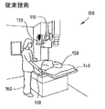
図1は、例示的医用X線イメージング・システム100を図示したものである。イメージング・システム100は、X線管110と、コリメータ120と、寝台検出器130と、X線寝台140と、患者150と、臨床操作者160とを含む。動作時には、撮影しようとする患者150がX線寝台140上に図のように配置される。次いで、放射線医や技術者などの臨床操作者160が、X線管110とコリメータ120を患者に対する幾つかの所定位置のうちの1つに位置決めする。臨床操作者がコリメータ120を所望の位置に位置決めした後、X線管110に通電しX線を発生させる。このX線は、X線が患者を透過して寝台検出器130に導かれるようにするコリメータ120を通過する。患者を透過するX線のエネルギーは、患者150の解剖学的特徴により減衰を受ける。寝台検出器130はこのX線のエネルギーを検出し、患者150の解剖学的特徴に関する画像を生じさせる。 FIG. 1 illustrates an exemplary medical
X線管110とコリメータ120は、典型的には、X線アセンブリを形成するように一体に固定されると共に、典型的には、X線寝台140に対して3次元に移動することができる。すなわち、コリメータ120は、移動止め(detent)と呼ばれる幾つかの固定位置のうちの任意の位置において、患者150の体軸に沿った上下方向に移動でき、患者150の体軸を横切る左右方向に移動でき、かつ患者150の体軸に対して接近させたり遠ざけたりすることができる。この幾つかの固定位置(すなわち、移動止め)の各々は、患者150に関して可能な最も明瞭な画像を作成するために予め決定しておいた異なるX線照射及び異なるイメージング・パラメータに対応させることができる。例えば、コリメータ120を患者からより離して配置することにより、検出器130が受け取るX線のエネルギーのダイナミックレンジに関して得られるパラメータは違ってくる。 The
典型的には、イメージング・パラメータは幾つかの所定の固定位置に対してのみ較正されており、コリメータ120の移動経路全体にわたり連続的に較正されてはいない。すなわち、各イメージング・パラメータは、典型的には、単一の特定の位置に関してのみ構成されており、コリメータが移動すると迅速に変更することができる。したがって、コリメータ120を精密に位置決めすることが、患者150に対するより明瞭で臨床的により適切な画像を提供するのに役立つ。 Typically, imaging parameters are calibrated only for a few predetermined fixed positions and are not continuously calibrated throughout the entire travel path of
図1を参照すると、医用X線イメージング・システムは、典型的には、放射線撮影検査にための幾つかの固定イメージング位置を特定できるように移動止めを利用し構成させることができる。コリメータ120を幾つかの固定イメージング位置のうちの1つに移動させると、イメージングの実施中にコリメータ120を所望の位置に保持させる移動止めが係合される。移動止めは機械式や電気式の場合もあるが、例えば損耗特性がより良好であるため、電磁式ロック及び位置基準トリガ・デバイスを用いた移動止めを利用することが好ましい場合もある。 Referring to FIG. 1, a medical X-ray imaging system can typically be configured with detents so that a number of fixed imaging positions for a radiographic examination can be identified. When the
1ミリメートル程度の小さい位置決め誤差により、得られる画像の品質が大幅に緩和される。例えば、検出器に対するビームのアラインメント不良や位置合わせ不良のために、解剖構造の欠落が生ずることがある。さらに、X線管に対する位置決め制御を改良することにより、X線画像の繰り返し精度の支援となる。この繰り返し精度は、患者を治療している間に時間間隔をおいて撮影したX線画像を比較する際に極めて重要となる。したがって、医用イメージング・システムに対するX線管及びコリメータの改良型位置決めシステムが必要とされている。 A positioning error as small as 1 millimeter greatly reduces the quality of the resulting image. For example, missing anatomical structures may occur due to misalignment or misalignment of the beam with respect to the detector. Further, by improving the positioning control for the X-ray tube, the repetition accuracy of the X-ray image is supported. This repeatability is extremely important when comparing X-ray images taken at time intervals while treating a patient. Therefore, there is a need for an improved X-ray tube and collimator positioning system for medical imaging systems.
本発明の好ましい実施形態は、X線イメージング・デバイス内のX線管の位置決め誤差を減少させるためのシステムを提供する。本システムにより、X線管を移動止めの位置に正確かつ高い繰り返し精度で位置決めすることが容易になる。本発明の好ましい実施形態は、X線管の位置または速度を示す位置信号または速度信号を発生させるセンサ・ユニットと、この位置信号を受け取りオーバーシュート補正を決定するマイクロプロセッサと、を含むことが好ましい。次いで、X線システムはこのオーバーシュート補正を用いて、X線管の位置を制御するロック・システムを制御する。センサ・ユニットは、ポテンショメータ、ディジタル・エンコーダ、また好ましくはこの両者の組み合わせ、を利用して位置信号または速度信号を決定する。 Preferred embodiments of the present invention provide a system for reducing x-ray tube positioning errors in an x-ray imaging device. This system makes it easy to position the X-ray tube at the detent position accurately and with high repeatability. A preferred embodiment of the present invention preferably includes a sensor unit that generates a position signal or velocity signal indicative of the position or velocity of the x-ray tube, and a microprocessor that receives the position signal and determines overshoot correction. . The X-ray system then uses this overshoot correction to control a locking system that controls the position of the X-ray tube. The sensor unit uses a potentiometer, digital encoder, or preferably a combination of both to determine the position or velocity signal.
図2は、本発明の好ましい実施形態による、医用X線イメージング・システムに対する例示的移動止め位置決めシステム200を図示したものである。移動止め位置決めシステム200は、X線管210と、X線アセンブリ205と、一対の垂直レール230と、一対の水平レール240と、センサ・ユニット275と、を含む。X線管210とコリメータ220は、全体としてX線アセンブリ205として知られている。水平レール240と垂直レール230の両者は多くの移動止め250を含んでいる。動作時には、この垂直レール230と水平レール240に沿った2つの次元方向にX線アセンブリ205を移動させる。この移動では、先ず、X線アセンブリ205と垂直レール230を水平レール240の範囲内で水平レール240上の移動止め250の位置までスライドさせる。次いで、X線アセンブリ205を垂直レール230の範囲内で垂直レール230上の移動止め250の位置までスライドさせる。それぞれの移動止め250の位置において、電磁式ロックを用いてコリメータを所望の移動止め位置にロックさせることが好ましい。センサ・ユニット275については以下で詳細に記載する。 FIG. 2 illustrates an exemplary
図3は、本発明の好ましい実施形態による医用X線イメージング・システムに対するロック・システム300を図示したものである。ロック・システム300は、電磁式ロック310と、ブリッジ・レール320と、電源330と、を含む。動作時には、ロック・システム300は、図2の移動止め位置決めシステム200の垂直レール230と水平レール240の内側に装着される。所与の移動止め250の位置に到達させた後、電磁式ロック310を起動させてその位置が確実にロックされる。電磁式ロック310は電源330が供給する電圧により起動させる。 FIG. 3 illustrates a
図4は、本発明の好ましい実施形態による図3の電磁式ロックの上面図400である。図400は、電磁式ロック・コイル410と、ロック・ストリップ420と、軸受け430と、を含む。動作時には、上述したように、電磁式ロック・コイル410は、外部供給電圧により起動状態になるまで、レールの内部でスライドさせることができる。外部供給電圧は、電磁式ロック・コイル410とロック・ストリップ420の間に、ある固定位置でコリメータを維持し確保するのに十分な磁気力を発生させる。 4 is a
動作時において、電磁式ロックは、コリメータ120の減速を開始させるのに十分な磁気力を生じさせるまでにある一定の有限時間が必要となる。さらに、電磁式ロックがコリメータ120を正しい位置に保持するために十分な力を生じさせるまでにもある一定の時間が必要となる。図2を参照すると、X線アセンブリ205(及びその支持/位置決め装置)はかなりの質量を有し、その結果、臨床操作者が位置決めをしている間にかなりの運動量を有しているため、電磁式ロックが発生させる磁気力は、所望の時間内では、X線アセンブリ205の運動量に打ち勝つだけの十分な大きさとならず、その結果、X線アセンブリ205を所望の移動止めの位置に精密に停止させることができない。したがって、この電磁式ロックの起動及び停止時間により、コリメータの位置決めに位置決め誤差が持ち込まれる。上述したように、この位置決め誤差はX線画像の画質及び繰り返し精度に対して悪影響を与えることがある。 In operation, the electromagnetic lock requires a certain finite time before generating enough magnetic force to initiate deceleration of the
別の述べ方をすると、X線アセンブリ205の初期速度がある臨界値(VC )未満である場合、操作者がX線アセンブリ205を位置決めしている速度、並びに電磁式ロックの電磁的ラグ(すなわち、時間遅延)が最終の位置決め誤差に寄与することがある。この位置決め誤差は、X線アセンブリ205を移動止め位置に近づける速度に概ね比例する。しかし、X線アセンブリ205の速度がかなり大きい場合には、電磁式ロックは、デバイスを完壁に係合して保持させるように対応することができない。電磁式ロックによる完壁な係合ができないと、X線アセンブリ205は期待する移動止め位置を通過してしまうことになる。速い速度ではロックによってコリメータを完全に係合して保持できないため、操作者は、X線アセンブリ205を事前設定の予め構成させた移動止め位置に位置決めしてロックできるように、移動止め位置に近づくにつれて減速し始める必要がある。さらに、接近速度を十分遅くしていないと、最終のオフセット位置決め誤差がかなり大きく(例えば、5〜10ミリメートルに)なることがある。このため、X線アセンブリ205を低速で移動させねばならないため、さらに追加の時間がかかる場合がある。追加の時間が必要になると、画像1枚あたりの追加時間のために受診者生産性(customer productivity) に悪影響を与えることがある。In other words, if the initial speed of the
これらの影響に対処するため、本発明の好ましい実施形態では、移動止めの様々な接近速度での位置オーバーシュートを計測することにより、位置制御システムを較正している。位置オーバーシュートは、電子的フィードバックを使用して決定することがあり、これについては以下でさらに記載する。次に、このオーバーシュート補正を決定するために速度とオーバーシュートの間の伝達関数を求める。最後に、臨床で使用している間に、このオーバーシュート補正をコリメータ位置決めに対して適用する。移動止めの位置オーバーシュートは、マイクロプロセッサ・ベースの位置決め制御を使用することにより計測することが好ましく、この際、図8〜10を参照しながら後述するように、位置と速度の両方のフィードバックを利用することができる。 To address these effects, the preferred embodiment of the present invention calibrates the position control system by measuring position overshoots at various approach speeds of the detent. Position overshoot may be determined using electronic feedback, as described further below. Next, in order to determine this overshoot correction, a transfer function between speed and overshoot is obtained. Finally, this overshoot correction is applied to collimator positioning during clinical use. The detent position overshoot is preferably measured by using microprocessor-based positioning control, where both position and velocity feedback is provided, as described below with reference to FIGS. Can be used.
図8は、本発明の好ましい実施形態によるセンサ・ユニット800を図示したものである。センサ・ユニット800は、エンコーダ・スプロケット810と、位置合せマーク830を有するポテンショメータ・スプロケット820と、位置センサ・ベルト840と、ベルト引張り器ねじ850と、駆動ベルト・アセンブリ860と、ベルト変位スプロケット870と、を含む。位置センサ・ベルト840は、エンコーダ・スプロケット810とポテンショメータ・スプロケット820の上に架けられている。位置センサ・ベルト840に対する張力は、ベルト引張り器ねじ850を使用して所望の張力になるように調整することができる。 FIG. 8 illustrates a
X線アセンブリ(並びに、これに取り付けられたセンサ・ユニット800)は、手作業で位置決めされるのが一般的である。しかし、センサ・ユニット800をモータにより駆動して位置決めすることが好ましい。例えば、センサ・ユニットは、駆動ベルト・アセンブリ860を使用する閉ループ・サーボモータによりモータ駆動させることがある。手作業ではなく、モータを使用してセンサ・ユニット800を位置決めすることにより、X線アセンブリを確実に移動止め位置に一貫して配置させるのに役立つ。 The x-ray assembly (as well as the
図9は、本発明の好ましい実施形態による図8のセンサ・ユニット800の上面図900である。エンコーダ・スプロケット810、ポテンショメータ・スプロケット820及びベルト変位スプロケット870を図示している。センサ・ユニット800はさらに、駆動ベルト・アセンブリ910と、マイクロプロセッサ・インタフェース920と、確保用ポイント930と、を含んでいる。センサ・ユニット800は、確保用ポイント930を使用してX線アセンブリ上に図2に示すように装着することが好ましい。 FIG. 9 is a
動作時には、センサ・ユニット800は、X線アセンブリ205の各レール方向への移動に関連付けされている。すなわち、あるセンサ・ユニット800はX線アセンブリ205の一対の垂直レール230に沿った移動に関するデータを提供し、またあるセンサ・ユニットは一対の水平レール240に沿った移動に関するデータを提供する。ノッチ付き駆動ベルト(図示せず)を、図2の一対の垂直レール230のうちの少なくとも一方のレールの内側、並びに一対の水平レール240のうちの少なくとも一方のレールの内側に装着することが好ましい。駆動ベルトは、そのレールの各端部に確保し、図9のセンサ・ユニット800の駆動ベルト・アセンブリ910を通過させることが好ましい。X線アセンブリ205を変位させるのに伴い、駆動ベルト・アセンブリ910を通過している固定の駆動ベルトにより、位置センサ・ベルト840の動きが誘発される。この位置センサ・ベルト840の動きにより、エンコーダ・スプロケット810及びポテンショメータ・スプロケット820の回転が誘発される。 In operation, the
ポテンショメータ・スプロケット820は、アナログのポテンショメータを含むことが好ましい。ポテンショメータの両端には、ポテンショメータ・スプロケット820の回転に伴って(したがって、X線アセンブリ205の位置に伴って)変化するような電圧が誘導されることが好ましい。エンコーダ・スプロケット810は、ディジタル・エンコーダを含むことが好ましい。ディジタル・エンコーダは、エンコーダ・スプロケット810の位置及び回転速度に関する(したがって、コリメータの位置及び速度に関する)データを提供することが好ましい。ポテンショメータ・スプロケット820を使用して、コリメータを初めに通電させた時点でX線アセンブリ205の初期位置を確定させることが好ましい。前回システムの運転を停止させた時点でデータが失なわれているため、エンコーダ・スプロケット810はこの初期情報を提供することができない。しかし、ポテンショメータ・スプロケット820の回転により内在するポテンショメータに対して機械的な変化が生じるため、X線アセンブリ205に関する初期位置はポテンショメータ・スプロケット820から復元することができ、これにより電源断の問題を回避できる。
X線アセンブリ205の初期位置をポテンショメータ・スプロケット820により確定し終えた後、エンコーダ・スプロケット810を利用して極めて正確な位置情報及び速度情報を提供することができる。エンコーダ・スプロケット810のディジタル・エンコーダは、速度情報を決定するために容易に解析できるような、X線アセンブリ205の位置を示す正確なディジタル信号を提供できることが好ましい。ポテンショメータ・スプロケット820を利用してX線アセンブリ205に関する位置情報を操作の全体にわたって提供することができるが、エンコーダ・スプロケット810からのディジタル式のエンコード信号によって使用をより容易にしかつ簡単にすることができる。 After the initial position of the
ポテンショメータ・スプロケット820により決定される初期位置の情報、並びにエンコーダ・スプロケット810により決定される位置情報及び速度情報は、マイクロプロセッサ・インタフェース920によって外部マイクロプロセッサ(図示せず)に渡される。詳細には以下に記載するが、マイクロプロセッサはX線アセンブリ205の位置情報及び速度情報を解析して上記の図3の電磁式ロック・システム300に対する起動を制御することができる。マイクロプロセッサは、典型的には、外部システムのキャビネット内に収容されている。 Initial position information determined by
使用に先立ち、センサ・ユニット800は、そのセンサ・ユニットが位置情報及び速度情報を提供する対象となる特定のレールに対して較正させる。ポテンショメータ・スプロケット820内部のポテンショメータは、その全長の各端部にハードストップを設けた複数巻きのポテンショメータであることが好ましく、10回巻きのポテンショメータであることが最も好ましい。このシステムを較正させるため、ポテンショメータを先ずハードストップまで回転させ、次いでポテンショメータ範囲の中間まで(10回巻きのポテンショメータでは、5回転まで)回転させる。次いで、このポテンショメータを含むセンサ・ユニット800を、レールに沿った移動経路の中央に位置決めし、駆動ベルト・アセンブリ910に位置センサ・ベルト840を架ける。さらに、センサ・ユニット800は、ベルト引張り器ねじ850を使用して位置センサ・ベルト840の張力を調整することにより較正することができる。 Prior to use, the
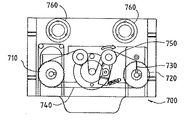
図7は、本発明の好ましい実施形態による自己引張式ベルト・アセンブリ700を有するセンサ・ユニットを図示したものである。自己引張式ベルト・アセンブリ700は、図8のセンサ・ユニット800と同様に、エンコーダ・スプロケット710と、ポテンショメータ・スプロケット720と、位置合せマーク730と、位置センサ・ベルト740と、駆動ベルト・アセンブリ760と、を含む。自己引張式センサ・ユニット700はさらに、図8のセンサ・ユニット800のベルト引張り器ねじ850の代わりに、位置センサ・ベルト740に対して所望の張力を自動的に印加する引張り器アーム750を含む。本発明の好ましい実施形態では、図8のセンサ・ユニット800と図7の自己引張式センサ・ユニット700とのいずれかを用いることができる。 FIG. 7 illustrates a sensor unit having a self-tensioning
センサ・ユニットを選択し据え付けた後に、センサ・ユニットのポテンショメータ・スプロケットを較正し、かつ位置センサ・ベルトを上述のように架ける。次いで、アセンブリ位置決めシステムを較正する。アセンブリ位置決めシステムを較正するするためには、コリメータ・アセンブリを移動状態に設定し、コリメータの位置及び速度に関する情報をマイクロプロセッサに送信する。次いで、移動止めラッチのシミュレーションを行う。すなわち、電源をX線アセンブリ上の電磁式ロックに印加して、アセンブリを停止状態にする。アセンブリが停止となる位置は、希望する所定の予め構成させた移動止め位置と異なることがある。次いで、移動止め位置とアセンブリの実際の位置との差を解析しオーバーシュート補正を決定する。 After selecting and installing the sensor unit, the potentiometer sprocket of the sensor unit is calibrated and the position sensor belt is hung as described above. The assembly positioning system is then calibrated. To calibrate the assembly positioning system, the collimator assembly is set to a moving state and information about the collimator position and velocity is sent to the microprocessor. Next, the detent latch is simulated. That is, power is applied to the electromagnetic lock on the X-ray assembly to bring the assembly to a stop. The position at which the assembly stops may be different from the desired predetermined pre-configured detent position. The difference between the detent position and the actual position of the assembly is then analyzed to determine overshoot correction.
図5は、本発明の好ましい実施形態による較正シーケンス500を図示したものである。先ず、位置510において、X線管アセンブリはゼロより大きいある初期速度V0で移動状態にあり、これまたゼロより大きいある初期位置X0に位置している。次いで、電磁式ロックを係合させる。電磁式ロックはアセンブリが移動するのと反対方向に制動力をかける。次いで、X線管アセンブリが位置520において静止状態となる。すなわち、最終速度Vf はゼロに等しくなり、アセンブリは最終位置Xf の位置にくる。次いで、530において、電磁式ロックを起動させた位置である初期位置X0 とアセンブリが静止した位置である最終位置Xf との間の位置の変化であるオーバーシュートΔXを決定する。初期及び最終の速度及び位置を決定し終えた後に、540において制動力を決定することができる。アセンブリの質量は既知であり、較正処理の間に変化することはない。次いで、較正シーケンスを幾つかの異なる初期速度で繰り返し、オーバーシュート補正を決定するために初期速度V0 とオーバーシュートΔXの間の実験関係式を決定する。FIG. 5 illustrates a
オーバーシュート補正は、例えば、幾つかの速度対オーバーシュートの較正試験に対する最小二乗回帰あてはめに基づいて線形関係式として表現することがある。この線形関係式は、次式
ΔX=B0+B1V
のように表現することができる。Overshoot correction may be expressed as a linear relationship based on, for example, a least-squares regression fit for some speed versus overshoot calibration tests. This linear relational expression is expressed by the following equation: ΔX = B 0 + B 1 V
It can be expressed as
別法として、オーバーシュート補正は、例えば、より一般的な非線形多項式の形態で、以下のように表現することもある。 Alternatively, overshoot correction may be expressed as follows, for example, in the form of a more general nonlinear polynomial.
ΔX=A0+A1V0+A2V0 2+A3V0 3+A4V4 4+...
上式において、多項式の次数は、較正処理に組み込まれた離散的速度の数によって異なる。ΔX = A 0 + A 1 V 0 + A 2 V 0 2 + A 3 V 0 3 + A 4 V 4 4 +. . .
In the above equation, the order of the polynomial depends on the number of discrete velocities built into the calibration process.
オーバーシュート補正を決定した後、このオーバーシュート補正を用いて、このアセンブリを所望の移動止め位置で静止状態にするためにこのシステムによる電磁式制動を有効にする位置が決定される。すなわち、較正シーケンスにより、移動止め位置の目標値に対する位置オーバーシュートを最小にするためにシステムの制御器が制動を有効にする位置が、X線管アセンブリの初期速度の関数として決定される。 After determining the overshoot correction, the overshoot correction is used to determine a position to enable electromagnetic braking by the system to bring the assembly stationary at the desired detent position. That is, the calibration sequence determines the position at which the system controller enables braking to minimize position overshoot relative to the detent position target value as a function of the initial velocity of the x-ray tube assembly.
本発明の第2の実施形態は、連続的な位置誤差監視の提供を含んでいる。すなわち、初期較正処理からの速度及び位置の基準値のみを使用するのではなく、連続的な位置検知を行っている。移動止め位置誤差がある一定の最大値を超えた場合に、操作者は通知を受け、電磁式ロックを解除し、さらに操作者はアセンブリを再位置決めすることができる。 The second embodiment of the present invention includes the provision of continuous position error monitoring. That is, instead of using only the speed and position reference values from the initial calibration process, continuous position detection is performed. When the detent position error exceeds a certain maximum value, the operator is notified and the electromagnetic lock is released and the operator can reposition the assembly.
本発明の第3の実施形態は、X線管アセンブリに対するそれぞれの位置決めの後オフシュート補正を絶えず更新することにより、オフシュート補正を適応可能に較正することを含んでいる。すなわち、アセンブリをある移動止め位置に位置決めするごとに、初期速度及び位置誤差を計測する。次いで、この速度及び位置誤差の計測値を用いてそのアセンブリに対する補正済みのオフシュート補正を作成する。この実施形態によればさらに、位置決めシステムは、使用に伴って生ずるシステムの劣化に対する補償をすることができる。例えば、アセンブリを持続的に使用することによりレールの摩擦が増大することがあり、これによりアセンブリの停止がより急激になる。オフシュート補正を適応可能に較正することにより、増大した摩擦の影響を最小にすることができアセンブリを常に最小の位置誤差で位置決めすることができる。 A third embodiment of the present invention includes adaptively calibrating the offshoot correction by constantly updating the offshoot correction after each positioning with respect to the x-ray tube assembly. That is, each time the assembly is positioned at a detent position, the initial speed and position error are measured. This velocity and position error measurement is then used to create a corrected offshoot correction for the assembly. This embodiment further allows the positioning system to compensate for system degradation that occurs with use. For example, continuous use of the assembly may increase rail friction, which causes the assembly to stop more abruptly. By adaptively calibrating the offshoot correction, the effects of increased friction can be minimized and the assembly can always be positioned with minimal positional error.
本発明の任意の実施形態を利用してオーバーシュート補正を作成することにより、移動止めのみしか使用せず本発明の好ましい実施形態による速度フィードバックや予測アルゴリズムを取り入れていない既存の実施と比較して、X線管と検出器アセンブリとの整列をより正確かつより繰り返し精度良く行うことができる。 By creating an overshoot correction using any embodiment of the present invention, compared to existing implementations that use only detents and do not incorporate speed feedback or prediction algorithms according to the preferred embodiment of the present invention. Therefore, the alignment of the X-ray tube and the detector assembly can be performed more accurately and repeatedly.
さらに、本発明により提供する位置決めの正確性と繰り返し精度に関する改良により、患者の解剖構造の欠落などの様々な要因と関連する放射線写真の再撮影が最小となる。患者解剖構造の欠落が生じると、X線画像は所望の解剖学的情報を含まず、再撮影が必要となる。患者の解剖構造の欠落の重大要因のうちの1つはアセンブリの位置決め誤差であるため、アセンブリの位置決め誤差を最小にすることにより、患者の解剖構造の欠落も減少させることができる。さらに、本発明は多くの方法により受診者生産性も向上させている。例えば、操作者は位置誤差を懸念することなく迅速にX線アセンブリを位置決めすることができる。したがって、アセンブリを位置決めする速度が増加すると共に、放射線写真の再撮影に関連する追加的な時間が最小となる。 Furthermore, the improvements in positioning accuracy and repeatability provided by the present invention minimize radiographic re-shoots associated with various factors such as missing patient anatomy. If the patient anatomy is missing, the X-ray image does not contain the desired anatomical information and re-imaging is required. Since one of the critical factors for missing patient anatomy is assembly positioning error, minimizing assembly positioning error can also reduce patient anatomy loss. Furthermore, the present invention also improves patient productivity by a number of methods. For example, the operator can quickly position the x-ray assembly without concern for positional errors. Thus, the speed of positioning the assembly is increased and the additional time associated with radiographic re-shooting is minimized.
図6は、本発明の好ましい実施形態による較正システムの流れ図600である。先ず、ステップ610において、X線管アセンブリは移動状態にある。ステップ620において、電磁式ロックが起動され初期速度V0及び初期位置X0が決定される。次に、ステップ630において、X線管アセンブリは停止状態になり最終速度Vf 及び最終位置Xf が決定される。次いで、ステップ640において、この初期位置X0 及び最終位置Xf を用いてオーバーシュートΔXを決定する。次いで、ステップ650において、ステップ610からステップ640が様々な初期速度で所定の回数反復され、初期速度V0 とオフセットΔXの間の実験関係式が作成される。次に、ステップ660において、様々な初期速度での反復計測の結果を用いてオーバーシュート補正を決定する。最後に、ステップ670において、このオーバーシュート補正を臨床で使用している間のX線管アセンブリの移動に対して適用する。上述したように、本発明の第3の実施形態を実現するには、ステップ610からステップ640までをアセンブリを臨床において位置決めするごとに反復させることがある。FIG. 6 is a
本発明を好ましい実施形態を参照しながら記載してきたが、当業者によれば、本発明の範囲を逸脱することなく様々な変更を行うことができ、また等価物による置換ができることを理解されたい。さらに、本発明の範囲を逸脱することなく、具体的な状況や材料を本発明の教示に適合させた多くの修正をすることもできる。したがって、本発明を開示した具体的な実施形態に限定しようとする意図はなく、本発明は特許請求の範囲の範疇に属するすべての実施形態を包含するものである。 Although the invention has been described with reference to preferred embodiments, those skilled in the art will recognize that various modifications can be made and substitutions made by equivalents without departing from the scope of the invention. . In addition, many modifications may be made to adapt a particular situation or material to the teachings of the invention without departing from the scope of the invention. Accordingly, there is no intention to limit the invention to the specific embodiments disclosed, and the invention encompasses all embodiments that fall within the scope of the claims.
100 医用X線イメージング・システム
110 X線管
120 コリメータ
130 寝台検出器
140 X線寝台
150 患者
160 臨床操作者
200 移動止め位置決めシステム
205 X線アセンブリ
210 X線管
220 コリメータ
230 一対の垂直レール
240 一対の水平レール
250 移動止め
275 センサ・ユニット
300 ロック・システム
310 電磁式ロック
320 ブリッジ・レール
330 電源
410 電磁式ロック・コイル
420 ロック・ストリップ
430 軸受け
700 自己引張式ベルト・アセンブリ
710 エンコーダ・スプロケット
720 ポテンショメータ・スプロケット
730 位置合せマーク
740 位置センサ・ベルト
750 引張り器アーム
760 駆動ベルト・アセンブリ
800 センサ・ユニット
810 エンコーダ・スプロケット
820 ポテンショメータ・スプロケット
830 位置合せマーク
840 位置センサ・ベルト
850 ベルト引張り器ねじ
860 駆動ベルト・アセンブリ
870 ベルト変位スプロケット
910 駆動ベルト・アセンブリ
920 マイクロプロセッサ・インタフェース
930 確保用ポイントDESCRIPTION OF
Claims (7)
前記X線イメージング・デバイス内で移動可能なX線管と、
X線イメージング・デバイス内での前記X線管の位置を示す位置信号を発生させるセンサ・ユニットと、
前記位置信号を受け取り、前記位置信号と前記X線イメージング・デバイス内の所定のX線管位置に基づいて前記X線管の移動を較正するために前記X線デバイス内で適用される前記X線管に対するオーバーシュート補正を決定するマイクロプロセッサと、を含むシステム。
A system for reducing positioning errors of an x-ray tube in an x-ray imaging device comprising:
An X-ray tube movable within the X-ray imaging device;
A sensor unit for generating a position signal indicative of the position of the X-ray tube within the X-ray imaging device;
The X-ray applied in the X-ray device to receive the position signal and calibrate movement of the X-ray tube based on the position signal and a predetermined X-ray tube position in the X-ray imaging device And a microprocessor for determining overshoot correction for the tube.
一対の垂直レール(230)と、
一対の水平レール(240)と、
電磁式ロック(310)とブリッジ・レール(320)と電源(330)とを含むロック・システム(300)と、を含み、
前記垂直レール(230)と前記水平レール(240)に沿った2つの次元方向に前記X線アセンブリ(205)が移動し、
前記マイクロプロセッサが前記ロック・システムを起動させることにより前記X線管の移動を抑制している、請求項1に記載のシステム。
An X-ray assembly (205) comprising the X-ray tube and a collimator (220);
A pair of vertical rails (230);
A pair of horizontal rails (240);
A locking system (300) including an electromagnetic lock (310), a bridge rail (320) and a power supply (330);
The X-ray assembly (205) moves in two dimensions along the vertical rail (230) and the horizontal rail (240);
The system according to claim 1, wherein the microprocessor suppresses the movement of the X-ray tube by activating the lock system.
The system according to claim 1 or 2, wherein the sensor unit generates a speed signal other than a position system and the speed signal is received by the microprocessor.
前記オーバーシュート補正が、少なくともX線管の初期位置と最終位置の解析により決定されている、請求項1乃至3のいずれかに記載のシステム。
The sensor unit uses a potentiometer or a digital encoder to generate the position signal;
The system according to any one of claims 1 to 3, wherein the overshoot correction is determined by an analysis of at least an initial position and a final position of the X-ray tube.
前記X線イメージング・デバイス内のX線管の位置信号を生成するステップと、
前記位置信号と前記X線イメージング・デバイス内の所定のX線管位置に基づいて前記X線管に対するオーバーシュート補正を決定するステップと、
前記X線管の移動を較正して前記X線管の位置決め誤差を減少させるために、前記オーバーシュート補正を適用するステップと、を含む方法。
A method for reducing x-ray tube positioning errors within an x-ray imaging device comprising :
Generating a position signal of an x-ray tube in the x-ray imaging device ;
Determining an overshoot correction for the x-ray tube based on the position signal and a predetermined x-ray tube position in the x-ray imaging device;
Applying the overshoot correction to calibrate movement of the x-ray tube to reduce positioning errors of the x-ray tube.
6. The method of claim 5, wherein the overshoot correction is determined by using a position signal relating to the position of the x-ray tube or velocity data relating to the velocity of the x-ray tube.
所定の初期速度でX線管を移動させるステップと、前記X線管の移動の停止を開始させるために、初期位置においてロック・システムを起動させるステップと、X線管を静止させる最終位置を決定するステップと、初期位置と最終位置の差に基づいてオーバーシュート補正を決定するステップと、を含む請求項5に記載の方法。
Determining the overshoot correction comprises:
A step of moving the X-ray tube at a predetermined initial speed, a step of activating a locking system at an initial position to start stopping the movement of the X-ray tube, and a final position for stopping the X-ray tube 6. The method of claim 5, comprising: and overdetermining overshoot correction based on a difference between the initial position and the final position.
Applications Claiming Priority (2)
| Application Number | Priority Date | Filing Date | Title |
|---|---|---|---|
| US09/575035 | 2000-05-19 | ||
| US09/575,035 US6379042B1 (en) | 2000-05-19 | 2000-05-19 | Variable self-compensating detent control system for improved positioning accuracy and repeatability |
Publications (3)
| Publication Number | Publication Date |
|---|---|
| JP2002034959A JP2002034959A (en) | 2002-02-05 |
| JP2002034959A5 JP2002034959A5 (en) | 2008-07-03 |
| JP5283809B2 true JP5283809B2 (en) | 2013-09-04 |
Family
ID=24298666
Family Applications (1)
| Application Number | Title | Priority Date | Filing Date |
|---|---|---|---|
| JP2001148554A Expired - Lifetime JP5283809B2 (en) | 2000-05-19 | 2001-05-18 | System and method for reducing x-ray tube positioning errors in x-ray imaging devices |
Country Status (4)
| Country | Link |
|---|---|
| US (1) | US6379042B1 (en) |
| EP (1) | EP1157661B1 (en) |
| JP (1) | JP5283809B2 (en) |
| DE (1) | DE60132516T2 (en) |
Families Citing this family (9)
| Publication number | Priority date | Publication date | Assignee | Title |
|---|---|---|---|---|
| US6459226B1 (en) * | 2001-01-04 | 2002-10-01 | Ge Medical Systems Global Technology Company, Llc | Method and apparatus for accurate powered deceleration and immobilization of manually operated mechanism |
| US6990368B2 (en) * | 2002-04-04 | 2006-01-24 | Surgical Navigation Technologies, Inc. | Method and apparatus for virtual digital subtraction angiography |
| JP4974726B2 (en) * | 2007-03-23 | 2012-07-11 | 富士フイルム株式会社 | Radiation imaging apparatus, radiation imaging method, and program |
| DE102012206343B4 (en) * | 2012-04-18 | 2015-08-27 | Siemens Aktiengesellschaft | Force equalization device and use in a medical device |
| US9277900B2 (en) * | 2013-01-07 | 2016-03-08 | Samsung Electronics Co., Ltd. | X-ray imaging apparatus |
| KR102373055B1 (en) * | 2015-02-26 | 2022-03-11 | 삼성전자주식회사 | Dish washer |
| CN106113039B (en) * | 2016-07-08 | 2018-06-15 | 深圳市优必选科技有限公司 | Steering engine locking position control method and steering engine |
| WO2023046460A1 (en) | 2021-09-23 | 2023-03-30 | Koninklijke Philips N.V. | Detent process for medical imaging systems |
| EP4154817A1 (en) * | 2021-09-23 | 2023-03-29 | Koninklijke Philips N.V. | Detent process for medical imaging systems |
Family Cites Families (17)
| Publication number | Priority date | Publication date | Assignee | Title |
|---|---|---|---|---|
| NL7706616A (en) * | 1977-06-16 | 1978-12-19 | Philips Nv | EXAMINATION DEVICE FOR MAKING SHADOW IMAGES OF A LAYER OF AN OBJECT (BODY). |
| DE2742642C3 (en) * | 1977-09-22 | 1985-02-21 | Philips Patentverwaltung Gmbh, 2000 Hamburg | Arrangement for weight compensation |
| DE2831058C2 (en) * | 1978-07-14 | 1984-04-19 | Philips Patentverwaltung Gmbh, 2000 Hamburg | X-ray examination device with a patient table that can be pivoted about a horizontal axis |
| US4380086A (en) * | 1980-11-24 | 1983-04-12 | Picker Corporation | Radiation imaging system with cyclically shiftable grid assembly |
| US4466112A (en) * | 1982-01-29 | 1984-08-14 | Technicare Corporation | Variable detector aperture |
| JPS6099906U (en) * | 1983-12-13 | 1985-07-08 | 朝日レントゲン工業株式会社 | Dental X-ray full jaw and head standard imaging equipment |
| JP2557502B2 (en) * | 1988-11-08 | 1996-11-27 | 株式会社モリタ製作所 | Medical panoramic X-ray equipment |
| JPH02245750A (en) * | 1989-03-20 | 1990-10-01 | Hitachi Medical Corp | Quick photographing controller for x-ray fluoroscopic photographing stand |
| JPH0355039A (en) * | 1989-07-22 | 1991-03-08 | Hitachi Medical Corp | Apparatus for detecting position of travelling truck in x-ray photographing apparatus |
| JPH0464345A (en) * | 1990-07-04 | 1992-02-28 | Toshiba Corp | X-ray diagnosing apparatus |
| JPH04341244A (en) * | 1991-05-17 | 1992-11-27 | Toshiba Corp | X-ray diagnostic device |
| JPH06269437A (en) * | 1993-03-19 | 1994-09-27 | Hitachi Medical Corp | Digital x-ray radiographing device |
| JPH0788108A (en) * | 1993-09-27 | 1995-04-04 | Toshiba Corp | X-ray fluororadiographic stand |
| JPH09505175A (en) * | 1994-09-01 | 1997-05-20 | フィリップス エレクトロニクス ネムローゼ フェンノートシャップ | Drive and X-ray device comprising such a drive |
| JPH08131432A (en) * | 1994-11-08 | 1996-05-28 | Hitachi Medical Corp | X-ray radiographing device |
| US5636259A (en) * | 1995-05-18 | 1997-06-03 | Continental X-Ray Corporation | Universal radiographic/fluoroscopic digital room |
| US6025685A (en) * | 1997-06-11 | 2000-02-15 | Elite Access Systems, Inc. | Gate operator method and apparatus with self-adjustment at operating limits |
-
2000
- 2000-05-19 US US09/575,035 patent/US6379042B1/en not_active Expired - Lifetime
-
2001
- 2001-05-16 EP EP01304348A patent/EP1157661B1/en not_active Expired - Lifetime
- 2001-05-16 DE DE60132516T patent/DE60132516T2/en not_active Expired - Lifetime
- 2001-05-18 JP JP2001148554A patent/JP5283809B2/en not_active Expired - Lifetime
Also Published As
| Publication number | Publication date |
|---|---|
| JP2002034959A (en) | 2002-02-05 |
| EP1157661A2 (en) | 2001-11-28 |
| DE60132516T2 (en) | 2009-01-15 |
| DE60132516D1 (en) | 2008-03-13 |
| EP1157661B1 (en) | 2008-01-23 |
| EP1157661A3 (en) | 2003-08-06 |
| US6379042B1 (en) | 2002-04-30 |
Similar Documents
| Publication | Publication Date | Title |
|---|---|---|
| JP5283809B2 (en) | System and method for reducing x-ray tube positioning errors in x-ray imaging devices | |
| US5469429A (en) | X-ray CT apparatus having focal spot position detection means for the X-ray tube and focal spot position adjusting means | |
| US6461040B1 (en) | Apparatus and method to correct for position errors in diagnostic imaging | |
| JP2004344656A (en) | Method and apparatus for detecting object collision of motorized mobile c-arm with object using pid controller | |
| JP2001061827A (en) | Movable x-ray device and deciding method of photographing position | |
| KR20010072305A (en) | Radiotherapy verification system | |
| US7319325B2 (en) | Table position sensing for magnetic resonance imaging | |
| US20080123811A1 (en) | System and Method for Improved Collision Detection in an Imaging Device | |
| CN113295197B (en) | Counting correction method and counting system of incremental encoder | |
| CN101797184B (en) | Monitoring system of a medical device | |
| FI87134C (en) | Dental x-ray apparatus for imaging whole jaws | |
| US5657498A (en) | Methods and apparatus for acquiring table elevation information | |
| FI56621C (en) | ANALYZING FOR PATIENTS WITH PANORAMA RENTAL PHOTOGRAPHY | |
| JP2009268799A (en) | X-ray ct device | |
| US4730351A (en) | X-ray diagnostics installation | |
| KR20170137037A (en) | Method and apparatus for aligning of cephalometric imaging device collimator | |
| JP3875793B2 (en) | Radiation measurement equipment | |
| JP2011229559A (en) | Radiation imaging apparatus | |
| JPH1189826A (en) | X-ray ct system | |
| JP5469952B2 (en) | X-ray CT system | |
| JP4112674B2 (en) | Magnetic resonance imaging system | |
| JP3738178B2 (en) | Image recording method | |
| JP5625258B2 (en) | X-ray imaging device | |
| CN117741524B (en) | Automatic full-built-in superconducting magnet magnetic field intensity measuring device and measuring method | |
| US6850594B2 (en) | Method for measuring the dose distribution in a computed tomography apparatus |
Legal Events
| Date | Code | Title | Description |
|---|---|---|---|
| A521 | Request for written amendment filed |
Free format text: JAPANESE INTERMEDIATE CODE: A523 Effective date: 20080516 |
|
| A621 | Written request for application examination |
Free format text: JAPANESE INTERMEDIATE CODE: A621 Effective date: 20080516 |
|
| RD02 | Notification of acceptance of power of attorney |
Free format text: JAPANESE INTERMEDIATE CODE: A7422 Effective date: 20101116 |
|
| RD04 | Notification of resignation of power of attorney |
Free format text: JAPANESE INTERMEDIATE CODE: A7424 Effective date: 20101116 |
|
| A131 | Notification of reasons for refusal |
Free format text: JAPANESE INTERMEDIATE CODE: A131 Effective date: 20110712 |
|
| A521 | Request for written amendment filed |
Free format text: JAPANESE INTERMEDIATE CODE: A523 Effective date: 20110913 |
|
| A131 | Notification of reasons for refusal |
Free format text: JAPANESE INTERMEDIATE CODE: A131 Effective date: 20120131 |
|
| A601 | Written request for extension of time |
Free format text: JAPANESE INTERMEDIATE CODE: A601 Effective date: 20120406 |
|
| A602 | Written permission of extension of time |
Free format text: JAPANESE INTERMEDIATE CODE: A602 Effective date: 20120416 |
|
| A521 | Request for written amendment filed |
Free format text: JAPANESE INTERMEDIATE CODE: A523 Effective date: 20120628 |
|
| A131 | Notification of reasons for refusal |
Free format text: JAPANESE INTERMEDIATE CODE: A131 Effective date: 20130205 |
|
| A521 | Request for written amendment filed |
Free format text: JAPANESE INTERMEDIATE CODE: A523 Effective date: 20130402 |
|
| TRDD | Decision of grant or rejection written | ||
| RD03 | Notification of appointment of power of attorney |
Free format text: JAPANESE INTERMEDIATE CODE: A7423 Effective date: 20130423 |
|
| A01 | Written decision to grant a patent or to grant a registration (utility model) |
Free format text: JAPANESE INTERMEDIATE CODE: A01 Effective date: 20130430 |
|
| A61 | First payment of annual fees (during grant procedure) |
Free format text: JAPANESE INTERMEDIATE CODE: A61 Effective date: 20130529 |
|
| R150 | Certificate of patent or registration of utility model |
Ref document number: 5283809 Country of ref document: JP Free format text: JAPANESE INTERMEDIATE CODE: R150 |
|
| R250 | Receipt of annual fees |
Free format text: JAPANESE INTERMEDIATE CODE: R250 |
|
| R250 | Receipt of annual fees |
Free format text: JAPANESE INTERMEDIATE CODE: R250 |
|
| R250 | Receipt of annual fees |
Free format text: JAPANESE INTERMEDIATE CODE: R250 |
|
| R250 | Receipt of annual fees |
Free format text: JAPANESE INTERMEDIATE CODE: R250 |
|
| R250 | Receipt of annual fees |
Free format text: JAPANESE INTERMEDIATE CODE: R250 |
|
| EXPY | Cancellation because of completion of term |


