JP5167149B2 - Leukemia diagnosis and treatment targets - Google Patents
Leukemia diagnosis and treatment targets Download PDFInfo
- Publication number
- JP5167149B2 JP5167149B2 JP2008551261A JP2008551261A JP5167149B2 JP 5167149 B2 JP5167149 B2 JP 5167149B2 JP 2008551261 A JP2008551261 A JP 2008551261A JP 2008551261 A JP2008551261 A JP 2008551261A JP 5167149 B2 JP5167149 B2 JP 5167149B2
- Authority
- JP
- Japan
- Prior art keywords
- calm
- hoxa5
- fusion protein
- dot1l
- polypeptide
- Prior art date
- Legal status (The legal status is an assumption and is not a legal conclusion. Google has not performed a legal analysis and makes no representation as to the accuracy of the status listed.)
- Expired - Fee Related
Links
Images


Classifications
-
- G—PHYSICS
- G01—MEASURING; TESTING
- G01N—INVESTIGATING OR ANALYSING MATERIALS BY DETERMINING THEIR CHEMICAL OR PHYSICAL PROPERTIES
- G01N33/00—Investigating or analysing materials by specific methods not covered by groups G01N1/00 - G01N31/00
- G01N33/48—Biological material, e.g. blood, urine; Haemocytometers
- G01N33/50—Chemical analysis of biological material, e.g. blood, urine; Testing involving biospecific ligand binding methods; Immunological testing
- G01N33/5005—Chemical analysis of biological material, e.g. blood, urine; Testing involving biospecific ligand binding methods; Immunological testing involving human or animal cells
- G01N33/5008—Chemical analysis of biological material, e.g. blood, urine; Testing involving biospecific ligand binding methods; Immunological testing involving human or animal cells for testing or evaluating the effect of chemical or biological compounds, e.g. drugs, cosmetics
- G01N33/5011—Chemical analysis of biological material, e.g. blood, urine; Testing involving biospecific ligand binding methods; Immunological testing involving human or animal cells for testing or evaluating the effect of chemical or biological compounds, e.g. drugs, cosmetics for testing antineoplastic activity
-
- C—CHEMISTRY; METALLURGY
- C12—BIOCHEMISTRY; BEER; SPIRITS; WINE; VINEGAR; MICROBIOLOGY; ENZYMOLOGY; MUTATION OR GENETIC ENGINEERING
- C12Q—MEASURING OR TESTING PROCESSES INVOLVING ENZYMES, NUCLEIC ACIDS OR MICROORGANISMS; COMPOSITIONS OR TEST PAPERS THEREFOR; PROCESSES OF PREPARING SUCH COMPOSITIONS; CONDITION-RESPONSIVE CONTROL IN MICROBIOLOGICAL OR ENZYMOLOGICAL PROCESSES
- C12Q1/00—Measuring or testing processes involving enzymes, nucleic acids or microorganisms; Compositions therefor; Processes of preparing such compositions
- C12Q1/68—Measuring or testing processes involving enzymes, nucleic acids or microorganisms; Compositions therefor; Processes of preparing such compositions involving nucleic acids
- C12Q1/6876—Nucleic acid products used in the analysis of nucleic acids, e.g. primers or probes
- C12Q1/6883—Nucleic acid products used in the analysis of nucleic acids, e.g. primers or probes for diseases caused by alterations of genetic material
- C12Q1/6886—Nucleic acid products used in the analysis of nucleic acids, e.g. primers or probes for diseases caused by alterations of genetic material for cancer
-
- C—CHEMISTRY; METALLURGY
- C12—BIOCHEMISTRY; BEER; SPIRITS; WINE; VINEGAR; MICROBIOLOGY; ENZYMOLOGY; MUTATION OR GENETIC ENGINEERING
- C12N—MICROORGANISMS OR ENZYMES; COMPOSITIONS THEREOF; PROPAGATING, PRESERVING, OR MAINTAINING MICROORGANISMS; MUTATION OR GENETIC ENGINEERING; CULTURE MEDIA
- C12N15/00—Mutation or genetic engineering; DNA or RNA concerning genetic engineering, vectors, e.g. plasmids, or their isolation, preparation or purification; Use of hosts therefor
- C12N15/09—Recombinant DNA-technology
- C12N15/11—DNA or RNA fragments; Modified forms thereof; Non-coding nucleic acids having a biological activity
- C12N15/113—Non-coding nucleic acids modulating the expression of genes, e.g. antisense oligonucleotides; Antisense DNA or RNA; Triplex- forming oligonucleotides; Catalytic nucleic acids, e.g. ribozymes; Nucleic acids used in co-suppression or gene silencing
- C12N15/1135—Non-coding nucleic acids modulating the expression of genes, e.g. antisense oligonucleotides; Antisense DNA or RNA; Triplex- forming oligonucleotides; Catalytic nucleic acids, e.g. ribozymes; Nucleic acids used in co-suppression or gene silencing against oncogenes or tumor suppressor genes
-
- C—CHEMISTRY; METALLURGY
- C12—BIOCHEMISTRY; BEER; SPIRITS; WINE; VINEGAR; MICROBIOLOGY; ENZYMOLOGY; MUTATION OR GENETIC ENGINEERING
- C12Q—MEASURING OR TESTING PROCESSES INVOLVING ENZYMES, NUCLEIC ACIDS OR MICROORGANISMS; COMPOSITIONS OR TEST PAPERS THEREFOR; PROCESSES OF PREPARING SUCH COMPOSITIONS; CONDITION-RESPONSIVE CONTROL IN MICROBIOLOGICAL OR ENZYMOLOGICAL PROCESSES
- C12Q1/00—Measuring or testing processes involving enzymes, nucleic acids or microorganisms; Compositions therefor; Processes of preparing such compositions
- C12Q1/68—Measuring or testing processes involving enzymes, nucleic acids or microorganisms; Compositions therefor; Processes of preparing such compositions involving nucleic acids
- C12Q1/6897—Measuring or testing processes involving enzymes, nucleic acids or microorganisms; Compositions therefor; Processes of preparing such compositions involving nucleic acids involving reporter genes operably linked to promoters
-
- C—CHEMISTRY; METALLURGY
- C12—BIOCHEMISTRY; BEER; SPIRITS; WINE; VINEGAR; MICROBIOLOGY; ENZYMOLOGY; MUTATION OR GENETIC ENGINEERING
- C12Q—MEASURING OR TESTING PROCESSES INVOLVING ENZYMES, NUCLEIC ACIDS OR MICROORGANISMS; COMPOSITIONS OR TEST PAPERS THEREFOR; PROCESSES OF PREPARING SUCH COMPOSITIONS; CONDITION-RESPONSIVE CONTROL IN MICROBIOLOGICAL OR ENZYMOLOGICAL PROCESSES
- C12Q2600/00—Oligonucleotides characterized by their use
- C12Q2600/136—Screening for pharmacological compounds
-
- C—CHEMISTRY; METALLURGY
- C12—BIOCHEMISTRY; BEER; SPIRITS; WINE; VINEGAR; MICROBIOLOGY; ENZYMOLOGY; MUTATION OR GENETIC ENGINEERING
- C12Q—MEASURING OR TESTING PROCESSES INVOLVING ENZYMES, NUCLEIC ACIDS OR MICROORGANISMS; COMPOSITIONS OR TEST PAPERS THEREFOR; PROCESSES OF PREPARING SUCH COMPOSITIONS; CONDITION-RESPONSIVE CONTROL IN MICROBIOLOGICAL OR ENZYMOLOGICAL PROCESSES
- C12Q2600/00—Oligonucleotides characterized by their use
- C12Q2600/154—Methylation markers
-
- Y—GENERAL TAGGING OF NEW TECHNOLOGICAL DEVELOPMENTS; GENERAL TAGGING OF CROSS-SECTIONAL TECHNOLOGIES SPANNING OVER SEVERAL SECTIONS OF THE IPC; TECHNICAL SUBJECTS COVERED BY FORMER USPC CROSS-REFERENCE ART COLLECTIONS [XRACs] AND DIGESTS
- Y10—TECHNICAL SUBJECTS COVERED BY FORMER USPC
- Y10T—TECHNICAL SUBJECTS COVERED BY FORMER US CLASSIFICATION
- Y10T436/00—Chemistry: analytical and immunological testing
- Y10T436/14—Heterocyclic carbon compound [i.e., O, S, N, Se, Te, as only ring hetero atom]
- Y10T436/142222—Hetero-O [e.g., ascorbic acid, etc.]
- Y10T436/143333—Saccharide [e.g., DNA, etc.]
Description
[関連出願情報]
本願は、2006年1月20日に出願された米国仮特許出願第60/760,709号の利益を主張し、その開示は引用することにより本明細書の一部をなすものとする。
[Related application information]
This application claims the benefit of US Provisional Patent Application No. 60 / 760,709, filed Jan. 20, 2006, the disclosure of which is hereby incorporated by reference.
[連邦政府の支援に関する記載]
本発明は、米国国立衛生研究所によって交付された助成金番号GM68804の下に、連邦政府の支援を受けてなされた。米国政府は本発明に一定の権利を有する。
[Federal support statement]
This invention was made with support from the federal government under grant number GM68804 issued by the National Institutes of Health. The US government has certain rights in this invention.
[発明の分野]
本発明は、ヒストンメチル化に基づく、白血病治療用の候補化合物を識別する方法および診断方法に関する。
[Field of the Invention]
The present invention relates to a method for identifying candidate compounds for the treatment of leukemia and a diagnostic method based on histone methylation.
高次クロマチン構造は、遺伝子調節および後成的遺伝に著しく重要である(Wu and Grunstein(2000)Trends Biochem.Sci.25:619−623)。コアヒストンの翻訳後修飾は、高次クロマチン構造の確立および維持に影響を及ぼす。一定のコアヒストンの、特定構造を持たないテールは、アセチル化、メチル化、リン酸化、リボシル化およびユビキチン化によって、大規模に修飾される。ヒストン修飾をクロマチン構造と結びつける「ヒストンコード」仮説は、近年の集中的な研究の焦点になってきた(Strahl and Allis(2000)Mol.Cell.Biol.22:1298−1306;Turner(2000)Bioessays 22:836−845)。ヒストンメチル化はヒストン修飾の主形態であることが明らかになっている(Strahl and Allis(2000)Mol.Cell.Biol.22:1298−1306;Zhang and Reinberg(2001)Genes Dev.15:2343−2360)。特に、SETドメインを含有するヒストンメチルトランスフェラーゼ(HMTases)の大きなファミリーが同定されている(Lachner and Jenuwein(2002)Curr.Opin.Cell Biol.14:286−298)。SETドメインタンパク質は、ヒストンH3およびH4のさまざまなN末端リジン残基をメチル化することが示されている。ヒストンリジンメチル化は、転写調節から細胞分裂時に起こる染色体の正確な伝達まで、多様な生物学的過程に関連づけられている(Grewal and Elgin(2002)Curr.Opin.Genet.Dev.12:178−187)。 Higher order chromatin structure is remarkably important for gene regulation and epigenetic inheritance (Wu and Grunstein (2000) Trends Biochem. Sci. 25: 619-623). Post-translational modifications of core histones affect the establishment and maintenance of higher order chromatin structures. Certain core histone non-specific tails are extensively modified by acetylation, methylation, phosphorylation, ribosylation and ubiquitination. The “histone code” hypothesis that links histone modifications to chromatin structures has been the focus of recent intensive research (Strahl and Allis (2000) Mol. Cell. Biol. 22: 1298-1306; Turner (2000) Bioessays. 22: 836-845). Histone methylation has been shown to be the major form of histone modification (Strah and Allis (2000) Mol. Cell. Biol. 22: 1298-1306; Zhang and Reinberg (2001) Genes Dev. 15: 2343). 2360). In particular, a large family of histone methyltransferases (HMTases) containing the SET domain has been identified (Lachner and Jenwein (2002) Curr. Opin. Cell Biol. 14: 286-298). SET domain proteins have been shown to methylate various N-terminal lysine residues of histones H3 and H4. Histone lysine methylation has been linked to a variety of biological processes, from transcriptional regulation to precise chromosome transmission during cell division (Grewal and Elgin (2002) Curr. Opin. Genet. Dev. 12: 178- 187).
さらにまた、SETドメイン含有タンパク質が触媒するリジンメチル化は、がんに結びつけられている(Schneider,et al.(2002)Trends Biochem.Sci.27:396−402)。例えば、H3−K4メチルトランスフェラーゼMLLは白血病ではしばしば転座し(Ayton and Cleary(2001)Oncogene 20:5695−5707;Milne,et al.(2002)Mol.Cell 10:1107−1117;Nakamura,et al.(2002)Mol.Cell 10:1119−1128)、H3−K27メチルトランスフェラーゼEZH2はいくつかの腫瘍において過剰発現され、その発現レベルはそれらの腫瘍の侵襲性と相関する(Bracken,et al.(2003)EMBO J.22:5323−5335;Kleer et al.(2003)Proc.Natl.Acad.Sci.USA 100:11606−11611;Varambally,et al.(2002)Nature 419:624−9)。 Furthermore, lysine methylation catalyzed by SET domain-containing proteins has been linked to cancer (Schneider, et al. (2002) Trends Biochem. Sci. 27: 396-402). For example, H3-K4 methyltransferase MLL is often translocated in leukemia (Ayton and Clear (2001) Oncogene 20: 5695-5707; Milne, et al. (2002) Mol. Cell 10: 1107-1117; Nakamura, et al (2002) Mol.Cell 10: 1119-1128), H3-K27 methyltransferase EZH2 is overexpressed in several tumors, and its expression level correlates with the invasiveness of those tumors (Bracken, et al. 2003) EMBO J. 22: 5323-5335; Kleer et al. (2003) Proc. Natl. Acad. Sci. USA 100: 11606-11611; , Et al (2002) Nature 419:. 624-9).
Dot1は、元々、サッカロミセスセレビシェ(S.cerevisiae)において、テロメアサイレンシングの破壊因子(disruptor of telomeric silencing)として同定された(Singer et al.,(1998)Genetics 150:613−632)進化的に保存されたタンパク質である。これは減数分裂周期中のパキテン期チェックポイントにおいても機能する(San−Segundo and Roeder(2000)Mol.Biol.Cell.11:3601−3615)。酵母Dot1の配列解析により、これは、タンパク質アルギニンメチルトランスフェラーゼ中のものに似た一定の特徴的SAM結合モチーフを有することが明らかになった(Dlakic(2001)Trends Biochem.Sci.26:405−407)。 Dot1 was originally in Saccharomyces cerevisiae shell (S. cerevisiae), was identified as a broken factor telomeric silencing (d isruptor o f t elomeric silencing ) (Singer et al, (1998) Genetics 150:. 613-632) It is an evolutionarily conserved protein. This also works at the pakiten phase checkpoint during the meiotic cycle (San-Segundo and Roeder (2000) Mol. Biol. Cell. 11: 3601-3615). Sequence analysis of the yeast Dot1 revealed that it has a certain characteristic SAM binding motif similar to that in the protein arginine methyltransferase (Dlackic (2001) Trends Biochem. Sci. 26: 405-407). ).
最近、hDOT1LはヒストンH3−K79メチルトランスフェラーゼであり(Feng et al.,(2002)Curr.Biol.12:1052−1058)、MLL−AF10による白血病誘発に重要な役割を果たすこと(Okada et al.,(2005)Cell 121:167−78)が証明された。MML−AF10によるHoxa9遺伝子へのhDOT1Lのミスターゲティングは、H3−K79メチル化およびHoxa9のアップレギュレーションをもたらし、それが白血病性形質転換の一因になることが示された(Okada et al.,(2005)Cell 121:167−78)。さらに、hDOT1LとMLL−AF10の相互作用には、MLL−AF10による白血病性形質転換に必要なAF10のOM−LZ(オクタペプチドモチーフ−ロイシンジッパー)領域が関与することも証明された(DiMartino et al.,(2002)Blood 99:3780−5)。 Recently, hDOT1L is a histone H3-K79 methyltransferase (Feng et al., (2002) Curr. Biol. 12: 1052-1058) and plays an important role in leukemogenesis by MLL-AF10 (Okada et al. (2005) Cell 121: 167-78). Mistargeting of hDOT1L to the Hoxa9 gene by MML-AF10 has been shown to result in H3-K79 methylation and up-regulation of Hoxa9, which contributes to leukemic transformation (Okada et al., ( 2005) Cell 121: 167-78). Furthermore, it was proved that the interaction between hDOT1L and MLL-AF10 involves the OM-LZ (octapeptide motif-leucine zipper) region of AF10 necessary for leukemic transformation by MLL-AF10 (DiMarino et al. (2002) Blood 99: 3780-5).
本発明者らは、(1)CALM−AF10融合は、インビトロおよびインビボで白血病誘発を媒介するのに、必要かつ十分であるらしいこと、(2)hDOT1LおよびそのH3−K79メチルトランスフェラーゼ活性がCALM−AF10による白血病性形質転換に関係すること、(3)Hoxa5遺伝子がCALM−AF10による形質転換に関与することを、ここに初めて証明する。いかなる本発明の理論にも束縛されることは望まないが、本発明者らは、hDOT1Lがタンパク質−タンパク質相互作用によってCALM−AF10の核局在化を保つ実験モデルも提供する。hDOT1Lと会合したCALM−AF10は、次に、Hoxa5プロモーターに動員されてH3−K79メチル化を媒介し、それが結果として、Hoxa5アップレギュレーションおよび白血病誘発につながる。 We found that (1) CALM-AF10 fusion appears to be necessary and sufficient to mediate leukemogenesis in vitro and in vivo, (2) hDOT1L and its H3-K79 methyltransferase activity is CALM- Here we demonstrate for the first time that it is involved in leukemic transformation by AF10 and that (3) the Hoxa5 gene is involved in the transformation by CALM-AF10. Without wishing to be bound by any theory of the present invention, we also provide an experimental model in which hDOT1L maintains the nuclear localization of CALM-AF10 through protein-protein interactions. CALM-AF10 associated with hDOT1L is then recruited to the Hoxa5 promoter to mediate H3-K79 methylation, which results in Hoxa5 up-regulation and leukemia induction.
染色体転座は、(特に急性白血病において)ヒトがんの主原因の一つである。キメラ遺伝子CALM−AF10を生成するt(10;11)(p12−13;q14−q23)の転座は、T−ALL(T細胞急性リンパ性白血病)およびAML(急性骨髄性白血病)を含むさまざまなタイプの白血病に観察される(Carlson et al.,(2000)Leukemia 14:100−104;Dreylng et al.,(1998)Blood 91:4662−7)。この染色体転座は、キメラ遺伝子MLL−AF10を生成するt(10;11)(p13;q23)の転座と区別することが難しい場合もあるが、MLL−AF10を持つ患者は、もっぱらAMLを発症する(Beverloo et al et al.,(1995)Cancer Res.55:4220−4)。CALM−AF10によるAMLとMLL−AF10によるAMLにも顕著な相違がある。例えばCALM−AF10によるAMLはM0/1サブタイプに属するのに対し、MLL−AF10によるAMLはM4/5サブタイプに属する(Dreylng et al.,(1998)Blood 91:4662−7;Beverloo et al et al.,(1995)Cancer Res.55:4220−4)。造血および白血病誘発におけるMLL(mixed lineage leukemia)の役割は確立されているが、基礎にある分子機序は解明され始めたばかりである(Ayton et al.,(2001)Oncogene 20:5695−5707;Daser et al.,(2004)Genes Dev.18:965−74;Hess(2004)Crit.Rev.Eukaryot. Gene Expr.14:235−54)。MLLとは異なり、造血および白血病におけるCALM(clathrin assembly lymphoid myeloid leukemia)の機能は全くわかっていない。なぜなら、CALMタンパク質は転座がなければ細胞質に位置し、そこではクラスリンによる輸送に関与しているからである(Dreyling et al.,(1996)Proc Natl Acad Sci 93:4804−9;Tebar et al.,(1999)Mol Biol.Cell 10:2687−702)。実際、CALM−AF10の白血病性形質転換能は、実験的には証明されていない。 Chromosomal translocation is one of the main causes of human cancer (especially in acute leukemia). Translocations of t (10; 11) (p12-13; q14-q23) that produce the chimeric gene CALM-AF10 include a variety of T-ALL (T cell acute lymphoblastic leukemia) and AML (acute myeloid leukemia) Observed in various types of leukemia (Carlson et al., (2000) Leukemia 14: 100-104; Dryylg et al., (1998) Blood 91: 4662-7). This chromosomal translocation may be difficult to distinguish from the translocation of t (10; 11) (p13; q23), which produces the chimeric gene MLL-AF10, but patients with MLL-AF10 exclusively have AML. It develops (Beverloo et al et al., (1995) Cancer Res. 55: 4220-4). There are also significant differences between AML with CALM-AF10 and AML with MLL-AF10. For example, AML by CALM-AF10 belongs to the M0 / 1 subtype, whereas AML by MLL-AF10 belongs to the M4 / 5 subtype (Dreyng et al., (1998) Blood 91: 4662-7; Beverloo et al. et al., (1995) Cancer Res. 55: 4220-4). The role of MLL (mixed lineage leukemia) in hematopoiesis and leukemia induction has been established, but the underlying molecular mechanism is just beginning to be elucidated (Ayton et al., (2001) Oncogene 20: 5695-5707; Daser et al., (2004) Genes Dev. 18: 965-74; Hess (2004) Crit. Rev. Eukaryot. Gene Expr. 14: 235-54). Unlike MLL, the function of CALM in the hematopoiesis and leukemia is completely unknown. This is because the CALM protein is located in the cytoplasm without translocation, where it is involved in transport by clathrin (Dreyling et al., (1996) Proc Natl Acad Sci 93: 4804-9; Tebar et al., (1999) Mol Biol. Cell 10: 2687-702). Indeed, the leukemic transformation ability of CALM-AF10 has not been experimentally proven.
したがって第1の態様として、本発明は、白血病の予防および/または治療のための候補化合物を識別する方法であって、
DOT1LポリペプチドがCALM−AF10融合タンパク質に結合するのに十分な条件下で、試験化合物の存在下で、DOT1LポリペプチドをCALM−AF10融合タンパク質と接触させるステップと、DOT1LポリペプチドとCALM−AF10融合タンパク質の間の相互作用を検出するステップとを含み、該試験化合物の非存在下での相互作用のレベルと比較した、該試験化合物存在下での、DOT1LポリペプチドとCALM−AF10融合タンパク質の間の相互作用の減少が、前記試験化合物が白血病の予防および/または治療のための候補化合物であることを示す方法を提供する。
Accordingly, as a first aspect, the present invention provides a method for identifying candidate compounds for the prevention and / or treatment of leukemia comprising:
Contacting the DOT1L polypeptide with the CALM-AF10 fusion protein in the presence of a test compound under conditions sufficient for the DOT1L polypeptide to bind to the CALM-AF10 fusion protein; and the DOT1L polypeptide and the CALM-AF10 fusion. Detecting an interaction between the protein and between the DOT1L polypeptide and the CALM-AF10 fusion protein in the presence of the test compound compared to the level of interaction in the absence of the test compound. Provides a method indicating that the test compound is a candidate compound for the prevention and / or treatment of leukemia.
さらに本発明は、白血病の予防および/または治療のための候補化合物を識別する方法であって、
試験化合物がCALM−AF10融合タンパク質に結合するのに十分な条件下で、CALM−AF10融合タンパク質を試験化合物と接触させるステップと、試験化合物とCALM−AF10融合タンパク質の間の結合を検出するステップとを含み、CALM−AF10融合タンパク質への試験化合物の結合は、前記試験化合物が白血病の予防および/または治療のための候補化合物であることを示す方法も提供する。
Furthermore, the present invention is a method for identifying candidate compounds for the prevention and / or treatment of leukemia comprising:
Contacting the CALM-AF10 fusion protein with the test compound under conditions sufficient for the test compound to bind to the CALM-AF10 fusion protein; and detecting binding between the test compound and the CALM-AF10 fusion protein; And the binding of the test compound to the CALM-AF10 fusion protein also provides a method indicating that the test compound is a candidate compound for the prevention and / or treatment of leukemia.
さらなる一態様として、本発明は、白血病の予防および/または治療のための候補化合物を識別する方法であって、CALMポリペプチドまたはCALM−AF10融合タンパク質がHoxA5プロモーターに結合するのに十分な条件下で、試験化合物の存在下で、HoxA5プロモーター領域を含む核酸をCALMポリペプチドまたはCALM−AF10融合タンパク質と接触させるステップと、CALMポリペプチドまたはCALM−AF10融合タンパク質のHoxA5プロモーターとの相互作用を検出するステップとを含み、試験化合物の非存在下での相互作用のレベルと比較した、試験化合物存在下での、CALMポリペプチドまたはCALM−AF10融合タンパク質とHoxA5プロモーターの間の相互作用の減少が、前記試験化合物が白血病の予防および/または治療のための候補化合物であることを示す方法を提供する。 In a further aspect, the present invention is a method for identifying candidate compounds for the prevention and / or treatment of leukemia, under conditions sufficient for a CALM polypeptide or CALM-AF10 fusion protein to bind to the HoxA5 promoter. The step of contacting a nucleic acid comprising a HoxA5 promoter region with a CALM polypeptide or CALM-AF10 fusion protein in the presence of a test compound and detecting the interaction of the CALM polypeptide or CALM-AF10 fusion protein with the HoxA5 promoter. Reducing the interaction between the CALM polypeptide or CALM-AF10 fusion protein and the HoxA5 promoter in the presence of the test compound compared to the level of interaction in the absence of the test compound Trial It provides a way to indicate that the compound is a candidate compound for the prevention and / or treatment of leukemia.
さらにもう一つの態様として、本発明は、白血病の予防および/または治療のための候補化合物を識別する方法であって、DOT1LポリペプチドとCALM−AF10融合タンパク質が結合して複合体を形成し、前記複合体がHoxA5プロモーターに結合するのに十分な条件下で、試験化合物の存在下で、HoxA5プロモーター領域を含む核酸をDOT1LポリペプチドおよびCALM−AF10融合タンパク質と接触させるステップと、DOT1L/CALM−AF10複合体のHoxA5プロモーターとの相互作用を検出するステップとを含み、試験化合物の非存在下での相互作用のレベルと比較した、試験化合物存在下での、DOT1L/CALM−AF10複合体のHoxA5プロモーターとの相互作用の減少が、前記試験化合物が白血病の予防および/または治療のための候補化合物であることを示す方法を提供する。 In yet another aspect, the present invention provides a method for identifying candidate compounds for the prevention and / or treatment of leukemia, wherein a DOT1L polypeptide and a CALM-AF10 fusion protein bind to form a complex, Contacting a nucleic acid comprising a HoxA5 promoter region with a DOT1L polypeptide and a CALM-AF10 fusion protein in the presence of a test compound under conditions sufficient for the complex to bind to the HoxA5 promoter; and DOT1L / CALM- Detecting the interaction of the AF10 complex with the HoxA5 promoter, and comparing the level of interaction in the absence of the test compound to the HoxA5 of the DOT1L / CALM-AF10 complex in the presence of the test compound. Reduced interaction with the promoter is Object to provide a method for indicating a candidate compound for the prevention and / or treatment of leukemia.
さらにもう一つの態様として、本発明は、白血病の予防および/または治療のための候補化合物を識別する方法であって、試験化合物がHoxA5プロモーター領域および/またはHoxA5遺伝子の他の任意の部分に結合するのに十分な条件下で、HoxA5プロモーター領域および/またはHoxA5遺伝子の他の任意の部分を含む核酸を試験化合物と接触させるステップと、試験化合物とHoxA5プロモーターおよび/またはHoxA5遺伝子の他の任意の部分の間の結合を検出するステップとを含み、試験化合物とHoxA5プロモーターおよび/またはHoxA5遺伝子の他の任意の部分の間の結合は、前記試験化合物が白血病の予防および/または治療のための候補化合物であることを示す方法を提供する。 In yet another aspect, the present invention provides a method for identifying candidate compounds for the prevention and / or treatment of leukemia, wherein the test compound binds to the HoxA5 promoter region and / or any other part of the HoxA5 gene. Contacting a nucleic acid comprising a HoxA5 promoter region and / or any other portion of the HoxA5 gene with a test compound under conditions sufficient to: a test compound and the HoxA5 promoter and / or any other optional HoxA5 gene Detecting the binding between the portions, wherein the binding between the test compound and the HoxA5 promoter and / or any other portion of the HoxA5 gene is a candidate for the prevention and / or treatment of leukemia A method of indicating a compound is provided.
もう一つの態様として、本発明は、白血病の予防および/または治療のための候補化合物を識別する方法であって、HoxA5プロモーター活性にとって十分な条件下で、HoxA5プロモーター領域を含む核酸を試験化合物と接触させるステップと、HoxA5プロモーター活性を検出するステップとを含み、試験化合物の非存在下でのHoxA5プロモーター活性のレベルと比較した、試験化合物存在下でのHoxA5プロモーター活性の減少が、前記試験化合物が白血病の予防および/または治療のための候補化合物であることを示す方法を提供する。 In another aspect, the present invention provides a method for identifying a candidate compound for the prevention and / or treatment of leukemia, wherein a nucleic acid comprising a HoxA5 promoter region is tested as a test compound under conditions sufficient for HoxA5 promoter activity. Reducing the HoxA5 promoter activity in the presence of the test compound as compared to the level of HoxA5 promoter activity in the absence of the test compound, comprising the step of contacting and detecting HoxA5 promoter activity, Methods are provided that indicate that they are candidate compounds for the prevention and / or treatment of leukemia.
他の実施形態において、本発明は、T細胞急性リンパ性白血病(T−ALL)または急性骨髄性白血病サブタイプM0/1(AML−M0/1)の予防および/または治療のための候補化合物を識別する方法であって、試験化合物の存在下で、DOT1Lポリペプチドを、ヒストンH3を含むヒストンまたはヌクレオソーム基質と接触させるステップと、ヒストンH3リジン79(H3−K79)メチル化をもたらすのに十分な条件下で、基質のH3−K79メチル化を検出するステップとを含み、試験化合物の非存在下でのH3−K79メチル化のレベルと比較した、試験化合物存在下での、H3−K79メチル化の減少は、前記試験化合物がT−ALLまたはAMLサブタイプM0/1の予防および/または治療のための候補化合物であることを示す方法を提供する。 In other embodiments, the present invention provides candidate compounds for the prevention and / or treatment of T-cell acute lymphoblastic leukemia (T-ALL) or acute myeloid leukemia subtype M0 / 1 (AML-M0 / 1). A method of discrimination, comprising contacting a DOT1L polypeptide with a histone or nucleosomal substrate comprising histone H3 in the presence of a test compound, sufficient to effect histone H3 lysine 79 (H3-K79) methylation. Detecting H3-K79 methylation of the substrate under conditions, and comparing the level of H3-K79 methylation in the absence of the test compound with the H3-K79 methylation in the presence of the test compound The test compound is a candidate compound for the prevention and / or treatment of T-ALL or AML subtype M0 / 1 To provide a method of indicating that.
もう一つの態様として、本発明は、T−ALLまたはAML−M0/1の予防および/または治療のための候補化合物を識別する方法であって、DOT1LポリペプチドがAF10に結合するのに十分な条件下で、DOT1LポリペプチドをAF10と接触させるステップと、DOT1LポリペプチドとAF10の間の相互作用を検出するステップとを含み、試験化合物の非存在下での結合のレベルと比較した、試験化合物存在下での、DOT1LポリペプチドとAF10の間の相互作用の減少は、前記試験化合物がT−ALLまたはAMLサブタイプM0/1の予防および/または治療のための候補化合物であることを示す方法を提供する。 In another aspect, the invention provides a method for identifying candidate compounds for prevention and / or treatment of T-ALL or AML-M0 / 1, wherein the DOT1L polypeptide is sufficient to bind AF10. A test compound comprising, under conditions, contacting the DOT1L polypeptide with AF10 and detecting an interaction between the DOT1L polypeptide and AF10, as compared to the level of binding in the absence of the test compound A decrease in the interaction between DOT1L polypeptide and AF10 in the presence indicates that said test compound is a candidate compound for the prevention and / or treatment of T-ALL or AML subtype M0 / 1 I will provide a.
本発明は診断方法も包含する。一態様として、本発明は、対象が白血病を有するかどうかもしくは白血病を発症する危険があるかどうかを診断し、かつ/または前記疾患の経過に関する予後を決定する方法であって、対象からヒストン(例えばヌクレオソーム)を含む生物学的試料を取得するステップと、HoxA5遺伝子と関連するヒストンH3リジン79(H3−K79)メチル化を検出するステップとを含み、非白血病生物学的試料中のHoxA5関連H3−K79メチル化のレベルと比較した、生物学的試料中のHoxA5関連H3−K79メチル化の増加は、前記対象が白血病を有することもしくは白血病を発症する危険があることの診断指標となり、かつ/または前記対象における疾患の経過の予後指標となる方法を提供する。 The invention also encompasses diagnostic methods. In one aspect, the invention relates to a method of diagnosing whether a subject has leukemia or is at risk of developing leukemia and / or determining a prognosis for the course of said disease, wherein histone ( Obtaining a biological sample comprising (eg, nucleosomes) and detecting histone H3 lysine 79 (H3-K79) methylation associated with the HoxA5 gene, wherein the HoxA5-related H3 in a non-leukemic biological sample An increase in HoxA5-related H3-K79 methylation in a biological sample compared to the level of K79 methylation is a diagnostic indicator that the subject has leukemia or is at risk of developing leukemia, and / or Alternatively, a method is provided that serves as a prognostic indicator of the course of disease in the subject.
さらにもう一つの態様として、本発明は、対象が白血病を有するかどうかもしくは白血病を発症する危険があるかどうかを診断し、かつ/または前記疾患の経過に関する予後を決定する方法であって、対象におけるHoxA5プロモーター活性を決定するステップを含む方法を提供する。 In yet another aspect, the invention provides a method for diagnosing whether a subject has leukemia or is at risk of developing leukemia and / or determining a prognosis for the course of the disease, A method comprising determining the HoxA5 promoter activity in
以下の発明の説明では、本発明のこれらの態様および他の態様を、さらに詳しく述べる。 These and other aspects of the invention are described in further detail in the description of the invention below.
クロマチン構造は遺伝子調節および後成的遺伝に重要である。ヒストンの翻訳後修飾は高次クロマチン構造の確立および維持に関与することが知られており、されにまた、一定のコアヒストンのテールが、アセチル化、メチル化、リン酸化、リボシル化およびユビキチン化によって修飾されることも報告されている。本発明は、一つには、(1)CALM−AF10融合は、インビトロおよびインビボで白血病誘発を媒介するのに、必要かつ十分であるらしいこと、(2)hDOT1Lおよび前記H3−K79メチルトランスフェラーゼ活性がCALM−AF10による白血病性形質転換に関係すること、(3)Hoxa5遺伝子がCALM−AF10による形質転換に関与することの、初めての証明に基づく。いかなる本発明の理論にも束縛されることは望まないが、本発明者らは、hDOT1Lがタンパク質−タンパク質相互作用によってCALM−AF10の核局在化を保つ実験モデルも提供する。hDOT1Lと会合したCALM−AF10は、次に、Hoxa5プロモーターに動員されてH3−K79メチル化を媒介し、それが結果として、Hoxa5のアップレギュレーションおよび白血病誘発につながる。 Chromatin structure is important for gene regulation and epigenetic inheritance. Histone post-translational modifications are known to be involved in the establishment and maintenance of higher order chromatin structures, and in addition, certain core histone tails are mediated by acetylation, methylation, phosphorylation, ribosylation and ubiquitination. It has also been reported to be modified. The present invention, in part, (1) CALM-AF10 fusion appears to be necessary and sufficient to mediate leukemogenesis in vitro and in vivo, (2) hDOT1L and said H3-K79 methyltransferase activity Is related to leukemic transformation by CALM-AF10, and (3) the first proof that the Hoxa5 gene is involved in transformation by CALM-AF10. Without wishing to be bound by any theory of the present invention, we also provide an experimental model in which hDOT1L maintains the nuclear localization of CALM-AF10 through protein-protein interactions. CALM-AF10 associated with hDOT1L is then recruited to the Hoxa5 promoter to mediate H3-K79 methylation, which results in Hoxa5 up-regulation and leukemia induction.
以下に、本発明の好ましい実施形態が示されている添付の図面を参照して、本発明を説明する。本発明は異なる形態で具体化することができ、本明細書に記載する実施形態に限定されると解釈してはならない。むしろこれらの実施形態は、この開示が十分かつ完全になり、本発明の範囲を当業者に完全に伝達することになるように記載するものである。例えば、ある実施形態に関して例示する特徴は、他の実施形態に組み入れることができ、特定の実施形態に関して例示した特徴を、その実施形態から削除することもできる。さらにまた、本明細書に提案する実施形態に加えられる変形および付加であって、本発明から逸脱しないものは、本明細書に照らせば、当業者には明白になるだろう。 The present invention will now be described with reference to the accompanying drawings, in which preferred embodiments of the invention are shown. The present invention may be embodied in different forms and should not be construed as limited to the embodiments set forth herein. Rather, these embodiments are provided so that this disclosure will be thorough and complete, and will fully convey the scope of the invention to those skilled in the art. For example, features illustrated with respect to one embodiment can be incorporated into other embodiments, and features illustrated with respect to a particular embodiment can be deleted from that embodiment. Furthermore, variations and additions to the embodiments proposed herein that do not depart from the invention will be apparent to those skilled in the art in light of this specification.
別段の定義がない限り、本明細書で使用する技術用語および科学用語は全て、本発明が属する技術分野の通常の知識を有する者が一般に理解している意味と同じ意味を持つ。本明細書において使用する専門用語は、特定の態様を説明することだけを目的としており、本発明を限定しようとするものではない。 Unless defined otherwise, all technical and scientific terms used herein have the same meaning as commonly understood by one of ordinary skill in the art to which this invention belongs. The terminology used herein is for the purpose of describing particular embodiments only and is not intended to be limiting of the invention.
本発明の説明および本願特許請求の範囲において使用する単数形「a」「an」および「the」(「ある」「一つの」「その」)は、文脈上そうでないことが明白でない限り、複数形も包含するものとする。 In the description of the invention and in the claims, the singular forms “a”, “an”, and “the” (“a”, “a”, “and”) are plural unless the context clearly dictates otherwise. It also includes shapes.
本明細書で使用する場合、「および/または」は、関連して列挙された事項の一つ以上の、ありとあらゆる、考えうる組み合わせを指し、それらを包含すると共に、もう一つの選択肢(「または」)で解釈する場合には、組み合わせの欠如も指し、包含する。 As used herein, “and / or” refers to and includes any and all possible combinations of one or more of the associated listed items, and another option (“or”). ) Also refers to and includes lack of combinations.
本明細書で使用する用語「ポリペプチド」はタンパク質とペプチドとの両方を包含する。 As used herein, the term “polypeptide” encompasses both proteins and peptides.
特定の実施形態では、本発明の実施に使用するポリペプチドが「単離された」ポリペプチドである。本明細書にいう「単離された」ポリペプチドとは、自然に存在する生物またはウイルスの他の構成成分、例えば細胞構造構成成分もしくはウイルス構造構成成分またはそのポリペプチドに付随して一般的に見出される他のポリペプチドもしくは核酸などの少なくとも一部から分離されているか、それら他の構成成分の少なくとも一部を実質的に含まないポリペプチドである。特定の実施形態では、「単離された」ポリペプチドが、少なくとも約1%、5%、10%、25%、50%、60%、70%、75%、80%、85%、90%、95%、97%、98%、99%またはそれ以上の純度(w/w)を持つ。他の実施形態では、「単離された」ポリペプチドが、出発物質と比較して少なくとも約5倍、10倍、25倍、100倍、1000倍、10,000倍、またはそれ以上のタンパク質の濃縮(w/w)が達成されることを示す。 In certain embodiments, the polypeptides used to practice the invention are “isolated” polypeptides. As used herein, an “isolated” polypeptide is generally associated with other components of a naturally occurring organism or virus, such as cell structural components or viral structural components or polypeptides thereof. A polypeptide that is separated from at least a portion of other found polypeptides or nucleic acids, or that is substantially free of at least a portion of those other components. In certain embodiments, an “isolated” polypeptide is at least about 1%, 5%, 10%, 25%, 50%, 60%, 70%, 75%, 80%, 85%, 90% , 95%, 97%, 98%, 99% or higher purity (w / w). In other embodiments, an “isolated” polypeptide is at least about 5-fold, 10-fold, 25-fold, 100-fold, 1000-fold, 10,000-fold, or more of the protein relative to the starting material. It shows that concentration (w / w) is achieved.
特定の実施形態では、本発明で使用される核酸が「単離された」核酸である。本明細書にいう「単離された」核酸とは、自然に存在する生物の他の構成成分、例えば細胞構造構成成分またはその核酸に付随してよく見出される他のポリペプチドもしくは核酸などの少なくとも一部から分離されているか、それら他の構成成分の少なくとも一部を実質的に含まない核酸を意味する。特定の実施形態では、「単離された」核酸が、少なくとも約1%、5%、10%、25%、50%、60%、70%、75%、80%、85%、90%、95%、97%、98%、99%またはそれ以上の純度(w/w)である。他の実施形態では、「単離された」核酸が、出発物質と比較して少なくとも約5倍、10倍、25倍、100倍、1000倍、10,000倍、100,000倍またはそれ以上の核酸の濃縮(w/w)が達成されることを示す。 In certain embodiments, the nucleic acids used in the present invention are “isolated” nucleic acids. As used herein, an "isolated" nucleic acid is at least other component of a naturally occurring organism, such as a cellular structural component or other polypeptide or nucleic acid commonly found associated with the nucleic acid. It means a nucleic acid that is separated from a part or substantially free of at least a part of those other components. In certain embodiments, an “isolated” nucleic acid is at least about 1%, 5%, 10%, 25%, 50%, 60%, 70%, 75%, 80%, 85%, 90%, Purity (w / w) of 95%, 97%, 98%, 99% or more. In other embodiments, the “isolated” nucleic acid is at least about 5-fold, 10-fold, 25-fold, 100-fold, 1000-fold, 10,000-fold, 100,000-fold, or more compared to the starting material. It shows that the concentration (w / w) of the nucleic acid is achieved.
核酸に関して本明細書で使用する用語「発現」(およびその文法上の等価表現)は、核酸の転写および場合によっては翻訳を指す。 The term “expression” (and grammatical equivalents thereof) as used herein with respect to nucleic acids refers to transcription and optionally translation of the nucleic acid.
用語「調整する」(およびその文法上の等価表現)は増加または減少を指す。特定の実施形態では、用語「増加」または「強化」(およびその文法上の等価表現)が、少なくとも約25%、50%、75%、2倍、3倍、5倍、10倍、15倍、20倍またはそれ以上の上昇を意味する。特定の実施形態では、用語「減少」または「低下」(およびその文法上の等価表現)が、少なくとも約25%、40%、50%、60%、75%、80%、85%、90%、95%、98%またはそれ以上の縮小を意味する。いくつかの実施形態では、表示した活性、物質または他のパラメータが検出可能でない。 The term “modulate” (and its grammatical equivalent) refers to an increase or decrease. In certain embodiments, the term “increase” or “enhancement” (and its grammatical equivalent) is at least about 25%, 50%, 75%, 2 times, 3 times, 5 times, 10 times, 15 times. Means an increase of 20 times or more. In certain embodiments, the term “decreasing” or “decreasing” (and grammatical equivalents thereof) is at least about 25%, 40%, 50%, 60%, 75%, 80%, 85%, 90% , 95%, 98% or more reduction. In some embodiments, the indicated activity, substance or other parameter is not detectable.
「治療する」「治療すること」もしくは「の治療」(またはその文法上の等価表現)とは、対象の状態の重症度が低下するか、少なくとも部分的に改善もしくは寛解すること、および/または少なくとも一つの臨床症状にいくらかの軽減、緩和もしくは減少が達成されること、および/またはその状態の進行の遅延および/または疾患もしくは障害の発生の予防もしくは遅延が起こることを意味する。「治療する」(treat、treats)「治療すること」(treating)または「の治療」(treatment of)などの用語は、(例えば感染またはがんの発生を予防するための)対象の予防的治療も包含する。本明細書で使用する用語「予防する」(prevent、prevents)または「予防」(およびその文法上の等価表現)は、疾患の完全な撤廃を意味するのではなく、その状態の発生率を低下させ、その状態の発生および/または進行を遅延させ、かつ/またはその状態に関連する症状を減少させるあらゆるタイプの予防的治療を包含する。したがって、文脈上別段の解釈を必要とする場合を除き、用語「治療する」「治療すること」もしくは「の治療」(またはその文法上の等価表現)は、予防レジメンおよび治療レジメンの両方を指す。 “Treat”, “treating” or “treatment of” (or grammatical equivalents thereof) means that the severity of the subject's condition is reduced, or at least partially ameliorated or ameliorated, and / or Meaning that some relief, alleviation or reduction of at least one clinical symptom is achieved, and / or the progression of the condition is delayed and / or the prevention or delay of the occurrence of the disease or disorder occurs. Terms such as “treat”, “treating”, “treating” or “treatment of” are used to refer to prophylactic treatment of a subject (eg, to prevent the development of an infection or cancer). Is also included. As used herein, the terms “prevent” (prevent, presents) or “prevention” (and their grammatical equivalents) do not mean complete elimination of the disease, but reduce the incidence of the condition. Any type of prophylactic treatment that delays the onset and / or progression of the condition and / or reduces symptoms associated with the condition. Thus, unless the context requires otherwise interpretation, the terms “treat”, “treating” or “treatment of” (or grammatical equivalents thereof) refer to both prophylactic and therapeutic regimens. .
本明細書にいう「治療有効量」とは、対象を(上に定義したように)治療するのに十分な量である。 As used herein, a “therapeutically effective amount” is an amount sufficient to treat a subject (as defined above).
本明細書にいう「診断方法」とは、特定の障害を患っている対象および/または特定の障害の危険がある対象を識別するために行われるスクリーニング手法を指す。 As used herein, “diagnostic method” refers to a screening technique performed to identify a subject suffering from a particular disorder and / or a subject at risk for a particular disorder.
「予後判定方法」とは、疾患の経過(例えば活動性の多寡)を少なくとも部分的に予測するのを助けるために使用される方法を指す。言い方を変えると、予後判定方法は疾患の重症度を評価するために使用することができる。例えば、本明細書に開示するスクリーニング手法は、罹患個体を識別するためにも、疾患の重症度を評価するためにも、かつ/または疾患の今後の経過を予測するためにも行うことができる。そのような方法は、治療的処置の考えうる利益、実施すべき治療のタイプなどを評価するのに役立ちうる。また、以前に特定障害の診断を下された対象に対し、その特定対象について、その疾患がどのように進行するか、より深い洞察を得たい場合(例えば、特定の患者が特定の薬物治療に有利に応答する見込み、または特定の薬物治療に関して臨床試験を行う目的で患者を独特な異なる部分集団に分類または分別したい場合)に、予後判定方法を実行することもできる。 “Prognostic method” refers to a method used to help at least partially predict the course of a disease (eg, activity mania). In other words, prognostic methods can be used to assess disease severity. For example, the screening techniques disclosed herein can be performed to identify affected individuals, to assess the severity of the disease, and / or to predict the future course of the disease. . Such methods can help assess the potential benefits of therapeutic treatment, the type of treatment to be performed, and the like. Also, if you have previously diagnosed a specific disorder and you want to gain deeper insight into how the disease progresses for that specific target (for example, if a particular patient is on a specific medication Prognostic methods can also be performed if they wish to respond favorably or want to classify or sort patients into unique and different sub-populations for the purpose of conducting clinical trials for a particular drug treatment.
本明細書で言及する刊行物、特許出願、特許、および他の参考文献はすべて、引用することにより本明細書の一部をなすものとする。 All publications, patent applications, patents, and other references mentioned herein are hereby incorporated by reference.
別段の表示がある場合を除き、本発明による組換えおよび合成ポリペプチド、融合タンパク質、抗体またはその抗原結合性断片の製造、核酸配列の操作、形質転換細胞の作出などには、標準的方法を使用することができる。そのような技法は、当業者には知られている。例えばSAMBROOK et al,「MOLECULAR CLONING:A LABORATORY MANUAL」2nd Ed.(Cold Spring Harbor、NY、1989);F.M.AUSUBEL et al,「CURRENT PROTOCOLS IN MOLECULAR BIOLOGY」(Green Publishing Associates,Inc. and John Wiley & Sons,Inc.,New York)を参照されたい。 Unless otherwise indicated, standard methods are used for the production of recombinant and synthetic polypeptides, fusion proteins, antibodies or antigen-binding fragments thereof according to the invention, manipulation of nucleic acid sequences, production of transformed cells, etc. Can be used. Such techniques are known to those skilled in the art. For example, Sambrook et al, “MOLECULAR CLONING: A LABORATORY MANUAL” 2nd Ed. (Cold Spring Harbor, NY, 1989); M.M. See AUSUBEL et al, “CURRENT PROTOCOLS IN MOLECULAR BIOLOGY” (Green Publishing Associates, Inc. and John Wiley & Sons, Inc., New York).
本発明者らは、DOT1LポリペプチドとCALM−AF10融合タンパク質の相互作用が、HoxA5のアップレギュレーションおよび白血病誘発に関係することを確認した。したがって本発明は、DOT1Lおよび/またはCALM−AF10および/またはHoxA5間の相互作用を創薬のターゲットとして使用する方法を提供する。 The present inventors have confirmed that the interaction between DOT1L polypeptide and CALM-AF10 fusion protein is related to HoxA5 up-regulation and leukemia induction. Thus, the present invention provides a method of using the interaction between DOT1L and / or CALM-AF10 and / or HoxA5 as a drug discovery target.
本発明は、現在知られているまたは今後発見される任意のDOT1LポリペプチドまたはDOT1L核酸を使って実施することができる。DOT1L核酸およびDOT1Lポリペプチドについては以前に記述されている(例えば米国特許出願公開番号2005−0048634A1;Feng et al.,(2002)Curr.Biol.12:1052−1058;およびOkada et al.,(2005)Cell 121:167−78を参照されたい)。DOT1の酵母ホモログは、元々、テロメアサイレンシングの破壊因子として同定された(酵母DOT1のタンパク質配列および核酸配列はアクセッション番号NP_010728に見出すことができる)。hDOT1L(ヒトDOT1様タンパク質)と呼ばれるヒトホモログがクローニングされ、単離され、HMTaseであることが決定されている。hDOT1L核酸およびhDOT1Lタンパク質の配列はGenBankアクセッション番号AF509504として登録されている。hDOT1Lの約360個のN末端アミノ酸だけが、酵母DOT1と有意な配列類似度を持っている。さらに本発明者らは、C.エレガンス(C.elegans、GenBankアクセッション番号NP_510056およびCAA90610)、ショウジョウバエ(Drosophila、GenBankアクセッション番号CG10272およびAAF54122)、マウス(GenBankアクセッション番号XP_125730)、ガンビエハマダラカ(Anopheles gambiae、GenBankアクセッション番号EAA03558)、およびアカパンカビ(Neurospora crassa、GenBankアクセッション番号EAA33634)由来のDOT1ホモログも、公共データベース中の配列から同定している。これらのホモログではSAM結合ドメインが保存されている(約30〜100%のアミノ酸配列一致度および50〜100%のアミノ酸配列類似度[すなわち同一アミノ酸または保存されたアミノ酸])。 The present invention can be practiced using any DOT1L polypeptide or DOT1L nucleic acid now known or later discovered. DOT1L nucleic acids and DOT1L polypeptides have been previously described (eg, US Patent Application Publication No. 2005-0048634A1; Feng et al., (2002) Curr. Biol. 12: 1052-1058; and Okada et al., ( 2005) Cell 121: 167-78). The yeast homologue of DOT1 was originally identified as a disruptor of telomere silencing (the protein and nucleic acid sequence of yeast DOT1 can be found at accession number NP — 010728). A human homolog called hDOT1L (human DOT1-like protein) has been cloned, isolated and determined to be HMTase. The sequences of hDOT1L nucleic acid and hDOT1L protein are registered as GenBank accession number AF509504. Only about 360 N-terminal amino acids of hDOT1L have significant sequence similarity with yeast DOT1. In addition, the inventors have described C.I. Elegance (C. elegans, GenBank accession numbers NP_510056 and CAA90610), Drosophila (Drosophila, GenBank accession numbers CG10272 and AAF54122), mice (GenBank accession numbers XP_125730), Gambie agenda es , And DOT1 homologues from Neurospora crassa (GenBank accession number EAA33634) have also been identified from sequences in public databases. In these homologs, the SAM binding domain is conserved (approximately 30-100% amino acid sequence identity and 50-100% amino acid sequence similarity [ie, the same or conserved amino acids]).
触媒ドメイン(アミノ酸1〜416)を含有するhDOT1Lタンパク質フラグメントの2.5Å解像度の構造が解明されている。hDOT1Lのアミノ酸1〜416について原子座標が決定され、RCSBデータベースにIDコード1NW3として登録されている(Min,et al.(2003)Cell 112:711−723も参照されたい)。
The 2.5Å resolution structure of the hDOT1L protein fragment containing the catalytic domain (amino acids 1-416) has been elucidated. Atomic coordinates for
本発明の特定の実施形態では、DOT1LポリペプチドがH3−K79特異的HMTase活性を持つ。H3−K79「特異的」HMTase活性とは(例えばヒストンまたはヌクレオソーム基質を使った場合に)全てのまたは基本的に全てのHMTase活性がH3−K79に向けられることを意味する。 In certain embodiments of the invention, the DOT1L polypeptide has H3-K79 specific HMTase activity. H3-K79 “specific” HMTase activity means that all or essentially all HMTase activity is directed to H3-K79 (eg, when using a histone or nucleosome substrate).
AF10ポリペプチドも当分野では知られている。例えばヒトAF10配列、GenBankアクセッション番号AY598745;マウスAF10配列、GenBankアクセッション番号054826を参照されたい。本発明は、現在知られているまたは今後決定される任意のAF10ポリペプチドを使って実施することができる。 AF10 polypeptides are also known in the art. See, for example, human AF10 sequence, GenBank accession number AY598745; mouse AF10 sequence, GenBank accession number 054826. The present invention can be practiced using any AF10 polypeptide now known or later determined.
さらに本発明は、当分野で現在知られているまたは今後発見される任意のCALMポリペプチドを使って実施することができる。例えばGenBankアクセッション番号AAB07762(ヒトアミノ酸);U45976(ヒトmRNA);S36327;CAA48748およびS36326(ラットアミノ酸);X6877およびNM_031728(ラット核酸);M83985、AAA37587およびAAA37586(マウスアミノ酸);S27866(マウス核酸);ならびにXM_595075およびXP_595075(ウシアミノ酸);S39150およびXP_595075(ウシ核酸)を参照されたい。 Furthermore, the present invention can be practiced using any CALM polypeptide now known in the art or discovered in the future. For example GenBank accession number AAB07762 (human amino acid); U45976 (human mRNA); S36327; CAA48748 and S36326 (rat amino acid); X6877 and NM_031728 (rat nucleic acid); M83985, AAA37587 and AAA37586 (mouse amino acid); S27866 (mouse nucleic acid) And XM — 595075 and XP — 595075 (bovine amino acid); see S39150 and XP — 595075 (bovine nucleic acid).
「DOT1Lポリペプチド」「AF10ポリペプチド」および「CALMポリペプチド」という用語は、完全長ポリペプチドの機能的フラグメント、または実質的に類似するもしくは実質的に同一なアミノ酸配列(少なくとも約75%、80%、85%、90%、95%、98%またはそれ以上のアミノ酸配列類似度または一致度)を持つ上記のいずれかの機能的等価物であって、ネイティブポリペプチドの機能的性質の一つ以上を保持しているものを包含する。 The terms “DOT1L polypeptide”, “AF10 polypeptide” and “CALM polypeptide” refer to a functional fragment of a full-length polypeptide, or a substantially similar or substantially identical amino acid sequence (at least about 75%, 80% %, 85%, 90%, 95%, 98% or higher amino acid sequence similarity or identity), and any one of the functional equivalents of a native polypeptide The thing holding the above is included.
「機能的」とは、そのポリペプチド(または核酸)が、ネイティブポリペプチド(または核酸)の生物学的性質の一つ以上に関して、同じまたは実質的に類似する活性(例えばネイティブポリペプチド(または核酸)の活性の少なくとも約50%、75%、85%、90%、95%もしくは98%またはそれ以上)を持つことを意味する。 “Functional” means that the polypeptide (or nucleic acid) has the same or substantially similar activity (eg, native polypeptide (or nucleic acid) with respect to one or more of the biological properties of the native polypeptide (or nucleic acid). ) At least about 50%, 75%, 85%, 90%, 95% or 98% or more).
例えば代表的な実施形態では、機能的DOT1Lポリペプチド(上で論じた機能的フラグメントおよび機能的等価物を含む)が、ネイティブDOT1Lポリペプチドと比較して同じまたは実質的に類似するH3−K79 HMTase活性、SAM結合活性、ヒストンおよび/またはヌクレオソーム結合活性、AF10結合活性、CALM−AF10融合タンパク質結合活性、白血病誘発活性および/または他の任意の興味ある生物学的活性を持つ。 For example, in an exemplary embodiment, a functional DOT1L polypeptide (including the functional fragments and functional equivalents discussed above) is the same or substantially similar H3-K79 HMase compared to a native DOT1L polypeptide. It has activity, SAM binding activity, histone and / or nucleosome binding activity, AF10 binding activity, CALM-AF10 fusion protein binding activity, leukemia-inducing activity and / or any other biological activity of interest.
ヒストン、ヌクレオソーム、核酸またはポリペプチドへのDOT1L結合を評価する方法は、当業者には明白であろう標準的技法を使って行うことができる(典型的方法については実施例を参照されたい)。そのような方法として、酵母および哺乳類細胞ツーハイブリッドアッセイおよび共免疫沈降技法が挙げられる。 Methods for assessing DOT1L binding to histones, nucleosomes, nucleic acids or polypeptides can be performed using standard techniques that will be apparent to those skilled in the art (see the Examples for typical methods). Such methods include yeast and mammalian cell two-hybrid assays and co-immunoprecipitation techniques.
DOT1Lに関連する他の生物学的活性、例えばH3−K79 HMTaseおよび白血病誘発活性も、例えば下記実施例で述べるような当分野で知られる標準的方法を使って評価することができる。 Other biological activities associated with DOT1L, such as H3-K79 HMTase and leukemia-inducing activity, can also be assessed using standard methods known in the art as described, for example, in the Examples below.
本発明は、CALM、AF10、およびDOT1Lの機能的フラグメント、ならびにその機能的等価物を使って実施することもできる。特定の実施形態では、機能的DOT1Lフラグメントおよびその機能的等価物が、SAM結合ドメインを含む(場合によっては隣接する配列を含む)触媒ドメイン、およびそれをコードする核酸を含む。触媒ドメインを含む機能的DOT1Lフラグメントおよび機能的等価物は、場合によってはDOT1L正荷電領域を、さらに含むことができる。 The invention can also be practiced using functional fragments of CALM, AF10, and DOT1L, and functional equivalents thereof. In certain embodiments, a functional DOT1L fragment and functional equivalent thereof comprise a catalytic domain that includes a SAM binding domain (and optionally includes adjacent sequences), and a nucleic acid that encodes it. Functional DOT1L fragments and functional equivalents comprising the catalytic domain can optionally further comprise a DOT1L positively charged region.
本発明の実施形態では、機能的DOT1Lフラグメントまたは機能的等価物が、AF10とのDOT1Lロイシンリッチ相互作用ドメインを含む。 In an embodiment of the invention, the functional DOT1L fragment or functional equivalent comprises a DOT1L leucine rich interaction domain with AF10.
代表的実施形態では、機能的DOT1Lフラグメントまたは機能的等価物が、ロイシンジッパー領域および/またはコイルドコイル領域を含むことができる。 In an exemplary embodiment, the functional DOT1L fragment or functional equivalent can comprise a leucine zipper region and / or a coiled coil region.
さらなる実施形態では、機能的DOT1Lフラグメントまたは機能的等価物が、核外移行シグナルおよび/または核局在化シグナルを含む。 In further embodiments, the functional DOT1L fragment or functional equivalent comprises a nuclear export signal and / or a nuclear localization signal.
さらにもう一つの実施形態では、機能的DOT1Lフラグメントまたは機能的等価物が、DOT1ポリペプチドのN末端部分、例えばN末端の約10、20、30、40、50、60、70、80、90または100アミノ酸を含む。別の実施形態では、機能的フラグメントがN末端で切断され、例えば約100、85、75、60、50、35、20、15、10または5個未満のアミノ酸がN末端から切断される。代表的実施形態では、機能的DOT1Lフラグメントまたは機能的等価物が、N末端の10〜100アミノ酸、N末端の10〜70アミノ酸、N末端の20〜50アミノ酸またはN末端の20〜40アミノ酸を含む。別の代表的実施形態では、N末端の5〜100、10〜75または15〜50アミノ酸が、N末端機能的DOT1Lフラグメントまたは機能的等価物から切断される。 In yet another embodiment, the functional DOT1L fragment or functional equivalent is an N-terminal portion of the DOT1 polypeptide, eg, about 10, 20, 30, 40, 50, 60, 70, 80, 90 or at the N-terminus. Contains 100 amino acids. In another embodiment, the functional fragment is cleaved at the N-terminus, eg, less than about 100, 85, 75, 60, 50, 35, 20, 15, 10 or 5 amino acids are cleaved from the N-terminus. In exemplary embodiments, the functional DOT1L fragment or functional equivalent comprises an N-terminal 10-100 amino acid, an N-terminal 10-70 amino acid, an N-terminal 20-50 amino acid, or an N-terminal 20-40 amino acid. . In another exemplary embodiment, the N-terminal 5-100, 10-75, or 15-50 amino acids are cleaved from the N-terminal functional DOT1L fragment or functional equivalent.
AF10に関して、代表的実施形態では、機能的AF10フラグメントまたは機能的等価物が、ネイティブAF10ポリペプチドと同じまたは実質的に類似するDOT1L結合活性、白血病誘発活性および/または他の任意の興味ある生物学的活性を持つ。さらにまた、機能的AF10フラグメントまたは機能的等価物は、OM−LZドメイン、例えばAF10のアミノ酸719〜800[ヒトAF10配列、アクセッション番号AY598745;マウスAF10配列、アクセッション番号054826]、PHD配列、AT配列、C末端グルタミンリッチ領域および/または核局在化シグナルを含むことができる。 With respect to AF10, in an exemplary embodiment, the functional AF10 fragment or functional equivalent is a DOT1L binding activity, leukemogenic activity and / or any other biology of interest that is the same or substantially similar to the native AF10 polypeptide. Active. Furthermore, a functional AF10 fragment or functional equivalent may comprise an OM-LZ domain, such as amino acids 719-800 of AF10 [human AF10 sequence, accession number AY598745; mouse AF10 sequence, accession number 0548826], PHD sequence, AT The sequence, C-terminal glutamine rich region and / or nuclear localization signal can be included.
機能的CALMフラグメントまたは機能的等価物は、ネイティブCALMポリペプチドと比較して、同じまたは実質的に類似する白血病誘発活性、HoxA5遺伝子結合活性および/または他の任意の興味ある生物学的活性を持つ。特定の実施形態では、機能的CALMフラグメントまたは機能的等価物が、そのC末端にある推定CRM1依存的NES(核外移行シグナル)配列、ENTHドメイン、HoxA5遺伝子結合ドメインおよび/またはクラスリン結合ドメインを含む。 A functional CALM fragment or functional equivalent has the same or substantially similar leukemogenic activity, HoxA5 gene binding activity and / or any other biological activity of interest as compared to the native CALM polypeptide. . In certain embodiments, the functional CALM fragment or functional equivalent contains a putative CRM1-dependent NES (nuclear export signal) sequence, ENTH domain, HoxA5 gene binding domain and / or clathrin binding domain at its C-terminus. Including.
機能的フラグメントおよび機能的等価物が上述した機能領域を二つ以上含みうることは、当業者には理解されるだろう。 One skilled in the art will appreciate that functional fragments and functional equivalents can include more than one of the functional regions described above.
本明細書にいう「等価物」とは、一つ以上のアミノ酸が変化しているアミノ酸配列を指す。等価物は「保存的」変化を持ってもよく、この場合、置換アミノ酸は類似する構造または化学的性質を持つ。特に、そのような変化は、アミノ酸が基本的に同じ機能的性質を持つ別のアミノ酸で置換されるように、電荷密度、疎水性/親水性、サイズおよび配置などの物理的特徴に関するアミノ酸間の既知の類似度を指針にすることができる。例えば、AlaはValまたはSerで置き換えることができ;ValはAla、Leu、Met、またはIle、好ましくはAlaまたはLeuで置き換えることができ;LeuはAla、ValまたはIle、好ましくはValまたはIleで置き換えることができ;GlyはProまたはCys、好ましくはProで置き換えることができ;ProはGly、Cys、Ser、またはMet、好ましくはGly、Cys、またはSerで置き換えることができ;CysはGly、Pro、Ser、またはMet、好ましくはProまたはMetで置き換えることができ;MetはProまたはCys、好ましくはCysで置き換えることができ;HisはPheまたはGln、好ましくはPheで置き換えることができ;PheはHis、Tyr、またはTrp、好ましくはHisまたはTyrで置き換えることができ;TyrはHis、PheまたはTrp、好ましくはPheまたはTrpで置き換えることができ;TrpはPheまたはTyr、好ましくはTyrで置き換えることができ;AsnはGlnまたはSer、好ましくはGlnで置き換えることができ;GlnはHis、Lys、Glu、Asn、またはSer、好ましくはAsnまたはSerで置き換えることができ;SerはGln、Thr、Pro、CysまたはAlaで置き換えることができ;ThrはGlnまたはSer、好ましくはSerで置き換えることができ;LysはGlnまたはArgで置き換えることができ;ArgはLys、AspまたはGlu、好ましくはLysまたはAspで置き換えることができ;AspはLys、Arg、またはGlu、好ましくはArgまたはGluで置き換えることができ;そしてGluはArgまたはAsp、好ましくはAspで置き換えることができる。ひとたび変化を加えたら、その変化を常法でスクリーニングして、機能に対するそれらの効果を決定することができる。 As used herein, “equivalent” refers to an amino acid sequence in which one or more amino acids are altered. Equivalents may have “conservative” changes, in which case the substituted amino acid has a similar structure or chemistry. In particular, such changes are between amino acids with respect to physical characteristics such as charge density, hydrophobicity / hydrophilicity, size and configuration, such that an amino acid is replaced with another amino acid having essentially the same functional properties. A known similarity can be used as a guide. For example, Ala can be replaced with Val or Ser; Val can be replaced with Ala, Leu, Met, or Ile, preferably Ala or Leu; Leu can be replaced with Ala, Val or Ile, preferably Val or Ile. Gly can be replaced with Pro or Cys, preferably Pro; Pro can be replaced with Gly, Cys, Ser, or Met, preferably Gly, Cys, or Ser; Cys can be replaced with Gly, Pro, Ser, or Met, preferably Pro or Met can be replaced; Met can be replaced with Pro or Cys, preferably Cys; His can be replaced with Phe or Gln, preferably Phe; Phe is Hi , Tyr, or Trp, preferably His or Tyr can be replaced; Tyr can be replaced with His, Phe or Trp, preferably Phe or Trp; Trp can be replaced with Phe or Tyr, preferably Tyr Asn can be replaced with Gln or Ser, preferably Gln; Gln can be replaced with His, Lys, Glu, Asn, or Ser, preferably Asn or Ser; Ser is Gln, Thr, Pro, Cys or Thr can be replaced with Gln or Ser, preferably Ser; Lys can be replaced with Gln or Arg; Arg can be replaced with Lys, Asp or Glu, preferably Lys or Asp Can replace come; Asp is Lys, Arg or Glu,, preferably being able to be replaced by Arg or Glu; and Glu may be replaced Arg or Asp, preferably Asp. Once changes are made, the changes can be screened routinely to determine their effect on function.
もう一つの選択肢として、等価物は「非保存的」な変化(例えばトリプトファンによるグリシンの置き換え)を持ちうる。同様の軽微な変化として、アミノ酸欠失もしくはアミノ酸挿入またはその両方を挙げることもできる。生物学的活性を消失させずにどのアミノ酸残基を置換し、挿入しまたは欠失させうるかを決定する際の指針は、当分野で周知のコンピュータプログラム、例えばLASERGENE(商標)ソフトウェアなどを使って見出すことができる。 As another option, equivalents may have “nonconservative” changes (eg, replacement of glycine with tryptophan). Similar minor changes can include amino acid deletions and / or amino acid insertions. Guidance in determining which amino acid residues can be substituted, inserted or deleted without loss of biological activity can be obtained using computer programs well known in the art, such as LASERGENE ™ software. Can be found.
当分野では知られているとおり、ある核酸またはポリペプチドが既知の配列に対して配列類似性または配列同一性を持つかどうかを識別するには、いくつかの異なるプログラムを使用することができる。配列類似度または配列一致度は、当分野で知られる標準的技法を使って、例えば、限定するわけではないが、Smith & Waterman,Adv.Appl.Math.2,482 (1981)の局所配列一致度アルゴリズム(local sequence identity algorithm)によって、Needleman & Wunsch,J.Mol.Biol.48,443 (1970)の配列一致度アラインメントアルゴリズム(sequence identity alignment algorithm)によって、Pearson & Lipman,Proc.Natl.Acad.Sci.USA 85,2444 (1988)の類似性検索法(search for similarity method)によって、これらのアルゴリズムのコンピュータへの実装(Wisconsin Genetics Software Package(Genetics Computer Group(ウィスコンシン州マディソン・サイエンスドライブ575)のGAP、BESTFIT、FASTA、およびTFASTA)、Devereux et al.,Nucl.Acid Res.12,387−395 (1984)に記載のBest Fit配列プログラムを、好ましくはデフォルト設定で使用することによって、または目視検査によって決定することができる。
As is known in the art, several different programs can be used to identify whether a nucleic acid or polypeptide has sequence similarity or sequence identity to a known sequence. Sequence similarity or sequence identity can be determined using standard techniques known in the art, such as, but not limited to, Smith & Waterman, Adv. Appl. Math. 2, 482 (1981), according to the Needleman & Wunsch, J. et al. Mol. Biol. 48,443 (1970) by Sequence identity alignment algorithm, Pearson & Lipman, Proc. Natl.
もう一つの適切なアルゴリズムは、Altschul et al.,J.Mol.Biol.215,403−410,(1990) and Karlin et al.,Proc.Natl.Acad.Sci.USA 90,5873−5787 (1993)に記載されているBLASTアルゴリズムである。特に有用なBLASTプログラムは、Altschul et al.,Methods in Enzymology,266,460−480 (1996);http://blast.wustl/edu/blast/README.htmlから入手したWU−BLAST−2プログラムである。WU−BLAST−2ではいくつかの検索パラメータを使用し、それらは場合により、デフォルト値に設定される。これらのパラメータは動的値であり、特定配列の組成、および関心配列を検索する対象となる特定データベースの組成に応じて、プログラム自体によって確立されるが、感度を増加させるためにそれらの値を調節してもよい。 Another suitable algorithm is the Altschul et al. , J .; Mol. Biol. 215, 403-410, (1990) and Karlin et al. , Proc. Natl. Acad. Sci. USA 90, 5873-5787 (1993). A particularly useful BLAST program is Altschul et al. , Methods in Enzymology, 266, 460-480 (1996); http: // blast. westl / edu / blast / README. WU-BLAST-2 program obtained from html. WU-BLAST-2 uses several search parameters, which are optionally set to default values. These parameters are dynamic values that are established by the program itself, depending on the composition of the particular sequence and the composition of the particular database for which the sequence of interest is to be searched, but these values are used to increase sensitivity. You may adjust.
さらにまた、もう一つの有用なアルゴリズムは、Altschul et al.,(1997)Nucleic Acids Res.25、3389−3402によって報告されたgapped BLASTである。 Yet another useful algorithm is Altschul et al. (1997) Nucleic Acids Res. 25, 3389-3402. Gapped BLAST.
ある実施形態では、同一残基だけに正のスコア(+1)を付け、ギャップを含むあらゆる形態の配列変異に「0」という値を割り当てることにより、配列類似度計算に関して後述する重み付き尺度またはパラメータの必要を回避する。配列一致率は、例えば、一致した同一残基の数を、整列した領域中の「短い方」の配列の総残基数で割り、100を掛けることによって算出することができる。「長い方」の配列は、整列した領域中に最も多くの実際の残基を持つものである。 In certain embodiments, a weighted scale or parameter described below for sequence similarity calculation is provided by assigning a positive score (+1) only to the same residue and assigning a value of “0” to any form of sequence variation including gaps. Avoid the need for. The sequence match rate can be calculated, for example, by dividing the number of matched identical residues by the total number of residues in the “shorter” sequence in the aligned region and multiplying by 100. The “longer” sequence is the one with the most actual residues in the aligned region.
ポリペプチドフラグメントの長さは重要ではない。機能的フラグメントの具体例は、完全長ポリペプチドの少なくとも約80、100、200、300、400、500、600、700、800、1000、1200、1400またはそれ以上のアミノ酸(場合により、連続アミノ酸)、例えば完全長ポリペプチドの約80〜1400アミノ酸、約100〜1000アミノ酸、約100〜700アミノ酸、または約200〜600アミノ酸(場合により、連続アミノ酸)を含む。本発明は、機能的フラグメントをコードする核酸も提供する。機能的フラグメントをコードする典型的核酸は、完全長ポリペプチドをコードする核酸の少なくとも約250、300、400、500、600、700、800、1000、1500、2000、2500、3000、4000またはそれ以上のヌクレオチド塩基(場合により、連続塩基)、例えば約250〜4000、約300〜3000、約300〜2000、約400〜2000、または約600〜1500ヌクレオチド塩基(場合により、連続塩基)を含む。 The length of the polypeptide fragment is not critical. Specific examples of functional fragments are at least about 80, 100, 200, 300, 400, 500, 600, 700, 800, 1000, 1200, 1400 or more amino acids (optionally contiguous amino acids) of the full-length polypeptide. For example, about 80-1400 amino acids, about 100-1000 amino acids, about 100-700 amino acids, or about 200-600 amino acids (optionally consecutive amino acids) of a full-length polypeptide. The invention also provides a nucleic acid encoding a functional fragment. Exemplary nucleic acids encoding functional fragments are at least about 250, 300, 400, 500, 600, 700, 800, 1000, 1500, 2000, 2500, 3000, 4000 or more of nucleic acids encoding full-length polypeptides. Of nucleotide bases (optionally continuous bases), such as about 250-4000, about 300-3000, about 300-2000, about 400-2000, or about 600-1500 nucleotide bases (optionally continuous bases).
ポリペプチドは、任意の関心ある種(例えば、哺乳類(ヒト、サルなどの非ヒト霊長類、マウス、ラット、ウサギ、ウシ、ヒツジ、ヤギ、ブタ、ウマ、ネコ、イヌなどを含むが、これらに限るわけではない)、昆虫、酵母、鳥類、植物など)に由来することができ、対立遺伝子変異体、アイソフォーム、スプライス変異体などであることもできる。さらにアミノ酸配列は、全て合成物であるか、部分的に合成物であることもできる。 Polypeptides include any species of interest (eg, mammals (human, non-human primates such as monkeys, mice, rats, rabbits, cows, sheep, goats, pigs, horses, cats, dogs, etc.). But not limited to), insects, yeast, birds, plants, etc.) and can be allelic variants, isoforms, splice variants, and the like. Furthermore, the amino acid sequence can be entirely synthetic or partially synthetic.
「DOT1Lポリペプチド」「AF10ポリペプチド」および「CALMポリペプチド」という用語は、そのポリペプチド(機能的フラグメントを含む)を含む融合タンパク質(およびそれをコードする核酸配列)を包含する。特定の実施形態では、融合タンパク質が非天然融合タンパク質であり、前記の用語は天然融合タンパク質を包含しない。この文脈において、「天然融合タンパク質」という用語は、MLL−AF10またはCALM−AF10などの病理学的状態に関連するものを包含する。例えば、ポリペプチドは、市販の抗体によって認識されうる融合タンパク質(例えばFLAGモチーフ)として、または他の方法で(例えばポリHisテールの付加によって)より容易に精製することができる融合タンパク質として発現させることが有用でありうる。また、タンパク質の安定性を強化する融合タンパク質、例えばマルトース結合タンパク質(MBP)またはグルタチオン−S−トランスフェラーゼを含む融合タンパク質を製造することもできる。もう一つの選択肢として、融合タンパク質は、レポーター分子を含むこともできる。典型的な実施形態では、当分野で知られているとおり、酵母ツーハイブリッド系で使用するための融合タンパク質(例えばGAL4−DOT1L融合物)を作製することができる。 The terms “DOT1L polypeptide”, “AF10 polypeptide” and “CALM polypeptide” encompass a fusion protein (and the nucleic acid sequence encoding it) comprising the polypeptide (including functional fragments). In certain embodiments, the fusion protein is a non-natural fusion protein and the term does not encompass a natural fusion protein. In this context, the term “native fusion protein” encompasses those associated with pathological conditions such as MLL-AF10 or CALM-AF10. For example, the polypeptide can be expressed as a fusion protein that can be recognized by a commercially available antibody (eg, a FLAG motif) or as a fusion protein that can be more easily purified by other methods (eg, by addition of a poly-His tail). Can be useful. It is also possible to produce a fusion protein that enhances the stability of the protein, such as a fusion protein comprising maltose binding protein (MBP) or glutathione-S-transferase. As another option, the fusion protein can include a reporter molecule. In an exemplary embodiment, a fusion protein (eg, a GAL4-DOT1L fusion) can be made for use in a yeast two-hybrid system, as is known in the art.
「CALM−AF10融合タンパク質」という用語は、AF10とCALMの間の天然および非天然融合タンパク質、ならびにCALM−AF10の生物学的活性、例えばDOT1L結合、HoxA5遺伝子結合、白血病誘発および/または他の任意の関心ある生物学的活性を、少なくとも一つは保っている、その機能的フラグメントおよび機能的等価物を包含する。融合タンパク質のAF10部分およびCALM部分は、AF10ポリペプチドおよびCALMポリペプチドについて本明細書に説明するとおりである。さらにまた、本発明の方法で使用するために、DOT1L/CALM−AF10の三元融合物(trfusion)を構築することもできる。 The term “CALM-AF10 fusion protein” refers to natural and non-natural fusion proteins between AF10 and CALM, and biological activities of CALM-AF10, such as DOT1L binding, HoxA5 gene binding, leukemia induction and / or any other Functional fragments and functional equivalents thereof that retain at least one biological activity of interest. The AF10 and CALM portions of the fusion protein are as described herein for AF10 and CALM polypeptides. Furthermore, a DOT1L / CALM-AF10 ternary fusion can be constructed for use in the method of the present invention.
本発明は、いくつかの研究用途、診断用途および/または治療用途に使用することができる。例えば、DOT1Lポリペプチド、CALMポリペプチド、AF10ポリペプチドおよびCALM−AF10融合タンパク質ならびにそれをコードする核酸は、以下の化合物を識別するために使用することができる。
・CALMに結合しかつ/またはCAMLの一つ以上の生物学的活性、例えば、限定するわけではないが、白血病誘発活性および/または他の任意の興味ある生物学的活性を調整する(例えば増加または減少させる)化合物、
・AF10に結合しかつ/またはAF10の一つ以上の生物学的活性、例えば、限定するわけではないが、白血病誘発活性および/または他の任意の興味ある生物学的活性を調整する(例えば増加または減少させる)化合物、
・CALM−AF10に結合しかつ/またはCALM−AF10の一つ以上の生物学的活性、例えば、限定するわけではないが、白血病誘発活性、HoxA5プロモーター活性のアップレギュレーション(例えば、ネイティブであるか異種であるかを問わず、HoxA5プロモーターと作動可能に関連する核酸の転写の増加)および/または他の任意の興味ある生物学的活性を調整する(例えば増加または減少させる)化合物、
・DOT1LのAF10またはCALM−AF10との相互作用を調整する(例えば増加または減少させる)化合物、
・CALM−AF10の核局在化を調整する(例えば増加または減少させる)化合物;
・DOT1LまたはDOT1L/CALM−AF10によるHoxA5遺伝子のH3−K79メチル化を調整する(例えば増加または減少させる)化合物、
・HoxA5遺伝子におけるDOT1L/CALM−AF10、CALM−AF10またはCALM結合を調整する(例えば増加または減少させる)化合物、および/または、
・DOT1L/CALM−AF10、CALM−AF10またはCALMによるHoxA5プロモーター活性(例えば、ネイティブであるか異種であるかを問わず、HoxA5プロモーターと作動可能に関連する核酸の転写)の調節を調整する(例えば増加または減少させる)化合物、
・HoxA5プロモーター活性に結合しかつ/またはHoxA5プロモーター活性をダウンレギュレートする(例えばHoxA5遺伝子発現またはレポーター遺伝子などの導入遺伝子と作動可能に関連するHoxA5プロモーターを含むキメラコンストラクトを減少させる)化合物。
The present invention can be used for several research, diagnostic and / or therapeutic applications. For example, DOT1L polypeptide, CALM polypeptide, AF10 polypeptide and CALM-AF10 fusion protein and nucleic acids encoding it can be used to identify the following compounds:
Binds to and / or modulates one or more biological activities of CAML, such as, but not limited to, leukemogenic activity and / or any other biological activity of interest Or compound),
Binds to and / or modulates one or more biological activities of AF10, such as, but not limited to, leukemogenic activity and / or any other biological activity of interest (eg, increased Or compound),
Up-regulation (eg, native or heterologous) of one or more biological activities of CALM-AF10 that bind to and / or CALM-AF10, such as, but not limited to, leukemogenic activity, HoxA5 promoter activity Compounds that modulate (eg, increase or decrease) and / or any other biological activity of interest, whether or not, increased transcription of nucleic acids operably associated with the HoxA5 promoter,
A compound that modulates (eg, increases or decreases) the interaction of DOT1L with AF10 or CALM-AF10,
A compound that modulates (eg increases or decreases) the nuclear localization of CALM-AF10;
A compound that modulates (eg increases or decreases) H3-K79 methylation of the HoxA5 gene by DOT1L or DOT1L / CALM-AF10,
• a compound that modulates (eg increases or decreases) DOT1L / CALM-AF10, CALM-AF10 or CALM binding in the HoxA5 gene, and / or
Regulates the regulation of HoxA5 promoter activity (eg, transcription of nucleic acids operably associated with the HoxA5 promoter, whether native or heterologous) by DOT1L / CALM-AF10, CALM-AF10 or CALM (eg Compounds that increase or decrease),
• A compound that binds to and / or down-regulates HoxA5 promoter activity (reduces a chimeric construct comprising a HoxA5 promoter operably associated with a transgene such as HoxA5 gene expression or a reporter gene).
さらに本発明は、DOT1Lポリペプチド、CALMポリペプチド、AF10ポリペプチド、CALM−AF10融合ポリペプチド、DOT1L/CALM−AF10三元融合(trifusion)タンパク質に結合し、かつ/またはその生物学的活性を調整し、HoxA5プロモーター活性を調整し、かつ/またはDOT1L、CALM、AF10、CALM−AF10および/またはHoxA5の相互作用を調整する化合物を識別することによって、白血病の治療および/または予防用の化合物を識別する方法を包含する。特定の実施形態では、本発明は、T細胞急性リンパ性白血病(T−ALL)または急性骨髄性白血病サブタイプM0/1(AML−M0/1)の治療および/または予防用の化合物を識別する方法を提供する。 Furthermore, the present invention binds to and / or modulates the biological activity of a DOT1L polypeptide, CALM polypeptide, AF10 polypeptide, CALM-AF10 fusion polypeptide, DOT1L / CALM-AF10 trifusion protein. Identifying compounds for the treatment and / or prevention of leukemia by modulating compounds that modulate HoxA5 promoter activity and / or modulate DOT1L, CALM, AF10, CALM-AF10 and / or HoxA5 interactions To include a method. In certain embodiments, the present invention identifies compounds for the treatment and / or prevention of T-cell acute lymphoblastic leukemia (T-ALL) or acute myeloid leukemia subtype M0 / 1 (AML-M0 / 1). Provide a method.
代表的実施形態では、本発明は、白血病の予防および/または治療のための候補化合物を識別する方法であって、DOT1LポリペプチドがCALM−AF10融合タンパク質に結合するのに十分な条件下で、試験化合物の存在下で、DOT1LポリペプチドをCALM−AF10融合タンパク質と接触させるステップと、DOT1LポリペプチドとCALM−AF10融合タンパク質の間の相互作用のレベルを検出するステップとを含み、試験化合物の非存在下での相互作用のレベルと比較した、試験化合物存在下での、DOT1LポリペプチドとCALM−AF10融合タンパク質の間の相互作用の減少が、前記試験化合物が白血病の予防および/または治療のための候補化合物であることを示す方法を提供する。 In an exemplary embodiment, the present invention is a method of identifying candidate compounds for the prevention and / or treatment of leukemia, under conditions sufficient for a DOT1L polypeptide to bind to a CALM-AF10 fusion protein, Contacting the DOT1L polypeptide with the CALM-AF10 fusion protein in the presence of the test compound and detecting the level of interaction between the DOT1L polypeptide and the CALM-AF10 fusion protein, A decrease in the interaction between the DOT1L polypeptide and the CALM-AF10 fusion protein in the presence of the test compound compared to the level of interaction in the presence of the test compound for the prevention and / or treatment of leukemia. The method of showing that it is a candidate compound of is provided.
DOT1LポリペプチドのCALM−AF10融合タンパク質との相互作用のレベルは、任意の適切な方法によって、例えばDOT1LポリペプチドとCALM−AF10の間の結合を決定することによって、かつ/またはHoxA5遺伝子もしくはその一部(例えばプロモーター領域、エクソン1、エクソン2および/またはエクソン1とエクソン2の間のイントロンの全部または機能的部分)のヒストンH3−K79メチル化を決定することによって、かつ/または(例えばプロモーターがネイティブ配列、HoxA5コード配列を表すcDNAまたはレポーター遺伝子などの異種配列と作動可能に関連している場合に)HoxA5プロモーター活性を決定することなどによって、評価することができる。DOT1LポリペプチドのCALM−AF10融合タンパク質との相互作用のレベルは、CALM−AF10の核局在化を決定することによって評価することもできる。
The level of interaction of the DOT1L polypeptide with the CALM-AF10 fusion protein can be determined by any suitable method, for example, by determining the binding between the DOT1L polypeptide and CALM-AF10 and / or the HoxA5 gene or one of its By determining histone H3-K79 methylation of a portion (eg, promoter region,
二つ以上の構成成分の間の結合は、直接的または間接的に(例えばそれら構成成分の結合がもたらす生物学的作用または帰結を検出することなどによって)決定することができる。 Binding between two or more components can be determined directly or indirectly (eg, by detecting a biological effect or consequence of binding of the components).
本明細書にいうレポーター遺伝子は、細胞または生物に検出可能な変化をもたらすポリペプチドならびにRNAiおよびアンチセンスRNAなどの非翻訳RNAを含む、当分野で知られる任意の適切なレポーター分子をコードすることができる。代表的な実施形態では、レポーター分子は、酵素などのポリペプチド(例えば緑色蛍光タンパク質、β−グルクロニダーゼ、β−ガラクトシダーゼ、ルシフェラーゼなど)である。 As used herein, a reporter gene encodes any suitable reporter molecule known in the art, including polypeptides that cause a detectable change in a cell or organism and untranslated RNA such as RNAi and antisense RNA. Can do. In exemplary embodiments, the reporter molecule is a polypeptide such as an enzyme (eg, green fluorescent protein, β-glucuronidase, β-galactosidase, luciferase, etc.).
本発明は、白血病の予防および/または治療のための候補化合物を識別する方法であって、試験化合物がCALMポリペプチドまたはCALM−AF10融合タンパク質に結合するのに十分な条件下で、CALMポリペプチドまたはCALM−AF10融合タンパク質を試験化合物と接触させるステップと、試験化合物とCALMポリペプチドまたはCALM−AF10融合タンパク質の間の結合のレベルを検出するステップとを含み、CALMポリペプチドまたはCALM−AF10融合タンパク質への試験化合物の結合が、試験化合物が白血病の予防および/または治療のための候補化合物であることを示す方法も提供する。 The present invention is a method for identifying candidate compounds for the prevention and / or treatment of leukemia, wherein the CALM polypeptide is under conditions sufficient for the test compound to bind to the CALM polypeptide or the CALM-AF10 fusion protein. Or contacting the CALM-AF10 fusion protein with a test compound and detecting the level of binding between the test compound and the CALM polypeptide or CALM-AF10 fusion protein, the CALM polypeptide or CALM-AF10 fusion protein Also provided is a method wherein binding of a test compound to indicates that the test compound is a candidate compound for the prevention and / or treatment of leukemia.
もう一つの態様として、本発明は、白血病の予防および/または治療のための候補化合物を識別する方法であって、CALMポリペプチド、AF10ポリペプチド、DOT1LポリペプチドまたはCALM−AF10融合タンパク質がHoxA5遺伝子の全部または一部に結合するのに十分な条件下で、試験化合物の存在下で、HoxA5遺伝子の全部または一部を含む(例えばプロモーター領域、エクソン1、エクソン2および/またはエクソン1とエクソン2の間のイントロンの全部または機能的部分を含む)核酸をCALMポリペプチド、AF10ポリペプチド、DOT1LポリペプチドまたはCALM−AF10融合タンパク質と接触させるステップと、CALMポリペプチド、AF10ポリペプチド、DOT1LポリペプチドまたはCALM−AF10融合タンパク質とHoxA5遺伝子の全部または一部の相互作用のレベルを検出するステップとを含み、試験化合物の非存在下での相互作用のレベルと比較した、試験化合物存在下での、CALMポリペプチド、AF10ポリペプチド、DOT1LポリペプチドまたはCALM−AF10融合タンパク質とHoxA5遺伝子の全部または一部の間の相互作用の減少が、前記試験化合物が白血病の予防および/または治療のための候補化合物であることを示す方法を提供する。
In another aspect, the present invention provides a method for identifying candidate compounds for the prevention and / or treatment of leukemia, wherein the CALM polypeptide, AF10 polypeptide, DOT1L polypeptide or CALM-AF10 fusion protein is a HoxA5 gene. Containing all or part of the HoxA5 gene in the presence of a test compound under conditions sufficient to bind to all or part of (eg, promoter region,
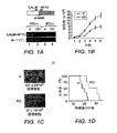
DOT1L/CALM−AF10と相互作用することができるHoxA5遺伝子の典型的領域を図5Aに示す。 A typical region of the HoxA5 gene that can interact with DOT1L / CALM-AF10 is shown in FIG. 5A.
CALMポリペプチド、AF10ポリペプチド、DOT1LポリペプチドまたはCALM−AF10融合タンパク質とHoxA5遺伝子またはその一部の間の相互作用は、任意の適切な方法によって、例えば、限定するわけではないが、HoxA5遺伝子またはその一部へのCALMポリペプチド、AF10ポリペプチド、DOT1LポリペプチドまたはCALM−AF10融合タンパク質の結合を決定すること、および/またはHoxA5遺伝子またはその一部(例えばHoxA5プロモーターの全部または機能的部分)のH3−K79メチル化を決定すること、および/または(例えばプロモーターがネイティブ配列、HoxA5コード配列を表すcDNAまたはレポーター遺伝子などの異種配列と作動可能に関連している場合に)HoxA5プロモーター活性を決定することなどによって、評価することができる。 The interaction between the CALM polypeptide, AF10 polypeptide, DOT1L polypeptide or CALM-AF10 fusion protein and the HoxA5 gene or part thereof may be performed by any suitable method, for example, but not limited to, the HoxA5 gene or Determining binding of a CALM polypeptide, AF10 polypeptide, DOT1L polypeptide or CALM-AF10 fusion protein to a portion thereof, and / or of the HoxA5 gene or a portion thereof (eg, all or a functional portion of the HoxA5 promoter) Determining H3-K79 methylation, and / or Ho (eg, when the promoter is operably associated with a heterologous sequence such as a native sequence, a cDNA representing the HoxA5 coding sequence or a reporter gene) Such as by determining the A5 promoter activity, it can be evaluated.
代表的実施形態では、本方法は細胞に基づく系で行われ、この場合、細胞は、レポーター分子(例えば酵素などのポリペプチド)をコードする核酸と作動可能に関連するHoxA5プロモーターを含む組換え核酸を含む。細胞は、CALM−AF10融合タンパク質を含む白血病細胞であることができ、あるいは、CALM−AF10融合タンパク質を産生するように改変された細胞であることができる。その細胞に一つ以上の候補化合物を加え、HoxA5プロモーター活性のレベルによって示されるHoxA5へのCALM−AF10の結合を、レポーター分子の発現(例えば酵素活性)を決定することによって評価することができる。 In an exemplary embodiment, the method is performed in a cell based system, wherein the cell is a recombinant nucleic acid comprising a HoxA5 promoter operably associated with a nucleic acid encoding a reporter molecule (eg, a polypeptide such as an enzyme). including. The cell can be a leukemia cell comprising a CALM-AF10 fusion protein or can be a cell that has been modified to produce a CALM-AF10 fusion protein. The binding of CALM-AF10 to HoxA5 as indicated by the level of HoxA5 promoter activity can be evaluated by adding reporter molecule expression (eg, enzyme activity) by adding one or more candidate compounds to the cell.
本発明は、白血病の予防および/または治療のための候補化合物を識別する方法であって、DOT1LポリペプチドとCALM−AF10融合タンパク質が結合して複合体を形成し、その複合体がHoxA5プロモーターに結合するのに十分な条件下で、試験化合物の存在下で、(上述した)HoxA5遺伝子の全部または一部を含む核酸をDOT1LポリペプチドおよびCALM−AF10融合タンパク質と接触させるステップと、DOT1L/CALM−AF10融合タンパク質複合体のHoxA5プロモーターとの相互作用のレベルを検出するステップとを含み、試験化合物の非存在下での相互作用のレベルと比較した、試験化合物存在下での、DOT1L/CALM−AF10融合タンパク質複合体とHoxA5プロモーターの相互作用の減少が、前記試験化合物が白血病の予防および/または治療のための候補化合物であることを示す方法も提供する。 The present invention is a method for identifying candidate compounds for the prevention and / or treatment of leukemia, wherein a DOT1L polypeptide and a CALM-AF10 fusion protein bind to form a complex, and the complex is linked to the HoxA5 promoter. Contacting a nucleic acid comprising all or part of the HoxA5 gene (described above) with a DOT1L polypeptide and a CALM-AF10 fusion protein in the presence of a test compound under conditions sufficient to bind; and DOT1L / CALM Detecting the level of interaction of the AF10 fusion protein complex with the HoxA5 promoter and comparing it to the level of interaction in the absence of the test compound in the presence of the test compound DOT1L / CALM- Mutation between AF10 fusion protein complex and HoxA5 promoter Reduction in use, provides a method of indicating that said test compound is a candidate compound for the prevention and / or treatment of leukemia.
前記の方法では、DOT1LおよびCALM−AF10を、DOT1L−CALM−AF10三元融合タンパク質で置き換えることができる。 In the above method, DOT1L and CALM-AF10 can be replaced with DOT1L-CALM-AF10 ternary fusion protein.
DOT1L/CALM−AF10複合体とHoxA5遺伝子またはその一部の間の相互作用は、任意の適切な方法によって、例えば、限定するわけではないが、HoxA5遺伝子またはその一部への複合体の結合を決定すること、またはHoxA5遺伝子またはその一部(例えばHoxA5プロモーター)のH3−K79メチル化を決定すること、または(例えばプロモーターがネイティブ配列、HoxA5コード配列を表すcDNAまたはレポーター遺伝子などの異種配列と作動可能に関連している場合に)HoxA5プロモーター活性を決定することなどによって、評価することができる。 The interaction between the DOT1L / CALM-AF10 complex and the HoxA5 gene or part thereof can be determined by any suitable method, for example, but not limited to binding of the complex to the HoxA5 gene or part thereof. Determining or determining H3-K79 methylation of the HoxA5 gene or part thereof (eg the HoxA5 promoter), or working with heterologous sequences such as cDNA or reporter genes where the promoter represents the native sequence, HoxA5 coding sequence (eg This can be assessed, for example, by determining HoxA5 promoter activity (where relevant).
HoxA5が一定の白血病において過剰発現されることは以前に観察されているが、がんでは多くの遺伝子が調節不全になるので、HoxA5アップレギュレーションの意義はわからなかった。本発明者らは、HoxA5アップレギュレーションが白血病誘発の原因因子であることを発見した。したがって本発明は、白血病の予防および/または治療のための候補化合物を識別する方法であって、HoxA5プロモーター活性に結合しかつ/またはHoxA5プロモーター活性を(例えばHoxA5遺伝子またはレポーター遺伝子などの導入遺伝子と作動可能に関連するHoxA5プロモーターを含むキメラコンストラクトの発現をダウンレギュレートすることによって)調整する化合物を識別することを含む方法も提供する。特定の実施形態において、本方法は、HoxA5プロモーター領域および/またはHoxA5遺伝子の他の任意の領域(例えばエクソン1、エクソン2、またはエクソン1とエクソン2の間のイントロン)の全部または一部を含む核酸を、試験化合物と、試験化合物が核酸中に存在するHoxA5プロモーター領域および/またはHoxA5遺伝子の他の任意の領域の全部または機能的部分に結合するのに十分な条件下で接触させるステップと、試験化合物と、核酸中に存在するHoxA5プロモーターおよび/またはHoxA5遺伝子の他の任意の領域の全部または機能的部分の間の結合のレベルを検出するステップとを含み、結合は、前記試験化合物が白血病の予防および/または治療のための候補化合物であることを示す。
It has been observed previously that HoxA5 is overexpressed in certain leukemias, but since many genes are dysregulated in cancer, the significance of HoxA5 upregulation was unknown. The present inventors have discovered that HoxA5 upregulation is a causative factor of leukemia induction. Accordingly, the present invention is a method for identifying candidate compounds for the prevention and / or treatment of leukemia, which binds to and / or binds a HoxA5 promoter activity (eg, a transgene such as a HoxA5 gene or a reporter gene). Also provided is a method comprising identifying a compound that modulates (by down-regulating expression of a chimeric construct comprising an operably associated HoxA5 promoter). In certain embodiments, the method comprises all or part of the HoxA5 promoter region and / or any other region of the HoxA5 gene (eg,
白血病の予防および/または治療のための候補化合物を識別する他の代表的方法は、HoxA5プロモーター領域の全部または機能的部分を含む核酸を、HoxA5プロモーター活性にとって十分な条件下で、試験化合物と接触させること、およびHoxA5プロモーター活性のレベルを検出するステップとを含み、試験化合物の非存在下でのHoxA5プロモーター活性のレベルと比較した、試験化合物存在下でのHoxA5プロモーター活性の減少が、前記試験化合物が白血病の予防および/または治療のための候補化合物であることを示す。代表的化合物では、試験化合物が、核酸中に存在するHoxA5プロモーターおよび/またはHoxA5遺伝子の他の任意の領域(例えばエクソン1、エクソン2および/またはエクソン1とエクソン2の間のイントロン)の全部または機能的部分に結合する。HoxA5プロモーター活性は本明細書で述べるように決定することができる。
Another exemplary method for identifying candidate compounds for the prevention and / or treatment of leukemia is to contact a nucleic acid comprising all or a functional portion of the HoxA5 promoter region with a test compound under conditions sufficient for HoxA5 promoter activity. And reducing the level of HoxA5 promoter activity in the presence of the test compound as compared to the level of HoxA5 promoter activity in the absence of the test compound, the step of detecting the level of HoxA5 promoter activity Is a candidate compound for the prevention and / or treatment of leukemia. For representative compounds, the test compound is all or part of the HoxA5 promoter and / or any other region of the HoxA5 gene (eg,
さらに本発明は、DOT1ポリペプチドに結合し、かつ/またはDOT1ポリペプチドの生物学的活性および/またはDOT1LとAF10の相互作用を調整する化合物を識別することによって、T−ALLまたはAMLサブタイプM0/1の治療および/または予防用の化合物の識別する方法を提供する。 Furthermore, the present invention relates to T-ALL or AML subtype M0 by identifying compounds that bind to and / or modulate the biological activity of DOT1 polypeptide and / or the interaction of DOT1L and AF10. A method of identifying compounds for therapeutic and / or prophylactic treatment is provided.
代表的実施形態では、本発明は、T−ALLまたはAMLサブタイプM0/1の予防および/または治療のための候補化合物を識別する方法であって、DOT1Lポリペプチドを、試験化合物の存在下で、ヒストンH3を含むヒストンまたはヌクレオソーム基質と接触させるステップと、H3−K79メチル化をもたらすのに十分な条件下で、基質のH3−K79メチル化のレベルを検出するステップとを含み、試験化合物の非存在下でのH3−K79メチル化のレベルと比較した、試験化合物存在下でのH3−K79メチル化の阻害は、前記試験化合物がT−ALLまたはAMLサブタイプM0/1の予防および/または治療のための候補化合物であることを示す方法を提供する。 In an exemplary embodiment, the invention provides a method for identifying a candidate compound for prevention and / or treatment of T-ALL or AML subtype M0 / 1, wherein the DOT1L polypeptide is isolated in the presence of a test compound. Contacting with a histone or nucleosomal substrate comprising histone H3 and detecting the level of H3-K79 methylation of the substrate under conditions sufficient to effect H3-K79 methylation comprising: Inhibition of H3-K79 methylation in the presence of a test compound, compared to the level of H3-K79 methylation in the absence, indicates that said test compound prevents T-ALL or AML subtype M0 / 1 and / or A method of indicating a candidate compound for treatment is provided.
さらに本発明は、T−ALLまたはAMLサブタイプM0/1の予防および/または治療のための候補化合物を識別する方法であって、DOT1LポリペプチドをAF10と接触させるステップと、DOT1LポリペプチドとAF10の間の結合のレベルを、それらの間の結合にとって十分な条件下で検出するステップとを含み、試験化合物の非存在下での結合のレベルと比較した、試験化合物存在下でのDOT1LポリペプチドとAF10の間の結合の減少は、前記試験化合物がT−ALLまたはAMLサブタイプM0/1の予防および/または治療のための候補化合物であることを示す方法を提供する。 The present invention further relates to a method of identifying candidate compounds for the prevention and / or treatment of T-ALL or AML subtype M0 / 1, comprising contacting a DOT1L polypeptide with AF10, the DOT1L polypeptide and AF10 Detecting the level of binding between the DOT1L polypeptide in the presence of the test compound compared to the level of binding in the absence of the test compound, comprising detecting under conditions sufficient for binding between them Decrease in the binding between AF10 and AF10 provides a way to indicate that the test compound is a candidate compound for the prevention and / or treatment of T-ALL or AML subtype M0 / 1.
ポリペプチドおよび核酸への結合を評価する方法は、当業者には明白であるだろう標準的技法を使って行うことができる(典型的方法については実施例を参照されたい)。そのような方法として、酵母および哺乳動物細胞ツーハイブリッドアッセイならびに共免疫沈降技法が挙げられる。 Methods for assessing binding to polypeptides and nucleic acids can be performed using standard techniques that will be apparent to those skilled in the art (see the Examples for exemplary methods). Such methods include yeast and mammalian cell two-hybrid assays and co-immunoprecipitation techniques.
H3−K79 HMTaseおよび白血病誘発活性などの他の活性も、当分野で知られる標準的方法を使って、例えば下記実施例で述べるように評価することができる。 Other activities such as H3-K79 HMTase and leukemia-inducing activity can also be assessed using standard methods known in the art, for example as described in the examples below.
HoxA5プロモーター活性も、当分野で知られる任意の方法によって、例えばHoxA5プロモーターと作動可能に関連するレポーター遺伝子の発現または活性を検出することによって、HoxA5転写物を(例えばRT−PCRで)検出することによって、かつ/またはHoxA5タンパク質産物を検出することによって、評価することができる。 HoxA5 promoter activity can also be detected (eg, by RT-PCR) by any method known in the art, for example, by detecting the expression or activity of a reporter gene operably associated with the HoxA5 promoter. And / or by detecting the HoxA5 protein product.
本発明のスクリーニング方法は、細胞に基づく系または無細胞系で行うことができる。さらにもう一つの選択肢として、動物全体(トランスジェニック非ヒト動物を含む)でアッセイを行うこともできる。さらにまた、細胞に基づく系に関して、ポリペプチドは細胞に直接添加するか、細胞中の核酸から産生させることができる。核酸は、その細胞にとって内在性であるか、外来(例えば遺伝子改変細胞)であることができる。 The screening method of the present invention can be performed in a cell-based system or a cell-free system. As yet another option, the assay can be performed on whole animals (including transgenic non-human animals). Furthermore, for cell-based systems, the polypeptide can be added directly to the cell or produced from nucleic acid in the cell. The nucleic acid can be endogenous to the cell or foreign (eg, a genetically modified cell).
任意の興味ある化合物を本発明に従ってスクリーニングすることができる。適切な試験化合物として、小有機化合物(すなわち非オリゴマー)、オリゴマーまたはその組み合わせ、および無機分子が挙げられる。適切な有機分子として、ポリペプチド(酵素、抗体およびFab’フラグメントを含む)、糖質、脂質、補酵素、および核酸分子(DNA、RNAおよびキメラならびにその類似体を含む)ならびにヌクレオチドおよびヌクレオチド類似体を挙げることができるが、これらに限定されるわけではない。特定の実施形態では、化合物が、ターゲットポリペプチドの産生を阻害するアンチセンス核酸、siRNAまたはリボザイムである。 Any compound of interest can be screened according to the present invention. Suitable test compounds include small organic compounds (ie, non-oligomers), oligomers or combinations thereof, and inorganic molecules. Suitable organic molecules include polypeptides (including enzymes, antibodies and Fab ′ fragments), carbohydrates, lipids, coenzymes, and nucleic acid molecules (including DNA, RNA and chimeras and analogs thereof) and nucleotides and nucleotide analogs It can be mentioned, but is not limited to these. In certain embodiments, the compound is an antisense nucleic acid, siRNA or ribozyme that inhibits production of the target polypeptide.
小有機化合物(または「非オリゴマー」)には、多種多様な有機分子、例えばステロイド、抗生物質、酵素阻害剤、リガンド、ホルモン、薬物、アルカロイド、オピオイド、テルペン、ポルフィリン、毒素、触媒ならびにそれらの組み合わせを含む複素環式化合物、芳香族化合物、脂環式化合物、脂肪族化合物およびそれらの組み合わせなどが含まれる。 Small organic compounds (or “non-oligomers”) include a wide variety of organic molecules such as steroids, antibiotics, enzyme inhibitors, ligands, hormones, drugs, alkaloids, opioids, terpenes, porphyrins, toxins, catalysts and combinations thereof Heterocyclic compounds, aromatic compounds, alicyclic compounds, aliphatic compounds, combinations thereof, and the like.
オリゴマーとしては、オリゴペプチド、オリゴヌクレオチド、オリゴ糖、ポリ脂質、ポリエステル、ポリアミド、ポリウレタン、ポリウレア、ポリエーテル、およびポリ(リン誘導体)、例えばホスフェート、ホスホネート、ホスホルアミド、ホスホンアミド、ホスファイト、ホスフィンアミドなど、ポリ(硫黄誘導体)、例えばスルホン、スルホネート、サルファイト、スルホンアミド、スルフェンアミドなどが挙げられ、リン誘導体および硫黄誘導体の場合、表示したヘテロ原子は、大部分が、C、H、N、OまたはSおよびそれらの組み合わせに結合されるだろう。そのようなオリゴマーは、既知の技法に従って、コンビナトリアルライブラリーから取得してもよい。 As oligomers, oligopeptides, oligonucleotides, oligosaccharides, polylipids, polyesters, polyamides, polyurethanes, polyureas, polyethers, and poly (phosphorus derivatives) such as phosphates, phosphonates, phosphoramides, phosphonamides, phosphites, phosphine amides, etc. , Poly (sulfur derivatives) such as sulfones, sulfonates, sulfites, sulfonamides, sulfenamides, etc. In the case of phosphorus and sulfur derivatives, the heteroatoms indicated are mostly C, H, N Will be bound to O or S and combinations thereof. Such oligomers may be obtained from combinatorial libraries according to known techniques.
さらにまた、例えばコンビナトリアルケミカル化合物ライブラリー(例えば米国特許第5,288,514号に記載のベンゾジアゼピンライブラリー;米国特許第5,420,328号に記載のホスホネートエステルライブラリー、米国特許第5,525,735号および同第5,525,734号に記載のピロリジンライブラリー、ならびに米国特許第5,817,751号に記載のジケトピペラジンおよびジケトモルホリンライブラリー)、ポリペプチドライブラリー、cDNAライブラリー、アンチセンス核酸ライブラリーなどの化合物ライブラリー、またはポリペプチドおよび核酸アレイなどのアレイ化された化合物の収集物をスクリーニングするために、本発明の方法を実施することができる。 Furthermore, for example, combinatorial chemical compound libraries (for example, benzodiazepine libraries described in US Pat. No. 5,288,514; phosphonate ester libraries described in US Pat. No. 5,420,328, US Pat. No. 5,525). , 735 and 5,525,734, and diketopiperazine and diketomorpholine libraries described in US Pat. No. 5,817,751), polypeptide libraries, cDNA libraries The methods of the invention can be practiced to screen compound libraries, such as antisense nucleic acid libraries, or collections of arrayed compounds, such as polypeptide and nucleic acid arrays.
本発明は上述のスクリーニング方法によって識別される化合物も包含する。 The present invention also encompasses compounds identified by the screening methods described above.
さらにまた本発明は、HoxA5、CALM、AF10、CALM−AF10および/またはDOT1Lに対する治療有効量の阻害核酸(例えばsiRNAなどのRNAi)または抗体(例えばモノクローナル抗体)を投与することによって、白血病(例えばT−ALLまたはAMLサブタイプM0/1)を患っている対象または白血病の危険がある対象を治療する方法を提供する。 Furthermore, the present invention provides leukemia (eg, T antibodies) by administering therapeutically effective amounts of inhibitory nucleic acids (eg, RNAi such as siRNA) or antibodies (eg, monoclonal antibodies) against HoxA5, CALM, AF10, CALM-AF10 and / or DOT1L. -Provide a method of treating a subject suffering from ALL or AML subtype M0 / 1) or at risk for leukemia.
本発明の実施形態によれば、抗体は、抗体フラグメントを含むモノクローナル抗体またはポリクローナル抗体であることができる。抗体および抗体フラグメントは特定の形態に限定されず、二重特異性、ヒト化、またはキメラ化抗体または抗体フラグメントであることができ、さらにFabフラグメント、単鎖抗体などであることもできる。 According to an embodiment of the invention, the antibody can be a monoclonal or polyclonal antibody comprising an antibody fragment. Antibodies and antibody fragments are not limited to a particular form and can be bispecific, humanized, or chimeric antibodies or antibody fragments, and can also be Fab fragments, single chain antibodies, and the like.
本発明の実施に適した対象として、鳥類および哺乳類対象が挙げられ、哺乳類対象にはヒト、非ヒト霊長類、マウス、ラット、ヒツジ、ブタ、ウシ、ヤギ、ウサギ、ウマ、イヌ、ネコなどが含まれるが、これらに限るわけではない。 Suitable subjects for the practice of the present invention include avian and mammalian subjects, including humans, non-human primates, mice, rats, sheep, pigs, cows, goats, rabbits, horses, dogs, cats and the like. Including, but not limited to.
さらにもう一つの態様として、本発明は、HoxA5のH3−K79ヒストンメチル化を評価することによる、対象が白血病を有するかどうかもしくは白血病の危険があるかどうかを評価するための診断方法、および/または対象における白血病の今後の経過を予測するための予後判定方法であって、正常(例えば非白血病)対象と比較した、HoxA5のH3−K79メチル化の増加は、白血病の診断指標となり、かつ/または疾患の経過の予後指標となる。場合により、HoxA5遺伝子のH3−K79メチル化のレベルと、場合によりH3−K4メチル化のレベルが決定され(例えばHoxA5プロモーター、エクソン1、エクソン2および/またはエクソン1とエクソン2の間のイントロン)、正常(例えば非白血病)対象と比較したH3−K79メチル化の増加、および場合により、H3−K4メチル化の減少は、白血病の診断指標となり、かつ/または疾患の経過の予後指標となる。
In yet another aspect, the present invention relates to a diagnostic method for assessing whether a subject has or is at risk for leukemia by assessing H3-K79 histone methylation of HoxA5, and / or Or a prognostic method for predicting the future course of leukemia in a subject, wherein an increase in H3-K79 methylation of HoxA5 relative to a normal (eg, non-leukemic) subject is a diagnostic indicator of leukemia and / or Or it is a prognostic indicator of the course of disease. Optionally, the level of H3-K79 methylation of the HoxA5 gene and optionally the level of H3-K4 methylation is determined (eg, the HoxA5 promoter,
さらに本発明は、対象が白血病を有するかどうかもしくは白血病を発症する危険があるかどうかを診断し、かつ/またはその疾患の経過に関する予後を決定する方法であって、対象におけるHoxA5プロモーター活性を決定するステップを含み、対照(例えば健常)対象におけるHoxA5プロモーター活性と比較した、HoxA5プロモーター活性のアップレギュレーション(すなわち増加した活性)は、その対象が白血病を有することまたは白血病を発症する危険があることを示す方法を提供する。HoxA5プロモーター活性は、当分野で知られる任意の方法によって決定することができる。例えば、本方法は、対象から生物学的試料を取得するステップと、および生物学的試料中のHoxA5プロモーター活性(例えば、ネイティブであるか異種であるかを問わず、HoxA5プロモーターと作動可能に関連する核酸の増加した発現)を決定するステップを、さらに含むことができる。 The present invention further relates to a method of diagnosing whether a subject has leukemia or is at risk of developing leukemia and / or determining a prognosis for the course of the disease, which determines HoxA5 promoter activity in the subject. Up-regulation (ie, increased activity) of HoxA5 promoter activity relative to HoxA5 promoter activity in a control (eg, healthy) subject indicates that the subject has leukemia or is at risk of developing leukemia Provide a way to show. HoxA5 promoter activity can be determined by any method known in the art. For example, the method can include obtaining a biological sample from a subject and HoxA5 promoter activity in the biological sample (eg, native or heterologous, operably associated with the HoxA5 promoter). Determining the increased expression of the nucleic acid to be further included.
対象が白血病を有するかどうかもしくは白血病を発症する危険があるかどうかを診断し、かつ/またはその疾患の経過に関する予後を決定する方法の一例は、対象からHoxA5遺伝子を含む生物学的試料を取得するステップと、生物学的試料中のHoxA5遺伝子のH3−K79メチル化のレベルを検出するステップとを含み、非白血病生物学的試料におけるH3−K79メチル化のレベルと比較した、生物学的試料におけるHoxA5遺伝子のH3−K79メチル化の上昇は、その対象が白血病を有することもしくは白血病を発症する危険があることの診断指標となり、かつ/またはその対象における疾患の経過の予後指標となる。本方法は、場合により、HoxA5遺伝子のH3−K4メチル化のレベルを決定するステップをさらに含むことができ、非白血病試料と比較した、生物学的試料におけるHoxA5遺伝子のH3−K4メチル化の減少は、その対象が白血病を有することもしくは白血病を発症する危険があることの診断指標となり、かつ/またはその対象における疾患の経過の予後指標となる。 An example of a method of diagnosing whether a subject has or is at risk of developing leukemia and / or determining a prognosis for the course of the disease is obtaining a biological sample containing the HoxA5 gene from the subject And detecting the level of H3-K79 methylation of the HoxA5 gene in the biological sample and comparing to the level of H3-K79 methylation in the non-leukemic biological sample Increased H3-K79 methylation of the HoxA5 gene in can be a diagnostic indicator that the subject has leukemia or is at risk of developing leukemia and / or a prognostic indicator of the course of disease in the subject. The method can optionally further comprise the step of determining the level of H3-K4 methylation of the HoxA5 gene, and reducing the H3-K4 methylation of the HoxA5 gene in a biological sample as compared to a non-leukemic sample. Is a diagnostic indicator that the subject has leukemia or is at risk of developing leukemia and / or a prognostic indicator of the course of disease in the subject.
代表的実施形態では、本発明の前記診断方法が、T−ALLもしくはAMLサブタイプM0/1に関する診断および/または予後判定方法として実施される。 In an exemplary embodiment, the diagnostic method of the present invention is implemented as a diagnostic and / or prognostic method for T-ALL or AML subtype M0 / 1.
対象がT−ALLもしくはAML−M0/1を持つかどうかまたはT−ALLもしくはAML−M0/1を発症する危険があるかどうかを診断し、かつ/またはその疾患の経過に関する予後を決定する方法の一例は、対象からヒストン(例えばヌクレオソーム)を含む生物学的試料を取得するステップと、生物学的試料におけるH3−K79メチル化のレベルを検出するステップとを含み、非白血病生物学的試料におけるH3−K79メチル化のレベルと比較した、生物学的試料におけるH3−K79メチル化の増加は、その対象がT−ALLもしくはAML−M0/1を持つことまたはT−ALLもしくはAML−M0/1を発症する危険があることの診断指標となり、かつ/またはその対象における疾患の経過の予後指標となる。本方法は、場合により、生物学的試料におけるH3−K4メチル化のレベルを決定するステップをさらに含むことができ、非白血病試料と比較した、生物学的試料におけるH3−K4メチル化の減少は、その対象がT−ALLもしくはAML−M0/1を持つことまたはT−ALLもしくはAML−M0/1を発症する危険があることの診断指標となり、かつ/またはその対象における疾患の経過の予後指標となる。 Method for diagnosing whether a subject has T-ALL or AML-M0 / 1 or at risk of developing T-ALL or AML-M0 / 1 and / or determining a prognosis for the course of the disease One example includes obtaining a biological sample containing histones (eg, nucleosomes) from a subject and detecting the level of H3-K79 methylation in the biological sample, wherein the Increased H3-K79 methylation in a biological sample compared to the level of H3-K79 methylation indicates that the subject has T-ALL or AML-M0 / 1 or T-ALL or AML-M0 / 1. As a diagnostic indicator of the risk of developing and / or as a prognostic indicator of the course of the disease in the subject. The method can optionally further comprise determining the level of H3-K4 methylation in the biological sample, wherein the decrease in H3-K4 methylation in the biological sample compared to the non-leukemic sample is A diagnostic indicator that the subject has T-ALL or AML-M0 / 1 or is at risk of developing T-ALL or AML-M0 / 1, and / or a prognostic indicator of the course of disease in the subject It becomes.
もう一つの代表的実施形態によれば、本発明は、対象がT−ALLもしくはAML−M0/1を持つかどうかまたはT−ALLもしくはAML−M0/1を発症する危険があるかどうかを診断し、かつ/またはその疾患の経過に関する予後を決定する方法であって、対象からヒストン(例えばヌクレオソーム)を含む生物学的試料を取得するステップと、生物学的試料におけるH3−K79メチル化のレベルを検出するステップとを含み、非白血病生物学的試料におけるレベルと比較した、生物学的試料におけるHoxA遺伝子関連H3−K79メチル化(例えばHoxA9遺伝子と関連するもの)の増加は、その対象がT−ALLもしくはAML−M0/1を持つことまたはT−ALLもしくはAML−M0/1を発症する危険があることの診断指標となり、かつ/またはその対象における疾患の経過の予後指標となる。本方法は、場合により、一つ以上のHoxA遺伝子(例えばHoxA9遺伝子)に関連するH3−K4メチル化のレベルを決定するステップをさらに含むことができ、非白血病試料と比較した、生物学的試料におけるHoxA遺伝子関連H3−K4メチル化の減少は、その対象がT−ALLもしくはAML−M0/1を持つことまたはT−ALLもしくはAML−M0/1を発症する危険があることの診断指標となり、かつ/またはその対象における疾患の経過の予後指標となる。 According to another exemplary embodiment, the present invention diagnoses whether a subject has T-ALL or AML-M0 / 1 or is at risk of developing T-ALL or AML-M0 / 1. And / or determining a prognosis for the course of the disease, obtaining a biological sample containing histones (eg, nucleosomes) from a subject, and the level of H3-K79 methylation in the biological sample Detecting an increase in HoxA gene-related H3-K79 methylation (eg, associated with HoxA9 gene) in a biological sample as compared to a level in a non-leukemic biological sample Having ALL or AML-M0 / 1 or being at risk of developing T-ALL or AML-M0 / 1 It is diagnostic, and / or a prognostic indicator of the course of the disease in the subject. The method can optionally further comprise determining the level of H3-K4 methylation associated with one or more HoxA genes (eg, the HoxA9 gene), wherein the biological sample is compared to a non-leukemic sample. Reduced HoxA gene-related H3-K4 methylation in can be a diagnostic indicator that the subject has T-ALL or AML-M0 / 1 or is at risk of developing T-ALL or AML-M0 / 1, And / or a prognostic indicator of the course of the disease in the subject.
本発明の診断および予後判定方法は、例えばヒト、非ヒト霊長類、ウシ、ヒツジ、ヤギ、ネコ、イヌ、ブタ、ウマ、ラット、マウス、ウサギまたはモルモット対象を含む(ただしこれらに限るわけではない)任意の哺乳動物対象で実施することができる。特定の実施形態では、対象が白血病を有するか、白血病を発症する危険があると考えられる。別の実施形態では、対象が白血病の動物モデルである。 The diagnostic and prognostic methods of the present invention include, but are not limited to, for example, humans, non-human primates, cows, sheep, goats, cats, dogs, pigs, horses, rats, mice, rabbits or guinea pig subjects. ) Can be performed on any mammalian subject. In certain embodiments, the subject may have leukemia or be at risk of developing leukemia. In another embodiment, the subject is an animal model of leukemia.
本発明の診断および予後判定方法は、確証的ではなく、最終的な診断または予後判定を下すには、他の診断および/または予後判定方法で補足する必要がありうることは、当業者には理解されるだろう。 It will be appreciated by those skilled in the art that the diagnostic and prognostic methods of the present invention are not confirmative and may need to be supplemented with other diagnostic and / or prognostic methods to make a final diagnosis or prognosis. Will be understood.
細胞または組織試料、臍帯試料、血液、血漿または血清試料、尿または糞便試料、粘液または喀痰試料など、任意の適切な生物学的試料を使用することができる。特定の実施形態では、生物学的試料がB細胞または骨髄試料である。他の代表的実施形態では、生物学的試料が、ヒストンH3を含むヒストンまたはヌクレオソーム調製物(例えばB細胞または骨髄細胞から得られるもの)である。代表的実施形態では、細胞または組織が対象から取り出され、培養され、その培養細胞または培養組織からヒストンまたはヌクレオソームが調製される。 Any suitable biological sample can be used, such as a cell or tissue sample, umbilical cord sample, blood, plasma or serum sample, urine or fecal sample, mucus or sputum sample. In certain embodiments, the biological sample is a B cell or bone marrow sample. In other exemplary embodiments, the biological sample is a histone or nucleosome preparation comprising histone H3 (eg, obtained from B cells or bone marrow cells). In an exemplary embodiment, cells or tissues are removed from a subject and cultured, and histones or nucleosomes are prepared from the cultured cells or tissues.
「非白血病生物学的試料」とは、正常な対象(すなわち白血病を持たず白血病を発症する危険もない対象)を示す適切な対照試料を意味する。例えばこの試料は、正常な対象から単離することができ、いくつかの例(例えばヌクレオソーム調製物)では、培養細胞から単離することができる。 By “non-leukemic biological sample” is meant an appropriate control sample that represents a normal subject (ie, a subject who does not have leukemia and is not at risk of developing leukemia). For example, the sample can be isolated from a normal subject, and in some instances (eg, a nucleosome preparation) can be isolated from cultured cells.
本発明を記述したので、以下の実施例では、これをさらに詳しく説明する。以下の実施例は、例示のみを目的として本明細書に記載するものであり、本発明を限定しようとするものではない。 Having described the invention, the following examples illustrate this in more detail. The following examples are described herein for purposes of illustration only and are not intended to limit the invention.
実験手法
U937細胞におけるCALM−AF10ノックダウンおよび移植.CALMとAF10の接合部をターゲットとする21bp−shRNA(5’−ATC AGG AGC ACA GAG CTG TGA−3’;配列番号3)をコードするDNAを、H1 RNAプロモーターと共に、pMSCV−puroにサブクローニングした。形質導入細胞をピューロマイシン(0.5μg/ml)によって選択し、限界希釈法によってクローニングした。ノックダウン効率をRT−PCRによって評価した。5〜10週齢のNOD/SCIDマウスに照射(300rad)した。6時間後に、CALM−AF10ノックダウン(KD)U937細胞およびベクター形質導入(V)U937細胞を、眼窩内注射した(1×107細胞/マウス、各群n=6)。マウスを監視し、組織学的検査およびFACS解析のために、末期に屠殺した。KD群のマウス数匹については、対照実験用に、末期に至る前に屠殺した。
Experimental procedure CALM-AF10 knockdown and transplantation in U937 cells. DNA encoding 21 bp-shRNA (5′-ATC AGG AGC ACA GAG CTG TGA-3 ′; SEQ ID NO: 3) targeting the junction of CALM and AF10 was subcloned into pMSCV-puro together with the H1 RNA promoter. Transduced cells were selected with puromycin (0.5 μg / ml) and cloned by limiting dilution. Knockdown efficiency was evaluated by RT-PCR. 5-10 week old NOD / SCID mice were irradiated (300 rad). Six hours later, CALM-AF10 knockdown (KD) U937 cells and vector-transduced (V) U937 cells were injected intraorbitally (1 × 10 7 cells / mouse, each group n = 6). Mice were monitored and sacrificed at the end for histological examination and FACS analysis. Several mice in the KD group were sacrificed for the control experiment before reaching the end stage.
マウス骨髄細胞の単離および形質導入.4〜10週齢のC57BL/6マウスを使って骨髄細胞を収集した。Hoxa5欠損骨髄細胞を8〜10週齢のMF1マウスから得た(Jeannotte et al.,(1993)Genes Dev 7,2085−96)。マウスを150mg/kgの5−フルオロウラシル(5FU)で5日間処理してから、骨髄細胞を収集した。EasySep(登録商標)マウス造血前駆細胞濃縮キット(Stem cell technology)を使ってLin−細胞を濃縮し、レトロウイルス形質導入に使用した。CALM−AF10のcDNAをRT−PCRによってU937細胞からクローニングした。野生型およびOM−LZ欠失突然変異型のCALM−AF10を、pMSCV−neoのFlagタグの下流にサブクローニングした。レトロウイルス調製、形質導入、およびコロニーアッセイは、以前記述したように行った(Okada et al.,(2005)Cell 121:167−78)。図3bに記載する実験では、非形質導入細胞の成長を防ぐために、第3ラウンドまで、メチルセルロースにG418(1mg/ml)を加えた。メチルセルロース上のコロニーを拾い上げ、5ng/mlのmIL−3(Peprotech)を含むmFTOC(RPMI 1640中の10%FBS、1mM MEMピルビン酸ナトリウム、1%MEM非必須アミノ酸、10mM HEPES、pH.7.3、5×10⁵M 2−メルカプトエタノール)中でさらに培養した。ブラストサイジン耐性レトロウイルスによるDOT1Lの形質導入については以前記述した(Okada et al.,(2005)Cell 121:167−78)。
Isolation and transduction of mouse bone marrow cells. Bone marrow cells were collected using 4-10 week old C57BL / 6 mice. Hoxa5-deficient bone marrow cells were obtained from MF1 mice aged 8-10 weeks (Jeannotte et al., (1993)
細胞培養、トランスフェクション、免疫染色、および免疫沈降.U937細胞は、10%ウシ胎仔血清を補足したRPMI 1640で維持した。293T細胞またはU2OS細胞は、10%ウシ胎仔血清を補足したダルベッコ変法イーグル培地(DMEM)で維持した。トランスフェクションはFugene6(Roche)を使って行った。トランスフェクションの24時間後に、U2OS細胞を3%パラホルムアルデヒドで固定し、0.1%Triton X−100で5分間透過処理した。Flag M2抗体(Sigma)およびhDOT1L抗体(Okada et al.,(2005)Cell 121:167−78)を一次抗体として免疫染色に使用した。DAPIを核染色に使用した。 Cell culture, transfection, immunostaining, and immunoprecipitation. U937 cells were maintained in RPMI 1640 supplemented with 10% fetal calf serum. 293T cells or U2OS cells were maintained in Dulbecco's Modified Eagle Medium (DMEM) supplemented with 10% fetal calf serum. Transfection was performed using Fugene 6 (Roche). Twenty-four hours after transfection, U2OS cells were fixed with 3% paraformaldehyde and permeabilized with 0.1% Triton X-100 for 5 minutes. Flag M2 antibody (Sigma) and hDOT1L antibody (Okada et al., (2005) Cell 121: 167-78) were used as primary antibodies for immunostaining. DAPI was used for nuclear staining.
免疫沈降のために、トランスフェクションの36時間後に293T細胞を収集し、氷冷リン酸緩衝食塩水(PBS)で洗浄してから、溶解緩衝液(50mM Hepes−KOH[pH7.9]、420mM KCl、0.1mM EDTA[pH8.0]、5mM MgCl2、20%グリセロール、1mM DTT、1mMフェニルメチルスルホニルフルオリド、1μg/mlのアプロチニン、0.5μg/mlのロイペプチン、0.7μg/mlのペプスタチン)で溶解した。4℃で1時間インキュベートした後、細胞を穏やかに超音波処理し、14,000rpmで30分間の遠心分離により、細胞片を除去した。抗Flag M2アガロースビーズ(Sigma)を0.5mgタンパク質抽出物に加え、4℃で3時間インキュベートした。500mM KClを含有する溶解緩衝液で5回洗浄した後、免疫沈降物をウェスタンブロット法で解析した。 For immunoprecipitation, 293T cells were collected 36 hours after transfection and washed with ice-cold phosphate buffered saline (PBS) before lysis buffer (50 mM Hepes-KOH [pH 7.9], 420 mM KCl). 0.1 mM EDTA [pH 8.0], 5 mM MgCl2, 20% glycerol, 1 mM DTT, 1 mM phenylmethylsulfonyl fluoride, 1 μg / ml aprotinin, 0.5 μg / ml leupeptin, 0.7 μg / ml pepstatin) And dissolved. After 1 hour incubation at 4 ° C., the cells were gently sonicated and cell debris was removed by centrifugation at 14,000 rpm for 30 minutes. Anti-Flag M2 agarose beads (Sigma) were added to 0.5 mg protein extract and incubated at 4 ° C. for 3 hours. After washing 5 times with lysis buffer containing 500 mM KCl, immunoprecipitates were analyzed by Western blotting.
FACS解析.NOD/SCIDマウスから収集した細胞、およびCALM−AF10で形質導入したマウス骨髄細胞を、CD16/CD32 Fc block(BD Pharmingen)と共に氷上で20分間インキュベートしてから、フィコエリトリン接合アイソタイプ対照、フルオレセインイソチオシアネート接合アイソタイプ対照、およびアロフィコシアニン接合アイソタイプ対照、ならびにヒトCD45、マウスCD11b、Gr−1、c−kit、B220、CD3、およびTer119に対するモノクローナル抗体(BD Pharmingen)により、氷上で30分間染色した。次に細胞を2%FBS含有PBSで洗浄し、FACSCalibur(BD Pharmingen)を使って解析した。収集したデータをSummit V3.1(Cytomation Inc. Fort Collins)で解析した。 FACS analysis. Cells collected from NOD / SCID mice and mouse bone marrow cells transduced with CALM-AF10 were incubated with CD16 / CD32 Fc block (BD Pharmingen) for 20 minutes on ice before phycoerythrin-conjugated isotype control, fluorescein isothiocyanate-conjugated Isotype controls and allophycocyanin-conjugated isotype controls and monoclonal antibodies against human CD45, mouse CD11b, Gr-1, c-kit, B220, CD3, and Ter119 (BD Pharmingen) were stained on ice for 30 minutes. The cells were then washed with 2% FBS-containing PBS and analyzed using FACSCalibur (BD Pharmingen). The collected data was analyzed with Summit V3.1 (Cytomation Inc. Fort Collins).
Hox遺伝子、Bmi−1およびCALM−AF10のRT−PCR解析.全RNAを、ベクター形質導入U937細胞もしくはCALM−AF10ノックダウンU937細胞、または野生型もしくは突然変異型CALM−AF10形質導入細胞の第2および第3ラウンドに由来するマウス骨髄細胞のコロニーから、RNeasy(Qiagen)を使って単離した。最大1μgの全RNAをRNaseフリーDNase Iで処理し、製造者のプロトコールに従って、ImProm−II(Promega)を用いる逆転写にかけた。20μlのRT反応液のうち0.5μlのcDNAを、Platinum Taqポリメラーゼ(Invitrogen)を用いる33〜35サイクルのPCR増幅に、テンプレートとして使用した。そのcDNAの10分の1をβ−アクチンおよびGAPDH増幅に使用した。HoxaおよびBmi−1用のプライマー配列は請求すれば入手できる。CALM−AF10遺伝子を検出するためのプライマー配列は以下のとおりとした:CALM−AF10−F:5’−TATACAGCCAGCCTGTCATG−3’(配列番号4)、CALM−AF10−R:5’−AGTGGCTGCTTTGCTTTCTC−3’(配列番号5)。 RT-PCR analysis of Hox gene, Bmi-1 and CALM-AF10. Total RNA was obtained from a colony of mouse bone marrow cells derived from the second and third rounds of vector-transduced U937 cells or CALM-AF10 knockdown U937 cells, or wild-type or mutant CALM-AF10-transduced cells. Qiagen). Up to 1 μg of total RNA was treated with RNase-free DNase I and subjected to reverse transcription using ImProm-II (Promega) according to the manufacturer's protocol. Of the 20 μl RT reaction, 0.5 μl cDNA was used as a template for 33-35 cycles of PCR amplification using Platinum Taq polymerase (Invitrogen). One tenth of the cDNA was used for β-actin and GAPDH amplification. Primer sequences for Hoxa and Bmi-1 are available upon request. The primer sequences for detecting the CALM-AF10 gene were as follows: CALM-AF10-F: 5′-TATACAGCCAGCCGTCATTG-3 ′ (SEQ ID NO: 4), CALM-AF10-R: 5′-AGTGCTGCTTTGCCTTCTC-3 ′ (SEQ ID NO: 5).
ChIPアッセイ.細胞調製、免疫沈降、およびPCRについては以前に記述した(Okada et al.,(2005)Cell 121:167−78)。抗体は以下のとおり入手した:抗3mK4(Abcam)、抗2mK9(Upstate)、および抗2mK79(Feng et al.,(2002)Curr.Biol.12:1052−1058)。プライマー配列および詳細なPCR条件は請求すれば入手できる。 ChIP assay. Cell preparation, immunoprecipitation, and PCR have been previously described (Okada et al., (2005) Cell 121: 167-78). Antibodies were obtained as follows: anti-3mK4 (Abcam), anti-2mK9 (Upstate), and anti-2mK79 (Feng et al., (2002) Curr. Biol. 12: 1052-1058). Primer sequences and detailed PCR conditions are available upon request.
結果
CALM−AF10融合タンパク質は、白血病性形質転換を媒介するのに必要である。本発明者らは最近、ヒストンH3−K79メチルトランスフェラーゼであるhDOT1L(Feng et al.,(2002)Curr.Biol.12:1052−1058)がMLL−AF10による白血病誘発に重要な役割を果たすことを証明した(Okada et al.,(2005)Cell 121:167−78)。本発明者らは、MLL−AF10によるHoxa9遺伝子へのhDOT1LのミスターゲティングがH3−K79メチル化およびHoxa9アップレギュレーションをもたらし、それが白血病性形質転換の一因になることを示した(Okada et al.,(2005)Cell 121:167−78)。さらに本発明者らは、MLL−AF10による白血病性形質転換に必要なAF10のOM−LZモチーフが、hDOT1LとMLL−AF10の相互作用に関与することも証明した(DiMartino et al.,(2002)Blood 99,3780−5)。OM−LZ領域がCALM−AF10融合タンパク質に保たれているという事実から、hDOT1LはCALM−AF10による白血病にも重要な役割を果たしうる可能性が考えられる。CALM−AF10による白血病におけるhDOT1Lの潜在的役割を検討するために、本発明者らは、まず、CALM−AF10融合タンパク質と白血病の間の因果関係を確証しようと試みた。
Results The CALM-AF10 fusion protein is required to mediate leukemic transformation. We have recently shown that the histone H3-K79 methyltransferase, hDOT1L (Feng et al., (2002) Curr. Biol. 12: 1052-1058) plays an important role in leukemia induction by MLL-AF10. (Okada et al., (2005) Cell 121: 167-78). We have shown that mistargeting of hDOT1L to the Hoxa9 gene by MLL-AF10 results in H3-K79 methylation and Hoxa9 upregulation, which contributes to leukemic transformation (Okada et al. (2005) Cell 121: 167-78). Furthermore, the present inventors also demonstrated that the OM-LZ motif of AF10 necessary for leukemic transformation by MLL-AF10 is involved in the interaction between hDOT1L and MLL-AF10 (DiMartino et al., (2002)). Blood 99, 3780-5). The fact that the OM-LZ region is retained in the CALM-AF10 fusion protein suggests that hDOT1L may play an important role in CALM-AF10-induced leukemia. In order to examine the potential role of hDOT1L in leukemia with CALM-AF10, we first attempted to establish a causal relationship between CALM-AF10 fusion protein and leukemia.
以前の研究により、ヒト単球性白血病細胞株U937はCALM−AF10融合タンパク質を発現させることが確証されている(Dreyling et al.,(1996)Proc.Natl.Acad.Sci.USA 93:4804−9;Kobayashi et al.,(1995)Genes Chromosomes Cancer 13:217−8)。CALM−AF10が細胞の増殖および形質転換に関連するかどうかを決定するために、本発明者らは、ベクターに基づくRNAiアプローチを使って、U937細胞におけるCALM−AF10ノックダウンを行った(Okada et al.,(2005)Cell 121:167−78)。図1aに示す結果は、本発明者らが、有意なCALM−AF10ノックダウンを持つ安定なU937派生細胞株を作製できることを示している(レーン1とレーン2を比較)。ベクター形質導入対照(V)と比較して、ノックダウン(KD)細胞は、液体RPMI培地中でゆっくり増殖するだけでなく(図1b)、メチルセルロースで培養した場合に生じるコロニーは少なくかつ小さかった(図1c)。このデータは、CALM−AF10のノックダウンがインビトロで細胞の増殖と形質転換の両方に影響を及ぼすことを示唆している。
Previous studies have confirmed that the human monocytic leukemia cell line U937 expresses a CALM-AF10 fusion protein (Dreyling et al., (1996) Proc. Natl. Acad. Sci. USA 93: 4804- 9; Kobayashi et al., (1995) Genes Chromosomes Cancer 13: 217-8). To determine whether CALM-AF10 is associated with cell growth and transformation, we performed a CALM-AF10 knockdown in U937 cells using a vector-based RNAi approach (Okada et al. al., (2005) Cell 121: 167-78). The results shown in FIG. 1a indicate that we can generate a stable U937 derived cell line with significant CALM-AF10 knockdown (compare
インビボにおけるCALM−AF10の役割を評価するために、本発明者らは、1×10⁷個のノックダウン細胞および対照細胞を、NOD/SCIDマウスに移植した。図1dに示す結果は、CALM−AF10ノックダウンが移植マウスの生存時間を延ばしたことを証明している。ベクター形質導入対照細胞を移植したマウスの組織学的解析では、末期に複数の臓器で白血病細胞の浸潤が明らかになった(図1e、上図)。これに対し、移植後の同じ日に解析したところ、CALM−AF10ノックダウン細胞を移植したマウスの臓器では、白血病細胞をほとんど検出することができなかった(図1e、下図)。骨髄および脾臓から単離した細胞のFACS解析では、CALM−AF10ノックダウン細胞の移植が、たとえ同じ数の細胞を移植したとしても、対照と比較して、ヒト細胞のパーセンテージを有意に低下させることが明らかになった(骨髄では0.05%対11.14%、脾臓では0.10%対6.49%)(図1f)。総合すると上記の結果は、CALM−AF10融合タンパク質が、インビトロおよびインビボにおける白血病細胞の細胞増殖および形質転換の一因になることを裏付けている。 To assess the role of CALM-AF10 in vivo, we transplanted 1 × 10 7 knockdown cells and control cells into NOD / SCID mice. The results shown in FIG. 1d demonstrate that CALM-AF10 knockdown extended the survival time of the transplanted mice. Histological analysis of mice transplanted with vector-transduced control cells revealed leukemia cell infiltration in multiple organs at the end stage (FIG. 1e, upper panel). On the other hand, when analyzed on the same day after transplantation, leukemia cells were hardly detected in the organs of mice transplanted with CALM-AF10 knockdown cells (FIG. 1e, lower panel). In FACS analysis of cells isolated from bone marrow and spleen, transplantation of CALM-AF10 knockdown cells significantly reduced the percentage of human cells compared to controls even though the same number of cells were transplanted (0.05% vs 11.14% for bone marrow and 0.10% vs 6.49% for spleen) (Figure 1f). Taken together, the above results support that CALM-AF10 fusion protein contributes to cell proliferation and transformation of leukemic cells in vitro and in vivo.
CALM−AF10による白血病性形質転換は機能的hDOT1Lに依存する。本発明者らは、次に、CALM−AF10の発現が、骨髄細胞形質転換を引き起こすのに十分であるかどうかを解析した。この目的にはメチルセルロースコロニー連続プレーティングアッセイを使用した(図2a)。CALM−AF10による骨髄形質転換におけるhDOT1Lの役割を検討するために、本発明者らは、まず、hDOT1LはCALM−AF10と相互作用することができ、その相互作用がAF10のOM−LZ領域に依存することを確証した(図6)。次に本発明者らは、野生型またはhDOT1L相互作用領域OM−LZを欠く欠失CALM−AF10突然変異体を用いるマウス骨髄細胞の形質導入により、コロニー連続プレーティングアッセイを行った。RT−PCR解析により、第2ラウンドのコロニーに由来する細胞において、両融合タンパク質の発現が確認された(図2a、レーン1および2)。CALM−AF10を発現させる骨髄細胞において、第3ラウンドのプレーティングで第2ラウンドと比較してコロニー数が増加することはなかったが(図2c、中央のカラム)、それらの細胞は、mIL−3を補足したmFTOC(マウス胎仔胸腺臓器培養)培地で6ヶ月以上にわたって持続的に増殖することができた(図2dおよび非開示データ)。対照的に、CALM−AF10のOM−LZ欠失突然変異体を発現させる細胞は、メチルセルロースアッセイにおいて有意な数の第3ラウンドコロニーを生成させず、6週間の培養後にmFTOC培地で増殖し続けることもなかった(図2cおよび2d)。CALM−AF10で形質転換された細胞のFACS解析は、これらの細胞がc−Kitを発現させるが、他の系列特異的マーカー、例えばGr−1、CD11b、B−220、CD3、およびTER−119をいずれも発現させないことを示しており(図2e)、これは、これらの細胞がどの造血細胞系列にも分化拘束されていない初期前駆骨髄細胞であるらしいことを示す。これらの結果に基づいて、本発明者らは、CALM−AF10融合タンパク質の発現が、前駆骨髄細胞を形質転換するのに十分であり、hDOT1L相互作用領域がCALM−AF10の形質転換能には必要であると結論する。AF10のhDOT1L相互作用領域がCALM−AF10の形質転換能にとって必要であるという事実は、hDOT1LがCALM−AF10による白血病誘発に重要な役割を果たすことを、強く示唆している。hDOT1LによるH3−K79メチル化がCALM−AF10による増殖および白血病性形質転換に果たす役割をさらに証明するために、本発明者らは、野生型hDOT1LおよびそのH3−K79メチルトランスフェラーゼ活性が欠損した突然変異型hDOT1Lを、CALM−AF10形質転換骨髄細胞で発現させた。次に本発明者らは、それらがメチルセルロースでのコロニー形成に及ぼす影響を解析した。図2fに示す結果は、野生型hDOT1Lの発現がコロニー形成を強化したのに対して、突然変異型hDOT1Lの発現はCALM−AF10発現細胞のコロニー形成能を強く抑制したことを示している(図2f)。したがって、hDOT1Lおよびその関連H3−K79メチルトランスフェラーゼ活性は、CALM−AF10による白血病性形質転換に重要な役割を果たすと、本発明者らは結論づける。
Leukemic transformation with CALM-AF10 is dependent on functional hDOT1L. We next analyzed whether CALM-AF10 expression was sufficient to cause bone marrow cell transformation. A methylcellulose colony continuous plating assay was used for this purpose (Figure 2a). In order to investigate the role of hDOT1L in bone marrow transformation by CALM-AF10, we firstly allowed hDOT1L to interact with CALM-AF10, which interaction depends on the OM-LZ region of AF10. (Fig. 6). The inventors then performed a colony serial plating assay by transduction of mouse bone marrow cells with a deleted CALM-AF10 mutant lacking wild-type or hDOT1L interaction region OM-LZ. RT-PCR analysis confirmed the expression of both fusion proteins in cells derived from the second round of colonies (FIG. 2a,
CALM−AF10による白血病性形質転換はHoxa5アップレギュレーションを伴う。CALM−AF10による白血病性形質転換におけるhDOT1Lの役割を確証したので、本発明者らは、関連するターゲット遺伝子を識別しようと試みた。cDNAマイクロアレイ研究により、後期Hoxa遺伝子およびBmi−1の過剰発現は、CALM−AF10およびいくつかのMLL融合タンパク質が関与する白血病の特徴であることが明らかになっている(Dik et al.,(2005)Leukemia 19:1948−57;Drabkin et al.,(2002)Leukemia 16:186−95;Souler et al.,(2005)Blood 106:274−86)。特に、Hoxa9の過剰発現は、MLL−ENLおよびMLL−AF10融合タンパク質が関与する白血病にとって極めて重要であることが証明されている(Okada et al.,(2005)Cell 121:167−78)(Ayton et al.,(2004)Genes Dev 17:2298−307.24:617−28;Zeisig et al.,(2004)Mol.Cell.Biol.24:617−28)。hDOT1LおよびH3−K79メチル化がCALM−AF10による白血病誘発の一因となる分子機序を理解するために、本発明者らは後期Hoxa遺伝子およびBmi−1発現をRT−PCRによって解析した。図3aに示す結果(レーン1とレーン2を比較)は、U937細胞におけるノックダウンCALM−AF10が、結果として、後期Hoxa遺伝子およびBmi−1の発現を減少させたことを示している(図3a、左図)。この結果と合致して、マウス骨髄細胞におけるCALM−AF10の過剰発現は、これらの遺伝子の活性化をもたらした(図3a、右図)。hDOT1L相互作用領域は、Bmi−1アップレギュレーションには関与しないようだったが、後期Hoxa遺伝子の発現には影響を及ぼした(図3a、レーン5とレーン6を比較)。興味深いことに、Hoxa5は、CALM−AF10で形質導入されたマウス骨髄細胞において、OM−LZ欠失突然変異体で形質導入された細胞と比較して、著しくアップレギュレートされた(図3a、レーン5とレーン6を比較)ことから、hDOT1LはHoxa5アップレギュレーションに関与することが示唆される。Hoxa9の発現レベルがCALM−AF10形質転換細胞では検出不能だったことを考えると、この形質転換過程には、Hoxa5アップレギュレーションが、重要な役割を果たすのかもしれない。CALM−AF10による白血病性形質転換にとってHoxa5が必要であるかどうかを決定するために、本発明者らは、Hoxa5ノックアウトマウスから単離された骨髄細胞を使って、コロニー再プレーティングアッセイを行った。並行して、本発明者らは、Hoxa5ノックアウトがMLL−AF10の形質転換能に及ぼす効果も解析した。図3bに示す結果は、第3ラウンドのプレーティングにおけるコロニー数の増加が証明するように、CALM−AF10は野生型マウスに由来する骨髄細胞を形質転換することはできるが、Hoxa5が欠損した骨髄細胞を形質転換することはできないことを示している。対照的に、Hoxa5ノックアウトは、MLL−AF10の形質転換能に影響を及ぼさない。これらの結果に基づいて、Hoxa5アップレギュレーションはCALM−AF10による白血病性形質転換にとって極めて重要であると、本発明者らは結論づける。
Leukemic transformation with CALM-AF10 is associated with Hoxa5 upregulation. Having confirmed the role of hDOT1L in leukemic transformation with CALM-AF10, we attempted to identify relevant target genes. cDNA microarray studies have revealed that late Hoxa gene and Bmi-1 overexpression are characteristic of leukemia involving CALM-AF10 and several MLL fusion proteins (Dik et al., (2005). Leukemia 19: 1948-57; Drabkin et al., (2002) Leukemia 16: 186-95; Souler et al., (2005) Blood 106: 274-86). In particular, overexpression of Hoxa9 has been shown to be crucial for leukemia involving MLL-ENL and MLL-AF10 fusion proteins (Okada et al., (2005) Cell 121: 167-78) (Ayton et al., (2004) Genes Dev 17: 2298-307.24: 617-28; Zeisig et al., (2004) Mol. Cell. Biol.24: 617-28). To understand the molecular mechanism by which hDOT1L and H3-K79 methylation contribute to the induction of leukemia by CALM-AF10, we analyzed late Hoxa gene and Bmi-1 expression by RT-PCR. The results shown in FIG. 3a (compare
CALM−AF10の核局在化はそのhDOT1Lとの相互作用能力に依存する。CALMは、クラスリン集合において機能することが知られている細胞質タンパク質である(Tebar et al.,(1999)Mol.Biol.Cell.10:2687−702;Meyrholz et al.,(2005)Traffic 6:1225−34)。これは、推定CRM1依存性NES(核外移行シグナル)配列をそのC末端に含有し、それはCALM−AF10融合タンパク質にも保たれる(Vecchi et al.,(2001)J.Cell.Biol.153:1511−7)(図7)。一方、AF10は、CALM−AF10融合タンパク質でも保たれるNLS(核局在化シグナル)を持つ核タンパク質である(図7)。細胞内局在に応じて、CALM−AF10は、直接的または間接的に、Hoxa5アップレギュレーションの一因となりうる。CALM−AF10の細胞内局在を決定するために、本発明者らは、Flagタグ付きCALM−AF10をコードするプラスミドDNAを、U2OS細胞にトランスフェクトした。並行して、本発明者らは、OM−LZ領域に欠失を持つCALM−AF10およびFlagタグ付きCALMをコードする二つのコンストラクトもトラスフェクトした。過剰発現させたCALM−AF10タンパク質の大半は細胞質分布を示すことが、免疫蛍光染色によって明らかになった(図4a)。しかし野生型CALM−AF10トランスフェクト細胞では核分布も目についた(ただし突然変異型CALM−AF10トランスフェクト細胞では核分布が目につくことはなかった)(図4a、b)。対照的に、CALMは、クラスリン関連タンパク質に特有な網目様の細胞質分布を示した(図4a)(Tebar et al.,(1999)Mol Biol.Cell 10:2687−702)。興味深いことに、CALM−AF10をhDOT1Lと同時発現させると、CALM−AF10は核に局在化するようになる(図4c、4d、左カラム)。タンパク質の高解像度解析は、それらが核内に共局在することを示している(図4e)。重要なことに、CALM−AF10局在化のこの変化は、hDOT1Lと相互作用するその能力に依存する。というのも、hDOT1Lと、OM−LZ領域を欠くCALM−AF10突然変異体との同時発現は、融合タンパク質の局在化に何も影響しないからである(図4c、4d、中央のカラム)。さらにまた、hDOT1LはCALM局在化に影響を及ぼさなかった(図4c、4d、右図)。これらの結果は、hDOT1Lが核内でCALM−AF10と相互作用し、CALM−AF10を核内に保つことを、強く示唆している。これらの結果は、図4aおよび4bで観察される核CALM−AF10が、内在性hDOT1Lとの相互作用によるものであると思われることも示唆している。 The nuclear localization of CALM-AF10 depends on its ability to interact with hDOT1L. CALM is a cytoplasmic protein known to function in clathrin assembly (Tebar et al., (1999) Mol. Biol. Cell. 10: 2687-702; Myrholz et al., (2005) Traffic 6 : 1225-34). It contains a putative CRM1-dependent NES (nuclear export signal) sequence at its C-terminus, which is also retained in the CALM-AF10 fusion protein (Vecchi et al., (2001) J. Cell. Biol. 153). 1511-7) (FIG. 7). On the other hand, AF10 is a nuclear protein having NLS (nuclear localization signal) that is also maintained by the CALM-AF10 fusion protein (FIG. 7). Depending on the subcellular localization, CALM-AF10 can contribute directly or indirectly to Hoxa5 upregulation. To determine the subcellular localization of CALM-AF10, we transfected plasmid DNA encoding Flag-tagged CALM-AF10 into U2OS cells. In parallel, we also transfected two constructs encoding CALM-AF10 with a deletion in the OM-LZ region and Flag-tagged CALM. Immunofluorescence staining revealed that most of the overexpressed CALM-AF10 protein showed cytoplasmic distribution (FIG. 4a). However, nuclear distribution was also noticeable in wild type CALM-AF10-transfected cells (however, nuclear distribution was not noticeable in mutant CALM-AF10-transfected cells) (FIGS. 4a and b). In contrast, CALM showed a network-like cytoplasmic distribution unique to clathrin-related proteins (Figure 4a) (Tebar et al., (1999) Mol Biol. Cell 10: 2687-702). Interestingly, when CALM-AF10 is co-expressed with hDOT1L, CALM-AF10 becomes localized in the nucleus (FIGS. 4c, 4d, left column). High resolution analysis of proteins shows that they colocalize in the nucleus (FIG. 4e). Importantly, this change in CALM-AF10 localization depends on its ability to interact with hDOT1L. This is because co-expression of hDOT1L and the CALM-AF10 mutant lacking the OM-LZ region has no effect on the localization of the fusion protein (FIGS. 4c, 4d, middle column). Furthermore, hDOT1L did not affect CALM localization (FIGS. 4c, 4d, right panel). These results strongly suggest that hDOT1L interacts with CALM-AF10 in the nucleus and keeps CALM-AF10 in the nucleus. These results also suggest that the nuclear CALM-AF10 observed in FIGS. 4a and 4b appears to be due to interaction with endogenous hDOT1L.
Hoxa5のアップレギュレーションはCALM−AF10結合およびH3−K79メチル化に付随して起こる。CALM−AF10が核に局在化することができ、hDOT1Lと相互作用することができるという事実から、Hoxa5はCALM−AF10の直接的ターゲットであるかもしれないという可能性が生じる。この可能性を検討するために、本発明者らは、図2に記載のCALM−AF10形質転換細胞を使って、ChIPにより、CALM−AF10の結合をHoxa5遺伝子の全体にわたって解析した。図5bに示す結果は、融合タンパク質がアンプリコンe〜gによってカバーされる領域に結合することを証明している(図5b、レーン5〜7)。空のベクターで形質導入した細胞を用いる並行ChIP実験ではシグナルが観察されないので、観察された結合は特異的である。CALM−AF10がH3−K79メチルトランスフェラーゼhDOT1Lと相互作用することから、CALM−AF10形質転換細胞では、CALM−AF10結合部位にH3−K79メチル化が濃縮されると、本発明者らは予想する。図5cに記載するChIP結果は、H3−K79メチル化が形質転換細胞におけるCALM−AF10の存在と相関し、対照細胞ではそのような相関がないことを証明している(レーン5とレーン10を比較)。H3−K4メチル化が遺伝子活性化と相関するのに対して、H3−K9メチル化は遺伝子抑制と相関するという認識と合致して、非形質転換骨髄細胞においてHoxa5遺伝子がサイレンシングを受ける場合には、H3−K9メチル化がHoxa5遺伝子プロモーターに観察され(図5c、レーン4)、CALM−AF10形質転換細胞でこの遺伝子が活性化される場合には、H3−K4メチル化が観察された(図5c、レーン8)。全てのChIPシグナルは特異的である。というのも、それらはアンプリコンkによってカバーされる対照領域には存在しないからである。これらの結果は、hDOT1Lと会合したCALM−AF10がHoxa5遺伝子にターゲティングされるというモデルと合致する。このターゲティングはHoxa5遺伝子のアップレギュレーションをもたらし、次にそれが、白血病性形質転換の一因になる。
Up-regulation of Hoxa5 occurs concomitant with CALM-AF10 binding and H3-K79 methylation. The fact that CALM-AF10 can localize to the nucleus and can interact with hDOT1L raises the possibility that Hoxa5 may be a direct target of CALM-AF10. In order to examine this possibility, the present inventors analyzed the binding of CALM-AF10 over the entire Hoxa5 gene by ChIP using the CALM-AF10 transformed cells described in FIG. The results shown in FIG. 5b demonstrate that the fusion protein binds to the region covered by amplicons eg (FIG. 5b, lanes 5-7). The observed binding is specific because no signal is observed in parallel ChIP experiments using cells transduced with the empty vector. Since CALM-AF10 interacts with H3-K79 methyltransferase hDOT1L, the present inventors predict that H3-K79 methylation is concentrated at the CALM-AF10 binding site in CALM-AF10 transformed cells. The ChIP results described in FIG. 5c demonstrate that H3-K79 methylation correlates with the presence of CALM-AF10 in transformed cells and no such correlation in control cells (
一定の白血病患者においてCALM−AF10融合を生じさせる染色体転座が識別されたことにより、CALM−AF10が発がん能を持つかもしれないことを示す情況証拠が得られている。本発明者らは、この研究において、CALM−AF10融合が白血病誘発にとって必要でありかつ十分であることを、骨髄形質転換および移植アッセイを使って証明する(図1および2)。さらにまた、本発明者らは、CALM−AF10による白血病性形質転換におけるhDOT1Lの役割を裏付ける証拠を二つ提示する。第1に、hDOT1L相互作用ドメインを欠くCALM−AF10は、その形質転換能を失う(図2c)。第2に、酵素的に不活性な形態のhDOT1Lの過剰発現は、CALM−AF10形質転換細胞の増殖を抑制した(図2f)。さらにまた本発明者らは、hDOT1Lが、CALM−AF10の核外移行を防止することによって、およびHoxa5遺伝子におけるH3−K79のメチル化(これはその活性化の一因になる)によって、CALM−AF10による白血病性形質転換の一因となることを証明した。最後に本発明者らは、Hoxa5が、CALM−AF10による白血病性形質転換に特異的に要求されることを示した。なぜなら、Hoxa5ヌルマウス由来の骨髄細胞は、MLL−AF10による形質転換には影響しないが、CALM−AF10による形質転換は受けることができないからである(図3b)。このように、本発明者らの研究は、CALM−AF10が白血病性形質転換の原因であることを確証するだけでなく、Hoxa5およびhDOT1LがCALM−AF10融合が関与する白血病誘発過程において重要な役割を果たすことも明らかにしている。 The identification of chromosomal translocations that give rise to CALM-AF10 fusion in certain leukemia patients has provided contextual evidence that CALM-AF10 may have carcinogenic potential. In this study, we demonstrate that CALM-AF10 fusion is necessary and sufficient for leukemia induction using bone marrow transformation and transplantation assays (FIGS. 1 and 2). Furthermore, we present two evidences supporting the role of hDOT1L in leukemic transformation by CALM-AF10. First, CALM-AF10 lacking the hDOT1L interaction domain loses its ability to transform (FIG. 2c). Second, overexpression of the enzymatically inactive form of hDOT1L suppressed the growth of CALM-AF10 transformed cells (FIG. 2f). Furthermore, the present inventors have shown that by virtue of hDOT1L preventing CALM-AF10 nuclear translocation and by methylation of H3-K79 in the Hoxa5 gene, which contributes to its activation. It was proved that it contributes to leukemic transformation by AF10. Finally, the inventors have shown that Hoxa5 is specifically required for leukemic transformation by CALM-AF10. This is because bone marrow cells derived from Hoxa5 null mice do not affect transformation with MLL-AF10 but cannot undergo transformation with CALM-AF10 (FIG. 3b). Thus, our study not only confirms that CALM-AF10 is responsible for leukemic transformation, but also plays an important role in the leukemogenic process in which Hoxa5 and hDOT1L involve CALM-AF10 fusion It also makes clear that
本発明者らは、hDOT1LがMLL−AF10による白血病誘発の一因であることも、以前に証明した(Okada et al.,(2005)Cell 121:167−78)。MLL−AF10による白血病誘発とCALM−AF10による白血病誘発には、どちらもhDOT1LおよびHox遺伝子活性化が関与するが、基礎にある機序は同じではない。CALM−AF10とは異なり、MLL−AF10による白血病性形質転換には、Hoxa5は重要な役割を果たさないようである(図3b)。その代りに、hDOT1LによるH3−K79メチル化を介したHoxa9アップレギュレーションが、MLL−AF10による白血病性形質転換における重要なイベントであると思われる(Okada et al.,(2005)Cell 121:167−78)。いくつかの白血病ではHoxa9過剰発現が観察されているが、その過剰発現は白血病性形質転換にとって必ずしも必要でないことは、注目に値する(So et al.,(2004)Blood 103:3192−9)。これは、白血病性形質転換に必要な過剰発現Hoxa遺伝子を、形質転換にとって不可欠でないものと区別することの重要性を強調している。もう一つの相違は、それらの明確に異なる免疫表現型にある。CALM−AF10形質転換骨髄細胞が主に系列拘束されていない前駆細胞である(c−Kitについてのみ陽性)のに対し、MLL−AF10によって形質転換された細胞は、系列マーカーの発現を伴って骨髄系列に分化する傾向を持つ(図2e、図8)(Okada et al.,(2005)Cell 121:167−78)。この観察結果は、CALM−AF10を持つ白血病患者がしばしば多系列の血液悪性疾患を発症するという事実(これは、その白血病細胞が造血発生の極めて初期段階に由来していることを示している)と合致する(Kobayashi et al.,(1997)Genes Chromosomes Cancer 20:253−9)。異なるHoxa遺伝子のアップレギュレーションはこれらの表現型の相違に結びつけられるかもしれない。例えば、ヒトCD34+細胞におけるHoxa5の構成的発現は、赤血球への分化を妨げ、骨髄造血を増加させる(Crooks et al.,(1999)Blood 94:519−28)。造血幹細胞におけるHoxa5の過剰発現が、白血病性形質転換を直接引き起こすことができるという報告は今のところないが、MLL融合物を含まない多くの細胞株が、Hoxa5の過剰発現に関して、Hoxa9の過剰発現よりも高い発生率を持ち(Quentmeier et al.,(2004)Leuk Lymphoma 45:567−74)、このことは、Hoxa5のアップレギュレーションがCALM−AF10による白血病に限定されないかもしれないことを示している。 The inventors have also previously demonstrated that hDOT1L contributes to leukemia induction by MLL-AF10 (Okada et al., (2005) Cell 121: 167-78). Induction of leukemia by MLL-AF10 and leukemia by CALM-AF10 both involve hDOT1L and Hox gene activation, but the underlying mechanisms are not the same. Unlike CALM-AF10, Hoxa5 does not appear to play an important role in leukemic transformation by MLL-AF10 (FIG. 3b). Instead, Hoxa9 upregulation via H3-K79 methylation by hDOT1L appears to be a key event in leukemic transformation by MLL-AF10 (Okada et al., (2005) Cell 121: 167- 78). It is noteworthy that Hoxa9 overexpression has been observed in some leukemias, but that overexpression is not necessarily required for leukemic transformation (So et al., (2004) Blood 103: 3192-9). This underscores the importance of distinguishing overexpressed Hoxa genes required for leukemic transformation from those that are not essential for transformation. Another difference is in their distinctly different immunophenotypes. CALM-AF10 transformed bone marrow cells are primarily non-lineaged progenitor cells (positive only for c-Kit), whereas cells transformed with MLL-AF10 show bone marrow with lineage marker expression. There is a tendency to differentiate into lineages (FIG. 2e, FIG. 8) (Okada et al., (2005) Cell 121: 167-78). This observation shows the fact that leukemia patients with CALM-AF10 often develop multi-lineage hematologic malignancies (which indicates that the leukemia cells are derived from the very early stages of hematopoiesis) (Kobayashi et al., (1997) Genes Chromosomes Cancer 20: 253-9). Up-regulation of different Hoxa genes may be linked to these phenotypic differences. For example, constitutive expression of Hoxa5 in human CD34 + cells prevents differentiation into erythrocytes and increases bone marrow hematopoiesis (Crooks et al., (1999) Blood 94: 519-28). Although there are currently no reports that overexpression of Hoxa5 in hematopoietic stem cells can directly cause leukemic transformation, many cell lines that do not contain MLL fusions have overexpressed Hoxa9 with respect to Hoxa5 overexpression. (Quentmeier et al., (2004) Leuk Lymphoma 45: 567-74), which indicates that up-regulation of Hoxa5 may not be limited to CALM-AF10 leukemia .
上記は、本発明を例証するものであり、本発明を限定するものであると解釈してはならない。本発明は本願の特許請求の範囲によって定義され、特許請求の範囲の等価物はそこに包含される。 The foregoing is illustrative of the present invention and should not be construed as limiting the invention. The invention is defined by the following claims, with equivalents of the claims encompassed therein.
Claims (19)
DOT1LポリペプチドがCALM−AF10融合タンパク質に結合するのに十分な条件下で、試験化合物の存在下で、DOT1LポリペプチドをCALM−AF10融合タンパク質と接触させるステップと、
DOT1LポリペプチドとCALM−AF10融合タンパク質の間の相互作用を検出するステップと
を含み、
前記試験化合物の非存在下での相互作用のレベルと比較した、前記試験化合物存在下での、DOT1LポリペプチドとCALM−AF10融合タンパク質の間の相互作用の減少が、前記試験化合物が白血病の予防および/または治療のための候補化合物であることを示す、方法。A method for identifying candidate compounds for the prevention and / or treatment of leukemia, comprising:
Contacting the DOT1L polypeptide with the CALM-AF10 fusion protein in the presence of a test compound under conditions sufficient for the DOT1L polypeptide to bind to the CALM-AF10 fusion protein;
Detecting an interaction between a DOT1L polypeptide and a CALM-AF10 fusion protein,
A decrease in the interaction between the DOT1L polypeptide and the CALM-AF10 fusion protein in the presence of the test compound compared to the level of interaction in the absence of the test compound indicates that the test compound prevents leukemia. And / or a method of indicating a candidate compound for treatment.
(i)DOT1LポリペプチドとCALM−AF10の間の結合を決定すること、
(ii)CALM−AF10の核局在化を決定すること、
(iii)HoxA5のヒストンH3リジン79(H3−K79)メチル化を決定すること、もしくは前記HoxA5プロモーターの前記H3−K79メチル化が決定されること、または
(iv)HoxA5プロモーター活性を決定すること
によって評価される、請求項1に記載の方法。Interaction of the DOT1L polypeptide with the CALM-AF10 fusion protein
(I) determining binding between the DOT1L polypeptide and CALM-AF10;
(Ii) determining nuclear localization of CALM-AF10;
(Iii) HoxA5 of histone H3 lysine 79 (H3-K79) to determine the child determining the methylation, or said H3-K79 methylation of the HoxA5 promoter is determined, or (iv) HoxA5 promoter activity The method of claim 1, evaluated by:
試験化合物がCALM−AF10融合タンパク質に結合するのに十分な条件下で、CALM−AF10融合タンパク質を前記試験化合物と接触させるステップと、
前記試験化合物とCALM−AF10融合タンパク質の間の結合を検出するステップとを含み、
CALM−AF10融合タンパク質への前記試験化合物の結合が、前記試験化合物が白血病の予防および/または治療のための候補化合物であることを示す方法。A method for identifying candidate compounds for the prevention and / or treatment of leukemia, comprising:
Contacting the CALM-AF10 fusion protein with the test compound under conditions sufficient for the test compound to bind to the CALM-AF10 fusion protein;
Detecting binding between the test compound and the CALM-AF10 fusion protein,
A method wherein binding of the test compound to a CALM-AF10 fusion protein indicates that the test compound is a candidate compound for the prevention and / or treatment of leukemia.
(i)CALM−AF10融合タンパク質がHoxA5プロモーターに結合するのに十分な条件下、または(ii)DOT1LポリペプチドとCALM−AF10融合タンパク質が複合体を形成し、前記複合体がHoxA5プロモーターに結合するのに十分な条件下で、試験化合物の存在下で、HoxA5プロモーター領域を含む核酸を(i)CALM−AF10融合タンパク質、または(ii)DOT1LポリペプチドおよびCALM−AF10融合タンパク質と接触させるステップと、
(i)CALM−AF10融合タンパク質の前記HoxA5プロモーターとの相互作用、または(ii)前記DOT1L/CALM−AF10複合体の前記HoxA5プロモーターとの相互作用を検出するステップと
を含み、
前記試験化合物の非存在下での相互作用のレベルと比較した、前記試験化合物存在下での、(i)CALM−AF10融合タンパク質とHoxA5プロモーターとの相互作用、または(ii)前記DOT1L/CALM−AF10複合体の前記HoxA5プロモーターとの相互作用の減少が、前記試験化合物が白血病の予防および/または治療のための候補化合物であることを示す方法。A method for identifying candidate compounds for the prevention and / or treatment of leukemia, comprising:
(I) under conditions sufficient for the CALM-AF10 fusion protein to bind to the HoxA5 promoter, or (ii) the DOT1L polypeptide and the CALM-AF10 fusion protein form a complex, and the complex binds to the HoxA5 promoter Contacting a nucleic acid comprising a HoxA5 promoter region with (i) a CALM-AF10 fusion protein or (ii) a DOT1L polypeptide and a CALM-AF10 fusion protein in the presence of a test compound under conditions sufficient to:
(I) detecting the interaction of the CALM-AF10 fusion protein with the HoxA5 promoter, or (ii) detecting the interaction of the DOT1L / CALM-AF10 complex with the HoxA5 promoter,
(I) the interaction between the CALM-AF10 fusion protein and the HoxA5 promoter in the presence of the test compound compared to the level of interaction in the absence of the test compound, or (ii) the DOT1L / CALM- A method wherein decreased interaction of the AF10 complex with the HoxA5 promoter indicates that the test compound is a candidate compound for the prevention and / or treatment of leukemia.
Applications Claiming Priority (3)
| Application Number | Priority Date | Filing Date | Title |
|---|---|---|---|
| US76070906P | 2006-01-20 | 2006-01-20 | |
| US60/760,709 | 2006-01-20 | ||
| PCT/US2006/046612 WO2007087015A1 (en) | 2006-01-20 | 2006-12-06 | Diagnostic and therapeutic targets for leukemia |
Publications (3)
| Publication Number | Publication Date |
|---|---|
| JP2009524051A JP2009524051A (en) | 2009-06-25 |
| JP2009524051A5 JP2009524051A5 (en) | 2010-01-28 |
| JP5167149B2 true JP5167149B2 (en) | 2013-03-21 |
Family
ID=38309541
Family Applications (1)
| Application Number | Title | Priority Date | Filing Date |
|---|---|---|---|
| JP2008551261A Expired - Fee Related JP5167149B2 (en) | 2006-01-20 | 2006-12-06 | Leukemia diagnosis and treatment targets |
Country Status (6)
| Country | Link |
|---|---|
| US (2) | US8524467B2 (en) |
| EP (2) | EP1974055A4 (en) |
| JP (1) | JP5167149B2 (en) |
| AU (1) | AU2006336552B2 (en) |
| CA (1) | CA2623271A1 (en) |
| WO (1) | WO2007087015A1 (en) |
Families Citing this family (10)
| Publication number | Priority date | Publication date | Assignee | Title |
|---|---|---|---|---|
| CA2841142C (en) | 2010-06-23 | 2020-12-15 | Ryan D. Morin | Biomarkers for non-hodgkin lymphomas and uses thereof |
| US9175331B2 (en) | 2010-09-10 | 2015-11-03 | Epizyme, Inc. | Inhibitors of human EZH2, and methods of use thereof |
| RU2618475C2 (en) | 2010-09-10 | 2017-05-03 | Эпизайм, Инк. | Human ezh2 inhibitors and methods of application thereof |
| TWI598336B (en) | 2011-04-13 | 2017-09-11 | 雅酶股份有限公司 | Substituted benzene compounds |
| JO3438B1 (en) | 2011-04-13 | 2019-10-20 | Epizyme Inc | Aryl- or heteroaryl-substituted benzene compounds |
| SG11201406468YA (en) | 2012-04-13 | 2015-01-29 | Epizyme Inc | Salt form of a human hi stone methyltransf erase ezh2 inhibitor |
| PE20150887A1 (en) | 2012-10-15 | 2015-06-04 | Epizyme Inc | SUBSTITUTE BENZENE COMPOUNDS |
| US10407732B2 (en) | 2013-08-02 | 2019-09-10 | Memorial Sloan Kettering Cancer Center | Methods for the detection and treatment of leukemias that are responsive to DOT1L inhibition |
| AU2014337300B2 (en) | 2013-10-16 | 2019-01-03 | Eisai R&D Management Co., Ltd. | Hydrochloride salt form for EZH2 inhibition |
| EP3911418A1 (en) * | 2019-01-15 | 2021-11-24 | PTC Therapeutics, Inc. | Method for treating an acute myeloid leukemia |
Family Cites Families (11)
| Publication number | Priority date | Publication date | Assignee | Title |
|---|---|---|---|---|
| US5420328A (en) | 1992-09-11 | 1995-05-30 | Affymax Technologies, N.V. | Methods for the synthesis of phosphonate esters |
| US5288514A (en) | 1992-09-14 | 1994-02-22 | The Regents Of The University Of California | Solid phase and combinatorial synthesis of benzodiazepine compounds on a solid support |
| US5525735A (en) | 1994-06-22 | 1996-06-11 | Affymax Technologies Nv | Methods for synthesizing diverse collections of pyrrolidine compounds |
| US5525734A (en) | 1994-06-22 | 1996-06-11 | Affymax Technologies N.V. | Methods for synthesizing diverse collections of pyrrolidine compounds |
| US5817751A (en) | 1994-06-23 | 1998-10-06 | Affymax Technologies N.V. | Method for synthesis of diketopiperazine and diketomorpholine derivatives |
| US6251594B1 (en) | 1997-06-09 | 2001-06-26 | Usc/Norris Comprehensive Cancer Ctr. | Cancer diagnostic method based upon DNA methylation differences |
| US6555329B2 (en) | 2000-06-09 | 2003-04-29 | Boehringer Ingelheim International Gmbh | Method for identifying compounds altering higher-order chromatin-dependent chromosome stability |
| AU2002329784A1 (en) | 2001-08-17 | 2003-03-03 | Incyte Genomics, Inc. | Molecules for disease detection and treatment |
| US7485414B2 (en) | 2002-08-30 | 2009-02-03 | Rigel Pharmaceuticals, Inc. | Modulators of angiogenesis |
| US7442685B2 (en) * | 2003-06-13 | 2008-10-28 | The University Of North Carolina At Chapel Hill | DOT1 histone methyltransferases as a target for identifying therapeutic agents for leukemia |
| ES2335411T3 (en) | 2004-08-31 | 2010-03-26 | University Of North Carolina At Chapel Hill | DOT1 HISTONE METHYLTRANSPHERASES AS A DIANA TO IDENTIFY THERAPEUTIC AGENTS FOR LEUKEMIA. |
-
2006
- 2006-12-06 WO PCT/US2006/046612 patent/WO2007087015A1/en active Application Filing
- 2006-12-06 AU AU2006336552A patent/AU2006336552B2/en not_active Ceased
- 2006-12-06 EP EP06839119A patent/EP1974055A4/en not_active Withdrawn
- 2006-12-06 CA CA002623271A patent/CA2623271A1/en not_active Abandoned
- 2006-12-06 EP EP13177872.2A patent/EP2730661A1/en not_active Withdrawn
- 2006-12-06 JP JP2008551261A patent/JP5167149B2/en not_active Expired - Fee Related
- 2006-12-06 US US12/161,482 patent/US8524467B2/en not_active Expired - Fee Related
-
2013
- 2013-07-29 US US13/953,190 patent/US20140044740A1/en not_active Abandoned
Also Published As
| Publication number | Publication date |
|---|---|
| WO2007087015A1 (en) | 2007-08-02 |
| CA2623271A1 (en) | 2007-08-02 |
| EP1974055A1 (en) | 2008-10-01 |
| JP2009524051A (en) | 2009-06-25 |
| EP2730661A1 (en) | 2014-05-14 |
| US20090061443A1 (en) | 2009-03-05 |
| EP1974055A4 (en) | 2010-01-27 |
| AU2006336552A1 (en) | 2007-08-02 |
| AU2006336552B2 (en) | 2012-12-20 |
| US20140044740A1 (en) | 2014-02-13 |
| US8524467B2 (en) | 2013-09-03 |
Similar Documents
| Publication | Publication Date | Title |
|---|---|---|
| JP5167149B2 (en) | Leukemia diagnosis and treatment targets | |
| US9534259B2 (en) | DOT1 histone methyltransferases as a target for identifying therapeutic agents for leukemia | |
| Sessa et al. | SETD5 regulates chromatin methylation state and preserves global transcriptional fidelity during brain development and neuronal wiring | |
| Zhao et al. | Identification of transcription factor KLF8 as a downstream target of focal adhesion kinase in its regulation of cyclin D1 and cell cycle progression | |
| Vogl et al. | Neddylation inhibition impairs spine development, destabilizes synapses and deteriorates cognition | |
| EP1791973B1 (en) | Dot1 histone methyltransferases as a target for identifying therapeutic agents for leukemia | |
| JP2010531662A (en) | TRIM24 (TIF-1A) is a modulator of P53 and a cancer target | |
| Munnur et al. | NR4A nuclear receptors target poly-ADP-ribosylated DNA-PKcs protein to promote DNA repair | |
| JP2002516577A (en) | Mammalian CHK1 effector cell cycle checkpoint protein kinase materials and methods | |
| JP2003532428A (en) | Enzymatic assays for screening anticancer drugs | |
| US20040180349A1 (en) | Diagnosis and treatment of cancer involving the notch pathway | |
| JP2007282628A (en) | Method for screening anti-cancer agent | |
| KR20140046991A (en) | Methyl degron peptide and methods of controlling protein life span | |
| EP1227106A1 (en) | Pro-apoptotic proteins and DNA molecules encoding them | |
| Cho et al. | S-nitrosylation-mediated unfolded protein response maintains hematopoietic progenitors | |
| Sarkar | Bookending the Germ Cell to Maternal Transition: Dynamics of Gene Regulation in Drosophila Oogenesis | |
| JP2003533226A (en) | Serine threonine protein kinase in nerve cells | |
| US20040235001A1 (en) | Regulatory poly(A) polymerase and uses thereof | |
| WO2023230564A2 (en) | Palmitoylation of the alternative amino terminus of the btk-c isoform controls subcellular distribution and signaling | |
| EP2250195B1 (en) | Sulfatase enzymes | |
| Kilpatrick | Elucidating the role of GODZ-mediated palmitoylation in controlling GABAergic inhibition | |
| WO2004031731A2 (en) | Methods for controlling proliferation of cells | |
| Peng | Regulation and mechanism of NFBD1 and 53BP1 in mediating DNA damage responses | |
| de Almeida Dantas | The roles of centrins/caltractins at the centrosome and in genome maintenance | |
| Stairs | Discovery, characterization and functional analysis of the serine/threonine kinase Krct |
Legal Events
| Date | Code | Title | Description |
|---|---|---|---|
| A521 | Request for written amendment filed |
Free format text: JAPANESE INTERMEDIATE CODE: A523 Effective date: 20091201 |
|
| A621 | Written request for application examination |
Free format text: JAPANESE INTERMEDIATE CODE: A621 Effective date: 20091201 |
|
| A977 | Report on retrieval |
Free format text: JAPANESE INTERMEDIATE CODE: A971007 Effective date: 20110608 |
|
| A131 | Notification of reasons for refusal |
Free format text: JAPANESE INTERMEDIATE CODE: A131 Effective date: 20110708 |
|
| A601 | Written request for extension of time |
Free format text: JAPANESE INTERMEDIATE CODE: A601 Effective date: 20111007 |
|
| A602 | Written permission of extension of time |
Free format text: JAPANESE INTERMEDIATE CODE: A602 Effective date: 20111017 |
|
| A601 | Written request for extension of time |
Free format text: JAPANESE INTERMEDIATE CODE: A601 Effective date: 20111108 |
|
| A602 | Written permission of extension of time |
Free format text: JAPANESE INTERMEDIATE CODE: A602 Effective date: 20111115 |
|
| A601 | Written request for extension of time |
Free format text: JAPANESE INTERMEDIATE CODE: A601 Effective date: 20111208 |
|
| A521 | Request for written amendment filed |
Free format text: JAPANESE INTERMEDIATE CODE: A523 Effective date: 20111215 |
|
| A602 | Written permission of extension of time |
Free format text: JAPANESE INTERMEDIATE CODE: A602 Effective date: 20111215 |
|
| A131 | Notification of reasons for refusal |
Free format text: JAPANESE INTERMEDIATE CODE: A131 Effective date: 20120424 |
|
| A601 | Written request for extension of time |
Free format text: JAPANESE INTERMEDIATE CODE: A601 Effective date: 20120724 |
|
| A602 | Written permission of extension of time |
Free format text: JAPANESE INTERMEDIATE CODE: A602 Effective date: 20120731 |
|
| A601 | Written request for extension of time |
Free format text: JAPANESE INTERMEDIATE CODE: A601 Effective date: 20120824 |
|
| A602 | Written permission of extension of time |
Free format text: JAPANESE INTERMEDIATE CODE: A602 Effective date: 20120831 |
|
| A601 | Written request for extension of time |
Free format text: JAPANESE INTERMEDIATE CODE: A601 Effective date: 20120924 |
|
| A602 | Written permission of extension of time |
Free format text: JAPANESE INTERMEDIATE CODE: A602 Effective date: 20121001 |
|
| A521 | Request for written amendment filed |
Free format text: JAPANESE INTERMEDIATE CODE: A523 Effective date: 20121023 |
|
| TRDD | Decision of grant or rejection written | ||
| A01 | Written decision to grant a patent or to grant a registration (utility model) |
Free format text: JAPANESE INTERMEDIATE CODE: A01 Effective date: 20121204 |
|
| A61 | First payment of annual fees (during grant procedure) |
Free format text: JAPANESE INTERMEDIATE CODE: A61 Effective date: 20121221 |
|
| FPAY | Renewal fee payment (event date is renewal date of database) |
Free format text: PAYMENT UNTIL: 20151228 Year of fee payment: 3 |
|
| R150 | Certificate of patent or registration of utility model |
Free format text: JAPANESE INTERMEDIATE CODE: R150 |
|
| R250 | Receipt of annual fees |
Free format text: JAPANESE INTERMEDIATE CODE: R250 |
|
| LAPS | Cancellation because of no payment of annual fees |