JP5138372B2 - Method for preparing freeze-dried platelets, compositions containing freeze-dried platelets, and methods of use - Google Patents
Method for preparing freeze-dried platelets, compositions containing freeze-dried platelets, and methods of use Download PDFInfo
- Publication number
- JP5138372B2 JP5138372B2 JP2007525791A JP2007525791A JP5138372B2 JP 5138372 B2 JP5138372 B2 JP 5138372B2 JP 2007525791 A JP2007525791 A JP 2007525791A JP 2007525791 A JP2007525791 A JP 2007525791A JP 5138372 B2 JP5138372 B2 JP 5138372B2
- Authority
- JP
- Japan
- Prior art keywords
- platelets
- composition
- platelet
- lyophilized
- blood
- Prior art date
- Legal status (The legal status is an assumption and is not a legal conclusion. Google has not performed a legal analysis and makes no representation as to the accuracy of the status listed.)
- Active
Links
- 238000000034 method Methods 0.000 title claims abstract description 330
- 239000000203 mixture Substances 0.000 title claims description 422
- 210000001772 blood platelet Anatomy 0.000 claims description 1485
- 239000008280 blood Substances 0.000 claims description 148
- 210000004369 blood Anatomy 0.000 claims description 147
- 238000011282 treatment Methods 0.000 claims description 127
- 230000023555 blood coagulation Effects 0.000 claims description 112
- 239000000872 buffer Substances 0.000 claims description 102
- 230000000694 effects Effects 0.000 claims description 90
- LFQSCWFLJHTTHZ-UHFFFAOYSA-N Ethanol Chemical compound CCO LFQSCWFLJHTTHZ-UHFFFAOYSA-N 0.000 claims description 64
- 150000001720 carbohydrates Chemical group 0.000 claims description 63
- 238000010438 heat treatment Methods 0.000 claims description 42
- HDTRYLNUVZCQOY-UHFFFAOYSA-N α-D-glucopyranosyl-α-D-glucopyranoside Natural products OC1C(O)C(O)C(CO)OC1OC1C(O)C(O)C(O)C(CO)O1 HDTRYLNUVZCQOY-UHFFFAOYSA-N 0.000 claims description 35
- HDTRYLNUVZCQOY-WSWWMNSNSA-N Trehalose Natural products O[C@@H]1[C@@H](O)[C@@H](O)[C@@H](CO)O[C@@H]1O[C@@H]1[C@H](O)[C@@H](O)[C@@H](O)[C@@H](CO)O1 HDTRYLNUVZCQOY-WSWWMNSNSA-N 0.000 claims description 35
- HDTRYLNUVZCQOY-LIZSDCNHSA-N alpha,alpha-trehalose Chemical compound O[C@@H]1[C@@H](O)[C@H](O)[C@@H](CO)O[C@@H]1O[C@@H]1[C@H](O)[C@@H](O)[C@H](O)[C@@H](CO)O1 HDTRYLNUVZCQOY-LIZSDCNHSA-N 0.000 claims description 35
- 150000003839 salts Chemical class 0.000 claims description 26
- 239000003795 chemical substances by application Substances 0.000 claims description 24
- 238000000338 in vitro Methods 0.000 claims description 24
- 239000002577 cryoprotective agent Substances 0.000 claims description 23
- 238000005119 centrifugation Methods 0.000 claims description 20
- 238000007710 freezing Methods 0.000 claims description 15
- 230000008014 freezing Effects 0.000 claims description 15
- 239000003112 inhibitor Substances 0.000 claims description 15
- 230000002411 adverse Effects 0.000 claims description 14
- 229920000642 polymer Polymers 0.000 claims description 10
- 230000002401 inhibitory effect Effects 0.000 claims description 9
- 230000010118 platelet activation Effects 0.000 claims description 8
- CZMRCDWAGMRECN-UGDNZRGBSA-N Sucrose Chemical compound O[C@H]1[C@H](O)[C@@H](CO)O[C@@]1(CO)O[C@@H]1[C@H](O)[C@@H](O)[C@H](O)[C@@H](CO)O1 CZMRCDWAGMRECN-UGDNZRGBSA-N 0.000 claims description 7
- 229930006000 Sucrose Natural products 0.000 claims description 7
- 238000000926 separation method Methods 0.000 claims description 7
- 239000005720 sucrose Substances 0.000 claims description 7
- BRLQWZUYTZBJKN-UHFFFAOYSA-N Epichlorohydrin Chemical compound ClCC1CO1 BRLQWZUYTZBJKN-UHFFFAOYSA-N 0.000 claims 3
- 230000008569 process Effects 0.000 abstract description 66
- 230000001225 therapeutic effect Effects 0.000 abstract description 25
- 238000011160 research Methods 0.000 abstract description 24
- 210000002381 plasma Anatomy 0.000 description 135
- 230000006870 function Effects 0.000 description 107
- 239000011859 microparticle Substances 0.000 description 98
- 208000037265 diseases, disorders, signs and symptoms Diseases 0.000 description 94
- 206010053567 Coagulopathies Diseases 0.000 description 93
- 230000035602 clotting Effects 0.000 description 93
- 208000032843 Hemorrhage Diseases 0.000 description 82
- 208000027418 Wounds and injury Diseases 0.000 description 82
- 208000034158 bleeding Diseases 0.000 description 82
- 230000000740 bleeding effect Effects 0.000 description 82
- 208000009292 Hemophilia A Diseases 0.000 description 71
- 238000004108 freeze drying Methods 0.000 description 67
- 239000000306 component Substances 0.000 description 64
- 239000000126 substance Substances 0.000 description 63
- 206010052428 Wound Diseases 0.000 description 61
- 230000015271 coagulation Effects 0.000 description 55
- 238000005345 coagulation Methods 0.000 description 55
- 208000035475 disorder Diseases 0.000 description 51
- XLYOFNOQVPJJNP-UHFFFAOYSA-N water Substances O XLYOFNOQVPJJNP-UHFFFAOYSA-N 0.000 description 50
- 208000031220 Hemophilia Diseases 0.000 description 48
- 230000007547 defect Effects 0.000 description 46
- 238000002474 experimental method Methods 0.000 description 44
- 241001465754 Metazoa Species 0.000 description 43
- 201000010099 disease Diseases 0.000 description 41
- 238000003556 assay Methods 0.000 description 39
- 210000004623 platelet-rich plasma Anatomy 0.000 description 39
- FAPWRFPIFSIZLT-UHFFFAOYSA-M Sodium chloride Chemical compound [Na+].[Cl-] FAPWRFPIFSIZLT-UHFFFAOYSA-M 0.000 description 37
- 239000007788 liquid Substances 0.000 description 37
- 238000001727 in vivo Methods 0.000 description 36
- 238000002360 preparation method Methods 0.000 description 36
- 108010054218 Factor VIII Proteins 0.000 description 33
- 102000001690 Factor VIII Human genes 0.000 description 33
- 208000014674 injury Diseases 0.000 description 33
- 108090000623 proteins and genes Proteins 0.000 description 32
- 239000003114 blood coagulation factor Substances 0.000 description 31
- 229960000301 factor viii Drugs 0.000 description 31
- 235000018102 proteins Nutrition 0.000 description 31
- 102000004169 proteins and genes Human genes 0.000 description 31
- 102000015081 Blood Coagulation Factors Human genes 0.000 description 29
- 108010039209 Blood Coagulation Factors Proteins 0.000 description 29
- 210000004027 cell Anatomy 0.000 description 28
- 239000003814 drug Substances 0.000 description 28
- 230000006378 damage Effects 0.000 description 27
- 229940079593 drug Drugs 0.000 description 27
- 239000002245 particle Substances 0.000 description 27
- 230000002950 deficient Effects 0.000 description 26
- 210000003743 erythrocyte Anatomy 0.000 description 26
- 238000011534 incubation Methods 0.000 description 26
- 239000000523 sample Substances 0.000 description 26
- 108010035532 Collagen Proteins 0.000 description 25
- 102000008186 Collagen Human genes 0.000 description 25
- 239000003146 anticoagulant agent Substances 0.000 description 25
- WQZGKKKJIJFFOK-VFUOTHLCSA-N beta-D-glucose Chemical compound OC[C@H]1O[C@@H](O)[C@H](O)[C@@H](O)[C@@H]1O WQZGKKKJIJFFOK-VFUOTHLCSA-N 0.000 description 25
- 229920001436 collagen Polymers 0.000 description 25
- 230000023597 hemostasis Effects 0.000 description 25
- 238000012544 monitoring process Methods 0.000 description 25
- 230000029663 wound healing Effects 0.000 description 25
- 229940030225 antihemorrhagics Drugs 0.000 description 24
- 230000007812 deficiency Effects 0.000 description 24
- 239000002874 hemostatic agent Substances 0.000 description 24
- 238000004519 manufacturing process Methods 0.000 description 24
- 238000002560 therapeutic procedure Methods 0.000 description 24
- 201000003542 Factor VIII deficiency Diseases 0.000 description 23
- 238000006243 chemical reaction Methods 0.000 description 23
- 238000001356 surgical procedure Methods 0.000 description 23
- 102100022641 Coagulation factor IX Human genes 0.000 description 22
- WQZGKKKJIJFFOK-GASJEMHNSA-N Glucose Natural products OC[C@H]1OC(O)[C@H](O)[C@@H](O)[C@@H]1O WQZGKKKJIJFFOK-GASJEMHNSA-N 0.000 description 22
- 230000027455 binding Effects 0.000 description 22
- 239000003153 chemical reaction reagent Substances 0.000 description 22
- 230000002776 aggregation Effects 0.000 description 21
- 238000004220 aggregation Methods 0.000 description 21
- 239000012503 blood component Substances 0.000 description 21
- 208000031169 hemorrhagic disease Diseases 0.000 description 21
- 238000001802 infusion Methods 0.000 description 21
- 239000000463 material Substances 0.000 description 21
- 239000011780 sodium chloride Substances 0.000 description 21
- 102100026735 Coagulation factor VIII Human genes 0.000 description 20
- 101000911390 Homo sapiens Coagulation factor VIII Proteins 0.000 description 20
- 210000004204 blood vessel Anatomy 0.000 description 20
- 229940127219 anticoagulant drug Drugs 0.000 description 19
- 108091003079 Bovine Serum Albumin Proteins 0.000 description 18
- 239000008103 glucose Substances 0.000 description 18
- JKMHFZQWWAIEOD-UHFFFAOYSA-N 2-[4-(2-hydroxyethyl)piperazin-1-yl]ethanesulfonic acid Chemical compound OCC[NH+]1CCN(CCS([O-])(=O)=O)CC1 JKMHFZQWWAIEOD-UHFFFAOYSA-N 0.000 description 17
- 229940098773 bovine serum albumin Drugs 0.000 description 17
- 238000012360 testing method Methods 0.000 description 17
- 239000007995 HEPES buffer Substances 0.000 description 16
- 230000015572 biosynthetic process Effects 0.000 description 16
- 239000008188 pellet Substances 0.000 description 16
- 108090000190 Thrombin Proteins 0.000 description 15
- 208000007536 Thrombosis Diseases 0.000 description 15
- 230000008901 benefit Effects 0.000 description 15
- 208000015294 blood coagulation disease Diseases 0.000 description 15
- 239000000243 solution Substances 0.000 description 15
- 210000001519 tissue Anatomy 0.000 description 15
- 230000008602 contraction Effects 0.000 description 14
- 239000008187 granular material Substances 0.000 description 14
- 238000011068 loading method Methods 0.000 description 14
- 229960004072 thrombin Drugs 0.000 description 14
- 108010076282 Factor IX Proteins 0.000 description 13
- 108010049003 Fibrinogen Proteins 0.000 description 13
- 102000008946 Fibrinogen Human genes 0.000 description 13
- 229960004222 factor ix Drugs 0.000 description 13
- 229940012952 fibrinogen Drugs 0.000 description 13
- 239000003102 growth factor Substances 0.000 description 13
- KRKNYBCHXYNGOX-UHFFFAOYSA-N citric acid Chemical compound OC(=O)CC(O)(C(O)=O)CC(O)=O KRKNYBCHXYNGOX-UHFFFAOYSA-N 0.000 description 12
- 239000012530 fluid Substances 0.000 description 12
- 230000002439 hemostatic effect Effects 0.000 description 12
- 230000004048 modification Effects 0.000 description 12
- 238000012986 modification Methods 0.000 description 12
- 150000003904 phospholipids Chemical class 0.000 description 12
- 238000011002 quantification Methods 0.000 description 12
- 230000009467 reduction Effects 0.000 description 12
- 230000008733 trauma Effects 0.000 description 12
- 108010014173 Factor X Proteins 0.000 description 11
- 108010074860 Factor Xa Proteins 0.000 description 11
- BWGVNKXGVNDBDI-UHFFFAOYSA-N Fibrin monomer Chemical compound CNC(=O)CNC(=O)CN BWGVNKXGVNDBDI-UHFFFAOYSA-N 0.000 description 11
- HTTJABKRGRZYRN-UHFFFAOYSA-N Heparin Chemical compound OC1C(NC(=O)C)C(O)OC(COS(O)(=O)=O)C1OC1C(OS(O)(=O)=O)C(O)C(OC2C(C(OS(O)(=O)=O)C(OC3C(C(O)C(O)C(O3)C(O)=O)OS(O)(=O)=O)C(CO)O2)NS(O)(=O)=O)C(C(O)=O)O1 HTTJABKRGRZYRN-UHFFFAOYSA-N 0.000 description 11
- 102000002262 Thromboplastin Human genes 0.000 description 11
- 108010000499 Thromboplastin Proteins 0.000 description 11
- 230000004913 activation Effects 0.000 description 11
- 238000004458 analytical method Methods 0.000 description 11
- 201000007219 factor XI deficiency Diseases 0.000 description 11
- 208000009429 hemophilia B Diseases 0.000 description 11
- 239000012160 loading buffer Substances 0.000 description 11
- 229920003023 plastic Polymers 0.000 description 11
- 239000004033 plastic Substances 0.000 description 11
- IAZDPXIOMUYVGZ-UHFFFAOYSA-N Dimethylsulphoxide Chemical compound CS(C)=O IAZDPXIOMUYVGZ-UHFFFAOYSA-N 0.000 description 10
- 108010073385 Fibrin Proteins 0.000 description 10
- 102000009123 Fibrin Human genes 0.000 description 10
- 108091006905 Human Serum Albumin Proteins 0.000 description 10
- 102000008100 Human Serum Albumin Human genes 0.000 description 10
- 210000001367 artery Anatomy 0.000 description 10
- 235000014633 carbohydrates Nutrition 0.000 description 10
- 229950003499 fibrin Drugs 0.000 description 10
- 210000002950 fibroblast Anatomy 0.000 description 10
- 238000009472 formulation Methods 0.000 description 10
- 210000000265 leukocyte Anatomy 0.000 description 10
- 238000002156 mixing Methods 0.000 description 10
- 108090000672 Annexin A5 Proteins 0.000 description 9
- 102000004121 Annexin A5 Human genes 0.000 description 9
- 206010010356 Congenital anomaly Diseases 0.000 description 9
- 229920001612 Hydroxyethyl starch Polymers 0.000 description 9
- 239000012062 aqueous buffer Substances 0.000 description 9
- 230000036772 blood pressure Effects 0.000 description 9
- 239000012153 distilled water Substances 0.000 description 9
- 239000000499 gel Substances 0.000 description 9
- 229940050526 hydroxyethylstarch Drugs 0.000 description 9
- 229920001282 polysaccharide Polymers 0.000 description 9
- 102000009027 Albumins Human genes 0.000 description 8
- 108010088751 Albumins Proteins 0.000 description 8
- 102100023472 P-selectin Human genes 0.000 description 8
- 241000283984 Rodentia Species 0.000 description 8
- 208000013633 acquired hemophilia Diseases 0.000 description 8
- YZXBAPSDXZZRGB-DOFZRALJSA-N arachidonic acid Chemical compound CCCCC\C=C/C\C=C/C\C=C/C\C=C/CCCC(O)=O YZXBAPSDXZZRGB-DOFZRALJSA-N 0.000 description 8
- 208000037765 diseases and disorders Diseases 0.000 description 8
- 210000003527 eukaryotic cell Anatomy 0.000 description 8
- 229920000669 heparin Polymers 0.000 description 8
- 230000001965 increasing effect Effects 0.000 description 8
- 230000037361 pathway Effects 0.000 description 8
- 239000001267 polyvinylpyrrolidone Substances 0.000 description 8
- 229920000036 polyvinylpyrrolidone Polymers 0.000 description 8
- 235000013855 polyvinylpyrrolidone Nutrition 0.000 description 8
- 108010013773 recombinant FVIIa Proteins 0.000 description 8
- 230000004044 response Effects 0.000 description 8
- 108010047303 von Willebrand Factor Proteins 0.000 description 8
- 102100036537 von Willebrand factor Human genes 0.000 description 8
- 229960001134 von willebrand factor Drugs 0.000 description 8
- 108010035766 P-Selectin Proteins 0.000 description 7
- 208000027276 Von Willebrand disease Diseases 0.000 description 7
- 239000007864 aqueous solution Substances 0.000 description 7
- 239000010836 blood and blood product Substances 0.000 description 7
- 208000011664 congenital factor XI deficiency Diseases 0.000 description 7
- 230000001419 dependent effect Effects 0.000 description 7
- 238000001514 detection method Methods 0.000 description 7
- 229940012426 factor x Drugs 0.000 description 7
- 230000001404 mediated effect Effects 0.000 description 7
- 102000005962 receptors Human genes 0.000 description 7
- 108020003175 receptors Proteins 0.000 description 7
- 239000007787 solid Substances 0.000 description 7
- 238000003860 storage Methods 0.000 description 7
- 208000012137 von Willebrand disease (hereditary or acquired) Diseases 0.000 description 7
- 241000283690 Bos taurus Species 0.000 description 6
- 241000282472 Canis lupus familiaris Species 0.000 description 6
- 108010054265 Factor VIIa Proteins 0.000 description 6
- 238000012413 Fluorescence activated cell sorting analysis Methods 0.000 description 6
- PEDCQBHIVMGVHV-UHFFFAOYSA-N Glycerine Chemical compound OCC(O)CO PEDCQBHIVMGVHV-UHFFFAOYSA-N 0.000 description 6
- 238000012404 In vitro experiment Methods 0.000 description 6
- 102100025306 Integrin alpha-IIb Human genes 0.000 description 6
- 241000699666 Mus <mouse, genus> Species 0.000 description 6
- 241000699670 Mus sp. Species 0.000 description 6
- 101000882917 Penaeus paulensis Hemolymph clottable protein Proteins 0.000 description 6
- 108010038512 Platelet-Derived Growth Factor Proteins 0.000 description 6
- 102000010780 Platelet-Derived Growth Factor Human genes 0.000 description 6
- 108010094028 Prothrombin Proteins 0.000 description 6
- 241000700159 Rattus Species 0.000 description 6
- 102000003790 Thrombin receptors Human genes 0.000 description 6
- 241000700605 Viruses Species 0.000 description 6
- 229930003448 Vitamin K Natural products 0.000 description 6
- 239000002253 acid Chemical class 0.000 description 6
- 230000009471 action Effects 0.000 description 6
- 230000010100 anticoagulation Effects 0.000 description 6
- 239000011575 calcium Substances 0.000 description 6
- 230000009852 coagulant defect Effects 0.000 description 6
- 239000012141 concentrate Substances 0.000 description 6
- LOKCTEFSRHRXRJ-UHFFFAOYSA-I dipotassium trisodium dihydrogen phosphate hydrogen phosphate dichloride Chemical compound P(=O)(O)(O)[O-].[K+].P(=O)(O)([O-])[O-].[Na+].[Na+].[Cl-].[K+].[Cl-].[Na+] LOKCTEFSRHRXRJ-UHFFFAOYSA-I 0.000 description 6
- 238000009826 distribution Methods 0.000 description 6
- 238000001035 drying Methods 0.000 description 6
- 229940012414 factor viia Drugs 0.000 description 6
- 230000014509 gene expression Effects 0.000 description 6
- 229960002897 heparin Drugs 0.000 description 6
- 239000002953 phosphate buffered saline Substances 0.000 description 6
- SHUZOJHMOBOZST-UHFFFAOYSA-N phylloquinone Natural products CC(C)CCCCC(C)CCC(C)CCCC(=CCC1=C(C)C(=O)c2ccccc2C1=O)C SHUZOJHMOBOZST-UHFFFAOYSA-N 0.000 description 6
- 239000003805 procoagulant Substances 0.000 description 6
- 239000000047 product Substances 0.000 description 6
- 230000000717 retained effect Effects 0.000 description 6
- 235000000346 sugar Nutrition 0.000 description 6
- 208000024891 symptom Diseases 0.000 description 6
- 206010043554 thrombocytopenia Diseases 0.000 description 6
- 230000017423 tissue regeneration Effects 0.000 description 6
- 235000019168 vitamin K Nutrition 0.000 description 6
- 239000011712 vitamin K Substances 0.000 description 6
- 150000003721 vitamin K derivatives Chemical class 0.000 description 6
- 229940046010 vitamin k Drugs 0.000 description 6
- UCTWMZQNUQWSLP-VIFPVBQESA-N (R)-adrenaline Chemical compound CNC[C@H](O)C1=CC=C(O)C(O)=C1 UCTWMZQNUQWSLP-VIFPVBQESA-N 0.000 description 5
- 229930182837 (R)-adrenaline Natural products 0.000 description 5
- OYPRJOBELJOOCE-UHFFFAOYSA-N Calcium Chemical compound [Ca] OYPRJOBELJOOCE-UHFFFAOYSA-N 0.000 description 5
- 102100023804 Coagulation factor VII Human genes 0.000 description 5
- 229920002307 Dextran Polymers 0.000 description 5
- 108010023321 Factor VII Proteins 0.000 description 5
- 241000282326 Felis catus Species 0.000 description 5
- 241000282412 Homo Species 0.000 description 5
- 102000018697 Membrane Proteins Human genes 0.000 description 5
- 108010052285 Membrane Proteins Proteins 0.000 description 5
- 108010071390 Serum Albumin Proteins 0.000 description 5
- 230000005856 abnormality Effects 0.000 description 5
- 230000000702 anti-platelet effect Effects 0.000 description 5
- 239000003130 blood coagulation factor inhibitor Substances 0.000 description 5
- 229940125691 blood product Drugs 0.000 description 5
- 235000001465 calcium Nutrition 0.000 description 5
- 229910052791 calcium Inorganic materials 0.000 description 5
- 230000003247 decreasing effect Effects 0.000 description 5
- 238000003745 diagnosis Methods 0.000 description 5
- 238000002405 diagnostic procedure Methods 0.000 description 5
- 238000010790 dilution Methods 0.000 description 5
- 239000012895 dilution Substances 0.000 description 5
- 210000002889 endothelial cell Anatomy 0.000 description 5
- 229960005139 epinephrine Drugs 0.000 description 5
- 238000011156 evaluation Methods 0.000 description 5
- 229940012413 factor vii Drugs 0.000 description 5
- 230000002349 favourable effect Effects 0.000 description 5
- 239000010419 fine particle Substances 0.000 description 5
- MHMNJMPURVTYEJ-UHFFFAOYSA-N fluorescein-5-isothiocyanate Chemical compound O1C(=O)C2=CC(N=C=S)=CC=C2C21C1=CC=C(O)C=C1OC1=CC(O)=CC=C21 MHMNJMPURVTYEJ-UHFFFAOYSA-N 0.000 description 5
- 150000004676 glycans Chemical class 0.000 description 5
- 230000035876 healing Effects 0.000 description 5
- 230000001771 impaired effect Effects 0.000 description 5
- 230000003993 interaction Effects 0.000 description 5
- 230000006623 intrinsic pathway Effects 0.000 description 5
- 150000002632 lipids Chemical class 0.000 description 5
- 210000004185 liver Anatomy 0.000 description 5
- 239000003550 marker Substances 0.000 description 5
- 230000007246 mechanism Effects 0.000 description 5
- 230000000704 physical effect Effects 0.000 description 5
- 239000005017 polysaccharide Substances 0.000 description 5
- 230000008439 repair process Effects 0.000 description 5
- 208000010110 spontaneous platelet aggregation Diseases 0.000 description 5
- 239000006228 supernatant Substances 0.000 description 5
- 238000011269 treatment regimen Methods 0.000 description 5
- HAGOWCONESKMDW-FRSCJGFNSA-N (2s)-4-amino-2-[[(2s)-2-[[(2s)-2-[[(2s)-2-[[(2s)-2-[[(2s)-2-amino-3-hydroxypropanoyl]amino]-3-phenylpropanoyl]amino]-4-methylpentanoyl]amino]-4-methylpentanoyl]amino]-5-(diaminomethylideneamino)pentanoyl]amino]-4-oxobutanoic acid Chemical compound NC(N)=NCCC[C@@H](C(=O)N[C@@H](CC(N)=O)C(O)=O)NC(=O)[C@H](CC(C)C)NC(=O)[C@H](CC(C)C)NC(=O)[C@@H](NC(=O)[C@@H](N)CO)CC1=CC=CC=C1 HAGOWCONESKMDW-FRSCJGFNSA-N 0.000 description 4
- 108010081589 Becaplermin Proteins 0.000 description 4
- 102000004506 Blood Proteins Human genes 0.000 description 4
- 108010017384 Blood Proteins Proteins 0.000 description 4
- BHPQYMZQTOCNFJ-UHFFFAOYSA-N Calcium cation Chemical compound [Ca+2] BHPQYMZQTOCNFJ-UHFFFAOYSA-N 0.000 description 4
- CURLTUGMZLYLDI-UHFFFAOYSA-N Carbon dioxide Chemical compound O=C=O CURLTUGMZLYLDI-UHFFFAOYSA-N 0.000 description 4
- KRKNYBCHXYNGOX-UHFFFAOYSA-K Citrate Chemical compound [O-]C(=O)CC(O)(CC([O-])=O)C([O-])=O KRKNYBCHXYNGOX-UHFFFAOYSA-K 0.000 description 4
- 101001062153 Clostridium acetobutylicum (strain ATCC 824 / DSM 792 / JCM 1419 / LMG 5710 / VKM B-1787) Flavo-diiron protein FprA2 Proteins 0.000 description 4
- 239000006144 Dulbecco’s modified Eagle's medium Substances 0.000 description 4
- KCXVZYZYPLLWCC-UHFFFAOYSA-N EDTA Chemical compound OC(=O)CN(CC(O)=O)CCN(CC(O)=O)CC(O)=O KCXVZYZYPLLWCC-UHFFFAOYSA-N 0.000 description 4
- 102000004190 Enzymes Human genes 0.000 description 4
- 108090000790 Enzymes Proteins 0.000 description 4
- 108010074864 Factor XI Proteins 0.000 description 4
- 108010080865 Factor XII Proteins 0.000 description 4
- 102000000429 Factor XII Human genes 0.000 description 4
- 229920001917 Ficoll Polymers 0.000 description 4
- WSFSSNUMVMOOMR-UHFFFAOYSA-N Formaldehyde Chemical compound O=C WSFSSNUMVMOOMR-UHFFFAOYSA-N 0.000 description 4
- 101710149643 Integrin alpha-IIb Proteins 0.000 description 4
- 229910019142 PO4 Inorganic materials 0.000 description 4
- 241001494479 Pecora Species 0.000 description 4
- 108091005804 Peptidases Proteins 0.000 description 4
- 102000035195 Peptidases Human genes 0.000 description 4
- 208000008425 Protein deficiency Diseases 0.000 description 4
- 102100027378 Prothrombin Human genes 0.000 description 4
- 229920002472 Starch Polymers 0.000 description 4
- 102000009618 Transforming Growth Factors Human genes 0.000 description 4
- 108010009583 Transforming Growth Factors Proteins 0.000 description 4
- 230000004520 agglutination Effects 0.000 description 4
- -1 amino phospholipids Chemical class 0.000 description 4
- 239000002246 antineoplastic agent Substances 0.000 description 4
- 210000000702 aorta abdominal Anatomy 0.000 description 4
- 229940114079 arachidonic acid Drugs 0.000 description 4
- 235000021342 arachidonic acid Nutrition 0.000 description 4
- 206010003549 asthenia Diseases 0.000 description 4
- 230000004071 biological effect Effects 0.000 description 4
- 229910001424 calcium ion Inorganic materials 0.000 description 4
- 230000000052 comparative effect Effects 0.000 description 4
- 229940127089 cytotoxic agent Drugs 0.000 description 4
- 230000007423 decrease Effects 0.000 description 4
- 238000011161 development Methods 0.000 description 4
- 230000018109 developmental process Effects 0.000 description 4
- 239000008121 dextrose Substances 0.000 description 4
- 230000007613 environmental effect Effects 0.000 description 4
- 229940088598 enzyme Drugs 0.000 description 4
- 239000012634 fragment Substances 0.000 description 4
- 230000002008 hemorrhagic effect Effects 0.000 description 4
- 230000001976 improved effect Effects 0.000 description 4
- NOESYZHRGYRDHS-UHFFFAOYSA-N insulin Chemical compound N1C(=O)C(NC(=O)C(CCC(N)=O)NC(=O)C(CCC(O)=O)NC(=O)C(C(C)C)NC(=O)C(NC(=O)CN)C(C)CC)CSSCC(C(NC(CO)C(=O)NC(CC(C)C)C(=O)NC(CC=2C=CC(O)=CC=2)C(=O)NC(CCC(N)=O)C(=O)NC(CC(C)C)C(=O)NC(CCC(O)=O)C(=O)NC(CC(N)=O)C(=O)NC(CC=2C=CC(O)=CC=2)C(=O)NC(CSSCC(NC(=O)C(C(C)C)NC(=O)C(CC(C)C)NC(=O)C(CC=2C=CC(O)=CC=2)NC(=O)C(CC(C)C)NC(=O)C(C)NC(=O)C(CCC(O)=O)NC(=O)C(C(C)C)NC(=O)C(CC(C)C)NC(=O)C(CC=2NC=NC=2)NC(=O)C(CO)NC(=O)CNC2=O)C(=O)NCC(=O)NC(CCC(O)=O)C(=O)NC(CCCNC(N)=N)C(=O)NCC(=O)NC(CC=3C=CC=CC=3)C(=O)NC(CC=3C=CC=CC=3)C(=O)NC(CC=3C=CC(O)=CC=3)C(=O)NC(C(C)O)C(=O)N3C(CCC3)C(=O)NC(CCCCN)C(=O)NC(C)C(O)=O)C(=O)NC(CC(N)=O)C(O)=O)=O)NC(=O)C(C(C)CC)NC(=O)C(CO)NC(=O)C(C(C)O)NC(=O)C1CSSCC2NC(=O)C(CC(C)C)NC(=O)C(NC(=O)C(CCC(N)=O)NC(=O)C(CC(N)=O)NC(=O)C(NC(=O)C(N)CC=1C=CC=CC=1)C(C)C)CC1=CN=CN1 NOESYZHRGYRDHS-UHFFFAOYSA-N 0.000 description 4
- 230000000670 limiting effect Effects 0.000 description 4
- 230000014759 maintenance of location Effects 0.000 description 4
- YDSWCNNOKPMOTP-UHFFFAOYSA-N mellitic acid Chemical compound OC(=O)C1=C(C(O)=O)C(C(O)=O)=C(C(O)=O)C(C(O)=O)=C1C(O)=O YDSWCNNOKPMOTP-UHFFFAOYSA-N 0.000 description 4
- 239000000546 pharmaceutical excipient Substances 0.000 description 4
- NBIIXXVUZAFLBC-UHFFFAOYSA-K phosphate Chemical compound [O-]P([O-])([O-])=O NBIIXXVUZAFLBC-UHFFFAOYSA-K 0.000 description 4
- 239000008363 phosphate buffer Substances 0.000 description 4
- 229940039716 prothrombin Drugs 0.000 description 4
- 230000005855 radiation Effects 0.000 description 4
- 230000002829 reductive effect Effects 0.000 description 4
- 230000000284 resting effect Effects 0.000 description 4
- 230000028327 secretion Effects 0.000 description 4
- 210000002966 serum Anatomy 0.000 description 4
- 239000001509 sodium citrate Substances 0.000 description 4
- NLJMYIDDQXHKNR-UHFFFAOYSA-K sodium citrate Chemical compound O.O.[Na+].[Na+].[Na+].[O-]C(=O)CC(O)(CC([O-])=O)C([O-])=O NLJMYIDDQXHKNR-UHFFFAOYSA-K 0.000 description 4
- 239000008107 starch Substances 0.000 description 4
- 235000019698 starch Nutrition 0.000 description 4
- 108010063955 thrombin receptor peptide (42-47) Proteins 0.000 description 4
- KQTIIICEAUMSDG-UHFFFAOYSA-N tricarballylic acid Chemical compound OC(=O)CC(C(O)=O)CC(O)=O KQTIIICEAUMSDG-UHFFFAOYSA-N 0.000 description 4
- PJVWKTKQMONHTI-UHFFFAOYSA-N warfarin Chemical compound OC=1C2=CC=CC=C2OC(=O)C=1C(CC(=O)C)C1=CC=CC=C1 PJVWKTKQMONHTI-UHFFFAOYSA-N 0.000 description 4
- FHZSIZRTNHGLSX-FLMSMKGQSA-N (2s)-1-[(2s)-4-amino-2-[[(2s)-2-[[(2s)-2-[[(2s)-2-[[(2s)-2-[[(2s)-2-amino-3-hydroxypropanoyl]amino]-3-phenylpropanoyl]amino]-4-methylpentanoyl]amino]-4-methylpentanoyl]amino]-5-(diaminomethylideneamino)pentanoyl]amino]-4-oxobutanoyl]pyrrolidine-2-carboxyl Chemical compound C([C@@H](C(=O)N[C@@H](CC(C)C)C(=O)N[C@@H](CC(C)C)C(=O)N[C@@H](CCCN=C(N)N)C(=O)N[C@@H](CC(N)=O)C(=O)N1[C@@H](CCC1)C(O)=O)NC(=O)[C@@H](N)CO)C1=CC=CC=C1 FHZSIZRTNHGLSX-FLMSMKGQSA-N 0.000 description 3
- GMVPRGQOIOIIMI-UHFFFAOYSA-N (8R,11R,12R,13E,15S)-11,15-Dihydroxy-9-oxo-13-prostenoic acid Natural products CCCCCC(O)C=CC1C(O)CC(=O)C1CCCCCCC(O)=O GMVPRGQOIOIIMI-UHFFFAOYSA-N 0.000 description 3
- OWEGMIWEEQEYGQ-UHFFFAOYSA-N 100676-05-9 Natural products OC1C(O)C(O)C(CO)OC1OCC1C(O)C(O)C(O)C(OC2C(OC(O)C(O)C2O)CO)O1 OWEGMIWEEQEYGQ-UHFFFAOYSA-N 0.000 description 3
- BSYNRYMUTXBXSQ-UHFFFAOYSA-N Aspirin Chemical compound CC(=O)OC1=CC=CC=C1C(O)=O BSYNRYMUTXBXSQ-UHFFFAOYSA-N 0.000 description 3
- 208000001593 Bernard-Soulier syndrome Diseases 0.000 description 3
- 244000025254 Cannabis sativa Species 0.000 description 3
- 241000283707 Capra Species 0.000 description 3
- 241000282693 Cercopithecidae Species 0.000 description 3
- 101001062159 Clostridium acetobutylicum (strain ATCC 824 / DSM 792 / JCM 1419 / LMG 5710 / VKM B-1787) Flavo-diiron protein FprA1 Proteins 0.000 description 3
- 208000028702 Congenital thrombocyte disease Diseases 0.000 description 3
- 238000009007 Diagnostic Kit Methods 0.000 description 3
- 241000283086 Equidae Species 0.000 description 3
- 108010048049 Factor IXa Proteins 0.000 description 3
- 206010019663 Hepatic failure Diseases 0.000 description 3
- GUBGYTABKSRVRQ-PICCSMPSSA-N Maltose Natural products O[C@@H]1[C@@H](O)[C@H](O)[C@@H](CO)O[C@@H]1O[C@@H]1[C@@H](CO)OC(O)[C@H](O)[C@H]1O GUBGYTABKSRVRQ-PICCSMPSSA-N 0.000 description 3
- 206010028980 Neoplasm Diseases 0.000 description 3
- 241000283973 Oryctolagus cuniculus Species 0.000 description 3
- 102000007562 Serum Albumin Human genes 0.000 description 3
- 241000282887 Suidae Species 0.000 description 3
- 108090000166 Thrombin receptors Proteins 0.000 description 3
- 108091005605 Vitamin K-dependent proteins Proteins 0.000 description 3
- 230000002159 abnormal effect Effects 0.000 description 3
- 229960001138 acetylsalicylic acid Drugs 0.000 description 3
- 230000002378 acidificating effect Effects 0.000 description 3
- 239000013543 active substance Substances 0.000 description 3
- 239000000443 aerosol Substances 0.000 description 3
- 229960000711 alprostadil Drugs 0.000 description 3
- 239000003708 ampul Substances 0.000 description 3
- 230000033115 angiogenesis Effects 0.000 description 3
- QVGXLLKOCUKJST-UHFFFAOYSA-N atomic oxygen Chemical compound [O] QVGXLLKOCUKJST-UHFFFAOYSA-N 0.000 description 3
- GUBGYTABKSRVRQ-QUYVBRFLSA-N beta-maltose Chemical compound OC[C@H]1O[C@H](O[C@H]2[C@H](O)[C@@H](O)[C@H](O)O[C@@H]2CO)[C@H](O)[C@@H](O)[C@@H]1O GUBGYTABKSRVRQ-QUYVBRFLSA-N 0.000 description 3
- 230000008827 biological function Effects 0.000 description 3
- 230000017531 blood circulation Effects 0.000 description 3
- 230000036770 blood supply Effects 0.000 description 3
- 201000011510 cancer Diseases 0.000 description 3
- 229910002091 carbon monoxide Inorganic materials 0.000 description 3
- 210000001715 carotid artery Anatomy 0.000 description 3
- 210000000170 cell membrane Anatomy 0.000 description 3
- 238000001516 cell proliferation assay Methods 0.000 description 3
- 230000001413 cellular effect Effects 0.000 description 3
- 230000008859 change Effects 0.000 description 3
- 239000000512 collagen gel Substances 0.000 description 3
- 230000000295 complement effect Effects 0.000 description 3
- 238000005516 engineering process Methods 0.000 description 3
- 238000006911 enzymatic reaction Methods 0.000 description 3
- DEFVIWRASFVYLL-UHFFFAOYSA-N ethylene glycol bis(2-aminoethyl)tetraacetic acid Chemical compound OC(=O)CN(CC(O)=O)CCOCCOCCN(CC(O)=O)CC(O)=O DEFVIWRASFVYLL-UHFFFAOYSA-N 0.000 description 3
- 235000020650 eye health related herbal supplements Nutrition 0.000 description 3
- 239000000835 fiber Substances 0.000 description 3
- 239000011521 glass Substances 0.000 description 3
- RAXXELZNTBOGNW-UHFFFAOYSA-N imidazole Natural products C1=CNC=N1 RAXXELZNTBOGNW-UHFFFAOYSA-N 0.000 description 3
- 239000004615 ingredient Substances 0.000 description 3
- 230000000977 initiatory effect Effects 0.000 description 3
- 208000007903 liver failure Diseases 0.000 description 3
- 231100000835 liver failure Toxicity 0.000 description 3
- 230000007774 longterm Effects 0.000 description 3
- 239000011159 matrix material Substances 0.000 description 3
- 238000005259 measurement Methods 0.000 description 3
- 239000001301 oxygen Substances 0.000 description 3
- 229910052760 oxygen Inorganic materials 0.000 description 3
- 239000000123 paper Substances 0.000 description 3
- 230000035790 physiological processes and functions Effects 0.000 description 3
- 239000000106 platelet aggregation inhibitor Substances 0.000 description 3
- 230000035755 proliferation Effects 0.000 description 3
- GMVPRGQOIOIIMI-DWKJAMRDSA-N prostaglandin E1 Chemical compound CCCCC[C@H](O)\C=C\[C@H]1[C@H](O)CC(=O)[C@@H]1CCCCCCC(O)=O GMVPRGQOIOIIMI-DWKJAMRDSA-N 0.000 description 3
- 238000000746 purification Methods 0.000 description 3
- 230000011664 signaling Effects 0.000 description 3
- 239000011734 sodium Substances 0.000 description 3
- 241000894007 species Species 0.000 description 3
- 239000007921 spray Substances 0.000 description 3
- 150000008163 sugars Chemical class 0.000 description 3
- 230000001629 suppression Effects 0.000 description 3
- 230000009885 systemic effect Effects 0.000 description 3
- 108010093640 thrombin receptor peptide SFLLRNP Proteins 0.000 description 3
- RZWIIPASKMUIAC-VQTJNVASSA-N thromboxane Chemical compound CCCCCCCC[C@H]1OCCC[C@@H]1CCCCCCC RZWIIPASKMUIAC-VQTJNVASSA-N 0.000 description 3
- 231100000331 toxic Toxicity 0.000 description 3
- 230000002588 toxic effect Effects 0.000 description 3
- 238000002054 transplantation Methods 0.000 description 3
- 230000007998 vessel formation Effects 0.000 description 3
- PGOHTUIFYSHAQG-LJSDBVFPSA-N (2S)-6-amino-2-[[(2S)-5-amino-2-[[(2S)-2-[[(2S)-2-[[(2S)-2-[[(2S)-4-amino-2-[[(2S)-2-[[(2S)-2-[[(2S)-2-[[(2S)-2-[[(2S)-5-amino-2-[[(2S)-5-amino-2-[[(2S)-2-[[(2S)-2-[[(2S)-2-[[(2S,3R)-2-[[(2S)-5-amino-2-[[(2S)-2-[[(2S)-2-[[(2S,3R)-2-[[(2S)-2-[[(2S)-2-[[(2S)-2-[[(2S)-2-[[(2S)-5-amino-2-[[(2S)-1-[(2S,3R)-2-[[(2S)-2-[[(2S)-2-[[(2R)-2-[[(2S)-2-[[(2S)-2-[[2-[[(2S)-2-[[(2S)-2-[[(2S)-2-[[(2S)-1-[(2S)-2-[[(2S)-2-[[(2S)-2-[[(2S)-2-amino-4-methylsulfanylbutanoyl]amino]-3-(1H-indol-3-yl)propanoyl]amino]-5-carbamimidamidopentanoyl]amino]propanoyl]pyrrolidine-2-carbonyl]amino]-3-methylbutanoyl]amino]-4-methylpentanoyl]amino]-4-methylpentanoyl]amino]acetyl]amino]-3-hydroxypropanoyl]amino]-4-methylpentanoyl]amino]-3-sulfanylpropanoyl]amino]-4-methylsulfanylbutanoyl]amino]-5-carbamimidamidopentanoyl]amino]-3-hydroxybutanoyl]pyrrolidine-2-carbonyl]amino]-5-oxopentanoyl]amino]-3-hydroxypropanoyl]amino]-3-hydroxypropanoyl]amino]-3-(1H-imidazol-5-yl)propanoyl]amino]-4-methylpentanoyl]amino]-3-hydroxybutanoyl]amino]-3-(1H-indol-3-yl)propanoyl]amino]-5-carbamimidamidopentanoyl]amino]-5-oxopentanoyl]amino]-3-hydroxybutanoyl]amino]-3-hydroxypropanoyl]amino]-3-carboxypropanoyl]amino]-3-hydroxypropanoyl]amino]-5-oxopentanoyl]amino]-5-oxopentanoyl]amino]-3-phenylpropanoyl]amino]-5-carbamimidamidopentanoyl]amino]-3-methylbutanoyl]amino]-4-methylpentanoyl]amino]-4-oxobutanoyl]amino]-5-carbamimidamidopentanoyl]amino]-3-(1H-indol-3-yl)propanoyl]amino]-4-carboxybutanoyl]amino]-5-oxopentanoyl]amino]hexanoic acid Chemical compound CSCC[C@H](N)C(=O)N[C@@H](Cc1c[nH]c2ccccc12)C(=O)N[C@@H](CCCNC(N)=N)C(=O)N[C@@H](C)C(=O)N1CCC[C@H]1C(=O)N[C@@H](C(C)C)C(=O)N[C@@H](CC(C)C)C(=O)N[C@@H](CC(C)C)C(=O)NCC(=O)N[C@@H](CO)C(=O)N[C@@H](CC(C)C)C(=O)N[C@@H](CS)C(=O)N[C@@H](CCSC)C(=O)N[C@@H](CCCNC(N)=N)C(=O)N[C@@H]([C@@H](C)O)C(=O)N1CCC[C@H]1C(=O)N[C@@H](CCC(N)=O)C(=O)N[C@@H](CO)C(=O)N[C@@H](CO)C(=O)N[C@@H](Cc1cnc[nH]1)C(=O)N[C@@H](CC(C)C)C(=O)N[C@@H]([C@@H](C)O)C(=O)N[C@@H](Cc1c[nH]c2ccccc12)C(=O)N[C@@H](CCCNC(N)=N)C(=O)N[C@@H](CCC(N)=O)C(=O)N[C@@H]([C@@H](C)O)C(=O)N[C@@H](CO)C(=O)N[C@@H](CC(O)=O)C(=O)N[C@@H](CO)C(=O)N[C@@H](CCC(N)=O)C(=O)N[C@@H](CCC(N)=O)C(=O)N[C@@H](Cc1ccccc1)C(=O)N[C@@H](CCCNC(N)=N)C(=O)N[C@@H](C(C)C)C(=O)N[C@@H](CC(C)C)C(=O)N[C@@H](CC(N)=O)C(=O)N[C@@H](CCCNC(N)=N)C(=O)N[C@@H](Cc1c[nH]c2ccccc12)C(=O)N[C@@H](CCC(O)=O)C(=O)N[C@@H](CCC(N)=O)C(=O)N[C@@H](CCCCN)C(O)=O PGOHTUIFYSHAQG-LJSDBVFPSA-N 0.000 description 2
- CPKVUHPKYQGHMW-UHFFFAOYSA-N 1-ethenylpyrrolidin-2-one;molecular iodine Chemical compound II.C=CN1CCCC1=O CPKVUHPKYQGHMW-UHFFFAOYSA-N 0.000 description 2
- QZTKDVCDBIDYMD-UHFFFAOYSA-N 2,2'-[(2-amino-2-oxoethyl)imino]diacetic acid Chemical compound NC(=O)CN(CC(O)=O)CC(O)=O QZTKDVCDBIDYMD-UHFFFAOYSA-N 0.000 description 2
- CSCPPACGZOOCGX-UHFFFAOYSA-N Acetone Natural products CC(C)=O CSCPPACGZOOCGX-UHFFFAOYSA-N 0.000 description 2
- 208000002004 Afibrinogenemia Diseases 0.000 description 2
- 206010002091 Anaesthesia Diseases 0.000 description 2
- 108010039627 Aprotinin Proteins 0.000 description 2
- IJGRMHOSHXDMSA-UHFFFAOYSA-N Atomic nitrogen Chemical compound N#N IJGRMHOSHXDMSA-UHFFFAOYSA-N 0.000 description 2
- 208000023275 Autoimmune disease Diseases 0.000 description 2
- 208000037157 Azotemia Diseases 0.000 description 2
- 239000005552 B01AC04 - Clopidogrel Substances 0.000 description 2
- 241000894006 Bacteria Species 0.000 description 2
- UXVMQQNJUSDDNG-UHFFFAOYSA-L Calcium chloride Chemical compound [Cl-].[Cl-].[Ca+2] UXVMQQNJUSDDNG-UHFFFAOYSA-L 0.000 description 2
- 241000207199 Citrus Species 0.000 description 2
- 229940122295 Clotting factor inhibitor Drugs 0.000 description 2
- 206010067787 Coagulation factor deficiency Diseases 0.000 description 2
- 102000012422 Collagen Type I Human genes 0.000 description 2
- 108010022452 Collagen Type I Proteins 0.000 description 2
- WQZGKKKJIJFFOK-QTVWNMPRSA-N D-mannopyranose Chemical compound OC[C@H]1OC(O)[C@@H](O)[C@@H](O)[C@@H]1O WQZGKKKJIJFFOK-QTVWNMPRSA-N 0.000 description 2
- SRBFZHDQGSBBOR-IOVATXLUSA-N D-xylopyranose Chemical compound O[C@@H]1COC(O)[C@H](O)[C@H]1O SRBFZHDQGSBBOR-IOVATXLUSA-N 0.000 description 2
- 108010000437 Deamino Arginine Vasopressin Proteins 0.000 description 2
- 206010016075 Factor I deficiency Diseases 0.000 description 2
- 108010080805 Factor XIa Proteins 0.000 description 2
- 102000003886 Glycoproteins Human genes 0.000 description 2
- 108090000288 Glycoproteins Proteins 0.000 description 2
- 108010000487 High-Molecular-Weight Kininogen Proteins 0.000 description 2
- 101001078143 Homo sapiens Integrin alpha-IIb Proteins 0.000 description 2
- 102000004877 Insulin Human genes 0.000 description 2
- 108090001061 Insulin Proteins 0.000 description 2
- 102100035792 Kininogen-1 Human genes 0.000 description 2
- TWRXJAOTZQYOKJ-UHFFFAOYSA-L Magnesium chloride Chemical compound [Mg+2].[Cl-].[Cl-] TWRXJAOTZQYOKJ-UHFFFAOYSA-L 0.000 description 2
- 241000124008 Mammalia Species 0.000 description 2
- 240000008821 Menyanthes trifoliata Species 0.000 description 2
- 235000011779 Menyanthes trifoliata Nutrition 0.000 description 2
- JOCBASBOOFNAJA-UHFFFAOYSA-N N-tris(hydroxymethyl)methyl-2-aminoethanesulfonic acid Chemical compound OCC(CO)(CO)NCCS(O)(=O)=O JOCBASBOOFNAJA-UHFFFAOYSA-N 0.000 description 2
- 240000004371 Panax ginseng Species 0.000 description 2
- NBIIXXVUZAFLBC-UHFFFAOYSA-N Phosphoric acid Chemical compound OP(O)(O)=O NBIIXXVUZAFLBC-UHFFFAOYSA-N 0.000 description 2
- 102000023159 Platelet Glycoprotein GPIb-IX Complex Human genes 0.000 description 2
- 108010045766 Platelet Glycoprotein GPIb-IX Complex Proteins 0.000 description 2
- 208000013544 Platelet disease Diseases 0.000 description 2
- 241000288906 Primates Species 0.000 description 2
- 239000004365 Protease Substances 0.000 description 2
- 208000001647 Renal Insufficiency Diseases 0.000 description 2
- 108010081391 Ristocetin Proteins 0.000 description 2
- UIIMBOGNXHQVGW-UHFFFAOYSA-M Sodium bicarbonate Chemical compound [Na+].OC([O-])=O UIIMBOGNXHQVGW-UHFFFAOYSA-M 0.000 description 2
- 208000026062 Tissue disease Diseases 0.000 description 2
- 108090000631 Trypsin Proteins 0.000 description 2
- 102000004142 Trypsin Human genes 0.000 description 2
- 102000005789 Vascular Endothelial Growth Factors Human genes 0.000 description 2
- 108010019530 Vascular Endothelial Growth Factors Proteins 0.000 description 2
- 206010047139 Vasoconstriction Diseases 0.000 description 2
- 238000010306 acid treatment Methods 0.000 description 2
- 239000012190 activator Substances 0.000 description 2
- 150000001298 alcohols Chemical class 0.000 description 2
- 230000037005 anaesthesia Effects 0.000 description 2
- 208000007502 anemia Diseases 0.000 description 2
- 239000003242 anti bacterial agent Substances 0.000 description 2
- 229940088710 antibiotic agent Drugs 0.000 description 2
- 239000000729 antidote Substances 0.000 description 2
- 229940064804 betadine Drugs 0.000 description 2
- 239000012620 biological material Substances 0.000 description 2
- 210000000601 blood cell Anatomy 0.000 description 2
- 230000037396 body weight Effects 0.000 description 2
- 239000007853 buffer solution Substances 0.000 description 2
- 239000001110 calcium chloride Substances 0.000 description 2
- 229910001628 calcium chloride Inorganic materials 0.000 description 2
- 238000004364 calculation method Methods 0.000 description 2
- 239000001569 carbon dioxide Substances 0.000 description 2
- 229910002092 carbon dioxide Inorganic materials 0.000 description 2
- 230000002612 cardiopulmonary effect Effects 0.000 description 2
- 239000005018 casein Substances 0.000 description 2
- BECPQYXYKAMYBN-UHFFFAOYSA-N casein, tech. Chemical compound NCCCCC(C(O)=O)N=C(O)C(CC(O)=O)N=C(O)C(CCC(O)=N)N=C(O)C(CC(C)C)N=C(O)C(CCC(O)=O)N=C(O)C(CC(O)=O)N=C(O)C(CCC(O)=O)N=C(O)C(C(C)O)N=C(O)C(CCC(O)=N)N=C(O)C(CCC(O)=N)N=C(O)C(CCC(O)=N)N=C(O)C(CCC(O)=O)N=C(O)C(CCC(O)=O)N=C(O)C(COP(O)(O)=O)N=C(O)C(CCC(O)=N)N=C(O)C(N)CC1=CC=CC=C1 BECPQYXYKAMYBN-UHFFFAOYSA-N 0.000 description 2
- 235000021240 caseins Nutrition 0.000 description 2
- 230000015556 catabolic process Effects 0.000 description 2
- 230000024245 cell differentiation Effects 0.000 description 2
- 238000012512 characterization method Methods 0.000 description 2
- BGTFCAQCKWKTRL-YDEUACAXSA-N chembl1095986 Chemical compound C1[C@@H](N)[C@@H](O)[C@H](C)O[C@H]1O[C@@H]([C@H]1C(N[C@H](C2=CC(O)=CC(O[C@@H]3[C@H]([C@@H](O)[C@H](O)[C@@H](CO)O3)O)=C2C=2C(O)=CC=C(C=2)[C@@H](NC(=O)[C@@H]2NC(=O)[C@@H]3C=4C=C(C(=C(O)C=4)C)OC=4C(O)=CC=C(C=4)[C@@H](N)C(=O)N[C@@H](C(=O)N3)[C@H](O)C=3C=CC(O4)=CC=3)C(=O)N1)C(O)=O)=O)C(C=C1)=CC=C1OC1=C(O[C@@H]3[C@H]([C@H](O)[C@@H](O)[C@H](CO[C@@H]5[C@H]([C@@H](O)[C@H](O)[C@@H](C)O5)O)O3)O[C@@H]3[C@H]([C@@H](O)[C@H](O)[C@@H](CO)O3)O[C@@H]3[C@H]([C@H](O)[C@@H](CO)O3)O)C4=CC2=C1 BGTFCAQCKWKTRL-YDEUACAXSA-N 0.000 description 2
- 230000004087 circulation Effects 0.000 description 2
- 239000007979 citrate buffer Substances 0.000 description 2
- 235000020971 citrus fruits Nutrition 0.000 description 2
- GKTWGGQPFAXNFI-HNNXBMFYSA-N clopidogrel Chemical compound C1([C@H](N2CC=3C=CSC=3CC2)C(=O)OC)=CC=CC=C1Cl GKTWGGQPFAXNFI-HNNXBMFYSA-N 0.000 description 2
- 229940105778 coagulation factor viii Drugs 0.000 description 2
- 229960005188 collagen Drugs 0.000 description 2
- 238000012733 comparative method Methods 0.000 description 2
- 150000001875 compounds Chemical class 0.000 description 2
- 238000007796 conventional method Methods 0.000 description 2
- 229940072645 coumadin Drugs 0.000 description 2
- 230000009089 cytolysis Effects 0.000 description 2
- 238000006731 degradation reaction Methods 0.000 description 2
- 229960004281 desmopressin Drugs 0.000 description 2
- NFLWUMRGJYTJIN-NXBWRCJVSA-N desmopressin Chemical compound C([C@H]1C(=O)N[C@H](C(N[C@@H](CC(N)=O)C(=O)N[C@@H](CSSCCC(=O)N[C@@H](CC=2C=CC(O)=CC=2)C(=O)N1)C(=O)N1[C@@H](CCC1)C(=O)N[C@@H](CCCNC(N)=N)C(=O)NCC(N)=O)=O)CCC(=O)N)C1=CC=CC=C1 NFLWUMRGJYTJIN-NXBWRCJVSA-N 0.000 description 2
- 206010012601 diabetes mellitus Diseases 0.000 description 2
- 235000015872 dietary supplement Nutrition 0.000 description 2
- 238000001085 differential centrifugation Methods 0.000 description 2
- 230000003292 diminished effect Effects 0.000 description 2
- XEYBRNLFEZDVAW-ARSRFYASSA-N dinoprostone Chemical compound CCCCC[C@H](O)\C=C\[C@H]1[C@H](O)CC(=O)[C@@H]1C\C=C/CCCC(O)=O XEYBRNLFEZDVAW-ARSRFYASSA-N 0.000 description 2
- IZEKFCXSFNUWAM-UHFFFAOYSA-N dipyridamole Chemical compound C=12N=C(N(CCO)CCO)N=C(N3CCCCC3)C2=NC(N(CCO)CCO)=NC=1N1CCCCC1 IZEKFCXSFNUWAM-UHFFFAOYSA-N 0.000 description 2
- 150000002016 disaccharides Chemical class 0.000 description 2
- 231100000673 dose–response relationship Toxicity 0.000 description 2
- 238000002651 drug therapy Methods 0.000 description 2
- 230000002255 enzymatic effect Effects 0.000 description 2
- 230000001747 exhibiting effect Effects 0.000 description 2
- 230000006624 extrinsic pathway Effects 0.000 description 2
- 230000012953 feeding on blood of other organism Effects 0.000 description 2
- 230000009477 glass transition Effects 0.000 description 2
- 230000012010 growth Effects 0.000 description 2
- 239000001963 growth medium Substances 0.000 description 2
- 230000036541 health Effects 0.000 description 2
- 238000005534 hematocrit Methods 0.000 description 2
- 230000036571 hydration Effects 0.000 description 2
- 238000006703 hydration reaction Methods 0.000 description 2
- 239000000017 hydrogel Substances 0.000 description 2
- BJRNKVDFDLYUGJ-RMPHRYRLSA-N hydroquinone O-beta-D-glucopyranoside Chemical compound O[C@@H]1[C@@H](O)[C@H](O)[C@@H](CO)O[C@H]1OC1=CC=C(O)C=C1 BJRNKVDFDLYUGJ-RMPHRYRLSA-N 0.000 description 2
- 210000000987 immune system Anatomy 0.000 description 2
- 208000015181 infectious disease Diseases 0.000 description 2
- 230000002458 infectious effect Effects 0.000 description 2
- 238000002347 injection Methods 0.000 description 2
- 239000007924 injection Substances 0.000 description 2
- 229940125396 insulin Drugs 0.000 description 2
- 230000010354 integration Effects 0.000 description 2
- 210000005061 intracellular organelle Anatomy 0.000 description 2
- 238000002955 isolation Methods 0.000 description 2
- 210000003734 kidney Anatomy 0.000 description 2
- 201000006370 kidney failure Diseases 0.000 description 2
- 238000000464 low-speed centrifugation Methods 0.000 description 2
- 238000004020 luminiscence type Methods 0.000 description 2
- 210000004072 lung Anatomy 0.000 description 2
- 238000012792 lyophilization process Methods 0.000 description 2
- 239000011777 magnesium Substances 0.000 description 2
- 239000002609 medium Substances 0.000 description 2
- 210000003593 megakaryocyte Anatomy 0.000 description 2
- 239000012528 membrane Substances 0.000 description 2
- 229910052751 metal Inorganic materials 0.000 description 2
- 239000002184 metal Substances 0.000 description 2
- 150000002772 monosaccharides Chemical class 0.000 description 2
- 238000010172 mouse model Methods 0.000 description 2
- 229910052757 nitrogen Inorganic materials 0.000 description 2
- 231100000252 nontoxic Toxicity 0.000 description 2
- 230000003000 nontoxic effect Effects 0.000 description 2
- 108020004707 nucleic acids Proteins 0.000 description 2
- 102000039446 nucleic acids Human genes 0.000 description 2
- 150000007523 nucleic acids Chemical class 0.000 description 2
- 239000003960 organic solvent Substances 0.000 description 2
- 244000052769 pathogen Species 0.000 description 2
- WEXRUCMBJFQVBZ-UHFFFAOYSA-N pentobarbital Chemical compound CCCC(C)C1(CC)C(=O)NC(=O)NC1=O WEXRUCMBJFQVBZ-UHFFFAOYSA-N 0.000 description 2
- 239000010452 phosphate Substances 0.000 description 2
- 108090000765 processed proteins & peptides Proteins 0.000 description 2
- 230000002947 procoagulating effect Effects 0.000 description 2
- 235000019833 protease Nutrition 0.000 description 2
- 230000001105 regulatory effect Effects 0.000 description 2
- 229950004257 ristocetin Drugs 0.000 description 2
- 230000036573 scar formation Effects 0.000 description 2
- 230000035945 sensitivity Effects 0.000 description 2
- 230000009870 specific binding Effects 0.000 description 2
- 210000000952 spleen Anatomy 0.000 description 2
- 239000003381 stabilizer Substances 0.000 description 2
- 239000008223 sterile water Substances 0.000 description 2
- 230000001954 sterilising effect Effects 0.000 description 2
- 150000005846 sugar alcohols Polymers 0.000 description 2
- 239000013589 supplement Substances 0.000 description 2
- 230000009469 supplementation Effects 0.000 description 2
- 239000004094 surface-active agent Substances 0.000 description 2
- 230000004083 survival effect Effects 0.000 description 2
- 239000000725 suspension Substances 0.000 description 2
- 238000003786 synthesis reaction Methods 0.000 description 2
- 230000035488 systolic blood pressure Effects 0.000 description 2
- 230000002123 temporal effect Effects 0.000 description 2
- PHWBOXQYWZNQIN-UHFFFAOYSA-N ticlopidine Chemical compound ClC1=CC=CC=C1CN1CC(C=CS2)=C2CC1 PHWBOXQYWZNQIN-UHFFFAOYSA-N 0.000 description 2
- 230000009261 transgenic effect Effects 0.000 description 2
- 239000012588 trypsin Substances 0.000 description 2
- 208000009852 uremia Diseases 0.000 description 2
- 230000025033 vasoconstriction Effects 0.000 description 2
- 229960005080 warfarin Drugs 0.000 description 2
- 238000013389 whole blood assay Methods 0.000 description 2
- IQVLXQGNLCPZCL-UHFFFAOYSA-N (2,5-dioxopyrrolidin-1-yl) 2,6-bis[(2-methylpropan-2-yl)oxycarbonylamino]hexanoate Chemical compound CC(C)(C)OC(=O)NCCCCC(NC(=O)OC(C)(C)C)C(=O)ON1C(=O)CCC1=O IQVLXQGNLCPZCL-UHFFFAOYSA-N 0.000 description 1
- GOHPTLYPQCTZSE-UHFFFAOYSA-N 2,2-dimethylsuccinic acid Chemical compound OC(=O)C(C)(C)CC(O)=O GOHPTLYPQCTZSE-UHFFFAOYSA-N 0.000 description 1
- ZIIUUSVHCHPIQD-UHFFFAOYSA-N 2,4,6-trimethyl-N-[3-(trifluoromethyl)phenyl]benzenesulfonamide Chemical compound CC1=CC(C)=CC(C)=C1S(=O)(=O)NC1=CC=CC(C(F)(F)F)=C1 ZIIUUSVHCHPIQD-UHFFFAOYSA-N 0.000 description 1
- SAYBRQDMVZLOPM-UHFFFAOYSA-N 2-[(4-chlorophenyl)methylsulfanyl]acetic acid Chemical compound OC(=O)CSCC1=CC=C(Cl)C=C1 SAYBRQDMVZLOPM-UHFFFAOYSA-N 0.000 description 1
- YQPCHPBGAALCRT-UHFFFAOYSA-N 2-[1-(carboxymethyl)cyclohexyl]acetic acid Chemical compound OC(=O)CC1(CC(O)=O)CCCCC1 YQPCHPBGAALCRT-UHFFFAOYSA-N 0.000 description 1
- FWPVKDFOUXHOKQ-UHFFFAOYSA-N 2-[1-(carboxymethyl)cyclopentyl]acetic acid Chemical compound OC(=O)CC1(CC(O)=O)CCCC1 FWPVKDFOUXHOKQ-UHFFFAOYSA-N 0.000 description 1
- 125000000954 2-hydroxyethyl group Chemical group [H]C([*])([H])C([H])([H])O[H] 0.000 description 1
- IJRKANNOPXMZSG-SSPAHAAFSA-N 2-hydroxypropane-1,2,3-tricarboxylic acid;(2r,3s,4r,5r)-2,3,4,5,6-pentahydroxyhexanal Chemical compound OC[C@@H](O)[C@@H](O)[C@H](O)[C@@H](O)C=O.OC(=O)CC(O)(C(O)=O)CC(O)=O IJRKANNOPXMZSG-SSPAHAAFSA-N 0.000 description 1
- GPICKHDXBPTBLD-UHFFFAOYSA-N 2-methylpropane-1,2,3-tricarboxylic acid Chemical compound OC(=O)CC(C)(CC(O)=O)C(O)=O GPICKHDXBPTBLD-UHFFFAOYSA-N 0.000 description 1
- ZOOGRGPOEVQQDX-UUOKFMHZSA-N 3',5'-cyclic GMP Chemical compound C([C@H]1O2)OP(O)(=O)O[C@H]1[C@@H](O)[C@@H]2N1C(N=C(NC2=O)N)=C2N=C1 ZOOGRGPOEVQQDX-UUOKFMHZSA-N 0.000 description 1
- OALHHIHQOFIMEF-UHFFFAOYSA-N 3',6'-dihydroxy-2',4',5',7'-tetraiodo-3h-spiro[2-benzofuran-1,9'-xanthene]-3-one Chemical compound O1C(=O)C2=CC=CC=C2C21C1=CC(I)=C(O)C(I)=C1OC1=C(I)C(O)=C(I)C=C21 OALHHIHQOFIMEF-UHFFFAOYSA-N 0.000 description 1
- DUHQIGLHYXLKAE-UHFFFAOYSA-N 3,3-dimethylglutaric acid Chemical compound OC(=O)CC(C)(C)CC(O)=O DUHQIGLHYXLKAE-UHFFFAOYSA-N 0.000 description 1
- HYRHLJWTFKJITA-UHFFFAOYSA-N 3-hydroxy-2-(hydroxymethyl)propanamide Chemical compound NC(=O)C(CO)CO HYRHLJWTFKJITA-UHFFFAOYSA-N 0.000 description 1
- 239000007991 ACES buffer Substances 0.000 description 1
- 208000030507 AIDS Diseases 0.000 description 1
- 208000028218 Acquired factor X deficiency Diseases 0.000 description 1
- 241000906543 Actaea racemosa Species 0.000 description 1
- 206010067484 Adverse reaction Diseases 0.000 description 1
- 244000307697 Agrimonia eupatoria Species 0.000 description 1
- 244000291564 Allium cepa Species 0.000 description 1
- 235000002732 Allium cepa var. cepa Nutrition 0.000 description 1
- 240000002234 Allium sativum Species 0.000 description 1
- 241001116389 Aloe Species 0.000 description 1
- GUBGYTABKSRVRQ-XLOQQCSPSA-N Alpha-Lactose Chemical compound O[C@@H]1[C@@H](O)[C@@H](O)[C@@H](CO)O[C@H]1O[C@@H]1[C@@H](CO)O[C@H](O)[C@H](O)[C@H]1O GUBGYTABKSRVRQ-XLOQQCSPSA-N 0.000 description 1
- 239000005995 Aluminium silicate Substances 0.000 description 1
- 244000061520 Angelica archangelica Species 0.000 description 1
- 240000007087 Apium graveolens Species 0.000 description 1
- 235000015849 Apium graveolens Dulce Group Nutrition 0.000 description 1
- 235000010591 Appio Nutrition 0.000 description 1
- 240000003291 Armoracia rusticana Species 0.000 description 1
- 235000011330 Armoracia rusticana Nutrition 0.000 description 1
- 241000086254 Arnica montana Species 0.000 description 1
- 206010060964 Arterial haemorrhage Diseases 0.000 description 1
- 201000001320 Atherosclerosis Diseases 0.000 description 1
- 208000037260 Atherosclerotic Plaque Diseases 0.000 description 1
- 239000005528 B01AC05 - Ticlopidine Substances 0.000 description 1
- 208000035143 Bacterial infection Diseases 0.000 description 1
- 208000019838 Blood disease Diseases 0.000 description 1
- BZUNJUAMQZRJIP-UHFFFAOYSA-N CPDA Natural products OCCCCCCCCCCCCCCC(O)=O BZUNJUAMQZRJIP-UHFFFAOYSA-N 0.000 description 1
- 235000007575 Calluna vulgaris Nutrition 0.000 description 1
- 235000002568 Capsicum frutescens Nutrition 0.000 description 1
- 208000024172 Cardiovascular disease Diseases 0.000 description 1
- GUBGYTABKSRVRQ-CUHNMECISA-N D-Cellobiose Chemical compound O[C@@H]1[C@@H](O)[C@H](O)[C@@H](CO)O[C@H]1O[C@@H]1[C@@H](CO)OC(O)[C@H](O)[C@H]1O GUBGYTABKSRVRQ-CUHNMECISA-N 0.000 description 1
- FBPFZTCFMRRESA-KVTDHHQDSA-N D-Mannitol Chemical compound OC[C@@H](O)[C@@H](O)[C@H](O)[C@H](O)CO FBPFZTCFMRRESA-KVTDHHQDSA-N 0.000 description 1
- 244000000626 Daucus carota Species 0.000 description 1
- 235000002206 Daucus carota subsp carota Nutrition 0.000 description 1
- 206010012689 Diabetic retinopathy Diseases 0.000 description 1
- 206010056340 Diabetic ulcer Diseases 0.000 description 1
- 101100108071 Dictyostelium discoideum act10 gene Proteins 0.000 description 1
- 238000002965 ELISA Methods 0.000 description 1
- 241000283073 Equus caballus Species 0.000 description 1
- 206010016076 Factor II deficiency Diseases 0.000 description 1
- 206010016077 Factor IX deficiency Diseases 0.000 description 1
- 108010014172 Factor V Proteins 0.000 description 1
- 108010061932 Factor VIIIa Proteins 0.000 description 1
- 108010074105 Factor Va Proteins 0.000 description 1
- 108010071289 Factor XIII Proteins 0.000 description 1
- 108010000196 Factor XIIIa Proteins 0.000 description 1
- 108010071241 Factor XIIa Proteins 0.000 description 1
- 108010080379 Fibrin Tissue Adhesive Proteins 0.000 description 1
- 241000233866 Fungi Species 0.000 description 1
- 241000287828 Gallus gallus Species 0.000 description 1
- 108010010803 Gelatin Proteins 0.000 description 1
- 235000011201 Ginkgo Nutrition 0.000 description 1
- 235000008100 Ginkgo biloba Nutrition 0.000 description 1
- 244000194101 Ginkgo biloba Species 0.000 description 1
- 108010044091 Globulins Proteins 0.000 description 1
- 102000006395 Globulins Human genes 0.000 description 1
- 244000068988 Glycine max Species 0.000 description 1
- 235000010469 Glycine max Nutrition 0.000 description 1
- 240000004670 Glycyrrhiza echinata Species 0.000 description 1
- 235000001453 Glycyrrhiza echinata Nutrition 0.000 description 1
- 235000006200 Glycyrrhiza glabra Nutrition 0.000 description 1
- 235000017382 Glycyrrhiza lepidota Nutrition 0.000 description 1
- 108060003393 Granulin Proteins 0.000 description 1
- 235000001287 Guettarda speciosa Nutrition 0.000 description 1
- 241001272567 Hominoidea Species 0.000 description 1
- 101000946124 Homo sapiens Lipocalin-1 Proteins 0.000 description 1
- 101000622137 Homo sapiens P-selectin Proteins 0.000 description 1
- 206010020751 Hypersensitivity Diseases 0.000 description 1
- 208000007646 Hypoprothrombinemias Diseases 0.000 description 1
- DGAQECJNVWCQMB-PUAWFVPOSA-M Ilexoside XXIX Chemical compound C[C@@H]1CC[C@@]2(CC[C@@]3(C(=CC[C@H]4[C@]3(CC[C@@H]5[C@@]4(CC[C@@H](C5(C)C)OS(=O)(=O)[O-])C)C)[C@@H]2[C@]1(C)O)C)C(=O)O[C@H]6[C@@H]([C@H]([C@@H]([C@H](O6)CO)O)O)O.[Na+] DGAQECJNVWCQMB-PUAWFVPOSA-M 0.000 description 1
- 108060003951 Immunoglobulin Proteins 0.000 description 1
- 208000026350 Inborn Genetic disease Diseases 0.000 description 1
- 206010061218 Inflammation Diseases 0.000 description 1
- AYRXSINWFIIFAE-SCLMCMATSA-N Isomaltose Natural products OC[C@H]1O[C@H](OC[C@@H](O)[C@@H](O)[C@H](O)[C@@H](O)C=O)[C@@H](O)[C@@H](O)[C@@H]1O AYRXSINWFIIFAE-SCLMCMATSA-N 0.000 description 1
- 206010023203 Joint destruction Diseases 0.000 description 1
- 208000007766 Kaposi sarcoma Diseases 0.000 description 1
- YQEZLKZALYSWHR-UHFFFAOYSA-N Ketamine Chemical compound C=1C=CC=C(Cl)C=1C1(NC)CCCCC1=O YQEZLKZALYSWHR-UHFFFAOYSA-N 0.000 description 1
- GUBGYTABKSRVRQ-QKKXKWKRSA-N Lactose Natural products OC[C@H]1O[C@@H](O[C@H]2[C@H](O)[C@@H](O)C(O)O[C@@H]2CO)[C@H](O)[C@@H](O)[C@H]1O GUBGYTABKSRVRQ-QKKXKWKRSA-N 0.000 description 1
- 240000005993 Lactuca saligna Species 0.000 description 1
- 235000003127 Lactuca serriola Nutrition 0.000 description 1
- 102100034724 Lipocalin-1 Human genes 0.000 description 1
- 102000003820 Lipoxygenases Human genes 0.000 description 1
- 108090000128 Lipoxygenases Proteins 0.000 description 1
- 206010067125 Liver injury Diseases 0.000 description 1
- 239000007993 MOPS buffer Substances 0.000 description 1
- 238000000134 MTT assay Methods 0.000 description 1
- 231100000002 MTT assay Toxicity 0.000 description 1
- FYYHWMGAXLPEAU-UHFFFAOYSA-N Magnesium Chemical compound [Mg] FYYHWMGAXLPEAU-UHFFFAOYSA-N 0.000 description 1
- 229930195725 Mannitol Natural products 0.000 description 1
- 240000004658 Medicago sativa Species 0.000 description 1
- 235000017587 Medicago sativa ssp. sativa Nutrition 0.000 description 1
- 241000282579 Pan Species 0.000 description 1
- 235000005035 Panax pseudoginseng ssp. pseudoginseng Nutrition 0.000 description 1
- 235000003140 Panax quinquefolius Nutrition 0.000 description 1
- QGMRQYFBGABWDR-UHFFFAOYSA-M Pentobarbital sodium Chemical compound [Na+].CCCC(C)C1(CC)C(=O)NC(=O)[N-]C1=O QGMRQYFBGABWDR-UHFFFAOYSA-M 0.000 description 1
- 208000018262 Peripheral vascular disease Diseases 0.000 description 1
- 240000009164 Petroselinum crispum Species 0.000 description 1
- 102000015439 Phospholipases Human genes 0.000 description 1
- 108010064785 Phospholipases Proteins 0.000 description 1
- 108010000020 Platelet Factor 3 Proteins 0.000 description 1
- 108010035030 Platelet Membrane Glycoprotein IIb Proteins 0.000 description 1
- 241000219000 Populus Species 0.000 description 1
- ZLMJMSJWJFRBEC-UHFFFAOYSA-N Potassium Chemical compound [K] ZLMJMSJWJFRBEC-UHFFFAOYSA-N 0.000 description 1
- OFOBLEOULBTSOW-UHFFFAOYSA-N Propanedioic acid Natural products OC(=O)CC(O)=O OFOBLEOULBTSOW-UHFFFAOYSA-N 0.000 description 1
- 102000004005 Prostaglandin-endoperoxide synthases Human genes 0.000 description 1
- 108090000459 Prostaglandin-endoperoxide synthases Proteins 0.000 description 1
- 108010029485 Protein Isoforms Proteins 0.000 description 1
- 102000001708 Protein Isoforms Human genes 0.000 description 1
- 201000004681 Psoriasis Diseases 0.000 description 1
- 108020004511 Recombinant DNA Proteins 0.000 description 1
- 241000124033 Salix Species 0.000 description 1
- 206010040943 Skin Ulcer Diseases 0.000 description 1
- 108010052164 Sodium Channels Proteins 0.000 description 1
- 102000018674 Sodium Channels Human genes 0.000 description 1
- 208000002847 Surgical Wound Diseases 0.000 description 1
- 244000223014 Syzygium aromaticum Species 0.000 description 1
- 235000016639 Syzygium aromaticum Nutrition 0.000 description 1
- 101000712605 Theromyzon tessulatum Theromin Proteins 0.000 description 1
- 229940122388 Thrombin inhibitor Drugs 0.000 description 1
- 206010043561 Thrombocytopenic purpura Diseases 0.000 description 1
- 239000007983 Tris buffer Substances 0.000 description 1
- 101710162629 Trypsin inhibitor Proteins 0.000 description 1
- 229940122618 Trypsin inhibitor Drugs 0.000 description 1
- 244000274883 Urtica dioica Species 0.000 description 1
- 235000009108 Urtica dioica Nutrition 0.000 description 1
- 208000024248 Vascular System injury Diseases 0.000 description 1
- 208000012339 Vascular injury Diseases 0.000 description 1
- 206010047642 Vitiligo Diseases 0.000 description 1
- 206010048038 Wound infection Diseases 0.000 description 1
- 244000273928 Zingiber officinale Species 0.000 description 1
- 235000006886 Zingiber officinale Nutrition 0.000 description 1
- 210000001015 abdomen Anatomy 0.000 description 1
- 210000000683 abdominal cavity Anatomy 0.000 description 1
- 230000003187 abdominal effect Effects 0.000 description 1
- 210000003815 abdominal wall Anatomy 0.000 description 1
- 206010000210 abortion Diseases 0.000 description 1
- 238000002835 absorbance Methods 0.000 description 1
- 230000003213 activating effect Effects 0.000 description 1
- 239000000654 additive Substances 0.000 description 1
- 230000000996 additive effect Effects 0.000 description 1
- 230000006838 adverse reaction Effects 0.000 description 1
- 239000000556 agonist Substances 0.000 description 1
- 208000030961 allergic reaction Diseases 0.000 description 1
- 235000011399 aloe vera Nutrition 0.000 description 1
- 229910000147 aluminium phosphate Inorganic materials 0.000 description 1
- 235000012211 aluminium silicate Nutrition 0.000 description 1
- 206010002022 amyloidosis Diseases 0.000 description 1
- 230000003444 anaesthetic effect Effects 0.000 description 1
- 230000036592 analgesia Effects 0.000 description 1
- 230000003872 anastomosis Effects 0.000 description 1
- 238000010171 animal model Methods 0.000 description 1
- 239000005557 antagonist Substances 0.000 description 1
- 230000002429 anti-coagulating effect Effects 0.000 description 1
- 230000000603 anti-haemophilic effect Effects 0.000 description 1
- 230000003302 anti-idiotype Effects 0.000 description 1
- 108010018823 anti-inhibitor coagulant complex Proteins 0.000 description 1
- 230000001147 anti-toxic effect Effects 0.000 description 1
- 238000009175 antibody therapy Methods 0.000 description 1
- 229940127090 anticoagulant agent Drugs 0.000 description 1
- 239000000427 antigen Substances 0.000 description 1
- 108091007433 antigens Proteins 0.000 description 1
- 102000036639 antigens Human genes 0.000 description 1
- 229940127218 antiplatelet drug Drugs 0.000 description 1
- 239000004019 antithrombin Substances 0.000 description 1
- 210000000709 aorta Anatomy 0.000 description 1
- 238000002617 apheresis Methods 0.000 description 1
- 230000004596 appetite loss Effects 0.000 description 1
- 238000013459 approach Methods 0.000 description 1
- 229960004405 aprotinin Drugs 0.000 description 1
- 239000003125 aqueous solvent Substances 0.000 description 1
- PYMYPHUHKUWMLA-UHFFFAOYSA-N arabinose Natural products OCC(O)C(O)C(O)C=O PYMYPHUHKUWMLA-UHFFFAOYSA-N 0.000 description 1
- 229960000271 arbutin Drugs 0.000 description 1
- 229940090880 ardeparin Drugs 0.000 description 1
- 238000000149 argon plasma sintering Methods 0.000 description 1
- 230000001174 ascending effect Effects 0.000 description 1
- 208000022362 bacterial infectious disease Diseases 0.000 description 1
- 238000005452 bending Methods 0.000 description 1
- 229940031422 benefix Drugs 0.000 description 1
- QNSOHXTZPUMONC-UHFFFAOYSA-N benzene pentacarboxylic acid Natural products OC(=O)C1=CC(C(O)=O)=C(C(O)=O)C(C(O)=O)=C1C(O)=O QNSOHXTZPUMONC-UHFFFAOYSA-N 0.000 description 1
- ZYGHJZDHTFUPRJ-UHFFFAOYSA-N benzo-alpha-pyrone Natural products C1=CC=C2OC(=O)C=CC2=C1 ZYGHJZDHTFUPRJ-UHFFFAOYSA-N 0.000 description 1
- SRBFZHDQGSBBOR-UHFFFAOYSA-N beta-D-Pyranose-Lyxose Natural products OC1COC(O)C(O)C1O SRBFZHDQGSBBOR-UHFFFAOYSA-N 0.000 description 1
- 230000003115 biocidal effect Effects 0.000 description 1
- 239000006177 biological buffer Substances 0.000 description 1
- 230000033228 biological regulation Effects 0.000 description 1
- 229960000074 biopharmaceutical Drugs 0.000 description 1
- 229920001222 biopolymer Chemical class 0.000 description 1
- 235000005770 birds nest Nutrition 0.000 description 1
- 229940019700 blood coagulation factors Drugs 0.000 description 1
- 238000004820 blood count Methods 0.000 description 1
- 239000003633 blood substitute Substances 0.000 description 1
- 244000078885 bloodborne pathogen Species 0.000 description 1
- 210000000988 bone and bone Anatomy 0.000 description 1
- 210000001185 bone marrow Anatomy 0.000 description 1
- 239000007975 buffered saline Substances 0.000 description 1
- RMRJXGBAOAMLHD-IHFGGWKQSA-N buprenorphine Chemical compound C([C@]12[C@H]3OC=4C(O)=CC=C(C2=4)C[C@@H]2[C@]11CC[C@]3([C@H](C1)[C@](C)(O)C(C)(C)C)OC)CN2CC1CC1 RMRJXGBAOAMLHD-IHFGGWKQSA-N 0.000 description 1
- 229960001736 buprenorphine Drugs 0.000 description 1
- GGAUUQHSCNMCAU-UHFFFAOYSA-N butane-1,2,3,4-tetracarboxylic acid Chemical compound OC(=O)CC(C(O)=O)C(C(O)=O)CC(O)=O GGAUUQHSCNMCAU-UHFFFAOYSA-N 0.000 description 1
- AXCZMVOFGPJBDE-UHFFFAOYSA-L calcium dihydroxide Chemical compound [OH-].[OH-].[Ca+2] AXCZMVOFGPJBDE-UHFFFAOYSA-L 0.000 description 1
- 239000000920 calcium hydroxide Substances 0.000 description 1
- 229910001861 calcium hydroxide Inorganic materials 0.000 description 1
- 159000000007 calcium salts Chemical class 0.000 description 1
- 238000011088 calibration curve Methods 0.000 description 1
- 210000002434 celiac artery Anatomy 0.000 description 1
- 230000004663 cell proliferation Effects 0.000 description 1
- 238000002512 chemotherapy Methods 0.000 description 1
- 238000002192 cholecystectomy Methods 0.000 description 1
- 235000005301 cimicifuga racemosa Nutrition 0.000 description 1
- 229960003009 clopidogrel Drugs 0.000 description 1
- 239000000701 coagulant Substances 0.000 description 1
- 208000014763 coagulation protein disease Diseases 0.000 description 1
- AGVAZMGAQJOSFJ-WZHZPDAFSA-M cobalt(2+);[(2r,3s,4r,5s)-5-(5,6-dimethylbenzimidazol-1-yl)-4-hydroxy-2-(hydroxymethyl)oxolan-3-yl] [(2r)-1-[3-[(1r,2r,3r,4z,7s,9z,12s,13s,14z,17s,18s,19r)-2,13,18-tris(2-amino-2-oxoethyl)-7,12,17-tris(3-amino-3-oxopropyl)-3,5,8,8,13,15,18,19-octamethyl-2 Chemical compound [Co+2].N#[C-].[N-]([C@@H]1[C@H](CC(N)=O)[C@@]2(C)CCC(=O)NC[C@@H](C)OP(O)(=O)O[C@H]3[C@H]([C@H](O[C@@H]3CO)N3C4=CC(C)=C(C)C=C4N=C3)O)\C2=C(C)/C([C@H](C\2(C)C)CCC(N)=O)=N/C/2=C\C([C@H]([C@@]/2(CC(N)=O)C)CCC(N)=O)=N\C\2=C(C)/C2=N[C@]1(C)[C@@](C)(CC(N)=O)[C@@H]2CCC(N)=O AGVAZMGAQJOSFJ-WZHZPDAFSA-M 0.000 description 1
- 235000000125 common agrimony Nutrition 0.000 description 1
- 238000010835 comparative analysis Methods 0.000 description 1
- 238000007906 compression Methods 0.000 description 1
- 230000006835 compression Effects 0.000 description 1
- 210000002808 connective tissue Anatomy 0.000 description 1
- 239000000356 contaminant Substances 0.000 description 1
- 230000008094 contradictory effect Effects 0.000 description 1
- 239000013068 control sample Substances 0.000 description 1
- 208000029078 coronary artery disease Diseases 0.000 description 1
- 210000004351 coronary vessel Anatomy 0.000 description 1
- 238000012937 correction Methods 0.000 description 1
- 238000002316 cosmetic surgery Methods 0.000 description 1
- 235000001671 coumarin Nutrition 0.000 description 1
- 150000004775 coumarins Chemical class 0.000 description 1
- 230000001086 cytosolic effect Effects 0.000 description 1
- 229960004969 dalteparin Drugs 0.000 description 1
- 229960003828 danaparoid Drugs 0.000 description 1
- 230000007123 defense Effects 0.000 description 1
- 230000007850 degeneration Effects 0.000 description 1
- 239000002274 desiccant Substances 0.000 description 1
- 238000003795 desorption Methods 0.000 description 1
- 230000001627 detrimental effect Effects 0.000 description 1
- 229940039227 diagnostic agent Drugs 0.000 description 1
- 239000000032 diagnostic agent Substances 0.000 description 1
- 238000000502 dialysis Methods 0.000 description 1
- XPPKVPWEQAFLFU-UHFFFAOYSA-N diphosphoric acid Chemical compound OP(O)(=O)OP(O)(O)=O XPPKVPWEQAFLFU-UHFFFAOYSA-N 0.000 description 1
- 229960002768 dipyridamole Drugs 0.000 description 1
- 239000006185 dispersion Substances 0.000 description 1
- 230000009977 dual effect Effects 0.000 description 1
- 230000002183 duodenal effect Effects 0.000 description 1
- 239000012636 effector Substances 0.000 description 1
- 229960000610 enoxaparin Drugs 0.000 description 1
- LKKJLUGBHZNOHT-UHFFFAOYSA-N ethanesulfonic acid;piperazine Chemical compound CCS(O)(=O)=O.CCS(O)(=O)=O.C1CNCCN1 LKKJLUGBHZNOHT-UHFFFAOYSA-N 0.000 description 1
- 238000000605 extraction Methods 0.000 description 1
- 239000004744 fabric Substances 0.000 description 1
- 229940105776 factor viii inhibitor bypassing activity Drugs 0.000 description 1
- 229940012444 factor xiii Drugs 0.000 description 1
- 239000012894 fetal calf serum Substances 0.000 description 1
- 238000001914 filtration Methods 0.000 description 1
- GNBHRKFJIUUOQI-UHFFFAOYSA-N fluorescein Chemical compound O1C(=O)C2=CC=CC=C2C21C1=CC=C(O)C=C1OC1=CC(O)=CC=C21 GNBHRKFJIUUOQI-UHFFFAOYSA-N 0.000 description 1
- 235000013305 food Nutrition 0.000 description 1
- 210000002683 foot Anatomy 0.000 description 1
- 229940087051 fragmin Drugs 0.000 description 1
- 230000005714 functional activity Effects 0.000 description 1
- 238000002825 functional assay Methods 0.000 description 1
- 238000011990 functional testing Methods 0.000 description 1
- 108010074605 gamma-Globulins Proteins 0.000 description 1
- BTCSSZJGUNDROE-UHFFFAOYSA-N gamma-aminobutyric acid Chemical compound NCCCC(O)=O BTCSSZJGUNDROE-UHFFFAOYSA-N 0.000 description 1
- 235000004611 garlic Nutrition 0.000 description 1
- 239000007789 gas Substances 0.000 description 1
- 239000008273 gelatin Substances 0.000 description 1
- 229920000159 gelatin Polymers 0.000 description 1
- 235000019322 gelatine Nutrition 0.000 description 1
- 235000011852 gelatine desserts Nutrition 0.000 description 1
- 238000001415 gene therapy Methods 0.000 description 1
- 208000016361 genetic disease Diseases 0.000 description 1
- 230000002068 genetic effect Effects 0.000 description 1
- 235000008397 ginger Nutrition 0.000 description 1
- 235000008434 ginseng Nutrition 0.000 description 1
- 208000019622 heart disease Diseases 0.000 description 1
- 201000011066 hemangioma Diseases 0.000 description 1
- 208000014951 hematologic disease Diseases 0.000 description 1
- 208000018706 hematopoietic system disease Diseases 0.000 description 1
- 231100000753 hepatic injury Toxicity 0.000 description 1
- 229920006158 high molecular weight polymer Polymers 0.000 description 1
- 210000001624 hip Anatomy 0.000 description 1
- 230000000887 hydrating effect Effects 0.000 description 1
- 238000003384 imaging method Methods 0.000 description 1
- 230000001900 immune effect Effects 0.000 description 1
- 230000028993 immune response Effects 0.000 description 1
- 230000036039 immunity Effects 0.000 description 1
- 230000002163 immunogen Effects 0.000 description 1
- 230000009851 immunogenic response Effects 0.000 description 1
- 230000005847 immunogenicity Effects 0.000 description 1
- 102000018358 immunoglobulin Human genes 0.000 description 1
- 238000002991 immunohistochemical analysis Methods 0.000 description 1
- 230000006057 immunotolerant effect Effects 0.000 description 1
- 238000000099 in vitro assay Methods 0.000 description 1
- 230000002779 inactivation Effects 0.000 description 1
- 229910052738 indium Inorganic materials 0.000 description 1
- 230000006698 induction Effects 0.000 description 1
- 230000001939 inductive effect Effects 0.000 description 1
- 230000004054 inflammatory process Effects 0.000 description 1
- ZPNFWUPYTFPOJU-LPYSRVMUSA-N iniprol Chemical compound C([C@H]1C(=O)NCC(=O)NCC(=O)N[C@H]2CSSC[C@H]3C(=O)N[C@@H](CCCCN)C(=O)N[C@@H](C)C(=O)N[C@@H](CCCNC(N)=N)C(=O)N[C@H](C(N[C@H](C(=O)N[C@@H](CCCNC(N)=N)C(=O)N[C@@H](CC=4C=CC(O)=CC=4)C(=O)N[C@@H](CC=4C=CC=CC=4)C(=O)N[C@@H](CC=4C=CC(O)=CC=4)C(=O)N[C@@H](CC(N)=O)C(=O)N[C@@H](C)C(=O)N[C@@H](CCCCN)C(=O)N[C@@H](C)C(=O)NCC(=O)N[C@@H](CC(C)C)C(=O)N[C@@H](CSSC[C@H](NC(=O)[C@H](CC(O)=O)NC(=O)[C@H](CCC(O)=O)NC(=O)[C@H](C)NC(=O)[C@H](CO)NC(=O)[C@H](CCCCN)NC(=O)[C@H](CC=4C=CC=CC=4)NC(=O)[C@H](CC(N)=O)NC(=O)[C@H](CC(N)=O)NC(=O)[C@H](CCCNC(N)=N)NC(=O)[C@H](CCCCN)NC(=O)[C@H](C)NC(=O)[C@H](CCCNC(N)=N)NC2=O)C(=O)N[C@@H](CCSC)C(=O)N[C@@H](CCCNC(N)=N)C(=O)N[C@@H]([C@@H](C)O)C(=O)N[C@@H](CSSC[C@H](NC(=O)[C@H](CC=2C=CC=CC=2)NC(=O)[C@H](CC(O)=O)NC(=O)[C@H]2N(CCC2)C(=O)[C@@H](N)CCCNC(N)=N)C(=O)N[C@@H](CC(C)C)C(=O)N[C@@H](CCC(O)=O)C(=O)N2[C@@H](CCC2)C(=O)N2[C@@H](CCC2)C(=O)N[C@@H](CC=2C=CC(O)=CC=2)C(=O)N[C@@H]([C@@H](C)O)C(=O)NCC(=O)N2[C@@H](CCC2)C(=O)N3)C(=O)NCC(=O)NCC(=O)N[C@@H](C)C(O)=O)C(=O)N[C@@H](CCC(N)=O)C(=O)N[C@H](C(=O)N[C@@H](CC=2C=CC=CC=2)C(=O)N[C@H](C(=O)N1)C(C)C)[C@@H](C)O)[C@@H](C)CC)=O)[C@@H](C)CC)C1=CC=C(O)C=C1 ZPNFWUPYTFPOJU-LPYSRVMUSA-N 0.000 description 1
- 229940095443 innohep Drugs 0.000 description 1
- 230000002452 interceptive effect Effects 0.000 description 1
- 230000000968 intestinal effect Effects 0.000 description 1
- 230000006525 intracellular process Effects 0.000 description 1
- 230000004068 intracellular signaling Effects 0.000 description 1
- 238000001990 intravenous administration Methods 0.000 description 1
- 230000009545 invasion Effects 0.000 description 1
- 239000002555 ionophore Substances 0.000 description 1
- 230000000236 ionophoric effect Effects 0.000 description 1
- 210000004153 islets of langerhan Anatomy 0.000 description 1
- DLRVVLDZNNYCBX-RTPHMHGBSA-N isomaltose Chemical compound O[C@@H]1[C@@H](O)[C@H](O)[C@@H](CO)O[C@@H]1OC[C@@H]1[C@@H](O)[C@H](O)[C@@H](O)C(O)O1 DLRVVLDZNNYCBX-RTPHMHGBSA-N 0.000 description 1
- 210000001503 joint Anatomy 0.000 description 1
- 230000009916 joint effect Effects 0.000 description 1
- NLYAJNPCOHFWQQ-UHFFFAOYSA-N kaolin Chemical compound O.O.O=[Al]O[Si](=O)O[Si](=O)O[Al]=O NLYAJNPCOHFWQQ-UHFFFAOYSA-N 0.000 description 1
- 229960003299 ketamine Drugs 0.000 description 1
- 208000017169 kidney disease Diseases 0.000 description 1
- 239000008101 lactose Substances 0.000 description 1
- 231100000518 lethal Toxicity 0.000 description 1
- 230000001665 lethal effect Effects 0.000 description 1
- 229940010454 licorice Drugs 0.000 description 1
- 239000003446 ligand Substances 0.000 description 1
- 239000002502 liposome Substances 0.000 description 1
- 208000019423 liver disease Diseases 0.000 description 1
- 230000005976 liver dysfunction Effects 0.000 description 1
- 244000144972 livestock Species 0.000 description 1
- 230000004807 localization Effects 0.000 description 1
- 235000021266 loss of appetite Nutrition 0.000 description 1
- 208000019017 loss of appetite Diseases 0.000 description 1
- 239000006210 lotion Substances 0.000 description 1
- 229940118179 lovenox Drugs 0.000 description 1
- 239000003055 low molecular weight heparin Substances 0.000 description 1
- 229940127215 low-molecular weight heparin Drugs 0.000 description 1
- 239000008176 lyophilized powder Substances 0.000 description 1
- 239000006166 lysate Substances 0.000 description 1
- 229920002521 macromolecule Polymers 0.000 description 1
- 229910052749 magnesium Inorganic materials 0.000 description 1
- 229910001629 magnesium chloride Inorganic materials 0.000 description 1
- 238000012423 maintenance Methods 0.000 description 1
- VZCYOOQTPOCHFL-UPHRSURJSA-N maleic acid Chemical compound OC(=O)\C=C/C(O)=O VZCYOOQTPOCHFL-UPHRSURJSA-N 0.000 description 1
- 239000011976 maleic acid Substances 0.000 description 1
- 239000000594 mannitol Substances 0.000 description 1
- 235000010355 mannitol Nutrition 0.000 description 1
- 230000000813 microbial effect Effects 0.000 description 1
- 244000005700 microbiome Species 0.000 description 1
- 239000003595 mist Substances 0.000 description 1
- 239000003226 mitogen Substances 0.000 description 1
- 230000002297 mitogenic effect Effects 0.000 description 1
- 230000000877 morphologic effect Effects 0.000 description 1
- 210000003205 muscle Anatomy 0.000 description 1
- 230000035772 mutation Effects 0.000 description 1
- GISJHCLTIVIGLX-UHFFFAOYSA-N n-[4-[(4-chlorophenyl)methoxy]pyridin-2-yl]-2-(2,6-difluorophenyl)acetamide Chemical compound FC1=CC=CC(F)=C1CC(=O)NC1=CC(OCC=2C=CC(Cl)=CC=2)=CC=N1 GISJHCLTIVIGLX-UHFFFAOYSA-N 0.000 description 1
- 239000013642 negative control Substances 0.000 description 1
- 229940105631 nembutal Drugs 0.000 description 1
- 238000006386 neutralization reaction Methods 0.000 description 1
- 230000003287 optical effect Effects 0.000 description 1
- 230000003204 osmotic effect Effects 0.000 description 1
- BJRNKVDFDLYUGJ-UHFFFAOYSA-N p-hydroxyphenyl beta-D-alloside Natural products OC1C(O)C(O)C(CO)OC1OC1=CC=C(O)C=C1 BJRNKVDFDLYUGJ-UHFFFAOYSA-N 0.000 description 1
- 238000004806 packaging method and process Methods 0.000 description 1
- 230000036961 partial effect Effects 0.000 description 1
- 230000001717 pathogenic effect Effects 0.000 description 1
- 230000007918 pathogenicity Effects 0.000 description 1
- 230000001575 pathological effect Effects 0.000 description 1
- 230000007170 pathology Effects 0.000 description 1
- 229960001412 pentobarbital Drugs 0.000 description 1
- 235000011197 perejil Nutrition 0.000 description 1
- 210000005259 peripheral blood Anatomy 0.000 description 1
- 239000011886 peripheral blood Substances 0.000 description 1
- 229940090007 persantine Drugs 0.000 description 1
- 230000000144 pharmacologic effect Effects 0.000 description 1
- 239000012071 phase Substances 0.000 description 1
- 230000001766 physiological effect Effects 0.000 description 1
- 229940020573 plavix Drugs 0.000 description 1
- 229960001109 policosanol Drugs 0.000 description 1
- 229920002959 polymer blend Polymers 0.000 description 1
- 229920001184 polypeptide Polymers 0.000 description 1
- 239000013641 positive control Substances 0.000 description 1
- 230000002980 postoperative effect Effects 0.000 description 1
- 229910052700 potassium Inorganic materials 0.000 description 1
- 239000011591 potassium Substances 0.000 description 1
- 159000000001 potassium salts Chemical class 0.000 description 1
- 230000003389 potentiating effect Effects 0.000 description 1
- 239000002243 precursor Substances 0.000 description 1
- 238000002203 pretreatment Methods 0.000 description 1
- 230000002265 prevention Effects 0.000 description 1
- 102000004196 processed proteins & peptides Human genes 0.000 description 1
- 238000012545 processing Methods 0.000 description 1
- 230000001737 promoting effect Effects 0.000 description 1
- BDERNNFJNOPAEC-UHFFFAOYSA-N propan-1-ol Chemical compound CCCO BDERNNFJNOPAEC-UHFFFAOYSA-N 0.000 description 1
- KCXFHTAICRTXLI-UHFFFAOYSA-N propane-1-sulfonic acid Chemical compound CCCS(O)(=O)=O KCXFHTAICRTXLI-UHFFFAOYSA-N 0.000 description 1
- 239000003380 propellant Substances 0.000 description 1
- 230000000069 prophylactic effect Effects 0.000 description 1
- 238000011321 prophylaxis Methods 0.000 description 1
- 230000001681 protective effect Effects 0.000 description 1
- 238000000159 protein binding assay Methods 0.000 description 1
- 235000004252 protein component Nutrition 0.000 description 1
- 231100000654 protein toxin Toxicity 0.000 description 1
- 229940024790 prothrombin complex concentrate Drugs 0.000 description 1
- 108010014806 prothrombinase complex Proteins 0.000 description 1
- 229940005657 pyrophosphoric acid Drugs 0.000 description 1
- 238000010791 quenching Methods 0.000 description 1
- 230000000171 quenching effect Effects 0.000 description 1
- 229940068953 recombinant fviia Drugs 0.000 description 1
- 238000010188 recombinant method Methods 0.000 description 1
- 238000003168 reconstitution method Methods 0.000 description 1
- 238000011084 recovery Methods 0.000 description 1
- 238000011946 reduction process Methods 0.000 description 1
- 230000011514 reflex Effects 0.000 description 1
- 238000005057 refrigeration Methods 0.000 description 1
- 230000008929 regeneration Effects 0.000 description 1
- 238000011069 regeneration method Methods 0.000 description 1
- 229940116157 regranex Drugs 0.000 description 1
- 239000000310 rehydration solution Substances 0.000 description 1
- 230000002787 reinforcement Effects 0.000 description 1
- 210000002254 renal artery Anatomy 0.000 description 1
- 230000008521 reorganization Effects 0.000 description 1
- 238000002271 resection Methods 0.000 description 1
- 208000037803 restenosis Diseases 0.000 description 1
- 230000002441 reversible effect Effects 0.000 description 1
- 206010039073 rheumatoid arthritis Diseases 0.000 description 1
- 239000000565 sealant Substances 0.000 description 1
- 229940048730 senega Drugs 0.000 description 1
- 102000035025 signaling receptors Human genes 0.000 description 1
- 108091005475 signaling receptors Proteins 0.000 description 1
- 231100000019 skin ulcer Toxicity 0.000 description 1
- 229940126586 small molecule drug Drugs 0.000 description 1
- 229910052708 sodium Inorganic materials 0.000 description 1
- 235000017557 sodium bicarbonate Nutrition 0.000 description 1
- 229910000030 sodium bicarbonate Inorganic materials 0.000 description 1
- 159000000000 sodium salts Chemical class 0.000 description 1
- 210000004872 soft tissue Anatomy 0.000 description 1
- 235000021055 solid food Nutrition 0.000 description 1
- 239000007790 solid phase Substances 0.000 description 1
- 238000001179 sorption measurement Methods 0.000 description 1
- 238000012453 sprague-dawley rat model Methods 0.000 description 1
- 230000006641 stabilisation Effects 0.000 description 1
- 238000011105 stabilization Methods 0.000 description 1
- 230000000087 stabilizing effect Effects 0.000 description 1
- 230000010473 stable expression Effects 0.000 description 1
- 238000010186 staining Methods 0.000 description 1
- 230000004936 stimulating effect Effects 0.000 description 1
- 239000011550 stock solution Substances 0.000 description 1
- 238000000859 sublimation Methods 0.000 description 1
- 230000008022 sublimation Effects 0.000 description 1
- 238000005092 sublimation method Methods 0.000 description 1
- 230000000153 supplemental effect Effects 0.000 description 1
- 230000001502 supplementing effect Effects 0.000 description 1
- 230000026676 system process Effects 0.000 description 1
- 239000008399 tap water Substances 0.000 description 1
- 235000020679 tap water Nutrition 0.000 description 1
- 210000002435 tendon Anatomy 0.000 description 1
- 238000010998 test method Methods 0.000 description 1
- 238000010257 thawing Methods 0.000 description 1
- 229940124597 therapeutic agent Drugs 0.000 description 1
- 238000011287 therapeutic dose Methods 0.000 description 1
- 239000003868 thrombin inhibitor Substances 0.000 description 1
- 239000003634 thrombocyte concentrate Substances 0.000 description 1
- 230000001732 thrombotic effect Effects 0.000 description 1
- 229940028869 ticlid Drugs 0.000 description 1
- 229960005001 ticlopidine Drugs 0.000 description 1
- 230000001550 time effect Effects 0.000 description 1
- SYRHIZPPCHMRIT-UHFFFAOYSA-N tin(4+) Chemical compound [Sn+4] SYRHIZPPCHMRIT-UHFFFAOYSA-N 0.000 description 1
- 229960005062 tinzaparin Drugs 0.000 description 1
- 210000003371 toe Anatomy 0.000 description 1
- 230000000699 topical effect Effects 0.000 description 1
- 108700012359 toxins Proteins 0.000 description 1
- VZCYOOQTPOCHFL-UHFFFAOYSA-N trans-butenedioic acid Natural products OC(=O)C=CC(O)=O VZCYOOQTPOCHFL-UHFFFAOYSA-N 0.000 description 1
- 238000012546 transfer Methods 0.000 description 1
- 230000001052 transient effect Effects 0.000 description 1
- 230000032258 transport Effects 0.000 description 1
- 229940108519 trasylol Drugs 0.000 description 1
- 230000000472 traumatic effect Effects 0.000 description 1
- 150000003626 triacylglycerols Chemical class 0.000 description 1
- LENZDBCJOHFCAS-UHFFFAOYSA-N tris Chemical compound OCC(N)(CO)CO LENZDBCJOHFCAS-UHFFFAOYSA-N 0.000 description 1
- 239000002753 trypsin inhibitor Substances 0.000 description 1
- 210000003606 umbilical vein Anatomy 0.000 description 1
- 210000003462 vein Anatomy 0.000 description 1
- 230000035899 viability Effects 0.000 description 1
- 229940088594 vitamin Drugs 0.000 description 1
- 229930003231 vitamin Natural products 0.000 description 1
- 235000013343 vitamin Nutrition 0.000 description 1
- 239000011782 vitamin Substances 0.000 description 1
- 238000005406 washing Methods 0.000 description 1
- 235000005765 wild carrot Nutrition 0.000 description 1
- 239000003357 wound healing promoting agent Substances 0.000 description 1
- BPICBUSOMSTKRF-UHFFFAOYSA-N xylazine Chemical compound CC1=CC=CC(C)=C1NC1=NCCCS1 BPICBUSOMSTKRF-UHFFFAOYSA-N 0.000 description 1
- 229960001600 xylazine Drugs 0.000 description 1
Images


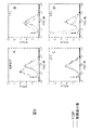

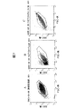


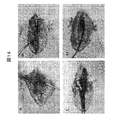

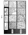


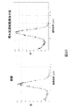

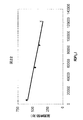


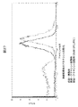
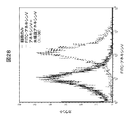
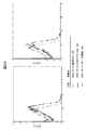

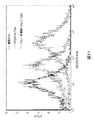


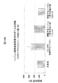


Classifications
-
- A—HUMAN NECESSITIES
- A01—AGRICULTURE; FORESTRY; ANIMAL HUSBANDRY; HUNTING; TRAPPING; FISHING
- A01N—PRESERVATION OF BODIES OF HUMANS OR ANIMALS OR PLANTS OR PARTS THEREOF; BIOCIDES, e.g. AS DISINFECTANTS, AS PESTICIDES OR AS HERBICIDES; PEST REPELLANTS OR ATTRACTANTS; PLANT GROWTH REGULATORS
- A01N1/00—Preservation of bodies of humans or animals, or parts thereof
- A01N1/02—Preservation of living parts
- A01N1/0278—Physical preservation processes
- A01N1/0284—Temperature processes, i.e. using a designated change in temperature over time
-
- A—HUMAN NECESSITIES
- A01—AGRICULTURE; FORESTRY; ANIMAL HUSBANDRY; HUNTING; TRAPPING; FISHING
- A01N—PRESERVATION OF BODIES OF HUMANS OR ANIMALS OR PLANTS OR PARTS THEREOF; BIOCIDES, e.g. AS DISINFECTANTS, AS PESTICIDES OR AS HERBICIDES; PEST REPELLANTS OR ATTRACTANTS; PLANT GROWTH REGULATORS
- A01N1/00—Preservation of bodies of humans or animals, or parts thereof
- A01N1/02—Preservation of living parts
-
- A—HUMAN NECESSITIES
- A61—MEDICAL OR VETERINARY SCIENCE; HYGIENE
- A61K—PREPARATIONS FOR MEDICAL, DENTAL OR TOILETRY PURPOSES
- A61K35/00—Medicinal preparations containing materials or reaction products thereof with undetermined constitution
- A61K35/12—Materials from mammals; Compositions comprising non-specified tissues or cells; Compositions comprising non-embryonic stem cells; Genetically modified cells
- A61K35/14—Blood; Artificial blood
- A61K35/19—Platelets; Megacaryocytes
-
- A—HUMAN NECESSITIES
- A61—MEDICAL OR VETERINARY SCIENCE; HYGIENE
- A61P—SPECIFIC THERAPEUTIC ACTIVITY OF CHEMICAL COMPOUNDS OR MEDICINAL PREPARATIONS
- A61P17/00—Drugs for dermatological disorders
- A61P17/02—Drugs for dermatological disorders for treating wounds, ulcers, burns, scars, keloids, or the like
-
- A—HUMAN NECESSITIES
- A61—MEDICAL OR VETERINARY SCIENCE; HYGIENE
- A61P—SPECIFIC THERAPEUTIC ACTIVITY OF CHEMICAL COMPOUNDS OR MEDICINAL PREPARATIONS
- A61P7/00—Drugs for disorders of the blood or the extracellular fluid
- A61P7/04—Antihaemorrhagics; Procoagulants; Haemostatic agents; Antifibrinolytic agents
-
- G—PHYSICS
- G01—MEASURING; TESTING
- G01N—INVESTIGATING OR ANALYSING MATERIALS BY DETERMINING THEIR CHEMICAL OR PHYSICAL PROPERTIES
- G01N33/00—Investigating or analysing materials by specific methods not covered by groups G01N1/00 - G01N31/00
- G01N33/48—Biological material, e.g. blood, urine; Haemocytometers
- G01N33/50—Chemical analysis of biological material, e.g. blood, urine; Testing involving biospecific ligand binding methods; Immunological testing
- G01N33/86—Chemical analysis of biological material, e.g. blood, urine; Testing involving biospecific ligand binding methods; Immunological testing involving blood coagulating time or factors, or their receptors
Abstract
Description
関連出願に関する相互参照
本出願は、下記の特許出願の開示に依存し、該出願の登録日付の利益を主張する。なお、これら出願全ての開示全体を引用することにより本出願に含める。すなわち、2004年8月12日登録米国仮出願第60/600,838号、2004年10月20日登録米国仮出願第60/619,930号、2005年6月15日登録米国特許出願第11/152,774号、および2005年8月5日登録米国特許出願第11/197,310号である。
本発明は、血液および血液製品の分野に関する。より詳細には、本発明は、血小板および血小板組成物、特に、凍結乾燥血小板、または、再水和凍結乾燥血小板を含む組成物であって、治療(処置)、診断、および研究目的に有用な組成物に関する。
CROSS REFERENCE TO RELATED APPLICATIONS This application relies on the disclosure of the following patent application and claims the benefit of the registration date of that application. The entire disclosure of all of these applications is incorporated herein by reference. U.S. Provisional Application No. 60 / 600,838 registered on August 12, 2004, U.S. Provisional Application No. 60 / 619,930 registered on Oct. 20, 2004, U.S. Patent Application No. 11 / 152,774 registered on June 15, 2005, And US patent application Ser. No. 11 / 197,310, registered on Aug. 5, 2005.
The present invention relates to the field of blood and blood products. More particularly, the present invention is a platelet and platelet composition, particularly a composition comprising freeze-dried platelets or rehydrated freeze-dried platelets, useful for therapeutic (treatment), diagnostic, and research purposes. Relates to the composition.
血液は、多数の成分から成る複雑な混合物である。一般に、血液は、四つの主要部分を含むと記載することが可能である。すなわち、赤血球、白血球、血小板、および血漿である。最初の3成分は、細胞または細胞様成分であり、4番目の成分(血漿)は、塩、タンパク、および、多数の生体機能のために必要な、他の因子から成る、広範な多様な混合物を含む液性成分である。血液の成分は、遠心により互いに分離することが可能である。通常、遠心によって、大容量/質量の、高密度の赤血球は、遠心管の底部に移動させられる。赤血球の上に、白血球と血小板から成る比較的薄い層が認められる。これは、その灰白色から「バフィーコート」と呼ばれる。バフィーコートの上に液性の血漿分画がある。 Blood is a complex mixture of many components. In general, blood can be described as containing four main parts. That is, red blood cells, white blood cells, platelets, and plasma. The first three components are cells or cell-like components, and the fourth component (plasma) is a broad and diverse mixture of salts, proteins, and other factors necessary for many biological functions It is a liquid component containing. Blood components can be separated from each other by centrifugation. Centrifugation typically moves high volume / mass, high density red blood cells to the bottom of the centrifuge tube. A relatively thin layer of white blood cells and platelets is found on the red blood cells. This is called “buffy coat” because of its grayish white color. There is a liquid plasma fraction on the buffy coat.
一般にエリスロサイトとも呼ばれる赤血球は、肺から、細胞に向かって細胞内諸過程に使用されるよう酸素を運搬し、細胞から、肺に向かって排除されるよう不要の二酸化炭素を運搬する役割を担う。赤血球は核を持たず、従って、血液における短命な細胞成分で、健康なヒトでは不断に更新される。赤血球で構成される血液容量のパーセントはヘマトクリットと呼ばれるが、この数字は、血液系の1種以上の疾患または障害、または血液系に影響を及ぼす1種以上の疾患または障害の存在を表示するのによく使用される。正常なヘマトクリット値は、女性では37%と47%の間、男性では40%と54%の間にある。赤血球は、通常、赤血球を必要とする患者、例えば、慢性貧血患者、または、傷害または外傷を負った患者、または、血液損失をもたらす手術を受けた患者に輸血される。さらに、赤血球は、任意の数の疾患または障害によって引き起こされる貧血を治療するためによく使用される。 Red blood cells, also commonly called erythrosite, carry oxygen from the lungs to cells for use in intracellular processes and carry unnecessary carbon dioxide from cells to be eliminated towards the lungs. . Red blood cells do not have a nucleus and are therefore short-lived cellular components in the blood that are constantly updated in healthy humans. The percentage of the blood volume that is made up of red blood cells is called hematocrit, but this number indicates the presence of one or more diseases or disorders of the blood system or one or more diseases or disorders that affect the blood system. Often used. Normal hematocrit values are between 37% and 47% for women and between 40% and 54% for men. Red blood cells are usually transfused into patients in need of red blood cells, such as patients with chronic anemia, or patients who have been injured or traumatic, or who have undergone surgery that results in blood loss. In addition, red blood cells are often used to treat anemia caused by any number of diseases or disorders.
通常ロイコサイトとも呼ばれる白血球は、生体を、外来の物質による傷害から保護する役割を担う有核細胞である。一般規則として、白血球は、病原性生物、例えば、細菌、真菌、およびウィルス、あるいは、生体に有害な可能性のある物質、例えば、タンパク毒素を撲滅するように働く。しかしながら、ある人々においては、白血球は、外見上無害な物質、例えば、花粉に対して保護反応を励起し、アレルギー反応をもたらす。実際、ある場合には、白血球は、生体自身の細胞またはタンパクに対して不適切に反応して、自己免疫疾患および生体組織の破壊をもたらし、この破壊が、ある状況下では致命的となることがある。その中で、精製白血球は、抗体療法に対して反応しない患者の治療に使用されてきた。 White blood cells, usually called leucosites, are nucleated cells that play a role in protecting living bodies from damage by foreign substances. As a general rule, white blood cells serve to eradicate pathogenic organisms, such as bacteria, fungi, and viruses, or substances that may be harmful to the body, such as protein toxins. However, in some people, white blood cells excite a protective reaction against apparently innocuous substances, such as pollen, resulting in an allergic reaction. In fact, in some cases, white blood cells react inappropriately to the body's own cells or proteins, leading to autoimmune diseases and destruction of living tissues, which under certain circumstances can be fatal. There is. Among them, purified leukocytes have been used to treat patients who do not respond to antibody therapy.
一般にトロンボサイトとも呼ばれる血小板は、小型で、不規則な形をした、骨髄で形成される、血液の巨核球由来成分であり、凝固過程に与り、かつ、外傷または傷害によるのみならず、正常な生理的活性によっても引き起こされる過度の血液損失からの生体保護を支援する。実際、血小板は、正常な止血において必須であり、血液が、損傷血管から流出するのを防ぐ最初の防衛線を実現する。血小板は一般に、血管の内面に付着し、血漿内に存在する、あるいは、血液の他の細胞成分によって放出される凝固系の成分と相互作用を持つことによって働く。精製血小板は、異常な血小板機能(血小板無力症)、および、血小板数減少(血小板減少症)を持つ患者を治療するのに使用されている。傷害後出血、または後天的血小板機能欠損による出血、例えば、バイパス手術中に起こる出血を阻止するために濃縮血小板がよく使われる。正常な循環血小板数は、血液マイクロリットル(ul)当たり150,000と450,000の間である。 Platelets, also commonly referred to as thrombosites, are small, irregularly shaped, bone marrow-derived components of blood that are derived from megakaryocytes, are involved in the clotting process, and are normal not only due to trauma or injury Helps protect the body from excessive blood loss caused by other physiological activities. In fact, platelets are essential in normal hemostasis and provide the first line of defense that prevents blood from flowing out of damaged blood vessels. Platelets generally work by attaching to the inner surface of blood vessels and interacting with components of the coagulation system that are present in plasma or released by other cellular components of the blood. Purified platelets have been used to treat patients with abnormal platelet function (platelet asthenia) and thrombocytopenia (thrombocytopenia). Concentrated platelets are often used to prevent bleeding after injury or bleeding due to acquired platelet function deficiency, such as bleeding that occurs during bypass surgery. Normal circulating platelet count is between 150,000 and 450,000 per microliter (ul) of blood.
出血が傷口から突然生じた場合、血小板が傷口に集中し、凝血塊を形成することによって血流を阻止しようとする。凝血塊の形成には二種類の一般的機構がある。一方の機構では、血液が空気に暴露されると、凝血塊が形成し始める。血小板は、空気の存在を感受し、フィブリノーゲンと反応し、フィブリンの形成を開始する。このフィブリンはクモの巣状の網目を形成し、これが血球をその中に捕捉する。他方の一般的機構では、損傷血管が化学的信号を発射し、これが、傷害領域における血小板の粘度を増加させる。この粘着性血小板は、損傷領域に接着し、徐々に血小板プラグを形成する。同時に、血小板は、一連の化学的信号を発射し、これが、血液中の他の因子を促して、血小板プラグを強化するように仕向ける。血小板と補強部との間には頑丈な凝血塊が形成され、これは、損傷領域が治癒する間、パッチとして働く。 If bleeding suddenly occurs from the wound, platelets concentrate on the wound and attempt to block blood flow by forming a clot. There are two general mechanisms for the formation of clots. In one mechanism, clots begin to form when blood is exposed to air. Platelets sense the presence of air and react with fibrinogen to initiate the formation of fibrin. The fibrin forms a cobweb-like network that traps blood cells within it. In the other general mechanism, the damaged blood vessel emits a chemical signal that increases the viscosity of the platelets in the injured area. The sticky platelets adhere to the damaged area and gradually form a platelet plug. At the same time, the platelets emit a series of chemical signals that prompt other factors in the blood to strengthen the platelet plug. A solid clot forms between the platelet and the reinforcement, which acts as a patch while the damaged area heals.
血小板ゲルの形を取る血小板は、外傷治癒を加速させるために広く使用されており、自家フィブリン糊と組み合わせた自家血小板ゲルは、手術前後の止血を改善し、かつ、上行大動脈交換手術における血液輸血の必要を減じさせたことが明らかにされている(Christenson and Kalangos, 2004)。Costasis手術止血装置(Costatis(登録商標))。フィブリノーゲンと血小板の供給源としてウシ・トロンビン、ウシ・コラーゲン、および血漿の組み合わせの利用が、ウサギ体内における出血性腎臓および脾臓モデルにおいて効果的に働くことが明らかにされている(Prior et al., 1999)。しかしながら、別の実験では、血小板ゲルは、単独で使用した場合、効果的な止血剤ではないことが示されている(Wajon et al., 2001)。血小板、および止血剤としてのその役割に関しては相矛盾する所見があるにも拘わらず、血小板微粒子の凝固先行性に関してはほとんど疑問の余地が無い。この必須成分は、しばしば見過ごされてきたけれども、次第に、インビトロおよびインビボの凝固過程における活発な関与因子であることが認識されるようになっている(Nieuwland et al., 1997)。生理的作用剤同士の組み合わせ、例えば、トロンビンとコラーゲンの組み合わせによって血小板を刺激した場合、血小板は大量の微粒子を放出する(Sims et al., 1988; Tans et al., 1991)。活性化血小板および微粒子は、アミノ・リン脂質を発現し、これが、前凝血表面を提供し、内在性、外在性、および共通経路における活性化凝固酵素の形成を支援する(Rosing et al., 1985)。 Platelets in the form of platelet gels are widely used to accelerate trauma healing, and autologous platelet gel combined with autologous fibrin glue improves hemostasis before and after surgery and blood transfusion in ascending aortic replacement surgery (Christenson and Kalangos, 2004). Costasis surgical hemostasis device (Costatis®). Utilizing a combination of bovine thrombin, bovine collagen, and plasma as a source of fibrinogen and platelets has been shown to work effectively in hemorrhagic kidney and spleen models in rabbits (Prior et al., 1999). However, another experiment has shown that platelet gel is not an effective hemostatic agent when used alone (Wajon et al., 2001). Despite the contradictory findings regarding platelets and their role as hemostatic agents, there is little doubt as to the precoagulation of platelet microparticles. Although this essential component has often been overlooked, it is increasingly recognized that it is an active participant in the clotting process in vitro and in vivo (Nieuwland et al., 1997). When platelets are stimulated by a combination of physiological agents, such as a combination of thrombin and collagen, platelets release large amounts of microparticles (Sims et al., 1988; Tans et al., 1991). Activated platelets and microparticles express amino phospholipids, which provide a procoagulant surface and assist in the formation of activated clotting enzymes in endogenous, extrinsic and common pathways (Rosing et al., 1985).
活性化血小板と比べると、微粒子は、活性化因子IX(IXa)(Hoffman et al., 1992)およびVa(Sims et al., 1988)に対する高い親和度を持つ、高密度の結合部位を含む。微粒子は、VIII因子(Gilbert et al., 1991)に対して高い親和性を持つ結合部位を連続発現し、Xa因子活性(Gilbert et al., 1991; Holme et al., 1995)およびプロトロンビナーゼ活性(Sims et al., 1989)の両方を支援する。 Compared to activated platelets, microparticles contain a high density of binding sites with high affinity for activators IX (IXa) (Hoffman et al., 1992) and Va (Sims et al., 1988). Microparticles continuously express binding sites with high affinity for factor VIII (Gilbert et al., 1991), factor Xa activity (Gilbert et al., 1991; Holme et al., 1995) and prothrombinase Supports both activities (Sims et al., 1989).
血小板微粒子が止血反応において重要な成分であるという事実は別として、血小板は、血小板ゲルという形で、外科創傷の治癒応用を始め、治癒の困難な創傷の治療にも使われてきた(Mazzucco et al., 2004)。さらに、血小板の、血小板富裕血漿という形での利用は、新規応用、例えば、生体組織工学、または、自家および異種組織移植片を始め、骨組織統合および軟組織再生(Okinarinen et al., 2003)にまで拡大している。これは、血小板が、そのアルファ粒子の中に、止血および創傷治癒過程を促進するいくつかの重要な増殖因子を含んでいるからである。実験から、増殖因子、中でも例えば、血小板由来創傷治癒因子(PDWHF)、血小板由来増殖因子(PDGF)、変形増殖因子(TGF)、およびインスリン増殖因子(IGF)が、創傷治癒カスケードの様々な段階において重要であり、マイトゲン性および細胞分化活性に大きな影響を及ぼすことが認められた(Pierce et al., 1989; Steed, 1997)。 Apart from the fact that platelet microparticles are an important component in the hemostatic response, platelets, in the form of platelet gels, have been used in the healing of surgical wounds and in the treatment of difficult wounds (Mazzucco et al. al., 2004). In addition, the use of platelets in the form of platelet-rich plasma has led to new applications such as tissue engineering or autologous and xenografts, bone tissue integration and soft tissue regeneration (Okinarinen et al., 2003). It has expanded to. This is because platelets contain several important growth factors in their alpha particles that promote the hemostasis and wound healing processes. Experiments have shown that growth factors such as platelet-derived wound healing factor (PDWHF), platelet-derived growth factor (PDGF), deformed growth factor (TGF), and insulin growth factor (IGF) are at various stages of the wound healing cascade. It was important and was found to have a significant effect on mitogenicity and cell differentiation activity (Pierce et al., 1989; Steed, 1997).
これらの所見から、増殖因子交換策の開発が導かれた。例えば、Regranex(登録商標)は、担体ゲルに溶解した組み換えヒトPDGFであるが、これは、糖尿病創傷の治療に使用されており、一方、別のあるもの、例えば、TGFは、現在FDA承認を求めて試験中である。にも拘わらず、単一増殖因子は、創傷に適用されると、複数の増殖因子ほど効果的ではない。これは驚くには当たらない。なぜなら、創傷治癒は、複数のカスケードの複雑な統合であって、過程内の様々な相において種々の刺激性および抑制性機能の発現に複数の増殖因子を必要とするからである。 These findings led to the development of growth factor exchange strategies. For example, Regranex® is recombinant human PDGF dissolved in a carrier gel, which has been used to treat diabetic wounds, while others, such as TGF, are currently FDA approved. In search for. Nevertheless, a single growth factor is not as effective as multiple growth factors when applied to a wound. This is not surprising. This is because wound healing is a complex integration of multiple cascades, requiring multiple growth factors for the development of various stimulatory and inhibitory functions in various phases within the process.
一般に血漿と呼ばれる、血液の液性部分は、各種のタンパクおよび塩を含む複雑な溶液である。一般的な言葉で言うと、血漿とは、赤血球、白血球、および血小板が血液から除去された際に留まる物質である。多数のタンパクが高濃度に存在するために、血漿は、室温では不安定な、麦わら色をした液体である(すなわち、血漿は、その中のタンパクが活性を失わないようにするためには室温よりもはるかに低い温度で保存しなければならない)。血漿の主要なタンパク組成分は、アルブミン、フィブリノーゲン、抗体、および、血液凝固および止血に必要な多数のタンパクである。血漿タンパクのこの簡単なリストからも見て取れるように、血漿は、十分な血圧と容量補給の維持から、血液凝固および免疫にとって必須なタンパクの補給に至るまで様々の機能を果たす。例えば、血漿から単離されるガンマグロブリンは、抗毒素を必要とする患者を治療するのに使用することが可能であり、患者が、あるウィルスまたは細菌に感染しているかどうかを示すために、いくつかの抗体の存在が定量される。さらに、血漿から単離される凝固因子VIIIは、古典的な先天性血友病の治療によく使われる。 The humoral part of blood, commonly called plasma, is a complex solution containing various proteins and salts. In general terms, plasma is a substance that stays when red blood cells, white blood cells, and platelets are removed from the blood. Due to the high concentration of many proteins, plasma is a straw-colored liquid that is unstable at room temperature (ie, plasma is room temperature to prevent the proteins in it from losing activity). Must be stored at a much lower temperature). The major protein components of plasma are albumin, fibrinogen, antibodies, and a number of proteins necessary for blood clotting and hemostasis. As can be seen from this simple list of plasma proteins, plasma serves a variety of functions, from maintaining sufficient blood pressure and volume supplementation to supplementing proteins essential for blood clotting and immunity. For example, gamma globulin isolated from plasma can be used to treat patients in need of an antitoxin and several to indicate whether the patient is infected with a virus or bacteria The presence of antibodies is quantified. In addition, coagulation factor VIII isolated from plasma is often used to treat classic congenital hemophilia.
血液の主要機能は、酸素および二酸化炭素を運搬し、免疫系の成分が素早く、かつ効果的に身体の全ての部分に達することを可能ならしめ、侵入する微生物を撲滅することである。しかしながら、血液は液体であり、生体内に保持されなければならないばかりでなく、生体内の特定領域(例えば、血管、または循環系の他の部分)に限局されなければならないのであるから、血液の重要機能は、生体内における自身の分布を監視し、血液の体内からの逸失、または、血液が保持されなければならない体内の特定領域からの逸失を許す損傷を修復することである。血液の分布を監視し、血液をその正常な境界内に維持する過程は、二つの生理的過程、一方は、血管損傷後の過度の出血を阻止する(血液凝固の形成によって)過程、他方は、血液を、凝固しない(すなわち、非凝固的)流動状態に維持することによって正常な血液循環を維持する過程とのバランスである。これら、外見上は競合する二つの過程は、多くの制御点およびフィードバックループを含む複雑なシステムの一部である。 The main function of blood is to carry oxygen and carbon dioxide, enable components of the immune system to reach all parts of the body quickly and effectively, and eradicate invading microorganisms. However, because blood is a liquid and not only must be retained in the body, it must be confined to a specific area in the body (for example, blood vessels or other parts of the circulatory system). An important function is to monitor its distribution in the body and repair damage that allows the loss of blood from the body or the loss of specific areas of the body where the blood must be retained. The process of monitoring blood distribution and keeping the blood within its normal boundaries is one of two physiological processes, one that prevents excessive bleeding (by formation of blood clots) after vascular injury, the other is A balance with the process of maintaining normal blood circulation by maintaining the blood in a non-coagulated (ie non-coagulant) fluid state. These two seemingly competing processes are part of a complex system that includes many control points and feedback loops.
適当な血流の維持および封じ込めのための主要過程は、止血と呼ばれる。これは、血液凝固塊の形成と最終的なその分解、および、損傷組織の修復から成る過程である。止血は四つの主要な事象から成る。すなわち、血管収縮;フィブリノーゲンによって仲介される、損傷部位における血小板の凝集、およびトロンビンによる血小板の活性化;血小板による血液凝固塊(また、血栓またはフィブリン網とも呼ばれる)の形成および、各種凝固因子の複雑な相互作用;および、最後に、凝固塊の分解および損傷組織の修復である。 The main process for maintaining and containing proper blood flow is called hemostasis. This is a process that consists of the formation of a blood clot and its final degradation, and the repair of damaged tissue. Hemostasis consists of four major events. That is, vasoconstriction; fibrinogen-mediated platelet aggregation at the site of injury, and thrombin activation of platelets; formation of blood clots by platelets (also called thrombus or fibrin network) and the complexity of various clotting factors And finally, the degradation of clots and the repair of damaged tissue.
血液凝固は複雑な過程である。凝固塊形成が阻止されなければ、血管は詰まる。凝固塊が堅固でなければ、過剰な血液損失が起こる。従って、正常な止血のためには微妙なバランスが維持されなければならない。正常な止血のバランスが崩れた状況では、凝固塊形成は損なわれる。このような異常は、アスピリンの服用、または免疫系の機能不全によって引き起こされることがある。この異常は、先天性の場合があり、例えば、遺伝的疾患によって、または凝固因子欠損によって起こる場合もある。例えば、出血傷害に至る止血過程における欠損が特定されているが、そのような欠損の多くは、血液凝固に必要な活性状態カスケード、血小板の活性化および機能、または接触活性化に関与する酵素の中に認められる。このような障害に含まれるものとして、vWDおよび血友病がある。血液凝固系の他の疾患または障害は、他の疾患または障害治療の結果(すなわち、副作用)である。このような疾患および障害の治療は、通常、その副作用の原因となる薬剤の用量を減らすこと、またはその薬剤による治療を中断することを含む。 Blood clotting is a complex process. If clot formation is not blocked, the blood vessels are clogged. If the clot is not firm, excessive blood loss occurs. Therefore, a delicate balance must be maintained for normal hemostasis. In a situation where normal hemostasis is unbalanced, clot formation is impaired. Such abnormalities can be caused by taking aspirin or by malfunctioning of the immune system. This abnormality may be congenital, for example, caused by a genetic disease or by a clotting factor deficiency. For example, defects in the hemostasis process leading to bleeding injuries have been identified, but many of these defects are active state cascades necessary for blood clotting, platelet activation and function, or enzymes involved in contact activation. Recognized in. Among these disorders are vWD and hemophilia. Other diseases or disorders of the blood clotting system are the result (ie, side effects) of other disease or disorder treatments. Treatment of such diseases and disorders usually involves reducing the dose of the agent that causes the side effects or discontinuing treatment with the agent.
血液凝固は、数多くのフィードバックループおよび制御点を通じて緊密に制御される、酵素活性の、一連の複雑なカスケードに依存する。凝固は、血小板が、損傷血管の切り裂かれた血管壁、またはその他の傷害部位に付着した時に始まる。この過程で、血小板は、損傷部位の細胞表面のコラーゲンに付着する。これは、フォンウィルブラント因子(vWF)と呼ばれる凝固因子によって仲介される過程である。vWFは、巨核球によって生産される複雑なタンパクであり、血小板、またはいくつかの結合組織の中に貯えられる。vWFは、しばしばVIII因子と複合体を形成することが認められ、血漿におけるVIII因子の安定化に必要であることが知られている。vWFの量および機能における欠損は、通常遺伝性であり、フォンウィルブラント病(vWD)という名前の病気をもたらす。 Blood clotting relies on a series of complex cascades of enzyme activity that are tightly controlled through numerous feedback loops and control points. Clotting begins when platelets adhere to the torn vessel wall of the damaged blood vessel or other injury site. In this process, platelets adhere to collagen on the cell surface at the site of injury. This is a process mediated by a coagulation factor called von Willebrand factor (vWF). vWF is a complex protein produced by megakaryocytes and is stored in platelets or some connective tissue. vWF is often found to form a complex with factor VIII and is known to be required for stabilization of factor VIII in plasma. A deficiency in the amount and function of vWF is usually hereditary and results in a disease named von Willebrand disease (vWD).
損傷部位に対する血小板の付着は、内皮下層のコラーゲンに結合するvWfによって仲介される。可溶性タンパクとして血漿中に存在するフィブリノーゲンは、凝集または結集と呼ばれる過程において活性化血小板をまとめて橋渡しすることが可能である。フィブリノーゲンは、酵素トロンビン(活性化X因子(Xa因子)によって活性化される)によってフィブリンという不溶性の繊維に変換されるが、これも、血小板の強力な活性剤である。フィブリンは、自発的に重合し繊維になるが、血小板上の表面タンパクまたはリン脂質に結合し、血小板を網状に罠のように捕捉する。次に、このフィブリン繊維は、XIII因子からトロンビンによって形成されるXIIIa因子の活性によって架橋される。こうして形成されるフィブリン-血小板網は、フィブリンメッシュ、血栓、または血液凝固塊と呼ばれる。 Platelet adhesion to the injury site is mediated by vWf binding to subendothelial collagen. Fibrinogen present in plasma as a soluble protein can bridge activated platelets together in a process called aggregation or aggregation. Fibrinogen is converted to an insoluble fiber called fibrin by the enzyme thrombin (activated by activated factor X (factor Xa)), which is also a strong activator of platelets. Fibrin spontaneously polymerizes into fibers, but binds to surface proteins or phospholipids on platelets and traps platelets like a cocoon. This fibrin fiber is then cross-linked by the activity of factor XIIIa formed by thrombin from factor XIII. The fibrin-platelet network thus formed is called a fibrin mesh, a thrombus, or a blood clot.
X因子は、外在経路および内在経路と名づけられる二つの経路の内のどちらかによって活性化される。内在経路は、各種プロテアーゼを活性化する一連の酵素反応を含む。この過程は、XII因子の、内皮下層の成分によって供給されると考えられる陰性荷電表面に対する結合、および、高分子量キニノゲン(HMWK)によって仲介される反応における、カリクラインによるXII因子からXIIaへの活性化から始まる。次に、XIIa因子は、XI因子をXIa因子(血漿トロンボプラスチンの先行体)に変換する。カルシウムイオンの存在下、XIa因子は、IX因子を、その活性形IXa因子に変換する。IXa因子は、非酵素タンパクVIII因子(抗血友病グロブリン、またはAHG)と結合し、かつ、カルシウムイオンおよび細胞由来リン脂質の存在下に、循環因子Xを活性化しXa因子を形成する。 Factor X is activated by one of two pathways termed the extrinsic pathway and the intrinsic pathway. The intrinsic pathway involves a series of enzymatic reactions that activate various proteases. This process is related to the activity of factor XII from factor XII to XIIa by calicline in the binding of factor XII to the negatively charged surface believed to be supplied by components of the subendothelium and the reaction mediated by high molecular weight kininogen (HMWK) It starts from the conversion. Factor XIIa then converts factor XI to factor XIa, a precursor to plasma thromboplastin. In the presence of calcium ions, factor XIa converts factor IX to its active form factor IXa. Factor IXa binds to non-enzymatic protein factor VIII (antihemophilic globulin or AHG) and activates circulating factor X to form factor Xa in the presence of calcium ions and cell-derived phospholipids.
血液凝固を起動するための一次的生理経路と広く見なされている外在経路では、因子VIIの活性形、VIIaが、一般に組織因子(TF)と呼ばれるIII因子(組織トロンボプラスチン)と連結する。カルシウムイオンの存在下に、VIIa/TF複合体は、循環するX因子を活性化してXa因子を形成する。Xa因子はまた、IXおよびXI因子によるVII因子の活性化によっても形成される。このシナリオでは、IX因子およびX因子は、TFとVIIa因子の共同活性によって活性化される。このVIIa/TF複合体は、血液凝固カスケードのもっとも強力な起動因子と認められている。前述のように、IXa因子は、カルシウム、血小板表面のリン脂質、およびVIIIa因子の存在下に、X因子をXa因子に活性化し、Xa因子は次にプロトロンビンをトロンビンに活性化する。トロンビンは、可溶性フィブリノーゲンを不溶性フィブリンに変換しメッシュを形成する。 In the extrinsic pathway, widely regarded as the primary physiological pathway for initiating blood clotting, the active form of factor VII, VIIa, is linked to factor III (tissue thromboplastin), commonly referred to as tissue factor (TF). In the presence of calcium ions, the VIIa / TF complex activates circulating factor X to form factor Xa. Factor Xa is also formed by activation of factor VII by factors IX and XI. In this scenario, factor IX and factor X are activated by the joint activity of TF and factor VIIa. This VIIa / TF complex is recognized as the most powerful trigger of the blood coagulation cascade. As mentioned above, factor IXa activates factor X to factor Xa in the presence of calcium, platelet surface phospholipids, and factor VIIIa, which in turn activates prothrombin to thrombin. Thrombin converts soluble fibrinogen to insoluble fibrin to form a mesh.
このようにして、X因子は、内在活性化経路、外在活性化経路のいずれかによって活性化される。Xa因子は、活性化V因子と共に、カルシウムイオンおよび血小板表面のリン脂質の存在下に、プロトロンビンを活性化してトロンビンとする。トロンビンは、フィブリノーゲンからフィブリンを形成し、凝固塊の形成をもたらす。血液凝固は、いくつかの成分、例えば、フィブリノーゲン、トロンビン、VII因子、VIII因子、IX因子、X因子、XI因子、およびXII因子を含む複数成分の相互作用に基づく複雑な過程である。これらの成分の内一つが欠けると、それは必ず、ある患者にとっては致命的となりかねない血液障害の臨床症状を招く。 In this way, factor X is activated by either the intrinsic activation pathway or the extrinsic activation pathway. Factor Xa, together with activated factor V, activates prothrombin to thrombin in the presence of calcium ions and phospholipids on the platelet surface. Thrombin forms fibrin from fibrinogen, resulting in the formation of a clot. Blood clotting is a complex process based on the interaction of several components, including several components such as fibrinogen, thrombin, factor VII, factor VIII, factor IX, factor X, factor XI, and factor XII. Lack of one of these components always leads to clinical symptoms of blood disorders that can be fatal for some patients.
これまでに出血障害をもたらす、複数の止血過程の欠損症が特定されているが、そのような欠損症の多くは、血液凝固に必要な活性カスケード、血小板活性化と機能、または接触活性化に関与する酵素に認められる。このような障害に含まれるものとしてvWDおよび血友病がある。これら二つの障害に対しては様々な治療が知られているが、その多くは、前述の凝固因子の一つ以上の補給に依存する。 Several hemostatic deficiencies have been identified that lead to bleeding disorders, but many of these deficiencies are related to the activation cascade, platelet activation and function, or contact activation required for blood clotting. Found in the enzymes involved. Among these disorders are vWD and hemophilia. Various treatments are known for these two disorders, many of which rely on the supplementation of one or more of the aforementioned clotting factors.
先天的血友病は三つの異なるグループに分類される。すなわち、古典的血友病、または血友病A(FVIII欠損症)、クリスマス病、または血友病B(FIX欠損症)、および血友病C(FXI欠損症)である。血友病とは、出血が正常な時間内に停止されない障害と認められる。すなわち、血友病患者は、より大量に、より速やかに出血し、より長期に出血する。約2万人のアメリカ人が血友病を患う。症例の圧倒的多数は、血友病AまたはBのいずれかであり、血友病Aが、全血友病症例の約80%を占める。血友病Cは稀であり、10万人のアメリカ人当たり約1名に見られる。 Congenital hemophilia is divided into three different groups. Classic hemophilia, or hemophilia A (FVIII deficiency), Christmas disease, or hemophilia B (FIX deficiency), and hemophilia C (FXI deficiency). Hemophilia is recognized as a disorder in which bleeding does not stop within normal time. That is, hemophilia patients bleed more rapidly, more rapidly and longer. About 20,000 Americans suffer from hemophilia. The overwhelming majority of cases are either hemophilia A or B, which accounts for about 80% of all hemophilia cases. Hemophilia C is rare and is found in about 1 in every 100,000 Americans.
血友病Aは、VIII因子の欠損によって生じるX-結合障害であり、凝固因子VIIIの欠如、または正常未満レベルと定義される。血友病Aは、VIII因子の遺伝子における様々な突然変異によって生じる。血友病Aの現代の治療は、ヒトの血漿から調製した、または組み換えDNA技術によって調製した、VIII因子濃縮物、または、VIII因子とwWFの複合体の濃縮物の投与を含む。血友病Aとは違って、血友病Bは、IX因子の欠損から生じる。血友病Bの現代の治療は、血漿由来の、または組み換え技術によるIX因子濃縮物の投与を含む。最後に、血友病Cは、XI因子の欠損による。 Hemophilia A is an X-binding disorder caused by factor VIII deficiency and is defined as a lack of coagulation factor VIII or subnormal levels. Hemophilia A is caused by various mutations in the gene for factor VIII. Modern treatments for hemophilia A involve the administration of factor VIII concentrates or factor VIII / wWF complex concentrates prepared from human plasma or prepared by recombinant DNA technology. Unlike hemophilia A, hemophilia B results from a factor IX deficiency. Modern treatments for hemophilia B include the administration of factor IX concentrates derived from plasma or by recombinant techniques. Finally, hemophilia C is due to a factor XI deficiency.
公共的血液供給の安全性、および血液および血液製品を通じて伝播される病気の出現に関する絶えざる不安のために、VIII因子の特選供給源は、組み換え技術によって生産される形態となっている。組み換えIX因子は、ヒトへの使用が承認されたが(Benefix(登録商標)、Genetics Institute)、やがて特選供給源となると思われる。さらに、血友病の治療または治癒策として遺伝子療法が提案されている。しかしながら、今日まで、血友病患者の治療におけるトランスジェニック対処法は、凝固因子の長期の安定的発現をもたらさず、予想もしなかった致死問題に遭遇し、現在でもなお、レシピエントにおける抑制因子生産の原因となる(後述)。 Due to the safety of public blood supplies and the constant concern about the emergence of diseases transmitted through blood and blood products, the factor VIII special source is in the form produced by recombinant technology. Recombinant Factor IX has been approved for use in humans (Benefix®, Genetics Institute) and will eventually become a source of choice. Furthermore, gene therapy has been proposed as a treatment or cure for hemophilia. To date, however, transgenic treatments in the treatment of hemophilia patients have not led to long-term stable expression of coagulation factors, encountered unexpected lethal problems, and still present suppressor production in recipients. Cause (described later).
VIII因子の濃縮物によって血友病を治療した場合、血友病Aの患者の約15%から約30%、血友病B患者の約3%において、それぞれ、導入されたVIII因子またはIX因子に対する抗体の生産を招く。組み換えVIII因子は、血友病A患者の僅か約5%にしかこの反応を引き起こさないようであるが、それでもこの障害の治療においては重大な問題となっている。この過程およびそれによって生じる障害は、「抑制因子つき血友病」と呼ばれ、凝固因子の欠如を治療するために通常使用される輸血タンパクにたいする抗体の誘発と多くの場合記載される。一方、後天的血友病は、正常レベルの凝固タンパクを含む個人における抑制因子の発生である。従って、後天的血友病は、擬似自己免疫疾患であり、凝固に与る因子を含む製剤によって治療される、その他の点では正常な、非血友病患者において起こることがある。一般に、生成される抗体は、投与されるVIII因子と反応し、VIII因子の活性の抑制をもたらすので、内在性VIII因子を欠如する患者では治療を無効とし、もともと、不十分ではあるが、低レベルの、または低活性の内在性VIII因子を所有していた患者では、皮肉にも治療を有害なものとする。 When hemophilia is treated with a factor VIII concentrate, about 15% to about 30% of hemophilia A patients and about 3% of hemophilia B patients are introduced factor VIII or factor IX, respectively. Cause the production of antibodies against. Recombinant factor VIII appears to cause this response in only about 5% of hemophilia A patients, yet it remains a significant problem in the treatment of this disorder. This process and the resulting disorder are called “suppressive hemophilia” and are often described as the induction of antibodies to transfusion proteins commonly used to treat lack of coagulation factors. Acquired hemophilia, on the other hand, is the generation of inhibitory factors in individuals that contain normal levels of clotting proteins. Acquired haemophilia is therefore a pseudo-autoimmune disease that can occur in otherwise non-hemophilic patients who are treated with a formulation containing factors that contribute to clotting. In general, the antibody produced reacts with administered factor VIII and results in suppression of factor VIII activity, thus invalidating treatment in patients lacking endogenous factor VIII, which is originally insufficient but low Ironically, treatments are also detrimental in patients who possess low-level or low-activity endogenous factor VIII.
後天性血友病、および抑制因子つき血友病を回避するために、いくつかのやり方が従来から提案され実行されている。例えば、外因性VIII因子で治療せずに、血友病Aを治療するためのもう一つの策として、外因性VIIa因子を投与し、そうすることによって止血のためのVIII因子を不要とするやり方がある。同様に、過剰量のVIII因子、および、抗イディオタイプ抗VIII因子抗体による治療が試験されている。他の対処法としては、FEIBAバイパス剤、プロトロンビン複合体濃縮物、組み換えVIIa因子、ブタのVIII因子の使用、高用量の免疫グロブリンの静注、免疫耐性療法(ITT)、および、抑制性抗体を除去するための、プロテインAの吸着付きまたは吸着無しの血漿瀉血、が挙げられる。 Several approaches have been proposed and implemented in the past to avoid acquired hemophilia and hemophilia with inhibitory factors. For example, another way to treat hemophilia A without treatment with exogenous factor VIII is to administer exogenous factor VIIa, thereby eliminating the need for factor VIII for hemostasis There is. Similarly, treatment with excess amounts of factor VIII and anti-idiotype anti-factor VIII antibodies is being tested. Other measures include FEIBA bypass agent, prothrombin complex concentrate, recombinant factor VIIa, use of porcine factor VIII, high doses of intravenous immunoglobulin, immunotolerant therapy (ITT), and inhibitory antibodies. And plasma phlebotomy with or without protein A adsorption for removal.
さらに、精製された組み換えVIIa因子による治療が一般的になっている。例えば、VIIa因子の10から15 ug/kgの用量、場合によっては150 ug/kgの高用量を投与しても、これは、約0.2から2.0 ug/ml血液のVIIa因子の循環レベルをもたらす用量範囲であるが、安全であり、かつ、ある抑制因子つき血友病A患者においては有効であることが判明した。これらの用量は、約0.005 ug/ml血液である、VIIa因子の正常な推定濃度と比べると極めて高いものである。上記方法は成功を見たものの、現在行われている方法はいずれも完全には効果的でなく、また全て極めて高価である。さらに、組み換えFVIIa治療を受けた患者の少なくとも5-10%では止血が起こらなかった。 Furthermore, treatment with purified recombinant factor VIIa has become common. For example, a dose of 10 to 15 ug / kg of factor VIIa, and possibly even a high dose of 150 ug / kg, will result in a circulating level of factor VIIa of about 0.2 to 2.0 ug / ml blood It was found to be safe in range, but effective in hemophilia A patients with certain inhibitors. These doses are very high compared to the normal estimated concentration of Factor VIIa, which is about 0.005 ug / ml blood. Although the above methods have been successful, none of the methods currently performed are completely effective and are all very expensive. Furthermore, hemostasis did not occur in at least 5-10% of patients treated with recombinant FVIIa.
さらに、III型、すなわち重篤なフォンウィルブラント病は、血友病Aのような臨床症状を呈することがよくある。VIII因子は、通常、vWfによって輸送されるが、血漿プロテアーゼからvWfによって保護される。循環するvWfが無い場合、内在性のVIII因子は急速に分解され、循環から除去されて、血友病Aの症状をもたらす。vWDの治療は、病気の性質および重度に応じて変化する。治療としては、注入または鼻孔経由いずれかによるDDAVP療法が挙げられる。DDAVP療法は、内皮細胞vWfを循環に放出することによって作動する。治療はまた、vWfおよび他の凝固因子の濃縮形となる血漿寒冷沈降物を含む。 In addition, type III, or severe von Willebrand disease, often presents with clinical symptoms such as hemophilia A. Factor VIII is normally transported by vWf but protected from plasma proteases by vWf. In the absence of circulating vWf, endogenous factor VIII is rapidly degraded and removed from the circulation, resulting in symptoms of hemophilia A. Treatment of vWD varies depending on the nature and severity of the disease. Treatment includes DDAVP therapy, either by infusion or via the nostril. DDAVP therapy works by releasing endothelial cells vWf into the circulation. Treatment also includes plasma cryoprecipitates that are concentrated forms of vWf and other clotting factors.
従来の血友病の治療は、通常、出血症状が認められて始めて行われる。比較的最近、その時の出血状態とは無関係に、欠乏凝固因子の定期的、予防的投与が実行される治療処方が開発された。この方法は、その因子レベルを十分高く維持するので、出血、関節破壊、および、生命を脅かす出血が抑制され、ほぼ完全に回避される。非常に効果的ではあるが、この治療処方は極めて高価である。 Conventional treatment of hemophilia is usually performed only after bleeding symptoms are observed. Relatively recently, treatment regimens have been developed in which regular and prophylactic administration of deficient coagulation factors is performed regardless of the current bleeding state. This method keeps the factor level high enough so that bleeding, joint destruction, and life-threatening bleeding are suppressed and almost completely avoided. Although very effective, this treatment regime is extremely expensive.
血小板の機能性が、血液凝固における、もう一つの決定的に重要な成分である。機能不全血小板は、異常出血、例えば、出血または血栓をもたらす可能性がある。従って、血小板機能アッセイは、血液関連疾患の診断および監視に組み込まれた一部分となる。例えば、ほんの数例を挙げれば、後天的血小板欠損、例えば、アスピリン注射、心臓病、腎臓病、または先天性血小板欠損、例えば、ベルナール・スーリエ症候群、グランツマン血小板無力症、および、蓄積症は、血小板の正常な止血機能に影響を及ぼすことがある。最小程度でもよいから血小板機能を評価するためには、末梢血スメアによる全血カウントが、僅かではあるが基本的情報を提供する。その他の試験、例えば、出血時間、凝集計を用いる血小板機能試験で、全血または血小板富裕血漿について行われる、一連の血小板作用剤に対する血小板凝集の評価試験によって、欠損が分類される。しかしながら、このような分析は、正確ではあるが、高感度ではなく、障害の初期段階における正常凝固機能における僅かな擾乱を検出することはできない。同様に、血液凝固カスケードの厳密な欠損点の決定は、新鮮採取血による多数のアッセイを要することがある。 Platelet functionality is another critical component in blood clotting. Malfunctioning platelets can lead to abnormal bleeding, such as bleeding or blood clots. Thus, platelet function assays become an integral part of the diagnosis and monitoring of blood related diseases. For example, to name just a few, acquired platelet defects, such as aspirin injection, heart disease, kidney disease, or congenital platelet defects, such as Bernard-Soulier syndrome, Grantsman platelet asthenia, and storage diseases are May affect normal hemostatic function of platelets. In order to assess platelet function since it may be minimal, a whole blood count with a peripheral blood smear provides a small but basic information. Other tests, such as bleeding time, platelet function tests using an aggregometer, classify defects by a series of platelet aggregation evaluation tests on whole blood or platelet rich plasma performed on platelets. However, such an analysis is accurate but not sensitive and cannot detect slight disturbances in normal coagulation function in the early stages of the disorder. Similarly, determining the exact point of failure of the blood clotting cascade may require multiple assays with freshly drawn blood.
血小板は、凝固過程に関与し、少なくとも1種の凝固因子の供給源であることが知られるが、後天的または先天的血友病の治療のために、あるいは、正常血小板数および血小板機能を持ちながら出血障害を患う人々の治療のために、安静で、活性化され、固定され、凍結され、または解凍された血小板、または、それら血小板の組み合わせを使用することについてはこれまで全く開示されたことはなかった。Kirby & Gregoriades (1984)は、血友病の経口的治療の試みとしてVIII因子を含むリポソームを調製した。その後、Giles et al. (1988)は、Xa因子とフォスファチジルコリン-フォスファチジルセリン顆粒の組み合わせは、インビボにおいてVIII因子を迂回することを示し、一方、Hong & Giles (1992)は、血友病A(VIII因子欠乏症)のイヌにおいて、Xa因子とフォスファチジルコリン-フォスファチジルセリン顆粒の混合物の投与後、止血プラグが正常化したことを明らかにした。比較的最近、Yarovoi et al. (2003)は、トランスジェニック法を用い、血小板において場違いに発現されたVIII因子が、マウスモデルにおいて血友病Aの治療に効果を示したことを実証した。さらに、Hrachovinova et al. (2003)は、P-セレクチンとPSGL1の相互作用によって白血球由来微粒子が生成され、これが、血友病Aのマウスモデルにおいて止血を適切なものにすることを示した。しかしながら、これらの研究者のいずれも、血友病の治療のために、正常血小板または血小板誘導体を用いたこともないし、提案もしていない。 Platelets are known to be involved in the clotting process and a source of at least one clotting factor, but have a normal platelet count and platelet function for the treatment of acquired or congenital hemophilia, or While there has been no disclosure of the use of resting, activated, fixed, frozen, or thawed platelets, or combinations of these platelets, for the treatment of people with bleeding disorders There was no. Kirby & Gregoriades (1984) prepared liposomes containing factor VIII in an attempt to orally treat hemophilia. Subsequently, Giles et al. (1988) showed that the combination of factor Xa and phosphatidylcholine-phosphatidylserine granules bypassed factor VIII in vivo, while Hong & Giles (1992) In dogs with hemophilia A (Factor VIII deficiency), the hemostatic plugs were normalized after administration of a mixture of Factor Xa and phosphatidylcholine-phosphatidylserine granules. Relatively recently, Yarovoi et al. (2003) used a transgenic method to demonstrate that factor VIII expressed out of place in platelets was effective in treating hemophilia A in a mouse model. In addition, Hrachovinova et al. (2003) showed that the interaction of P-selectin and PSGL1 produced leukocyte-derived microparticles that make hemostasis appropriate in a mouse model of hemophilia A. However, none of these researchers have used or proposed normal platelets or platelet derivatives for the treatment of hemophilia.
通常、血液凝固疾患または障害の検出は、血小板数、血液凝固に関与する各種マーカー、および凝固形成活性に関して患者血液を分析することを含む。かつて、凝固アッセイは、活性凝固時間(ACT)、プロトロンビン時間(PT)、血漿トロンビン時間(PTT)、および活性化部分トロンボプラスチン時間(APTT)を測定し、これらを用いて、内在性および外在性凝固経路を評価した。これらのアッセイは、一般に検査室で行われ、分析は、患者たちから血液の複数サンプルを抽出することを要求する。さらに、これらのアッセイの信頼度は潜在的に低い。なぜなら、これらは、結果が、インビトロにおける凝固形成時間に基づく終末点試験だからである。もう一つの限界点は、外来性の試薬、例えば、カオリン、トロンビン、カルシウム等を加えなければならないという事実に関連する。すなわち、結果は人工的なシステムに依存し、必ずしも患者の血栓形成能を反映しない。 Usually, detection of a blood clotting disease or disorder involves analyzing patient blood for platelet count, various markers involved in blood clotting, and clot-forming activity. In the past, clotting assays measure active clotting time (ACT), prothrombin time (PT), plasma thrombin time (PTT), and activated partial thromboplastin time (APTT) and use these to determine endogenous and exogenous The coagulation pathway was evaluated. These assays are generally performed in a laboratory, and analysis requires the extraction of multiple samples of blood from patients. Furthermore, the reliability of these assays is potentially low. Because these are endpoint tests based on in vitro clotting time. Another limiting point is related to the fact that exogenous reagents such as kaolin, thrombin, calcium, etc. must be added. That is, the results depend on the artificial system and do not necessarily reflect the patient's ability to form a thrombus.
前述したように、血液凝固システムの最重要機能は、損傷組織、例えば、創傷、手術、またはその他の外傷によって損なわれた組織からの血液損失を阻止することである。しかしながら、時として、創傷または外傷があまりにも大きく、傷を受けた人の血液系が、出血の全てを速やかに、効果的に阻止することができない場合がある。さらに、凝固機能は、大抵の人には十分な形で与えられているけれども、ある人々では凝固系は損なわれおり、そのために十分な凝固が得られず、広範な、時には致命的な出血が、損傷または外傷の結果生じることがある。以上から、人が、欠如した、あるいは不十分な凝固機能を補充するために追加の血小板を必要とする場合がしばしばある。 As previously mentioned, the most important function of the blood clotting system is to prevent blood loss from damaged tissue, such as tissue damaged by wounds, surgery, or other trauma. Occasionally, however, the wound or trauma is too large and the blood system of the wounded person may not be able to stop all of the bleeding quickly and effectively. In addition, although coagulation function is provided in a form that is sufficient for most people, in some people the coagulation system is impaired, so that sufficient coagulation is not obtained and extensive and sometimes fatal bleeding occurs. May result from damage or trauma. From the above, a person often needs additional platelets to replace missing or insufficient coagulation functions.
必要な人に血液凝固機能を補充するために、血小板を「そのままの形で」使用することに加えて、血小板は、その性質を明らかにし、血液凝固カスケードにおけるその正確な役割を理解するために、実験室において広範に研究されている。血小板に関する研究によって、血小板や、血小板と相互作用を持ち、凝固および創傷治癒を促進する因子、および、血小板を活性化したり、またはその他のやり方で血小板を損傷部位に引き付け、該部位に保持する因子に関する情報が得られる。 In addition to using platelets “as is” to replenish blood clotting functions to those who need it, platelets will reveal their nature and understand their exact role in the blood clotting cascade. Has been extensively studied in the laboratory. Research on platelets, factors that interact with platelets and promote coagulation and wound healing, and factors that activate platelets or otherwise attract platelets to damaged sites and retain them at those sites Information about
血小板の治療的使用および研究的使用のいずれも、血小板が生物学的に活性な形で利用可能であることを要求する。近年、治療用血小板(例えば、創傷治癒投与用)は、通常、5日未満という、新たに単離された製品の形で供給される。直ちに気が付くように、必要な患者に使用されるよう新鮮な血小板の十分な供給を維持することは高価であり、かつ、使用前の期限切れのために大量の供給分の損失を招く。さらに、新鮮血小板は、治療使用のために極めて重要なものであるから、そのような血小板を研究目的のために入手することは困難でもあり、高価でもある。以上から、治療および研究のための新鮮血小板の代替品に対しては従来技術において需要がある。 Both therapeutic and research uses of platelets require that platelets be available in a biologically active form. In recent years, therapeutic platelets (eg, for wound healing administration) are typically supplied in the form of newly isolated products, less than 5 days. As will be immediately noticed, maintaining a sufficient supply of fresh platelets to be used by the patient in need is expensive and results in loss of large supply due to expiration before use. Furthermore, since fresh platelets are extremely important for therapeutic use, it is difficult and expensive to obtain such platelets for research purposes. Thus, there is a need in the prior art for a replacement for fresh platelets for therapy and research.
Livesey et al.に付与された米国特許第5,622,867号は、保存用に血小板を寒冷保護するためのシステムを開示する。このシステムは、新鮮血小板を、第2メッセンジャーエフェクターを含む抑制因子システムによって処理する。下記の経路の内の1種以上の抑制因子が加えられる。すなわち、cAMP、ナトリウムチャンネル、cGMP、シクロオキシゲナーゼ、リポキシゲナーゼ、フォスフォリパーゼ、カルシウム、プロテイナーゼとプロテイナーゼ、および膜修飾である。寒冷保護剤、例えば、DMSO、マルトデキストラン、デキストラン、ヒドロキシエチルでん粉、およびグルコースが、血小板が低温で維持される場所に加えられる。使用時、血小板は洗浄され、抑制因子および寒冷保護剤が除去される。 US Pat. No. 5,622,867 to Livesey et al. Discloses a system for cryoprotecting platelets for storage. This system processes fresh platelets with an inhibitor system that includes a second messenger effector. One or more inhibitors of the following pathways are added. That is, cAMP, sodium channel, cGMP, cyclooxygenase, lipoxygenase, phospholipase, calcium, proteinase and proteinase, and membrane modification. Cryoprotectants, such as DMSO, maltodextran, dextran, hydroxyethyl starch, and glucose are added where platelets are maintained at low temperatures. In use, the platelets are washed and the inhibitor and cryoprotectant are removed.
Iijima et al.に付与された米国特許第5,656,498号は、凍結乾燥血小板、およびその製造法を開示する。方法は、血漿中の血小板をあらかじめ、サッカライド、生体高分子、酸、酸塩を含む溶液で処理し、処理血漿を顆粒化し、この顆粒を急冷し、顆粒を凍結乾燥することを含む。 US Pat. No. 5,656,498 granted to Iijima et al. Discloses lyophilized platelets and methods for their production. The method includes pre-treatment of platelets in plasma with a solution containing saccharide, biopolymer, acid, acid salt, granulating the treated plasma, quenching the granules, and freeze-drying the granules.
Spargo et al.に対する米国特許第5,736,313号は、凍結乾燥血小板およびその製造工程を開示する。凍結乾燥血小板の製造工程は、血小板を、リン酸・クエン酸バッファー、またはリン酸・リン酸・クエン酸バッファー中であらかじめインキュベートすることを含む。上記バッファーはいずれも炭水化物(例えば、グルコース)を含む。先行インキュベーション後、血小板には炭水化物が負荷され、次に、基質形成ポリマーおよび炭水化物を含む凍結乾燥バッファーに懸濁される。次に、血小板は、圧が真空状態にまで低下した状態で、約-50℃にゆっくりと冷却される。 US Pat. No. 5,736,313 to Spargo et al. Discloses lyophilized platelets and their manufacturing process. The process of producing lyophilized platelets involves pre-incubating the platelets in phosphate / citrate buffer or phosphate / phosphate / citrate buffer. All of the above buffers contain carbohydrates (eg, glucose). After prior incubation, the platelets are loaded with carbohydrate and then suspended in a lyophilization buffer containing the substrate-forming polymer and carbohydrate. The platelets are then slowly cooled to about −50 ° C. with the pressure reduced to a vacuum.
いずれもGoodrich et al.に付与された米国特許第5,958,670および5,800,978号も、凍結乾燥血小板およびその製造法を開示する。これらの特許に開示される発明は、約-60℃を上回るガラス転移温度を持つ組成物の使用に依存する。この組成物は、一般に、血小板に対して透過性を持つ成分(例えば、糖のような炭水化物)、および血小板に対して不透過な成分(例えば、ゼラチン、PEG)を含む。凍結乾燥血小板を創成するために、組成物の温度は、その組成物のガラス転移温度以下の点に下げられ、液体を、組成物から、真空蒸発または昇華させる。同じくGoodrich et al.に付与された、さらに以前の特許、米国特許第5,213,814号は、安定化血小板およびその製造法を開示する。この方法および血小板は、約4℃において血小板を長期に渡って保存するのに好適である。方法は、一般に、炭水化物および、生物学的に適合な一つのポリマー、またはポリマー混合物を含むバッファー水溶液に血小板を浸すこと、次に溶液を凍結し、凍結溶液を乾燥し、10重量%未満の水分を含む凍結乾燥血小板を生産することを含む。 US Pat. Nos. 5,958,670 and 5,800,978, both granted to Goodrich et al., Also disclose freeze-dried platelets and methods for their production. The invention disclosed in these patents relies on the use of compositions having a glass transition temperature above about −60 ° C. The composition generally comprises a component that is permeable to platelets (eg, carbohydrates such as sugars) and a component that is impermeable to platelets (eg, gelatin, PEG). In order to create lyophilized platelets, the temperature of the composition is lowered to a point below the glass transition temperature of the composition, allowing the liquid to evaporate or sublimate from the composition. An earlier patent, also US Pat. No. 5,213,814, also granted to Goodrich et al., Discloses stabilized platelets and methods for their production. This method and platelets are suitable for long-term storage of platelets at about 4 ° C. The method generally involves immersing platelets in an aqueous buffer solution containing carbohydrates and a biologically compatible polymer, or polymer mixture, then freezing the solution, drying the frozen solution, and containing less than 10% water by weight. Producing lyophilized platelets comprising:
いずれもBraunに付与された米国特許第6,127,111および6,372,423号は、凍結乾燥血小板およびその製造法を開示する。この凍結乾燥血小板の製造法は、凝固阻害剤(例えば、EDTAまたはクエン酸塩)、および「ケーキ形成剤」(例えば、血清アルブミンのようなタンパク、またはマンニトールのようなポリサッカライド)に、血小板を室温で5から60分暴露し、次に、水分含量が10%未満となるまで凍結乾燥することを含む。 US Pat. Nos. 6,127,111 and 6,372,423, both granted to Braun, disclose freeze-dried platelets and methods for their production. This method for producing lyophilized platelets consists of clotting inhibitors (eg, EDTA or citrate) and “cake forming agents” (eg, proteins such as serum albumin or polysaccharides such as mannitol). Exposure for 5 to 60 minutes at room temperature, followed by lyophilization until the moisture content is less than 10%.
カリフォルニア大学、Davisの研究者達は、凍結乾燥血小板を製造する工程を開発した。この工程は、凍結乾燥前に、血小板にトレハロースを負荷することを含む。米国特許第6,723,497号では、凍結乾燥血小板の調製法が開示される。この方法では、最大50 mMのトレハロースと、約25℃から約40℃未満の間の温度において血小板をインキュベートすることによって、血小板にトレハロースを負荷し、この負荷された血小板を-32℃未満に冷却し、該冷却血小板を凍結乾燥する。公開された米国特許出願2005/0048460は、凍結乾燥血小板の製造法であって、炭水化物(例えば、トレハロース)、および両親媒性仲介剤(例えば、アルブチン)に血小板を暴露し、該血小板を凍結乾燥することを含む方法を開示する。例えば、米国特許第6,770,478号、米国特許第6,723,497および5,827,741号、および、米国特許出願公報第2005/0048460、2004/0152964、2004/0147024、および2004/0136974号を参照されたい。 Researchers at the University of California, Davis have developed a process to produce freeze-dried platelets. This step involves loading the platelets with trehalose prior to lyophilization. US Pat. No. 6,723,497 discloses a method for preparing freeze-dried platelets. In this method, platelets are loaded with trehalose by incubating platelets with a maximum of 50 mM trehalose at a temperature between about 25 ° C. and less than about 40 ° C., and the loaded platelets are cooled to below −32 ° C. The cooled platelets are lyophilized. Published US Patent Application 2005/0048460 is a method for producing freeze-dried platelets by exposing platelets to a carbohydrate (eg, trehalose) and an amphipathic mediator (eg, arbutin) and freeze-drying the platelets Disclosed is a method that includes: See, for example, US Pat. No. 6,770,478, US Pat. Nos. 6,723,497 and 5,827,741, and US Patent Application Publication Nos. 2005/0048460, 2004/0152964, 2004/0147024, and 2004/0136974.
Stienstraに付与された米国特許第6,833,236号は、安定化血小板の製造法、および該方法によって製造される血小板を開示する。方法は、例えば、血小板をストレスに暴露し、微粒子の形成を誘発することによって血小板をあらかじめ活性化すること、この活性化血小板を炭水化物と接触させて、炭水化物を血小板内に導入すること、この負荷血小板を乾燥することを含む。 US Pat. No. 6,833,236 to Stienstra discloses a method for producing stabilized platelets and platelets produced by the method. Methods include, for example, pre-activating platelets by exposing the platelets to stress and inducing microparticle formation, contacting the activated platelets with carbohydrates to introduce carbohydrates into the platelets, Including drying the platelets.
血液製品および創傷治癒には、過去数年に数多くの進歩が見られたけれども、創傷治療における改善治療、例えば、創傷の止血または凝固による治療に対しては依然として需要がある。従ってまた、創傷治療用組成物の改善製造法に対しても需要がある。同様に、血液損失を阻止するよう創傷を治療する方法であって、速やかで、効果的で、かつ、複数の状況下で使用するのに好適な方法に対しても需要がある。さらに、出血性疾患および障害に関する改善された診断アッセイについても依然として需要がある。 Although many advances have been made in blood products and wound healing in the past few years, there is still a need for improved therapies in wound treatment, such as wound hemostasis or coagulation. Therefore, there is also a need for improved methods for producing wound healing compositions. Similarly, there is a need for a method of treating wounds to prevent blood loss that is fast, effective, and suitable for use in multiple situations. In addition, there remains a need for improved diagnostic assays for bleeding disorders and disorders.
本発明は、血小板、血小板微粒子、および、血小板および/または血小板微粒子を含む組成物を提供することによって従来技術における需要に対応する。血小板、微粒子、および/または、組成物は、例えば、止血剤としての使用、出血を含む損傷部位において血液凝固塊を形成するための使用、および、組織の再生および治癒を促進するための使用を含む多様な目的のために使用が可能である。ただし、本発明の使用される目的は上記に限定されない。血小板、微粒子、および/または、組成物はまた、血友病、例えば、血友病A、血友病B、血友病C、および抑制因子つき後天的血友病を含む血友病の治療に使用が可能である。本発明はまた、抗凝固治療、あるいは、凝固カスケードの抑制をもたらす、その他の治療または環境作用に関連する、活発な、過剰な出血を予防的に阻止する、または治療するための組成物および方法を提供する。本発明はまた、血液凝固障害を検出するための診断剤として使用することが可能な組成物および方法を提供することによって、従来技術における需要に対処する。従って、本発明は、出血性障害を診断する診断組成物の製造法、および、出血性障害を診断する方法における該組成物の用法を提供する。本発明はさらに、凍結乾燥血小板、凍結乾燥微粒子を調製するための方法、凍結乾燥血小板を再構成または再水和する方法、および再構成血小板を提供することによって、従来技術における需要に対処する。本発明の上記方法は、室温以下において長期的に安定な凍結乾燥血小板を提供する。上記方法はまた、再構成されると血液凝固過程において十分に機能し、従って、治療用途、例えば、創傷治癒、および出血性疾患および障害の治療に効果的に使用が可能な凍結乾燥血小板を提供する。該血小板、微粒子、および/または、組成物を含むキットが提供される。 The present invention addresses the need in the prior art by providing platelets, platelet microparticles, and compositions comprising platelets and / or platelet microparticles. Platelets, microparticles, and / or compositions are used, for example, as a hemostatic agent, for use in forming a blood clot at an injured site including bleeding, and for use in promoting tissue regeneration and healing. It can be used for various purposes including. However, the purpose of the present invention is not limited to the above. Platelets, microparticles, and / or compositions can also treat hemophilia, eg, hemophilia, including hemophilia A, hemophilia B, hemophilia C, and acquired hemophilia with inhibitors. Can be used. The present invention also provides compositions and methods for prophylactically preventing or treating active, excessive bleeding associated with anticoagulant therapy or other treatment or environmental effects that result in suppression of the coagulation cascade. I will provide a. The present invention also addresses the need in the prior art by providing compositions and methods that can be used as diagnostic agents for detecting blood coagulation disorders. Accordingly, the present invention provides a method for producing a diagnostic composition for diagnosing a bleeding disorder and the use of the composition in a method for diagnosing a bleeding disorder. The present invention further addresses the need in the prior art by providing freeze-dried platelets, a method for preparing freeze-dried microparticles, a method for reconstitution or rehydration of freeze-dried platelets, and reconstituted platelets. The above method of the present invention provides freeze-dried platelets that are stable over time at room temperature or below. The above method also provides lyophilized platelets that, when reconstituted, work well in the blood clotting process, and thus can be used effectively for therapeutic applications such as wound healing and treatment of bleeding disorders and disorders To do. Kits comprising the platelets, microparticles and / or compositions are provided.
いくつかの実施態様では、本発明は、例えば、血友病患者に対し、正常な、または擬似正常な止血性を与えるために、また、血友病患者、および、他の、出血をもたらす外傷を経験した人々に対し止血性を付与するために、血小板および、各種血小板および/または微粒子製剤を、活性剤として用いる。本発明はさらに、薬剤誘発性凝固障害の治療のために、および、凍結乾燥血小板誘導体の存在下に凝固促進剤の効力を加速するために、トレハロース安定化凍結乾燥誘導体を提供する。血小板、微粒子、および/または組成物における、その他の、非限定的使用例としては、診断アッセイにおける使用、および、血小板機能および血液凝固に関する研究における使用が挙げられる。血小板、微粒子、および/または組成物は、本明細書に示される方法に従って生産することが可能である。以上、本発明は、止血剤の製造法、および、例えば、創傷および出血を治療するための、該止血剤の使用法を提供する。 In some embodiments, the present invention also provides hemorrhagic patients and other traumas that cause bleeding, such as to provide normal or pseudo-normal hemostasis to hemophilia patients. Platelets and various platelet and / or microparticle preparations are used as active agents to confer hemostasis to those who have experienced this. The present invention further provides trehalose-stabilized lyophilized derivatives for the treatment of drug-induced coagulation disorders and to accelerate the efficacy of procoagulants in the presence of lyophilized platelet derivatives. Other non-limiting examples of use in platelets, microparticles, and / or compositions include use in diagnostic assays and research in platelet function and blood clotting. Platelets, microparticles, and / or compositions can be produced according to the methods set forth herein. As described above, the present invention provides a method for producing a hemostatic agent and a method for using the hemostatic agent, for example, for treating wounds and bleeding.
血小板、および血小板製剤、例えば、凍結乾燥血小板は、正常な、またはほぼ正常な凝固性、従って、止血性を実現可能であることが思いがけず発見された。この血小板および血小板製剤は、これらの機能を、外傷損傷部位に対し、また、血友病患者の血液に対し供給するのに好適であることが判明した。以上、この血小板および血小板製剤は、血友病の治療のために、それが血友病Aであれ、血友病Bであれ、血友病Cであれ、または後天的血友病であれ、血友病の治療のために予防的に使用することが可能である。この血小板および血小板製剤はまた、抗凝固治療を受けた血液に対し凝固性を強化することが可能である。この発見は意外であった。その理由は、少なくとも部分的には、本発明の血小板組成物によって治療される疾患および障害は、通常、低レベル血小板の臨床症状を呈しないにもかかわらず、この発見は出血性疾患および障害に関わっていたからである。すなわち、例えば、血友病患者の血小板数は通常正常であり、従って、一般的には、血小板が通常供給する必要成分の全てが供給されていることが予想されていたわけである。本発明の血小板は、VIII因子またはIX因子、あるいは、凝固過程において、VIII因子またはIX因子を含むステップ以前に起こるステップに与る、1種以上の必須成分を供給するので、血友病における欠損を克服することが可能となると考えられる。同様に、本発明の組成物は、抗凝固療法患者に見られる欠損、および、凝固遅延または欠如を示す他の被験者に見られる欠損を、凝固カスケードにおいて、それらの患者に欠如する成分の下流において少なくとも一つの成分を供給することによって克服する。本発明の血小板は、生体において比較的長期に渡って(例えば、小分子薬剤に比べて)維持されるので、治療は、スケジュール通りに実現することが可能であり、また、傷害の時点で実行される必要もない。もっとも、本発明による治療は、そのような「オンデマンド」治療を排除するものではない。 It has been unexpectedly discovered that platelets and platelet preparations, such as lyophilized platelets, can achieve normal or near-normal clotting properties and thus hemostasis. The platelets and platelet preparations have proved suitable for supplying these functions to the site of trauma injury and to the blood of hemophilia patients. Thus, the platelets and platelet preparations are used for the treatment of hemophilia, whether it is hemophilia A, hemophilia B, hemophilia C, or acquired hemophilia, It can be used prophylactically for the treatment of hemophilia. The platelets and platelet preparations can also enhance coagulability for blood that has undergone anticoagulation treatment. This discovery was surprising. This is because, at least in part, diseases and disorders that are treated by the platelet compositions of the present invention usually do not present with low-level platelet clinical symptoms, but this finding is associated with hemorrhagic diseases and disorders. Because he was involved. That is, for example, the number of platelets in a hemophilia patient is usually normal, and therefore, in general, it was expected that all the necessary components normally supplied by platelets were supplied. The platelets of the present invention supply factor VIII or factor IX or one or more essential components that contribute to the steps that occur prior to the step involving VIII or factor IX in the coagulation process. It is considered possible to overcome this problem. Similarly, the compositions of the present invention eliminate defects found in anticoagulant therapy patients and defects found in other subjects exhibiting coagulation delay or lack, in the coagulation cascade, downstream of the components that are lacking in those patients. Overcoming by supplying at least one component. Since the platelets of the present invention are maintained in the body for a relatively long period of time (e.g., compared to small molecule drugs), treatment can be achieved on schedule and performed at the time of injury. There is no need to be done. However, treatment according to the present invention does not preclude such “on-demand” treatment.
第1局面において、本発明は、血小板、および血小板を含む組成物を提供する。通常、血小板および組成物は、凍結乾燥血小板または再水和凍結乾燥血小板を含む。一般に、組成物はまた、血小板微粒子を含む。血小板、微粒子、および組成物は、本発明の方法を用いて製造される。本発明の凍結乾燥血小板は極めて安定であり、少なくとも6ヶ月の使用期限を持つ。凍結乾燥血小板、およびそれから得られる再水和血小板は、血小板機能を必要とする、個人、患者、または被験者(本明細書では、これらの用語はすべて相互交換的に用いられる)に導入された場合、血小板の血液凝固機能にとって必要な特性の、全部とは言わないまでも、ほとんどのものを保持する。従って、本発明の凍結乾燥血小板は、インビボの治療目的のためにも、インビトロの診断または研究のためにも、いずれにも使用されてよい。血小板は、凍結乾燥したものであれ、再構成されたものであれ、多数の目的のために、例えば、患者の出血治療用の注入用または輸液用物質としての使用、生体の外側から適用される出血の直接治療剤としての使用を含む目的のために使用することが可能である。同様に、この血小板は、診断目的のために、例えば、血液凝固系の障害を診断するため、あるいは、インビトロ実験のために、例えば、血液凝固過程の研究のために使用することが可能である。同様に、この血小板は、ある期間に渡って、例えば、患者の体内の血液凝固システム、または別のシステムまたは組織の疾患または障害に関する治療経過の際に、患者の血液凝固システムの血液凝固能を監視するために使用することが可能である。凍結乾燥血小板、またはそれから調製される再水和血小板は、凝固機能を実現し、創傷治癒を促進するのに十分なほど、新鮮血小板または公定使用期限内血小板の性質を保有することが可能である。この血小板は、適切なものであれば、いずれの組成物においても、いずれの濃度においても存在することが可能である。各種実施態様において、血小板は、血液から得られた濃縮血小板として、あるいは、一度凍結乾燥され、任意に再構成される濃縮血小板として供給される。組成物は、他の血液成分を含んでもよく、特に、正常または活性化状態における他の血液凝固因子、例えば、VII因子、VIII因子、またはIX因子を含んでもよい。 In the first aspect, the present invention provides platelets and compositions comprising platelets. Typically, platelets and compositions include lyophilized platelets or rehydrated lyophilized platelets. In general, the composition also includes platelet microparticles. Platelets, microparticles, and compositions are manufactured using the methods of the present invention. The freeze-dried platelets of the present invention are extremely stable and have an expiration date of at least 6 months. Lyophilized platelets, and the resulting rehydrated platelets, when introduced into an individual, patient, or subject (in this specification, all these terms are used interchangeably) that require platelet function Retains most if not all of the properties necessary for blood clotting function of platelets. Thus, the lyophilized platelets of the present invention may be used both for in vivo therapeutic purposes and for in vitro diagnosis or research. Platelets, whether lyophilized or reconstituted, are applied for a number of purposes, for example, as an infusion or infusion material for the treatment of patient bleeding, from outside the body. It can be used for purposes including use as a direct treatment for bleeding. Similarly, the platelets can be used for diagnostic purposes, for example, for diagnosing disorders of the blood clotting system, or for in vitro experiments, for example for studying blood clotting processes. . Similarly, the platelets may, for some time, elucidate the blood clotting capacity of the patient's blood clotting system, for example during the course of treatment for a blood clotting system in the patient's body, or for another system or tissue disease or disorder. It can be used for monitoring. Lyophilized platelets, or rehydrated platelets prepared from them, can retain the properties of fresh platelets or platelets within the official expiration date sufficient to achieve clotting function and promote wound healing . The platelets can be present at any concentration in any composition, as appropriate. In various embodiments, the platelets are supplied as concentrated platelets obtained from blood or as concentrated platelets that have been lyophilized and optionally reconstituted. The composition may contain other blood components, in particular other blood clotting factors in normal or activated state, such as factor VII, factor VIII or factor IX.
別の局面では、本発明は、凍結乾燥血小板、凍結乾燥微粒子、および/または、凍結乾燥血小板および/または微粒子を含む組成物を製造または調製(これらの用語は、本明細書では相互交換的に使用される)する方法を提供する。方法は一般に、血小板を入手すること、少なくとも1種のサッカライドに、該血小板を、サッカライドが血小板に取り込まれるのに十分な条件下に暴露すること、寒冷保護剤を添加すること、および凍結乾燥することを含む。例えば、方法は、血小板を準備すること、血小板を、少なくとも1種のサッカライドを含んで組成物を構成する塩バッファーに懸濁すること、組成物を、凍結温度を上回る温度で、前記少なくとも1種のサッカライドが血小板に接触することが可能となるほど十分な時間インキュベートすること、寒冷保護剤を加えて第2組成物を製造すること、および、第2組成物を凍結乾燥することを含んでもよい。方法はさらに、凍結乾燥血小板を他の血小板、あるいは血漿に加えて、混合物を形成することを含んでもよい。本発明による凍結乾燥血小板は、単独であれ、他の血小板および血漿と組み合わせたものであれ、取り分け、血液凝固系の各種疾患および障害を診断するのに有用である。この凍結乾燥血小板は、水性液体、例えば、水または水性バッファーに暴露されることによって再構成または再水和されることが可能である。また別法として、凍結乾燥血小板製剤は、被験者または患者(これらの用語は本明細書では相互交換的に使用される)、例えば、出血性創傷、または出血障害に苦しむ人を治療するのに直接使用することが可能である。凍結乾燥血小板または組成物を製造するのに使用される血小板は、公定使用期限内(新たに単離されたもの)であっても、公定使用期限外(血液の治療的使用に関する米国食品薬品局規制によって許可される期限よりも古いもの)であってもよい。 In another aspect, the present invention produces or prepares a composition comprising lyophilized platelets, lyophilized microparticles, and / or lyophilized platelets and / or microparticles (these terms are used interchangeably herein). Provide a method to be used). The method generally involves obtaining platelets, exposing the platelets to conditions sufficient for the saccharide to be taken up by platelets, adding a cryoprotectant, and lyophilizing to at least one saccharide. Including that. For example, the method comprises preparing platelets, suspending platelets in a salt buffer comprising the composition comprising at least one saccharide, and suspending the composition at a temperature above the freezing temperature. Incubating for a time sufficient to allow the saccharides to come into contact with the platelets, adding a cryoprotectant to produce the second composition, and lyophilizing the second composition. The method may further comprise adding lyophilized platelets to other platelets or plasma to form a mixture. The freeze-dried platelets according to the present invention, whether alone or in combination with other platelets and plasma, are particularly useful for diagnosing various diseases and disorders of the blood coagulation system. The lyophilized platelets can be reconstituted or rehydrated by exposure to an aqueous liquid, such as water or an aqueous buffer. Alternatively, the lyophilized platelet preparation may be used directly to treat a subject or patient (these terms are used interchangeably herein), eg, a person suffering from a bleeding wound or bleeding disorder. It is possible to use. Platelets used to produce lyophilized platelets or compositions can be used outside the official expiration date (US Food and Drug Administration for therapeutic use of blood), even within the official expiration date (freshly isolated) It may be older than the time limit permitted by regulation).
さらに別の局面では、本発明は、本発明の凍結乾燥血小板から再水和または再構成血小板を製造する方法を提供する。一般に、再構成法は、凍結乾燥血小板を、十分な量の水性液体に十分な時間暴露し、血小板が正常な形および流体含量を再び獲得できるように血小板を再水和することを含む。いくつかの実施態様では、水性流体の量は、乾燥血小板の容量の2倍である。いくつかの実施態様では、水性流体の量は、乾燥血小板の容量に等しい。いくつかの実施態様では、水性流体の量は、乾燥血小板の容量の半分である。他の実施態様では、水性流体の容量は、凍結乾燥前の組成物の容量の2倍である。別の実施態様では、水性流体の容量は、凍結乾燥前の組成物の容量に等しい。さらに別の実施態様では、水性流体の容量は、凍結乾燥前の組成物の容量の半分に等しい。 In yet another aspect, the present invention provides a method for producing rehydrated or reconstituted platelets from the lyophilized platelets of the present invention. In general, the reconstitution method involves exposing lyophilized platelets to a sufficient amount of aqueous liquid for a sufficient amount of time to rehydrate the platelets so that the platelets can regain their normal shape and fluid content. In some embodiments, the amount of aqueous fluid is twice the volume of dry platelets. In some embodiments, the amount of aqueous fluid is equal to the volume of dry platelets. In some embodiments, the amount of aqueous fluid is half the volume of dry platelets. In other embodiments, the volume of aqueous fluid is twice the volume of the composition prior to lyophilization. In another embodiment, the volume of aqueous fluid is equal to the volume of the composition prior to lyophilization. In yet another embodiment, the volume of aqueous fluid is equal to half the volume of the composition prior to lyophilization.
さらに別の局面では、本発明は再水和血小板を提供する。本発明の再水和血小板は、血液凝固機能を必要とする被験者の体中に導入されると、正常な血液凝固のために必要な血小板の特性の全てを所有する。例えば、再水和血小板は、血小板が導入される被験者(すなわち、血小板が投与される被験者)において血液凝固塊の形成に与るのに必要な表面分子の全てを含む。 In yet another aspect, the present invention provides rehydrated platelets. The rehydrated platelets of the present invention possess all the properties of platelets necessary for normal blood clotting when introduced into the body of a subject in need of blood clotting function. For example, rehydrated platelets include all of the surface molecules necessary to participate in the formation of a blood clot in a subject into which platelets are introduced (ie, a subject to whom platelets are administered).
本発明のもう一つの局面はキットを提供する。一般に、本発明のキットは、本発明の凍結乾燥血小板を含む。本発明のキットは、通常、本発明の血小板を含む少なくとも1個の容器を含むが、さらに、特注成分、例えば、血小板を再水和するための無菌水性液体、血小板を投与するための装置等を含んでもよい。以上、その基本レベルにおいて、本発明のキットは、本発明による血小板、微粒子、または組成物を含む容器である。容器は、これらの物質を含むのに好適であればいずれの材料であってもよく、例えば、瓶またはアンプルであってもよい。いくつかの実施態様では、容器は、本発明による少なくとも一つの方法の少なくとも一つの実施態様において実行するのに十分な量の血小板を含む。以上、キットは、取り分け、診断キット、凝固タンパクまたは血小板のための、血液凝固監視キット、または、薬剤治療監視キットであってもよい。いくつかの実施態様では、容器は、さらに大型のキットの一つの成分として提供され、適当な包装と、要すれば任意に、組成物の使用に関する指示、およびその他の情報を含む。いくつかの実施態様では、容器またはキットは、他の成分、例えば、凝固カスケードの精製成分を含む。キットは、インビボ治療用、インビトロ診断用、または、インビトロまたはインビボ研究用の凍結乾燥血小板を支給するように構成されていてもよい。多くの場合、キットは、キットの実行が適正であることを確保し、かつ、試験サンプルが比較される基礎結果を供給するために、1種以上のコントロール反応を実行するための支給品および試薬の内のいくつか、または全てを含む。いくつかの実施態様では、血小板は、血小板を必要とする被験者、例えば、手術を受けた患者、または出血性創傷を被った患者を治療するのに十分な量として供給される。別の実施態様では、血小板は、血小板、あるいは、その血小板の由来する動物種の血液凝固システムに関する実験を行うのに十分な量として供給される。 Another aspect of the invention provides a kit. In general, the kit of the present invention comprises the lyophilized platelet of the present invention. The kit of the present invention usually comprises at least one container containing the platelets of the present invention, but further includes custom components such as a sterile aqueous liquid for rehydrating platelets, a device for administering platelets, etc. May be included. As described above, at the basic level, the kit of the present invention is a container containing platelets, microparticles, or a composition according to the present invention. The container may be any material suitable for containing these substances, for example, a bottle or an ampoule. In some embodiments, the container contains an amount of platelets sufficient to perform in at least one embodiment of at least one method according to the invention. As described above, the kit may be a diagnostic kit, a blood coagulation monitoring kit, or a drug therapy monitoring kit for coagulation proteins or platelets. In some embodiments, the container is provided as a component of a larger kit and includes appropriate packaging and, optionally, instructions regarding the use of the composition, and other information. In some embodiments, the container or kit includes other components, such as purification components of the coagulation cascade. The kit may be configured to provide lyophilized platelets for in vivo therapy, in vitro diagnostics, or for in vitro or in vivo studies. In many cases, kits provide supplies and reagents to perform one or more control reactions to ensure that the kit is run properly and to provide a base result against which test samples are compared. Including some or all of In some embodiments, platelets are provided in an amount sufficient to treat a subject in need of platelets, eg, a patient who has undergone surgery or suffers from a bleeding wound. In another embodiment, platelets are provided in an amount sufficient to perform experiments on platelets or blood clotting systems of the animal species from which they are derived.
さらに別の局面では、本発明は、血液凝固システムの1種以上の成分を必要とする、または、必要とすると疑われている被験者、例えば、血小板を必要とする、あるいは必要とすると疑われている個人を治療する方法を提供する。一般に、方法は、凍結乾燥血小板(精製血小板、あるいは組成物の一部として)を入手すること、および、該血小板を必要とする被験者に投与することを含む。投与は、任意の既知の技術によって行われてよいが、通常、輸液、注入、または、出血部位に対する直接的塗布による。方法は、血小板を被験者に投与する前に、血小板を再水和する任意選択工程を含んでもよい。被験者は、必要とするものであればいずれの被験者であってもよく、例えば、出血性創傷に苦しむ人、または、出血性疾患または障害を持つ人であってもよい。様々の実施態様において、個人は、血友病患者、または、抗凝固剤による治療を受けている患者である。さらに別の実施態様では、個人は、凝固系が、何らかのやり方で、例えば、肝臓不全、透析、または環境因子に対する暴露によって損なわれた患者である。一般に、本発明のこの局面の方法は、本発明の組成物を、個人に対し、その個人の血液の止血能を、投与前のレベルよりも検出可能なほど高いレベルに上昇させるのに十分な量として投与することを含む。方法はさらに、他の生物学的に活性の高い薬剤、例えば、凝固因子、および、ガン治療用化学療法剤を投与することを含む。方法はまた、物理的変数、例えば、放射線による治療を含んでもよい。新鮮で、公定使用期限内の血小板を用いるならば、凝固障害の治療に向けてより優れた止血効果を実現できるよう血小板を任意に活性化することも考えられる。凍結乾燥血小板、再水和血小板、または組成物は、他の止血剤、例えば、組み換えFVIIIaと組み合わせて用い、後者の効力を、他のやり方であったならば薬理学的作用未満の量において強化し、そうすることによってコストを節約し、投与および治療を簡単化することも可能である。 In yet another aspect, the present invention requires one or more components of a blood clotting system or a subject suspected of needing, eg, platelets, or suspected of needing platelets. To provide a way to treat an individual. In general, the method involves obtaining lyophilized platelets (purified platelets or as part of a composition) and administering the platelets to a subject in need thereof. Administration may be performed by any known technique, but is usually by infusion, infusion, or direct application to the bleeding site. The method may include an optional step of rehydrating the platelets prior to administering the platelets to the subject. The subject may be any subject as needed, for example, a person suffering from a bleeding wound or a person with a bleeding disease or disorder. In various embodiments, the individual is a hemophilia patient or a patient undergoing treatment with an anticoagulant. In yet another embodiment, the individual is a patient whose coagulation system has been impaired in some way, for example, by exposure to liver failure, dialysis, or environmental factors. In general, the method of this aspect of the present invention is sufficient to increase the composition of the present invention to an individual to a level at which the individual's blood hemostasis is detectable to a level that is detectable above the level prior to administration. Including administration as an amount. The method further includes administering other biologically active agents, such as clotting factors and chemotherapeutic agents for treating cancer. The method may also include treatment with a physical variable, eg, radiation. If fresh platelets within the official expiration date are used, it is conceivable to optionally activate the platelets to achieve a better hemostatic effect for the treatment of coagulopathy. Lyophilized platelets, rehydrated platelets, or compositions are used in combination with other hemostatic agents, such as recombinant FVIIIa, to enhance the efficacy of the latter in amounts that would otherwise be less than pharmacological effects However, it can also save costs and simplify administration and treatment.
本発明のこの局面は、先天的または後天的出血、例えば、先天的血友病または抑制因子つき後天的血友病;血小板欠損疾患、例えば、ベルナール・スーリエ症候群およびグランツマン血小板無力症;自己免疫性血小板減少症、異種免疫性血小板減少症、薬剤誘発血小板減少症、血栓性血小板減少症紫斑病、およびその他の血小板関連障害に悩む被験者を治療する方法を提供する。本発明はまた、抗凝固治療、あるいは、凝固カスケードの抑制をもたらす、その他の治療または環境作用に関連する、活発な、過剰な出血を予防的に阻止する、または治療するための組成物および方法を提供する。 This aspect of the invention relates to congenital or acquired bleeding, such as congenital hemophilia or acquired hemophilia with a suppressor; Methods for treating subjects suffering from systemic thrombocytopenia, xenoimmune thrombocytopenia, drug-induced thrombocytopenia, thrombotic thrombocytopenia purpura, and other platelet-related disorders are provided. The present invention also provides compositions and methods for prophylactically preventing or treating active, excessive bleeding associated with anticoagulant therapy or other treatment or environmental effects that result in suppression of the coagulation cascade. I will provide a.
さらに別の局面では、本発明は、例えば、血友病患者を含む個人(ただし、それに限定されない)に対し、正常な、または擬似正常な止血性を与えるため、また、出血をもたらす外傷を経験した人々、例えば、血友病患者を含む個人(ただしそれに限定されない)に対し止血性を付与するための、血小板、微粒子、および組成物の用法を提供する。本発明はさらに、薬剤誘発性凝固障害の治療のため、および、凝固促進剤の効力を加速するための、血小板、微粒子、および組成物の用法を提供する。従って、本発明は、血小板、微粒子、および組成物を、他の止血剤、例えば、組み換えFVIIIaと組み合わせて用いる用法であって、後者の効力を、他のやり方であったならば薬理学的作用未満の量において強化し、そうすることによってコストを節約し、投与および治療を簡単化することも可能とする用法を提供する。 In yet another aspect, the present invention provides normal or pseudo-normal hemostasis to individuals, including but not limited to hemophilia patients, and experiences trauma that results in bleeding. Of platelets, microparticles, and compositions to provide hemostasis to individuals, including but not limited to individuals including hemophilia patients. The invention further provides the use of platelets, microparticles, and compositions for the treatment of drug-induced coagulopathy and to accelerate the efficacy of procoagulants. Accordingly, the present invention provides a method of using platelets, microparticles, and compositions in combination with other hemostatic agents, such as recombinant FVIIIa, that has the efficacy of the latter if it was otherwise. It provides usage that enhances in lesser amounts, thereby saving costs and simplifying administration and treatment.
別の局面では、本発明は、診断または研究目的のための、凍結乾燥血小板(または、それから得られた再構成血小板)、微粒子、および/または、組成物を提供する。従って、本発明は、血液凝固システムの疾患または障害を診断する方法を提供する。方法は一般に、血小板、微粒子、および/または、それらを含む組成物(または、再水和血小板、微粒子、および/または組成物)を入手すること、血液凝固システムの疾患または障害を有する、または有すると疑われる患者から取り出された血小板および/または血漿と、血小板、微粒子、および/または、それらを含む組成物を混ぜて、混合物を形成すること、かつ、混合物について1種以上の、生物学的または生化学的機能を定量することによって、該人物が血液凝固システムに欠陥を抱えているかどうかを判定することを含み、その際、欠陥は、患者の血液凝固システムが正常に機能する能力、あるいは、指定の時間内に血液凝固塊の形成をもたらす能力を低下、または停止させるものとする。通常、患者の血液凝固システムに欠陥があるかどうかを判定するのは、混合物の凝固時間を定量することを含む。診断法は、典型的には、インビトロで行われるが、望むなら試験動物に対してインビボで行われてもよい。診断法は一般に、出血障害および、それら障害の原因を特定するために行われる。研究法は一般に、出血障害の原因の発見、例えば、ある特定の個人が、創傷、またはその他の損傷によって起こる出血を正常に制御できない不能事象の分子的基盤に関連する。研究法はまた、個人の血液凝固システムに対する薬剤治療の作用(例えば、血液凝固に対して否定的な影響を与える副作用)の研究に関するものであってもよい。 In another aspect, the present invention provides lyophilized platelets (or reconstituted platelets derived therefrom), microparticles, and / or compositions for diagnostic or research purposes. Accordingly, the present invention provides a method of diagnosing a disease or disorder of the blood clotting system. The method generally involves obtaining platelets, microparticles, and / or compositions containing them (or rehydrated platelets, microparticles, and / or compositions), having or having a disease or disorder of the blood coagulation system. Mixing platelets and / or plasma removed from a suspected patient with platelets, microparticles and / or compositions containing them to form a mixture, and one or more biologicals for the mixture Or determining whether the person has a defect in the blood clotting system by quantifying biochemical function, wherein the defect is the ability of the patient's blood clotting system to function normally, or , Reduce or stop the ability to cause the formation of blood clots within a specified time. Usually, determining whether a patient's blood clotting system is defective involves quantifying the clotting time of the mixture. The diagnostic method is typically performed in vitro, but may be performed in vivo on the test animal if desired. Diagnosis is generally performed to identify bleeding disorders and the causes of those disorders. Research methods are generally associated with the discovery of the cause of bleeding disorders, such as the molecular basis of inability events where certain individuals cannot normally control bleeding caused by wounds or other injuries. Research methods may also relate to the study of the effects of drug treatments on an individual's blood clotting system (eg, side effects that negatively affect blood clotting).
さらに別の局面では、本発明は、血液凝固システムの疾患または障害の進行を監視する方法を提供する。方法は、一般に、凍結乾燥血小板、または、凍結乾燥血小板から得られた再水和血小板を入手すること、該血小板を、疾患または障害を抱える患者から取り出された血小板および/または血漿と混ぜて混合物を造ること、および、混合物の血液凝固性能を定量することを含む。典型的には、混合物の血液凝固能の定量は、患者の血液の血液凝固能を示し、混合物の凝固時間の定量を含む。さらに、典型的には、時間的進行を表示するために、時間に渡って複数の定量が行われる。 In yet another aspect, the present invention provides a method for monitoring the progression of a disease or disorder of the blood clotting system. Methods generally include obtaining freeze-dried platelets or rehydrated platelets obtained from freeze-dried platelets, mixing the platelets with platelets and / or plasma removed from a patient with a disease or disorder And quantifying the blood clotting performance of the mixture. Typically, the quantification of the blood clotting ability of the mixture indicates the blood clotting ability of the patient's blood and includes the quantification of the clotting time of the mixture. In addition, typically, multiple quantifications are made over time to indicate temporal progress.
別の局面で、本発明は、患者の治療処方の、該患者の血液凝固システムに及ぼす作用を監視する方法を提供する。一般に、方法は、凍結乾燥血小板、または、凍結乾燥血小板から得られた再水和血小板を入手すること、該血小板を、治療処方を受ける患者から取り出された血小板および/または血漿と混ぜて混合物を造ること、および、混合物の血液凝固性能を定量することを含む。典型的には、混合物の血液凝固能の定量は、患者の血液の血液凝固能を示し、混合物の凝固時間の定量を含む。さらに、典型的には、治療処方の時間的進行を表示するために、時間に渡って複数の定量が行われる。 In another aspect, the present invention provides a method of monitoring the effects of a patient's treatment regimen on the patient's blood clotting system. In general, the method involves obtaining freeze-dried platelets or rehydrated platelets obtained from freeze-dried platelets, mixing the platelets with platelets and / or plasma removed from a patient receiving a treatment regimen and mixing the mixture. And quantifying the blood clotting performance of the mixture. Typically, the quantification of the blood clotting ability of the mixture indicates the blood clotting ability of the patient's blood and includes the quantification of the clotting time of the mixture. In addition, multiple quantifications are typically made over time to indicate the progress of the treatment regime over time.
本明細書に組み込まれ、その一部を構成する付属の図面は、本発明のいくつかの実施態様を具体的に示すものであるが、説明と相俟って、本発明のいくつかの局面のいくつかの原理を説明するのに役立つ。
ここで、本発明の様々な例示の実施態様について詳細に参照する。なお、実施例が付属の図面に具体的に示される。
The accompanying drawings, which are incorporated in and constitute a part of this specification, illustrate some embodiments of the present invention, and together with the description, several aspects of the present invention. Help explain some of the principles.
Reference will now be made in detail to various exemplary embodiments of the invention. The embodiments are specifically shown in the accompanying drawings.
一つの局面では、本発明は、凍結乾燥血小板、再び水和された凍結乾燥血小板、および、凍結乾燥血小板、または再水和凍結乾燥血小板を含む組成物を提供する。組成物は、血小板に加えて微粒子を含んでもよいが、必ずしも含む必要はなく、これらの微粒子は、凍結乾燥血小板の調製の結果含められてもよいし、あるいは、組成物の一成分として意図的に加えられてもよい。組成物の調製法の如何によらず、凍結乾燥血小板を製造するために用いられた方法に応じて、血小板は、新鮮単離血小板、または、短期間、例えば、6日未満保存された血小板(公定使用期限内血小板)に対して、様々の程度の近似性を持つ。例示の実施態様では、血小板は、血液中の正常血小板の存在下における血小板凝固機能にとって必須の特性の全てを保持する。他の例示の実施態様では、血小板は、1種以上の特性において不十分である。 In one aspect, the present invention provides a composition comprising lyophilized platelets, rehydrated lyophilized platelets, and lyophilized platelets or rehydrated lyophilized platelets. The composition may, but need not necessarily contain microparticles in addition to platelets, and these microparticles may be included as a result of the preparation of lyophilized platelets, or are intentional as a component of the composition May be added. Regardless of the method of preparation of the composition, depending on the method used to produce the lyophilized platelets, the platelets may be freshly isolated platelets or platelets that have been stored for a short period of time, for example, less than 6 days ( There are various degrees of approximation for platelets within the official expiration date. In an exemplary embodiment, the platelets retain all of the properties essential for platelet clotting function in the presence of normal platelets in the blood. In other exemplary embodiments, platelets are deficient in one or more characteristics.
凍結乾燥血小板、およびこれらの凍結乾燥血小板より得られる再水和血小板は、新鮮単離血小板(ドナー被験者の体から取り出されて数時間以内)、公定期限内血小板(ドナー被験者の体から取り出されて6日以内)、または、公定期限外血小板(ドナー被験者の体から取り出されて6日以後)から調製することが可能である。驚くべきことに、公定期限外血小板も、凍結乾燥血小板の供給源として使用が可能であること、および、このような血小板、およびそれらの血小板から得られた再水和血小板も、研究目的のためのみならず、出血および出血障害の治療のためにも使用が可能であることが見出された。 Freeze-dried platelets and rehydrated platelets obtained from these freeze-dried platelets are freshly isolated platelets (within a few hours after being removed from the donor subject's body), platelets within the official deadline (removed from the donor subject's body) (Within 6 days) or platelets outside the official deadline (after 6 days of removal from the donor subject's body). Surprisingly, platelets outside the official deadline can also be used as a source of freeze-dried platelets, and such platelets and the rehydrated platelets obtained from those platelets are also for research purposes. It has been found that it can be used not only for the treatment of bleeding and bleeding disorders.
本発明の血小板は、血液中の、正常な、新鮮採取血小板の、大まかな形態的特徴の、実質的に全てを備えることが可能である。例えば、再構成凍結乾燥血小板が存在するある組成物の場合、該組成物を、典型的な血小板サイズを持つ粒子を保持するメッシュサイズを通じてろ過したところ、組成物中の粒子の約70%が保持された。同様に、一般に、該血小板粒子は、新鮮な、未処理の血小板と同じ一連の細胞表面タンパクを示す。例えば、サイズ、粒状性、および、表面受容体、例えば、GPIbおよびGPIIb/IIIaは、凍結乾燥血小板の表面において、新鮮血小板に相当するレベルで保持される。この血小板はさらに、新鮮血小板では一般に認められない特性を含み、例えば、荷電脂質および粒状タンパク、例えば、P-セレクチンおよびV因子の発現を示す。このために、これらの血小板は、新鮮血小板が実行できない新たな機能、例えば、ビタミンK依存性タンパクへの結合等を付与する。いくつかの特定実施態様では、この血小板は、十分な血液凝固に必要な特性の必ずしも全てではないが、多くを保持する。以上、例えば、本発明の凍結乾燥血小板は、正常サイズ(再水和時)、無傷の膜、正常な凝集能、適正な表面タンパク配列、および、凝固カスケードに関与する内部因子を保持する。すなわち、本発明の凍結乾燥血小板は、血小板機能を必要とする患者または被験者に導入された場合、血小板の血液凝固機能にとって必要な特性の全てとは言わないまでも、多くを保持することが可能である。 The platelets of the present invention can comprise substantially all of the rough morphological characteristics of normal, freshly collected platelets in the blood. For example, in the case of a composition in which reconstituted lyophilized platelets are present, the composition is filtered through a mesh size that retains particles with typical platelet sizes, which retains about 70% of the particles in the composition. It was done. Similarly, in general, the platelet particles exhibit the same set of cell surface proteins as fresh, untreated platelets. For example, size, granularity, and surface receptors such as GPIb and GPIIb / IIIa are retained at the level of lyophilized platelets at levels comparable to fresh platelets. The platelets further contain properties that are not generally found in fresh platelets, such as the expression of charged lipids and granular proteins such as P-selectin and factor V. For this reason, these platelets confer new functions that fresh platelets cannot perform, such as binding to vitamin K-dependent proteins. In some specific embodiments, the platelets retain many, but not necessarily all of the properties necessary for sufficient blood clotting. As described above, for example, the lyophilized platelet of the present invention retains normal size (at the time of rehydration), intact membrane, normal aggregation ability, proper surface protein sequence, and internal factors involved in the coagulation cascade. That is, when the freeze-dried platelets of the present invention are introduced into patients or subjects who require platelet function, they can retain many, if not all of the properties necessary for blood clotting function of platelets. It is.
血小板は、任意の供給源、例えば、哺乳類、例えば、ヒト、イヌまたは他のイヌ類、ネコまたは他のネコ類、マウス、ラット、または他のげっ歯類、ブタ、ウマ、ヒツジ、ヤギ、ウシ、その他の農業動物、およびサル類、チンパンジー、類人猿、または他の霊長類を含む供給源から入手が可能であるが、ただし供給源はこれらに限定されない。すなわち、組成物は、任意の哺乳動物種、例えば、ヒト、霊長類、イヌ類、ネコ類、ウシ類、ヒツジ類、ブタ類、ウマ類、およびげっ歯類を含む哺乳動物種から得られた血小板を含んでもよいが、ただし哺乳動物種はこれらに限定されない。さらに、血小板は、本発明の方法において混合される血液に関して、自己性であっても、異種性であってもよい。例えば、いくつかの実施態様では、本発明の方法は一般に、血小板、例えば、凍結乾燥血小板を、患者から得られた新鮮血と混合することを含む。血小板は、血液のものと同じ患者から得られる(すなわち、自己由来血小板)ことが好ましいが、必ずしも必要ではない。しかしながら、いくつかの実施態様では、血小板は、患者以外の1人以上の個人から得られる(異種性血小板)。ある実施態様では、凍結乾燥血小板は、2人以上のドナーから得られた血小板プールを起源とする。凍結乾燥血小板および新鮮血小板の両方を含む組成物に関するある実施態様では、新鮮血小板は、2人以上のドナーから得られた血小板プールを起源とする。 Platelets can be obtained from any source, for example, mammals such as humans, dogs or other dogs, cats or other cats, mice, rats, or other rodents, pigs, horses, sheep, goats, cows. , Other agricultural animals, and sources including, but not limited to, monkeys, chimpanzees, apes, or other primates. That is, the composition was obtained from any mammalian species, for example, mammalian species including humans, primates, dogs, cats, cows, sheep, pigs, horses, and rodents. Platelets may be included, but mammalian species are not limited to these. Furthermore, the platelets may be autologous or heterogeneous with respect to the blood mixed in the method of the invention. For example, in some embodiments, the methods of the invention generally involve mixing platelets, eg, lyophilized platelets, with fresh blood obtained from a patient. The platelets are preferably obtained from the same patient as the blood (ie autologous platelets), but are not necessary. However, in some embodiments, the platelets are obtained from one or more individuals other than the patient (heterologous platelets). In certain embodiments, the lyophilized platelets originate from a platelet pool obtained from two or more donors. In certain embodiments relating to compositions comprising both lyophilized and fresh platelets, the fresh platelets originate from a platelet pool obtained from two or more donors.
前述のように、本発明で使用される血小板は、公定期限内または公定期限外の血液から入手することが可能である。公定期限内血液とは、ドナーから得られたばかりの血液であり、6日未満の日数しか経っていない血液を含む。一方、公定期限外血液は、ドナーから6日以上前に採取された血液であり、従って、いくつかの政府の統制機関によって、過剰出血を治療するための治療剤として使用する(例えば、輸血)にはもはや相応しくないと判定されるものである。ある実施態様では、1個または複数のドナー供給源から得られる公定期限外血液(それぞれ単一にか、あるいは、異なる供給源からの血液の混合物として使用される)が、「正常」または「標準」コントロールとして使用される、凍結乾燥血小板の供給源として使用される。 As described above, the platelets used in the present invention can be obtained from blood within the official deadline or outside the official deadline. Blood within the official deadline is blood that has just been obtained from a donor, and includes blood that has been less than 6 days old. On the other hand, blood outside the official deadline is blood drawn more than 6 days ago from a donor and is therefore used by some government regulatory agencies as a therapeutic agent to treat excessive bleeding (eg transfusion) Is no longer suitable. In one embodiment, non-official blood (obtained individually or as a mixture of blood from different sources) obtained from one or more donor sources is used as “normal” or “standard” Used as a source of lyophilized platelets, used as a control.
さらに、血小板は、被験者の治療に使用する前に様々に処理されてもよい。一般に、血小板は全血から濃縮される。血小板は、適切であればいずれの方法でも、例えば、遠心およびろ過を含む任意の方法で濃縮されてよいが、ただしこれらの方法に限定されない。濃縮に加えて、血小板は、他のいくつかの、または全ての血液成分を除去するために、生食液、または他の適切な溶液によって1回以上洗浄されてもよい。同様に、血小板は、それを取り巻く液性媒体をほとんど、あるいは事実上全く持たない、パックされた濃縮物として保存されてもよいし、あるいは、血小板と適合する安定化剤またはその他の物質を含んでもよい適切な水溶液またはバッファー中に懸濁されてもよい。 In addition, platelets may be variously processed before being used to treat a subject. In general, platelets are concentrated from whole blood. Platelets may be concentrated in any suitable manner, for example, but not limited to, any method including centrifugation and filtration. In addition to concentration, the platelets may be washed one or more times with saline or other suitable solution to remove some or all other blood components. Similarly, platelets may be stored as a packed concentrate with little or virtually no liquid medium surrounding it, or contain stabilizers or other substances that are compatible with platelets. It may be suspended in a suitable aqueous solution or buffer.
いくつかの実施態様では、濃縮血小板は、凍結され乾燥される、すなわち、凍結乾燥される。血液製剤およびその他の生物学的物質を凍結乾燥するための技術は数多く知られるが、本発明による凍結乾燥血小板を調製するには任意の技術を用いてよい。例示の技術が下記のテキストおよび実施例に示される。さらに別の実施態様では、凍結乾燥血小板は、水、または生物学的適合性の水溶液、例えば、生食液によって再び水和される。この再水和血小板組成物は、直接使用されてもよいし、あるいは、他の物質、例えば、血液成分または薬剤が、該血小板を必要とする個人の治療前に、加えられてもよい。 In some embodiments, the concentrated platelets are frozen and dried, ie lyophilized. Many techniques for lyophilizing blood products and other biological materials are known, but any technique may be used to prepare lyophilized platelets according to the present invention. Exemplary techniques are shown in the text and examples below. In yet another embodiment, the lyophilized platelets are rehydrated with water or a biocompatible aqueous solution, such as a saline solution. This rehydrated platelet composition may be used directly or other substances, such as blood components or drugs, may be added prior to treatment of an individual in need of the platelets.
いくつかの実施態様では、本発明は凍結乾燥血小板から成る。別の実施態様では、本発明は、凍結乾燥血小板、あるいは、凍結乾燥血小板から得られた血小板、例えば、凍結乾燥され、次に水、生食液、または血漿によって再構成された血小板(本明細書では、再構成または再水和血小板とも呼ばれる)を含む組成物を提供する。 In some embodiments, the invention consists of lyophilized platelets. In another embodiment, the present invention relates to freeze-dried platelets or platelets obtained from freeze-dried platelets, such as freeze-dried and then reconstituted with water, saline or plasma (herein Provides a composition comprising reconstituted or rehydrated platelets).
従って、本発明による組成物は血小板を含む。この血小板は、凍結乾燥血小板、または再水和凍結乾燥血小板である。組成物は、血小板の外に、任意の数の物質、例えば、血小板微粒子を含むことが可能である。ただし、物質は上記に限定されない。従って、本発明の組成物は固体であっても、液体であってもよい。液体の形を取る場合、組成物は、水、または別の水性溶媒、例えば、水性バッファー、血液、または血液成分または分画(例えば、血漿)、生食液、バッファー生食液(例えば、リン酸バッファー生食液)等を含んでもよい。従って、本発明の再水和凍結乾燥血小板は、上記の任意の液体、例えば、水、水性バッファー、および血液または血漿を含むが、それらに限定されない液体によって再水和されてもよい。液体はまた、1種以上の有機溶媒、例えば、1種以上のアルコールを含んでもよい。組成物はまた、出血または出血障害のインビボ治療に好適であってもよいし、あるいは、インビトロまたはインビボ診断に好適であってもよいし、インビトロまたはインビボ研究に好適であってもよい。 Accordingly, the composition according to the invention comprises platelets. The platelets are freeze-dried platelets or rehydrated freeze-dried platelets. In addition to platelets, the composition can include any number of substances, such as platelet microparticles. However, the substance is not limited to the above. Accordingly, the composition of the present invention may be solid or liquid. When in liquid form, the composition may be water or another aqueous solvent, such as an aqueous buffer, blood, or blood component or fraction (eg, plasma), saline, buffered saline (eg, phosphate buffer). (Saline solution) or the like. Accordingly, the rehydrated lyophilized platelets of the present invention may be rehydrated with any of the liquids described above, such as, but not limited to, water, aqueous buffers, and blood or plasma. The liquid may also contain one or more organic solvents, such as one or more alcohols. The composition may also be suitable for in vivo treatment of bleeding or bleeding disorders, or may be suitable for in vitro or in vivo diagnosis, or suitable for in vitro or in vivo studies.
本発明による組成物は、血小板が凍結乾燥される前に、その際に、またはその後に血小板と共に存在する1種以上の物質を含んでもよい。従って、血小板を含む組成物はまた、1種以上の塩、例えば、リン酸塩、ナトリウム塩、カリウム塩、カルシウム塩、マグネシウム塩、および、血液または血液製剤の中に認められる、あるいは、凍結乾燥血小板または真核細胞において有用であることが知られる、他の任意の塩、あるいは、上記の塩の任意の二つ以上の組み合わせを含んでもよい。組成物の中に存在してもよい他の例示の物質としては、糖類、例えば、単糖類および二糖類(例えば、マルトース、デキストロース、マンノース、トレハロース、スクロース、スクロースのポリマー、グルコース);糖ポリマー、例えば、Ficoll-70およびFicoll-400;グリセロール;トリグリセリド;多糖類;脂質;デキストラン;ポリビニールピロリドン(PVP);でん粉;ヒドロキシエチルでん粉(HES)等が挙げられる。さらに他の例示の物質としては、ヒトまたは動物供給源から得られる生物分子、例えば、ポリペプチド(例えば、ウシ血清アルブミンおよびヒト血清アルブミンのようなアルブミン類)、カゼイン、ラミニン、フィブリノーゲン等が挙げられる。もちろん、凍結乾燥過程は、若干数の血小板の溶解をもたらすので、本発明の組成物は、無傷の血小板の外に、血小板内部に存在する成分のいくつかまたは全てを含む可能性がある。 The composition according to the invention may comprise one or more substances present with, before, during or after the platelets are lyophilized. Thus, a composition comprising platelets may also be found in one or more salts, such as phosphate, sodium, potassium, calcium, magnesium, and blood or blood products, or lyophilized. Any other salt known to be useful in platelets or eukaryotic cells, or a combination of any two or more of the above salts may be included. Other exemplary substances that may be present in the composition include saccharides, such as monosaccharides and disaccharides (eg, maltose, dextrose, mannose, trehalose, sucrose, sucrose polymers, glucose); sugar polymers, For example, Ficoll-70 and Ficoll-400; glycerol; triglycerides; polysaccharides; lipids; dextran; polyvinylpyrrolidone (PVP); starch; hydroxyethyl starch (HES) and the like. Still other exemplary substances include biomolecules obtained from human or animal sources, such as polypeptides (eg, albumins such as bovine serum albumin and human serum albumin), casein, laminin, fibrinogen, and the like. . Of course, since the lyophilization process results in the lysis of some platelets, the composition of the present invention may contain some or all of the components present inside the platelets in addition to the intact platelets.
本発明の組成物に存在する可能性のある物質の内の、一つの特定グループは、薬剤として機能する化学的および生物学的化合物である。もう一つのグループは、食物として機能する物質である。さらにもう一つのグループは、薬草サプリメントとして機能する物質である。いくつかの実施態様では、この物質は抗凝固剤である。本発明の組成物は、フィブリンを含む可能性があるが、必ずしも含んでいなくともよい。フィブリンを含まない、本発明による組成物は、例えば、本発明の組成物を非圧迫性外傷の治療に用いる際に、従来技術で既知の組成物に優る利点を示す。 One particular group of substances that may be present in the compositions of the present invention are chemical and biological compounds that function as drugs. Another group is substances that function as food. Yet another group is substances that function as herbal supplements. In some embodiments, the substance is an anticoagulant. The composition of the present invention may contain fibrin but need not contain it. Compositions according to the present invention that are free of fibrin exhibit advantages over compositions known in the prior art, for example when the compositions of the present invention are used to treat non-compressive trauma.
後に詳述するように、本発明の組成物および方法は、血液サンプル中の薬剤、食品、および薬草サプリメントの検出および監視、および、薬剤等が投与される患者の血液凝固システムに対する、これらの物質の作用の検出および監視に特に好適である。そのような薬剤としては、ワルファリン(Coumadin(R))、ヘパリン、クロピドグレル(Plavix(R))、ジピリダモル(Persantine(R))、エノキサパリン(Lovenox(R))、アルデパリン(Normiflo(R))、ダルテパリン(Fragmin(R))、チクロピジン(Ticlid(R))、ダナパロイド(Orgaran(R))、チンザパリン(Innohep(R))、アスピリン、トロンビン阻害剤等がある。さらに物質としては、抗凝固作用を持つ可能性のあるクマリン類を含む食品および薬草サプリメント、例えば、アルファルファ、アンゲリカ(Don Quai)、アルニカ、ボグビーン、唐辛子、セロリ、蒲公英、マロニエ、西洋ワサビ、シモツケ草、ネトル、パセリ、トケイ草、レッド・クローバー、スウィート・クローバー、ワイルド・キャロット、ワイルド・レタスがある。さらに、物質は、抗血小板性能を持つもの、例えば、アグリモニー、アロエ・ゲル、ブラックコホシュ、ボグビーン、クローブ、蒲公英、ガーリック、ジンジャー、銀杏、朝鮮人参(Ginseng Panax)、甘草、シモツケ草、玉葱、ポリコサノール、ポプラ、セネガ、柳、ヒメコウジ等であってもよい。 As will be described in detail below, the compositions and methods of the present invention provide these substances for the detection and monitoring of drugs, foods, and herbal supplements in blood samples, and for the blood clotting system of patients to whom the drugs are administered. It is particularly suitable for detecting and monitoring the effects of Such drugs include warfarin (Coumadin (R) ), heparin, clopidogrel (Plavix (R) ), dipyridamole (Persantine (R) ), enoxaparin (Lovenox (R) ), ardeparin ( Normiflo (R) ), dalteparin. (Fragmin (R) ), ticlopidine (Ticlid (R) ), danaparoid ( Orgaran (R) ), tinzaparin (Innohep (R) ), aspirin, thrombin inhibitor and the like. In addition, the substances include foods and herbal supplements containing coumarins that may have anticoagulant effects, such as alfalfa, angelica (Don Quai), arnica, bogbean, chili, celery, koei ei, maroni, horseradish, citrus grass , Nettle, Parsley, Tokei Grass, Red Clover, Sweet Clover, Wild Carrot, Wild Lettuce. In addition, the substance has anti-platelet performance, for example, agrimony, aloe gel, black cohosh, bog bean, clove, gong ling, garlic, ginger, ginkgo, ginseng (Ginseng Panax), licorice, citrus grass, onion, policosanol, Poplar, Senega, Willow, Himekouji and the like may be used.
従って、組成物は、他の血液成分を含んでもよい、特に、他の血液凝固因子、例えば、VII因子およびVIII因子を、その正常な、または活性化した状態で含んでもよい。これら他の成分は、血小板を濃縮した結果として存在してもよいし、あるいは、別に精製された成分として血小板に加えられてもよい。これら他の血液成分は、単一物として(すなわち、組成物の中にただ一つしか存在しない)存在してもよいし、あるいは、他の、複数の血液成分が、血小板と共に組成物の中に含められてもよい。通常、他の血液成分は、血小板のために選択された量において個人に投与された場合、その治療される個人の、少なくとも一つの生理的過程において検出可能な変化、あるいは、既知の効果を実現することが可能な量または濃度として含められる。 Thus, the composition may contain other blood components, in particular other blood coagulation factors, such as factor VII and factor VIII, in their normal or activated state. These other components may be present as a result of platelet concentration or may be added to the platelets as a separately purified component. These other blood components may exist as a single entity (ie, there is only one in the composition), or other blood components may be present in the composition along with platelets. May be included. Usually, other blood components, when administered to an individual in an amount selected for platelets, achieve a detectable change or known effect in at least one physiological process of the treated individual Included as an amount or concentration that can be made.
例えば、マイクロリットル当たり50,000個の血小板または血小板誘導体の存在下では、組み換えVIIa因子は、患者に対し、体重kg当たり10 ugの用量を実現する量として組成物の中に含まれてもよい(あるいは、別に投与されてもよい)。この量は、抑制因子つき血友病患者を治療するために使用される90 ug/kgよりもはるかに低い。本発明のこの実施態様に関連して、本発明の血小板含有組成物は、精製された組み換えVIIa因子と共に供給されると、活性のために必要なVIIa因子の量を5から10倍減少させることが判明した。すなわち、下記の説明文の中に記載されるように、組み換えVIIa因子を、本発明の濃縮凍結乾燥および再水和血小板と共に用いると、正常時間内に凝固を実現するのに必要な組み換えVIIa因子の量は、組み換えVIIa因子を単独で用いた場合よりも5から10倍少なくなることが見出された。 For example, in the presence of 50,000 platelets or platelet derivatives per microliter, recombinant Factor VIIa may be included in the composition as an amount that achieves a dose of 10 ug / kg body weight to the patient (or May be administered separately). This amount is much lower than the 90 ug / kg used to treat hemophiliacs with inhibitors. In connection with this embodiment of the invention, the platelet-containing composition of the invention reduces the amount of Factor VIIa required for activity by a factor of 5 to 10 when supplied with purified recombinant Factor VIIa. There was found. That is, as described in the legend below, recombinant factor VIIa is required to achieve clotting within normal time when used with concentrated lyophilized and rehydrated platelets of the present invention. Was found to be 5 to 10 times less than when recombinant factor VIIa was used alone.
同様に、組成物は、正常な血液成分ではない、他の成分を含んでもよい。そのような成分は、塩類、界面活性剤、および、溶液に対して任意の数の効果的性能、例えば、溶液中のタンパクの安定化、溶液を生物学的に適合化すること等の性能を付与する、他の非生物的物質であってもよい。この成分はまた、既知の生物学的活性を持つ物質、例えば、化学療法剤、抗生物質、ビタミン等であってもよい。血液成分と同様、溶液中に存在する非血液成分は、意図する機能を実現する量として含められることが好ましい。例えば、塩類は、組成物におけるタンパクを安定化する量、あるいは、レシピエントの血液との適合性を実現する量として加えられることが好ましい。さらに、抗生物質、または化学療法剤(および、類似の薬剤)は、血小板のために選ばれた量として個人に投与された場合、その治療される個人の、少なくとも一つの生理的過程において検出可能な変化、あるいは、既知の効果を実現することが可能な量または濃度として含められる(例えば、既知の抗生物質は、細菌感染を撲滅するのに相応しいことが知られる量として治療される個人に供給される)。 Similarly, the composition may include other components that are not normal blood components. Such ingredients can provide any number of effective performances for salts, surfactants, and solutions, such as stabilizing proteins in solutions, biologically adapting solutions, etc. Other abiotic substances to be applied may be used. This component may also be a substance with known biological activity, such as chemotherapeutic agents, antibiotics, vitamins and the like. As with blood components, non-blood components present in the solution are preferably included in amounts that achieve the intended function. For example, the salts are preferably added in an amount that stabilizes the protein in the composition or an amount that achieves compatibility with the blood of the recipient. In addition, antibiotics or chemotherapeutic agents (and similar agents) can be detected in at least one physiological process of the treated individual when administered to the individual as a selected amount for platelets. Or as an amount or concentration capable of achieving a known effect (for example, a known antibiotic is supplied to an individual being treated as an amount known to be suitable to eradicate bacterial infections) )
ある実施態様では、本発明の組成物は、血小板、微粒子、またはその両者を含むが、血液凝固塊の形成において生物学的に活性を持つ他の物質を全く含まない。従って、本発明は、組成物が血小板だけを含む場合でも、血小板と他の血液凝固因子を含む場合でも、凝固活性を促進する組成物を提供する。 In certain embodiments, the compositions of the invention include platelets, microparticles, or both, but no other substances that are biologically active in the formation of blood clots. Thus, the present invention provides compositions that promote clotting activity, whether the composition contains only platelets or contains platelets and other blood clotting factors.
組成物が微粒子を含む実施態様では、血小板は、通常、組成物中に、粒子全体数の、特に、血小板粒子または血小板由来粒子全体数の、約10%から約70%を含む。例えば、血小板は、粒子の約10%から約60%、粒子の約10%から約50%、粒子の約20%から約50%、粒子の約20%から約40%、または、粒子の約20%から約30%を含んでもよい。いくつかの実施態様では、組成物を典型的血小板のサイズの粒子を保持するメッシュサイズを通じてろ過した場合、組成物中の粒子の約70%は保持される。従って、いくつかの実施態様では、本発明の組成物は、70%の血小板および30%の微粒子、60%の血小板および40%の微粒子、50%の血小板および50%の微粒子、40%の血小板および60%の微粒子、20%の血小板および80%の微粒子、または10%の血小板および90%の微粒子を含む可能性がある。例示の実施態様では、組成物は、血小板と微粒子とを、血液凝固システムの一部を形成する事実上唯一の粒子として含み、かつ、血小板を、粒子全体数の約10%、約20%、約30%、約40%、約50%、約60%、または約70%の量として含む。もちろん本発明の組成物は、前述の範囲内において任意の特異的パーセント数、またはその分数の血小板または微粒子を含んでもよい。当業者であれば、血小板および微粒子の量について数多くの可能な組み合わせのそれぞれが直ちに認識されるであろうから、本明細書においてそれぞれを特異的に開示する必要はない。 In embodiments where the composition comprises microparticles, the platelets typically comprise from about 10% to about 70% of the total number of particles, particularly the total number of platelet particles or platelet-derived particles, in the composition. For example, platelets may comprise about 10% to about 60% of the particles, about 10% to about 50% of the particles, about 20% to about 50% of the particles, about 20% to about 40% of the particles, or about It may contain 20% to about 30%. In some embodiments, when the composition is filtered through a mesh size that retains particles of typical platelet size, about 70% of the particles in the composition are retained. Thus, in some embodiments, the composition of the invention comprises 70% platelets and 30% microparticles, 60% platelets and 40% microparticles, 50% platelets and 50% microparticles, 40% platelets. And 60% microparticles, 20% platelets and 80% microparticles, or 10% platelets and 90% microparticles. In an exemplary embodiment, the composition comprises platelets and microparticles as virtually the only particles that form part of the blood clotting system, and the platelets are about 10%, about 20% of the total number of particles, Included in an amount of about 30%, about 40%, about 50%, about 60%, or about 70%. Of course, the compositions of the present invention may comprise any specific percentage number, or fraction thereof, of platelets or microparticles within the aforementioned ranges. One skilled in the art will readily recognize each of the many possible combinations of platelet and microparticle amounts, and thus need not be specifically disclosed herein.
この時点で、本開示に述べられる各値は、別様に指定されない限り、その特定の値に厳密に限定されるものではないことに注意しなければならない。むしろ、その値は、言明された値および、その周囲の、任意の、統計的に無意味な値を示すことを意味する。一般的規則として、別様に指定しない限り、あるいは、開示の文脈から明白でない限り、各値は、言明された値の上および下5%の内在的範囲を含む。場合によって、この概念は、「約」という用語によって捉えられる。しかしながら、ある数字に関連して「約」という用語が欠如するからと言って、それは、「厳密に」または「正確に」ということを意味する意図でなされるものではない。むしろ、「厳密に」または「正確に」という用語(または、明らかに厳密さを示す他の用語)が用いられた場合にのみ、値はそのように限定されるものと理解しなければならない。そのような場合、言明された値は、記載された意味のある数字に基づく正規の四捨五入法によって定義される。従って、例えば、数値「50」の記載は、45と55の間の任意の整数値、または分数値を意味するが、一方、「正確に50」という値の記載は、49.5から50.4までを意味する。 At this point it should be noted that each value set forth in the present disclosure is not strictly limited to that particular value unless otherwise specified. Rather, the value is meant to indicate the stated value and any surrounding, statistically meaningless value. As a general rule, unless otherwise specified or apparent from the disclosure context, each value includes an intrinsic range of 5% above and below the stated value. In some cases, this concept is captured by the term “about”. However, the lack of the term “about” in connection with a number is not intended to mean “exactly” or “exactly”. Rather, it should be understood that the value is so limited only when the term “strictly” or “exactly” is used (or another term that clearly indicates strictness). In such cases, the stated value is defined by the normal rounding method based on the meaningful numbers listed. Thus, for example, the description of the numerical value “50” means any integer value between 45 and 55, or a fractional value, while the description of the value “exactly 50” means 49.5 to 50.4 To do.
本発明の組成物に関連して本明細書で用いられる「血小板」および「血小板誘導体」は相互交換的に使用され、全ての、または実質的に全ての血小板、全ての、または実質的に全ての血小板誘導体(血小板由来の粒子、例えば、血小板断片、微粒子、および表裏が引っくり返った血小板)、または、それぞれが任意の量で混じり合う混合物を含む組成物をその範囲として含む。 As used herein in connection with the composition of the present invention, “platelet” and “platelet derivative” are used interchangeably and refer to all or substantially all platelets, all or substantially all. A range of platelet derivatives (eg, platelet-derived particles, eg, platelet fragments, microparticles, and platelets that are turned upside down), or a mixture of each mixed in any amount.
血小板は、本発明の方法において使用するのに適切であれば、どのような組成物または処方、どのような濃度において存在してもよい。従って、血液から得られる血小板の部分または分画(例えば、0.5リットル、1リットル、または1パイントのヒト血液から得られる血小板の一部)を用いてもよいのと同様に、正常血液の遠心によって得られる血小板を使用してもよい。ある個人の全身が本発明の組成物によって治療されるのであるから、組成物は全血を含まない。すなわち、ある個人を全血輸血によって治療することは実際的ではなく、かつ、不必要なことでもあるから、本発明による組成物は、全血に比べると濃縮した形で血小板を含む。広範囲の濃度が受容可能であるが、血小板誘導体が、全血におけるよりも約10倍以上濃縮された形で供給され、かつ、血小板基礎数において約50,000から約500,000血小板/ulの増加を実現する組成物を準備することが好ましい。従って、血小板誘導体は、正常な全血におけるよりも、2倍または約2倍以下、5倍または約5倍、10倍または約10倍、15倍または約15倍、20倍または約20倍、25倍または約25倍、またはそれ以上濃縮された濃度として存在してもよい。血小板誘導体はまた、患者の血液における基礎血小板数において、50,000または約50,000以下、60,000または約60,000、70,000または約70,000、80,000または約80,000、90,000または約90,000、100,000または約100,000、125,000または約125,000、200,000または約200,000、250,000または約250,000、300,000または約300,000、350,000または約350,000、400,000または約400,000、450,000または約450,000、500,000または約500,000の増加をもたらす量として存在してもよい。血小板誘導体はまた、数え上げた数値によって測定されたものではなく、あるいは、その測定値に加えて、そのリン脂質の全含有量に基づいて投与されてもよい。組成物の中に実現される量および投与される量は、意図されるレシピエント(幼児、児童、成人)、および、用量の計算される根拠に応じて変動してよい。そのような計算は、当業者の技量の中に十分に含まれるものであり、従って面倒な実験をせずとも実行が可能である。 The platelets may be present in any composition or formulation and in any concentration that is suitable for use in the methods of the invention. Thus, by centrifuging normal blood as well as using portions or fractions of platelets obtained from blood (eg, a portion of platelets obtained from 0.5 liter, 1 liter, or 1 pint of human blood) The resulting platelets may be used. Since the whole body of an individual is treated with the composition of the present invention, the composition does not contain whole blood. That is, since it is impractical and unnecessary to treat an individual by whole blood transfusion, the composition according to the present invention contains platelets in a concentrated form compared to whole blood. Although a wide range of concentrations is acceptable, platelet derivatives are supplied in a form that is about 10 times more concentrated than in whole blood and achieve an increase in platelet basal numbers of about 50,000 to about 500,000 platelets / ul It is preferable to prepare a composition. Thus, platelet derivatives are less than 2 times or about 2 times, 5 times or about 5 times, 10 times or about 10 times, 15 times or about 15 times, 20 times or about 20 times, than in normal whole blood, It may be present as a concentrated concentration of 25 times or about 25 times or more. Platelet derivatives also have a basal platelet count in the patient's blood of 50,000 or about 50,000 or less, 60,000 or about 60,000, 70,000 or about 70,000, 80,000 or about 80,000, 90,000 or about 90,000, 100,000 or about 100,000, 125,000 or about 125,000, It may be present as an amount that results in an increase of 200,000 or about 200,000, 250,000 or about 250,000, 300,000 or about 300,000, 350,000 or about 350,000, 400,000 or about 400,000, 450,000 or about 450,000, 500,000 or about 500,000. Platelet derivatives are also not measured by enumerated numerical values, or may be administered based on the total content of phospholipids in addition to the measured values. The amount realized and administered in the composition may vary depending on the intended recipient (infant, child, adult) and the basis on which the dose is calculated. Such calculations are well within the skill of those skilled in the art and can therefore be performed without tedious experimentation.
従って、凍結乾燥血小板または再水和凍結乾燥血小板は、組成物の中に1x10⁵から1x10¹¹の量として存在することが可能である。組成物の中に新鮮血小板も存在する実施態様では、新鮮血小板は、通常、1x10⁵から1x10¹¹の量として存在する。例示の実施態様では、血小板の一方のタイプ、または両タイプが、組成物において、約1x10⁸から1x10¹⁰、例えば、約1x10⁹の量として存在する。新鮮血小板と凍結乾燥血小板の両方を含む組成物では、それぞれの量は、同じであっても異なっていてもよい。 Thus, lyophilized platelets or rehydrated lyophilized platelets can be present in the composition in an amount of 1 × 10 5 to 1 × 10 11 . In embodiments where fresh platelets are also present in the composition, the fresh platelets are typically present in an amount of 1 × 10 5 to 1 × 10 11 . In exemplary embodiments, one or both types of platelets are present in the composition in an amount of about 1 × 10 8 to 1 × 10 10 , eg, about 1 × 10 9 . In compositions containing both fresh and lyophilized platelets, the amount of each may be the same or different.
下記に詳述するように、本発明のいくつかの実施態様の方法は、凍結乾燥血小板を、血小板を含んでもよいし、または含まなくともよい新鮮血、または新鮮血の分画(例えば、血漿)と混合して、混合物を造ることを含む。この混合物は、本発明による組成物と考えられる。従って、いくつかの実施態様では、本発明の組成物は、ドナーから得られ、全く凍結乾燥技術を施されない新鮮血小板を含む。同様に、本発明の組成物は、新鮮血小板および凍結乾燥血小板の両方の組み合わせを含む。これらのタイプの血小板はそれぞれ、他方の量または濃度と無関係に、任意の量または濃度として組成物中に存在してよい。それぞれの適切な量は、本発明の実施に関連して本明細書に記載される注意に、少なくとも一部は基づいて、実地に行うオペレータによって選択されてよい。 As described in detail below, the methods of some embodiments of the present invention include lyophilized platelets, fresh blood that may or may not contain platelets, or a fraction of fresh blood (eg, plasma ) And making a mixture. This mixture is considered a composition according to the invention. Accordingly, in some embodiments, the compositions of the present invention comprise fresh platelets obtained from a donor and not subjected to any lyophilization technique. Similarly, the compositions of the present invention comprise a combination of both fresh and lyophilized platelets. Each of these types of platelets may be present in the composition as any amount or concentration, regardless of the other amount or concentration. The appropriate amount of each may be selected by a field operator based at least in part on the precautions described herein in connection with the practice of the present invention.
組成物のpHは、血小板の安定性と機能に適切である限りいずれのpHであってもよい。従って、pHは、やや酸性からやや塩基性まで、例えば、約pH4.0から約pH8.5の範囲にあってもよい。各種実施態様において、組成物のpHは、4.0, 4.5, 5.0, 5.5, 6.0, 6.5, 7.0, 7.5, 8.0, または8.5である。別の実施態様では、pHは、4.0から8.5までの範囲の、他の任意のpHである。血小板が固体(乾燥)状態にある実施態様では、組成物は、水和された場合、得られる液性組成物のpHを適当な範囲とする1種以上の物質を含んでもよい。 The pH of the composition may be any pH as long as it is appropriate for platelet stability and function. Accordingly, the pH may be from slightly acidic to slightly basic, for example, in the range of about pH 4.0 to about pH 8.5. In various embodiments, the pH of the composition is 4.0, 4.5, 5.0, 5.5, 6.0, 6.5, 7.0, 7.5, 8.0, or 8.5. In another embodiment, the pH is any other pH in the range of 4.0 to 8.5. In embodiments where the platelets are in a solid (dry) state, the composition may include one or more substances that, when hydrated, bring the pH of the resulting liquid composition to an appropriate range.
トレハロースおよび/または別の糖を組成物に含めてもよく、かつ、トレハロースおよび/または他の糖は、血小板の外側に存在しても、血小板の内側、またはその両側に存在してもよい。いずれの量であっても適当であるが、トレハロースまたは糖の量は、通常、50 mMから150 mMの範囲にある。各種実施態様において、トレハロース濃度は50 mM, 75 mM, 100 mM, 125 mM, または150 mMである。他の実施態様では、トレハロース濃度は、50 mMから150 mMまでの範囲内の、他の、任意の濃度である。血小板が固体(乾燥)状態にある実施態様では、組成物は、水和された場合、得られる液性組成物のトレハロース濃度を適当な範囲とする1種以上の物質を含んでもよい。 Trehalose and / or another sugar may be included in the composition, and trehalose and / or other sugar may be present outside the platelets, inside the platelets, or on both sides thereof. Although any amount is suitable, the amount of trehalose or sugar is usually in the range of 50 mM to 150 mM. In various embodiments, the trehalose concentration is 50 mM, 75 mM, 100 mM, 125 mM, or 150 mM. In other embodiments, the trehalose concentration is any other concentration within the range of 50 mM to 150 mM. In embodiments where the platelets are in a solid (dry) state, the composition may comprise one or more substances that, when hydrated, bring the trehalose concentration of the resulting liquid composition to an appropriate range.
トレハロースおよび/または他の糖を血小板に負荷するのに好適な組成物は、エタノールを含んでもよい。そのような組成物では、エタノールは0.1%から5.0%(v/v)の範囲であってもよい。各種実施態様において、エタノール濃度は0.1%, 0.5%, 1%, 2.5%, または5%である。別の実施態様では、エタノール濃度は、0.1%から5%までの範囲内の、他の、任意の濃度である。 A suitable composition for loading platelets with trehalose and / or other sugars may comprise ethanol. In such compositions, ethanol may range from 0.1% to 5.0% (v / v). In various embodiments, the ethanol concentration is 0.1%, 0.5%, 1%, 2.5%, or 5%. In another embodiment, the ethanol concentration is any other concentration within the range of 0.1% to 5%.
血小板が固体(乾燥)状態にある実施態様では、血小板、または血小板が存在する組成物は、室温に、50℃、55℃、60℃、65℃、70℃、75℃、80℃、85℃、または90℃に加熱してもよい。いくつかの実施態様では、温度は、室温から約90℃までの範囲内の任意の温度である。加熱過程は、血小板機能のアッセイに好適な血小板の形成を促進する可能性がある。 In embodiments where the platelets are in a solid (dry) state, the platelets or the composition in which the platelets are present is at room temperature, 50 ° C, 55 ° C, 60 ° C, 65 ° C, 70 ° C, 75 ° C, 80 ° C, 85 ° C. Or may be heated to 90 ° C. In some embodiments, the temperature is any temperature in the range from room temperature to about 90 ° C. The heating process may promote the formation of platelets suitable for platelet function assays.
血小板が固体(乾燥)状態にある実施態様では、血小板は、1分未満から最大24時間以上まで加熱してよい。従って、加熱時間は、0, 2, 4, 8, 12, または24時間であってもよい。別の実施態様では、加熱時間は、1分未満から24時間までの範囲内の任意の時間、その範囲内の任意の分、またはその分数を含む任意の時間である。 In embodiments where the platelets are in a solid (dry) state, the platelets may be heated from less than 1 minute to up to 24 hours or more. Thus, the heating time may be 0, 2, 4, 8, 12, or 24 hours. In another embodiment, the heating time is any time within the range of less than 1 minute to 24 hours, any minute within the range, or any time including fractions thereof.
本開示から明らかなように、ある種の薬剤および、ある実施態様による組成物における抗血小板化合物を例外として、本発明の組成物中に存在する任意の物質および全ての物質は、正常血小板の少なくとも一つの機能と適合する量として存在することが好ましい。すなわち、本発明の組成物は、血小板の外に数多くの物質を含んでもよいが、各物質、および存在する物質の全ての組み合わせは、血小板が、少なくとも一つの血小板機能に関して正常に機能することを可能とする量として存在することが好ましい。1種以上の物質が、正常な血小板機能を抑制する量として存在する実施態様では、本発明の方法において血小板を使用する前に、本方法が十分に機能するために、その物質を除去するか、その濃度を調整することが好ましい。もちろんこのような配慮は、血小板または凝固システム機能に対するそれらの物質の作用を定量するために、組成物に意図的に含められる薬剤、および他の抗血小板物質には関与しない。 As is apparent from this disclosure, with the exception of certain drugs and antiplatelet compounds in compositions according to certain embodiments, any and all substances present in the compositions of the present invention are at least normal platelets. It is preferably present as an amount compatible with a function. That is, the composition of the present invention may contain a large number of substances in addition to platelets, but each substance and all combinations of substances present ensure that the platelets function normally with respect to at least one platelet function. It is preferably present as a possible amount. In embodiments in which one or more substances are present in an amount that inhibits normal platelet function, before using the platelets in the method of the invention, the substance is removed in order for it to function satisfactorily. It is preferable to adjust the concentration. Of course, such considerations do not involve drugs intentionally included in the composition and other antiplatelet substances to quantify the effects of those substances on platelets or coagulation system function.
本発明の凍結乾燥血小板、再水和凍結乾燥血小板、および組成物は、多くの目的、例えば、インビトロ診断目的および研究目的の両方、およびインビトロ治療目的に好適である。ただし、目的は上記に限定されない。例えば、凍結乾燥血小板は再び水和されて、過度の出血、または出血障害を抱える被験者を治療するのに使用される。それとは別に、凍結乾燥血小板は、検査室環境の中で血小板機能を調べるために、あるいは、血液凝固システムに対する血小板または血小板成分の作用を研究するために使用してもよい。当業者であれば、血小板によって治療が可能な、数多くの特異的疾患および障害を想定することができるが、そのような疾患および障害の全ては、本発明の凍結乾燥血小板によって治療することが可能である。 The lyophilized platelets, rehydrated lyophilized platelets, and compositions of the present invention are suitable for many purposes, for example, both in vitro diagnostic and research purposes, and in vitro therapeutic purposes. However, the purpose is not limited to the above. For example, lyophilized platelets are hydrated again and used to treat subjects with excessive bleeding or bleeding disorders. Alternatively, lyophilized platelets may be used to examine platelet function in a laboratory environment or to study the effects of platelets or platelet components on the blood clotting system. One skilled in the art can envision many specific diseases and disorders that can be treated with platelets, but all such diseases and disorders can be treated with the lyophilized platelets of the present invention. It is.
本発明の血小板および組成物は極めて安定であり、室温以下において少なくとも6ヶ月の使用期限を持つ。例えば、この凍結乾燥血小板は、室温以下において最大1年、室温以下において最大18ヶ月、またはそれ以上安定であることが可能である。「安定」の意味は、この血小板は、再水和されると、公定期限内血小板の正常パラメータ内で機能し、必要とする被験者に投与すると、十分な血液凝固機能を実現することである。この安定性は、血小板製剤を、必要とする人々、特に、血液収集センターからいくらかの距離の場所に住む人々に対して供給する際に大きな利点となる。さらに、凍結乾燥血小板は室温で保存されるので、保存のために、複雑な、嵩張った、または高価な容器(例えば、冷蔵庫)は必要とされない。さらに、血小板は脱水状態で保存が可能なので、新鮮で、濃縮された血小板に比べ、容量および重量において際立った節約が達成される。 The platelets and compositions of the present invention are extremely stable and have an expiration date of at least 6 months below room temperature. For example, the lyophilized platelets can be stable for up to 1 year below room temperature, up to 18 months below room temperature, or more. The meaning of “stable” is that when the platelets are rehydrated, they function within the normal parameters of platelets within the official deadline and, when administered to a subject in need, achieve sufficient blood clotting function. This stability is a great advantage when supplying platelet products to those in need, especially those who live some distance from the blood collection center. Furthermore, since lyophilized platelets are stored at room temperature, complex, bulky or expensive containers (eg, refrigerators) are not required for storage. Moreover, because platelets can be stored in a dehydrated state, significant savings in volume and weight are achieved compared to fresh and concentrated platelets.
本発明の凍結乾燥血小板は、50 kGYという高いガンマ照射量に暴露されても、あるいは、80℃で24時間熱処理されても極めて安定である。この性質は、血小板を、病原体低下のために処理することを可能とする点で有利である。 The freeze-dried platelets of the present invention are extremely stable when exposed to gamma irradiation as high as 50 kGY or heat treated at 80 ° C. for 24 hours. This property is advantageous in that it allows platelets to be processed for pathogen reduction.
さらに、本発明の方法によって製造される凍結乾燥血小板は、再水和されると、新鮮な、または公定期限内の血小板の性質を示す。例えば、再水和されると、血小板は、新鮮な、または公定期限内の未活性化血小板の渦巻き特性を示す。さらに、再水和されると、血小板は、新鮮または公定期限内血小板と近似のサイズおよび粒状を示す。再水和された凍結乾燥血小板のその他の特性は前述した。 Furthermore, lyophilized platelets produced by the method of the present invention, when rehydrated, exhibit fresh or in-date platelet properties. For example, when rehydrated, the platelets exhibit the swirl characteristics of fresh or within the official deadline of unactivated platelets. Furthermore, when rehydrated, the platelets exhibit a size and granularity that approximates fresh or in-date platelets. Other properties of rehydrated freeze-dried platelets have been described above.
いくつかの実施態様では、組成物は、血液から得られた濃縮血小板を含み、その際、血小板は凍結し乾燥され、すなわち凍結乾燥され、水性溶液、例えば生食液によって再構成される。多くの血液供給源の利用が可能であり、任意のもの、例えば、一般的な、公共的血液補給、および自己由来血液補給の使用が可能であるが、ただし供給源は上記に限定されない。同様に、血小板の凍結乾燥には多くの方法が当業者に知られているが、適当であればどの技術を用いてもよい。例示の凍結乾燥技術を下記に示す。 In some embodiments, the composition comprises concentrated platelets obtained from blood, wherein the platelets are frozen and dried, ie lyophilized, reconstituted with an aqueous solution, such as a saline solution. Many blood sources are available, and any, for example, general, public blood supplies, and autologous blood supply can be used, although the sources are not limited to the above. Similarly, many methods are known to those skilled in the art for freeze-drying platelets, but any suitable technique may be used. An exemplary lyophilization technique is shown below.
本発明の血小板は、患者における出血の治療のために、注入用または輸液用の物質として使用が可能であるが、体外から接近できる出血に対しては直接治療として使用してもよい。同様に、血小板は、インビボまたはインビトロ診断目的、またはインビボまたはインビトロ研究、例えば、血液凝固過程の研究のために用いてもよい。凍結乾燥血小板、または、凍結乾燥血小板から製造される再水和血小板は、凝固機能を実現し、創傷治癒を促進するのに十分な、新鮮採取または公定期限内血小板の性質を持つことが可能である。 The platelets of the present invention can be used as an infusion or infusion material for the treatment of bleeding in patients, but may also be used as a direct treatment for bleeding accessible from outside the body. Similarly, platelets may be used for in vivo or in vitro diagnostic purposes, or for in vivo or in vitro studies, such as studying blood coagulation processes. Freeze-dried platelets or rehydrated platelets made from freeze-dried platelets can have the properties of freshly harvested or in-date platelets sufficient to achieve clotting function and promote wound healing. is there.
凍結乾燥血小板、再水和血小板、および本発明の組成物の実施態様における一つの利点は、血小板微粒子が、少なくとも部分的には、テンナーゼおよびプロトロンビナーゼ活性を増進するその能力を通じて凝固塊形成を促進し、それによって、損傷部位におけるトロンビン生産能を強化し、速やかな凝固塊形成を助長することである。さらに、組成物は、血小板由来物質を含み、いくつかの重要な増殖因子を含むことが可能であるために、創傷治癒および組織再生の過程にも寄与することが可能である。研究によって、マイトゲン脂質、および増殖因子、例えば、特に、血小板由来治癒因子(PDWHF)、血小板由来増殖因子(PDGF)、形質転換増殖因子(TGF)、およびインスリン増殖因子(IGF)が創傷治癒カスケードの様々な段階において重要であり、マイトゲンおよび細胞分化活性に大きく影響することが判明している。従って、いくつかの実施態様では、これらの因子の内の1種以上が組成物の中に含められるか、または治療法において供給される。 One advantage in lyophilized platelets, rehydrated platelets, and embodiments of the compositions of the present invention is that platelet microparticles can form clot formation through their ability to enhance tennase and prothrombinase activity, at least in part. To enhance the ability of thrombin to be produced at the site of injury and promote rapid clot formation. In addition, the composition includes platelet-derived material and can include several important growth factors, and thus can contribute to the process of wound healing and tissue regeneration. Studies have shown that mitogenic lipids and growth factors such as platelet-derived healing factor (PDWHF), platelet-derived growth factor (PDGF), transforming growth factor (TGF), and insulin growth factor (IGF) are part of the wound healing cascade. It is important at various stages and has been found to greatly affect mitogen and cell differentiation activity. Thus, in some embodiments, one or more of these factors are included in the composition or provided in therapy.
別の局面において、本発明は、凍結乾燥血小板の調製法を提供する。一般に、方法は、血小板を準備すること、該血小板を少なくとも一つのサッカライドに暴露すること、組成物を形成すること、前記少なくとも一つのサッカライドが血小板と接触するのに少なくとも十分な時間凍結点を上回る温度で組成物をインキュベートすること、寒冷保護剤を加えて第2組成物を製造すること、および、第2組成物を凍結乾燥することを含む。血小板を少なくとも一つのサッカライドに暴露することは、バッファー、例えば塩バッファーにおいて実現される。血小板とサッカライドが接触する時間量は、サッカライドが血小板内に取り込まれるのに十分であってもよい。凍結乾燥血小板は、水性液体、例えば、水、または水性バッファーに暴露することによって再構成または再水和(本明細書では相互交換的に使用される)されてもよい。別に、凍結乾燥血小板製剤は、治療法、診断法、または研究法において直接使用されてもよい。凍結乾燥血小板を調製する、特異的例示の方法を下記に示す。 In another aspect, the present invention provides a method for preparing lyophilized platelets. In general, the method includes providing platelets, exposing the platelets to at least one saccharide, forming a composition, and exceeding a freezing point for a time sufficient for the at least one saccharide to contact the platelets. Incubating the composition at temperature, adding a cryoprotectant to produce a second composition, and lyophilizing the second composition. Exposing platelets to at least one saccharide is accomplished in a buffer, such as a salt buffer. The amount of time that the platelets and saccharide are in contact may be sufficient for the saccharide to be taken into the platelets. Lyophilized platelets may be reconstituted or rehydrated (used interchangeably herein) by exposure to an aqueous liquid, such as water, or an aqueous buffer. Alternatively, lyophilized platelet preparations may be used directly in therapeutic, diagnostic, or research methods. A specific exemplary method for preparing lyophilized platelets is shown below.
血小板を準備する行為は、本発明において使用するのに好適な形態において本発明において利用可能な血小板をもたらすものである限りどのような行為であってもよい。従って、準備は、被験者から血液を取り出すこと、および、他の血液成分から血小板を単離すること、または精製すること(適切であればどのような程度であってもよい)を含む。他の血液成分から血小板を分離するには、既知の任意の手順を用いてよい。従って、分離は、血漿瀉血、または、血液の連続差動遠心による血小板入手工程を通じて実行してもよい。例えば、差動遠心を用い、2工程過程によって血小板を他の血液成分から単離または精製してもよい。すなわち、血液を3000xgで45分遠心し、血小板欠乏液を取り除き;血小板富裕ペレットを水性バッファーに再懸濁し、この混合液を200xgで5分再遠心して血小板ペレットを形成する。別に、単一遠心工程、例えば、100xgで10分の遠心を用いることも可能である。血小板獲得過程の際、血小板を含む組成物に、1種以上の物質、例えば、1種以上の抗凝固剤、または安定化剤を加えてもよい。他の方法も当業者には既知であるが、不必要な、または過剰な実験を要することなく、そのような任意の方法を用いることが可能である。 The act of preparing the platelets can be any act as long as it results in the platelets available in the present invention in a form suitable for use in the present invention. Thus, preparation includes removing blood from the subject and isolating or purifying platelets from other blood components (to any extent appropriate). Any known procedure may be used to separate platelets from other blood components. Thus, separation may be performed through plasma phlebotomy or a platelet acquisition process by continuous differential centrifugation of blood. For example, differential centrifugation may be used to isolate or purify platelets from other blood components by a two-step process. That is, the blood is centrifuged at 3000 × g for 45 minutes to remove platelet deficiency; the platelet-rich pellet is resuspended in aqueous buffer and the mixture is re-centrifuged at 200 × g for 5 minutes to form a platelet pellet. Alternatively, it is possible to use a single centrifugation step, for example centrifugation at 100 xg for 10 minutes. During the platelet acquisition process, one or more substances, such as one or more anticoagulants or stabilizers, may be added to the platelet-containing composition. Other methods are known to those skilled in the art, but any such method can be used without unnecessary or undue experimentation.
血小板は任意の供給源から得られてよい。従って、血小板は、動物、例えば、ブタ、ウマ、イヌ、ウシ、ヒツジ、ヤギ、ウサギ、ラット、マウス、サル、またはネコから得られてもよい。血小板はヒトから得られてもよい。ある場合には、血小板は、2種以上の供給源から得られた混合物として、例えば、公共の血液銀行におけるランダムな血液ドナーから得られた、2単位以上の血液からの混合物として準備されてもよい。別の実施態様では、例えば、血小板が、後日、そのドナーに帰還注入されるように使用することが意図される実施態様では、血小板は既知の供給源からのものであり、従って、本明細書に開示される治療法の目的にとっては自己由来血小板と見なされる。さらに具体的に言うと、血小板は、もともと凍結乾燥血小板または再構成血小板の最終的レシピエントから得られもよい。一般に、血小板は、新鮮な供給源(すなわち、ドナーから凍結乾燥の6日未満前に得られた血液から調製された公定期限内血小板)から得られる。もっとも、ある状況では、公定期限外血小板も、特に、インビボおよびインビトロ診断における使用が意図される凍結乾燥血小板の調製、特に、ある特定損傷部位における出血の阻止を助ける止血物質として用いられるように使用される場合がある。 Platelets may be obtained from any source. Thus, platelets may be obtained from animals such as pigs, horses, dogs, cows, sheep, goats, rabbits, rats, mice, monkeys, or cats. Platelets may be obtained from humans. In some cases, the platelets may be prepared as a mixture obtained from two or more sources, for example as a mixture from two or more units of blood obtained from a random blood donor in a public blood bank. Good. In another embodiment, for example, in an embodiment intended to be used such that platelets are returned to the donor at a later date, the platelets are from a known source and are therefore described herein. Are considered autologous platelets for the purposes of the therapy disclosed in. More specifically, platelets may be obtained from the final recipient of originally lyophilized or reconstituted platelets. In general, platelets are obtained from fresh sources (ie, platelets within the official deadline prepared from blood obtained from donors less than 6 days prior to lyophilization). However, in certain circumstances, platelets that are not officially expired are also used to prepare freeze-dried platelets, especially intended for use in in vivo and in vitro diagnostics, particularly as hemostatic agents that help prevent bleeding at certain sites of injury. May be.
供給される血小板は、少なくとも1種のサッカライドを含む塩バッファーに懸濁され、血小板含有組成物をもたらす。この塩バッファーは、少なくとも血小板の大多数を、そのバッファー中にある間、無傷で、機能的状態に維持するものであればどのようなバッファーであってもよい。バッファーは、血小板を約6.2から約7.8のpHに維持することが好ましい。従って、塩バッファーは、血小板が天然において遭遇する塩、例えば、ナトリウム塩、カリウム塩、カルシウム塩等、およびそれらの塩の組み合わせを含む等張性塩バッファーであってもよい。それとは別に、バッファーは、血小板が天然では接触することのない1種以上の塩を含んでもよい。バッファー中の塩の名前は、血小板に対して有毒ではない量として存在し、少なくとも血小板の大多数を、そのバッファー中にある間、無傷で、機能的状態に維持するものである限り、決定的に重要なものではない。同様に、バッファー成分は、血小板に対して有毒でなく、組成物が本発明の方法において暴露される温度において、組成物に対して十分なバッファー性能を発揮するものである限り、どのようなバッファーであってもよい。従って、バッファーは、市販されている、既知の、生物学的適合性を持つバッファー、例えば、HEPES、リン酸バッファー生食液(PBS)、およびトリス系バッファー、例えば、TBSの内から任意に選ばれるものを含んでよい。同様に、バッファーは、下記のバッファーの内の1種以上を含んでもよい。すなわち、プロパン-1,2,3-トリカルボン酸(トリカルバリル酸);べンゼンペンタカルボン酸;マレイン酸、2,2-ジメチルコハク酸;EDTA;3,3-ジメチルグルタル酸;ビス(2-ヒドロキシエチル)イミノ-トリス(ヒドロキシメチル)-メタン(BIS-TRIS);ベンゼンヘキサカルボン酸(メリト酸);N-(2-アセタミド)イミノ-ジ酢酸(ADA);ブタン-1,2,3,4-テトラカルボン酸;ピロリン酸;1,1-シクロペンタンジ酢酸(3,3テトラメチレン-グルタール酸);1,40ピペラジンビス-(エタンスルフォン酸)(PIPES);N-(2-アセタミド)-2-アムノエタンスルフォン酸(ACES);1,1-シクロヘキサンジ酢酸;3,6-エンドメチレン-1,2,3,6-テトラヒドロフタル酸(EMTA;ENDCA);イミダゾル;2-(アミノエチル)トリメチルアンモニウムクロリド(CHOLAMINE);N,N-ビス(2-ヒドロキシエチル)-2-アミノエタンスルフォン酸(BES);2-メチルプロパン-1,2,3-トリスカルボン酸(ベータ-メチルトリカルバリル酸);2-(N-モルフォリノ)プロパン-スルフォン酸(MOPS);リン酸;N-トリス(ヒドロキシメチル)メチル-2-アミノエタンスルフォン酸(TES);および、N-2-ヒドロキシエチルピペラジン-N'-2-エタンスルフォン酸(HEPES)である。さらに、バッファーシステムは、pH4からpH8の範囲においてバッファー性能を実現することが可能である。
The supplied platelets are suspended in a salt buffer containing at least one saccharide, resulting in a platelet-containing composition. The salt buffer may be any buffer that maintains at least the majority of the platelets intact and functional while in the buffer. The buffer preferably maintains the platelets at a pH of about 6.2 to about 7.8. Thus, the salt buffer may be an isotonic salt buffer comprising salts naturally encountered by platelets, such as sodium salts, potassium salts, calcium salts, and the like, and combinations of those salts. Alternatively, the buffer may contain one or more salts that the platelets do not contact naturally. The name of the salt in the buffer is definitive as long as it exists as an amount that is not toxic to platelets and at least maintains the majority of the platelets intact and functional while in the buffer. Is not important. Similarly, any buffer component is not toxic to platelets and so long as it provides sufficient buffer performance for the composition at the temperature at which the composition is exposed in the methods of the invention. It may be. Accordingly, the buffer is arbitrarily selected from commercially available, known, biocompatible buffers such as HEPES, phosphate buffered saline (PBS), and Tris-based buffers such as TBS. Things may be included. Similarly, the buffer may include one or more of the following buffers. Propane-1,2,3-tricarboxylic acid (tricarballylic acid); benzene pentacarboxylic acid; maleic acid, 2,2-dimethylsuccinic acid; EDTA; 3,3-dimethylglutaric acid; bis (2-hydroxy) Ethyl) imino-tris (hydroxymethyl) -methane (BIS-TRIS); benzenehexacarboxylic acid (mellitic acid); N- (2-acetamido) imino-diacetic acid (ADA); butane-1,2,3,4 -Tetracarboxylic acid; pyrophosphoric acid; 1,1-cyclopentanediacetic acid (3,3 tetramethylene-glutaric acid); 1,40 piperazine bis- (ethanesulfonic acid) (PIPES); N- (2-acetamide)- 2-amnoethanesulfonic acid (ACES); 1,1-cyclohexanediacetic acid; 3,6-endomethylene-1,2,3,6-tetrahydrophthalic acid (EMTA; ENDCA); imidazole; 2- (aminoethyl ) Trimethylammonium chloride (CHOLAMINE); N, N-bis (2-hydroxyethyl) -2-aminoethanesulfone Acid (BES); 2-methylpropane-1,2,3-triscarboxylic acid (beta-methyltricarbaryl acid); 2- (N-morpholino) propane-sulfonic acid (MOPS); phosphoric acid; N-tris ( Hydroxymethyl) methyl-2-aminoethanesulfonic acid (TES); and N-2-hydroxyethylpiperazine-N'-2-ethanesulfonic acid (HEPES). Furthermore, the buffer system can achieve buffer performance in the range of
塩バッファーは少なくとも1種のサッカライドを含む。このサッカライドは、任意の適切なサッカライド、例えば、単糖または二糖または多糖を含むサッカライドであってもよい。サッカライドは、血小板の生存性および機能の維持と適合するものであればいずれのサッカライドであってもよく、かつ、血小板に対して有毒でない限りいずれの量として存在してもよい。一般に、サッカライドは、細胞膜、例えば、血小板細胞膜を通過することができるものであれば、いずれのサッカライドであってもよい。適切なサッカライドの例としては、スクロース、マルトース、トレハロース、グルコース、マンノース、キシロース、Ficoll-70、約100キロダルトン未満の分子量カットオフを持つヒドロゲルがある。サッカライドは、凍結し乾燥された、または凍結乾燥の血小板に含められると好都合であることが知られるが、本発明は、凍結乾燥および再構成過程を通じて血小板を安定化させるために、またはその他のやり方で血小板の生存を増進するために少なくとも1種のサッカライドの使用を意図する。凍結乾燥血小板を調製する方法において使用される好ましいサッカライドはトレハロースである。サッカライドは、バッファー中に、適当であればいずれの量として存在してもよい。例えば、サッカライドは、1 mMから1 Mの量として存在してもよい。いくつかの実施態様では、サッカライドは、10 mMから500 mMの量として存在する。ある実施態様では、サッカライドは、20 mMから200 mMの量として存在する。いくつかの実施態様では、サッカライドは、40 mMから100 mMの量として存在する。ある特定の実施態様では、サッカライドは、下記の濃度、すなわち、40 mM, 50 mM, 60 mM, 70 mM, 80 mM, 90 mM, および100 mMの内の少なくともどれか、または、その内のほぼどれかの量としてバッファー中に存在する。もちろん、各種実施態様において、サッカライドは、上述の範囲内において様々な特異的濃度として存在し、当業者であれば、本明細書においてそれぞれを特異的に言及する必要を要せずその様々な濃度を直ちに理解することができる。バッファー中に1種を超えるサッカライドが存在する場合、各サッカライドは、上述の範囲および特定濃度に一致する量として存在してよい。 The salt buffer contains at least one saccharide. The saccharide may be any suitable saccharide, for example a saccharide comprising a mono- or di- or polysaccharide. The saccharide may be any saccharide as long as it is compatible with the maintenance of platelet viability and function, and may be present in any amount as long as it is not toxic to platelets. In general, the saccharide may be any saccharide as long as it can pass through a cell membrane, eg, a platelet cell membrane. Examples of suitable saccharides are sucrose, maltose, trehalose, glucose, mannose, xylose, Ficoll-70, a hydrogel with a molecular weight cutoff of less than about 100 kilodaltons. Although saccharides are known to be conveniently included in frozen and dried or lyophilized platelets, the present invention may be used to stabilize platelets through lyophilization and reconstitution processes, or otherwise. Intended to use at least one saccharide to enhance platelet survival. A preferred saccharide used in the method of preparing lyophilized platelets is trehalose. The saccharide may be present in the buffer in any suitable amount. For example, the saccharide may be present in an amount from 1 mM to 1 M. In some embodiments, the saccharide is present in an amount from 10 mM to 500 mM. In certain embodiments, the saccharide is present in an amount from 20 mM to 200 mM. In some embodiments, the saccharide is present in an amount from 40 mM to 100 mM. In certain embodiments, the saccharide is at least one of, or approximately within, the following concentrations: 40 mM, 50 mM, 60 mM, 70 mM, 80 mM, 90 mM, and 100 mM. Any amount present in the buffer. Of course, in various embodiments, the saccharide is present in various specific concentrations within the above ranges, and those of ordinary skill in the art need not specifically refer to each of the various concentrations herein. Can be understood immediately. If more than one saccharide is present in the buffer, each saccharide may be present in an amount consistent with the above ranges and specific concentrations.
塩バッファーは、他の成分を、それらの成分が、血小板に対し、該成分がバッファー中に存在する濃度において無毒である限り、含んでもよい。従って、ポリマー、例えば、タンパクおよびポリサッカライドをバッファーに含めてもよい。同様に、エタノールのようなアルコール、グリセロールおよび糖アルコールのようなポリアルコールが含まれてもよい。同様に、有機溶媒、例えば、ジメチルスルフォキシド(DMSO)を含めることも可能である。さらに、凝固、または血小板阻害剤、例えば、ヘパリン、EGTA、およびプロスタグランジンE(PGE)が含まれてもよい。 The salt buffer may contain other components as long as they are non-toxic to platelets at the concentration at which they are present in the buffer. Thus, polymers such as proteins and polysaccharides may be included in the buffer. Similarly, alcohols such as ethanol, polyalcohols such as glycerol and sugar alcohols may be included. Similarly, an organic solvent such as dimethyl sulfoxide (DMSO) can be included. In addition, coagulation, or platelet inhibitors such as heparin, EGTA, and prostaglandin E (PGE) may be included.
いくつかの実施態様において、バッファーは、50 mMのトレハロースpH6.8を含む、陽イオン非含有HEPES-タイロードバッファー(95 mM HEPES, 1 M NaCl, 48 mM KCl, 120 mM NaHCO3)を含んでもよい。別の実施態様では、バッファーは、100 mMトレハロースおよび1%(v/v)エタノール、pH6.8を含む、陽イオン非含有HEPES-タイロードバッファーである。 In some embodiments, the buffer may comprise cation-free HEPES-Tyrode buffer (95 mM HEPES, 1 M NaCl, 48 mM KCl, 120 mM NaHCO 3 ), including 50 mM trehalose pH 6.8. Good. In another embodiment, the buffer is a cation-free HEPES-Tyrode buffer comprising 100 mM trehalose and 1% (v / v) ethanol, pH 6.8.
血小板含有組成物は、少なくとも部分的にサッカライドの血小板に対する負荷を可能とするためにインキュベートされる。一般に、組成物は、凍結を上回る温度において、少なくとも、サッカライドが血小板と接触するのに十分な時間インキュベートされる。従って、インキュベーションは、1℃, 4℃, 10℃, 20℃, 22℃, 25℃, 37℃, 42℃, 50℃, 55℃, またはそれ以上で行われてよい。いくつかの実施態様では、インキュベーションは37℃で行われる。さらに、インキュベーションは、温度と組み合わせられた時間がサッカライドが、血小板と接触し、好ましくは、少なくともある程度、血小板に取り込まれるのに十分なものである限り、適切であれば任意の期間実行されてよい。いくつかの実施態様では、インキュベーションは、少なくとも、または約10分、20分、30分、40分、50分、60分、70分、80分、90分、100分、110分、120分、130分、140分、150分、160分、170分、180分以上実行される。ある実施態様では、インキュベーションは20℃から42℃において100分から150分間実行される。例えば、いくつかの実施態様では、インキュベーションは、35℃から40℃(例えば、37℃)で110から130(例えば、120)分実行される。約37℃よりも高い温度でのインキュベーションが適切であることが判明しているけれども、そのような高い温度は必要ではなく、いくつかの実施態様では、最適とは言えない結果をもたらすと判定されている。さらに、約2時間よりも長いインキュベーション時間が適切と認められているけれども、そのような長い時間は必要ではなく、いくつかの実施態様では、最適とは言えない結果をもたらすと判定されている。さらに、時間を、例えば、4時間から2時間に短縮することは、凍結乾燥血小板を生産するのに必要な時間を短縮するわけで、実地のオペレータにとっては、従来技術で利用可能な、他のいくつかの方法に優る利点を提供する。活性化血小板が望まれる実施態様では、トレハロースの存在下に、4時間に近い、または超えるインキュベーション時間を用いてもよい。しかしながら、活性化の量を下げ、構造的完全性の喪失を最小化するためには、4時間未満、例えば、2時間のインキュベーション時間の方が適当である。 The platelet-containing composition is incubated to at least partially allow loading of the saccharide against platelets. In general, the composition is incubated at a temperature above freezing for at least a time sufficient for the saccharide to contact the platelets. Thus, incubation may be performed at 1 ° C, 4 ° C, 10 ° C, 20 ° C, 22 ° C, 25 ° C, 37 ° C, 42 ° C, 50 ° C, 55 ° C, or higher. In some embodiments, the incubation is at 37 ° C. Furthermore, the incubation may be performed for any period of time, as appropriate, as long as the time combined with the temperature is sufficient for the saccharide to be in contact with the platelets, and preferably at least to be taken up by the platelets. . In some embodiments, the incubation is at least or about 10 minutes, 20 minutes, 30 minutes, 40 minutes, 50 minutes, 60 minutes, 70 minutes, 80 minutes, 90 minutes, 100 minutes, 110 minutes, 120 minutes, Run for 130 minutes, 140 minutes, 150 minutes, 160 minutes, 170 minutes, 180 minutes or more. In certain embodiments, the incubation is performed at 20 ° C. to 42 ° C. for 100 to 150 minutes. For example, in some embodiments, the incubation is performed at 35 to 40 ° C. (eg, 37 ° C.) for 110 to 130 (eg, 120) minutes. Although incubation at temperatures above about 37 ° C has been found suitable, such high temperatures are not necessary and in some embodiments have been determined to give suboptimal results. ing. Furthermore, although incubation times longer than about 2 hours have been found appropriate, such longer times are not necessary and in some embodiments have been determined to give sub-optimal results. Furthermore, reducing the time, for example from 4 hours to 2 hours, reduces the time required to produce freeze-dried platelets, and for other operators, other techniques available in the prior art. Offers advantages over several methods. In embodiments where activated platelets are desired, incubation times approaching or exceeding 4 hours may be used in the presence of trehalose. However, an incubation time of less than 4 hours, such as 2 hours, is more appropriate to reduce the amount of activation and minimize loss of structural integrity.
凍結乾燥血小板の方法は、寒冷保護剤を血小板組成物に加え、これ以後凍結乾燥バッファーと呼ばれる第2組成物を製造する。この凍結乾燥バッファーは、前述の成分の外に、寒冷保護剤(本明細書では賦形剤とも呼ばれる)を含む。この寒冷保護剤は、その後に続く凍結および解凍過程の間血小板を少なくともある程度保護するのに適切であれば、任意の物質であってよい。様々な寒冷保護剤が従来技術において既知であるが、それらの内の任意のものを、効果的で、かつ血小板に対して無毒である量として使用してよい。好適な寒冷保護剤の例としては、ウシ血清アルブミン、ヒト血清アルブミン、デキストラン、ポリビニールピロリドン(PVP)、でん粉、ヒドロキシエチルでん粉(HES)、および糖ポリマー、例えば、Ficoll-70およびFicoll-400が挙げられるが、ただしこれらに限定されない。寒冷保護剤は、1%から50%(w/v)の量として、例えば、5%から40%、5%から30%、5%から20%、および5%から10%として凍結乾燥バッファーに含まれる。いくつかの実施態様では、寒冷保護剤は、1%, 2%, 3%, 4%, 5%, 6%, 7%, 8%, 9%, または10%の最終濃度で凍結乾燥バッファー中に存在する。ある実施態様では、寒冷保護剤は、4%-8%の最終濃度で凍結乾燥バッファー中に存在する。いくつかの実施態様では、賦形剤は血清アルブミン、例えば、ウシ血清アルブミンまたはヒト血清アルブミンである。他の実施態様では、賦形剤は、汚染物質、例えば、感染性粒子が血小板製剤の中に導入されにくくするように選択される。例えば、ヒト血清アルブミンを用いる場合、アルブミンが、1種以上の感染性粒子(例えば、ウィルス)によって汚染される可能性がある。同様に、ウシ血清アルブミンを用いた場合、該アルブミンは、ヒト患者に投与された場合有害な反応を引き起こす可能性のある免疫原粒子を含む可能性がある。従って、いくつかの実施態様では、生物起源のものではない賦形剤、例えば、Ficoll-400を用いることが好ましい。寒冷保護剤の負荷バッファーへの添加は、介在する遠心または他の分離工程無しに実現される。すなわち、寒冷保護剤(およびその他の要すれば任意に添加される成分)は、直接負荷バッファーに加えられ、直接的凍結乾燥に好適な第2バッファーを生産する。これは、従来技術において現在利用が可能なプロトコール、すなわち、サッカライド負荷と凍結乾燥の間に分離工程を必要とするプロトコールとは対照的である。 The lyophilized platelet method involves adding a cryoprotectant to the platelet composition to produce a second composition, hereinafter referred to as a lyophilized buffer. This lyophilization buffer contains a cryoprotectant (also referred to herein as an excipient) in addition to the aforementioned components. The cryoprotectant may be any substance that is appropriate to protect at least some degree of platelets during the subsequent freezing and thawing process. Various cryoprotectants are known in the prior art, but any of them may be used as an amount that is effective and non-toxic to platelets. Examples of suitable cryoprotectants include bovine serum albumin, human serum albumin, dextran, polyvinylpyrrolidone (PVP), starch, hydroxyethyl starch (HES), and sugar polymers such as Ficoll-70 and Ficoll-400. But are not limited to these. The cryoprotectant is in the lyophilization buffer in an amount of 1% to 50% (w / v), for example, 5% to 40%, 5% to 30%, 5% to 20%, and 5% to 10%. included. In some embodiments, the cryoprotectant is in lyophilization buffer at a final concentration of 1%, 2%, 3%, 4%, 5%, 6%, 7%, 8%, 9%, or 10%. Exists. In certain embodiments, the cryoprotectant is present in the lyophilization buffer at a final concentration of 4% -8%. In some embodiments, the excipient is serum albumin, such as bovine serum albumin or human serum albumin. In other embodiments, the excipients are selected so that contaminants, such as infectious particles, are less likely to be introduced into the platelet formulation. For example, when using human serum albumin, the albumin can be contaminated by one or more infectious particles (eg, viruses). Similarly, when bovine serum albumin is used, the albumin may contain immunogenic particles that can cause adverse reactions when administered to human patients. Thus, in some embodiments, it is preferred to use an excipient that is not of biological origin, such as Ficoll-400. Addition of the cryoprotectant to the loading buffer is accomplished without intervening centrifugation or other separation steps. That is, the cryoprotectant (and other optional ingredients, if desired) are added directly to the loading buffer to produce a second buffer suitable for direct lyophilization. This is in contrast to protocols currently available in the prior art, ie protocols that require a separation step between saccharide loading and lyophilization.
凍結乾燥血小板の製造法は、第2組成物を凍結乾燥する、または凍結し乾燥することを含む。真核細胞および、血小板を含む細胞様粒子を凍結乾燥するために数多くのプロトコールが従来技術で知られているが、適当であれば任意のプロトコールを使用してよい。本明細書で用いる凍結乾燥、または凍結し乾燥することは、低温と真空の組み合わせを用いて物質を乾燥する方法である。典型的には、手順は、物質の凍結、それに続けて、真空による水および、その他の液体の昇華および/または脱離による乾燥を用いる。一般に、凍結乾燥は、10%未満の水分含量を持つ血小板をもたらす。いくつかの実施態様では、凍結乾燥は、5%未満、例えば、4%, 3%, 2%, 1%、あるいはそれ以下の水分含量を持つ血小板をもたらす。一般に、実現される水分含量が少なければ少ないほど、得られる凍結乾燥血小板はより安定になる(使用期限がより長くなる)ことが従来技術で知られる。従って、いくつかの実施態様では、水分含量をできるだけ低い量に下げることが好ましい。水分含量は2%以下に下げることが好ましい。なぜなら、これは、凍結乾燥後の加熱工程(使用する場合は)の有害作用を極小とし、凍結乾燥血小板の長期の安定的保存を増進するからである。 The method for producing freeze-dried platelets includes freeze-drying or freeze-drying the second composition. Numerous protocols are known in the art for lyophilizing eukaryotic cells and cell-like particles including platelets, but any protocol may be used as appropriate. As used herein, lyophilization or freezing and drying is a method of drying a material using a combination of low temperature and vacuum. Typically, the procedure uses freezing of the material followed by drying by sublimation and / or desorption of water and other liquids in a vacuum. In general, lyophilization results in platelets with a moisture content of less than 10%. In some embodiments, lyophilization results in platelets having a moisture content of less than 5%, such as 4%, 3%, 2%, 1%, or less. In general, it is known in the art that the lower the water content realized, the more stable the resulting freeze-dried platelets (the longer the expiration date). Therefore, in some embodiments it is preferred to reduce the moisture content to the lowest possible level. The water content is preferably lowered to 2% or less. This is because the adverse effect of the heating step (if used) after lyophilization is minimized and promotes long-term stable storage of lyophilized platelets.
好適な凍結乾燥プロトコールの一つの例は、凍結乾燥組成物を-45℃で2時間凍結すること、この凍結組成物を約100 mTorrの真空で-40℃で150分維持すること、および、10℃の段階的上昇で温度をゆっくりと6時間かけて25℃に上げること(約100 mTorrの真空において)を含む。好適な凍結プロトコールの別の例は、凍結組成物を-45℃で約4.5時間凍結すること、この凍結組成物を-45℃から-40℃において100 mTorrの真空下1時間維持すること、および、100 mTorrの真空下24時間をかけて10度刻みにて30℃まで温度をゆっくり上昇させることを含む。別の特定のプロトコールが下記の表3に示される。 One example of a suitable lyophilization protocol includes freezing the lyophilized composition at -45 ° C for 2 hours, maintaining the frozen composition at -40 ° C for 150 minutes at a vacuum of about 100 mTorr, and 10 Increasing the temperature slowly to 25 ° C over 6 hours (at a vacuum of about 100 mTorr) with a stepwise increase in ° C. Another example of a suitable freezing protocol is to freeze the frozen composition at -45 ° C for about 4.5 hours, maintaining the frozen composition at -45 ° C to -40 ° C under a vacuum of 100 mTorr for 1 hour, and Including slowly raising the temperature to 30 ° C in 10 degree increments over a 24 hour period under a vacuum of 100 mTorr. Another specific protocol is shown in Table 3 below.
ある実施態様では、凍結乾燥血小板の調製法は凍結乾燥血小板を加熱することをさらに含む。驚くべきことに、凍結乾燥後の熱処理工程は、凍結乾燥血小板の安定性を改善し、再水和されると、高い活性を持つ血小板を提供することが判明した。加熱は、25℃を上回るものであればいずれの温度で実行されてもよい。熱処理は、40℃を超える温度で、例えば、50℃を超える温度、60℃を超える温度、70℃を超える温度、または80℃を超える温度で実行されるのが好ましい。特定の実施態様では、加熱は、70℃-85℃で、例えば、75℃、80℃、85℃、または、75℃以上85℃以下の範囲内の、他の任意の特定の温度において実行される。加熱温度は、加熱が実行される期間との関連によって選択される。適切であればいずれの時間を用いてもよいが、凍結乾燥血小板は、少なくとも1時間、しかし36時間以下加熱される。従って、いくつかの実施態様では、加熱は、少なくとも2時間、少なくとも6時間、少なくとも12時間、少なくとも18時間、少なくとも20時間、少なくとも24時間、または少なくとも30時間実行される。例えば、凍結乾燥血小板は、18時間、19時間、20時間、21時間、22時間、23時間、24時間、25時間、26時間、27時間、28時間、29時間、または30時間加熱してもよい。非限定的例示の組み合わせとしては下記のものが挙げられる。すなわち、30℃よりも高い温度で少なくとも30分凍結乾燥血小板を加熱すること;50℃よりも高い温度で少なくとも10時間凍結乾燥血小板を加熱すること;75℃よりも高い温度で少なくとも18時間凍結乾燥血小板を加熱すること;および80℃で24時間凍結乾燥血小板を加熱することである。必ずしも必要というのではないが、加熱は、密封容器、例えば、蓋をかぶせた瓶に収められた凍結乾燥血小板に対して行われるのが好ましい。さらに、必要というのではないが、密封容器は、加熱前に減圧されることが好ましい。 In certain embodiments, the method of preparing lyophilized platelets further comprises heating the lyophilized platelets. Surprisingly, it has been found that the heat treatment step after lyophilization improves the stability of lyophilized platelets and, when rehydrated, provides highly active platelets. Heating may be performed at any temperature above 25 ° C. The heat treatment is preferably carried out at a temperature above 40 ° C., for example at a temperature above 50 ° C., a temperature above 60 ° C., a temperature above 70 ° C., or a temperature above 80 ° C. In certain embodiments, heating is performed at 70 ° -85 ° C., for example, 75 ° C., 80 ° C., 85 ° C., or any other specific temperature within the range of 75 ° C. to 85 ° C. The The heating temperature is selected in relation to the period during which heating is performed. Any suitable time may be used, but the lyophilized platelets are heated for at least 1 hour, but not more than 36 hours. Thus, in some embodiments, heating is performed for at least 2 hours, at least 6 hours, at least 12 hours, at least 18 hours, at least 20 hours, at least 24 hours, or at least 30 hours. For example, lyophilized platelets can be heated for 18 hours, 19 hours, 20 hours, 21 hours, 22 hours, 23 hours, 24 hours, 25 hours, 26 hours, 27 hours, 28 hours, 29 hours, or 30 hours. Good. Non-limiting exemplary combinations include the following. That is, heat freeze-dried platelets for at least 30 minutes at a temperature above 30 ° C; heat freeze-dried platelets for at least 10 hours at a temperature above 50 ° C; freeze-dry at least 18 hours at a temperature above 75 ° C Heating platelets; and heating freeze-dried platelets at 80 ° C. for 24 hours. Although not necessarily required, heating is preferably performed on freeze-dried platelets contained in a sealed container, such as a bottle with a lid. Further, although not required, the sealed container is preferably decompressed before heating.
加熱処理工程は、特に、寒冷保護剤、例えば、アルブミンまたはFicoll-400の存在下では、凍結乾燥血小板の安定性および使用期限を改善することが判明した。事実、血清アルブミンまたはFicoll-400と凍結乾燥後熱処理工程の特定の組み合わせは、熱処理工程を受けない寒冷保護剤に比べて有利な結果をもたらした。例えば、約6%のFicoll-400と、約80℃における約24時間の、凍結乾燥後熱処理工程の組み合わせを用いると、有利な結果が得られた。 The heat treatment process has been found to improve the stability and expiration date of freeze-dried platelets, particularly in the presence of cryoprotectants such as albumin or Ficoll-400. In fact, certain combinations of serum albumin or Ficoll-400 and a post-lyophilization heat treatment step yielded advantageous results compared to cryoprotectants that did not undergo the heat treatment step. For example, advantageous results have been obtained using a combination of about 6% Ficoll-400 and a post-lyophilization heat treatment step at about 80 ° C. for about 24 hours.
いくつかの実施態様では、本発明による凍結乾燥血小板の調製法は、血小板の塩バッファーにおけるインキュベートと、凍結乾燥の間に遠心工程を要しない。むしろ、凍結乾燥組成物は、塩バッファー組成物から直接製造されてもよく、凍結乾燥血小板は、凍結乾燥組成物から直接生産されてもよい。これは、現在使用される方法、すなわち、凍結乾燥血小板を調製するのに2種類の異なるバッファー(例えば、「負荷バッファー」および「凍結乾燥バッファー」)を用いられ、血小板は、第2バッファーに暴露される前に、第1バッファーから取り出される(そして通常洗浄される)方法と好対照をなす。 In some embodiments, the method for preparing lyophilized platelets according to the present invention does not require a centrifugation step between incubation in platelet salt buffer and lyophilization. Rather, the lyophilized composition may be made directly from the salt buffer composition and the lyophilized platelets may be produced directly from the lyophilized composition. This is the method currently used, ie two different buffers (eg “loading buffer” and “lyophilization buffer”) are used to prepare freeze-dried platelets, and the platelets are exposed to a second buffer. In contrast to the method of being removed from the first buffer (and usually washed) before being done.
凍結乾燥血小板調製法の実施態様では、方法は、HLA低減工程を含む。この工程は任意に選択されるが、低HLA含量血小板を生産するのに使用される。低HLA含量血小板は、血小板療法に対し強力な免疫原反応を呈する被験者に対しインビボの治療的使用をするのに効果的であることが報告されている。HLA低減工程が含められる実施態様では、低減工程のバッファーは、適当な任意のバッファー、例えば、陽イオン非含有HEPES-タイロードバッファー(95 mM HEPES, 1M NaCl, 48 mM KCl, 120 mM NaHCO3)および10 mM EGTA, pH4であってもよい。HLA低減を実行するには、血小板は、適当な時間、例えば、2時間バッファー中にインキュベートされる。HLA低減工程は、手順の任意の時点で実行してよいけれども、サッカライド負荷工程の前に実行することが好ましい。従って、いくつかの実施態様では、サッカライド負荷前に、血小板を適当なバッファーにインキュベートし、次に洗浄し、負荷バッファー、例えば、100 mMトレハロースおよび、1%(v/v) エタノールpH6.8を含む、陽イオン非含有HEPES-タイロードバッファーにインキュベートすることによって、HLA欠乏血小板が実現される。
In an embodiment of the lyophilized platelet preparation method, the method includes an HLA reduction step. This step is optional but is used to produce low HLA content platelets. Low HLA content platelets have been reported to be effective for in vivo therapeutic use for subjects who exhibit a strong immunogenic response to platelet therapy. In embodiments where an HLA reduction step is included, the reduction step buffer is any suitable buffer, such as cation-free HEPES-Tyrode buffer (95 mM HEPES, 1 M NaCl, 48 mM KCl, 120 mM NaHCO 3 ). And 10 mM EGTA,
さらに、方法は、要すれば任意に、凍結乾燥血小板を再水和することを含んでもよい。このような再水和工程を含める場合、方法は、血小板製造法の一部と見なしてもよいし、あるいは、別の、異なる、再水和凍結乾燥血小板(または再水和凍結乾燥血小板を含む組成物の)製造法と見なしてもよい。さらに詳細には、本発明の凍結乾燥血小板は、安定な形で長期に保存することが可能なので、再水和法は、凍結乾燥血小板製造法の数ヶ月または数年後に実行してもよく、従って別個の方法と見なすことも可能である。再水和は、適当な任意の方法、例えば、従来技術で一般的に使用されるもので行ってもよい。通常、再水和は、凍結乾燥血小板を部分的に、または全体的に再水和するのに十分な、例えば、正常な形および流体含量および/または機能を実現するのに十分な量の水、または水溶液に、凍結乾燥血小板を暴露することを含む。好適な再水和溶液は、従来技術で既知であり、例えば、リン酸バッファー水性組成物(例えば、PBS)が挙げられるが、ただしこれに限定されない。いくつかの特定の再水和組成物が本明細書に示される。いくつかの実施態様では、再水和バッファーは、凍結乾燥バッファーと類似の処方を持つ。これによって、水の凍結乾燥血小板に対する初期の有害作用は極小とされる。例示の再水和バッファーは、全血、血漿、血清、および、ウシ血清アルブミン、ヒト血清アルブミン、デキストラン、ポリビニールピロリドン(PVP)、でん粉、ヒドロキシエチルでん粉(HES)、および糖ポリマー、例えば、Ficoll-70およびFicoll-400を含む水溶液であってもよいが、ただしこれらに限定されない。これらは、1%から50%(w/v)の量として、例えば、5%から40%、5%から30%、5%から20%、および5%から10%再水和水性バッファーに含まれる。いくつかの実施態様では、これらは、1%, 2%, 3%, 4%, 5%, 6%, 7%, 8%, 9%、または10%の最終濃度で再水和バッファーに存在する。ある実施態様では、これらは、4%-8%の最終濃度において再水和バッファーに存在する。 Further, the method may optionally include rehydrating lyophilized platelets. Where such a rehydration step is included, the method may be considered part of the platelet production process, or may include another, different, rehydrated freeze-dried platelet (or rehydrated freeze-dried platelet). It may be regarded as a production method of the composition. More particularly, since the freeze-dried platelets of the present invention can be stored in a stable form for a long time, the rehydration method may be performed months or years after the freeze-dried platelet production method, Therefore, it can be regarded as a separate method. Rehydration may be performed by any suitable method, such as those commonly used in the prior art. Typically, rehydration is sufficient to partially or fully rehydrate lyophilized platelets, eg, sufficient amount of water to achieve normal shape and fluid content and / or function. Or exposing the lyophilized platelets to an aqueous solution. Suitable rehydration solutions are known in the art and include, but are not limited to, for example, phosphate buffer aqueous compositions (eg, PBS). A number of specific rehydration compositions are set forth herein. In some embodiments, the rehydration buffer has a similar formulation to the lyophilization buffer. This minimizes the initial adverse effects of water on freeze-dried platelets. Exemplary rehydration buffers include whole blood, plasma, serum, and bovine serum albumin, human serum albumin, dextran, polyvinylpyrrolidone (PVP), starch, hydroxyethyl starch (HES), and sugar polymers such as Ficoll It may be an aqueous solution containing -70 and Ficoll-400, but is not limited thereto. These are included in amounts of 1% to 50% (w / v), for example, 5% to 40%, 5% to 30%, 5% to 20%, and 5% to 10% rehydrated aqueous buffer. It is. In some embodiments, they are present in the rehydration buffer at a final concentration of 1%, 2%, 3%, 4%, 5%, 6%, 7%, 8%, 9%, or 10% To do. In certain embodiments, they are present in the rehydration buffer at a final concentration of 4% -8%.
再水和法によれば、凍結乾燥血小板は水性液体に暴露される。水性液体は水であってもよいし、あるいは、水および、1種以上の他の物質、例えば、塩またはバッファーを含む液体であってもよい。通常、液体は水性バッファー、例えばPBS、別の生物学的に適合性を持つバッファー(例えば、HEPES)、または全血、血漿、血清、または、浸透圧的にバランスされた任意の生物学的バッファーを含む水性組成物である。いくつかの実施態様では、再水和バッファーは、高分子量ポリマー、例えば、糖ポリマーを含む。これらのポリマーに含められるものとしてFicoll-400がある。いくつかの実施態様では、再水和バッファーはまた、ウシ血清アルブミン、ヒト血清アルブミン、デキストラン、ポリビニールピロリドン(PVP)、でん粉、およびヒドロキシエチルでん粉(HES)を含んでもよい。再水和バッファーは、血小板の完全性の保持を増進する成分、例えば、適正な浸透圧を与える成分を含むことが好ましい。 According to the rehydration method, lyophilized platelets are exposed to an aqueous liquid. The aqueous liquid may be water or may be a liquid containing water and one or more other substances such as salts or buffers. Typically, the liquid is an aqueous buffer, such as PBS, another biologically compatible buffer (eg, HEPES), or whole blood, plasma, serum, or any biological buffer that is osmotically balanced An aqueous composition comprising In some embodiments, the rehydration buffer comprises a high molecular weight polymer, such as a sugar polymer. Ficoll-400 is included in these polymers. In some embodiments, the rehydration buffer may also include bovine serum albumin, human serum albumin, dextran, polyvinylpyrrolidone (PVP), starch, and hydroxyethyl starch (HES). The rehydration buffer preferably includes a component that promotes retention of platelet integrity, eg, a component that provides adequate osmotic pressure.
血小板は、該血小板を再水和するのに、例えば、該血小板が正常な形状と流体含量を取り戻すのに十分な量の液体に十分な時間暴露される。液体の量および時間は、血小板の所望の最終濃度、バッファー、および、血小板が再水和される温度に応じて変動する。いくつかの実施態様では、水性液体の量は、所望の血小板容量の2倍である。任意の温度の使用が可能であるが、血小板を室内の雰囲気温度(例えば20℃-25℃)で再水和するのがもっとも好都合である。再水和時間は適当な任意の時間であってよい。従って、10秒から1時間を超える範囲に渡っていてもよい。例えば、時間は、約1分未満、約5分未満、約10分未満、約30分未満、および約60分未満であってもよい。いくつかの実施態様では、再水和は、血小板を10-30秒間物理的に再水和し(例えば、渦巻き流、またはピペッティングによって)、該血小板を室温で5分間静かに放置することによって実現される。 Platelets are exposed to a sufficient amount of fluid for a sufficient amount of time to rehydrate the platelets, for example, so that the platelets regain their normal shape and fluid content. The amount and time of fluid will vary depending on the desired final concentration of platelets, buffer, and temperature at which the platelets are rehydrated. In some embodiments, the amount of aqueous liquid is twice the desired platelet volume. Although any temperature can be used, it is most convenient to rehydrate the platelets at room ambient temperature (eg, 20 ° C.-25 ° C.). The rehydration time can be any suitable time. Therefore, it may be over a range exceeding 10 seconds to 1 hour. For example, the time may be less than about 1 minute, less than about 5 minutes, less than about 10 minutes, less than about 30 minutes, and less than about 60 minutes. In some embodiments, rehydration is by physically rehydrating the platelets for 10-30 seconds (eg, by swirling or pipetting) and leaving the platelets gently at room temperature for 5 minutes. Realized.
さらに、再水和は、一般的に既知の任意のプロトコールを用いて実行が可能である。従って、血小板は、直接再水和液体によって再水和されてもよいし、あるいは、間接的または受動的に再水和されてもよい。直接法は、ある容量の液体を凍結乾燥血小板に直接適用すること、例えば、該液体を血小板ペレットに加え、液体が、血小板に接触し、該血小板を再水和するのに十分な時間放置することを含む。直接再水和はまた、血小板が液体に接触している間に1回以上該血小板を、例えば、渦巻き流または穏やかなピペッティングによって物理的に分散させることを含んでもよい。直接再水和の実施態様では、再水和バッファーは、凍結乾燥血小板に優しく加えられ、10-60秒、例えば、30秒、静かな状態で血小板と接触させられ、次に血小板は、数秒間渦巻き流によって穏やかに擾乱され、次に、室温で1-10分静かに放置されて沈降するのにまかせられる。要すれば、再水和中、血小板は、渦巻き流またはピペッティングによって1回以上穏やかに擾乱されてもよい。別の実施態様では、血小板は、再水和バッファーの直接添加、その直後、完全な分散が観察されるまで温和なピペッティングの実施によって再水和される。次に、血小板は、1-10分以上、1回以上の短い、温和な擾乱期間を設けて、または無しで、静かに放置される。別の実施態様では、受動的再水和が用いられる。受動的再水和の例は、再水和バッファー蒸気に対する暴露、次に、再水和バッファー液に対する暴露による再水和を含む。実地のオペレータは、凍結乾燥血小板を再水和するための様々な方法をよく認識しているが、適当な任意の方法を用いてよい。 Furthermore, rehydration can be performed using any generally known protocol. Thus, platelets may be rehydrated directly by a rehydrating liquid, or indirectly or passively rehydrated. The direct method involves applying a volume of liquid directly to lyophilized platelets, eg, adding the liquid to a platelet pellet and allowing the liquid to contact the platelets and leave for a period of time sufficient to rehydrate the platelets. Including that. Direct rehydration may also include physically dispersing the platelets one or more times while the platelets are in contact with the liquid, eg, by swirling or gentle pipetting. In an embodiment of direct rehydration, the rehydration buffer is gently added to the lyophilized platelets and gently contacted with the platelets for 10-60 seconds, such as 30 seconds, and then the platelets are It is gently disturbed by the swirl and then left to settle for 1-10 minutes at room temperature. If necessary, during rehydration, platelets may be gently disturbed one or more times by swirling or pipetting. In another embodiment, platelets are rehydrated by direct addition of rehydration buffer, followed immediately by performing mild pipetting until complete dispersion is observed. The platelets are then left undisturbed with or without a short, mild disturbance period of 1-10 minutes or more, one or more times. In another embodiment, passive rehydration is used. Examples of passive rehydration include exposure to rehydration buffer vapor, followed by rehydration by exposure to rehydration buffer solution. The practitioner is well aware of various methods for rehydrating freeze-dried platelets, but any suitable method may be used.
前述の開示から、本発明は、再水和血小板および、再水和血小板の製造法を含むことが明らかである。本発明の再水和血小板は、血液凝固機能を必要とする被験者に導入された場合、正常血液凝固を起こすのに必要な血小板特性の全てを所有する。とはいうものの、本発明の血小板は、診断アッセイを実行するに当たって有利な、1種以上の特性を欠如する可能性がある。従って、再水和血小板は、新鮮血小板と同じサイズであってもよい。再水和血小板は、新鮮血小板と同様の全体として円板形を持ち、同じ容量を持っていてもよい。再水和血小板の表面分子の補体は、これらの分子によって実現される機能同様、新鮮血小板のものと同じであってもよい。従って、再水和血小板は、インビトロ条件でも、インビボ環境に再導入された場合でも、正常なやり方で凝固過程に参与することが可能である。 From the foregoing disclosure, it is apparent that the present invention includes rehydrated platelets and methods for producing rehydrated platelets. The rehydrated platelets of the present invention possess all of the platelet characteristics necessary to cause normal blood clotting when introduced into a subject in need of blood clotting function. Nevertheless, the platelets of the present invention may lack one or more properties that are advantageous in performing a diagnostic assay. Thus, rehydrated platelets may be the same size as fresh platelets. The rehydrated platelets have an overall disc shape similar to fresh platelets and may have the same volume. The complement of surface molecules of rehydrated platelets may be the same as that of fresh platelets, as well as the functions realized by these molecules. Thus, rehydrated platelets can participate in the clotting process in a normal manner, whether in vitro conditions or when reintroduced into an in vivo environment.
本発明の再水和血小板製剤は、それらを創成するために用いられる過程に応じて、数個の、または多数の微粒子を含んでもよい。一般に、従来技術で既知の凍結乾燥技術によれば、再構成されると、十分な血小板機能を発揮する凍結乾燥血小板が得られる。しかしながら、従来技術では、恐らく凍結乾燥および/または再水和過程の際多数の血小板が溶解されるために、多数の微粒子の存在がもたらされる。従来技術における凍結乾燥と異なり、本発明の内のいくつかは、比較的少数の微粒子を含む再構成血小板製剤を提供する。これらの実施態様における、微粒子に対し、無傷で、適切なサイズの血小板の比が高いことは、この血小板製剤を治療処方に使うのに有利である。さらに、本発明によれば、血小板と微粒子の相対量の調節法について適切な観点が得られるから、それぞれの所望量を有する組成物を生産することが可能である。 The rehydrated platelet preparations of the present invention may contain several or many microparticles depending on the process used to create them. Generally, according to lyophilization techniques known in the prior art, when reconstituted, lyophilized platelets that exhibit sufficient platelet function are obtained. However, the prior art results in the presence of a large number of microparticles, probably because a large number of platelets are lysed during the lyophilization and / or rehydration process. Unlike lyophilization in the prior art, some of the present invention provide reconstituted platelet preparations that contain relatively few microparticles. The high ratio of intact, appropriately sized platelets to microparticles in these embodiments is advantageous for using the platelet preparation in therapeutic formulations. Furthermore, according to the present invention, an appropriate viewpoint can be obtained with respect to a method for adjusting the relative amount of platelets and fine particles, and therefore it is possible to produce a composition having each desired amount.
別の局面では、本発明は、組成物の製造法を提供する。一般に、本発明のこの局面の方法は、血小板を入手すること、および、該血小板を凍結乾燥することを含む。方法はさらに、凍結乾燥または再水和の前または後に、血小板を再水和すること、および/または、血小板に1種以上の追加成分を添加することを含む。 In another aspect, the present invention provides a method for producing a composition. In general, the method of this aspect of the invention includes obtaining platelets and lyophilizing the platelets. The method further includes rehydrating the platelets and / or adding one or more additional components to the platelets before or after lyophilization or rehydration.
ある実施態様では、組成物の製造法は、血小板および/または微粒子を含む材料を準備すること、該材料に存在するかもしれない赤血球および白血球の全て、または事実上全てを除去すること、得られた細胞非含有材料のpHを酸性pHに調整すること、血小板、微粒子、またはその両方を、材料の中に存在する他の、全ての、または事実上全ての成分から分離すること、および、凍結乾燥することを含む。いくつかの実施態様では、凍結乾燥過程に通常含まれる1種以上の薬剤、例えば、糖類が、凍結乾燥前に、再懸濁血小板および/または微粒子に加えられる。例示の糖類としては、単糖類、二糖類(例えば、スクロース、乳糖、マルトース、イソマルトース、セロビオース、およびトレハロース)、または多糖類が挙げられるが、ただしこれらに限定されない。いくつかの実施態様では、方法は、凍結乾燥材料を滅菌するのに好適な任意の既知の技術、例えば、放射線暴露を含む技術、ただしこれに限定されないが、を用いて、凍結乾燥材料を滅菌する。 In certain embodiments, the method of making the composition is obtained by providing a material comprising platelets and / or microparticles, removing all or substantially all of the red blood cells and white blood cells that may be present in the material. Adjusting the pH of the cell-free material to acidic pH, separating platelets, microparticles, or both from all other, or virtually all components present in the material, and freezing Including drying. In some embodiments, one or more agents normally included in the lyophilization process, such as sugars, are added to the resuspended platelets and / or microparticles prior to lyophilization. Exemplary saccharides include, but are not limited to, monosaccharides, disaccharides (eg, sucrose, lactose, maltose, isomaltose, cellobiose, and trehalose), or polysaccharides. In some embodiments, the method sterilizes the lyophilized material using any known technique suitable for sterilizing the lyophilized material, for example, but not limited to, including radiation exposure. To do.
本発明の組成物を製造する基本過程において、血小板は、トレハロースを含むバッファーに懸濁され、約1x10⁹/mlの濃度を与える。組成物は、雰囲気温(約20℃-25℃)に2時間インキュベートされる。この時間に、5%(最終濃度)のウシ血清アルブミンまたは、他の任意のバルクタンパク(例えばカゼイン)、または他の寒冷保護剤が加えられ、血小板は、標準的凍結乾燥プロトコールを用いて凍結乾燥される。それとは別に、別の基本過程では、バルク試薬としてタンパクを置換する6.0%の炭水化物、例えば、Ficoll-400、または、他の任意のバルク炭水化物(例えばヒドロゲル)が添加され(最終濃度)、血小板は、標準凍結乾燥プロトコールによって凍結乾燥される。 In the basic process of making the composition of the invention, platelets are suspended in a buffer containing trehalose to give a concentration of about 1 × 10 9 / ml. The composition is incubated at ambient temperature (about 20 ° C.-25 ° C.) for 2 hours. At this time, 5% (final concentration) bovine serum albumin or any other bulk protein (eg casein) or other cryoprotectant is added, and the platelets are lyophilized using standard lyophilization protocols. Is done. Alternatively, in another basic process, 6.0% carbohydrate that replaces the protein as bulk reagent, eg Ficoll-400, or any other bulk carbohydrate (eg hydrogel) is added (final concentration) and the platelets are Lyophilized by standard lyophilization protocol.
前述の討論によれば、血小板は、適当な任意の供給源から得られ、該血小板は、公定期限内であっても、期限外であってもよく、自己由来ものであっても、異種のもの(本発明の方法において混合される血小板に関して)であってもよい。従って、血小板は、ランダムなドナー単位またはアフェレーシス単位から得られたものであってもよい。血小板の量は、任意の適当量、例えば、前述のものであってもよい。いくつかの実施態様では、血小板は、一人以上の血液ドナーから得られ、全血の中に存在する。しかしながら、血小板は、1種以上の他の血液成分から、少なくともある程度は精製されることが好ましい。これは、凍結乾燥血小板においては特にそうである。他の血液成分から血小板を精製、または単離する方法は、当業者にはよく知られるので、ここで詳述する必要はない。例示の実施態様では、血小板は、遠心を含む過程を通じて他の血液成分から精製される。 According to the foregoing discussion, platelets can be obtained from any suitable source, which can be within the official deadline, outside the deadline, self-derived, (For platelets mixed in the method of the present invention). Thus, platelets may be obtained from random donor units or apheresis units. The amount of platelets may be any suitable amount, such as those described above. In some embodiments, the platelets are obtained from one or more blood donors and are present in whole blood. However, it is preferred that platelets be purified at least to some extent from one or more other blood components. This is especially true for lyophilized platelets. Methods for purifying or isolating platelets from other blood components are well known to those skilled in the art and need not be described in detail here. In an exemplary embodiment, platelets are purified from other blood components through a process that includes centrifugation.
獲得工程は、ドナーの体からの血小板の取り出し、および、該血小板の受容容器への転送を実現する任意の活動を含む。この結果をもたらす数多くの技術が従来技術において既知であり、任意の方法、および方法の任意の組み合わせが本発明に含まれる。ある実施態様では、獲得は、ドナーの静脈から血液を吸引し、吸引された血液を管、例えば、プラスチックまたはガラス管に入れることを含む。この工程は、前述の「準備」工程と等価であり、それぞれ他方と相互交換的である。 The acquisition process includes any activity that achieves the removal of platelets from the donor's body and the transfer of the platelets to a receiving container. Numerous techniques that produce this result are known in the prior art, and any method and any combination of methods are encompassed by the present invention. In certain embodiments, the acquisition includes aspirating blood from a donor vein and placing the aspirated blood into a tube, such as a plastic or glass tube. This process is equivalent to the “preparation” process described above, and is interchangeable with the other.
凍結乾燥は、真核細胞を凍結乾燥するのに好適な任意の技術によって実現されてよい。例示の技術は本明細書に詳述される。一般に、凍結乾燥は、細胞を、真空を適用しながら、0℃以下の温度に暴露すること、および、昇華過程を通じて、血小板および周辺にもともと存在した水分の全て、または事実上全てを除去することを含む。得られた血小板は、固体(乾燥)形状であるが、本発明の方法において、直接そのまま、または再水和後使用することが可能である。 Lyophilization may be accomplished by any technique suitable for lyophilizing eukaryotic cells. Exemplary techniques are detailed herein. In general, lyophilization involves exposing cells to temperatures below 0 ° C while applying a vacuum, and removing all or virtually all of the water originally present in the platelets and surroundings through the sublimation process. including. The resulting platelets are in solid (dry) form, but can be used directly or after rehydration in the method of the present invention.
組成物の製造法はさらに、凍結乾燥血小板の再水和(または再構成)を含んでもよい。再水和は、血小板に、該血小板に対して少なくとも1種の物理的または生物学的性質を回復させるのに十分な量の水または水溶液を加えることを含んでもよい。再水和は、従来技術で知られる適当な任意の方法、例えば、血小板に対する液性水の直接添加、緩慢な蒸気再構成によって実行されてよいが、ただし再水和の技術は上記に限定されない。水溶液は、組成物の中に存在する量において血小板機能と適合する限り、任意の物質を含んでよい。 The method of making the composition may further comprise rehydration (or reconstitution) of lyophilized platelets. Rehydration may include adding to the platelets an amount of water or an aqueous solution sufficient to restore at least one physical or biological property to the platelets. Rehydration may be performed by any suitable method known in the art, such as direct addition of liquid water to platelets, slow vapor reconstitution, although the rehydration technique is not limited to the above. . The aqueous solution may contain any material as long as it is compatible with platelet function in the amount present in the composition.
本発明の組成物の製造法はさらに、凍結乾燥血小板を他の血小板と合わせて、混合物を形成してもよい。他の血小板は、凍結乾燥血小板でもよいし、液体組成物、例えば、血液または血液分画(例えば、血漿)中に存在する血小板であってもよい。混合物は、通常、凝固が検出される反応瓶の中で調製されるが、必ずしも常にそうであるとは限らない。すなわち、凍結乾燥血小板を生体に注入することによってインビボで混合物を製造することも可能ではあるが、通常、凍結乾燥血小板は、生体の外、例えば、血液凝固塊の検出に好適な反応容器において他の血小板と合わせられる。 The process for producing the composition of the present invention may further combine lyophilized platelets with other platelets to form a mixture. The other platelets may be lyophilized platelets or may be platelets present in a liquid composition such as blood or blood fraction (eg, plasma). The mixture is usually prepared in a reaction bottle where clotting is detected, but this is not always the case. In other words, it is possible to produce a mixture in vivo by injecting lyophilized platelets into a living body, but lyophilized platelets are usually used in a reaction container suitable for detection of blood clots outside the living body. Combined with platelets.
方法はさらに、生物活性を持つ1種以上の物質を添加することを含んでもよい。例えば、方法は、抗血小板活性を有する1種以上の薬剤または他の物質を、凍結乾燥血小板を含む組成物に添加することを含んでもよい。抗血小板活性を有する例示の薬剤および物質は前述した。従って、方法はさらに、酵素活性を有する1種以上の生物分子を添加することを含んでもよい。例えば、方法は、凍結乾燥血小板を含む組成物に対し、血小板活性を減衰させる可能性のある、1種以上の凝固タンパク、または他の物質を添加することを含んでもよい。 The method may further comprise adding one or more substances having biological activity. For example, the method may include adding one or more agents or other substances having antiplatelet activity to a composition comprising lyophilized platelets. Exemplary agents and substances having antiplatelet activity are described above. Thus, the method may further comprise adding one or more biomolecules having enzymatic activity. For example, the method may include adding to the composition comprising lyophilized platelets one or more clotting proteins or other substances that may attenuate platelet activity.
方法はさらに、凍結乾燥血小板に対し1種以上の蛍光分子を添加することを含んでもよい。例えば、方法は、凍結乾燥血小板を含む組成物に対し、血小板活性の信号発射を強化する、1種以上のフルオレセン、またはその他の蛍光物質を添加することを含んでもよい。 The method may further comprise adding one or more fluorescent molecules to the lyophilized platelets. For example, the method may comprise adding to the composition comprising lyophilized platelets one or more fluoresceins, or other fluorescent substances that enhance the signal emission of platelet activity.
凍結乾燥および再構成血小板、および、該血小板を含む組成物を調製するには、元の血小板は、公定期限内供給源から得られるものであることが好ましい。しかしながら、公定期限内血小板の供給が制限される状況下では、期限外血小板の使用も可能である。なぜなら、本発明によって生産される血小板は、血小板機能を失墜させることなく病原性低減およびHLA減衰工程によって処理することが可能だからである。もっとも有利な結果を得るためには、期限外血小板は、期限外の3日以内(すなわち、ドナーから取り出してから9日目までには)に使用するべきである。すなわち、血小板が5日目に期限切れとなるならば、期限外血小板は、本発明の手順を用い、6日、7日、または8日目に使用することが可能である。 To prepare lyophilized and reconstituted platelets and compositions containing the platelets, it is preferred that the original platelets are obtained from a source within the official deadline. However, under circumstances where the supply of platelets within the official deadline is restricted, it is possible to use platelets outside the deadline. This is because the platelets produced by the present invention can be processed by the pathogenicity reduction and HLA attenuation processes without loss of platelet function. To obtain the most favorable results, unexpired platelets should be used within 3 days beyond the expiration date (ie, by day 9 after removal from the donor). That is, if the platelets expire on the 5th day, the non-expired platelets can be used on the 6th, 7th, or 8th day using the procedure of the present invention.
本発明は、血小板、微粒子、またはその両方を生産する単一法、および、組成物を生産する単一法の実施を含む。各方法は、微粒子に対する血小板の所望の比を実現するように調整されてもよい。また、本発明は、凍結乾燥血小板を生産する複数の方法であって、それぞれが、微粒子に対する血小板の異なる比をもたらす、2種以上の方法を実行し、次に、得られた二つの組成物を所望の比で合わせ、微粒子に対する血小板の所望の比を実現することも含むことを理解しなければならない。 The invention includes the implementation of a single method of producing platelets, microparticles, or both, and a single method of producing a composition. Each method may be adjusted to achieve a desired ratio of platelets to microparticles. The present invention also implements two or more methods for producing freeze-dried platelets, each of which results in a different ratio of platelets to microparticles, and then the two resulting compositions Should be understood to include combining the desired ratios to achieve the desired ratio of platelets to microparticles.
本明細書に開示されるパラメータに基づいて、微粒子に対する血小板の相対量を増すために、あるいは、血小板に対する微粒子の相対量を増すために、基本過程について様々の修飾を実行することが可能である。無傷の血小板量を増すことは、インビボでの輸液または注入治療用途における組成物の適性を向上させることが判明している。なぜなら、組成物の活性化レベルが比較的低く、従って、組成物は、正常な、新鮮な、または、期限内血小板の特性をより多く示すからである。一方、凝固強調物質の、部位特異的インビボ投与が望まれる場合は、含む微粒子量の増大する組成物ほど次第に望ましくなる。組成物における微粒子の相対数の増大は、凝固時間をより早めると考えられる。なぜなら、それは、発射される凝固促進物質の量を、無傷の血小板を介してそれらの物質を供給する場合放出に長期を要するのに比べ、直ちに増大させることが考えられるからである。診断アッセイまたは研究アッセイの目的に応じて、当業者は、凍結乾燥血小板を含む組成物、または再構成凍結乾燥血小板を含む組成物における微粒子の相対量を増すために、または制限するために適当な凍結乾燥血小板製造法を選んでよい。 Based on the parameters disclosed herein, various modifications can be made to the basic process to increase the relative amount of platelets to microparticles or to increase the relative amount of microparticles to platelets. . Increasing the amount of intact platelets has been found to improve the suitability of the composition for in vivo infusion or infusion therapy applications. This is because the activation level of the composition is relatively low, and therefore the composition exhibits more normal, fresh or in-date platelet characteristics. On the other hand, if site-specific in vivo administration of a coagulation-enhancing substance is desired, compositions with increasing amounts of microparticles are increasingly desirable. An increase in the relative number of microparticles in the composition is believed to speed up the clotting time. This is because it is conceivable that the amount of procoagulant that is fired can be increased immediately compared to the time required for release when supplying those materials via intact platelets. Depending on the purpose of the diagnostic or research assay, one of ordinary skill in the art is appropriate to increase or limit the relative amount of microparticles in a composition comprising lyophilized platelets or a composition comprising reconstituted lyophilized platelets. A freeze-dried platelet production method may be chosen.
本発明の方法は、凍結乾燥血小板、および該血小板を含む組成物を製造する従来法に優る利点を提供する。一つの利点は、血小板活性化阻害剤を省略できるということである。従来法で用いられるよりもインキュベーションを短時間で実行することが可能である。これは、血小板が必ずしも活性化されておらず、あるいは活性化されたとしても、比較的低レベルで活性化されるにすぎないからである。従って、本発明の方法の実施態様では、血小板にサッカライドを負荷している間、血小板の活性化を抑制するために血小板活性化阻害剤を加える必要はない。これは、過程のコストと複雑性を下げるばかりでなく、その後使用前に、例えば、凍結乾燥前、または再水和後に阻害剤を除去する必要を無しで済ますことになる。 The method of the present invention provides advantages over conventional methods of producing freeze-dried platelets and compositions containing the platelets. One advantage is that platelet activation inhibitors can be omitted. It is possible to carry out the incubation in a shorter time than that used in conventional methods. This is because platelets are not necessarily activated or, if activated, are only activated at a relatively low level. Thus, in the method embodiment of the present invention, it is not necessary to add a platelet activation inhibitor to suppress platelet activation while the platelets are loaded with saccharide. This not only reduces the cost and complexity of the process, but also eliminates the need to remove the inhibitor prior to use, for example, prior to lyophilization or after rehydration.
本発明の実施態様による凍結乾燥血小板の調製法は、各種の血小板機能、例えば、内皮下層基質に付着して凝固過程を起動する、または該過程に関与する働きに与る無傷の表面受容体、例えば、糖タンパクIIb-IIIaおよび糖タンパクIbを有する血小板を提供する。本発明の実施態様による凍結乾燥血小板の調製法はまた、無傷の細胞内小器官、例えば、各種血小板機能、例えば、細胞内信号伝達、血管収縮の増進、および、血小板活性化および損傷部位における凝集をさらに増進する分子の放出に関与する、細胞内小器官、例えば、高密度およびアルファ顆粒を有する血小板を提供する。従って、方法は、他の、核を持たない真核細胞、または細胞断片、例えば、赤血球に対して、ただし赤血球に限定されないが、実行することが可能である。本明細書で用いる「血小板」という用語は、このような、他の、核を持たない真核細胞および細胞断片を指す。同様に、本発明は、安定化巨大分子または巨大分子複合体、例えば、タンパク、核酸、ウィルス等に対して、ただしこれらに限定されないが、実行されてもよい。事実、本発明の方法は、室温で長期に渡って安定な形で、冷蔵または凍結を要することなく保存することが可能な安定化製品を提供するので、本明細書において特定的に言及したもの、およびその類似物質を含む、任意の数の生物または化学物質に対して本法を実行することが可能である。 Methods for preparing freeze-dried platelets according to embodiments of the present invention include various surface functions, e.g., intact surface receptors that attach to subendothelial matrix to trigger or act to participate in the process, For example, platelets having glycoprotein IIb-IIIa and glycoprotein Ib are provided. Methods for preparing lyophilized platelets according to embodiments of the present invention also include intact intracellular organelles such as various platelet functions, eg, intracellular signaling, enhanced vasoconstriction, and aggregation at the site of platelet activation and injury. Intracellular organelles, such as platelets with high density and alpha granules, are provided that are involved in the release of molecules that further enhance. Thus, the method can be performed on other eukaryotic cells without nuclei, or cell fragments, eg, erythrocytes, but not limited to erythrocytes. As used herein, the term “platelet” refers to such other non-nuclear eukaryotic cells and cell fragments. Similarly, the present invention may be practiced against, but not limited to, stabilized macromolecules or macromolecular complexes, such as proteins, nucleic acids, viruses, and the like. In fact, the method of the present invention provides a stabilized product that can be stored for long periods of time at room temperature and without the need for refrigeration or freezing, and is therefore specifically referred to herein. And any number of organisms or chemicals, including the like, and the like.
例えば、ある実施態様では、方法は、微粒子を含む組成物を製造することを含む。方法は、血小板作用剤、例えば、TRAP、コラーゲン、トロンビン、またはイオノフォアによって血小板をあらかじめ活性化すること、および、該血小板を37℃で約30分インキュベートすることを含んでもよい。こうすることによって、血小板は、負荷および凍結乾燥前に活性化され、凍結乾燥組成物における微粒子の相対的パーセントを増大させる。高い相対的比率の微粒子(この場合、約60-90%の微粒子)を有する組成物生成のための、特定の、例示プロトコールは、PRPを管に採取すること;1000 x gで15分遠心すること;上清を傾斜流出すること;ペレットを、10 mM EDTA, pH6.5を含む10 ml PBSに懸濁すること;PBSE, pH6.5で洗浄すること;ペレットを、PMPバッファー(137 mM NaCl, 4 mM KCl, 0.5 mM MgCl2, 0.5 mM Na2HPO4, 5.5 mMグルコース、10 mM HEPES, 2 mM CaCl2)中に再懸濁し、ml当たり2.5 x 10⁹血小板の血小板濃度を実現すること;1.5 uM SFLLRNを加え、かつ、37℃で10分インキュベートすること;残留ペレットを750 x gで20分遠心すること;上清を除去し、ペレットを10,000 x gで4℃で30分遠心すること;上清を除去し、PMPを同じ容量の150 mMトレハロースバッファー(0.0095 M HEPES, 0.05 M NaCl, 0.0048 M KCl, 0.012 M NaHCO3, 0.15 Mトレハロース、0.005Mグルコース、pH6.8)に再懸濁すること;1/4容量の30% ficollを加えること;液体を0.5 ml分液に加えること;および凍結乾燥することを含む。 For example, in certain embodiments, the method includes producing a composition comprising microparticles. The method may comprise preactivating the platelets with a platelet agonist, such as TRAP, collagen, thrombin, or ionophore, and incubating the platelets at 37 ° C. for about 30 minutes. By doing this, platelets are activated prior to loading and lyophilization, increasing the relative percentage of microparticles in the lyophilized composition. A specific, exemplary protocol for producing a composition with a high relative proportion of microparticles (in this case about 60-90% microparticles) is to collect the PRP in a tube; centrifuge at 1000 xg for 15 minutes Decanting the supernatant; suspending the pellet in 10 ml PBS containing 10 mM EDTA, pH 6.5; washing with PBSE, pH 6.5; pelleting the PMP buffer (137 mM NaCl, Resuspending in 4 mM KCl, 0.5 mM MgCl 2 , 0.5 mM Na 2 HPO 4 , 5.5 mM glucose, 10 mM HEPES, 2 mM CaCl 2 ) to achieve a platelet concentration of 2.5 × 10 9 platelets per ml; Add 1.5 uM SFLLRN and incubate at 37 ° C for 10 minutes; centrifuge residual pellet at 750 xg for 20 minutes; remove supernatant and centrifuge pellet at 10,000 xg for 30 minutes at 4 ° C; top The supernatant was removed and PMP was added to the same volume of 150 mM trehalose buffer (0.0095 M HEPES, 0.05 M NaCl, 0.0048 M KCl, 0.012 M NaHCO 3 , 0.15 M trehalo). , 0.005M glucose, pH 6.8); add 1/4 volume of 30% ficoll; add liquid to 0.5 ml aliquot; and lyophilize.
一方、本発明の実施態様による凍結乾燥血小板の調製法は、血小板溶解による微粒子および他の物質に比べ、高レベルの無傷の血小板を有する、血小板含有組成物を提供する。従って、凍結乾燥、またはその他のやり方による乾燥血小板製剤を生産するためにDMSOまたはフォルムアルデヒドに依存する現在の技術同様、本発明は、高レベルの無傷血小板を有する組成物を提供することが可能である。しかしながら、DMSOまたはフォルムアルデヒド・プロトコールと違って、本発明の再構成血小板は、使用前に洗浄する必要はない。 On the other hand, the method of preparing freeze-dried platelets according to an embodiment of the present invention provides a platelet-containing composition having a high level of intact platelets compared to microparticles and other substances by platelet lysis. Thus, like current technology that relies on DMSO or formaldehyde to produce lyophilized or otherwise dried platelet preparations, the present invention can provide compositions having high levels of intact platelets. is there. However, unlike DMSO or formaldehyde protocols, the reconstituted platelets of the present invention need not be washed prior to use.
単一細胞計数によるパーセント凝集によって定量される凝集機能アッセイでは、本発明の実施態様によって製造される再構成血小板(下記の実施例1および2参照)は、表1に示されるように有利な性質を有することが判明した。 In an agglutination functional assay quantified by percent aggregation by single cell count, reconstituted platelets produced according to embodiments of the present invention (see Examples 1 and 2 below) have advantageous properties as shown in Table 1. It was found to have

新たに調製した凍結乾燥血小板および、室温で6ヶ月保存した凍結干渉血小板を再構成し、いくつかの特性について定量したところ、両血小板は共に下記の特性を持つことが判明した。すなわち、内皮下層基質タンパクへの接着;各種作用剤に対する反応としての凝集;自己由来血小板と調和する機能;凝固促進活性;全体的サイズと顆粒状態の保持;全血および血漿モデルにおけるインビトロ凝固の増進;熱処理およびガンマ線照射処理後における機能的活性の保持;および、90%(瞬時的再構成)よりも高い安定性、である。以上、室温で6ヶ月保存した凍結乾燥血小板は、新鮮調製凍結乾燥血小板と同じように機能することが期待される。 Freshly prepared freeze-dried platelets and frozen interfering platelets stored at room temperature for 6 months were reconstituted and quantified for several properties. Both platelets were found to have the following properties. That is, adhesion to subendothelial matrix protein; aggregation as a response to various agents; function in harmony with autologous platelets; procoagulant activity; retention of overall size and granular state; enhanced in vitro clotting in whole blood and plasma models Retention of functional activity after heat treatment and gamma irradiation treatment; and stability greater than 90% (instantaneous reconstitution). As described above, freeze-dried platelets stored at room temperature for 6 months are expected to function in the same manner as freshly prepared freeze-dried platelets.
表面マーカーに関しては、本発明の再構成凍結乾燥血小板は、表2に示すレベルの表面マーカーを所有することが判明した。血小板調製法の中にHLA低減工程を組み込んだ場合、HLAのレベルは5%(100%)まで低減させることが可能である。この数値は、従来技術で既知の他の方法によって製造された、再構成凍結乾燥血小板から得られた数値と比べると優っている。表2に示した結果は、本発明の方法によって製造された再構成凍結乾燥血小板(下記の実施例1および2参照)、および新鮮血小板に基づく。 Regarding surface markers, the reconstituted lyophilized platelets of the present invention were found to possess the levels of surface markers shown in Table 2. When the HLA reduction process is incorporated into the platelet preparation method, the level of HLA can be reduced to 5% (100%). This figure is superior to that obtained from reconstituted lyophilized platelets produced by other methods known in the prior art. The results shown in Table 2 are based on reconstituted lyophilized platelets produced by the method of the present invention (see Examples 1 and 2 below) and fresh platelets.
様々な実施態様において、再構成血小板は、様々な活性レベルを持つことが可能である。様々な要因、中でも例えば、サッカライド負荷の温度および時間長、凍結乾燥後の血小板の水分含量、および凍結乾燥後の加熱工程の有無に応じて、本発明の血小板は、低レベルから高レベルの活性範囲を示すことが判明した。本発明のいくつかの実施態様の工程を実行することによって、再構成しても十分に活性化されない凍結乾燥血小板を得ることもある。これは、従来技術で既知の凍結乾燥技術によって得られる、他の、血小板製剤とは異なる性質である。従って、いくつかの実施態様では、本発明の再構成血小板は、目視で調べると、捩れた血小板を示す。この捩れ特性は、作用剤、例えば、アラキドン酸、コラーゲン、エピネフィリン、TRAPペプチド、およびリストセチンに暴露すると消失する。さらに、再構成血小板は、作用剤に暴露すると凝集し、目視で検出可能な凝固塊を形成することが判明した。さらに、表面マーカーGP Ibは高レベルを維持する(〜60-100%)。 In various embodiments, reconstituted platelets can have various levels of activity. Depending on various factors, such as the temperature and duration of saccharide loading, the moisture content of platelets after lyophilization, and the presence or absence of a heating step after lyophilization, the platelets of the present invention may have low to high levels of activity. It was found to show a range. By performing the steps of some embodiments of the present invention, lyophilized platelets may be obtained that are not fully activated upon reconstitution. This is a different property from other platelet preparations obtained by lyophilization techniques known in the prior art. Thus, in some embodiments, the reconstituted platelets of the present invention exhibit twisted platelets when examined visually. This torsional characteristic disappears upon exposure to agents such as arachidonic acid, collagen, epinephrine, TRAP peptides, and ristocetin. Furthermore, it has been found that reconstituted platelets aggregate upon exposure to the agent and form a clot that can be detected visually. Furthermore, the surface marker GP Ib remains at a high level (˜60-100%).
活性化血小板は、捩れを解き、タンパクのアネキシンVに結合する。活性化血小板の表面は、他のタンパク(例えば、P-セレクチン)を発現し、表面タンパクGP Ibのレベルは、元のレベルの約10%に下がる。P-セレクチンの発現およびアネキシンVに対する結合だけが、本発明の凍結乾燥血小板において検出された。従って、上記の簡単な要約に基づくと、表2に試験結果が掲げられる再構成凍結乾燥血小板は、正常血小板に一般に認められる多くの不活性化特性、および若干の活性化特性を保持した。 Activated platelets untwist and bind to the protein annexin V. The surface of activated platelets expresses other proteins (eg, P-selectin) and the level of surface protein GP Ib falls to about 10% of the original level. Only P-selectin expression and binding to annexin V was detected in the lyophilized platelets of the present invention. Therefore, based on the above brief summary, reconstituted lyophilized platelets whose test results are listed in Table 2 retained many of the inactivation characteristics commonly found in normal platelets, and some activation characteristics.
上記から、本発明は、乾燥形式において血小板を長期に維持および保存するための方法であって、血小板を簡単に保存・輸送し、使用に便利なものとする方法を提供することを認識することは重要である。さらに、本発明は、血小板を安定化し、適当なバッファーによって再構成されると、機能的血小板を実現するプロトコールを提供することを認識することも重要である。本明細書に開示される過程は、新鮮血小板と類似の生物学的性能を、核を持たない真核細胞に付与することも理解しなければならない。本発明は、核を持たない真核細胞および細胞断片を、再構成されると発揮される生物学的機能を維持しながら、乾燥状態で保存する新規方法を構成することを理解しなければならない。同様に、本発明の方法は、より単純な生物材料、例えば、脂質、脂質顆粒、ウィルス粒子、ウィルスコート、タンパク、および核酸を凍結乾燥するために使用することが可能である。 From the above, it will be appreciated that the present invention provides a method for maintaining and storing platelets in a dry format for a long period of time, which makes it easy to store and transport platelets and is convenient for use. Is important. It is further important to recognize that the present invention provides a protocol that, when stabilized and reconstituted with an appropriate buffer, provides functional platelets. It should also be understood that the process disclosed herein imparts biological performance similar to fresh platelets to eukaryotic cells that do not have a nucleus. It should be understood that the present invention constitutes a novel method for storing eukaryotic cells and cell fragments without nuclei in a dry state while maintaining the biological function exerted upon reconstitution. . Similarly, the methods of the invention can be used to lyophilize simpler biological materials such as lipids, lipid granules, virus particles, virus coats, proteins, and nucleic acids.
本発明の凍結乾燥血小板および再構成凍結乾燥血小板は、多くの用途に好適である。実際、本発明の血小板は、新鮮または公定期限内血小板の特性を持つことが可能であるから、新鮮または期限内血小板が用いられる任意の治療目的のために使用が可能である。例えば、本発明の再水和血小板は、過剰出血、例えば、傷ついた被験者、または、手術を受けた被験者に見られるような出血の治療における血液代替物または補給物としての使用が可能である。さらに、この凍結乾燥血小板は、創傷部位に血小板機能を付与するために、創傷治癒包帯の一部として(例えば、1 cm3当たり1 x 10⁸‐1 x 10⁹血小板)含むことが可能である。この凍結乾燥血小板はまた、血小板機能の低下または欠如に関連する障害の治療に使用することも可能である。さらに、血小板は、新鮮または公定期限内血小板の特性を持つことが可能であるので、被験者の血液凝固システムの各種機能を定量するための診断アッセイに使用することが可能である。さらに、この凍結乾燥血小板は、血小板特性の解明、凝固カスケードの研究、および、止血およびその他の生物機能に与る細胞成分の特定等の研究場面においても使用が可能である。
The lyophilized platelets and reconstituted lyophilized platelets of the present invention are suitable for many applications. In fact, the platelets of the present invention can have the characteristics of fresh or in-date platelets and can therefore be used for any therapeutic purpose for which fresh or in-date platelets are used. For example, the rehydrated platelets of the present invention can be used as a blood substitute or supplement in the treatment of excessive bleeding, eg, bleeding as seen in injured subjects or subjects who have undergone surgery. In addition, this lyophilized platelet can be included as part of a wound healing bandage (eg 1 x 10 8 -1
さらにもう一つの局面では、本発明はキットを提供する。一般に、本発明のキットは、本発明の凍結乾燥および/または再構成血小板を含む。本発明の凍結乾燥血小板の保存安定性を考慮するならば、好ましいキットは凍結乾燥血小板を含む。キットは、典型的には、本発明の方法の少なくとも一つの実施態様を実行するのに十分な量の血小板を含む。 In yet another aspect, the present invention provides a kit. In general, the kits of the invention comprise the lyophilized and / or reconstituted platelets of the invention. Considering the storage stability of the lyophilized platelets of the present invention, a preferred kit contains lyophilized platelets. The kit typically contains an amount of platelets sufficient to perform at least one embodiment of the method of the invention.
もっとも単純な形では、本発明によるキットは、凍結乾燥血小板、再構成凍結乾燥血小板、または、本発明による少なくとも一つの組成物を含む容器である。従って、いくつかの実施態様では、本発明のキットは、凍結乾燥血小板を含む容器を含む。いくつかの実施態様では、キットは、再構成凍結乾燥血小板を含む容器を含む。いくつかの実施態様では、キットは、凍結乾燥血小板または再構成凍結乾燥血小板を含む、本発明の組成物を含む容器を含む。いくつかの実施態様では、キットは、ヒトの凍結乾燥血小板を含む。 In its simplest form, the kit according to the invention is a container comprising lyophilized platelets, reconstituted lyophilized platelets or at least one composition according to the invention. Thus, in some embodiments, the kits of the invention comprise a container containing lyophilized platelets. In some embodiments, the kit includes a container containing reconstituted lyophilized platelets. In some embodiments, the kit comprises a container containing a composition of the invention comprising lyophilized platelets or reconstituted lyophilized platelets. In some embodiments, the kit comprises human lyophilized platelets.
キットのある構成では、キットは、複数の容器であって、その際各容器は、それぞれ、凍結乾燥血小板、再構成凍結乾燥血小板、凍結乾燥血小板または再構成凍結乾燥血小板、または、本発明の方法の1種以上の実施態様を実行するのに有用な、その他の物質を含む組成物を含む、複数の容器を含む。キットが追加の成分または物質を含む実施態様では、それらの成分または物質は、血小板および/または組成物と同じ容器、または1種以上の異なる容器の中に含まれてもよい。キットが、複数の容器、または、1個の容器と他の成分を含む場合、容器および成分は、キット内部で組み合わせパックされると言う。複数の容器が存在する場合、各容器は、本発明の単一法の実施に十分な血小板、例えば、治療用の単一用量を含んでもよい。別に、各容器は、ある方法を2回以上実施するのに十分な血小板、例えば、2用量以上を含んでもよい。各種容器が、本発明の血小板および/または成分の様々な量を含んでよい。 In one configuration of the kit, the kit is a plurality of containers, wherein each container is lyophilized platelets, reconstituted lyophilized platelets, lyophilized platelets or reconstituted lyophilized platelets, or the method of the invention. A plurality of containers comprising a composition comprising other materials useful for carrying out one or more of the embodiments. In embodiments where the kit includes additional components or substances, the components or substances may be contained in the same container as the platelets and / or composition, or in one or more different containers. When a kit includes multiple containers, or one container and other components, the containers and components are said to be combined and packed within the kit. Where there are multiple containers, each container may contain enough platelets, eg, a single therapeutic dose, to perform a single method of the invention. Alternatively, each container may contain enough platelets, eg, two or more doses, to perform a method more than once. Various containers may contain various amounts of the platelets and / or components of the present invention.
いくつかの実施態様では、キットは、他の成分、例えば、凝固カスケードの精製成分等を含む。キットはさらに、本発明の組成物を調製および投与するのに必要な支給品および材料、例えば、大口径針および注射筒、ポンプ、滅菌布、および注射部位を滅菌するための溶液の内のいくつか、または全てを含んでもよい。いくつかの実施態様では、キットは、キットの組成物を水和するための1種以上の液体を含む。液体は、適当なものであればいずれの液体であってもよいが、通常、水に基づく液体、例えば、水、生食液、または前記二者の混合液である。液体は無菌であることが好ましい。従って、キットは、診断キット、凝固タンパクまたは血小板のための血液凝固監視キット、または、薬剤治療監視キットであってもよい。 In some embodiments, the kit includes other components, such as purification components of the coagulation cascade. The kit further includes any of the supplies and materials necessary to prepare and administer the composition of the invention, such as large diameter needles and syringes, pumps, sterile cloth, and solutions for sterilizing the injection site. Or all of them. In some embodiments, the kit includes one or more liquids for hydrating the composition of the kit. The liquid can be any suitable liquid, but is usually a water-based liquid, such as water, a saline solution, or a mixture of the two. The liquid is preferably sterile. Thus, the kit may be a diagnostic kit, a blood clotting monitoring kit for clotting proteins or platelets, or a drug therapy monitoring kit.
従って、キットは、インビボ治療、インビトロ診断、または、インビトロまたはインビボ研究に使用される、凍結乾燥血小板、または再構成凍結乾燥血小板(または、上記血小板を含む組成物)を支給するように構成される。従って、血小板の水和状態とは無関係に、いくつかの実施態様では、キットは、複数の容器であって、各容器は、それぞれ、1種以上の診断プロトコール、1種以上の治療プロトコール、または1種以上の研究実験を実行するのに有用な血小板または他の物質を含んでもよい、複数の容器を含む。他の実施態様では、キットは、同じ、または1種以上の異なる容器に含まれてもよい、新たに追加される成分を含む。キットは多くの場合、キットが適正に実行することを確保し、かつ、試験サンプルが比較される基礎結果を提供するために1種以上のコントロール反応を実行するが、そのための支給品および試薬の内のいくつか、またはその全てを含む。 Thus, the kit is configured to provide lyophilized platelets, or reconstituted lyophilized platelets (or compositions comprising said platelets) for use in in vivo therapy, in vitro diagnostics, or in vitro or in vivo studies. . Thus, regardless of platelet hydration state, in some embodiments, the kit is a plurality of containers, each container each having one or more diagnostic protocols, one or more treatment protocols, or It includes a plurality of containers that may contain platelets or other substances useful for carrying out one or more research experiments. In other embodiments, the kit includes newly added components that may be included in the same or in one or more different containers. Kits often perform one or more control reactions to ensure that the kit performs properly and to provide a baseline result against which test samples are compared, but the supplies and reagents for that Including some or all of them.
様々な形において保持する組成物と同様に、本発明のキットは、血小板の研究、例えば、様々な血小板の特性および機能を検出および/または研究するインビトロ実験;装置の較正;血小板の細胞質分子または血小板顆粒(アルファおよび高密度顆粒)の分離および単離;血小板および微粒子同士の相互作用、または血液凝固システムの他の成分との相互作用;抗血小板剤および血小板または凝固阻害剤の研究;血小板サイズの較正;差動勾配分離技術の較正;血小板受容体とそのリガンドとの相互作用を調べるための研究ツールとして;表面介在性酵素反応、例えば、テンナーゼ複合体、プロトロンビナーゼ複合体等を含むが、それらに限定されない酵素反応の研究;機械的または生化学的に誘発された血小板凝集反応の研究;血小板生物学および保存の研究;血小板関連表面分子の単離;個人の要求に合わせることが可能な血小板阻害剤の決定;神経精神薬理学の研究;炎症、凝固、細胞修復、および再生の研究;血小板治療における新規抗原性の研究;血球に対する免疫反応を亢進する非MHC抗原の解明;血液運搬病原体の作用の研究;正常および損傷血管の画像化;および、血管形成、アテローム硬化症、血栓症、および循環器病の研究に有用な物質を含んでもよい。 Similar to compositions retained in various forms, the kits of the present invention can be used for platelet studies, eg, in vitro experiments to detect and / or study various platelet properties and functions; device calibration; platelet cytoplasmic molecules or Separation and isolation of platelet granules (alpha and high density granules); interactions between platelets and microparticles, or interactions with other components of the blood clotting system; studies of antiplatelet agents and platelets or coagulation inhibitors; platelet size Calibration of differential gradient separation techniques; as a research tool to investigate the interaction between platelet receptors and their ligands; including surface-mediated enzymatic reactions such as tennase complex, prothrombinase complex, etc. Enzymatic reaction studies, not limited to them; Mechanically or biochemically induced platelet aggregation reaction studies; And storage studies; isolation of platelet-related surface molecules; determination of platelet inhibitors that can be tailored to individual needs; neuropsychopharmacology studies; inflammation, coagulation, cell repair, and regeneration studies; Novel antigenicity studies; elucidation of non-MHC antigens that enhance immune responses to blood cells; studies of the effects of bloodborne pathogens; imaging of normal and damaged blood vessels; and angiogenesis, atherosclerosis, thrombosis, and cardiovascular disease It may contain substances useful for disease research.
容器は、本発明の血小板または組成物を含むために好適であれば、いずれの材料のものであってもよく、例えば、瓶またはアンプルであってもよい。容器は、適当であれば任意の材料、例えば、ガラス、プラスチック、金属、または紙または紙製品から製造されてもよい。いくつかの実施態様では、容器は、例えば、ストッパー、ストッパーおよび折り曲げシール、プラスチックまたは金属キャップ、例えば、捻じ込みキャップによって密封が可能な、ガラスまたはプラスチック・アンプルまたは瓶である。一般に容器およびシールは、熱(乾式、または湿式)、照射(UV、ガンマ線等)、または化学薬品に対する暴露によって滅菌が可能な材料から製造される。容器は、本発明の血小板または組成物が該容器に導入される前に滅菌されることが好ましい。通常、容器は、血小板、または本発明の組成物を含むのに十分なサイズを持つものであるが、それでいてなお、容器中の血小板または組成物を再水和するのに使用される追加物質、例えば、滅菌水または生食液、または上記二つのものの混合液の添加を可能とする十分な頭部空間を持つ。いくつかの実施態様では、容器は、本発明による方法の少なくとも一つの実施態様を実行するのに十分な量の血小板を含む。従って、いくつかの実施態様では、容器は、出血または出血障害を抱える個人の治療のために1回用量、2回用量、またはそれ以上の用量を投与するのに十分な量の血小板、あるいは、少なくとも1回の診断アッセイのために十分な量の血小板を含む。容器に含まれる血小板の量は、数多くのパラメータ、例えば、患者の体重、治療される出血または出血障害のタイプ、ある任意の期間(例えば、組成物の水和後24時間内)内に投与される用量の回数、および、診断装置の感度を含む、ただしそれらに限定されない、多数のパラメータに基づいて、当業者であれば、わざわざ実験をするまでもなく選択することが可能である。 The container may be of any material that is suitable for containing the platelets or compositions of the invention, for example, a bottle or an ampoule. The container may be made from any suitable material such as glass, plastic, metal, or paper or paper products. In some embodiments, the container is a glass or plastic ampoule or bottle that can be sealed by, for example, a stopper, stopper and fold seal, a plastic or metal cap, such as a screw cap. Generally, containers and seals are manufactured from materials that can be sterilized by heat (dry or wet), irradiation (UV, gamma radiation, etc.), or exposure to chemicals. The container is preferably sterilized before the platelets or compositions of the invention are introduced into the container. Typically, the container is of sufficient size to contain platelets, or a composition of the invention, yet an additional substance used to rehydrate the platelets or composition in the container, For example, it has sufficient head space to allow the addition of sterile water or saline or a mixture of the two. In some embodiments, the container contains an amount of platelets sufficient to perform at least one embodiment of the method according to the invention. Thus, in some embodiments, the container has an amount of platelets sufficient to administer a single dose, two doses, or more doses for the treatment of individuals with bleeding or bleeding disorders, or Contains a sufficient amount of platelets for at least one diagnostic assay. The amount of platelets contained in the container is administered within a number of parameters, such as the patient's weight, the type of bleeding or bleeding disorder being treated, within any given time period (eg, within 24 hours after hydration of the composition). Based on a number of parameters including, but not limited to, the number of doses to be determined and the sensitivity of the diagnostic device, one of ordinary skill in the art can select without undue experimentation.
いくつかの実施態様では、容器は、適当なパッケージ、および要すれば任意に、案内および/または、キットの内容の使用に関連するその他の情報を含むキットの一成分として供給される。典型的には、キットは、頑丈な材料、例えば、ボール紙またはプラスチックから製造され、その上に直接印刷される案内、またはその他の情報を含んでもよい。いくつかの実施態様では、キットの容器は、その他の成分、例えば、凝固カスケードの1種以上の精製成分、凝固カスケードに影響を及ぼす薬剤、1種以上の適用装置、血小板投与部位に対する1種以上のカバーまたはコート等を含む。キットは、本発明の血小板および/または組成物を含む複数の容器を含んでもよい。このようなキットでは、各容器は、同じサイズを持ち、他の容器と同じ量の血小板または組成物を含んでもよい。それとは別に、様々な容器が異なるサイズを持ち、および/または、異なる量の血小板、組成物(単数または複数)、または異なる成分を持つ組成物を含んでもよい。当業者であれば、容器サイズおよび内容の数多くの異なる構成が本発明によって想起されること、従って、必ずしも全ての組み合わせを特異的に本明細書に言及する必要のないことが直ちに了解されるであろう。 In some embodiments, the container is supplied as a component of a kit that includes a suitable package, and optionally guides and / or other information related to the use of the contents of the kit. Typically, the kit may include a guide, or other information, manufactured from a sturdy material, such as cardboard or plastic, and printed directly thereon. In some embodiments, the container of the kit comprises other components, such as one or more purification components of the coagulation cascade, an agent that affects the coagulation cascade, one or more application devices, one or more for the site of platelet administration. Cover or coat. The kit may include a plurality of containers containing the platelets and / or compositions of the present invention. In such a kit, each container may have the same size and contain the same amount of platelets or composition as the other containers. Alternatively, various containers may have different sizes and / or contain compositions with different amounts of platelets, composition (s), or different components. One skilled in the art will readily appreciate that many different configurations of container size and content are envisioned by the present invention, and thus not all combinations need to be specifically mentioned herein. I will.
血小板が出血部位に直接投与されるインビボ治療目的のために、キットの各特定容器に、または、キット全体に、適当な任意の量の血小板が供給されてよいが、キットは、通常、少なくとも、または、約1 x 10⁸から1 x 10¹¹個の血小板を含む、少なくとも1個の容器を含む。いくつかの実施態様では、少なくとも1個の容器は、1 x 10⁸個の血小板、1 x 10⁹個の血小板、1 x 10¹⁰個の血小板、または1 x 10¹¹個の血小板を含む。血小板が、輸液または注入可能な止血剤として投与されるインビボ治療目的のためには、少なくとも1個の容器は通常少なくとも約1 x 10⁸個から1 x 10⁹個の血小板を含む。同様に、インビトロの診断または研究目的のためには、少なくとも1個の容器は通常、少なくとも約1 x 10⁸個から1 x 10⁹個の血小板を含む。前述の量は、各容器の典型的量であって、他の量、それより高い量も、低い量もまた考慮することが可能であることに注意しなければならない。従って、いくつかの実施態様では、キットは、血小板を必要とする被験者、例えば、手術を受けた患者、または出血性創傷を有する患者を治療するのに十分な量の血小板を供給する。例えば、キットは、創傷治療のためにそれぞれ1 x 10⁸個から1 x 10⁹個の血小板を含む、1個以上の瓶を含んでもよい。このようなキットを用いる治療処方は、血小板(再水和後)を10回用量として投与することを含む。別の実施態様では、血小板は、血小板に関する研究、または、血小板が得られた動物種の血液凝固システムに関する研究を実行するのに十分な量としてキットにおいて供給される。さらに別の実施態様では、血小板は、血液凝固システムの少なくとも一つの機能、例えば、血小板機能について少なくとも1回の診断アッセイを実行するのに十分な量としてキットにおいて供給される。例えば、診断目的のためのキットは、それぞれが200,000から1,000,000個の血小板を含む複数の瓶を含むことが可能である。いくつかの実施態様では、キットは、単純に、1リットルまたは1パイントの血液中の血小板の量に相当する量の凍結乾燥血小板を含む容器である。 Any suitable amount of platelets may be supplied to each specific container of the kit or to the entire kit for in vivo therapeutic purposes where the platelets are administered directly to the site of bleeding, but the kit will usually be at least Alternatively, it comprises at least one container containing about 1 × 10 8 to 1 × 10 11 platelets. In some embodiments, the at least one container comprises 1 × 10 8 platelets, 1 × 10 9 platelets, 1 × 10 10 platelets, or 1 × 10 11 platelets. For in vivo therapeutic purposes where platelets are administered as an infusion or infusible hemostatic agent, at least one container usually contains at least about 1 × 10 8 to 1 × 10 9 platelets. Similarly, for in vitro diagnostic or research purposes, at least one container typically contains at least about 1 × 10 8 to 1 × 10 9 platelets. It should be noted that the aforementioned amounts are typical for each container and that other, higher and lower amounts can be considered. Thus, in some embodiments, the kit provides a sufficient amount of platelets to treat a subject in need of platelets, eg, a patient who has undergone surgery or has a bleeding wound. For example, the kit may include one or more bottles, each containing 1 × 10 8 to 1 × 10 9 platelets for wound treatment. Treatment regimens using such kits include administering platelets (after rehydration) as 10 doses. In another embodiment, the platelets are provided in the kit as an amount sufficient to perform a study on platelets or a study on the blood clotting system of the animal species from which the platelets were obtained. In yet another embodiment, platelets are provided in the kit as an amount sufficient to perform at least one diagnostic assay for at least one function of the blood clotting system, eg, platelet function. For example, a kit for diagnostic purposes can include multiple bottles, each containing 200,000 to 1,000,000 platelets. In some embodiments, the kit is simply a container containing an amount of lyophilized platelets that corresponds to the amount of platelets in one liter or one pint of blood.
さらに別の局面では、本発明は、血小板、または、1種以上の血小板機能を必要とする被験者を治療する方法を提供する。一般に、この方法は、血小板、血小板由来微粒子、またはその両方を入手すること、および、それらを、血小板、または1種以上の血小板機能を必要とする被験者に投与することを含む。本発明のこの局面の有利な特性は、この局面は、様々な応用を持つ様々な実施態様を提供することである。全体として、方法は、本発明の血小板または組成物を、個人の血液の止血能を投与前のレベルに比べて検出可能なほど高いレベルに上げるのに十分な量として、該個人に投与することを含むと理解される。従って、本発明の方法は、一般に、本発明の組成物を個人に投与することを含み、それによって、個人を悩ませる疾患または障害、または創傷または外傷による欠陥を克服するのに十分な血小板量が該個人に送達される。例えば、いくつかの実施態様では、本発明は、出血を含む損傷または創傷を治療する、血小板、微粒子、および/または組成物の使用法であって、その際、血小板、微粒子、および組成物は、従来技術に典型的に見られるように新鮮または期限内血小板の輸液としてではなく、必要な患者に対し、直接塗布によって(例えば、直接投与によって)投与することが可能である方法を提供する。一方、本発明は、出血を含む損傷または創傷を治療するための、血小板、微粒子、および/または組成物の使用法であって、その際、血小板、微粒子、および組成物は、従来技術に典型的に見られるように新鮮または期限内血小板の輸液としてではなく、必要な患者に対し、凍結乾燥血小板、微粒子、または組成物の輸液または注入によって投与することが可能である方法を提供する。 In yet another aspect, the present invention provides a method of treating a subject in need of platelets or one or more platelet functions. In general, the method includes obtaining platelets, platelet-derived microparticles, or both, and administering them to a subject in need of platelets or one or more platelet functions. An advantageous feature of this aspect of the invention is that this aspect provides various embodiments with various applications. Overall, the method comprises administering to the individual platelets or compositions of the invention in an amount sufficient to increase the blood's hemostatic ability to a level that is detectable relative to the level prior to administration. Is understood to include. Accordingly, the methods of the present invention generally comprise administering to the individual a composition of the present invention, whereby the amount of platelets sufficient to overcome a disease or disorder that afflicts the individual, or a defect due to a wound or trauma. Is delivered to the individual. For example, in some embodiments, the invention is a method of using platelets, microparticles, and / or compositions to treat an injury or wound, including bleeding, wherein the platelets, microparticles, and compositions are It provides a method that can be administered by direct application (eg, by direct administration) to a patient in need, rather than as a fresh or in-life platelet infusion as typically seen in the prior art. On the other hand, the present invention relates to the use of platelets, microparticles and / or compositions for the treatment of injuries or wounds including bleeding, wherein the platelets, microparticles and compositions are typical of the prior art. Provided is a method that can be administered to a patient in need by infusion or infusion of lyophilized platelets, microparticles, or composition, rather than as an infusion of fresh or in-date platelets as seen by nature.
本発明者の知る限り、本発明は始めて、インビボ治療目的のための凍結乾燥血小板の使用を提供する。本発明はまた、インビボ治療目的のための再水和凍結乾燥血小板の使用を提供する。同様に、本発明は、インビト治療目的のための凍結乾燥微粒子および再水和凍結乾燥微粒子の使用を提供する。再水和凍結乾燥血小板は、様々な形の血友病または薬剤誘発性凝固障害の臨床属性を発揮する個人に投与されると、この臨床作用を逆転し、従って、このような個人において凝固に必要な時間を短縮することが、思いがけず見出された。これは驚くべきことである。なぜなら、このような個人は、通常、低い血小板数や、異常な血小板機能を示すことは無いからである。 To the best of the inventors' knowledge, the present invention provides for the first time the use of lyophilized platelets for in vivo therapeutic purposes. The present invention also provides the use of rehydrated lyophilized platelets for in vivo therapeutic purposes. Similarly, the present invention provides the use of lyophilized microparticles and rehydrated lyophilized microparticles for in vitro therapeutic purposes. Rehydrated lyophilized platelets reverse this clinical action when administered to individuals exhibiting the clinical attributes of various forms of hemophilia or drug-induced coagulopathy, and thus, in such individuals It was unexpectedly found to reduce the time required. This is surprising. This is because such individuals usually do not show a low platelet count or abnormal platelet function.
治療法によれば、投与は、血小板、または1種以上の組成物を出血部位に直接塗布することによってなされてもよい。同様に、血小板、または1種以上の組成物を、出血部位の直近部位に直接塗布することによってなされてもよい。従って、投与は、出血部位に接触して設置される包帯、または他の担体の中に血小板、微粒子、または組成物を支給することによってなされてもよい。投与はさらに、治療される被験者の血液系の中に、血小板、または1種以上の組成物を輸液することによってなされてもよい。それとは別に、投与は、治療される被験者の血液系の中に血小板または1種以上の組成物を注入することによってなされてもよい。 Depending on the therapy, administration may be by applying platelets, or one or more compositions, directly to the bleeding site. Similarly, it may be done by applying platelets, or one or more compositions directly to the site of bleeding. Thus, administration may be done by providing platelets, microparticles, or compositions in a bandage or other carrier placed in contact with the bleeding site. Administration may further be by infusion of platelets, or one or more compositions, into the blood system of the subject to be treated. Alternatively, administration may be by injecting platelets or one or more compositions into the blood system of the subject to be treated.
本発明の方法は、出血を含む創傷または損傷を治療するために使用することが可能である。方法はまた、本明細書に述べられる他の様々の治療にも使用が可能である。出血はどのような原因で起こってもよいが、典型的には、損傷または他の外傷(手術を含む)、または出血性疾患または障害である。方法は、例えば、出血部位に凝固塊を形成することによって出血を完全に停止するために用いられてもよいし、あるいは、方法は、ある部位における出血量を下げることによって創傷治癒を増進するために、あるいは、ある場合には、患者の血液システムによって与えられる凝固塊形成システムにおける補助または支援として活用されるために用いられてもよい。 The methods of the present invention can be used to treat wounds or injuries including bleeding. The method can also be used for various other treatments described herein. Bleeding can occur for any cause, but is typically an injury or other trauma (including surgery), or a bleeding disease or disorder. The method may be used to completely stop bleeding, for example, by forming a clot at the bleeding site, or the method may enhance wound healing by reducing the amount of bleeding at a site. Alternatively, in some cases, it may be used to be utilized as an aid or assistance in a clot formation system provided by the patient's blood system.
被験者は、血小板、または1種以上の血小板機能を必要とする個人であるならばどのような被験者であってもよい。例えば、被験者は、出血性創傷に悩まされる人であってもよいし、または、出血性疾患または障害を持つ人であってもよい。被験者または患者は、動物、例えば、朋友ペット(例えば、イヌ、ネコ、げっ歯類、トリ)、または家畜動物(例えば、ウシ、ヒツジ、ウマ、ヤギ、ニワトリ)であってもよい。被験者はまた、実験動物、例えば、げっ歯類(例えば、ラット、マウス)、ウサギ、またはサルであってもよい。被験者はヒトであってもよい。一般に、被験者は、任意の動物、例えば、哺乳類を含む動物であってよいが、ただしこれらに限定されない。 The subject can be any subject as long as it is platelets or an individual in need of one or more platelet functions. For example, the subject may be a person afflicted with a bleeding wound or a person with a bleeding disorder or disorder. The subject or patient may be an animal, such as a puppy pet (eg, dog, cat, rodent, bird) or a livestock animal (eg, cow, sheep, horse, goat, chicken). The subject may also be a laboratory animal, such as a rodent (eg, rat, mouse), rabbit, or monkey. The subject may be a human. In general, a subject may be any animal, including but not limited to animals including mammals.
本発明は、凍結乾燥され、加工された血小板と血小板微粒子との混合物、または再水和血小板と、その血小板から得られた、または由来の微粒子との混合物の二重使用を含む。従って、一面では、本発明の血小板および組成物は、血管の低圧領域ばかりでなく、高圧領域、例えば、腹大動脈、股動脈、頚動脈、および、その他の、現在の止血制御手段、例えば、手による圧迫および/または止血帯印加では止血不可能な血管における出血を速やかに停止するために使用が可能な、非輸液性止血剤として使用が可能である。 The present invention involves the dual use of a mixture of freeze-dried and processed platelets and platelet microparticles, or a mixture of rehydrated platelets and microparticles obtained or derived from the platelets. Thus, in one aspect, the platelets and compositions of the present invention are not only in the low pressure region of blood vessels, but also in high pressure regions such as the abdominal aorta, hip artery, carotid artery, and other current hemostatic control means such as by hand. It can be used as a non-infusion hemostatic agent that can be used to quickly stop bleeding in blood vessels that cannot be stopped by compression and / or tourniquet application.
各種実施態様において、本発明は、下記の概念を具体化する治療組成物および方法を提供する。すなわち、凍結乾燥血小板、凍結乾燥微粒子、または凍結乾燥血小板および凍結乾燥微粒子の組み合わせを含む組成物、出血を含む損傷または創傷の治療のための、そのような組成物の使用法であって、例えば、その誘導体および修飾物を含む組成物、任意の形、例えば、凍結乾燥粉末、エロゾル系、蒸気ミスト、包帯等を含む組成物を損傷または創傷に塗布することによって治療する使用法;組成物、および、低圧血管ばかりでなく、高圧血管、例えば、腹大動脈、冠状動脈、股動脈、頚動脈、肝臓動脈、腹腔動脈、腎動脈、腸動脈、および、その他の大血管からの大出血を含む出血を停止するための止血剤としての、組成物の使用であって、その際、止血剤は、出血部位に直接に、またはその近傍に投与され、組成物の輸液および全身輸送の場合に見られるような遠隔投与は行われない;および、本発明の組成物、その誘導体、および任意の修飾物であって、血液の全体損失および輸血の必要を低減するために手術/外傷部位に対して適用される方法において非輸液性止血剤として使用される組成物、その誘導体、および任意の修飾物である。本発明はまた、下記の概念を具体化する治療組成物および方法を提供する。すなわち、組成物、誘導体、およびその任意の修飾物であって、先天的または後天的凝固障害における出血阻止に適用される非輸液性止血剤として使用されるもの;組成物、誘導体、およびその任意の修飾物であって、抗トロンビン剤を服用する患者における出血阻止に適用される非輸液性止血剤として使用されるもの;組成物、誘導体、およびその任意の修飾物であって、侵襲的手術、例えば、脾臓、肝臓切除術、十二指腸膵臓切除術、および、胆嚢切除術において、ただしこれらに限定されないが、出血を阻止し組織の再生を加速するために適用される封印剤として使用されるもの;組成物、誘導体、およびその任意の修飾物であって、病的状態、例えば、糖尿病潰瘍、皮膚潰瘍、およびその他の非治癒性創傷に対する、ただしこれらに限定されないが、局所的/創傷治癒性塗布として使用されるもの、である。本発明はさらに、下記の概念を具体化する組成物および治療法を提供する。すなわち、組成物、誘導体、およびその任意の修飾物であって、局所的創傷治癒を加速するための薬剤として使用されるもの;組成物、誘導体、およびその任意の修飾物であって、瘢痕形成を低減させる薬剤として使用されるもの;組成物、誘導体、およびその任意の修飾物であって、吻合表示のための仲介剤として使用されるもの;および、組成物、誘導体、およびその任意の修飾物であって、血管形成不全または異常と関連する病態、および血管または内皮細胞を巻き込む疾患の治療に使用されるもの、である。これらは、加齢性の白斑変性、冠状動脈疾患、末梢血管病、島細胞移植、骨折および腱修復、形成外科、組織工学、再狭窄、ガン、糖尿病網膜症、関節リューマチ、乾癬、血管腫/AIDS関連カポジ肉腫、アテローム硬化プラーク破裂等であってもよいが、ただしそれらに限定されない。上記から、本発明の組成物は、止血剤としても、創傷治癒過程を加速するためにも使用が可能である。 In various embodiments, the present invention provides therapeutic compositions and methods that embody the following concepts. A composition comprising lyophilized platelets, lyophilized microparticles, or a combination of lyophilized platelets and lyophilized microparticles, use of such a composition for the treatment of an injury or wound, including bleeding, for example A composition comprising any of its derivatives and modifications, any form, for example a lyophilized powder, an aerosol system, a vapor mist, a dressing, etc. And stop bleeding including high pressure blood vessels as well as high pressure blood vessels such as abdominal aorta, coronary artery, hip artery, carotid artery, liver artery, celiac artery, renal artery, intestinal artery, and other large blood vessels Use of the composition as a hemostatic agent, wherein the hemostatic agent is administered directly at or near the site of bleeding and the infusion and systemic infusion of the composition. Remote administration as seen in the case of; and compositions / derivatives thereof, and optional modifications of the present invention, in order to reduce total blood loss and the need for transfusion Compositions, derivatives thereof, and any modifications used as non-infusion hemostatic agents in methods applied to the site. The present invention also provides therapeutic compositions and methods that embody the following concepts. That is, compositions, derivatives, and any modifications thereof, which are used as non-infusion hemostatic agents applied to prevent bleeding in congenital or acquired coagulation disorders; compositions, derivatives, and any thereof A non-infusion hemostatic agent applied to prevent bleeding in patients taking antithrombin agents; compositions, derivatives, and any modifications thereof, invasive surgery For example, but not limited to, spleen, liver resection, duodenal pancreatectomy, and cholecystectomy, but used as a sealant applied to prevent bleeding and accelerate tissue regeneration Compositions, derivatives, and any modifications thereof, for pathological conditions such as diabetic ulcers, skin ulcers, and other non-healing wounds Not constant, but those used as topical / wound healing application, a. The present invention further provides compositions and treatments that embody the following concepts. Namely, compositions, derivatives, and any modifications thereof used as agents for accelerating local wound healing; compositions, derivatives, and any modifications thereof, wherein scar formation Used as an agent that reduces lysates; compositions, derivatives, and any modifications thereof, used as mediators for anastomosis indication; and compositions, derivatives, and any modifications thereof And used for the treatment of pathologies associated with angiogenesis defects or abnormalities, and diseases involving blood vessels or endothelial cells. These include age-related vitiligo degeneration, coronary artery disease, peripheral vascular disease, islet cell transplantation, fracture and tendon repair, plastic surgery, tissue engineering, restenosis, cancer, diabetic retinopathy, rheumatoid arthritis, psoriasis, hemangioma / It may be AIDS-related Kaposi's sarcoma, atherosclerotic plaque rupture, etc., but is not limited thereto. From the above, the composition of the present invention can be used both as a hemostatic agent and to accelerate the wound healing process.
従って、本発明は、本明細書に論じるインビボおよびインビトロ目的のために、血小板、微粒子、および組成物を使用する方法、例えば、少なくとも1種の血液凝固成分を必要とする被験者を治療する方法を提供する。 Accordingly, the present invention provides a method of using platelets, microparticles, and compositions for the in vivo and in vitro purposes discussed herein, eg, a method of treating a subject in need of at least one blood clotting component. provide.
いくつかの実施態様では、本発明は、正常な血液の凝固システムの1種以上の成分を必要とする、または必要が疑われる被験者を治療する方法を提供する。本発明の血小板は、いくつかの形の血友病および治療誘発性凝固障害の欠損を克服するのに十分な、少なくとも1種の因子を提供するので、本発明の組成物は、血友病またはいくつかの形の凝固障害を抱える個人を治療するのに使用が可能である。一般に、本法は、個人に対し、該個人の血液の止血能を、投与前よりも検出可能なほどの高いレベルに上昇させるのに十分な量として本発明の組成物を投与することを含む。従って、本発明の方法は、一般に、本発明の組成物を個人に対して投与し、それによって該個人を悩ませる疾患または障害の欠損を克服するのに十分な量の血小板が該個人に送達されるようにすることを含む。 In some embodiments, the present invention provides a method of treating a subject in need or suspected of needing one or more components of a normal blood clotting system. Since the platelets of the present invention provide at least one factor sufficient to overcome some forms of hemophilia and treatment-induced coagulopathy deficiencies, the compositions of the present invention comprise hemophilia Or it can be used to treat individuals with some form of coagulopathy. In general, the method comprises administering to the individual a composition of the present invention in an amount sufficient to increase the blood's hemostatic ability to a level that is detectable above pre-administration. . Accordingly, the methods of the present invention generally deliver a sufficient amount of platelets to an individual to administer the composition of the present invention to the individual thereby overcoming the deficiency of the disease or disorder that afflicts the individual. To be done.
いくつかの実施態様では、方法は、血友病を抱える個人を治療する。血友病患者の抱える疾患は、血友病A、血友病B、血友病C、または、抑制因子つき後天的血友病であってもよい。同様に、任意のレベルの血友病(全身性、重篤、または中等)が、本発明の方法によって治療が可能である。 In some embodiments, the method treats an individual with hemophilia. The disease that a hemophilia patient has may be hemophilia A, hemophilia B, hemophilia C, or acquired hemophilia with a suppressor. Similarly, any level of hemophilia (systemic, severe, or moderate) can be treated by the methods of the present invention.
別の実施態様では、本法は、凝固システムを冒す、抗凝固剤による治療、あるいはその他の薬剤または療法を受ける患者を治療する。従って、いくつかの実施態様では、本法は、化学療法誘発性血液凝固障害、血小板減少症とは異なる放射線照射誘発性血液凝固障害、または、1種以上の環境有害因子に対する暴露によって生じた血液凝固障害の治療法である。本発明の組成物は、安静時血小板数の2倍を超えない血小板数をもたらす量として投与されるのが好ましい。言い換えると、レシピエントが、200,000血小板/ulの基礎カウントを持つ場合、製剤は、約1011血小板の用量で投与される。この用量は、用量当たり約50,000血小板/ulだけ基礎カウントを増加するように設計されているからである。出血の性質、場所、および重度に応じて止血を実現するには、2回以上の用量が必要とされることがある。 In another embodiment, the method treats a patient that affects the coagulation system, receives treatment with an anticoagulant, or other agent or therapy. Thus, in some embodiments, the method comprises a chemotherapy-induced blood coagulation disorder, a radiation-induced blood coagulation disorder different from thrombocytopenia, or blood resulting from exposure to one or more environmental adverse factors. It is a treatment for coagulopathy. The composition of the invention is preferably administered in an amount that results in a platelet count not exceeding twice the resting platelet count. In other words, if the recipient has a basal count of 200,000 platelets / ul, the formulation is administered at a dose of about 10 11 platelets. This dose is designed to increase the basal count by about 50,000 platelets / ul per dose. More than one dose may be required to achieve hemostasis depending on the nature, location, and severity of the bleeding.
従って、本発明は、出血障害の治療、特に、患者の血小板カウントが正常であるか、臨床的に異常と見なされない障害の治療のために本発明の血小板を用いることを意図する。従って、本発明は、全ての形の、先天的血友病、抑制因子つき血友病、後天的血友病、および薬剤誘発性凝固障害の治療のために本発明の血小板を用いることを意図する。本発明はさらに、例えば、心臓学の介在的治療過程におけるヘパリンの中和のために、かつ、低分子量ヘパリンの解毒剤として、およびXa因子の直接または間接の阻害剤として本血小板を用いることを意図する。本発明はさらに、組み換えVIIa因子の効力を強化するための補完治療における血小板の使用にも応用が可能である。同様に、本発明は、例えば、軽鎖関連性アミロイドーシスの際に見られる後天的X因子欠乏症の治療にも使用される。本発明はまた、II因子(プロトロンビン)またはI因子(フィブリノーゲン)以外の、凝固因子欠乏症の治療に使用されてもよい。本発明および開示の血小板はまた、肝臓機能不全によって生じる一過性凝固障害、例えば、肝不全または肝移植に関連する凝固障害、および、尿毒症を招くことがある腎不全によって生じる一過性凝固障害の治療に使用されてもよい。本発明の血小板のさらに別の応用としては、GP IIb/IIIa拮抗剤治療に対する治療(例えば、解毒剤として)、およびvWDの治療における使用が挙げられる。 Thus, the present invention contemplates using the platelets of the present invention for the treatment of bleeding disorders, particularly for disorders where the patient's platelet count is normal or not considered clinically abnormal. Thus, the present invention contemplates using the platelets of the present invention for the treatment of all forms of congenital hemophilia, hemophilia with inhibitory factor, acquired hemophilia, and drug-induced coagulopathy. To do. The present invention further includes the use of the present platelets, for example, for neutralization of heparin in cardiology-mediated therapeutic processes, and as a low molecular weight heparin antidote and as a direct or indirect inhibitor of factor Xa. Intended. The invention is further applicable to the use of platelets in complementary therapies to enhance the efficacy of recombinant factor VIIa. Similarly, the present invention is also used for the treatment of acquired factor X deficiency found, for example, during light chain associated amyloidosis. The present invention may also be used for the treatment of coagulation factor deficiencies other than factor II (prothrombin) or factor I (fibrinogen). The platelets of the present invention and the disclosed platelets also have transient clotting disorders caused by liver dysfunction, such as clotting disorders associated with liver failure or liver transplantation, and renal failure that can lead to uremia. It may be used to treat a disorder. Still other applications of the platelets of the invention include treatment for GP IIb / IIIa antagonist treatment (eg, as an antidote) and use in the treatment of vWD.
上記から、本発明の方法はさらに、本発明の組成物を、2回、または複数回投与することを含んでもよい。従って、本発明の方法は、投与が1回以上繰り返される投与スケジュールを含む。連続投与は、同じ量の、または異なる量の血小板誘導体を含んでもよいし、かつ、さらに別の成分を含んでもよいし、含まなくともよい。量および組成物成分の選択は、様々のパラメータ、例えば、被験者の年齢、体重、既往歴、臨床症状、補足的医学的指標等に基づいて当業者によって実行が可能である。面倒な実験を行うことなしに治療スケジュールに対して適当な変更および調整を加えることは、十分当業者の能力の範囲内にある。従って、本発明の方法は、本発明の組成物の複数回の投与であって、各投与が指定の期間隔てられる複数回の投与を含んでもよい。例えば、血友病の予防治療の場合、本発明の組成物は、週に1回、または2週間に1回投与されてもよい。治療される疾患または障害に基づいてその他にも適当な投与スケジュールがあることは明白であろう。 From the above, the method of the present invention may further comprise administering the composition of the present invention twice or multiple times. Accordingly, the methods of the invention include a dosing schedule in which dosing is repeated one or more times. Sequential administration may include the same or different amounts of platelet derivatives, and may or may not include additional components. Selection of amounts and composition components can be performed by one skilled in the art based on various parameters such as the subject's age, weight, medical history, clinical symptoms, supplemental medical indicators, and the like. It is well within the ability of those skilled in the art to make appropriate changes and adjustments to the treatment schedule without tedious experimentation. Accordingly, the methods of the present invention may comprise multiple administrations of the composition of the present invention, each administration being administered at specified intervals. For example, in the case of prophylactic treatment for hemophilia, the composition of the present invention may be administered once a week or once every two weeks. It will be apparent that there are other suitable dosing schedules based on the disease or disorder being treated.
本法はさらに、他の生物学的に活性を持つ因子、例えば、凝固因子、および、ガン治療のための化学療法剤を投与することを含んでもよい。本法はさらに、物理的変数、例えば、放射線による治療を含んでもよい。外にも、当業者には明らかな治療法が数多く、様々あるが、そのような治療法は、それがどのようなものであれ、本発明の方法の内に含まれる。 The method may further comprise administering other biologically active factors, such as clotting factors, and chemotherapeutic agents for the treatment of cancer. The method may further include treatment with a physical variable, eg, radiation. There are many other and varied treatments that will be apparent to those skilled in the art, and any such treatment is included within the methods of the present invention.
方法は、凍結乾燥血小板および/または微粒子を、それらを被験者に投与する前に、再び水和する選択工程を含んでもよい。 The method may include a selection step in which the lyophilized platelets and / or microparticles are hydrated again before they are administered to the subject.
本発明の一つの局面は、本発明の組成物の供給源として非自己性血液製剤を用いることである。さらに具体的に言うと、出血治療の使用に現在市販される、血小板性止血製剤は、治療を必要とする患者から採取された血液(すなわち、自己由来血液享受)を用いる。本発明は、本発明の組成物のために、供給源の、自己供与を必要としない。実際、本発明は、新鮮または公定期限内血小板に依存することさえしない。すなわち、驚くべきことに、公定期限外血小板、例えば、採取後6から9日目の血小板でも、凍結乾燥血小板、凍結乾燥血小板組成物(相当量の微粒子を含むこともあるし、含まないこともある)、または、再水和凍結乾燥血小板を含む組成物として支給された場合、適切な血小板機能を実現可能であることが発見された。
One aspect of the present invention is the use of non-self blood products as a source of the composition of the present invention. More specifically, platelet hemostatic products currently marketed for use in bleeding treatment use blood drawn from patients in need of treatment (ie, self-derived blood enjoyment). The present invention does not require source self-donation for the composition of the present invention. Indeed, the present invention does not even rely on fresh or officially due platelets. That is, surprisingly, platelets outside the official deadline, such as
本発明の方法の見地からすると、本発明は、治療的に有効な組成物または処方の調製における本発明の組成物の使用法を提供する。これらの組成物または処方は、出血の治療のみならず、血友病の治療、または、その他の、正常な血液凝固の欠損を招くか、または、1種以上の凝固因子レベルの欠如または低下を含む疾患または障害の治療にも使用が可能である。従って、本発明は、血友病の治療、または、1種以上の凝固因子の低レベルまたは欠如によって特徴付けられる他の疾患または障害の治療における、本発明の組成物または処方の使用を提供する。他の、非限定的例示の実施態様としては、肝臓傷害、肝不全、または肝臓移植を始め、腎不全(尿毒症)に関連する出血性素質の治療のための、血小板誘導体の使用が挙げられる。 In view of the method of the present invention, the present invention provides the use of the composition of the present invention in the preparation of a therapeutically effective composition or formulation. These compositions or formulations not only treat bleeding, but also cause hemophilia treatment, or other deficiencies in normal blood clotting, or the absence or reduction of one or more clotting factor levels. It can also be used to treat a disease or disorder that contains it. Accordingly, the present invention provides the use of a composition or formulation of the present invention in the treatment of hemophilia or other diseases or disorders characterized by low levels or lack of one or more clotting factors. . Other non-limiting exemplary embodiments include the use of platelet derivatives for the treatment of hemorrhagic predisposition associated with renal failure (uremia), including liver injury, liver failure, or liver transplantation. .
遺伝的または後天的出血障害を治療するために用いられる、効率的・効果的止血剤は、損傷または手術による受け容れ難い血液損失を阻止するために、かつ、微生物の侵入または血液タンパクの活性化によって生じる、生体の他の部位に対する同時的傷害、これらはいずれも、関節を含む他の生体組織に負の影響を及ぼすものであるが、このような傷害を極小に留めるために、出血の速やかな停止を実現しなければならない。本発明は、この点における従来技術の要求に、凝固過程に関与する1種以上の因子を必要とする個人を治療するための組成物および方法を提供することによって対応する。本発明によって治療される疾患および障害の中に含まれるものは、全ての形の血友病であって、例えば、血友病A、血友病B、血友病C、および抑制因子つき後天的血友病を含む血友病、および、抗凝固療法に基づく治療による凝固不全である。本発明は、血小板誘導体が、完全に正常な血小板数および血小板機能を有する個人における血液凝固障害を治療することが可能であるという発見に、少なくとも部分的に、基づく。本発明は、血友病患者に対し正常な、偽正常な止血性能を付与するために、また、血友病患者、および、他の、出血性の外傷を受けた人々に対し止血性能を付与するために、活性剤として様々な形の血小板を用いる。従って、好ましい実施態様では、本発明は、薬剤誘発性凝固障害の治療、および、凝固促進剤の効力増進のために、凍結され乾燥された(凍結乾燥)トレハロース安定化血小板を提供する。本発明によれば、凝固が少なくとも部分的に増進される。なぜなら、血小板は、もともと負に帯電するリン脂質表面を含み、これは、内因性因子、例えば、ビタミンK-依存性凝固因子(すなわち、II、VII、IX、およびX因子)の結合を促進するからである。 Efficient and effective hemostatic agents used to treat genetic or acquired bleeding disorders prevent microbial invasion or blood protein activation to prevent unacceptable blood loss due to injury or surgery Simultaneous damage to other parts of the body, both of which negatively affect other body tissues, including joints, but in order to keep such damage to a minimum, Must be realized. The present invention addresses the needs of the prior art in this regard by providing compositions and methods for treating individuals who require one or more factors involved in the coagulation process. Included among the diseases and disorders treated by the present invention are all forms of hemophilia, such as hemophilia A, hemophilia B, hemophilia C, and acquired with suppressors Hemophilia including experimental hemophilia and coagulopathy due to treatment based on anticoagulant therapy. The present invention is based, at least in part, on the discovery that platelet derivatives are capable of treating blood clotting disorders in individuals with a completely normal platelet count and platelet function. The present invention provides normal, pseudo-normal hemostatic performance to hemophilia patients, and also provides hemostasis performance to hemophilia patients and other people who suffer from hemorrhagic trauma In order to do this, various forms of platelets are used as active agents. Thus, in a preferred embodiment, the present invention provides frozen and dried (lyophilized) trehalose-stabilized platelets for the treatment of drug-induced coagulation disorders and for increasing the efficacy of procoagulants. According to the invention, coagulation is at least partially enhanced. Because platelets originally contain phospholipid surfaces that are negatively charged, this facilitates the binding of endogenous factors, such as vitamin K-dependent clotting factors (ie, factors II, VII, IX, and X) Because.
好ましい実施態様では、本発明は、凝固塊形成を実行または加速するために、凍結乾燥凝固促進血小板および血小板誘導体を用いる。その機構はおそらく、凝固因子を血小板誘導体表面に結合させ、それによってトロンビン生成と凝固塊形成を加速することによるものと考えられる。いかなる特定の理論に限定されるものではないけれども、現在得られるデータは、1種以上の凝固タンパクは、内在的に、血小板保存顆粒から得られるか、すなわち、血小板由来であるか、または、外在的に血漿から結合されたものであるか、すなわち、凍結乾燥前の血漿由来のものであるかどちらかであるとする理論と合致する。 In a preferred embodiment, the present invention uses freeze-dried procoagulant platelets and platelet derivatives to perform or accelerate clot formation. The mechanism is probably due to binding of clotting factors to the surface of the platelet derivative, thereby accelerating thrombin generation and clot formation. Without being limited to any particular theory, currently available data indicate that one or more coagulation proteins are derived from platelet-preserving granules, ie, derived from platelets or external It is consistent with the theory that either it is inherently bound from plasma, ie it is either from plasma prior to lyophilization.
本発明はさらに、薬剤誘発性凝固障害、特に、アプロチニン(Trasylol(登録商標)、Bayer)またはヘパリン、これら共に人工心肺装置手術の際に一般的に使用される薬剤によって誘発される凝固障害の逆転のために、血小板および再水和血小板の使用を含む。従って、本発明は、現に抗凝固療法を受けている、または、最近抗凝固療法を受けたことがある患者の治療を含む。 The present invention further provides for reversal of drug-induced coagulation disorders, particularly aprotinin (Trasylol®, Bayer) or heparin, both of which are commonly used during cardiopulmonary apparatus surgery. For use, including the use of platelets and rehydrated platelets. Accordingly, the present invention includes treatment of patients who are currently undergoing anticoagulation therapy or who have recently received anticoagulation therapy.
本システムの働きは、欠如した一つの凝固因子、または、欠如した複数の凝固因子を、直接血小板表面に供給し、そうすることによって、欠如した凝固因子を補うか、あるいは、抗体抑制因子または欠陥性凝固タンパクの作用を直接回避して、止血を増進することにあると考えられる。 The system works by supplying one missing clotting factor or multiple missing clotting factors directly to the platelet surface, thereby making up for the missing clotting factor, or an antibody suppressor or defect. It is thought that the hemostasis is promoted by directly avoiding the action of sex coagulation protein.
本発明によって提供される多くの利点の中でも、特に一つの利点はコストの節約と利便性である。血友病患者に対する多くの療法は、各有効用量について約10,000ドルかかることが知られる。従って、血友病患者は、通常、この障害の治療のために毎年100,000ドルを超える額を消費する。 Among the many advantages provided by the present invention, one advantage is cost savings and convenience. Many therapies for hemophilia patients are known to cost about $ 10,000 for each effective dose. Thus, hemophilia patients typically spend more than $ 100,000 annually to treat this disorder.
別の局面では、本発明は、凍結乾燥血小板、または、該血小板から得られる再構成血小板を、診断または研究目的のために使用する方法を提供する。診断方法は、通常インビトロで実行されるが、要すれば試験動物に対してインビボで実行されてもよい。診断法は、一般に、出血障害、および、それらの障害の原因を特定するために実行される。研究法は、一般に、出血障害の原因の発見、例えば、特定の個人が、創傷またはその他の損傷に応じて正常に制御された出血をできない場合、その不能の分子的根拠を発見することに関連する。研究法もまた、個人の血液凝固システムに及ぼす薬剤治療の作用(例えば、血液凝固に不利に働く副作用)、または、一般的に血液凝固過程全体、あるいは、血液凝固過程の中の1個以上の特定工程に特異的に関連する薬剤治療の作用の研究に関してもよい。 In another aspect, the present invention provides a method of using freeze-dried platelets or reconstituted platelets obtained from the platelets for diagnostic or research purposes. The diagnostic method is usually performed in vitro, but may be performed in vivo on the test animal if desired. Diagnostic methods are generally performed to identify bleeding disorders and the causes of those disorders. Research methods are generally associated with the discovery of the cause of bleeding disorders, for example, the discovery of the molecular basis for the inability of a particular individual to have a normally controlled bleeding in response to a wound or other injury To do. Research methods also affect the effects of drug treatments on an individual's blood clotting system (eg, side effects that adversely affect blood clotting), or generally the entire blood clotting process, or one or more of the blood clotting processes. It may be related to the study of the effects of drug treatments specifically related to a particular process.
診断法では、方法は、血液凝固システムの疾患または障害を診断する方法であってもよい。これらの方法は、一般に、凍結乾燥血小板を入手すること、凍結乾燥血小板を、血液凝固システムの疾患または障害を抱える、または抱えることが疑われる患者から取り出された血小板および/または血漿と合わせること、および、混合物の1種以上の生物学的、または生化学的機能を定量することによって、該個人が血液凝固システムに欠陥を抱えるかどうかを判断することを含み、その際、欠陥は、患者の血液凝固システムが正常に機能する能力、すなわち、指定の期間内に凝固塊形成を行わせる能力を減退させる、または中絶するものとする。典型的には、患者の血液凝固システムが欠陥を持つかどうかの判断は、混合物の凝固時間の定量を含む。凍結乾燥血小板は、使用前に再び水和されてもよい。 In a diagnostic method, the method may be a method of diagnosing a disease or disorder of the blood clotting system. These methods generally involve obtaining freeze-dried platelets, combining freeze-dried platelets with platelets and / or plasma removed from patients with or suspected of having a disease or disorder of the blood clotting system, And determining whether the individual has a defect in the blood clotting system by quantifying one or more biological or biochemical functions of the mixture, wherein the defect is The ability of the blood clotting system to function normally, i.e., the ability to cause clot formation within a specified period, shall be diminished or aborted. Typically, determining whether a patient's blood clotting system is defective involves quantifying the clotting time of the mixture. Lyophilized platelets may be rehydrated before use.
凍結乾燥血小板は、凝固システムに関して既知の状態にある(例えば、完全に機能的な凝固システムを持つ、または、1種以上の凝固因子において欠損を抱える)、1名以上のドナーから得られたものであってもよい。凍結乾燥血小板は、公共血液銀行から入手した血小板混合物から得られた場合、該血小板は、血小板機能に関して「正常」または「完全に機能的」と仮定してもよい。それとは別に、凍結乾燥血小板は、血小板機能に悪影響を及ぼす可能性のある治療を受けた患者、これから受けようとする患者から得られたものであってもよい。同様に、凍結乾燥血小板は、血小板機能に悪影響を及ぼした、あるいは、及ぼす可能性のある治療スケジュールを完了した患者(患者が、全治療スケジュールを完了しようと、副作用のために治療スケジュールから早期に除外されようと構わない)から入手したものであってもよい。 Lyophilized platelets are obtained from one or more donors that are in a known state with respect to the clotting system (eg, have a fully functional clotting system or have a defect in one or more clotting factors) It may be. If lyophilized platelets are obtained from a platelet mixture obtained from a public blood bank, the platelets may be assumed to be “normal” or “fully functional” with respect to platelet function. Alternatively, lyophilized platelets may be obtained from patients who have received or are about to receive treatment that may adversely affect platelet function. Similarly, lyophilized platelets may have adversely affected platelet function or patients who have completed a treatment schedule that may have had an effect (early from the treatment schedule due to side effects, the patient may try to complete the entire treatment schedule). It may be obtained from (may be excluded).
凍結乾燥血小板と同様、新鮮血小板または血漿も、凝固システムに関して既知の状態にある(例えば、完全に機能的な凝固システムを持つ、または、1種以上の凝固因子において欠損を抱える)、1名以上のドナーから得られたものであってもよい。新鮮血小板または血漿が、公共の血液銀行からの混合物から得られた場合、それらは、血小板機能または血漿補体に関して「正常」または「完全に機能的」と仮定してもよい。それとは別に、新鮮血小板または血漿は、血小板機能に悪影響を及ぼす可能性のある治療を受けた患者、これから受けようとする患者から得られたものであってもよい。同様に、新鮮血小板または血漿は、血小板機能に悪影響を及ぼした、あるいは、及ぼす可能性のある治療スケジュールを完了した患者(患者が、全治療スケジュールを完了しようと、副作用のために治療スケジュールから早期に除外されようと構わない)から入手したものであってもよい。 Like freeze-dried platelets, fresh platelets or plasma are in a known state with respect to the clotting system (eg, have a fully functional clotting system or have a defect in one or more clotting factors) May be obtained from other donors. If fresh platelets or plasma are obtained from a mixture from a public blood bank, they may be assumed to be “normal” or “fully functional” with respect to platelet function or plasma complement. Alternatively, fresh platelets or plasma may be obtained from patients who have received or are about to receive treatment that may adversely affect platelet function. Similarly, fresh platelets or plasma may have adversely affected platelet function or patients who have completed a treatment schedule that may have had an effect (early from the treatment schedule due to side effects, the patient will try to complete the entire treatment schedule). May be excluded from the above).
凍結乾燥血小板および新鮮血小板または血漿の供給源とは無関係に、本発明は、両者を合わせて混合物を製造することを含む。次に、この混合物は、該混合物の、1種以上の生物学的または生化学的機能について定量される。凝固システムの1種以上の機能、例えば、凝集能力が定量されるのが好ましい。選ばれた機能または活性の機能または活性レベルを、「正常」レベルと比較することによって、二つのレベルに差があるかどうかを決めることが可能になる。二つのレベルに差があることは、血液凝固システムに疾患または障害があることを示す。 Regardless of the source of lyophilized platelets and fresh platelets or plasma, the present invention involves combining the two to produce a mixture. This mixture is then quantified for one or more biological or biochemical functions of the mixture. Preferably, one or more functions of the coagulation system are quantified, for example the agglutination capacity. By comparing the function or activity level of the selected function or activity with the “normal” level, it is possible to determine if there is a difference between the two levels. A difference between the two levels indicates that there is a disease or disorder in the blood clotting system.
例示の実施態様では、方法は、公共の血液銀行から得られた凍結乾燥血小板と、血液凝固システムの疾患または障害を抱える、または抱えることが疑われる患者から取り出された新鮮血小板とを合わせ、混合物を形成すること、および、該混合物の1種以上の生物学的または生化学的機能を定量することによって、該個人は血液凝固システムに欠陥を有するかどうかを判断することを含む。本発明のこの局面によれば、欠陥は、もし存在するならば、患者の血液凝固システムが正常に機能しようとする能力、すなわち、指定の期間内に凝固塊形成をもたらす能力を低減、または中絶する。 In an exemplary embodiment, the method combines a freeze-dried platelet obtained from a public blood bank with fresh platelets taken from a patient with or suspected of having a disease or disorder of the blood clotting system, and a mixture And determining whether the individual has a defect in the blood clotting system by quantifying one or more biological or biochemical functions of the mixture. According to this aspect of the invention, the defect, if present, reduces or aborts the ability of the patient's blood clotting system to function normally, i.e., to produce clot formation within a specified period of time. To do.
他の例示の実施態様では、方法は、治療スケジュールの開始前に患者から得られた凍結乾燥血小板と、治療スケジュール中に、または完了後に1回以上に渡って、患者から得られた新鮮血小板または血漿とを合わせて、混合物を形成することを含む。本法はさらに、混合物(単複)の凝固能力を定量することを含む。その際、この能力は、治療スケジュールが、凝固システムの疾患または障害を誘発したか、または、基礎疾患を悪化させたかどうかは示すが、患者の凝固システムの疾患または障害については認識しない。 In another exemplary embodiment, the method comprises lyophilized platelets obtained from a patient prior to the start of the treatment schedule and fresh platelets obtained from the patient during the treatment schedule or at least once after completion. Combining with plasma to form a mixture. The method further includes quantifying the coagulation ability of the mixture (s). In doing so, this ability indicates whether the treatment schedule has induced a disease or disorder of the coagulation system or has exacerbated the underlying disease, but is unaware of the disease or disorder of the patient's coagulation system.
凍結乾燥血小板、および患者から得られる血小板は、前述の議論に基づき、任意の供給源から得られたものであってよい。両者の結合は、適当な任意の方法で、例えば、2種類の真核細胞を混合するための、従来技術で既知の方法で実行されてよい。さらに、患者が、血液凝固システムにおいて1種以上の欠陥を持つかどうかの判定は、前述した通り適当な任意の技術で実行されてよい。 Lyophilized platelets, and platelets obtained from patients, can be obtained from any source based on the foregoing discussion. The binding of the two may be performed by any suitable method, for example, a method known in the art for mixing two types of eukaryotic cells. Further, the determination of whether a patient has one or more defects in the blood clotting system may be performed by any suitable technique as described above.
いくつかの実施態様では、判定は、混合物における凝集の有無、またはその量を検出することを含む。一般に、低レベルの凝集は、血液凝固活性における欠陥または欠乏を示し、一方、高レベルの凝集は、正常または受容可能レベルの活性を示す。通常、患者の血液凝固システムが欠陥を持つという判定は、混合物の凝固時間を定量することを含む。 In some embodiments, the determination includes detecting the presence or amount of aggregation in the mixture, or the amount thereof. In general, low levels of aggregation indicate a deficiency or deficiency in blood clotting activity, while high levels of aggregation indicate normal or acceptable levels of activity. Usually, determining that a patient's blood clotting system is defective involves quantifying the clotting time of the mixture.
方法は、上に開示した基本工程に加えて、さらに別の工程を含んでもよい。例えば、方法は、凍結乾燥血小板を、該血小板を血液と合わせる前に、獲得することを含んでもよい。いくつかの実施態様では、凍結乾燥血小板は、アッセイが実行される患者から得られたもので、以前に、例えば、薬剤投与スケジュールの開始前に得られた血小板である。方法はまた、血小板または他の関与細胞、または、凝固システムの分子に対し既知の作用を及ぼす、1種以上の薬剤または他の物質を、血小板に添加すること、および、該添加の凝固機能に及ぼす作用を定量することを含んでもよい。既知の活性を持つ特定薬剤を選択することによって、疾患または障害の厳密な原因を確定することが可能である。このような知識を背景に、適当な治療スケジュールが導入される。 The method may comprise further steps in addition to the basic steps disclosed above. For example, the method may include obtaining lyophilized platelets prior to combining the platelets with blood. In some embodiments, the lyophilized platelets are those obtained from the patient on which the assay is performed, such as platelets obtained previously, for example, before the start of the drug dosing schedule. The method also adds one or more drugs or other substances that have a known effect on platelets or other involved cells, or molecules of the coagulation system, to the platelets and to the coagulation function of the addition. It may include quantifying the effect. By selecting specific agents with known activity, it is possible to determine the exact cause of the disease or disorder. Based on this knowledge, an appropriate treatment schedule is introduced.
ある実施態様では、方法は、血液凝固システムの疾患または障害の進行を監視する方法であってもよい。これらの方法は、一般に、凍結乾燥血小板を入手すること、疾患または障害を抱える患者から取り出された血小板および/または血漿と凍結乾燥血小板を合わせて混合物を製造すること、この混合物の血液凝固能を定量することを含む。通常、混合物の血液凝固能の定量は、患者の血液の血液凝固能を示し、混合物の凝固時間を定量することを含む。さらに、通常は、時間的に複数回のアッセイを実行し、時間的進行の表示を得る。二つの時点を比較することによって、この二つの時点の間で、病気または障害の状態に変化が起こったかどうか(もしあれば)を判断することが可能である。この情報は取り分け、ある特定の治療スケジュールを続行するかどうかを決断するに当たって医師または患者を助ける。凍結乾燥血小板は、使用前に再び水和されてもよい。 In certain embodiments, the method may be a method of monitoring the progression of a disease or disorder of the blood clotting system. These methods generally involve obtaining freeze-dried platelets, producing a mixture of platelets and / or plasma and freeze-dried platelets taken from a patient with a disease or disorder, and determining the blood clotting ability of the mixture. Including quantification. Usually, quantifying the blood clotting ability of a mixture includes determining the blood clotting ability of the patient's blood and quantifying the clotting time of the mixture. In addition, typically, multiple assays are performed in time to obtain an indication of time progression. By comparing the two time points, it is possible to determine whether there has been a change in the condition of the disease or disorder (if any) between the two time points. This information is particularly helpful to help the physician or patient in deciding whether to continue with a particular treatment schedule. Lyophilized platelets may be rehydrated before use.
方法はまた、ある患者に対する治療スケジュールが、その患者の血液凝固システムに及ぼす作用を監視する方法であってもよい。一般に、これらの方法は、凍結乾燥血小板を入手すること、凍結乾燥血小板を、治療スケジュールを受けている患者から取り出された血小板および/または血漿と合わせて混合物を製造すること、および、この混合物の血液凝固能を定量することを含む。通常、混合物の血液凝固能の定量は、患者の血液の血液凝固能を示し、混合物の凝固時間の定量を含む。さらに、通常、時間的に複数回のアッセイを実行し、治療スケジュール効果の時間的変化の表示を得る。吐血乾燥血小板は、使用前に再び水和されてもよい。 The method may also be a method of monitoring the effect of a treatment schedule for a patient on the patient's blood clotting system. Generally, these methods involve obtaining freeze-dried platelets, combining freeze-dried platelets with platelets and / or plasma removed from patients undergoing a treatment schedule, and Includes quantifying blood clotting ability. Usually, quantification of the blood clotting ability of the mixture indicates the blood clotting ability of the patient's blood and includes quantification of the clotting time of the mixture. In addition, multiple assays are typically performed over time to obtain an indication of the temporal change in treatment schedule effects. The exhaled blood platelets may be rehydrated before use.
従って、いくつかの実施態様では、本発明は、患者に対する治療スケジュールが、その患者の血液凝固システムに及ぼす作用を監視する方法を提供する。一般に、方法は、凍結乾燥血小板と、新鮮血小板とを(凍結乾燥血小板または新鮮血小板のいずれか、または両方および/または血漿)2回以上合わせること、凍結乾燥血小板または新鮮血小板または血漿を採取した個人において、血液凝固システムの疾患または障害があるかどうかを判断することを含む。二つの時点を比較することによって、その人の血液凝固システムに及ぼす治療スケジュールの効果を監視することが可能である。この方法では、凍結乾燥血小板と、新鮮血小板または血漿のいずれか、またはその両方を、同じ人(すなわち、患者)から入手してもよい。2個以上の時点の比較で得られた情報は、特に、ある特定の治療スケジュールを続行するかどうかを決断するに際に医師または患者の助けとなる。 Accordingly, in some embodiments, the present invention provides a method of monitoring the effect of a treatment schedule for a patient on the patient's blood clotting system. In general, the method involves combining lyophilized platelets and fresh platelets (either lyophilized platelets or fresh platelets, or both and / or plasma) two or more times, individuals who have collected lyophilized platelets or fresh platelets or plasma In determining whether there is a disease or disorder of the blood coagulation system. By comparing the two time points, it is possible to monitor the effect of the treatment schedule on the person's blood clotting system. In this method, lyophilized platelets and either fresh platelets or plasma, or both, may be obtained from the same person (ie, patient). Information obtained by comparing two or more time points is particularly helpful to the physician or patient in deciding whether to continue with a particular treatment schedule.
別の実施態様では、本発明は、血小板の1種以上の機能を監視する方法を提供する。一般に、方法は、凍結乾燥血小板を入手すること、該血小板を、血小板機能に作用を及ぼす1種以上の物質に暴露すること、および、該物質が、血小板の1種以上の機能に作用したかどうかを判断することを含む。方法はさらに、凍結乾燥血小板を、該血小板を前記物質(単複)に暴露する前に、際に、した後に再構成することを含んでもよい。凍結乾燥血小板の入手およびその再構成は、前述の方法、またはこのような目的に好適であるとされる従来技術で既知の方法の内の任意の方法によって実現されてよい。 In another embodiment, the present invention provides a method of monitoring one or more functions of platelets. In general, methods include obtaining freeze-dried platelets, exposing the platelets to one or more substances that affect platelet function, and whether the substance affected one or more functions of platelets. Including determining whether. The method may further comprise reconstituting the lyophilized platelets before, during and after exposure of the platelets to the substance (s). Obtaining lyophilized platelets and reconstitution thereof may be accomplished by the methods described above or any of the methods known in the prior art deemed suitable for such purposes.
物質(単複)の血小板機能に及ぼす作用の定量は、当業者に既知の広範な技術の内から選ばれる任意のものによって為されてよい。このような技術は当業者にはよく知られており、従ってここで詳述する必要はない。物質(単複)の血小板に及ぼす作用を定量するための例示の技術としては、血小板の、凝固塊形成に関与する能力(インビトロアッセイでは、本明細書では凝集とも呼ばれる)を定量する技術が挙げられるが、ただしこれに限定されない。凝集は、組成物による光散乱の量によって定量されるが、簡単な光電セル、または専用の凝集計測計を用いて定量することが可能である。凝集を検出するために使用される分子としては、エピネフィリン、ADP、トロンビン受容体活性ペプチド(TRAP)、コラーゲン、およびトロンボキサンが挙げられるが、ただしこれらに限定されない。 Quantifying the effect of the substance (s) on platelet function may be done by any of a wide variety of techniques known to those skilled in the art. Such techniques are well known to those skilled in the art and therefore need not be described in detail here. Exemplary techniques for quantifying the effect of substance (s) on platelets include techniques for quantifying the ability of platelets to participate in clot formation (also referred to herein as aggregation in in vitro assays). However, it is not limited to this. Aggregation is quantified by the amount of light scattering by the composition, but can be quantified using a simple photocell or a dedicated aggregometer. Molecules used to detect aggregation include, but are not limited to, epinephrine, ADP, thrombin receptor active peptide (TRAP), collagen, and thromboxane.
血小板に及ぼす物質(単複)の作用の定量は、凍結乾燥血小板および新鮮血小板の両方を含む血小板含有組成物の凝集量を検出することを含む。下記にさらに精しく論じるように、本発明の凍結乾燥血小板は、新鮮血小板の機能的特質の全部とは言わないまでも、その多くを持つ。しかしながら、機能の多くは、凝固を増進するには不十分なレベルでしか存在しない。興味あることに、いくつかの機能は、正常レベルの凝固を増進するには不十分なレベルでしか存在しないけれども、この凍結乾燥血小板は、不十分な機能しか実現できない他の血小板が存在する場合、正常な、または正常に近い凝固に関与することが可能となる。従って、いくつかの実施態様では、新鮮血小板が、凍結乾燥血小板に不十分な、または欠如する1種以上の機能を実現することになるので、凝固の検出が可能となる。 Quantifying the effect of the substance (s) on the platelets involves detecting the amount of aggregation of the platelet-containing composition, including both lyophilized and fresh platelets. As discussed more precisely below, the lyophilized platelets of the present invention have many if not all of the functional characteristics of fresh platelets. However, many of the functions exist only at levels that are insufficient to enhance coagulation. Interestingly, some functions exist only at levels that are insufficient to promote normal levels of clotting, but this lyophilized platelet is present when there are other platelets that can only perform insufficient functions. It is possible to participate in normal or near normal coagulation. Thus, in some embodiments, fresh platelets will achieve one or more functions that are insufficient or lacking in lyophilized platelets, thus allowing detection of coagulation.
本発明のいくつかの実施態様において、本発明の凍結乾燥血小板は、他の血小板、例えば、新鮮血小板が無いと、凝固能力が低減するという事実は、新鮮血小板単独によっては得られない利点を提供する。事実、この特性によって、凍結乾燥血小板、および、凍結乾燥血小板と新鮮血小板との混合物は、凝固システムの阻害剤に対して、また、凝固システムの欠陥に対してより敏感となる。上記から、凍結乾燥血小板を用いることによって、凝固システムにおける欠陥の定量が可能となる。システムのアッセイ、特に、凍結乾燥血小板によるアッセイは、ユーザーが、試験サンプルの凝固システムを変調することを可能とするから、凝固能における僅かな変化に対しても極めて高感度なシステムが得られる。 In some embodiments of the present invention, the fact that the lyophilized platelets of the present invention have reduced clotting ability in the absence of other platelets, eg, fresh platelets, provides an advantage not obtained with fresh platelets alone. To do. In fact, this property makes freeze-dried platelets and mixtures of freeze-dried and fresh platelets more sensitive to inhibitors of the coagulation system and to coagulation system defects. From the above, it is possible to quantify defects in the coagulation system by using freeze-dried platelets. System assays, in particular lyophilized platelet assays, allow the user to modulate the clotting system of the test sample, resulting in a system that is extremely sensitive to small changes in clotting capacity.
さらに、血小板機能に影響を及ぼす治療前にドナーから採取された、指定量の凍結乾燥血小板を、治療の開始後(例えば、治療中または終了後)にドナーから採取された、指定量の新鮮血小板と合わせることは、新鮮血小板単独以上の凝固性能を持つ組成物を創成する。実際、これによって、凍結乾燥血小板と新鮮血小板との混合物は、凝固システムの阻害剤に対してより敏感になり、また、この混合物は、凝固システムの欠陥に対して敏感になる。従って、凍結乾燥血小板と新鮮血小板の両方を含む組成物を用いることによって、新鮮血小板単独の場合よりも高い感度で凝固システムにおける欠陥を定量することが可能となる。 In addition, a specified amount of lyophilized platelets collected from the donor prior to treatment that affects platelet function, and a specified amount of fresh platelets collected from the donor after the start of treatment (eg, during or after treatment). Combine with to create a composition with clotting performance over that of fresh platelets alone. In fact, this makes the mixture of freeze-dried platelets and fresh platelets more sensitive to inhibitors of the coagulation system, and this mixture is more sensitive to defects in the coagulation system. Therefore, by using a composition containing both lyophilized platelets and fresh platelets, it is possible to quantify defects in the coagulation system with higher sensitivity than with fresh platelets alone.
本発明のいくつかの実施態様では、凍結乾燥血小板は、新鮮血小板の表面マーカーを保持する。実際、これによって、血小板は、他の欠陥の中でも特に、糖タンパクIIb/IIIa、糖タンパクIb、フォンウィルブランド因子、およびフィブリノーゲンの欠陥に対して敏感になる。これによってさらに、血小板は、無フィブリノーゲン血症、血小板無力症、vWF病、ベルナール・スーリエ症候群、分泌/信号伝達の受容体欠陥障害、保存プール欠乏、トロンボキサン合成減少、信号伝達/一次分泌欠陥、および、血小板凝固活性減少に対してより敏感になる。従って、凍結乾燥血小板を用いることによって、血小板欠陥、および凝固システムにおける欠陥を定量することが可能になる。 In some embodiments of the invention, the lyophilized platelets retain a fresh platelet surface marker. In fact, this makes platelets sensitive to glycoprotein IIb / IIIa, glycoprotein Ib, von Willebrand factor, and fibrinogen defects, among other defects. In addition to this, platelets can also be treated with afibrinogenemia, asthenia gravis, vWF disease, Bernard-Soulier syndrome, secretion / signaling receptor defect, conserved pool deficiency, decreased thromboxane synthesis, signaling / primary secretion defects, And it becomes more sensitive to decreased platelet clotting activity. Thus, by using freeze-dried platelets, it is possible to quantify platelet defects and defects in the coagulation system.
監視法は、一人のドナーから複数サンプルを入手すること、および、これらのサンプルを相互に、および/または、標準曲線と比較して、1種以上の血小板機能の有無および/またはそのレベルを定量することを含む。この複数のサンプルは、時間と共に入手してもよいし、比較は、1種以上の治療処方の、血小板機能、または凝固システム全体に及ぼす作用を判断するために行われてもよい。サンプルはまた分析されて、手術、または、血液が失われる可能性のある、その他の処置を支持するのに十分な数の血小板が、ドナーの血液の中にあるかどうかを確認するために用いられてもよい。得られたサンプルは、新鮮サンプルとして短期間保存されるか、あるいは、それらのサンプルは処理されて凍結乾燥血小板サンプルを生成し、それらは後に再構成され、定量されてもよい。 Monitoring methods obtain multiple samples from a single donor and compare these samples with each other and / or a standard curve to quantify the presence and / or level of one or more platelet functions Including doing. The multiple samples may be obtained over time, and a comparison may be made to determine the effect of one or more treatment regimens on platelet function or the entire coagulation system. Samples are also analyzed and used to determine if there are enough platelets in the donor's blood to support surgery or other treatment that may cause blood loss May be. The resulting samples can be stored as fresh samples for a short period of time, or they can be processed to produce lyophilized platelet samples that can be reconstituted and quantified later.
さらに、監視は、一人のドナーから複数サンプルを入手すること、および、これらのサンプルを相互に、および/または、標準曲線と比較して、1種以上の血小板機能の有無および/またはそのレベルを定量することを含む。例えば、無フィブリノーゲン血症、血小板無力症、vWF病、ベルナール・スーリエ症候群、分泌/信号伝達の受容体欠陥障害、保存プール欠乏、トロンボキサン合成減少、信号伝達/一次分泌欠陥、または、血小板凝固活性減少に関して定量することが可能である。 In addition, monitoring involves obtaining multiple samples from a single donor and comparing these samples to each other and / or to a standard curve to determine the presence and / or level of one or more platelet functions. Including quantification. For example, afibrinogenemia, asthenia gravis, vWF disease, Bernard-Soulier syndrome, impaired receptor for secretion / signaling, conserved pool deficiency, decreased thromboxane synthesis, signaling / primary secretion deficiency, or platelet coagulation activity It is possible to quantify with respect to the decrease.
本発明の他の方法同様、疾患または障害における特定の欠陥を確定するために、アッセイ混合物に様々の薬剤またはその他の物質を加えてもよい。欠陥の特定の起源に関する知識は、治療処方の開発を可能とする。 As with other methods of the invention, various drugs or other substances may be added to the assay mixture to establish a particular defect in the disease or disorder. Knowledge about the specific origin of the defect allows the development of treatment prescriptions.
いくつかの実施態様では、方法は、治療スケジュールの開始前に、公共的供給源から凍結乾燥血小板を入手すること、治療スケジュールの開始前に、患者または公共的供給源から新鮮血小板または血漿を入手すること、および、治療スケジュール中に1回以上に渡って新鮮血小板または血漿を入手することを含む。方法はさらに、凍結乾燥血小板と新鮮成分の混合物について、血液凝固能を定量することを含む。通常、混合物の血液凝固能の定量は、患者の血液の血液凝固能を示し、混合物の凝固時間の定量を含む。さらに、通常は、時間的に複数回のアッセイを実行し、治療スケジュールの時間的作用変化の表示を得る。 In some embodiments, the method obtains lyophilized platelets from a public source prior to initiation of a treatment schedule, and obtains fresh platelets or plasma from a patient or public source prior to initiation of a treatment schedule. And obtaining fresh platelets or plasma more than once during the treatment schedule. The method further includes quantifying blood clotting capacity for the mixture of lyophilized platelets and fresh ingredients. Usually, quantification of the blood clotting ability of the mixture indicates the blood clotting ability of the patient's blood and includes quantification of the clotting time of the mixture. In addition, typically, multiple assays are performed in time to obtain an indication of changes in the time effects of the treatment schedule.
一般公衆には、様々な疾患および障害について数多くの治療が利用可能である。これらの治療の内のあるものは、ある特定の疾患または障害を効果的に治療するもののその一方で、血液凝固システムの1種以上の機能を低下または中絶する、好ましからざる作用(すなわち、副作用)をもたらす。また、患者の血液凝固システムの活性を増進または抑制するように特異的に設計される治療もある。いずれにしろ、患者の血液中における薬剤の有無および/または濃度を監視すること、特に、それらの薬剤の、患者の血液凝固活性に及ぼす作用を監視することが望ましいことが多々ある。本法は、そのような作用を簡単に、速やかに監視することを可能にする。 A number of treatments are available for the general public for various diseases and disorders. Some of these therapies effectively treat a particular disease or disorder, while others undesirably act (ie, side effects) that reduce or abort one or more functions of the blood clotting system. Bring. There are also treatments that are specifically designed to enhance or inhibit the activity of the patient's blood clotting system. In any case, it is often desirable to monitor the presence and / or concentration of drugs in the patient's blood, particularly the effects of those drugs on the patient's blood clotting activity. The method makes it possible to monitor such actions easily and quickly.
監視および診断法は全て1種以上のコントロール反応を含むことが可能であることを銘記しなければならない。コントロール反応の概念は、当業者にはよく知られており、本発明の方法にも、方法中の1個以上の工程の効果および成績を監視するために、多くのタイプのコントロール反応が含まれる。実行可能な、比較的一般的コントロール反応の中には、凍結乾燥血小板を、血小板の唯一の供給源として含む反応、新鮮血小板を、血小板の唯一の供給源として含む反応、1種以上の既知の物質(血小板機能、または凝固システム機能に対して既知の作用を持つ)が新鮮血小板に暴露される反応(例えば、陽性コントロール)、および、血小板の外には何の物質も加えられない反応(例えば、陰性コントロール)がある。コントロール反応の中に含まれるものとして、標準曲線を生成する反応がある。本発明の方法は、正確に測定された量の正常な凍結乾燥血小板と正常血清または血液を用いて実行した場合、反復可能な凝集特性を与えるものであるから、標準曲線の生成が可能であり、これらの標準曲線を用いて、任意の数の特性、例えば、血小板数/濃度、血小板の凝固に参与する能力、および、血小板における機能的表面タンパクの有無について、ただしこれらに限定されないが、試験サンプルを比較する場合の基盤として使用することが可能である。 It should be noted that all monitoring and diagnostic methods can include one or more control reactions. The concept of control reactions is well known to those skilled in the art, and the methods of the present invention also include many types of control reactions to monitor the effect and performance of one or more steps in the method. . Among the relatively common control reactions that can be performed are reactions that contain lyophilized platelets as the sole source of platelets, reactions that contain fresh platelets as the sole source of platelets, one or more known Reactions where substances (with known effects on platelet function or clotting system function) are exposed to fresh platelets (eg positive control) and reactions where no substance is added outside of platelets (eg Negative control). Included in the control reaction is a reaction that generates a standard curve. Since the method of the present invention provides repeatable aggregation properties when performed with an accurately measured amount of normal lyophilized platelets and normal serum or blood, a standard curve can be generated. These standard curves can be used to test any number of characteristics, including but not limited to, platelet count / concentration, ability to participate in platelet clotting, and the presence or absence of functional surface proteins in platelets. It can be used as a basis for comparing samples.
本法はこれまで、凍結乾燥血小板および新鮮血小板または血漿について使用するのに好適であるものとして開示されたわけであるが、凍結乾燥血小板は、本発明の方法に従って、全血、血小板、血漿、精製凝固タンパク、および、血液系のその他の成分と合わせることも可能であることにも注意しなければならない。「新鮮血小板」および/または「新鮮血漿」という用語は、他の全てのタイプの血液製剤を含むものと理解されなければならない。さらに、「新鮮」という用語は、必ずしも厳密な時間依存性を要求するものではない。むしろ、この用語は、凍結乾燥血小板と非凍結乾燥物質とを区別するためにのみ使用される。 Although this method has been previously disclosed as being suitable for use with lyophilized platelets and fresh platelets or plasma, lyophilized platelets can be purified according to the methods of the present invention in whole blood, platelets, plasma, purified It should also be noted that coagulation proteins and other components of the blood system can be combined. The terms “fresh platelets” and / or “fresh plasma” should be understood to include all other types of blood products. Furthermore, the term “fresh” does not necessarily require strict time dependence. Rather, this term is only used to distinguish between lyophilized platelets and non-lyophilized material.
本発明の方法はまた、同じ条件下で、同一サンプルに対し1回以上本法を実行することを含んでもよい。従来技術において知られるように、複数の同一サンプルに対して方法を実行することは、その方法の信頼性と再現性の表示を与える。本発明によれば、方法における各工程は、または、方法内のいくつかの工程は、本発明のこの実施態様に従って反復することが可能である。 The methods of the invention may also include performing the method one or more times on the same sample under the same conditions. As is known in the prior art, performing a method on multiple identical samples gives an indication of the reliability and reproducibility of the method. According to the present invention, each step in the method, or several steps in the method, can be repeated according to this embodiment of the present invention.
前述の記述から明らかなように、検出法および監視用は全て、凝固時間を定量することによって血小板数または機能レベルを決定するという一般概念を含む。従って、本発明の方法は、いくつかの実施態様では、血小板を含むサンプルの血小板数を決定する方法と見なすことが可能である。同様に、本発明の方法は、いくつかの実施態様では、血小板を含むサンプルの血小板機能を定量する方法と見なすことが可能である。通常、血小板機能は、凝固過程に関与する能力によって定量される。 As is apparent from the foregoing description, all detection methods and monitoring include the general concept of determining platelet count or functional level by quantifying clotting time. Thus, the method of the present invention can be considered in some embodiments as a method for determining the platelet count of a sample containing platelets. Similarly, the methods of the invention can be viewed in some embodiments as a method of quantifying platelet function in a sample containing platelets. Usually, platelet function is quantified by its ability to participate in the coagulation process.
本発明の凍結乾燥血小板は、多くの、新鮮血小板の特性を示すことが可能である。これらの特性の中にはサイズがある。本発明のいくつかの実施態様の凍結乾燥血小板は、新鮮血小板とほぼ同じサイズを持つ。従って、凍結乾燥血小板は、血小板検出および研究のための装置を較正するために使用することが可能である。凍結乾燥されているので、本発明の血小板は、装置の較正に有利に使用される。なぜなら、較正は、新鮮血小板で得られる僅かな隙間の好機にではなく、都合のよい時に何時でも実現することが可能だからである。 The freeze-dried platelets of the present invention can exhibit many fresh platelet characteristics. Among these properties is size. The lyophilized platelets of some embodiments of the invention have approximately the same size as fresh platelets. Thus, lyophilized platelets can be used to calibrate devices for platelet detection and research. Because it is lyophilized, the platelets of the present invention are advantageously used for device calibration. This is because calibration can be implemented at any time convenient, not in the slight gap opportunity obtained with fresh platelets.
本発明は、血液凝固システムに悪影響を及ぼす疾患および障害の検出および監視における凍結乾燥血小板の有用性を始めて認識する。本発明はまた、薬剤および薬剤治療スケジュールの、その薬剤が投与される個人の血液凝固システムに及ぼす作用の監視における凍結乾燥血小板の有用性を始めて認識する。事実上、本発明は、凍結乾燥血小板は、血小板の全ての、かつ任意の機能の監視を含めた、新鮮血小板によって実現される全ての診断性能に対し有効であることを認識する。従って、本発明は、個人の血液の血液凝固能を監視するに当たって凍結乾燥血小板の有用性を認識する。凍結乾燥血小板が、様々なアッセイにおいて新鮮血小板の代替として使用が可能であるという発見は、血液サンプルの血液凝固能を監視する方法を可能とし、個人の健康と生活にとって重要な、または決定的な情報を提供する。 The present invention recognizes for the first time the usefulness of freeze-dried platelets in the detection and monitoring of diseases and disorders that adversely affect the blood clotting system. The present invention also recognizes for the first time the usefulness of freeze-dried platelets in monitoring the effects of a drug and drug treatment schedule on the blood clotting system of an individual to whom the drug is administered. In effect, the present invention recognizes that lyophilized platelets are effective for all diagnostic performance achieved by fresh platelets, including monitoring of all and any function of platelets. Therefore, the present invention recognizes the usefulness of freeze-dried platelets in monitoring the blood clotting ability of an individual's blood. The discovery that lyophilized platelets can be used as a replacement for fresh platelets in a variety of assays enables a method to monitor the blood clotting ability of blood samples, which is important or critical to personal health and life Provide information.
本発明を、下記の実施例に基づいてさらに説明する。これらの実施例は、本発明の純粋に例示を意図するものであって、いかなる意味でも本発明を限定するものと見なしてはならない。 The invention is further illustrated based on the following examples. These examples are intended to be purely exemplary of the invention and should not be construed as limiting the invention in any way.
別様に注記しない限り、下記の実験は、BRT Labs(バルチモア、メリーランド州)から購入した血小板を用いた。血小板は、吸引後4-24時間以内、または吸引後6-7日目に使用された。新鮮血小板は、クエン酸デキストロース(ACD)抗凝固バッファー(1.5容量血小板+8.5容量血液)に集めた。あるいは、公定期限外血小板(ジョージワシントン大学血液銀行)を用いた。ただし、期限のずれは5日を超えない。 Unless otherwise noted, the following experiments used platelets purchased from BRT Labs (Baltimore, MD). Platelets were used within 4-24 hours after aspiration or 6-7 days after aspiration. Fresh platelets were collected in citrate dextrose (ACD) anticoagulation buffer (1.5 volume platelets + 8.5 volume blood). Alternatively, platelets outside the official deadline (George Washington University Blood Bank) were used. However, the difference in deadline does not exceed 5 days.
[実施例1]
凍結乾燥血小板の調製
[Example 1]
Preparation of freeze-dried platelets
長い使用期限を持ち、再水和すると好適な特性を示す血小板を供給するために、凍結乾燥血小板の調製法が開発された。方法は、インビトロ実験およびインビボの治療応用において有利な性質を持つ、凍結乾燥血小板、および該凍結乾燥血小板から再構成される血小板を供給することが判明した。 A lyophilized platelet preparation method has been developed to provide platelets that have a long shelf-life and that exhibit favorable properties when rehydrated. The method has been found to provide lyophilized platelets and platelets reconstituted from the lyophilized platelets that have advantageous properties in in vitro experiments and in vivo therapeutic applications.
凍結乾燥血小板の調製法は下記を含んでいた。すなわち、 The method for preparing freeze-dried platelets included: That is,
最初のサッカライド負荷過程は下記を含んでいた。すなわち、 The initial saccharide loading process included: That is,
全ての溶液、バッファー、装置等は、低温の血小板に及ぼす有害作用を抑えるために、それぞれをチェックして室温に、または室温近くにあることを確かめた。 All solutions, buffers, devices, etc. were checked to make sure they were at or near room temperature in order to reduce adverse effects on cold platelets.
血小板富裕血漿(PRP)を得た。 Platelet rich plasma (PRP) was obtained.
血小板の適性を、捩れをチェックすることで調べた。捩れがまったく認められない場合、血小板は拒否された。 The suitability of platelets was examined by checking for twist. Platelets were rejected if no twist was observed.
血小板組成物のpHをチェックし、6.2よりも低いpHを持つサンプルは拒否された。 The pH of the platelet composition was checked and samples with a pH lower than 6.2 were rejected.
適用可能な場合、血小板(例えば、PRP)の異なるサンプル同士をプラスチックビーカーにプールした。 Where applicable, different samples of platelets (eg, PRP) were pooled in plastic beakers.
血小板組成物を攪拌し、pHを測定した。必要なら、ACDバッファー(脱イオン限外ろ過水に溶解した85 mMクエン酸ナトリウム;65 mMクエン酸;111 mMグルコースで、ろ過したもの)にてpHを6.6-6.8に調整した。 The platelet composition was stirred and the pH was measured. If necessary, the pH was adjusted to 6.6-6.8 with ACD buffer (85 mM sodium citrate dissolved in deionized ultrafiltered water; 65 mM citric acid; filtered with 111 mM glucose).
ACT-10装置にて血小板数を定量し、ACT-10の直線範囲内(約10から1000)の血小板を得るために複数回の希釈を行った。 The platelet count was quantified with an ACT-10 apparatus, and multiple dilutions were performed to obtain platelets within the linear range of ACT-10 (approximately 10 to 1000).
血小板は、異なる遠心瓶に等しく分割した。 Platelets were equally divided into different centrifuge bottles.
必要な場合は、赤血球(RBC)を、固定角度遠心器500 x g、5分の遠心によって除去した。次に、血小板富裕分画を、新しい、清潔な瓶に移し、血小板カウントを新たに行った。 If necessary, red blood cells (RBC) were removed by centrifugation at 500 × g for 5 minutes in a fixed angle centrifuge. The platelet rich fraction was then transferred to a new, clean bottle and a fresh platelet count was performed.
望ましい場合には、PRPサンプルを後の分析に備えて採取した(5-10 ml)。 If desired, PRP samples were taken (5-10 ml) for later analysis.
血小板は、1500 x g 15分の遠心によってペレットとした。 Platelets were pelleted by centrifugation at 1500 xg for 15 minutes.
血小板欠乏血漿を吸引によって取り出し、望ましければ後の使用に備えて保存した。 Platelet deficient plasma was removed by aspiration and stored for later use if desired.
ペレット状血小板を、極小容量(前工程で取り出された血小板欠乏血漿の容量の約5%に等しい)の負荷バッファー(9.5 mM HEPES; 100 mM NaCl; 4.8 mM KCl; 5.0 mMグルコース;12 mM NaHCO3; 50 mMトレハロース、pH6.8)に再懸濁した。 Pellet platelets are loaded into a minimal volume (equal to about 5% of the platelet-poor plasma volume removed in the previous step) loading buffer (9.5 mM HEPES; 100 mM NaCl; 4.8 mM KCl; 5.0 mM glucose; 12 mM NaHCO 3 Resuspended in 50 mM trehalose, pH 6.8).
再懸濁血小板について血小板数を測定し、濃度を約1250(ACT-10装置で測定した場合1.25 x 10⁹ /ml)に調整した。 The platelet count was measured for the resuspended platelets and the concentration was adjusted to about 1250 (1.25 × 10 9 / ml when measured with ACT-10 apparatus).
容量を記録した。 The capacity was recorded.
血小板を、水浴中で37℃において2時間インキュベートした。 Platelets were incubated for 2 hours at 37 ° C. in a water bath.
このインキュベーション期間中に、PRPと血小板欠乏血漿とを比較するために、凝固塊収縮アッセイを行った。血小板が、血小板欠乏血漿と比べて、凝固塊を縮小させることができなかった場合、その血小板標本は拒否された。 During this incubation period, a clot contraction assay was performed to compare PRP with platelet-poor plasma. If platelets were unable to shrink the clot compared to platelet-deficient plasma, the platelet specimen was rejected.
インキュベーション後、血小板にFicoll 400を、最終濃度5%(w/v)となるように加えた。
After incubation,
最終的血小板数を、ACT-10装置において測定した。 Final platelet count was measured on an ACT-10 instrument.
血小板を下記のように凍結乾燥した。 Platelets were lyophilized as follows.
[実施例2]
凍結乾燥血小板の調製
[Example 2]
Preparation of freeze-dried platelets
長い使用期限を持ち、再水和すると好適な特性を示す血小板を供給するために、第2の凍結乾燥血小板の調製法が開発された。方法は、インビトロ実験およびインビボの治療応用において極めて有利な性質を持つ、凍結乾燥血小板、および該凍結乾燥血小板から再構成される血小板を供給することが判明した。 A second lyophilized platelet preparation method has been developed to provide platelets that have a long shelf life and exhibit favorable properties upon rehydration. The method has been found to provide freeze-dried platelets and platelets reconstituted from the freeze-dried platelets that have very advantageous properties in in vitro experiments and in vivo therapeutic applications.
凍結乾燥血小板の調製法は下記を含んでいた。すなわち、 The method for preparing freeze-dried platelets included: That is,
最初のサッカライド負荷過程は下記を含んでいた。すなわち、 The initial saccharide loading process included: That is,
全ての溶液、バッファー、装置等は、低温の血小板に及ぼす有害作用を抑えるために、それぞれ、室温に、または室温近くにあることを確かめるためにチェックした。 All solutions, buffers, devices, etc. were checked to make sure they were at or near room temperature, respectively, to reduce adverse effects on cold platelets.
血小板富裕血漿(PRP)を得た。 Platelet rich plasma (PRP) was obtained.
血小板の適性を、捩れをチェックすることで調べた。捩れがまったく認められない場合、血小板は拒否された。 The suitability of platelets was examined by checking for twist. Platelets were rejected if no twist was observed.
血小板組成物のpHをチェックし、6.2よりも低いpHを持つサンプルは拒否された。 The pH of the platelet composition was checked and samples with a pH lower than 6.2 were rejected.
適用可能な場合、血小板(例えば、PRP)の異なるサンプル同士をプラスチックビーカーにプールした。 Where applicable, different samples of platelets (eg, PRP) were pooled in plastic beakers.
血小板組成物を攪拌し、pHを測定した。必要なら、ACDバッファー(脱イオン限外ろ過水に溶解した85 mMクエン酸ナトリウム;65 mMクエン酸;111 mMグルコースで、ろ過したもの)にてpHを6.6-6.8に調整した。 The platelet composition was stirred and the pH was measured. If necessary, the pH was adjusted to 6.6-6.8 with ACD buffer (85 mM sodium citrate dissolved in deionized ultrafiltered water; 65 mM citric acid; filtered with 111 mM glucose).
ACT-10装置にて血小板数を定量し、ACT-10の直線範囲内(約10から1000)の血小板を得るために複数回の希釈を行った。 The platelet count was quantified with an ACT-10 apparatus, and multiple dilutions were performed to obtain platelets within the linear range of ACT-10 (approximately 10 to 1000).
必要な場合は、赤血球(RBC)を、固定角度遠心器500 x g、5分の遠心によって除去した。次に、血小板富裕分画を、新しい、清潔な瓶に移し、血小板カウントを新たに行った。 If necessary, red blood cells (RBC) were removed by centrifugation at 500 × g for 5 minutes in a fixed angle centrifuge. The platelet rich fraction was then transferred to a new, clean bottle and a fresh platelet count was performed.
望ましい場合には、PRPサンプルを後の分析に備えて採取した(5-10 ml)。 If desired, PRP samples were taken (5-10 ml) for later analysis.
血小板は、1500 x g 15分の遠心によってペレットとした。 Platelets were pelleted by centrifugation at 1500 xg for 15 minutes.
血小板欠乏血漿を吸引によって取り出し、望ましければ後の使用に備えて保存した。 Platelet deficient plasma was removed by aspiration and stored for later use if desired.
ペレット状血小板を、極小容量(前工程で取り出された血小板欠乏血漿の容量の約10%に等しい)の負荷バッファー(9.5 mM HEPES; 100 mM NaCl; 4.8 mM KCl; 5.0 mMグルコース;12 mM NaHCO3; 50 mMトレハロース、pH6.8)に再懸濁した。 Pelleted platelets are loaded into a minimal volume (equal to approximately 10% of the platelet-deficient plasma volume removed in the previous step) loading buffer (9.5 mM HEPES; 100 mM NaCl; 4.8 mM KCl; 5.0 mM glucose; 12 mM NaHCO 3 Resuspended in 50 mM trehalose, pH 6.8).
再懸濁血小板について血小板数を測定し、濃度を約1250(ACT-10装置で測定した場合1.25 x 10⁹ /ml)に調整した。 The platelet count was measured for the resuspended platelets and the concentration was adjusted to about 1250 (1.25 × 10 9 / ml when measured with the ACT-10 apparatus).
容量を記録した。 The capacity was recorded.
血小板を、水浴中で37℃において2時間インキュベートした。 Platelets were incubated for 2 hours at 37 ° C. in a water bath.
このインキュベーション期間中に、PRPと血小板欠乏血漿とを比較するために、凝固塊収縮アッセイを行った。血小板が、血小板欠乏血漿と比べて、凝固塊を縮小させることができなかった場合、その血小板標本は拒否された。 During this incubation period, a clot contraction assay was performed to compare PRP with platelet-poor plasma. If platelets were unable to shrink the clot compared to platelet-deficient plasma, the platelet specimen was rejected.
インキュベーション後、血小板にFicoll 400を、最終濃度6%(w/v)となるように加えた。
After incubation,
最終的血小板数を、ACT-10装置において測定した(計数は通常約1000(1 x 10⁹ /ml)であった)。 Final platelet counts were measured in an ACT-10 apparatus (counts were usually about 1000 (1 × 10 9 / ml)).
血小板を分割し、表3に記載されるものと同じ凍結乾燥プロトコールを用いて凍結乾燥した。 Platelets were divided and lyophilized using the same lyophilization protocol as described in Table 3.
凍結乾燥後、血小板が凍結乾燥される瓶に真空下ストッパーを装着し、直ちにキャップをはめ、様々な温度および回数の下オーブンで加熱した。 After lyophilization, the vials in which the platelets were lyophilized were fitted with a stopper under vacuum, immediately capped and heated in an oven at various temperatures and times.
望ましければ、乾燥血小板に添加された、再水和バッファーの凍結乾燥前容量と同じ容量によって再水和した。例えば、1 mlの組成物が凍結乾燥された場合、再水和のために1 mlの再構成バッファーが添加された。 If desired, it was rehydrated by the same volume as the pre-lyophilization volume of rehydration buffer added to the dried platelets. For example, if 1 ml of composition was lyophilized, 1 ml of reconstitution buffer was added for rehydration.
再水和過程は通常、蒸留水;蒸留水に溶解した6% Ficoll-400;蒸留水に溶解した6% Ficoll-400, 2 mM塩化カルシウム;または、蒸留水に溶解した6% Ficoll-400、2 mM塩化カルシウム、1 mM塩化マグネシウムの添加を含んでいた。 The rehydration process is usually distilled water; 6% Ficoll-400 dissolved in distilled water; 6% Ficoll-400 dissolved in distilled water, 2 mM calcium chloride; or 6% Ficoll-400 dissolved in distilled water; The addition of 2 mM calcium chloride, 1 mM magnesium chloride was included.
再水和血小板を使用前に室温で30秒から300秒放置して平衡させた。 The rehydrated platelets were allowed to equilibrate for 30 to 300 seconds at room temperature before use.
[実施例3]
本発明の組成物の調製
[Example 3]
Preparation of the composition of the present invention
非自己由来血小板を、BRT Labs(バルチモア、メリーランド州)から購入し、吸引後4-24時間以内に使用した。血小板富裕血漿(PRP)は、低速遠心(135 x g)15分によって赤血球を除去することによって得た。この遠心PRP(赤血球を含まない)に、1/14容量のACD(クエン酸デキストロース)を加えてpH6.5の酸性にし、次に、1000 x gで10分遠心してペレットとした。血小板欠乏血漿を傾斜除去し、細胞塊をペーパータオル上に排出し、血漿タンパクを除いた。別法として、プラスチックの移動ピペットで吸引することによって残留液を除いた。血小板は、50 mMのトレハロースをpH6.8で含む、陽イオン非含有タイロードバッファー1 mlに再懸濁し、血小板濃度を〜1.0 x 10⁹ /mlに調整した。この混合物を、37℃で2時間、半時間毎に混ぜ合わせながらインキュベートした。最後に、アルブミン(BSA)濃度を、凍結乾燥に備えて、血小板標本の5%(w/v)に調整した。凍結乾燥は、表3に従って実行した。 Non-autologous platelets were purchased from BRT Labs (Baltimore, MD) and used within 4-24 hours after aspiration. Platelet rich plasma (PRP) was obtained by removing red blood cells by low speed centrifugation (135 xg) for 15 minutes. To this centrifuged PRP (without erythrocytes), 1/14 volume of ACD (dextrose citrate) was added to acidify to pH 6.5, and then centrifuged at 1000 xg for 10 minutes to give a pellet. The platelet-deficient plasma was decanted and the cell mass was drained onto a paper towel to remove plasma proteins. Alternatively, residual liquid was removed by aspiration with a plastic moving pipette. Platelets were resuspended in 1 ml of cation-free Tyrode buffer containing 50 mM trehalose at pH 6.8 and the platelet concentration was adjusted to ˜1.0 × 10 9 / ml. This mixture was incubated at 37 ° C. for 2 hours with mixing every half hour. Finally, the albumin (BSA) concentration was adjusted to 5% (w / v) of the platelet specimen in preparation for lyophilization. Freeze drying was performed according to Table 3.
得られた凍結乾燥組成物には0, 5, 30および50 kGyで照射し、パックし、各種用途に備えて封印した。 The resulting lyophilized composition was irradiated at 0, 5, 30, and 50 kGy, packed and sealed for various uses.
[実施例4]
凍結乾燥血小板製造のための別法
[Example 4]
Alternative method for the production of freeze-dried platelets.
長い使用期限を持ち、再水和すると好適な特性を示す血小板を供給するために、凍結乾燥血小板の調製法が開発された。方法は、インビトロ実験およびインビボの治療応用において有利な性質を持つ、凍結乾燥血小板、および該凍結乾燥血小板から再構成される血小板を供給することが判明した。 A lyophilized platelet preparation method has been developed to provide platelets that have a long shelf-life and that exhibit favorable properties when rehydrated. The method has been found to provide lyophilized platelets and platelets reconstituted from the lyophilized platelets that have advantageous properties in in vitro experiments and in vivo therapeutic applications.
凍結乾燥血小板の調製法は下記を含んでいた。すなわち、 The method for preparing freeze-dried platelets included: That is,
最初のサッカライド負荷過程は下記を含んでいた。すなわち、 The initial saccharide loading process included: That is,
全ての溶液、バッファー、装置等は、低温の血小板に及ぼす有害作用を抑えるために、それぞれをチェックして室温に、または室温近くにあることを確かめた。 All solutions, buffers, devices, etc. were checked to make sure they were at or near room temperature in order to reduce adverse effects on cold platelets.
血小板富裕血漿(PRP)を得た。 Platelet rich plasma (PRP) was obtained.
血小板の適性を、捩れをチェックすることで調べた。捩れがまったく認められない場合、血小板は拒否された。 The suitability of platelets was examined by checking for twist. Platelets were rejected if no twist was observed.
血小板組成物のpHをチェックし、6.2よりも低いpHを持つサンプルは拒否された。 The pH of the platelet composition was checked and samples with a pH lower than 6.2 were rejected.
適用可能な場合、血小板(例えば、PRP)の異なるサンプル同士をプラスチックビーカーにプールした。 Where applicable, different samples of platelets (eg, PRP) were pooled in plastic beakers.
血小板組成物を攪拌し、pHを測定した。必要なら、ACDバッファー(脱イオン限外ろ過水に溶解した85 mMクエン酸ナトリウム;65 mMクエン酸;111 mMグルコースで、ろ過したもの)にてpHを6.6-6.8に調整した。 The platelet composition was stirred and the pH was measured. If necessary, the pH was adjusted to 6.6-6.8 with ACD buffer (85 mM sodium citrate dissolved in deionized ultrafiltered water; 65 mM citric acid; filtered with 111 mM glucose).
ACT-10装置にて血小板数を定量し、ACT-10の直線範囲内(約10から1000)の血小板を得るために複数回の希釈を行った。 The platelet count was quantified with an ACT-10 apparatus, and multiple dilutions were performed to obtain platelets within the linear range of ACT-10 (approximately 10 to 1000).
血小板は、異なる遠心瓶に等量に分割した。 Platelets were divided equally into different centrifuge bottles.
必要な場合は、赤血球(RBC)を、固定角度遠心器500 x g、5分の遠心によって除去した。次に、血小板富裕分画を、新しい、清潔な瓶に移し、血小板カウントを新たに行った。 If necessary, red blood cells (RBC) were removed by centrifugation at 500 × g for 5 minutes in a fixed angle centrifuge. The platelet rich fraction was then transferred to a new, clean bottle and a fresh platelet count was performed.
望ましい場合には、PRPサンプルを後の分析に備えて採取した(5-10 ml)。 If desired, PRP samples were taken (5-10 ml) for later analysis.
血小板は、1500 x g 15分の遠心によってペレットとした。 Platelets were pelleted by centrifugation at 1500 xg for 15 minutes.
血小板欠乏血漿を吸引によって取り出し、望ましければ後の使用に備えて保存した。 Platelet deficient plasma was removed by aspiration and stored for later use if desired.
ペレット状血小板を、極小容量(前工程で取り出された血小板欠乏血漿の容量の約5%に等しい)の負荷バッファー(9.5 mM HEPES; 100 mM NaCl; 4.8 mM KCl; 5.0 mMグルコース;12 mM NaHCO3; 50 mMトレハロース、pH6.8)に再懸濁した。 Pellet platelets are loaded into a minimal volume (equal to about 5% of the platelet-poor plasma volume removed in the previous step) loading buffer (9.5 mM HEPES; 100 mM NaCl; 4.8 mM KCl; 5.0 mM glucose; 12 mM NaHCO 3 Resuspended in 50 mM trehalose, pH 6.8).
再懸濁血小板について血小板数を測定し、濃度を約1250(ACT-10装置で測定した場合1.25 x 10⁹ /ml)に調整した。 The platelet count was measured for the resuspended platelets and the concentration was adjusted to about 1250 (1.25 × 10 9 / ml when measured with the ACT-10 apparatus).
容量を記録した。 The capacity was recorded.
血小板を、水浴中で37℃において2時間インキュベートした。 Platelets were incubated for 2 hours at 37 ° C. in a water bath.
このインキュベーション期間中に、PRPと血小板欠乏血漿とを比較するために、凝固塊収縮アッセイを行った。血小板が、血小板欠乏血漿と比べて、凝固塊を縮小させることができなかった場合、その血小板標本は拒否された。 During this incubation period, a clot contraction assay was performed to compare PRP with platelet-poor plasma. If platelets were unable to shrink the clot compared to platelet-deficient plasma, the platelet specimen was rejected.
インキュベーション後、ヒト血清アルブミンを、最終濃度5%(w/v)となるように加えた。 After incubation, human serum albumin was added to a final concentration of 5% (w / v).
最終的血小板数を、ACT-10装置において測定し、かつ、 The final platelet count is measured in an ACT-10 device, and
血小板組成物を、上記表3に記載される通りに凍結乾燥した。 The platelet composition was lyophilized as described in Table 3 above.
[実施例5]
漸増微粒子パーセントを有する凍結乾燥血小板の別の製造法
[Example 5]
An alternative method for producing freeze-dried platelets with increasing percentage of fine particles.
長い使用期限を持ち、再水和すると好適な特性を示す血小板を供給するために、凍結乾燥血小板の調製法が開発された。方法は、インビトロ実験およびインビボの治療応用において有利な性質を持ち、高いパーセントの微粒子を有する、凍結乾燥血小板、および該凍結乾燥血小板から再構成される血小板を供給することが判明した。 A lyophilized platelet preparation method has been developed to provide platelets that have a long shelf-life and that exhibit favorable properties when rehydrated. The method has been found to have advantageous properties in in vitro experiments and in vivo therapeutic applications, providing lyophilized platelets having a high percentage of microparticles, and platelets reconstituted from the lyophilized platelets.
凍結乾燥血小板の調製法は下記を含んでいた。すなわち、 The method for preparing freeze-dried platelets included: That is,
最初のサッカライド負荷過程は下記を含んでいた。すなわち、 The initial saccharide loading process included: That is,
全ての溶液、バッファー、装置等は、低温の血小板に及ぼす有害作用を抑えるために、それぞれをチェックして室温に、または室温近くにあることを確かめた。 All solutions, buffers, devices, etc. were checked to make sure they were at or near room temperature in order to reduce adverse effects on cold platelets.
血小板富裕血漿(PRP)を得た。 Platelet rich plasma (PRP) was obtained.
血小板の適性を、捩れをチェックすることで調べた。捩れがまったく認められない場合、血小板は拒否された。 The suitability of platelets was examined by checking for twist. Platelets were rejected if no twist was observed.
血小板組成物のpHをチェックし、6.2よりも低いpHを持つサンプルは拒否された。 The pH of the platelet composition was checked and samples with a pH lower than 6.2 were rejected.
適用可能な場合、血小板(例えば、PRP)の異なるサンプル同士をプラスチックビーカーにプールした。 Where applicable, different samples of platelets (eg, PRP) were pooled in plastic beakers.
血小板組成物を攪拌し、pHを測定した。必要なら、ACDバッファー(脱イオン限外ろ過水に溶解した85 mMクエン酸ナトリウム;65 mMクエン酸;111 mMグルコースで、ろ過したもの)にてpHを6.6-6.8に調整した。 The platelet composition was stirred and the pH was measured. If necessary, the pH was adjusted to 6.6-6.8 with ACD buffer (85 mM sodium citrate dissolved in deionized ultrafiltered water; 65 mM citric acid; filtered with 111 mM glucose).
ACT-10装置にて血小板数を定量し、ACT-10の直線範囲内(約10から1000)の血小板を得るために複数回の希釈を行った。 The platelet count was quantified with an ACT-10 apparatus, and multiple dilutions were performed to obtain platelets within the linear range of ACT-10 (approximately 10 to 1000).
血小板は、異なる遠心瓶に等量に分割した。 Platelets were divided equally into different centrifuge bottles.
必要な場合は、赤血球(RBC)を、固定角度遠心器500 x g、5分の遠心によって除去した。次に、血小板富裕分画を、新しい、清潔な瓶に移し、血小板カウントを新たに行った。 If necessary, red blood cells (RBC) were removed by centrifugation at 500 × g for 5 minutes in a fixed angle centrifuge. The platelet rich fraction was then transferred to a new, clean bottle and a fresh platelet count was performed.
望ましい場合には、PRPサンプルを後の分析に備えて採取した(5-10 ml)。 If desired, PRP samples were taken (5-10 ml) for later analysis.
血小板は、1500 x g 15分の遠心によってペレットとした。 Platelets were pelleted by centrifugation at 1500 xg for 15 minutes.
血小板欠乏血漿を吸引によって取り出し、望ましければ後の使用に備えて保存した。 Platelet deficient plasma was removed by aspiration and stored for later use if desired.
ペレット状血小板を、極小容量(前工程で取り出された血小板欠乏血漿の容量の約5%に等しい)の負荷バッファー(9.5 mM HEPES; 100 mM NaCl; 4.8 mM KCl; 5.0 mMグルコース;12 mM NaHCO3; 50 mMトレハロース、pH6.8)に再懸濁した。 Pellet platelets are loaded into a minimal volume (equal to about 5% of the platelet-poor plasma volume removed in the previous step) loading buffer (9.5 mM HEPES; 100 mM NaCl; 4.8 mM KCl; 5.0 mM glucose; 12 mM NaHCO 3 Resuspended in 50 mM trehalose, pH 6.8).
再懸濁血小板について血小板数を測定し、濃度を約1250(ACT-10装置で測定した場合1.25 x 10⁹ /ml)に調整した。 The platelet count was measured for the resuspended platelets and the concentration was adjusted to about 1250 (1.25 × 10 9 / ml when measured with ACT-10 apparatus).
容量を記録した。 The capacity was recorded.
血小板を、水浴中で37℃において2時間インキュベートした。 Platelets were incubated for 2 hours at 37 ° C. in a water bath.
このインキュベーション期間中に、PRPと血小板欠乏血漿とを比較するために、凝固塊収縮アッセイを行った。血小板が、血小板欠乏血漿と比べて、凝固塊を縮小させることができなかった場合、その血小板標本は拒否された。 During this incubation period, a clot contraction assay was performed to compare PRP with platelet-poor plasma. If platelets were unable to shrink the clot compared to platelet-deficient plasma, the platelet specimen was rejected.
インキュベーション後、ヒト血清アルブミンを、最終濃度5%(w/v)となるように加えた。 After incubation, human serum albumin was added to a final concentration of 5% (w / v).
最終的血小板数を、ACT-10装置において測定した。 Final platelet count was measured on an ACT-10 instrument.
この血小板濃縮液を、液体窒素(-190℃)に60秒浸すことによって急速に凍結し、かつ、 This platelet concentrate is rapidly frozen by immersing in liquid nitrogen (-190 ° C) for 60 seconds, and
血小板組成物を、上記表3に記載される通りに凍結乾燥した。 The platelet composition was lyophilized as described in Table 3 above.
[実施例6]
凍結乾燥血小板製造のために従来技術で用いられる方法の比較例
[Example 6]
Comparative example of methods used in the prior art for the production of freeze-dried platelets
本発明の実施態様に従って製造される凍結乾燥血小板と比較するための凍結乾燥血小板を生産するために、従来技術で既知のプロトコールを用いて凍結乾燥血小板を製造した。方法は下記を含んでいた。すなわち、 To produce lyophilized platelets for comparison with lyophilized platelets produced according to embodiments of the present invention, lyophilized platelets were produced using protocols known in the prior art. The method included: That is,
PRPは、血液(CPDまたはCPDA抗凝固液に溶解させたもの)を、宙吊りバケツローターを用い320 x g、14分の遠心によって得たが、遠心破壊は無かった。 PRP was obtained by centrifuging blood (dissolved in CPD or CPDA anticoagulant) using a suspension bucket rotor at 320 x g for 14 minutes, but there was no centrifugal breakage.
RBCの混入が無いように注意しながら、PRPを取り出し新鮮なチューブに移した。 The PRP was removed and transferred to a fresh tube, taking care not to contaminate the RBC.
エタノールに溶解したPGE1を、100Xストック液から得た10 ug/mlに加え、血小板を数えた。 PGE1 dissolved in ethanol was added to 10 ug / ml obtained from the 100X stock solution and platelets were counted.
血小板を480 x gで25分遠心した。 Platelets were centrifuged at 480 xg for 25 minutes.
血小板欠乏上清を吸引除去した。 The platelet-deficient supernatant was removed by aspiration.
血小板は、5 mMグルコースと40 mMトレハロース、および2 mM Mg2+プラス10 ug/mL PGE1(1 mg/mlのストックから1:100で加えたもの)を含む、タイロードリン酸バッファー、pH6.8(すなわち、4.63 mM Na2HPO4, 5.37 mM NaH2PO4, 120 mM NaCl, 2.67 mM KCl, 2 mM NaHCO3, 5 mMグルコース、2 mM MgCl2, 40 mMトレハロース、pH6.8 (+10 ug/ml PGE1、EtOHに溶解した1 mg/mlストックから得たもの)において1 x 109 /mlで再懸濁した。
Platelets contain 5 mM glucose and 40 mM trehalose, and 2 mM Mg 2+ plus 10 ug / mL PGE1 (added 1: 100 from a 1 mg / ml stock),
少量を望ましければ後の使用に備えて保存した。 Small amounts were saved for later use if desired.
サンプルは、半時間毎に穏やかに反転させて混ぜながら、37℃で4時間インキュベートした。 Samples were incubated at 37 ° C. for 4 hours with gentle inversion every half hour and mixing.
サンプルは、要すれば、機能的テスト(例えば、凝集測定およびFACS)のために取り出した。 Samples were removed for functional testing (eg, agglutination measurements and FACS) if needed.
組成物は、480 x gで15分遠心した。 The composition was centrifuged at 480 xg for 15 minutes.
上清を吸引にて除去した。 The supernatant was removed by aspiration.
ペレットを、5%ヒト血清アルブミン、100 mMトレハロース、および1 mM MgCl2, pH6.8を含む等張HEPES生食液(すなわち、9.5 mM HEPES, 75 mM NaCl, 4.8 mM KCl, 1.00 mM MgCl2, 100 mM トレハロース、5%ヒト血清アルブミン、pH6.8)に1‐2 x10⁹ /mlとなるように再懸濁した。 Pellets are isotonic HEPES saline containing 5% human serum albumin, 100 mM trehalose, and 1 mM MgCl 2 , pH 6.8 (ie, 9.5 mM HEPES, 75 mM NaCl, 4.8 mM KCl, 1.00 mM MgCl 2 , 100 The suspension was resuspended in mM trehalose, 5% human serum albumin, pH 6.8) to a concentration of 1-2 × 10 9 / ml.
血小板をACT-10装置上にて数え、血小板数および容量を記録した。 Platelets were counted on an ACT-10 apparatus and platelet count and volume were recorded.
望ましければ、サンプルを取り出し、後の試験に備えて保存した(例えば、機能試験)。 If desired, samples were removed and stored for later testing (eg, functional tests).
血小板は、ストッパーキャップ付き凍結乾燥瓶に移し、各瓶の内容物を秤量した。 Platelets were transferred to freeze-dried bottles with stopper caps and the contents of each bottle were weighed.
血小板は、実施例1のものと同じ凍結乾燥サイクルを用いて凍結乾燥した。 Platelets were lyophilized using the same lyophilization cycle as in Example 1.
凍結乾燥血小板は、凍結乾燥瓶中に真空下密封した。 Lyophilized platelets were sealed under vacuum in a lyophilization bottle.
凍結乾燥血小板は、乾燥剤不在の下に、周囲温、または2-8℃にて保存した。 Lyophilized platelets were stored at ambient temperature or 2-8 ° C. in the absence of desiccant.
望ましければ、凍結乾燥血小板は、下記の通り無菌水にて再び水和された。すなわち、加える水の量=凍結乾燥前の瓶の重量マイナス凍結乾燥後の瓶の重量、ただし、1 mlの水=1.0 gとする。 If desired, the lyophilized platelets were rehydrated in sterile water as follows. That is, the amount of water to be added = the weight of the bottle before lyophilization minus the weight of the bottle after lyophilization, where 1 ml of water = 1.0 g.
本発明の実施態様に従って製造される凍結乾燥血小板の特性を確かめるために、上記実施例2に従って製造された凍結乾燥血小板を蒸留水にて再水和し、様々の物理的、機能的性質について試験した。 To ascertain the properties of lyophilized platelets produced according to embodiments of the present invention, lyophilized platelets produced according to Example 2 above are rehydrated with distilled water and tested for various physical and functional properties. did.
任意に選択されるHLA低減工程(後述)を含む、実施例1および2に示されたプロトコールと、実施例6の比較プロトコールの、グラフ様フローチャート比較が図1に示される。 A graph-like flowchart comparison of the protocol shown in Examples 1 and 2 and the comparative protocol of Example 6 including an optional HLA reduction step (described below) is shown in FIG.
[実施例7]
実施例2に従って調製された凍結乾燥血小板の特性解明
[Example 7]
Characterization of freeze-dried platelets prepared according to Example 2
本発明の実施態様によって調製された凍結乾燥血小板の特性を定めるために、上記実施例2に従って調製された凍結乾燥血小板を前述の通りに再水和し、各種物理的、機能的性質について試験した。 In order to characterize lyophilized platelets prepared according to embodiments of the present invention, lyophilized platelets prepared according to Example 2 above were rehydrated as described above and tested for various physical and functional properties. .
一組の実験では、再構成血小板が、用量依存的やり方で血漿凝固時間を強化する能力が定量された。この実験では、100 ulのAPCT(活性化血漿凝固時間、Analytical Control Systems, Inc., Fishers、インディアナ州)試薬を、25 ul の各種濃度の水再構成凍結乾燥血小板、および25 ulの血漿で市場の業者から得たものと混合した。混合液は、水浴中で37℃で3分インキュベートし、次に、100 ul の0.02 M CaCl2 (37℃)を加え、凝固時間を定量した。 In one set of experiments, the ability of reconstituted platelets to enhance plasma clotting time in a dose-dependent manner was quantified. In this experiment, 100 ul of APCT (Activated Plasma Clotting Time, Analytical Control Systems, Inc., Fishers, IN) reagent was marketed in 25 ul of various concentrations of water reconstituted lyophilized platelets and 25 ul of plasma. Mixed with those obtained from The mixture was incubated in a water bath at 37 ° C. for 3 minutes, then 100 ul of 0.02 M CaCl 2 (37 ° C.) was added and the clotting time was quantified.
図2から見て取れように、実施例2に従って調製された再構成凍結乾燥血小板は、新鮮血小板と同様に、用量依存性に血漿凝固時間を強化する。さらに詳細に言うと、図2は、血小板富裕血漿(PRP、レーン1)、血小板欠乏血漿(PPP)、および、様々の濃度の本発明の凍結乾燥血小板(FDP)を含む、各種製剤における凝固時間を示す。FDPは、PRPに、少なくとも匹敵する凝固能を示すが、血小板の数が下がるにつれて凝固効果に低下が起こることが見て取れる。 As can be seen from FIG. 2, reconstituted lyophilized platelets prepared according to Example 2 enhance plasma clotting time in a dose-dependent manner, similar to fresh platelets. More specifically, FIG. 2 shows clotting times for various formulations, including platelet rich plasma (PRP, lane 1), platelet deficient plasma (PPP), and various concentrations of lyophilized platelets (FDP) of the present invention. Indicates. It can be seen that FDP exhibits at least comparable clotting ability to PRP, but the clotting effect decreases as the number of platelets decreases.
別の一組の実験では、実施例2のプロトコールによって調製された再構成FDPの、凝固塊を収縮させる能力を試験した。手短に言うと、試験手順は下記を含んでいた。すなわち、1 ml当たり約4.5 x 10⁷個の再構成血小板の、1 mlの血小板欠乏血漿への添加である。これに対し、0.02 M CaCl2を加え、37℃でインキュベートした。凝固塊の初回形成を測定し、30分時に、凝固塊の長さを再び測定した。凝固塊収縮量は、0時および30分時における凝固塊の長さに基づいて計算した。 In another set of experiments, the ability of reconstituted FDP prepared by the protocol of Example 2 to shrink the clot was tested. Briefly, the test procedure included: That is, the addition of about 4.5 × 10 7 reconstituted platelets per ml to 1 ml of platelet-poor plasma. To this, 0.02 M CaCl 2 was added and incubated at 37 ° C. The initial formation of the clot was measured and at 30 minutes, the length of the clot was measured again. The amount of clot shrinkage was calculated based on the length of the clot at 0 and 30 minutes.
図3から見て取れるように、本発明の再構成凍結乾燥血小板は、新鮮血小板と同様に凝固塊収縮を強調することが可能である。さらに詳細には、凝固塊収縮の相対量は、再構成FDPの方がPPPよりも高いが、同量のPRPよりはいく分低い。 As can be seen from FIG. 3, the reconstituted lyophilized platelets of the present invention can emphasize clot contraction in the same way as fresh platelets. More specifically, the relative amount of clot shrinkage is higher for reconstituted FDP than for PPP but somewhat lower than the same amount of PRP.
[実施例8]
凍結乾燥血小板のサイズおよび顆粒状態に及ぼす凍結乾燥後加熱工程の作用
[Example 8]
Effect of post-lyophilization heating step on the size and granular state of freeze-dried platelets
実施例2に記載されたプロトコールの凍結乾燥後処理工程の作用を定めるために、そのプロトコールによって調製された再構成血小板のサイズおよび顆粒状態を調べ、同様に処理された新鮮血小板のサイズおよび顆粒状態と比較した。実験は、log-log設定においてBecton Dickenson FACS径測定装置によって実行した。血小板は、その代表的前方および側方散乱光プロフィール(ゲルろ過血小板を用いて行った)、および、FITC抗ヒトCD41の結合によって特徴を明らかにした。血小板は、別々のチューブにおいてHBMT中で1 ul当たり〜50,000まで希釈し、蛍光標識抗体を、周囲温で30分飽和点において加えた。サンプルは、2 ml HMBTで希釈し、10,000個の個別事例を集めた。蛍光ヒストグラムおよび陽性細胞のパーセントを記録した。これは、蛍光標識抗体に結合した血小板集団を表す。結果を図4-6に示す。 To determine the effect of the lyophilization post-treatment process of the protocol described in Example 2, the size and granule status of reconstituted platelets prepared by that protocol were examined, and the size and granule status of freshly treated platelets as well Compared with. The experiment was performed with a Becton Dickenson FACS diameter measuring device in a log-log setting. Platelets were characterized by their typical forward and side scattered light profiles (performed using gel-filtered platelets) and FITC anti-human CD41 binding. Platelets were diluted to ˜50,000 per ul in HBMT in separate tubes and fluorescently labeled antibody was added at the saturation point for 30 minutes at ambient temperature. Samples were diluted with 2 ml HMBT and 10,000 individual cases were collected. A fluorescence histogram and the percentage of positive cells were recorded. This represents the platelet population bound to the fluorescently labeled antibody. The results are shown in Figure 4-6.
図4は、実施例2に従って調製された再構成凍結乾燥血小板であって、凍結乾燥後の熱処理工程のある場合と無い場合の、そのサイズと顆粒状態を定量した実験結果を表す。熱処理再構成FDP(80℃で24時間加熱)のサイズ分布(図4A)と顆粒状態(図4B)は、新鮮血小板と事実上同じであるが、一方、熱処理されなかった再構成FDPは、サイズが小さく、顆粒の発達が低い。 FIG. 4 shows the experimental results of quantifying the size and granular state of reconstituted lyophilized platelets prepared according to Example 2 with and without a heat treatment step after lyophilization. The size distribution (Fig. 4A) and granule state (Fig. 4B) of heat-treated reconstituted FDP (heated at 80 ° C for 24 hours) are virtually the same as fresh platelets, while reconstituted FDP that was not heat-treated was Is small and the development of granules is low.
図5は、加熱しないサンプルをコントロールとし、75℃から80℃、85℃に至る様々な温度における、凍結乾燥後熱処理工程の、血小板サイズに及ぼす作用を示す。さらに詳細には、実施例2に記載される手順に従って調製される凍結乾燥血小板は、熱処理時点まではそれぞれ同じ様に製造される。熱処理工程において、サンプルは、75℃、80℃、または85℃で24時間加熱されるか、または室温で24時間維持された。血漿における新鮮血漿は、比較分析の直前に調製された。各温度の各時点におけるサンプルは合わせられ、血小板のサイズはFACS分析によって定量された。結果は図5に示されるが、最大80℃の温度において18-24時間凍結乾燥血小板を加熱すると、血小板のサイズは改善される(すなわち、新鮮血小板と比べると、サイズ保持が強化される)が、この有利な作用は、85℃以上になると脱落することを示す。同様の結果が、18時間の処理についても得られた(データ示さず)。 FIG. 5 shows the effect of post-freeze-drying heat treatment steps on platelet size at various temperatures ranging from 75 ° C. to 80 ° C. and 85 ° C. with a non-heated sample as a control. More specifically, lyophilized platelets prepared according to the procedure described in Example 2 are each produced in the same way until the time of heat treatment. In the heat treatment step, the samples were heated at 75 ° C., 80 ° C., or 85 ° C. for 24 hours or maintained at room temperature for 24 hours. Fresh plasma in plasma was prepared just prior to comparative analysis. Samples at each time point at each temperature were combined and platelet size was quantified by FACS analysis. The results are shown in FIG. 5, but heating freeze-dried platelets at temperatures up to 80 ° C. for 18-24 hours improves platelet size (ie, enhanced size retention compared to fresh platelets) This advantageous action shows that it drops off at 85 ° C or higher. Similar results were obtained for the 18 hour treatment (data not shown).
図6は、加熱しないサンプルをコントロールとし、75℃から80℃、85℃に至る様々な温度で24時間行った凍結乾燥後熱処理工程の、血小板顆粒状態に及ぼす作用を示すグラフである。さらに詳細には、実施例2に記載される手順に従って調製される凍結乾燥血小板は、熱処理時点まではそれぞれ同じ様に製造される。熱処理工程において、サンプルは、75℃、80℃、または85℃で24時間加熱されるか、または室温で24時間維持された。血漿における新鮮血漿は、分析前に調製された。各温度の各時点におけるサンプルは合わせられ、血小板の顆粒状態はFACS分析によって定量された。結果は図6に示されるが、最大80℃の温度において、特に約80℃で24時間凍結乾燥血小板を加熱すると、血小板の顆粒状態は改善される(すなわち、新鮮血小板の顆粒状態に近似する)が、この有利な作用は、85℃以上になると脱落することを示す。同様の結果が、18時間の処理についても得られた(データ示さず)。 FIG. 6 is a graph showing the effect on the platelet granule state of the heat treatment step after freeze-drying performed for 24 hours at various temperatures ranging from 75 ° C. to 80 ° C. and 85 ° C., using a non-heated sample as a control. More specifically, lyophilized platelets prepared according to the procedure described in Example 2 are each produced in the same way until the time of heat treatment. In the heat treatment step, the samples were heated at 75 ° C., 80 ° C., or 85 ° C. for 24 hours or maintained at room temperature for 24 hours. Fresh plasma in plasma was prepared before analysis. Samples at each time point at each temperature were combined and platelet granularity was quantified by FACS analysis. The results are shown in FIG. 6, but heating the freeze-dried platelets at temperatures up to 80 ° C., especially at about 80 ° C. for 24 hours improves platelet granularity (ie, approximates fresh platelet granularity) However, this advantageous action shows that it drops off at 85 ° C. or higher. Similar results were obtained for the 18 hour treatment (data not shown).
[実施例9]
他の方法と比較した場合の、凍結乾燥後熱処理の凍結乾燥血小板のサイズに及ぼす作用
[Example 9]
Effect of post-lyophilization heat treatment on freeze-dried platelet size compared to other methods
本発明の凍結乾燥血小板、特に、実施例2に開示される熱処理工程によって生産される血小板の適性を定めるために、3種のサンプルについてサイズを定量した。第1サンプルは、血漿に懸濁した新鮮血小板を含んでいた。第2サンプルは、実施例6の比較法に従って調製された再構成血小板で、凍結乾燥血小板を蒸留水で再構成した血小板を含んでいた。第3サンプルは、実施例2に従って製造された再構成凍結乾燥血小板を含んでいた。この例では、80℃で24時間の凍結乾燥後熱処理を用い、蒸留水で再構成が行われた。各サンプルには、前述のようにFACS分析を行い、その結果を図7に示す。 To determine the suitability of the freeze-dried platelets of the present invention, in particular the platelets produced by the heat treatment process disclosed in Example 2, the size was quantified for three samples. The first sample contained fresh platelets suspended in plasma. The second sample was reconstituted platelets prepared according to the comparative method of Example 6 and contained lyophilized platelets reconstituted with distilled water. The third sample contained reconstituted lyophilized platelets prepared according to Example 2. In this example, reconstitution with distilled water was performed using heat treatment after freeze-drying at 80 ° C. for 24 hours. Each sample was subjected to FACS analysis as described above, and the results are shown in FIG.
図7の結果は、各サンプルの平均サイズと顆粒状態を表す。新鮮血小板を含むサンプルが分析され(図7A)、そのFACSグラフの上には、事実上全ての血小板が配置される面積を示すゲートまたはウィンドウが置かれる。実施例6の比較例によって製造された再構成凍結乾燥血小板を含むサンプルも同様に分析され、そのFACSグラフの上には、図7Aのものと同じ位置にゲートまたはウィンドウが置かれた。最後に、実施例2に開示されたプロトコールに従って製造された再構成凍結乾燥血小板を含むサンプルが同様に分析され、そのFACSグラフの上に、図7Aの場合と同じ位置にゲートまたはウィンドウが置かれた。図7A、7B、および7Cの比較から見て取れるように、本発明による再構成凍結乾燥血小板を含むサンプルは、新鮮血小板と比べ、ほぼ同じサイズと顆粒状態を示したが、他方、比較例によって製造された再構成血小板は、新鮮血小板が配置される面積からは有意に変位していた。この例は、本発明の方法によって製造される再構成凍結乾燥血小板は、従来技術で既知のプロトコールによって製造された再構成凍結乾燥血小板よりも、新鮮血小板により近似することを示す。 The results in FIG. 7 represent the average size and granular state of each sample. A sample containing fresh platelets is analyzed (FIG. 7A) and above the FACS graph is placed a gate or window showing the area where virtually all platelets are located. Samples containing reconstituted lyophilized platelets produced by the comparative example of Example 6 were similarly analyzed, with a gate or window placed on the FACS graph at the same location as that of FIG. 7A. Finally, samples containing reconstituted lyophilized platelets produced according to the protocol disclosed in Example 2 were similarly analyzed, and a gate or window was placed on the FACS graph at the same position as in FIG. 7A. It was. As can be seen from the comparison of FIGS. 7A, 7B, and 7C, the sample containing reconstituted lyophilized platelets according to the present invention showed approximately the same size and granular state as compared to fresh platelets, whereas it was produced by the comparative example. The reconstituted platelets were significantly displaced from the area where fresh platelets were placed. This example shows that reconstituted lyophilized platelets produced by the method of the present invention more closely approximate fresh platelets than reconstituted lyophilized platelets produced by protocols known in the prior art.
[実施例10]
実施例2に従って調製された凍結乾燥血小板の生物学的活性の解明
[Example 10]
Elucidation of biological activity of lyophilized platelets prepared according to Example 2
再構成凍結乾燥血小板が、作用剤の添加に反応して凝集することが可能であることを証明するために、各種作用剤(アラキドン酸0.5 mg/ml、コラーゲン10 ug/ml、エピネフィリン300 uM、トロンビン受容体活性化ペプチド(TRAP: SFLLRN)10 mM、およびリストセチン1 mg/ml プラス20%クエン酸塩添加血漿、および生食液を、0.3%ウシ血清アルブミン(BSA)を含むHEPES-タイロードバッファーに1 ul当たり250,000個血小板で懸濁させた再構成凍結乾燥血小板400 ulに、最終容量が500 ulとなるように加えた。血小板の凝集を、5分後室温で定量した。血小板は、標準的完全血液計数装置(Beckman coulterから支給されたACT10)を用いてカウントした。結果は、凍結乾燥血小板は、アラキドン酸、コラーゲン、エピネフェリン、トロンビン受容体活性化ペプチド(TRAP)、およびリストセチンに反応して凝集することを示し、その凝集パーセントは、それぞれ、77、83、86、93、97、および10であると求められた(上記表1を参照)。
To demonstrate that reconstituted lyophilized platelets can aggregate in response to the addition of the agent, various agents (arachidonic acid 0.5 mg / ml,
[実施例11]
再構成時の血小板表面マーカーの特性解明
[Example 11]
Characterization of platelet surface markers during reconstitution
別の一連の実験では、80℃、24時間の熱処理工程付きの実施例2に従って製造された再構成凍結乾燥血小板を、血小板の一般的表面マーカーの有無について定量した。実験は、前述のFACS分析によって実行した。実験には、下記の蛍光抗体を用いた。すなわち、
異性形 BD PharminagenマウスIgGカッパ
HLA BD Pharminagen抗ヒトHLA-B-C
GPIb DakoCytomationマウス抗ヒトCD24bクローンAN51
IIbIIIa DakoCytomationマウス抗ヒトCD41クローン5b12
P-セレクチン BD Pharminagen抗ヒトCD62P(カタログ番号555523)
In another series of experiments, reconstituted lyophilized platelets prepared according to Example 2 with a heat treatment step at 80 ° C. for 24 hours were quantified for the presence of general surface markers of platelets. The experiment was performed by FACS analysis as described above. In the experiment, the following fluorescent antibodies were used. That is,
Isoform BD Pharminagen Mouse IgG Kappa
HLA BD Pharminagen anti-human HLA-BC
GPIb DakoCytomation mouse anti-human CD24b clone AN51
IIbIIIa DakoCytomation mouse anti-human CD41 clone 5b12
P-selectin BD Pharminagen anti-human CD62P (Cat. No. 555523)
本発明の凍結乾燥血小板が、血小板機能に関与する表面受容体を保持する能力を定量するために、実験の直前に調製した新鮮血小板、実施例2に開示された熱処理工程によって製造された血小板、および実施例6の比較法に従って製造された血小板についてFACS分析を実行した。凍結乾燥血小板は全て蒸留水で再構成した。 In order to quantify the ability of the freeze-dried platelets of the present invention to retain surface receptors involved in platelet function, fresh platelets prepared immediately prior to the experiment, platelets produced by the heat treatment process disclosed in Example 2, And FACS analysis was performed on platelets produced according to the comparative method of Example 6. All lyophilized platelets were reconstituted with distilled water.
前述したように、新鮮血小板に対する基礎値計算のために、下記の値を再調整し、正規化してパーセントとした。構成的に発現される受容体GP1b、GPIIb/IIIa、およびHLAのパーセントは、新鮮血小板では100%に設定した。P-セレクチンの場合、該タンパクは、血小板の安静時には発現しない(平均5-10%の発現)が、血小板活性時には完全に発現する(100%)。 As described above, the following values were readjusted and normalized to percent for calculation of basal values for fresh platelets. The percentage of constitutively expressed receptors GP1b, GPIIb / IIIa, and HLA was set at 100% for fresh platelets. In the case of P-selectin, the protein is not expressed when platelets are resting (average 5-10% expression), but is fully expressed when platelets are active (100%).
実施例2に開示される熱処理工程を用いて生産された凍結乾燥血小板では、構成的に発現される受容体GPIbおよびGPIIb/IIIaの、新鮮血小板に対するパーセントは、それぞれ、65-75%および100%の範囲にあった。HLAでは、酸処理した場合、HLA発現レベルは、新鮮血小板に対し5%に下がったが、酸処理しない場合は、100%に留まった。P-セレクチンの場合、このタンパクは、凍結乾燥血小板が活性的であると、安静であるとを問わず構成的に発現した。結果は、上記の表2に表の形にして示される。 For lyophilized platelets produced using the heat treatment process disclosed in Example 2, the percentages of constitutively expressed receptors GPIb and GPIIb / IIIa relative to fresh platelets are 65-75% and 100%, respectively. Was in the range. In HLA, the HLA expression level decreased to 5% with respect to fresh platelets when acid-treated, but remained at 100% without acid treatment. In the case of P-selectin, this protein was constitutively expressed regardless of whether lyophilized platelets were active or at rest. The results are shown in tabular form in Table 2 above.
上記から、実施例2に示した熱処理工程は、止血にとって重要なタンパクであるGPIbの発現の維持を補佐することが可能である。 From the above, the heat treatment step shown in Example 2 can assist in maintaining the expression of GPIb, which is a protein important for hemostasis.
[実施例12]
サッカライド負荷バッファーおよび凍結乾燥バッファーにおけるエタノールの作用
[Example 12]
Effect of ethanol in saccharide loading buffer and lyophilization buffer
本発明の一つの実施態様によるプロトコールは、サッカライド負荷バッファーおよび凍結乾燥バッファー中にエタノールを含む。これらのバッファーにおけるエタノールの存在の作用を定めるために、実施例2の方法によって製造された凍結乾燥血小板を、各種温度において24時間熱処理し、再構成し、サイズおよび顆粒状態に関して定量した。さらに詳細に言うと、新鮮血漿(コントロール)、トレハロースを負荷したが、負荷バッファーまたは凍結乾燥バッファーにはエタノールを含まない再構成凍結乾燥血小板、および、1%エタノールの存在下にトレハロースを負荷し、かつ、0.8%エタノールの存在下に凍結乾燥した再構成凍結乾燥血小板についてFACS分析を行った。結果を図8に示す。 The protocol according to one embodiment of the invention includes ethanol in a saccharide loading buffer and a lyophilization buffer. To determine the effect of the presence of ethanol in these buffers, lyophilized platelets produced by the method of Example 2 were heat treated for 24 hours at various temperatures, reconstituted, and quantified with respect to size and granular state. More specifically, fresh plasma (control), trehalose loaded, but reconstituted lyophilized platelets without ethanol in loading buffer or lyophilization buffer, and trehalose in the presence of 1% ethanol, FACS analysis was performed on reconstituted lyophilized platelets that were lyophilized in the presence of 0.8% ethanol. The results are shown in FIG.
図8において見て取れるように、負荷および凍結の両バッファーにおけるエタノールの存在は、エタノールの不在下に実行された類似プロトコールに比べて、再構成凍結乾燥血小板のサイズ分布を改善した。それと対照的に、エタノールの存在は、再構成血小板の顆粒状態には目立った作用を及ぼさなかった。本発明が始めて、負荷バッファー、および、特に凍結バッファーにエタノールを含めると、それは血小板の安定化を助け、負荷工程におけるサッカライドの取り込みを増進することを示す証拠を提出した。 As can be seen in FIG. 8, the presence of ethanol in both loading and freezing buffer improved the size distribution of reconstituted lyophilized platelets compared to a similar protocol performed in the absence of ethanol. In contrast, the presence of ethanol had no significant effect on the granular state of reconstituted platelets. For the first time, the present invention has provided evidence that the inclusion of ethanol in the loading buffer, and particularly in the freezing buffer, helps stabilize platelets and enhances saccharide uptake during the loading process.
[実施例13]
HLA低減
[Example 13]
HLA reduction
本発明の凍結乾燥血小板の調製過程の一つの実施態様は、任意に選択されるHLA低減工程を含む。実施例1および2に記載される過程と組み合わせたこの選択工程の有用性を定めるために、この選択工程を、上記実施例のそれぞれにおいて、血小板の最初のペレット形成の直後に実施した。各実施例におけるHLA低減の詳細は後述する。 One embodiment of the lyophilized platelet preparation process of the present invention includes an optional HLA reduction step. In order to define the usefulness of this selection process in combination with the process described in Examples 1 and 2, this selection process was performed immediately after the initial pellet formation of platelets in each of the above examples. Details of the HLA reduction in each embodiment will be described later.
実施例1および2で得られたペレット状血小板を、極小容量の還元バッファー(9.5 mM HEPES; 100 mM NaCl; 4.8 mM KCl; 5.0 mMグルコース; 12 mM NaHCO3; 100 mM EGTA, pH4.0)に再懸濁した。なお、この工程において定義される還元バッファーの極小容量は、前工程において取り出された血小板欠乏血漿の容量の約10%に等しい。室温で2時間インキュベーションした後、血小板を、同じ容量の還元バッファーとして、バッファー(9.5 mM HEPES; 100 mM NaCl; 4.8 mM KCl; 5.0 mMグルコース; 12 mM NaHCO3; pH6.8)で3回洗浄し、前と同様にしてペレットとした。 Pellet platelets obtained in Examples 1 and 2 were placed in a minimal volume of reducing buffer (9.5 mM HEPES; 100 mM NaCl; 4.8 mM KCl; 5.0 mM glucose; 12 mM NaHCO 3 ; 100 mM EGTA, pH 4.0). Resuspended. Note that the minimal volume of reducing buffer defined in this step is equal to about 10% of the volume of platelet-deficient plasma removed in the previous step. After incubation for 2 hours at room temperature, the platelets are washed three times with the same volume of reducing buffer (9.5 mM HEPES; 100 mM NaCl; 4.8 mM KCl; 5.0 mM glucose; 12 mM NaHCO 3 ; pH 6.8). A pellet was formed in the same manner as before.
前記選択工程は、組成物の免疫原性を低減する柔軟性を与えるためのプロトコールの中に組み込むことが可能である。前記実施例2によって製造される凍結乾燥血小板は、蒸留水で再水和し、血小板表面におけるHLAの量について試験した。凍結乾燥血小板表面におけるHLA含量について分析するために、本実験は、log-log設定においてBecton Dickenson FACS径測定装置によって実行した。血小板は、その代表的前方および側方散乱光プロフィール(ゲルろ過血小板を用いて行った)、および、FITC抗ヒトHLA-A-B-Cの結合によって特徴を明らかにした。血小板は、別々のチューブにおいてHBMT中で1 ul当たり〜50,000まで希釈し、蛍光標識抗体を、周囲温で3分飽和点において加えた。サンプルは、2 ml HMBTで希釈し、10,000個の個別事例を集めた。蛍光ヒストグラムおよび陽性細胞のパーセントを記録した。これは、蛍光標識抗体に結合した血小板集団を表す。 The selection step can be incorporated into a protocol to give the flexibility to reduce the immunogenicity of the composition. Lyophilized platelets produced according to Example 2 were rehydrated with distilled water and tested for the amount of HLA on the platelet surface. To analyze the HLA content on the lyophilized platelet surface, this experiment was performed with a Becton Dickenson FACS calibrator in a log-log setting. Platelets were characterized by their typical forward and side scattered light profiles (performed using gel-filtered platelets) and FITC anti-human HLA-A-B-C binding. Platelets were diluted to -50,000 per ul in HBMT in separate tubes and fluorescently labeled antibody was added at the 3 minute saturation point at ambient temperature. Samples were diluted with 2 ml HMBT and 10,000 individual cases were collected. A fluorescence histogram and the percentage of positive cells were recorded. This represents the platelet population bound to the fluorescently labeled antibody.
図9から見て取れるように、酸処理無しでは、調節血小板は、強力な蛍光信号を発現したが、他方、実施例2に概略された酸性バッファーで処理された血小板では、蛍光信号はほぼ95%だけ減少した。 As can be seen from FIG. 9, without acid treatment, the regulatory platelets expressed a strong fluorescence signal, whereas for the platelets treated with the acidic buffer outlined in Example 2, the fluorescence signal was only about 95%. Diminished.
[実施例14]
細胞による増殖アッセイ
[Example 14]
Cell proliferation assay
実施例4に従って調製された組成物を用いて線維芽細胞および内皮細胞増殖アッセイを行った。手短に言うと、凍結乾燥血小板は下記のように製造された。血小板は、クエン酸デキストロース(ACD)抗凝固バッファー(1.5容量+8.5容量の血液)の中に収集した。血小板富裕血漿(PRP)は、低速遠心(135 x g, 15分)で赤血球を除去して得た。このPRPを、1/14容量のACDを加えてpH6.5まで酸性とし、次に、1000 x g, 10分の遠心にてペレットとした。血小板ペレットを50 mM トレハロース、pH6.8を含む陽イオン非含有タイロードバッファーに再懸濁し、〜1.0 x 10⁹血小板/mlに調整した。この混合物を、半時間毎に混ぜ合わせながら37℃で2時間インキュベートした。最後に、アルブミンを、血小板調製品の全容量の5%の最終濃度となるように加え、血小板調製品を凍結乾燥した。 Fibroblast and endothelial cell proliferation assays were performed using the composition prepared according to Example 4. Briefly, lyophilized platelets were produced as follows. Platelets were collected in citrate dextrose (ACD) anticoagulation buffer (1.5 volumes + 8.5 volumes of blood). Platelet rich plasma (PRP) was obtained by removing red blood cells by low speed centrifugation (135 xg, 15 min). The PRP was acidified to pH 6.5 by adding 1/14 volume of ACD and then pelleted by centrifugation at 1000 xg for 10 minutes. The platelet pellet was resuspended in a cation-free Tyrode buffer containing 50 mM trehalose, pH 6.8 and adjusted to ˜1.0 × 10 9 platelets / ml. This mixture was incubated at 37 ° C. for 2 hours with mixing every half hour. Finally, albumin was added to a final concentration of 5% of the total volume of the platelet preparation and the platelet preparation was lyophilized.
それぞれ3回および7回継代させた、線維芽細胞およびヒト臍帯静脈内皮細胞(HUVEC)を、ウシ胎児血清添加物を含まない培養液を用い24時間絶食させた。24時間後、細胞を植え継ぎ、96ウェルの平底皿にウェル当たり10,000個細胞で撒き、2-3時間付着させた。一旦細胞が付着したならば、サンプルを加え、37℃に維持し、加湿5%CO2で通気したインキュベータで48時間インキュベートした。48時間時点で、細胞増殖をMTTアッセイ(ATCC)で測定した。該アッセイでは、細胞はMTT染料を減少させ、その減少は590-650nmにおける吸収率で測定可能となる。手短に言うと、MTT染料を、ウェルに対し1:10比で加え、プレートを、37℃、5%CO2加湿インキュベータで2-3時間インキュベートした。インキュベーション後、100 ulの界面活性剤を加え、光学的密度を590-650nmで定量した。A590-650読み取りによって得られた数値を、参照増殖示数として用いた。 Fibroblasts and human umbilical vein endothelial cells (HUVEC), which were passaged 3 and 7 times, respectively, were fasted for 24 hours using culture medium without fetal calf serum additive. After 24 hours, the cells were inoculated and seeded at 10,000 cells per well in a 96-well flat bottom dish and allowed to attach for 2-3 hours. Once the cells attached, the sample was added and maintained at 37 ° C. and incubated for 48 hours in an incubator aerated with humidified 5% CO 2 . At 48 hours, cell proliferation was measured by MTT assay (ATCC). In the assay, cells deplete MTT dye, and the decrease can be measured by absorbance at 590-650 nm. Briefly, MTT dye was added to the wells in a 1:10 ratio and the plates were incubated for 2-3 hours in a 37 ° C., 5% CO 2 humidified incubator. After incubation, 100 ul of surfactant was added and the optical density was quantified at 590-650 nm. The value obtained from the A590-650 reading was used as the reference growth index.
増殖アッセイの結果を図10に示す。図は、本発明の凍結乾燥血小板は、線維芽細胞および内皮細胞の増殖に対して、新鮮血小板と事実上同じ作用を及ぼすことを示す。 The results of the proliferation assay are shown in FIG. The figure shows that the lyophilized platelets of the present invention have virtually the same effect on the proliferation of fibroblasts and endothelial cells as fresh platelets.
[実施例15]
コラーゲン-線維芽細胞収縮アッセイ
[Example 15]
Collagen-fibroblast contraction assay
実施例3において生産された血小板の特性をさらに解明するために、これらの血小板を用いてコラーゲン収縮アッセイを行った。コラーゲン収縮アッセイのために、80%集密の線維芽細胞培養体を、0.05%トリプシン/0.02%EDTAで処理することによって収集した。トリプシンは、0.2%BSAを含むPBSに溶解した大豆トリプシン阻害剤を添加することによって不活性化した。細胞は、DMEM+10%FBSで2度洗浄し、1 x 106細胞/mlの濃度に再懸濁した。線維芽細胞は、DMEMと炭酸水素ナトリウムの最終濃度が1Xとなるように、10%FBS、中和コラーゲン、および濃縮DMEMと混ぜ合わせた。いくつかの実験では、FBSは、30 ng/ml PDGF-BB 2% BSA、または、本発明の再構成組成物および2%BSAの希釈液と交換した。この細胞混合物のサンプル(0.6 ml)を、あらかじめ2%BSAでコートされた24ウェルの組織培養プレートに加え、コラーゲンは37℃で重合させた。コラーゲンの最終濃度は約1.8 mg/mlであり、各ゲルは6 x 104細胞を含んでいた。2時間のインキュベーション後、ゲルをそっとプラスチック表面から剥がし、収縮するにまかせ、各ウェル当たり0.5 ml DMEM+10%FBSを加え、このゲルを5%CO2中で37℃で一晩インキュベートした。コラーゲンゲルがPDGF-BBの添加によって収縮したならば、添加培養液にFBSの代わりに30 ng/ml PDGF-BB+2% BSAを補足した。
In order to further elucidate the properties of the platelets produced in Example 3, a collagen contraction assay was performed using these platelets. For collagen contraction assays, 80% confluent fibroblast cultures were harvested by treatment with 0.05% trypsin / 0.02% EDTA. Trypsin was inactivated by adding soybean trypsin inhibitor dissolved in PBS containing 0.2% BSA. Cells were washed twice with DMEM + 10% FBS and resuspended to a concentration of 1 × 10 6 cells / ml. Fibroblasts were mixed with 10% FBS, neutralized collagen, and concentrated DMEM so that the final concentration of DMEM and sodium bicarbonate was 1X. In some experiments, FBS was replaced with 30 ng / ml PDGF-
コラーゲン収縮アッセイの結果を図11に示す。さらに詳細には、図11は、本発明の凍結乾燥血小板による、コラーゲン線維芽細胞基質再構成のコラーゲン収縮アッセイの結果のグラフを示す。ラット尾部I型コラーゲンと線維芽細胞の混合物を、培養液単独(コントロール)、血小板富裕血漿(新鮮血小板)、または、実施例4によって調製された本発明による組成物(FDP)において培養した場合、本発明の組成物FDP、および新鮮血小板を含むサンプルは、コラーゲン再構成および収縮を示したが、他方、コントロールサンプルは収縮を示さなかった。 The results of the collagen contraction assay are shown in FIG. More specifically, FIG. 11 shows a graph of the results of a collagen contraction assay of collagen fibroblast matrix reconstitution with lyophilized platelets of the present invention. When a mixture of rat tail type I collagen and fibroblasts is cultured in culture medium alone (control), platelet rich plasma (fresh platelets), or the composition according to the invention (FDP) prepared according to Example 4, Samples containing the composition FDP of the invention and fresh platelets showed collagen reconstitution and contraction, whereas the control sample showed no contraction.
図11に示す実験は、本発明の組成物の、創傷治癒および再構成に及ぼす作用を評価するために行った。インビトロで組織の再構成を示すことは難しいけれども、創傷治癒における瘢痕形成および組織再構成を証明するために、自然状態で見られる3次元I型コラーゲンゲルにおける線維芽細胞培養を用いた。従来、いくつかの実験によって、PDGFおよび新鮮血小板は、インビトロにおいてコラーゲンゲルの収縮を増進する可能性のあることが明らかにされていた。図11に示されるように、本発明の組成物は、24時間のインキュベーション時間内に、コラーゲン収縮を増進した。従って、本発明の組成物は、コラーゲン基質再組織化および線維芽細胞増殖を増進し、かつ、創傷治癒を実現し、促進し、または支援することが可能である。 The experiment shown in FIG. 11 was conducted to evaluate the effect of the composition of the present invention on wound healing and reconstitution. Although it is difficult to demonstrate tissue reconstitution in vitro, fibroblast cultures in 3D type I collagen gels found in nature were used to demonstrate scar formation and tissue reconstitution in wound healing. Previously, several experiments have shown that PDGF and fresh platelets may enhance collagen gel contraction in vitro. As shown in FIG. 11, the composition of the present invention enhanced collagen contraction within an incubation time of 24 hours. Thus, the compositions of the present invention can enhance collagen matrix reorganization and fibroblast proliferation, and can achieve, promote or support wound healing.
[実施例16]
組成物の物理的特性の評価
[Example 16]
Evaluation of the physical properties of the composition
実施例3に従って調製した組成物の構造的構成、特に粒子サイズを分析するために、Beckman Multisizer 3 COULTER COUNTER (Fullerton、カリフォルニア州)を用いて調べた。このマルチサイザーは、最大10 um範囲でサイズおよび容量分布を提供する。本明細書で用いる場合、血小板の容量は2-4 umであり、その場合、1 um未満のものはいずれも血小板微粒子と見なされる。
To analyze the structural composition of the composition prepared according to Example 3, particularly the particle size, it was examined using a
図12は、血小板調製品のマルチサイザー分析グラフを示す。図は、組成物が、実施例4および5によって製造された血小板調製品においてあるパーセントの血小板微粒子によって構成されることを示す。図12から見て取れるように、実施例4に概説された方法によって生産された全粒子数の内の約10-50%は微粒子であり(FDP-2およびFDP-3と表示されたサンプル)、一方、実施例5に概説された方法によって生産された粒子全数の内の70%を超えるものが微粒子であった(FDP-1)。従って、本実施例によって、本発明による組成物は、凍結乾燥前に極端な低温に暴露されるか、水によって再構成されると、血小板微粒子と無傷の血小板の混合物となることを示すことが明らかになった。 FIG. 12 shows a multisizer analysis graph of a platelet preparation. The figure shows that the composition is composed of a certain percentage of platelet microparticles in the platelet preparation produced by Examples 4 and 5. As can be seen from FIG. 12, about 10-50% of the total number of particles produced by the method outlined in Example 4 is fine particles (samples labeled FDP-2 and FDP-3), while Of the total number of particles produced by the method outlined in Example 5, more than 70% were fine particles (FDP-1). Thus, this example shows that a composition according to the present invention can be a mixture of platelet microparticles and intact platelets when exposed to extremely low temperatures prior to lyophilization or reconstituted with water. It was revealed.
さらに、GPIIb/IIIa、およびその他の血小板表面マーカーの発現を、実施例4および5に従って製造されるサンプルにおいて血小板および微粒子の表面に検出することが可能であり(データ示さず)、かつ、これらのマーカーは、組成物の成分が、フィブリノーゲンでコートされた固相表面に対して結合するのを可逆的かつ特異的に仲介していた(データ示さず)。 Furthermore, expression of GPIIb / IIIa, and other platelet surface markers can be detected on the surface of platelets and microparticles in samples prepared according to Examples 4 and 5 (data not shown), and The marker reversibly and specifically mediated binding of the components of the composition to the solid phase surface coated with fibrinogen (data not shown).
[実施例17]
凍結乾燥血小板の凝固機能
[Example 17]
Coagulation function of freeze-dried platelets
本発明の組成物の特性をさらに解明するために、実施例4および5によって製造された凍結乾燥血小板について、凝固機能を実現する能力を試験した。 To further elucidate the properties of the composition of the present invention, the lyophilized platelets produced according to Examples 4 and 5 were tested for their ability to achieve a clotting function.
図13Aは、図12に示したように、様々な量の微粒子を含む凍結乾燥血小板の、全血サンプルの凝固能に及ぼす作用を示す。図13Aに示すデータは下記のようにして得た。凝固時間は、400 ulのACD全血、25 ulの0.2M CaCl2、25 ulの生食液、50 ulの各種濃度の再構成(再水和)凍結乾燥血小板で、サンプルFDP-1、FDP-2、およびFDP-3によるものを含む混合物について定量した。 FIG. 13A shows the effect of freeze-dried platelets containing various amounts of microparticles on the clotting ability of whole blood samples, as shown in FIG. The data shown in FIG. 13A was obtained as follows. Clotting time was 400 ul ACD whole blood, 25 ul 0.2 M CaCl 2 , 25 ul saline, 50 ul reconstituted (rehydrated) freeze-dried platelets, samples FDP-1, FDP- 2 and the mixture containing that by FDP-3 was quantified.
図13Aから見て取れるように、全血アッセイの結果は、もっとも高い量の微粒子を含むFDP-3サンプルは、より低いパーセントの微粒子を含むFDP-2およびFDP-3サンプルに比べて、最短の凝固時間を実現することを示す。 As can be seen from FIG. 13A, the whole blood assay results show that the FDP-3 sample with the highest amount of microparticles has the shortest clotting time compared to the FDP-2 and FDP-3 samples with the lower percentage of microparticles. It shows that it realizes.
図13Bは、図12に示したように、様々な量の微粒子を含む凍結乾燥血小板の、正常なプールされた血漿サンプルの凝固能に及ぼす作用を示す。凝固時間を定量するために、
図13Aに示すデータは下記のようにして得た。凝固時間は、100 ulのAPCT(活性化血漿凝固時間、Analytical Control Systems, Inc., Fischers, インディアナ州)試薬を、様々な調製品FDP-1、FDP-2、またはFDP-3から得られた、各種濃度の水再構成凍結乾燥血小板25 ul、および、市販の業者から購入した因子欠乏血漿25 ulと混合した。この混合物を、水浴において37℃で3分インキュベートし、次に100 ulの0.02M CaCl2 (37℃)を加え、凝固時間を定量した。
FIG. 13B shows the effect of freeze-dried platelets containing various amounts of microparticles on the clotting ability of normal pooled plasma samples, as shown in FIG. To quantify the clotting time,
The data shown in FIG. 13A was obtained as follows. Clotting time was obtained from 100 ul APCT (Activated Plasma Clotting Time, Analytical Control Systems, Inc., Fischers, IN) reagents from various preparations FDP-1, FDP-2, or FDP-3 Mixed with 25 ul of various concentrations of water reconstituted lyophilized platelets and 25 ul of factor deficient plasma purchased from a commercial supplier. This mixture was incubated in a water bath at 37 ° C. for 3 minutes, then 100 ul of 0.02 M CaCl 2 (37 ° C.) was added and the clotting time was quantified.
図13Bから見て取れるように、血漿使用アッセイの結果は、もっとも高い量の微粒子を含むFDP-3サンプルは、より低いパーセントの微粒子を含むFDP-2およびFDP-3サンプルに比べて、最短の凝固時間を実現することを示す。 As can be seen from FIG. 13B, the results of the plasma use assay show that the FDP-3 sample with the highest amount of microparticles has the shortest clotting time compared to the FDP-2 and FDP-3 samples with the lower percentage of microparticles. It shows that it realizes.
[実施例18]
本発明の組成物によるインビボ実験
[Example 18]
In vivo experiments with the composition of the present invention
本発明の組成物の止血能を調べ、それを、広く使用されるQuikclot(登録商標)およびSurgicel(登録商標)製品と比べるために実験を行った。実験は、Qual Tech Labs、ニュージャージー州で行った。実験のためにSprague DawleyラットをHilltop Lab Animalsから購入した。この試験動物は、雄、成獣、同年齢、および体重が約350 gであった。到着時、動物は48時間検疫し、その後、動物達は、NIH要求に合致するワイヤー蓋付きポリエチレン・ケージにペアとして収容した。動物室の温度は毎日記録した。12時間の明/暗サイクルを維持した。Purinaげっ歯類用固形餌および水道水を自由に供給した。ただし、実験の前は一晩絶食させた。実験は、体重100グラム当たり0.05 mlで筋肉内投与した、ケタミン/キシラジン7:1混合液で麻酔させたラットに対して行った。各試験動物は、手術台に載せて固定した。頚部および下腹部の毛を剃毛し、手術部位をベタジンを浸したスワッブで清拭した。次に、頚動脈を露出、分離した。絹の縫合糸を用い、動脈を緊縛遮断し、緊縛点からできるだけ遠い地点でクランプした。緊縛点とクランプの間において動脈に対し、20ゲージのカテーテル設置単位によってカテーテル挿入した。カテーテルを固定し、WECO血圧モニターに接続した。クランプを外し、血圧を安定させた。腹壁に約20 mm長の正中切開を施した。腹部動脈を分離し、絹の縫合糸を、実験の間位置確認を容易にするために動脈の下に通した。その後、腹部大動脈に対し23ゲージ針を用いて穿刺を行った。本発明の組成物を含む各種止血剤を出血部位に適用した。これを、コントロールグループと比較した。生存率を評価し、生命徴候、例えば、収縮血圧、心拍数、および酸素飽和を30分間監視した。実験の終了時、該げっし動物を安楽死させた。 Experiments were conducted to determine the hemostatic ability of the compositions of the present invention and to compare it with the widely used Quikclot® and Surgicel® products. The experiment was conducted at Qual Tech Labs, New Jersey. Sprague Dawley rats were purchased from Hilltop Lab Animals for experiments. The test animal was male, adult, the same age, and weighed about 350 g. On arrival, the animals were quarantined for 48 hours, after which they were housed in pairs in polyethylene-capped wire cages meeting NIH requirements. Animal room temperature was recorded daily. A 12 hour light / dark cycle was maintained. Purina rodent solid food and tap water were provided ad libitum. However, they were fasted overnight before the experiment. The experiment was performed on rats anesthetized with a ketamine / xylazine 7: 1 mixture administered intramuscularly at 0.05 ml per 100 grams of body weight. Each test animal was fixed on the operating table. The neck and lower abdomen were shaved and the surgical site was wiped with a swab dipped in betadine. The carotid artery was then exposed and separated. Using silk sutures, the artery was tightly blocked and clamped as far as possible from the tight point. The artery was inserted into the artery between the tie point and the clamp with a 20 gauge catheter placement unit. The catheter was fixed and connected to a WECO blood pressure monitor. The clamp was removed to stabilize the blood pressure. A midline incision approximately 20 mm long was made in the abdominal wall. The abdominal artery was isolated and a silk suture was passed under the artery to facilitate localization during the experiment. Thereafter, the abdominal aorta was punctured using a 23 gauge needle. Various hemostatic agents containing the composition of the present invention were applied to the bleeding site. This was compared with the control group. Survival was assessed and vital signs such as systolic blood pressure, heart rate, and oxygen saturation were monitored for 30 minutes. At the end of the experiment, the rodent animal was euthanized.
図14は、Surgicel(登録商標)、QuikClot(登録商標)、および本発明の組成物の間で、出血阻止を比較する写真を示す。図14Aは、動脈創傷部位を示す参照写真を示す。図14Bに示すように、動脈を針で穿刺し、直ちにQuikClot(登録商標)を損傷部位に塗布した(2グラム)場合、処置2分後においてもなお、その時点で傷口から血液が依然として湧出することから明らかなように、出血を阻止することができなかった。図14Cに示すように、Surgicel(登録商標)を止血剤として塗布した場合も同じパターンを示した。すなわち、該止血剤の塗布後2分以上経過した後も出血は続いた。一方、実施例5の手順に従って製造された本発明の組成物を用いた場合、出血は速やかに減少し、創傷部位に対する塗布2分以内に出血はしっかりと止まった(図14D)。このデータに基づいて、本発明の止血剤は、従来技術で利用可能な他の薬剤よりも優れており、出血、それも激しい動脈出血さえ停止可能であるのに対し、他方、他の止血剤はそれができなかったと結論された。 FIG. 14 shows photographs comparing bleeding prevention between Surgicel®, QuikClot®, and compositions of the present invention. FIG. 14A shows a reference photograph showing an arterial wound site. As shown in FIG. 14B, if the artery is punctured with a needle and QuikClot® is immediately applied to the injury site (2 grams), blood will still emanate from the wound at that point even after 2 minutes of treatment As is clear from the above, bleeding could not be prevented. As shown in FIG. 14C, the same pattern was exhibited when Surgicel (registered trademark) was applied as a hemostatic agent. That is, bleeding continued even after 2 minutes had passed since the application of the hemostatic agent. On the other hand, when the composition of the present invention produced according to the procedure of Example 5 was used, bleeding rapidly decreased and bleeding stopped firmly within 2 minutes of application to the wound site (FIG. 14D). Based on this data, the hemostatic agent of the present invention is superior to other drugs available in the prior art and can stop bleeding, even severe arterial bleeding, while other hemostatic agents It was concluded that it was not possible.
本発明の組成物のインビボにおける作用をさらに定量するために、前述の実験に使用されたラットの生命徴候を、収縮血圧、平均血圧、および心拍数を30分間に渡って記録することによって監視した。図15は、その腹大動脈を穿刺され、次に、本発明の凍結乾燥血小板、Surgicel(登録商標)、およびQuikClot(登録商標)で治療された、げっ歯動物の平均血圧のグラフである。実験はコントロール(出血部位に圧を印加しなかった)を含む。血圧は、WECOモニターを用いて30分測定した。動物は処置後屠殺した。 To further quantify the in vivo effects of the composition of the present invention, the vital signs of the rats used in the above experiments were monitored by recording systolic blood pressure, mean blood pressure, and heart rate over 30 minutes. . FIG. 15 is a graph of mean blood pressure in rodents punctured through their abdominal aorta and then treated with the lyophilized platelets, Surgicel®, and QuikClot® of the present invention. The experiment includes a control (no pressure was applied to the bleeding site). Blood pressure was measured for 30 minutes using a WECO monitor. The animals were sacrificed after treatment.
図15に示すように、Surgicel(登録商標)グループおよびコントロールグループ(出血部位に圧を印加しなかった)の平均血圧は、穿刺処置後全く回復しなかった。QuikClot(登録商標)グループの動物は若干血圧を取り戻したが、正常に復帰することはできなかった。逆に、本発明の組成物で治療されると、穿刺処置後1200-1400秒以内に、正常な血圧が戻り、実験期間中ずっと安定していた。 As shown in FIG. 15, the mean blood pressure of the Surgicel® group and the control group (no pressure was applied to the bleeding site) did not recover at all after the puncture procedure. QuikClot® group animals recovered blood pressure slightly but were unable to return to normal. Conversely, when treated with the composition of the present invention, normal blood pressure returned within 1200-1400 seconds after the puncture procedure and remained stable throughout the experimental period.
げっ歯動物の心拍数を監視したところ、本発明の組成物で治療されたグループにのみ、心拍数が速やかに正常に復帰したことが認められた(コントロール、Surgicel(登録商標)、およびQuikClot(登録商標)はできなかった)。 Rodent heart rate monitoring showed that only groups treated with the composition of the present invention had rapid return to normal (control, Surgicel®, and QuikClot ( Registered trademark)).
処置中はずっと、動物達を連続して目視で監視した。動物は全て30分後も生存していたけれども、Surgicel(登録商標)、QuikClot(登録商標)、およびコントロールグループの動物は喘ぎ、心拍はきわめて微かであった。一方、本発明の組成物で治療された動物は、正常に呼吸し、心拍もほとんど正常であった。このデータに基づき、本発明の組成物は、強力なインビボ止血剤であることが結論された。 The animals were continuously monitored visually throughout the treatment. Although all animals were alive after 30 minutes, Surgicel®, QuikClot®, and control group animals panted and heart rate was negligible. On the other hand, the animals treated with the composition of the present invention breathed normally and the heart rate was almost normal. Based on this data, it was concluded that the composition of the present invention is a potent in vivo hemostatic agent.
上記データから、本発明の組成物は、その物理的表面構造および全体性の大部分を保持することが明らかである。さらに、組成物は、コラーゲン再構成および線維芽細胞増殖(前述の実施例を参照)にも関与する。いかなる特定の理論によっても拘束されるものではないが、本組成物の中には、増殖因子の天然のプールが含まれており、それらが、創傷治療の外にも細胞および組織再生を支援するものと考えられる。 From the above data, it is clear that the composition of the present invention retains most of its physical surface structure and integrity. In addition, the composition is also involved in collagen reconstitution and fibroblast proliferation (see previous examples). While not being bound by any particular theory, the present composition includes a natural pool of growth factors that help cell and tissue regeneration outside of wound treatment. It is considered a thing.
これらの現象は全て、本発明の組成物が、血小板と血小板微粒子の独特の混合物をある定義された比率で含むことを示唆するものと考えられる。さらに、上記データはまた、本発明の組成物が、高圧で出血する大動脈を、その出血部位に塗布されると速やかに、かつ効果的に密封することを示した。さらに、この止血活性は、QuikClot(登録商標)またはSurgicel(登録商標)よりも優り、高圧下で、圧迫されない出血部位における出血を止めることが可能な止血剤として有用であり、効果的であることが実証された。 All of these phenomena are believed to suggest that the compositions of the present invention contain a unique mixture of platelets and platelet microparticles in a defined ratio. Furthermore, the data also showed that the composition of the present invention seals the aorta that bleeds at high pressure quickly and effectively when applied to the bleeding site. Furthermore, this hemostatic activity is superior to QuikClot (registered trademark) or Surgicel (registered trademark), and is useful and effective as a hemostatic agent that can stop bleeding at uncompressed bleeding sites under high pressure. Has been demonstrated.
[実施例19]
インビボ特性のさらに別の分析
[Example 19]
Further analysis of in vivo properties
実施例4に従って製造される組成物の性質をさらに定量するために、さらに別のインビボ創傷治癒実験を行い、この実験で得られた細胞にぶいて分析を行った。インビボ創傷実験のために、糖尿病マウス(雄Lebr db+/+)を用いた。30匹の動物を注文し、創傷部位に切開創を入れるまで1ケージ当たり5匹で飼育した。麻酔剤(ネンブタール/ペントバルビタール)は、60 mg/kgの用量で腹腔内投与した。麻酔の深度は、動物の足のつま先を摘んだり、屈曲反射によって評価した。麻酔されたマウスの背部を電気剃刀にて剃毛した。残留毛は全て、脱毛ローション(水酸化カルシウムベース)を短時間皮膚に塗布して完全に除去し、次に、温めた生食液で濯いだ。手術前、剃毛皮膚をベタジンで清め、次に70%EtOHで拭った。マウスの無毛の背部から、1 x 1 cm2の皮膚全断切片が下記のように切除された。皮膚をピンセットで持ち上げ、鋏で切れ目を入れた。皮膚を持ち上げることで、切れ目が確実に皮筋層を貫いて移動することが保証される。第1カットに続いて、部分的に剥がされた皮膚領域をピンセットで保持し、さらに2回または3回のカットによって切除を完了した。皮膚切除創傷の完了後、動物を、複数の試験グループ(グループ当たり10匹の動物)に分け、対応する試験物質をその創傷底に塗布した。10匹の動物に対しては閉塞的包帯のみを適用し;10匹の動物には手術当日、手術後2、5、9、および12日目に5 x 10⁸ FDPの単一回塗布を行った。Benzoinチンキ化合物を傷の辺縁周囲に配し、Tegadermを傷口を被うように配した。手術後、術後の鎮痛のために、マウスには12時間毎にブプレノルフィン0.05 mg/Kg2を24時間与え、次に毎日必要に応じて与えた。麻酔から完全に回復した後、マウスは、動物介護施設に移送し、そこで一匹一匹ケージに収容され、1日2回監視された。マウスについて不活性、食欲減退を監視し、傷口を調べた。実験期間中1日置きに傷口の測定を行った。これらの日々において、各グループについて1匹ずつCO2ガスの吸引によって屠殺した。その動物から、免疫学的実験のために、血液、腎臓、および肝臓サンプルを採取し、創傷部位を免疫組織学的分析のために取り出した。創傷感染を呈した動物は全て実験から排除した。 In order to further quantify the properties of the composition produced according to Example 4, another in vivo wound healing experiment was performed and analyzed against the cells obtained in this experiment. Diabetic mice (male Lebr db + / +) were used for in vivo wound experiments. Thirty animals were ordered and kept at 5 per cage until an incision was made at the wound site. Anesthetic (Nembutal / Pentobarbital) was administered intraperitoneally at a dose of 60 mg / kg. The depth of anesthesia was evaluated by picking the toes of the animal's feet or by bending reflexes. The back of the anesthetized mouse was shaved with an electric razor. All residual hair was removed by applying a hair removal lotion (calcium hydroxide base) to the skin for a short time and then rinsing with warm saline. Prior to surgery, the shaved skin was cleaned with betadine and then wiped with 70% EtOH. From the hairless back of the mouse, a 1 x 1 cm 2 whole skin section was excised as follows. The skin was lifted with tweezers and cut with a scissors. Lifting the skin ensures that the cut travels through the cutaneous muscle layer. Following the first cut, the partially peeled skin area was held with tweezers and the excision was completed by two or three more cuts. After completion of the skin excision wound, the animals were divided into multiple test groups (10 animals per group) and the corresponding test substance was applied to the wound bottom. Only occlusive dressing is applied to 10 animals; 10 animals are given a single application of 5 x 10 8 FDP on the day of surgery, 2, 5, 9, and 12 days after surgery It was. Benzoin tincture compound was placed around the wound edge and Tegaderm was placed over the wound. Following surgery, mice were given buprenorphine 0.05 mg / Kg 2 every 12 hours for 24 hours and then daily as needed for post-operative analgesia. After complete recovery from anesthesia, the mice were transferred to an animal care facility where they were housed one by one and monitored twice daily. Mice were monitored for inactivity and loss of appetite and examined for wounds. Wounds were measured every other day during the experimental period. During these days, one animal for each group was sacrificed by inhalation of CO 2 gas. From that animal, blood, kidney, and liver samples were taken for immunological experiments and the wound site was removed for immunohistochemical analysis. All animals that exhibited wound infection were excluded from the experiment.
創傷底部の免疫組織学的分析を図16に示す。データは下記のようにして得られた。創傷底部の組織を、各グループの1匹の動物から3日に1回、染色のために取り出した。染色切片を低倍で走査し、もっとも強力な新規血管形成の見られる領域を特定した。新規血管形成を評価するために、40x倍率において、スライド当たり3箇所のフィールドを系統的に、すなわち、創傷の中央部から一つ、傷口の辺縁から二つ採取した。 An immunohistological analysis of the wound bottom is shown in FIG. Data were obtained as follows. The tissue at the bottom of the wound was removed for staining once every 3 days from one animal in each group. The stained sections were scanned at low magnification to identify the areas with the most intense new blood vessel formation. To assess new blood vessel formation, at 40x magnification, three fields per slide were systematically taken, ie one from the center of the wound and two from the wound margin.
創傷底部組織を顕微鏡かで丁寧に調べてみると、FDP、新鮮血小板、およびVEGPを与えられたグループでは、強力な新規血管形成が見られることが明らかになった。新規形成血管は、傷口辺縁を始め、中央部においても認めることができる。血管の絶対数を数えて見ると、FDP、新鮮血小板、およびVEGFを投与されたサンプルにおける血管の数はほぼ同じであった。図16に見られるように、閉塞的包帯、単一投与、および複数回投与グループの1、9、および15日目における創傷底部の顕微鏡像は、血管形成の増大を示す。
Careful examination of the wound bottom tissue under a microscope revealed that groups given FDP, fresh platelets, and VEGP showed strong new blood vessel formation. Newly formed blood vessels can be seen in the central part, including the wound margin. Looking at the absolute number of blood vessels, the number of blood vessels in samples administered with FDP, fresh platelets, and VEGF was approximately the same. As seen in FIG. 16, microscopic images of the wound base at
図17は、図16のデータをグラフ的に示す。さらに詳細には、図17は、創傷底部組織における血管数を定量する棒グラフである。創傷組織は、治癒するために血管を必要とするので、創傷底部に対する凍結乾燥血小板の塗布は、組織再生に必要な増殖因子を供給するばかりでなく、新規血管の増殖を刺激する。このデータは、本組成物によって生成される血管の数は、新鮮血小板およびVEGFコントロールのものに近似することを示した。以上、凍結乾燥血小板は、効果的な創傷治癒剤である。 FIG. 17 shows the data of FIG. 16 graphically. More specifically, FIG. 17 is a bar graph that quantifies the number of blood vessels in the wound bottom tissue. Because wound tissue requires blood vessels to heal, application of lyophilized platelets to the wound bottom not only supplies the growth factors necessary for tissue regeneration, but also stimulates the growth of new blood vessels. This data indicated that the number of blood vessels produced by the composition approximated that of fresh platelets and VEGF control. As described above, freeze-dried platelets are effective wound healing agents.
[実施例20]
傷口閉鎖測度および複数投与の評価
[Example 20]
Wound closure measure and multiple dose evaluation
この実施例では、手術の日に1回投与を受けた動物、および、閉塞的包帯を受けただけの動物と比較した場合の、凍結乾燥血小板の複数回投与の、創傷治癒に及ぼす作用を定めるために実験を行った。複数回投与FDPグループでは、動物は、手術の日と、術後2、5、9、および12日目に、5回の5 x 10⁸血小板の投与を受けた。単回投与FDPグループでは、動物は、手術の日に1回の5 x 10⁸血小板の投与を受けた。閉塞包帯グループの動物は、血小板の投与を全く受けなかった。
This example defines the effect on wound healing of multiple doses of lyophilized platelets when compared to animals that received a single dose on the day of surgery and animals that had only received an occlusive dressing. An experiment was conducted for this purpose. In the multidose FDP group, animals received 5 doses of 5 x 10 8 platelets on the day of surgery and on
実験結果を図18に示す。図は、複数回投与FDPグループの動物は、傷閉鎖速度がはるかに速く、完全閉鎖に僅か16日しか要しなかった。単回投与および閉塞包帯グループでは、創傷閉鎖速度ははるかに遅く、完全な閉鎖には17日および21日を要した。さらに詳細には、創傷測定を、1日おき(または3日に1回)に行った。16日目までに、複数回投与グループは、完全な傷口閉鎖をほぼ達成したが、一方、単回投与および閉塞包帯グループは、それぞれ、少なくとも17および21日を要した。複数回投与グループでは、創傷平均面積が、実験の全期間を通じて、単回投与および閉塞包帯グループよりも小さかったことも重要である。
The experimental results are shown in FIG. The figure shows that animals in the multidose FDP group had a much faster wound closure rate and required only 16 days for complete closure. In single dose and occlusive dressing groups, the wound closure rate was much slower, and complete closure took 17 and 21 days. More specifically, wound measurements were taken every other day (or once every 3 days). By
[実施例21]
創傷閉鎖速度および、期限内および期限外血小板単回投与の評価
[Example 21]
Evaluation of wound closure rate and single in- and out-of-date platelets
様々なタイプの血小板および血小板調製品のインビボ治療用途に対する適性を決めるために、血小板を、単回投与における傷口閉鎖能力について評価した。先ず、1日だけ期限を出た血小板を実施例4に従って調製した。手短に言うと、血小板は、クエン酸デキストロース(ACD)抗凝固バッファー(1.5容量+8.5容量の血液)の中に収集した。血小板富裕血漿(PRP)は、低速遠心135 x g, 15分で赤血球を除去して得た。このPRPを、1/14容量のACDを加えてpH6.5まで酸性とし、次に、1000 x g, 10分の遠心にてペレットとした。血小板ペレットを50 mM トレハロース、pH6.8を含む陽イオン非含有タイロードバッファーに再懸濁し、〜1.0 x 10⁹血小板/mlに調整した。この混合物を、半時間毎に混ぜ合わせながら37℃で2時間インキュベートした。最後に、アルブミン濃度を、凍結乾燥用血小板調製品の5%となるように調整した。 To determine the suitability of various types of platelets and platelet preparations for in vivo therapeutic applications, platelets were evaluated for wound closure capacity in a single dose. First, platelets that expired for only one day were prepared according to Example 4. Briefly, platelets were collected in citrate dextrose (ACD) anticoagulation buffer (1.5 volumes + 8.5 volumes of blood). Platelet rich plasma (PRP) was obtained by removing red blood cells at low speed of 135 xg for 15 minutes. The PRP was acidified to pH 6.5 by adding 1/14 volume of ACD and then pelleted by centrifugation at 1000 xg for 10 minutes. The platelet pellet was resuspended in a cation-free Tyrode buffer containing 50 mM trehalose, pH 6.8 and adjusted to ˜1.0 × 10 9 platelets / ml. This mixture was incubated at 37 ° C. for 2 hours with mixing every half hour. Finally, the albumin concentration was adjusted to 5% of the freeze-dried platelet preparation.
実験では、動物に対し、期限内または期限外FDPを単回用量塗布した場合の創傷治癒に及ぼす作用を、閉鎖包帯のみの作用と比べた。FDPグループでは、動物に対し、手術当日に、5 x 10⁸血小板の単回塗布を行った。閉鎖包帯グループの動物には血小板の投与は行わなかった。創傷測定は、1日置きに行った(または3日に1回)。図19から見て取れるように、本発明の期限内または期限外凍結乾燥血小板いずれかの単回投与は、創傷を完全に治癒するのに17日を要した。一方、前述の結果と一致して、閉鎖包帯グループは、創傷を完全に治癒するのに21日を要した。従って、期限内および期限外血小板の間には創傷治癒能力において差はなく、両方とも閉鎖包帯技術よりも優れていた。 In the experiment, the effect on wound healing when a single dose of in-time or out-of-date FDP was applied to an animal was compared to the effect of a closure bandage alone. In the FDP group, animals received a single application of 5 x 10 8 platelets on the day of surgery. Platelets were not administered to animals in the closure bandage group. Wound measurements were taken every other day (or once every 3 days). As can be seen from FIG. 19, a single administration of either the in-date or out-of-date freeze-dried platelets of the present invention required 17 days to fully heal the wound. On the other hand, consistent with the previous results, the closure bandage group took 21 days to fully heal the wound. Therefore, there was no difference in wound healing ability between in-date and out-of-date platelets, both superior to the closed dressing technique.
[実施例22]
本発明の組成物用送達システム
[Example 22]
Delivery system for the composition of the present invention
本発明による組成物は、エロゾルシステムで使用されるよう圧縮空気の中に懸濁されるように処方された。このシステムでは、圧縮空気は、血小板組成物を出血部位に吹き付ける推進剤として働く。このシステムでは、組成物に押し付けられた空気は、エロゾルシステムのディップチューブを通じて、さらにバルブが開放されている場合は該バルブを通じて組成物を強制的に送り出す。このスプレイ装置は、創傷部位から腹腔に挿入されたノズルを含んでいた。本発明の組成物を含む噴霧は、出血部位に作用し出血を止めた。 The composition according to the invention was formulated to be suspended in compressed air for use in an aerosol system. In this system, the compressed air acts as a propellant that sprays the platelet composition onto the bleeding site. In this system, air forced against the composition forces the composition out through the dip tube of the aerosol system and through the valve when the valve is open. The spray device included a nozzle inserted into the abdominal cavity from the wound site. The spray containing the composition of the present invention acted on the bleeding site to stop bleeding.
[実施例23]
組成物の物理的特性の評価
[Example 23]
Evaluation of the physical properties of the composition
さらに、実施例2に従って調製した組成物の構造的構成、特に粒子サイズを分析するために、Beckman Multisizer 3 COULTER COUNTER (Fullerton、カリフォルニア州)を用いて調べた。このマルチサイザーは、最大10 um範囲でサイズおよび容量分布を提供する。本明細書で用いる場合、血小板の容量は2-4 umであり、その場合、1 um未満のものはいずれも血小板微粒子と見なされる。
In addition, the
図4のデータを裏付ける図20に示すデータから、本発明の組成物は、水によって再構成されると、新鮮血小板と同様のサイズを保持することが明らかである。さらに、図20から、この、凍結乾燥血小板調製プロトコールは、大部分は血小板であって、ある程度の僅かの微粒子を含む組成物をもたらすことが分る。さらに詳細に言うと、図20は、実施例2に開示される方法に従って調製された組成物のサイズ範囲の分析結果を示す。再水和されると、再水和粒子は、サイズデータで明らかなように、血小板と血小板微粒子の混合物を示す(図20)。微粒子のパーセントは、組成物中の全体粒子数の約1-20%の間のどこかにあると推定される。 From the data shown in FIG. 20, which supports the data in FIG. 4, it is clear that the composition of the present invention retains a size similar to fresh platelets when reconstituted with water. Furthermore, it can be seen from FIG. 20 that this lyophilized platelet preparation protocol results in a composition that is mostly platelets and contains some fine particles. More specifically, FIG. 20 shows the results of analysis of the size range of the composition prepared according to the method disclosed in Example 2. When rehydrated, the rehydrated particles show a mixture of platelets and platelet microparticles as evidenced by size data (Figure 20). The percentage of microparticles is estimated to be somewhere between about 1-20% of the total number of particles in the composition.
[実施例24]
正常血漿プール用較正試薬としての凍結乾燥血小板の使用
[Example 24]
Use of freeze-dried platelets as a calibration reagent for normal plasma pools
前述のように、凍結乾燥血小板は、血小板機能を監視するために使用することが可能であることが見出された。この所見にそって、血液凝固に関与する凍結乾燥血小板の能力を定量した。このために、様々な量の凍結乾燥血小板を、多くの正常ドナーからプールした血漿と混ぜ合わせ、血液凝固塊を生成するのに要した時間を定量した。 As mentioned above, it has been found that lyophilized platelets can be used to monitor platelet function. In line with this finding, the ability of freeze-dried platelets involved in blood clotting was quantified. To this end, various amounts of lyophilized platelets were mixed with plasma pooled from many normal donors and the time taken to generate a blood clot was quantified.
凝固時間を定量するために、100 ulのAPCT(活性化血漿凝固時間、Analytical Control Systems, Inc., Fishers、インディアナ州)試薬を、25 ulの、各種濃度の水再構成凍結乾燥血小板および、25 ulの、市販の業者から得た正常血漿プールと混合した。混合物を、水浴中で37℃で3分インキュベートし、次に、100 ulの0.02 M CaCl2 (37℃)を加え、凝固時間を定量した。 To quantify clotting time, 100 ul of APCT (activated plasma clotting time, Analytical Control Systems, Inc., Fishers, Ind.) Reagent, 25 ul of various concentrations of water reconstituted lyophilized platelets and 25 ul ul mixed with normal plasma pool obtained from a commercial supplier. The mixture was incubated in a water bath at 37 ° C. for 3 minutes, then 100 ul of 0.02 M CaCl 2 (37 ° C.) was added and the clotting time was quantified.
図21から見て取れるように、ある任意の量の正常血漿に加えられた凍結乾燥血小板の量は、凝固時間が、凍結乾燥血小板の量に比例する標準曲線を生む。従って、凍結乾燥血小板は、凝固塊形成に関与するばかりでなく、血漿における正常な凝固時間を特定するのに使用することも可能である。ある任意の量の凍結乾燥血小板と血漿における正常な凝固時間と比較することによって、個別のサンプルの、例えば、血液凝固システムの疾患または障害を持つ、または持つことが疑われる患者から得られたサンプルの血液凝固能の異常を特定することが可能である。 As can be seen from FIG. 21, the amount of lyophilized platelets added to any amount of normal plasma produces a standard curve in which the clotting time is proportional to the amount of lyophilized platelets. Thus, lyophilized platelets are not only involved in clot formation, but can also be used to identify normal clotting times in plasma. A sample of an individual sample, for example from a patient with or suspected of having a disease or disorder of the blood clotting system, by comparing it with a certain amount of lyophilized platelets and normal clotting time in plasma It is possible to identify abnormalities in blood coagulation ability.
標準的凝固アッセイは、内在的凝固機構を活性化するために血小板因子3(リン脂質)に依存する。リン脂質成分を補給するために新鮮血小板を用いるアッセイもある。本発明においては、リン脂質は、新鮮血小板ではなく、凍結乾燥血小板によって補給される。従って、この実験は、凍結乾燥血小板が、新鮮血小板と同じ物理的性質を持つことを示すばかりでなく、凍結乾燥血小板が同様の機能性を持つことも示す。 Standard clotting assays rely on platelet factor 3 (phospholipid) to activate the intrinsic clotting mechanism. Some assays use fresh platelets to supplement the phospholipid component. In the present invention, phospholipids are replenished by lyophilized platelets rather than fresh platelets. Therefore, this experiment not only shows that freeze-dried platelets have the same physical properties as fresh platelets, but also shows that freeze-dried platelets have similar functionality.
[実施例25]
血小板欠乏血漿用較正試薬としての凍結乾燥血小板の使用
[Example 25]
Use of freeze-dried platelets as a calibration reagent for platelet-deficient plasma
凍結乾燥血小板は、正常血漿と混合されると、標準的凝固時間反応を与えることができるという能力に関する概念はさらに、凍結乾燥血小板は、血小板欠乏血漿のための較正試薬としても使用が可能であるかどうかを定めるという試みまで拡張された。すなわち、前述の実験は、凍結乾燥血小板は、正常血漿を含む混合物において、血液凝固に、再現的に、かつ予想可能な形で、関与することができることを証明した。実験は、凍結乾燥血小板は、血小板を欠乏するという点で異常な血漿と組み合わせた場合に、同様に凝固反応に関与することができるかどうかを定めるために行われた。血漿から血小板が故意に取り除かれ、凍結乾燥血小板が、その新鮮血小板の代わりに加えられた。サンプル中における新鮮血小板数は無視できるものであった(約5000血小板/ul)。 The concept regarding the ability of freeze-dried platelets to give a standard clotting time response when mixed with normal plasma can also be used as a calibration reagent for platelet-deficient plasma. Extended to an attempt to determine whether or not. That is, the foregoing experiment demonstrated that lyophilized platelets can participate in blood coagulation in a reproducible and predictable manner in a mixture containing normal plasma. Experiments were conducted to determine whether lyophilized platelets could also participate in the clotting reaction when combined with abnormal plasma in that they lack platelets. Platelets were deliberately removed from plasma and freeze-dried platelets were added in place of the fresh platelets. The number of fresh platelets in the sample was negligible (approximately 5000 platelets / ul).
図22から見て取れるように、ある任意の量の血小板欠乏血漿に加えられた凍結乾燥血小板の量は、凝固時間が、凍結乾燥血小板の量に比例する標準曲線を生む。従って、凍結乾燥血小板は、血小板欠乏血漿において凝固塊形成に関与するばかりでなく、そのような血漿における凝固時間を特定するのに使用することも可能である。ある任意の量の凍結乾燥血小板と正常血漿における正常な凝固時間と比較することによって、個別のサンプルの、例えば、血液凝固システムの疾患または障害を持つ、または持つことが疑われる患者から得られたサンプルの血液凝固能の異常を特定することができるばかりでなく、血小板欠乏サンプル中の血小板の数を定量することも可能である。実際、この実験から引き出すことができる一つの結論は、血小板を全く含まない血漿(または、極端に低い血小板数を持つ血漿)では、凍結乾燥血小板は、他の血液成分(すなわち、凝固因子阻害剤、または、凝固経路内部の他の任意の欠陥)を較正するための較正試薬として使用できるということである。正常血漿では、凍結乾燥血小板も、同じ目的のための較正試薬として使用が可能である。ここに開示されるシステムは、任意の血漿サンプルにおいて、その中に存在する血小板とは無関係に、凝固タンパク欠損を探るための、または、何らかの凝固阻害因子を探るための試薬として凍結乾燥血小板を使用する。例えば、血友病血漿において、凍結乾燥血小板は、様々の欠陥を抱える凍結血漿に対して適用したところ、IX、X、およびXI因子の欠損を特定し、矯正することができたが、VIIIおよびII因子の欠損では出来なかった。この事実における一つの利点は、ある検査室が凍結血漿を受け取ると、この凍結乾燥血小板試薬を用いて、速やかに凝固タンパク欠損を決定できることである。 As can be seen from FIG. 22, the amount of freeze-dried platelets added to any amount of platelet-deficient plasma produces a standard curve in which the clotting time is proportional to the amount of freeze-dried platelets. Thus, lyophilized platelets are not only involved in clot formation in platelet-deficient plasma, but can also be used to specify the clotting time in such plasma. Obtained from a patient with or suspected to have a disease or disorder of the blood clotting system, for example, by comparing an arbitrary amount of lyophilized platelets with normal clotting time in normal plasma Not only can an abnormality in the blood clotting ability of the sample be identified, it is also possible to quantify the number of platelets in the platelet-deficient sample. In fact, one conclusion that can be drawn from this experiment is that in plasma that does not contain any platelets (or plasma with an extremely low platelet count), lyophilized platelets can be found in other blood components (ie, clotting factor inhibitors). Or any other defect within the coagulation pathway) that can be used as a calibration reagent. In normal plasma, lyophilized platelets can also be used as a calibration reagent for the same purpose. The system disclosed herein uses lyophilized platelets as a reagent to probe clotting protein deficiency or any coagulation inhibitor in any plasma sample, regardless of the platelets present in it To do. For example, in hemophilia plasma, lyophilized platelets, when applied to frozen plasma with various defects, were able to identify and correct factor IX, X, and factor XI deficiencies, but VIII and It was not possible with a factor II deficiency. One advantage of this fact is that once a laboratory receives frozen plasma, this freeze-dried platelet reagent can be used to quickly determine clotting protein deficiencies.
本実施例は、血小板を全く含まない血漿(または、極端に低い血小板数を持つ血漿)では、凍結乾燥血小板は、他の血液成分(すなわち、凝固因子阻害剤、または、凝固経路内部の他の任意の欠陥)を較正するための較正試薬として使用できるということを示す。正常血漿では、凍結乾燥血小板も、同じ目的のための較正試薬として使用が可能であることは明白である。このシステムは、任意の血漿サンプルにおいて、血小板が存在するか否かに関係なく、凝固タンパク欠損を探るため、または、何らかの凝固阻害因子を探るための試薬として凍結乾燥血小板を使用する。例えば、血友病血漿において、凍結乾燥血小板を、様々の欠陥を抱える凍結血漿に対して適用した。その組み合わせは、IX、X、およびXI因子の欠損を特定し、矯正することができた。しかしながら、VIIIおよびII因子における欠損の矯正は示されなかった。このシステムの一つの利点は、ある検査室が凍結血漿を受け取ると、凍結乾燥血小板と本発明のシステムを用いて、速やかに凝固タンパク欠損を決定できることである。 This example shows that in plasma that does not contain any platelets (or plasma with an extremely low platelet count), lyophilized platelets can be found in other blood components (ie, clotting factor inhibitors or other It shows that it can be used as a calibration reagent for calibrating any defect). In normal plasma, it is clear that lyophilized platelets can also be used as calibration reagents for the same purpose. This system uses lyophilized platelets as a reagent to probe for coagulation protein deficiency or any coagulation inhibitor, regardless of whether platelets are present in any plasma sample. For example, in hemophilia plasma, lyophilized platelets were applied to frozen plasma with various defects. The combination was able to identify and correct deficiencies in factor IX, X, and XI. However, no correction of defects in factor VIII and II was shown. One advantage of this system is that once a laboratory receives frozen plasma, clotting protein deficiency can be quickly determined using lyophilized platelets and the system of the present invention.
[実施例26]
凝固因子欠損に対する診断試薬としての凍結乾燥血小板の使用
[Example 26]
Use of freeze-dried platelets as a diagnostic reagent for coagulation factor deficiency
血漿の凝固能における欠陥を特定するために凍結乾燥血小板を使用することが可能であるという知識に基づいて、血液凝固系路における特異的欠陥を特定するために凍結乾燥血小板を用いることが可能かどうかを定めるための実験を設計した。凝固時間を定量するために、100 ulのAPCT(活性化血漿凝固時間、Analytical Control Systems, Inc., Fishers、インディアナ州)試薬を、25 ulの、各種濃度の水再構成凍結乾燥血小板および、25 ulの、市販の業者から得た因子欠損血漿と混合した。混合物を、水浴中で37℃で3分インキュベートし、次に、100 ulの0.02 M CaCl2 (37℃)を加え、凝固時間を定量した。 Is it possible to use freeze-dried platelets to identify specific defects in the blood clotting system based on the knowledge that freeze-dried platelets can be used to identify defects in plasma clotting ability? An experiment was designed to determine whether. To quantify clotting time, 100 ul of APCT (activated plasma clotting time, Analytical Control Systems, Inc., Fishers, Ind.) Reagent, 25 ul of various concentrations of water reconstituted lyophilized platelets and 25 ul ul mixed with factor deficient plasma obtained from a commercial supplier. The mixture was incubated in a water bath at 37 ° C. for 3 minutes, then 100 ul of 0.02 M CaCl 2 (37 ° C.) was added and the clotting time was quantified.
図23において見て取れるように、凍結乾燥血小板は、凝固因子XI、X、およびIXの欠損による凝固欠陥を矯正することはできるが、VIII因子についてはできない。従って、XI、X、およびIX因子と比較して、VIII因子に基づく凝固欠陥を区別するためにアッセイを実行することが可能であり、凝固塊形成の内在経路における欠損を特定することが可能である。凍結乾燥血小板は、XI、X、およびIX因子欠損を矯正することが可能なのであるから、較正曲線を設定して、血液におけるこれらの因子の欠如、または、存在する場合はその量を正確に定量することが可能である。同じ根拠で、ワルファリン(クマジン)を服用する患者では、ビタミンK依存性因子が損なわれるが、そのような患者でも、凍結乾燥血小板を用いて、その欠乏を監視することが可能である。 As can be seen in FIG. 23, freeze-dried platelets can correct coagulation defects due to deficiency of coagulation factors XI, X, and IX, but not factor VIII. Thus, compared to Factors XI, X, and IX, assays can be performed to distinguish factor VIII-based coagulation defects, and defects in the intrinsic pathway of clot formation can be identified. is there. Because lyophilized platelets can correct factor XI, X, and IX deficiencies, a calibration curve can be set up to accurately quantify the absence of these factors in the blood, or the amount if present. Is possible. On the same basis, patients taking warfarin (coumadin) have impaired vitamin K-dependent factors, but even in such patients, it is possible to monitor the deficiency using freeze-dried platelets.
[実施例27]
特異的凝固因子欠損を特定するための診断ツールとしての凍結乾燥血小板の使用
[Example 27]
Use of freeze-dried platelets as a diagnostic tool to identify specific clotting factor deficiencies
凍結乾燥血小板を用い、血小板系における内在的凝固因子における欠損を特定することが可能であるという理解を背景に、凍結乾燥血小板が、全血液系における同じ種類の欠損を厳密に特定するための診断ツールとして使用できるかどうかを試験した。もしもこのことを実行できる能力があるならば、それは、凍結乾燥血小板による診断を、他の市販のアッセイ(例えば、aPTT、PT、ELISA、PCR等)と区別することになる。なぜなら、市販のアッセイでは、特定の欠損を定量するためには、血漿、血清、または個別の血液成分を抽出するために、全血を処理しなければならないからである。参照の都合のために、図24は、血液凝固システムおよび血液凝固阻害剤の概観図を示す。 With the understanding that freeze-dried platelets can be used to identify deficiencies in endogenous clotting factors in the platelet system, freeze-dried platelets can be used to accurately identify the same type of defect in the whole blood system It was tested whether it could be used as a tool. If there is the ability to do this, it will distinguish lyophilized platelet diagnosis from other commercially available assays (eg, aPTT, PT, ELISA, PCR, etc.). This is because in a commercially available assay, whole blood must be processed to extract plasma, serum, or individual blood components in order to quantify a specific defect. For convenience of reference, FIG. 24 shows an overview of the blood clotting system and blood clotting inhibitors.
図25は、血液凝固成分に既知の欠損を持つ血液サンプルの凝固能に及ぼす、凍結乾燥血小板の作用を示す。図に示すデータは下記のようにして得られた。凝固時間を定量するために、400 ulのACD全血(特定の凝固因子に向けた各種抗体、または、保健施設で最近使用される抗凝固剤とインキュベートしたもの)、25 ulのCaCl2、25 ulの生食液、50 ulの、各種濃度の再構成(再水和)凍結乾燥血小板を含む混合物について凝固時間を定量した。 FIG. 25 shows the effect of freeze-dried platelets on the clotting ability of blood samples with known defects in blood clotting components. The data shown in the figure was obtained as follows. 400 ul ACD whole blood (incubated with various antibodies directed to specific clotting factors or anticoagulants recently used in health facilities), 25 ul CaCl 2 , 25 to quantify clotting time Coagulation time was quantified for ul saline, 50 ul of a mixture containing various concentrations of reconstituted (rehydrated) lyophilized platelets.
図から見て取れるように、全血アッセイの結果は、血漿使用のアッセイの結果と一致する。凍結乾燥血小板は、IX、X、およびXI因子の欠損については凝固時間を短縮することができたが、VIII因子の欠損についてはできなかった。この結果は、凍結乾燥血小板は、血漿および全血の両方と組み合わせると、IX、X、およびXI因子の欠損を特定すること、および、これらの欠損をVIII因子の欠損と識別することが可能であることを示す。この事実の利点は、凍結乾燥血小板は全血でも十分働くことが可能であり、そのため血漿を処理する手間が回避されることである。 As can be seen from the figure, the results of the whole blood assay are consistent with the results of the plasma use assay. Lyophilized platelets were able to reduce clotting time for factor IX, X, and factor XI deficiencies, but not for factor VIII deficiencies. This result shows that freeze-dried platelets, when combined with both plasma and whole blood, can identify factor IX, X, and factor XI deficiencies and distinguish these deficiencies from factor VIII deficiencies. Indicates that there is. The advantage of this fact is that freeze-dried platelets can work well with whole blood, thus avoiding the hassle of processing plasma.
本実施例は、凍結乾燥血小板の反応プロフィールは、特異的抗体を全血に加えた場合、血漿使用システムのものとほぼ同じであることを示す。さらに、全血を各種抗凝固剤で処理した場合、凍結乾燥血小板も、異なる反応速度および反応プロフィールを持つが、これらの抗凝固剤に対して感受性を持つことが判明した(下記参照)。 This example shows that the reaction profile of lyophilized platelets is approximately the same as that of the plasma usage system when specific antibodies are added to whole blood. Furthermore, when whole blood was treated with various anticoagulants, lyophilized platelets were also found to be sensitive to these anticoagulants, although with different kinetics and reaction profiles (see below).
以上、凍結乾燥血小板の使用は、下記を含むいくつかの明瞭な利点を提供する。すなわち、 Thus, the use of lyophilized platelets provides several distinct advantages, including: That is,
凍結乾燥血小板は、内在経路に関与する因子の欠損を特定するための自立型試薬として使用することが可能である。 Lyophilized platelets can be used as a self-supporting reagent to identify defects in factors involved in the intrinsic pathway.
凍結乾燥血小板は、新鮮血小板とともに使用するのに好適な、任意の既存の臨床装置と共に使用することが可能である。 Lyophilized platelets can be used with any existing clinical device that is suitable for use with fresh platelets.
凍結乾燥血小板は、較正試薬として、既存の診断キットと組み合わせて使用することが可能である。 Lyophilized platelets can be used as a calibration reagent in combination with existing diagnostic kits.
凍結乾燥血小板は、内在経路に関与する因子の欠損を特定するために全血または血漿に対して使用することが可能である。 Lyophilized platelets can be used on whole blood or plasma to identify defects in factors involved in the intrinsic pathway.
[実施例28]
凍結乾燥血小板は、全血に対し明瞭な反応プロフィールを示す。
[Example 28]
Lyophilized platelets show a clear reaction profile for whole blood.
血液凝固システムにおける欠陥を特定するために凍結乾燥血小板を使用することが可能であるという知識に基づいて、このような血小板を用いて、各種抗凝固剤の有無の検出、またはその作用を特定できるかどうかを試験した。ACDに溶解させた新鮮採取血液を、指定の量の阻害剤とインキュベートした。様々の濃度の凍結乾燥血小板を加え、室温で30分インキュベートした。次に、血液に10 mM CaCl2のカルシウムを加え、凝固時間を定量した。 Based on the knowledge that freeze-dried platelets can be used to identify defects in the blood clotting system, such platelets can be used to detect the presence of various anticoagulants or identify their effects. Whether it was tested. Freshly collected blood dissolved in ACD was incubated with the indicated amount of inhibitor. Various concentrations of lyophilized platelets were added and incubated at room temperature for 30 minutes. Next, 10 mM CaCl 2 calcium was added to the blood, and the clotting time was quantified.
図26において見て取れるように、全血における抗凝固剤の存在および/または作用を特定するために凍結乾燥血小板を使用することが可能である。凍結乾燥血小板は、特異的抗凝固剤に対しそれぞれ異なる反応プロフィールを示して反応するので、該凍結乾燥血小板は、抗凝固剤の存在を検出するためばかりでなく、その抗凝固剤がどれだけ血中に存在するのかを定めるためにも使用することが可能である。このようにして、血中における抗凝固剤を監視し、それによって、例えば、該抗凝固剤の適切な用量が摂取されていることを確かめるためことも可能である。これは、ヘパリン療法を受ける、心肺バイパス(CBP)患者にとっては特に有効である。血中のヘパリンレベルを定量し、手術に安全な時点を決めるために、このような患者からの血液をベッドサイドにおいて監視することが可能である。 As can be seen in FIG. 26, lyophilized platelets can be used to identify the presence and / or action of anticoagulants in whole blood. Since freeze-dried platelets react with different anti-coagulant agents with different reaction profiles, the freeze-dried platelets not only detect the presence of anticoagulants, but also how much blood is present in the anticoagulant. It can also be used to determine if it is present inside. In this way, it is possible to monitor the anticoagulant in the blood and thereby ensure, for example, that an appropriate dose of the anticoagulant has been taken. This is particularly effective for cardiopulmonary bypass (CBP) patients who receive heparin therapy. Blood from such patients can be monitored at the bedside in order to quantify heparin levels in the blood and determine a safe time for surgery.
[実施例29]
ビタミンK依存性凝固因子を監視するための凍結乾燥血小板の使用
[Example 29]
Use of freeze-dried platelets to monitor vitamin K-dependent clotting factors
凝固カスケードにおける多くの凝固因子がビタミンK依存性であり、細胞膜の陰性荷電リン脂質に結合する。さらに、アネキシン-Vマーカーは、血小板の凝固促進活性のマーカーとなる。なぜなら、これは、ビタミンK依存性タンパクと同様に、Ca2+依存的に陰性荷電リン脂質に結合するからである。これらのタンパクの凍結乾燥血小板に対する結合を分析するために、下記の実験を、log-log設定においてBecton Dickenson FACS径測定装置によって実行した。血小板は、その代表的前方および側方散乱光プロフィール(ゲルろ過血小板を用いて行った)、および・または、フルオレセイン標識タンパクの結合によって特徴を明らかにした。血小板は、別々のチューブにおいてHBMT中で1 ul当たり〜50,000まで希釈し、蛍光標識タンパクを、周囲温で30分飽和点において加えた。サンプルは、2 ml HMBTで希釈し、10,000個の個別事例を集めた。蛍光ヒストグラムおよび陽性細胞のパーセントを記録した。これは、蛍光標識タンパクに結合した血小板集団を表す。 Many coagulation factors in the coagulation cascade are vitamin K dependent and bind to negatively charged phospholipids in the cell membrane. Furthermore, the annexin-V marker is a marker of platelet procoagulant activity. This is because, like vitamin K-dependent proteins, it binds to negatively charged phospholipids in a Ca 2+ dependent manner. To analyze the binding of these proteins to lyophilized platelets, the following experiment was performed with a Becton Dickenson FACS calibrator in a log-log setting. Platelets were characterized by their typical forward and side scatter profile (performed using gel-filtered platelets) and / or binding of fluorescein-labeled protein. Platelets were diluted to -50,000 per ul in HBMT in separate tubes and fluorescently labeled protein was added at the 30 minute saturation point at ambient temperature. Samples were diluted with 2 ml HMBT and 10,000 individual cases were collected. A fluorescence histogram and the percentage of positive cells were recorded. This represents the platelet population bound to the fluorescently labeled protein.
図27から見て取れるように、凍結乾燥血小板は、25 mMのFITC-標識アネキシン-V(アネキシンV安静と表示)に結合する。20 uMのTRAPペプチド(SFLLRN)を加えると、凍結乾燥血小板は、新たに別の陰性荷電リン脂質を暴露するので、さらに新たなアネキシンV結合をもたらす(アネキシンV活性と表示)。FITC-アネキシンVの安静凍結乾燥血小板に対する結合が特異的であることを確かめるために、100倍過剰の未標識アネキシンVを加えた。図28から見て取れるように、FITC-アネキシンVは、未標識アネキシンVと競合する。これは、凍結乾燥血小板の陰性荷電表面は、定められた結合部位と構造的に一体化されることを示唆する。 As can be seen from FIG. 27, lyophilized platelets bind to 25 mM FITC-labeled annexin-V (labeled annexin V rest). When 20 uM TRAP peptide (SFLLRN) is added, the lyophilized platelets will be exposed to another negatively charged phospholipid, resulting in additional new annexin V binding (denoted as annexin V activity). To confirm that the binding of FITC-annexin V to resting lyophilized platelets was specific, a 100-fold excess of unlabeled annexin V was added. As can be seen from FIG. 28, FITC-Annexin V competes with unlabeled Annexin V. This suggests that the negatively charged surface of lyophilized platelets is structurally integrated with a defined binding site.
さらに詳細に言うと、本結合アッセイではビタミンK依存性タンパクが用いられた。FITC-標識PPACK-FVIIa(活性部位抑制FVIIa)についてその結合を試験すると、FVIIaは、25 nMの濃度において、新鮮な不活性化血小板のみならず、新鮮な活性化血小板にも結合できないことが判明した(図29)。一方、凍結乾燥血小板を用いると、FITC-FVIIaは、25 nMにおいて特異的結合を示し、この結合は、未標識FVIIaによって競合させることが可能である(図30)。 More specifically, vitamin K-dependent proteins were used in this binding assay. When tested for binding to FITC-labeled PPACK-FVIIa (active site inhibitory FVIIa), it was found that FVIIa cannot bind not only to fresh inactivated platelets but also to fresh activated platelets at a concentration of 25 nM. (Fig. 29). On the other hand, using freeze-dried platelets, FITC-FVIIa shows specific binding at 25 nM, and this binding can be competed by unlabeled FVIIa (FIG. 30).
凍結乾燥血小板に対する、FITC-標識EGR-FXa(活性部位抑制FXa)の結合についても調べた。図31において見て取れるように、FXaの凍結乾燥血小板に対する結合は特異的である。なぜなら、100倍過剰な未標識FXaと競合することが可能だからである。 The binding of FITC-labeled EGR-FXa (active site inhibitory FXa) to lyophilized platelets was also examined. As can be seen in FIG. 31, the binding of FXa to lyophilized platelets is specific. Because it is possible to compete with 100 times excess of unlabeled FXa.
上記から、全血または血漿における、ビタミンK依存性凝固因子の機能性または濃度の監視において、凍結乾燥血小板を用いることの利点は、これらの実験から明らかである。これらの凝固因子は、特異的に凍結乾燥血小板の表面に結合する。さらに、この、凍結乾燥血小板表面への特異的結合は修飾することが可能である。例えば、凍結乾燥血小板の表面は、各ビタミンK依存性因子に対して特異的な介在因子(発光、またはその他の)に結合させることが可能である。この信号(発光、またはその他の)は、欠如因子(単複)、または、抗凝固剤の影響下にある因子(単複)の正体を厳密に確定するように解釈することが可能である。 From the above, the advantages of using lyophilized platelets in monitoring the functionality or concentration of vitamin K-dependent clotting factors in whole blood or plasma are evident from these experiments. These clotting factors specifically bind to the surface of lyophilized platelets. Furthermore, this specific binding to the lyophilized platelet surface can be modified. For example, the surface of lyophilized platelets can be bound to a mediator specific for each vitamin K-dependent factor (luminescence or other). This signal (luminescence or other) can be interpreted to precisely determine the identity of the missing factor (s) or the factor (s) under the influence of the anticoagulant.
[実施例30]
血小板欠陥を特定するための診断試薬としての凍結乾燥血小板の使用
[Example 30]
Use of freeze-dried platelets as a diagnostic reagent to identify platelet defects
他の実験では、本発明の凍結乾燥血小板は、新鮮血小板と近似の物理的および機能的特性を持つことが示された。その物理的特性をさらに詳細に解明するために、凍結乾燥血小板について、新鮮血小板の凝固に抑制作用を及ぼすことが知られる各種作用剤に対する反応について試験した。 In other experiments, the lyophilized platelets of the present invention have been shown to have physical and functional properties similar to fresh platelets. In order to elucidate the physical properties in more detail, freeze-dried platelets were tested for their response to various agents known to have an inhibitory effect on the clotting of fresh platelets.
本実施例の実験は下記のように実行した。新鮮血小板および/または凍結乾燥血小板は、0.3%ウシ血清アルブミン(BSA)を含むHEPESタイロードバッファーに、1 ul当たり250,000個の血小板の最終濃度となるように希釈した。各種作用剤を、下記に略述するように各組成物に加えた。400 ulの組成物を、凝集測定キュベットに入れ、血小板凝集の時間的変化を追った。 The experiment of this example was performed as follows. Fresh platelets and / or lyophilized platelets were diluted in HEPES Tyrode buffer containing 0.3% bovine serum albumin (BSA) to a final concentration of 250,000 platelets per ul. Various agents were added to each composition as outlined below. 400 ul of the composition was placed in an agglutination cuvette and the time course of platelet aggregation was followed.
アルファ-FIIa:0.05-1 U/ml Alpha-FIIa: 0.05-1 U / ml
ガンマ-FII:0.03 ug/ml Gamma-FII: 0.03 ug / ml
A23187: 10 mM A23187: 10 mM
トロンビン受容体活性化ペプチド(TRAP):SFLLRN: 10 mM Thrombin receptor activating peptide (TRAP): SFLLRN: 10 mM
Risto+: 1 mg/ml(20%自己由来クエン酸添加血漿) Risto +: 1 mg / ml (20% autologous citrated plasma)
Risto-: 1 mg/ml Risto-: 1 mg / ml
コラーゲン(Chronolog): 10 ug/ml Collagen (Chronolog): 10 ug / ml
エピネフィリン:300 uM Epinephrine: 300 uM
アラキドン酸:0.5 mg/ml Arachidonic acid: 0.5 mg / ml
ADP: 20 uM ADP: 20 uM
コントロール:作用剤無し Control: No agent
コラーゲンを用いたアッセイの結果を図32に示す。パネルAは、100%凍結乾燥血小板を用いた場合の凝集パーセントを示す。このパネルは、低量の凝集(約10%)を表すが、凍結乾燥血小板は、コラーゲンに対してほんの部分的にしか感受性を持たないことを示す。それと対照的に、パネルDは、コラーゲンの新鮮洗浄血小板に対する作用を示す。パネルDでは、凍結乾燥血小板では10%を僅かに超える凝集が見られたのと同じ期間にほぼ90%の凝集が見られる。パネルCおよびDから見て取れるように、様々な量の凍結乾燥血小板と新鮮血小板とから成る混合物は、中間レベルの凝集を示し、その量は、添加された凍結乾燥血小板と新鮮血小板の相対的量に依存した。 The results of the assay using collagen are shown in FIG. Panel A shows the percent aggregation using 100% lyophilized platelets. This panel represents a low amount of aggregation (about 10%), but shows that lyophilized platelets are only partially sensitive to collagen. In contrast, panel D shows the effect of collagen on freshly washed platelets. In Panel D, freeze-dried platelets show almost 90% aggregation over the same time period as slightly more than 10% aggregation. As can be seen from panels C and D, the mixture of varying amounts of freeze-dried platelets and fresh platelets showed an intermediate level of aggregation, which is in proportion to the relative amount of freeze-dried platelets and fresh platelets added. Depended.
凍結乾燥血小板の、新鮮血小板の凝集に及ぼす作用を定量するように設計された第2組の実験では、様々の量の凍結乾燥血小板(再水和血小板、またはRH)を、様々な量の新鮮血小板と合わせた。再水和血小板を、表示の濃度において新鮮血小板と混合した。これらのそれぞれにおいて、10 ug/ml(400 ul血小板+ 4 ulの200 mM MgCl (2 mM) + 4 ulの1 mg/mlコラーゲン(10 ug/ml))を混合物に加えた。室温で5分後、標準的全血計数装置(Beckman coulterから販売されるACT10)を用いて血小板を計数した。
In a second set of experiments designed to quantify the effect of freeze-dried platelets on fresh platelet aggregation, various amounts of freeze-dried platelets (rehydrated platelets, or RH) Combined with platelets. Rehydrated platelets were mixed with fresh platelets at the indicated concentrations. In each of these, 10 ug / ml (400 ul platelets + 4
図33から見て取れるように、凍結乾燥血小板および新鮮血小板の様々な混合物は、混合物の中に存在する各血小板の相対量に応じて中間的凝集特性を持つ。 As can be seen from FIG. 33, various mixtures of lyophilized and fresh platelets have intermediate aggregation properties depending on the relative amount of each platelet present in the mixture.
さらに、凍結乾燥血小板は、アラキドン酸、コラーゲン、エピネフィリン、トロンビン受容体活性化ペプチド(TRAP)、およびリストセチンに反応して凝集することが観察され、凝集パーセントは、それぞれ、77、83、86、93、および97%であった(図34)。 In addition, lyophilized platelets were observed to aggregate in response to arachidonic acid, collagen, epinephrine, thrombin receptor activating peptide (TRAP), and ristocetin, with percent aggregation being 77, 83, 86, 93, respectively. And 97% (Figure 34).
図32、33、および34の結果は、凍結乾燥血小板は、前述の全ての作用剤に対して反応性を持つ、少なくとも部分的に機能的な受容体を含むこと、また、低いが、検出可能なレベルの自己凝集能を持つことを示す。凍結乾燥血小板を新鮮血小板と混合した反応で、我々は、混合物は、用量依存的に共同的に凝集することが可能であることを証明した。従って、血小板特異的診断ツールとして凍結乾燥血小板を用いることは、下記の様々な実施態様においていくつかの利点を提供する。すなわち、 The results in FIGS. 32, 33, and 34 show that lyophilized platelets contain at least partially functional receptors that are reactive to all the aforementioned agents and that are low but detectable It has a high level of self-aggregation ability. In a reaction where lyophilized platelets were mixed with fresh platelets, we have demonstrated that the mixture is capable of coaggregating in a dose-dependent manner. Thus, the use of lyophilized platelets as a platelet specific diagnostic tool provides several advantages in the various embodiments described below. That is,
このようなアッセイを実現する独特の技術−フォルムアルデヒド凝集剤で固定された血小板は凝集しないが、一方、本発明による凍結乾燥血小板は凝集する。 A unique technique to achieve such an assay-platelets fixed with formaldehyde aggregating agents do not aggregate, whereas lyophilized platelets according to the present invention aggregate.
凍結乾燥血小板は、血小板機能欠損、例えば、グランツマン血小板無力症、ベルナール・スーリエ症候群、グレイ血小板症候群、ケベック血小板症候群、ヘルマンスキー・パドラック症候群、チェディアック・ヒガシ症候群、ウィスコット・オールドリッチ症候群、放出欠陥、vWF障害、無フィブリノーゲン血症、スコット症候群、およびその他の先天的障害を監視するのに使用することが可能な関連表面マーカーを保存する。 Freeze-dried platelets are deficient in platelet function, eg, Granzman platelet asthenia, Bernard-Soulier syndrome, Gray platelet syndrome, Quebec platelet syndrome, Hermannsky-Padlac syndrome, Chediak-Higashi syndrome, Wiscot Aldrich syndrome, release Preserve related surface markers that can be used to monitor defects, vWF disorders, afibrinogenemia, Scott syndrome, and other congenital disorders.
患者自身の血小板を凍結乾燥し、治療スケージュールの進行中に、その患者自身の血小板機能を監視するための調節剤として使用することが可能である。 The patient's own platelets can be lyophilized and used as a modulator to monitor the patient's own platelet function during the course of the treatment schedule.
プールされた血小板を凍結乾燥し、同じ目的のために汎用血小板試薬として使用することが可能である。 Pooled platelets can be lyophilized and used as a universal platelet reagent for the same purpose.
本発明の組成物は、自立型製品であり、血小板分析に好適な、既存の任意の装置に取り付けて使用することが可能である。 The composition of the present invention is a self-supporting product and can be used by attaching to any existing apparatus suitable for platelet analysis.
[実施例31]
擬似血友病Cの誘発および血小板による治療
[Example 31]
Induction of pseudohemophilia C and treatment with platelets
血小板C治療用モデルを、本発明の血小板が、この障害を治療することができるかどうかを定めるために開発した。このため、1/10容量の3.8%クエン酸ナトリウムの中に血液を収集し、プールし、1 ml分液に分けた。XI因子(Hematologic Technologies, Inc., Essex Junction, バーモント州)に対するモノクロナール抗体、または生食液コントロールを0と30 ug/mlの間において加え、周囲温で〜15分インキュベートした。400 ulの血液を、25 ulの0.2 Mの塩化カルシウムを含むチューブに移し(最終10 mM)、75 ulのバッファーコントロールまたは再水和血小板を加え凝固を起動した。直ちに、チューブをActalyke活性化凝固時間装置(Helena Labs, Beaumont、テキサス州)に設置し、凝固時間を自動的に記録した。図35Aおよび35Bに示すデータから、血友病Cの誘発モデルにおいて、本発明の血小板は、XI因子の抑制または欠如を補正し、凝固時間を正常範囲に戻すことが可能であることが示された。 A model for the treatment of platelet C was developed to determine whether the platelets of the present invention can treat this disorder. For this, blood was collected in 1/10 volume of 3.8% sodium citrate, pooled and divided into 1 ml aliquots. Monoclonal antibody against factor XI (Hematologic Technologies, Inc., Essex Junction, VT) or saline control was added between 0 and 30 ug / ml and incubated at ambient temperature for ˜15 minutes. 400 ul of blood was transferred to a tube containing 25 ul of 0.2 M calcium chloride (final 10 mM) and 75 ul buffer control or rehydrated platelets were added to initiate clotting. Immediately, the tube was placed in an Actelyke activated clotting time apparatus (Helena Labs, Beaumont, Texas) and the clotting time was automatically recorded. The data shown in FIGS. 35A and 35B show that in the hemophilia C induction model, the platelets of the present invention can correct factor XI suppression or lack and return the clotting time to the normal range. It was.
[実施例32]
擬似血友病Bの誘発および血小板による治療
[Example 32]
Induction of pseudohemophilia B and treatment with platelets
本発明の血小板が、血小板Bを治療することが可能かどうかを定めるために、血小板B治療モデルを開発した。実験は、IX因子に対するヒツジ・ポリクロナール抗体、または非免疫性ヒツジ抗体コントロールを用いたことを除いては、実施例31に記載される通りに行った。図36に示すデータは、血友病Bの誘発モデルにおいて、本発明の血小板は、IX因子の抑制に打ち克ち、凝固時間を正常範囲に回復させることを示す。 In order to determine whether the platelets of the present invention are capable of treating platelet B, a platelet B treatment model was developed. Experiments were performed as described in Example 31 except that sheep polyclonal antibodies against factor IX or non-immune sheep antibody controls were used. The data shown in FIG. 36 show that in the hemophilia B induction model, the platelets of the present invention overcome the suppression of factor IX and restore the clotting time to the normal range.
[実施例33]
阻害剤による後天的血友病の誘発および血小板による治療
[Example 33]
Induction of acquired hemophilia by inhibitors and treatment with platelets
本発明の血小板が、後天的血友病を治療することが可能かどうかを定めるために、阻害剤による後天的血友病の治療モデルを開発した。実験は、VIII因子に対するヒツジ・ポリクロナール抗体、または非免疫性ヒツジ抗体コントロールを用いたことを除いては、実施例31に記載される通りに行った。図36に示すデータは、血友病Bの誘発モデルにおいて、本発明の血小板は、IX因子の抑制に打ち克ち、凝固時間を正常範囲に回復させることを示す。図37に示すデータは、阻害剤による血友病の誘発モデルにおいて、本発明の血小板が、VIII因子の抑制に打ち克ち、凝固時間を正常範囲に回復させることを示す。 To determine whether the platelets of the present invention are capable of treating acquired hemophilia, a therapeutic model for acquired hemophilia with inhibitors was developed. Experiments were performed as described in Example 31 except that sheep polyclonal antibodies to factor VIII or non-immune sheep antibody controls were used. The data shown in FIG. 36 show that in the hemophilia B induction model, the platelets of the present invention overcome the suppression of factor IX and restore the clotting time to the normal range. The data shown in FIG. 37 shows that the platelets of the present invention overcome the suppression of factor VIII and restore the clotting time to the normal range in a model of haemophilia induced by inhibitors.
[実施例34]
血小板による血友病決の治療
[Example 34]
Treatment of hemophilia decision with platelets
本発明の血小板の、真正の血友病血に対する作用を試験するために、様々な形態の血友病に罹患していることが既知である人々から得た血液サンプルを、正常血液および血漿と共に試験した。先天的血友病A、B、またはCを抱える患者から得た血漿、低抗体価(1ベスセダ単位)および高抗体価(140ベスセダ単位)血漿、およびコントロールの正常な血漿プールを、George King Biomedical (Overland Park, カンサス州)から入手した。正常O型ドナーからのクエン酸塩処理血液を新たに得、2 ml分液に分割し、2000 x gで15分遠心し細胞をペレットとした。血漿を除去し、遠心細胞を、先に得た、当量の自己由来血漿で45%ヘマトクリットとなるまで再構成した。この再構成血液(400 ul)を凝固チューブに入れ、前述のように、再構成血小板誘導体の存在下、または不在下にカルシウム再添加によって凝固を起動した。図38に示すデータは、本発明の血小板は、インビボ阻害剤による、血友病血液における、XI、IX、およびVIII因子の欠如を補正し、凝固時間を正常範囲に回復させることを示す。従って、このデータは、実施例31-33に示したモデルシステムで得られた結果を保証し、これらの実施例で使用されたモデルの有効性を裏付ける。 To test the effects of the platelets of the present invention on genuine hemophilia blood, blood samples obtained from people known to suffer from various forms of hemophilia, along with normal blood and plasma, Tested. The plasma from patients with congenital hemophilia A, B, or C, low antibody titers (1 vesseda unit) and high antibody titers (140 vesseda units), and a normal plasma pool of controls, were added to the George King Biomedical (Overland Park, Kansas). Citrate-treated blood from a normal type O donor was newly obtained, divided into 2 ml aliquots, and centrifuged at 2000 × g for 15 minutes to pellet the cells. Plasma was removed and the centrifuged cells were reconstituted to 45% hematocrit with the equivalent autologous plasma obtained earlier. This reconstituted blood (400 ul) was placed in a clotting tube and clotting was triggered by calcium re-addition in the presence or absence of the reconstituted platelet derivative as described above. The data shown in FIG. 38 shows that the platelets of the invention correct the lack of factor XI, IX, and VIII in hemophilia blood by in vivo inhibitors and restore the clotting time to the normal range. Thus, this data guarantees the results obtained with the model systems shown in Examples 31-33 and confirms the validity of the models used in these examples.
[実施例35]
アプロチニンによる凝固欠陥の誘発および血小板による治療
[Example 35]
Induction of coagulation defects by aprotinin and treatment with platelets
本発明の血小板が、凝固遅延作用を治療できるかどうかを定めるために、抗凝固剤の投薬による凝固遅延の治療モデルを開発した。正常なクエン酸塩処理を、0-2000 U/mlのアプロチニン(Calbiochem)、または生食液コントロールとインキュベートし、次に400 ulを凝固チューブに入れ、前述のように、再構成血小板誘導体の存在下、または不在下にカルシウム再添加によって凝固を起動した。図39Aおよび39Bに示すデータは、抗凝固剤の投薬によって生じる血液凝固遅延誘発モデルにおいて、本発明の血小板は、該抗凝固因子の作用に打ち克ち、凝固時間を正常範囲に回復させることが可能であることを示す。 In order to determine whether the platelets of the present invention can treat the anticoagulant effect, a therapeutic model for coagulation delay by the administration of anticoagulants was developed. Normal citrate treatment is incubated with 0-2000 U / ml aprotinin (Calbiochem) or saline control, then 400 ul is placed in a coagulation tube and as described above in the presence of reconstituted platelet derivatives. Coagulation was triggered by calcium re-addition in the absence or absence. The data shown in FIGS. 39A and 39B show that in the blood coagulation delay induction model caused by administration of an anticoagulant, the platelet of the present invention can overcome the action of the anticoagulant and restore the coagulation time to the normal range. Indicates that it is possible.
[実施例36]
ヘパリンによる凝固欠陥(薬剤誘発凝固障害)の誘発および血小板による治療
[Example 36]
Induction of coagulation defects (drug-induced coagulopathy) by heparin and treatment with platelets
本発明の血小板が、凝固遅延作用を治療できるかどうかを定めるために、抗凝固剤の投薬による凝固遅延治療の第2のモデルを開発した。このモデルでは、凝固を遅らせ、または抑制するために全血をヘパリンで処置した。実験は、アプロチニンの代わりに、非断片化リチウムヘパリンを用いたことを除いて、実施例35に記載される通りに行った。図40Aおよび40Bに示すデータは、抗凝固剤の投薬によって生じる血液凝固遅延誘発モデルにおいて、本発明の血小板は、該抗凝固因子の作用に打ち克ち、凝固時間を正常範囲に回復させることが可能であることを示す。 In order to determine whether the platelets of the present invention can treat the anticoagulant action, a second model of anticoagulant therapy by the administration of anticoagulants was developed. In this model, whole blood was treated with heparin to delay or inhibit clotting. The experiment was performed as described in Example 35 except that non-fragmented lithium heparin was used instead of aprotinin. The data shown in FIGS. 40A and 40B show that in the blood coagulation delay induction model caused by administration of an anticoagulant, the platelet of the present invention can overcome the action of the anticoagulant and restore the coagulation time to the normal range. Indicates that it is possible.
[実施例37]
血小板の組み換えVIIa因子に及ぼす作用
[Example 37]
Effects of platelets on recombinant factor VIIa
血友病の治療に用いられる既知の凝固剤の活性に対する本発明の血小板の作用を定量するために、カルシウム添加全血を、薬理作用を発揮しない濃度(5 nM)の組み換えVIIa因子単独で、または、本発明の血小板の存在下に処理した。実験の結果を図41に示す。用いた実験システムは、標準的活性化凝固時間(ACT)装置による全血カルシウム再添加凝固時間アッセイである。装置は、外来性の凝固促進因子、例えば、カオリン、セライト、またはガラスを含まない特注の定量チューブを用いた。 In order to quantify the effect of the platelets of the present invention on the activity of known coagulants used in the treatment of hemophilia, calcium-added whole blood can be obtained with recombinant factor VIIa alone at a concentration that does not exert pharmacological action (5 nM) Alternatively, the treatment was performed in the presence of platelets of the present invention. The results of the experiment are shown in FIG. The experimental system used is a whole blood calcium re-addition clotting time assay with a standard activated clotting time (ACT) device. The device used a custom metering tube that did not contain exogenous procoagulant factors such as kaolin, celite, or glass.
図41Aに示すデータは下記のようにして得られた。正常のドナーからのクエン酸塩処理血液を新たに得、2 ml分液に分割し、2000 x gで15分遠心し細胞をペレットとした。血漿を除去し、遠心細胞を、当量の自己由来血漿(PPP、血小板を含まない)、自己由来PRP(PRP)で再構成するか、あるいは、二つの独立ロットから得られた再水和血小板調製品を含むPPP(RHロット1、RHロット2)を用いた。全てのサンプルにおける最終血小板数は、150,000/ulで、45%ヘマトクリットであった。この再構成血液(400 ul)を凝固チューブに入れ、前述のように、再構成血小板誘導体の存在下、または不在下にカルシウム再添加によって凝固を起動した。
The data shown in FIG. 41A was obtained as follows. Fresh citrated blood from a normal donor was obtained, divided into 2 ml aliquots, and centrifuged at 2000 × g for 15 minutes to pellet the cells. Remove plasma and reconstitute centrifuge cells with an equivalent amount of autologous plasma (PPP, free of platelets), autologous PRP (PRP), or rehydrated platelet preparation from two independent lots PPP (
図41Bおよび41Cに示すデータは下記のようにして得られた。再水和血小板は、0.35%ウシ・アルブミンを含み、かつ、無添加、1 mM MgCl2、2 mM CaCl2、または両方添加した、2価陽イオン非含有HEPESタイロードバッファーによって1 ul当たり50,000−100,000個に希釈した。50 ulの希釈血小板を、単独で、または、5 nM(薬理作用発揮濃度未満)、または25 nM(薬理作用発揮濃度)のFITC-PPACK-FVIIaと、周囲温で30分インキュベートし、次に、2 mlの同じバッファーで洗浄し、log-log設定によるフローサイトメトリーで分析し、データは、血小板のみ通過ゲートについて得た。 The data shown in FIGS. 41B and 41C were obtained as follows. Rehydrated platelets are 50,000− per ul with divalent cation-free HEPES Tyrode buffer containing 0.35% bovine albumin and no addition, 1 mM MgCl 2 , 2 mM CaCl 2 or both. Diluted to 100,000. Incubate 50 ul of diluted platelets alone or with 5 nM (below pharmacological concentration) or 25 nM (pharmacological concentration) FITC-PPACK-FVIIa for 30 minutes at ambient temperature, then Washed with 2 ml of the same buffer and analyzed by flow cytometry with log-log settings, data were obtained for the platelet-only pass gate.
図41A、41B、および41Cから見て取れるように、本発明の再水和血小板は、組み換えVIIa因子の活性を、薬理作用を発揮できない量の、該組み換えヒトVIIa因子が、凝固時間を正常範囲にまで効果的に短縮可能とするまでに強化した。さらに、同図は、Ca2+によって増強された反応において、FVII因子変異種が、再水和血小板表面に直接結合することを示す。これは、組み換えVIIa因子および他の凝固因子のコストを考えると重要な所見であり、間違いなく血友病治療のコストを著明に下げると考えられる。 As can be seen from FIGS. 41A, 41B, and 41C, the rehydrated platelets of the present invention show the activity of recombinant factor VIIa in an amount incapable of exerting pharmacological action when the recombinant human factor VIIa brings the clotting time to the normal range. Strengthened to enable effective shortening. Furthermore, the figure shows that the FVII variant binds directly to the rehydrated platelet surface in a Ca 2+ enhanced response. This is an important finding considering the cost of recombinant factor VIIa and other coagulation factors, and will undoubtedly significantly reduce the cost of hemophilia treatment.
[実施例38]
本発明の血小板に対するアネキシンVの結合
[Example 38]
Annexin V binding to platelets of the invention
本発明の血小板の性質をさらに解明するために、本発明の血小板に対するアネキシンVの結合を定量した。アネキシンVは、血小板の凝固促進活性のマーカーとなる。なぜなら、アネキシンVは、ビタミンK依存性凝固因子と同様、カルシウム依存性に、陰性荷電リン脂質に結合するからである。図42は、アネキシンVが、本発明の血小板に結合することを示すフローサイトメトリー・プロフィールデータを示す。 In order to further elucidate the properties of the platelets of the present invention, the binding of Annexin V to the platelets of the present invention was quantified. Annexin V is a marker of platelet procoagulant activity. This is because Annexin V binds to negatively charged phospholipids in a calcium-dependent manner, similar to vitamin K-dependent clotting factors. FIG. 42 shows flow cytometry profile data showing that annexin V binds to platelets of the invention.
本発明の実施においては、本発明の範囲または精神から逸脱することなく、様々な修飾および変更が可能であることは当業者には明白であろう。本発明の他の実施態様も、従来技術に熟練した当業者には、本発明の明細書および実施例を考察することによって明白であろう。本明細書および実施例は単に例示と見なされるべきであって、本発明の範囲および精神は、頭書の特許請求項によって示されることが意図される。 It will be apparent to those skilled in the art that various modifications and variations can be made in the practice of the present invention without departing from the scope or spirit of the invention. Other embodiments of the invention will be apparent to those skilled in the art from consideration of the specification and examples of the invention. It is intended that the specification and examples be considered as exemplary only, with a true scope and spirit of the invention being indicated by the following claims.
引用文献
クリステンソン、ジェイ・ティー(Christenson, JT)、エイ、カランゴス(A. Kalangos)「上行大動脈手術における、血小板によって強化された自己由来フィブリン糊」、Thorac. Cardiovasc. Surg., 52, 225-229頁(2004)
ギルバート、ジー・イー(Gilbert, GE), ピー・ジェイ、シムズ(PJ. Sims)、ティー、ウィードマー(T. Wiedmer)、ビー、フューリー(B. Furie)、ビー・シー、フューリー(BC. Furie)、エス・ジェイ、シャッティル(SJ. Shattil)「血小板由来微粒子は、VIII因子に対し高い親和性を持つ受容体を発現する」J. Biol. Chem. 266, 17261-17268頁(1991)
ジャイルス、エイ・アール(Giles, AR)、マン、ケイ・ジー(Mann, KG)、ネシャイム、エム・イー(Nesheim, ME)「Xa因子およびフォスファチジルコリン-フォスファチジルセリン顆粒の結合体は、インビボにおいてVIII因子を迂回する」Br. J. Haematol. 69(4)、8月号(1988)
ホフマン、エム(Hoffman, M)、ディー・エム、モンロー(DM. Monroe)、エイチ・アール、ロバーツ(HR. Roberts)「活性化血小板および血小板由来微粒子に対する凝固因子IXaの結合:フローサイトメトリー実験」Thromb. Haemast., 68, 74-78頁(1992)
ホーム、ピー・エイ(Holme, PA), エフ、ブロスタッド(F. Brosstad)、エヌ・オー、ソラム(NO. Solum)「血小板由来微粒子および活性化血小板はXa因子活性を発現する」Blood Coagul. Fibrinolysis, 6, 302-310頁(1995)
フラコビノーバ、アイ(Hrachovinova, I)、カンビアン、ビー(Cambien, B)、ハフェジモガダム、エイ(HafeziMoghadam, A)、カッペルマイヤー、ジェイ(Kappelmayer, J)、カンプハウゼン、アール・ティー(Camphausen, RT)、ウィーダム、エイ(Widom, A)、ジア、エル(Xia, L)、カザジアン、エイチ・エイチ二世(Kazazian, HH. Jr.)、シャウプ、アール・ジー(Schaub, RG)、マッキーバー、アール・ピー(McEver, RP)、ワグナー、ディー・ディー(Wagner, DD)「PセレクチンとPSGL1の相互作用は、マウスの血友病Aモデルにおいて止血機構を矯正する微粒子を生成する」、Nat. Med. 9(8)、1025頁、8月号(2003)
キルビー、シー・ジェイ(Kirby, CJ)、グレゴリアディス、ジー(Gregoriadis, G)、「血友病の経口治療のためのVIII因子含有リポソームの調製」、J. Microencapsul. 1(1)、3345頁、1-3月号(1984)
マツッコ、エル(Mazzucco, L)、ディー、メディチ(D, Medici)、エム、セラ(M. Serra)、アール、パニツァ(R. Panizza)、ジー、リヴァラ(G. Rivara)、エス、オレッチア(S. Orecchia)、アール、リベナー(R. Libener)、イー、カタナ(E. Catana)、エイ、レーヴィス(A. Levis)、ピー・ジー、ベッタ(PG. Betta)、ピー、ボルツィーニ(P. Borzini)、「難治性創傷を治療するための、自己由来血小板ゲルの使用−パイロット実験」、Transfusion, 44, 1013-1018頁(2004)
ニューランド、アール(Nieuwland, R)、アール・ジェイ、バークマンズ(RJ. Berckmans)、アール・シー、ロッテヴィール-エイクマン(RC. Rotteveel-Eijkman)、ケー・エヌ、マケリン(KN. Maquelin)、ケー・ジェイ、ローゼンダール(KJ. Roozendaal)、ケー、テンヘイヴ(K. ten Have)、エル、エイスマン(L. Eijsman)、シー・イー、ハック(CE. Hack)、エイ、スターク(A. Sturk)、「心肺バイパス中に患者に生成される細胞由来微粒子は極めて凝固促進性である」、Circulation, 96, 3534-3541頁(1997)
オイカリネン、ケー・エス(Oikarinen, KS)、ジー・ケー、サンダー(GK. Sandor)、ヴィー・ティー、カイヌライネン(VT. Kainulainen)、エム、サロネン-ケンピ(M. Salonen-Kemppi)、「歯科用インプラント設置促進のための、狭い外傷を被った前方歯槽隆起の増量」、Dent. Traumatol. 19, 19-29頁(2003)
ピアス、ジー・エフ(Pierce, GF)、ティー・エイ、マストー(TA. Mustoe)、ジェイ、リンゲルバック(J. Lingelbach)、ヴィー・アール、マサコウスキー(VR. Masakowski)、ジー・エル、グリフィン(GL. Griffin)、アール・エム、シニア(RM. Senior)、テイー・エフ、デュエル(TF. Deuel)、「血小板由来増殖因子および変換増殖因子ベータは、特異な機構によって組織修復活性を強化する」、J. Cell Biol., 109, 429-440頁(1989)
プライアー、ジェイ・ジェイ(Prior, JJ)、ディー・ジー、ウォラス(DG. Wallace)、エイ、ハフナー(A. Hafner)、エヌ、パワーズ(N. Powers)、「繊維性コラーゲン、ウシ・トロンビン、および自己由来血漿を含む噴霧可能な止血剤」、Ann. Thorac. Surg., 68, 479-485頁(1999)
ロージング、ジェイ(Rosing, J)、イー・エム、ビーバーズ(EM. Bevers)、ピー、コンフリュース(P. Comfurius)、エイチ・シー、ヘンカー(HC. Hemker)、ジー・ヴァン、ディエイジェン(G van Dieijen)、エイチ・ジェイ、ワイス(HJ. Weiss)、アール・エフ、ツヴァール(RF. Zwaal)、「出血障害を抱える患者から得た血小板におけるリン脂質暴露低減に関連するX因子およびプロトロンビン活性化の損失」、Blood, 65, 1557-1561頁(1985)
セレブラーニィ、ヴィー・エル(Serebruany, VL)、ジェイ・ヴィー、オルドネス(JV. Ordonez)、ヴィー・ヴィー、ユロフスキー(VV. Yurovsky)、ピー・エイ、ガーベル(PA. Gurbel)、「ヒト対ブタ血小板表面抗原の交差反応性は、糖タンパクIbおよびIIIaの間では近似するが、糖タンパクIIb/IIIa複合体ではそうはならない」、J. Thromb. Thrombolysis., 5, 37-41頁(1998)
シムズ、ピー・ジェイ(Sims, PJ)、イー・エム、ファイオーニ(EM. Faioni)、ティー、ウィードマー(T. Wiedmer)、エス・ジェイ、シャッティル(SJ. Shattil)、「補体タンパクC5b-9は、凝固因子Vaに対する膜受容体の濃縮された血小板表面から膜顆粒の放出を引き起こし、プロトロンビナーゼ活性を発現する」、J. Biol. Chem. 263, 18205-18212頁(1988)
シムズ、ピー・ジェイ(Sims, PJ)、エス・エイ、ローリンズ(SA. Rollins)、ティー、ウィードマー(T. Wiedmer)、「血小板上の補体の調節的制御。C5b-9複合体の膜抑制因子による血小板の凝固促進反応の修飾」、J. Biol. Chem. 264, 19228-19235頁(1989)
スティード、ディー・エル(Steed, DL)、「創傷治癒における増殖因子の役割」、Surg. Clin. North Am., 77, 575-586頁(1997)
タンズ、ジー(Tans, G)、ジェイ、ロージング(J. Rosing)、エム・シー、トマッセン(MC. Thomassen)、エム・ジェイ、ヘーブ(MJ. Heeb)、アール・エフ、ツヴァール(RF. Zwaal)、ジェイ・エイチ、グリフィン(JH. Griffin)、「刺激された、血小板および血小板由来微粒子の抗凝固および凝固促進活性の比較」、Blood, 77, 2641-2648頁(1991)
ウェイジョン、ピー(Wajon, P)、ジェイ、ギブソン(J. Gibson)、シー、ヒューズ(C. Hughes)、ビー、スリフト(B. Thrift)、「術中血漿瀉血および自己由来血小板ゲルは、胸部チューブ排出を低減しない、または冠状動脈バイパス移植片設置再手術後の異種血液輸血を低減しない」、Anesth. Analg., 93, 536-542頁(2001)
ヤロヴォイ、エイチ・ヴィー(Yarovoi, HV)、カフリン、ディー(Kufrin, D)、エスリン、ディー・イー(Eslin, DE)、ソーントン、エム・エイ(Thornton, MA)、ハーベリヒター、エス・エル(Haberichter, SL)、シー、キュー(Shi, Q)、ジュー、エイチ(Zhu, H)、カミール、アール(Camire, R)、ファカラザデー、エス・エス(Fakarazadeh, SS)、コワルスカ、エム・エイ(Kowalska, MA)、ウィルコックス、ディー・エイ(Wilcox, DA)、サケイス、ビー・エス(Sachais, BS)、モンゴメリー、アール・アール(Montgomery, PR)、ポンス、エム(Poncz, M)、「血小板局外に発現されたVIII因子:血友病A治療における有効性」、Blood, 100(2), 400613頁、12月1日、2003年
本明細書に引用された文献は全て、参照することによりその全体を本明細書に含める。
Cited by Christenson, JT, A. Kalangos "Self-derived fibrin glue enhanced by platelets in ascending aortic surgery", Thorac. Cardiovasc. Surg., 52, 225-229 Page (2004)
Gilbert, GE, PJ, Sims, T. Wiedmer, B. Furie, B. Furie (BC. Furie) ), S. Shattil, “Platelet-derived microparticles express receptors with high affinity for factor VIII” J. Biol. Chem. 266, 17261-17268 (1991)
Giles, Giles, AR, Mann, KG, Nesheim, Nesheim, ME "Factors Xa and phosphatidylcholine-phosphatidylserine granules are , Bypasses factor VIII in vivo "Br. J. Haematol. 69 (4), August (1988)
Hoffman, M, DM. Monroe, H. Roberts, HR. Roberts “Coagulation factor IXa binding to activated platelets and platelet-derived microparticles: flow cytometry experiments” Thromb. Haemast., 68, 74-78 (1992)
Home, Holme, PA, F. Brosstad, N. Solum, “platelet-derived microparticles and activated platelets express factor Xa activity” Blood Coagul. Fibrinolysis, 6, 302-310 (1995)
Fracovinova, I (Hrachovinova, I), Cambian, Cambien, B, Hafezi Moghadam, A, Kappelmayer, J, Kampelmayer, J, Camphausen, RT, Weedam, Widom, A, Zia, L, Kazazian, HH.Jr., Schaup, Schaub, RG, McKeeber, Earl P. (McEver, RP), Wagner, D. Dee (Wagner, DD) “The interaction of P-selectin and PSGL1 produces microparticles that correct the hemostatic mechanism in the mouse hemophilia A model,” Nat. Med. 9 (8), 1025, August (2003)
Kilby, Kirby, CJ, Gregoriadis, G, “Preparation of Factor VIII-Containing Liposomes for Oral Treatment of Hemophilia”, J. Microencapsul. 1 (1), p. 3345 January-March issue (1984)
Matsucco, L (Mazzucco, L), Dee, Medici (D, Medici), M, M. Serra, Earl, Panica (R. Panizza), Gee, G. Rivara, S, Orecchia (S) Orecchia), Earl, R. Libener, E. Catana, A. Levis, P. G., Betta, P. Borzini , "Use of autologous platelet gel to treat refractory wounds-pilot experiment", Transfusion, 44, 1013-1018 (2004).
Newland, Nieuwland, R, Earl Jay, R. Berckmans, Earl Sea, Lotteveel-Eijkman, K.N, Maquelin, K. Jay, Rozendaal, K, ten Have, L, Eijsman, Sea E, Hack, A, Sturk, `` Cardiopulmonary Cell-derived microparticles produced in patients during bypass are very procoagulant. '' Circulation, 96, 3534-3541 (1997)
Oikarinen, KS, GK.Sandor, V.T., Kainulainen, M. Salonen-Kemppi, `` Dental Implant "Increase of anterior alveolar ridge with narrow trauma to facilitate installation", Dent. Traumatol. 19, 19-29 (2003)
Pierce, GF, TEA, Masto (TA. Mustoe), Jay, J. Lingelbach, V. Earl, VR. Masakowski, GL, Griffin (GL) Griffin, R.M., RM. Senior, TF. Deuel, “Platelet-derived growth factor and transforming growth factor beta enhance tissue repair activity by a unique mechanism”, J. Cell Biol., 109, 429-440 (1989)
Prior, Prior, JJ, DG, Wallace, A, Hafner, N, Powers, `` Fibrous collagen, bovine thrombin, and Nebulizable hemostatic agent containing autologous plasma ", Ann. Thorac. Surg., 68, 479-485 (1999)
Losing, J, Rom, J, EM, Bevers, P, Comfurius, H.C., Hemker, G. Van, G. Van Dieijen), H.J. Weiss, R.F., Zwaal, “Factor X and prothrombin activation associated with reduced phospholipid exposure in platelets from patients with bleeding disorders. Loss '', Blood, 65, 1557-1561 (1985)
Celebrity, Serrebruany, VL, Jay V, Ordonez, VV, Yurovsky, PA Gurbel, `` People vs. pig platelet surface Antigen cross-reactivity is similar between glycoproteins Ib and IIIa, but not with glycoprotein IIb / IIIa complex, ”J. Thromb. Thrombolysis., 5, 37-41 (1998).
Sims, PJ, Em, Faioni, T, Wiedmer, SJ, Shattil, "Complement protein C5b-9 Causes the release of membrane granules from the platelet surface enriched in membrane receptors for coagulation factor Va and expresses prothrombinase activity ", J. Biol. Chem. 263, 18205-18212 (1988)
Sims, PJ, SA, Rollins, T. Wiedmer, “Regulatory control of complement on platelets. Membrane of C5b-9 complex Modification of platelet procoagulant reaction by inhibitory factor ", J. Biol. Chem. 264, 19228-19235 (1989)
Steed, DL, "The role of growth factors in wound healing", Surg. Clin. North Am., 77, 575-586 (1997)
Tans, G, Jay, J. Rosing, M.C., Thomassen, M.J., Heb, R.F., Zwaal JH. Griffin, “Comparison of stimulated anticoagulant and procoagulant activities of platelets and platelet-derived microparticles”, Blood, 77, 2641-2648 (1991).
Wajon, P, Jay, J. Gibson, See, C. Hughes, Bee, B. Thrift, "Intraoperative plasma hemoptysis and autologous platelet gels are chest tubes Does not reduce drainage or does not reduce heterogeneous blood transfusion after coronary artery bypass graft placement reoperation ”, Anesth. Analg., 93, 536-542 (2001)
Yarovoi, Yarovoi, HV, Kafrin, D, Eslin, Eslin, DE, Thornton, Thornton, MA, Havelichter, Haberichter , SL), Sea, Kew (Shi, Q), Joo, H (Zhu, H), Camille, Earl (Camire, R), Fakarazadeh, Fakarazadeh, SS, Kowalska, M A (Kowalska, MA), Wilcox, Wilcox, DA, Success, Sachais, BS, Montgomery, Montgomery, PR, Ponce, P, M Factor VIII expressed in: Effectiveness in the treatment of hemophilia A ”, Blood, 100 (2), 400613, December 1, 2003 All references cited in this specification are incorporated herein by reference. The entirety is included herein.
Claims (17)
血小板を準備すること;
少なくとも1種のサッカライドを含む塩バッファーに該血小板を懸濁し、第1組成物を作製すること;
該少なくとも1種のサッカライドが血小板と接触するのに少なくとも十分な時間、第1組成物を、凍結点を超える温度でインキュベートすること;
寒冷保護剤を添加して第2組成物を作製すること、
但し、第1組成物は、寒冷保護剤が添加されるまでは、遠心、またはその他の分離工程を受けることはなく、かつ第1組成物、第2組成物またはその両者は血小板活性化阻害剤を含まない;および、
第2組成物を凍結乾燥して凍結乾燥血小板を作製すること、
を含む方法によって作製される前記凍結乾燥血小板。Freeze-dried platelets,
Preparing platelets;
Suspending the platelets in a salt buffer containing at least one saccharide to produce a first composition;
Incubating the first composition at a temperature above the freezing point for at least sufficient time for the at least one saccharide to contact the platelets;
And producing child a second composition by adding a cryoprotectant,
However, and first compositions, until cryoprotectant is added, centrifugation or receives no other separation processes, and the first composition, the second composition or both the inhibitory platelet activation, No agent included; and
Lyophilizing the second composition to produce lyophilized platelets;
Said freeze-dried platelets produced by a method comprising:
少なくとも1種のサッカライドを含む塩バッファーに血小板を懸濁し、第1組成物を作製すること;
該少なくとも1種のサッカライドが血小板と接触するのに少なくとも十分な時間、第1組成物を、凍結点を超える温度でインキュベートすること;
寒冷保護剤を添加して第2組成物を作製すること、
但し、第1組成物は、寒冷保護剤が添加されるまでは、遠心、またはその他の分離工程を受けることはなく、かつ第1組成物、第2組成物またはその両者は血小板活性化阻害剤を含まない;
第2組成物を凍結乾燥して凍結乾燥血小板を作製すること;および、
前記凍結乾燥血小板を、50℃よりも高い温度で少なくとも10時間加熱することを含む前記方法。A method for preparing freeze-dried platelets, comprising:
Suspending platelets in a salt buffer containing at least one saccharide to produce a first composition;
Incubating the first composition at a temperature above the freezing point for at least sufficient time for the at least one saccharide to contact the platelets;
Adding a cryoprotectant to make a second composition ,
However, the first composition is not subjected to centrifugation or other separation steps until the cryoprotectant is added, and the first composition, the second composition or both are platelet activation inhibitors. Does not contain
Lyophilizing the second composition to produce lyophilized platelets ; and
Heating said lyophilized platelets at a temperature above 50 ° C. for at least 10 hours.
100 mMトレハロースおよび1%(v/v)エタノールを含む塩バッファーに血小板を懸濁し、第1組成物を作製すること;
第1組成物を37℃で2時間インキュベートすること;
ショ糖とエピクロロヒドリンとを共重合させることによって得られるポリマーであって、平均分子量が400,000である前記ポリマーを6%(v/v)の最終濃度となるように添加して第2組成物を作製すること;
第2組成物を凍結乾燥して凍結乾燥血小板を作製すること;および、
凍結乾燥血小板を75℃で18時間加熱すること、
を含む請求項12から16の何れか1項に記載の方法。Preparing platelets;
Suspending platelets in a salt buffer containing 100 mM trehalose and 1% (v / v) ethanol to make a first composition;
Incubating the first composition at 37 ° C. for 2 hours;
A polymer obtained by copolymerizing sucrose and epichlorohydrin and having an average molecular weight of 400,000 is added to a final concentration of 6% (v / v). 2 making a composition;
Lyophilizing the second composition to produce lyophilized platelets; and
Heating lyophilized platelets at 75 ° C. for 18 hours;
The method according to any one of claims 12 to 16 including.
Applications Claiming Priority (9)
| Application Number | Priority Date | Filing Date | Title |
|---|---|---|---|
| US60083804P | 2004-08-12 | 2004-08-12 | |
| US60/600,838 | 2004-08-12 | ||
| US61993004P | 2004-10-20 | 2004-10-20 | |
| US60/619,930 | 2004-10-20 | ||
| US11/152,774 | 2005-06-15 | ||
| US11/152,774 US20060035383A1 (en) | 2004-08-12 | 2005-06-15 | Dry platelet preparations for use in diagnostics |
| US11/197,310 US20060051731A1 (en) | 2004-08-12 | 2005-08-05 | Processes for preparing lyophilized platelets |
| US11/197,310 | 2005-08-05 | ||
| PCT/US2005/028559 WO2006020773A2 (en) | 2004-08-12 | 2005-08-12 | Methods for preparing freeze-dried platelets, compositions comprising freeze-dried platelets, and methods of use |
Publications (3)
| Publication Number | Publication Date |
|---|---|
| JP2008509924A JP2008509924A (en) | 2008-04-03 |
| JP2008509924A5 JP2008509924A5 (en) | 2008-10-02 |
| JP5138372B2 true JP5138372B2 (en) | 2013-02-06 |
Family
ID=35908143
Family Applications (1)
| Application Number | Title | Priority Date | Filing Date |
|---|---|---|---|
| JP2007525791A Active JP5138372B2 (en) | 2004-08-12 | 2005-08-12 | Method for preparing freeze-dried platelets, compositions containing freeze-dried platelets, and methods of use |
Country Status (10)
| Country | Link |
|---|---|
| US (1) | US8486617B2 (en) |
| EP (1) | EP1784639B1 (en) |
| JP (1) | JP5138372B2 (en) |
| CN (1) | CN101072506B (en) |
| AT (1) | ATE455463T1 (en) |
| AU (1) | AU2005272821B2 (en) |
| CA (1) | CA2577068C (en) |
| DE (1) | DE602005019064D1 (en) |
| NZ (1) | NZ553389A (en) |
| WO (1) | WO2006020773A2 (en) |
Families Citing this family (47)
| Publication number | Priority date | Publication date | Assignee | Title |
|---|---|---|---|---|
| WO2007038629A2 (en) | 2005-09-26 | 2007-04-05 | Lifecell Corporation | Dry platelet composition |
| US8058064B2 (en) * | 2006-10-04 | 2011-11-15 | The University Of Tokyo | Sac-like structure enclosing hematopoietic progenitor cells produced from ES cells and method for preparing blood cells |
| US8372343B2 (en) * | 2007-12-07 | 2013-02-12 | Sheldon Goldstein | Multiple coagulation test cartridge and method of using same |
| WO2010022202A1 (en) * | 2008-08-20 | 2010-02-25 | Bio/Data Corporation | Kinetic biologic activity assays and kinetic von willebrand assay |
| WO2011124280A1 (en) * | 2010-04-08 | 2011-10-13 | INSERM (Institut National de la Santé et de la Recherche Médicale) | Zeodration method for the preservation of blood platelets |
| EP2598660B1 (en) | 2010-07-26 | 2017-03-15 | Biomatrica, INC. | Compositions for stabilizing dna, rna and proteins in blood and other biological samples during shipping and storage at ambient temperatures |
| EP2598661B1 (en) | 2010-07-26 | 2017-09-27 | Biomatrica, INC. | Compositions for stabilizing dna, rna and proteins in saliva and other biological samples during shipping and storage at ambient temperatures |
| CN101957364A (en) * | 2010-09-19 | 2011-01-26 | 李勇 | Preparation method of lyophilization type platelet antigen spectrum cells |
| GB201101002D0 (en) * | 2011-01-20 | 2011-03-09 | Biopharma Technology Ltd | Freeze drying method |
| WO2012098358A1 (en) * | 2011-01-20 | 2012-07-26 | Biopharma Technology Ltd | Freeze drying method |
| EP2508892A1 (en) * | 2011-04-04 | 2012-10-10 | Siemens Healthcare Diagnostics Products GmbH | Controls and kit for thrombocyte activity tests |
| CN102327288B (en) * | 2011-10-17 | 2012-11-14 | 济南环肽医药科技有限公司 | Blood platelet freeze-dried powder preparation for injection and preparation method thereof |
| EP2934572A4 (en) | 2012-12-20 | 2016-11-23 | Biomatrica Inc | Formulations and methods for stabilizing pcr reagents |
| CA2900851C (en) * | 2013-03-15 | 2021-06-22 | Michelle CHUTKA | Ecm implant compositions and methods |
| AU2013204728B2 (en) * | 2013-03-15 | 2016-09-15 | Baxalta GmbH | Methods for treating bleeding disorders using a platelet subpopulation |
| TW201446253A (en) * | 2013-06-07 | 2014-12-16 | Chang Gung Medical Foundation Lin Kou Chang Gung Memorial Hospital | Non-agglutinating preactivated deformed platelet application and drug thereof |
| EP3942931A1 (en) * | 2014-06-10 | 2022-01-26 | Biomatrica, INC. | Stabilization of thrombocytes at ambient temperatures |
| JP6687601B2 (en) * | 2014-08-07 | 2020-04-22 | ザ ジェネラル ホスピタル コーポレイション | Stabilization of whole blood samples |
| CA2972583A1 (en) * | 2014-12-31 | 2016-07-07 | Anthrogenesis Corporation | Methods for isolation of platelets |
| US10722608B2 (en) | 2015-03-04 | 2020-07-28 | University Of Rochester | Systemic and topical application of platelet microparticles to treat bleeding in trauma patients |
| JP2018516965A (en) * | 2015-06-10 | 2018-06-28 | セルフィアー インコーポレイテッド | Compositions and methods for the treatment of fluid loss (FLUIDS) leading to hypotension (HYPOTENSION) and / or hypovolemia (HYPOVOLEMIA) |
| CA3005615A1 (en) * | 2015-08-28 | 2017-03-09 | Cellphire, Inc. | Products and methods using a platelet-derived hemostatic agent for controlling bleeding and improving healing |
| EP3387411B1 (en) | 2015-12-08 | 2023-05-10 | Biomatrica, INC. | Reduction of erythrocyte sedimentation rate |
| WO2017138204A1 (en) * | 2016-02-10 | 2017-08-17 | ソニー株式会社 | Sample for measuring electrical characteristics, device for measuring electrical characteristics, and method for measuring electrical characteristics |
| WO2018106250A1 (en) * | 2016-12-08 | 2018-06-14 | Cellphire, Inc. | Compositions and methods for treatment of loss of fluids leading to hypotension and/or hypovolemia |
| US11872315B2 (en) * | 2016-12-21 | 2024-01-16 | Sun, Chung Chin | Method for blood plasma protein activity preservation |
| US20200291356A1 (en) * | 2019-03-13 | 2020-09-17 | Cellphire, Inc. | Canine blood platelet preparations |
| AU2018331418A1 (en) * | 2017-09-13 | 2020-03-19 | Cellphire, Inc. | Canine blood platelet preparations |
| CN107998375B (en) * | 2017-12-14 | 2021-11-05 | 广东医科大学 | Application of polypeptide in preventing or treating thrombotic diseases or diseases related to cerebral ischemia-reperfusion injury |
| WO2020113035A1 (en) * | 2018-11-30 | 2020-06-04 | Cellphire, Inc. | PLATELETS LOADED WITH mRNA |
| US20210180016A1 (en) * | 2019-11-27 | 2021-06-17 | Cellphire, Inc. | PLATELETS LOADED WITH mRNA |
| EP3886879A4 (en) | 2018-11-30 | 2022-12-07 | Cellphire Inc. | Platelets as delivery agents |
| US10429377B1 (en) | 2019-03-15 | 2019-10-01 | Coagulation Sciences Llc | Coagulation test device, system, and method of use |
| KR20220016092A (en) | 2019-05-03 | 2022-02-08 | 셀파이어, 인크. | Materials and methods for producing blood products |
| US20210100846A1 (en) * | 2019-07-17 | 2021-04-08 | Cellphire, Inc. | Use of platelets in treating infections |
| EP4013432A4 (en) * | 2019-08-16 | 2023-10-18 | Cellphire Inc. | Thrombosomes as an anticoagulant reversal agent |
| US20210299179A1 (en) | 2020-02-04 | 2021-09-30 | Cellphire, Inc. | Methods of treating congenital hemophilia with anti-fibrinolytic loaded platelets |
| AU2020334903B2 (en) * | 2019-08-16 | 2023-12-21 | Cellphire, Inc. | Thrombosomes as an antiplatelet agent reversal agent |
| CN110585240B (en) * | 2019-10-10 | 2021-10-29 | 石开生物科技(广州)有限公司 | Freeze-dried preparation containing regeneration factor and preparation method and application thereof |
| WO2021232015A1 (en) | 2020-05-15 | 2021-11-18 | Cellphire, Inc. | Platelet derived extracellular vesicles |
| CN113156144A (en) * | 2021-01-29 | 2021-07-23 | 郑州普湾医疗技术有限公司 | Arachidonic acid cup detection reagent with platelet aggregation function and preparation method thereof |
| WO2022178191A1 (en) | 2021-02-17 | 2022-08-25 | Cellphire, Inc. | Freeze-dried platelet derivative compositions for treating anticoagulant-induced coagulopathy |
| WO2022178177A1 (en) | 2021-02-17 | 2022-08-25 | Cellphire, Inc. | Freeze-dried platelet derivative compositions for treating antiplatelet-induced coagulopathy |
| EP4086634A1 (en) * | 2021-05-06 | 2022-11-09 | Eberhard Karls Universität Tübingen Medizinische Fakultät | Method for diagnosing a predisposition of a living being to develop thrombocytopenia |
| WO2023081804A1 (en) | 2021-11-04 | 2023-05-11 | Cellphire, Inc. | Platelet derivative compositions, and methods of making and using such compositions |
| WO2023220739A1 (en) | 2022-05-12 | 2023-11-16 | Cellphire, Inc. | Platelet derivative compositions for use in subjects afflicted with hermansky pudlak syndrome or bernard soulier syndrome |
| WO2023220694A1 (en) | 2022-05-12 | 2023-11-16 | Cellphire, Inc. | Mri-agent loaded platelet compositions and methods of preparing and using the same |
Family Cites Families (14)
| Publication number | Priority date | Publication date | Assignee | Title |
|---|---|---|---|---|
| US5213814A (en) | 1989-04-10 | 1993-05-25 | Cryopharm Corporation | Lyophilized and reconstituted blood platelet compositions |
| ES2254795T3 (en) | 1992-05-29 | 2006-06-16 | The University Of North Carolina At Chapel Hill | PHARMACEUTICALLY ACCEPTABLE HUMAN BLOOD SETS AND DRIED. |
| US5656498A (en) | 1994-02-22 | 1997-08-12 | Nippon Telegraph And Telephone Corporation | Freeze-dried blood cells, stem cells and platelets, and manufacturing method for the same |
| JP3788522B2 (en) * | 1994-02-22 | 2006-06-21 | 日本電信電話株式会社 | Method for producing freeze-dried products of blood cells, stem cells, and platelets |
| JPH09109136A (en) | 1995-10-16 | 1997-04-28 | Mitsubishi Materials Corp | Manufacture of mortar or concrete |
| US5736313A (en) | 1995-10-20 | 1998-04-07 | The United States Of America As Represented By The Secretary Of The Navy | Method of lyophilizing platelets by incubation with high carbohydrate concentrations and supercooling prior to freezing |
| JP2000511059A (en) | 1996-05-29 | 2000-08-29 | ユニバーサル プリザーベーション テクノロジーズ,インコーポレイテッド | Long-term storage by vitrification |
| WO1998034478A1 (en) | 1997-02-07 | 1998-08-13 | Quadrant Holdings Cambridge Limited | Methods and compositions for producing dried, storage-stable platelets |
| US6211575B1 (en) * | 1999-08-18 | 2001-04-03 | Sigmatel Inc. | Method and apparatus for identifying customized integrated circuits |
| AU2001234978A1 (en) * | 2000-02-10 | 2001-08-20 | The Regents Of The University Of California | Therapeutic platelets and methods |
| US20020076445A1 (en) | 2000-02-10 | 2002-06-20 | Crowe John H. | Eukaryotic cells and method for preserving cells |
| US6653062B1 (en) | 2000-07-26 | 2003-11-25 | Wisconsin Alumni Research Foundation | Preservation and storage medium for biological materials |
| ATE445418T1 (en) | 2000-10-19 | 2009-10-15 | Bracco Imaging Spa | RADIOPHARMACEUTICAL FORMULATIONS |
| WO2004050896A2 (en) | 2002-11-29 | 2004-06-17 | The Regents Of The University Of California | A method and therapeutic platelets |
-
2005
- 2005-08-12 JP JP2007525791A patent/JP5138372B2/en active Active
- 2005-08-12 NZ NZ553389A patent/NZ553389A/en unknown
- 2005-08-12 AT AT05784165T patent/ATE455463T1/en active
- 2005-08-12 US US11/659,708 patent/US8486617B2/en active Active
- 2005-08-12 CA CA2577068A patent/CA2577068C/en active Active
- 2005-08-12 EP EP05784165A patent/EP1784639B1/en active Active
- 2005-08-12 CN CN2005800348733A patent/CN101072506B/en active Active
- 2005-08-12 WO PCT/US2005/028559 patent/WO2006020773A2/en active Application Filing
- 2005-08-12 DE DE602005019064T patent/DE602005019064D1/en active Active
- 2005-08-12 AU AU2005272821A patent/AU2005272821B2/en active Active
Also Published As
| Publication number | Publication date |
|---|---|
| EP1784639A4 (en) | 2008-05-07 |
| AU2005272821A1 (en) | 2006-02-23 |
| NZ553389A (en) | 2009-10-30 |
| CA2577068C (en) | 2016-05-17 |
| WO2006020773A2 (en) | 2006-02-23 |
| WO2006020773A3 (en) | 2007-07-12 |
| CN101072506B (en) | 2010-05-12 |
| ATE455463T1 (en) | 2010-02-15 |
| EP1784639A2 (en) | 2007-05-16 |
| CA2577068A1 (en) | 2006-02-23 |
| US8486617B2 (en) | 2013-07-16 |
| JP2008509924A (en) | 2008-04-03 |
| AU2005272821B2 (en) | 2010-09-09 |
| EP1784639B1 (en) | 2010-01-20 |
| CN101072506A (en) | 2007-11-14 |
| US20070243178A1 (en) | 2007-10-18 |
| DE602005019064D1 (en) | 2010-03-11 |
Similar Documents
| Publication | Publication Date | Title |
|---|---|---|
| JP5138372B2 (en) | Method for preparing freeze-dried platelets, compositions containing freeze-dried platelets, and methods of use | |
| US7811558B2 (en) | Use of stabilized platelets as hemostatic agent | |
| US20060035383A1 (en) | Dry platelet preparations for use in diagnostics | |
| US20060051731A1 (en) | Processes for preparing lyophilized platelets | |
| US8097403B2 (en) | Freeze-dried platelets, method of making and method of use as a diagnostic agent | |
| EP1338648B1 (en) | Pharmaceutically acceptable fixed-dried human blood platelets | |
| JP5028086B2 (en) | Cell-polymer fiber composition and use thereof | |
| Kim et al. | Microparticles from aged packed red blood cell units stimulate pulmonary microthrombus formation via P-selectin | |
| US20200291356A1 (en) | Canine blood platelet preparations | |
| Abonnenc et al. | In vitro evaluation of pathogen-inactivated buffy coat-derived platelet concentrates during storage: psoralen-based photochemical treatment step-by-step | |
| US20110183311A1 (en) | Dry platelet preparations for use in diagnostics | |
| WO2017177104A1 (en) | Reconstitution solution for spray-dried plasma | |
| US20230112136A1 (en) | Canine blood platelet preparations | |
| Price et al. | Platelet‐enhanced plasma: Characterization of a novel candidate resuscitation fluid's extracellular vesicle content, clotting parameters, and thrombin generation capacity | |
| van Hezel et al. | The effect of red blood cell transfusion on platelet function in critically ill patients | |
| Smith et al. | Procoagulant phospholipid concentration in canine erythrocyte concentrates stored with or without prestorage leukoreduction | |
| US20190255114A1 (en) | Storage of platelets | |
| RU2485502C1 (en) | Method for assessing morphofunctional status of human thrombocytes | |
| AU2017367084B2 (en) | RMP composition and methods of use | |
| US20170370952A1 (en) | Reconstitution Solution for Spray-Dried Plasma | |
| BR102021018145A2 (en) | BIODRUG RICH IN FREEZE DRIED GROWTH FACTORS AND PRODUCTION PROCESS | |
| JP2001131078A (en) | New active immobilized platelet and method for preparing the same | |
| Fontenot | Tube centrifugation for processing platelet-rich plasma in the horse |
Legal Events
| Date | Code | Title | Description |
|---|---|---|---|
| A711 | Notification of change in applicant |
Free format text: JAPANESE INTERMEDIATE CODE: A711 Effective date: 20080616 |
|
| A521 | Request for written amendment filed |
Free format text: JAPANESE INTERMEDIATE CODE: A821 Effective date: 20080616 |
|
| A521 | Request for written amendment filed |
Free format text: JAPANESE INTERMEDIATE CODE: A523 Effective date: 20080808 |
|
| A621 | Written request for application examination |
Free format text: JAPANESE INTERMEDIATE CODE: A621 Effective date: 20080808 |
|
| A131 | Notification of reasons for refusal |
Free format text: JAPANESE INTERMEDIATE CODE: A131 Effective date: 20110823 |
|
| A601 | Written request for extension of time |
Free format text: JAPANESE INTERMEDIATE CODE: A601 Effective date: 20111116 |
|
| A602 | Written permission of extension of time |
Free format text: JAPANESE INTERMEDIATE CODE: A602 Effective date: 20111124 |
|
| A601 | Written request for extension of time |
Free format text: JAPANESE INTERMEDIATE CODE: A601 Effective date: 20111220 |
|
| A602 | Written permission of extension of time |
Free format text: JAPANESE INTERMEDIATE CODE: A602 Effective date: 20111228 |
|
| A601 | Written request for extension of time |
Free format text: JAPANESE INTERMEDIATE CODE: A601 Effective date: 20120118 |
|
| A602 | Written permission of extension of time |
Free format text: JAPANESE INTERMEDIATE CODE: A602 Effective date: 20120125 |
|
| A521 | Request for written amendment filed |
Free format text: JAPANESE INTERMEDIATE CODE: A523 Effective date: 20120223 |
|
| TRDD | Decision of grant or rejection written | ||
| A01 | Written decision to grant a patent or to grant a registration (utility model) |
Free format text: JAPANESE INTERMEDIATE CODE: A01 Effective date: 20121023 |
|
| A01 | Written decision to grant a patent or to grant a registration (utility model) |
Free format text: JAPANESE INTERMEDIATE CODE: A01 |
|
| A61 | First payment of annual fees (during grant procedure) |
Free format text: JAPANESE INTERMEDIATE CODE: A61 Effective date: 20121114 |
|
| R150 | Certificate of patent or registration of utility model |
Ref document number: 5138372 Country of ref document: JP Free format text: JAPANESE INTERMEDIATE CODE: R150 Free format text: JAPANESE INTERMEDIATE CODE: R150 |
|
| FPAY | Renewal fee payment (event date is renewal date of database) |
Free format text: PAYMENT UNTIL: 20151122 Year of fee payment: 3 |
|
| R250 | Receipt of annual fees |
Free format text: JAPANESE INTERMEDIATE CODE: R250 |
|
| R250 | Receipt of annual fees |
Free format text: JAPANESE INTERMEDIATE CODE: R250 |
|
| R250 | Receipt of annual fees |
Free format text: JAPANESE INTERMEDIATE CODE: R250 |
|
| R250 | Receipt of annual fees |
Free format text: JAPANESE INTERMEDIATE CODE: R250 |
|
| R250 | Receipt of annual fees |
Free format text: JAPANESE INTERMEDIATE CODE: R250 |
|
| R250 | Receipt of annual fees |
Free format text: JAPANESE INTERMEDIATE CODE: R250 |
|
| R250 | Receipt of annual fees |
Free format text: JAPANESE INTERMEDIATE CODE: R250 |
|
| R250 | Receipt of annual fees |
Free format text: JAPANESE INTERMEDIATE CODE: R250 |
|
| R250 | Receipt of annual fees |
Free format text: JAPANESE INTERMEDIATE CODE: R250 |