JP5116697B2 - Hydrogels for intravenous amperometric biosensors - Google Patents
Hydrogels for intravenous amperometric biosensors Download PDFInfo
- Publication number
- JP5116697B2 JP5116697B2 JP2008556412A JP2008556412A JP5116697B2 JP 5116697 B2 JP5116697 B2 JP 5116697B2 JP 2008556412 A JP2008556412 A JP 2008556412A JP 2008556412 A JP2008556412 A JP 2008556412A JP 5116697 B2 JP5116697 B2 JP 5116697B2
- Authority
- JP
- Japan
- Prior art keywords
- chitosan
- genipin
- oxidase
- solution
- electrode
- Prior art date
- Legal status (The legal status is an assumption and is not a legal conclusion. Google has not performed a legal analysis and makes no representation as to the accuracy of the status listed.)
- Expired - Fee Related
Links
Images
Classifications
-
- A—HUMAN NECESSITIES
- A61—MEDICAL OR VETERINARY SCIENCE; HYGIENE
- A61B—DIAGNOSIS; SURGERY; IDENTIFICATION
- A61B5/00—Measuring for diagnostic purposes; Identification of persons
- A61B5/145—Measuring characteristics of blood in vivo, e.g. gas concentration or pH-value ; Measuring characteristics of body fluids or tissues, e.g. interstitial fluid or cerebral tissue
- A61B5/1468—Measuring characteristics of blood in vivo, e.g. gas concentration or pH-value ; Measuring characteristics of body fluids or tissues, e.g. interstitial fluid or cerebral tissue using chemical or electrochemical methods, e.g. by polarographic means
- A61B5/1486—Measuring characteristics of blood in vivo, e.g. gas concentration or pH-value ; Measuring characteristics of body fluids or tissues, e.g. interstitial fluid or cerebral tissue using chemical or electrochemical methods, e.g. by polarographic means using enzyme electrodes, e.g. with immobilised oxidase
- A61B5/14865—Measuring characteristics of blood in vivo, e.g. gas concentration or pH-value ; Measuring characteristics of body fluids or tissues, e.g. interstitial fluid or cerebral tissue using chemical or electrochemical methods, e.g. by polarographic means using enzyme electrodes, e.g. with immobilised oxidase invasive, e.g. introduced into the body by a catheter or needle or using implanted sensors
-
- A—HUMAN NECESSITIES
- A61—MEDICAL OR VETERINARY SCIENCE; HYGIENE
- A61B—DIAGNOSIS; SURGERY; IDENTIFICATION
- A61B5/00—Measuring for diagnostic purposes; Identification of persons
- A61B5/145—Measuring characteristics of blood in vivo, e.g. gas concentration or pH-value ; Measuring characteristics of body fluids or tissues, e.g. interstitial fluid or cerebral tissue
- A61B5/14532—Measuring characteristics of blood in vivo, e.g. gas concentration or pH-value ; Measuring characteristics of body fluids or tissues, e.g. interstitial fluid or cerebral tissue for measuring glucose, e.g. by tissue impedance measurement
-
- C—CHEMISTRY; METALLURGY
- C12—BIOCHEMISTRY; BEER; SPIRITS; WINE; VINEGAR; MICROBIOLOGY; ENZYMOLOGY; MUTATION OR GENETIC ENGINEERING
- C12Q—MEASURING OR TESTING PROCESSES INVOLVING ENZYMES, NUCLEIC ACIDS OR MICROORGANISMS; COMPOSITIONS OR TEST PAPERS THEREFOR; PROCESSES OF PREPARING SUCH COMPOSITIONS; CONDITION-RESPONSIVE CONTROL IN MICROBIOLOGICAL OR ENZYMOLOGICAL PROCESSES
- C12Q1/00—Measuring or testing processes involving enzymes, nucleic acids or microorganisms; Compositions therefor; Processes of preparing such compositions
- C12Q1/001—Enzyme electrodes
- C12Q1/002—Electrode membranes
- C12Q1/003—Functionalisation
-
- C—CHEMISTRY; METALLURGY
- C12—BIOCHEMISTRY; BEER; SPIRITS; WINE; VINEGAR; MICROBIOLOGY; ENZYMOLOGY; MUTATION OR GENETIC ENGINEERING
- C12Q—MEASURING OR TESTING PROCESSES INVOLVING ENZYMES, NUCLEIC ACIDS OR MICROORGANISMS; COMPOSITIONS OR TEST PAPERS THEREFOR; PROCESSES OF PREPARING SUCH COMPOSITIONS; CONDITION-RESPONSIVE CONTROL IN MICROBIOLOGICAL OR ENZYMOLOGICAL PROCESSES
- C12Q1/00—Measuring or testing processes involving enzymes, nucleic acids or microorganisms; Compositions therefor; Processes of preparing such compositions
- C12Q1/001—Enzyme electrodes
- C12Q1/005—Enzyme electrodes involving specific analytes or enzymes
- C12Q1/006—Enzyme electrodes involving specific analytes or enzymes for glucose
-
- A—HUMAN NECESSITIES
- A61—MEDICAL OR VETERINARY SCIENCE; HYGIENE
- A61B—DIAGNOSIS; SURGERY; IDENTIFICATION
- A61B2562/00—Details of sensors; Constructional details of sensor housings or probes; Accessories for sensors
- A61B2562/02—Details of sensors specially adapted for in-vivo measurements
Landscapes
- Life Sciences & Earth Sciences (AREA)
- Health & Medical Sciences (AREA)
- Chemical & Material Sciences (AREA)
- Physics & Mathematics (AREA)
- Organic Chemistry (AREA)
- Engineering & Computer Science (AREA)
- General Health & Medical Sciences (AREA)
- Zoology (AREA)
- Wood Science & Technology (AREA)
- Biophysics (AREA)
- Proteomics, Peptides & Aminoacids (AREA)
- Molecular Biology (AREA)
- Biomedical Technology (AREA)
- Pathology (AREA)
- Surgery (AREA)
- Public Health (AREA)
- Veterinary Medicine (AREA)
- Medical Informatics (AREA)
- Analytical Chemistry (AREA)
- Heart & Thoracic Surgery (AREA)
- Biotechnology (AREA)
- Immunology (AREA)
- Microbiology (AREA)
- Animal Behavior & Ethology (AREA)
- Optics & Photonics (AREA)
- Emergency Medicine (AREA)
- Biochemistry (AREA)
- Bioinformatics & Cheminformatics (AREA)
- General Engineering & Computer Science (AREA)
- Genetics & Genomics (AREA)
- Chemical Kinetics & Catalysis (AREA)
- General Chemical & Material Sciences (AREA)
- Measurement Of The Respiration, Hearing Ability, Form, And Blood Characteristics Of Living Organisms (AREA)
- Medicines Containing Antibodies Or Antigens For Use As Internal Diagnostic Agents (AREA)
- Pharmaceuticals Containing Other Organic And Inorganic Compounds (AREA)
Abstract
Description
(米国特許法119条による優先権の主張)
特許のための本出願は、2006年2月27日に出願され、そして本出願の譲受人に譲渡され、そして本明細書中に参考として明示して本明細書によって援用される米国仮出願番号第60/777,254号への優先権を主張している。
(Claiming priority under US Patent Act 119)
This application for patent is filed on Feb. 27, 2006 and is assigned to the assignee of this application and is hereby expressly incorporated herein by reference. Claims priority to 60 / 777,254.
(発明の分野)
本発明は、血液化学を測定するための電流測定バイオセンサに関する。特に、本発明は、静脈内電流測定バイオセンサに関する。
(Field of Invention)
The present invention relates to an amperometric biosensor for measuring blood chemistry. In particular, the present invention relates to an intravenous current measurement biosensor.
(背景)
電流測定バイオセンサは、血液化学を分析するための医療産業で公知である。酵素電極としてもまた知られる初期のバイオセンサは、ClarkおよびLyonsによって最初に提案され、そしてUpdikeおよびHicksによって履行された。酵素電極は、代表的には、電極の表面で透析膜の後に固定化されるグルコースオキシダーゼのようなオキシダーゼ酵素を含む。血液の存在下で、この膜は、目的の分析物、例えば、グルコースを、上記酸化酵素まで選択的に通過させ、そこで、それは、酸化または還元、例えば、グルコースのグルコノラクトンへの酸化を受ける。電流測定バイオセンサは、反応を持続するに十分な電位が、反応体の存在下で2つの電極間に付与されるとき、電流を生成することによって機能する。例えば、グルコースとグルコースオキシダーゼとの反応では、過酸化水素副産物が、電極への電子移動によって次に酸化され得る。電極中の電流の得られる流れは、目的の分析物の濃度の指標である。
(background)
Amperometric biosensors are well known in the medical industry for analyzing blood chemistry. Early biosensors, also known as enzyme electrodes, were first proposed by Clark and Lyons and implemented by Updike and Hicks. The enzyme electrode typically includes an oxidase enzyme such as glucose oxidase that is immobilized after the dialysis membrane on the surface of the electrode. In the presence of blood, this membrane selectively passes the analyte of interest, eg, glucose, to the oxidase, where it undergoes oxidation or reduction, eg, oxidation of glucose to gluconolactone. . An amperometric biosensor functions by generating a current when a potential sufficient to sustain the reaction is applied between the two electrodes in the presence of the reactant. For example, in the reaction of glucose and glucose oxidase, the hydrogen peroxide byproduct can then be oxidized by electron transfer to the electrode. The resulting flow of current in the electrode is an indicator of the concentration of the analyte of interest.
電流測定バイオセンサの適用は、体液中の分析物、血液中の電解質レベルそして特に血液グルコース濃度を測定することを含む。グルコースを測定するために、皮下的方法が提案されている。例えば、非特許文献1を参照のこと。これらの最小侵襲的グルコースモニタリングシステムは、血漿グルコース濃度を適正に表示する傾向にあるが、それらは、例えば、低血糖症の状態における不正確さが患者に非常に高いリスクを課し得る、集中的インシュリン療法ために用いられるために十分正確にはグルコースを追跡しない。さらに、酵素グルコースオキシダーゼを基礎にするセンサは、直線状のグルコース応答を提供するために適切な酸素への接近を有さなければならない。皮下組織のために最適化されたセンサシステムは、酸素圧力が20mmHg以下であり得る静脈血では必ずしも良好に機能するわけではない。 Application of amperometric biosensors involves measuring analytes in body fluids, electrolyte levels in blood and in particular blood glucose concentrations. Subcutaneous methods have been proposed for measuring glucose. For example, see Non-Patent Document 1. Although these minimally invasive glucose monitoring systems tend to display plasma glucose levels properly, they are focused, for example, inaccuracies in hypoglycemic conditions can pose a very high risk to the patient. Does not track glucose sufficiently accurately to be used for active insulin therapy. In addition, sensors based on the enzyme glucose oxidase must have access to appropriate oxygen in order to provide a linear glucose response. Sensor systems optimized for subcutaneous tissue do not always work well with venous blood where the oxygen pressure can be 20 mmHg or less.
現今では、ICU患者における高度に正確な血中グルコース測定を得るために最も信頼性ある方法は、直接時点法により、これは、血液サンプルを引き抜くこと、およびそれを実験室分析のために発送することを含む。これは、しばしば時宜を得た様式で必要な結果を生じ得ない時間のかかる方法である。この分野での継続する研究にかかわらず、グルコースモニタリングにおける多くの改良がなお必要である。
静脈内電流測定センサの開発を妨げる困難さの1つは、このセンサが血管内につるされるために十分小さくなければならないが、反応が十分な長さの時間の間持続され得るように酵素を固定化するに十分頑丈でなければならないことである。静脈内センサはまた、それが患者中に任意の毒素を放出せず、そして例えば大腿静脈中にカテーテルを通じて移植されるとき、血漿が酵素層に拡散することを防ぎ得る膜表面における血液の凝固を阻止するように、生体適合性でなければならない。 One of the difficulties that hinders the development of an intravenous amperometric sensor is that the sensor must be small enough to be suspended in a blood vessel, but the enzyme can be sustained so that the reaction can last for a sufficient length of time. It must be strong enough to fix. Intravenous sensors also prevent blood clotting at the membrane surface, which does not release any toxins into the patient and can prevent plasma from diffusing into the enzyme layer, for example when implanted through a catheter into the femoral vein. It must be biocompatible to prevent.
(要旨)
本発明は、静脈内使用および連続的分析物モニタリングのために設計された電流測定バイオセンサとの使用のための新規ヒドロゲルを開示する。電極を有するセンサ上でヒドロゲルは、少なくとも1つの反応性酵素を有する層として形成され得る。このヒドロゲル層は、少なくとも1つのさらなる架橋剤と架橋されたキトサンの固定化するマトリックスを含み得、層の全体に反応性酵素を固定化する。1つの実施形態では、この反応性酵素は、グルコースオキシダーゼのようなオキシダーゼを含み得る。別の実施形態では、ジェニピンが架橋剤として用いられる。その他の実施形態では、上記センサは、薄膜センサであり得、そして上記電極は白金電極であり得る。ヒドロゲル自体は、律速層として機能し得、ヒドロゲル層への拡散の速度を選択的に制御する。
(Summary)
The present invention discloses novel hydrogels for use with amperometric biosensors designed for intravenous use and continuous analyte monitoring. On the sensor with the electrodes, the hydrogel can be formed as a layer with at least one reactive enzyme. The hydrogel layer may include a matrix for immobilizing chitosan crosslinked with at least one additional crosslinker to immobilize the reactive enzyme throughout the layer. In one embodiment, the reactive enzyme may comprise an oxidase such as glucose oxidase. In another embodiment, genipin is used as a crosslinker. In other embodiments, the sensor can be a thin film sensor and the electrode can be a platinum electrode. The hydrogel itself can function as a rate limiting layer, selectively controlling the rate of diffusion into the hydrogel layer.
電流測定バイオセンサにおける使用のために固定化オキシダーゼを有するヒドロゲル層を備えた電極を被覆するための関連する方法がまた開示される。この方法は、酸性溶液中でキトサンを溶解する工程、このキトサン溶液にオキシダーゼを添加する工程、このキトサン溶液を上記電極の表面に付与する工程、このキトサン溶液をそれが固体膜を形成するまで硬化する工程、およびこの固体膜をジェニピン溶液中に浸漬する工程を含み得る。1つの実施形態では、この方法は、キトサンをジェニピンで架橋する工程を含み得、オキシダーゼを効率的に固定化する。 A related method for coating an electrode with a hydrogel layer having an immobilized oxidase for use in an amperometric biosensor is also disclosed. This method involves dissolving chitosan in an acidic solution, adding oxidase to the chitosan solution, applying the chitosan solution to the surface of the electrode, and curing the chitosan solution until it forms a solid film. And immersing the solid film in a genipin solution. In one embodiment, the method can include the step of cross-linking chitosan with genipin to efficiently immobilize the oxidase.
(詳細な説明)
本発明は、静脈内電流測定バイオセンサにおける作業電極上の反応性酵素層としての使用のために理想的な酵素を固定化する頑丈な生体適合性ヒドロゲルを開示する。このヒドロゲルは、(i)キトサン、(ii)反応性酵素、および(iii)架橋剤から形成され得る。本明細書に記載されるように、この組み合わせは、反応性酵素を固定化し、そしてそれをヒドロゲル全体に均一に分配する機械的に強い架橋されたマトリックスを提供することが示された。1つの実施形態では、キトサンは、ジェニピンと架橋され得、グルコースオキシダーゼのようなオキシダーゼを固定化する。ヒドロゲル中で架橋することは、形成を強化し、そしてそれが水性溶液中で溶解することを防ぐ。
(Detailed explanation)
The present invention discloses a rugged biocompatible hydrogel that immobilizes an enzyme ideal for use as a reactive enzyme layer on a working electrode in an intravenous amperometric biosensor. The hydrogel may be formed from (i) chitosan, (ii) a reactive enzyme, and (iii) a crosslinker. As described herein, this combination has been shown to provide a mechanically strong cross-linked matrix that immobilizes the reactive enzyme and distributes it uniformly throughout the hydrogel. In one embodiment, chitosan can be cross-linked with genipin and immobilizes an oxidase such as glucose oxidase. Crosslinking in the hydrogel enhances formation and prevents it from dissolving in aqueous solution.
ヒドロゲルの1つの適用は、柔軟回路上に形成される薄膜電流測定バイオセンサにある。柔軟回路は、多年の間マイクロ電子産業で用いられている。より最近、柔軟性回路は、医療デバイスでインビボ適用のためのマイクロ電極基板として適用されている。例えば、1つの可撓性回路設計は、可撓性の誘電性基板(例えば、ポリイミド)上の伝導性ホイル(例えば、銅)の積層を用いる。この柔軟回路は、マスキングおよびフォトリソグラフィー技法を用いて導電性ホイル上に形成され得る。柔軟回路は、それらの低製造コスト、設計統合における容易さ、および中央静脈カテーテル(CVC)挿入のような適用における輸送の間の物理的柔軟性に起因して所望される。 One application of hydrogels is in thin film amperometric biosensors formed on flexible circuits. Flexible circuits have been used in the microelectronic industry for many years. More recently, flexible circuits have been applied as microelectrode substrates for in vivo applications in medical devices. For example, one flexible circuit design uses a stack of conductive foil (eg, copper) on a flexible dielectric substrate (eg, polyimide). This flexible circuit can be formed on the conductive foil using masking and photolithography techniques. Flexible circuits are desirable due to their low manufacturing costs, ease in design integration, and physical flexibility during transport in applications such as central venous catheter (CVC) insertion.
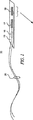
図1は、本発明の実施形態によるヒドロゲル層を取り込む柔軟回路の形態である電流測定バイオセンサ11である。このバイオセンサまたはセンサ11は、基板13(例えば、柔軟基板)上に形成され得る。1つ以上の電極15、17および19が、基板13の表面上に付着または結合され得る。このバイオセンサ11は、参照電極15、対電極17、および作業電極19とともに示される。別の実施形態では、1つ以上のさらなる作業電極がこの基板13上に含められ得る。電気ワイヤ21は、酸化または還元反応を持続するために電力を電極に伝達し得、そしてまた、測定されるパラメータの指標である信号電流を検出回路(図示せず)に運び得る。測定されるパラメータは、血液化学にある、またはそれから派生され得る目的の任意の分析物であり得る。1つの実施形態では、目的の分析物は、グルコースオキシダーゼとのグルコースの反応から形成される過酸化水素であり得、そしてそれ故、血中グルコース濃度に比例する濃度を有する。
FIG. 1 is an amperometric biosensor 11 in the form of a flexible circuit incorporating a hydrogel layer according to an embodiment of the present invention. The biosensor or sensor 11 can be formed on a substrate 13 (eg, a flexible substrate). One or
1つの実施形態では、上記ヒドロゲル層は、グルコースセンサ上での使用のためのグルコースオキシダーゼ酵素を含み得る。センサ11は、参照電極15、対電極17、および作業電極19をそれぞれ含み得る。作業電極19は、ヒドロゲルの層で少なくとも部分的に覆われ得、そして血液のような溶液に曝されるか、またはその内に浸漬され、酵素が血液中の特定の反応物と化学的に反応することを可能にする。この例では、作業電極19のグルコースオキシダーゼ酵素が、血液中のグルコースと反応する。
In one embodiment, the hydrogel layer may include a glucose oxidase enzyme for use on a glucose sensor. The sensor 11 can include a
センサ11は、電流測定原理に基づいて働き、ここで、作業電極19は、対電極17に対して正の電位で保持される。この正の電位は、グルコースオキシダーゼとのグルコース反応の結果である過酸化水素の酸化反応を持続するに十分である。それ故、作業電極19は、アノードとして機能し、そして上記酸化反応から生じる作業電極19の表面で産生される電子を集める。集められた電子は、電気回路として作業電極19に流れる。1つの実施形態では、作業電極はグルコースオキシダーゼで被覆され、グルコースの酸化が、作業電極19が約+450mVと約+650mVとの間の電位で保持されるとき、グルーコース分子毎について過酸化水素分子を生成する。産生された過酸化水素は、式:
H2O2 −1 → 2H+ + O2 0 +2e−
に従って、作業電極19の表面上で酸化する。
The sensor 11 works on the basis of the current measurement principle, where the working
H 2 O 2 −1 → 2H + + O 2 0 + 2e −
Accordingly, the surface of the working
この式は、2つの電子が酸化される過酸化水素分子毎に産生されることを示す。それ故、特定の条件下では、電流の量は、過酸化水素濃度に比例し得る。1つの過酸化水素分子が、作業電極で酸化されれグルコース分子毎について産生されるので、血中グルコース濃度と得られる電流との間には直線関係が存在し得る。読者は、電流測定グルコースバイオセンサの電子的感知理論に関するさらなる情報については以下の論文を参照し得る:J.Wang、「グルコースバイオセンサ:進歩および挑戦の40年」、Electroanalysis、Vol.13、No.12、983〜988(2001)。 This equation shows that two electrons are produced for each hydrogen peroxide molecule that is oxidized. Therefore, under certain conditions, the amount of current can be proportional to the hydrogen peroxide concentration. Since one hydrogen peroxide molecule is oxidized at the working electrode and produced for each glucose molecule, there can be a linear relationship between blood glucose concentration and the resulting current. The reader may refer to the following paper for further information on the electronic sensing theory of an amperometric glucose biosensor: Wang, “Glucose Biosensor: 40 Years of Progress and Challenge”, Electroanalysis, Vol. 13, no. 12, 983-988 (2001).
条件が、直線的相関関係を達成するために好ましいことを確実にするため、作業電極19は、所望の化学反応を促進するように設計される。電流測定センサー11では、化学は、柔軟回路基板の表面上の変動する組成の1つ以上の膜または層を付与することにより制御され得る。1つの実施形態では、基板13はポリイミド材料であり得、そして膜はヒドロゲル層であり得る。基板13は、電極およわび膜層を載せるための絶縁された構造を提供する。1つの実施形態では、カテーテル管腔内のような制限されたスペースにおける使用のために意図された柔軟回路に関して、基板13は、約0.015インチと約0.080インチとの間の幅、および約1.0〜2.0インチの長さを有し得る。膜層の厚みは、約0.5ミクロンと約10ミクロンとの間で変動し得る。1つの実施形態では、1つ以上の膜層は、約4ミクロン〜約5ミクロン範囲にある厚みを有し得る。
To ensure that conditions are preferred to achieve a linear correlation, the working
電気ワイヤ21は、従来の柔軟回路技法を用いて基板13上に形成された金属トレースに連結またははんだ付けされ得る。例えば、これらトレースは、金めっきされた銅であり得る。1つの実施形態では、上記センサー11は、柔軟回路が、3−ピンの、1mmピッチのZIF Molexコネクターのような従来の複数ピンコネクターに嵌合するタブに終わるように設計される。このような連結は、例えば、ポテンシオスタットまたはその他のコントローラを用いて、作業電極の励起および電流信号の測定を容易にする。
The
上記の電極15、17および19は、厚膜プロセスおよび市販され入手可能なインクを用いて基板13に付与され得る。1つの実施形態では、参照電極15は、基板13上に堆積または形成された銀/塩化銀タイプであり得る。この参照電極15は、対電極17および作業電極19の電位が確立され得る固定された電位を確立する。この参照電位は、ネルンスト電位である。銀/塩化銀電極について、この参照電位は、以下の半反応によって維持される:
Ag0 → Ag+ + e−
対電極17は、白金またはグラファイトのような伝導性材料から構築され得る。これらの材料は、厚膜プロセスそしてそれに合うように硬化を用いて基板13への付与のためのインクとして処方され得る。対電極17は、酸化化学から産生される電子の大部分を、血液溶液に伝導して戻す作業領域を提供する。そうでなければ、すべての電流は、参照電極15を通過する可能性があり、そしてその耐用年数を低減し得る。1つの実施形態では、上記対電極17は、作業電極19の表面積より大きい表面積を備えて形成され得る。
The
Ag 0 → Ag + + e −
The
作業電極19は、対電極17を形成するために用いられる材料と類似の白金/グラファイト材料を用いて形成され得る。その他の実施形態では、作業電極19は、その他の導電性材料から形成され得る。その作動は、ここまでは、その表面上で過酸化水素のアノード酸化を促進するとして説明されてきた。その他の実施形態が可能であり、しかし、作業電極19は、負の電位で保持され得る。この場合には、この作業電極19で産生される電流は、酸素の還元から生じ得る。
The working
1つの実施形態では、バイオセンサ11は、例えば、CVCを経由して患者中への静脈内挿入のためのプローブまたはカテーテル内に設置され得る。このバイオセンサ11は、作業電極19の表面への反応性ヒドロゲル層23の付加により患者の血流中に浸漬される間電流測定センサとして機能し得る。このヒドロゲル層23は、この層の基礎材料への反応物質の添加によって反応性にされる。
In one embodiment, the biosensor 11 can be placed in a probe or catheter for intravenous insertion into a patient via, for example, a CVC. This biosensor 11 can function as an amperometric sensor while immersed in the patient's bloodstream by the addition of a
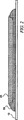
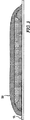
図2の拡大側面図は、作業電極19上に形成されたヒドロゲル層23の図解である。この図は、作業電極19の近傍の基板13の遠位部分を示す。図3に示される別の実施形態では、律速層25が、ヒドロゲル層23の頂部に付加され得、反応物質と反応する血液成分のヒドロゲル層23への拡散を選択的に可能にする。
The enlarged side view of FIG. 2 is an illustration of the
上記ヒドロゲル層23は、キトサン、少なくとも1つの反応物質、および少なくとも1つの架橋剤を含み得る。この少なくとも1つの架橋剤はキトサンに架橋し、上記少なくとも1つの作用剤を固定化するマトリックスを形成する。キトサン、反応物質、および架橋剤は、得られるヒドロゲル層全体に実質的に均一に分配され得る。
The
キトサンは、甲殻類の外骨格中の構造要素であり、そして地球上の最も一般的な多糖の1つであるキチン由来である。このキトサンは、その生体適合性、その利用可能性、その架橋親和性、およびその接着性質に基づき、ヒドロゲル層23のための基礎材料として選択される。このキトサンは、非毒性であり、そして移植されたセンサの表面上に血小板接着または血栓を阻止することが見出されている。さらに、このキトサンは、金属表面上に硬い透明層を形成するようにされ得、そして塩水環境で金属ブリッジ上の保護スプレー被覆として有効に用いられている。
Chitosan is a structural element in the crustacean exoskeleton and is derived from chitin, one of the most common polysaccharides on earth. This chitosan is selected as the base material for the
上記ヒドロゲル層23は、キトサンに添加され得る少なくとも1つの反応物質を含む。1つの実施形態では、この反応物質は、オキシダーゼ酵素のような酵素であり得る。グルコースバイオセンサのための実施形態では、この反応物質は、Aspergillus niger(EC1.1.3.4)、タイプIIまたはタイプVII由来であり得るようなグルコースオキシダーゼであり得る。この反応物質の血液との反応を促進するために、この反応物質は、この反応物質の特定量がヒドロゲル表面に剥き出るようにヒドロゲル層の全体に均一に分配され得る。これは、この反応物質をキトサンに添加または架橋することにより達成され得る。ヒドロゲル層23は、それが膨潤し得、血液からこの反応物質に血液中の反応体(例えば、グルコース)の能動輸送を提供するように、水吸収剤であり得る。それ故、分子間結合がヒドロゲル層23全体に形成され得、接着および所定密度のマトリックスを生成し、ヒドロゲル表面を横切り、そしてヒドロゲル層23全体に反応物質の均一な分散を可能にする。反応産物が、次いで、電極層に伝達され得る。
The
しかし、キトサンはそれ単独では、反応物質を含むために必要な適切な頑丈さを提供するには不十分であり得る。従って、ジェニピンのような架橋剤が、十分な機械的強度および固定化性質をもつマトリックスを形成するためにキトサンに添加され得る。クチナシ属果実抽出物に見出される活性化合物であるジェニピンは、伝統的中国医療で用いられる草本医療である。ジェニピンは、身体内にあるとき、無害に二酸化炭素および水に分解する生体適合性かつ非毒性の架橋剤である。 However, chitosan by itself may not be sufficient to provide the proper robustness needed to contain the reactants. Thus, a cross-linking agent such as genipin can be added to chitosan to form a matrix with sufficient mechanical strength and immobilization properties. Genipin, an active compound found in gardenia fruit extracts, is a herbal medicine used in traditional Chinese medicine. Genipin is a biocompatible and non-toxic crosslinking agent that decomposes harmlessly into carbon dioxide and water when in the body.
キトサンにジェニピンを架橋することは、ヒドロゲル層23が作業電極19の表面上で無傷なままであるように、その機械的頑丈さを確実にする。この性質は、製造の間、そしてまた、インビボにおける設置の間、例えばCVCカテーテルを経由する測定位置への輸送の間に、ヒドロゲル層23の一体性を確実にするために重要である。ジェニピンのキトサンとの架橋適合性は、J.Bergerら、「生物医療適用のための共有結合およびイオン結合により架橋されたキトサンヒドロゲルにおける構造および相互作用」Europ.J.Pharm.Biopharm、57巻、No.1、19〜34(2004)に記載されている。
Crosslinking genipin to chitosan ensures its mechanical robustness so that the
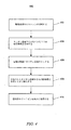
実験的試行に基づき、電極上に反応物質を固定化するためのヒドロゲルを形成する方法またはプロセスとして証明された方法が開発され、そして本明細書中に開示されるその物質が本明細書中に開示される。図4は、方法400の1つのこのような実施形態を示す。
Based on experimental trials, a method has been developed as a method or process for forming a hydrogel for immobilizing a reactant on an electrode, and the material disclosed herein is herein incorporated by reference. Disclosed. FIG. 4 illustrates one such embodiment of the
ステップ402では、キトサンが酢酸溶液中に溶解される。例えば、1つの実施形態では、このキトサンは、酢酸の溶液中に溶解され得、約0.1〜約2.0重量%のキトサン濃度を生成する。あるいは、キトサンは、約1%〜5%の濃度を有する酢酸溶液中に溶解され得る。ステップ402は、キトサンが溶解されるまでこの溶液をかき混ぜるか、または撹拌し得る。ステップ404では、オキシダーゼまたはその他の酵素のような少なくとも1つの反応物質が、キトサン溶液に添加され得る。ステップ404はまた、上記溶液を撹拌または混合することを含み得、そしてこの溶液が粘性液体またはゲルを形成するまで進行し得る。1つの実施形態では、ステップ404は、(溶液に対して)約1/120,000と約1/10,000重量%オキシダーゼとの間のオキシダーゼ濃度を生じ得る。
In
ステップ406では、上記キトサン溶液またはゲルは、電極の表面に付与され得る。キトサンの付与は、実質的に均一な層でこの表面を部分的または完全に被覆し得る。ステップ408では、付与されたキトサンは、それが電極表面上に固体膜を形成するまで硬化され得る。ステップ408は、室温で、または低加熱下で実施され得る。ステップ410では、反応物質は、ヒドロゲル層中に固定化される。これは、上記固体膜をジェニピン溶液に浸漬することによって達成され得、ジェニピンのキトサンへの架橋を促進する。1つの実施形態では、膜で被覆された電極は、約2時間〜約10時間の間浸漬され得る。その他の実施形態では、上記ジェニピン溶液は、容量に対して約1%重量のジェニピンの有し、そして約4と約10との間のpH値を有するリン酸塩またはクエン酸緩衝液であり得る。必要に応じて、架橋されたジェニピンおよびキトサンを硬化した後、ジェニピンおよびキトサンのより密な架橋層が上記硬化された架橋ジェニピンおよびキトサンに付加され得、上記反応物質と反応する血液成分を選択的に拡散するための律速層を提供する。
In
(実験結果)
プロトタイプグルコースセンサの集団が、白金電極上に形成されたキトサン−ジェニピンヒドロゲル中にグルコースオキシダーゼを固定化するために方法400を用いて実験室中で製作された。これらグルコースセンサは、種々の機械的および化学的性質について試験された。試験された主な機械的性質は、接着、および浸漬後の接着であった。試験された主な化学的性質は、グルコース応答であった。タイプ7グルコースオキシダーゼをこれら実験のために用いた。以下の開示は、試験センサ製作手順および上記試験センサ集団上で実施された主な試験から得られた結果を提示する。
(Experimental result)
A population of prototype glucose sensors was fabricated in the
すべての試験について、試験センサは、10mm×10mmパッチ中のセンサ基板上に製作され、かつ堆積された白金インクを用いて製作された。この白金は、炭素/グラファイトインクを用いて付与され、柔軟基板への良好な接着を確実にした。硬化後、インクは、固体白金パッドを形成した。センサ基板は、柔軟センサ、この柔軟センサ中にエッチングされた回路トレース、およびそれに接続されたワイヤを備えた薄い金属層上に堆積されたエポキシ層であった。 For all tests, test sensors were fabricated using platinum ink fabricated and deposited on a sensor substrate in a 10 mm x 10 mm patch. This platinum was applied using a carbon / graphite ink to ensure good adhesion to the flexible substrate. After curing, the ink formed a solid platinum pad. The sensor substrate was an epoxy layer deposited on a thin metal layer with a flexible sensor, circuit traces etched into the flexible sensor, and wires connected to it.
接着試験は、ジェニピンなしのキトサン−グルコースオキシダーゼ溶液からヒドロゲルが付与された試験センサの第1の集団に対して実施された。すべてが1%酢酸中に0.25wt%キトサンを含む2000μL溶液の異なる処方物が作製された。各溶液は、キトサンを酢酸中に溶解し、そして粘性ゲルを形成するために氷浴中で拡散することにより作製された。グルコースオキシダーゼは、上記溶液中に添加され、そして溶解され、黄色のゲル材料−すなわち、ヒドロゲルを形成した。グルコースオキシダーゼの量は、異なる試験センサについて変動され、そして、0、1/160,000、1/140,000、および1/120,000wt%グルコースオキシダーゼから作製されたヒドロゲル溶液を含んだ。ヒドロゲルは、乾燥された白金インク領域に付与され、そして硬化させた。いくつかの試験センサは、周囲空気中で硬化され、そしてその他は低温オーブン中で硬化された。硬化後、ヒドロゲル層は、硬い透明な膜を形成した。 The adhesion test was performed on a first population of test sensors to which a hydrogel was applied from a chitosan-glucose oxidase solution without genipin. Different formulations of 2000 μL solutions were made, all containing 0.25 wt% chitosan in 1% acetic acid. Each solution was made by dissolving chitosan in acetic acid and diffusing in an ice bath to form a viscous gel. Glucose oxidase was added to the solution and dissolved to form a yellow gel material-ie, a hydrogel. The amount of glucose oxidase was varied for different test sensors and included hydrogel solutions made from 0, 1 / 160,000, 1 / 140,000, and 1 / 120,000 wt% glucose oxidase. The hydrogel was applied to the dried platinum ink area and allowed to cure. Some test sensors were cured in ambient air and others were cured in a low temperature oven. After curing, the hydrogel layer formed a hard transparent film.
接着試験には、各10mm×10mmパッチは、各立方体が1mm×1mmである100の立方体を形成するようにスコアされた。ASTMテープ試験標準が、ヒドロゲルの白金電極表面への接着を決定するために用いられた。このテープは付与され、そして除去され、そしてパッチが、ヒドロゲル立方体の存在または不在について検査された。最良の毛結果は、1/40,000wt%および1/60,000wt%グルコースオキシダーゼから形成された風乾ヒドロゲルから得られた。 For the adhesion test, each 10 mm x 10 mm patch was scored to form 100 cubes where each cube was 1 mm x 1 mm. An ASTM tape test standard was used to determine adhesion of the hydrogel to the platinum electrode surface. The tape was applied and removed, and the patches were inspected for the presence or absence of hydrogel cubes. The best hair results were obtained from air-dried hydrogels formed from 1 / 40,000 wt% and 1 / 60,000 wt% glucose oxidase.
この風乾試験センサは、次いで、体温で約7.4のpHをもつリン酸緩衝液中に浸された。この浸漬試験の目的は、シミュレーションされた静脈内環境中でヒドロゲル接着の耐久性を試験することであった。所定の時間の間の浸漬の後、テープを用いた接着試験が再び実施された。しかし、浸漬後すぐに、緩衝溶液中に色素の曇りが出現し、そしてすべてのヒドロゲル層は溶解するようであった。 The air dry test sensor was then immersed in a phosphate buffer having a pH of about 7.4 at body temperature. The purpose of this immersion test was to test the durability of hydrogel adhesion in a simulated intravenous environment. After soaking for a predetermined time, the adhesion test with tape was performed again. However, immediately after soaking, a cloud of dye appeared in the buffer solution and all the hydrogel layers appeared to dissolve.
第2の接着試験は、第2の集団の試験センサを用いて実施された。この試験では、ヒドロゲルは、キトサン、グルコースオキシダーゼ、およびジェニピンを含んだ。これら第2の集団の試験センサは、上記第1の集団とほぼ同一に製作された。試験センサの第2の集団に用いられたキトサン溶液は、100mlの1%酢酸中の1.0wt.%キトサンであった。ヒドロゲルは、4mLのキトサン溶液中の15mgのグルコースオキシダーゼを用いて調製された。ヒドロゲル層にジェニピンを添加するためのさらなるステップがとられた。 A second adhesion test was performed using a second population of test sensors. In this test, the hydrogel contained chitosan, glucose oxidase, and genipin. These second group of test sensors were fabricated almost identically to the first group. The chitosan solution used for the second population of test sensors was 1.0 wt. % Chitosan. The hydrogel was prepared using 15 mg glucose oxidase in 4 mL chitosan solution. Additional steps were taken to add genipin to the hydrogel layer.
キトサン膜をジェニピンに架橋するために、ジェニピン溶液の異なるpHレベルが試験された。ジェニピンのpHに依存して、ジェニピンはそれ自体に最初架橋し(pH7〜10)、ジェニピンとキトサンとの間の大きな結合を生成する長い鎖を形成するか、またはより低いpH(4〜5)で、ジェニピンはキトサンに直接架橋し得、そしてヒドロゲルの機械的性質を変化し得る、ポリマースペーサーに代わる単一ユニットのスペーサーを有し得る。ジェニピンは、それ故、生体適合性緩衝液から形成される異なるpHの溶液に溶解された。第1の緩衝液は、約7と約10との間のpHでリン酸から形成され、そして第2の緩衝液は、約4と約5との間のpHでクエン酸から形成された。この第2の緩衝液は、0.1Mのクエン酸ナトリウムおよび0.1Mのクエン酸から形成された。 In order to crosslink the chitosan membrane to genipin, different pH levels of the genipin solution were tested. Depending on the pH of genipin, genipin initially crosslinks itself (pH 7-10) to form a long chain that creates a large bond between genipin and chitosan, or at a lower pH (4-5) Thus, genipin can directly crosslink to chitosan and can have a single unit spacer instead of a polymer spacer that can change the mechanical properties of the hydrogel. Genipin was therefore dissolved in different pH solutions formed from biocompatible buffers. The first buffer was formed from phosphate at a pH between about 7 and about 10, and the second buffer was formed from citrate at a pH between about 4 and about 5. This second buffer was formed from 0.1 M sodium citrate and 0.1 M citric acid.
第2の集団の試験センサを製作する際の次のステップは、キトサンおよびグルコースオキシダーゼの硬化された層をあるジェニピン溶液または他のジェニピン溶液に浸すことであった。1つのグループは、4.98のpHを有するクエン酸緩衝液中に6時間の間浸し、そして別のグループは、7.54のpHを有するリン酸緩衝液中に6時間の間浸した。この曝露は、キトサンとジェニピンとの間の架橋反応を生成した。次に、マトリックスは、周囲空気中で一晩乾燥することにより再び硬化された。これらセンサーは、第1の集団について用いられた同じ手順を用いて、接着、および浸漬後の接着について試験された。 The next step in fabricating the second population of test sensors was to immerse the cured layer of chitosan and glucose oxidase in one or other genipin solutions. One group was soaked in citrate buffer having a pH of 4.98 for 6 hours, and another group was soaked in phosphate buffer having a pH of 7.54 for 6 hours. This exposure produced a cross-linking reaction between chitosan and genipin. The matrix was then cured again by drying overnight in ambient air. These sensors were tested for adhesion and adhesion after immersion using the same procedure used for the first population.
結果は、静脈内環境をシミュレートするリン酸緩衝液中に浸した後でさえ、ヒドロゲル層の電極表面への優れた接着を示した。優れた結果は、4.98のpHのジェニピン緩衝液から作製された試験センサから得られた。これらの内で、4つの試験センサからの3つが、少なくとも24時間のリン酸緩衝液中に浸した後に実施されたASTM接着試験の結果として、除去された3つ以下の立方体を有していた。 The results showed excellent adhesion of the hydrogel layer to the electrode surface even after soaking in a phosphate buffer simulating an intravenous environment. Excellent results were obtained from test sensors made from genipin buffer with a pH of 4.98. Of these, 3 out of 4 test sensors had no more than 3 cubes removed as a result of ASTM adhesion tests performed after soaking in phosphate buffer for at least 24 hours. .
グルコース応答を、第2の集団の試験サンプルからの別のサンプルを用いて試験した。このサンプルは、4.98−pHグループと7.54−pHグループとに分割した。単純さのために、これらは、5−pHグループおよび7−pHグループとそれぞれ称される。生理食塩水中に15.5時間の間これらグループからの各試験センサを浸した後、各センサを、約650mVの電位に励起された作業電極とともに既知のグルコース濃度に曝した。各センサについて、電極に生じた電流を、50、100、および150mg/dLのグルコース濃度で測定した。 The glucose response was tested with another sample from the second population of test samples. This sample was divided into 4.98-pH group and 7.54-pH group. For simplicity, these are referred to as 5-pH group and 7-pH group, respectively. After immersing each test sensor from these groups in saline for 15.5 hours, each sensor was exposed to a known glucose concentration with a working electrode excited to a potential of about 650 mV. For each sensor, the current generated in the electrodes was measured at glucose concentrations of 50, 100, and 150 mg / dL.
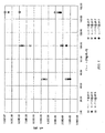
図5のグラフは、第2の集団からの7つのセンサに対するグルコースアッセイの結果を示す。すべては、グルコース濃度の関数として電流の直線状応答特徴を示した。上記5−pHグループは、より良好な直線性および優れた電流出力を示した。この5−pHグループは、グルコースオキシダーゼを包括するためのより強い機械的性質を有すると考えられるので、これらの結果は、より高い電流応答がヒドロゲル層内に架橋または包括されるグルコースオキシダーゼの量を増加することによって得られ得ることを示す。 The graph of FIG. 5 shows the results of a glucose assay for seven sensors from the second population. All showed a linear response characteristic of current as a function of glucose concentration. The 5-pH group showed better linearity and excellent current output. Since this 5-pH group is believed to have stronger mechanical properties for inclusion of glucose oxidase, these results indicate that the amount of glucose oxidase in which a higher current response is crosslinked or entrapped within the hydrogel layer. It can be obtained by increasing.
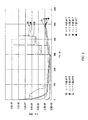
図6のグラフは、電流出力の関数として同じセンサに対するグルコースアッセイの結果を経時的に示す。示される時間の期間は、50、100、および150mg/dL濃度に対応するグルコース濃度における3ステップ変化をカバーする。各ステップ変化と一致する電流信号中の初期の一時的スパイクの後、各濃度における応答は、定常状態応答に迅速に平らになる。グルコースレベルにおけるステップ変化の間の経時的な定常応答は、グルコースオキシダーゼが、ヒドロゲルマトリックス内で事実上均一に分散され、そして経時的に直線状出力を持続し得ることを示す。グルコースオキシダーゼの接着された層は、グルコースのような反応体の存在下で経時的に維持され、これは、グルコースオキシダーゼの接着がまた安定であり、そしてキトサン−ジェニピンマトリックス内に固定されながら、グルコースとの、および電極との接触を維持し得ることを示す。 The graph in FIG. 6 shows the glucose assay results over time for the same sensor as a function of current output. The time period shown covers three step changes in glucose concentration corresponding to 50, 100, and 150 mg / dL concentrations. After an initial transient spike in the current signal consistent with each step change, the response at each concentration quickly flattens to a steady state response. The steady-state response over time during the step change in glucose level indicates that glucose oxidase is virtually uniformly dispersed within the hydrogel matrix and can maintain a linear output over time. The adhered layer of glucose oxidase is maintained over time in the presence of a reactant such as glucose, while the adhesion of glucose oxidase is also stable and immobilized within the chitosan-genipin matrix. It shows that contact with glucose and with electrodes can be maintained.
図5および6に示される直線状応答は、本発明によるヒドロゲルがまた、血漿とグルコースオキシダーゼとの間の律速層25として機能し得ることを示す。すなわち、キトサンは、所望の酸素感受性を得るような様式、そしてジェニピンおよびグルコースオキシダーゼに比例して架橋され得る。この所望の酸素感受性は、多量の酸素が、グルコースを選択的に通過しながら、ヒドロゲル層を通過することを可能にし、グルコースオキシダーゼとの酸化反応が、利用可能なグルコースによって制限されることを確実にする。
The linear response shown in FIGS. 5 and 6 shows that the hydrogel according to the invention can also function as a
最後に、1/20,000〜1/60,000wt.%グルコースオキシダーゼのいずれかを用い、ジェニピン緩衝液のpHが一定であったと仮定して、約0.25〜1.0wt.%の範囲にある濃度を有するキトサン溶液中で混合されて製作されたセンサの反応性に有意な変化はなかった。これら条件下では、キトサン中の1/120,000グルコースオキシダーゼ程度の低い濃度が、測定可能でかつ直線状電流応答を提供するし得ると考えられる。 Finally, 1/20000 to 1 / 60,000 wt. % Glucose oxidase and assuming that the pH of the genipin buffer was constant, about 0.25-1.0 wt. There was no significant change in the reactivity of sensors made by mixing in chitosan solutions having concentrations in the% range. Under these conditions, it is believed that concentrations as low as 1 / 120,000 glucose oxidase in chitosan can be measured and provide a linear current response.
本発明を、例示の様式で開示した。従って、全体で採用される用語は、制限する様式よりもむしろ例示で読まれるべきである。本発明の小さな改変が当業者に生じ、本明細書で保証される特許の範囲内に境界を定められることが意図されるものは、本明細書によって寄与される当該技術の進歩の範囲内に合理的に入るようなすべての実施形態であり、しかも、添付の請求項およびそれらの相当物を考慮することを除いてその範囲は制限されないことが理解されるべきである。 The invention has been disclosed in an illustrative manner. Accordingly, the terms employed throughout should be read by way of example rather than in a limiting manner. It is intended that minor modifications of the present invention occur to those skilled in the art and are intended to be bound within the scope of the patents warranted herein, within the scope of the technical advances contributed by this specification. It should be understood that all embodiments are within reasonable scope and that the scope is not limited except in light of the appended claims and their equivalents.
本発明の特徴、目的、および利点は、図面と組み合わせて考慮されるとき、以下に提示される詳細な説明からより明らかになる。
Claims (11)
可撓性基質;
該基質に結合された電極;および
該電極に結合されたヒドロゲル層であって、キトサン、オキシダーゼ酵素、および該キトサンに架橋され、該オキシダーゼ酵素を固定化するマトリックスを形成するジェニピンを含む、ヒドロゲル層、
を含む、静脈内電流測定バイオセンサ。An intravenous current measurement biosensor comprising:
Flexible substrate;
An electrode coupled to the substrate; and a hydrogel layer coupled to the electrode, comprising a chitosan, an oxidase enzyme , and a genipin crosslinked to the chitosan and forming a matrix immobilizing the oxidase enzyme ,
Intravenous amperometric biosensor.
キトサンを酸性溶液に溶解する工程;
該キトサン溶液にオキシダーゼを添加する工程;
該キトサン溶液を該電極の表面に付与する工程;
付与された該キトサン溶液をそれが固体膜を形成するまで硬化する工程;および
該固体膜をジェニピン溶液中に浸漬し、該キトサンおよび該ジェニピンを架橋し、それによって該オキシダーゼを固定化する工程、を包含する、方法。A method of forming a hydrogel for immobilizing an oxidase on an electrode comprising:
Dissolving chitosan in an acidic solution;
Adding oxidase to the chitosan solution;
Applying the chitosan solution to the surface of the electrode;
Curing the applied chitosan solution until it forms a solid film; and immersing the solid film in a genipin solution to cross-link the chitosan and the genipin, thereby immobilizing the oxidase ; Including the method.
Applications Claiming Priority (3)
| Application Number | Priority Date | Filing Date | Title |
|---|---|---|---|
| US77725406P | 2006-02-27 | 2006-02-27 | |
| US60/777,254 | 2006-02-27 | ||
| PCT/US2007/004536 WO2007100588A1 (en) | 2006-02-27 | 2007-02-20 | Hydrogel for an intravenous amperometric biosensor |
Publications (2)
| Publication Number | Publication Date |
|---|---|
| JP2009528083A JP2009528083A (en) | 2009-08-06 |
| JP5116697B2 true JP5116697B2 (en) | 2013-01-09 |
Family
ID=38261532
Family Applications (1)
| Application Number | Title | Priority Date | Filing Date |
|---|---|---|---|
| JP2008556412A Expired - Fee Related JP5116697B2 (en) | 2006-02-27 | 2007-02-20 | Hydrogels for intravenous amperometric biosensors |
Country Status (8)
| Country | Link |
|---|---|
| US (1) | US20080029390A1 (en) |
| EP (1) | EP1991111B1 (en) |
| JP (1) | JP5116697B2 (en) |
| CN (1) | CN101360449A (en) |
| AT (1) | ATE504236T1 (en) |
| CA (1) | CA2630537A1 (en) |
| DE (1) | DE602007013718D1 (en) |
| WO (1) | WO2007100588A1 (en) |
Families Citing this family (60)
| Publication number | Priority date | Publication date | Assignee | Title |
|---|---|---|---|---|
| US7885697B2 (en) | 2004-07-13 | 2011-02-08 | Dexcom, Inc. | Transcutaneous analyte sensor |
| US20190357827A1 (en) | 2003-08-01 | 2019-11-28 | Dexcom, Inc. | Analyte sensor |
| US8626257B2 (en) | 2003-08-01 | 2014-01-07 | Dexcom, Inc. | Analyte sensor |
| US7591801B2 (en) | 2004-02-26 | 2009-09-22 | Dexcom, Inc. | Integrated delivery device for continuous glucose sensor |
| US8886273B2 (en) | 2003-08-01 | 2014-11-11 | Dexcom, Inc. | Analyte sensor |
| US20080119703A1 (en) * | 2006-10-04 | 2008-05-22 | Mark Brister | Analyte sensor |
| US7920906B2 (en) | 2005-03-10 | 2011-04-05 | Dexcom, Inc. | System and methods for processing analyte sensor data for sensor calibration |
| US9247900B2 (en) | 2004-07-13 | 2016-02-02 | Dexcom, Inc. | Analyte sensor |
| US8615282B2 (en) * | 2004-07-13 | 2013-12-24 | Dexcom, Inc. | Analyte sensor |
| US8425417B2 (en) * | 2003-12-05 | 2013-04-23 | Dexcom, Inc. | Integrated device for continuous in vivo analyte detection and simultaneous control of an infusion device |
| US8364230B2 (en) * | 2006-10-04 | 2013-01-29 | Dexcom, Inc. | Analyte sensor |
| US20080197024A1 (en) * | 2003-12-05 | 2008-08-21 | Dexcom, Inc. | Analyte sensor |
| US8425416B2 (en) * | 2006-10-04 | 2013-04-23 | Dexcom, Inc. | Analyte sensor |
| US8364231B2 (en) | 2006-10-04 | 2013-01-29 | Dexcom, Inc. | Analyte sensor |
| US8808228B2 (en) | 2004-02-26 | 2014-08-19 | Dexcom, Inc. | Integrated medicament delivery device for use with continuous analyte sensor |
| US20090143658A1 (en) * | 2006-02-27 | 2009-06-04 | Edwards Lifesciences Corporation | Analyte sensor |
| US8298142B2 (en) | 2006-10-04 | 2012-10-30 | Dexcom, Inc. | Analyte sensor |
| US8449464B2 (en) * | 2006-10-04 | 2013-05-28 | Dexcom, Inc. | Analyte sensor |
| US8275438B2 (en) * | 2006-10-04 | 2012-09-25 | Dexcom, Inc. | Analyte sensor |
| US8447376B2 (en) | 2006-10-04 | 2013-05-21 | Dexcom, Inc. | Analyte sensor |
| US8562528B2 (en) * | 2006-10-04 | 2013-10-22 | Dexcom, Inc. | Analyte sensor |
| US8478377B2 (en) * | 2006-10-04 | 2013-07-02 | Dexcom, Inc. | Analyte sensor |
| US20080306434A1 (en) | 2007-06-08 | 2008-12-11 | Dexcom, Inc. | Integrated medicament delivery device for use with continuous analyte sensor |
| EP4159114B1 (en) | 2007-10-09 | 2024-04-10 | DexCom, Inc. | Integrated insulin delivery system with continuous glucose sensor |
| US8000918B2 (en) | 2007-10-23 | 2011-08-16 | Edwards Lifesciences Corporation | Monitoring and compensating for temperature-related error in an electrochemical sensor |
| EP2217914A1 (en) * | 2007-11-02 | 2010-08-18 | Edwards Lifesciences Corporation | Analyte monitoring system having back-up power source for use in either transport of the system or primary power loss |
| US20090188811A1 (en) * | 2007-11-28 | 2009-07-30 | Edwards Lifesciences Corporation | Preparation and maintenance of sensors |
| CA2715628A1 (en) | 2008-02-21 | 2009-08-27 | Dexcom, Inc. | Systems and methods for processing, transmitting and displaying sensor data |
| US8396528B2 (en) | 2008-03-25 | 2013-03-12 | Dexcom, Inc. | Analyte sensor |
| US20100072062A1 (en) * | 2008-05-05 | 2010-03-25 | Edwards Lifesciences Corporation | Membrane For Use With Amperometric Sensors |
| PL235185B1 (en) | 2008-07-02 | 2020-06-01 | Univ Jagiellonski | Application of network chitosan polymer in heparin removal |
| US8900431B2 (en) * | 2008-08-27 | 2014-12-02 | Edwards Lifesciences Corporation | Analyte sensor |
| CA2741853A1 (en) * | 2008-10-31 | 2010-05-06 | Edwards Lifesciences Corporation | Analyte sensor with non-working electrode layer |
| US20100160756A1 (en) * | 2008-12-24 | 2010-06-24 | Edwards Lifesciences Corporation | Membrane Layer for Electrochemical Biosensor and Method of Accommodating Electromagnetic and Radiofrequency Fields |
| WO2010147158A1 (en) * | 2009-06-19 | 2010-12-23 | 日産化学工業株式会社 | Gel enclosing a compound by means of low-molecular weight gelling agent |
| US20110054284A1 (en) * | 2009-08-28 | 2011-03-03 | Edwards Lifesciences Corporation | Anti-Coagulant Calibrant Infusion Fluid Source |
| EP2473963A4 (en) | 2009-08-31 | 2014-01-08 | Abbott Diabetes Care Inc | MEDICAL DEVICES AND METHODS |
| US8858883B2 (en) * | 2010-12-02 | 2014-10-14 | University Of Maryland, College Park | Method and system for capture and use of intact vesicles on electrodeposited hydrophobically modified biopolymer films |
| ES2847578T3 (en) | 2011-04-15 | 2021-08-03 | Dexcom Inc | Advanced analyte sensor calibration and error detection |
| EP3427598B1 (en) | 2011-11-07 | 2020-02-12 | Wild Flavors, Inc. | Method for preparing a colorant from a genipin-rich extract of genipa americana |
| US8992751B2 (en) * | 2012-04-09 | 2015-03-31 | Compose Element Limited | Test strips and preparation method thereof |
| HK1217364A1 (en) * | 2013-01-11 | 2017-01-06 | Northeastern University | Saliva glucose monitoring system |
| WO2014165023A1 (en) | 2013-03-12 | 2014-10-09 | Carnegie Mellon University | Coated vaso-occclusive device for treatment of aneurysms |
| DE102014112972A1 (en) * | 2013-09-12 | 2015-03-12 | Endress + Hauser Conducta Gesellschaft für Mess- und Regeltechnik mbH + Co. KG | Measuring diaphragm for an optochemical or amperometric sensor |
| WO2016065190A1 (en) | 2014-10-23 | 2016-04-28 | Abbott Diabetes Care Inc. | Electrodes having at least one sensing structure and methods for making and using the same |
| GB2538724A (en) * | 2015-05-26 | 2016-11-30 | Imp Innovations Ltd | Methods |
| JP6954301B2 (en) * | 2016-11-08 | 2021-10-27 | Jsr株式会社 | Enzyme sensor and enzyme sensor system |
| KR102037800B1 (en) * | 2017-04-04 | 2019-10-30 | 주식회사 스칼라팍스트롯 | Biochip |
| WO2018186657A1 (en) * | 2017-04-04 | 2018-10-11 | 주식회사 스칼라팍스트롯 | Biochip |
| WO2019083939A1 (en) | 2017-10-24 | 2019-05-02 | Dexcom, Inc. | Pre-connected analyte sensors |
| US11331022B2 (en) | 2017-10-24 | 2022-05-17 | Dexcom, Inc. | Pre-connected analyte sensors |
| WO2019094387A1 (en) * | 2017-11-08 | 2019-05-16 | The Regents Of The University Of Colorado, A Body Corporate | Hydrogel drug delivery systems for the treatment of pediatric growth plate injuries |
| US11918348B2 (en) | 2017-12-05 | 2024-03-05 | Abbott Diabetes Care Inc. | Medical devices having a dynamic surface profile and methods for production and use thereof |
| EP4218567B1 (en) | 2018-06-07 | 2025-03-12 | Abbott Diabetes Care, Inc. | Focused sterilization and sterilized sub-assemblies for analyte monitoring systems |
| EP3805304A4 (en) * | 2018-06-08 | 2022-03-02 | i-Sens, Inc. | CROSSLINKER WITH GENIPIN FOR USE IN MAKING A SENSOR FILM OR A DIFFUSION CONTROL FILM OF AN ELECTROCHEMICAL SENSOR |
| WO2021145307A1 (en) * | 2020-01-16 | 2021-07-22 | 富士フイルム株式会社 | Organism mucous membrane model, and method for evaluating retention properties of mucous-membrane-protective agent |
| CN111839532B (en) * | 2020-07-14 | 2024-11-05 | 天津大学 | A flexible epidermal electrochemical biosensor based on conductive hydrogel |
| CA3186905A1 (en) | 2020-09-15 | 2022-03-24 | Abbott Diabetes Care Inc. | Systems, devices, and methods for an analyte sensor |
| CN112642018B (en) * | 2020-12-18 | 2023-05-23 | 河南科技大学第一附属医院 | Blood sugar detection device matched with venous indwelling needle and detection method thereof |
| WO2023215131A1 (en) * | 2022-05-03 | 2023-11-09 | Massachusetts Institute Of Technology | Flexible electronics for analyte detection |
Family Cites Families (21)
| Publication number | Priority date | Publication date | Assignee | Title |
|---|---|---|---|---|
| JPS5928358Y2 (en) * | 1978-08-28 | 1984-08-16 | 株式会社クラレ | Medical FET sensor |
| US4542291A (en) * | 1982-09-29 | 1985-09-17 | Vpl Research Inc. | Optical flex sensor |
| US4937444A (en) * | 1982-09-29 | 1990-06-26 | Vpl Research, Inc. | Optical flex sensor |
| JPH0716409B2 (en) * | 1986-12-19 | 1995-03-01 | サントリー株式会社 | Method of immobilizing enzyme |
| GB8725936D0 (en) * | 1987-11-05 | 1987-12-09 | Genetics Int Inc | Sensing system |
| US5322063A (en) * | 1991-10-04 | 1994-06-21 | Eli Lilly And Company | Hydrophilic polyurethane membranes for electrochemical glucose sensors |
| US5423883A (en) * | 1993-07-14 | 1995-06-13 | Pacesetter, Inc. | Implantable myocardial stimulation lead with sensors thereon |
| JPH0980010A (en) * | 1995-09-08 | 1997-03-28 | Daikin Ind Ltd | Disposable enzyme electrode and method for manufacturing the same |
| JPH09182738A (en) * | 1995-12-28 | 1997-07-15 | Fujitsu Ltd | Oxygen electrode and method for manufacturing the same |
| AU2067597A (en) * | 1996-03-07 | 1997-09-22 | Axon Engineering, Inc. | Polymer-metal foil structure for neural stimulating electrodes |
| JP2910670B2 (en) * | 1996-04-12 | 1999-06-23 | 日本電気株式会社 | Semiconductor mounting structure |
| GB9801286D0 (en) * | 1998-01-21 | 1998-03-18 | Univ Cambridge Tech | Sensor |
| CA2265119C (en) * | 1998-03-13 | 2002-12-03 | Cygnus, Inc. | Biosensor, iontophoretic sampling system, and methods of use thereof |
| WO2002089664A2 (en) * | 2001-05-03 | 2002-11-14 | Masimo Corporation | Flex circuit shielded optical sensor and method of fabricating the same |
| CA2476656C (en) * | 2002-02-21 | 2008-11-25 | Encelle, Inc. | Immobilized bioactive hydrogel matrices as surface coatings |
| EP1545505A4 (en) * | 2002-08-02 | 2008-01-02 | Gp Medical | Drug-loaded biological material chemically treated with genipin |
| US6885107B2 (en) * | 2002-08-29 | 2005-04-26 | Micron Technology, Inc. | Flip-chip image sensor packages and methods of fabrication |
| US7883615B2 (en) * | 2003-02-12 | 2011-02-08 | University Of Maryland, College Park | Controlled electrochemical deposition of polysaccharide films and hydrogels, and materials formed therefrom |
| WO2005061127A1 (en) * | 2003-12-11 | 2005-07-07 | University Of Maryland College Park | Biolithographical deposition, and materials and devices formed therefrom |
| JP4593957B2 (en) * | 2004-03-31 | 2010-12-08 | テルモ株式会社 | Electro-optical detector |
| US7282194B2 (en) * | 2004-10-05 | 2007-10-16 | Gp Medical, Inc. | Nanoparticles for protein drug delivery |
-
2007
- 2007-02-20 JP JP2008556412A patent/JP5116697B2/en not_active Expired - Fee Related
- 2007-02-20 AT AT07751306T patent/ATE504236T1/en not_active IP Right Cessation
- 2007-02-20 WO PCT/US2007/004536 patent/WO2007100588A1/en not_active Ceased
- 2007-02-20 CA CA002630537A patent/CA2630537A1/en not_active Abandoned
- 2007-02-20 DE DE602007013718T patent/DE602007013718D1/en active Active
- 2007-02-20 CN CNA2007800014680A patent/CN101360449A/en active Pending
- 2007-02-20 US US11/709,114 patent/US20080029390A1/en not_active Abandoned
- 2007-02-20 EP EP07751306A patent/EP1991111B1/en not_active Not-in-force
Also Published As
| Publication number | Publication date |
|---|---|
| EP1991111A1 (en) | 2008-11-19 |
| WO2007100588A1 (en) | 2007-09-07 |
| CN101360449A (en) | 2009-02-04 |
| CA2630537A1 (en) | 2007-09-07 |
| US20080029390A1 (en) | 2008-02-07 |
| DE602007013718D1 (en) | 2011-05-19 |
| ATE504236T1 (en) | 2011-04-15 |
| JP2009528083A (en) | 2009-08-06 |
| EP1991111B1 (en) | 2011-04-06 |
Similar Documents
| Publication | Publication Date | Title |
|---|---|---|
| JP5116697B2 (en) | Hydrogels for intravenous amperometric biosensors | |
| JP5234967B2 (en) | Flux limiting membranes for intravenous amperometric biosensors | |
| EP1841363B1 (en) | Catheter-free implantable needle biosensor | |
| JP5595038B2 (en) | Analyte sensor device and manufacturing method, composition and kit thereof | |
| JP3194434B2 (en) | Implantable glucose sensor | |
| Ming Li et al. | Implantable electrochemical sensors for biomedical and clinical applications: progress, problems, and future possibilities. | |
| CN109312383B (en) | In situ chemical stack for continuous glucose sensor | |
| JP5996791B2 (en) | Folding sensor and its production and use | |
| JP6088064B2 (en) | Method and system for optimizing sensor function by applying voltage | |
| US20150122645A1 (en) | Enzyme matrices for biosensors | |
| CN113567522A (en) | A kind of biosensor and preparation method thereof | |
| CN103648382A (en) | Method and apparatus for continuous analyte monitoring | |
| JP2002506209A (en) | Electrochemical analyte sensor | |
| JP2008501415A (en) | TEST SENSOR AND MANUFACTURING METHOD AND USING THE SAME | |
| US11311215B2 (en) | Measurement of device materials using non-Faradaic electrochemical impedance spectroscopy | |
| CN111683596B (en) | Implantable polymer surfaces exhibiting reduced inflammatory responses in vivo | |
| Vadgama et al. | In vivo biosensors | |
| Madden | Development and characterisation of macro-disc and micro-band electrodes for electrochemical sensing applications | |
| Wang | A Membrane Biosensor for the Detection of Lactate in Body Fluids | |
| CN118777398A (en) | Analyte transport membrane for use with analyte sensors | |
| HK1113071A1 (en) | Catheter-free implantable needle biosensor | |
| HK1113071B (en) | Catheter-free implantable needle biosensor |
Legal Events
| Date | Code | Title | Description |
|---|---|---|---|
| A621 | Written request for application examination |
Free format text: JAPANESE INTERMEDIATE CODE: A621 Effective date: 20100209 |
|
| A521 | Request for written amendment filed |
Free format text: JAPANESE INTERMEDIATE CODE: A523 Effective date: 20110204 |
|
| A977 | Report on retrieval |
Free format text: JAPANESE INTERMEDIATE CODE: A971007 Effective date: 20120215 |
|
| A131 | Notification of reasons for refusal |
Free format text: JAPANESE INTERMEDIATE CODE: A131 Effective date: 20120229 |
|
| A601 | Written request for extension of time |
Free format text: JAPANESE INTERMEDIATE CODE: A601 Effective date: 20120528 |
|
| A602 | Written permission of extension of time |
Free format text: JAPANESE INTERMEDIATE CODE: A602 Effective date: 20120604 |
|
| A601 | Written request for extension of time |
Free format text: JAPANESE INTERMEDIATE CODE: A601 Effective date: 20120628 |
|
| A602 | Written permission of extension of time |
Free format text: JAPANESE INTERMEDIATE CODE: A602 Effective date: 20120705 |
|
| A601 | Written request for extension of time |
Free format text: JAPANESE INTERMEDIATE CODE: A601 Effective date: 20120727 |
|
| A602 | Written permission of extension of time |
Free format text: JAPANESE INTERMEDIATE CODE: A602 Effective date: 20120803 |
|
| A521 | Request for written amendment filed |
Free format text: JAPANESE INTERMEDIATE CODE: A523 Effective date: 20120828 |
|
| TRDD | Decision of grant or rejection written | ||
| A01 | Written decision to grant a patent or to grant a registration (utility model) |
Free format text: JAPANESE INTERMEDIATE CODE: A01 Effective date: 20120919 |
|
| A01 | Written decision to grant a patent or to grant a registration (utility model) |
Free format text: JAPANESE INTERMEDIATE CODE: A01 |
|
| A61 | First payment of annual fees (during grant procedure) |
Free format text: JAPANESE INTERMEDIATE CODE: A61 Effective date: 20121016 |
|
| R150 | Certificate of patent or registration of utility model |
Free format text: JAPANESE INTERMEDIATE CODE: R150 Ref document number: 5116697 Country of ref document: JP Free format text: JAPANESE INTERMEDIATE CODE: R150 |
|
| FPAY | Renewal fee payment (event date is renewal date of database) |
Free format text: PAYMENT UNTIL: 20151026 Year of fee payment: 3 |
|
| R250 | Receipt of annual fees |
Free format text: JAPANESE INTERMEDIATE CODE: R250 |
|
| R250 | Receipt of annual fees |
Free format text: JAPANESE INTERMEDIATE CODE: R250 |
|
| R250 | Receipt of annual fees |
Free format text: JAPANESE INTERMEDIATE CODE: R250 |
|
| R250 | Receipt of annual fees |
Free format text: JAPANESE INTERMEDIATE CODE: R250 |
|
| LAPS | Cancellation because of no payment of annual fees |