JP5022231B2 - Polymer-von Willebrand factor conjugate - Google Patents
Polymer-von Willebrand factor conjugate Download PDFInfo
- Publication number
- JP5022231B2 JP5022231B2 JP2007548552A JP2007548552A JP5022231B2 JP 5022231 B2 JP5022231 B2 JP 5022231B2 JP 2007548552 A JP2007548552 A JP 2007548552A JP 2007548552 A JP2007548552 A JP 2007548552A JP 5022231 B2 JP5022231 B2 JP 5022231B2
- Authority
- JP
- Japan
- Prior art keywords
- vwf
- fviii
- molecule
- life
- rvwf
- Prior art date
- Legal status (The legal status is an assumption and is not a legal conclusion. Google has not performed a legal analysis and makes no representation as to the accuracy of the status listed.)
- Active
Links
Images


Classifications
-
- A—HUMAN NECESSITIES
- A61—MEDICAL OR VETERINARY SCIENCE; HYGIENE
- A61K—PREPARATIONS FOR MEDICAL, DENTAL OR TOILETRY PURPOSES
- A61K47/00—Medicinal preparations characterised by the non-active ingredients used, e.g. carriers or inert additives; Targeting or modifying agents chemically bound to the active ingredient
- A61K47/50—Medicinal preparations characterised by the non-active ingredients used, e.g. carriers or inert additives; Targeting or modifying agents chemically bound to the active ingredient the non-active ingredient being chemically bound to the active ingredient, e.g. polymer-drug conjugates
-
- C—CHEMISTRY; METALLURGY
- C07—ORGANIC CHEMISTRY
- C07K—PEPTIDES
- C07K14/00—Peptides having more than 20 amino acids; Gastrins; Somatostatins; Melanotropins; Derivatives thereof
- C07K14/435—Peptides having more than 20 amino acids; Gastrins; Somatostatins; Melanotropins; Derivatives thereof from animals; from humans
- C07K14/745—Blood coagulation or fibrinolysis factors
- C07K14/755—Factors VIII, e.g. factor VIII C (AHF), factor VIII Ag (VWF)
-
- A—HUMAN NECESSITIES
- A61—MEDICAL OR VETERINARY SCIENCE; HYGIENE
- A61K—PREPARATIONS FOR MEDICAL, DENTAL OR TOILETRY PURPOSES
- A61K47/00—Medicinal preparations characterised by the non-active ingredients used, e.g. carriers or inert additives; Targeting or modifying agents chemically bound to the active ingredient
- A61K47/50—Medicinal preparations characterised by the non-active ingredients used, e.g. carriers or inert additives; Targeting or modifying agents chemically bound to the active ingredient the non-active ingredient being chemically bound to the active ingredient, e.g. polymer-drug conjugates
- A61K47/51—Medicinal preparations characterised by the non-active ingredients used, e.g. carriers or inert additives; Targeting or modifying agents chemically bound to the active ingredient the non-active ingredient being chemically bound to the active ingredient, e.g. polymer-drug conjugates the non-active ingredient being a modifying agent
- A61K47/56—Medicinal preparations characterised by the non-active ingredients used, e.g. carriers or inert additives; Targeting or modifying agents chemically bound to the active ingredient the non-active ingredient being chemically bound to the active ingredient, e.g. polymer-drug conjugates the non-active ingredient being a modifying agent the modifying agent being an organic macromolecular compound, e.g. an oligomeric, polymeric or dendrimeric molecule
- A61K47/59—Medicinal preparations characterised by the non-active ingredients used, e.g. carriers or inert additives; Targeting or modifying agents chemically bound to the active ingredient the non-active ingredient being chemically bound to the active ingredient, e.g. polymer-drug conjugates the non-active ingredient being a modifying agent the modifying agent being an organic macromolecular compound, e.g. an oligomeric, polymeric or dendrimeric molecule obtained otherwise than by reactions only involving carbon-to-carbon unsaturated bonds, e.g. polyureas or polyurethanes
- A61K47/60—Medicinal preparations characterised by the non-active ingredients used, e.g. carriers or inert additives; Targeting or modifying agents chemically bound to the active ingredient the non-active ingredient being chemically bound to the active ingredient, e.g. polymer-drug conjugates the non-active ingredient being a modifying agent the modifying agent being an organic macromolecular compound, e.g. an oligomeric, polymeric or dendrimeric molecule obtained otherwise than by reactions only involving carbon-to-carbon unsaturated bonds, e.g. polyureas or polyurethanes the organic macromolecular compound being a polyoxyalkylene oligomer, polymer or dendrimer, e.g. PEG, PPG, PEO or polyglycerol
-
- A—HUMAN NECESSITIES
- A61—MEDICAL OR VETERINARY SCIENCE; HYGIENE
- A61P—SPECIFIC THERAPEUTIC ACTIVITY OF CHEMICAL COMPOUNDS OR MEDICINAL PREPARATIONS
- A61P7/00—Drugs for disorders of the blood or the extracellular fluid
-
- A—HUMAN NECESSITIES
- A61—MEDICAL OR VETERINARY SCIENCE; HYGIENE
- A61P—SPECIFIC THERAPEUTIC ACTIVITY OF CHEMICAL COMPOUNDS OR MEDICINAL PREPARATIONS
- A61P7/00—Drugs for disorders of the blood or the extracellular fluid
- A61P7/04—Antihaemorrhagics; Procoagulants; Haemostatic agents; Antifibrinolytic agents
-
- C—CHEMISTRY; METALLURGY
- C07—ORGANIC CHEMISTRY
- C07K—PEPTIDES
- C07K14/00—Peptides having more than 20 amino acids; Gastrins; Somatostatins; Melanotropins; Derivatives thereof
- C07K14/435—Peptides having more than 20 amino acids; Gastrins; Somatostatins; Melanotropins; Derivatives thereof from animals; from humans
- C07K14/775—Apolipopeptides
Description
(発明の分野)
本発明は、血漿のおよび/または組換えフォンビルブラント因子(VWF)を含むタンパク質性構築物に関し、上記VWFが少なくとも1つの生理学的に許容し得るポリマー分子に結合するとともに、上記タンパク質性構築物と少なくとも1つの第VIII因子(FVIII)タンパク質との複合体と結合する。さらに、本発明は、FVIIIまたはVWFの少なくとも1つの機能的な欠陥または不足を伴う出血障害がある哺乳動物の血液でVWFまたはFVIIIの生体内半減期を延長する方法に関する。
(Field of Invention)
The present invention relates to a proteinaceous construct comprising plasma and / or recombinant von Willebrand factor (VWF), wherein said VWF binds to at least one physiologically acceptable polymer molecule and at least said proteinaceous construct and It binds to a complex with one Factor VIII (FVIII) protein. Furthermore, the present invention relates to a method for extending the in vivo half-life of VWF or FVIII in blood of a mammal having a bleeding disorder with at least one functional defect or deficiency of FVIII or VWF.
(発明の背景)
VWFは、哺乳動物の血漿で存在する多量体付着性糖タンパク質であり、複数の生理機能を持つ。一次止血の間、VWFは血小板表面上の特異的な受容体とコラーゲン等の細胞外マトリックス成分と間にメディエーターとして働く。さらに、VWFはプロコアギュラントFVIII用担体を安定させるタンパク質として働く。VWFは、2813個のアミノ酸からなる前駆体分子として血管内皮細胞および巨核球で合成される。前駆体ポリペチド(プレプロVWF)は、成熟血漿VWFで見つかる22残基シグナルペプチド、741残基プロペプチド、および2050残基ポリペチドからなる(非特許文献1)。血漿へ分泌されると、VWFは異なる分子の大きさで多用な種類の形態で循環する。これらのVWF分子は、2050のアミノ酸残基の成熟サブユニットのオリゴマーおよびマルチマーからなる。VWFは、通常、1つのダイマーから最大で50〜100のダイマーからなるマルチマーまでのダイマーとして、血漿で見いだされる(非特許文献2)。ヒト血行内のヒトVWFの生体内半減期は、約12〜20時間である。
(Background of the Invention)
VWF is a multimeric adherent glycoprotein present in mammalian plasma and has multiple physiological functions. During primary hemostasis, VWF acts as a mediator between specific receptors on the platelet surface and extracellular matrix components such as collagen. Furthermore, VWF acts as a protein that stabilizes the carrier for procoagulant FVIII. VWF is synthesized in vascular endothelial cells and megakaryocytes as a precursor molecule consisting of 2813 amino acids. The precursor polypeptide (preproVWF) consists of a 22-residue signal peptide, a 741-residue propeptide, and a 2050-residue polypeptide found in mature plasma VWF (Non-patent Document 1). When secreted into plasma, VWF circulates in many different forms with different molecular sizes. These VWF molecules consist of oligomers and multimers of mature subunits of 2050 amino acid residues. VWF is usually found in plasma as a dimer from one dimer to a multimer composed of a maximum of 50 to 100 dimers (Non-patent Document 2). The in vivo half-life of human VWF in human blood circulation is about 12-20 hours.
ヒトで最も頻繁に起こる遺伝性出血障害はフォンビルブラント病(VWD)であり、血漿または組換え起源のコンセントレートを含むVWFによる補充療法によって処置することができる。血液中のVWFの半減期が短いため、VWFの生体内半減期を長期にしたVWFコンセントレートを発現することが強く求められている。同じことがFVIIIにもあてはまり、FVIIIも約8〜12時間の比較的短い生体内半減期を持つことから、FVIIIおよびVWFの少なくとも1つの機能的な欠陥または不足を伴う出血障害の患者を処置するために頻繁に投薬を行うことを必要とする。 The most frequent inherited bleeding disorder in humans is von Willebrand disease (VWD), which can be treated by replacement therapy with VWF, including plasma or concentrates of recombinant origin. Since the half-life of VWF in blood is short, it is strongly required to express a VWF concentrate having a long in vivo half-life of VWF. The same applies to FVIII, which treats patients with bleeding disorders with at least one functional defect or deficiency of FVIII and VWF, since FVIII also has a relatively short in vivo half-life of about 8-12 hours. In order to require frequent dosing.
先行技術では、真核細胞培養で産生される組換えVWF(rVWF)がより完全であり、血漿由来VWFよりも蛋白質加水分解による分解が少ないことが記載されている(非特許文献3)。特許文献1は、陰イオン交換クロマトグラフィーを使用して組換え型VWFを精製することによって高純度のVWFを単離する方法を記載する。相同かつ構造的に完全なVWFの大量産生のための方法は、当該技術分野でも知られている(非特許文献4;非特許文献5)。組換え型VWFは、フォンビルブラント病(VWD)のイヌ、マウス、およびブタモデルを用いたことを特徴とする(非特許文献6;非特許文献7;非特許文献8;非特許文献9)。特許文献2は、プロVWFをトロンビンで処置することによってVWF調製を生産する方法を記載する。担体の上で固定されるrVWFを用いてVWFと結合するタンパク質を精製する方法は、特許文献3に開示される。血液凝固障害を処置するための血漿由来および組換えVWFプロペプチド(pro−VWF)の製薬的使用は、特許文献4に記載される。特許文献5は、リンカーまたはカップリング剤を介したポリ(酸化アルキレン)に対するFVIIIおよびIX因子(FIX)の結合について記載する。特許文献6では、長期間にわたる生物学的生体内半減期を持つ組換え型VWFの使用が哺乳動物血液でFVIIIを安定化させ、内因性FVIIIの産生を誘導することが示されている。にもかかわらず、VWF−またはFVIIIベースの出血障害がある患者にとっては、さらにVWFおよびFVIIIの生体内半減期を増大させる必要性が存在する。
The prior art describes that recombinant VWF (rVWF) produced in eukaryotic cell culture is more complete and less proteolytically degraded than plasma-derived VWF (Non-patent Document 3). U.S. Patent No. 6,057,031 describes a method of isolating high purity VWF by purifying recombinant VWF using anion exchange chromatography. Methods for mass production of homologous and structurally complete VWF are also known in the art (
VWFは生体内でFVIIIを安定化させることで、FVIIIの血漿レベルを調節する重要な役割を担うことで、結果として一次および二次止血を制御するための中心因子となることが知られている。VWFを含む治療的産物を使用した後、24時間内に1mlあたり1〜3単位まで内因性FVMI:Cが増大することが観察され、このことがFVIII上でのVWFの生体内安定効果を示していることも知られている
凝固FVIII欠乏(別名血友病A)および/またはVWFの質的および定量的欠乏(別名VWD)に対する処置の範囲を広げるための新規の物質が強く求められている。機能性VWFが欠如していることから、VWDを有する患者は、正常値によりも低いFVIII血漿レベルによって示されるFVIIIの二次欠乏を呈する。VWDの型と疾患の重症度とに応じて、これらのFVIIIレベルが変化し得るが、通常、健常なヒトで見出されるFVIII血漿レベルよりも多少低い。
このように、本発明は哺乳動物血液中のVWFおよび/またはFVIIIの生体内半減期を延長する新規の系を提供する。本発明のさらなる目的は、FVIIIおよびVWFの一方または両方の機能欠陥または欠乏を伴う出血障害に対する改善された処置のための方法を提供することである。 Thus, the present invention provides a novel system for extending the in vivo half-life of VWF and / or FVIII in mammalian blood. It is a further object of the present invention to provide a method for improved treatment of bleeding disorders with functional defects or deficiencies in one or both of FVIII and VWF.
(発明の要旨)
本発明は、血漿および/または組換えフォンビルブラント因子(VWF)またはその生物学的活性誘導体を含むタンパク質性構築物に関し、上記VWFまたは上記その生物活性誘導体が一つ以上の生理学的に許容し得るポリマー分子に結合しており、タンパク質性構築物の生体内半減期が哺乳動物(特にヒト)の血液で長くなる。さらに、本発明は上記タンパク質性構築物と少なくとも1つのFVIII因子(FVIII)タンパク質またはその生物学的活性誘導体との複合体に関し、上記FVIIIタンパク質または上記その上記生物学的活性誘導体の半減期が哺乳動物の血液でも長くなる。また、本発明により、上記タンパク質性構築物または上記複合体を用いたFVIIIおよびVWFの少なくとも1つの機能的な欠陥または欠乏を伴う出血障害がある哺乳動物の血液でVWFまたはFVIIIの生体内半減期を延長する方法と同様に、上記タンパク質性構築物または上記複合体を含む医薬品組成物を提供する。蛋白質様構築物を作る方法も、提供する。
(Summary of the Invention)
The present invention relates to a proteinaceous construct comprising plasma and / or recombinant von Willebrand factor (VWF) or a biologically active derivative thereof, wherein said VWF or said biologically active derivative thereof is one or more physiologically acceptable. Bound to polymer molecules, the in vivo half-life of proteinaceous constructs is increased in mammalian (especially human) blood. Furthermore, the present invention relates to a complex of the proteinaceous construct and at least one FVIII (FVIII) protein or a biologically active derivative thereof, wherein the half-life of the FVIII protein or the biologically active derivative thereof is a mammal. Even blood will be longer. The present invention also provides in vivo half-life of VWF or FVIII in blood of mammals with bleeding disorders with at least one functional defect or deficiency of FVIII and VWF using the proteinaceous construct or complex. Similar to the prolongation method, a pharmaceutical composition comprising the proteinaceous construct or the complex is provided. A method of making a proteinaceous construct is also provided.
(発明の詳細な説明)
本発明の一態様は、血漿および/または組換えVWFあるいはその生物活性誘導体を含むタンパク質性構築物(また、以下で「ポリマーVWF結合体」と称される)に関し、上記VWFまたは上記その生物活性誘導体が一種類以上の生理学的に許容可能なポリマー分子に結合しており、上記VWFまたは上記その生物活性誘導体の生体内半減期が哺乳動物血液で長くなる。
(Detailed description of the invention)
One aspect of the present invention relates to a proteinaceous construct (also referred to hereinafter as a “polymer VWF conjugate”) comprising plasma and / or recombinant VWF or a biologically active derivative thereof, the VWF or the biologically active derivative thereof. Are bound to one or more physiologically acceptable polymer molecules, and the in vivo half-life of the VWF or the biologically active derivative thereof is increased in mammalian blood.
本発明のさらなる態様は、ポリマーVWF結合体を提供することであり、該ポリマーVWF結合体は、2つの形態の中間にある特徴を有する上記ポリマーVWF結合体の形態をなす。1つの形態は、VWFと結合体を形成するポリマーを安定して担持し、哺乳動物への適用の後、時間が経つにつれて内在性分子として除去される。他の形態は、VWFに結合体を形成するポリマーの可逆性を特徴とする。哺乳動物に対する投与の後、VWFに結合したポリマー分子はVWFから段階的に遊離され、非結合VWFは薬理的機能因子として利用可能となる。剥離性は、共役化学に依存するとともに、VWFに結合したポリマー分子の組成および構造に依存する。 A further aspect of the invention is to provide a polymer VWF conjugate, which is in the form of the polymer VWF conjugate having characteristics that are intermediate between the two forms. One form stably carries a polymer that forms a conjugate with VWF and is removed as an endogenous molecule over time after application to a mammal. Another form is characterized by the reversibility of the polymer forming a conjugate with VWF. After administration to a mammal, polymer molecules bound to VWF are stepwise released from VWF, and unbound VWF becomes available as a pharmacological function factor. The peelability depends on the conjugation chemistry and on the composition and structure of the polymer molecules bound to VWF.
ポリマーVWF結合体は、VWDの処置に対して単独で、または半減期の増大に関してFVIIIを安定化させるためにFVIIIと結合して、あるいは両方で、有用である。VWDの処置に対して単独使用される場合、結合体は2つの形態のうちの一方を取ると考えらえる。第1の形態は、ポリマーが遊離自在にVWFに結合するものである。このようにして、VWFは、ポリマーが遊離または分解するにつれて活性化される。第2の形態は、VWFに結合したポリマー濃度がVWF活性と干渉しない程度のものである。結合体がFVIIIを結合して安定させるために調製される場合、VWFに結合したポリマーの度合いまたはレベルは、VWFの結合領域と干渉しないように提供される。実施例で示されるように、VWFに対する満足のいくポリマーの結合は、VWFおよびFVIIIの結合能に干渉することなく達成し得る。ポリマー結合の度合いもまた、制御または修飾してVWF活性を保つ一方で、FVIIIに対して結合するVWFの能力も保たれる。この形態で、ポリマーVWF結合体は治療的活性VWFを提供する一方で、半減期増大に関してFVIIIを安定化させる。 The polymeric VWF conjugate is useful alone for VWD treatment or in combination with FVIII to stabilize FVIII for increased half-life, or both. When used alone for the treatment of VWD, the conjugate may be considered to take one of two forms. In the first form, the polymer binds to VWF freely. In this way, VWF is activated as the polymer is liberated or degraded. The second form is such that the concentration of polymer bound to VWF does not interfere with VWF activity. When the conjugate is prepared to bind and stabilize FVIII, the degree or level of polymer bound to VWF is provided so as not to interfere with the binding region of VWF. As shown in the examples, satisfactory polymer binding to VWF can be achieved without interfering with the binding capabilities of VWF and FVIII. The degree of polymer binding is also controlled or modified to preserve VWF activity while maintaining the ability of VWF to bind to FVIII. In this form, the polymer VWF conjugate provides therapeutically active VWF while stabilizing FVIII with respect to increased half-life.
本発明に有用なVWFおよびFVIII分子として、完全長タンパク質、タンパク質の前駆体、タンパク質のサブユニットまたはフラグメント、ならびにその機能的誘導体が挙げられる。VWFおおびFVIIIまたはFVIIIへの言及は、このようなタンパク質のすべての潜在的形態を含むものと考える。 VWF and FVIII molecules useful in the present invention include full-length proteins, protein precursors, protein subunits or fragments, and functional derivatives thereof. Reference to VWF and FVIII or FVIII is considered to include all potential forms of such proteins.
本明細書中で用いられる場合「生物活性誘導体」は、分子の任意の誘導体を含み、該誘導体は、上記分子の同じ機能的なおよび/または生物学的な性質(例えば結合性を持つ)および/または同じ構造的基礎(例えばペプチドの主鎖または基本的高分子単位)を、実質的に持つ。 As used herein, “biologically active derivative” includes any derivative of a molecule, which derivative has the same functional and / or biological properties (eg, binding) of the molecule and And / or substantially have the same structural basis (eg, peptide backbone or basic polymeric unit).
本発明に有用なVWFは、単一および多量体形態を含むすべての潜在的形態を含む。VWFの1つの特に有用な形態は、少なくとも2つのVWFからなるホモマルチマーである。VWFタンパク質は、いずれの生物活性誘導体であってもよく、あるいはFVIIIの安定剤として単独で使用される場合、VWFが生物学的に不活性である形態であってもよい。併用使用されるVWFの異なる形態を本発明が含むことも理解すべきである。例えば、本発明に有用な組成物は、異なるマルチマー、異なる誘導体、ならびに生物活性誘導体と生物活性のない誘導体とを含むものであってもよい。一次止血において、VWFは血小板と特異的な細胞外マトリックス成分(例えばコラーゲン)間の架橋として働く。このプロセスでのVWFの生物学的活性を、2つの異なる生体外アッセイで計測することができる(Turecek et al.,Semin.Thromb.Hemost.28:149−160,2002)。リストセチンコファクターアッセイは、VWF存在下で抗生物質のリストセチンによって誘発される新鮮またはホルマリン固定血小板の凝集に基づく。血小板凝集反応の程度は、VWF濃度次第であって、比濁法によって、例えば血小板凝集計を使用することで計測することができる(Weiss et al.,J.Clin.Invest.52:2708−2716,1973;Macfarlane et al.,Thromb.Diath.Haemorrh.34:306−308,1975)。第2の方法はコラーゲン結合実験であり、これはELISA技術に基づく(Brown et Bosak,Thromb.Res.43:303−311,1986;Favaloro,Thromb.Haemost.83:127−135,2000)。マイクロタイタープレートは、I型またはIII型のコラーゲンで被覆されている。さらに、VWFはコラーゲン面に結合し、続いて、酵素で標識されたポリクローナル抗体で検出される。最後のステップは、基質反応であり、ELISAリーダーで光度分析的にモニターすることができる。 VWF useful for the present invention includes all potential forms, including single and multimeric forms. One particularly useful form of VWF is a homomultimer consisting of at least two VWFs. The VWF protein may be any biologically active derivative, or when used alone as a stabilizer for FVIII, the VWF may be in a form that is biologically inactive. It should also be understood that the present invention includes different forms of VWF used in combination. For example, compositions useful in the present invention may include different multimers, different derivatives, and biologically active and non-bioactive derivatives. In primary hemostasis, VWF acts as a bridge between platelets and specific extracellular matrix components (eg, collagen). The biological activity of VWF in this process can be measured in two different in vitro assays (Turecek et al., Semin. Thromb. Hemost. 28: 149-160, 2002). The ristocetin cofactor assay is based on the aggregation of fresh or formalin-fixed platelets induced by the antibiotic ristocetin in the presence of VWF. The degree of platelet aggregation reaction depends on the VWF concentration and can be measured by turbidimetry, for example, using a platelet aggregometer (Weiss et al., J. Clin. Invest. 52: 2708-2716). Macfarlane et al., Thromb. Diath. Haemorr. 34: 306-308, 1975). The second method is a collagen binding experiment, which is based on ELISA technology (Brown et Bosak, Thromb. Res. 43: 303-311, 1986; Favaloro, Thromb. Haemost. 83: 127-135, 2000). The microtiter plate is coated with type I or type III collagen. Furthermore, VWF binds to the collagen surface and is subsequently detected with an enzyme labeled polyclonal antibody. The last step is a substrate reaction, which can be monitored photometrically with an ELISA reader.
本明細書中で用いられる場合、「血漿由来VWF(pdVWF)」は血液中に見いだされるタンパク質のすべての形態を含むもので、該形態として、少なくとも1つのFVIII分子を生体内安定化(例えば結合)させる性質がある哺乳動物から得られる成熟VWFが挙げられる。しかし、本発明は成熟VWFに限らない。1つに、上記pVWFの生物活性誘導体は、プロペプチドを含むプロVWFである。本発明に有用な他の型のVWFとして、タンパク質性構築物が挙げられる。このタンパク質性構築物は未熟なVWFを含み、該未熟VWFとして、血管内皮細胞および巨核球によって合成される前駆体VWF分子(プレプロVWF)および/またはシグナルペプチドおよびプロペプチドの開裂で得られるVWFプロペプチド(プロVWF)および/または成熟pdVWF(それぞれ前駆体分子のもの)が挙げられる。pdVWFの生物活性誘導体のさらなる例として、プロドラッグが挙げられ、該プロドラッグは、処理または変換され、生物活性形態となる。あるいは、プロドラッグは、天然型と比べて、生物学的に活性があることから、切断形態、欠失を持つ形態、置換を持つ形態、プロ形態以外の付加を有する形態、成熟形態のフラグメント、キメラ形態、および後翻訳修飾を持つ形態である。本発明に有用なPdVWFも、生物学的活性のないそれらの形態を含む。このことは、血液中見いだされる成熟VWFまたは他の天然形態の修飾によって達成し得る。本発明に有用なVWFの供給源は、ブタおよびヒトバージョンを含む哺乳動物である。 As used herein, “plasma-derived VWF (pdVWF)” is intended to include all forms of protein found in the blood, such that at least one FVIII molecule is in vivo stabilized (eg, bound) And mature VWF obtained from a mammal having the property of However, the present invention is not limited to mature VWF. For one thing, the biologically active derivative of pVWF is pro-VWF including a propeptide. Another type of VWF useful in the present invention includes proteinaceous constructs. This proteinaceous construct contains immature VWF, which, as said immature VWF, is a precursor VWF molecule synthesized by vascular endothelial cells and megakaryocytes (pre-pro VWF) and / or a VWF propeptide obtained by cleavage of signal and propeptides (Pro-VWF) and / or mature pdVWF (each of the precursor molecules). Further examples of biologically active derivatives of pdVWF include prodrugs, which are processed or converted to a biologically active form. Alternatively, since the prodrug is biologically active compared to the natural form, a truncated form, a form having a deletion, a form having a substitution, a form having an addition other than the pro form, a mature form fragment, Chimeric forms and forms with posttranslational modifications. PdVWF useful in the present invention also includes those forms that are not biologically active. This can be achieved by modification of mature VWF or other natural forms found in blood. Sources of VWF useful for the present invention are mammals including porcine and human versions.
本明細書中で用いられる場合、「組換え型VWF(rVWF)」は組換えDNA技術を経て得られるVWFを含む。有用なrVWFの1つの形態は、少なくとも1つのFVIII分子の少なくとも生体内安定化(例えば結合)の性質を持ち、薬理学的に許容可能なグリコシル化パターンを任意に有する。その具体例として、A2領域を持たないことからタンパク質分解に対して耐性を持つVWF(Lankhof et al.,Thromb.Haemost.77:1008−1013,1997)、糖タンパク質Ib結合領域とコラーゲンおよびヘパリンの結合部位とをVal449からAsn730までのVWFフラグメント(Pietu et al.,Biochem.Biophys.Res.Commun.164:1339−1347,1989)が挙げられる。少なくとも1つのFVIII分子の安定化の側定は、当該技術分野における状態で公知の方法によって、VWF欠乏哺乳動物で行うことができる。例えば、下記の実施例8に記載のように、VWF欠乏マウスに対してVWFを経尾静脈静注することにより処置すると、その血漿のFVIII活性のレベルが時間とともに変化する。例えば、FVIII活性のレベルは、色素アッセイ(例えば、European Pharmacopoeiaで発表されているもの)で計測することができる(Ph.Eur.,3rd Ed.1997:2.7.4)。 As used herein, “recombinant VWF (rVWF)” includes VWF obtained via recombinant DNA technology. One form of useful rVWF has at least in vivo stabilization (eg, binding) properties of at least one FVIII molecule and optionally has a pharmacologically acceptable glycosylation pattern. Specific examples thereof include VWF (Lankhof et al., Thromb. Haemost. 77: 1008-1013, 1997), which has resistance to proteolysis because it does not have an A2 region, glycoprotein Ib binding region, collagen and heparin. Examples of the binding site include a VWF fragment from Val449 to Asn730 (Pietu et al., Biochem. Biophys. Res. Commun. 164: 1339-1347, 1989). The stabilization of at least one FVIII molecule can be performed in a VWF-deficient mammal by methods known in the state of the art. For example, as described in Example 8 below, when VWF-deficient mice are treated by intravenous transfusion of VWF, the level of FVIII activity in the plasma changes with time. For example, the level of FVIII activity can be measured with a dye assay (eg, as published in European Pharmacopoeia) (Ph. Eur., 3 rd Ed. 1997: 2.7.4).
FVIII(FVIlI:C)含有試料を、カルシウム含有緩衝液中でトロンビン、活性化第IX因子(FIXa)、リン脂質、および第X因子(FX)と混合する。FVIIIはトロンビンにより活性化され、続いてリン脂質、FIXaおよびカルシウムイオンと複合体を形成する。この複合体は、FXおよびFXaを活性化し、順次に色素基質(例えば、AcOH*CH3OCO−D−CHA−Gly−Arg−pNA)を切断する。放出されたp−ニトロアニリン(pNA)の時間経過を、405nmで計測する。反応の傾斜は、試料中のFVIII濃度に比例している。 A sample containing FVIII (FVIlI: C) is mixed with thrombin, activated factor IX (FIXa), phospholipid, and factor X (FX) in a calcium-containing buffer. FVIII is activated by thrombin and subsequently forms a complex with phospholipids, FIXa and calcium ions. This complex activates FX and FXa, sequentially cleave the chromogenic substrate (e.g., AcOH * CH 3 OCO-D -CHA-Gly-Arg-pNA). The time course of released p-nitroaniline (pNA) is measured at 405 nm. The slope of the reaction is proportional to the FVIII concentration in the sample.
本発明のrVWFは、当該技術分野で知られている任意の方法によって生産可能である。一具体例が、WO86/06096(1986年10月23日発行)および米国特許出願第07/559509号(Ginsburg et al.名義で1990年7月23日出願)に開示されており、組換え型VWFを生産する方法に関連して本明細書中に援用する。これは、当該技術分野で公知の任意の方法を含むものであってよく、該方法は、(i)遺伝子工学による組換えDNAの産生(例えばRNAの逆転写および/またはDNAの増幅を介する)、(ii)トランスフェクションによって組換えDNAを原核または真核細胞に導入すること(例えば電気穿孔法またはマイクロインジェクションを介する)、(iii)上記形質転換細胞を培養すること(例えば連続またはバッチ方式によるもの)、(iv)VWFを発現すること(例えば構成的または誘導的)、ならびに(v)上記VWFを単離すること(例えば、培地から、または形質転換細胞を収集することによって)、そのために(vi)精製されたrVWF(例えば陰イオン交換クロマトグラフィーまたはアフィニティークロマトグラフィーを経て)を得ることのためのものである。 The rVWF of the present invention can be produced by any method known in the art. One specific example is disclosed in WO 86/06096 (issued 23 October 1986) and US patent application No. 07/559509 (filed July 23, 1990 in the name of Ginsburg et al.). Incorporated herein in connection with methods of producing VWF. This may include any method known in the art, including: (i) production of recombinant DNA by genetic engineering (eg, via reverse transcription of RNA and / or amplification of DNA) , (Ii) introducing recombinant DNA into prokaryotic or eukaryotic cells by transfection (eg via electroporation or microinjection), (iii) culturing the transformed cells (eg by continuous or batch mode) And (iv) expressing VWF (eg constitutive or inducible), and (v) isolating the VWF (eg from the medium or by collecting transformed cells) (Vi) purified rVWF (eg anion exchange chromatography or affinity chromatography) Is for obtaining a through by) a.
薬理学的に許容可能なVWF分子を生産することを特徴とする適当な原核生物または真核生物宿主系での発現によって、rVWFを生産することができる。真核細胞の例は、哺乳動物細胞(例えばCHO、COS、HEK 293、BHK、SK−Hep、およびHepG2)である。本発明によるVWFを生産または単離するための試薬または条件に対しては何ら特定の制限はなく、当該技術分野で公知または市販されている任意の系を用いることができる。本発明の好ましい実施形態では、先端技術に記載の方法でrVWFが得られる。 RVWF can be produced by expression in a suitable prokaryotic or eukaryotic host system characterized by producing a pharmacologically acceptable VWF molecule. Examples of eukaryotic cells are mammalian cells (eg CHO, COS, HEK 293, BHK, SK-Hep, and HepG2). There are no particular limitations on the reagents or conditions for producing or isolating the VWF according to the present invention, and any system known or commercially available in the art can be used. In a preferred embodiment of the present invention, rVWF is obtained by the methods described in the state of the art.
多種多様なベクターをrVWFの調製に使用することができ、該ベクターを真核生物および原核生物発現ベクターから選択することができる。原核生物発現のためのベクターの例として、プラスミド(例えばpRSET、pET、pBAD、その他)が挙げられ、原核生物発現ベクターで使用されるプロモーターとして、lac、trc、trp、recA、araBAD、その他が挙げられる。真核生物発現のためのベクターの例として、以下のものが挙げられる。すなわち、(i)酵母での発現用として、プロモーター(例えば、AOX1、GAP、GAL1、AUG1、その他)を用いるベクター(例えば、pAO、pPIC、pYES、pMET)、(ii)昆虫細胞での発現用として、プロモーター(例えば、PH、p10、MT、Ac5、OplE2、gp64、polh、その他)を用いるベクター(例えば、pMT、pAc5、plB、pMIB、pBAC,その他)、ならびに(iii)哺乳動物細胞での発現として、ベクター(例えば、pSVL、pCMV、pRc/RSV、pcDNA3、pBPV、その他)、およびプロモーター(例えばCMV、SV40、EF−1、UbC、RSV、ADV、BPV、β−actin)を用いたこのようなウイルス系に由来するベクター(例えば、ワクシニアウイルス、アデノ関連性ウイルス、ヘルペスウイルス、レトロウイルス)が挙げられる。 A wide variety of vectors can be used for the preparation of rVWF, and the vectors can be selected from eukaryotic and prokaryotic expression vectors. Examples of vectors for prokaryotic expression include plasmids (eg, pRSET, pET, pBAD, etc.), and promoters used in prokaryotic expression vectors include lac, trc, trp, recA, araBAD, etc. It is done. Examples of vectors for eukaryotic expression include the following: That is, (i) a vector (eg, pAO, pPIC, pYES, pMET) using a promoter (eg, AOX1, GAP, GAL1, AUG1, etc.) for expression in yeast, (ii) for expression in insect cells As vectors (eg, pMT, pAc5, plB, pMIB, pBAC, etc.) using promoters (eg, PH, p10, MT, Ac5, OplE2, gp64, polh, etc.), and (iii) in mammalian cells This using a vector (eg pSVL, pCMV, pRc / RSV, pcDNA3, pBPV, etc.) and a promoter (eg CMV, SV40, EF-1, UbC, RSV, ADV, BPV, β-actin) for expression Vectors derived from viral systems such as , Vaccinia virus, adeno-associated virus, herpes virus, retrovirus) and the like.
本発明に有用なFVIIIは、それらの形態を含み、該形態は、生物学的に活性があり、完全長FVIII、凝固FIXの活性化でコファクターとして働く任意の派生的な能力、およびVWFで複合体を形成する能力が含まれる。本発明によって使用されるFVIIIは、血漿由来FVIII(pdFVIII)または組換え型FVIII(rFVIII)またはその生物活性誘導体であってもよい。pdFVIIIおよびrFVIIIは、当該技術分野で知られている任意の方法によって生産することが可能である。PdFVIIIは、任意の適当な手段で精製可能である。1つの有用な方法は米国特許第5,470,954号に記載されており、該特許文献を本明細書中に援用する。RFVIIIタンパク質は、任意の適当な手段で調製可能である。このようなrFVIIIの例として、Baxter Healthcare社によって製造販売されているRECOMBINATEとADVATE、Wyeth社によって製造販売されているREFACTO(FVIIIのB−領域が欠失した形態)、およびBayer社によって製造販売されているKOGENATEが挙げられる。rFVIIIの方法および例は、米国特許第4,757,006号、第4,965,199号、および第5,618,788号(いずれも本明細書中に援用)に記載されている。 FVIII useful in the present invention includes those forms, which are biologically active, full length FVIII, any derivative ability to act as a cofactor in the activation of coagulation FIX, and VWF The ability to form a complex is included. The FVIII used according to the present invention may be plasma-derived FVIII (pdFVIII) or recombinant FVIII (rFVIII) or a biologically active derivative thereof. pdFVIII and rFVIII can be produced by any method known in the art. PdFVIII can be purified by any suitable means. One useful method is described in US Pat. No. 5,470,954, which is hereby incorporated by reference. RFVIII protein can be prepared by any suitable means. Examples of such rFVIII include RECOMBINATE and ADVATE manufactured and sold by Baxter Healthcare, REFACTO manufactured by Wyeth (a form in which the B-region of FVIII is deleted), and manufactured and sold by Bayer KOGENATE is listed. Methods and examples of rFVIII are described in US Pat. Nos. 4,757,006, 4,965,199, and 5,618,788, both incorporated herein.
本明細書中で用いられる場合、「生理学的に許容し得るポリマー」は、水溶液または懸濁液に可溶であり、医薬的に有効な量のポリマーVWF結合体の投与に際して哺乳動物に対し陰性影響(例えば副作用)がないポリマーを含む。本発明によって使用される生理学的に許容し得るポリマーは、特に制限されるものではない。ポリマーは、反復単位を好ましくは2ないし約300持つことによって一般に特徴づけられる。このようなポリマーの例として、限定されるものではないが、ポリ(アルキレングリコール)、例えばポリエチレングリコール(PEG)、ポリ(プロピレングリコール)(PPG)、エチレングリコールとプロピレングリコールとの共重合体、その他、ポリ(オキシエチル化ポリオール)、ポリ(オレフィンアルコール)、ポリ(ビニルピロリドン)、ポリ(ヒドロキシアルキルメタクリルアミド)、ポリ(ヒドロキシアルキルメタクリル酸塩)、ポリ(サッカライド)、ポリ(α−ヒドロキシ酸)、ポリ(ビニルアルコール)、ポリホスファゼン、ポリオキサゾリン、ポリ(N−重合開始剤)、ならびに前述のいずれかの組合せが挙げられる。 As used herein, a “physiologically acceptable polymer” is soluble in an aqueous solution or suspension and is negative to a mammal upon administration of a pharmaceutically effective amount of the polymer VWF conjugate. Includes polymers that have no effect (eg side effects). The physiologically acceptable polymer used by the present invention is not particularly limited. The polymer is generally characterized by having from 2 to about 300 repeating units. Examples of such polymers include, but are not limited to, poly (alkylene glycols) such as polyethylene glycol (PEG), poly (propylene glycol) (PPG), copolymers of ethylene glycol and propylene glycol, etc. , Poly (oxyethylated polyol), poly (olefin alcohol), poly (vinyl pyrrolidone), poly (hydroxyalkyl methacrylamide), poly (hydroxyalkyl methacrylate), poly (saccharide), poly (α-hydroxy acid), Poly (vinyl alcohol), polyphosphazene, polyoxazoline, poly (N-polymerization initiator), and combinations of any of the foregoing.
生理学的に許容し得るポリマーは特定の構造に限定されず、直鎖状(例えばアルコキシPEGまたは二官能性PEG)、分岐状、またはマルチアーム(例えばフォーク状PEGまたはポリオール核に結合したPEG)、樹状、または分解可能な結合を有することができる。さらに、ポリマーの内部構造は、任意の数の異なるパターンで系統化され、ホモポリマー、交互共重合体、ランダム共重合体、ブロック共重合体、交互トリポリマー、ランダムトリポリマー、およびブロックトリポリマーからなる群から選択される。 Physiologically acceptable polymers are not limited to a particular structure and may be linear (eg, alkoxy PEG or bifunctional PEG), branched, or multi-armed (eg, forked PEG or PEG attached to a polyol core), It can have dendritic or degradable bonds. In addition, the internal structure of the polymer is organized in any number of different patterns, from homopolymers, alternating copolymers, random copolymers, block copolymers, alternating tripolymers, random tripolymers, and block tripolymers. Selected from the group consisting of
これらのポリマーもまた、ポリ(酸化アルキレン)ポリマー、ポリ(マレイン酸)、ポリ(DL−アラニン)、例えばカルボキシメチルセルロース、デキストラン、ヒアルロン酸およびキチン、ならびにポリ(メセドリン)アクリレート系が挙げられる。 These polymers also include poly (alkylene oxide) polymers, poly (maleic acid), poly (DL-alanine) such as carboxymethylcellulose, dextran, hyaluronic acid and chitin, and the poly (methedrine) acrylate system.
本発明の実施形態において、生理学的に許容し得るポリマーはそのPEGと誘導体である。PEG側鎖は直鎖状、分岐状、フォーク状、マルチアームからなることができる。本発明によって使用されるPEGは、特に限定されない。特に有用なPEGは、3,000〜20,000の範囲に、分子量を持つ。パブリックドメイン(すなわち、決して特許権を得なかったか、現在は特許期限切れ.)にあるいくつかの有用なPEG分子がある。他の有用なPEG分子は、WO03/040211、US6,566,506、US6,864,350、およびUS6,455,639に開示されている。 In an embodiment of the invention, the physiologically acceptable polymer is its PEG and derivatives. The PEG side chain can be linear, branched, forked, or multi-armed. The PEG used according to the present invention is not particularly limited. Particularly useful PEG has a molecular weight in the range of 3,000 to 20,000. There are several useful PEG molecules in the public domain (ie, never patented or now expired). Other useful PEG molecules are disclosed in WO 03/040221, US 6,566,506, US 6,864,350, and US 6,455,639.
本発明の別の実施形態において、生理学的に許容し得るポリマーはポリシアル酸(PSA)とその誘導体である。PSAはUS4,356,170(本明細書中に援用)に記載の方法によってVWFに結合することができる。本発明の一実施形態において、多糖化合物を、天然に存在する多糖類、天然に存在する多糖類の誘導体または天然に存在する多糖誘導体することが可能である。化合物の多糖部分は、5以上(概して少なくとも10)のシアル酸残基をポリマー鎖に有し、別の実施形態では少なくとも20〜50のシアル酸残基をポリマー鎖に有する。すぐに利用できる多糖化合物は全体で最高500のサッカライド残基を持つと考えられ、通常ポリマー鎖で300未満の残基を持つものであってもよい。通常、化合物のサッカライド残基の全てが、シアル酸残基である。 In another embodiment of the invention, the physiologically acceptable polymer is polysialic acid (PSA) and its derivatives. PSA can be bound to VWF by the method described in US 4,356,170 (incorporated herein). In one embodiment of the invention, the polysaccharide compound can be a naturally occurring polysaccharide, a naturally occurring polysaccharide derivative or a naturally occurring polysaccharide derivative. The polysaccharide portion of the compound has 5 or more (generally at least 10) sialic acid residues in the polymer chain, and in another embodiment at least 20-50 sialic acid residues in the polymer chain. Ready-to-use polysaccharide compounds are considered to have a total of up to 500 saccharide residues, and may generally have less than 300 residues in the polymer chain. Usually, all of the saccharide residues of the compound are sialic acid residues.
多糖化合物で少なくともポリシアル酸部分、また一実施形態では全ての化合物で、高度に親水性である。親水性は、水酸基と同様に、シアル酸単位のペンダントカルボキシル基によって主に与えられる。サッカライド単位は、他の官能基(例えばアミン、ヒドロキシル基または硫酸塩基)またはその組合せを含むものであってもよい。これらの基は天然のサッカライド化合物上に存在可能であり、あるいは派生的な多糖化合物に導入されるものであってもよい。 The polysaccharide compound is highly hydrophilic, at least the polysialic acid moiety, and in one embodiment all compounds. The hydrophilicity is mainly given by the pendant carboxyl group of the sialic acid unit in the same manner as the hydroxyl group. The saccharide unit may contain other functional groups (eg amines, hydroxyl groups or sulfate groups) or combinations thereof. These groups can be present on natural saccharide compounds or they can be introduced into derivative polysaccharide compounds.
本発明の特定用途の多糖化合物は、細菌によって産生されるものである。これらの天然の多糖の一部は、グリコリピドとして知られている。特に有利なことは、多糖化合物が末端ガラクトース単位を実質的に有しない場合であり、このことは肝細胞およびクッパー細胞のガラクトース受容体によって認識される傾向にある。 The specific use polysaccharide compounds of the present invention are those produced by bacteria. Some of these natural polysaccharides are known as glycolipids. Particularly advantageous is when the polysaccharide compound is substantially free of terminal galactose units, which tends to be recognized by hepatocyte and Kupffer cell galactose receptors.
VWFマルチマーは、当業者に知られている種々の技術のいずれかによって、多糖化合物に共有結合可能である。実施例は、VWFまたは多糖のいずれか一方の上にあるカルボキシル基と他方のアミン基との間のペプチド結合、あるいは一方のカルボキシ基と他方の水酸基との間のエステル結合を介した結合を含む。あるいは、シッフ塩基は一方のアミノ基と他方のアルデヒド基との間に形成することができる。結合の他の機構は、当該技術分野の通常の技量内である。種々の例が米国特許第5,846,951号の第7欄15合目から第8欄第5行目までに同定される。
VWF multimers can be covalently attached to polysaccharide compounds by any of a variety of techniques known to those skilled in the art. Examples include linkage via a peptide bond between a carboxyl group on one of VWF or a polysaccharide and the other amine group, or an ester bond between one carboxy group and the other hydroxyl group. . Alternatively, the Schiff base can be formed between one amino group and the other aldehyde group. Other mechanisms for coupling are within the ordinary skill in the art. Various examples are identified in US Pat. No. 5,846,951 from
本明細書中で用いられる場合、一つ以上の生理学的に許容し得るポリマー分子に結合しているVWFへの言及は任意の適当な化学的結合、例えば共有結合または非共有結合(例えばイオン性、疎水性、親和性、生物親和性相互作用)を含む。ポリマーを、二価性試薬の使用によって、またスペーサーアームを経てタンパク質に連結させることもできる。加えて、ポリマー分子は親和性相互作用によってVWFに連結することができる。例えば、VWFをビオチン標識させることができ、アビジンまたはストレプトアビジン結合ポリマーをVWFに結合させることができる。また、ポリクローナルまたはモノクローナル抗VWF抗体およびそのフラグメントをポリマーに結合することができ、この複合体をVWFに結合させることができる。ポリマーを酵素的方法、例えばUS6,379,933に教示されるポリグリコシルトランスフェラーゼによるサッカライドの転移またはUS20040132640A1に教示されるものとしてのグリコペジレーション(glycopegylation)の転移によってもVWFに結合させることができ、それらの特許文献を本明細書中に援用する。もう一つのアプローチは、VWFのA1およびA3領域に対するPEG付加コラーゲンまたはコラーゲンフラグメントの結合のようなそれらの生体機能を基礎としたVWFに対するポリマーの結合である。この目的のために、VWFで強い相互作用を示すIおよびIII型(例えばヒト胎盤由来)由来コラーゲンを使うことができる。ポリマーの結合は、タンパク質性構築物の生体内使用の後、安定または可逆的であると思われる。 As used herein, reference to VWF attached to one or more physiologically acceptable polymer molecules refers to any suitable chemical bond, such as a covalent bond or a non-covalent bond (eg, ionic , Hydrophobicity, affinity, bioaffinity interactions). The polymer can also be linked to the protein by use of a bivalent reagent and via a spacer arm. In addition, polymer molecules can be linked to VWF by affinity interaction. For example, VWF can be biotinylated and avidin or streptavidin conjugated polymer can be attached to VWF. In addition, polyclonal or monoclonal anti-VWF antibodies and fragments thereof can be conjugated to a polymer and the complex can be conjugated to VWF. The polymer can also be coupled to VWF by enzymatic methods, such as saccharide transfer by polyglycosyltransferase as taught in US 6,379,933 or glycopegylation as taught in US20040132640A1, Those patent documents are incorporated herein by reference. Another approach is the attachment of polymers to VWF based on their biological functions, such as the attachment of PEGylated collagen or collagen fragments to the A1 and A3 regions of VWF. For this purpose, collagens derived from type I and III (eg derived from human placenta) that exhibit a strong interaction with VWF can be used. The attachment of the polymer appears to be stable or reversible after in vivo use of the proteinaceous construct.
本明細書中で用いられる場合、「PEG付加VWF」は一つ以上のPEGに結合するVWFを含み、また本明細書中で用いられる場合、「PEG付加(PEGylation)」はVWFに一つ以上のPEGを結合させるプロセスを含む。PEG付加の適当な方法は、米国特許第5,122,614号および第5,539,063号(PEG付加方法の全てを本明細書中に援用)に開示されている。 As used herein, “PEG-attached VWF” includes VWF that binds to one or more PEGs, and as used herein, “PEG-addition” is one or more to VWF. The process of attaching the PEG. Suitable methods for PEG addition are disclosed in US Pat. Nos. 5,122,614 and 5,539,063, all of which are incorporated herein by reference.
本発明の実施形態によれば、生理学的に許容し得るポリマーはPEGまたはPEG誘導体であり、当該技術分野で知られている任意の戦略と方法によってVWFに共有結合する。最も頻度が高い修飾戦略は、リジン残基のアミノ基を経た少なくとも1つのポリマー分子の結合、糖質側鎖を経た少なくとも1つのポリマー分子の結合、スルフヒドリル基を経た少なくとも1つのポリマー分子の結合、水酸基の少なくとも1つのポリマー分子の結合と同様にアスパラギン酸およびグルタミン酸のカルボキシル基を経た少なくとも1つのポリマー分子の結合、ならびにN末端の少なくとも1つのポリマー分子の結合である。 According to embodiments of the present invention, the physiologically acceptable polymer is PEG or a PEG derivative and is covalently attached to VWF by any strategy and method known in the art. The most frequent modification strategies are the attachment of at least one polymer molecule via the amino group of a lysine residue, the attachment of at least one polymer molecule via a carbohydrate side chain, the attachment of at least one polymer molecule via a sulfhydryl group, A bond of at least one polymer molecule via the carboxyl group of aspartic acid and glutamic acid as well as a bond of at least one polymer molecule of the hydroxyl group, and a bond of at least one polymer molecule at the N-terminus.
本発明の一実施形態において、VWFに対する少なくとも1つのポリマー分子の結合は、上記ポリマー分子をVWFのリジン側鎖のアミノ基に連結される共有結合によって行うことができる。ヒトVWFは側鎖でNH2基とともに遊離の108リジン残基を含み、少なくとも1つのポリマー分子の結合の影響を受けやすい。ポリマー分子が本発明によって共有結合であるVWFのリジン残基の実施例を図1Aに示す。VWFのリジン残基に共有結合である可能性がある適当なPEG誘導体は、例えば、活性N−ヒドロキシスクシンイミドエステル(NHS)(例えばスクシンイミジル琥珀酸エステル、スクシンイミジルグルタラートまたはスクシンイミジルプロピオン酸)を有するポリエチレングリコールであり、それらはアミド結合を形成することによって、温和な条件下でリジン残基と反応する。活性PEGの他の例として、活性カーボネート(例えばスクシンイミジルカーボネート(SC−PEG)、およびベンゾトリアゾールカーボネート(BTC−PEG))を持つものが挙げられる(Roberts et al.,Advanced Drug Delivery Reviews 54:459−476,2002の463ページを参照)。SC−PEGおよびBTC−PEGは、カルバミン酸塩結合を形成するためにリジン残基に優先して反応するが、ヒスチジンおよびチロシン残基と反応することも知られている。別の方法は、ウレタン結合を形成するPEGカーボネートまたは第二級アミンを形成するアルデヒドまたはケトンと、例えばナトリウムシアノホウ化水素による還元の後、少なくとも1つのポリマー分子との結合である。ウレタンを生産する他の試薬をアシル化しているPEGは、タンパク質を連結したp−ニトロフェニルカーボネート、トリクロロフェニルカーボネート、およびカルボニルイミダゾールを含む。これらの試薬は、モノメトキシPEG(mPEG)(前述のRoberts et al.の464ページを参照)上で、末期の水酸基とクロロギ酸エステルまたはカルボニルイミダゾールとを反応させることによって調製される。もう一つの実施例は、所謂「第二世代」PEG付加化学に関するもので、それによって、酸性条件下のmPEG−プロピオンアルデヒドが、N末端a−アミンに対して選択的である(上記のRoberts et al.の464ページを参照)。遊離可能なPEG試薬(例えばPEG無水マレイン酸、mPEGフェニルエーテルスクシンイミジルカーボネート、およびmPEGベンズアミドスクシンイミジルカーボネート)は、結合体の生産に用いることが可能である。上記のRoberts et al.の469ページに記載のように、結合体は、生理学的条件下において「非標識(ノータグ)」で治療的タンパク質を遊離する。 In one embodiment of the present invention, the binding of at least one polymer molecule to VWF can be by a covalent bond linking the polymer molecule to the amino group of the lysine side chain of VWF. Human VWF contains a free 108 lysine residue with NH 2 groups in the side chain and is susceptible to binding of at least one polymer molecule. An example of a lysine residue of VWF in which the polymer molecule is covalently bonded according to the present invention is shown in FIG. 1A. Suitable PEG derivatives that may be covalently attached to the lysine residue of VWF include, for example, active N-hydroxysuccinimide ester (NHS) (eg succinimidyl succinate, succinimidyl glutarate or succinimidyl propionic acid). ), Which react with lysine residues under mild conditions by forming amide bonds. Other examples of active PEG include those with active carbonates such as succinimidyl carbonate (SC-PEG), and benzotriazole carbonate (BTC-PEG) (Roberts et al., Advanced Drug Delivery Reviews 54) : 459-476, 2002, page 463). SC-PEG and BTC-PEG react preferentially over lysine residues to form carbamate bonds, but are also known to react with histidine and tyrosine residues. Another method is the conjugation of PEG carbonate forming a urethane bond or aldehyde or ketone forming a secondary amine with at least one polymer molecule after reduction, for example with sodium cyanoborohydride. PEG acylating other reagents that produce urethane include protein-linked p-nitrophenyl carbonate, trichlorophenyl carbonate, and carbonylimidazole. These reagents are prepared by reacting the terminal hydroxyl group with chloroformate or carbonylimidazole on monomethoxy PEG (mPEG) (see Roberts et al., Supra, page 464). Another example relates to so-called “second generation” PEG addition chemistry whereby mPEG-propionaldehyde under acidic conditions is selective for the N-terminal a-amine (Roberts et al., Supra). al., page 464). Releasable PEG reagents (eg, PEG maleic anhydride, mPEG phenyl ether succinimidyl carbonate, and mPEG benzamide succinimidyl carbonate) can be used to produce conjugates. The above-mentioned Roberts et al. As described on page 469, the conjugate releases the therapeutic protein “unlabeled” under physiological conditions.
本発明のさらなる実施形態において、VWFはその糖質残基を経て少なくとも1つのポリマー分子に結合することができる。このことは、例えば糖質のマイルド酸化(例えば、NaIO4による)によって、実施することができ、アルデヒド機能を形成するとともに、その後PEG(例えばPEG−ヒドラジド)に対して連結する。この手法の長所は、以下の事実に基づく。すなわち、VWFのA1およびA3ループは、コラーゲン結合部位を含むことから、それらはVWFの生物学的活性について重要な領域であり、また糖質残基を含まずに、図1Bで分かるように、この手法によって修飾されない場合もある。この図は、アスパラギンN結合型GIcNAcとスレオニンまたはセリンO結合型GaINAc残基の実施例を表し、それらは酸化型であり、その後少なくとも1つのポリマー分子によって結合し得る。VWFの糖質が特定の領域に集まるという事実のため、VWFはこの修飾手法の使用によって少なくとも1つのポリマー分子と直接結合する部位であると考えられる。VWFの治療的活性形態が求められる場合、糖質残基に対するポリマーの結合が特に有利である。これは、既知の方法によってN結合型残基またはO結合型残基のいずれかに対してポリマーが選択的に反応することによって高められる。例えば、酵素グルコースオキシダーゼは複数の反応性アルデヒド基を生成するためにVWF上の糖質残基を酸化する際に用いることができ、PEG−ヒドラジドと反応してヒドラゾン結合を生産することができ、またはPEG−アミンと反応して可逆性シッフ塩基を生産することができる(前述のRoberts et al.の467ページを参照)。特定の状況下では、VWFのN末端セリンまたはスレオニンを、過沃素酸酸化によってグリオキシル誘導体に変換することによって、部位特異的結合に用いることができる(前述のRoberts et al.の467ページを参照)。 In a further embodiment of the invention, VWF can be linked to at least one polymer molecule via its carbohydrate residue. This can be done, for example, by mild oxidation of carbohydrates (eg, with NaIO 4 ), forming an aldehyde function and then linking to PEG (eg, PEG-hydrazide). The advantages of this approach are based on the following facts. That is, since the A1 and A3 loops of VWF contain collagen binding sites, they are important regions for the biological activity of VWF, and without carbohydrate residues, as can be seen in FIG. It may not be modified by this technique. This figure represents an example of asparagine N-linked GIcNAc and threonine or serine O-linked GaINAc residues, which are oxidized and can then be bound by at least one polymer molecule. Due to the fact that carbohydrates of VWF collect in specific regions, VWF is considered to be a site that binds directly to at least one polymer molecule through the use of this modification technique. When a therapeutically active form of VWF is desired, polymer attachment to carbohydrate residues is particularly advantageous. This is enhanced by the selective reaction of the polymer to either N-linked or O-linked residues by known methods. For example, the enzyme glucose oxidase can be used in oxidizing carbohydrate residues on VWF to generate multiple reactive aldehyde groups, can react with PEG-hydrazide to produce a hydrazone bond, Alternatively, it can be reacted with PEG-amine to produce a reversible Schiff base (see Roberts et al., Above, page 467). Under certain circumstances, the N-terminal serine or threonine of VWF can be used for site-specific binding by converting it to a glyoxyl derivative by periodate oxidation (see Roberts et al., Supra, page 467). .
本発明の別の実施形態は、スルフヒドリル基を経たVWFに対する少なくとも1つのポリマー分子の結合である。ヒトVWFは、例えばPEGマレイミドによって、177個の遊離SH基(修飾し得る)を持ち、安定なスルフィドを形成している。システイン残基のPEG付加は、例えば、PEG−ビニルスルホン、PEG−ヨードアセトアミドまたはPEG−オルソピリジルジスルフィドを使用して、行うことが可能である(前述のRoberts et al.の466ページを参照)。 Another embodiment of the invention is the attachment of at least one polymer molecule to VWF via a sulfhydryl group. Human VWF has 177 free SH groups (which can be modified), for example with PEG maleimide, and forms a stable sulfide. PEG addition of cysteine residues can be performed using, for example, PEG-vinyl sulfone, PEG-iodoacetamide or PEG-orthopyridyl disulfide (see Roberts et al., Supra, page 466).
その複数の機能によって、VWF分子は、特異的な受容体またはリガンドに対するいくつかの結合部位を有する(Girma et al.,Thromb Haemost.74:156−60,1995)。1つの重要な結合部位はFVIII結合領域であり、成熟サブユニット(アミノ酸1〜272)のN末端に位置する。このエピトープを、遊離FVIIIによるインキュベーションと複雑なFVIII/VWFの形成にとよって保護することができる。その後、複合体を化学的に修飾(PEG付加またはポリシアル酸付加)し、遊離FVIII結合部位によるポリマー結合VWFを、FVIIIから分離する(例えば0.3MCaCl2または2MNaClによるサイズ排除クロマトグラフィーによる)。同様に、VWFを固定FVIIIを持つ親和性樹脂に結合させることができる。その後、VWFとポリマー(例えばポリエチレングリコールまたはポリシアル酸誘導体)とから化学的に結合させ、バッチモードまたはクロマトグラフィーカラムの使用により、このマトリックス(例えば高塩条件下、例えば0.3MCaCl2または2MNaCl)から溶出させる。 Due to its multiple functions, VWF molecules have several binding sites for specific receptors or ligands (Girma et al., Thromb Haemost. 74: 156-60, 1995). One important binding site is the FVIII binding region, located at the N-terminus of the mature subunit (amino acids 1-272). This epitope can be protected by incubation with free FVIII and formation of complex FVIII / VWF. The conjugate is then chemically modified (PEG addition or polysialic acid addition) and polymer bound VWF due to free FVIII binding sites is separated from FVIII (eg, by size exclusion chromatography with 0.3 M CaCl 2 or 2M NaCl). Similarly, VWF can be bound to an affinity resin with immobilized FVIII. It is then chemically conjugated from VWF and a polymer (eg polyethylene glycol or polysialic acid derivative) and from this matrix (eg high salt conditions eg 0.3 M CaCl 2 or 2M NaCl) by using batch mode or chromatography column. Elute.
VWFのFVIII結合エピトープは、ヘパリン結合部位位とほとんど同一である。このように、FVIII結合部位はヘパリンに対するVWFの結合または固定化ヘパリンを有する親和性樹脂に対するVWFの結合によって、化学修飾法の過程でブロックおよび保護される。 The FVIII binding epitope of VWF is almost identical to the heparin binding site position. Thus, the FVIII binding site is blocked and protected during the chemical modification process by binding of VWF to heparin or binding of VWF to an affinity resin with immobilized heparin.
本明細書中で用いられる場合、用語「第VIII因子結合部位保護剤」とは、VWF分子上でFVIII結合領域またはエピトープと結合する任意の薬剤のことをいう。第VIII因子結合部位保護剤は、第VIII因子、FVIIIの誘導体、ヘパリン、およびヘパリン誘導体から選択可能である。 As used herein, the term “Factor VIII binding site protectant” refers to any agent that binds to a FVIII binding region or epitope on a VWF molecule. The Factor VIII binding site protectant can be selected from Factor VIII, FVIII derivatives, heparin, and heparin derivatives.
本発明は、少なくとも1つの生理学的に許容可能なポリマー分子に連結されないVWFの生体内半減期と比較して、VWFまたはその生物活性誘導体の生体内半減期の増大に関する。本発明の一実施形態において、別の実施形態で生体内半減期が少なくとも3倍増大している一方で、VWFの生体内半減期は少なくとも2倍長くなる。さらに別の実施例において、生体内半減期は少なくとも1つの生理学的に許容可能なポリマー分子の結合によって、5倍に増大する。下記の実施例7に記載のように、VWF半減期の増大または延長を、FVIII欠乏マウスでVWFの薬物動態学を計測することによって評価することができる。手短に言うと、FVIII欠乏マウスは尾静脈を経てFVIIIで予備混合されるVWFの大量瞬時投与で処置され、VWF抗原量は種々の時点で、血漿サンプルで計測される。VWF抗原(FVIII抗原と同様に)を、ELISAアッセイを介して計測することができる。 The present invention relates to increasing the in vivo half-life of VWF or a biologically active derivative thereof compared to the in vivo half-life of VWF that is not linked to at least one physiologically acceptable polymer molecule. In one embodiment of the present invention, the in vivo half-life is increased by at least 3 times in another embodiment, while the in vivo half-life of VWF is at least twice as long. In yet another embodiment, the in vivo half-life is increased by a factor of 5 by the attachment of at least one physiologically acceptable polymer molecule. As described in Example 7 below, increased or prolonged VWF half-life can be assessed by measuring the pharmacokinetics of VWF in FVIII-deficient mice. Briefly, FVIII-deficient mice are treated with a bolus of VWF premixed with FVIII via the tail vein, and VWF antigen levels are measured in plasma samples at various time points. VWF antigen (similar to FVIII antigen) can be measured via an ELISA assay.
本発明のさらなる態様は、FVIIIの生体内半減期がポリマーVWF結合体によって哺乳動物血液中で長くなる、少なくとも1つのポリマーVWF結合体と少なくとも1つのFVIII分子との間に形成される複合体に関する。 A further aspect of the invention relates to a complex formed between at least one polymer VWF conjugate and at least one FVIII molecule, wherein the in vivo half-life of FVIII is increased in mammalian blood by the polymer VWF conjugate. .
ポリマーVWF結合体とFVIIIとの結合は、FVIIIの生体内半減期と比較すると、上記FVIIIの生体内半減期を延長または増加させ、VWFを少なくとも1つの生理学的に許容可能なポリマー分子に連結していない複合体を形成する。本発明の一実施形態において、FVIIIの生体内半減期は、少なくとも1.5倍であり、別の実施形態では少なくとも2倍、別の実施形態では少なくとも3倍、さらに別の実施形態では少なくとも5倍である。 Binding of the polymer VWF conjugate to FVIII increases or increases the in vivo half-life of the FVIII compared to the in vivo half-life of FVIII, linking VWF to at least one physiologically acceptable polymer molecule. Not forming a complex. In one embodiment of the invention, the in vivo half-life of FVIII is at least 1.5 times, at least 2 times in another embodiment, at least 3 times in another embodiment, and at least 5 in yet another embodiment. Is double.
本発明のポリマーVWF結合体を、血友病Aおよび/またはVWDの処置またはこれらの2つの疾患の亜型の処置に使用することができる。VWDの場合、ポリマーVWF結合体の使用は、予防プロトコルまたはオンデマンド処置とも各々呼ばれる規則的または不規則な処置によってコンセントレートを含むVWFによる現在の補充療法と類似している。ポリマーVWF結合体を、血友病A予防のアジュバント処置として用いることもできる。これらの処置状況下で、ポリマーVWF結合体は、血漿由来または組換え型のFVIIIコンセントレートとは別個に時間間隔で投与され、血友病Aの規則的な処置のために現在使われる同じことが通常通り与えられるが、ポリマーVWF結合体の半減期延長能力による長期間の処置間隔による。 The polymer VWF conjugates of the invention can be used to treat hemophilia A and / or VWD or to treat these two disease subtypes. In the case of VWD, the use of polymer VWF conjugates is similar to current replacement therapies with VWF that include concentrates by regular or irregular treatments, also called prophylactic protocols or on-demand treatments, respectively. The polymer VWF conjugate can also be used as an adjuvant treatment to prevent hemophilia A. Under these treatment conditions, the polymer VWF conjugate is administered at time intervals separately from plasma-derived or recombinant FVIII concentrates and is currently used for regular treatment of hemophilia A. Is given as usual, but due to long treatment intervals due to the ability to extend half-life of the polymer VWF conjugate.
血友病AおよびVWDの予防と血友病AおよびVWDの急性出血の処置とに関する本発明の一実施形態では、血友病AまたはVWDを有する患者に、ポリマーVWF結合体をFVIIIと共に投与するか、あるいはFVIIIで複合体の形で投与する。このような場合、体内または外部の出血の停止が、治療的に有効なレベルにVWFおよび/またはFVIIIのいずれかのさもなければ低い血漿レベルを上げることで、直ちに達成することが必要である。ポリマーVWF結合体を免疫寛容療法に用いて、阻害抗体を根絶することもでき、該阻害抗体はFVIII(凝血抑制因子増加性血友病としても公知の臨床状況、またはVWFに対して)に対して発現した。このような状況下で、FVIIIの超生理学的および超薬理学レベルを、FVIIIまたはVWFに対する発現した阻害因子を有する患者に投与する。治療のこの形態は、上記製剤を投与された患者の血行で通常より高い回復とVWFより長い持続性とを持っているポリマーVWF結合体の使用によって促進される。 In one embodiment of the invention relating to the prevention of hemophilia A and VWD and the treatment of acute hemorrhagic A and VWD bleeding, a polymer VWF conjugate is administered with FVIII to a patient with hemophilia A or VWD. Or administered in the form of a complex with FVIII. In such cases, cessation of internal or external bleeding needs to be achieved immediately by raising the otherwise low plasma levels of VWF and / or FVIII to a therapeutically effective level. Polymeric VWF conjugates can also be used in immune tolerance therapy to eradicate inhibitory antibodies, which are against FVIII (for clinical situations also known as anticoagulant hemophilia or VWF). Expressed. Under these circumstances, FVIII superphysiological and superpharmacological levels are administered to patients with expressed inhibitors against FVIII or VWF. This form of treatment is facilitated by the use of a polymer VWF conjugate that has a higher recovery than usual and longer persistence than VWF in the circulation of the patient receiving the formulation.
治療の技術水準によれば、また国際的なガイドラインと規則に従って、注入されたFVIIIの薬物動態学が認識され、有効性のための妥当な代用のマーカーと認められる。このことは、機能的な活性のために標準検査によって特徴づけられる注入されたFVIII産物が血流量で見いだされること、またテナーゼ複合体(FIXaおよびリン脂質に結合することによる第X因子の活性化複合体)のコファクターとして予想されたようにそこで作用するという、確証された仮定に基づく(Elodi et al.,Thromb.Res.21:695−700,1981)。したがって、動物モデルの任意の薬動力学の分析は、FVIII産物で処置される患者で、期待される有効性を予測する。 According to the state of the art of treatment and in accordance with international guidelines and regulations, the pharmacokinetics of infused FVIII is recognized and accepted as a reasonable surrogate marker for efficacy. This means that the injected FVIII product, characterized by standard tests for functional activity, is found in blood flow, and activation of factor X by binding to the tenase complex (FIXa and phospholipids). (Elodi et al., Thromb. Res. 21: 695-700, 1981), based on a well-established assumption that it acts there as expected as a cofactor of the complex. Thus, any pharmacokinetic analysis of the animal model predicts the expected efficacy in patients treated with the FVIII product.
FIXコファクター活性を判定するために、FVIIIまたはFVIIIa試料(トロンビンで完全に活性化されるFVIII)を、FIXa、FX、燐脂質、およびCaCl2からなる調製された混合物に加える。この反応混合物を37℃でインキュベートして複合体形成を可能にするとともに、その後のFXa生成を可能にする。副試料を最高20分の間隔で採血し、FXaによって選択的に分けられる色素基質に加える。15分のインキュベーションの後、酢酸を添加することで反応を停止させる。吸光度(A405)値(それはFXa濃度に比例)をELISAリーダーで計測し、反応混合物のインキュベーション時間に対してプロットする。 To determine the FIX cofactor activity, is added FVIII or FVIIIa sample (FVIII is completely activated with thrombin), FIXa, FX, the phospholipid, and a CaCl 2 prepared mixture. This reaction mixture is incubated at 37 ° C. to allow complex formation and subsequent FXa production. Subsamples are drawn at intervals of up to 20 minutes and added to a chromogenic substrate that is selectively separated by FXa. After 15 minutes incubation, the reaction is stopped by adding acetic acid. Absorbance (A 405 ) values (which are proportional to FXa concentration) are measured with an ELISA reader and plotted against the incubation time of the reaction mixture.
本発明のさらなる態様は、FVIIIおよびVWFの少なくとも1つの機能的な欠陥または欠乏を伴う出血障害がある哺乳動物血液でFVIIIの生体内半減期を延長する方法の提供であり、該方法は、
(a)上記に定義された少なくとも1種類のタンパク質性構築物を提供するステップと、
(b)上記に定義した少なくとも1種類のFVIIIを提供するステップと、
(c)上記タンパク質性構築物と上記FVIIIとの間の複合体を形成するステップと、を有する。
A further aspect of the invention is the provision of a method for extending the in vivo half-life of FVIII in mammalian blood having a bleeding disorder with at least one functional defect or deficiency of FVIII and VWF, the method comprising:
(A) providing at least one proteinaceous construct as defined above;
(B) providing at least one FVIII as defined above;
(C) forming a complex between the proteinaceous construct and the FVIII.
上記の方法の一実施形態では、ステップ(c)の複合体は、例えばタンパク質性構築物と上記FVIIIとを混合することによって「体外」(すなわち、哺乳動物の体外)で形成され、このように形成された複合体を上記出血障害がある哺乳動物に対して有効量投与する。 In one embodiment of the above method, the complex of step (c) is formed “in vitro” (ie, outside the mammalian body), eg, by mixing the proteinaceous construct and the FVIII, and thus formed The resulting complex is administered to a mammal having the bleeding disorder in an effective amount.
上記の方法のさらなる実施形態では、ステップ(c)の複合体は、上記哺乳動物に有効な量でタンパク質性構築物を投与すると即座に、上記出血障害がある哺乳動物血液中に存在するタンパク質性構築物と内因性FVIIIとの間に体内(すなわち哺乳動物の体内)で形成される。 In a further embodiment of the above method, the complex of step (c) is a proteinaceous construct present in the blood of the bleeding disorder as soon as the proteinaceous construct is administered in an effective amount to the mammal. And endogenous FVIII in the body (ie, the mammalian body).
上記の方法のさらに別の実施例では、ステップ(c)の複合体は、上記哺乳動物に有効な量でタンパク質性構築物を投与すると即座に、上記出血障害がある哺乳動物血液で存在する外因性FVIIIとタンパク質性構築物との間に「体内で」形成される。外因性FVIIIを有効量で上記タンパク質性構築物と同時に、または経時的に、例えば上記タンパク質性構築物の前または後に、投与することが可能である。 In yet another embodiment of the above method, the complex of step (c) is exogenous present in mammalian blood with the bleeding disorder as soon as the proteinaceous construct is administered to the mammal in an effective amount. It is formed “in the body” between FVIII and the proteinaceous construct. Exogenous FVIII can be administered in an effective amount simultaneously with the proteinaceous construct or over time, eg, before or after the proteinaceous construct.
本明細書中で用いられる場合、「内因性FVIII」は上記哺乳動物から生じるFVIIIを含む。上記哺乳動物に存在する導入遺伝子または任意の他の外来性DNAから転写されるFVIIIも含む。本明細書中で用いられる場合、「外因性FVIII」は既に概説されたpdFVIIIおよびrFVIIIを含むFVIII(上記哺乳動物からは生じない)を有するとともに、pdFVIII(上記の定義された複合体を形成した後に単離された哺乳動物に再投与される)、さらにrFVIII(DNAが上記rFVIIIの産生で使われた哺乳動物に投与される)が挙げられる。 As used herein, “endogenous FVIII” includes FVIII originating from the mammal. Also included is FVIII transcribed from a transgene present in the mammal or any other foreign DNA. As used herein, “exogenous FVIII” has FVIII (not originating from the mammal) including pdFVIII and rFVIII already outlined, and pdFVIII (forms the complex defined above) Later re-administered to mammals isolated) and rFVIII (DNA is administered to the mammal used in the production of rFVIII).
本明細書中で用いられる場合、上で概説されるように、「有効量」は出血障害がある哺乳動物を処置するための適当な用量を含み、例えば、ヒトに関して、好ましくは1回の注入あたり5〜1,000IU、より好ましくは1回の注入あたり10〜250IUの範囲内である。 As used herein, as outlined above, an “effective amount” includes a suitable dose for treating a mammal having a bleeding disorder, for example, for humans, preferably a single infusion. Within the range of 5 to 1,000 IU, more preferably 10 to 250 IU per injection.
投与経路は特に制限されるものではなく、一実施形態において、タンパク質性構築物または本発明の複合体を注射(例えば静脈内、筋内、または腹腔内注射)によって投与することも可能である。 The route of administration is not particularly limited, and in one embodiment, the proteinaceous construct or the complex of the invention can be administered by injection (eg, intravenous, intramuscular, or intraperitoneal injection).
本発明も、本明細書中で用いられるFVIIIおよびVWFの少なくとも1つの機能的な欠陥または欠乏を伴う出血障害を処置することに関し、出血障害の原因が、FVIIIおよび/またはVWFの短くなる生体内半減期、FVIIIおよび/またはVWFの改変された結着性、FVIIIおよび/またはVWFの遺伝子欠損とFVIIIおよび/またはVWFの減少した発現からなる群から選択されるものであってもよい出血障害が含まれる。本発明の一実施形態において、出血障害は血友病A、VWDまたはVWFの障害のある機能または他の分子によるVWFの障害のある相互作用に関連した他の疾患からなる群から選択される。 The present invention also relates to treating bleeding disorders with at least one functional defect or deficiency of FVIII and VWF as used herein, wherein the cause of the bleeding disorder is in vivo where FVIII and / or VWF is shortened A bleeding disorder that may be selected from the group consisting of half-life, altered binding properties of FVIII and / or VWF, genetic defects of FVIII and / or VWF and reduced expression of FVIII and / or VWF included. In one embodiment of the invention, the bleeding disorder is selected from the group consisting of hemophilia A, VWD or VWF impaired function or other diseases associated with impaired interaction of VWF with other molecules.
さらに、本発明は上記に定義されたタンパク質性構築物の有効量または上記に定義された複合体の有効量を含む医薬品組成物に関する。医薬品組成物は、薬学的に許容可能な担体、希釈剤、塩、緩衝液、または賦形剤をさらに含む可能性がある。医薬品組成物が、上記に定義された出血障害を処置するために使用し得る。本発明の医薬品組成物は、溶液または凍結乾燥された産物である可能性がある。タンパク質と特にVWFとFVIIIの安定溶液を形成する多くの既知の方法がある。例えば、米国特許第6,586,573号、第5,565,427号、第5,763,401号、第5,733,873号、第4,877,608号、第5,605,884号、および第5,328,694号が挙げられる。これらの溶液は任意の適当な凍結乾燥プロセス(例えばUS 6,586,573に記載のプロセス)にかけることができ、そのことは本明細書中に援用する。本発明は、単独ポリマーVWF結合体単独またはFVIIIと組み合わせた他の適当な形態を含む。 Furthermore, the invention relates to a pharmaceutical composition comprising an effective amount of a proteinaceous construct as defined above or an effective amount of a complex as defined above. The pharmaceutical composition may further comprise a pharmaceutically acceptable carrier, diluent, salt, buffer, or excipient. The pharmaceutical composition can be used to treat bleeding disorders as defined above. The pharmaceutical composition of the present invention may be a solution or a lyophilized product. There are many known methods for forming stable solutions of proteins and especially VWF and FVIII. For example, U.S. Pat. Nos. 6,586,573, 5,565,427, 5,763,401, 5,733,873, 4,877,608, 5,605,884 No., and No. 5,328,694. These solutions can be subjected to any suitable lyophilization process (eg, the process described in US 6,586,573), which is incorporated herein. The present invention includes single polymer VWF conjugates alone or other suitable forms in combination with FVIII.
本発明は、以下の実施例でさらに詳しく説明されるが、それらになんら限定されるものではない。 The present invention is described in more detail in the following examples, but is not limited thereto.
(実施例1:糖質残基の修飾によるポリマーVWF結合体の調製)
糖質残基(図1B)を介してポリマーVWF結合体を調製するために、rVWF(最終濃度:500μg/ml)の溶液を20mM酢酸ナトリウム緩衝液(pH6.0)で調製し、NaIO4を糖質残基の酸化のために添加した(5mM最終濃度)。酸化を4℃で20分間行い、亜硫酸水素ナトリウム(5mM最終濃度)を添加して反応を停止させた。その後、mPEGヒドラジド(鎖長:3kD)を添加し(10mM最終濃度)、さらにVWFのPEG付加を室温で1時間行った。次に、PEG付加VWFをサイズ排除クロマトグラフィーによって精製した。反応混合物をセファクリルS−300HR(Amersham)充填クロマトグラフィーカラム(寸法26mm×840mm)に加え、PEG付加VWFを20mM HEPES緩衝液(150mM NaCl、pH7.4、5%トレハロース含有)を用いて、試薬から分離した。280ナノメートルVWF抗原量およびODの計測によって示されるように、修飾VWFをボイド容量で溶出させた。VWF含有分画を、さらに精製するために、EMD TMAE 650M(Merck)充填陰イオン交換カラムに直接載せた(寸法:10mm×108mm)。次に、PEG付加VWFを20mM HEPES緩衝液(5%トレハロースおよび1000mM NaCl含有)で溶出させた。
Example 1: Preparation of polymer VWF conjugate by modification of carbohydrate residues
To prepare a polymer VWF conjugate via a carbohydrate residue (FIG. 1B), a solution of rVWF (final concentration: 500 μg / ml) was prepared in 20 mM sodium acetate buffer (pH 6.0) and NaIO 4 was Added for the oxidation of carbohydrate residues (5 mM final concentration). Oxidation was carried out at 4 ° C. for 20 minutes and the reaction was stopped by adding sodium bisulfite (5 mM final concentration). Then, mPEG hydrazide (chain length: 3 kD) was added (10 mM final concentration), and PEG addition of VWF was performed at room temperature for 1 hour. The PEGylated VWF was then purified by size exclusion chromatography. The reaction mixture was added to a Sephacryl S-300HR (Amersham) packed chromatography column (dimensions 26 mm x 840 mm) and PEG-added VWF was added from the reagents using 20 mM HEPES buffer (containing 150 mM NaCl, pH 7.4, 5% trehalose). separated. Modified VWF was eluted with void volume as indicated by 280 nanometer VWF antigen content and OD measurement. VWF-containing fractions were loaded directly onto an EMD TMAE 650M (Merck) packed anion exchange column for further purification (dimensions: 10 mm × 108 mm). Next, PEG-added VWF was eluted with 20 mM HEPES buffer (containing 5% trehalose and 1000 mM NaCl).
(実施例2:mPEGスクシンイミジルスクシネートによるVWFのリジン残基のPEG付加)
リジン残基(図1A)を介したVWFのPEG付加のために、rVWF(最終濃度:500μg/ml)溶液を20mM HEPES緩衝液(150mM NaCl、pH7.4、5%ショ糖含有)で調製し、mPEGスクシンイミジルスクシネート(鎖長:5kD)を添加した(10mM最終濃度)。VWFを室温で1時間、PEG付加した。その後、PEG付加VWFをサイズ排除クロマトグラフィーによって精製した。反応混合物をセファクリルS−300HR(Amersham)充填クロマトグラフィーカラムに載せ、PEG付加VWFを、PEG付加反応に使用されるのと同じ緩衝系によって分離した。VWF抗原量およびOD280nmの計測によって示されるように、修飾VWFをボイド容量で溶出させた。VWF含有分画を、さらに精製するために、EMD TMAE650M(Merck)充填陰イオン交換カラムに直接載せた(寸法:26mm×840mm)。次に、PEG付加VWFを20mM HEPES緩衝液(5%ショ糖および1000mM NaCl含有)で溶出させた。
(Example 2: PEG addition of lysine residue of VWF with mPEG succinimidyl succinate)
For PEGylation of VWF via a lysine residue (FIG. 1A), a rVWF (final concentration: 500 μg / ml) solution was prepared in 20 mM HEPES buffer (containing 150 mM NaCl, pH 7.4, 5% sucrose). , MPEG succinimidyl succinate (chain length: 5 kD) was added (10 mM final concentration). VWF was PEGylated for 1 hour at room temperature. The PEGylated VWF was then purified by size exclusion chromatography. The reaction mixture was loaded onto a Sephacryl S-300HR (Amersham) packed chromatography column and the PEGylated VWF was separated by the same buffer system used for the PEG addition reaction. Modified VWF was eluted with void volume as indicated by VWF antigen content and OD280nm measurement. VWF-containing fractions were loaded directly onto an EMD TMAE650M (Merck) packed anion exchange column for further purification (dimensions: 26 mm × 840 mm). Next, PEG-added VWF was eluted with 20 mM HEPES buffer (containing 5% sucrose and 1000 mM NaCl).
(実施例3:mPEGp−ニトロフェニルカーボネートによるVWFのリジン残基のPEG付加)
mPEG p−ニトロフェニルカーボネートによるVWFのPEG付加のために、血漿由来VWF(最終濃度:500μg/ml)を20mM HEPES緩衝液(150mM NaCl、pH7.6、5%ショ糖含有)で調製し、mPEG p−ニトロフェニルカーボネート(鎖長:2kD)を添加した(最終濃度:10mM)。VWFを室温で2時間、PEG付加した。その後、PEG付加VWFをサイズ排除クロマトグラフィーによって精製した。反応混合物をセファクリルS−300HR(Amersham)充填クロマトグラフィーカラムに載せ、PEG付加VWFを、PEG付加反応に使用されるのと同じ緩衝系によって分離した。VWF抗原量およびOD280nmの計測によって示されるように、VWFをボイド容量で溶出させた。
(Example 3: PEG addition of lysine residue of VWF with mPEGp-nitrophenyl carbonate)
For PEGylation of VWF with mPEG p-nitrophenyl carbonate, plasma-derived VWF (final concentration: 500 μg / ml) was prepared in 20 mM HEPES buffer (containing 150 mM NaCl, pH 7.6, 5% sucrose), and mPEG p-Nitrophenyl carbonate (chain length: 2 kD) was added (final concentration: 10 mM). VWF was PEGylated for 2 hours at room temperature. The PEGylated VWF was then purified by size exclusion chromatography. The reaction mixture was loaded onto a Sephacryl S-300HR (Amersham) packed chromatography column and the PEGylated VWF was separated by the same buffer system used for the PEG addition reaction. VWF was eluted with void volume as indicated by measurement of VWF antigen amount and OD280nm.
(実施例4:mPEGマレイミドによるVWFのスルフヒドリル残基のPEG付加)
mPEGマレイミドによる遊離SH残基を介したVWFのPEG付加のために、rVWF溶液(最終濃度:500μg/m)を20mM HEPES緩衝液(150mM NaCl、pH7.6、4%マンノースおよび1%トレハロース含有)で調製し、mPEG マレイミド(鎖長:10kD)を添加した(最終濃度:10mM)。VWFを室温で2時間、PEG付加した。その後、PEG付加VWFをサイズ排除クロマトグラフィーによって精製した。反応混合物をセファクリルS−300HR(Amersham)充填クロマトグラフィーカラムに載せ、PEG付加VWFを、PEG付加反応に使用されるのと同じ緩衝系によって分離した。VWF抗原量およびOD280nmの計測によって示されるように、修飾VWFをボイド容量で溶出させた。
Example 4: PEG addition of sulfhydryl residues of VWF with mPEG maleimide
For PEGylation of VWF via free SH residues with mPEG maleimide, rVWF solution (final concentration: 500 μg / m) was added to 20 mM HEPES buffer (containing 150 mM NaCl, pH 7.6, 4% mannose and 1% trehalose). And mPEG maleimide (chain length: 10 kD) was added (final concentration: 10 mM). VWF was PEGylated for 2 hours at room temperature. The PEGylated VWF was then purified by size exclusion chromatography. The reaction mixture was loaded onto a Sephacryl S-300HR (Amersham) packed chromatography column and the PEGylated VWF was separated by the same buffer system used for the PEG addition reaction. Modified VWF was eluted with void volume as indicated by VWF antigen content and OD280nm measurement.
(実施例5:デキストランとVWFとの結合)
6mg/mlのデキストラン(MW40kD)溶液を20mM酢酸ナトリウム緩衝液(pH6.0)で調製し、NaIO4を添加した(最終濃度10mM)を添加して遊離アルデヒド基を生成した。酸化を暗所で4℃、1時間実施し、亜硫酸水素ナトリウム(最終濃度5mM)を添加して反応を停止させた。活性化デキストランを0.15MNaCl(PBS緩衝液)含有0.1Mリン酸ナトリウム緩衝液(pH7.2)で透析した。次に、この活性化デキストラン溶液2.4mlを10mlのrVWF溶液(濃度:PBS緩衝液中0.6mg/ml)に添加した。この混合物に、5mlのシクラミン酸ナトリウム溶液(PBS緩衝液中64mg/ml)を添加し、暗所で室温、一晩にわたってインキュベートした。次に、3mlの1.0MTRIS−HCl溶液(pH7.2)を添加して残留アルデヒド基をブロックし、室温で1時間インキュベートし、さらに20mM HEPES緩衝液(pH7.4、5%ショ糖含有)でインキュベートした。次に、rVWF誘導体に結合したデキストランをさらに、セファクリルS−300HR(緩衝液:20mM HEPES、5%ショ糖、pH7.4)を充填したクロマトグラフィーカラム(寸法:50mm×860mm)に混合物を載せることで、サイズ排除クロマトグラフィーにより精製した。rVWF誘導体を、VWF抗原量およびOD280nmの計測によって示されるように、VWFをボイド容量で溶出させた。これらの分画を回収し、100kDの再生セルロース膜(Millipore)を用いて、限界濾過法によって濃縮した。
(Example 5: Binding of dextran and VWF)
A 6 mg / ml dextran (
(実施例6:VWDマウスにおける薬物動態)
Denis et al.(PNAS 95:9524−9529,1998)に詳述されているVWF欠乏マウスを、重度III型VWD類似ヒトVWDモデルとして使用した。マウス5匹からなる群に対して、PEG−rVWF(鎖長3kD、実施例1によるrVWFのPEG付加)または対照としてのネイティブrVWFを、PEG付加後、検出可能なVWF(ELISA)に基づいて、40単位(U)VWF:Ag/kg体重の用量で、経尾静脈ボーラス注入法により投与した。PEG−rVWF群を、注射後5分、30分、1時間、2時間、6時間、10時間、および24時間(対照群の場合、5分、15分、30分、1時間、2時間、4時間、6時間、10時間、および24時間)麻酔し、クエン酸血漿を心臓穿刺から調製した。血漿でVWF抗原量を追跡した。この実験の結果を図2にまとめる。ネイティブrVWFを、文献(Lenting et al.,J.Biol.Chem.279:12102−12109,2004)に記載されているように、典型的な二相法で血行から除去することで、600分と1440分(10時間および24時間に等しい)との間の検出限界以下に落とす。対照的に、注射時の0から注射後10時間の約0.6U/ml血漿までの初期増加後のPEG付加rVWFは、依然として注射後24時間であっても約0.4U/mlの実質的に高いレベルに存在し、10時間ないし24時間の範囲では平坦な勾配となったことから、PEG付加rVWFがより長期にわたって持続することが示される。経時的な計測可能VWFの漸増は、VWFによって結合体を形成したポリマーPEGの結合の可逆性を示すもので、ポリマーからの放出後、計測のためにアクセス可能になる。このモデルでのPEG−VWFの長い循環時間は、この調製物がVWDの予防療法に使用し得ることを示す。
(Example 6: Pharmacokinetics in VWD mice)
Denis et al. VWF-deficient mice detailed in (PNAS 95: 9524-9529, 1998) were used as a severe type III VWD-like human VWD model. For a group of 5 mice, PEG-rVWF (
(実施例7:FVIII−K.O.マウスにおける薬物動態)
Bi et al.(Nat.Genet.10:119−121,1995)に詳しく記載されているFVIII欠乏マウスを、重度ヒト血友病Aのモデルとして使用した。マウス5匹からなる群に対して、各々をrFVIIIと事前に混合して3UFVIII/mlおよび3UVWF:Ag/mlを達成するためにPEG−rVWF(鎖長3kD、実施例1によるrVWFのPEG付加)またはネイティブrVWFのいずれかによる経尾静脈ボーラス注入法による投与(13ml/kg)を行った。麻酔後、生成物を注射してから5分および6時間後に、心臓穿刺によってクエン酸血漿を各々の群から調製した。対照群に対しては緩衝液を注射し、注射5分後に出血させた。VWF抗原量を血漿サンプルで計測した。この実験の結果を図3にまとめた。曲線は、VWFのベースレベルの近くまで落ち込んだ、FVIII欠乏マウスに存在するrVWFの典型的除去を示し、その一方でPEG付加rVWFの使用後、レベルが6時間の観察期間中に増加したことを示す。このことは再び、ポリマーPEGの結合の可逆性がVWFと結合体を形成したことを示し、ポリマーからの放出の後、計測のためにアクセス可能になり、適用後360分(6時間)経過しても増加を持続することを示している。
(Example 7: Pharmacokinetics in FVIII-KO mouse)
Bi et al. (Nat. Genet. 10: 119-121, 1995), FVIII-deficient mice were used as a model for severe human hemophilia A. For a group of 5 mice, each was premixed with rFVIII to achieve 3UFVIII / ml and 3UVWF: Ag / ml PEG-rVWF (
(実施例8:VWDマウスでのFVIII増加)
Denis et al.(PNAS 95:9524−9529,1998)によって詳述されるフォンビルブラント欠乏マウスを、重度III型VWD類似ヒトVWDモデルとして使用した。4〜5匹のマウスからなる群に対して、PEG−rVWF(鎖長5kD、実施例2によるrVWFのPEG付加)含有20mM HEPES(150mM NaCl、5%ショ糖、pH7.4)またはネイティブrVWFを、尾静脈を経る静脈内注射した。対照群を緩衝液で処置した。(ELISA換算PEG−rVWF−用量18UVWF:Ag/kg、2700μg/kg、rVWFネイティブ用量:2400μg/kg)。
(Example 8: FVIII increase in VWD mice)
Denis et al. The von Willebrand deficient mice detailed by (PNAS 95: 9524-9529, 1998) were used as a severe type III VWD-like human VWD model. For groups of 4-5 mice, 20 mM HEPES (150 mM NaCl, 5% sucrose, pH 7.4) containing PEG-rVWF (
各マウスに対して、10ml/kg体積用量を投与した。PEG−rVWF投与後の時点(5分、1時間、3時間、6時間、10時間、24時間、および48時間)またはネイティブrVWF投与後の時点(5分、15分、30分、1時間、2時間、6時間、24時間、および32時間)の注射の後の時点で、マウス4〜5匹からなる群を麻酔し、クエン酸血漿を心臓穿刺から調製し、FVIII活性(ハウスアッセイにおける発色)のレベルを血漿で追跡した。対照群を注射15分後に出血させた。この実験の結果を図4にまとめた。 Each mouse received a 10 ml / kg volume dose. Time points after PEG-rVWF administration (5 minutes, 1 hour, 3 hours, 6 hours, 10 hours, 24 hours, and 48 hours) or time points after native rVWF administration (5 minutes, 15 minutes, 30 minutes, 1 hour, At time points after injections of 2 hours, 6 hours, 24 hours, and 32 hours, a group of 4-5 mice was anesthetized, citrated plasma was prepared from cardiac puncture, and FVIII activity (color development in house assay) ) Levels were followed in plasma. The control group was bled 15 minutes after injection. The results of this experiment are summarized in FIG.
マウスにおける内因性FVIIIのレベルは、rVWF注入の結果として増加する。PEG付加rVWF適用後の血中濃度時間曲線下面積(AUC)は、rVWF使用後のたったの3.3U*h/mlと比較して、8.0U*h/mlであった。これは、PEG付加rVWFのための実質的により長い循環時間を示す。結果は、PEG付加VWFがVWDにおける二次FVIII欠乏の予防療法に使用し得ることを示す。 Endogenous FVIII levels in mice increase as a result of rVWF infusion. The area under the blood concentration time curve (AUC) after application of rVWF with PEG was 8.0 U * h / ml, compared to only 3.3 U * h / ml after using rVWF. This indicates a substantially longer circulation time for PEGylated rVWF. The results show that PEGylated VWF can be used for prophylactic treatment of secondary FVIII deficiency in VWD.
(実施例9:FVIII−K.O.マウスでのrFVIIIおよびVWFの回収)
Bi et al.(Nat Genet.10:119−121,1995)に詳しく記載されているFVIII欠乏マウスを、重度ヒト血友病Aのモデルとして使用した。マウス5匹からなる群に対して、各々をrFVIIIと事前に混合して3UFVIII/mlを達成するためにPEG−rVWF(HZ−PEG、3K、糖質を介して結合)またはネイティブrVWFのいずれかによる経尾静脈ボーラス注入法による投与(13ml/kg)を行った。麻酔後、注射してから5分および6時間後に、心臓穿刺によってクエン酸血漿を各々の群から調製した。FVIII活性およびVWF抗原回収量を血漿サンプルで計測した。この実験の結果を図5および図6にまとめた。
Example 9: Recovery of rFVIII and VWF in FVIII-KO mice
Bi et al. FVIII deficient mice described in detail in (Nat Genet. 10: 119-121, 1995) were used as a model for severe human hemophilia A. For groups of 5 mice, either PEG-rVWF (HZ-PEG, 3K, coupled via carbohydrate) or native rVWF to premix each with rFVIII to achieve 3UFVIII / ml (13 ml / kg) was administered by the transcaudal vein bolus injection method. Citrate plasma was prepared from each group by
無処置のrVWFと比較して、両調製物のPEG付加rVWFは、同時注射されたrFVIllのより高度な回収をもたらした。経時的にPEG付加rVWFに対するVWFレベルが上昇し、その一方で正常rVWFが360分以内に完全に除去された。結果は、FVIIIと複合体を形成したPEG付加VWFを、FVIII循環時間の増大という利点をもって血友病Aの急性処置に用いることができることを示している。 Compared to untreated rVWF, PEGylated rVWF of both preparations resulted in a higher recovery of co-injected rFVIll. Over time, VWF levels for PEGylated rVWF increased while normal rVWF was completely removed within 360 minutes. The results show that PEGylated VWF complexed with FVIII can be used for acute treatment of hemophilia A with the advantage of increased FVIII circulation time.
(実施例10:FVIII×VWFダブルノックアウトマウスにおけるFVIII半減期の増大)
FVIII×VWFダブルノックアウトマウスは、FVIII欠乏およびVWF欠乏マウスの交雑育種によって得られた。それらのマウスはVWF欠乏と同様にFVIII欠乏を患う。このように、動物モデルにおけるFVIII−VWF相互作用を研究するための理想的なモデルを提供する。
Example 10: Increased FVIII half-life in FVIII × VWF double knockout mice
FVIII × VWF double knockout mice were obtained by cross breeding FVIII deficient and VWF deficient mice. Those mice suffer from FVIII deficiency as well as VWF deficiency. Thus, it provides an ideal model for studying FVIII-VWF interactions in animal models.
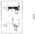
FVIII×VWFダブルノックアウトマウス(FVIII欠乏マウスをVWF欠乏マウスと交雑育種した)5匹からなる群に対して、PEG−rVWF(鎖長5kD、rVWFのPEG付加を、mPEGスクシンイミジルスクシネートのリジン残基の修飾によって、実施例2により行った)含有20mM HEPES(150mM NaCl、5%ショ糖、pH7.4(またはネイティブrVWFSS−PEGまたはネイティブrVWF(各々をrFVIIIと事前に混ぜ合わせ、9UFVIII/mlおよび9UVWF抗原/mlおよび0.67UVWF:RCo/mlを達成)を、経尾静脈によりボーラス注入(11ml/kg)を行った。公開(Ingerslev(Scand.J.Clin).Invest.47:143−149(1987))されているようにして、VWF−抗原値をELISA法で計測した。一次止血のプロセスにおけるVWFの血小板結着性を反映している機能的なVWF:RCo活性をMacfarlane et al.(Thromb.Diath.Haemorrh.34:306−308,1975)により、計測した。注射後5分、3時間、6時間、10時間、および24時間、麻酔後の心臓穿刺によるクエン酸血漿を、それぞれの群から調製した。FVIII活性およびVWF抗原量を、血漿サンプルで計測した。
For a group consisting of 5 FVIII × VWF double knockout mice (FVIII deficient mice were cross-bred with VWF deficient mice), PEG-rVWF (
FVIIIおよびVWFの半減期を、薬物動態学ライブラリーの1つの区画をモデルとして使用するMicroMath科学者プログラム(Micromath Research,Saint Luis,MO,US)を用いて算出した。rVWFまたはPEG付加rVWFのいずれかと同時注入されたFVIIIの半減期は、1.88時間から2.58時間へと増大し、血中濃度時間曲線下面積(AUC)は4.3から7.3U*h/mlへと増大した。VWFの半減期は、3.1から10.4へと増大し、血中濃度時間曲線下面積は、5.7から22.8に増やされる。結果を図7および図8にまとめた。データによれば、PEG−VWFを、VWDおよびFVIIIの循環時間を長くするという利点とともに、血友病AおよびVWDの急性処置および予防処置のために使用し得ることが示された。 The half-life of FVIII and VWF was calculated using the MicroMath scientist program (Micromath Research, Saint Luis, MO, US) using one compartment of the pharmacokinetic library as a model. The half-life of FVIII co-injected with either rVWF or PEGylated rVWF increased from 1.88 hours to 2.58 hours with an area under the blood concentration time curve (AUC) of 4.3 to 7.3 U * Increased to h / ml. The half-life of VWF is increased from 3.1 to 10.4 and the area under the blood concentration time curve is increased from 5.7 to 22.8. The results are summarized in FIG. 7 and FIG. The data showed that PEG-VWF can be used for acute and prophylactic treatment of hemophilia A and VWD, with the advantage of increasing the circulation time of VWD and FVIII.
(実施例11:マウス血漿におけるPEG付加の可逆性の実証)
PEG付加の可逆性を、VWF欠乏血漿による試験管内実験によって示した。クエン酸血漿を、4℃、15分間、1100xgの遠心によって、VWF欠乏マウスから得た(Denis et al.PNAS 95:9524−9529,1998)。4容量のマウス血漿を、実施例1(糖質を経たPEGカップリング)または実施例2(リジン残基を経たPEGカップリング)によって調製し、48時間にわたり37℃に保ち、1容量のPEG付加rVWFと混ぜ合わせた。非PEG付加rVWFが、両方の実験における対照として用いた。副試料を混合直後、ならびに1時間、5.5時間、24時間、および48時間後に回収し、VWF抗原量を、サンドイッチELISAシステムを用いて凍結試料からアッセイした。ポリクローナル抗VWF抗体(DAKO)を用いて、96穴ELISAプレートを被覆し、ヤギ−抗ウサギ−lgG−HRP−結合体(AXELL)を結合VWF因子検出のためにアッセイした。経時的にVWF抗原の増加性量を計測し、生体外血漿サンプルにおける場合も含めVWFに対するポリエチレングリコールの結合の可逆性が示された(図9)。
(Example 11: Demonstration of reversibility of PEG addition in mouse plasma)
The reversibility of PEGylation was shown by in vitro experiments with VWF-deficient plasma. Citrated plasma was obtained from VWF-deficient mice by centrifugation at 1100 × g for 15 minutes at 4 ° C. (Denis et al. PNAS 95: 9524-9529, 1998). Four volumes of mouse plasma are prepared according to Example 1 (PEG coupling via carbohydrate) or Example 2 (PEG coupling via lysine residues) and maintained at 37 ° C. for 48 hours, with one volume of PEG addition. Mixed with rVWF. Non-PEGylated rVWF was used as a control in both experiments. Subsamples were collected immediately after mixing and after 1 hour, 5.5 hours, 24 hours, and 48 hours, and VWF antigen levels were assayed from frozen samples using a sandwich ELISA system. Polyclonal anti-VWF antibody (DAKO) was used to coat 96-well ELISA plates and goat-anti-rabbit-lgG-HRP-conjugate (AXELL) was assayed for detection of bound VWF factor. The increasing amount of VWF antigen was measured over time, and the reversibility of the binding of polyethylene glycol to VWF was shown including in the case of in vitro plasma samples (FIG. 9).
(実施例12:PEG付加VWF調製試料のFVIII結合能の側定)
異なるPEG付加rVWF調製試料のFVIII結合能を、BIACORE(登録商標)3000装置(BIACORE、Uppsala、Sweden)を使用して表面プラズモン共鳴実験によって比較した(Karlsson et Falt,J.Immunol.Methods 200:121−33,1997)。通常、リガンドをセンサチップに固定し、該リガンドに対する他の構成要素の結合を表面プラズモン共鳴によって測定する。この技術の使用によって、チップ表面の近くの溶液の屈折率の変化が計測される。チップの表面の結合した構成要素の濃度変化を信号として検出し、それを任意の共鳴音単位(RU)で表す。固定リガンドに結合したタンパク質量と観察されるRUとのあいだに、比例関係が存在する。PEG付加VWF調製試料を、7000〜9000RUおよび25℃で、NHS/EDC化学を用いて、BIACORE(商標)センサチップのデキストラン表面に25℃で固定した。150mM NaCl、3mM EDTA、および0.005%界面活性剤P20(HBS緩衝液、BIACORE)を含む10mM HEPES緩衝液(pH7.4)を流速15μl/分で使用した。図10に例示されるように、市販のFVIII産物(ADVATE、Baxter AG、Vienna、Austria)の異なる量の結合が計測された。この図は、mPEGマレイミド5000で修飾され、かつ実施例4によって調製されるPEG付加rVWF調製試料のFVIII結合能を示す。異なるPEG付加rVWF調製試料のBIACORE実験の結果を表1にまとめている。この表では、PEG付加rVWF調製試料の異なるFVIII結合能は、10〜20IUFVIII/ml(色素アッセイ)の範囲にある参照の最大レベルでの非PEG付加参照調製試料のRU値の割合として与えられる。
(Example 12: Determination of FVIII binding ability of PEG-added VWF prepared sample)
The FVIII binding ability of different PEGylated rVWF prepared samples was compared by surface plasmon resonance experiments using a BIACORE® 3000 instrument (BIACORE, Uppsala, Sweden) (Karlsson et Falt, J. Immunol. Methods 200: 121). -33, 1997). Usually, a ligand is immobilized on a sensor chip, and the binding of other components to the ligand is measured by surface plasmon resonance. By using this technique, the change in refractive index of the solution near the chip surface is measured. A change in the concentration of the combined components on the surface of the chip is detected as a signal and expressed in arbitrary resonance units (RU). There is a proportional relationship between the amount of protein bound to the fixed ligand and the observed RU. PEG-added VWF prepared samples were immobilized at 25 ° C. on the dextran surface of BIACORE ™ sensor chip using NHS / EDC chemistry at 7000-9000 RU and 25 ° C. A 10 mM HEPES buffer (pH 7.4) containing 150 mM NaCl, 3 mM EDTA, and 0.005% surfactant P20 (HBS buffer, BIACORE) was used at a flow rate of 15 μl / min. As illustrated in FIG. 10, different amounts of binding of commercially available FVIII products (ADVATE, Baxter AG, Vienna, Austria) were measured. This figure shows the FVIII binding capacity of the PEGylated rVWF preparation sample modified with mPEG maleimide 5000 and prepared according to Example 4. The results of BIACORE experiments for different PEGylated rVWF preparation samples are summarized in Table 1. In this table, the different FVIII binding capacities of PEGylated rVWF preparation samples are given as a percentage of the RU value of the non-PEGylated reference preparation sample at the maximum level of reference in the range of 10-20 IUFVIII / ml (dye assay).

rVWFを、種々の濃度(1mM、2.5mM、5mM、7.5mM、および10mM)でmPEGスクシンイミジルスクシネート(鎖長:5kD)を使用して、実施例2により、PEG付加した。PEG付加VWF種を2つの異なる方法で分析した。すなわち、SDSポリアクリルアミドゲル電気泳動およびVWFマルチマー分析である。SDSゲル電気泳動を、3〜8%勾配ゲルを使用して還元条件下で行った(Tris Acetat Gel/Bio−Rad)。VWFマルチマー分析を、1.6%のアガロースゲルを使用してRuggeri et Zimmerman(Blood 57:1140−43,1981)により行った。VWFマルチマーの明視化を、Aihara et al.(Thromb.Haemost.55:263−67,1986)によって実施した。
rVWF was PEGylated according to Example 2 using mPEG succinimidyl succinate (chain length: 5 kD) at various concentrations (1 mM, 2.5 mM, 5 mM, 7.5 mM, and 10 mM). PEGylated VWF species were analyzed in two different ways. That is, SDS polyacrylamide gel electrophoresis and VWF multimer analysis. SDS gel electrophoresis was performed under reducing conditions using a 3-8% gradient gel (Tris Acetat Gel / Bio-Rad). VWF multimer analysis was performed by Ruggeri et Zimmerman (Blood 57: 1140-43, 1981) using a 1.6% agarose gel. Visualization of VWF multimers is described in Aihara et al. (Thromb. Haemost. 55: 263-67, 1986).
SDSゲル電気泳動(図11)は、成熟VWF(下部のバンド)とプロVWF(上層ブライトバンド)とからなるrVWF調製試料を示すとともに、異なる試薬濃度の使用によってPEG付加後の分子量が増加することを示す。PEG付加の後、ヒト血清アルブミン(HSA)調製試料(PEG付加は、実施例2によって実施)からの分子量の推移は、参照調製試料として示される。HSAの分子量が66,000のダルトンから190,000のダルトンまでシフトしていることで、PEG付加手法が有効であることが示されることが明らかになった。 SDS gel electrophoresis (Figure 11) shows a rVWF preparation sample consisting of mature VWF (lower band) and pro-VWF (upper bright band) and increased molecular weight after PEG addition by using different reagent concentrations Indicates. After PEG addition, the transition of molecular weight from human serum albumin (HSA) preparation sample (PEG addition is performed according to Example 2) is shown as a reference preparation sample. It has been clarified that the molecular weight of HSA is shifted from 66,000 daltons to 190,000 daltons, indicating that the PEG addition method is effective.
図12は、異なる試薬濃度でのPEG付加前後におけるrVWFの多量体パターンを示す。試薬濃度の上昇に伴って異なるマルチマーの分子量がより高いほうへ移行および広幅化することが明らかに示される。 FIG. 12 shows rVWF multimer patterns before and after PEG addition at different reagent concentrations. It is clearly shown that the molecular weight of the different multimers shifts and broadens with increasing reagent concentration.
(実施例14:mPEGスクシンイミジルグルタレートによるVWFのリジン残基のPEG付加)
リジン残基(図1A)を介したVWFのPEG付加のために、rVWF(最終濃度:500μg/ml)を、20mM HEPES緩衝液(150mM NaCl、pH7.4、5%スクロース含有)で調製し、mPEGスクシンイミジルグルタレート(鎖長:5kD)を添加した(最終濃度:200mgPEGスクシンイミジルグルタレート/mgタンパク質)。つぎに、pH値を0.1MNaOHで7.4に合わせた。VWFを室温で1時間、PEG付加し、実施例2の記載に従って精製した。
(Example 14: PEG addition of lysine residue of VWF with mPEG succinimidyl glutarate)
For PEGylation of VWF via lysine residues (FIG. 1A), rVWF (final concentration: 500 μg / ml) is prepared in 20 mM HEPES buffer (containing 150 mM NaCl, pH 7.4, 5% sucrose) mPEG succinimidyl glutarate (chain length: 5 kD) was added (final concentration: 200 mg PEG succinimidyl glutarate / mg protein). Next, the pH value was adjusted to 7.4 with 0.1 M NaOH. VWF was PEGylated for 1 hour at room temperature and purified as described in Example 2.
(実施例15:FVIII−K.O.マウスでのFVIII半減期の増大)
Bi et al.(Nat.Genet.10:119−121,1995)に詳しく記載されているFVIII欠乏マウスを、重度ヒト血友病Aのモデルとして使用した。マウス5匹からなる群に対して、各々を組換え型FVIIIと事前に混合して10UFVIII/mlおよび10UVWF/mlを達成するために、実施例2により調製したPEG−rVWF(SS−PEG、5K)またはネイティブrVWFのいずれかによる経尾静脈ボーラス注入法による投与(10ml/kg)を行った。麻酔後、生成物を注射してから5分、3時間、9時間、および24時間後に、心臓穿刺によるクエン酸血漿を調製した。FVIII活性およびVWF抗原回収量を血漿サンプルで計測した。この実験の結果を図13および図14にまとめた。
Example 15: Increased FVIII half-life in FVIII-KO mice
Bi et al. (Nat. Genet. 10: 119-121, 1995), FVIII-deficient mice were used as a model for severe human hemophilia A. For a group of 5 mice, each was pre-mixed with recombinant FVIII to achieve 10 UFVIII / ml and 10 UVWF / ml to achieve PEG-rVWF prepared according to Example 2 (SS-PEG, 5K ) Or native rVWF (10 ml / kg) by transvenous bolus injection. After anesthesia, citrated plasma was prepared by
FVIIIのための半減期は1.8時間(ネイティブrVWF存在下)から3.9時間(PEG−rVWFと併用)へと増大し、血中濃度時間曲線下面積(AUC)が4.1から7.8U*hへと増大した。VWF半減期は3.2時間から13.6時間へと増大し、VWFのAUCは7.7から32.1U*hへとほぼ4倍になった。 The half-life for FVIII increases from 1.8 hours (in the presence of native rVWF) to 3.9 hours (in combination with PEG-rVWF), and the area under the blood concentration time curve (AUC) is 4.1 to 7 Increased to 8 U * h. The VWF half-life increased from 3.2 hours to 13.6 hours, and the VWF AUC nearly quadrupled from 7.7 to 32.1 U * h.
(実施例16:異なる方法によるSS−PEG付加VWF調製試料のFVIII結合能の測定)
異なるPEG付加rVWF調製試料のFVIII結合能を、ELISAと色素アッセイシステム(ECA)とを組み合わせ、Bendetowicz et al.(Blood 92:529−538,1998)の方法の変法によって計測した。マイクロタイタープレートを、200μlの2.6μg/mL抗vWFポリクローナル抗体含有50mmol/lNa2CO3/NaHCO3(pH9.6)で覆った。プレートを、PBS−Tween緩衝液(100mM Na2HPO4/KH2PO4、150mM NaCl、pH7.6と0.05%Tween20)で、各ステップの後に洗った。プレートを、0.1%粉ミルク/2mMベンズアミジン含有PBS−Tweenで37℃、1時間にわたりブロックした。VWFの増加量を37℃、25分間にわたり、0.2U/mlrFVIII(ADVATE,Baxter AG,Vienna,Austria)とともにプレインキュベートし、これらの混合物の100μlをプレートに添加した。インキュベーション後、捕獲されたVWFに結合したFVIIIの量を、FVIII色素アッセイ(Technoclone,Vienna,Austria)で計測した。FVIII結合能を、1分内で405nm(dA405)で計測される吸光度の変化として表した。図15は、2つのPEG付加VWF調製試料(両方ともmPEG−スクシンイミジルスクシネート(SS)で修飾)のVWF用量依存性FVIII結合を示す。修飾VWF調製試料のFVIII結合能は、非修飾開始VWF調製試料の%として算出され、PEG−SS−rVWF−1では20%およびPEG−SS−rVWF−2では50%であると算出された。
(Example 16: Measurement of FVIII binding ability of SS-PEG-added VWF prepared sample by different methods)
The FVIII binding ability of different PEG-added rVWF preparation samples was combined with ELISA and dye assay system (ECA), and Bendewickz et al. (Blood 92: 529-538, 1998). The microtiter plate was covered with 200 μl of 50 mmol / l Na 2 CO 3 / NaHCO 3 (pH 9.6) containing 2.6 μg / mL anti-vWF polyclonal antibody. The plate was washed after each step with PBS-Tween buffer (100 mM Na 2 HPO 4 / KH 2 PO 4 , 150 mM NaCl, pH 7.6 and 0.05% Tween 20). Plates were blocked with PBS-Tween containing 0.1% powdered milk / 2 mM benzamidine at 37 ° C. for 1 hour. Increasing amounts of VWF were preincubated with 0.2 U / ml rFVIII (ADVATE, Baxter AG, Vienna, Austria) for 25 minutes at 37 ° C. and 100 μl of these mixtures were added to the plates. After incubation, the amount of FVIII bound to the captured VWF was measured with the FVIII dye assay (Technoclone, Vienna, Austria). FVIII binding ability was expressed as the change in absorbance measured at 405 nm (dA405) within 1 minute. FIG. 15 shows VWF dose-dependent FVIII binding of two PEGylated VWF preparation samples, both modified with mPEG-succinimidyl succinate (SS). The FVIII binding capacity of the modified VWF preparation sample was calculated as% of the unmodified starting VWF preparation sample, calculated as 20% for PEG-SS-rVWF-1 and 50% for PEG-SS-rVWF-2.
図16は、実施例12に記載の表面プラズモン共鳴方法で計測される2つのPEG−SS−rVWF調製試料のFVIII結合能を示す。算出結合能は、それぞれ25および45%であった。 FIG. 16 shows the FVIII binding ability of two PEG-SS-rVWF prepared samples measured by the surface plasmon resonance method described in Example 12. The calculated binding capacity was 25 and 45%, respectively.
SDS−PAGEで計測されるPEG−SS結合後のrVWF分子の分子質量における適当な増加を、図17に示す。 A suitable increase in the molecular mass of the rVWF molecule after PEG-SS conjugation as measured by SDS-PAGE is shown in FIG.
(実施例17:分枝PEGマレイミドによるVWFにおけるスルフヒドリル基のPEG付加)
分枝PEGマレイミドによる遊離SH残基を経たVWFのPEG付加のために組換え型VWF(最終濃度:500μg/ml)の溶液を、20mM HEPES緩衝液(150mM NaCl、pH7.6、3%トレハロース含有)によって調製する。次に、NOF社(NOF Europe,Grobbendonk,Belgium)によって供給される分岐mPEGマレイミド(鎖長:20kD)を添加した(最終濃度10mM)。VWFを静かに撹拌しながら室温で2時間にわたり、PEG付加した。その後、PEG付加rVWFを、再生セルロース(Millipore)からなる100kD膜を使用して、限界濾過法/ダイアフィルトレーション(UF/DF)によって、試薬から分離する。
(Example 17: PEG addition of sulfhydryl group in VWF with branched PEG maleimide)
A solution of recombinant VWF (final concentration: 500 μg / ml) for PEG addition of VWF via free SH residues with branched PEG maleimide, containing 20 mM HEPES buffer (150 mM NaCl, pH 7.6, 3% trehalose) ) To prepare. Next, branched mPEG maleimide (chain length: 20 kD) supplied by NOF (NOF Europe, Grobbendonk, Belgium) was added (
(実施例18:mPEGヒドラジド/EDCによるVWFのカルボキシル基のPEG付加)
実施例23により、フリン成熟rVWFを調製および精製した。調製試料を50mMリン酸緩衝液(pH6.2)に対して透析し、400μg/mlの濃度まで希釈した。次に、鎖長5kDのmPEGヒドラジド(mPEG Hz)を添加した(濃度:60mg mPEG Hz/mgVWF)。500mM 1−エチル−3−(3−ジメチルアミノプロピル)カルボジイミド(EDC)の新しく調製された溶液30μlを、1mlのVWF含有混合物の1mlに添加し、5時間にわたって静かに振とうさせながら室温でインキュベートした。100kD膜(再生セルロース/Millipore)を用いて、20mM HEPES緩衝液(150mM NaCl、pH7.4)に対して、UF/DFによりPEG付加rVWFから試薬を分離する。
(Example 18: PEG addition of carboxyl group of VWF by mPEG hydrazide / EDC)
According to Example 23, furin mature rVWF was prepared and purified. The prepared sample was dialyzed against 50 mM phosphate buffer (pH 6.2) and diluted to a concentration of 400 μg / ml. Next, mPEG hydrazide (mPEG Hz) having a chain length of 5 kD was added (concentration: 60 mg mPEG Hz / mg VWF). 30 μl of a freshly prepared solution of 500 mM 1-ethyl-3- (3-dimethylaminopropyl) carbodiimide (EDC) is added to 1 ml of a 1 ml VWF containing mixture and incubated at room temperature with gentle shaking for 5 hours. did. Reagents are separated from PEGylated rVWF by UF / DF against 20 mM HEPES buffer (150 mM NaCl, pH 7.4) using a 100 kD membrane (regenerated cellulose / Millipore).
(実施例19:ポリシアル酸によるVWFのリジン残基の修飾)
ポリシアル酸(コロミン酸、CA)によるリジン残基の修飾を、Fernandes et Gregoriadis(Biochim.Biophys.Acta 1341:26−34,1997)およびJennings et Lugowski(J.Immunol.127:1011−1018,1981)に記載のようにして、実施した。0.1MNaIO4含有コロミン酸(濃度:20mg/ml)溶液を暗所、室温で15分間撹拌させて、CAを酸化した。活性化CA溶液1mlあたり2mlのエチレングリコールを添加し、暗所、室温でさらに30分間撹拌した。溶液を一晩にわたって暗所で0.05Mリン酸ナトリウム緩衝液(pH7.2)に対して透析した。その後、この溶液のアリコートをrVWF溶液(400μg/ml)含有0.05Mリン酸ナトリウムに添加し、1mgVWFあたり50mg活性化CAの最終濃度を得た。この混合物を暗所、室温で30分間撹拌した。NaCNBH3を添加し(1mg/mgrVWF)、混合物を暗所、室温で18時間、静かに撹拌しながらインキュベートした。1MTRIS水溶液(pH7.2)を添加し(50μl/mgNaCNBH3)、1時間にわたって撹拌することで反応を停止させた。遊離試薬を、100kD膜(再生セルロース/Millipore)を使用して、UF/DFによりrVWF−ポリシアル酸結合体から分離した。
(Example 19: Modification of lysine residue of VWF with polysialic acid)
Modification of lysine residues with polysialic acid (colominic acid, CA) is described in Fernandes et Gregoriadis (Biochim. Biophys. Acta 1341: 26-34, 1997) and Jennings et Lugoski (J. Immunol. 127: 1011-1018, 1981). As described in. A 0.1M NaIO 4 -containing colominic acid (concentration: 20 mg / ml) solution was allowed to stir in the dark at room temperature for 15 minutes to oxidize CA. 2 ml of ethylene glycol per 1 ml of the activated CA solution was added and stirred for an additional 30 minutes in the dark at room temperature. The solution was dialyzed overnight against 0.05M sodium phosphate buffer (pH 7.2) in the dark. An aliquot of this solution was then added to 0.05 M sodium phosphate containing rVWF solution (400 μg / ml) to give a final concentration of 50 mg activated CA per mg VWF. The mixture was stirred in the dark at room temperature for 30 minutes. NaCNBH 3 was added (1 mg / mgrVWF) and the mixture was incubated in the dark at room temperature for 18 hours with gentle agitation. The reaction was stopped by adding 1 M TRIS aqueous solution (pH 7.2) (50 μl / mg NaCNBH 3 ) and stirring for 1 hour. The free reagent was separated from the rVWF-polysialic acid conjugate by UF / DF using a 100 kD membrane (regenerated cellulose / Millipore).
この調製試料のFVIII結合能を実施例16の記載通りに測定した。 The FVIII binding ability of this prepared sample was measured as described in Example 16.
ポリシアル酸によるrVWFの結合を、還元条件の下で等電点電気泳動(IEF)によって検出される等電点(PI)の移動によって示した。図20は、この実施例によりポリシアル酸による結合の前後におけるVWF調製試料の比較を示す。製造元(Amersham Bioscience)の指示により、Ampholine PAGplateシステム(pH3.5ないし9.5)を用いた。ポリシアル酸との結合による酸性基の導入によって、修飾rVWFの1つの酸性バンドが生ずる。 Binding of rVWF by polysialic acid was shown by the movement of the isoelectric point (PI) detected by isoelectric focusing (IEF) under reducing conditions. FIG. 20 shows a comparison of VWF prepared samples before and after binding with polysialic acid according to this example. An Amphorine PAGplate system (pH 3.5 to 9.5) was used according to the manufacturer's instructions (Amersham Bioscience). Introduction of acidic groups by conjugation with polysialic acid results in one acidic band of modified rVWF.
実施例16に記載のように、54%のFVIII結合能がECA試験を用いて側定された。70%のFVIII結合能を表面プラズモン共鳴方法(実施例12)を用いて側定した。 As described in Example 16, 54% FVIII binding capacity was determined using the ECA test. 70% FVIII binding ability was determined using the surface plasmon resonance method (Example 12).
(実施例20:剪断応力条件下のPEG付加)
Sakariassen et al.(J.Lab.Clin.Med.102:522−535,1983)に記載のように、灌流チェンバーを作製し、剪断応力条件下でのrVWFのPEG付加に使用した。rVWF(500μg/ml)含有20mM HEPES(pH7.4、3%トレハロース含有)の溶液を調製した。次に、mPEGスクシンイミジルスクシネート(最終濃度:5mM)を添加し、0.1MNaOHを添加してpHを7.4に調製し、直ちに灌流システムに充填した。次に、PEG付加はSakariassen et al.(J.Lab.Clin.Med.102:522−535,1983に記載のように、灌流条件下で、蠕動ポンプを使用することで、2500秒−1の剪断速度で、室温で行った。
(Example 20: PEG addition under shear stress condition)
Sakariassen et al. (J. Lab. Clin. Med. 102: 522-535, 1983), a perfusion chamber was made and used for PEGylation of rVWF under shear stress conditions. A solution of 20 mM HEPES (pH 7.4, containing 3% trehalose) containing rVWF (500 μg / ml) was prepared. Next, mPEG succinimidyl succinate (final concentration: 5 mM) was added and 0.1 M NaOH was added to adjust the pH to 7.4 and immediately loaded into the perfusion system. Next, PEG addition is performed according to Sakariassen et al. (As described in J. Lab. Clin. Med. 102: 522-535, 1983, using a peristaltic pump under perfusion conditions and at a shear rate of 2500 s −1 at room temperature.
(実施例21:pdVWFの調製)
dVWFの調製を、改良を加えたThorell et Blomback(Thromb.Res.35:431−450,1984)により実施した。pdVWFの調製のために、1.5kgのクリオプレシピテートを20ないし30℃で6リットルの水に溶解した。1時間撹拌させた後に、フィブロネクチン沈殿物を遠心によって除去した。上清1リットルあたり8gNaClを添加し、溶液を室温まで温め、ウイルスを失活させるためにSDストック試薬(1%Tween80+0.18%アセチルトリエチルシトレート最終濃度)を添加した。
Example 21: Preparation of pdVWF
dVWF was prepared by Thorell et Bromback (Thromb. Res. 35: 431-450, 1984) with improvements. For the preparation of pdVWF, 1.5 kg of cryoprecipitate was dissolved in 6 liters of water at 20-30 ° C. After stirring for 1 hour, fibronectin precipitate was removed by centrifugation. 8 g NaCl was added per liter of supernatant, the solution was warmed to room temperature and SD stock reagent (1
溶液を、0.2MNaCl、0.02M酢酸ナトリウム(pH6.5)(洗浄用緩衝液)で事前に平衡化されたEMD−TMAEフラクトゲル650Mカラム(XK50/180)上で精製した。洗浄用緩衝液20カラム容量で洗浄した後、VWFを0.5MNaCl、0.02MNaクエン酸塩(pH6.9)で溶出させた。VWFを沈降させるために、グリシン(1M最終濃度)およびNaCl(3M最終濃度)を添加した。沈殿物を緩衝液に溶解し、最終濃度に対して0.3MCaCl2、0.15MNaCl、20mM HEPES(pH7.4)で平衡化したカラムをセファクリルS−400HR(Amersham)上に載せた。 The solution was purified on an EMD-TMAE fructogel 650M column (XK50 / 180) pre-equilibrated with 0.2M NaCl, 0.02M sodium acetate, pH 6.5 (washing buffer). After washing with 20 column volumes of washing buffer, VWF was eluted with 0.5 M NaCl, 0.02 M Na citrate (pH 6.9). Glycine (1M final concentration) and NaCl (3M final concentration) were added to precipitate the VWF. The precipitate was dissolved in a buffer, 0.3MCaCl 2 relative to the final concentration, 0.15M NaCl, and equilibrated column loaded on Sephacryl S-400HR (Amersham) in 20mM HEPES (pH7.4).
PdVWFを実施例21により調製し、20mM HEPES緩衝液(pH7.4)(150mM NaClおよび3%ショ糖を含む)で希釈し、最終濃度を400μg/mlとする。次に、mPEGスクシイミジルスクシネート(鎖長:5kD)を添加し(濃度:10mgPEGSS5000/mg VWF)、pdVWFを室温で、1時間にわたりPEG付加した。次に、再生セルロース(Millipore)からなる100kD膜を使用して、UF/DFによって、PEG付加rVWFから試薬を分離した。
PdVWF is prepared according to Example 21 and diluted with 20 mM HEPES buffer pH 7.4 (containing 150 mM NaCl and 3% sucrose) to a final concentration of 400 μg / ml. Next, mPEG succinimidyl succinate (chain length: 5 kD) was added (concentration: 10 mg PEGSS5000 / mg VWF), and pdVWF was PEG added at room temperature for 1 hour. Next, the reagent was separated from the PEG-added rVWF by UF / DF using a 100 kD membrane made of regenerated cellulose (Millipore).
(実施例23:フリン成熟rVWFのフリン成熟および精製)
rFVIII発酵および精製プロセス由来の抗FVIII抗体カラムのフロースルー分画の143kgを、Schlokat et al.(Biotechnol.Appl.Biochem.24:257−267,1996)に記載のように、プロペプチド除去のためにフリンで処理し、滅菌濾過した。このプロセスは、初期の研究に基づいた(Fischer et al.,FEBS Lett.375:259−262,1995;Fischer et al.PCT/AT98/00034[WO98/38219],1−33,1998,18−2−1998 and Kaersgaard et Barington,J.Chromatogr.B 715:357−367,1998)。
Example 23: Furin maturation and purification of furin mature rVWF
143 kg of the flow-through fraction of the anti-FVIII antibody column from the rFVIII fermentation and purification process was added to Schlokat et al. (Biotechnol. Appl. Biochem. 24: 257-267, 1996) was treated with furin for propeptide removal and sterile filtered. This process was based on early work (Fischer et al., FEBS Lett. 375: 259-262, 1995; Fischer et al. PCT / AT98 / 00034 [WO 98/38219], 1-33, 1998, 18- 2-1998 and Kaersgaard et Barington, J. Chromatogr. B 715: 357-367, 1998).
水で4倍希釈して16mS/cmにした後、633kgの希釈溶液を、10mM Tris、100mM NaAc、86mM NaCl、pH6.5、mS/cm(平衡緩衝液)により平衡化させたXK50/15EMD−TMAEフラクトゲル650Mカラム(300mlゲル;Merck;#K14540281)に載せた。カラムを平衡化緩衝液で洗浄し、100mM NaAc、250mM NaCl、100mMグリシン、3mM CaCl2で溶出した。
After
4506gのTMAE溶出液をSartoclean GF(0.8及び0.65μ)およびSartobran P(0.45及び0.2μ)上で濾過し、1.5倍希釈して29mS/cmとし、Mustang Qフィルター(#IH18770932)を介してポンピングする。22+/−2℃で60分間にわたりSD処置し、かつ水で2倍に希釈して16mS/cmにした後、溶液を、TMAEステップの平衡用緩衝液で平衡化させたAmicon 70/29 UNOsphere Sカラム(600mlゲル;Bio−Rad,#78960C)に載せた。カラムを平衡用緩衝液で洗浄し、そしてTMAEステップの溶離緩衝液で溶出させた。 4506 g of TMAE eluate was filtered over Sartoclean GF (0.8 and 0.65 μ) and Sartobran P (0.45 and 0.2 μ), diluted 1.5-fold to 29 mS / cm, and Mustang Q filter ( # IH1877092). After SD treatment at 22 +/− 2 ° C. for 60 minutes and 2-fold dilution with water to 16 mS / cm, the solution was equilibrated with TMAE step equilibration buffer to Amicon 70/29 UNOsphere S. The column (600 ml gel; Bio-Rad, # 78960C) was loaded. The column was washed with equilibration buffer and eluted with TMAE step elution buffer.
3223gのUNO−S溶出液を、再生セルロースからなる30kDaの0.1m2膜(Hydrosart #01080217、Sartorius)を使用して限界濾過法により15倍濃縮した。コンセントレートの201gを、100mM NaAc、500mM NaCl、pH7.0(1698mlのゲル;GE Healthcare #17−0489−01)で平衡化させたXK50/86.5Superose 6 Prep Gradeカラム上で、サイズ排除クロマトグラフィーにより最終的に精製された。 3223 g of UNO-S eluate was concentrated 15-fold by ultrafiltration using a 30 kDa 0.1 m 2 membrane (Hydroart # 010080217, Sartorius) made of regenerated cellulose. Size exclusion chromatography on 201 g of concentrate was performed on an XK50 / 86.5 Superose 6 Prep Grade column equilibrated with 100 mM NaAc, 500 mM NaCl, pH 7.0 (1698 ml gel; GE Healthcare # 17-0489-01). Was finally purified.
フリン成熟VWFを実施例23により調製し、20mM HEPES緩衝液(pH7.4)(150mM NaClおよび3%ショ糖を含有)に対して透析した。次に、溶液を20mM HEPES緩衝液(pH7.4)(150mM NaClおよび3%ショ糖を含有)により最終濃度を300μg/mlに希釈した。続いて、mPEGスクシミジルスクシネート(鎖長:5kD)を添加し(濃度:25mgPEGSS5000/mgVWF)、フリン成熟VWFを室温で1時間にわたってPEG付加した。次に、再生セルロース(Millipore)からなる100kD膜を使用して、UF/DFによって、PEG付加VWFから試薬を分離した。
Furin mature VWF was prepared according to Example 23 and dialyzed against 20 mM HEPES buffer (pH 7.4) containing 150 mM NaCl and 3% sucrose. The solution was then diluted to a final concentration of 300 μg / ml with 20 mM HEPES buffer (pH 7.4) (containing 150 mM NaCl and 3% sucrose). Subsequently, mPEG succimidyl succinate (chain length: 5 kD) was added (concentration: 25 mg PEGSS5000 / mg VWF), and furin mature VWF was PEG added over 1 hour at room temperature. Next, the reagent was separated from the PEG-added VWF by UF / DF using a 100 kD membrane made of regenerated cellulose (Millipore).
(実施例25:フリン成熟rVWFの生体外特徴付け)
実施例12および図15に記載のように、フリン成熟rVWFのFVIII結合能を表面プラズモン共鳴技術およびECAによって測定した。結果を、実施例21によって調製されるアルブミンを含まない血漿由来VWF調製試料と比較した。
Example 25: In vitro characterization of furin mature rVWF
As described in Example 12 and FIG. 15, the FVIII binding ability of furin mature rVWF was measured by surface plasmon resonance technique and ECA. The results were compared to plasma-derived VWF preparation samples without albumin prepared according to Example 21.
図18および図19に示されるように、フリン成熟rVWFのFVIII結合能は血漿由来参照調製試料と同等だった。 As shown in FIGS. 18 and 19, the FVIII binding ability of furin mature rVWF was equivalent to the plasma-derived reference preparation sample.
(実施例26:グルタルアルデヒドで架橋させることによるポリシアル酸とVWFとの結合)
グルタルアルデヒドを架橋試薬(Migneault et al.,Biotechniques 37:790−796,2004)として用いたrVWFとポリシアル酸(コロミン酸)との結合のために、コロミン酸(濃度:20mg/ml)含有20mM HEPES緩衝液(150mM NaCl、pH7.4)の溶液4mlを調製し、0.1MNaOHを添加してpHを7.4に合わせた。グルタルアルデヒドを添加して最終濃度を0.01%にした。続いて、rVWF(400μg/ml)含有20mM HEPES緩衝液(150mM NaCl、pH7.4)の溶液1mlを100μlのアリコートに添加し、混合物を静かに振とうさせながら1時間インキュベートした。次に、混合物を透析し、トレハロースを添加し(最終濃度:3%)、rVWFポリシアル酸結合体を限界濾過法によって濃縮した。
(Example 26: Binding of polysialic acid and VWF by crosslinking with glutaraldehyde)
20 mM HEPES containing colominic acid (concentration: 20 mg / ml) for the coupling of rVWF and polysialic acid (colominic acid) using glutaraldehyde as a cross-linking reagent (Migneart et al., Biotechniques 37: 790-796, 2004) A 4 ml solution of buffer (150 mM NaCl, pH 7.4) was prepared and 0.1 M NaOH was added to adjust the pH to 7.4. Glutaraldehyde was added to a final concentration of 0.01%. Subsequently, 1 ml of a solution of 20 mM HEPES buffer (150 mM NaCl, pH 7.4) containing rVWF (400 μg / ml) was added to 100 μl aliquots and the mixture was incubated for 1 hour with gentle shaking. The mixture was then dialyzed, trehalose was added (final concentration: 3%) and the rVWF polysialic acid conjugate was concentrated by ultrafiltration.
(実施例27:VWF欠乏マウスでのVWF半減期の増大)
Denis et al.(PNAS 95:9524−9529,1998)により記載されたVWF欠乏マウスを、ヒトVWDの動物モデルとして使用した。マウス5匹からなる群に対して、100UVWF:Ag/kgを達成するために、実施例19により調製したrVWFポリシアル酸結合体またはネイティブrVWFのいずれかをボーラス注入法(10ml/kg)によって経尾静脈により投与した。麻酔後の心臓穿刺によるクエン酸血漿は、注射後5分、1、3、6、9、および21時間で、それぞれの群から調製した。VWF抗原回収および内因性マウスFVIII活性レベルを血漿サンプルで計測した。この実験の結果は、図21および図22にまとめられている。
Example 27: Increased VWF half-life in VWF-deficient mice
Denis et al. VWF-deficient mice described by (PNAS 95: 9524-9529, 1998) were used as an animal model for human VWD. In order to achieve 100 UVWF: Ag / kg for a group of 5 mice, either rVWF polysialic acid conjugate prepared according to Example 19 or native rVWF was injected by bolus injection (10 ml / kg). Administered intravenously. Citrate plasma by cardiac puncture after anesthesia was prepared from each group at 5 minutes, 1, 3, 6, 9, and 21 hours after injection. VWF antigen recovery and endogenous mouse FVIII activity levels were measured in plasma samples. The results of this experiment are summarized in FIGS.
VWFの半減期を、薬物動態学ライブラリーの1つの区画をモデルとして使用するMicroMath科学者プログラム(Micromath Research,Saint Luis,MO,US)を用いて算出した。FVIII活性の曲線下面積は、ベースラインサブトラクションによる台形モデルで算出した。 VWF half-life was calculated using the MicroMath scientist program (Micromath Research, Saint Luis, MO, US) using one compartment of the pharmacokinetic library as a model. The area under the curve of FVIII activity was calculated with a trapezoidal model with baseline subtraction.
VWFの半減期は1.3時間(ネイティブrVWF)から2.4時間(rVWF−ポリシアル酸結合体)へと増大し、FVIIIのAUCがそれぞれ3.3U*hr/mlから5.3U*hr/mlへと増大した。 The half-life of VWF increased from 1.3 hours (native rVWF) to 2.4 hours (rVWF-polysialic acid conjugate), and the FUC AUC increased from 3.3 U * hr / ml to 5.3 U * hr / increased to ml.
(実施例28:還元性アミノ化によるリジン基に対するmPEGプロピオンアルデヒドの結合)
rVWF溶液(400μg/ml)を0.05Mリン酸ナトリウム緩衝液(pH7.2)に調製し、mPEGプロピオンアルデヒド(5kDa鎖長)を添加して最終濃度を1mgVWFあたり10mgのmPEGプロピオンアルデヒドとする。この混合物を30分間撹拌する。つぎに、NaCNBH3を添加し(1mg/mgのrVWF)、混合物を静かに撹拌させながら室温で15時間にわたりインキュベートする。1MのTRIS水溶液(pH7.2)を添加し(1mgのNaCNBH3あたり50μl)、1時間にわたって撹拌することで反応を停止させる。その後、PEG付加rVWFは、100kD膜(再生セルロース/Millipore)を使用している限界濾過法/ダイアフィルトレーションによって、試薬から分離される。
Example 28: Binding of mPEG propionaldehyde to a lysine group by reductive amination
Prepare rVWF solution (400 μg / ml) in 0.05 M sodium phosphate buffer (pH 7.2) and add mPEG propionaldehyde (5 kDa chain length) to a final concentration of 10 mg mPEG propionaldehyde per mg VWF. The mixture is stirred for 30 minutes. Then NaCNBH 3 is added (1 mg / mg rVWF) and the mixture is incubated for 15 hours at room temperature with gentle agitation. The reaction is stopped by adding 1 M aqueous TRIS solution (pH 7.2) (50 μl per 1 mg NaCNBH 3 ) and stirring for 1 hour. The PEGylated rVWF is then separated from the reagents by ultrafiltration / diafiltration using a 100 kD membrane (regenerated cellulose / Millipore).
(実施例29:VWFのN末端PEG付加)
rVWFのN末端PEG付加をLee et al.(Pharm.Res.20:818−825,2003)の記載のように行う。rVWF溶液(最終濃度:500のμg/ml)を50mMの酢酸ナトリウム緩衝液(pH5.5)に調製し、mPEGプロピオンアルデヒド(鎖長:5kD)を添加した(濃度:10mg mPEGプロピオンアルデヒド/mgVWF)。PEG付加を、還元剤として2mMのNaCNBH3の存在下、室温で24時間にわたって行う。その後、PEG付加rVWFを、100kD膜(再生セルロース/Millipore)を使用して限界濾過法/ダイアフィルトレーションにより試薬から分離される。
(Example 29: N-terminal PEG addition of VWF)
rVWF N-terminal PEGylation was performed by Lee et al. (Pharm. Res. 20: 818-825, 2003). rVWF solution (final concentration: 500 μg / ml) was prepared in 50 mM sodium acetate buffer (pH 5.5) and mPEG propionaldehyde (chain length: 5 kD) was added (concentration: 10 mg mPEG propionaldehyde / mg VWF). . The PEG addition is carried out for 24 hours at room temperature in the presence of 2 mM NaCNBH 3 as reducing agent. The PEGylated rVWF is then separated from the reagents by ultrafiltration / diafiltration using a 100 kD membrane (regenerated cellulose / Millipore).
(実施例30:リジン残基およびrVWFのSH残基の逐次的なPEG付加)
RVWFを、実施例2によりmPEGスクシンイミジルスクシネート(鎖長:5kD)で、リジン残基を経てPEG付加する。PEG付加を1時間にわたって室温で行い、遊離試薬を、100kD膜(再生セルロース/Millipore)を使用して20mM HEPES緩衝液(pH7.4、5%サッカロース)に対してUF/DFによりrVWFPEG結合体から分離する。次に、溶液のpH値を、0.1MNaOHで7.6に合わせ、mPEGマレイミド(10mM鎖長5kD/最終濃度)を加えて、遊離SH基のPEG付加を行う。静かに振とうしながら、室温で2時間にわたってPEG付加を行う。次に、試薬を再び、100kD膜(再生セルロース/Millipore)を使用して反応混合物から分離する。
Example 30: Sequential PEGylation of lysine residues and rVWF SH residues
RVWF is PEGylated via lysine residues with mPEG succinimidyl succinate (chain length: 5 kD) according to Example 2. PEG addition was performed for 1 hour at room temperature and the free reagent was removed from the rVWFPEG conjugate by UF / DF against 20 mM HEPES buffer (pH 7.4, 5% sucrose) using a 100 kD membrane (regenerated cellulose / Millipore). To separate. Next, the pH value of the solution is adjusted to 7.6 with 0.1 M NaOH, mPEG maleimide (10
(実施例31:糖質残基の酵素酸化とPEG−Hzによるその後のPEG付加)
Avigad et al.(J.Biol.Chem.237:2736−43,1962)に記載のように、アルデヒド基を生じさせるrVWFの糖質残基(Wilchek et Bayer,Meth.Enzymol.138;429−442,1987)の酵素酸化を、Dactylium dendroides(Sigma)由来のガラクトースオキシダーゼを使用して行う。得られた溶液を、50mMリン酸緩衝液(pH7.2)に対して透析し、希釈してVWF濃度を400μg/mlにする。次に、鎖長5kDのmPEGヒドラジド(mPEG Hz)(最終濃度:40mg mPEG−Hz/mg VWF)を添加する。混合物を静かに振とうさせながら室温で3時間インキュベートする。試薬を、100kD膜(再生セルロース/Millipore)を使用して、5%のサッカロース含有20mM HEPES緩衝液(150mM NaCl、pH7.4)に対して、UF/DFによりPEG付加rVWFから分離する。
Example 31: Enzymatic oxidation of carbohydrate residues and subsequent PEG addition with PEG-Hz
Avigad et al. (J. Biol. Chem. 237: 2736-43, 1962), the carbohydrate residue of rVWF that produces an aldehyde group (Wilchek et Bayer, Meth. Enzymol. 138; 429-442, 1987). Enzymatic oxidation is performed using galactose oxidase from Dactylium dendriides (Sigma). The resulting solution is dialyzed against 50 mM phosphate buffer (pH 7.2) and diluted to a VWF concentration of 400 μg / ml. Next, mPEG hydrazide (mPEG Hz) with a chain length of 5 kD (final concentration: 40 mg mPEG-Hz / mg VWF) is added. Incubate the mixture for 3 hours at room temperature with gentle shaking. Reagents are separated from PEGylated rVWF by UF / DF against 20 mM HEPES buffer (150 mM NaCl, pH 7.4) containing 5% sucrose using a 100 kD membrane (regenerated cellulose / Millipore).
(実施例32:rFVIIIによるFVIII結合部位のブロッキングによるrVWFのPEG付加)
非修飾FVIII結合部位を持つPEG付加rVWFを調製するために、rFVIII(200U/ml)およびrVWF(40U VWF:Ag/ml)を含む50mM HEPES緩衝液(50mM HEPES、150mM NaCl、2%トレハロース、pH7.4)の溶液3mlを調製して、37℃で1時間インキュベートする。混合物を、室温まで冷やし、PEGスクシンイミジルスクシネート(PEG−SS/鎖長:5kD)を添加し(最終濃度:1mgPEG−SS/UVWF:Ag)、静かに振とうさせながら1時間インキュベートする。次に、CaCl2を静かに振とうさせながら添加することで、最終濃度で400mMを得る。この溶液を、セファクリルS−400HR(Amersham)充填クロマトグラフィーカラム(2.6×80cm)に載せ、FVIIIの遊離結合部位を持つPEG付加rVWFを、サイズ排除クロマトグラフィー(溶離緩衝液:50mM HEPES緩衝液、400mM CaCI2、pH7.4)によって、rFVIIIから分離する。
Example 32: PEGylation of rVWF by blocking FVIII binding site with rFVIII
To prepare PEGylated rVWF with unmodified FVIII binding site, 50 mM HEPES buffer (50 mM HEPES, 150 mM NaCl, 2% trehalose, pH 7) containing rFVIII (200 U / ml) and rVWF (40 U VWF: Ag / ml). Prepare 3 ml of the solution of 4) and incubate at 37 ° C. for 1 hour. Cool the mixture to room temperature and add PEG succinimidyl succinate (PEG-SS / chain length: 5 kD) (final concentration: 1 mg PEG-SS / UVWF: Ag) and incubate for 1 hour with gentle shaking . Next, CaCl 2 is added with gentle shaking to obtain 400 mM at the final concentration. This solution was loaded onto a Sephacryl S-400HR (Amersham) packed chromatography column (2.6 × 80 cm), and PEGylated rVWF having a free binding site of FVIII was subjected to size exclusion chromatography (elution buffer: 50 mM HEPES buffer). , 400 mM CaCI 2 , pH 7.4).
(実施例33:ヘパリンによるFVIII結合部位のブロッキングによるrVWFのPEG付加)
rVWF(300μg/ml)含有50mM HEPES緩衝液(pH7.4)の溶液5mlを調製し、同一緩衝液のヘパリンセファロースCL−6B懸濁液(Amersham Bioscience)2mlに添加する。この混合物を静かに振とうさせながら2時間にわたってインキュベートし、VWFをゲルに結合させる(de Romeuf et Mazurier,Thromb.Hamost.69:436−440,1993)。その後、mPEGスクシンイミジルスクシネート(200mg/mgのVWF)を混合物に加え、静かに振とうさせなら、PEG付加を室温で1時間行う。混合物を等量の2MNaCl含有HEPES緩衝液(pH7.4)で希釈する。ゲルを濾過によって上清から分離する。次に、ゲルは2mlHEPES緩衝液(pH7.4)(20mM HEPES、1MNaCl)で3回洗浄し、上清および洗い溶液を組み合わせる。続いて、PEG付加VWFを含む溶液を限界濾過法によって濃縮し、再生セルロース(Millipore)からなる100kD膜を用いて20mM HEPES緩衝液(pH7.4)(150mM NaCl、3%のサッカロース)に対して透析する。得られた誘導体は、バイアコア技術またはECA試験(試験システムは、米国仮特許出願第60/668,378号(2005年4月4日出願)に記載されている)の使用によって、完全なFVIII結合能を示す。
Example 33: PEGylation of rVWF by blocking the FVIII binding site with heparin
Prepare 5 ml of a solution of 50 mM HEPES buffer (pH 7.4) containing rVWF (300 μg / ml) and add to 2 ml of heparin Sepharose CL-6B suspension (Amersham Bioscience) in the same buffer. This mixture is incubated for 2 hours with gentle shaking to allow VWF to bind to the gel (de Romeuf et Mazurier, Thromb. Hamost. 69: 436-440, 1993). Thereafter, mPEG succinimidyl succinate (200 mg / mg VWF) is added to the mixture and PEG addition is performed for 1 hour at room temperature if gently shaken. The mixture is diluted with an equal volume of 2M NaCl in HEPES buffer (pH 7.4). The gel is separated from the supernatant by filtration. The gel is then washed 3 times with 2 ml HEPES buffer (pH 7.4) (20 mM HEPES, 1 M NaCl) and the supernatant and wash solution are combined. Subsequently, the solution containing PEG-added VWF was concentrated by ultrafiltration, and against a 20 mM HEPES buffer (pH 7.4) (150 mM NaCl, 3% saccharose) using a 100 kD membrane made of regenerated cellulose (Millipore). Dialyze. The resulting derivative is fully FVIII-conjugated by use of Biacore technology or ECA testing (test system is described in US Provisional Patent Application No. 60 / 668,378, filed April 4, 2005). Show performance.
(実施例34:ヒアルロン酸とVWFとの結合)
ヒアルロン酸(HA)によるリジン残基の修飾を、Sigma(C53747)から得られるヒアルロン酸を使用して還元性アミノ化によって行った。HAを、新たに調製された0.1MのNaIO4溶液に溶解して最終濃度を5mgHA/mlにした。次に、静かに撹拌させながら15分間にわたり酸化を行った。1mlの酸化HA溶液に対して2mlのエチレングリコールを添加し、室温、暗所で30分間さらに撹拌することで、反応を停止させた。溶液を、0.05Mリン酸ナトリウム緩衝液(pH7.2)に対して、暗所4℃で一晩透析を行った。続いて、この溶液のアリコートを、0.05Mリン酸ナトリウム緩衝液(pH7.2))からなるrVWF溶液(40UVWF:Ag/ml)に添加して、最終濃度50mgの活性化HA/mgタンパク質を得た。この混合物を、暗所、室温で120分間にわたり撹拌した。NaCNBH3を添加し(1mg/mgタンパク質)、混合物を静かに振とうさせながら、18時間、室温でインキュベートした。つぎに、100μlの1MTris緩衝液(pH7.2)をこの混合物1mlに対して添加し、1時間にわたり撹拌することで、反応を停止させた。遊離試薬を、100kD膜(再生セルロース/Millipore)を使用して、UF/DFによりrVWFHA結合体から分離した。
(Example 34: Binding of hyaluronic acid and VWF)
Modification of lysine residues with hyaluronic acid (HA) was performed by reductive amination using hyaluronic acid obtained from Sigma (C53747). HA was dissolved in freshly prepared 0.1 M NaIO 4 solution to a final concentration of 5 mg HA / ml. Next, oxidation was performed for 15 minutes with gentle stirring. The reaction was stopped by adding 2 ml of ethylene glycol to 1 ml of oxidized HA solution and further stirring in the dark at room temperature for 30 minutes. The solution was dialyzed against 0.05 M sodium phosphate buffer (pH 7.2) at 4 ° C. in the dark overnight. Subsequently, an aliquot of this solution was added to an rVWF solution (40 UVWF: Ag / ml) consisting of 0.05 M sodium phosphate buffer (pH 7.2) to give a final concentration of 50 mg activated HA / mg protein. Obtained. The mixture was stirred for 120 minutes at room temperature in the dark. NaCNBH 3 was added (1 mg / mg protein) and the mixture was incubated for 18 hours at room temperature with gentle shaking. Next, 100 μl of 1 M Tris buffer (pH 7.2) was added to 1 ml of this mixture, and the reaction was stopped by stirring for 1 hour. The free reagent was separated from the rVWFHA conjugate by UF / DF using a 100 kD membrane (regenerated cellulose / Millipore).
(実施例35:PSA結合VWFの生体外生化学的特徴づけ)
実施例19によりRVWFをポリシアル化した。この実施例に記載のパラメーター、例えば等電点電気泳動(図20)、実施例16によるECA試験(54%)および実施例12による表面プラズモン共鳴(70%)によってFVIII結合能の測定に加えて、比率VWF:RCo/VWF:Agを算出した。VWF抗原量を、市販のアッセイシステム(Asserachrom vWF,Roche,Basel,Switzerland)を用いて側定した。Macfarlane et al.(Thromb.Diath.Haemorrh 34:306−308,1975)に記載のように、調製試料の機能的な活性をリストセチンコファクターアッセイで測定した。rVWF出発原料に関して算出される0.39という比率の値は、ポリシアル酸付加後に減少して0.13となる。
Example 35: In vitro biochemical characterization of PSA-bound VWF
According to Example 19, RVWF was polysialized. In addition to the measurement of FVIII binding capacity by the parameters described in this example, eg isoelectric focusing (FIG. 20), ECA test according to example 16 (54%) and surface plasmon resonance according to example 12 (70%) The ratio VWF: RCo / VWF: Ag was calculated. VWF antigen levels were determined using a commercially available assay system (Asserachrom vWF, Roche, Basel, Switzerland). Macfarlane et al. (Thromb. Diath. Haemorr 34: 306-308, 1975), the functional activity of the prepared samples was measured with a ristocetin cofactor assay. The ratio value of 0.39 calculated for the rVWF starting material decreases to 0.13 after the addition of polysialic acid.
(実施例36:リジン残基を介した分岐PEGによるrVWFの結合)
成熟rVWF(35UVWF:Ag/ml)含有20mM HEPES緩衝液(150mM NaCl、pH7.4、0.5%ショ糖含有)の溶液を実施例23により調製した。次に、静かに撹拌させながら、NOF会社(NOF Europe,Grobbendonk,Belgium)によって供給される分枝mPEGスクシンイミジルグルタレート(PE−SG/鎖長:20kD)を、この溶液に添加し(5mgPEG−SG/mgタンパク質)、0.5MNaOHを滴加することでpH値を7.4に合わせた。次に、室温で1時間、静かに撹拌させながらPEG付加を行った。続いて、反応混合物を平衡イオン交換クロマトグラフィー樹脂(Fractogel EMD TMAE 650M)含有20mM HEPES緩衝液(150mM NaCl、pH7.4、0.5%ショ糖含有)に載せた。次に、このカラムを20CV平衡化緩衝液で洗い、PEG付加rVWFを溶離緩衝液(20mM HEPES、0.5MNaCl、0.5%ショ糖、pH7.4)で溶出した。溶出液を、20mMのHEPES、150mMのNaCl、0.5%ショ糖、pH7.4からなる緩衝系を用い、再生セルロースからなる膜および100kD分子量カットオフで、限界濾過法/ダイアフィルトレーションにより濃縮した。得られたPEG付加誘導体のVWF:RCo/VWF:Ag 比は、rVWF出発材料(VWF:Rco/VWF:Ag比:0.89)と比較して、わずかに減少して0.79であった。また、PEG付加rVWF出発原料のFVIII結合能は、ECA試験(実施例16)で計測されたように、83%であった。
(Example 36: rVWF binding by branched PEG via lysine residue)
A solution of 20 mM HEPES buffer (150 mM NaCl, pH 7.4, containing 0.5% sucrose) containing mature rVWF (35 UVWF: Ag / ml) was prepared according to Example 23. Next, with gentle agitation, branched mPEG succinimidyl glutarate (PE-SG / chain length: 20 kD) supplied by the NOF company (NOF Europe, Grobbendonk, Belgium) is added to this solution ( 5 mg PEG-SG / mg protein) and 0.5 M NaOH were added dropwise to adjust the pH value to 7.4. Next, PEG addition was performed with gentle stirring for 1 hour at room temperature. Subsequently, the reaction mixture was loaded onto 20 mM HEPES buffer (150 mM NaCl, pH 7.4, containing 0.5% sucrose) containing equilibrium ion exchange chromatography resin (Fractogel EMD TMAE 650M). The column was then washed with 20 CV equilibration buffer and PEGylated rVWF was eluted with elution buffer (20 mM HEPES, 0.5 M NaCl, 0.5% sucrose, pH 7.4). The eluate was obtained by ultrafiltration / diafiltration using a buffer system consisting of 20 mM HEPES, 150 mM NaCl, 0.5% sucrose, pH 7.4, with a membrane made of regenerated cellulose and a 100 kD molecular weight cutoff. Concentrated. The VPEG: RCo / VWF: Ag ratio of the resulting PEG adduct derivative was slightly reduced to 0.79 compared to the rVWF starting material (VWF: Rco / VWF: Ag ratio: 0.89). . Further, the FVIII binding ability of the PEG-added rVWF starting material was 83% as measured in the ECA test (Example 16).
(実施例37:FVIII−K.O.マウスでの分岐PEGと結合体を形成したVWFの薬物動態)
FVIII欠乏マウス(Bi et al.,Nat.Genet.10:119−121,1995)を、重度ヒト血友病Aのモデルとして使用した。マウス5匹からなる群に対して、PEG−rVWF(分岐PEG,SG)およびrFVIIIの混合物またはネイティブrVWFおよびrFVIIIの混合物を経尾静脈によるボーラス注入法(10ml/kg)によって投与することで、30UFVIII/mlおよび25UVWF/mlを達成した。注射後5分、3、9、24、および32時間、麻酔後の心臓穿刺によってそれぞれの群からクエン酸血漿を調製した。FVIII活性およびVWF抗原回収濃度を血漿サンプルで計測した。VWFおよびFVIIIの抽出曲線を図23および図24に示す。VWF半減期は1.4から9.7時間へと増大し、VWFのAUCは11.8から49.2U*h/mlに増加した。FVIIIの半減期は、1.2時間(ネイティブrVWF存在下)から4.4時間(PEG−rVWFとともに適用)へと増大し、血中濃度時間曲線下面積(AUC)が12.1から30.5U*h/mlへと増大した。
Example 37 Pharmacokinetics of VWF Conjugated with Branched PEG in FVIII-KO Mouse
FVIII-deficient mice (Bi et al., Nat. Genet. 10: 119-121, 1995) were used as a model for severe human hemophilia A. By administering a mixture of PEG-rVWF (branched PEG, SG) and rFVIII or a mixture of native rVWF and rFVIII to a group of 5 mice by bolus injection via the tail vein (10 ml / kg), 30 UFVIII / Ml and 25 UVWF / ml were achieved. Citrated plasma was prepared from each group by cardiac puncture after
(実施例38:FVIII−K.O.マウスにおける異なる量のPEG−VWFと混合されたFVIIIの比較薬物動態)
FVIII欠乏K.O.マウス5匹からなる群に対して、PEG−rVWF(25mg分岐PEG−SG20000/mgタンパク質)とrFVIII(A:20IUPEG−rVWF/ml+20IUFVIII/ml、B:10IUPEG−rVWF/ml+20IUFVIII/ml、C:3IUPEG−rVWF/ml+20IUFVIII/ml)の種々の混合物を経尾静脈によるボーラス注入法(10ml/kg)によって投与した。PEG付加rVWFについて、算出された比率は3モルPEG/moleリジンであった。異なるPEG−rVWF/rFVIII混合物において等量(単位換算)のPEG−rVWFとrFVIIIとを比較すると、以下のPEG/FVIII比が算出された。すなわち、3:1(A)、1.5:1(B)、0.45:1(C)である。麻酔後、クエン酸血漿を注射後5分、1、3、9、および24時間で、それぞれの群から心臓穿刺によって調製した。FVIII半減期(A:2.1時間(B):2.0時間、および(C):2.5時間)とAUC(A:18.9(B):14.5、およびC:13.2U*hr/ml)との間に関連した違いは見出されなかった。FVIIIの抽出曲線を図25に示す。
Example 38: Comparative pharmacokinetics of FVIII mixed with different amounts of PEG-VWF in FVIII-KO mice.
FVIII deficiency O. For a group of 5 mice, PEG-rVWF (25 mg branched PEG-SG20000 / mg protein) and rFVIII (A: 20 IUPEG-rVWF / ml + 20 IUFVIII / ml, B: 10 IUPEG-rVWF / ml + 20 IUFVIII / ml, C: 3 IUPEG− Various mixtures of rVWF / ml + 20 IUFVIII / ml) were administered by bolus infusion via the tail vein (10 ml / kg). For PEGylated rVWF, the calculated ratio was 3 mol PEG / mole lysine. When equal amounts (unit conversion) of PEG-rVWF and rFVIII were compared in different PEG-rVWF / rFVIII mixtures, the following PEG / FVIII ratio was calculated. That is, they are 3: 1 (A), 1.5: 1 (B), and 0.45: 1 (C). After anesthesia, citrated plasma was prepared by cardiac puncture from each group at 5 minutes, 1, 3, 9, and 24 hours after injection. FVIII half-life (A: 2.1 hours (B): 2.0 hours, and (C): 2.5 hours) and AUC (A: 18.9 (B): 14.5, and C: 13. 2U * hr / ml) was not found. The extraction curve of FVIII is shown in FIG.
(実施例39:PEG付加の度合いが異なるPEG−VWF調製試料を用いた比較薬物動態)
FVIII×VWFダブルノックアウトマウスを、FVIII欠乏マウスとVWF欠乏マウスとの交雑育種によって得た。それらのマウスは、VWF欠乏と同様にFVIII欠乏を患う。FVIII×VWFダブルノックアウトマウス5匹からなる群に対して、ネイティブrVWF/rFVIII(100/150IU/kg)混合物、PEG付加rVWF#AとrFVIIIとの混合物(100/150IU/kg)、あるいはPEG付加rVWF#BとrFVIIIとの混合物(150/150IU/kg)を経尾静脈により注入した。PEG−rVWF#A(5mg PEG−SS 5000/mgタンパク質)およびPEG−rVWF#B(20mg PEG−SS 5000/mgタンパク質)を実施例24により調製した。調製試料#Aに関しては、算出された比率は2.5molePEG/moleリジンであった。調製試料#Bに関しては、算出された比率は10molePEG/moleリジンであった。このクエン酸血漿に関して、試料適用後5分、1、3、9、および24時間、試料を調製した。VWF:Agの血漿レベルおよびFVIII活性を計測し、最大血漿レベルのパーセントに概ね注射後5分で達する。VWFおよびFVIIIの抽出曲線を、それぞれ図26および図27に示す。半減期は、PEG−rVWF #Aおよび#Bに関して、それぞれ6.3時間および8.1時間であった。ネイティブrVWFに関しては、算出された半減期は2.0時間であった。PEG付加rVWF両方の正規化AUC(%最大×h)は、360%*h(ネイティブrVWF)から901%*h(#A)および1064%*h(#B)へと増大した。同時注入されたrFVIIIの循環時間は、ネイティブVWFと比較してPEG付加rVWFによって改善された。FVIIIの半減期は、ネイティブrVWFの存在下で0.8時間であって、それぞれPEG付加rVWF#Aおよび#Bが注入される場合、1.5および1.8時間へと増大した。FVIIIのAUCは、214、370、および358%*hであった。
(Example 39: Comparative pharmacokinetics using PEG-VWF prepared samples with different degrees of PEG addition)
FVIII × VWF double knockout mice were obtained by cross breeding of FVIII deficient mice and VWF deficient mice. Those mice suffer from FVIII deficiency as well as VWF deficiency. For groups of 5 FVIII × VWF double knockout mice, native rVWF / rFVIII (100/150 IU / kg) mixture, PEG-added rVWF # A and rFVIII mixture (100/150 IU / kg), or PEG-added rVWF A mixture of #B and rFVIII (150/150 IU / kg) was injected via the tail vein. PEG-rVWF # A (5 mg PEG-SS 5000 / mg protein) and PEG-rVWF # B (20 mg PEG-SS 5000 / mg protein) were prepared according to Example 24. For prepared sample #A, the calculated ratio was 2.5 mole PEG / mole lysine. For prepared sample #B, the calculated ratio was 10 mole PEG / mole lysine. For this citrated plasma, samples were prepared 5 minutes, 1, 3, 9, and 24 hours after sample application. VWF: Ag plasma levels and FVIII activity are measured and the percent of maximum plasma levels is reached approximately 5 minutes after injection. The extraction curves for VWF and FVIII are shown in FIGS. 26 and 27, respectively. The half-life was 6.3 hours and 8.1 hours for PEG-rVWF #A and #B, respectively. For native rVWF, the calculated half-life was 2.0 hours. The normalized AUC (% max xh) for both PEGylated rVWF increased from 360% * h (native rVWF) to 901% * h (#A) and 1064% * h (#B). The circulation time of co-injected rFVIII was improved by PEGylated rVWF compared to native VWF. The half-life of FVIII was 0.8 hours in the presence of native rVWF and increased to 1.5 and 1.8 hours when PEGylated rVWF #A and #B were injected, respectively. The FUC AUCs were 214, 370, and 358% * h.
(実施例40:VWFダイマーのPEG付加)
VWFダイマー(58IUのVWF:Ag/ml)(組換えCHO細胞株の条件培地から精製(Baxter BioScience))を20mM HEPES緩衝液(150mM NaCl、pH7.4、0.5%ショ糖)で調製した。NOF社から供給された分岐mPEGスクシンイミジルグルタレート(PE−SG/鎖長:20kD)を、静かに撹拌しながらこの溶液に添加し(5mgPEG−SG/mgタンパク質)、0.5MNaOHを滴加してpH値を7.4に合わせた。PEG付加を、室温で1時間、静かに撹拌させながら行った。続いて、反応混合物を、反応混合物を平衡イオン交換クロマトグラフィー樹脂(Fractogel EMD TMAE 650M)含有20mM HEPES緩衝液(150mM NaCl、pH7.4、0.5%ショ糖含有)に載せた。次に、カラムを20CV平衡化緩衝液で洗い、過剰の試薬を取り除いた。PEG付加rVWFダイマーを溶離緩衝液(20mM HEPES、0.5MNaCl、0.5%ショ糖、pH7.4)で溶離させた。溶出液を、20mM HEPES、150mM NaCl、0.5%ショ糖、pH7.4からなる緩衝系を使用し、再生セルロース(Millipore)からなる膜および100kD分子量カットオフで、限界濾過法/ダイアフィルトレーションによって濃縮した。
(Example 40: PEG addition of VWF dimer)
VWF dimer (58 IU VWF: Ag / ml) (purified from conditioned medium of recombinant CHO cell line (Baxter BioScience)) was prepared with 20 mM HEPES buffer (150 mM NaCl, pH 7.4, 0.5% sucrose). . Branched mPEG succinimidyl glutarate (PE-SG / chain length: 20 kD) supplied by NOF was added to this solution with gentle stirring (5 mg PEG-SG / mg protein) and 0.5 M NaOH was added dropwise. To adjust the pH value to 7.4. The PEG addition was performed at room temperature for 1 hour with gentle stirring. Subsequently, the reaction mixture was loaded onto 20 mM HEPES buffer (150 mM NaCl, pH 7.4, containing 0.5% sucrose) containing the equilibrium ion exchange chromatography resin (Fractogel EMD TMAE 650M). The column was then washed with 20 CV equilibration buffer to remove excess reagent. PEGylated rVWF dimer was eluted with elution buffer (20 mM HEPES, 0.5 M NaCl, 0.5% sucrose, pH 7.4). The eluate was ultrafiltration / diafiltered using a buffer system consisting of 20 mM HEPES, 150 mM NaCl, 0.5% sucrose, pH 7.4, with a membrane made of regenerated cellulose (Millipore) and a 100 kD molecular weight cutoff. Concentrated by filtration.
(実施例41:低マルチマーrVWFのPEG付加および生体外特徴付け)
成熟rVWFを、実施例23により精製した。精製手法は、イオン交換クロマトグラフィーステップおよびスペローズ6上での最終ゲル濾過ステップを含むもので、該手法を20mM HEPES、150mM NaCl、pH7.4で実行し、高マルチマーrVWF(17のマルチマー)がボイド容量で溶出した。VWFマルチマー分析を、1.0%のアガロースゲルを使用してRuggeri and Zimmerman(Blood 57:1140−43,1981)により行った。低マルチマーrVWF調製試料(6マルチマー)がサイド分画から得られ、より高い持続時間で溶離した。0.5%のショ糖pH7.4を加えることにより、この分画を安定化させた。次に、低マルチマーrVWFを、mPEGスクシンイミジルスクシネート(PEG−SS)を使用してPEG付加した。PEG−SSを、静かに撹拌させながらこの溶液に添加し(5mgPEG−SS/mgタンパク質)、0.5MNaOHを滴加することでpH値を7.4に合わせた。PEG付加を、室温で1時間、静かに撹拌させながら行った。その後、20mM HEPES、150mM NaCl、0.5%ショ糖、pH7.4からなる緩衝系を用いて過剰試薬を、再生セルロースからなる膜および100kDの分子量カットオフで、限界濾過法/ダイアフィルトレーションによって除去した。実施例16によるECAアッセイによって測定されるFVIII結合能は、出発原料の49%からPEG付加調製試料の34%まで、わずかに減少した。低マルチマーrVWFについて計測されたVWF:RCo/VWF:Ag比率の値は、0.02であり、PEG付加手法の影響を受けなかった。
Example 41: PEGylation and in vitro characterization of low multimeric rVWF
Mature rVWF was purified according to Example 23. The purification procedure includes an ion exchange chromatography step and a final gel filtration step on
(実施例42:可逆的にブロックされたFVIII結合エピトープ(FVIIIおよびヘパリンでブロック)によるVWFの誘導体化)
クロマトグラフィーカラム(15mm×148mm)をヘパリンハイパーD(Bio−Sepra)で充填し、20mM HEPES、68mM NaCl、0.5%ショ糖、pH7.4からなる平衡化緩衝液で平衡させた。次に、成熟rVWF(48IUVWF:Ag/ml)含有20mM HEPES(150mM NaCl、0.5ショ糖)の溶液をH2Oで希釈することで伝導率を7〜8mS/cmとし、線流速1.5cm/分を使用してカラム上に載せた。その後、NOF社(NOF Europe,Grobbendonk,Belgium)によって供給された分枝mPEGスクシンイミジルグルタレート(鎖長:20kD)を新たに15mlの平衡化緩衝液に溶かし、最終濃度を5mgPEG−SG/mg結合タンパク質とした。次に、この試薬液をカラム上にポンプで送り、PEG付加を静的条件下で2時間行った。次に、0.05%リジンを含む10CV平衡化緩衝液でカラムを洗った。次に、保護されたFVIII結合エピトープによるPEG付加rVWFを20mM HEPES、1MNaCl、0.5%ショ糖、pH7.4からなる緩衝液で溶出させた。最終的に、再生セルロース(Millipore)からなる100kD膜を使用して、20mM HEPES緩衝液(pH7.4、150mM NaCl、0.5%ショ糖)に対する限界濾過法/ダイアフィルトレーションによって、この溶液を濃縮した。得られた誘導体のVWF:RCo/VWF:Ag比率は0.48であり、rVWF出発原料(比率0.47)と同一であった。実施例36に記載の分枝PEG−SG20000によるrVWFのPEG付加手法と対照的にFVIII結合能は、ECA試験(実施例16)によって計測されるように、FVIIIエピトープキャッピングでこのPEG付加手法に影響を受けなかった。
Example 42: Derivatization of VWF with a reversibly blocked FVIII binding epitope (blocked with FVIII and heparin)
A chromatography column (15 mm × 148 mm) was packed with heparin hyper D (Bio-Sepra) and equilibrated with an equilibration buffer consisting of 20 mM HEPES, 68 mM NaCl, 0.5% sucrose, pH 7.4. Next, a conductivity of 7 to 8 mS / cm is obtained by diluting a solution of 20 mM HEPES (150 mM NaCl, 0.5 sucrose) containing mature rVWF (48 IUVWF: Ag / ml) with H 2 O, and a linear flow rate of 1. It was placed on the column using 5 cm / min. Thereafter, the branched mPEG succinimidyl glutarate (chain length: 20 kD) supplied by NOF (NOF Europe, Globbendonk, Belgium) was newly dissolved in 15 ml of equilibration buffer to a final concentration of 5 mg PEG-SG / mg binding protein. Next, this reagent solution was pumped onto the column, and PEG addition was performed for 2 hours under static conditions. The column was then washed with 10 CV equilibration buffer containing 0.05% lysine. Next, PEGylated rVWF with protected FVIII binding epitope was eluted with a buffer consisting of 20 mM HEPES, 1 M NaCl, 0.5% sucrose, pH 7.4. Finally, this solution was obtained by ultrafiltration / diafiltration against 20 mM HEPES buffer (pH 7.4, 150 mM NaCl, 0.5% sucrose) using a 100 kD membrane made of regenerated cellulose (Millipore). Was concentrated. The resulting derivative had a VWF: RCo / VWF: Ag ratio of 0.48, identical to the rVWF starting material (ratio 0.47). In contrast to the PEGylation procedure of rVWF with branched PEG-SG20000 described in Example 36, FVIII binding ability affects this PEGylation procedure with FVIII epitope capping, as measured by the ECA test (Example 16). I did not receive it.
(実施例43:リジン残基を介した分解可能PEGによるVWFの結合)
成熟rVWFを、実施例23によって精製する。次に、このrVWF(40UVWF:Ag/ml)含有20mM HEPES緩衝液(150mM NaCl、pH7.4、0.5%ショ糖含有)の溶液を、調製する。続いて、PEG付加は、静かに撹拌させながら、mPEG−(カルボキシメチル)−3−ヒドロキシ−ブタン酸N−ヒドロキシスクシンイミドエステル(鎖長:5kD)をこの溶液に加えることによって行い(5mgのPEG試薬/mgタンパク質)、0.5MNaOHを滴加することでpH値を7.4に合わせた。次に、PEG付加反応を、室温で1時間、静かに撹拌させながら行った。その後、20mM HEPES、150mM NaCl、0.5%ショ糖、pH7.4からなる緩衝系を用いて過剰試薬を、再生セルロースからなる膜および100kDの分子量カットオフで、限界濾過法/ダイアフィルトレーションによって、PEG付加rVWFから分離した。
Example 43: Binding of VWF with degradable PEG via a lysine residue
Mature rVWF is purified according to Example 23. Next, a solution of this 20 mM HEPES buffer (containing 150 mM NaCl, pH 7.4, containing 0.5% sucrose) containing rVWF (40 UVWF: Ag / ml) is prepared. Subsequently, PEG addition was performed by adding mPEG- (carboxymethyl) -3-hydroxy-butanoic acid N-hydroxysuccinimide ester (chain length: 5 kD) to this solution with gentle stirring (5 mg PEG reagent). PH value was adjusted to 7.4 by adding 0.5 M NaOH dropwise. Next, the PEG addition reaction was performed with gentle stirring at room temperature for 1 hour. Then, using a buffer system consisting of 20 mM HEPES, 150 mM NaCl, 0.5% sucrose, pH 7.4, the excess reagent is subjected to ultrafiltration / diafiltration with a membrane made of regenerated cellulose and a molecular weight cutoff of 100 kD. Was separated from the PEGylated rVWF.
Claims (16)
該タンパク質性分子は、
(a)血漿フォンビルブラント因子(VWF)、組換え型VWF、ならびにそれらのダイマーおよびマルチマーからなる群から選択されるVWF分子と、
(b)該VWF分子に結合した少なくとも1つの生理学的に受容可能なポリマー分子であって、該生理学的に受容可能なポリマー分子が、ポリエチレングリコール(PEG)、ポリシアル酸(PSA)およびヒアルロン酸(HA)からなる群より選択される、生理学的に受容可能なポリマー分子と、
を含み、
該タンパク質性分子が少なくとも1つの第VIII因子(FVIII)分子に対する結合能を有し、該タンパク質性分子の生体内半減期が該VWF分子の生体内半減期と比較して増大しており、かつ、該タンパク質性分子に結合した該FVIII分子の生体内半減期が、該タンパク質性分子とは結合していない該FVIII分子の生体内半減期と比較して増大している、複合体。 A complex comprising (i) at least one FVIII molecule and (ii) a proteinaceous molecule,
The proteinaceous molecule is
(A) a VWF molecule selected from the group consisting of plasma von Willebrand factor (VWF), recombinant VWF, and dimers and multimers thereof;
(B) at least one physiologically acceptable polymer molecule attached to the VWF molecule, wherein the physiologically acceptable polymer molecule comprises polyethylene glycol (PEG), polysialic acid (PSA) and hyaluronic acid ( A physiologically acceptable polymer molecule selected from the group consisting of HA) ;
Including
The proteinaceous molecule has the ability to bind to at least one factor VIII (FVIII) molecule, the in vivo half-life of the proteinaceous molecule is increased compared to the in vivo half-life of the VWF molecule; and , in vivo half-life of the FVIII molecule bound to the proteinaceous molecule, the said proteinaceous molecule is increased compared with in vivo half-life of the FVIII molecule that is not bound complex.
(b)FVIIIの機能的な欠陥または欠乏に関連した出血性障害を持つ哺乳動物血液中での第VIII因子(FVIII)の生体内半減期を延長させるため、
の医薬の製造における請求項1に記載の複合体の使用。(A) To increase the in vivo half-life of Factor VIII (FVIII) and VWF in mammalian blood with a bleeding disorder associated with a functional defect or deficiency of at least one of FVIII and VWF; Or (b) To increase the in vivo half-life of Factor VIII (FVIII) in mammalian blood with a bleeding disorder associated with a functional defect or deficiency of FVIII,
Use of the complex according to claim 1 in the manufacture of a medicament.
請求項1〜7のいずれか1項に記載の複合体の有効量と、
薬学的に許容可能な担体、希釈剤、塩、緩衝液および賦形剤からなる群から選択される一つ以上の化合物と、
を含む、医薬品組成物。A pharmaceutical product composition,
An effective amount of the complex according to any one of claims 1 to 7 ,
One or more compounds selected from the group consisting of pharmaceutically acceptable carriers, diluents, salts, buffers and excipients;
A pharmaceutical composition comprising:
Applications Claiming Priority (9)
| Application Number | Priority Date | Filing Date | Title |
|---|---|---|---|
| US63924404P | 2004-12-27 | 2004-12-27 | |
| US60/639,244 | 2004-12-27 | ||
| US66837805P | 2005-04-04 | 2005-04-04 | |
| US60/668,378 | 2005-04-04 | ||
| US67190105P | 2005-04-15 | 2005-04-15 | |
| US60/671,901 | 2005-04-15 | ||
| US68508605P | 2005-05-26 | 2005-05-26 | |
| US60/685,086 | 2005-05-26 | ||
| PCT/US2005/046879 WO2006071801A2 (en) | 2004-12-27 | 2005-12-23 | Polymer-von willebrand factor-conjugates |
Related Child Applications (1)
| Application Number | Title | Priority Date | Filing Date |
|---|---|---|---|
| JP2012081700A Division JP5715983B2 (en) | 2004-12-27 | 2012-03-30 | Polymer-von Willebrand factor conjugate |
Publications (3)
| Publication Number | Publication Date |
|---|---|
| JP2008525491A JP2008525491A (en) | 2008-07-17 |
| JP2008525491A5 JP2008525491A5 (en) | 2009-02-12 |
| JP5022231B2 true JP5022231B2 (en) | 2012-09-12 |
Family
ID=36540284
Family Applications (2)
| Application Number | Title | Priority Date | Filing Date |
|---|---|---|---|
| JP2007548552A Active JP5022231B2 (en) | 2004-12-27 | 2005-12-23 | Polymer-von Willebrand factor conjugate |
| JP2012081700A Active JP5715983B2 (en) | 2004-12-27 | 2012-03-30 | Polymer-von Willebrand factor conjugate |
Family Applications After (1)
| Application Number | Title | Priority Date | Filing Date |
|---|---|---|---|
| JP2012081700A Active JP5715983B2 (en) | 2004-12-27 | 2012-03-30 | Polymer-von Willebrand factor conjugate |
Country Status (16)
| Country | Link |
|---|---|
| US (4) | US7884075B2 (en) |
| EP (1) | EP1835938B1 (en) |
| JP (2) | JP5022231B2 (en) |
| KR (1) | KR20070092754A (en) |
| CN (1) | CN101163506B (en) |
| AU (1) | AU2005322067B8 (en) |
| BR (1) | BRPI0519562A2 (en) |
| CA (1) | CA2591852A1 (en) |
| DK (1) | DK1835938T3 (en) |
| ES (1) | ES2434035T3 (en) |
| HK (1) | HK1105276A1 (en) |
| IL (1) | IL183634A (en) |
| MX (1) | MX2007007877A (en) |
| PL (1) | PL1835938T3 (en) |
| PT (1) | PT1835938E (en) |
| WO (1) | WO2006071801A2 (en) |
Families Citing this family (72)
| Publication number | Priority date | Publication date | Assignee | Title |
|---|---|---|---|---|
| JPS6263355A (en) * | 1985-09-13 | 1987-03-20 | Hitachi Ltd | Checking method for battery back-up ram |
| BRPI0519562A2 (en) | 2004-12-27 | 2009-01-27 | Baxter Int | proteinaceous construct, complex, method for prolonging the in vivo half-life of factor viii (fviii) or a biologically active derivative of the same pharmaceutical composition, and method for forming a proteinaceous construct |
| KR20080108147A (en) | 2006-03-31 | 2008-12-11 | 백스터 인터내셔널 인코포레이티드 | Pegylated factor viii |
| US7982010B2 (en) | 2006-03-31 | 2011-07-19 | Baxter International Inc. | Factor VIII polymer conjugates |
| US7985839B2 (en) | 2006-03-31 | 2011-07-26 | Baxter International Inc. | Factor VIII polymer conjugates |
| US7645860B2 (en) | 2006-03-31 | 2010-01-12 | Baxter Healthcare S.A. | Factor VIII polymer conjugates |
| ES2531934T3 (en) | 2006-09-01 | 2015-03-20 | Novo Nordisk Health Care Ag | Modified glycoproteins |
| DK2101821T3 (en) * | 2006-12-15 | 2014-10-06 | Baxter Healthcare Sa | Factor VIIA (poly) sialic acid conjugate with extended half-life in vivo |
| KR101503085B1 (en) * | 2006-12-27 | 2015-03-16 | 넥타르 테라퓨틱스 | Von willebrand factor- and factor viii-polymer conjugates having a releasable linkage |
| JP5053435B2 (en) * | 2007-05-18 | 2012-10-17 | バクスター・インターナショナル・インコーポレイテッド | A method for producing mature VWF from a VWF propeptide. |
| WO2008151817A1 (en) | 2007-06-13 | 2008-12-18 | Csl Behring Gmbh | Use of vwf stabilized fviii preparations and of vwf preparations without fviii for extravascular administration in the therapy and prophylactic treatment of bleeding disorders |
| MX2009013682A (en) * | 2007-06-26 | 2010-08-31 | Baxter Int | Hydrolysable polymeric fmoc-linker. |
| JP2011503101A (en) * | 2007-11-09 | 2011-01-27 | バクスター・インターナショナル・インコーポレイテッド | Modified recombinant factor VIII and von Willebrand factor and methods of use thereof |
| JP5806816B2 (en) | 2007-12-27 | 2015-11-10 | バクスター・インターナショナル・インコーポレイテッドBaxter International Incorp0Rated | Methods and compositions for specifically detecting physiologically acceptable polymer molecules |
| CA2709073A1 (en) | 2007-12-27 | 2009-07-09 | Baxter International Inc. | Methods for differentiating plasma-derived protein from recombinant protein in a sample |
| TW201731488A (en) | 2007-12-28 | 2017-09-16 | 巴克斯歐塔公司 | Recombinant VWF formulations |
| US11197916B2 (en) | 2007-12-28 | 2021-12-14 | Takeda Pharmaceutical Company Limited | Lyophilized recombinant VWF formulations |
| WO2009088876A2 (en) * | 2007-12-31 | 2009-07-16 | Baxter International Inc. | Transgenic non-human animals expressing human blood clotting factors and uses thereof |
| CN102083469A (en) * | 2008-05-06 | 2011-06-01 | 奥克塔法马股份有限公司 | Complex comprising both heparin binding proteins and heparin-hydroxyalkyl starch conjugates |
| WO2009156137A1 (en) | 2008-06-24 | 2009-12-30 | Csl Behring Gmbh | Factor viii, von willebrand factor or complexes thereof with prolonged in vivo half-life |
| MY151184A (en) * | 2008-07-23 | 2014-04-30 | Hanmi Science Co Ltd | A polypeptide complex comprising non-peptidyl polymer having three functional ends |
| WO2010014258A2 (en) * | 2008-08-01 | 2010-02-04 | Nektar Therapeutics Al, Corporation | Conjugates having a releasable linkage |
| US20100099616A1 (en) * | 2008-10-17 | 2010-04-22 | Baxter International Inc. | Modified blood factors comprising a low degree of water soluble polymer |
| CA2741209C (en) * | 2008-10-20 | 2016-12-20 | Usv Limited | An improved process for pegylation of proteins |
| EP2349314B1 (en) | 2008-10-21 | 2013-02-27 | Baxter International Inc. | Lyophilized recombinant vwf formulations |
| WO2010048184A2 (en) * | 2008-10-21 | 2010-04-29 | Baxter International Inc. | Methods for determining active ingredients in pro-drug peg protein conjugates with releasable peg reagents (in vitro de-pegylation) |
| JP5568091B2 (en) * | 2008-12-11 | 2014-08-06 | バクスター・インターナショナル・インコーポレイテッド | Detection of physiologically acceptable polymer molecules using near infrared spectroscopy |
| CN102497884A (en) | 2009-07-27 | 2012-06-13 | 巴克斯特国际公司 | Blood coagulation protein conjugates |
| US8642737B2 (en) | 2010-07-26 | 2014-02-04 | Baxter International Inc. | Nucleophilic catalysts for oxime linkage |
| NZ623810A (en) | 2009-07-27 | 2015-10-30 | Lipoxen Technologies Ltd | Glycopolysialylation of non-blood coagulation proteins |
| AU2010277438B2 (en) | 2009-07-27 | 2015-08-20 | Baxalta GmbH | Glycopolysialylation of non-blood coagulation proteins |
| US8809501B2 (en) | 2009-07-27 | 2014-08-19 | Baxter International Inc. | Nucleophilic catalysts for oxime linkage |
| AU2010279463A1 (en) | 2009-08-04 | 2012-03-01 | Baxter Healthcare S.A. | Transgenic mouse knockout for FVIII and VWF - model of hemophilia A |
| CN106008697B (en) * | 2009-08-20 | 2020-08-04 | 百深公司 | Purification of vwf to increase removal of non-lipid encapsulated viruses |
| EP2470559B1 (en) | 2009-08-24 | 2017-03-22 | Amunix Operating Inc. | Coagulation factor ix compositions and methods of making and using same |
| MX2012005527A (en) * | 2009-11-13 | 2012-08-08 | Grifols Therapeutics Inc | Von willebrand factor (vwf)-containing preparations, and methods, kits, and uses related thereto. |
| GB201007357D0 (en) * | 2010-04-30 | 2010-06-16 | Leverton Licence Holdings Ltd | Conjugated factor VIII |
| LT2598172T (en) | 2010-07-30 | 2019-08-26 | Baxalta GmbH | Nucleophilic catalysts for oxime linkage |
| KR101309566B1 (en) | 2010-12-10 | 2013-09-17 | 포항공과대학교 산학협력단 | Hyaluronic acid-protein conjugate, and preparation method thereof |
| NZ612320A (en) | 2010-12-22 | 2015-06-26 | Baxter Healthcare Sa | Materials and methods for conjugating a water soluble fatty acid derivative to a protein |
| JP2014520094A (en) | 2011-05-27 | 2014-08-21 | バクスター・インターナショナル・インコーポレイテッド | Therapeutic proteins with increased half-life and methods for preparing the same |
| MX350582B (en) * | 2011-06-10 | 2017-09-11 | Baxalta Inc | Treatment of coagulation disease by administration of recombinant vwf. |
| MX357403B (en) * | 2012-01-12 | 2018-07-09 | Bioverativ Therapeutics Inc | Chimeric factor viii polypeptides and uses thereof. |
| JP6383666B2 (en) | 2012-02-15 | 2018-08-29 | バイオベラティブ セラピューティクス インコーポレイテッド | Recombinant factor VIII protein |
| ES2651523T3 (en) | 2012-02-15 | 2018-01-26 | Csl Behring Gmbh | Von Willebrand Factor variants that have improved Factor VIII binding affinity |
| PT2814840T (en) | 2012-02-15 | 2020-01-28 | Bioverativ Therapeutics Inc | Factor viii compositions and methods of making and using same |
| AU2013204754C1 (en) | 2012-05-16 | 2018-10-11 | Takeda Pharmaceutical Company Limited | Nucleophilic Catalysts for Oxime Linkage |
| AU2013290173B2 (en) * | 2012-07-11 | 2018-02-15 | Bioverativ Therapeutics Inc. | Factor VIII complex with XTEN and von Willebrand Factor protein, and uses thereof |
| ES2657291T3 (en) * | 2013-04-22 | 2018-03-02 | Csl Ltd. | A covalent complex of von Willebrand factor and factor VIII associated by a disulfide bridge |
| WO2015021423A2 (en) * | 2013-08-08 | 2015-02-12 | Biogen Idec Ma Inc. | Purification of chimeric fviii molecules |
| US10548953B2 (en) | 2013-08-14 | 2020-02-04 | Bioverativ Therapeutics Inc. | Factor VIII-XTEN fusions and uses thereof |
| LT3091997T (en) | 2014-01-10 | 2022-08-25 | Bioverativ Therapeutics Inc. | Factor viii chimeric proteins and uses thereof |
| EP3158055B1 (en) | 2014-06-13 | 2019-09-11 | CSL Limited | Improved production of recombinant von willebrand factor in a bioreactor |
| JP6676551B2 (en) | 2014-07-02 | 2020-04-08 | ツェー・エス・エル・ベーリング・レングナウ・アクチエンゲゼルシャフト | Modified von Willebrand factor |
| CN104491917B (en) * | 2014-12-30 | 2018-01-09 | 广州市拜特凇医药科技有限公司 | A kind of biomaterial containing PEG or derivatives thereof |
| JP2018510212A (en) | 2015-03-06 | 2018-04-12 | ツェー・エス・エル・ベーリング・レコンビナント・ファシリティ・アクチエンゲゼルシャフト | Modified von Willebrand factor with improved half-life |
| EP3297656B1 (en) | 2015-05-22 | 2020-01-08 | CSL Behring Lengnau AG | Truncated von willebrand factor polypeptides for treating hemophilia |
| SG11201708754XA (en) | 2015-05-22 | 2017-12-28 | Csl Behring Recombinant Facility Ag | Methods for preparing modified von willebrand factor |
| JP2015155469A (en) * | 2015-06-01 | 2015-08-27 | バクスター・インターナショナル・インコーポレイテッドBaxter International Incorp0Rated | Fviii-polymer conjugates |
| MX2018001497A (en) | 2015-08-03 | 2018-05-15 | Bioverativ Therapeutics Inc | Factor ix fusion proteins and methods of making and using same. |
| DK3400238T3 (en) | 2016-01-07 | 2021-07-26 | CSL Behring Lengnau AG | MUTERATED BY WILLEBRAND FACTOR |
| ES2909573T3 (en) | 2016-01-07 | 2022-05-09 | CSL Behring Lengnau AG | Mutated truncated von Willebrand factor |
| ES2869339T3 (en) | 2016-11-11 | 2021-10-25 | CSL Behring Lengnau AG | Truncated von Willebrand factor polypeptides for the treatment of hemophilia |
| SG10201912768YA (en) | 2016-11-11 | 2020-02-27 | CSL Behring Lengnau AG | Truncated von willebrand factor polypeptides for extravascular administration in the treatment or prophylaxis of a blood coagulation disorder |
| JP7326246B2 (en) | 2017-07-07 | 2023-08-15 | 武田薬品工業株式会社 | Treatment of patients with severe von Willebrand's disease undergoing elective surgery by administration of recombinant VWF |
| EP3648788A1 (en) | 2017-07-07 | 2020-05-13 | Baxalta Incorporated | Treatment of gastrointestinal bleeding in patients with severe von willebrand disease by administration of recombinant vwf |
| AU2018321131A1 (en) | 2017-08-23 | 2020-04-09 | Csl Behring Gmbh | Method for virus filtration of von Willebrand Factor |
| JP2018115170A (en) * | 2018-03-02 | 2018-07-26 | バクスアルタ ゲーエムベーハー | Factor fviii-polymer conjugates |
| KR20200144547A (en) | 2018-03-21 | 2020-12-29 | 박스알타 인코퍼레이티드 | Separation of VWF and VWF propeptide by chromatographic method |
| CA3128498A1 (en) | 2019-02-01 | 2020-08-06 | Bjorn Mellgard | Methods of prophylactic treatment using recombinant vwf (rvwf) |
| EP4028046B1 (en) | 2019-09-11 | 2024-02-14 | Takeda Pharmaceutical Company Limited | Methods of treatment related to complexes of von willebrand factor and complement c1q |
| AR121268A1 (en) | 2020-02-04 | 2022-05-04 | Takeda Pharmaceuticals Co | TREATMENT OF MENORRAGIA IN PATIENTS WITH SEVERE VON WILLEBRAND DISEASE BY ADMINISTRATION OF RECOMBINANT VWF |
Family Cites Families (47)
| Publication number | Priority date | Publication date | Assignee | Title |
|---|---|---|---|---|
| US2004A (en) * | 1841-03-12 | Improvement in the manner of constructing and propelling steam-vessels | ||
| US4179337A (en) * | 1973-07-20 | 1979-12-18 | Davis Frank F | Non-immunogenic polypeptides |
| US4356170A (en) * | 1981-05-27 | 1982-10-26 | Canadian Patents & Development Ltd. | Immunogenic polysaccharide-protein conjugates |
| JPS59172425A (en) * | 1983-03-18 | 1984-09-29 | Nippon Chemiphar Co Ltd | Novel blood coagulation factor derivative, its preparation and blood coagulation promoting agent containing the same |
| US4757006A (en) * | 1983-10-28 | 1988-07-12 | Genetics Institute, Inc. | Human factor VIII:C gene and recombinant methods for production |
| US4965199A (en) * | 1984-04-20 | 1990-10-23 | Genentech, Inc. | Preparation of functional human factor VIII in mammalian cells using methotrexate based selection |
| US4970300A (en) * | 1985-02-01 | 1990-11-13 | New York University | Modified factor VIII |
| JPS62502589A (en) | 1985-04-11 | 1987-10-08 | ザ・チヤイルドレンズ・メデイカル・センタ−・コ−ポレ−シヨン | Von Willebrand factor |
| CA1339946C (en) * | 1987-03-31 | 1998-07-07 | Michael J. Griffith | Ultrapurification process for polypeptides |
| US5605884A (en) * | 1987-10-29 | 1997-02-25 | Rhone-Poulenc Rorer Pharmaceuticals Inc. | Factor VIII formulations in high ionic strength media |
| US4877608A (en) | 1987-11-09 | 1989-10-31 | Rorer Pharmaceutical Corporation | Pharmaceutical plasma protein formulations in low ionic strength media |
| US5122614A (en) * | 1989-04-19 | 1992-06-16 | Enzon, Inc. | Active carbonates of polyalkylene oxides for modification of polypeptides |
| DE4001451A1 (en) | 1990-01-19 | 1991-08-01 | Octapharma Ag | STABLE INJECTABLE SOLUTIONS OF FACTOR VIII AND FACTOR IX |
| US5595732A (en) * | 1991-03-25 | 1997-01-21 | Hoffmann-La Roche Inc. | Polyethylene-protein conjugates |
| DE4111393A1 (en) * | 1991-04-09 | 1992-10-15 | Behringwerke Ag | STABILIZED FACTOR VIII PREPARATIONS |
| US5846951A (en) * | 1991-06-06 | 1998-12-08 | The School Of Pharmacy, University Of London | Pharmaceutical compositions |
| WO1993000357A1 (en) * | 1991-06-28 | 1993-01-07 | Rhone-Poulenc Rorer International (Holdings) Inc. | Therapeutic polypeptides based on von willebrand factor |
| US6037452A (en) | 1992-04-10 | 2000-03-14 | Alpha Therapeutic Corporation | Poly(alkylene oxide)-Factor VIII or Factor IX conjugate |
| ATE198277T1 (en) * | 1992-10-02 | 2001-01-15 | Genetics Inst | COMPOSITION INCLUDING COAGULATION FACTOR VIII; METHOD FOR THE PRODUCTION THEREOF AND THE USE OF A SURFACE-ACTIVE SUBSTANCE AS A STABILIZER |
| US5298643A (en) * | 1992-12-22 | 1994-03-29 | Enzon, Inc. | Aryl imidate activated polyalkylene oxides |
| WO1994015625A1 (en) | 1993-01-15 | 1994-07-21 | Enzon, Inc. | Factor viii - polymeric conjugates |
| US5621039A (en) * | 1993-06-08 | 1997-04-15 | Hallahan; Terrence W. | Factor IX- polymeric conjugates |
| US5919455A (en) * | 1993-10-27 | 1999-07-06 | Enzon, Inc. | Non-antigenic branched polymer conjugates |
| DE4435485C1 (en) | 1994-10-04 | 1996-03-21 | Immuno Ag | Process for obtaining high-purity von Willebrand factor |
| US6127153A (en) * | 1995-06-07 | 2000-10-03 | Neose Technologies, Inc. | Method of transferring at least two saccharide units with a polyglycosyltransferase, a polyglycosyltransferase and gene encoding a polyglycosyltransferase |
| SE9503380D0 (en) * | 1995-09-29 | 1995-09-29 | Pharmacia Ab | Protein derivatives |
| US6005077A (en) | 1995-11-10 | 1999-12-21 | Immuno Aktiengesellschaft | Use of von willebrand factor and pharmaceutical formulation |
| US6562781B1 (en) * | 1995-11-30 | 2003-05-13 | Hamilton Civic Hospitals Research Development Inc. | Glycosaminoglycan-antithrombin III/heparin cofactor II conjugates |
| US5763401A (en) | 1996-07-12 | 1998-06-09 | Bayer Corporation | Stabilized albumin-free recombinant factor VIII preparation having a low sugar content |
| US6214966B1 (en) * | 1996-09-26 | 2001-04-10 | Shearwater Corporation | Soluble, degradable poly(ethylene glycol) derivatives for controllable release of bound molecules into solution |
| AT405740B (en) | 1996-12-13 | 1999-11-25 | Immuno Ag | FROM WILLEBRAND FACTOR DERIVATIVE AND A METHOD FOR ISOLATING PROTEINS |
| AT405403B (en) | 1997-02-27 | 1999-08-25 | Immuno Ag | CLEANING OF WILLEBRAND FACTOR BY CATION EXCHANGER CHROMATOGRAPHY |
| AT405485B (en) | 1997-05-28 | 1999-08-25 | Immuno Ag | A PHARMACEUTICAL PREPARATION CONTAINING THE VWF PROPEPTIDE |
| AU2855499A (en) * | 1998-03-24 | 1999-10-18 | Nof Corporation | Oxirane derivatives and process for producing the same |
| AT407750B (en) | 1999-02-19 | 2001-05-25 | Immuno Ag | METHOD FOR PRODUCING A VWF PREPARATION |
| EP2921180B1 (en) | 1999-02-22 | 2019-08-14 | University of Connecticut | Albumin-free factor VIII formulations |
| AU6357900A (en) * | 1999-07-20 | 2001-02-05 | Amgen, Inc. | Hyaluronic acid-protein conjugates, pharmaceutical compositions and related methods |
| SE515295C2 (en) * | 1999-11-23 | 2001-07-09 | Medicarb Ab | Process for the preparation of conjugates of a biologically active polysaccharide and a solid substrate, and such conjugates |
| AUPR610501A0 (en) | 2001-07-04 | 2001-07-26 | Smart Drug Systems Inc | Treatment of parasitic disease |
| US7265084B2 (en) | 2001-10-10 | 2007-09-04 | Neose Technologies, Inc. | Glycopegylation methods and proteins/peptides produced by the methods |
| MXPA04004336A (en) | 2001-11-07 | 2005-05-16 | Nektar Therapeutics Al Corp | Branched polymers and their conjugates. |
| CN102139114A (en) * | 2003-02-26 | 2011-08-03 | 尼克塔治疗公司 | Polymer factor VIII moiety conjugates |
| US20050176108A1 (en) * | 2003-03-13 | 2005-08-11 | Young-Min Kim | Physiologically active polypeptide conjugate having prolonged in vivo half-life |
| US20040247624A1 (en) | 2003-06-05 | 2004-12-09 | Unger Evan Charles | Methods of making pharmaceutical formulations for the delivery of drugs having low aqueous solubility |
| EP1673453A2 (en) | 2003-10-07 | 2006-06-28 | Novo Nordisk Health Care AG | Hybrid molecules having factor vii/viia activity |
| AU2004298996A1 (en) * | 2003-12-10 | 2005-06-30 | Nektar Therapeutics Al, Corporation | Compositions comprising two different populations of polymer-active agent conjugates |
| BRPI0519562A2 (en) | 2004-12-27 | 2009-01-27 | Baxter Int | proteinaceous construct, complex, method for prolonging the in vivo half-life of factor viii (fviii) or a biologically active derivative of the same pharmaceutical composition, and method for forming a proteinaceous construct |
-
2005
- 2005-12-23 BR BRPI0519562-4A patent/BRPI0519562A2/en not_active IP Right Cessation
- 2005-12-23 US US11/317,582 patent/US7884075B2/en active Active
- 2005-12-23 DK DK05855442.9T patent/DK1835938T3/en active
- 2005-12-23 AU AU2005322067A patent/AU2005322067B8/en active Active
- 2005-12-23 KR KR1020077017261A patent/KR20070092754A/en not_active Application Discontinuation
- 2005-12-23 CN CN2005800445983A patent/CN101163506B/en active Active
- 2005-12-23 PL PL05855442T patent/PL1835938T3/en unknown
- 2005-12-23 MX MX2007007877A patent/MX2007007877A/en active IP Right Grant
- 2005-12-23 PT PT58554429T patent/PT1835938E/en unknown
- 2005-12-23 CA CA002591852A patent/CA2591852A1/en not_active Abandoned
- 2005-12-23 EP EP05855442.9A patent/EP1835938B1/en active Active
- 2005-12-23 JP JP2007548552A patent/JP5022231B2/en active Active
- 2005-12-23 ES ES05855442T patent/ES2434035T3/en active Active
- 2005-12-23 WO PCT/US2005/046879 patent/WO2006071801A2/en active Application Filing
-
2007
- 2007-06-04 IL IL183634A patent/IL183634A/en active IP Right Grant
- 2007-10-30 HK HK07111722.5A patent/HK1105276A1/en unknown
-
2011
- 2011-01-14 US US13/007,208 patent/US8076463B2/en active Active
- 2011-11-28 US US13/305,334 patent/US8357779B2/en active Active
-
2012
- 2012-03-30 JP JP2012081700A patent/JP5715983B2/en active Active
- 2012-12-21 US US13/724,831 patent/US8835388B2/en active Active
Also Published As
| Publication number | Publication date |
|---|---|
| KR20070092754A (en) | 2007-09-13 |
| AU2005322067A1 (en) | 2006-07-06 |
| IL183634A0 (en) | 2007-09-20 |
| WO2006071801A2 (en) | 2006-07-06 |
| AU2005322067B8 (en) | 2012-07-26 |
| DK1835938T3 (en) | 2013-11-04 |
| EP1835938A2 (en) | 2007-09-26 |
| EP1835938B1 (en) | 2013-08-07 |
| JP5715983B2 (en) | 2015-05-13 |
| US20120135462A1 (en) | 2012-05-31 |
| US8076463B2 (en) | 2011-12-13 |
| PT1835938E (en) | 2013-11-06 |
| HK1105276A1 (en) | 2008-02-06 |
| US20130190242A1 (en) | 2013-07-25 |
| CN101163506B (en) | 2012-09-26 |
| US7884075B2 (en) | 2011-02-08 |
| CA2591852A1 (en) | 2006-07-06 |
| JP2008525491A (en) | 2008-07-17 |
| MX2007007877A (en) | 2007-08-21 |
| ES2434035T3 (en) | 2013-12-13 |
| AU2005322067B2 (en) | 2012-07-05 |
| WO2006071801A3 (en) | 2007-08-16 |
| US8357779B2 (en) | 2013-01-22 |
| IL183634A (en) | 2012-02-29 |
| JP2012126750A (en) | 2012-07-05 |
| US8835388B2 (en) | 2014-09-16 |
| PL1835938T3 (en) | 2014-01-31 |
| US20110111455A1 (en) | 2011-05-12 |
| US20060160948A1 (en) | 2006-07-20 |
| BRPI0519562A2 (en) | 2009-01-27 |
| CN101163506A (en) | 2008-04-16 |
Similar Documents
| Publication | Publication Date | Title |
|---|---|---|
| JP5022231B2 (en) | Polymer-von Willebrand factor conjugate | |
| JP5876208B2 (en) | Factor VIIa- (poly) sialic acid conjugate with extended in vivo half-life | |
| KR101879838B1 (en) | Factor ⅷ polymer conjugates | |
| AU2009305612B2 (en) | Modified blood factors comprising a low degree of water soluble polymer | |
| JP2015155469A (en) | Fviii-polymer conjugates | |
| JP2018115170A (en) | Factor fviii-polymer conjugates |
Legal Events
| Date | Code | Title | Description |
|---|---|---|---|
| A521 | Request for written amendment filed |
Free format text: JAPANESE INTERMEDIATE CODE: A523 Effective date: 20081215 |
|
| A621 | Written request for application examination |
Free format text: JAPANESE INTERMEDIATE CODE: A621 Effective date: 20081215 |
|
| A131 | Notification of reasons for refusal |
Free format text: JAPANESE INTERMEDIATE CODE: A131 Effective date: 20110708 |
|
| A601 | Written request for extension of time |
Free format text: JAPANESE INTERMEDIATE CODE: A601 Effective date: 20111007 |
|
| A602 | Written permission of extension of time |
Free format text: JAPANESE INTERMEDIATE CODE: A602 Effective date: 20111017 |
|
| A521 | Request for written amendment filed |
Free format text: JAPANESE INTERMEDIATE CODE: A523 Effective date: 20111107 |
|
| A521 | Request for written amendment filed |
Free format text: JAPANESE INTERMEDIATE CODE: A821 Effective date: 20111107 |
|
| A02 | Decision of refusal |
Free format text: JAPANESE INTERMEDIATE CODE: A02 Effective date: 20111205 |
|
| A521 | Request for written amendment filed |
Free format text: JAPANESE INTERMEDIATE CODE: A523 Effective date: 20120330 |
|
| A911 | Transfer to examiner for re-examination before appeal (zenchi) |
Free format text: JAPANESE INTERMEDIATE CODE: A911 Effective date: 20120514 |
|
| TRDD | Decision of grant or rejection written | ||
| A01 | Written decision to grant a patent or to grant a registration (utility model) |
Free format text: JAPANESE INTERMEDIATE CODE: A01 Effective date: 20120607 |
|
| A01 | Written decision to grant a patent or to grant a registration (utility model) |
Free format text: JAPANESE INTERMEDIATE CODE: A01 |
|
| A61 | First payment of annual fees (during grant procedure) |
Free format text: JAPANESE INTERMEDIATE CODE: A61 Effective date: 20120615 |
|
| R150 | Certificate of patent or registration of utility model |
Free format text: JAPANESE INTERMEDIATE CODE: R150 Ref document number: 5022231 Country of ref document: JP Free format text: JAPANESE INTERMEDIATE CODE: R150 |
|
| FPAY | Renewal fee payment (event date is renewal date of database) |
Free format text: PAYMENT UNTIL: 20150622 Year of fee payment: 3 |
|
| R250 | Receipt of annual fees |
Free format text: JAPANESE INTERMEDIATE CODE: R250 |
|
| S111 | Request for change of ownership or part of ownership |
Free format text: JAPANESE INTERMEDIATE CODE: R313113 |
|
| R350 | Written notification of registration of transfer |
Free format text: JAPANESE INTERMEDIATE CODE: R350 |
|
| R250 | Receipt of annual fees |
Free format text: JAPANESE INTERMEDIATE CODE: R250 |
|
| RD02 | Notification of acceptance of power of attorney |
Free format text: JAPANESE INTERMEDIATE CODE: R3D02 |
|
| R250 | Receipt of annual fees |
Free format text: JAPANESE INTERMEDIATE CODE: R250 |
|
| R250 | Receipt of annual fees |
Free format text: JAPANESE INTERMEDIATE CODE: R250 |
|
| S531 | Written request for registration of change of domicile |
Free format text: JAPANESE INTERMEDIATE CODE: R313531 |
|
| R350 | Written notification of registration of transfer |
Free format text: JAPANESE INTERMEDIATE CODE: R350 |
|
| R250 | Receipt of annual fees |
Free format text: JAPANESE INTERMEDIATE CODE: R250 |
|
| R250 | Receipt of annual fees |
Free format text: JAPANESE INTERMEDIATE CODE: R250 |
|
| R250 | Receipt of annual fees |
Free format text: JAPANESE INTERMEDIATE CODE: R250 |
|
| S111 | Request for change of ownership or part of ownership |
Free format text: JAPANESE INTERMEDIATE CODE: R313113 |
|
| S531 | Written request for registration of change of domicile |
Free format text: JAPANESE INTERMEDIATE CODE: R313531 |
|
| R350 | Written notification of registration of transfer |
Free format text: JAPANESE INTERMEDIATE CODE: R350 |
|
| R250 | Receipt of annual fees |
Free format text: JAPANESE INTERMEDIATE CODE: R250 |
|
| R250 | Receipt of annual fees |
Free format text: JAPANESE INTERMEDIATE CODE: R250 |