JP4881526B2 - Non-anaphylactic forms of grass pollen Phlp6 allergen and their use - Google Patents
Non-anaphylactic forms of grass pollen Phlp6 allergen and their use Download PDFInfo
- Publication number
- JP4881526B2 JP4881526B2 JP2001533813A JP2001533813A JP4881526B2 JP 4881526 B2 JP4881526 B2 JP 4881526B2 JP 2001533813 A JP2001533813 A JP 2001533813A JP 2001533813 A JP2001533813 A JP 2001533813A JP 4881526 B2 JP4881526 B2 JP 4881526B2
- Authority
- JP
- Japan
- Prior art keywords
- phl
- rphl
- allergen
- ige
- grass pollen
- Prior art date
- Legal status (The legal status is an assumption and is not a legal conclusion. Google has not performed a legal analysis and makes no representation as to the accuracy of the status listed.)
- Expired - Lifetime
Links
- 239000013566 allergen Substances 0.000 title claims abstract description 61
- 229940046528 grass pollen Drugs 0.000 title description 44
- 230000002163 immunogen Effects 0.000 claims abstract description 17
- 230000000774 hypoallergenic effect Effects 0.000 claims abstract description 12
- 238000003745 diagnosis Methods 0.000 claims abstract description 4
- 206010020751 Hypersensitivity Diseases 0.000 claims description 12
- 208000026935 allergic disease Diseases 0.000 claims description 12
- 230000007815 allergy Effects 0.000 claims description 11
- 238000000586 desensitisation Methods 0.000 claims description 4
- 239000003814 drug Substances 0.000 claims description 2
- 238000001727 in vivo Methods 0.000 claims description 2
- 239000003153 chemical reaction reagent Substances 0.000 claims 1
- 230000036039 immunity Effects 0.000 claims 1
- 238000012217 deletion Methods 0.000 abstract description 25
- 230000037430 deletion Effects 0.000 abstract description 25
- 210000004899 c-terminal region Anatomy 0.000 abstract description 7
- 239000000203 mixture Substances 0.000 abstract description 2
- 230000000172 allergic effect Effects 0.000 description 39
- 208000010668 atopic eczema Diseases 0.000 description 39
- 241000283973 Oryctolagus cuniculus Species 0.000 description 37
- NTYJJOPFIAHURM-UHFFFAOYSA-N Histamine Chemical compound NCCC1=CN=CN1 NTYJJOPFIAHURM-UHFFFAOYSA-N 0.000 description 36
- 108090000623 proteins and genes Proteins 0.000 description 30
- 235000018102 proteins Nutrition 0.000 description 29
- 102000004169 proteins and genes Human genes 0.000 description 29
- 241000746983 Phleum pratense Species 0.000 description 25
- 229960004784 allergens Drugs 0.000 description 24
- 239000013574 grass pollen allergen Substances 0.000 description 22
- 239000002245 particle Substances 0.000 description 20
- 239000012634 fragment Substances 0.000 description 19
- 229960001340 histamine Drugs 0.000 description 18
- 210000002966 serum Anatomy 0.000 description 16
- 230000009257 reactivity Effects 0.000 description 13
- 239000000020 Nitrocellulose Substances 0.000 description 10
- 239000002299 complementary DNA Substances 0.000 description 10
- 229920001220 nitrocellulos Polymers 0.000 description 10
- 150000001413 amino acids Chemical class 0.000 description 9
- 102000001708 Protein Isoforms Human genes 0.000 description 8
- 108010029485 Protein Isoforms Proteins 0.000 description 8
- FAPWRFPIFSIZLT-UHFFFAOYSA-M Sodium chloride Chemical compound [Na+].[Cl-] FAPWRFPIFSIZLT-UHFFFAOYSA-M 0.000 description 8
- 230000002052 anaphylactic effect Effects 0.000 description 8
- 239000000284 extract Substances 0.000 description 8
- 238000009169 immunotherapy Methods 0.000 description 8
- 241000588724 Escherichia coli Species 0.000 description 7
- 210000003651 basophil Anatomy 0.000 description 7
- 230000002829 reductive effect Effects 0.000 description 7
- 244000025254 Cannabis sativa Species 0.000 description 6
- 210000001744 T-lymphocyte Anatomy 0.000 description 6
- 238000004458 analytical method Methods 0.000 description 6
- 238000002983 circular dichroism Methods 0.000 description 6
- 230000000694 effects Effects 0.000 description 6
- PCHJSUWPFVWCPO-UHFFFAOYSA-N gold Chemical compound [Au] PCHJSUWPFVWCPO-UHFFFAOYSA-N 0.000 description 6
- 238000000034 method Methods 0.000 description 6
- 241000196324 Embryophyta Species 0.000 description 5
- 241000283074 Equus asinus Species 0.000 description 5
- 102000011195 Profilin Human genes 0.000 description 5
- 108050001408 Profilin Proteins 0.000 description 5
- 206010040914 Skin reaction Diseases 0.000 description 5
- 238000002474 experimental method Methods 0.000 description 5
- 239000010931 gold Substances 0.000 description 5
- 229910052737 gold Inorganic materials 0.000 description 5
- 238000005259 measurement Methods 0.000 description 5
- 230000035483 skin reaction Effects 0.000 description 5
- 231100000430 skin reaction Toxicity 0.000 description 5
- 239000002904 solvent Substances 0.000 description 5
- 210000001519 tissue Anatomy 0.000 description 5
- 108020004414 DNA Proteins 0.000 description 4
- 241001198387 Escherichia coli BL21(DE3) Species 0.000 description 4
- 208000003455 anaphylaxis Diseases 0.000 description 4
- 210000004027 cell Anatomy 0.000 description 4
- 238000001816 cooling Methods 0.000 description 4
- 238000004925 denaturation Methods 0.000 description 4
- 230000036425 denaturation Effects 0.000 description 4
- 210000003714 granulocyte Anatomy 0.000 description 4
- 230000004807 localization Effects 0.000 description 4
- 239000012528 membrane Substances 0.000 description 4
- 210000004379 membrane Anatomy 0.000 description 4
- 230000004044 response Effects 0.000 description 4
- 239000000523 sample Substances 0.000 description 4
- 210000003491 skin Anatomy 0.000 description 4
- 239000011780 sodium chloride Substances 0.000 description 4
- 239000006228 supernatant Substances 0.000 description 4
- 238000012360 testing method Methods 0.000 description 4
- 229940030577 timothy grass pollen extract Drugs 0.000 description 4
- 208000035285 Allergic Seasonal Rhinitis Diseases 0.000 description 3
- 206010003645 Atopy Diseases 0.000 description 3
- 108010054576 Deoxyribonuclease EcoRI Proteins 0.000 description 3
- 238000002965 ELISA Methods 0.000 description 3
- 125000001429 N-terminal alpha-amino-acid group Chemical group 0.000 description 3
- 206010048908 Seasonal allergy Diseases 0.000 description 3
- HEMHJVSKTPXQMS-UHFFFAOYSA-M Sodium hydroxide Chemical compound [OH-].[Na+] HEMHJVSKTPXQMS-UHFFFAOYSA-M 0.000 description 3
- 230000005867 T cell response Effects 0.000 description 3
- 208000024780 Urticaria Diseases 0.000 description 3
- 229940024606 amino acid Drugs 0.000 description 3
- 235000001014 amino acid Nutrition 0.000 description 3
- 238000000376 autoradiography Methods 0.000 description 3
- 210000004369 blood Anatomy 0.000 description 3
- 239000008280 blood Substances 0.000 description 3
- 235000013339 cereals Nutrition 0.000 description 3
- 238000001142 circular dichroism spectrum Methods 0.000 description 3
- 230000009260 cross reactivity Effects 0.000 description 3
- 231100000673 dose–response relationship Toxicity 0.000 description 3
- RAXXELZNTBOGNW-UHFFFAOYSA-N imidazole Natural products C1=CNC=N1 RAXXELZNTBOGNW-UHFFFAOYSA-N 0.000 description 3
- 230000028993 immune response Effects 0.000 description 3
- 230000001939 inductive effect Effects 0.000 description 3
- 239000013573 pollen allergen Substances 0.000 description 3
- 201000004338 pollen allergy Diseases 0.000 description 3
- 238000000746 purification Methods 0.000 description 3
- 238000003127 radioimmunoassay Methods 0.000 description 3
- 238000002415 sodium dodecyl sulfate polyacrylamide gel electrophoresis Methods 0.000 description 3
- 208000024891 symptom Diseases 0.000 description 3
- 238000002560 therapeutic procedure Methods 0.000 description 3
- 238000011282 treatment Methods 0.000 description 3
- 125000001433 C-terminal amino-acid group Chemical group 0.000 description 2
- 101800004637 Communis Proteins 0.000 description 2
- 108020004635 Complementary DNA Proteins 0.000 description 2
- 210000000712 G cell Anatomy 0.000 description 2
- 241000282821 Hippopotamus Species 0.000 description 2
- KFZMGEQAYNKOFK-UHFFFAOYSA-N Isopropanol Chemical compound CC(C)O KFZMGEQAYNKOFK-UHFFFAOYSA-N 0.000 description 2
- 240000004296 Lolium perenne Species 0.000 description 2
- 230000004988 N-glycosylation Effects 0.000 description 2
- 241000746981 Phleum Species 0.000 description 2
- 235000014676 Phragmites communis Nutrition 0.000 description 2
- 101710127735 Pollen allergen Phl p 6 Proteins 0.000 description 2
- WCUXLLCKKVVCTQ-UHFFFAOYSA-M Potassium chloride Chemical compound [Cl-].[K+] WCUXLLCKKVVCTQ-UHFFFAOYSA-M 0.000 description 2
- 108010076504 Protein Sorting Signals Proteins 0.000 description 2
- 244000082988 Secale cereale Species 0.000 description 2
- 235000007238 Secale cereale Nutrition 0.000 description 2
- 206010070834 Sensitisation Diseases 0.000 description 2
- 229920002684 Sepharose Polymers 0.000 description 2
- 239000007983 Tris buffer Substances 0.000 description 2
- 239000002671 adjuvant Substances 0.000 description 2
- 201000009961 allergic asthma Diseases 0.000 description 2
- 238000003556 assay Methods 0.000 description 2
- 208000006673 asthma Diseases 0.000 description 2
- WQZGKKKJIJFFOK-FPRJBGLDSA-N beta-D-galactose Chemical compound OC[C@H]1O[C@@H](O)[C@H](O)[C@@H](O)[C@H]1O WQZGKKKJIJFFOK-FPRJBGLDSA-N 0.000 description 2
- 108010005774 beta-Galactosidase Proteins 0.000 description 2
- 230000008827 biological function Effects 0.000 description 2
- 230000037029 cross reaction Effects 0.000 description 2
- 238000003795 desorption Methods 0.000 description 2
- 238000001514 detection method Methods 0.000 description 2
- 239000012636 effector Substances 0.000 description 2
- 238000001493 electron microscopy Methods 0.000 description 2
- 210000000245 forearm Anatomy 0.000 description 2
- 238000013467 fragmentation Methods 0.000 description 2
- 238000006062 fragmentation reaction Methods 0.000 description 2
- 150000004676 glycans Chemical class 0.000 description 2
- 210000003630 histaminocyte Anatomy 0.000 description 2
- 230000002209 hydrophobic effect Effects 0.000 description 2
- 239000002198 insoluble material Substances 0.000 description 2
- 238000002955 isolation Methods 0.000 description 2
- 238000013507 mapping Methods 0.000 description 2
- 230000036961 partial effect Effects 0.000 description 2
- 239000013612 plasmid Substances 0.000 description 2
- 210000002706 plastid Anatomy 0.000 description 2
- 229920001282 polysaccharide Polymers 0.000 description 2
- 239000005017 polysaccharide Substances 0.000 description 2
- 239000002244 precipitate Substances 0.000 description 2
- 108090000765 processed proteins & peptides Proteins 0.000 description 2
- 230000008313 sensitization Effects 0.000 description 2
- 238000002864 sequence alignment Methods 0.000 description 2
- 238000001228 spectrum Methods 0.000 description 2
- 239000000126 substance Substances 0.000 description 2
- LENZDBCJOHFCAS-UHFFFAOYSA-N tris Chemical compound OCC(N)(CO)CO LENZDBCJOHFCAS-UHFFFAOYSA-N 0.000 description 2
- 241000238876 Acari Species 0.000 description 1
- 206010002198 Anaphylactic reaction Diseases 0.000 description 1
- 206010002199 Anaphylactic shock Diseases 0.000 description 1
- 244000075850 Avena orientalis Species 0.000 description 1
- 235000007319 Avena orientalis Nutrition 0.000 description 1
- 102000005701 Calcium-Binding Proteins Human genes 0.000 description 1
- 108010045403 Calcium-Binding Proteins Proteins 0.000 description 1
- 241000283707 Capra Species 0.000 description 1
- 206010010741 Conjunctivitis Diseases 0.000 description 1
- 102000008130 Cyclic AMP-Dependent Protein Kinases Human genes 0.000 description 1
- 108010049894 Cyclic AMP-Dependent Protein Kinases Proteins 0.000 description 1
- 239000003155 DNA primer Substances 0.000 description 1
- 208000001840 Dandruff Diseases 0.000 description 1
- 229920002307 Dextran Polymers 0.000 description 1
- 108091006027 G proteins Proteins 0.000 description 1
- 102000030782 GTP binding Human genes 0.000 description 1
- 108091000058 GTP-Binding Proteins 0.000 description 1
- 240000005979 Hordeum vulgare Species 0.000 description 1
- 235000007340 Hordeum vulgare Nutrition 0.000 description 1
- 108060003951 Immunoglobulin Proteins 0.000 description 1
- 241000124008 Mammalia Species 0.000 description 1
- 241001465754 Metazoa Species 0.000 description 1
- 102000016943 Muramidase Human genes 0.000 description 1
- 108010014251 Muramidase Proteins 0.000 description 1
- 108010062010 N-Acetylmuramoyl-L-alanine Amidase Proteins 0.000 description 1
- 238000012408 PCR amplification Methods 0.000 description 1
- 241001494479 Pecora Species 0.000 description 1
- 244000273256 Phragmites communis Species 0.000 description 1
- 241000218657 Picea Species 0.000 description 1
- 108020004511 Recombinant DNA Proteins 0.000 description 1
- 102000007056 Recombinant Fusion Proteins Human genes 0.000 description 1
- 108010008281 Recombinant Fusion Proteins Proteins 0.000 description 1
- 206010039085 Rhinitis allergic Diseases 0.000 description 1
- 239000012722 SDS sample buffer Substances 0.000 description 1
- 101001086866 Sus scrofa Pulmonary surfactant-associated protein B Proteins 0.000 description 1
- 241000209140 Triticum Species 0.000 description 1
- 235000021307 Triticum Nutrition 0.000 description 1
- 244000098338 Triticum aestivum Species 0.000 description 1
- 229920004890 Triton X-100 Polymers 0.000 description 1
- 239000013504 Triton X-100 Substances 0.000 description 1
- 238000009825 accumulation Methods 0.000 description 1
- 238000001042 affinity chromatography Methods 0.000 description 1
- 238000000246 agarose gel electrophoresis Methods 0.000 description 1
- 125000003295 alanine group Chemical group N[C@@H](C)C(=O)* 0.000 description 1
- 201000010105 allergic rhinitis Diseases 0.000 description 1
- BFNBIHQBYMNNAN-UHFFFAOYSA-N ammonium sulfate Chemical compound N.N.OS(O)(=O)=O BFNBIHQBYMNNAN-UHFFFAOYSA-N 0.000 description 1
- 229910052921 ammonium sulfate Inorganic materials 0.000 description 1
- 235000011130 ammonium sulphate Nutrition 0.000 description 1
- 230000036783 anaphylactic response Effects 0.000 description 1
- 230000005875 antibody response Effects 0.000 description 1
- 239000000427 antigen Substances 0.000 description 1
- 230000000890 antigenic effect Effects 0.000 description 1
- 102000036639 antigens Human genes 0.000 description 1
- 108091007433 antigens Proteins 0.000 description 1
- 239000013060 biological fluid Substances 0.000 description 1
- 239000012620 biological material Substances 0.000 description 1
- 230000015572 biosynthetic process Effects 0.000 description 1
- 239000000872 buffer Substances 0.000 description 1
- 238000004364 calculation method Methods 0.000 description 1
- 150000001720 carbohydrates Chemical class 0.000 description 1
- 235000014633 carbohydrates Nutrition 0.000 description 1
- 210000002421 cell wall Anatomy 0.000 description 1
- 229920002678 cellulose Polymers 0.000 description 1
- 239000001913 cellulose Substances 0.000 description 1
- 238000012512 characterization method Methods 0.000 description 1
- 238000006243 chemical reaction Methods 0.000 description 1
- 238000011210 chromatographic step Methods 0.000 description 1
- 238000000978 circular dichroism spectroscopy Methods 0.000 description 1
- 238000010276 construction Methods 0.000 description 1
- NKLPQNGYXWVELD-UHFFFAOYSA-M coomassie brilliant blue Chemical compound [Na+].C1=CC(OCC)=CC=C1NC1=CC=C(C(=C2C=CC(C=C2)=[N+](CC)CC=2C=C(C=CC=2)S([O-])(=O)=O)C=2C=CC(=CC=2)N(CC)CC=2C=C(C=CC=2)S([O-])(=O)=O)C=C1 NKLPQNGYXWVELD-UHFFFAOYSA-M 0.000 description 1
- 238000004132 cross linking Methods 0.000 description 1
- 210000004292 cytoskeleton Anatomy 0.000 description 1
- 210000000172 cytosol Anatomy 0.000 description 1
- 230000003247 decreasing effect Effects 0.000 description 1
- 238000013154 diagnostic monitoring Methods 0.000 description 1
- 239000010432 diamond Substances 0.000 description 1
- 229910003460 diamond Inorganic materials 0.000 description 1
- 208000037265 diseases, disorders, signs and symptoms Diseases 0.000 description 1
- 229940079593 drug Drugs 0.000 description 1
- 238000010828 elution Methods 0.000 description 1
- 238000011049 filling Methods 0.000 description 1
- 230000004927 fusion Effects 0.000 description 1
- 230000002068 genetic effect Effects 0.000 description 1
- 125000003630 glycyl group Chemical group [H]N([H])C([H])([H])C(*)=O 0.000 description 1
- 230000009610 hypersensitivity Effects 0.000 description 1
- 230000001900 immune effect Effects 0.000 description 1
- 210000000987 immune system Anatomy 0.000 description 1
- 208000026278 immune system disease Diseases 0.000 description 1
- 238000003018 immunoassay Methods 0.000 description 1
- 238000003119 immunoblot Methods 0.000 description 1
- 230000000984 immunochemical effect Effects 0.000 description 1
- 230000005847 immunogenicity Effects 0.000 description 1
- 102000018358 immunoglobulin Human genes 0.000 description 1
- 238000000338 in vitro Methods 0.000 description 1
- 238000011065 in-situ storage Methods 0.000 description 1
- 230000006698 induction Effects 0.000 description 1
- 230000002757 inflammatory effect Effects 0.000 description 1
- 238000001802 infusion Methods 0.000 description 1
- 230000005764 inhibitory process Effects 0.000 description 1
- 238000002347 injection Methods 0.000 description 1
- 239000007924 injection Substances 0.000 description 1
- 230000000968 intestinal effect Effects 0.000 description 1
- 230000000670 limiting effect Effects 0.000 description 1
- 239000007788 liquid Substances 0.000 description 1
- 238000009630 liquid culture Methods 0.000 description 1
- 229960000274 lysozyme Drugs 0.000 description 1
- 235000010335 lysozyme Nutrition 0.000 description 1
- 239000004325 lysozyme Substances 0.000 description 1
- 239000003550 marker Substances 0.000 description 1
- 238000001819 mass spectrum Methods 0.000 description 1
- 239000000463 material Substances 0.000 description 1
- 239000011159 matrix material Substances 0.000 description 1
- 238000000816 matrix-assisted laser desorption--ionisation Methods 0.000 description 1
- 230000010534 mechanism of action Effects 0.000 description 1
- 238000002844 melting Methods 0.000 description 1
- 230000008018 melting Effects 0.000 description 1
- 210000003470 mitochondria Anatomy 0.000 description 1
- 230000004048 modification Effects 0.000 description 1
- 238000012986 modification Methods 0.000 description 1
- 238000001823 molecular biology technique Methods 0.000 description 1
- 210000004877 mucosa Anatomy 0.000 description 1
- 230000007498 myristoylation Effects 0.000 description 1
- 125000000449 nitro group Chemical class [O-][N+](*)=O 0.000 description 1
- TWNQGVIAIRXVLR-UHFFFAOYSA-N oxo(oxoalumanyloxy)alumane Chemical compound O=[Al]O[Al]=O TWNQGVIAIRXVLR-UHFFFAOYSA-N 0.000 description 1
- 239000008188 pellet Substances 0.000 description 1
- 238000010647 peptide synthesis reaction Methods 0.000 description 1
- 210000002824 peroxisome Anatomy 0.000 description 1
- 238000011458 pharmacological treatment Methods 0.000 description 1
- 230000026731 phosphorylation Effects 0.000 description 1
- 238000006366 phosphorylation reaction Methods 0.000 description 1
- 230000007198 pollen germination Effects 0.000 description 1
- 238000002264 polyacrylamide gel electrophoresis Methods 0.000 description 1
- 108010026466 polyproline Proteins 0.000 description 1
- 239000004175 ponceau 4R Substances 0.000 description 1
- 239000001103 potassium chloride Substances 0.000 description 1
- 235000011164 potassium chloride Nutrition 0.000 description 1
- 229960002816 potassium chloride Drugs 0.000 description 1
- 238000011533 pre-incubation Methods 0.000 description 1
- 230000008569 process Effects 0.000 description 1
- 239000000047 product Substances 0.000 description 1
- 230000001681 protective effect Effects 0.000 description 1
- 239000010453 quartz Substances 0.000 description 1
- 238000011084 recovery Methods 0.000 description 1
- 230000009467 reduction Effects 0.000 description 1
- 230000002040 relaxant effect Effects 0.000 description 1
- 238000011160 research Methods 0.000 description 1
- 230000000241 respiratory effect Effects 0.000 description 1
- 238000004062 sedimentation Methods 0.000 description 1
- VYPSYNLAJGMNEJ-UHFFFAOYSA-N silicon dioxide Inorganic materials O=[Si]=O VYPSYNLAJGMNEJ-UHFFFAOYSA-N 0.000 description 1
- 241000894007 species Species 0.000 description 1
- 238000007910 systemic administration Methods 0.000 description 1
- 230000001225 therapeutic effect Effects 0.000 description 1
- 238000002211 ultraviolet spectrum Methods 0.000 description 1
- 229940125575 vaccine candidate Drugs 0.000 description 1
- XLYOFNOQVPJJNP-UHFFFAOYSA-N water Substances O XLYOFNOQVPJJNP-UHFFFAOYSA-N 0.000 description 1
Images


Classifications
-
- A—HUMAN NECESSITIES
- A61—MEDICAL OR VETERINARY SCIENCE; HYGIENE
- A61K—PREPARATIONS FOR MEDICAL, DENTAL OR TOILETRY PURPOSES
- A61K39/00—Medicinal preparations containing antigens or antibodies
- A61K39/35—Allergens
- A61K39/36—Allergens from pollen
-
- A—HUMAN NECESSITIES
- A61—MEDICAL OR VETERINARY SCIENCE; HYGIENE
- A61P—SPECIFIC THERAPEUTIC ACTIVITY OF CHEMICAL COMPOUNDS OR MEDICINAL PREPARATIONS
- A61P37/00—Drugs for immunological or allergic disorders
- A61P37/08—Antiallergic agents
-
- C—CHEMISTRY; METALLURGY
- C07—ORGANIC CHEMISTRY
- C07K—PEPTIDES
- C07K14/00—Peptides having more than 20 amino acids; Gastrins; Somatostatins; Melanotropins; Derivatives thereof
- C07K14/415—Peptides having more than 20 amino acids; Gastrins; Somatostatins; Melanotropins; Derivatives thereof from plants
-
- A—HUMAN NECESSITIES
- A61—MEDICAL OR VETERINARY SCIENCE; HYGIENE
- A61K—PREPARATIONS FOR MEDICAL, DENTAL OR TOILETRY PURPOSES
- A61K38/00—Medicinal preparations containing peptides
-
- A—HUMAN NECESSITIES
- A61—MEDICAL OR VETERINARY SCIENCE; HYGIENE
- A61K—PREPARATIONS FOR MEDICAL, DENTAL OR TOILETRY PURPOSES
- A61K39/00—Medicinal preparations containing antigens or antibodies
Landscapes
- Health & Medical Sciences (AREA)
- Chemical & Material Sciences (AREA)
- Life Sciences & Earth Sciences (AREA)
- Organic Chemistry (AREA)
- General Health & Medical Sciences (AREA)
- Medicinal Chemistry (AREA)
- Veterinary Medicine (AREA)
- Public Health (AREA)
- Animal Behavior & Ethology (AREA)
- Pharmacology & Pharmacy (AREA)
- Immunology (AREA)
- Genetics & Genomics (AREA)
- Engineering & Computer Science (AREA)
- Molecular Biology (AREA)
- Microbiology (AREA)
- Mycology (AREA)
- Biophysics (AREA)
- Epidemiology (AREA)
- Biochemistry (AREA)
- Gastroenterology & Hepatology (AREA)
- Botany (AREA)
- Pulmonology (AREA)
- Proteomics, Peptides & Aminoacids (AREA)
- Bioinformatics & Cheminformatics (AREA)
- Chemical Kinetics & Catalysis (AREA)
- General Chemical & Material Sciences (AREA)
- Nuclear Medicine, Radiotherapy & Molecular Imaging (AREA)
- Peptides Or Proteins (AREA)
- Medicines Containing Antibodies Or Antigens For Use As Internal Diagnostic Agents (AREA)
- Medicines Containing Plant Substances (AREA)
- Breeding Of Plants And Reproduction By Means Of Culturing (AREA)
- Medicines That Contain Protein Lipid Enzymes And Other Medicines (AREA)
Abstract
Description
【0001】
本発明は、重要なチモシー(オオアワガエリ)牧草花粉アレルゲンPhl p 6の非アナフィラキシー性、すなわち低刺激性形態ならびに減感作及び診断のためのかかる形態の使用に関する。本発明はまた、Phl p 6アレルゲンに対するI型アレルギーに罹患している哺乳類個体、代表的にはヒト個体の減感作のための方法に関する。
【0002】
I型アレルギーは、工業国において人口の20%以上が罹患する遺伝的に決定された過敏性疾患である(1)。この免疫障害の結果、アレルギー患者は、花粉、ダニ、カビ及び動物の毛/ふけからのそれ自体は無害な、主として空気伝達されるタンパク質に対してIgE抗体を産生する。I型アレルギーの症状(アレルギー性鼻炎、結膜炎、アレルギー性喘息及びアナフィラキシーショック)は、アレルゲンが誘導する、エフェクター細胞(肥満細胞、好塩基球)結合IgE抗体とその後の炎症伝達物質遊離のクロスリンクによって生じる(2)。アレルギー個体の約40%が牧草花粉との接触後に症状を起こすことから、タンパク質及び免疫化学法による関連する牧草花粉アレルゲンの特定に関して研究が進められてきた(3)。大部分の牧草種で生じる交叉反応の一部分として主要なアレルゲンのグループが同定されたが(4)、それらの性質や生物学的機能に関しては全く不明であった。
【0003】
アレルゲンの特定に対して、最近の分子生物学手法を応用することにより、アレルゲンの一次構造が明らかになり、診断及び治療を目的とする組換えアレルゲンの産生が容易となった(5)。植物細胞骨格の成分(たとえばプロフィリン)(6)ならびにカルシウム結合花粉タンパク質(7)が関連アレルゲンとして同定された。アレルギー患者が種々の無関係なアレルゲン源との接触時に即時型反応を示すという事実は、それ故、遍在するアレルゲンとそれらのIgE抗体との交叉反応性によって説明することができる。1群の牧草花粉アレルゲンが細胞壁弛緩タンパク質(エクスパンシン)のファミリーに属し(8)、牧草5群アレルゲンがRNAse活性を有すると考えられる(9)証拠によって、所与のタンパク質の生物学的機能はそのアレルゲン性に関連するとの説が再燃している。主要な牧草花粉アレルゲンは、小粒子に付着する(たとえばディーゼル排気に対する1群アレルゲン(10))か、若しくは小さな花粉小画分として空気伝達されうる(たとえばアミロプラスト中の5群アレルゲン(11))という最近の所見は、一部のアレルゲンが患者の気道深部に達し、アレルギー性喘息を惹起するという作用機序の解明の可能性を提供するであろう。
【0004】
I型アレルギー疾患の治療は、現在、薬理学的治療と特異的免疫療法によって行われている。特異的免疫療法は今世紀の初頭に既に確立されており(Noon,Lancet 1(1911)1572−1573)、長期間にわたって漸増量のアレルゲンを全身投与することを含む。特異的免疫療法は有効な治療として認められているが、アナフィラキシー副作用の発生がこの治療の主要な問題点の1つとなっている。アナフィラキシー反応を低減するために、最近アレルゲン特異的免疫療法のためにT細胞エピトープの使用が提案された(Brinerら、Proc.Natl.Acad.Sci.USA 90(1993)7608−7612及びNorman,Curr.Opin.Immunol.5(1993)986−973)。アレルゲンは、連続抗原決定基であるIgEエピトープと重複しうる、極めて多様なT細胞エピトープを内包する(Ebnerら、J.Immunol.150(1993)1047−1054;Joost−van−Neervenら、J.Immunol.151(1993)2326−2335;及びSchenketら、J.Allergy Clin.Immunol.96(1995)986−996)。エフェクター細胞(肥満細胞、好塩基球)結合IgEと伝達物質遊離の架橋を防ぐためには、T細胞エピトープとIgEエピトープを詳細に検討する必要がある。
【0005】
Vrtalaら、J.Clin.Invest.99(7)1673−1681(1997)及びWO99/16467号は、大腸菌のアミノ酸1−74と75−160で表されるBet v 1 cDNAの2つの部分を発現することによりその三次元構造を崩壊することによって主要なカバアレルゲンBet v 1のアナフィラキシー活性を低下させるという新しい戦略を開示している。完全な組換えBet v 1と異なって、組換え断片はほとんどアレルゲン性を示さなかった。両方の非アナフィラキシー性断片がヒトBet v 1特異的T細胞クローンの増殖を誘導し、それらがすべての優性T細胞エピトープを内包し、それ故安全で特異的なT細胞免疫療法のために使用しうることを示唆した。この戦略の成功の秘訣は、Bet v 1アレルゲンが、他の多くのアレルゲンの場合のように連続IgEエピトープではなく不連続(すなわち立体的)IgEエピトープを有するという事実によるものと考えられた。
【0006】
主要カバアレルゲンBet v 1と異なって、主要チモシー牧草花粉アレルゲンPhl p 6は連続(一連の)IgEエピトープを含むので、上記のようなアナフィラキシー活性低下の断片化戦略には適応しないであろう。
【0007】
しかしながら、驚くべきことに且つ意外にも、本発明に従えば、遺伝的(組換え)又は合成断片化によってPhl p 6欠失変異体が構築でき、かかる断片は低いアナフィラキシー副作用で牧草花粉アレルギーの特異的免疫療法のために使用しうることが認められた。そのようなアナフィラキシー能力が大きく低下した断片を、以下に非アナフィラキシー性又は低刺激性と称する。
【0008】
本発明の最初の態様としては、Phl p 6アレルゲンから誘導される低刺激性免疫原性分子を提供するが、該Phl p 6分子は、N末端及び/又はC末端を欠失しており、これにより該分子はIgE結合能を少なくとも実質的に欠如している。
【0009】
N末端又はC末端の欠失はアレルゲンの末端トランケーションであると考えられる。かかる欠失はまた、それぞれアレルゲンのN末端又はC末端部分の内部でも存在しうる。
【0010】
アレルゲン分子の断片は、それ自体当業者には周知であり、組換えDNA手法又はペプチド合成化学によって作製することができる。
【0011】
第二の態様としては、本発明は、(i)N末端が欠失することによりIgE結合能を少なくとも実質的に欠如しているPhl p 6分子、及び(ii)C末端が欠失することによりIgE結合能を少なくとも実質的に欠如しているPhl p 6分子を含み、かかる2つの分子が共同してPhl p 6の完全な配列を含む、免疫原性分子の組合せを提供する。
【0012】
共同してPhl p 6の完全なアミノ酸配列を含むため、2つのPhl p 6分子のそれぞれの配列は重複し又は隣接しうる。
【0013】
本発明のために有用である断片に必要な(i)免疫原性であって、且つ(ii)非IgE反応性であるPhl p 6アレルゲンのN末端及びC末端欠失部分の大きさは、当業者には容易に決定できるであろう。特定のN末端又はC末端欠失分子のIgE結合能の存否は容易に決定でき、IgE反応性の欠如は、その分子がアナフィラキシー副作用を誘発する危険性なしに又は低い危険性で適用できることを示唆する。該分子の免疫原性は、完全なPhl p 6アレルゲンに対するポリクローナル抗血清によって認識される能力によって測定することができる。このようにして、完全なアレルゲンに対する防御免疫応答を惹起することができる可能性が非常に高い断片及び断片の組合せを選択しうる。
【0014】
本発明の第三の態様は、特異的減感作療法である。かかる療法はタンパク質アレルゲンについての公知技術として実施され、アレルゲンに対するI型アレルギーに罹患している哺乳類、代表的にはヒト個体に、当該アレルゲンに対するIgG免疫応答を惹起することができる免疫原を繰り返し投与することを含む。免疫原は酸化アルミニウムのような適当なアジュバントと混合することができる。投与は全身的に、たとえば注射、注入等々によって実施しうるが、免疫系の腸管部分にさらすために経口経路も示唆されてきた。Norman PS、「アレルギー及びアナフィラキシー反応のための免疫療法の現状(Current status of immunotherapy for allergies and anaphylactic reactions)」Adv.Internal.Medicine 41(1996)681−713も参照のこと。
【0015】
ここでは、投与する免疫原は本発明の最初の態様による免疫原性分子、又はそのような分子の混合物、好ましくは本発明の第二の態様による、(i)N末端が欠失することによりIgE結合能を少なくとも実質的に欠如しているPhl p 6分子、及び(ii)C末端が欠失することによりIgE結合能を少なくとも実質的に欠如しているPhl p 6分子であって、かかる2つの分子が共同してPhl p 6アレルゲンの完全なアミノ酸配列を含む、Phl p 6アレルゲンから誘導される上記の低刺激性免疫原性分子の組合せでありうる。
【0016】
より明細には、免疫原は、患者において抗体応答を誘導するため、及び/又はT細胞応答を惹起するため、及び/又は寛容を誘導するように抗体及びT細胞応答を調節させるために使用しうる。
【0017】
本発明の第四の態様は、第一の態様による免疫原又は第二の態様による免疫原の組合せの、当該免疫原が由来するPhl p 6アレルゲンに対するIgA、IgD、IgE、IgG又はIgMクラスの特異性抗体を検出するための免疫測定法における抗原としての使用を提供する。適切なアッセイ変法は、免疫原、サンプル抗体及び対象とするIgクラスに対する抗体間での三重複合系の形成を含む。サンプルは、Igを含有するあらゆる生物学的液体、たとえば血液由来のサンプル(血清、血漿、全血)、CSF、等々でありうる。特に、低刺激性断片は、治療において当該断片に対する新たな免疫応答を誘導するときの診断モニタリング(たとえばIgG測定、T細胞応答の測定)のために使用しうる。
【0018】
本発明を以下の非制限的実施例によって説明する。
【0019】
(実施例)
実験材料及び方法
生物学的材料、患者血清、抗血清、組換えアレルゲン。
【0020】
チモシー牧草(Phleum pratense)、ホソムギ(Lolium perenne)、ライムギ(Secale cereale)、ケンタッキースズメノカタビラ(Kentucky blue grass)(Poa pratensis)、コムギ(Triticum sativum)、カラスムギ(cultivated oat)(Avena sativa)及びヨシ(common reed)(Phragmites communis)からの花粉はAllergon AB(Valinge,Sweden)から入手した。チモシー牧草の種子をAustrosaat(Vienna,Austria)から購入し、4週間生育させて生葉と根を入手した。牧草花粉に対してアレルギー性の患者は(4)の記載のように特定した。ウサギ抗セロリプロフィリン抗血清(RP1)が(12)に記載されている。フロイントアジュバントを用いて、精製組換えPhl p 6に対するウサギ抗rPhl p 6抗血清を作製した(Charles River,Kissleg,Germany)。組換えチモシー牧草花粉アレルゲン、rPhl p 1、rPhl p 2及びrPhl p 5を(13)に記載されているように精製した。組換えチモシー牧草花粉プロフィリンをポリ(L−プロリン)アフィニティークロマトグラフィー(6)によって精製した。
【0021】
Phl p 6アイソフォーム/断片をコードするcDNAの単離と特定。
【0022】
ファージλgt11(14)において成熟チモシー牧草花粉から構築した発現cDNAライブラリーから、350のIgE反応性クローンを単離した。Phl p 6をコードするcDNAと配列相同性を有する6個のcDNA(c121、c142、c146、c171、c223、c233)をプラスミドpUC18にサブクローニングし、配列決定した(16、17)。McVectorプログラム(Kodak,Rochester,NY)を用いて配列を分析した。SwissProtデータベースのFastAプログラム(GCGパッケージ)(18)によりPhl p 6に相同なタンパク質配列について検索を行った。EMBLデータベースからHol l 5及びHor v 5アレルゲンの配列を取り出した。Clustal W(19)で多配列アラインメントを作製し、手動で編集した。GDE配列エディター(S.Smith,Harvard University,Cambridge,MA)及びCOLORMASK(J.Thompson,EMBL,Heidelberg,Germany)を用いて、関連した特性を有する保存された残基に着色した(19)。EMBL PredictProtein server(20)でPHDプログラムによりタンパク質二次構造及び表面可触性の予測を行った。
【0023】
Phl p 6 IgEエピトープのマッピング、組換えPhl p 6の発現と精製。
【0024】
Phl p 6アイソフォーム及び断片を発現するファージクローンのIgE結合能をプラークリフトアッセイ(21)によって検討した。成熟Phl p 6アレルゲンをコードするDNAをクローン142 DNAからPCR増幅し、pET−17bのNdeI/Eco RI部位にサブクローニングした。組換えPhl p 6を液体培養中でE.coli BL 21(DE 3)において発現させた。細胞を25mM イミダゾール、pH7.4、0.1%Triton X−100に懸濁し、リゾチーム(20μg/g細胞)の室温で30分間の添加と凍結融解サイクルによって溶解した。DNAを室温で20分間DNAse I(0.1mg/g細胞ペレット)で消化した。タンパク質抽出物を10,000×gで20分間遠心分離して(Sorvall RC5C;SS34ローター)、不溶性物質を除去した。得られた沈殿物中に硫酸アンモニウム(40−60%w/v)を加えてrPhl p 6を濃縮した。沈殿物を10mM Tris pH6に溶解し、この緩衝液を透析し、遠心分離(20分間、10,000g,Sorvall RC5C;SS34ローター)後、ジエチルアミノエチルセルロース−Sepharoseカラム(Pharmacia)に注入した。非結合タンパク質を10mM Tris、pH6、4%v/vイソプロパノールで溶出した。80%以上の純度のPhl p 6を含む分画をNaOHでpH8の調整し、ジエチルアミノエチルセルロース−Sepharoseカラムで第二のクロマトグラフィー段階に供した。pH8の0−0.5M NaCl勾配での結合タンパク質の溶出により純rPhl p 6を含む分画を得て、それをH2Oddで透析した。
【0025】
精製組換えPhl p 6のMALDI−TOF(マトリックス援用レーザー脱着イオン化−飛行時間型)及びCD(円偏光二色性)分析。
【0026】
飛行時間型Compact MALDI II装置(Kratos,Manchester,UK)を用いて線形モードでレーザー脱着質量スペクトルを得た(piCHEM,Graz,Austria)。Jasco PTC−348WIペルチエ型温度制御システムを取り付け、Fisons HAAKE GH水浴に結合したJasco J−710分光偏光計でCDスペクトルを記録した。7μMのタンパク質濃度で2mm幅−長さの石英キュベット(Hellma,Mullheim,Baden,Germany)で20℃で遠紫外CDスペクトルを記録した。220nmで温度上昇の間(50℃/時)の楕円性を記録することにより、Phl p 6の熱変性をモニターした。開始温度(20℃)まで冷却したとき(50℃/時)のCDシグナルの回復を測定することにより、変性プロセスの可逆性を調べた。折りたたみタンパク質の分画をF=1−U[式中、U=(Θ220−ΘN)/(ΘU−ΘN)]として算出した。ΘNは天然状態でのタンパク質の楕円性であり、ΘUは変性タンパク質の楕円性である。rPhl p 6については、ΘUは85℃でΘ220に等しいと仮定し、ΘNは20℃でΘ220に等しいと仮定した。
【0027】
組換えPhl p 6のIgE結合能、天然Phl p 6及び他のチモシー牧草花粉アレルゲンとの交叉反応性。
【0028】
IgE抗rPhl p 6反応性の発生率を、171名の牧草花粉アレルギー患者からの血清及び対照として非アトピー個人からの血清においてELISAによって測定した(13)。天然及びrPhl p 6上の交叉反応性IgEエピトープの存在をIgE免疫ブロット阻害実験によって検討した(4)。rPhl p 6と組換えチモシー牧草花粉アレルゲン(rPhl p 1、rPhl p 2及びrPhl p 5)との間の免疫学的関係(13)を(4)の記載のようにELISA競合によって検討した。
【0029】
ヒスタミン遊離実験。
【0030】
rPhl p 6反応性IgE抗体を有する牧草花粉アレルギー個人のヘパリン化血液サンプルから、デキストラン沈降によって(22)顆粒球を単離した。細胞を漸増濃度の精製rPhl p 5、rPhl p 6、及び抗ヒトIgE抗体(E124.2.8 Dε2,Immunotech,Marseille,France)と共にインキュベートした。上清中に遊離されたヒスタミンを放射免疫測定法(Immunotech,Marseille,France)によって測定した。
【0031】
皮膚試験。
【0032】
インフォームドコンセントを得たあと、(23)の記載のように各人の前腕に皮膚穿刺試験を実施した。種々の濃度(1μg/ml、10μg/ml、100μg/ml)の精製rPhl p 6、rPhl p 5を含む20μlアリコート及びチモシー牧草花粉抽出物、ヒスタミン及び塩化カリウム(ALK,Horsholm,Denmark)で各人を穿刺した。
【0033】
他の牧草種におけるPhl p 6関連アレルゲン存在の分析及びPhl p 6の組織特異的発現。
【0034】
SDS−サンプル緩衝液中で組織を均質化することによって花粉、葉及び根からのタンパク質抽出物を得た(24)。抽出物を遠心分離して(10,000×g、20分間;Sorvall RC5C,SS34ローター)不溶性物質を除去した。タンパク質抽出物を14%SDS−PAGEによって分離し(25)、ニトロセルロースにブロッティングした(26)。ニトロセルロース細片をウサギ抗セロリプロフィリン抗血清、RP1(12)、ウサギ抗rPhl p 6抗血清及び後者のウサギ免疫前血清で試験した。結合ウサギ抗体を1:1000希釈した125I標識ロバ抗ウサギIg抗血清(Amersham)で検出した。
【0035】
イムノゴールド電子顕微鏡検査によるPhl p 6のin situ局在。
【0036】
チモシー牧草花粉粒を(27)の記載のように無水的に固定した。超薄切片を等濃度のウサギ抗rPhl p 6 Ig(Ig:Gタンパク質精製免疫グロブリン分画)又は免疫前Igのいずれかと共にインキュベートした。結合ウサギ抗体を、10nmのコロイド状金粒子(Plano,Wetzlar,Germany)に結合したヒツジ抗ウサギIgG抗体で検出した(27)。
【0037】
低刺激性Phl p 6(Phleum pratense)欠失変異体の構築。
【0038】
N末端及びC末端のPhl p 6欠失変異体を作製し、aa1−57及びaa31−110と称した。Phl p 6 aa1−57及びPhl p 6 aa31−110をコードするcDNAを、次のオリゴヌクレオチドプライマーを用いたPhl p 6 cDNA(クローン#142)のPCR増幅によって得た:
【化1】

【0039】
PCR産物をNde I/Kpn I(aa1−57)又はNde I/Eco RI(aa31−110)で切断し、分取アガロースゲル電気泳動によって精製して、プラスミドpET−17b(Novagen)にサブクローニングし、E.coli BL 21(DE3)(Novagen)に形質転換した。ウサギ抗Phl p 6抗血清を用いた免疫スクリーニングにより、正しい欠失変異体を発現するコロニーを同定した。陽性クローンからのDNAをQiagen tips(Qiagen,Hilden,Germany)を用いて単離し、配列決定した(MWG−Biotech,Hilden,Germany)。
【0040】
E.coliにおけるPhl p 6欠失変異体の発現及びIgE結合能の試験
組換えPhl p 6 aa1−57及びPhl p 6 aa31−110を、液体培地中0.8のOD600、37℃で5時間、0.5mMイソプロピル−β−チオガラクトピラノシドで誘導することにより、E.coli BL 21(DE3)において発現させた。等量のrPhl p 6、rPhl p 6 aa1−57及びrPhl p 6 aa31−110をSDS−PAGEによって分離し、ニトロセルロースにブロッティングした。ニトロセルロース細片を、アレルギー個人、非アレルギー対照個人からの血清IgE、ウサギ抗Phl p 6抗血清及びウサギ免疫前血清と共にインキュベートした。結合IgE抗体を125I標識抗ヒトIgE抗体で検出し、結合ウサギ抗体を125I標識ロバ抗ウサギ抗体で検出した。
【0041】
結果
Phl p 6のアイソフォーム/断片をコードするcDNAの単離と特定。
【0042】
Phl p 6アイソフォーム/断片をコードする6個のcDNAクローン(c142、c223、c171、c121、c233、c146)を、牧草花粉アレルギー患者からの血清IgEでチモシー牧草花粉λgt11ライブラリーから単離した。上述したクローンの配列をGenBankデータベースに寄託した(アクセス番号:Y16955λY16960)。Phl p 6(クローン142)の推定アミノ酸配列は28aa疎水性リーダーペプチドを含んだ。グリシン残基から始まり、アラニン残基の高い含量(20.9%)を示す成熟Phl p 6(クローン142)タンパク質について、分子量11.8kDa、pI 5.5と算定した。Phl p 6のコンピュータ援用二次構造分析は顕著ならせん含量を示唆し、溶媒可触性の算定によって、N末端アミノ酸の多くが溶媒にさらされているのに対し、C末端アミノ酸は埋め込まれていることが予測される。配列モチーフについての検索によって、1つの潜在的N連結グリコシル化部位(NAS:aa15−17)、1つのN末端ミリストイル化部位(GKAT:aa1−4)、2つのcAMP依存性プロテインキナーゼリン酸化部位(KATT:aa2−5;KYKT:aa33−36)及び2つのペルオキシソーム標的部位(GKA:aa1−3;SKA:aa54−56)の存在が明らかになった。推定Phl p 6アミノ酸配列は、最近提供されたPhl p 6配列(15)と同一性を示し、また5群牧草花粉アレルゲンのN末端部分と類似性を示した。しかし、Phl p 6特異的IgEは5群アレルゲンとほとんど若しくは全く交叉反応性を示さない。5群牧草花粉アレルゲンとの比較はVrtala,S.ら、J.Immunol.1999,163(10):5489−5496(37)(その開示は参照してここに組み込まれる)に述べられている。その中の図1Aは、Phl p 6変異体及び5群アレルゲンの多配列整列、二次構造及び溶媒可触性の予測を示している。
【0043】
Phl p 6のN末端はIgE結合に関連する。
【0044】
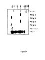
ニトロセルロース結合β−gal融合完全rPhl p 6(c223、c142)、N末端トランケートrPhl p 6(c171、c121、c233、c146)及び対照としてβ−gal単独を、9名の牧草花粉アレルギー個人及び1名の非アレルギー個人からの血清IgEに接触させた(図1)。結果は、2つの完全なPhl p 6アイソフォーム及び4個のN末端アミノ酸のみ欠失するPhl p 6断片は試験したすべての牧草花粉アレルギー患者からのIgEに強く結合し、部分的Phl p 6クローンのIgE結合能はタンパク質のN末端から欠如しているアミノ酸の数に依存して低下することを示した。N末端の30個のアミノ酸を欠失する部分クローン(クローン121)は既にそのIgE結合能を完全に喪失していた(図1)。
【0045】
組換えPhl p 6のE.coli発現と精製。精製rPhl p 6のIgE結合能。
【0046】
E.coli BL 21(DE3)においてrPhl p 6を過剰発現させた。いくつかの精製段階の組合せによって純粋な可溶性rPhl p 6(約5mgタンパク質/リットルE.coli培養)を生成し、SDS−PAGEにより低分子量チモシー牧草花粉アレルゲンの1つとして同定した(図2A)。精製組換えPhl p 6のMALDI−TOF分析は、サンプルのMH+及びM2H2+種に対応する11790と5896の2つの質量/電荷ピークを生じ、推定Phl p 6分子量(11789Da)と一致した。
【0047】
171名の牧草花粉アレルギー患者からの128の血清においてrPhl p 6特異的IgE抗体が検出されたが、10名の非アレルギー個人からの血清からは検出されなかった。rPhl p 6を有する牧草花粉アレルギー患者からの血清の前吸収は、ニトロセルロースにブロッティングしたチモシー牧草花粉抽出物における10−14kDa部分へのIgE結合を大きく低下させ、rPhl p 6と天然Phl p 6がIgEエピトープを共有することを示唆した。ELISA競合実験は、低率(<20%)のPhl p 5特異的IgEだけがrPhl p 6と共に前吸収されうることを明らかにした。rPhl p 1、rPhl p 2及び組換えチモシー牧草プロフィリンへのIgE結合は、rPhl p 6との血清のプレインキュベーション後も低下しなかった。これらの結果により、Phl p 6が他の牧草花粉アレルゲンとは異なる重要なアレルゲンとして同定された。
【0048】
安定なすべてのαらせんコンフォメーションにおけるrPhl p 6の折りたたみ
精製rPhl p 6の遠紫外CDスペクトル(図2B)は、このタンパク質がかなりの量のαらせん二次構造を含むことを示す。スペクトルは、208nmと220nmの2つの広い最小量と191nmの最大量を特徴とする。二次構造予測(37)は、顕著なαらせん二次構造含量を示し、CD測定とかなり一致する。rPhl p 6の変性推移は一相性であり、61℃の融点と高度に協同する。85℃では、rPhl p 6は200nmの典型的最小量を有するランダムコイル構造をとる。rPhl p 6は、冷却曲線プロフィール(図2C)及び85℃から冷却後20℃で記録した遠紫外スペクトル(図2B)から明らかなように、高度の折りたたみ可逆性を示す。
【0049】
組換えPhl p 6は牧草花粉アレルギー患者において用量依存的な好塩基球のヒスタミン遊離と即時型皮膚反応を誘導する。
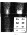
【0050】
精製rPhl p 6は、牧草花粉アレルギー患者の好塩基球から特異的で用量依存的なヒスタミンの遊離を誘導した(図3A)。活性が高い牧草花粉アレルゲンであるrPhl p 5(14、ValentaとFlicker、未発表データ)は、rPhl p 6に比べてより低濃度でも最大の遊離を誘導した。4名の牧草花粉アレルギー患者においてrPhl p 6、rPhl p 5及びチモシー牧草花粉抽出物は即時型皮膚反応を誘発したが、非アレルギー患者では反応を生じなかった(表1;図3B)。塩化ナトリウムに対する反応は認められなかったが、ヒスタミンは試験したすべての個人で膨疹反応を誘発した(表1;図3B)。
【0051】
6群アレルゲンは花粉特異的タンパク質である。
【0052】
牧草花粉アレルゲンの主要な群はほとんどの牧草種で起こるが(4)、6群アレルゲンはチモシー牧草(Phleum pratense)花粉において広汎に生じることが報告された(15)。ウサギ抗rPhl p 6抗血清は、25−28kDaの間で様々な単子葉植物(Phleum pratense、Lolium perenne、Secale cereale、Triticum sativum、Avena sativa、Phragmites communis)からのニトロセルロースブロッティングした花粉抽出物中の5群アレルゲンと交叉反応した(図4A、レーン2)。11kDaでPhl p 6又はPhl p 6関連アレルゲンがPhleum pratense及びPoa pratensisからの花粉において広汎に検出された。Phl p 6 cDNA配列から推定したアミノ酸配列内に推定Nグリコシル化部位が認められたが、天然と組換えのPhl p 6について同等の分子量が観察されたことから、天然Phl p 6の多グリコシル化は排除される(図4A、2A)。ウサギ抗rPhl p 6抗体は、ニトロセルロースブロッティングしたチモシー牧草花粉中11kDaでPhl p 6と強く反応したが、葉あるいは根の抽出物とは反応しなかった(図4B、レーン2)。約14kDaで3つの組織すべてにおいてプロフィリンが検出された(図4B、レーン1)。
【0053】
チモシー牧草花粉におけるPhl p 6の免疫電子顕微鏡的局在。
【0054】
包埋後イムノゴールド電子顕微鏡を用いて、ウサギ抗rPhl p 6抗体を成熟チモシー牧草花粉粒内のかなりの部分を満たす多数の多糖類(P−)粒子に結合した(図4C)。金粒子の最大の蓄積はP粒子の断面図上の表面に認められ、Phl p 6はP粒子内ではなくP粒子上に存在することを示唆した。花粉粒子の他の部分ではほとんど(細胞質ゾル、外膜)若しくは全く(ミトコンドリア、内膜)抗rPhl p 6免疫反応性を認めなかった。同様にアミロプラストではほとんど金粒子が検出されなかった。この局在パターンは、ウサギ抗rPhl p 5抗血清がP粒子を標識することができなかったという我々の所見(データは示していない)と合わせて考えると、P粒子の免疫標識が交叉反応性5群アレルゲンの存在から生じた可能性を排除する。免疫前Igに関して実施した対照実験は、いくつかの非特異的に吸収された金粒子だけを生じた(図4D)。
【0055】
Phl p 6欠失変異体(aa1−57、aa31−110)はIgE結合能が大きく低下したことを示す。
【0056】
ニトロセルロースブロットした完全なrPhl p 6(図5A)、rPhl p 6変異体aa1−57(図5B)及びrPhl p 6変異体aa31−110(図5C)を、牧草花粉アレルギー患者からの13の血清、非アトピー個人からの血清及びウサギ抗rPhl p 6抗血清に接触させた。13名の牧草花粉アレルギー患者全員が完全な組換えPhl p 6に対してIgE反応性を示し(図5A)、変異体aa1−57は、血清11によって認識され、血清13によって認識は弱くかった(図5B)。Phl p 6変異体aa31−110は血清7及び11とのみ弱く反応した(図5C)。非アトピー個人からの血清は完全なrPhl p 6及び欠失変異体と反応することができなかった。ウサギ抗rPhl p 6抗血清は完全なrPhl p 6及び2つの欠失変異体に対して同等の強度の反応性を示した(図5A−C;レーン15)が、ウサギ免疫前血清は当該分子の分子量の範囲内で反応性を示さなかった(図5A−C;レーン16)。
【0057】
rPhl p 6に対するマウス抗rPhl p 6又はrPhl p 6 aa31−110抗血清のIgG1反応性
完全なrPhl p 6及びrPhl p 6 aa31−110に対して惹起したマウスIgG1はrPhl p 6と反応する(表II)。
【0058】
rPhl p 6誘導体はヒスタミンの遊離誘導能がかなり低下している。
【0059】
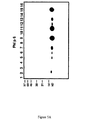
牧草花粉に対してアレルギー性の患者からの顆粒球を様々な濃度の精製rPhl p 6、rPhl p 6 aa1−57、rPhl p 6 aa31−110、rPhl p 6 aa1−33又は抗IgE mAb(E124.2.8 Dε2,Immunotech,Marseilles,France)と共にインキュベートした。上清中に遊離されたヒスタミンをRIA(Immunotech)によって測定した(図6)。精製rPhl p 6は、牧草花粉に対してアレルギー性の患者の好塩基球から特異的で用量依存性のヒスタミン遊離を誘導したが、rPhl p 6誘導体aa1−57及びaa31−110は1μg/mlの濃度までヒスタミンの遊離を誘導しなかった。Phl p 6 aa1−33は1μg/mlの濃度でヒスタミンの50%遊離を誘導し、これは完全なrPhl p 6に比べて約1000倍のヒスタミン遊離の低下に相当する。
【0060】
(考察)
アレルギー患者の約40%が牧草花粉との接触後に即時型症状を示す(3)。我々は、Phl p 6と称する、主要なチモシー牧草花粉アレルゲンのアイソフォーム及び断片をコードするcDNAを単離した。Phl p 6は、牧草花粉アレルギー患者の75%のIgE抗体によって認識される11.8kDaのタンパク質アレルゲンである。従ってrPhl p 6のIgE認識の発生率は先に報告された天然Phl p 6についての発生率と一致し、炭水化物はPhl p 6のIgE認識に関連する役割を果たしていないことを示唆する(28、29)。天然Phl p 6について得られたペプチド配列データと一致して、我々は、rPhl p 6の推定繹アミノ酸配列が、25−35kDaの主要牧草花粉アレルゲンのファミリーである5群牧草花粉アレルゲンのN末端部分と高度の配列相同性を示すことが認められた(29、14)。N末端の疎水性リーダーペプチドの存在により、Phl p 6は、5群アレルゲン断片と異なる独立したアレルゲンである。cDNAクローンをコードするPhl p 6を分析した他の著者(15)の提案と一致して、我々は、5群及び6群アレルゲンが、1群及び2/3群牧草花粉アレルゲンについて記述されている(30)と同様に、共通の祖先遺伝子から進化したのであろうと推測する。Phl p 6が牧草花粉アレルゲンの独立した群に属するという仮説も、Phl p 6が5群及び他の牧草花粉アレルゲンと交叉反応性IgEエピトープをほとんど共有しないという我々の所見に裏付けられる。溶媒可触性の予測からは、Phl p 6のN末端アミノ酸の多くが溶媒に接触しているのに対し、C末端アミノ酸残基の大部分は被覆されていることが示された。証明されてはいないが、この所見はIgEエピトープマッピング実験から得られたデータと一致しており、この実験は、タンパク質のN末端がIgE認識に決定的に関与することを示している。しかしまた、N末端自体が優性IgEエピトープである、若しくはN末端の欠失が立体的Phl p 6 IgEエピトープに影響を及ぼすという可能性も存在する。
【0061】
E.coliにおけるPhl p 6の発現は、αらせん二次構造をほとんど独占的に含む可溶性の折りたたみ組換えタンパク質を多量に生成した。Phl p 6のαらせん折りたたみは、ある種のタンパク質がアレルゲンとして働く傾向として共通の構造的特徴が存在しないことがさらに確認された。Phl p 6はおそらくすべてαらせんタンパク質である可能性が高いが、主要カバ花粉アレルゲンであるBet v 1(31)、及びカバプロフィリン、Bet v 2(32)はαβ混合折りたたみを有する。CD分光分析によって明らかにされたように、rPhl p 6は、他の免疫学的に無関係な花粉アレルゲン(たとえばBet v 1(33)、Bet v 2(6、32))と、変性後再び安定な構造に折りたたまれるという顕著な内在傾向を共有する。Phl p 6と他の重要な植物アレルゲンが共有するもう1つの特徴は、花粉組織におけるその高い発現である。これまでに特定された植物アレルゲンの大部分が主として成熟花粉において発現されるという事実により、気道を通じた感作の軌跡と解釈することができる(34)。
【0062】
イムノゴールド電子顕微鏡検査によると、Phl p 6は主として成熟花粉のP粒子上に局在した。P粒子は、休眠花粉粒の含量の30%までを占め、花粉の発芽の際に成長する花粉管壁内に物質を運ぶ、小さな多糖類含有体である(35、36)。P粒子は小さなサイズであり(<2.5ミクロン)、それ故Phl p 6を気管支粘膜と直接接触させる呼吸性アレルゲン担体として働くことができるので、P粒子上にPhl p 6が生じることと決定的な関連性を有すると考えられる。従って、いくつかの牧草種(Phleum pratense、Poa pratensis)だけが低分子量範囲内(10−12kDa)にウサギ抗rPhl p 6反応性部分を含むが、P粒子と結合したPhl p 6の気道深部への侵入により、アレルゲン感作の発生率が高い(75%)ことの説明となるであろう。
【0063】
大腸菌(Escherichika coli)に発現させた精製組換えrPhl p 6アレルゲンは、牧草花粉アレルギー患者の大半のIgE抗体と反応し、好塩基球のヒスタミン遊離ならびに即時型皮膚反応を誘発した。それ故、このアレルゲンは牧草花粉アレルギーの生体外及び生体内(皮膚試験)診断のために使用しうる。Phl p 6のN末端部分の欠失がアレルゲンのIgE結合能を著しく低下させたという我々の所見により、アフィラキシー副作用の少ない牧草花粉アレルギーの特異的免疫療法のために使用しうるPhl p 6欠失変異体を構築することが可能であろうという発想に至った。同様の戦略が最近、主要カバ花粉アレルゲンBet v 1の立体的IgEエピトープを崩壊するために適用された(23)が、Phl p 6分子は連続IgEエピトープであるので、Phl p 6については予測できなかったであろう。我々は、Phl p 6のN末端及びC末端がトランケートされた変種を作製したが、その中で変異体aa1−57とaa31−110はIgE結合能をほとんど完全に消失していた。我々は、組換え分子として又はペプチド化学によって作製したこれら2つの低刺激性Phl p 6を牧草花粉アレルギーに対するワクチン候補として使用することを提案する。
【0064】
(表の簡単な説明)
(表I)
rPhl p 6に対する即時型皮膚反応。4名の牧草花粉アレルギー患者(HP、SF、CS、LW)及び2名の非アレルギー個人(SV、SS)を、精製rPhl p 6、rPhl p 5、天然チモシー牧草花粉抽出物、ヒスタミン及び等張塩化ナトリウムで皮膚試験した。結果は膨疹反応の平均直径(mm)で表わしている。
【0065】
(表II)
rPhl p 6に対するマウス抗rPhl p 6又は抗rPhl p 6 aa31−110抗血清のIgG1反応性。
【0066】
【表1】
【表2】
【表3】
【図面の簡単な説明】
【図1】 rPhl p 6アイソフォーム及び断片のIgE反応性。2つのPhl p 6アイソフォーム(c142、c223)、Phl p 6断片(c121、c146、c171、c233)を発現する組換えλgt11ファージ、及び対照としてλgt11野生型ファージ(0)からのタンパク質を含むニトロセルロースフィルターを、9名の牧草花粉アレルギー患者(1−9)及び1名の非アレルギー個人(10)からの血清IgEで試験した。
【図2A】 、組換えチモシー牧草花粉アレルゲンの純度。精製組換えチモシー牧草花粉アレルゲン(Phl p 1、Phl p 2、Phl p 5、Phl p 6、チモシー牧草花粉プロフィリン)及び天然チモシー牧草花粉抽出物(チモシー)を含む、クマシーブリリアントブルー染色したSDS−PAGE。(M)分子量マーカー。
【図2B】 円偏光二色性分析。平均残基楕円性([Θ])として表わしたrPhl p 6の遠紫外遠偏光二色性スペクトル(y軸)をx軸に表示した波長範囲で20℃(実線)、85℃(点線)及び85℃から冷却後20℃(破線)で記録した。
【図2C】 円偏光二色性分析。220nmでモニターした精製rPhl p 6の熱変性と冷却(x軸:℃で表示した温度;y軸:折りたたみタンパク質の見かけ分画)。
【図3A】 rPhl p 6は好塩基球のヒスタミン遊離を誘導する。牧草花粉アレルギー患者からの顆粒球を種々の濃度(x軸)の精製組換えPhl p 6(三角)、Phl p 5(点)又はモノクローナル抗IgE抗体(四角)と共にインキュベートした。上清中に遊離されたヒスタミンのパーセンテージをy軸に示す。結果は3回の測定の平均値(+/−SD)である。
【図3B】 感作アレルギー患者でのrPhl p 6による即時型皮膚反応の誘発。2名の牧草花粉アレルギー患者(a)LW、(b)HP及び非アレルギー個人(c)SVを、(d)に示すような漸増濃度のrPhl p 6とrPhl p 5ならびにヒスタミン(Hist)とNaClで前腕に穿刺した。膨疹部分をボールペンで囲んでいる。
【図4】 Phl p 6の組織特異的発現。、種々の単子葉植物からのニトロセルロースブロットした牧草花粉抽出物をウサギ免疫前Ig(レーン1)又はウサギ抗rPhl p 6 Ig(レーン2)でプローブした。B、同等の量のニトロセルロースブロットしたチモシー牧草花粉、葉及び根からのタンパク質抽出物をウサギ抗プロフィリンIg(レーン1)、ウサギ抗Phl p 6 Ig(レーン2)又はウサギ免疫前Ig(レーン3)と共にインキュベートした。
C,D,Phl p 6の超微細構造局在。チモシー牧草花粉の超薄切片をウサギ抗Phl p 6 Ig(C)及びウサギ免疫前Ig(D)で染色した。結合ウサギ抗体を金複合ヤギ抗ウサギIg抗血清(金粒子=黒い点)で検出した。矢印はP粒子上のPhl p 6免疫反応性を示す。略語:E:外膜;I:内膜;P:P粒子。バーは0.250μmである。
【図5A】 Phl p 6欠失変異体の低いIgE結合能。等量の組換えPhl p 6を、チモシー牧草花粉アレルギー患者からの血清(レーン1−13)及び非アレルギー対照個人からの血清(レーン14)とのIgE反応性について試験した。レーン15とレーン16はウサギ抗rPhl p 6抗血清及びウサギ免疫前血清との反応性を示す。結合IgE抗体を125I標識抗ヒトIgE抗体で検出し、結合ウサギ抗体を125I標識ロバ抗ウサギ抗体で検出して、オートラジオグラフィーによって視覚化した。
【図5B】 Phl p 6欠失変異体の低いIgE結合能。等量の組換えPhl p 6 aa1−57を、チモシー牧草花粉アレルギー患者からの血清(レーン1−13)及び非アレルギー対照個人からの血清(レーン14)とのIgE反応性について試験した。レーン15とレーン16はウサギ抗rPhl p 6抗血清及びウサギ免疫前血清との反応性を示す。結合IgE抗体を125I標識抗ヒトIgE抗体で検出し、結合ウサギ抗体を125I標識ロバ抗ウサギ抗体で検出して、オートラジオグラフィーによって視覚化した。
【図5C】 Phl p 6欠失変異体の低いIgE結合能。等量の組換えPhl p 6 aa31−110を、チモシー牧草花粉アレルギー患者からの血清(レーン1−13)及び非アレルギー対照個人からの血清(レーン14)とのIgE反応性について試験した。レーン15とレーン16はウサギ抗rPhl p 6抗血清及びウサギ免疫前血清との反応性を示す。結合IgE抗体を125I標識抗ヒトIgE抗体で検出し、結合ウサギ抗体を125I標識ロバ抗ウサギ抗体で検出して、オートラジオグラフィーによって視覚化した。
【図6】 牧草花粉に対するアレルギー患者からの顆粒球を様々な濃度(1、10−1、10−2、10−、10−5、及び10−7μg/ml)の精製rPhl p 6(点)、rPhl p 6 aa1−57(上向き三角)、rPhl p 6 aa31−110(下向き三角)、rPhl p 6 aa1−33(菱形)又は抗IgE mAb(四角)と共にインキュベートした。上清中に遊離されたヒスタミンをRIAによって測定し、y軸に示している。結果は3回の測定の平均値である。[0001]
The present invention relates to the non-anaphylactic, i.e. hypoallergenic, form of the important timothy (Pollus vulgaris) grass pollen
[0002]
Type I allergy is a genetically determined hypersensitivity disease that affects more than 20% of the population in industrialized countries (1). As a result of this immune disorder, allergic patients produce IgE antibodies against mainly airborne proteins that are harmless per se from pollen, mites, molds and animal hair / dandruff. Symptoms of type I allergy (allergic rhinitis, conjunctivitis, allergic asthma and anaphylactic shock) are caused by an allergen-induced cross-linkage between effector cell (mast cell, basophil) -bound IgE antibody and subsequent release of inflammatory mediators. Occurs (2). Since about 40% of allergic individuals develop symptoms after contact with grass pollen, research has been conducted on the identification of related grass pollen allergens by protein and immunochemical methods (3). Major allergen groups have been identified as part of the cross-reactions that occur in most grass species (4), but their properties and biological functions are completely unknown.
[0003]
By applying recent molecular biology techniques to the identification of allergens, the primary structure of allergens has been clarified, making it easier to produce recombinant allergens for diagnostic and therapeutic purposes (5). Plant cytoskeleton components (eg profilin) (6) as well as calcium-binding pollen protein (7) have been identified as related allergens. The fact that allergic patients show an immediate response upon contact with various unrelated allergen sources can therefore be explained by the cross-reactivity of ubiquitous allergens and their IgE antibodies. A group of grass pollen allergens belongs to the family of cell wall relaxing proteins (expansins) (8), and
[0004]
Treatment of type I allergic diseases is currently performed by pharmacological treatment and specific immunotherapy. Specific immunotherapy has already been established at the beginning of this century (Noon, Lancet 1 (1911) 1572-1573) and involves the systemic administration of increasing amounts of allergen over a long period of time. Although specific immunotherapy has been recognized as an effective treatment, the occurrence of anaphylactic side effects is one of the major problems with this treatment. Recently, the use of T cell epitopes for allergen-specific immunotherapy was proposed to reduce anaphylactic reactions (Briner et al., Proc. Natl. Acad. Sci. USA 90 (1993) 7608-7612 and Norman, Curr. Opin.Immunol.5 (1993) 986-973). Allergens contain a vast variety of T cell epitopes that can overlap with the continuous antigenic determinant IgE epitope (Ebner et al., J. Immunol. 150 (1993) 1047-1054; Joost-van-Neeven et al. Immunol. 151 (1993) 2326-2335; and Schenket et al., J. Allergy Clin. Immunol. 96 (1995) 986-996). In order to prevent cross-linking of effector cell (mast cell, basophil) -bound IgE and transmitter release, it is necessary to examine T cell epitope and IgE epitope in detail.
[0005]
Vrtala et al. Clin. Invest. 99 (7) 1673-1681 (1997) and WO 99/16467 disrupt its three-dimensional structure by expressing two parts of the
[0006]
Unlike the major hippo allergen Bet v 1, the major timothy grass pollen
[0007]
Surprisingly and surprisingly, however, according to the present invention,
[0008]
In a first aspect of the invention, there is provided a hypoallergenic immunogenic molecule derived from a
[0009]
N-terminal or C-terminal deletions are considered to be terminal truncations of the allergen. Such deletions can also be present within the N-terminal or C-terminal part of the allergen, respectively.
[0010]
Fragments of allergen molecules are well known to those skilled in the art and can be made by recombinant DNA techniques or peptide synthesis chemistry.
[0011]
As a second aspect, the present invention relates to (i) a
[0012]
Because they together contain the complete amino acid sequence of
[0013]
The size of the N-terminal and C-terminal deletions of (i) immunogenic and (ii) non-IgE-
[0014]
The third aspect of the present invention is specific desensitization therapy. Such therapy is practiced as a known technique for protein allergens, and is repeated administration of an immunogen capable of eliciting an IgG immune response against the allergen to a mammal, typically a human individual, suffering from a type I allergy to the allergen. Including doing. The immunogen can be mixed with a suitable adjuvant, such as aluminum oxide. Administration can be carried out systemically, such as by injection, infusion, etc., but oral routes have also been suggested to expose the intestinal portion of the immune system. Norman PS, “Current status of immunotherapy for allergies and anaphylactic reactions” Adv. Internal. See also Medicine 41 (1996) 681-713.
[0015]
Here, the immunogen to be administered is an immunogenic molecule according to the first aspect of the invention, or a mixture of such molecules, preferably according to the second aspect of the invention, (i) by deletion of the N-terminus
[0016]
More specifically, immunogens are used to induce antibody responses and / or elicit T cell responses in patients and / or to modulate antibodies and T cell responses to induce tolerance. sell.
[0017]
A fourth aspect of the invention relates to an IgA, IgD, IgE, IgG or IgM class of the immunogen according to the first aspect or the combination of the immunogens according to the second aspect against the
[0018]
The invention is illustrated by the following non-limiting examples.
[0019]
(Example)
Experimental materials and methods
Biological material, patient serum, antiserum, recombinant allergen.
[0020]
Timothy grass (Phlum plate), barley (Lolium perenne), rye (Secale cereal), Kentucky spruce grass (Pentium), wheat (Tria) reed) (Phragmits communis) pollen was obtained from Allergon AB (Valinge, Sweden). Timothy grass seeds were purchased from Austria (Vienna, Austria) and grown for 4 weeks to obtain fresh leaves and roots. Patients allergic to grass pollen were identified as described in (4). Rabbit anti-celeryprofilin antiserum (RP1) is described in (12). A
[0021]
Isolation and identification of cDNA
[0022]
350 IgE-reactive clones were isolated from an expression cDNA library constructed from mature timothy grass pollen in phage λgt11 (14). Six cDNAs (c121, c142, c146, c171, c223, c233) having sequence homology with the cDNA
[0023]
Mapping of
[0024]
The IgE binding ability of phage clones expressing
[0025]
MALDI-TOF (Matrix Assisted Laser Desorption / Ionization-Time of Flight) and CD (Circular Dichroism) analysis of purified
[0026]
Laser desorption mass spectra were obtained in linear mode using a time-of-flight Compact MALDI II instrument (Kratos, Manchester, UK) (piCHEM, Graz, Austria). CD spectra were recorded on a Jasco J-710 spectropolarimeter fitted with a Jasco PTC-348WI Peltier-type temperature control system and coupled to a Fisons HAAKE GH water bath. Far ultraviolet CD spectra were recorded at 20 ° C. with a 2 mm width-length quartz cuvette (Hellma, Mullheim, Baden, Germany) at a protein concentration of 7 μM. Thermal denaturation of
[0027]
[0028]
The incidence of
[0029]
Histamine release experiment.
[0030]
Granulocytes were isolated by dextran sedimentation (22) from heparinized blood samples of grass pollen allergic individuals with
[0031]
Skin test.
[0032]
After obtaining informed consent, a skin puncture test was performed on each person's forearm as described in (23). Each person in 20 μl aliquots containing purified
[0033]
Analysis of the presence of
[0034]
Protein extracts from pollen, leaves and roots were obtained by homogenizing the tissue in SDS-sample buffer (24). The extract was centrifuged (10,000 × g, 20 minutes; Sorvall RC5C, SS34 rotor) to remove insoluble material. Protein extracts were separated by 14% SDS-PAGE (25) and blotted to nitrocellulose (26). Nitrocellulose strips were tested with rabbit anti-celeryprofirin antiserum, RP1 (12),
[0035]
In situ localization of
[0036]
Timothy grass pollen grains were fixed anhydrous as described in (27). Ultrathin sections were incubated with either an equal concentration of
[0037]
Construction of hypoallergenic Phl p 6 (Phlum plate) deletion mutants.
[0038]
N-terminal and C-
[Chemical 1]
[0039]
The PCR product was cut with Nde I / Kpn I (aa1-57) or Nde I / Eco RI (aa31-110), purified by preparative agarose gel electrophoresis, subcloned into plasmid pET-17b (Novagen), E. E. coli BL21 (DE3) (Novagen) was transformed. Colonies expressing the correct deletion mutant were identified by immunoscreening with
[0040]
E. Expression of
By inducing
[0041]
result
Isolation and characterization of cDNA
[0042]
Six cDNA clones (c142, c223, c171, c121, c233, c146) encoding the
[0043]
The N-terminus of
[0044]
Nitrocellulose-bound β-gal fusion complete rPhl p 6 (c223, c142), N-terminal truncated rPhl p 6 (c171, c121, c233, c146) and β-gal alone as controls, 9 grass pollen allergic individuals and 1 Serum IgE from non-allergic individuals was contacted (Figure 1). The results show that the two
[0045]
[0046]
[0047]
RPhl p 6-specific IgE antibodies were detected in 128 sera from 171 grass pollen allergic patients, but not from sera from 10 non-allergic individuals. Preabsorption of serum from grass pollen allergic patients with
[0048]
The far ultraviolet CD spectrum of purified rPhl p 6 (FIG. 2B) shows that this protein contains a significant amount of α-helical secondary structure. The spectrum is characterized by two broad minimums of 208 nm and 220 nm and a maximum of 191 nm. Secondary structure prediction (37) shows a significant α-helical secondary structure content and is in good agreement with the CD measurement. The modification course of
[0049]
[0050]
[0051]
[0052]
While the main group of grass pollen allergens occurs in most grass species (4),
[0053]
Immunoelectron microscopic localization of
[0054]
After embedding, an immunogold electron microscope was used to bind the
[0055]
[0056]
Nitrocellulose blotted complete rPhl p 6 (FIG. 5A),
[0057]
IgG1 reactivity of
Mouse IgG1 raised against
[0058]
The
[0059]
Granulocytes from patients allergic to grass pollen were purified at various concentrations of purified
[0060]
(Discussion)
About 40% of allergic patients show immediate symptoms after contact with grass pollen (3). We have isolated a cDNA encoding a major timothy grass pollen allergen isoform and fragment, referred to as
[0061]
E. Expression of
[0062]
According to immunogold electron microscopy,
[0063]
Purified
[0064]
(Short description of the table)
(Table I)
Immediate skin reaction to
[0065]
(Table II)
IgG1 reactivity of
[0066]
[Table 1]
[Table 2]
[Table 3]
[Brief description of the drawings]
FIG. 1. IgE reactivity of
FIG. 2A, Purity of recombinant timothy grass pollen allergen. Coomassie brilliant blue stained SDS- containing purified recombinant Timothy grass pollen allergen (
FIG. 2B: Circular dichroism analysis. 20 ° C. (solid line), 85 ° C. (dotted line) in the wavelength range indicated on the x axis of the far ultraviolet far dichroism spectrum (y axis) of
FIG. 2C Circular dichroism analysis. Thermal denaturation and cooling of purified
FIG. 3A.
FIG. 3B. Induction of immediate skin reaction by
FIG. 4. Tissue specific expression of
Hyperfine structure localization of C, D,
FIG. 5A: Low IgE binding capacity of
FIG. 5B: Low IgE binding capacity of
FIG. 5C: Low IgE binding capacity of
FIG. 6: Purified rPhl p 6 (dots) at various concentrations (1, 10-1, 10-2, 10-10, 10-5, and 10-7 μg / ml) of granulocytes from patients allergic to grass pollen ,
Claims (3)
Applications Claiming Priority (3)
| Application Number | Priority Date | Filing Date | Title |
|---|---|---|---|
| SE9903950A SE9903950D0 (en) | 1999-10-29 | 1999-10-29 | Non-anaphylactic forms of grass pollen allergen and their use |
| SE9903950-5 | 1999-10-29 | ||
| PCT/SE2000/002062 WO2001030816A1 (en) | 1999-10-29 | 2000-10-24 | NON-ANAPHYLACTIC FORMS OF GRASS POLLEN Ph1 p 6 ALLERGEN AND THEIR USE |
Related Child Applications (1)
| Application Number | Title | Priority Date | Filing Date |
|---|---|---|---|
| JP2011132059A Division JP2011236217A (en) | 1999-10-29 | 2011-06-14 | NON-ANAPHYLACTIC FORMS OF GRASS POLLEN Phl p 6 ALLERGEN AND THEIR USE |
Publications (2)
| Publication Number | Publication Date |
|---|---|
| JP2003513023A JP2003513023A (en) | 2003-04-08 |
| JP4881526B2 true JP4881526B2 (en) | 2012-02-22 |
Family
ID=20417569
Family Applications (2)
| Application Number | Title | Priority Date | Filing Date |
|---|---|---|---|
| JP2001533813A Expired - Lifetime JP4881526B2 (en) | 1999-10-29 | 2000-10-24 | Non-anaphylactic forms of grass pollen Phlp6 allergen and their use |
| JP2011132059A Pending JP2011236217A (en) | 1999-10-29 | 2011-06-14 | NON-ANAPHYLACTIC FORMS OF GRASS POLLEN Phl p 6 ALLERGEN AND THEIR USE |
Family Applications After (1)
| Application Number | Title | Priority Date | Filing Date |
|---|---|---|---|
| JP2011132059A Pending JP2011236217A (en) | 1999-10-29 | 2011-06-14 | NON-ANAPHYLACTIC FORMS OF GRASS POLLEN Phl p 6 ALLERGEN AND THEIR USE |
Country Status (10)
| Country | Link |
|---|---|
| EP (1) | EP1224215B2 (en) |
| JP (2) | JP4881526B2 (en) |
| AT (1) | ATE302213T1 (en) |
| AU (1) | AU778066B2 (en) |
| CA (1) | CA2387260C (en) |
| DE (1) | DE60022069T3 (en) |
| DK (1) | DK1224215T4 (en) |
| ES (1) | ES2246904T5 (en) |
| SE (1) | SE9903950D0 (en) |
| WO (1) | WO2001030816A1 (en) |
Families Citing this family (3)
| Publication number | Priority date | Publication date | Assignee | Title |
|---|---|---|---|---|
| AT503530A1 (en) | 2006-04-28 | 2007-11-15 | Biomay Ag | POLYPEPTIDE WITH ALLERGENIC PROPERTIES |
| EP2042193A1 (en) | 2007-09-28 | 2009-04-01 | Biomay AG | RNA Vaccines |
| BR112012017303A2 (en) * | 2010-01-14 | 2016-04-19 | Merck Patent Gmbh | true grass group 6 allergen variants having reduced allergen quality due to proline residue mutagenesis |
Citations (1)
| Publication number | Priority date | Publication date | Assignee | Title |
|---|---|---|---|---|
| WO1998043657A2 (en) * | 1997-03-27 | 1998-10-08 | Merck Patent Gmbh | Graminae pollen allergen mutants for specific immunotherapy, and production and use of the same |
-
1999
- 1999-10-29 SE SE9903950A patent/SE9903950D0/en unknown
-
2000
- 2000-10-24 ES ES00975093T patent/ES2246904T5/en not_active Expired - Lifetime
- 2000-10-24 DK DK00975093.6T patent/DK1224215T4/en active
- 2000-10-24 EP EP00975093A patent/EP1224215B2/en not_active Expired - Lifetime
- 2000-10-24 JP JP2001533813A patent/JP4881526B2/en not_active Expired - Lifetime
- 2000-10-24 CA CA2387260A patent/CA2387260C/en not_active Expired - Lifetime
- 2000-10-24 DE DE60022069T patent/DE60022069T3/en not_active Expired - Lifetime
- 2000-10-24 AU AU13193/01A patent/AU778066B2/en not_active Expired
- 2000-10-24 AT AT00975093T patent/ATE302213T1/en active
- 2000-10-24 WO PCT/SE2000/002062 patent/WO2001030816A1/en active IP Right Grant
-
2011
- 2011-06-14 JP JP2011132059A patent/JP2011236217A/en active Pending
Patent Citations (1)
| Publication number | Priority date | Publication date | Assignee | Title |
|---|---|---|---|---|
| WO1998043657A2 (en) * | 1997-03-27 | 1998-10-08 | Merck Patent Gmbh | Graminae pollen allergen mutants for specific immunotherapy, and production and use of the same |
Also Published As
| Publication number | Publication date |
|---|---|
| EP1224215B1 (en) | 2005-08-17 |
| ES2246904T5 (en) | 2011-03-01 |
| DE60022069T3 (en) | 2011-05-05 |
| CA2387260C (en) | 2015-12-08 |
| JP2003513023A (en) | 2003-04-08 |
| AU1319301A (en) | 2001-05-08 |
| EP1224215B2 (en) | 2010-10-06 |
| DE60022069D1 (en) | 2005-09-22 |
| ATE302213T1 (en) | 2005-09-15 |
| DE60022069T2 (en) | 2006-06-08 |
| DK1224215T4 (en) | 2011-01-24 |
| EP1224215A1 (en) | 2002-07-24 |
| AU778066B2 (en) | 2004-11-11 |
| SE9903950D0 (en) | 1999-10-29 |
| ES2246904T3 (en) | 2006-03-01 |
| JP2011236217A (en) | 2011-11-24 |
| DK1224215T3 (en) | 2005-12-19 |
| CA2387260A1 (en) | 2001-05-03 |
| WO2001030816A1 (en) | 2001-05-03 |
Similar Documents
| Publication | Publication Date | Title |
|---|---|---|
| Vrtala et al. | Molecular, immunological, and structural characterization of Phl p 6, a major allergen and P-particle-associated protein from Timothy grass (Phleum pratense) pollen | |
| Vrtala et al. | Immunologic characterization of purified recombinant timothy grass pollen (Phleum pratense) allergens (Phl p 1, Phl p 2, Phl p 5) | |
| Ball et al. | Gain of structure and IgE epitopes by eukaryotic expression of the major Timothy grass pollen allergen, Phl p 1 | |
| US7485305B2 (en) | Allergen from house-dust mites | |
| US20090098167A1 (en) | PHL P 1 Allergen Derivative | |
| JP2011236217A (en) | NON-ANAPHYLACTIC FORMS OF GRASS POLLEN Phl p 6 ALLERGEN AND THEIR USE | |
| US8440200B2 (en) | Non-anaphylactic forms of grass pollen Ph1 p 6 allergen and their use | |
| JP4253122B2 (en) | Non-anaphylactic forms of allergens and their use | |
| Laffer et al. | Molecular characterization of recombinant T1, a non-allergenic periwinkle (Catharanthus roseus) protein, with sequence similarity to the Bet v 1 plant allergen family | |
| WO2002020790A1 (en) | Parietaria judaica ns-ltp antigen variants, uses thereof and compositions comprising them | |
| EP1317484B1 (en) | Variants of the phleum pratense phl p 1 allergenic protein | |
| JP2006515984A (en) | Hypoallergenic allergic vaccine based on Pseudomonas pollen allergen PhlP7 | |
| EP1515988A1 (en) | Dna sequence and recombinant production of the grass pollen allergen phl p4 | |
| WO1992016554A1 (en) | Protein allergens of the species cynodon dactylon | |
| CA2437484A1 (en) | Ragweed allergens | |
| EP1369483A1 (en) | Allergen from mugwort pollen | |
| JP3588615B2 (en) | Recombinant mite allergen |
Legal Events
| Date | Code | Title | Description |
|---|---|---|---|
| A621 | Written request for application examination |
Free format text: JAPANESE INTERMEDIATE CODE: A621 Effective date: 20071010 |
|
| A131 | Notification of reasons for refusal |
Free format text: JAPANESE INTERMEDIATE CODE: A131 Effective date: 20100706 |
|
| A601 | Written request for extension of time |
Free format text: JAPANESE INTERMEDIATE CODE: A601 Effective date: 20100924 |
|
| A602 | Written permission of extension of time |
Free format text: JAPANESE INTERMEDIATE CODE: A602 Effective date: 20101001 |
|
| A521 | Request for written amendment filed |
Free format text: JAPANESE INTERMEDIATE CODE: A523 Effective date: 20110105 |
|
| A02 | Decision of refusal |
Free format text: JAPANESE INTERMEDIATE CODE: A02 Effective date: 20110215 |
|
| A521 | Request for written amendment filed |
Free format text: JAPANESE INTERMEDIATE CODE: A523 Effective date: 20110614 |
|
| A911 | Transfer to examiner for re-examination before appeal (zenchi) |
Free format text: JAPANESE INTERMEDIATE CODE: A911 Effective date: 20110803 |
|
| A131 | Notification of reasons for refusal |
Free format text: JAPANESE INTERMEDIATE CODE: A131 Effective date: 20110920 |
|
| A521 | Request for written amendment filed |
Free format text: JAPANESE INTERMEDIATE CODE: A523 Effective date: 20111102 |
|
| TRDD | Decision of grant or rejection written | ||
| A01 | Written decision to grant a patent or to grant a registration (utility model) |
Free format text: JAPANESE INTERMEDIATE CODE: A01 Effective date: 20111129 |
|
| A01 | Written decision to grant a patent or to grant a registration (utility model) |
Free format text: JAPANESE INTERMEDIATE CODE: A01 |
|
| A61 | First payment of annual fees (during grant procedure) |
Free format text: JAPANESE INTERMEDIATE CODE: A61 Effective date: 20111205 |
|
| R150 | Certificate of patent or registration of utility model |
Ref document number: 4881526 Country of ref document: JP Free format text: JAPANESE INTERMEDIATE CODE: R150 Free format text: JAPANESE INTERMEDIATE CODE: R150 |
|
| FPAY | Renewal fee payment (event date is renewal date of database) |
Free format text: PAYMENT UNTIL: 20141209 Year of fee payment: 3 |
|
| R250 | Receipt of annual fees |
Free format text: JAPANESE INTERMEDIATE CODE: R250 |
|
| R250 | Receipt of annual fees |
Free format text: JAPANESE INTERMEDIATE CODE: R250 |
|
| R250 | Receipt of annual fees |
Free format text: JAPANESE INTERMEDIATE CODE: R250 |
|
| R250 | Receipt of annual fees |
Free format text: JAPANESE INTERMEDIATE CODE: R250 |
|
| R250 | Receipt of annual fees |
Free format text: JAPANESE INTERMEDIATE CODE: R250 |
|
| R250 | Receipt of annual fees |
Free format text: JAPANESE INTERMEDIATE CODE: R250 |
|
| EXPY | Cancellation because of completion of term |