JP4841054B2 - Novel insulin / IGF / relaxin family polypeptide and DNA thereof - Google Patents
Novel insulin / IGF / relaxin family polypeptide and DNA thereof Download PDFInfo
- Publication number
- JP4841054B2 JP4841054B2 JP2001123210A JP2001123210A JP4841054B2 JP 4841054 B2 JP4841054 B2 JP 4841054B2 JP 2001123210 A JP2001123210 A JP 2001123210A JP 2001123210 A JP2001123210 A JP 2001123210A JP 4841054 B2 JP4841054 B2 JP 4841054B2
- Authority
- JP
- Japan
- Prior art keywords
- polypeptide
- dna
- seq
- present
- amino acid
- Prior art date
- Legal status (The legal status is an assumption and is not a legal conclusion. Google has not performed a legal analysis and makes no representation as to the accuracy of the status listed.)
- Expired - Fee Related
Links
- 108090000765 processed proteins & peptides Proteins 0.000 title claims description 528
- 102000004196 processed proteins & peptides Human genes 0.000 title claims description 509
- 229920001184 polypeptide Polymers 0.000 title claims description 504
- NOESYZHRGYRDHS-UHFFFAOYSA-N insulin Chemical compound N1C(=O)C(NC(=O)C(CCC(N)=O)NC(=O)C(CCC(O)=O)NC(=O)C(C(C)C)NC(=O)C(NC(=O)CN)C(C)CC)CSSCC(C(NC(CO)C(=O)NC(CC(C)C)C(=O)NC(CC=2C=CC(O)=CC=2)C(=O)NC(CCC(N)=O)C(=O)NC(CC(C)C)C(=O)NC(CCC(O)=O)C(=O)NC(CC(N)=O)C(=O)NC(CC=2C=CC(O)=CC=2)C(=O)NC(CSSCC(NC(=O)C(C(C)C)NC(=O)C(CC(C)C)NC(=O)C(CC=2C=CC(O)=CC=2)NC(=O)C(CC(C)C)NC(=O)C(C)NC(=O)C(CCC(O)=O)NC(=O)C(C(C)C)NC(=O)C(CC(C)C)NC(=O)C(CC=2NC=NC=2)NC(=O)C(CO)NC(=O)CNC2=O)C(=O)NCC(=O)NC(CCC(O)=O)C(=O)NC(CCCNC(N)=N)C(=O)NCC(=O)NC(CC=3C=CC=CC=3)C(=O)NC(CC=3C=CC=CC=3)C(=O)NC(CC=3C=CC(O)=CC=3)C(=O)NC(C(C)O)C(=O)N3C(CCC3)C(=O)NC(CCCCN)C(=O)NC(C)C(O)=O)C(=O)NC(CC(N)=O)C(O)=O)=O)NC(=O)C(C(C)CC)NC(=O)C(CO)NC(=O)C(C(C)O)NC(=O)C1CSSCC2NC(=O)C(CC(C)C)NC(=O)C(NC(=O)C(CCC(N)=O)NC(=O)C(CC(N)=O)NC(=O)C(NC(=O)C(N)CC=1C=CC=CC=1)C(C)C)CC1=CN=CN1 NOESYZHRGYRDHS-UHFFFAOYSA-N 0.000 title description 32
- 108090001061 Insulin Proteins 0.000 title description 16
- 102000004877 Insulin Human genes 0.000 title description 16
- 229940125396 insulin Drugs 0.000 title description 16
- 102000003743 Relaxin Human genes 0.000 title description 14
- 108090000103 Relaxin Proteins 0.000 title description 14
- 238000000034 method Methods 0.000 claims description 202
- 125000003275 alpha amino acid group Chemical group 0.000 claims description 154
- 150000001875 compounds Chemical class 0.000 claims description 149
- 150000003839 salts Chemical class 0.000 claims description 118
- 238000012216 screening Methods 0.000 claims description 61
- 150000002148 esters Chemical class 0.000 claims description 55
- 230000000694 effects Effects 0.000 claims description 52
- 150000001408 amides Chemical class 0.000 claims description 48
- 239000013598 vector Substances 0.000 claims description 34
- 239000003814 drug Substances 0.000 claims description 25
- 210000004408 hybridoma Anatomy 0.000 claims description 11
- 239000000032 diagnostic agent Substances 0.000 claims description 5
- 229940039227 diagnostic agent Drugs 0.000 claims description 5
- 239000002773 nucleotide Substances 0.000 claims description 5
- 125000003729 nucleotide group Chemical group 0.000 claims description 5
- 108020004414 DNA Proteins 0.000 description 388
- 210000004027 cell Anatomy 0.000 description 200
- 239000002585 base Substances 0.000 description 160
- 108090000623 proteins and genes Proteins 0.000 description 110
- 208000037265 diseases, disorders, signs and symptoms Diseases 0.000 description 99
- 241000282414 Homo sapiens Species 0.000 description 90
- 102000005962 receptors Human genes 0.000 description 84
- 108020003175 receptors Proteins 0.000 description 84
- 241001465754 Metazoa Species 0.000 description 82
- 239000013615 primer Substances 0.000 description 77
- 201000010099 disease Diseases 0.000 description 75
- 210000001519 tissue Anatomy 0.000 description 71
- 238000006243 chemical reaction Methods 0.000 description 68
- 102000004169 proteins and genes Human genes 0.000 description 61
- 241000699666 Mus <mouse, genus> Species 0.000 description 59
- 235000018102 proteins Nutrition 0.000 description 58
- 241000700159 Rattus Species 0.000 description 54
- 230000006870 function Effects 0.000 description 51
- 239000002243 precursor Substances 0.000 description 49
- 239000002299 complementary DNA Substances 0.000 description 42
- 230000014509 gene expression Effects 0.000 description 42
- 206010016654 Fibrosis Diseases 0.000 description 39
- 230000005856 abnormality Effects 0.000 description 39
- -1 and the like Substances 0.000 description 39
- 239000012634 fragment Substances 0.000 description 39
- 239000000243 solution Substances 0.000 description 39
- 206010039710 Scleroderma Diseases 0.000 description 37
- 239000013612 plasmid Substances 0.000 description 37
- 238000012360 testing method Methods 0.000 description 34
- 230000002159 abnormal effect Effects 0.000 description 31
- 241000588724 Escherichia coli Species 0.000 description 30
- 238000004519 manufacturing process Methods 0.000 description 28
- 230000004069 differentiation Effects 0.000 description 27
- 230000033228 biological regulation Effects 0.000 description 25
- 239000002609 medium Substances 0.000 description 25
- 230000035755 proliferation Effects 0.000 description 25
- 229940024606 amino acid Drugs 0.000 description 24
- 235000001014 amino acid Nutrition 0.000 description 24
- 239000000427 antigen Substances 0.000 description 24
- 108091007433 antigens Proteins 0.000 description 24
- 102000036639 antigens Human genes 0.000 description 24
- 208000035475 disorder Diseases 0.000 description 24
- 239000012528 membrane Substances 0.000 description 24
- 210000004379 membrane Anatomy 0.000 description 24
- 230000005764 inhibitory process Effects 0.000 description 23
- 230000001850 reproductive effect Effects 0.000 description 23
- 241000124008 Mammalia Species 0.000 description 22
- 210000002808 connective tissue Anatomy 0.000 description 22
- 230000002950 deficient Effects 0.000 description 22
- 230000008467 tissue growth Effects 0.000 description 22
- 150000001413 amino acids Chemical class 0.000 description 21
- 230000033115 angiogenesis Effects 0.000 description 21
- 230000027455 binding Effects 0.000 description 21
- 235000000346 sugar Nutrition 0.000 description 21
- 208000017701 Endocrine disease Diseases 0.000 description 20
- 230000004761 fibrosis Effects 0.000 description 20
- 239000012530 fluid Substances 0.000 description 20
- 238000002347 injection Methods 0.000 description 20
- 239000007924 injection Substances 0.000 description 20
- 230000004060 metabolic process Effects 0.000 description 20
- 208000024172 Cardiovascular disease Diseases 0.000 description 19
- 108091028043 Nucleic acid sequence Proteins 0.000 description 19
- 108091081021 Sense strand Proteins 0.000 description 19
- 230000000692 anti-sense effect Effects 0.000 description 19
- 239000003795 chemical substances by application Substances 0.000 description 19
- 230000007882 cirrhosis Effects 0.000 description 19
- 208000019425 cirrhosis of liver Diseases 0.000 description 19
- 230000002503 metabolic effect Effects 0.000 description 19
- 208000005069 pulmonary fibrosis Diseases 0.000 description 19
- 201000002793 renal fibrosis Diseases 0.000 description 19
- 102000004190 Enzymes Human genes 0.000 description 18
- 108090000790 Enzymes Proteins 0.000 description 18
- 206010019280 Heart failures Diseases 0.000 description 18
- 208000008589 Obesity Diseases 0.000 description 18
- 208000005764 Peripheral Arterial Disease Diseases 0.000 description 18
- 108010076504 Protein Sorting Signals Proteins 0.000 description 18
- 206010012601 diabetes mellitus Diseases 0.000 description 18
- 229940088598 enzyme Drugs 0.000 description 18
- 208000026278 immune system disease Diseases 0.000 description 18
- 230000037356 lipid metabolism Effects 0.000 description 18
- 208000010125 myocardial infarction Diseases 0.000 description 18
- 235000020824 obesity Nutrition 0.000 description 18
- 239000000126 substance Substances 0.000 description 18
- 206010003210 Arteriosclerosis Diseases 0.000 description 17
- 208000023275 Autoimmune disease Diseases 0.000 description 17
- 206010020751 Hypersensitivity Diseases 0.000 description 17
- 208000030831 Peripheral arterial occlusive disease Diseases 0.000 description 17
- 230000007815 allergy Effects 0.000 description 17
- 208000011775 arteriosclerosis disease Diseases 0.000 description 17
- 238000000746 purification Methods 0.000 description 17
- 239000011347 resin Substances 0.000 description 17
- 229920005989 resin Polymers 0.000 description 17
- 206010061218 Inflammation Diseases 0.000 description 16
- 239000002253 acid Substances 0.000 description 16
- 230000004054 inflammatory process Effects 0.000 description 16
- 239000000203 mixture Substances 0.000 description 16
- 125000006239 protecting group Chemical group 0.000 description 16
- 230000001225 therapeutic effect Effects 0.000 description 16
- 241000699670 Mus sp. Species 0.000 description 15
- 230000003247 decreasing effect Effects 0.000 description 15
- 230000003834 intracellular effect Effects 0.000 description 15
- 210000001550 testis Anatomy 0.000 description 15
- 108020004491 Antisense DNA Proteins 0.000 description 14
- 108700026244 Open Reading Frames Proteins 0.000 description 14
- 229960000723 ampicillin Drugs 0.000 description 14
- AVKUERGKIZMTKX-NJBDSQKTSA-N ampicillin Chemical compound C1([C@@H](N)C(=O)N[C@H]2[C@H]3SC([C@@H](N3C2=O)C(O)=O)(C)C)=CC=CC=C1 AVKUERGKIZMTKX-NJBDSQKTSA-N 0.000 description 14
- 239000000872 buffer Substances 0.000 description 14
- 235000013601 eggs Nutrition 0.000 description 14
- 210000001671 embryonic stem cell Anatomy 0.000 description 14
- 230000001737 promoting effect Effects 0.000 description 14
- 108091029865 Exogenous DNA Proteins 0.000 description 13
- 102100037852 Insulin-like growth factor I Human genes 0.000 description 13
- 210000004102 animal cell Anatomy 0.000 description 13
- 239000003816 antisense DNA Substances 0.000 description 13
- 108010005774 beta-Galactosidase Proteins 0.000 description 13
- 239000000047 product Substances 0.000 description 13
- 230000004936 stimulating effect Effects 0.000 description 13
- 239000012085 test solution Substances 0.000 description 13
- QTBSBXVTEAMEQO-UHFFFAOYSA-N Acetic acid Chemical compound CC(O)=O QTBSBXVTEAMEQO-UHFFFAOYSA-N 0.000 description 12
- VEXZGXHMUGYJMC-UHFFFAOYSA-N Hydrochloric acid Chemical compound Cl VEXZGXHMUGYJMC-UHFFFAOYSA-N 0.000 description 12
- DNIAPMSPPWPWGF-UHFFFAOYSA-N Propylene glycol Chemical compound CC(O)CO DNIAPMSPPWPWGF-UHFFFAOYSA-N 0.000 description 12
- 108700008625 Reporter Genes Proteins 0.000 description 12
- 241000700605 Viruses Species 0.000 description 12
- 210000000349 chromosome Anatomy 0.000 description 12
- KRKNYBCHXYNGOX-UHFFFAOYSA-N citric acid Chemical compound OC(=O)CC(O)(C(O)=O)CC(O)=O KRKNYBCHXYNGOX-UHFFFAOYSA-N 0.000 description 12
- 239000000284 extract Substances 0.000 description 12
- 210000004602 germ cell Anatomy 0.000 description 12
- 239000000523 sample Substances 0.000 description 12
- KDYFGRWQOYBRFD-UHFFFAOYSA-N succinic acid Chemical compound OC(=O)CCC(O)=O KDYFGRWQOYBRFD-UHFFFAOYSA-N 0.000 description 12
- 206010058314 Dysplasia Diseases 0.000 description 11
- LFQSCWFLJHTTHZ-UHFFFAOYSA-N Ethanol Chemical compound CCO LFQSCWFLJHTTHZ-UHFFFAOYSA-N 0.000 description 11
- 241000238631 Hexapoda Species 0.000 description 11
- 241000283973 Oryctolagus cuniculus Species 0.000 description 11
- 240000004808 Saccharomyces cerevisiae Species 0.000 description 11
- 235000014680 Saccharomyces cerevisiae Nutrition 0.000 description 11
- 210000000170 cell membrane Anatomy 0.000 description 11
- 108020004999 messenger RNA Proteins 0.000 description 11
- 238000007857 nested PCR Methods 0.000 description 11
- 238000000926 separation method Methods 0.000 description 11
- 241000282472 Canis lupus familiaris Species 0.000 description 10
- 241000282693 Cercopithecidae Species 0.000 description 10
- 241000282326 Felis catus Species 0.000 description 10
- TWRXJAOTZQYOKJ-UHFFFAOYSA-L Magnesium chloride Chemical compound [Mg+2].[Cl-].[Cl-] TWRXJAOTZQYOKJ-UHFFFAOYSA-L 0.000 description 10
- AFVFQIVMOAPDHO-UHFFFAOYSA-N Methanesulfonic acid Chemical compound CS(O)(=O)=O AFVFQIVMOAPDHO-UHFFFAOYSA-N 0.000 description 10
- 241001494479 Pecora Species 0.000 description 10
- 230000009471 action Effects 0.000 description 10
- 125000003178 carboxy group Chemical group [H]OC(*)=O 0.000 description 10
- 230000000295 complement effect Effects 0.000 description 10
- 238000005516 engineering process Methods 0.000 description 10
- 238000002360 preparation method Methods 0.000 description 10
- 230000000069 prophylactic effect Effects 0.000 description 10
- 239000007790 solid phase Substances 0.000 description 10
- 210000001082 somatic cell Anatomy 0.000 description 10
- 238000012546 transfer Methods 0.000 description 10
- 239000002202 Polyethylene glycol Substances 0.000 description 9
- 239000011543 agarose gel Substances 0.000 description 9
- 238000004458 analytical method Methods 0.000 description 9
- 230000037396 body weight Effects 0.000 description 9
- 238000009833 condensation Methods 0.000 description 9
- 230000005494 condensation Effects 0.000 description 9
- 238000012258 culturing Methods 0.000 description 9
- 239000013604 expression vector Substances 0.000 description 9
- 125000002485 formyl group Chemical group [H]C(*)=O 0.000 description 9
- 238000002372 labelling Methods 0.000 description 9
- 229920001223 polyethylene glycol Polymers 0.000 description 9
- 241000283690 Bos taurus Species 0.000 description 8
- CURLTUGMZLYLDI-UHFFFAOYSA-N Carbon dioxide Chemical compound O=C=O CURLTUGMZLYLDI-UHFFFAOYSA-N 0.000 description 8
- 241000588722 Escherichia Species 0.000 description 8
- VZCYOOQTPOCHFL-OWOJBTEDSA-N Fumaric acid Chemical compound OC(=O)\C=C\C(O)=O VZCYOOQTPOCHFL-OWOJBTEDSA-N 0.000 description 8
- 241000282412 Homo Species 0.000 description 8
- XUJNEKJLAYXESH-REOHCLBHSA-N L-Cysteine Chemical compound SC[C@H](N)C(O)=O XUJNEKJLAYXESH-REOHCLBHSA-N 0.000 description 8
- NBIIXXVUZAFLBC-UHFFFAOYSA-N Phosphoric acid Chemical compound OP(O)(O)=O NBIIXXVUZAFLBC-UHFFFAOYSA-N 0.000 description 8
- WCUXLLCKKVVCTQ-UHFFFAOYSA-M Potassium chloride Chemical compound [Cl-].[K+] WCUXLLCKKVVCTQ-UHFFFAOYSA-M 0.000 description 8
- QAOWNCQODCNURD-UHFFFAOYSA-N Sulfuric acid Chemical compound OS(O)(=O)=O QAOWNCQODCNURD-UHFFFAOYSA-N 0.000 description 8
- 125000000539 amino acid group Chemical group 0.000 description 8
- YZXBAPSDXZZRGB-DOFZRALJSA-N arachidonic acid Chemical compound CCCCC\C=C/C\C=C/C\C=C/C\C=C/CCCC(O)=O YZXBAPSDXZZRGB-DOFZRALJSA-N 0.000 description 8
- WPYMKLBDIGXBTP-UHFFFAOYSA-N benzoic acid Chemical compound OC(=O)C1=CC=CC=C1 WPYMKLBDIGXBTP-UHFFFAOYSA-N 0.000 description 8
- 102000005936 beta-Galactosidase Human genes 0.000 description 8
- 230000015572 biosynthetic process Effects 0.000 description 8
- 150000001720 carbohydrates Chemical class 0.000 description 8
- 235000014633 carbohydrates Nutrition 0.000 description 8
- XBDQKXXYIPTUBI-UHFFFAOYSA-N dimethylselenoniopropionate Natural products CCC(O)=O XBDQKXXYIPTUBI-UHFFFAOYSA-N 0.000 description 8
- 238000000855 fermentation Methods 0.000 description 8
- 230000004151 fermentation Effects 0.000 description 8
- BDAGIHXWWSANSR-UHFFFAOYSA-N methanoic acid Natural products OC=O BDAGIHXWWSANSR-UHFFFAOYSA-N 0.000 description 8
- 150000007522 mineralic acids Chemical class 0.000 description 8
- 150000007524 organic acids Chemical class 0.000 description 8
- 238000011160 research Methods 0.000 description 8
- 239000002904 solvent Substances 0.000 description 8
- 208000024891 symptom Diseases 0.000 description 8
- 230000008685 targeting Effects 0.000 description 8
- VZCYOOQTPOCHFL-UHFFFAOYSA-N trans-butenedioic acid Natural products OC(=O)C=CC(O)=O VZCYOOQTPOCHFL-UHFFFAOYSA-N 0.000 description 8
- 241000193830 Bacillus <bacterium> Species 0.000 description 7
- QOSSAOTZNIDXMA-UHFFFAOYSA-N Dicylcohexylcarbodiimide Chemical compound C1CCCCC1N=C=NC1CCCCC1 QOSSAOTZNIDXMA-UHFFFAOYSA-N 0.000 description 7
- 108091060211 Expressed sequence tag Proteins 0.000 description 7
- WQZGKKKJIJFFOK-GASJEMHNSA-N Glucose Natural products OC[C@H]1OC(O)[C@H](O)[C@@H](O)[C@@H]1O WQZGKKKJIJFFOK-GASJEMHNSA-N 0.000 description 7
- CKLJMWTZIZZHCS-REOHCLBHSA-N L-aspartic acid Chemical compound OC(=O)[C@@H](N)CC(O)=O CKLJMWTZIZZHCS-REOHCLBHSA-N 0.000 description 7
- 241000283984 Rodentia Species 0.000 description 7
- 230000004913 activation Effects 0.000 description 7
- 239000002671 adjuvant Substances 0.000 description 7
- 239000002552 dosage form Substances 0.000 description 7
- ZMMJGEGLRURXTF-UHFFFAOYSA-N ethidium bromide Chemical compound [Br-].C12=CC(N)=CC=C2C2=CC=C(N)C=C2[N+](CC)=C1C1=CC=CC=C1 ZMMJGEGLRURXTF-UHFFFAOYSA-N 0.000 description 7
- 229960005542 ethidium bromide Drugs 0.000 description 7
- 239000008103 glucose Substances 0.000 description 7
- 238000003018 immunoassay Methods 0.000 description 7
- 239000007788 liquid Substances 0.000 description 7
- 239000003550 marker Substances 0.000 description 7
- 230000013011 mating Effects 0.000 description 7
- 230000035772 mutation Effects 0.000 description 7
- 230000003248 secreting effect Effects 0.000 description 7
- 108091032973 (ribonucleotides)n+m Proteins 0.000 description 6
- 241000700198 Cavia Species 0.000 description 6
- 241000701022 Cytomegalovirus Species 0.000 description 6
- KCXVZYZYPLLWCC-UHFFFAOYSA-N EDTA Chemical compound OC(=O)CN(CC(O)=O)CCN(CC(O)=O)CC(O)=O KCXVZYZYPLLWCC-UHFFFAOYSA-N 0.000 description 6
- ODKSFYDXXFIFQN-BYPYZUCNSA-N L-arginine Chemical compound OC(=O)[C@@H](N)CCCN=C(N)N ODKSFYDXXFIFQN-BYPYZUCNSA-N 0.000 description 6
- 239000006142 Luria-Bertani Agar Substances 0.000 description 6
- 241000282887 Suidae Species 0.000 description 6
- 239000005557 antagonist Substances 0.000 description 6
- 230000004071 biological effect Effects 0.000 description 6
- 230000008827 biological function Effects 0.000 description 6
- 239000002775 capsule Substances 0.000 description 6
- 239000013611 chromosomal DNA Substances 0.000 description 6
- 230000007423 decrease Effects 0.000 description 6
- 238000002474 experimental method Methods 0.000 description 6
- 230000006801 homologous recombination Effects 0.000 description 6
- 238000002744 homologous recombination Methods 0.000 description 6
- 238000001727 in vivo Methods 0.000 description 6
- 230000002401 inhibitory effect Effects 0.000 description 6
- 239000000463 material Substances 0.000 description 6
- 210000000056 organ Anatomy 0.000 description 6
- 230000036961 partial effect Effects 0.000 description 6
- 238000010647 peptide synthesis reaction Methods 0.000 description 6
- 230000000717 retained effect Effects 0.000 description 6
- 239000003826 tablet Substances 0.000 description 6
- FWMNVWWHGCHHJJ-SKKKGAJSSA-N 4-amino-1-[(2r)-6-amino-2-[[(2r)-2-[[(2r)-2-[[(2r)-2-amino-3-phenylpropanoyl]amino]-3-phenylpropanoyl]amino]-4-methylpentanoyl]amino]hexanoyl]piperidine-4-carboxylic acid Chemical compound C([C@H](C(=O)N[C@H](CC(C)C)C(=O)N[C@H](CCCCN)C(=O)N1CCC(N)(CC1)C(O)=O)NC(=O)[C@H](N)CC=1C=CC=CC=1)C1=CC=CC=C1 FWMNVWWHGCHHJJ-SKKKGAJSSA-N 0.000 description 5
- 244000063299 Bacillus subtilis Species 0.000 description 5
- 235000014469 Bacillus subtilis Nutrition 0.000 description 5
- 101150074155 DHFR gene Proteins 0.000 description 5
- FEWJPZIEWOKRBE-JCYAYHJZSA-N Dextrotartaric acid Chemical compound OC(=O)[C@H](O)[C@@H](O)C(O)=O FEWJPZIEWOKRBE-JCYAYHJZSA-N 0.000 description 5
- 108060003951 Immunoglobulin Proteins 0.000 description 5
- 108090000723 Insulin-Like Growth Factor I Proteins 0.000 description 5
- 206010035226 Plasma cell myeloma Diseases 0.000 description 5
- 108090000631 Trypsin Proteins 0.000 description 5
- 102000004142 Trypsin Human genes 0.000 description 5
- 238000005273 aeration Methods 0.000 description 5
- 238000013019 agitation Methods 0.000 description 5
- 239000000556 agonist Substances 0.000 description 5
- 125000003277 amino group Chemical group 0.000 description 5
- 238000009395 breeding Methods 0.000 description 5
- 230000001488 breeding effect Effects 0.000 description 5
- 210000004899 c-terminal region Anatomy 0.000 description 5
- 239000003153 chemical reaction reagent Substances 0.000 description 5
- 238000010367 cloning Methods 0.000 description 5
- 125000000151 cysteine group Chemical group N[C@@H](CS)C(=O)* 0.000 description 5
- 230000006378 damage Effects 0.000 description 5
- 230000007812 deficiency Effects 0.000 description 5
- 238000011161 development Methods 0.000 description 5
- 230000018109 developmental process Effects 0.000 description 5
- 229940079593 drug Drugs 0.000 description 5
- 230000032050 esterification Effects 0.000 description 5
- 238000005886 esterification reaction Methods 0.000 description 5
- 238000009396 hybridization Methods 0.000 description 5
- 230000000984 immunochemical effect Effects 0.000 description 5
- 102000018358 immunoglobulin Human genes 0.000 description 5
- 230000016784 immunoglobulin production Effects 0.000 description 5
- 238000010253 intravenous injection Methods 0.000 description 5
- 101150066555 lacZ gene Proteins 0.000 description 5
- 239000003446 ligand Substances 0.000 description 5
- 229910001629 magnesium chloride Inorganic materials 0.000 description 5
- 229940098779 methanesulfonic acid Drugs 0.000 description 5
- 201000000050 myeloid neoplasm Diseases 0.000 description 5
- 230000009871 nonspecific binding Effects 0.000 description 5
- 235000005985 organic acids Nutrition 0.000 description 5
- 239000008194 pharmaceutical composition Substances 0.000 description 5
- 239000000546 pharmaceutical excipient Substances 0.000 description 5
- ISWSIDIOOBJBQZ-UHFFFAOYSA-N phenol group Chemical group C1(=CC=CC=C1)O ISWSIDIOOBJBQZ-UHFFFAOYSA-N 0.000 description 5
- 230000003449 preventive effect Effects 0.000 description 5
- 239000002994 raw material Substances 0.000 description 5
- 230000002829 reductive effect Effects 0.000 description 5
- 238000010517 secondary reaction Methods 0.000 description 5
- 241000894007 species Species 0.000 description 5
- 238000003786 synthesis reaction Methods 0.000 description 5
- 230000009772 tissue formation Effects 0.000 description 5
- 238000013519 translation Methods 0.000 description 5
- 239000012588 trypsin Substances 0.000 description 5
- YBJHBAHKTGYVGT-ZKWXMUAHSA-N (+)-Biotin Chemical compound N1C(=O)N[C@@H]2[C@H](CCCCC(=O)O)SC[C@@H]21 YBJHBAHKTGYVGT-ZKWXMUAHSA-N 0.000 description 4
- BJEPYKJPYRNKOW-REOHCLBHSA-N (S)-malic acid Chemical compound OC(=O)[C@@H](O)CC(O)=O BJEPYKJPYRNKOW-REOHCLBHSA-N 0.000 description 4
- UKAUYVFTDYCKQA-UHFFFAOYSA-N -2-Amino-4-hydroxybutanoic acid Natural products OC(=O)C(N)CCO UKAUYVFTDYCKQA-UHFFFAOYSA-N 0.000 description 4
- NKDFYOWSKOHCCO-YPVLXUMRSA-N 20-hydroxyecdysone Chemical compound C1[C@@H](O)[C@@H](O)C[C@]2(C)[C@@H](CC[C@@]3([C@@H]([C@@](C)(O)[C@H](O)CCC(C)(O)C)CC[C@]33O)C)C3=CC(=O)[C@@H]21 NKDFYOWSKOHCCO-YPVLXUMRSA-N 0.000 description 4
- BMYNFMYTOJXKLE-UHFFFAOYSA-N 3-azaniumyl-2-hydroxypropanoate Chemical compound NCC(O)C(O)=O BMYNFMYTOJXKLE-UHFFFAOYSA-N 0.000 description 4
- OSWFIVFLDKOXQC-UHFFFAOYSA-N 4-(3-methoxyphenyl)aniline Chemical compound COC1=CC=CC(C=2C=CC(N)=CC=2)=C1 OSWFIVFLDKOXQC-UHFFFAOYSA-N 0.000 description 4
- IJGRMHOSHXDMSA-UHFFFAOYSA-N Atomic nitrogen Chemical compound N#N IJGRMHOSHXDMSA-UHFFFAOYSA-N 0.000 description 4
- 239000005711 Benzoic acid Substances 0.000 description 4
- 108091003079 Bovine Serum Albumin Proteins 0.000 description 4
- 108700024394 Exon Proteins 0.000 description 4
- 102000010292 Peptide Elongation Factor 1 Human genes 0.000 description 4
- 108010077524 Peptide Elongation Factor 1 Proteins 0.000 description 4
- 239000004698 Polyethylene Substances 0.000 description 4
- OFOBLEOULBTSOW-UHFFFAOYSA-N Propanedioic acid Natural products OC(=O)CC(O)=O OFOBLEOULBTSOW-UHFFFAOYSA-N 0.000 description 4
- 102100027584 Protein c-Fos Human genes 0.000 description 4
- 101710137500 T7 RNA polymerase Proteins 0.000 description 4
- FEWJPZIEWOKRBE-UHFFFAOYSA-N Tartaric acid Natural products [H+].[H+].[O-]C(=O)C(O)C(O)C([O-])=O FEWJPZIEWOKRBE-UHFFFAOYSA-N 0.000 description 4
- IQFYYKKMVGJFEH-XLPZGREQSA-N Thymidine Chemical compound O=C1NC(=O)C(C)=CN1[C@@H]1O[C@H](CO)[C@@H](O)C1 IQFYYKKMVGJFEH-XLPZGREQSA-N 0.000 description 4
- 239000007983 Tris buffer Substances 0.000 description 4
- XSQUKJJJFZCRTK-UHFFFAOYSA-N Urea Chemical compound NC(N)=O XSQUKJJJFZCRTK-UHFFFAOYSA-N 0.000 description 4
- 235000011054 acetic acid Nutrition 0.000 description 4
- 238000010306 acid treatment Methods 0.000 description 4
- 239000004480 active ingredient Substances 0.000 description 4
- 239000013543 active substance Substances 0.000 description 4
- 125000002252 acyl group Chemical group 0.000 description 4
- 125000000217 alkyl group Chemical group 0.000 description 4
- BJEPYKJPYRNKOW-UHFFFAOYSA-N alpha-hydroxysuccinic acid Natural products OC(=O)C(O)CC(O)=O BJEPYKJPYRNKOW-UHFFFAOYSA-N 0.000 description 4
- 229910000147 aluminium phosphate Inorganic materials 0.000 description 4
- 210000000628 antibody-producing cell Anatomy 0.000 description 4
- 229940114079 arachidonic acid Drugs 0.000 description 4
- 235000021342 arachidonic acid Nutrition 0.000 description 4
- SRSXLGNVWSONIS-UHFFFAOYSA-N benzenesulfonic acid Chemical compound OS(=O)(=O)C1=CC=CC=C1 SRSXLGNVWSONIS-UHFFFAOYSA-N 0.000 description 4
- 229940092714 benzenesulfonic acid Drugs 0.000 description 4
- 235000010233 benzoic acid Nutrition 0.000 description 4
- SESFRYSPDFLNCH-UHFFFAOYSA-N benzyl benzoate Chemical compound C=1C=CC=CC=1C(=O)OCC1=CC=CC=C1 SESFRYSPDFLNCH-UHFFFAOYSA-N 0.000 description 4
- 210000002459 blastocyst Anatomy 0.000 description 4
- KDYFGRWQOYBRFD-NUQCWPJISA-N butanedioic acid Chemical compound O[14C](=O)CC[14C](O)=O KDYFGRWQOYBRFD-NUQCWPJISA-N 0.000 description 4
- 230000003491 cAMP production Effects 0.000 description 4
- 239000001569 carbon dioxide Substances 0.000 description 4
- 229910002092 carbon dioxide Inorganic materials 0.000 description 4
- 230000002759 chromosomal effect Effects 0.000 description 4
- 235000015165 citric acid Nutrition 0.000 description 4
- 239000012228 culture supernatant Substances 0.000 description 4
- 210000004748 cultured cell Anatomy 0.000 description 4
- 239000005547 deoxyribonucleotide Substances 0.000 description 4
- 125000002637 deoxyribonucleotide group Chemical group 0.000 description 4
- 239000003085 diluting agent Substances 0.000 description 4
- 210000002257 embryonic structure Anatomy 0.000 description 4
- 208000030172 endocrine system disease Diseases 0.000 description 4
- 235000019253 formic acid Nutrition 0.000 description 4
- 239000001530 fumaric acid Substances 0.000 description 4
- 235000011087 fumaric acid Nutrition 0.000 description 4
- 108020001507 fusion proteins Proteins 0.000 description 4
- 102000037865 fusion proteins Human genes 0.000 description 4
- 239000000499 gel Substances 0.000 description 4
- NPZTUJOABDZTLV-UHFFFAOYSA-N hydroxybenzotriazole Substances O=C1C=CC=C2NNN=C12 NPZTUJOABDZTLV-UHFFFAOYSA-N 0.000 description 4
- 239000007791 liquid phase Substances 0.000 description 4
- 231100000053 low toxicity Toxicity 0.000 description 4
- HQKMJHAJHXVSDF-UHFFFAOYSA-L magnesium stearate Chemical compound [Mg+2].CCCCCCCCCCCCCCCCCC([O-])=O.CCCCCCCCCCCCCCCCCC([O-])=O HQKMJHAJHXVSDF-UHFFFAOYSA-L 0.000 description 4
- VZCYOOQTPOCHFL-UPHRSURJSA-N maleic acid Chemical compound OC(=O)\C=C/C(O)=O VZCYOOQTPOCHFL-UPHRSURJSA-N 0.000 description 4
- 239000011976 maleic acid Substances 0.000 description 4
- 239000001630 malic acid Substances 0.000 description 4
- 235000011090 malic acid Nutrition 0.000 description 4
- 210000001161 mammalian embryo Anatomy 0.000 description 4
- 238000005259 measurement Methods 0.000 description 4
- 238000000691 measurement method Methods 0.000 description 4
- 210000003205 muscle Anatomy 0.000 description 4
- 230000020477 pH reduction Effects 0.000 description 4
- 238000007911 parenteral administration Methods 0.000 description 4
- 230000001575 pathological effect Effects 0.000 description 4
- 230000001766 physiological effect Effects 0.000 description 4
- 108091033319 polynucleotide Proteins 0.000 description 4
- 102000040430 polynucleotide Human genes 0.000 description 4
- 239000002157 polynucleotide Substances 0.000 description 4
- 239000001103 potassium chloride Substances 0.000 description 4
- 235000011164 potassium chloride Nutrition 0.000 description 4
- 235000019260 propionic acid Nutrition 0.000 description 4
- IUVKMZGDUIUOCP-BTNSXGMBSA-N quinbolone Chemical compound O([C@H]1CC[C@H]2[C@H]3[C@@H]([C@]4(C=CC(=O)C=C4CC3)C)CC[C@@]21C)C1=CCCC1 IUVKMZGDUIUOCP-BTNSXGMBSA-N 0.000 description 4
- 230000001105 regulatory effect Effects 0.000 description 4
- 238000012163 sequencing technique Methods 0.000 description 4
- 239000001384 succinic acid Substances 0.000 description 4
- 239000006228 supernatant Substances 0.000 description 4
- 239000011975 tartaric acid Substances 0.000 description 4
- 235000002906 tartaric acid Nutrition 0.000 description 4
- 229940124597 therapeutic agent Drugs 0.000 description 4
- LENZDBCJOHFCAS-UHFFFAOYSA-N tris Chemical compound OCC(N)(CO)CO LENZDBCJOHFCAS-UHFFFAOYSA-N 0.000 description 4
- 241000701161 unidentified adenovirus Species 0.000 description 4
- 241001430294 unidentified retrovirus Species 0.000 description 4
- 238000011144 upstream manufacturing Methods 0.000 description 4
- 125000003088 (fluoren-9-ylmethoxy)carbonyl group Chemical group 0.000 description 3
- 125000001917 2,4-dinitrophenyl group Chemical group [H]C1=C([H])C(=C([H])C(=C1*)[N+]([O-])=O)[N+]([O-])=O 0.000 description 3
- ZOOGRGPOEVQQDX-UUOKFMHZSA-N 3',5'-cyclic GMP Chemical compound C([C@H]1O2)OP(O)(=O)O[C@H]1[C@@H](O)[C@@H]2N1C(N=C(NC2=O)N)=C2N=C1 ZOOGRGPOEVQQDX-UUOKFMHZSA-N 0.000 description 3
- OPIFSICVWOWJMJ-AEOCFKNESA-N 5-bromo-4-chloro-3-indolyl beta-D-galactoside Chemical compound O[C@@H]1[C@@H](O)[C@@H](O)[C@@H](CO)O[C@H]1OC1=CNC2=CC=C(Br)C(Cl)=C12 OPIFSICVWOWJMJ-AEOCFKNESA-N 0.000 description 3
- WFDIJRYMOXRFFG-UHFFFAOYSA-N Acetic anhydride Chemical compound CC(=O)OC(C)=O WFDIJRYMOXRFFG-UHFFFAOYSA-N 0.000 description 3
- WEVYAHXRMPXWCK-UHFFFAOYSA-N Acetonitrile Chemical compound CC#N WEVYAHXRMPXWCK-UHFFFAOYSA-N 0.000 description 3
- 102000007469 Actins Human genes 0.000 description 3
- 108010085238 Actins Proteins 0.000 description 3
- QGZKDVFQNNGYKY-UHFFFAOYSA-N Ammonia Chemical compound N QGZKDVFQNNGYKY-UHFFFAOYSA-N 0.000 description 3
- 102000014914 Carrier Proteins Human genes 0.000 description 3
- 108010078791 Carrier Proteins Proteins 0.000 description 3
- 241000700199 Cavia porcellus Species 0.000 description 3
- 241000699800 Cricetinae Species 0.000 description 3
- 241000699802 Cricetulus griseus Species 0.000 description 3
- IVOMOUWHDPKRLL-KQYNXXCUSA-N Cyclic adenosine monophosphate Chemical compound C([C@H]1O2)OP(O)(=O)O[C@H]1[C@@H](O)[C@@H]2N1C(N=CN=C2N)=C2N=C1 IVOMOUWHDPKRLL-KQYNXXCUSA-N 0.000 description 3
- 238000000018 DNA microarray Methods 0.000 description 3
- YMWUJEATGCHHMB-UHFFFAOYSA-N Dichloromethane Chemical compound ClCCl YMWUJEATGCHHMB-UHFFFAOYSA-N 0.000 description 3
- QMMFVYPAHWMCMS-UHFFFAOYSA-N Dimethyl sulfide Chemical compound CSC QMMFVYPAHWMCMS-UHFFFAOYSA-N 0.000 description 3
- 241000283086 Equidae Species 0.000 description 3
- 241000701959 Escherichia virus Lambda Species 0.000 description 3
- XEKOWRVHYACXOJ-UHFFFAOYSA-N Ethyl acetate Chemical compound CCOC(C)=O XEKOWRVHYACXOJ-UHFFFAOYSA-N 0.000 description 3
- SXRSQZLOMIGNAQ-UHFFFAOYSA-N Glutaraldehyde Chemical compound O=CCCCC=O SXRSQZLOMIGNAQ-UHFFFAOYSA-N 0.000 description 3
- DHMQDGOQFOQNFH-UHFFFAOYSA-N Glycine Chemical compound NCC(O)=O DHMQDGOQFOQNFH-UHFFFAOYSA-N 0.000 description 3
- 108090001117 Insulin-Like Growth Factor II Proteins 0.000 description 3
- 102000048143 Insulin-Like Growth Factor II Human genes 0.000 description 3
- 108091092195 Intron Proteins 0.000 description 3
- UKAUYVFTDYCKQA-VKHMYHEASA-N L-homoserine Chemical group OC(=O)[C@@H](N)CCO UKAUYVFTDYCKQA-VKHMYHEASA-N 0.000 description 3
- OUYCCCASQSFEME-QMMMGPOBSA-N L-tyrosine Chemical compound OC(=O)[C@@H](N)CC1=CC=C(O)C=C1 OUYCCCASQSFEME-QMMMGPOBSA-N 0.000 description 3
- ZMXDDKWLCZADIW-UHFFFAOYSA-N N,N-Dimethylformamide Chemical compound CN(C)C=O ZMXDDKWLCZADIW-UHFFFAOYSA-N 0.000 description 3
- 229930193140 Neomycin Natural products 0.000 description 3
- 101100022915 Neurospora crassa (strain ATCC 24698 / 74-OR23-1A / CBS 708.71 / DSM 1257 / FGSC 987) cys-11 gene Proteins 0.000 description 3
- 108010071563 Proto-Oncogene Proteins c-fos Proteins 0.000 description 3
- HEMHJVSKTPXQMS-UHFFFAOYSA-M Sodium hydroxide Chemical compound [OH-].[Na+] HEMHJVSKTPXQMS-UHFFFAOYSA-M 0.000 description 3
- DBMJMQXJHONAFJ-UHFFFAOYSA-M Sodium laurylsulphate Chemical compound [Na+].CCCCCCCCCCCCOS([O-])(=O)=O DBMJMQXJHONAFJ-UHFFFAOYSA-M 0.000 description 3
- 108091081024 Start codon Proteins 0.000 description 3
- 229930006000 Sucrose Natural products 0.000 description 3
- CZMRCDWAGMRECN-UGDNZRGBSA-N Sucrose Chemical compound O[C@H]1[C@H](O)[C@@H](CO)O[C@@]1(CO)O[C@@H]1[C@H](O)[C@@H](O)[C@H](O)[C@@H](CO)O1 CZMRCDWAGMRECN-UGDNZRGBSA-N 0.000 description 3
- ZMANZCXQSJIPKH-UHFFFAOYSA-N Triethylamine Chemical compound CCN(CC)CC ZMANZCXQSJIPKH-UHFFFAOYSA-N 0.000 description 3
- IVOMOUWHDPKRLL-UHFFFAOYSA-N UNPD107823 Natural products O1C2COP(O)(=O)OC2C(O)C1N1C(N=CN=C2N)=C2N=C1 IVOMOUWHDPKRLL-UHFFFAOYSA-N 0.000 description 3
- INAPMGSXUVUWAF-GCVPSNMTSA-N [(2r,3s,5r,6r)-2,3,4,5,6-pentahydroxycyclohexyl] dihydrogen phosphate Chemical compound OC1[C@H](O)[C@@H](O)C(OP(O)(O)=O)[C@H](O)[C@@H]1O INAPMGSXUVUWAF-GCVPSNMTSA-N 0.000 description 3
- OIPILFWXSMYKGL-UHFFFAOYSA-N acetylcholine Chemical compound CC(=O)OCC[N+](C)(C)C OIPILFWXSMYKGL-UHFFFAOYSA-N 0.000 description 3
- 229960004373 acetylcholine Drugs 0.000 description 3
- 229910052783 alkali metal Inorganic materials 0.000 description 3
- 230000003321 amplification Effects 0.000 description 3
- 239000007864 aqueous solution Substances 0.000 description 3
- 125000001797 benzyl group Chemical group [H]C1=C([H])C([H])=C(C([H])=C1[H])C([H])([H])* 0.000 description 3
- 210000004369 blood Anatomy 0.000 description 3
- 239000008280 blood Substances 0.000 description 3
- 239000011575 calcium Substances 0.000 description 3
- 150000001718 carbodiimides Chemical class 0.000 description 3
- 238000005119 centrifugation Methods 0.000 description 3
- 238000006482 condensation reaction Methods 0.000 description 3
- ATDGTVJJHBUTRL-UHFFFAOYSA-N cyanogen bromide Chemical compound BrC#N ATDGTVJJHBUTRL-UHFFFAOYSA-N 0.000 description 3
- 229940095074 cyclic amp Drugs 0.000 description 3
- 230000000593 degrading effect Effects 0.000 description 3
- LOKCTEFSRHRXRJ-UHFFFAOYSA-I dipotassium trisodium dihydrogen phosphate hydrogen phosphate dichloride Chemical compound P(=O)(O)(O)[O-].[K+].P(=O)(O)([O-])[O-].[Na+].[Na+].[Cl-].[K+].[Cl-].[Na+] LOKCTEFSRHRXRJ-UHFFFAOYSA-I 0.000 description 3
- 125000001495 ethyl group Chemical group [H]C([H])([H])C([H])([H])* 0.000 description 3
- 210000003722 extracellular fluid Anatomy 0.000 description 3
- 238000005194 fractionation Methods 0.000 description 3
- 230000008717 functional decline Effects 0.000 description 3
- 125000000524 functional group Chemical group 0.000 description 3
- 238000002523 gelfiltration Methods 0.000 description 3
- 230000012010 growth Effects 0.000 description 3
- 239000003102 growth factor Substances 0.000 description 3
- 210000002216 heart Anatomy 0.000 description 3
- HNDVDQJCIGZPNO-UHFFFAOYSA-N histidine Natural products OC(=O)C(N)CC1=CN=CN1 HNDVDQJCIGZPNO-UHFFFAOYSA-N 0.000 description 3
- 125000002883 imidazolyl group Chemical group 0.000 description 3
- 210000000987 immune system Anatomy 0.000 description 3
- 239000003112 inhibitor Substances 0.000 description 3
- 210000004185 liver Anatomy 0.000 description 3
- 210000004072 lung Anatomy 0.000 description 3
- 230000007246 mechanism Effects 0.000 description 3
- 125000002496 methyl group Chemical group [H]C([H])([H])* 0.000 description 3
- 239000011259 mixed solution Substances 0.000 description 3
- 238000002156 mixing Methods 0.000 description 3
- 238000010369 molecular cloning Methods 0.000 description 3
- 229960004927 neomycin Drugs 0.000 description 3
- 238000003199 nucleic acid amplification method Methods 0.000 description 3
- WVDDGKGOMKODPV-ZQBYOMGUSA-N phenyl(114C)methanol Chemical compound O[14CH2]C1=CC=CC=C1 WVDDGKGOMKODPV-ZQBYOMGUSA-N 0.000 description 3
- 239000002953 phosphate buffered saline Substances 0.000 description 3
- 239000000419 plant extract Substances 0.000 description 3
- 229920002401 polyacrylamide Polymers 0.000 description 3
- 235000010482 polyoxyethylene sorbitan monooleate Nutrition 0.000 description 3
- 229920000053 polysorbate 80 Polymers 0.000 description 3
- 239000002244 precipitate Substances 0.000 description 3
- 238000001556 precipitation Methods 0.000 description 3
- 230000009822 protein phosphorylation Effects 0.000 description 3
- 238000011002 quantification Methods 0.000 description 3
- 230000035945 sensitivity Effects 0.000 description 3
- 239000008159 sesame oil Substances 0.000 description 3
- 235000011803 sesame oil Nutrition 0.000 description 3
- 229910052708 sodium Inorganic materials 0.000 description 3
- 239000011734 sodium Substances 0.000 description 3
- 238000006467 substitution reaction Methods 0.000 description 3
- 239000005720 sucrose Substances 0.000 description 3
- 239000000829 suppository Substances 0.000 description 3
- 239000004094 surface-active agent Substances 0.000 description 3
- 125000000999 tert-butyl group Chemical group [H]C([H])([H])C(*)(C([H])([H])[H])C([H])([H])[H] 0.000 description 3
- RMVRSNDYEFQCLF-UHFFFAOYSA-N thiophenol Chemical compound SC1=CC=CC=C1 RMVRSNDYEFQCLF-UHFFFAOYSA-N 0.000 description 3
- 241001515965 unidentified phage Species 0.000 description 3
- 210000004291 uterus Anatomy 0.000 description 3
- 235000015112 vegetable and seed oil Nutrition 0.000 description 3
- 239000008158 vegetable oil Substances 0.000 description 3
- VYMPLPIFKRHAAC-UHFFFAOYSA-N 1,2-ethanedithiol Chemical compound SCCS VYMPLPIFKRHAAC-UHFFFAOYSA-N 0.000 description 2
- ODHCTXKNWHHXJC-VKHMYHEASA-N 5-oxo-L-proline Chemical compound OC(=O)[C@@H]1CCC(=O)N1 ODHCTXKNWHHXJC-VKHMYHEASA-N 0.000 description 2
- ZKHQWZAMYRWXGA-UHFFFAOYSA-N Adenosine triphosphate Natural products C1=NC=2C(N)=NC=NC=2N1C1OC(COP(O)(=O)OP(O)(=O)OP(O)(O)=O)C(O)C1O ZKHQWZAMYRWXGA-UHFFFAOYSA-N 0.000 description 2
- 102000002260 Alkaline Phosphatase Human genes 0.000 description 2
- 108020004774 Alkaline Phosphatase Proteins 0.000 description 2
- GUBGYTABKSRVRQ-XLOQQCSPSA-N Alpha-Lactose Chemical compound O[C@@H]1[C@@H](O)[C@@H](O)[C@@H](CO)O[C@H]1O[C@@H]1[C@@H](CO)O[C@H](O)[C@H](O)[C@H]1O GUBGYTABKSRVRQ-XLOQQCSPSA-N 0.000 description 2
- 239000004475 Arginine Substances 0.000 description 2
- 210000002237 B-cell of pancreatic islet Anatomy 0.000 description 2
- DWRXFEITVBNRMK-UHFFFAOYSA-N Beta-D-1-Arabinofuranosylthymine Natural products O=C1NC(=O)C(C)=CN1C1C(O)C(O)C(CO)O1 DWRXFEITVBNRMK-UHFFFAOYSA-N 0.000 description 2
- 241000255789 Bombyx mori Species 0.000 description 2
- 206010006187 Breast cancer Diseases 0.000 description 2
- 208000026310 Breast neoplasm Diseases 0.000 description 2
- 241000283707 Capra Species 0.000 description 2
- OKTJSMMVPCPJKN-UHFFFAOYSA-N Carbon Chemical compound [C] OKTJSMMVPCPJKN-UHFFFAOYSA-N 0.000 description 2
- HEDRZPFGACZZDS-UHFFFAOYSA-N Chloroform Chemical compound ClC(Cl)Cl HEDRZPFGACZZDS-UHFFFAOYSA-N 0.000 description 2
- 108020004705 Codon Proteins 0.000 description 2
- 229920002261 Corn starch Polymers 0.000 description 2
- 102000004127 Cytokines Human genes 0.000 description 2
- 108090000695 Cytokines Proteins 0.000 description 2
- FBPFZTCFMRRESA-KVTDHHQDSA-N D-Mannitol Chemical compound OC[C@@H](O)[C@@H](O)[C@H](O)[C@H](O)CO FBPFZTCFMRRESA-KVTDHHQDSA-N 0.000 description 2
- 102000053602 DNA Human genes 0.000 description 2
- 229920002307 Dextran Polymers 0.000 description 2
- IAZDPXIOMUYVGZ-UHFFFAOYSA-N Dimethylsulphoxide Chemical compound CS(C)=O IAZDPXIOMUYVGZ-UHFFFAOYSA-N 0.000 description 2
- 241000283073 Equus caballus Species 0.000 description 2
- OHCQJHSOBUTRHG-KGGHGJDLSA-N FORSKOLIN Chemical compound O=C([C@@]12O)C[C@](C)(C=C)O[C@]1(C)[C@@H](OC(=O)C)[C@@H](O)[C@@H]1[C@]2(C)[C@@H](O)CCC1(C)C OHCQJHSOBUTRHG-KGGHGJDLSA-N 0.000 description 2
- WSFSSNUMVMOOMR-UHFFFAOYSA-N Formaldehyde Chemical compound O=C WSFSSNUMVMOOMR-UHFFFAOYSA-N 0.000 description 2
- 241000287828 Gallus gallus Species 0.000 description 2
- 108010010803 Gelatin Proteins 0.000 description 2
- 108700039691 Genetic Promoter Regions Proteins 0.000 description 2
- WHUUTDBJXJRKMK-UHFFFAOYSA-N Glutamic acid Natural products OC(=O)C(N)CCC(O)=O WHUUTDBJXJRKMK-UHFFFAOYSA-N 0.000 description 2
- NTYJJOPFIAHURM-UHFFFAOYSA-N Histamine Chemical compound NCCC1=CN=CN1 NTYJJOPFIAHURM-UHFFFAOYSA-N 0.000 description 2
- 101001091088 Homo sapiens Prorelaxin H2 Proteins 0.000 description 2
- DGAQECJNVWCQMB-PUAWFVPOSA-M Ilexoside XXIX Chemical compound C[C@@H]1CC[C@@]2(CC[C@@]3(C(=CC[C@H]4[C@]3(CC[C@@H]5[C@@]4(CC[C@@H](C5(C)C)OS(=O)(=O)[O-])C)C)[C@@H]2[C@]1(C)O)C)C(=O)O[C@H]6[C@@H]([C@H]([C@@H]([C@H](O6)CO)O)O)O.[Na+] DGAQECJNVWCQMB-PUAWFVPOSA-M 0.000 description 2
- 208000026350 Inborn Genetic disease Diseases 0.000 description 2
- 102000003746 Insulin Receptor Human genes 0.000 description 2
- 108010001127 Insulin Receptor Proteins 0.000 description 2
- 108010036012 Iodide peroxidase Proteins 0.000 description 2
- DCXYFEDJOCDNAF-REOHCLBHSA-N L-asparagine Chemical compound OC(=O)[C@@H](N)CC(N)=O DCXYFEDJOCDNAF-REOHCLBHSA-N 0.000 description 2
- AGPKZVBTJJNPAG-WHFBIAKZSA-N L-isoleucine Chemical compound CC[C@H](C)[C@H](N)C(O)=O AGPKZVBTJJNPAG-WHFBIAKZSA-N 0.000 description 2
- FBOZXECLQNJBKD-ZDUSSCGKSA-N L-methotrexate Chemical compound C=1N=C2N=C(N)N=C(N)C2=NC=1CN(C)C1=CC=C(C(=O)N[C@@H](CCC(O)=O)C(O)=O)C=C1 FBOZXECLQNJBKD-ZDUSSCGKSA-N 0.000 description 2
- COLNVLDHVKWLRT-QMMMGPOBSA-N L-phenylalanine Chemical compound OC(=O)[C@@H](N)CC1=CC=CC=C1 COLNVLDHVKWLRT-QMMMGPOBSA-N 0.000 description 2
- QIVBCDIJIAJPQS-VIFPVBQESA-N L-tryptophane Chemical compound C1=CC=C2C(C[C@H](N)C(O)=O)=CNC2=C1 QIVBCDIJIAJPQS-VIFPVBQESA-N 0.000 description 2
- GUBGYTABKSRVRQ-QKKXKWKRSA-N Lactose Natural products OC[C@H]1O[C@@H](O[C@H]2[C@H](O)[C@@H](O)C(O)O[C@@H]2CO)[C@H](O)[C@@H](O)[C@H]1O GUBGYTABKSRVRQ-QKKXKWKRSA-N 0.000 description 2
- 108091026898 Leader sequence (mRNA) Proteins 0.000 description 2
- KDXKERNSBIXSRK-UHFFFAOYSA-N Lysine Natural products NCCCCC(N)C(O)=O KDXKERNSBIXSRK-UHFFFAOYSA-N 0.000 description 2
- 102000000380 Matrix Metalloproteinase 1 Human genes 0.000 description 2
- 108010016113 Matrix Metalloproteinase 1 Proteins 0.000 description 2
- 102000003792 Metallothionein Human genes 0.000 description 2
- 108090000157 Metallothionein Proteins 0.000 description 2
- JGFZNNIVVJXRND-UHFFFAOYSA-N N,N-Diisopropylethylamine (DIPEA) Chemical compound CCN(C(C)C)C(C)C JGFZNNIVVJXRND-UHFFFAOYSA-N 0.000 description 2
- 206010028980 Neoplasm Diseases 0.000 description 2
- 235000019483 Peanut oil Nutrition 0.000 description 2
- 102000035195 Peptidases Human genes 0.000 description 2
- 108091005804 Peptidases Proteins 0.000 description 2
- 102000003992 Peroxidases Human genes 0.000 description 2
- GLUUGHFHXGJENI-UHFFFAOYSA-N Piperazine Chemical compound C1CNCCN1 GLUUGHFHXGJENI-UHFFFAOYSA-N 0.000 description 2
- NQRYJNQNLNOLGT-UHFFFAOYSA-N Piperidine Chemical compound C1CCNCC1 NQRYJNQNLNOLGT-UHFFFAOYSA-N 0.000 description 2
- 108090000545 Proprotein Convertase 2 Proteins 0.000 description 2
- 102000004088 Proprotein Convertase 2 Human genes 0.000 description 2
- 108090000544 Proprotein convertase 1 Proteins 0.000 description 2
- 102000004085 Proprotein convertase 1 Human genes 0.000 description 2
- 102100034949 Prorelaxin H2 Human genes 0.000 description 2
- 102100037681 Protein FEV Human genes 0.000 description 2
- 101710198166 Protein FEV Proteins 0.000 description 2
- JUJWROOIHBZHMG-UHFFFAOYSA-N Pyridine Chemical compound C1=CC=NC=C1 JUJWROOIHBZHMG-UHFFFAOYSA-N 0.000 description 2
- 239000012980 RPMI-1640 medium Substances 0.000 description 2
- 101100221606 Saccharomyces cerevisiae (strain ATCC 204508 / S288c) COS7 gene Proteins 0.000 description 2
- MTCFGRXMJLQNBG-UHFFFAOYSA-N Serine Natural products OCC(N)C(O)=O MTCFGRXMJLQNBG-UHFFFAOYSA-N 0.000 description 2
- FAPWRFPIFSIZLT-UHFFFAOYSA-M Sodium chloride Chemical compound [Na+].[Cl-] FAPWRFPIFSIZLT-UHFFFAOYSA-M 0.000 description 2
- 238000002105 Southern blotting Methods 0.000 description 2
- 229920002472 Starch Polymers 0.000 description 2
- 241000282898 Sus scrofa Species 0.000 description 2
- 108020005038 Terminator Codon Proteins 0.000 description 2
- WYURNTSHIVDZCO-UHFFFAOYSA-N Tetrahydrofuran Chemical compound C1CCOC1 WYURNTSHIVDZCO-UHFFFAOYSA-N 0.000 description 2
- 102000014267 Thyroid peroxidases Human genes 0.000 description 2
- 241000255993 Trichoplusia ni Species 0.000 description 2
- DTQVDTLACAAQTR-UHFFFAOYSA-N Trifluoroacetic acid Chemical compound OC(=O)C(F)(F)F DTQVDTLACAAQTR-UHFFFAOYSA-N 0.000 description 2
- QIVBCDIJIAJPQS-UHFFFAOYSA-N Tryptophan Natural products C1=CC=C2C(CC(N)C(O)=O)=CNC2=C1 QIVBCDIJIAJPQS-UHFFFAOYSA-N 0.000 description 2
- 241000700618 Vaccinia virus Species 0.000 description 2
- 108010073929 Vascular Endothelial Growth Factor A Proteins 0.000 description 2
- 102000005789 Vascular Endothelial Growth Factors Human genes 0.000 description 2
- 108010019530 Vascular Endothelial Growth Factors Proteins 0.000 description 2
- 108010051583 Ventricular Myosins Proteins 0.000 description 2
- 125000002777 acetyl group Chemical group [H]C([H])([H])C(*)=O 0.000 description 2
- 238000004220 aggregation Methods 0.000 description 2
- 230000002776 aggregation Effects 0.000 description 2
- 239000003708 ampul Substances 0.000 description 2
- RDOXTESZEPMUJZ-UHFFFAOYSA-N anisole Chemical compound COC1=CC=CC=C1 RDOXTESZEPMUJZ-UHFFFAOYSA-N 0.000 description 2
- ODKSFYDXXFIFQN-UHFFFAOYSA-N arginine Natural products OC(=O)C(N)CCCNC(N)=N ODKSFYDXXFIFQN-UHFFFAOYSA-N 0.000 description 2
- 229960003121 arginine Drugs 0.000 description 2
- KBZOIRJILGZLEJ-LGYYRGKSSA-N argipressin Chemical compound C([C@H]1C(=O)N[C@@H](CCC(N)=O)C(=O)N[C@@H](CC(N)=O)C(=O)N[C@@H](CSSC[C@@H](C(N[C@@H](CC=2C=CC(O)=CC=2)C(=O)N1)=O)N)C(=O)N1[C@@H](CCC1)C(=O)N[C@@H](CCCN=C(N)N)C(=O)NCC(N)=O)C1=CC=CC=C1 KBZOIRJILGZLEJ-LGYYRGKSSA-N 0.000 description 2
- 125000003710 aryl alkyl group Chemical group 0.000 description 2
- 238000003556 assay Methods 0.000 description 2
- 229960002903 benzyl benzoate Drugs 0.000 description 2
- 125000001584 benzyloxycarbonyl group Chemical group C(=O)(OCC1=CC=CC=C1)* 0.000 description 2
- WQZGKKKJIJFFOK-VFUOTHLCSA-N beta-D-glucose Chemical compound OC[C@H]1O[C@@H](O)[C@H](O)[C@@H](O)[C@@H]1O WQZGKKKJIJFFOK-VFUOTHLCSA-N 0.000 description 2
- IQFYYKKMVGJFEH-UHFFFAOYSA-N beta-L-thymidine Natural products O=C1NC(=O)C(C)=CN1C1OC(CO)C(O)C1 IQFYYKKMVGJFEH-UHFFFAOYSA-N 0.000 description 2
- 239000011230 binding agent Substances 0.000 description 2
- 239000011616 biotin Substances 0.000 description 2
- 229960002685 biotin Drugs 0.000 description 2
- 235000020958 biotin Nutrition 0.000 description 2
- 210000004204 blood vessel Anatomy 0.000 description 2
- 210000001124 body fluid Anatomy 0.000 description 2
- 239000010839 body fluid Substances 0.000 description 2
- 238000006664 bond formation reaction Methods 0.000 description 2
- 210000000988 bone and bone Anatomy 0.000 description 2
- 210000004556 brain Anatomy 0.000 description 2
- 125000000484 butyl group Chemical group [H]C([*])([H])C([H])([H])C([H])([H])C([H])([H])[H] 0.000 description 2
- 201000011510 cancer Diseases 0.000 description 2
- 229940041514 candida albicans extract Drugs 0.000 description 2
- 239000004202 carbamide Substances 0.000 description 2
- 229910052799 carbon Inorganic materials 0.000 description 2
- 150000007942 carboxylates Chemical class 0.000 description 2
- 210000000748 cardiovascular system Anatomy 0.000 description 2
- 239000000969 carrier Substances 0.000 description 2
- 230000007910 cell fusion Effects 0.000 description 2
- 239000001913 cellulose Substances 0.000 description 2
- 229920002678 cellulose Polymers 0.000 description 2
- 230000008859 change Effects 0.000 description 2
- 235000013330 chicken meat Nutrition 0.000 description 2
- 210000004978 chinese hamster ovary cell Anatomy 0.000 description 2
- 238000003200 chromosome mapping Methods 0.000 description 2
- 238000003776 cleavage reaction Methods 0.000 description 2
- 230000002860 competitive effect Effects 0.000 description 2
- 238000007796 conventional method Methods 0.000 description 2
- 235000005687 corn oil Nutrition 0.000 description 2
- 239000002285 corn oil Substances 0.000 description 2
- 239000008120 corn starch Substances 0.000 description 2
- 235000012343 cottonseed oil Nutrition 0.000 description 2
- 239000002385 cottonseed oil Substances 0.000 description 2
- 230000008878 coupling Effects 0.000 description 2
- 238000010168 coupling process Methods 0.000 description 2
- 238000005859 coupling reaction Methods 0.000 description 2
- 125000000113 cyclohexyl group Chemical group [H]C1([H])C([H])([H])C([H])([H])C([H])(*)C([H])([H])C1([H])[H] 0.000 description 2
- 125000001511 cyclopentyl group Chemical group [H]C1([H])C([H])([H])C([H])([H])C([H])(*)C1([H])[H] 0.000 description 2
- 235000018417 cysteine Nutrition 0.000 description 2
- XUJNEKJLAYXESH-UHFFFAOYSA-N cysteine Natural products SCC(N)C(O)=O XUJNEKJLAYXESH-UHFFFAOYSA-N 0.000 description 2
- OPTASPLRGRRNAP-UHFFFAOYSA-N cytosine Chemical compound NC=1C=CNC(=O)N=1 OPTASPLRGRRNAP-UHFFFAOYSA-N 0.000 description 2
- SUYVUBYJARFZHO-RRKCRQDMSA-N dATP Chemical compound C1=NC=2C(N)=NC=NC=2N1[C@H]1C[C@H](O)[C@@H](COP(O)(=O)OP(O)(=O)OP(O)(O)=O)O1 SUYVUBYJARFZHO-RRKCRQDMSA-N 0.000 description 2
- RGWHQCVHVJXOKC-SHYZEUOFSA-N dCTP Chemical compound O=C1N=C(N)C=CN1[C@@H]1O[C@H](CO[P@](O)(=O)O[P@](O)(=O)OP(O)(O)=O)[C@@H](O)C1 RGWHQCVHVJXOKC-SHYZEUOFSA-N 0.000 description 2
- HAAZLUGHYHWQIW-KVQBGUIXSA-N dGTP Chemical compound C1=NC=2C(=O)NC(N)=NC=2N1[C@H]1C[C@H](O)[C@@H](COP(O)(=O)OP(O)(=O)OP(O)(O)=O)O1 HAAZLUGHYHWQIW-KVQBGUIXSA-N 0.000 description 2
- NHVNXKFIZYSCEB-XLPZGREQSA-N dTTP Chemical compound O=C1NC(=O)C(C)=CN1[C@@H]1O[C@H](COP(O)(=O)OP(O)(=O)OP(O)(O)=O)[C@@H](O)C1 NHVNXKFIZYSCEB-XLPZGREQSA-N 0.000 description 2
- 238000012217 deletion Methods 0.000 description 2
- 230000037430 deletion Effects 0.000 description 2
- 238000001514 detection method Methods 0.000 description 2
- 238000000502 dialysis Methods 0.000 description 2
- 238000001962 electrophoresis Methods 0.000 description 2
- 238000003379 elimination reaction Methods 0.000 description 2
- 230000002124 endocrine Effects 0.000 description 2
- 239000003623 enhancer Substances 0.000 description 2
- 238000006266 etherification reaction Methods 0.000 description 2
- 238000000605 extraction Methods 0.000 description 2
- 239000012894 fetal calf serum Substances 0.000 description 2
- 239000000835 fiber Substances 0.000 description 2
- 210000002950 fibroblast Anatomy 0.000 description 2
- 239000000796 flavoring agent Substances 0.000 description 2
- 235000013355 food flavoring agent Nutrition 0.000 description 2
- 230000004927 fusion Effects 0.000 description 2
- 239000007789 gas Substances 0.000 description 2
- 239000008273 gelatin Substances 0.000 description 2
- 229920000159 gelatin Polymers 0.000 description 2
- 235000019322 gelatine Nutrition 0.000 description 2
- 235000011852 gelatine desserts Nutrition 0.000 description 2
- 102000034356 gene-regulatory proteins Human genes 0.000 description 2
- 108091006104 gene-regulatory proteins Proteins 0.000 description 2
- 208000016361 genetic disease Diseases 0.000 description 2
- 230000002068 genetic effect Effects 0.000 description 2
- BRZYSWJRSDMWLG-CAXSIQPQSA-N geneticin Chemical compound O1C[C@@](O)(C)[C@H](NC)[C@@H](O)[C@H]1O[C@@H]1[C@@H](O)[C@H](O[C@@H]2[C@@H]([C@@H](O)[C@H](O)[C@@H](C(C)O)O2)N)[C@@H](N)C[C@H]1N BRZYSWJRSDMWLG-CAXSIQPQSA-N 0.000 description 2
- 239000003365 glass fiber Substances 0.000 description 2
- RWSXRVCMGQZWBV-WDSKDSINSA-N glutathione Chemical compound OC(=O)[C@@H](N)CCC(=O)N[C@@H](CS)C(=O)NCC(O)=O RWSXRVCMGQZWBV-WDSKDSINSA-N 0.000 description 2
- 229960000789 guanidine hydrochloride Drugs 0.000 description 2
- PJJJBBJSCAKJQF-UHFFFAOYSA-N guanidinium chloride Chemical compound [Cl-].NC(N)=[NH2+] PJJJBBJSCAKJQF-UHFFFAOYSA-N 0.000 description 2
- UYTPUPDQBNUYGX-UHFFFAOYSA-N guanine Chemical compound O=C1NC(N)=NC2=C1N=CN2 UYTPUPDQBNUYGX-UHFFFAOYSA-N 0.000 description 2
- 210000003630 histaminocyte Anatomy 0.000 description 2
- 230000013632 homeostatic process Effects 0.000 description 2
- 229940088597 hormone Drugs 0.000 description 2
- 239000000017 hydrogel Substances 0.000 description 2
- 125000002887 hydroxy group Chemical group [H]O* 0.000 description 2
- FDGQSTZJBFJUBT-UHFFFAOYSA-N hypoxanthine Chemical compound O=C1NC=NC2=C1NC=N2 FDGQSTZJBFJUBT-UHFFFAOYSA-N 0.000 description 2
- 230000001900 immune effect Effects 0.000 description 2
- 230000003053 immunization Effects 0.000 description 2
- 210000003000 inclusion body Anatomy 0.000 description 2
- 125000001041 indolyl group Chemical group 0.000 description 2
- 238000003780 insertion Methods 0.000 description 2
- 230000037431 insertion Effects 0.000 description 2
- 238000004255 ion exchange chromatography Methods 0.000 description 2
- BPHPUYQFMNQIOC-NXRLNHOXSA-N isopropyl beta-D-thiogalactopyranoside Chemical compound CC(C)S[C@@H]1O[C@H](CO)[C@H](O)[C@H](O)[C@H]1O BPHPUYQFMNQIOC-NXRLNHOXSA-N 0.000 description 2
- 239000000644 isotonic solution Substances 0.000 description 2
- 239000008101 lactose Substances 0.000 description 2
- 208000032839 leukemia Diseases 0.000 description 2
- HWYHZTIRURJOHG-UHFFFAOYSA-N luminol Chemical compound O=C1NNC(=O)C2=C1C(N)=CC=C2 HWYHZTIRURJOHG-UHFFFAOYSA-N 0.000 description 2
- 235000019359 magnesium stearate Nutrition 0.000 description 2
- 210000005075 mammary gland Anatomy 0.000 description 2
- 125000001360 methionine group Chemical group N[C@@H](CCSC)C(=O)* 0.000 description 2
- 230000000813 microbial effect Effects 0.000 description 2
- 239000003094 microcapsule Substances 0.000 description 2
- 238000000520 microinjection Methods 0.000 description 2
- ZUSSTQCWRDLYJA-UHFFFAOYSA-N n-hydroxy-5-norbornene-2,3-dicarboximide Chemical compound C1=CC2CC1C1C2C(=O)N(O)C1=O ZUSSTQCWRDLYJA-UHFFFAOYSA-N 0.000 description 2
- 229910052757 nitrogen Inorganic materials 0.000 description 2
- 239000002736 nonionic surfactant Substances 0.000 description 2
- 239000003921 oil Substances 0.000 description 2
- 235000019198 oils Nutrition 0.000 description 2
- 239000004006 olive oil Substances 0.000 description 2
- 235000008390 olive oil Nutrition 0.000 description 2
- 239000000312 peanut oil Substances 0.000 description 2
- IZUPBVBPLAPZRR-UHFFFAOYSA-N pentachlorophenol Chemical compound OC1=C(Cl)C(Cl)=C(Cl)C(Cl)=C1Cl IZUPBVBPLAPZRR-UHFFFAOYSA-N 0.000 description 2
- 108040007629 peroxidase activity proteins Proteins 0.000 description 2
- 239000013034 phenoxy resin Substances 0.000 description 2
- 229920006287 phenoxy resin Polymers 0.000 description 2
- 125000001997 phenyl group Chemical group [H]C1=C([H])C([H])=C(*)C([H])=C1[H] 0.000 description 2
- YBYRMVIVWMBXKQ-UHFFFAOYSA-N phenylmethanesulfonyl fluoride Chemical compound FS(=O)(=O)CC1=CC=CC=C1 YBYRMVIVWMBXKQ-UHFFFAOYSA-N 0.000 description 2
- 239000008363 phosphate buffer Substances 0.000 description 2
- 239000002504 physiological saline solution Substances 0.000 description 2
- 239000006187 pill Substances 0.000 description 2
- 210000002826 placenta Anatomy 0.000 description 2
- 239000000244 polyoxyethylene sorbitan monooleate Substances 0.000 description 2
- 229940068968 polysorbate 80 Drugs 0.000 description 2
- 239000003755 preservative agent Substances 0.000 description 2
- 230000002265 prevention Effects 0.000 description 2
- 238000012545 processing Methods 0.000 description 2
- 238000003127 radioimmunoassay Methods 0.000 description 2
- 239000000018 receptor agonist Substances 0.000 description 2
- 229940044601 receptor agonist Drugs 0.000 description 2
- 239000002464 receptor antagonist Substances 0.000 description 2
- 229940044551 receptor antagonist Drugs 0.000 description 2
- 239000006176 redox buffer Substances 0.000 description 2
- 238000003757 reverse transcription PCR Methods 0.000 description 2
- 229920002477 rna polymer Polymers 0.000 description 2
- 238000005185 salting out Methods 0.000 description 2
- 230000028327 secretion Effects 0.000 description 2
- 230000019491 signal transduction Effects 0.000 description 2
- 238000010532 solid phase synthesis reaction Methods 0.000 description 2
- 239000003549 soybean oil Substances 0.000 description 2
- 235000012424 soybean oil Nutrition 0.000 description 2
- 210000000952 spleen Anatomy 0.000 description 2
- 210000004989 spleen cell Anatomy 0.000 description 2
- 239000003381 stabilizer Substances 0.000 description 2
- 238000010186 staining Methods 0.000 description 2
- 239000008107 starch Substances 0.000 description 2
- 235000019698 starch Nutrition 0.000 description 2
- 210000000130 stem cell Anatomy 0.000 description 2
- 125000001424 substituent group Chemical group 0.000 description 2
- 150000005846 sugar alcohols Polymers 0.000 description 2
- 230000001629 suppression Effects 0.000 description 2
- 239000000725 suspension Substances 0.000 description 2
- 230000009885 systemic effect Effects 0.000 description 2
- 238000002560 therapeutic procedure Methods 0.000 description 2
- 229940104230 thymidine Drugs 0.000 description 2
- RWQNBRDOKXIBIV-UHFFFAOYSA-N thymine Chemical compound CC1=CNC(=O)NC1=O RWQNBRDOKXIBIV-UHFFFAOYSA-N 0.000 description 2
- 231100000331 toxic Toxicity 0.000 description 2
- 230000002588 toxic effect Effects 0.000 description 2
- 238000013518 transcription Methods 0.000 description 2
- 230000035897 transcription Effects 0.000 description 2
- 230000009261 transgenic effect Effects 0.000 description 2
- 230000014621 translational initiation Effects 0.000 description 2
- 125000002221 trityl group Chemical group [H]C1=C([H])C([H])=C([H])C([H])=C1C([*])(C1=C(C(=C(C(=C1[H])[H])[H])[H])[H])C1=C([H])C([H])=C([H])C([H])=C1[H] 0.000 description 2
- OUYCCCASQSFEME-UHFFFAOYSA-N tyrosine Natural products OC(=O)C(N)CC1=CC=C(O)C=C1 OUYCCCASQSFEME-UHFFFAOYSA-N 0.000 description 2
- 241000701447 unidentified baculovirus Species 0.000 description 2
- 239000003981 vehicle Substances 0.000 description 2
- 108700026220 vif Genes Proteins 0.000 description 2
- XLYOFNOQVPJJNP-UHFFFAOYSA-N water Substances O XLYOFNOQVPJJNP-UHFFFAOYSA-N 0.000 description 2
- 239000012138 yeast extract Substances 0.000 description 2
- DGVVWUTYPXICAM-UHFFFAOYSA-N β‐Mercaptoethanol Chemical compound OCCS DGVVWUTYPXICAM-UHFFFAOYSA-N 0.000 description 2
- UQCONOAQMLZQMP-IDIVVRGQSA-N (2r,3r,4s,5r)-2-(6-aminopurin-9-yl)-5-(hydroxymethyl)oxolane-3,4-diol;potassium;sodium Chemical compound [Na].[K].C1=NC=2C(N)=NC=NC=2N1[C@@H]1O[C@H](CO)[C@@H](O)[C@H]1O UQCONOAQMLZQMP-IDIVVRGQSA-N 0.000 description 1
- XNRRDBJRLNJVMF-GKAPJAKFSA-N (2s)-3-(dimethylamino)-n-(ethyliminomethylidene)pyrrolidine-2-carboxamide Chemical compound CCN=C=NC(=O)[C@H]1NCCC1N(C)C XNRRDBJRLNJVMF-GKAPJAKFSA-N 0.000 description 1
- NPWMTBZSRRLQNJ-VKHMYHEASA-N (3s)-3-aminopiperidine-2,6-dione Chemical compound N[C@H]1CCC(=O)NC1=O NPWMTBZSRRLQNJ-VKHMYHEASA-N 0.000 description 1
- UHPQFNXOFFPHJW-UHFFFAOYSA-N (4-methylphenyl)-phenylmethanamine Chemical compound C1=CC(C)=CC=C1C(N)C1=CC=CC=C1 UHPQFNXOFFPHJW-UHFFFAOYSA-N 0.000 description 1
- AIOMGEMZFLRFJE-VKHMYHEASA-N (4r)-1,3-thiazolidine-4-carboxamide Chemical group NC(=O)[C@@H]1CSCN1 AIOMGEMZFLRFJE-VKHMYHEASA-N 0.000 description 1
- BDNKZNFMNDZQMI-UHFFFAOYSA-N 1,3-diisopropylcarbodiimide Chemical compound CC(C)N=C=NC(C)C BDNKZNFMNDZQMI-UHFFFAOYSA-N 0.000 description 1
- RYHBNJHYFVUHQT-UHFFFAOYSA-N 1,4-Dioxane Chemical compound C1COCCO1 RYHBNJHYFVUHQT-UHFFFAOYSA-N 0.000 description 1
- ASOKPJOREAFHNY-UHFFFAOYSA-N 1-Hydroxybenzotriazole Chemical compound C1=CC=C2N(O)N=NC2=C1 ASOKPJOREAFHNY-UHFFFAOYSA-N 0.000 description 1
- WPJIWZWFSYTBAL-UHFFFAOYSA-N 1-hydroxy-4-azatricyclo[5.2.1.02,6]dec-8-ene-3,5-dione Chemical compound O=C1NC(=O)C2C1C1(O)C=CC2C1 WPJIWZWFSYTBAL-UHFFFAOYSA-N 0.000 description 1
- NHBKXEKEPDILRR-UHFFFAOYSA-N 2,3-bis(butanoylsulfanyl)propyl butanoate Chemical compound CCCC(=O)OCC(SC(=O)CCC)CSC(=O)CCC NHBKXEKEPDILRR-UHFFFAOYSA-N 0.000 description 1
- LHJGJYXLEPZJPM-UHFFFAOYSA-N 2,4,5-trichlorophenol Chemical compound OC1=CC(Cl)=C(Cl)C=C1Cl LHJGJYXLEPZJPM-UHFFFAOYSA-N 0.000 description 1
- UFBJCMHMOXMLKC-UHFFFAOYSA-N 2,4-dinitrophenol Chemical compound OC1=CC=C([N+]([O-])=O)C=C1[N+]([O-])=O UFBJCMHMOXMLKC-UHFFFAOYSA-N 0.000 description 1
- KISWVXRQTGLFGD-UHFFFAOYSA-N 2-[[2-[[6-amino-2-[[2-[[2-[[5-amino-2-[[2-[[1-[2-[[6-amino-2-[(2,5-diamino-5-oxopentanoyl)amino]hexanoyl]amino]-5-(diaminomethylideneamino)pentanoyl]pyrrolidine-2-carbonyl]amino]-3-hydroxypropanoyl]amino]-5-oxopentanoyl]amino]-5-(diaminomethylideneamino)p Chemical compound C1CCN(C(=O)C(CCCN=C(N)N)NC(=O)C(CCCCN)NC(=O)C(N)CCC(N)=O)C1C(=O)NC(CO)C(=O)NC(CCC(N)=O)C(=O)NC(CCCN=C(N)N)C(=O)NC(CO)C(=O)NC(CCCCN)C(=O)NC(C(=O)NC(CC(C)C)C(O)=O)CC1=CC=C(O)C=C1 KISWVXRQTGLFGD-UHFFFAOYSA-N 0.000 description 1
- QKNYBSVHEMOAJP-UHFFFAOYSA-N 2-amino-2-(hydroxymethyl)propane-1,3-diol;hydron;chloride Chemical compound Cl.OCC(N)(CO)CO QKNYBSVHEMOAJP-UHFFFAOYSA-N 0.000 description 1
- BFSVOASYOCHEOV-UHFFFAOYSA-N 2-diethylaminoethanol Chemical compound CCN(CC)CCO BFSVOASYOCHEOV-UHFFFAOYSA-N 0.000 description 1
- CFMZSMGAMPBRBE-UHFFFAOYSA-N 2-hydroxyisoindole-1,3-dione Chemical compound C1=CC=C2C(=O)N(O)C(=O)C2=C1 CFMZSMGAMPBRBE-UHFFFAOYSA-N 0.000 description 1
- 125000000094 2-phenylethyl group Chemical group [H]C1=C([H])C([H])=C(C([H])=C1[H])C([H])([H])C([H])([H])* 0.000 description 1
- UMCMPZBLKLEWAF-BCTGSCMUSA-N 3-[(3-cholamidopropyl)dimethylammonio]propane-1-sulfonate Chemical compound C([C@H]1C[C@H]2O)[C@H](O)CC[C@]1(C)[C@@H]1[C@@H]2[C@@H]2CC[C@H]([C@@H](CCC(=O)NCCC[N+](C)(C)CCCS([O-])(=O)=O)C)[C@@]2(C)[C@@H](O)C1 UMCMPZBLKLEWAF-BCTGSCMUSA-N 0.000 description 1
- HJBLUNHMOKFZQX-UHFFFAOYSA-N 3-hydroxy-1,2,3-benzotriazin-4-one Chemical compound C1=CC=C2C(=O)N(O)N=NC2=C1 HJBLUNHMOKFZQX-UHFFFAOYSA-N 0.000 description 1
- OEBIVOHKFYSBPE-UHFFFAOYSA-N 4-Benzyloxybenzyl alcohol Chemical compound C1=CC(CO)=CC=C1OCC1=CC=CC=C1 OEBIVOHKFYSBPE-UHFFFAOYSA-N 0.000 description 1
- TVZGACDUOSZQKY-LBPRGKRZSA-N 4-aminofolic acid Chemical compound C1=NC2=NC(N)=NC(N)=C2N=C1CNC1=CC=C(C(=O)N[C@@H](CCC(O)=O)C(O)=O)C=C1 TVZGACDUOSZQKY-LBPRGKRZSA-N 0.000 description 1
- BTJIUGUIPKRLHP-UHFFFAOYSA-N 4-nitrophenol Chemical compound OC1=CC=C([N+]([O-])=O)C=C1 BTJIUGUIPKRLHP-UHFFFAOYSA-N 0.000 description 1
- ZKHQWZAMYRWXGA-KQYNXXCUSA-J ATP(4-) Chemical compound C1=NC=2C(N)=NC=NC=2N1[C@@H]1O[C@H](COP([O-])(=O)OP([O-])(=O)OP([O-])([O-])=O)[C@@H](O)[C@H]1O ZKHQWZAMYRWXGA-KQYNXXCUSA-J 0.000 description 1
- 244000215068 Acacia senegal Species 0.000 description 1
- 229930024421 Adenine Natural products 0.000 description 1
- GFFGJBXGBJISGV-UHFFFAOYSA-N Adenine Chemical compound NC1=NC=NC2=C1N=CN2 GFFGJBXGBJISGV-UHFFFAOYSA-N 0.000 description 1
- 229920000936 Agarose Polymers 0.000 description 1
- 108010088751 Albumins Proteins 0.000 description 1
- 102000009027 Albumins Human genes 0.000 description 1
- 206010003445 Ascites Diseases 0.000 description 1
- DCXYFEDJOCDNAF-UHFFFAOYSA-N Asparagine Natural products OC(=O)C(N)CC(N)=O DCXYFEDJOCDNAF-UHFFFAOYSA-N 0.000 description 1
- 241000416162 Astragalus gummifer Species 0.000 description 1
- 101800001288 Atrial natriuretic factor Proteins 0.000 description 1
- 101800001890 Atrial natriuretic peptide Proteins 0.000 description 1
- 102400001282 Atrial natriuretic peptide Human genes 0.000 description 1
- 241000271566 Aves Species 0.000 description 1
- 241000894006 Bacteria Species 0.000 description 1
- 241000409811 Bombyx mori nucleopolyhedrovirus Species 0.000 description 1
- 101000800130 Bos taurus Thyroglobulin Proteins 0.000 description 1
- 241000167854 Bourreria succulenta Species 0.000 description 1
- 125000001433 C-terminal amino-acid group Chemical group 0.000 description 1
- UXVMQQNJUSDDNG-UHFFFAOYSA-L Calcium chloride Chemical compound [Cl-].[Cl-].[Ca+2] UXVMQQNJUSDDNG-UHFFFAOYSA-L 0.000 description 1
- 108090000087 Carboxypeptidase B Proteins 0.000 description 1
- 102000003670 Carboxypeptidase B Human genes 0.000 description 1
- 108090000317 Chymotrypsin Proteins 0.000 description 1
- 102000008186 Collagen Human genes 0.000 description 1
- 108010035532 Collagen Proteins 0.000 description 1
- 102000012422 Collagen Type I Human genes 0.000 description 1
- 108010022452 Collagen Type I Proteins 0.000 description 1
- 102000000503 Collagen Type II Human genes 0.000 description 1
- 108010041390 Collagen Type II Proteins 0.000 description 1
- 102000007644 Colony-Stimulating Factors Human genes 0.000 description 1
- 108010071942 Colony-Stimulating Factors Proteins 0.000 description 1
- 108020004635 Complementary DNA Proteins 0.000 description 1
- 102000008130 Cyclic AMP-Dependent Protein Kinases Human genes 0.000 description 1
- 108010049894 Cyclic AMP-Dependent Protein Kinases Proteins 0.000 description 1
- FBPFZTCFMRRESA-FSIIMWSLSA-N D-Glucitol Natural products OC[C@H](O)[C@H](O)[C@@H](O)[C@H](O)CO FBPFZTCFMRRESA-FSIIMWSLSA-N 0.000 description 1
- IGXWBGJHJZYPQS-SSDOTTSWSA-N D-Luciferin Chemical compound OC(=O)[C@H]1CSC(C=2SC3=CC=C(O)C=C3N=2)=N1 IGXWBGJHJZYPQS-SSDOTTSWSA-N 0.000 description 1
- FBPFZTCFMRRESA-JGWLITMVSA-N D-glucitol Chemical compound OC[C@H](O)[C@@H](O)[C@H](O)[C@H](O)CO FBPFZTCFMRRESA-JGWLITMVSA-N 0.000 description 1
- 108020001019 DNA Primers Proteins 0.000 description 1
- 230000009946 DNA mutation Effects 0.000 description 1
- 239000003155 DNA primer Substances 0.000 description 1
- 101100481408 Danio rerio tie2 gene Proteins 0.000 description 1
- CYCGRDQQIOGCKX-UHFFFAOYSA-N Dehydro-luciferin Natural products OC(=O)C1=CSC(C=2SC3=CC(O)=CC=C3N=2)=N1 CYCGRDQQIOGCKX-UHFFFAOYSA-N 0.000 description 1
- SUZLHDUTVMZSEV-UHFFFAOYSA-N Deoxycoleonol Natural products C12C(=O)CC(C)(C=C)OC2(C)C(OC(=O)C)C(O)C2C1(C)C(O)CCC2(C)C SUZLHDUTVMZSEV-UHFFFAOYSA-N 0.000 description 1
- 239000004375 Dextrin Substances 0.000 description 1
- 229920001353 Dextrin Polymers 0.000 description 1
- QRLVDLBMBULFAL-UHFFFAOYSA-N Digitonin Natural products CC1CCC2(OC1)OC3C(O)C4C5CCC6CC(OC7OC(CO)C(OC8OC(CO)C(O)C(OC9OCC(O)C(O)C9OC%10OC(CO)C(O)C(OC%11OC(CO)C(O)C(O)C%11O)C%10O)C8O)C(O)C7O)C(O)CC6(C)C5CCC4(C)C3C2C QRLVDLBMBULFAL-UHFFFAOYSA-N 0.000 description 1
- 108010015720 Dopamine beta-Hydroxylase Proteins 0.000 description 1
- 102100033156 Dopamine beta-hydroxylase Human genes 0.000 description 1
- 206010059866 Drug resistance Diseases 0.000 description 1
- 239000006144 Dulbecco’s modified Eagle's medium Substances 0.000 description 1
- 108010069091 Dystrophin Proteins 0.000 description 1
- 102000001039 Dystrophin Human genes 0.000 description 1
- LTLYEAJONXGNFG-DCAQKATOSA-N E64 Chemical compound NC(=N)NCCCCNC(=O)[C@H](CC(C)C)NC(=O)[C@H]1O[C@@H]1C(O)=O LTLYEAJONXGNFG-DCAQKATOSA-N 0.000 description 1
- 241000196324 Embryophyta Species 0.000 description 1
- 102000002045 Endothelin Human genes 0.000 description 1
- 108050009340 Endothelin Proteins 0.000 description 1
- 241000991587 Enterovirus C Species 0.000 description 1
- YQYJSBFKSSDGFO-UHFFFAOYSA-N Epihygromycin Natural products OC1C(O)C(C(=O)C)OC1OC(C(=C1)O)=CC=C1C=C(C)C(=O)NC1C(O)C(O)C2OCOC2C1O YQYJSBFKSSDGFO-UHFFFAOYSA-N 0.000 description 1
- 102000003951 Erythropoietin Human genes 0.000 description 1
- 108090000394 Erythropoietin Proteins 0.000 description 1
- 241000620209 Escherichia coli DH5[alpha] Species 0.000 description 1
- 241001646716 Escherichia coli K-12 Species 0.000 description 1
- LYCAIKOWRPUZTN-UHFFFAOYSA-N Ethylene glycol Chemical compound OCCO LYCAIKOWRPUZTN-UHFFFAOYSA-N 0.000 description 1
- BJGNCJDXODQBOB-UHFFFAOYSA-N Fivefly Luciferin Natural products OC(=O)C1CSC(C=2SC3=CC(O)=CC=C3N=2)=N1 BJGNCJDXODQBOB-UHFFFAOYSA-N 0.000 description 1
- 206010051283 Fluid imbalance Diseases 0.000 description 1
- KRHYYFGTRYWZRS-UHFFFAOYSA-N Fluorane Chemical compound F KRHYYFGTRYWZRS-UHFFFAOYSA-N 0.000 description 1
- 241000233866 Fungi Species 0.000 description 1
- 102000004961 Furin Human genes 0.000 description 1
- 108090001126 Furin Proteins 0.000 description 1
- 101000834253 Gallus gallus Actin, cytoplasmic 1 Proteins 0.000 description 1
- 108010024636 Glutathione Proteins 0.000 description 1
- 108010070675 Glutathione transferase Proteins 0.000 description 1
- 102000005720 Glutathione transferase Human genes 0.000 description 1
- 239000004471 Glycine Substances 0.000 description 1
- 244000068988 Glycine max Species 0.000 description 1
- 235000010469 Glycine max Nutrition 0.000 description 1
- 229920002527 Glycogen Polymers 0.000 description 1
- 102000005744 Glycoside Hydrolases Human genes 0.000 description 1
- 108010031186 Glycoside Hydrolases Proteins 0.000 description 1
- 229920000084 Gum arabic Polymers 0.000 description 1
- 102100036242 HLA class II histocompatibility antigen, DQ alpha 2 chain Human genes 0.000 description 1
- 101000930801 Homo sapiens HLA class II histocompatibility antigen, DQ alpha 2 chain Proteins 0.000 description 1
- 101000979333 Homo sapiens Neurofilament light polypeptide Proteins 0.000 description 1
- 108091006905 Human Serum Albumin Proteins 0.000 description 1
- 102000008100 Human Serum Albumin Human genes 0.000 description 1
- UFHFLCQGNIYNRP-UHFFFAOYSA-N Hydrogen Chemical compound [H][H] UFHFLCQGNIYNRP-UHFFFAOYSA-N 0.000 description 1
- 206010062767 Hypophysitis Diseases 0.000 description 1
- UGQMRVRMYYASKQ-UHFFFAOYSA-N Hypoxanthine nucleoside Natural products OC1C(O)C(CO)OC1N1C(NC=NC2=O)=C2N=C1 UGQMRVRMYYASKQ-UHFFFAOYSA-N 0.000 description 1
- 108010047761 Interferon-alpha Proteins 0.000 description 1
- 102000006992 Interferon-alpha Human genes 0.000 description 1
- 102000014150 Interferons Human genes 0.000 description 1
- 108010050904 Interferons Proteins 0.000 description 1
- 241000701460 JC polyomavirus Species 0.000 description 1
- 102100023970 Keratin, type I cytoskeletal 10 Human genes 0.000 description 1
- 101710183404 Keratin, type I cytoskeletal 10 Proteins 0.000 description 1
- 102100040445 Keratin, type I cytoskeletal 14 Human genes 0.000 description 1
- 101710183391 Keratin, type I cytoskeletal 14 Proteins 0.000 description 1
- 102000011782 Keratins Human genes 0.000 description 1
- 108010076876 Keratins Proteins 0.000 description 1
- 241000235058 Komagataella pastoris Species 0.000 description 1
- 238000012218 Kunkel's method Methods 0.000 description 1
- ONIBWKKTOPOVIA-BYPYZUCNSA-N L-Proline Chemical compound OC(=O)[C@@H]1CCCN1 ONIBWKKTOPOVIA-BYPYZUCNSA-N 0.000 description 1
- QNAYBMKLOCPYGJ-REOHCLBHSA-N L-alanine Chemical compound C[C@H](N)C(O)=O QNAYBMKLOCPYGJ-REOHCLBHSA-N 0.000 description 1
- QJPWUUJVYOJNMH-VKHMYHEASA-N L-homoserine lactone Chemical compound N[C@H]1CCOC1=O QJPWUUJVYOJNMH-VKHMYHEASA-N 0.000 description 1
- ROHFNLRQFUQHCH-YFKPBYRVSA-N L-leucine Chemical compound CC(C)C[C@H](N)C(O)=O ROHFNLRQFUQHCH-YFKPBYRVSA-N 0.000 description 1
- FFEARJCKVFRZRR-BYPYZUCNSA-N L-methionine Chemical compound CSCC[C@H](N)C(O)=O FFEARJCKVFRZRR-BYPYZUCNSA-N 0.000 description 1
- ROHFNLRQFUQHCH-UHFFFAOYSA-N Leucine Natural products CC(C)CC(N)C(O)=O ROHFNLRQFUQHCH-UHFFFAOYSA-N 0.000 description 1
- GDBQQVLCIARPGH-UHFFFAOYSA-N Leupeptin Natural products CC(C)CC(NC(C)=O)C(=O)NC(CC(C)C)C(=O)NC(C=O)CCCN=C(N)N GDBQQVLCIARPGH-UHFFFAOYSA-N 0.000 description 1
- DDWFXDSYGUXRAY-UHFFFAOYSA-N Luciferin Natural products CCc1c(C)c(CC2NC(=O)C(=C2C=C)C)[nH]c1Cc3[nH]c4C(=C5/NC(CC(=O)O)C(C)C5CC(=O)O)CC(=O)c4c3C DDWFXDSYGUXRAY-UHFFFAOYSA-N 0.000 description 1
- 102000008072 Lymphokines Human genes 0.000 description 1
- 108010074338 Lymphokines Proteins 0.000 description 1
- 108090000362 Lymphotoxin-beta Proteins 0.000 description 1
- 239000004472 Lysine Substances 0.000 description 1
- 108010059343 MM Form Creatine Kinase Proteins 0.000 description 1
- 102000013460 Malate Dehydrogenase Human genes 0.000 description 1
- 108010026217 Malate Dehydrogenase Proteins 0.000 description 1
- PEEHTFAAVSWFBL-UHFFFAOYSA-N Maleimide Chemical compound O=C1NC(=O)C=C1 PEEHTFAAVSWFBL-UHFFFAOYSA-N 0.000 description 1
- 241000555303 Mamestra brassicae Species 0.000 description 1
- 108010052285 Membrane Proteins Proteins 0.000 description 1
- 244000246386 Mentha pulegium Species 0.000 description 1
- 235000016257 Mentha pulegium Nutrition 0.000 description 1
- 235000004357 Mentha x piperita Nutrition 0.000 description 1
- 206010027476 Metastases Diseases 0.000 description 1
- 108010050619 Monokines Proteins 0.000 description 1
- 102000013967 Monokines Human genes 0.000 description 1
- 102000016943 Muramidase Human genes 0.000 description 1
- 108010014251 Muramidase Proteins 0.000 description 1
- 241000711408 Murine respirovirus Species 0.000 description 1
- 101100481410 Mus musculus Tek gene Proteins 0.000 description 1
- 102000047918 Myelin Basic Human genes 0.000 description 1
- 101710107068 Myelin basic protein Proteins 0.000 description 1
- 102000036675 Myoglobin Human genes 0.000 description 1
- 108010062374 Myoglobin Proteins 0.000 description 1
- 102000005604 Myosin Heavy Chains Human genes 0.000 description 1
- 108010084498 Myosin Heavy Chains Proteins 0.000 description 1
- 102100030740 Myosin light chain 1/3, skeletal muscle isoform Human genes 0.000 description 1
- 102100026925 Myosin regulatory light chain 2, ventricular/cardiac muscle isoform Human genes 0.000 description 1
- 210000004460 N cell Anatomy 0.000 description 1
- FXHOOIRPVKKKFG-UHFFFAOYSA-N N,N-Dimethylacetamide Chemical compound CN(C)C(C)=O FXHOOIRPVKKKFG-UHFFFAOYSA-N 0.000 description 1
- 108010062010 N-Acetylmuramoyl-L-alanine Amidase Proteins 0.000 description 1
- NQTADLQHYWFPDB-UHFFFAOYSA-N N-Hydroxysuccinimide Chemical compound ON1C(=O)CCC1=O NQTADLQHYWFPDB-UHFFFAOYSA-N 0.000 description 1
- SECXISVLQFMRJM-UHFFFAOYSA-N N-Methylpyrrolidone Chemical compound CN1CCCC1=O SECXISVLQFMRJM-UHFFFAOYSA-N 0.000 description 1
- VIHYIVKEECZGOU-UHFFFAOYSA-N N-acetylimidazole Chemical compound CC(=O)N1C=CN=C1 VIHYIVKEECZGOU-UHFFFAOYSA-N 0.000 description 1
- 125000000729 N-terminal amino-acid group Chemical group 0.000 description 1
- 102100023057 Neurofilament light polypeptide Human genes 0.000 description 1
- 108010079246 OMPA outer membrane proteins Proteins 0.000 description 1
- 108020005187 Oligonucleotide Probes Proteins 0.000 description 1
- 239000008118 PEG 6000 Substances 0.000 description 1
- 101150012394 PHO5 gene Proteins 0.000 description 1
- 108010067372 Pancreatic elastase Proteins 0.000 description 1
- 102000016387 Pancreatic elastase Human genes 0.000 description 1
- 239000001888 Peptone Substances 0.000 description 1
- 108010080698 Peptones Proteins 0.000 description 1
- 108091000080 Phosphotransferase Proteins 0.000 description 1
- 108010038512 Platelet-Derived Growth Factor Proteins 0.000 description 1
- 102000010780 Platelet-Derived Growth Factor Human genes 0.000 description 1
- 241000276498 Pollachius virens Species 0.000 description 1
- 229920003171 Poly (ethylene oxide) Polymers 0.000 description 1
- 229920002584 Polyethylene Glycol 6000 Polymers 0.000 description 1
- 101710182846 Polyhedrin Proteins 0.000 description 1
- 239000004793 Polystyrene Substances 0.000 description 1
- HCBIBCJNVBAKAB-UHFFFAOYSA-N Procaine hydrochloride Chemical compound Cl.CCN(CC)CCOC(=O)C1=CC=C(N)C=C1 HCBIBCJNVBAKAB-UHFFFAOYSA-N 0.000 description 1
- 206010036790 Productive cough Diseases 0.000 description 1
- ONIBWKKTOPOVIA-UHFFFAOYSA-N Proline Natural products OC(=O)C1CCCN1 ONIBWKKTOPOVIA-UHFFFAOYSA-N 0.000 description 1
- XBDQKXXYIPTUBI-UHFFFAOYSA-M Propionate Chemical compound CCC([O-])=O XBDQKXXYIPTUBI-UHFFFAOYSA-M 0.000 description 1
- 239000004365 Protease Substances 0.000 description 1
- 102000001253 Protein Kinase Human genes 0.000 description 1
- 102000004022 Protein-Tyrosine Kinases Human genes 0.000 description 1
- 108090000412 Protein-Tyrosine Kinases Proteins 0.000 description 1
- ODHCTXKNWHHXJC-GSVOUGTGSA-N Pyroglutamic acid Natural products OC(=O)[C@H]1CCC(=O)N1 ODHCTXKNWHHXJC-GSVOUGTGSA-N 0.000 description 1
- 238000010240 RT-PCR analysis Methods 0.000 description 1
- 108090000783 Renin Proteins 0.000 description 1
- 102100028255 Renin Human genes 0.000 description 1
- 108020005091 Replication Origin Proteins 0.000 description 1
- 229910003797 SPO1 Inorganic materials 0.000 description 1
- 229910003798 SPO2 Inorganic materials 0.000 description 1
- 101150014136 SUC2 gene Proteins 0.000 description 1
- 101100150136 Saccharomyces cerevisiae (strain ATCC 204508 / S288c) SPO1 gene Proteins 0.000 description 1
- 241000235347 Schizosaccharomyces pombe Species 0.000 description 1
- 101100478210 Schizosaccharomyces pombe (strain 972 / ATCC 24843) spo2 gene Proteins 0.000 description 1
- 238000012300 Sequence Analysis Methods 0.000 description 1
- 108010045517 Serum Amyloid P-Component Proteins 0.000 description 1
- 102100036202 Serum amyloid P-component Human genes 0.000 description 1
- 244000061456 Solanum tuberosum Species 0.000 description 1
- 235000002595 Solanum tuberosum Nutrition 0.000 description 1
- 101800002899 Soluble alkaline phosphatase Proteins 0.000 description 1
- 102000013275 Somatomedins Human genes 0.000 description 1
- 241000256251 Spodoptera frugiperda Species 0.000 description 1
- 108090000787 Subtilisin Proteins 0.000 description 1
- 210000001744 T-lymphocyte Anatomy 0.000 description 1
- 101150052863 THY1 gene Proteins 0.000 description 1
- 108091036066 Three prime untranslated region Proteins 0.000 description 1
- AYFVYJQAPQTCCC-UHFFFAOYSA-N Threonine Natural products CC(O)C(N)C(O)=O AYFVYJQAPQTCCC-UHFFFAOYSA-N 0.000 description 1
- 239000004473 Threonine Substances 0.000 description 1
- 108010022394 Threonine synthase Proteins 0.000 description 1
- 108010034949 Thyroglobulin Proteins 0.000 description 1
- 102000009843 Thyroglobulin Human genes 0.000 description 1
- 102000005353 Tissue Inhibitor of Metalloproteinase-1 Human genes 0.000 description 1
- 108010031374 Tissue Inhibitor of Metalloproteinase-1 Proteins 0.000 description 1
- 101710120037 Toxin CcdB Proteins 0.000 description 1
- 229920001615 Tragacanth Polymers 0.000 description 1
- 108090000992 Transferases Proteins 0.000 description 1
- RHQDFWAXVIIEBN-UHFFFAOYSA-N Trifluoroethanol Chemical compound OCC(F)(F)F RHQDFWAXVIIEBN-UHFFFAOYSA-N 0.000 description 1
- 102000013534 Troponin C Human genes 0.000 description 1
- 108060008682 Tumor Necrosis Factor Proteins 0.000 description 1
- 102000013532 Uroplakin II Human genes 0.000 description 1
- 108010065940 Uroplakin II Proteins 0.000 description 1
- KZSNJWFQEVHDMF-UHFFFAOYSA-N Valine Chemical compound CC(C)C(N)C(O)=O KZSNJWFQEVHDMF-UHFFFAOYSA-N 0.000 description 1
- GXBMIBRIOWHPDT-UHFFFAOYSA-N Vasopressin Natural products N1C(=O)C(CC=2C=C(O)C=CC=2)NC(=O)C(N)CSSCC(C(=O)N2C(CCC2)C(=O)NC(CCCN=C(N)N)C(=O)NCC(N)=O)NC(=O)C(CC(N)=O)NC(=O)C(CCC(N)=O)NC(=O)C1CC1=CC=CC=C1 GXBMIBRIOWHPDT-UHFFFAOYSA-N 0.000 description 1
- 108010004977 Vasopressins Proteins 0.000 description 1
- 102000002852 Vasopressins Human genes 0.000 description 1
- 210000002593 Y chromosome Anatomy 0.000 description 1
- 240000008042 Zea mays Species 0.000 description 1
- 235000005824 Zea mays ssp. parviglumis Nutrition 0.000 description 1
- 235000002017 Zea mays subsp mays Nutrition 0.000 description 1
- XAKBSHICSHRJCL-UHFFFAOYSA-N [CH2]C(=O)C1=CC=CC=C1 Chemical group [CH2]C(=O)C1=CC=CC=C1 XAKBSHICSHRJCL-UHFFFAOYSA-N 0.000 description 1
- 238000010521 absorption reaction Methods 0.000 description 1
- 235000010489 acacia gum Nutrition 0.000 description 1
- 239000000205 acacia gum Substances 0.000 description 1
- 238000009825 accumulation Methods 0.000 description 1
- KXKVLQRXCPHEJC-UHFFFAOYSA-N acetic acid trimethyl ester Natural products COC(C)=O KXKVLQRXCPHEJC-UHFFFAOYSA-N 0.000 description 1
- 230000021736 acetylation Effects 0.000 description 1
- 238000006640 acetylation reaction Methods 0.000 description 1
- 150000008065 acid anhydrides Chemical class 0.000 description 1
- ODHCTXKNWHHXJC-UHFFFAOYSA-N acide pyroglutamique Natural products OC(=O)C1CCC(=O)N1 ODHCTXKNWHHXJC-UHFFFAOYSA-N 0.000 description 1
- 150000007513 acids Chemical class 0.000 description 1
- 230000003213 activating effect Effects 0.000 description 1
- 125000003670 adamantan-2-yl group Chemical group [H]C1([H])C(C2([H])[H])([H])C([H])([H])C3([H])C([*])([H])C1([H])C([H])([H])C2([H])C3([H])[H] 0.000 description 1
- 125000005076 adamantyloxycarbonyl group Chemical group C12(CC3CC(CC(C1)C3)C2)OC(=O)* 0.000 description 1
- 239000000654 additive Substances 0.000 description 1
- 229960000643 adenine Drugs 0.000 description 1
- 210000001789 adipocyte Anatomy 0.000 description 1
- 210000000577 adipose tissue Anatomy 0.000 description 1
- 210000004100 adrenal gland Anatomy 0.000 description 1
- 239000003463 adsorbent Substances 0.000 description 1
- 238000001042 affinity chromatography Methods 0.000 description 1
- 235000004279 alanine Nutrition 0.000 description 1
- 150000001298 alcohols Chemical class 0.000 description 1
- 235000010443 alginic acid Nutrition 0.000 description 1
- 239000000783 alginic acid Substances 0.000 description 1
- 229920000615 alginic acid Polymers 0.000 description 1
- 229960001126 alginic acid Drugs 0.000 description 1
- 150000004781 alginic acids Chemical class 0.000 description 1
- 239000003513 alkali Substances 0.000 description 1
- 150000001340 alkali metals Chemical class 0.000 description 1
- 230000000172 allergic effect Effects 0.000 description 1
- 230000000735 allogeneic effect Effects 0.000 description 1
- 102000004139 alpha-Amylases Human genes 0.000 description 1
- 108090000637 alpha-Amylases Proteins 0.000 description 1
- 229940024171 alpha-amylase Drugs 0.000 description 1
- 150000003862 amino acid derivatives Chemical class 0.000 description 1
- 125000004202 aminomethyl group Chemical group [H]N([H])C([H])([H])* 0.000 description 1
- 229960003896 aminopterin Drugs 0.000 description 1
- 229910021529 ammonia Inorganic materials 0.000 description 1
- 150000003863 ammonium salts Chemical class 0.000 description 1
- 210000004727 amygdala Anatomy 0.000 description 1
- 150000008064 anhydrides Chemical class 0.000 description 1
- 238000010171 animal model Methods 0.000 description 1
- 210000001557 animal structure Anatomy 0.000 description 1
- 239000003963 antioxidant agent Substances 0.000 description 1
- 230000003078 antioxidant effect Effects 0.000 description 1
- 230000006907 apoptotic process Effects 0.000 description 1
- 239000007900 aqueous suspension Substances 0.000 description 1
- 108010030518 arginine endopeptidase Proteins 0.000 description 1
- 125000003435 aroyl group Chemical group 0.000 description 1
- 125000003118 aryl group Chemical group 0.000 description 1
- 235000009582 asparagine Nutrition 0.000 description 1
- 229960001230 asparagine Drugs 0.000 description 1
- 235000003704 aspartic acid Nutrition 0.000 description 1
- QVGXLLKOCUKJST-UHFFFAOYSA-N atomic oxygen Chemical compound [O] QVGXLLKOCUKJST-UHFFFAOYSA-N 0.000 description 1
- 208000010668 atopic eczema Diseases 0.000 description 1
- 230000035578 autophosphorylation Effects 0.000 description 1
- 239000012752 auxiliary agent Substances 0.000 description 1
- 150000001540 azides Chemical class 0.000 description 1
- 210000003719 b-lymphocyte Anatomy 0.000 description 1
- 210000003651 basophil Anatomy 0.000 description 1
- 230000008901 benefit Effects 0.000 description 1
- 229960000686 benzalkonium chloride Drugs 0.000 description 1
- 125000003236 benzoyl group Chemical group [H]C1=C([H])C([H])=C(C([H])=C1[H])C(*)=O 0.000 description 1
- RXUBZLMIGSAPEJ-UHFFFAOYSA-N benzyl n-aminocarbamate Chemical compound NNC(=O)OCC1=CC=CC=C1 RXUBZLMIGSAPEJ-UHFFFAOYSA-N 0.000 description 1
- CADWTSSKOVRVJC-UHFFFAOYSA-N benzyl(dimethyl)azanium;chloride Chemical compound [Cl-].C[NH+](C)CC1=CC=CC=C1 CADWTSSKOVRVJC-UHFFFAOYSA-N 0.000 description 1
- 102000006995 beta-Glucosidase Human genes 0.000 description 1
- 108010047754 beta-Glucosidase Proteins 0.000 description 1
- OQFSQFPPLPISGP-UHFFFAOYSA-N beta-carboxyaspartic acid Natural products OC(=O)C(N)C(C(O)=O)C(O)=O OQFSQFPPLPISGP-UHFFFAOYSA-N 0.000 description 1
- 230000000975 bioactive effect Effects 0.000 description 1
- 230000005540 biological transmission Effects 0.000 description 1
- 210000002449 bone cell Anatomy 0.000 description 1
- 210000001185 bone marrow Anatomy 0.000 description 1
- 210000002798 bone marrow cell Anatomy 0.000 description 1
- 239000012888 bovine serum Substances 0.000 description 1
- 229940098773 bovine serum albumin Drugs 0.000 description 1
- 239000007853 buffer solution Substances 0.000 description 1
- 239000001273 butane Substances 0.000 description 1
- UNQHMFJVBBWADE-UHFFFAOYSA-N butane-1,1-dithiol Chemical compound CCCC(S)S UNQHMFJVBBWADE-UHFFFAOYSA-N 0.000 description 1
- 239000001110 calcium chloride Substances 0.000 description 1
- 229910001628 calcium chloride Inorganic materials 0.000 description 1
- 239000001506 calcium phosphate Substances 0.000 description 1
- 229910000389 calcium phosphate Inorganic materials 0.000 description 1
- 235000011010 calcium phosphates Nutrition 0.000 description 1
- 230000009702 cancer cell proliferation Effects 0.000 description 1
- 125000001589 carboacyl group Chemical group 0.000 description 1
- BVKZGUZCCUSVTD-UHFFFAOYSA-N carbonic acid Chemical compound OC(O)=O BVKZGUZCCUSVTD-UHFFFAOYSA-N 0.000 description 1
- 125000002915 carbonyl group Chemical group [*:2]C([*:1])=O 0.000 description 1
- NSQLIUXCMFBZME-MPVJKSABSA-N carperitide Chemical compound C([C@H]1C(=O)NCC(=O)NCC(=O)N[C@@H](CCCNC(N)=N)C(=O)N[C@@H](CCSC)C(=O)N[C@@H](CC(O)=O)C(=O)N[C@@H](CCCNC(N)=N)C(=O)N[C@H](C(NCC(=O)N[C@@H](C)C(=O)N[C@@H](CCC(N)=O)C(=O)N[C@@H](CO)C(=O)NCC(=O)N[C@@H](CC(C)C)C(=O)NCC(=O)N[C@@H](CSSC[C@@H](C(=O)N1)NC(=O)[C@H](CO)NC(=O)[C@H](CO)NC(=O)[C@H](CCCNC(N)=N)NC(=O)[C@H](CCCNC(N)=N)NC(=O)[C@H](CC(C)C)NC(=O)[C@@H](N)CO)C(=O)N[C@@H](CC(N)=O)C(=O)N[C@@H](CO)C(=O)N[C@@H](CC=1C=CC=CC=1)C(=O)N[C@@H](CCCNC(N)=N)C(=O)N[C@@H](CC=1C=CC(O)=CC=1)C(O)=O)=O)[C@@H](C)CC)C1=CC=CC=C1 NSQLIUXCMFBZME-MPVJKSABSA-N 0.000 description 1
- 210000000845 cartilage Anatomy 0.000 description 1
- 239000005018 casein Substances 0.000 description 1
- BECPQYXYKAMYBN-UHFFFAOYSA-N casein, tech. Chemical compound NCCCCC(C(O)=O)N=C(O)C(CC(O)=O)N=C(O)C(CCC(O)=N)N=C(O)C(CC(C)C)N=C(O)C(CCC(O)=O)N=C(O)C(CC(O)=O)N=C(O)C(CCC(O)=O)N=C(O)C(C(C)O)N=C(O)C(CCC(O)=N)N=C(O)C(CCC(O)=N)N=C(O)C(CCC(O)=N)N=C(O)C(CCC(O)=O)N=C(O)C(CCC(O)=O)N=C(O)C(COP(O)(O)=O)N=C(O)C(CCC(O)=N)N=C(O)C(N)CC1=CC=CC=C1 BECPQYXYKAMYBN-UHFFFAOYSA-N 0.000 description 1
- 235000021240 caseins Nutrition 0.000 description 1
- 239000004359 castor oil Substances 0.000 description 1
- 235000019438 castor oil Nutrition 0.000 description 1
- 230000015556 catabolic process Effects 0.000 description 1
- 239000003054 catalyst Substances 0.000 description 1
- 238000010531 catalytic reduction reaction Methods 0.000 description 1
- 150000001768 cations Chemical class 0.000 description 1
- 238000004113 cell culture Methods 0.000 description 1
- 230000024245 cell differentiation Effects 0.000 description 1
- 230000003915 cell function Effects 0.000 description 1
- 238000003163 cell fusion method Methods 0.000 description 1
- 230000010261 cell growth Effects 0.000 description 1
- 239000013592 cell lysate Substances 0.000 description 1
- 210000001638 cerebellum Anatomy 0.000 description 1
- 210000003710 cerebral cortex Anatomy 0.000 description 1
- 230000003196 chaotropic effect Effects 0.000 description 1
- 235000019693 cherries Nutrition 0.000 description 1
- 229960005091 chloramphenicol Drugs 0.000 description 1
- 125000004218 chloromethyl group Chemical group [H]C([H])(Cl)* 0.000 description 1
- 210000001612 chondrocyte Anatomy 0.000 description 1
- 238000004587 chromatography analysis Methods 0.000 description 1
- 229960002376 chymotrypsin Drugs 0.000 description 1
- 238000005352 clarification Methods 0.000 description 1
- 238000012411 cloning technique Methods 0.000 description 1
- 239000003240 coconut oil Substances 0.000 description 1
- 235000019864 coconut oil Nutrition 0.000 description 1
- OHCQJHSOBUTRHG-UHFFFAOYSA-N colforsin Natural products OC12C(=O)CC(C)(C=C)OC1(C)C(OC(=O)C)C(O)C1C2(C)C(O)CCC1(C)C OHCQJHSOBUTRHG-UHFFFAOYSA-N 0.000 description 1
- 229920001436 collagen Polymers 0.000 description 1
- 229940096422 collagen type i Drugs 0.000 description 1
- 229940047120 colony stimulating factors Drugs 0.000 description 1
- 238000004440 column chromatography Methods 0.000 description 1
- 230000006957 competitive inhibition Effects 0.000 description 1
- 239000007859 condensation product Substances 0.000 description 1
- 235000005822 corn Nutrition 0.000 description 1
- 239000007822 coupling agent Substances 0.000 description 1
- 238000004132 cross linking Methods 0.000 description 1
- 125000000753 cycloalkyl group Chemical group 0.000 description 1
- 125000000582 cycloheptyl group Chemical group [H]C1([H])C([H])([H])C([H])([H])C([H])([H])C([H])(*)C([H])([H])C1([H])[H] 0.000 description 1
- 125000000640 cyclooctyl group Chemical group [H]C1([H])C([H])([H])C([H])([H])C([H])([H])C([H])(*)C([H])([H])C([H])([H])C1([H])[H] 0.000 description 1
- 208000031513 cyst Diseases 0.000 description 1
- UFULAYFCSOUIOV-UHFFFAOYSA-N cysteamine Chemical compound NCCS UFULAYFCSOUIOV-UHFFFAOYSA-N 0.000 description 1
- 229940104302 cytosine Drugs 0.000 description 1
- SUYVUBYJARFZHO-UHFFFAOYSA-N dATP Natural products C1=NC=2C(N)=NC=NC=2N1C1CC(O)C(COP(O)(=O)OP(O)(=O)OP(O)(O)=O)O1 SUYVUBYJARFZHO-UHFFFAOYSA-N 0.000 description 1
- 238000006731 degradation reaction Methods 0.000 description 1
- 239000003398 denaturant Substances 0.000 description 1
- 210000004443 dendritic cell Anatomy 0.000 description 1
- 238000000432 density-gradient centrifugation Methods 0.000 description 1
- 229940009976 deoxycholate Drugs 0.000 description 1
- KXGVEGMKQFWNSR-LLQZFEROSA-N deoxycholic acid Chemical compound C([C@H]1CC2)[C@H](O)CC[C@]1(C)[C@@H]1[C@@H]2[C@@H]2CC[C@H]([C@@H](CCC(O)=O)C)[C@@]2(C)[C@@H](O)C1 KXGVEGMKQFWNSR-LLQZFEROSA-N 0.000 description 1
- 238000010511 deprotection reaction Methods 0.000 description 1
- 238000013461 design Methods 0.000 description 1
- 238000003795 desorption Methods 0.000 description 1
- 235000019425 dextrin Nutrition 0.000 description 1
- 238000003745 diagnosis Methods 0.000 description 1
- UVYVLBIGDKGWPX-KUAJCENISA-N digitonin Chemical compound O([C@@H]1[C@@H]([C@]2(CC[C@@H]3[C@@]4(C)C[C@@H](O)[C@H](O[C@H]5[C@@H]([C@@H](O)[C@@H](O[C@H]6[C@@H]([C@@H](O[C@H]7[C@@H]([C@@H](O)[C@H](O)CO7)O)[C@H](O)[C@@H](CO)O6)O[C@H]6[C@@H]([C@@H](O[C@H]7[C@@H]([C@@H](O)[C@H](O)[C@@H](CO)O7)O)[C@@H](O)[C@@H](CO)O6)O)[C@@H](CO)O5)O)C[C@@H]4CC[C@H]3[C@@H]2[C@@H]1O)C)[C@@H]1C)[C@]11CC[C@@H](C)CO1 UVYVLBIGDKGWPX-KUAJCENISA-N 0.000 description 1
- UVYVLBIGDKGWPX-UHFFFAOYSA-N digitonine Natural products CC1C(C2(CCC3C4(C)CC(O)C(OC5C(C(O)C(OC6C(C(OC7C(C(O)C(O)CO7)O)C(O)C(CO)O6)OC6C(C(OC7C(C(O)C(O)C(CO)O7)O)C(O)C(CO)O6)O)C(CO)O5)O)CC4CCC3C2C2O)C)C2OC11CCC(C)CO1 UVYVLBIGDKGWPX-UHFFFAOYSA-N 0.000 description 1
- 102000004419 dihydrofolate reductase Human genes 0.000 description 1
- 230000010339 dilation Effects 0.000 description 1
- 238000010790 dilution Methods 0.000 description 1
- 239000012895 dilution Substances 0.000 description 1
- 239000000539 dimer Substances 0.000 description 1
- ZZVUWRFHKOJYTH-UHFFFAOYSA-N diphenhydramine Chemical group C=1C=CC=CC=1C(OCCN(C)C)C1=CC=CC=C1 ZZVUWRFHKOJYTH-UHFFFAOYSA-N 0.000 description 1
- MGHPNCMVUAKAIE-UHFFFAOYSA-N diphenylmethanamine Chemical compound C=1C=CC=CC=1C(N)C1=CC=CC=C1 MGHPNCMVUAKAIE-UHFFFAOYSA-N 0.000 description 1
- 229940042399 direct acting antivirals protease inhibitors Drugs 0.000 description 1
- 238000004821 distillation Methods 0.000 description 1
- 238000009826 distribution Methods 0.000 description 1
- 150000004662 dithiols Chemical class 0.000 description 1
- VHJLVAABSRFDPM-QWWZWVQMSA-N dithiothreitol Chemical compound SC[C@@H](O)[C@H](O)CS VHJLVAABSRFDPM-QWWZWVQMSA-N 0.000 description 1
- 239000008298 dragée Substances 0.000 description 1
- 229940000406 drug candidate Drugs 0.000 description 1
- 238000007877 drug screening Methods 0.000 description 1
- 230000004064 dysfunction Effects 0.000 description 1
- 230000008482 dysregulation Effects 0.000 description 1
- 230000008030 elimination Effects 0.000 description 1
- 238000010828 elution Methods 0.000 description 1
- 230000013020 embryo development Effects 0.000 description 1
- 230000001804 emulsifying effect Effects 0.000 description 1
- 239000000839 emulsion Substances 0.000 description 1
- 210000002889 endothelial cell Anatomy 0.000 description 1
- 108091007231 endothelial receptors Proteins 0.000 description 1
- ZUBDGKVDJUIMQQ-UBFCDGJISA-N endothelin-1 Chemical compound C([C@@H](C(=O)N[C@@H](CC(C)C)C(=O)N[C@@H](CC(O)=O)C(=O)N[C@@H]([C@@H](C)CC)C(=O)N[C@@H]([C@@H](C)CC)C(=O)N[C@@H](CC=1C2=CC=CC=C2NC=1)C(O)=O)NC(=O)[C@H]1NC(=O)[C@H](CC=2C=CC=CC=2)NC(=O)[C@@H](CC=2C=CC(O)=CC=2)NC(=O)[C@H](C(C)C)NC(=O)[C@H]2CSSC[C@@H](C(N[C@H](CO)C(=O)N[C@@H](CO)C(=O)N[C@H](CC(C)C)C(=O)N[C@@H](CCSC)C(=O)N[C@H](CC(O)=O)C(=O)N[C@@H](CCCCN)C(=O)N[C@@H](CCC(O)=O)C(=O)N2)=O)NC(=O)[C@@H](CO)NC(=O)[C@H](N)CSSC1)C1=CNC=N1 ZUBDGKVDJUIMQQ-UBFCDGJISA-N 0.000 description 1
- 230000037149 energy metabolism Effects 0.000 description 1
- 210000003979 eosinophil Anatomy 0.000 description 1
- 210000001339 epidermal cell Anatomy 0.000 description 1
- 210000002919 epithelial cell Anatomy 0.000 description 1
- 229940105423 erythropoietin Drugs 0.000 description 1
- 150000002170 ethers Chemical class 0.000 description 1
- 125000003754 ethoxycarbonyl group Chemical group C(=O)(OCC)* 0.000 description 1
- 238000011156 evaluation Methods 0.000 description 1
- 238000001400 expression cloning Methods 0.000 description 1
- 239000003925 fat Substances 0.000 description 1
- 235000019197 fats Nutrition 0.000 description 1
- 230000004136 fatty acid synthesis Effects 0.000 description 1
- 230000004720 fertilization Effects 0.000 description 1
- 239000012091 fetal bovine serum Substances 0.000 description 1
- 230000003619 fibrillary effect Effects 0.000 description 1
- 239000007941 film coated tablet Substances 0.000 description 1
- 238000001914 filtration Methods 0.000 description 1
- ZFKJVJIDPQDDFY-UHFFFAOYSA-N fluorescamine Chemical compound C12=CC=CC=C2C(=O)OC1(C1=O)OC=C1C1=CC=CC=C1 ZFKJVJIDPQDDFY-UHFFFAOYSA-N 0.000 description 1
- GNBHRKFJIUUOQI-UHFFFAOYSA-N fluorescein Chemical compound O1C(=O)C2=CC=CC=C2C21C1=CC=C(O)C=C1OC1=CC(O)=CC=C21 GNBHRKFJIUUOQI-UHFFFAOYSA-N 0.000 description 1
- MHMNJMPURVTYEJ-UHFFFAOYSA-N fluorescein-5-isothiocyanate Chemical compound O1C(=O)C2=CC(N=C=S)=CC=C2C21C1=CC=C(O)C=C1OC1=CC(O)=CC=C21 MHMNJMPURVTYEJ-UHFFFAOYSA-N 0.000 description 1
- 239000007850 fluorescent dye Substances 0.000 description 1
- 230000037406 food intake Effects 0.000 description 1
- 235000010855 food raising agent Nutrition 0.000 description 1
- 235000003599 food sweetener Nutrition 0.000 description 1
- 238000009472 formulation Methods 0.000 description 1
- 239000012737 fresh medium Substances 0.000 description 1
- 210000000232 gallbladder Anatomy 0.000 description 1
- 210000001035 gastrointestinal tract Anatomy 0.000 description 1
- 230000005861 gene abnormality Effects 0.000 description 1
- 238000010353 genetic engineering Methods 0.000 description 1
- 239000011521 glass Substances 0.000 description 1
- 230000002518 glial effect Effects 0.000 description 1
- 230000004190 glucose uptake Effects 0.000 description 1
- 235000013922 glutamic acid Nutrition 0.000 description 1
- 239000004220 glutamic acid Substances 0.000 description 1
- ZDXPYRJPNDTMRX-UHFFFAOYSA-N glutamine Natural products OC(=O)C(N)CCC(N)=O ZDXPYRJPNDTMRX-UHFFFAOYSA-N 0.000 description 1
- 125000000404 glutamine group Chemical group N[C@@H](CCC(N)=O)C(=O)* 0.000 description 1
- 229960003180 glutathione Drugs 0.000 description 1
- ZEMPKEQAKRGZGQ-XOQCFJPHSA-N glycerol triricinoleate Natural products CCCCCC[C@@H](O)CC=CCCCCCCCC(=O)OC[C@@H](COC(=O)CCCCCCCC=CC[C@@H](O)CCCCCC)OC(=O)CCCCCCCC=CC[C@H](O)CCCCCC ZEMPKEQAKRGZGQ-XOQCFJPHSA-N 0.000 description 1
- 229940096919 glycogen Drugs 0.000 description 1
- 239000010931 gold Substances 0.000 description 1
- 229910052737 gold Inorganic materials 0.000 description 1
- 210000002149 gonad Anatomy 0.000 description 1
- 239000006451 grace's insect medium Substances 0.000 description 1
- 239000008187 granular material Substances 0.000 description 1
- 239000000122 growth hormone Substances 0.000 description 1
- 125000002795 guanidino group Chemical group C(N)(=N)N* 0.000 description 1
- 150000008282 halocarbons Chemical class 0.000 description 1
- 210000000777 hematopoietic system Anatomy 0.000 description 1
- 108060003552 hemocyanin Proteins 0.000 description 1
- 210000003494 hepatocyte Anatomy 0.000 description 1
- 238000013537 high throughput screening Methods 0.000 description 1
- 210000001320 hippocampus Anatomy 0.000 description 1
- 229960001340 histamine Drugs 0.000 description 1
- 239000005556 hormone Substances 0.000 description 1
- 235000001050 hortel pimenta Nutrition 0.000 description 1
- 239000001257 hydrogen Substances 0.000 description 1
- 229910052739 hydrogen Inorganic materials 0.000 description 1
- 229910000040 hydrogen fluoride Inorganic materials 0.000 description 1
- 230000002209 hydrophobic effect Effects 0.000 description 1
- 125000004029 hydroxymethyl group Chemical group [H]OC([H])([H])* 0.000 description 1
- 239000000960 hypophysis hormone Substances 0.000 description 1
- 210000003016 hypothalamus Anatomy 0.000 description 1
- 210000002865 immune cell Anatomy 0.000 description 1
- 230000036039 immunity Effects 0.000 description 1
- 238000002649 immunization Methods 0.000 description 1
- 230000002163 immunogen Effects 0.000 description 1
- 230000005847 immunogenicity Effects 0.000 description 1
- 230000001976 improved effect Effects 0.000 description 1
- 238000000338 in vitro Methods 0.000 description 1
- 230000000415 inactivating effect Effects 0.000 description 1
- 230000002779 inactivation Effects 0.000 description 1
- 230000002757 inflammatory effect Effects 0.000 description 1
- 230000010365 information processing Effects 0.000 description 1
- 238000001802 infusion Methods 0.000 description 1
- 230000002608 insulinlike Effects 0.000 description 1
- 230000003993 interaction Effects 0.000 description 1
- 229940047124 interferons Drugs 0.000 description 1
- 238000010255 intramuscular injection Methods 0.000 description 1
- 239000007927 intramuscular injection Substances 0.000 description 1
- 150000002500 ions Chemical class 0.000 description 1
- 238000001155 isoelectric focusing Methods 0.000 description 1
- 238000002955 isolation Methods 0.000 description 1
- 229960000310 isoleucine Drugs 0.000 description 1
- AGPKZVBTJJNPAG-UHFFFAOYSA-N isoleucine Natural products CCC(C)C(N)C(O)=O AGPKZVBTJJNPAG-UHFFFAOYSA-N 0.000 description 1
- 125000001449 isopropyl group Chemical group [H]C([H])([H])C([H])(*)C([H])([H])[H] 0.000 description 1
- 210000003734 kidney Anatomy 0.000 description 1
- 208000037805 labour Diseases 0.000 description 1
- 210000001821 langerhans cell Anatomy 0.000 description 1
- 210000002429 large intestine Anatomy 0.000 description 1
- 238000012177 large-scale sequencing Methods 0.000 description 1
- GDBQQVLCIARPGH-ULQDDVLXSA-N leupeptin Chemical compound CC(C)C[C@H](NC(C)=O)C(=O)N[C@@H](CC(C)C)C(=O)N[C@H](C=O)CCCN=C(N)N GDBQQVLCIARPGH-ULQDDVLXSA-N 0.000 description 1
- 108010052968 leupeptin Proteins 0.000 description 1
- 125000005647 linker group Chemical group 0.000 description 1
- 238000001638 lipofection Methods 0.000 description 1
- 238000004811 liquid chromatography Methods 0.000 description 1
- 239000008297 liquid dosage form Substances 0.000 description 1
- 239000000314 lubricant Substances 0.000 description 1
- KNJDBYZZKAZQNG-UHFFFAOYSA-N lucigenin Chemical compound [O-][N+]([O-])=O.[O-][N+]([O-])=O.C12=CC=CC=C2[N+](C)=C(C=CC=C2)C2=C1C1=C(C=CC=C2)C2=[N+](C)C2=CC=CC=C12 KNJDBYZZKAZQNG-UHFFFAOYSA-N 0.000 description 1
- 210000001165 lymph node Anatomy 0.000 description 1
- 239000004325 lysozyme Substances 0.000 description 1
- 229960000274 lysozyme Drugs 0.000 description 1
- 235000010335 lysozyme Nutrition 0.000 description 1
- RLSSMJSEOOYNOY-UHFFFAOYSA-N m-cresol Chemical compound CC1=CC=CC(O)=C1 RLSSMJSEOOYNOY-UHFFFAOYSA-N 0.000 description 1
- 210000002540 macrophage Anatomy 0.000 description 1
- 238000012423 maintenance Methods 0.000 description 1
- 230000014759 maintenance of location Effects 0.000 description 1
- 210000004216 mammary stem cell Anatomy 0.000 description 1
- 235000010355 mannitol Nutrition 0.000 description 1
- 235000013372 meat Nutrition 0.000 description 1
- 210000003593 megakaryocyte Anatomy 0.000 description 1
- 229960003151 mercaptamine Drugs 0.000 description 1
- 210000003584 mesangial cell Anatomy 0.000 description 1
- 229940100630 metacresol Drugs 0.000 description 1
- 230000009401 metastasis Effects 0.000 description 1
- UKVIEHSSVKSQBA-UHFFFAOYSA-N methane;palladium Chemical compound C.[Pd] UKVIEHSSVKSQBA-UHFFFAOYSA-N 0.000 description 1
- 229930182817 methionine Natural products 0.000 description 1
- 229960000485 methotrexate Drugs 0.000 description 1
- UZKWTJUDCOPSNM-UHFFFAOYSA-N methoxybenzene Substances CCCCOC=C UZKWTJUDCOPSNM-UHFFFAOYSA-N 0.000 description 1
- 239000012046 mixed solvent Substances 0.000 description 1
- 210000001616 monocyte Anatomy 0.000 description 1
- 238000004264 monolayer culture Methods 0.000 description 1
- 239000000178 monomer Substances 0.000 description 1
- 229910000403 monosodium phosphate Inorganic materials 0.000 description 1
- 235000019799 monosodium phosphate Nutrition 0.000 description 1
- 238000010172 mouse model Methods 0.000 description 1
- 210000000663 muscle cell Anatomy 0.000 description 1
- 210000004165 myocardium Anatomy 0.000 description 1
- 108010065781 myosin light chain 2 Proteins 0.000 description 1
- 125000004108 n-butyl group Chemical group [H]C([H])([H])C([H])([H])C([H])([H])C([H])([H])* 0.000 description 1
- 125000004123 n-propyl group Chemical group [H]C([H])([H])C([H])([H])C([H])([H])* 0.000 description 1
- 210000004412 neuroendocrine cell Anatomy 0.000 description 1
- 230000007935 neutral effect Effects 0.000 description 1
- 230000003472 neutralizing effect Effects 0.000 description 1
- 210000000440 neutrophil Anatomy 0.000 description 1
- FEMOMIGRRWSMCU-UHFFFAOYSA-N ninhydrin Chemical compound C1=CC=C2C(=O)C(O)(O)C(=O)C2=C1 FEMOMIGRRWSMCU-UHFFFAOYSA-N 0.000 description 1
- 150000002823 nitrates Chemical class 0.000 description 1
- 150000002825 nitriles Chemical class 0.000 description 1
- 102000039446 nucleic acids Human genes 0.000 description 1
- 108020004707 nucleic acids Proteins 0.000 description 1
- 150000007523 nucleic acids Chemical class 0.000 description 1
- 210000004940 nucleus Anatomy 0.000 description 1
- 210000000956 olfactory bulb Anatomy 0.000 description 1
- 239000002751 oligonucleotide probe Substances 0.000 description 1
- 230000005868 ontogenesis Effects 0.000 description 1
- 230000003287 optical effect Effects 0.000 description 1
- 210000000963 osteoblast Anatomy 0.000 description 1
- 210000002997 osteoclast Anatomy 0.000 description 1
- 210000001672 ovary Anatomy 0.000 description 1
- 230000002018 overexpression Effects 0.000 description 1
- 210000004681 ovum Anatomy 0.000 description 1
- 230000003647 oxidation Effects 0.000 description 1
- 238000007254 oxidation reaction Methods 0.000 description 1
- 239000001301 oxygen Substances 0.000 description 1
- 229910052760 oxygen Inorganic materials 0.000 description 1
- IWDCLRJOBJJRNH-UHFFFAOYSA-N p-cresol Chemical compound CC1=CC=C(O)C=C1 IWDCLRJOBJJRNH-UHFFFAOYSA-N 0.000 description 1
- 210000000496 pancreas Anatomy 0.000 description 1
- 230000001936 parietal effect Effects 0.000 description 1
- 239000002245 particle Substances 0.000 description 1
- 101150019841 penP gene Proteins 0.000 description 1
- 229950000964 pepstatin Drugs 0.000 description 1
- 108010091212 pepstatin Proteins 0.000 description 1
- FAXGPCHRFPCXOO-LXTPJMTPSA-N pepstatin A Chemical compound OC(=O)C[C@H](O)[C@H](CC(C)C)NC(=O)[C@H](C)NC(=O)C[C@H](O)[C@H](CC(C)C)NC(=O)[C@H](C(C)C)NC(=O)[C@H](C(C)C)NC(=O)CC(C)C FAXGPCHRFPCXOO-LXTPJMTPSA-N 0.000 description 1
- 239000000813 peptide hormone Substances 0.000 description 1
- 239000000137 peptide hydrolase inhibitor Substances 0.000 description 1
- 235000019319 peptone Nutrition 0.000 description 1
- 208000030613 peripheral artery disease Diseases 0.000 description 1
- 210000005259 peripheral blood Anatomy 0.000 description 1
- 239000011886 peripheral blood Substances 0.000 description 1
- 229940021222 peritoneal dialysis isotonic solution Drugs 0.000 description 1
- 239000008177 pharmaceutical agent Substances 0.000 description 1
- 239000012071 phase Substances 0.000 description 1
- COLNVLDHVKWLRT-UHFFFAOYSA-N phenylalanine Natural products OC(=O)C(N)CC1=CC=CC=C1 COLNVLDHVKWLRT-UHFFFAOYSA-N 0.000 description 1
- UYWQUFXKFGHYNT-UHFFFAOYSA-N phenylmethyl ester of formic acid Natural products O=COCC1=CC=CC=C1 UYWQUFXKFGHYNT-UHFFFAOYSA-N 0.000 description 1
- 150000003904 phospholipids Chemical class 0.000 description 1
- PTMHPRAIXMAOOB-UHFFFAOYSA-N phosphoramidic acid Chemical compound NP(O)(O)=O PTMHPRAIXMAOOB-UHFFFAOYSA-N 0.000 description 1
- 102000020233 phosphotransferase Human genes 0.000 description 1
- 125000000612 phthaloyl group Chemical group C(C=1C(C(=O)*)=CC=CC1)(=O)* 0.000 description 1
- 230000035479 physiological effects, processes and functions Effects 0.000 description 1
- 230000001817 pituitary effect Effects 0.000 description 1
- 210000003635 pituitary gland Anatomy 0.000 description 1
- 229920002523 polyethylene Glycol 1000 Polymers 0.000 description 1
- 229920001282 polysaccharide Polymers 0.000 description 1
- 239000005017 polysaccharide Substances 0.000 description 1
- 150000004804 polysaccharides Chemical class 0.000 description 1
- 229920002223 polystyrene Polymers 0.000 description 1
- OXCMYAYHXIHQOA-UHFFFAOYSA-N potassium;[2-butyl-5-chloro-3-[[4-[2-(1,2,4-triaza-3-azanidacyclopenta-1,4-dien-5-yl)phenyl]phenyl]methyl]imidazol-4-yl]methanol Chemical compound [K+].CCCCC1=NC(Cl)=C(CO)N1CC1=CC=C(C=2C(=CC=CC=2)C2=N[N-]N=N2)C=C1 OXCMYAYHXIHQOA-UHFFFAOYSA-N 0.000 description 1
- 239000000843 powder Substances 0.000 description 1
- 108010074732 preproenkephalin Proteins 0.000 description 1
- 125000002924 primary amino group Chemical group [H]N([H])* 0.000 description 1
- 239000002987 primer (paints) Substances 0.000 description 1
- 229960001309 procaine hydrochloride Drugs 0.000 description 1
- 230000002062 proliferating effect Effects 0.000 description 1
- FVSKHRXBFJPNKK-UHFFFAOYSA-N propionitrile Chemical compound CCC#N FVSKHRXBFJPNKK-UHFFFAOYSA-N 0.000 description 1
- 125000001436 propyl group Chemical group [H]C([*])([H])C([H])([H])C([H])([H])[H] 0.000 description 1
- 235000013772 propylene glycol Nutrition 0.000 description 1
- 210000002307 prostate Anatomy 0.000 description 1
- 230000001681 protective effect Effects 0.000 description 1
- 230000012846 protein folding Effects 0.000 description 1
- 108060006633 protein kinase Proteins 0.000 description 1
- 230000030788 protein refolding Effects 0.000 description 1
- 230000010349 pulsation Effects 0.000 description 1
- UMJSCPRVCHMLSP-UHFFFAOYSA-N pyridine Natural products COC1=CC=CN=C1 UMJSCPRVCHMLSP-UHFFFAOYSA-N 0.000 description 1
- 238000004445 quantitative analysis Methods 0.000 description 1
- 230000006340 racemization Effects 0.000 description 1
- 239000002516 radical scavenger Substances 0.000 description 1
- 239000012857 radioactive material Substances 0.000 description 1
- 239000000941 radioactive substance Substances 0.000 description 1
- 230000009257 reactivity Effects 0.000 description 1
- 230000000384 rearing effect Effects 0.000 description 1
- 101150079601 recA gene Proteins 0.000 description 1
- 102000027426 receptor tyrosine kinases Human genes 0.000 description 1
- 238000001953 recrystallisation Methods 0.000 description 1
- 230000009467 reduction Effects 0.000 description 1
- 238000006722 reduction reaction Methods 0.000 description 1
- 239000011819 refractory material Substances 0.000 description 1
- 230000026313 regulation of carbohydrate metabolic process Effects 0.000 description 1
- 238000007634 remodeling Methods 0.000 description 1
- 108091008146 restriction endonucleases Proteins 0.000 description 1
- 238000004366 reverse phase liquid chromatography Methods 0.000 description 1
- 238000010839 reverse transcription Methods 0.000 description 1
- 238000004007 reversed phase HPLC Methods 0.000 description 1
- 230000002441 reversible effect Effects 0.000 description 1
- 238000012552 review Methods 0.000 description 1
- CVHZOJJKTDOEJC-UHFFFAOYSA-N saccharin Chemical compound C1=CC=C2C(=O)NS(=O)(=O)C2=C1 CVHZOJJKTDOEJC-UHFFFAOYSA-N 0.000 description 1
- 235000019204 saccharin Nutrition 0.000 description 1
- 229940081974 saccharin Drugs 0.000 description 1
- 239000000901 saccharin and its Na,K and Ca salt Substances 0.000 description 1
- 210000003079 salivary gland Anatomy 0.000 description 1
- 230000007017 scission Effects 0.000 description 1
- 208000037921 secondary disease Diseases 0.000 description 1
- 239000012679 serum free medium Substances 0.000 description 1
- 239000010703 silicon Substances 0.000 description 1
- 229910052710 silicon Inorganic materials 0.000 description 1
- 210000002027 skeletal muscle Anatomy 0.000 description 1
- 210000003491 skin Anatomy 0.000 description 1
- 210000000813 small intestine Anatomy 0.000 description 1
- 210000002460 smooth muscle Anatomy 0.000 description 1
- 239000007974 sodium acetate buffer Substances 0.000 description 1
- 239000011780 sodium chloride Substances 0.000 description 1
- AJPJDKMHJJGVTQ-UHFFFAOYSA-M sodium dihydrogen phosphate Chemical compound [Na+].OP(O)([O-])=O AJPJDKMHJJGVTQ-UHFFFAOYSA-M 0.000 description 1
- 238000002415 sodium dodecyl sulfate polyacrylamide gel electrophoresis Methods 0.000 description 1
- 239000007901 soft capsule Substances 0.000 description 1
- 239000007787 solid Substances 0.000 description 1
- 239000007909 solid dosage form Substances 0.000 description 1
- 238000005063 solubilization Methods 0.000 description 1
- 230000007928 solubilization Effects 0.000 description 1
- 238000000638 solvent extraction Methods 0.000 description 1
- 229960002920 sorbitol Drugs 0.000 description 1
- 238000001179 sorption measurement Methods 0.000 description 1
- 230000009870 specific binding Effects 0.000 description 1
- 210000000278 spinal cord Anatomy 0.000 description 1
- 208000010110 spontaneous platelet aggregation Diseases 0.000 description 1
- 210000003802 sputum Anatomy 0.000 description 1
- 208000024794 sputum Diseases 0.000 description 1
- 239000012086 standard solution Substances 0.000 description 1
- 239000008174 sterile solution Substances 0.000 description 1
- 210000002784 stomach Anatomy 0.000 description 1
- 210000002536 stromal cell Anatomy 0.000 description 1
- 238000012916 structural analysis Methods 0.000 description 1
- 239000007929 subcutaneous injection Substances 0.000 description 1
- 238000010254 subcutaneous injection Methods 0.000 description 1
- 239000000758 substrate Substances 0.000 description 1
- 239000007940 sugar coated tablet Substances 0.000 description 1
- 238000009495 sugar coating Methods 0.000 description 1
- 150000008163 sugars Chemical class 0.000 description 1
- 150000003462 sulfoxides Chemical class 0.000 description 1
- 239000013589 supplement Substances 0.000 description 1
- 239000002511 suppository base Substances 0.000 description 1
- 238000004114 suspension culture Methods 0.000 description 1
- 239000003765 sweetening agent Substances 0.000 description 1
- 210000002437 synoviocyte Anatomy 0.000 description 1
- 238000001308 synthesis method Methods 0.000 description 1
- 239000000057 synthetic resin Substances 0.000 description 1
- 229920003002 synthetic resin Polymers 0.000 description 1
- 239000006188 syrup Substances 0.000 description 1
- 235000020357 syrup Nutrition 0.000 description 1
- 229940095064 tartrate Drugs 0.000 description 1
- YLQBMQCUIZJEEH-UHFFFAOYSA-N tetrahydrofuran Natural products C=1C=COC=1 YLQBMQCUIZJEEH-UHFFFAOYSA-N 0.000 description 1
- 125000001412 tetrahydropyranyl group Chemical group 0.000 description 1
- 210000001103 thalamus Anatomy 0.000 description 1
- 229940126585 therapeutic drug Drugs 0.000 description 1
- HNKJADCVZUBCPG-UHFFFAOYSA-N thioanisole Chemical compound CSC1=CC=CC=C1 HNKJADCVZUBCPG-UHFFFAOYSA-N 0.000 description 1
- 125000003396 thiol group Chemical group [H]S* 0.000 description 1
- 229940113082 thymine Drugs 0.000 description 1
- 210000001541 thymus gland Anatomy 0.000 description 1
- 229960002175 thyroglobulin Drugs 0.000 description 1
- 210000001685 thyroid gland Anatomy 0.000 description 1
- 238000012090 tissue culture technique Methods 0.000 description 1
- 210000003437 trachea Anatomy 0.000 description 1
- 235000010487 tragacanth Nutrition 0.000 description 1
- 239000000196 tragacanth Substances 0.000 description 1
- 229940116362 tragacanth Drugs 0.000 description 1
- 230000005030 transcription termination Effects 0.000 description 1
- 230000009466 transformation Effects 0.000 description 1
- 230000001131 transforming effect Effects 0.000 description 1
- QORWJWZARLRLPR-UHFFFAOYSA-H tricalcium bis(phosphate) Chemical compound [Ca+2].[Ca+2].[Ca+2].[O-]P([O-])([O-])=O.[O-]P([O-])([O-])=O QORWJWZARLRLPR-UHFFFAOYSA-H 0.000 description 1
- 125000004044 trifluoroacetyl group Chemical group FC(C(=O)*)(F)F 0.000 description 1
- 102000003390 tumor necrosis factor Human genes 0.000 description 1
- 238000000108 ultra-filtration Methods 0.000 description 1
- 238000005199 ultracentrifugation Methods 0.000 description 1
- 238000002604 ultrasonography Methods 0.000 description 1
- 229960003726 vasopressin Drugs 0.000 description 1
- 230000009278 visceral effect Effects 0.000 description 1
- 229940088594 vitamin Drugs 0.000 description 1
- 239000011782 vitamin Substances 0.000 description 1
- 235000013343 vitamin Nutrition 0.000 description 1
- 229930003231 vitamin Natural products 0.000 description 1
- 239000008215 water for injection Substances 0.000 description 1
- 238000001262 western blot Methods 0.000 description 1
Images


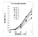


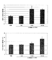


Classifications
-
- G—PHYSICS
- G01—MEASURING; TESTING
- G01N—INVESTIGATING OR ANALYSING MATERIALS BY DETERMINING THEIR CHEMICAL OR PHYSICAL PROPERTIES
- G01N33/00—Investigating or analysing materials by specific methods not covered by groups G01N1/00 - G01N31/00
- G01N33/48—Biological material, e.g. blood, urine; Haemocytometers
- G01N33/50—Chemical analysis of biological material, e.g. blood, urine; Testing involving biospecific ligand binding methods; Immunological testing
- G01N33/74—Chemical analysis of biological material, e.g. blood, urine; Testing involving biospecific ligand binding methods; Immunological testing involving hormones or other non-cytokine intercellular protein regulatory factors such as growth factors, including receptors to hormones and growth factors
-
- A—HUMAN NECESSITIES
- A61—MEDICAL OR VETERINARY SCIENCE; HYGIENE
- A61P—SPECIFIC THERAPEUTIC ACTIVITY OF CHEMICAL COMPOUNDS OR MEDICINAL PREPARATIONS
- A61P1/00—Drugs for disorders of the alimentary tract or the digestive system
- A61P1/16—Drugs for disorders of the alimentary tract or the digestive system for liver or gallbladder disorders, e.g. hepatoprotective agents, cholagogues, litholytics
-
- A—HUMAN NECESSITIES
- A61—MEDICAL OR VETERINARY SCIENCE; HYGIENE
- A61P—SPECIFIC THERAPEUTIC ACTIVITY OF CHEMICAL COMPOUNDS OR MEDICINAL PREPARATIONS
- A61P11/00—Drugs for disorders of the respiratory system
-
- A—HUMAN NECESSITIES
- A61—MEDICAL OR VETERINARY SCIENCE; HYGIENE
- A61P—SPECIFIC THERAPEUTIC ACTIVITY OF CHEMICAL COMPOUNDS OR MEDICINAL PREPARATIONS
- A61P13/00—Drugs for disorders of the urinary system
- A61P13/12—Drugs for disorders of the urinary system of the kidneys
-
- A—HUMAN NECESSITIES
- A61—MEDICAL OR VETERINARY SCIENCE; HYGIENE
- A61P—SPECIFIC THERAPEUTIC ACTIVITY OF CHEMICAL COMPOUNDS OR MEDICINAL PREPARATIONS
- A61P15/00—Drugs for genital or sexual disorders; Contraceptives
-
- A—HUMAN NECESSITIES
- A61—MEDICAL OR VETERINARY SCIENCE; HYGIENE
- A61P—SPECIFIC THERAPEUTIC ACTIVITY OF CHEMICAL COMPOUNDS OR MEDICINAL PREPARATIONS
- A61P17/00—Drugs for dermatological disorders
-
- A—HUMAN NECESSITIES
- A61—MEDICAL OR VETERINARY SCIENCE; HYGIENE
- A61P—SPECIFIC THERAPEUTIC ACTIVITY OF CHEMICAL COMPOUNDS OR MEDICINAL PREPARATIONS
- A61P19/00—Drugs for skeletal disorders
-
- A—HUMAN NECESSITIES
- A61—MEDICAL OR VETERINARY SCIENCE; HYGIENE
- A61P—SPECIFIC THERAPEUTIC ACTIVITY OF CHEMICAL COMPOUNDS OR MEDICINAL PREPARATIONS
- A61P25/00—Drugs for disorders of the nervous system
-
- A—HUMAN NECESSITIES
- A61—MEDICAL OR VETERINARY SCIENCE; HYGIENE
- A61P—SPECIFIC THERAPEUTIC ACTIVITY OF CHEMICAL COMPOUNDS OR MEDICINAL PREPARATIONS
- A61P3/00—Drugs for disorders of the metabolism
-
- A—HUMAN NECESSITIES
- A61—MEDICAL OR VETERINARY SCIENCE; HYGIENE
- A61P—SPECIFIC THERAPEUTIC ACTIVITY OF CHEMICAL COMPOUNDS OR MEDICINAL PREPARATIONS
- A61P37/00—Drugs for immunological or allergic disorders
-
- A—HUMAN NECESSITIES
- A61—MEDICAL OR VETERINARY SCIENCE; HYGIENE
- A61P—SPECIFIC THERAPEUTIC ACTIVITY OF CHEMICAL COMPOUNDS OR MEDICINAL PREPARATIONS
- A61P43/00—Drugs for specific purposes, not provided for in groups A61P1/00-A61P41/00
-
- A—HUMAN NECESSITIES
- A61—MEDICAL OR VETERINARY SCIENCE; HYGIENE
- A61P—SPECIFIC THERAPEUTIC ACTIVITY OF CHEMICAL COMPOUNDS OR MEDICINAL PREPARATIONS
- A61P5/00—Drugs for disorders of the endocrine system
-
- A—HUMAN NECESSITIES
- A61—MEDICAL OR VETERINARY SCIENCE; HYGIENE
- A61P—SPECIFIC THERAPEUTIC ACTIVITY OF CHEMICAL COMPOUNDS OR MEDICINAL PREPARATIONS
- A61P9/00—Drugs for disorders of the cardiovascular system
-
- C—CHEMISTRY; METALLURGY
- C07—ORGANIC CHEMISTRY
- C07K—PEPTIDES
- C07K14/00—Peptides having more than 20 amino acids; Gastrins; Somatostatins; Melanotropins; Derivatives thereof
- C07K14/435—Peptides having more than 20 amino acids; Gastrins; Somatostatins; Melanotropins; Derivatives thereof from animals; from humans
- C07K14/575—Hormones
- C07K14/64—Relaxins
-
- C—CHEMISTRY; METALLURGY
- C07—ORGANIC CHEMISTRY
- C07K—PEPTIDES
- C07K14/00—Peptides having more than 20 amino acids; Gastrins; Somatostatins; Melanotropins; Derivatives thereof
- C07K14/435—Peptides having more than 20 amino acids; Gastrins; Somatostatins; Melanotropins; Derivatives thereof from animals; from humans
- C07K14/575—Hormones
- C07K14/65—Insulin-like growth factors (Somatomedins), e.g. IGF-1, IGF-2
-
- C—CHEMISTRY; METALLURGY
- C07—ORGANIC CHEMISTRY
- C07K—PEPTIDES
- C07K16/00—Immunoglobulins [IGs], e.g. monoclonal or polyclonal antibodies
- C07K16/18—Immunoglobulins [IGs], e.g. monoclonal or polyclonal antibodies against material from animals or humans
- C07K16/26—Immunoglobulins [IGs], e.g. monoclonal or polyclonal antibodies against material from animals or humans against hormones ; against hormone releasing or inhibiting factors
-
- A—HUMAN NECESSITIES
- A01—AGRICULTURE; FORESTRY; ANIMAL HUSBANDRY; HUNTING; TRAPPING; FISHING
- A01K—ANIMAL HUSBANDRY; CARE OF BIRDS, FISHES, INSECTS; FISHING; REARING OR BREEDING ANIMALS, NOT OTHERWISE PROVIDED FOR; NEW BREEDS OF ANIMALS
- A01K2217/00—Genetically modified animals
- A01K2217/05—Animals comprising random inserted nucleic acids (transgenic)
-
- A—HUMAN NECESSITIES
- A01—AGRICULTURE; FORESTRY; ANIMAL HUSBANDRY; HUNTING; TRAPPING; FISHING
- A01K—ANIMAL HUSBANDRY; CARE OF BIRDS, FISHES, INSECTS; FISHING; REARING OR BREEDING ANIMALS, NOT OTHERWISE PROVIDED FOR; NEW BREEDS OF ANIMALS
- A01K2217/00—Genetically modified animals
- A01K2217/07—Animals genetically altered by homologous recombination
- A01K2217/075—Animals genetically altered by homologous recombination inducing loss of function, i.e. knock out
-
- A—HUMAN NECESSITIES
- A61—MEDICAL OR VETERINARY SCIENCE; HYGIENE
- A61K—PREPARATIONS FOR MEDICAL, DENTAL OR TOILETRY PURPOSES
- A61K38/00—Medicinal preparations containing peptides
-
- A—HUMAN NECESSITIES
- A61—MEDICAL OR VETERINARY SCIENCE; HYGIENE
- A61K—PREPARATIONS FOR MEDICAL, DENTAL OR TOILETRY PURPOSES
- A61K39/00—Medicinal preparations containing antigens or antibodies
-
- G—PHYSICS
- G01—MEASURING; TESTING
- G01N—INVESTIGATING OR ANALYSING MATERIALS BY DETERMINING THEIR CHEMICAL OR PHYSICAL PROPERTIES
- G01N2333/00—Assays involving biological materials from specific organisms or of a specific nature
- G01N2333/435—Assays involving biological materials from specific organisms or of a specific nature from animals; from humans
- G01N2333/575—Hormones
- G01N2333/62—Insulins
-
- G—PHYSICS
- G01—MEASURING; TESTING
- G01N—INVESTIGATING OR ANALYSING MATERIALS BY DETERMINING THEIR CHEMICAL OR PHYSICAL PROPERTIES
- G01N2333/00—Assays involving biological materials from specific organisms or of a specific nature
- G01N2333/435—Assays involving biological materials from specific organisms or of a specific nature from animals; from humans
- G01N2333/575—Hormones
- G01N2333/64—Relaxins
Description
【0001】
【発明の属する技術分野】
本発明は、新規な分泌性生体機能調節タンパク質およびそのDNAなどに関する。詳しくは、新規なインスリン/IGF/リラキシンファミリータンパク質及びそのDNAなどに関する。
【0002】
【従来の技術】
生体は、細胞間または組織間で、互いに情報伝達をすることにより、発生、分化、増殖、恒常性の維持などの統合の取れた調節を行っている。多くの場合、タンパク性因子がそれらの仲立ちをしている。例えば、免疫系、造血系に関与する分泌性因子(液性因子)が数多く見いだされていて、それらはサイトカインと呼ばれている。リンホカイン、モノカイン、インターフェロン、コロニー刺激因子、腫瘍壊死因子などがこれらに含まれる。これらについて、疾病との関係や医薬としての利用方法について盛んに研究されている。
また、内分泌組織から生産されるペプチドホルモンや増殖因子などの液性因子も、恒常性の維持や成長に大変重要な機能を担っており、これらについても医薬への応用が精力的に研究されている。
インスリン(insulin)、インスリン様成長因子−I(IGF-I)、インスリン様成長因子−II(IGF-II)、リラキシンH1(relaxin H1)、リラキシンH2(relaxin H2)は、その構造的特徴から一群のファミリーを形成する液性因子と考えられていて、生体において炭水化物の代謝調節、組織の成長促進、生殖機能の調節など幅広い生理的役割を担っている[BioScience用語ライブラリー サイトカイン・増殖因子、羊土社、108−109頁、1995年;ホルモンの分子生物学6 発生と成長因子・ホルモン、学会出版センター、1−23頁、1996年]。また、同じファミリーに属する新しい分子種も見つかりつつある[モレキュラー エンドクリノロジー(Molecular Endocrinology)、第13巻、2163−2174頁、1999年]。これらの前駆体タンパク質の構造的特徴は、シグナル配列−Bドメイン−Cドメイン−Aドメイン構造を有していることで、成熟型分子となるBドメインとAドメインは2つのジスルフィド結合でつながっている。更に、Aドメイン内にある1つのジスルフィド結合は立体構造の維持や活性の発現に重要である。インスリン、インスリン様成長因子、リラキシンは多くの生理活性を有し、生体にとって重要な情報伝達因子である。インスリンは膵臓のβ細胞から分泌され、肝臓,筋肉,脂肪組織での糖の取り込みの促進、脂肪酸合成の促進、グリコーゲン合成の促進などのエネルギー代謝の調節に重要であるほか、細胞増殖作用を有する。IGF-Iは主に肝臓で合成され、骨由来細胞をはじめとする多くの細胞に対して増殖や分化を促進する。IGF-IIはIGF-Iと同様に細胞増殖促進作用、インスリン様作用を有する。これらの因子は、特異的な受容体に結合し、その受容体の自己リン酸化を引き起こすことが、その後の一連の作用の引き金となる。一方リラキシンは、産道の弛緩、コラーゲンの再構築による結合組織の軟化、乳腺の増殖分化促進などの主に生殖機能に関連する作用を始め幅広いを生理作用を有することが近年明らかになってきている。例えば、子宮,乳腺,肺,心臓などに分布する血管の拡張促進、心臓の拍動に対する作用、肥満細胞からのヒスタミンの放出阻害、血小板の凝集阻害、下垂体ホルモンの分泌調節、体液バランスの調節、培養系における乳ガン細胞の増殖や分化の調節等である[ジェネラル ファーマコロジー (Gen Pharmacol)、第28巻、13−22頁、1997年]。
生体にとって重要なこれらのタンパク性及びペプチド性因子は、従来、その固有の生物活性を指標にして発見されてきた。また、既知の生理活性タンパク質に対するホモロジーを手がかりにしたクローニング技術により、相同性の高い類似遺伝子が追随的に発見されてきている。しかし、高等生物、とりわけ、哺乳類動物が健康体を維持し続けるために、公知の遺伝子群以外にも既存の手法で未だ同定されずにその存在が知られていない液性機能分子が重要な生理的役割を果たしている可能性は極めて高いと考えられる。
そこで、最近ではコンピュータを使った情報処理技術の助けを借り、DNAの配列情報から見出されてきた新規な遺伝子産物を、生物学、医学、獣医学などに役立てようとする試み、すなわち、バイオインフォマティクス(bioinformatics)からの各種研究が行われつつある[トレンズ・イン・バイオテクノロジー(Trends in Biotechnology)、第14巻、294−298頁、1996年]。近年、cDNAライブラリーの大規模シーケンシングが可能になったことで、EST(expresssed sequence tag)情報の蓄積などにより、膨大な数の新規遺伝子、あるいはその候補が見つかってきつつあるが、未だ配列情報が断片的で不正確なことも多く、また、現実問題として、現存するcDNA関連の各種公開データベースが各生物の全発現遺伝子を完全に網羅できていないのが現状である。したがって、これらの中からさらに全く新しい有用遺伝子産物を探索することは必ずしも容易ではない。他方、現在、一つの生物のもつ全DNA、つまりゲノムの構造解析が、細菌類、真菌類(酵母など)、昆虫、植物では既にそのいくつかの種で終了し、ヒトのそれもあと数年で完成の見通しが立っている。確かにこれまで数多くの分泌タンパク質あるいは分泌ペプチドをコードする遺伝子が単離されてきているものの、その数は全ゲノムからみればとてもそのすべてを網羅したとはいえない。そして、インスリン/IGF/リラキシンファミリーに属する新規物質については上述のように医療への応用が高く期待できることからその登場が強く望まれていた。
【0003】
【発明が解決しようとする課題】
本発明の目的は、生物学、医学、獣医学などに利用可能なインスリン/IGF/リラキシンファミリーに属する新規タンパク、およびその前駆体タンパク質、それらのフラグメント、ならびにそれらをコードするポリヌクレオチドを提供することである。本発明の他の目的は、かかるポリヌクレオチドを含有する組み換えベクター、該ベクターを含有する形質転換宿主細胞、かかるポリヌクレオチドを含む遺伝子を導入されたトランスジェニック動物も提供することである。また、かかるタンパク質またはポリペプチドの製造方法、かかるタンパク質またはポリペプチドに対する抗体、アゴニストまたはアンタゴニスト、受容体ならびにそれらの同定方法も提供する。さらに本発明は、かかるタンパク質、ポリペプチド、ポリヌクレオチド、アンタゴニスト、抗体、受容体を含有する医薬組成物、疾病の治療方法および予防方法等を提供することも目的とする。
【0004】
【課題を解決するための手段】
本発明者らは、上記の課題を解決するために鋭意研究を重ねた結果、ヒト生体内で発現している新規な遺伝子を発見することに成功し、それにコードされるタンパク質がインスリン/IGF/リラキシンファミリーに属する新しい分泌性生体機能調節タンパク質であることを見出した。
更に本発明者らは、鋭意研究を重ねた結果、新規ヒト型分泌性生体機能調節タンパク質に続いて、新規ラット型、該ラット型バリアント、マウス型およびブタ型分泌性生体機能調節タンパク質を見出した。
本発明者らは、これらの知見に基づいて、さらに検討を重ねた結果、本発明を完成するに至った。
【0005】
すなわち、本発明は、
(1)配列番号:7で表されるアミノ酸配列と同一もしくは実質的に同一のアミノ酸配列を含有するポリペプチド、そのアミド、もしくはそのエステルまたはその塩、
(2)実質的に同一のアミノ酸配列が配列番号:19または配列番号:47で表されるアミノ酸配列である上記(1)記載のポリペプチド、そのアミド、もしくはそのエステルまたはその塩、
(3)配列番号:8で表されるアミノ酸配列と同一もしくは実質的に同一のアミノ酸配列を含有するポリペプチド、そのアミド、もしくはそのエステルまたはその塩、
(4)実質的に同一のアミノ酸配列が配列番号:21または配列番号:49で表されるアミノ酸配列である上記(3)記載のポリペプチド、そのアミド、もしくはそのエステルまたはその塩、
(5)配列番号:7で表されるアミノ酸配列と同一もしくは実質的に同一のアミノ酸配列および配列番号:8で表されるアミノ酸配列と同一もしくは実質的に同一のアミノ酸配列を含有する上記(1)または上記(3)記載のポリペプチド、そのアミド、もしくはそのエステルまたはその塩、
(6)配列番号:7で表されるアミノ酸配列と同一もしくは実質的に同一のアミノ酸配列を含有するポリペプチド、そのアミドまたはそのエステルおよび配列番号:8で表されるアミノ酸配列と同一もしくは実質的に同一のアミノ酸配列を含有するポリペプチド、そのアミドまたはそのエステルがジスルフィド結合で結合しているポリペプチド、そのアミド、もしくはそのエステルまたはその塩、
【0006】
(7)配列番号:7で表されるアミノ酸配列と同一もしくは実質的に同一のアミノ酸配列が配列番号:19または配列番号:47で表されるアミノ酸配列であり、配列番号:8で表されるアミノ酸配列と同一もしくは実質的に同一のアミノ酸配列が配列番号:21または配列番号:49で表されるアミノ酸配列である上記(5)または上記(6)記載のポリペプチド、そのアミド、もしくはそのエステルまたはその塩、
(8)上記(1)または上記(3)記載のポリペプチドをコードするDNAを含有するDNA、
(9)配列番号:3で表されるアミノ酸配列と同一もしくは実質的に同一のアミノ酸配列を含有する上記(1)または上記(3)記載のポリペプチド、そのアミド、もしくはそのエステルまたはその塩、
(10)実質的に同一のアミノ酸配列が配列番号:17、配列番号:23、配列番号:45または配列番号:51で表されるアミノ酸配列である上記(9)記載のポリペプチド、そのアミド、もしくはそのエステルまたはその塩、
(11)上記(9)記載のポリペプチドをコードするDNAを含有する上記(8)記載のDNA、
(12)配列番号:12、配列番号:18、配列番号:24、配列番号:46または配列番号:52で表される塩基配列を有する上記(10)記載のDNA、
(13)上記(8)記載のDNAを含有する組換えベクター、
(14)上記(13)記載の組換えベクターで形質転換された形質転換体、
(15)上記(14)記載の形質転換体を培養し、上記(1)、上記(3)または上記(6)記載のポリペプチドを生成せしめることを特徴とする上記(1)、上記(3)または上記(6)記載のポリペプチド、そのアミド、もしくはそのエステルまたはその塩の製造法、
(16)上記(1)、上記(3)または上記(6)記載のポリペプチド、そのアミド、もしくはそのエステルまたはその塩に対する抗体、
(17)上記(1)、上記(3)または上記(6)記載のポリペプチド、そのアミド、もしくはそのエステルまたはその塩を用いることを特徴とする上記(1)、上記(3)または上記(6)記載のポリペプチド、そのアミド、もしくはそのエステルまたはその塩の活性を促進または阻害する化合物またはその塩のスクリーニング方法、
(18)上記(1)、上記(3)または上記(6)記載のポリペプチド、そのアミド、もしくはそのエステルまたはその塩を含有してなる上記(1)、上記(3)または上記(6)記載のポリペプチド、そのアミド、もしくはそのエステルまたはその塩の活性を促進または阻害する化合物またはその塩のスクリーニング用キット、
【0007】
(19)上記(17)記載のスクリーニング方法または上記(18)記載のスクリーニング用キットを用いて得られる、上記(1)、上記(3)または上記(6)記載のポリペプチド、そのアミド、もしくはそのエステルまたはその塩の活性を促進または阻害する化合物またはその塩、
(20)上記(17)記載のスクリーニング方法または上記(18)記載のスクリーニング用キットを用いて得られる上記(1)、上記(3)または上記(6)記載のポリペプチド、そのアミド、もしくはそのエステルまたはその塩の活性を促進または阻害する化合物またはその塩を含有してなる医薬、
(21)上記(1)、上記(3)または上記(6)記載のポリペプチド、そのアミド、もしくはそのエステルまたはその塩を含有してなる医薬、
(22)上記(16)記載の抗体を含有してなる医薬、
(23)▲1▼上記(1)、上記(3)または上記(6)記載のポリペプチド、そのアミド、もしくはそのエステルまたはその塩、▲2▼上記(19)記載の化合物またはその塩、または▲3▼上記(16)記載の抗体を含有してなる代謝調節異常、組織の成長・増殖・分化阻害、生殖機能の機能低下、結合組織の形成異常、組織の線維化、循環器障害、内分泌障害、体液バランス異常、中枢性疾患、免疫系疾患、血管新生障害の予防・治療剤、
(24)肝硬変・肺線維症、強皮症、腎線維症または末梢動脈疾患の予防・治療剤である上記(23)記載の剤、
(25)上記(16)記載の抗体を含有してなる診断剤、
(26)代謝調節異常、組織の成長・増殖・分化阻害、生殖機能の機能低下、結合組織の形成異常、組織の線維化、循環器障害、内分泌障害、体液バランス異常、中枢性疾患、免疫系疾患、血管新生障害の予防・治療作用を有する医薬を製造するための▲1▼上記(1)、上記(3)または上記(6)記載のポリペプチド、そのアミド、もしくはそのエステルまたはその塩、▲2▼上記(17)記載のスクリーニング方法または上記(18)記載のスクリーニング用キットを用いて得られる上記(1)、上記(3)または上記(6)記載のポリペプチド、そのアミド、もしくはそのエステルまたはその塩の活性を促進または阻害する化合物またはその塩、または▲3▼上記(16)記載の抗体の使用、および
(27)▲1▼上記(1)、上記(3)または上記(6)記載のポリペプチド、そのアミド、もしくはそのエステルまたはその塩、▲2▼上記(17)記載のスクリーニング方法または上記(18)記載のスクリーニング用キットを用いて得られる上記(1)、上記(3)または上記(6)記載のポリペプチド、そのアミド、もしくはそのエステルまたはその塩の活性を促進または阻害する化合物またはその塩、または▲3▼上記(16)記載の抗体を哺乳動物に投与することを特徴とする代謝調節異常、組織の成長・増殖・分化阻害、生殖機能の機能低下、結合組織の形成異常、組織の線維化、循環器障害、内分泌障害、体液バランス異常、中枢性疾患、免疫系疾患、血管新生障害の予防・治療方法などを提供するものである。
さらに本発明のDNA、およびポリペプチド、そのアミドもしくはそのエステルまたはその塩等は、分子量マーカー、組織マーカー、染色体マッピング、遺伝病の同定、プライマー、プローブの設計等の基礎研究に利用できる。
【0008】
【発明の実施の形態】
本発明の配列番号:7または配列番号:8で表されるアミノ酸配列と同一または実質的に同一のアミノ酸配列を含有するポリペプチドは(以下、配列番号:7または配列番号:8で表されるアミノ酸配列と同一または実質的に同一のアミノ酸配列を含有するポリペプチドを総称して本発明のポリペプチドと称することもある)、ヒトや温血動物(例えば、モルモット、ラット、マウス、ニワトリ、ウサギ、ブタ、ヒツジ、ウシ、サル等)の細胞(例えば、肝細胞、脾細胞、神経細胞、内分泌細胞、神経内分泌細胞、グリア細胞、膵臓β細胞、骨髄細胞、メサンギウム細胞、ランゲルハンス細胞、表皮細胞、上皮細胞、内皮細胞、繊維芽細胞、繊維細胞、筋細胞、脂肪細胞、免疫細胞(例、マクロファージ、T細胞、B細胞、ナチュラルキラー細胞、肥満細胞、好中球、好塩基球、好酸球、単球、樹状細胞)、巨核球、滑膜細胞、軟骨細胞、骨細胞、骨芽細胞、破骨細胞、乳腺細胞、もしくは間質細胞、またはこれら細胞の前駆細胞、幹細胞もしくはガン細胞等)もしくはそれらの細胞が存在するあらゆる組織、例えば、脳、脳の各部位(例、嗅球、扁桃核、大脳基底球、海馬、視床、視床下部、大脳皮質、延髄、小脳)、脊髄、下垂体、胃、膵臓、腎臓、肝臓、生殖腺、甲状腺、胆のう、骨髄、副腎、皮膚、筋肉、肺、消化管(例、大腸、小腸)、血管、心臓、胸腺、脾臓、唾液腺、末梢血、前立腺、睾丸(精巣)、卵巣、胎盤、子宮、骨、軟骨、関節、骨格筋等に由来するポリペプチドであってもよく、組換えポリペプチドであってもよく、合成ポリペプチドであってもよい。
また、本発明のポリペプチドがシグナルペプチドを有している場合は、ポリペプチドを効率よく細胞外に分泌させることができる。
配列番号:7または配列番号:8で表されるアミノ酸配列と実質的に同一のアミノ酸配列としては、配列番号:7または配列番号:8で表されるアミノ酸配列と約50%以上、好ましくは約60%以上、さらに好ましくは約70%以上、より好ましくは約80%以上、特に好ましくは約90%以上、最も好ましくは約95%以上の相同性を有するアミノ酸配列等が挙げられる。
配列番号:3で表されるアミノ酸配列は、配列番号:7で表されるアミノ酸配列および配列番号:8で表されるアミノ酸配列の両方を含有する。
配列番号:3で表されるアミノ酸配列と実質的に同一のアミノ酸配列としては、配列番号:3で表されるアミノ酸配列と約50%以上、好ましくは約60%以上、さらに好ましくは約70%以上、より好ましくは約80%以上、特に好ましくは約90%以上、最も好ましくは約95%以上の相同性を有するアミノ酸配列等が挙げられる。
【0009】
本発明の配列番号:7または配列番号:8で表されるアミノ酸配列と実質的に同一のアミノ酸配列を含有するポリペプチドとしては、例えば、前記の配列番号:7または配列番号:8で表されるアミノ酸配列と実質的に同一のアミノ酸配列を含有し、配列番号:7または配列番号:8で表されるアミノ酸配列を含有するポリペプチドと実質的に同質の性質を有するポリペプチド等が好ましい。
配列番号:7で表されるアミノ酸配列と実質的に同一のアミノ酸配列として、より具体的には、例えば配列番号:19または配列番号:47で表されるアミノ酸配列などがあげられる。
また、配列番号:8で表されるアミノ酸配列と実質的に同一のアミノ酸配列として、より具体的には、例えば配列番号:21または配列番号:49で表されるアミノ酸配列などがあげられる。
また、配列番号:3で表されるアミノ酸配列と実質的に同一のアミノ酸配列を含有するポリペプチドとしては、例えば、前記の配列番号:3で表されるアミノ酸配列と実質的に同一のアミノ酸配列を含有し、配列番号:3で表されるアミノ酸配列を含有するポリペプチドと実質的に同質の性質を有するポリペプチド等が好ましい。
配列番号:3で表されるアミノ酸配列と実質的に同一のアミノ酸配列として、より具体的には、例えば配列番号:17、配列番号:23、配列番号:45または配列番号:51で表されるアミノ酸配列などがあげられる。
実質的に同質の性質としては、例えば、抗原性や、分泌され液性因子として作用すること、細胞内サイクリックAMP産生促進作用等が挙げられる。実質的に同質とは、それらの性質が定性的に同質であることを示す。したがって、分泌作用や溶解度等や生理作用の性質が同等(例、約0.1〜100倍、好ましくは約0.5〜10倍、より好ましくは0.5〜2倍)であることが好ましいが、これらの性質の程度、ポリペプチドの分子量等の量的要素は異なっていてもよい。
また、配列番号:7、配列番号:8または配列番号:3で表されるアミノ酸配列と実質的に同一のアミノ酸配列を含有するポリペプチドとしてより具体的には、例えば、▲1▼配列番号:7、配列番号:8または配列番号:3で表されるアミノ酸配列中の1または2個以上(好ましくは、1〜30個程度、好ましくは1〜10個程度、さらに好ましくは数(1〜5)個)のアミノ酸が欠失したアミノ酸配列、▲2▼配列番号:7、配列番号:8または配列番号:3で表されるアミノ酸配列に1または2個以上(好ましくは、1〜30個程度、好ましくは1〜10個程度、さらに好ましくは数(1〜5)個)のアミノ酸が付加したアミノ酸配列、▲3▼配列番号:7、配列番号:8または配列番号:3で表されるアミノ酸配列に1または2個以上(好ましくは、1〜30個程度、好ましくは1〜10個程度、さらに好ましくは数(1〜5)個)のアミノ酸が挿入されたアミノ酸配列、▲4▼配列番号:7、配列番号:8または配列番号:3で表されるアミノ酸配列中の1または2個以上(好ましくは、1〜30個程度、好ましくは1〜10個程度、さらに好ましくは数(1〜5)個)のアミノ酸が他のアミノ酸で置換されたアミノ酸配列、または▲5▼それらを組み合わせたアミノ酸配列を含有するポリペプチド等のいわゆるムテインも含まれる。
【0010】
上記のようにアミノ酸配列が挿入、欠失または置換されている場合、その挿入、欠失または置換の位置としては、特に限定されないが、配列番号:7、配列番号:8または配列番号:3のそれぞれの配列番号で表されるアミノ酸配列の中のシステイン残基以外のアミノ酸残基が挙げられる。
配列番号:7で表されるアミノ酸配列と同一もしくは実質的に同一のアミノ酸配列および配列番号:8で表されるアミノ酸配列と同一もしくは実質的に同一のアミノ酸配列を含有するポリペプチドは、配列番号:7で表されるアミノ酸配列および配列番号:8で表されるアミノ酸配列の両方を含有する一本鎖ポリペプチドに加えて、配列番号:7で表されるアミノ酸配列と同一もしくは実質的に同一のアミノ酸配列を含有するポリペプチド(以下、本明細書において、A鎖と略称する場合がある)と配列番号:8で表されるアミノ酸配列と同一もしくは実質的に同一のアミノ酸配列を含有するポリペプチド(以下、本明細書において、B鎖と略称する場合がある)の2本のポリペプチド鎖からなるポリペプチド(以下、本明細書において、2本鎖ポリペプチドと略称する場合がある)を含む。
2本鎖ポリペプチドとして、より具体的には、配列番号:7で表されるアミノ酸配列と同一もしくは実質的に同一のアミノ酸配列および配列番号:8で表されるアミノ酸配列と同一もしくは実質的に同一のアミノ酸配列がジスルフィド結合で結合しているポリペプチドのことを意味し、A鎖及びB鎖内のシステイン残基が分子間及び分子内でジスルフィド結合を形成しているポリペプチドを示す。システイン残基同士の結合の組み合わせとしては、例えば特にA鎖のCys11(A鎖のCys11とは、配列番号:7、配列番号:19または配列番号:47で表されるアミノ酸配列のN末端から第11番目のシステイン残基を示す。以下同様)とB鎖のCys10(B鎖のCys10とは、配列番号:8、配列番号:21または配列番号:49で表されるアミノ酸配列のN末端から第10番目のシステイン残基を示す。以下同様)が結合し、かつA鎖のCys24とB鎖のCys22が結合し、さらにA鎖のCys10とA鎖のCys15が結合していることが望ましい。
配列番号:3で表されるアミノ酸配列と同一もしくは実質的に同一のアミノ酸配列を含有するポリペプチド(以下、単に前駆体タンパク質と称する場合がある)として好ましいものは、▲1▼ 配列番号:3の場合、N末端から第26番目(Arg)〜第52番目(Trp)に配列番号:8で表されるアミノ酸配列(即ち、B鎖をコードするアミノ酸配列)を含有し、配列番号:3のN末端から第119番目(Asp)〜第142番目(Cys)に配列番号:7で表されるアミノ酸配列(即ち、A鎖をコードするアミノ酸配列)を含有しており、
▲2▼ 配列番号:17の場合、N末端から第25番目(Arg)〜第51番目(Trp)に配列番号:21で表されるアミノ酸配列(即ち、B鎖をコードするアミノ酸配列)を含有し、配列番号:17のN末端から第118番目(Asp)〜第141番目(Cys)に配列番号:19で表されるアミノ酸配列(即ち、A鎖をコードするアミノ酸配列)を含有しており、
▲3▼ 配列番号:23の場合、N末端から第24番目(Arg)〜第50番目(Trp)に配列番号:21で表されるアミノ酸配列(即ち、B鎖をコードするアミノ酸配列)を含有し、配列番号:23のN末端から第117番目(Asp)〜第140番目(Cys)に配列番号:19で表されるアミノ酸配列(即ち、A鎖をコードするアミノ酸配列)を含有しており、
▲4▼ 配列番号:45の場合、N末端から第27番目(Arg)〜第53番目(Trp)に配列番号:49で表されるアミノ酸配列(即ち、B鎖をコードするアミノ酸配列)を含有し、配列番号:45のN末端から第117番目(Asp)〜第140番目(Cys)に配列番号:47で表されるアミノ酸配列(即ち、A鎖をコードするアミノ酸配列)を含有しており、
▲5▼ 配列番号:51の場合、N末端から第24番目(Arg)〜第50番目(Trp)に配列番号:21で表されるアミノ酸配列(即ち、B鎖をコードするアミノ酸配列)を含有し、配列番号:51のN末端から第151番目(Asp)〜第174番目(Cys)に配列番号:19で表されるアミノ酸配列(即ち、A鎖をコードするアミノ酸配列)を含有しており、A鎖、B鎖、2本鎖ポリペプチドの前駆体としての性質を有する。
【0011】
本発明のポリペプチド(以下、A鎖、B鎖、2本鎖ポリペプチド、前駆体タンパク質を総称して「本発明のポリペプチド」と称する場合がある)は、ペプチド標記の慣例に従って左端がN末端(アミノ末端)、右端がC末端(カルボキシル末端)である。配列番号:3、配列番号:7および配列番号:8で表されるアミノ酸配列を含有するポリペプチドをはじめとする、本発明のポリペプチドは、C末端が通常カルボキシル基(−COOH)またはカルボキシレート(−COO-)であるが、C末端がアミド(−CONH2)またはエステル(−COOR)であってもよい。
ここでエステルにおけるRとしては、例えば、メチル、エチル、n−プロピル、イソプロピルもしくはn−ブチル等のC1-6アルキル基、例えば、シクロペンチル、シクロヘキシル等のC3-8シクロアルキル基、例えば、フェニル、α−ナフチル等のC6-12アリール基、例えば、ベンジル、フェネチル等のフェニル−C1-2アルキル基もしくはα−ナフチルメチル等のα−ナフチル−C1-2アルキル基等のC7-14アラルキル基のほか、経口用エステルとして汎用されるピバロイルオキシメチル基等が用いられる。
本発明のポリペプチドがC末端以外にカルボキシル基(またはカルボキシレート)を有している場合、カルボキシル基がアミド化またはエステル化されているものも本発明のポリペプチドに含まれる。この場合のエステルとしては、例えば上記したC末端のエステル等が用いられる。
さらに、本発明のポリペプチドには、N末端のアミノ酸残基(例、メチオニン残基)のアミノ基が保護基(例えば、ホルミル基、アセチル基等のC1-6アルカノイル等のC1-6アシル基等)で保護されているもの、生体内で切断されて生成するN末端のグルタミン残基がピログルタミン酸化したもの、C末端のアミノ酸残基がホモセリンやホモセリンラクトンであるものや、分子内のアミノ酸の側鎖上の置換基(例えば−OH、−SH、アミノ基、イミダゾール基、インドール基、グアニジノ基等)が適当な保護基(例えば、ホルミル基、アセチル基等のC1-6アルカノイル基等のC1-6アシル基等)で保護されているもの、あるいは糖鎖が結合したいわゆる糖ポリペプチド等の複合ポリペプチド等も含まれる。
また、本発明のポリペプチドは一量体であってもよく、例えば、二量体、四量体、六量体、八量体などの多量体であってもよい。
本発明のポリペプチドまたはその塩としては、生理学的に許容される酸(例、無機酸、有機酸)や塩基(例、アルカリ金属塩)等との塩が用いられ、とりわけ生理学的に許容される酸付加塩が好ましい。この様な塩としては、例えば、無機酸(例えば、塩酸、リン酸、臭化水素酸、硫酸)との塩、あるいは有機酸(例えば、酢酸、ギ酸、プロピオン酸、フマル酸、マレイン酸、コハク酸、酒石酸、クエン酸、リンゴ酸、蓚酸、安息香酸、メタンスルホン酸、ベンゼンスルホン酸)との塩等が用いられる。
【0012】
本発明のポリペプチドまたはその塩としては、生理学的に許容される酸(例、無機酸、有機酸)や塩基(例、アルカリ金属塩)等との塩が用いられ、とりわけ生理学的に許容される酸付加塩が好ましい。この様な塩としては、例えば、無機酸(例えば、塩酸、リン酸、臭化水素酸、硫酸)との塩、あるいは有機酸(例えば、酢酸、ギ酸、プロピオン酸、フマル酸、マレイン酸、コハク酸、酒石酸、クエン酸、リンゴ酸、蓚酸、安息香酸、メタンスルホン酸、ベンゼンスルホン酸)との塩等が用いられる。
本発明のポリペプチドまたはその塩は、前述したヒトや温血動物の細胞または組織から自体公知のポリペプチド(タンパク質)の精製方法によって製造することもできるし、後述するポリペプチドをコードするDNAを含有する形質転換体を培養することによっても製造することができる。また、後述のペプチド合成法に準じて製造することもできる。
ヒトや哺乳動物の組織または細胞から製造する場合、ヒトや哺乳動物の組織または細胞をホモジナイズした後、酸等で抽出を行い、該抽出液を逆相クロマトグラフィー、イオン交換クロマトグラフィー等のクロマトグラフィーを組み合わせることにより精製単離することができる。
本発明のポリペプチドまたはその塩、またはそのアミド体の合成には、通常市販のポリペプチド(タンパク質)合成用樹脂を用いることができる。そのような樹脂としては、例えば、クロロメチル樹脂、ヒドロキシメチル樹脂、ベンズヒドリルアミン樹脂、アミノメチル樹脂、4−ベンジルオキシベンジルアルコール樹脂、4−メチルベンズヒドリルアミン樹脂、PAM樹脂、4−ヒドロキシメチルメチルフェニルアセトアミドメチル樹脂、ポリアクリルアミド樹脂、4−(2',4'-ジメトキシフェニル−ヒドロキシメチル)フェノキシ樹脂、4−(2',4'-ジメトキシフェニル−Fmocアミノエチル)フェノキシ樹脂等を挙げることができる。このような樹脂を用い、α−アミノ基と側鎖官能基を適当に保護したアミノ酸を、目的とするポリペプチドの配列通りに、自体公知の各種縮合方法に従い、樹脂上で縮合させる。反応の最後に樹脂からポリペプチドを切り出すと同時に各種保護基を除去し、さらに高希釈溶液中で分子内ジスルフィド結合形成反応を実施し、目的のポリペプチドまたはそれらのアミド体を取得する。
上記した保護アミノ酸の縮合に関しては、ポリペプチド合成に使用できる各種活性化試薬を用いることができるが、特に、カルボジイミド類がよい。カルボジイミド類としては、DCC、N,N'-ジイソプロピルカルボジイミド、N-エチル-N'-(3-ジメチルアミノプロリル)カルボジイミド等が用いられる。これらによる活性化にはラセミ化抑制添加剤(例えば、HOBt, HOOBt)とともに保護アミノ酸を直接樹脂に添加するかまたは、対称酸無水物またはHOBtエステルあるいはHOOBtエステルとしてあらかじめ保護アミノ酸の活性化を行なった後に樹脂に添加することができる。
保護アミノ酸の活性化や樹脂との縮合に用いられる溶媒としては、ポリペプチド(タンパク質)縮合反応に使用しうることが知られている溶媒から適宜選択されうる。例えば、N,N−ジメチルホルムアミド,N,N−ジメチルアセトアミド,N−メチルピロリドン等の酸アミド類、塩化メチレン,クロロホルム等のハロゲン化炭化水素類、トリフルオロエタノール等のアルコール類、ジメチルスルホキシド等のスルホキシド類、ピリジン,ジオキサン,テトラヒドロフラン等のエーテル類、アセトニトリル,プロピオニトリル等のニトリル類、酢酸メチル,酢酸エチル等のエステル類あるいはこれらの適宜の混合物等が用いられる。反応温度はポリペプチド(タンパク質)結合形成反応に使用され得ることが知られている範囲から適宜選択され、通常約−20〜50℃の範囲から適宜選択される。活性化されたアミノ酸誘導体は通常1.5〜4倍過剰で用いられる。ニンヒドリン反応を用いたテストの結果、縮合が不十分な場合には保護基の脱離を行うことなく縮合反応を繰り返すことにより十分な縮合を行うことができる。反応を繰り返しても十分な縮合が得られないときには、無水酢酸またはアセチルイミダゾールを用いて未反応アミノ酸をアセチル化することによって、後の反応に影響を与えないようにすることができる。
原料のアミノ基の保護基としては、例えば、Z、Boc、t−ペンチルオキシカルボニル、イソボルニルオキシカルボニル、4−メトキシベンジルオキシカルボニル、Cl-Z、Br-Z、アダマンチルオキシカルボニル、トリフルオロアセチル、フタロイル、ホルミル、2−ニトロフェニルスルフェニル、ジフェニルホスフィノチオイル、Fmoc等が用いられる。
カルボキシル基は、例えば、アルキルエステル化(例えば、メチル、エチル、プロピル、ブチル、t−ブチル、シクロペンチル、シクロヘキシル、シクロヘプチル、シクロオクチル、2−アダマンチル等の直鎖状、分枝状もしくは環状アルキルエステル化)、アラルキルエステル化(例えば、ベンジルエステル、4−ニトロベンジルエステル、4−メトキシベンジルエステル、4−クロロベンジルエステル、ベンズヒドリルエステル化)、フェナシルエステル化、ベンジルオキシカルボニルヒドラジド化、t−ブトキシカルボニルヒドラジド化、トリチルヒドラジド化等によって保護することができる。
セリンの水酸基は、例えば、エステル化またはエーテル化によって保護することができる。このエステル化に適する基としては、例えば、アセチル基等の低級(C1-6)アルカノイル基、ベンゾイル基等のアロイル基、ベンジルオキシカルボニル基、エトキシカルボニル基等の炭酸から誘導される基等が用いられる。また、エーテル化に適する基としては、例えば、ベンジル基、テトラヒドロピラニル基、t-ブチル基等である。
チロシンのフェノール性水酸基の保護基としては、例えば、Bzl、Cl2-Bzl、2−ニトロベンジル、Br-Z、t−ブチル等が用いられる。
ヒスチジンのイミダゾールの保護基としては、例えば、Tos、4-メトキシ-2,3,6-トリメチルベンゼンスルホニル、DNP、ベンジルオキシメチル、Bum、Boc、Trt、Fmoc等が用いられる。
【0013】
原料のカルボキシル基の活性化されたものとしては、例えば、対応する酸無水物、アジド、活性エステル〔アルコール(例えば、ペンタクロロフェノール、2,4,5-トリクロロフェノール、2,4-ジニトロフェノール、シアノメチルアルコール、パラニトロフェノール、HONB、N-ヒドロキシスクシミド、N-ヒドロキシフタルイミド、HOBt)とのエステル〕等が用いられる。原料のアミノ基の活性化されたものとしては、例えば、対応するリン酸アミドが用いられる。
保護基の除去(脱離)方法としては、例えば、Pd−黒あるいはPd-炭素等の触媒の存在下での水素気流中での接触還元や、また、無水フッ化水素、メタンスルホン酸、トリフルオロメタンスルホン酸、トリフルオロ酢酸あるいはこれらの混合液等による酸処理や、ジイソプロピルエチルアミン、トリエチルアミン、ピペリジン、ピペラジン等による塩基処理、また液体アンモニア中ナトリウムによる還元等も用いられる。上記酸処理による脱離反応は、一般に約−20〜40℃の温度で行われるが、酸処理においては、例えば、アニソール、フェノール、チオアニソール、メタクレゾール、パラクレゾール、ジメチルスルフィド、1,4-ブタンジチオール、1,2-エタンジチオール等のようなカチオン捕捉剤の添加が有効である。また、ヒスチジンのイミダゾール保護基として用いられる2,4-ジニトロフェニル基はチオフェノール処理により除去され、トリプトファンのインドール保護基として用いられるホルミル基は上記の1,2-エタンジチオール、1,4-ブタンジチオール等の存在下の酸処理による脱保護以外に、希水酸化ナトリウム溶液、希アンモニア等によるアルカリ処理によっても除去される。
原料の反応に関与すべきでない官能基の保護ならびに保護基、およびその保護基の脱離、反応に関与する官能基の活性化等は公知の基または公知の手段から適宜選択しうる。
ポリペプチドのアミド体を得る別の方法としては、例えば、まず、カルボキシ末端アミノ酸のα−カルボキシル基をアミド化して保護した後、アミノ基側にペプチド(ポリペプチド)鎖を所望の鎖長まで延ばした後、該ペプチド鎖のN末端のα−アミノ基の保護基のみを除いたポリペプチドとC末端のカルボキシル基の保護基のみを除去したポリペプチドとを製造し、この両ポリペプチドを上記したような混合溶媒中で縮合させる。縮合反応の詳細については上記と同様である。縮合により得られた保護ポリペプチドを精製した後、上記方法によりすべての保護基を除去し、所望の粗ポリペプチドを得ることができる。この粗ポリペプチドは既知の各種精製手段を駆使して精製し、主要画分を凍結乾燥することで所望のポリペプチドのアミド体を得ることができる。
ポリペプチドのエステル体を得るには、例えば、カルボキシ末端アミノ酸のα−カルボキシル基を所望のアルコール類と縮合しアミノ酸エステルとした後、ポリペプチドのアミド体と同様にして、所望のポリペプチドのエステル体を得ることができる。
本発明のポリペプチドまたはその塩は、自体公知のペプチドの合成法に従って製造することができる。ペプチドの合成法としては、例えば、固相合成法、液相合成法のいずれによっても良い。すなわち、本発明の部分ペプチドを構成し得る部分ペプチドもしくはアミノ酸と残余部分とを縮合させ、生成物が保護基を有する場合は保護基を脱離することにより目的のペプチドを製造することができる。
公知の縮合方法や保護基の脱離としては、例えば、以下の▲1▼〜▲5▼に記載された方法が挙げられる。
【0014】
▲1▼M. Bodanszky および M.A. Ondetti、ペプチド・シンセシス (Peptide Synthesis), Interscience Publishers, New York (1966年);
▲2▼SchroederおよびLuebke、ザ・ペプチド(The Peptide), Academic Press, New York (1965年);
▲3▼泉屋信夫他、ペプチド合成の基礎と実験、 丸善(株) (1975年);
▲4▼矢島治明 および榊原俊平、生化学実験講座 1、 タンパク質の化学IV、 205、(1977年);
▲5▼矢島治明監修、続医薬品の開発、第14巻、ペプチド合成、広川書店。
また、反応後は通常の精製法、例えば、溶媒抽出・蒸留・カラムクロマトグラフィー・液体クロマトグラフィー・再結晶等を組み合わせて本発明のポリペプチドポリペプチドを精製単離することができる。上記方法で得られるポリペプチドが遊離体である場合は、公知の方法あるいはそれに準じる方法によって適当な塩に変換することができるし、逆に塩で得られた場合は、公知の方法あるいはそれに準じる方法によって遊離体または他の塩に変換することができる。
本発明のポリペプチドをコードするDNAとしては、前述した本発明のポリペプチドのアミノ酸配列をコードする塩基配列を含有するものであればいかなるものであってもよい。また、ゲノムDNA、前記した細胞・組織由来のcDNA、合成DNAのいずれでもよい。
ライブラリーに使用するベクターは、バクテリオファージ、プラスミド、コスミド、ファージミド等いずれであってもよい。また、前記した細胞・組織よりtotalRNAまたはmRNA画分を調製したものを用いて直接Reverse Transcriptase Polymerase Chain Reaction(以下、RT-PCR法と略称する)によって増幅することもできる。
本発明のポリペプチドをコードするDNAとしては、例えば、配列番号:12、配列番号:15、配列番号:16、配列番号:18、配列番号:20、配列番号:22、配列番号:24、配列番号:25、配列番号:26、配列番号:46、配列番号:48、配列番号:50または配列番号:52で表される塩基配列を含有するDNA、または配列番号:12、配列番号:15、配列番号:16、配列番号:18、配列番号:20、配列番号:22、配列番号:24、配列番号:25、配列番号:26、配列番号:46、配列番号:48、配列番号:50または配列番号:52で表される塩基配列とハイストリンジェントな条件下でハイブリダイズする塩基配列を有し、本発明のポリペプチドと実質的に同質の性質(例、生物活性、免疫原性、分泌され液性因子として作用すること、細胞内サイクリックAMP産生促進作用等)を有するポリペプチドをコードするDNA等を有し、本発明のポリペプチドと実質的に同質の性質を有するポリペプチドをコードするDNAであれば何れのものでもよい。
配列番号:12、配列番号:15、配列番号:16、配列番号:18、配列番号:20、配列番号:22、配列番号:24、配列番号:25、配列番号:26、配列番号:46、配列番号:48、配列番号:50または配列番号:52で表される塩基配列とハイストリンジェントな条件下でハイブリダイズできるDNAとしては、例えば、配列番号:12、配列番号:15、配列番号:16、配列番号:18、配列番号:20、配列番号:22、配列番号:24、配列番号:25、配列番号:26、配列番号:46、配列番号:48、配列番号:50または配列番号:52で表される塩基配列と約60%以上、好ましくは約70%以上、さらに好ましくは約80%以上の相同性を有する塩基配列を含有するDNA等が用いられる。
また、配列番号:12、配列番号:15、配列番号:16、配列番号:18、配列番号:20、配列番号:22、配列番号:24、配列番号:25、配列番号:26、配列番号:46、配列番号:48、配列番号:50または配列番号:52で表される塩基配列とハイストリンジェントな条件下でハイブリダイズできるDNAとして、本発明のポリペプチドと実質的に同質の性質(例、生物活性、免疫原性、分泌され液性因子として作用すること、細胞内サイクリックAMP産生促進作用等)有するポリペプチドをコードするDNA等を有し、本発明のポリペプチドと実質的に同質の性質を有するポリペプチドをコードするDNA等があげられる。
ハイブリダイゼーションは、自体公知の方法あるいはそれに準じる方法、例えば、モレキュラー・クローニング(Molecular Cloning)2nd(J. Sambrook et al., Cold Spring Harbor Lab. Press, 1989)に記載の方法等に従って行うことができる。また、市販のライブラリーを使用する場合、添付の使用説明書に記載の方法に従って行うことができる。より好ましくはハイストリンジェントな条件に従って行うことができる。
ハイストリンジェントな条件とは、例えば、ナトリウム濃度が約19〜40mM、好ましくは約19〜20mMで、温度が約50〜70℃、好ましくは約60〜65℃の条件を示す。
本発明のポリペプチドをコードするDNAとしては、配列番号:12、配列番号:15、配列番号:16、配列番号:18、配列番号:20、配列番号:22、配列番号:24、配列番号:25、配列番号:26、配列番号:46、配列番号:48、配列番号:50または配列番号:52で表される塩基配列を有するDNA等が用いられる。
【0015】
より具体的には、
(I) A鎖(ヒト型;配列番号:7で表されるアミノ酸配列を含有するポリペプチド)をコードするDNAとしては、例えば、配列番号:15で表される塩基配列を有するDNAを含有するDNAなどが、
(II) A鎖(ラット型;配列番号:19で表されるアミノ酸配列を含有するポリペプチド)をコードするDNAとしては、例えば、配列番号:20で表される塩基配列を有するDNAを含有するDNAなどが、
(III) A鎖(マウス型;配列番号:19で表されるアミノ酸配列を含有するポリペプチド)をコードするDNAとしては、例えば、配列番号:25で表される塩基配列を有するDNAを含有するDNAなどが、
(IV) A鎖(ブタ型;配列番号:47で表されるアミノ酸配列を含有するポリペプチド)をコードするDNAとしては、例えば、配列番号:48で表される塩基配列を有するDNAを含有するDNAなどが、
(V) B鎖(ヒト型;配列番号:8で表されるアミノ酸配列を含有するポリペプチド)をコードするDNAとしては、例えば、配列番号:16で表される塩基配列を有するDNAを含有するDNAなどが、
(VI) B鎖(ラット型;配列番号:21で表されるアミノ酸配列を含有するポリペプチド)をコードするDNAとしては、例えば、配列番号:22で表される塩基配列を有するDNAを含有するDNAなどが、
(VII) B鎖(マウス型;配列番号:21で表されるアミノ酸配列を含有するポリペプチド)をコードするDNAとしては、例えば、配列番号:26で表される塩基配列を有するDNAを含有するDNAなどが、
(VIII) B鎖(ブタ型;配列番号:49で表されるアミノ酸配列を含有するポリペプチド)をコードするDNAとしては、例えば、配列番号:50で表される塩基配列を有するDNAを含有するDNAなどが、
(IX) 2本鎖ポリペプチド(ヒト型)および、前駆体タンパク質(ヒト型;配列番号:3で表されるアミノ酸配列を含有するポリペプチド)をコードするDNAとしては、例えば、配列番号:12で表される塩基配列を有するDNAを含有するDNAなどが、
(X) 2本鎖ポリペプチド(ラット型)および、前駆体タンパク質(ラット型;配列番号:17または配列番号:51で表されるアミノ酸配列を含有するポリペプチド)をコードするDNAとしては、例えば、配列番号:18または配列番号:52で表される塩基配列を有するDNAを含有するDNAなどが、
(XI) 2本鎖ポリペプチド(マウス型)および、前駆体タンパク質(マウス型;配列番号:23で表されるアミノ酸配列を含有するポリペプチド)をコードするDNAとしては、例えば、配列番号:24で表される塩基配列を有するDNAを含有するDNAなどが、
(XII) 2本鎖ポリペプチド(ブタ型)および、前駆体タンパク質(ブタ型;配列番号:45で表されるアミノ酸配列を含有するポリペプチド)をコードするDNAとしては、例えば、配列番号:46で表される塩基配列を有するDNAを含有するDNAなどがあげられる。
【0016】
本発明のポリペプチドを完全にコードするDNAのクローニングの手段としては、本発明のポリペプチドの部分塩基配列を有する合成DNAプライマーを用いてPCR法によって増幅するか、または適当なベクターに組み込んだDNAを本発明のポリペプチドの一部あるいは全領域をコードするDNA断片もしくは合成DNAを用いて標識したものとのハイブリダイゼーションによって選別することができる。ハイブリダイゼーションの方法は、例えば、モレキュラー・クローニング(Molecular Cloning)2nd(J. Sambrook et al., Cold Spring Harbor Lab. Press, 1989)に記載の方法等に従って行うことができる。また、市販のライブラリーを使用する場合、添付の使用説明書に記載の方法に従って行うことができる。
DNAの塩基配列の変換は、PCRや公知のキット、例えば、Mutan(登録商標)-super Express Km(宝酒造(株))、Mutan(登録商標)-K(宝酒造(株))等を用いて、ODA-LA PCR法やGupped duplex法やKunkel法等の自体公知の方法あるいはそれらに準じる方法に従って行うことができる。
クローン化されたポリペプチドをコードするDNAは目的によりそのまま、または所望により制限酵素で消化したり、リンカーを付加したりして使用することができる。該DNAはその5’末端側に翻訳開始コドンとしてのATGを有し、また3’末端側には翻訳終止コドンとしてのTAA、TGAまたはTAGを有していてもよい。これらの翻訳開始コドンや翻訳終止コドンは、適当な合成DNAアダプターを用いて付加することもできる。
本発明のポリペプチドの発現ベクターは、例えば、(イ)本発明のポリペプチドをコードするDNAから目的とするDNA断片を切り出し、(ロ)該DNA断片を適当な発現ベクター中のプロモーターの下流、もしくは適当な保護ペプチドをコードする塩基配列の下流に連結することにより製造することができる。
ベクターとしては、大腸菌由来のプラスミド(例、pBR322,pBR325,pUC12,pUC13)、枯草菌由来のプラスミド(例、pUB110,pTP5,pC194)、酵母由来プラスミド(例、pSH19,pSH15)、λファージ等のバクテリオファージ、レトロウイルス,ワクシニアウイルス,バキュロウイルス等の動物ウイルス等の他、pA1−11、pXT1、pRc/CMV、pRc/RSV、pcDNAI/Neo、pET−1、pET−2、pET−3、pET−4、pET−5、pET−11等が用いられる。
本発明で用いられるプロモーターとしては、遺伝子の発現に用いる宿主に対応して適切なプロモーターであればいかなるものでもよい。例えば、動物細胞を宿主として用いる場合は、SRαプロモーター、SV40プロモーター、LTRプロモーター、CMVプロモーター、HSV-TKプロモーター、β-アクチンプロモーター等が挙げられる。
これらのうち、CMV(サイトメガロウイルス)プロモーター、SRαプロモーター、β-アクチンプロモーター等を用いるのが好ましい。宿主がエシェリヒア属菌である場合は、trpプロモーター、lacプロモーター、recAプロモーター、λPLプロモーター、lppプロモーター、T7プロモーター等が、宿主がバチルス属菌である場合は、SPO1プロモーター、SPO2プロモーター、penPプロモーター等、宿主が酵母である場合は、PHO5プロモーター、PGKプロモーター、GAPプロモーター、ADHプロモーター等が好ましい。宿主が昆虫細胞である場合は、ポリヘドリンプロモーター、P10プロモーター等が好ましい。
T7プロモーターの発現系を用いる場合にはT7プロモーターとしてはT7 DNA上で見出されている17種のプロモーター[J. L. Oakleyら, Proc. Natl. Acad. Sci, U. S. A, 74: 4266-4270(1977), M. D. Rosa, Cell 16: 815-825 (1979), N. Panayotatosら, Nature, 280:35 (1979), J. J. Dunnら, J. Mol. Biol., 166:477-535 (1983) ]のいずれでもよいがφ10プロモーター[A. H. Rosenbergら, Gene, 56: 125-135 (1987)]が好ましい。
ベクターは上記ベクターにT7プロモーター、T7ターミネーターを組み込んで構築されるのが好ましく、このようなベクターとしては、pET−1、pET−2、pET−3、pET−4、pET−5、pET−11 [A. H. Rosenberg, Gene 56: 125-135 (1987)]などをあげることができる。
発現ベクターには、以上の他に、所望によりエンハンサー、スプライシングシグナル、ポリA付加シグナル、選択マーカー、SV40複製オリジン(以下、SV40oriと略称する場合がある)等を含有しているものを用いることができる。選択マーカーとしては、例えば、ジヒドロ葉酸還元酵素(以下、dhfrと略称する場合がある)遺伝子〔メソトレキセート(MTX)耐性〕、アンピシリン耐性遺伝子(以下、Amprと略称する場合がある)、ネオマイシン耐性遺伝子(以下、Neorと略称する場合がある、Geneticin耐性)等が挙げられる。特に、dhfr遺伝子欠損チャイニーズハムスター細胞を用いてdhfr遺伝子を選択マーカーとして使用する場合、組換え体細胞をチミジンを含まない培地によっても選択できる。
また、必要に応じて、宿主に合ったシグナル配列を、本発明のポリペプチドのN末端側に付加する。宿主がエシェリヒア属菌である場合は、PhoA・シグナル配列、OmpA・シグナル配列等が、宿主がバチルス属菌である場合は、α−アミラーゼ・シグナル配列、サブチリシン・シグナル配列等が、宿主が酵母である場合は、MFα・シグナル配列、SUC2・シグナル配列等、宿主が動物細胞である場合には、インスリン・シグナル配列、α−インターフェロン・シグナル配列、抗体分子・シグナル配列等がそれぞれ利用できる。
【0017】
このようにして構築された本発明のポリペプチドをコードするDNAを含有するベクターを用いて、形質転換体を製造することができる。
宿主としては、例えば、エシェリヒア属菌、バチルス属菌、酵母、昆虫細胞、昆虫、動物細胞等が用いられる。
エシェリヒア属菌の具体例としては、例えば、エシェリヒア・コリ(Escherichia coli)K12・DH1〔プロシージングズ・オブ・ザ・ナショナル・アカデミー・オブ・サイエンシイズ・オブ・ザ・ユーエスエー(Proc. Natl. Acad. Sci. USA),60巻,160(1968)〕,JM103〔ヌクイレック・アシッズ・リサーチ,(Nucleic Acids Research),9巻,309(1981)〕,JA221〔ジャーナル・オブ・モレキュラー・バイオロジー(Journal of Molecular Biology)〕,120巻,517(1978)〕,HB101〔ジャーナル・オブ・モレキュラー・バイオロジー,41巻,459(1969)〕,C600〔ジェネティックス(Genetics),39巻,440(1954)〕等が用いられる。
T7プロモーターの発現系を用いる場合には、その形質転換体の宿主としては、T7 RNAポリメラーゼ遺伝子 [F. W. Studierら, J. Mol. Biol. 189: 113-130 (1986)]]を組み込んだ大腸菌株、例えば、MM294、DH−1,C600,JM109,BL21,あるいはT7 RNAポリメラーゼ遺伝子を他のプラスミドと共に組み込んだ大腸菌株などが用いられる。好ましくはT7 RNAポリメラーゼ遺伝子を組み込んだλファージが溶原化したMM294株、BL21株、およびBL21(DE3)株等が用いられる。この場合、T7 RNAポリメラーゼ遺伝子のプロモーターとしては、イソプロピル−1−チオ−β-D-ガラクトピラノシド(IPTG)で発現が誘導されるlacプロモーターが用いられる。
バチルス属菌としては、例えば、バチルス・サブチルス(Bacillus subtilis)MI114〔ジーン,24巻,255(1983)〕,207−21〔ジャーナル・オブ・バイオケミストリー(Journal of Biochemistry),95巻,87(1984)〕等が用いられる。
酵母としては、例えば、サッカロマイセス セレビシエ(Saccharomyces cerevisiae)AH22,AH22R-,NA87−11A,DKD−5D,20B−12、シゾサッカロマイセス ポンベ(Schizosaccharomyces pombe)NCYC1913,NCYC2036、ピキア パストリス(Pichia pastoris)KM71等が用いられる。
昆虫細胞としては、例えば、ウイルスがAcNPVの場合は、夜盗蛾の幼虫由来株化細胞(Spodoptera frugiperda cell;Sf細胞)、Trichoplusia niの中腸由来のMG1細胞、Trichoplusia niの卵由来のHigh FiveTM細胞、Mamestra brassicae由来の細胞またはEstigmena acrea由来の細胞等が用いられる。ウイルスがBmNPVの場合は、蚕由来株化細胞(Bombyx mori N 細胞;BmN細胞)等が用いられる。該Sf細胞としては、例えば、Sf9細胞(ATCC CRL1711)、Sf21細胞(以上、Vaughn, J.L.ら、イン・ヴィボ(In Vivo),13, 213-217,(1977))等が用いられる。
昆虫としては、例えば、カイコの幼虫等が用いられる〔前田ら、ネイチャー(Nature),315巻,592(1985)〕。
動物細胞としては、例えば、サル細胞COS−7,Vero,チャイニーズハムスター細胞CHO(以下、CHO細胞と略記),dhfr遺伝子欠損チャイニーズハムスター細胞CHO(以下、CHO(dhfr-)細胞と略記),マウスL細胞,マウスAtT20,マウスミエローマ細胞,ラットGH3,ヒトFL細胞等が用いられる。
【0018】
エシェリヒア属菌を形質転換するには、例えば、プロシージングズ・オブ・ザ・ナショナル・アカデミー・オブ・サイエンシイズ・オブ・ザ・ユーエスエー(Proc. Natl. Acad. Sci. USA),69巻,2110(1972)やジーン(Gene),17巻,107(1982)等に記載の方法に従って行うことができる。
バチルス属菌を形質転換するには、例えば、モレキュラー・アンド・ジェネラル・ジェネティックス(Molecular & General Genetics),168巻,111(1979)等に記載の方法に従って行うことができる。
酵母を形質転換するには、例えば、メソッズ・イン・エンザイモロジー(Methods in Enzymology),194巻,182−187(1991)、プロシージングズ・オブ・ザ・ナショナル・アカデミー・オブ・サイエンシイズ・オブ・ザ・ユーエスエー(Proc. Natl. Acad. Sci.USA),75巻,1929(1978)等に記載の方法に従って行うことができる。
昆虫細胞または昆虫を形質転換するには、例えば、バイオ/テクノロジー(Bio/Technology),6, 47-55(1988))等に記載の方法に従って行うことができる。動物細胞を形質転換するには、例えば、細胞工学別冊8 新細胞工学実験プロトコール.263−267(1995)(秀潤社発行)、ヴィロロジー(Virology),52巻,456(1973)に記載の方法に従って行うことができる。
このようにして、ポリペプチドをコードするDNAを含有する発現ベクターで形質転換された形質転換体を得ることができる。
宿主がエシェリヒア属菌、バチルス属菌である形質転換体を培養する際、培養に使用される培地としては液体培地が適当であり、その中には該形質転換体の生育に必要な炭素源、窒素源、無機物その他が含有せしめられる。炭素源としては、例えば、グルコース、デキストリン、可溶性澱粉、ショ糖等、窒素源としては、例えば、アンモニウム塩類、硝酸塩類、コーンスチープ・リカー、ペプトン、カゼイン、酵母エキス、肉エキス、大豆粕、バレイショ抽出液等の無機または有機物質、無機物としては、例えば、塩化カルシウム、リン酸二水素ナトリウム、塩化マグネシウム等が挙げられる。また、酵母エキス、ビタミン類、生長促進因子等を添加してもよい。培地のpHは約5〜8が望ましい。
エシェリヒア属菌を培養する際の培地としては、例えば、グルコース、カザミノ酸を含むM9培地〔ミラー(Miller),ジャーナル・オブ・エクスペリメンツ・イン・モレキュラー・ジェネティックス(Journal of Experiments in Molecular Genetics),431−433,Cold Spring Harbor Laboratory, New York 1972〕が好ましい。ここに必要によりプロモーターを効率よく働かせるために、例えば、3β−インドリルアクリル酸のような薬剤を加えることができる。
宿主がエシェリヒア属菌の場合、培養は通常約15〜43℃で約3〜24時間行い、必要により、通気や撹拌を加えることもできる。
宿主がバチルス属菌の場合、培養は通常約30〜40℃で約6〜24時間行い、必要により通気や撹拌を加えることもできる。
宿主が酵母である形質転換体を培養する際、培地としては、例えば、バークホールダー(Burkholder)最小培地〔Bostian, K. L. ら、プロシージングズ・オブ・ザ・ナショナル・アカデミー・オブ・サイエンシイズ・オブ・ザ・ユーエスエー(Proc. Natl. Acad. Sci. USA), 77巻, 4505 (1980)〕や0.5%カザミノ酸を含有するSD培地〔Bitter, G. A. ら、プロシージングズ・オブ・ザ・ナショナル・アカデミー・オブ・サイエンシイズ・オブ・ザ・ユーエスエー(Proc. Natl. Acad. Sci. USA),81巻,5330(1984)〕が挙げられる。培地のpHは約5〜8に調整するのが好ましい。培養は通常約20〜35℃で約24〜72時間行い、必要に応じて通気や撹拌を加える。
宿主が昆虫細胞または昆虫である形質転換体を培養する際、培地としては、Grace's Insect Medium(Grace, T.C.C.,ネイチャー(Nature),195,788(1962))に非動化した10%ウシ血清等の添加物を適宜加えたもの等が用いられる。培地のpHは約6.2〜6.4に調整するのが好ましい。培養は通常約27℃で約3〜5日間行い、必要に応じて通気や撹拌を加える。
【0019】
宿主が動物細胞である形質転換体を培養する際、培地としては、例えば、約5〜20%の胎児牛血清を含むMEM培地〔サイエンス(Science),122巻,501(1952)〕,DMEM培地〔ヴィロロジー(Virology),8巻,396(1959)〕,RPMI 1640培地〔ジャーナル・オブ・ザ・アメリカン・メディカル・アソシエーション(The Journal of the American Medical Association)199巻,519(1967)〕,199培地〔プロシージング・オブ・ザ・ソサイエティ・フォー・ザ・バイオロジカル・メディスン(Proceeding of the Society for the Biological Medicine),73巻,1(1950)〕等が用いられる。pHは約6〜8であるのが好ましい。培養は通常約30〜40℃で約15〜60時間行い、必要に応じて通気や撹拌を加える。
以上のようにして、形質転換体の細胞内、細胞膜または細胞外に本発明のポリペプチドを生成せしめることができる。
上記培養物から本発明のポリペプチドを分離精製するには、例えば、下記の方法により行うことができる。
本発明のポリペプチドを培養菌体あるいは細胞から抽出するに際しては、培養後、公知の方法で菌体あるいは細胞を集め、これを適当な緩衝液に懸濁し、超音波、リゾチームおよび/または凍結融解等によって菌体あるいは細胞を破壊したのち、遠心分離やろ過によりポリペプチドの粗抽出液を得る方法等が適宜用いられる。組換え体が産生するポリペプチドが菌体中で封入体を形成する場合には、遠心分離により集菌した後、超音波等で菌体を破砕して得られた封入体を変性剤(2−8M 塩酸グアニジン、5−9M 尿素等)を用いて可溶化することによってポリペプチドを抽出することができる。培養液中にポリペプチドが分泌される場合には、培養終了後、それ自体公知の方法で菌体あるいは細胞と上清とを分離し、上清を集める。
このようにして得られた培養上清、あるいは抽出液中に含まれるポリペプチドの精製は、自体公知の分離・精製法を適切に組み合わせて行うことができる。これらの公知の分離、精製法としては、塩析や溶媒沈澱法等の溶解度を利用する方法、透析法、限外ろ過法、ゲルろ過法、およびSDS−ポリアクリルアミドゲル電気泳動法等の主として分子量の差を利用する方法、イオン交換クロマトグラフィー等の荷電の差を利用する方法、アフィニティークロマトグラフィー等の特異的親和性を利用する方法、逆相高速液体クロマトグラフィー等の疎水性の差を利用する方法、等電点電気泳動法等の等電点の差を利用する方法等が用いられる。
かくして得られるポリペプチドが遊離体で得られた場合には、自体公知の方法あるいはそれに準じる方法によって塩に変換することができ、逆に塩で得られた場合には自体公知の方法あるいはそれに準じる方法により、遊離体または他の塩に変換することができる。
なお、組換え体が産生するポリペプチドを、精製前または精製後に適当な蛋白修飾酵素または蛋白分解酵素等を作用させること、または化学反応により、任意に修飾を加えたり、ポリペプチドを部分的に除去することもできる。これらの酵素としては、例えば、トリプシン、キモトリプシン、アルギニルエンドペプチダーゼ、フリン、プロホルモンコンベルターゼ1(PC1)、プロホルモンコンベルターゼ2(PC2)カルボキシぺプチダーゼB、プロテインキナーゼ、グリコシダーゼ等が用いられる。 化学反応としては、例えば臭化シアン(CNBr)を用いた切断反応などがあげられる。
抽出した目的蛋白質または分離精製した目的蛋白質は必要により蛋白質のリフォールディングに付される。リフォールディングは、例えば「蛋白質のフォールディング」(R.H.Pain編、245-279 (1995)、シュプリンガーフェアラーク東京)に記載された公知の方法あるいはそれに準じる方法により実施することが可能である。抽出剤(例えば、グアニジン塩酸塩、尿素のようなカオトロピック可溶化剤、n-ラウリルメチルグリシン、SDSのような界面活性剤など)を含まない、もしくは低濃度の抽出剤を含む緩衝液で1段階もしくは多段階での希釈や半透膜を用いた透析、ゲル濾過を用いた緩衝液の置換等により行うことができる。この場合、目的蛋白質のアグリゲーションを防止するために、アルギニン、ポリエチレングリコール、中性界面活性剤等を添加することができる。蛋白質のジスルフィド結合を形成させるために空気酸化したり、酸化還元緩衝液系等を添加することができる。酸化還元緩衝液にはグルタチオン、システイン、ジチオスレイトール、2−メルカプトエタノール、またはシステアミンをベースとしたものがあげられる。
【0020】
また、本発明のポリペプチドであるA鎖及びB鎖を別々に生産した後、両者をジスルフィド結合で連結させることができる。例えば、A鎖とB鎖をコードするDNA断片をそれぞれlacZ(β-ガラクトシダーゼ)遺伝子に繋げ、大腸菌を宿主に用いてβ-ガラクトシダーゼ+A鎖と、β-ガラクトシダーゼ+B鎖の融合タンパク質を作製した後、臭化シアン処理や酵素処理でA鎖とB鎖を融合タンパク質から切り出すことができる。切り出されたA鎖とB鎖を還元状態で混合し、徐々に酸化させることによってA鎖とB鎖がジスルフィド結合で連結したタンパクに変換させることができる。A鎖とB鎖の連結状態は、例えばA鎖のCys11とB鎖のCys10が結合し、かつA鎖のCys24とB鎖のCys22が結合し、さらにA鎖のCys10とA鎖のCys15が結合していることが望ましい。このときの変換条件としては、例えば、インスリン分子の製造法〔プロシージングズ・オブ・ザ・ナショナル・アカデミー・オブ・サイエンシイズ・オブ・ザ・ユーエスエー(Proc. Natl. Acad. Sci. USA),76巻,106(1979)〕に準じて行うことができる。
かくして生成する本発明のポリペプチドまたはその塩の存在は、特異抗体を用いたエンザイムイムノアッセイやウエスタンブロット解析等で測定することができる。
本発明のポリペプチド、そのアミド、もしくはそのエステルまたはその塩(以下、単に本発明のポリペプチドと称する場合がある)に対する抗体は、本発明のポリペプチドを認識し得る抗体であれば、ポリクローナル抗体、モノクローナル抗体の何れであってもよい。
【0021】
本発明のポリペプチドに対する抗体は、本発明のポリペプチドを抗原として用い、自体公知の抗体または抗血清の製造法に従って製造することができる。
〔モノクローナル抗体の作製〕(a)モノクロナール抗体産生細胞の作製
本発明のポリペプチドは、温血動物に対して投与により抗体産生が可能な部位にそれ自体あるいは担体、希釈剤とともに投与される。投与に際して抗体産生能を高めるため、完全フロイントアジュバントや不完全フロイントアジュバントを投与してもよい。投与は通常2〜6週毎に1回ずつ、計2〜10回程度行われる。用いられる温血動物としては、例えば、サル、ウサギ、イヌ、モルモット、マウス、ラット、ヒツジ、ヤギ、ニワトリが挙げられるが、マウスおよびラットが好ましく用いられる。
モノクローナル抗体産生細胞の作製に際しては、抗原で免疫された温血動物、例えばマウスから抗体価の認められた個体を選択し最終免疫の2〜5日後に脾臓またはリンパ節を採取し、それらに含まれる抗体産生細胞を同種または異種動物の骨髄腫細胞と融合させることにより、モノクローナル抗体産生ハイブリドーマを調製することができる。抗血清中の抗体価の測定は、例えば、後記の標識化ポリペプチドと抗血清とを反応させたのち、抗体に結合した標識剤の活性を測定することにより行うことができる。融合操作は既知の方法、例えば、ケーラーとミルスタインの方法〔ネイチャー(Nature)、256、495 (1975)〕に従い実施することができる。融合促進剤としては、例えば、ポリエチレングリコール(PEG)やセンダイウィルス等が挙げられるが、好ましくはPEGが用いられる。
骨髄腫細胞としては、例えば、NS−1、P3U1、SP2/0、AP−1等の温血動物の骨髄腫細胞が挙げられるが、P3U1が好ましく用いられる。用いられる抗体産生細胞(脾臓細胞)数と骨髄腫細胞数との好ましい比率は1:1〜20:1程度であり、PEG(好ましくはPEG1000〜PEG6000)が10〜80%程度の濃度で添加され、約20〜40℃、好ましくは約30〜37℃で約1〜10分間インキュベートすることにより効率よく細胞融合を実施できる。
モノクローナル抗体産生ハイブリドーマのスクリーニングには種々の方法が使用できるが、例えば、ポリペプチド抗原を直接あるいは担体とともに吸着させた固相(例、マイクロプレート)にハイブリドーマ培養上清を添加し、次に放射性物質や酵素等で標識した抗免疫グロブリン抗体(細胞融合に用いられる細胞がマウスの場合、抗マウス免疫グロブリン抗体が用いられる)またはプロテインAを加え、固相に結合したモノクローナル抗体を検出する方法、抗免疫グロブリン抗体またはプロテインAを吸着させた固相にハイブリドーマ培養上清を添加し、放射性物質や酵素等で標識したポリペプチドを加え、固相に結合したモノクローナル抗体を検出する方法等が挙げられる。
【0022】
モノクローナル抗体の選別は、自体公知あるいはそれに準じる方法に従って行うことができる。通常HAT(ヒポキサンチン、アミノプテリン、チミジン)を添加した動物細胞用培地で行うことができる。選別および育種用培地としては、ハイブリドーマが生育できるものならばどのような培地を用いても良い。例えば、約1〜20%、好ましくは約10〜20%の牛胎児血清を含むRPMI 1640培地、約1〜10%の牛胎児血清を含むGIT培地(和光純薬工業(株))あるいはハイブリドーマ培養用無血清培地(SFM−101、日水製薬(株))等を用いることができる。培養温度は、通常約20〜40℃、好ましくは約37℃である。培養時間は、通常5日〜3週間、好ましくは1週間〜2週間である。培養は、通常5%炭酸ガス下で行うことができる。ハイブリドーマ培養上清の抗体価は、上記の抗血清中の抗体価の測定と同様にして測定できる。
(b)モノクロナール抗体の精製
モノクローナル抗体の分離精製は、自体公知の方法、例えば、免疫グロブリンの分離精製法〔例、塩析法、アルコール沈殿法、等電点沈殿法、電気泳動法、イオン交換体(例、DEAE)による吸脱着法、超遠心法、ゲルろ過法、抗原結合固相あるいはプロテインAあるいはプロテインG等の活性吸着剤により抗体のみを採取し、結合を解離させて抗体を得る特異的精製法〕に従って行うことができる。
〔ポリクローナル抗体の作製〕
本発明のポリクローナル抗体は、それ自体公知あるいはそれに準じる方法に従って製造することができる。例えば、免疫抗原(本発明のポリペプチド抗原)自体、あるいはそれとキャリアー蛋白質との複合体をつくり、上記のモノクローナル抗体の製造法と同様に温血動物に免疫を行い、該免疫動物から本発明のポリペプチドに対する抗体含有物を採取して、抗体の分離精製を行うことにより製造することができる。
温血動物を免疫するために用いられる免疫抗原とキャリアー蛋白質との複合体に関し、キャリアー蛋白質の種類およびキャリアーとハプテンとの混合比は、キャリアーに架橋させて免疫したハプテンに対して抗体が効率良くできれば、どの様なものをどの様な比率で架橋させてもよいが、例えば、ウシ血清アルブミンやウシサイログロブリン、ヘモシアニン等を重量比でハプテン1に対し、約0.1〜20、好ましくは約1〜5の割合でカプルさせる方法が用いられる。
また、ハプテンとキャリアーのカプリングには、種々の縮合剤を用いることができるが、グルタルアルデヒドやカルボジイミド、マレイミド活性エステル、チオール基、ジチオビリジル基を含有する活性エステル試薬等が用いられる。
縮合生成物は、温血動物に対して、抗体産生が可能な部位にそれ自体あるいは担体、希釈剤とともに投与される。投与に際して抗体産生能を高めるため、完全フロイントアジュバントや不完全フロイントアジュバントを投与してもよい。投与は、通常約2〜6週毎に1回ずつ、計約3〜10回程度行われる。
ポリクローナル抗体は、上記の方法で免疫された温血動物の血液、腹水等、好ましくは血液から採取することができる。
抗血清中のポリクローナル抗体価の測定は、上記の抗血清中の抗体価の測定と同様にして測定できる。ポリクローナル抗体の分離精製は、上記のモノクローナル抗体の分離精製と同様の免疫グロブリンの分離精製法に従って行うことができる。
また、本発明のポリぺプチドを用いて、Nat. Biotechnol,14,845‐851.(1996)、Nat Genet.15,146‐156.(1997)、PNAS,97(2),722−727 (2000)等に記載の方法に準じてヒト化抗体を作製することも可能である。
本発明のポリペプチドをコードするDNA(以下、本発明のDNAと称することもある)に相補的な、または実質的に相補的な塩基配列を有するアンチセンスDNAとしては、本発明のDNAに相補的な、または実質的に相補的な塩基配列を有し、該DNAの発現を抑制し得る作用を有するものであれば、いずれのアンチセンスDNAであってもよい。
【0023】
本発明のDNAに実質的に相補的な塩基配列とは、例えば、本発明のDNAに相補的な塩基配列(すなわち、本発明のDNAの相補鎖)の全塩基配列あるいは部分塩基配列と約70%以上、好ましくは約80%以上、より好ましくは約90%以上、最も好ましくは約95%以上の相同性を有する塩基配列等が挙げられる。特に、本発明のDNAの相補鎖の全塩基配列うち、本発明のポリペプチドのN末端部位をコードする部分の塩基配列(例えば、開始コドン付近の塩基配列等)の相補鎖と約70%以上、好ましくは約80%以上、より好ましくは約90%以上、最も好ましくは約95%以上の相同性を有するアンチセンスDNAが好適である。これらのアンチセンスDNAは、公知のDNA合成装置等を用いて製造することができる。
本発明のポリペプチドがシグナルペプチドを有する場合は、細胞外に効率よく分泌され、液性因子として、生体機能調節などの重要な生物活性を発揮する。
以下に、本発明のポリペプチド、本発明のDNA、本発明のポリペプチドまたはその塩に対する抗体(以下、本発明の抗体と略記する場合がある)、およびアンチセンスDNAの用途を説明する。
(1)本発明のポリペプチドは、組織特異的に発現しているため、組織マーカーとして使用することができる。すなわち組織の分化、病態、癌の転移等の検出のためのマーカーとして有用である。また、対応する受容体、結合ポリペプチド等の分取にも利用できる。さらに、自体公知のハイスループットスクリーニングのためのパネルにして、生物活性を調べるのに利用できる。
(2)本発明のポリペプチドが関与する各種疾病の治療・予防剤
本発明のポリペプチドは、生体内で液性因子として存在し、後述の実施例に記載のとおり、MMP−1(マトリックスメタロプロテイナーゼ−1)やVEGF(血管内皮細胞増殖因子)の発現促進作用を有するため、本発明のポリペプチド、または本発明のDNA等に異常があったり、欠損している場合あるいは発現量が異常に減少または亢進している場合、例えば、炭水化物などのエネルギー源の代謝調節(糖代謝、脂質代謝など)異常(例えば糖尿病、肥満症など)、組織の成長・増殖・分化阻害、生殖機能の機能低下、結合組織の形成異常(例えば強皮症など)、組織の線維化(例えば、肝硬変・肺線維症、強皮症または腎線維症など)、循環器障害(末梢動脈疾患、心筋梗塞または心不全など)、動脈硬化、内分泌障害、体液バランス異常、中枢性疾患、免疫系疾患(例えばアレルギー、炎症、自己免疫疾患等)、血管新生障害等の種々の疾病が発症する。したがって、本発明のポリペプチドおよび本発明のDNAは、例えば、炭水化物などのエネルギー源の代謝調節(糖代謝、脂質代謝など)異常(例えば糖尿病、肥満症など)、組織の成長・増殖・分化阻害、生殖機能の機能低下、結合組織の形成異常(例えば強皮症など)、組織の線維化(例えば、肝硬変・肺線維症、強皮症または腎線維症など)、循環器障害(末梢動脈疾患、心筋梗塞または心不全など)、動脈硬化、内分泌障害、体液バランス異常、中枢性疾患、免疫系疾患(例えばアレルギー、炎症、自己免疫疾患等)、血管新生障害等の種々の疾病の治療・予防剤等の医薬として使用することができる。
また、本発明のポリペプチドは、他のインスリン/IGF/リラキシンファミリーポリペプチドとファミリーを形成するため、他のファミリーのポリペプチド、またはそのDNA等に異常があったり、欠損している場合あるいは発現量が異常に減少または亢進している場合においても、他のファミリーと相補的に作用することにより、さらには、特に発現量に異常が認められない場合においても、本発明のポリペプチド単独の薬理作用により、炭水化物などのエネルギー源の代謝調節(糖代謝、脂質代謝など)異常(例えば糖尿病、肥満症など)、組織の成長・増殖・分化阻害、生殖機能の機能低下、結合組織の形成異常(例えば強皮症など)、組織の線維化(例えば、肝硬変・肺線維症、強皮症または腎線維症など)、循環器障害(末梢動脈疾患、心筋梗塞または心不全など)、内分泌障害、体液バランス異常、中枢性疾患、アレルギーなどの免疫系疾患、血管新生障害等の種々の疾病の治療・予防剤等の医薬として使用することができる。
さらに、本発明のポリペプチドは、上述のとおり、1組のA鎖内ジスルフィド結合と2組のA鎖とB鎖間ジスルフィド結合より形成されるものが好ましいが、2組のジスルフィド結合を有するA鎖単独あるいは1組のジスルフィド結合を有するB鎖単独の分子種も存在し得る。後者のA鎖あるいはB鎖単独の分子種も代謝調節(糖代謝、脂質代謝など)異常(例えば糖尿病、肥満症など)、組織の成長・増殖・分化阻害、生殖機能の機能低下、結合組織の形成異常(例えば強皮症など)、組織の線維化(例えば、肝硬変・肺線維症、強皮症または腎線維症など)、循環器障害(末梢動脈疾患、心筋梗塞または心不全など)、動脈硬化、内分泌障害、体液バランス異常、中枢性疾患、免疫系疾患(例えばアレルギー、炎症、自己免疫疾患等)、血管新生障害等の種々の疾病の治療・予防剤等の医薬として使用することができる。
例えば、生体内において本発明のポリペプチドが減少あるいは欠損しているために、細胞における情報伝達が十分に、あるいは正常に発揮されない患者がいる場合に、(イ)本発明のDNAを該患者に投与し、生体内で本発明のポリペプチドを発現させることによって、(ロ)細胞に本発明のDNAを挿入し、本発明のポリペプチドを発現させた後に、該細胞を患者に移植することによって、または(ハ)本発明のポリペプチドを該患者に投与すること等によって、該患者における本発明のポリペプチドの役割を十分に、あるいは正常に発揮させることができる。
本発明のDNAを上記の治療・予防剤として使用する場合は、該DNAを単独あるいはレトロウイルスベクター、アデノウイルスベクター、アデノウイルスアソシエーテッドウイルスベクター等の適当なベクターに挿入した後、常套手段に従って、ヒトまたは温血動物に投与することができる。本発明のDNAは、そのままで、あるいは摂取促進のための補助剤等の生理学的に認められる担体とともに製剤化し、遺伝子銃やハイドロゲルカテーテルのようなカテーテルによって投与できる。
本発明のポリペプチドを上記の治療・予防剤として使用する場合は、少なくとも90%、好ましくは95%以上、より好ましくは98%以上、さらに好ましくは99%以上に精製されたものを使用するのが好ましい。本発明のポリペプチドは、例えば、必要に応じて糖衣を施した錠剤、カプセル剤、エリキシル剤、マイクロカプセル剤等として経口的に、あるいは水もしくはそれ以外の薬学的に許容し得る液との無菌性溶液、または懸濁液剤等の注射剤の形で非経口的に使用できる。例えば、本発明のポリペプチドを生理学的に認められる担体、香味剤、賦形剤、ベヒクル、防腐剤、安定剤、結合剤等とともに一般に認められた製剤実施に要求される単位用量形態で混和することによって製造することができる。これら製剤における有効成分量は指示された範囲の適当な用量が得られるようにするものである。錠剤、カプセル剤等に混和することができる添加剤としては、例えば、ゼラチン、コーンスターチ、トラガント、アラビアゴムのような結合剤、結晶性セルロースのような賦形剤、コーンスターチ、ゼラチン、アルギン酸等のような膨化剤、ステアリン酸マグネシウムのような潤滑剤、ショ糖、乳糖またはサッカリンのような甘味剤、ペパーミント、アカモノ油またはチェリーのような香味剤等が用いられる。調剤単位形態がカプセルである場合には、前記タイプの材料にさらに油脂のような液状担体を含有することができる。注射のための無菌組成物は注射用水のようなベヒクル中の活性物質、胡麻油、椰子油等のような天然産出植物油等を溶解または懸濁させる等の通常の製剤実施に従って処方することができる。注射用の水性液としては、例えば、生理食塩水、ブドウ糖やその他の補助薬を含む等張液(例えば、D−ソルビトール、D−マンニトール、塩化ナトリウム等)等が挙げられ、適当な溶解補助剤、例えば、アルコール(例えば、エタノール等)、ポリアルコール(例えば、プロピレングリコール、ポリエチレングリコール等)、非イオン性界面活性剤(例えば、ポリソルベート80TM、HCO−50等)等と併用してもよい。油性液としては、例えば、オリーブ油、ゴマ油、大豆油、ラッカセイ油、綿実油、コーン油などの植物油、プロピレングリコール等が挙げられ、溶解補助剤として安息香酸ベンジル、ベンジルアルコール等と併用してもよい。また、緩衝剤(例えば、リン酸塩緩衝液、酢酸ナトリウム緩衝液等)、無痛化剤(例えば、塩化ベンザルコニウム、塩酸プロカイン等)、安定剤(例えば、ヒト血清アルブミン、ポリエチレングリコール等)、保存剤(例えば、ベンジルアルコール、フェノール等)、酸化防止剤等と配合してもよい。調製された注射液は、通常、適当なアンプルに充填される。
【0024】
本発明のDNAが挿入されたベクターも上記と同様に製剤化され、通常、非経口的に使用される。このようにして得られる製剤は、安全で低毒性であるので、例えば、温血動物(例えば、ヒト、ラット、マウス、モルモット、ウサギ、トリ、ヒツジ、ブタ、ウシ、ウマ、ネコ、イヌ、サル等)に対して投与することができる。
本発明のポリペプチドの投与量は、対象疾患、投与対象、投与ルート等により差異はあるが、例えば、代謝調節(糖代謝、脂質代謝など)異常(例えば糖尿病、肥満症など)、組織の成長・増殖・分化阻害、生殖機能の機能低下、結合組織の形成異常(例えば強皮症など)、組織の線維化(例えば、肝硬変・肺線維症、強皮症または腎線維症など)、循環器障害(末梢動脈疾患、心筋梗塞または心不全など)、動脈硬化、内分泌障害、体液バランス異常、中枢性疾患、免疫系疾患(例えばアレルギー、炎症、自己免疫疾患等)、血管新生障害等の種々の疾病の治療目的で本発明のポリペプチドを経口投与する場合、一般的に成人(60kgとして)においては、一日につき本発明のポリペプチドを約0.01mg〜1000mg、好ましくは約1mg〜1000mg、さらに好ましくは約10〜500mg、より好ましくは約10〜200mg投与する。非経口的に投与する場合は、本発明のポリペプチドの1回投与量は投与対象、対象疾患等によっても異なるが、例えば、代謝調節(糖代謝、脂質代謝など)異常(例えば糖尿病、肥満症など)、組織の成長・増殖・分化阻害、生殖機能の機能低下、結合組織の形成異常(例えば強皮症など)、組織の線維化(例えば、肝硬変・肺線維症、強皮症または腎線維症など)、循環器障害(末梢動脈疾患、心筋梗塞または心不全など)、動脈硬化、内分泌障害、体液バランス異常、中枢性疾患、免疫系疾患(例えばアレルギー、炎症、自己免疫疾患等)、血管新生障害等の種々の疾病の治療目的で本発明のポリペプチドを注射剤の形で成人(体重60kgとして)に投与する場合、一日につき該ポリペプチドを約0.001〜500mg程度、好ましくは0.01〜500mg程度、さらに好ましくは約0.1〜200mg程度、より好ましくは約0.1〜100mg程度を患部にあるいは全身性に注射することにより投与するのが好都合である。他の動物の場合も、60kg当たりに換算した量を投与することができる。
(2)疾病に対する医薬候補化合物のスクリーニング
本発明のポリペプチドは生体内で液性因子として存在するため、本発明のポリペプチドの機能(活性)を促進する化合物またはその塩は、例えば、炭水化物などのエネルギー源の代謝調節(糖代謝、脂質代謝など)異常(例えば糖尿病、肥満症など)、組織の成長・増殖・分化阻害、生殖機能の機能低下、結合組織の形成異常(例えば強皮症など)、組織の線維化(例えば、肝硬変・肺線維症、強皮症または腎線維症など)、循環器障害(末梢動脈疾患、心筋梗塞または心不全など)、動脈硬化、内分泌障害、体液バランス異常、中枢性疾患、免疫系疾患(例えばアレルギー、炎症、自己免疫疾患等)、血管新生障害等の種々の疾病の治療・予防剤等の医薬として使用できる。
一方、本発明のポリペプチドの機能(活性)を阻害する化合物またはその塩は、本発明のポリペプチドの産生過剰に起因する疾患の治療・予防剤等の医薬として使用できる。
したがって、本発明のポリペプチドは、本発明のポリペプチドの機能(活性)を促進または阻害する化合物またはその塩のスクリーニングのための試薬として有用である。
【0025】
すなわち、本発明は、
(1)本発明のポリペプチドまたはその塩を用いることを特徴とする本発明のポリペプチドまたはその塩の機能(活性)を促進する化合物もしくはその塩(以下、促進剤と略記する場合がある)、または本発明のポリペプチドまたはその塩の機能(活性)を阻害する化合物(以下、阻害剤と略記する場合がある)のスクリーニング方法を提供する。
本発明のスクリーニング用キットは、本発明のポリペプチドまたはその塩を含有するものである。
本発明のポリペプチドを用いることを特徴とする本発明のポリペプチドの機能(活性)を促進または阻害する化合物またはその塩のスクリーニング方法として、具体的には、
(i)本発明のポリペプチドが結合する組織、細胞、またはそれらの膜画分に、本発明のポリペプチドを接触させた場合と(ii)本発明のポリペプチドが結合する組織、細胞、またはそれらの膜画分に、本発明のポリペプチドおよび試験化合物を接触させた場合との比較を行うことを特徴とする本発明のポリペプチドと組織、細胞、またはそれらの膜画分の結合性を変化させる化合物(本発明のポリペプチドの活性を促進または阻害する化合物)またはその塩のスクリーニング方法などがあげられる。
本発明のスクリーニング方法においては、(i)上記した本発明のポリペプチドが結合する組織、細胞、またはそれらの膜画分に、本発明のポリペプチドを接触させた場合と(ii)本発明のポリペプチドが結合する組織、細胞、またはそれらの膜画分に、本発明のポリペプチドおよび試験化合物を接触させた場合における、例えば本発明のポリペプチドが結合する組織、細胞、またはそれらの膜画分に対するリガンドの結合量、細胞刺激活性、組織反応性などを測定して、比較する。
本発明のスクリーニング方法は具体的には、
▲1▼標識した本発明のポリペプチドを、本発明のポリペプチドが結合する組織、細胞、またはそれらの膜画分に接触させた場合と、標識した本発明のポリペプチドおよび試験化合物を本発明のポリペプチドが結合する組織、細胞、またはそれらの膜画分に接触させた場合における、標識した本発明のポリペプチドの本発明のポリペプチドが結合する組織、細胞、またはそれらの膜画分に対する結合量を測定し、比較することを特徴とする本発明のポリペプチドと本発明のポリペプチドが結合する組織、細胞、またはそれらの膜画分との結合性を変化させる化合物(本発明のポリペプチドの活性を促進または阻害する化合物)またはその塩のスクリーニング方法、
▲2▼標識した本発明のポリペプチドを、本発明のポリペプチドに対する受容体を含有する細胞または該細胞の膜画分に接触させた場合と、標識した本発明のポリペプチドおよび試験化合物を本発明のポリペプチドに対する受容体を含有する細胞または該細胞の膜画分に接触させた場合における、標識した本発明のポリペプチドの該細胞または該膜画分に対する結合量を測定し、比較することを特徴とする本発明のポリペプチドと本発明のポリペプチドに対する受容体との結合性を変化させる化合物(本発明のポリペプチドの活性を促進または阻害する化合物)またはその塩のスクリーニング方法、
▲3▼標識した本発明のポリペプチドを、本発明のポリペプチドに対する受容体をコードするDNAを含有する形質転換体を培養することによって細胞膜上に発現した本発明のポリペプチドに対する受容体に接触させた場合と、標識した本発明のポリペプチドおよび試験化合物を本発明のポリペプチドに対する受容体をコードするDNAを含有する形質転換体を培養することによって細胞膜上に発現した本発明のポリペプチドに対する受容体に接触させた場合における、標識した本発明のポリペプチドの本発明のポリペプチドに対する受容体への結合量を測定し、比較することを特徴とする本発明のポリペプチドと本発明のポリペプチドに対する受容体との結合性を変化させる化合物(本発明のポリペプチドの活性を促進または阻害する化合物)またはその塩のスクリーニング方法、
▲4▼本発明のポリペプチドに対する受容体を活性化する化合物(例えば、本発明のポリペプチド)を本発明のポリペプチドに対する受容体を含有する細胞に接触させた場合と、本発明のポリペプチドに対する受容体を活性化する化合物および試験化合物を本発明のポリペプチドに対する受容体を含有する細胞に接触させた場合における、本発明のポリペプチドに対する受容体を介した細胞刺激活性(例えば、アラキドン酸遊離、アセチルコリン遊離、細胞内Ca2+の遊離、細胞内cAMP生成、細胞内cGMP生成、イノシトールリン酸産生、細胞膜電位変動、細胞内蛋白質のリン酸化、c−fosの活性化、細胞外液のpHの低下、NO産生、該細胞が特有に産生している生理活性物質の産生などを促進する活性または抑制する活性など)を測定し、比較することを特徴とする本発明のポリペプチドと本発明のポリペプチドに対する受容体との結合性を変化させる化合物(本発明のポリペプチドの活性を促進または阻害する化合物)またはその塩のスクリーニング方法、および
▲5▼本発明のポリペプチドに対する受容体を活性化する化合物(例えば、本発明のポリペプチドなど)を本発明のポリペプチドに対する受容体をコードするDNAを含有する形質転換体を培養することによって細胞膜上に発現した本発明のポリペプチドに対する受容体に接触させた場合と、本発明のポリペプチドに対する受容体を活性化する化合物および試験化合物を、本発明のポリペプチドに対する受容体をコードするDNAを含有する形質転換体を培養することによって細胞膜上に発現した本発明のポリペプチドに対する受容体に接触させた場合における、本発明のポリペプチドに対する受容体を介する細胞刺激活性(例えば、アラキドン酸遊離、アセチルコリン遊離、細胞内Ca2+の遊離、細胞内cAMP生成、細胞内cGMP生成、イノシトールリン酸産生、細胞膜電位変動、細胞内蛋白質のリン酸化、c−fosの活性化、細胞外液のpHの低下、NO産生、該細胞が特有に産生している生理活性物質の産生などを促進する活性または抑制する活性など)を測定し、比較することを特徴とする本発明のポリペプチドと本発明のポリペプチドに対する受容体との結合性を変化させる化合物(本発明のポリペプチドの活性を促進または阻害する化合物)またはその塩のスクリーニング方法などである。
▲6▼本発明のポリペプチドに対する受容体をコードするDNAは、以下のような方法で取得することができる。本発明のポリペプチドを125Iなどのラジオアイソトープやフルオレセインなどの蛍光物質、またはビオチン等で標識し、該標識体が特異的に結合する細胞株、組織、器官などを見出す。次に公知の方法でそこからmRNAを抽出し、cDNAを合成した後、動物細胞用の発現ベクターにクローニングする。これをCOS7細胞などに導入し、該形質転換細胞に上記の標識体が結合するかどうかを指標にして、本発明のポリペプチドに対する受容体をコードするDNAが組み込まれた形質転換細胞を選び出すことができる(発現クローニング)。
▲7▼また、インスリン受容体ファミリーに共通の構造的特徴(例えば、チロシンキナーゼドメインなどのアミノ酸配列)に対する混合プライマーを作製し、RT−PCRを行うことにより、インスリン受容体ファミリーに属する新規受容体cDNA断片を取得することができる。続いて得られたDNA配列を基に5’-RACEおよび3’-RACE PCRを行って、全長のcDNAを取得する。これを上述の動物細胞用発現ベクターに挿入し、CHO細胞やCOS7細胞などの適当な動物培養細胞で発現させ、本発明のポリペプチドが特異的に結合することを確認することにより、本発明のポリペプチドに対する受容体をコードするDNAであることが確認できる。
【0026】
本発明のスクリーニング方法の具体的な説明を以下にする。
まず、本発明のスクリーニング方法に用いる本発明のポリペプチドが結合する組織、細胞、またはそれらの膜画分など、すなわち本発明のポリペプチドに対する受容体としては、本発明のポリペプチドを特異的に結合するものであれば何れのものであってもよいが、ヒトや温血動物の臓器、培養細胞、またはそれらの膜画分などが好適である。しかし、特にヒト由来の臓器は入手が極めて困難なことから、スクリーニングに用いられるものとしては、組換え体を用いて大量発現させたヒトの組換え受容体などが適している。
本発明のポリペプチドに対する受容体を製造するには、前述の本発明のポリペプチドの製造方法などが用いられる。
本発明のスクリーニング方法において、本発明のポリペプチドに対する受容体を含有する細胞あるいは該細胞膜画分などを用いる場合、後述の調製法に従えばよい。
本発明のポリペプチドに対する受容体を含有する細胞を用いる場合、該細胞をグルタルアルデヒド、ホルマリンなどで固定化してもよい。固定化方法はそれ自体公知の方法に従って行うことができる。
本発明のポリペプチドに対する受容体を含有する細胞としては、本発明のポリペプチドに対する受容体を発現した細胞株をいうが、大腸菌、枯草菌、酵母、昆虫細胞、動物細胞などを宿主細胞とする形質転換細胞であってもよい。また、本発明のポリペプチドに対する受容体を発現した宿主細胞の取得方法としては、前述の本発明のポリペプチドを含有する発現ベクターで形質転換された形質転換体の製造方法と同様の方法などがあげられる。
膜画分としては、細胞を破砕した後、それ自体公知の方法で得られる細胞膜が多く含まれる画分のことをいう。細胞の破砕方法としては、Potter−Elvehjem型ホモジナイザーで細胞を押し潰す方法、ワーリングブレンダーやポリトロン(Kinematica社製)による破砕、超音波による破砕、フレンチプレスなどで加圧しながら細胞を細いノズルから噴出させることによる破砕などが挙げられる。細胞膜の分画には、分画遠心分離法や密度勾配遠心分離法などの遠心力による分画法が主として用いられる。例えば、細胞破砕液を低速(500rpm〜3000rpm)で短時間(通常、約1分〜10分)遠心し、上清をさらに高速(15000rpm〜30000rpm)で通常30分〜2時間遠心し、得られる沈澱を膜画分とする。該膜画分中には、発現した本発明のポリペプチドに対する受容体と細胞由来のリン脂質や膜蛋白質などの膜成分が多く含まれる。
該本発明のポリペプチドに対する受容体を含有する細胞や膜画分中の本発明のポリペプチドに対する受容体の量は、1細胞当たり103〜108分子であるのが好ましく、105〜107分子であるのが好適である。なお、発現量が多いほど膜画分当たりのリガンド結合活性(比活性)が高くなり、高感度なスクリーニング系の構築が可能になるばかりでなく、同一ロットで大量の試料を測定できるようになる。
本発明のポリペプチドと本発明のポリペプチドに対する受容体との結合性を変化させる化合物(本発明のポリペプチドの活性を促進または阻害する化合物)をスクリーニングする前記の▲1▼〜▲3▼を実施するためには、適当な本発明のポリペプチドに対する受容体画分と、標識した本発明のポリペプチドなどが用いられる。本発明のポリペプチドに対する受容体画分としては、本発明のポリペプチドに対する天然の組織や細胞の受容体画分か、またはそれと同等の活性を有する組換え型の受容体画分などが望ましい。ここで、同等の活性とは、同等のリガンド結合活性などを示す。標識したリガンドとしては、標識した本発明のポリペプチドなどが用いられる。例えば〔3H〕、〔125I〕、〔14C〕、〔35S〕、蛍光色素、ビオチン、βガラクトシダーゼやパーオキシダーゼなどの酵素などで標識された本発明のポリペプチドを利用することができる。
【0027】
具体的には、本発明のポリペプチドと本発明のポリペプチドに対する受容体との結合性を変化させる化合物のスクリーニングを行うには、まず本発明のポリペプチドに対する受容体を含有する細胞または細胞の膜画分を、スクリーニングに適したバッファーに懸濁することにより受容体標品を調製する。バッファーには、pH4〜10(望ましくはpH6〜8)のリン酸バッファー、トリス−塩酸バッファーなどのリガンドと受容体との結合を阻害しないバッファーであればいずれでもよい。また、非特異的結合を低減させる目的で、CHAPS、Tween−80TM(花王−アトラス社)、ジギトニン、デオキシコレートなどの界面活性剤をバッファーに加えることもできる。さらに、プロテアーゼによる受容体や本発明のポリペプチドの分解を抑える目的でPMSF、ロイペプチン、E−64(ペプチド研究所製)、ペプスタチンなどのプロテアーゼ阻害剤を添加することもできる。0.01ml〜10mlの該受容体溶液に、一定量(5000cpm〜500000cpm)の標識した本発明のポリペプチドを添加し、同時に10-10〜10-6Mの試験化合物を共存させる。非特異的結合量(NSB)を知るために大過剰の未標識の本発明のポリペプチドを加えた反応チューブも用意する。反応は0℃から50℃、望ましくは4℃から37℃で20分から24時間、望ましくは30分から3時間行う。反応後、ガラス繊維濾紙等で濾過し、適量の同バッファーで洗浄した後、ガラス繊維濾紙に残存する放射活性を液体シンチレーションカウンターまたはγ−カウンターで計測する。拮抗する物質がない場合のカウント(B0))から非特異的結合量(NSB)を引いたカウント(B0−NSB)を100%とした時、特異的結合量(B−NSB)が例えば50%以下になる試験化合物を拮抗阻害能力のある候補物質として選択することができる。
本発明のポリペプチドと本発明のポリペプチドに対する受容体との結合性を変化させる化合物(本発明のポリペプチドの活性を促進または阻害する化合物)をスクリーニングする前記の▲4▼〜▲5▼の方法を実施するためには、本発明のポリペプチドに対する受容体を介する細胞刺激活性(例えば、アラキドン酸遊離、アセチルコリン遊離、細胞内Ca2+の遊離、細胞内cAMP生成、細胞内cGMP生成、イノシトールリン酸産生、細胞膜電位変動、細胞内蛋白質のリン酸化、c−fosの活性化、細胞外液のpHの低下、NO産生、該細胞が特有に産生している生理活性物質の産生などを促進する活性または抑制する活性など)を公知の方法または市販の測定用キットを用いて測定することができる。具体的には、まず、本発明のポリペプチドに対する受容体を含有する細胞をマルチウェルプレート等に培養する。スクリーニングを行うにあたっては前もって新鮮な培地あるいは細胞に毒性を示さない適当なバッファーに交換し、試験化合物などを添加して一定時間インキュベートした後、細胞を抽出あるいは上清液を回収して、生成した産物をそれぞれの方法に従って定量する。細胞刺激活性の指標とする物質(例えば、アラキドン酸など)の生成が、細胞が含有する分解酵素によって検定困難な場合は、該分解酵素に対する阻害剤を添加してアッセイを行なってもよい。また、cAMP産生抑制などの活性については、フォルスコリンなどで細胞の基礎的産生量を増大させておいた細胞に対する産生抑制作用として検出することができる。また、細胞外液のpHの低下については、マイクロフィジオメーター(CytosensorTM等)を用いてその酸性化率の変化を測定できる。
【0028】
細胞刺激活性を測定してスクリーニングを行うには、適当な本発明のポリペプチドに対する受容体を発現した細胞が必要である。本発明のポリペプチドに対する受容体を発現した細胞としては、天然の細胞株や前述の本発明のポリペプチドに対する受容体発現細胞株などが望ましい。
試験化合物としては、例えばペプチド、タンパク、非ペプチド性化合物、合成化合物、発酵生産物、細胞抽出液、植物抽出液、動物組織抽出液などが挙げられる。
本発明のポリペプチドと本発明のポリペプチドに対する受容体との結合性を変化させる化合物(本発明のポリペプチドの活性を促進または阻害する化合物)またはその塩のスクリーニング用キットは、本発明のポリペプチド、および本発明のポリペプチドが結合する対象(本発明のポリペプチドに対する受容体、本発明のポリペプチドに対する受容体を含有する細胞、あるいは本発明のポリペプチドの受容体を含有する細胞の膜画分など)を含有するものである。
本発明のスクリーニング方法またはスクリーニング用キットを用いて得られる化合物またはその塩は、本発明のポリペプチドと本発明のポリペプチドに対する受容体との結合を変化させる(結合を阻害あるいは促進する)化合物(本発明のポリペプチドの活性を促進または阻害する化合物)であり、具体的には本発明のポリペプチドに対する受容体を介して細胞刺激活性を有する化合物またはその塩(いわゆる本発明のポリペプチドに対する受容体のアゴニスト)、あるいは該刺激活性を有しない化合物(いわゆる本発明のポリペプチドに対する受容体のアンタゴニスト)である。該化合物としては、ペプチド、タンパク、非ペプチド性化合物、合成化合物、発酵生産物などが挙げられ、これら化合物は新規な化合物であってもよいし、公知の化合物であってもよい。
上記本発明のポリペプチドに対する受容体のアゴニストであるかアンタゴニストであるかの具体的な評価方法は以下の(i)または(ii)に従えばよい。
(i)本発明のポリペプチドと本発明のポリペプチドに対する受容体との結合性を変化させる(特に、結合を阻害する)化合物を得た後、該化合物が上記した本発明のポリペプチドに対する受容体を介する細胞刺激活性を有しているか否かを測定する。細胞刺激活性を有する化合物またはその塩は本発明のポリペプチドに対する受容体のアゴニストであり、該活性を有しない化合物またはその塩は本発明のポリペプチドに対する受容体のアンタゴニストである。
(ii)(a)試験化合物を本発明のポリペプチドに対する受容体を含有する細胞に接触させ、上記本発明のポリペプチドに対する受容体を介した細胞刺激活性を測定する。細胞刺激活性を有する化合物またはその塩は本発明のポリペプチドに対する受容体のアゴニストである。
(b) 本発明のポリペプチドに対する受容体を活性化する化合物(例えば、本発明のポリペプチドまたは本発明のポリペプチドに対する受容体アゴニストなど)を本発明のポリペプチドに対する受容体を含有する細胞に接触させた場合と、本発明のポリペプチドに対する受容体を活性化する化合物および試験化合物を本発明のポリペプチドに対する受容体を含有する細胞に接触させた場合における、本発明のポリペプチドに対する受容体を介した細胞刺激活性を測定し、比較する。本発明のポリペプチドに対する受容体を活性化する化合物による細胞刺激活性を減少させ得る化合物またはその塩は本発明のポリペプチドに対する受容体のアンタゴニストである。
【0029】
該本発明のポリペプチドに対する受容体アゴニストは、本発明のポリペプチドに対する受容体に対する本発明のポリペプチドが有する生理活性と同様の作用を有しているので、本発明のポリペプチドと同様に安全で低毒性な医薬として有用である。
逆に、本発明のポリペプチドに対する受容体アンタゴニストは、本発明のポリペプチドに対する受容体に対する本発明のポリペプチドが有する生理活性を抑制することができるので、本発明のポリペプチドが過剰な場合の活性を抑制する安全で低毒性な医薬として有用である。
本発明のスクリーニング方法またはスクリーニング用キットを用いて得られる化合物またはその塩は、例えば、ペプチド、タンパク、非ペプチド性化合物、合成化合物、発酵生産物、細胞抽出液、植物抽出液、動物組織抽出液、血漿等から選ばれた化合物であり、本発明のポリペプチドの機能を促進または阻害する化合物である。
該化合物の塩としては、前記した本発明のポリペプチドの塩と同様のものが用いられる。
本発明のスクリーニング方法またはスクリーニング用キットを用いて得られる化合物を上述の治療・予防剤として使用する場合、常套手段に従って実施することができる。例えば、前記した本発明のポリペプチドを含有する医薬と同様にして、錠剤、カプセル剤、エリキシル剤、マイクロカプセル剤、無菌性溶液、懸濁液剤等とすることができる。
このようにして得られる製剤は安全で低毒性であるので、例えば、温血動物(例えば、ヒト、マウス、ラット、ウサギ、ヒツジ、ブタ、ウシ、ウマ、トリ、ネコ、イヌ、サル等)に対して投与することができる。
該化合物またはその塩の投与量は、その作用、対象疾患、投与対象、投与ルート等により差異はあるが、例えば、代謝調節(糖代謝、脂質代謝など)異常(例えば糖尿病、肥満症など)、組織の成長・増殖・分化阻害、生殖機能の機能低下、結合組織の形成異常(例えば強皮症など)、組織の線維化(例えば、肝硬変・肺線維症、強皮症、腎線維症など)、循環器障害(末梢動脈疾患、心筋梗塞または心不全など)、動脈硬化、内分泌障害、体液バランス異常、中枢性疾患、免疫系疾患(例えばアレルギー、炎症、自己免疫疾患等)、血管新生障害等の種々の疾病の治療目的で本発明のポリペプチドの機能を促進する化合物を経口投与する場合、一般的に成人(体重60kgとして)においては、一日につき該化合物を約0.01〜1000mg、好ましくは約0.1〜1000mg、さらに好ましくは約1.0〜200mg、より好ましくは約1.0〜50mg投与する。非経口的に投与する場合は、該化合物の1回投与量は投与対象、対象疾患等によっても異なるが、例えば、代謝調節(糖代謝、脂質代謝など)異常(例えば糖尿病、肥満症など)、組織の成長・増殖・分化阻害、生殖機能の機能低下、結合組織の形成異常(例えば強皮症など)、組織の線維化(例えば、肝硬変・肺線維症、強皮症、腎線維症など)、循環器障害(末梢動脈疾患、心筋梗塞または心不全など)、動脈硬化、内分泌障害、体液バランス異常、中枢性疾患、免疫系疾患(例えばアレルギー、炎症、自己免疫疾患等)、血管新生障害等治療の目的で本発明のポリペプチドの機能を促進する化合物を注射剤の形で通常成人(60kgとして)に投与する場合、一日につき該化合物を約0.01〜30mg程度、好ましくは約0.1〜20mg程度、より好ましくは約0.1〜10mg程度を静脈注射により投与するのが好都合である。他の動物の場合も、60kg当たりに換算した量を投与することができる。
一方、本発明のポリペプチドの機能を阻害する化合物を経口投与する場合、一般的に成人(体重60kgとして)においては、一日につき該化合物を約0.01〜1000mg、好ましくは約0.1〜1000mg、さらに好ましくは約1.0〜200mg、より好ましくは約1.0〜50mg投与する。非経口的に投与する場合は、該化合物の1回投与量は投与対象、対象疾患等によっても異なるが、本発明のポリペプチドの機能を阻害する化合物を注射剤の形で通常成人(60kgとして)に投与する場合、一日につき該化合物を約0.01〜30mg程度、好ましくは約0.1〜20mg程度、より好ましくは約0.1〜10mg程度を静脈注射により投与するのが好都合である。他の動物の場合も、60kg当たりに換算した量を投与することができる。
【0030】
(3)本発明のポリペプチドまたはその塩の定量
本発明のポリペプチドに対する抗体(以下、本発明の抗体と略記する場合がある)は、本発明のポリペプチドを特異的に認識することができるので、被検液中の本発明のポリペプチドの定量、特にサンドイッチ免疫測定法による定量等に使用することができる。
すなわち、本発明は、
(i)本発明の抗体と、被検液および標識化された本発明のポリペプチドとを競合的に反応させ、該抗体に結合した標識化された本発明のポリペプチドの割合を測定することを特徴とする被検液中の本発明のポリペプチドの定量法、および
(ii)被検液と担体上に不溶化した本発明の抗体および標識化された本発明の別の抗体とを同時あるいは連続的に反応させたのち、不溶化担体上の標識剤の活性を測定することを特徴とする被検液中の本発明のポリペプチドの定量法を提供する。
また、本発明のポリペプチドに対するモノクローナル抗体(以下、本発明のモノクローナル抗体と称する場合がある)を用いて本発明のポリペプチドの定量を行なえるほか、組織染色等による検出を行うこともできる。これらの目的には、抗体分子そのものを用いてもよく、また、抗体分子のF(ab')2、Fab'、あるいはFab画分を用いてもよい。
本発明の抗体を用いる本発明のポリペプチドの定量法は、特に制限されるべきものではなく、被測定液中の抗原量(例えば、本発明のポリペプチド量)に対応した抗体、抗原もしくは抗体−抗原複合体の量を化学的または物理的手段により検出し、これを既知量の抗原を含む標準液を用いて作製した標準曲線より算出する測定法であれば、いずれの測定法を用いてもよい。例えば、ネフロメトリー、競合法、イムノメトリック法およびサンドイッチ法が好適に用いられるが、感度、特異性の点で、後述するサンドイッチ法を用いるのが特に好ましい。
標識物質を用いる測定法に用いられる標識剤としては、例えば、放射性同位元素、酵素、蛍光物質、発光物質等が用いられる。放射性同位元素としては、例えば、〔125I〕、〔131I〕、〔3H〕、〔14C〕等が用いられる。上記酵素としては、安定で比活性の大きなものが好ましく、例えば、β−ガラクトシダーゼ、β−グルコシダーゼ、アルカリフォスファターゼ、パーオキシダーゼ、リンゴ酸脱水素酵素等が用いられる。蛍光物質としては、例えば、フルオレスカミン、フルオレッセンイソチオシアネート等が用いられる。発光物質としては、例えば、ルミノール、ルミノール誘導体、ルシフェリン、ルシゲニン等が用いられる。さらに、抗体あるいは抗原と標識剤との結合にビオチン−アビジン系を用いることもできる。
抗原あるいは抗体の不溶化にあたっては、物理吸着を用いてもよく、また通常ポリペプチドあるいは酵素等を不溶化、固定化するのに用いられる化学結合を用いる方法でもよい。担体としては、アガロース、デキストラン、セルロース等の不溶性多糖類、ポリスチレン、ポリアクリルアミド、シリコン等の合成樹脂、あるいはガラス等が挙げられる。
サンドイッチ法においては不溶化した本発明のモノクローナル抗体に被検液を反応させ(1次反応)、さらに標識化した別の本発明のモノクローナル抗体を反応させ(2次反応)たのち、不溶化担体上の標識剤の活性を測定することにより被検液中の本発明のポリペプチド量を定量することができる。1次反応と2次反応は逆の順序に行っても、また、同時に行なってもよいし時間をずらして行なってもよい。標識化剤および不溶化の方法は前記のそれらに準じることができる。また、サンドイッチ法による免疫測定法において、固相用抗体あるいは標識用抗体に用いられる抗体は必ずしも1種類である必要はなく、測定感度を向上させる等の目的で2種類以上の抗体の混合物を用いてもよい。
本発明のサンドイッチ法による本発明のポリペプチドの測定法においては、1次反応と2次反応に用いられる本発明のモノクローナル抗体は、本発明のポリペプチドの結合する部位が相異なる抗体が好ましく用いられる。すなわち、1次反応および2次反応に用いられる抗体は、例えば、2次反応で用いられる抗体が、本発明のポリペプチドのC端部を認識する場合、1次反応で用いられる抗体は、好ましくはC端部以外、例えばN端部を認識する抗体が用いられる。
本発明のモノクローナル抗体をサンドイッチ法以外の測定システム、例えば、競合法、イムノメトリック法あるいはネフロメトリー等に用いることができる。
競合法では、被検液中の抗原と標識抗原とを抗体に対して競合的に反応させたのち、未反応の標識抗原(F)と、抗体と結合した標識抗原(B)とを分離し(B/F分離)、B,Fいずれかの標識量を測定し、被検液中の抗原量を定量する。本反応法には、抗体として可溶性抗体を用い、B/F分離をポリエチレングリコール、前記抗体に対する第2抗体等を用いる液相法、および、第1抗体として固相化抗体を用いるか、あるいは、第1抗体は可溶性のものを用い第2抗体として固相化抗体を用いる固相化法とが用いられる。
イムノメトリック法では、被検液中の抗原と固相化抗原とを一定量の標識化抗体に対して競合反応させた後固相と液相を分離するか、あるいは、被検液中の抗原と過剰量の標識化抗体とを反応させ、次に固相化抗原を加え未反応の標識化抗体を固相に結合させたのち、固相と液相を分離する。次に、いずれかの相の標識量を測定し被検液中の抗原量を定量する。
また、ネフロメトリーでは、ゲル内あるいは溶液中で抗原抗体反応の結果生じた不溶性の沈降物の量を測定する。被検液中の抗原量が僅かであり、少量の沈降物しか得られない場合にもレーザーの散乱を利用するレーザーネフロメトリー等が好適に用いられる。
これら個々の免疫学的測定法を本発明の定量方法に適用するにあたっては、特別の条件、操作等の設定は必要とされない。それぞれの方法における通常の条件、操作法に当業者の通常の技術的配慮を加えて本発明のポリペプチドの測定系を構築すればよい。これらの一般的な技術手段の詳細については、総説、成書等を参照することができる。
【0031】
例えば、入江 寛編「ラジオイムノアッセイ〕(講談社、昭和49年発行)、入江 寛編「続ラジオイムノアッセイ〕(講談社、昭和54年発行)、石川栄治ら編「酵素免疫測定法」(医学書院、昭和53年発行)、石川栄治ら編「酵素免疫測定法」(第2版)(医学書院、昭和57年発行)、石川栄治ら編「酵素免疫測定法」(第3版)(医学書院、昭和62年発行)、「Methods in ENZYMOLOGY」 Vol. 70(Immunochemical Techniques(Part A))、 同書 Vol. 73(Immunochemical Techniques(Part B))、 同書 Vol. 74(Immunochemical Techniques(Part C))、 同書 Vol. 84(Immunochemical Techniques(Part D:Selected Immunoassays))、 同書 Vol. 92(Immunochemical Techniques(Part E:Monoclonal Antibodies and General Immunoassay Methods))、 同書 Vol. 121(Immunochemical Techniques(Part I:Hybridoma Technology and Monoclonal Antibodies))(以上、アカデミックプレス社発行)等を参照することができる。
以上のようにして、本発明の抗体を用いることによって、本発明のポリペプチドを感度良く定量することができる。
さらには、本発明の抗体を用いて本発明のポリペプチドの濃度を定量することによって、本発明のポリペプチドの濃度の増多または減少が検出された場合、例えば、炭水化物などのエネルギー源の代謝調節(糖代謝、脂質代謝など)異常(例えば糖尿病、肥満症など)、組織の成長・増殖・分化阻害、生殖機能の機能低下、結合組織の形成異常(例えば強皮症など)、組織の線維化(例えば、肝硬変・肺線維症、強皮症または腎線維症など)、循環器障害(末梢動脈疾患、心筋梗塞または心不全など)、動脈硬化、内分泌障害、体液バランス異常、中枢性疾患、免疫系疾患(例えばアレルギー、炎症、自己免疫疾患等)、血管新生障害等の種々の疾病である可能性、または将来罹患する可能性が高いと診断することができる。
また、本発明の抗体は、体液や組織等の被検体中に存在する本発明のポリペプチドを検出するために使用することができる。また、本発明のポリペプチドを精製するために使用する抗体カラムの作製、精製時の各分画中の本発明のポリペプチドの検出、被検細胞内における本発明のポリペプチドの挙動の分析等のために使用することができる。
【0032】
(4)遺伝子診断剤
本発明のDNAは、例えば、プローブとして使用することにより、温血動物(例えば、ヒト、ラット、マウス、モルモット、ウサギ、トリ、ヒツジ、ブタ、ウシ、ウマ、ネコ、イヌ、サル等)における本発明のポリペプチドをコードするDNAまたはmRNAの異常(遺伝子異常)を検出することができるので、例えば、該DNAまたはmRNAの損傷、突然変異あるいは発現低下や、該DNAまたはmRNAの増加あるいは発現過多等の遺伝子診断剤として有用である。また、染色体マッピングを行い、遺伝病の研究にも利用できる。
本発明のDNAを用いる上記の遺伝子診断は、例えば、自体公知のノーザンハイブリダイゼーションやPCR−SSCP法(ゲノミックス(Genomics),第5巻,874〜879頁(1989年)、プロシージングズ・オブ・ザ・ナショナル・アカデミー・オブ・サイエンシイズ・オブ・ユーエスエー(Proceedings of the National Academy of Sciences of the United States of America),第86巻,2766〜2770頁(1989年))、DNAマイクロアレイ(サイエンス(Science),第270巻,467〜470頁(1995年))等により実施することができる。
例えば、ノーザンハイブリダイゼーション、DNAマイクロアレイにより発現低下が検出された場合やPCR−SSCP法、DNAマイクロアレイによりDNAの突然変異が検出された場合は、例えば、炭水化物などのエネルギー源の代謝調節(糖代謝、脂質代謝など)異常(例えば糖尿病、肥満症など)、組織の成長・増殖・分化阻害、生殖機能の機能低下、結合組織の形成異常(例えば強皮症など)、組織の線維化(例えば、肝硬変・肺線維症、強皮症または腎線維症など)、循環器障害(末梢動脈疾患、心筋梗塞または心不全など)、動脈硬化、内分泌障害、体液バランス異常、中枢性疾患、免疫系疾患(例えばアレルギー、炎症、自己免疫疾患等)、血管新生障害等の種々の疾病等である可能性が高いと診断することができる。
【0033】
(5)アンチセンスDNAを含有する医薬
本発明のDNAに相補的に結合し、該DNAの発現を抑制することができるアンチセンスDNAは、生体内における本発明のポリペプチドまたは本発明のDNAの機能を抑制することができるので、例えば、本発明のポリペプチドの発現過多に起因する疾患の治療・予防剤として使用することができる。
上記アンチセンスDNAを上記の治療・予防剤として、前記した本発明のDNAを含有する各種疾病の治療・予防剤と同様に使用することができる。
例えば、該アンチセンスDNAを単独あるいはレトロウイルスベクター、アデノウイルスベクター、アデノウイルスアソシエーテッドウイルスベクター等の適当なベクターに挿入した後、常套手段に従って投与することができる。該アンチセンスDNAは、そのままで、あるいは摂取促進のために補助剤等の生理学的に認められる担体とともに製剤化し、遺伝子銃やハイドロゲルカテーテルのようなカテーテルによって投与できる。あるいは、エアロゾル化して吸入剤として気管内に投与することもできる。
該アンチセンスDNAの投与量は、対象疾患、投与対象、投与ルートなどにより差異はあるが、例えば、本発明のアンチセンスDNAを吸入剤として気管内に局所投与する場合、一般的に成人(体重60kg)においては、一日につき該アンチセンスDNAを約0.1〜100mg投与する。
さらに、該アンチセンスDNAは、組織や細胞における本発明のDNAの存在やその発現状況を調べるための診断用オリゴヌクレオチドプローブとして使用することもできる。
【0034】
(6)本発明の抗体を含有する医薬
本発明のポリペプチドの活性を中和する作用を有する本発明の抗体は、例えば、本発明のポリペプチドの発現過多に起因する疾患の治療・予防剤等の医薬として使用することができる。
本発明の抗体を含有する上記疾患の治療・予防剤は、そのまま液剤として、または適当な剤型の医薬組成物として、哺乳動物(例、ヒト、ラット、ウサギ、ヒツジ、ブタ、ウシ、ネコ、イヌ、サル等)に対して経口的または非経口的に投与することができる。投与量は、投与対象、対象疾患、症状、投与ルート等によっても異なるが、例えば、本発明の抗体を1回量として、通常0.01〜20mg/kg体重程度、好ましくは0.1〜10mg/kg体重程度、さらに好ましくは0.1〜5mg/kg体重程度を、1日1〜5回程度、好ましくは1日1〜3回程度、静脈注射により投与するのが好都合である。他の非経口投与および経口投与の場合もこれに準ずる量を投与することができる。症状が特に重い場合には、その症状に応じて増量してもよい。
本発明の抗体は、それ自体または適当な医薬組成物として投与することができる。上記投与に用いられる医薬組成物は、上記またはその塩と薬理学的に許容され得る担体、希釈剤もしくは賦形剤とを含むものである。かかる組成物は、経口または非経口投与に適する剤形として提供される。
すなわち、例えば、経口投与のための組成物としては、固体または液体の剤形、具体的には錠剤(糖衣錠、フィルムコーティング錠を含む)、丸剤、顆粒剤、散剤、カプセル剤(ソフトカプセル剤を含む)、シロップ剤、乳剤、懸濁剤等があげられる。かかる組成物は自体公知の方法によって製造され、製剤分野において通常用いられる担体、希釈剤もしくは賦形剤を含有するものである。例えば、錠剤用の担体、賦形剤としては、乳糖、でんぷん、蔗糖、ステアリン酸マグネシウム等が用いられる。
非経口投与のための組成物としては、例えば、注射剤、坐剤等が用いられ、注射剤は静脈注射剤、皮下注射剤、皮内注射剤、筋肉注射剤、点滴注射剤等の剤形を包含する。かかる注射剤は、自体公知の方法に従って、例えば、上記抗体またはその塩を通常注射剤に用いられる無菌の水性もしくは油性液に溶解、懸濁または乳化することによって調製する。注射用の水性液としては、例えば、生理食塩水、ブドウ糖やその他の補助薬を含む等張液等が用いられ、適当な溶解補助剤、例えば、アルコール(例、エタノール)、ポリアルコール(例、プロピレングリコール、ポリエチレングリコール)、非イオン界面活性剤〔例、ポリソルベート80、HCO−50(polyoxyethylene(50mol)adduct of hydrogenated castor oil)〕等と併用してもよい。油性液としては、例えば、オリーブ油、ゴマ油、大豆油、ラッカセイ油、綿実油、コーン油などの植物油、プロピ レングリコール等が用いられ、溶解補助剤として安息香酸ベンジル、ベンジルアルコール等を併用してもよい。調製された注射液は、通常、適当なアンプルに充填される。直腸投与に用いられる坐剤は、上記抗体またはその塩を通常の坐薬用基剤に混合することによって調製される。
上記の経口用または非経口用医薬組成物は、活性成分の投与量に適合するような投薬単位の剤形に調製されることが好都合である。かかる投薬単位の剤形としては、錠剤、丸剤、カプセル剤、注射剤(アンプル)、坐剤等が例示され、それぞれの投薬単位剤形当たり通常5〜500mg程度、とりわけ注射剤では5〜100mg程度、その他の剤形では10〜250mg程度の上記抗体が含有されていることが好ましい。
なお前記した各組成物は、上記抗体との配合により好ましくない相互作用を生じない限り他の活性成分を含有してもよい。
【0035】
(7)DNA転移動物
本発明は、外来性の本発明のポリペプチドをコードするDNA(以下、本発明の外来性DNAと略記する)またはその変異DNA(本発明の外来性変異DNAと略記する場合がある)を有する非ヒト哺乳動物を提供する。
すなわち、本発明は、(1)本発明の外来性DNAまたはその変異DNAを有する非ヒト哺乳動物、(2)非ヒト哺乳動物がゲッ歯動物である第(1)記載の動物、
(3)ゲッ歯動物がマウスまたはラットである第(2)記載の動物、および(4)本発明の外来性DNAまたはその変異DNAを含有し、哺乳動物において発現しうる組換えベクターを提供するものである。
本発明の外来性DNAまたはその変異DNAを有する非ヒト哺乳動物(以下、本発明のDNA転移動物と略記する)は、未受精卵、受精卵、精子およびその始原細胞を含む胚芽細胞等に対して、好ましくは、非ヒト哺乳動物の発生における胚発生の段階(さらに好ましくは、単細胞または受精卵細胞の段階でかつ一般に8細胞期以前)に、リン酸カルシウム法、電気パルス法、リポフェクション法、凝集法、マイクロインジェクション法、パーティクルガン法、DEAE−デキストラン法等により目的とするDNAを転移することによって作出することができる。また、該DNA転移方法により、体細胞、生体の臓器、組織細胞等に目的とする本発明の外来性DNAを転移し、細胞培養、組織培養等に利用することもでき、さらに、これら細胞を上述の胚芽細胞と自体公知の細胞融合法により融合させることにより本発明のDNA転移動物を作出することもできる。
非ヒト哺乳動物としては、例えば、サル、ウシ、ブタ、ヒツジ、ヤギ、ウサギ、イヌ、ネコ、モルモット、ハムスター、マウス、ラット等が用いられる。なかでも、病体動物モデル系の作成の面から個体発生および生物サイクルが比較的短く、また、繁殖が容易なゲッ歯動物、とりわけマウス(例えば、純系として、C57BL/6系統,DBA2系統等、交雑系として、B6C3F1系統,BDF1系統,B6D2F1系統,BALB/c系統,ICR系統等)またはラット(例えば、Wistar,SD等)等が好ましい。
哺乳動物において発現しうる組換えベクターにおける「哺乳動物」としては、上記の非ヒト哺乳動物の他にヒト等が挙げられる。
本発明の外来性DNAとは、非ヒト哺乳動物が本来有している本発明のDNAではなく、いったん哺乳動物から単離・抽出された本発明のDNAをいう。
本発明の変異DNAとしては、元の本発明のDNAの塩基配列に変異(例えば、突然変異等)が生じたもの、具体的には、塩基の付加、欠損、他の塩基への置換等が生じたDNA等が用いられ、また、異常DNAも含まれる。該異常DNAとしては、異常な本発明のポリペプチドを発現させるDNAを意味し、例えば、正常な本発明のポリペプチドの機能を抑制するポリペプチドを発現させるDNA等が用いられる。
本発明の外来性DNAは、対象とする動物と同種あるいは異種のどちらの哺乳動物由来のものであってもよい。本発明のDNAを対象動物に転移させるにあたっては、該DNAを動物細胞で発現させうるプロモーターの下流に結合したDNAコンストラクトとして用いるのが一般に有利である。例えば、本発明のヒトDNAを転移させる場合、これと相同性が高い本発明のDNAを有する各種哺乳動物(例えば、ウサギ、イヌ、ネコ、モルモット、ハムスター、ラット、マウス等)由来のDNAを発現させうる各種プロモーターの下流に、本発明のヒトDNAを結合したDNAコンストラクト(例、ベクター等)を対象哺乳動物の受精卵、例えば、マウス受精卵へマイクロインジェクションすることによって本発明のDNAを高発現するDNA転移哺乳動物を作出することができる。
本発明のポリペプチドの発現ベクターとしては、大腸菌由来のプラスミド、枯草菌由来のプラスミド、酵母由来のプラスミド、λファージ等のバクテリオファージ、モロニー白血病ウィルス等のレトロウィルス、ワクシニアウィルスまたはバキュロウィルス等の動物ウイルス等が用いられる。なかでも、大腸菌由来のプラスミド、枯草菌由来のプラスミドまたは酵母由来のプラスミド等が好ましく用いられる。
上記のDNA発現調節を行うプロモーターとしては、例えば、▲1▼ウイルス(例、シミアンウイルス、サイトメガロウイルス、モロニー白血病ウイルス、JCウイルス、乳癌ウイルス、ポリオウイルス等)に由来するDNAのプロモーター、▲2▼各種哺乳動物(ヒト、ウサギ、イヌ、ネコ、モルモット、ハムスター、ラット、マウス等)由来のプロモーター、例えば、アルブミン、インスリンII、ウロプラキンII、エラスターゼ、エリスロポエチン、エンドセリン、筋クレアチンキナーゼ、グリア線維性酸性タンパク質、グルタチオンS−トランスフェラーゼ、血小板由来成長因子β、ケラチンK1,K10およびK14、コラーゲンI型およびII型、サイクリックAMP依存タンパク質キナーゼβIサブユニット、ジストロフィン、酒石酸抵抗性アルカリフォスファターゼ、心房ナトリウム利尿性因子、内皮レセプターチロシンキナーゼ(一般にTie2と略される)、ナトリウムカリウムアデノシン3リン酸化酵素(Na,K−ATPase)、ニューロフィラメント軽鎖、メタロチオネインIおよびIIA、メタロプロティナーゼ1組織インヒビター、MHCクラスI抗原(H−2L)、H−ras、レニン、ドーパミンβ−水酸化酵素、甲状腺ペルオキシダーゼ(TPO)、ポリペプチド鎖延長因子1α(EF−1α)、βアクチン、αおよびβミオシン重鎖、ミオシン軽鎖1および2、ミエリン基礎タンパク質、チログロブリン、Thy−1、免疫グロブリン、H鎖可変部(VNP)、血清アミロイドPコンポーネント、ミオグロビン、トロポニンC、平滑筋αアクチン、プレプロエンケファリンA、バソプレシン等のプロモーター等が用いられる。なかでも、全身で高発現することが可能なサイトメガロウイルスプロモーター、ヒトポリペプチド鎖延長因子1α(EF−1α)のプロモーター、ヒトおよびニワトリβアクチンプロモーター等が好適である。
上記ベクターは、DNA転移哺乳動物において目的とするメッセンジャーRNAの転写を終結する配列(一般にターミネーターと呼ばれる)を有していることが好ましく、例えば、ウィルス由来および各種哺乳動物由来の各DNAの配列を用いることができ、好ましくは、シミアンウィルスのSV40ターミネーター等が用いられる。
その他、目的とする外来性DNAをさらに高発現させる目的で各DNAのスプライシングシグナル、エンハンサー領域、真核DNAのイントロンの一部等をプロモーター領域の5´上流、プロモーター領域と翻訳領域間あるいは翻訳領域の3´下流に連結することも目的により可能である。
該翻訳領域は転移動物において発現しうるDNAコンストラクトとして、前記のプロモーターの下流および所望により転写終結部位の上流に連結させる通常のDNA工学的手法により作製することができる。
受精卵細胞段階における本発明の外来性DNAの転移は、対象哺乳動物の胚芽細胞および体細胞のすべてに存在するように確保される。DNA転移後の作出動物の胚芽細胞において、本発明の外来性DNAが存在することは、作出動物の後代がすべて、その胚芽細胞および体細胞のすべてに本発明の外来性DNAを保持することを意味する。本発明の外来性DNAを受け継いだこの種の動物の子孫はその胚芽細胞および体細胞のすべてに本発明の外来性DNAを有する。
本発明の外来性正常DNAを転移させた非ヒト哺乳動物は、交配により外来性DNAを安定に保持することを確認して、該DNA保有動物として通常の飼育環境で継代飼育することが出来る。
【0036】
受精卵細胞段階における本発明の外来性DNAの転移は、対象哺乳動物の胚芽細胞および体細胞の全てに過剰に存在するように確保される。DNA転移後の作出動物の胚芽細胞において本発明の外来性DNAが過剰に存在することは、作出動物の子孫が全てその胚芽細胞および体細胞の全てに本発明の外来性DNAを過剰に有することを意味する。本発明の外来性DNAを受け継いだこの種の動物の子孫はその胚芽細胞および体細胞の全てに本発明の外来性DNAを過剰に有する。
導入DNAを相同染色体の両方に持つホモザイゴート動物を取得し、この雌雄の動物を交配することによりすべての子孫が該DNAを過剰に有するように繁殖継代することができる。
本発明の正常DNAを有する非ヒト哺乳動物は、本発明の正常DNAが高発現させられており、内在性の正常DNAの機能を促進することにより最終的に本発明のポリペプチドの機能亢進症を発症することがあり、その病態モデル動物として利用することができる。例えば、本発明の正常DNA転移動物を用いて、本発明のポリペプチドの機能亢進症や、本発明のポリペプチドが関連する疾患の病態機序の解明およびこれらの疾患の治療方法の検討を行うことが可能である。
また、本発明の外来性正常DNAを転移させた哺乳動物は、遊離した本発明のポリペプチドの増加症状を有することから、本発明のポリペプチドに関連する疾患に対する治療薬のスクリーニング試験にも利用可能である。
一方、本発明の外来性異常DNAを有する非ヒト哺乳動物は、交配により外来性DNAを安定に保持することを確認して該DNA保有動物として通常の飼育環境で継代飼育することが出来る。さらに、目的とする外来DNAを前述のプラスミドに組み込んで原科として用いることができる。プロモーターとのDNAコンストラク卜は、通常のDNA工学的手法によって作製することができる。受精卵細胞段階における本発明の異常DNAの転移は、対象哺乳動物の胚芽細胞および体細胞の全てに存在するように確保される。DNA転移後の作出動物の胚芽細胞において本発明の異常DNAが存在することは、作出動物の子孫が全てその胚芽細胞および体細胞の全てに本発明の異常DNAを有することを意味する。本発明の外来性DNAを受け継いだこの種の動物の子孫は、その胚芽細胞および体細胞の全てに本発明の異常DNAを有する。導入DNAを相同染色体の両方に持つホモザイゴート動物を取得し、この雌雄の動物を交配することによりすべての子孫が該DNAを有するように繁殖継代することができる。
本発明の異常DNAを有する非ヒト哺乳動物は、本発明の異常DNAが高発現させられており、内在性の正常DNAの機能を阻害することにより最終的に本発明のポリペプチドの機能不活性型不応症となることがあり、その病態モデル動物として利用することができる。例えば、本発明の異常DNA転移動物を用いて、本発明のポリペプチドの機能不活性型不応症の病態機序の解明およびこの疾患を治療方法の検討を行うことが可能である。
また、具体的な利用可能性としては、本発明の異常DNA高発現動物は、本発明のポリペプチドの機能不活性型不応症における本発明の異常ポリペプチドによる正常ポリペプチドの機能阻害(dominant negative作用)を解明するモデルとなる。
また、本発明の外来異常DNAを転移させた哺乳動物は、遊離した本発明のポリペプチドの増加症状を有することから、本発明のポリペプチドの機能不活性型不応症に対する治療薬スクリーニング試験にも利用可能である。
【0037】
また、上記2種類の本発明のDNA転移動物のその他の利用可能性として、例えば、
▲1▼組織培養のための細胞源としての使用、
▲2▼本発明のDNA転移動物の組織中のDNAもしくはRNAを直接分析するか、またはDNAにより発現されたポリペプチド組織を分析することによる、本発明のポリペプチドにより特異的に発現あるいは活性化するポリペプチドとの関連性についての解析、
▲3▼DNAを有する組織の細胞を標準組織培養技術により培養し、これらを使用して、一般に培養困難な組織からの細胞の機能の研究、
▲4▼上記▲3▼記載の細胞を用いることによる細胞の機能を高めるような薬剤のスクリーニング、および
▲5▼本発明の変異ポリペプチドの単離精製およびその抗体作製等が考えられる。
さらに、本発明のDNA転移動物を用いて、本発明のポリペプチドの機能不活性型不応症等を含む、本発明のポリペプチドに関連する疾患の臨床症状を調べることができ、また、本発明のポリペプチドに関連する疾患モデルの各臓器におけるより詳細な病理学的所見が得られ、新しい治療方法の開発、さらには、該疾患による二次的疾患の研究および治療に貢献することができる。
また、本発明のDNA転移動物から各臓器を取り出し、細切後、トリプシン等のポリペプチド(タンパク質)分解酵素により、遊離したDNA転移細胞の取得、その培養またはその培養細胞の系統化を行うことが可能である。さらに、本発明のポリペプチド産生細胞の特定化、アポトーシス、分化あるいは増殖との関連性、またはそれらにおけるシグナル伝達機構を調べ、それらの異常を調べること等ができ、本発明のポリペプチドおよびその作用解明のための有効な研究材料となる。
さらに、本発明のDNA転移動物を用いて、本発明のポリペプチドの機能不活性型不応症を含む、本発明のポリペプチドに関連する疾患の治療薬の開発を行うために、上述の検査法および定量法等を用いて、有効で迅速な該疾患治療薬のスクリーニング法を提供することが可能となる。また、本発明のDNA転移動物または本発明の外来性DNA発現ベクターを用いて、本発明のポリペプチドが関連する疾患のDNA治療法を検討、開発することが可能である。
【0038】
(8)ノックアウト動物
本発明は、本発明のDNAが不活性化された非ヒト哺乳動物胚幹細胞および本発明のDNA発現不全非ヒト哺乳動物を提供する。
すなわち、本発明は、
(1)本発明のDNAが不活性化された非ヒト哺乳動物胚幹細胞、
(2)該DNAがレポーター遺伝子(例、大腸菌由来のβ−ガラクトシダーゼ遺伝子)を導入することにより不活性化された第(1)項記載の胚幹細胞、
(3)ネオマイシン耐性である第(1)項記載の胚幹細胞、
(4)非ヒト哺乳動物がゲッ歯動物である第(1)項記載の胚幹細胞、
(5)ゲッ歯動物がマウスである第(4)項記載の胚幹細胞、
(6)本発明のDNAが不活性化された該DNA発現不全非ヒト哺乳動物、
(7)該DNAがレポーター遺伝子(例、大腸菌由来のβ−ガラクトシダーゼ遺伝子)を導入することにより不活性化され、該レポーター遺伝子が本発明のDNAに対するプロモーターの制御下で発現しうる第(6)項記載の非ヒト哺乳動物、
(8)非ヒト哺乳動物がゲッ歯動物である第(6)項記載の非ヒト哺乳動物、
(9)ゲッ歯動物がマウスである第(8)項記載の非ヒト哺乳動物、および
(10)第(7)項記載の動物に、試験化合物を投与し、レポーター遺伝子の発現を検出することを特徴とする本発明のDNAに対するプロモーター活性を促進または阻害する化合物またはその塩のスクリーニング方法を提供する。
本発明のDNAが不活性化された非ヒト哺乳動物胚幹細胞とは、該非ヒト哺乳動物が有する本発明のDNAに人為的に変異を加えることにより、該DNAの発現能を抑制するか、もしくは該DNAがコードしている本発明のポリペプチドの活性を実質的に喪失させることにより、DNAが実質的に本発明のポリペプチドの発現能を有さない(以下、本発明のノックアウトDNAと称することがある)非ヒト哺乳動物の胚幹細胞(以下、ES細胞と略記する)をいう。
非ヒト哺乳動物としては、前記と同様のものが用いられる。
本発明のDNAに人為的に変異を加える方法としては、例えば、遺伝子工学的手法により該DNA配列の一部または全部の削除、他DNAを挿入または置換させることによって行うことができる。これらの変異により、例えば、コドンの読み取り枠をずらしたり、プロモーターあるいはエキソンの機能を破壊することにより本発明のノックアウトDNAを作製すればよい。
本発明のDNAが不活性化された非ヒト哺乳動物胚幹細胞(以下、本発明のDNA不活性化ES細胞または本発明のノックアウトES細胞と略記する)の具体例としては、例えば、目的とする非ヒト哺乳動物が有する本発明のDNAを単離し、そのエキソン部分にネオマイシン耐性遺伝子、ハイグロマイシン耐性遺伝子を代表とする薬剤耐性遺伝子、あるいはlacZ(β−ガラクトシダーゼ遺伝子)、cat(クロラムフェニコールアセチルトランスフェラーゼ遺伝子)を代表とするレポーター遺伝子等を挿入することによりエキソンの機能を破壊するか、あるいはエキソン間のイントロン部分に遺伝子の転写を終結させるDNA配列(例えば、polyA付加シグナル等)を挿入し、完全なメッセンジャーRNAを合成できなくすることによって、結果的に遺伝子を破壊するように構築したDNA配列を有するDNA鎖(以下、ターゲッティングベクターと略記する)を、例えば相同組換え法により該動物細胞の染色体に導入し、得られたES細胞について本発明のDNA上あるいはその近傍のDNA配列をプローブとしたサザンハイブリダイゼーション解析あるいはターゲッティングベクター上のDNA配列とターゲッティングベクター作製に使用した本発明のDNA以外の近傍領域のDNA配列をプライマーとしたPCR法により解析し、本発明のノックアウトES細胞を選別することにより得ることができる。
また、相同組換え法等により本発明のDNAを不活化させる元のES細胞としては、例えば、前述のような既に樹立されたものを用いてもよく、また公知 EvansとKaufmaの方法に準じて新しく樹立したものでもよい。例えば、マウスのES細胞の場合、現在、一般的には129系のES細胞が使用されているが、免疫学的背景がはっきりしていないので、これに代わる純系で免疫学的に遺伝的背景が明らかなES細胞を取得する等の目的で例えば、C57BL/6マウスやC57BL/6の採卵数の少なさをDBA/2との交雑により改善したBDF1マウス(C57BL/6とDBA/2とのF1)を用いて樹立したもの等も良好に用いうる。BDF1マウスは、採卵数が多く、かつ、卵が丈夫であるという利点に加えて、C57BL/6マウスを背景に持つので、これを用いて得られたES細胞は病態モデルマウスを作出したとき、C57BL/6マウスと戻し交配(バッククロス)することでその遺伝的背景をC57BL/6マウスに代えることが可能である点で有利に用い得る。
【0039】
また、ES細胞を樹立する場合、一般には受精後3.5日目の胚盤胞を使用するが、これ以外に8細胞期胚を採卵し胚盤胞まで培養して用いることにより効率よく多数の初期胚を取得することができる。
また、雌雄いずれのES細胞を用いてもよいが、通常雄のES細胞の方が生殖系列キメラを作出するのに都合が良い。また、煩雑な培養の手間を削減するためにもできるだけ早く雌雄の判別を行うことが望ましい。
ES細胞の雌雄の判定方法としては、例えば、PCR法によりY染色体上の性決定領域の遺伝子を増幅、検出する方法が、その1例として挙げることができる。この方法を使用すれば、従来、核型分析をするのに約106個の細胞数を要していたのに対して、1コロニー程度のES細胞数(約50個)で済むので、培養初期におけるES細胞の第一次セレクションを雌雄の判別で行うことが可能であり、早期に雄細胞の選定を可能にしたことにより培養初期の手間は大幅に削減できる。
また、第二次セレクションとしては、例えば、G−バンディング法による染色体数の確認等により行うことができる。得られるES細胞の染色体数は正常数の100%が望ましいが、樹立の際の物理的操作等の関係上困難な場合は、ES細胞の遺伝子をノックアウトした後、正常細胞(例えば、マウスでは染色体数が2n=40である細胞)に再びクローニングすることが望ましい。
このようにして得られた胚幹細胞株は、通常その増殖性は大変良いが、個体発生できる能力を失いやすいので、注意深く継代培養することが必要である。例えば、STO線維芽細胞のような適当なフィーダー細胞上でLIF(1−10000U/ml)存在下に炭酸ガス培養器内(好ましくは、約5%炭酸ガス、約95%空気または約5%酸素、約5%炭酸ガス、約90%空気)で約37℃で培養する等の方法で培養し、継代時には、例えば、トリプシン/EDTA溶液(通常約0.001−0.5%トリプシン/約0.1−5mM EDTA、好ましくは約0.1%トリプシン/約1mM EDTA)処理により単細胞化し、新たに用意したフィーダー細胞上に播種する方法等がとられる。このような継代は、通常1−3日毎に行うが、この際に細胞の観察を行い、形態的に異常な細胞が見受けられた場合はその培養細胞は放棄することが望まれる。
ES細胞は、適当な条件により、高密度に至るまで単層培養するか、または細胞集塊を形成するまで浮遊培養することにより、頭頂筋、内臓筋、心筋等の種々のタイプの細胞に分化させることが可能であり〔M. J. EvansおよびM. H. Kaufman, ネイチャー(Nature)第292巻、154頁、1981年;G. R. Martin プロシーディングス・オブ・ナショナル・アカデミー・オブ・サイエンス・ユーエスエー(Proc. Natl. Acad. Sci. U.S.A.)第78巻、7634頁、1981年;T. C. Doetschman ら、ジャーナル・オブ・エンブリオロジー・アンド・エクスペリメンタル・モルフォロジー、第87巻、27頁、1985年〕、本発明のES細胞を分化させて得られる本発明のDNA発現不全細胞は、インビトロにおける本発明のポリペプチドの細胞生物学的検討において有用である。
本発明のDNA発現不全非ヒト哺乳動物は、該動物のmRNA量を公知方法を用いて測定して間接的にその発現量を比較することにより、正常動物と区別することが可能である。
該非ヒト哺乳動物としては、前記と同様のものが用いられる。
本発明のDNA発現不全非ヒト哺乳動物は、例えば、前述のようにして作製したターゲッティングベクターをマウス胚幹細胞またはマウス卵細胞に導入し、導入によりターゲッティングベクターの本発明のDNAが不活性化されたDNA配列が遺伝子相同組換えにより、マウス胚幹細胞またはマウス卵細胞の染色体上の本発明のDNAと入れ換わる相同組換えをさせることにより、本発明のDNAをノックアウトさせることができる。
本発明のDNAがノックアウトされた細胞は、本発明のDNA上またはその近傍のDNA配列をプローブとしたサザンハイブリダイゼーション解析またはターゲッティングベクター上のDNA配列と、ターゲッティングベクターに使用したマウス由来の本発明のDNA以外の近傍領域のDNA配列とをプライマーとしたPCR法による解析で判定することができる。非ヒト哺乳動物胚幹細胞を用いた場合は、遺伝子相同組換えにより、本発明のDNAが不活性化された細胞株をクローニングし、その細胞を適当な時期、例えば、8細胞期の非ヒト哺乳動物胚または胚盤胞に注入し、作製したキメラ胚を偽妊娠させた該非ヒト哺乳動物の子宮に移植する。作出された動物は正常な本発明のDNA座をもつ細胞と人為的に変異した本発明のDNA座をもつ細胞との両者から構成されるキメラ動物である。
該キメラ動物の生殖細胞の一部が変異した本発明のDNA座をもつ場合、このようなキメラ個体と正常個体を交配することにより得られた個体群より、全ての組織が人為的に変異を加えた本発明のDNA座をもつ細胞で構成された個体を、例えば、コートカラーの判定等により選別することにより得られる。このようにして得られた個体は、通常、本発明のポリペプチドのヘテロ発現不全個体であり、本発明のポリペプチドのヘテロ発現不全個体同志を交配し、それらの産仔から本発明のポリペプチドのホモ発現不全個体を得ることができる。
【0040】
卵細胞を使用する場合は、例えば、卵細胞核内にマイクロインジェクション法でDNA溶液を注入することによりターゲッティングベクターを染色体内に導入したトランスジェニック非ヒト哺乳動物を得ることができ、これらのトランスジェニック非ヒト哺乳動物に比べて、遺伝子相同組換えにより本発明のDNA座に変異のあるものを選択することにより得られる。
このようにして本発明のDNAがノックアウトされている個体は、交配により得られた動物個体も該DNAがノックアウトされていることを確認して通常の飼育環境で飼育継代を行うことができる。
さらに、生殖系列の取得および保持についても常法に従えばよい。すなわち、該不活化DNAの保有する雌雄の動物を交配することにより、該不活化DNAを相同染色体の両方に持つホモザイゴート動物を取得しうる。得られたホモザイゴート動物は、母親動物に対して、正常個体1,ホモザイゴート複数になるような状態で飼育することにより効率的に得ることができる。ヘテロザイゴート動物の雌雄を交配することにより、該不活化DNAを有するホモザイゴートおよびヘテロザイゴート動物を繁殖継代する。
本発明のDNAが不活性化された非ヒト哺乳動物胚幹細胞は、本発明のDNA発現不全非ヒト哺乳動物を作出する上で、非常に有用である。
また、本発明のDNA発現不全非ヒト哺乳動物は、本発明のポリペプチドにより誘導され得る種々の生物活性を欠失するため、本発明のポリペプチドの生物活性の不活性化を原因とする疾病のモデルとなり得るので、これらの疾病の原因究明および治療法の検討に有用である。
(8a)本発明のDNAの欠損や損傷等に起因する疾病に対して治療・予防効果を有する化合物のスクリーニング方法
本発明のDNA発現不全非ヒト哺乳動物は、本発明のDNAの欠損や損傷等に起因する疾病(炭水化物などのエネルギー源の代謝調節(糖代謝、脂質代謝など)異常(例えば糖尿病、肥満症など)、組織の成長・増殖・分化阻害、生殖機能の機能低下、結合組織の形成異常(例えば強皮症など)、組織の線維化(例えば、肝硬変・肺線維症、強皮症または腎線維症など)、循環器障害(末梢動脈疾患、心筋梗塞または心不全など)、動脈硬化、内分泌障害、体液バランス異常、中枢性疾患、免疫系疾患(例えばアレルギー、炎症、自己免疫疾患等)、血管新生障害等)に対して治療・予防効果を有する化合物のスクリーニングに用いることができる。
すなわち、本発明は、本発明のDNA発現不全非ヒト哺乳動物に試験化合物を投与し、該動物の変化を観察・測定することを特徴とする、本発明のDNAの欠損や損傷等に起因する疾病に対して治療・予防効果を有する化合物またはその塩のスクリーニング方法を提供する。
該スクリーニング方法において用いられる本発明のDNA発現不全非ヒト哺乳動物としては、前記と同様のものが挙げられる。
試験化合物としては、例えば、ペプチド、タンパク、非ペプチド性化合物、合成化合物、発酵生産物、細胞抽出液、植物抽出液、動物組織抽出液、血漿等が挙げられ、これら化合物は新規な化合物であってもよいし、公知の化合物であってもよい。
具体的には、本発明のDNA発現不全非ヒト哺乳動物を、試験化合物で処理し、無処理の対照動物と比較し、該動物の各器官、組織、疾病の症状等の変化を指標として試験化合物の治療・予防効果を試験することができる。
試験動物を試験化合物で処理する方法としては、例えば、経口投与、静脈注射等が用いられ、試験動物の症状、試験化合物の性質等にあわせて適宜選択することができる。また、試験化合物の投与量は、投与方法、試験化合物の性質等にあわせて適宜選択することができる。
例えば、膵臓機能障害に対して治療・予防効果を有する化合物をスクリーニングする場合、本発明のDNA発現不全非ヒト哺乳動物に糖負荷処置を行い、糖負荷処置前または処置後に試験化合物を投与し、該動物の血糖値および体重変化等を経時的に測定する。
本発明のスクリーニング方法を用いて得られる化合物は、上記した試験化合物から選ばれた化合物であり、本発明のポリペプチドの欠損や損傷等によって引き起こされる疾患(炭水化物などのエネルギー源の代謝調節(糖代謝、脂質代謝など)異常(例えば糖尿病、肥満症など)、組織の成長・増殖・分化阻害、生殖機能の機能低下、結合組織の形成異常(例えば強皮症など)、組織の線維化(例えば、肝硬変・肺線維症、強皮症または腎線維症など)、循環器障害(末梢動脈疾患、心筋梗塞または心不全など)、動脈硬化、内分泌障害、体液バランス異常、中枢性疾患、免疫系疾患(例えばアレルギー、炎症、自己免疫疾患等)、血管新生障害等)に対して治療・予防効果を有するので、該疾患に対する安全で低毒性な治療・予防剤等の医薬として使用することができる。さらに、上記スクリーニングで得られた化合物から誘導される化合物も同様に用いることができる。
【0041】
該スクリーニング方法で得られた化合物は塩を形成していてもよく、該化合物の塩としては、生理学的に許容される酸(例、無機酸、有機酸)や塩基(例アルカリ金属)等との塩が用いられ、とりわけ生理学的に許容される酸付加塩が好ましい。この様な塩としては、例えば、無機酸(例えば、塩酸、リン酸、臭化水素酸、硫酸)との塩、あるいは有機酸(例えば、酢酸、ギ酸、プロピオン酸、フマル酸、マレイン酸、コハク酸、酒石酸、クエン酸、リンゴ酸、蓚酸、安息香酸、メタンスルホン酸、ベンゼンスルホン酸)との塩等が用いられる。
該スクリーニング方法で得られた化合物またはその塩を含有する医薬は、前記した本発明のポリペプチドを含有する医薬と同様にして製造することができる。
このようにして得られる製剤は、安全で低毒性であるので、例えば、哺乳動物(例えば、ヒト、ラット、マウス、モルモット、ウサギ、ヒツジ、ブタ、ウシ、ウマ、ネコ、イヌ、サル等)に対して投与することができる。
該化合物またはその塩の投与量は、対象疾患、投与対象、投与ルート等により差異はあるが、例えば、例えば、代謝調節(糖代謝、脂質代謝など)異常(例えば糖尿病、肥満症など)、組織の成長・増殖・分化阻害、生殖機能の機能低下、結合組織の形成異常(例えば強皮症など)、組織の線維化(例えば、肝硬変・肺線維症、強皮症または腎線維症など)、循環器障害(末梢動脈疾患、心筋梗塞または心不全など)、動脈硬化、内分泌障害、体液バランス異常、中枢性疾患、免疫系疾患(例えばアレルギー、炎症、自己免疫疾患等)、血管新生障害等の治療目的で該化合物を経口投与する場合、一般的に成人(体重60kgとして)においては、一日につき該化合物を約0.01〜1000mg、好ましくは約0.1〜1000mg、さらに好ましくは約1.0〜200mg、より好ましくは約1.0〜50mg投与する。非経口的に投与する場合は、該化合物の1回投与量は投与対象、対象疾患等によっても異なるが、例えば、代謝調節(糖代謝、脂質代謝など)異常(例えば糖尿病、肥満症など)、組織の成長・増殖・分化阻害、生殖機能の機能低下、結合組織の形成異常(例えば強皮症など)、組織の線維化(例えば、肝硬変・肺線維症、強皮症または腎線維症など)、循環器障害(末梢動脈疾患、心筋梗塞または心不全など)、動脈硬化、内分泌障害、体液バランス異常、中枢性疾患、免疫系疾患(例えばアレルギー、炎症、自己免疫疾患等)、血管新生障害等の治療目的で該化合物を注射剤の形で通常成人(60kgとして)に投与する場合、一日につき該化合物を約0.01〜30mg程度、好ましくは約0.1〜20mg程度、より好ましくは約0.1〜10mg程度を静脈注射により投与するのが好都合である。他の動物の場合も、60kg当たりに換算した量を投与することができる。
【0042】
(8b)本発明のDNAに対するプロモーターの活性を促進または阻害する化合物をスクリーニング方法
本発明は、本発明のDNA発現不全非ヒト哺乳動物に、試験化合物を投与し、レポーター遺伝子の発現を検出することを特徴とする本発明のDNAに対するプロモーターの活性を促進または阻害する化合物またはその塩のスクリーニング方法を提供する。上記スクリーニング方法において、本発明のDNA発現不全非ヒト哺乳動物としては、前記した本発明のDNA発現不全非ヒト哺乳動物の中でも、本発明のDNAがレポーター遺伝子を導入することにより不活性化され、該レポーター遺伝子が本発明のDNAに対するプロモーターの制御下で発現しうるものが用いられる。試験化合物としては、前記と同様のものが挙げられる。レポーター遺伝子としては、前記と同様のものが用いられ、β−ガラクトシダーゼ遺伝子(lacZ)、可溶性アルカリフォスファターゼ遺伝子またはルシフェラーゼ遺伝子等が好適である。
本発明のDNAをレポーター遺伝子で置換された本発明のDNA発現不全非ヒト哺乳動物では、レポーター遺伝子が本発明のDNAに対するプロモーターの支配下に存在するので、レポーター遺伝子がコードする物質の発現をトレースすることにより、プロモーターの活性を検出することができる。
例えば、本発明のポリペプチドをコードするDNA領域の一部を大腸菌由来のβ−ガラクトシダーゼ遺伝子(lacZ)で置換している場合、本来、本発明のポリペプチドの発現する組織で、本発明のポリペプチドの代わりにβ−ガラクトシダーゼが発現する。従って、例えば、5−ブロモ−4−クロロ−3−インドリル−β−D−ガラクトピラノシド(X−gal)のようなβ−ガラクトシダーゼの基質となる試薬を用いて染色することにより、簡便に本発明のポリペプチドの動物生体内における発現状態を観察することができる。具体的には、本発明のポリペプチド欠損マウスまたはその組織切片をグルタルアルデヒド等で固定し、リン酸緩衝生理食塩液(PBS)で洗浄後、X−galを含む染色液で、室温または37℃付近で、約30分ないし1時間反応させた後、組織標本を1mM EDTA/PBS溶液で洗浄することによって、β−ガラクトシダーゼ反応を停止させ、呈色を観察すればよい。また、常法に従い、lacZをコードするmRNAを検出してもよい。
上記スクリーニング方法を用いて得られる化合物またはその塩は、上記した試験化合物から選ばれた化合物であり、本発明のDNAに対するプロモーター活性を促進または阻害する化合物である。
該スクリーニング方法で得られた化合物は塩を形成していてもよく、該化合物の塩としては、生理学的に許容される酸(例、無機酸)や塩基(例、有機酸)等との塩が用いられ、とりわけ生理学的に許容される酸付加塩が好ましい。この様な塩としては、例えば、無機酸(例えば、塩酸、リン酸、臭化水素酸、硫酸)との塩、あるいは有機酸(例えば、酢酸、ギ酸、プロピオン酸、フマル酸、マレイン酸、コハク酸、酒石酸、クエン酸、リンゴ酸、蓚酸、安息香酸、メタンスルホン酸、ベンゼンスルホン酸)との塩等が用いられる。
本発明のDNAに対するプロモーター活性を促進する化合物またはその塩は、本発明のポリペプチドの発現を促進し、該ポリペプチドの機能を促進することができるので、例えば、炭水化物などのエネルギー源の代謝調節(糖代謝、脂質代謝など)異常(例えば糖尿病、肥満症など)、組織の成長・増殖・分化阻害、生殖機能の機能低下、結合組織の形成異常(例えば強皮症など)、組織の線維化(例えば、肝硬変・肺線維症、強皮症または腎線維症など)、循環器障害(末梢動脈疾患、心筋梗塞または心不全など)、動脈硬化、内分泌障害、体液バランス異常、中枢性疾患、免疫系疾患(例えばアレルギー、炎症、自己免疫疾患等)、血管新生障害等の種々の疾病に対する安全で低毒性な治療・予防剤等の医薬として有用である。
さらに、上記スクリーニングで得られた化合物から誘導される化合物も同様に用いることができる。
該スクリーニング方法で得られた化合物またはその塩を含有する医薬は、前記した本発明のポリペプチドまたはその塩を含有する医薬と同様にして製造することができる。
このようにして得られる製剤は、安全で低毒性であるので、例えば、哺乳動物(例えば、ヒト、ラット、マウス、モルモット、ウサギ、ヒツジ、ブタ、ウシ、ウマ、ネコ、イヌ、サル等)に対して投与することができる。
該化合物またはその塩の投与量は、対象疾患、投与対象、投与ルート等により差異はあるが、例えば、代謝調節(糖代謝、脂質代謝など)異常(例えば糖尿病、肥満症など)、組織の成長・増殖・分化阻害、生殖機能の機能低下、結合組織の形成異常(例えば強皮症など)、組織の線維化(例えば、肝硬変・肺線維症、強皮症または腎線維症など)、循環器障害(末梢動脈疾患、心筋梗塞または心不全など)、動脈硬化、内分泌障害、体液バランス異常、中枢性疾患、免疫系疾患(例えばアレルギー、炎症、自己免疫疾患等)、血管新生障害等の治療目的で本発明のDNAに対するプロモーター活性を促進する化合物を経口投与する場合、一般的に成人(体重60kgとして)においては、一日につき該化合物を約0.01〜1000mg、好ましくは約0.1〜1000mg、さらに好ましくは約1.0〜200mg、より好ましくは約1.0〜50mg投与する。非経口的に投与する場合は、該化合物の1回投与量は投与対象、対象疾患等によっても異なるが、例えば、代謝調節(糖代謝、脂質代謝など)異常(例えば糖尿病、肥満症など)、組織の成長・増殖・分化阻害、生殖機能の機能低下、結合組織の形成異常(例えば強皮症など)、組織の線維化(例えば、肝硬変・肺線維症、強皮症または腎線維症など)、循環器障害(末梢動脈疾患、心筋梗塞または心不全など)、動脈硬化、内分泌障害、体液バランス異常、中枢性疾患、免疫系疾患(例えばアレルギー、炎症、自己免疫疾患等)、血管新生障害等の治療目的で本発明のDNAに対するプロモーター活性を促進する化合物を注射剤の形で通常成人(60kgとして)に投与する場合、一日につき該化合物を約0.01〜30mg程度、好ましくは約0.1〜20mg程度、より好ましくは約0.1〜10mg程度を静脈注射により投与するのが好都合である。他の動物の場合も、60kg当たりに換算した量を投与することができる。
一方、例えば、本発明のDNAに対するプロモーター活性を阻害する化合物を経口投与する場合、一般的に成人(体重60kgとして)においては、一日につき該化合物を約0.1〜1000mg、好ましくは約1.0〜200mg、より好ましくは約1.0〜50mg投与する。非経口的に投与する場合は、該化合物の1回投与量は投与対象、対象疾患等によっても異なるが、本発明のDNAに対するプロモーター活性を阻害する化合物を注射剤の形で通常成人(60kgとして)に投与する場合、一日につき該化合物を約0.01〜30mg程度、好ましくは約0.1〜20mg程度、より好ましくは約0.1〜10mg程度を静脈注射により投与するのが好都合である。他の動物の場合も、60kg当たりに換算した量を投与することができる。
【0043】
本明細書および図面において、塩基やアミノ酸等を略号で表示する場合、IUPAC−IUB Commission on Biochemical Nomenclature による略号あるいは当該分野における慣用略号に基づくものであり、その例を下記する。またアミノ酸に関し光学異性体があり得る場合は、特に明示しなければL体を示すものとする。
DNA :デオキシリボ核酸
cDNA :相補的デオキシリボ核酸
A :アデニン
T :チミン
G :グアニン
C :シトシン
RNA :リボ核酸
mRNA :メッセンジャーリボ核酸
dATP :デオキシアデノシン三リン酸
dTTP :デオキシチミジン三リン酸
dGTP :デオキシグアノシン三リン酸
dCTP :デオキシシチジン三リン酸
ATP :アデノシン三リン酸
EDTA :エチレンジアミン四酢酸
SDS :ドデシル硫酸ナトリウム
Gly又はG :グリシン
Ala又はA :アラニン
Val又はV :バリン
Leu又はL :ロイシン
Ile又はI :イソロイシン
Ser又はS :セリン
Thr又はT :スレオニン
Cys又はC :システイン
Met又はM :メチオニン
Glu又はE :グルタミン酸
Asp又はD :アスパラギン酸
Lys又はK :リジン
Arg又はR :アルギニン
His又はH :ヒスチジン
Phe又はF :フェニルアラニン
Tyr又はY :チロシン
Trp又はW :トリプトファン
Pro又はP :プロリン
Asn又はN :アスパラギン
Gln又はQ :グルタミン
pGlu :ピログルタミン酸
Hse :ホモセリン
【0044】
また、本明細書中で繁用される置換基、保護基および試薬を下記の記号で表記する。
Me :メチル基
Et :エチル基
Bu :ブチル基
Ph :フェニル基
TC :チアゾリジン−4(R)−カルボキサミド基
Tos :p−トルエンスルフォニル
CHO :ホルミル
Bzl :ベンジル
Cl2−Bzl :2,6−ジクロロベンジル
Bom :ベンジルオキシメチル
Z :ベンジルオキシカルボニル
Cl−Z :2−クロロベンジルオキシカルボニル
Br−Z :2−ブロモベンジルオキシカルボニル
Boc :t−ブトキシカルボニル
DNP :ジニトロフェニル
Trt :トリチル
Bum :t−ブトキシメチル
Fmoc :N−9−フルオレニルメトキシカルボニル
HOBt :1−ヒドロキシベンズトリアゾール
HOOBt :3,4−ジヒドロ−3−ヒドロキシ−4−オキソ−
1,2,3−ベンゾトリアジン
HONB :1-ヒドロキシ-5-ノルボルネン-2,3-ジカルボキシイミド
DCC :N,N’−ジシクロヘキシルカルボジイミド
【0045】
本願明細書の配列表の配列番号は、以下の配列を示す。
[配列番号:1]
実施例1で用いられたプライマーの塩基配列を示す。
[配列番号:2]
実施例1及び実施例3で用いられたプライマーの塩基配列を示す。
[配列番号:3]
本発明の新規タンパク質前駆体(ヒト型)のアミノ酸配列を示す。
[配列番号:4]
実施例1及び実施例2で判明した本発明の新規タンパク質前駆体(ヒト型)をコードするcDNA断片の塩基配列を示す。
[配列番号:5]
実施例2及び実施例4で用いられたプライマーの塩基配列を示す。
[配列番号:6]
実施例4で用いられたプライマーの塩基配列を示す。
[配列番号:7]
新規ポリペプチドのA鎖(ヒト型)のアミノ酸配列を示す
[配列番号:8]
新規ポリペプチドのB鎖(ヒト型)のアミノ酸配列を示す
[配列番号:9]
実施例2で用いられたプライマーの塩基配列を示す。
[配列番号:10]
実施例2で用いられたプライマーの塩基配列を示す。
[配列番号:11]
実施例2で用いられたプライマーの塩基配列を示す。
[配列番号:12]
実施例3で得られた本発明の新規タンパク質前駆体(ヒト型)をコードするcDNA断片の中のオープンリーディングフレーム(ORF)の塩基配列を示す。
[配列番号:13]
実施例3で用いられたプライマーの塩基配列を示す。
[配列番号:14]
実施例3で用いられたプライマーの塩基配列を示す。
[配列番号:15]
A鎖(ヒト型)をコードするDNAの塩基配列を示す。
[配列番号:16]
B鎖(ヒト型)をコードするDNAの塩基配列を示す。
[配列番号:17]
本発明の新規タンパク質前駆体(ラット型)のアミノ酸配列を示す。
[配列番号:18]
配列番号:17で表されるアミノ酸配列をコードするDNAの塩基配列を示す。
[配列番号:19]
A鎖(ラット型・マウス型)のアミノ酸配列を示す。
[配列番号:20]
A鎖(ラット型)のアミノ酸配列をコードするDNAの塩基配列を示す。
[配列番号:21]
B鎖(ラット型・マウス型)のアミノ酸配列を示す。
[配列番号:22]
B鎖(ラット型)のアミノ酸配列をコードするDNAの塩基配列を示す。
[配列番号:23]
本発明の新規タンパク質前駆体(マウス型)のアミノ酸配列を示す。
[配列番号:24]
配列番号:23で表されるアミノ酸配列をコードするDNAの塩基配列を示す。
[配列番号:25]
A鎖(マウス型)のアミノ酸配列をコードするDNAの塩基配列を示す。
[配列番号:26]
B鎖(マウス型)のアミノ酸配列をコードするDNAの塩基配列を示す。
[配列番号:27]
後述の実施例5で用いられたプライマーR1の塩基配列を示す。
[配列番号:28]
後述の実施例5で用いられたプライマーR2の塩基配列を示す。
[配列番号:29]
後述の実施例5で用いられたプライマーL1の塩基配列を示す。
[配列番号:30]
後述の実施例5で用いられたプライマーR8の塩基配列を示す。
【0046】
[配列番号:31]
後述の実施例5で用いられたプライマーR9の塩基配列を示す。
[配列番号:32]
後述の実施例5で用いられたプライマーR6の塩基配列を示す。
[配列番号:33]
後述の実施例5で用いられたプライマーR7の塩基配列を示す。
[配列番号:34]
後述の実施例5で用いられたプライマーU1の塩基配列を示す。
[配列番号:35]
後述の実施例5で用いられたプライマーU2の塩基配列を示す。
[配列番号:36]
後述の実施例6、実施例7で用いられたプライマーUPの塩基配列を示す。
[配列番号:37]
後述の実施例6で用いられたプライマーRLの塩基配列を示す。
[配列番号:38]
後述の実施例7で用いられたプライマーMLの塩基配列を示す。
[配列番号:39]
後述の実施例6で得られたラットORFを含むDNAの塩基配列を示す。
[配列番号:40]
後述の実施例7で得られたマウスORFを含むDNAの塩基配列を示す。
[配列番号:41]
後述の実施例5で明らかになったラット型前駆体をコードするDNAのうち第1エクソン領域の塩基配列を示す。
[配列番号:42]
後述の実施例5で明らかになったラット型前駆体をコードするDNAのうち第2エクソン領域の塩基配列を示す。
[配列番号:43]
後述の実施例5で明らかになったマウス型前駆体をコードするDNAのうち第1エクソン領域の塩基配列を示す。
[配列番号:44]
後述の実施例5で明らかになったマウス型前駆体をコードするDNAのうち第2エクソン領域の塩基配列を示す。
[配列番号:45]
本発明の新規タンパク質前駆体(ブタ型)のアミノ酸配列を示す。
[配列番号:46]
配列番号:45で表されるアミノ酸配列をコードするDNAの塩基配列を示す。
[配列番号:47]
A鎖(ブタ型)のアミノ酸配列を示す。
[配列番号:48]
A鎖(ブタ型)のアミノ酸配列をコードするDNAの塩基配列を示す。
[配列番号:49]
B鎖(ブタ型)のアミノ酸配列を示す。
[配列番号:50]
B鎖(ブタ型)のアミノ酸配列をコードするDNAの塩基配列を示す。
[配列番号:51]
本発明の新規タンパク質前駆体(ラット型バリアント)のアミノ酸配列を示す。
[配列番号:52]
配列番号:51で表されるアミノ酸配列をコードするDNAの塩基配列を示す。
[配列番号:53]
後述の実施例8で用いられたセンス鎖プライマーex1F1の塩基配列を示す。
[配列番号:54]
後述の実施例8で用いられたアンチセンス鎖プライマーex1R1の塩基配列を示す。
[配列番号:55]
後述の実施例8で得られたブタ型前駆体タンパク質のN末端からシグナルペプチドを経てB鎖ペプチドまでをコードするDNAの塩基配列を示す。
[配列番号:56]
後述の実施例8で得られたPCR産物の塩基配列を示す。
[配列番号:57]
後述の実施例8で用いられたオリゴDNA(PORex1F1)の塩基配列を示す。
[配列番号:58]
後述の実施例8で用いられたオリゴDNA(PORex1F2)の塩基配列を示す。
[配列番号:59]
後述の実施例8で明らかになったブタ型前駆体をコードするDNAのうち第1エクソン領域の塩基配列を示す。
[配列番号:60]
後述の実施例8で明らかになったブタ型前駆体をコードするDNAのうち第2エクソン領域の塩基配列を示す。
[配列番号:61]
後述の実施例9で明らかになったラット型前駆体バリアントをコードするオープンリーディングフレーム(ORF)を含むDNAのの塩基配列を示す。
[配列番号:62]
後述の実施例9で明らかになったラット型前駆体バリアントの一部をコードするDNAの塩基配列を示す。
[配列番号:63]
後述の実施例9で明らかになった配列番号:62で表されるDNA塩基配列でコードされるアミノ酸配列を示す。
[配列番号:64]
後述の実施例19で用いられたセンス鎖プライマーの塩基配列を示す。
[配列番号:65]
後述の実施例19で用いられたアンチセンス鎖プライマ−の塩基配列を示す。
[配列番号:66]
後述の実施例19で用いられたセンス鎖プライマーの塩基配列を示す。
[配列番号:67]
後述の実施例19で用いられたアンチセンス鎖プライマ−の塩基配列を示す。
[配列番号:68]
後述の実施例19で得られた融合蛋白質をコードするDNAの塩基配列を示す。
[配列番号:69]
後述の実施例19で得られた融合蛋白質のアミノ酸配列を示す。
[配列番号:70]
後述の実施例23で用いられたセンス鎖プライマーの塩基配列を示す。
[配列番号:71]
後述の実施例23で用いられたアンチセンス鎖プライマ−の塩基配列を示す。
[配列番号:72]
後述の実施例23で用いられたTaqManプローブのオリゴDNA部分の塩基配列を示す。
[配列番号:73]
後述の実施例23で用いられたセンス鎖プライマ−の塩基配列を示す。
[配列番号:74]
後述の実施例23で用いられたアンチセンス鎖プライマ−の塩基配列を示す。
[配列番号:75]
後述の実施例23で用いられたTaqManプローブのオリゴDNA部分の塩基配列を示す。
[配列番号:76]
後述の実施例23で用いられたセンス鎖プライマ−の塩基配列を示す。
[配列番号:77]
後述の実施例23で用いられたアンチセンス鎖プライマ−の塩基配列を示す。
[配列番号:78]
後述の実施例23で用いられたTaqManプローブのオリゴDNA部分の塩基配列を示す。
[配列番号:79]
後述の実施例29で用いられた合成DNAの塩基配列を示す。
[配列番号:80]
後述の実施例29で用いられた合成DNAの塩基配列を示す。
[配列番号:81]
後述の実施例29で用いられた合成DNAの塩基配列を示す。
[配列番号:82]
後述の実施例29で用いられた合成DNAの塩基配列を示す。
[配列番号:83]
後述の実施例29で用いられた合成DNAの塩基配列を示す。
[配列番号:84]
後述の実施例30で用いられた合成DNAの塩基配列を示す。
[配列番号:85]
後述の実施例30で用いられた合成DNAの塩基配列を示す。
[配列番号:86]
後述の実施例30で用いられた合成DNAの塩基配列を示す。
【0047】
後述の実施例1で得られた形質転換体エシェリヒア コリ(Escherichia coli)INVαF’/pVH7U5Lhは、2000年4月12日から日本国茨城県つくば市東1−1−3、通商産業省工業技術院生命工学工業技術研究所(NIBH)に寄託番号FERM BP−7130として寄託されており、2000年4月18日から日本国大阪府大阪市十三本町2−17−85、財団法人・発酵研究所(IFO)に寄託番号IFO 16423として寄託されている。後述の実施例3で得られた形質転換体エシェリヒア コリ(Escherichia coli)INVαF’/pVHNC5Lhは、2000年4月18日から日本国茨城県つくば市東1−1−3、通商産業省工業技術院生命工学工業技術研究所(NIBH)に寄託番号FERM BP−7139として寄託されており、2000年4月18日から日本国大阪府大阪市十三本町2−17−85、財団法人・発酵研究所(IFO)に寄託番号IFO 16424として寄託されている。後述の実施例6で得られたプラスミド pVHUPTrは、2000年7月3日から日本国茨城県つくば市東1−1−3、通商産業省工業技術院生命工学工業技術研究所(NIBH)に寄託番号FERM BP−7204として寄託されている。後述の実施例6で得られた形質転換体エシェリヒアコリ(Escherichia coli)TOP10/pVHUPTrは、2000年7月17日から日本国大阪府大阪市十三本町2−17−85、財団法人・発酵研究所(IFO)に寄託番号IFO 16455として寄託されている。後述の実施例7で得られたプラスミド pVHUPTmは、2000年7月3日から日本国茨城県つくば市東1−1−3、通商産業省工業技術院生命工学工業技術研究所(NIBH)に寄託番号FERM BP−7205として寄託されている。後述の実施例7で得られた形質転換体エシェリヒア コリ(Escherichia coli)TOP10/pVHUPTmは、2000年7月17日から日本国大阪府大阪市十三本町2−17−85、財団法人・発酵研究所(IFO)に寄託番号IFO 16454として寄託されている。後述の実施例9で得られた形質転換体エシェリヒア コリ(Escherichia coli)TOP10/pVHABDrは、2000年8月10日から日本国茨城県つくば市東1−1−3、通商産業省工業技術院生命工学工業技術研究所(NIBH)に寄託番号FERM BP−7268として寄託されており、2000年8月3日から日本国大阪府大阪市十三本町2−17−85、財団法人・発酵研究所(IFO)に寄託番号IFO 16461として寄託されている。後述の実施例19で得られた形質転換体エシェリヒア コリ(Escherichia coli)BL21-Gold (DE3)/pETVHMMhは、2001年2月1日から日本国大阪府大阪市十三本町2−17−85、財団法人・発酵研究所(IFO)に寄託番号IFO 16531として寄託されている。後述の実施例14で得られたハイブリドーマ HK4-144-10は、2001年3月26日から茨城県つくば市東1−1−3、日本国経済産業省産業技術総合研究所生命工学工業技術研究所(NIBH)に寄託番号FERM BP−7520として寄託されている。
【0048】
【実施例】
以下に、実施例を挙げて本発明を具体的に説明するが、本発明はそれらに限定されるものではない。なお、大腸菌を用いての遺伝子操作は、モレキュラー・クローニング(Molecular cloning)に記載されている方法に従った。
【0049】
実施例1
新規ポリペプチドをコードするcDNAのクローニング
本発明の新規ポリペプチドをコードするcDNAは以下のようなPCR法により取得した。配列番号:1で表されるオリゴDNA(CTGGCGGTATGGGTGCTGAC)をセンス鎖プライマーとして、配列番号:2で表されるオリゴDNA(ACTGGGGCATTGGTCCTGGTG)をアンチセンス鎖プライマーとして各々5pmol、100mM トリス・塩酸緩衝液(pH9.0)5μl、500mM 塩化カリウム溶液 5μl、25mM 塩化マグネシウム溶液 3μl、2.5mM デオキシリボヌクレオチド溶液 4μl、 鋳型DNAとしてHuman Testis polyA+ RNA (クロンテック(株))より調製したcDNA溶液 1μl、およびTaKaRa TaqTM 0.5μlを含む混合液50μlを調製し、TaKaRa PCR Thermal CyclerMP(宝酒造(株))を用いて最初に95℃で1分間置いた後、95℃で30秒、 65℃で1分、72℃で1分を1サイクルとして35サイクル各反応を繰り返し、さらに72℃、10分間反応させるプログラムでPCR反応を行った。反応終了液を1.0%アガロースゲルを用いて電気泳動後エチジウムブロマイド染色し、分子量マーカー換算で0.45kb付近の位置にPCR反応で増幅されたDNAに対応するバンドを確認した。次にGENE CLEAN SPIN KIT(BIO101社)を用いて該DNA断片を回収し、塩基配列を決定する為にpCR(登録商標)2.1 (インビトロジェン社)を用いてTAクローニングし、該プラスミドを大腸菌INVαF’株のコンピテントセルに導入した。アンピシリン含有LB寒天培地上で出現するアンピシリン耐性形質転換株のコロニーの中から外来DNA断片が挿入されていたプラスミドを保持していたクローンを選択し、該プラスミドDNA、pVH7U5Lhを調製した。挿入DNAの塩基配列を決定するため、pVH7U5Lhを鋳型DNA、上記の配列番号:1で表されるオリゴDNAと上記の配列番号:2で表されるオリゴDNAをシーケンスプライマーとし、ABI PRISMTM BigDye Terminator Cycle Sequencing FS Ready Reaction Kit(パーキンエルマー社)を用いたシーケンス反応を添付資料の条件にしたがって、TaKaRa PCR Thermal Cycler MP(宝酒造(株))で行った後、該反応試料をDNAシーケンサーABI PRISMTM 377(パーキンエルマー社)で分析した。
その結果、pVH7U5Lhには、配列番号:4で示される49番目から494番目に相当する塩基配列、すなわち配列番号:3で表される142個のアミノ酸からなる新規ポリペプチドのうち、N末端の9アミノ酸残基を除くC末端側の133残基のアミノ酸配列に相当する領域をコードしていることが判明した。該当部分のアミノ酸配列には、インスリン/IGF/リラキシンファミリーに特徴的な一次構造(N末端の疎水性領域、塩基性アミノ酸残基で挟まれ、かつCys残基を有するA鎖(配列番号:7)及びB鎖(配列番号:8))が見られた。
こうして得られたプラスミドpVH7U5Lhを大腸菌INVαF’に導入し、形質転換体Escherichia coli INVαF’/ pVH7U5Lhを得た。
【0050】
実施例2 新規ポリペプチドをコードする cDNA の5' RACE及び3' RACE解析以下の要領で5' RACE (Rapid Amplification of cDNA End)PCR及び3' RACEPCRを行うことにより、新規ポリペプチドをコードする完全長cDNAの解析を行った。RACE PCRの鋳型DNAには、MarathonTM-Ready cDNA Human Testis(クロンテック社)を用いた。実施例1で得られたpVH7U5Lhの挿入DNA配列を基に、5' RACEPCRの一次PCR反応では、配列番号:9で示されるオリゴDNAをアンチセンス鎖プライマーとして、MarathonTM-Ready cDNA Human Testis添付のAP1をセンス鎖プライマーとして使用した。続くnested PCRでは、配列番号:10で示されるオリゴDNAをアンチセンス鎖プライマーとして、MarathonTM-Ready cDNA Human Testis添付のAP2をセンス鎖プライマーとして使用した。反応条件は、キットに付属の使用説明書に従って行い、PCRのサイクルは1回目が35回、続くnested PCRは20回で行った。増幅されたPCR断片を2.0%アガロースゲルを用いて分離し、電気泳動溶出で回収した後、配列番号:10で表される合成DNAをシークエンス用プライマーに用いて、直接該PCR断片の塩基配列を決定した。その結果、配列番号:4で示される1番目から48番目に相当する塩基配列、及び配列番号:3で示される1番目から9番目に相当するアミノ酸配列が新たに明らかになった。
次に実施例1で得られたpVH7U5Lhの挿入DNA配列を基に3'RACE PCRを以下の要領で行った。一次PCR反応では、センス鎖プライマーとして配列番号:5で表されるオリゴDNAを、またアンチセンス鎖プライマーとしてはMarathonTM-Ready cDNA Human Testis添付のAP1を用い、続くnested PCRでは、センス鎖プライマーとして配列番号:11で表されるオリゴDNAを、またアンチセンス鎖プライマーとしてはMarathonTM-Ready cDNA Human Testis添付のAP2を用いて反応を行った。反応終了液を2.0%アガロースゲルを用いて電気泳動し、エチジウムブロマイド染色したところ、0.7kbの位置にRACE PCR産物としてバンドが検出された。そこで、該DNA断片をキアクィックゲルエキストラクションキット(キアゲン社)を用いて回収、精製し、塩基配列を決定する為にpCR(登録商標)2.1-TOPO(インビトロジェン社)を用いてTAクローニングし、該プラスミドを大腸菌DH5α株のコンピテントセルに導入した。アンピシリン含有LB寒天培地上で出現してきたアンピシリン耐性形質転換株のコロニーの中から外来DNA断片が挿入されていたプラスミドを保持していたクローン4株を選択し、各プラスミドDNAを調製後、挿入DNA断片の塩基配列を決定した。その結果、全てのクローンのプラスミドに、配列番号:11で表されるnested primerを起点とする配列番号:4で示される413番目から1061番目に相当する塩基配列に続きさらにその3’側にポリA配列が付加したDNA断片が挿入されていることがわかった。以上の結果から、配列番号:4のオープンリーディングフレームが明らかとなり、該オープンリーディングフレームにコードされている配列番号:3で表されるタンパク質の特徴(シグナル配列、塩基性アミノ酸残基で挟まれ、かつCys残基を有するA鎖(配列番号:7)及びB鎖(配列番号:8))から、本発明の新規タンパク質は、インスリン/IGF/リラキシンファミリーに属する新しい分泌性生体機能調節タンパク質の前駆体であることが判明した(図1)。
【0051】
実施例3 新規ポリペプチド(前駆体ポリペプチド)をコードする 全長cDNAのクローニング
本発明の新規ポリペプチド(前駆体ポリペプチド)をコードする全長cDNAは以下のような5'-RACE法により取得した。
5'-RACE System(GIBCO BRL社)を用いて、実施例1で得られたpVH7U5Lhの挿入DNA配列を基に作製した配列番号:13で表されるオリゴDNA(GGGCAGGGGTCTCTGTGT)をプライマーとし、鋳型としてHuman Testis polyA+ RNA (クロンテック(株))を用いた逆転写反応によりcDNA溶液を調製した。このcDNAを鋳型とし、センス鎖プライマーとして5'-RACE System添付のAAPを、アンチセンス鎖プライマーとして上記の配列番号:13で表されるオリゴDNAを用いて一次PCR反応を行った。次にセンス鎖プライマーとして、実施例1で得られたpVH7U5Lhの挿入DNA配列を基に作製した配列番号:14で表されるオリゴDNA(TTCAAAGCATCTCCGTCCAGC)を、アンチセンス鎖プライマーとして前記の配列番号:2で表されるオリゴDNA(ACTGGGGCATTGGTCCTGGTG)を用いてnested PCRを行った。この反応終了液を1.0%アガロースゲルを用いて電気泳動し、エチジウムブロマイド染色したところ、0.5kbの位置にバンドが検出された。次に該DNA断片をGENE CLEAN SPIN KIT(BIO101社)を用いて回収し、塩基配列を決定するためにpCR(登録商標)2.1(インビトロジェン社)を用いてTAクローニングし、該プラスミドを大腸菌INVαF’株のコンピテントセルに導入した。アンピシリン含有LB寒天培地上で出現するアンピシリン耐性形質転換株のコロニーの中から外来DNA断片が挿入されていたプラスミドを保持していたクローンを選択し、該プラスミドDNA、pVHNC5Lhを調製した。挿入DNAの塩基配列を決定するため、 pVHNC5Lhを鋳型DNA、配列番号:14で表されるオリゴDNA(TTCAAAGCATCTCCGTCCAGC)と配列番号:2で表されるオリゴDNA(ACTGGGGCATTGGTCCTGGTG)をシーケンスプライマーとし、ABI PRISMTM BigDye Terminator Cycle Sequencing FS Ready Reaction Kit(パーキンエルマー社)を用いたシーケンス反応を添付資料の条件にしたがって、TaKaRa PCR Thermal Cycler MP(宝酒造(株))で行った後、該反応試料をDNAシーケンサーABI PRISMTM 377(パーキンエルマー社)で分析した。
その結果、pVHNC5Lhには、配列番号:4で示される1番目から494番目に相当する塩基配列、すなわち配列番号:3で表される全142個のアミノ酸からなる新規ポリペプチドをコードするオープンリーディングフレーム(ORF)が含まれることが判明した。(図1)。
こうして得られたプラスミドpVHNC5Lhを大腸菌INVαF’に導入し、形質転換体Escherichia coli INVαF’/ pVHNC5Lhを得た。
【0052】
実施例4 新規ポリペプチドの組織発現分布の解析
以下に示すPCR法によって新規ポリペプチドの発現組織を調べた。まず、配列番号:5で表されるオリゴDNA(CCGGATGCAGATGCTGATGAA)をセンス鎖プライマーとして、配列番号:6で表されるオリゴDNA(TGGTCAAAGGGCAGGGTTGG)をアンチセンス鎖プライマーとして各々2.5pmol、100mM トリス・塩酸緩衝液(pH9.0) 2.5μl、500mM 塩化カリウム溶液 2.5μl、25mM 塩化マグネシウム溶液 1.5μl、2.5mM デオキシリボヌクレオチド溶液 2μl、 鋳型DNAとしてHuman multiple tissue cDNA(MTCTM) panels(クロンテック(株))の各組織別cDNA溶液 1μl、およびTaKaRa TaqTM 0.25μlを含む混合液25μlを調製し、TaKaRa PCR Thermal Cycler MP(宝酒造(株))を用いて最初に95℃で1分間置いた後、95℃で30秒、66℃で1分、72℃で1分を1サイクルとして35サイクル各反応を繰り返すプログラムでPCR反応を行った。反応終了液を1.5%アガロースゲルを用いて電気泳動後、アガロースゲルをエチジウムブロマイド染色し増幅産物の有無を調べた。その結果、新規ポリペプチドをコードするDNAに由来する0.45kbのDNA断片が主に胎盤、肺、精巣、下垂体などの反応液から検出されたことから、本新規ポリペプチドがこれらの生体組織で発現していることが判った(図2)。
【0053】
実施例5 新規ポリペプチド(前駆体ポリペプチド)のラットカウンターパートおよびマウスカウンターパートをコードする染色体遺伝子およびcDNAの配列解析実施例3で得られたヒト由来新規ポリペプチド(前駆体ポリペプチド)のラットカウンターパート及びマウスカウンターパートをコードするcDNAをクローニングするため、まず両動物種における染色体遺伝子の構造を下記のようなgenome walking法で解析した。被検材料としては、ラット染色体DNAについてはRat GenomeWalkerTM Kit(クロンテック社)を、またマウス染色体DNAについてはMouse GenomeWalkerTM Kit(クロンテック社)を用いた。方法はPCR反応用の酵素としてTaKaRa Ex TaqTM(宝酒造)を用いた点を除いては各キットの添付資料に従った。1回目の5’上流方向へのgenome walkingを行うにあたって、まず、配列番号:12で表されるヒト新規ポリペプチド(前駆体ポリペプチド)をコードするcDNAの塩基配列をクエリー(query)にしてpublic EST (Expressed Sequence Tag)データベースから見出された、該ポリペプチドのラットカウンターパートをコードするcDNAの 3'側の一部の領域を含むと考えられるEST、AW523625およびAW521175の塩基配列を基に、これらの塩基配列の一部に相補的な配列の3種のオリゴDNA(R1(配列番号:27)、R2(配列番号:28)、L1(配列番号:29))を化学合成し、gene specific primerとした。その結果、ラット染色体DNAに関しては、1st PCRではL1、続くnested PCRでR2を用いた反応系で、またマウス染色体DNAに関しては1st PCRではR1、続くnested PCRでR2を用いた反応系でそれぞれ特異的な増幅DNA断片を得た。これらのDNA断片について、アダプタープライマー(AP2)配列を末端とする側の各ゲノム由来の塩基配列を既述の実施例の方法に準拠して決定し、その配列を基に、さらに2回目の5’上流方向へのgenome walkingを進めるために新たなgene specific primerを設計した。すなわち、ラット染色体DNAに関しては、1st PCR用にR8(配列番号:30)、nested PCR用にR9(配列番号:31)、またマウス染色体DNAに関しては、1st PCR用にR6(配列番号:32)、nested PCR用にR7(配列番号:33)の計4種のオリゴDNAである。これらのプライマーを用いて上述のRat GenomeWalkerTM Kit 、Mouse GenomeWalkerTM Kitの各DNAサンプルに対して再度同様な条件でPCR反応を行ったところ、マウス、ラット各々について特異的な増幅DNA断片が新たに得られた。そこで、先の1回目のgenome walkingで得られたDNA断片共々、各増幅DNA断片の塩基配列を決定し、実施例3で得られた本発明ヒト新規ポリペプチド(前駆体ポリペプチド)をコードするcDNAの塩基配列との相同性を比較しながら構造解析した。
その結果、新規ポリペプチド(前駆体ポリペプチド)のラットカウンターパートは、以上の実験で判明したゲノムの塩基配列と上述のパブリックEST配列(AW523625及びAW521175)から、配列番号:18で示される420塩基のDNAでコードされる配列番号:17で示される140個のアミノ酸残基からなるポリペプチドであること、また該遺伝子はラット染色体上で1つの介在配列を挟んで2つのエクソン(exon)から構成され、420塩基のうち、第1エクソンには配列番号:41で示される184塩基が、第2エクソンには配列番号:42で示される236塩基がコードされていることが明らかとなった。該ポリペプチドにはヒト新規ポリペプチド(前駆体ポリペプチド)同様にインスリン/IGF/リラキシンファミリーに特徴的な配列、即ちシグナル配列、塩基性アミノ酸残基で挟まれ、かつCys残基を有するA鎖(配列番号:19)およびB鎖(配列番号:21)が含まれていた。また、該ラット新規ポリペプチドのヒト新規ポリペプチドとの相同性は75.0%であった。
また、マウスについても、上記実験で本件新規ポリペプチドカウンターパート遺伝子の5’側の領域と考えられる配列が得られたことから、次に残る3’側の配列とmRNAとしての発現を併せて調べるための3’ RACE PCRを行った。RACE PCRの鋳型DNAにはMarathonTM-Ready cDNA Mouse Testis(クロンテック社)を用い、gene specific primerとして、1回目のgenome walkingで得られた第2エクソン相当領域の配列から、2種のオリゴDNA (U1(配列番号:34)、U2(配列番号:35))を設計して各々化学合成した。、1st PCR反応においては、U1をセンス鎖プライマーとして、 またMarathonTM-Ready cDNA Mouse Testis添付のAP1をアンチセンス鎖プライマーとして用い、続くnested PCR反応においてはU2をセンス鎖プライマーとして、またMarathonTM-Ready cDNA Mouse Testis添付のAP2をアンチセンス鎖プライマーとして用いた。これらのPCR反応の結果、単一のDNA断片が最終増幅産物として得られ、既述の方法に準じて塩基配列を決定したところ、該DNA断片にはU2を起点としてマウス第2エクソン相当領域の塩基配列が終止コドンまで含まれ、さらにその3’側の非翻訳領域末端にはポリA付加シグナル配列とポリA配列が存在していた。このcDNA部分配列と本実施例で得たマウス染色体DNA断片の一次構造から、新規ポリペプチド(前駆体ポリペプチド)のマウスカウンターパートは配列番号:24で示される423塩基のDNAでコードされる配列番号:23で示される141個のアミノ酸残基からなるポリペプチドであること、また該遺伝子はラット同様、染色体上で1つの介在配列を挟んで2つのエクソン(exon)から構成され、423塩基のうち、第1エクソンには配列番号:43で示される187塩基が、第2エクソンには配列番号:44で示される236塩基がコードされていることが明らかとなった。該ポリペプチドにはヒト新規ポリペプチド、また上記ラット新規ポリペプチド同様にインスリン/IGF/リラキシンファミリーに特徴的な配列、即ちシグナル配列、塩基性アミノ酸残基で挟まれ、かつCys残基を有するA鎖(配列番号:19)およびB鎖(配列番号:21)が含まれていた。また、該マウス新規ポリペプチドのヒト新規ポリペプチド、ラット新規ポリペプチドとの相同性はそれぞれ77.3%、92.1%であり、A鎖及びB鎖のアミノ酸配列は上述したラット由来のそれと完全に一致していた。
【0054】
実施例6 新規ポリペプチド(前駆体ポリペプチド)のラットカウンターパートをコードする完全長cDNAのクローニング
本発明の新規ポリペプチドラットカウンターパートをコードする完全長cDNAは以下の要領でPCRを行うことにより取得した。
まず、センス鎖プライマーとして、実施例5で得られた新規ポリペプチドマウスカウンターパート遺伝子の5’側非翻訳領域塩基配列を基に化学合成したオリゴDNA(UP(配列番号:36))を、またアンチセンス鎖プライマーとして、実施例5記載のpublic EST、AW523625およびAW521175の塩基配列を基に化学合成したオリゴDNA(RL(配列番号:37))を各々5pmol、100mMトリス・塩酸緩衝液(pH8.3) 5μl、500mM 塩化カリウム溶液 5μl、25mM 塩化マグネシウム溶液 3μl、2.5mM デオキシリボヌクレオチド溶液 4μl、 鋳型DNAとしてSDラット(9週齢)精巣由来total RNAより調製したcDNA溶液 4μl、およびTaKaRa TaqTM(宝酒造(株))0.5μlを含む混合液50μlを調製した。次に該混合液に対して、TaKaRa PCR Thermal Cycler MP(宝酒造(株))を用いて、最初に95℃で1分間置いた後、95℃で30秒、 67℃で1分、72℃で1分を1サイクルとして40サイクル各反応を繰り返し、さらに72℃で10分間反応させるプログラムでPCR反応を行った。反応終了液を1.0%アガロースゲルを用いて電気泳動後、エチジウムブロマイド染色し、分子量マーカー換算で0.45kb付近の位置にPCR反応で増幅されたDNAに対応するバンドを確認した。次にGENE CLEAN SPIN KIT(BIO101社)を用いて該DNA断片を回収し、塩基配列を決定する為にpCR(登録商標)2.1−TOPO (インビトロジェン社)にTAクローニングし、該プラスミドを大腸菌TOP10株のコンピテントセル(インビトロジェン社)に導入した。アンピシリン含有LB寒天平板培地上の培養で出現してきたアンピシリン耐性形質転換株のコロニーの中から外来DNA断片が挿入されていたプラスミドを保持していたクローンを選択し、該クローンよりプラスミドDNA、pVHUPTrを調製し、挿入DNA断片の塩基配列を決定した。
その結果、pVHUPTrには、配列番号:17で示される140個のアミノ酸からなる新規ポリペプチド前駆体のラットカウンターパートをコードする配列番号:18で示される420塩基からなるオープンリーディングフレーム(ORF)を含む配列番号:39で示される470塩基対のDNA断片が含まれることが判明した。
こうして得られたラット新規ポリペプチド(前駆体タンパク質)をコードするDNAを保持するプラスミドpVHUPTrを大腸菌( Escherichia coli) TOP10に導入し、形質転換体Escherichia coli TOP10/pVHUPTrを得た。
【0055】
実施例7 新規ポリペプチド(前駆体タンパク質)のマウスカウンターパートをコードする完全長cDNAのクローニング
本発明の新規ポリペプチドマウスカウンターパートをコードする完全長cDNAは以下の要領でPCRを行うことにより取得した。
まず、センス鎖プライマーとして、実施例5で得られた新規ポリペプチドマウスカウンターパート遺伝子の5’側非翻訳領域塩基配列を基に化学合成したオリゴDNA(UP(配列番号:36))を、またアンチセンス鎖プライマーとして、実施例5で得られたマウスcDNA部分配列を基に化学合成したオリゴDNA(ML(配列番号:38))を各々5pmol、100mM トリス・塩酸緩衝液(pH8.3) 5μl、500mM 塩化カリウム溶液 5μl、25mM 塩化マグネシウム溶液 3μl、2.5mM デオキシリボヌクレオチド溶液 4μl、 鋳型DNAとしてMarathonTM-Ready cDNA Mouse Testis (クロンテック社)4μl、およびTaKaRa TaqTM(宝酒造(株))0.5μlを含む混合液50μlを調製した。次に該混合液に対して、TaKaRa PCR Thermal Cycler MP(宝酒造(株))を用いて、最初に95℃で1分間置いた後、95℃で30秒、 67℃で1分、72℃で1分を1サイクルとして40サイクル各反応を繰り返し、さらに72℃で10分間反応させるプログラムでPCR反応を行った。反応終了液を1.0%アガロースゲルを用いて電気泳動後、エチジウムブロマイド染色し、分子量マーカー換算で0.45kb付近の位置にPCR反応で増幅されたDNAに対応するバンドを確認した。次にGENE CLEAN SPIN KIT(BIO101社)を用いて該DNA断片を回収し、塩基配列を決定する為にpCR(登録商標)2.1−TOPO(インビトロジェン社)にTAクローニングし、該プラスミドを大腸菌TOP10株のコンピテントセル(インビトロジェン社)に導入した。アンピシリン含有LB寒天平板培地上の培養で出現してきたアンピシリン耐性形質転換株のコロニーの中から外来DNA断片が挿入されていたプラスミドを保持していたクローンを選択し、該クローンよりプラスミドDNA、pVHUPTmを調製し、挿入DNA断片の塩基配列を決定した。
その結果、pVHUPTmには、 配列番号:23で示される141個のアミノ酸からなる新規ポリペプチド前駆体のマウスカウンターパートをコードする配列番号:24で示される423塩基からなるオープンリーディングフレーム(ORF)を含む配列番号:40で示される475塩基対のDNA断片が含まれることが判明した。
こうして得られたマウス新規ポリペプチド(前駆体タンパク質)をコードするDNAを保持するプラスミドpVHUPTmを大腸菌( Escherichia coli ) TOP10に導入し、形質転換体Escherichia coli TOP10/pVHUPTmを得た。
【0056】
実施例8 新規ポリペプチド(前駆体タンパク質)ブタカウンターパートをコードする染色体遺伝子の解析
まずブタ型新規ポリペプチド(前駆体タンパク質)の一部をコードするDNA断片を以下のようなPCR法により取得した。即ち、配列番号:53(ex1F1)で示されるオリゴDNAをセンス鎖プライマーとして、配列番号:54(ex1R1)で示されるオリゴDNAをアンチセンス鎖プライマーとして各々20pmol含み、さらにPremix TaqTM (Ex TaqTM Version)(宝酒造(株))20μl、鋳型DNAとしてブタgenomic DNA(クロンテック社、#6651−1)1μlを含む混合液40μlを調製し、サーマルサイクラー(GeneAmpTM PCR system model 9700(PEバイオシステムズ社))を用いて、94℃、2分、続いて94℃、10秒→55℃、10秒→72℃、30秒を30サイクル繰り返し、さらに72℃、1分30秒で伸長反応させるプログラムでPCR反応を行った。反応終了液を2%アガロースゲルで電気泳動後、そのゲルをエチジウムブロマイド染色したところ、PCR反応で増幅されたDNAが単一バンドとして確認された。キアクィックゲルエキストラクションキット(キアゲン社)を用いて該DNA断片を回収し、塩基配列を決定するためにpCRTM2.1-Topo(インビトロジェン社)を用いてTAクローニングし、該プラスミドを大腸菌(Escherichia coli)DH5αコンピテントセル(東洋紡績(株))に導入した。アンピシリン含有LB寒天培地上で出現してきたアンピシリン耐性形質転換株のコロニーの中から、外来DNA断片が挿入されていたプラスミドを保持していたクローンを選択し、挿入DNAの塩基配列を決定するため、アンピシリン存在下で再培養したクローン菌体から調製した該プラスミドを鋳型DNA、市販プライマーDNA(PRM-007, PRM-008(東洋紡績(株)))をシーケンスプライマーとし、ABI PRISMTM BigDye Terminator Cycle Sequencing FS Ready Reaction Kit(PEバイオシステムズ社)を用いたシーケンス反応を添付資料の条件に準拠してサーマルサイクラー(GeneAmpTM PCR system model 9700(PEバイオシステムズ社))で行った後、該反応試料をDNAシーケンサーABI PRISM 377(PEバイオシステムズ社)で分析した。
その結果、該PCR産物は、上記プライマー配列を両末端に有し、かつその内側にはブタ型前駆体タンパク質のN末端からシグナルペプチドを経てB鎖ペプチドまでをコードする配列番号:55で示される塩基配列を含む配列番号:56で示される239塩基対のDNAを含むDNA断片であることが判明した。そこで、次にブタ型前駆体タンパク質をコードする染色体遺伝子について、さらに下流(3’)側の構造を調べるために、genome walkingを行った。被検材料としては、上述のブタgenomic DNAに対しUniversal GenomeWalkerTM Kit(クロンテック社)で加工したGenomeWalkerライブラリーDNAを用い、方法は実施例5におけるRat GenomeWalkerTM Kit及びMouse GenomeWalkerTM Kit(クロンテック社)適用の際のそれに準拠した。まずgene specific primerとして、配列番号:56で示される塩基配列の一部にあたる2種のオリゴDNA (PORex1F1(配列番号:57)、 PORex1F2(配列番号:58))をそれぞれ化学合成し、PORex1F1を1st PCR反応時に、PORex1F2をnested PCR反応時にそれぞれ用いた。nested PCR反応後に得られた増幅DNA断片について、上述の方法に準じて塩基配列を決定し、PORex1F2プライマー配列部位を起点としてヒト、マウス及びラットの前駆体タンパク質をコードするcDNAの塩基配列との相同性を比較しながらその塩基配列を解析した。その結果、判明したゲノムの一次構造から、新規ポリペプチド(前駆体タンパク質)のブタカウンターパートは配列番号:46で示される420塩基のDNAでコードされる配列番号:45で示される140個のアミノ酸残基からなるポリペプチドであること、また該遺伝子はブタ染色体上で1つの介在配列を挟んで2つのエクソン(exon)から構成され、420塩基のうち、第1エクソンには配列番号:59で示される193塩基が、第2エクソンには配列番号:60で示される227塩基がコードされていることが明らかとなった。該ポリペプチドにはヒト新規ポリペプチド(前駆体タンパク質)同様にインスリン/IGF/リラキシンファミリーに特徴的な配列、即ちシグナル配列、塩基性アミノ酸残基で挟まれ、かつCys残基を有するA鎖(配列番号:47)およびB鎖(配列番号:49)が含まれていた。また、該ブタ新規ポリペプチドのヒト前駆体タンパク質、マウス前駆体タンパク質及びラット前駆体タンパク質とのアミノ酸レベルでの相同性は それぞれ77.1%、70.0%、67.1%であった。
【0057】
実施例9 ラット腸間膜脂肪組織由来新規ポリペプチド(前駆体ポリペプチド)バリアントをコードする全長cDNAのクローニング
ラット腸間膜脂肪組織由来新規ポリペプチド(前駆体ポリペプチド)バリアントをコードする全長cDNAは以下のようなPCR法により取得した。
まず、実施例6で用いたオリゴDNAのうち、UP(配列番号:36)をセンス鎖プライマーとして、またRL(配列番号:37)をアンチセンス鎖プライマーとして各々5pmol、100mM トリス・塩酸緩衝液(pH8.3) 5μl、500mM 塩化カリウム溶液 5μl、25mM 塩化マグネシウム溶液 3μl、2.5mM デオキシリボヌクレオチド溶液4μl、鋳型DNAとしてラット腸間膜脂肪組織由来total RNAより調製したcDNA溶液 1μl、およびTaKaRa TaqTM (宝酒造(株))0.5μlを含む混合液50μlを調製した。次にTaKaRa PCR Thermal Cycler MP(宝酒造(株))を用いて、最初に95℃で1分間置いた後、95℃で30秒、 67℃で1分、72℃で1分を1サイクルとして35サイクル各反応を繰り返し、さらに72℃、10分間反応させるプログラムでPCR反応を行った。反応終了液を1.0%アガロースゲルを用いて電気泳動後エチジウムブロマイド染色し、分子量マーカー換算で0.6kb付近の位置にPCR反応で増幅されたDNAに対応するバンドを検出した。次にGENE CLEAN SPIN KIT(BIO101社)を用いて該DNA断片を回収し、塩基配列を決定する為にpCRTM2.1-TOPO(インビトロジェン社)を用いてTAクローニングし、該プラスミドを大腸菌TOP10株のコンピテントセルに導入した。アンピシリン含有LB寒天培地上で出現するアンピシリン耐性形質転換株のコロニーの中から外来DNA断片が挿入されていたプラスミドを保持していたクローンを選択し、該プラスミドDNA、pVHABDrを調製した。挿入DNAの塩基配列を決定するため、pVHABDrを鋳型DNA、市販プライマーDNA(PRM-007, PRM008(東洋紡績(株))をシーケンスプライマーとし、ABI PRISMTM BigDye Terminator Cycle Sequencing FS Ready Reaction Kit(PEバイオシステムズ社)を用いたシーケンス反応を添付資料の条件にしたがって、 サーマルサイクラー(GeneAmpTM PCRsystem model 9700( PEバイオシステムズ社))で行った後、該反応試料をDNAシーケンサーABI PRISM 377( PEバイオシステムズ社)で分析した。
その結果、pVHABDrには、配列番号:52で示される522塩基からなるDNAでコードされる配列番号:51で示される174個のアミノ酸残基からなる新規ポリペプチド(前駆体ポリペプチド)バリアントをコードするオープンリーディングフレーム(ORF)を含む配列番号:61で示される572塩基対のDNA断片が挿入されていることが判明した。該ポリペプチドバリアントには、インスリン/IGF/リラキシンファミリーに特徴的な配列、すなわち実施例5及び実施例6記載の新規ポリペプチド(前駆体ポリペプチド)ラット型カウンターパートと全く同一のシグナル配列と、塩基性アミノ酸で挟まれ、かつCys残基を有するA鎖(配列番号:19)およびB鎖(配列番号:21)を有しており、かつ実施例5記載の新規ポリペプチド(前駆体ポリペプチド)ラット型カウンターパートの第1エキソンと第2エキソン間に1ヶ所存在する介在配列の一部に由来する配列番号:62で示される102塩基からなるDNAでコードされる配列番号:63で示される34アミノ酸残基からなるポリペプチドが挿入された構造を有することが判明した。
こうして得られたプラスミドpVHABDrを大腸菌TOP10に導入し、形質転換体Escherichia coli TOP10/pVHABDrを得た。
【0058】
実施例10 ヒト新規ポリペプチド遺伝子発現AtT20細胞株の作製
実施例3で得られた新規ポリペプチドをコードするDNA断片が挿入されたプラスミドであるpVHNC5Lhを制限酵素EcoRIで消化した後、反応終了液を1.5% アガロースゲルを用いて電気泳動し、0.15kbと0.35kbに相当する2本のバンドをゲルから回収した。哺乳動物細胞発現ベクターであるpcDNA3.1 (-) (Invitrogen)をEcoRI消化し、Calf intestine alkaline phosphatase (宝酒造)を用いて脱リン酸化処理を施した後の反応終了液を1.5% アガロースゲルを用いて電気泳動し、約5.5kbに相当するバンドをゲルから回収した。DNA Ligation Kit ver. 2 (宝酒造)を用いてこれらのDNA断片を連結させた後、その反応液をE. coli JM109 Competent Cells (宝酒造)に加え、形質転換させた。得られたアンピシリン耐性コロニーの中からベクター挿入部位のCMVプロモーター側に0.15kb Eco RI断片、ポリAシグナル側に0.35kb EcoRI断片のトータル0.5kbのDNA断片が挿入されたプラスミドを保持したクローンを選択し、再培養した菌体から該プラスミドをPlasmid Maxi Kit (QIAGEN)を用いて大量調製した。
こうして得られたプラスミドをLipofectin (GIBCO-BRL)を用い、添付プロトコール記載の方法にしたがってマウス脳下垂体腫瘍細胞株AtT20(大日本製薬)に導入した。10%FCS、ペニシリン(100単位/ml)、ストレプトマイシン(100μg/ml)および500μg/mlのG418 (和光純薬)を含むDMEM中で生育可能なコロニーを選択することにより、ヒト新規ポリペプチド遺伝子発現AtT20細胞株を取得した。
【0059】
実施例11 新規ポリペプチド(ヒト型)A鎖N端ペプチドを含む免疫原の作製および免疫
上記実施例3で得られた新規ポリペプチド(ヒト型)のA鎖N端ペプチド、AspValLeuAlaGlyLeuSerSerSerCys(配列番号:7のN端(1-10)部分配列:公知手法に準じて合成した)とヘモシアニン(KLH)との複合体を作製し免疫原とした。すなわち、KLH 20mgを、0.1Mリン酸緩衝液(pH6.7) 2.0 mlに溶解させ、N-(γ-マレイミドブチリロキシ)サクシニミド(GMBS)2.8 mgを含むDMSO溶液200μlと混合し、室温で30分反応させた。2 mM EDTAを含む0.1Mリン酸緩衝液(pH6.5)で平衡化したセファデックスG−25カラムで過剰のGMBS試薬を除去した後、マレイミド基の導入されたKLH 5 mgと0.1 mlのDMSOに溶解させたA鎖N端ペプチド0.84 mgとを混合し、4℃で1夜反応させた。反応後、生理食塩水に対し、4℃で2日間透析した。
8週齢のBALB/C雌マウスに、上記、A鎖N端ペプチド-KLH複合体、約0.1mg/匹を完全フロインドアジュバントとともに皮下免疫した。3週間後同量の免疫原を不完全フロインドアジュバントとともに追加免疫し、その1週間後に採血した。
【0060】
実施例12 西洋ワサビパーオキシダーゼ(HRP)標識化A鎖N端ペプチドの作製上記、A鎖N端ペプチドとHRP(酵素免疫測定法用、ベーリンガーマンハイム社製)とを架橋し、酵素免疫測定法(EIA)の標識体とした。すなわち、HRP 23 mgを2.3 mlの0.1 Mリン酸緩衝液、pH6.7に溶解させ、GMBS 1.6 mgを含むDMF溶液0.23 m1と混合し、室温で30分間反応させたのち、2 mM EDTAを含む0.1 Mリン酸緩衝液、pH6.5で平衡化させたセファデックスG-25カラムで分画した。このようにして作製したマレイミド基の導入されたHRP 2.3 mgとA鎖N端ペプチド1 mgとを混合し、4℃で1日反応させた。反応後、0.1 Mリン酸緩衝液、pH6.5で平衡化させたウルトロゲルAcA54(ファルマシア社製)カラムで分画し、HRP標識化A鎖N端ペプチドを得た。
【0061】
実施例13 A鎖N端ペプチドを免疫したマウスの抗血清中の抗体価の測定
マウス抗血清中の抗体価を以下の方法により測定した。抗マウスイムノグロブリン抗体結合マイクロプレートを作製するため、まずヤギ抗マウスイムノグロブリン抗体(IgG画分、カッペル社製)を0.1 mg/ml含む0.1 M炭酸緩衝液、pH9.6溶液を96ウェルマイクロプレートに0.1m1ずつ分注し、4℃で24時間放置した。次に、プレートをリン酸緩衝生理食塩水(PBS、pH7.4)で洗浄したのち、ウェルの余剰の結合部位をふさぐため25%ブロックエース(雪印乳業社製)および0.05% NaN3を含むPBS、pH7.2を0.3m1ずつ分注し、4℃で少なくとも24時間処理した。上記、抗マウスイムノグロブリン抗体結合マイクロプレートの各ウエルにバッファーEC [0.2% BSA、0.4 M NaCl、0.4%ブロックエース、0.05% CHAPS〔3−〔(コラミドプロピル)ジメチルアンモニオ〕プロパンスルホン酸〕、2 mM EDTAおよび0.05% NaN3を含む0.02 Mリン酸緩衝液、pH7.0]で希釈したマウス抗血清0.14mlを加え4℃で16時間反応させた。次に、該プレートをPBS、pH7.4で洗浄したのち、バッファーC[1% BSA、0.4 M NaCl、および2 mM EDTAを含む0.02 Mリン酸緩衝液、PH7.0]で1000倍に希釈した上記実施例12で作製したHRP標識A鎖N端ペプチド0.1mlを加え4℃で1日反応させた。次に、該プレートをPBS、pH7.4で洗浄したのち、固相上の酵素活性をTMBマイクロウェルパーオキシダーゼ基質システム(KIRKEGAARD&PERRY LAB. フナコシ薬品取り扱い)0.1mlを加え室温で5分間反応させることにより測定した。反応を1Mリン酸0.1mlを加え停止させたのち、450 nmの吸光度(Abs.450)をプレートリーダー(MTP-120、コロナ社製またはMultiskan Ascent、Labsystems社製)で測定した。結果を図4に示す。免疫した8匹のマウス全てにA鎖N端ペプチドに対する抗体価の上昇が認められた。
【0062】
実施例14 抗A鎖N端ペプチドモノクローナル抗体の作製
実施例13において比較的高い抗体価を示したマウスNo.1およびNo.7に対してKLHとして0.08-0.1mgのA鎖N端ペプチド-KLHを静脈内に投与することにより最終免疫を行なった。最終免疫3日後のマウスから脾臓を摘出し、ステンレスメッシュで圧迫ろ過し、イーグルズ・ミニマム・エッセンシヤル・メデイウム(MEM)に浮遊させ、脾臓細胞浮遊液を得た。細胞融合に用いる細胞として、BALB/Cマウス由来ミエローマ細胞P3−X63.Ag 8.U1(P3U1)を用いた〔カレント・トピックス・イン・マイクロバイオロジー・アンド・イムノロジー、81、1(1978)〕。細胞融合は、原法〔ネイチャー、256、495(1975)〕に準じて行なった。すなわち、脾臓細胞およびP3U1をそれぞれ血清を含有しないMEMで3度洗浄し、脾臓紬砲とP3U1数の比率を6.6:1になるよう混合して、750回転で15分間遠心を行なって細胞を沈澱させた。上清を充分に除去した後、沈殿を軽くほぐし、45%ポリエチレン・グリコール(PEG)6000(コッホライト社製)を0.3m1加え、37℃温水槽中で7分間静置して融合を行なった。融合後細胞に徐々にMEMを添加し、合計15 mlのMEMを加えた後750回転15分間遠心して上清を除去した。この細胞沈殿物を10%牛胎児血清を含有するGITメデイウム(和光純薬)(GIT−10%FCS)にP3U1が1m 1当リ2×10⁵個になるように浮遊し、24穴マルチディッシュ(リンブロ社製)に1ウェル1 mlずつ168ウェルに播種した。播種後、細胞を37℃で5%炭酸ガスインキュベーター中で培養した。24時間後HAT(ヒポキサンチン1×10⁻⁴ M、アミノプテリン4×10⁻⁷ M、チミジン1.6×10⁻³ M)を含んだGIT−10%FCS培地(HAT培地)を1ウェル当リ1 mlずつ添加することにより、HAT選択培養を開始した。HAT選択培養は、培養開始5、8日後に旧液を1 ml捨てたあと、1 mlのHAT培地を添加することにより継続し、細胞融合後9日目に上清を採取した。
培養上清中の抗体価は、実施例13に記載の方法に準じて実施した。すなわち、抗マウスイムノグロブリン抗体結合マイクロプレートの各ウエルに培養上清0.07mlおよびバッファーEC0.07mlを加え4℃で1夜反応させた後、バッファーCで1000倍に希釈したHRP標識化A鎖N端ペプチド0.1mlを非標識A鎖N端ペプチド0.002mMの存在下あるいは非存在下室温で7時間反応させた。該プレートをPBSで洗浄したのち、実施例13に記載の方法に従い固相上の酵素活性を測定した。その結果、168ウェルの中から特異的な抗体価の認められた31ウェルを選択し、ハイブリドーマを凍結保存した。さらに5ウェルのハイブリドーマ、No.14、 No.43、No.144、 No.146、 およびNo.149については、希釈法によるクローニングに付した。クローニングに際しては、フィーダー細胞としてBALB/Cマウスの胸腺細胞をウェル当リ5×105個になるように加えた。クローニング後、 培養上清中の抗体価を同様の方法に従って測定した。陽性クローンは、No.14からは33ウェル中2ウェルに、No.43からは28ウェル中1ウェルに、No.144からは25ウェル中23ウェルに、No.149から58ウェル中15ウェルに、 No.146からは288ウェル中7ウェルに検出された。
これらのクローンの中から、抗A鎖N端ペプチドモノクローナル抗体産生ハイブリドーマとしてNo.144-10(ハイブリドーマ HK4-144-10)を選択した。
【0063】
実施例15 ハイブリドーマのマウス腹水化およびモノクローナル抗体の精製
ハイブリドーマ、No.144-10のマウス腹水化を実施した。あらかじめミネラルオイル0.5 mlを腹腔内投与されたマウス(BALB/C、雌)に1〜3×106セル/匹の上記ハイブリドーマを腹腔内投与したのち、6〜20日後に抗体含有腹水を採取した。モノクローナル抗体は得られた腹水よリプロテイン−Aカラムにより精製した。即ち、腹水約25 mlを等量の結合緩衝液(3.5 M NaCl、0.05% NaN3を含む1.5 Mグリシン、pH9.0)で希釈したのち、あらかじめ結合緩衝液で平衡化したリコンビナントプロテイン−A−アガロース(Repligen社製)カラムに供し、特異抗体を溶離緩衝液(0.05% NaN3を含む0.1 Mクエン酸緩衝液、pH3.0)で溶出した。溶出液はPBS、pH7.4に対して4℃、2日間透析したのち、0.22μmのフィルター(ミリポア社製)により除菌濾過し4℃あるいは-80℃で保存した。得られたモノクローナル抗体をHK4-144-10と命名した。
【0064】
実施例16 AtT20培養上清からの新規ポリペプチド(ヒト型)の精製
10%FCSを含むDMEMあるいは5%FCSを含むOPTI-MEMに0.5mg/mlのG418を添加した培地で実施例10記載の新規ポリペプチド(ヒト型)前駆体発現AtT20を培養した。培養上清2L分をHK4-144-10結合トレシルトヨパール固相3.2mlに通し、PBS、pH7.4で洗浄した後、10mlの0.1%TFA(トリフルオロ酢酸)を含む60%アセトニトリルで溶出した。HK4-144-10結合トレシルトヨパール固相は125mgのHK4-144-10抗体を5gのAF-Tresyl Toyopearl(TOSOH社製)に添付指針書に従い結合させた。溶出液を凍結乾燥後、0.5mlの0.1%TFAを含む40%アセトニトリルに溶解し、0.1%TFAを含む40%アセトニトリルで平衡化したTSK G3000PWカラム(7.8 x 300mm、TOSOH社製)を用いるゲルろ過で分離した。流速は0.25ml/minとし、2分間を1フラクションとして集めた。
各フラクションの免疫活性を上記実施例15記載のモノクローナル抗体、HK4-144-10および実施例12記載のHRP標識化A鎖N端ペプチドを用いる競合法-EIAにより調べた。即ち、抗マウスイムノグロブリン抗体結合マイクロプレートに、0.05%CHAPSを含むバッファーC(バッファーCC)で3ng/mlに希釈したHK4-144-10、0.05ml、バッファーCCで段階的(0, 0.016, 0.08, 0.4, 2, 10 ng/ml)に希釈されたA鎖N端ペプチドあるいはバッファーCCで250倍希釈された上記ゲルろ過画分、0.05ml、およびHRP標識化A鎖N端ペプチド(バッファーCCで6000倍希釈)を0.05ml加え、4℃で1日反応させた。反応後、PBS、pH7.4で洗浄したのち固相上の酵素活性を上記実施例13記載の方法により測定した。その結果、フラクションNo.19-21にA鎖N端ペプチドの標準曲線からの計算値として約1.8nmolの新規ポリペプチド(ヒト型)の免疫活性が検出された。
同様の操作により、新規ポリペプチド前駆体(ヒト型)発現AtT20の他の培養上清1Lからも約0.8nmolの新規ポリペプチド(ヒト型)の免疫活性がフラクションNo.19-21に検出された。
次に、合計3Lの培養上清から部分精製された新規ポリペプチド(ヒト型)(免疫活性として約2.6nmol)をTSK ODS-80TSカラム(4.6 x 250mm、TOSOH)で分離した。溶離液のアセトニトリル濃度は17%-38%の直線勾配(0.05%TFAを含む)とし40分間で上昇させた。流速1ml/minで0.5分間を1フラクションとして集め、UVピークは215nmの吸光度で、また免疫活性は上記競合法EIAで検出した。結果を図5に示す。主要なUVピーク、No.1、No.2およびNo.3は、それぞれ免疫活性フラクションNo.56、No.64およびNo.65に一致した。これらのフラクションについて質量分析(LCQduo、ThermoQuest社製)ならびにアミノ酸配列分析(491cLC、Applied Biosystems社製)を実施した。その結果、フラクションNo.56の質量分析からは分子量2459.9の値が、また、アミノ酸分析からはA鎖N端配列、AspValLeuAlaGlyLeuSerSerSer (配列番号:7のN端(1-10)部分配列)が得られた。この分子量はA鎖の還元型での分子量2463.8よりも約4小さいことから、フラクションNo.56には酸化型A鎖が単独で溶出されていることが分かった。一方、フラクションNo.64の質量分析からは分子量5500.5の値が得られ、アミノ酸配列分析からはB鎖N端配列、ArgAlaAlaProTyrGlyValArgLeu(配列番号:8のN端(1-9)部分配列)およびA鎖N端配列、AspValLeuAlaGlyLeuSerSerSer(配列番号:7のN端(1-9)部分配列)の混合配列が得られた。この分子量は配列番号:7で表されるアミノ酸配列を有するA鎖と配列番号:8で表されるアミノ酸配列を有するB鎖からなる新規ポリペプチド(ヒト型)の分子量5500.4にほぼ一致することから、フラクションNo.64には当該新規ポリペプチド(ヒト型)そのものが溶出されていることが分かった。さらに、フラクションNo.65の質量分析からは、少なくとも2種類の成分が検出され、主成分の分子量は5343.7、副成分の分子量は5272.8であった。新規ポリペプチド(ヒト型)のB鎖のN端アミノ酸Argが欠損したもの、およびさらにAlaが欠損したものの分子量はそれぞれ5344.2および5273.1であり、観測された分子量にほぼ一致することから、主成分はB鎖のN端アミノ酸Argが欠損したもの、副成分はさらにAlaも欠損したものと推定された。
以上の結果から、新規ポリペプチド前駆体(ヒト型)から、AtT20細胞において、実際に配列番号:7で表されるアミノ酸配列を有するA鎖と配列番号:8で表されるアミノ酸配列を有するB鎖からなる新規ポリペプチドが分泌・産生されることが確認された。また、酸化型A鎖が単独で分泌・産生され得ることも明らかとなった。
【0065】
実施例17 ヒト新規ポリペプチド精製標品のヒト単球系細胞株THP-1細胞(大日本製薬)に対する細胞内環状アデノシン-1リン酸(cAMP)産生促進作用
新規ポリペプチド前駆体(ヒト型)発現AtT20細胞の別ロットの培養上清3.5Lより上記実施例16に記載した方法にしたがって別途精製された新規ポリペプチド(ヒト型)を含むフラクションを凍結乾燥し、200μlの緩衝液A (PBS、1%BSA、0.05%CHAPS)を加えて溶解したもの(濃度3.8μM)をヒト新規ポリペプチド精製標品として以下の実験に使用した。ヒト単球系細胞株THP-1細胞を106 cells/mlになるように培地A(DMEM/F12 (1:1)、10mM HEPES (pH7.5)、0.1%BSA、0.1mM 3-イソブチル-1-メチルキサンチン(IBMX))に懸濁し、その細胞懸濁液を1.5ml容マイクロチューブに200μlずつ分注し37℃で1時間プレインキュベートした。さらに緩衝液Aで反応液当たりの希釈倍数が102、103、104、105、106、107、108になるように希釈されたヒト新規ポリペプチド精製標品溶液25μlずつを上記細胞懸濁液に添加し、さらに培地Aに溶解したフォルスコリン(和光純薬)溶液(終濃度1μM)25μlずつを上記細胞懸濁液に添加した後、37℃で30分間インキュベートした。3,000rpm、5分間、4℃で遠心することにより細胞を沈澱させ、上清を捨てた後、培地Aを1ml加え、細胞を懸濁した。遠心分離(3,000rpm、5分間、4℃)し上清を捨てた後、250μlの培地Aに細胞を懸濁した。この懸濁液に20%過塩素酸溶液50μlを加えた後、4℃で20分間静置した。遠心分離(15,000rpm、10分間、4℃)した後、上清250μlを別の1.5ml容マイクロチューブに移し、60mM HEPES、1.5M KOH溶液を加え、中和したものをcAMPアッセイ用サンプルとした。
細胞内cAMP量は、cAMP EIA System (アマシャムファルマシア)を用い、キット添付のプロトコールに準じて測定した。
その結果、ヒト新規ポリペプチド精製標品はTHP-1細胞に対して濃度依存的に細胞内cAMP産生量を増加させる活性を示した。(図6)。
【0066】
実施例18 マイクロフィジオメーターによるヒト新規ポリペプチド精製標品のTHP-1細胞株に対する細胞刺激作用の解析
ヒト新規ポリペプチド精製標品のTHP-1細胞株に対する細胞刺激作用を、以下のようなマイクロフィジオメーターを用いた細胞外酸性化率(Acidification Rate)の測定で調べた。まず、予め10%FCS含有RPMI 1640培地中で浮遊培養させた増殖状態のTHP-1細胞を、測定培地(low buffered RPMI(モレキュラーデバイス社))で1×10⁸cells/mlに懸濁し、さらにアガロースセルエントラップメディウム(モレキュラーデバイス社)と3対1の割合で混合した液を7μlずつ各カプセルカップ(モレキュラーデバイス社)の中心に分注した。アガロースは固化後、測定培地で浸し、その状態でカップ上にスぺーサー、カプセルインサートを順に入れ、最後に測定培地でインサートを完全に沈めることで測定用カプセルを完成した。該カプセルは直ちにセンサーチャンバーに移し、サイトセンサー・マイクロフィジオメーター(CytosensorTM Microphysiometer、モレキュラーデバイス社)本体に装着した。チャンバー内の酸性化率測定ならびにデータ解析は該装置付属のアプリケーションプログラムであるCytosoftにて行った。ポンプスピードは測定培地の流速がポンプ作動中100μl/minになるように設定し、酸性化率の測定は2分30秒の各ポンプサイクル中の40秒間のインターバル毎に行った。被検試料としては実施例16で得られたヒト新規ポリペプチド精製標品を実施例17記載の緩衝液A (PBS、1%BSA、0.05%CHAPS)に溶解後、測定培地で500倍希釈したものを用いた。該希釈液をサイトセンサーの2系統ある流路の一方にセットし、バルブの切り換えによってヒト新規ポリペプチド溶液を一定時間(7分間)細胞に暴露した。酸性化率の変化の比較は、細胞に試料を暴露する前の酸性化率の値を基準に各時点の酸性化率を百分率に換算して行った。その結果、図9に示すように、ヒト新規ポリペプチド精製標品暴露に伴う酸性化率の一過性の上昇が観察され、該ペプチドの細胞刺激活性が検出された。
【0067】
実施例19 ヒト新規ポリペプチド融合蛋白質発現組換え大腸菌株の作製
実施例3で得られたヒト新規ポリペプチドをコードするDNA断片を含むプラスミドであるpVHNC5Lhを鋳型とし、配列番号:64で示されるオリゴDNA(CCGGATCCATGCGGGCAGCGCCTTA)をセンス鎖プライマーとして、配列番号:65で示されるオリゴDNA(ATCTCGACTGCCCCGAAGAACC)をアンチセンス鎖プライマーとして、ならびに、配列番号:66で示されるオリゴDNA(CAGTCGAATGGATGTCCTGGCTGGC)をセンス鎖プライマーとして、配列番号:67で示されるオリゴDNA (CCGGATCCTAGCAAAGGCTACTGATTTCA)をアンチセンス鎖プライマーとして、各々LA-Taq ポリメラーゼ(宝酒造)を用いてPCR反応を行った。反応終了液を1.5%アガロースゲルを用いて電気泳動し、それぞれの反応により生じた0.3kbならびに0.1kbのDNA断片をゲルから回収した。これらのDNA断片を制限酵素BamHIおよびTaqIで消化することにより、末端にBamHIサイトおよびTaqIサイトを形成させた。pET-11a(STRATAGENE)発現ベクターをBamHIで消化し、Calf intestine phosphatase (宝酒造)を用いて脱リン酸化処理した後、反応終了液を1%アガロースゲルを用いて電気泳動し、5.5kbのDNA断片をゲルから回収した。DNA Ligation Kit ver. 2 (宝酒造)を用いてこれら3本のDNA断片を連結させた後、その反応液をE. coli JM109 Competent Cells (宝酒造)に加え、形質転換させた。得られたアンピシリン耐性コロニーの中から上記0.1kb断片と0.3kb断片が直列につながった0.4kbのDNA断片が挿入されたプラスミド(pETVHMMh)を保持したクローンを選択した。該プラスミドの構築過程を図7に示す。
挿入DNA断片の塩基配列を確認した後、該プラスミドをEpicurian Coli BL21-Gold (DE3) Competent Cells (STRATAGENE)に導入することにより、ヒト新規ポリペプチド融合蛋白質発現のための組換え大腸菌株Escherichia coli BL21-Gold (DE3)/pETVHMMhを得た。該融合蛋白質は配列番号:68に示す全399個の塩基配列によりコードされ、14アミノ酸から成るリーダーペプチド配列−Met−B鎖−プロセシングプロテアーゼ認識配列(Arg-Trp-Arg-Arg)−C鎖−Met−A鎖の順に連結された配列番号:69に示す133個のアミノ酸残基で構成される。
【0068】
実施例20 ヒト新規ポリペプチド融合蛋白質発現組換え大腸菌株による融合蛋白質の発現
実施例19で取得した大腸菌株Escherichia coli BL21-Gold (DE3)/pETVHMMhを50μg/mlアンピシリンを含むLB培地中で37℃でOD600が0.5になるまで培養した後、終濃度1mM になるようにイソプロピル-β-D-チオガラクトピラノシド(IPTG)を添加してさらに3時間培養した。培養終了液を遠心分離して集菌し、全菌体蛋白質を抽出した後、SDS-PAGEにより分析した。
その結果、図8に示すように、ヒト新規ポリペプチド融合蛋白質にあたる17KDaの蛋白質のバンドがIPTG誘導下で認められた。本実施例に基づき培養された大腸菌株菌体から該融合蛋白質を封入体として回収し、変性、巻き戻しにより正しい組合わせでS-S結合を形成させた後、Met残基C末側の臭化シアン(CNBr)による化学的切断、Arg-Trp-Arg-Arg配列C末側の限定分解とそれにより生じた2個のArgの切断を酵素反応により行うことでヒト新規ポリペプチド成熟体を得ることが出来る。
【0069】
実施例21 ヒト新規ポリペプチド融合蛋白質発現組換え大腸菌株からのヒト新規ポリペプチドおよびその誘導体の調製
実施例19で取得した大腸菌株Escherichia coli BL21-Gold (DE3)/pETVHMMhを実施例20記載の方法で培養して得た菌体を破砕用緩衝液(50mM Tris.HCl (pH6.8)、 5mM EDTA)に懸濁し、超音波処理(1分間×3回)を行った後、遠心分離(15,000rpm、20分間、4℃)した。沈澱物を洗浄用緩衝液(50mM Tris.HCl (pH6.8)、 5mM EDTA、4% Triton X-100)に懸濁し、遠心分離(15,000rpm、20分間、4℃)した後、沈澱物を再度、洗浄用緩衝液に懸濁し、同様に遠心分離した。沈澱物を蒸留水に懸濁し、同様に遠心分離する操作を2回行った。このようにして得た沈澱物、すなわちヒト新規ポリペプチド融合蛋白質を含む封入体を可溶化用緩衝液(50mM Tris.HCl (pH6.8)、4M 塩酸グアニジン、5mM 2-メルカプトエタノール)に溶解し、この可溶化液を再生用緩衝液(50mM Tris.HCl (pH8.0)、5mM EDTA、5mM 還元型グルタチオン、0.5mM 酸化型グルタチオン、0.8M アルギニン塩酸塩)で20倍に希釈した後、4℃で一晩攪拌した。この溶液を遠心分離(15,000rpm、15分間、4℃)して得られた上清をセップパックC-18(ウォーターズ社)を用いて濃縮後、凍結乾燥した。このヒト新規ポリペプチド融合蛋白質を含む凍結乾燥物より実施例16に記載された方法に準じてTSKgel ODS-80Tsカラムを用いて分画された主ピークのフラクションについて、実施例16記載の方法に準じて質量分析を行った結果、得られた測定分子量の値が、配列番号:69に示す133個のアミノ酸で構成されるポリペプチドのうちN末端の1アミノ酸(Met)を欠失したポリペプチドである(2-133)の理論分子量の値と一致した(質量分析値:実測値:14156.9、理論値:14157.0)。
次に前記ポリペプチド(2-133)を0.1N 塩酸と5%臭化シアン(CNBr)の存在下で室温、暗所にて一晩インキュベートさせることにより化学的に分解させた。この分解反応産物より実施例16に記載された方法に準じてTSKgel ODS-80Tsカラムを用いて分画された主分解産物のフラクションについて実施例16記載の方法に準じて質量分析を行った結果、このときの測定分子量の値が配列番号:8で表されるアミノ酸配列を有するB鎖のC末に11アミノ酸(ArgArgSerAspIleLeuAlaHisGluAlaHse)が付加したペプチドと配列番号:7で表されるアミノ酸配列を有するA鎖とで構成されるポリペプチド((16-53)/(110-133))の理論分子量の値と一致した(質量分析値:実測値:6732.9、理論値:6732.8)。
前記ポリペプチド(16-53)/(110-133)を基質としてトリプシン(ベーリンガーマンハイム社)とカルボキシペプチダーゼB(CPB)(ベーリンガーマンハイム社)の存在下(各々酵素/基質(重量比)=1/1000)、0.1M Tris.HCl(pH 8.5)を含む反応液中で、37℃、45分間インキュベートした後の反応生成物を実施例16に記載された方法に準じてTSKgel ODS-80Tsカラムを用いて分画した(図10)。31.681分に溶出されたフラクションについて実施例16記載の方法に準じて質量分析を行ったところ、測定分子量の値が配列番号:8で表されるアミノ酸配列を有するB鎖と配列番号:7で表されるアミノ酸配列を有するA鎖とで構成されるポリペプチドである((16-42)/(110-133))、すなわちヒト新規ポリペプチドの理論分子量の値と一致した(質量分析値:実測値:5500.3、理論値:5500.4)。このフラクションについてさらにアミノ酸配列分析および還元後に生じた2本のペプチドの質量分析を行った。その結果、このフラクションのアミノ酸配列分析によりB鎖N端配列、ArgAlaAlaProTyr(配列番号:8のN端(1-5)部分配列)とA鎖N端配列、AspValLeuAlaGly(配列番号:7のN端(1-5)部分配列)の混合配列が得られた。またこのフラクションの還元後に生じた2本のペプチドの質量分析により得られた測定分子量の値が配列番号:8で表されるアミノ酸配列を有するB鎖の理論分子量の値(質量分析値:実測値:3043.1、理論値:3042.6)と配列番号:7で表されるアミノ酸配列を有するA鎖の理論分子量の値(質量分析値:実測値:2463.1、理論値:2463.8)と一致した。
以上の結果から、(16-53)/(110-133)をトリプシンとCPBで処理することにより、配列番号:8で表されるアミノ酸配列を有するB鎖と配列番号:7で表されるアミノ酸配列を有するA鎖とで構成されるポリペプチド((16-42)/(110-133))、すなわちヒト新規ポリペプチドが得られることが明らかとなった。
また、後述の実施例22で用いる配列番号:8で表されるアミノ酸配列を有するB鎖のC末から1アミノ酸(Trp)を欠失したペプチドと配列番号:7で表されるアミノ酸配列を有するA鎖とで構成されるポリペプチド(16-41)/(110-133)も上記の過程で副産物として得られた。
【0070】
実施例22 ヒト新規ポリペプチド融合蛋白質発現組換え大腸菌株から調製したヒト新規ポリペプチドおよびその誘導体のTHP-1細胞に対する細胞内cAMP産生促進作用
実施例17記載の方法に準じて、ヒト新規ポリペプチド融合蛋白質発現組換え大腸菌株から調製したヒト新規ポリペプチドおよびその誘導体のTHP-1細胞株に対する細胞内cAMP産生への効果を調べた。誘導体は(16-53)/(110-133)の他、ポリペプチド(16-41)/(110-133)について調べた。その結果、ヒト新規ポリペプチド〔ポリペプチド((16-42)/(110-133))〕(図11)およびその誘導体(図12)はいずれも濃度依存的に細胞内cAMP産生量を増加させる活性を示した。
また、ヒト新規ポリペプチドとB鎖C末配列の異なるヒト新規ポリペプチド誘導体とで活性の強さを比較すると、(16-53)/(110-133)>(16-42)/(110-133)(ヒト新規ポリペプチド)>(16-41)/(110-133)の順に強かった。
この結果から、B鎖C末の配列が活性に重要であることが明らかとなった。
【0071】
実施例23 ヒト新規ポリペプチドによる正常ヒト肺線維芽細胞株CCD-19Lu(ATCC CCL-210)におけるマトリックスメタロプロテイナーゼ-1(MMP-1)および血管内皮細胞増殖因子(VEGF)遺伝子の発現促進作用
正常ヒト肺線維芽細胞株CCD-19Luを106 cells/mlになるように培地B(Minimum Essential Medium with Earle’s Salts(GIBCO-BRL社)、100μM MEM Non-Essential Amino Acids(GIBCO-BRL社)、10% FCS、100U/ml ペニシリン、100μg/ml ストレプトマイシン)に懸濁し、その細胞懸濁液を6-ウェルプレートの1ウェル当り2mlずつ分注し、37℃で5%炭酸ガスインキュベーター中で培養した。細胞がコンフルエント(confluent)に達したところで2mlの培地C(Minimum Essential Medium with Earle’s Salts(GIBCO-BRL社)、100μM MEM Non-Essential Amino Acids(GIBCO-BRL社)、100U/ml ペニシリン、100μg/ml ストレプトマイシン、0.025% CHAPS、0.2%BSA)で細胞を2回洗った後、培地Cを2mlずつ入れ、37℃で5%炭酸ガスインキュベーター中でさらに24時間培養した。培地を吸引除去し、培地Cを1mlずつ添加した後、培地Cでウェル当りの終濃度の2倍濃度に調製されたヒト新規ポリペプチド溶液を1mlずつ添加し、37℃で5%炭酸ガスインキュベーター中で24時間培養した。このように処理された細胞よりトリゾル試薬(GIBCO BRL社)を用いて、メーカー推奨のプロトコールに準じて、全RNAを抽出した後、TaKaRa RNA PCR Kit (AMV) Ver.2.1(宝酒造)を用いて、キット添付のプロトコールに準じて逆転写反応を行うことによりcDNAを合成した。CCD-19Lu細胞株におけるMMP-1、VEGF、およびハウスキーピング遺伝子であるグリセルアルデヒド3-リン酸脱水素酵素(G3PDH)遺伝子発現量の測定は、いずれもこの逆転写反応液を鋳型DNAとして用い、TaqMan PCR Core Reagents Kit with AmpliTaq Gold (Applied Biosystems社)を用いてキット添付のプロトコールに従い、ABI PRISM 7700( Applied Biosystems 社)を用いるTaqMan PCR法により行った。その際、ヒトMMP-1の遺伝子発現量の測定には、配列番号:70で表されるオリゴDNAをセンス鎖プライマーとして、配列番号:71で表されるオリゴDNAをアンチセンス鎖プライマーとして、そして配列番号:72で表されるオリゴDNAの5’末端にリポーター色素としてFamを、3’末端にクエンチャー色素としてTamraを結合したものをTaqMan プローブとして用いた。ヒトVEGFの遺伝子発現量の測定には、配列番号:73で表されるオリゴDNAをセンス鎖プライマーとして、配列番号:74で表されるオリゴDNAをアンチセンス鎖プライマーとして、そして配列番号:75で表されるオリゴDNAの5’末端にリポーター色素としてFamを、3’末端にクエンチャー色素としてTamraを結合したものをTaqMan プローブとして用いた。ヒトG3PDHの遺伝子発現量の測定には、配列番号:76で表されるオリゴDNAをセンス鎖プライマーとして、配列番号:77で表されるオリゴDNAをアンチセンス鎖プライマーとして、そして配列番号:78で表されるオリゴDNAの5’末端にリポーター色素としてFamを、3’末端にクエンチャー色素としてTamraを結合したものをTaqMan プローブとして用いた。
PCR反応は、最初に50℃で2分間保温した後、続いて95℃で15秒および 60℃で1分を1サイクルとして40サイクル各反応を繰り返すプログラムで行った。
その結果、ヒト新規ポリペプチド精製標品はMMP-1およびVEGFの遺伝子発現量のG3PDHのそれに対する相対比を増加させた(図13)。このことから、正常ヒト肺線維芽細胞株CCD-19Luにおいてヒト新規ポリペプチド精製標品がMMP-1およびVEGF遺伝子発現を促進させる作用を有することが明らかとなった。
【0072】
実施例24 ヒト新規ポリペプチド精製標品のTHP-1細胞に対する血管内皮細胞成長因子(VEGF)遺伝子の発現促進作用
ヒト新規ポリペプチド精製標品のTHP-1細胞に対する血管内皮細胞成長因子(VEGF)遺伝子の発現への影響を以下の要領で調べた。
まず増殖期にあるTHP-1細胞を5 X 10⁴ cells/mlになるように10% FCS含有RPMI-1640培地に懸濁し、0.4 mlずつ24穴培養プレートへ分注した。次に同培地で所定の濃度に希釈したヒト新規ポリペプチド精製標品(実施例21と同一ロット)を各ウエルに0.1 mlずつ添加、混和した後、同培養プレート中で該細胞を37℃、24時間培養した。このように処理された各細胞からRNeasy Mini Kit (キアゲン社)を用いて直ちにtotal RNAを回収した後、THERMOSCRIPTTM RT-PCR System(ライフテックオリエンタル社)を用いて逆転写反応を行い、まずcDNAを合成した。遺伝子発現の定量は、実施例23の方法に準じて該cDNAを鋳型DNAとしたTaqManTM PCR (アプライドバイオシステムズ社)を行い、生成するターゲット特異的シグナルをABI PRISMTM 7700 Sequence Detection Systems (アプライドバイオシステムズ社)で検出することで行った。
その結果図16に示すように、ヒト新規ポリペプチド精製標品はG3PDH遺伝子発現量に対するVEGF遺伝子発現量の相対比を昂進させ、VEGF遺伝子の発現を促進する活性を有していた。
【0073】
実施例25 ヒト新規ポリペプチド精製標品のヒト正常皮膚線維芽細胞株NHDF細胞に対するcAMP産生促進作用
ヒト正常皮膚線維芽細胞株NHDF細胞(BioWhittaker社)を105 cells/mlになるように培地D(線維芽細胞培地キット(BioWhittaker社))に懸濁し、その細胞懸濁液を1mlずつ24-ウェルプレートに分注し、37℃で5%炭酸ガスインキュベーター中で2日間培養した。実施例17記載の培地Aの1mlで細胞を2回洗浄し、0.4 mlの培地Aを加えた後、37℃で5%炭酸ガスインキュベーター中で1時間インキュベートした。実施例17記載の緩衝液Aで反応時の終濃度の10倍濃度になるように調製されたヒト新規ポリペプチド精製標品溶液50μl、および培地Aに溶解した10μM フォルスコリン(和光純薬)溶液50μlを上記細胞懸濁液に添加し、37℃で5%炭酸ガスインキュベーター中で30分間インキュベートした。培地Aの1mlで細胞を2回洗浄し、0.5 mlの培地Aを加えた後、100μlの20%過塩素酸溶液を加え、4℃で20分間静置した。この細胞抽出液を1.5ml容マイクロチューブに移し、遠心分離(15,000rpm、10分間、4℃)した。上清500μlを別の1.5ml容マイクロチューブに移し、60mM HEPES、1.5M KOH溶液を加えて中和したものをcAMPアッセイ用サンプルとした。細胞内cAMP量は、cAMP EIA System (アマシャムファルマシア)を用い、キット添付のプロトコールに準じて測定した。
その結果、ヒト新規ポリペプチド精製標品はNHDF細胞に対して濃度依存的に細胞内cAMP量を増加させる活性を示した(図14)。
【0074】
実施例26 ヒト新規ポリペプチド精製標品のラット下垂体前葉初代培養細胞に対するcAMP産生促進作用
ウィスターラット(8週齢、雄性)32匹を無麻酔下に断頭し、摘出した下垂体前葉を緩衝液B(137 mM 塩化ナトリウム、5 mM 塩化カリウム、0.7 mM リン酸水素二ナトリウム、25 mM HEPES(pH 7.3)、50μg/ml 硫酸ゲンタマイシン)の入ったシャーレに入れ、緩衝液Bで1回洗浄した。これをはさみで4分割し、さらに2回洗浄した後、下垂体片を30 mlの酵素液I(0.4% コラゲナーゼA(ベーリンガーマンハイム社)、0.4% ウシ血清アルブミン、10μg/ml デオキシリボヌクレアーゼ I(シグマ社)、0.2% グルコースを含む緩衝液B)中で振とうしながら37℃で1時間インキュベートした。組織片を駒込ピペットで分散させ、遠心分離(480×g、6分間)し、上清を除去した。沈澱を30 mlの酵素液II(0.25% パンクレアチン(シグマ社)を含む緩衝液B)に懸濁し、振とうしながら37℃で8分間インキュベートした。2mlのウシ胎仔血清を添加し、再び遠心分離(480×g、6分間)した後、上清を除去した。沈澱に10mlの培地D(10% ウシ胎仔血清、20mM HEPES (pH 7.3)、50U/ml ペニシリンG、50μg/ml ストレプトマイシンを含むダルベッコ変法イーグル培地(DMEM))を加え懸濁し、ナイロンメッシュを用いて濾過した。10mlの培地Dでさらに2回洗浄した後、細胞数を測定し、細胞を1.8×10⁵cells/mlになるように培地Dに浮遊させた。細胞浮遊液を1mlずつ24-ウェルプレートの各穴に分注し、37℃で5%炭酸ガスインキュベーター中で3日間培養した。培地E(実施例17記載の培地Aのうち、IBMX濃度を1mMにしたもの)の1mlで細胞を2回洗浄し、0.45 mlの培地Eを加えた後、37℃で5%炭酸ガスインキュベーター中で1時間インキュベートした。実施例17記載の緩衝液Aで反応時の終濃度の10倍濃度になるように調製されたヒト新規ポリペプチド精製標品溶液50μlを上記細胞懸濁液に添加し、37℃で5%炭酸ガスインキュベーター中で30分間インキュベートした。培地Eの1mlで細胞を2回洗浄し、0.5 mlの培地Eを加えた後、100μlの20%過塩素酸溶液を加え、4℃で20分間静置した。この細胞抽出液を1.5ml容マイクロチューブに移し、遠心分離(15,000rpm、10分間、4℃)した。上清500μlを別の1.5ml容マイクロチューブに移し、60mM HEPES、1.5M KOH溶液を加えて中和したものをcAMPアッセイ用サンプルとした。細胞内cAMP量は、cAMP EIA System (アマシャムファルマシア)を用い、キット添付のプロトコールに準じて測定した。
その結果、ヒト新規ポリペプチド精製標品はラット下垂体前葉初代培養細胞に対して濃度依存的に細胞内cAMP量を増加させる活性を示した(図15)。
【0075】
実施例27 ヒト新規ポリペプチド精製標品のマウス肺胞マクロファージに対するcAMP産生促進作用
マウス肺胞マクロファージはネンブタール(大日本製薬)投与の麻酔下BALB/Cマウス(雌8週齢)よりハンクス液(ライフテックオリエンタル社)を用いた肺胞洗浄液を公知の方法で回収することで取得した。該肺胞マクロファージ細胞は5 X 104個ずつ24穴培養プレートへ播種し、10% FCS含有RPMI-1640培地中で37℃、2時間静置培養し、細胞を培養プレート底面へ付着させた。次に10% FCSと50μM IBMXを含有するRPMI-1640培地へ培地交換し、そこへヒト新規ポリペプチド精製標品(新規ポリペプチド前駆体(ヒト型)発現AtT20細胞より上記実施例16に記載した方法に従って別途精製され、実施例17記載の緩衝液Aで濃度5.0μMに溶解したもの)を最終濃度100nMになるように添加後、細胞をさらに37℃、45分間静置培養した。このように処理された細胞の細胞内cAMP量はcAMP EIA System (アマシャムファルマシア)を用いて該キットのプロトコールに準じて測定した。
その結果、ヒト新規ポリペプチド精製標品を添加されたマウス肺胞マクロファージでは未添加の対照群に比較して約1.4倍の細胞内cAMP量の昂進が認められた。
【0076】
実施例28 マウスモノクローナル抗体、HK4-144-10のクラス・サブクラスの決定
前記実施例15記載マウスモノクローナル抗体、HK4-144-10のクラス・サブクラスは、市販の抗血清(Bio-Rad社製)を用いるEIAにより、IgG1、κと決定された。
【0077】
実施例29 ヒト新規ポリペプチド遺伝子の動物細胞発現ベクターの構築
ヒト新規ポリペプチド遺伝子をCOS-7細胞中で発現させるための発現ベクターの構築を行った。COS-7細胞はポリペプチド前駆体のプロセッシングに必要なプロセシング酵素PC1を発現していないため、ポリペプチド前駆体A鎖の前のPC1プロセッシング認識配列(Arg-Xaa-Xaa-Arg)を、プロセシング酵素furinによって認識プロセッシングされる配列 (Arg-Xaa-Arg-Xaa-Arg-Arg)に置換し、さらにヒトfurinを同時に発現する発現プラスミドを構築した。その際、furin発現産物を分泌タンパク質として得ることができるように、C末端部分の膜貫通領域に相当する部分を除去し、さらに検出を容易にするために、そのC末端側に6アミノ酸残基からなるHis-tag配列を導入した。
ヒトfurincDNAはヒト膵臓 cDNA (Clontech 社)を鋳型にして、ヒトfurinタンパク質の翻訳開始部分に相当する合成 DNA(配列番号:79)と、furin タンパク質の595Ala までのアミノ酸配列に続いて His-His-His-His-His-His 配列、終始コドンがくるように設計した合成 DNA(配列番号:80)、Pfu DNA polymerase (Stratagene 社)を用いてPCR (反応条件:94℃ 1分→ (98℃ 10秒 →68℃ 3分 30秒)×25回→72℃ 10分 → 4℃)を行いDNA断片を得た。このDNA断片を制限酵素 BlnI(宝酒造)で消化した後、pCAN618の BlnI部位に導入し、まず分泌型 furin/His-tag 発現プラスミドを構築した。
次に、ヒト新規ポリペプチド遺伝子をコ-ドしている cDNA 断片を鋳型にして、翻訳開始コドンのATGの直前に MfeI制限酵素部位がくるように設計した合成DNA(配列番号:81)とヒト新規ポリペプチド遺伝子のC末側をコードする配列、続いて終止コドンがくるように設計した合成DNA(配列番号:82)、Pfu DNA polymerase (Stratagene社)を用いてPCR(反応条件:95℃ 1分→(98℃ 10秒→68℃ 35秒)×25回→72℃ 5分→4℃)を行い、ヒト新規ポリペプチド遺伝子のORFを含み、かつ113Val→113Arg、117Ser→117Argのアミノ酸置換を伴ったDNA断片を得た。このDNA断片を制限酵素MfeI(New England Biolabs)で消化した後、上記で得た分泌型furin/His-tag 発現プラスミドの EcoRI、平滑末端化したNotI部位に導入し、A鎖の前にfurinによって認識プロセッシングされる配列(Arg-Leu-Arg-Gly-Arg-Arg)を持つ、ヒト新規ポリペプチド前駆体/furin-His-tag共発現ベクターpCAN618/hVH1,3を得た。
同時に、コントロ−ルとしてアミノ酸置換を行っていないそのままのヒト新規ポリペプチド前駆体タンパク質/furin-His-tag共発現ベクターを同様にして構築した。
ヒト新規ポリペプチド遺伝子をコ-ドしているcDNA断片を鋳型にして、翻訳開始コドンの ATG の直前にMfeI制限酵素部位がくるように設計した合成DNA(配列番号:81)とヒト新規ポリペプチド遺伝子のC末側をコードする配列、続いて終止コドンNotI制限酵素部位がくるように設計した合成DNA(配列番号:83)、PfuDNA polymerase (Stratagene社)を用いてPCR(反応条件:95℃ 1分→(98℃ 10秒→68℃ 35秒)×25回→72℃ 5分→4℃)を行い、ヒトポリペプチド前駆体のORFを含むDNA断片を得た。このDNA断片を制限酵素MfeI(New England Biolabs)、NotI(New England Biolabs)で二重消化した後、上記で得た分泌型furin/His-tag 発現プラスミドのEcoRI、NotI部位に導入し、ヒトポリペプチド前駆体/ furin-His-tag 共発現ベクター pCAN618/hVH1,4を得た。
【0078】
実施例30 マウス新規ポリペプチド遺伝子の動物細胞発現ベクターの構築
マウス新規ポリペプチド遺伝子をCOS-7細胞中で発現させるための発現ベクターの構築を上記実施例29と同様の手法を用いて行った。
まず、マウス新規ポリペプチド遺伝子をコ-ドしているcDNA断片を鋳型にして、翻訳開始コドンのATGの直前にEcoRI制限酵素部位がくるように設計した合成DNA(配列番号:84)とマウス新規ポリペプチド遺伝子のC末側をコードする配列、続いて終止コドン NotI制限酵素部位がくる様に設計した合成DNA(配列番号:85)、Pfu DNA polymerase(Stratagene社)を用いて PCR(反応条件: 95℃ 1分 → (98℃ 10 秒 →68℃ 35秒)×25回→72℃ 5分→4℃)を行い、マウスポリペプチド前駆体のORFを含み、かつ112Val→112Arg、116Ser→116Argのアミノ酸置換を伴ったDNA断片を得た。このDNA断片を制限酵素 EcoRI、NotI(New England Biolabs)で二重消化した後、上記実施例29で得た分泌型 furin/His-tag 発現プラスミドのEcoRI、NotI部位に導入し、A鎖の前にfurinによって認識プロセッシングされる配列(RVRGRR)を持つマウスポリペプチド前駆体/furin-His-tag共発現ベクター pCAN618/mVH1,3を得た。
同時に、コントロ−ルとしてアミノ酸置換を行っていないそのままのマウスポリペプチド前駆体/furin-His-tag共発現ベクターを同様にして構築した。
マウス新規ポリペプチド遺伝子をコ-ドしているcDNA 断片を鋳型にして、翻訳開始コドンの ATG の直前にEcoRI制限酵素部位がくるように設計した合成DNA(配列番号:84)とマウス新規ポリペプチド遺伝子のC末側をコードする配列、続いて終止コドン NotI制限酵素部位がくるように設計した合成DNA(配列番号:86)、Pfu DNA polymerase(Stratagene社)を用いて PCR (反応条件: 95℃ 1分→(98℃ 10秒 →68℃ 35秒)×25回→72℃ 5分→4℃)を行い、ポリペプチド前駆体のORFを含む DNA 断片を得た。この DNA 断片を制限酵素 EcoRI、NotI(New England Biolabs )で二重消化した後、上記実施例29で得た分泌型 furin/His-tag 発現プラスミドの EcoRI、NotI部位に導入し、マウスポリペプチド前駆体/furin-His-tag共発現ベクター pCAN618/mVH1,2 を得た。
【0079】
実施例31 新規ポリペプチド成熟体のCOS-7培養上清中への分泌発現
新規ポリペプチド成熟体が培養上清中への分泌発現していることを確認するために、COS-7 細胞に上記実施例29および実施例30で作製したヒトポリペプチド前駆体現ベクターを導入し、培養上清中の成熟ペプチドをEIAによって測定した。上記実施例29で作製した発現ベクタ-をトランスフェクションする前日に4×10⁵ cell/wellとなるように6-wellプレートにまき10% FBS (Hyclone)を含むDMEM培地 ( Gibco-BRL )で24時間 CO2 incubator 中で培養した。上記実施例29で作製した発現ベクタ-およびpCAN618とFuGENE6 ( Roche Diagnostics ) を用いてトランスフェクションを行った後、24時間後に1mlの 0.1mM 4-(2-aminoethyl benzen sulfonyl fluolide (pABSF)(和光純薬)と0.05% CHAPS(同仁化学)を含むDMEM(Phenol Red)培地 (Gibco-BRL)に培地交換を行ない、さらに48時間培養を続けた。培養上清は eppendorf サンプルチューブに移して遠心し、浮いている細胞を除去した後、この原液をそのままEIA のアッセイに用いた。EIAは実施例16記載の競合法に準じて行った(図17)。
その結果、A鎖の前にfurinによって認識プロセッシングされる配列(RLRGRR)を持つヒトポリペプチド前駆体/furin-His-tag 共発現ベクターpCAN618/hVH1,3を導入したCOS-7細胞培養上清中には約35nMの免疫反応性のポリペプチドが、マウスポリペプチド前駆体/furin-His-tag共発現ベクターpCAN618/mVH1,3を導入したCOS-7細胞培養上清中には約41nMの免疫反応性 ポリペプチドが存在することが判明した。
【0080】
実施例32 COS-7 培養上清中に存在する新規ポリペプチドのTHP-1細胞に対する細胞内cAMP産生促進作用
実施例31において免疫反応性ヒトポリペプチドがCOS-7細胞培養上清中へ分泌発現されることが確認されたので、この生物活性を確認するために、培養上清原液を用いて実施例17に示した方法でTHP-1 細胞に対する細胞内cAMP産生促進作用を見た(図18)。
その結果、A鎖の前にfurinによって認識プロセッシングされる配列(RLRGRR)を持つヒトポリペプチド前駆体/furin-His-tag 共発現ベクターpCAN618/hVH1,3を導入した COS-7 細胞培養上清を加えた場合、コントロールに比べて約1.8倍の、マウスポリペプチド前駆体/furin-His-tag 共発現ベクターpCAN618/mVH1,3を導入した COS-7 細胞培養上清を加えた場合は約1.6倍の THP-1細胞に対する細胞内 cAMP産生促進作用があることが判明し、生物活性を持つ新規ポリペプチドが産生されていることが明らかとなった。
【0081】
【発明の効果】
本発明のポリペプチドおよびそれをコードするDNAは、例えば、炭水化物などのエネルギー源の代謝調節(糖代謝、脂質代謝など)異常(例えば糖尿病など)、組織の成長・増殖・分化阻害、生殖機能の機能低下、結合組織の形成異常(例えば強皮症など)、組織の線維化(例えば、肝硬変・肺線維症、強皮症または腎線維症など)、循環器障害(末梢動脈疾患、心筋梗塞または心不全など)、内分泌障害、体液バランス異常、中枢性疾患およびアレルギーなどの免疫系疾患、血管新生障害等の種々の疾病の診断、治療、予防に使用することができる。また、本発明のポリペプチドは、本発明のポリペプチドの活性を促進もしくは阻害する化合物またはその塩のスクリーニングのための試薬として有用である。さらに、本発明のポリペプチドに対する抗体は、本発明のポリペプチドを特異的に認識することができるので、被検液中の本発明のポリペプチドの検出、定量、中和等に使用することができる。
【0082】
【配列表】

![]()
【図面の簡単な説明】
【図1】本発明の新規タンパク質(前駆体タンパク質)のオープンリーディングフレームおよびコードされるアミノ酸配列を示す。
【図2】実施例4において本発明のヒト型ポリペプチドの発現組織をRT−PCRで調べた結果を示す。
図中、レーン1はheart(心臓)、レーン2はbrain(脳)、レーン3はplacenta(胎盤)、レーン4はlung(肺)、レーン5はliver(肝臓)、レーン6はskeletal muscle(骨格筋)、レーン7はkidney(腎臓)、レーン8はpancreas(膵臓)、レーン9はspleen(脾臓)、レーン10はthymus(胸腺)、レーン11はprostate(前立腺)、レーン12はtestis(精巣)、レーン13はovary(卵巣)、レーン14はsmall intestine(小腸)、レーン15はcolon(結腸)、レーン16はperipheral blood leukocyte(末梢血白血球)、レーン17はhypothalamus(視床下部)、レーン18はhippocampus(海馬)、レーン19はpituitary(下垂体)、レーン20はadipocyte(脂肪細胞)、レーン21はfetal brain(胎児脳)、レーン22はfetal lung(胎児肺)、レーン23はfetal liver(胎児肝臓)、レーン24はfetal kidney(胎児腎臓)、レーン25はfetal heart(胎児心臓)、レーン26はfetal spleen(胎児脾臓)、レーン27はfetal thymus(胎児胸腺)、レーン28はfetal skeletal muscle(胎児骨格筋)、レーン29はgenomic DNA(ゲノムDNA)を示す。
【図3】本発明のヒト型・マウス型・ラット型・ブタ型新規タンパク質(前駆体タンパク質)の各アミノ酸配列のアライメントを示す。
【図4】本発明の新規ポリペプチドA鎖N端ペプチドに対するマウス抗血清の抗体価を示す。
【図5】本発明の新規ポリペプチド遺伝子導入AtT20細胞株培養上清から新規ポリペプチドを逆相-HPLCにより精製した時の吸光度215nmおよび免疫活性による検出結果を示す。
【図6】ヒト新規ポリペプチド精製標品のTHP-1細胞株における細胞内cAMP産生促進作用を示す。
【図7】ヒト新規ポリペプチド融合蛋白質大腸菌発現用プラスミドの構築図を示す。
図中、「T7/lacO promoter」はT7プロモーターおよびlac オペレーター領域を、「Amp」はアンピシリン耐性領域を、「ColE1 ori」はプラスミド複製開始点を、「lac Iq」はlac レプレッサー領域を、「B鎖」は配列番号:16で表される塩基配列を、「A鎖」は配列番号:15で表される塩基配列を、「C鎖」は配列番号:4で表される塩基配列の184番目から375番目の塩基配列を示す。
【図8】ヒト新規ポリペプチド融合蛋白質発現組換え大腸菌から調製した全菌体蛋白質のSDS-PAGEの結果を示す。
【図9】ヒト新規ポリペプチド精製標品のTHP-1細胞株における細胞刺激作用を示す。
【図10】(16-53)/(110-133)をトリプシンとCPBで処理した後の反応生成物をTSKgel ODS-80Tsカラムを用いるHPLCで分離したときのパターンを示す。
【図11】大腸菌から調製したヒト新規ポリペプチドのTHP-1細胞株における細胞内cAMP産生促進作用を示す。
【図12】大腸菌から調製したヒト新規ポリペプチド誘導体のTHP-1細胞株における細胞内cAMP産生促進作用を示す。図中、−●−はヒト新規ポリペプチド(16-42)/(110-133)を示し、−□−は(16-53)/(110-133)、−△−は(16-41)/(110-133)を示す。
【図13】ヒト新規ポリペプチド(16-42)/(110-133)精製標品による正常ヒト肺線維芽細胞株CCD-19LuにおけるMMP-1およびVEGF遺伝子の発現促進作用の結果図を示す。
【図14】ヒト新規ポリペプチド(16-42)/(110-133)精製標品の正常ヒト皮膚線維芽細胞株NHDFにおける細胞内cAMP産生促進作用を示す。
【図15】ヒト新規ポリペプチド(16-42)/(110-133)精製標品のラット下垂体前葉初代培養細胞における細胞内cAMP産生促進作用を示す。
【図16】ヒト新規ポリペプチド(16-42)/(110-133)精製標品のTHP-1細胞に対するVEGF遺伝子の発現促進作用を示す。
【図17】種々の新規ポリペプチド発現ベクタ-を導入した COS-7 細胞培養上清中の新規ポリペプチドの免疫活性を示す。Control はもとの親プラスミド pCAN618 を導入した COS-7 細胞培養上清中の、blank は COS-7 細胞の培養上清中の免疫活性を示す。
【図18】新規ポリペプチド発現ベクタ-を導入したCOS-7細胞培養上清中のTHP-1細胞に対する細胞内 cAMP産生促進活性を示す。Controlはもとの親プラスミドpCAN618を導入したCOS-7細胞の培養上清を、100nM新規ポリペプチドは新規ポリペプチ精製標品100nMを加えたときのTHP-1細胞に対する細胞内cAMP産生促進活性を示す。[0001]
BACKGROUND OF THE INVENTION
The present invention relates to a novel secretory biological function regulating protein and DNA thereof. Specifically, it relates to a novel insulin / IGF / relaxin family protein and its DNA.
[0002]
[Prior art]
Living organisms perform integrated regulation such as development, differentiation, proliferation, and maintenance of homeostasis by transmitting information between cells or tissues. In many cases, protein factors mediate them. For example, many secretory factors (humoral factors) involved in the immune system and hematopoietic system have been found, and these are called cytokines. These include lymphokines, monokines, interferons, colony stimulating factors, tumor necrosis factors, and the like. About these, research on the relationship with a disease and the utilization method as a medicine is active.
In addition, humoral factors such as peptide hormones and growth factors produced from endocrine tissues play a very important role in maintaining homeostasis and growth, and their application to medicine has been energetically studied. Yes.
Insulin, insulin-like growth factor-I (IGF-I), insulin-like growth factor-II (IGF-II), relaxin H1 (relaxin H1), relaxin H2 (relaxin H2) It is considered to be a humoral factor that forms a family of proteins, and plays a wide range of physiological roles in the body, such as the regulation of carbohydrate metabolism, the promotion of tissue growth, and the regulation of reproductive functions [BioScience terminology library: cytokines / growth factors, sheep Satosha, pages 108-109, 1995; Molecular biology of
These proteinaceous and peptidic factors that are important for the living body have heretofore been discovered using their inherent biological activity as an index. In addition, similar genes with high homology have been discovered with the aid of cloning techniques based on homology to known physiologically active proteins. However, in order for higher organisms, especially mammals, to maintain a healthy body, humoral functional molecules that have not yet been identified by existing methods and are not known to exist are important physiology in addition to known gene groups. It is highly probable that they are playing an important role.
Therefore, recently, with the help of computer-based information processing technology, attempts to use the new gene products discovered from DNA sequence information in biology, medicine, veterinary medicine, etc. Various studies from informatics are being carried out [Trends in Biotechnology, Vol. 14, pp. 294-298, 1996]. In recent years, since large-scale sequencing of cDNA libraries has become possible, an enormous number of new genes or candidates have been discovered due to the accumulation of EST (expressed sequence tag) information. Are often fragmented and inaccurate, and as a real problem, the existing cDNA-related public databases do not completely cover all the genes expressed in each organism. Therefore, it is not always easy to search for a completely new useful gene product from these. On the other hand, the structural analysis of the total DNA of a living organism, that is, the genome, has already been completed for several species of bacteria, fungi (yeasts, etc.), insects, and plants, and that for humans is several years away. The prospect of completion is standing. Certainly, genes encoding many secreted proteins or peptides have been isolated so far, but the number is not very comprehensive when viewed from the whole genome. As described above, the novel substance belonging to the insulin / IGF / relaxin family has been highly desired because of its high medical application.
[0003]
[Problems to be solved by the invention]
An object of the present invention is to provide a novel protein belonging to the insulin / IGF / relaxin family that can be used in biology, medicine, veterinary medicine, and the like, precursor proteins thereof, fragments thereof, and polynucleotides encoding them. It is. Another object of the present invention is to provide a recombinant vector containing such a polynucleotide, a transformed host cell containing the vector, and a transgenic animal into which a gene containing such a polynucleotide has been introduced. Also provided are methods for producing such proteins or polypeptides, antibodies, agonists or antagonists, receptors for such proteins or polypeptides, and methods for identifying them. It is another object of the present invention to provide pharmaceutical compositions containing such proteins, polypeptides, polynucleotides, antagonists, antibodies, receptors, methods for treating and preventing diseases, and the like.
[0004]
[Means for Solving the Problems]
As a result of intensive studies to solve the above problems, the present inventors have succeeded in discovering a novel gene expressed in the human body, and the protein encoded thereby is insulin / IGF / It was found that the protein is a new secretory biological function regulatory protein belonging to the relaxin family.
Furthermore, as a result of intensive studies, the present inventors have found a novel rat type, rat-type variant, mouse type, and porcine type secretory biological function regulating protein following a novel human type secretory biological function regulating protein. .
As a result of further studies based on these findings, the present inventors have completed the present invention.
[0005]
That is, the present invention
(1) a polypeptide containing the same or substantially the same amino acid sequence as the amino acid sequence represented by SEQ ID NO: 7, its amide, its ester or its salt,
(2) the polypeptide according to (1), wherein the substantially identical amino acid sequence is the amino acid sequence represented by SEQ ID NO: 19 or SEQ ID NO: 47, an amide thereof, an ester thereof or a salt thereof,
(3) a polypeptide containing the same or substantially the same amino acid sequence as the amino acid sequence represented by SEQ ID NO: 8, its amide, its ester or its salt,
(4) The polypeptide according to the above (3), wherein the substantially identical amino acid sequence is the amino acid sequence represented by SEQ ID NO: 21 or SEQ ID NO: 49, an amide thereof, an ester thereof or a salt thereof,
(5) The above (1) containing the same or substantially the same amino acid sequence as the amino acid sequence represented by SEQ ID NO: 7 and the same or substantially the same amino acid sequence as the amino acid sequence represented by SEQ ID NO: 8 Or the polypeptide according to (3), an amide thereof, an ester thereof or a salt thereof,
(6) A polypeptide containing the same or substantially the same amino acid sequence as the amino acid sequence represented by SEQ ID NO: 7, its amide or ester, and the same or substantially the same as the amino acid sequence represented by SEQ ID NO: 8. A polypeptide containing the same amino acid sequence, an amide or ester thereof linked by a disulfide bond, an amide or ester or salt thereof,
[0006]
(7) The amino acid sequence identical or substantially identical to the amino acid sequence represented by SEQ ID NO: 7 is the amino acid sequence represented by SEQ ID NO: 19 or SEQ ID NO: 47, and represented by SEQ ID NO: 8. The polypeptide according to (5) or (6) above, wherein the amino acid sequence identical or substantially identical to the amino acid sequence is the amino acid sequence represented by SEQ ID NO: 21 or SEQ ID NO: 49, its amide, or ester thereof Or its salt,
(8) DNA containing DNA encoding the polypeptide according to (1) or (3) above,
(9) The polypeptide of (1) or (3) above, which contains the same or substantially the same amino acid sequence as the amino acid sequence represented by SEQ ID NO: 3, its amide, its ester or its salt,
(10) The polypeptide according to (9) above, wherein the substantially identical amino acid sequence is the amino acid sequence represented by SEQ ID NO: 17, SEQ ID NO: 23, SEQ ID NO: 45 or SEQ ID NO: 51, its amide, Or its ester or its salt,
(11) The DNA according to (8) above, which contains DNA encoding the polypeptide according to (9) above,
(12) The DNA according to the above (10) having the base sequence represented by SEQ ID NO: 12, SEQ ID NO: 18, SEQ ID NO: 24, SEQ ID NO: 46 or SEQ ID NO: 52,
(13) A recombinant vector containing the DNA according to (8) above,
(14) A transformant transformed with the recombinant vector according to (13) above,
(15) The transformant according to (14) above is cultured to produce the polypeptide according to (1), (3) or (6) above, (1), (3) Or a method for producing the polypeptide according to (6), an amide thereof, an ester thereof or a salt thereof,
(16) An antibody against the polypeptide according to (1), (3) or (6) above, an amide thereof, an ester thereof or a salt thereof,
(17) The polypeptide according to (1), (3), or (6) above, the amide, the ester thereof, or the salt thereof, wherein (1), (3) or ( 6) A method for screening a compound or salt thereof that promotes or inhibits the activity of the polypeptide, its amide, or ester or salt thereof,
(18) The above (1), (3) or (6) comprising the polypeptide according to (1), (3) or (6) above, an amide thereof, an ester thereof or a salt thereof. A kit for screening a compound or a salt thereof that promotes or inhibits the activity of the polypeptide, its amide, or an ester or a salt thereof,
[0007]
(19) The polypeptide according to (1), (3), or (6) above, or its amide obtained by using the screening method according to (17) or the screening kit according to (18) A compound or salt thereof that promotes or inhibits the activity of the ester or salt thereof;
(20) The polypeptide according to (1), (3) or (6), its amide, or its obtained by using the screening method according to (17) or the screening kit according to (18) A pharmaceutical comprising a compound or a salt thereof that promotes or inhibits the activity of an ester or a salt thereof,
(21) A medicament comprising the polypeptide according to (1), (3) or (6) above, an amide thereof, an ester thereof or a salt thereof,
(22) A medicament comprising the antibody according to (16) above,
(23) (1) The polypeptide of (1), (3) or (6) above, its amide, or ester or salt thereof, (2) the compound or salt of (19) above, or (3) Abnormal metabolic regulation comprising the antibody described in (16) above, inhibition of tissue growth / proliferation / differentiation, decreased function of reproductive function, abnormal formation of connective tissue, tissue fibrosis, cardiovascular disorder, endocrine secretion Disorders, fluid balance abnormalities, central diseases, immune system diseases, angiogenesis disorder prevention / treatment agents,
(24) The agent according to the above (23), which is a prophylactic / therapeutic agent for cirrhosis / pulmonary fibrosis, scleroderma, renal fibrosis or peripheral arterial disease,
(25) a diagnostic agent comprising the antibody according to (16) above,
(26) Metabolic regulation abnormalities, tissue growth / proliferation / differentiation inhibition, reproductive function decline, connective tissue formation abnormality, tissue fibrosis, cardiovascular disorders, endocrine disorders, fluid balance abnormalities, central diseases, immune system (1) a polypeptide for the prevention and treatment of diseases and angiogenesis disorders, (1) the polypeptide of (1), (3) or (6), its amide, its ester or its salt, (2) The polypeptide according to (1), (3) or (6), its amide, or its obtained by using the screening method according to (17) or the screening kit according to (18) A compound or a salt thereof that promotes or inhibits the activity of an ester or a salt thereof, or (3) the use of the antibody according to (16) above, and
(27) (1) The polypeptide according to (1), (3) or (6) above, its amide, or ester or salt thereof, (2) the screening method according to (17) above or (18) ) A compound or salt thereof that promotes or inhibits the activity of the polypeptide, amide, ester or salt thereof described in (1), (3) or (6) above, obtained using the screening kit described in Or (3) metabolic regulation abnormality, tissue growth / proliferation / differentiation inhibition, decreased function of reproductive function, connective tissue formation abnormality, tissue administration, characterized by administering the antibody according to (16) above to a mammal The present invention provides a method for preventing and treating fibrosis, cardiovascular disorders, endocrine disorders, abnormal fluid balance, central diseases, immune system diseases, angiogenesis disorders, and the like.
Furthermore, the DNA of the present invention, and polypeptides, amides or esters thereof, salts thereof, and the like can be used for basic studies such as molecular weight markers, tissue markers, chromosome mapping, identification of genetic diseases, primers, and probe designs.
[0008]
DETAILED DESCRIPTION OF THE INVENTION
A polypeptide containing an amino acid sequence identical or substantially identical to the amino acid sequence represented by SEQ ID NO: 7 or SEQ ID NO: 8 of the present invention (hereinafter, represented by SEQ ID NO: 7 or SEQ ID NO: 8) Polypeptides containing the same or substantially the same amino acid sequence as the amino acid sequence may be collectively referred to as the polypeptide of the present invention), humans and warm-blooded animals (eg, guinea pigs, rats, mice, chickens, rabbits) , Pigs, sheep, cows, monkeys, etc.) cells (eg, hepatocytes, spleen cells, neurons, endocrine cells, neuroendocrine cells, glial cells, pancreatic β cells, bone marrow cells, mesangial cells, Langerhans cells, epidermal cells, Epithelial cells, endothelial cells, fibroblasts, fiber cells, muscle cells, fat cells, immune cells (eg, macrophages, T cells, B cells, natural killer) Cysts, mast cells, neutrophils, basophils, eosinophils, monocytes, dendritic cells), megakaryocytes, synoviocytes, chondrocytes, bone cells, osteoblasts, osteoclasts, mammary cells, or Stromal cells, or precursor cells of these cells, stem cells, cancer cells, etc.) or any tissue in which these cells exist, such as the brain, each part of the brain (eg, olfactory bulb, amygdala, basal sphere, hippocampus, thalamus) , Hypothalamus, cerebral cortex, medulla, cerebellum), spinal cord, pituitary gland, stomach, pancreas, kidney, liver, gonad, thyroid, gall bladder, bone marrow, adrenal gland, skin, muscle, lung, gastrointestinal tract (eg, large intestine, small intestine) , Polypeptide derived from blood vessels, heart, thymus, spleen, salivary gland, peripheral blood, prostate, testis (testis), ovary, placenta, uterus, bone, cartilage, joint, skeletal muscle, etc. It may be a peptide or a synthetic polypeptide.
In addition, when the polypeptide of the present invention has a signal peptide, the polypeptide can be efficiently secreted outside the cell.
The amino acid sequence substantially identical to the amino acid sequence represented by SEQ ID NO: 7 or SEQ ID NO: 8 is about 50% or more, preferably about 50% or more of the amino acid sequence represented by SEQ ID NO: 7 or SEQ ID NO: 8. Examples thereof include amino acid sequences having homology of 60% or more, more preferably about 70% or more, more preferably about 80% or more, particularly preferably about 90% or more, and most preferably about 95% or more.
The amino acid sequence represented by SEQ ID NO: 3 contains both the amino acid sequence represented by SEQ ID NO: 7 and the amino acid sequence represented by SEQ ID NO: 8.
The amino acid sequence substantially the same as the amino acid sequence represented by SEQ ID NO: 3 is about 50% or more, preferably about 60% or more, more preferably about 70%, with the amino acid sequence represented by SEQ ID NO: 3. More preferably, amino acid sequences having homology of about 80% or more, particularly preferably about 90% or more, and most preferably about 95% or more are mentioned.
[0009]
Examples of the polypeptide containing the amino acid sequence substantially the same as the amino acid sequence represented by SEQ ID NO: 7 or SEQ ID NO: 8 of the present invention are represented by the above-mentioned SEQ ID NO: 7 or SEQ ID NO: 8. A polypeptide having substantially the same properties as the polypeptide containing the amino acid sequence represented by SEQ ID NO: 7 or SEQ ID NO: 8 is preferred.
More specifically, the amino acid sequence substantially the same as the amino acid sequence represented by SEQ ID NO: 7 includes, for example, the amino acid sequence represented by SEQ ID NO: 19 or SEQ ID NO: 47.
Further, as an amino acid sequence substantially the same as the amino acid sequence represented by SEQ ID NO: 8, more specifically, for example, the amino acid sequence represented by SEQ ID NO: 21 or SEQ ID NO: 49 can be mentioned.
In addition, examples of the polypeptide containing an amino acid sequence substantially the same as the amino acid sequence represented by SEQ ID NO: 3 include, for example, an amino acid sequence substantially identical to the amino acid sequence represented by SEQ ID NO: 3. And a polypeptide having substantially the same properties as the polypeptide containing the amino acid sequence represented by SEQ ID NO: 3 is preferred.
As an amino acid sequence substantially the same as the amino acid sequence represented by SEQ ID NO: 3, more specifically, for example, represented by SEQ ID NO: 17, SEQ ID NO: 23, SEQ ID NO: 45 or SEQ ID NO: 51 Examples include amino acid sequences.
The substantially homogeneous properties include, for example, antigenicity, secreted action as a humoral factor, and intracellular cyclic AMP production promoting action. “Substantially homogeneous” means that these properties are qualitatively homogeneous. Therefore, it is preferable that the properties of secretory action, solubility, etc. and physiological action are equivalent (eg, about 0.1 to 100 times, preferably about 0.5 to 10 times, more preferably 0.5 to 2 times). However, quantitative factors such as the degree of these properties and the molecular weight of the polypeptide may be different.
More specifically, as a polypeptide containing an amino acid sequence substantially identical to the amino acid sequence represented by SEQ ID NO: 7, SEQ ID NO: 8 or SEQ ID NO: 3, for example, (1) SEQ ID NO: 7, 1 or more in the amino acid sequence represented by SEQ ID NO: 8 or SEQ ID NO: 3 (preferably about 1 to 30, preferably about 1 to 10, more preferably a number (1 to 5 ) Amino acid sequence from which amino acids are deleted, (2) 1 or 2 or more (preferably about 1 to 30) in the amino acid sequence represented by SEQ ID NO: 7, SEQ ID NO: 8 or SEQ ID NO: 3. , Preferably about 1 to 10, more preferably several (1 to 5) amino acids, (3) amino acid represented by SEQ ID NO: 7, SEQ ID NO: 8 or SEQ ID NO: 3. 1 or more in the array (Preferably about 1 to 30, preferably about 1 to 10, more preferably several (1 to 5) amino acids), (4) SEQ ID NO: 7, SEQ ID NO: 8 Alternatively, one or more (preferably about 1 to 30, preferably about 1 to 10, more preferably a number (1 to 5)) amino acids in the amino acid sequence represented by SEQ ID NO: 3 are present. Also included are so-called muteins such as polypeptides containing amino acid sequences substituted with other amino acids, or (5) amino acid sequences obtained by combining them.
[0010]
When the amino acid sequence is inserted, deleted or substituted as described above, the position of the insertion, deletion or substitution is not particularly limited, but the sequence of SEQ ID NO: 7, SEQ ID NO: 8 or SEQ ID NO: 3 Examples include amino acid residues other than cysteine residues in the amino acid sequence represented by each SEQ ID NO.
The polypeptide containing the same or substantially the same amino acid sequence as the amino acid sequence represented by SEQ ID NO: 7 and the same or substantially the same amino acid sequence as the amino acid sequence represented by SEQ ID NO: 8 : In addition to the single-chain polypeptide containing both the amino acid sequence represented by SEQ ID NO: 7 and the amino acid sequence represented by SEQ ID NO: 8, the same or substantially the same amino acid sequence represented by SEQ ID NO: 7 A polypeptide containing the amino acid sequence (hereinafter sometimes abbreviated as A chain) and a polypeptide containing the same or substantially the same amino acid sequence as the amino acid sequence represented by SEQ ID NO: 8 A polypeptide comprising two polypeptide chains of a peptide (hereinafter sometimes abbreviated as B chain in the present specification) (hereinafter referred to as 2 in the present specification). Including in some cases abbreviated as chain polypeptide).
More specifically, the double-stranded polypeptide is the same or substantially the same as or substantially the same as the amino acid sequence represented by SEQ ID NO: 7 and the amino acid sequence represented by SEQ ID NO: 8. It means a polypeptide in which the same amino acid sequence is bound by a disulfide bond, and a polypeptide in which cysteine residues in the A chain and the B chain form a disulfide bond between molecules and within a molecule. Examples of combinations of cysteine residues include, for example, A chain Cys11 (A chain Cys11 refers to the amino acid sequence represented by SEQ ID NO: 7, SEQ ID NO: 19 or SEQ ID NO: 47 from the N-terminal. The 11th cysteine residue, the same applies hereinafter) and B chain Cys10 (B chain Cys10 is the number from the N-terminus of the amino acid sequence represented by SEQ ID NO: 8, SEQ ID NO: 21 or SEQ ID NO: 49). 10th cysteine residue (same below) is bound, and it is desirable that Cys24 of A chain and Cys22 of B chain are bound, and Cys10 of A chain and Cys15 of A chain are bound.
Preferred as a polypeptide containing the same or substantially the same amino acid sequence as the amino acid sequence represented by SEQ ID NO: 3 (hereinafter sometimes simply referred to as precursor protein) is: (1) SEQ ID NO: 3 In the case of containing the amino acid sequence represented by SEQ ID NO: 8 (that is, the amino acid sequence encoding the B chain) from the 26th (Arg) to the 52nd (Trp) from the N-terminus, The amino acid sequence represented by SEQ ID NO: 7 (that is, the amino acid sequence encoding the A chain) from the N-terminal to the 119th (Asp) to 142nd (Cys);
(2) In the case of SEQ ID NO: 17, the amino acid sequence represented by SEQ ID NO: 21 (that is, the amino acid sequence encoding the B chain) is contained in the 25th (Arg) to 51st (Trp) from the N-terminus And the amino acid sequence represented by SEQ ID NO: 19 (ie, the amino acid sequence encoding the A chain) from the N-terminus of SEQ ID NO: 17 at the 118th (Asp) to 141st (Cys). ,
(3) In the case of SEQ ID NO: 23, the amino acid sequence represented by SEQ ID NO: 21 (that is, the amino acid sequence encoding the B chain) is contained in the 24th (Arg) to 50th (Trp) from the N-terminus. And the amino acid sequence represented by SEQ ID NO: 19 (ie, the amino acid sequence encoding the A chain) from the N-terminus of SEQ ID NO: 23 to the 117th (Asp) to 140th (Cys). ,
(4) In the case of SEQ ID NO: 45, the amino acid sequence represented by SEQ ID NO: 49 (ie, the amino acid sequence encoding the B chain) is contained in the 27th (Arg) to 53rd (Trp) from the N-terminal And the amino acid sequence represented by SEQ ID NO: 47 (ie, the amino acid sequence encoding the A chain) from the N-terminus of SEQ ID NO: 45 to the 117th (Asp) to 140th (Cys). ,
(5) In the case of SEQ ID NO: 51, the amino acid sequence represented by SEQ ID NO: 21 (ie, the amino acid sequence encoding the B chain) is contained in the 24th (Arg) to 50th (Trp) from the N-terminus And the amino acid sequence represented by SEQ ID NO: 19 (that is, the amino acid sequence encoding the A chain) from the N-terminus of SEQ ID NO: 51 at positions 151 (Asp) to 174th (Cys). , A-chain, B-chain, double-chain polypeptide as a precursor.
[0011]
The polypeptide of the present invention (hereinafter, A chain, B chain, double chain polypeptide, and precursor protein may be collectively referred to as “polypeptide of the present invention”) is N The terminal (amino terminal) and the right terminal are the C terminal (carboxyl terminal). The polypeptide of the present invention, including the polypeptide containing the amino acid sequence represented by SEQ ID NO: 3, SEQ ID NO: 7 and SEQ ID NO: 8, is generally C-terminally carboxyl group (—COOH) or carboxylate. (-COO - ) But the C-terminus is an amide (-CONH 2 ) Or ester (—COOR).
Here, R in the ester is, for example, C such as methyl, ethyl, n-propyl, isopropyl or n-butyl. 1-6 Alkyl groups such as C, such as cyclopentyl, cyclohexyl, etc. 3-8 A cycloalkyl group such as phenyl, α-naphthyl and the like 6-12 Aryl groups such as phenyl-C such as benzyl, phenethyl and the like 1-2 Alkyl groups or α-naphthyl-C such as α-naphthylmethyl 1-2 C such as alkyl group 7-14 In addition to the aralkyl group, a pivaloyloxymethyl group that is widely used as an oral ester is used.
When the polypeptide of the present invention has a carboxyl group (or carboxylate) in addition to the C-terminus, those in which the carboxyl group is amidated or esterified are also included in the polypeptide of the present invention. As the ester in this case, for example, the above C-terminal ester or the like is used.
Furthermore, in the polypeptide of the present invention, the amino group of the N-terminal amino acid residue (eg, methionine residue) is protected with a protecting group (for example, C-form such as formyl group, acetyl group, etc.). 1-6 C such as alkanoyl 1-6 Protected with an acyl group, etc., N-terminal glutamine residue produced by cleavage in vivo is pyroglutamine oxidized, C-terminal amino acid residue is homoserine or homoserine lactone, Substituents on the side chain of amino acids (for example, —OH, —SH, amino group, imidazole group, indole group, guanidino group, etc.) are suitable protecting groups (for example, C such as formyl group, acetyl group, etc.) 1-6 C such as alkanoyl group 1-6 Examples include those protected with an acyl group or the like, or complex polypeptides such as so-called sugar polypeptides to which sugar chains are bound.
The polypeptide of the present invention may be a monomer, and may be a multimer such as a dimer, a tetramer, a hexamer, or an octamer.
As the polypeptide of the present invention or a salt thereof, a salt with a physiologically acceptable acid (eg, inorganic acid, organic acid) or base (eg, alkali metal salt) is used, and particularly physiologically acceptable. The acid addition salts are preferred. Such salts include, for example, salts with inorganic acids (eg hydrochloric acid, phosphoric acid, hydrobromic acid, sulfuric acid) or organic acids (eg acetic acid, formic acid, propionic acid, fumaric acid, maleic acid, succinic acid). Acid, tartaric acid, citric acid, malic acid, succinic acid, benzoic acid, methanesulfonic acid, benzenesulfonic acid) and the like.
[0012]
As the polypeptide of the present invention or a salt thereof, a salt with a physiologically acceptable acid (eg, inorganic acid, organic acid) or base (eg, alkali metal salt) is used, and particularly physiologically acceptable. The acid addition salts are preferred. Such salts include, for example, salts with inorganic acids (eg hydrochloric acid, phosphoric acid, hydrobromic acid, sulfuric acid) or organic acids (eg acetic acid, formic acid, propionic acid, fumaric acid, maleic acid, succinic acid). Acid, tartaric acid, citric acid, malic acid, succinic acid, benzoic acid, methanesulfonic acid, benzenesulfonic acid) and the like.
The polypeptide of the present invention or a salt thereof can be produced from the aforementioned human or warm-blooded animal cells or tissues by a known method for purifying a polypeptide (protein), or a DNA encoding a polypeptide described later can be obtained. It can also be produced by culturing the transformant contained therein. Moreover, it can also manufacture according to the below-mentioned peptide synthesis method.
When producing from human or mammalian tissue or cells, the human or mammalian tissue or cells are homogenized and then extracted with acid or the like, and the extract is subjected to chromatography such as reverse phase chromatography or ion exchange chromatography. Can be purified and isolated.
For the synthesis of the polypeptide of the present invention or a salt thereof, or an amide thereof, a commercially available polypeptide (protein) synthesis resin can be used. Examples of such resins include chloromethyl resin, hydroxymethyl resin, benzhydrylamine resin, aminomethyl resin, 4-benzyloxybenzyl alcohol resin, 4-methylbenzhydrylamine resin, PAM resin, 4-hydroxymethylmethyl. Examples include phenylacetamidomethyl resin, polyacrylamide resin, 4- (2 ′, 4′-dimethoxyphenyl-hydroxymethyl) phenoxy resin, 4- (2 ′, 4′-dimethoxyphenyl-Fmocaminoethyl) phenoxy resin, and the like. it can. Using such a resin, an amino acid in which the α-amino group and the side chain functional group are appropriately protected is condensed on the resin according to various known condensation methods according to the sequence of the target polypeptide. At the end of the reaction, the polypeptide is cut out from the resin, and at the same time, various protecting groups are removed, and further, intramolecular disulfide bond forming reaction is performed in a highly diluted solution to obtain the desired polypeptide or amide form thereof.
For the above-mentioned condensation of protected amino acids, various activating reagents that can be used for polypeptide synthesis can be used, and carbodiimides are particularly preferable. As the carbodiimide, DCC, N, N′-diisopropylcarbodiimide, N-ethyl-N ′-(3-dimethylaminoprolyl) carbodiimide and the like are used. For activation by these, a protected amino acid is added directly to the resin together with a racemization inhibitor (eg, HOBt, HOOBt), or the protected amino acid is activated in advance as a symmetric anhydride, HOBt ester or HOOBt ester. It can later be added to the resin.
The solvent used for the activation of the protected amino acid and the condensation with the resin can be appropriately selected from solvents that are known to be usable for the polypeptide (protein) condensation reaction. For example, acid amides such as N, N-dimethylformamide, N, N-dimethylacetamide and N-methylpyrrolidone, halogenated hydrocarbons such as methylene chloride and chloroform, alcohols such as trifluoroethanol, dimethyl sulfoxide and the like Sulfoxides, ethers such as pyridine, dioxane and tetrahydrofuran, nitriles such as acetonitrile and propionitrile, esters such as methyl acetate and ethyl acetate, or appropriate mixtures thereof are used. The reaction temperature is appropriately selected from a range known to be usable for a polypeptide (protein) bond-forming reaction, and is usually appropriately selected from a range of about -20 to 50 ° C. The activated amino acid derivative is usually used in an excess of 1.5 to 4 times. As a result of the test using the ninhydrin reaction, when the condensation is insufficient, sufficient condensation can be performed by repeating the condensation reaction without removing the protecting group. When sufficient condensation is not obtained even if the reaction is repeated, acetylation of the unreacted amino acid with acetic anhydride or acetylimidazole can prevent the subsequent reaction from being affected.
Examples of the protective group for the amino group of the raw material include Z, Boc, t-pentyloxycarbonyl, isobornyloxycarbonyl, 4-methoxybenzyloxycarbonyl, Cl-Z, Br-Z, adamantyloxycarbonyl, and trifluoroacetyl. , Phthaloyl, formyl, 2-nitrophenylsulfenyl, diphenylphosphinothioyl, Fmoc, and the like.
The carboxyl group is, for example, alkyl esterified (eg, linear, branched or cyclic alkyl ester such as methyl, ethyl, propyl, butyl, t-butyl, cyclopentyl, cyclohexyl, cycloheptyl, cyclooctyl, 2-adamantyl, etc. ), Aralkyl esterification (eg, benzyl ester, 4-nitrobenzyl ester, 4-methoxybenzyl ester, 4-chlorobenzyl ester, benzhydryl esterification), phenacyl esterification, benzyloxycarbonylhydrazide, t-butoxy It can be protected by carbonyl hydrazation, trityl hydrazation or the like.
The hydroxyl group of serine can be protected, for example, by esterification or etherification. Examples of the group suitable for esterification include, for example, lower (C 1-6 ) Aroyl groups such as alkanoyl group and benzoyl group, groups derived from carbonic acid such as benzyloxycarbonyl group and ethoxycarbonyl group are used. Examples of groups suitable for etherification include benzyl group, tetrahydropyranyl group, t-butyl group and the like.
Examples of protecting groups for the phenolic hydroxyl group of tyrosine include Bzl, Cl 2 -Bzl, 2-nitrobenzyl, Br-Z, t-butyl and the like are used.
As the imidazole protecting group for histidine, for example, Tos, 4-methoxy-2,3,6-trimethylbenzenesulfonyl, DNP, benzyloxymethyl, Bum, Boc, Trt, Fmoc and the like are used.
[0013]
Examples of the activated carboxyl group of the raw material include a corresponding acid anhydride, azide, active ester [alcohol (eg, pentachlorophenol, 2,4,5-trichlorophenol, 2,4-dinitrophenol, Cyanomethyl alcohol, paranitrophenol, HONB, N-hydroxysuccinimide, N-hydroxyphthalimide, ester with HOBt)] and the like. As the activated amino group of the raw material, for example, a corresponding phosphoric acid amide is used.
Examples of the method for removing (eliminating) the protecting group include catalytic reduction in a hydrogen stream in the presence of a catalyst such as Pd-black or Pd-carbon, anhydrous hydrogen fluoride, methanesulfonic acid, trifluoro. Acid treatment with romethanesulfonic acid, trifluoroacetic acid or a mixture thereof, base treatment with diisopropylethylamine, triethylamine, piperidine, piperazine, etc., reduction with sodium in liquid ammonia, and the like are also used. The elimination reaction by the acid treatment is generally performed at a temperature of about −20 to 40 ° C. In the acid treatment, for example, anisole, phenol, thioanisole, metacresol, paracresol, dimethyl sulfide, 1,4- Addition of a cation scavenger such as butanedithiol or 1,2-ethanedithiol is effective. The 2,4-dinitrophenyl group used as the imidazole protecting group of histidine was removed by thiophenol treatment, and the formyl group used as the indole protecting group of tryptophan was the above 1,2-ethanedithiol and 1,4-butane. In addition to deprotection by acid treatment in the presence of dithiol or the like, it can also be removed by alkali treatment with dilute sodium hydroxide solution, dilute ammonia or the like.
The protection of the functional group that should not be involved in the reaction of the raw material, the protecting group, the elimination of the protecting group, the activation of the functional group involved in the reaction, etc. can be appropriately selected from known groups or known means.
As another method for obtaining an amide form of a polypeptide, for example, first, the α-carboxyl group of the carboxy terminal amino acid is amidated and protected, and then the peptide (polypeptide) chain is extended to the desired chain length on the amino group side. Then, a polypeptide in which only the N-terminal α-amino protecting group of the peptide chain was removed and a polypeptide in which only the C-terminal carboxyl protecting group was removed were prepared. The condensation is performed in such a mixed solvent. The details of the condensation reaction are the same as described above. After purifying the protected polypeptide obtained by condensation, all the protecting groups are removed by the above-described method to obtain the desired crude polypeptide. This crude polypeptide can be purified using various known purification means, and the main fraction can be freeze-dried to obtain an amide of the desired polypeptide.
In order to obtain a polypeptide ester, for example, the α-carboxyl group of the carboxy terminal amino acid is condensed with a desired alcohol to form an amino acid ester, and then the desired polypeptide ester is obtained in the same manner as the amide of the polypeptide. You can get a body.
The polypeptide of the present invention or a salt thereof can be produced according to a peptide synthesis method known per se. As a peptide synthesis method, for example, either a solid phase synthesis method or a liquid phase synthesis method may be used. That is, the partial peptide or amino acid that can constitute the partial peptide of the present invention is condensed with the remaining portion, and when the product has a protective group, the target peptide can be produced by removing the protective group.
Examples of known condensation methods and protecting group removal include the methods described in (1) to (5) below.
[0014]
(1) M. Bodanszky and MA Ondetti, Peptide Synthesis, Interscience Publishers, New York (1966);
(2) Schroeder and Luebke, The Peptide, Academic Press, New York (1965);
(3) Nobuo Izumiya et al., Basics and Experiments of Peptide Synthesis, Maruzen Co., Ltd. (1975);
(4) Haruaki Yajima and Shunpei Sugawara,
(5) Supervised by Haruaki Yajima, Development of follow-up drugs,
Further, after the reaction, the polypeptide polypeptide of the present invention can be purified and isolated by combining ordinary purification methods such as solvent extraction, distillation, column chromatography, liquid chromatography, recrystallization and the like. When the polypeptide obtained by the above method is a free form, it can be converted to an appropriate salt by a known method or a method similar thereto, and conversely, when obtained as a salt, the known method or a method equivalent thereto. It can be converted to the free form or other salts by methods.
The DNA encoding the polypeptide of the present invention may be any DNA as long as it contains the base sequence encoding the amino acid sequence of the polypeptide of the present invention described above. Further, any of genomic DNA, cDNA derived from the cells and tissues described above, and synthetic DNA may be used.
The vector used for the library may be any of bacteriophage, plasmid, cosmid, phagemid and the like. Moreover, it can also be directly amplified by reverse transcriptase polymerase chain reaction (hereinafter abbreviated as RT-PCR method) using a total RNA or mRNA fraction prepared from the cells / tissues described above.
Examples of the DNA encoding the polypeptide of the present invention include SEQ ID NO: 12, SEQ ID NO: 15, SEQ ID NO: 16, SEQ ID NO: 18, SEQ ID NO: 20, SEQ ID NO: 22, SEQ ID NO: 24, and sequence. SEQ ID NO: 25, SEQ ID NO: 26, SEQ ID NO: 46, SEQ ID NO: 48, DNA containing the base sequence represented by SEQ ID NO: 50 or SEQ ID NO: 52, or SEQ ID NO: 12, SEQ ID NO: 15, SEQ ID NO: 16, SEQ ID NO: 18, SEQ ID NO: 20, SEQ ID NO: 22, SEQ ID NO: 24, SEQ ID NO: 25, SEQ ID NO: 26, SEQ ID NO: 46, SEQ ID NO: 48, SEQ ID NO: 50 or It has a base sequence that hybridizes with the base sequence represented by SEQ ID NO: 52 under highly stringent conditions, and has substantially the same properties (eg, biological activity, immunogenicity) as the polypeptide of the present invention. A polypeptide encoding a polypeptide having a secreted and acting as a humoral factor, an intracellular cyclic AMP production promoting action, etc.) and having substantially the same quality as the polypeptide of the present invention. Any DNA can be used as long as it encodes.
SEQ ID NO: 12, SEQ ID NO: 15, SEQ ID NO: 16, SEQ ID NO: 18, SEQ ID NO: 20, SEQ ID NO: 22, SEQ ID NO: 24, SEQ ID NO: 25, SEQ ID NO: 26, SEQ ID NO: 46, Examples of the DNA that can hybridize with the base sequence represented by SEQ ID NO: 48, SEQ ID NO: 50 or SEQ ID NO: 52 under highly stringent conditions include, for example, SEQ ID NO: 12, SEQ ID NO: 15, SEQ ID NO: 16, SEQ ID NO: 18, SEQ ID NO: 20, SEQ ID NO: 22, SEQ ID NO: 24, SEQ ID NO: 25, SEQ ID NO: 26, SEQ ID NO: 46, SEQ ID NO: 48, SEQ ID NO: 50 or SEQ ID NO: A DNA containing a base sequence having a homology of about 60% or more, preferably about 70% or more, more preferably about 80% or more with the base sequence represented by 52 is used.
Also, SEQ ID NO: 12, SEQ ID NO: 15, SEQ ID NO: 16, SEQ ID NO: 18, SEQ ID NO: 20, SEQ ID NO: 22, SEQ ID NO: 24, SEQ ID NO: 25, SEQ ID NO: 26, SEQ ID NO: 46, SEQ ID NO: 48, SEQ ID NO: 50 or SEQ ID NO: 52 as a DNA capable of hybridizing under highly stringent conditions with substantially the same property as the polypeptide of the present invention (eg, Bioactive, immunogenic, secreted and acting as a humoral factor, an intracellular cyclic AMP production promoting action, etc.) and the like, and substantially the same as the polypeptide of the present invention. DNA encoding a polypeptide having the following properties:
Hybridization can be performed according to a method known per se or a method analogous thereto, for example, the method described in Molecular Cloning 2nd (J. Sambrook et al., Cold Spring Harbor Lab. Press, 1989). . Moreover, when using a commercially available library, it can carry out according to the method as described in an attached instruction manual. More preferably, it can be carried out according to highly stringent conditions.
The highly stringent conditions are, for example, conditions in which the sodium concentration is about 19 to 40 mM, preferably about 19 to 20 mM, and the temperature is about 50 to 70 ° C., preferably about 60 to 65 ° C.
The DNA encoding the polypeptide of the present invention includes SEQ ID NO: 12, SEQ ID NO: 15, SEQ ID NO: 16, SEQ ID NO: 18, SEQ ID NO: 20, SEQ ID NO: 22, SEQ ID NO: 24, SEQ ID NO: 25, SEQ ID NO: 26, SEQ ID NO: 46, SEQ ID NO: 48, SEQ ID NO: 50, or DNA having the base sequence represented by SEQ ID NO: 52 is used.
[0015]
More specifically,
(I) The DNA encoding the A chain (human type; polypeptide containing the amino acid sequence represented by SEQ ID NO: 7) includes, for example, the DNA having the base sequence represented by SEQ ID NO: 15 DNA etc.
(II) The DNA encoding the A chain (rat type; polypeptide containing the amino acid sequence represented by SEQ ID NO: 19) includes, for example, DNA having the base sequence represented by SEQ ID NO: 20 DNA etc.
(III) The DNA encoding the A chain (mouse type; polypeptide containing the amino acid sequence represented by SEQ ID NO: 19) includes, for example, DNA having the base sequence represented by SEQ ID NO: 25 DNA etc.
(IV) The DNA encoding the A chain (porcine type; polypeptide containing the amino acid sequence represented by SEQ ID NO: 47) includes, for example, the DNA having the base sequence represented by SEQ ID NO: 48 DNA etc.
(V) The DNA encoding the B chain (human type; polypeptide containing the amino acid sequence represented by SEQ ID NO: 8) includes, for example, a DNA having the base sequence represented by SEQ ID NO: 16 DNA etc.
(VI) DNA encoding the B chain (rat type; polypeptide containing the amino acid sequence represented by SEQ ID NO: 21) includes, for example, DNA having the base sequence represented by SEQ ID NO: 22 DNA etc.
(VII) DNA encoding the B chain (mouse type; polypeptide containing the amino acid sequence represented by SEQ ID NO: 21) includes, for example, DNA having the base sequence represented by SEQ ID NO: 26 DNA etc.
(VIII) DNA encoding the B chain (porcine type; polypeptide containing the amino acid sequence represented by SEQ ID NO: 49) includes, for example, DNA having the base sequence represented by SEQ ID NO: 50 DNA etc.
(IX) DNA encoding a double-chain polypeptide (human type) and a precursor protein (human type; polypeptide containing the amino acid sequence represented by SEQ ID NO: 3) is, for example, SEQ ID NO: 12 DNA containing a DNA having a base sequence represented by
(X) As a DNA encoding a double-stranded polypeptide (rat type) and a precursor protein (rat type; polypeptide containing the amino acid sequence represented by SEQ ID NO: 17 or SEQ ID NO: 51), for example, DNA containing a DNA having the base sequence represented by SEQ ID NO: 18 or SEQ ID NO: 52, etc.
(XI) As a DNA encoding a double-chain polypeptide (mouse type) and a precursor protein (mouse type; polypeptide containing the amino acid sequence represented by SEQ ID NO: 23), for example, SEQ ID NO: 24 DNA containing a DNA having a base sequence represented by
(XII) As a DNA encoding a double-chain polypeptide (porcine type) and a precursor protein (porcine type; polypeptide containing the amino acid sequence represented by SEQ ID NO: 45), for example, SEQ ID NO: 46 DNA containing DNA having the base sequence represented by
[0016]
As a means for cloning DNA that completely encodes the polypeptide of the present invention, DNA amplified by a PCR method using a synthetic DNA primer having a partial base sequence of the polypeptide of the present invention or incorporated into an appropriate vector is used. Can be selected by hybridization with a DNA fragment encoding a part or the entire region of the polypeptide of the present invention or labeled with a synthetic DNA. The method of hybridization can be performed, for example, according to the method described in Molecular Cloning 2nd (J. Sambrook et al., Cold Spring Harbor Lab. Press, 1989). Moreover, when using a commercially available library, it can carry out according to the method as described in an attached instruction manual.
The DNA base sequence is converted using PCR or a known kit such as Mutan (registered trademark) -super Express Km (Takara Shuzo Co., Ltd.), Mutan (registered trademark) -K (Takara Shuzo Co., Ltd.), etc. The ODA-LA PCR method, the Gupped duplex method, the Kunkel method and the like can be carried out according to a method known per se or a method analogous thereto.
The DNA encoding the cloned polypeptide can be used as it is depending on the purpose, or digested with a restriction enzyme or added with a linker, if desired. The DNA may have ATG as a translation initiation codon on the 5 ′ end side, and may have TAA, TGA, or TAG as a translation termination codon on the 3 ′ end side. These translation initiation codon and translation termination codon can be added using an appropriate synthetic DNA adapter.
Examples of the expression vector for the polypeptide of the present invention include (a) excision of a target DNA fragment from the DNA encoding the polypeptide of the present invention, and (b) downstream of the promoter in an appropriate expression vector, Alternatively, it can be produced by ligating downstream of the base sequence encoding an appropriate protective peptide.
Examples of vectors include E. coli-derived plasmids (eg, pBR322, pBR325, pUC12, pUC13), Bacillus subtilis-derived plasmids (eg, pUB110, pTP5, pC194), yeast-derived plasmids (eg, pSH19, pSH15), λ phage, etc. In addition to animal viruses such as bacteriophages, retroviruses, vaccinia viruses, baculoviruses, etc., pA1-11, pXT1, pRc / CMV, pRc / RSV, pcDNAI / Neo, pET-1, pET-2, pET-3, pET -4, pET-5, pET-11, etc. are used.
The promoter used in the present invention may be any promoter as long as it is suitable for the host used for gene expression. For example, when animal cells are used as the host, SRα promoter, SV40 promoter, LTR promoter, CMV promoter, HSV-TK promoter, β-actin promoter and the like can be mentioned.
Of these, the CMV (cytomegalovirus) promoter, SRα promoter, β-actin promoter and the like are preferably used. When the host is Escherichia, trp promoter, lac promoter, recA promoter, λPL promoter, lpp promoter, T7 promoter, etc., and when the host is Bacillus, SPO1 promoter, SPO2 promoter, penP promoter, etc. When the host is yeast, the PHO5 promoter, PGK promoter, GAP promoter, ADH promoter and the like are preferable. When the host is an insect cell, a polyhedrin promoter, a P10 promoter and the like are preferable.
When the expression system of T7 promoter is used, 17 kinds of promoters found on T7 DNA are used as T7 promoter [JL Oakley et al., Proc. Natl. Acad. Sci, US A, 74 : 4266-4270 (1977), MD Rosa, Cell 16 : 815-825 (1979), N. Panayotatos et al., Nature, 280: 35 (1979), JJ Dunn et al., J. Mol. Biol., 166 : 477-535 (1983)], but the φ10 promoter [AH Rosenberg et al., Gene, 56 : 125-135 (1987)] is preferred.
The vector is preferably constructed by incorporating the T7 promoter and T7 terminator into the above vector. Examples of such vectors include pET-1, pET-2, pET-3, pET-4, pET-5, and pET-11. [AH Rosenberg, Gene 56: 125-135 (1987)].
In addition to the above, an expression vector containing an enhancer, a splicing signal, a poly A addition signal, a selection marker, an SV40 replication origin (hereinafter sometimes abbreviated as SV40ori) and the like is used as desired. it can. Examples of selection markers include dihydrofolate reductase (hereinafter sometimes abbreviated as dhfr) gene [methotrexate (MTX) resistance], ampicillin resistance gene (hereinafter Amp). r A neomycin resistance gene (hereinafter Neo) r Geneticin resistance) and the like. In particular, when dhfr gene-deficient Chinese hamster cells are used and the dhfr gene is used as a selection marker, the recombinant cells can be selected by a medium not containing thymidine.
If necessary, a signal sequence suitable for the host is added to the N-terminal side of the polypeptide of the present invention. When the host is Escherichia, the PhoA signal sequence, OmpA, signal sequence, etc., and when the host is Bacillus, the α-amylase signal sequence, subtilisin signal sequence, etc. In some cases, MFα • signal sequence, SUC2 • signal sequence, etc., and when the host is an animal cell, insulin signal sequence, α-interferon signal sequence, antibody molecule / signal sequence, etc. can be used.
[0017]
A transformant can be produced using a vector containing the DNA encoding the polypeptide of the present invention thus constructed.
As the host, for example, Escherichia, Bacillus, yeast, insect cells, insects, animal cells and the like are used.
Specific examples of the genus Escherichia include, for example, Escherichia coli K12 • DH1 [Procedures of the National Academy of Sciences of the USA (Proc. Natl. Acad. USA, 60, 160 (1968)], JM103 [Nucleic Acids Research, 9, 309 (1981)], JA221 [Journal of Molecular Biology (Journal of Molecular Biology). Molecular Biology)], 120, 517 (1978)], HB101 [Journal of Molecular Biology, 41, 459 (1969)], C600 [Genetics, 39, 440 (1954)] Etc. are used.
When using the expression system of the T7 promoter, the E. coli strain into which the T7 RNA polymerase gene [FW Studier et al., J. Mol. Biol. 189: 113-130 (1986)]] is incorporated as the host of the transformant is used. For example, MM294, DH-1, C600, JM109, BL21, or an E. coli strain in which a T7 RNA polymerase gene is incorporated together with other plasmids can be used. Preferably, MM294 strain, BL21 strain, BL21 (DE3) strain, etc., in which λ phage incorporating the T7 RNA polymerase gene is lysogenized, are used. In this case, a lac promoter whose expression is induced by isopropyl-1-thio-β-D-galactopyranoside (IPTG) is used as the promoter of the T7 RNA polymerase gene.
Examples of Bacillus bacteria include, for example, Bacillus subtilis MI114 [Gen, 24, 255 (1983)], 207-21 [Journal of Biochemistry, 95, 87 (1984). )] Etc. are used.
Examples of yeast include Saccharomyces cerevisiae AH22, AH22R. - NA87-11A, DKD-5D, 20B-12, Schizosaccharomyces pombe NCYC1913, NCYC2036, Pichia pastoris KM71, and the like.
As insect cells, for example, when the virus is AcNPV, larvae-derived cell lines (Spodoptera frugiperda cells; Sf cells), MG1 cells derived from the midgut of Trichoplusia ni, High Five derived from eggs of Trichoplusia ni TM Cells, cells derived from Mamestra brassicae or cells derived from Estigmena acrea are used. When the virus is BmNPV, sputum-derived cell lines (Bombyx mori N cells; BmN cells) and the like are used. Examples of the Sf cells include Sf9 cells (ATCC CRL 1711), Sf21 cells (Vaughn, JL et al., In Vivo, 13, 213-217, (1977)) and the like.
Examples of insects include silkworm larvae [Maeda et al., Nature, 315, 592 (1985)].
Examples of animal cells include monkey cell COS-7, Vero, Chinese hamster cell CHO (hereinafter abbreviated as CHO cell), dhfr gene-deficient Chinese hamster cell CHO (hereinafter referred to as CHO (dhfr). - Abbreviated cells), mouse L cells, mouse AtT20, mouse myeloma cells, rat GH3, human FL cells, and the like.
[0018]
For transforming Escherichia, for example, Proc. Natl. Acad. Sci. USA, 69, 2110 (Proc. Natl. Acad. Sci. USA) 1972), Gene, Vol. 17, 107 (1982), and the like.
Transformation of Bacillus can be carried out according to the method described in, for example, Molecular & General Genetics, 168, 111 (1979).
To transform yeast, for example, Methods in Enzymology, 194, 182-187 (1991), Processings of the National Academy of Sciences of The method can be carried out according to the method described in Proc. Natl. Acad. Sci. USA, Vol. 75, 1929 (1978).
Insect cells or insects can be transformed, for example, according to the method described in Bio / Technology, 6, 47-55 (1988). In order to transform animal cells, for example,
In this way, a transformant transformed with an expression vector containing DNA encoding the polypeptide can be obtained.
When cultivating a transformant whose host is an Escherichia bacterium or Bacillus genus, a liquid medium is suitable as a medium used for the culture, including a carbon source necessary for the growth of the transformant, Nitrogen sources, inorganic substances, etc. are contained. Examples of the carbon source include glucose, dextrin, soluble starch, and sucrose. Examples of the nitrogen source include ammonium salts, nitrates, corn steep liquor, peptone, casein, yeast extract, meat extract, soybean cake, potato Examples of inorganic or organic substances and inorganic substances such as extracts include calcium chloride, sodium dihydrogen phosphate, and magnesium chloride. In addition, yeast extract, vitamins, growth promoting factors and the like may be added. The pH of the medium is preferably about 5-8.
As a medium for culturing Escherichia, for example, M9 medium containing glucose and casamino acid [Miller, Journal of Experiments in Molecular Genetics] 431-433, Cold Spring Harbor Laboratory, New York 1972]. In order to make the promoter work efficiently as necessary, a drug such as 3β-indolylacrylic acid can be added.
When the host is Escherichia, the culture is usually performed at about 15 to 43 ° C. for about 3 to 24 hours, and if necessary, aeration or agitation can be added.
When the host is a genus Bacillus, the culture is usually performed at about 30 to 40 ° C. for about 6 to 24 hours, and if necessary, aeration or agitation can be added.
When cultivating a transformant whose host is yeast, examples of the medium include a Burkholder minimal medium [Bostian, KL et al., Proceedings of the National Academy of Sciences of Science. USA, Proc. Natl. Acad. Sci. USA, 77, 4505 (1980)] and SD medium containing 0.5% casamino acid [Bitter, GA et al., Proceedings of the National -Academy of Sciences of the USA (Proc. Natl. Acad. Sci. USA), 81, 5330 (1984)]. The pH of the medium is preferably adjusted to about 5-8. The culture is usually performed at about 20 to 35 ° C. for about 24 to 72 hours, and aeration and agitation are added as necessary.
When culturing a transformant whose host is an insect cell or an insect, a medium such as 10% bovine serum immobilized in Grace's Insect Medium (Grace, TCC, Nature, 195,788 (1962)) is added. What added the thing suitably is used. The pH of the medium is preferably adjusted to about 6.2 to 6.4. The culture is usually performed at about 27 ° C. for about 3 to 5 days, and aeration and agitation are added as necessary.
[0019]
When cultivating a transformant whose host is an animal cell, examples of the medium include a MEM medium containing about 5 to 20% fetal bovine serum [Science, Vol. 122, 501 (1952)], DMEM medium. [Virology, 8, 396 (1959)], RPMI 1640 medium [The Journal of the American Medical Association 199, 519 (1967)], 199 medium [Proceeding of the Society for the Biological Medicine, Volume 73, 1 (1950)] and the like are used. The pH is preferably about 6-8. Culture is usually performed at about 30 to 40 ° C. for about 15 to 60 hours, and aeration and agitation are added as necessary.
As described above, the polypeptide of the present invention can be produced inside the cell, the cell membrane, or extracellularly of the transformant.
Separation and purification of the polypeptide of the present invention from the culture can be performed, for example, by the following method.
When the polypeptide of the present invention is extracted from cultured cells or cells, the cells or cells are collected by a known method after culturing, suspended in an appropriate buffer, and subjected to ultrasound, lysozyme and / or freeze-thaw. For example, a method of obtaining a crude polypeptide extract by centrifugation or filtration after destroying cells or cells by, for example, is suitably used. When the polypeptide produced by the recombinant forms an inclusion body in the microbial cell, the inclusion body obtained by crushing the microbial cell with ultrasonic waves after collection by centrifugation is used as a denaturant (2 The polypeptide can be extracted by solubilization using -8M guanidine hydrochloride, 5-9M urea, or the like. When the polypeptide is secreted into the culture solution, after completion of the culture, the cells or cells and the supernatant are separated by a method known per se, and the supernatant is collected.
Purification of the polypeptide contained in the culture supernatant or the extract thus obtained can be performed by appropriately combining per se known separation and purification methods. These known separation and purification methods include mainly molecular weights such as methods utilizing solubility such as salting out and solvent precipitation, dialysis, ultrafiltration, gel filtration, and SDS-polyacrylamide gel electrophoresis. Method using difference in charge, method using difference in charge such as ion exchange chromatography, method using specific affinity such as affinity chromatography, and difference in hydrophobicity such as reverse phase high performance liquid chromatography A method using a difference in isoelectric point, such as a method, isoelectric focusing method, or the like is used.
When the polypeptide thus obtained is obtained in a free form, it can be converted into a salt by a method known per se or a method analogous thereto, and conversely when it is obtained as a salt, a method known per se or a method analogous thereto. Depending on the method, it can be converted to the free form or other salts.
In addition, the polypeptide produced by the recombinant can be arbitrarily modified by subjecting an appropriate protein-modifying enzyme or proteolytic enzyme or the like to the polypeptide before or after purification, or by chemical reaction, or the polypeptide can be partially It can also be removed. Examples of these enzymes include trypsin, chymotrypsin, arginyl endopeptidase, furin, prohormone convertase 1 (PC1), prohormone convertase 2 (PC2) carboxypeptidase B, protein kinase, glycosidase and the like. Examples of the chemical reaction include a cleavage reaction using cyanogen bromide (CNBr).
The extracted target protein or the separated and purified target protein is subjected to protein refolding as necessary. The refolding can be performed by a known method described in, for example, “Protein Folding” (RHPain, 245-279 (1995), Springer Fairlark Tokyo) or a method analogous thereto. One step with buffer containing no extractant (eg, guanidine hydrochloride, chaotropic solubilizers such as urea, n-laurylmethylglycine, surfactants such as SDS, etc.) or low concentrations of extractant Alternatively, it can be performed by multistage dilution, dialysis using a semipermeable membrane, substitution of a buffer solution using gel filtration, or the like. In this case, arginine, polyethylene glycol, a neutral surfactant or the like can be added in order to prevent aggregation of the target protein. In order to form a protein disulfide bond, air oxidation or a redox buffer system can be added. Redox buffers include those based on glutathione, cysteine, dithiothreitol, 2-mercaptoethanol, or cysteamine.
[0020]
Moreover, after producing separately A chain and B chain which are the polypeptide of this invention, both can be connected by the disulfide bond. For example, after ligating DNA fragments encoding A chain and B chain to lacZ (β-galactosidase) gene, respectively, using E. coli as a host to prepare β-galactosidase + A chain and β-galactosidase + B chain fusion proteins, A chain and B chain can be excised from the fusion protein by cyanogen bromide treatment or enzyme treatment. The excised A chain and B chain are mixed in a reduced state and gradually oxidized to be converted into a protein in which the A chain and the B chain are linked by a disulfide bond. The connection state of the A chain and the B chain is, for example, that the A chain Cys11 and the B chain Cys10 are combined, the A chain Cys24 and the B chain Cys22 are combined, and the A chain Cys10 and the A chain Cys15 are combined. It is desirable that As the conversion conditions at this time, for example, a method for producing insulin molecules [Proc. Natl. Acad. Sci. USA], 76, Proc. Of the National Academy of Sciences of the USA Vol. 106 (1979)].
The presence of the polypeptide of the present invention or a salt thereof thus produced can be measured by an enzyme immunoassay using a specific antibody, Western blot analysis or the like.
As long as the antibody against the polypeptide of the present invention, its amide, its ester or its salt (hereinafter sometimes simply referred to as the polypeptide of the present invention) is an antibody that can recognize the polypeptide of the present invention, it is a polyclonal antibody. Any of monoclonal antibodies may be used.
[0021]
The antibody against the polypeptide of the present invention can be produced according to a method for producing an antibody or antiserum known per se, using the polypeptide of the present invention as an antigen.
[Preparation of monoclonal antibody] (a) Preparation of monoclonal antibody-producing cells
The polypeptide of the present invention is administered to a warm-blooded animal per se, together with a carrier and a diluent, at a site where antibody production is possible by administration. Complete Freund's adjuvant or incomplete Freund's adjuvant may be administered in order to enhance antibody production ability upon administration. The administration is usually performed once every 2 to 6 weeks, about 2 to 10 times in total. Examples of the warm-blooded animal used include monkeys, rabbits, dogs, guinea pigs, mice, rats, sheep, goats and chickens, and mice and rats are preferably used.
When producing monoclonal antibody-producing cells, select a warm-blooded animal immunized with an antigen, for example, an individual with a confirmed antibody titer from a mouse, and collect spleen or
Examples of myeloma cells include warm-blooded animal myeloma cells such as NS-1, P3U1, SP2 / 0, AP-1, and P3U1 is preferably used. The preferred ratio between the number of antibody-producing cells (spleen cells) and the number of myeloma cells used is about 1: 1 to 20: 1, and PEG (preferably PEG 1000 to PEG 6000) is added at a concentration of about 10 to 80%. The cell fusion can be efficiently carried out by incubating at about 20 to 40 ° C., preferably about 30 to 37 ° C. for about 1 to 10 minutes.
Various methods can be used to screen for monoclonal antibody-producing hybridomas. For example, the hybridoma culture supernatant is added to a solid phase (eg, microplate) on which a polypeptide antigen is adsorbed directly or with a carrier, and then radioactive material is added. An anti-immunoglobulin antibody labeled with an enzyme or an enzyme (when the cell used for cell fusion is a mouse, an anti-mouse immunoglobulin antibody is used) or protein A to detect a monoclonal antibody bound to a solid phase, Examples include a method in which a hybridoma culture supernatant is added to a solid phase on which an immunoglobulin antibody or protein A is adsorbed, a polypeptide labeled with a radioactive substance or an enzyme is added, and a monoclonal antibody bound to the solid phase is detected.
[0022]
The selection of the monoclonal antibody can be performed according to a method known per se or a method analogous thereto. Usually, it can be performed in a medium for animal cells supplemented with HAT (hypoxanthine, aminopterin, thymidine). As a selection and breeding medium, any medium may be used as long as the hybridoma can grow. For example, RPMI 1640 medium containing about 1-20%, preferably about 10-20% fetal calf serum, GIT medium (Wako Pure Chemical Industries, Ltd.) or hybridoma culture containing about 1-10% fetal calf serum Serum-free medium (SFM-101, Nissui Pharmaceutical Co., Ltd.) etc. can be used. The culture temperature is usually about 20 to 40 ° C, preferably about 37 ° C. The culture time is usually 5 days to 3 weeks, preferably 1 to 2 weeks. Culturing can usually be performed under 5% carbon dioxide gas. The antibody titer of the hybridoma culture supernatant can be measured in the same manner as the antibody titer in the above antiserum.
(B) Purification of monoclonal antibody
Separation and purification of the monoclonal antibody can be performed by a method known per se, for example, an immunoglobulin separation and purification method (eg, salting out method, alcohol precipitation method, isoelectric precipitation method, electrophoresis method, ion exchanger (eg, DEAE)). Absorption / desorption method, ultracentrifugation method, gel filtration method, antigen-binding solid phase or specific purification method for obtaining antibody by dissociating the binding using active adsorbent such as protein A or protein G Can do.
[Preparation of polyclonal antibody]
The polyclonal antibody of the present invention can be produced according to a method known per se or a method analogous thereto. For example, an immune antigen (polypeptide antigen of the present invention) itself or a complex thereof and a carrier protein is prepared, and a warm-blooded animal is immunized in the same manner as the above-described monoclonal antibody production method. It can be produced by collecting an antibody-containing substance against the polypeptide and performing separation and purification of the antibody.
Regarding the complex of immunizing antigen and carrier protein used to immunize warm-blooded animals, the type of carrier protein and the mixing ratio of carrier and hapten are such that antibodies are more effective against hapten immunized by cross-linking to carrier. As long as it is possible, any substance may be cross-linked at any ratio. For example, bovine serum albumin, bovine thyroglobulin, hemocyanin and the like are about 0.1 to 20, preferably about 1 with respect to
Various coupling agents can be used for coupling of the hapten and the carrier, but active ester reagents containing glutaraldehyde, carbodiimide, maleimide active ester, thiol group, and dithiobilidyl group are used.
The condensation product is administered to warm-blooded animals at the site where antibody production is possible, or with a carrier and a diluent. Complete Freund's adjuvant or incomplete Freund's adjuvant may be administered in order to enhance antibody production ability upon administration. The administration is usually performed once every about 2 to 6 weeks, about 3 to 10 times in total.
Polyclonal antibodies can be collected from blood, ascites, etc., preferably blood of warm-blooded animals immunized by the above method.
The polyclonal antibody titer in the antiserum can be measured in the same manner as the antibody titer in the antiserum described above. Separation and purification of the polyclonal antibody can be performed according to the same immunoglobulin separation and purification method as the above-described monoclonal antibody separation and purification.
In addition, using the polypeptide of the present invention, Nat. Biotechnol, 14, 845-851. (1996), Nat Genet. 15, 146-156. (1997), PNAS, 97 (2), 722-727 (2000), etc., can also be used to produce humanized antibodies.
An antisense DNA having a base sequence complementary to or substantially complementary to the DNA encoding the polypeptide of the present invention (hereinafter sometimes referred to as the DNA of the present invention) is complementary to the DNA of the present invention. Any antisense DNA may be used as long as it has a specific or substantially complementary base sequence and has an action capable of suppressing the expression of the DNA.
[0023]
The base sequence substantially complementary to the DNA of the present invention is, for example, about 70 of the total base sequence or partial base sequence of the base sequence complementary to the DNA of the present invention (that is, the complementary strand of the DNA of the present invention). % Or more, preferably about 80% or more, more preferably about 90% or more, and most preferably about 95% or more of a base sequence having homology. In particular, of the total base sequence of the complementary strand of the DNA of the present invention, about 70% or more of the complementary strand of the base sequence encoding the N-terminal site of the polypeptide of the present invention (for example, the base sequence near the start codon, etc.) Antisense DNA having a homology of preferably about 80% or more, more preferably about 90% or more, and most preferably about 95% or more is suitable. These antisense DNAs can be produced using a known DNA synthesizer or the like.
When the polypeptide of the present invention has a signal peptide, it is secreted efficiently to the outside of the cell and exhibits important biological activities such as regulation of biological functions as a humoral factor.
The use of the polypeptide of the present invention, the DNA of the present invention, an antibody against the polypeptide of the present invention or a salt thereof (hereinafter sometimes abbreviated as the antibody of the present invention), and antisense DNA will be described.
(1) Since the polypeptide of the present invention is expressed in a tissue-specific manner, it can be used as a tissue marker. That is, it is useful as a marker for detecting tissue differentiation, disease state, cancer metastasis, and the like. It can also be used for fractionation of corresponding receptors, binding polypeptides and the like. Furthermore, it can be used to examine biological activity in a panel known per se for high-throughput screening.
(2) Agents for treating / preventing various diseases related to the polypeptide of the present invention
The polypeptide of the present invention exists as a humoral factor in vivo, and has an action of promoting the expression of MMP-1 (matrix metalloproteinase-1) and VEGF (vascular endothelial growth factor) as described in Examples below. Therefore, when the polypeptide of the present invention or the DNA of the present invention is abnormal or deficient, or the expression level is abnormally decreased or increased, for example, metabolic regulation of energy sources such as carbohydrates Abnormalities (such as sugar metabolism and lipid metabolism) (eg, diabetes, obesity, etc.), tissue growth / proliferation / differentiation inhibition, decreased function of reproductive function, abnormal formation of connective tissue (eg, scleroderma), tissue fibrosis (Eg, cirrhosis / pulmonary fibrosis, scleroderma or renal fibrosis), cardiovascular disorders (peripheral artery disease, myocardial infarction or heart failure, etc.), arteriosclerosis, endocrine disorder, fluid balun Abnormalities, central diseases, immune system diseases (such as allergic, inflammatory, autoimmune disease, etc.), various diseases such as angiogenesis disorders will develop. Therefore, the polypeptide of the present invention and the DNA of the present invention are, for example, abnormal metabolic regulation (sugar metabolism, lipid metabolism, etc.) of energy sources such as carbohydrates (eg, diabetes, obesity, etc.), tissue growth / proliferation / differentiation inhibition. Reproductive function decline, connective tissue dysplasia (eg scleroderma), tissue fibrosis (eg cirrhosis / pulmonary fibrosis, scleroderma or renal fibrosis), cardiovascular disorders (peripheral arterial disease) , Myocardial infarction or heart failure, etc.), arteriosclerosis, endocrine disorders, fluid balance abnormalities, central diseases, immune system diseases (eg allergies, inflammation, autoimmune diseases), angiogenesis disorders, etc. It can be used as a pharmaceutical.
In addition, since the polypeptide of the present invention forms a family with other insulin / IGF / relaxin family polypeptides, the other family polypeptides or their DNAs are abnormal or deficient or expressed. Even when the amount is abnormally decreased or increased, the pharmacology of the polypeptide alone of the present invention can be further enhanced by acting complementarily with other families, and even when the expression level is not abnormal. Due to the action, metabolic regulation of carbohydrates and other energy sources (sugar metabolism, lipid metabolism, etc.) abnormalities (eg, diabetes, obesity, etc.), tissue growth / proliferation / differentiation inhibition, decreased reproductive function, abnormal connective tissue formation ( Eg, scleroderma), tissue fibrosis (eg, cirrhosis / pulmonary fibrosis, scleroderma or renal fibrosis), cardiovascular disorders (peripheral arterial disease) , Such as myocardial infarction or heart failure), endocrine disorders, fluid imbalance, central diseases, can be used as immune system diseases, pharmaceutical, such as therapeutic and preventive agent for various diseases such as angiogenesis disorders, such as allergies.
Further, as described above, the polypeptide of the present invention is preferably formed from one set of intra-A-chain disulfide bonds, two sets of A-chains, and inter-B-chain disulfide bonds, but A having two sets of disulfide bonds. There may also be molecular species of the chain alone or the chain B alone with a set of disulfide bonds. The latter molecular species of A chain or B chain alone is also abnormal in metabolic regulation (sugar metabolism, lipid metabolism, etc.) (eg, diabetes, obesity, etc.), tissue growth / proliferation / differentiation inhibition, reproductive function decline, connective tissue function Dysplasia (eg scleroderma), tissue fibrosis (eg cirrhosis / pulmonary fibrosis, scleroderma or renal fibrosis), cardiovascular disorders (peripheral arterial disease, myocardial infarction or heart failure), arteriosclerosis It can be used as a medicament for treating / preventing various diseases such as endocrine disorders, abnormal body fluid balance, central diseases, immune system diseases (for example, allergies, inflammation, autoimmune diseases) and angiogenesis disorders.
For example, when there is a patient whose information transmission in cells is not fully or normally performed due to a decrease or deficiency of the polypeptide of the present invention in vivo, (i) the DNA of the present invention is transferred to the patient By administering and expressing the polypeptide of the present invention in vivo, (b) inserting the DNA of the present invention into a cell, expressing the polypeptide of the present invention, and then transplanting the cell to a patient Or (c) The role of the polypeptide of the present invention in the patient can be fully or normally exerted by administering the polypeptide of the present invention to the patient.
When using the DNA of the present invention as the above-mentioned therapeutic / prophylactic agent, after inserting the DNA alone or into an appropriate vector such as a retrovirus vector, adenovirus vector, adenovirus associated virus vector, etc., according to conventional means, It can be administered to humans or warm-blooded animals. The DNA of the present invention can be formulated as it is or with a physiologically recognized carrier such as an auxiliary agent for promoting ingestion and administered by a gene gun or a catheter such as a hydrogel catheter.
When the polypeptide of the present invention is used as the above-mentioned therapeutic / prophylactic agent, a polypeptide purified to at least 90%, preferably 95% or more, more preferably 98% or more, and further preferably 99% or more is used. Is preferred. The polypeptide of the present invention is aseptically obtained, for example, orally as a tablet, capsule, elixir, microcapsule, or the like with sugar coating as necessary, or with water or other pharmaceutically acceptable liquid. It can be used parenterally in the form of an injection solution such as an aqueous solution or suspension. For example, the polypeptide of the present invention is admixed in a unit dosage form required for the practice of a generally accepted formulation with physiologically acceptable carriers, flavoring agents, excipients, vehicles, preservatives, stabilizers, binders, and the like. Can be manufactured. The amount of active ingredient in these preparations is such that an appropriate dose within the indicated range can be obtained. Examples of additives that can be mixed into tablets, capsules and the like include binders such as gelatin, corn starch, tragacanth and gum arabic, excipients such as crystalline cellulose, corn starch, gelatin, and alginic acid. Leavening agents, lubricants such as magnesium stearate, sweeteners such as sucrose, lactose or saccharin, flavoring agents such as peppermint, red mono oil or cherry. When the dispensing unit form is a capsule, a liquid carrier such as fats and oils can be further contained in the above-mentioned type of material. Sterile compositions for injection can be formulated according to conventional pharmaceutical practice, such as dissolving or suspending active substances in vehicles such as water for injection, naturally produced vegetable oils such as sesame oil, coconut oil and the like. Examples of aqueous solutions for injection include isotonic solutions (eg, D-sorbitol, D-mannitol, sodium chloride, etc.) containing physiological saline, glucose and other adjuvants, and suitable solubilizing agents. For example, alcohol (for example, ethanol), polyalcohol (for example, propylene glycol, polyethylene glycol, etc.), nonionic surfactant (for example, polysorbate 80) TM , HCO-50, etc.). Examples of the oily liquid include vegetable oils such as olive oil, sesame oil, soybean oil, peanut oil, cottonseed oil, and corn oil, propylene glycol, and the like, and may be used in combination with benzyl benzoate, benzyl alcohol, or the like as a solubilizer. Buffers (eg, phosphate buffer, sodium acetate buffer, etc.), soothing agents (eg, benzalkonium chloride, procaine hydrochloride, etc.), stabilizers (eg, human serum albumin, polyethylene glycol, etc.), You may mix | blend with preservatives (for example, benzyl alcohol, phenol, etc.), antioxidant, etc. The prepared injection solution is usually filled in a suitable ampoule.
[0024]
A vector into which the DNA of the present invention has been inserted is formulated in the same manner as described above, and is usually used parenterally. Since the preparation thus obtained is safe and has low toxicity, for example, a warm-blooded animal (eg, human, rat, mouse, guinea pig, rabbit, bird, sheep, pig, cow, horse, cat, dog, monkey) Etc.).
The dose of the polypeptide of the present invention varies depending on the target disease, administration subject, administration route, etc., but for example, metabolic regulation (sugar metabolism, lipid metabolism, etc.) abnormality (eg, diabetes, obesity etc.), tissue growth, etc.・ Proliferation / differentiation inhibition, decline in reproductive function, connective tissue dysplasia (eg, scleroderma), tissue fibrosis (eg, cirrhosis / pulmonary fibrosis, scleroderma or renal fibrosis), cardiovascular system Various diseases such as disorders (peripheral arterial disease, myocardial infarction or heart failure, etc.), arteriosclerosis, endocrine disorders, fluid balance abnormalities, central diseases, immune system diseases (eg allergies, inflammation, autoimmune diseases), angiogenesis disorders When the polypeptide of the present invention is orally administered for the purpose of treatment, generally, about 0.01 mg to 1000 mg of the polypeptide of the present invention per day for an adult (as 60 kg), preferably about The dose is 1 mg to 1000 mg, more preferably about 10 to 500 mg, more preferably about 10 to 200 mg. In the case of parenteral administration, the single dose of the polypeptide of the present invention varies depending on the administration subject, target disease, etc., for example, abnormalities in metabolic regulation (sugar metabolism, lipid metabolism, etc.) (eg, diabetes, obesity) Etc.), tissue growth / proliferation / differentiation inhibition, decreased reproductive function, connective tissue dysplasia (eg scleroderma), tissue fibrosis (eg cirrhosis / pulmonary fibrosis, scleroderma or renal fibrosis) ), Cardiovascular disorders (peripheral arterial disease, myocardial infarction or heart failure, etc.), arteriosclerosis, endocrine disorders, fluid balance abnormalities, central diseases, immune system diseases (eg allergies, inflammation, autoimmune diseases), angiogenesis When administering the polypeptide of the present invention in the form of an injection to an adult (with a body weight of 60 kg) for the purpose of treating various diseases such as disorders, the polypeptide is preferably about 0.001 to 500 mg per day, preferably About 0.01 to 500, more preferably about 0.1 to 200 mg, more preferably it is convenient to administer by injection of about 0.1~100mg to the affected area in or systemic. In the case of other animals, an amount converted per 60 kg can be administered.
(2) Screening of drug candidate compounds for disease
Since the polypeptide of the present invention exists as a humoral factor in a living body, a compound or a salt thereof that promotes the function (activity) of the polypeptide of the present invention can be used to regulate metabolism of an energy source such as carbohydrate (sugar metabolism, Lipid metabolism, etc.) Abnormalities (eg, diabetes, obesity, etc.), tissue growth / proliferation / differentiation inhibition, reduced reproductive function, connective tissue abnormalities (eg, scleroderma), tissue fibrosis (eg, cirrhosis)・ Pulmonary fibrosis, scleroderma or renal fibrosis, etc.), cardiovascular disorders (peripheral arterial disease, myocardial infarction or heart failure, etc.), arteriosclerosis, endocrine disorders, fluid balance abnormalities, central diseases, immune system diseases (eg allergies) , Inflammation, autoimmune diseases, etc.), angiogenesis disorders, and other diseases.
On the other hand, a compound or a salt thereof that inhibits the function (activity) of the polypeptide of the present invention can be used as a medicament for treating or preventing a disease caused by excessive production of the polypeptide of the present invention.
Therefore, the polypeptide of the present invention is useful as a reagent for screening a compound or a salt thereof that promotes or inhibits the function (activity) of the polypeptide of the present invention.
[0025]
That is, the present invention
(1) A compound or salt thereof that promotes the function (activity) of the polypeptide of the present invention or a salt thereof, characterized by using the polypeptide of the present invention or a salt thereof (hereinafter sometimes abbreviated as an accelerator). Or a method for screening a compound that inhibits the function (activity) of the polypeptide of the present invention or a salt thereof (hereinafter sometimes abbreviated as an inhibitor).
The screening kit of the present invention contains the polypeptide of the present invention or a salt thereof.
As a screening method for a compound or a salt thereof that promotes or inhibits the function (activity) of the polypeptide of the present invention, characterized by using the polypeptide of the present invention, specifically,
(I) when the polypeptide of the present invention is brought into contact with the tissue, cell, or membrane fraction thereof, to which the polypeptide of the present invention binds, and (ii) the tissue, cell, or to which the polypeptide of the present invention binds Comparison of the polypeptide of the present invention with tissue, cells, or their membrane fraction, characterized by comparing the membrane fraction with the polypeptide of the present invention and a test compound. And a method for screening a compound to be altered (a compound that promotes or inhibits the activity of the polypeptide of the present invention) or a salt thereof.
In the screening method of the present invention, (i) when the polypeptide of the present invention is contacted with the tissue, cell or membrane fraction to which the above-mentioned polypeptide of the present invention binds, and (ii) the present invention When the polypeptide of the present invention and the test compound are contacted with the tissue, cell, or membrane fraction to which the polypeptide binds, for example, the tissue, cell, or membrane fraction to which the polypeptide of the present invention binds The amount of ligand binding to the minute, cell stimulating activity, tissue reactivity, etc. are measured and compared.
Specifically, the screening method of the present invention includes:
(1) The case where the labeled polypeptide of the present invention is brought into contact with the tissue, cell, or their membrane fraction to which the polypeptide of the present invention binds, and the labeled polypeptide of the present invention and the test compound are represented by the present invention. Of the labeled polypeptide of the present invention to the tissue, cell or membrane fraction to which the polypeptide of the present invention binds when contacted with the tissue, cell or membrane fraction to which the polypeptide of the present invention binds A compound that changes the binding between the polypeptide of the present invention and the tissue, cell, or their membrane fraction to which the polypeptide of the present invention binds, wherein the binding amount is measured and compared A compound that promotes or inhibits the activity of a peptide) or a salt thereof,
(2) When the labeled polypeptide of the present invention is brought into contact with a cell containing a receptor for the polypeptide of the present invention or a membrane fraction of the cell, the labeled polypeptide of the present invention and the test compound are Measure and compare the amount of labeled polypeptide of the present invention bound to the cell or membrane fraction when the cell contains a receptor containing the receptor for the polypeptide of the invention or the membrane fraction of the cell. A method for screening a compound (compound that promotes or inhibits the activity of the polypeptide of the present invention) or a salt thereof that changes the binding property between the polypeptide of the present invention and a receptor for the polypeptide of the present invention,
(3) Contacting the labeled polypeptide of the present invention with the receptor for the polypeptide of the present invention expressed on the cell membrane by culturing a transformant containing DNA encoding the receptor for the polypeptide of the present invention. And the labeled polypeptide of the present invention and the test compound against the polypeptide of the present invention expressed on the cell membrane by culturing a transformant containing DNA encoding a receptor for the polypeptide of the present invention. A polypeptide of the present invention and a polypeptide of the present invention, characterized by measuring and comparing the amount of binding of the labeled polypeptide of the present invention to the receptor of the polypeptide of the present invention when contacted with the receptor. A compound that alters the binding of a receptor to a peptide (compound that promotes or inhibits the activity of the polypeptide of the present invention) ) Or the screening method of the salt,
(4) When a compound that activates the receptor for the polypeptide of the present invention (for example, the polypeptide of the present invention) is contacted with a cell containing the receptor for the polypeptide of the present invention, and the polypeptide of the present invention Cell stimulating activity via a receptor for a polypeptide of the present invention (eg, arachidonic acid) when a compound that activates a receptor for the polypeptide and a test compound are contacted with a cell containing a receptor for the polypeptide of the present invention Release, acetylcholine release, intracellular Ca 2+ Release, intracellular cAMP production, intracellular cGMP production, inositol phosphate production, cell membrane potential fluctuation, intracellular protein phosphorylation, c-fos activation, pH reduction of extracellular fluid, NO production, The activity of promoting or inhibiting the production of a physiologically active substance that is produced specifically, and the like, and the comparison between the polypeptide of the present invention and the receptor for the polypeptide of the present invention A screening method for a compound that changes sex (a compound that promotes or inhibits the activity of the polypeptide of the present invention) or a salt thereof, and
(5) A compound that activates a receptor for the polypeptide of the present invention (for example, the polypeptide of the present invention) is cultured in a transformant containing DNA encoding the receptor for the polypeptide of the present invention. A DNA that encodes a receptor for the polypeptide of the present invention, a compound that activates the receptor for the polypeptide of the present invention and a test compound when contacted with a receptor for the polypeptide of the present invention expressed on the cell membrane A cell-stimulating activity via the receptor for the polypeptide of the present invention (for example, arachidonic acid release) when the transformant containing the polypeptide is contacted with a receptor for the polypeptide of the present invention expressed on the cell membrane , Acetylcholine release, intracellular Ca 2+ Release, intracellular cAMP production, intracellular cGMP production, inositol phosphate production, cell membrane potential fluctuation, intracellular protein phosphorylation, c-fos activation, pH reduction of extracellular fluid, NO production, The activity of promoting or inhibiting the production of a physiologically active substance that is produced specifically, and the like, and the comparison between the polypeptide of the present invention and the receptor for the polypeptide of the present invention And a screening method for a compound that changes the sex (a compound that promotes or inhibits the activity of the polypeptide of the present invention) or a salt thereof.
(6) DNA encoding the receptor for the polypeptide of the present invention can be obtained by the following method. The polypeptide of the present invention 125 It is labeled with a radioisotope such as I, a fluorescent substance such as fluorescein, or biotin, and a cell line, tissue, organ or the like to which the label specifically binds is found. Next, mRNA is extracted therefrom by a known method, cDNA is synthesized, and then cloned into an expression vector for animal cells. This is introduced into COS7 cells and the like, and using the above-mentioned labeled body as an index to select whether or not a transformed cell incorporating a DNA encoding a receptor for the polypeptide of the present invention is selected. (Expression cloning).
(7) A novel receptor belonging to the insulin receptor family is prepared by preparing mixed primers for structural features common to the insulin receptor family (for example, amino acid sequences such as tyrosine kinase domains) and performing RT-PCR. cDNA fragments can be obtained. Subsequently, 5′-RACE and 3′-RACE PCR are performed based on the obtained DNA sequence to obtain a full-length cDNA. This is inserted into the animal cell expression vector described above, expressed in appropriate animal culture cells such as CHO cells and COS7 cells, and confirmed that the polypeptide of the present invention specifically binds. It can be confirmed that the DNA encodes a receptor for the polypeptide.
[0026]
A specific description of the screening method of the present invention will be given below.
First, the polypeptide of the present invention is specifically used as a receptor for the tissue, cell, or membrane fraction thereof to which the polypeptide of the present invention used in the screening method of the present invention binds, that is, the polypeptide of the present invention. Any one can be used as long as it binds, but human or warm-blooded animal organs, cultured cells, or membrane fractions thereof are preferable. However, since human-derived organs are particularly difficult to obtain, human recombinant receptors expressed in large quantities using recombinants are suitable for use in screening.
In order to produce a receptor for the polypeptide of the present invention, the aforementioned method for producing the polypeptide of the present invention is used.
In the screening method of the present invention, when cells containing a receptor for the polypeptide of the present invention or the cell membrane fraction are used, the preparation method described later may be followed.
When cells containing a receptor for the polypeptide of the present invention are used, the cells may be fixed with glutaraldehyde, formalin or the like. The immobilization method can be performed according to a method known per se.
The cell containing the receptor for the polypeptide of the present invention refers to a cell line expressing the receptor for the polypeptide of the present invention, and Escherichia coli, Bacillus subtilis, yeast, insect cells, animal cells, etc. are host cells. It may be a transformed cell. The method for obtaining a host cell expressing a receptor for the polypeptide of the present invention includes the same method as the method for producing a transformant transformed with the expression vector containing the polypeptide of the present invention. can give.
The membrane fraction refers to a fraction containing a large amount of cell membrane obtained by a method known per se after disrupting cells. Cell disruption methods include crushing cells with a Potter-Elvehjem homogenizer, disrupting with a Waring blender or polytron (manufactured by Kinematica), disrupting with ultrasonic waves, or pressurizing with a French press, etc. Crushing by things. For fractionation of the cell membrane, a fractionation method using centrifugal force such as a fractional centrifugation method or a density gradient centrifugation method is mainly used. For example, the cell lysate is centrifuged at a low speed (500 rpm to 3000 rpm) for a short time (usually about 1 minute to 10 minutes), and the supernatant is further centrifuged at a high speed (15000 rpm to 30000 rpm) for usually 30 minutes to 2 hours. The precipitate is taken as the membrane fraction. The membrane fraction is rich in receptors for the expressed polypeptide of the present invention and membrane components such as cell-derived phospholipids and membrane proteins.
The amount of the receptor for the polypeptide of the present invention in the cell or membrane fraction containing the receptor for the polypeptide of the present invention is 10 per cell. Three -10 8 Preferably it is a molecule. Five -10 7 It is preferably a molecule. In addition, the higher the expression level, the higher the ligand binding activity (specific activity) per membrane fraction, making it possible not only to construct a highly sensitive screening system, but also to measure a large amount of samples in the same lot. .
(1) to (3) above for screening for a compound (compound that promotes or inhibits the activity of the polypeptide of the present invention) that changes the binding property of the polypeptide of the present invention and the receptor for the polypeptide of the present invention. In order to carry out, an appropriate receptor fraction for the polypeptide of the present invention, a labeled polypeptide of the present invention, and the like are used. The receptor fraction for the polypeptide of the present invention is preferably a natural tissue or cell receptor fraction for the polypeptide of the present invention, or a recombinant receptor fraction having equivalent activity. Here, the equivalent activity indicates an equivalent ligand binding activity and the like. As the labeled ligand, a labeled polypeptide of the present invention is used. For example [ Three H], [ 125 I], [ 14 C], [ 35 S], a fluorescent dye, biotin, a polypeptide of the present invention labeled with an enzyme such as β-galactosidase or peroxidase can be used.
[0027]
Specifically, in order to screen for a compound that changes the binding property between the polypeptide of the present invention and the receptor for the polypeptide of the present invention, first, cells or cells containing the receptor for the polypeptide of the present invention are screened. A receptor sample is prepared by suspending the membrane fraction in a buffer suitable for screening. The buffer may be any buffer that does not inhibit the binding between the ligand and the receptor, such as a phosphate buffer having a pH of 4 to 10 (preferably
(4) to (5) above for screening for a compound (compound that promotes or inhibits the activity of the polypeptide of the present invention) that changes the binding property of the polypeptide of the present invention to the receptor for the polypeptide of the present invention. In order to carry out the method, a cell stimulating activity via a receptor for the polypeptide of the present invention (eg, arachidonic acid release, acetylcholine release, intracellular Ca 2+ Release, intracellular cAMP production, intracellular cGMP production, inositol phosphate production, cell membrane potential fluctuation, intracellular protein phosphorylation, c-fos activation, pH reduction of extracellular fluid, NO production, The activity of promoting or inhibiting the production of a physiologically active substance that is specifically produced can be measured using a known method or a commercially available measurement kit. Specifically, first, cells containing a receptor for the polypeptide of the present invention are cultured in a multiwell plate or the like. Before screening, replace with fresh medium or an appropriate buffer that is not toxic to cells, add test compound, etc. and incubate for a certain period of time, then extract cells or collect supernatant and generate The product is quantified according to the respective method. When the production of a substance (for example, arachidonic acid) as an index of cell stimulating activity is difficult to assay with a degrading enzyme contained in cells, an assay may be performed by adding an inhibitor to the degrading enzyme. Further, the activity such as cAMP production suppression can be detected as a production suppression effect on the cells whose basic production amount has been increased with forskolin or the like. In addition, the microphysiometer (Cytosensor) TM Etc.) can be used to measure the change in acidification rate.
[0028]
In order to perform screening by measuring cell stimulating activity, cells expressing a receptor for an appropriate polypeptide of the present invention are required. As a cell expressing a receptor for the polypeptide of the present invention, a natural cell line or a cell line expressing a receptor for the aforementioned polypeptide of the present invention is desirable.
Examples of the test compound include peptides, proteins, non-peptide compounds, synthetic compounds, fermentation products, cell extracts, plant extracts, animal tissue extracts, and the like.
A kit for screening for a compound (a compound that promotes or inhibits the activity of the polypeptide of the present invention) or a salt thereof that alters the binding property of the polypeptide of the present invention to the receptor for the polypeptide of the present invention is a polymorph of the present invention. Membrane of a peptide and a subject to which the polypeptide of the present invention binds (a receptor for the polypeptide of the present invention, a cell containing a receptor for the polypeptide of the present invention, or a cell containing a receptor for the polypeptide of the present invention) Fractions, etc.).
A compound obtained by using the screening method or screening kit of the present invention or a salt thereof is a compound that changes (inhibits or promotes binding) the binding between the polypeptide of the present invention and a receptor for the polypeptide of the present invention ( A compound that promotes or inhibits the activity of the polypeptide of the present invention, specifically, a compound having a cell stimulating activity via a receptor for the polypeptide of the present invention or a salt thereof (so-called receptor for the polypeptide of the present invention) Body agonist), or a compound not having the stimulating activity (so-called receptor antagonist for the polypeptide of the present invention). Examples of the compound include peptides, proteins, non-peptidic compounds, synthetic compounds, fermentation products, and the like, and these compounds may be novel compounds or known compounds.
The specific evaluation method of whether it is an agonist or antagonist of the receptor for the polypeptide of the present invention may be according to the following (i) or (ii).
(I) After obtaining a compound that changes the binding property of the polypeptide of the present invention and the receptor for the polypeptide of the present invention (particularly, inhibiting the binding), the compound accepts the above-described polypeptide of the present invention. It is determined whether or not it has cell stimulating activity through the body. A compound having a cell stimulating activity or a salt thereof is an agonist of a receptor for the polypeptide of the present invention, and a compound having no such activity or a salt thereof is an antagonist of a receptor for the polypeptide of the present invention.
(Ii) (a) A test compound is brought into contact with a cell containing a receptor for the polypeptide of the present invention, and the cell stimulating activity via the receptor for the polypeptide of the present invention is measured. A compound having a cell stimulating activity or a salt thereof is an agonist of a receptor for the polypeptide of the present invention.
(b) A compound that activates a receptor for the polypeptide of the present invention (for example, a polypeptide of the present invention or a receptor agonist for the polypeptide of the present invention) is added to a cell containing the receptor for the polypeptide of the present invention. Receptor for the polypeptide of the present invention when contacted and when the compound that activates the receptor for the polypeptide of the present invention and the test compound is contacted with a cell containing the receptor for the polypeptide of the present invention Measure and compare cell stimulating activity via A compound or a salt thereof capable of reducing the cell stimulating activity by a compound that activates the receptor for the polypeptide of the present invention is an antagonist of the receptor for the polypeptide of the present invention.
[0029]
Since the receptor agonist for the polypeptide of the present invention has the same action as the physiological activity of the polypeptide of the present invention for the receptor for the polypeptide of the present invention, it is as safe as the polypeptide of the present invention. It is useful as a low-toxic pharmaceutical.
On the contrary, the receptor antagonist for the polypeptide of the present invention can suppress the physiological activity of the polypeptide of the present invention against the receptor for the polypeptide of the present invention. It is useful as a safe and low toxic pharmaceutical that suppresses the activity.
The compound or salt thereof obtained using the screening method or screening kit of the present invention is, for example, a peptide, protein, non-peptide compound, synthetic compound, fermentation product, cell extract, plant extract, animal tissue extract. Or a compound selected from plasma and the like, which promotes or inhibits the function of the polypeptide of the present invention.
As the salt of the compound, the same salts as those of the polypeptide of the present invention described above can be used.
When the compound obtained by using the screening method or screening kit of the present invention is used as the above-mentioned therapeutic / prophylactic agent, it can be carried out according to conventional means. For example, in the same manner as the pharmaceutical containing the polypeptide of the present invention described above, a tablet, a capsule, an elixir, a microcapsule, a sterile solution, a suspension, and the like can be obtained.
Since the preparation thus obtained is safe and has low toxicity, it can be used, for example, in warm-blooded animals (eg, humans, mice, rats, rabbits, sheep, pigs, cows, horses, birds, cats, dogs, monkeys, etc.). Can be administered.
The dose of the compound or a salt thereof varies depending on its action, target disease, administration subject, administration route, etc., for example, metabolic regulation (sugar metabolism, lipid metabolism, etc.) abnormality (eg, diabetes, obesity, etc.), Tissue growth / proliferation / differentiation inhibition, decreased function of reproductive function, connective tissue dysplasia (eg, scleroderma), tissue fibrosis (eg, cirrhosis / pulmonary fibrosis, scleroderma, renal fibrosis, etc.) , Cardiovascular disorders (peripheral arterial disease, myocardial infarction or heart failure, etc.), arteriosclerosis, endocrine disorders, fluid balance abnormalities, central diseases, immune system diseases (eg allergies, inflammation, autoimmune diseases, etc.), angiogenesis disorders, etc. When a compound that promotes the function of the polypeptide of the present invention is orally administered for the purpose of treating various diseases, it is generally about 0.01 to 1000 m per day for an adult (with a body weight of 60 kg). g, preferably about 0.1 to 1000 mg, more preferably about 1.0 to 200 mg, more preferably about 1.0 to 50 mg. When administered parenterally, the single dose of the compound varies depending on the administration subject, target disease, etc., for example, metabolic regulation (sugar metabolism, lipid metabolism etc.) abnormality (eg diabetes, obesity etc.), Tissue growth / proliferation / differentiation inhibition, decreased function of reproductive function, connective tissue dysplasia (eg, scleroderma), tissue fibrosis (eg, cirrhosis / pulmonary fibrosis, scleroderma, renal fibrosis, etc.) , Cardiovascular disorders (peripheral arterial disease, myocardial infarction or heart failure, etc.), arteriosclerosis, endocrine disorders, fluid balance abnormalities, central diseases, immune system diseases (eg allergies, inflammation, autoimmune diseases, etc.), angiogenesis disorders, etc. When the compound that promotes the function of the polypeptide of the present invention is administered to an ordinary adult (as 60 kg) in the form of an injection for the purpose of, for example, about 0.01 to 30 mg, preferably about 0. 1-2 mg, more preferably about it is advantageous to administer about 0.1~10mg intravenously. In the case of other animals, an amount converted per 60 kg can be administered.
On the other hand, when a compound that inhibits the function of the polypeptide of the present invention is orally administered, generally in an adult (with a body weight of 60 kg), the compound is about 0.01 to 1000 mg per day, preferably about 0.1. -1000 mg, more preferably about 1.0-200 mg, more preferably about 1.0-50 mg. When administered parenterally, the single dose of the compound varies depending on the administration subject, target disease, etc., but the compound that inhibits the function of the polypeptide of the present invention is usually administered in the form of an injection (as 60 kg). ), It is convenient to administer about 0.1 to 30 mg, preferably about 0.1 to 20 mg, more preferably about 0.1 to 10 mg of the compound per day by intravenous injection. is there. In the case of other animals, an amount converted per 60 kg can be administered.
[0030]
(3) Quantification of the polypeptide of the present invention or a salt thereof
Since the antibody against the polypeptide of the present invention (hereinafter sometimes abbreviated as the antibody of the present invention) can specifically recognize the polypeptide of the present invention, the polypeptide of the present invention in the test solution It can be used for quantitative determination, particularly quantitative determination by sandwich immunoassay.
That is, the present invention
(I) Competitively reacting the antibody of the present invention with a test solution and the labeled polypeptide of the present invention, and measuring the ratio of the labeled polypeptide of the present invention bound to the antibody A method for quantifying the polypeptide of the present invention in a test solution, and
(Ii) The test solution and the antibody of the present invention insolubilized on the carrier and the labeled another antibody of the present invention are reacted simultaneously or successively, and then the activity of the labeling agent on the insolubilized carrier is measured. The method for quantifying the polypeptide of the present invention in a test solution is provided.
In addition to quantification of the polypeptide of the present invention using a monoclonal antibody against the polypeptide of the present invention (hereinafter sometimes referred to as the monoclonal antibody of the present invention), detection by tissue staining or the like can also be performed. For these purposes, the antibody molecule itself may be used, or the F (ab ′) of the antibody molecule. 2 , Fab ′, or Fab fractions may be used.
The method for quantifying the polypeptide of the present invention using the antibody of the present invention is not particularly limited, and an antibody, antigen or antibody corresponding to the amount of antigen in the solution to be measured (for example, the amount of polypeptide of the present invention). -Any measurement method can be used as long as it is a measurement method in which the amount of antigen complex is detected by chemical or physical means and calculated from a standard curve prepared using a standard solution containing a known amount of antigen. Also good. For example, nephrometry, competition method, immunometric method and sandwich method are preferably used, but the sandwich method described later is particularly preferable in view of sensitivity and specificity.
Examples of the labeling agent used in the measurement method using the labeling substance include radioisotopes, enzymes, fluorescent substances, luminescent substances, and the like. Examples of radioisotopes include [ 125 I], [ 131 I], [ Three H], [ 14 C] and the like are used. As the enzyme, a stable enzyme having a large specific activity is preferable. For example, β-galactosidase, β-glucosidase, alkaline phosphatase, peroxidase, malate dehydrogenase and the like are used. As the fluorescent substance, for example, fluorescamine, fluorescein isothiocyanate and the like are used. As the luminescent substance, for example, luminol, luminol derivatives, luciferin, lucigenin and the like are used. Furthermore, a biotin-avidin system can also be used for binding of an antibody or antigen and a labeling agent.
For insolubilization of the antigen or antibody, physical adsorption may be used, or a method using a chemical bond usually used to insolubilize or immobilize a polypeptide or an enzyme may be used. Examples of the carrier include insoluble polysaccharides such as agarose, dextran, and cellulose, synthetic resins such as polystyrene, polyacrylamide, and silicon, or glass.
In the sandwich method, the test solution is reacted with the insolubilized monoclonal antibody of the present invention (primary reaction), and further labeled with another monoclonal antibody of the present invention (secondary reaction), and then on the insolubilized carrier. By measuring the activity of the labeling agent, the amount of the polypeptide of the present invention in the test solution can be quantified. The primary reaction and the secondary reaction may be performed in the reverse order, or may be performed simultaneously or at different times. The labeling agent and the insolubilization method can be the same as those described above. In the immunoassay by the sandwich method, the antibody used for the solid phase antibody or the labeling antibody is not necessarily one type, and a mixture of two or more types of antibodies is used for the purpose of improving measurement sensitivity. May be.
In the method for measuring the polypeptide of the present invention by the sandwich method of the present invention, the monoclonal antibody of the present invention used for the primary reaction and the secondary reaction is preferably an antibody having a different site to which the polypeptide of the present invention binds. It is done. That is, the antibody used in the primary reaction and the secondary reaction is preferably an antibody used in the primary reaction when, for example, the antibody used in the secondary reaction recognizes the C-terminal part of the polypeptide of the present invention. For example, an antibody that recognizes the N end other than the C end is used.
The monoclonal antibody of the present invention can be used in measurement systems other than the sandwich method, for example, competitive methods, immunometric methods, nephrometry, and the like.
In the competition method, the antigen in the test solution and the labeled antigen are reacted competitively with the antibody, and then the unreacted labeled antigen (F) and the labeled antigen (B) bound to the antibody are separated. (B / F separation), the labeling amount of either B or F is measured, and the amount of antigen in the test solution is quantified. In this reaction method, a soluble antibody is used as an antibody, B / F separation is performed using polyethylene glycol, a liquid phase method using a second antibody against the antibody, and a solid-phased antibody is used as the first antibody, or As the first antibody, a soluble method is used, and a solid phase method using a solid phase antibody as the second antibody is used.
In the immunometric method, the antigen in the test solution and the immobilized antigen are subjected to a competitive reaction with a certain amount of labeled antibody, and then the solid phase and the liquid phase are separated, or the antigen in the test solution is separated. Are reacted with an excess amount of labeled antibody, and then a solid phased antigen is added to bind the unreacted labeled antibody to the solid phase, and then the solid phase and the liquid phase are separated. Next, the amount of label in any phase is measured to quantify the amount of antigen in the test solution.
In nephrometry, the amount of insoluble precipitate produced as a result of the antigen-antibody reaction in a gel or solution is measured. Laser nephrometry using laser scattering is preferably used even when the amount of antigen in the test solution is small and only a small amount of precipitate is obtained.
In applying these individual immunological measurement methods to the quantification method of the present invention, special conditions, operations and the like are not required to be set. What is necessary is just to construct | assemble the measurement system of the polypeptide of this invention, adding the usual technical consideration of those skilled in the art to the usual conditions and operation methods in each method. For the details of these general technical means, it is possible to refer to reviews, books and the like.
[0031]
For example, Hiroshi Irie “Radioimmunoassay” (Kodansha, published in 1974), Hiroshi Irie “Continue Radioimmunoassay” (published in Kodansha, 1974), “Enzyme Immunoassay” edited by Eiji Ishikawa et al. 53)), edited by Eiji Ishikawa et al. “Enzyme Immunoassay” (2nd edition) (Medical Shoin, published in 1982), edited by Eiji Ishikawa et al. 62), “Methods in ENZYMOLOGY” Vol. 70 (Immunochemical Techniques (Part A)), Id. Vol. 73 (Immunochemical Techniques (Part B)), Id. Vol. 74 (Immunochemical Techniques (Part C)), Id. 84 (Immunochemical Techniques (Part D: Selected Immunoassays)), ibid.Vol. 92 (Immunochemical Techniques (Part E: Monoclonal Antibodies and General Immunoassay Methods)), ibid. )) (From Academic Press ) And the like can be referred to.
As described above, the polypeptide of the present invention can be quantified with high sensitivity by using the antibody of the present invention.
Further, when an increase or decrease in the concentration of the polypeptide of the present invention is detected by quantifying the concentration of the polypeptide of the present invention using the antibody of the present invention, for example, metabolism of an energy source such as a carbohydrate. Dysregulation (sugar metabolism, lipid metabolism, etc.) abnormalities (eg, diabetes, obesity, etc.), tissue growth / proliferation / differentiation inhibition, decreased function of reproductive function, abnormal connective tissue formation (eg, scleroderma), tissue fibers (Eg, cirrhosis / pulmonary fibrosis, scleroderma or renal fibrosis), cardiovascular disorders (peripheral arterial disease, myocardial infarction or heart failure, etc.), arteriosclerosis, endocrine disorders, fluid balance abnormalities, central diseases, immunity It can be diagnosed that there is a possibility of various diseases such as systemic diseases (for example, allergies, inflammation, autoimmune diseases, etc.), angiogenesis disorders, or a high possibility of suffering in the future.
The antibody of the present invention can be used for detecting the polypeptide of the present invention present in a subject such as a body fluid or tissue. In addition, production of an antibody column used for purifying the polypeptide of the present invention, detection of the polypeptide of the present invention in each fraction during purification, analysis of the behavior of the polypeptide of the present invention in test cells, etc. Can be used for.
[0032]
(4) Gene diagnostic agent
The DNA of the present invention can be used as a probe in a warm-blooded animal (for example, human, rat, mouse, guinea pig, rabbit, bird, sheep, pig, cow, horse, cat, dog, monkey, etc.). Since abnormality (gene abnormality) of DNA or mRNA encoding the polypeptide of the invention can be detected, for example, damage, mutation or expression decrease of the DNA or mRNA, increase or overexpression of the DNA or mRNA, etc. It is useful as a gene diagnostic agent. Chromosome mapping can also be used to study genetic diseases.
The above gene diagnosis using the DNA of the present invention includes, for example, known Northern hybridization and PCR-SSCP method (Genomics, Vol. 5, pp. 874-879 (1989), Processings of. The National Academy of Sciences of USA (Proceedings of the National Academy of Sciences of the United States of America), 86, 2766-2770 (1989)), DNA microarray (Science) 270, 467-470 (1995)) and the like.
For example, when a decrease in expression is detected by Northern hybridization or DNA microarray, or when a DNA mutation is detected by PCR-SSCP method or DNA microarray, for example, metabolic regulation of sugar or other energy sources (sugar metabolism, Lipid metabolism, etc.) Abnormalities (eg, diabetes, obesity, etc.), tissue growth / proliferation / differentiation inhibition, reduced reproductive function, connective tissue abnormalities (eg, scleroderma), tissue fibrosis (eg, cirrhosis)・ Pulmonary fibrosis, scleroderma or renal fibrosis, etc.), cardiovascular disorders (peripheral arterial disease, myocardial infarction or heart failure, etc.), arteriosclerosis, endocrine disorders, fluid balance abnormalities, central diseases, immune system diseases (eg allergies) Inflammation, autoimmune diseases, etc.) and various diseases such as angiogenesis disorders can be diagnosed.
[0033]
(5) Medicine containing antisense DNA
An antisense DNA that can complementarily bind to the DNA of the present invention and suppress the expression of the DNA can suppress the function of the polypeptide of the present invention or the DNA of the present invention in vivo. It can be used as a therapeutic / prophylactic agent for diseases caused by excessive expression of the polypeptide of the present invention.
The antisense DNA can be used as the above therapeutic / prophylactic agent in the same manner as the above-described therapeutic / preventive agents for various diseases containing the DNA of the present invention.
For example, the antisense DNA can be administered alone or after insertion into a suitable vector such as a retrovirus vector, adenovirus vector, adenovirus associated virus vector, etc., and then administered according to conventional means. The antisense DNA can be formulated as it is or with a physiologically recognized carrier such as an adjuvant for promoting intake, and can be administered by a gene gun or a catheter such as a hydrogel catheter. Alternatively, it can be aerosolized and administered into the trachea as an inhalant.
The dosage of the antisense DNA varies depending on the target disease, administration subject, administration route, and the like. For example, when the antisense DNA of the present invention is locally administered as an inhalation, In 60 kg), about 0.1 to 100 mg of the antisense DNA is administered per day.
Furthermore, the antisense DNA can also be used as a diagnostic oligonucleotide probe for examining the presence and expression status of the DNA of the present invention in tissues and cells.
[0034]
(6) Medicament containing the antibody of the present invention
The antibody of the present invention having the action of neutralizing the activity of the polypeptide of the present invention can be used, for example, as a medicament for treating or preventing a disease caused by excessive expression of the polypeptide of the present invention.
The therapeutic / prophylactic agent for the above-mentioned diseases containing the antibody of the present invention can be used as a solution or as a pharmaceutical composition of an appropriate dosage form for mammals (eg, humans, rats, rabbits, sheep, pigs, cows, cats, It can be administered orally or parenterally to dogs, monkeys, etc.). The dose varies depending on the administration subject, target disease, symptom, administration route and the like. For example, the antibody of the present invention is usually about 0.01 to 20 mg / kg body weight, preferably 0.1 to 10 mg, per dose. It is convenient to administer about 0.1 kg / kg body weight, more preferably about 0.1 to 5 mg / kg body weight by intravenous injection about 1 to 5 times a day, preferably about 1 to 3 times a day. In the case of other parenteral administration and oral administration, an equivalent amount can be administered. If symptoms are particularly severe, the dose may be increased according to the symptoms.
The antibody of the present invention can be administered per se or as an appropriate pharmaceutical composition. The pharmaceutical composition used for the administration comprises the above or a salt thereof and a pharmacologically acceptable carrier, diluent or excipient. Such compositions are provided as dosage forms suitable for oral or parenteral administration.
That is, for example, compositions for oral administration include solid or liquid dosage forms, specifically tablets (including sugar-coated tablets and film-coated tablets), pills, granules, powders, capsules (soft capsules). ), Syrups, emulsions, suspensions, and the like. Such a composition is produced by a method known per se, and contains a carrier, diluent or excipient usually used in the pharmaceutical field. For example, lactose, starch, sucrose, magnesium stearate and the like are used as carriers and excipients for tablets.
As a composition for parenteral administration, for example, injections, suppositories and the like are used. Injections are dosage forms such as intravenous injections, subcutaneous injections, intradermal injections, intramuscular injections, infusions, and the like. Is included. Such an injection is prepared according to a method known per se, for example, by dissolving, suspending or emulsifying the above-described antibody or a salt thereof in a sterile aqueous or oily liquid usually used for injections. As an aqueous solution for injection, for example, an isotonic solution containing physiological saline, glucose and other adjuvants, and the like are used, and suitable solubilizers such as alcohol (eg, ethanol), polyalcohol (eg, Propylene glycol, polyethylene glycol), nonionic surfactants [eg,
The above oral or parenteral pharmaceutical compositions are conveniently prepared in dosage unit form to suit the dosage of the active ingredient. Examples of the dosage form of such a dosage unit include tablets, pills, capsules, injections (ampoules), suppositories and the like, and usually about 5 to 500 mg per dosage unit dosage form, especially 5 to 100 mg for injections. In other dosage forms, about 10 to 250 mg of the above antibody is preferably contained.
Each of the above-described compositions may contain other active ingredients as long as they do not cause an unfavorable interaction by blending with the antibody.
[0035]
(7) DNA transfer animals
The present invention has a DNA encoding an exogenous polypeptide of the present invention (hereinafter abbreviated as the exogenous DNA of the present invention) or a mutant DNA thereof (sometimes abbreviated as the exogenous mutant DNA of the present invention). A non-human mammal is provided.
That is, the present invention provides (1) a non-human mammal having the exogenous DNA of the present invention or a mutant DNA thereof, (2) the animal according to (1), wherein the non-human mammal is a rodent,
(3) An animal according to (2), wherein the rodent is a mouse or a rat, and (4) a recombinant vector containing the exogenous DNA of the present invention or a mutant DNA thereof and capable of being expressed in a mammal. Is.
A non-human mammal having an exogenous DNA of the present invention or a mutant DNA thereof (hereinafter abbreviated as a DNA-transferred animal of the present invention) can be used for embryos including unfertilized eggs, fertilized eggs, sperm and their progenitor cells, etc. Preferably, at the stage of embryonic development in non-human mammal development (more preferably at the single cell or fertilized egg cell stage and generally prior to the 8-cell stage), the calcium phosphate method, electric pulse method, lipofection method, aggregation method, It can be produced by transferring the target DNA by the microinjection method, particle gun method, DEAE-dextran method or the like. Further, by the DNA transfer method, the target exogenous DNA of the present invention can be transferred to somatic cells, living organs, tissue cells, etc., and can be used for cell culture, tissue culture, etc. The DNA-transferred animal of the present invention can also be produced by fusing the above-mentioned embryo cells with a cell fusion method known per se.
Examples of non-human mammals include monkeys, cows, pigs, sheep, goats, rabbits, dogs, cats, guinea pigs, hamsters, mice, rats and the like. Among them, rodents, especially mice (for example, C57BL / 6 strain, DBA2 strain, etc. as pure strains), which have relatively short ontogeny and biological cycle from the viewpoint of creating a disease animal model system, and which are easy to reproduce, are crossed. As a system, B6C3F 1 System, BDF 1 System, B6D2F 1 Strain, BALB / c strain, ICR strain, etc.) or rats (eg, Wistar, SD, etc.) are preferred.
Examples of the “mammal” in the recombinant vector that can be expressed in mammals include humans and the like in addition to the non-human mammals described above.
The exogenous DNA of the present invention is not the DNA of the present invention inherently possessed by a non-human mammal but the DNA of the present invention once isolated and extracted from the mammal.
Examples of the mutant DNA of the present invention include those in which a mutation (for example, mutation or the like) has occurred in the base sequence of the original DNA of the present invention, specifically, addition of a base, deletion, substitution to another base, etc. The resulting DNA or the like is used, and abnormal DNA is also included. The abnormal DNA means a DNA that expresses an abnormal polypeptide of the present invention. For example, a DNA that expresses a polypeptide that suppresses the function of the normal polypeptide of the present invention is used.
The exogenous DNA of the present invention may be derived from mammals of the same or different species as the target animal. In transferring the DNA of the present invention to a target animal, it is generally advantageous to use the DNA as a DNA construct bound downstream of a promoter that can be expressed in animal cells. For example, when transferring the human DNA of the present invention, the expression of DNA derived from various mammals (for example, rabbits, dogs, cats, guinea pigs, hamsters, rats, mice, etc.) having the DNA of the present invention having high homology thereto. The DNA of the present invention is highly expressed by microinjecting a DNA construct (eg, vector, etc.) in which the human DNA of the present invention is bound downstream of various promoters into a fertilized egg of a target mammal, such as a mouse fertilized egg. DNA transfer mammals can be created.
Examples of the expression vector for the polypeptide of the present invention include plasmids derived from Escherichia coli, plasmids derived from Bacillus subtilis, plasmids derived from yeast, bacteriophages such as λ phage, retroviruses such as Moloney leukemia virus, animals such as vaccinia viruses and baculoviruses. Virus or the like is used. Of these, plasmids derived from Escherichia coli, plasmids derived from Bacillus subtilis, plasmids derived from yeast, and the like are preferably used.
Examples of promoters that regulate the above DNA expression include: (1) promoters of DNA derived from viruses (eg, simian virus, cytomegalovirus, Moloney leukemia virus, JC virus, breast cancer virus, poliovirus, etc.); ▼ Promoters from various mammals (human, rabbit, dog, cat, guinea pig, hamster, rat, mouse, etc.), such as albumin, insulin II, uroplakin II, elastase, erythropoietin, endothelin, muscle creatine kinase, glial fibrillary acidity Protein, glutathione S-transferase, platelet-derived growth factor β, keratin K1, K10 and K14, collagen type I and type II, cyclic AMP-dependent protein kinase βI subunit, dystrophin , Tartrate-resistant alkaline phosphatase, atrial natriuretic factor, endothelial receptor tyrosine kinase (commonly abbreviated as Tie2), sodium potassium adenosine 3 kinase (Na, K-ATPase), neurofilament light chain, metallothionein I and IIA , Metalloproteinase 1 tissue inhibitor, MHC class I antigen (H-2L), H-ras, renin, dopamine β-hydroxylase, thyroid peroxidase (TPO), polypeptide chain elongation factor 1α (EF-1α), β actin , Α and β myosin heavy chain, myosin light chain 1 and 2, myelin basic protein, thyroglobulin, Thy-1, immunoglobulin, heavy chain variable region (VNP), serum amyloid P component, myoglobin, troponin C, smooth muscle α Acti , Preproenkephalin A, promoters such as vasopressin are used. Among them, a cytomegalovirus promoter capable of being highly expressed throughout the body, a human polypeptide chain elongation factor 1α (EF-1α) promoter, a human and chicken β-actin promoter, and the like are preferable.
The vector preferably has a sequence (generally called a terminator) that terminates transcription of a target messenger RNA in a DNA-transferred mammal. For example, the sequence of each DNA derived from viruses and various mammals Preferably, a simian virus SV40 terminator or the like is used.
In addition, the splicing signal of each DNA, enhancer region, part of intron of eukaryotic DNA, etc. 5 ′ upstream of the promoter region, between the promoter region and the translation region, or between the translation regions for the purpose of further expressing the target foreign DNA. It is also possible to connect it 3 ′ downstream of the other.
The translation region can be prepared as a DNA construct that can be expressed in a metastasized animal by an ordinary DNA engineering technique in which it is linked downstream of the promoter and optionally upstream of the transcription termination site.
The transfer of the foreign DNA of the present invention at the fertilized egg cell stage is ensured to be present in all germ cells and somatic cells of the subject mammal. The presence of the foreign DNA of the present invention in the embryo cells of the produced animal after DNA transfer means that all the progeny of the produced animal retain the foreign DNA of the present invention in all the germ cells and somatic cells. means. The offspring of this type of animal that has inherited the foreign DNA of the present invention has the foreign DNA of the present invention in all of its germ cells and somatic cells.
The non-human mammal to which the exogenous normal DNA of the present invention has been transferred can be subcultured in a normal breeding environment as a DNA-bearing animal after confirming that the exogenous DNA is stably retained by mating. .
[0036]
The transfer of the exogenous DNA of the present invention at the fertilized egg cell stage is ensured to be excessively present in all germ cells and somatic cells of the target mammal. Excessive exogenous DNA of the present invention is present in the embryo cells of the produced animal after the DNA transfer. This means that all the offspring of the produced animal have the exogenous DNA of the present invention in all the germ cells and somatic cells. Means. The offspring of this type of animal that has inherited the foreign DNA of the present invention has an excess of the foreign DNA of the present invention in all germ cells and somatic cells.
By obtaining a homozygous animal having the introduced DNA in both homologous chromosomes and mating the male and female animals, all the offspring can be bred and passaged so as to have the DNA in excess.
In the non-human mammal having the normal DNA of the present invention, the normal DNA of the present invention is highly expressed, and finally the function of the endogenous normal DNA is promoted to finally promote hyperfunction of the polypeptide of the present invention. Can be used as a model animal of the disease state. For example, by using the normal DNA-transferred animal of the present invention, elucidation of the hyperfunction of the polypeptide of the present invention and the pathological mechanism of the disease associated with the polypeptide of the present invention, and examination of a treatment method for these diseases It is possible.
In addition, since the mammal to which the exogenous normal DNA of the present invention has been transferred has an increased symptom of the released polypeptide of the present invention, it is also used in screening tests for therapeutic agents for diseases related to the polypeptide of the present invention. Is possible.
On the other hand, the non-human mammal having the exogenous abnormal DNA of the present invention can be subcultured in a normal breeding environment as the DNA-bearing animal after confirming that the exogenous DNA is stably retained by mating. Furthermore, the target foreign DNA can be incorporated into the aforementioned plasmid and used as a raw material. A DNA construct with a promoter can be prepared by a normal DNA engineering technique. The abnormal DNA transfer of the present invention at the fertilized egg cell stage is ensured to be present in all germ cells and somatic cells of the target mammal. The presence of the abnormal DNA of the present invention in the embryo cell of the produced animal after DNA transfer means that all the offspring of the produced animal have the abnormal DNA of the present invention in all the germ cells and somatic cells. The offspring of this type of animal that has inherited the foreign DNA of the present invention has the abnormal DNA of the present invention in all of its germ cells and somatic cells. By obtaining a homozygous animal having the introduced DNA in both homologous chromosomes and mating the male and female animals, all the offspring can be bred and passaged so as to have the DNA.
In the non-human mammal having the abnormal DNA of the present invention, the abnormal DNA of the present invention is highly expressed, and finally the function of the polypeptide of the present invention is inactivated by inhibiting the function of the endogenous normal DNA. It can become type refractory and can be used as a model animal for the disease state. For example, by using the abnormal DNA-transferred animal of the present invention, it is possible to elucidate the pathological mechanism of the functional inactive refractory of the polypeptide of the present invention and to examine a method for treating this disease.
Further, as a specific applicability, the abnormally high-expressing animal of the present invention is capable of inhibiting the function of the normal polypeptide (dominant negative) by the abnormal polypeptide of the present invention in the functional inactive refractory of the polypeptide of the present invention. Model).
Further, since the mammal to which the foreign abnormal DNA of the present invention has been transferred has an increased symptom of the released polypeptide of the present invention, it is also used in a therapeutic drug screening test for the functional inactive refractory of the polypeptide of the present invention. Is available.
[0037]
Further, as other applicability of the above-mentioned two kinds of DNA transfer animals of the present invention, for example,
(1) Use as a cell source for tissue culture,
(2) Specific expression or activation by the polypeptide of the present invention by directly analyzing DNA or RNA in the tissue of the DNA-transferred animal of the present invention or by analyzing the polypeptide tissue expressed by the DNA Analysis of the relationship to the polypeptide
(3) Cell of tissue having DNA is cultured by a standard tissue culture technique, and using these, research on the function of cells from tissue generally difficult to cultivate,
(4) Screening for drugs that enhance cell function by using the cells described in (3) above, and
(5) Isolation and purification of the mutant polypeptide of the present invention and production of an antibody thereof can be considered.
Furthermore, using the DNA-transferred animal of the present invention, clinical symptoms of diseases related to the polypeptide of the present invention, including functional inactive refractories of the polypeptide of the present invention, can be examined. More detailed pathological findings in each organ of a disease model related to the polypeptide can be obtained, which can contribute to the development of a new treatment method and further to the study and treatment of secondary diseases caused by the disease.
In addition, each organ is removed from the DNA-transferred animal of the present invention, and after minced, free DNA-transferred cells are obtained, cultured, or the cultured cells are systematized with a polypeptide (protein) degrading enzyme such as trypsin. Is possible. Furthermore, the polypeptide of the present invention and its action can be examined by examining the relationship between the specification, apoptosis, differentiation or proliferation of the polypeptide-producing cells of the present invention, or their signal transduction mechanism, and their abnormalities. It becomes an effective research material for elucidation.
Further, in order to develop a therapeutic agent for a disease related to the polypeptide of the present invention, including the functional inactive refractory of the polypeptide of the present invention, using the DNA-transferred animal of the present invention, In addition, it is possible to provide an effective and rapid screening method for the therapeutic agent for the disease using a quantitative method and the like. Moreover, it is possible to examine and develop a DNA therapy for a disease associated with the polypeptide of the present invention using the DNA-transferred animal of the present invention or the exogenous DNA expression vector of the present invention.
[0038]
(8) Knockout animals
The present invention provides a non-human mammal embryonic stem cell in which the DNA of the present invention is inactivated and the DNA expression-deficient non-human mammal of the present invention.
That is, the present invention
(1) a non-human mammalian embryonic stem cell in which the DNA of the present invention is inactivated,
(2) The embryonic stem cell according to (1), wherein the DNA is inactivated by introducing a reporter gene (eg, β-galactosidase gene derived from E. coli),
(3) The embryonic stem cell according to (1), which is neomycin-resistant,
(4) The embryonic stem cell according to item (1), wherein the non-human mammal is a rodent.
(5) The embryonic stem cell according to (4), wherein the rodent is a mouse,
(6) the DNA expression-deficient non-human mammal in which the DNA of the present invention is inactivated,
(7) The DNA can be inactivated by introducing a reporter gene (eg, β-galactosidase gene derived from E. coli), and the reporter gene can be expressed under the control of a promoter for the DNA of the present invention (6) The non-human mammal according to Item,
(8) The non-human mammal according to (6), wherein the non-human mammal is a rodent.
(9) The non-human mammal according to item (8), wherein the rodent is a mouse, and
(10) A screening method for a compound or a salt thereof that promotes or inhibits promoter activity against the DNA of the present invention, which comprises administering a test compound to the animal according to item (7) and detecting the expression of a reporter gene I will provide a.
The non-human mammal embryonic stem cell in which the DNA of the present invention is inactivated refers to suppressing the expression ability of the DNA by artificially adding mutation to the DNA of the present invention possessed by the non-human mammal, or By substantially losing the activity of the polypeptide of the present invention encoded by the DNA, the DNA has substantially no ability to express the polypeptide of the present invention (hereinafter referred to as the knockout DNA of the present invention). This refers to embryonic stem cells (hereinafter abbreviated as ES cells) of non-human mammals.
As the non-human mammal, the same ones as described above are used.
As a method for artificially adding mutation to the DNA of the present invention, for example, a part or all of the DNA sequence can be deleted by using a genetic engineering technique, or another DNA can be inserted or replaced. With these mutations, for example, the knockout DNA of the present invention may be prepared by shifting the reading frame of the codon or destroying the function of the promoter or exon.
Specific examples of non-human mammalian embryonic stem cells in which the DNA of the present invention is inactivated (hereinafter abbreviated as the DNA inactivated ES cell of the present invention or the knockout ES cell of the present invention) A DNA of the present invention possessed by a non-human mammal is isolated, and a neomycin resistance gene, a drug resistance gene typified by a hygromycin resistance gene, lacZ (β-galactosidase gene), cat (chloramphenicol acetyl) Inserting a reporter gene typified by a transferase gene) or the like, or inserting a DNA sequence (for example, a polyA addition signal) that terminates the transcription of the gene into an intron between exons, By preventing the synthesis of complete messenger RNA As a result, a DNA strand having a DNA sequence constructed so as to destroy the gene (hereinafter abbreviated as a targeting vector) is introduced into the chromosome of the animal cell by, for example, homologous recombination, and the obtained ES cell Southern hybridization analysis using the DNA sequence of the present invention or the vicinity thereof as a probe, or PCR method using the DNA sequence of the targeting vector and the DNA sequence of the neighboring region other than the DNA of the present invention used for the preparation of the targeting vector as primers And can be obtained by selecting the knockout ES cell of the present invention.
In addition, as the original ES cell for inactivating the DNA of the present invention by homologous recombination method, for example, those already established as described above may be used, or according to the known Evans and Kaufma method. It may be newly established. For example, in the case of mouse ES cells, currently 129 ES cells are generally used, but since the immunological background is unclear, an alternative purely immunologically genetic background For example, for the purpose of obtaining ES cells with clear clarification, for example, B57 improved by reducing the number of eggs collected in C57BL / 6 mice and C57BL / 6 by crossing with DBA / 2 1 Mouse (F of C57BL / 6 and DBA / 2 1 The one established using) can be used well. BDF 1 In addition to the advantage that the number of eggs collected is high and the eggs are strong, the mouse has C57BL / 6 mice as a background. Therefore, when ES cells obtained using these mice are used to produce pathological model mice, C57BL / 6 mice can be advantageously used in that they can be backcrossed to replace their genetic background with C57BL / 6 mice.
[0039]
When establishing ES cells, blastocysts on the third day after fertilization are generally used, but in addition to this, many blastocysts can be efficiently obtained by collecting eggs and culturing them to blastocysts. Early embryos can be obtained.
Although both male and female ES cells may be used, male ES cells are usually more convenient for producing germline chimeras. In addition, it is desirable to discriminate between males and females as soon as possible in order to reduce troublesome culture work.
As a method for determining sex of male and female ES cells, for example, a method of amplifying and detecting a gene in the sex-determining region on the Y chromosome by PCR can be mentioned as an example. If this method is used, it is conventionally about 10 times for karyotype analysis. 6 Since the number of ES cells is required, the number of ES cells of about 1 colony (about 50) is sufficient. Therefore, primary selection of ES cells in the early stage of culture can be performed by sex discrimination. Yes, the early labor of the culture can be greatly reduced by enabling the selection of male cells at an early stage.
The secondary selection can be performed, for example, by confirming the number of chromosomes by the G-banding method. The number of chromosomes of the obtained ES cell is preferably 100% of the normal number. However, if it is difficult due to physical operations during establishment, the gene of the ES cell is knocked out, and then normal cells (for example, chromosomes in mice) It is desirable to clone again into cells where the number is 2n = 40.
The embryonic stem cell line obtained in this way is usually very proliferative, but it tends to lose its ontogenic ability, so it needs to be subcultured carefully. For example, in a carbon dioxide incubator (preferably about 5% carbon dioxide, about 95% air or about 5% oxygen) in the presence of LIF (1-10000 U / ml) on a suitable feeder cell such as STO fibroblast. Culturing at about 37 ° C. in about 5% carbon dioxide gas, about 90% air), and at the time of passage, for example, trypsin / EDTA solution (usually about 0.001-0.5% trypsin / about 0.1-5 mM EDTA (preferably about 0.1% trypsin / about 1 mM EDTA) is made into a single cell and treated on a newly prepared feeder cell. Such passage is usually carried out every 1-3 days. At this time, the cells are observed, and if morphologically abnormal cells are found, the cultured cells are preferably discarded.
ES cells can be differentiated into various types of cells such as parietal muscle, visceral muscle, and myocardium by monolayer culture to high density or suspension culture until a cell conglomerate is formed under appropriate conditions. [MJ Evans and MH Kaufman, Nature 292, 154, 1981; GR Martin Proceedings of National Academy of Sciences USA (Proc. Natl. Acad. Sci. USA) 78, 7634, 1981; TC Doetschman et al., Journal of Embrology and Experimental Morphology, 87, 27, 1985], ES cells of the present invention. The DNA expression-deficient cells of the present invention obtained by differentiation are useful in cell biology of the polypeptide of the present invention in vitro.
The DNA expression deficient non-human mammal of the present invention can be distinguished from normal animals by measuring the mRNA level of the animal using a known method and comparing the expression level indirectly.
As the non-human mammal, those similar to the above can be used.
The DNA expression-deficient non-human mammal of the present invention is a DNA in which, for example, a targeting vector prepared as described above is introduced into mouse embryonic stem cells or mouse egg cells, and the DNA of the targeting vector of the present invention is inactivated by the introduction. The DNA of the present invention can be knocked out by homologous recombination in which the sequence replaces the DNA of the present invention on the chromosome of mouse embryonic stem cells or mouse egg cells by gene homologous recombination.
A cell in which the DNA of the present invention is knocked out is obtained by analyzing the DNA sequence of the Southern hybridization analysis or targeting vector using the DNA sequence of or near the DNA of the present invention as a probe and the mouse used for the targeting vector of the present invention. It can be determined by analysis by a PCR method using a DNA sequence in a neighboring region other than DNA as a primer. When a non-human mammalian embryonic stem cell is used, a cell line in which the DNA of the present invention is inactivated by gene homologous recombination is cloned, and the cell is placed at a suitable time, for example, an 8-cell non-human mammal. It is injected into animal embryos or blastocysts, and the produced chimeric embryos are transplanted into the uterus of the non-human mammal that is pseudopregnant. The produced animal is a chimeric animal composed of both cells having the normal DNA locus of the present invention and cells having the artificially mutated DNA locus of the present invention.
When some of the germ cells of the chimeric animal have the mutated DNA locus of the present invention, all tissues are artificially mutated from the population obtained by mating such a chimeric individual with a normal individual. It can be obtained by selecting an individual composed of the added cells having the DNA locus of the present invention, for example, by determining the coat color. The individuals thus obtained are usually individuals with deficient hetero-expression of the polypeptide of the present invention, and individuals having deficient hetero-expression of the polypeptide of the present invention are mated with each other, and the polypeptide of the present invention is derived from their offspring. Can be obtained.
[0040]
When using an egg cell, for example, a transgenic non-human mammal having a targeting vector introduced into the chromosome can be obtained by injecting a DNA solution into the nucleus of the egg cell by a microinjection method. Compared to mammals, it can be obtained by selecting those having a mutation at the DNA locus of the present invention by gene homologous recombination.
Thus, an individual in which the DNA of the present invention is knocked out can be reared in a normal breeding environment after confirming that the animal individual obtained by mating has also been knocked out.
In addition, germline acquisition and retention may be followed in accordance with conventional methods. That is, a homozygous animal having the inactivated DNA in both homologous chromosomes can be obtained by mating male and female animals possessed by the inactivated DNA. The obtained homozygous animal can be efficiently obtained by rearing the mother animal in a state where there are 1 normal individual and multiple homozygous animals. By breeding male and female heterozygotes, homozygotes and heterozygotes having the inactivated DNA are bred and passaged.
The non-human mammal embryonic stem cell in which the DNA of the present invention is inactivated is very useful for producing the non-human mammal deficient in DNA expression of the present invention.
In addition, since the non-human mammal deficient in DNA expression of the present invention lacks various biological activities that can be induced by the polypeptide of the present invention, diseases caused by inactivation of the biological activity of the polypeptide of the present invention Therefore, it is useful for investigating the causes of these diseases and for examining therapeutic methods.
(8a) Screening method for compounds having therapeutic / preventive effects on diseases caused by deficiency or damage of the DNA of the present invention
The non-human mammal deficient in DNA expression according to the present invention is a disease caused by deficiency or damage of the DNA of the present invention (metabolic regulation of energy sources such as carbohydrates (sugar metabolism, lipid metabolism, etc.)) abnormalities (for example, diabetes, obesity, etc.) ), Tissue growth / proliferation / differentiation inhibition, decreased reproductive function, connective tissue dysplasia (eg scleroderma), tissue fibrosis (eg cirrhosis / pulmonary fibrosis, scleroderma or renal fibrosis) Etc.), cardiovascular disorders (peripheral arterial disease, myocardial infarction or heart failure, etc.), arteriosclerosis, endocrine disorders, abnormal fluid balance, central diseases, immune system diseases (eg allergies, inflammation, autoimmune diseases), angiogenesis disorders Etc.) can be used for screening compounds having therapeutic / prophylactic effects.
That is, the present invention is attributable to the deficiency or damage of the DNA of the present invention, which comprises administering a test compound to the non-human mammal deficient in DNA expression of the present invention and observing and measuring changes in the animal. Provided is a screening method for a compound having a therapeutic / preventive effect on a disease or a salt thereof.
Examples of the non-human mammal deficient in DNA expression of the present invention used in the screening method include those described above.
Examples of the test compound include peptides, proteins, non-peptidic compounds, synthetic compounds, fermentation products, cell extracts, plant extracts, animal tissue extracts, plasma and the like, and these compounds are novel compounds. It may be a known compound.
Specifically, the DNA expression-deficient non-human mammal of the present invention is treated with a test compound, compared with a non-treated control animal, and tested using changes in each organ, tissue, disease symptom, etc. of the animal as an index. The therapeutic / prophylactic effect of the compound can be tested.
As a method for treating a test animal with a test compound, for example, oral administration, intravenous injection and the like are used, and can be appropriately selected according to the symptoms of the test animal, the properties of the test compound, and the like. The dosage of the test compound can be appropriately selected according to the administration method, the properties of the test compound, and the like.
For example, when screening a compound having a therapeutic / prophylactic effect on pancreatic dysfunction, the non-human mammal deficient in DNA expression of the present invention is subjected to a glucose tolerance treatment, and a test compound is administered before or after the glucose tolerance treatment, The blood glucose level and weight change of the animal are measured over time.
The compound obtained by using the screening method of the present invention is a compound selected from the test compounds described above, and is caused by a deficiency or damage of the polypeptide of the present invention (metabolic regulation of energy sources such as carbohydrates (sugars). Metabolism, lipid metabolism, etc.) abnormalities (eg, diabetes, obesity, etc.), tissue growth / proliferation / differentiation inhibition, reduced function of reproductive function, connective tissue formation abnormalities (eg, scleroderma), tissue fibrosis (eg, Cirrhosis / pulmonary fibrosis, scleroderma or renal fibrosis, etc.), cardiovascular disorders (peripheral arterial disease, myocardial infarction or heart failure, etc.), arteriosclerosis, endocrine disorder, abnormal fluid balance, central disease, immune system disease ( For example, allergies, inflammations, autoimmune diseases, etc.), angiogenesis disorders, etc.) It can be used as. Furthermore, compounds derived from the compounds obtained by the above screening can be used as well.
[0041]
The compound obtained by the screening method may form a salt. Examples of the salt of the compound include physiologically acceptable acids (eg, inorganic acids, organic acids), bases (eg, alkali metals) and the like. In particular, physiologically acceptable acid addition salts are preferred. Such salts include, for example, salts with inorganic acids (eg hydrochloric acid, phosphoric acid, hydrobromic acid, sulfuric acid) or organic acids (eg acetic acid, formic acid, propionic acid, fumaric acid, maleic acid, succinic acid). Acid, tartaric acid, citric acid, malic acid, succinic acid, benzoic acid, methanesulfonic acid, benzenesulfonic acid) and the like.
A pharmaceutical containing the compound obtained by the screening method or a salt thereof can be produced in the same manner as the pharmaceutical containing the polypeptide of the present invention described above.
Since the preparation thus obtained is safe and has low toxicity, it can be used, for example, in mammals (eg, humans, rats, mice, guinea pigs, rabbits, sheep, pigs, cows, horses, cats, dogs, monkeys, etc.). Can be administered.
The dose of the compound or a salt thereof varies depending on the target disease, administration subject, administration route, etc. For example, metabolic regulation (sugar metabolism, lipid metabolism, etc.) abnormality (eg, diabetes, obesity, etc.), tissue Growth / proliferation / differentiation inhibition, decreased reproductive function, connective tissue dysplasia (eg, scleroderma), tissue fibrosis (eg, cirrhosis / pulmonary fibrosis, scleroderma or renal fibrosis), Treatment of cardiovascular disorders (peripheral arterial diseases, myocardial infarction or heart failure, etc.), arteriosclerosis, endocrine disorders, abnormal fluid balance, central diseases, immune system diseases (eg allergies, inflammation, autoimmune diseases), angiogenesis disorders, etc. When the compound is orally administered for the purpose, generally in an adult (with a body weight of 60 kg), the compound is about 0.01 to 1000 mg, preferably about 0.1 to 1000 mg, The Mashiku about 1.0~200Mg, more preferably administered from about 1.0 to 50 mg. When administered parenterally, the single dose of the compound varies depending on the administration subject, target disease, etc., for example, metabolic regulation (sugar metabolism, lipid metabolism etc.) abnormality (eg diabetes, obesity etc.), Tissue growth / proliferation / differentiation inhibition, decreased function of reproductive function, dysplasia of connective tissue (eg, scleroderma), tissue fibrosis (eg, cirrhosis / pulmonary fibrosis, scleroderma or renal fibrosis) , Cardiovascular disorders (peripheral arterial disease, myocardial infarction or heart failure, etc.), arteriosclerosis, endocrine disorders, fluid balance abnormalities, central diseases, immune system diseases (eg allergies, inflammation, autoimmune diseases, etc.), angiogenesis disorders, etc. When the compound is administered to a normal adult (as 60 kg) in the form of an injection for therapeutic purposes, the compound is about 0.01 to 30 mg, preferably about 0.1 to 20 mg, more preferably about 0.1 per day. 0 It is expedient to administer the active ingredient intravenously to about 1-10 mg. In the case of other animals, an amount converted per 60 kg can be administered.
[0042]
(8b) Screening method for compounds that promote or inhibit the activity of the promoter for the DNA of the present invention
The present invention provides a compound that promotes or inhibits the activity of a promoter for the DNA of the present invention, which comprises administering a test compound to a non-human mammal deficient in DNA expression of the present invention and detecting the expression of a reporter gene, or a compound thereof A salt screening method is provided. In the screening method described above, the DNA expression-deficient non-human mammal of the present invention is inactivated by introducing a reporter gene among the DNA expression-deficient non-human mammals of the present invention described above, A gene capable of expressing the reporter gene under the control of a promoter for the DNA of the present invention is used. Examples of the test compound are the same as described above. As the reporter gene, the same ones as described above are used, and a β-galactosidase gene (lacZ), a soluble alkaline phosphatase gene, a luciferase gene or the like is preferable.
In the non-human mammal of the present invention in which the DNA of the present invention is replaced with a reporter gene, the expression of the substance encoded by the reporter gene is traced because the reporter gene exists under the control of the promoter for the DNA of the present invention. By doing so, the activity of the promoter can be detected.
For example, when a part of the DNA region encoding the polypeptide of the present invention is substituted with the β-galactosidase gene (lacZ) derived from E. coli, the tissue of the present invention is originally expressed in the tissue expressing the polypeptide of the present invention. Β-galactosidase is expressed instead of the peptide. Therefore, for example, by staining with a reagent that is a substrate of β-galactosidase such as 5-bromo-4-chloro-3-indolyl-β-D-galactopyranoside (X-gal), The expression state of the polypeptide of the present invention in an animal body can be observed. Specifically, the polypeptide-deficient mouse of the present invention or a tissue section thereof is fixed with glutaraldehyde or the like, washed with phosphate buffered saline (PBS), and then stained with X-gal at room temperature or 37 ° C. After reacting in the vicinity for about 30 minutes to 1 hour, the tissue specimen is washed with a 1 mM EDTA / PBS solution to stop the β-galactosidase reaction and observe the coloration. Moreover, you may detect mRNA which codes lacZ according to a conventional method.
The compound obtained by using the screening method or a salt thereof is a compound selected from the above-described test compounds, and is a compound that promotes or inhibits the promoter activity for the DNA of the present invention.
The compound obtained by the screening method may form a salt, and as a salt of the compound, a salt with a physiologically acceptable acid (eg, inorganic acid), base (eg, organic acid), etc. And physiologically acceptable acid addition salts are particularly preferred. Such salts include, for example, salts with inorganic acids (eg hydrochloric acid, phosphoric acid, hydrobromic acid, sulfuric acid) or organic acids (eg acetic acid, formic acid, propionic acid, fumaric acid, maleic acid, succinic acid). Acid, tartaric acid, citric acid, malic acid, succinic acid, benzoic acid, methanesulfonic acid, benzenesulfonic acid) and the like.
Since the compound or its salt that promotes the promoter activity for the DNA of the present invention can promote the expression of the polypeptide of the present invention and promote the function of the polypeptide, for example, metabolic regulation of energy sources such as carbohydrates Abnormalities (such as sugar metabolism and lipid metabolism) (eg, diabetes, obesity, etc.), tissue growth / proliferation / differentiation inhibition, decreased function of reproductive function, abnormal formation of connective tissue (eg, scleroderma), tissue fibrosis (Eg, cirrhosis / pulmonary fibrosis, scleroderma or renal fibrosis), cardiovascular disorders (peripheral arterial disease, myocardial infarction or heart failure, etc.), arteriosclerosis, endocrine disorder, fluid balance abnormality, central disease, immune system It is useful as a pharmaceutical agent such as a safe and low-toxic therapeutic / preventive agent for various diseases such as diseases (for example, allergies, inflammations, autoimmune diseases, etc.) and angiogenesis disorders.
Furthermore, compounds derived from the compounds obtained by the above screening can be used as well.
The medicament containing the compound or salt thereof obtained by the screening method can be produced in the same manner as the medicament containing the polypeptide of the present invention or salt thereof.
Since the preparation thus obtained is safe and has low toxicity, it can be used, for example, in mammals (eg, humans, rats, mice, guinea pigs, rabbits, sheep, pigs, cows, horses, cats, dogs, monkeys, etc.). Can be administered.
Although the dose of the compound or a salt thereof varies depending on the target disease, administration subject, administration route, etc., for example, metabolic regulation (sugar metabolism, lipid metabolism, etc.) abnormality (eg, diabetes, obesity etc.), tissue growth・ Proliferation / differentiation inhibition, decline in reproductive function, connective tissue dysplasia (eg, scleroderma), tissue fibrosis (eg, cirrhosis / pulmonary fibrosis, scleroderma or renal fibrosis), cardiovascular system For the purpose of treating disorders (peripheral arterial disease, myocardial infarction or heart failure, etc.), arteriosclerosis, endocrine disorder, abnormal fluid balance, central diseases, immune system diseases (eg allergies, inflammation, autoimmune diseases), angiogenesis disorders, etc. When a compound that promotes promoter activity against DNA of the present invention is orally administered, generally in an adult (with a body weight of 60 kg), about 0.01 to 1000 mg of the compound per day is preferable. The dose is preferably about 0.1 to 1000 mg, more preferably about 1.0 to 200 mg, more preferably about 1.0 to 50 mg. When administered parenterally, the single dose of the compound varies depending on the administration subject, target disease, etc., for example, metabolic regulation (sugar metabolism, lipid metabolism etc.) abnormality (eg diabetes, obesity etc.), Tissue growth / proliferation / differentiation inhibition, decreased function of reproductive function, dysplasia of connective tissue (eg, scleroderma), tissue fibrosis (eg, cirrhosis / pulmonary fibrosis, scleroderma or renal fibrosis) , Cardiovascular disorders (peripheral arterial disease, myocardial infarction or heart failure, etc.), arteriosclerosis, endocrine disorders, fluid balance abnormalities, central diseases, immune system diseases (eg allergies, inflammation, autoimmune diseases, etc.), angiogenesis disorders, etc. When the compound that promotes the promoter activity for the DNA of the present invention for therapeutic purposes is usually administered to an adult (as 60 kg) in the form of an injection, the compound is preferably about 0.01 to 30 mg per day. Is about 0.1 to 20 mg, more preferably it is advantageous to administer about 0.1~10mg intravenously. In the case of other animals, an amount converted per 60 kg can be administered.
On the other hand, for example, when a compound that inhibits the promoter activity against the DNA of the present invention is orally administered, generally in an adult (with a body weight of 60 kg), the compound is about 0.1 to 1000 mg, preferably about 1 per day. 0.0 to 200 mg, more preferably about 1.0 to 50 mg is administered. When administered parenterally, the single dose of the compound varies depending on the administration subject, target disease, etc., but the compound that inhibits the promoter activity against the DNA of the present invention is usually administered in the form of an injection (as an
[0043]
In the present specification and drawings, bases, amino acids, and the like are represented by abbreviations based on abbreviations by IUPAC-IUB Commission on Biochemical Nomenclature or common abbreviations in the field, examples of which are described below. In addition, when there is an optical isomer with respect to an amino acid, the L form is shown unless otherwise specified.
DNA: Deoxyribonucleic acid
cDNA: complementary deoxyribonucleic acid
A: Adenine
T: Thymine
G: Guanine
C: cytosine
RNA: Ribonucleic acid
mRNA: Messenger ribonucleic acid
dATP: Deoxyadenosine triphosphate
dTTP: Deoxythymidine triphosphate
dGTP: Deoxyguanosine triphosphate
dCTP: Deoxycytidine triphosphate
ATP: Adenosine triphosphate
EDTA: Ethylenediaminetetraacetic acid
SDS: sodium dodecyl sulfate
Gly or G: Glycine
Ala or A: Alanine
Val or V: Valine
Leu or L: Leucine
Ile or I: isoleucine
Ser or S: Serine
Thr or T: Threonine
Cys or C: cysteine
Met or M: methionine
Glu or E: Glutamic acid
Asp or D: Aspartic acid
Lys or K: lysine
Arg or R: Arginine
His or H: histidine
Phe or F: phenylalanine
Tyr or Y: tyrosine
Trp or W: Tryptophan
Pro or P: proline
Asn or N: Asparagine
Gln or Q: glutamine
pGlu: pyroglutamic acid
Hse: homoserine
[0044]
In addition, substituents, protecting groups and reagents frequently used in the present specification are represented by the following symbols.
Me: methyl group
Et: ethyl group
Bu: butyl group
Ph: phenyl group
TC: thiazolidine-4 (R) -carboxamide group
Tos: p-toluenesulfonyl
CHO: Formyl
Bzl: benzyl
Cl 2 -Bzl: 2,6-dichlorobenzyl
Bom: benzyloxymethyl
Z: benzyloxycarbonyl
Cl-Z: 2-chlorobenzyloxycarbonyl
Br-Z: 2-bromobenzyloxycarbonyl
Boc: t-butoxycarbonyl
DNP: Dinitrophenyl
Trt: Trityl
Bum: t-butoxymethyl
Fmoc: N-9-fluorenylmethoxycarbonyl
HOBt: 1-hydroxybenztriazole
HOOBt: 3,4-dihydro-3-hydroxy-4-oxo-
1,2,3-benzotriazine
HONB: 1-hydroxy-5-norbornene-2,3-dicarboximide
DCC: N, N′-dicyclohexylcarbodiimide
[0045]
The sequence numbers in the sequence listing in the present specification indicate the following sequences.
[SEQ ID NO: 1]
The base sequence of the primer used in Example 1 is shown.
[SEQ ID NO: 2]
The base sequence of the primer used in Example 1 and Example 3 is shown.
[SEQ ID NO: 3]
The amino acid sequence of the novel protein precursor (human type) of the present invention is shown.
[SEQ ID NO: 4]
The base sequence of the cDNA fragment which codes the novel protein precursor (human type) of this invention discovered in Example 1 and Example 2 is shown.
[SEQ ID NO: 5]
The base sequence of the primer used in Example 2 and Example 4 is shown.
[SEQ ID NO: 6]
The base sequence of the primer used in Example 4 is shown.
[SEQ ID NO: 7]
The amino acid sequence of the A chain (human type) of the novel polypeptide is shown.
[SEQ ID NO: 8]
The amino acid sequence of the B chain (human type) of the novel polypeptide is shown.
[SEQ ID NO: 9]
The base sequence of the primer used in Example 2 is shown.
[SEQ ID NO: 10]
The base sequence of the primer used in Example 2 is shown.
[SEQ ID NO: 11]
The base sequence of the primer used in Example 2 is shown.
[SEQ ID NO: 12]
The base sequence of the open reading frame (ORF) in the cDNA fragment which codes the novel protein precursor (human type) of this invention obtained in Example 3 is shown.
[SEQ ID NO: 13]
The base sequence of the primer used in Example 3 is shown.
[SEQ ID NO: 14]
The base sequence of the primer used in Example 3 is shown.
[SEQ ID NO: 15]
The base sequence of DNA encoding A chain (human type) is shown.
[SEQ ID NO: 16]
This shows the base sequence of DNA encoding the B chain (human type).
[SEQ ID NO: 17]
The amino acid sequence of the novel protein precursor (rat type) of the present invention is shown.
[SEQ ID NO: 18]
This shows the base sequence of DNA encoding the amino acid sequence represented by SEQ ID NO: 17.
[SEQ ID NO: 19]
The amino acid sequence of A chain (rat type / mouse type) is shown.
[SEQ ID NO: 20]
This shows the base sequence of DNA encoding the amino acid sequence of the A chain (rat type).
[SEQ ID NO: 21]
The amino acid sequence of B chain (rat type / mouse type) is shown.
[SEQ ID NO: 22]
The base sequence of DNA encoding the amino acid sequence of B chain (rat type) is shown.
[SEQ ID NO: 23]
The amino acid sequence of the novel protein precursor (mouse type) of the present invention is shown.
[SEQ ID NO: 24]
This shows the base sequence of DNA encoding the amino acid sequence represented by SEQ ID NO: 23.
[SEQ ID NO: 25]
This shows the base sequence of DNA encoding the amino acid sequence of A chain (mouse type).
[SEQ ID NO: 26]
This shows the base sequence of DNA encoding the amino acid sequence of B chain (mouse type).
[SEQ ID NO: 27]
The base sequence of primer R1 used in Example 5 described later is shown.
[SEQ ID NO: 28]
The base sequence of primer R2 used in Example 5 described later is shown.
[SEQ ID NO: 29]
The base sequence of primer L1 used in Example 5 described later is shown.
[SEQ ID NO: 30]
The base sequence of primer R8 used in Example 5 described later is shown.
[0046]
[SEQ ID NO: 31]
The base sequence of primer R9 used in Example 5 described later is shown.
[SEQ ID NO: 32]
The base sequence of primer R6 used in Example 5 described later is shown.
[SEQ ID NO: 33]
The base sequence of primer R7 used in Example 5 described later is shown.
[SEQ ID NO: 34]
The base sequence of primer U1 used in Example 5 described later is shown.
[SEQ ID NO: 35]
The base sequence of primer U2 used in Example 5 described later is shown.
[SEQ ID NO: 36]
The base sequence of primer UP used in Examples 6 and 7 described later is shown.
[SEQ ID NO: 37]
The base sequence of primer RL used in Example 6 described later is shown.
[SEQ ID NO: 38]
The base sequence of primer ML used in Example 7 described later is shown.
[SEQ ID NO: 39]
The base sequence of DNA containing rat ORF obtained in Example 6 described later is shown.
[SEQ ID NO: 40]
The base sequence of DNA containing mouse ORF obtained in Example 7 described later is shown.
[SEQ ID NO: 41]
The base sequence of the first exon region of DNA encoding the rat type precursor clarified in Example 5 described later is shown.
[SEQ ID NO: 42]
The base sequence of the second exon region in the DNA encoding the rat type precursor clarified in Example 5 described later is shown.
[SEQ ID NO: 43]
The base sequence of the first exon region in the DNA encoding the mouse precursor clarified in Example 5 described later is shown.
[SEQ ID NO: 44]
The base sequence of the second exon region in the DNA encoding the mouse precursor clarified in Example 5 described later is shown.
[SEQ ID NO: 45]
The amino acid sequence of the novel protein precursor (porcine type) of the present invention is shown.
[SEQ ID NO: 46]
This shows the base sequence of DNA encoding the amino acid sequence represented by SEQ ID NO: 45.
[SEQ ID NO: 47]
The amino acid sequence of A chain (porcine type) is shown.
[SEQ ID NO: 48]
This shows the base sequence of DNA encoding the amino acid sequence of A chain (porcine type).
[SEQ ID NO: 49]
The amino acid sequence of B chain (porcine type) is shown.
[SEQ ID NO: 50]
This shows the base sequence of DNA encoding the amino acid sequence of B chain (porcine type).
[SEQ ID NO: 51]
The amino acid sequence of the novel protein precursor (rat variant) of the present invention is shown.
[SEQ ID NO: 52]
This shows the base sequence of DNA encoding the amino acid sequence represented by SEQ ID NO: 51.
[SEQ ID NO: 53]
The base sequence of the sense strand primer ex1F1 used in Example 8 described later is shown.
[SEQ ID NO: 54]
The base sequence of the antisense strand primer ex1R1 used in Example 8 described later is shown.
[SEQ ID NO: 55]
The base sequence of DNA which codes from the N terminal of the pig type | mold precursor protein obtained in below-mentioned Example 8 to a B chain peptide through a signal peptide is shown.
[SEQ ID NO: 56]
The base sequence of the PCR product obtained in Example 8 described later is shown.
[SEQ ID NO: 57]
The base sequence of the oligo DNA (PORex1F1) used in Example 8 described later is shown.
[SEQ ID NO: 58]
The base sequence of the oligo DNA (PORex1F2) used in Example 8 described later is shown.
[SEQ ID NO: 59]
The base sequence of the first exon region in the DNA encoding the porcine precursor clarified in Example 8 described later is shown.
[SEQ ID NO: 60]
The base sequence of the second exon region of the DNA encoding the porcine precursor clarified in Example 8 described later is shown.
[SEQ ID NO: 61]
The base sequence of DNA containing the open reading frame (ORF) which codes the rat type | mold precursor variant clarified in Example 9 mentioned later is shown.
[SEQ ID NO: 62]
The base sequence of DNA which codes a part of rat type | mold precursor variant clarified in below-mentioned Example 9 is shown.
[SEQ ID NO: 63]
This shows the amino acid sequence encoded by the DNA base sequence represented by SEQ ID NO: 62, which was clarified in Example 9 described later.
[SEQ ID NO: 64]
The base sequence of the sense strand primer used in Example 19 described later is shown.
[SEQ ID NO: 65]
The base sequence of the antisense strand primer used in Example 19 described later is shown.
[SEQ ID NO: 66]
The base sequence of the sense strand primer used in Example 19 described later is shown.
[SEQ ID NO: 67]
The base sequence of the antisense strand primer used in Example 19 described later is shown.
[SEQ ID NO: 68]
This shows the base sequence of DNA encoding the fusion protein obtained in Example 19 described later.
[SEQ ID NO: 69]
The amino acid sequence of the fusion protein obtained in Example 19 described later is shown.
[SEQ ID NO: 70]
The base sequence of the sense strand primer used in Example 23 described later is shown.
[SEQ ID NO: 71]
The base sequence of the antisense strand primer used in Example 23 described later is shown.
[SEQ ID NO: 72]
The base sequence of the oligo DNA part of the TaqMan probe used in Example 23 described later is shown.
[SEQ ID NO: 73]
The base sequence of the sense strand primer used in Example 23 described later is shown.
[SEQ ID NO: 74]
The base sequence of the antisense strand primer used in Example 23 described later is shown.
[SEQ ID NO: 75]
The base sequence of the oligo DNA part of the TaqMan probe used in Example 23 described later is shown.
[SEQ ID NO: 76]
The base sequence of the sense strand primer used in Example 23 described later is shown.
[SEQ ID NO: 77]
The base sequence of the antisense strand primer used in Example 23 described later is shown.
[SEQ ID NO: 78]
The base sequence of the oligo DNA part of the TaqMan probe used in Example 23 described later is shown.
[SEQ ID NO: 79]
The base sequence of the synthetic DNA used in Example 29 described later is shown.
[SEQ ID NO: 80]
The base sequence of the synthetic DNA used in Example 29 described later is shown.
[SEQ ID NO: 81]
The base sequence of the synthetic DNA used in Example 29 described later is shown.
[SEQ ID NO: 82]
The base sequence of the synthetic DNA used in Example 29 described later is shown.
[SEQ ID NO: 83]
The base sequence of the synthetic DNA used in Example 29 described later is shown.
[SEQ ID NO: 84]
The base sequence of the synthetic DNA used in Example 30 described later is shown.
[SEQ ID NO: 85]
The base sequence of the synthetic DNA used in Example 30 described later is shown.
[SEQ ID NO: 86]
The base sequence of the synthetic DNA used in Example 30 described later is shown.
[0047]
The transformant Escherichia coli INVαF ′ / pVH7U5Lh obtained in Example 1 described below is from April 12, 2000, 1-1-3 Higashi, Tsukuba City, Ibaraki Prefecture, Japan. It has been deposited with the National Institute of Advanced Industrial Science and Technology (NIBH) under the deposit number FERM BP-7130. From 18 April 2000, 2-17-85 Juzahoncho, Osaka, Osaka, Japan, Fermentation Research Institute ( The deposit number is IFO 16423. Transformant Escherichia coli INVαF ′ / pVHNC5Lh obtained in Example 3 described below is from April 18, 2000, 1-1-3 Higashi, Tsukuba City, Ibaraki, Japan. It has been deposited with the National Institute of Advanced Industrial Science and Technology (NIBH) under the deposit number FERM BP-7139. From April 18, 2000, 2-17-85 Jusanhonmachi, Osaka, Osaka, Japan, Fermentation Research Institute ( The deposit number is IFO 16424. The plasmid pVHUPTr obtained in Example 6, which will be described later, was deposited with the National Institute of Biotechnology and Industrial Technology (NIBH), Institute of Industrial Technology, Ministry of International Trade and Industry from July 3, 2000, Tsukuba City, Ibaraki, Japan. Deposited as FERM BP-7204. The transformant Escherichia coli TOP10 / pVHUPTr obtained in Example 6 to be described later is 2-17-85 Juzahoncho, Osaka City, Osaka, Japan from July 17, 2000. (IFO) is deposited under the deposit number IFO 16455. The plasmid pVHUPTm obtained in Example 7, which will be described later, was deposited with the National Institute of Biotechnology and Industrial Technology (NIBH), Institute of Industrial Technology, Ministry of International Trade and Industry, on July 3, 2000, Tsukuba City, Ibaraki, Japan. Deposited as FERM BP-7205. The transformant Escherichia coli TOP10 / pVHUPTm obtained in Example 7 to be described later is 2-17-85 Juzahoncho, Osaka City, Osaka, Japan from July 17, 2000. (IFO) is deposited under the deposit number IFO 16454. Transformant Escherichia coli TOP10 / pVHABDr obtained in Example 9 to be described later is 1-1-3 Higashi, Tsukuba City, Ibaraki, Japan from August 10, 2000. Deposited at the National Institute of Advanced Industrial Science and Technology (NIBH) under the deposit number FERM BP-7268. From August 3, 2000, 2-17-85 Juzahoncho, Osaka, Osaka, Japan, Fermentation Research Institute (IFO) ) As deposit number IFO 16461. Transformant Escherichia coli BL21-Gold (DE3) / pETVHMMh obtained in Example 19 described below was obtained from February 1, 2001, 2-17-85 Jusohoncho, Osaka, Osaka, Japan. Deposited with the foundation Fermentation Research Institute (IFO) as deposit number IFO 16531. Hybridoma HK4-144-10 obtained in Example 14 to be described later, from March 26, 2001, 1-1-3 Higashi Tsukuba, Ibaraki Prefecture, National Institute of Advanced Industrial Science and Technology, Ministry of Economy, Trade and Industry (NIBH) is deposited under the deposit number FERM BP-7520.
[0048]
【Example】
Hereinafter, the present invention will be specifically described with reference to examples, but the present invention is not limited thereto. In addition, gene manipulation using E. coli was performed according to the method described in Molecular cloning.
[0049]
Example 1
Cloning of cDNA encoding a novel polypeptide
CDNA encoding the novel polypeptide of the present invention was obtained by the following PCR method. The oligo DNA represented by SEQ ID NO: 1 (CTGGCGGTATGGGGTCTGAC) was used as a sense strand primer, and the oligo DNA represented by SEQ ID NO: 2 (ACTGGGGCATTGGTCCTGGGTG) was used as an antisense strand primer, 5 pmol, 100 mM Tris / HCl buffer (
As a result, pVH7U5Lh has a nucleotide sequence corresponding to the 49th to 494th positions shown in SEQ ID NO: 4, that is, N-
The plasmid pVH7U5Lh thus obtained was introduced into E. coli INVαF ′ to obtain a transformant Escherichia coli INVαF ′ / pVH7U5Lh.
[0050]
Example 2 Analysis of 5 ′ RACE and 3 ′ RACE of cDNA Encoding Novel Polypeptide Completely encoding a novel polypeptide by performing 5 ′ RACE (Rapid Amplification of cDNA End) PCR and 3 ′ RACE PCR in the following manner Long cDNA was analyzed. RACE PCR template DNA includes Marathon TM -Ready cDNA Human Testis (Clontech) was used. Based on the inserted DNA sequence of pVH7U5Lh obtained in Example 1, in the primary PCR reaction of 5 ′ RACE PCR, the oligo DNA represented by SEQ ID NO: 9 was used as an antisense strand primer and Marathon TM -Ready cDNA AP1 attached to Human Testis was used as a sense strand primer. In the subsequent nested PCR, the oligo DNA represented by SEQ ID NO: 10 was used as an antisense strand primer, and Marathon TM -Ready cDNA AP2 attached to Human Testis was used as a sense strand primer. The reaction conditions were performed according to the instruction manual attached to the kit, and the first PCR cycle was performed 35 times, and the subsequent nested PCR was performed 20 times. The amplified PCR fragment is separated using a 2.0% agarose gel and recovered by electrophoresis elution, and then the base of the PCR fragment is directly used using the synthetic DNA represented by SEQ ID NO: 10 as a sequencing primer. The sequence was determined. As a result, a base sequence corresponding to the 1st to 48th positions shown in SEQ ID NO: 4 and an amino acid sequence corresponding to the 1st to 9th positions shown in SEQ ID NO: 3 were newly clarified.
Next, 3′RACE PCR was performed as follows based on the inserted DNA sequence of pVH7U5Lh obtained in Example 1. In the primary PCR reaction, an oligo DNA represented by SEQ ID NO: 5 is used as a sense strand primer, and Marathon is used as an antisense strand primer. TM -Using the AP1 attached to the Ready cDNA Human Testis, in the subsequent nested PCR, the oligo DNA represented by SEQ ID NO: 11 was used as the sense strand primer, and the AP2 attached to the MarathonTM-Ready cDNA Human Testis was used as the antisense strand primer. The reaction was performed. When the reaction completed solution was electrophoresed using 2.0% agarose gel and stained with ethidium bromide, a band was detected as a RACE PCR product at a position of 0.7 kb. Therefore, the DNA fragment is recovered and purified using a Chiaque Gel Extraction Kit (Qiagen), and TA-cloned using pCR (registered trademark) 2.1-TOPO (Invitrogen) to determine the base sequence, The plasmid was introduced into a competent cell of E. coli DH5α strain. From clones of ampicillin-resistant transformants that have appeared on ampicillin-containing LB agar medium, 4 clones that retained the plasmid with the foreign DNA fragment inserted were selected, and after preparing each plasmid DNA, the inserted DNA The base sequence of the fragment was determined. As a result, the plasmids of all the clones were added to the 3′-side of the nucleotide sequence corresponding to the 413th to 1061st sequences shown in SEQ ID NO: 4 starting from the nested primer represented by SEQ ID NO: 11. It was found that a DNA fragment to which the A sequence was added was inserted. From the above results, the open reading frame of SEQ ID NO: 4 is clarified, and the characteristic of the protein represented by SEQ ID NO: 3 encoded in the open reading frame (signal sequence, sandwiched between basic amino acid residues, In addition, from the A chain (SEQ ID NO: 7) and B chain (SEQ ID NO: 8)) having a Cys residue, the novel protein of the present invention is a precursor of a new secretory biological function regulatory protein belonging to the insulin / IGF / relaxin family. It turned out to be a body (FIG. 1).
[0051]
Example 3 Cloning of full-length cDNA encoding a novel polypeptide (precursor polypeptide)
A full-length cDNA encoding the novel polypeptide (precursor polypeptide) of the present invention was obtained by the 5′-RACE method as follows.
Using 5′-RACE System (GIBCO BRL) as a template, the oligo DNA (GGGCAGGGGTCTCTGTGT) represented by SEQ ID NO: 13 prepared based on the inserted DNA sequence of pVH7U5Lh obtained in Example 1 was used as a template. Human Testis polyA + A cDNA solution was prepared by reverse transcription reaction using RNA (Clontech). A primary PCR reaction was performed using this cDNA as a template, AAP attached to the 5′-RACE System as a sense strand primer, and the oligo DNA represented by SEQ ID NO: 13 as an antisense strand primer. Next, as a sense strand primer, an oligo DNA (TTCAAAGCATCTCCGTCCAGC) represented by SEQ ID NO: 14 prepared based on the inserted DNA sequence of pVH7U5Lh obtained in Example 1 was used as an antisense strand primer and the above SEQ ID NO: 2 Nested PCR was performed using the oligo DNA represented by (ACTGGGGCATTGGTCCTGGGTG). When this reaction completed solution was electrophoresed using 1.0% agarose gel and stained with ethidium bromide, a band was detected at a position of 0.5 kb. Next, the DNA fragment was recovered using GENE CLEAN SPIN KIT (BIO101), TA-cloned using pCR (registered trademark) 2.1 (Invitrogen) to determine the base sequence, and the plasmid was transformed into Escherichia coli. It was introduced into a competent cell of INVαF ′ strain. From the ampicillin-resistant transformant colonies appearing on the ampicillin-containing LB agar medium, a clone holding the plasmid into which the foreign DNA fragment was inserted was selected, and the plasmid DNA, pVHNC5Lh, was prepared. In order to determine the base sequence of the inserted DNA, pVHNC5Lh was used as a template DNA, oligo DNA represented by SEQ ID NO: 14 (TTCAAAAGCATCTCCGTCCAGC) and oligo DNA represented by SEQ ID NO: 2 (ACTGGGGCATTGTGTCCTGGTG) as sequence primers, and ABI PRISM TM A sequence reaction using a BigDye Terminator Cycle Sequencing FS Ready Reaction Kit (Perkin Elmer) was performed with TaKaRa PCR Thermal Cycler MP (Takara Shuzo Co., Ltd.) according to the conditions of the attached material, and then the reaction sample A was sequenced with the DNA sequence B. TM 377 (Perkin Elmer).
As a result, pVHNC5Lh has an open reading frame encoding a novel polypeptide consisting of the base sequence corresponding to the first to 494th positions shown in SEQ ID NO: 4, ie, the total 142 amino acids represented by SEQ ID NO: 3. (ORF) was found to be included. (FIG. 1).
The plasmid pVHNC5Lh thus obtained was introduced into E. coli INVαF ′ to obtain a transformant Escherichia coli INVαF ′ / pVHNC5Lh.
[0052]
Example 4 Analysis of tissue expression distribution of novel polypeptide
The tissue expressing the novel polypeptide was examined by the PCR method shown below. First, the oligo DNA represented by SEQ ID NO: 5 (CCGGATGCAGATGCTGGAGAA) was used as a sense strand primer, and the oligo DNA represented by SEQ ID NO: 6 (TGGTCAAAGGGGAGGGTTGG) was used as an antisense strand primer at 2.5 pmol and 100 mM Tris / HCl buffer, respectively. Solution (pH 9.0) 2.5 μl, 500 mM potassium chloride solution 2.5 μl, 25 mM magnesium chloride solution 1.5 μl, 2.5
[0053]
Example 5 Sequence analysis of chromosomal gene and cDNA encoding rat counterpart and mouse counterpart of novel polypeptide (precursor polypeptide) Rat of novel human-derived polypeptide (precursor polypeptide) obtained in Example 3 In order to clone the cDNA encoding the counterpart and mouse counterpart, first, the structure of the chromosomal gene in both animal species was analyzed by the following genome walking method. As a test material, Rat GenomeWalker for rat chromosomal DNA TM Kit (Clontech) or Mouse GenomeWalker for mouse chromosomal DNA TM Kit (Clontech) was used. The method is TaKaRa Ex Taq as an enzyme for PCR reaction. TM Except for the use of (Takara Shuzo), the attachments of each kit were followed. In performing the first 5 ′ upstream genome walking, first, the cDNA sequence encoding the novel human polypeptide (precursor polypeptide) represented by SEQ ID NO: 12 is used as a query to public. Based on the nucleotide sequences of EST, AW523625 and AW521175, which are considered to include a part of the 3 ′ side of cDNA encoding the rat counterpart of the polypeptide, found from the EST (Expressed Sequence Tag) database, Three types of oligo DNAs (R1 (SEQ ID NO: 27), R2 (SEQ ID NO: 28), L1 (SEQ ID NO: 29)) having a sequence complementary to a part of these base sequences were chemically synthesized, and gene specific Primer. As a result, rat chromosomal DNA was specific in the reaction system using L1 in the 1st PCR and R2 in the subsequent nested PCR, and in the reaction system using R2 in the 1st PCR and R2 in the subsequent nested PCR. A typical amplified DNA fragment was obtained. About these DNA fragments, the base sequence derived from each genome which ends the adapter primer (AP2) sequence is determined according to the method of the above-mentioned Examples, and the second 5 'A new gene specific primer was designed to proceed upstream genome walking. That is, for rat chromosomal DNA, R8 (SEQ ID NO: 30) for 1st PCR, R9 (SEQ ID NO: 31) for nested PCR, and for mouse chromosomal DNA, R6 (SEQ ID NO: 32) for 1st PCR. , R7 (SEQ ID NO: 33) in
As a result, the rat counterpart of the novel polypeptide (precursor polypeptide) is 420 bases represented by SEQ ID NO: 18 from the genomic base sequence found in the above experiment and the above public EST sequences (AW523625 and AW521175). A polypeptide consisting of 140 amino acid residues represented by SEQ ID NO: 17 encoded by the DNA of the DNA, and the gene is composed of two exons with one intervening sequence on the rat chromosome Of the 420 bases, it was revealed that the first exon encoded 184 bases represented by SEQ ID NO: 41 and the second exon encoded 236 bases represented by SEQ ID NO: 42. Like the novel human polypeptide (precursor polypeptide), the polypeptide has a sequence characteristic to the insulin / IGF / relaxin family, that is, a signal sequence, an A chain sandwiched between basic amino acid residues and having a Cys residue. (SEQ ID NO: 19) and B chain (SEQ ID NO: 21) were included. The homology of the rat novel polypeptide with the human novel polypeptide was 75.0%.
In addition, in the mouse, a sequence considered to be the 5 'region of the novel polypeptide counterpart gene was obtained in the above experiment, so the next 3' sequence and mRNA expression were examined together. A 3 ′ RACE PCR was performed. The template DNA for RACE PCR is Marathon TM -Ready cDNA Mouse Testis (Clontech) was used as a gene specific primer. From the sequence of the region corresponding to the second exon obtained in the first genome walking, two oligo DNAs (U1 (SEQ ID NO: 34), U2 (SEQ ID NO: 35)) was designed and each was chemically synthesized. In the 1st PCR reaction, U1 is used as the sense strand primer and AP1 attached to MarathonTM-Ready cDNA Mouse Testis is used as the antisense strand primer. In the subsequent nested PCR reaction, U2 is used as the sense strand primer and MarathonTM-Ready cDNA. AP2 attached to Mouse Testis was used as an antisense strand primer. As a result of these PCR reactions, a single DNA fragment was obtained as the final amplification product, and its nucleotide sequence was determined according to the above-described method. The DNA fragment contained the region corresponding to the mouse second exon starting from U2. The base sequence was included up to the stop codon, and a poly A addition signal sequence and a poly A sequence were present at the end of the 3 ′ untranslated region. From this cDNA partial sequence and the primary structure of the mouse chromosomal DNA fragment obtained in this example, the mouse counterpart of the novel polypeptide (precursor polypeptide) is a sequence encoded by the 423-base DNA represented by SEQ ID NO: 24. It is a polypeptide consisting of 141 amino acid residues represented by No. 23, and the gene is composed of two exons with a single intervening sequence on the chromosome as in the rat, and has 423 bases. Among them, it was revealed that the first exon encoded 187 bases represented by SEQ ID NO: 43 and the second exon encoded 236 bases represented by SEQ ID NO: 44. The polypeptide includes a novel human polypeptide, and a sequence characteristic of the insulin / IGF / relaxin family, that is, a signal sequence, a basic amino acid residue, and a Cys residue as in the rat novel polypeptide. The chain (SEQ ID NO: 19) and B chain (SEQ ID NO: 21) were included. In addition, the homology of the novel mouse polypeptide with the novel human polypeptide and the novel rat polypeptide is 77.3% and 92.1%, respectively, and the amino acid sequences of the A chain and the B chain are completely identical to those derived from the rat described above. It was.
[0054]
Example 6 Cloning of full-length cDNA encoding rat counterpart of novel polypeptide (precursor polypeptide)
A full-length cDNA encoding the novel polypeptide rat counterpart of the present invention was obtained by performing PCR in the following manner.
First, as a sense strand primer, an oligo DNA (UP (SEQ ID NO: 36)) chemically synthesized based on the 5′-untranslated region base sequence of the novel polypeptide mouse counterpart gene obtained in Example 5, As antisense strand primers, oligo DNAs (RL (SEQ ID NO: 37)) chemically synthesized based on the base sequences of public EST, AW523625 and AW521175 described in Example 5 were each 5 pmol, 100 mM Tris / HCl buffer (
As a result, pVHUPTr has an open reading frame (ORF) consisting of 420 bases represented by SEQ ID NO: 18 encoding a rat counterpart of a novel polypeptide precursor consisting of 140 amino acids represented by SEQ ID NO: 17. It was found that a 470 base pair DNA fragment represented by SEQ ID NO: 39 was included.
The plasmid pVHUPTr holding DNA encoding the rat novel polypeptide (precursor protein) thus obtained was introduced into Escherichia coli TOP10 to obtain a transformant Escherichia coli TOP10 / pVHUPTr.
[0055]
Example 7 Cloning of full-length cDNA encoding mouse counterpart of novel polypeptide (precursor protein)
A full-length cDNA encoding the novel polypeptide mouse counterpart of the present invention was obtained by performing PCR in the following manner.
First, as a sense strand primer, an oligo DNA (UP (SEQ ID NO: 36)) chemically synthesized based on the 5′-untranslated region base sequence of the novel polypeptide mouse counterpart gene obtained in Example 5, As an antisense strand primer, 5 μmol each of oligo DNA (ML (SEQ ID NO: 38)) chemically synthesized based on the mouse cDNA partial sequence obtained in Example 5 and 5 μl of 100 mM Tris / HCl buffer (pH 8.3) 500 mM
As a result, pVHUPTm has an open reading frame (ORF) consisting of 423 bases represented by SEQ ID NO: 24 encoding the mouse counterpart of a novel polypeptide precursor consisting of 141 amino acids represented by SEQ ID NO: 23. It was found that a 475 base pair DNA fragment represented by SEQ ID NO: 40 was included.
The plasmid pVHUPTm carrying the DNA encoding the mouse novel polypeptide (precursor protein) thus obtained was introduced into Escherichia coli TOP10 to obtain a transformant Escherichia coli TOP10 / pVHUPPTm.
[0056]
Example 8 Analysis of a chromosomal gene encoding a novel polypeptide (precursor protein) porcine counterpart
First, a DNA fragment encoding a part of a porcine novel polypeptide (precursor protein) was obtained by the following PCR method. That is, the oligo DNA represented by SEQ ID NO: 53 (ex1F1) was used as a sense strand primer, and the oligo DNA represented by SEQ ID NO: 54 (ex1R1) was each contained 20 pmol as an antisense strand primer. TM (Ex Taq TM Version) (Takara Shuzo Co., Ltd.) 20 μl, 40 μl of a mixed solution containing 1 μl of porcine genomic DNA (Clontech, # 6651-1) as a template DNA was prepared, and a thermal cycler (GeneAmp TM PCR system model 9700 (PE Biosystems)) was repeated at 94 ° C. for 2 minutes, followed by 94 cycles of 10 ° C., 10 seconds → 55 ° C., 10 seconds → 72 ° C., 30 seconds for 30 cycles. The PCR reaction was carried out with a program for extending the reaction in 30 minutes. After the reaction completed solution was electrophoresed on a 2% agarose gel, the gel was stained with ethidium bromide. As a result, DNA amplified by the PCR reaction was confirmed as a single band. The DNA fragment was recovered using a Chiaque gel extraction kit (Qiagen), TA-cloned using pCRTM2.1-Topo (Invitrogen) to determine the base sequence, and the plasmid was Escherichia coli. ) DH5α competent cell (Toyobo Co., Ltd.). In order to determine the base sequence of the inserted DNA from the ampicillin-resistant transformant colonies that have appeared on the ampicillin-containing LB agar medium, the clone holding the plasmid into which the foreign DNA fragment was inserted was selected. ABI PRISMTM BigDye Terminator Cycle Sequencing FS using the plasmid prepared from clonal cells re-cultured in the presence of ampicillin as template DNA and commercially available primer DNA (PRM-007, PRM-008 (Toyobo Co., Ltd.)) as a sequence primer Sequence reaction using Ready Reaction Kit (PE Biosystems) in accordance with the conditions in the attached document, thermal cycler (GeneAmp TM PCR system model 9700 (PE Biosystems)), and then the reaction sample was analyzed with DNA sequencer ABI PRISM 377 (PE Biosystems).
As a result, the PCR product has the above-described primer sequence at both ends, and the inside thereof is represented by SEQ ID NO: 55 encoding from the N-terminus of the porcine precursor protein through the signal peptide to the B chain peptide. It was found to be a DNA fragment containing a DNA of 239 base pairs represented by SEQ ID NO: 56 containing the base sequence. Therefore, genome walking was then performed on the chromosomal gene encoding the porcine precursor protein to examine the structure on the further downstream (3 ′) side. The test materials include Universal Genome Walker against the above-mentioned porcine genomic DNA. TM Using the GenomeWalker library DNA processed with Kit (Clontech), the method is Rat GenomeWalker in Example 5. TM It conformed to that when Kit and Mouse GenomeWalkerTM Kit (Clontech) were applied. First, as a gene specific primer, two kinds of oligo DNAs (PORex1F1 (SEQ ID NO: 57) and PORex1F2 (SEQ ID NO: 58)) corresponding to a part of the base sequence shown in SEQ ID NO: 56 were respectively chemically synthesized, and PORex1F1 was 1st During the PCR reaction, PORex1F2 was used during the nested PCR reaction. The nucleotide sequence of the amplified DNA fragment obtained after the nested PCR reaction is determined according to the method described above, and homologous to the nucleotide sequences of cDNAs encoding human, mouse and rat precursor proteins starting from the PORex1F2 primer sequence site. The base sequence was analyzed while comparing the sex. As a result, from the revealed primary structure of the genome, the porcine counterpart of the novel polypeptide (precursor protein) has 140 amino acids represented by SEQ ID NO: 45 encoded by 420-base DNA represented by SEQ ID NO: 46. It is a polypeptide consisting of residues, and the gene is composed of two exons on the porcine chromosome with one intervening sequence sandwiched between them. Among 420 bases, the first exon is SEQ ID NO: 59. It was revealed that the 193 bases shown and the 227 bases shown in SEQ ID NO: 60 were encoded in the second exon. Similar to the novel human polypeptide (precursor protein), the polypeptide has a sequence characteristic to the insulin / IGF / relaxin family, that is, a signal sequence, an A chain sandwiched between basic amino acid residues and having a Cys residue ( SEQ ID NO: 47) and B chain (SEQ ID NO: 49) were included. The homology of the novel porcine polypeptide with the human precursor protein, mouse precursor protein and rat precursor protein at the amino acid level was 77.1%, 70.0% and 67.1%, respectively.
[0057]
Example 9 Cloning of full-length cDNA encoding a novel polypeptide (precursor polypeptide) variant derived from rat mesenteric adipose tissue
A full-length cDNA encoding a novel polypeptide (precursor polypeptide) variant derived from rat mesenteric adipose tissue was obtained by the following PCR method.
First, among the oligo DNAs used in Example 6, UP (SEQ ID NO: 36) was used as a sense strand primer, and RL (SEQ ID NO: 37) was used as an antisense strand primer, respectively, 5 pmol, 100 mM Tris / HCl buffer ( pH 8.3) 5 μl, 500 mM
As a result, pVHABDr encodes a novel polypeptide (precursor polypeptide) variant consisting of 174 amino acid residues shown by SEQ ID NO: 51 encoded by DNA consisting of 522 bases shown by SEQ ID NO: 52 It was found that a 572 base pair DNA fragment represented by SEQ ID NO: 61 containing an open reading frame (ORF) was inserted. The polypeptide variant includes a sequence characteristic to the insulin / IGF / relaxin family, that is, a signal sequence identical to the novel polypeptide (precursor polypeptide) rat counterpart described in Example 5 and Example 6, and Novel polypeptide (precursor polypeptide) described in Example 5 having an A chain (SEQ ID NO: 19) and a B chain (SEQ ID NO: 21) sandwiched between basic amino acids and having a Cys residue ) SEQ ID NO: 63 encoded by a DNA consisting of 102 bases represented by SEQ ID NO: 62 derived from a part of the intervening sequence existing between one exon and the second exon of the rat counterpart It was found to have a structure in which a polypeptide consisting of 34 amino acid residues was inserted.
The plasmid pVHABDr thus obtained was introduced into E. coli TOP10 to obtain a transformant Escherichia coli TOP10 / pVHABDr.
[0058]
Example 10 Production of AtT20 cell line expressing human novel polypeptide gene
After digesting pVHNC5Lh, which is a plasmid inserted with the DNA fragment encoding the novel polypeptide obtained in Example 3, with restriction enzyme EcoRI, the reaction completion solution was electrophoresed using 1.5% agarose gel to obtain 0.15 kb. Two bands corresponding to 0.35 kb were recovered from the gel. Mammalian cell expression vector pcDNA3.1 (-) (Invitrogen) was digested with EcoRI and dephosphorylated with Calf intestine alkaline phosphatase (Takara Shuzo). And a band corresponding to about 5.5 kb was recovered from the gel. After ligating these DNA fragments using DNA Ligation Kit ver. 2 (Takara Shuzo), the reaction solution was added to E. coli JM109 Competent Cells (Takara Shuzo) and transformed. From the resulting ampicillin resistant colonies, a clone carrying a plasmid in which a 0.15 kb Eco RI fragment on the CMV promoter side and a 0.35 kb EcoRI fragment on the poly A signal side of the vector insertion site is inserted is selected. Then, a large amount of the plasmid was prepared from the re-cultured cells using the Plasmid Maxi Kit (QIAGEN).
The thus obtained plasmid was introduced into mouse pituitary tumor cell line AtT20 (Dainippon Pharmaceutical Co., Ltd.) using Lipofectin (GIBCO-BRL) according to the method described in the attached protocol. Novel human polypeptide gene expression by selecting colonies that can grow in DMEM containing 10% FCS, penicillin (100 units / ml), streptomycin (100 μg / ml) and 500 μg / ml G418 (Wako Pure Chemicals) AtT20 cell line was obtained.
[0059]
Example 11 Production and Immunization of Novel Polypeptide (Human Type) A Chain N-Terminal Peptide
A chain N-terminal peptide of the novel polypeptide (human type) obtained in Example 3 above, AspValLeuAlaGlyLeuSerSerSerCys (N-terminal (1-10) partial sequence of SEQ ID NO: 7: synthesized according to a known method) and hemocyanin ( A complex with (KLH) was prepared and used as an immunogen. That is, 20 mg of KLH was dissolved in 2.0 ml of 0.1 M phosphate buffer (pH 6.7), mixed with 200 μl of a DMSO solution containing 2.8 mg of N- (γ-maleimidobutyryloxy) succinimide (GMBS), and 30 ml at room temperature. It was made to react for minutes. After removing excess GMBS reagent with Sephadex G-25 column equilibrated with 0.1M phosphate buffer (pH 6.5) containing 2 mM EDTA, 5 mg of KLH with maleimide group introduced and 0.1 ml of DMSO A chain N-terminal peptide 0.84 mg dissolved in was mixed and reacted at 4 ° C. overnight. After the reaction, it was dialyzed against physiological saline at 4 ° C. for 2 days.
Eight weeks old BALB / C female mice were immunized subcutaneously with the above-mentioned A chain N-terminal peptide-KLH complex, about 0.1 mg / mouse together with complete Freund's adjuvant. Three weeks later, the same amount of immunogen was boosted with incomplete Freund's adjuvant, and blood was collected one week later.
[0060]
Example 12 Preparation of Horseradish Peroxidase (HRP) Labeled A-Chain N-Terminal Peptide The above-mentioned A-chain N-terminal peptide and HRP (for enzyme immunoassay, manufactured by Boehringer Mannheim) were cross-linked, and enzyme immunoassay ( EIA). That is, 23 mg of HRP was dissolved in 2.3 ml of 0.1 M phosphate buffer, pH 6.7, mixed with 0.23 ml of DMF solution containing 1.6 mg of GMBS, reacted at room temperature for 30 minutes, and then containing 2 mM EDTA. Fractionation was performed on a Sephadex G-25 column equilibrated with 0.1 M phosphate buffer, pH 6.5. Thus prepared 2.3 mg of HRP introduced with maleimide group and 1 mg of A chain N-terminal peptide were mixed and reacted at 4 ° C. for 1 day. After the reaction, fractionation was carried out with a Ultrogel AcA54 (Pharmacia) column equilibrated with 0.1 M phosphate buffer, pH 6.5 to obtain an HRP-labeled A chain N-terminal peptide.
[0061]
Example 13 Measurement of antibody titer in antiserum of mice immunized with A chain N-terminal peptide
The antibody titer in the mouse antiserum was measured by the following method. To prepare an anti-mouse immunoglobulin antibody-binding microplate, first add a 0.1 M carbonate buffer solution containing 0.1 mg / ml of goat anti-mouse immunoglobulin antibody (IgG fraction, manufactured by Kappel), pH 9.6 solution to a 96-well microplate. Was dispensed at 0.1 ml each and left at 4 ° C. for 24 hours. Next, after washing the plate with phosphate buffered saline (PBS, pH 7.4), 25% Block Ace (manufactured by Snow Brand Milk Products) and 0.05% NaN are used to block excess binding sites in the wells. Three PBS containing pH 7.2, 0.3 ml each was dispensed and treated at 4 ° C. for at least 24 hours. In each well of the anti-mouse immunoglobulin antibody-binding microplate, buffer EC [0.2% BSA, 0.4 M NaCl, 0.4% block ace, 0.05% CHAPS [3-[(colamidopropyl) dimethylammonio] propanesulfonic acid] , 2 mM EDTA and 0.05% NaN Three 0.14 ml of mouse antiserum diluted with 0.02 M phosphate buffer solution containing pH 7.0] was added and reacted at 4 ° C. for 16 hours. Next, the plate was washed with PBS, pH 7.4, and then diluted 1000-fold with a buffer C [0.02 M phosphate buffer, PH 7.0] containing 1% BSA, 0.4 M NaCl, and 2 mM EDTA. 0.1 ml of the HRP-labeled A chain N-terminal peptide prepared in Example 12 was added and reacted at 4 ° C. for 1 day. Next, after washing the plate with PBS, pH 7.4, 0.1 ml of TMB microwell peroxidase substrate system (KIRKEGAARD & PERRY LAB. Funakoshi Chemical Co., Ltd.) was added to react the enzyme activity on the solid phase at room temperature for 5 minutes. It was measured. After the reaction was stopped by adding 0.1 ml of 1M phosphoric acid, the absorbance at 450 nm (Abs. 450) was measured with a plate reader (MTP-120, Corona or Multiskan Ascent, Labsystems). The results are shown in FIG. An increase in antibody titer against the A chain N-terminal peptide was observed in all 8 immunized mice.
[0062]
Example 14 Production of anti-A chain N-terminal peptide monoclonal antibody
Final immunization was carried out by intravenously administering 0.08-0.1 mg of A-chain N-terminal peptide-KLH as KLH to mice No. 1 and No. 7 showing relatively high antibody titers in Example 13. . The spleen was excised from the
The antibody titer in the culture supernatant was determined according to the method described in Example 13. That is, 0.07 ml of culture supernatant and 0.07 ml of buffer EC were added to each well of an anti-mouse immunoglobulin antibody-binding microplate, reacted at 4 ° C. overnight, and then diluted HRP-labeled A chain N 1000 times with buffer C. 0.1 ml of end peptide was reacted for 7 hours at room temperature in the presence or absence of 0.002 mM unlabeled A chain N-terminal peptide. After the plate was washed with PBS, the enzyme activity on the solid phase was measured according to the method described in Example 13. As a result, 31 wells with a specific antibody titer were selected from 168 wells, and the hybridomas were stored frozen. Furthermore, 5-well hybridomas No. 14, No. 43, No. 144, No. 146, and No. 149 were subjected to cloning by dilution method. During cloning, thymocytes from BALB / C mice were used as feeder cells. Five It was added to become pieces. After cloning, the antibody titer in the culture supernatant was measured according to the same method. Positive clones are 2 in 33 wells from No.14, 1 well in 28 wells from No.43, 23 wells in 25 wells from No.144, 15 wells in 58 wells from No.149 No. 146 was detected in 7 out of 288 wells.
From these clones, No. 144-10 (hybridoma HK4-144-10) was selected as an anti-A chain N-terminal peptide monoclonal antibody-producing hybridoma.
[0063]
Example 15 Mouse Ascites of Hybridoma and Purification of Monoclonal Antibody
Hybridoma, No. 144-10 mouse ascites was performed. 1 to 3 x 10 for mice (BALB / C, female) previously administered 0.5 ml of mineral oil intraperitoneally 6 After the intraperitoneal administration of the above cell / animal hybridoma, antibody-containing ascites was collected 6 to 20 days later. The monoclonal antibody was purified from the obtained ascites using a reprotein-A column. That is, about 25 ml of ascites was added to an equal volume of binding buffer (3.5 M NaCl, 0.05% NaN Three After dilution with 1.5 M glycine containing pH 9.0), it is applied to a recombinant protein-A-agarose (Repligen) column previously equilibrated with a binding buffer, and the specific antibody is eluted with an elution buffer (0.05% NaN). Three And 0.1 M citrate buffer solution, pH 3.0). The eluate was dialyzed against PBS, pH 7.4 at 4 ° C. for 2 days, sterilized by filtration through a 0.22 μm filter (Millipore), and stored at 4 ° C. or -80 ° C. The obtained monoclonal antibody was named HK4-144-10.
[0064]
Example 16 Purification of novel polypeptide (human type) from AtT20 culture supernatant
The novel polypeptide (human type) precursor-expressing AtT20 described in Example 10 was cultured in a medium in which 0.5 mg / ml G418 was added to DMEM containing 10% FCS or OPTI-MEM containing 5% FCS. Pass 2 L of culture supernatant through 3.2 ml of HK4-144-10-bound Tresyl Toyopearl solid phase, wash with PBS, pH 7.4, and elute with 60 ml of acetonitrile containing 10 ml of 0.1% TFA (trifluoroacetic acid). did. The HK4-144-10-bound tresyl toyopearl solid phase was obtained by binding 125 mg of HK4-144-10 antibody to 5 g of AF-Tresyl Toyopearl (manufactured by TOSOH) according to the attached instructions. Gel filtration using TSK G3000PW column (7.8 x 300mm, manufactured by TOSOH) equilibrated with 40% acetonitrile containing 0.1% TFA after lyophilizing the eluate and dissolving in 40% acetonitrile containing 0.5% 0.1% TFA Separated. The flow rate was 0.25 ml / min and 2 minutes were collected as one fraction.
The immunoreactivity of each fraction was examined by a competition method-EIA using the monoclonal antibody described in Example 15 above, HK4-144-10 and the HRP-labeled A chain N-terminal peptide described in Example 12. That is, HK4-144-10 diluted to 3 ng / ml with 0.05% CHAPS containing buffer C (buffer CC), 0.05 ml, buffer CC stepwise (0, 0.016, 0.08) , 0.4, 2, 10 ng / ml) A-chain N-terminal peptide diluted with buffer CC or 250-fold diluted with buffer CC, 0.05 ml, and HRP-labeled A-chain N-terminal peptide (with buffer CC) 0.05 ml) was added and reacted at 4 ° C. for 1 day. After the reaction, after washing with PBS, pH 7.4, the enzyme activity on the solid phase was measured by the method described in Example 13 above. As a result, about 1.8 nmol of the novel polypeptide (human type) immunoreactivity was detected in fraction No. 19-21 as a calculated value from the standard curve of the A chain N-terminal peptide.
By the same operation, the immunoreactivity of about 0.8 nmol of the novel polypeptide (human type) was also detected in fraction No. 19-21 from 1 L of the other culture supernatant of AtT20 expressing the novel polypeptide precursor (human type). .
Next, a novel polypeptide (human type) partially purified from a total of 3 L of the culture supernatant (about 2.6 nmol as immunological activity) was separated with a TSK ODS-80TS column (4.6 × 250 mm, TOSOH). The acetonitrile concentration of the eluent was a linear gradient of 17% -38% (containing 0.05% TFA) and increased over 40 minutes. 0.5 minutes was collected as one fraction at a flow rate of 1 ml / min, the UV peak was detected at an absorbance of 215 nm, and the immunological activity was detected by the competitive EIA. The results are shown in FIG. The major UV peaks, No. 1, No. 2 and No. 3, corresponded to the immunoactive fractions No. 56, No. 64 and No. 65, respectively. Mass spectrometry (LCQduo, manufactured by ThermoQuest) and amino acid sequence analysis (491cLC, manufactured by Applied Biosystems) were performed on these fractions. As a result, the mass analysis of fraction No. 56 gave a molecular weight of 2459.9, and amino acid analysis gave the A chain N-terminal sequence and AspValLeuAlaGlyLeuSerSerSer (N-terminal (1-10) partial sequence of SEQ ID NO: 7). It was. Since this molecular weight was about 4 smaller than the molecular weight 2463.8 in the reduced form of the A chain, it was found that the oxidized A chain was eluted in fraction No. 56 alone. On the other hand, mass analysis of fraction No. 64 gave a molecular weight of 5500.5, and amino acid sequence analysis revealed a B chain N-terminal sequence, ArgAlaAlaProTyrGlyValArgLeu (N-terminal (1-9) partial sequence of SEQ ID NO: 8) and A chain. A mixed sequence of N-terminal sequence, AspValLeuAlaGlyLeuSerSerSer (N-terminal (1-9) partial sequence of SEQ ID NO: 7) was obtained. This molecular weight is almost the same as the molecular weight 5500.4 of a novel polypeptide (human type) consisting of an A chain having the amino acid sequence represented by SEQ ID NO: 7 and a B chain having the amino acid sequence represented by SEQ ID NO: 8. In the fraction No. 64, it was found that the novel polypeptide (human type) itself was eluted. Furthermore, from mass spectrometry of fraction No. 65, at least two kinds of components were detected, the molecular weight of the main component was 5343.7, and the molecular weight of the subcomponent was 5272.8. The molecular weights of the new polypeptide (human type) lacking the N-terminal amino acid Arg of the B chain and those lacking Ala are 5344.2 and 5273.1, respectively, which are almost the same as the observed molecular weights. It was presumed that the N-terminal amino acid Arg of the B chain was deleted, and that the subcomponent was further deleted of Ala.
From the above results, from the novel polypeptide precursor (human type), in AtT20 cells, the A chain actually having the amino acid sequence represented by SEQ ID NO: 7 and the B chain having the amino acid sequence represented by SEQ ID NO: 8 It was confirmed that a novel polypeptide consisting of a chain was secreted and produced. It was also revealed that the oxidized A chain can be secreted and produced alone.
[0065]
Example 17 Promoting production of intracellular cyclic adenosine 1-phosphate (cAMP) on human monocytic cell line THP-1 cells (Dainippon Pharmaceutical Co., Ltd.), a purified human novel polypeptide preparation
A fraction containing a novel polypeptide (human type) separately purified according to the method described in Example 16 above from 3.5 L of culture supernatant of another lot of AtT20 cells expressing a novel polypeptide precursor (human type) was lyophilized. 200 μl of Buffer A (PBS, 1% BSA, 0.05% CHAPS) and dissolved (concentration 3.8 μM) were used in the following experiments as a purified human polypeptide preparation. 10 human monocytic cell lines THP-1 cells 6 Suspend in medium A (DMEM / F12 (1: 1), 10 mM HEPES (pH 7.5), 0.1% BSA, 0.1 mM 3-isobutyl-1-methylxanthine (IBMX)) The cell suspension was dispensed in 200 μl aliquots into 1.5 ml microtubes and preincubated at 37 ° C. for 1 hour. Furthermore, with Buffer A, the dilution factor per reaction solution was 10 2 ,Ten Three ,Ten Four ,Ten Five ,Ten 6 ,Ten 7 ,
The amount of intracellular cAMP was measured using cAMP EIA System (Amersham Pharmacia) according to the protocol attached to the kit.
As a result, the purified human novel polypeptide preparation showed activity to increase intracellular cAMP production in a concentration-dependent manner against THP-1 cells. (FIG. 6).
[0066]
Example 18 Analysis of cell stimulating effect on THP-1 cell line of purified human polypeptide by microphysiometer
The cell stimulating effect of the purified human polypeptide preparation on the THP-1 cell line was examined by measuring the extracellular acidification rate (Acidification Rate) using the following microphysiometer. First, proliferated THP-1 cells previously suspended in 10% FCS-containing RPMI 1640 medium were added to a measurement medium (low buffered RPMI (Molecular Devices)) at 1 × 10 8 After suspending in cells / ml, 7 μl of the liquid mixed with agarose cell entrap medium (Molecular Device) at a ratio of 3 to 1 was dispensed at the center of each capsule cup (Molecular Device). Agarose was solidified and then immersed in a measurement medium. In that state, a spacer and a capsule insert were placed on the cup in this order, and finally, the insert was completely submerged in the measurement medium to complete a measurement capsule. The capsule is immediately transferred to a sensor chamber and a cytosensor microphysiometer (Cytosensor). TM Microphysiometer, Molecular Device) Measurement of acidification rate in the chamber and data analysis were performed by Cytosoft which is an application program attached to the apparatus. The pump speed was set so that the flow rate of the measurement medium was 100 μl / min during pump operation, and the acidification rate was measured at intervals of 40 seconds in each pump cycle of 2 minutes 30 seconds. As a test sample, the purified human novel polypeptide preparation obtained in Example 16 was dissolved in buffer A (PBS, 1% BSA, 0.05% CHAPS) described in Example 17, and then diluted 500-fold with a measurement medium. Things were used. The diluted solution was set in one of the two channels of the site sensor, and the novel human polypeptide solution was exposed to the cells for a certain time (7 minutes) by switching the valve. Comparison of the change in acidification rate was performed by converting the acidification rate at each time point into a percentage based on the value of the acidification rate before exposing the sample to the cells. As a result, as shown in FIG. 9, a transient increase in the acidification rate accompanying exposure to the purified human polypeptide preparation was observed, and the cell stimulating activity of the peptide was detected.
[0067]
Example 19 Production of a recombinant Escherichia coli strain expressing a novel human polypeptide fusion protein
PVHNC5Lh, which is a plasmid containing a DNA fragment encoding a novel human polypeptide obtained in Example 3, was used as a template, an oligo DNA represented by SEQ ID NO: 64 (CCGGATCCATGCGGGCAGCGCCTTA) was used as a sense strand primer, and SEQ ID NO: 65 Oligo DNA (ATCTCGACTGCCCCGAAGAACC) as antisense strand primer, Oligo DNA (CCGGATCCTAGCAAAGGCTACTGATTTCA) as SEQ ID NO: 67 as antisense strand primer, Oligo DNA (CCGGATCCTAGCAAAGGCTACTGATTTCA) as SEQ ID NO: 66 as antisense strand primer As described above, PCR reaction was performed using LA-Taq polymerase (Takara Shuzo). The reaction completed solution was electrophoresed using a 1.5% agarose gel, and 0.3 kb and 0.1 kb DNA fragments generated by each reaction were recovered from the gel. These DNA fragments were digested with restriction enzymes BamHI and TaqI to form BamHI sites and TaqI sites at the ends. The pET-11a (STRATAGENE) expression vector is digested with BamHI, dephosphorylated with Calf intestine phosphatase (Takara Shuzo), and the reaction mixture is electrophoresed on 1% agarose gel to obtain a 5.5 kb DNA fragment. Was recovered from the gel. After ligating these three DNA fragments using DNA Ligation Kit ver. 2 (Takara Shuzo), the reaction solution was added to E. coli JM109 Competent Cells (Takara Shuzo) and transformed. From the obtained ampicillin resistant colonies, a clone carrying a plasmid (pETVHMMh) in which a 0.4 kb DNA fragment in which the 0.1 kb fragment and the 0.3 kb fragment were connected in series was inserted was selected. The construction process of the plasmid is shown in FIG.
After confirming the base sequence of the inserted DNA fragment, the plasmid was introduced into Epicurian Coli BL21-Gold (DE3) Competent Cells (STRATAGENE), so that a recombinant Escherichia coli strain Escherichia coli BL21 for expressing a novel human polypeptide fusion protein was obtained. -Gold (DE3) / pETVHMMh was obtained. The fusion protein is encoded by a total of 399 nucleotide sequences shown in SEQ ID NO: 68, and consists of a leader peptide sequence consisting of 14 amino acids-Met-B chain-processing protease recognition sequence (Arg-Trp-Arg-Arg) -C chain- It consists of 133 amino acid residues shown in SEQ ID NO: 69 linked in the order of the Met-A chain.
[0068]
Example 20 Expression of fusion protein by recombinant E. coli strain expressing new polypeptide fusion protein of human
After culturing the Escherichia coli BL21-Gold (DE3) / pETVHMMh obtained in Example 19 in LB medium containing 50 μg / ml ampicillin at 37 ° C. to an OD600 of 0.5, isopropyl was added to a final concentration of 1 mM. -β-D-thiogalactopyranoside (IPTG) was added and further cultured for 3 hours. The culture-terminated liquid was collected by centrifugation, and the whole cell protein was extracted and analyzed by SDS-PAGE.
As a result, as shown in FIG. 8, a 17 KDa protein band corresponding to a novel human polypeptide fusion protein was observed under IPTG induction. The fusion protein is recovered as inclusion bodies from the Escherichia coli strain cells cultured according to the present example, and SS bonds are formed in the correct combination by denaturation and rewinding, and then cyanogen bromide at the Met residue C-terminal side. (CNBr) chemical cleavage, limited degradation of the Arg-Trp-Arg-Arg sequence C-terminal side and cleavage of the two resulting Arg by enzymatic reaction can yield a novel human polypeptide mature form I can do it.
[0069]
Example 21 Preparation of novel human polypeptide and its derivatives from recombinant Escherichia coli strain expressing human novel polypeptide fusion protein
The cells obtained by culturing the Escherichia coli BL21-Gold (DE3) / pETVHMMh obtained in Example 19 by the method described in Example 20 were subjected to disruption buffer (50 mM Tris.HCl (pH 6.8), 5 mM). The suspension was suspended in EDTA and subjected to ultrasonic treatment (1 minute × 3 times), followed by centrifugation (15,000 rpm, 20 minutes, 4 ° C.). The precipitate was suspended in a washing buffer (50 mM Tris.HCl (pH 6.8), 5 mM EDTA, 4% Triton X-100) and centrifuged (15,000 rpm, 20 minutes, 4 ° C.). The suspension was again suspended in the washing buffer and centrifuged in the same manner. The precipitate was suspended in distilled water and centrifuged in the same manner twice. The precipitate thus obtained, that is, an inclusion body containing a novel human polypeptide fusion protein, is dissolved in a solubilization buffer (50 mM Tris.HCl (pH 6.8), 4 M guanidine hydrochloride, 5 mM 2-mercaptoethanol). The solubilized solution was diluted 20 times with a regeneration buffer (50 mM Tris.HCl (pH 8.0), 5 mM EDTA, 5 mM reduced glutathione, 0.5 mM oxidized glutathione, 0.8 M arginine hydrochloride), and then 4 times. Stir overnight at ° C. The supernatant obtained by centrifuging this solution (15,000 rpm, 15 minutes, 4 ° C.) was concentrated using Sepp Pack C-18 (Waters) and then lyophilized. The main peak fraction fractionated from the lyophilized product containing the novel human polypeptide fusion protein using the TSKgel ODS-80Ts column according to the method described in Example 16 was determined according to the method described in Example 16. As a result of performing mass spectrometry, the obtained molecular weight value is a polypeptide in which one amino acid (Met) at the N-terminal is deleted from the polypeptide consisting of 133 amino acids shown in SEQ ID NO: 69. It agreed with the value of the theoretical molecular weight of a certain (2-133) (mass analysis value: actual measurement value: 14156.9, theoretical value: 14157.0).
Next, the polypeptide (2-133) was chemically decomposed by incubation overnight in the dark at room temperature in the presence of 0.1N hydrochloric acid and 5% cyanogen bromide (CNBr). As a result of mass spectrometry according to the method described in Example 16, the fraction of the main decomposition product fractionated from this decomposition reaction product using the TSKgel ODS-80Ts column according to the method described in Example 16 was obtained. A chain having 11 amino acids (ArgArgSerAspIleLeuAlaHisGluAlaHse) added to the C-terminus of the B chain having the amino acid sequence represented by SEQ ID NO: 8 and the amino acid sequence represented by SEQ ID NO: 7 And a theoretical molecular weight value of a polypeptide composed of ((16-53) / (110-133)) (mass analysis value: actual measurement value: 6732.9, theoretical value: 6732.8).
Using the polypeptide (16-53) / (110-133) as a substrate in the presence of trypsin (Boehringer Mannheim) and carboxypeptidase B (CPB) (Boehringer Mannheim) (each enzyme / substrate (weight ratio) = 1 / 1000), and the reaction product after incubation at 37 ° C. for 45 minutes in a reaction solution containing 0.1M Tris.HCl (pH 8.5) using a TSKgel ODS-80Ts column according to the method described in Example 16. And fractionated (FIG. 10). The fraction eluted at 31.681 minutes was subjected to mass spectrometry according to the method described in Example 16. As a result, the measured molecular weight was represented by the B chain having the amino acid sequence represented by SEQ ID NO: 8 and SEQ ID NO: 7. Is a polypeptide composed of an A chain having a specific amino acid sequence ((16-42) / (110-133)), that is, agrees with the theoretical molecular weight value of a novel human polypeptide (mass analysis value: measured Value: 5500.3, theoretical value: 5500.4). This fraction was further subjected to amino acid sequence analysis and mass analysis of two peptides generated after reduction. As a result, by analyzing the amino acid sequence of this fraction, the B chain N-terminal sequence, ArgAlaAlaProTyr (N-terminal (1-5) partial sequence of SEQ ID NO: 8) and A-chain N-terminal sequence, AspValLeuAlaGly (N-terminal of SEQ ID NO: 7 ( 1-5) A mixed sequence of partial sequence) was obtained. The measured molecular weight value obtained by mass spectrometry of the two peptides generated after reduction of this fraction is the theoretical molecular weight value of the B chain having the amino acid sequence represented by SEQ ID NO: 8 (mass analysis value: actual measurement value). : 3043.1, theoretical value: 3042.6) and the theoretical molecular weight value (mass analysis value: actual measurement value: 2463.1, theoretical value: 2463.8) of the A chain having the amino acid sequence represented by SEQ ID NO: 7.
From the above results, by treating (16-53) / (110-133) with trypsin and CPB, the B chain having the amino acid sequence represented by SEQ ID NO: 8 and the amino acid represented by SEQ ID NO: 7 It was revealed that a polypeptide composed of an A chain having a sequence ((16-42) / (110-133)), that is, a novel human polypeptide can be obtained.
Moreover, it has the amino acid sequence represented by the peptide which deleted 1 amino acid (Trp) from the C terminus of the B chain which has the amino acid sequence represented by SEQ ID NO: 8 used in Example 22 described later, and SEQ ID NO: 7 Polypeptide (16-41) / (110-133) composed of A chain was also obtained as a by-product in the above process.
[0070]
Example 22 Promoting intracellular cAMP production of THP-1 cells by a novel human polypeptide prepared from a recombinant Escherichia coli strain expressing human novel polypeptide fusion protein
According to the method described in Example 17, the effect of human novel polypeptide prepared from recombinant Escherichia coli strain expressing human novel polypeptide fusion protein and its derivatives on intracellular cAMP production against THP-1 cell line was examined. In addition to (16-53) / (110-133), the derivatives were examined for polypeptide (16-41) / (110-133). As a result, both the novel human polypeptide [polypeptide ((16-42) / (110-133))] (FIG. 11) and its derivative (FIG. 12) increase intracellular cAMP production in a concentration-dependent manner. Showed activity.
In addition, when comparing the strength of activity between a novel human polypeptide and a novel human polypeptide derivative having a different B chain C-terminal sequence, (16-53) / (110-133)> (16-42) / (110- 133) (Novel human polypeptide)> (16-41) / (110-133).
From this result, it was revealed that the sequence of the B chain C terminus is important for activity.
[0071]
Example 23 Effect of Promoting Expression of Matrix Metalloproteinase-1 (MMP-1) and Vascular Endothelial Growth Factor (VEGF) Gene in Normal Human Lung Fibroblast Cell Line CCD-19Lu (ATCC CCL-210) by Novel Human Polypeptide
Normal human lung fibroblast cell line CCD-
The PCR reaction was first carried out at 50 ° C. for 2 minutes, followed by a program in which each reaction was repeated 40 cycles with 95 ° C. for 15 seconds and 60 ° C. for 1 minute.
As a result, the purified human novel polypeptide preparation increased the relative ratio of the gene expression levels of MMP-1 and VEGF to that of G3PDH (FIG. 13). From this, it was clarified that a purified human polypeptide preparation has an action of promoting MMP-1 and VEGF gene expression in a normal human lung fibroblast cell line CCD-19Lu.
[0072]
Example 24 Effect of Promoting Expression of Vascular Endothelial Cell Growth Factor (VEGF) Gene on THP-1 Cells of a Novel Human Polypeptide Purified Sample
The effect of the purified human novel polypeptide preparation on the expression of vascular endothelial growth factor (VEGF) gene on THP-1 cells was examined as follows.
First, 5x10 THP-1 cells in the proliferating phase Four The cells were suspended in RPMI-1640 medium containing 10% FCS so as to be cells / ml, and 0.4 ml was dispensed into 24-well culture plates. Next, 0.1 ml of a purified human novel polypeptide (same lot as in Example 21) diluted to the predetermined concentration in the same medium was added to each well and mixed, and then the cells were cultured at 37 ° C. in the same culture plate. Cultured for 24 hours. The total RNA was immediately recovered from each of the treated cells using the RNeasy Mini Kit (Qiagen), and then the THERMOSCRIPT TM Reverse transcription was performed using RT-PCR System (Lifetech Oriental) to synthesize cDNA first. The gene expression was quantified by TaqMan using the cDNA as a template DNA according to the method of Example 23. TM Perform PCR (Applied Biosystems) and generate target-specific signals using ABI PRISM TM The detection was performed by 7700 Sequence Detection Systems (Applied Biosystems).
As a result, as shown in FIG. 16, the purified human novel polypeptide preparation had the activity of promoting the expression of the VEGF gene by increasing the relative ratio of the VEGF gene expression level to the G3PDH gene expression level.
[0073]
Example 25 Promoting effect of cAMP production on human normal dermal fibroblast cell line NHDF cells of a purified human novel polypeptide preparation
Human normal skin fibroblast cell line NHDF cells (BioWhittaker) 10 Five Suspend in medium D (fibroblast medium kit (BioWhittaker)) to make cells / ml, dispense 1 ml of each cell suspension into a 24-well plate, and in a 5% carbon dioxide incubator at 37 ° C. For 2 days. The cells were washed twice with 1 ml of medium A described in Example 17, 0.4 ml of medium A was added, and then incubated at 37 ° C. in a 5% carbon dioxide incubator for 1 hour. 50 μl of a purified human novel polypeptide preparation solution prepared to a
As a result, the purified human novel polypeptide preparation showed an activity to increase the amount of intracellular cAMP in a concentration-dependent manner on NHDF cells (FIG. 14).
[0074]
Example 26 Promoting effect of cAMP production on rat anterior pituitary primary cultured cells of a purified human novel polypeptide preparation
Thirty-two Wistar rats (8 weeks old, male) were decapitated without anesthesia, and the excised anterior pituitary gland was buffer B (137 mM sodium chloride, 5 mM potassium chloride, 0.7 mM disodium hydrogen phosphate, 25 mM HEPES). (PH 7.3), put in a petri dish containing 50 μg / ml gentamicin sulfate) and washed once with buffer B. This was divided into four with scissors and washed twice, and then the pituitary piece was removed from 30 ml of enzyme solution I (0.4% collagenase A (Boehringer Mannheim), 0.4% bovine serum albumin, 10 μg / ml deoxyribonuclease I (Sigma). Incubate for 1 hour at 37 ° C. with shaking in buffer B) containing 0.2% glucose. The tissue pieces were dispersed with a Komagome pipette, centrifuged (480 × g, 6 minutes), and the supernatant was removed. The precipitate was suspended in 30 ml of Enzyme Solution II (Buffer B containing 0.25% Pancreatin (Sigma)) and incubated at 37 ° C. for 8 minutes with shaking. After adding 2 ml of fetal calf serum and centrifuging again (480 × g, 6 minutes), the supernatant was removed. 10 ml of medium D (10% fetal bovine serum, 20 mM HEPES (pH 7.3), 50 U / ml penicillin G, 50 μg / ml streptomycin-containing Dulbecco's modified Eagle medium (DMEM)) is suspended in the precipitate, and a nylon mesh is used. And filtered. After further washing twice with 10 ml of medium D, the number of cells was measured, and the cells were 1.8 × 10 Five It was made to float in the culture medium D so that it might become cells / ml. 1 ml of cell suspension was dispensed into each well of a 24-well plate and cultured at 37 ° C. in a 5% carbon dioxide incubator for 3 days. Cells were washed twice with 1 ml of medium E (medium A described in Example 17 having an IBMX concentration of 1 mM), 0.45 ml of medium E was added, and then in a 5% carbon dioxide incubator at 37 ° C. And incubated for 1 hour. 50 μl of a purified human novel polypeptide preparation solution prepared to a
As a result, the purified human novel polypeptide preparation showed an activity to increase the amount of intracellular cAMP in a concentration-dependent manner on rat anterior pituitary primary cultured cells (FIG. 15).
[0075]
Example 27 Promoting effect of cAMP production on mouse alveolar macrophages by a purified human novel polypeptide preparation
Mouse alveolar macrophages were obtained by recovering alveolar lavage fluid using Hanks' solution (Lifetech Oriental) from BALB / C mice (8 weeks old female) under anesthesia administered by Nembutal (Dainippon Pharmaceutical Co., Ltd.) by a known method. did. The alveolar macrophage cells are 5 × 10 Four The cells were seeded individually in 24-well culture plates, and allowed to stand for 2 hours at 37 ° C. in RPMI-1640 medium containing 10% FCS to attach the cells to the bottom of the culture plate. Next, the medium was changed to RPMI-1640 medium containing 10% FCS and 50 μM IBMX, and a human novel polypeptide purified preparation (new polypeptide precursor (human type) -expressing AtT20 cells was described in Example 16 above). The cells were further purified according to the method and dissolved in buffer A described in Example 17 to a concentration of 5.0 μM) to a final concentration of 100 nM, and the cells were further incubated at 37 ° C. for 45 minutes. The amount of intracellular cAMP in the cells thus treated was measured according to the protocol of the kit using cAMP EIA System (Amersham Pharmacia).
As a result, in mouse alveolar macrophages to which a purified human novel polypeptide preparation was added, an increase in the amount of intracellular cAMP was observed about 1.4 times that of the control group without addition.
[0076]
Example 28 Determination of Class and Subclass of Mouse Monoclonal Antibody, HK4-144-10
The class and subclass of the mouse monoclonal antibody described in Example 15, HK4-144-10, were determined to be IgG1 and κ by EIA using a commercially available antiserum (manufactured by Bio-Rad).
[0077]
Example 29 Construction of Animal Cell Expression Vector for Novel Human Polypeptide Gene
An expression vector for expressing a novel human polypeptide gene in COS-7 cells was constructed. Since COS-7 cells do not express the processing enzyme PC1 required for processing of the polypeptide precursor, the PC1 processing recognition sequence (Arg-Xaa-Xaa-Arg) in front of the polypeptide precursor A chain is Substitution with a sequence recognized and processed by furin (Arg-Xaa-Arg-Xaa-Arg-Arg) was performed, and an expression plasmid that simultaneously expresses human furin was constructed. At that time, in order to obtain a furin expression product as a secreted protein, a portion corresponding to the transmembrane region of the C-terminal portion is removed, and in order to facilitate detection, 6 amino acid residues on the C-terminal side. A His-tag sequence consisting of
Human furincDNA is a template of human pancreatic cDNA (Clontech), and a synthetic DNA corresponding to the translation initiation site of human furin protein (SEQ ID NO: 79) and furin protein 595 Following the amino acid sequence up to Ala, His-His-His-His-His-His sequence, synthetic DNA (SEQ ID NO: 80) designed to have a stop codon, PCR using Pfu DNA polymerase (Stratagene) Reaction conditions: 94 ° C. for 1 minute → (98 ° C. for 10 seconds → 68 ° C. for 3 minutes 30 seconds) × 25 times → 72 ° C. for 10 minutes → 4 ° C.) to obtain a DNA fragment. This DNA fragment was digested with the restriction enzyme BlnI (Takara Shuzo) and then introduced into the BlnI site of pCAN618 to construct a secreted furin / His-tag expression plasmid.
Next, a synthetic DNA (SEQ ID NO: 81) designed with a cDNA fragment encoding a novel human polypeptide gene as a template and a MfeI restriction enzyme site immediately before the translation start codon ATG and human A synthetic DNA (SEQ ID NO: 82) designed to have a sequence encoding the C-terminal side of a novel polypeptide gene followed by a stop codon, PCR using Pfu DNA polymerase (Stratagene) (reaction condition: 95 ° C. 1 Min → (98 ° C. 10 sec → 68 ° C. 35 sec) × 25 times → 72 ° C. 5 min → 4 ° C.), containing ORF of a novel human polypeptide gene, and 113 Val → 113 Arg, 117 Ser → 117 A DNA fragment with Arg amino acid substitution was obtained. This DNA fragment was digested with the restriction enzyme MfeI (New England Biolabs), then introduced into the EcoRI of the secreted furin / His-tag expression plasmid obtained above, the blunt-ended NotI site, and furin before the A chain. A novel human polypeptide precursor / furin-His-tag co-expression vector pCAN618 / hVH1,3 having a sequence to be recognized and processed (Arg-Leu-Arg-Gly-Arg-Arg) was obtained.
At the same time, a novel human polypeptide precursor protein / furin-His-tag co-expression vector with no amino acid substitution as a control was similarly constructed.
Synthetic DNA (SEQ ID NO: 81) designed with a cDNA fragment encoding a novel human polypeptide gene as a template and an MfeI restriction enzyme site immediately preceding the translation initiation codon ATG and the novel human polypeptide Synthetic DNA (SEQ ID NO: 83) designed so that the sequence encoding the C-terminal side of the gene, followed by the stop codon NotI restriction enzyme site, PCR using PfuDNA polymerase (Stratagene) (reaction condition: 95 ° C. 1 Min → (98 ° C. 10 seconds → 68 ° C. 35 seconds) × 25 times → 72 ° C. 5 minutes → 4 ° C.) to obtain a DNA fragment containing the ORF of the human polypeptide precursor. This DNA fragment was double-digested with restriction enzymes MfeI (New England Biolabs) and NotI (New England Biolabs), and then introduced into the EcoRI and NotI sites of the secreted furin / His-tag expression plasmid obtained above. A peptide precursor / furin-His-tag co-expression vector pCAN618 / hVH1,4 was obtained.
[0078]
Example 30 Construction of an animal cell expression vector for a novel mouse polypeptide gene
An expression vector for expressing a mouse novel polypeptide gene in COS-7 cells was constructed using the same method as in Example 29 above.
First, a synthetic DNA (SEQ ID NO: 84) designed to have an EcoRI restriction enzyme site immediately before the translation start codon ATG using a cDNA fragment encoding a novel mouse polypeptide gene as a template and a novel mouse PCR using a synthetic DNA (SEQ ID NO: 85) and Pfu DNA polymerase (Stratagene) designed to have a sequence encoding the C-terminal side of the polypeptide gene followed by a stop codon NotI restriction enzyme site. 95 ° C for 1 minute → (98 ° C for 10 seconds → 68 ° C for 35 seconds) x 25 times → 72 ° C for 5 minutes → 4 ° C), containing the ORF of the mouse polypeptide precursor, and 112 Val → 112 Arg, 116 Ser → 116 A DNA fragment with Arg amino acid substitution was obtained. This DNA fragment was double-digested with restriction enzymes EcoRI and NotI (New England Biolabs) and then introduced into the EcoRI and NotI sites of the secreted furin / His-tag expression plasmid obtained in Example 29 above, before the A chain. A mouse polypeptide precursor / furin-His-tag co-expression vector pCAN618 / mVH1,3 having a sequence recognized by furin (RVRGRR) was obtained.
At the same time, a mouse polypeptide precursor / furin-His-tag co-expression vector without amino acid substitution as a control was similarly constructed.
Synthetic DNA (SEQ ID NO: 84) designed with a cDNA fragment encoding a novel mouse polypeptide gene as a template and an EcoRI restriction enzyme site immediately preceding the translation start codon ATG and the novel mouse polypeptide PCR (reaction condition: 95 ° C.) using synthetic DNA (SEQ ID NO: 86) and Pfu DNA polymerase (Stratagene) designed so that the sequence encoding the C-terminal side of the gene, followed by the stop codon NotI restriction enzyme site. 1 minute → (98 ° C. 10 seconds → 68 ° C. 35 seconds) × 25 times → 72 ° C. 5 minutes → 4 ° C.) to obtain a DNA fragment containing the ORF of the polypeptide precursor. This DNA fragment was double-digested with restriction enzymes EcoRI and NotI (New England Biolabs), and then introduced into the EcoRI and NotI sites of the secreted furin / His-tag expression plasmid obtained in Example 29 above, and the mouse polypeptide precursor The body / furin-His-tag co-expression vector pCAN618 / mVH1,2 was obtained.
[0079]
Example 31 Secretory expression of mature mature polypeptide in COS-7 culture supernatant
In order to confirm that a novel mature polypeptide was secreted and expressed in the culture supernatant, the human polypeptide precursor expression vector prepared in Example 29 and Example 30 was introduced into COS-7 cells, The mature peptide in the culture supernatant was measured by EIA. 4 × 10 4 the day before transfection with the expression vector prepared in Example 29 above Five Seed 6-well plate so that it becomes cell / well. DMEM medium (Gibco-BRL) containing 10% FBS (Hyclone) for 24 hours CO 2 Cultured in incubator. After transfection using the expression vector prepared in Example 29 and pCAN618 and FuGENE6 (Roche Diagnostics), 24 ml later, 1 ml of 0.1 mM 4- (2-aminoethyl benzen sulfonyl fluolide (pABSF) (pure Wako) The medium was changed to DMEM (Phenol Red) medium (Gibco-BRL) containing 0.05% CHAPS (Dojindo Laboratories) and the culture was continued for another 48 hours.The culture supernatant was transferred to an eppendorf sample tube, centrifuged, After removing floating cells, this stock solution was used as it was in the EIA assay, which was performed according to the competition method described in Example 16 (FIG. 17).
As a result, in the COS-7 cell culture supernatant into which human polypeptide precursor / furin-His-tag co-expression vector pCAN618 / hVH1,3 having a sequence recognized by furin (RLRGRR) in front of the A chain was introduced About 35 nM of the immunoreactive polypeptide is about 41 nM in the COS-7 cell culture supernatant into which the mouse polypeptide precursor / furin-His-tag co-expression vector pCAN618 / mVH1,3 has been introduced. Sex polypeptide was found to be present.
[0080]
Example 32 Promotion of intracellular cAMP production of THP-1 cells by a novel polypeptide present in COS-7 culture supernatant
Since it was confirmed that the immunoreactive human polypeptide was secreted and expressed in the COS-7 cell culture supernatant in Example 31, Example 17 was performed using the culture supernatant stock solution in order to confirm this biological activity. The intracellular cAMP production promotion effect | action with respect to THP-1 cell was seen by the method shown in (FIG. 18).
As a result, the COS-7 cell culture supernatant into which the human polypeptide precursor / furin-His-tag co-expression vector pCAN618 / hVH1,3 having a sequence recognized by furin (RLRGRR) in front of the A chain was introduced. When added, the COS-7 cell culture supernatant introduced with mouse polypeptide precursor / furin-His-tag co-expression vector pCAN618 / mVH1,3 is approximately 1.6 times that of the control. Was found to have an effect of promoting intracellular cAMP production on THP-1 cells, and it was revealed that a novel polypeptide having biological activity was produced.
[0081]
【The invention's effect】
The polypeptide of the present invention and the DNA encoding the same include, for example, metabolic regulation (sugar metabolism, lipid metabolism, etc.) of energy sources such as carbohydrates (eg, diabetes), tissue growth / proliferation / differentiation inhibition, reproductive function Reduced function, connective tissue dysplasia (eg, scleroderma), tissue fibrosis (eg, cirrhosis / pulmonary fibrosis, scleroderma or renal fibrosis), cardiovascular disorders (peripheral artery disease, myocardial infarction or It can be used for diagnosis, treatment, and prevention of various diseases such as heart failure, endocrine disorders, abnormal body fluid balance, immune system diseases such as central diseases and allergies, and angiogenesis disorders. In addition, the polypeptide of the present invention is useful as a reagent for screening for compounds or salts thereof that promote or inhibit the activity of the polypeptide of the present invention. Furthermore, since the antibody against the polypeptide of the present invention can specifically recognize the polypeptide of the present invention, it can be used for detection, quantification, neutralization, etc. of the polypeptide of the present invention in a test solution. it can.
[0082]
[Sequence Listing]
![]()
[Brief description of the drawings]
FIG. 1 shows the open reading frame and encoded amino acid sequence of the novel protein (precursor protein) of the present invention.
FIG. 2 shows the results of examining the expression tissue of the human polypeptide of the present invention by RT-PCR in Example 4.
In the figure,
FIG. 3 shows alignments of amino acid sequences of novel human type, mouse type, rat type and pig type protein (precursor protein) of the present invention.
FIG. 4 shows the antibody titer of mouse antiserum against the novel polypeptide A chain N-terminal peptide of the present invention.
FIG. 5 shows the results of detection by absorbance at 215 nm and immunoreactivity when a novel polypeptide was purified from the culture supernatant of the novel polypeptide gene-introduced AtT20 cell line of the present invention by reverse phase-HPLC.
FIG. 6 shows intracellular cAMP production promoting action in a THP-1 cell line of a purified human novel polypeptide preparation.
FIG. 7 shows the construction of a plasmid for expression of a novel human polypeptide fusion protein E. coli.
In the figure, “T7 / lacO promoter” is the T7 promoter and lac operator region, “Amp” is the ampicillin resistance region, “ColE1 ori” is the plasmid replication origin, “lac I q "Is the lac repressor region," B chain "is the base sequence represented by SEQ ID NO: 16," A chain "is the base sequence represented by SEQ ID NO: 15, and" C chain "is SEQ ID NO: 4 shows the 184th to 375th nucleotide sequences of the nucleotide sequence represented by 4.
FIG. 8 shows the results of SDS-PAGE of whole cell proteins prepared from recombinant Escherichia coli expressing human novel polypeptide fusion protein.
FIG. 9 shows the cell stimulating effect of a purified human novel polypeptide preparation in the THP-1 cell line.
FIG. 10 shows a pattern when the reaction products after treating (16-53) / (110-133) with trypsin and CPB are separated by HPLC using a TSKgel ODS-80Ts column.
FIG. 11 shows the intracellular cAMP production promoting action in a THP-1 cell line of a novel human polypeptide prepared from E. coli.
FIG. 12 shows the intracellular cAMP production promoting action in a THP-1 cell line of a novel human polypeptide derivative prepared from E. coli. In the figure,-●-indicates a novel human polypeptide (16-42) / (110-133),-□-indicates (16-53) / (110-133),-△-indicates (16-41) / (110-133).
FIG. 13 shows the results of the action of promoting the expression of MMP-1 and VEGF genes in normal human lung fibroblast cell line CCD-19Lu by the purified human novel polypeptide (16-42) / (110-133) preparation.
FIG. 14 shows the intracellular cAMP production promoting effect of a purified human novel polypeptide (16-42) / (110-133) preparation in normal human skin fibroblast cell line NHDF.
FIG. 15 shows the intracellular cAMP production promoting action of rat human anterior pituitary primary cultured cells of a novel human polypeptide (16-42) / (110-133) purified preparation.
FIG. 16 shows the VEGF gene expression promoting action on THP-1 cells of a novel human polypeptide (16-42) / (110-133) purified preparation.
FIG. 17 shows immunoreactivity of a novel polypeptide in COS-7 cell culture supernatant into which various novel polypeptide expression vectors have been introduced. Control indicates the immunoreactivity in the culture supernatant of COS-7 cells into which the original parent plasmid pCAN618 was introduced, and blank indicates the immunoreactivity in the culture supernatant of COS-7 cells.
FIG. 18 shows intracellular cAMP production promoting activity against THP-1 cells in a COS-7 cell culture supernatant introduced with a novel polypeptide expression vector. Control shows the culture supernatant of COS-7 cells introduced with the original parental plasmid pCAN618, and 100nM new polypeptide shows the activity of promoting intracellular cAMP production against THP-1 cells when 100nM of the new polypeptide purified product is added .
Claims (20)
Priority Applications (1)
| Application Number | Priority Date | Filing Date | Title |
|---|---|---|---|
| JP2001123210A JP4841054B2 (en) | 2000-04-21 | 2001-04-20 | Novel insulin / IGF / relaxin family polypeptide and DNA thereof |
Applications Claiming Priority (13)
| Application Number | Priority Date | Filing Date | Title |
|---|---|---|---|
| JP2000126340 | 2000-04-21 | ||
| JP2000-126340 | 2000-04-21 | ||
| JP2000126340 | 2000-04-21 | ||
| JP2000205587 | 2000-07-03 | ||
| JP2000-205587 | 2000-07-03 | ||
| JP2000205587 | 2000-07-03 | ||
| JP2000-247962 | 2000-08-10 | ||
| JP2000247962 | 2000-08-10 | ||
| JP2000247962 | 2000-08-10 | ||
| JP2000395050 | 2000-12-22 | ||
| JP2000-395050 | 2000-12-22 | ||
| JP2000395050 | 2000-12-22 | ||
| JP2001123210A JP4841054B2 (en) | 2000-04-21 | 2001-04-20 | Novel insulin / IGF / relaxin family polypeptide and DNA thereof |
Publications (3)
| Publication Number | Publication Date |
|---|---|
| JP2002345468A JP2002345468A (en) | 2002-12-03 |
| JP2002345468A5 JP2002345468A5 (en) | 2008-05-29 |
| JP4841054B2 true JP4841054B2 (en) | 2011-12-21 |
Family
ID=27481250
Family Applications (1)
| Application Number | Title | Priority Date | Filing Date |
|---|---|---|---|
| JP2001123210A Expired - Fee Related JP4841054B2 (en) | 2000-04-21 | 2001-04-20 | Novel insulin / IGF / relaxin family polypeptide and DNA thereof |
Country Status (8)
| Country | Link |
|---|---|
| US (2) | US7049403B2 (en) |
| EP (1) | EP1283260A4 (en) |
| JP (1) | JP4841054B2 (en) |
| KR (1) | KR20020093053A (en) |
| CN (1) | CN1633498A (en) |
| AU (2) | AU2001248809B2 (en) |
| CA (1) | CA2406855A1 (en) |
| WO (1) | WO2001081562A1 (en) |
Families Citing this family (9)
| Publication number | Priority date | Publication date | Assignee | Title |
|---|---|---|---|---|
| MXPA03002180A (en) * | 2000-09-13 | 2004-12-13 | Smithkline Beecham Corp | Novel compounds. |
| AUPR814401A0 (en) * | 2001-10-08 | 2001-11-01 | Howard Florey Institute Of Experimental Physiology And Medicine | Human 3 relaxin |
| EP1599168A2 (en) * | 2003-03-04 | 2005-11-30 | Janssen Pharmaceutica N.V. | Relaxin3-gpcr135 complexes and their production and use |
| EP1721971A4 (en) | 2004-02-09 | 2007-09-26 | Eisai R&D Man Co Ltd | Screening method |
| EP1789452B1 (en) * | 2004-08-25 | 2012-04-25 | Janssen Pharmaceutica NV | Relaxin-chimeric polypeptides and their preparation and use |
| EP2129359B1 (en) * | 2007-01-30 | 2012-03-14 | Janssen Pharmaceutica, N.V. | Chimeric peptide antagonist for gpcr135 or gpcr142 |
| US20100161366A1 (en) * | 2008-12-19 | 2010-06-24 | Achim Clemens | Product requirement specification in production model |
| US9767495B2 (en) * | 2008-12-19 | 2017-09-19 | Sap Se | Different sales and planning product options |
| CN116773828B (en) * | 2023-08-21 | 2023-10-31 | 成都大熊猫繁育研究基地 | Panda RLN3 enzyme-linked immunosorbent assay method and monoclonal antibody |
Family Cites Families (8)
| Publication number | Priority date | Publication date | Assignee | Title |
|---|---|---|---|---|
| AU623518B2 (en) * | 1989-05-04 | 1992-05-14 | Genentech Inc. | Processes and compositions for the isolation of human relaxin |
| JP2001517444A (en) * | 1997-09-24 | 2001-10-09 | ジェネンテック・インコーポレーテッド | Insulin-like polypeptides and uses thereof |
| EP1121439B1 (en) * | 1998-10-13 | 2006-07-12 | Genentech, Inc. | Methods and compositions for inhibiting neoplastic cell growth |
| CA2347835A1 (en) * | 1998-12-01 | 2000-06-08 | Genentech, Inc. | Promotion or inhibition of angiogenesis and cardiovascularization |
| WO2000047776A2 (en) * | 1999-02-12 | 2000-08-17 | Zymogenetics, Inc. | Insulin-family homolog localized to chromosome 1 |
| WO2001068862A1 (en) * | 2000-03-10 | 2001-09-20 | Zymogenetics, Inc. | Insulin homolog polypeptide zins4 |
| US20020012967A1 (en) * | 2000-03-10 | 2002-01-31 | Holloway James L. | Insulin homolog polypeptide zins4 |
| MXPA03002180A (en) * | 2000-09-13 | 2004-12-13 | Smithkline Beecham Corp | Novel compounds. |
-
2001
- 2001-04-20 KR KR1020027014048A patent/KR20020093053A/en not_active Application Discontinuation
- 2001-04-20 CN CNA018110762A patent/CN1633498A/en active Pending
- 2001-04-20 JP JP2001123210A patent/JP4841054B2/en not_active Expired - Fee Related
- 2001-04-20 CA CA002406855A patent/CA2406855A1/en not_active Abandoned
- 2001-04-20 US US10/257,848 patent/US7049403B2/en not_active Expired - Fee Related
- 2001-04-20 EP EP01921950A patent/EP1283260A4/en not_active Ceased
- 2001-04-20 WO PCT/JP2001/003399 patent/WO2001081562A1/en not_active Application Discontinuation
- 2001-04-20 AU AU2001248809A patent/AU2001248809B2/en not_active Ceased
- 2001-04-20 AU AU4880901A patent/AU4880901A/en active Pending
-
2005
- 2005-11-21 US US11/284,367 patent/US7410641B2/en not_active Expired - Fee Related
Also Published As
| Publication number | Publication date |
|---|---|
| US20060073567A1 (en) | 2006-04-06 |
| JP2002345468A (en) | 2002-12-03 |
| US20030158381A1 (en) | 2003-08-21 |
| AU2001248809B2 (en) | 2005-05-19 |
| EP1283260A4 (en) | 2004-08-18 |
| KR20020093053A (en) | 2002-12-12 |
| US7410641B2 (en) | 2008-08-12 |
| CA2406855A1 (en) | 2001-11-01 |
| AU4880901A (en) | 2001-11-07 |
| CN1633498A (en) | 2005-06-29 |
| EP1283260A1 (en) | 2003-02-12 |
| US7049403B2 (en) | 2006-05-23 |
| WO2001081562A1 (en) | 2001-11-01 |
Similar Documents
| Publication | Publication Date | Title |
|---|---|---|
| US7410641B2 (en) | Antibodies to insulin/IGF/relaxin family polypeptides | |
| US7833972B2 (en) | Proteins and use thereof | |
| KR20020026465A (en) | Novel polypeptide and dna thereof | |
| WO2001046415A1 (en) | Novel tachykinin-like polypeptides and use thereof | |
| EP1179540A1 (en) | Novel polypeptide | |
| JP2004073182A (en) | Insulin resistance-improving agent | |
| JP4344408B2 (en) | Novel protein and its DNA | |
| JP4169651B2 (en) | Novel proteins and their uses | |
| JP4761812B2 (en) | Musclin receptor and use thereof | |
| JP4320151B2 (en) | Novel polypeptides and uses thereof | |
| JP4579378B2 (en) | Novel polypeptide and its DNA | |
| JP4128030B2 (en) | Novel ligand and its DNA | |
| JP4676608B2 (en) | Novel tahikinin-like polypeptide and uses thereof | |
| JP4300008B2 (en) | Novel protein and its DNA | |
| US20090227501A1 (en) | Agents for preventing and/or treating upper digestive tract disorders | |
| JPH10324698A (en) | New protein and dna thereof | |
| JP4799775B2 (en) | Novel protein and its DNA | |
| JP2001149083A (en) | New polypeptide and dna encoding the same | |
| JP2004026692A (en) | New use of mepe | |
| JP2001299362A (en) | New polypeptide and dna encoding the same | |
| JP2001299364A (en) | New polypeptide and dna encoding the same | |
| JP2004166699A (en) | Antidiuretic | |
| JP2005151826A (en) | Use of c1qtnf5 | |
| JP2004041003A (en) | New protein, its dna and use thereof | |
| JP2001149072A (en) | New g protein conjugation type receptor protein, its dna and its ligand |
Legal Events
| Date | Code | Title | Description |
|---|---|---|---|
| A521 | Request for written amendment filed |
Free format text: JAPANESE INTERMEDIATE CODE: A523 Effective date: 20080416 |
|
| A621 | Written request for application examination |
Free format text: JAPANESE INTERMEDIATE CODE: A621 Effective date: 20080416 |
|
| A131 | Notification of reasons for refusal |
Free format text: JAPANESE INTERMEDIATE CODE: A131 Effective date: 20110322 |
|
| A521 | Request for written amendment filed |
Free format text: JAPANESE INTERMEDIATE CODE: A523 Effective date: 20110520 |
|
| TRDD | Decision of grant or rejection written | ||
| A01 | Written decision to grant a patent or to grant a registration (utility model) |
Free format text: JAPANESE INTERMEDIATE CODE: A01 Effective date: 20110906 |
|
| A01 | Written decision to grant a patent or to grant a registration (utility model) |
Free format text: JAPANESE INTERMEDIATE CODE: A01 |
|
| A61 | First payment of annual fees (during grant procedure) |
Free format text: JAPANESE INTERMEDIATE CODE: A61 Effective date: 20111004 |
|
| R150 | Certificate of patent or registration of utility model |
Free format text: JAPANESE INTERMEDIATE CODE: R150 |
|
| FPAY | Renewal fee payment (event date is renewal date of database) |
Free format text: PAYMENT UNTIL: 20141014 Year of fee payment: 3 |
|
| LAPS | Cancellation because of no payment of annual fees |