JP4827357B2 - Snake-like transmembrane antigen expressed in human prostate cancer and method of use thereof - Google Patents
Snake-like transmembrane antigen expressed in human prostate cancer and method of use thereof Download PDFInfo
- Publication number
- JP4827357B2 JP4827357B2 JP2001541031A JP2001541031A JP4827357B2 JP 4827357 B2 JP4827357 B2 JP 4827357B2 JP 2001541031 A JP2001541031 A JP 2001541031A JP 2001541031 A JP2001541031 A JP 2001541031A JP 4827357 B2 JP4827357 B2 JP 4827357B2
- Authority
- JP
- Japan
- Prior art keywords
- steap
- protein
- cells
- expression
- cancer
- Prior art date
- Legal status (The legal status is an assumption and is not a legal conclusion. Google has not performed a legal analysis and makes no representation as to the accuracy of the status listed.)
- Expired - Lifetime
Links
- 208000000236 Prostatic Neoplasms Diseases 0.000 title claims abstract description 147
- 206010060862 Prostate cancer Diseases 0.000 title claims abstract description 145
- 239000000427 antigen Substances 0.000 title claims abstract description 27
- 108091007433 antigens Proteins 0.000 title claims abstract description 27
- 102000036639 antigens Human genes 0.000 title claims abstract description 27
- 238000000034 method Methods 0.000 title claims description 113
- 210000004027 cell Anatomy 0.000 claims abstract description 320
- 108090000623 proteins and genes Proteins 0.000 claims abstract description 282
- 102000004169 proteins and genes Human genes 0.000 claims abstract description 188
- 206010028980 Neoplasm Diseases 0.000 claims abstract description 184
- 150000001413 amino acids Chemical class 0.000 claims abstract description 136
- 108090000765 processed proteins & peptides Proteins 0.000 claims description 136
- 201000011510 cancer Diseases 0.000 claims description 98
- 239000002299 complementary DNA Substances 0.000 claims description 97
- 102000004196 processed proteins & peptides Human genes 0.000 claims description 95
- 102000040430 polynucleotide Human genes 0.000 claims description 89
- 108091033319 polynucleotide Proteins 0.000 claims description 89
- 239000002157 polynucleotide Substances 0.000 claims description 89
- 125000003275 alpha amino acid group Chemical group 0.000 claims description 81
- 229920001184 polypeptide Polymers 0.000 claims description 72
- 108020004999 messenger RNA Proteins 0.000 claims description 71
- 239000000523 sample Substances 0.000 claims description 69
- 239000012634 fragment Substances 0.000 claims description 54
- 239000013598 vector Substances 0.000 claims description 50
- 230000027455 binding Effects 0.000 claims description 49
- 238000003556 assay Methods 0.000 claims description 45
- 241001465754 Metazoa Species 0.000 claims description 25
- 239000013612 plasmid Substances 0.000 claims description 23
- 239000012472 biological sample Substances 0.000 claims description 21
- 238000009396 hybridization Methods 0.000 claims description 21
- 230000000692 anti-sense effect Effects 0.000 claims description 20
- 230000002401 inhibitory effect Effects 0.000 claims description 19
- 230000000295 complement effect Effects 0.000 claims description 17
- 108091026890 Coding region Proteins 0.000 claims description 15
- 239000003814 drug Substances 0.000 claims description 15
- 108010008281 Recombinant Fusion Proteins Proteins 0.000 claims description 14
- 102000007056 Recombinant Fusion Proteins Human genes 0.000 claims description 14
- 238000004519 manufacturing process Methods 0.000 claims description 14
- 239000003550 marker Substances 0.000 claims description 12
- 239000013604 expression vector Substances 0.000 claims description 10
- 229940124597 therapeutic agent Drugs 0.000 claims description 10
- 230000009261 transgenic effect Effects 0.000 claims description 9
- 150000001875 compounds Chemical class 0.000 claims description 8
- 102000004190 Enzymes Human genes 0.000 claims description 7
- 108090000790 Enzymes Proteins 0.000 claims description 7
- 230000010261 cell growth Effects 0.000 claims description 7
- 239000003053 toxin Substances 0.000 claims description 5
- 231100000765 toxin Toxicity 0.000 claims description 5
- 108700012359 toxins Proteins 0.000 claims description 5
- IAKHMKGGTNLKSZ-INIZCTEOSA-N (S)-colchicine Chemical compound C1([C@@H](NC(C)=O)CC2)=CC(=O)C(OC)=CC=C1C1=C2C=C(OC)C(OC)=C1OC IAKHMKGGTNLKSZ-INIZCTEOSA-N 0.000 claims description 4
- VPFUWHKTPYPNGT-UHFFFAOYSA-N 3-(3,4-dihydroxyphenyl)-1-(5-hydroxy-2,2-dimethylchromen-6-yl)propan-1-one Chemical compound OC1=C2C=CC(C)(C)OC2=CC=C1C(=O)CCC1=CC=C(O)C(O)=C1 VPFUWHKTPYPNGT-UHFFFAOYSA-N 0.000 claims description 4
- 108010066676 Abrin Proteins 0.000 claims description 4
- AOJJSUZBOXZQNB-TZSSRYMLSA-N Doxorubicin Chemical compound O([C@H]1C[C@@](O)(CC=2C(O)=C3C(=O)C=4C=CC=C(C=4C(=O)C3=C(O)C=21)OC)C(=O)CO)[C@H]1C[C@H](N)[C@H](O)[C@H](C)O1 AOJJSUZBOXZQNB-TZSSRYMLSA-N 0.000 claims description 4
- 108010039491 Ricin Proteins 0.000 claims description 4
- ZMMJGEGLRURXTF-UHFFFAOYSA-N ethidium bromide Chemical compound [Br-].C12=CC(N)=CC=C2C2=CC=C(N)C=C2[N+](CC)=C1C1=CC=CC=C1 ZMMJGEGLRURXTF-UHFFFAOYSA-N 0.000 claims description 4
- 229960005542 ethidium bromide Drugs 0.000 claims description 4
- 210000004408 hybridoma Anatomy 0.000 claims description 4
- 229930012538 Paclitaxel Natural products 0.000 claims description 3
- 239000002738 chelating agent Substances 0.000 claims description 3
- 239000007850 fluorescent dye Substances 0.000 claims description 3
- 239000003112 inhibitor Substances 0.000 claims description 3
- 229910052751 metal Inorganic materials 0.000 claims description 3
- 239000002184 metal Substances 0.000 claims description 3
- 229960001592 paclitaxel Drugs 0.000 claims description 3
- 230000008569 process Effects 0.000 claims description 3
- 238000003259 recombinant expression Methods 0.000 claims description 3
- RCINICONZNJXQF-MZXODVADSA-N taxol Chemical compound O([C@@H]1[C@@]2(C[C@@H](C(C)=C(C2(C)C)[C@H](C([C@]2(C)[C@@H](O)C[C@H]3OC[C@]3([C@H]21)OC(C)=O)=O)OC(=O)C)OC(=O)[C@H](O)[C@@H](NC(=O)C=1C=CC=CC=1)C=1C=CC=CC=1)O)C(=O)C1=CC=CC=C1 RCINICONZNJXQF-MZXODVADSA-N 0.000 claims description 3
- FBUTXZSKZCQABC-UHFFFAOYSA-N 2-amino-1-methyl-7h-purine-6-thione Chemical compound S=C1N(C)C(N)=NC2=C1NC=N2 FBUTXZSKZCQABC-UHFFFAOYSA-N 0.000 claims description 2
- STQGQHZAVUOBTE-UHFFFAOYSA-N 7-Cyan-hept-2t-en-4,6-diinsaeure Natural products C1=2C(O)=C3C(=O)C=4C(OC)=CC=CC=4C(=O)C3=C(O)C=2CC(O)(C(C)=O)CC1OC1CC(N)C(O)C(C)O1 STQGQHZAVUOBTE-UHFFFAOYSA-N 0.000 claims description 2
- 108700032819 Croton tiglium crotin II Proteins 0.000 claims description 2
- 108010092160 Dactinomycin Proteins 0.000 claims description 2
- 102000016607 Diphtheria Toxin Human genes 0.000 claims description 2
- 108010053187 Diphtheria Toxin Proteins 0.000 claims description 2
- 108700004714 Gelonium multiflorum GEL Proteins 0.000 claims description 2
- 229930192392 Mitomycin Natural products 0.000 claims description 2
- NWIBSHFKIJFRCO-WUDYKRTCSA-N Mytomycin Chemical compound C1N2C(C(C(C)=C(N)C3=O)=O)=C3[C@@H](COC(N)=O)[C@@]2(OC)[C@@H]2[C@H]1N2 NWIBSHFKIJFRCO-WUDYKRTCSA-N 0.000 claims description 2
- JXLYSJRDGCGARV-WWYNWVTFSA-N Vinblastine Natural products O=C(O[C@H]1[C@](O)(C(=O)OC)[C@@H]2N(C)c3c(cc(c(OC)c3)[C@]3(C(=O)OC)c4[nH]c5c(c4CCN4C[C@](O)(CC)C[C@H](C3)C4)cccc5)[C@@]32[C@H]2[C@@]1(CC)C=CCN2CC3)C JXLYSJRDGCGARV-WWYNWVTFSA-N 0.000 claims description 2
- 229930183665 actinomycin Natural products 0.000 claims description 2
- 108010001818 alpha-sarcin Proteins 0.000 claims description 2
- 229930195731 calicheamicin Natural products 0.000 claims description 2
- HXCHCVDVKSCDHU-LULTVBGHSA-N calicheamicin Chemical compound C1[C@H](OC)[C@@H](NCC)CO[C@H]1O[C@H]1[C@H](O[C@@H]2C\3=C(NC(=O)OC)C(=O)C[C@](C/3=C/CSSSC)(O)C#C\C=C/C#C2)O[C@H](C)[C@@H](NO[C@@H]2O[C@H](C)[C@@H](SC(=O)C=3C(=C(OC)C(O[C@H]4[C@@H]([C@H](OC)[C@@H](O)[C@H](C)O4)O)=C(I)C=3C)OC)[C@@H](O)C2)[C@@H]1O HXCHCVDVKSCDHU-LULTVBGHSA-N 0.000 claims description 2
- 229960001338 colchicine Drugs 0.000 claims description 2
- STQGQHZAVUOBTE-VGBVRHCVSA-N daunorubicin Chemical compound O([C@H]1C[C@@](O)(CC=2C(O)=C3C(=O)C=4C=CC=C(C=4C(=O)C3=C(O)C=21)OC)C(C)=O)[C@H]1C[C@H](N)[C@H](O)[C@H](C)O1 STQGQHZAVUOBTE-VGBVRHCVSA-N 0.000 claims description 2
- 229960000975 daunorubicin Drugs 0.000 claims description 2
- 229960004679 doxorubicin Drugs 0.000 claims description 2
- 108010028531 enomycin Proteins 0.000 claims description 2
- VJJPUSNTGOMMGY-MRVIYFEKSA-N etoposide Chemical compound COC1=C(O)C(OC)=CC([C@@H]2C3=CC=4OCOC=4C=C3[C@@H](O[C@H]3[C@@H]([C@@H](O)[C@@H]4O[C@H](C)OC[C@H]4O3)O)[C@@H]3[C@@H]2C(OC3)=O)=C1 VJJPUSNTGOMMGY-MRVIYFEKSA-N 0.000 claims description 2
- 229960005420 etoposide Drugs 0.000 claims description 2
- 239000003862 glucocorticoid Substances 0.000 claims description 2
- 229960004857 mitomycin Drugs 0.000 claims description 2
- 108010076042 phenomycin Proteins 0.000 claims description 2
- NRUKOCRGYNPUPR-QBPJDGROSA-N teniposide Chemical compound COC1=C(O)C(OC)=CC([C@@H]2C3=CC=4OCOC=4C=C3[C@@H](O[C@H]3[C@@H]([C@@H](O)[C@@H]4O[C@@H](OC[C@H]4O3)C=3SC=CC=3)O)[C@@H]3[C@@H]2C(OC3)=O)=C1 NRUKOCRGYNPUPR-QBPJDGROSA-N 0.000 claims description 2
- 229960001278 teniposide Drugs 0.000 claims description 2
- 229960003048 vinblastine Drugs 0.000 claims description 2
- JXLYSJRDGCGARV-XQKSVPLYSA-N vincaleukoblastine Chemical compound C([C@@H](C[C@]1(C(=O)OC)C=2C(=CC3=C([C@]45[C@H]([C@@]([C@H](OC(C)=O)[C@]6(CC)C=CCN([C@H]56)CC4)(O)C(=O)OC)N3C)C=2)OC)C[C@@](C2)(O)CC)N2CCC2=C1NC1=CC=CC=C21 JXLYSJRDGCGARV-XQKSVPLYSA-N 0.000 claims description 2
- 229960004528 vincristine Drugs 0.000 claims description 2
- OGWKCGZFUXNPDA-XQKSVPLYSA-N vincristine Chemical compound C([N@]1C[C@@H](C[C@]2(C(=O)OC)C=3C(=CC4=C([C@]56[C@H]([C@@]([C@H](OC(C)=O)[C@]7(CC)C=CCN([C@H]67)CC5)(O)C(=O)OC)N4C=O)C=3)OC)C[C@@](C1)(O)CC)CC1=C2NC2=CC=CC=C12 OGWKCGZFUXNPDA-XQKSVPLYSA-N 0.000 claims description 2
- OGWKCGZFUXNPDA-UHFFFAOYSA-N vincristine Natural products C1C(CC)(O)CC(CC2(C(=O)OC)C=3C(=CC4=C(C56C(C(C(OC(C)=O)C7(CC)C=CCN(C67)CC5)(O)C(=O)OC)N4C=O)C=3)OC)CN1CCC1=C2NC2=CC=CC=C12 OGWKCGZFUXNPDA-UHFFFAOYSA-N 0.000 claims description 2
- 101000762949 Pseudomonas aeruginosa (strain ATCC 15692 / DSM 22644 / CIP 104116 / JCM 14847 / LMG 12228 / 1C / PRS 101 / PAO1) Exotoxin A Proteins 0.000 claims 2
- 238000012258 culturing Methods 0.000 claims 1
- 238000010839 reverse transcription Methods 0.000 claims 1
- 101000628547 Homo sapiens Metalloreductase STEAP1 Proteins 0.000 abstract description 661
- 102100026712 Metalloreductase STEAP1 Human genes 0.000 abstract description 583
- 230000014509 gene expression Effects 0.000 abstract description 223
- 210000001519 tissue Anatomy 0.000 abstract description 125
- 210000002307 prostate Anatomy 0.000 abstract description 102
- 230000003834 intracellular effect Effects 0.000 abstract description 20
- 102000054725 human STEAP1 Human genes 0.000 abstract description 19
- 102000042326 STEAP family Human genes 0.000 abstract description 18
- 108091077778 STEAP family Proteins 0.000 abstract description 18
- 210000005267 prostate cell Anatomy 0.000 abstract description 9
- 102000018697 Membrane Proteins Human genes 0.000 abstract description 8
- 108010052285 Membrane Proteins Proteins 0.000 abstract description 8
- WYTGDNHDOZPMIW-RCBQFDQVSA-N alstonine Natural products C1=CC2=C3C=CC=CC3=NC2=C2N1C[C@H]1[C@H](C)OC=C(C(=O)OC)[C@H]1C2 WYTGDNHDOZPMIW-RCBQFDQVSA-N 0.000 abstract description 5
- 102000003839 Human Proteins Human genes 0.000 abstract description 4
- 108090000144 Human Proteins Proteins 0.000 abstract description 4
- 235000018102 proteins Nutrition 0.000 description 158
- 235000001014 amino acid Nutrition 0.000 description 141
- 229940024606 amino acid Drugs 0.000 description 137
- 230000001225 therapeutic effect Effects 0.000 description 46
- 108020004414 DNA Proteins 0.000 description 41
- 238000004458 analytical method Methods 0.000 description 41
- 239000000203 mixture Substances 0.000 description 41
- 239000002773 nucleotide Substances 0.000 description 37
- 125000003729 nucleotide group Chemical group 0.000 description 37
- 238000011282 treatment Methods 0.000 description 34
- 239000000047 product Substances 0.000 description 32
- 238000013459 approach Methods 0.000 description 30
- 108091032973 (ribonucleotides)n+m Proteins 0.000 description 29
- 230000006870 function Effects 0.000 description 29
- 241000699666 Mus <mouse, genus> Species 0.000 description 24
- 230000004913 activation Effects 0.000 description 24
- 201000010099 disease Diseases 0.000 description 24
- 208000037265 diseases, disorders, signs and symptoms Diseases 0.000 description 24
- 238000001262 western blot Methods 0.000 description 24
- 206010027476 Metastases Diseases 0.000 description 23
- 206010009944 Colon cancer Diseases 0.000 description 22
- 238000000636 Northern blotting Methods 0.000 description 22
- 125000000539 amino acid group Chemical group 0.000 description 22
- 210000004962 mammalian cell Anatomy 0.000 description 22
- 230000000694 effects Effects 0.000 description 21
- 230000002163 immunogen Effects 0.000 description 21
- 241000588724 Escherichia coli Species 0.000 description 20
- 241000699670 Mus sp. Species 0.000 description 20
- 108700026244 Open Reading Frames Proteins 0.000 description 20
- 208000029742 colonic neoplasm Diseases 0.000 description 20
- 150000007523 nucleic acids Chemical group 0.000 description 20
- 206010005003 Bladder cancer Diseases 0.000 description 19
- 208000007097 Urinary Bladder Neoplasms Diseases 0.000 description 19
- 230000026731 phosphorylation Effects 0.000 description 19
- 238000006366 phosphorylation reaction Methods 0.000 description 19
- 238000001514 detection method Methods 0.000 description 18
- 102000037865 fusion proteins Human genes 0.000 description 18
- 108020001507 fusion proteins Proteins 0.000 description 18
- 102000039446 nucleic acids Human genes 0.000 description 18
- 108020004707 nucleic acids Proteins 0.000 description 18
- 238000003757 reverse transcription PCR Methods 0.000 description 18
- 201000005112 urinary bladder cancer Diseases 0.000 description 18
- 238000006467 substitution reaction Methods 0.000 description 17
- FAPWRFPIFSIZLT-UHFFFAOYSA-M Sodium chloride Chemical compound [Na+].[Cl-] FAPWRFPIFSIZLT-UHFFFAOYSA-M 0.000 description 16
- 239000003098 androgen Substances 0.000 description 16
- 230000009401 metastasis Effects 0.000 description 16
- 230000004048 modification Effects 0.000 description 16
- 238000012986 modification Methods 0.000 description 16
- 238000010186 staining Methods 0.000 description 16
- 230000035897 transcription Effects 0.000 description 16
- 238000013518 transcription Methods 0.000 description 16
- 102000005720 Glutathione transferase Human genes 0.000 description 15
- 108010070675 Glutathione transferase Proteins 0.000 description 15
- 102000025850 HLA-A2 Antigen Human genes 0.000 description 15
- 108010074032 HLA-A2 Antigen Proteins 0.000 description 15
- 238000010367 cloning Methods 0.000 description 15
- 230000004927 fusion Effects 0.000 description 15
- XLYOFNOQVPJJNP-UHFFFAOYSA-N water Substances O XLYOFNOQVPJJNP-UHFFFAOYSA-N 0.000 description 15
- 108091003079 Bovine Serum Albumin Proteins 0.000 description 14
- 108091034117 Oligonucleotide Proteins 0.000 description 14
- 238000012360 testing method Methods 0.000 description 14
- 108091060211 Expressed sequence tag Proteins 0.000 description 13
- 102100024193 Mitogen-activated protein kinase 1 Human genes 0.000 description 13
- 238000006243 chemical reaction Methods 0.000 description 13
- 239000003153 chemical reaction reagent Substances 0.000 description 13
- 239000003446 ligand Substances 0.000 description 13
- 210000004072 lung Anatomy 0.000 description 13
- 230000001404 mediated effect Effects 0.000 description 13
- 230000037361 pathway Effects 0.000 description 13
- 102000007469 Actins Human genes 0.000 description 12
- 108010085238 Actins Proteins 0.000 description 12
- ZHNUHDYFZUAESO-UHFFFAOYSA-N Formamide Chemical compound NC=O ZHNUHDYFZUAESO-UHFFFAOYSA-N 0.000 description 12
- 206010058467 Lung neoplasm malignant Diseases 0.000 description 12
- 229940022399 cancer vaccine Drugs 0.000 description 12
- 238000009566 cancer vaccine Methods 0.000 description 12
- 210000001072 colon Anatomy 0.000 description 12
- 239000012091 fetal bovine serum Substances 0.000 description 12
- 238000002991 immunohistochemical analysis Methods 0.000 description 12
- 201000005202 lung cancer Diseases 0.000 description 12
- 208000020816 lung neoplasm Diseases 0.000 description 12
- 230000001105 regulatory effect Effects 0.000 description 12
- 230000019491 signal transduction Effects 0.000 description 12
- 239000000126 substance Substances 0.000 description 12
- 210000003932 urinary bladder Anatomy 0.000 description 12
- 229960000723 ampicillin Drugs 0.000 description 11
- AVKUERGKIZMTKX-NJBDSQKTSA-N ampicillin Chemical compound C1([C@@H](N)C(=O)N[C@H]2[C@H]3SC([C@@H](N3C2=O)C(O)=O)(C)C)=CC=CC=C1 AVKUERGKIZMTKX-NJBDSQKTSA-N 0.000 description 11
- 230000003321 amplification Effects 0.000 description 11
- 210000000349 chromosome Anatomy 0.000 description 11
- 230000003053 immunization Effects 0.000 description 11
- 238000000338 in vitro Methods 0.000 description 11
- 238000012423 maintenance Methods 0.000 description 11
- 239000012528 membrane Substances 0.000 description 11
- 238000003199 nucleic acid amplification method Methods 0.000 description 11
- 238000002560 therapeutic procedure Methods 0.000 description 11
- 241000701022 Cytomegalovirus Species 0.000 description 10
- 241001494479 Pecora Species 0.000 description 10
- 238000002474 experimental method Methods 0.000 description 10
- 238000002649 immunization Methods 0.000 description 10
- 230000005764 inhibitory process Effects 0.000 description 10
- 150000003384 small molecules Chemical class 0.000 description 10
- 210000004881 tumor cell Anatomy 0.000 description 10
- 206010004446 Benign prostatic hyperplasia Diseases 0.000 description 9
- 102000053642 Catalytic RNA Human genes 0.000 description 9
- 108090000994 Catalytic RNA Proteins 0.000 description 9
- 238000002965 ELISA Methods 0.000 description 9
- 206010061535 Ovarian neoplasm Diseases 0.000 description 9
- 208000004403 Prostatic Hyperplasia Diseases 0.000 description 9
- 230000015572 biosynthetic process Effects 0.000 description 9
- 210000004443 dendritic cell Anatomy 0.000 description 9
- 230000001419 dependent effect Effects 0.000 description 9
- 235000014304 histidine Nutrition 0.000 description 9
- 238000002955 isolation Methods 0.000 description 9
- 210000004185 liver Anatomy 0.000 description 9
- 102000002574 p38 Mitogen-Activated Protein Kinases Human genes 0.000 description 9
- 108010068338 p38 Mitogen-Activated Protein Kinases Proteins 0.000 description 9
- 108091092562 ribozyme Proteins 0.000 description 9
- 230000014616 translation Effects 0.000 description 9
- 108020004705 Codon Proteins 0.000 description 8
- 102000043136 MAP kinase family Human genes 0.000 description 8
- 108091054455 MAP kinase family Proteins 0.000 description 8
- 206010033128 Ovarian cancer Diseases 0.000 description 8
- 206010061902 Pancreatic neoplasm Diseases 0.000 description 8
- 238000011579 SCID mouse model Methods 0.000 description 8
- 235000004279 alanine Nutrition 0.000 description 8
- 108010006025 bovine growth hormone Proteins 0.000 description 8
- 210000004556 brain Anatomy 0.000 description 8
- 230000001413 cellular effect Effects 0.000 description 8
- 238000002512 chemotherapy Methods 0.000 description 8
- 230000034994 death Effects 0.000 description 8
- 231100000517 death Toxicity 0.000 description 8
- 238000012217 deletion Methods 0.000 description 8
- 230000037430 deletion Effects 0.000 description 8
- 238000011161 development Methods 0.000 description 8
- 230000018109 developmental process Effects 0.000 description 8
- 238000010195 expression analysis Methods 0.000 description 8
- 238000003384 imaging method Methods 0.000 description 8
- 208000015486 malignant pancreatic neoplasm Diseases 0.000 description 8
- 208000010658 metastatic prostate carcinoma Diseases 0.000 description 8
- -1 nucleoside phosphorothioate Chemical class 0.000 description 8
- 201000002528 pancreatic cancer Diseases 0.000 description 8
- 208000008443 pancreatic carcinoma Diseases 0.000 description 8
- 230000035755 proliferation Effects 0.000 description 8
- 102000005962 receptors Human genes 0.000 description 8
- 108020003175 receptors Proteins 0.000 description 8
- 238000012216 screening Methods 0.000 description 8
- 210000002966 serum Anatomy 0.000 description 8
- 239000011780 sodium chloride Substances 0.000 description 8
- 238000013519 translation Methods 0.000 description 8
- 108060003951 Immunoglobulin Proteins 0.000 description 7
- QNAYBMKLOCPYGJ-REOHCLBHSA-N L-alanine Chemical compound C[C@H](N)C(O)=O QNAYBMKLOCPYGJ-REOHCLBHSA-N 0.000 description 7
- FFEARJCKVFRZRR-BYPYZUCNSA-N L-methionine Chemical compound CSCC[C@H](N)C(O)=O FFEARJCKVFRZRR-BYPYZUCNSA-N 0.000 description 7
- OUYCCCASQSFEME-QMMMGPOBSA-N L-tyrosine Chemical compound OC(=O)[C@@H](N)CC1=CC=C(O)C=C1 OUYCCCASQSFEME-QMMMGPOBSA-N 0.000 description 7
- 238000010240 RT-PCR analysis Methods 0.000 description 7
- 108020004511 Recombinant DNA Proteins 0.000 description 7
- 108010090804 Streptavidin Proteins 0.000 description 7
- 239000002671 adjuvant Substances 0.000 description 7
- 238000009175 antibody therapy Methods 0.000 description 7
- 230000006907 apoptotic process Effects 0.000 description 7
- 238000001574 biopsy Methods 0.000 description 7
- 210000004899 c-terminal region Anatomy 0.000 description 7
- 210000000170 cell membrane Anatomy 0.000 description 7
- 210000001151 cytotoxic T lymphocyte Anatomy 0.000 description 7
- 239000003205 fragrance Substances 0.000 description 7
- HNDVDQJCIGZPNO-UHFFFAOYSA-N histidine Natural products OC(=O)C(N)CC1=CN=CN1 HNDVDQJCIGZPNO-UHFFFAOYSA-N 0.000 description 7
- 102000018358 immunoglobulin Human genes 0.000 description 7
- 238000003780 insertion Methods 0.000 description 7
- 230000037431 insertion Effects 0.000 description 7
- 230000003993 interaction Effects 0.000 description 7
- 229960004452 methionine Drugs 0.000 description 7
- 239000013642 negative control Substances 0.000 description 7
- 230000002018 overexpression Effects 0.000 description 7
- 210000000496 pancreas Anatomy 0.000 description 7
- 238000002360 preparation method Methods 0.000 description 7
- 238000002864 sequence alignment Methods 0.000 description 7
- 239000000243 solution Substances 0.000 description 7
- OUYCCCASQSFEME-UHFFFAOYSA-N tyrosine Natural products OC(=O)C(N)CC1=CC=C(O)C=C1 OUYCCCASQSFEME-UHFFFAOYSA-N 0.000 description 7
- 241001430294 unidentified retrovirus Species 0.000 description 7
- 238000005406 washing Methods 0.000 description 7
- WVDDGKGOMKODPV-UHFFFAOYSA-N Benzyl alcohol Chemical compound OCC1=CC=CC=C1 WVDDGKGOMKODPV-UHFFFAOYSA-N 0.000 description 6
- 208000006168 Ewing Sarcoma Diseases 0.000 description 6
- 102100041003 Glutamate carboxypeptidase 2 Human genes 0.000 description 6
- 108090000862 Ion Channels Proteins 0.000 description 6
- 102000004310 Ion Channels Human genes 0.000 description 6
- ROHFNLRQFUQHCH-UHFFFAOYSA-N Leucine Natural products CC(C)CC(N)C(O)=O ROHFNLRQFUQHCH-UHFFFAOYSA-N 0.000 description 6
- 108700019146 Transgenes Proteins 0.000 description 6
- JLCPHMBAVCMARE-UHFFFAOYSA-N [3-[[3-[[3-[[3-[[3-[[3-[[3-[[3-[[3-[[3-[[3-[[5-(2-amino-6-oxo-1H-purin-9-yl)-3-[[3-[[3-[[3-[[3-[[3-[[5-(2-amino-6-oxo-1H-purin-9-yl)-3-[[5-(2-amino-6-oxo-1H-purin-9-yl)-3-hydroxyoxolan-2-yl]methoxy-hydroxyphosphoryl]oxyoxolan-2-yl]methoxy-hydroxyphosphoryl]oxy-5-(5-methyl-2,4-dioxopyrimidin-1-yl)oxolan-2-yl]methoxy-hydroxyphosphoryl]oxy-5-(6-aminopurin-9-yl)oxolan-2-yl]methoxy-hydroxyphosphoryl]oxy-5-(6-aminopurin-9-yl)oxolan-2-yl]methoxy-hydroxyphosphoryl]oxy-5-(6-aminopurin-9-yl)oxolan-2-yl]methoxy-hydroxyphosphoryl]oxy-5-(6-aminopurin-9-yl)oxolan-2-yl]methoxy-hydroxyphosphoryl]oxyoxolan-2-yl]methoxy-hydroxyphosphoryl]oxy-5-(5-methyl-2,4-dioxopyrimidin-1-yl)oxolan-2-yl]methoxy-hydroxyphosphoryl]oxy-5-(4-amino-2-oxopyrimidin-1-yl)oxolan-2-yl]methoxy-hydroxyphosphoryl]oxy-5-(5-methyl-2,4-dioxopyrimidin-1-yl)oxolan-2-yl]methoxy-hydroxyphosphoryl]oxy-5-(5-methyl-2,4-dioxopyrimidin-1-yl)oxolan-2-yl]methoxy-hydroxyphosphoryl]oxy-5-(6-aminopurin-9-yl)oxolan-2-yl]methoxy-hydroxyphosphoryl]oxy-5-(6-aminopurin-9-yl)oxolan-2-yl]methoxy-hydroxyphosphoryl]oxy-5-(4-amino-2-oxopyrimidin-1-yl)oxolan-2-yl]methoxy-hydroxyphosphoryl]oxy-5-(4-amino-2-oxopyrimidin-1-yl)oxolan-2-yl]methoxy-hydroxyphosphoryl]oxy-5-(4-amino-2-oxopyrimidin-1-yl)oxolan-2-yl]methoxy-hydroxyphosphoryl]oxy-5-(6-aminopurin-9-yl)oxolan-2-yl]methoxy-hydroxyphosphoryl]oxy-5-(4-amino-2-oxopyrimidin-1-yl)oxolan-2-yl]methyl [5-(6-aminopurin-9-yl)-2-(hydroxymethyl)oxolan-3-yl] hydrogen phosphate Polymers Cc1cn(C2CC(OP(O)(=O)OCC3OC(CC3OP(O)(=O)OCC3OC(CC3O)n3cnc4c3nc(N)[nH]c4=O)n3cnc4c3nc(N)[nH]c4=O)C(COP(O)(=O)OC3CC(OC3COP(O)(=O)OC3CC(OC3COP(O)(=O)OC3CC(OC3COP(O)(=O)OC3CC(OC3COP(O)(=O)OC3CC(OC3COP(O)(=O)OC3CC(OC3COP(O)(=O)OC3CC(OC3COP(O)(=O)OC3CC(OC3COP(O)(=O)OC3CC(OC3COP(O)(=O)OC3CC(OC3COP(O)(=O)OC3CC(OC3COP(O)(=O)OC3CC(OC3COP(O)(=O)OC3CC(OC3COP(O)(=O)OC3CC(OC3COP(O)(=O)OC3CC(OC3COP(O)(=O)OC3CC(OC3COP(O)(=O)OC3CC(OC3CO)n3cnc4c(N)ncnc34)n3ccc(N)nc3=O)n3cnc4c(N)ncnc34)n3ccc(N)nc3=O)n3ccc(N)nc3=O)n3ccc(N)nc3=O)n3cnc4c(N)ncnc34)n3cnc4c(N)ncnc34)n3cc(C)c(=O)[nH]c3=O)n3cc(C)c(=O)[nH]c3=O)n3ccc(N)nc3=O)n3cc(C)c(=O)[nH]c3=O)n3cnc4c3nc(N)[nH]c4=O)n3cnc4c(N)ncnc34)n3cnc4c(N)ncnc34)n3cnc4c(N)ncnc34)n3cnc4c(N)ncnc34)O2)c(=O)[nH]c1=O JLCPHMBAVCMARE-UHFFFAOYSA-N 0.000 description 6
- 239000002246 antineoplastic agent Substances 0.000 description 6
- 239000000074 antisense oligonucleotide Substances 0.000 description 6
- 238000012230 antisense oligonucleotides Methods 0.000 description 6
- 230000003385 bacteriostatic effect Effects 0.000 description 6
- 210000000988 bone and bone Anatomy 0.000 description 6
- 230000004663 cell proliferation Effects 0.000 description 6
- 229940127089 cytotoxic agent Drugs 0.000 description 6
- 238000005516 engineering process Methods 0.000 description 6
- 230000013595 glycosylation Effects 0.000 description 6
- 238000006206 glycosylation reaction Methods 0.000 description 6
- 238000000099 in vitro assay Methods 0.000 description 6
- 238000001727 in vivo Methods 0.000 description 6
- 230000000977 initiatory effect Effects 0.000 description 6
- 238000002347 injection Methods 0.000 description 6
- 239000007924 injection Substances 0.000 description 6
- 238000001990 intravenous administration Methods 0.000 description 6
- 108010045069 keyhole-limpet hemocyanin Proteins 0.000 description 6
- 210000003734 kidney Anatomy 0.000 description 6
- 229930182817 methionine Natural products 0.000 description 6
- 230000011987 methylation Effects 0.000 description 6
- 238000007069 methylation reaction Methods 0.000 description 6
- 239000013641 positive control Substances 0.000 description 6
- 208000023958 prostate neoplasm Diseases 0.000 description 6
- 210000000952 spleen Anatomy 0.000 description 6
- 229960005486 vaccine Drugs 0.000 description 6
- FWMNVWWHGCHHJJ-SKKKGAJSSA-N 4-amino-1-[(2r)-6-amino-2-[[(2r)-2-[[(2r)-2-[[(2r)-2-amino-3-phenylpropanoyl]amino]-3-phenylpropanoyl]amino]-4-methylpentanoyl]amino]hexanoyl]piperidine-4-carboxylic acid Chemical compound C([C@H](C(=O)N[C@H](CC(C)C)C(=O)N[C@H](CCCCN)C(=O)N1CCC(N)(CC1)C(O)=O)NC(=O)[C@H](N)CC=1C=CC=CC=1)C1=CC=CC=C1 FWMNVWWHGCHHJJ-SKKKGAJSSA-N 0.000 description 5
- 102100031181 Glyceraldehyde-3-phosphate dehydrogenase Human genes 0.000 description 5
- 108010043121 Green Fluorescent Proteins Proteins 0.000 description 5
- 102000004144 Green Fluorescent Proteins Human genes 0.000 description 5
- KZSNJWFQEVHDMF-BYPYZUCNSA-N L-valine Chemical compound CC(C)[C@H](N)C(O)=O KZSNJWFQEVHDMF-BYPYZUCNSA-N 0.000 description 5
- 208000003788 Neoplasm Micrometastasis Diseases 0.000 description 5
- 241000283973 Oryctolagus cuniculus Species 0.000 description 5
- 241000700159 Rattus Species 0.000 description 5
- DBMJMQXJHONAFJ-UHFFFAOYSA-M Sodium laurylsulphate Chemical compound [Na+].CCCCCCCCCCCCOS([O-])(=O)=O DBMJMQXJHONAFJ-UHFFFAOYSA-M 0.000 description 5
- 238000002105 Southern blotting Methods 0.000 description 5
- KZSNJWFQEVHDMF-UHFFFAOYSA-N Valine Natural products CC(C)C(N)C(O)=O KZSNJWFQEVHDMF-UHFFFAOYSA-N 0.000 description 5
- 239000002253 acid Substances 0.000 description 5
- 230000003302 anti-idiotype Effects 0.000 description 5
- 230000000259 anti-tumor effect Effects 0.000 description 5
- 230000001580 bacterial effect Effects 0.000 description 5
- 239000013592 cell lysate Substances 0.000 description 5
- 238000003776 cleavage reaction Methods 0.000 description 5
- 238000003745 diagnosis Methods 0.000 description 5
- 238000010586 diagram Methods 0.000 description 5
- 230000001605 fetal effect Effects 0.000 description 5
- 239000000499 gel Substances 0.000 description 5
- 108020004445 glyceraldehyde-3-phosphate dehydrogenase Proteins 0.000 description 5
- 239000005090 green fluorescent protein Substances 0.000 description 5
- 230000012010 growth Effects 0.000 description 5
- 230000028993 immune response Effects 0.000 description 5
- 238000001114 immunoprecipitation Methods 0.000 description 5
- 238000007901 in situ hybridization Methods 0.000 description 5
- 230000001965 increasing effect Effects 0.000 description 5
- 210000001165 lymph node Anatomy 0.000 description 5
- 230000036210 malignancy Effects 0.000 description 5
- 230000007246 mechanism Effects 0.000 description 5
- 238000010369 molecular cloning Methods 0.000 description 5
- 238000012544 monitoring process Methods 0.000 description 5
- 238000010606 normalization Methods 0.000 description 5
- 210000001672 ovary Anatomy 0.000 description 5
- 230000036961 partial effect Effects 0.000 description 5
- 230000001575 pathological effect Effects 0.000 description 5
- 210000002826 placenta Anatomy 0.000 description 5
- 230000005855 radiation Effects 0.000 description 5
- 238000003753 real-time PCR Methods 0.000 description 5
- 230000007017 scission Effects 0.000 description 5
- 210000000813 small intestine Anatomy 0.000 description 5
- 210000001550 testis Anatomy 0.000 description 5
- 238000001890 transfection Methods 0.000 description 5
- 230000014621 translational initiation Effects 0.000 description 5
- 239000004474 valine Substances 0.000 description 5
- UXFSPRAGHGMRSQ-UHFFFAOYSA-N 3-isobutyl-2-methoxypyrazine Chemical compound COC1=NC=CN=C1CC(C)C UXFSPRAGHGMRSQ-UHFFFAOYSA-N 0.000 description 4
- 108020000948 Antisense Oligonucleotides Proteins 0.000 description 4
- WSFSSNUMVMOOMR-UHFFFAOYSA-N Formaldehyde Chemical compound O=C WSFSSNUMVMOOMR-UHFFFAOYSA-N 0.000 description 4
- DHMQDGOQFOQNFH-UHFFFAOYSA-N Glycine Chemical compound NCC(O)=O DHMQDGOQFOQNFH-UHFFFAOYSA-N 0.000 description 4
- 101000950669 Homo sapiens Mitogen-activated protein kinase 9 Proteins 0.000 description 4
- ROHFNLRQFUQHCH-YFKPBYRVSA-N L-leucine Chemical compound CC(C)C[C@H](N)C(O)=O ROHFNLRQFUQHCH-YFKPBYRVSA-N 0.000 description 4
- 101710175625 Maltose/maltodextrin-binding periplasmic protein Proteins 0.000 description 4
- 102100037809 Mitogen-activated protein kinase 9 Human genes 0.000 description 4
- 229930193140 Neomycin Natural products 0.000 description 4
- PXHVJJICTQNCMI-UHFFFAOYSA-N Nickel Chemical compound [Ni] PXHVJJICTQNCMI-UHFFFAOYSA-N 0.000 description 4
- 102100036735 Prostate stem cell antigen Human genes 0.000 description 4
- 101710120463 Prostate stem cell antigen Proteins 0.000 description 4
- 239000013614 RNA sample Substances 0.000 description 4
- 108700008625 Reporter Genes Proteins 0.000 description 4
- 101100289792 Squirrel monkey polyomavirus large T gene Proteins 0.000 description 4
- 108091023040 Transcription factor Proteins 0.000 description 4
- 102000040945 Transcription factor Human genes 0.000 description 4
- 230000010056 antibody-dependent cellular cytotoxicity Effects 0.000 description 4
- 230000004071 biological effect Effects 0.000 description 4
- 238000007413 biotinylation Methods 0.000 description 4
- 230000006287 biotinylation Effects 0.000 description 4
- 229940079593 drug Drugs 0.000 description 4
- 230000008482 dysregulation Effects 0.000 description 4
- CBOQJANXLMLOSS-UHFFFAOYSA-N ethyl vanillin Chemical compound CCOC1=CC(C=O)=CC=C1O CBOQJANXLMLOSS-UHFFFAOYSA-N 0.000 description 4
- 239000000284 extract Substances 0.000 description 4
- 238000000684 flow cytometry Methods 0.000 description 4
- 238000009472 formulation Methods 0.000 description 4
- 210000002216 heart Anatomy 0.000 description 4
- 210000000987 immune system Anatomy 0.000 description 4
- 230000005847 immunogenicity Effects 0.000 description 4
- 238000011532 immunohistochemical staining Methods 0.000 description 4
- 238000005462 in vivo assay Methods 0.000 description 4
- 238000011534 incubation Methods 0.000 description 4
- 230000010354 integration Effects 0.000 description 4
- 238000007726 management method Methods 0.000 description 4
- 238000013507 mapping Methods 0.000 description 4
- 239000000463 material Substances 0.000 description 4
- 239000011159 matrix material Substances 0.000 description 4
- 239000002609 medium Substances 0.000 description 4
- 206010061289 metastatic neoplasm Diseases 0.000 description 4
- 229960004927 neomycin Drugs 0.000 description 4
- 238000006386 neutralization reaction Methods 0.000 description 4
- 210000004940 nucleus Anatomy 0.000 description 4
- 239000002953 phosphate buffered saline Substances 0.000 description 4
- 230000008488 polyadenylation Effects 0.000 description 4
- 125000002924 primary amino group Chemical group [H]N([H])* 0.000 description 4
- 238000000746 purification Methods 0.000 description 4
- 238000011002 quantification Methods 0.000 description 4
- 230000010076 replication Effects 0.000 description 4
- 239000001509 sodium citrate Substances 0.000 description 4
- 238000010561 standard procedure Methods 0.000 description 4
- 210000001541 thymus gland Anatomy 0.000 description 4
- 210000002303 tibia Anatomy 0.000 description 4
- 230000005030 transcription termination Effects 0.000 description 4
- 102000035160 transmembrane proteins Human genes 0.000 description 4
- 108091005703 transmembrane proteins Proteins 0.000 description 4
- 230000005740 tumor formation Effects 0.000 description 4
- 230000004614 tumor growth Effects 0.000 description 4
- 102000010637 Aquaporins Human genes 0.000 description 3
- 206010006187 Breast cancer Diseases 0.000 description 3
- 208000026310 Breast neoplasm Diseases 0.000 description 3
- OBMZMSLWNNWEJA-XNCRXQDQSA-N C1=CC=2C(C[C@@H]3NC(=O)[C@@H](NC(=O)[C@H](NC(=O)N(CC#CCN(CCCC[C@H](NC(=O)[C@@H](CC4=CC=CC=C4)NC3=O)C(=O)N)CC=C)NC(=O)[C@@H](N)C)CC3=CNC4=C3C=CC=C4)C)=CNC=2C=C1 Chemical compound C1=CC=2C(C[C@@H]3NC(=O)[C@@H](NC(=O)[C@H](NC(=O)N(CC#CCN(CCCC[C@H](NC(=O)[C@@H](CC4=CC=CC=C4)NC3=O)C(=O)N)CC=C)NC(=O)[C@@H](N)C)CC3=CNC4=C3C=CC=C4)C)=CNC=2C=C1 OBMZMSLWNNWEJA-XNCRXQDQSA-N 0.000 description 3
- 108010078791 Carrier Proteins Proteins 0.000 description 3
- 102000010970 Connexin Human genes 0.000 description 3
- 108050001175 Connexin Proteins 0.000 description 3
- 102000053602 DNA Human genes 0.000 description 3
- KCXVZYZYPLLWCC-UHFFFAOYSA-N EDTA Chemical compound OC(=O)CN(CC(O)=O)CCN(CC(O)=O)CC(O)=O KCXVZYZYPLLWCC-UHFFFAOYSA-N 0.000 description 3
- LFQSCWFLJHTTHZ-UHFFFAOYSA-N Ethanol Chemical compound CCO LFQSCWFLJHTTHZ-UHFFFAOYSA-N 0.000 description 3
- PEDCQBHIVMGVHV-UHFFFAOYSA-N Glycerine Chemical compound OCC(O)CO PEDCQBHIVMGVHV-UHFFFAOYSA-N 0.000 description 3
- 241000282412 Homo Species 0.000 description 3
- 101000892862 Homo sapiens Glutamate carboxypeptidase 2 Proteins 0.000 description 3
- 108060001084 Luciferase Proteins 0.000 description 3
- 239000005089 Luciferase Substances 0.000 description 3
- 208000007433 Lymphatic Metastasis Diseases 0.000 description 3
- 206010027459 Metastases to lymph nodes Diseases 0.000 description 3
- 241000699660 Mus musculus Species 0.000 description 3
- 108091028043 Nucleic acid sequence Proteins 0.000 description 3
- 108700020796 Oncogene Proteins 0.000 description 3
- 238000012408 PCR amplification Methods 0.000 description 3
- 229910019142 PO4 Inorganic materials 0.000 description 3
- 101710176384 Peptide 1 Proteins 0.000 description 3
- 241000364051 Pima Species 0.000 description 3
- 102000004022 Protein-Tyrosine Kinases Human genes 0.000 description 3
- 108090000412 Protein-Tyrosine Kinases Proteins 0.000 description 3
- 102000018674 Sodium Channels Human genes 0.000 description 3
- 108010052164 Sodium Channels Proteins 0.000 description 3
- 210000001744 T-lymphocyte Anatomy 0.000 description 3
- 208000024313 Testicular Neoplasms Diseases 0.000 description 3
- 206010057644 Testis cancer Diseases 0.000 description 3
- 102000007238 Transferrin Receptors Human genes 0.000 description 3
- 108010033576 Transferrin Receptors Proteins 0.000 description 3
- 230000002159 abnormal effect Effects 0.000 description 3
- 150000007513 acids Chemical class 0.000 description 3
- 238000000246 agarose gel electrophoresis Methods 0.000 description 3
- 230000004075 alteration Effects 0.000 description 3
- 230000001640 apoptogenic effect Effects 0.000 description 3
- 210000003719 b-lymphocyte Anatomy 0.000 description 3
- 210000000270 basal cell Anatomy 0.000 description 3
- 230000008901 benefit Effects 0.000 description 3
- 230000033228 biological regulation Effects 0.000 description 3
- 210000004369 blood Anatomy 0.000 description 3
- 239000008280 blood Substances 0.000 description 3
- 210000001185 bone marrow Anatomy 0.000 description 3
- 239000000872 buffer Substances 0.000 description 3
- 125000003178 carboxy group Chemical group [H]OC(*)=O 0.000 description 3
- 239000000969 carrier Substances 0.000 description 3
- 238000012761 co-transfection Methods 0.000 description 3
- 230000021615 conjugation Effects 0.000 description 3
- 230000001086 cytosolic effect Effects 0.000 description 3
- 231100000433 cytotoxic Toxicity 0.000 description 3
- 230000001472 cytotoxic effect Effects 0.000 description 3
- 230000002950 deficient Effects 0.000 description 3
- 230000004069 differentiation Effects 0.000 description 3
- LOKCTEFSRHRXRJ-UHFFFAOYSA-I dipotassium trisodium dihydrogen phosphate hydrogen phosphate dichloride Chemical compound P(=O)(O)(O)[O-].[K+].P(=O)(O)([O-])[O-].[Na+].[Na+].[Cl-].[K+].[Cl-].[Na+] LOKCTEFSRHRXRJ-UHFFFAOYSA-I 0.000 description 3
- 239000012636 effector Substances 0.000 description 3
- 210000002472 endoplasmic reticulum Anatomy 0.000 description 3
- 239000003623 enhancer Substances 0.000 description 3
- 210000000981 epithelium Anatomy 0.000 description 3
- 238000011156 evaluation Methods 0.000 description 3
- 238000001415 gene therapy Methods 0.000 description 3
- 238000002744 homologous recombination Methods 0.000 description 3
- 230000006801 homologous recombination Effects 0.000 description 3
- 230000002209 hydrophobic effect Effects 0.000 description 3
- 230000001900 immune effect Effects 0.000 description 3
- 238000003119 immunoblot Methods 0.000 description 3
- 230000002055 immunohistochemical effect Effects 0.000 description 3
- 238000010253 intravenous injection Methods 0.000 description 3
- 210000000265 leukocyte Anatomy 0.000 description 3
- 238000011068 loading method Methods 0.000 description 3
- 230000004807 localization Effects 0.000 description 3
- 239000006166 lysate Substances 0.000 description 3
- 238000013508 migration Methods 0.000 description 3
- 230000003278 mimic effect Effects 0.000 description 3
- 239000013610 patient sample Substances 0.000 description 3
- 238000002823 phage display Methods 0.000 description 3
- 239000010452 phosphate Substances 0.000 description 3
- 238000004393 prognosis Methods 0.000 description 3
- 230000000069 prophylactic effect Effects 0.000 description 3
- 208000021046 prostate intraepithelial neoplasia Diseases 0.000 description 3
- 230000009257 reactivity Effects 0.000 description 3
- 230000002829 reductive effect Effects 0.000 description 3
- 238000011160 research Methods 0.000 description 3
- 230000001177 retroviral effect Effects 0.000 description 3
- 238000010845 search algorithm Methods 0.000 description 3
- 230000011664 signaling Effects 0.000 description 3
- NLJMYIDDQXHKNR-UHFFFAOYSA-K sodium citrate Chemical compound O.O.[Na+].[Na+].[Na+].[O-]C(=O)CC(O)(CC([O-])=O)C([O-])=O NLJMYIDDQXHKNR-UHFFFAOYSA-K 0.000 description 3
- 210000002784 stomach Anatomy 0.000 description 3
- 230000008685 targeting Effects 0.000 description 3
- 201000003120 testicular cancer Diseases 0.000 description 3
- 230000001988 toxicity Effects 0.000 description 3
- 231100000419 toxicity Toxicity 0.000 description 3
- 238000012546 transfer Methods 0.000 description 3
- 230000005748 tumor development Effects 0.000 description 3
- YBJHBAHKTGYVGT-ZKWXMUAHSA-N (+)-Biotin Chemical compound N1C(=O)N[C@@H]2[C@H](CCCCC(=O)O)SC[C@@H]21 YBJHBAHKTGYVGT-ZKWXMUAHSA-N 0.000 description 2
- HLCSDJLATUNSSI-JXMROGBWSA-N (2e)-3,7-dimethylocta-2,6-dienenitrile Chemical compound CC(C)=CCC\C(C)=C\C#N HLCSDJLATUNSSI-JXMROGBWSA-N 0.000 description 2
- 108020003589 5' Untranslated Regions Proteins 0.000 description 2
- 108700028369 Alleles Proteins 0.000 description 2
- DCXYFEDJOCDNAF-UHFFFAOYSA-N Asparagine Natural products OC(=O)C(N)CC(N)=O DCXYFEDJOCDNAF-UHFFFAOYSA-N 0.000 description 2
- 241000972773 Aulopiformes Species 0.000 description 2
- 102000014914 Carrier Proteins Human genes 0.000 description 2
- 108010001857 Cell Surface Receptors Proteins 0.000 description 2
- 102000000844 Cell Surface Receptors Human genes 0.000 description 2
- 206010008342 Cervix carcinoma Diseases 0.000 description 2
- 108091006146 Channels Proteins 0.000 description 2
- HEDRZPFGACZZDS-UHFFFAOYSA-N Chloroform Chemical compound ClC(Cl)Cl HEDRZPFGACZZDS-UHFFFAOYSA-N 0.000 description 2
- 108020004635 Complementary DNA Proteins 0.000 description 2
- 108091035707 Consensus sequence Proteins 0.000 description 2
- 108091029523 CpG island Proteins 0.000 description 2
- 108091029430 CpG site Proteins 0.000 description 2
- 230000004544 DNA amplification Effects 0.000 description 2
- 241000702421 Dependoparvovirus Species 0.000 description 2
- 108700024394 Exon Proteins 0.000 description 2
- 239000004471 Glycine Substances 0.000 description 2
- 102000004457 Granulocyte-Macrophage Colony-Stimulating Factor Human genes 0.000 description 2
- 108010017213 Granulocyte-Macrophage Colony-Stimulating Factor Proteins 0.000 description 2
- 241000238631 Hexapoda Species 0.000 description 2
- 108010001336 Horseradish Peroxidase Proteins 0.000 description 2
- 206010061598 Immunodeficiency Diseases 0.000 description 2
- 108010021625 Immunoglobulin Fragments Proteins 0.000 description 2
- 102000008394 Immunoglobulin Fragments Human genes 0.000 description 2
- 108091092195 Intron Proteins 0.000 description 2
- 208000008839 Kidney Neoplasms Diseases 0.000 description 2
- DCXYFEDJOCDNAF-REOHCLBHSA-N L-asparagine Chemical compound OC(=O)[C@@H](N)CC(N)=O DCXYFEDJOCDNAF-REOHCLBHSA-N 0.000 description 2
- AGPKZVBTJJNPAG-WHFBIAKZSA-N L-isoleucine Chemical compound CC[C@H](C)[C@H](N)C(O)=O AGPKZVBTJJNPAG-WHFBIAKZSA-N 0.000 description 2
- 101710128836 Large T antigen Proteins 0.000 description 2
- 241000713666 Lentivirus Species 0.000 description 2
- 206010027452 Metastases to bone Diseases 0.000 description 2
- 241001529936 Murinae Species 0.000 description 2
- 241001045988 Neogene Species 0.000 description 2
- 101150102573 PCR1 gene Proteins 0.000 description 2
- 108010067902 Peptide Library Proteins 0.000 description 2
- 108091093037 Peptide nucleic acid Proteins 0.000 description 2
- ISWSIDIOOBJBQZ-UHFFFAOYSA-N Phenol Chemical compound OC1=CC=CC=C1 ISWSIDIOOBJBQZ-UHFFFAOYSA-N 0.000 description 2
- 239000004698 Polyethylene Substances 0.000 description 2
- 239000002202 Polyethylene glycol Substances 0.000 description 2
- 101710182846 Polyhedrin Proteins 0.000 description 2
- 108010059712 Pronase Proteins 0.000 description 2
- 102000001253 Protein Kinase Human genes 0.000 description 2
- 108010076504 Protein Sorting Signals Proteins 0.000 description 2
- 206010038389 Renal cancer Diseases 0.000 description 2
- 208000034189 Sclerosis Diseases 0.000 description 2
- MTCFGRXMJLQNBG-UHFFFAOYSA-N Serine Natural products OCC(N)C(O)=O MTCFGRXMJLQNBG-UHFFFAOYSA-N 0.000 description 2
- PXIPVTKHYLBLMZ-UHFFFAOYSA-N Sodium azide Chemical compound [Na+].[N-]=[N+]=[N-] PXIPVTKHYLBLMZ-UHFFFAOYSA-N 0.000 description 2
- 230000024932 T cell mediated immunity Effects 0.000 description 2
- ISAKRJDGNUQOIC-UHFFFAOYSA-N Uracil Chemical compound O=C1C=CNC(=O)N1 ISAKRJDGNUQOIC-UHFFFAOYSA-N 0.000 description 2
- 208000006105 Uterine Cervical Neoplasms Diseases 0.000 description 2
- 108700005077 Viral Genes Proteins 0.000 description 2
- 108010084455 Zeocin Proteins 0.000 description 2
- 230000003213 activating effect Effects 0.000 description 2
- 238000001042 affinity chromatography Methods 0.000 description 2
- 239000002870 angiogenesis inducing agent Substances 0.000 description 2
- 238000010171 animal model Methods 0.000 description 2
- 238000011319 anticancer therapy Methods 0.000 description 2
- 230000030741 antigen processing and presentation Effects 0.000 description 2
- 235000009582 asparagine Nutrition 0.000 description 2
- 229960001230 asparagine Drugs 0.000 description 2
- 235000019445 benzyl alcohol Nutrition 0.000 description 2
- 238000003766 bioinformatics method Methods 0.000 description 2
- 230000037396 body weight Effects 0.000 description 2
- 210000000481 breast Anatomy 0.000 description 2
- 239000012830 cancer therapeutic Substances 0.000 description 2
- 239000002775 capsule Substances 0.000 description 2
- 125000000837 carbohydrate group Chemical group 0.000 description 2
- 231100000504 carcinogenesis Toxicity 0.000 description 2
- 230000024245 cell differentiation Effects 0.000 description 2
- 230000003915 cell function Effects 0.000 description 2
- 230000006037 cell lysis Effects 0.000 description 2
- 230000012292 cell migration Effects 0.000 description 2
- 230000036755 cellular response Effects 0.000 description 2
- 230000005754 cellular signaling Effects 0.000 description 2
- 201000010881 cervical cancer Diseases 0.000 description 2
- 239000003795 chemical substances by application Substances 0.000 description 2
- 239000003636 conditioned culture medium Substances 0.000 description 2
- 230000002380 cytological effect Effects 0.000 description 2
- 230000009089 cytolysis Effects 0.000 description 2
- 238000012303 cytoplasmic staining Methods 0.000 description 2
- 210000000172 cytosol Anatomy 0.000 description 2
- 231100000599 cytotoxic agent Toxicity 0.000 description 2
- 238000002716 delivery method Methods 0.000 description 2
- 230000017858 demethylation Effects 0.000 description 2
- 238000010520 demethylation reaction Methods 0.000 description 2
- 230000030609 dephosphorylation Effects 0.000 description 2
- 238000006209 dephosphorylation reaction Methods 0.000 description 2
- 229960000633 dextran sulfate Drugs 0.000 description 2
- 239000003085 diluting agent Substances 0.000 description 2
- 238000010790 dilution Methods 0.000 description 2
- 239000012895 dilution Substances 0.000 description 2
- 238000010494 dissociation reaction Methods 0.000 description 2
- 230000005593 dissociations Effects 0.000 description 2
- 230000007783 downstream signaling Effects 0.000 description 2
- 239000003937 drug carrier Substances 0.000 description 2
- 210000001671 embryonic stem cell Anatomy 0.000 description 2
- 210000002919 epithelial cell Anatomy 0.000 description 2
- 229940073505 ethyl vanillin Drugs 0.000 description 2
- 230000001747 exhibiting effect Effects 0.000 description 2
- 238000001476 gene delivery Methods 0.000 description 2
- 230000002068 genetic effect Effects 0.000 description 2
- 210000004602 germ cell Anatomy 0.000 description 2
- 229940022353 herceptin Drugs 0.000 description 2
- 150000002411 histidines Chemical class 0.000 description 2
- 239000005556 hormone Substances 0.000 description 2
- 229940088597 hormone Drugs 0.000 description 2
- 230000004727 humoral immunity Effects 0.000 description 2
- 238000003018 immunoassay Methods 0.000 description 2
- 229940127121 immunoconjugate Drugs 0.000 description 2
- 238000010324 immunological assay Methods 0.000 description 2
- 229940121354 immunomodulator Drugs 0.000 description 2
- 238000009169 immunotherapy Methods 0.000 description 2
- 238000011065 in-situ storage Methods 0.000 description 2
- 230000006882 induction of apoptosis Effects 0.000 description 2
- 230000001939 inductive effect Effects 0.000 description 2
- 238000001802 infusion Methods 0.000 description 2
- 230000002452 interceptive effect Effects 0.000 description 2
- 230000031146 intracellular signal transduction Effects 0.000 description 2
- 238000007918 intramuscular administration Methods 0.000 description 2
- 238000007912 intraperitoneal administration Methods 0.000 description 2
- 230000002601 intratumoral effect Effects 0.000 description 2
- 230000009545 invasion Effects 0.000 description 2
- 229960000310 isoleucine Drugs 0.000 description 2
- AGPKZVBTJJNPAG-UHFFFAOYSA-N isoleucine Natural products CCC(C)C(N)C(O)=O AGPKZVBTJJNPAG-UHFFFAOYSA-N 0.000 description 2
- 201000010982 kidney cancer Diseases 0.000 description 2
- 238000002372 labelling Methods 0.000 description 2
- 230000003902 lesion Effects 0.000 description 2
- 125000001909 leucine group Chemical group [H]N(*)C(C(*)=O)C([H])([H])C(C([H])([H])[H])C([H])([H])[H] 0.000 description 2
- 150000002632 lipids Chemical group 0.000 description 2
- 230000003211 malignant effect Effects 0.000 description 2
- 210000001161 mammalian embryo Anatomy 0.000 description 2
- 108010082117 matrigel Proteins 0.000 description 2
- 238000007855 methylation-specific PCR Methods 0.000 description 2
- 230000005012 migration Effects 0.000 description 2
- 238000007479 molecular analysis Methods 0.000 description 2
- 125000004573 morpholin-4-yl group Chemical group N1(CCOCC1)* 0.000 description 2
- 238000002703 mutagenesis Methods 0.000 description 2
- 231100000350 mutagenesis Toxicity 0.000 description 2
- 230000035772 mutation Effects 0.000 description 2
- 210000004897 n-terminal region Anatomy 0.000 description 2
- 101150091879 neo gene Proteins 0.000 description 2
- 229910052759 nickel Inorganic materials 0.000 description 2
- 238000011580 nude mouse model Methods 0.000 description 2
- 210000000287 oocyte Anatomy 0.000 description 2
- 210000000056 organ Anatomy 0.000 description 2
- 230000001582 osteoblastic effect Effects 0.000 description 2
- 238000004806 packaging method and process Methods 0.000 description 2
- 239000012188 paraffin wax Substances 0.000 description 2
- 239000008188 pellet Substances 0.000 description 2
- 210000005259 peripheral blood Anatomy 0.000 description 2
- 239000011886 peripheral blood Substances 0.000 description 2
- 239000008194 pharmaceutical composition Substances 0.000 description 2
- CWCMIVBLVUHDHK-ZSNHEYEWSA-N phleomycin D1 Chemical compound N([C@H](C(=O)N[C@H](C)[C@@H](O)[C@H](C)C(=O)N[C@@H]([C@H](O)C)C(=O)NCCC=1SC[C@@H](N=1)C=1SC=C(N=1)C(=O)NCCCCNC(N)=N)[C@@H](O[C@H]1[C@H]([C@@H](O)[C@H](O)[C@H](CO)O1)O[C@@H]1[C@H]([C@@H](OC(N)=O)[C@H](O)[C@@H](CO)O1)O)C=1N=CNC=1)C(=O)C1=NC([C@H](CC(N)=O)NC[C@H](N)C(N)=O)=NC(N)=C1C CWCMIVBLVUHDHK-ZSNHEYEWSA-N 0.000 description 2
- 150000003013 phosphoric acid derivatives Chemical class 0.000 description 2
- 229920000573 polyethylene Polymers 0.000 description 2
- 229920001223 polyethylene glycol Polymers 0.000 description 2
- 229920000642 polymer Polymers 0.000 description 2
- 102000054765 polymorphisms of proteins Human genes 0.000 description 2
- 239000000843 powder Substances 0.000 description 2
- 239000003755 preservative agent Substances 0.000 description 2
- 229940002612 prodrug Drugs 0.000 description 2
- 239000000651 prodrug Substances 0.000 description 2
- 210000000064 prostate epithelial cell Anatomy 0.000 description 2
- 238000011471 prostatectomy Methods 0.000 description 2
- 238000002331 protein detection Methods 0.000 description 2
- 230000004853 protein function Effects 0.000 description 2
- 108060006633 protein kinase Proteins 0.000 description 2
- 230000012743 protein tagging Effects 0.000 description 2
- 230000004850 protein–protein interaction Effects 0.000 description 2
- 238000003127 radioimmunoassay Methods 0.000 description 2
- 238000001959 radiotherapy Methods 0.000 description 2
- 230000009467 reduction Effects 0.000 description 2
- 108091008146 restriction endonucleases Proteins 0.000 description 2
- 238000012552 review Methods 0.000 description 2
- 235000019515 salmon Nutrition 0.000 description 2
- 230000035945 sensitivity Effects 0.000 description 2
- 238000012163 sequencing technique Methods 0.000 description 2
- 125000003607 serino group Chemical group [H]N([H])[C@]([H])(C(=O)[*])C(O[H])([H])[H] 0.000 description 2
- 238000002741 site-directed mutagenesis Methods 0.000 description 2
- 210000002027 skeletal muscle Anatomy 0.000 description 2
- 210000003491 skin Anatomy 0.000 description 2
- 238000002415 sodium dodecyl sulfate polyacrylamide gel electrophoresis Methods 0.000 description 2
- 239000001488 sodium phosphate Substances 0.000 description 2
- 229910000162 sodium phosphate Inorganic materials 0.000 description 2
- 239000007787 solid Substances 0.000 description 2
- 241000894007 species Species 0.000 description 2
- 239000008223 sterile water Substances 0.000 description 2
- 230000000638 stimulation Effects 0.000 description 2
- 239000000758 substrate Substances 0.000 description 2
- 238000001356 surgical procedure Methods 0.000 description 2
- 238000003786 synthesis reaction Methods 0.000 description 2
- 238000007910 systemic administration Methods 0.000 description 2
- 231100000331 toxic Toxicity 0.000 description 2
- 230000002588 toxic effect Effects 0.000 description 2
- 239000003440 toxic substance Substances 0.000 description 2
- 230000002103 transcriptional effect Effects 0.000 description 2
- 238000011277 treatment modality Methods 0.000 description 2
- RYFMWSXOAZQYPI-UHFFFAOYSA-K trisodium phosphate Chemical compound [Na+].[Na+].[Na+].[O-]P([O-])([O-])=O RYFMWSXOAZQYPI-UHFFFAOYSA-K 0.000 description 2
- 241000701161 unidentified adenovirus Species 0.000 description 2
- 241000701447 unidentified baculovirus Species 0.000 description 2
- 241001515965 unidentified phage Species 0.000 description 2
- 210000004291 uterus Anatomy 0.000 description 2
- MTCFGRXMJLQNBG-REOHCLBHSA-N (2S)-2-Amino-3-hydroxypropansäure Chemical compound OC[C@H](N)C(O)=O MTCFGRXMJLQNBG-REOHCLBHSA-N 0.000 description 1
- JUDOLRSMWHVKGX-UHFFFAOYSA-N 1,1-dioxo-1$l^{6},2-benzodithiol-3-one Chemical compound C1=CC=C2C(=O)SS(=O)(=O)C2=C1 JUDOLRSMWHVKGX-UHFFFAOYSA-N 0.000 description 1
- FDSYTWVNUJTPMA-UHFFFAOYSA-N 2-[3,9-bis(carboxymethyl)-3,6,9,15-tetrazabicyclo[9.3.1]pentadeca-1(15),11,13-trien-6-yl]acetic acid Chemical compound C1N(CC(O)=O)CCN(CC(=O)O)CCN(CC(O)=O)CC2=CC=CC1=N2 FDSYTWVNUJTPMA-UHFFFAOYSA-N 0.000 description 1
- JKMHFZQWWAIEOD-UHFFFAOYSA-N 2-[4-(2-hydroxyethyl)piperazin-1-yl]ethanesulfonic acid Chemical compound OCC[NH+]1CCN(CCS([O-])(=O)=O)CC1 JKMHFZQWWAIEOD-UHFFFAOYSA-N 0.000 description 1
- OTLLEIBWKHEHGU-UHFFFAOYSA-N 2-[5-[[5-(6-aminopurin-9-yl)-3,4-dihydroxyoxolan-2-yl]methoxy]-3,4-dihydroxy-6-(hydroxymethyl)oxan-2-yl]oxy-3,5-dihydroxy-4-phosphonooxyhexanedioic acid Chemical compound C1=NC=2C(N)=NC=NC=2N1C(C(C1O)O)OC1COC1C(CO)OC(OC(C(O)C(OP(O)(O)=O)C(O)C(O)=O)C(O)=O)C(O)C1O OTLLEIBWKHEHGU-UHFFFAOYSA-N 0.000 description 1
- QKNYBSVHEMOAJP-UHFFFAOYSA-N 2-amino-2-(hydroxymethyl)propane-1,3-diol;hydron;chloride Chemical compound Cl.OCC(N)(CO)CO QKNYBSVHEMOAJP-UHFFFAOYSA-N 0.000 description 1
- QRXMUCSWCMTJGU-UHFFFAOYSA-N 5-bromo-4-chloro-3-indolyl phosphate Chemical compound C1=C(Br)C(Cl)=C2C(OP(O)(=O)O)=CNC2=C1 QRXMUCSWCMTJGU-UHFFFAOYSA-N 0.000 description 1
- 208000024893 Acute lymphoblastic leukemia Diseases 0.000 description 1
- 208000014697 Acute lymphocytic leukaemia Diseases 0.000 description 1
- 229920001817 Agar Polymers 0.000 description 1
- 229920000936 Agarose Polymers 0.000 description 1
- 102100032187 Androgen receptor Human genes 0.000 description 1
- 108020004491 Antisense DNA Proteins 0.000 description 1
- 208000019901 Anxiety disease Diseases 0.000 description 1
- 108010063290 Aquaporins Proteins 0.000 description 1
- 101100490659 Arabidopsis thaliana AGP17 gene Proteins 0.000 description 1
- 101100519158 Arabidopsis thaliana PCR2 gene Proteins 0.000 description 1
- 239000004475 Arginine Substances 0.000 description 1
- 208000002109 Argyria Diseases 0.000 description 1
- 206010003445 Ascites Diseases 0.000 description 1
- 101000669426 Aspergillus restrictus Ribonuclease mitogillin Proteins 0.000 description 1
- 241000201370 Autographa californica nucleopolyhedrovirus Species 0.000 description 1
- 108090001008 Avidin Proteins 0.000 description 1
- 208000003950 B-cell lymphoma Diseases 0.000 description 1
- 241000894006 Bacteria Species 0.000 description 1
- 208000018084 Bone neoplasm Diseases 0.000 description 1
- 206010055113 Breast cancer metastatic Diseases 0.000 description 1
- 125000001433 C-terminal amino-acid group Chemical group 0.000 description 1
- 241000178270 Canarypox virus Species 0.000 description 1
- 101710132601 Capsid protein Proteins 0.000 description 1
- OKTJSMMVPCPJKN-UHFFFAOYSA-N Carbon Chemical compound [C] OKTJSMMVPCPJKN-UHFFFAOYSA-N 0.000 description 1
- 208000005623 Carcinogenesis Diseases 0.000 description 1
- 201000009030 Carcinoma Diseases 0.000 description 1
- 206010057248 Cell death Diseases 0.000 description 1
- 102000034573 Channels Human genes 0.000 description 1
- VEXZGXHMUGYJMC-UHFFFAOYSA-M Chloride anion Chemical compound [Cl-] VEXZGXHMUGYJMC-UHFFFAOYSA-M 0.000 description 1
- 101710094648 Coat protein Proteins 0.000 description 1
- 208000001333 Colorectal Neoplasms Diseases 0.000 description 1
- 206010010099 Combined immunodeficiency Diseases 0.000 description 1
- 102000000989 Complement System Proteins Human genes 0.000 description 1
- 108010069112 Complement System Proteins Proteins 0.000 description 1
- 108010047041 Complementarity Determining Regions Proteins 0.000 description 1
- 241000699800 Cricetinae Species 0.000 description 1
- 229920001076 Cutan Polymers 0.000 description 1
- 102000005636 Cyclic AMP Response Element-Binding Protein Human genes 0.000 description 1
- 108010045171 Cyclic AMP Response Element-Binding Protein Proteins 0.000 description 1
- 102100023033 Cyclic AMP-dependent transcription factor ATF-2 Human genes 0.000 description 1
- 101710112752 Cytotoxin Proteins 0.000 description 1
- IGXWBGJHJZYPQS-SSDOTTSWSA-N D-Luciferin Chemical compound OC(=O)[C@H]1CSC(C=2SC3=CC=C(O)C=C3N=2)=N1 IGXWBGJHJZYPQS-SSDOTTSWSA-N 0.000 description 1
- 102000012410 DNA Ligases Human genes 0.000 description 1
- 108010061982 DNA Ligases Proteins 0.000 description 1
- 239000003298 DNA probe Substances 0.000 description 1
- 238000001712 DNA sequencing Methods 0.000 description 1
- 229940021995 DNA vaccine Drugs 0.000 description 1
- 108010014303 DNA-directed DNA polymerase Proteins 0.000 description 1
- 102000016928 DNA-directed DNA polymerase Human genes 0.000 description 1
- 102000000541 Defensins Human genes 0.000 description 1
- 108010002069 Defensins Proteins 0.000 description 1
- CYCGRDQQIOGCKX-UHFFFAOYSA-N Dehydro-luciferin Natural products OC(=O)C1=CSC(C=2SC3=CC(O)=CC=C3N=2)=N1 CYCGRDQQIOGCKX-UHFFFAOYSA-N 0.000 description 1
- 206010061818 Disease progression Diseases 0.000 description 1
- 239000006144 Dulbecco’s modified Eagle's medium Substances 0.000 description 1
- UPEZCKBFRMILAV-JNEQICEOSA-N Ecdysone Natural products O=C1[C@H]2[C@@](C)([C@@H]3C([C@@]4(O)[C@@](C)([C@H]([C@H]([C@@H](O)CCC(O)(C)C)C)CC4)CC3)=C1)C[C@H](O)[C@H](O)C2 UPEZCKBFRMILAV-JNEQICEOSA-N 0.000 description 1
- 241000196324 Embryophyta Species 0.000 description 1
- 108010013369 Enteropeptidase Proteins 0.000 description 1
- 102100029727 Enteropeptidase Human genes 0.000 description 1
- 241000991587 Enterovirus C Species 0.000 description 1
- 241000701959 Escherichia virus Lambda Species 0.000 description 1
- 108050001049 Extracellular proteins Proteins 0.000 description 1
- 108091006020 Fc-tagged proteins Proteins 0.000 description 1
- BJGNCJDXODQBOB-UHFFFAOYSA-N Fivefly Luciferin Natural products OC(=O)C1CSC(C=2SC3=CC(O)=CC=C3N=2)=N1 BJGNCJDXODQBOB-UHFFFAOYSA-N 0.000 description 1
- 238000012413 Fluorescence activated cell sorting analysis Methods 0.000 description 1
- 241000700662 Fowlpox virus Species 0.000 description 1
- 102100039554 Galectin-8 Human genes 0.000 description 1
- 108700028146 Genetic Enhancer Elements Proteins 0.000 description 1
- WHUUTDBJXJRKMK-UHFFFAOYSA-N Glutamic acid Natural products OC(=O)C(N)CCC(O)=O WHUUTDBJXJRKMK-UHFFFAOYSA-N 0.000 description 1
- 102100021181 Golgi phosphoprotein 3 Human genes 0.000 description 1
- 101000974934 Homo sapiens Cyclic AMP-dependent transcription factor ATF-2 Proteins 0.000 description 1
- 101000608769 Homo sapiens Galectin-8 Proteins 0.000 description 1
- 101000997829 Homo sapiens Glial cell line-derived neurotrophic factor Proteins 0.000 description 1
- 101000628535 Homo sapiens Metalloreductase STEAP2 Proteins 0.000 description 1
- 101000880398 Homo sapiens Metalloreductase STEAP3 Proteins 0.000 description 1
- 101000880402 Homo sapiens Metalloreductase STEAP4 Proteins 0.000 description 1
- 108010002231 IgA-specific serine endopeptidase Proteins 0.000 description 1
- DGAQECJNVWCQMB-PUAWFVPOSA-M Ilexoside XXIX Chemical compound C[C@@H]1CC[C@@]2(CC[C@@]3(C(=CC[C@H]4[C@]3(CC[C@@H]5[C@@]4(CC[C@@H](C5(C)C)OS(=O)(=O)[O-])C)C)[C@@H]2[C@]1(C)O)C)C(=O)O[C@H]6[C@@H]([C@H]([C@@H]([C@H](O6)CO)O)O)O.[Na+] DGAQECJNVWCQMB-PUAWFVPOSA-M 0.000 description 1
- 108700005091 Immunoglobulin Genes Proteins 0.000 description 1
- 102100034343 Integrase Human genes 0.000 description 1
- 108010002350 Interleukin-2 Proteins 0.000 description 1
- XUJNEKJLAYXESH-REOHCLBHSA-N L-Cysteine Chemical compound SC[C@H](N)C(O)=O XUJNEKJLAYXESH-REOHCLBHSA-N 0.000 description 1
- DDWFXDSYGUXRAY-UHFFFAOYSA-N Luciferin Natural products CCc1c(C)c(CC2NC(=O)C(=C2C=C)C)[nH]c1Cc3[nH]c4C(=C5/NC(CC(=O)O)C(C)C5CC(=O)O)CC(=O)c4c3C DDWFXDSYGUXRAY-UHFFFAOYSA-N 0.000 description 1
- 239000004472 Lysine Substances 0.000 description 1
- KDXKERNSBIXSRK-UHFFFAOYSA-N Lysine Natural products NCCCCC(N)C(O)=O KDXKERNSBIXSRK-UHFFFAOYSA-N 0.000 description 1
- 102000043129 MHC class I family Human genes 0.000 description 1
- 108091054437 MHC class I family Proteins 0.000 description 1
- 101710125418 Major capsid protein Proteins 0.000 description 1
- 206010027458 Metastases to lung Diseases 0.000 description 1
- 208000034578 Multiple myelomas Diseases 0.000 description 1
- 125000000729 N-terminal amino-acid group Chemical group 0.000 description 1
- 108091061960 Naked DNA Proteins 0.000 description 1
- 108700019961 Neoplasm Genes Proteins 0.000 description 1
- 102000048850 Neoplasm Genes Human genes 0.000 description 1
- 101100049938 Neurospora crassa (strain ATCC 24698 / 74-OR23-1A / CBS 708.71 / DSM 1257 / FGSC 987) exr-1 gene Proteins 0.000 description 1
- 239000000020 Nitrocellulose Substances 0.000 description 1
- 108010077850 Nuclear Localization Signals Proteins 0.000 description 1
- 101710141454 Nucleoprotein Proteins 0.000 description 1
- 206010035226 Plasma cell myeloma Diseases 0.000 description 1
- 241000276498 Pollachius virens Species 0.000 description 1
- 102000004257 Potassium Channel Human genes 0.000 description 1
- 102100025067 Potassium voltage-gated channel subfamily H member 4 Human genes 0.000 description 1
- 101710163352 Potassium voltage-gated channel subfamily H member 4 Proteins 0.000 description 1
- 208000006664 Precursor Cell Lymphoblastic Leukemia-Lymphoma Diseases 0.000 description 1
- 101710083689 Probable capsid protein Proteins 0.000 description 1
- 108010029485 Protein Isoforms Proteins 0.000 description 1
- 102000001708 Protein Isoforms Human genes 0.000 description 1
- 108091034057 RNA (poly(A)) Proteins 0.000 description 1
- 238000002123 RNA extraction Methods 0.000 description 1
- 108010092799 RNA-directed DNA polymerase Proteins 0.000 description 1
- 208000001647 Renal Insufficiency Diseases 0.000 description 1
- 206010061481 Renal injury Diseases 0.000 description 1
- 108010073443 Ribi adjuvant Proteins 0.000 description 1
- 108091028664 Ribonucleotide Proteins 0.000 description 1
- 241000283984 Rodentia Species 0.000 description 1
- 239000006146 Roswell Park Memorial Institute medium Substances 0.000 description 1
- 229920002684 Sepharose Polymers 0.000 description 1
- 241000710960 Sindbis virus Species 0.000 description 1
- 206010061363 Skeletal injury Diseases 0.000 description 1
- DWAQJAXMDSEUJJ-UHFFFAOYSA-M Sodium bisulfite Chemical compound [Na+].OS([O-])=O DWAQJAXMDSEUJJ-UHFFFAOYSA-M 0.000 description 1
- 208000005718 Stomach Neoplasms Diseases 0.000 description 1
- QAOWNCQODCNURD-UHFFFAOYSA-L Sulfate Chemical compound [O-]S([O-])(=O)=O QAOWNCQODCNURD-UHFFFAOYSA-L 0.000 description 1
- 230000005867 T cell response Effects 0.000 description 1
- RYYWUUFWQRZTIU-UHFFFAOYSA-N Thiophosphoric acid Chemical class OP(O)(S)=O RYYWUUFWQRZTIU-UHFFFAOYSA-N 0.000 description 1
- AYFVYJQAPQTCCC-UHFFFAOYSA-N Threonine Natural products CC(O)C(N)C(O)=O AYFVYJQAPQTCCC-UHFFFAOYSA-N 0.000 description 1
- 239000004473 Threonine Substances 0.000 description 1
- 102000000591 Tight Junction Proteins Human genes 0.000 description 1
- 108010002321 Tight Junction Proteins Proteins 0.000 description 1
- 206010066901 Treatment failure Diseases 0.000 description 1
- 108091023045 Untranslated Region Proteins 0.000 description 1
- 241000700618 Vaccinia virus Species 0.000 description 1
- 241000269370 Xenopus <genus> Species 0.000 description 1
- 238000002679 ablation Methods 0.000 description 1
- 238000010317 ablation therapy Methods 0.000 description 1
- 239000012190 activator Substances 0.000 description 1
- 210000004100 adrenal gland Anatomy 0.000 description 1
- 239000008272 agar Substances 0.000 description 1
- 230000002776 aggregation Effects 0.000 description 1
- 238000004220 aggregation Methods 0.000 description 1
- 125000003295 alanine group Chemical group N[C@@H](C)C(=O)* 0.000 description 1
- 238000012867 alanine scanning Methods 0.000 description 1
- UPEZCKBFRMILAV-UHFFFAOYSA-N alpha-Ecdysone Natural products C1C(O)C(O)CC2(C)C(CCC3(C(C(C(O)CCC(C)(C)O)C)CCC33O)C)C3=CC(=O)C21 UPEZCKBFRMILAV-UHFFFAOYSA-N 0.000 description 1
- 108010080146 androgen receptors Proteins 0.000 description 1
- 210000004102 animal cell Anatomy 0.000 description 1
- 238000000137 annealing Methods 0.000 description 1
- 239000003242 anti bacterial agent Substances 0.000 description 1
- 230000001093 anti-cancer Effects 0.000 description 1
- 230000006023 anti-tumor response Effects 0.000 description 1
- 229940088710 antibiotic agent Drugs 0.000 description 1
- 238000011230 antibody-based therapy Methods 0.000 description 1
- 210000000612 antigen-presenting cell Anatomy 0.000 description 1
- 229940041181 antineoplastic drug Drugs 0.000 description 1
- 239000003816 antisense DNA Substances 0.000 description 1
- 230000036506 anxiety Effects 0.000 description 1
- 210000000709 aorta Anatomy 0.000 description 1
- ODKSFYDXXFIFQN-UHFFFAOYSA-N arginine Natural products OC(=O)C(N)CCCNC(N)=N ODKSFYDXXFIFQN-UHFFFAOYSA-N 0.000 description 1
- 210000004436 artificial bacterial chromosome Anatomy 0.000 description 1
- 210000001106 artificial yeast chromosome Anatomy 0.000 description 1
- 238000002820 assay format Methods 0.000 description 1
- 230000002238 attenuated effect Effects 0.000 description 1
- 229960002756 azacitidine Drugs 0.000 description 1
- 238000012742 biochemical analysis Methods 0.000 description 1
- 230000008512 biological response Effects 0.000 description 1
- 230000005540 biological transmission Effects 0.000 description 1
- 239000000090 biomarker Substances 0.000 description 1
- 229960002685 biotin Drugs 0.000 description 1
- 235000020958 biotin Nutrition 0.000 description 1
- 239000011616 biotin Substances 0.000 description 1
- 108700021042 biotin binding protein Proteins 0.000 description 1
- 102000043871 biotin binding protein Human genes 0.000 description 1
- 201000001531 bladder carcinoma Diseases 0.000 description 1
- 210000002459 blastocyst Anatomy 0.000 description 1
- 238000006664 bond formation reaction Methods 0.000 description 1
- 229940098773 bovine serum albumin Drugs 0.000 description 1
- 238000010804 cDNA synthesis Methods 0.000 description 1
- BQRGNLJZBFXNCZ-UHFFFAOYSA-N calcein am Chemical compound O1C(=O)C2=CC=CC=C2C21C1=CC(CN(CC(=O)OCOC(C)=O)CC(=O)OCOC(C)=O)=C(OC(C)=O)C=C1OC1=C2C=C(CN(CC(=O)OCOC(C)=O)CC(=O)OCOC(=O)C)C(OC(C)=O)=C1 BQRGNLJZBFXNCZ-UHFFFAOYSA-N 0.000 description 1
- 230000003185 calcium uptake Effects 0.000 description 1
- 238000004364 calculation method Methods 0.000 description 1
- 230000036952 cancer formation Effects 0.000 description 1
- 230000005907 cancer growth Effects 0.000 description 1
- 238000002619 cancer immunotherapy Methods 0.000 description 1
- 230000000711 cancerogenic effect Effects 0.000 description 1
- 150000001718 carbodiimides Chemical class 0.000 description 1
- 229910052799 carbon Inorganic materials 0.000 description 1
- 231100000315 carcinogenic Toxicity 0.000 description 1
- 238000012219 cassette mutagenesis Methods 0.000 description 1
- 210000001159 caudate nucleus Anatomy 0.000 description 1
- 238000004113 cell culture Methods 0.000 description 1
- 230000022534 cell killing Effects 0.000 description 1
- 238000001516 cell proliferation assay Methods 0.000 description 1
- 230000005859 cell recognition Effects 0.000 description 1
- 230000005889 cellular cytotoxicity Effects 0.000 description 1
- 230000007969 cellular immunity Effects 0.000 description 1
- 238000005119 centrifugation Methods 0.000 description 1
- 210000001638 cerebellum Anatomy 0.000 description 1
- 210000003710 cerebral cortex Anatomy 0.000 description 1
- 210000003679 cervix uteri Anatomy 0.000 description 1
- 230000008859 change Effects 0.000 description 1
- 238000012512 characterization method Methods 0.000 description 1
- 239000002975 chemoattractant Substances 0.000 description 1
- 230000001889 chemoattractive effect Effects 0.000 description 1
- 210000000038 chest Anatomy 0.000 description 1
- 210000004978 chinese hamster ovary cell Anatomy 0.000 description 1
- 230000002759 chromosomal effect Effects 0.000 description 1
- 239000012568 clinical material Substances 0.000 description 1
- 238000012411 cloning technique Methods 0.000 description 1
- 238000011260 co-administration Methods 0.000 description 1
- 230000005757 colony formation Effects 0.000 description 1
- 238000003271 compound fluorescence assay Methods 0.000 description 1
- 230000001143 conditioned effect Effects 0.000 description 1
- 108091036078 conserved sequence Proteins 0.000 description 1
- 238000007796 conventional method Methods 0.000 description 1
- 208000029078 coronary artery disease Diseases 0.000 description 1
- 238000004132 cross linking Methods 0.000 description 1
- 238000002425 crystallisation Methods 0.000 description 1
- 230000008025 crystallization Effects 0.000 description 1
- 235000018417 cysteine Nutrition 0.000 description 1
- XUJNEKJLAYXESH-UHFFFAOYSA-N cysteine Natural products SCC(N)C(O)=O XUJNEKJLAYXESH-UHFFFAOYSA-N 0.000 description 1
- 125000000151 cysteine group Chemical group N[C@@H](CS)C(=O)* 0.000 description 1
- OPTASPLRGRRNAP-UHFFFAOYSA-N cytosine Chemical class NC=1C=CNC(=O)N=1 OPTASPLRGRRNAP-UHFFFAOYSA-N 0.000 description 1
- 239000002254 cytotoxic agent Substances 0.000 description 1
- 230000003013 cytotoxicity Effects 0.000 description 1
- 231100000135 cytotoxicity Toxicity 0.000 description 1
- 239000002619 cytotoxin Substances 0.000 description 1
- 230000006378 damage Effects 0.000 description 1
- 239000003398 denaturant Substances 0.000 description 1
- 238000004925 denaturation Methods 0.000 description 1
- 230000036425 denaturation Effects 0.000 description 1
- 238000013461 design Methods 0.000 description 1
- 238000002059 diagnostic imaging Methods 0.000 description 1
- 238000002405 diagnostic procedure Methods 0.000 description 1
- 230000029087 digestion Effects 0.000 description 1
- 238000007865 diluting Methods 0.000 description 1
- 230000005750 disease progression Effects 0.000 description 1
- 238000009509 drug development Methods 0.000 description 1
- UPEZCKBFRMILAV-JMZLNJERSA-N ecdysone Chemical compound C1[C@@H](O)[C@@H](O)C[C@]2(C)[C@@H](CC[C@@]3([C@@H]([C@@H]([C@H](O)CCC(C)(C)O)C)CC[C@]33O)C)C3=CC(=O)[C@@H]21 UPEZCKBFRMILAV-JMZLNJERSA-N 0.000 description 1
- 238000001962 electrophoresis Methods 0.000 description 1
- 238000004520 electroporation Methods 0.000 description 1
- 210000002308 embryonic cell Anatomy 0.000 description 1
- 230000002255 enzymatic effect Effects 0.000 description 1
- 210000003743 erythrocyte Anatomy 0.000 description 1
- 231100000776 exotoxin Toxicity 0.000 description 1
- 239000002095 exotoxin Substances 0.000 description 1
- 230000002349 favourable effect Effects 0.000 description 1
- 239000012894 fetal calf serum Substances 0.000 description 1
- 210000003754 fetus Anatomy 0.000 description 1
- 210000002950 fibroblast Anatomy 0.000 description 1
- 239000000706 filtrate Substances 0.000 description 1
- 238000001943 fluorescence-activated cell sorting Methods 0.000 description 1
- 230000037433 frameshift Effects 0.000 description 1
- 210000001652 frontal lobe Anatomy 0.000 description 1
- 238000002825 functional assay Methods 0.000 description 1
- 210000003976 gap junction Anatomy 0.000 description 1
- 206010017758 gastric cancer Diseases 0.000 description 1
- 238000001502 gel electrophoresis Methods 0.000 description 1
- 230000004545 gene duplication Effects 0.000 description 1
- 238000003209 gene knockout Methods 0.000 description 1
- 238000003208 gene overexpression Methods 0.000 description 1
- 238000013412 genome amplification Methods 0.000 description 1
- 238000010362 genome editing Methods 0.000 description 1
- 210000004907 gland Anatomy 0.000 description 1
- 230000000762 glandular Effects 0.000 description 1
- 210000001703 glandular epithelial cell Anatomy 0.000 description 1
- 235000013922 glutamic acid Nutrition 0.000 description 1
- 239000004220 glutamic acid Substances 0.000 description 1
- ZDXPYRJPNDTMRX-UHFFFAOYSA-N glutamine Natural products OC(=O)C(N)CCC(N)=O ZDXPYRJPNDTMRX-UHFFFAOYSA-N 0.000 description 1
- 230000003394 haemopoietic effect Effects 0.000 description 1
- 210000003128 head Anatomy 0.000 description 1
- 208000019622 heart disease Diseases 0.000 description 1
- 238000004128 high performance liquid chromatography Methods 0.000 description 1
- 238000012203 high throughput assay Methods 0.000 description 1
- 210000001320 hippocampus Anatomy 0.000 description 1
- 230000003054 hormonal effect Effects 0.000 description 1
- 102000053231 human STEAP2 Human genes 0.000 description 1
- 102000048286 human STEAP3 Human genes 0.000 description 1
- 102000048782 human STEAP4 Human genes 0.000 description 1
- 210000005260 human cell Anatomy 0.000 description 1
- 210000003917 human chromosome Anatomy 0.000 description 1
- 210000002758 humerus Anatomy 0.000 description 1
- 230000028996 humoral immune response Effects 0.000 description 1
- 230000006607 hypermethylation Effects 0.000 description 1
- 230000014200 hypermethylation of CpG island Effects 0.000 description 1
- 206010020718 hyperplasia Diseases 0.000 description 1
- 239000012642 immune effector Substances 0.000 description 1
- 229940124452 immunizing agent Drugs 0.000 description 1
- 238000010166 immunofluorescence Methods 0.000 description 1
- 238000012151 immunohistochemical method Methods 0.000 description 1
- 238000003364 immunohistochemistry Methods 0.000 description 1
- 239000002955 immunomodulating agent Substances 0.000 description 1
- 230000002584 immunomodulator Effects 0.000 description 1
- 239000012133 immunoprecipitate Substances 0.000 description 1
- 230000002637 immunotoxin Effects 0.000 description 1
- 239000002596 immunotoxin Substances 0.000 description 1
- 229940051026 immunotoxin Drugs 0.000 description 1
- 231100000608 immunotoxin Toxicity 0.000 description 1
- 230000001976 improved effect Effects 0.000 description 1
- 239000012535 impurity Substances 0.000 description 1
- 238000012613 in situ experiment Methods 0.000 description 1
- 238000011503 in vivo imaging Methods 0.000 description 1
- 230000006698 induction Effects 0.000 description 1
- 210000005061 intracellular organelle Anatomy 0.000 description 1
- 230000010189 intracellular transport Effects 0.000 description 1
- 150000002500 ions Chemical class 0.000 description 1
- 201000006370 kidney failure Diseases 0.000 description 1
- 208000037806 kidney injury Diseases 0.000 description 1
- 231100000518 lethal Toxicity 0.000 description 1
- 230000001665 lethal effect Effects 0.000 description 1
- 208000032839 leukemia Diseases 0.000 description 1
- 150000002617 leukotrienes Chemical class 0.000 description 1
- 108020001756 ligand binding domains Proteins 0.000 description 1
- 238000009630 liquid culture Methods 0.000 description 1
- 210000005228 liver tissue Anatomy 0.000 description 1
- 238000004020 luminiscence type Methods 0.000 description 1
- 230000014759 maintenance of location Effects 0.000 description 1
- 210000005075 mammary gland Anatomy 0.000 description 1
- 201000001441 melanoma Diseases 0.000 description 1
- 238000002844 melting Methods 0.000 description 1
- 230000008018 melting Effects 0.000 description 1
- 230000004060 metabolic process Effects 0.000 description 1
- 230000001394 metastastic effect Effects 0.000 description 1
- 208000037819 metastatic cancer Diseases 0.000 description 1
- 208000011575 metastatic malignant neoplasm Diseases 0.000 description 1
- 208000029691 metastatic malignant neoplasm in the lymph nodes Diseases 0.000 description 1
- 238000012737 microarray-based gene expression Methods 0.000 description 1
- 239000002480 mineral oil Substances 0.000 description 1
- 235000010446 mineral oil Nutrition 0.000 description 1
- 208000024191 minimally invasive lung adenocarcinoma Diseases 0.000 description 1
- 230000002297 mitogenic effect Effects 0.000 description 1
- 108010010621 modeccin Proteins 0.000 description 1
- 239000003068 molecular probe Substances 0.000 description 1
- 230000000877 morphologic effect Effects 0.000 description 1
- 238000010172 mouse model Methods 0.000 description 1
- 238000012243 multiplex automated genomic engineering Methods 0.000 description 1
- 210000003205 muscle Anatomy 0.000 description 1
- 210000000663 muscle cell Anatomy 0.000 description 1
- 210000003739 neck Anatomy 0.000 description 1
- 230000035407 negative regulation of cell proliferation Effects 0.000 description 1
- 210000002569 neuron Anatomy 0.000 description 1
- 230000007935 neutral effect Effects 0.000 description 1
- 229920001220 nitrocellulos Polymers 0.000 description 1
- 230000009871 nonspecific binding Effects 0.000 description 1
- 238000007899 nucleic acid hybridization Methods 0.000 description 1
- 239000002777 nucleoside Substances 0.000 description 1
- 210000000869 occipital lobe Anatomy 0.000 description 1
- 229940046166 oligodeoxynucleotide Drugs 0.000 description 1
- 238000011275 oncology therapy Methods 0.000 description 1
- 230000011164 ossification Effects 0.000 description 1
- 230000000010 osteolytic effect Effects 0.000 description 1
- 210000003101 oviduct Anatomy 0.000 description 1
- 230000003647 oxidation Effects 0.000 description 1
- 238000007254 oxidation reaction Methods 0.000 description 1
- 125000004430 oxygen atom Chemical group O* 0.000 description 1
- 210000002741 palatine tonsil Anatomy 0.000 description 1
- 239000002245 particle Substances 0.000 description 1
- 210000004197 pelvis Anatomy 0.000 description 1
- 230000000149 penetrating effect Effects 0.000 description 1
- 238000010647 peptide synthesis reaction Methods 0.000 description 1
- 230000008447 perception Effects 0.000 description 1
- 230000000737 periodic effect Effects 0.000 description 1
- 230000002093 peripheral effect Effects 0.000 description 1
- 238000009520 phase I clinical trial Methods 0.000 description 1
- NBIIXXVUZAFLBC-UHFFFAOYSA-K phosphate Chemical compound [O-]P([O-])([O-])=O NBIIXXVUZAFLBC-UHFFFAOYSA-K 0.000 description 1
- 125000002467 phosphate group Chemical group [H]OP(=O)(O[H])O[*] 0.000 description 1
- 108091005981 phosphorylated proteins Proteins 0.000 description 1
- 238000003566 phosphorylation assay Methods 0.000 description 1
- 230000035790 physiological processes and functions Effects 0.000 description 1
- 230000001817 pituitary effect Effects 0.000 description 1
- 238000007747 plating Methods 0.000 description 1
- 229920002704 polyhistidine Polymers 0.000 description 1
- 229920001451 polypropylene glycol Polymers 0.000 description 1
- 239000004800 polyvinyl chloride Substances 0.000 description 1
- 229920000915 polyvinyl chloride Polymers 0.000 description 1
- 239000001267 polyvinylpyrrolidone Substances 0.000 description 1
- 229920000036 polyvinylpyrrolidone Polymers 0.000 description 1
- 235000013855 polyvinylpyrrolidone Nutrition 0.000 description 1
- 230000029279 positive regulation of transcription, DNA-dependent Effects 0.000 description 1
- 108020001213 potassium channel Proteins 0.000 description 1
- 230000003389 potentiating effect Effects 0.000 description 1
- 239000002244 precipitate Substances 0.000 description 1
- 238000001556 precipitation Methods 0.000 description 1
- 230000002335 preservative effect Effects 0.000 description 1
- 230000002265 prevention Effects 0.000 description 1
- 239000013615 primer Substances 0.000 description 1
- 239000002987 primer (paints) Substances 0.000 description 1
- 230000037452 priming Effects 0.000 description 1
- 210000001948 pro-b lymphocyte Anatomy 0.000 description 1
- 238000012545 processing Methods 0.000 description 1
- 230000002062 proliferating effect Effects 0.000 description 1
- 230000001737 promoting effect Effects 0.000 description 1
- 201000001514 prostate carcinoma Diseases 0.000 description 1
- 238000000159 protein binding assay Methods 0.000 description 1
- 230000000722 protumoral effect Effects 0.000 description 1
- 210000002637 putamen Anatomy 0.000 description 1
- JFINOWIINSTUNY-UHFFFAOYSA-N pyrrolidin-3-ylmethanesulfonamide Chemical compound NS(=O)(=O)CC1CCNC1 JFINOWIINSTUNY-UHFFFAOYSA-N 0.000 description 1
- 238000002601 radiography Methods 0.000 description 1
- 101150101384 rat1 gene Proteins 0.000 description 1
- 239000011535 reaction buffer Substances 0.000 description 1
- 239000011541 reaction mixture Substances 0.000 description 1
- 238000010188 recombinant method Methods 0.000 description 1
- 230000000306 recurrent effect Effects 0.000 description 1
- 239000013074 reference sample Substances 0.000 description 1
- 230000009703 regulation of cell differentiation Effects 0.000 description 1
- 230000004044 response Effects 0.000 description 1
- 230000000717 retained effect Effects 0.000 description 1
- 230000002441 reversible effect Effects 0.000 description 1
- 239000002336 ribonucleotide Substances 0.000 description 1
- 125000002652 ribonucleotide group Chemical group 0.000 description 1
- 210000003079 salivary gland Anatomy 0.000 description 1
- 150000003839 salts Chemical class 0.000 description 1
- 238000007423 screening assay Methods 0.000 description 1
- 230000003248 secreting effect Effects 0.000 description 1
- 230000028327 secretion Effects 0.000 description 1
- 210000000582 semen Anatomy 0.000 description 1
- 210000001625 seminal vesicle Anatomy 0.000 description 1
- 238000000926 separation method Methods 0.000 description 1
- 238000004904 shortening Methods 0.000 description 1
- 108091006024 signal transducing proteins Proteins 0.000 description 1
- 102000034285 signal transducing proteins Human genes 0.000 description 1
- 210000004927 skin cell Anatomy 0.000 description 1
- 210000003625 skull Anatomy 0.000 description 1
- 239000011734 sodium Substances 0.000 description 1
- 229910052708 sodium Inorganic materials 0.000 description 1
- FQENQNTWSFEDLI-UHFFFAOYSA-J sodium diphosphate Chemical compound [Na+].[Na+].[Na+].[Na+].[O-]P([O-])(=O)OP([O-])([O-])=O FQENQNTWSFEDLI-UHFFFAOYSA-J 0.000 description 1
- 235000010267 sodium hydrogen sulphite Nutrition 0.000 description 1
- 239000012064 sodium phosphate buffer Substances 0.000 description 1
- 229940048086 sodium pyrophosphate Drugs 0.000 description 1
- 238000002798 spectrophotometry method Methods 0.000 description 1
- 210000000278 spinal cord Anatomy 0.000 description 1
- 208000021550 spleen neoplasm Diseases 0.000 description 1
- 230000006641 stabilisation Effects 0.000 description 1
- 238000011105 stabilization Methods 0.000 description 1
- 150000003431 steroids Chemical class 0.000 description 1
- 201000011549 stomach cancer Diseases 0.000 description 1
- 238000003860 storage Methods 0.000 description 1
- 238000012916 structural analysis Methods 0.000 description 1
- 238000007920 subcutaneous administration Methods 0.000 description 1
- QAOWNCQODCNURD-UHFFFAOYSA-N sulfuric acid group Chemical group S(O)(O)(=O)=O QAOWNCQODCNURD-UHFFFAOYSA-N 0.000 description 1
- 239000006228 supernatant Substances 0.000 description 1
- 230000001629 suppression Effects 0.000 description 1
- 230000002195 synergetic effect Effects 0.000 description 1
- 230000002194 synthesizing effect Effects 0.000 description 1
- 102000055501 telomere Human genes 0.000 description 1
- 108091035539 telomere Proteins 0.000 description 1
- 210000003411 telomere Anatomy 0.000 description 1
- 208000001608 teratocarcinoma Diseases 0.000 description 1
- 230000002381 testicular Effects 0.000 description 1
- 235000019818 tetrasodium diphosphate Nutrition 0.000 description 1
- 239000001577 tetrasodium phosphonato phosphate Substances 0.000 description 1
- RYYWUUFWQRZTIU-UHFFFAOYSA-K thiophosphate Chemical class [O-]P([O-])([O-])=S RYYWUUFWQRZTIU-UHFFFAOYSA-K 0.000 description 1
- 210000001578 tight junction Anatomy 0.000 description 1
- 210000003437 trachea Anatomy 0.000 description 1
- 108091006106 transcriptional activators Proteins 0.000 description 1
- 230000009466 transformation Effects 0.000 description 1
- 238000011830 transgenic mouse model Methods 0.000 description 1
- 230000007704 transition Effects 0.000 description 1
- 238000011269 treatment regimen Methods 0.000 description 1
- HRXKRNGNAMMEHJ-UHFFFAOYSA-K trisodium citrate Chemical compound [Na+].[Na+].[Na+].[O-]C(=O)CC(O)(CC([O-])=O)C([O-])=O HRXKRNGNAMMEHJ-UHFFFAOYSA-K 0.000 description 1
- 229940038773 trisodium citrate Drugs 0.000 description 1
- 230000005747 tumor angiogenesis Effects 0.000 description 1
- 230000004565 tumor cell growth Effects 0.000 description 1
- 238000000539 two dimensional gel electrophoresis Methods 0.000 description 1
- 238000003160 two-hybrid assay Methods 0.000 description 1
- 125000001493 tyrosinyl group Chemical group [H]OC1=C([H])C([H])=C(C([H])=C1[H])C([H])([H])C([H])(N([H])[H])C(*)=O 0.000 description 1
- 241000712461 unidentified influenza virus Species 0.000 description 1
- 210000000689 upper leg Anatomy 0.000 description 1
- 230000003827 upregulation Effects 0.000 description 1
- 229940035893 uracil Drugs 0.000 description 1
- 208000010570 urinary bladder carcinoma Diseases 0.000 description 1
- 210000002700 urine Anatomy 0.000 description 1
- 210000003741 urothelium Anatomy 0.000 description 1
- 201000010653 vesiculitis Diseases 0.000 description 1
- 229920002554 vinyl polymer Polymers 0.000 description 1
- 239000013603 viral vector Substances 0.000 description 1
- 230000003612 virological effect Effects 0.000 description 1
- 238000011179 visual inspection Methods 0.000 description 1
- 230000003442 weekly effect Effects 0.000 description 1
- 210000005253 yeast cell Anatomy 0.000 description 1
Images


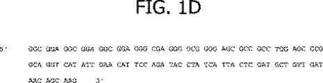
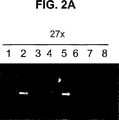
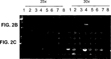
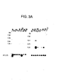
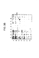
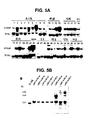

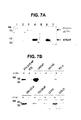
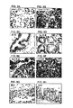

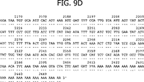


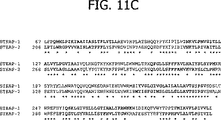
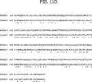

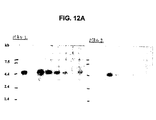

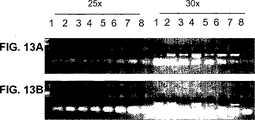
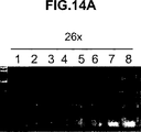
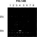
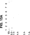
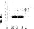
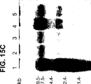
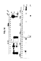

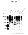
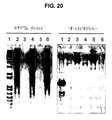

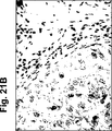


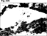
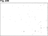


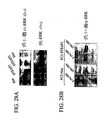


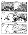
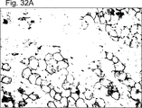

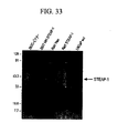
Classifications
-
- C—CHEMISTRY; METALLURGY
- C07—ORGANIC CHEMISTRY
- C07K—PEPTIDES
- C07K16/00—Immunoglobulins [IGs], e.g. monoclonal or polyclonal antibodies
- C07K16/18—Immunoglobulins [IGs], e.g. monoclonal or polyclonal antibodies against material from animals or humans
- C07K16/28—Immunoglobulins [IGs], e.g. monoclonal or polyclonal antibodies against material from animals or humans against receptors, cell surface antigens or cell surface determinants
-
- A—HUMAN NECESSITIES
- A61—MEDICAL OR VETERINARY SCIENCE; HYGIENE
- A61P—SPECIFIC THERAPEUTIC ACTIVITY OF CHEMICAL COMPOUNDS OR MEDICINAL PREPARATIONS
- A61P13/00—Drugs for disorders of the urinary system
- A61P13/08—Drugs for disorders of the urinary system of the prostate
-
- A—HUMAN NECESSITIES
- A61—MEDICAL OR VETERINARY SCIENCE; HYGIENE
- A61P—SPECIFIC THERAPEUTIC ACTIVITY OF CHEMICAL COMPOUNDS OR MEDICINAL PREPARATIONS
- A61P35/00—Antineoplastic agents
-
- A—HUMAN NECESSITIES
- A61—MEDICAL OR VETERINARY SCIENCE; HYGIENE
- A61P—SPECIFIC THERAPEUTIC ACTIVITY OF CHEMICAL COMPOUNDS OR MEDICINAL PREPARATIONS
- A61P43/00—Drugs for specific purposes, not provided for in groups A61P1/00-A61P41/00
-
- C—CHEMISTRY; METALLURGY
- C07—ORGANIC CHEMISTRY
- C07K—PEPTIDES
- C07K14/00—Peptides having more than 20 amino acids; Gastrins; Somatostatins; Melanotropins; Derivatives thereof
- C07K14/435—Peptides having more than 20 amino acids; Gastrins; Somatostatins; Melanotropins; Derivatives thereof from animals; from humans
- C07K14/705—Receptors; Cell surface antigens; Cell surface determinants
-
- C—CHEMISTRY; METALLURGY
- C07—ORGANIC CHEMISTRY
- C07K—PEPTIDES
- C07K16/00—Immunoglobulins [IGs], e.g. monoclonal or polyclonal antibodies
- C07K16/18—Immunoglobulins [IGs], e.g. monoclonal or polyclonal antibodies against material from animals or humans
- C07K16/28—Immunoglobulins [IGs], e.g. monoclonal or polyclonal antibodies against material from animals or humans against receptors, cell surface antigens or cell surface determinants
- C07K16/30—Immunoglobulins [IGs], e.g. monoclonal or polyclonal antibodies against material from animals or humans against receptors, cell surface antigens or cell surface determinants from tumour cells
- C07K16/3069—Reproductive system, e.g. ovaria, uterus, testes, prostate
-
- C—CHEMISTRY; METALLURGY
- C12—BIOCHEMISTRY; BEER; SPIRITS; WINE; VINEGAR; MICROBIOLOGY; ENZYMOLOGY; MUTATION OR GENETIC ENGINEERING
- C12Q—MEASURING OR TESTING PROCESSES INVOLVING ENZYMES, NUCLEIC ACIDS OR MICROORGANISMS; COMPOSITIONS OR TEST PAPERS THEREFOR; PROCESSES OF PREPARING SUCH COMPOSITIONS; CONDITION-RESPONSIVE CONTROL IN MICROBIOLOGICAL OR ENZYMOLOGICAL PROCESSES
- C12Q1/00—Measuring or testing processes involving enzymes, nucleic acids or microorganisms; Compositions therefor; Processes of preparing such compositions
- C12Q1/68—Measuring or testing processes involving enzymes, nucleic acids or microorganisms; Compositions therefor; Processes of preparing such compositions involving nucleic acids
- C12Q1/6876—Nucleic acid products used in the analysis of nucleic acids, e.g. primers or probes
- C12Q1/6883—Nucleic acid products used in the analysis of nucleic acids, e.g. primers or probes for diseases caused by alterations of genetic material
- C12Q1/6886—Nucleic acid products used in the analysis of nucleic acids, e.g. primers or probes for diseases caused by alterations of genetic material for cancer
-
- G—PHYSICS
- G01—MEASURING; TESTING
- G01N—INVESTIGATING OR ANALYSING MATERIALS BY DETERMINING THEIR CHEMICAL OR PHYSICAL PROPERTIES
- G01N33/00—Investigating or analysing materials by specific methods not covered by groups G01N1/00 - G01N31/00
- G01N33/48—Biological material, e.g. blood, urine; Haemocytometers
- G01N33/50—Chemical analysis of biological material, e.g. blood, urine; Testing involving biospecific ligand binding methods; Immunological testing
- G01N33/53—Immunoassay; Biospecific binding assay; Materials therefor
- G01N33/574—Immunoassay; Biospecific binding assay; Materials therefor for cancer
-
- A—HUMAN NECESSITIES
- A61—MEDICAL OR VETERINARY SCIENCE; HYGIENE
- A61K—PREPARATIONS FOR MEDICAL, DENTAL OR TOILETRY PURPOSES
- A61K38/00—Medicinal preparations containing peptides
-
- C—CHEMISTRY; METALLURGY
- C07—ORGANIC CHEMISTRY
- C07K—PEPTIDES
- C07K2317/00—Immunoglobulins specific features
- C07K2317/30—Immunoglobulins specific features characterized by aspects of specificity or valency
- C07K2317/34—Identification of a linear epitope shorter than 20 amino acid residues or of a conformational epitope defined by amino acid residues
-
- C—CHEMISTRY; METALLURGY
- C07—ORGANIC CHEMISTRY
- C07K—PEPTIDES
- C07K2319/00—Fusion polypeptide
-
- C—CHEMISTRY; METALLURGY
- C07—ORGANIC CHEMISTRY
- C07K—PEPTIDES
- C07K2319/00—Fusion polypeptide
- C07K2319/30—Non-immunoglobulin-derived peptide or protein having an immunoglobulin constant or Fc region, or a fragment thereof, attached thereto
-
- C—CHEMISTRY; METALLURGY
- C12—BIOCHEMISTRY; BEER; SPIRITS; WINE; VINEGAR; MICROBIOLOGY; ENZYMOLOGY; MUTATION OR GENETIC ENGINEERING
- C12Q—MEASURING OR TESTING PROCESSES INVOLVING ENZYMES, NUCLEIC ACIDS OR MICROORGANISMS; COMPOSITIONS OR TEST PAPERS THEREFOR; PROCESSES OF PREPARING SUCH COMPOSITIONS; CONDITION-RESPONSIVE CONTROL IN MICROBIOLOGICAL OR ENZYMOLOGICAL PROCESSES
- C12Q2600/00—Oligonucleotides characterized by their use
- C12Q2600/112—Disease subtyping, staging or classification
-
- C—CHEMISTRY; METALLURGY
- C12—BIOCHEMISTRY; BEER; SPIRITS; WINE; VINEGAR; MICROBIOLOGY; ENZYMOLOGY; MUTATION OR GENETIC ENGINEERING
- C12Q—MEASURING OR TESTING PROCESSES INVOLVING ENZYMES, NUCLEIC ACIDS OR MICROORGANISMS; COMPOSITIONS OR TEST PAPERS THEREFOR; PROCESSES OF PREPARING SUCH COMPOSITIONS; CONDITION-RESPONSIVE CONTROL IN MICROBIOLOGICAL OR ENZYMOLOGICAL PROCESSES
- C12Q2600/00—Oligonucleotides characterized by their use
- C12Q2600/136—Screening for pharmacological compounds
-
- C—CHEMISTRY; METALLURGY
- C12—BIOCHEMISTRY; BEER; SPIRITS; WINE; VINEGAR; MICROBIOLOGY; ENZYMOLOGY; MUTATION OR GENETIC ENGINEERING
- C12Q—MEASURING OR TESTING PROCESSES INVOLVING ENZYMES, NUCLEIC ACIDS OR MICROORGANISMS; COMPOSITIONS OR TEST PAPERS THEREFOR; PROCESSES OF PREPARING SUCH COMPOSITIONS; CONDITION-RESPONSIVE CONTROL IN MICROBIOLOGICAL OR ENZYMOLOGICAL PROCESSES
- C12Q2600/00—Oligonucleotides characterized by their use
- C12Q2600/154—Methylation markers
-
- C—CHEMISTRY; METALLURGY
- C12—BIOCHEMISTRY; BEER; SPIRITS; WINE; VINEGAR; MICROBIOLOGY; ENZYMOLOGY; MUTATION OR GENETIC ENGINEERING
- C12Q—MEASURING OR TESTING PROCESSES INVOLVING ENZYMES, NUCLEIC ACIDS OR MICROORGANISMS; COMPOSITIONS OR TEST PAPERS THEREFOR; PROCESSES OF PREPARING SUCH COMPOSITIONS; CONDITION-RESPONSIVE CONTROL IN MICROBIOLOGICAL OR ENZYMOLOGICAL PROCESSES
- C12Q2600/00—Oligonucleotides characterized by their use
- C12Q2600/158—Expression markers
-
- G—PHYSICS
- G01—MEASURING; TESTING
- G01N—INVESTIGATING OR ANALYSING MATERIALS BY DETERMINING THEIR CHEMICAL OR PHYSICAL PROPERTIES
- G01N2500/00—Screening for compounds of potential therapeutic value
Landscapes
- Health & Medical Sciences (AREA)
- Chemical & Material Sciences (AREA)
- Life Sciences & Earth Sciences (AREA)
- Organic Chemistry (AREA)
- Immunology (AREA)
- General Health & Medical Sciences (AREA)
- Medicinal Chemistry (AREA)
- Molecular Biology (AREA)
- Proteomics, Peptides & Aminoacids (AREA)
- Engineering & Computer Science (AREA)
- Biochemistry (AREA)
- Genetics & Genomics (AREA)
- Biophysics (AREA)
- Cell Biology (AREA)
- Veterinary Medicine (AREA)
- Bioinformatics & Cheminformatics (AREA)
- Public Health (AREA)
- Pathology (AREA)
- Zoology (AREA)
- Analytical Chemistry (AREA)
- Animal Behavior & Ethology (AREA)
- Pharmacology & Pharmacy (AREA)
- Nuclear Medicine, Radiotherapy & Molecular Imaging (AREA)
- General Chemical & Material Sciences (AREA)
- Chemical Kinetics & Catalysis (AREA)
- Urology & Nephrology (AREA)
- Hospice & Palliative Care (AREA)
- Oncology (AREA)
- Biomedical Technology (AREA)
- Biotechnology (AREA)
- Hematology (AREA)
- Microbiology (AREA)
- Wood Science & Technology (AREA)
- Physics & Mathematics (AREA)
- Reproductive Health (AREA)
- Toxicology (AREA)
- Gynecology & Obstetrics (AREA)
- Pregnancy & Childbirth (AREA)
- Gastroenterology & Hepatology (AREA)
- Food Science & Technology (AREA)
Abstract
Description
【0001】
本出願は、1999年12月6日出願、米国特許出願第09/455,486号の利益を請求する。
【0002】
(発明の分野)
本明細書に記載の発明は、新規な遺伝子のファミリーおよびそれらのコードするタンパク質および腫瘍抗原(STEAPと名付けられた)に関し、そして種々の癌(特に、前立腺癌、結腸癌、膀胱癌、肺癌、卵巣癌および膵臓癌を含む)の管理において有用な診断および治療の方法ならびに組成物に関する。
【0003】
(発明の背景)
癌は、冠動脈疾患に次いで、ヒトの死亡の二番目に主要な原因である。世界中で、数百万の人々が毎年、癌で死んでいる。アメリカ合衆国単独でも、癌は、毎年に50万人を軽く上回る死亡を引き起しており、1年あたりおよそ140万の新しい症例が診断されている。心疾患に起因する死亡は、有意に低下してきたが、癌に起因する死亡は、一般に増大している。次の世紀の初頭には、癌は死亡の主要な原因になると予想される。
【0004】
世界中で、いくつかの癌が主な死亡原因として突出している。詳細には、肺癌、前立腺癌、乳癌、結腸癌、膵臓癌、および卵巣癌が、癌死の主要な原因である。これらおよび実質的に全ての他の癌は、共通の致死的特徴を共有する。ごくわずかな例外を除いて、癌腫由来の転移性疾患は、致死的である。さらに、原発性癌を最初に生きのびた癌患者にとってさえ、共通の経験として、患者らの生活が劇的に変化したことが示される。多くの癌患者は、再発または処置の失敗の可能性の認識から生じる強い不安感を経験する。多くの癌患者は、処置後の身体的衰弱を経験する。多くの癌患者は再発を経験する。
【0005】
世界中で、前立腺癌は、ヒトにおいて4番目に最も流行している癌である。北アメリカおよび北ヨーロッパでは、前立腺癌は、間違いなく最も通常の男性の癌であり、そしてヒトにおける癌死の第二の主要な原因である。アメリカ合衆国単独でも、唯一肺癌に次いで、毎年、この疾患で40,000人を超える男性が死んでいる。これらの数字の大きさにもかかわらず、転移性前立腺癌の有効な処置はいまだに存在しない。外科的前立腺切除、放射線療法、ホルモンアブレーション(消失)療法、および化学療法が、主要な処置の様式であり続けている。不幸にも、これらの処置は、多くの場合無効であり、そしてしばしば望ましくない結果を伴う。
【0006】
診断的分野では、初期の限局性腫瘍を正確に検出できる前立腺腫瘍マーカーがないことは、この疾患の管理における重要な限界となっている。血清PSAアッセイは非常に有用なツールであるが、その特異性および一般的有用性は、いくつかの重要な事項を欠くと、広くみなされている。
【0007】
前立腺癌に特異的なさらなるマーカーを同定するにおける進歩が、マウスにおいて疾患の種々の段階を再現し得る、前立腺癌異種移植片の生成によって改善されている。LAPC(Los Angeles Prostate Cancer)異種移植片は、重症複合免疫不全(SCID)マウスにおいて継代生存し、そして疾患進行(アンドロゲン依存性からアンドロゲン非依存性への遷移、および転移性病変の発生を含む)を模倣する能力を示した、前立腺癌異種移植片である(米国特許第6,107,540号;Kleinら、1997,Nat.Med.3:402)。さらに最近になって同定された前立腺癌マーカーとしては、以下が挙げられる:PCTA−1(Suら、1996,Proc.Natl.Acad.Sci.USA 93:7252)、前立腺幹細胞抗原(PSCA)(Reiterら、1998,Proc.Natl.Acad.Sci.USA 95:1735)、およびSTEAP(Hubertら、1999,Proc.Natl.Acad.Sci.USA 96:14523)。
【0008】
PSA、PSM、PCTAおよびPSCAのような以前に同定されたマーカーは、前立腺癌を診断および処置するための労力を容易にしたが、診断および治療をさらに改善するため、前立腺および関連する癌についてのさらなるマーカーおよび治療標的の同定の必要性が存在する。
【0009】
(発明の要旨)
本発明は、細胞表面のサーパンタインな(曲がりくねった)(serpentine)膜貫通抗原の新規なファミリーに関する。このファミリーの2つのタンパク質は、前立腺および前立腺癌において排他的または優先的に発現され、従ってこのファミリーのメンバーは、「STEAP」(Six Transmembrane Epithelial Antigen of the Prostate(前立腺の6つの膜貫通上皮抗原))と名付けられている。4つの特定のヒトSTEAPが、本明細書において記載されており、そして特徴付けられている。ヒトSTEAPは、その間で高い程度の構造的保存を示すが、いずれの公知のヒトタンパク質に対しても有意な構造的相同性を示さない。
【0010】
STEAPファミリーのプトロタイプ(原型)のメンバーである、STEAP−1は、正常なヒト組織における前立腺細胞で優先的に発現されたIIIa型膜タンパク質であるようである。構造的に、STEAP−1は、6つの膜貫通ドメインならびに細胞内N末端およびC末端の分子トポロジーによって特徴付けられた339アミノ酸のタンパク質であり、このことは、STEAP−1が、3つの細胞外ループおよび2つの細胞内ループに「サーパンタイン(曲がりくねった)(serpentine)」様式で折り畳むことを示唆する。STEAP−1タンパク質発現は、種々の段階の前立腺癌にまたがって高レベルで維持されている。さらに、STEAP−1は、特定の他のヒト癌において非常に過剰発現されている。詳細には、STEAP−1の細胞表面発現は、種々の前立腺細胞および前立腺癌細胞、肺癌、膀胱癌細胞および結腸癌細胞において明確に決定されている。これらの特徴は、STEAP−1が、前立腺癌、膀胱癌、結腸癌および他の癌において高レベルで発現される特異的な細胞表面腫瘍抗原であることを示す。
【0011】
このファミリーの第二のメンバーである、STEAP−2は、454アミノ酸のタンパク質であり、STEAP−1のトポロジーと類似の推定分子トポロジーを有する。STEAP−2は、STEAP−1と同様、正常なヒト組織において前立腺特異的であり、そしてまた前立腺癌において発現される。STEAP−2およびSTEAP−1のORFのアラインメントは、237アミノ酸残基重複にわたって54.9%の同一性を示し、そしてSTEAP−2中の6つの推定膜貫通ドメインの位置は、STEAP−1における膜貫通ドメインの位置と一致する(図11A−B)。
【0012】
STEAP−3およびSTEAP−4がまた、本明細書に記載されている。これらはまた構造的に関連しており、そして特有の発現プロフィールを示す。詳細には、STEAP−3およびSTEAP−4は、異なる組織制限パターンを示すようである。4つのSTEAP全てのアミノ酸配列アラインメントを、図11A〜Bに示す。
【0013】
本発明は、本明細書に記載されるSTEAP遺伝子、mRNA、および/またはコード配列の全てまたは一部に相当するかまたは相補的なポリヌクレオチドを、好ましくは単離した形態で提供する。これには、STEAPタンパク質およびそのフラグメントをコードするポリヌクレオチド、STEAP遺伝子またはmRNA配列またはその部分に相補的なDNA、RNA、DNA/RNAハイブリッド、および関連分子、ポリヌクレオチドまたはオリゴヌクレオチド、ならびにSTEAP遺伝子、mRNAに対して、もしくはSTEAPコードポリヌクレオチドに対してハイブリダイズするポリヌクレオチドまたはオリゴヌクレオチドが挙げられる。また、STEAPをコードするcDNAおよび遺伝子を単離する手段も提供される。STEAPポリヌクレオチドを含有する組換えDNA分子、このような分子で形質転換されたまたは形質導入された細胞、およびSTEAP遺伝子産物の発現のための宿主ベクター系がまた提供される。
【0014】
本発明はさらに、STEAPタンパク質およびそのポリペプチドフラグメントを提供する。本発明はさらに、STEAPタンパク質およびそのポリペプチドフラグメントに結合する抗体を提供する。この抗体としては、ポリクローナル抗体およびモノクローナル抗体、マウスおよび他の哺乳動物の抗体、キメラ抗体、ヒト化抗体、および完全ヒト抗体、ならびに検出マーカーで標識した抗体、そして放射性核種/放射性同位体、毒素または他の治療組成物に結合体化された抗体が挙げられる。本発明はさらに、種々の生物学的サンプルにおいて、STEAPポリヌクレオチドおよびタンパク質の存在を検出するための方法を提供する。本発明はさらに、前立腺癌および他の癌を処置するための種々の治療用組成物およびストラテジー(詳細には、抗体、ワクチン、および低分子治療を含む)を提供する。
【0015】
本発明はさらに、種々の生物学的サンプルにおけるSTEAPポリヌクレオチドおよびタンパク質の存在を検出するための方法、ならびにSTEAPを発現する細胞を同定するための方法を提供する。本発明はさらに、前立腺癌を処置するための種々の治療用組成物およびストラテジー(詳細には、抗体、ワクチン、および低分子治療を含む)を提供する。このタンパク質の細胞外の性質は、STEAPおよびその機能を標的する分子、ならびにSTEAPと相互作用する他のタンパク質、因子およびリガンドを標的する分子を用いる、多数の治療アプローチを提供する。これらの治療アプローチとしては、抗STEAP抗体を用いる抗体療法、低分子療法、およびワクチン療法が挙げられる。さらに、前立腺癌におけるそのアップレギュレートされた発現を考慮して、STEAPは、前立腺癌の診断マーカー、段階決定マーカー、および/または予後マーカーとして有用であり、そして同様に、このタンパク質を発現する他の癌のマーカーであり得る。詳細には、STEAP−1は、前立腺癌転移を同定するための優れたマーカーを提供する。
【0016】
(発明の詳細な説明)
本発明は、細胞表面ヘビ状(serpentine)膜貫通抗原の新規ファミリーに関する。このファミリーにおけるこれらのタンパク質の2つは、前立腺、ならびに前立腺癌において排他的にかまたは優勢に発現され、従って、このファミリーのメンバーは、「STEAP」(前立腺の6回膜貫通上皮抗原)と名付けられた。4つの特定のヒトSTEAPは、本明細書中で記載されかつ特徴付けられる。ヒトSTEAPは、これらの中で高度な構造的保存を示すが、任意の公知のヒトタンパク質と顕著な構造的相同性を示さない。本発明は、前立腺および他の癌の診断および治療のための方法および組成物に関する。本方法は、ヒトSTEAP遺伝子に対応する単離されたポリヌクレオチド、STEAP遺伝子によってコードされるタンパク質およびそのフラグメント、ならびにSTEAPタンパク質を特異的に認識しそして結合し得る抗体を利用する。
【0017】
他に規定しない限り、本明細書中で使用されるすべての専門用語、表記、および他の科学的用語は、本発明が属する分野の当業者によって共通して理解される意味を有することが意図される。いくつかの場合において、共通して理解される意味を有する用語は、明確さおよび/または迅速な参照のために本明細書中で定義され、そしてこのような定義の本明細書中への包含は、当該分野において一般に理解されることにわたる実質的な相違を表すとは必ずしも解釈されるべきではない。本明細書中に記載または参照される技術および手順は、一般に、従来の方法論(例えば、Sambrookら、Molecular Cloning:A Laboratory Manual 第2版(1989)Cold Spring Harbor Laboratory Press、Cold Spring Harbor,N.Y.において記載されている方法論を広く利用した分子クローニングの方法論)を用いて、当業者によって十分に理解されかつ共通して用いられる。適切な場合、市販のキットおよび試薬の使用を含む手順は、他に注記されない限り、一般に、製造業者によって規定されるプロトコールおよび/またはパラメーターに従って実行される。
【0018】
本明細書中で使用される場合、用語「進行した前立腺癌」、「局所的に進行した前立腺癌」、「進行した疾患」および「局所的に進行した疾患」とは、前立腺皮膜を通して伸長した前立腺癌を意味し、そして米国泌尿器学会(AUA)システムの下でのステージC疾患、Whitmore−Jewettシステムの下でのステージC1−C2疾患、およびTNM(腫瘍、節、転移)システムの下でのステージT3−T4およびN+疾患を含むことが意味される。一般に、手術は、局所的に進行した疾患を有する患者のためには推奨されず、そしてこれらの患者は、臨床的に局在化している(器官に限定されている)前立腺癌を有する患者と比較して、実質的により好ましくない結果を有する。局所的に進行した疾患は、前立腺の側縁を超える硬化の明白な証拠、または前立腺基部上の非対称もしくは硬化によって臨床的に同定される。局所的に進行した前立腺癌は、腫瘍が前立腺皮膜に侵入するかもしくは浸透するか、外科的な境界まで伸長するか、または精嚢に侵入する場合、現在、徹底的な前立腺切除後に生理学的に診断される。
【0019】
本明細書中で使用する場合、用語「転移性前立腺癌」および「転移性疾患」は、局所的なリンパ節、または異なる部位に拡がった前立腺癌を意味し、そしてAUAシステム下でステージD疾患、およびTNMシステム下でステージTxNxM+を包含することが意味される。局所的に進行した前立腺癌の場合、手術は一般的には転移性疾患を有する患者には示されず、そしてホルモン(アンドロゲン除去)治療が好ましい治療様式である。転移性前立腺癌を有する患者は、最終的に、処置の開始から12〜18ヶ月以内にアンドロゲン抵抗性の状態を発症し、そしてこれらの患者のおよそ半数がその後6ヶ月以内に死亡する。前立腺癌転移の最も一般的な部位は骨である。前立腺癌の骨転移は、結局のところ、特徴として、溶骨性ではなくむしろ造血性である(すなわち、正味の骨の形成を生じる)。骨への転移は、脊椎において最も頻繁に見出され、大腿骨、骨盤、胸郭、頭蓋、および上腕骨と続く。転移のための他の一般的な部位には、リンパ節、肺、肝臓、および脳が含まれる。転移性の前立腺癌は、代表的には、直視的または腹腔鏡的な骨盤のリンパ節切除術、全身の放射性核種スキャン、骨格のラジオグラフィー、および/または骨損傷生検によって診断される。
【0020】
本明細書中で使用される場合、用語「ポリヌクレオチド」は、少なくとも10塩基または10塩基対の長さのヌクレオチドのポリマー型を意味し、リボヌクレオチドもしくはデオキシヌクレオチドまたはいずれかの型のヌクレオチドの改変型のいずれかであり、そして一本鎖および二本鎖の形態のDNAを含むことを意味する。
【0021】
本明細書中で使用する場合、用語「ポリペプチド」は、少なくとも10アミノ酸のポリマーを意味する。本明細書を通して、アミノ酸についての標準的な3文字表記または1文字表記が使用される。
【0022】
本明細書中で使用される場合、ポリヌクレオチドの状況において使用される、用語「ハイブリダイズ」、「ハイブリダイズする」「ハイブリダイズする」などは、従来のハイブリダイゼーション条件(好ましくは、例えば、50% ホルムアミド/6×SSC/0.1% SDS/100μg/ml ssDNAにおけるハイブリダイゼーション)をいうことが意味される。ここで、ハイブリダイゼーションのための温度が、37℃より高い温度であり、そして0.1×SSC/0.1% SDS中での洗浄のための55℃がより高い温度であり、そして最も好ましくは、ストリンジェントなハイブリダイゼーション条件である。
【0023】
ハイブリダイゼーション反応の「ストリンジェンシー」は、当業者によって容易に決定可能であり、そして一般的には、プローブの長さ、洗浄温度、および塩濃度に依存する経験による計算である。一般に、より長いプローブは、正しいアニーリングのためにより高い温度を必要とするが、一方、より短いプローブは、より低い温度を必要とする。ハイブリダイゼーションは、一般に、融解温度より下の環境において、相補鎖が存在する場合、変性したDNAの再アニーリングする能力に依存する。プローブと、ハイブリダイズ可能な配列との間の所望される相同性の程度が高いほど、使用され得る相対的な温度が高くなる。結果として、相対的により高い温度が、反応条件をよりストリンジェントにする傾向があるが、より低い温度ではそのようなことがより少ない、ということになる。ハイブリダイゼーション反応のストリンジェンシーのさらなる詳細および説明については、Ausubelら、Current Protocols in Molecular Biology、Wiley,Interscience Publishers,(1995)を参照のこと。
【0024】
本明細書中に規定されるように、「ストリンジェントな条件」または「高ストリンジェンシー条件」は、以下の条件によって同定され得る:(1)洗浄のために低いイオン強度および高い温度を使用する条件(例えば、0.015M 塩化ナトリウム/0.0015M クエン酸ナトリウム/0.1% ドデシル硫酸ナトリウム、50℃);(2)ハイブリダイゼーションの間に変性剤(例えば、ホルムアミド)を使用する条件(例えば、50% (v/v)ホルムアミドおよび0.1% ウシ血清アルブミン/0.1% Ficol/0.1% ポリビニルピロリドン/50mM リン酸ナトリウム緩衝液(pH 6.5)および750mM 塩化ナトリウム、75mM クエン酸ナトリウム、42℃);または(3)50% ホルムアミド、5×SSC(0.75M NaCl、0.075M クエン酸ナトリウム)、50mM リン酸ナトリウム(pH 6.8)、0.1% ピロリン酸ナトリウム、5×Denhardt’s溶液、超音波処理したサケ精子DNA(50μg/ml)、0.1% SDS、および10% 硫酸デキストラン、42℃、さらに以下の洗浄工程、42℃において0.2×SSC(塩化ナトリウム/クエン酸ナトリウム)および50%ホルムアミド、55℃、続いて、高ストリンジェンシー洗浄(EDTAを含む0.1×SSCからなる、55℃)を使用する条件。
【0025】
「中程度にストリンジェントな条件」は、Sambrookら、Molecular Cloning:A Laboratory Manual,New York:Cold Spring Harbor Press,1989によって記載されたように同定され得、そして洗浄溶液およびハイブリダイゼーション条件(例えば、温度、イオン強度、およびSDS)の使用を含む。これは、上記に記載されたものよりもよりストリンジェントではない。中程度にストリンジェントな条件の例は、以下を含む溶液中での37℃、一晩のインキュベーションである:20%ホルムアミド、5×SSC(150mM NaCl、15mM クエン酸三ナトリウム)、50mM リン酸ナトリウム(pH7.6)、5×Denhardt’s溶液、10% 硫酸デキストラン、および20mg/mL変性剪断サケ精子DNA、続いて約37℃〜50℃で1×SSC中でフィルターを洗浄する。当業者は、温度、イオン強度などを、因子(例えば、プローブ長など)を適応させるのに必要なように調整する方法を認識する。
【0026】
アミノ酸比較の状況において、用語「同一性」は、同一である同じ相対的位置のアミノ酸残基のパーセンテージを表現するために使用される。この状況においてはまた、用語「相同性」は、BLAST分析の保存性アミノ酸の判断基準を使用して、当該分野において一般的に理解されるように、同一であるかまたは類似であるかのいずれかである、同じ相対位置でのアミノ酸残基のパーセンテージを表現するために使用される。例えば、同一性%の値は、WU−BLAST−2(Altschulら、Methods in Enzymology,266:460−480(1996);http://blast.wustl/edu/blast/README.html)によって生成され得る。アミノ酸置換(これは、このような判断基準の下では保存性であると見なされる)に関するさらなる詳細は以下に提供される。
【0027】
さらなる定義が、引き続くサブセクションを通して提供される。
【0028】
(STEAPポリヌクレオチド)
本発明の一つの局面は、STEAP遺伝子、mRNAおよび/またはコード配列(好ましくは、STEAPタンパク質およびそのフラグメント、DNA、RNA、DNA/RNAハイブリッドならびに関連分子をコードするポリヌクレオチド、STEAP遺伝子もしくはmRNA配列またはその一部分に相補的な、ポリヌクレオチドまたはオリゴヌクレオチド、ならびにSTEAP遺伝子、mRNAもしくはSTEAPをコードするポリヌクレオチド(集合的に、「STEAPポリヌクレオチド」)にハイブリダイズする、ポリヌクレオチドまたはオリゴヌクレオチドを含む単離形態における)のすべてもしくは一部分に対応するか、またはそのすべてもしくは一部分に相補的なポリヌクレオチドを提供する。本明細書中で使用される場合、STEAP遺伝子およびタンパク質は、STEAP−1、STEAP−2およびSTEAP−3の遺伝子およびタンパク質、GenBankアクセッション番号R80991(STEAP−4)に対応する遺伝子およびタンパク質、ならびに他のSTEAPタンパク質および前述の構造的に類似の改変体に対応する、遺伝子およびタンパク質を含むことが意味される。このような他のSTEAPタンパク質および改変体は、一般的に、STEAPコード配列に高度に相同なコード配列を有し、そして好ましくは、少なくとも約50%のアミノ酸同一性、そして(BLAST標準を使用して)少なくとも約60%のアミノ酸相同性を共有し、より好ましくは、(BLAST標準を使用して)70%以上の相同性を共有する。
【0029】
本明細書中に記載されるSTEAPファミリーメンバー遺伝子配列は、他のタンパク質からSTEAPタンパク質を区別する、独特な高度に保存されたアミノ酸配列を共有するSTEAPタンパク質をコードする。1以上のこれらの独特な高度に保存されたドメインを含むタンパク質は、STEAPファミリーメンバーに関し得るかまたは新たなSTEAPタンパク質を示し得る。全長STEAP−1、STEAP−2、およびSTEAP−3、ならびにSTEAP−4のタンパク質配列のアミノ酸配列アラインメントである図11A〜Bを参照して、STEAPが構造レベルで密接に関連することは明らかである。STEAP−1およびSTEAP−2のタンパク質配列のアミノ酸配列のアラインメントである図11Cを参照して、密接な構造的保存は、特に好ましくは、膜貫通ドメインにおいて明らかである。STEAP−1配列およびSTEAP−2配列は、237アミノ酸重複にわたって54.9%同一性を共有する。STEAPの間のさらなるアミノ酸配列アラインメントは、図11Dおよび11Eに示される。これらのアラインメントは、STEAP−1およびSTEAP−3が264アミノ酸領域にわたって40.9%同一であるが、STEAP−2およびSTEAP−3は、416アミノ酸領域にわたって47.8%同一である。
【0030】
STEAPポリヌクレオチドは、図1A〜Bに示されるようなヒトSTEAP−1のヌクレオチド配列、図9A〜Dに示されるようなヒトSTEAP−2のヌクレオチド配列、図10A〜Eに示されるようなヒトSTEAP−3のヌクレオチド配列、もしくは図10Fに示されるようなヒトSTEAP−4のヌクレオチド配列、またはこれらに相補的な配列、あるいは、前述の任意のポリヌクレオチドフラグメントを有するポリヌクレオチドを含み得る。別の実施形態は、ヒトSTEAP−1タンパク質、STEAP−2タンパク質、STEAP−3タンパク質またはSTEAP−4タンパク質のアミノ酸配列、これらに相補的な配列、または前述の任意のポリヌクレオチドフラグメントをコードするポリヌクレオチドを含む。別の実施形態は、図1A〜Bに示されるヒトSTEAP−1 cDNA、図9A〜Dに示されるヒトSTEAP−2 cDNA、図10A〜Eに示されるヒトSTEAP−3 cDNA、または図10Fに示されるようなSTEAP−4、あるいは、これらのポリヌクレオチドフラグメントに、ストリンジェントなハイブリダイゼーション条件下でハイブリダイズし得るポリヌクレオチドを含む。
【0031】
本明細書中で開示される本発明の代表的な実施形態は、STEAP mRNA配列の特定の一部分をコードするSTEAPポリヌクレオチド(例えば、タンパク質およびそのフラグメントをコードするポリヌクレオチド)を含む。例えば、本明細書中で開示される本発明の代表的な実施形態は、以下を含む:図11A〜Bに示されるSTEAPタンパク質のおよそのアミノ酸1〜およそのアミノ酸10をコードするポリヌクレオチド、図11A〜Bに示されるSTEAPタンパク質のおよそのアミノ酸20〜およそのアミノ酸30をコードするポリヌクレオチド、図11A〜Bに示されるSTEAPタンパク質のおよそのアミノ酸30〜およそのアミノ酸40をコードするポリヌクレオチド、図11A〜Bに示されるSTEAPタンパク質のおよそのアミノ酸40〜およそのアミノ酸50をコードするポリヌクレオチド、図11A〜Bに示されるSTEAPタンパク質のおよそのアミノ酸50〜およそのアミノ酸60をコードするポリヌクレオチド、図11A〜Bに示されるSTEAPタンパク質のおよそのアミノ酸60〜およそのアミノ酸70をコードするポリヌクレオチド、図11A〜Bに示されるSTEAPタンパク質のおよそのアミノ酸70〜およそのアミノ酸80をコードするポリヌクレオチド、図11A〜Bに示されるSTEAPタンパク質のおよそのアミノ酸80〜およそのアミノ酸90をコードするポリヌクレオチド、および図11A〜Bに示されるSTEAPタンパク質のおよそのアミノ酸90〜およそのアミノ酸100をコードするポリヌクレオチドなど。このスキームの後、STEAPタンパク質のさらなる部分をコードするポリヌクレオチド(少なくとも10アミノ酸の)は、本発明の代表的な実施形態である。STEAPタンパク質のこのような部分は、図11A〜Bに示されるSTEAP−1タンパク質のアミノ酸100〜339、または図11A〜Bに示されるSTEAP−2タンパク質のアミノ酸100〜454、または図11A〜Bに示されるSTEAP−3タンパク質のアミノ酸100〜459、および図11A〜Bに示されるSTEAP−4のアミノ酸100〜133を含む。STEAPタンパク質のより大きな部分をコードするポリヌクレオチドがまた意図される。例えば、図11A〜Bに示されるSTEAPタンパク質のおよそのアミノ酸1(または20、または30、または40など)〜およそのアミノ酸20(または30、または40、または50など)をコードするポリヌクレオチドは、当該分野で周知の種々の技術によって生成され得る。
【0032】
本明細書中で開示される本発明のさらなる例示的な実施形態は、STEAPタンパク質配列内に含まれる、1つ以上の生物学的モチーフをコードするSTEAPポリヌクレオチドフラグメントを含む。1つの実施形態において、本発明の代表的なポリヌクレオチドフラグメントは、他のSTEAPファミリーメンバー(例えば、1以上の膜貫通ドメイン)との相同性を示すSTEAPの1つ以上の領域をコードし得る。本発明の別の実施形態において、代表的なポリヌクレオチドフラグメントは、1つ以上のSTEAP選択的スプライシング改変体に独特である配列をコードし得る。本発明のさらに別の実施形態において、代表的なポリヌクレオチドフラグメントは、STEAPタンパク質の免疫原性部分をコードし得る。STEAPタンパク質の免疫原性部分の1例は、図1A〜B(WKMKPRRNLEEDDYL;配列番号2)に示されるようなSTEAP−1アミノ酸配列のアミノ酸残基14〜28である。
【0033】
前述の段落のポリヌクレオチドは、多くの異なる特定の使用を有する。STEAPは、前立腺癌および他の癌において差示的に発現されるので、これらのポリヌクレオチドは、正常な組織 対 癌性組織におけるSTEAP遺伝子産物の状態を評価する方法において使用され得る。代表的には、STEAPタンパク質の特定の領域をコードするポリヌクレオチドが使用されて、STEAP遺伝子産物の特定の領域において、混乱状態(例えば、欠失、挿入、点変異など)の存在を評価し得る。例示的なアッセイは、RT−PCRアッセイ、ならびに一本鎖高次構造多型(SSCP)分析(例えば、Marrogiら、J.Cutan.Pathol.26(8):369−378(1999)を参照のこと)の両方を含み、これらの両方は、タンパク質の特定の領域をコードするポリヌクレオチドを利用して、タンパク質内のこれらの領域を試験する。単一ヌクレオチド多型を検出する配列を分析するためのアッセイおよび方法はまた、利用可能である(Irizarryら、2000、Nature Genetics 26(2)223−236)。
【0034】
本明細書中で開示される本発明の他の具体的に意図された実施形態は、ゲノムDNA、cDNA、リボザイムおよびアンチセンス分子(モルホリノアンチセンス分子を含む)、ならびに天然の供給源または合成由来にせよ、代替の骨格に基づくか、または代替の塩基を含む核酸分子である。例えば、アンチセンス分子は、RNA、あるいは他の分子(ペプチド核酸(PNA)または塩基対依存様式でDNAもしくはRNAに特異的に結合する非核酸分子(例えば、ホスホロチオエート誘導体)を含む)であり得る。当業者は、STEAPポリヌクレオチドおよび本明細書中で開示されるポリヌクレオチド配列を使用して、核酸分子のこれらのクラスを容易に獲得し得る。
【0035】
アンチセンス技術は、細胞内部に位置する標的ポリヌクレオチドに結合する外因性オリゴヌクレオチドの投与を必然的に伴う。用語「アンチセンス」は、このようなオリゴヌクレオチドが、その細胞内標的(例えば、STEAP)に相補的であるという事実をいう。例えば、Jack Cohen,OLIGODEOXYNUCLEOTIDES,Antisense Inhibitors of Gene Expression,CRC Press,1989;およびSynthesis 1:1−5(1988)を参照のこと。本発明のSTEAPアンチセンスオリゴヌクレオチドは、Sオリゴヌクレオチド(ホスホロチオエート誘導体またはS−オリゴ、Jack Cohen、前出を参照のこと)のような誘導体を含み、この誘導体は、癌細胞増殖阻害作用の増大を示す。S−オリゴ(ヌクレオシドホスホロチオエート)は、オリゴヌクレオチド(O−オリゴ)の等電子アナログであり、リン酸基の非架橋酸素原子は、硫酸原子に置換される。本発明のS−オリゴは、3H−1,2−ベンゾジチオール−3−オン−1,1−ジオキシド(これは、硫酸転移試薬である)での対応するO−オリゴの処理によって調製され得る。Iyer,R.P.ら、J.Org.Chem.55:4693−4698(1990);およびIyer,R.P.ら、J.Am.Chem.Soc.112:1253−1254(1990)(これらの開示は、本明細書中で参考として完全に援用される)を参照のこと。本発明のさらなるSTEAPアンチセンスオリゴヌクレオチドは、当該分野で公知のモルホリノアンチセンスオリゴヌクレオチドを含む(例えば、Partridgeら、1996,Antisense&Nucleic Acid Drug Development 6:169−175を参照のこと)。
【0036】
本発明のSTEAPアンチセンスオリゴヌクレオチドは、代表的に、STEAPゲノムもしくは対応するmRNAの、最初の100個のN末端コドンまたは最後の100個のC末端コドンに相補的であり、そしてこれらのコドンと安定にハイブリダイズするか、あるいはATG開始部位と重複する、RNAまたはDNAであり得る。完全な相補性は必要ではないが、高い程度の相補性が好ましい。この領域に相補的なオリゴヌクレオチドの使用は、STEAP mRNAへの選択的ハイブリダイゼーションを可能にし、そしてタンパク質キナーゼの他の調節サブユニットを特定するmRNAへの選択的ハイブリダイゼーションを可能にしない。好ましくは、本発明のSTEAPアンチセンスオリゴヌクレオチドは、STEAP mRNAにハイブリダイズする配列を有するアンチセンスDNA分子の15〜30merフラグメントである。必要に応じて、STEAPアンチセンスオリゴヌクレオチドは、STEAPの最初の10個のN末端コドンおよび最後の10個のC末端コドンにおける領域に相補的な30merオリゴヌクレオチドである。あるいは、アンチセンス分子は、STEAP発現の阻害において、リボザイムを利用するように改変される。L.A.CoutureおよびD.T.Stinchcomb;Trends Genet 12:510−515(1996)。
【0037】
本発明のこの局面のさらなる特定の実施形態は、プライマーおよびプライマーペアを含み、これは、本発明のポリヌクレオチドまたはその任意の特定の部分の特異的増幅、および本発明の核酸分子またはその任意の部分に選択的もしくは特異的にハイブリダイズするプローブの特異的増幅を可能にする。プローブは、検出可能なマーカー(例えば、放射性同位体、蛍光化合物、生体発光化合物、化学発光化合物、金属キレート剤または酵素のような)で標識され得る。このようなプローブおよびプライマーが、サンプルにおけるSTEAPポリヌクレオチドの存在を検出するために使用され得、そしてSTEAPタンパク質を発現する細胞を検出するための手段として使用され得る。このようなプローブの例としては、図1A〜B(配列番号1)、図9A〜D(配列番号7)または図10A〜E(配列番号9)に示されるヒトSTEAP cDNA配列の、すべてまたは一部分を含むポリペプチドが挙げられる。STEAP mRNAを特異的に増幅し得るプライマーペアの例はまた、続く実施例において記載される。当業者に理解されるように、非常に多くの異なるプライマーおよびプローブが、本明細書中に提供される配列に基づいて調製され得、そしてSTEAP mRNAを有効に増幅および/または検出するために使用され得る。
【0038】
本明細書中で使用される場合、ポリヌクレオチドは、STEAP遺伝子ではない遺伝子に、対応するかまたは相補的である混入ポリヌクレオチド、またはSTEAP遺伝子産物もしくはそのフラグメントではないポリペプチドをコードする混入ポリヌクレオチドから実質的に分離されている場合、「単離された」と言われる。当業者は、容易に核酸単離手順を利用して、単離されたSTEAPポリヌクレオチドを獲得し得る。
【0039】
本発明のSTEAPポリヌクレオチドは、種々の目的に有用である。この目的としては、STEAP遺伝子、mRNAまたはそのフラグメントの、増幅および/または検出のための、プローブおよびプライマーとしての使用;前立腺癌および他の癌の、診断および/または予後のための試薬としての使用;前立腺細胞へのカルシウムの侵入を特異的に阻害する分子を同定するための手段としての使用;STEAPポリペプチドの発現を指向し得るコード配列としての使用;STEAP遺伝子の発現および/またはSTEAP転写物の翻訳を、調節または阻害するための手段としての使用;ならびに治療薬としての使用が挙げられるが、これらに限定されない。
【0040】
(STEAPの分子的特徴および生化学的特徴)
本発明は、STEAPと命名された新規タンパク質ファミリーに関する。4つのSTEAPが、構造特徴、分子特徴および生化学的特徴によって本明細書中に詳細に記載される。以下に続く実施例にさらに記載されるように、このSTEAPは、種々の方法で特徴付けられている。例えば、ヌクレオチドコード配列およびアミノ酸配列の分析は、STEAPファミリー内で保存された構造エレメントを同定するために実施された。STEAP mRNA発現の大量のRT−PCR分析およびノーザンブロット分析が、種々のSTEAPメッセージを発現する正常組織および癌性組織の範囲を確立するために実施された。STEAPタンパク質発現のウエスタンブロット分析、免疫組織化学分析、およびフローサイトメトリー分析は、STEAPのタンパク質発現プロフィール、細胞表面局在および総分子トポロジーを決定するために実施された。
【0041】
STEAPファミリーの原型のメンバーであるSTEAP−1は、いずれの既知のヒトタンパク質とも識別可能な相同性を有さない、339アミノ酸の6回膜貫通細胞表面タンパク質である。ヒトSTEAP−1のcDNAヌクレオチドおよび推定アミノ酸配列は、図1A〜Bに示される。細胞膜内に組込まれたSTEAP−1の総トポロジーの模式図は、図1Bに示される。STEAP−1発現は、正常組織において主に前立腺特異的である。詳細には、正常ヒト組織におけるSTEAP−1 mRNA発現およびSTEAP−1タンパク質発現の大量の分析により、STEAP−1タンパク質が、前立腺に主に発現され、そしてはるかに少ない程度で膀胱に発現されることが示される。STEAP−1 mRNAはまた、相対的に前立腺特異的である、非常に低いレベルの発現のみが、いくつかの他の正常組織で検出される。癌において、STEAP−1 mRNAおよびSTEAP−1タンパク質は、一貫して、前立腺癌(アンドロゲン依存性腫瘍およびアンドロゲン非依存性腫瘍を含む)そしてこの疾患の全段階の間に高レベルで発現される。STEAP−1はまた、他の癌で発現される。詳細には、STEAP−1 mRNAは、非常に高いレベルで、膀胱癌、結腸癌、膵臓癌、および卵巣癌(ならびに他の癌)で発現される。さらに、STEAP−1タンパク質の細胞表面発現が、前立腺癌、膀胱癌、肺癌および結腸癌で確立されている。従って、STEAP−1は、特定の癌(詳細には、前立腺癌腫、結腸癌腫および膀胱癌腫を含む)の処置のための、優れた診断標的および治療標的の顕著な特徴の全てを有する。
【0042】
このファミリーの第2のメンバーであるSTEAP−2は、異なる遺伝子によってコードされた454アミノ酸タンパク質であり、STEAP−1の推定分子トポロジーに類似した推定分子トポロジーを有する。STEAP−2のcDNAヌクレオチド配列および推定アミノ酸配列は、図9A〜Dに示される。STEAP−1配列とSTEAP−2配列のアミノ酸アラインメントは、高い程度の構造的な保存(237アミノ酸残基の重複にわたって54.9%の同一性)、およびSTEAP−1およびSTEAP−2の同時に生じる6つの推定膜貫通ドメインの位置を示す(図11A〜B、11C)。これらのSTEAP−1とSTEAP−2との間の構造的な相同性は、第1の推定細胞外ループから第5の膜貫通ドメインに広がる領域において最も高い。しかし、STEAP−1とSTEAP−2との間のいくつかの有意な構造的な差異が、明らかである。例えば、STEAP−2は、205アミノ酸長の細胞内N末端(STEAP−1における69アミノ酸に比較される)および短い4アミノ酸の細胞内C末端(STEAP−1における26アミノ酸と比較される)を示す。さらに、STEAP−1遺伝子およびSTEAP−2遺伝子の両方は、第7番染色体に位置するが、異なるアーム上である。これらの差異は、機能および/または細胞内シグナル伝達経路との相互作用における有意な差異を意味し得る。
【0043】
STEAP−2は、試験されたヒト組織の間では正常な前立腺にのみ発現され(図14および15)、そしてまた、前立腺癌で発現される(図15)。従って、これは、STEAP−1に対して発現プロフィールにいくらかの類似性を示す。しかし、STEAP−2は、前立腺癌サンプル(図3および15を比較)および試験された他の非前立腺癌(図5および図16を比較)において、STEAP−1に対して異なるmRNA発現プロフィールを示す。STEAP−1およびSTEAP−2の発現プロフィールにおけるこれらの差異は、これらが特異に調節されることを示唆する。
【0044】
STEAP−3およびSTEAP−4は、構造レベルではSTEAP−1およびSTEAP−2の両方に密接に関連するようであり、これらの両方は、同様に膜貫通タンパク質のようである。STEAP−3は、STEAP−1(40.9%の同一性)よりも、STEAP−2(47.8%の同一性)により関連する。STEAP−3およびSTEAP−4は、特有の発現プロフィールを示す。例えば、STEAP−3は、主に胎盤に限定される発現パターンを有するようであり、より少ない程度で、発現は、前立腺に見出されるが試験された他の正常組織では見出されない。STEAP−4は、RT−PCR分析によって、主に肝臓で発現されるようである。STEAP−3もSTEAP−4も、高レベルのSTEAP−1発現およびSTEAP−2発現が示される前立腺癌異種移植片で発現されないようである。
【0045】
STEAPタンパク質は、細胞のシグナル伝達経路に関与するタンパク質の特徴を示す。詳細には、STEAP−1およびSTEAP−2は、PC3細胞に発現された場合、p38(MAPKシグナル伝達カスケードに関与するタンパク質)のリン酸化を活性化する。さらに、STEAP−2発現は、チロシンリン酸化を誘導し、そしてSTEAP−1は、臭気物質処理細胞においてチロシンキナーゼの活性化を媒介する。これらの知見は、STEAP関連分子および細胞のシグナル伝達経路を変更し得る分子を同定するための高スループットアッセイにおいてSTEAPを発現するために改変される細胞の使用を支持し、新規治療剤の同定を導く。1つの実施形態において、このアッセイは、STEAP機能を阻害し得る分子を同定し、それによってこの分子は、癌または調節不全細胞の増殖と関連した他の疾患の進行を調節し得る。
【0046】
本明細書中に記載された4つのSTEAPのうちの3つは、ヒト第7番染色体にマップされる(STEAP−1、2および3)。興味深いことに、原発性前立腺癌および再発性前立腺癌の両方に関して報告された(Visakorpiら、1995 Cancer Res.55:342、Nupponenら、1998 American J.Pathol.153:141)、7p22(7p22.3)(対立遺伝子(allelic gain)の大きい領域)内のSTEAP−1マップは、癌におけるSTEAP−1のアップレギュレーションが、ゲノム機構を含み得ることを示唆する。さらに、STEAP−2およびSTEAP−3の両方は、染色体7q21に位置し、これらの2つの遺伝子が、遺伝子重複によって発生したことを示唆する。
【0047】
6回膜貫通ドメインを含む他の細胞表面分子としては、イオンチャネル(DollyおよびParcej,1996 J Bioenerg Biomembr 28:231)および水チャネルもしくはアクアポリン(Reizerら、1993 Crit Rev Biochem Mol Biol 28:235)が挙げられる。構造研究は、機能的なチャネルを形成するために4量体複合体に集合される両方の型の分子を示す(Christie,1995,Clin Exp Pharmacol Physiol 22:944,Walzら、1997 Nature 387:624,Chengら,1997 Nature 387:627)。前立腺におけるSTEAP−1の免疫組織化学染色は、細胞間境界において濃縮されるようであり、管腔側において染色はほとんど検出されない。これは、きつい接合点、ギャップの接合点、細胞の伝達、接着またはトランスポータータンパク質としてのSTEAP−1の役割を示唆し得る。
【0048】
これらの可能性を試験するために、STEAPを発現するツメガエルの卵母細胞(または他の細胞)は、STEAPがイオンチャネルとして機能するか否かを決定するために、電圧クランプ実験およびパッチクランプ実験を用いて分析され得る。卵母細胞の容積はまた、STEAPが水チャネル特性を示すか否かを決定するために測定され得る。STEAPがチャネルタンパク質またはギャップ接合点タンパク質として機能する場合、これらは、例えば、発現または機能を阻害し得る抗体、小分子、およびポリヌクレオチドを使用して、阻害に関して優れた標的としてはたらき得る。正常組織における限定された発現パターンおよび癌組織における高レベルの発現は、STEAP機能を妨害することが、選択的に癌細胞を殺傷し得ることを示唆する。
【0049】
STEAP遺伝子ファミリーは上皮組織で主に発現されるので、STEAPタンパク質が、上皮細胞機能においてイオンチャネル、輸送タンパク質、またはギャップ接合点タンパク質として機能する可能性があるようである。イオンチャネルは、前立腺癌細胞の増殖および侵襲性に関与している(Lalaniら、1997、Cancer Metastasis Rev 16:29)。ラットおよびヒト両方の前立腺癌細胞は、より高いまたはより低いナトリウムチャネルの発現レベルを有する細胞の亜集団を含む。より高いレベルのナトリウムチャネル発現は、インビトロにおいてより侵襲的な侵襲性と相関する(Smithら、1998、FEBS Lett.423:19)。同様に、ナトリウムチャネルの特定の遮断が、インビトロにおいてPC−3細胞の侵襲性を阻害することが示されている(Laniadoら、1997、Am.J.Pathol.150:1213)が、LNCaP細胞における特定のカリウムチャネルの阻害は、細胞増殖を阻害した(Skrymaら、1997、Prostate 33:112)。これらの報告は、前立腺癌中のイオンチャネルに関する役割を示唆し、そしてまた、イオンチャネル機能を阻害する小分子が前立腺癌増殖を妨害し得ることを証明する。
【0050】
(STEAPをコードする核酸分子の単離)
本明細書中に記載されるSTEAP cDNA配列は、STEAP遺伝子産物をコードする他のポリヌクレオチドの単離、ならびにSTEAP遺伝子産物ホモログ、選択的にスプライシングされたアイソフォーム、対立遺伝子改変体、およびSTEAP遺伝子産物の変異体形態をコードするポリヌクレオチドの単離を可能にする。STEAP遺伝子をコードする全長cDNAを単離するために利用され得る種々の分子クローニング方法は、周知である(例えば、Sambrook,J.ら、Molecular Cloning:A Laboratory Manual,第2版、Cold Spring Harbor Press,New York,1989;Current Protocols in Molecular Biology.Ausubelら編、Wiley and Sons,1995を参照のこと)。例えば、λファージクローニング方法論が、市販のクローニング系を使用して慣用的に利用され得る(例えば、Lambda ZAP Express,Stratagene)。STEAP遺伝子cDNAを含むファージクローンは、標識されたSTEAP cDNAまたはそのフラグメントを用いてプローブ化することにより同定され得る。例えば、一つの実施形態において、STEAP cDNAまたはその一部分が、合成され、そしてプローブとして使用されて、STEAP遺伝子に対応する重複するcDNAおよびSTEAP遺伝子に対応する全長cDNAを回収し得る。STEAP遺伝子自体は、STEAP DNAプローブまたはプライマーを使用して、ゲノムDNAライブラリー、細菌人工染色体ライブラリー(BAC)、酵母人工染色体ライブラリー(YAC)などをスクリーニングすることによって単離され得る。
【0051】
(組換えDNA分子および宿主−ベクター系)
本発明はまた、STEAPポリヌクレオチドを含む組換えDNAまたはRNA分子(これには、当該分野において周知のファージ、プラスミド、ファージミド、コスミド、YAC、BAC、ならびに種々のウイルス性および非ウイルス性ベクターが含まれるが、これらに限定されない)、およびこのような組換えDNAまたはRNA分子で形質転換またはトランスフェクトされた細胞を提供する。本明細書中で使用される場合、組換えDNAまたはRNA分子は、インビトロでの分子操作に供されたDNAまたはRNA分子である。このような分子を生成するための方法は周知である(例えば、Sambrookら、1989(前出)を参照のこと)。
【0052】
本発明はさらに、適切な原核生物宿主細胞または真核生物宿主細胞においてSTEAPポリヌクレオチドを含む組換えDNA分子を含む、宿主−ベクター系を提供する。適切な真核生物宿主細胞の例としては、酵母細胞、植物細胞、または動物細胞(例えば、哺乳動物細胞または昆虫細胞(例えば、バキュロウイルス感染可能細胞(例えば、Sf9細胞)))が挙げられる。適切な哺乳動物細胞の例としては、種々の前立腺癌細胞株(例えば、LnCaP、PC3、DU145、LAPC−4、TsuPr1、他のトランスフェクト可能であるかまたは形質導入可能な前立腺細胞株)、ならびに組換えタンパク質の発現のために慣用的に使用される多くの哺乳動物細胞(例えば、COS細胞、CHO細胞、293細胞、293T細胞)が挙げられる。より詳細には、STEAPのコード配列を含むポリヌクレオチドを使用し、当該分野で慣用的に使用され、そして広く知られた任意の数の宿主−ベクター系を用いて、STEAPタンパク質またはそれらのフラグメントを生成し得る。
【0053】
STEAPタンパク質またはそれらのフラグメントの発現に適切な広範な種々の宿主−ベクター系が利用可能である(例えば、Sambrookら、1989(前出);Current Protocols in Molecular Biology、1995(前出)を参照のこと)。哺乳動物での発現に好ましいベクターとしては、pcDNA 3.1 myc−His−tag(Invitrogen)およびレトロウイルスベクターpSRαtkneo(Mullerら、1991、MCB 11:1785)が挙げられるが、これらに限定されない。これらの発現ベクターを使用して、STEAPは、好ましくは、いくつかの前立腺癌細胞株および非前立腺癌細胞株(例えば、293、293T、rat−1、3T3、PC3、LNCaPおよびTsuPr1を含む)において発現され得る。本発明の宿主ベクター系は、STEAPタンパク質またはそれらのフラグメントの産生のために有用である。このような宿主−ベクター系は、STEAPおよびSTEAP変異の機能的特性を研究するために使用され得る。
【0054】
STEAP遺伝子またはそれらのフラグメントによってコードされるタンパク質は、種々の用途を有する。これには、以下が挙げられるが、これらに限定されない:抗体の産生、およびSTEAP遺伝子産物に結合する、リガンドおよび他の因子および細胞構成要素を同定するための方法において。STEAPタンパク質またはそれらのフラグメントに対して惹起された抗体は、診断および予後アッセイにおいて、画像化方法(特に、癌画像化を含む)において、そしてSTEAPタンパク質の発現によって特徴付けられるヒト癌(前立腺の癌を含むが、これに限定されない)の管理における治療方法において有用であり得る。STEAPタンパク質の検出のために有用な種々の免疫学的アッセイが意図され、これには、種々の型のラジオイムノアッセイ、酵素結合イムノソルベント検定法(ELISA)、酵素結合免疫蛍光アッセイ(ELIFA)、免疫細胞学的方法などが意図されるが、これらに限定されない。このような抗体は、標識され得、そして前立腺細胞を検出し得る免疫学的画像化試薬として使用され得る(例えば、放射線シンチグラフィー画像化方法において)。STEAPタンパク質はまた、以下にさらに記載されるように、癌ワクチンを生成するにおいて特に有用であり得る。
【0055】
(STEAPタンパク質)
本発明の別の局面は、種々のSTEAPタンパク質およびそのポリペプチドフラグメントを提供する。本明細書中に使用される場合、STEAPタンパク質は、図1A〜Bに提供されるようなヒトSTEAP−1、図9A〜Dに提供されるようなヒトSTEAP−2、図10A〜Eに提供されるようなヒトSTEAP−3のアミノ酸配列、他の哺乳動物STEAPホモログ(例えば、STEAP−4)のアミノ酸配列および改変体、ならびにSTEAP生物学的活性を有するこれらのタンパク質の対立遺伝子改変体および保存的置換改変体(このような改変体およびホモログが以下に概説される方法に従って過度な実験を伴わずに単離/生成および特徴付けされ得る限り)を、有するかまたは含むタンパク質をいう。異なるSTEAPタンパク質またはそのフラグメントの部分を組合せる融合タンパク質、ならびにSTEAPタンパク質および異種ポリペプチドの融合タンパク質もまた含まれる。このようなSTEAPタンパク質は、集合的に、STEAPタンパク質、本発明のタンパク質、またはSTEAPという。本明細書中で使用される場合、用語「STEAPポリペプチド」は、少なくとも10個のアミノ酸(好ましくは、少なくとも15個のアミノ酸)のポリペプチドフラグメントまたはSTEAPタンパク質をいう。
【0056】
STEAPタンパク質の特定の実施形態は、図1A〜Bに示されるようなヒトSTEAP−1のアミノ酸配列を有するポリペプチドを含む。STEAPタンパク質の別の実施形態は、図9A〜Dに示されるようなSTEAP−2アミノ酸配列を含むポリペプチドを含む。別の実施形態は、図10A〜Eに示されるSTEAP−3アミノ酸配列を含むポリペプチドを含む。さらに別の実施形態は、図11A〜Bに示される部分的なSTEAP−4アミノ酸配列を含むポリペプチドを含む。
【0057】
一般的に、ヒトSTEAPの天然に存在する対立遺伝子改変体は、高い程度の構造的同一性および相同性(例えば、90%以上の同一性)を共有する。代表的に、STEAPタンパク質の対立遺伝子改変体は、本明細書中に記載されるSTEAP配列内に保存的アミノ酸置換を含むか、またはSTEAPホモログにおいて対応する位置に由来するアミノ酸の置換を含む。STEAP対立遺伝子改変体の1つのクラスは、特定のSTEAPアミノ酸配列の少なくとも小さな領域と高い程度の相同性を共有するが、この配列とは根本的な背反(例えば、非保存的置換、短縮化(truncation)挿入、またはフレームシフト)をさらに含むタンパク質である。
【0058】
保存的アミノ酸置換はしばしば、タンパク質のコンフォメーションも機能もいずれも変更することなく、タンパク質中で作製され得る。このような変化としては、以下が挙げられる:任意のイソロイシン(I)、バリン(V)、およびロイシン(L)の、任意の他のこれらの疎水性アミノ酸についての置換;グルタミン酸(E)に対するアスパラギン酸(D)、およびその逆の置換;アスパラギン(N)に対するグルタミン(Q)、およびその逆の置換;ならびに、スレオニン(T)に対するセリン(S)、およびその逆の置換。他の置換はまた、タンパク質の三次元構造における特定のアミノ酸の環境およびその役割に依存して、保存的とみなされ得る。例えば、グリシン(G)およびアラニン(A)は、しばしば交換可能であり得、アラニン(A)およびバリン(V)も同様であり得る。比較的疎水性であるメチオニン(M)はしばしば、ロイシンおよびイソロイシンと交換可能であり得、そして時としてバリンと交換可能であり得る。リシン(K)およびアルギニン(R)はしばしば、そのアミノ酸残基の重要な特徴がその電荷であり、そしてこれら2つのアミノ酸残基の異なるpKが重要でない位置において、交換可能である。さらに他の変化が、特定の状況において、「保存的」とみなされ得る。
【0059】
STEAPタンパク質(改変体を含む)は、図11A〜Bに示されるアミノ酸配列を有するSTEAPタンパク質に共通した少なくとも1つのエピトープを含み、その結果、STEAPタンパク質または改変体に特異的に結合する抗体はまた、図11A〜Bに示されるアミノ酸配列を有するSTEAPタンパク質に特異的に結合する。STEAPタンパク質改変体の1つのクラスは、図11A〜Bのアミノ酸配列と90%以上の同一性を共有する。STEAPタンパク質改変体のより特定のクラスは、上記のように細胞外タンパク質SCPモチーフを含む。好ましいSTEAPタンパク質改変体は、本明細書中に記載される1つ以上のディフェンシン機能(例えば、腫瘍の死を誘導する能力または細胞移動を化学誘引および/もしくは誘導する能力を含む)を示し得る。
【0060】
STEAPタンパク質は、多くの形態、好ましくは単離された形態で具体化され得る。本明細書中で使用される場合、このタンパク質と正常に関連する細胞構成成分からSTEAPタンパク質を取り出すために、物理的方法、機械的方法または化学的方法が使用される場合、タンパク質は「単離された」という。当業者は、単離されたSTEAPタンパク質を得るために、標準的な精製方法を容易に使用し得る。精製されたSTEAPタンパク質分子は、抗体または他のリガンドへのSTEAPの結合を損なう他のタンパク質または分子を実質的に含まない。単離および精製の性質および程度は、意図される用途に依存する。STEAPタンパク質の実施形態は、精製されたSTEAPおよび機能的な可溶性STEAPタンパク質を含む。1つの形態においては、このような機能的な可溶性STEAPタンパク質またはそのフラグメントは、抗体または他のリガンドに結合する能力を保持する。
【0061】
本発明はまた、STEAPアミノ酸配列の生物学的に活性なフラグメントを含むSTEAPポリペプチド(例えば、図1A〜Bに示されるようなSTEAP−1、図9A〜Dに示されるようなSTEAP−2、図10A〜Eに示されるようなSTEAP−3、または図11A〜Bに示されるようなSTEAP−4のアミノ酸配列の部分に対応するポリペプチド)を提供する。本発明のこのようなポリペプチドは、STEAPタンパク質の特性(例えば、STEAPタンパク質に関連するエピトープに特異的に結合する抗体の産生を誘発する能力)を示す。特定のSTEAPタンパク質に対して特有(他のSTEAPタンパク質に対して)であるアミノ酸配列を含むポリペプチドは、その特定のSTEAPタンパク質と特異的に反応する抗体を作製するために使用され得る。例えば、図11A〜Eに示されるSTEAP構造のアミノ酸アラインメントを参照して、当業者は、各分子がその構造に特有な配列ストレッチを含むことを容易に認識する。これらの特有なストレッチは、特定のSTEAPに特異的な抗体を作製するために使用され得る。同様に、保存配列の領域は、複数のSTEAPに結合し得る抗体を作製するために使用され得る。
【0062】
本明細書中に開示される本発明の実施形態は、広範な種々の当該分野で認められたSTEAPタンパク質の改変体(例えば、アミノ酸の挿入、欠失および置換を有するポリペプチド)を含む。STEAP改変体は、当該分野において公知の方法(例えば、部位指向型変異誘発、アラニンスキャニング(alanine scanning)、およびPCR変異誘発)を使用して作製され得る。部位指向型変異誘発[Carterら、Nucl.Acids Res.13:4331(1986);Zollerら、Nucl.Acids Res.,10:6487(1987)]、カセット変異誘発[Wellら、Gene 34:315(1985)]、制限選択的変異誘発[Wellsら、Philos.Trans.R.Soc.London SerA 317:415(1986)]または他の公知技術を、クローン化されたDNAにおいて実施し、STEAP改変体DNAを産生し得る。スキャニングアミノ酸分析もまた、連続配列に沿って1以上のアミノ酸を同定するために使用され得る。とりわけ好ましいスキャニングアミノ酸は、比較的小さい中性アミノ酸である。このようなアミノ酸としては、アラニン、グリシン、セリン、およびシステインが挙げられる。アラニンが、代表的に、この群の中で好ましいスキャニングアミノ酸である。なぜなら、アラニンは、β炭素を越えて側鎖を排除し、そして改変体の主鎖コンフォメーションを変更する可能性が低いからである。アラニンもまた、代表的に好ましい。なぜなら、アラニンは、最も一般的なアミノ酸であるからである。さらに、埋没位置および露出位置の両方において、頻繁に見出される[Creighton、The Proteins(W.H.Freeman&Co.、NY);Chothia、J.Mol.Biol.150:1(1976)]。アラニン置換が適切な量の改変体を生じない場合には、等配電子アミノ酸が使用され得る。
【0063】
上記で考察したように、本願発明の実施形態は、図11A〜Bに示されるSTEAPタンパク質の全長アミノ酸配列未満を含むポリペプチドを含む。例えば、本明細書中に開示される本発明の代表的な実施形態は、以下が挙げられる:図11A〜Bに示されるSTEAPタンパク質の約アミノ酸1個〜約アミノ酸10個からなるポリペプチド、図11A〜Bに示されるSTEAPタンパク質の約アミノ酸20個〜約アミノ酸30個からなるポリペプチド、図11A〜Bに示されるSTEAPタンパク質の約アミノ酸30個〜約アミノ酸40個からなるポリペプチド、図11A〜Bに示されるSTEAPタンパク質の約アミノ酸40個〜約アミノ酸50個からなるポリペプチド、図11A〜Bに示されるSTEAPタンパク質の約アミノ酸50個〜約アミノ酸60個からなるポリペプチド、図11A〜Bに示されるSTEAPタンパク質の約アミノ酸60個〜約アミノ酸70個からなるポリペプチド、図11A〜Bに示されるSTEAPタンパク質の約アミノ酸70個〜約アミノ酸80個からなるポリペプチド、図11A〜Bに示されるSTEAPタンパク質の約アミノ酸80個〜約アミノ酸90個からなるポリペプチド、および図11A〜Bに示されるSTEAPタンパク質の約アミノ酸90個〜約アミノ酸100個からなるポリペプチドなど。このスキームに従って、図11A〜Bに示されるSTEAP−1タンパク質のアミノ酸100〜339、図11A〜Bに示されるSTEAP−2タンパク質のアミノ酸100〜454、または図11A〜Bに示されるSTEAP−3タンパク質のアミノ酸100〜459、または図11A〜Bに示されるSTEAP−4タンパク質のアミノ酸100〜133のアミノ酸配列の部分からなるポリペプチドが、本発明の代表的な実施形態である。STEAPタンパク質のより大きな部分からなるポリペプチドもまた意図される。例えば、図11A〜Bに示されるSTEAPタンパク質の約アミノ酸1(または、20もしくは30もしくは40など)個〜約アミノ酸20(または、30もしくは40もしくは50など)個からなるポリペプチドが、当該分野において周知の種々の技術によって生成され得る。
【0064】
本明細書中に開示される本発明のさらなる例示的な実施形態は、図11A−Bに示されるようなSTEAPポリペプチド配列内に含まれる1つ以上の生物学的モチーフのアミノ酸残基を含むSTEAPポリペプチドを含む。1つ以上のこれらのモチーフまたは本明細書中に記載される目的の他の選択領域を含むSTEAPポリペプチドは、代表的に、選択されたモチーフの一面または両面上の隣接したSTEAPタンパク質配列のさらなる5〜25またはそれを超えるアミノ酸残基を含む。1つの実施形態では、本発明の代表的なポリペプチドは、1つ以上の他のSTEAPタンパク質に対する相同性を示すSTEAPの1つ以上の領域を含み得る。別の実施形態において、本発明の代表的なポリペプチドは、STEAPタンパク質の1つ以上の免疫原性部分を含み得る。STEAPタンパク質の免疫原性部分の1つの例は、図1A−Bに示されるSTEAP−1アミノ酸配列のアミノ酸残基14〜28である(WKMKPRRNLEEDDYL;配列番号22)。別の実施形態において、本発明の代表的なポリペプチドは、STEAP−1のアミノ酸165−173、STEAP−1のアミノ酸86−94、STEAP−1のアミノ酸262−270、STEAP−1のアミノ酸302−310、STEAP−1のアミノ酸158−166、STEAP−2のアミノ酸227−235、STEAP−2のアミノ酸402−410、STEAP−2のアミノ酸307−315、STEAP−2のアミノ酸306−314、およびSTEAP−2のアミノ酸100−108のような1つ以上の推定されたHLA−A2結合ペプチドを含み得る。別の実施形態において、本発明の代表的なポリペプチドは、図8に示されるSTEAP−2タンパク質のアミノ酸1−245、2−204、121−454、153−165、182−454、183−387、276−453、345−358、または419−454のようなSTEAP−2のフラグメントのすべてまたは一部からなる。これらの本発明の関連した実施形態としては、上記で考察された異なるモチーフの組合せを含むポリペプチドが挙げられ、好ましい実施形態は、これらのポリペプチドのモチーフまたは介在配列のいずれにも、挿入も欠失も置換も含んでいないポリペプチドである。
【0065】
STEAPポリペプチドは、本明細書中に開示されるヒトSTEAPタンパク質のアミノ酸配列に基づいて、当該分野において周知の、標準的なペプチド合成技術または化学切断方法を使用して生成され得る。あるいは、組換え方法を使用して、STEAPタンパク質のポリペプチドフラグメントをコードする核酸分子を生成し得る、この点において、本明細書中に記載されるSTEAPコード核酸分子は、STEAPタンパク質の規定のフラグメントを生成するための手段を提供する。STEAPポリペプチドは、ドメイン特異的抗体(例えば、STEAPタンパク質の細胞外エピトープおよび細胞内エピトープを認識する抗体)の生成および特徴付け、STEAPまたはその特定の構造ドメインに結合する薬剤または細胞因子の同定、および種々の治療状況(癌ワクチンを含むが、これらに限定されない)において、特に有用である。特定の興味深い構造を含むSTEAPポリペプチドは、例えば、以下:Chou−Fasman、Garnier−Robson、Kyte−Doolittle、Eisenberg、Karplus−SchultzまたはJameson−Wolf分析の方法を含む、当該分野で周知の種々の分析技術を使用するか、または免疫原性に基づいて、予測および/または同定され得る。このような構造を含むフラグメントは、サブユニット特異的抗STEAP抗体の生成、またはSTEAPに結合する細胞因子の同定において、特に有用である。
【0066】
以下の実施例に記載される特定の実施形態において、STEAPの分泌形態は、C末端6×HisおよびMYCタグを有する、STEAPをコードするCMV駆動発現ベクター(pcDNA3.1/mycHIS、Imvitrogen)でトランスフェクトされた293T細胞において、簡便に発現され得る。培養液中に分泌されたHISタグ化STEAPは、ニッケルカラムおよび標準的な技術を使用して精製され得る。あるいは、AP標識系が使用され得る。STEAPの発現のための種々の構築物が、以下の実施形態に記載される。
【0067】
STEAPの改変(例えば、共有結合性改変)は、本発明の範囲内に含まれる。1つの型の共有結合性改変としては、STEAPの選択された側鎖あるいはN末端残基またはC末端残基と反応し得る有機誘導体化薬剤での、STEAPポリペプチドの標的化されたアミノ酸残基の反応が挙げられる。本発明の範囲内のSTEAPポリペプチドの別の型の共有結合性改変としては、このポリペプチドのネイティブなグリコシル化パターンの変更が挙げられる。「ネイティブなグリコシル化パターンの変更」は、ネイティブ配列STEAPに見い出される1以上の炭水化物部分の欠失(基礎となるグリコシル化部位の除去、または化学的手段および/または酵素的手段によるグリコシル化の欠失のいずれかによって)、および/またはネイティブ配列STEAPに存在しない、1以上のグリコシル化部位の付加を意味するために、本明細書中で目的について意図される。さらに、この句は、存在する種々の炭水化物部分の性質および割合の変更を含む、ネイティブタンパク質のグリコシル化の質的変化を含む。STEAPの別の型の共有結合性改変としては、米国特許第4,640,835号、同第4,496,689号、同第4,301,144号、同第4,670,417号、同第4,791,192号または同第4,179,337号に示されるような様式の、種々の非タンパク質様ポリマー(例えば、ポリエチレングリコール(PEG)、ポリプロピレングリコールまたはポリオキシアルキレン)のうちの1つへの、STEAPポリペプチドの連結が挙げられる。
【0068】
本発明のSTEAPはまた、別の異種ポリペプチドまたはアミノ酸配列に融合されたSTEAPを含むキメラ分子を形成する様式で、改変され得る。1つの実施形態において、このようなキメラ分子としては、ポリヒスチジンエピトープタグを有するSTEAPの融合体が挙げられ、このタグは、固定されたニッケルが選択的に結合し得るエピトープを提供する。このエピトープタグは、一般に、STEAPのアミノ末端またはカルボキシル末端に配置される。代替的実施形態において、このキメラ分子は、STEAPの免疫グロブリンまたは免疫グロブリンの特定の領域との融合体を含み得る。キメラ分子の二価形態(「免疫付着因子(immunoadhesin)」とも呼ばれる)について、このような融合体は、IgG分子のFc領域に対するものであり得る。このIg融合体は、好ましくは、Ig分子内の少なくとも1つの可変領域の、STEAPポリペプチドの可溶性(膜貫通ドメインを欠失または不活性化した)形態での置換を含む。特に好ましい実施形態において、免疫グロブリン融合体は、ヒンジ領域、CH2領域およびCH3領域、またはIgGI分子のヒンジ領域、CH1領域、CH2領域およびCH3領域を含む。免疫グロブリン融合体の産生については、米国特許第5,428,130号(1995年6月27日公布)もまた参照のこと。
【0069】
(STEAP抗体)
本発明の別の局面は、STEAPタンパク質およびポリペプチドに結合する抗体を提供する。最も好ましい抗体は、STEAPタンパク質に特異的に結合し、そして非STEAPタンパク質およびポリペプチドに結合しない(または、弱く結合する)。特に意図される抗STEAP抗体としては、モノクローナル抗体およびポリクローナル抗体、ならびにこれらの抗体の抗原結合ドメインおよび/または1以上の相補性決定領域を含むフラグメントが挙げられる。本明細書中で使用される場合、抗体フラグメントは、その標的に結合する免疫グロブリン分子の可変領域(すなわち、抗原結合領域)の少なくとも一部として定義される。
【0070】
いくつかの適用について、特定の構造的ドメイン内の特定のSTEAPタンパク質および/またはエピトープと特異的に反応する抗体を産生することが所望であり得る。例えば、癌治療および診断画像化目的のために有用な好ましい抗体は、癌細胞において発現されるようなSTEAPタンパク質の細胞外領域におけるエピトープと反応する抗体である。このような抗体は、免疫原として、本明細書中に記載されるSTEAPタンパク質を使用してか、またはその推定された細胞外ドメインに由来するペプチドを使用して産生され得る。この点で、図1Bにおいて示されるようなSTEAP−1タンパク質の形態的概略図を参照して、示された膜貫通ドメインの間の細胞外ループにおける領域が、細胞外特異的抗体を惹起するための適切な免疫原を設計する場合に使用され得る。
【0071】
本発明のSTEAP抗体は、前立腺癌の治療ストラテジー、診断アッセイおよび予後アッセイ、ならびに画像化方法において特に有用であり得る。同様に、このような抗体は、STEAPが他の型の癌においてもまた発現または過剰発現される程度に、他の癌の処置、診断および/または予後に有用であり得る。本発明は、STEAPおよび変異体STEAPのタンパク質およびポリペプチドの、検出および定量に有用な種々の免疫学的アッセイを提供する。このようなアッセイは、一般に、適切には、STEAPまたは変異体STEAPタンパク質を認識および結合し得る、1以上のSTEAP抗体を含み、そして、以下を含むが、これらに限定されない当該分野で周知の種々の免疫学的アッセイ形式で行われ得る:種々の型のラジオイムノアッセイ、酵素連結免疫吸着アッセイ(ELISA)、酵素連結免疫蛍光アッセイ(ELIFA)など。さらに、前立腺癌を検出可能な免疫学的画像化方法もまた、本発明によって提供され、これらには、標識したSTEAP抗体を使用するラジオシンチグラフィー画像化方法が挙げられるが、これらに限定されない。このようなアッセイは、前立腺癌(特に、進行型前立腺癌)の検出、モニタリングおよび予後において、臨床的に有用であり得る。
【0072】
STEAP抗体はまた、STEAPおよび変異体STEAPのタンパク質およびポリペプチドの精製、およびSTEAPホモログおよび関連分子の単離のための方法において使用され得る。例えば、1つの実施形態において、STEAPタンパク質を精製する方法は、STEAP抗体(これは、固体マトリックスに結合されている)を、STEAPを含有する溶解物または他の溶液と共に、このSTEAP抗体がSTEAPに結合するのを可能にする条件下でインキュベートする工程;固体マトリックスを洗浄して不純物を除去する工程;およびSTEAPをその結合された抗体から溶出する工程を包含する。本発明のSTEAP抗体の他の用途としては、STEAPタンパク質を模倣する抗イディオタイプ抗体の生成が挙げられる。
【0073】
STEAP抗体はまた、例えば、STEAPタンパク質の生物学的活性を調節または阻害することによって、あるいは、STEAPタンパク質を発現する癌細胞を標的化および破壊することによって、治療学的に使用され得る。前立腺癌および他の癌の抗体治療は、以下の別々の小節においてより詳細に記載される。
【0074】
抗体の調製のための種々の方法が、当該分野で周知である。例えば、抗体は、単離または免疫結合体化された形態の、STEAPのタンパク質、ペプチドまたはフラグメントを使用して、適切な哺乳動物宿主を免疫することによって調製され得る(Antibodies:A Laboratory Manual,CSH Press編、HarlowおよびLane(1988);Harlow、Antibodies,Cold Spring Harbor Press,NY(1989))。タンパク質免疫原の例としては、組換えSTEAP(バキュロウイルス系、哺乳動物系などにおいて発現される)、1つ以上の抗体定常領域(AP−標識化STEAPなど)に結合体化されたSTEAPタンパク質のSTEAP細胞外ドメインまたは細胞外ループが挙げられる。さらに、STEAPの融合タンパク質(例えば、STEAPのGST、マルトース結合タンパク質(MBP)、緑色蛍光タンパク質(GFP)、HisMax−TOPOまたはMysHis(例えば、以下の実施例を参照のこと)との融合体のようなSTEAPの融合タンパク質もまた使用され得る。
【0075】
特定の実施形態において、図11A〜Bに示されるようなオープンリーディングフレームアミノ酸配列の全てまたはほとんどを含むGST融合タンパク質が生成され得、そして適切な抗体を生成するための免疫原として使用され得る。STEAPを発現するかまたは過剰発現する細胞がまた免疫のために使用され得る。同様にSTEAPを発現するように操作された任意の細胞が使用され得る。このようなストラテジーは、内因性STEAPを認識するための強化された能力を有するモノクローナル抗体の産生を生じ得る。別の有用な免疫原は、ヒツジの赤血球細胞の原形質膜に結合したSTEAPペプチドを含む。
【0076】
図11A〜Bに示されるようなSTEAPタンパク質のアミノ酸配列を使用して、抗体を生成するためにSTEAPタンパク質の特定の領域を選択し得る。例えば、STEAPアミノ酸配列の疎水性および親水性分析を使用して、STEAP構造の疎水性領域を同定し得る。免疫原性構造を示すSTEAPタンパク質の領域、ならびに他の領域およびドメインは、以下のような当該分野で公知の種々の他の方法を使用して容易に同定され得る:Chou−Fasman、Garnier−Robson、Kyte−Doolittle、Eisenberg、Karplus−SchultzまたはJameson−Wolf分析。HLA−A2を結合することが推定されるSTEAPのペプチドは、抗体の産生のために選択され得るか、またはCTL応答を産生するために使用され得る。このような推定されたHLA−A2結合ペプチドとしては、以下を含むがこれらに限定されない:STEAP−1のアミノ酸165−173、STEAP−1のアミノ酸86−94、STEAP−1のアミノ酸262−270、STEAP−1のアミノ酸302−310、STEAP−1のアミノ酸158−166、STEAP−2のアミノ酸227−235、STEAP−2のアミノ酸402−410、STEAP−2のアミノ酸307−315、STEAP−2のアミノ酸306−314、STEAP−2のアミノ酸100−108。以下の実施例において議論されるように、免疫原性は、STEAPを用いて実証され得、これは、それぞれ、ウサギおよびマウスを使用して、ポリクローナル抗体およびモノクローナル抗体を生成するために使用される。このB細胞応答(抗体生成)は、STEAPの免疫原性部分によって誘発された最初のT細胞応答の結果である。
【0077】
免疫原としての使用のためのタンパク質またはポリペプチドを調製するための方法、およびキャリア(例えば、BSA、KLHまたは他のキャリアタンパク質)とのタンパク質の免疫結合体を調製するための方法は、当該分野で周知である。いくつかの状況下で、例えば、カルボジイミド試薬を使用する直接的な結合体化が使用され得;他の例において、連結試薬(例えば、Pierce Chemical Co.,Rockford,ILによって供給される連結試薬)が、効果的であり得る。STEAP免疫原の投与は一般に、当該分野で一般に理解されるように、適切な期間にわたって、適切なアジュバントの使用を伴う注射によって行われ得る。免疫スケジュールの間、抗体の力価は、抗体形成の能力(adequacy)を決定することによって理解され得る。
【0078】
STEAPモノクローナル抗体は、好ましくは、当該分野で周知の種々の手段によって産生され得る。例えば、所望のモノクローナル抗体を分泌する不死化細胞株が、一般に公知のように、KohlerおよびMilsteinの標準的なハイブリドーマ技術、または産生性(producing)B細胞を不死化する改変を使用して調製され得る。所望の抗体を分泌する不死化細胞株は、抗原がSTEAPタンパク質またはSTEAPフラグメントである免疫アッセイによってスクリーニングされる。所望の抗体を分泌する適切な不死化細胞培養物が同定された場合、これらの細胞を拡大し、そしてインビトロ培養物または腹水のいずれかから抗体が産生され得る。
【0079】
抗体またはフラグメントはまた、現在の技術を使用する、組換え手順によって産生され得る。STEAPタンパク質の所望の領域に特異的に結合する領域はまた、複数の種起源のキメラ抗体またはCDR移植化(grafted)抗体の状況下で産生され得る。ヒト化STEAP抗体またはヒトSTEAP抗体もまた産生され得、そして治療的状況下での使用のために好ましい。1以上の非ヒト抗体CDRを対応するヒト抗体配列と置換することによる、マウス抗体および他の非ヒト抗体をヒト化するための方法は、周知である(例えば、Jonesら、1986、Nature 321:522−525;Riechmannら、1988、Nature 332:323−327;Verhoeyenら、1988、Science 239:1534−1536を参照のこと)。Carterら、1993、Proc.Natl.Acad.Sci.USA 89:4285、およびSimsら、1993、J.Immunol.151:2296もまた参照のこと。完全なヒトモノクローナル抗体を産生するための方法としては、ファージディスプレイ法およびトランスジェニック法が挙げられる(概要について、Vaughanら、1998、Nature Biotechnology 16:535−539を参照のこと)。
【0080】
完全なヒトSTEAPモノクローナル抗体は、大きいヒトIg遺伝子コンビナトリアルライブラリー(すなわち、ファージディスプレイ)を使用するクローニング技術を使用して生成され得る(Protein Engineering of Antibody Molecules for Prophylactic and Therapeutic Applications(Man.Clark,M.編)、Nottingham Academic、45−64頁(1993)のGriffithsおよびHoogenboom、Building an in vitro immune system:human antibodies from phage display libraries;BurtonおよびBarbas、Human Antibodies from combinatorial libraries(同書)65−82頁)。完全なヒトSTEAPモノクローナル抗体はまた、1997年12月3日提出のPCT特許出願WO98/24893に記載されるように、ヒト免疫グロブリン遺伝子座を含むように操作されたトランスジェニックマウスを使用して産生され得る(Jakobovits、1998、Exp.Opin.Invest.Drugs 7(4):607−614もまた参照のこと)。この方法は、ファージディスプレイ技術に必要とされるインビトロ操作を回避し、そして高親和性の真正ヒト抗体を効率的に産生する。
【0081】
STEAP抗体のSTEAPタンパク質との反応性は、適切には、STEAPタンパク質、STEAPペプチド、STEAP発現細胞またはその抽出物を使用する、多くの周知の手段(ウエスタンブロット、免疫沈降、ELISAおよびFACS分析を含む)によって確証され得る。
【0082】
本発明のSTEAP抗体またはそのフラグメントは、検出可能なマーカーで標識され得るか、または細胞毒または他の治療薬剤のような第2の分子に結合体化され得るか、STEAPポジティブ細胞に第2の分子を標的化するために使用される(Vitetta,E.S.ら、1993、Immunotoxin therapy、DeVita,Jr.,V.T.ら編、Cancer:Principle and Practice of Oncology,第4版、J.B.Lippincott Co.,Philadelphia,2624−2636)。細胞障害性薬剤の例としては、リシン、リシンA鎖、ドキソルビシン、メイタンシノイド、ダウノルビシン、タキソール、エチジウムブロマイド、マイトマイシン、エトポシド、テノポシド、ビンクリスチン、ビンブラスチン、コルヒチン、ジヒドロキシアントラシンジオン、アクチノマイシン、ジフテリア毒素、Psudomonas外毒素(PE)A、PE40、アブリン、アブリンA鎖、モデシン(modeccin)A鎖、α−サルシン(sarcin)、ゲロニン、マイトジェリン(mitogellin)、レストリクトシン(restrictocin)、フェノマイシン、エノマイシン、キュリシン、クロチン、カリーチアミシン、サパオナリアオフィシナリス(sapaonaria officinalis)インヒビター、およびグルココルチコイドおよび他の化学療法剤、ならびに212Bi、131I、131In、90Y、および186Reのような放射性同位体が挙げられるがこれらに限定されない。適切な検出可能なマーカーとしては、放射性同位体、蛍光化合物、生物発光化合物、化学発光化合物、金属キレート剤または酵素が挙げられるが、これらに限定されない。抗体は、プロドラッグをその活性化形態に変換し得る抗癌プロドラッグ活性化酵素に結合体化され得る。例えば、米国特許第4,975,287号を参照のこと。
【0083】
さらに、2つ以上のSTEAPエピトープについて特異的な二重特異的抗体は、当該分野で一般的に公知の方法を使用して産生され得る。さらに、抗体エフェクター機能は、癌細胞に対するSTEAP抗体の治療効果を増強するために改変され得る。例えば、システイン残基は、Fc領域中に操作され得、鎖間ジスルフィド結合の形成、ならびに、内部移行、ADCCおよび/または補体媒介細胞死滅について増強された能力を有し得るホモ二量体の産生を可能にし得る(例えば、Caronら、1992、J.Exp.Med.176:1191−1195;Shopes,1992,J.Immunol.148:2918−2922)。ホモダイマー性の抗体もまた、当該分野で公知の架橋技術によって生成され得る(例えば、Wolffら、Cancer Res.53:2560−2565)。
【0084】
(STEAPトランスジェニック動物)
STEAPまたはその改変形態をコードする核酸はまた、トランスジェニック動物または「ノックアウト」動物のいずれかを生成するために使用され得、これらの動物は、次いで、治療的に有用な試薬の開発およびスクリーニングにおいて有用である。トランスジェニック動物(例えば、マウスまたはラット)は、導入遺伝子を含む細胞を有する動物であり、その導入遺伝子は、その動物に、または出生前(例えば、胚段階)でその動物の祖先に導入される。導入遺伝子は、細胞のゲノム内に組み込まれるDNAであり、トランスジェニック動物は、その細胞から発生する。1つの実施形態において、STEAPをコードするcDNAが、確立された技術に従って、STEAPコードするゲノムDNAをクローニングするために使用され得、そしてそのゲノム配列は、STEAPをコードするDNAを発現する細胞を含むトランスジェニック動物を生成するために使用され得る。
【0085】
トランスジェニック動物(特に、マウスまたはラットのような動物)を生成するための方法は、当該分野で慣用的となり、そして例えば、米国特許第4,736,866号および4,870,009号に記載される。代表的に、特定の細胞が、組織特異的エンハンサーと共にSTEAP導入遺伝子の組み込みのために標的化される。胚段階の動物の生殖系列に導入された、STEAPをコードする1コピーの導入遺伝子を含むトランスジェニック動物を使用して、STEAPをコードするDNAの発現の増大の効果を試験し得る。このような動物は、例えば、その過剰発現と関連する病理学的状態からの防御を付与することが考えられる試薬についての、試験動物として使用され得る。本発明のこの局面に従って、動物をその試薬で処置し、そしてこの導入遺伝子を保有する処置していない動物と比較した、その病理学的状態の発生率の減少は、その病理学的状態についての潜在的な治療的介入を示す。
【0086】
あるいは、STEAPの非ヒトホモログを使用して、STEAP「ノックアウト」動物を構築し得、この動物は、STEAPをコードする内因性遺伝子とその動物の胚細胞に導入されたSTEAPをコードする改変ゲノムDNAと間の相同組換えの結果として、STEAPをコードする欠損性または改変型遺伝子を有する。例えば、STEAPをコードするcDNAを使用して、確立された技術に従って、STEAPをコードするゲノムDNAをクローニングし得る。STEAPをコードするゲノムDNAの一部は、欠失され得るか、または別の遺伝子(例えば、組み込みをモニターするために使用され得る、選択マーカーをコードする遺伝子)で置換され得る。
【0087】
代表的に、数キロベースの変更されていない隣接DNA(5’末端および3’末端の両方)が、そのベクターに含まれる(相同組換えベクターの記載については、例えば、ThomasおよびCapecchi、Cell、51:503を参照のこと)。このベクターは、胚性幹細胞株に(例えば、エレクトロポレーションによって)導入され、そして導入されたDNAが内因性のDNAと相同組換えした細胞が、選択される[例えば、Liら、Cell、69:915(1992)を参照のこと]。次いで、この選択された細胞を、動物(例えば、マウスまたはラット)の胚盤胞に導入し、凝集キメラを形成させる[例えば、Bradley、Teratocarcinomas and Embryonic Stem Cells:A Practical Approach、E.J.Robertson編(IRL,Oxford、1987)、113−152頁を参照のこと]。
【0088】
次いで、キメラ性の胚を、適切な偽妊娠雌性養母動物に移植し、そしてその胚は、満期まで育成させて、「ノックアウト」動物を作製し得る。生殖細胞中に相同組換えされたDNAを有する子孫は、標準的な技術によって同定され得、そしてその子孫を用いて、全ての細胞が相同組換えDNAを含む動物を育種し得る。ノックアウト動物は、例えば、STEAPポリペプチドの非存在に起因する、特定の病理学的状態に対して防御する能力および病理学的状態の発生について、特徴付けられ得る。
【0089】
(STEAPおよびその産物の状態のモニタリング)
個体におけるSTEAP遺伝子およびSTEAP遺伝子産物の状態を評価するアッセイは、この個体に由来する生物学的サンプルの増殖または腫瘍潜在性に関する情報を提供し得る。例えば、STEAP mRNAは、多くの正常組織ではなく前立腺においてかなり多く発現され、その発現は特定の癌と関連するので、生物学的サンプル中のSTEAP mRNA転写物またはタンパク質の相対レベルを評価するアッセイは、STEAP調節不全と関連する疾患(例えば、癌または良性の前立腺増殖(BPH))を診断するために使用され得、そして適切な治療オプションを規定するに有用である予後情報を提供し得る。同様に、生物学的サンプルにおけるSTEAPヌクレオチドおよびアミノ配列の完全性を評価するアッセイもまた、この状況において使用され得る。
【0090】
STEAP mRNAが前立腺癌および他の癌において発現され、そして多くの正常組織において発現されないという知見は、この遺伝子が細胞増殖の調節不全に関連するという証拠を提供し、それによってこの遺伝子およびその産物を、当業者がSTEAP調節不全に関連する疾患を有することが疑われる個体由来の生物学的サンプルを評価するために使用し得る標的として同定する。別の実施形態において、STEAPの発現は、通常前立腺に制限されるので、当業者はまた、転移の指標としてSTEAP発現を検出するために他の組織から採取された生物学的サンプルを評価し得る。例えば、図31A−Fにおいて示されるように、STEAP−1に対する抗体は、従来の前立腺癌マーカー(PSA)と比較して、転移の検出のための優れたマーカーを提供する。このようなマーカーは、組織生検の評価において、そしてインビボ画像化ストラテジーの一部としての両方で有用である。この状況において、STEAP遺伝子およびその産物の発現状態の評価は、組織サンプルに潜在的な疾患に関する情報を得るために使用され得る。この状況における用語「発現状態」は、遺伝子およびその産物の発現、機能および調節(例えば、mRNA発現レベル、発現された遺伝子産物(例えば、核酸およびアミノ酸配列)の完全性、ならびにこれらの分子に対する転写改変および翻訳改変)に関与する種々の因子を広範にいうために使用される。
【0091】
STEAPの発現状態は、特定の疾患の状態、進行および/または腫瘍攻撃性に対する感受性を推定するために有用な情報を提供し得る。本発明は、STEAP発現状態を決定し、そしてSTEAPを発現する癌(例えば、前立腺癌)を診断するための方法およびアッセイを提供する。患者サンプルにおけるSTEAP発現状態は、当該分野で周知の多数の手段(免疫組織化学的分析、インサイチュハイブリダイゼーション、レーザ捕捉マイクロ精査(micro−dessected)サンプルに対するRT−PCR分析、臨床サンプルおよび細胞株のウエスタンブロット分析、ならびに組織アレイ分析が挙げられるがこれらに限定されない)によって分析され得る。STEAP遺伝子および遺伝子産物の発現状態を評価するための代表的なプロトコールは、例えば、Current Protocols In Molecular Biology,Units 2[Northern Blotting],4[Southern Blotting],15[Immunoblotting]および18[PCR Analysis],Frederick M.Ausubulら(編)(1995)に見出され得る。
【0092】
1つの局面において、本発明は、細胞増殖の調節不全と関連する疾患(例えば、過形成または癌)を有すると疑われる個体に由来する試験組織サンプル中の細胞によって発現されるSTEAP遺伝子産物の状態を決定して、次いでそのように決定された状態を、対応する正常サンプル中のSTEAP遺伝子産物の状態と比較することによってSTEAP遺伝子産物をモニターための方法を提供し、正常サンプルと比較して試験サンプルにおける異常なまたは変更された状態のSTEAP遺伝子産物の存在は、この個体の細胞において細胞増殖の調節不全の存在の指標を提供する。
【0093】
本発明はさらに、調節不全の細胞増殖の証拠について生物学的サンプルを調査する方法を提供する。1つの実施形態において、この方法は、対応する正常なサンプル中でのSTEAPの状態に対して生物学的サンプルにおけるSTEAPの状態を比較する工程を包含し、ここで、生物学的サンプルにおけるSTEAPの状態における変更が、調節不全の細胞増殖に関連している。生物学的サンプル中のSTEAPの状態は、例えば、STEAP mRNA発現およびSTEAPタンパク質発現のレベルを調査することによって、評価され得る。1つの実施形態において、STEAPの状態における変更は、STEAP発現細胞が通常存在しない組織由来の生物学的サンプル中のSTEAP発現細胞の存在によって同定される。
【0094】
別の局面において、本発明は、個体における癌の存在の決定に有用なアッセイを提供する。このアッセイは、対応する正常細胞または組織における発現レベルと比べて、試験細胞サンプルまたは組織サンプルにおけるSTEAP mRNAまたはタンパク質の発現における有意な増加を検出する工程を包含する。STEAP mRNAの存在は、例えば、結腸、肺、前立腺、膵臓、膀胱、乳房、卵巣、頚部、精巣、頭部および頸部、脳、胃、骨などを含むがこれらに限定されない組織サンプル中で評価され得る。これらの組織のいずれかにおける有意なSTEAP発現の存在は、これらの癌、または別の組織に由来する癌の転移の発生、存在および/または重篤度を示すために有用であり得る。なぜなら、対応する正常組織は、STEAP mRNAを発現しないかまたは低いレベルで発現するからである。
【0095】
関連する実施形態において、STEAP発現状態は、核酸レベルではなくタンパク質レベルで決定され得る。例えば、このような方法またはアッセイは、試験組織サンプル中の細胞によって発現されるSTEAPタンパク質のレベルを決定する工程、およびそのように決定されたレベルを、対応する正常サンプルにおいて発現されるSTEAPのレベルに対して比較する工程を包含する。1つの実施形態において、STEAPタンパク質の存在は、例えば、免疫組織化学方法を使用して評価される。STEAPタンパク質発現を検出し得るSTEAP抗体または結合パートナーが、この目的のために、当該分野で周知の種々のアッセイ形式において使用され得る。以下の実施例において示されるように、STEAP免疫反応性は、前立腺癌、膀胱癌および肺癌、ならびにBPHおよび前立腺癌転移に関連している。
【0096】
他の関連する実施形態において、これらの分子の構造における混乱(perturbation)(例えば、挿入、欠失、置換など)を同定するために、生物学的サンプル中のSTEAPヌクレオチドおよびアミノ酸配列の完全性を評価し得る。このような実施形態は、有用である。なぜなら、ヌクレオチドおよびアミノ酸配列における混乱は、増殖調節不全の表現型と関連する多くのタンパク質で観察されるからである(例えば、Marrogiら、J.Gutan.Pathol.26(8):369〜378(1999)を参照のこと)。この状況において、ヌクレオチドおよびアミノ酸配列における混乱を観察するための広範な種々のアッセイが、当該分野において周知である。例えば、STEAP遺伝子産物の核酸配列またはアミノ酸配列のサイズおよび構造は、本明細書中で議論されるノーザン、サザン、ウエスタン、PCRおよびDNA配列決定プロトコールによって観察され得る。さらに、ヌクレオチドおよびアミノ酸配列における混乱を観察するための他の方法(例えば、一本鎖高次構造多型分析)が、当該分野で周知である(例えば、米国特許第5,382,510号および同第5,952,170号を参照のこと)。
【0097】
別の実施形態では、生物学的サンプル中のSTEAP遺伝子のメチル化状態を試験し得る。遺伝子の5’調節領域中のCpG島の異常な脱メチル化および/または過剰メチル化は、しばしば、不死化細胞および形質転換細胞中で生じ、そして種々の遺伝子の発現改変を生じさせ得る。例えば、πクラスグルタチオンS−トランスフェラーゼ(正常な前立腺において発現されるが、90%より多くの前立腺癌においては発現されないタンパク質)のプロモーターの過剰メチル化は、この遺伝子の転写を持続的にサイレントにさせるようであり、そして前立腺癌において最も頻繁に検出されるゲノム改変である(De Marzoら、1999、Am.J.Pathol.155(6):1985−1992)。さらに、この改変は、高度前立腺上皮内腫瘍(PIN)の少なくとも70%の症例において存在する(Brooksら、1998、Cancer Epidemiol.Biomarkers Prev.7:531−536)。
【0098】
別の例では、LAGE−I腫瘍特異遺伝子(これは、正常な前立腺においては発現されないが、25〜50%の前立腺癌において発現される)の発現は、リンパ芽球様細胞(lymphoblastoid cell)においてデオキシ−アザシチジンによって誘導される。このことは、腫瘍での発現が、脱メチル化に起因することを示唆する(Letheら、1998、Int.J.Cancer 76(6):903−908)。この状況下において、遺伝子のメチル化状態を試験するための種々のアッセイが、当該分野において周知である。例えば、サザンハイブリダイゼーションアプローチにおいて、CpG島の全体的なメチル化状態を評価するために、メチル化CpG部位を含む配列を切断し得ないメチル化感受性制限酵素が使用され得る。
【0099】
さらに、MSP(メチル化特異的PCR)は、迅速に、所定の遺伝子のCpG島に存在するすべてのCpG部位のメチル化状態をプロフィール付けし得る。この手順は、亜硫酸水素ナトリウム(これは、すべての非メチル化シトシンをウラシルへと変換する)によるDNAの最初の改変、次いで、非メチル化DNAに対してメチル化DNAに特異的なプライマーを使用する増幅を包含する。メチル化の干渉を含むプロトコールはまた、例えば、Current Protocols In Molecular Biology、第12単元、Frederick M.Ausubelら編、1995において見出され得る。
【0100】
別の関連の実施形態では、本発明は、個体における癌の存在を検出するにおいて有用なアッセイを提供する。このアッセイは、対応する正常な細胞または組織における発現レベルと比較して、試験細胞または組織サンプル中で発現されるSTEAP選択的スプライシング改変体の有意な変化を検出する工程を包含する。STEAPの選択的スプライス改変体のモニタリングは有用である。なぜなら、タンパク質の選択的スプライシングの変化は、癌の進行へと導く一連の事象の工程のうちの1つであると示唆されるからである(例えば、Carstensら、Oncogene 15(250:3059−3065(1997))を参照のこと)。
【0101】
遺伝子増幅は、STEAPの状態を評価するさらなる方法を提供する。遺伝子増幅は、本明細書中に提供された配列に基づく適切に標識されたプローブを用いる、例えば、従来のサザンブロッティング、mRNAの転写を定量するためのノーザンブロッティング[Thomas、Proc.Natl.Acad.Sci.USA 77:5201−5205(1980)]、ドットブロッティング(DNA分析)、またはインサイチュハイブリダイゼーションによって直接的に、サンプル中で測定され得る。あるいは、特定の二重鎖(DNA二重鎖、RNA二重鎖、およびDNA−RNAハイブリッド二重鎖、またはDNA−タンパク質二重鎖を含む)を認識し得る抗体を使用し得る。次いで、この抗体を標識し得、そしてアッセイを実施し得る。ここでは、二重鎖は表面に結合され、その結果、表面上での二重鎖の形成に際して、この二重鎖に結合した抗体の存在が検出され得る。
【0102】
上記に考察した組織に加えて、末梢血が、癌細胞(前立腺癌を含むが、これに限定されない)の存在について、RT−PCRを使用して従来的にアッセイされ、STEAP発現を検出し得る。RT−PCRで増幅可能なSTEAP mRNAの存在は、癌の存在の指標を提供する。末梢血中の腫瘍細胞に関するRT−PCR検出アッセイは、現在、多数のヒト固形腫瘍の診断および管理において使用するために評価されている。前立腺癌の分野において、これらとしては、PSAおよびPSMを発現する細胞の検出についてのRT−PCRアッセイが挙げられる(Verkaikら、1997、Urol.Res.25:373−384;Ghosseinら、1995、J.Clin.Oncol.13:1195−2000;Hestonら、1995、Clin.Chem.41:1687−1688)。RT−PCRアッセイは、当該分野において周知である。
【0103】
本発明の関連の局面は、個体における癌の発生に対する感受性予測に関する。1つの実施形態では、癌に対する感受性を予測するための方法は、以下の工程を包含する:組織サンプル中においてSTEAP mRNAまたはSTEAPタンパク質を検出する工程であって、その存在は癌に対する感受性を示し、ここで、存在するSTEAP mRNA発現の程度は、感受性の程度に対して比例する、工程。特定の実施形態では、前立腺組織中のSTEAPの存在を試験し、サンプル中のSTEAPの存在は、前立腺癌感受性(すなわち、前立腺腫瘍の発生または存在)の指標を提供する。密接に関連した実施形態では、生物学的サンプル中のSTEAPヌクレオチド配列およびアミノ酸配列の完全性が評価されて、これらの分子の構造における摂動(perturbation)(例えば、挿入、欠失、置換など)を同定し得、サンプル中のSTEAP遺伝子産物における1以上の摂動の存在は、癌感受性(すなわち、腫瘍の発生または存在)の指標を提供する。
【0104】
本発明のさらに別の関連した局面は、腫瘍の攻撃性を測定するための方法に関する。1つの実施形態では、腫瘍の攻撃性を測定するための方法は、以下の工程を包含する:腫瘍のサンプル中の細胞によって発現されるSTEAP mRNAまたはSTEAPタンパク質のレベルを決定する工程、そのようにして決定されたレベルを、同一個体から採取された対応する正常組織または正常組織参照サンプル中において発現されるSTEAP mRNAまたはSTEAPタンパク質のレベルに対して比較する工程であって、正常サンプルと比較した腫瘍サンプル中のSTEAP mRNAまたはSTEAPタンパク質の発現程度は、攻撃性の程度を示す、工程。特定の実施形態では、前立腺腫瘍の攻撃性は、腫瘍細胞中においてSTEAPが発現される程度を決定することによって評価され、より高い発現レベルは、より攻撃性の高い腫瘍を示す。密接に関連した実施形態では、生物学的サンプル中のSTEAPヌクレオチド配列およびアミノ酸配列の完全性を評価して、これらの分子の構造における摂動(例えば、挿入、欠失、置換など)を同定し得、1以上の摂動の存在は、より攻撃性の高い腫瘍を示す。
【0105】
本発明のさらに別の関連した局面は、経時的に、個体中の悪性疾患の進行を観察するための方法に関する。1つの実施形態では、経時的に、個体中の悪性疾患の進行を観察するための方法は、以下の工程を包含する:腫瘍のサンプル中の細胞によって発現されるSTEAP mRNAまたはSTEAPタンパク質のレベルを決定する工程、そのようにして決定されたレベルを、異なる時間の同一個体から採取された等価な組織サンプル中で発現されるSTEAP mRNAまたはSTEAPタンパク質のレベルに対して比較する工程であって、ここで腫瘍サンプル中での経時的なSTEAP mRNAまたはSTEAPタンパク質の発現程度は、癌の進行に関する情報を提供する。特定の実施形態では、癌の進行は、腫瘍細胞中のSTEAP発現が経時的に変化する程度を決定することによって評価され、より高い発現レベルは癌の進行を示す。密接に関連した実施形態では、生物学的サンプル中のSTEAPヌクレオチド配列およびアミノ酸配列の完全性を評価して、このような分子の構造における摂動(例えば、挿入、欠失、置換など)を同定し得、1以上の摂動の存在は、癌の進行を示す。
【0106】
上記の診断アプローチは、当該分野において公知の広範な種々の予後プロトコールおよび診断プロトコールのいずれとも組み合わせられ得る。例えば、本明細書中に開示された本発明の別の実施形態は、STEAP遺伝子およびSTEAP遺伝子産物の発現(または、STEAP遺伝子およびSTEAP遺伝子産物における摂動)と、組織サンプルの状態を診断および予後判定する手段としての悪性疾患関連因子との間の一致を観察するための方法に関する。この状況下において、広範な種々の悪性疾患関連因子が利用され得る(例えば、さもなくば悪性疾患と関連付けられる遺伝子の発現(PSA、PSCAおよびPSMの発現を含む)ならびに全体的な細胞学的観察(例えば、Bockingら、1984、Anal.Quant.Cytol.6(2):74−88;Epstein、1995、Hum.Pathol.1995年2月;26(2):223−9;Thorsonら、1998、Mod.Pathol.11(6):543−51;Baisdenら、1999、Am.J.Surg.Pathol.23(8):918−24を参照のこと))。STEAP遺伝子およびSTEAP遺伝子産物の発現(または、STEAP遺伝子およびSTEAP遺伝子産物における摂動)と、さらなる悪性疾患関連因子との間の一致を観察するための方法は、例えば、一致する特定の因子のセットまたは型の存在が、組織サンプルの状態を診断および予後判定するために重要な情報を提供するので、有用である。
【0107】
代表的な実施形態では、STEAP遺伝子およびSTEAP遺伝子産物の発現(または、STEAP遺伝子およびSTEAP遺伝子産物における摂動)と、悪性疾患関連因子との間の一致を観察する方法は、以下の工程を内含する:組織サンプル中のSTEAP mRNAまたはタンパク質の過剰発現を検出する工程、組織サンプル中のPSA mRNAまたはタンパク質の過剰発現を検出する工程、およびSTEAP mRNAまたはタンパク質の過剰発現と、PSA mRNAまたはタンパク質の過剰発現との一致を観察する工程。特定の実施形態では、前立腺組織におけるSTEAPおよびPSAのmRNA発現を試験する。好ましい実施形態では、サンプル中のSTEAP mRNAとPSA mRNAとの過剰発現の一致は、前立腺癌、前立腺癌感受性、または前立腺腫瘍の発生もしくは存在の指標を提供する。
【0108】
STEAP mRNAまたはタンパク質の発現を検出および定量するための方法は、本明細書中に記載されており、そして当該分野において周知の標準的な核酸およびタンパク質の検出技術および定量技術を使用する。STEAP mRNAの検出および定量のための標準的な方法としては、標識化STEAPリボプローブを使用するインサイチュハイブリダイゼーション、STEAPポリヌクレオチドプローブを使用するノーザンブロットおよび関連技術、STEAPに特異的なプライマーを使用するRT−PCR分析、および他の増幅型の検出方法(例えば、分枝(branched)DNA、SISBA、TMAなどのような)が挙げられる。特定の実施形態では、半定量的RT−PCRを使用して、以下に続く実施例に記載のように、STEAP mRNAの発現を検出および定量し得る。STEAPを増幅し得るかなり多数のプライマー(本明細書中に具体的に記載された種々のプライマーセットを含むが、これらに限定されない)が、この目的のために使用され得る。タンパク質の検出および定量のための標準的な方法が、この目的のために使用され得る。特定の実施形態では、野生型STEAPタンパク質と特異的に反応するポリクローナル抗体またはモノクローナル抗体が、生検組織の免疫組織化学的アッセイにおいて使用され得る。STEAPタンパク質に対して指向された抗体をまた用いて、蛍光細胞分析分離(FACS)および/またはELISAのような従来技術を使用して、患者標本(例えば、血液、尿、精液、または他のサンプル)中のSTEAPを検出し得る。
【0109】
(STEAPと相互作用する分子の同定)
本明細書中に開示されたSTEAPタンパク質配列は、当業者が、当該分野で認められた種々のプロトコールのいずれかを介して、STEAPおよびSTEAPによって活性化される経路と相互作用するタンパク質、低分子、および他の因子を同定することを可能にする。例えば、種々のいわゆる相互作用捕捉系(「ツーハイブリッドアッセイ」ともいわれる)の1つが利用され得る。このような系では、相互作用する分子が、転写因子を再構築し、そしてレポーター遺伝子の発現を指示し、次いで、この発現をアッセイする。代表的な系は、真核生物の転写活性化因子の再構築を通して、インビボでタンパク質−タンパク質相互作用を同定する。これらは、例えば、米国特許第5,955,280号、同第5,925,523号、同第5,846,722号、および同第6,004,746号に開示される。
【0110】
あるいは、ペプチドライブラリーをスクリーニングすることによって、STEAPタンパク質配列と相互作用する分子を同定し得る。このような方法では、選択されたレセプター分子(例えば、STEAP)に結合するペプチドが、無作為のアミノ酸集合または制御されたアミノ酸集合をコードするライブラリーをスクリーニングすることによって同定される。ライブラリーによってコードされるペプチドは、バクテリオファージコートタンパク質の融合タンパク質として発現され、次いで、このバクテリオファージ粒子は、目的のレセプターに対してスクリーニングされる。広範な種々の用途(例えば、治療用試薬または診断用試薬)を有するペプチドが、このようにして、予期されるリガンドまたはレセプター分子の構造に関する先行情報が全くなくとも同定され得る。STEAPタンパク質配列と相互作用する分子を同定するために使用され得る代表的なペプチドライブラリーおよびスクリーニング方法は、例えば、米国特許第5,723,286号および同第5,733,731号に開示されている。
【0111】
あるいは、STEAPを発現する細胞株を使用して、STEAPによって媒介されるタンパク質−タンパク質相互作用を同定し得る。この可能性は、他者ら(Hamilton BJら、Biochem.Biophys.Res.Commun.1999、261:646−51)によって示されたような免疫沈降技術を使用して試験され得る。代表的に、STEAPタンパク質は、抗STEAP抗体を使用して、STEAPを発現する前立腺癌細胞株から免疫沈降され得る。あるいは、His−タグに対する抗体が、STEAP(上述のベクター)を発現するように操作された細胞株において使用され得る。免疫沈降された複合体は、ウェスタンブロッティング、タンパク質の35S−メチオニン標識化、タンパク質の微小配列決定、銀染色、および二次元ゲル電気泳動のような手順によって、タンパク質の結合に関して試験され得る。
【0112】
STEAPと相互作用する低分子が、このようなスクリーニングアッセイの関連の実施形態を通して同定され得る。例えば、STEAP機能を阻害する低分子(STEAPが、細胞に結合する能力ならびに/または腫瘍の形成、進行、移動、および/もしくはアポトーシスを調節する能力を阻害する分子を含む)が、同定され得る。代表的な方法は、例えば、米国特許第5,928,868号において考察されており、そして少なくとも1つのリガンドが低分子であるハイブリッドリガンドを形成するための方法が含まれる。例示的な実施形態では、ハイブリッドリガンドが細胞中に導入される(次いで、この細胞は、第一発現ベクターおよび第二発現ベクターを含む)。各発現ベクターは、転写モジュールについてのコード配列に連結された標的タンパク質をコードするハイブリッドタンパク質を発現するためのDNAを含む。この細胞はさらに、レポーター遺伝子を含み、このレポーター遺伝子の発現は、第一ハイブリッドタンパク質と第二ハイブリッドタンパク質の互いに対して近位に条件付けられ、両方のハイブリッドタンパク質上の標的部位にハイブリッドリガンドが結合する場合にのみ生じる事象である。レポーター遺伝子を発現するこれらの細胞を選択し、そして未知の低分子または未知のハイブリッドタンパク質を同定する。
【0113】
本発明の代表的な実施形態は、図11A〜Bに示されるSTEAPアミノ酸配列と相互作用する分子についてスクリーニングする方法から構成され、これは以下の工程を包含する:分子集団とSTEAPアミノ酸配列とを接触させる工程、分子集団とSTEAPアミノ酸配列とを、相互作用を容易にする条件下で相互作用させる工程、STEAPアミノ酸配列と相互作用する分子の存在を決定する工程、次いで、STEAPアミノ酸配列と実際に相互作用する分子から、STEAPアミノ酸配列と相互作用しない分子を分離する工程。特定の実施形態では、この方法はさらに、STEAPアミノ酸配列と相互作用する分子を精製する工程を包含する。好ましい実施形態では、STEAPアミノ酸配列を、ペプチドのライブラリーと接触させる。STEAP機能を調節する分子を同定するためのさらなるアッセイを、以下に続く実施例に記載する。
【0114】
本発明はさらに、STEAPまたはSTEAP関連経路の活性化を調節する分子についてスクリーニングする方法を提供する。この方法は、STEAPタンパク質を発現する細胞と分子を接触させる工程、およびSTEAPまたはSTEAP関連経路の活性化を決定する工程を包含する。この決定する工程は、以下に続く実施例に記載のリン酸化アッセイの1つの使用(例えば、リン酸化されたシグナル伝達分子に対する抗体を用いるウェスタンブロッティング)を介し得る。シグナル伝達分子のリン酸化の改変は、STEAPまたはSTEAP関連経路の活性化を調節する候補分子を示す。
【0115】
(治療的方法および組成物)
前立腺癌タンパク質としてのSTEAPの同定は、前立腺癌およびSTEAPに関連した他の癌の処置に対する多数の治療的アプローチへの道を開いた。上記のように、STEAPは、膜貫通タンパク質であり、そして他の細胞および分子とのその相互作用は、前立腺環境の調節、ならびに癌の開始、発達、および/または進行において役割を果たす可能性がある。STEAPは、STEAPタンパク質の活性化を阻害する目的、他の細胞および分子とのSTEAPタンパク質の結合または会合を阻害する目的、STEAPの転写または翻訳を阻害する目的、および/またはSTEAPに基づく癌ワクチンの使用の目的のアプローチを介した治療のために標的化され得る。従って、治療的ストラテジーが、この分子の機能を阻害するようにか、またはSTEAP分子自体を標的化するように設計され得る。
【0116】
STEAPの発現プロフィールは、黒色腫および他の癌においてアップレギュレートされる組織特異的遺伝子であるMAGE、PSA、およびPMSAと類似する(Van den EyndeおよびBoon、Int J Clin Lab Res.27:81−86、1997)。その組織特異的発現および癌における高い発現レベルに起因して、これらの分子は現在、癌ワクチンについての標的として研究されている(Durrant、Anticancer Drugs 8:727−733、1997;Reynoldsら、Int J Cancer 72:972−976、1997)。STEAPの発現パターンは、その発現が、大半の正常組織において検出されないので、前立腺癌に対する癌ワクチンアプローチについての理想的標的である可能性が高いという証拠を与える。
【0117】
従って、STEAPの特定のモチーフを標的化する治療的アプローチ、またはSTEAPタンパク質の活性化を阻害する目的の治療的アプローチは、前立腺癌およびSTEAPを発現する他の癌を罹患している患者に有用であることが予期される。STEAPタンパク質の活性化を阻害する目的の治療的アプローチは、一般的に、2つのクラスに分類される。1つのクラスは、STEAPタンパク質と、その結合パートナーまたは他のタンパク質との結合または会合を阻害する種々の方法を含む。別のクラスは、STEAP遺伝子の転写またはSTEAP mRNAの翻訳を阻害する種々の方法を含む。
【0118】
(抗体に基づく治療についての標的としてのSTEAP)
癌(前立腺癌を含む)におけるSTEAPの細胞表面性質および発現プロフィールは、それらが、前立腺癌およびSTEAPを発現する他の癌の抗体治療についての有望な標的であることを示す。本明細書中の実施例に記載の実験結果は、STEAP−1およびSTEAP−2が、強力に、前立腺細胞および前立腺癌細胞内の腺上皮細胞の表面上で均一して発現されるという強い証拠を提供する。特に、免疫組織化学的分析の結果は、ヒト前立腺上皮細胞(正常および癌)の表面が、STEAP−1で均一にコーティングされているようであることを示す。生化学的分析によって、STEAP−1の細胞表面局在性が確認されている。これらは、当初、その推定6−膜貫通一次構造エレメントまたは細胞周囲染色(免疫組織化学的染色によって明白)によって示唆された。
【0119】
STEAP−1およびSTEAP−2は、前立腺癌上皮(細胞外STEAPエピトープを標的化する免疫療法介入ストラテジーについて理想的な位置)の表面にわたって高レベルで均一に発現される。STEAP免疫反応性組成物の全身投与は、STEAP細胞外エピトープへの結合を介して前立腺上皮細胞とその組成物の広範な接触を生じると予期される。さらに、正常なヒト組織ではSTEAP−1タンパク質発現がほとんど存在しないことを考慮すると、非標的器官および組織におけるSTEAP−1への免疫療法組成物の結合によって引き起こされる、毒性、非特異性および/または非標的効果を有さない精巧な感受性を予期させる豊富な理由が存在する。
【0120】
前立腺細胞および前立腺癌細胞におけるSTEAP−1の高レベル発現に加えて、STEAP−1は、ヒトの種々の他の癌(膀胱癌、肺癌、結腸癌、膵臓癌、および卵巣癌を含む)において実質的に過剰発現されるようである。特に、高レベルのSTEAP−1 mRNA発現が、試験されたすべての前立腺癌組織および細胞株、ならびに試験された大半の膵臓癌、結腸癌、および膀胱癌の細胞株において検出されている。高レベルのSTEAP−1発現はまた、いくつかの卵巣癌細胞株において観察されている。低レベルの発現が、いくつかの乳癌、精巣癌、および子宮頸癌の細胞株において観察されている。非常に高レベルの発現はまた、ユーイング肉腫細胞株において検出されている。出願人は、細胞表面のSTEAP−1タンパク質が、膀胱癌、肺癌、および結腸癌において発現されるが、正常な結腸においては検出可能な細胞表面(または細胞内)STEAP−1タンパク質が存在せず、そして正常な膀胱では低い発現であることを示した。STEAP−1の細胞外ドメインと特異的に反応する抗体は、毒素もしくは治療剤の結合体としてか、または細胞の増殖もしくは機能を阻害し得る裸の抗体としてのいずれかとして、全身的にこれらの癌を処置するために有用であり得る。
【0121】
STEAP−2タンパク質もまた、前立腺癌、ならびに他の癌(結腸癌および肺癌を含む)においてまた発現される。RT−PCRおよびノーザンブロットによるSTEAP−2 mRNA分析によって、この発現が、正常な組織における前立腺に限定され、そしてまたいくつかの前立腺癌、膵臓癌、結腸癌、精巣癌、卵巣癌、および他の癌において発現されることが示された。従って、STEAP−2と反応性の抗体は、前立腺癌および他の癌の処置において有用であり得る。同様に、STEAP−3およびSTEAP−4(ならびに、他のSTEAP)の発現は、いくつかの癌と関連付けられ得る。従って、これらのSTEAPファミリーメンバータンパク質と反応性の抗体もまた、治療的に有用であり得る。
【0122】
STEAP抗体は、この抗体が癌細胞上のSTEAPに結合し、そして細胞および腫瘍の破壊を媒介し、細胞または腫瘍の増殖を阻害し、および/または循環している微小転移巣および/または確立された転移における原発性腫瘍のSTEAP機能を排除するように、患者に導入され得る。腫瘍の血管新生の程度は、指針(これに基づいて、送達アプローチが推奨される)を提供し得る。同様に、疾患の悪性度分類および/または病期は、これに関する有用な情報を提供することが予期される。例えば、より悪性度分類の高いより進行した腫瘍は、転移を蒔く可能性がより高い可能性があり、これは転移の発生を処置または予防するための全身投与を示唆する。このような抗体が治療的効果を発揮する機構は、以下を含み得る:補体媒介性細胞溶解、抗体依存性の細胞傷害性、STEAPの生理的機能の調節、リガンド結合またはシグナル伝達経路の阻害、腫瘍細胞分化の調節、腫瘍新脈管形成因子プロフィールの改変、および/またはアポトーシスの誘導。毒性剤または治療剤に結合体化されたSTEAP抗体はまた、STEAP保有腫瘍細胞に直接的に毒性剤または治療剤を送達するために治療的に使用され得る。
【0123】
抗STEAP抗体を使用する癌免疫療法は、他の癌の型(結腸癌(Arlenら、1998、Crit.Rev.Immunol.18:133−138)、多発性骨髄腫(Ozakiら、1997、Blood 90:3179−3186;Tsunenariら、1997、Blood 90:2437−2444)、胃癌(Kasprzykら、1992、Cancer Res.52:2771−2776)、B細胞リンパ腫(Funakoshiら、1996、J.Immunother.Emphasis Tumor Immunol.19:93−101)、白血病(Zhongら、1996、Leuk.Res.20:581−589)、結腸直腸癌(Mounら、1994、Cancer Res.54:6160−6166);Veldersら、1995、Cancer Res.55:4398−4403)および乳癌(Shepardら、1991、J.Clin.Immunol.11:117−127)が挙げられるが、これらに限定されない)の処置において首尾良く使用された種々のアプローチによって生み出された教示に従い得る。いくつかの治療的アプローチは、毒素への裸の抗体の結合体化(例えば、抗CD20抗体への131Iの結合体化(例えば、Bexxar、Coulter Pharmaceutical))を含むが、他の治療的アプローチは、抗体と他の治療剤(例えば、パクリタキセルとのHerceptinTM(トラツマブ(trastuzumab))(Genentech,Inc.))との同時投与を包含する。前立腺癌の処置について、例えば、STEAP抗体は、照射、化学療法、またはホルモン除去(ablation)と組み合わせて投与され得る。
【0124】
STEAP抗体療法は、すべての病期の癌に有用であり得るが、抗体療法は、特に、進行した癌または転移した癌において適切であり得る。本発明の抗体療法を用いる処置は、以前に1以上の化学療法を受けた患者に指示され得るが、化学療法または照射レジメンと本発明の抗体療法を組み合わせることは、化学療法による処置を受けていない患者に好適であり得る。さらに、抗体療法は、特に、化学療法剤の毒性に対してあまり耐性ではなかった患者に対する、同時化学療法の減少した投薬量の使用を可能にし得る。
【0125】
いくつかの癌患者については、好ましくは、腫瘍組織の免疫組織化学的な評価、定量的なSTEAP画像化、またはSTEAPの発現の存在および程度を確実に示し得る他の技術を使用して、STEAPの発現の存在およびその発現のレベルを評価することが所望され得る。腫瘍の生検または外科的な標本の免疫組織化学的な分析が、この目的のために好ましくあり得る。腫瘍組織の免疫組織化学的分析のための方法は、当該分野で周知である。
【0126】
前立腺および他の癌を処置するにおいて有用である抗STEAPモノクローナル抗体として、腫瘍に対する強力な免疫応答を開始し得るもの、および直接細胞傷害性であり得るものが挙げられる。これに関して、抗−STEAPモノクローナル抗体(mAb)は、補体によって媒介される細胞の細胞傷害性または抗体依存性の細胞の細胞傷害性(ADCC)の機構(これらの両方ともが、エフェクター細胞のFcレセプター部位または補体タンパク質との相互作用のために、免疫グロブリン分子のインタクトなFc部分を必要とする)のいずれかによって、腫瘍細胞の溶解を誘発し得る。さらに、腫瘍の増殖に対して直接的な生物学的効果を発揮する抗STEAP mAbが、本発明の実施において有用である。このような直接的な細胞傷害性のmAbが作用し得る可能性のある機構として、細胞の増殖の阻害、細胞の分化の調節、腫瘍の血管形成因子のプロフィールの調節、およびアポトーシスの誘導が挙げられる。特定の抗−STEAP mAbが抗腫瘍効果を発揮する機構は、ADCC、ADMMC、補体によって媒介される細胞の溶解などを決定するように設計されたかなり多数のインビトロでのアッセイを使用して、当該分野で一般的に公知であるように評価され得る。
【0127】
特定の抗STEAP mAbまたは抗STEAP mAbの組み合わせの抗腫瘍活性は、適切な動物モデルを使用してインビボで評価され得る。例えば、異種(xenogenic)前立腺癌モデル(ここでは、ヒト前立腺癌外植片または継代異種移植片組織が、免疫無防備状態の動物(例えば、ヌードマウスまたはSCIDマウス)に導入されている)が、前立腺癌に関連して適切であり、そして米国特許第6,107,540号;Kleinら、1997、Nature Medicine 3:402−408に記載されている。例えば、PCT特許出願WO98/16628(Sawyersら、1998年4月23日付公開)は、原発性腫瘍、微小転移巣、および後期疾患に特徴的な骨芽細胞転移の形成の発生を反復し得るヒト前立腺癌の種々の異種移植片モデルを記載している。効力は、腫瘍形成、腫瘍の回帰または転移などの阻害を測定するアッセイを使用して予測され得る。
【0128】
マウスまたは他の非ヒトのモノクローナル抗体の使用、あるいはヒト/マウスキメラ mAb抗体の使用が、何人かの患者で、中程度から強力な免疫応答を誘導し得る。いくつかの場合において、これは、循環からの抗体のクリアランスおよび減少した効力をもたらす。最も深刻な場合において、このような免疫応答は、潜在的に腎不全を引き起こし得る、免疫複合体の広範な形成をもたらし得る。従って、本発明の治療法の実施にて使用される好ましいモノクローナル抗体は、高い親和性で標的STEAP抗原に特異的に結合するが、患者において低い抗原性を示すかまたは全く抗原性を示さない、完全にヒトであるかまたはヒト化されているかのいずれかである抗体である。
【0129】
本発明の治療法は、単一の抗STEAP mAb抗体の投与、ならびに異なるmAb抗体の組み合わせまたはカクテルの投与を意図する。このようなmAbカクテルは、それらが、異なるエピトープを標的とするmAbを含むか、異なるエフェクター機構を使用するか、または免疫エフェクター機能性に依存するmAbと直接細胞傷害性mAbを組み合わせるのと同じ程度だけ、特定の利点を有し得る。組み合わせたこのようなmAbは、相乗的治療効果を示し得る。さらに、抗STEAP mAbの投与は、種々の化学療法薬剤、アンドロゲン遮断薬、および免疫モジュレーター(例えば、IL−2、GM−CSF)を含むがこれらに限定されない、他の治療薬剤と組合され得る。抗STEAP mAbは、その「裸」の形態または非結合形態で投与され得るし、またはそれらに結合した治療薬剤を有し得る。
【0130】
本発明の方法の実施において使用された抗STEAPモノクローナル抗体は、所望の送達方法に適するキャリアを含む薬学的組成物中に処方され得る。適切なキャリアは、抗STEAP mAbと併用された場合に抗体の抗腫瘍性の機能を保持しかつ被験体の免疫系と非反応性である任意の材料を含む。例として、例えば、滅菌リン酸緩衝化生理食塩水、静菌水などといった多数の標準的な薬学的キャリアのいずれかが挙げられるが、これらに限定されない。
【0131】
抗STEAP抗体処方物は、腫瘍部位にその抗体を送達し得る任意の経路を介して投与され得る。投与の潜在的に有効な経路としては、静脈内経路、腹腔内経路、筋肉内経路、腫瘍内経路、皮内経路などが挙げられるが、これらに限定されない。投与の好ましい経路は、静脈内注射によってである。静脈内注射のために好ましい処方物は、保存静菌水の溶液中の抗STEAP mAb、滅菌非保存水中の抗STEAP mAbおよび/あるいはInjection(USP)用の0.9%滅菌塩化ナトリウムを含むポリビニルクロライドバッグまたはポリエチレンバッグ中で希釈された抗STEAP mAbを含む。抗STEAP mAb調製物は、凍結乾燥され得、そして滅菌パウダーとして保存され得(好ましくは、真空下で)、次いで、注射の前に、例えば、ベンジルアルコール保存剤を含む静菌水または滅菌水において再構成され得る。
【0132】
処置は一般的に、受容可能な投与経路(例えば、静脈注射(IV))を介する、代表的には約0.1〜約10mg/kg体重の範囲の用量での、抗STEAP抗体調製物の反復投与を包含する。10〜500mg mAb/週の範囲の用量が、効果的であり得かつ十分に許容され得る。転移性乳癌の処置においてHerceptin mAbを用いる臨床実験に基づいて、初回負荷量約4mg/kg患者の体重のIVに続いての、毎週の用量約2mg/kgの抗STEAP mAb調製物のIVが、受容可能な投薬レジメンを示し得る。好ましくは、この初回負荷量が、90分以上注入として投与される。周期的維持用量は、30分以上の注入として投与され得るが、ただし、その初回量が十分に許容された場合に限る。しかし、当業者が理解するように、種々の因子が、特定の場合における初回量レジメンに影響する。このような因子としては、例えば、使用される抗体(Ab)またはモノクローナル抗体(mAb)の結合親和性および半減期、患者におけるSTEAP発現の程度、循環し落ちた(shed)STEAP抗原の程度、所望される安定状態の抗体濃度レベル、処置の頻度、ならびに本発明の処置方法と組み合わせて使用される化学療法薬剤の影響が、挙げられ得る。
【0133】
最適には、患者は、最も有効な投薬レジメンおよび関連因子の決定を補助するために、血清中の循環する落ちたSTEAP抗原のレベルについて評価されるべきである。このような評価はまた、治療を通じモニターする目的のために使用され得、そして他のパラメーター(例えば、前立腺癌治療における血清PSAレベル)を評価することと組み合わせて、治療の成功を判断するために有用であり得る。
【0134】
(STEAPタンパク質機能の阻害)
本発明は、その結合パートナーまたはリガンドへのSTEAPの結合あるいは他のタンパク質とのSTEAPの会合を阻害するための、種々の方法および組成物、ならびにSTEAP機能を阻害するための方法を包含する。
【0135】
(組換えタンパク質を用いるSTEAPの阻害)
1つのアプローチにおいて、STEAPへの結合が可能で、その結果、STEAPの結合パートナー(単数または複数)またはSTEAPの関連する他のタンパク質(単数または複数)への、STEAPの接近/結合の防止が可能である組換え分子は、STEAP機能の阻害に使用される。例えば、そのような組換え分子は、STEAP特異的な抗体分子の反応性部分(単数または複数)を含む。特定の実施形態において、STEAP結合パートナーのSTEAP結合ドメインは、ヒトIgG(例えば、ヒトIgG1)のFc部分に連結される2つのSTEAPリガンド結合ドメインを含む、二量体融合タンパク質の中に設計され得る。そのようなIgG部分は、CH1ドメインではなく、例えば、CH2ドメインおよびCH3ドメインならびにヒンジ領域を含み得る。そのような二量体融合タンパク質は、可溶性形態で、STEAPの発現に関連する癌(前立腺癌に限定されない)を患う患者に投与され得る。ここで、二量体融合タンパク質は、特異的にSTEAPに結合し、その結果、結合パートナーおよび/または調節STEAP機能とのSTEAP相互作用をブロックする。このような二量体融合タンパク質は、さらに、公知の抗体関連性技術を使用する多量体タンパク質に併用され得る。
【0136】
(細胞内抗体を用いるSTEAPの阻害)
別のアプローチにおいて、STEAPに特異的に結合する単鎖抗体をコードする組換えベクターが、STEAPを発現する細胞中へと遺伝子移入技術を介して導入され得、その遺伝子移入技術において、そのコードされる単鎖抗STEAP抗体が細胞内発現され、STEAPタンパク質に結合し、そしてそれによりSTEAPタンパク質の機能を阻害する。このような細胞内単鎖抗体を操作するための方法は、周知である。このような細胞内抗体(「内部抗体(intrabody)」としても公知)は、その細胞内の特定の区画に特異的に標的化され得、その処置の阻害活性が焦点を合わせる制御を提供する。この技術は、当該分野で首尾良く適用されている(概説として、RichardsonおよびMarasco、1995、TIBTECH、第13巻を参照のこと)。内部抗体は、他に豊富な細胞表面レセプターの発現を事実上除去することが示されている。例えば、Richardsonら、1995、Proc.Natl.Acad.Sci.USA 92:3137〜3141;Beerliら、1994、J.Biol.Chem.289:23931〜23936;Deshaneら、1994、Gene Ther.1:332〜337を参照のこと。
【0137】
単鎖抗体は、可撓性のリンカーポリペプチドにより結合された、重鎖および軽鎖の可変ドメインを含み、そして単一のポリペプチドとして発現される。必要に応じて、単鎖抗体は、その軽鎖定常領域に結合された単鎖可変領域フラグメントとして発現され得る。周知の細胞内輸送シグナルが、発現される内部抗体を所望の細胞内区画に正確に標的化するために、このような単鎖抗体をコードする組換えポリヌクレオチドベクター中に操作され得る。例えば、小胞体(ER)に標的化された内部抗体は、リーダーペプチドを組み込むように、そして必要に応じてC末端ER保持シグナル(例えば、KDELアミノ酸モチーフ)を組み込むように、操作され得る。核において活性を発揮することが意図される内部抗体は、核局在化シグナルを含むように操作され得る。脂質部分が、原形質膜の細胞質ゾル側に内部抗体をつなぐために、その内部抗体に結合され得る。内部抗体はまた、細胞質ゾルにおいて機能を発揮するように標的化され得る。例えば、細胞質内部抗体が、細胞質ゾル内に因子を隔離し、それによりそれらの因子がその天然での細胞での終着点に輸送されることを防ぐために、使用され得る。
【0138】
1つの実施形態では、STEAP内部抗体は、特定のSTEAPドメインに特異的に結合するように設計される。例えば、STEAPタンパク質に特異的に結合する細胞質ゾル内部抗体は、STEAPが核へアクセスすることを防ぎ、それによりSTEAPが核内で任意の生物学的活性を及ぼすのを防ぐ(例えば、STEAPが他の因子と転写複合体を形成するのを防ぐ)ために使用され得る。
【0139】
このような内部抗体の発現を特定の腫瘍細胞に特異的に向けるために、その内部抗体の転写が、適切な腫瘍特異的プロモーターおよび/またはエンハンサーの調節制御下に配置され得る。前立腺に特異的に内部抗体発現を標的化するために、例えば、PSAプロモーターおよび/またはプロモーター/エンハンサーが、利用され得る(例えば、米国特許第5,919,652号を参照のこと)。
【0140】
(STEAPの転写または翻訳の阻害)
別の種類の治療アプローチにおいて、本発明は、STEAP遺伝子の転写を阻害するための、種々の方法および組成物を提供する。同様に、本発明はまた、STEAP mRNAからタンパク質への翻訳を阻害するための、方法および組成物も提供する。
【0141】
1つのアプローチにおいて、STEAP遺伝子の転写を阻害する方法は、STEAP遺伝子をSTEAPアンチセンスポリヌクレオチドと接触させる工程を包含する。別のアプローチにおいて、STEAP mRNAの翻訳を阻害する方法は、STEAP mRNAをアンチセンスポリヌクレオチドと接触させる工程を包含する。別のアプローチにおいて、STEAP特異的リボザイムが、STEAPメッセージを切断し、それにより翻訳を阻害するために使用され得る。このようなアンチセンスに基づく方法およびリボザイムに基づく方法はまた、STEAP遺伝子の調節領域(例えば、STEAPプロモーターエレメントおよび/またはエンハンサーエレメント)に関し得る。同様に、STEAP遺伝子転写因子を阻害し得るタンパク質が、STEAP mRNA転写を阻害するために使用され得る。上述の方法において有用な種々のポリヌクレオチドおよび組成物が、上記に記載されている。転写および翻訳を阻害するためのアンチセンス分子およびリボザイム分子の使用は、当該分野で周知である。
【0142】
STEAP転写活性化を妨害することを介してSTEAPの転写を阻害する他の因子もまた、STEAPを発現する癌の処置に有用であり得る。同様に、STEAPのプロセシングを妨害し得る因子は、STEAPを発現する癌の処置に有用であり得る。このような因子を利用する癌の処置方法もまた、本発明の範囲内にある。
【0143】
(治療ストラテジーについての一般的な考慮)
遺伝子移入および遺伝子治療の技術は、STEAPを合成している腫瘍細胞に、治療用ポリヌクレオチド分子(すなわち、アンチセンス、リボザイム、内部抗体をコードするポリヌクレオチドおよび他のSTEAP阻害分子)を送達するために使用され得る。多数の遺伝子治療アプローチが、当該分野で公知である。STEAPアンチセンスポリヌクレオチド、リボザイム、STEAP転写を妨害し得る因子などをコードする組換えベクターは、このような遺伝子治療アプローチを使用して標的腫瘍細胞に送達され得る。
【0144】
上記の治療アプローチは、広範な種々の化学療法または放射線療法のレジメンのいずれか1つと組み合わされ得る。これらの治療アプローチはまた、特に、化学療法剤の毒性に十分に寛容ではない患者において、化学療法の投薬量の減少、および/またはより頻度の少ない投与の使用を可能にし得る。
【0145】
特定の組成物(例えば、アンチセンス、リボザイム、内部抗体)、またはこのような組成物の組合せの抗腫瘍活性は、種々のインビトロおよびインビボアッセイ系を使用して評価され得る。治療の可能性を評価するためのインビトロアッセイとしては、細胞増殖アッセイ、軟質ゲルアッセイおよび腫瘍促進活性を示す他のアッセイ、治療用組成物が結合パートナーへのSTEAPの結合を阻害する程度を決定し得る結合アッセイなどが挙げられる。
【0146】
インビボでは、STEAP治療用組成物の効果は、適切な動物モデルにおいて評価され得る。例えば、外因性前立腺癌モデル(ここで、ヒト前立腺癌外植片または継代された異種移植組織が、免疫無防備動物(例えば、ヌードマウスまたはSCIDマウス)に導入される)は、前立腺癌に関して適切であり、そして記載されている(Kleinら、1997,Nature Medicine 3:402〜408)。例えば、PCT特許出願WO98/16628,Sawyersら(1998年4月23日公開)は、原発性腫瘍の発生、微小転移、および後期段階疾患の特徴である骨芽細胞性転移の形成を要約し得る、ヒト前立腺癌の種々の異種移植モデルを記載している。効力は、腫瘍形成、腫瘍後退または転移などの阻害を測定するアッセイを使用して推定され得る。以下の実施例もまた参照のこと。
【0147】
アポトーシスの促進を和らげる(qualify)インビボアッセイもまた、潜在的な治療用組成物の評価において有用であり得る。1つの実施形態において、治療用組成物で処置された保有マウス由来の異種移植片は、アポトーシス病巣の存在について試験され得、そして未処置のコントロール異種移植片保有マウスに比較され得る。アポトーシス病巣が処置マウスの腫瘍に見出される程度は、この組成物の治療効力の指標を提供する。
【0148】
前述の方法の実施において使用される治療用組成物は、所望の送達方法に適切なキャリアを含む薬学的組成物中(ワクチン組成物を含む)に処方され得る。適切なキャリアとしては、治療用組成物と組み合わされる場合に、治療用組成物の抗腫瘍機能を保持し、かつ患者の免疫系と非反応性である、任意の物質が挙げられる。例としては、任意の多数の標準的な薬学的キャリア(例えば、滅菌リン酸緩衝化生理食塩水溶液、静菌水など)が挙げられるがこれらに限定されない(一般には、Remington’s Pharmaceutical Sciences(第16版),A.Osal.編,1980を参照のこと)。
【0149】
治療用処方物は、可溶化され得、そして腫瘍部位に治療用組成物を送達させ得る任意の経路を通じて投与され得る。潜在的に効果的な投与経路としては、静脈内、非経口、腹腔内、筋肉内、腫瘍内、皮内、器官内、眼内などが挙げられるがこれらに限定されない。静脈内注射に好ましい処方物は、保存された静菌水、滅菌非保存水の溶液における治療用組成物を含み、そして/または注射用0.9%滅菌塩化ナトリウム(USP)を含むポリビニルクロリドまたはポリエチレンバッグに希釈される。治療用タンパク質調製物は、凍結乾燥され得、そして滅菌粉末として、好ましくは減圧下で貯蔵され得、次いで注射する前に、例えば、ベンジルアルコール保存剤を含む静菌水中に、または滅菌水中に再構成され得る。
【0150】
前述の方法を使用する癌の処置についての投薬量および投与プロトコールは、方法および標的の癌とともに変化し、そして一般に、当該分野で理解される多数の他の因子に依存する。
【0151】
(癌ワクチン)
本発明はさらに、STEAPタンパク質またはそのフラグメントを含む癌ワクチン、およびDNAベースのワクチンを提供する。STEAPの腫瘍により制限される発現を考慮すると、STEAP癌ワクチンは、非標的組織に対して非特異的効果を生成することなく、STEAPを発現している癌の特異的な予防および/または処置に効果的であることが予期される。抗癌療法での使用について、液性免疫および細胞媒介性免疫を生成するワクチンにおける腫瘍抗原の使用は、当該分野で周知であり、そしてヒトPSMAおよびげっ歯類PAP免疫原を使用して、前立腺癌に使用されている(Hodgeら、1995,Int.J.Cancer 63:231〜237;Fongら、1997,J.Immunol.159:3113〜3117)。このような方法は、STEAPタンパク質もしくはそのフラグメント、またはSTEAPをコードする核酸分子、およびSTEAP免疫原を発現し、かつ適切に提示し得る組換えベクターを用いることによって容易に実施され得る。
【0152】
例えば、ウイルス遺伝子送達系は、STEAPをコードする核酸分子を送達するために用いられ得る。本発明のこの局面の実施において使用され得る種々のウイルス遺伝子送達系は、以下を包含するが、これらに限定されない:ワクシニアウイルス、鶏痘ウイルス、カナリア痘ウイルス、アデノウイルス、インフルエンザウイルス、ポリオウイルス、アデノ随伴ウイルス、レンチウイルス、およびシンドビスウイルス(Restifo、1996、Curr.Opin.Immunol.8:658−663)。非ウイルス送達系もまた、抗腫瘍応答を誘導するために患者に導入される(例えば筋内で)、STEAPタンパク質またはそのフラグメントをコードする裸のDNAを用いることにより用いられ得る。1つの実施形態では、全長ヒトSTEAP cDNAが用いられ得る。
【0153】
1つの実施形態では、STEAP癌ワクチンは、図11A〜Bに示されるSTEAPアミノ酸配列内の免疫原性ペプチドの同一性に基づく。以下の実施例でさらに議論するように、STEAPは、T細胞またはB細胞の応答を増加させることが示されている。従って、STEAPのこれらの特定の部分およびこれらの部分をコードするポリヌクレオチドは、癌ワクチンの生成のために選択され得る。STEAPタンパク質のこのような部分の1例は、図1A〜Bに示されるようなSTEAP−1アミノ酸配列のアミノ酸残基14から28(WKMKPRRNLEEDDYL;配列番号22)である。
【0154】
別の実施形態では、特定の細胞傷害性Tリンパ球(CTL)エピトープをコードするSTEAP核酸分子が用いられ得る。CTLエピトープは、特定化されたHLA対立遺伝子に最適に結合し得る、STEAPタンパク質内のペプチドを同定するための特定のアルゴリズム(例えば、Epimer、Brown University)を用いて決定され得る。1つの適切なアルゴリズムは、Bioinformatics and Molecular Analysis Section(BIMAS)ウェブサイト(http://bimas.dcrt.nih.gov/)において利用可能であるHLA Peptide Motif Searchアルゴリズムである。このアルゴリズムは、HLAクラスI分子そして具体的にはHLA−A2の溝における特定のペプチド配列の結合に基づく(Flakら、1991、Nature 351;290−6;Huntら、1992、Science 255:1261−3;Parkerら、1992、J.Immunol.149:3580−7;Parkerら、1994、J.Immunol.152:163−75)。HLA Peptide Motif Searchアルゴリズムは、HLA−A2および他のクラスI分子への推定される結合について、完全タンパク質配列からの8マー、9マーおよび10マーのペプチドの配置および順位を可能にする。ほとんどのHLA−A2結合ペプチドは、9マーであり、好適には、2位でロイシンを含み、そして9位でバリンまたはロイシンを含む(Parkerら、1992、J.Immunol.149:3580−7)。HLA−A2へのペプチドの実際の結合は、抗原プロセシング欠損細胞株T2上のHLA−A2発現の安定化により評価され得る(Xueら、1997、Prostate 30:73−8;Peshwaら、1998、Prostate 36:129−38)。特定のペプチドの免疫現性は、樹状細胞の存在下でのCD8+CTLの刺激によりインビトロで評価され得る(Xueら;Peshwaら、前出)。
【0155】
特定のSTEAPペプチドは、HLA−A2への結合が推定され、そして癌ワクチンにおける使用に好ましいSTEAP−1のアミノ酸165〜173、STEAP−1のアミノ酸86〜94、STEAP−1のアミノ酸262〜270、STEAP−1のアミノ酸302〜310、STEAP−1のアミノ酸158〜166、STEAP−2のアミノ酸227〜235、STEAP−2のアミノ酸402〜410、STEAP−2のアミノ酸307〜315、STEAP−2のアミノ酸306〜314、およびSTEAP−2のアミノ酸100〜108に対応するペプチドを含む。
【0156】
種々のエキソビボストラテジーが、用いられ得る。1つのアプローチは、患者の免疫系にSTEAP抗原を呈示するための樹状細胞の使用を包含する。樹状細胞は、MHCクラスIおよびII、B7副刺激因子(co−stimulator)、およびIL−12を発現し、そして従って高度に特化された抗原提示細胞である。前立腺癌では、前立腺特異的膜抗原(PSMA)のペプチドでパルスされた自己樹状細胞が、第I相臨床試験において、前立腺癌患者の免疫系を刺激するために使用されている(Tjoaら、1996、Prostate 28:65−69;Murphyら、1996、Prostate 29:371−380)。樹状細胞は、MHCクラスI分子およびクラスII分子の関連においてT細胞に対してSTEAPペプチドを提示するために使用され得る。1つの実施形態では、自己樹状細胞は、MHC分子に結合し得るSTEAPペプチドでパルスされる。別の実施形態では、樹状細胞は、完全STEAPタンパク質でパルスされる。さらに別の実施形態は、当該分野で公知の種々の実行ベクター(例えば、アデノウイルス(Arthurら、1997、Cancer Gene Ther.4:17−25)、レトロウイルス(Hendersonら、1996、Cancer Res.56:3763−3770)、レンチウイルス、アデノ随伴ウイルス、DNAトランスフェクション(Ribasら、1997、Cancer Res.57:2865−2869)、および腫瘍由来RNAトランスフェクション(Ashleyら、1997、J.Exp.Med.186:1177−1182))を用いて樹状細胞におけるSTEAP遺伝子の過剰発現を操作することを含む。STEAPを発現する細胞は、免疫調節因子(例えば、GM−CSF)を発現するように操作され得、そして免疫剤として使用され得る。
【0157】
抗イディオタイプ抗STEAP抗体もまた、抗癌療法において、STEAPタンパク質を発現する細胞に対する免疫応答を惹起するためのワクチンとして使用され得る。詳細には、抗イディオタイプ抗体の生成は、当該分野で周知であり、そしてSTEAPタンパク質上のエピトープを模倣する抗イディオタイプ抗STEAP抗体を生成するように容易に適合され得る(例えば、Wagnerら、1997、Hybridoma 16:33−40;Foonら、1995、J Clin Invest 96:334−342;Herlynら、1996、Cancer Immunol Immunother 43:65−76を参照のこと)。このような抗イディオタイプ抗体は、癌ワクチンストラテジーにおいて使用され得る。
【0158】
遺伝子免疫法は、STEAPを発現する癌細胞に対する予防的または治療的な体液性免疫応答および細胞性免疫応答を生成するために使用され得る。STEAPタンパク質/免疫原をコードするDNAおよび適切な調節配列を含む構築物は、個体の筋肉または皮膚に直接的に注射され得、それにより、筋肉または皮膚の細胞が、この構築物を取り込み、そしてコードされたSTEAPタンパク質/免疫原を発現する。STEAPタンパク質免疫原の発現は、前立腺癌およびSTEAP発癌性癌に対する予防的または治療的な体液性免疫および細胞性免疫の生成を生じる。当該分野で公知の種々の予防的および治療的遺伝子免疫技術が使用され得る(検討のために、インターネットアドレスwww.genweb.comで公開されている情報および参考文献を参照のこと)。
【0159】
(診断組成物およびキット)
上記で記載または示唆された診断適用および治療適用における使用のために、本発明によって、キットもまた提供される。このようなキットは、1つ以上のコンテナ手段(例えば、バイアル、チューブなど)を厳重に閉じ込めて受容するように区画化されるキャリア手段を備え得る。コンテナ手段のそれぞれは、本方法において、使用されるべき別々の要素の1つを含む。例えば、コンテナ手段の1つは、検出可能に標識された、または検出可能に標識され得るプローブを含み得る。このようなプローブは、それぞれSTEAPタンパク質もしくはSTEAP遺伝子またはメッセージに特異的な抗体またはポリヌクレオチドであり得る。キットが、標的核酸配列を検出するために核酸ハイブリダイゼーションを利用する場合、キットはまた、標的核酸配列の増幅用のヌクレオチドを含むコンテナ、および/またはレポーター分子(例えば、酵素標識、蛍光標識、または放射性同位体標識)に結合されるレポーター手段(例えば、ビオチン結合タンパク質(例えば、アビジンまたはストレプトアビジン)を含むコンテナを有し得る。
【0160】
本発明のキットは、代表的には、上記コンテナ、および使用説明書と共に、市販および使用者の観点で所望の材料(緩衝液、希釈剤、フィルター、ニードル、シリンジ、およびパッケージ挿入物を含む)を含む1つ以上の他のコンテナを含む。表示は、組成物が、特定の治療または非治療適用について使用されることを示し、そしてまたインビボまたはインビトロのいずれかの使用(例えば、上記の使用)についての説明を示し得るために、コンテナ上に存在し得る。
【0161】
STEAP cDNAは、ブダペスト条約の下でアメリカンタイプカルチャーコレクション(ATCC;10801 University Blvd.Manassas、VA 20110−2209 USA)に、プラスミドp58P1D12−2として1999年3月10日に寄託され、そしてATCC受託番号207152で承認されている。
【0162】
従って、本発明はまた、STEAP関連分子を含む診断組成物を提供する。このような分子は、本明細書中に記載の種々のSTEAPポリヌクレオチド、プライマー、プローブ、タンパク質、フラグメント、抗体を含む。診断組成物に含まれる分子は、必要に応じて、検出可能なマーカーで標識化され得る。STEAP診断組成物は、さらに、適切な緩衝液、希釈剤および所望される場合、他の成分を含み得る。
【0163】
(実施例)
本発明の種々の局面を、以下に続くいくつかの例によってさらに記載および例示する。これらはいずれも、本発明の範囲の制限を意図するものではない。
【0164】
(実施例1:STEAP遺伝子のcDNAフラグメントのSSH生成単離)
(材料および方法)
(LAPC異種移植片)
LAPC異種移植片を、Charles Sawyers博士(UCLA)から入手し、記載のように生成した(Kleinら、1997、Nature Med.3:402−408;Craftら、1999、Cancer Res.59:5030−5036)。アンドロゲン依存性LAPC−4異種移植片およびアンドロゲン非依存性LAPC−4異種移植片(それぞれLAPC−4 ADおよびAI)ならびにアンドロゲン依存性LAPC−9異種移植片およびアンドロゲン非依存性LACP−9異種移植片(それぞれLACP−9 ADおよびAI)をそれぞれインタクトな雄SCIDマウスまたは雄去勢マウスにおいて増殖させ、そしてレシピエント雄に小組織塊として継代させた。LAPC−4 AI異種移植片は、LAPC−4 AD腫瘍から誘導し、LAPC−9 AI異種移植片は、LAPC−9 AD腫瘍から誘導した。AI異種移植片を生成するために、LAPC AD腫瘍を有する雄マウスを去勢し、そして2〜3ヶ月間飼育した。LAPC腫瘍が再増殖した後、この腫瘍を採集し、そして去勢雄SCIDマウスにおいてまたは雌SCIDマウスにおいて継代させた。
【0165】
LAPC−4CD異種移植片は、以下のように脛骨内(intratibially)で増殖した。皮膚下で増殖したLAPC−4CD異種移植片腫瘍組織を、1〜2mm3の切片に細かく切断し、この組織を、1×Iscoves培地に浸した。次いで、切り刻んだ組織を、1.3Krpmで4分間遠心分離し、この上清を、10mlの氷冷1×Iscoves培地中に再懸濁し、そして1.3Krpmで、4分間遠心分離した。次いで、このペレットを、1%プロナーゼEを含む1×Iscoves中に再懸濁し、そして穏やかなゆれ攪拌(rocking agitation)をしながら、室温で20分間インキュベートした後に2〜4分間の氷上でインキュベートした。濾液を、1.3Krpmで4分間遠心分離し、そしてプロナーゼを、10mlのIscoves中での再懸濁および再遠心分離によって吸引されたペレットから除去した。次いで、細胞の凝集塊を、PrEGM培地にプレートし、そして一晩増殖させた。次いで、この細胞を収集し、濾過し、2×RPMIで洗浄し、そしてカウントした。約50,000個の細胞を、氷上で、当量の氷冷Matrigelと混合し、そして27ゲージの針を介してSCIDマウスの近位脛骨幹端(proximal tibial metaphyses)に、外科的に注射した。10〜12週間後、骨髄において増殖したLAPC−4腫瘍は、回復した。
【0166】
(細胞株および組織)
ヒト細胞株(例えば、HeLa)を、ATCCから入手し、そして5%ウシ胎児血清を有するDMEM中で維持した。
【0167】
(RNA単離)
腫瘍組織および細胞株を、10ml/g組織または10ml/108細胞を用いてTrizol試薬(Life Technologies、Gibco BRL)においてホモジナイズし、総RNAを単離した。ポリA RNAを、Qiagen’s Oligotex mRNA MiniキットおよびMidiキットを用いて総RNAから精製した。総RNAおよびmRNAを、分光光度計分析(O.D. 260/280nm)によって定量し、そしてゲル電気泳動によって分析した。
【0168】
(オリゴヌクレオチド)
以下のHPLC精製オリゴヌクレオチドを使用した。
【0169】
DPNCDN(cDNA合成プライマー):
5’TTTTGATCAAGCTT303’(配列番号23)
アダプター1:
5’CTAATACGACTCACTATAGGGCTCGAGCGGCCGCCCGGGCAG3’
3’GGCCCGTCCTAG5’ (配列番号24、25)
アダプター2:
5’GTAATACGACTCACTATAGGGCAGCGTGGTCGCGGCCGAG3’
3’CGGCTCCTAG5’ (配列番号26、27)
PCRプライマー1:
5’CTAATACGACTCACTATAGGGC3’ (配列番号28) ネスト化プライマー(NP)1:
5’TCGAGCGGCCGCCCGGGCAGGA3’ (配列番号29) ネスト化プライマー(NP)2:
5’AGCGTGGTCGCGGCCGAGGA3’ (配列番号30)。
【0170】
(抑制サブトラクティブ(suppresion subtractive)ハイブリダイゼーション)
抑制サブトラクティブハイブリダイゼーション(SSH)を使用して、良性前立腺過形成(BPH)と比較して、アンドロゲン依存性前立腺癌で上方制御され得る遺伝子に対応するcDNAを同定した。
【0171】
LAPC−4AD異種移植片(テスター)およびBPH組織(ドライバー)に対応する二本鎖cDNAを、CLONTECHのPCR−Select cDNA Subtraction Kitおよびプライマーとして1ngのオリゴヌクレオチドRSACDNを使用して、上記のように、異種移植片およびBPH組織から単離した2μgのポリ(A)+RNAから合成した。第一鎖および第二鎖合成を、このキットの使用者マニュアルプロトコル(CLONTECH Protocol No.PT1117−1、Catalog No.K1804−1)に記載のように実施した。この得られたcDNAを、RsaIで37℃で3時間消化した。消化したcDNAを、フェノール/クロロホルム(1:1)で抽出し、そしてエタノール沈殿した。
【0172】
ドライバーcDNA(BPH)を、4:1比で、RsaI消化したBPH cDNAとマウス肝臓由来の消化したcDNAとを合わせることによって生成し、マウス遺伝子が、このテスターcDNA(LAPC−4AD)から差し引かれることを確実にした。
【0173】
テスターcDNAを、5μlの水中に1μlのRsaI消化したLAPC−4AD cDNA(400ng)を希釈することにより生成した。次いで、この希釈したcDNA(2μl、160ng)を、400uのT4 DNAリガーゼ(CLONTECH)を用いて、総容量10μlで、16℃で一晩、別々の連結反応液中で、2μlのアダプター1およびアダプター2(10μM)に連結した。連結を、1μlの0.2M EDTAで、そして72℃で5分間加熱して終結させた。
【0174】
第一のハイブリダイゼーションを、1.5μl(600ng)のドライバーcDNAを、1.5μl(20ng)のアダプター1を連結したテスターcDNAおよびアダプター2を連結したテスターcDNAを含む2つの各チューブに添加することにより実施した。4μlの最終容量で、サンプルに鉱油を重層し、MJ Research サーマルサイクラー中で、98℃で1.5分間変性し、次いで、68℃で8時間ハイブリダイズさせた。次いで、この2つのハイブリダイゼーションを、さらなる1μlの新鮮な変性ドライバーcDNAと一緒に混合し、そして68℃で一晩ハイブリダイズさせた。次いで、この第二のハイブリダイゼーションを、200μlの20mM Hepes、pH8.3、50mM NaCl、0.2mM EDTA中で希釈し、70℃で7分間加熱し、そして−20℃で貯蔵した。
【0175】
(SSHから生成した遺伝子フラグメントのPCR増幅、クローニング、および配列決定)
SSH反応から生じる遺伝子フラグメントを増幅するために、2つのPCR増幅を行った。第一のPCR反応において、1μlの希釈した最終ハイブリダイゼーション混合物を、最終容量25μl中の1μlのPCRプライマー1(10μM)、0.5μl dNTPミックス(10μM)、2.5μlの10×反応緩衝液(CLONTECH)および0.5μlの50×Advantage cDNA polymerase Mix(CLONTECH)に添加した。PCR1を、以下の条件を用いて実施した:75℃で5分、94℃で25秒、次いで27サイクルの94℃で10秒、66℃で30秒、72℃で1.5分。5つの別の第一のPCR反応を、各実験について行った。産物をプールし、そして水で1:10に希釈した。第二のPCR反応については、このプールしそして希釈した第一のPCR反応物からの1μlを、プライマーNP1およびNP2(10μM)をPCRプライマー1の代わりに使用したことを除き、PCR1について用いた反応混合物と同じ反応混合物に添加した。PCR2を、10〜12サイクルの94℃で10秒、68℃で30秒、72℃で1.5分を用いて実施した。これらのPCR産物を、2%アガロースゲル電気泳動を用いて分析した。
【0176】
これらのPCR産物を、T/Aベクタークローニングキット(Invitrogen)を用いてpCR2.1に挿入した。形質転換したE.coliを、青/白およびアンピシリン選択に供した。白色コロニーを選択し、96ウェルプレート中に並べ、そして液体培養中で一晩増殖させた。挿入物を同定するために、PCR増幅を、PCR1の条件ならびにプライマーとしてNP1およびNP2を用いて、1mlの細菌培養物に対して実施した。PCR産物を、2%アガロースゲル電気泳動を用いて分析した。
【0177】
細菌クローンを、96ウェル形式で20%グリセロール中に貯蔵した。プラスミドDNAを、調製し、配列決定し、そしてGenBank、dbESTおよびNCI−CGAPデータベースの核酸相同性検索に供した。
【0178】
(RT−PCR発現分析)
第一鎖cDNAを、Gibco−BRL Superscript Preamplificationシステムを用いるオリゴ(dT)12〜18プライミングによって、1μgのmRNAから生成した。製造者プロトコールを使用し、そしてこれは、42℃で50分間の逆転写酵素とのインキュベーション、それに続く、37℃で20分間のRNAse H処理を含んだ。反応を完了した後、この容量を、正規化の前に水で200μlに増大させた。16の異なる正常ヒト組織からの第一鎖cDNAを、Clontechから入手した。
【0179】
複数の組織からの第一鎖cDNAの正規化を、プライマー5’atatcgccgcgctcgtcgtcgacaa3’(配列番号31)および5’agccacacgcagctcattgtagaagg3’(配列番号32)を用いてβアクチンを増幅することにより実施した。第一鎖cDNA(5μl)を、0.4μMプライマー、0.2μM各dNTP、1×PCR緩衝液(Clontech、10mM Tris−HCL、1.5mM MgCl2、50mM KCl、pH8.3)および1×Klentaq DNAポリメラーゼ(Clontech)を含む、総容量50μlで増幅した。18サイクル、20サイクルおよび22サイクルで、5μlのPCR反応物を取り出し、そしてアガロース電気泳動のために使用した。PCRを、以下の条件下で、MJ Researchサーマルサイクラーを用いて実施した:初期変性は94℃で15秒間であり、続いて18サイクル、20サイクル、および22サイクルの94℃で15、65℃で2分、72℃で5秒。72℃での最終伸長を、2分間行った。アガロースゲル電気泳動後、複数の組織からの283bpのβアクチンバンドのバンド強度を、可視検査によって比較した。22サイクルのPCR後に全ての組織において等しいβアクチンバンド強度を生じるように、第一鎖cDNAの希釈因数を算定した。22サイクルのPCR後に全ての組織において等しいバンド強度を達成するには、3ラウンドの正規化が必要であった。
【0180】
8P1D4遺伝子の発現レベルを決定するために、5μlの正規化した第一鎖cDNAを、以下のプライマー対を用いる、25、30、および35サイクルの増幅を用いるPCRによって分析した:
5’−ACT TTG TTG ATG ACC AGG ATT GGA−3’(配列番号4)
5’−CAG AAC TTC AGC ACA CAC AGG AAC−3’(配列番号5)。
半定量発現分析を、軽度のバンド強度を与えるサイクル数でのPCR産物を比較することにより達成した。
【0181】
(結果)
いくつかのSSH実験を、上述の材料および方法に記載のように行い、そしてこれらのSSH実験は、多数の候補遺伝子フラグメントクローンの単離を導いた。全ての候補クローンを配列決定し、そして、対応する遺伝子の正体に関する情報を提供するためおよび差次的な発現について特定の遺伝子を分析するための決定を導くことを補助するために、主な公的な遺伝子データベースおよびESTデータベース中の全配列に対する相同性分析に供した。一般に、これらの検索したデータベースのいずれかにおいていかなる公知の配列とも相同性を有さず、従って新規な遺伝子を表すと考えられる遺伝子フラグメント、ならびに以前に配列決定された発現配列タグ(EST)に対して相同性を示す遺伝子フラグメントを、RT−PCRおよび/またはノーザン分析によって差次的発現分析に供した。
【0182】
8P1D4と名付けた、これらのcDNAクローンの1つは、436bpの長さであり、そしてNCI−CGAP腫瘍遺伝子データベースにおいてEST配列に対する相同性を示した。その後、この8P1D4遺伝子をコードする全長cDNAを、このcDNAを使用して単離し、STEAP−1と改名した。この8P1D4 cDNAヌクレオチド配列は、図1A〜Bに示されるように、STEAP−1 cDNA配列のヌクレオチド150〜585に対応する。28P3E1と名付けた、別のクローンは、561bpの長さであり、そしてNCI−CGAP腫瘍遺伝子データベースまたは他のデータベースにおいて、多くのEST配列に対する相同性を示した。この28P3E1配列(356bp)は、ヒト胎児組織由来のESTと同一である。全長STEAP−1 cDNAを得てそして配列決定した後に、このクローンはまた、STEAP−1(より詳細には、図1A〜Bに示されるような、STEAP−1ヌクレオチド配列の残基622から3’末端まで)に対応することが明らかとなった。
【0183】
8P1D4 cDNAクローン由来のプライマーを使用するRT−PCRによる差次的発現分析は、8P1D4(STEAP−1)遺伝子が、正常前立腺ならびにLAPC−4異種移植片およびLAPC−9異種移植片において、ほぼ等しいレベルで発現されることを示した(図2、パネルA)。16の正常組織由来の第1鎖cDNAのさらなるRT−PCR発現分析は、前立腺における最も高いレベルの8P1D4発現を示した。いくつかの他の正常組織(すなわち、結腸、卵巣、小腸、脾臓および精巣)における実質的により低レベルな発現は、脳、膵臓、結腸および小腸における30サイクルの増幅でのみ検出可能であった(図2、パネルBおよびC)。
【0184】
(実施例2:全長STEAP−1をコードするcDNAのクローニング)
436bpの8P1D4遺伝子フラグメント(実施例1)を使用して、8P1D4/STEAP−1遺伝子をコードするさらなるcDNAを単離した。手短に言うと、正常なヒト前立腺cDNAライブラリー(Clontech)を、この436bpの8P1D4 cDNAから作製した標識プローブを用いてスクリーニングした。陽性クローンの1つであるクローン10は、1195bpの長さであり、そして339アミノ酸のタンパク質をコードし、ヌクレオチドおよびコードされるアミノ酸配列は、いかなる公知のヒト遺伝子またはタンパク質に対しても有意な相同性を保持しなかった(国際出願WO98/53071に最近記載されたラット腎臓損傷タンパク質に対する相同性)。このコードされるタンパク質は、少なくとも6つの推定膜貫通モチーフを含み、これは、細胞表面配向性を示す(図1A〜B(下線を付した推定膜貫通モチーフ)を参照のこと)。これらの構造的特徴によって、STEAP(「前立腺の6つの膜貫通上皮抗原(Six Transmembrane Epithelial Antigen of the Prostate)」)と命名した。
【0185】
引き続くさらなるSTEAPタンパク質の同定によって、この8P1D4遺伝子産物を「STEAP−1」と改名した。このSTEAP−1 cDNAおよびコードされるアミノ酸配列を、図1A〜Bに示し、これらは、それぞれ、配列番号1および配列番号2に対応する。STEAP−1 cDNAクローン10を、American Type Culture Collection(「ATCC」)(10801 University Blvd.,Manassas,VA 20110−2209 USA)に、プラスミド8P1D4クローン10.1として、1998年8月26日に寄託し、ATCC登録番号98849を付与された。このSTEAP−1 cDNAクローンを、EcoRI/XbaIの二重消化(5’末端でEcoRI、3’末端でXbaI)を使用して、このプラスミドから切除した。
【0186】
(実施例3:STEAP−1遺伝子およびタンパク質発現分析)
STEAP−1の生物学的特徴を特徴付けることを開始するために、種々のヒト組織試料にわたる、STEAP−1 mRNAおよびSTEAP−1タンパク質の広範な評価を行った。この評価には、多くの正常ヒト組織、ヒト前立腺癌異種移植片および細胞株、ならびに種々の他のヒト癌細胞株における、STEAP−1発現のノーザンブロット分析、ウエスタンブロット分析および免疫組織化学的分析が含まれた。
【0187】
(実施例3A:正常ヒト組織におけるSTEAP−1 mRNA発現のノーザンブロット分析)
正常ヒト組織におけるSTEAP−1 mRNA発現の最初の分析を、標識STEAP−1クローン10をプローブとして使用して、Clontech(Palo Alto,California)から得た2つの多組織ブロット(これは、総計16の異なる正常ヒト組織を含む)をノーザンブロットに供することによって行った。RNAサンプルを、βアクチンプローブを使用して、定量的に正規化した。結果を、図3Aに示す。最も高い発現レベルは、正常な前立腺において検出され、結腸および肝臓において検出された発現レベルより約5〜10倍低かった。これらのノーザンブロットは、約1.4kbおよび4.0kbの2つの転写物を示し、前者は、全長STEAP−1クローン10のcDNAに対応し、これは、STEAP−1のオープンリーディングフレーム全体をコードする。より大きい転写物が、正常な前立腺ライブラリーから3627bpのcDNAとして別にクローニングされ、この配列は、2399bpのイントロンを含む(図4)。
【0188】
この最初の分析は、STEAP−1クローン10プローブを使用して、37の正常ヒト組織のRNAドットブロットマトリクス(Clontech、Palo Alto,California)を分析することによって拡大した。これらの結果を図3Bに示し、これは、前立腺のみにおける強力なSTEAP−1発現を示した。非常に低いレベルのSTEAP−1 RNA発現は、肝臓、肺、気管および胎児肝臓組織において検出され、おそらく、前立腺癌と比較して5倍低いレベルであった。残りの組織のいずれにおいても、発現は検出されなかった。これらの分析に基づいて、有意なSTEAP−1発現は、正常組織において前立腺特異的であるようである。
【0189】
(実施例3B:前立腺癌の異種移植片および細胞株におけるSTEAP−1 mRNA発現のノーザンブロット分析)
ヒト癌組織および細胞株におけるSTEAP−1発現を分析するために、ヒト前立腺癌異種移植片ならびに前立腺および非前立腺癌の細胞株の広範なパネル由来のRNAを、STEAP−1 cDNAクローン10をプローブとして使用するノーザンブロットによって分析した。全てのRNAサンプルを、エチジウムブロミド染色および標識βアクチンプローブでのその後の分析によって、定量的に正規化した。
【0190】
図5に示される結果は、全てのLAPC異種移植片および全ての前立腺癌細胞株における、高レベルのSTEAP−1発現を示す。LAPC−9異種移植片における発現は、LAPC−4異種移植片と比較して高かったが、アンドロゲン依存性亜株とアンドロゲン非依存性亜株との間には有意な差異は観察されなかった(図5A)。LAPC−4異種移植片における発現は、正常前立腺における発現に匹敵した。より低いレベルの発現は、PrEC細胞(Clonetics)(これは、前立腺の基底細胞画分を表す)において検出された。前立腺癌細胞株の分析は、LNCaP(アンドロゲン依存性前立腺癌細胞株)における最も高い発現レベルを示した。有意な発現はまた、アンドロゲン非依存性細胞株PC−3およびDU145においても検出された。高レベルのSTEAP発現はまた、前立腺癌の骨転移のモデルとしてマウスの脛骨内で増殖させた、LAPC−4腫瘍およびLAPC−9腫瘍においても検出された。
【0191】
有意に、非常に強力なSTEAP−1発現もまた、分析した多くの非前立腺ヒト癌細胞株においても検出された(図5A)。特に高レベルの発現は、RD−ES細胞、Ewing肉腫(EWS)由来細胞株において観察された。さらに、非常に高レベルの発現はまた、結腸癌細胞株(例えば、CaCo−2、LoVo、T84およびColo−205)、膀胱癌細胞株(例えば、SCABER、UM−UC−3、TCCSUPおよび5637)、卵巣癌細胞株(例えば、OV−1063およびSW626)および膵臓癌細胞株(例えば、HPAC、Capan−1、PANC−1およびBxPC−3)においても検出された。これらの結果は、対応する正常組織における強力な発現の不在(図3)と合わせて、STEAP−1が、一般に、これらの型(および他の型)のヒト癌において上方制御され得ることを示す。
【0192】
(実施例3C:前立腺癌および他の癌におけるSTEAP−1タンパク質発現のウエスタンブロット分析)
図1A〜Bに示されるようなSTEAP−1アミノ酸配列のアミノ酸残基14〜28(WKMKPRRNLEEDDYL;配列番号22)に対応する15マーのペプチドを合成し、そして以下のように、これを使用して、ヒツジを免疫し、このタンパク質のアミノ末端に対するヒツジモノクローナル抗体(抗STEAP−1)を作製した。このペプチドを、KLH(キーホールリンペットヘモシアニン)に結合体化させた。ヒツジを、完全フロイントアジュバント中の400μgのペプチドで最初に免疫した。その後、この動物を、不完全フロイントアジュバント中の200μgのペプチドで2週間毎にブーストした。抗STEAP抗体を、アフィゲル10(affigel 10)(Bio Rad)に結合したSTEAPペプチドを使用して、ヒツジ血清からアフィニティー精製した。精製した抗体を、0.1%アジ化ナトリウムを含むリン酸緩衝化生理食塩水中に保存する。
【0193】
抗体特異性を試験するために、STEAP−1のcDNAを、レトロウイルス発現ベクター(pSRαtkneo、Mullerら、1991、MCB 11:1785)にクローニングした。NIH 3T3細胞を、STEAP−1をコードするレトロウイルスで感染し、2週間、G418下で選択した。感染および非感染NIH 3T3細胞のタンパク質抽出物のウエスタンブロット分析は、この感染細胞のみにおける、36kDの見かけの分子量を有するタンパク質の発現を示した(図6、「3T3 STEAP」および「3T3」と印したレーン)。
【0194】
抗STEAP−1ポリクローナル抗体を使用して、種々の前立腺癌異種移植片組織、前立腺癌細胞株および他の非前立腺癌細胞株から調製した細胞溶解物のウエスタンブロットを、プローブした。タンパク質サンプル(各20μg)を、抗Grb−2抗体でのこのブロットのプローブによって、定量的に正規化した。
【0195】
これらの結果を、図6に示す。STEAP−1タンパク質は、全てのLAPC前立腺癌異種移植片、全ての前立腺癌細胞株、初代前立腺癌試料およびその適合する正常な前立腺コントロールにおいて検出された。最も高いSTEAP−1タンパク質発現は、LAPC−9異種移植片およびLNCaP細胞において検出され、これは、直前に記載したノーザンブロット分析と一致する。高レベルの発現はまた、膀胱癌細胞株UM−UC−3においても観察された。他の癌細胞株における発現もまた、検出可能であった(図6)。
【0196】
(実施例3D:前立腺腫瘍生検および外科的生検におけるSTEAP−1タンパク質発現の免疫組織化学的分析)
臨床的材料におけるSTEAP−1タンパク質発現の程度を決定するために、組織切片を、免疫組織化学的分析のために、種々の前立腺癌生検および外科的サンプルから調製した。組織を、10%ホルマリン中に固定し、パラフィンで包理し、そして標準的なプロトコールに従って切片化した。ホルマリン固定し、パラフィン包理した、LNCaP細胞の切片を、ポジティブコントロールとして使用した。切片を、STEAP−1 N末端エピトープに対するヒツジ抗STEAP−1ポリクローナル抗体(直前に記載したような)で染色した。LNCaP切片を、このポリクローナル抗体を作製するために使用したSTEAP−1 N末端ペプチド免疫原(ペプチド1)、またはSTEAP−1の別の領域由来の非特異的ペプチド(ペプチド2;YQQVQQNKEDAWIEH;配列番号33)の過剰量の存在下で、染色した。
【0197】
結果を、図8に示す。LNCaP細胞は、全ての細胞における均一に強力な細胞周囲染色を示した(図8B)。過剰なSTEAP N末端ペプチド(ペプチド1)は、抗体染色を競合的に阻害し得たが(図8A)、ペプチド2は、効果を有さなかった(図8B)。同様に、均一に強力な細胞周囲染色が、LAPC−9(図8F)およびLAPC−4前立腺癌異種移植片において見られた。これらの結果は明確であり、そしてこの染色がSTEAP特異的であることを示唆する。さらに、これらの結果は、形質膜に対するSTEAPの局在を示し、これは、以下の実施例4において示された生化学的知見を確証する。
【0198】
これらの種々の臨床的試料を用いて得られた結果を、図8C(正常な前立腺組織)、図8D(グレード3の前立腺癌)、および図8E(グレード4の前立腺癌)において示し、そしてこれらはまた、表1に示された、要約した結果に含まれる。強力な染色に対する光は、試験した全ての前立腺癌サンプルならびに正常な前立腺または良性疾患(例えば、BPH)に由来する全てのサンプルにおける腺上皮において観察された。このシグナルは、上皮細胞の細胞膜、特に細胞−細胞接合部で最も強力であるようであり(図8C〜E)、そしてまた、STEAP N末端ペプチド1で阻害される。いくらかの基底細胞染色もまた、正常な前立腺において示され(図8C)、これは、萎縮性の腺を試験した場合により明らかである。STEAP−1は、全てのグレードの前立腺癌で発現されるようである。なぜなら、低グレード(図8D)、高グレード(図8E)および転移性の前立腺癌(LAPC−9によって示される;図8F;図32A〜Bに示されるヒト患者転移もまた参照のこと)の全てが、強力な染色を示すからである。
【0199】
正常な非前立腺組織の大きいパネルの免疫組織化学的染色は、これらの正常組織のほとんどにおいて、検出可能なSTEAP−1発現を示さなかった(表1)。正常な膀胱は、移行性の上皮における低レベルの細胞表面染色を示した(図8G)。膵臓、胃、子宮、ファローピウス管および下垂体は、低レベルの細胞質染色を示した(表1)。ノーザンブロットは、膵臓におけるSTEAP−1発現をほとんど示さなかったので(図3)、この観察された細胞質染色が、特異的であるのかまたはこの抗体の非特異的結合に起因するのは明らかではない。これらの結果は、正常組織におけるSTEAP−1の細胞表面発現が、前立腺および膀胱に制限されるようであることを示す。
【0200】
表1:抗STEAP−1ヒツジポリクローナル抗体でのヒト組織の免疫組織化学的染色
【0201】
【表1】

他の癌におけるSTEAP−1タンパク質発現の程度を決定するために、膀胱癌および肺癌の臨床試料由来の組織を、実施例3Dに記載のようなポリクローナル抗体および方法を使用する免疫組織化学的分析に供した。図21A〜Dに示される結果は、前立腺癌組織で観察されたパターンと類似の細胞周囲染色パターンを明らかにする。
【0202】
(実施例4:STEAP−1タンパク質の生化学的特徴付け)
STEAP−1タンパク質を最初に特徴付けるために、cDNAクローン10を、pcDNA3.1 Myc−Hisプラスミド(Invitrogen)(これは、そのカルボキシル末端に6Hisタグをコードする)にクローニングし、そして抗Hisモノクローナル抗体(His−プローブ、Santa Cruz)ならびに上記のような抗STEAP−1ポリクローナル抗体を使用するフローサイトメトリーによって分析した。細胞の染色を、インタクトな細胞および透過化処理した細胞に対して行った。これらの結果は、透過化処理した細胞のみが、両方の抗体で染色されることを示し、これは、STEAP−1タンパク質の両末端が、細胞内局在されていることを示す。従って、1以上のSTEAP−1タンパク質末端は、形質膜よりもむしろ細胞内オルガネラに関連しているという可能性がある。
【0203】
STEAP−1タンパク質が細胞表面で発現されるか否かを決定するために、インタクトな、STEAP−1をトランスフェクトした293T細胞を、ビオチン化試薬(これは、生存細胞には侵入しない)で標識した。次いで、STEAP−1を、抗Hisおよび抗STEAP抗体を使用して、細胞抽出物から免疫沈降させた。SV40ラージT抗原(293T細胞で高レベルで発現される細胞内タンパク質)および内因性細胞表面トランスフェリンレセプターを、それぞれ、ネガティブコントロールおよびポジティブコントロールとして免疫沈降させた。免疫沈降後、これらのタンパク質を膜に転写し、そして西洋ワサビペルオキシダーゼ結合体化ストレプトアビジンで可視化した。この分析の結果を、図7に示す。トランスフェリンレセプター(ポジティブコントロール)およびSTEAP−1のみが、ビオチンで標識され、一方、SV40ラージT抗原(ネガティブコントロール)は、検出可能に標識されなかった(図7A)。細胞表面タンパク質のみが、この技術で標識されるので、STEAP−1が細胞表面タンパク質であることが、これらの結果から明らかである。フローサイトメトリー分析から得られた結果を組み合わせて、STEAP−1が細胞内のアミノ末端およびカルボキシル末端を有する細胞表面タンパク質であることが、明らかである。
【0204】
さらに、STEAP−1二次構造予想とともに、上記の結果は、STEAP−1が6つの潜在的膜貫通ドメイン、3つの細胞外ループ、2つの細胞内ループおよび2つの細胞内末端の分子位相を有するIIIa型膜タンパク質であることを示す。細胞膜に関するSTEAP−1タンパク質位相の模式的な例示を、図2に示す。
【0205】
さらに、前立腺、膀胱および結腸の癌細胞は、ビオチン化研究によってSTEAP−1の細胞表面発現について直接分析される。簡単には、ビオチン化細胞表面タンパク質を、ストレプトアビジンゲルでアフィニティー精製し、そして上記の抗STEAP−1ポリクローナル抗体でプローブした。清浄にストレプトアビジン精製されたタンパク質のウエスタンブロットは、試験した前立腺(LNCaP、PC−3、DU145)、膀胱(UM−UC−3、TCCSUP)および結腸癌(LoVo、Colo)細胞、ならびにSTEAP−1をコードするレトロウイルスで感染したNIH3T3細胞の全てにおいて内因性STEAP−1の細胞表面ビオチン化を示すが、ネガティブコントロールとして使用した発現しないNIH3T3細胞においては示さない(図7B)。さらなるネガティブコントロールにおいて、STEAP−1タンパク質は、非ビオチン化STEAP発現細胞由来のストレプトアビジン沈殿中に検出されなかった(図7B)。
【0206】
(実施例5:STEAP−1の組換え発現)
(pGEX構築物)
細菌細胞中でSTEAP−1を発現させるために、STEAP−1の一部を、pGEX−6P−1(Amersham Pharmacia Biotech,NJ)中にクローニングすることによって、グルタチオンS−トランスフェラーゼ(GST)遺伝子に融合させた。全ての構築物を、N末端に融合したGSTおよびC末端の6個のヒスチジンエピトープを有する組換えのSTEAP−1タンパク質配列を生じるように作製した。6個のヒスチジンエピトープタグを、ORFの3’末端でクローニングプライマーにヒスチジンコドンを付加することによって作製した。PreScissionTM認識部位によって、STEAP−1からのGSTタグの切断を可能にする。アンピシリン耐性遺伝子およびpBR322起点によって、E.coliにおけるプラスミドの選択および維持を可能にする。以下のSTEAP−1のフラグメントを、pGEX−6P−1中にクローニングした:
アミノ酸148〜251;アミノ酸144〜339;アミノ酸39〜253;アミノ酸70〜136;アミノ酸254〜313。
【0207】
そして以下のフラグメントを、pGEX−4T中にクローン化した:アミノ酸2〜247;アミノ酸135〜318;およびアミノ酸144〜339。
【0208】
STEAP−1タンパク質の以下の領域にわたるpGEX−6P−1中に作製し得るさらなる例示的な構築物は:アミノ酸1〜339;アミノ酸1〜144を含む。
【0209】
(pcDNA3.1/MycHis構築物)
哺乳動物細胞にSTEAP−1を発現させるために、1,017bpのSTEAP−1 ORF(翻訳開始Kozakコンセンサスを有する)を、pcDNA3.1/MycHis Version B(Invtrogen,Carsbad,CA)にクローニングした。タンパク質発現を、サイトメガロウイルス(CMV)プロモーターより駆動する。この組換えタンパク質は、mycおよびC末端に融合した6つのヒスチジンを有する。pcDNA3.1/MycHisベクターはまた、ウシ成長ホルモン(BGH)ポリアデニル化シグナル、およびエピソームのSV40複製起点にそってmRNAの安定性を増強するための転写終止配列を含み、そして単純ベクターは、ラージT抗原を発現する細胞株において救助(rescue)される。ネオマイシン耐性遺伝子は、このタンパク質を発現する哺乳動物細胞の選択を可能にし、そしてアンピシリン耐性遺伝子およびColE1起点は、E.coliにおいてプラスミドの選択および維持を可能にする。
【0210】
(pSRα構築物)
STEAP−1を構成的に発現する哺乳動物細胞株を作製するために、1,017bpのORF(翻訳開始Kozakコンセンサスを有する)を、pSRαにクローニングした。アンホトロピック(amphotropic)かつエコトロピック(ecotropic)なレトロウイルスを、293T−10A1パッケージング株へのpSRα構築物のトランスフェクション、または293細胞へのpSRαおよびヘルパープラスミド(ψ−)の同時トランスフェクションによって作製する。レトロウイルスを使用して、種々の哺乳動物細胞株を感染し得、宿主細胞株にクローニングされた遺伝子(STEAP−1)の組み込みを生じる。タンパク質発現を、長末端反復(LTR)より駆動する。ネオマイシン耐性遺伝子は、このタンパク質を発現する哺乳動物細胞の選択を可能にし、そしてアンピシリン耐性遺伝子およびColE1起点は、E.coliにおいてプラスミドの選択および維持を可能にする。抗FLAG抗体を使用する検出を可能にするために、FLAGタグをN末端およびC末端に融合したさらなるpSRα構築物を作製した。ORFの3’末端でのクローニングプライマーに、FLAGコンセンサス配列 5’ gat tac aag gat gac gac gat aag 3’(配列番号(SEQ ID NO:)34)を付加した。アミノ酸182〜454を含む別のpSRα構築物を作製した。この短縮型形態のSTEAP−1タンパク質は、長い細胞外N末端領域の機能を決定することを補助する。
【0211】
さらなるpSRα構築物は、全長STEAP−1タンパク質のmyc/6HIS融合タンパク質を生成するように作製されている。
【0212】
(実施例6:他のヒトSTEAPの同定および構造的分析)
STEAP−1は、公知のヒト遺伝子のいずれとも相同性を有さない。STEAP−1に相同であるさらなる遺伝子の同定の試みに際して、STEAP−1のタンパク質配列を、電子プローブとして使用して公共のEST(発現配列タグ)データベース(dbEST)でファミリーメンバーを同定した。NCBI(National Center for Biotechnology Information)の「tblasm機能」を使用して、dbESTデータベースをSTEAP−1タンパク質配列と問い合わせた。この分析は、以下にさらに記載されるように、さらなる潜在的STEAP−1相同物またはSTEAPファミリーメンバーを示した。
【0213】
さらに、SSHクローニング実験はまた、STEAP−1関連cDNAフラグメントであるクローン98P4B6を同定した。このクローンを、正常前立腺cDNAをテスターをして使用しかつLAPC−4 AD cDNAをドライバーとして使用するSSHクローニングより単離した。98P4B6クローンの大きな部分配列を、引き続いて正常前立腺ライブラリーより単離した;このクローンは、STEAP−1の一次構造に密接な相同性を有する173アミノ酸のORFをコードし、それゆえに、STEAP−2を名付けられた。2454bpの全長STEAP−2 cDNAは、前立腺ライブラリーより単離された。STEAP−2ヌクレオチドおよびコードされたORFアミノ酸配列を、図9A〜Dに示す。STEAP−1の一次構造とSTEAP−2の部分一次構造とのアミノ酸アラインメントを、図11A〜Bおよび11Cに示す。STEAP−1およびSTEAP−2は、171アミノ酸残基重複にわたって61%同一性を共有する(図11C)。STEAP−2 cDNAは、American Typr Culture Collection(「ATCC」;10801 University Blvd.,Manassas,VA 20110−2209 USA)にプラスミド98P4B6−GTD3として、1999年7月2日にATCC受託番号PTA−311として寄託された。
【0214】
STEAP−2 cDNA(98P4B6−GTD3)は、72%のGCリッチである355bpの5’UTR(非翻訳領域)を含み、このことは、翻訳調節エレメントを含むことを示唆する。このcDNAは、6つの潜在的膜貫通ドメインを有する54アミノ酸(a.a.)のオープンリーディングフレーム(ORF)をコードする。これは、339a.a.の長さを有するSTEAP−1と対照的である。STEAP−1とのアラインメントは、237アミノ酸重複にわたって54.9%の同一性を示す。驚くことに、STEAP−2における6つの潜在的膜貫通ドメインの位置は、STEAP−1における膜貫通ドメインの位置と一致する(アラインメントを参照のこと)。STEAP−2とSTEAP−1との相同性は、第一の潜在的細胞外ループから第五の膜貫通ドメインにわたる領域において最大である。この分析およびSTEAP−2の配列は、STEAP−1とSTEAP−2との間のいくつかの顕著な差異を示唆する:STEAP−2は、205a.a.の長さの細胞内N末端(STEAP−1では69a.a.)、および短い4a.a.細胞内C末端(STEAP−1では26a.a.)を示す。これらの差異は、機能、および/または細胞内シグナル伝達経路との相互作用における顕著な差異を意味し得る。STEAP−2に対応する独特のマウスESTを同定するために、STEAP−2の独特なN末端を使用して、dbESTデータベースを問い合わせた。1つのマウスESTを単離した(AI747886、マウス腎臓)。これは、マウスSTEAP−2の同定、およびマウスにおけるSTEAP−2の発現分析において使用し得た。
【0215】
STEAP−1配列およびSTEAP−2配列に密接な相同性を保有するORFをコードする2つのESTをまた、STEAP−1タンパク質配列と電子プローブすることによって同定した。これらのEST(AI139607およびR80991)を、暫定的にSTEAP−3およびSTEAP−4と名付けた。STEAP−3をコードする全長cDNAを、引き続きクローニングし、そしてそのヌクレオチドおよび推定アミノ酸配列を、図10A〜Eに示す。STEAP−3 cDNAは、American Typr Culture Collection(「ATCC」;10801 University Blvd.,Manassas,VA 20110−2209 USA)にプラスミドpSTEAP−3 EBB4として、1999年12月8日にATCC受託番号PTA−1033として寄託された。STEAPに対応するESTのヌクレオチド配列を、図10Fに示す。
【0216】
STEAP−1、STEAP−2、STEAP−3および推定のSTEAP−4の部分配列の構造のアミノ酸アラインメントを、図11A〜Bに示す。このアラインメントは、4つのSTEAPファミリータンパク質全ての間の、特に、推定膜貫通ドメインの密接な構造類似性を示す。上記のように、STEAP−1とSTEAP−2とは、237アミノ酸重複にわたって54.9%同一性を示す。STEAP−1とSTEAP−3とは、264アミノ酸領域にわたって40.9%同一性であるが、STEAP−2とSTEAP−3とは、416アミノ酸領域にわたって47.8%同一性である。
【0217】
(実施例7:STEAP−2の組換え発現のための構築物)
(pGEX構築物)
細菌細胞中でSTEAP−2を発現させるために、STEAP−2の一部を、pGEX−6P−1(Amersham Pharmacia Biotech,NJ)中にクローニングすることによって、グルタチオンS−トランスフェラーゼ(GST)遺伝子に融合させた。全ての構築物を、N末端に融合したGSTおよびC末端の6個のヒスチジンエピトープを有する組換えのSTEAP−2タンパク質配列を生じるように作製した。6個のヒスチジンエピトープタグを、ORFの3’末端でクローニングプライマーにヒスチジンコドンを付加することによって作製した。PreScissionTM認識部位によって、STEAP−2からのGSTタグの切断を可能にする。アンピシリン耐性遺伝子およびpBR322起点によって、E.coliにおけるプラスミドの選択および維持を可能にする。以下のSTEAP−2のフラグメントを、pGEX−6P−1中にクローニングした:
アミノ酸287〜390;アミノ酸285〜454;アミノ酸193〜454。
【0218】
STEAP−2タンパク質の以下の領域にわたるpGEX−6P−1中に作製し得るさらなる例示的な構築物は:アミノ酸1〜193;アミノ酸1〜454を含む。
【0219】
以下のフラグメントを、pGEX−4T中にクローンニングした:アミノ酸2〜204;183〜387;および276〜453。
【0220】
(pCRII構築物)
RNAインサイチュ研究のためのセンスおよびアンチセンスのリボプローブを作製するために、pCRII構築物を、GTD3 STEAP−2 cDNAの367〜877bpを使用して作製した。pCRIIベクターは、挿入物に隣接するSp6プロモーターおよびT7プロモーターを有し、RNAインサイチュ実験において使用されるSTEAP−2 RNAリボプローブの生成を駆動する。
【0221】
(pcDNA4/HisMax−TOPO構築物)
哺乳動物細胞にSTEAP−2を発現させるために、1,362bpのSTEAP−2 ORF(翻訳開始Kozakコンセンサスを有する)を、pcDNA4/HisMax−TOPO Version A(cat#K864−20、Invtrogen,Carsbad,CA)にクローニングした。タンパク質発現を、サイトメガロウイルス(CMV)プロモーターより駆動する。この組換えタンパク質は、XpressTMおよびN末端に融合した6つのヒスチジンタグを有する。1,362bpのORFのみを含む構築物に加えて、終止コドンの前のベクター配列から生じる28アミノ酸融合物を含む組換えタンパク質のC末端を有するさらなる構築物を作製した。pcDNA4/HisMax−TOPOベクターはまた、ウシ成長ホルモン(BGH)ポリアデニル化シグナル、およびエピソームのSV40複製起点にそってmRNAの安定性を増強するための転写終止配列を含み、そして単純ベクターは、ラージT抗原を発現する細胞株において救助(rescue)される。ゼオシン(Zeocin)耐性遺伝子は、このタンパク質を発現する哺乳動物細胞の選択を可能にし、そしてアンピシリン耐性遺伝子およびColE1起点は、E.coliにおいてプラスミドの選択および維持を可能にする。
【0222】
(pcDNA3.1/MycHis構築物)
哺乳動物細胞にSTEAP−2を発現させるために、1,362bpのSTEAP−2 ORF(翻訳開始Kozakコンセンサスを有する)を、pcDNA3.1/MycHis Version A(Invtrogen,Carsbad,CA)にクローニングした。タンパク質発現を、サイトメガロウイルス(CMV)プロモーターより駆動する。この組換えタンパク質は、mycおよびC末端に融合した6つのヒスチジンを有する。pcDNA3.1/MycHisベクターはまた、ウシ成長ホルモン(BGH)ポリアデニル化シグナル、およびエピソームのSV40複製起点にそってmRNAの安定性を増強するための転写終止配列を含み、そして単純ベクターは、ラージT抗原を発現する細胞株において救助(rescue)される。ネオマイシン耐性遺伝子は、このタンパク質を発現する哺乳動物細胞の選択を可能にし、そしてアンピシリン耐性遺伝子およびColE1起点は、E.coliにおいてプラスミドの選択および維持を可能にする。さらなるpcDNA3.1/MycHis構築物を、STEAP−2アミノ酸の6〜454、121〜454および182〜454を使用して作製した。
【0223】
(pBlueBacHIS2a)
SF9昆虫細胞にSTEAP−1を発現するために、STEAP−1 ORFを、pBlueBacHIS2A(Invitrogen,California)にクローニングした。タンパク質発現を、ポリヘドリンプロモーターより駆動する。N末端ポリHISおよびXpressタグは、組換えSTEAP−1タンパク質の検出および精製を可能にする。C末端エンテロキナーゼ認識部位は、これらのタグの切断を可能にする。アンピシリン耐性遺伝子およびColE1起点は、E.coliにおけるプラスミドの選択および維持を可能にする。AcMNPV DNAを用いる共トランスフェクションに続いて、相同性組換え事象がこれらの配列間で生じ、その結果、目的の遺伝子およびポリヘドリンを有する組換えベクターを生じる。簡略化サブクローニングに対する種々の制限酵素部位が存在する。さらに、レポーター遺伝子(b−ガラクトシダーゼ)は、便利に組換えプラークを同定し、それによって単調なプラークスクリーニングを容易にする。
【0224】
(pcDNA3.1CT−GFP−TOPO構築物)
哺乳動物細胞にSTEAP−2を発現させかつ蛍光を使用して組換えタンパク質の検出を可能にするために、1,362bp ORF(翻訳開始Kozakコンセンサスを有する)を、pcDNA3.1CT−GFP−TOPO(Invtrogen,CA)にクローニングした。タンパク質発現を、サイトメガロウイルス(CMV)プロモーターより駆動する。この組換えタンパク質は、非侵潤、インビボ検出および細胞生物学研究を容易にする、C末端に融合した緑色蛍光タンパク質(GFP)を有する。pcDNA3.1/MycHisベクターはまた、ウシ成長ホルモン(BGH)ポリアデニル化シグナル、およびエピソームのSV40複製起点にそってmRNAの安定性を増強するための転写終止配列を含み、そして単純ベクターは、ラージT抗原を発現する細胞株において救助(rescue)される。ゼオシン(Zeocin)耐性遺伝子は、このタンパク質を発現する哺乳動物細胞の選択を可能にし、そしてアンピシリン耐性遺伝子およびColE1起点は、E.coliにおいてプラスミドの選択および維持を可能にする。
【0225】
N末端GFP融合物を有するさらなる構築物は、STEAP−2タンパク質の全長にわたってpcDNA3.1NT−GFP−TOPO中に作製されている。
【0226】
(pSRα構築物)
STEAP−2を構成的に発現する哺乳動物細胞株を作製するために、1,362bpのORF(翻訳開始Kozakコンセンサスを有する)を、pSRαにクローニングした。アンホトロピック(amphotropic)かつエコトロピック(ecotropic)なレトロウイルスを、それぞれ293T−10A1パッケージング株へのpSRα構築物のトランスフェクション、または293細胞へのpSRαおよびヘルパープラスミド(ψ−)の同時トランスフェクションによって作製する。レトロウイルスを使用して、種々の哺乳動物細胞株を感染し得、宿主細胞株にクローニングされた遺伝子(STEAP−2)の組み込みを生じる。タンパク質発現を、長末端反復(LTR)より駆動する。ネオマイシン耐性遺伝子は、このタンパク質を発現する哺乳動物細胞の選択を可能にし、そしてアンピシリン耐性遺伝子およびColE1起点は、E.coliにおいてプラスミドの選択および維持を可能にする。抗FLAG抗体を使用する検出を可能にするために、FLAGタグをN末端およびC末端に融合したさらなるpSRα構築物を作製した。ORFの3’末端でのクローニングプライマーに、FLAGコンセンサス配列 5’ gat tac aag gat gac gac gat aag 3’(配列番号34)を付加した。アミノ酸182〜454を含む別のpSRα構築物を作製した。この短縮型形態のSTEAP−2タンパク質は、長い細胞外N末端領域の機能を決定することを補助する。
【0227】
さらなるpSRα構築物は、全長STEAP−2タンパク質のmyc/6HIS融合タンパク質を生成するように作製されている。
【0228】
(実施例8:STEAP−2および他のヒトSTEAPファミリーメンバーの発現分析)
(実施例8A:正常ヒト組織におけるSTEAPファミリーメンバーの組織特異的発現)
STEAP−2のRT−PCR分析は、LAPC前立腺癌異種移植片の全てにおける発現を示す(図14、パネルA)。8つの正常ヒト組織の分析は、25サイクルの増幅の後に前立腺特異的発現を示す(図14、パネルB)。他の組織における低いレベルの発現は、30サイクルの増幅の後にのみ検出された。STEAP−2についてのノーザンブロットは、前立腺にのみ発現した(およびLAPC異種移植片における顕著に低いレベル)2つの転写物(約3kbおよび8kbのサイズ)のパターンを示し、他の15の正常ヒト組織のいずれにも検出可能な発現はなかった(図15、パネルC)。従って、正常ヒト組織におけるSTEAP−2発現は、高度に前立腺特異的であるようだ。
【0229】
正常組織におけるSTEAPファミリーメンバーの発現分析を、ノーザンブロットおよび/またはRT−PCRによって実施した。STEAPファミリーメンバーの全ては、組織制限発現パターンを示すようである。STEAP−3/AI139607発現を、図12A(ノーザン)および図12B(RT−PCR)に示す。STEAP−4/R80991発現を、図13に示す。
【0230】
(実施例8B:種々の癌細胞株におけるSTEAP−2の発現)
上記のRT−PCRの結果は、異なるSTEAPファミリーメンバーが異なる組織発現パターンを示すことを示唆した。興味深いことに、前立腺に非常に特異的であるSTEAP−2は、正常前立腺組織および異常の前立腺組織の両方において高度に発現するSTEAP−1と対照的である。
【0231】
(STEAP−1に比べた)STEAP−2前立腺癌発現プロファイルにおけるこの示唆された差異をよりよく特徴付けるために、LAPC異種移植片、ならびにいくつかの前立腺および他の癌細胞株由来のRNAに対して、STEAP−2特異的プローブ(cDNAクローン98P4B6と標識された)を使用してノーザンブロッティングを実施した。これらの結果を、図16に示し、以下のように概要し得る。STEAP−2は、正常前立腺、ならびにいくつかの前立腺異種移植片および細胞株に高度に発現する。より特定には、非常に強度の発現を、LAPC−9 AD異種移植片およびLNCaP細胞において観察した。顕著に減弱した発現または発現なしを、他の前立腺癌異種移植片および細胞株において観察した。非常に強度の発現をまた、Ewing Sarcoma細胞株RD−ESにおいて明らかであった。膀胱、結腸、脾臓および卵巣の腫瘍由来の癌細胞株において高度に発現するSTEAP−1とは異なって、STEAP−2は、これらの同一の細胞株において低い〜検出可能でない発現を示した(図5と比較して)。驚くことに、STEAP−2はまた、前立腺の正常基底細胞区画の例示であるPrEC細胞において検出可能でない。これらの結果は、STEAP−1の発現とSTEAP−2の発現とが差示的に調節されることを示唆する。STEAP−1は癌において一般にアップレギュレートされる遺伝子であり得るが、STEAP−2は、正常前立腺および前立腺癌により限定される遺伝子であり得る。
【0232】
(実施例8C:正常前立腺および前立腺癌におけるSTEAP−2 RNAのインサイチュ発現の分析)
STEAP−2 RNAの発現を、標識したアンチセンスプローブでのインサイチュハイブリダイゼーションを使用して、そしてコントロールとして標識したセンスプローブを使用して評価した。試験した4つの正常前立腺組織標本の4つ全ては、STEAPについてポジティブであった(図22A〜B)。同様に、5つの前立腺癌組織標本の5つは、STEAP RNAインサイチュハイブリダイゼーションを使用してポジティブであった(図23A〜B)。試験したLNCaP細胞のPIN標本およびサンプルは、同様にポジティブであった。
【0233】
(実施例8D:癌組織におけるSTEAP−2発現の分析)
種々の癌組織におけるSTEAP−2の発現を、RT−PCRを使用して試験した。これらの結果を、図24に示す。ここで、レーン1は、LAPC4 AD異種移植片由来のサンプルを示す;レーン2は、LAPC9 AD異種移植片である;レーン3は、LAPC9 AD2異種移植片である(ヒト骨移植片とともに増殖した);レーン4は、LAPC9 AD ITである(脛骨内で増殖した);レーン5は、結腸癌患者由来のプールした組織である;レーン6は、肺癌患者由来のプールした組織である;レーン7は、患者の正常前立腺組織である;レーン8は、患者の前立腺癌組織である;レーン9は、腎癌患者由来のプールした組織である;レーン10は、膀胱癌患者由来のプールした組織である;レーン11は、Hela細胞である;レーン12は、水ブランクである。最高の発現を、3つのLAPC9 AD異種移植片において見出す。高発現を、LAPC4 AD異種移植片、ならびに正常前立腺および肺癌において同様に観察する。顕著な発現をまた、結腸癌および膀胱がんにおいて検出した。低い発現を、前立腺癌および腎癌の患者サンプルにおいて検出した。
【0234】
(実施例8E:正常組織におけるSTEAP−2発現の分析)
76の正常組織におけるSTEAP−2の発現を、RNAサンプルのドットブロット分析を使用して試験した。図25に示すように、STEAP−2は、正常前立腺においてのみ非常に高いレベルで発現される。
【0235】
(実施例9:STEAP遺伝子の染色体上配置)
STEAP−1の染色体上配置を、GeneBridge 4 Human/Hamster放射線ハイブリッド(RH)パネル(Walterら、1994、Nat.Genetics 7:22)(Research Genetics,Huntsville Al)を使用して決定したが、STEAP−2およびSTEAP相同物を、Stanford G3放射線ハイブリッドパネル(Stewartら、1997、Genome Res.7:422)を使用してマッピングした。
【0236】
以下のPCRプライマーをSTEAP−1について使用した:
【0237】
【化1】
【0238】
【化2】
【0239】
以下のPCRプライマーを98P4B6/STEAP−2について使用した:
【0240】
【化3】
【0241】
【化4】
【0242】
以下のPCRプライマーをAI139607について使用した:
【0243】
【化5】
【0244】
【化6】
【0245】
以下のPCRプライマーをR80991について使用した:
【0246】
【化7】
【0247】
【化8】
【0248】
要約すると、上記の結果は、推定のヒトSTEAPファミリーメンバーの3つが7番染色体に位置することを示し、このことは、図17に例示的に示される。特に、STEAP−1遺伝子は、7番染色体の短腕のテロメア領域に7p22.3で位置したが、STEAP−2およびAI139607は、7番染色体の長腕に7q21で位置した(図17)。R80991は、染色体2q14〜q21にマッピングされる。
【0249】
(実施例10:STEAP−1のイントロン−エキソンの同定)
STEAP−1についてのゲノムクローンを、STEAP−1配列を含むBACクローンについてのGenBank検索によって同定し、アクセッション番号AC004969(PAC DJ1121E10)およびAC005053(BAC RG041D11)を同定した。STEAPについてのPACクローンおよびBACクローン由来のこれらの配列を使用して、イントロン−エキソン協会を規定した(図18)。STEAP遺伝子のコード領域内に、合計4つのエキソンと3つのイントロンとを同定した。STEAP−1遺伝子の正確なエキソン−イントロン構造の知識は、イントロン配列内にプライマーを設計するために使用され得、同様に、エキソンのゲノム増幅のために使用され得る。このような増幅は、一本鎖コンフォメーションの多型(SSCP)分析によって癌関連多型についての検索することを可能にする。変異エキソンまたは多型エキソンは配列決定され得、そして、野生型STEAPと比較され得る。このような分析は、攻撃的な前立腺癌、ならびに他の型の癌(特に結腸癌、膀胱癌、膵臓癌、卵巣癌、子宮頸癌および精巣癌)に対してより感受性である患者を同定するために有用である。
【0250】
サザンブロット分析は、STEAP−1遺伝子がマウスを含むいくつかの種において存在することを示す(図19)。従って、マウスBACライブラリー(Mouse ES 129−V release I,Genome Systems,FRAC−4431)を、STEAP−1についてのヒトcDNA(クローン10、実施例2)でスクリーニングした。1つのポジティブクローンである12P11を、同定し、そしてサザンブロットによって確認した(図20)。ヒトSTEAPに関するイントロン−エキソン境界情報を使用して、マウスSTEAP−1コード配列を同定し得る。
【0251】
マウスSTEAP−1ゲノムクローンを使用して、発生中および腫瘍形成中のSTEAP−1の生物学的役割を研究し得る。特に、マウスゲノムSTEAP−1クローンを、当該分野において一般に公知の方法を使用してマウスの遺伝子の標的した破壊を行うための遺伝子ノックアウト(K/O)ベクターに挿入し得る。さらに、代謝過程および上皮機能におけるSTEAPの役割を、解明し得る。このようなK/Oマウスを、他の前立腺癌マウスモデル(例えば、TRAMPモデル(Greenbergら、1995、PNAS 92:3439))と交配させて、多かれ少なかれ攻撃的および転移性の前立腺癌の発生および進行に影響するか否かを決定し得る。
【0252】
(実施例11:STEAP−1およびSTEAP−2の推定HLA−A2結合ペプチド)
STEAP−1およびSTEAP−2タンパク質の完全アミノ酸配列を、Bioinformatics and Molecular Analysis Section(BIMAS)ウェブサイト(http://bimas.dcrt.nih.gov/)において見出されるHLA Peptide Motif Searchアルゴリズムに入力した。このHLA Peptide Motif Searchアルゴリズムは、HLAクラスI分子(そして特に、HLA−A2)の溝(groove)の特定のペプチド配列の結合に基づいて、Ken Parker博士によって開発された(Falkら,1991,Nature 351:290−6;Huntら,1992,Science 255:1261−3;Parkerら,1992,J.Immunol.149:3580−7;Parkerら,1994,J.Immunol.152.163−75)。このアルゴリズムは、HLA−A2ならびに他のHLAクラスI分子に対して推定される結合についての完全タンパク質配列からの8マー、9マー、および10マーのペプチドの位置付けおよび順位付けを可能にする。大半のHLA−A2結合ペプチドは、9マーであり、これは優先的に、2位にロイシン(L)および9位にバリン(V)またはロイシン(L)を含む。
【0253】
STEAP−1およびSTEAP−2の推定結合ペプチドの結果を、下記の表2に示す。両方のタンパク質について、上位5つに順位付けされる候補を、その位置、各特異的ペプチドのアミノ酸配列、および推定結合スコアと共に示す。この結合スコアは、pH6.5で37℃での、ペプチドを含む複合体の解離に関する推定半減期に対応する。最高結合スコアを有するペプチド(すなわち、STEAP−1ペプチド165についての10776.470;STEAP−2ペプチド227についての1789.612)が、細胞表面上のHLAクラスIに最も緊密に結合すると予測された。従って、これらは、T細胞認識のための最良の免疫原性標的を表す。HLA−A2へのペプチドの実際の結合は、最終的には、抗原プロセシング欠損細胞株T2上でのHLA−A2発現の安定性によって評価され得る(参考文献5,6)。特異的ペプチドの免疫原性は、樹状細胞の存在下におけるCD8+細胞傷害性Tリンパ球(CTL)の刺激によって、インビトロで評価され得る(Xueら、1997、Prostate 30:73−8;Peshwaら、1998、Prostate 36:129−38)。
【0254】
【表2】
多重膜貫通タンパク質(multi−transmembrane protein)は、膜からシグナルを伝達し、そして種々の下流の事象(遺伝子発現、細胞の分化、移動および増殖を含む)を調節するシグナル伝達カスケードを開始する能力を有する(Biochim.Biophys.Acta 1997、1348:56−62、J.Virol.1995;69:675−83)。下流のシグナル伝達事象におけるSTEAP−1およびSTEAP−2の関与を決定するために、PC3細胞のチロシンリン酸化に対するSTEAP−1およびSTEAP−2の効果を研究した(図26)。neo、STEAP−1またはSTEAP−2を安定して発現するPC3細胞を、1%ウシ胎仔血清(FBS)中において一晩増殖させて、レセプター占拠率およびバックグラウンド活性を低下させた。次いで、これらの細胞を、1%または10%のいずれかのFBSの存在下で5分間インキュベートし、溶解し、そして抗ホスホチロシン(4G10 mAb)を用いるウェスタンブロッティングによって分析した。抗Grb2 Abを使用する重壘を、ゲルが等しくロードされたことを示すために使用した。
【0255】
この結果(図26)は、PC3細胞中のSTEAP−2発現が、チロシン残基において、いくつかのタンパク質(p150、p120、およびp75を含む)のリン酸化を誘導することを示している。対照的に、STEAP−1の発現は、p150の脱リン酸化を誘導した。いくつかのリン酸化タンパク質は、既知のシグナル伝達タンパク質に対応する。このことは、STEAP−1およびSTEAP−2が、特定のシグナル伝達カスケードの活性化を制御し得ることを示す。
【0256】
(実施例13:MAPKカスケードのSTEAP−1およびSTEAP−2媒介性活性化)
いくつかの多重膜貫通タンパク質は、タンパク質キナーゼカスケード(MAPK経路を含む)を活性化することによって、特定の生物学的応答を誘導する(Curr.Med.Chem.2000.7:911−43,Life Sci.2000;67:335−6,Biol.Chem.2000;275:4660−9)。STEAP−1およびSTEAP−2の発現が、特定のシグナル伝達経路を調節するに十分である否か、さもなくば、PC3細胞を休止させるにおいて十分であるか否かを決定するために、p38MAPKカスケードの活性化に対するこれらの遺伝子の効果を、前立腺癌細胞株PC3において研究した(図27A〜B)。p38キナーゼの活性化は、チロシンおよびセリン残基におけるそのリン酸化に依存する。リン酸化されたp38は、Phospho−p38 mAbによって、非リン酸化状態から区別され得る。このリン酸特異的Abを使用して、操作されたPC3細胞株におけるp38のリン酸化状態を研究した。
【0257】
STEAP−1、STEAP−2、またはneoを安定して発現するPC3細胞を、1%または10%のいずれかのFBS中において一晩増殖させた。全細胞溶解産物を、ウェスタンブロッティングによって分析した。既知のp38活性化因子(NaSaIまたはTNF)で処理されたPC3細胞を、ポジティブコントロールとして使用した。この結果によって、コントロールneo遺伝子の発現は、p38リン酸化に対してなんら効果を有さないが、PC3細胞におけるSTEAP−1およびSTEAP−2の発現は、p38経路の活性化を誘導するに十分であることが示されている(図27A)。この結果は、抗p38 Abを使用するウェスタンブロッティングによって確認された。このウェスタンブロッティングは、ゲルにおける等しいタンパク質ローディングを示している(図27B)。
【0258】
別の実験セットでは、マイトジェン性MAPK経路(すなわち、ERKカスケード)を活性化するための、前立腺癌細胞株PC3中におけるSTEAP−1またはSTEAP−2の発現の十分性を決定した(図28A〜B)。ERKの活性化は、チロシンおよびセリン残基におけるそのリン酸化に依存する。リン酸化されたERKは、Phospho−ERK mAbによって、非リン酸化状態から区別され得る。このリン酸特異的Abを使用して、操作されたPC3細胞株におけるERKのリン酸化状態を研究した。Rasの活性化形態を発現するPC3細胞を、ポジティブコントロールとして使用した。
【0259】
この結果によって、コントロールneo遺伝子の発現は、ERKリン酸化に対してなんら効果を有さないが、PC3細胞におけるSTEAP−2の発現は、ERKリン酸化における少なくとも3倍の増加を誘導するに十分であることが示されている(図28A)。STEAP−1の発現はまた、1%PBS中で増殖されたPC3細胞において、幾分かのERKリン酸化を誘導した。これらの結果は、抗ERKウェスタンブロッティングを使用して確認され(図28A)、そしてSTEAP−1およびSTEAP−2によるERK経路の活性化を確認している。
【0260】
FBSは、レセプター媒介性のERK活性化に寄与し得るいくつかの成分を含むので、本発明者らは、低レベルおよび最適レベルのFBS中でのSTEAP−1の効果を試験した。neoまたはSTEAP−1を発現するPC3細胞を、0.1%または10%のいずれかのFBS中で一晩増殖させた。これらの細胞を、抗Phospho−ERKウェスタンブロッティングによって分析した。この実験によって、STEAP−1が、0.1%FBS中においてERKのリン酸化を誘導することが示され、そしてSTEAP−1の発現が、さらなる刺激物の非存在下でERKシグナル伝達カスケードの活性化を誘導するに十分であることを確認している。
【0261】
(実施例14:STEAP−1のリガンド媒介性活性化)
いくつかの因子が、多重膜貫通タンパク質を活性化し、そして下流のシグナル伝達事象(イオン、ロイコトリエン、リゾホスファチド酸などを含む)を媒介することが示されている(Cell Biochem.Biophys.1999;30:213−42)。多重膜貫通タンパク質を活性化することが公知の化合物の一群は、臭気物質である(Neuron 2000;25:503−4)。本実施例では、3つのクラスの臭気物質を、PC3細胞においてチロシンキナーゼ媒介性シグナル伝達の活性化を誘導する能力についてスクリーニングした(図29)。
【0262】
neoまたはSTEAP−1を安定して発現するPC3細胞を、0.1%FBS中において一晩増殖させて、レセプター占拠を可能にした。次いで、この細胞を、示される濃度(0.1〜10μM)のシトラルバ(citralva)、エチルバニリンまたはIBMPを用いて5分間処理した。10%FBSでの処理を、コントロールとして使用した。異なる処理条件から生成された全細胞溶解産物(20μg)を、SDS−PAGEによって分離し、そして抗ホスホチロシンウェスタンブロッティングによって分析した。
【0263】
この実験によって、3つすべてのクラスの臭気物質が、PC3−STEAP−1細胞において幾分かのチロシンリン酸化を誘導するが、IBMPのみが、STEAP−1遺伝子を発現しないPC3−neo細胞のリン酸化に対して測定可能な効果を有していたことが示された。さらに、シトラルバおよびエチルバニリンは両方とも、STEAP−1特異的様式で、PC3−STEAP−1細胞においてp136−140およびp200−210のリン酸化の増加を誘導した。さらに、シトラルバは、160−200kDaのタンパク質のデノボでのリン酸化を誘導した。この結果は、STEAP−1が、臭気物質処理細胞においてチロシンキナーゼ経路の活性化を媒介していることを実証している。
【0264】
シトラルバが、STEAP−1媒介様式で、タンパク質のチロシンリン酸化を誘導したという知見は、STEAP−1が1以上のシグナル伝達カスケードの活性化を開始し得ることを示唆する。STEAP−1の臭気物質活性化に関連した潜在的なシグナル伝達カスケードを同定するために、MAPKカスケードの活性化に対する臭気物質の効果を、PC3細胞において研究した(図30)。neoまたはSTEAP−1のいずれかを安定して発現するPC3細胞を、0.1%FBS中で一晩増殖させた。次いで、細胞を、5分間シトラルバを用いて処理した。10%FBSでの処理をコントロールとして使用した。リン酸特異的mAb(抗phospho−ERK)を使用して観察されたように、シトラルバは、STEAP−2特異的様式で、ERK経路を活性化した。これらの結果はまた、STEAP−1単独の発現が、ERK経路の活性化を誘導するに十分であるという図27〜28に記載の知見と確認している。
【0265】
(実施例15:潜在的シグナル伝達経路の同定)
STEAPが、細胞中において既知のシグナル伝達経路を直接的または間接的に活性化することを確認するため、そしてSTEAPが媒介する下流事象を描写するために、ルシフェラーゼ(luc)に基づいた転写レポーターアッセイを、STEAPを発現する細胞において実施する。これらの転写レポーターは、十分に特徴付けられたシグナル伝達経路の下流に存在する既知の転写因子に対してコンセンサスな結合部位を含む。レポーター、およびこれらの関連した転写因子の例、シグナル伝達経路、ならびに活性化刺激物を、以下に列挙する:
1.NFkB−luc,NFkB/Rel;Ik−キナーゼ/SAPK;増殖/アポトーシス/ストレス
2.SRE−luc,SRF/TCF/ELK1;MAPK/SAPK;増殖/分化
3.AP−1−luc,FOS/JUN;MAPK/SAPK/PKC;増殖/アポトーシス/ストレス
4.ARE−luc,アンドロゲンレセプター;ステロイド/MAPK;増殖/分化/アポトーシス
5.p53−luc,p53;SAPK;増殖/分化/アポトーシス
6.CRE−luc,CREB/ATF2;PKA/p38;増殖/アポトーシス/ストレス。
【0266】
STEAP媒介性効果を、mRNA発現を示す細胞においてアッセイし得る。ルシフェラーゼレポータープラスミドを、脂質媒介性トランスフェクションによって導入し得る(TFX−50、Promega)。ルシフェラーゼ活性(相対的転写活性の指標)を、ルシフェリン基質を伴う細胞抽出物のインキュベーションによって測定し、そして反応の発光を、照度計においてモニターする。
【0267】
(実施例16:STEAP機能のインビトロアッセイ)
前立腺癌におけるSTEAPの発現プロフィールは、腫瘍の開始、進行および/または維持における機能的役割を示唆する。STEAP機能を、インビトロアプローチを使用して、哺乳動物細胞中において評価し得る。哺乳動物発現のために、STEAPを、多数の適切なベクター(pcDNA3.1 myc−His−タグおよびレトロウイルスベクターpSRαtkneo(Mullerら,1991,MCB 11:1785)を含む)中にクローニングし得る。このような発現ベクターを使用して、STEAPを、いくつかの癌細胞株(例えば、PC−3、NIH 3T3、LNCaP、および293Tを含む)において発現させ得る。STEAPの発現を、抗STEAP抗体を使用してモニターし得る。
【0268】
STEAPを発現する哺乳動物細胞株を、インビトロアッセイおよびインビボアッセイにおいて試験し得る(組織培養における細胞増殖、アポトーシスシグナルの活性化、SCIDマウスにおける原発性腫瘍および転移腫瘍の形成、ならびに膜侵襲(membrane invasion)培養システム(MICS:Welchら、Int.J.Cancer 43:449−457)を使用するインビトロでの侵襲を含む)。STEAP細胞表現型を、STEAPの発現を欠く細胞の表現型と比較する。さらに、外因的に添加されたSTEAPタンパク質を伴ってまたは伴わずに処理された細胞を、増殖パラメーターの改変について分析し得る。
【0269】
STEAPを発現する細胞株はまた、マトリゲル(matrigel)コーティングされた多孔性膜チャンバ(Becton Dickinson)を通した細胞の通過を測定することによって、侵襲性および移動特性の改変についてアッセイされ得る。膜を通した反対側への細胞の通過は、カルセイン(calcein)−Am(Molerular Probes)をロードした指標細胞を用いる蛍光アッセイを使用して、モニターされる(Becton Dickinson Technical Bulletin #428)。分析された細胞株は、親細胞ならびにSTEAPを過剰発現するPC3、3T3およびLNCaP細胞を含む。STEAPが、化学誘引物質特性を有するか否かをアッセイするために、コントロール培地と比較したSTEAP馴化培地の勾配に対する多孔性膜を通した通過について、親の指標細胞をモニターする。このアッセイはまた、癌治療組成物の候補によるSTEAP誘導効果の特異的中和を定性および定量するためにもまた使用され得る。
【0270】
前立腺癌細胞および他の癌細胞または正常細胞において発現される細胞性タンパク質に対してSTEAPが結合するか否かを確証するために、2つのアプローチを採択し得る。第一のアプローチでは、種々の細胞株への組換えHIS−タグ化STEAP結合についてのインビトロアッセイが使用される。別のアプローチでは、組換えアルカリホスファターゼ−STEAP融合タンパク質が、GenHuntet CorporationからのAP−TAGシステム(Nashville,TN,カタログ番号Q202)を使用して生成され、そしてこのAP−TAG融合物を使用して、記載のような種々の前立腺癌細胞株へのSTEAP結合を試験する(ChengおよびFlanagan,1994,Cell 79:157−168)。細胞を洗浄し、そしてAP基質であるBCIP(これは、脱リン酸化に際して、不溶性の青色沈殿を形成する)を添加した後、光学顕微鏡下で青色に染色されている細胞を同定することによって、STEAP結合を決定する。
【0271】
種々の癌細胞株(種々の前立腺癌細胞株(例えば、LNCaP,PC−3,DU145,TSUPR,IAPC4)を含むが、これに限定されない)が試験され得る。他の細胞株(例えば、PREC前立腺細胞株,293T,PIN細胞、NIH 3T3などのような)もまた試験され得る。さらに、LAPCおよび他の前立腺癌異種移植片が試験され得る。平衡状態の解離速度定数が、結合相互作用の強度を評価するために算出され得る。さらに、1細胞あたりの細胞表面レセプターの数が決定され得る。STEAPに対する最高の結合能力を有する細胞株または組織が、STEAP結合パートナーをクローニングするために好ましい。
【0272】
別の機能的アッセイでは、STEAPを安定して発現するNIH 3T3細胞が、軟寒天においてコロニーを形成するその能力について分析され得る。これらの実験では、このような手順に使用される細胞(例えば、NIH 3T3細胞)をトランスフェクトして、STEAPもしくはneoまたは活性化Ras(それぞれ、試験遺伝子、ネガティブコントロール、およびポジティブコントロールとして)を安定して発現させ、STEAPの形質転換能力を評価し得る。代表的な実験は、二連で実施され、そしてこのアッセイは、細胞プレーティングの約4週間後に評価される。STEAPが、ネガティブコントロール(例えば、neo)と比較して、コロニー形成における増加を誘導することが実験観察によって実証された場合には、このような結果は、STEAPが、顕著な形質転換能力を有することを示す。
【0273】
(実施例17:STEAP腫瘍増殖の促進についてのインビボアッセイ)
腫瘍細胞増殖に対するSTEAPタンパク質の効果は、腫瘍保有マウスにおける遺伝子過剰発現によって、インビボで評価され得る。例えば、SCIDマウスに、tkNeo空ベクターまたはSTEAPを含む1×10⁶個の前立腺細胞株を、各フラスコにおいて皮下的に注射し得る。少なくとも2つのストラテジーが、使用され得る:(1)LTRプロモーターの調節下での構成的なSTEAP発現、および(2)誘導可能なベクター系(例えば、エクジソン、tetなど)の制御下での調節発現。次いで、腫瘍体積を、触知可能な腫瘍の出現でモニターし、そしてSTEAP発現細胞が、より高速で増殖するか否かを決定するために経時的に追跡した。さらに、マウスに、1×10⁵個の同一細胞を同所的に移植して、STEAPが、標的組織(すなわち、前立腺)における局所的増殖に対する効果、またはその細胞が、転移(特に、肺、リンパ節、肝臓、骨髄などに)する能力に対する効果を有するか否かを決定し得る。骨腫瘍形成および増殖に対するSTEAPの効果は、実施例1に記載のように、脛骨内(intratibially)に前立腺腫瘍細胞を注射することによって評価され得る。
【0274】
これらのアッセイはまた、治療用組成物候補(例えば、STEAP抗体、STEAPアンチセンス分子、およびリボザイムなど)のSTEAP阻害性効果を決定するために有用である。
【0275】
(実施例18:抗STEAP−1ポリクローナル抗体を使用する、マウスおよびヒトにおける前立腺癌転移の検出)
(マウス)
STEAP−1免疫組織化学的分析を、4μgの同所性LAPC−9前立腺癌腫瘍を保有するマウス由来のホルマリン固定組織、ならびにそれに由来する肺およびリンパ節転移物において実施した。連続性の肺切片を、抗STEAP−1ヒツジポリクローナル抗体および抗PSAウサギポリクローナル抗体(pAb)を使用して試験した。染色された組織の顕微鏡試験を使用して、LAPC−9前立腺癌細胞を検出した。これらの結果を、図31A〜Fに示す。
【0276】
抗STEAP−1ヒツジpAbは、マウス前立腺(図31A)、ならびにリンパ節(図31B)および肺(図31C〜D)への転移物において、ヒト前立腺癌細胞を容易に検出した。肺への小さな(図31C)および大きな(図31D)微小転移の両方が、抗STEAP−1 pAbを使用して容易に検出された。肺での転移が、前立腺癌起源であることを確認するために、連続性の肺切片に対する免疫組織化学を、抗PSA pAbを使用して実施した(図31E〜F)。抗STEAP−1 pAbを用いて観察された強力な細胞表面染色は、抗PSAポリクローナル抗体の使用と比較して、微小転移の容易な検出を可能にする。
【0277】
(ヒト)
STEAP−1ポリクローナルAbを、ヒト患者由来の転移性前立腺癌標本の類似の免疫組織化学的アッセイにおいて使用した。STEAP−1の高い発現が、研究されたリンパ節および骨の両方の転移において検出された。図32A(リンパ節転移)および32B(骨転移)において示されるように、強力な細胞周囲染色が、これらのヒト患者サンプルにおいて観察された。このことは、STEAP−1が、転移性前立腺癌の検出のために優れたマーカーであることを確認する。
【0278】
(実施例19:STEAP−1、STEAP−2、およびSTEAP−3細胞外ループ−Fc融合構築物)
哺乳動物細胞株の馴化培地中にSTEAP−1、STEAP−2およびSTEAP−3の細胞外ループを発現および分泌させるために、これらの遺伝子のフラグメントを、pFcベクター中にクローニングして、ヒトIgG Fc領域のC末端融合物を生成し得る。pFcベクターは、5’ cctcgacctccaacaccgggg 3’(配列番号47)によってコードされるIgAプロテアーゼ切断部位(Roche,カタログ番号1461265)を先行させて、ヒト免疫グロブリンG1であるFc(GenBank登録番号X70421の塩基74〜768)を、XhoIおよびApaIを使用してpTag−5(GenHunter Corp.Nashville,TN)中にクローニングすることによって生成された。この構築物は、STEAP−1、STEAP−2およびSTEAP−3の細胞外領域のC末端にIgG Fc融合物を生成するが、N末端にIgGKシグナル配列を融合している。
【0279】
生じる組換えタンパク質を、トランスフェクトされた哺乳動物細胞の培地中への分泌について最適化し、そしてこれを使用して、STEAP−1、STEAP−2、およびSTEAP−3タンパク質と相互作用するリガンドまたはレセプターのようなタンパク質を同定し得る。これらのタンパク質融合物はまた、抗体を産生するための免疫原として使用され得る。タンパク質発現は、CMVプロモーターに由来する。Zeosin耐性遺伝子は、このタンパク質を発現する哺乳動物細胞の選別を可能にし、そしてアンピシリン耐性遺伝子は、E.coli中でのプラスミドの選別を可能にする。
【0280】
STEAP−1、STEAP−2およびSTEAP−3タンパク質の以下の細胞来ループが、pFc中へのクローニングのために適切である。
【0281】
【化9】
3つの免疫原を使用して、STEAP−2に対して特異的な抗体を産生した。2つの免疫原は、STEAP−2タンパク質配列のアミノ酸153〜165(ALQLGPKDASRQV;配列番号45)およびアミノ酸345〜358(IENSWNEEEVWRIE;配列番号46)をコードするペプチドであった。第一のペプチド残基は、STEAP−2のN末端に存在する(これは膜トポロジー予測プログラムSOSUIを使用すると、細胞内である)。後者のペプチド残基は、膜貫通ドメイン3と4との間の領域に存在し、そしてこの領域は、3つの細胞外ループの第二をコードすることが予測されている。第三の免疫原は、STEAP−2タンパク質配列のアミノ酸2〜204を含むグルタチオン−S−トランスフェラーゼ(GST)融合タンパク質であった。この組換えGST−STEAP−2融合タンパク質は、グルタチオン−セファロース(sepharose)アフィニティクロマトグラフィーによって、誘導された細菌から精製された。
【0282】
上記の抗原に加えて、STEAP−2タンパク質の他の領域をコードするペプチド、STEAP−2タンパク質の全長配列または部分配列のいずれかをコードする細菌産生タンパク質およびバキュロウイルス産生タンパク質を使用して、種々のSTEAP−2特異的抗体を産生した。これらの抗体は、STEAP−2機能を調節し得る領域に対して指向している。
【0283】
(ポリクローナル抗体(pAb)の産生)
STEAP−2に対するpAbの産生のために、精製されたGST−融合タンパク質およびキーホールリンペットヘモシアニン(KLH)に結合体化されたペプチドを使用して、以下のように個々のウサギに免疫した。ウサギに、完全フロイントアジュバントに混合された、200μgの融合タンパク質またはKLH−ペプチド抗原を用いて免疫した。次いで、これらのウサギに、2週間毎に、不完全フロイントアジュバント中の200μgの免疫原を用いて注射した。試験採血を、各免疫から約7〜10日後に行った。ペプチド153〜165抗血清の力価は、個々の免疫原に対するELISAによって決定された場合に、少なくとも25,000であり、そしてペプチド345〜358の力価は、少なくとも10,000であった。GST−融合血清の力価は、少なくとも300,000であった。ペプチド抗血清を、Affigelマトリクス(BioRad)に共有結合された個々のペプチドから構成されるアフィニティカラムに対して血清を通過させることによってアフィニティー精製した。GST融合物に対して惹起された血清を、まず、GSTアフィニティカラムに対して通過させることによって、GST−反応性抗体を除去することによって半精製する。次いで、STEAP−2特異的抗体を、GST−STEAP−2アフィニティカラムに対して通過させることによって単離する。あるいは、STEAP−2特異的抗血清を、同一アミノ酸をコードするマルトース結合タンパク質(MBP)−STEAP−2融合タンパク質を使用するアフィニティクロマトグラフィーによって単離する。
【0284】
(実施例21:STEAPタンパク質に対するモノクローナル抗体の産生)
STEAP−1、STEAP−2、STEAP−3またはSTEAP−4タンパク質に対するmAbを産生するために、Balb Cマウスに、完全フロイントアジュバントに混合された、個々のSTEAPメンバータンパク質配列の領域をコードする、20〜50μgのKLH共役ペプチドまたは細菌性組換えポリペプチド(例えば、GST−融合タンパク質)を用いて、腹腔内で免疫する。次いで、引き続き、マウスに、2〜4週間毎に、フロイント不完全アジュバント中に混合された20〜50μgの免疫原を用いて免疫する。あるいは、Ribiアジュバントを、初回免疫のために使用する。試験採血を、免疫から7〜10日後に行い、免疫応答の力価および特異性をモニターする。
【0285】
STEAP−1のアミノ酸148〜251を含むGST−融合タンパク質で免疫されたマウス由来の血清は、ELISAによって決定された場合に、少なくとも8×10⁶の力価に達し、そしてウエスタンブロットによって、STEAP−1タンパク質を特異的に認識した(図33)。一旦、適切な反応性および特異性が、ELISA、ウェスタンブロッティング、およびフローサイトメトリー分析によって決定されるように得られると、次いで、融合物およびハイブリドーマの生成を、当該分野において周知の確立された手順を使用して実施する(HarlowおよびLane、1988)。
【0286】
図33は、抗STEAP−1マウスpAbが、操作された細胞株中のSTEAP−1タンパク質、およびLNCaP細胞中の内因性STEAP−1タンパク質を認識することを示すウエスタンブロットである。pcDNA3.1 MYC/HISタグ化STEAP−1またはneo空ベクターのいずれかでトランスフェクトされたLNCaP細胞および293T細胞、ならびにSTEAP−1またはneoコントロール遺伝子を発現するように操作されたRAT1細胞の溶解産物を、SDS−PAGEによって分離し、そしてニトロセルロースに転写した。次いで、このブロットを、SGT−STEAP−1融合タンパク質で免疫したマウス由来の血清の1:1000希釈物を使用する抗STEAPウエスタン分析に供した。
【0287】
代替的な抗原および免疫ストラテジーもまた、STEAPタンパク質の種々の領域に対して特異的な反応性および特異性を有するmAbを生成するために使用される。このような抗原としては、各STEAPファミリーメンバータンパク質配列の種々の領域(例えば、推定細胞外ドメイン)をコードする、バキュロウイルス産生組換えタンパク質、または哺乳動物発現および分泌ヒトIgG Fc融合タンパク質が挙げられ得る。細胞に基づく免疫ストラテジーもまた使用される。ここでは、STEAPファミリーメンバーのcDNAを、NIH 3T3マウス線維芽細胞または300.19マウスpre−B細胞のような細胞中において過剰発現させ、そしてこれらの細胞由来の細胞全体または膜調製物を、免疫原として使用する。さらに、STEAP−1またはSTEAP−2 cDNAをコードする哺乳動物発現ベクター(例えば、pcDNA3.1)が使用される、DNAに基づく免疫プロトコールを使用して、プラスミドDNAの直接注射によってマウスに免疫する。このプロトコールは、単独で、またはタンパク質および/もしくは細胞に基づく免疫ストラテジーと組み合わせてのいずれかで使用される。
【0288】
本出願のいたるところに、種々の刊行物が、参照される。これらの刊行物の開示は、その全体が参考として本明細書中に援用される。
【0289】
本発明は、本明細書中に開示される実施形態による範囲に限定されず、この実施形態は、本発明の個々の局面の単一の例示として意図され、機能的に等価な任意のものは、本発明の範囲内である。本明細書中に記載のモデルおよび方法に加えて本発明のモデルおよび方法に対する種々の改変は、先の記載および教示から当業者にとって明らかとなり、そして本発明の範囲内に含まれることが同様に意図される。このような改変または他の実施形態は、本発明の真の範囲および精神から逸脱なく実施され得る。
【0290】
【表3】
【表4】
【表5】
【図面の簡単な説明】
【図1A】 図1は、STEAPの構造である。1A〜B:STEAP−1(8P1B4)クローン 10 cDNA(それぞれ、配列番号1および2)のヌクレオチドおよび推定アミノ酸配列。開始メチオニンはアミノ酸残基位置1で太字で示され、そして6つの推定膜貫通ドメインは太字で示されており、そして下線が付されている。1C:STEAP−1膜貫通方向の模式図;推定の膜貫通ドメインを縁取るアミノ酸残基を示しており、そして図1A〜Bの番号付けに相当する。1D:クローン10およびクローン3の重複する配列によって決定される、STEAP−1遺伝子(配列番号3)のG/Cリッチ5’非コード配列。
【図1B】 図1は、STEAPの構造である。1A〜B:STEAP−1(8P1B4)クローン 10 cDNA(それぞれ、配列番号1および2)のヌクレオチドおよび推定アミノ酸配列。開始メチオニンはアミノ酸残基位置1で太字で示され、そして6つの推定膜貫通ドメインは太字で示されており、そして下線が付されている。1C:STEAP−1膜貫通方向の模式図;推定の膜貫通ドメインを縁取るアミノ酸残基を示しており、そして図1A〜Bの番号付けに相当する。1D:クローン10およびクローン3の重複する配列によって決定される、STEAP−1遺伝子(配列番号3)のG/Cリッチ5’非コード配列。
【図1C】 図1は、STEAPの構造である。1A〜B:STEAP−1(8P1B4)クローン 10 cDNA(それぞれ、配列番号1および2)のヌクレオチドおよび推定アミノ酸配列。開始メチオニンはアミノ酸残基位置1で太字で示され、そして6つの推定膜貫通ドメインは太字で示されており、そして下線が付されている。1C:STEAP−1膜貫通方向の模式図;推定の膜貫通ドメインを縁取るアミノ酸残基を示しており、そして図1A〜Bの番号付けに相当する。1D:クローン10およびクローン3の重複する配列によって決定される、STEAP−1遺伝子(配列番号3)のG/Cリッチ5’非コード配列。
【図1D】 図1は、STEAPの構造である。1A〜B:STEAP−1(8P1B4)クローン 10 cDNA(それぞれ、配列番号1および2)のヌクレオチドおよび推定アミノ酸配列。開始メチオニンはアミノ酸残基位置1で太字で示され、そして6つの推定膜貫通ドメインは太字で示されており、そして下線が付されている。1C:STEAP−1膜貫通方向の模式図;推定の膜貫通ドメインを縁取るアミノ酸残基を示しており、そして図1A〜Bの番号付けに相当する。1D:クローン10およびクローン3の重複する配列によって決定される、STEAP−1遺伝子(配列番号3)のG/Cリッチ5’非コード配列。
【図2A】 図2Aは、前立腺組織におけるSTEAP−1の優先的発現。一本鎖cDNAを16の正常な組織、LAPC異種移植片(4AD、4AIおよび9AD)およびHeLa細胞から調製した。アクチンおよびGAPDHに対するプライマーを用いてPCRによって中和を実施した。STEAP−1(8P1D4)cDNA由来のプライマーを用いる半定量的PCR(図1A〜B)は、正常な前立腺およびLAPC異種移植片におけるSTEAP−1の優先的発現を示す。以下のプライマーをSTEAP−1を増幅するために用いた:
8P1D4.1 5’ACTTTGTTGATGACCAGGATTGGA3’(配列番号4)
8P1D4.2 5’CAGAACTTCAGCACACACAGGAAC3’(配列番号5)。
レーンは以下のとおりである:1脳;2前立腺;3LAPC−4 AD;4LAPC−4 AI;5LAPC−9 AD;6 HeLa;7マウス cDNA;8陰性コントロール。
【図2B】 図2Bは、前立腺組織におけるSTEAP−1の優先的発現。一本鎖cDNAを16の正常な組織、LAPC異種移植片(4AD、4AIおよび9AD)およびHeLa細胞から調製した。アクチンおよびGAPDHに対するプライマーを用いてPCRによって中和を実施した。STEAP−1(8P1D4)cDNA由来のプライマーを用いる半定量的PCR(図1A〜B)は、正常な前立腺およびLAPC異種移植片におけるSTEAP−1の優先的発現を示す。以下のプライマーをSTEAP−1を増幅するために用いた:
8P1D4.1 5’ACTTTGTTGATGACCAGGATTGGA3’(配列番号4)
8P1D4.2 5’CAGAACTTCAGCACACACAGGAAC3’(配列番号5)。
レーンは以下のとおりである:1脳;2心臓;3腎臓;4肝臓;5肺;6膵臓;7胎盤;8骨格筋。
【図2C】 図2Cは、前立腺組織におけるSTEAP−1の優先的発現。一本鎖cDNAを16の正常な組織、LAPC異種移植片(4AD、4AIおよび9AD)およびHeLa細胞から調製した。アクチンおよびGAPDHに対するプライマーを用いてPCRによって中和を実施した。STEAP−1(8P1D4)cDNA由来のプライマーを用いる半定量的PCR(図1A〜B)は、正常な前立腺およびLAPC異種移植片におけるSTEAP−1の優先的発現を示す。以下のプライマーをSTEAP−1を増幅するために用いた:
8P1D4.1 5’ACTTTGTTGATGACCAGGATTGGA3’(配列番号4)
8P1D4.2 5’CAGAACTTCAGCACACACAGGAAC3’(配列番号5)。
レーンは以下のとおりである:1結腸;2卵巣;3白血球;4前立腺;5小腸;6脾臓;7精巣;8胸腺。
【図3A】 種々の正常なヒト組織および前立腺癌異種移植片におけるSTEAP−1発現のノーザンブロット分析であり、前立腺組織におけるSTEAP−1の優先的発現を示す。
2つの複数の組織のノーザンブロット(Clontech)を全長STEAP cDNAクローン10(図1A〜B)でプローブした。キロベース(kb)のサイズ標準を、一方の側で示す。各レーンは、2μgのmRNAを含む、このmRNAは、βアクチンプローブを用いて正規化した。A1脳;A2扁桃;A3尾状核;A4小脳;A5大脳皮質;A6前頭葉;A7海馬;A8延髄;B1後頭葉;B2被殻;B3黒質;B4側頭葉;B5視床;B6視床下核;B7脊髄;C1心臓;C2大動脈;C3骨格筋;C4結腸;C5膀胱;C6子宮;C7前立腺;C8胃;D1精巣;D2卵巣;D3膵臓;D4脳下垂体;D5副腎;D6甲状腺;D7唾液腺;D8乳腺;E1腎臓;E2肝臓;E3小腸;E4脾臓;E5胸腺;E6末梢白血球;E7リンパ節;E8骨髄;F1盲腸;F2肺;F3喉頭;F4胎盤;G1胎児脳;G2胎児心臓;G3胎児腎臓;G4胎児肝臓;G5胎児脾臓;G6胎児胸腺;G7胎児肺。
【図3B】 種々の正常なヒト組織および前立腺癌異種移植片におけるSTEAP−1発現のノーザンブロット分析であり、前立腺組織におけるSTEAP−1の優先的発現を示す。
図3Bは、STEAP−1 cDNAクローン10(図1A〜B)でプローブした複数の組織RNAドットブロット(Clontech,Human Master Blotカタログ番号7770−1)であり、これは有意な検出可能発現を伴う他の組織に対して、前立腺における約5倍大きい発現を示す。
【図4】 ノーザンブロット(図3A)における4kbのメッセージに対応するSTEAP−1 GTH9クローンのヌクレオチド配列(配列番号6)。この配列は、図1A〜BのSTEAP−1クローン10配列に対して2399塩基対のイントロンを含む;コード領域は、ヌクレオチド96〜857および3257〜3510(太字で示される)である。開始ATGは、太字でかつ下線を付してあり、終止コドンは、太字でかつ下線を付してあり、そしてイントロンとエキソンの境界には下線を付している。
【図5】 前立腺および複数の癌細胞株ならびに前立腺癌異種移植片におけるSTEAP−1の発現。異種移植片および細胞株フィルターを、1レーンあたり10μgの総RNAで調製した。このブロットをプローブとしてSTEAP−1クローン10を用いて分析した。全てのRNAサンプルを臭化エチジウム染色、および引き続きβアクチンプローブを用いた分析によって正規化した。図5A:種々の癌細胞株ならびに異種移植片および前立腺における発現。レーンは以下の通りである:(1)PrEC細胞、(2)正常前立腺組織、(3)LAPC−4AD異種移植片、(4)LAPC−4 AI異種移植片、(5)LAPC−9 AD異種移植片、(6)LAPC−9 AI異種移植片、(7)LNCaP細胞、(8)PC−3細胞、(9)DU145細胞、(10)PANC−1細胞、(11)BxPC−3細胞、(12)HPAC細胞、(13)Capan−1細胞、(14)CACO−2細胞、(15)LOVO細胞、(16)T84細胞、(17)COLO−205細胞、(18)KCL−22細胞(急性リンパ球性白血病、ALL)、(19)HT1197細胞、(20)SCABER細胞、(21)UM−UC−3細胞、(22)TCCSUP細胞、(23)J82細胞、(24)5637細胞、(25)RD−ES細胞(ユーイング肉腫、EWS)、(26)CAMA−1細胞、(27)DU4475細胞、(28)MCF−7細胞、(29)MDA−MB−435s細胞、(30)NTERA−2細胞、(31)NCCIT細胞、(32)TERA−1細胞、(33)TERA−2細胞、(34)A431細胞、(35)HeLa細胞、(36)OV−1063細胞、(37PA−1細胞、(38)SW 626細胞、(39)CAOV−3細胞。図5B:マウスの脛骨(it)において増殖したLAPC−4およびLAPC−9異種移植片における発現と比較した、皮下(sc)で増殖したLAPC異種移植片中のSTEAP−1の発現。
【図6】 組織および複数の細胞株におけるSTEAP−1タンパク質発現のウエスタンブロット分析。前立腺癌の異種移植片および細胞株から調製した細胞溶解物のウエスタンブロットを、ポリクローナル抗体STEAP−1抗体調製物でプローブした。このサンプルは、20μgのタンパク質を含み、そしてウエスタンブロットの抗Grb2プローブを用いて正規化した。
【図7】 STEAP−1の細胞表面ビオチン化。図7A:ベクター単独または6−Hisタグ化STEAP−1をコードするcDNAを含むベクターでトランスフェクトした293T細胞の細胞表面ビオチン化。細胞溶解物を特定の抗体を用いて免疫沈降し、膜に転写し、そして西洋ワサビペルオキシダーゼ結合体化ストレプトアビジンを用いてプローブした。レーン1〜4および6は、STEAP−1発現293T細胞から調製した溶解物からの免疫沈降に相当する。レーン5および7は、ベクターで形質転換した細胞由来の免疫沈降である。この免疫沈降は、以下の抗体を用いて実施した:(1)ヒツジ(免疫なし)、(2)抗ラージT抗原、(3)抗CD71(トランスフェリンレセプター)、(4)抗−His、(5)抗−His、(6)抗−STEAP−1、(7)抗STEAP−1。図7B:前立腺癌(LNCaP、PC−3、DU145)、膀胱癌(UM−UC−3、TCCSUP)および結腸癌(LOVO,COLO)の各々の細胞株を溶解の前にビオチン化する(+)か、またはしなかった(−)。ストレプトアビジンゲル精製タンパク質のウエスタンブロットを、抗STEAP−1抗体でプローブした。分子量マーカーは、キロダルトン(kD)で示す。
【図8】 抗STEAP−1ポリクローナル抗体を用いた、STEAP−1発現の免疫組織化学分析。組織を10%ホルマリンで固定し、そしてパラフィンに埋設した。組織切片を、N末端ペプチドを指向する抗STEAP−1ポリクローナル抗体を用いて染色した。図8A:N末端STEAP−1ペプチドの存在下でプローブしたLNCaP細胞、図8B:LNCaPおよび非特異的ペプチド2、図8C:正常前立腺組織、図8D:グレード3(3等級)前立腺癌、図8E:グレード2、グリースン(Gleason)7前立腺癌、図8F:LAPC−9 AD異種移植片、図8G:正常膀胱、図8H:正常結腸。全ての画像は、400×倍率である。
【図9】 STEAP−2(98P4B6)クローンGTD3 cDNAのヌクレオチド配列(配列番号7)および推定アミノ酸配列(配列番号8)。開始メチオニンおよびコザック(Kozak)配列を、太字で示し、そして推定膜貫通ドメインを太字下線で示す。5’UTRは、72%という高GC含量を示す。
【図10A】 (図10A〜E)
STEAP−3のヌクレオチド配列(配列番号9)および推定アミノ酸配列(配列番号10)。コザック領域は、太字である。
【図10B】 (図10A〜E)
STEAP−3のヌクレオチド配列(配列番号9)および推定アミノ酸配列(配列番号10)。コザック領域は、太字である。
【図10C】 (図10A〜E)
STEAP−3のヌクレオチド配列(配列番号9)および推定アミノ酸配列(配列番号10)。コザック領域は、太字である。
【図10D】 (図10A〜E)
STEAP−3のヌクレオチド配列(配列番号9)および推定アミノ酸配列(配列番号10)。コザック領域は、太字である。
【図10E】 (図10A〜E)
STEAP−3のヌクレオチド配列(配列番号9)および推定アミノ酸配列(配列番号10)。コザック領域は、太字である。
【図10F】 (図10F)
STEAP−1のタンパク質配列を用いた検索によって得たさらなるSTEAPファミリーメンバーに相当する、dbESTデータベースエントリーのヌクレオチド配列(それぞれ、配列番号11〜14)。
【図11】 STEAPファミリータンパク質の一次構造比較:
(図11A〜B)PIMAプログラム(インターネットアドレス:<http://dot.imgen.bcm.tmc.edu:9331/multi−align/nulti−align.html>のPIMA 1.4プログラム)を用いたSTEAP1〜4(それぞれ、配列番号2、8、10および15)のアミノ酸配列アラインメント;SOSUIプログラム(以下のインターネットアドレスから入手可能:http://www.tuat.ac.jp/〜mitaku/adv sosui/submit.html)によって同定された膜貫通ドメインは太字である。PIMA最大連結クラスター化結果を示す;同一の残基は太字で示す。
(図11C)STEAP−1(8P1D4クローン10;配列番号2)配列およびSTEAP−2(98P4B6クローンGTD3;配列番号8)配列のアミノ酸配列アラインメント。このアラインメントは、baylor College of Medicine Search Launcher WebサイトのSIMアラインメントプログラムを用いて実施した。膜貫通ドメインを太字で示す。結果は、237残基重複において54.9%を示す(スコア:717.0;ギャップ頻度:0.0%)。
(図11D)STEAP−1(配列番号2)配列およびSTEAP−3(98P4B6クローンGTD3;配列番号10)配列のアミノ酸配列アラインメント。同一の残基にアスタリスク(*)を付けて示す。SIMの結果:264残基重複において40.9%同一性;スコア:625.0;ギャップ頻度:0.0%。
(図11E)STEAP−2(配列番号8)配列およびSTEAP−3(98P4B6クローンGTD3;配列番号10)配列のアミノ酸配列アラインメント。同一の残基にアスタリスク(*)を付けて示す。SIMの結果:416残基重複において47.8%同一性;スコア:1075.0;ギャップ頻度:0.2%。
【図12A】 ノーザンブロット(図12A〜B)およびRT−PCR
(図12B)による正常組織におけるSTEAP−3 mRNAの発現。RT−PCR分析については、一本鎖cDNAを16の正常な組織から調製した。アクチンおよびGAPDHに対するプライマーを用いたPCRによって正規化を実施した。半定量的PCR(AI139607に対するプライマーを用いる)は、25サイクルの増幅後、胎盤および前立腺におけるAI139607の優先的発現を示す。AI139607を増幅するために、以下のプライマーを用いた:
AI139607.1 5’TTAGGACAACTTGATCACCAGCA 3’(配列番号16)
AI139607.2 5’TGTCCAGTCCAAACTGGGTTATTT 3’(配列番号17)。
レーンは(左から右に)以下の通りである:パネル1:心臓、脳、胎盤、肺、肝臓、骨格筋、腎臓、および膵臓;パネル2:脾臓、胸腺、前立腺、精巣、卵巣、小腸、結腸および白血球。
【図12B】 ノーザンブロット(図12A〜B)およびRT−PCR(図12B)による正常組織におけるSTEAP−3 mRNAの発現。RT−PCR分析については、一本鎖cDNAを16の正常な組織から調製した。アクチンおよびGAPDHに対するプライマーを用いたPCRによって正規化を実施した。半定量的PCR(AI139607に対するプライマーを用いる)は、25サイクルの増幅後、胎盤および前立腺におけるAI139607の優先的発現を示す。AI139607を増幅するために、以下のプライマーを用いた:
AI139607.1 5’TTAGGACAACTTGATCACCAGCA 3’(配列番号16)
AI139607.2 5’TGTCCAGTCCAAACTGGGTTATTT 3’(配列番号17)。
レーンは以下の通りである:1脳;2心臓;3腎臓;4肝臓;5肺;6膵臓;7胎盤;8骨格筋。
【図12C】 ノーザンブロット(図12A〜B)およびRT−PCR(図12B)による正常組織におけるSTEAP−3 mRNAの発現。RT−PCR分析については、一本鎖cDNAを16の正常な組織から調製した。アクチンおよびGAPDHに対するプライマーを用いたPCRによって正規化を実施した。半定量的PCR(AI139607に対するプライマーを用いる)は、25サイクルの増幅後、胎盤および前立腺におけるAI139607の優先的発現を示す。AI139607を増幅するために、以下のプライマーを用いた:
AI139607.1 5’TTAGGACAACTTGATCACCAGCA 3’(配列番号16)
AI139607.2 5’TGTCCAGTCCAAACTGGGTTATTT 3’(配列番号17)。
レーンは以下の通りである:1結腸;2卵巣;3白血球;4前立腺;5小腸;6脾臓;7精巣;8胸腺。
【図13】 肝臓におけるSTEAP−4/R80991の優先的発現。一本鎖cDNAを16の正常な組織から調製した。アクチンおよびGAPDHに対するプライマーを用いたPCRによって正規化を実施した。半定量的PCR(R80991に対するプライマーを用いる)は、25サイクルの増幅後、肝臓におけるR80991の優先的発現を示す。R80991を増幅するために、以下のプライマーを用いた:
R80991.1 5’AGGGAGTTCAGCTTCGTTCAGTC 3’(配列番号18)
R80991.2 5’GGTAGAACTTGTAGCGGCTCTCCT 3’(配列番号19)。
【図13A】 肝臓におけるSTEAP−4/R80991の優先的発現。一本鎖cDNAを16の正常な組織から調製した。アクチンおよびGAPDHに対するプライマーを用いたPCRによって正規化を実施した。半定量的PCR(R80991に対するプライマーを用いる)は、25サイクルの増幅後、肝臓におけるR80991の優先的発現を示す。R80991を増幅するために、以下のプライマーを用いた:
R80991.1 5’AGGGAGTTCAGCTTCGTTCAGTC 3’(配列番号18)
R80991.2 5’GGTAGAACTTGTAGCGGCTCTCCT 3’(配列番号19)。
レーンは以下の通りである:1脳;2心臓;3腎臓;4肝臓;5肺;6膵臓;7胎盤;8骨格筋。
【図13B】 肝臓におけるSTEAP−4/R80991の優先的発現。一本鎖cDNAを16の正常な組織から調製した。アクチンおよびGAPDHに対するプライマーを用いたPCRによって正規化を実施した。半定量的PCR(R80991に対するプライマーを用いる)は、25サイクルの増幅後、肝臓におけるR80991の優先的発現を示す。R80991を増幅するために、以下のプライマーを用いた:
R80991.1 5’AGGGAGTTCAGCTTCGTTCAGTC 3’(配列番号18)
R80991.2 5’GGTAGAACTTGTAGCGGCTCTCCT 3’(配列番号19)。
レーンは以下の通りである:1結腸;2卵巣;3白血球;4前立腺;5小腸;6脾臓;7精巣;8胸腺。
【図14】 前立腺組織におけるSTEAP−2(98P4B6)の優先的発現。一本鎖cDNAを8つの正常な組織、LAPC異種移植片(4AD、4AI、および9AD)、ならびにHeLa細胞から調製した。アクチンおよびGAPDHに対するプライマーを用いたPCRによって正規化を実施した。半定量的PCR(98P4B6に対するプライマーを用いる)は、正常前立腺およびLAPC異種移植片における98P4B6の優先的発現を示す。STEAP IIを増幅するために、以下のプライマーを用いた:
98P4B6.1 5’GACTGAGCTGGAACTGGAATTTGT 3’(配列番号20)
98P4B6.2 5’TTTGAGGAGACTTCATCTCACTGG 3’(配列番号21)
【図14A】 前立腺組織におけるSTEAP−2(98P4B6)の優先的発現。一本鎖cDNAを8つの正常な組織、LAPC異種移植片(4AD、4AI、および9AD)、ならびにHeLa細胞から調製した。アクチンおよびGAPDHに対するプライマーを用いたPCRによって正規化を実施した。半定量的PCR(98P4B6に対するプライマーを用いる)は、正常前立腺およびLAPC異種移植片における98P4B6の優先的発現を示す。STEAP IIを増幅するために、以下のプライマーを用いた:
98P4B6.1 5’GACTGAGCTGGAACTGGAATTTGT 3’(配列番号20)
98P4B6.2 5’TTTGAGGAGACTTCATCTCACTGG 3’(配列番号21)
レーンは以下の通りである:1脳;2前立腺、3 LAPC−4 AD;4 LAPC−4 AI;5 LAPC−9 AD;6 HeLa;7マウスcDNA;8陰性コントロール。
【図14B】 前立腺組織におけるSTEAP−2(98P4B6)の優先的発現。一本鎖cDNAを8つの正常な組織、LAPC異種移植片(4AD、4AI、および9AD)、ならびにHeLa細胞から調製した。アクチンおよびGAPDHに対するプライマーを用いたPCRによって正規化を実施した。半定量的PCR(98P4B6に対するプライマーを用いる)は、正常前立腺およびLAPC異種移植片における98P4B6の優先的発現を示す。STEAP IIを増幅するために、以下のプライマーを用いた:
98P4B6.1 5’GACTGAGCTGGAACTGGAATTTGT 3’(配列番号20)
98P4B6.2 5’TTTGAGGAGACTTCATCTCACTGG 3’(配列番号21)
レーンは以下の通りである:1結腸;2卵巣;3白血球;4前立腺;5小腸;6脾臓;7精巣;8胸腺。
【図15】 ノーザンブロットによって決定した正常組織および前立腺癌異種移植片における、前立腺特異的STEAP−2/98P4B6の発現。ヒト正常組織フィルター(AおよびB)をCLONTECHから入手し、そして1レーンあたり2μgのmRNAを含む。異種移植片フィルター(C)を、1レーンあたり10μgの総RNAを用いて調製した。プローブとして98P4B6クローン由来のSSHを用いてブロットを分析した。全てのRNAサンプルを臭化エリジウム染色によって正規化した。
【図15A】 ノーザンブロットによって決定した正常組織および前立腺癌異種移植片における、前立腺特異的STEAP−2/98P4B6の発現。ヒト正常組織フィルター(AおよびB)をCLONTECHから入手し、そして1レーンあたり2μgのmRNAを含む。異種移植片フィルター(C)を、1レーンあたり10μgの総RNAを用いて調製した。プローブとして98P4B6クローン由来のSSHを用いてブロットを分析した。全てのRNAサンプルを臭化エリジウム染色によって正規化した。
レーンは以下の通りである:1心臓;2脳;3胎盤;4肺;5肝臓;6骨格筋;7腎臓;8膵臓。
【図15B】 ノーザンブロットによって決定した正常組織および前立腺癌異種移植片における、前立腺特異的STEAP−2/98P4B6の発現。ヒト正常組織フィルター(AおよびB)をCLONTECHから入手し、そして1レーンあたり2μgのmRNAを含む。異種移植片フィルター(C)を、1レーンあたり10μgの総RNAを用いて調製した。プローブとして98P4B6クローン由来のSSHを用いてブロットを分析した。全てのRNAサンプルを臭化エリジウム染色によって正規化した。
レーンは以下の通りである:1脾臓;2胸腺;3前立腺;4精巣;5卵巣;6小腸;7結腸;8白血球。
【図15C】 レーンは以下の通りである:1前立腺;2LAPC−4 AD;3 LAPC−4 AI;4 LAPC−9 AD;LAPC−9AI。
【図16】 ノーザンブロット分析によって決定した、前立腺および選択した癌細胞株における、STEAP−2の発現。異種移植片および細胞株フィルターを、1レーンあたり10μgの総RNAを用いて調製した。プローブとして98P4B6クローン由来のSSHを用いてブロットを分析した。全てのRNAサンプルを臭化エリジウム染色によって正規化した。レーンは以下の通りである:1前立腺;2 LAPC−4 AD;3 LAPC−4 AI;4 LAPC−9 AD;LAPC−9AI;6 TsuPr1;7 DU145;8 LNCaP;9 PC−3;10 LAPC−4 CL;11 PrEC;12 HT1197;13 SCaBER;14 UM−UC−3;15 TCCSUP;16 J82;17 5637;18 RD−ES;19 293T;20 PANC−1;21 BxPC−3;22 HPAC;23 Capan−1;24 LS180;25 SK−CO−1;26 CaCo−2;27 LoVo;28 T84;29 Colo−205;30 BT−20;31 CAMA−1;32 DU 4475;33 MCF−7;34 MDA−MB−435s;35 NTERA−2;36 NCCIT;37 TERA−1;38 TERA−2;39 A431;40 HeLa;41 OV−1063;42 PA−1;43 SW626;44 CAOV−3。
【図17】 STEAPファミリーメンバーの染色体局在化。本明細書に記載されるSTEAP遺伝子の染色体局在化を、GeneBridge4放射ハイブリッドパネル(Research Genetics,Huntsville AI)を用いて決定した。STEAP−2およびAI139607についてのマッピングを、Stanford G3放射ハイブリッドパネル(Research Genetics,Huntsville AI)を用いて実施した。
【図18】 ヒトSTEAP−1遺伝子のORF内のイントロン−エキソン境界の模式図。全部で3つのイントロン(i)および4つのエキソン(e)を同定した。
【図19】 図19は、種々の種におけるSTEAP−1遺伝子のZooblotサザン分析を示す。ゲノムDNAを、ヒト、サル、イヌ、マウス、トリおよびショウジョウバエを含むいくつかの異なる生物から調製した。10μgの各DNAサンプルを、EcoRIで消化し、ニトロセルロース膜上にブロットし、そしてSTEAP−1プローブでプローブした。サイズ標準は、キロベース(kb)で側方に示す。
【図20】 図20は、STEAP−1プローブでのマウスBACのサザンブロット分析を示す。DNAを、ゲノムDNAを単離するために、ヒト細胞、およびマウスSTEAP遺伝子を含むマウスBACクローン(12P11)から調製した。各DNAサンプルを、EcoRIで消化し、ニトロセルロース膜上にブロットし、そしてプローブした。8μgのゲノムDNAを、250ngのマウスBAC DNAと比較した。レーンは以下である:(1)1kbラダー;(2)ヒト雌ゲノム;(3)12P11 BACマウス(mus);(4)ヒト雌ゲノム;(5)12P11 BACマウス;(6)3T3。
【図21A】 図21Aは、STEAP−1に対するヒツジポリクローナル抗体を使用する免疫組織化学染色であり、膀胱癌標本における細胞周囲(pericellular)染色を示す。
【図21B】 図21Bは、STEAP−1に対するヒツジポリクローナル抗体を使用する免疫組織化学染色であり、第二の膀胱癌標本における細胞周囲(pericellular)染色を示す。
【図21C】 図21Cは、STEAP−1に対するヒツジポリクローナル抗体を使用する免疫組織化学染色であり、肺癌標本における細胞周囲(pericellular)染色を示す。
【図21D】 図21Dは、STEAP−1に対するヒツジポリクローナル抗体を使用する免疫組織化学染色であり、第二の肺癌標本における細胞周囲(pericellular)染色を示す。
【図22A】 図22Aは、アンチセンスプローブでのRNAインサイチュハイブリダイゼーションによる、正常前立腺におけるSTEAP−2の発現を示す。
【図22B】 図22Bは、コントロールとしてセンスプローブを使用する、RNAインサイチュハイブリダイゼーションによる、正常前立腺におけるSTEAP−2の発現を示す。
【図23A】 図23Aは、アンチセンスプローブでのRNAインサイチュハイブリダイゼーションによる、前立腺癌におけるSTEAP−2の発現を示す。
【図23B】 図23Bは、センスプローブを使用する、RNAインサイチュハイブリダイゼーションについての前立腺癌コントロールにおけるSTEAP−2の発現を示す。
【図24】 図24は、RT−PCRを使用して試験したような種々の癌におけるSTEAP−2の発現を示す。レーン1は、LAPC4 AD異種移植片由来のサンプルを示す;レーン2は、LAPC9 AD異種移植片である;レーン3は、LAPC9 AD2異種移植片である(ヒト骨移植片と増殖した);レーン4は、LAPC9 AD ITである(脛骨内(intratibially)で増殖した);レーン5は、結腸癌患者由来の、プールした組織である;レーン6は、肺癌患者由来の、プールした組織である;Mは、マーカーレーンを示す;レーン7は、患者の正常前立腺組織である;レーン8は、患者の前立腺癌組織である;レーン9は、腎癌組織由来の、プールした組織である;レーン10は、膀胱癌患者由来の、プールした組織である;レーン11は、Hela細胞である;および、レーン12は、水ブランクである。
【図25】 図25は、76の正常組織におけるSTEAP−2発現のRNAドットブロット分析を示す。RNA組織供給源:A1 全脳;A2 小脳、左;A3 黒質;A4 心臓;A5 食道;A6 結腸、逆行;A7 腎臓;A8 肺;A9 肝臓;A10 HL60、白血病;A11 胎児脳;B1 大脳皮質;B2 小脳、右;B3 accumbens necleus;B4 大動脈;B5 胃;B6 結腸、下行;B7 骨格筋;B8 胎盤;B9 脾臓;B10 Hela、S3;B11 胎児心臓;C1 前頭葉;C2 脳梁;C3 視床;C4 房、左;C5 空腸;C6 直腸;C7 脾臓;C8 膀胱;C9 副腎;C10 K562、白血病;C11 胎児腎臓;D1 頭頂葉;D2 扁桃;D3 下垂体;D4 房、右;D5 空腸;D6 ブランク;D7 胸腺;D8 子宮;D9 甲状腺;D10 MOLT−4、白血病;D11 胎児肝臓;E1 後頭葉;E2 尾状核;E3 脊髄;E4 脳室、左;E5 回腸;E6 ブランク;E7 白血球;E8 前立腺;E9 唾液腺;E10 RAJI、白血病;E11 胎児脾臓;F1 側頭葉;F2 海馬;F3 ブランク;F4 脳室、右;F5 回盲部;F6 ブランク;F7 リンパ節;F8 精巣;F9 乳腺;F10 DAUDI、白血病;F11 胎児胸腺;G1 中心回;G2 延髄;G3 ブランク;G4 心室中隔;G5 虫垂;G6 ブランク;G7 骨髄;G8 卵巣;G9 ブランク;G10 SW480、結腸癌;G11 胎児肺;H1 橋;H2 被殻;H3 ブランク;H4 心尖;H5 結腸、横行;H6 ブランク;H7 気管;H8 ブランク;H9 ブランク;H10 A549、肺癌;H11 ブランク。
【図26】 図26は、PC3細胞におけるSTEAP−2の発現がp150、p120およびp75を含む種々のタンパク質のチロシン残基へのリン酸化を誘導するために十分であることを示す、抗ホスホチロシン抗体(4G10 mAb)でのウエスタンブロットである。抗Grb2 Abを使用するオーバーレイは、ゲルが等しくロードされたことを示す。
【図27】 図27は、ウエスタンブロットを示す。図27Aは、PC3細胞におけるSTEAP−1およびSTEAP−2の発現がPC3−neoと比較してp38キナーゼを活性化することを示す、抗リン酸化p38を使用したウエスタンブロットである。p38アクチベーターであることが既知のTNF−NaSaIおよびNaSaIは、ポジティブコントロールとして働く。図27Bは、図27Aにおけるように、ゲル上を等量のタンパク質がロードすることを抗p38を使用して示すウエスタンブロットである。
【図28】 図28は、ウエスタンブロットを示す。図28Aは、1% FBS中のERK経路の活性化を試験するための、抗リン酸化ERKを使用したウエスタンブロットである。Rasを発現するPC3細胞(ポジティブコントロール)、STEAP−1、STEAP−2、またはneo(ネガティブコントロール)を、1% FBS中で増殖した。これらの結果は、neoコントロール遺伝子の発現はERKリン酸化に効果がないが、STEAP−1およびSTEAP−2の発現はERKリン酸化を誘導することを示す。抗ERK抗体を使用して、全てのレーンにおいてERKの存在を確認した。図28Bは、0.1% FBSおよび10% FBS中のERK経路の活性化を試験するための、抗リン酸化ERKを使用したウエスタンブロットである。STEAP−1またはneo(ネガティブコントロール)を発現するPC3細胞を、0.1% FBSまたは10% FBSのいずれかにおいて増殖した。これらの結果は、STEAP−1の発現がERKシグナル伝達カスケードの活性化を誘導するために十分であることを確認する。
【図29】 図29は、臭気物質(odorant)によって媒介されるPC3−STEAP−1細胞におけるシグナル伝達を示す。図29Aは、neoを安定に発現するPC3細胞の抗ホスホチロシンウエスタンブロットを示し、この細胞は、0.1% FBS中で一晩増殖させ、レセプターの保有を可能にし、次いで、シトラルバ(citralva)、エチルバニリンまたはIBMPで処理した。10% FBSでの処理は、コントロールとして使用した。図29Bは、図29Aについて記載したように処理した、STEAP−1を安定に発現するPC3細胞の抗ホスホチロシンウエスタンブロットを示す。これらの結果は、シトラルバ(citralva)およびエチルバニリンが、PC3−STEAP−1細胞においてp136〜140のリン酸化を特異的に誘導することを示す。さらに、シトラルバは、デノボでの160〜200kDaでのタンパク質のリン酸化を誘導する。
【図30】 図30は、臭気物質によるERKカスケードの活性化を示す。neoまたはSTEAP−1のいずれかを安定に発現するPC3細胞の抗ERKウエスタンブロットであり、この細胞は、0.1% FBS中で一晩増殖させた。次いで、細胞を、シトラルバで5分間処理した。10% FBSでの処理は、コントロールとして使用した。全細胞溶解物を、抗リン酸化ERKを使用して分析した。これらの結果は、シトラルバがERKをリン酸化し、これによってSTEAP−1特異的様式でERK経路を活性化することを示す。
【図31】 図31は、抗STEAP−1免疫組織化学分析を示す。図31Aは、LAPC−9正常位(orthotopic)の前立腺癌腫瘍(200倍の倍率)である。STEAP−1を発現する細胞は、核周囲の染色を示す。図31Bは、LAPC−9リンパ節転移巣(400倍の倍率)である。STEAP−1を発現する細胞は、核周囲の染色を示す。図31C〜Dは、LAPC−9肺転移巣(800倍の倍率)である。STEAP−1を発現する細胞は、核周囲の染色を示す。図31E〜Fは、LAPC−9前立腺癌の微小転移巣(800倍の倍率)である。PSAを発現する細胞は、核周囲の染色を示す。
【図32】 図32は、ヒト患者のリンパ節転移巣(図32A)、および骨転移巣(図32B)における強度の核周囲染色を示す、STEAP−1の免疫組織化学検出である。
【図33】 図33は、操作された細胞株におけるSTEAP−1タンパク質、およびLNCaP細胞における内因性STEAP−1タンパク質を抗STEAP−1マウスpAbが認識することを示すウエスタンブロットである。pcDNA3.1MYC/HISタグ化STEAP−1、またはneo空ベクターのいずれかでトランスフェクトされたLNCaP細胞および293T細胞、ならびにSTEAP−1またはneoコントロール遺伝子を発現するように操作されたRAT1細胞の溶解物を、SDS−PAGEによって分離し、そしてニトロセルロースに転写した。次いで、これらのブロットを、GST−STEAP−1融合タンパク質で免疫したマウス由来の血清の1:1000希釈を使用した抗STEAPウエスタンブロット分析に供した。
【配列表】
![]()
This application claims the benefit of US patent application Ser. No. 09 / 455,486, filed Dec. 6, 1999.
[0002]
(Field of Invention)
The invention described herein relates to a family of novel genes and their encoded proteins and tumor antigens (named STEAP) and various cancers (especially prostate cancer, colon cancer, bladder cancer, lung cancer, Diagnostic and therapeutic methods and compositions useful in the management of ovarian cancer and pancreatic cancer.
[0003]
(Background of the Invention)
Cancer is the second leading cause of human death after coronary artery disease. Worldwide, millions of people die from cancer each year. Even in the United States alone, cancer causes lightly over 500,000 deaths annually, with approximately 1.4 million new cases diagnosed per year. Mortality due to heart disease has declined significantly, but deaths due to cancer are generally increasing. At the beginning of the next century, cancer is expected to be a major cause of death.
[0004]
Several cancers are prominent causes of death throughout the world. Specifically, lung cancer, prostate cancer, breast cancer, colon cancer, pancreatic cancer, and ovarian cancer are the leading causes of cancer death. These and virtually all other cancers share common lethal characteristics. With few exceptions, metastases from carcinoma are fatal. Furthermore, even for cancer patients who first survived a primary cancer, a common experience shows that their lives have changed dramatically. Many cancer patients experience strong anxiety that arises from the perception of possible recurrence or treatment failure. Many cancer patients experience physical weakness after treatment. Many cancer patients experience a recurrence.
[0005]
Worldwide, prostate cancer is the fourth most prevalent cancer in humans. In North America and Northern Europe, prostate cancer is undoubtedly the most common male cancer and the second leading cause of cancer death in humans. In the United States alone, after the only lung cancer, more than 40,000 men die each year from this disease. Despite the magnitude of these numbers, there is still no effective treatment for metastatic prostate cancer. Surgical prostatectomy, radiation therapy, hormone ablation therapy, and chemotherapy continue to be the main treatment modalities. Unfortunately, these treatments are often ineffective and often have undesirable consequences.
[0006]
In the diagnostic field, the absence of prostate tumor markers that can accurately detect early localized tumors is an important limitation in the management of this disease. Serum PSA assays are very useful tools, but their specificity and general utility are widely regarded as lacking some important issues.
[0007]
Advances in identifying additional markers specific for prostate cancer have been improved by the generation of prostate cancer xenografts that can reproduce various stages of the disease in mice. LAPC (Los Angels Prostate Cancer) xenografts survive in passage in severe combined immunodeficiency (SCID) mice and include disease progression (androgen-dependent to androgen-independent transition and development of metastatic lesions) ), A prostate cancer xenograft that has demonstrated the ability to mimic (US Pat. No. 6,107,540; Klein et al., 1997, Nat. Med. 3: 402). More recently identified prostate cancer markers include the following: PCTA-1 (Su et al., 1996, Proc. Natl. Acad. Sci. USA 93: 7252), prostate stem cell antigen (PSCA) (Reiter) 1998, Proc. Natl. Acad. Sci. USA 95: 1735), and STEAP (Hubert et al., 1999, Proc. Natl. Acad. Sci. USA 96: 14523).
[0008]
Previously identified markers such as PSA, PSM, PCTA and PSCA have facilitated efforts to diagnose and treat prostate cancer, but to further improve diagnosis and therapy, for prostate and related cancers There is a need for identification of additional markers and therapeutic targets.
[0009]
(Summary of the Invention)
The present invention relates to a novel family of cell surface serpentine transmembrane antigens. Two proteins of this family are expressed exclusively or preferentially in prostate and prostate cancer, so members of this family are “STEAP” (Six Transmembrane Epithelial Antigen of the Prostate) ). Four specific human STEAPs have been described and characterized herein. Human STEAP shows a high degree of structural conservation between them, but does not show significant structural homology to any known human protein.
[0010]
STEAP-1, a member of the STEAP family prototype, appears to be a type IIIa membrane protein that is preferentially expressed in prostate cells in normal human tissues. Structurally, STEAP-1 is a 339 amino acid protein characterized by six transmembrane domains and intracellular N-terminal and C-terminal molecular topologies, indicating that STEAP-1 has three extracellular Suggests folding into a loop and two intracellular loops in a “serpentine” manner. STEAP-1 protein expression is maintained at high levels across various stages of prostate cancer. Furthermore, STEAP-1 is highly overexpressed in certain other human cancers. In particular, cell surface expression of STEAP-1 has been clearly determined in various prostate and prostate cancer cells, lung cancer, bladder cancer cells and colon cancer cells. These features indicate that STEAP-1 is a specific cell surface tumor antigen that is expressed at high levels in prostate cancer, bladder cancer, colon cancer and other cancers.
[0011]
A second member of this family, STEAP-2 is a 454 amino acid protein with a putative molecular topology similar to that of STEAP-1. STEAP-2, like STEAP-1, is prostate specific in normal human tissues and is also expressed in prostate cancer. The alignment of the STEAP-2 and STEAP-1 ORFs shows 54.9% identity over a 237 amino acid residue overlap, and the position of the six putative transmembrane domains in STEAP-2 is the membrane in STEAP-1. It coincides with the position of the penetrating domain (FIGS. 11A-B).
[0012]
STEAP-3 and STEAP-4 are also described herein. They are also structurally related and exhibit a unique expression profile. Specifically, STEAP-3 and STEAP-4 appear to exhibit different tissue restriction patterns. Amino acid sequence alignments of all four STEAPs are shown in FIGS.
[0013]
The present invention provides polynucleotides, preferably in isolated form, that correspond to or are complementary to all or part of the STEAP gene, mRNA, and / or coding sequence described herein. This includes polynucleotides encoding STEAP proteins and fragments thereof, DNA complementary to the STEAP gene or mRNA sequence or portions thereof, RNA, DNA / RNA hybrids and related molecules, polynucleotides or oligonucleotides, and STEAP genes, Examples include polynucleotides or oligonucleotides that hybridize to mRNA or to STEAP-encoding polynucleotides. Also provided are means for isolating cDNA and genes encoding STEAP. Also provided are recombinant DNA molecules containing STEAP polynucleotides, cells transformed or transduced with such molecules, and host vector systems for expression of the STEAP gene product.
[0014]
The present invention further provides STEAP proteins and polypeptide fragments thereof. The invention further provides antibodies that bind to the STEAP protein and polypeptide fragments thereof. These antibodies include polyclonal and monoclonal antibodies, murine and other mammalian antibodies, chimeric antibodies, humanized antibodies, and fully human antibodies, as well as antibodies labeled with detection markers, and radionuclides / radioisotopes, toxins or Antibodies that are conjugated to other therapeutic compositions. The present invention further provides methods for detecting the presence of STEAP polynucleotides and proteins in various biological samples. The present invention further provides various therapeutic compositions and strategies for treating prostate cancer and other cancers, including in particular antibodies, vaccines, and small molecule therapies.
[0015]
The present invention further provides methods for detecting the presence of STEAP polynucleotides and proteins in various biological samples, as well as methods for identifying cells that express STEAP. The invention further provides various therapeutic compositions and strategies for treating prostate cancer, including in particular antibodies, vaccines, and small molecule therapies. The extracellular nature of this protein provides a number of therapeutic approaches using molecules targeting STEAP and its functions, as well as molecules targeting other proteins, factors and ligands that interact with STEAP. These therapeutic approaches include antibody therapy using anti-STEAP antibodies, small molecule therapy, and vaccine therapy. Furthermore, in view of its up-regulated expression in prostate cancer, STEAP is useful as a diagnostic, staging, and / or prognostic marker for prostate cancer, as well as others that express this protein Can be a marker of cancer. Specifically, STEAP-1 provides an excellent marker for identifying prostate cancer metastasis.
[0016]
(Detailed description of the invention)
The present invention relates to a novel family of cell surface serpentine transmembrane antigens. Two of these proteins in this family are expressed exclusively or predominantly in the prostate, as well as in prostate cancer, and thus members of this family are termed “STEAP” (6th transmembrane epithelial antigen of the prostate) It was. Four specific human STEAPs are described and characterized herein. Human STEAP exhibits a high degree of structural conservation among them, but does not show significant structural homology with any known human protein. The present invention relates to methods and compositions for the diagnosis and treatment of prostate and other cancers. The method utilizes isolated polynucleotides corresponding to the human STEAP gene, proteins encoded by the STEAP gene and fragments thereof, and antibodies that can specifically recognize and bind to the STEAP protein.
[0017]
Unless defined otherwise, all technical terms, notations, and other scientific terms used herein are intended to have a meaning commonly understood by one of ordinary skill in the art to which this invention belongs. Is done. In some cases, terms having a commonly understood meaning are defined herein for clarity and / or quick reference, and inclusion of such definitions herein. Should not necessarily be construed as representing substantial differences over the general understanding in the art. Techniques and procedures described or referenced herein are generally described in conventional methodologies (eg, Sambrook et al., Molecular Cloning: A Laboratory Manual Second Edition (1989) Cold Spring Harbor Laboratory Press, Cold Spring Harb., Spring Spring. Well-understood and commonly used by those skilled in the art using molecular cloning methodologies that make extensive use of the methodology described in Y.). Where appropriate, procedures involving the use of commercially available kits and reagents are generally performed according to the protocols and / or parameters defined by the manufacturer, unless otherwise noted.
[0018]
As used herein, the terms “advanced prostate cancer”, “locally advanced prostate cancer”, “advanced disease” and “locally advanced disease” have extended through the prostate capsule. Means prostate cancer and under stage C disease under the American Urological Association (AUA) system, stage C1-C2 disease under Whitmore-Jewett system, and under the TNM (tumor, node, metastasis) system It is meant to include stage T3-T4 and N + disease. In general, surgery is not recommended for patients with locally advanced disease, and these patients are clinically localized (limited to organs) patients with prostate cancer In comparison, it has substantially less favorable results. Locally advanced disease is clinically identified by clear evidence of sclerosis beyond the lateral edges of the prostate, or asymmetry or sclerosis on the prostate base. Locally advanced prostate cancer is now physiological after thorough prostatectomy if the tumor penetrates or penetrates the prostate capsule, extends to the surgical boundary, or enters the seminal vesicle. Diagnosed.
[0019]
As used herein, the terms “metastatic prostate cancer” and “metastatic disease” refer to local lymph nodes, or prostate cancer that has spread to different sites, and stage D disease under the AUA system. , And under the TNM system is meant to encompass stage TxNxM +. In the case of locally advanced prostate cancer, surgery is generally not indicated for patients with metastatic disease, and hormonal (androgen deprivation) treatment is the preferred treatment modality. Patients with metastatic prostate cancer eventually develop an androgen-resistant condition within 12-18 months from the start of treatment, and approximately half of these patients die within 6 months thereafter. The most common site of prostate cancer metastasis is the bone. Prostate cancer bone metastases, after all, are characteristically hematopoietic rather than osteolytic (ie, resulting in net bone formation). Bone metastasis is most frequently found in the spine, followed by the femur, pelvis, rib cage, skull, and humerus. Other common sites for metastasis include lymph nodes, lungs, liver, and brain. Metastatic prostate cancer is typically diagnosed by direct or laparoscopic pelvic lymphadenectomy, whole body radionuclide scanning, skeletal radiography, and / or bone injury biopsy.
[0020]
As used herein, the term “polynucleotide” means a polymeric form of nucleotides of at least 10 bases or 10 base pairs in length, and is a modification of ribonucleotides or deoxynucleotides or any type of nucleotide. It is of any type and is meant to include single and double stranded forms of DNA.
[0021]
As used herein, the term “polypeptide” means a polymer of at least 10 amino acids. Throughout this specification, standard three-letter or single-letter code for amino acids is used.
[0022]
As used herein, the terms “hybridize”, “hybridize”, “hybridize” and the like used in the context of polynucleotides refer to conventional hybridization conditions (preferably, for example, 50 % Formamide / 6 × SSC / 0.1% SDS / 100 μg / ml hybridization in ssDNA). Here, the temperature for hybridization is higher than 37 ° C., and 55 ° C. for washing in 0.1 × SSC / 0.1% SDS is higher temperature, and most preferably Are stringent hybridization conditions.
[0023]
The “stringency” of a hybridization reaction is readily determinable by one of ordinary skill in the art and is generally an empirical calculation that depends on probe length, wash temperature, and salt concentration. In general, longer probes require higher temperatures for correct annealing, while shorter probes require lower temperatures. Hybridization generally depends on the ability of denatured DNA to reanneal when complementary strands are present in an environment below the melting temperature. The higher the degree of desired homology between the probe and hybridizable sequence, the higher the relative temperature that can be used. As a result, relatively higher temperatures tend to make the reaction conditions more stringent, but at lower temperatures this is less likely. For further details and explanation of the stringency of hybridization reactions, see Ausubel et al., Current Protocols in Molecular Biology, Wiley, Interscience Publishers, (1995).
[0024]
As defined herein, “stringent conditions” or “high stringency conditions” can be identified by the following conditions: (1) Use low ionic strength and high temperature for washing Conditions (eg, 0.015 M sodium chloride / 0.0015 M sodium citrate / 0.1% sodium dodecyl sulfate, 50 ° C.); (2) conditions using a denaturant (eg, formamide) during hybridization (eg, , 50% (v / v) formamide and 0.1% bovine serum albumin / 0.1% Ficol / 0.1% polyvinylpyrrolidone / 50 mM sodium phosphate buffer (pH 6.5) and 750 mM sodium chloride, 75 mM (3) 50% formamide, 5 × SSC (sodium acid, 42 ° C.) .75 M NaCl, 0.075 M sodium citrate), 50 mM sodium phosphate (pH 6.8), 0.1% sodium pyrophosphate, 5 × Denhardt's solution, sonicated salmon sperm DNA (50 μg / ml) , 0.1% SDS, and 10% dextran sulfate, 42 ° C., followed by the following washing steps: 0.2 × SSC (sodium chloride / sodium citrate) and 50% formamide at 42 ° C., 55 ° C., followed by high Conditions using stringency washing (consisting of 0.1 × SSC with EDTA, 55 ° C.).
[0025]
“Moderately stringent conditions” can be identified as described by Sambrook et al., Molecular Cloning: A Laboratory Manual, New York: Cold Spring Harbor Press, 1989, and washing solutions and hybridization conditions (eg, Use of temperature, ionic strength, and SDS). This is less stringent than that described above. An example of moderately stringent conditions is an overnight incubation at 37 ° C. in a solution containing: 20% formamide, 5 × SSC (150 mM NaCl, 15 mM trisodium citrate), 50 mM sodium phosphate (PH 7.6) Wash the filter in 1 × SSC at 5 × Denhardt's solution, 10% dextran sulfate, and 20 mg / mL denatured sheared salmon sperm DNA, followed by approximately 37 ° C. to 50 ° C. Those skilled in the art will recognize how to adjust temperature, ionic strength, etc. as necessary to accommodate factors (eg, probe length, etc.).
[0026]
In the context of amino acid comparisons, the term “identity” is used to express the percentage of amino acid residues at the same relative position that are identical. Also in this context, the term “homology” is either identical or similar, as commonly understood in the art, using the conserved amino acid criteria of BLAST analysis. Is used to express the percentage of amino acid residues at the same relative position. For example,% identity values are generated by WU-BLAST-2 (Altschul et al., Methods in Enzymology, 266: 460-480 (1996); http: //blast.wustl/edu/blast/README.html). obtain. Further details regarding amino acid substitutions (which are considered conservative under such criteria) are provided below.
[0027]
Further definitions are provided throughout subsequent subsections.
[0028]
(STEAP polynucleotide)
One aspect of the invention is a STEAP gene, mRNA and / or coding sequence (preferably a polynucleotide encoding STEAP protein and fragments thereof, DNA, RNA, DNA / RNA hybrids and related molecules, STEAP gene or mRNA sequence or Isolation comprising a polynucleotide or oligonucleotide that is complementary to a portion thereof and hybridizes to a polynucleotide encoding STEAP gene, mRNA or STEAP (collectively, a “STEAP polynucleotide”) Polynucleotides corresponding to, or complementary to, all or a portion of (in the form). As used herein, STEAP genes and proteins include STEAP-1, STEAP-2 and STEAP-3 genes and proteins, genes and proteins corresponding to GenBank accession number R80991 (STEAP-4), and It is meant to include genes and proteins corresponding to other STEAP proteins and the structurally similar variants described above. Such other STEAP proteins and variants generally have a coding sequence that is highly homologous to the STEAP coding sequence, and preferably at least about 50% amino acid identity, and (using the BLAST standard Share at least about 60% amino acid homology, and more preferably share at least 70% homology (using the BLAST standard).
[0029]
The STEAP family member gene sequences described herein encode STEAP proteins that share a unique, highly conserved amino acid sequence that distinguishes STEAP proteins from other proteins. Proteins containing one or more of these unique highly conserved domains may relate to STEAP family members or may represent new STEAP proteins. With reference to FIGS. 11A-B, which are amino acid sequence alignments of the protein sequences of full-length STEAP-1, STEAP-2, and STEAP-3, and STEAP-4, it is clear that STEAP is closely related at the structural level . With reference to FIG. 11C, which is an alignment of the amino acid sequences of the STEAP-1 and STEAP-2 protein sequences, close structural conservation is particularly evident in the transmembrane domain. The STEAP-1 and STEAP-2 sequences share 54.9% identity over a 237 amino acid overlap. Additional amino acid sequence alignments between STEAP are shown in FIGS. 11D and 11E. These alignments show that STEAP-1 and STEAP-3 are 40.9% identical over the 264 amino acid region, while STEAP-2 and STEAP-3 are 47.8% identical over the 416 amino acid region.
[0030]
A STEAP polynucleotide comprises a nucleotide sequence of human STEAP-1 as shown in FIGS. 1A-B, a nucleotide sequence of human STEAP-2 as shown in FIGS. 9A-D, and a human STEAP as shown in FIGS. -3 nucleotide sequences, or the human STEAP-4 nucleotide sequence as shown in FIG. 10F, or a sequence complementary thereto, or a polynucleotide having any of the aforementioned polynucleotide fragments. Another embodiment is a polynucleotide encoding a human STEAP-1 protein, STEAP-2 protein, STEAP-3 protein or STEAP-4 protein amino acid sequence, a sequence complementary thereto, or any polynucleotide fragment described above including. Another embodiment is the human STEAP-1 cDNA shown in FIGS. 1A-B, the human STEAP-2 cDNA shown in FIGS. 9A-D, the human STEAP-3 cDNA shown in FIGS. 10A-E, or shown in FIG. 10F. STEAP-4, or a polynucleotide fragment thereof, which includes a polynucleotide capable of hybridizing under stringent hybridization conditions.
[0031]
Exemplary embodiments of the invention disclosed herein include STEAP polynucleotides that encode specific portions of a STEAP mRNA sequence (eg, polynucleotides that encode proteins and fragments thereof). For example, exemplary embodiments of the invention disclosed herein include the following: a polynucleotide encoding approximately
[0032]
Further exemplary embodiments of the invention disclosed herein include STEAP polynucleotide fragments that encode one or more biological motifs contained within a STEAP protein sequence. In one embodiment, exemplary polynucleotide fragments of the present invention may encode one or more regions of STEAP that exhibit homology with other STEAP family members (eg, one or more transmembrane domains). In another embodiment of the invention, a representative polynucleotide fragment may encode a sequence that is unique to one or more STEAP alternative splicing variants. In yet another embodiment of the invention, a representative polynucleotide fragment may encode an immunogenic portion of a STEAP protein. One example of an immunogenic portion of a STEAP protein is amino acid residues 14-28 of the STEAP-1 amino acid sequence as shown in FIGS. 1A-B (WKMKPRRNLEEDDYL; SEQ ID NO: 2).
[0033]
The polynucleotides of the preceding paragraph have many different specific uses. Since STEAP is differentially expressed in prostate and other cancers, these polynucleotides can be used in methods to assess the status of the STEAP gene product in normal versus cancerous tissue. Typically, a polynucleotide encoding a specific region of the STEAP protein can be used to assess the presence of confusion (eg, deletion, insertion, point mutation, etc.) in a specific region of the STEAP gene product. . Exemplary assays include RT-PCR assays, as well as single strand conformational polymorphism (SSCP) analysis (see, eg, Marrogi et al., J. Cutan. Pathol. 26 (8): 369-378 (1999)). Both of which utilize a polynucleotide encoding a particular region of the protein to test these regions within the protein. Assays and methods for analyzing sequences that detect single nucleotide polymorphisms are also available (Irizarry et al., 2000, Nature Genetics 26 (2) 223-236).
[0034]
Other specifically contemplated embodiments of the invention disclosed herein include genomic DNA, cDNA, ribozymes and antisense molecules (including morpholino antisense molecules), as well as natural sources or synthetic sources In any case, it is a nucleic acid molecule based on an alternative backbone or containing an alternative base. For example, antisense molecules can be RNA, or other molecules, including peptide nucleic acids (PNA) or non-nucleic acid molecules (eg, phosphorothioate derivatives) that specifically bind to DNA or RNA in a base-pair dependent manner. One skilled in the art can readily obtain these classes of nucleic acid molecules using STEAP polynucleotides and the polynucleotide sequences disclosed herein.
[0035]
Antisense technology entails the administration of exogenous oligonucleotides that bind to target polynucleotides located inside the cell. The term “antisense” refers to the fact that such an oligonucleotide is complementary to its intracellular target (eg, STEAP). See, for example, Jack Cohen, OLIGODEOXYNUCLEOTIDES, Antisense Inhibitors of Gene Expression, CRC Press, 1989; and Synthesis 1: 1-5 (1988). STEAP antisense oligonucleotides of the present invention include derivatives such as S oligonucleotides (phosphorothioate derivatives or S-oligos, Jack Cohen, see above), which derivatives have increased cancer cell growth inhibitory activity. Show. S-oligo (nucleoside phosphorothioate) is an isoelectronic analog of an oligonucleotide (O-oligo), where the non-bridging oxygen atom of the phosphate group is replaced by a sulfuric acid atom. The S-oligos of the present invention can be prepared by treatment of the corresponding O-oligo with 3H-1,2-benzodithiol-3-one-1,1-dioxide, which is a sulfate transfer reagent. Iyer, R.A. P. Et al. Org. Chem. 55: 4693-4698 (1990); and Iyer, R .; P. Et al. Am. Chem. Soc. 112: 1253-1254 (1990), the disclosures of which are fully incorporated herein by reference. Additional STEAP antisense oligonucleotides of the invention include morpholino antisense oligonucleotides known in the art (see, eg, Partridge et al., 1996, Antisense & Nucleic Acid Drug Development 6: 169-175).
[0036]
The STEAP antisense oligonucleotides of the present invention are typically complementary to the first 100 N-terminal codons or the last 100 C-terminal codons of the STEAP genome or corresponding mRNA, and It can be RNA or DNA that hybridizes stably or overlaps the ATG start site. Although complete complementarity is not required, a high degree of complementarity is preferred. The use of oligonucleotides complementary to this region allows selective hybridization to STEAP mRNA and does not allow selective hybridization to mRNA specifying other regulatory subunits of protein kinases. Preferably, the STEAP antisense oligonucleotide of the present invention is a 15-30 mer fragment of an antisense DNA molecule having a sequence that hybridizes to STEAP mRNA. Optionally, the STEAP antisense oligonucleotide is a 30-mer oligonucleotide that is complementary to a region in the first 10 N-terminal codons and the last 10 C-terminal codons of STEAP. Alternatively, antisense molecules are modified to utilize ribozymes in inhibiting STEAP expression. L. A. Couture and D.C. T.A. Stinchcomb; Trends Genet 12: 510-515 (1996).
[0037]
Further specific embodiments of this aspect of the invention include primers and primer pairs, which specifically amplify a polynucleotide of the invention or any particular portion thereof, and a nucleic acid molecule of the invention or any of its Allows specific amplification of probes that selectively or specifically hybridize to portions. The probe can be labeled with a detectable marker, such as a radioisotope, fluorescent compound, bioluminescent compound, chemiluminescent compound, metal chelator or enzyme. Such probes and primers can be used to detect the presence of a STEAP polynucleotide in a sample and can be used as a means to detect cells that express a STEAP protein. Examples of such probes include all or part of the human STEAP cDNA sequence shown in FIGS. 1A-B (SEQ ID NO: 1), FIGS. 9A-D (SEQ ID NO: 7), or FIGS. 10A-E (SEQ ID NO: 9). A polypeptide comprising Examples of primer pairs that can specifically amplify STEAP mRNA are also described in the examples that follow. As will be appreciated by those skilled in the art, numerous different primers and probes can be prepared based on the sequences provided herein and used to effectively amplify and / or detect STEAP mRNA. Can be done.
[0038]
As used herein, a polynucleotide is a contaminating polynucleotide that encodes a contaminating polynucleotide that corresponds to or is complementary to a gene that is not a STEAP gene, or a polypeptide that is not a STEAP gene product or fragment thereof. Is said to be “isolated” if it is substantially separated from One skilled in the art can readily utilize an nucleic acid isolation procedure to obtain an isolated STEAP polynucleotide.
[0039]
The STEAP polynucleotides of the present invention are useful for a variety of purposes. For this purpose, the use of STEAP gene, mRNA or a fragment thereof as a probe and primer for amplification and / or detection; use as a diagnostic and / or prognostic reagent for prostate and other cancers Use as a means to identify molecules that specifically inhibit calcium entry into prostate cells; use as a coding sequence capable of directing STEAP polypeptide expression; STEAP gene expression and / or STEAP transcripts; As a means for modulating or inhibiting the translation of; as well as for use as a therapeutic agent.
[0040]
(Molecular and biochemical features of STEAP)
The present invention relates to a novel protein family designated STEAP. Four STEAPs are described in detail herein by structural, molecular and biochemical characteristics. As further described in the examples that follow, this STEAP has been characterized in various ways. For example, analysis of nucleotide coding sequences and amino acid sequences was performed to identify structural elements conserved within the STEAP family. Massive RT-PCR analysis and Northern blot analysis of STEAP mRNA expression was performed to establish a range of normal and cancerous tissues expressing various STEAP messages. Western blot analysis, immunohistochemical analysis, and flow cytometry analysis of STEAP protein expression were performed to determine STEAP protein expression profile, cell surface localization and total molecular topology.
[0041]
STEAP-1, a prototype member of the STEAP family, is a 339 amino acid 6-transmembrane cell surface protein that has no distinguishable homology with any known human protein. The cDNA nucleotide and deduced amino acid sequence of human STEAP-1 is shown in FIGS. A schematic diagram of the total topology of STEAP-1 incorporated into the cell membrane is shown in FIG. 1B. STEAP-1 expression is mainly prostate specific in normal tissues. Specifically, extensive analysis of STEAP-1 mRNA expression and STEAP-1 protein expression in normal human tissues indicates that STEAP-1 protein is predominantly expressed in the prostate and to a much lesser extent in the bladder. Is shown. STEAP-1 mRNA is also relatively prostate specific, and only very low levels of expression are detected in some other normal tissues. In cancer, STEAP-1 mRNA and STEAP-1 protein are consistently expressed at high levels during prostate cancer (including androgen-dependent and androgen-independent tumors) and all stages of the disease. STEAP-1 is also expressed in other cancers. Specifically, STEAP-1 mRNA is expressed at very high levels in bladder cancer, colon cancer, pancreatic cancer, and ovarian cancer (and other cancers). In addition, cell surface expression of STEAP-1 protein has been established in prostate cancer, bladder cancer, lung cancer and colon cancer. Thus, STEAP-1 has all of the prominent features of an excellent diagnostic and therapeutic target for the treatment of specific cancers, including in particular prostate, colon and bladder carcinomas.
[0042]
STEAP-2, the second member of this family, is a 454 amino acid protein encoded by a different gene and has a putative molecular topology similar to that of STEAP-1. The cDNA nucleotide sequence and deduced amino acid sequence of STEAP-2 are shown in FIGS. The amino acid alignment of the STEAP-1 and STEAP-2 sequences shows a high degree of structural conservation (54.9% identity over 237 amino acid residue overlap), and the simultaneous occurrence of STEAP-1 and STEAP-2 6 The location of one putative transmembrane domain is shown (Figures 11A-B, 11C). The structural homology between these STEAP-1 and STEAP-2 is highest in the region extending from the first putative extracellular loop to the fifth transmembrane domain. However, some significant structural differences between STEAP-1 and STEAP-2 are evident. For example, STEAP-2 exhibits a 205 amino acid long intracellular N-terminus (compared to 69 amino acids in STEAP-1) and a short 4 amino acid intracellular C-terminus (compared to 26 amino acids in STEAP-1). . Furthermore, both STEAP-1 and STEAP-2 genes are located on
[0043]
STEAP-2 is expressed only in normal prostate among the human tissues tested (FIGS. 14 and 15) and also expressed in prostate cancer (FIG. 15). This therefore shows some similarity in expression profile to STEAP-1. However, STEAP-2 shows a different mRNA expression profile relative to STEAP-1 in prostate cancer samples (compare FIGS. 3 and 15) and other non-prostate cancers tested (compare FIGS. 5 and 16). . These differences in the expression profiles of STEAP-1 and STEAP-2 suggest that they are specifically regulated.
[0044]
STEAP-3 and STEAP-4 appear to be closely related to both STEAP-1 and STEAP-2 at the structural level, both of which appear to be transmembrane proteins as well. STEAP-3 is more related to STEAP-2 (47.8% identity) than STEAP-1 (40.9% identity). STEAP-3 and STEAP-4 show unique expression profiles. For example, STEAP-3 appears to have an expression pattern that is largely restricted to the placenta, to a lesser extent expression is found in the prostate but not in other normal tissues tested. STEAP-4 appears to be expressed primarily in the liver by RT-PCR analysis. Neither STEAP-3 nor STEAP-4 appear to be expressed in prostate cancer xenografts exhibiting high levels of STEAP-1 and STEAP-2 expression.
[0045]
STEAP protein is characteristic of proteins involved in cellular signaling pathways. Specifically, STEAP-1 and STEAP-2 activate phosphorylation of p38 (a protein involved in the MAPK signaling cascade) when expressed in PC3 cells. Furthermore, STEAP-2 expression induces tyrosine phosphorylation, and STEAP-1 mediates tyrosine kinase activation in odorant-treated cells. These findings support the use of cells that are modified to express STEAP in high-throughput assays to identify STEAP-related molecules and molecules that can alter cellular signaling pathways, and identify new therapeutic agents. Lead. In one embodiment, the assay identifies a molecule that can inhibit STEAP function, thereby allowing the molecule to regulate the progression of cancer or other diseases associated with the growth of dysregulated cells.
[0046]
Three of the four STEAPs described herein map to human chromosome 7 (STEAP-1, 2, and 3). Interestingly, both primary and recurrent prostate cancers have been reported (Visakorpi et al., 1995 Cancer Res. 55: 342, Nupponen et al., 1998 American J. Pathol. 153: 141), 7p22 (7p22.3). ) (The STEAP-1 map within the large region of the allele) suggests that STEAP-1 upregulation in cancer may involve genomic machinery. Furthermore, both STEAP-2 and STEAP-3 are located on chromosome 7q21, suggesting that these two genes were generated by gene duplication.
[0047]
Other cell surface molecules that contain a six transmembrane domain include ion channels (Dolly and Parcej, 1996 J Bioenerg Biomembr 28: 231) and water channels or aquaporins (Reizer et al., 1993 Crit Rev Biochem Mol Biol 28: 235). Can be mentioned. Structural studies show both types of molecules assembled into tetrameric complexes to form functional channels (Christie, 1995, Clin Exp Pharmacol Physiol 22: 944, Walz et al., 1997 Nature 387: 624). Cheng et al., 1997 Nature 387: 627). The immunohistochemical staining of STEAP-1 in the prostate appears to be enriched at the intercellular boundary and little staining is detected on the luminal side. This may suggest a role for STEAP-1 as a tight junction, gap junction, cell transmission, adhesion or transporter protein.
[0048]
To test these possibilities, Xenopus oocytes (or other cells) expressing STEAP were tested for voltage and patch clamp experiments to determine whether STEAP functions as an ion channel. Can be analyzed. Oocyte volume can also be measured to determine whether STEAP exhibits water channel properties. If STEAP functions as a channel protein or gap junction protein, they can serve as excellent targets for inhibition using, for example, antibodies, small molecules, and polynucleotides that can inhibit expression or function. Limited expression patterns in normal tissues and high levels of expression in cancer tissues suggest that interfering with STEAP function can selectively kill cancer cells.
[0049]
Since the STEAP gene family is mainly expressed in epithelial tissues, it appears that STEAP proteins may function as ion channels, transport proteins, or gap junction proteins in epithelial cell function. Ion channels are involved in the proliferation and invasiveness of prostate cancer cells (Lalani et al., 1997, Cancer Metastasis Rev 16:29). Both rat and human prostate cancer cells contain a subpopulation of cells with higher or lower sodium channel expression levels. Higher levels of sodium channel expression correlate with more invasive invasiveness in vitro (Smith et al., 1998, FEBS Lett. 423: 19). Similarly, specific blockade of sodium channels has been shown to inhibit the invasiveness of PC-3 cells in vitro (Laniado et al., 1997, Am. J. Pathol. 150: 1213), but in LNCaP cells Inhibition of certain potassium channels inhibited cell proliferation (Skrima et al., 1997, Prostate 33: 112). These reports suggest a role for ion channels in prostate cancer and also demonstrate that small molecules that inhibit ion channel function can interfere with prostate cancer growth.
[0050]
(Isolation of nucleic acid molecule encoding STEAP)
The STEAP cDNA sequences described herein include isolation of other polynucleotides that encode STEAP gene products, as well as STEAP gene product homologs, alternatively spliced isoforms, allelic variants, and STEAP genes. Allows isolation of the polynucleotide encoding the mutant form of the product. Various molecular cloning methods that can be utilized to isolate a full-length cDNA encoding the STEAP gene are well known (eg, Sambrook, J. et al., Molecular Cloning: A Laboratory Manual, 2nd edition, Cold Spring Harbor Press). , New York, 1989; Current Protocols in Molecular Biology, edited by Aubel et al., Wiley and Sons, 1995). For example, lambda phage cloning methodologies can be routinely utilized using commercially available cloning systems (eg, Lambda ZAP Express, Stratagene). Phage clones containing STEAP gene cDNA can be identified by probing with a labeled STEAP cDNA or fragment thereof. For example, in one embodiment, STEAP cDNA or a portion thereof can be synthesized and used as a probe to recover overlapping cDNA corresponding to the STEAP gene and full-length cDNA corresponding to the STEAP gene. The STEAP gene itself can be isolated by screening genomic DNA libraries, bacterial artificial chromosome libraries (BAC), yeast artificial chromosome libraries (YAC), etc., using STEAP DNA probes or primers.
[0051]
(Recombinant DNA molecules and host-vector systems)
The invention also includes recombinant DNA or RNA molecules comprising STEAP polynucleotides, including phage, plasmids, phagemids, cosmids, YACs, BACs, and various viral and non-viral vectors well known in the art. But not limited thereto), and cells transformed or transfected with such recombinant DNA or RNA molecules. As used herein, a recombinant DNA or RNA molecule is a DNA or RNA molecule that has been subjected to molecular manipulation in vitro. Methods for generating such molecules are well known (see, for example, Sambrook et al., 1989 (supra)).
[0052]
The present invention further provides a host-vector system comprising a recombinant DNA molecule comprising a STEAP polynucleotide in a suitable prokaryotic or eukaryotic host cell. Examples of suitable eukaryotic host cells include yeast cells, plant cells, or animal cells (eg, mammalian cells or insect cells (eg, cells capable of infecting baculovirus (eg, Sf9 cells)). Examples of suitable mammalian cells include various prostate cancer cell lines (eg, LnCaP, PC3, DU145, LAPC-4, TsuPr1, other transfectable or transducible prostate cell lines), and There are many mammalian cells (eg, COS cells, CHO cells, 293 cells, 293T cells) that are conventionally used for expression of recombinant proteins. More particularly, using a polynucleotide comprising a STEAP coding sequence and using any number of host-vector systems conventionally used and widely known in the art, a STEAP protein or fragment thereof may be Can be generated.
[0053]
A wide variety of host-vector systems suitable for expression of STEAP proteins or fragments thereof are available (see, eg, Sambrook et al., 1989 (supra); Current Protocols in Molecular Biology, 1995 (supra)). thing). Preferred vectors for mammalian expression include, but are not limited to, pcDNA 3.1 myc-His-tag (Invitrogen) and the retroviral vector pSRαtkneo (Muller et al., 1991, MCB 11: 1785). Using these expression vectors, STEAP is preferably in several prostate and non-prostate cancer cell lines (including, for example, 293, 293T, rat-1, 3T3, PC3, LNCaP and TsuPr1). Can be expressed. The host vector system of the present invention is useful for the production of STEAP proteins or fragments thereof. Such host-vector systems can be used to study the functional properties of STEAP and STEAP mutations.
[0054]
Proteins encoded by the STEAP gene or fragments thereof have a variety of uses. This includes, but is not limited to, in the production of antibodies and methods for identifying ligands and other factors and cellular components that bind to the STEAP gene product. Antibodies raised against STEAP proteins or fragments thereof can be used in diagnostic and prognostic assays, in imaging methods (particularly including cancer imaging) and in human cancers characterized by expression of STEAP proteins (prostate cancers). Can be useful in therapeutic methods in the management of (including but not limited to). Various immunological assays useful for the detection of STEAP proteins are contemplated, including various types of radioimmunoassay, enzyme-linked immunosorbent assay (ELISA), enzyme-linked immunofluorescence assay (ELIFA), immunological Although cytological methods are intended, it is not limited to these. Such antibodies can be labeled and used as immunological imaging reagents that can detect prostate cells (eg, in radiation scintigraphic imaging methods). STEAP proteins may also be particularly useful in generating cancer vaccines, as described further below.
[0055]
(STEAP protein)
Another aspect of the invention provides various STEAP proteins and polypeptide fragments thereof. As used herein, STEAP protein is provided in human STEAP-1, as provided in FIGS. 1A-B, human STEAP-2, as provided in FIGS. 9A-D, FIGS. 10A-E. Amino acid sequences of human STEAP-3, amino acid sequences and variants of other mammalian STEAP homologs (eg, STEAP-4), and allelic variants and conservation of these proteins with STEAP biological activity Refers to a protein having or comprising a general substitution variant (as long as such variants and homologues can be isolated / generated and characterized without undue experimentation according to the methods outlined below). Also included are fusion proteins that combine portions of different STEAP proteins or fragments thereof, and fusion proteins of STEAP proteins and heterologous polypeptides. Such STEAP proteins are collectively referred to as STEAP proteins, proteins of the invention, or STEAP. As used herein, the term “STEAP polypeptide” refers to a polypeptide fragment or STEAP protein of at least 10 amino acids (preferably at least 15 amino acids).
[0056]
A specific embodiment of a STEAP protein comprises a polypeptide having the amino acid sequence of human STEAP-1 as shown in FIGS. Another embodiment of a STEAP protein comprises a polypeptide comprising a STEAP-2 amino acid sequence as shown in FIGS. Another embodiment includes a polypeptide comprising the STEAP-3 amino acid sequence shown in FIGS. Yet another embodiment comprises a polypeptide comprising the partial STEAP-4 amino acid sequence shown in FIGS.
[0057]
In general, naturally occurring allelic variants of human STEAP share a high degree of structural identity and homology (eg, greater than 90% identity). Typically, an allelic variant of a STEAP protein contains a conservative amino acid substitution within the STEAP sequence described herein, or contains an amino acid substitution from a corresponding position in a STEAP homolog. One class of STEAP allelic variants shares a high degree of homology with at least a small region of a particular STEAP amino acid sequence, but with a fundamental contradiction (eg, non-conservative substitution, shortening ( a protein further comprising a truncation) or a frameshift).
[0058]
Conservative amino acid substitutions can often be made in a protein without altering either the protein's conformation or function. Such changes include the following: substitution of any isoleucine (I), valine (V), and leucine (L) for any other of these hydrophobic amino acids; asparagine for glutamic acid (E) Acid (D) and its reverse substitution; glutamine (Q) for asparagine (N) and vice versa; and serine (S) for threonine (T) and vice versa. Other substitutions can also be considered conservative, depending on the environment of the particular amino acid and its role in the three-dimensional structure of the protein. For example, glycine (G) and alanine (A) can often be interchangeable, as can alanine (A) and valine (V). Methionine (M), which is relatively hydrophobic, can often be exchanged for leucine and isoleucine, and sometimes can be exchanged for valine. Lysine (K) and arginine (R) are often interchangeable at positions where an important feature of their amino acid residues is their charge and the different pKs of these two amino acid residues are not important. Still other changes may be considered “conservative” in certain circumstances.
[0059]
STEAP proteins (including variants) contain at least one epitope common to STEAP proteins having the amino acid sequences shown in FIGS. 11A-B so that antibodies that specifically bind to STEAP proteins or variants are also , Specifically binds to STEAP protein having the amino acid sequence shown in FIGS. One class of STEAP protein variants shares over 90% identity with the amino acid sequences of FIGS. A more specific class of STEAP protein variants includes the extracellular protein SCP motif as described above. Preferred STEAP protein variants may exhibit one or more of the defensin functions described herein, including, for example, the ability to induce tumor death or chemoattractive and / or induce cell migration.
[0060]
The STEAP protein can be embodied in many forms, preferably in isolated form. As used herein, a protein is “isolated” when a physical, mechanical or chemical method is used to remove a STEAP protein from cellular components normally associated with the protein. It was said. One skilled in the art can readily use standard purification methods to obtain an isolated STEAP protein. A purified STEAP protein molecule is substantially free of other proteins or molecules that impair the binding of STEAP to an antibody or other ligand. The nature and degree of isolation and purification will depend on the intended use. STEAP protein embodiments include purified STEAP and functional soluble STEAP protein. In one form, such a functional soluble STEAP protein or fragment thereof retains the ability to bind to an antibody or other ligand.
[0061]
The invention also includes a STEAP polypeptide comprising a biologically active fragment of a STEAP amino acid sequence (eg, STEAP-1 as shown in FIGS. 1A-B, STEAP-2 as shown in FIGS. 9A-D, STEAP-3 as shown in FIGS. 10A-E, or a polypeptide corresponding to a portion of the amino acid sequence of STEAP-4 as shown in FIGS. 11A-B). Such polypeptides of the invention exhibit the properties of STEAP proteins (eg, the ability to induce the production of antibodies that specifically bind to epitopes associated with STEAP proteins). Polypeptides containing amino acid sequences that are unique to a particular STEAP protein (relative to other STEAP proteins) can be used to generate antibodies that specifically react with that particular STEAP protein. For example, with reference to the amino acid alignment of the STEAP structure shown in FIGS. These unique stretches can be used to generate antibodies specific for a particular STEAP. Similarly, regions of conserved sequences can be used to make antibodies that can bind to multiple STEAPs.
[0062]
Embodiments of the invention disclosed herein include a wide variety of art-recognized variants of STEAP proteins (eg, polypeptides having amino acid insertions, deletions and substitutions). STEAP variants can be made using methods known in the art, such as site-directed mutagenesis, alanine scanning, and PCR mutagenesis. Site-directed mutagenesis [Carter et al., Nucl. Acids Res. 13: 4331 (1986); Zoller et al., Nucl. Acids Res. , 10: 6487 (1987)], cassette mutagenesis [Well et al., Gene 34: 315 (1985)], restriction selective mutagenesis [Wells et al., Philos. Trans. R. Soc. London SerA 317: 415 (1986)] or other known techniques can be performed on the cloned DNA to produce STEAP variant DNA. Scanning amino acid analysis can also be used to identify one or more amino acids along a contiguous sequence. Particularly preferred scanning amino acids are relatively small neutral amino acids. Such amino acids include alanine, glycine, serine, and cysteine. Alanine is typically the preferred scanning amino acid in this group. This is because alanine is less likely to eliminate side chains beyond the β carbon and alter the backbone conformation of the variant. Alanine is also typically preferred. This is because alanine is the most common amino acid. In addition, it is frequently found in both buried and exposed positions [Creighton, The Proteins (WH Freeman & Co., NY); Chothia, J. et al. Mol. Biol. 150: 1 (1976)]. An isosteric amino acid can be used if alanine substitution does not result in the appropriate amount of modification.
[0063]
As discussed above, embodiments of the present invention include polypeptides comprising less than the full-length amino acid sequence of the STEAP protein shown in FIGS. For example, representative embodiments of the invention disclosed herein include the following: a polypeptide comprising from about 1 amino acid to about 10 amino acids of the STEAP protein shown in FIGS. A polypeptide consisting of about 20 to about 30 amino acids of the STEAP protein shown in FIGS. 11A-B, a polypeptide consisting of about 30 to about 40 amino acids of the STEAP protein shown in FIGS. 11A-B, FIGS. A polypeptide consisting of about 40 amino acids to about 50 amino acids of the STEAP protein shown in B, a polypeptide consisting of about 50 amino acids to about 60 amino acids of the STEAP protein shown in FIGS. A polypeptide comprising from about 60 amino acids to about 70 amino acids of the indicated STEAP protein Chido, a polypeptide consisting of about 70 to about 80 amino acids of the STEAP protein shown in FIGS. 11A-B, a polypeptide consisting of about 80 to about 90 amino acids of the STEAP protein shown in FIGS. 11A-B, And a polypeptide comprising from about 90 amino acids to about 100 amino acids of the STEAP protein shown in FIGS. According to this scheme, amino acids 100-339 of the STEAP-1 protein shown in FIGS. 11A-B, amino acids 100-454 of the STEAP-2 protein shown in FIGS. 11A-B, or the STEAP-3 protein shown in FIGS. 11A-B A polypeptide consisting of a portion of the amino acid sequence of
[0064]
Further exemplary embodiments of the invention disclosed herein include one or more biological motif amino acid residues contained within a STEAP polypeptide sequence as shown in FIGS. 11A-B. Contains a STEAP polypeptide. A STEAP polypeptide comprising one or more of these motifs or other selected regions of interest described herein will typically be a further addition of adjacent STEAP protein sequences on one or both sides of the selected motif. Contains 5 to 25 or more amino acid residues. In one embodiment, an exemplary polypeptide of the invention may include one or more regions of STEAP that exhibit homology to one or more other STEAP proteins. In another embodiment, a representative polypeptide of the invention can include one or more immunogenic portions of a STEAP protein. One example of an immunogenic portion of a STEAP protein is amino acid residues 14-28 of the STEAP-1 amino acid sequence shown in FIGS. 1A-B (WKMKPRRNLEEDDYL; SEQ ID NO: 22). In another embodiment, a representative polypeptide of the invention comprises STEAP-1 amino acids 165-173, STEAP-1 amino acids 86-94, STEAP-1 amino acids 262-270, STEAP-1 amino acids 302- 310, amino acids 158-166 of STEAP-1, amino acids 227-235 of STEAP-2, amino acids 402-410 of STEAP-2, amino acids 307-315 of STEAP-2, amino acids 306-314 of STEAP-2, and STEAP- One or more putative HLA-A2 binding peptides such as two amino acids 100-108 may be included. In another embodiment, a representative polypeptide of the invention is amino acids 1-245, 2-204, 121-454, 153-165, 182-454, 183-387 of the STEAP-2 protein shown in FIG. , 276-453, 345-358, or 419-454, consisting of all or part of a fragment of STEAP-2. These related embodiments of the present invention include polypeptides comprising combinations of the different motifs discussed above, and preferred embodiments include insertions into any of these polypeptide motifs or intervening sequences. A polypeptide that does not contain deletions or substitutions.
[0065]
STEAP polypeptides can be generated using standard peptide synthesis techniques or chemical cleavage methods well known in the art based on the amino acid sequence of the human STEAP protein disclosed herein. Alternatively, recombinant methods can be used to generate nucleic acid molecules that encode polypeptide fragments of a STEAP protein, in which a STEAP-encoding nucleic acid molecule described herein is a defined fragment of a STEAP protein. Provides a means for generating STEAP polypeptides generate and characterize domain-specific antibodies (eg, antibodies that recognize the extracellular and intracellular epitopes of a STEAP protein), identify agents or cellular factors that bind to STEAP or a particular structural domain thereof, And in various therapeutic situations, including but not limited to cancer vaccines. STEAP polypeptides containing specific interesting structures can be analyzed in a variety of ways well known in the art including, for example: Chou-Fasman, Garnier-Robson, Kyte-Doolittle, Eisenberg, Karplus-Schultz, or Jameson-Wolf analysis methods. Techniques can be used or predicted and / or identified based on immunogenicity. Fragments containing such structures are particularly useful in generating subunit-specific anti-STEAP antibodies or identifying cellular factors that bind to STEAP.
[0066]
In certain embodiments described in the examples below, the secreted form of STEAP is transduced with a CMV driven expression vector (pcDNA3.1 / mycHIS, Imvitrogen) encoding STEAP with a C-terminal 6 × His and MYC tag. It can be conveniently expressed in the infected 293T cells. HIS-tagged STEAP secreted into the culture can be purified using a nickel column and standard techniques. Alternatively, an AP labeling system can be used. Various constructs for the expression of STEAP are described in the following embodiments.
[0067]
STEAP modifications (eg, covalent modifications) are included within the scope of the present invention. One type of covalent modification includes a targeted amino acid residue of a STEAP polypeptide with an organic derivatizing agent that can react with a selected side chain of STEAP or an N-terminal residue or C-terminal residue. Reaction. Another type of covalent modification of a STEAP polypeptide within the scope of the present invention includes altering the native glycosylation pattern of the polypeptide. “Modification of the native glycosylation pattern” refers to the deletion of one or more carbohydrate moieties found in the native sequence STEAP (removal of the underlying glycosylation site, or lack of glycosylation by chemical and / or enzymatic means). Are intended for purposes herein to mean the addition of one or more glycosylation sites that are not present in the native sequence STEAP and / or present in the native sequence STEAP. In addition, this phrase includes qualitative changes in glycosylation of the native protein, including changes in the nature and proportion of the various carbohydrate moieties present. Another type of covalent modification of STEAP includes U.S. Pat. Nos. 4,640,835, 4,496,689, 4,301,144, 4,670,417, Of various non-proteinaceous polymers (eg, polyethylene glycol (PEG), polypropylene glycol or polyoxyalkylene) in the manner shown in US Pat. No. 4,791,192 or US Pat. No. 4,179,337 A linkage of a STEAP polypeptide to one is mentioned.
[0068]
The STEAP of the present invention can also be modified in a manner that forms a chimeric molecule comprising STEAP fused to another heterologous polypeptide or amino acid sequence. In one embodiment, such chimeric molecules include a fusion of STEAP with a polyhistidine epitope tag, which provides an epitope to which immobilized nickel can selectively bind. This epitope tag is generally placed at the amino terminus or carboxyl terminus of STEAP. In an alternative embodiment, the chimeric molecule may comprise a STEAP immunoglobulin or a fusion with a specific region of an immunoglobulin. For a bivalent form of a chimeric molecule (also called “immunoadhesin”), such a fusion may be to the Fc region of an IgG molecule. This Ig fusion preferably comprises a substitution of at least one variable region within the Ig molecule in a soluble (deleted or inactivated transmembrane domain) form of the STEAP polypeptide. In particularly preferred embodiments, the immunoglobulin fusion comprises a hinge region, a CH2 region and a CH3 region, or a hinge region, a CH1 region, a CH2 region and a CH3 region of an IgGI molecule. See also US Pat. No. 5,428,130 (issued June 27, 1995) for the production of immunoglobulin fusions.
[0069]
(STEAP antibody)
Another aspect of the invention provides antibodies that bind to STEAP proteins and polypeptides. Most preferred antibodies bind specifically to STEAP proteins and do not bind (or bind weakly) to non-STEAP proteins and polypeptides. Particularly contemplated anti-STEAP antibodies include monoclonal and polyclonal antibodies, as well as fragments comprising the antigen binding domain and / or one or more complementarity determining regions of these antibodies. As used herein, an antibody fragment is defined as at least part of the variable region (ie, antigen binding region) of an immunoglobulin molecule that binds to its target.
[0070]
For some applications, it may be desirable to produce antibodies that specifically react with specific STEAP proteins and / or epitopes within specific structural domains. For example, preferred antibodies useful for cancer therapy and diagnostic imaging purposes are antibodies that react with epitopes in the extracellular region of STEAP protein as expressed in cancer cells. Such antibodies can be produced using the STEAP protein described herein as an immunogen, or using a peptide derived from its putative extracellular domain. In this regard, referring to the morphological schematic of the STEAP-1 protein as shown in FIG. 1B, the regions in the extracellular loop between the shown transmembrane domains elicit extracellular specific antibodies. Can be used when designing appropriate immunogens.
[0071]
The STEAP antibodies of the invention may be particularly useful in prostate cancer therapeutic strategies, diagnostic and prognostic assays, and imaging methods. Similarly, such antibodies may be useful for the treatment, diagnosis and / or prognosis of other cancers to the extent that STEAP is also expressed or overexpressed in other types of cancer. The present invention provides various immunological assays useful for the detection and quantification of STEAP and mutant STEAP proteins and polypeptides. Such assays generally include one or more STEAP antibodies that are capable of recognizing and binding STEAP or mutant STEAP proteins, and various well known in the art including, but not limited to: Can be performed in various immunoassay formats: various types of radioimmunoassay, enzyme linked immunosorbent assay (ELISA), enzyme linked immunofluorescent assay (ELIFA), and the like. In addition, immunological imaging methods capable of detecting prostate cancer are also provided by the present invention, including but not limited to radioscintigraphic imaging methods that use labeled STEAP antibodies. Such assays can be clinically useful in the detection, monitoring and prognosis of prostate cancer (particularly advanced prostate cancer).
[0072]
STEAP antibodies can also be used in methods for the purification of STEAP and mutant STEAP proteins and polypeptides, and the isolation of STEAP homologs and related molecules. For example, in one embodiment, a method of purifying a STEAP protein comprises a STEAP antibody (which is bound to a solid matrix), together with a lysate or other solution containing STEAP, the STEAP antibody is bound to STEAP. Incubating under conditions that allow binding; washing the solid matrix to remove impurities; and eluting STEAP from the bound antibody. Other uses of the STEAP antibodies of the invention include the generation of anti-idiotype antibodies that mimic STEAP proteins.
[0073]
STEAP antibodies can also be used therapeutically, for example, by modulating or inhibiting the biological activity of STEAP protein, or by targeting and destroying cancer cells that express STEAP protein. Antibody treatment of prostate cancer and other cancers is described in more detail in the following separate sections.
[0074]
Various methods for the preparation of antibodies are well known in the art. For example, antibodies can be prepared by immunizing a suitable mammalian host using STEAP proteins, peptides or fragments in isolated or immunoconjugate form (Antibodies: A Laboratory Manual, CSH). Press ed., Harlow and Lane (1988); Harlow, Antibodies, Cold Spring Harbor Press, NY (1989)). Examples of protein immunogens include STEAP proteins conjugated to recombinant STEAP (expressed in baculovirus systems, mammalian systems, etc.), one or more antibody constant regions (such as AP-labeled STEAP). STEAP extracellular domain or extracellular loop. Further, like a fusion with a STEAP fusion protein (eg STEAP GST, maltose binding protein (MBP), green fluorescent protein (GFP), HisMax-TOPO or MysHis (eg see examples below) STEAP fusion proteins can also be used.
[0075]
In certain embodiments, a GST fusion protein comprising all or most of the open reading frame amino acid sequence as shown in FIGS. 11A-B can be generated and used as an immunogen to generate appropriate antibodies. Cells expressing or overexpressing STEAP can also be used for immunization. Similarly, any cell that has been engineered to express STEAP can be used. Such a strategy can result in the production of monoclonal antibodies with enhanced ability to recognize endogenous STEAP. Another useful immunogen comprises a STEAP peptide bound to the plasma membrane of sheep red blood cells.
[0076]
The amino acid sequence of the STEAP protein as shown in FIGS. 11A-B can be used to select a particular region of the STEAP protein to generate antibodies. For example, hydrophobicity and hydrophilicity analysis of STEAP amino acid sequences can be used to identify hydrophobic regions of STEAP structures. Regions of the STEAP protein that exhibit immunogenic structures, as well as other regions and domains, can be readily identified using various other methods known in the art, such as: Chou-Fasman, Garnier-Robson , Kyte-Doolittle, Eisenberg, Karplus-Schultz or Jameson-Wolf analysis. STEAP peptides presumed to bind HLA-A2 can be selected for the production of antibodies or can be used to produce CTL responses. Such presumed HLA-A2 binding peptides include, but are not limited to: amino acids 165-173 of STEAP-1, amino acids 86-94 of STEAP-1, amino acids 262-270 of STEAP-1, STEAP-1 amino acids 302-310, STEAP-1 amino acids 158-166, STEAP-2 amino acids 227-235, STEAP-2 amino acids 402-410, STEAP-2 amino acids 307-315, STEAP-2 amino acids 306-314, amino acids 100-108 of STEAP-2. As discussed in the examples below, immunogenicity can be demonstrated using STEAP, which is used to generate polyclonal and monoclonal antibodies using rabbits and mice, respectively. . This B cell response (antibody generation) is the result of the initial T cell response elicited by the immunogenic portion of STEAP.
[0077]
Methods for preparing a protein or polypeptide for use as an immunogen and methods for preparing an immunoconjugate of a protein with a carrier (eg, BSA, KLH or other carrier protein) are described in the art. Is well known. Under some circumstances, for example, direct conjugation using carbodiimide reagents can be used; in other examples, ligation reagents (eg, ligation reagents supplied by Pierce Chemical Co., Rockford, IL) Can be effective. Administration of a STEAP immunogen can generally be performed by injection with the use of a suitable adjuvant for a suitable period of time, as is generally understood in the art. During the immunization schedule, antibody titers can be understood by determining the ability of antibody formation.
[0078]
STEAP monoclonal antibodies can preferably be produced by various means well known in the art. For example, an immortalized cell line secreting the desired monoclonal antibody is prepared using standard hybridoma technology from Kohler and Milstein, or a modification that immortalizes producing B cells, as is generally known. obtain. Immortalized cell lines that secrete the desired antibody are screened by immunoassay where the antigen is a STEAP protein or STEAP fragment. If appropriate immortalized cell cultures that secrete the desired antibodies are identified, these cells can be expanded and antibodies produced either from in vitro cultures or ascites.
[0079]
Antibodies or fragments can also be produced by recombinant procedures using current technology. Regions that specifically bind to the desired region of the STEAP protein can also be produced in the context of chimeric or CDR-grafted antibodies from multiple species. Humanized STEAP antibodies or human STEAP antibodies can also be produced and are preferred for use in therapeutic situations. Methods for humanizing murine and other non-human antibodies by replacing one or more non-human antibody CDRs with corresponding human antibody sequences are well known (eg, Jones et al., 1986, Nature 321: 522-525; Riechmann et al., 1988, Nature 332: 323-327; Verhoeyen et al., 1988, Science 239: 1534-1536). Carter et al., 1993, Proc. Natl. Acad. Sci. USA 89: 4285, and Sims et al., 1993, J. MoI. Immunol. See also 151: 2296. Methods for producing fully human monoclonal antibodies include phage display methods and transgenic methods (for an overview, see Vaughan et al., 1998, Nature Biotechnology 16: 535-539).
[0080]
Fully human STEAP monoclonal antibodies can be generated using cloning techniques using large human Ig gene combinatorial libraries (ie, phage display) (Protein Engineering of Antibiotics for Proliferative and Therapeutic Applications. Ed.), Nottingham Academic, pages 45-64 (1993), Griffiths and Hoogenboom, Building an in vitro imprint system: human antigens from Brazil, n Antibodies from combinatorial libraries (ibid.), pp. 65-82). Fully human STEAP monoclonal antibodies are also produced using transgenic mice engineered to contain human immunoglobulin loci as described in PCT patent application WO 98/24893 filed December 3, 1997. (See also Jakobovits, 1998, Exp. Opin. Invest. Drugs 7 (4): 607-614). This method avoids the in vitro manipulation required for phage display technology and efficiently produces high affinity authentic human antibodies.
[0081]
The reactivity of the STEAP antibody with the STEAP protein suitably includes many well-known means (Western blot, immunoprecipitation, ELISA and FACS analysis) using STEAP protein, STEAP peptide, STEAP-expressing cells or extracts thereof. ) Can be confirmed.
[0082]
The STEAP antibody or fragment thereof of the invention can be labeled with a detectable marker, or can be conjugated to a second molecule, such as a cytotoxin or other therapeutic agent, or can be second to STEAP positive cells. Used to target molecules (Vitetta, ES, et al., 1993, Immunotoxin therapy, edited by DeVita, Jr., V. T., et al., Cancer: Principle and Practice of Oncology, 4th edition, J. Chem. B. Lippincott Co., Philadelphia, 2624-2636). Examples of cytotoxic agents include ricin, ricin A chain, doxorubicin, maytansinoid, daunorubicin, taxol, ethidium bromide, mitomycin, etoposide, tenoposide, vincristine, vinblastine, colchicine, dihydroxyanthracindione, actinomycin, diphtheria toxin , Psudomonas exotoxin (PE) A, PE40, abrin, abrin A chain, modeccin A chain, α-sarcin, gelonin, mitellolin, restrictocin, phenomycin, enomycin , Curicin, crotin, calicheamicin, sapaonaria officinalis inhibitor , And glucocorticoid and other chemotherapeutic agents, as well as 212 Bi, 131 I, 131 In, 90 Y, and 186 Examples include, but are not limited to, radioisotopes such as Re. Suitable detectable markers include, but are not limited to, radioisotopes, fluorescent compounds, bioluminescent compounds, chemiluminescent compounds, metal chelators or enzymes. The antibody can be conjugated to an anti-cancer prodrug activating enzyme that can convert the prodrug to its activated form. See, for example, US Pat. No. 4,975,287.
[0083]
In addition, bispecific antibodies specific for two or more STEAP epitopes can be produced using methods generally known in the art. Furthermore, antibody effector functions can be modified to enhance the therapeutic effect of STEAP antibodies on cancer cells. For example, cysteine residues can be engineered into the Fc region to form homodimeric, which can have enhanced ability for interchain disulfide bond formation and internalization, ADCC and / or complement-mediated cell killing. Production can be enabled (eg, Caron et al., 1992, J. Exp. Med. 176: 1191-1195; Shopes, 1992, J. Immunol. 148: 2918-2922). Homodimeric antibodies can also be generated by cross-linking techniques known in the art (eg, Wolff et al., Cancer Res. 53: 2560-2565).
[0084]
(STEAP transgenic animals)
Nucleic acids encoding STEAP or modified forms thereof can also be used to generate either transgenic or “knockout” animals, which in turn can be used in the development and screening of therapeutically useful reagents. Useful. A transgenic animal (eg, a mouse or rat) is an animal that has cells that contain the transgene, and that transgene is introduced into the animal or into its ancestors before birth (eg, at the embryonic stage). . A transgene is DNA that is integrated into the genome of a cell, and a transgenic animal develops from that cell. In one embodiment, a cDNA encoding STEAP can be used to clone STEAP-encoding genomic DNA according to established techniques, and the genomic sequence includes cells that express DNA encoding STEAP. Can be used to generate transgenic animals.
[0085]
Methods for generating transgenic animals, particularly animals such as mice or rats, have become conventional in the art and are described, for example, in US Pat. Nos. 4,736,866 and 4,870,009. Is done. Typically, specific cells are targeted for STEAP transgene integration with tissue specific enhancers. Transgenic animals containing a single copy of the transgene encoding STEAP introduced into the germ line of embryonic stage animals can be used to test the effect of increased expression of DNA encoding STEAP. Such animals can be used, for example, as test animals for reagents that are thought to confer protection from pathological conditions associated with its overexpression. In accordance with this aspect of the present invention, the reduction in the incidence of the pathological condition compared to an untreated animal that has been treated with the reagent and carrying the transgene is associated with the pathological condition. Indicates potential therapeutic intervention.
[0086]
Alternatively, a non-human homologue of STEAP can be used to construct a STEAP “knock-out” animal that contains an endogenous gene encoding STEAP and a modified genomic DNA encoding STEAP introduced into the animal's embryonic cells. As a result of homologous recombination between, it has a defective or modified gene encoding STEAP. For example, cDNA encoding STEAP can be used to clone genomic DNA encoding STEAP according to established techniques. A portion of the genomic DNA encoding STEAP can be deleted or replaced with another gene (eg, a gene encoding a selectable marker that can be used to monitor integration).
[0087]
Typically, several kilobases of unaltered flanking DNA (both 5 'and 3' ends) are included in the vector (for descriptions of homologous recombination vectors, see, eg, Thomas and Capecchi, Cell, 51: 503). This vector is introduced into an embryonic stem cell line (eg, by electroporation), and cells in which the introduced DNA is homologously recombined with endogenous DNA are selected [eg, Li et al., Cell, 69 : 915 (1992)]. The selected cells are then introduced into animal (eg, mouse or rat) blastocysts to form aggregate chimeras [see, eg, Bradley, Teratocarcinomas and Embryonic Stem Cells: A Practical Approach, E. et al. J. et al. Edited by Robertson (IRL, Oxford, 1987), pages 113-152].
[0088]
The chimeric embryo can then be transferred to an appropriate pseudopregnant female foster animal and the embryo can be allowed to grow to full term to create a “knockout” animal. Offspring having DNA homologously recombined in germ cells can be identified by standard techniques, and the progeny can be used to breed animals in which all cells contain the homologous recombinant DNA. Knockout animals can be characterized for their ability to protect against certain pathological conditions and the development of pathological conditions, eg, due to the absence of a STEAP polypeptide.
[0089]
(Monitoring the status of STEAP and its products)
An assay that assesses the status of a STEAP gene and STEAP gene product in an individual can provide information regarding the growth or tumor potential of a biological sample derived from the individual. For example, STEAP mRNA is expressed significantly more in the prostate rather than many normal tissues, and its expression is associated with specific cancers, so assays that assess the relative levels of STEAP mRNA transcripts or proteins in biological samples are Can be used to diagnose diseases associated with STEAP dysregulation (eg, cancer or benign prostate growth (BPH)) and can provide prognostic information that is useful in defining appropriate treatment options. Similarly, assays that assess the integrity of STEAP nucleotides and amino sequences in biological samples can also be used in this context.
[0090]
The finding that STEAP mRNA is expressed in prostate cancer and other cancers and not in many normal tissues provides evidence that this gene is associated with dysregulation of cell proliferation, thereby making this gene and its product Identifies as a target that one of skill in the art can use to evaluate biological samples from individuals suspected of having a disease associated with STEAP dysregulation. In another embodiment, since STEAP expression is usually restricted to the prostate, one skilled in the art can also evaluate biological samples taken from other tissues to detect STEAP expression as an indicator of metastasis. . For example, as shown in FIGS. 31A-F, antibodies against STEAP-1 provide an excellent marker for detection of metastases compared to conventional prostate cancer markers (PSA). Such markers are useful both in the evaluation of tissue biopsies and as part of an in vivo imaging strategy. In this situation, assessment of the expression status of the STEAP gene and its products can be used to obtain information about potential diseases in the tissue sample. The term “expression state” in this context refers to the expression, function and regulation of genes and their products (eg, mRNA expression levels, integrity of expressed gene products (eg, nucleic acid and amino acid sequences), and transcription to these molecules. It is used to broadly refer to various factors involved in modification and translation modification).
[0091]
The expression status of STEAP may provide useful information for estimating the susceptibility to a particular disease state, progression and / or tumor aggressiveness. The present invention provides methods and assays for determining STEAP expression status and diagnosing cancers that express STEAP (eg, prostate cancer). STEAP expression status in patient samples is determined by a number of means well known in the art (immunohistochemical analysis, in situ hybridization, RT-PCR analysis on laser-captured micro-detected samples, Western of clinical samples and cell lines. Analysis, including but not limited to blot analysis, as well as tissue array analysis. Representative protocols for assessing the expression status of STEAP genes and gene products are, for example, Current Protocols In Molecular Biology, Units 2 [Northern Blotting], 4 [Southern Blotting], 15 [Immunoblotting] and 18 [Immunoblotting] and 18 [Immunoblotting] , Frederick M. et al. Ausubul et al. (Eds.) (1995).
[0092]
In one aspect, the invention relates to the status of a STEAP gene product expressed by cells in a test tissue sample derived from an individual suspected of having a disease (eg, hyperplasia or cancer) associated with dysregulation of cell proliferation. And then providing a method for monitoring the STEAP gene product by comparing the status so determined to the status of the STEAP gene product in the corresponding normal sample The presence of an abnormal or altered STEAP gene product in the sample provides an indication of the presence of dysregulated cell growth in the cells of this individual.
[0093]
The present invention further provides a method for investigating biological samples for evidence of dysregulated cell growth. In one embodiment, the method includes the step of comparing the STEAP status in the biological sample to the STEAP status in the corresponding normal sample, wherein STEAP in the biological sample. Changes in the state are associated with dysregulated cell growth. The status of STEAP in a biological sample can be assessed, for example, by examining the level of STEAP mRNA expression and STEAP protein expression. In one embodiment, an alteration in the status of STEAP is identified by the presence of STEAP-expressing cells in a biological sample from a tissue where STEAP-expressing cells are not normally present.
[0094]
In another aspect, the present invention provides an assay useful for determining the presence of cancer in an individual. This assay involves detecting a significant increase in STEAP mRNA or protein expression in a test cell sample or tissue sample as compared to the expression level in a corresponding normal cell or tissue. The presence of STEAP mRNA is assessed in tissue samples including, but not limited to, colon, lung, prostate, pancreas, bladder, breast, ovary, cervix, testis, head and neck, brain, stomach, bone, etc. Can be done. The presence of significant STEAP expression in any of these tissues can be useful to indicate the occurrence, presence and / or severity of metastasis of these cancers, or cancers from another tissue. This is because the corresponding normal tissue does not express STEAP mRNA or at low levels.
[0095]
In related embodiments, STEAP expression status can be determined at the protein level rather than at the nucleic acid level. For example, such methods or assays may include determining the level of STEAP protein expressed by cells in a test tissue sample, and the level so determined to be the level of STEAP expressed in the corresponding normal sample. For comparing. In one embodiment, the presence of STEAP protein is assessed using, for example, immunohistochemical methods. A STEAP antibody or binding partner capable of detecting STEAP protein expression can be used for this purpose in various assay formats well known in the art. As shown in the examples below, STEAP immunoreactivity is associated with prostate cancer, bladder cancer and lung cancer, and BPH and prostate cancer metastasis.
[0096]
In other related embodiments, to identify perturbations in the structure of these molecules (eg, insertions, deletions, substitutions, etc.), the integrity of STEAP nucleotide and amino acid sequences in biological samples is determined. Can be evaluated. Such an embodiment is useful. Because confusion in nucleotide and amino acid sequences is observed in many proteins associated with a dysregulated growth phenotype (see, eg, Marrogi et al., J. Gutan. Pathol. 26 (8): 369-378 ( 1999)). In this context, a wide variety of assays for observing confusion in nucleotide and amino acid sequences are well known in the art. For example, the size and structure of the nucleic acid or amino acid sequence of the STEAP gene product can be observed by Northern, Southern, Western, PCR and DNA sequencing protocols discussed herein. In addition, other methods for observing confusion in nucleotide and amino acid sequences (eg, single-stranded conformational polymorphism analysis) are well known in the art (eg, US Pat. No. 5,382,510 and No. 5,952,170).
[0097]
In another embodiment, the methylation status of the STEAP gene in a biological sample may be tested. Abnormal demethylation and / or hypermethylation of CpG islands in the 5 'regulatory region of genes often occurs in immortalized and transformed cells and can result in altered expression of various genes. For example, hypermethylation of the promoter of π-class glutathione S-transferase (a protein that is expressed in normal prostate but not in more than 90% prostate cancer) makes transcription of this gene persistently silent And seems to be the most frequently detected genomic modification in prostate cancer (De Marzo et al., 1999, Am. J. Pathol. 155 (6): 1985-1992). Furthermore, this modification is present in at least 70% of cases of advanced prostate intraepithelial neoplasia (PIN) (Brooks et al., 1998, Cancer Epidemiol. Biomarkers Prev. 7: 531-536).
[0098]
In another example, the expression of the LAGE-I tumor-specific gene (which is not expressed in normal prostate but is expressed in 25-50% prostate cancer) is expressed in lymphoblastoid cells (lymphoblastoid cells). Induced by deoxy-azacytidine. This suggests that tumor expression is due to demethylation (Lethe et al., 1998, Int. J. Cancer 76 (6): 903-908). Under this circumstance, various assays for testing the methylation status of genes are well known in the art. For example, in a Southern hybridization approach, methylation sensitive restriction enzymes that cannot cleave sequences containing methylated CpG sites can be used to assess the overall methylation status of CpG islands.
[0099]
Furthermore, MSP (methylation specific PCR) can rapidly profile the methylation status of all CpG sites present on CpG islands of a given gene. This procedure uses the first modification of DNA with sodium bisulfite, which converts all unmethylated cytosine to uracil, and then uses primers specific for methylated DNA versus unmethylated DNA Including amplification. Protocols involving methylation interference are also described in, for example, Current Protocols In Molecular Biology,
[0100]
In another related embodiment, the present invention provides an assay useful in detecting the presence of cancer in an individual. This assay involves detecting a significant change in a STEAP alternative splicing variant expressed in a test cell or tissue sample as compared to the expression level in the corresponding normal cell or tissue. Monitoring of alternative splice variants of STEAP is useful. This is because alterations in protein alternative splicing are suggested to be one of a series of event steps leading to cancer progression (eg, Carstens et al., Oncogene 15 (250: 3059-3065). (1997))).
[0101]
Gene amplification provides an additional way to assess STEAP status. Gene amplification uses appropriately labeled probes based on the sequences provided herein, for example, conventional Southern blotting, Northern blotting to quantify mRNA transcription [Thomas, Proc. Natl. Acad. Sci. USA 77: 5201-5205 (1980)], dot blotting (DNA analysis), or directly in situ hybridization. Alternatively, antibodies that can recognize specific duplexes (including DNA duplexes, RNA duplexes, and DNA-RNA hybrid duplexes, or DNA-protein duplexes) can be used. The antibody can then be labeled and the assay performed. Here, the duplex is bound to the surface so that upon the formation of the duplex on the surface, the presence of antibodies bound to this duplex can be detected.
[0102]
In addition to the tissues discussed above, peripheral blood can be conventionally assayed for the presence of cancer cells (including but not limited to prostate cancer) using RT-PCR to detect STEAP expression. . The presence of STEAP mRNA that can be amplified by RT-PCR provides an indication of the presence of cancer. RT-PCR detection assays for tumor cells in peripheral blood are currently being evaluated for use in the diagnosis and management of many human solid tumors. In the field of prostate cancer, these include RT-PCR assays for the detection of cells expressing PSA and PSM (Verkaik et al., 1997, Urol. Res. 25: 373-384; Ghossein et al., 1995, J Clin. Oncol. 13: 1195-2000; Heston et al., 1995, Clin. Chem. 41: 1687-1688). RT-PCR assays are well known in the art.
[0103]
A related aspect of the invention relates to predicting susceptibility to the development of cancer in an individual. In one embodiment, a method for predicting susceptibility to cancer comprises the following steps: detecting STEAP mRNA or STEAP protein in a tissue sample, the presence of which indicates susceptibility to cancer; Wherein the degree of STEAP mRNA expression present is proportional to the degree of sensitivity. In certain embodiments, the presence of STEAP in prostate tissue is tested, and the presence of STEAP in a sample provides an indication of prostate cancer susceptibility (ie, the development or presence of prostate tumor). In closely related embodiments, the integrity of STEAP nucleotide and amino acid sequences in biological samples is assessed to perturbation (eg, insertions, deletions, substitutions, etc.) in the structure of these molecules. The presence of one or more perturbations in the STEAP gene product in the sample that can be identified provides an indication of cancer susceptibility (ie, tumor development or presence).
[0104]
Yet another related aspect of the invention relates to a method for measuring tumor aggressiveness. In one embodiment, a method for measuring tumor aggressiveness comprises the steps of: determining the level of STEAP mRNA or STEAP protein expressed by cells in a sample of tumor, and so on. Comparing the determined level against the level of STEAP mRNA or STEAP protein expressed in a corresponding normal tissue or normal tissue reference sample taken from the same individual, the tumor being compared to the normal sample A step in which the expression level of STEAP mRNA or STEAP protein in the sample indicates the degree of aggressiveness. In certain embodiments, prostate tumor aggressiveness is assessed by determining the extent to which STEAP is expressed in tumor cells, with higher expression levels indicating more aggressive tumors. In closely related embodiments, the integrity of STEAP nucleotide and amino acid sequences in a biological sample can be evaluated to identify perturbations (eg, insertions, deletions, substitutions, etc.) in the structure of these molecules. The presence of one or more perturbations indicates a more aggressive tumor.
[0105]
Yet another related aspect of the invention relates to a method for observing the progression of malignancy in an individual over time. In one embodiment, over time, a method for observing the progression of malignancy in an individual includes the following steps: determining the level of STEAP mRNA or STEAP protein expressed by cells in a sample of a tumor. Determining the level thus determined against the level of STEAP mRNA or STEAP protein expressed in equivalent tissue samples taken from the same individual at different times, wherein The degree of STEAP mRNA or STEAP protein expression over time in a tumor sample provides information regarding cancer progression. In certain embodiments, cancer progression is assessed by determining the extent to which STEAP expression in tumor cells changes over time, with higher expression levels indicating cancer progression. In closely related embodiments, the integrity of STEAP nucleotide and amino acid sequences in a biological sample is assessed to identify perturbations (eg, insertions, deletions, substitutions, etc.) in the structure of such molecules. In addition, the presence of one or more perturbations indicates cancer progression.
[0106]
The above diagnostic approaches can be combined with any of a wide variety of prognostic and diagnostic protocols known in the art. For example, another embodiment of the invention disclosed herein provides for the diagnosis and prognosis of STEAP gene and STEAP gene product expression (or perturbations in the STEAP gene and STEAP gene product) and tissue sample status. It relates to a method for observing the agreement between malignant disease-related factors as a means to do. In this context, a wide variety of malignancy-related factors can be utilized (eg, expression of genes otherwise associated with malignancy (including expression of PSA, PSCA and PSM) and global cytological observations. (See, eg, Bocking et al., 1984, Anal. Quant. Cytol. 6 (2): 74-88; Epstein, 1995, Hum. Pathol. February 1995; 26 (2): 223-9; Thorson et al., 1998, Mod. Pathol. 11 (6): 543-51; see Baisden et al., 1999, Am. J. Surg. Pathol. 23 (8): 918-24)). Methods for observing a match between STEAP gene and STEAP gene product expression (or perturbations in the STEAP gene and STEAP gene product) and additional malignancy-related factors include, for example, a set of matched specific factors or The presence of the mold is useful because it provides important information for diagnosing and prognosing the condition of a tissue sample.
[0107]
In an exemplary embodiment, a method for observing a match between STEAP gene and STEAP gene product expression (or perturbations in STEAP gene and STEAP gene product) and a malignant disease-related factor includes the following steps: Doing: detecting overexpression of STEAP mRNA or protein in a tissue sample, detecting overexpression of PSA mRNA or protein in a tissue sample, and overexpression of STEAP mRNA or protein and overexpression of PSA mRNA or protein The process of observing the coincidence with expression. In certain embodiments, STEAP and PSA mRNA expression in prostate tissue is tested. In a preferred embodiment, the coincidence of overexpression of STEAP mRNA and PSA mRNA in the sample provides an indication of prostate cancer, prostate cancer susceptibility, or prostate tumor development or presence.
[0108]
Methods for detecting and quantifying STEAP mRNA or protein expression are described herein and use standard nucleic acid and protein detection and quantification techniques well known in the art. Standard methods for the detection and quantification of STEAP mRNA include in situ hybridization using labeled STEAP riboprobe, Northern blot using STEAP polynucleotide probe and related techniques, and primers specific for STEAP RT-PCR analysis and other amplification-type detection methods (such as, for example, branched DNA, SISBA, TMA, etc.). In certain embodiments, semi-quantitative RT-PCR may be used to detect and quantify STEAP mRNA expression, as described in the examples that follow. Any number of primers capable of amplifying STEAP (including but not limited to the various primer sets specifically described herein) can be used for this purpose. Standard methods for protein detection and quantification can be used for this purpose. In certain embodiments, polyclonal or monoclonal antibodies that specifically react with wild-type STEAP protein can be used in immunohistochemical assays of biopsy tissues. Antibodies directed against the STEAP protein can also be used to detect patient specimens (eg, blood, urine, semen, or other samples using conventional techniques such as fluorescence cell analysis separation (FACS) and / or ELISA ) Can be detected.
[0109]
(Identification of molecules that interact with STEAP)
The STEAP protein sequences disclosed herein are those proteins, small molecules that allow one of ordinary skill in the art to interact with STEAP and pathways activated by STEAP through any of a variety of protocols recognized in the art. , And other factors can be identified. For example, one of various so-called interaction capture systems (also referred to as “two-hybrid assays”) can be utilized. In such a system, interacting molecules reconstitute the transcription factor and direct reporter gene expression, which is then assayed. A typical system identifies protein-protein interactions in vivo through the reconstruction of eukaryotic transcriptional activators. These are disclosed, for example, in US Pat. Nos. 5,955,280, 5,925,523, 5,846,722, and 6,004,746.
[0110]
Alternatively, one can identify molecules that interact with STEAP protein sequences by screening peptide libraries. In such methods, peptides that bind to a selected receptor molecule (eg, STEAP) are identified by screening a library that encodes a random or controlled set of amino acids. The peptide encoded by the library is expressed as a bacteriophage coat protein fusion protein and the bacteriophage particles are then screened against the receptor of interest. Peptides having a wide variety of uses (eg, therapeutic or diagnostic reagents) can thus be identified without any prior information regarding the structure of the expected ligand or receptor molecule. Exemplary peptide libraries and screening methods that can be used to identify molecules that interact with STEAP protein sequences are disclosed, for example, in US Pat. Nos. 5,723,286 and 5,733,731. ing.
[0111]
Alternatively, cell lines expressing STEAP can be used to identify protein-protein interactions mediated by STEAP. This possibility can be tested using immunoprecipitation techniques as shown by others (Hamilton BJ et al., Biochem. Biophys. Res. Commun. 1999, 261: 646-51). Typically, STEAP protein can be immunoprecipitated from a prostate cancer cell line that expresses STEAP using an anti-STEAP antibody. Alternatively, antibodies against the His-tag can be used in cell lines engineered to express STEAP (the vector described above). The immunoprecipitated complex was analyzed by Western blotting, protein 35 It can be tested for protein binding by procedures such as S-methionine labeling, protein microsequencing, silver staining, and two-dimensional gel electrophoresis.
[0112]
Small molecules that interact with STEAP can be identified through related embodiments of such screening assays. For example, small molecules that inhibit STEAP function (including molecules that inhibit STEAP's ability to bind to cells and / or regulate tumor formation, progression, migration, and / or apoptosis) can be identified. Exemplary methods are discussed, for example, in US Pat. No. 5,928,868, and include methods for forming hybrid ligands in which at least one ligand is a small molecule. In an exemplary embodiment, a hybrid ligand is introduced into the cell (the cell then comprises a first expression vector and a second expression vector). Each expression vector contains DNA for expressing a hybrid protein encoding a target protein linked to a coding sequence for a transcription module. The cell further comprises a reporter gene, the expression of which is conditioned proximal to each other of the first hybrid protein and the second hybrid protein, and the hybrid ligand binds to a target site on both hybrid proteins. It is an event that occurs only in cases. Those cells that express the reporter gene are selected and an unknown small molecule or an unknown hybrid protein is identified.
[0113]
An exemplary embodiment of the invention consists of a method of screening for molecules that interact with the STEAP amino acid sequence shown in FIGS. 11A-B, which includes the following steps: molecular population and STEAP amino acid sequence. Contacting the molecular population with the STEAP amino acid sequence under conditions that facilitate the interaction, determining the presence of a molecule that interacts with the STEAP amino acid sequence, then the STEAP amino acid sequence and the actual Separating molecules that do not interact with STEAP amino acid sequences from interacting molecules. In certain embodiments, the method further comprises purifying a molecule that interacts with the STEAP amino acid sequence. In a preferred embodiment, the STEAP amino acid sequence is contacted with a library of peptides. Additional assays for identifying molecules that modulate STEAP function are described in the examples that follow.
[0114]
The invention further provides methods of screening for molecules that modulate STEAP or STEAP-related pathway activation. The method includes contacting the molecule with a cell that expresses a STEAP protein, and determining activation of a STEAP or STEAP-related pathway. This determining step may be through the use of one of the phosphorylation assays described in the examples that follow (eg, Western blotting using antibodies against phosphorylated signaling molecules). Altered phosphorylation of signaling molecules indicates candidate molecules that modulate the activation of STEAP or STEAP-related pathways.
[0115]
(Therapeutic methods and compositions)
The identification of STEAP as a prostate cancer protein has paved the way for numerous therapeutic approaches for the treatment of prostate cancer and other cancers associated with STEAP. As mentioned above, STEAP is a transmembrane protein and its interaction with other cells and molecules may play a role in the regulation of the prostate environment and in the initiation, development, and / or progression of cancer. is there. STEAP is intended to inhibit STEAP protein activation, to inhibit STEAP protein binding or association with other cells and molecules, to inhibit STEAP transcription or translation, and / or to STEAP-based cancer vaccines. Can be targeted for treatment via the intended approach of use. Thus, therapeutic strategies can be designed to inhibit the function of this molecule or to target the STEAP molecule itself.
[0116]
The expression profile of STEAP is similar to MAGE, PSA, and PMSA, which are tissue-specific genes that are up-regulated in melanoma and other cancers (Van den Eynde and Boon, Int J Clin Lab Res. 27: 81- 86, 1997). Due to their tissue-specific expression and high expression levels in cancer, these molecules are currently being investigated as targets for cancer vaccines (Durrant, Anticancer Drugs 8: 727-733, 1997; Reynolds et al., Int J Cancer 72: 972-976, 1997). The expression pattern of STEAP provides evidence that its expression is likely not to be an ideal target for a cancer vaccine approach to prostate cancer because it is not detected in most normal tissues.
[0117]
Therefore, therapeutic approaches that target specific STEAP motifs, or therapeutic approaches aimed at inhibiting STEAP protein activation, are useful for patients suffering from prostate cancer and other cancers that express STEAP. Expected to be. Therapeutic approaches aimed at inhibiting STEAP protein activation generally fall into two classes. One class includes various methods for inhibiting the binding or association of a STEAP protein with its binding partner or other protein. Another class includes various methods of inhibiting STEAP gene transcription or STEAP mRNA translation.
[0118]
(STEAP as a target for antibody-based therapy)
The cell surface properties and expression profiles of STEAP in cancer (including prostate cancer) indicate that they are promising targets for antibody therapy of prostate cancer and other cancers that express STEAP. The experimental results described in the examples herein show strong evidence that STEAP-1 and STEAP-2 are strongly and uniformly expressed on the surface of glandular epithelial cells within prostate cells and prostate cancer cells. I will provide a. In particular, the results of immunohistochemical analysis indicate that the surface of human prostate epithelial cells (normal and cancer) appears to be uniformly coated with STEAP-1. Biochemical analysis has confirmed the cell surface localization of STEAP-1. These were initially suggested by their putative 6-transmembrane primary structural elements or pericellular staining (obviously revealed by immunohistochemical staining).
[0119]
STEAP-1 and STEAP-2 are uniformly expressed at high levels across the surface of prostate cancer epithelium, an ideal location for immunotherapy intervention strategies that target extracellular STEAP epitopes. Systemic administration of a STEAP immunoreactive composition is expected to result in extensive contact between the prostate epithelial cells and the composition through binding to a STEAP extracellular epitope. Furthermore, given that there is little STEAP-1 protein expression in normal human tissues, toxicity, non-specificity and / or caused by binding of immunotherapy compositions to STEAP-1 in non-target organs and tissues There are abundant reasons to expect elaborate sensitivities that have no non-targeted effects.
[0120]
In addition to high-level expression of STEAP-1 in prostate cells and prostate cancer cells, STEAP-1 is parenchyma in a variety of other human cancers, including bladder cancer, lung cancer, colon cancer, pancreatic cancer, and ovarian cancer. Seem to be overexpressed. In particular, high levels of STEAP-1 mRNA expression have been detected in all prostate cancer tissues and cell lines tested, as well as most pancreatic, colon, and bladder cancer cell lines tested. High levels of STEAP-1 expression have also been observed in several ovarian cancer cell lines. Low levels of expression have been observed in several breast, testicular, and cervical cancer cell lines. Very high levels of expression have also been detected in Ewing sarcoma cell lines. Applicants have noted that cell surface STEAP-1 protein is expressed in bladder cancer, lung cancer, and colon cancer, but there is no detectable cell surface (or intracellular) STEAP-1 protein in normal colon. And low expression in normal bladder. Antibodies that specifically react with the extracellular domain of STEAP-1 are these systemically, either as a conjugate of a toxin or therapeutic agent, or as a naked antibody that can inhibit cell proliferation or function. It can be useful for treating cancer.
[0121]
STEAP-2 protein is also expressed in prostate cancer, as well as other cancers, including colon cancer and lung cancer. By STEAP-2 mRNA analysis by RT-PCR and Northern blot, this expression is limited to the prostate in normal tissues, and also some prostate cancer, pancreatic cancer, colon cancer, testicular cancer, ovarian cancer, and others It has been shown to be expressed in cancer. Thus, antibodies reactive with STEAP-2 may be useful in the treatment of prostate cancer and other cancers. Similarly, expression of STEAP-3 and STEAP-4 (and other STEAP) can be associated with several cancers. Thus, antibodies reactive with these STEAP family member proteins may also be useful therapeutically.
[0122]
A STEAP antibody is a micrometastasis and / or established where the antibody binds to STEAP on cancer cells and mediates cell and tumor destruction, inhibits cell or tumor growth, and / or is circulating. May be introduced into the patient to eliminate STEAP function of the primary tumor in metastasis. The extent of tumor angiogenesis may provide guidance (based on which a delivery approach is recommended). Similarly, disease grade and / or stage are expected to provide useful information about this. For example, more advanced tumors with higher grades may be more likely to develop metastases, suggesting systemic administration to treat or prevent the occurrence of metastases. The mechanism by which such antibodies exert therapeutic effects may include: complement-mediated cytolysis, antibody-dependent cytotoxicity, modulation of STEAP physiological function, inhibition of ligand binding or signaling pathways Modulation of tumor cell differentiation, modification of tumor angiogenic factor profile, and / or induction of apoptosis. STEAP antibodies conjugated to toxic or therapeutic agents can also be used therapeutically to deliver toxic or therapeutic agents directly to STEAP-bearing tumor cells.
[0123]
Cancer immunotherapy using anti-STEAP antibodies is available for other cancer types (colon cancer (Arlen et al., 1998, Crit. Rev. Immunol. 18: 133-138), multiple myeloma (Ozaki et al., 1997, Blood 90). TSUNENARI et al., 1997, Blood 90: 2437-2444), gastric cancer (Kasprzyk et al., 1992, Cancer Res. 52: 2771-2776), B cell lymphoma (Funakoshi et al., 1996, J. Immunother. Immunol. 19: 93-101), leukemia (Zhong et al., 1996, Leuk. Res. 20: 581-589), colorectal cancer (Moun et al., 1994, Cancer Res. 54: 6160-6166). Velders et al., 1995, Cancer Res. 55: 4398-4403) and breast cancer (including but not limited to Shepard et al., 1991, J. Clin. Immunol. 11: 117-127). The teachings produced by the various approaches that have been taken can be followed. Some therapeutic approaches include conjugation of naked antibodies to toxins (eg, to anti-CD20 antibodies). 131 I conjugation (eg, Bexar, Coulter Pharmaceutical), but other therapeutic approaches include Herceptin with antibodies and other therapeutic agents (eg, paclitaxel) TM (Co-administration with (trastumab) (Genentech, Inc.)). For the treatment of prostate cancer, for example, STEAP antibodies can be administered in combination with irradiation, chemotherapy, or hormone ablation.
[0124]
STEAP antibody therapy may be useful for all stages of cancer, but antibody therapy may be appropriate particularly in advanced or metastatic cancers. Treatment with the antibody therapy of the present invention may be directed to patients who have previously received one or more chemotherapy, but combining chemotherapy or radiation regimen with the antibody therapy of the present invention is undergoing treatment with chemotherapy. May be suitable for non-patients. Furthermore, antibody therapy may allow the use of reduced dosages of concurrent chemotherapy, particularly for patients who have not been very resistant to the toxicity of chemotherapeutic agents.
[0125]
For some cancer patients, preferably STEAP is used using immunohistochemical assessment of tumor tissue, quantitative STEAP imaging, or other techniques that can reliably indicate the presence and extent of STEAP expression. It may be desirable to assess the presence of the expression of and the level of that expression. Tumor biopsies or immunohistochemical analysis of surgical specimens may be preferred for this purpose. Methods for immunohistochemical analysis of tumor tissue are well known in the art.
[0126]
Anti-STEAP monoclonal antibodies that are useful in treating prostate and other cancers include those that can initiate a strong immune response against a tumor and those that can be directly cytotoxic. In this regard, anti-STEAP monoclonal antibodies (mAbs) are capable of complement-mediated cellular cytotoxicity or antibody-dependent cellular cytotoxicity (ADCC) mechanisms, both of which are effector cell Fc. Tumor cell lysis can be induced by either the receptor site or the complement protein, which requires an intact Fc portion of the immunoglobulin molecule. Furthermore, anti-STEAP mAbs that exert a direct biological effect on tumor growth are useful in the practice of the present invention. Possible mechanisms by which such direct cytotoxic mAbs may act include inhibition of cell proliferation, regulation of cell differentiation, regulation of tumor angiogenic factor profiles, and induction of apoptosis. It is done. The mechanism by which a particular anti-STEAP mAb exerts an anti-tumor effect is using a number of in vitro assays designed to determine ADCC, ADMMC, complement mediated cell lysis, etc. It can be assessed as is generally known in the art.
[0127]
The anti-tumor activity of a particular anti-STEAP mAb or combination of anti-STEAP mAbs can be assessed in vivo using an appropriate animal model. For example, a xenogenic prostate cancer model, where human prostate cancer explants or passaged xenograft tissue has been introduced into immunocompromised animals (eg, nude mice or SCID mice) Suitable in relation to prostate cancer and is described in US Pat. No. 6,107,540; Klein et al., 1997, Nature Medicine 3: 402-408. For example, PCT patent application WO 98/16628 (Sawayrs et al., Published April 23, 1998) is a human that can repeat the occurrence of primary tumors, micrometastases, and the formation of osteoblastic metastases characteristic of late stage disease. Various xenograft models of prostate cancer have been described. Efficacy can be predicted using assays that measure inhibition such as tumor formation, tumor regression or metastasis.
[0128]
The use of mouse or other non-human monoclonal antibodies, or the use of human / mouse chimeric mAb antibodies, can induce moderate to strong immune responses in some patients. In some cases this results in clearance of the antibody from the circulation and reduced efficacy. In the most severe cases, such an immune response can result in extensive formation of immune complexes that can potentially cause renal failure. Accordingly, preferred monoclonal antibodies used in the practice of the therapeutic methods of the invention specifically bind to the target STEAP antigen with high affinity but exhibit low or no antigenicity in the patient. An antibody that is either fully human or humanized.
[0129]
The therapeutic methods of the invention contemplate administration of a single anti-STEAP mAb antibody as well as a combination or cocktail of different mAb antibodies. Such mAb cocktails are as much as they contain mAbs that target different epitopes, use different effector mechanisms, or combine mAbs that depend on immune effector functionality with direct cytotoxic mAbs. Only may have certain advantages. Such mAbs in combination may show a synergistic therapeutic effect. In addition, administration of anti-STEAP mAbs can be combined with other therapeutic agents, including but not limited to various chemotherapeutic agents, androgen blockers, and immune modulators (eg, IL-2, GM-CSF). An anti-STEAP mAb can be administered in its “naked” or unbound form, or can have a therapeutic agent attached thereto.
[0130]
The anti-STEAP monoclonal antibody used in the practice of the methods of the invention can be formulated in a pharmaceutical composition comprising a carrier suitable for the desired delivery method. Suitable carriers include any material that retains the anti-tumor function of the antibody and is non-reactive with the subject's immune system when combined with an anti-STEAP mAb. Examples include, but are not limited to, any of a number of standard pharmaceutical carriers such as, for example, sterile phosphate buffered saline, bacteriostatic water, and the like.
[0131]
The anti-STEAP antibody formulation can be administered via any route that can deliver the antibody to the tumor site. Potentially effective routes of administration include, but are not limited to, intravenous route, intraperitoneal route, intramuscular route, intratumoral route, intradermal route and the like. The preferred route of administration is by intravenous injection. A preferred formulation for intravenous injection is a polyvinyl containing an anti-STEAP mAb in a solution of preserved bacteriostatic water, an anti-STEAP mAb in sterile non-storage water and / or 0.9% sterile sodium chloride for Injection (USP). Contains anti-STEAP mAb diluted in chloride or polyethylene bags. Anti-STEAP mAb preparations can be lyophilized and stored as a sterile powder (preferably under vacuum) and then prior to injection, eg, in bacteriostatic or sterile water containing benzyl alcohol preservatives Can be reconfigured.
[0132]
Treatment is generally via an acceptable route of administration (eg, intravenous (IV)), typically at a dose in the range of about 0.1 to about 10 mg / kg body weight of the anti-STEAP antibody preparation. Includes repeated administration. A dose in the range of 10-500 mg mAb / week may be effective and well tolerated. Based on clinical experiments with Herceptin mAb in the treatment of metastatic breast cancer, the IV of the weekly dose of about 2 mg / kg anti-STEAP mAb preparation, following an initial loading of about 4 mg / kg patient body weight IV, An acceptable dosing regimen may be indicated. Preferably, this initial loading dose is administered as an infusion over 90 minutes. Periodic maintenance doses can be administered as an infusion of 30 minutes or longer, provided that the initial dose is well tolerated. However, as those skilled in the art will appreciate, various factors affect the initial dosage regimen in a particular case. Such factors include, for example, the binding affinity and half-life of the antibody (Ab) or monoclonal antibody (mAb) used, the degree of STEAP expression in the patient, the degree of sheap STEAP antigen, the desired The steady state antibody concentration level to be achieved, the frequency of treatment, and the effect of chemotherapeutic agents used in combination with the treatment methods of the invention may be mentioned.
[0133]
Optimally, patients should be evaluated for the level of circulating dropped STEAP antigen in the serum to help determine the most effective dosing regimen and related factors. Such an assessment can also be used for monitoring purposes throughout treatment and in combination with assessing other parameters (eg, serum PSA levels in prostate cancer treatment) to determine the success of the treatment. Can be useful.
[0134]
(Inhibition of STEAP protein function)
The invention encompasses various methods and compositions for inhibiting STEAP binding to its binding partner or ligand or STEAP association with other proteins, as well as methods for inhibiting STEAP function.
[0135]
(Inhibition of STEAP using recombinant protein)
In one approach, binding to STEAP is possible, thus preventing STEAP access / binding to STEAP binding partner (s) or other relevant protein (s) of STEAP Recombinant molecules that are are used to inhibit STEAP function. For example, such a recombinant molecule comprises the reactive part (s) of a STEAP-specific antibody molecule. In certain embodiments, the STEAP binding domain of a STEAP binding partner can be designed into a dimeric fusion protein comprising two STEAP ligand binding domains linked to the Fc portion of human IgG (eg, human IgG1). . Such an IgG moiety is C H For example, C instead of one
[0136]
(Inhibition of STEAP using intracellular antibodies)
In another approach, a recombinant vector encoding a single chain antibody that specifically binds to STEAP can be introduced into cells expressing STEAP via gene transfer technology, in which the encoded gene is encoded. Single chain anti-STEAP antibodies are expressed intracellularly, bind to STEAP protein and thereby inhibit STEAP protein function. Methods for manipulating such intracellular single chain antibodies are well known. Such intracellular antibodies (also known as “intrabodies”) can be specifically targeted to specific compartments within the cell, providing control where the inhibitory activity of the treatment is focused. This technique has been successfully applied in the art (for review, see Richardson and Marasco, 1995, TIBTECH, Volume 13). Internal antibodies have been shown to virtually eliminate the expression of other abundant cell surface receptors. See, for example, Richardson et al., 1995, Proc. Natl. Acad. Sci. USA 92: 3137-3141; Beerli et al., 1994, J. MoI. Biol. Chem. 289: 23931-23936; Desane et al., 1994, Gene Ther. 1: 332-337.
[0137]
Single chain antibodies comprise heavy and light chain variable domains joined by a flexible linker polypeptide and are expressed as a single polypeptide. If desired, a single chain antibody can be expressed as a single chain variable region fragment linked to its light chain constant region. Well known intracellular trafficking signals can be engineered into recombinant polynucleotide vectors encoding such single chain antibodies to accurately target the expressed internal antibody to the desired intracellular compartment. For example, an internal antibody targeted to the endoplasmic reticulum (ER) can be engineered to incorporate a leader peptide and optionally a C-terminal ER retention signal (eg, a KDEL amino acid motif). An internal antibody that is intended to exert activity in the nucleus can be engineered to contain a nuclear localization signal. A lipid moiety can be attached to the internal antibody to link it to the cytosolic side of the plasma membrane. Intrabodies can also be targeted to exert function in the cytosol. For example, cytoplasmic intrabodies can be used to sequester factors within the cytosol, thereby preventing them from being transported to their natural cellular termination point.
[0138]
In one embodiment, STEAP internal antibodies are designed to specifically bind to a particular STEAP domain. For example, a cytosolic internal antibody that specifically binds to a STEAP protein prevents STEAP from accessing the nucleus, thereby preventing STEAP from exerting any biological activity in the nucleus (eg, STEAP is otherwise To prevent the formation of transcription complexes with other factors.
[0139]
In order to specifically direct the expression of such an internal antibody to a particular tumor cell, the transcription of that internal antibody can be placed under the regulatory control of an appropriate tumor-specific promoter and / or enhancer. To target internal antibody expression specifically to the prostate, for example, a PSA promoter and / or promoter / enhancer can be utilized (see, eg, US Pat. No. 5,919,652).
[0140]
(Inhibition of STEAP transcription or translation)
In another type of therapeutic approach, the present invention provides various methods and compositions for inhibiting STEAP gene transcription. Similarly, the present invention also provides methods and compositions for inhibiting STEAP mRNA to protein translation.
[0141]
In one approach, the method of inhibiting STEAP gene transcription comprises contacting the STEAP gene with a STEAP antisense polynucleotide. In another approach, a method of inhibiting the translation of STEAP mRNA comprises contacting STEAP mRNA with an antisense polynucleotide. In another approach, STEAP-specific ribozymes can be used to cleave the STEAP message and thereby inhibit translation. Such antisense-based and ribozyme-based methods can also relate to regulatory regions of the STEAP gene (eg, STEAP promoter elements and / or enhancer elements). Similarly, proteins that can inhibit STEAP gene transcription factors can be used to inhibit STEAP mRNA transcription. Various polynucleotides and compositions useful in the above methods are described above. The use of antisense and ribozyme molecules to inhibit transcription and translation is well known in the art.
[0142]
Other factors that inhibit STEAP transcription through interfering with STEAP transcriptional activation may also be useful in the treatment of cancers that express STEAP. Similarly, factors that can interfere with the processing of STEAP may be useful in the treatment of cancers that express STEAP. Cancer treatment methods utilizing such factors are also within the scope of the present invention.
[0143]
(General considerations for treatment strategies)
Gene transfer and gene therapy techniques are used to deliver therapeutic polynucleotide molecules (ie, polynucleotides encoding antisense, ribozymes, internal antibodies and other STEAP inhibitory molecules) to tumor cells synthesizing STEAP. Can be used. A number of gene therapy approaches are known in the art. Recombinant vectors encoding STEAP antisense polynucleotides, ribozymes, factors that can interfere with STEAP transcription, and the like can be delivered to target tumor cells using such gene therapy approaches.
[0144]
The above therapeutic approaches can be combined with any one of a wide variety of chemotherapy or radiation therapy regimens. These therapeutic approaches may also allow a reduction in chemotherapy dosage and / or the use of less frequent administration, particularly in patients who are not well tolerated by the toxicity of chemotherapeutic agents.
[0145]
The antitumor activity of a particular composition (eg, antisense, ribozyme, internal antibody), or a combination of such compositions can be assessed using various in vitro and in vivo assay systems. In vitro assays for assessing therapeutic potential include cell proliferation assays, soft gel assays, and other assays that exhibit tumor promoting activity, and the extent to which the therapeutic composition inhibits STEAP binding to the binding partner. Obtainable binding assays and the like.
[0146]
In vivo, the effect of a STEAP therapeutic composition can be evaluated in an appropriate animal model. For example, an exogenous prostate cancer model (where human prostate cancer explants or passaged xenografts are introduced into immunocompromised animals such as nude mice or SCID mice) is suitable for prostate cancer And have been described (Klein et al., 1997, Nature Medicine 3: 402-408). For example, PCT patent application WO 98/16628, Sawyers et al. (Published April 23, 1998) may summarize primary tumor development, micrometastasis, and formation of osteoblastic metastases characteristic of late stage disease. Various xenograft models of human prostate cancer have been described. Efficacy can be estimated using assays that measure inhibition such as tumor formation, tumor regression or metastasis. See also the examples below.
[0147]
In vivo assays that mitigate the promotion of apoptosis may also be useful in evaluating potential therapeutic compositions. In one embodiment, xenografts from retained mice treated with the therapeutic composition can be tested for the presence of apoptotic foci and compared to untreated control xenografted mice. The degree to which apoptotic lesions are found in the tumors of treated mice provides an indication of the therapeutic efficacy of this composition.
[0148]
The therapeutic compositions used in the practice of the foregoing methods can be formulated in pharmaceutical compositions (including vaccine compositions) containing carriers suitable for the desired delivery method. Suitable carriers include any substance that, when combined with a therapeutic composition, retains the anti-tumor function of the therapeutic composition and is non-reactive with the patient's immune system. Examples include, but are not limited to, any number of standard pharmaceutical carriers (eg, sterile phosphate buffered saline, bacteriostatic water, etc.) (generally Remington's Pharmaceutical Sciences (No. 1 16)), A. Osal., 1980).
[0149]
The therapeutic formulation can be solubilized and administered via any route that can deliver the therapeutic composition to the tumor site. Potentially effective routes of administration include, but are not limited to, intravenous, parenteral, intraperitoneal, intramuscular, intratumoral, intradermal, intraorgan, intraocular and the like. Preferred formulations for intravenous injection comprise a therapeutic composition in a solution of preserved bacteriostatic water, sterile non-preserved water and / or polyvinyl chloride containing 0.9% sterile sodium chloride (USP) for injection or Dilute into polyethylene bag. The therapeutic protein preparation can be lyophilized and stored as a sterile powder, preferably under reduced pressure, and then reconstituted in, for example, bacteriostatic water containing benzyl alcohol preservative or in sterile water before injection. Can be configured.
[0150]
Dosages and administration protocols for the treatment of cancer using the methods described above will vary with the method and target cancer and will generally depend on a number of other factors understood in the art.
[0151]
(Cancer vaccine)
The present invention further provides cancer vaccines comprising the STEAP protein or fragments thereof, and DNA-based vaccines. In view of STEAP tumor-restricted expression, STEAP cancer vaccines can be used for specific prevention and / or treatment of cancers expressing STEAP without producing non-specific effects on non-target tissues. Expected to be effective. For use in anti-cancer therapy, the use of tumor antigens in vaccines that generate humoral and cell-mediated immunity is well known in the art and using human PSMA and rodent PAP immunogens, the prostate Used in cancer (Hodge et al., 1995, Int. J. Cancer 63: 231-237; Fong et al., 1997, J. Immunol. 159: 3113-3117). Such methods can be readily performed by using a STEAP protein or fragment thereof, or a nucleic acid molecule encoding STEAP, and a recombinant vector that can express and appropriately display a STEAP immunogen.
[0152]
For example, viral gene delivery systems can be used to deliver nucleic acid molecules encoding STEAP. Various viral gene delivery systems that can be used in the practice of this aspect of the invention include, but are not limited to: vaccinia virus, fowlpox virus, canarypox virus, adenovirus, influenza virus, poliovirus, Adeno-associated virus, lentivirus, and Sindbis virus (Restifo, 1996, Curr. Opin. Immunol. 8: 658-663). Non-viral delivery systems can also be used by using naked DNA encoding a STEAP protein or fragment thereof that is introduced into a patient (eg, intramuscularly) to induce an anti-tumor response. In one embodiment, full length human STEAP cDNA can be used.
[0153]
In one embodiment, the STEAP cancer vaccine is based on the identity of an immunogenic peptide within the STEAP amino acid sequence shown in FIGS. As further discussed in the examples below, STEAP has been shown to increase T or B cell responses. Thus, these particular portions of STEAP and the polynucleotides encoding these portions can be selected for the production of cancer vaccines. One example of such a portion of a STEAP protein is
[0154]
In another embodiment, STEAP nucleic acid molecules that encode specific cytotoxic T lymphocyte (CTL) epitopes can be used. CTL epitopes can be determined using specific algorithms (eg, Epimer, Brown University) to identify peptides within the STEAP protein that can optimally bind to a specified HLA allele. One suitable algorithm is the HLA Peptide Motif Search algorithm, which is available on the Bioinformatics and Molecular Analysis Section (BIMAS) website (http://bimas.dcrt.nih.gov/). This algorithm is based on the binding of specific peptide sequences in the groove of HLA class I molecules and specifically HLA-A2 (Flak et al., 1991, Nature 351; 290-6; Hunt et al., 1992, Science 255: 1261- 3; Parker et al., 1992, J. Immunol. 149: 3580-7; Parker et al., 1994, J. Immunol. 152: 163-75). The HLA Peptide Motif Search algorithm allows the placement and ranking of 8-mer, 9-mer and 10-mer peptides from the complete protein sequence for putative binding to HLA-A2 and other class I molecules. Most HLA-A2 binding peptides are 9-mers, preferably containing leucine at
[0155]
Certain STEAP peptides are predicted to bind to HLA-A2 and are preferred for use in cancer vaccines STEAP-1
[0156]
Various ex vivo strategies can be used. One approach involves the use of dendritic cells to present STEAP antigens to the patient's immune system. Dendritic cells express MHC classes I and II, B7 co-stimulator, and IL-12 and are therefore highly specialized antigen presenting cells. In prostate cancer, autologous dendritic cells pulsed with a peptide of prostate specific membrane antigen (PSMA) have been used in a phase I clinical trial to stimulate the immune system of prostate cancer patients (Tjoa et al., 1996, Prostate 28: 65-69; Murphy et al., 1996, Prostate 29: 371-380). Dendritic cells can be used to present STEAP peptides to T cells in the context of MHC class I and class II molecules. In one embodiment, autologous dendritic cells are pulsed with a STEAP peptide that can bind to MHC molecules. In another embodiment, dendritic cells are pulsed with complete STEAP protein. Still other embodiments include various execution vectors known in the art (eg, adenovirus (Arthur et al., 1997, Cancer Gene Ther. 4: 17-25), retrovirus (Henderson et al., 1996, Cancer Res. 56). : 3763-3770), lentivirus, adeno-associated virus, DNA transfection (Ribas et al., 1997, Cancer Res. 57: 2865-2869), and tumor-derived RNA transfection (Ashley et al., 1997, J. Exp. Med. 186: 1177-1182)) to manipulate the overexpression of the STEAP gene in dendritic cells. Cells expressing STEAP can be engineered to express an immunomodulator (eg, GM-CSF) and used as an immunizing agent.
[0157]
Anti-idiotype anti-STEAP antibodies can also be used as vaccines to elicit an immune response against cells expressing STEAP protein in anti-cancer therapy. In particular, the generation of anti-idiotype antibodies is well known in the art and can be readily adapted to generate anti-idiotype anti-STEAP antibodies that mimic epitopes on STEAP proteins (see, eg, Wagner et al., 1997, Hybridoma 16: 33-40; see Foon et al., 1995, J Clin Invest 96: 334-342; Herlyn et al., 1996, Cancer Immunol Immunor 43: 65-76). Such anti-idiotype antibodies can be used in cancer vaccine strategies.
[0158]
Genetic immunization methods can be used to generate prophylactic or therapeutic humoral and cellular immune responses against cancer cells that express STEAP. A construct comprising DNA encoding a STEAP protein / immunogen and appropriate regulatory sequences can be injected directly into the muscle or skin of an individual so that the muscle or skin cells take up and encode the construct. Express STEAP protein / immunogen. Expression of the STEAP protein immunogen results in the generation of prophylactic or therapeutic humoral and cellular immunity against prostate and STEAP carcinogenic cancers. Various prophylactic and therapeutic gene immunization techniques known in the art may be used (for review, see information and references published at the internet address www.genweb.com).
[0159]
(Diagnostic composition and kit)
Kits are also provided by the present invention for use in the diagnostic and therapeutic applications described or suggested above. Such kits may comprise carrier means that are compartmentalized to tightly enclose and receive one or more container means (eg, vials, tubes, etc.). Each of the container means includes one of the separate elements to be used in the method. For example, one of the container means may include a probe that is detectably labeled or can be detectably labeled. Such probes can be antibodies or polynucleotides specific for the STEAP protein or STEAP gene or message, respectively. If the kit utilizes nucleic acid hybridization to detect the target nucleic acid sequence, the kit may also include a container containing nucleotides for amplification of the target nucleic acid sequence, and / or a reporter molecule (eg, an enzyme label, a fluorescent label, or It may have a container that contains a reporter means (eg, a biotin-binding protein (eg, avidin or streptavidin)) attached to a radioisotope label).
[0160]
The kit of the present invention typically includes the above containers and instructions, as well as materials desired from a commercial and user standpoint (including buffers, diluents, filters, needles, syringes, and package inserts). One or more other containers containing The label indicates that the composition is to be used for a particular therapeutic or non-therapeutic application, and also may provide instructions for either in vivo or in vitro use (eg, the use described above). Can exist.
[0161]
STEAP cDNA was deposited with the American Type Culture Collection (ATCC; 10801 University Blvd. Manassas, VA 20110-2209 USA) under the Budapest Treaty on 10 March 1999 as plasmid p58P1D12-2 and ATCC Accession No. 207152 It has been approved by.
[0162]
Accordingly, the present invention also provides a diagnostic composition comprising a STEAP-related molecule. Such molecules include the various STEAP polynucleotides, primers, probes, proteins, fragments, antibodies described herein. Molecules included in the diagnostic composition can be labeled with a detectable marker, if desired. The STEAP diagnostic composition may further comprise appropriate buffers, diluents and other components as desired.
[0163]
(Example)
Various aspects of the invention are further described and illustrated by the following examples. None of these are intended to limit the scope of the present invention.
[0164]
(Example 1: SSH generation isolation of cDNA fragment of STEAP gene)
(Materials and methods)
(LAPC xenografts)
LAPC xenografts were obtained from Dr. Charles Sawyers (UCLA) and generated as described (Klein et al., 1997, Nature Med. 3: 402-408; Craft et al., 1999, Cancer Res. 59: 5030-5036). ). Androgen-dependent LAPC-4 xenografts and androgen-independent LAPC-4 xenografts (LAPC-4 AD and AI, respectively) and androgen-dependent LAPC-9 xenografts and androgen-independent LACP-9 xenografts (LACP-9 AD and AI, respectively) were grown in intact male SCID mice or male castrated mice, respectively, and passaged to recipient males as small tissue masses. LAPC-4 AI xenografts were derived from LAPC-4 AD tumors, and LAPC-9 AI xenografts were derived from LAPC-9 AD tumors. To generate AI xenografts, male mice bearing LAPC AD tumors were castrated and bred for 2-3 months. After the LAPC tumor re-growth, the tumor was collected and passaged in castrated male SCID mice or in female SCID mice.
[0165]
LAPC-4CD xenografts were grown intratially as follows. LAPC-4CD xenograft tumor tissue grown under the skin, 1-2 mm 3 The tissue was cut into small pieces and the tissue was soaked in 1 × Iscoves medium. The minced tissue was then centrifuged at 1.3K rpm for 4 minutes, the supernatant was resuspended in 10 ml of ice-cold 1 × Iscoves medium and centrifuged at 1.3K rpm for 4 minutes. The pellet was then resuspended in 1 × Iscoves containing 1% pronase E and incubated on ice for 2-4 minutes after 20 minutes incubation at room temperature with gentle rocking aggregation. . The filtrate was centrifuged at 1.3 K rpm for 4 minutes and pronase was removed from the aspirated pellet by resuspension and re-centrifugation in 10 ml Iscoves. Cell clumps were then plated on PrEGM media and grown overnight. The cells were then collected, filtered, washed with 2 × RPMI and counted. Approximately 50,000 cells were mixed on ice with an equivalent amount of ice-cold Matrigel and surgically injected via a 27 gauge needle into the proximal tibia metaphysis of SCID mice. After 10-12 weeks, LAPC-4 tumors that grew in the bone marrow recovered.
[0166]
(Cell lines and tissues)
Human cell lines (eg, HeLa) were obtained from ATCC and maintained in DMEM with 5% fetal calf serum.
[0167]
(RNA isolation)
Tumor tissue and cell lines were 10 ml / g tissue or 10 ml / 10 8 Cells were homogenized in Trizol reagent (Life Technologies, Gibco BRL) to isolate total RNA. Poly A RNA was purified from total RNA using Qiagen's Oligotex mRNA Mini kit and Midi kit. Total RNA and mRNA were quantified by spectrophotometric analysis (
[0168]
(Oligonucleotide)
The following HPLC purified oligonucleotides were used.
[0169]
DPNCDN (cDNA synthesis primer):
Adapter 1:
5 'CTAATACGACTCACTATAGGGCTCGAGCGGCCGCCCGGGCAG3'
3′GGCCCGTCCTAG5 ′ (SEQ ID NOS: 24, 25)
Adapter 2:
5 'GTAATACGACTCACTATAGGGCAGCGGTGGTCCGCGCCGAG3'
3′CGGCTCCTAG5 ′ (SEQ ID NO: 26, 27)
PCR primer 1:
5 ′ CTAATACGACTCACTATAGGGC3 ′ (SEQ ID NO: 28) Nested primer (NP) 1:
5′TCGAGCGGCCCGCCCGGGCAGGA3 ′ (SEQ ID NO: 29) Nested primer (NP) 2:
5′AGCGTGGTCGCGCGCGAGGA3 ′ (SEQ ID NO: 30).
[0170]
(Suppression subtractive hybridization)
Suppressive subtractive hybridization (SSH) was used to identify cDNAs that correspond to genes that can be upregulated in androgen-dependent prostate cancer compared to benign prostatic hyperplasia (BPH).
[0171]
Double-stranded cDNAs corresponding to LAPC-4AD xenografts (testers) and BPH tissues (drivers) were obtained using CLONTECH PCR-Select cDNA Subtraction Kit and 1 ng oligonucleotide RSACDN as a primer as described above. Synthesized from 2 μg poly (A) + RNA isolated from xenografts and BPH tissue. First strand and second strand synthesis was performed as described in the user manual protocol for this kit (CLONTECH Protocol No. PT1117-1, Catalog No. K1804-1). The obtained cDNA was digested with RsaI at 37 ° C. for 3 hours. The digested cDNA was extracted with phenol / chloroform (1: 1) and ethanol precipitated.
[0172]
Driver cDNA (BPH) is generated by combining RsaI digested BPH cDNA with mouse liver digested cDNA in a 4: 1 ratio and the mouse gene is subtracted from this tester cDNA (LAPC-4AD) Made sure.
[0173]
Tester cDNA was generated by diluting 1 μl RsaI digested LAPC-4AD cDNA (400 ng) in 5 μl water. This diluted cDNA (2 μl, 160 ng) was then used in a separate ligation reaction overnight at 16 ° C. in a total volume of 10 μl using 400 u of T4 DNA ligase (CLONTECH). 2 (10 μM). Ligation was terminated with 1 μl of 0.2 M EDTA and heated at 72 ° C. for 5 minutes.
[0174]
First hybridization, add 1.5 μl (600 ng) of driver cDNA to each of two tubes containing 1.5 μl (20 ng) of tester cDNA linked to
[0175]
(PCR amplification, cloning and sequencing of gene fragments generated from SSH)
Two PCR amplifications were performed to amplify the gene fragment resulting from the SSH reaction. In the first PCR reaction, 1 μl of the diluted final hybridization mixture was added to 1 μl of PCR primer 1 (10 μM), 0.5 μl dNTP mix (10 μM) in a final volume of 25 μl, 2.5 μl of 10 × reaction buffer ( CLONTECH) and 0.5 μl of 50 × Advantage cDNA polymerase mix (CLONTECH). PCR1 was performed using the following conditions: 75 ° C for 5 minutes, 94 ° C for 25 seconds, then 27 cycles of 94 ° C for 10 seconds, 66 ° C for 30 seconds, 72 ° C for 1.5 minutes. Five separate first PCR reactions were performed for each experiment. The product was pooled and diluted 1:10 with water. For the second PCR reaction, 1 μl from this pooled and diluted first PCR reaction was used for
[0176]
These PCR products were inserted into pCR2.1 using the T / A vector cloning kit (Invitrogen). Transformed E. coli E. coli was subjected to blue / white and ampicillin selection. White colonies were selected, arranged in 96 well plates and grown overnight in liquid culture. To identify inserts, PCR amplification was performed on 1 ml bacterial cultures using PCR1 conditions and NP1 and NP2 as primers. PCR products were analyzed using 2% agarose gel electrophoresis.
[0177]
Bacterial clones were stored in 20% glycerol in a 96 well format. Plasmid DNA was prepared, sequenced, and subjected to nucleic acid homology searches of GenBank, dbEST and NCI-CGAP databases.
[0178]
(RT-PCR expression analysis)
First strand cDNA was generated from 1 μg of mRNA by oligo (dT) 12-18 priming using the Gibco-BRL Superscript Preamplification system. The manufacturer's protocol was used and included incubation with reverse transcriptase for 50 minutes at 42 ° C, followed by RNAse H treatment for 20 minutes at 37 ° C. After completing the reaction, the volume was increased to 200 μl with water prior to normalization. First strand cDNA from 16 different normal human tissues was obtained from Clontech.
[0179]
Normalization of first strand cDNA from multiple tissues was performed by amplifying β-
[0180]
To determine the expression level of the 8P1D4 gene, 5 μl of normalized first strand cDNA was analyzed by PCR with 25, 30, and 35 cycles of amplification using the following primer pairs:
5′-ACT TTG TTG ATG ACC AGG ATT GGA-3 ′ (SEQ ID NO: 4)
5′-CAG AAC TTC AGC ACA CAC AGG AAC-3 ′ (SEQ ID NO: 5).
Semi-quantitative expression analysis was achieved by comparing PCR products at cycle numbers giving mild band intensity.
[0181]
(result)
Several SSH experiments were performed as described in the materials and methods described above, and these SSH experiments led to the isolation of a number of candidate gene fragment clones. The main public clones are used to sequence all candidate clones and to help guide the decision to analyze specific genes for differential expression and to provide information about the identity of the corresponding genes. Homology analysis for all sequences in the genetic and EST databases. In general, for gene fragments that do not have homology to any known sequences in any of these searched databases and are therefore considered to represent novel genes, as well as previously sequenced expressed sequence tags (ESTs) The homologous gene fragments were subjected to differential expression analysis by RT-PCR and / or Northern analysis.
[0182]
One of these cDNA clones, named 8P1D4, was 436 bp long and showed homology to the EST sequence in the NCI-CGAP oncogene database. Thereafter, a full-length cDNA encoding this 8P1D4 gene was isolated using this cDNA and renamed STEAP-1. This 8P1D4 cDNA nucleotide sequence corresponds to nucleotides 150-585 of the STEAP-1 cDNA sequence, as shown in FIGS. Another clone, named 28P3E1, was 561 bp long and showed homology to many EST sequences in the NCI-CGAP oncogene database or other databases. This 28P3E1 sequence (356 bp) is identical to an EST derived from human fetal tissue. After obtaining and sequencing the full-length STEAP-1 cDNA, this clone was also treated with STEAP-1 (more specifically,
[0183]
Differential expression analysis by RT-PCR using primers derived from 8P1D4 cDNA clones showed that 8P1D4 (STEAP-1) gene was at approximately equal levels in normal prostate and LAPC-4 and LAPC-9 xenografts (FIG. 2, panel A). Further RT-PCR expression analysis of first strand cDNA from 16 normal tissues showed the highest level of 8P1D4 expression in the prostate. Substantially lower levels of expression in several other normal tissues (ie, colon, ovary, small intestine, spleen and testis) were only detectable with 30 cycles of amplification in the brain, pancreas, colon and small intestine ( FIG. 2, panels B and C).
[0184]
(Example 2: Cloning of cDNA encoding full-length STEAP-1)
An additional cDNA encoding the 8P1D4 / STEAP-1 gene was isolated using a 436 bp 8P1D4 gene fragment (Example 1). Briefly, a normal human prostate cDNA library (Clontech) was screened using a labeled probe made from this 436 bp 8P1D4 cDNA. One of the positive clones,
[0185]
This 8P1D4 gene product was renamed “STEAP-1” by subsequent identification of further STEAP proteins. The STEAP-1 cDNA and encoded amino acid sequence are shown in FIGS. 1A-B, which correspond to SEQ ID NO: 1 and SEQ ID NO: 2, respectively. STEAP-1
[0186]
(Example 3: STEAP-1 gene and protein expression analysis)
To begin characterizing the biological characteristics of STEAP-1, extensive evaluation of STEAP-1 mRNA and STEAP-1 protein across various human tissue samples was performed. This evaluation includes Northern blot analysis, Western blot analysis and immunohistochemical analysis of STEAP-1 expression in many normal human tissues, human prostate cancer xenografts and cell lines, and various other human cancer cell lines. Was included.
[0187]
Example 3A: Northern blot analysis of STEAP-1 mRNA expression in normal human tissues
Initial analysis of STEAP-1 mRNA expression in normal human tissues was performed using two labeled multi-tissue blots from Clontech (Palo Alto, California) using a labeled STEAP-1
[0188]
This initial analysis was expanded by analyzing the RNA dot blot matrix (Clontech, Palo Alto, California) of 37 normal human tissues using the STEAP-1
[0189]
Example 3B: Northern blot analysis of STEAP-1 mRNA expression in prostate cancer xenografts and cell lines
To analyze STEAP-1 expression in human cancer tissues and cell lines, RNA from a broad panel of human prostate cancer xenografts and prostate and non-prostate cancer cell lines was probed with STEAP-1
[0190]
The results shown in FIG. 5 show high levels of STEAP-1 expression in all LAPC xenografts and all prostate cancer cell lines. Expression in LAPC-9 xenografts was higher compared to LAPC-4 xenografts, but no significant differences were observed between androgen-dependent and androgen-independent substrains ( FIG. 5A). Expression in LAPC-4 xenografts was comparable to expression in normal prostate. Lower levels of expression were detected in PrEC cells (Clonetics), which represents the basal cell fraction of the prostate. Analysis of prostate cancer cell lines showed the highest expression level in LNCaP (androgen dependent prostate cancer cell line). Significant expression was also detected in androgen independent cell lines PC-3 and DU145. High levels of STEAP expression were also detected in LAPC-4 and LAPC-9 tumors grown in mouse tibia as a model of prostate cancer bone metastasis.
[0191]
Significantly, very strong STEAP-1 expression was also detected in many non-prostate human cancer cell lines analyzed (FIG. 5A). Particularly high levels of expression were observed in RD-ES cells, Ewing sarcoma (EWS) derived cell lines. Furthermore, very high levels of expression are also seen in colon cancer cell lines (eg CaCo-2, LoVo, T84 and Colo-205), bladder cancer cell lines (eg SCABER, UM-UC-3, TCCSUP and 5637). It was also detected in ovarian cancer cell lines (eg OV-1063 and SW626) and pancreatic cancer cell lines (eg HPAC, Capan-1, PANC-1 and BxPC-3). These results, combined with the absence of strong expression in the corresponding normal tissues (FIG. 3), indicate that STEAP-1 can generally be upregulated in these types (and other types) of human cancer. .
[0192]
Example 3C: Western blot analysis of STEAP-1 protein expression in prostate and other cancers
A 15-mer peptide corresponding to amino acid residues 14-28 (WKMKPRRNLEEDDYL; SEQ ID NO: 22) of the STEAP-1 amino acid sequence as shown in FIGS. 1A-B was synthesized and used as follows: Sheep were immunized to produce a sheep monoclonal antibody (anti-STEAP-1) against the amino terminus of this protein. This peptide was conjugated to KLH (keyhole limpet hemocyanin). Sheep were first immunized with 400 μg of peptide in complete Freund's adjuvant. The animals were then boosted every 2 weeks with 200 μg of peptide in incomplete Freund's adjuvant. Anti-STEAP antibody was affinity purified from sheep serum using a STEAP peptide conjugated to Affigel 10 (BioRad). Purified antibody is stored in phosphate buffered saline containing 0.1% sodium azide.
[0193]
To test antibody specificity, STEAP-1 cDNA was cloned into a retroviral expression vector (pSRαtkneo, Muller et al., 1991, MCB 11: 1785). NIH 3T3 cells were infected with a retrovirus encoding STEAP-1 and selected under G418 for 2 weeks. Western blot analysis of protein extracts of infected and non-infected NIH 3T3 cells showed the expression of a protein with an apparent molecular weight of 36 kD in only this infected cell (FIG. 6, marked “3T3 STEAP” and “3T3”). Lane).
[0194]
Anti-STEAP-1 polyclonal antibodies were used to probe western blots of cell lysates prepared from various prostate cancer xenograft tissues, prostate cancer cell lines and other non-prostate cancer cell lines. Protein samples (20 μg each) were quantitatively normalized by the probe of this blot with anti-Grb-2 antibody.
[0195]
These results are shown in FIG. STEAP-1 protein was detected in all LAPC prostate cancer xenografts, all prostate cancer cell lines, primary prostate cancer samples and matched normal prostate controls. The highest STEAP-1 protein expression was detected in LAPC-9 xenografts and LNCaP cells, which is consistent with the Northern blot analysis just described. High levels of expression were also observed in the bladder cancer cell line UM-UC-3. Expression in other cancer cell lines was also detectable (Figure 6).
[0196]
Example 3D: Immunohistochemical analysis of STEAP-1 protein expression in prostate tumor biopsy and surgical biopsy
To determine the extent of STEAP-1 protein expression in clinical material, tissue sections were prepared from various prostate cancer biopsies and surgical samples for immunohistochemical analysis. Tissues were fixed in 10% formalin, embedded in paraffin, and sectioned according to standard protocols. Formalin-fixed and paraffin-embedded sections of LNCaP cells were used as positive controls. Sections were stained with a sheep anti-STEAP-1 polyclonal antibody against STEAP-1 N-terminal epitope (as described immediately above). LNCaP sections were prepared from the STEAP-1 N-terminal peptide immunogen (peptide 1) used to make this polyclonal antibody, or a non-specific peptide from another region of STEAP-1 (
[0197]
The results are shown in FIG. LNCaP cells showed uniformly strong pericellular staining in all cells (FIG. 8B). Excess STEAP N-terminal peptide (peptide 1) was able to competitively inhibit antibody staining (FIG. 8A), while
[0198]
The results obtained with these various clinical samples are shown in FIG. 8C (normal prostate tissue), FIG. 8D (
[0199]
Immunohistochemical staining of a large panel of normal non-prostate tissues showed no detectable STEAP-1 expression in most of these normal tissues (Table 1). Normal bladder showed a low level of cell surface staining in the transitional epithelium (FIG. 8G). The pancreas, stomach, uterus, fallopian tube and pituitary showed low levels of cytoplasmic staining (Table 1). Northern blots showed little STEAP-1 expression in the pancreas (FIG. 3), so it is not clear that this observed cytoplasmic staining is specific or due to non-specific binding of this antibody. . These results indicate that cell surface expression of STEAP-1 in normal tissues appears to be restricted to the prostate and bladder.
[0200]
Table 1: Immunohistochemical staining of human tissues with anti-STEAP-1 sheep polyclonal antibody
[0201]
[Table 1]
To determine the extent of STEAP-1 protein expression in other cancers, tissues from bladder and lung cancer clinical samples were subjected to immunohistochemical analysis using polyclonal antibodies and methods as described in Example 3D. Provided. The results shown in FIGS. 21A-D reveal a pericellular staining pattern similar to that observed in prostate cancer tissue.
[0202]
Example 4: Biochemical characterization of STEAP-1 protein
To initially characterize the STEAP-1 protein,
[0203]
To determine whether STEAP-1 protein is expressed on the cell surface, intact STEAP-1-transfected 293T cells are labeled with a biotinylated reagent (which does not invade viable cells) did. STEAP-1 was then immunoprecipitated from the cell extract using anti-His and anti-STEAP antibodies. SV40 large T antigen (an intracellular protein expressed at high levels in 293T cells) and endogenous cell surface transferrin receptor were immunoprecipitated as negative and positive controls, respectively. After immunoprecipitation, these proteins were transferred to membranes and visualized with horseradish peroxidase conjugated streptavidin. The result of this analysis is shown in FIG. Only the transferrin receptor (positive control) and STEAP-1 were labeled with biotin, while the SV40 large T antigen (negative control) was not detectably labeled (FIG. 7A). Since only cell surface proteins are labeled with this technique, it is clear from these results that STEAP-1 is a cell surface protein. Combining the results obtained from flow cytometric analysis, it is clear that STEAP-1 is a cell surface protein with an intracellular amino terminus and a carboxyl terminus.
[0204]
Furthermore, together with STEAP-1 secondary structure predictions, the above results show that STEAP-1 has 6 potential transmembrane domains, 3 extracellular loops, 2 intracellular loops and 2 intracellular phase molecular phases. It shows that it is a type IIIa membrane protein. A schematic illustration of the STEAP-1 protein phase with respect to the cell membrane is shown in FIG.
[0205]
In addition, prostate, bladder and colon cancer cells are analyzed directly for cell surface expression of STEAP-1 by biotinylation studies. Briefly, biotinylated cell surface proteins were affinity purified on streptavidin gel and probed with the anti-STEAP-1 polyclonal antibody described above. Western blots of clean streptavidin purified proteins were tested for prostate (LNCaP, PC-3, DU145), bladder (UM-UC-3, TCCSUP) and colon cancer (LoVo, Colo) cells, and STEAP-1. All of the NIH3T3 cells infected with the retrovirus that encodes show cell surface biotinylation of endogenous STEAP-1, but not in the non-expressing NIH3T3 cells used as a negative control (FIG. 7B). In an additional negative control, STEAP-1 protein was not detected during streptavidin precipitation from non-biotinylated STEAP expressing cells (FIG. 7B).
[0206]
(Example 5: Recombinant expression of STEAP-1)
(PGEX construct)
To express STEAP-1 in bacterial cells, a portion of STEAP-1 is fused to the glutathione S-transferase (GST) gene by cloning into pGEX-6P-1 (Amersham Pharmacia Biotech, NJ). I let you. All constructs were made to yield a recombinant STEAP-1 protein sequence with GST fused to the N-terminus and six histidine epitopes at the C-terminus. Six histidine epitope tags were generated by adding a histidine codon to the cloning primer at the 3 ′ end of the ORF. PreScission TM The recognition site enables cleavage of the GST tag from STEAP-1. By the ampicillin resistance gene and the pBR322 origin, E. coli. Allows selection and maintenance of plasmids in E. coli. The following STEAP-1 fragment was cloned into pGEX-6P-1:
Amino acids 148-251; Amino acids 144-339; Amino acids 39-253; Amino acids 70-136; Amino acids 254-313.
[0207]
The following fragments were then cloned into pGEX-4T: amino acids 2-247; amino acids 135-318; and amino acids 144-339.
[0208]
Additional exemplary constructs that can be made in pGEX-6P-1 spanning the following regions of the STEAP-1 protein include: amino acids 1-339; amino acids 1-144.
[0209]
(PcDNA3.1 / MycHis construct)
To express STEAP-1 in mammalian cells, a 1,017 bp STEAP-1 ORF (with translation initiation Kozak consensus) was cloned into pcDNA3.1 / MycHis Version B (Invtrogen, Carsbad, CA). Protein expression is driven from the cytomegalovirus (CMV) promoter. This recombinant protein has six histidines fused to myc and the C-terminus. The pcDNA3.1 / MycHis vector also contains a bovine growth hormone (BGH) polyadenylation signal, and a transcription termination sequence to enhance mRNA stability along the episomal SV40 origin of replication, and the simple vector contains a large T Rescued in a cell line expressing the antigen. The neomycin resistance gene allows selection of mammalian cells expressing this protein, and the ampicillin resistance gene and the ColE1 origin are E. coli. Allows selection and maintenance of plasmids in E. coli.
[0210]
(PSRα construct)
To create a mammalian cell line that constitutively expresses STEAP-1, a 1,017 bp ORF (with translation initiation Kozak consensus) was cloned into pSRα. Amphotropic and ecotropic retroviruses are generated by transfection of the pSRα construct into the 293T-10A1 packaging strain or by co-transfection of pSRα and helper plasmid (ψ-) into 293 cells . Retroviruses can be used to infect various mammalian cell lines, resulting in the integration of a gene (STEAP-1) cloned into the host cell line. Protein expression is driven by long terminal repeats (LTR). The neomycin resistance gene allows selection of mammalian cells expressing this protein, and the ampicillin resistance gene and the ColE1 origin are E. coli. Allows selection and maintenance of plasmids in E. coli. To enable detection using anti-FLAG antibodies, additional pSRα constructs were created in which a FLAG tag was fused to the N-terminus and C-terminus. The
[0211]
Additional pSRα constructs have been made to generate the myc / 6HIS fusion protein of the full-length STEAP-1 protein.
[0212]
Example 6: Identification and structural analysis of other human STEAPs
STEAP-1 has no homology with any of the known human genes. In an attempt to identify additional genes homologous to STEAP-1, STEAP-1 protein sequences were used as electronic probes to identify family members in a public EST (Expressed Sequence Tag) database (dbEST). The dbEST database was interrogated with the STEAP-1 protein sequence using the “tblast function” of NCBI (National Center for Biotechnology Information). This analysis showed additional potential STEAP-1 homologues or STEAP family members, as described further below.
[0213]
In addition, SSH cloning experiments also identified clone 98P4B6, a STEAP-1-related cDNA fragment. This clone was isolated by SSH cloning using normal prostate cDNA as a tester and LAPC-4 AD cDNA as a driver. A large partial sequence of the 98P4B6 clone was subsequently isolated from a normal prostate library; this clone encodes a 173 amino acid ORF with close homology to the primary structure of STEAP-1, and therefore STEAP-2. Named. The 2454 bp full length STEAP-2 cDNA was isolated from a prostate library. The STEAP-2 nucleotide and encoded ORF amino acid sequences are shown in FIGS. The amino acid alignment of the primary structure of STEAP-1 and the partial primary structure of STEAP-2 is shown in FIGS. 11A-B and 11C. STEAP-1 and STEAP-2 share 61% identity over a 171 amino acid residue overlap (FIG. 11C). STEAP-2 cDNA is available from American Type Pr Culture Collection (“ATCC”; 10801 University Blvd., Manassas, VA 201110-2209 USA) as plasmid 98P4B6-GTD3 on July 2, 1999 as ATCC accession number APT11 It was done.
[0214]
The STEAP-2 cDNA (98P4B6-GTD3) contains a 355 bp 5'UTR (untranslated region) that is 72% GC rich, suggesting that it contains translational regulatory elements. This cDNA encodes a 54 amino acid (aa) open reading frame (ORF) with six potential transmembrane domains. This is because 339a. a. In contrast to STEAP-1, which has a length of The alignment with STEAP-1 shows 54.9% identity over the 237 amino acid overlap. Surprisingly, the position of the six potential transmembrane domains in STEAP-2 is consistent with the position of the transmembrane domain in STEAP-1 (see alignment). The homology between STEAP-2 and STEAP-1 is greatest in the region spanning from the first potential extracellular loop to the fifth transmembrane domain. This analysis and the sequence of STEAP-2 suggests some significant differences between STEAP-1 and STEAP-2: STEAP-2 is 205a. a. Intracellular N-terminus (69 aa for STEAP-1), and 4a. a. The intracellular C-terminus (26a.a. for STEAP-1) is shown. These differences can mean significant differences in function and / or interaction with intracellular signaling pathways. To identify a unique mouse EST corresponding to STEAP-2, the unique N-terminus of STEAP-2 was used to query the dbEST database. One mouse EST was isolated (AI747786, mouse kidney). This could be used in the identification of mouse STEAP-2 and in the analysis of STEAP-2 expression in mice.
[0215]
Two ESTs encoding ORFs that possess close homology to the STEAP-1 and STEAP-2 sequences were also identified by electronic probing with the STEAP-1 protein sequence. These ESTs (AI139607 and R80991) were provisionally named STEAP-3 and STEAP-4. The full-length cDNA encoding STEAP-3 was subsequently cloned, and its nucleotide and deduced amino acid sequences are shown in FIGS. STEAP-3 cDNA is available from American Type Pr Culture Collection (“ATCC”; 10801 University Blvd., Manassas, VA 20110-2209 USA) as plasmid pSTEAP-3 EBB4 as ATCC Accession No. 3 on December 8, 1999. Deposited. The nucleotide sequence of EST corresponding to STEAP is shown in FIG. 10F.
[0216]
The amino acid alignment of the structures of the partial sequences of STEAP-1, STEAP-2, STEAP-3 and putative STEAP-4 are shown in FIGS. This alignment shows intimate structural similarity between all four STEAP family proteins, in particular the putative transmembrane domain. As noted above, STEAP-1 and STEAP-2 show 54.9% identity over a 237 amino acid overlap. STEAP-1 and STEAP-3 are 40.9% identical over the 264 amino acid region, while STEAP-2 and STEAP-3 are 47.8% identical over the 416 amino acid region.
[0217]
Example 7: Construct for recombinant expression of STEAP-2
(PGEX construct)
To express STEAP-2 in bacterial cells, a portion of STEAP-2 is fused to the glutathione S-transferase (GST) gene by cloning into pGEX-6P-1 (Amersham Pharmacia Biotech, NJ). I let you. All constructs were made to yield a recombinant STEAP-2 protein sequence with GST fused to the N-terminus and six histidine epitopes at the C-terminus. Six histidine epitope tags were generated by adding a histidine codon to the cloning primer at the 3 ′ end of the ORF. PreScission TM The recognition site allows cleavage of the GST tag from STEAP-2. By the ampicillin resistance gene and the pBR322 origin, E. coli. Allows selection and maintenance of plasmids in E. coli. The following STEAP-2 fragment was cloned into pGEX-6P-1:
Amino acids 287-390; amino acids 285-454; amino acids 193-454.
[0218]
Additional exemplary constructs that can be made in pGEX-6P-1 spanning the following regions of the STEAP-2 protein include: amino acids 1-193; amino acids 1-454.
[0219]
The following fragments were cloned into pGEX-4T: amino acids 2-204; 183-387; and 276-453.
[0220]
(PCRII construct)
To create sense and antisense riboprobes for RNA in situ studies, pCRII constructs were generated using 367-877 bp of GTD3 STEAP-2 cDNA. The pCRII vector has an Sp6 promoter and a T7 promoter adjacent to the insert and drives the generation of STEAP-2 RNA riboprobe used in RNA in situ experiments.
[0221]
(PcDNA4 / HisMax-TOPO construct)
To express STEAP-2 in mammalian cells, a 1,362 bp STEAP-2 ORF (with a translation initiation Kozak consensus) was added to pcDNA4 / HisMax-TOPO Version A (cat # K864-20, Invtrogen, Carsbad, CA). ). Protein expression is driven from the cytomegalovirus (CMV) promoter. This recombinant protein is Xpress TM And has 6 histidine tags fused to the N-terminus. In addition to a construct containing only the 1,362 bp ORF, a further construct was made having the C-terminus of the recombinant protein containing a 28 amino acid fusion resulting from the vector sequence preceding the stop codon. The pcDNA4 / HisMax-TOPO vector also includes a bovine growth hormone (BGH) polyadenylation signal, and a transcription termination sequence to enhance mRNA stability along the episomal SV40 origin of replication, and a simple vector comprises a large T Rescued in a cell line expressing the antigen. The Zeocin resistance gene allows selection of mammalian cells that express this protein, and the ampicillin resistance gene and the ColE1 origin are E. coli. Allows selection and maintenance of plasmids in E. coli.
[0222]
(PcDNA3.1 / MycHis construct)
To express STEAP-2 in mammalian cells, a 1,362 bp STEAP-2 ORF (with a translation initiation Kozak consensus) was cloned into pcDNA3.1 / MycHis Version A (Invtrogen, Carsbad, CA). Protein expression is driven from the cytomegalovirus (CMV) promoter. This recombinant protein has six histidines fused to myc and the C-terminus. The pcDNA3.1 / MycHis vector also contains a bovine growth hormone (BGH) polyadenylation signal, and a transcription termination sequence to enhance mRNA stability along the episomal SV40 origin of replication, and the simple vector contains a large T Rescued in a cell line expressing the antigen. The neomycin resistance gene allows selection of mammalian cells expressing this protein, and the ampicillin resistance gene and the ColE1 origin are E. coli. Allows selection and maintenance of plasmids in E. coli. Additional pcDNA3.1 / MycHis constructs were made using STEAP-2 amino acids 6-454, 121-454, and 182-454.
[0223]
(PBlueBacHIS2a)
To express STEAP-1 in SF9 insect cells, the STEAP-1 ORF was cloned into pBlueBacHIS2A (Invitrogen, California). Protein expression is driven by the polyhedrin promoter. N-terminal polyHIS and Xpress tags allow detection and purification of recombinant STEAP-1 protein. The C-terminal enterokinase recognition site allows cleavage of these tags. The ampicillin resistance gene and the ColE1 origin are Allows selection and maintenance of plasmids in E. coli. Following co-transfection with AcMNPV DNA, homologous recombination events occur between these sequences, resulting in a recombinant vector having the gene of interest and polyhedrin. There are various restriction enzyme sites for simplified subcloning. Furthermore, the reporter gene (b-galactosidase) conveniently identifies recombinant plaques, thereby facilitating monotonous plaque screening.
[0224]
(PcDNA3.1 CT-GFP-TOPO construct)
In order to express STEAP-2 in mammalian cells and enable detection of recombinant proteins using fluorescence, a 1,362 bp ORF (with a translation-initiating Kozak consensus) was synthesized with pcDNA3.1CT-GFP-TOPO ( Invtrogen, CA). Protein expression is driven from the cytomegalovirus (CMV) promoter. This recombinant protein has a green fluorescent protein (GFP) fused to the C-terminus that facilitates non-invasive, in vivo detection and cell biology studies. The pcDNA3.1 / MycHis vector also contains a bovine growth hormone (BGH) polyadenylation signal, and a transcription termination sequence to enhance mRNA stability along the episomal SV40 origin of replication, and the simple vector contains a large T Rescued in a cell line expressing the antigen. The Zeocin resistance gene allows selection of mammalian cells that express this protein, and the ampicillin resistance gene and the ColE1 origin are E. coli. Allows selection and maintenance of plasmids in E. coli.
[0225]
A further construct with an N-terminal GFP fusion has been made in pcDNA3.1NT-GFP-TOPO over the entire length of the STEAP-2 protein.
[0226]
(PSRα construct)
To create a mammalian cell line that constitutively expresses STEAP-2, a 1,362 bp ORF (with a translation initiation Kozak consensus) was cloned into pSRα. Amphotropic and ecotropic retroviruses are generated by transfection of pSRα constructs into 293T-10A1 packaging strain, respectively, or co-transfection of pSRα and helper plasmid (ψ-) into 293 cells, respectively. To do. Retroviruses can be used to infect various mammalian cell lines, resulting in the integration of a gene (STEAP-2) cloned into the host cell line. Protein expression is driven by long terminal repeats (LTR). The neomycin resistance gene allows selection of mammalian cells expressing this protein, and the ampicillin resistance gene and the ColE1 origin are E. coli. Allows selection and maintenance of plasmids in E. coli. To enable detection using anti-FLAG antibodies, additional pSRα constructs were created in which a FLAG tag was fused to the N-terminus and C-terminus. The
[0227]
Additional pSRα constructs have been made to generate the myc / 6HIS fusion protein of the full-length STEAP-2 protein.
[0228]
Example 8: Expression analysis of STEAP-2 and other human STEAP family members
Example 8A: Tissue-specific expression of STEAP family members in normal human tissues
RT-PCR analysis of STEAP-2 shows expression in all of the LAPC prostate cancer xenografts (FIG. 14, panel A). Analysis of 8 normal human tissues shows prostate specific expression after 25 cycles of amplification (Figure 14, Panel B). Low level expression in other tissues was detected only after 30 cycles of amplification. The Northern blot for STEAP-2 shows a pattern of two transcripts (approximately 3 kb and 8 kb in size) expressed only in the prostate (and significantly lower levels in LAPC xenografts) and 15 other normal human tissues There was no detectable expression in either of these (Figure 15, Panel C). Thus, STEAP-2 expression in normal human tissues appears to be highly prostate specific.
[0229]
Expression analysis of STEAP family members in normal tissues was performed by Northern blot and / or RT-PCR. All of the STEAP family members appear to exhibit a tissue restricted expression pattern. STEAP-3 / AI139607 expression is shown in FIG. 12A (Northern) and FIG. 12B (RT-PCR). STEAP-4 / R80991 expression is shown in FIG.
[0230]
Example 8B: STEAP-2 expression in various cancer cell lines
The above RT-PCR results suggested that different STEAP family members show different tissue expression patterns. Interestingly, STEAP-2, which is very specific for the prostate, is in contrast to STEAP-1, which is highly expressed in both normal and abnormal prostate tissue.
[0231]
To better characterize this suggested difference in STEAP-2 prostate cancer expression profile (compared to STEAP-1), we compared LAPC xenografts and RNA from several prostate and other cancer cell lines. Northern blotting was performed using a STEAP-2 specific probe (labeled cDNA clone 98P4B6). These results are shown in FIG. 16 and can be summarized as follows. STEAP-2 is highly expressed in normal prostate, as well as in some prostate xenografts and cell lines. More specifically, very strong expression was observed in LAPC-9 AD xenografts and LNCaP cells. Significantly attenuated expression or no expression was observed in other prostate cancer xenografts and cell lines. Very strong expression was also evident in the Ewing Sarcoma cell line RD-ES. Unlike STEAP-1, which is highly expressed in cancer cell lines derived from bladder, colon, spleen and ovarian tumors, STEAP-2 showed low to undetectable expression in these same cell lines (Figure Compared to 5). Surprisingly, STEAP-2 is also not detectable in PrEC cells, which is an example of the normal basal cell compartment of the prostate. These results suggest that STEAP-1 expression and STEAP-2 expression are differentially regulated. STEAP-1 can be a gene that is generally upregulated in cancer, whereas STEAP-2 can be a gene restricted by normal prostate and prostate cancer.
[0232]
Example 8C: Analysis of in situ expression of STEAP-2 RNA in normal prostate and prostate cancer
STEAP-2 RNA expression was assessed using in situ hybridization with a labeled antisense probe and using a labeled sense probe as a control. All four of the four normal prostate tissue specimens tested were positive for STEAP (FIGS. 22A-B). Similarly, five of the five prostate cancer tissue specimens were positive using STEAP RNA in situ hybridization (FIGS. 23A-B). The PIN specimens and samples of LNCaP cells tested were similarly positive.
[0233]
Example 8D: Analysis of STEAP-2 expression in cancer tissue
The expression of STEAP-2 in various cancer tissues was examined using RT-PCR. These results are shown in FIG. Here,
[0234]
Example 8E: Analysis of STEAP-2 expression in normal tissues
STEAP-2 expression in 76 normal tissues was tested using dot blot analysis of RNA samples. As shown in FIG. 25, STEAP-2 is expressed at very high levels only in normal prostate.
[0235]
(Example 9: STEAP gene arrangement on the chromosome)
The chromosomal arrangement of STEAP-1 was determined using the
[0236]
The following PCR primers were used for STEAP-1:
[0237]
[Chemical 1]
[0238]
[Chemical 2]
[0239]
The following PCR primers were used for 98P4B6 / STEAP-2:
[0240]
[Chemical 3]
[0241]
[Formula 4]
[0242]
The following PCR primers were used for AI139607:
[0243]
[Chemical formula 5]
[0244]
[Chemical 6]
[0245]
The following PCR primers were used for R80991:
[0246]
[Chemical 7]
[0247]
[Chemical 8]
[0248]
In summary, the above results show that three of the putative human STEAP family members are located on
[0249]
(Example 10: Identification of intron-exon of STEAP-1)
Genomic clones for STEAP-1 were identified by GenBank search for BAC clones containing STEAP-1 sequences and accession numbers AC004969 (PAC DJ1121E10) and AC005053 (BAC RG041D11). These sequences from the PAC and BAC clones for STEAP were used to define the intron-exon association (Figure 18). A total of 4 exons and 3 introns were identified within the coding region of the STEAP gene. Knowledge of the exact exon-intron structure of the STEAP-1 gene can be used to design primers within intron sequences, as well as for exon genome amplification. Such amplification makes it possible to search for cancer-associated polymorphisms by single-strand conformation polymorphism (SSCP) analysis. Mutant or polymorphic exons can be sequenced and compared to wild type STEAP. Such an analysis identifies patients who are more sensitive to aggressive prostate cancer, as well as other types of cancer (especially colon cancer, bladder cancer, pancreatic cancer, ovarian cancer, cervical cancer and testicular cancer) Useful for.
[0250]
Southern blot analysis shows that the STEAP-1 gene is present in several species including mice (FIG. 19). Therefore, a mouse BAC library (Mouse ES 129-V release I, Genome Systems, FRAC-4431) was screened with a human cDNA for STEAP-1 (clone 10, Example 2). One positive clone, 12P11, was identified and confirmed by Southern blot (Figure 20). Intron-exon boundary information for human STEAP can be used to identify the mouse STEAP-1 coding sequence.
[0251]
The mouse STEAP-1 genomic clone can be used to study the biological role of STEAP-1 during development and tumorigenesis. In particular, the mouse genomic STEAP-1 clone can be inserted into a gene knockout (K / O) vector for targeted disruption of mouse genes using methods generally known in the art. Furthermore, the role of STEAP in metabolic processes and epithelial function can be elucidated. Such K / O mice can be bred with other prostate cancer mouse models (eg, TRAMP model (Greenberg et al., 1995, PNAS 92: 3439)) to develop more or less aggressive and metastatic prostate cancer development and It can be determined whether the progress is affected.
[0252]
Example 11: Putative HLA-A2 binding peptides of STEAP-1 and STEAP-2
The complete amino acid sequences of STEAP-1 and STEAP-2 proteins were entered into the HLA Peptide Motif Search found on the Bioinformatics and Molecular Analysis Section (BIMAS) website (http://bimas.dcrt.nih.gov/). This HLA Peptide Motif Search algorithm was developed by Dr. Ken Parker (Falk et al., 1991, Nature) based on the binding of specific peptide sequences in the groove of HLA class I molecules (and in particular HLA-A2). 351: 290-6; Hunt et al., 1992, Science 255: 1261-3; Parker et al., 1992, J. Immunol. 149: 3580-7; Parker et al., 1994, J. Immunol. 152.163-75). This algorithm allows the positioning and ranking of 8-mer, 9-mer, and 10-mer peptides from the complete protein sequence for putative binding to HLA-A2 and other HLA class I molecules. Most HLA-A2 binding peptides are 9-mers, which preferentially contain leucine (L) at
[0253]
The results for STEAP-1 and STEAP-2 putative binding peptides are shown in Table 2 below. For both proteins, the top five candidates are shown along with their position, the amino acid sequence of each specific peptide, and a putative binding score. This binding score corresponds to the estimated half-life for the dissociation of the complex containing the peptide at pH 6.5 and 37 ° C. The peptide with the highest binding score (ie 10776.4470 for STEAP-1
[0254]
[Table 2]
Multi-transmembrane proteins have the ability to initiate signaling cascades that transduce signals from the membrane and regulate various downstream events, including gene expression, cell differentiation, migration and proliferation. (Biochim. Biophys. Acta 1997, 1348: 56-62, J. Virol. 1995; 69: 675-83). To determine the involvement of STEAP-1 and STEAP-2 in downstream signaling events, the effect of STEAP-1 and STEAP-2 on tyrosine phosphorylation of PC3 cells was studied (FIG. 26). PC3 cells stably expressing neo, STEAP-1 or STEAP-2 were grown overnight in 1% fetal bovine serum (FBS) to reduce receptor occupancy and background activity. The cells were then incubated for 5 minutes in the presence of either 1% or 10% FBS, lysed and analyzed by Western blotting with anti-phosphotyrosine (4G10 mAb). A bark using anti-Grb2 Ab was used to show that the gel was equally loaded.
[0255]
This result (FIG. 26) shows that STEAP-2 expression in PC3 cells induces phosphorylation of several proteins (including p150, p120, and p75) at tyrosine residues. In contrast, STEAP-1 expression induced p150 dephosphorylation. Some phosphorylated proteins correspond to known signaling proteins. This indicates that STEAP-1 and STEAP-2 can control the activation of specific signaling cascades.
[0256]
Example 13: STEAP-1 and STEAP-2-mediated activation of the MAPK cascade
Some multi-membrane spanning proteins induce specific biological responses by activating the protein kinase cascade (including the MAPK pathway) (Curr. Med. Chem. 2000.7: 911-43, Life). Sci.2000; 67: 335-6, Biol.Chem.2000; 275: 4660-9). To determine whether STEAP-1 and STEAP-2 expression is sufficient to regulate specific signaling pathways or otherwise to rest PC3 cells, the p38 MAPK cascade The effect of these genes on the activation of was studied in prostate cancer cell line PC3 (FIGS. 27A-B). Activation of p38 kinase depends on its phosphorylation at tyrosine and serine residues. Phosphorylated p38 can be distinguished from the unphosphorylated state by Phospho-p38 mAb. This phosphate specific Ab was used to study the phosphorylation status of p38 in engineered PC3 cell lines.
[0257]
PC3 cells stably expressing STEAP-1, STEAP-2, or neo were grown overnight in either 1% or 10% FBS. Total cell lysates were analyzed by Western blotting. PC3 cells treated with known p38 activators (NaSaI or TNF) were used as positive controls. This result suggests that control neo gene expression has no effect on p38 phosphorylation, but STEAP-1 and STEAP-2 expression in PC3 cells is sufficient to induce activation of the p38 pathway. It has been shown (FIG. 27A). This result was confirmed by Western blotting using anti-p38 Ab. This Western blotting shows equal protein loading in the gel (Figure 27B).
[0258]
In another experimental set, the sufficiency of STEAP-1 or STEAP-2 expression in prostate cancer cell line PC3 to activate the mitogenic MAPK pathway (ie, the ERK cascade) was determined (FIGS. 28A-B). ). The activation of ERK depends on its phosphorylation at tyrosine and serine residues. Phosphorylated ERK can be distinguished from unphosphorylated state by Phospho-ERK mAb. This phosphate specific Ab was used to study the phosphorylation status of ERK in engineered PC3 cell lines. PC3 cells expressing an activated form of Ras were used as a positive control.
[0259]
This result indicates that control neo gene expression has no effect on ERK phosphorylation, but STEAP-2 expression in PC3 cells is sufficient to induce at least a 3-fold increase in ERK phosphorylation. It has been shown (FIG. 28A). STEAP-1 expression also induced some ERK phosphorylation in PC3 cells grown in 1% PBS. These results have been confirmed using anti-ERK Western blotting (FIG. 28A) and confirm activation of the ERK pathway by STEAP-1 and STEAP-2.
[0260]
Since FBS contains several components that can contribute to receptor-mediated ERK activation, we tested the effect of STEAP-1 in low and optimal levels of FBS. PC3 cells expressing neo or STEAP-1 were grown overnight in either 0.1% or 10% FBS. These cells were analyzed by anti-Phospho-ERK western blotting. This experiment shows that STEAP-1 induces phosphorylation of ERK in 0.1% FBS, and STEAP-1 expression is the activity of the ERK signaling cascade in the absence of additional stimuli It is confirmed that it is sufficient to induce crystallization.
[0261]
Example 14: Ligand-mediated activation of STEAP-1
Several factors have been shown to activate multiple transmembrane proteins and mediate downstream signaling events, including ions, leukotrienes, lysophosphatide acids, etc. (Cell Biochem. Biophys. 1999; 30: 213-42). One group of compounds known to activate multiple transmembrane proteins are odorants (Neuron 2000; 25: 503-4). In this example, three classes of odorants were screened for the ability to induce activation of tyrosine kinase-mediated signaling in PC3 cells (FIG. 29).
[0262]
PC3 cells stably expressing neo or STEAP-1 were grown overnight in 0.1% FBS to allow receptor occupation. The cells were then treated for 5 minutes with the indicated concentrations (0.1-10 μM) of citralva, ethyl vanillin or IBMP. Treatment with 10% FBS was used as a control. Total cell lysates (20 μg) generated from different treatment conditions were separated by SDS-PAGE and analyzed by anti-phosphotyrosine western blotting.
[0263]
This experiment shows that all three classes of odorants induce some tyrosine phosphorylation in PC3-STEAP-1 cells, but only IBMP does not express the STEAP-1 gene. It was shown to have a measurable effect on oxidation. Moreover, both citralba and ethyl vanillin induced increased phosphorylation of p136-140 and p200-210 in PC3-STEAP-1 cells in a STEAP-1 specific manner. Furthermore, citralba induced de novo phosphorylation of the 160-200 kDa protein. This result demonstrates that STEAP-1 mediates activation of the tyrosine kinase pathway in odorant-treated cells.
[0264]
The finding that citralva induced tyrosine phosphorylation of proteins in a STEAP-1-mediated manner suggests that STEAP-1 can initiate activation of one or more signaling cascades. To identify potential signaling cascades associated with STEAP-1 odorant activation, the effect of odorants on the activation of the MAPK cascade was studied in PC3 cells (FIG. 30). PC3 cells stably expressing either neo or STEAP-1 were grown overnight in 0.1% FBS. Cells were then treated with citralba for 5 minutes. Treatment with 10% FBS was used as a control. Citraluba activated the ERK pathway in a STEAP-2 specific manner, as observed using a phosphate specific mAb (anti-phospho-ERK). These results also confirm the findings described in FIGS. 27-28 that STEAP-1 expression alone is sufficient to induce activation of the ERK pathway.
[0265]
Example 15: Identification of potential signaling pathways
To confirm that STEAP directly or indirectly activates known signaling pathways in cells and to depict downstream events mediated by STEAP, a luciferase (luc) based transcription reporter assay Is performed in cells expressing STEAP. These transcriptional reporters contain binding sites that are consensus for known transcription factors present downstream of a well-characterized signaling pathway. Examples of reporters and their associated transcription factors, signal transduction pathways, and activation stimuli are listed below:
1. NFkB-luc, NFkB / Rel; Ik-kinase / SAPK; proliferation / apoptosis / stress
2. SRE-luc, SRF / TCF / ELK1; MAPK / SAPK; proliferation / differentiation
3. AP-1-luc, FOS / JUN; MAPK / SAPK / PKC; proliferation / apoptosis / stress
4). ARE-luc, androgen receptor; steroid / MAPK; proliferation / differentiation / apoptosis
5). p53-luc, p53; SAPK; proliferation / differentiation / apoptosis
6). CRE-luc, CREB / ATF2; PKA / p38; proliferation / apoptosis / stress.
[0266]
STEAP-mediated effects can be assayed in cells exhibiting mRNA expression. A luciferase reporter plasmid can be introduced by lipid-mediated transfection (TFX-50, Promega). Luciferase activity (an indicator of relative transcriptional activity) is measured by incubation of cell extracts with a luciferin substrate, and the luminescence of the reaction is monitored in a luminometer.
[0267]
Example 16: In vitro assay for STEAP function
The expression profile of STEAP in prostate cancer suggests a functional role in tumor initiation, progression and / or maintenance. STEAP function can be assessed in mammalian cells using an in vitro approach. For mammalian expression, STEAP can be cloned into a number of suitable vectors, including pcDNA3.1 myc-His-tag and retroviral vector pSRαtkneo (Muller et al., 1991, MCB 11: 1785). Using such expression vectors, STEAP can be expressed in several cancer cell lines including, for example, PC-3, NIH 3T3, LNCaP, and 293T. STEAP expression can be monitored using anti-STEAP antibodies.
[0268]
Mammalian cell lines expressing STEAP can be tested in in vitro and in vivo assays (cell proliferation in tissue culture, activation of apoptotic signals, formation of primary and metastatic tumors in SCID mice, and membrane invasion). ) Culture system (including in vitro invasion using MICS: Welch et al., Int. J. Cancer 43: 449-457). The STEAP cell phenotype is compared to the phenotype of a cell lacking STEAP expression. Furthermore, cells treated with or without exogenously added STEAP protein can be analyzed for alterations in proliferation parameters.
[0269]
Cell lines expressing STEAP can also be assayed for altered invasiveness and migration properties by measuring the passage of cells through a matrigel-coated porous membrane chamber (Becton Dickinson). The passage of cells across the membrane is monitored using a fluorescence assay with indicator cells loaded with calcein-Am (Molecular Probes) (Becton Dickinson Technical Bulletin # 428). Analyzed cell lines include parental cells as well as PC3, 3T3 and LNCaP cells overexpressing STEAP. To assay whether STEAP has chemoattractant properties, the parent indicator cells are monitored for passage through the porous membrane against a gradient of STEAP-conditioned medium compared to control medium. This assay can also be used to qualify and quantify the specific neutralization of STEAP-inducing effects by candidate cancer therapeutic compositions.
[0270]
Two approaches may be taken to confirm whether STEAP binds to cellular proteins expressed in prostate cancer cells and other cancer cells or normal cells. In the first approach, an in vitro assay for recombinant HIS-tagged STEAP binding to various cell lines is used. In another approach, a recombinant alkaline phosphatase-STEAP fusion protein is generated using the AP-TAG system from GenHuntet Corporation (Nashville, TN, catalog number Q202) and using this AP-TAG fusion. Test STEAP binding to various prostate cancer cell lines as described (Cheng and Flanagan, 1994, Cell 79: 157-168). By washing the cells and adding the AP substrate BCIP (which forms an insoluble blue precipitate upon dephosphorylation), by identifying the cells that are stained blue under a light microscope, Determine STEAP binding.
[0271]
Various cancer cell lines can be tested, including but not limited to various prostate cancer cell lines (eg, LNCaP, PC-3, DU145, TSUPR, IAPC4). Other cell lines (eg, such as PREC prostate cell line, 293T, PIN cells, NIH 3T3, etc.) can also be tested. In addition, LAPC and other prostate cancer xenografts can be tested. An equilibrium dissociation rate constant can be calculated to evaluate the strength of the binding interaction. In addition, the number of cell surface receptors per cell can be determined. A cell line or tissue with the highest binding capacity for STEAP is preferred for cloning STEAP binding partners.
[0272]
In another functional assay, NIH 3T3 cells stably expressing STEAP can be analyzed for their ability to form colonies in soft agar. In these experiments, cells used for such procedures (eg, NIH 3T3 cells) are transfected to stabilize STEAP or neo or activated Ras (as test gene, negative control, and positive control, respectively). And the ability of STEAP to transform can be evaluated. A representative experiment is performed in duplicate and the assay is evaluated approximately 4 weeks after cell plating. If STEAP has been demonstrated by experimental observations to induce an increase in colony formation compared to a negative control (eg, neo), these results indicate that STEAP has significant transformation ability It shows that.
[0273]
Example 17: In vivo assay for promoting STEAP tumor growth
The effect of STEAP protein on tumor cell growth can be assessed in vivo by gene overexpression in tumor-bearing mice. For example, SCID mice contain 1 × 10 containing tkNeo empty vector or STEAP 6 Individual prostate cell lines can be injected subcutaneously in each flask. At least two strategies can be used: (1) constitutive STEAP expression under the control of the LTR promoter, and (2) regulated expression under the control of an inducible vector system (eg, ecdysone, tet, etc.). . Tumor volume was then monitored for the appearance of palpable tumors and followed over time to determine if STEAP-expressing cells grew faster. In addition, 1 × 10 5 Equally transplanting the same cells, STEAP has an effect on local growth in the target tissue (ie, prostate), or the cells have metastasized (especially to lungs, lymph nodes, liver, bone marrow, etc.) It can be determined whether it has an effect on the ability to do. The effect of STEAP on bone tumor formation and growth can be assessed by injecting prostate tumor cells intratially as described in Example 1.
[0274]
These assays are also useful for determining the STEAP inhibitory effects of therapeutic composition candidates such as STEAP antibodies, STEAP antisense molecules, and ribozymes.
[0275]
Example 18: Detection of prostate cancer metastasis in mice and humans using anti-STEAP-1 polyclonal antibody
(mouse)
STEAP-1 immunohistochemical analysis was performed on formalin-fixed tissue from mice bearing 4 μg of orthotopic LAPC-9 prostate cancer tumor, and lung and lymph node metastases derived therefrom. Serial lung sections were tested using anti-STEAP-1 sheep polyclonal antibody and anti-PSA rabbit polyclonal antibody (pAb). LAPC-9 prostate cancer cells were detected using microscopic examination of the stained tissue. These results are shown in FIGS.
[0276]
Anti-STEAP-1 sheep pAb readily detected human prostate cancer cells in mouse prostate (FIG. 31A) and metastases to lymph nodes (FIG. 31B) and lungs (FIGS. 31C-D). Both small (FIG. 31C) and large (FIG. 31D) micrometastasis to the lung were readily detected using the anti-STEAP-1 pAb. To confirm that the metastases in the lung were of prostate cancer origin, immunohistochemistry on serial lung sections was performed using anti-PSA pAbs (FIGS. 31E-F). The strong cell surface staining observed with anti-STEAP-1 pAb allows for easy detection of micrometastasis compared to the use of anti-PSA polyclonal antibodies.
[0277]
(Human)
STEAP-1 polyclonal Ab was used in a similar immunohistochemical assay of metastatic prostate cancer specimens from human patients. High expression of STEAP-1 was detected in both studied lymph node and bone metastases. Strong pericellular staining was observed in these human patient samples, as shown in FIGS. 32A (lymph node metastasis) and 32B (bone metastasis). This confirms that STEAP-1 is an excellent marker for the detection of metastatic prostate cancer.
[0278]
Example 19: STEAP-1, STEAP-2, and STEAP-3 extracellular loop-Fc fusion construct
In order to express and secrete the extracellular loops of STEAP-1, STEAP-2 and STEAP-3 in the conditioned medium of mammalian cell lines, fragments of these genes were cloned into a pFc vector and human IgG Fc A C-terminal fusion of the region can be generated. The pFc vector is preceded by an IgA protease cleavage site (Roche, catalog number 1461265) encoded by 5 'cctcgacctcaacaccggggg 3' (SEQ ID NO: 47), and is Fc (GenBank accession number X70421 bases 74 to 70 of GenBank). 768) was cloned into pTag-5 (GenHunter Corp. Nashville, TN) using XhoI and ApaI. This construct generates an IgG Fc fusion at the C-terminus of the extracellular region of STEAP-1, STEAP-2 and STEAP-3, but is fused to the N-terminus with an IgGK signal sequence.
[0279]
The resulting recombinant protein is optimized for secretion into the medium of transfected mammalian cells and used to ligand or receptor that interacts with STEAP-1, STEAP-2, and STEAP-3 proteins Can be identified. These protein fusions can also be used as immunogens to produce antibodies. Protein expression is derived from the CMV promoter. The Zeosin resistance gene allows selection of mammalian cells that express this protein, and the ampicillin resistance gene is E. coli. Allows selection of plasmids in E. coli.
[0280]
The following cellular loops of STEAP-1, STEAP-2 and STEAP-3 proteins are suitable for cloning into pFc.
[0281]
[Chemical 9]
Three immunogens were used to produce antibodies specific for STEAP-2. The two immunogens were peptides encoding amino acids 153 to 165 (ALQLGGPKDASRQV; SEQ ID NO: 45) and
[0282]
In addition to the antigens described above, various peptides can be used using peptides encoding other regions of STEAP-2 protein, bacterially produced proteins encoding either full-length or partial sequences of STEAP-2 protein, and baculovirus-produced proteins. Of STEAP-2 specific antibodies were produced. These antibodies are directed against regions that can modulate STEAP-2 function.
[0283]
(Production of polyclonal antibody (pAb))
For production of pAbs against STEAP-2, individual rabbits were immunized as follows using purified GST-fusion protein and peptide conjugated to keyhole limpet hemocyanin (KLH). Rabbits were immunized with 200 μg of fusion protein or KLH-peptide antigen mixed in complete Freund's adjuvant. These rabbits were then injected every two weeks with 200 μg of immunogen in incomplete Freund's adjuvant. Test bleeds were taken approximately 7-10 days after each immunization. The titer of peptide 153-165 antiserum was at least 25,000 as determined by ELISA against individual immunogens and the titer of peptide 345-358 was at least 10,000. The titer of the GST-fusion serum was at least 300,000. Peptide antisera was affinity purified by passing the serum through an affinity column composed of individual peptides covalently linked to Affigel matrix (BioRad). Serum raised against the GST fusion is first semi-purified by removing the GST-reactive antibody by first passing through a GST affinity column. The STEAP-2 specific antibody is then isolated by passing through a GST-STEAP-2 affinity column. Alternatively, STEAP-2 specific antisera is isolated by affinity chromatography using a maltose binding protein (MBP) -STEAP-2 fusion protein encoding the same amino acid.
[0284]
(Example 21: Production of monoclonal antibody against STEAP protein)
To produce mAbs against STEAP-1, STEAP-2, STEAP-3 or STEAP-4 protein, Balb C mice encode regions of individual STEAP member protein sequences mixed in complete Freund's adjuvant, 20 Immunize intraperitoneally with ˜50 μg KLH-conjugated peptide or bacterial recombinant polypeptide (eg, GST-fusion protein). The mice are then subsequently immunized with 20-50 μg of immunogen mixed in incomplete Freund's adjuvant every 2-4 weeks. Alternatively, Ribi adjuvant is used for the first immunization. Test bleeds are taken 7-10 days after immunization to monitor the titer and specificity of the immune response.
[0285]
Sera from mice immunized with a GST-fusion protein comprising amino acids 148-251 of STEAP-1 is at least 8 × 10 6 as determined by ELISA. 6 The STEAP-1 protein was specifically recognized by Western blot (Figure 33). Once the appropriate reactivity and specificity is obtained, as determined by ELISA, Western blotting, and flow cytometric analysis, fusion and hybridoma production is then established by well-known procedures well known in the art. (Harlow and Lane, 1988).
[0286]
FIG. 33 is a Western blot showing that anti-STEAP-1 mouse pAb recognizes STEAP-1 protein in engineered cell lines and endogenous STEAP-1 protein in LNCaP cells. pcDNA3.1 Lysates of LNCaP and 293T cells transfected with either MYC / HIS tagged STEAP-1 or neo empty vector, and RAT1 cells engineered to express STEAP-1 or neo control genes Were separated by SDS-PAGE and transferred to nitrocellulose. The blot was then subjected to anti-STEAP Western analysis using a 1: 1000 dilution of serum from mice immunized with SGT-STEAP-1 fusion protein.
[0287]
Alternative antigens and immune strategies are also used to generate mAbs with specific reactivity and specificity for various regions of the STEAP protein. Such antigens include baculovirus-produced recombinant proteins, or mammalian expressed and secreted human IgG Fc fusion proteins that encode various regions (eg, putative extracellular domains) of each STEAP family member protein sequence. obtain. Cell based immune strategies are also used. Here, STEAP family member cDNA is overexpressed in cells such as NIH 3T3 mouse fibroblasts or 300.19 mouse pre-B cells and whole cells or membrane preparations derived from these cells are immunized. Use as a source. In addition, mice are immunized by direct injection of plasmid DNA using a DNA-based immunization protocol in which a mammalian expression vector (eg, pcDNA3.1) encoding STEAP-1 or STEAP-2 cDNA is used. This protocol is used either alone or in combination with protein and / or cell-based immunization strategies.
[0288]
Various publications are referenced throughout this application. The disclosures of these publications are incorporated herein by reference in their entirety.
[0289]
The present invention is not limited to the scope by the embodiments disclosed herein, which are intended as a single illustration of individual aspects of the invention, and any functionally equivalent Is within the scope of the present invention. Various modifications to the models and methods of the invention in addition to the models and methods described herein will be apparent to those skilled in the art from the foregoing description and teachings and are also included within the scope of the invention. Intended. Such modifications or other embodiments can be practiced without departing from the true scope and spirit of the invention.
[0290]
[Table 3]
[Table 4]
[Table 5]
[Brief description of the drawings]
FIG. 1 shows the structure of STEAP. 1A-B: Nucleotide and deduced amino acid sequence of STEAP-1 (8P1B4)
FIG. 1B is a STEAP structure. 1A-B: Nucleotide and deduced amino acid sequence of STEAP-1 (8P1B4)
FIG. 1C is a STEAP structure. 1A-B: Nucleotide and deduced amino acid sequence of STEAP-1 (8P1B4)
FIG. 1D is a STEAP structure. 1A-B: Nucleotide and deduced amino acid sequence of STEAP-1 (8P1B4)
FIG. 2A shows preferential expression of STEAP-1 in prostate tissue. Single stranded cDNA was prepared from 16 normal tissues, LAPC xenografts (4AD, 4AI and 9AD) and HeLa cells. Neutralization was performed by PCR using primers for actin and GAPDH. Semi-quantitative PCR using primers derived from STEAP-1 (8P1D4) cDNA (FIGS. 1A-B) shows preferential expression of STEAP-1 in normal prostate and LAPC xenografts. The following primers were used to amplify STEAP-1:
8P1D4.1 5′ACTTTGTTGATGACCAGGATGTGGA3 ′ (SEQ ID NO: 4)
8P1D4.2 5 ′ CAGAACTTCAGCACACACAGGGAAC3 ′ (SEQ ID NO: 5).
Lanes are as follows: 1 brain; 2 prostate; 3 LAPC-4 AD; 4 LAPC-4 AI; 5 LAPC-9 AD; 6 HeLa; 7 mouse cDNA; 8 negative control.
FIG. 2B shows preferential expression of STEAP-1 in prostate tissue. Single stranded cDNA was prepared from 16 normal tissues, LAPC xenografts (4AD, 4AI and 9AD) and HeLa cells. Neutralization was performed by PCR using primers for actin and GAPDH. Semi-quantitative PCR using primers derived from STEAP-1 (8P1D4) cDNA (FIGS. 1A-B) shows preferential expression of STEAP-1 in normal prostate and LAPC xenografts. The following primers were used to amplify STEAP-1:
8P1D4.1 5′ACTTTGTTGATGACCAGGATGTGGA3 ′ (SEQ ID NO: 4)
8P1D4.2 5 ′ CAGAACTTCAGCACACACAGGGAAC3 ′ (SEQ ID NO: 5).
Lanes are: 1 brain; 2 hearts; 3 kidneys; 4 livers; 5 lungs; 6 pancreas; 7 placenta;
FIG. 2C shows preferential expression of STEAP-1 in prostate tissue. Single stranded cDNA was prepared from 16 normal tissues, LAPC xenografts (4AD, 4AI and 9AD) and HeLa cells. Neutralization was performed by PCR using primers for actin and GAPDH. Semi-quantitative PCR using primers derived from STEAP-1 (8P1D4) cDNA (FIGS. 1A-B) shows preferential expression of STEAP-1 in normal prostate and LAPC xenografts. The following primers were used to amplify STEAP-1:
8P1D4.1 5′ACTTTGTTGATGACCAGGATGTGGA3 ′ (SEQ ID NO: 4)
8P1D4.2 5 ′ CAGAACTTCAGCACACACAGGGAAC3 ′ (SEQ ID NO: 5).
Lanes are: 1 colon; 2 ovaries; 3 leukocytes; 4 prostate; 5 small intestine; 6 spleen; 7 testis; 8 thymus.
FIG. 3A is a Northern blot analysis of STEAP-1 expression in various normal human tissues and prostate cancer xenografts, showing preferential expression of STEAP-1 in prostate tissue.
Two multiple tissue Northern blots (Clontech) were probed with full-length STEAP cDNA clone 10 (FIGS. 1A-B). The kilobase (kb) size standard is shown on one side. Each lane contains 2 μg of mRNA, which was normalized using a β-actin probe. A1 brain; A2 tonsil; A3 caudate nucleus; A4 cerebellum; A5 cerebral cortex; A6 frontal lobe; A7 hippocampus; A8 medulla; B1 occipital lobe; B2 putamen; B3 subclinical; Nucleus; B7 spinal cord; C1 heart; C2 aorta; C3 skeletal muscle; C4 colon; C5 urinary bladder; C6 uterus; C7 prostate; C8 stomach; D1 testis; D2 ovary; D3 pancreas; D4 adrenal gland; D7 salivary gland; D8 mammary gland; E1 kidney; E2 liver; E3 small intestine; E4 spleen; E5 thymus; E6 peripheral leukocytes; E7 lymph node; E8 bone marrow; G3 fetal kidney; G5 fetal spleen; G6 fetal thymus; G7 fetal lung.
FIG. 3B is a Northern blot analysis of STEAP-1 expression in various normal human tissues and prostate cancer xenografts, showing preferential expression of STEAP-1 in prostate tissue.
FIG. 3B is a multiple tissue RNA dot blot (Clontech, Human Master Blot catalog number 7770-1) probed with STEAP-1 cDNA clone 10 (FIGS. 1A-B), which has significant detectable expression. About 5 times greater expression in the prostate for the other tissues.
FIG. 4: Nucleotide sequence of the STEAP-1 GTH9 clone (SEQ ID NO: 6) corresponding to the 4 kb message in the Northern blot (FIG. 3A). This sequence contains a 2399 base pair intron relative to the STEAP-1
FIG. 5. STEAP-1 expression in prostate and multiple cancer cell lines and prostate cancer xenografts. Xenograft and cell line filters were prepared with 10 μg total RNA per lane. The blot was analyzed using STEAP-1
FIG. 6 Western blot analysis of STEAP-1 protein expression in tissues and multiple cell lines. Western blots of cell lysates prepared from prostate cancer xenografts and cell lines were probed with the polyclonal antibody STEAP-1 antibody preparation. The sample contained 20 μg of protein and was normalized using Western blot anti-Grb2 probe.
FIG. 7 Cell surface biotinylation of STEAP-1. FIG. 7A: Cell surface biotinylation of 293T cells transfected with vector alone or vector containing cDNA encoding 6-His tagged STEAP-1. Cell lysates were immunoprecipitated with specific antibodies, transferred to membranes and probed with horseradish peroxidase conjugated streptavidin. Lanes 1-4 and 6 correspond to immunoprecipitation from lysates prepared from STEAP-1 expressing 293T cells.
FIG. 8: Immunohistochemical analysis of STEAP-1 expression using anti-STEAP-1 polyclonal antibody. Tissues were fixed with 10% formalin and embedded in paraffin. Tissue sections were stained with an anti-STEAP-1 polyclonal antibody directed against the N-terminal peptide. 8A: LNCaP cells probed in the presence of N-terminal STEAP-1 peptide, FIG. 8B: LNCaP and
FIG. 9: STEAP-2 (98P4B6) clone GTD3 cDNA nucleotide sequence (SEQ ID NO: 7) and deduced amino acid sequence (SEQ ID NO: 8). The starting methionine and Kozak sequences are shown in bold and the putative transmembrane domain is shown in bold underline. The 5′UTR shows a high GC content of 72%.
FIG. 10A (FIGS. 10A-E)
STEAP-3 nucleotide sequence (SEQ ID NO: 9) and deduced amino acid sequence (SEQ ID NO: 10). The Kozak area is bold.
FIG. 10B (FIGS. 10A-E)
STEAP-3 nucleotide sequence (SEQ ID NO: 9) and deduced amino acid sequence (SEQ ID NO: 10). The Kozak area is bold.
FIG. 10C (FIGS. 10A-E)
STEAP-3 nucleotide sequence (SEQ ID NO: 9) and deduced amino acid sequence (SEQ ID NO: 10). The Kozak area is bold.
FIG. 10D (FIGS. 10A-E)
STEAP-3 nucleotide sequence (SEQ ID NO: 9) and deduced amino acid sequence (SEQ ID NO: 10). The Kozak area is bold.
FIG. 10E (FIGS. 10A-E)
STEAP-3 nucleotide sequence (SEQ ID NO: 9) and deduced amino acid sequence (SEQ ID NO: 10). The Kozak area is bold.
FIG. 10F (FIG. 10F)
Nucleotide sequences of dbEST database entries corresponding to additional STEAP family members obtained by searching with the protein sequence of STEAP-1 (SEQ ID NOs: 11-14, respectively).
FIG. 11. Primary structure comparison of STEAP family proteins:
(FIG. 11A-B) STEAP1 using PIMA program (PIMA 1.4 program of Internet address: <http://dot.imgen.bcm.tmc.edu:9331/multi-align/null-align.html>) Amino acid sequence alignment of ~ 4 (SEQ ID NO: 2, 8, 10 and 15 respectively); SOSUI program (available from the following internet address: http://www.tuat.ac.jp/~mitaku/adv sosui / submit The transmembrane domain identified by (.html) is bold. PIMA maximum ligation clustering results are shown; identical residues are shown in bold.
(FIG. 11C) Amino acid sequence alignment of STEAP-1 (
(FIG. 11D) Amino acid sequence alignment of the STEAP-1 (SEQ ID NO: 2) and STEAP-3 (98P4B6 clone GTD3; SEQ ID NO: 10) sequences. The same residue is shown with an asterisk (*). SIM results: 40.9% identity in 264 residue overlap; score: 625.0; gap frequency: 0.0%.
(FIG. 11E) Amino acid sequence alignment of the STEAP-2 (SEQ ID NO: 8) and STEAP-3 (98P4B6 clone GTD3; SEQ ID NO: 10) sequences. The same residue is shown with an asterisk (*). SIM results: 47.8% identity at 416 residue overlap; score: 1075.0; gap frequency: 0.2%.
FIG. 12A Northern blot (FIGS. 12A-B) and RT-PCR
STEAP-3 mRNA expression in normal tissues according to (FIG. 12B). For RT-PCR analysis, single stranded cDNA was prepared from 16 normal tissues. Normalization was performed by PCR using primers for actin and GAPDH. Semi-quantitative PCR (using primers for AI139607) shows preferential expression of AI139607 in placenta and prostate after 25 cycles of amplification. The following primers were used to amplify AI139607:
AI1399607.1 5 ′
AI1399607.2 5 ′
The lanes are (from left to right): Panel 1: Heart, brain, placenta, lung, liver, skeletal muscle, kidney, and pancreas; Panel 2: spleen, thymus, prostate, testis, ovary, small intestine, Colon and white blood cells.
FIG. 12B: STEAP-3 mRNA expression in normal tissues by Northern blot (FIGS. 12A-B) and RT-PCR (FIG. 12B). For RT-PCR analysis, single stranded cDNA was prepared from 16 normal tissues. Normalization was performed by PCR using primers for actin and GAPDH. Semi-quantitative PCR (using primers for AI139607) shows preferential expression of AI139607 in placenta and prostate after 25 cycles of amplification. The following primers were used to amplify AI139607:
AI1399607.1 5 ′
AI1399607.2 5 ′
The lanes are as follows: 1 brain; 2 hearts; 3 kidneys; 4 livers; 5 lungs; 6 pancreas; 7 placenta;
FIG. 12C: STEAP-3 mRNA expression in normal tissues by Northern blot (FIGS. 12A-B) and RT-PCR (FIG. 12B). For RT-PCR analysis, single stranded cDNA was prepared from 16 normal tissues. Normalization was performed by PCR using primers for actin and GAPDH. Semi-quantitative PCR (using primers for AI139607) shows preferential expression of AI139607 in placenta and prostate after 25 cycles of amplification. The following primers were used to amplify AI139607:
AI1399607.1 5 ′
AI1399607.2 5 ′
Lanes are: 1 colon; 2 ovaries; 3 leukocytes; 4 prostate; 5 small intestine; 6 spleen; 7 testis; 8 thymus.
FIG. 13: Preferential expression of STEAP-4 / R80991 in the liver. Single stranded cDNA was prepared from 16 normal tissues. Normalization was performed by PCR using primers for actin and GAPDH. Semi-quantitative PCR (using primers for R80991) shows preferential expression of R80991 in the liver after 25 cycles of amplification. The following primers were used to amplify R80991:
R809991.1 5′
R809911 5'GGTAGAACTTGTAGCGGCTCTCTCCT 3 '(SEQ ID NO: 19).
FIG. 13A: Preferential expression of STEAP-4 / R80991 in the liver. Single stranded cDNA was prepared from 16 normal tissues. Normalization was performed by PCR using primers for actin and GAPDH. Semi-quantitative PCR (using primers for R80991) shows preferential expression of R80991 in the liver after 25 cycles of amplification. The following primers were used to amplify R80991:
R809991.1 5′
R809911 5'GGTAGAACTTGTAGCGGCTCTCTCCT 3 '(SEQ ID NO: 19).
The lanes are as follows: 1 brain; 2 hearts; 3 kidneys; 4 livers; 5 lungs; 6 pancreas; 7 placenta;
FIG. 13B: Preferential expression of STEAP-4 / R80991 in the liver. Single stranded cDNA was prepared from 16 normal tissues. Normalization was performed by PCR using primers for actin and GAPDH. Semi-quantitative PCR (using primers for R80991) shows preferential expression of R80991 in the liver after 25 cycles of amplification. The following primers were used to amplify R80991:
R809991.1 5′
R809911 5'GGTAGAACTTGTAGCGGCTCTCTCCT 3 '(SEQ ID NO: 19).
Lanes are: 1 colon; 2 ovaries; 3 leukocytes; 4 prostate; 5 small intestine; 6 spleen; 7 testis; 8 thymus.
FIG. 14: Preferential expression of STEAP-2 (98P4B6) in prostate tissue. Single-stranded cDNA was prepared from 8 normal tissues, LAPC xenografts (4AD, 4AI, and 9AD), and HeLa cells. Normalization was performed by PCR using primers for actin and GAPDH. Semi-quantitative PCR (using primers for 98P4B6) shows preferential expression of 98P4B6 in normal prostate and LAPC xenografts. To amplify STEAP II, the following primers were used:
98P4B6.1 5 'GACTGAGCTGGAACTGGAATTGT 3' (SEQ ID NO: 20)
98P4B6.2 5'TTTGAGGAGAACTTCATCTCACTGG 3 '(SEQ ID NO: 21)
FIG. 14A: Preferential expression of STEAP-2 (98P4B6) in prostate tissue. Single-stranded cDNA was prepared from 8 normal tissues, LAPC xenografts (4AD, 4AI, and 9AD), and HeLa cells. Normalization was performed by PCR using primers for actin and GAPDH. Semi-quantitative PCR (using primers for 98P4B6) shows preferential expression of 98P4B6 in normal prostate and LAPC xenografts. To amplify STEAP II, the following primers were used:
98P4B6.1 5 'GACTGAGCTGGAACTGGAATTGT 3' (SEQ ID NO: 20)
98P4B6.2 5'TTTGAGGAGAACTTCATCTCACTGG 3 '(SEQ ID NO: 21)
Lanes are as follows: 1 brain; 2 prostate, 3 LAPC-4 AD; 4 LAPC-4 AI; 5 LAPC-9 AD; 6 HeLa; 7 mouse cDNA; 8 negative control.
FIG. 14B: Preferential expression of STEAP-2 (98P4B6) in prostate tissue. Single-stranded cDNA was prepared from 8 normal tissues, LAPC xenografts (4AD, 4AI, and 9AD), and HeLa cells. Normalization was performed by PCR using primers for actin and GAPDH. Semi-quantitative PCR (using primers for 98P4B6) shows preferential expression of 98P4B6 in normal prostate and LAPC xenografts. To amplify STEAP II, the following primers were used:
98P4B6.1 5 'GACTGAGCTGGAACTGGAATTGT 3' (SEQ ID NO: 20)
98P4B6.2 5'TTTGAGGAGAACTTCATCTCACTGG 3 '(SEQ ID NO: 21)
Lanes are: 1 colon; 2 ovaries; 3 leukocytes; 4 prostate; 5 small intestine; 6 spleen; 7 testis; 8 thymus.
FIG. 15: Prostate specific STEAP-2 / 98P4B6 expression in normal tissues and prostate cancer xenografts as determined by Northern blot. Human normal tissue filters (A and B) are obtained from CLONTECH and contain 2 μg of mRNA per lane. Xenograft filters (C) were prepared using 10 μg total RNA per lane. Blots were analyzed using SSH from 98P4B6 clone as a probe. All RNA samples were normalized by erydium bromide staining.
FIG. 15A: Prostate-specific STEAP-2 / 98P4B6 expression in normal tissues and prostate cancer xenografts as determined by Northern blot. Human normal tissue filters (A and B) are obtained from CLONTECH and contain 2 μg of mRNA per lane. Xenograft filters (C) were prepared using 10 μg total RNA per lane. Blots were analyzed using SSH from 98P4B6 clone as a probe. All RNA samples were normalized by erydium bromide staining.
The lanes are as follows: 1 heart; 2 brains; 3 placenta; 4 lungs; 5 livers; 6 skeletal muscles; 7 kidneys;
FIG. 15B: Prostate specific STEAP-2 / 98P4B6 expression in normal tissues and prostate cancer xenografts as determined by Northern blot. Human normal tissue filters (A and B) are obtained from CLONTECH and contain 2 μg of mRNA per lane. Xenograft filters (C) were prepared using 10 μg total RNA per lane. Blots were analyzed using SSH from 98P4B6 clone as a probe. All RNA samples were normalized by erydium bromide staining.
Lanes are as follows: 1 spleen; 2 thymus; 3 prostate; 4 testis; 5 ovaries; 6 small intestine; 7 colon;
FIG. 15C: Lanes are: 1 prostate; 2 LAPC-4 AD; 3 LAPC-4 AI; 4 LAPC-9 AD; LAPC-9AI.
FIG. 16: STEAP-2 expression in prostate and selected cancer cell lines as determined by Northern blot analysis. Xenograft and cell line filters were prepared using 10 μg total RNA per lane. Blots were analyzed using SSH from 98P4B6 clone as a probe. All RNA samples were normalized by erydium bromide staining. The lanes are as follows: 1 prostate; 2 LAPC-4 AD; 3 LAPC-4 AI; 4 LAPC-9 AD; LAPC-9AI; 6 TsuPr1; 7 DU145; 8 LNCaP; 9 PC-3; 10 LAPC- 4 CL; 11 PrEC; 12 HT1197; 13 SCaBER; 14 UM-UC-3; 15 TCCSUP; 16 J82; 17 5637; 18 RD-ES; 19 293T; 20 PANC-1; 21 BxPC-3; 22 HPAC; 23 Capan-1; 24 LS180; 25 SK-CO-1; 26 CaCo-2; 27 LoVo; 28 T84; 29 Colo-205; 30 BT-20; 31 CAMA-1; 32 DU 4475; 33 MCF-7; MDA-MB-435s; 35 NTERA-2; 36 NCCIT; 7 TERA-1; 38 TERA-2; 39 A431; 40 HeLa; 41 OV-1063; 42 PA-1; 43 SW626; 44 CAOV-3.
FIG. 17: Chromosomal localization of STEAP family members. Chromosomal localization of the STEAP gene described herein was determined using the
FIG. 18 is a schematic diagram of an intron-exon boundary in the ORF of the human STEAP-1 gene. A total of 3 introns (i) and 4 exons (e) were identified.
FIG. 19 shows a Zooblot Southern analysis of the STEAP-1 gene in various species. Genomic DNA was prepared from several different organisms including humans, monkeys, dogs, mice, birds and Drosophila. 10 μg of each DNA sample was digested with EcoRI, blotted onto a nitrocellulose membrane and probed with a STEAP-1 probe. Size standards are indicated laterally in kilobases (kb).
FIG. 20 shows Southern blot analysis of murine BAC with STEAP-1 probe. DNA was prepared from human cells and a mouse BAC clone (12P11) containing the mouse STEAP gene to isolate genomic DNA. Each DNA sample was digested with EcoRI, blotted onto a nitrocellulose membrane and probed. 8 μg of genomic DNA was compared to 250 ng of mouse BAC DNA. Lanes are: (1) 1 kb ladder; (2) human female genome; (3) 12P11 BAC mouse (mus); (4) human female genome; (5) 12P11 BAC mouse; (6) 3T3.
FIG. 21A is an immunohistochemical staining using a sheep polyclonal antibody against STEAP-1 and shows pericellular staining in a bladder cancer specimen.
FIG. 21B is an immunohistochemical staining using a sheep polyclonal antibody against STEAP-1 and shows pericellular staining in a second bladder cancer specimen.
FIG. 21C is an immunohistochemical staining using a sheep polyclonal antibody against STEAP-1 and shows pericellular staining in lung cancer specimens.
FIG. 21D is an immunohistochemical staining using a sheep polyclonal antibody against STEAP-1 and shows pericellular staining in a second lung cancer specimen.
FIG. 22A shows STEAP-2 expression in normal prostate by RNA in situ hybridization with an antisense probe.
FIG. 22B shows STEAP-2 expression in normal prostate by RNA in situ hybridization using a sense probe as a control.
FIG. 23A shows STEAP-2 expression in prostate cancer by RNA in situ hybridization with an antisense probe.
FIG. 23B shows STEAP-2 expression in prostate cancer controls for RNA in situ hybridization using a sense probe.
FIG. 24 shows STEAP-2 expression in various cancers as tested using RT-PCR.
FIG. 25 shows RNA dot blot analysis of STEAP-2 expression in 76 normal tissues. RNA tissue source: A1 whole brain; A2 cerebellum, left; A3 substantia nigra; A4 heart; A5 esophagus; A6 colon, retrograde; A7 kidney; A8 lung; A9 liver; A10 HL60, leukemia; A11 fetal brain; B2 cerebellum, right; B3 accumbens necleus; B4 aorta; B5 stomach; B6 colon, descending; B7 skeletal muscle; B8 placenta; B9 spleen; B10 Hela, S3; B11 fetal heart; C1 frontal lobe; C4 tuft, left; C5 jejunum; C6 rectum; C7 spleen; C8 bladder; C9 adrenal gland; C10 K562, leukemia; C11 fetal kidney; D1 parietal lobe; D2 tonsil; D3 pituitary; D4 tuft, right; D7 thymus; D8 uterus; D9 thyroid; D10 MOLT-4, leukemia; D11 fetal liver; E1 occipital lobe; E2 caudate nucleus; E4 cerebral vent, left; E5 ileum; E6 blank; E7 leukocyte; E8 prostate; E9 salivary gland; E10 RAJI, leukemia; E11 fetal spleen; F1 hippocampus; F2 hippocampus; F3 blank; F4 ventricle, right; F6 blank; F7 lymph node; F8 testis; F9 mammary gland; F10 DAUDI, leukemia; F11 fetal thymus; G1 central gyrus; G2 blanket; G3 blank; G4 ventricular septum; G5 appendix; G6 blank; G8 ovary; G9 blank; G10 SW480, colon cancer; G11 fetal lung; H1 bridge; H2 putamen; H3 blank; H4 apex; H5 colon, transverse; H6 blank; H7 trachea; H8 blank; H9 blank; H10 A549, lung cancer H11 blank.
FIG. 26 shows anti-phosphotyrosine antibodies showing that STEAP-2 expression in PC3 cells is sufficient to induce phosphorylation of various proteins including p150, p120 and p75 to tyrosine residues. Western blot with (4G10 mAb). An overlay using anti-Grb2 Ab indicates that the gel was loaded equally.
FIG. 27 shows a Western blot. FIG. 27A is a Western blot using anti-phosphorylated p38 showing that STEAP-1 and STEAP-2 expression in PC3 cells activates p38 kinase compared to PC3-neo. TNF-NaSaI and NaSaI, known to be p38 activators, serve as positive controls. FIG. 27B is a Western blot using anti-p38 that shows equal amounts of protein loading on the gel as in FIG. 27A.
FIG. 28 shows a Western blot. FIG. 28A is a Western blot using anti-phosphorylated ERK to test activation of the ERK pathway in 1% FBS. PC3 cells expressing Ras (positive control), STEAP-1, STEAP-2, or neo (negative control) were grown in 1% FBS. These results indicate that neo control gene expression has no effect on ERK phosphorylation, whereas STEAP-1 and STEAP-2 expression induces ERK phosphorylation. Anti-ERK antibody was used to confirm the presence of ERK in all lanes. FIG. 28B is a Western blot using anti-phosphorylated ERK to test the activation of the ERK pathway in 0.1% FBS and 10% FBS. PC3 cells expressing STEAP-1 or neo (negative control) were grown in either 0.1% FBS or 10% FBS. These results confirm that STEAP-1 expression is sufficient to induce activation of the ERK signaling cascade.
FIG. 29 shows signaling in PC3-STEAP-1 cells mediated by odorants. FIG. 29A shows an anti-phosphotyrosine western blot of PC3 cells stably expressing neo, which were grown overnight in 0.1% FBS to allow receptor retention, and then citralva, Treated with ethyl vanillin or IBMP. Treatment with 10% FBS was used as a control. FIG. 29B shows an anti-phosphotyrosine western blot of PC3 cells stably expressing STEAP-1 processed as described for FIG. 29A. These results indicate that citralva and ethyl vanillin specifically induce phosphorylation of p136-140 in PC3-STEAP-1 cells. Furthermore, citralba induces protein phosphorylation at 160-200 kDa in de novo.
FIG. 30 shows activation of the ERK cascade by odorants. Anti-ERK Western blot of PC3 cells stably expressing either neo or STEAP-1, which were grown overnight in 0.1% FBS. Cells were then treated with citralba for 5 minutes. Treatment with 10% FBS was used as a control. Whole cell lysates were analyzed using anti-phosphorylated ERK. These results indicate that citralva phosphorylates ERK, thereby activating the ERK pathway in a STEAP-1 specific manner.
FIG. 31 shows anti-STEAP-1 immunohistochemical analysis. FIG. 31A is a LAPC-9 orthotopic prostate cancer tumor (200 × magnification). Cells expressing STEAP-1 show perinuclear staining. FIG. 31B is a LAPC-9 lymph node metastasis (400 × magnification). Cells expressing STEAP-1 show perinuclear staining. FIGS. 31C-D are LAPC-9 lung metastases (800 × magnification). Cells expressing STEAP-1 show perinuclear staining. FIGS. 31E-F are micrometastasis of LAPC-9 prostate cancer (800 × magnification). Cells expressing PSA show perinuclear staining.
FIG. 32 is an immunohistochemical detection of STEAP-1 showing intense perinuclear staining in lymph node metastases (FIG. 32A) and bone metastases (FIG. 32B) in human patients.
FIG. 33 is a Western blot showing anti-STEAP-1 mouse pAb recognizes STEAP-1 protein in engineered cell lines and endogenous STEAP-1 protein in LNCaP cells. Lysates of LNCaP and 293T cells transfected with either pcDNA3.1MYC / HIS-tagged STEAP-1, or neo empty vector, and RAT1 cells engineered to express STEAP-1 or neo control genes Were separated by SDS-PAGE and transferred to nitrocellulose. These blots were then subjected to anti-STEAP western blot analysis using a 1: 1000 dilution of serum from mice immunized with GST-STEAP-1 fusion protein.
[Sequence Listing]
![]()
Claims (28)
(a)該サンプルと請求項4に記載のポリヌクレオチドに特異的にハイブリダイズするポリヌクレオチドプローブを接触させる工程;および
(b)該サンプルにおいてSTEAP−2ポリヌクレオチドと該プローブとのハイブリダイゼーションによって形成されるハイブリダイゼーション複合体の存在を検出し、ここで該ハイブリダイゼーション複合体の存在が該サンプル中のSTEAP−2ポリヌクレオチドの存在を示す、工程、
を包含する、アッセイ。An assay for detecting the presence of a STEAP-2 polynucleotide in a biological sample, the assay comprising:
(A) contacting the sample with a polynucleotide probe that specifically hybridizes to the polynucleotide of claim 4; and (b) formed by hybridization of a STEAP-2 polynucleotide with the probe in the sample. Detecting the presence of a hybridization complex, wherein the presence of the hybridization complex indicates the presence of a STEAP-2 polynucleotide in the sample;
An assay comprising.
(a)少なくとも1つのプライマーを使用する逆転写によって該サンプル由来のcDNAを生成する工程;
(b)そのように生成される該cDNAを、センスプライマーおよびアンチセンスプライマーとしてSTEAP−2ポリヌクレオチドを使用して増幅し、それによって該生物学的サンプル内でSTEAP−2 cDNAを増幅する工程;
(c)該増幅されたSTEAP−2 cDNAの存在を検出する工程、を包含し、
ここで、該センスプライマーおよびアンチセンスプライマーとして使用されるSTEAP−2ポリヌクレオチドが、図9A〜Dに示されるポリヌクレオチド(配列番号7)を増幅し得る、アッセイ。An assay for detecting the presence of STEAP-2 mRNA in a biological sample, the assay comprising:
(A) generating cDNA from the sample by reverse transcription using at least one primer;
(B) amplifying the cDNA so generated using STEAP-2 polynucleotides as sense and antisense primers, thereby amplifying the STEAP-2 cDNA in the biological sample;
(C) detecting the presence of the amplified STEAP-2 cDNA,
Wherein the STEAP-2 polynucleotide used as the sense and antisense primers can amplify the polynucleotide shown in FIGS. 9A-D (SEQ ID NO: 7).
Applications Claiming Priority (3)
| Application Number | Priority Date | Filing Date | Title |
|---|---|---|---|
| US09/455,486 US6833438B1 (en) | 1999-06-01 | 1999-12-06 | Serpentine transmembrane antigens expressed in human cancers and uses thereof |
| US09/455,486 | 1999-12-06 | ||
| PCT/US2000/033040 WO2001040276A2 (en) | 1999-12-06 | 2000-12-06 | Serpentine transmembrane antigens expressed in human prostate cancers and uses thereof |
Related Child Applications (2)
| Application Number | Title | Priority Date | Filing Date |
|---|---|---|---|
| JP2005365540A Division JP2006204290A (en) | 1999-12-06 | 2005-12-19 | Serpentine transmembrane antigen expressed in human prostate cancer and method for using the same |
| JP2010212334A Division JP2011062201A (en) | 1999-12-06 | 2010-09-22 | Serpentine transmembrane antigen expressed in human prostate cancer and use thereof |
Publications (2)
| Publication Number | Publication Date |
|---|---|
| JP2003517306A JP2003517306A (en) | 2003-05-27 |
| JP4827357B2 true JP4827357B2 (en) | 2011-11-30 |
Family
ID=23809010
Family Applications (3)
| Application Number | Title | Priority Date | Filing Date |
|---|---|---|---|
| JP2001541031A Expired - Lifetime JP4827357B2 (en) | 1999-12-06 | 2000-12-06 | Snake-like transmembrane antigen expressed in human prostate cancer and method of use thereof |
| JP2005365540A Withdrawn JP2006204290A (en) | 1999-12-06 | 2005-12-19 | Serpentine transmembrane antigen expressed in human prostate cancer and method for using the same |
| JP2010212334A Pending JP2011062201A (en) | 1999-12-06 | 2010-09-22 | Serpentine transmembrane antigen expressed in human prostate cancer and use thereof |
Family Applications After (2)
| Application Number | Title | Priority Date | Filing Date |
|---|---|---|---|
| JP2005365540A Withdrawn JP2006204290A (en) | 1999-12-06 | 2005-12-19 | Serpentine transmembrane antigen expressed in human prostate cancer and method for using the same |
| JP2010212334A Pending JP2011062201A (en) | 1999-12-06 | 2010-09-22 | Serpentine transmembrane antigen expressed in human prostate cancer and use thereof |
Country Status (12)
| Country | Link |
|---|---|
| EP (2) | EP1642909A3 (en) |
| JP (3) | JP4827357B2 (en) |
| AT (1) | ATE456578T1 (en) |
| AU (2) | AU775366B2 (en) |
| CA (1) | CA2395053C (en) |
| CY (1) | CY1111034T1 (en) |
| DE (1) | DE60043784D1 (en) |
| DK (1) | DK1244705T3 (en) |
| ES (1) | ES2340249T3 (en) |
| IL (3) | IL150018A0 (en) |
| PT (1) | PT1244705E (en) |
| WO (1) | WO2001040276A2 (en) |
Cited By (1)
| Publication number | Priority date | Publication date | Assignee | Title |
|---|---|---|---|---|
| JP2010500568A (en) * | 2006-08-09 | 2010-01-07 | ホームステッド クリニカル コーポレイション | Organ-specific protein and method of use thereof |
Families Citing this family (36)
| Publication number | Priority date | Publication date | Assignee | Title |
|---|---|---|---|---|
| US20060052321A1 (en) | 2002-04-05 | 2006-03-09 | Raitano Arthur B | Nucleic acid and corresponding protein entitled 98P4B6 useful in treatment and detection of cancer |
| US7037667B1 (en) | 1998-06-01 | 2006-05-02 | Agensys, Inc. | Tumor antigen useful in diagnosis and therapy of prostate and colon cancer |
| US20040141975A1 (en) | 1998-06-01 | 2004-07-22 | Raitano Arthur B. | Nucleic acid and corresponding protein entitled 98P4B6 useful in treatment and detection of cancer |
| US6833438B1 (en) | 1999-06-01 | 2004-12-21 | Agensys, Inc. | Serpentine transmembrane antigens expressed in human cancers and uses thereof |
| US20030149531A1 (en) | 2000-12-06 | 2003-08-07 | Hubert Rene S. | Serpentine transmembrane antigens expressed in human cancers and uses thereof |
| PT1086223E (en) | 1998-06-01 | 2009-11-03 | Agensys Inc | Novel serpentine transmembrane antigens expressed in human cancers and uses thereof |
| US7189565B2 (en) | 2001-03-23 | 2007-03-13 | Fahri Saatcioglu | Prostate-specific or testis-specific nucleic acid molecules, polypeptides, and diagnostic and therapeutic methods |
| US7611892B2 (en) | 2000-03-24 | 2009-11-03 | President And Fellows Of Harvard College | Prostate-specific or testis-specific nucleic acid molecules, polypeptides, and diagnostic and therapeutic methods |
| EP1311674A2 (en) * | 2000-08-24 | 2003-05-21 | Genentech, Inc. | Compositions and methods for the diagnosis and treatment of tumor |
| US7494646B2 (en) | 2001-09-06 | 2009-02-24 | Agensys, Inc. | Antibodies and molecules derived therefrom that bind to STEAP-1 proteins |
| EP1429793B1 (en) * | 2001-09-06 | 2015-02-25 | Agensys, Inc. | Nucleic acid and corresponding protein entitled steap-1 useful in treatment and detection of cancer |
| JP2005534287A (en) * | 2002-04-05 | 2005-11-17 | アジェンシス, インコーポレイテッド | Nucleic acids named 98P4B6 and corresponding proteins useful in the treatment and detection of cancer |
| AU2003256912A1 (en) * | 2002-08-16 | 2004-03-03 | Yeda Research And Development Co. Ltd. | Tumor associated antigen, peptides thereof, and use of same as anti-tumor vaccines |
| JP5840351B2 (en) | 2002-09-06 | 2016-01-06 | アジェンシス,インコーポレイテッド | Nucleic acids and corresponding proteins referred to as 98P4B6 useful in the treatment and detection of cancer |
| JP2006522122A (en) * | 2003-03-05 | 2006-09-28 | インネクサス バイオテクノロジー インコーポレイテッド | Transmembrane antibody-induced apoptosis inhibition |
| KR101438983B1 (en) | 2003-11-06 | 2014-09-05 | 시애틀 지네틱스, 인크. | Monomethylvaline compounds capable of conjugation to ligands |
| US8008442B2 (en) | 2004-04-22 | 2011-08-30 | Agensys, Inc. | Antibodies and molecules derived therefrom that bind to STEAP-1 proteins |
| AU2005246090A1 (en) * | 2005-02-22 | 2005-12-01 | Biomedisinsk Innovasjon As | STAMP2 nucleic acid molecules, polypeptides, and diagnostic and therapeutic methods |
| JP4191189B2 (en) | 2005-12-08 | 2008-12-03 | 康生 梅津 | Peptides with cancer-related gene activation ability |
| PL2845866T3 (en) | 2006-10-27 | 2017-10-31 | Genentech Inc | Antibodies and immunoconjugates and uses therefor |
| JP5394246B2 (en) * | 2007-03-30 | 2014-01-22 | ジェネンテック, インコーポレイテッド | Antibodies and immunoconjugates and methods for their use |
| CL2008002085A1 (en) | 2007-07-16 | 2008-11-21 | Genentech Inc | Humanized anti-cd79b / igbeta / b29 antibody; coding polynucleotide, vector, host cell; manufacturing method; immunoconjugate; pharmaceutical composition; use to treat cancer; in vitro method to determine the presence of cd79b, or inhibit growth of quqe cells expressing cd79b; in vitro assay to detect b cells |
| AU2008276128B2 (en) | 2007-07-16 | 2013-10-10 | Genentech, Inc. | Humanized anti-CD79b antibodies and immunoconjugates and methods of use |
| WO2009046739A1 (en) | 2007-10-09 | 2009-04-16 | Curevac Gmbh | Composition for treating prostate cancer (pca) |
| HUE029869T2 (en) | 2008-01-31 | 2017-04-28 | Genentech Inc | Anti-cd79b antibodies and immunoconjugates and methods of use |
| WO2010037408A1 (en) | 2008-09-30 | 2010-04-08 | Curevac Gmbh | Composition comprising a complexed (m)rna and a naked mrna for providing or enhancing an immunostimulatory response in a mammal and uses thereof |
| WO2014165818A2 (en) | 2013-04-05 | 2014-10-09 | T Cell Therapeutics, Inc. | Compositions and methods for preventing and treating prostate cancer |
| RS60349B8 (en) | 2014-09-23 | 2022-10-31 | Hoffmann La Roche | Method of using anti-cd79b immunoconjugates |
| CA2967595A1 (en) | 2014-11-12 | 2016-05-19 | Siamab Therapeutics, Inc. | Glycan-interacting compounds and methods of use |
| US9879087B2 (en) | 2014-11-12 | 2018-01-30 | Siamab Therapeutics, Inc. | Glycan-interacting compounds and methods of use |
| IL302822A (en) | 2015-11-12 | 2023-07-01 | Seagen Inc | Glycan-interacting compounds and methods of use |
| WO2018058001A1 (en) * | 2016-09-23 | 2018-03-29 | Regeneron Pharmaceuticals, Inc. | Anti-steap2 antibodies, antibody-drug conjugates, and bispecific antigen-binding molecules that bind steap2 and cd3, and uses thereof |
| EP3541847A4 (en) | 2016-11-17 | 2020-07-08 | Seattle Genetics, Inc. | Glycan-interacting compounds and methods of use |
| KR102653141B1 (en) | 2017-03-03 | 2024-04-01 | 씨젠 인크. | Glycan-interacting compounds and methods of use |
| ES2955852T3 (en) | 2017-04-03 | 2023-12-07 | Hoffmann La Roche | STEAP-1 binding antibodies |
| WO2019125184A1 (en) | 2017-12-19 | 2019-06-27 | Auckland Uniservices Limited | Use of biomarker in cancer therapy |
Citations (4)
| Publication number | Priority date | Publication date | Assignee | Title |
|---|---|---|---|---|
| WO1994009150A1 (en) * | 1992-10-08 | 1994-04-28 | The Government Of The United States Of America As Represented By The Secretary Of The Department Of Health And Human Services | Monoclonal antibodies to prostate cells |
| WO1998018489A1 (en) * | 1996-10-30 | 1998-05-07 | The Uab Research Foundation | Enhancement of tumor cell chemosensitivity and radiosensitivity using single chain intracellular antibodies |
| WO1999061469A2 (en) * | 1998-05-22 | 1999-12-02 | Incyte Pharmaceuticals, Inc. | Prostate growth-associated membrane proteins |
| WO1999062941A2 (en) * | 1998-06-01 | 1999-12-09 | Urogenesys, Inc. | Serpentine transmembrane antigens expressed in human cancers and uses thereof |
Family Cites Families (4)
| Publication number | Priority date | Publication date | Assignee | Title |
|---|---|---|---|---|
| US5919652A (en) | 1994-11-09 | 1999-07-06 | The Regents Of The University Of California | Nucleic acid molecules comprising the prostate specific antigen (PSA) promoter and uses thereof |
| WO2000035937A1 (en) * | 1998-12-17 | 2000-06-22 | Human Genome Sciences, Inc. | 47 human secreted proteins |
| US20020015950A1 (en) * | 1999-07-07 | 2002-02-07 | Karen Anne Jones | Atherosclerosis-associated genes |
| JP2003527089A (en) * | 1999-08-17 | 2003-09-16 | インサイト・ゲノミックス・インコーポレイテッド | Membrane-related proteins |
-
2000
- 2000-12-06 PT PT00983938T patent/PT1244705E/en unknown
- 2000-12-06 JP JP2001541031A patent/JP4827357B2/en not_active Expired - Lifetime
- 2000-12-06 AU AU20629/01A patent/AU775366B2/en not_active Expired
- 2000-12-06 IL IL15001800A patent/IL150018A0/en unknown
- 2000-12-06 CA CA2395053A patent/CA2395053C/en not_active Expired - Lifetime
- 2000-12-06 DE DE60043784T patent/DE60043784D1/en not_active Expired - Lifetime
- 2000-12-06 ES ES00983938T patent/ES2340249T3/en not_active Expired - Lifetime
- 2000-12-06 EP EP05014435A patent/EP1642909A3/en not_active Withdrawn
- 2000-12-06 WO PCT/US2000/033040 patent/WO2001040276A2/en active Application Filing
- 2000-12-06 DK DK00983938.2T patent/DK1244705T3/en active
- 2000-12-06 EP EP00983938A patent/EP1244705B1/en not_active Expired - Lifetime
- 2000-12-06 AT AT00983938T patent/ATE456578T1/en active
-
2002
- 2002-06-04 IL IL150018A patent/IL150018A/en active IP Right Grant
-
2004
- 2004-10-29 AU AU2004224964A patent/AU2004224964B2/en not_active Expired
-
2005
- 2005-12-19 JP JP2005365540A patent/JP2006204290A/en not_active Withdrawn
-
2009
- 2009-06-10 IL IL199275A patent/IL199275A0/en active IP Right Grant
-
2010
- 2010-04-26 CY CY20101100373T patent/CY1111034T1/en unknown
- 2010-09-22 JP JP2010212334A patent/JP2011062201A/en active Pending
Patent Citations (4)
| Publication number | Priority date | Publication date | Assignee | Title |
|---|---|---|---|---|
| WO1994009150A1 (en) * | 1992-10-08 | 1994-04-28 | The Government Of The United States Of America As Represented By The Secretary Of The Department Of Health And Human Services | Monoclonal antibodies to prostate cells |
| WO1998018489A1 (en) * | 1996-10-30 | 1998-05-07 | The Uab Research Foundation | Enhancement of tumor cell chemosensitivity and radiosensitivity using single chain intracellular antibodies |
| WO1999061469A2 (en) * | 1998-05-22 | 1999-12-02 | Incyte Pharmaceuticals, Inc. | Prostate growth-associated membrane proteins |
| WO1999062941A2 (en) * | 1998-06-01 | 1999-12-09 | Urogenesys, Inc. | Serpentine transmembrane antigens expressed in human cancers and uses thereof |
Cited By (1)
| Publication number | Priority date | Publication date | Assignee | Title |
|---|---|---|---|---|
| JP2010500568A (en) * | 2006-08-09 | 2010-01-07 | ホームステッド クリニカル コーポレイション | Organ-specific protein and method of use thereof |
Also Published As
| Publication number | Publication date |
|---|---|
| CA2395053C (en) | 2012-05-29 |
| EP1244705B1 (en) | 2010-01-27 |
| JP2011062201A (en) | 2011-03-31 |
| AU2004224964A1 (en) | 2004-11-25 |
| ES2340249T3 (en) | 2010-06-01 |
| IL150018A0 (en) | 2002-12-01 |
| JP2006204290A (en) | 2006-08-10 |
| AU775366B2 (en) | 2004-07-29 |
| AU2062901A (en) | 2001-06-12 |
| IL150018A (en) | 2011-04-28 |
| JP2003517306A (en) | 2003-05-27 |
| CY1111034T1 (en) | 2015-08-05 |
| DK1244705T3 (en) | 2010-05-31 |
| EP1642909A2 (en) | 2006-04-05 |
| PT1244705E (en) | 2010-05-04 |
| AU2004224964B2 (en) | 2008-10-02 |
| WO2001040276A2 (en) | 2001-06-07 |
| WO2001040276A3 (en) | 2002-01-10 |
| CA2395053A1 (en) | 2001-06-07 |
| EP1642909A3 (en) | 2006-04-12 |
| IL199275A0 (en) | 2011-07-31 |
| ATE456578T1 (en) | 2010-02-15 |
| DE60043784D1 (en) | 2010-03-18 |
| EP1244705A2 (en) | 2002-10-02 |
Similar Documents
| Publication | Publication Date | Title |
|---|---|---|
| JP4827357B2 (en) | Snake-like transmembrane antigen expressed in human prostate cancer and method of use thereof | |
| AU2008264161B2 (en) | Serpentine transmembrane antigens expressed in human cancers and uses thereof | |
| US8241626B2 (en) | Serpentine transmembrane antigens expressed in human cancers and uses thereof | |
| JP4373637B2 (en) | C-type lectin transmembrane antigen expressed in human prostate cancer and use thereof | |
| US7834156B2 (en) | 125P5C8: tissue specific protein highly expressed in various cancers | |
| US7172898B2 (en) | 103P2D6: tissue specific protein highly expressed in various cancers | |
| JP4875671B2 (en) | G protein-coupled receptors up-regulated in prostate cancer and uses thereof | |
| JP5501550B2 (en) | 84P2A9: Prostate and testis specific protein highly expressed in prostate cancer | |
| JP4325943B2 (en) | Cancer diagnosis and treatment using SGP28 related molecules | |
| US6924358B2 (en) | 121P1F1: a tissue specific protein highly expressed in various cancers | |
| US20020055478A1 (en) | GTP-binding protein useful in treatment and detection of cancer | |
| AU2005202361A1 (en) | Serpentine transmembrane antigens expressed in human cancers and uses thereof |
Legal Events
| Date | Code | Title | Description |
|---|---|---|---|
| A621 | Written request for application examination |
Free format text: JAPANESE INTERMEDIATE CODE: A621 Effective date: 20071203 |
|
| RD04 | Notification of resignation of power of attorney |
Free format text: JAPANESE INTERMEDIATE CODE: A7424 Effective date: 20090319 |
|
| A131 | Notification of reasons for refusal |
Free format text: JAPANESE INTERMEDIATE CODE: A131 Effective date: 20091005 |
|
| A601 | Written request for extension of time |
Free format text: JAPANESE INTERMEDIATE CODE: A601 Effective date: 20091225 |
|
| A602 | Written permission of extension of time |
Free format text: JAPANESE INTERMEDIATE CODE: A602 Effective date: 20100107 |
|
| A521 | Request for written amendment filed |
Free format text: JAPANESE INTERMEDIATE CODE: A523 Effective date: 20100128 |
|
| A131 | Notification of reasons for refusal |
Free format text: JAPANESE INTERMEDIATE CODE: A131 Effective date: 20100526 |
|
| A601 | Written request for extension of time |
Free format text: JAPANESE INTERMEDIATE CODE: A601 Effective date: 20100824 |
|
| A602 | Written permission of extension of time |
Free format text: JAPANESE INTERMEDIATE CODE: A602 Effective date: 20100831 |
|
| A521 | Request for written amendment filed |
Free format text: JAPANESE INTERMEDIATE CODE: A523 Effective date: 20100922 |
|
| A02 | Decision of refusal |
Free format text: JAPANESE INTERMEDIATE CODE: A02 Effective date: 20110207 |
|
| A521 | Request for written amendment filed |
Free format text: JAPANESE INTERMEDIATE CODE: A523 Effective date: 20110531 |
|
| A911 | Transfer to examiner for re-examination before appeal (zenchi) |
Free format text: JAPANESE INTERMEDIATE CODE: A911 Effective date: 20110608 |
|
| A131 | Notification of reasons for refusal |
Free format text: JAPANESE INTERMEDIATE CODE: A131 Effective date: 20110808 |
|
| A521 | Request for written amendment filed |
Free format text: JAPANESE INTERMEDIATE CODE: A523 Effective date: 20110809 |
|
| TRDD | Decision of grant or rejection written | ||
| A01 | Written decision to grant a patent or to grant a registration (utility model) |
Free format text: JAPANESE INTERMEDIATE CODE: A01 Effective date: 20110831 |
|
| A01 | Written decision to grant a patent or to grant a registration (utility model) |
Free format text: JAPANESE INTERMEDIATE CODE: A01 |
|
| A61 | First payment of annual fees (during grant procedure) |
Free format text: JAPANESE INTERMEDIATE CODE: A61 Effective date: 20110913 |
|
| FPAY | Renewal fee payment (event date is renewal date of database) |
Free format text: PAYMENT UNTIL: 20140922 Year of fee payment: 3 |
|
| R150 | Certificate of patent or registration of utility model |
Ref document number: 4827357 Country of ref document: JP Free format text: JAPANESE INTERMEDIATE CODE: R150 Free format text: JAPANESE INTERMEDIATE CODE: R150 |
|
| R250 | Receipt of annual fees |
Free format text: JAPANESE INTERMEDIATE CODE: R250 |
|
| R250 | Receipt of annual fees |
Free format text: JAPANESE INTERMEDIATE CODE: R250 |
|
| R250 | Receipt of annual fees |
Free format text: JAPANESE INTERMEDIATE CODE: R250 |
|
| R250 | Receipt of annual fees |
Free format text: JAPANESE INTERMEDIATE CODE: R250 |
|
| R250 | Receipt of annual fees |
Free format text: JAPANESE INTERMEDIATE CODE: R250 |
|
| R250 | Receipt of annual fees |
Free format text: JAPANESE INTERMEDIATE CODE: R250 |
|
| R250 | Receipt of annual fees |
Free format text: JAPANESE INTERMEDIATE CODE: R250 |
|
| EXPY | Cancellation because of completion of term |