JP4820820B2 - Focused ultrasound system - Google Patents
Focused ultrasound system Download PDFInfo
- Publication number
- JP4820820B2 JP4820820B2 JP2007525370A JP2007525370A JP4820820B2 JP 4820820 B2 JP4820820 B2 JP 4820820B2 JP 2007525370 A JP2007525370 A JP 2007525370A JP 2007525370 A JP2007525370 A JP 2007525370A JP 4820820 B2 JP4820820 B2 JP 4820820B2
- Authority
- JP
- Japan
- Prior art keywords
- tissue
- energy
- ultrasound system
- transducer
- focused ultrasound
- Prior art date
- Legal status (The legal status is an assumption and is not a legal conclusion. Google has not performed a legal analysis and makes no representation as to the accuracy of the status listed.)
- Active
Links
Images


Classifications
-
- A—HUMAN NECESSITIES
- A61—MEDICAL OR VETERINARY SCIENCE; HYGIENE
- A61N—ELECTROTHERAPY; MAGNETOTHERAPY; RADIATION THERAPY; ULTRASOUND THERAPY
- A61N7/00—Ultrasound therapy
- A61N7/02—Localised ultrasound hyperthermia
-
- A—HUMAN NECESSITIES
- A61—MEDICAL OR VETERINARY SCIENCE; HYGIENE
- A61B—DIAGNOSIS; SURGERY; IDENTIFICATION
- A61B17/00—Surgical instruments, devices or methods, e.g. tourniquets
- A61B2017/00017—Electrical control of surgical instruments
- A61B2017/00022—Sensing or detecting at the treatment site
- A61B2017/00084—Temperature
-
- A—HUMAN NECESSITIES
- A61—MEDICAL OR VETERINARY SCIENCE; HYGIENE
- A61B—DIAGNOSIS; SURGERY; IDENTIFICATION
- A61B90/00—Instruments, implements or accessories specially adapted for surgery or diagnosis and not covered by any of the groups A61B1/00 - A61B50/00, e.g. for luxation treatment or for protecting wound edges
- A61B90/04—Protection of tissue around surgical sites against effects of non-mechanical surgery, e.g. laser surgery
Description
本発明は、一般に、音響エネルギを放出する装置に関し、特に、トランスデューサから被術者の標的領域に向けて診断用及び/又は治療用超音波エネルギを放出する装置に関する。 The present invention relates generally to an apparatus that emits acoustic energy, and more particularly to an apparatus that emits diagnostic and / or therapeutic ultrasound energy from a transducer toward a target area of a subject.
音響エネルギ、特に超音波領域、すなわち、約20キロヘルツ(20kHz)よりも高く、より典型的には50キロヘルツと10メガヘルツとの間(0.05−10MHz)の周波数の音波を使用する装置及びシステムが、患者を診断及び治療するのに使用されている。超音波エネルギは、診断又は治療の処置の際に患者の画像を取得するのに使用可能である。加えて、超音波システムは、例えば癌又は良性腫瘍といった患者内部の標的組織領域に音響エネルギを向けて、凝固、壊死、(キャビテーションによる)機械的な損傷の生成、さもなければ組織領域を加熱することにより組織を治療するのに用いられている。例えば、1以上の圧電トランスデューサを患者の体の近くに配置して、患者の内部組織領域に超音波といった高強度の音波を放出して、この組織領域を治療するのに用いている。例示的な集束超音波システムが、Umemuraらが取得した米国特許番号第4,865,042号に開示されている。このようなシステムから放出される音響エネルギは、所望の焦点域に集束されて、標的組織領域に熱エネルギを放出する。 Devices and systems that use acoustic energy, particularly sound waves above the ultrasonic range, i.e. above about 20 kilohertz (20 kHz), and more typically between 50 kilohertz and 10 megahertz (0.05-10 MHz). Are used to diagnose and treat patients. The ultrasonic energy can be used to acquire an image of the patient during a diagnostic or therapeutic procedure. In addition, the ultrasound system directs acoustic energy to a target tissue region within the patient, such as a cancer or benign tumor, to produce coagulation, necrosis, mechanical damage (due to cavitation), or otherwise heat the tissue region Is used to treat tissue. For example, one or more piezoelectric transducers are placed near the patient's body to emit high intensity sound waves, such as ultrasound, into the patient's internal tissue region and used to treat the tissue region. An exemplary focused ultrasound system is disclosed in U.S. Pat. No. 4,865,042, issued to Umemura et al. The acoustic energy emitted from such a system is focused at the desired focal area and releases thermal energy to the target tissue region.
集束超音波処置により、侵襲的手術を避けながら患者を治療可能である。例えば、単一の凹形トランスデューサを有する集束超音波システムは、胸部、子宮及び他の部位の腫瘍の治療に使用されている。このようなトランスデューサは、標的組織の焦点に収束する音響ビームを伝送して組織を治療する。しかしながら、音響ビームは、ビームが焦点に集束する手前(すなわち近距離音場)又は標的組織を超えて集束する場合(すなわち遠距離音場)のいずれかで、胸部の乳頭又は他の感受性領域といった器官を通過する。これらの領域は、普通の組織と比較して高い吸収率を有するため、近距離音場及び/又は遠距離音場での非標的組織の損傷リスクを有する。また、あるケースでは、音響ビームが組織(例えば、骨組織)に作用し、大部分の進入エネルギを反射及び/又は吸収することによりビームが通過できなくなる。このため、音響ビームが標的組織に到達せず、音響ビームを遮断又は干渉する好ましくない加熱が組織の表面で発生する。あるケースでは、骨組織の加熱が、さらに骨組織の近傍の神経を加熱して悪影響を及ぼす。同様な状況が、音響ビームの全体の反射物として作用する空気で満たされている体の部分に発生するため、ビームが標的組織領域に伝播するのを遮断する。 Focused ultrasound treatment can treat a patient while avoiding invasive surgery. For example, focused ultrasound systems with a single concave transducer have been used to treat tumors in the breast, uterus and other areas. Such transducers treat the tissue by transmitting an acoustic beam that converges to the focus of the target tissue. However, the acoustic beam is either before the beam is focused at the focal point (ie, near field) or when it is focused beyond the target tissue (ie, far field), such as the breast papilla or other sensitive area Go through the organ. These regions have a high absorption rate compared to normal tissue and therefore have a risk of damage to non-target tissue in the near field and / or far field. Also, in some cases, the acoustic beam acts on tissue (eg, bone tissue) and cannot pass through the beam by reflecting and / or absorbing most of the incoming energy. For this reason, the acoustic beam does not reach the target tissue, and undesirable heating occurs on the tissue surface that blocks or interferes with the acoustic beam. In some cases, heating of bone tissue can also adversely affect nerves in the vicinity of the bone tissue. A similar situation occurs in a body part filled with air that acts as an overall reflector of the acoustic beam, thus blocking the beam from propagating to the target tissue region.
トランスデューサは、時として、音響ビームが標的組織に到達可能な一方、高い損傷リスクを有する組織を通過するリスクを減少させる方法で設置可能である。しかしながら、保護を要する組織での損傷を避ける方法でトランスデューサを位置決めすることは、治療計画及び治療処置を複雑にする可能性がある。さらに、標的組織を治療するためのトランスデューサの位置決めは、保護を要する組織を通る音波のエネルギの通過を避ける一方、あるケースでは実用的又は実施可能性のあるものでない。例えば、肝臓又は腎臓を治療するときに、肋骨間に音響エネルギを放出する必要がある。このようなケースでは、肋骨からトランスデューサを離すこと、例えば、それを肋骨の下方に位置決めすることが、治療を効果のないものにする可能性がある。 Transducers can sometimes be installed in a manner that reduces the risk that the acoustic beam can reach the target tissue while passing through tissue with a high risk of damage. However, positioning the transducer in a manner that avoids damage to the tissue that needs protection can complicate treatment planning and treatment procedures. Furthermore, positioning of the transducer to treat the target tissue avoids the passage of acoustic energy through the tissue that needs protection, while in some cases it is not practical or feasible. For example, when treating the liver or kidney, acoustic energy needs to be released between the ribs. In such cases, moving the transducer away from the ribs, for example positioning it below the ribs, can make the treatment ineffective.
本発明に係る一つの実施例では、集束超音波システムが、複数のトランスデューサ素子を有する超音波トランスデューサ装置、トランスデューサ素子に接続された駆動回路、駆動回路に接続された駆動信号制御装置を有している。駆動信号制御装置は、標的組織に向けて超音波エネルギ通路内をトランスデューサ素子から放出される音響エネルギを制御して、標的組織におけるエネルギ強度が、所定の治療レベル又はそれ以上で、一方、超音波エネルギ放出通路中で保護される標的組織の外側の組織におけるエネルギ強度が、所定の安全レベル又はそれ以下であるよう構成されている。 In one embodiment of the present invention, a focused ultrasound system includes an ultrasonic transducer device having a plurality of transducer elements, a drive circuit connected to the transducer elements, and a drive signal control device connected to the drive circuit. Yes. The drive signal control device controls the acoustic energy emitted from the transducer elements in the ultrasonic energy path toward the target tissue so that the energy intensity at the target tissue is at a predetermined therapeutic level or higher while The energy intensity in the tissue outside the target tissue protected in the energy release passage is configured to be at or below a predetermined safety level.
本発明に係る様々な実施例が図面を参照して以下に記載されている。注目すべきは、図面は同縮尺で描かれておらず、同じ構成及び機能の要素が全体を通して同じ符号で表されていることである。 Various embodiments according to the invention are described below with reference to the drawings. It should be noted that the drawings are not drawn to scale, and elements of the same configuration and function are denoted by the same reference numerals throughout.
図1は、トランスデューサ装置10、トランスデューサ装置10に接続された駆動回路16、及び駆動回路16に接続されたコントローラ18を有する集束超音波システム5を示す。トランスデューサ装置10は、患者7の体内にある標的組織6に向けて音響エネルギ(ビーム15で表される)を放出するよう構成されている。音響エネルギ15は、臓器又は他の組織構造(図示せず)内の良性又は悪性の腫瘍といった標的組織6の凝固、機械的なダメージの発生、壊死、加熱、又は他の方法による治療に用いられる。トランスデューサ装置10は、位置決め装置といった機械的リンク機構(図示せず)により患者の支持台22に取り付けられ、患者の支持台22に対するトランスデューサ装置10の位置が調整される。代替的に、トランスデューサ装置10を、患者の支持台22から独立した機械的アームに取り付けてもよい。
FIG. 1 shows a focused ultrasound system 5 having a
図示の実施例では、トランスデューサ装置10は、構造体20及び構造体20に固定された複数のトランスデューサ素子12を含んでいる。この構造体20は、湾曲した形状を有して描かれている。他の実施例では、構造体20が、トランスデューサ素子12を固定可能な台又は面を提供するものであれば、他の形状、外形及び/又は構造を有してもよい。構造体20は、実質的に剛性、半剛性、又は実質的に柔軟性のあるものでよく、プラスチック、高分子、金属及び合金といった様々な材料で作製することが可能である。構造体20は、単体として、又は代替的に、トランスデューサ装置10の部品である複数の部材を組み立てて作製することが可能である。場合によっては、トランスデューサ装置10は、膨張体又はバルーンといった結合膜(図示せず)を具えており、集束超音波エネルギを放出しながらトランスデューサ素子12及び患者7の皮膚間の音響結合を提供又は改善することができる。また、電極及び導電性ワイヤ(図示せず)を従来の方式で用いて、駆動装置16にトランスデューサ素子12を接続することも可能である。トランスデューサ素子12の電極は、構造体20の中に収容し、そして構造体20から出して、駆動装置16及び/又はコントローラ18に接続するのが好ましい。
In the illustrated embodiment, the
トランスデューサ素子12は駆動装置16及び/又はコントローラ18に接続され、トランスデューサ素子12により放出される音響エネルギを、発生及び/又は制御する。例えば、駆動装置16は、コントローラ18により制御される1以上の電子駆動信号を発成することが可能である。トランスデューサ素子12は、駆動信号を、従来の方法を用いて集束される音響エネルギ15に変換する。コントローラ18及び/又は駆動装置16は、別々又は一体の部品であってよい。コントローラ18及び/又は駆動装置16の作動を、1以上のコントローラ、処理装置、及び/又はソフトウェア及び/又はハードウェア部品を含む他の電子部品により実施してもよいことは、当業者にとって当然のことである。コントローラ及び制御回路という用語を、ここでは同じ意味で用いることができる。同様に、駆動装置及び駆動回路という用語を、ここでは同じ意味で用いることができる。
The
駆動装置16は、電気発振器で構成可能で、例えば、50KHz程度又は10MHz程度の超音波の周波数スペクトルの駆動信号を生成する。駆動装置16は、無線周波数(RF)でトランスデューサ素子12に駆動信号を出力するのが好ましい。この周波数は、例えば、約100kHzから10MHzの間(0.1−10MHz)が好ましく、組織内では約7.5mmから0.5mmの波長に相当する200kHzから3MHz(0.2−3.0MHz)の間の周波数がより好ましい。しかしながら、別の実施例では、駆動装置16を、他の周波数帯域で動作するよう構成してもよい。トランスデューサ素子12に駆動信号が出力されるときに、トランスデューサ素子12は、その各露出面から音響エネルギを15を放出する。
The
コントローラ18は、信号の振幅を制御することで、トランスデューサ素子12によって伝達される音波の強度又は出力が制御することができる。また、コントローラ18が、トランスデューサ装置10の各トランスデューサ素子12への駆動信号の位相成分を制御することで、例えば、トランスデューサ素子12により形成される焦点域38の形状又は大きさを制御し、及び/又は所望の位置に焦点域38を移動させてもよい。例えば、コントローラ18は、駆動信号の位相シフトを制御して、焦点面の焦点距離、すなわち、トランスデューサ素子12の表面と焦点域の中央部との距離を調整してもよい。
The
図示の実施例では、コントローラ18が、各トランスデューサ素子12を制御して、ある組織領域、例えば、標的組織に対して遠距離の、又は標的組織に対して近距離の標的組織付近の健常組織を保護するのにも用いられる。特に、コントローラ18は、各トランスデューサ素子12の振幅、位相又はこれら双方を制御するよう構成し、標的組織におけるエネルギ強度が、標的組織を治療するのに十分な所定の閾値(治療閾値)レベルを超える一方で、保護することが望ましい組織(感受性組織)におけるエネルギ強度が、この感受性組織の保護のための所定の閾値(安全閾値)レベルを下回るようにする。例えば、コントローラ18は、トランスデューサ素子12により感受性組織に放出されるエネルギを低減する、又はトランスデューサ素子12のうちの1つを停止するように駆動信号を生成することができ、これにより、感受性組織に比較的低いエネルギ領域が生成される。本明細書では、「感受性組織」という用語は、超音波エネルギによる悪影響から保護することを要する組織を意味しており、特定の敏感さを有する組織に限られない。No Pass Region(NPR−ビームが透過しない領域)又はLimited Energy Density Region(LEDR−制限されたエネルギー密度を有する領域)を含む治療計画を作成する方法、及びNPR及び/又はLEDRの生成を含む治療法の実施方法は、以下に詳述されている。
In the illustrated embodiment, the
上記のように、トランスデューサ素子12は、駆動信号をエネルギビーム15で示す音響エネルギに変換する。音響エネルギ15が患者の体内を通過するときに、音響エネルギ15は組織に吸収され、エネルギ密度に応じて温度が上がる熱に変換される。この方法では、透過域で温度上昇は僅かで、一方、標的部位内の焦点域38で温度上昇が著しいため、標的部位内の組織の温度が上昇する。本発明の一般的な実施例に従えば、音響エネルギ15は標的部位に集束して、凝固させる組織の温度を上昇させる一方、健常組織の周囲の損傷を最小限にする。トランスデューサ装置のエネルギ出力の測定及び/又は較正をする典型的な装置は、2001年12月3日に出願された米国特許出願番号第10/005,845号に記載されている。
As described above, the
図示の実施例では、各トランスデューサ素子12が、1片の圧電セラミック部品、又は代替的に、複数の微小な圧電セラミック素子のモザイク配列(例えば、整相列)からなる。圧電セラミック部品又は素子は、六角形、三角形、四角形等といった、様々な幾何学的形状を有してよい。使用される材料は、複合材料、圧電セラミック、又は電気信号を音波に変換可能な他のいかなる材料でよい。
In the illustrated embodiment, each
トランスデューサ素子12は、時間遅れ又は位相遅れで駆動可能である。遅延素子(図示せず)は、当業者に周知で、各トランスデューサ素子12に接続され、各トランスデューサ素子12に時間遅れを生成して、トランスデューサ素子12により放出された音波が、ある領域に集束する。トランスデューサ素子12が複数の圧電セラミック素子を有する場合、各素子を各遅延素子に接続してもよい。遅延素子を、超音波トランスデューサ装置10、駆動装置16、又はコントローラ18の一部として構成してもよい。
The
代替的な実施例では、トランスデューサ素子12を構造体20に移動可能に固定して、使用中に焦点域38の位置及び/又は形状を変えてもよい。このケースでは、トランスデューサ装置10が、トランスデューサ素子12を移動させるための位置決め装置を含んでいる。例えば、この位置決め装置は、電気モータ、圧電モータ、油圧モータ、又は、ジンバルシステムといったモータを含んでいる。ある実施例では、構造体20が、1以上のトランスデューサ素子12が固定される複数の移動可能な部分を有してよい。このケースでは、移動可能な部分が各ジンバルに搭載され、トランスデューサ素子12がジンバルの動作により移動可能である。トランスデューサ素子12を、1又は複数の自由度、例えば、2から6の自由度で移動させるよう構成することが可能である。
In alternative embodiments, the
上記のように、コントローラ18は標的組織に音響エネルギを放出し、一方で感受性組織を(NPR及び/又はLEDRで)保護するよう構成されている。このようなことは、所定の治療計画に従ってトランスデューサ素子12を制御するコントローラ18により実現される。治療計画は、加熱量特性で表される1以上の治療部位、及び保護することを要する組織に関する1以上の安全部位を含むのが好ましい。治療計画は、標的部位6内での様々な場所での各々連続する加熱量を加える一連の超音波処理を計画することにより、標的部位6の完全な切除を確実にする一方、音響エネルギビームが感受性組織部位に交わったり、さもなければ感受性組織部位を損傷しないよう確実にするのが好ましい。例えば、ある感受性組織のために、治療計画を、音響エネルギビームが限られた強度を有するように、すなわち、使用者が予め決めた最大許容エネルギ強度以下の場合に、音響エネルギビームが感受性組織領域を通過してLEDRを形成するよう確実に構成して、保護することを要する組織を傷付けるのを防止することが可能である。これに対して、他の感受性組織のために、治療計画を、音響エネルギビームが感受性組織を通過しないよう確実に構成して、ユーザが最大許容エネルギ密度をゼロに定める特別なケースのLEDRであると考えられているNPRを形成することが可能である。図示の実施例では、治療計画が、超音波素子の出力及び/又は位相の制御とともに、感受性組織領域を保護するゼロ又は制限された強度のビーム口径領域を有するビームを生成する基準を記載する。
As described above, the
治療計画を作成するために、プランナを用いることが可能である。プランナは、特定の機能を実行するプロセッサ(図示せず)を用いて構成することが可能である。このプロセッサは、駆動回路16の部品、コントローラ18の部品、又は、1つの又は駆動回路16及びコントローラ18の双方に接続された別のユニットでよい。あるケースでは、プロセッサがコンピュータに設けられている。プランナは、ユーザインターフェイス及び撮像装置からの入力を用いて治療計画を作成する。例えばある実施例では、ユーザが、ユーザインターフェイスを介して、治療すべき身体組織領域、すなわち、胸部、骨盤、眼、前立腺等に応じて、治療のアプリケーション・プロトコルを特定する。治療のアプリケーション・プロトコルの選択は、加熱量閾値、加熱量予測アルゴリズム、各加熱量に対する最大許容エネルギ、各治療部位に対する加熱量継続時間、加熱量間の冷却時間、トランスデューサの電気的特性といった、少なくともいくつかの既定の加熱量の予測特性を制御する。別の実施例では、いくつか又は全てのこれらの特性が、ユーザが特定する加熱量予測特性として、ユーザインターフェイスを通して入力される。ユーザが特定する加熱量予測特性として入力できる他の特性は、超音波処理格子密度、すなわち、超音波処理がどの程度重なり合うかといったこと及びトランスデューサ素子12の動作パラメータである。ある実施例では、使用者が、ユーザインターフェイスを介して既定のパラメータのいくつかを変更してもよい。ある実施例では、ユーザインターフェイスが、マウス、又は適切な選択を行い所望の情報を提供するために表示装置に表示されるメニュー又は選択肢をナビゲートするためのタッチ画面装置を使用者が使用可能な、Graphical User Interface(GUI)を具えていてもよい。
A planner can be used to create a treatment plan. The planner can be configured using a processor (not shown) that performs a specific function. The processor may be a component of the
ある実施例では、治療計画の構築においてプランナをさらに補助するために、撮像装置が、ボリューム、位置、及び皮膚の表面からの距離といった治療パラメータを決定するのに使用可能な標的組織6の画像を提供する。典型的な実施例では、撮像装置は磁気共鳴映像法(MRI)装置で、ある実施例では、提供される画像は、標的組織6の3次元画像
ある。代替的に、超音波、X線、X線透視装置、コンピュータ断層撮影法、陽電子放射断層撮影法といった、他の画像診断法を使用して、標的組織6の画像を生成してもよい。画像は、フィルム画像又はデジタル画像のどちらでもよい。
In one embodiment, to further assist the planner in the construction of the treatment plan, the imaging device provides an image of the
一旦、プランナがユーザインターフェイスからの入力及び撮像素子からの画像を受信すると、プランナは、治療計画を自動的に構築するのが好ましい。ある実施例では、治療計画は、3次元の治療計画である。代替的に、治療計画を、2次元の治療計画にすることが可能である。超音波エネルギで標的組織を治療する治療計画を作成するユーザインターフェイスは、米国特許番号第6,618,620号に記載されている。 Once the planner receives input from the user interface and images from the image sensor, the planner preferably automatically builds a treatment plan. In some embodiments, the treatment plan is a three-dimensional treatment plan. Alternatively, the treatment plan can be a two-dimensional treatment plan. A user interface for creating a treatment plan for treating a target tissue with ultrasonic energy is described in US Pat. No. 6,618,620.
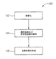
図2は、本発明に係るいくつかの実施例に関する治療計画を作成する処理を示す。まず、標的組織6及び感受性組織の画像が、撮像装置を用いて取得される(ステップ102)。図3Aは、標的組織6の画像及び感受性組織60の画像を含む画像フレーム202の一例を示す。あるケースでは、トランスデューサ装置10が画像202が取得される間に患者7の近傍で操作可能な位置に設置されている場合、画像202は、さらに、トランスデューサ素子12の画像を含めることができる。ここでは2次元画像が図示されているが、別の実施例では、画像が3次元画像であってもよい。3次元画像は、複数のボクセル又は複数の2次元画像で形成可能である。注目すべきは、ここで用いられている「画像」という用語は、表示される画像に制限されず、例えば、データ形式の画像といった表示されない画像を含めることができることである。
FIG. 2 illustrates a process for creating a treatment plan for some embodiments according to the present invention. First, images of the
次に、標的組織6及び感受性組織60が、画像フレーム202の中で特定される(ステップ104)。図示の実施例では、画像がデジタル化されてコンピュータ画面に映し出される。このようなケースでは、医師はユーザインターフェイスを使用して、画面上の標的組織6及び感受性組織60を識別することができる。例えば、医師は、ポインタ214を位置決めするマウスを使用して、標的組織6の近くのフレーム(標的フレーム)210及び感受性組織60の近くのフレーム(安全フレーム)212を描くことができる(図3B)。長方形のフレームが例として描かれているが、他の実施例では、標的フレーム210及び安全フレーム212が、別の及び異なる形状又は輪郭を各々有することができる。あるケースでは、標的フレーム210及び安全フレーム212の各々が、標的組織6及び感受性組織60を各々取り囲む3次元表面である。別の実施例では、標的組織6及び感受性組織60の画像の近くにフレームを作成する代わりに、医師が標的組織領域内の点(ターゲット点)及び感受性組織領域内の点(感受性点)を識別可能である。
Next, the
次に、例えば、大きさ、形状、位置といった特定の標的組織6及び特定の感受性組織60に関するデータが、プランナに引き継がれて、演算を実行し、治療計画を作成する(ステップ106)。治療計画は、例えば、位相、振幅等といった各トランスデューサ素子12についての動作パラメータを有するのが好ましく、標的組織6で生成されるエネルギ強度が、標的組織6を的確に治療するのに十分高く(すなわち、熱が)、一方、感受性組織60で生成されるエネルギ強度が、熱傷から感受性組織60を保護できる位に十分低くなるようにする。
Next, for example, the data regarding the
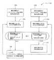
様々な方法を治療計画を作成するために適用可能である。図4は、本発明に係るある実施例に関する、治療計画を作成するための音波のシミュレーション方法を示している。まず、標的組織6及び各トランスデューサ素子12間の相対位置、感受性組織60及び各トランスデューサ素子12間の相対位置が、決定される(ステップ304a及び304b)。標的組織6及び各トランスデューサ素子12間の相対位置を決定するために、標的フレーム210内の点の位置又は複数のターゲット点の位置が、標的組織6の位置を表すものとして使用可能である。同様に、安全フレーム212内の点の位置又は複数の安全点の位置が、感受性組織60の位置を表すものとして使用可能である。ある実施例では、画像フレーム202がトランスデューサ素子12の画像をも有する場合、画像フレーム202は、標的組織6及び各トランスデューサ素子12間の相対位置及び感受性組織60及び各トランスデューサ素子12間の相対位置を決定するのに使用可能である。
Various methods can be applied to create a treatment plan. FIG. 4 shows a sound wave simulation method for creating a treatment plan according to an embodiment of the present invention. First, the relative position between the
次に、標的組織6及び各トランスデューサ素子12間の決定された相対位置に基づいて、及び、各トランスデューサ素子12の動作パラメータの仮定に基づいて、標的組織6におけるエネルギ強度が決定される(ステップ306a)。同様に、感受性組織60及び各トランスデューサ素子12間の決定された相対位置に基づいて、及び、各トランスデューサ素子12の動作パラメータの仮定に基づいて、感受性組織60におけるエネルギ強度が決定される(ステップ306b)。標的組織6における予測温度又はエネルギ強度が、所定の治療閾値、例えば、標的組織6の治療に要される最小温度を下回る場合、1以上のトランスデューサ素子12の位相、振幅又は、双方が調整され、標的組織6の温度又はエネルギ強度を演算するステップが、標的組織6の温度が所定の治療閾値を超えるまで繰り返される(ステップ308)。
Next, the energy intensity at the
感受性組織60におけるエネルギ強度が、所定の安全閾値、例えば、それ以上になると感受性組織60が傷付けられる最大エネルギレベルを超える場合、1以上のトランスデューサ素子12の位相、振幅又は、双方が調整され、感受性組織60のエネルギ強度を演算するステップが、感受性組織60のエネルギ強度が所定の安全閾値を下回るまで繰りされる(ステップ308)。あるケースでは、1以上のトランスデューサ素子12からのエネルギ寄与を、計算上ゼロと仮定することができるが、これは対応するトランスデューサ素子12が治療計画の実行中コントローラ18により作動しないシナリオを表している。別の実施例では、標的組織6及び感受性組織60を所望の温度又はエネルギ強度に上げるのに加え、動作パラメータを調整することにより、ビーム口径の大きさ、形状及び/又は位置を変えて、ある大きさ及び形状を有する組織を保護することも可能である。さらに別の実施例では、上記の演算結果が、灌流、拡散、比熱、熱伝導率、吸収作用といった生理的パラメータを用いて自動的に最適化される。
If the energy intensity in the
トランスデューサ素子12の動作パラメータを調整するステップ308は、標的組織6の温度又はエネルギ強度が所定の閾値を超え、感受性組織60の温度又はエネルギ強度が所定の閾値を下回るまで繰り返される。あるケースでは、光線モデルを使用して医師を補助して、トランスデューサ素子12の動作基準をどのように調整するかを決定可能である。
The
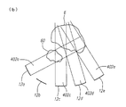
図5Aは、光線モデル400の一例を示す。この光線モデル400は、各トランスデューサ素子12a−eの表面から延びる光線402a−eを有していて、各光線402は対応する波長及び配置と関連する角放出分散を有している。感受性組織60に交わる光線402のメインローブを有するいずれかのトランスデューサ素子12に対して、このトランスデューサ素子12の動作パラメータを調整して、対応するエネルギ強度を減少又は除去可能である。この例で示すように、光線402a−cが感受性組織60に交わっている。感受性組織60のエネルギ強度を減少させるために、光線402bが除去される。すなわち、トランスデューサ素子12bが治療処置中に作動しない(図5B)。ここでは光線モデル400が、図形的に図示されているが、別の実施例では、光線モデル400が、図形的に描かれない数学的モデルであってもよい。注目すべきは、他の方法もまた、トランスデューサ素子12の動作基準の調整方法を決定するのに使用可能であることである。例えば、光線モデルを使用する代わりに、代替的な実施例では、他のモデル又は試行錯誤法を使用することができる。
FIG. 5A shows an example of the
トランスデューサ素子12の動作パラメータが決定された後、患者7の治療に使用するメモリに治療計画の一部として動作パラメータを保存可能である(ステップ310)。メモリは、コントローラ18に電子的に接続したり、コントローラ18の内部に設けたり、又はコントローラ18とは別に設けることが可能である。動作パラメータを用いて患者7を治療する典型的な方法が、以下に記載されている。
After the operational parameters of the
他の実施例では、音波シミュレーションを実行して治療計画を作成する代わりに、集束超音波システム5自身を使用して、治療計画を作成可能である。このケースでは、トランスデューサ素子12を、標的組織6に向けて低強度で(又は致死量以下で)(例えば、組織を傷付けないエネルギレベルで)音波エネルギを放出するよう構成することができる。次に、各標的組織6及び各感受性組織60の温度、エネルギ強度又は他の組織パラメータE1p及びE2pが測定される。ある実施例では、このステップが、標的組織6及び感受性組織60の温度感受性画像(温度マップ)の取得を具えている。温度感受性画像は、音波エネルギの使用による実際の加熱量分布を示している。
In another embodiment, instead of performing a sound wave simulation to create a treatment plan, the focused ultrasound system 5 itself can be used to create a treatment plan. In this case, the
図示された実施例では、標的組織6の所望の治療エネルギ強度E1t及びE1pの比RTが計算され(すなわち、RT=E1t/E1p)、比RTとE2pとを乗算して感受性組織60の所定の治療エネルギ強度E2tを得る(E2t=RT×E2p)。このケースでは、E2tが所定の閾値を超える場合、トランスデューサ素子の動作パラメータが、E2tがこの所定の閾値を下回るまで調整される。別の実施例では、比RTを計算する代わりに、測定されたエネルギ強度E1p及びE2pが、(プロセッサを用いて)測定された温度に基づいて、又は所定の治療閾値及び安全閾値の各々と比較される。E1pが調整された治療閾値を下回る場合、又はE2pが調整された安全閾値を超える場合、標的組織6の所望の温度又はエネルギ強度及び感受性組織60の所望のエネルギ強度を得るまで、トランスデューサ素子12の動作パラメータが調整される。そして、最終的に決定された各トランスデューサ素子12の動作パラメータはメモリに保存され、患者7を治療する治療セッションにおいて使用される。
In the illustrated embodiment, the ratio RT of the desired treatment energy intensity E1t and E1p of the
注目すべきは、治療計画を作成する方法が上記の実施例に限られず、他の方法を本発明に係る代替的な実施例として使用可能であることである。限定されない実施例として、位相及び振幅パラメータの代わりに又はこれらに加えて、治療計画が、各治療部位における周波数、継続期間、超音波出力、焦点の位置及びモードといった他の動作パラメータを含めることができる。上記の方法による実施例は、上記及び/又は他の動作パラメータのうちのいずれか1つ又はこれらの組み合わせを調整するステップを含めることができる。トランスデューサ素子12の1以上が構造体20に対して移動するよう構成されている場合、治療計画は、さらに、1以上のトランスデューサ素子12の位置を調整するステップを含めることができる。一旦治療計画が決定されると、それはコントローラに適切な形式で出力され、コントローラ18は患者7の治療を着手可能になる。コントローラ18自身がプランナの機能を実行するよう構成されている場合、コントローラに治療計画を出力するステップは明らかに不要である。
It should be noted that the method of creating a treatment plan is not limited to the above-described embodiments, and other methods can be used as alternative embodiments according to the present invention. As a non-limiting example, instead of or in addition to phase and amplitude parameters, the treatment plan may include other operating parameters such as frequency, duration, ultrasound output, focus position and mode at each treatment site. it can. Embodiments of the above method can include adjusting any one or a combination of the above and / or other operating parameters. If one or more of the
システム5を使用して患者7を治療するとき、トランスデューサ装置10は、まず患者7に対して置かれる。特に、トランスデューサ装置10及び患者7間の相対的な位置は、治療計画セッションに関するものと実質的に同一であることを要する。あるケースでは、治療計画セッションの後、即座に治療セッションが続く場合、トランスデューサ装置10及び患者7間の相対的な位置はそのままの状態であるから、トランスデューサ装置10の位置決めは不要である。
When treating the patient 7 using the system 5, the
一旦治療計画が決定され、トランスデューサ装置10が患者7に対して適切に設置されると、トランスデューサ装置10は、治療計画に従って標的組織7に集束超音波エネルギを放出する。特に、装置16及び/又はコントローラ18は、治療計画で定められた動作パラメータに基づいてトランスデューサ装置10から放出される音響エネルギを生成及び/又は制御するのに使用される。例えば、装置16及び/又はコントローラ18は、1以上のトランスデューサ素子12の位相及び/又は振幅を制御可能であるため、各トランスデューサ素子12の位相及び振幅は、治療計画で決定されたものと一致する。結果として、標的組織6のエネルギ強度は、標的組織6を凝固させ、壊死させ、加熱し、又は他の治療を行う位に十分高い。治療計画は、感受性組織60で低エネルギ領域又はエネルギ非通過ゾーンを生成するよう作成されているため、治療セッション時に、感受性組織60のエネルギ強度は、例えば「エネルギ不可」として規定可能である所定の閾値を下回り、これにより、音響エネルギによる損傷から感受性組織60を保護する。
Once the treatment plan is determined and the
ある実施例では、トランスデューサ素子12が構造体20に対して移動するよう構成されている場合、駆動装置16及び/又はコントローラ18が、所定の治療計画に従ってトランスデューサ素子12の位置を制御するよう使用可能で、これにより焦点域38の位置、形状、及び/又は大きさを調整する。所望の治療効果が達成された後に、患者7がトランスデューサ装置10から外され、又は逆にトランスデューサ装置10が患者7から外される。
In certain embodiments, when the
限定されない実施例により、上記の装置は、肝臓を治療するのに使用可能である。肋骨を介して肝臓を治療するときに、複数のトランスデューサ素子を有するトランスデューサを、できる限り肋骨の近く(例えば、約1cm又はそれ以下)に設置することができる。このようなケースでは、駆動装置16及び/又はコントローラ18を、肋骨に向いたトランスデューサ素子を停止させるようにする一方、肋骨の間に向いたトランスデューサ素子を作動させるよう構成することができる。
By way of non-limiting example, the device described above can be used to treat the liver. When treating the liver via the ribs, a transducer having multiple transducer elements can be placed as close as possible to the ribs (eg, about 1 cm or less). In such a case, the
Claims (11)
前記トランスデューサ素子に接続された駆動回路と、
前記駆動回路に接続された駆動信号制御装置と、
を具える集束超音波システムであって、
前記駆動信号制御装置が、前記トランスデューサ素子から超音波エネルギ通路を通って標的組織に向けて放出される音響エネルギを制御するよう構成され、
前記標的組織におけるエネルギ強度が、所定の治療レベル又はそれ以上で、一方、前記標的組織の外の前記超音波エネルギ放出通路内の保護されるべき組織におけるエネルギ強度が、所定の安全レベル又はそれ以下であり、
前記駆動信号制御装置が、1以上の前記トランスデューサ素子を制御して、前記超音波エネルギ通路内の前記保護されるべき組織に制限されたエネルギ密度を生成し、
前記駆動信号制御装置が、1以上の前記トランスデューサ素子に、1以上の他の前記トランスデューサ素子により放出される音響エネルギレベルよりも低いエネルギレベルで音響エネルギを放出させて、超音波エネルギを放出中に前記超音波エネルギ通路内の前記保護されるべき組織を保護することを特徴とする集束超音波システム。An ultrasonic transducer device having a plurality of transducer elements;
A drive circuit connected to the transducer element;
A drive signal control device connected to the drive circuit;
A focused ultrasound system comprising:
The drive signal controller is configured to control acoustic energy emitted from the transducer element through an ultrasonic energy path toward a target tissue;
The energy intensity at the target tissue is at a predetermined therapeutic level or higher, while the energy intensity at the tissue to be protected in the ultrasonic energy emission passage outside the target tissue is at a predetermined safety level or lower. And
The drive signal controller controls one or more of the transducer elements to generate an energy density limited to the tissue to be protected in the ultrasonic energy path ;
During the emission of ultrasonic energy, the drive signal controller causes one or more of the transducer elements to emit acoustic energy at an energy level lower than the acoustic energy level emitted by the one or more other transducer elements. A focused ultrasound system for protecting the tissue to be protected in the ultrasound energy path .
Applications Claiming Priority (3)
| Application Number | Priority Date | Filing Date | Title |
|---|---|---|---|
| US10/916,998 US7699780B2 (en) | 2004-08-11 | 2004-08-11 | Focused ultrasound system with adaptive anatomical aperture shaping |
| US10/916,998 | 2004-08-11 | ||
| PCT/IB2005/002273 WO2006018686A1 (en) | 2004-08-11 | 2005-08-01 | Focused ultrasound system with adaptive anatomical aperture shaping |
Publications (3)
| Publication Number | Publication Date |
|---|---|
| JP2008509713A JP2008509713A (en) | 2008-04-03 |
| JP2008509713A5 JP2008509713A5 (en) | 2008-06-26 |
| JP4820820B2 true JP4820820B2 (en) | 2011-11-24 |
Family
ID=35445681
Family Applications (1)
| Application Number | Title | Priority Date | Filing Date |
|---|---|---|---|
| JP2007525370A Active JP4820820B2 (en) | 2004-08-11 | 2005-08-01 | Focused ultrasound system |
Country Status (4)
| Country | Link |
|---|---|
| US (1) | US7699780B2 (en) |
| EP (1) | EP1786518A1 (en) |
| JP (1) | JP4820820B2 (en) |
| WO (1) | WO2006018686A1 (en) |
Families Citing this family (157)
| Publication number | Priority date | Publication date | Assignee | Title |
|---|---|---|---|---|
| US6050943A (en) | 1997-10-14 | 2000-04-18 | Guided Therapy Systems, Inc. | Imaging, therapy, and temperature monitoring ultrasonic system |
| US8256430B2 (en) | 2001-06-15 | 2012-09-04 | Monteris Medical, Inc. | Hyperthermia treatment and probe therefor |
| US6618620B1 (en) | 2000-11-28 | 2003-09-09 | Txsonics Ltd. | Apparatus for controlling thermal dosing in an thermal treatment system |
| US7914453B2 (en) | 2000-12-28 | 2011-03-29 | Ardent Sound, Inc. | Visual imaging system for ultrasonic probe |
| US8088067B2 (en) * | 2002-12-23 | 2012-01-03 | Insightec Ltd. | Tissue aberration corrections in ultrasound therapy |
| US7611462B2 (en) * | 2003-05-22 | 2009-11-03 | Insightec-Image Guided Treatment Ltd. | Acoustic beam forming in phased arrays including large numbers of transducer elements |
| US7377900B2 (en) * | 2003-06-02 | 2008-05-27 | Insightec - Image Guided Treatment Ltd. | Endo-cavity focused ultrasound transducer |
| US7645244B2 (en) * | 2004-07-09 | 2010-01-12 | Boston Scientific Scimed, Inc. | Ultrasound systems and methods for treating ischemic limbs or tissue affected by peripheral arterial disease |
| US8409099B2 (en) * | 2004-08-26 | 2013-04-02 | Insightec Ltd. | Focused ultrasound system for surrounding a body tissue mass and treatment method |
| US7393325B2 (en) | 2004-09-16 | 2008-07-01 | Guided Therapy Systems, L.L.C. | Method and system for ultrasound treatment with a multi-directional transducer |
| US7824348B2 (en) | 2004-09-16 | 2010-11-02 | Guided Therapy Systems, L.L.C. | System and method for variable depth ultrasound treatment |
| US8444562B2 (en) | 2004-10-06 | 2013-05-21 | Guided Therapy Systems, Llc | System and method for treating muscle, tendon, ligament and cartilage tissue |
| US10864385B2 (en) | 2004-09-24 | 2020-12-15 | Guided Therapy Systems, Llc | Rejuvenating skin by heating tissue for cosmetic treatment of the face and body |
| US8535228B2 (en) | 2004-10-06 | 2013-09-17 | Guided Therapy Systems, Llc | Method and system for noninvasive face lifts and deep tissue tightening |
| US20120165668A1 (en) | 2010-08-02 | 2012-06-28 | Guided Therapy Systems, Llc | Systems and methods for treating acute and/or chronic injuries in soft tissue |
| US9827449B2 (en) | 2004-10-06 | 2017-11-28 | Guided Therapy Systems, L.L.C. | Systems for treating skin laxity |
| US9694212B2 (en) | 2004-10-06 | 2017-07-04 | Guided Therapy Systems, Llc | Method and system for ultrasound treatment of skin |
| US8133180B2 (en) | 2004-10-06 | 2012-03-13 | Guided Therapy Systems, L.L.C. | Method and system for treating cellulite |
| KR101328103B1 (en) * | 2004-10-06 | 2013-11-13 | 가이디드 테라피 시스템스, 엘.엘.씨. | Method and system for noninvasive cosmetic enhancement |
| US8663112B2 (en) | 2004-10-06 | 2014-03-04 | Guided Therapy Systems, Llc | Methods and systems for fat reduction and/or cellulite treatment |
| US8690779B2 (en) | 2004-10-06 | 2014-04-08 | Guided Therapy Systems, Llc | Noninvasive aesthetic treatment for tightening tissue |
| US7758524B2 (en) | 2004-10-06 | 2010-07-20 | Guided Therapy Systems, L.L.C. | Method and system for ultra-high frequency ultrasound treatment |
| US11883688B2 (en) | 2004-10-06 | 2024-01-30 | Guided Therapy Systems, Llc | Energy based fat reduction |
| US20060111744A1 (en) | 2004-10-13 | 2006-05-25 | Guided Therapy Systems, L.L.C. | Method and system for treatment of sweat glands |
| KR20130080477A (en) | 2004-10-06 | 2013-07-12 | 가이디드 테라피 시스템스, 엘.엘.씨. | System of ultrasound treatment |
| US11235179B2 (en) | 2004-10-06 | 2022-02-01 | Guided Therapy Systems, Llc | Energy based skin gland treatment |
| US11207548B2 (en) | 2004-10-07 | 2021-12-28 | Guided Therapy Systems, L.L.C. | Ultrasound probe for treating skin laxity |
| US11724133B2 (en) | 2004-10-07 | 2023-08-15 | Guided Therapy Systems, Llc | Ultrasound probe for treatment of skin |
| WO2006044997A2 (en) * | 2004-10-15 | 2006-04-27 | The Trustees Of Columbia University In The City Of New York | System and method for localized measurement and imaging of viscosity of tissues |
| WO2006044996A2 (en) * | 2004-10-15 | 2006-04-27 | The Trustees Of Columbia University In The City Of New York | System and method for automated boundary detection of body structures |
| US7553284B2 (en) * | 2005-02-02 | 2009-06-30 | Vaitekunas Jeffrey J | Focused ultrasound for pain reduction |
| US10687785B2 (en) | 2005-05-12 | 2020-06-23 | The Trustees Of Columbia Univeristy In The City Of New York | System and method for electromechanical activation of arrhythmias |
| EP1883375B1 (en) | 2005-05-24 | 2016-12-07 | Edwards Lifesciences Corporation | Rapid deployment prosthetic heart valve |
| US20070016039A1 (en) * | 2005-06-21 | 2007-01-18 | Insightec-Image Guided Treatment Ltd. | Controlled, non-linear focused ultrasound treatment |
| US8926959B2 (en) | 2005-07-22 | 2015-01-06 | The Board Of Trustees Of The Leland Stanford Junior University | System for optical stimulation of target cells |
| US9274099B2 (en) | 2005-07-22 | 2016-03-01 | The Board Of Trustees Of The Leland Stanford Junior University | Screening test drugs to identify their effects on cell membrane voltage-gated ion channel |
| US9238150B2 (en) | 2005-07-22 | 2016-01-19 | The Board Of Trustees Of The Leland Stanford Junior University | Optical tissue interface method and apparatus for stimulating cells |
| US10052497B2 (en) * | 2005-07-22 | 2018-08-21 | The Board Of Trustees Of The Leland Stanford Junior University | System for optical stimulation of target cells |
| WO2007024391A2 (en) | 2005-07-22 | 2007-03-01 | The Board Of Trustees Of The Leland Stanford Junior University | Light-activated cation channel and uses thereof |
| US20090093403A1 (en) | 2007-03-01 | 2009-04-09 | Feng Zhang | Systems, methods and compositions for optical stimulation of target cells |
| EP1937151A4 (en) * | 2005-09-19 | 2011-07-06 | Univ Columbia | Systems and methods for opening of the blood-brain barrier of a subject using ultrasound |
| JP5087007B2 (en) * | 2005-11-23 | 2012-11-28 | インサイテック・リミテッド | Hierarchical switching ultra high density ultrasonic array |
| US8235901B2 (en) * | 2006-04-26 | 2012-08-07 | Insightec, Ltd. | Focused ultrasound system with far field tail suppression |
| FR2903316B1 (en) * | 2006-07-05 | 2009-06-26 | Edap S A | THERAPY PROBE AND THERAPY APPARATUS INCLUDING SUCH A PROBE |
| FR2903315B1 (en) * | 2006-07-05 | 2016-03-11 | Edap S A | METHOD AND APPARATUS FOR SEQUENTIALLY ACTIVE ULTRASOUND EMITTER THERAPY |
| US20100030076A1 (en) * | 2006-08-01 | 2010-02-04 | Kobi Vortman | Systems and Methods for Simultaneously Treating Multiple Target Sites |
| US8150128B2 (en) * | 2006-08-30 | 2012-04-03 | The Trustees Of Columbia University In The City Of New York | Systems and method for composite elastography and wave imaging |
| WO2008086470A1 (en) | 2007-01-10 | 2008-07-17 | The Board Of Trustees Of The Leland Stanford Junior University | System for optical stimulation of target cells |
| US8401609B2 (en) | 2007-02-14 | 2013-03-19 | The Board Of Trustees Of The Leland Stanford Junior University | System, method and applications involving identification of biological circuits such as neurological characteristics |
| ES2699477T3 (en) | 2007-05-07 | 2019-02-11 | Guided Therapy Systems Llc | Methods and systems for coupling and focusing acoustic energy using a coupling member |
| TWI526233B (en) | 2007-05-07 | 2016-03-21 | 指導治療系統股份有限公司 | Methods and systems for modulating medicants using acoustic energy |
| US20150174388A1 (en) | 2007-05-07 | 2015-06-25 | Guided Therapy Systems, Llc | Methods and Systems for Ultrasound Assisted Delivery of a Medicant to Tissue |
| WO2009001297A1 (en) * | 2007-06-28 | 2008-12-31 | Koninklijke Philips Electronics, N.V. | Method and apparatus for steering ultrasound therapy beam |
| US20090088625A1 (en) * | 2007-10-01 | 2009-04-02 | Kenneth Oosting | Photonic Based Non-Invasive Surgery System That Includes Automated Cell Control and Eradication Via Pre-Calculated Feed-Forward Control Plus Image Feedback Control For Targeted Energy Delivery |
| US8251908B2 (en) | 2007-10-01 | 2012-08-28 | Insightec Ltd. | Motion compensated image-guided focused ultrasound therapy system |
| US8088072B2 (en) | 2007-10-12 | 2012-01-03 | Gynesonics, Inc. | Methods and systems for controlled deployment of needles in tissue |
| US10035027B2 (en) * | 2007-10-31 | 2018-07-31 | The Board Of Trustees Of The Leland Stanford Junior University | Device and method for ultrasonic neuromodulation via stereotactic frame based technique |
| US10434327B2 (en) | 2007-10-31 | 2019-10-08 | The Board Of Trustees Of The Leland Stanford Junior University | Implantable optical stimulators |
| US20090149782A1 (en) * | 2007-12-11 | 2009-06-11 | Donald Cohen | Non-Invasive Neural Stimulation |
| EP2092916A1 (en) | 2008-02-19 | 2009-08-26 | Institut National De La Sante Et De La Recherche Medicale (Inserm) | A method of treating an ocular pathology by applying high intensity focused ultrasound and device thereof |
| US9517359B2 (en) | 2009-02-18 | 2016-12-13 | Eye Tech Care | High intensity focused ultrasound device with a concave segment shaped transducer for eye treatment |
| WO2011035312A1 (en) | 2009-09-21 | 2011-03-24 | The Trustees Of Culumbia University In The City Of New York | Systems and methods for opening of a tissue barrier |
| KR20110005280A (en) | 2008-04-23 | 2011-01-17 | 더 보드 어브 트러스티스 어브 더 리랜드 스탠포드 주니어 유니버시티 | Systems, methods and compositions for optical stimulation of target cells |
| EP2294208B1 (en) | 2008-05-29 | 2013-05-08 | The Board of Trustees of The Leland Stanford Junior University | Cell line, system and method for optical control of secondary messengers |
| CA3206234A1 (en) | 2008-06-06 | 2009-12-10 | Ulthera, Inc. | A system and method for cosmetic treatment and imaging |
| JP5887136B2 (en) * | 2008-06-17 | 2016-03-16 | ザ ボード オブ トラスティーズ オブ ザ レランド スタンフォード ジュニア ユニバーシティー | Apparatus and method for controlling cell development |
| MY156389A (en) | 2008-06-17 | 2016-02-15 | Univ Leland Stanford Junior | Methods, systems and devices for optical stimulation of target cells using an optical transmission element |
| WO2010006049A1 (en) * | 2008-07-08 | 2010-01-14 | The Board Of Trustees Of The Leland Stanford Junior University | Materials and approaches for optical stimulation of the peripheral nervous system |
| WO2010014977A1 (en) | 2008-08-01 | 2010-02-04 | The Trustees Of Columbia University In The City Of New York | Systems and methods for matching and imaging tissue characteristics |
| WO2010030819A1 (en) | 2008-09-10 | 2010-03-18 | The Trustees Of Columbia University In The City Of New York | Systems and methods for opening a tissue |
| US9050449B2 (en) * | 2008-10-03 | 2015-06-09 | Mirabilis Medica, Inc. | System for treating a volume of tissue with high intensity focused ultrasound |
| NZ602416A (en) | 2008-11-14 | 2014-08-29 | Univ Leland Stanford Junior | Optically-based stimulation of target cells and modifications thereto |
| US8425424B2 (en) * | 2008-11-19 | 2013-04-23 | Inightee Ltd. | Closed-loop clot lysis |
| US20100179425A1 (en) * | 2009-01-13 | 2010-07-15 | Eyal Zadicario | Systems and methods for controlling ultrasound energy transmitted through non-uniform tissue and cooling of same |
| US8617073B2 (en) * | 2009-04-17 | 2013-12-31 | Insightec Ltd. | Focusing ultrasound into the brain through the skull by utilizing both longitudinal and shear waves |
| JP5654580B2 (en) * | 2009-06-02 | 2015-01-14 | コーニンクレッカ フィリップス エヌ ヴェ | MR imaging guide treatment |
| EP2440292A1 (en) * | 2009-06-10 | 2012-04-18 | Insightec Ltd. | Acoustic-feedback power control during focused ultrasound delivery |
| US9623266B2 (en) * | 2009-08-04 | 2017-04-18 | Insightec Ltd. | Estimation of alignment parameters in magnetic-resonance-guided ultrasound focusing |
| US9289154B2 (en) * | 2009-08-19 | 2016-03-22 | Insightec Ltd. | Techniques for temperature measurement and corrections in long-term magnetic resonance thermometry |
| US20110046475A1 (en) * | 2009-08-24 | 2011-02-24 | Benny Assif | Techniques for correcting temperature measurement in magnetic resonance thermometry |
| WO2011024074A2 (en) | 2009-08-26 | 2011-03-03 | Insightec Ltd. | Asymmetric phased-array ultrasound transducer |
| EP2489034B1 (en) | 2009-10-14 | 2016-11-30 | Insightec Ltd. | Mapping ultrasound transducers |
| WO2011045695A1 (en) * | 2009-10-15 | 2011-04-21 | Koninklijke Philips Electronics, N.V. | Ultrasound power supply for an ultrasound transducer |
| US8368401B2 (en) | 2009-11-10 | 2013-02-05 | Insightec Ltd. | Techniques for correcting measurement artifacts in magnetic resonance thermometry |
| US8715186B2 (en) | 2009-11-24 | 2014-05-06 | Guided Therapy Systems, Llc | Methods and systems for generating thermal bubbles for improved ultrasound imaging and therapy |
| US20110125022A1 (en) * | 2009-11-25 | 2011-05-26 | Siemens Medical Solutions Usa, Inc. | Synchronization for multi-directional ultrasound scanning |
| WO2011079177A1 (en) * | 2009-12-22 | 2011-06-30 | The Trustees Of Columbia University In The City Of New York | A planning system for targeting tissue structures with ultrasound |
| CN102711914B (en) * | 2009-12-28 | 2016-10-19 | 皇家飞利浦电子股份有限公司 | The optimization of high-intensity focusing ultrasonic transducer |
| ES2654789T3 (en) | 2009-12-28 | 2018-02-15 | Profound Medical Inc | Therapeutic device |
| AU2011227131B2 (en) | 2010-03-17 | 2014-11-13 | The Board Of Trustees Of The Leland Stanford Junior University | Light-sensitive ion-passing molecules |
| US9852727B2 (en) * | 2010-04-28 | 2017-12-26 | Insightec, Ltd. | Multi-segment ultrasound transducers |
| US8932237B2 (en) | 2010-04-28 | 2015-01-13 | Insightec, Ltd. | Efficient ultrasound focusing |
| US20130079621A1 (en) * | 2010-05-05 | 2013-03-28 | Technion Research & Development Foundation Ltd. | Method and system of operating a multi focused acoustic wave source |
| US9504446B2 (en) | 2010-08-02 | 2016-11-29 | Guided Therapy Systems, Llc | Systems and methods for coupling an ultrasound source to tissue |
| US9981148B2 (en) | 2010-10-22 | 2018-05-29 | Insightec, Ltd. | Adaptive active cooling during focused ultrasound treatment |
| ES2716819T3 (en) | 2010-11-05 | 2019-06-17 | Univ Leland Stanford Junior | Chimeric opposites activated by light and methods of using them |
| EP2635109A4 (en) | 2010-11-05 | 2014-03-19 | Univ Leland Stanford Junior | Optically-controlled cns dysfunction |
| WO2012061684A1 (en) | 2010-11-05 | 2012-05-10 | The Board Of Trustees Of The Leland Stanford Junior University | Upconversion of light for use in optogenetic methods |
| AU2011323228B2 (en) | 2010-11-05 | 2016-11-10 | The Board Of Trustees Of The Leland Stanford Junior University | Control and characterization of memory function |
| CN103476456B (en) | 2010-11-05 | 2017-10-03 | 斯坦福大学托管董事会 | Award the light Genetic control of corelation behaviour |
| CA2816990A1 (en) | 2010-11-05 | 2012-05-10 | The Board Of Trustees Of The Leland Stanford Junior University | Stabilized step function opsin proteins and methods of using the same |
| US8857438B2 (en) | 2010-11-08 | 2014-10-14 | Ulthera, Inc. | Devices and methods for acoustic shielding |
| US8696722B2 (en) | 2010-11-22 | 2014-04-15 | The Board Of Trustees Of The Leland Stanford Junior University | Optogenetic magnetic resonance imaging |
| BR112013018044A2 (en) * | 2011-01-18 | 2019-09-03 | Koninl Philips Electronics Nv | therapeutic apparatus, computer program product and rendering method of an attainable target region |
| US20120191020A1 (en) * | 2011-01-25 | 2012-07-26 | Shuki Vitek | Uniform thermal treatment of tissue interfaces |
| KR20120090170A (en) * | 2011-02-07 | 2012-08-17 | 삼성전자주식회사 | Ultrasound measuring apparatus and controlling method thereof |
| US8968205B2 (en) * | 2011-02-10 | 2015-03-03 | Siemens Medical Solutions Usa, Inc. | Sub-aperture control in high intensity focused ultrasound |
| WO2012162664A1 (en) | 2011-05-26 | 2012-11-29 | The Trustees Of Columbia University In The City Of New York | Systems and methods for opening of a tissue barrier in primates |
| WO2013009784A2 (en) | 2011-07-10 | 2013-01-17 | Guided Therapy Systems, Llc | Systems and method for accelerating healing of implanted material and/or native tissue |
| WO2013012641A1 (en) | 2011-07-11 | 2013-01-24 | Guided Therapy Systems, Llc | Systems and methods for coupling an ultrasound source to tissue |
| KR20130009138A (en) * | 2011-07-14 | 2013-01-23 | 삼성전자주식회사 | Focused ultrasound therapy apparatus and focal point controlling method thereof |
| EP2768396A2 (en) | 2011-10-17 | 2014-08-27 | Butterfly Network Inc. | Transmissive imaging and related apparatus and methods |
| US9365628B2 (en) | 2011-12-16 | 2016-06-14 | The Board Of Trustees Of The Leland Stanford Junior University | Opsin polypeptides and methods of use thereof |
| EP2606837A1 (en) * | 2011-12-22 | 2013-06-26 | Koninklijke Philips Electronics N.V. | Calculating the ultrasonic intensity estimate using an incoherent sum of the ultrasonic pressure generated by multiple transducer elements |
| WO2013126521A1 (en) | 2012-02-21 | 2013-08-29 | The Board Of Trustees Of The Leland Stanford Junior University | Compositions and methods for treating neurogenic disorders of the pelvic floor |
| US11116405B2 (en) | 2012-04-12 | 2021-09-14 | Profound Medical Inc. | High-intensity focused ultrasound for heating a target zone larger than the electronic focusing zone |
| US9263663B2 (en) | 2012-04-13 | 2016-02-16 | Ardent Sound, Inc. | Method of making thick film transducer arrays |
| WO2013185784A1 (en) * | 2012-06-11 | 2013-12-19 | Helmholtz Zentrum München Deutsches Forschungszentrum Für Gesundheit Und Umwelt (Gmbh) | Imaging system and method for imaging an object |
| CN104602638B (en) | 2012-06-27 | 2017-12-19 | 曼特瑞斯医药有限责任公司 | System for influenceing to treat tissue |
| US9510802B2 (en) | 2012-09-21 | 2016-12-06 | Guided Therapy Systems, Llc | Reflective ultrasound technology for dermatological treatments |
| JP2015534488A (en) * | 2012-10-01 | 2015-12-03 | コーニンクレッカ フィリップス エヌ ヴェKoninklijke Philips N.V. | Reduction of heating in the region where high-intensity focused ultrasound overlaps |
| WO2014059170A1 (en) | 2012-10-10 | 2014-04-17 | The Trustees Of Columbia University In The City Of New York | Systems and methods for mechanical mapping of cardiac rhythm |
| CN105682739B (en) * | 2013-01-29 | 2018-11-13 | 因赛泰克有限公司 | Focused ultrasound therapy plan based on simulation |
| CN204017181U (en) | 2013-03-08 | 2014-12-17 | 奥赛拉公司 | Aesthstic imaging and processing system, multifocal processing system and perform the system of aesthetic procedure |
| EP2968997B1 (en) | 2013-03-15 | 2019-06-26 | The Board of Trustees of the Leland Stanford Junior University | Optogenetic control of behavioral state |
| US9636380B2 (en) | 2013-03-15 | 2017-05-02 | The Board Of Trustees Of The Leland Stanford Junior University | Optogenetic control of inputs to the ventral tegmental area |
| WO2014146022A2 (en) | 2013-03-15 | 2014-09-18 | Guided Therapy Systems Llc | Ultrasound treatment device and methods of use |
| US9667889B2 (en) | 2013-04-03 | 2017-05-30 | Butterfly Network, Inc. | Portable electronic devices with integrated imaging capabilities |
| EP2991491B1 (en) | 2013-04-29 | 2019-12-25 | The Board of Trustees of the Leland Stanford Junior University | Devices, systems and methods for optogenetic modulation of action potentials in target cells |
| US20160082293A1 (en) * | 2013-05-08 | 2016-03-24 | Koninklijke Philips N.V. | Hifu treatment optimization in vicinity of sensitive zones |
| US9247921B2 (en) | 2013-06-07 | 2016-02-02 | The Trustees Of Columbia University In The City Of New York | Systems and methods of high frame rate streaming for treatment monitoring |
| US10322178B2 (en) | 2013-08-09 | 2019-06-18 | The Trustees Of Columbia University In The City Of New York | Systems and methods for targeted drug delivery |
| CN105829538A (en) | 2013-08-14 | 2016-08-03 | 小利兰·斯坦福大学托管委员会 | Silicone-modified polyester coating |
| US10028723B2 (en) | 2013-09-03 | 2018-07-24 | The Trustees Of Columbia University In The City Of New York | Systems and methods for real-time, transcranial monitoring of blood-brain barrier opening |
| US20150141874A1 (en) * | 2013-11-18 | 2015-05-21 | Kitchener Clark Wilson | Multi-beam Ultrasound Device |
| US9486170B2 (en) | 2014-03-18 | 2016-11-08 | Monteris Medical Corporation | Image-guided therapy of a tissue |
| US10675113B2 (en) | 2014-03-18 | 2020-06-09 | Monteris Medical Corporation | Automated therapy of a three-dimensional tissue region |
| US20150265353A1 (en) | 2014-03-18 | 2015-09-24 | Monteris Medical Corporation | Image-guided therapy of a tissue |
| WO2015160708A1 (en) | 2014-04-18 | 2015-10-22 | Ulthera, Inc. | Band transducer ultrasound therapy |
| US10643371B2 (en) * | 2014-08-11 | 2020-05-05 | Covidien Lp | Treatment procedure planning system and method |
| FR3034320B1 (en) | 2015-03-31 | 2017-04-28 | Eye Tech Care | ULTRASOUND TREATMENT OCULAR PROBE |
| US10327830B2 (en) | 2015-04-01 | 2019-06-25 | Monteris Medical Corporation | Cryotherapy, thermal therapy, temperature modulation therapy, and probe apparatus therefor |
| WO2016209654A1 (en) | 2015-06-22 | 2016-12-29 | The Board Of Trustees Of The Leland Stanford Junior University | Methods and devices for imaging and/or optogenetic control of light-responsive neurons |
| PL3405294T3 (en) | 2016-01-18 | 2023-05-08 | Ulthera, Inc. | Compact ultrasound device having annular ultrasound array peripherally electrically connected to flexible printed circuit board |
| EP3245988B1 (en) | 2016-05-18 | 2023-12-27 | Sonikure Holdings Limited | System for ultrasound-enhanced transscleral delivery of drugs |
| CN109688934A (en) * | 2016-08-01 | 2019-04-26 | 戈尔丹斯医疗公司 | The opening of the blood-brain barrier of ultrasonic guidance |
| IL293809B2 (en) | 2016-08-16 | 2023-09-01 | Ulthera Inc | Systems and methods for cosmetic ultrasound treatment of skin |
| WO2018036912A1 (en) * | 2016-08-26 | 2018-03-01 | Koninklijke Philips N.V. | Detection of treatment failure for mild hyperthermia |
| EP3537982B1 (en) | 2016-11-11 | 2022-09-07 | Gynesonics, Inc. | Controlled treatment of tissue and dynamic interaction with tissue and/or treatment data and comparison of tissue and/or treatment data |
| US10716545B2 (en) | 2016-12-22 | 2020-07-21 | Fujifilm Sonosite, Inc. | Ultrasound system for imaging and protecting ophthalmic or other sensitive tissues |
| US11294165B2 (en) | 2017-03-30 | 2022-04-05 | The Board Of Trustees Of The Leland Stanford Junior University | Modular, electro-optical device for increasing the imaging field of view using time-sequential capture |
| WO2019164836A1 (en) | 2018-02-20 | 2019-08-29 | Ulthera, Inc. | Systems and methods for combined cosmetic treatment of cellulite with ultrasound |
| JP7302163B2 (en) * | 2018-11-22 | 2023-07-04 | コニカミノルタ株式会社 | ULTRASOUND DIAGNOSTIC APPARATUS AND ULTRASOUND IMAGE GENERATING METHOD |
| US11684807B2 (en) * | 2018-12-27 | 2023-06-27 | Insightec Ltd. | Optimization of transducer configurations in ultrasound procedures |
| US11730452B2 (en) * | 2019-04-09 | 2023-08-22 | Insightec Ltd. | Systems and methods for regulating microbubbles in ultrasound procedures |
| KR102320038B1 (en) * | 2019-12-06 | 2021-11-01 | 한국과학기술연구원 | Apparatus and method for precise mechanical tissue ablation using pressure modulated focused ultrasound |
| EP4076646A1 (en) * | 2019-12-18 | 2022-10-26 | Insightec Ltd. | Adaptive single-bubble-based autofocusing and power adjustment in ultrasound procedures |
Citations (1)
| Publication number | Priority date | Publication date | Assignee | Title |
|---|---|---|---|---|
| JPH07184907A (en) * | 1993-12-28 | 1995-07-25 | Toshiba Corp | Ultrasonic treating device |
Family Cites Families (21)
| Publication number | Priority date | Publication date | Assignee | Title |
|---|---|---|---|---|
| DE3119295A1 (en) * | 1981-05-14 | 1982-12-16 | Siemens AG, 1000 Berlin und 8000 München | DEVICE FOR DESTROYING CONCRETE IN BODIES |
| US4537074A (en) * | 1983-09-12 | 1985-08-27 | Technicare Corporation | Annular array ultrasonic transducers |
| US4865042A (en) * | 1985-08-16 | 1989-09-12 | Hitachi, Ltd. | Ultrasonic irradiation system |
| GB8529446D0 (en) * | 1985-11-29 | 1986-01-08 | Univ Aberdeen | Divergent ultrasound arrays |
| JP3325300B2 (en) * | 1992-02-28 | 2002-09-17 | 株式会社東芝 | Ultrasound therapy equipment |
| JP3860227B2 (en) * | 1993-03-10 | 2006-12-20 | 株式会社東芝 | Ultrasonic therapy device used under MRI guide |
| US5553618A (en) * | 1993-03-12 | 1996-09-10 | Kabushiki Kaisha Toshiba | Method and apparatus for ultrasound medical treatment |
| FR2715313B1 (en) * | 1994-01-27 | 1996-05-31 | Edap Int | Method for controlling a hyperthermia treatment device using ultrasound. |
| DE69634714T2 (en) * | 1995-03-31 | 2006-01-19 | Kabushiki Kaisha Toshiba, Kawasaki | Therapeutic ultrasound device |
| DE69840444D1 (en) * | 1997-05-23 | 2009-02-26 | Prorhythm Inc | DISMISSABLE FOCUSING ULTRASOUND APPLICATOR OF HIGH INTENSITY |
| WO2001017455A2 (en) * | 1999-09-10 | 2001-03-15 | Transurgical, Inc. | Occlusion of tubular anatomical structures by energy application |
| US6626855B1 (en) * | 1999-11-26 | 2003-09-30 | Therus Corpoation | Controlled high efficiency lesion formation using high intensity ultrasound |
| US6419648B1 (en) * | 2000-04-21 | 2002-07-16 | Insightec-Txsonics Ltd. | Systems and methods for reducing secondary hot spots in a phased array focused ultrasound system |
| US6613005B1 (en) * | 2000-11-28 | 2003-09-02 | Insightec-Txsonics, Ltd. | Systems and methods for steering a focused ultrasound array |
| US6618620B1 (en) * | 2000-11-28 | 2003-09-09 | Txsonics Ltd. | Apparatus for controlling thermal dosing in an thermal treatment system |
| US6645162B2 (en) * | 2000-12-27 | 2003-11-11 | Insightec - Txsonics Ltd. | Systems and methods for ultrasound assisted lipolysis |
| JP2002209905A (en) * | 2001-01-22 | 2002-07-30 | Hitachi Medical Corp | Ultrasonic therapy probe and ultrasonic therapy apparatus |
| US6790180B2 (en) * | 2001-12-03 | 2004-09-14 | Insightec-Txsonics Ltd. | Apparatus, systems, and methods for measuring power output of an ultrasound transducer |
| US6705993B2 (en) * | 2002-05-10 | 2004-03-16 | Regents Of The University Of Minnesota | Ultrasound imaging system and method using non-linear post-beamforming filter |
| US6629929B1 (en) * | 2002-11-08 | 2003-10-07 | Koninklijke Philips Electronics N.V. | Method and apparatus for automatically setting the transmit aperture and apodization of an ultrasound transducer array |
| IL154101A0 (en) * | 2003-01-23 | 2003-07-31 | Univ Ramot | Minimally invasive controlled surgical system with feedback |
-
2004
- 2004-08-11 US US10/916,998 patent/US7699780B2/en active Active
-
2005
- 2005-08-01 WO PCT/IB2005/002273 patent/WO2006018686A1/en active Application Filing
- 2005-08-01 EP EP05779849A patent/EP1786518A1/en not_active Withdrawn
- 2005-08-01 JP JP2007525370A patent/JP4820820B2/en active Active
Patent Citations (1)
| Publication number | Priority date | Publication date | Assignee | Title |
|---|---|---|---|---|
| JPH07184907A (en) * | 1993-12-28 | 1995-07-25 | Toshiba Corp | Ultrasonic treating device |
Also Published As
| Publication number | Publication date |
|---|---|
| US7699780B2 (en) | 2010-04-20 |
| EP1786518A1 (en) | 2007-05-23 |
| US20060058671A1 (en) | 2006-03-16 |
| JP2008509713A (en) | 2008-04-03 |
| WO2006018686A1 (en) | 2006-02-23 |
Similar Documents
| Publication | Publication Date | Title |
|---|---|---|
| JP4820820B2 (en) | Focused ultrasound system | |
| CN109475755B (en) | Ultrasonic autofocus using reflection | |
| US20070016039A1 (en) | Controlled, non-linear focused ultrasound treatment | |
| JP7119089B2 (en) | Focusing ultrasound in dynamically changing media | |
| EP1796545B1 (en) | Focused ultrasound system for surrounding a body tissue mass | |
| JP5805176B2 (en) | System for treating tissue volume using high intensity focused ultrasound (HIFU) | |
| JP4558504B2 (en) | Correction of tissue abnormalities in ultrasonic therapy | |
| JP5681727B2 (en) | Optimization of high-density focused ultrasonic transducer | |
| CN111093520B (en) | Local cavitation signal measurement | |
| JP7012726B2 (en) | Therapeutic ultrasound with reduced interference from microbubbles | |
| JPH0747079A (en) | Ultrasonic therapeutic system | |
| JPH05300910A (en) | Ultrasonic medical treatment system | |
| US11730452B2 (en) | Systems and methods for regulating microbubbles in ultrasound procedures | |
| CN115135381A (en) | Adaptive single bubble based autofocus and power adjustment in ultrasound procedures | |
| CN115135248A (en) | System and method for providing anatomical target region tissue information using an acoustic reflector | |
| Cao et al. | Patient‐specific 3‐dimensional model for high‐intensity focused ultrasound treatment through the rib cage: a preliminary study | |
| JP3369504B2 (en) | Ultrasound therapy equipment | |
| JP4394440B2 (en) | Ultrasound treatment | |
| Sohrab et al. | Ultrasound thermotherapy of breast: theoretical design of transducer and numerical simulation of procedure | |
| JPH08266553A (en) | Ultrasonic treating device | |
| KR20120061595A (en) | Method for controlling of treating ultrasound, and apparatus thereof |
Legal Events
| Date | Code | Title | Description |
|---|---|---|---|
| A521 | Request for written amendment filed |
Free format text: JAPANESE INTERMEDIATE CODE: A523 Effective date: 20080512 |
|
| A621 | Written request for application examination |
Free format text: JAPANESE INTERMEDIATE CODE: A621 Effective date: 20080512 |
|
| A977 | Report on retrieval |
Free format text: JAPANESE INTERMEDIATE CODE: A971007 Effective date: 20110125 |
|
| A131 | Notification of reasons for refusal |
Free format text: JAPANESE INTERMEDIATE CODE: A131 Effective date: 20110201 |
|
| A601 | Written request for extension of time |
Free format text: JAPANESE INTERMEDIATE CODE: A601 Effective date: 20110427 |
|
| A602 | Written permission of extension of time |
Free format text: JAPANESE INTERMEDIATE CODE: A602 Effective date: 20110510 |
|
| A521 | Request for written amendment filed |
Free format text: JAPANESE INTERMEDIATE CODE: A523 Effective date: 20110526 |
|
| TRDD | Decision of grant or rejection written | ||
| A01 | Written decision to grant a patent or to grant a registration (utility model) |
Free format text: JAPANESE INTERMEDIATE CODE: A01 Effective date: 20110830 |
|
| A01 | Written decision to grant a patent or to grant a registration (utility model) |
Free format text: JAPANESE INTERMEDIATE CODE: A01 |
|
| A61 | First payment of annual fees (during grant procedure) |
Free format text: JAPANESE INTERMEDIATE CODE: A61 Effective date: 20110905 |
|
| FPAY | Renewal fee payment (event date is renewal date of database) |
Free format text: PAYMENT UNTIL: 20140909 Year of fee payment: 3 |
|
| R150 | Certificate of patent or registration of utility model |
Ref document number: 4820820 Country of ref document: JP Free format text: JAPANESE INTERMEDIATE CODE: R150 Free format text: JAPANESE INTERMEDIATE CODE: R150 |
|
| R250 | Receipt of annual fees |
Free format text: JAPANESE INTERMEDIATE CODE: R250 |
|
| R250 | Receipt of annual fees |
Free format text: JAPANESE INTERMEDIATE CODE: R250 |
|
| R250 | Receipt of annual fees |
Free format text: JAPANESE INTERMEDIATE CODE: R250 |
|
| R250 | Receipt of annual fees |
Free format text: JAPANESE INTERMEDIATE CODE: R250 |
|
| R250 | Receipt of annual fees |
Free format text: JAPANESE INTERMEDIATE CODE: R250 |
|
| R250 | Receipt of annual fees |
Free format text: JAPANESE INTERMEDIATE CODE: R250 |
|
| R250 | Receipt of annual fees |
Free format text: JAPANESE INTERMEDIATE CODE: R250 |
|
| R250 | Receipt of annual fees |
Free format text: JAPANESE INTERMEDIATE CODE: R250 |
|
| R250 | Receipt of annual fees |
Free format text: JAPANESE INTERMEDIATE CODE: R250 |