JP4771425B2 - Stent - Google Patents
Stent Download PDFInfo
- Publication number
- JP4771425B2 JP4771425B2 JP2006502643A JP2006502643A JP4771425B2 JP 4771425 B2 JP4771425 B2 JP 4771425B2 JP 2006502643 A JP2006502643 A JP 2006502643A JP 2006502643 A JP2006502643 A JP 2006502643A JP 4771425 B2 JP4771425 B2 JP 4771425B2
- Authority
- JP
- Japan
- Prior art keywords
- stent
- lumen
- section
- cavity
- side pieces
- Prior art date
- Legal status (The legal status is an assumption and is not a legal conclusion. Google has not performed a legal analysis and makes no representation as to the accuracy of the status listed.)
- Expired - Fee Related
Links
Images


Classifications
-
- A—HUMAN NECESSITIES
- A61—MEDICAL OR VETERINARY SCIENCE; HYGIENE
- A61F—FILTERS IMPLANTABLE INTO BLOOD VESSELS; PROSTHESES; DEVICES PROVIDING PATENCY TO, OR PREVENTING COLLAPSING OF, TUBULAR STRUCTURES OF THE BODY, e.g. STENTS; ORTHOPAEDIC, NURSING OR CONTRACEPTIVE DEVICES; FOMENTATION; TREATMENT OR PROTECTION OF EYES OR EARS; BANDAGES, DRESSINGS OR ABSORBENT PADS; FIRST-AID KITS
- A61F2/00—Filters implantable into blood vessels; Prostheses, i.e. artificial substitutes or replacements for parts of the body; Appliances for connecting them with the body; Devices providing patency to, or preventing collapsing of, tubular structures of the body, e.g. stents
- A61F2/82—Devices providing patency to, or preventing collapsing of, tubular structures of the body, e.g. stents
- A61F2/86—Stents in a form characterised by the wire-like elements; Stents in the form characterised by a net-like or mesh-like structure
- A61F2/90—Stents in a form characterised by the wire-like elements; Stents in the form characterised by a net-like or mesh-like structure characterised by a net-like or mesh-like structure
-
- A—HUMAN NECESSITIES
- A61—MEDICAL OR VETERINARY SCIENCE; HYGIENE
- A61F—FILTERS IMPLANTABLE INTO BLOOD VESSELS; PROSTHESES; DEVICES PROVIDING PATENCY TO, OR PREVENTING COLLAPSING OF, TUBULAR STRUCTURES OF THE BODY, e.g. STENTS; ORTHOPAEDIC, NURSING OR CONTRACEPTIVE DEVICES; FOMENTATION; TREATMENT OR PROTECTION OF EYES OR EARS; BANDAGES, DRESSINGS OR ABSORBENT PADS; FIRST-AID KITS
- A61F2/00—Filters implantable into blood vessels; Prostheses, i.e. artificial substitutes or replacements for parts of the body; Appliances for connecting them with the body; Devices providing patency to, or preventing collapsing of, tubular structures of the body, e.g. stents
- A61F2/82—Devices providing patency to, or preventing collapsing of, tubular structures of the body, e.g. stents
- A61F2/844—Devices providing patency to, or preventing collapsing of, tubular structures of the body, e.g. stents folded prior to deployment
-
- A—HUMAN NECESSITIES
- A61—MEDICAL OR VETERINARY SCIENCE; HYGIENE
- A61F—FILTERS IMPLANTABLE INTO BLOOD VESSELS; PROSTHESES; DEVICES PROVIDING PATENCY TO, OR PREVENTING COLLAPSING OF, TUBULAR STRUCTURES OF THE BODY, e.g. STENTS; ORTHOPAEDIC, NURSING OR CONTRACEPTIVE DEVICES; FOMENTATION; TREATMENT OR PROTECTION OF EYES OR EARS; BANDAGES, DRESSINGS OR ABSORBENT PADS; FIRST-AID KITS
- A61F2/00—Filters implantable into blood vessels; Prostheses, i.e. artificial substitutes or replacements for parts of the body; Appliances for connecting them with the body; Devices providing patency to, or preventing collapsing of, tubular structures of the body, e.g. stents
- A61F2/82—Devices providing patency to, or preventing collapsing of, tubular structures of the body, e.g. stents
- A61F2/92—Stents in the form of a rolled-up sheet expanding after insertion into the vessel, e.g. with a spiral shape in cross-section
-
- A—HUMAN NECESSITIES
- A61—MEDICAL OR VETERINARY SCIENCE; HYGIENE
- A61F—FILTERS IMPLANTABLE INTO BLOOD VESSELS; PROSTHESES; DEVICES PROVIDING PATENCY TO, OR PREVENTING COLLAPSING OF, TUBULAR STRUCTURES OF THE BODY, e.g. STENTS; ORTHOPAEDIC, NURSING OR CONTRACEPTIVE DEVICES; FOMENTATION; TREATMENT OR PROTECTION OF EYES OR EARS; BANDAGES, DRESSINGS OR ABSORBENT PADS; FIRST-AID KITS
- A61F2/00—Filters implantable into blood vessels; Prostheses, i.e. artificial substitutes or replacements for parts of the body; Appliances for connecting them with the body; Devices providing patency to, or preventing collapsing of, tubular structures of the body, e.g. stents
- A61F2/02—Prostheses implantable into the body
- A61F2/04—Hollow or tubular parts of organs, e.g. bladders, tracheae, bronchi or bile ducts
- A61F2002/047—Urethrae
-
- A—HUMAN NECESSITIES
- A61—MEDICAL OR VETERINARY SCIENCE; HYGIENE
- A61F—FILTERS IMPLANTABLE INTO BLOOD VESSELS; PROSTHESES; DEVICES PROVIDING PATENCY TO, OR PREVENTING COLLAPSING OF, TUBULAR STRUCTURES OF THE BODY, e.g. STENTS; ORTHOPAEDIC, NURSING OR CONTRACEPTIVE DEVICES; FOMENTATION; TREATMENT OR PROTECTION OF EYES OR EARS; BANDAGES, DRESSINGS OR ABSORBENT PADS; FIRST-AID KITS
- A61F2/00—Filters implantable into blood vessels; Prostheses, i.e. artificial substitutes or replacements for parts of the body; Appliances for connecting them with the body; Devices providing patency to, or preventing collapsing of, tubular structures of the body, e.g. stents
- A61F2/02—Prostheses implantable into the body
- A61F2/30—Joints
- A61F2002/30001—Additional features of subject-matter classified in A61F2/28, A61F2/30 and subgroups thereof
- A61F2002/30108—Shapes
- A61F2002/30199—Three-dimensional shapes
-
- A—HUMAN NECESSITIES
- A61—MEDICAL OR VETERINARY SCIENCE; HYGIENE
- A61F—FILTERS IMPLANTABLE INTO BLOOD VESSELS; PROSTHESES; DEVICES PROVIDING PATENCY TO, OR PREVENTING COLLAPSING OF, TUBULAR STRUCTURES OF THE BODY, e.g. STENTS; ORTHOPAEDIC, NURSING OR CONTRACEPTIVE DEVICES; FOMENTATION; TREATMENT OR PROTECTION OF EYES OR EARS; BANDAGES, DRESSINGS OR ABSORBENT PADS; FIRST-AID KITS
- A61F2210/00—Particular material properties of prostheses classified in groups A61F2/00 - A61F2/26 or A61F2/82 or A61F9/00 or A61F11/00 or subgroups thereof
- A61F2210/0014—Particular material properties of prostheses classified in groups A61F2/00 - A61F2/26 or A61F2/82 or A61F9/00 or A61F11/00 or subgroups thereof using shape memory or superelastic materials, e.g. nitinol
-
- A—HUMAN NECESSITIES
- A61—MEDICAL OR VETERINARY SCIENCE; HYGIENE
- A61F—FILTERS IMPLANTABLE INTO BLOOD VESSELS; PROSTHESES; DEVICES PROVIDING PATENCY TO, OR PREVENTING COLLAPSING OF, TUBULAR STRUCTURES OF THE BODY, e.g. STENTS; ORTHOPAEDIC, NURSING OR CONTRACEPTIVE DEVICES; FOMENTATION; TREATMENT OR PROTECTION OF EYES OR EARS; BANDAGES, DRESSINGS OR ABSORBENT PADS; FIRST-AID KITS
- A61F2230/00—Geometry of prostheses classified in groups A61F2/00 - A61F2/26 or A61F2/82 or A61F9/00 or A61F11/00 or subgroups thereof
- A61F2230/0002—Two-dimensional shapes, e.g. cross-sections
- A61F2230/0004—Rounded shapes, e.g. with rounded corners
- A61F2230/001—Figure-8-shaped, e.g. hourglass-shaped
-
- A—HUMAN NECESSITIES
- A61—MEDICAL OR VETERINARY SCIENCE; HYGIENE
- A61F—FILTERS IMPLANTABLE INTO BLOOD VESSELS; PROSTHESES; DEVICES PROVIDING PATENCY TO, OR PREVENTING COLLAPSING OF, TUBULAR STRUCTURES OF THE BODY, e.g. STENTS; ORTHOPAEDIC, NURSING OR CONTRACEPTIVE DEVICES; FOMENTATION; TREATMENT OR PROTECTION OF EYES OR EARS; BANDAGES, DRESSINGS OR ABSORBENT PADS; FIRST-AID KITS
- A61F2230/00—Geometry of prostheses classified in groups A61F2/00 - A61F2/26 or A61F2/82 or A61F9/00 or A61F11/00 or subgroups thereof
- A61F2230/0002—Two-dimensional shapes, e.g. cross-sections
- A61F2230/0017—Angular shapes
- A61F2230/0023—Angular shapes triangular
-
- A—HUMAN NECESSITIES
- A61—MEDICAL OR VETERINARY SCIENCE; HYGIENE
- A61F—FILTERS IMPLANTABLE INTO BLOOD VESSELS; PROSTHESES; DEVICES PROVIDING PATENCY TO, OR PREVENTING COLLAPSING OF, TUBULAR STRUCTURES OF THE BODY, e.g. STENTS; ORTHOPAEDIC, NURSING OR CONTRACEPTIVE DEVICES; FOMENTATION; TREATMENT OR PROTECTION OF EYES OR EARS; BANDAGES, DRESSINGS OR ABSORBENT PADS; FIRST-AID KITS
- A61F2230/00—Geometry of prostheses classified in groups A61F2/00 - A61F2/26 or A61F2/82 or A61F9/00 or A61F11/00 or subgroups thereof
- A61F2230/0002—Two-dimensional shapes, e.g. cross-sections
- A61F2230/0028—Shapes in the form of latin or greek characters
- A61F2230/0054—V-shaped
-
- A—HUMAN NECESSITIES
- A61—MEDICAL OR VETERINARY SCIENCE; HYGIENE
- A61F—FILTERS IMPLANTABLE INTO BLOOD VESSELS; PROSTHESES; DEVICES PROVIDING PATENCY TO, OR PREVENTING COLLAPSING OF, TUBULAR STRUCTURES OF THE BODY, e.g. STENTS; ORTHOPAEDIC, NURSING OR CONTRACEPTIVE DEVICES; FOMENTATION; TREATMENT OR PROTECTION OF EYES OR EARS; BANDAGES, DRESSINGS OR ABSORBENT PADS; FIRST-AID KITS
- A61F2230/00—Geometry of prostheses classified in groups A61F2/00 - A61F2/26 or A61F2/82 or A61F9/00 or A61F11/00 or subgroups thereof
- A61F2230/0063—Three-dimensional shapes
Abstract
Description
本発明は、内腔用の医療装置、より詳細には、腔の開通性を維持するような装置に関する。 The present invention relates to a medical device for a lumen, and more particularly to such a device that maintains the patency of the lumen.
ステントは、体腔中に挿入され、腔の開通性を維持するように拡大される内腔用の装置である。例えば、動脈、尿道若しくは胃腸器官の開通性を維持するためにステントを使用することが知られている。永久的なステントは、体内に永遠に残されるように意図されており、一方、一時的なステントは、所定の期間、腔内に残される。 A stent is a device for a lumen that is inserted into a body cavity and expanded to maintain the patency of the cavity. For example, it is known to use stents to maintain patency of arteries, urethra, or gastrointestinal organs. Permanent stents are intended to remain forever in the body, while temporary stents are left in the cavity for a predetermined period of time.
ステントは、2つの形態で存在し得る細長い装置である。小径の形態では、ステントは、体内に挿入され、処理される腔へと移動される。ステントは、腔中に正確に位置付けられると、腔の内壁に対して径方向外方の力を加えるように大径にされることによって広げられる。ステントは、腔の壁によってステントに加えられる径方向内方の力に耐え得るように構成されており、この結果、ステントの内径は、腔中で広げられた後も維持される。 A stent is an elongated device that can exist in two forms. In the small diameter form, the stent is inserted into the body and moved into the cavity to be processed. Once correctly positioned in the cavity, the stent is expanded by being enlarged to apply a radially outward force against the inner wall of the cavity. The stent is configured to withstand the radially inward force applied to the stent by the cavity wall so that the inner diameter of the stent is maintained after being expanded in the cavity.
ステントは、例えば、大径の形態のときに、非緊張状態である弾性材で形成されることができる。従って、ステントは、小径の形態になるように機械的に抑制される。このステントは、制限スリーブ中に挿入されることによって小径の形態に維持されることができる。体内に位置された後に、制限スリーブは除去される。ステントが弾性性質であるため、ステントは、大径の形態に自発的に変形する。 The stent can be formed of, for example, an elastic material that is in a non-tensioned state when in a large-diameter form. Thus, the stent is mechanically constrained to have a small diameter configuration. The stent can be maintained in a small diameter configuration by being inserted into a limiting sleeve. After being placed in the body, the restriction sleeve is removed. Due to the elastic nature of the stent, it will spontaneously deform into a large diameter form.
緊張状態のとき、プラスチックからなる材料でステントを形成することも知られている。ステントは、非緊張状態では小径の形態に形成されている。バルーンが、ステントの腔中に挿入されている。そして、ステントが体内に位置され、バルーンが膨張する。これは、ステントの材料の塑性変形を引き起こすことによって、ステントを大径の形態に拡大させる。 It is also known to form stents from plastic materials when in tension. The stent is formed in a small-diameter form in a non-tensioned state. A balloon is inserted into the stent cavity. The stent is then positioned in the body and the balloon is inflated. This causes the stent to expand to a larger diameter configuration by causing plastic deformation of the stent material.
ニチノール(商標登録)のような形状記憶合金でステントを形成することがさらに知られている。形状記憶合金は、超弾性状態(オーステナイト状態)と、柔軟状態(マルテンサイト状態)との2つの状態で存在することができる。オーステナイト状態の合金は、大径の形態のステントを形成する。従って、合金は、冷却されるか緊張されることによって、マルテンサイト状態にされる。マルテンサイト状態では、合金は、処理される腔へと移動されるように小径の形態に変形される。位置された後に、合金は、加熱されることによってオーステナイトの形態にされる。オーステナイトの形態では、ステントは、合金の形状記憶性質のために大径の形態に回復する。 It is further known to form stents with shape memory alloys such as Nitinol ™. Shape memory alloys can exist in two states: a superelastic state (austenite state) and a soft state (martensite state). The austenitic alloy forms a large diameter stent. Thus, the alloy is made martensitic by being cooled or tensioned. In the martensitic state, the alloy is transformed into a small diameter form so that it can be moved into the cavity to be processed. After being positioned, the alloy is heated to austenite form. In the austenite form, the stent recovers to a larger diameter form due to the shape memory nature of the alloy.
生物安定性若しくは生物分解性の弾性の形状記憶ポリマーでステントを形成することがさらに知られている。このようなポリマーの形状記憶能力が、このような材料で形成されたステントを、小さな開口部へと挿入させ、温度の上昇によって開口部の内径を拡大させることを可能にしている。ポリマーの形状記憶効果は、アモルファスポリマーによって非常に良く示された物理的性質である。アモルファスポリマーのガラス転移温度は、室温よりもわずかに高く、ポリマーのガラスからゴムへの転移は特に急である。この場合、歪みエネルギーが、機械的変形(例えば、ストレッチング)、続いて冷却によって、ポリマーに記憶されることができる。形状記憶の回復は、伸ばされたポリマー鎖を均衡性のあるコイル状構造に戻し得るように、冷却された温度より高い温度に材料を再加熱することに示される。 It is further known to form stents with biostable or biodegradable elastic shape memory polymers. The shape memory capability of such a polymer allows a stent formed of such a material to be inserted into a small opening and to increase the inner diameter of the opening with increasing temperature. The shape memory effect of polymers is a physical property that is very well demonstrated by amorphous polymers. The glass transition temperature of amorphous polymers is slightly higher than room temperature, and the transition from polymer glass to rubber is particularly steep. In this case, strain energy can be stored in the polymer by mechanical deformation (eg stretching) followed by cooling. Shape memory recovery is indicated by reheating the material to a temperature above the cooled temperature so that the stretched polymer chains can be returned to a balanced coiled structure.
脈管系において、代表的なステントの直径は、過度の圧力を血管壁に生じさせること無く適切な固着を確実にするように、血管径の約1.2倍である。血管の腔並びにステントが円形の断面であるため、広げられた後、ステント全体は、全体を囲んでいる血管壁に接触する。ステントの壁が窓を有していると、ステントが血管壁一面に広がり、血管壁一面を押圧するときに、血管の内皮に対して圧力損傷が生じる。この損傷は、ステントが内皮でカバーされるまで鎖組織反応(chain tissue reaction)を引き起こす。 In the vascular system, a typical stent diameter is about 1.2 times the vessel diameter to ensure proper anchoring without creating excessive pressure on the vessel wall. Because the vessel lumen as well as the stent has a circular cross-section, after being expanded, the entire stent contacts the entire surrounding vessel wall. When the wall of the stent has a window, when the stent spreads over the blood vessel wall and presses against the blood vessel wall, pressure damage occurs to the endothelium of the blood vessel. This damage causes a chain tissue reaction until the stent is covered with endothelium.
良性前立腺過形成(BPH)は、人間の男性に影響を与える最も一般的な腫瘍である。この腫瘍は、前立腺部で成長すると、尿道前立腺部の腔を収縮させる。この収縮が一点に集中すると、尿道前立腺部が塞がれ、尿の排出が抑制される。 Benign prostatic hyperplasia (BPH) is the most common tumor affecting human men. When the tumor grows in the prostate, it contracts the urethral prostate cavity. When this contraction is concentrated at one point, the urethral prostate is blocked, and urine output is suppressed.
BPHを治す方法が幾つかある。最も一般的な治療には、尿道前立腺部への圧力を減じるか、前立腺のサイズを減じるような薬物治療、さらに極端な場合には、腫瘍を除去し、尿道前立腺部を広げるような観血的手術若しくは内視鏡手術が含まれている。外科的なBPH治療には、観血的前立腺摘除術のような観血的手術、径尿道前立腺摘除術(Transurethral Prostatectomy,TURP)、前立腺の径尿道切開(Transurethral Incision of the Prostate,TURP)、並びに前立腺の径尿道レーザー若しくは高周波蒸発(Transurethral Laser or Radiofrequency,FR)のような内視鏡手術が代表的に含まれている。拡大した前立腺組織に対する熱損傷を有することに基づいた最小の侵襲的な外科治療も発達している。前立腺組織を加熱若しくは冷凍した後、組織の大半を縮小し、閉塞を和らげる瘢痕形成過程が徐々に起こる。 There are several ways to cure BPH. The most common treatments are medications that reduce pressure on the urethral prostate or reduce the size of the prostate, and in extreme cases, open blood that removes the tumor and widens the urethral prostate Surgery or endoscopic surgery is included. Surgical BPH treatment includes open surgery such as open prostatectomy, Transurethral Prostatectomy (TURP), Transurethral Incision of the Prostate (TURP), and Endoscopic surgery such as prostate radial urethral laser or radio frequency evaporation (FR) is typically included. Minimal invasive surgical treatments based on having thermal damage to enlarged prostate tissue have also been developed. After heating or freezing prostate tissue, a scar formation process occurs that shrinks most of the tissue and relieves the obstruction.
BPHの治療のための外科手術では、カテーテルが、尿道中に数日間留置される。最小の侵襲的な治療の後に、多くの患者は以下のカテーテルの除去を避けることができず、従って再びカテーテルを入れなければならない。これは、組織の連続的な水腫性腫脹による代表である。数週間はかかり得る外傷に対する自然治癒の間は、不快感の続く傾向がある。尿道カテーテルが48時間以上留置されると、不快感に加え、カテーテルが患者にもたらす感染の危険が高くなることがはっきりと立証されている。この感染は、膀胱と外部との間の連通により引き起こされる。バクテリアが、尿道口からカテーテルの外面越しに、若しくは尿道腔を通って膀胱へと上っていく。このような場合、留置カテーテルの代わりに一時的なステントを挿入することが、患者にとっては気楽であり、尿路感染の危険を減じさせる。 In surgery for the treatment of BPH, a catheter is placed in the urethra for several days. After minimally invasive treatment, many patients cannot avoid removing the following catheters, and therefore have to reinsert the catheters. This is representative of the continuous edematous swelling of the tissue. During natural healing for trauma that can take weeks, there is a tendency for discomfort to continue. It has been clearly demonstrated that if the urinary catheter is left in place for more than 48 hours, in addition to discomfort, the risk of infection caused by the catheter to the patient increases. This infection is caused by communication between the bladder and the outside. Bacteria ascend from the urethral orifice over the outer surface of the catheter or through the urethral cavity to the bladder. In such cases, inserting a temporary stent instead of an indwelling catheter is comfortable for the patient and reduces the risk of urinary tract infection.
尿道前立腺の腔の形状に一致しない円形の横断面を有する一時的なステントの使用が、患者に対する不快感並びに痛みを引き起こすかもしれない。BPHに使用される一時的なステントの幾つかは、ワイヤによってステントに接続されたアンカーを有している。このステントは、アンカーが尿道球(bulbar urethra)に位置された状態で尿道前立腺部中で広げられる。このとき、ステントとアンカーとは、ワイヤによって、膀胱からの尿流を制御している尿道球と、前立腺との間の自発的な尿道括約筋を介して接続されている。この括約筋は、尿道を囲む筋肉組織の構造であり、この筋肉組織の収縮が、膀胱からの尿の流れを阻止するように尿道を塞ぐ。このようなステントの例が、プロスタカス(Prostakath)並びにプロスタコイル(Prostacoil)である。メモカス(Memokath)のような他の一時的なステント並びにホリズン(Horizon)ステントは、アンカーのための、ステントの括約筋部分のところでベル形状の端部を有している。交差する括約筋部分が無い一時的なステントは、移動する傾向がある。 The use of a temporary stent with a circular cross-section that does not match the shape of the urethral prostate cavity may cause discomfort and pain for the patient. Some temporary stents used in BPH have anchors connected to the stent by wires. The stent is expanded in the urethral prostate with the anchor positioned on the bulbar urethra. At this time, the stent and the anchor are connected by a wire through a urethral sphincter between the urethral bulb that controls urine flow from the bladder and the prostate. The sphincter is the structure of muscle tissue that surrounds the urethra, and the contraction of the muscle tissue blocks the urethra so as to prevent the flow of urine from the bladder. Examples of such stents are Prostakath as well as Prostacoil. Other temporary stents such as Memokath as well as Horizon stents have a bell-shaped end at the sphincter portion of the stent for the anchor. Temporary stents that do not have intersecting sphincter segments tend to move.
BPHの閉塞の治療のために永久的なステントを尿道前立腺部中に挿入することも知られている。多くの永久的な前立腺用ステントは、脈管使用のために最初はデザインされ、基本的なデザインの内径と長さとを主に変更することによって、前立腺使用に用いられていた。種々の脈管のステントの中でも、尿路に使用されるステントには、自身が拡大するストレッカー(Strecker)ステントとワルステント(Wallstent)とに対して、(チタン(Titan)という商品名で販売された)バルーンが拡大可能なパルマズ(Palmaz)ステントがある。バルーンが拡大可能なパルマズステントの使用は、このステントの剛性、尿道中へのステントの受け入れ難い高移動率、壁中に完全に包埋されるステント能力とのために見捨てられてしまった。編まれたワルステント並びにニット編みされたストレッカーステントは、また、永久的なステントとして尿路中に使用されている。しかし、ステントの限定性にもかかわらず、泌尿器科で最もよく知られた永久的なステントとなったのはワルステントだった。 It is also known to insert a permanent stent into the urethral prostate for the treatment of BPH occlusion. Many permanent prostate stents were originally designed for vascular use and were used for prostate use, primarily by changing the inner diameter and length of the basic design. Among the various vascular stents, the stent used in the urinary tract is sold under the trade name (Titan) against the Strecker stent and the Wallstent that it expands. There is a Palmaz stent that can expand the balloon. The use of the Palmas stent, which is balloon expandable, has been abandoned due to the rigidity of the stent, the unacceptably high rate of movement of the stent into the urethra, and the ability of the stent to be fully embedded in the wall. Woven wal stents as well as knit knitted strecker stents are also used in the urinary tract as permanent stents. However, despite the limitations of the stent, it was the Walst stent that became the best known permanent stent in the urology department.
脈管用ステントのような窓を有する永久的な前立腺用テントは、壁中に包埋され上皮の層によってカバーされるので、尿道組織と反応するように期待された。しかし、尿道前立腺部用ステントは、必ずしも組織中に完全に包埋されるわけではない。Milroy and Ng (K. J. Milroy E.J.G., Anatomical limitations of the prostatic urethra in using sylindrical stent. In: Stenting the Urinary System. Ed. D. Yachia. ISIS Medical Media, 1998. Chapter 42. Pages 319-321)は、組織中に完全に包埋される永久的な前立腺用ステントの失敗を研究した。彼らは、3次元の超音波検査スキャンを実行し、BPHで、尿道前立腺部が、拡大した前立腺の突出物によって変形され、かくして、円形の断面を有していないことを発見した。尿道前立腺部の非円形横断面のために、多くの場合、ステントの組織のカバーは完全ではない。尿で連続的に接触したままの、カバーされていない裸線が、尿塩によって徐々にカバーされ、結石の進行並びに感染を引き起こす。 Permanent prostate tents with windows like vascular stents were expected to react with urethral tissue because they were embedded in the walls and covered by a layer of epithelium. However, the urethral prostate stent is not necessarily completely embedded in the tissue. Milroy and Ng (KJ Milroy EJG, Anatomical limitations of the prostatic urethra in using sylindrical stent.In: Stenting the Urinary System.Ed. D. Yachia.ISIS Medical Media, 1998. Chapter 42. The failure of permanent prostate stents to be completely embedded in was studied. They performed a three-dimensional ultrasonographic scan and found at BPH that the urethral prostate was deformed by enlarged prostate protrusions and thus did not have a circular cross-section. Due to the non-circular cross-section of the urethral prostate, in many cases the tissue coverage of the stent is not perfect. Uncovered bare wire that remains in continuous contact with urine is gradually covered by urine salt, causing calculus progression as well as infection.
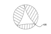
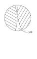
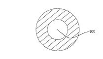
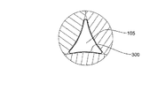
図1は、例えば、尿道中に挿入される内視鏡によって観察されることができるような種々の尿道前立腺部の腔の概略図を示している。図1Aでは、腔105が、いわゆる“A”形状の横断面を有した尿道前立腺部が示されている。図1Bでは、腔110が、いわゆる“I”字形の横断面を有した尿道前立腺部が示されている。図1Cでは、腔100が、円形、即ちいわゆる“O”字形の横断面を有した尿道前立腺部が示されている。
FIG. 1 shows a schematic diagram of various urethral prostate cavities as can be observed, for example, by an endoscope inserted into the urethra. In FIG. 1A, the urethral prostate is shown in which the
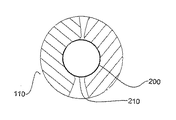
図2は、従来技術のステント200の広がりの後の、図1に示された腔の概略図を示している。図2A並びに2Bでは、ステント200が、腔105,110中に夫々広げられている。しかし、腔105,110は、ステント200の広がりの前に、O形状を有していないので、ステント200の外面の領域210は、尿道の壁205に接していない。このような解剖組織状態では、永久的なステントの領域210は、上皮によってカバーされないだろう。図2Cでは、円形横断面を有するステント200が、腔100中で広げられている。図2A並びに2Bとは対照的に、腔100が、ステント200の広がりの前にO形状を有しているので、ステント200の外面は、尿道の壁205のほぼ全体に渡って接している。かくして、図2Cのステント200は、全体を上皮によってカバーされるように期待されることができる。
FIG. 2 shows a schematic view of the cavity shown in FIG. 1 after the expansion of the
本発明は、3角形の横断面を有する体腔の領域中へ挿入されるためのステントを提供している。このステントは、“V”字形の横断面のように、開いた三角形を有することができる。 The present invention provides a stent for insertion into a region of a body cavity having a triangular cross section. The stent, as in the cross section of the "V" shape, it is Rukoto to have a open triangle.
横断面に湾曲した3つの頂点を有する三角形のステントが、円形の横断面を有するステントよりも良く非円形の横断面を有する腔に一致するように構成されることができる。三角形の横断面を有する本発明によるステントが、円形の横断面を有するステントよりも良く、BPHの尿道前立腺部の大部分の横断面に一致する。三角形の横断面は、尿流のための比較的大きくて効果的な開口を維持しながら、A若しくはI字形の前立腺の腔の形状に一致させることにより、患者への快適性を最大にしている。 A triangular stent with three vertices curved in cross-section can be configured to match a cavity with a non-circular cross-section better than a stent with a circular cross-section . The stent according to the invention having a triangle cross-section is better than a stent having a circular cross section, matching the cross-section of the majority of prostatic urethra of BPH. The triangular cross-section maximizes patient comfort by matching the shape of the A or I-shaped prostate cavity while maintaining a relatively large and effective opening for urine flow .
このステントは、ステントを製造するための技術分野で知られた材料で形成されることができ、弾性の材料と、プラスチックと、形状記憶合金と、形状記憶ポリマーとを含んでいる。さらに、ステントをこれが広がる場所へと移動させるための技術分野で知られた装置若しくは方法が、本発明のステントと共に使用されることができる。本発明の他の態様によれば、本発明は、ステントと、ステントを体腔中で広げるための装置とを提供している。 The stent can be formed of materials known in the art for manufacturing stents and includes an elastic material, plastic, shape memory alloy, and shape memory polymer. In addition, any device or method known in the art for moving the stent to where it will spread can be used with the stent of the present invention. According to another aspect of the present invention, the present invention provides a stent and an apparatus for expanding the stent in a body cavity.
本発明のステントは、永久的なステント若しくは一時的なステントであることができる。永久的なステントの場合、好ましい実施形態では、ステントは、腔中で広げられた後に上皮に包埋されるように窓を有している。このステントは、例えば、メッシュ材若しくはワイヤで形成されることができる。尿道前立腺用のステントの場合、例えば、三角形の拡大された横断面を有するステントは、ステントの面全体が、これを囲む組織に接触するようにA若しくはI字形の尿道前立腺部の腔に一致する。この接触は、ステントが組織によってほぼ完全にカバーされるまで鎖組織反応を引き起こす。尿道前立腺部用のステントの膀胱頸部端は、ステントが上皮、さらには膀胱頸部に包埋されるように、尿道/膀胱頸部のアングルに一致するように傾斜していることが好ましい。 The stent of the present invention can be a permanent stent or a temporary stent. In the case of a permanent stent, in a preferred embodiment, the stent has a window to be embedded in the epithelium after being expanded in the cavity. The stent can be formed of, for example, a mesh material or a wire. In the case of a stent for the urethral prostate, for example, a stent having an enlarged triangular cross section matches the cavity of the A or I-shaped urethral prostate so that the entire surface of the stent contacts the surrounding tissue. . This contact causes a chain tissue reaction until the stent is almost completely covered by the tissue. The bladder neck end of the urethral prostate stent is preferably tilted to match the angle of the urethra / bladder neck so that the stent is embedded in the epithelium and then the bladder neck.
このステントは、広げられた後にステントの移動を防止するように、アンカー部分を選択的に有することができる。このアンカーは、逆向きの円錐形状を含んだ形状を有することができる。永久的なステントの場合、アンカーが、回収可能な縫合糸、若しくは溶解可能な縫合糸、例えば、カットグット又はエシコン(Ethicon)からのMONOCRYL(商標登録)のような一時的な若しくは取り外し可能な装着部で、ステントに装着されることができる。これら溶解可能な装置は、例えば、尿道上皮によってカバーされることによってステントの永久的な部分の自主的な固着を可能にする、広がりの後の10ないし50日内に溶解可能である。 The stent can optionally have an anchor portion to prevent movement of the stent after being expanded. The anchor may have a shape that includes an inverted conical shape. In the case of a permanent stent, the anchor may be a temporary or removable attachment, such as a retrievable suture, or a dissolvable suture, such as MONOCRYL® from Cutgut or Ethicon Can be attached to the stent. These dissolvable devices can be dissolved within 10 to 50 days after spreading, for example allowing voluntary fixation of the permanent part of the stent by being covered by the urethral epithelium.
本発明の一時的なステントは、管腔用のライニングの一端若しくは両端のところでライニングに選択的に装着されることができる。この“管腔用のライニング”という用語は、中空の円筒形の弾性構造体を示すのに使用されている。この弾性構造体は、体腔中に挿入され、腔の形状並びに内径の変更に従って腔の形状並びに内径に一致する。ステントを広げるための管腔用のステントが、“内腔用のライニング”という名称で2002年11月13日出願の同時系続米国特許出願に開示されている。このライニングは、これが広げられる腔の内径に等しいか、わずかに大きいか、わずかに小さい非緊張状態の内径を有している。このライニングの面は、例えば、ライニングの弾性部材を可撓性の材料中に包埋するか、弾性部材を可撓性のシースにカバーするかにより、連続的であることができる。 The temporary stent of the present invention can be selectively attached to the lining at one or both ends of the luminal lining. The term “luminal lining” is used to indicate a hollow cylindrical elastic structure. This elastic structure is inserted into the body cavity and conforms to the shape and inner diameter of the cavity according to changes in the shape and inner diameter of the cavity. A luminal stent for expanding the stent is disclosed in a co-pending US patent application filed Nov. 13, 2002, entitled “Luminary Lining”. The lining has an unstrained inner diameter that is equal to, slightly larger, or slightly smaller than the inner diameter of the cavity in which it is expanded. The surface of the lining can be continuous, for example by embedding the elastic member of the lining in a flexible material or covering the elastic member with a flexible sheath.
ライニングに関連した本発明のステントの使用は、幾つかの利点を有している。最初に、本発明のステントに隣接したライニングの使用は、ステントの両端周辺の腔壁に別に存在する急な勾配圧力を除去する。急な勾配圧力の領域は、ステントの両端部のところで腔を部分的若しくは完全に閉塞するステントの両端部で、組織の内部成長を減じることが知られている。 The use of the inventive stent in conjunction with a lining has several advantages. Initially, the use of a lining adjacent to the stent of the present invention removes the steep gradient pressure that exists separately in the cavity walls around the ends of the stent. It is known that regions of steep gradient pressure reduce tissue ingrowth at the ends of the stent that partially or completely occlude the cavity at both ends of the stent.
また、本発明のステントと共に管腔用のライニングを使用することは、ステントが括約筋近くで広げられるときに効果的である。このステントは、ライニングが、ステントと括約筋との間に介在した状態で腔中に位置される。このライニングとステントとは、単一ユニットとして組立てられるか、別々の2つのユニットとして形成されることができる。別々の2つのユニットは、尿道中に挿入される前に結合されて単一統合ユニットとして一緒に挿入されるか、ライニングとステントとは、別々に挿入されることができる。ステントは、ライニングによって括約筋から離される所定の領域の腔の開通性を維持する。括約筋が接触し、括約筋に隣接する腔の直径が減じられると、ライニングの直径も、腔壁の形状に一致しながら減じられる。括約筋が弛緩し、括約筋に隣接する腔の直径が増すと、ライニングの直径も、腔壁の形状に一致しながら増す。かくして、ライニングは、括約筋が緊縮並びに弛緩するに従って、括約筋に隣接する腔の形状に動的に一致し、ステントの括約筋端部のところで、反応増殖性の組織成長を阻止する。また、ライニングは、括約筋近くの腔にステントを正確に位置させることを可能にしている。括約筋と尿道前立腺部の領域との間の距離が測定され、この距離に等しい長さを有するライニングが使用される。そして、ステントとライニングとは上述されたように位置される。また、括約筋近くのライニングの存在は、括約筋の機能を干渉することなく、括約筋に隣接する腔壁に対して支持を与える。 Also, the use of a luminal lining with the stent of the present invention is effective when the stent is expanded near the sphincter. The stent is positioned in the cavity with a lining interposed between the stent and the sphincter. The lining and stent can be assembled as a single unit or formed as two separate units. The two separate units can be combined and inserted together as a single integrated unit before being inserted into the urethra, or the lining and stent can be inserted separately. The stent maintains the patency of a predetermined area of the cavity that is separated from the sphincter by the lining. As the sphincter comes into contact and the diameter of the cavity adjacent to the sphincter is reduced, the diameter of the lining is also reduced to match the shape of the cavity wall. As the sphincter relaxes and the diameter of the cavity adjacent to the sphincter increases, the diameter of the lining also increases to match the shape of the cavity wall. Thus, as the sphincter contracts and relaxes, the lining dynamically conforms to the shape of the cavity adjacent to the sphincter and prevents reactive proliferative tissue growth at the sphincter end of the stent. The lining also allows the stent to be accurately positioned in the cavity near the sphincter. The distance between the sphincter and the urethral prostate region is measured and a lining having a length equal to this distance is used. The stent and lining are then positioned as described above. Also, the presence of the lining near the sphincter provides support to the cavity wall adjacent to the sphincter without interfering with the function of the sphincter.
このステントは、生物安定性、生物分解性、若しくは生体吸収性の材料で形成された外装材で囲まれることができる。このステントは、また、尿道前立腺部用のステントの場合に、組織の過剰増殖を防止するような薬、抗生物質、抗腫瘍薬、若しくは、拡大した前立腺の塊を小さくするフィナステライド(finasteride)のような薬を含むことができる。一時的なステント用に、カバーは、生物安定性の材料で形成されることができ、永久的なステント用に、カバーは、ステントが組織によってカバーされ得るように数日間で分解される生物分解性若しくは生体吸収性の材料で形成されることができる。 The stent can be surrounded by a sheath made of a biostable, biodegradable, or bioabsorbable material. This stent can also be used in the case of urethral prostate stents, such as drugs that prevent tissue overgrowth, antibiotics, anti-tumor agents, or finasteride that reduces enlarged prostate mass. Can contain various medicines. For temporary stents, the cover can be formed of a biostable material, and for permanent stents, the cover is degraded in a few days so that the stent can be covered by tissue. Or bioabsorbable material.
本発明を理解し、本発明が実際にどう実施されるかをみるために、好ましい実施形態が、単なる非限定的な一例によって、添付図面を参照して説明されるであろう。 In order to understand the present invention and to see how it is actually practiced, preferred embodiments will be described by way of non-limiting example only with reference to the accompanying drawings.
説明の便宜並びに明確性のために、本発明は、尿道前立腺部用のステントを参照して説明されているであろう。これはほんの一例であり、本発明のステントは、非円形の横断面を有する体腔内での使用のために用いられることができる。 For ease of explanation and clarity, the present invention will be described with reference to a stent for the urethral prostate. This is only an example, and the stent of the present invention can be used for use in a body cavity having a non-circular cross section.
図3Aは、完全な三角形の横断面を有する、前立腺の管腔用のステント300の参考例を示している。この参考例によれば、このステント300の各横断面は、3つの頂点302で交わる3つの線分301で形成されている。図3Bは、参考例に係る前立腺の管腔用のステント305を示しており、このステントは、3つのアーク307によって接続されている3つの線分306で形成された横断面を有している。各線分306は、アーク上の点の曲率半径の間違いなく2.5倍以上である限定されない曲率半径を有している。図3C,3D(参考例),並びに3Eは、3つの更なる尿道管腔用のステント320,321,並びに322を夫々示している。これらステントは、側面が、種々の形状に曲げられている閉じた三角形の横断面を有している。各横断面には少なくとも1対の点(例えば、1対の点303,304)が存在しており、1対の点のうちの一方の点(例えば、点303)の曲率半径は、1対の点のうちの他方の点(点304)の曲率半径の少なくとも2.5倍である。図3F並びに3Gは、不完全な三角形の横断面を有する更なる2つの尿道管腔用のステント310,312を夫々示している。参考例のステント312では、各横断面は、3つの線分309と3つの頂点311とで形成されている。ステント310では、各横断面は、円形若しくは楕円形の2つのアーク314と円形の3つのアーク316とで形成されている。前記アーク314は、(例えば、楕円形のアーク314の点315で)最大曲率半径を有している。この最大曲率半径は、円形のアーク316の半径の少なくとも2.5倍である。
FIG. 3A shows a reference example of a
本発明のステントは、ステントが窓を有するようにメッシュ材から形成されることができる。上述されたように、これは、ステントが組織中に包埋されることを可能にしている。 The stent of the present invention can be formed from a mesh material such that the stent has a window. As described above, this allows the stent to be embedded in tissue.
本発明のステントは、ステントを製造するための技術分野で知られた材料で形成されることができ、弾性の材料、プラスチック、形状記憶合金、並びに形状記憶ポリマーを含んでいる。さらに、ステントをこれの広がり場所に移動させ、ステントを拡大させるための技術分野で知られた装置又は方法が、本発明のステントと一緒に使用されることができる。 The stent of the present invention can be formed of materials known in the art for manufacturing stents and includes elastic materials, plastics, shape memory alloys, and shape memory polymers. In addition, any device or method known in the art for moving the stent to its expanse and expanding the stent can be used with the stent of the present invention.
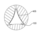
図4Aは、図1Aに示された尿道前立腺部105(”A”形状の尿道前立腺部)中への挿入後の参考例のステント300を示している。図4Bは、図1Bに示された尿道前立腺部110(”I”字形の尿道前立腺部)中への挿入後のステント300を示している。図4Cは、尿道中に挿入される内視鏡を使用して観察されることができるような、図1Aに示された尿道前立腺部(”A”字形の尿道)中への挿入後の、開いた多角形の横断面を有するステント405を示している。見ることができるように、ステント300の広がり後に、尿道腔105,110は、ステント300の形状に一致する。これは、組織の成長によってステントの包埋を促進させる。
FIG. 4A shows a
図5は、管腔用のライニング(lining)510に装着された参考例のステント300を有するシステム500を示している。このライニング510は、これが中で広げられる最大径の尿道前立腺部よりもわずかに大きい非緊張状態の大径を有するようなディメンションである。ライニングの形状は、本体形状とは異なることができ、例えば、一端の横断面は本体形状のような三角形であり、外括約筋に面した他端は円形であることができる。ライニング510の弾性抵抗は、ライニングが中で広げられる腔壁によって、腔が収縮したときにライニングに加えられる径方向内方の力よりも小さい。前記システム500は、ステントが尿道前立腺部中に位置された状態で尿道中で広げられる。
FIG. 5 shows a
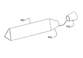
図6Aは、参考例のステント300とアンカー610とを有するシステム600を示している。このシステム600は、尿道前立腺部内へのステント300と、外括約筋の反対側の尿道内へのアンカー610と共に尿道中で広げられる。ステント300とアンカー610とは、前記システム600の広がり後に括約筋を貫通するつなぎ網625によって接続されている。アンカー610は、括約筋の動作を干渉しないのと同時に、ステント300が、尿道前立腺部から離れるように移動するのを防止している。
FIG. 6A shows a
図6Bは、管腔用のライニング605とアンカー610とに装着された参考例のステント300を有するシステムを示している。
FIG. 6B illustrates a system having a
説明されたステントが、尿道、特に男性尿道内で使用されるように説明されているが、これは、単なる一例である。これらステントは、臓器のディメンション並びに横断面の形状に対して変更されるのであれば、他の環状の臓器を含む他の臓器に使用されることができる。 Although the described stent has been described for use in the urethra, particularly the male urethra, this is merely an example. These stents can be used for other organs, including other annular organs, as long as they are modified to the organ dimensions as well as the cross-sectional shape.
Claims (17)
また、前記3つの側片は、互いに交わる3つの湾曲した端部を有し、
更に、前記3つの側片は、前記広がった状態で、外側に湾曲して突出している、管腔中に挿入するための拡大可能なステント。A stent having an expanded state having an outer shape of at least one triangular cross section defined by three side pieces, the triangular cross section passing through the side pieces to an inner wall of a lumen; can give force radially outward, and can withstand against the force of the radially inward provided by the force acting on the inner wall of the lumen,
Further, the three side pieces is to have the three curved end intersecting one another,
Further, the expandable stent for insertion into a lumen , wherein the three side pieces are curved outwardly protruding in the expanded state .
また、前記3つの側片は、互いに交わる3つの湾曲した端部を有し、
更に、前記3つの側片は、前記広がった状態で、外側に湾曲して突出している、管腔中に挿入するための拡大可能なステントを具備するシステム。A stent having an expanded state having an outer shape of at least one triangular cross section defined by three side pieces , wherein the triangular cross section exerts a force radially outward against the inner wall of the lumen. providing that it is, and can withstand against the force of the radially inward provided by the force acting on the inner wall of the lumen,
Further, the three side pieces is to have the three curved end intersecting one another,
The system further comprising an expandable stent for insertion into a lumen , wherein the three side pieces are curved outwardly projecting in the expanded state .
Applications Claiming Priority (3)
| Application Number | Priority Date | Filing Date | Title |
|---|---|---|---|
| US10/370,592 US9333102B2 (en) | 2003-02-24 | 2003-02-24 | Stent |
| US10/370,592 | 2003-02-24 | ||
| PCT/IL2004/000174 WO2004073556A1 (en) | 2003-02-24 | 2004-02-23 | Stent |
Publications (2)
| Publication Number | Publication Date |
|---|---|
| JP2006518626A JP2006518626A (en) | 2006-08-17 |
| JP4771425B2 true JP4771425B2 (en) | 2011-09-14 |
Family
ID=32868191
Family Applications (1)
| Application Number | Title | Priority Date | Filing Date |
|---|---|---|---|
| JP2006502643A Expired - Fee Related JP4771425B2 (en) | 2003-02-24 | 2004-02-23 | Stent |
Country Status (8)
| Country | Link |
|---|---|
| US (1) | US9333102B2 (en) |
| EP (1) | EP1599153B1 (en) |
| JP (1) | JP4771425B2 (en) |
| AT (1) | ATE487437T1 (en) |
| CA (1) | CA2515438C (en) |
| DE (1) | DE602004029986D1 (en) |
| ES (1) | ES2356079T3 (en) |
| WO (1) | WO2004073556A1 (en) |
Families Citing this family (47)
| Publication number | Priority date | Publication date | Assignee | Title |
|---|---|---|---|---|
| US20050131515A1 (en) * | 2003-12-16 | 2005-06-16 | Cully Edward H. | Removable stent-graft |
| US8668705B2 (en) | 2005-05-20 | 2014-03-11 | Neotract, Inc. | Latching anchor device |
| US10195014B2 (en) | 2005-05-20 | 2019-02-05 | Neotract, Inc. | Devices, systems and methods for treating benign prostatic hyperplasia and other conditions |
| US7758594B2 (en) | 2005-05-20 | 2010-07-20 | Neotract, Inc. | Devices, systems and methods for treating benign prostatic hyperplasia and other conditions |
| US7645286B2 (en) | 2005-05-20 | 2010-01-12 | Neotract, Inc. | Devices, systems and methods for retracting, lifting, compressing, supporting or repositioning tissues or anatomical structures |
| US10925587B2 (en) | 2005-05-20 | 2021-02-23 | Neotract, Inc. | Anchor delivery system |
| US9549739B2 (en) | 2005-05-20 | 2017-01-24 | Neotract, Inc. | Devices, systems and methods for treating benign prostatic hyperplasia and other conditions |
| US8628542B2 (en) | 2005-05-20 | 2014-01-14 | Neotract, Inc. | Median lobe destruction apparatus and method |
| US8603106B2 (en) | 2005-05-20 | 2013-12-10 | Neotract, Inc. | Integrated handle assembly for anchor delivery system |
| US9642726B2 (en) | 2005-07-25 | 2017-05-09 | Vascular Dynamics, Inc. | Devices and methods for control of blood pressure |
| US9592136B2 (en) | 2005-07-25 | 2017-03-14 | Vascular Dynamics, Inc. | Devices and methods for control of blood pressure |
| US9125732B2 (en) * | 2005-07-25 | 2015-09-08 | Vascular Dynamics, Inc. | Devices and methods for control of blood pressure |
| US8923972B2 (en) | 2005-07-25 | 2014-12-30 | Vascular Dynamics, Inc. | Elliptical element for blood pressure reduction |
| EP1951150B1 (en) * | 2005-10-29 | 2010-08-18 | PNN Medical SA | Stent with anchoring portion |
| US9622888B2 (en) * | 2006-11-16 | 2017-04-18 | W. L. Gore & Associates, Inc. | Stent having flexibly connected adjacent stent elements |
| US8926688B2 (en) | 2008-01-11 | 2015-01-06 | W. L. Gore & Assoc. Inc. | Stent having adjacent elements connected by flexible webs |
| WO2011019892A2 (en) * | 2009-08-14 | 2011-02-17 | Mayo Foundation For Medical Education And Research | Non-circular esophageal stents and delivery systems |
| US10292801B2 (en) | 2012-03-29 | 2019-05-21 | Neotract, Inc. | System for delivering anchors for treating incontinence |
| KR101362446B1 (en) * | 2012-05-10 | 2014-02-11 | 이훈범 | Filler for wrinkle removing |
| EP2858710B1 (en) * | 2012-06-08 | 2020-10-07 | University Hospitals Health System Inc. | Ureteral stent for placement in a kidney and bladder |
| US10130353B2 (en) | 2012-06-29 | 2018-11-20 | Neotract, Inc. | Flexible system for delivering an anchor |
| US9180031B2 (en) | 2013-03-15 | 2015-11-10 | Covidien Lp | Stent with varying radius between struts |
| US9259335B2 (en) | 2013-03-15 | 2016-02-16 | Covidien Lp | Stent |
| HUE054310T2 (en) * | 2014-01-26 | 2021-08-30 | Butterfly Medical Ltd | A dilating device for prostatic urethra |
| US11027106B2 (en) | 2014-01-26 | 2021-06-08 | Butterfly Medical Ltd. | Dilating device and method for prostatic urethra |
| CA2955338C (en) * | 2014-08-12 | 2022-06-21 | Anastomosis As | Minimally traumatic anastomosis |
| US10299948B2 (en) | 2014-11-26 | 2019-05-28 | W. L. Gore & Associates, Inc. | Balloon expandable endoprosthesis |
| ES2934649T3 (en) | 2015-07-29 | 2023-02-23 | Butterfly Medical Ltd | Retraction and/or support of the periurethral tissue |
| EP3167845A1 (en) * | 2015-11-12 | 2017-05-17 | The Provost, Fellows, Foundation Scholars, & the other members of Board, of the College of Holy and Undiv. Trinity of Queen Elizabeth near Dublin | An implantable biocompatible expander suitable for treatment of constrictions of body lumen |
| US9888981B2 (en) * | 2016-04-01 | 2018-02-13 | Larry Travis Haws | Interdental laterally expansive wedge |
| WO2017192145A1 (en) * | 2016-05-06 | 2017-11-09 | University Hospitals Health Systems, Inc. | Ureteral stent |
| US10568752B2 (en) | 2016-05-25 | 2020-02-25 | W. L. Gore & Associates, Inc. | Controlled endoprosthesis balloon expansion |
| US10449069B2 (en) | 2016-11-14 | 2019-10-22 | Covidien Lp | Stent |
| US10905572B2 (en) | 2016-11-14 | 2021-02-02 | Covidien Lp | Stent |
| US10258488B2 (en) | 2016-11-14 | 2019-04-16 | Covidien Lp | Stent |
| EP3551140A4 (en) | 2016-12-09 | 2020-07-08 | Zenflow, Inc. | Systems, devices, and methods for the accurate deployment of an implant in the prostatic urethra |
| US11523920B2 (en) | 2017-03-16 | 2022-12-13 | Keyvon Rashidi | Stent with a smooth surface in its expanded configuration |
| AU2018306738A1 (en) * | 2017-07-28 | 2020-02-13 | Zenflow, Inc. | Systems, devices, and methods for expanding the diameter of the prostatic urethra |
| WO2019126718A1 (en) | 2017-12-23 | 2019-06-27 | Neotract, Inc. | Expandable tissue engagement apparatus and method |
| CN109350316B (en) * | 2018-11-06 | 2024-03-12 | 广东工业大学 | Intravascular stent monomer and intravascular stent |
| US10555802B1 (en) | 2019-03-07 | 2020-02-11 | John H. Shadduck | Urologic stents and methods of use |
| CN110338947A (en) * | 2019-07-17 | 2019-10-18 | 胡信 | A kind of prostate bracket |
| EP4061292A4 (en) | 2019-11-19 | 2023-12-27 | Zenflow, Inc. | Systems, devices, and methods for the accurate deployment and imaging of an implant in the prostatic urethra |
| US11273025B2 (en) | 2019-11-22 | 2022-03-15 | Pro Verum Limited | Expandable implant delivery device |
| US11602621B2 (en) | 2019-11-22 | 2023-03-14 | ProVerum Limited | Device for controllably deploying expandable implants |
| FR3112935B1 (en) | 2020-07-29 | 2022-07-29 | Univ Claude Bernard Lyon | IMAGEABLE AND RESORBABLE MEDICAL DEVICE AND USES THEREOF |
| CN114732567A (en) * | 2022-05-19 | 2022-07-12 | 哈尔滨工业大学 | Shape memory urethral stent |
Citations (2)
| Publication number | Priority date | Publication date | Assignee | Title |
|---|---|---|---|---|
| JPH05212121A (en) * | 1991-02-05 | 1993-08-24 | Kanji Inoue | Folding apparatus inserted in human organ with elastic restoring function and folding device therefor |
| WO2002060350A1 (en) * | 2001-01-29 | 2002-08-08 | Icon Technologies, Llc | Irradiated stent coating |
Family Cites Families (53)
| Publication number | Priority date | Publication date | Assignee | Title |
|---|---|---|---|---|
| US4893623A (en) * | 1986-12-09 | 1990-01-16 | Advanced Surgical Intervention, Inc. | Method and apparatus for treating hypertrophy of the prostate gland |
| US6730105B2 (en) * | 1988-07-29 | 2004-05-04 | Samuel Shiber | Clover leaf shaped tubular medical device |
| US6171338B1 (en) * | 1988-11-10 | 2001-01-09 | Biocon, Oy | Biodegradable surgical implants and devices |
| US5037392A (en) * | 1989-06-06 | 1991-08-06 | Cordis Corporation | Stent-implanting balloon assembly |
| US5269802A (en) * | 1991-09-10 | 1993-12-14 | Garber Bruce B | Prostatic stent |
| US5411550A (en) * | 1991-09-16 | 1995-05-02 | Atrium Medical Corporation | Implantable prosthetic device for the delivery of a bioactive material |
| US5320100A (en) * | 1991-09-16 | 1994-06-14 | Atrium Medical Corporation | Implantable prosthetic device having integral patency diagnostic indicia |
| JP3393383B2 (en) * | 1992-01-21 | 2003-04-07 | リージェンツ オブ ザ ユニバーシティ オブ ミネソタ | Septal defect closure device |
| US5540712A (en) * | 1992-05-01 | 1996-07-30 | Nitinol Medical Technologies, Inc. | Stent and method and apparatus for forming and delivering the same |
| US5716410A (en) * | 1993-04-30 | 1998-02-10 | Scimed Life Systems, Inc. | Temporary stent and method of use |
| ES2137961T3 (en) * | 1993-07-01 | 2000-01-01 | Schneider Europ Gmbh | MEDICAL DEVICE FOR THE TREATMENT OF A PORTION OF BLOOD VESSEL THROUGH IONIZING RADIATION. |
| DE4339265A1 (en) * | 1993-11-18 | 1995-05-24 | Angiomed Ag | Vena cava filter |
| US6039749A (en) * | 1994-02-10 | 2000-03-21 | Endovascular Systems, Inc. | Method and apparatus for deploying non-circular stents and graftstent complexes |
| US5522881A (en) * | 1994-06-28 | 1996-06-04 | Meadox Medicals, Inc. | Implantable tubular prosthesis having integral cuffs |
| EP0810845A2 (en) * | 1995-02-22 | 1997-12-10 | Menlo Care Inc. | Covered expanding mesh stent |
| BE1009277A3 (en) * | 1995-04-12 | 1997-01-07 | Corvita Europ | Guardian self-expandable medical device introduced in cavite body, and method of preparation. |
| US5639277A (en) * | 1995-04-28 | 1997-06-17 | Target Therapeutics, Inc. | Embolic coils with offset helical and twisted helical shapes |
| US6849069B1 (en) * | 1995-11-07 | 2005-02-01 | Boston Scientitfic Corporation | Medical device with tail(s) for assisting flow of urine |
| DE69526857T2 (en) * | 1995-11-27 | 2003-01-02 | Schneider Europ Gmbh Buelach | Stent for use in one pass |
| US5927345A (en) * | 1996-04-30 | 1999-07-27 | Target Therapeutics, Inc. | Super-elastic alloy braid structure |
| ZA9710342B (en) * | 1996-11-25 | 1998-06-10 | Alza Corp | Directional drug delivery stent and method of use. |
| US5902475A (en) * | 1997-04-08 | 1999-05-11 | Interventional Technologies, Inc. | Method for manufacturing a stent |
| US5855597A (en) * | 1997-05-07 | 1999-01-05 | Iowa-India Investments Co. Limited | Stent valve and stent graft for percutaneous surgery |
| US6325824B2 (en) * | 1998-07-22 | 2001-12-04 | Advanced Cardiovascular Systems, Inc. | Crush resistant stent |
| US6156064A (en) * | 1998-08-14 | 2000-12-05 | Schneider (Usa) Inc | Stent-graft-membrane and method of making the same |
| GB9828696D0 (en) | 1998-12-29 | 1999-02-17 | Houston J G | Blood-flow tubing |
| US6338740B1 (en) * | 1999-01-26 | 2002-01-15 | Edwards Lifesciences Corporation | Flexible heart valve leaflets |
| US6709465B2 (en) * | 1999-03-18 | 2004-03-23 | Fossa Medical, Inc. | Radially expanding ureteral device |
| US7226475B2 (en) * | 1999-11-09 | 2007-06-05 | Boston Scientific Scimed, Inc. | Stent with variable properties |
| US6458153B1 (en) * | 1999-12-31 | 2002-10-01 | Abps Venture One, Ltd. | Endoluminal cardiac and venous valve prostheses and methods of manufacture and delivery thereof |
| US7044980B2 (en) * | 2000-02-03 | 2006-05-16 | Boston Scientific Scimed, Inc. | Facilitating drainage |
| US6740093B2 (en) * | 2000-02-28 | 2004-05-25 | Stephen Hochschuler | Method and apparatus for treating a vertebral body |
| US20030135268A1 (en) * | 2000-04-11 | 2003-07-17 | Ashvin Desai | Secure stent for maintaining a lumenal opening |
| US6572646B1 (en) * | 2000-06-02 | 2003-06-03 | Advanced Cardiovascular Systems, Inc. | Curved nitinol stent for extremely tortuous anatomy |
| US6602272B2 (en) * | 2000-11-02 | 2003-08-05 | Advanced Cardiovascular Systems, Inc. | Devices configured from heat shaped, strain hardened nickel-titanium |
| US6626937B1 (en) * | 2000-11-14 | 2003-09-30 | Advanced Cardiovascular Systems, Inc. | Austenitic nitinol medical devices |
| EP1392388B1 (en) * | 2001-01-23 | 2010-10-06 | Abbeymoor Medical, Inc. | Endourethral device |
| US7238199B2 (en) * | 2001-03-06 | 2007-07-03 | The Board Of Regents Of The University Of Texas System | Method and apparatus for stent deployment with enhanced delivery of bioactive agents |
| US20030032976A1 (en) * | 2001-05-21 | 2003-02-13 | Boucek Mark M. | Catheter deployed partial occlusion devices and methods |
| US6981964B2 (en) * | 2001-05-22 | 2006-01-03 | Boston Scientific Scimed, Inc. | Draining bodily fluids with a stent |
| US7128755B2 (en) * | 2001-06-01 | 2006-10-31 | Texas Stent Technologies, Inc. | Expandable biodegradable polymeric stents for combined mechanical support and pharmacological or radiation therapy |
| GB2379996B (en) * | 2001-06-05 | 2004-05-19 | Tayside Flow Technologies Ltd | Flow means |
| US6790223B2 (en) * | 2001-09-21 | 2004-09-14 | Scimed Life Systems, Inc. | Delivering a uretheral stent |
| US20030065381A1 (en) * | 2001-09-28 | 2003-04-03 | Solar Ronald J. | Longitudinal focussed force stent |
| US20030093142A1 (en) * | 2001-10-16 | 2003-05-15 | Elazer Edelman | Stent concept for minimization of deployment related wall shear and injury |
| US20030083746A1 (en) * | 2001-10-31 | 2003-05-01 | Kuslich Stephen D. | Vertebral spacer for spinal stabilization |
| US7186237B2 (en) * | 2002-02-14 | 2007-03-06 | Avantec Vascular Corporation | Ballon catheter for creating a longitudinal channel in a lesion and method |
| US20030187498A1 (en) * | 2002-03-28 | 2003-10-02 | Medtronic Ave, Inc. | Chamfered stent strut and method of making same |
| IL149828A (en) | 2002-05-23 | 2007-09-20 | Ronnie Levi | Medical device having a tubular portion |
| US6733536B1 (en) * | 2002-10-22 | 2004-05-11 | Scimed Life Systems | Male urethral stent device |
| US8282678B2 (en) * | 2002-11-13 | 2012-10-09 | Allium Medical Solutions Ltd. | Endoluminal lining |
| US7004971B2 (en) * | 2002-12-31 | 2006-02-28 | Depuy Acromed, Inc. | Annular nucleus pulposus replacement |
| US20040193141A1 (en) * | 2003-02-14 | 2004-09-30 | Leopold Eric W. | Intravascular flow modifier and reinforcement device and deployment system for same |
-
2003
- 2003-02-24 US US10/370,592 patent/US9333102B2/en not_active Expired - Lifetime
-
2004
- 2004-02-23 AT AT04713627T patent/ATE487437T1/en not_active IP Right Cessation
- 2004-02-23 CA CA2515438A patent/CA2515438C/en not_active Expired - Fee Related
- 2004-02-23 ES ES04713627T patent/ES2356079T3/en not_active Expired - Lifetime
- 2004-02-23 JP JP2006502643A patent/JP4771425B2/en not_active Expired - Fee Related
- 2004-02-23 DE DE602004029986T patent/DE602004029986D1/en not_active Expired - Lifetime
- 2004-02-23 EP EP04713627A patent/EP1599153B1/en not_active Expired - Lifetime
- 2004-02-23 WO PCT/IL2004/000174 patent/WO2004073556A1/en active Application Filing
Patent Citations (2)
| Publication number | Priority date | Publication date | Assignee | Title |
|---|---|---|---|---|
| JPH05212121A (en) * | 1991-02-05 | 1993-08-24 | Kanji Inoue | Folding apparatus inserted in human organ with elastic restoring function and folding device therefor |
| WO2002060350A1 (en) * | 2001-01-29 | 2002-08-08 | Icon Technologies, Llc | Irradiated stent coating |
Also Published As
| Publication number | Publication date |
|---|---|
| WO2004073556A1 (en) | 2004-09-02 |
| US9333102B2 (en) | 2016-05-10 |
| DE602004029986D1 (en) | 2010-12-23 |
| US20040167635A1 (en) | 2004-08-26 |
| JP2006518626A (en) | 2006-08-17 |
| CA2515438C (en) | 2012-08-21 |
| EP1599153B1 (en) | 2010-11-10 |
| ES2356079T3 (en) | 2011-04-04 |
| ATE487437T1 (en) | 2010-11-15 |
| CA2515438A1 (en) | 2004-09-02 |
| EP1599153A1 (en) | 2005-11-30 |
| WO2004073556B1 (en) | 2004-10-07 |
Similar Documents
| Publication | Publication Date | Title |
|---|---|---|
| JP4771425B2 (en) | Stent | |
| CA2505417C (en) | Endoluminal lining | |
| US20180235651A1 (en) | Radial cutter implant | |
| ES2241615T3 (en) | BIO-REABSORBABLE STENT. | |
| JP4913069B2 (en) | Stent with protruding bifurcation for bifurcated tube | |
| EP1951150B1 (en) | Stent with anchoring portion | |
| US8177741B2 (en) | Catheter with superelastic retention device | |
| US20080039921A1 (en) | Balloon Catheter | |
| US20020188344A1 (en) | Retrievable stent and method of use thereof | |
| US20090149935A1 (en) | Expandable stent | |
| JPH04256759A (en) | Apparatus for expanding constricted part of internal tube | |
| WO2006037088A2 (en) | Method and device for extravascular intervention | |
| JP2008080141A (en) | Contraceptive system | |
| JPH08510152A (en) | Stent delivery system | |
| US20050240141A1 (en) | Stent kidney curl improvements | |
| IL169830A (en) | Stent | |
| JPH02252467A (en) | Living organ dilator and catheter | |
| IL168532A (en) | Device for insertion into a body lumen |
Legal Events
| Date | Code | Title | Description |
|---|---|---|---|
| A621 | Written request for application examination |
Free format text: JAPANESE INTERMEDIATE CODE: A621 Effective date: 20070222 |
|
| A131 | Notification of reasons for refusal |
Free format text: JAPANESE INTERMEDIATE CODE: A131 Effective date: 20091104 |
|
| A521 | Request for written amendment filed |
Free format text: JAPANESE INTERMEDIATE CODE: A523 Effective date: 20100201 |
|
| A131 | Notification of reasons for refusal |
Free format text: JAPANESE INTERMEDIATE CODE: A131 Effective date: 20100629 |
|
| A601 | Written request for extension of time |
Free format text: JAPANESE INTERMEDIATE CODE: A601 Effective date: 20100929 |
|
| A602 | Written permission of extension of time |
Free format text: JAPANESE INTERMEDIATE CODE: A602 Effective date: 20101006 |
|
| A601 | Written request for extension of time |
Free format text: JAPANESE INTERMEDIATE CODE: A601 Effective date: 20101029 |
|
| A602 | Written permission of extension of time |
Free format text: JAPANESE INTERMEDIATE CODE: A602 Effective date: 20101108 |
|
| A521 | Request for written amendment filed |
Free format text: JAPANESE INTERMEDIATE CODE: A523 Effective date: 20101129 |
|
| TRDD | Decision of grant or rejection written | ||
| A01 | Written decision to grant a patent or to grant a registration (utility model) |
Free format text: JAPANESE INTERMEDIATE CODE: A01 Effective date: 20110517 |
|
| A711 | Notification of change in applicant |
Free format text: JAPANESE INTERMEDIATE CODE: A711 Effective date: 20110526 |
|
| A521 | Request for written amendment filed |
Free format text: JAPANESE INTERMEDIATE CODE: A821 Effective date: 20110526 |
|
| A61 | First payment of annual fees (during grant procedure) |
Free format text: JAPANESE INTERMEDIATE CODE: A61 Effective date: 20110616 |
|
| FPAY | Renewal fee payment (event date is renewal date of database) |
Free format text: PAYMENT UNTIL: 20140701 Year of fee payment: 3 |
|
| R150 | Certificate of patent or registration of utility model |
Free format text: JAPANESE INTERMEDIATE CODE: R150 Ref document number: 4771425 Country of ref document: JP Free format text: JAPANESE INTERMEDIATE CODE: R150 |
|
| R250 | Receipt of annual fees |
Free format text: JAPANESE INTERMEDIATE CODE: R250 |
|
| R250 | Receipt of annual fees |
Free format text: JAPANESE INTERMEDIATE CODE: R250 |
|
| R250 | Receipt of annual fees |
Free format text: JAPANESE INTERMEDIATE CODE: R250 |
|
| R250 | Receipt of annual fees |
Free format text: JAPANESE INTERMEDIATE CODE: R250 |
|
| S303 | Written request for registration of pledge or change of pledge |
Free format text: JAPANESE INTERMEDIATE CODE: R316303 |
|
| S531 | Written request for registration of change of domicile |
Free format text: JAPANESE INTERMEDIATE CODE: R313531 |
|
| R350 | Written notification of registration of transfer |
Free format text: JAPANESE INTERMEDIATE CODE: R350 |
|
| R250 | Receipt of annual fees |
Free format text: JAPANESE INTERMEDIATE CODE: R250 |
|
| R250 | Receipt of annual fees |
Free format text: JAPANESE INTERMEDIATE CODE: R250 |
|
| R250 | Receipt of annual fees |
Free format text: JAPANESE INTERMEDIATE CODE: R250 |
|
| S111 | Request for change of ownership or part of ownership |
Free format text: JAPANESE INTERMEDIATE CODE: R313113 |
|
| R350 | Written notification of registration of transfer |
Free format text: JAPANESE INTERMEDIATE CODE: R350 |
|
| R250 | Receipt of annual fees |
Free format text: JAPANESE INTERMEDIATE CODE: R250 |
|
| LAPS | Cancellation because of no payment of annual fees |