JP4756032B2 - Monoclonal antibody with specificity for fetal red blood cells - Google Patents
Monoclonal antibody with specificity for fetal red blood cells Download PDFInfo
- Publication number
- JP4756032B2 JP4756032B2 JP2007505500A JP2007505500A JP4756032B2 JP 4756032 B2 JP4756032 B2 JP 4756032B2 JP 2007505500 A JP2007505500 A JP 2007505500A JP 2007505500 A JP2007505500 A JP 2007505500A JP 4756032 B2 JP4756032 B2 JP 4756032B2
- Authority
- JP
- Japan
- Prior art keywords
- cells
- fetal
- monoclonal antibody
- blood cells
- detecting
- Prior art date
- Legal status (The legal status is an assumption and is not a legal conclusion. Google has not performed a legal analysis and makes no representation as to the accuracy of the status listed.)
- Active
Links
Images


Classifications
-
- G—PHYSICS
- G01—MEASURING; TESTING
- G01N—INVESTIGATING OR ANALYSING MATERIALS BY DETERMINING THEIR CHEMICAL OR PHYSICAL PROPERTIES
- G01N33/00—Investigating or analysing materials by specific methods not covered by groups G01N1/00 - G01N31/00
- G01N33/48—Biological material, e.g. blood, urine; Haemocytometers
- G01N33/50—Chemical analysis of biological material, e.g. blood, urine; Testing involving biospecific ligand binding methods; Immunological testing
- G01N33/53—Immunoassay; Biospecific binding assay; Materials therefor
- G01N33/569—Immunoassay; Biospecific binding assay; Materials therefor for microorganisms, e.g. protozoa, bacteria, viruses
- G01N33/56966—Animal cells
-
- G—PHYSICS
- G01—MEASURING; TESTING
- G01N—INVESTIGATING OR ANALYSING MATERIALS BY DETERMINING THEIR CHEMICAL OR PHYSICAL PROPERTIES
- G01N33/00—Investigating or analysing materials by specific methods not covered by groups G01N1/00 - G01N31/00
- G01N33/48—Biological material, e.g. blood, urine; Haemocytometers
- G01N33/50—Chemical analysis of biological material, e.g. blood, urine; Testing involving biospecific ligand binding methods; Immunological testing
- G01N33/80—Chemical analysis of biological material, e.g. blood, urine; Testing involving biospecific ligand binding methods; Immunological testing involving blood groups or blood types or red blood cells
-
- C—CHEMISTRY; METALLURGY
- C07—ORGANIC CHEMISTRY
- C07K—PEPTIDES
- C07K16/00—Immunoglobulins [IGs], e.g. monoclonal or polyclonal antibodies
- C07K16/18—Immunoglobulins [IGs], e.g. monoclonal or polyclonal antibodies against material from animals or humans
- C07K16/28—Immunoglobulins [IGs], e.g. monoclonal or polyclonal antibodies against material from animals or humans against receptors, cell surface antigens or cell surface determinants
-
- G—PHYSICS
- G01—MEASURING; TESTING
- G01N—INVESTIGATING OR ANALYSING MATERIALS BY DETERMINING THEIR CHEMICAL OR PHYSICAL PROPERTIES
- G01N33/00—Investigating or analysing materials by specific methods not covered by groups G01N1/00 - G01N31/00
- G01N33/48—Biological material, e.g. blood, urine; Haemocytometers
- G01N33/50—Chemical analysis of biological material, e.g. blood, urine; Testing involving biospecific ligand binding methods; Immunological testing
- G01N33/5005—Chemical analysis of biological material, e.g. blood, urine; Testing involving biospecific ligand binding methods; Immunological testing involving human or animal cells
- G01N33/5094—Chemical analysis of biological material, e.g. blood, urine; Testing involving biospecific ligand binding methods; Immunological testing involving human or animal cells for blood cell populations
-
- G—PHYSICS
- G01—MEASURING; TESTING
- G01N—INVESTIGATING OR ANALYSING MATERIALS BY DETERMINING THEIR CHEMICAL OR PHYSICAL PROPERTIES
- G01N33/00—Investigating or analysing materials by specific methods not covered by groups G01N1/00 - G01N31/00
- G01N33/48—Biological material, e.g. blood, urine; Haemocytometers
- G01N33/50—Chemical analysis of biological material, e.g. blood, urine; Testing involving biospecific ligand binding methods; Immunological testing
- G01N33/53—Immunoassay; Biospecific binding assay; Materials therefor
- G01N33/577—Immunoassay; Biospecific binding assay; Materials therefor involving monoclonal antibodies binding reaction mechanisms characterised by the use of monoclonal antibodies; monoclonal antibodies per se are classified with their corresponding antigens
-
- C—CHEMISTRY; METALLURGY
- C07—ORGANIC CHEMISTRY
- C07K—PEPTIDES
- C07K2317/00—Immunoglobulins specific features
- C07K2317/10—Immunoglobulins specific features characterized by their source of isolation or production
- C07K2317/14—Specific host cells or culture conditions, e.g. components, pH or temperature
-
- G—PHYSICS
- G01—MEASURING; TESTING
- G01N—INVESTIGATING OR ANALYSING MATERIALS BY DETERMINING THEIR CHEMICAL OR PHYSICAL PROPERTIES
- G01N2333/00—Assays involving biological materials from specific organisms or of a specific nature
- G01N2333/435—Assays involving biological materials from specific organisms or of a specific nature from animals; from humans
- G01N2333/705—Assays involving receptors, cell surface antigens or cell surface determinants
- G01N2333/70582—CD71
-
- G—PHYSICS
- G01—MEASURING; TESTING
- G01N—INVESTIGATING OR ANALYSING MATERIALS BY DETERMINING THEIR CHEMICAL OR PHYSICAL PROPERTIES
- G01N2469/00—Immunoassays for the detection of microorganisms
- G01N2469/10—Detection of antigens from microorganism in sample from host
-
- Y—GENERAL TAGGING OF NEW TECHNOLOGICAL DEVELOPMENTS; GENERAL TAGGING OF CROSS-SECTIONAL TECHNOLOGIES SPANNING OVER SEVERAL SECTIONS OF THE IPC; TECHNICAL SUBJECTS COVERED BY FORMER USPC CROSS-REFERENCE ART COLLECTIONS [XRACs] AND DIGESTS
- Y10—TECHNICAL SUBJECTS COVERED BY FORMER USPC
- Y10T—TECHNICAL SUBJECTS COVERED BY FORMER US CLASSIFICATION
- Y10T436/00—Chemistry: analytical and immunological testing
- Y10T436/25—Chemistry: analytical and immunological testing including sample preparation
- Y10T436/25125—Digestion or removing interfering materials
-
- Y—GENERAL TAGGING OF NEW TECHNOLOGICAL DEVELOPMENTS; GENERAL TAGGING OF CROSS-SECTIONAL TECHNOLOGIES SPANNING OVER SEVERAL SECTIONS OF THE IPC; TECHNICAL SUBJECTS COVERED BY FORMER USPC CROSS-REFERENCE ART COLLECTIONS [XRACs] AND DIGESTS
- Y10—TECHNICAL SUBJECTS COVERED BY FORMER USPC
- Y10T—TECHNICAL SUBJECTS COVERED BY FORMER US CLASSIFICATION
- Y10T436/00—Chemistry: analytical and immunological testing
- Y10T436/25—Chemistry: analytical and immunological testing including sample preparation
- Y10T436/25375—Liberation or purification of sample or separation of material from a sample [e.g., filtering, centrifuging, etc.]
Abstract
Description
本発明は、胎児赤血球細胞に対して特異性を有するモノクローナル抗体に関する。 The present invention relates to a monoclonal antibody having specificity for fetal red blood cells.
社会の発展は、出生前調査の増加をもたらした。羊水穿刺またはそれほど多くないものの絨毛膜絨毛のサンプリングは、出生前分析(例えば第21番染色体トリソミー)のため、妊娠の10分の1で行われている。染色体欠損のリスクは、母体の年齢とともに増加する。これは、羊水穿刺が35才以上の妊婦の50%以上で行われているためである。しかしながら、総数を考慮に入れた場合、染色体あるいは遺伝子欠損を有する子供の多くはなお、35才以下の女性から生まれている。第21番染色体トリソミーの確率は、35才以上の女性の胎児では0.3%である。これについては、羊水穿刺処置によって中絶を誘発する0.5%のリスクも併せて考慮されなければならない。これらの数字から、胎児へのリスクを課すことなく、同じ結果をもたらす代替的な診断方法の必要性が大きいことは明らかである。一つのアプローチとして、母体血液から胎児細胞を分離することが考えられる。これは胎児に対するリスクを解消するであろう。 Social development has led to an increase in prenatal surveys. Amniocentesis or less frequent chorionic villi sampling is performed in 1/10 of pregnancy for prenatal analysis (eg, chromosome 21 trisomy). The risk of chromosome loss increases with maternal age. This is because amniocentesis is performed in over 50% of pregnant women over 35 years of age. However, taking into account the total number, many children with chromosomal or genetic defects are still born to women under the age of 35. The probability of chromosome 21 trisomy is 0.3% for fetuses of women over 35 years old. In this regard, the 0.5% risk of inducing abortion by amniocentesis must also be considered. From these numbers it is clear that there is a great need for alternative diagnostic methods that yield the same results without imposing risk to the fetus. One approach is to separate fetal cells from maternal blood. This will eliminate the risk to the fetus.
母体の105〜107の有核血液細胞中から一つの胎児細胞を見つけ出すことができると推定されていた。さらなる調査で、染色体異常が存在する場合、より多くの胎児細胞が母体循環系で検出できることが開示された。これは、非侵襲性の手順によって異数性の胎児を検出できる可能性を上昇させる。3つの異なるタイプの胎児細胞が、母体血液において確認された:リンパ球、栄養芽細胞および有核赤血球(NRBCs)。 It was estimated that one fetal cell could be found in maternal 10 5 to 10 7 nucleated blood cells. Further investigations have disclosed that more fetal cells can be detected in the maternal circulation when chromosomal abnormalities are present. This increases the likelihood that an aneuploid fetus can be detected by a non-invasive procedure. Three different types of fetal cells were identified in maternal blood: lymphocytes, trophoblasts and nucleated red blood cells (NRBCs).
胎児リンパ球は、出産後1〜5年たってもなお検出された。この長寿命は、次の妊娠時における正確な診断を妨げる可能性がある。
栄養芽細胞は、その形態によって容易に同定できるという理由から、魅力的なターゲットである。しかしながら、それらは診断目的のために安易に使用できない。なぜなら胎盤細胞のように、それらは胎児の細胞ではないかもしれない:栄養芽細胞において染色体異常が診断されたうちの約1%で、その胎児が健康であることが判明した。
Fetal lymphocytes were still detected 1-5 years after delivery. This long life may prevent accurate diagnosis at the next pregnancy.
Trophoblasts are attractive targets because they can be easily identified by their morphology. However, they cannot be used easily for diagnostic purposes. Because, like placental cells, they may not be fetal cells: about 1% of chromosomal abnormalities diagnosed in trophoblasts were found to be healthy.
胎児の有核赤血球(NRBCs)は、母体循環系中に早期に現れるが、出生後は残存しない。それらは核を有するので、染色体分析の好ましい候補である。しかしながら、これまでは、それらを他の血液細胞から容易かつ明確に区別することはできなかった。 Fetal nucleated red blood cells (NRBCs) appear early in the maternal circulation but do not remain after birth. Because they have a nucleus, they are preferred candidates for chromosome analysis. Until now, however, they could not be easily and clearly distinguished from other blood cells.
それらは赤血球前駆細胞に特有であり、他の血液細胞亜集団と異なるマーカー・プロフィールを通じて同定される。血液細胞は、ヒト白血球分化抗原に関する第7回ワークショップおよびカンファレンス(ハロゲート2000)で定義されたように、いわゆる表面抗原分類(CD)マーカーによって幅広く特徴付けられている。未成熟の赤血球細胞はCD71を発現し、白血球上に発現されるCD45が欠けている。この知見は、他の単核細胞から赤血球前駆細胞を区別するのに使用することができる。 They are unique to erythroid progenitor cells and are identified through a marker profile that is different from other blood cell subpopulations. Blood cells have been extensively characterized by so-called surface antigen classification (CD) markers, as defined at the 7th Workshop and Conference on Human Leukocyte Differentiation Antigen (Harrogate 2000). Immature red blood cells express CD71 and lack CD45 expressed on white blood cells. This finding can be used to distinguish erythroid progenitors from other mononuclear cells.
胎児細胞(105〜107の母体の有核細胞の内1つ)を単離および同定するには、もっとも厳しい判定基準に適合させる必要がある。胎児のNRBCs上にのみ発現する利用可能な細胞表面マーカーは、まだ見つかっていない。胎児細胞を濃縮するためには、通常、免疫磁性あるいはフローサイトメトリーによる細胞分離技術が、どちらか単独であるいは組み合わせで使用される。単離された細胞の染色体あるいは遺伝子分析の結果は、羊膜細胞で得られた結果と比較されてきた。多くの調査が、大規模コホートを伴う非侵襲性アプローチの技術的可能性を示してきた。 To isolate and identify fetal cells (one of 10 5 to 10 7 maternal nucleated cells), it is necessary to meet the most stringent criteria. An available cell surface marker that is only expressed on fetal NRBCs has not yet been found. To concentrate fetal cells, immunomagnetic or flow cytometric cell separation techniques are usually used, either alone or in combination. The results of chromosomal or genetic analysis of the isolated cells have been compared with those obtained with amniotic cells. A number of studies have shown the technical potential of non-invasive approaches with large cohorts.
しかしながら、既存の手段は未だ通常の診断として適当とはいえない。検査中の細胞が、確実に胎児細胞であることが保証されなければならない。胎児のNRBCsの同定は、胎児赤血球細胞上に選択的に発現したマーカー、あるいは血液内の胎児細胞に特異的な方法で発現あるいは局在化したマーカーの認識によってのみ達成できる。 However, existing means are still not suitable for normal diagnosis. It must be ensured that the cells under examination are fetal cells. Identification of fetal NRBCs can only be achieved by recognition of markers that are selectively expressed on fetal red blood cells, or markers that are expressed or localized in a manner specific to fetal cells in the blood.
胎児細胞を特異的に同定するマーカーがないことは、信頼できる非侵襲性の出生前診断を開発するに当たって、重大な障害である。 The lack of markers that specifically identify fetal cells is a significant obstacle in developing a reliable non-invasive prenatal diagnosis.
本発明の目的は、胎児と成人の赤血球細胞の識別を可能にし、かつ胎児細胞の一義的な同定を可能にする抗体を生成することである。これらの抗体によって認識される胎児細胞は、好ましくはCD71抗原を発現する無傷の細胞核を少なくとも一部所有していなければならず、かつ従前に発行された研究結果と一致してCD45抗原を欠損していなければならない。 It is an object of the present invention to generate antibodies that allow the identification of fetal and adult red blood cells and the unambiguous identification of fetal cells. Fetal cells recognized by these antibodies should preferably possess at least a portion of the intact cell nucleus that expresses the CD71 antigen, and is deficient in the CD45 antigen consistent with previously published research results. Must be.
さらに本発明の目的は、そのような抗体を産生するハイブリドーマ細胞株に加えて胎児細胞と特異的に反応するモノクローナル抗体も生成することである。 It is a further object of the present invention to generate monoclonal antibodies that specifically react with fetal cells in addition to hybridoma cell lines that produce such antibodies.
この目的は、請求項1,7あるいは10にかかる抗体、請求項9にかかる抗原、および請求項4あるいは5にかかるハイブリドーマ細胞によって解決される。従属請求項により、前記の抗体、ハイブリドーマ細胞および抗原のさらなる改良が得られる。
This object is solved by an antibody according to
本発明の目的のため、臍帯血から単離された赤血球細胞(CD71+,CD45-)であって、DE 100 35 433 A1に記述の自己抗体によって定義される"i"抗原を保有している赤血球細胞を用いて、5匹のマウスを免疫化した。これらの細胞を用いた免疫化は、"i"抗原に対する抗体のほかに、赤血球前駆細胞を同定するのに使用できる新しいマーカーに対して特異性を有する抗体を生成できる可能性を開く。ハイブリドーマ細胞を産生するために、免疫化マウスの脾臓細胞を標準手順によって骨髄腫細胞株と融合させた(Schetters H, Production of monoclonal antibody, in: Methods of Immunological Analysis, Masseyeff RF, Albert WH and Staines NA (Eds.) Vol. 2, Ch. 4.3, 230-245, VCH Weinheim, 1993)。 For the purposes of the present invention, red blood cells isolated from umbilical cord blood (CD71 +, CD45−), which carry the “i” antigen defined by the autoantibodies described in DE 100 35 433 A1 Cells were used to immunize 5 mice. Immunization with these cells opens up the possibility of generating antibodies with specificity for new markers that can be used to identify erythroid progenitors in addition to antibodies to the “i” antigen. To produce hybridoma cells, spleen cells from immunized mice were fused with myeloma cell lines by standard procedures (Schetters H, Production of monoclonal antibody, in: Methods of Immunological Analysis, Masseyeff RF, Albert WH and Staines NA (Eds.) Vol. 2, Ch. 4.3, 230-245, VCH Weinheim, 1993).
詳細な設計および方法
具体的には、フローソートしたヒト臍帯血細胞(CD71+,抗原i+,CD19-およびCD45-)を用いてマウスを免疫化した。ハイブリドーマ上清を、プールした単核臍帯血細胞でスクリーンする一方、対応する量の赤血球前駆体を、血液塗抹標本の細胞化学的染色によって測定した。ハイブリドーマ・スクリーニングのために、6パラメータ・フローサイトメトリー分析(4カラー、前方および側方散乱)を、赤血球前駆細胞、白血球、除核赤血球、および胎児細胞と特異的に反応する抗体の同時同定のためにセットアップした。さらに、妊娠期間第6週〜第38週における胎児血液塗抹標本および胎児肝切片について、及び、コントロールとして成人血液、正常な成人骨髄および成人赤血球について、免疫組織化学解析を行った。
Detailed Design and Methods Specifically, mice were immunized with flow-sorted human umbilical cord blood cells (CD71 +, antigen i +, CD19− and CD45−). Hybridoma supernatants were screened with pooled mononuclear umbilical cord blood cells, while corresponding amounts of erythrocyte precursors were measured by cytochemical staining of blood smears. For hybridoma screening, a six-parameter flow cytometric analysis (4-color, forward and side scatter) was used to simultaneously identify antibodies that specifically react with erythroid progenitor cells, leukocytes, enucleated erythrocytes, and fetal cells. Set up for. Furthermore, immunohistochemical analysis was performed on fetal blood smears and fetal liver sections in the 6th to 38th weeks of gestation, and on adult blood, normal adult bone marrow and adult erythrocytes as controls.
結果:
胎児赤血球細胞上にのみ発現する表面抗原に対して特異性を有するクローン(ドイツ微生物および細胞培養コレクション(DSMZ)における受入番号 DSM ACC 2666)を同定した。この新しい抗体は、妊娠期間初期(第6週)から出産までの胎児血液の赤血球細胞に対して、不変的な結合を示した。さらに、詳細な検査において、成人の赤血球、赤芽球あるいはリンパ細胞および骨髄細胞との表面反応性がないことが示された。この抗体は、胎児の血リンパ性器官の細胞とは反応しなかった。
result:
A clone (accession number DSM ACC 2666 in the German Microorganisms and Cell Culture Collection (DSMZ)) with specificity for surface antigens expressed only on fetal red blood cells was identified. This new antibody showed invariant binding to red blood cells in fetal blood from early pregnancy (week 6) to birth. Furthermore, detailed examination showed no surface reactivity with adult red blood cells, erythroblasts or lymphocytes and bone marrow cells. This antibody did not react with cells of fetal hemolymphoid organs.
結論:
前記研究により、新しいモノクローナル抗体が、胎児赤血球細胞と特異的に結合し、これにより、胎児と成人の赤血球を区別できることが証明された。妊娠期間の初期におけるこの胎児抗原の発現により、非侵襲性の出生前診断が可能になりうる。この抗体は、別の濃縮技術及び/又は胎児赤血球細胞の同定のために適用可能である。
Conclusion:
The study demonstrated that new monoclonal antibodies can specifically bind to fetal red blood cells, thereby distinguishing fetal and adult red blood cells. Expression of this fetal antigen early in gestation may allow non-invasive prenatal diagnosis. This antibody is applicable for other enrichment techniques and / or identification of fetal red blood cells.
ハイブリドーマ細胞の詳細分析
胎児NRBCsと特異的に反応する抗体を産生するハイブリドーマ細胞のスクリーニング
Detailed analysis of hybridoma cells Screening for hybridoma cells producing antibodies that react specifically with fetal NRBCs
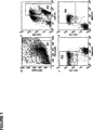
好適なクローンを見つけるために、数千の抗体産生ハイブリドーマをスクリーニングする必要があるので、必要とされる特異性を維持する一方、高処理を可能にする手順を構築した。赤血球前駆細胞の同時同定、除核赤血球と白血球の区別、および新しい抗体の同定をシングルステップで可能にする、6パラメータ分析(4蛍光チャネル、前方および側方散乱)を確立した。分析した細胞を、核酸色素(LDS751,Molecular Probes,cat# 7595)を用いて染色し、クローン化ハイブリドーマ細胞の抗体とともにインキュベートした。これらの抗体を、蛍光色素(FITC)で標識したそれらに対する抗体(ヤギ抗マウスIgG(H+L)-FITC,Caltag Laboratories,cat# M35001)と反応させた。抗体の性質を決定するその後の実験において、前記抗体はFITCで直接標識された。 Since it was necessary to screen thousands of antibody-producing hybridomas to find suitable clones, a procedure was constructed that allowed high throughput while maintaining the required specificity. A six-parameter analysis (4 fluorescence channels, forward and side scatter) was established that allows the simultaneous identification of erythroid progenitors, differentiation of enucleated erythrocytes and leukocytes, and identification of new antibodies in a single step. Analyzed cells were stained with nucleic acid dye (LDS751, Molecular Probes, cat # 7595) and incubated with the antibodies of cloned hybridoma cells. These antibodies were reacted with antibodies against them labeled with a fluorescent dye (FITC) (goat anti-mouse IgG (H + L) -FITC, Caltag Laboratories, cat # M35001). In subsequent experiments to determine the nature of the antibody, the antibody was directly labeled with FITC.
赤血球前駆細胞の同定は、この光散乱特性によって及びそれらのフィコエリトリン標識CD71特異抗体(CD71 PE,Diatec,cat#3212)の結合によって可能である。白血球は、アロフィコシアニン標識CD45特異抗体(CD45 APC,BD Pharmingen,cat#555485)へのそれらの結合によって識別できた。有核および除核の赤血球細胞は、それらの核酸色素との結合あるいは結合の欠如によって区別することができる。この手順によって、目的とする標的細胞、すなわち胎児のNRBCsへ結合する抗体を、成人の赤血球あるいは白血球との交差反応なしで同定することが可能である(図1)。 Identification of erythroid progenitors is possible by this light scattering property and by binding of their phycoerythrin labeled CD71 specific antibodies (CD71 PE, Diatec, cat # 3212). Leukocytes could be identified by their binding to allophycocyanin labeled CD45 specific antibodies (CD45 APC, BD Pharmingen, cat # 555485). Nucleated and enucleated red blood cells can be distinguished by their binding or lack of binding to nucleic acid dyes. By this procedure, antibodies that bind to the target cells of interest, ie fetal NRBCs, can be identified without cross-reaction with adult erythrocytes or leukocytes (FIG. 1).
共通血液型抗原を含む成人赤血球上の抗原と反応する抗体の排除
血液型抗原は、成人の赤血球およびそれらの前駆体上に大量に見受けられる。それゆえ、それらは抗原として使用された場合、主要な免疫応答を誘導するかもしれない。これらの血液型抗原に対する抗体は、胎児細胞の同定のために好適ではない。血液型抗原を有する成人赤血球上の抗原へ結合する抗体を排除するために、胎児細胞に関するそれらの結合特異性を、赤血球への吸収後に調査した。血液型AB Rh+の赤血球を収集し、Meridian Diagnostics Europe(バートホンブルク)が提供する試薬を用いて安定化した。調査において抗体は、数を増す赤血球とともにインキュベートされ、標的細胞に対するそれらの結合活性を前後で試験された。CD71+,CD45-有核赤血球前駆細胞への結合強度が赤血球とのインキュベーション後も不変である場合、血液型抗原に関する抗体の反応性は消失していると考えられる(図2)。このようにして選択された抗体は、成人の血液細胞とは反応せず、胎児赤血球前駆細胞の正確な同定を可能とする(図3)。
Exclusion of antibodies that react with antigens on adult erythrocytes, including common blood group antigens Blood group antigens are found in large quantities on adult erythrocytes and their precursors. Therefore, they may induce a major immune response when used as an antigen. Antibodies against these blood group antigens are not suitable for fetal cell identification. To eliminate antibodies that bind to antigens on adult erythrocytes with blood group antigens, their binding specificity for fetal cells was investigated after absorption into erythrocytes. Blood group AB Rh + red blood cells were collected and stabilized using reagents provided by Meridian Diagnostics Europe (Bert Homburg). In the study, antibodies were incubated with increasing numbers of red blood cells and tested before and after their binding activity against target cells. When the binding strength to CD71 +, CD45-nucleated erythrocyte progenitor cells remains unchanged after incubation with erythrocytes, it is considered that the antibody reactivity with respect to blood group antigens has disappeared (FIG. 2). The antibody selected in this way does not react with adult blood cells, allowing accurate identification of fetal erythroid progenitor cells (FIG. 3).
選択されたモノクローナル抗体の特異性試験
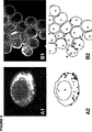
スクリーニング手順において必要とされる結合特性を示すIgMアイソタイプのモノクローナル抗体を産出するハイブリドーマ・クローンを同定することができた。それは、記号表示4B9を有し、本特許あるいは本特許出願の出願人によって、2004年7月13日に、ドイツ微生物および細胞培養コレクション(DSMZ,ブラウンシュヴァイク)に受入番号 DSM ACC 2666のもと寄託された。同一のエピトープを認識する第二の抗体4B8を、図2および図3に示す。
Specificity testing of selected monoclonal antibodies It was possible to identify hybridoma clones producing monoclonal antibodies of the IgM isotype that exhibited the binding properties required in the screening procedure. It has the symbol 4B9 and was filed with the German microorganism and cell culture collection (DSMZ, Braunschweig) on July 13, 2004, under the accession number DSM ACC 2666, by the applicant of this patent or this patent application. Deposited. A second antibody 4B8 that recognizes the same epitope is shown in FIGS.
胎児および成人の赤芽球は、グリコホリンAを強く明確に発現し、それゆえ、このマーカータンパク質によって同定できる。これらの細胞へのモノクローナル抗体の結合は、免疫蛍光二重染色によって可視化した。 Fetal and adult erythroblasts express glycophorin A strongly and clearly and can therefore be identified by this marker protein. Monoclonal antibody binding to these cells was visualized by immunofluorescence double staining.
免疫蛍光染色の手順
1.アセトン中で10分間サイトスピンあるいは凍結組織切片を固定する。
2.5分間乾燥する。
3.グリコホリンAに対するモノクローナル抗体(1%ウシ血清アルブミン(BSA)を含有するリン酸緩衝食塩水(PBS)にて1:100に希釈したDAKO M0819)を60分間適用する。
4.PBSですすぐ。
5.ヤギ抗マウス抗体F(ab)フラグメント[Alexa Fluor 488(Molecular Probes A-21044),グリーン,PBSにて1:100に希釈]を60分間適用する。
6.PBSですすぐ。
7.モノクローナル抗体4B9(ハイブリドーマ上清)を60分間適用する。
8.PBSですすぐ。
9.ヤギ抗マウスIgM[Alexa Fluor 594(Molecular Probes A-21044),レッド]を60分間適用する。
10.PBSですすぐ。
11.細胞核をDAPI[(Molecular Probes),ブルー,PBSにて1:300に希釈]で3分間染色する。
12.PBSですすぐ。
13.蛍光培地(S3023,DAKO)でカバーする。
14.フィルターセット02,10および15を用いてCarl Zeiss社の"Universalmikroskop Axioplan"を用いて可視化し、デジタルカメラシステム(例えば、Visitron Systems GmbH)を用いて撮影する。
PBS:1LのH2O中に8g NaCl,1.3g Na2HPO4,4g NaH2PO4.pH 7.4
Procedure for immunofluorescence staining Fix cytospin or frozen tissue sections for 10 minutes in acetone.
2. Dry for 5 minutes.
3. A monoclonal antibody against glycophorin A (DAKO M0819 diluted 1: 100 in phosphate buffered saline (PBS) containing 1% bovine serum albumin (BSA)) is applied for 60 minutes.
4). Rinse with PBS.
5. Apply goat anti-mouse antibody F (ab) fragment [Alexa Fluor 488 (Molecular Probes A-21044), green, diluted 1: 100 in PBS] for 60 minutes.
6). Rinse with PBS.
7). Monoclonal antibody 4B9 (hybridoma supernatant) is applied for 60 minutes.
8). Rinse with PBS.
9. Apply goat anti-mouse IgM [Alexa Fluor 594 (Molecular Probes A-21044), red] for 60 minutes.
10. Rinse with PBS.
11. Stain cell nuclei with DAPI [(Molecular Probes), blue, diluted 1: 300 with PBS] for 3 minutes.
12 Rinse with PBS.
13. Cover with fluorescent medium (S3023, DAKO).
14 Visualize with Carl Zeiss "Universalmikroskop Axioplan" using filter sets 02, 10 and 15 and photograph with a digital camera system (eg Visitron Systems GmbH).
PBS: 8 g NaCl, 1.3 g Na 2 HPO 4 , 4 g NaH 2 PO 4 in 1 L H 2 O. pH 7.4
免疫酵素法も使用した:
アルカリホスファターゼ-抗-アルカリホスファターゼ(APAAP)染色の手順
1.アセトン中で10分間サイトスピンあるいは凍結組織切片を固定する。
2.5分間乾燥する。
3.30分間モノクローナル抗体4B9(ハイブリドーマ上清)とインキュベートする。
4.トリス緩衝食塩水(TBS)ですすぐ。
5.30分間APAAP複合体[(D0651,DAKO),TBS/HS(不活化ヒト血清)にて1:25に希釈]とインキュベートする。
6.TBSですすぐ。
7.ステップ5〜7を10分おきに2度繰り返す。
8.TBSですすぐ。
9.基質を用いてスライドを展開する。
i.溶液Aの調製:18mlの0.2 mol/Lの2-アミノ-2メチル-1,3-プロパンジオールを50mlの0.05mol/Lのトリス緩衝液(pH 9.7)および600mgのNaClと混合する。28mgのレバミソールを添加する。
ii.溶液Bの調製:35mgのナフトールAS-bi-ホスフェートを0.42mlのN,N-ジメチルホルムアミド中に溶解する。
iii.溶液Cの調製:0.14mlの5%新フクシンと0.35の調製したばかりの4%亜硝酸ナトリウムを混合する。60秒間撹拌する。
iv.溶液Aと溶液Bを混合し、その後溶液Cを添加する。pHを8.7に調整する。混合し、濾過し、およびスライドに塗布する。
v.室温で10〜20分間インキュベートする。
vi.水道水ですすぐ。
vii.5分間、マイヤー酸Haemalaunで対比染色する。
viii.10分間水道水中で”青化”し、カイザーのグリセロール・ゼラチンでカバーする。
TBS(トリス緩衝食塩水):5LのH2O中に43.33gのNaClおよび39.40gのTris-HCLを溶解した。NaOHでpHを7.4に調節した。
TBS/HS:9部のTBS+1部の不活性化ヒト血清
ネガティブ・コントロール:同一のアイソタイプまたはマウスの過免疫血清のモノクローナル抗体
The immunoenzyme method was also used:
Alkaline phosphatase-anti-alkaline phosphatase (APAAP) staining procedure Fix cytospin or frozen tissue sections for 10 minutes in acetone.
2. Dry for 5 minutes.
3. Incubate with monoclonal antibody 4B9 (hybridoma supernatant) for 30 minutes.
4). Rinse with Tris-buffered saline (TBS).
5. Incubate with APAAP complex [(D0651, DAKO), diluted 1:25 in TBS / HS (inactivated human serum)] for 30 minutes.
6). Rinse with TBS.
7). Repeat steps 5-7 twice every 10 minutes.
8). Rinse with TBS.
9. The slide is developed using the substrate.
i. Solution A preparation: 18 ml of 0.2 mol / L 2-amino-2methyl-1,3-propanediol is mixed with 50 ml 0.05 mol / L Tris buffer (pH 9.7) and 600 mg NaCl. Add 28 mg of levamisole.
ii. Solution B preparation: 35 mg naphthol AS-bi-phosphate is dissolved in 0.42 ml N, N-dimethylformamide.
iii. Solution C preparation: Mix 0.14 ml of 5% fresh fuchsin with 0.35 freshly prepared 4% sodium nitrite. Stir for 60 seconds.
iv. Solution A and solution B are mixed and then solution C is added. Adjust the pH to 8.7. Mix, filter, and apply to slide.
v. Incubate at room temperature for 10-20 minutes.
vi. Rinse with tap water.
vii. Counterstain with Haemalaun Mayer acid for 5 minutes.
viii. "Blue" in tap water for 10 minutes and cover with Kaiser's glycerol gelatin.
TBS (Tris-buffered saline): 43.33 g NaCl and 39.40 g Tris-HCL were dissolved in 5 L H 2 O. The pH was adjusted to 7.4 with NaOH.
TBS / HS: 9 parts TBS + 1 part inactivated human serum
Negative control: monoclonal antibody of the same isotype or mouse hyperimmune serum
CD71と反応する抗体の排除
使用した免疫化方法で産生された抗体は、CD71と反応するかもしれない。このような抗体を除外するために、CD71-抗体が同じ結合サイトで競合するかどうかを調べる分析を行った。ビオチン化抗体4B8を臍帯血由来の単核細胞とともにプレ-インキュベートした。その後、非標識のCD71-特異抗体(Anti-CD71,クローンDF1513,DPC ビーアマン,バート・ナウハイム,ドイツ)を添加した。ストレプトアビジン-DTAF-標識化後、CD71-抗体が抗体4B8に置き換わるかどうかを、フローサイトメトリーによって分析した。この競合実験用のポジティブ・コントロールサンプルとして、非標識の抗体4B8をCD71の代わりに添加した。これらの分析により、非標識の抗体4B8の添加がそのシグナルを減少させたのに対して、抗体4B8およびCD71が同じエピトープに対して競合しないことが証明された。
Exclusion of antibodies that react with CD71 Antibodies produced by the immunization method used may react with CD71. To rule out such antibodies, an analysis was performed to determine whether CD71-antibodies compete at the same binding site. Biotinylated antibody 4B8 was pre-incubated with cord blood derived mononuclear cells. Thereafter, an unlabeled CD71-specific antibody (Anti-CD71, clone DF1513, DPC Beerman, Bad Nauheim, Germany) was added. After streptavidin-DTAF-labelling, whether the CD71-antibody replaced antibody 4B8 was analyzed by flow cytometry. As a positive control sample for this competition experiment, unlabeled antibody 4B8 was added instead of CD71. These analyzes demonstrated that addition of unlabeled antibody 4B8 reduced the signal, whereas antibody 4B8 and CD71 did not compete for the same epitope.
結果
・4B9-反応抗原が胎児赤芽球の表面に発現した。これは妊娠期間第6から第38週までの胎児細胞で実証できた。図4において、抗体4B9はすべてのグリコホリンA陽性胎児赤芽球を認識した。
・正常な成人骨髄における赤芽球は、4B9陰性であった。対照的に、全ての赤血球生成細胞はグリコホリンA陽性であった。32症例の1においてのみ、初期の好塩基性赤芽球の細胞質中に細胞内顆粒の発現が見られた。
・4B9反応抗原は、成人および胎児の肝細胞上で見つからなかった。クッパー細胞、マクロファージ、内皮細胞および類洞細胞も陰性であった。
・成人における血リンパ性細胞の詳細分析によって、リンパおよび骨髄球性の細胞において反応性がないことが示された。
・胎児の血リンパ性組織は全て陰性であった。これは骨髄球性の細胞だけでなくリンパ細胞にも当てはまる。
Results・ 4B9-reactive antigen was expressed on the surface of fetal erythroblasts. This could be demonstrated with fetal cells from the 6th to 38th week of gestation. In FIG. 4, antibody 4B9 recognized all glycophorin A positive fetal erythroblasts.
・ Erythroblasts in normal adult bone marrow were 4B9 negative. In contrast, all erythropoietic cells were glycophorin A positive. Only in 1 of 32 cases, intracellular granule expression was seen in the cytoplasm of early basophilic erythroblasts.
• No 4B9 reactive antigen was found on adult and fetal hepatocytes. Kupffer cells, macrophages, endothelial cells and sinusoidal cells were also negative.
• Detailed analysis of hemolymphoid cells in adults showed no reactivity in lymphoid and myeloid cells.
• All fetal hemolymph tissues were negative. This applies to lymphocytes as well as myeloid cells.

以下、図1〜4を詳細に説明する。 1 to 4 will be described in detail below.
Claims (10)
請求項7〜9のいずれか1項に記載の方法によって、胎児細胞を検出または同定した後、前記胎児細胞を、染色体及び/又は遺伝子の異常、欠損及び/又は変異について分析する工程を含む、方法。After detecting or identifying fetal cells by the method according to any one of claims 7 to 9, the fetal cells are analyzed for chromosomal and / or genetic abnormalities, defects and / or mutations. Method.
Applications Claiming Priority (3)
| Application Number | Priority Date | Filing Date | Title |
|---|---|---|---|
| EP04007811 | 2004-03-31 | ||
| EP04007811.5 | 2004-03-31 | ||
| PCT/EP2005/003371 WO2005100401A2 (en) | 2004-03-31 | 2005-03-31 | Monoclonal antibodies with specificity for fetal erythroid cells |
Publications (2)
| Publication Number | Publication Date |
|---|---|
| JP2007530629A JP2007530629A (en) | 2007-11-01 |
| JP4756032B2 true JP4756032B2 (en) | 2011-08-24 |
Family
ID=35150543
Family Applications (1)
| Application Number | Title | Priority Date | Filing Date |
|---|---|---|---|
| JP2007505500A Active JP4756032B2 (en) | 2004-03-31 | 2005-03-31 | Monoclonal antibody with specificity for fetal red blood cells |
Country Status (9)
| Country | Link |
|---|---|
| US (6) | US7858757B2 (en) |
| EP (1) | EP1732951B1 (en) |
| JP (1) | JP4756032B2 (en) |
| AT (1) | ATE450549T1 (en) |
| AU (1) | AU2005233254B2 (en) |
| CA (1) | CA2561830C (en) |
| DE (1) | DE602005018028D1 (en) |
| ES (1) | ES2337702T3 (en) |
| WO (1) | WO2005100401A2 (en) |
Families Citing this family (56)
| Publication number | Priority date | Publication date | Assignee | Title |
|---|---|---|---|---|
| WO2005100401A2 (en) * | 2004-03-31 | 2005-10-27 | Adnagen Ag | Monoclonal antibodies with specificity for fetal erythroid cells |
| US10083273B2 (en) | 2005-07-29 | 2018-09-25 | Natera, Inc. | System and method for cleaning noisy genetic data and determining chromosome copy number |
| US9424392B2 (en) | 2005-11-26 | 2016-08-23 | Natera, Inc. | System and method for cleaning noisy genetic data from target individuals using genetic data from genetically related individuals |
| US11111543B2 (en) | 2005-07-29 | 2021-09-07 | Natera, Inc. | System and method for cleaning noisy genetic data and determining chromosome copy number |
| US10081839B2 (en) | 2005-07-29 | 2018-09-25 | Natera, Inc | System and method for cleaning noisy genetic data and determining chromosome copy number |
| US11111544B2 (en) | 2005-07-29 | 2021-09-07 | Natera, Inc. | System and method for cleaning noisy genetic data and determining chromosome copy number |
| DE102006062619B4 (en) * | 2006-12-29 | 2012-04-26 | Medion Diagnostics Ag | Method for the determination of minor cell populations in heterogeneous cell populations |
| CA2731991C (en) | 2008-08-04 | 2021-06-08 | Gene Security Network, Inc. | Methods for allele calling and ploidy calling |
| ES2640776T3 (en) | 2009-09-30 | 2017-11-06 | Natera, Inc. | Methods for non-invasively calling prenatal ploidy |
| US11339429B2 (en) | 2010-05-18 | 2022-05-24 | Natera, Inc. | Methods for non-invasive prenatal ploidy calling |
| US11326208B2 (en) | 2010-05-18 | 2022-05-10 | Natera, Inc. | Methods for nested PCR amplification of cell-free DNA |
| US11332793B2 (en) | 2010-05-18 | 2022-05-17 | Natera, Inc. | Methods for simultaneous amplification of target loci |
| US11939634B2 (en) | 2010-05-18 | 2024-03-26 | Natera, Inc. | Methods for simultaneous amplification of target loci |
| US10316362B2 (en) | 2010-05-18 | 2019-06-11 | Natera, Inc. | Methods for simultaneous amplification of target loci |
| US11408031B2 (en) | 2010-05-18 | 2022-08-09 | Natera, Inc. | Methods for non-invasive prenatal paternity testing |
| US9677118B2 (en) | 2014-04-21 | 2017-06-13 | Natera, Inc. | Methods for simultaneous amplification of target loci |
| US20190010543A1 (en) | 2010-05-18 | 2019-01-10 | Natera, Inc. | Methods for simultaneous amplification of target loci |
| CA2798758C (en) | 2010-05-18 | 2019-05-07 | Natera, Inc. | Methods for non-invasive prenatal ploidy calling |
| US11322224B2 (en) | 2010-05-18 | 2022-05-03 | Natera, Inc. | Methods for non-invasive prenatal ploidy calling |
| US11332785B2 (en) | 2010-05-18 | 2022-05-17 | Natera, Inc. | Methods for non-invasive prenatal ploidy calling |
| US20120171120A1 (en) | 2010-11-30 | 2012-07-05 | Genentech, Inc. | Low affinity blood brain barrier receptor antibodies and uses therefor |
| CN103608466B (en) | 2010-12-22 | 2020-09-18 | 纳特拉公司 | Non-invasive prenatal paternity testing method |
| AU2011358564B9 (en) | 2011-02-09 | 2017-07-13 | Natera, Inc | Methods for non-invasive prenatal ploidy calling |
| US9404864B2 (en) | 2013-03-13 | 2016-08-02 | Denovo Sciences, Inc. | System for imaging captured cells |
| US10466160B2 (en) | 2011-08-01 | 2019-11-05 | Celsee Diagnostics, Inc. | System and method for retrieving and analyzing particles |
| EP2739587B1 (en) | 2011-08-01 | 2020-05-27 | Denovo Sciences | Cell capture system |
| US9174216B2 (en) | 2013-03-13 | 2015-11-03 | DeNovo Science, Inc. | System for capturing and analyzing cells |
| US20130122492A1 (en) * | 2011-11-14 | 2013-05-16 | Kellbenx Inc. | Detection, isolation and analysis of rare cells in biological fluids |
| US10058306B2 (en) | 2012-06-22 | 2018-08-28 | Preprogen, LLC | Method for obtaining fetal cells and fetal cellular components |
| US9752181B2 (en) | 2013-01-26 | 2017-09-05 | Denovo Sciences, Inc. | System and method for capturing and analyzing cells |
| US9707562B2 (en) | 2013-03-13 | 2017-07-18 | Denovo Sciences, Inc. | System for capturing and analyzing cells |
| US9856535B2 (en) | 2013-05-31 | 2018-01-02 | Denovo Sciences, Inc. | System for isolating cells |
| US10391490B2 (en) | 2013-05-31 | 2019-08-27 | Celsee Diagnostics, Inc. | System and method for isolating and analyzing cells |
| US9499870B2 (en) | 2013-09-27 | 2016-11-22 | Natera, Inc. | Cell free DNA diagnostic testing standards |
| US10577655B2 (en) | 2013-09-27 | 2020-03-03 | Natera, Inc. | Cell free DNA diagnostic testing standards |
| US10262755B2 (en) | 2014-04-21 | 2019-04-16 | Natera, Inc. | Detecting cancer mutations and aneuploidy in chromosomal segments |
| RU2717641C2 (en) | 2014-04-21 | 2020-03-24 | Натера, Инк. | Detection of mutations and ploidy in chromosomal segments |
| CN106488981A (en) | 2014-05-15 | 2017-03-08 | 科尔贝恩科斯公司 | The preparation of the fetal nucleated red blood (NRBCS) for diagnostic test |
| US20160130553A1 (en) * | 2014-05-15 | 2016-05-12 | Kellbenx Incorporated | PREPARATION OF FETAL NUCLEATED RED BLOOD CELLS (NRBCs) FOR DIAGNOSTIC TESTING |
| EP3179248A4 (en) * | 2014-08-05 | 2017-06-14 | Fujifilm Corporation | Method for separating nucleated red blood cells |
| US11479812B2 (en) | 2015-05-11 | 2022-10-25 | Natera, Inc. | Methods and compositions for determining ploidy |
| US11485996B2 (en) | 2016-10-04 | 2022-11-01 | Natera, Inc. | Methods for characterizing copy number variation using proximity-litigation sequencing |
| US10011870B2 (en) | 2016-12-07 | 2018-07-03 | Natera, Inc. | Compositions and methods for identifying nucleic acid molecules |
| JP6234542B1 (en) | 2016-12-27 | 2017-11-22 | 株式会社 TL Genomics | Method for obtaining chromosomal DNA derived from fetal cells |
| EP3585889A1 (en) | 2017-02-21 | 2020-01-01 | Natera, Inc. | Compositions, methods, and kits for isolating nucleic acids |
| CN111295245B (en) | 2017-08-29 | 2021-03-16 | 伯乐实验室有限公司 | Systems and methods for separating and analyzing cells |
| KR102313468B1 (en) | 2017-10-19 | 2021-10-14 | 티엘 제노믹스 인크. | Chip for cell sorting |
| US10946380B2 (en) * | 2018-01-19 | 2021-03-16 | International Business Machines Corporation | Microfluidic chips for particle purification and fractionation |
| US11525159B2 (en) | 2018-07-03 | 2022-12-13 | Natera, Inc. | Methods for detection of donor-derived cell-free DNA |
| US10633693B1 (en) | 2019-04-16 | 2020-04-28 | Celsee Diagnostics, Inc. | System and method for leakage control in a particle capture system |
| EP3963337A1 (en) | 2019-05-02 | 2022-03-09 | KellBenx Inc. | Filtration-based methods for preparing fetal nucleated red blood cells (nrbcs) for diagnostic testing |
| US11273439B2 (en) | 2019-05-07 | 2022-03-15 | Bio-Rad Laboratories, Inc. | System and method for target material retrieval from microwells |
| SG11202112151UA (en) | 2019-05-07 | 2021-12-30 | Bio Rad Laboratories | System and method for automated single cell processing |
| EP3982716A4 (en) | 2019-06-14 | 2023-09-06 | Bio-Rad Laboratories, Inc. | System and method for automated single cell processing and analyses |
| US11504719B2 (en) | 2020-03-12 | 2022-11-22 | Bio-Rad Laboratories, Inc. | System and method for receiving and delivering a fluid for sample processing |
| TW202328187A (en) | 2021-09-01 | 2023-07-16 | 美商百健Ma公司 | Anti-transferrin receptor antibodies and uses thereof |
Family Cites Families (3)
| Publication number | Priority date | Publication date | Assignee | Title |
|---|---|---|---|---|
| DE10035433C2 (en) * | 2000-07-20 | 2002-07-18 | Tuma Wolfgang | Gentle high enrichment of fetal cells from peripheral blood and use of the same |
| ITRM20010322A1 (en) * | 2001-06-08 | 2002-12-09 | Consiglio Nazionale Ricerche | NON-IVASIVE METHOD ON MATERNAL BLOOD FOR THE SCREENING AND DIAGNOSIS OF FETAL ANEUPLOIDIES. |
| WO2005100401A2 (en) * | 2004-03-31 | 2005-10-27 | Adnagen Ag | Monoclonal antibodies with specificity for fetal erythroid cells |
-
2005
- 2005-03-31 WO PCT/EP2005/003371 patent/WO2005100401A2/en active Application Filing
- 2005-03-31 AU AU2005233254A patent/AU2005233254B2/en active Active
- 2005-03-31 CA CA2561830A patent/CA2561830C/en active Active
- 2005-03-31 AT AT05736009T patent/ATE450549T1/en active
- 2005-03-31 JP JP2007505500A patent/JP4756032B2/en active Active
- 2005-03-31 ES ES05736009T patent/ES2337702T3/en active Active
- 2005-03-31 EP EP05736009A patent/EP1732951B1/en active Active
- 2005-03-31 DE DE602005018028T patent/DE602005018028D1/en active Active
- 2005-03-31 US US10/599,512 patent/US7858757B2/en active Active
-
2010
- 2010-12-28 US US12/979,535 patent/US8536312B2/en active Active
-
2013
- 2013-09-16 US US14/028,027 patent/US9194871B2/en active Active
-
2015
- 2015-08-13 US US14/825,516 patent/US20160039932A1/en not_active Abandoned
- 2015-10-14 US US14/882,995 patent/US9453841B2/en active Active
-
2022
- 2022-11-18 US US17/989,895 patent/US20230324383A1/en active Pending
Also Published As
| Publication number | Publication date |
|---|---|
| US20110129916A1 (en) | 2011-06-02 |
| US20140154704A1 (en) | 2014-06-05 |
| US8536312B2 (en) | 2013-09-17 |
| US20160039932A1 (en) | 2016-02-11 |
| CA2561830C (en) | 2013-05-21 |
| DE602005018028D1 (en) | 2010-01-14 |
| US20070275418A1 (en) | 2007-11-29 |
| ATE450549T1 (en) | 2009-12-15 |
| US20230324383A1 (en) | 2023-10-12 |
| ES2337702T3 (en) | 2010-04-28 |
| CA2561830A1 (en) | 2005-10-27 |
| EP1732951B1 (en) | 2009-12-02 |
| US9453841B2 (en) | 2016-09-27 |
| US7858757B2 (en) | 2010-12-28 |
| JP2007530629A (en) | 2007-11-01 |
| EP1732951A2 (en) | 2006-12-20 |
| AU2005233254A1 (en) | 2005-10-27 |
| AU2005233254B2 (en) | 2011-11-24 |
| US9194871B2 (en) | 2015-11-24 |
| US20160084837A1 (en) | 2016-03-24 |
| WO2005100401A2 (en) | 2005-10-27 |
| WO2005100401A3 (en) | 2006-06-15 |
Similar Documents
| Publication | Publication Date | Title |
|---|---|---|
| JP4756032B2 (en) | Monoclonal antibody with specificity for fetal red blood cells | |
| Janossy et al. | Immuno-histological diagnosis of lymphoproliferative diseases by selected combinations of antisera and monoclonal antibodies | |
| Van Der Schoot et al. | Monoclonal antibodies against myeloperoxidase are valuable immunological reagents for the diagnosis of acute myeloid leukaemia | |
| CA1248471A (en) | Immunoassay for breast cancer employing monoclonal antibodies | |
| JPH06505332A (en) | Treatment and diagnostic methods using total leukocyte surface antigen | |
| JPWO2010058860A1 (en) | Method and reagent for measuring C-reactive protein in sample | |
| CA2661803A1 (en) | Antibodies, assays and hybridomas | |
| JP2004504818A (en) | Mild high enrichment of fetal cells from peripheral blood and its use | |
| Van Heerde et al. | Non‐Hodgkin's Lymphoma. Immunohistochemical and electron microscopical findings in relation to lightmicroscopy A study of 74 cases | |
| HU208041B (en) | Monoclonal antibodies for treating human hon microcellular carcinoma pulmonum | |
| Jansson et al. | Classification of acute leukaemia by light and electron microscope cytochemistry | |
| CA2949324A1 (en) | Method and enzyme solution for flow-cytometric detection of light chain restriction | |
| US6844164B1 (en) | Antibody against rat postacrosome reaction sperm and utilization thereof | |
| Christensson et al. | Correlation of immunophenotype to morphology in unfavourable non‐Hodgkin lymphoma | |
| Lin et al. | Detection of exfoliated bladder cancer cells by monoclonal antibodies to tumor-associated cell surface antigens | |
| EP0106894B1 (en) | Methods and test kit for diagnosing cancer in humans | |
| CN116396942A (en) | Hybridoma cell, anti-P4 HB monoclonal antibody and kit | |
| Jaswaney et al. | Differential expression of H‐Y antigen on lymphocyte subsets: analysis by flow cytometry | |
| WO2003028625A2 (en) | Methods for screening monoclonal antibodies on heterogeneous antigen substrates | |
| JP3164922B2 (en) | Method for measuring human c-kit proto-oncogene product | |
| JOHANSSON | CORRELATION OF IMMUNOPHENOTYPE TO MORPHOLOGY IN UNFAVOURABLE NON-HODGKIN LYMPHOMA | |
| Noel et al. | ASCP/CAP Pathology Resident Award Finalists | |
| JP2001302700A (en) | New antibody against nk cell | |
| JPS63177798A (en) | Antibody of anti-human carcinoma of uterine, production and use thereof |
Legal Events
| Date | Code | Title | Description |
|---|---|---|---|
| A621 | Written request for application examination |
Free format text: JAPANESE INTERMEDIATE CODE: A621 Effective date: 20080207 |
|
| A131 | Notification of reasons for refusal |
Free format text: JAPANESE INTERMEDIATE CODE: A131 Effective date: 20101006 |
|
| A601 | Written request for extension of time |
Free format text: JAPANESE INTERMEDIATE CODE: A601 Effective date: 20110106 |
|
| A602 | Written permission of extension of time |
Free format text: JAPANESE INTERMEDIATE CODE: A602 Effective date: 20110114 |
|
| A601 | Written request for extension of time |
Free format text: JAPANESE INTERMEDIATE CODE: A601 Effective date: 20110207 |
|
| A602 | Written permission of extension of time |
Free format text: JAPANESE INTERMEDIATE CODE: A602 Effective date: 20110215 |
|
| A601 | Written request for extension of time |
Free format text: JAPANESE INTERMEDIATE CODE: A601 Effective date: 20110302 |
|
| A602 | Written permission of extension of time |
Free format text: JAPANESE INTERMEDIATE CODE: A602 Effective date: 20110309 |
|
| A521 | Request for written amendment filed |
Free format text: JAPANESE INTERMEDIATE CODE: A523 Effective date: 20110404 |
|
| A521 | Request for written amendment filed |
Free format text: JAPANESE INTERMEDIATE CODE: A821 Effective date: 20110404 |
|
| TRDD | Decision of grant or rejection written | ||
| A01 | Written decision to grant a patent or to grant a registration (utility model) |
Free format text: JAPANESE INTERMEDIATE CODE: A01 Effective date: 20110511 |
|
| A01 | Written decision to grant a patent or to grant a registration (utility model) |
Free format text: JAPANESE INTERMEDIATE CODE: A01 |
|
| A61 | First payment of annual fees (during grant procedure) |
Free format text: JAPANESE INTERMEDIATE CODE: A61 Effective date: 20110530 |
|
| FPAY | Renewal fee payment (event date is renewal date of database) |
Free format text: PAYMENT UNTIL: 20140603 Year of fee payment: 3 |
|
| R150 | Certificate of patent or registration of utility model |
Ref document number: 4756032 Country of ref document: JP Free format text: JAPANESE INTERMEDIATE CODE: R150 Free format text: JAPANESE INTERMEDIATE CODE: R150 |
|
| R250 | Receipt of annual fees |
Free format text: JAPANESE INTERMEDIATE CODE: R250 |
|
| R250 | Receipt of annual fees |
Free format text: JAPANESE INTERMEDIATE CODE: R250 |
|
| S111 | Request for change of ownership or part of ownership |
Free format text: JAPANESE INTERMEDIATE CODE: R313113 |
|
| S531 | Written request for registration of change of domicile |
Free format text: JAPANESE INTERMEDIATE CODE: R313531 |
|
| S533 | Written request for registration of change of name |
Free format text: JAPANESE INTERMEDIATE CODE: R313533 |
|
| R350 | Written notification of registration of transfer |
Free format text: JAPANESE INTERMEDIATE CODE: R350 |
|
| R250 | Receipt of annual fees |
Free format text: JAPANESE INTERMEDIATE CODE: R250 |
|
| R250 | Receipt of annual fees |
Free format text: JAPANESE INTERMEDIATE CODE: R250 |
|
| R250 | Receipt of annual fees |
Free format text: JAPANESE INTERMEDIATE CODE: R250 |
|
| R250 | Receipt of annual fees |
Free format text: JAPANESE INTERMEDIATE CODE: R250 |
|
| R250 | Receipt of annual fees |
Free format text: JAPANESE INTERMEDIATE CODE: R250 |
|
| R250 | Receipt of annual fees |
Free format text: JAPANESE INTERMEDIATE CODE: R250 |
|
| R250 | Receipt of annual fees |
Free format text: JAPANESE INTERMEDIATE CODE: R250 |
|
| R250 | Receipt of annual fees |
Free format text: JAPANESE INTERMEDIATE CODE: R250 |