JP4699609B2 - Novel acyl-dipeptide-like compound, process for its production and pharmaceutical composition containing it - Google Patents
Novel acyl-dipeptide-like compound, process for its production and pharmaceutical composition containing it Download PDFInfo
- Publication number
- JP4699609B2 JP4699609B2 JP2000557223A JP2000557223A JP4699609B2 JP 4699609 B2 JP4699609 B2 JP 4699609B2 JP 2000557223 A JP2000557223 A JP 2000557223A JP 2000557223 A JP2000557223 A JP 2000557223A JP 4699609 B2 JP4699609 B2 JP 4699609B2
- Authority
- JP
- Japan
- Prior art keywords
- group
- compound
- alkyl
- cells
- general formula
- Prior art date
- Legal status (The legal status is an assumption and is not a legal conclusion. Google has not performed a legal analysis and makes no representation as to the accuracy of the status listed.)
- Expired - Fee Related
Links
Images


Classifications
-
- C—CHEMISTRY; METALLURGY
- C07—ORGANIC CHEMISTRY
- C07C—ACYCLIC OR CARBOCYCLIC COMPOUNDS
- C07C237/00—Carboxylic acid amides, the carbon skeleton of the acid part being further substituted by amino groups
- C07C237/02—Carboxylic acid amides, the carbon skeleton of the acid part being further substituted by amino groups having the carbon atoms of the carboxamide groups bound to acyclic carbon atoms of the carbon skeleton
- C07C237/04—Carboxylic acid amides, the carbon skeleton of the acid part being further substituted by amino groups having the carbon atoms of the carboxamide groups bound to acyclic carbon atoms of the carbon skeleton the carbon skeleton being acyclic and saturated
- C07C237/06—Carboxylic acid amides, the carbon skeleton of the acid part being further substituted by amino groups having the carbon atoms of the carboxamide groups bound to acyclic carbon atoms of the carbon skeleton the carbon skeleton being acyclic and saturated having the nitrogen atoms of the carboxamide groups bound to hydrogen atoms or to acyclic carbon atoms
-
- C—CHEMISTRY; METALLURGY
- C07—ORGANIC CHEMISTRY
- C07C—ACYCLIC OR CARBOCYCLIC COMPOUNDS
- C07C237/00—Carboxylic acid amides, the carbon skeleton of the acid part being further substituted by amino groups
- C07C237/02—Carboxylic acid amides, the carbon skeleton of the acid part being further substituted by amino groups having the carbon atoms of the carboxamide groups bound to acyclic carbon atoms of the carbon skeleton
- C07C237/22—Carboxylic acid amides, the carbon skeleton of the acid part being further substituted by amino groups having the carbon atoms of the carboxamide groups bound to acyclic carbon atoms of the carbon skeleton having nitrogen atoms of amino groups bound to the carbon skeleton of the acid part, further acylated
-
- A—HUMAN NECESSITIES
- A61—MEDICAL OR VETERINARY SCIENCE; HYGIENE
- A61P—SPECIFIC THERAPEUTIC ACTIVITY OF CHEMICAL COMPOUNDS OR MEDICINAL PREPARATIONS
- A61P35/00—Antineoplastic agents
-
- A—HUMAN NECESSITIES
- A61—MEDICAL OR VETERINARY SCIENCE; HYGIENE
- A61P—SPECIFIC THERAPEUTIC ACTIVITY OF CHEMICAL COMPOUNDS OR MEDICINAL PREPARATIONS
- A61P37/00—Drugs for immunological or allergic disorders
-
- A—HUMAN NECESSITIES
- A61—MEDICAL OR VETERINARY SCIENCE; HYGIENE
- A61P—SPECIFIC THERAPEUTIC ACTIVITY OF CHEMICAL COMPOUNDS OR MEDICINAL PREPARATIONS
- A61P37/00—Drugs for immunological or allergic disorders
- A61P37/02—Immunomodulators
-
- C—CHEMISTRY; METALLURGY
- C07—ORGANIC CHEMISTRY
- C07C—ACYCLIC OR CARBOCYCLIC COMPOUNDS
- C07C235/00—Carboxylic acid amides, the carbon skeleton of the acid part being further substituted by oxygen atoms
- C07C235/02—Carboxylic acid amides, the carbon skeleton of the acid part being further substituted by oxygen atoms having carbon atoms of carboxamide groups bound to acyclic carbon atoms and singly-bound oxygen atoms bound to the same carbon skeleton
- C07C235/04—Carboxylic acid amides, the carbon skeleton of the acid part being further substituted by oxygen atoms having carbon atoms of carboxamide groups bound to acyclic carbon atoms and singly-bound oxygen atoms bound to the same carbon skeleton the carbon skeleton being acyclic and saturated
- C07C235/10—Carboxylic acid amides, the carbon skeleton of the acid part being further substituted by oxygen atoms having carbon atoms of carboxamide groups bound to acyclic carbon atoms and singly-bound oxygen atoms bound to the same carbon skeleton the carbon skeleton being acyclic and saturated having the nitrogen atom of at least one of the carboxamide groups bound to an acyclic carbon atom of a hydrocarbon radical substituted by nitrogen atoms not being part of nitro or nitroso groups
-
- C—CHEMISTRY; METALLURGY
- C07—ORGANIC CHEMISTRY
- C07F—ACYCLIC, CARBOCYCLIC OR HETEROCYCLIC COMPOUNDS CONTAINING ELEMENTS OTHER THAN CARBON, HYDROGEN, HALOGEN, OXYGEN, NITROGEN, SULFUR, SELENIUM OR TELLURIUM
- C07F9/00—Compounds containing elements of Groups 5 or 15 of the Periodic System
- C07F9/02—Phosphorus compounds
- C07F9/06—Phosphorus compounds without P—C bonds
- C07F9/08—Esters of oxyacids of phosphorus
- C07F9/09—Esters of phosphoric acids
- C07F9/091—Esters of phosphoric acids with hydroxyalkyl compounds with further substituents on alkyl
-
- C—CHEMISTRY; METALLURGY
- C07—ORGANIC CHEMISTRY
- C07K—PEPTIDES
- C07K5/00—Peptides containing up to four amino acids in a fully defined sequence; Derivatives thereof
- C07K5/02—Peptides containing up to four amino acids in a fully defined sequence; Derivatives thereof containing at least one abnormal peptide link
-
- C—CHEMISTRY; METALLURGY
- C07—ORGANIC CHEMISTRY
- C07K—PEPTIDES
- C07K5/00—Peptides containing up to four amino acids in a fully defined sequence; Derivatives thereof
- C07K5/04—Peptides containing up to four amino acids in a fully defined sequence; Derivatives thereof containing only normal peptide links
- C07K5/06—Dipeptides
- C07K5/06191—Dipeptides containing heteroatoms different from O, S, or N
Description
【0001】
【発明の技術分野】
本発明は化学の分野、特に医薬品に関するものである。
本発明は特に、ヒドロキシ化されたアミノ酸から得られる、アミノ酸のフリーなアミン官能基が脂肪酸によってアミド化された準ジペプチド(pseudodipeptides)化合物(以下、ジペプチド様化合物という)関するものである。
【0002】
本発明は特に、少なくとも一つの水酸基が中性または帯電した形の酸基によってエステル化された、下記一般式(I)で表されるN-アシルされたジペプチド様化合物に関するものである:
【0003】
【化15】

[ここで
R1およびR2は直鎖または分岐した2〜24の炭素原子を有する飽和または不飽和のカルボン酸に由来するアシル基を表し、ヒドロキシル、アルキル、アルコキシ、アシロキシ、アミノ、アシルアミノ、アシルチオおよび((C1-24)アルキル)チオ基から成られる群の中から選択される一つまたは一つ以上の置換基を有していてもよく、
m、pおよびqは1〜10の整数であり、
nは0〜10までの数であり、
XおよびYは水素または中性でも帯電した形でもよい酸基を表し、ただし、置換基X およびYの少なくとも一つは中性または帯電した形の酸基を表し、
AおよびBは互いに独立して酸素原子、硫黄原子またはイミノ基-NH-を表す]
置換基X およびYは下記の1)〜9)の中から選択するのが好ましい:
1) カルボキシ[(C1-5)アルキル]
2) CH-[(CH2)mCOOH][(CH2)nCOOH]
(ここでm = 0 〜5 、n = 0 〜5)
3) ホスホノ[(C1-5)アルキル]
4) ジヒドロキシホスホリルオキシ[(C1-5)アルキル]
5) ジメトキシホスホリル
6) ホスホノ
7) ヒドロキシスルホニル
8) ヒドロキシスルホニル[(C1-5)アルキル]
9) ヒドロキシスルホニルオキシ[(C1-5)アルキル]
【0005】
置換基Xおよび/またはYが中性である酸基としてはフリーなカルボキシル基、スルホン酸基または燐酸基を挙げることができる。帯電した形の酸基としてはカルボン酸塩、スルホン酸塩または燐酸塩、すなわち、有機または無機塩基、特に医薬用途の塩基の付加塩を挙げることができる。医薬用途以外の塩基は同定、精製および分離のための手段を提供する。
Xおよび/またはYがカルボキシルアルキル、アルセニルビスカルボキシル、ヒドロキシスルホニル、ヒドロキシスルホニルアルキル、ヒドロキシスルホニルオキシアルキル、ホスホノアルキル、ホスホリルオキシアルキル基を表す場合にも同じことがいえる。
【0006】
医薬用途の塩を形成する塩基にはアルカリ塩基、例えば水酸化ナトリウム、カリウムまたはリチウム、アンモニウム塩、アルカリ土類金属塩基、例えば水酸化カルシウムまたはストロンチウム、マグネシウム塩、第一鉄金属塩や、有機塩基、例えば第一、第二、第三アミン、例えばメチルアミン、ジエチルアミン、モノエタノールアミン、ジエタノールアミン、ベンジルアミン、N-メチルベンジルアミン、ベラトリルアミン、トリメトキシベンジルアミンや、リシンやオルニチンのような塩基性アミノ酸またはアミノ糖に由来する有機塩基が含まれる。
医薬用途以外の塩基の例としてはブルシン、ストリキニーネ、アグマチン、ホマリン、グルコサミン、N-メチルグルコアミンまたはN-メチルモルホリンを挙げることができる。既に述べたように、これらから誘導される塩は分離、識別手段として利用できる。
【0007】
mが1で、nが0の場合の重要な分子はセリンから得られる。mが2で、nが0の場合に考えられる分子はホモセリンから得られる。mが3で、nが0の場合にはペンタホモセリン化合物になり、mが4で、nが0の場合にはヘキサホモセリン化合物になる。
pが3で、qが1の場合に重要な化合物はシトルリン、オルニチンまたはアルギニン化合物である。pが4で、qが1の場合にはホモアルギニンまたはリシン化合物が挙げられる。
本発明に含まれるジペプチド様化合物において、下記一般式(I')の化合物に特に注目されたい:
【0008】
【化16】
[ここで
R1およびR2は直鎖または分岐した2〜24の炭素原子を有する飽和または不飽和のカルボン酸鎖に由来するアシル基を表し、ヒドロキシル、アルキル、アルコキシ、アシロキシ、アミノ、アシルアミノ、アシルチオおよび((C1-24)アルキル)チオ基から成られる群の中から選択される一つまたは一つ以上の置換基を有していてもよく、
m、pおよびqは1〜10の整数であり、
nは0〜10までの整数であり、
XおよびYは水素またはホスホノ基を表す]
【0010】
すなわち、
3-(3-ドデカノイルオキシテトラデカノイルアミノ)9-(3-ヒドロキシテトラデカノイルアミノ)4-オキソ-5-アザデカン-1、10-ジオール1および/または10-二水素ホスフェートおよびその有機または無機塩基付加塩、
3-(3-ドデカノイルオキシ−テトラデカノイルアミノ)9-(3-ヒドロキシテトラデカノイルアミノ)4-オキソ-5-アザデカン-1、10-ジオール1,10-ビス(二水素ホスフェート)およびその有機または無機塩基付加塩、
3-(3-ヒドロキシテトラデカノイルアミノ)9-(3-ドデカノイルオキシテトラデカノイルアミノ)4-オキソ-5-アザデカン-1、10-ジオール1,10- ビス(二水素ホスフェート)およびその有機または無機塩基付加塩、
3-(3-ドデカノイルオキシテトラデカノイルアミノ)9-(3-ヒドロキシテトラデカノイルアミノ)4-オキソ-5-アザデカン-1、10-ジオール1-二水素ホスフェートおよびその有機または無機塩基付加塩、
3-(3-ヒドロキシテトラデカノイルアミノ)9-(3-ドデカノイルオキシテトラデカノイルアミノ)4-オキソ-5-アザデカン-1、10-ジオール1-二水素ホスフェートおよびその有機または無機塩基付加塩、
3-(3-ヒドロキシテトラエカノイルアミノ)9-(3-ドデカノイルオキシテトラデカノイルアミノ)4-オキソ-5-アザデカン-1、10-ジオール10-二水素ホスフェートおよびその有機または無機塩基付加塩。
【0011】
R1およびR2は飽和または不飽和の分岐または直立鎖の各種長さの鎖を有するアシル誘導体を含み、互いに同一でも違ってしてもよく、アルキル、アミノ、アシルアミノ、ヒドロキシル、アルコキシ、アシロキシアシルチオおよびアルキルチオ基から成る群の中から選択される一つ以上の置換基を有していてもよい。
アシル化された置換誘導体の例はリシノイル、12-ヒドロキシステアロイル、2-ヒドロキシ-3-メチルブチロイル、3-ヒドロキシ-2-アミノペンタノニル、パルミトイル、エライジル、エレオステアロイル、アラキドイル、アラキドニル、ガドレイル、ベヘニル、エルシル、8-メチルデカノイル、9-メチルデカノイル、ドデカソヘキサノイルまたはエイコサペンタノイル基である。
【0012】
一般式(I)の化合物、特にコード名でOM-294-MP(MP)およびOM-294-DP(DP)とよばれるモノ-およびビス-フォスフォリル化された化合物は重要な薬理学的性質、主として免疫修飾に関する特性を有している。これらは特に免疫防御系の欠失または免疫応答の過剰発現に関連する疾患の治療に使われる。どちらの治療に用いるかによって服用量を変える。また、これらは癌の治療およびワクチン製造でのアジュバントまたは応答エンハンサとして使用することができる。
その他の用途としては治療用分子のためのベクターとしての使用がある。これはその親水性または疎水性の作用に基づく共有結合複合体を作るその能力のためである。また、その両親媒特性から治療用分子の形成および膜受容体、細胞膜および細胞原形質への輸送を促進する。本発明化合物は単独または治療用分子と一緒に経口、経腸管外、経直腸、局所、経皮下または経粘膜経路で投与することができる。また、本発明化合物は単独または治療用分子と一緒に血液細胞でイクスビボ即時培養を行って、それを注射する前の免疫応答細胞の形成を促進し、インビボの非経口投与へ戻ることができる。
【0013】
上記のMPおよびDPの分子は、適当な抗原と組み合わせて、ウイルス、寄生性、微生物または菌類を起源とする疾患に対する例えばワクチンで用いた場合には免疫系のアジュバントとしてに同様な性質を示す。これとは対照的に、本発明化合物は全く異なる性質を示し、サイトカイン産生や、造血性およびリンパ性器官に由来する免疫適格幹細胞の成熟を誘導する。
MP化合物は、適当な抗原の存在下または非存在下に単核細胞の成熟と官能性樹状細胞への分化を促進し、体液および細胞を介した免疫性を促進する役目をする。一方、DP化合物は抗腫瘍性を示す。
【0014】
本発明化合物は毒性が低い点で特に重要であり、人間および動物に0.025mgから100mgの単位供与量で服用でき、一日当たりの投与量は0.05から200mgである。
本発明はさらに、一般式(I)のジペプチド様化合物の製造方法にも関するものである。本発明方法は、容易に酸加水分解および水素化分解が可能なブロッキング剤によってジアミノ酸の(q+1)位置およびω位置のアミン官能基をブロックし、フリーであるカルボキシル官能基を還元剤とを反応させて対応するアルコールを作り、(q+1)位置のアミン官能基をフリーにし、次いで、式R2OHのカルボン酸官能性誘導体(ここで、R2は上記定義のもの)によってアシル化し、その後、水素化分解によって末端のアミン官能基をフリーにして一般式(II):
【0015】
【化17】
(ここで
R2は2〜24の炭素原子を有する飽和または不飽和の直鎖または分枝鎖のカルボン酸に由来するアシル基を表し、一つ以上の上記置換基を有していもよく、pおよびqは1〜10まで整数を表す)
のジアミノアルコールを作り、
このジアミノアルコールを不活性溶媒中でペプチド縮合剤の存在下で下記一般式(III):
【0017】
【化18】
(ここで、R1は2〜24の炭素原子を有する飽和または不飽和の直鎖または分枝鎖のカルボン酸に由来するアシル基を表し、一つ以上の上記置換基を有していもよく、mは1〜10までの整数を表し、nは0〜10までの整数を表し、Aは酸素原子、硫黄原子またはイミノ基NHを表し、Xは酸基を表し、必要に応じてエステルの形をしていてもよい)
のω-ヒドロキシ、ω−アミノまたはω−チオアミノ酸化合物と縮合させて下記一般式(IV)のジペプチド様化合物にする:
【0019】
【化19】
(ここで、置換基R1、R2および添字n、m、p、qは上記定義の意味を有する)
この式のアルコール官能基を必要に応じてアルキルまたはアシルでもよく、あるいは、アルキルまたはアシルで置換されていてもよく、さらには、必要な場合にはカップリング剤の存在下に、他の置換試剤で置換されていてもよく、触媒水素化または他の脱保護方法で一般式(I)の誘導体を得る:
【0021】
【化20】
(ここで、置換基およびA、B、X、Y、R1、R2、n、m、pおよびqは上記と同じ意味を有する)。
本発明はさらに、下記一般式(I')のフォスフォジペプチド様化合物の製造方法に関するものである:
【0023】
【化21】
[ここで
R1およびR2は直鎖または分岐した2〜24の炭素原子を有する飽和または不飽和のカルボン酸鎖に由来するアシル基を表し、ヒドロキシル、アルキル、アルコキシ、アシロキシ、アミノ、アシルアミノ、アシルチオおよび((C1-24)アルキル)チオ基から成られる群の中から選択される一つまたは一つ以上の置換基を有していてもよく、
m、pおよびqは1〜10の整数であり、
nは0〜10までの整数であり、
XおよびYは水素またはホスホノ基を表し、中性でも帯電した形でもよい]
本発明方法は、式:H2N(CH2)pCHNH2(CH2)q+1COOHのジアミノ酸の(q+1)位置およびω位置のアミン官能基を容易に酸加水分解および水素化分解されるブロッキング剤でブロックし、遊離した形にあるカルボキシル基を還元剤と反応させて対応するアルコールにし、(q+1)位置のアミン官能基をフリーにし、次いで、式R2OH(ここでR2は上記定義のもの)のカルボン酸官能性誘導体を用いてアシル置換し、次いで、水素化分解によって末端アミン官能基を遊離させて、一般式(II):
【0025】
【化22】
(ここで
R2は2〜24の炭素原子を有する飽和または不飽和の直鎖または分枝鎖のカルボン酸に由来するアシル基を表し、一つ以上の上記置換基を有していもよく、pおよびqは1〜10まで整数を表す)
のアミノアルコールを作り、
このアミノアルコールを不活性溶媒中でペプチド縮合剤の存在下で下記一般式(III'):
【0027】
【化23】
(ここで、R1は2〜24の炭素原子を有する飽和または不飽和の直鎖または分枝鎖のカルボン酸に由来するアシル基を表し、一つ以上の上記置換基を有していもよく、mは1〜10までの整数を表し、nは0〜10までの整数を表し、Aは酸素原子、硫黄原子またはイミノ基NHを表し、Xは下記の式:
【0029】
【化24】
ジアルキルオキシ-またはジアリールオキシ-ホスホリル基を表す)
のω-ヒドロキシアミノ酸化合物と縮合させて下記一般式(IV'):
【0031】
【化25】
(ここで、置換基R1、R2および添字n、m、p、qは上記定義の意味を有し、Rは容易に水素化分解される基を表す)
のペプチド様化合物にし、必要な場合には、この式の他のアルコール官能基をカップリング剤の存在下で(必要な場合)フォスフォリレート化剤によってフォスフォリレート化し、必要に応じて、触媒水素化してアシル基R2上に必要に応じて存在するアルコール官能基のブロックを外し、フォスフェート官能基を自由にし、次いで、水素化分解によって必要に応じて存在する第2のフォスフェート官能基のブロックを外して下記一般式(V)の誘導体にする:
【0033】
【化26】
(ここで、Yは水素原子またはフォスフォノ基を表す)。
さらに、必要に応じて有機または無機塩基によっ塩を作るステップを実行する。
【0035】
アシルアミノ基のキラル中心の立体化学は先ず最初に用いたアミノ酸構造によって決定され、アシルアミノ基の立体化学は最初に用いた脂肪酸の構造に依存する。LまたはD構造のジアミノ酸またはラセミ体から出発することができる。L、D構造のヒドロキシ化されたアミノ酸またはラセミ体混合物から出発することができる。この種の全ての立体異性体またはジアステレオアイソマが本発明の範囲に含まれる。
本発明方法は下記の反応スキーム1、2、3(図33、34、35)に概要を示した現時点での好ましい操作手順で定義することもできる:
【0036】
1. オルニチン誘導体鎖のω位置のアミン官能基をN-ベンジルオキシカルボニル置換によってブロックする。先ず最初にアルカリ性媒体中で酸官能基を銅の塩と反応させ、銅カルボキシレートをベンジルルクロロホルメートと反応させ、酸性環境中で銅をキレート化してエルボン酸基をフリーにし、Organic Preparations and Procedures International, 23、1992、191−194に開示される方法に従ってN-ベンジルオキシカルボニル置換誘導体を得る。
【0037】
2. アルカリ媒体中でアルキルピロカルボネート、例えばtert-ブチルピロカーボネートによってtert-ブチルオキシカルボニル置換を行って、オルニチン誘導体のα位置のカルボキシルアミン官能基をブロックする。
tert-ブチルピロカーボネートがアミン官能基と反応すると、ω-ベンジルオキシカルボニルアミノα-tert-ブチルカルボニルアミノカルボキシル誘導体が得られる。
【0038】
3. Tetrahedron Letters, 32 (1991) 923 -926に開示の方法に従ってカルボキシル官能基を第一級アルコール官能基へ変換する。この方法ではカルボキシル誘導体をアルキルクロロホルメート(例えばイソブチルクロロホルメート)と反応させて混合無水物を作り、それをアルカリまたはアルカリ土類金属のボロハイドレートと反応させて、第一アルコール官能基を有する対応するヒドロキシ化誘導体にする。
【0039】
4. α位置のtert-ブチルオキシカルボニル基の除去をトリフルオロ酢酸を用いて実行する。それと同時にアミン官能基対応トリフルオロアセタトが形成される。
5. フリーになったアミン官能基のアシル化は、R2OH酸とアルキルクロロホルメートから調製される混合無水物によるトリフルオロ酢酸の塩から出発する。
6. 末端アミン官能基の遊離は貴金属をベースにした触媒(例えば炭素またはイリジウム上に保持した白金またはパラジウム)の存在の水素化分解で行う。
【0040】
7. 式(II)のアミノ化合物と式(III')のホスホリル誘導体との間のペプチドカップリングまたは連結は、カップリング剤、例えば1-イソブチルオキシ-2-イソブチルオキシカルボニル-1、2- ジヒドロキノリンの存在に不活性溶媒(例えばハロゲン含有溶媒)の存在下またはカルボジイミドの存在下に行う。
こうして、一般式(IV')のジペプチド様化合物が得られる。この化合物のアシル基R2によって生じるヒドロキシル官能基は必要に応じてブロックされる。
【0041】
8. アシル基R2のヒドロキシル官能基をフリーにするには炭素等の基質上に塗布したパラジウムのような貴金属の存在下で水素化分解する。
9. 燐酸基をフリーにするには酸化白金のような貴金属酸化物の存在下で触媒水素化する。
10. ジペプチド様誘導体(IV')のフォスホリル化は2段ステップで行う(Helv. Chim. Acta, 70(1987)、175)。最初のステップで、カップリング剤、例えば[1H]−テトラゾールテトラの存在下で、ヒドロフランのような極性溶媒中で化合物(IV')をジアルキルまたはジアリール−N,N-ジアルキルフォスホロアミダイト(phosphoramidite)と反応させる。得られた亜リン酸エステルを芳香族ペルオキシカルボン酸、例えばたとえばペルオキシフタル酸、m-クロロ過安息香酸またはニトロ過安息香酸によって酸化してフォスフェートにする。燐酸基Y(式V)をフリーにするには炭素に含浸したパラジウムのような貴金属の存在下で触媒水素化する。
【0042】
11. ホモセリン誘導体のフォスホリル化はピリジンおよびN,N-ジアルキルアミノピリジンの存在下でハロゲン化ジフェニルホスホリルによって行う(Helv. Chim. Acta, 58: (1975), 518)。まず、アルカリ媒体中でtert-ブチルピロカー
ボネートによってtert-ブトキシカルボニル置換でアミン官能基をブロックし、次いで、カルボニル置換基をブロックし、セシウム塩を形成し、ジメチルホルムアミドまたはジメチルアセトアミド中でベンジルハライドによってカルボキシル官能基をベンジル化する。
【0043】
12. ホモセリン誘導体の窒素原子のアクリル化は、トリフルオロ酢酸によってアミン官能基の保護を解除してアミントリフルオロ酢酸塩を得、N-メチルモルホリンのようなアミンの存在下でのカルボン酸R1OHとアルキルクロロホルメートとの反応で得られる混合無水物と反応させる。
【0044】
本発明はさらに、純粋のエナンチオマーまたは立体異性体混合物の形をした、一般式(II)(III)および(III')の中間体に関するものである。
本発明はさらに、少なくとも一種の一般式(I)の化合物を中性または帯電させた形で有効成分として含み、それと薬学的に許容される有毒、不活性な賦形剤またはキャリアとを組合わせた医薬組成物に関するものである。
本発明は特に、有効成分として一般式(I)の化合物の少なくとも一種の塩と、治療を目的とする有機または無機の塩基とを一緒に含む医薬品組成物に関するものである。
【0045】
本発明はさらに、純粋なエナンチオマーの形または立体異性体の混合物の形の一般式(I)の化合物をベースにし、それに製薬賦形剤またはキャリアとを組合わせた医薬組成物に関するものである。
医薬品製剤としては経粘膜投与、経皮、局所投与、経腸管外、経消化系または吸入投与が挙げられ、例えば糖衣錠、非糖衣錠、カプセル、注射液、懸濁液、スプレー、ゲル、膏薬または速吸収液等が適している。
本発明の化合物は水溶液または懸濁液にし、必要に応じてアミンまたはヒドロキシルアミンによって中和した注射液として投与するのが好ましい。
以下、本発明の実施例を示すが、本発明が以下の実施例に限定されるものではない。本発明の反応スキーム1〜6は図33〜38にその概要が示されている。
【0046】
実施例 I
4-(ジフェニルオキシホスホリルオキシ )-2-[(R)3- ドデカノイルオキシテトラデカノイルアミノ ] ブタン酸
1. N α -tert- ブチルオキシカルボニル -DL- ホモセリン
2gのホモセリン(16.78mmol)を20mlの水に溶解し、得られた溶液に16.78mlの1M NaOHと3.006gの炭酸セシウム(9.23mmol)とを加えた。5分間撹拌した後、溶液を氷/水浴で冷却された。次に、60mlのジオキサンとtert-ブチルピイロカルボネートを加えられた。反応液を氷/水浴で中で1時間攪拌した後、室温で5時間撹拌した。次いで、溶媒を真空除去した。乾燥した残さ次のステップで直接用いた。
【0047】
2. N α -tert- ブチルオキシカルボニル−ベンジル -DL- ホモセリネート
ステップ1の残基に20mlのジメチルホルムアミドを加え、溶媒を蒸発乾燥させた。次いで、60mlのジメチルホルムアミドと、4.5mlの臭化ベンジル(20.13mmol)とを反応液に加えた。この時点で白い沈澱物ができる。混合物を16時間撹拌下に維持する。次いで溶媒を真空除去する。残基を乾固し、20mlの酢酸エチルで二度抽出する。有機層を水(20ml)と塩水(20ml)とでそれぞれ洗浄し、無水硫酸マグネシウム上で乾燥する。溶媒を蒸発する。残さは次のステップで使用する。
【0048】
3. ベンジル N α -tert- ブチルオキシカルボニル( O-( ジフェニルオキシホスホリル )-DL- ホモセリネート
前のステップの残基を高真空下で乾燥した後、塩化メチレン(60ml)に溶かす。次いで、溶液に4,11gの4-ジメチルアミノピリジン(33.56mmol)を加え、反応液を10分間撹拌し、12mlのピリジンと6.95mlのクロロホスフェート(33.56mmol)とを加える。溶液を18時間の室温で撹拌した後、1N−塩酸塩(5x 20ml)、水(30ml)および塩水(30ml)で洗浄する。有機層は無水硫酸マグネシウム上で乾燥し、溶媒を真空除去する。残基はフラッシュクロマトグラフィ(ヘキサン/酢酸エチル= 4:1)で精製する。主要画分を濃縮して残基を結晶化する。その結果、7,49gのフォスフォリレート化された化合物が得られた(収量:82.4%)。融点:63.5−64.0℃。
【0049】
4 .ベンジル O-( ジフェニルオキシホスホリル )-DL- ホモセリネート
前のステップ(7.88g、15.4mmol)のフォスフォリレート化された化合物を15mlのトリフルオロ酢酸に溶解し、溶液を室温下で2.5時間撹拌する。溶媒を真空除去し、残基をフラッシュクロマトグラフ(MeOH/CH2Cl2 = 10:1)で精製する。主要画分を濃縮して残基を室温で結晶化する。その結果、7,17gのフォスフォリレート化された化合物を回収する(収量:88.9%)。これを次のステップでそのまま使用する。
【0050】
5. ベンジル 2-[(R)-3- ドデカノイルオキシテトラデカノイルアミノ ]-4-( ジフェニルオキシホスホリルオキシ ) ブトネート
Bull. Chem. Soc. Jpn., 60 (1987), 2205 _ 2214に記載の方法に従って調製した4.284 g(10.07mmol)の(R)3-ドデカノイルオキシテトラデカン酸を30mlのテトラヒドロフランに溶解し、溶液を氷冷した塩水槽で-15℃に冷却する。次いで、1,108ml(10.07mmol)のN-メチルモルフォリンと、1.31ml(10.07mmol)のイソブチルクロロホルメートを加える。撹拌を30分間継続する。反応液に30mlのテトラヒドロフランと5mlのトリエチルアミンとの混合物中に溶かした5.724g(10.07mmol)のベンジルO-(ジフェニルオキシホスホリル)-DL-ホモセリネートを加える。室温で一晩撹拌した後、溶媒を真空除去し、残基に20mlの水を加える。得られた混合物を酢酸エチル(2x 30ml)で抽出し、有機層を水(20ml)および塩水(20ml)で連続して洗浄し、硫酸マグネシウム上で乾燥する。溶媒を蒸発し、残基をフラッシュクロマトグラフィで精製した(ヘキサン−酢酸エチル 2:1、Rf = 0.29)、収量7.455 g すなわち87.1% 、m.p. 31.0°−32.1℃、1H-NMR(250MHz、CDCl3)、δ ppm:7.4 −7.1(15H、m)、6.90(2d、1H、3J 7.6Hz =、NH)、5.3-5.1(3H、m)、4.7(1H、m)、4.35(2H、m)、2.45(2H、m)、2.4-2.1(4H、m)、1.6(4H、m)、1.4-1.1(34H、m)、0.9(6H、t)。13C-NMR(63MHz、CDCl3)(δ ppm):173.01、171.08、169.66、150.18(d(2JP,C =)7.1Hz)135.01、129.60、128.33、128.14、127.96、125.21、119.80(d(3JP,C =)5.0Hz)、70.69、67.05、65.19(d,2JP,C = 5.6Hz)、49.13、40.97、40.77(二つのdiast。)、34.20、33.98、33.82、31.70、29.42、29.34、29.14、28.94、25.01、24.47、13.91。
【0051】
6. 4-( ジフェニルオキシホスホリルオキシ )-2-[(R)-3- ドデカノイルオキシテトラデカノイルアミノ ] −ブタン酸
ステップ5で得られた2.23gすなわち2.6mmolのベンジルエステルを三つの首丸底フラスコ中で300mlのHPLC-グレートのメタノールに溶かした溶液を調製する。次いで、10%炭素−パラジウムを1.0g加える。丸底フラスコ中の空気を真空除去してからフラスコに大気圧下の水素ガスを加える。
反応液を1時間室温で撹拌し、触媒を膜で素早くろ過して取り除く。ろ液を濃縮すると無色の溶液が得られる。得られた化合物が均質であることを薄層クロマトグラフィおよびNMRで確認する。この化合物を精製処理しないで次のステップで直接使用する。Rf = 0.75(ジクロロメタン−メタノール−トリエチルアミン10:1:0.5)。1H-RMN(250MHz、CDCl3)(δ ppm):7.4 −7.1(10H、m)、6.85(2d、1H、NH)、5.15(1H、m)、4.6(1H、m)、4.35(2H、m)、2.45(2H、m)、2.4-2.15(4H、m)、1.6(4H、m)、1.4-1.1(34H、m)、0.9(6H、t)。13C-NMR(63MHz、CDCl3)(δ ppm):173.35, 171.30 (2 diast.), 172.75, 170.37, 150.0 (d, 2JP,C = 7.5 Hz), 129.55, 125.28, 119.71 (d, 3JP,C = 4.4 Hz), 70.78, 65.65, (d, 2JP,C = 5.9 Hz), 49.00, 40.77, 40.63 (2 diast.), 34.13, 33.86, 33.76, 31.59, 29.31, 29.25, 29.03, 28.82, 24.88, 24.68, 22.36, 13.76。
【0052】
4-(ジフェニルオキシホスホリルオキシ)-2-[(R)-3-ベンジルオキシテトラデカノイルアミノ]−ブタン酸は実施例Iのステップ5で(R)-3-ドデカノイルオキシテトラデカン酸を(R)-3-ベンジルオキシテトラデカン酸に代えることによって同じ反応スキームで調製することができる。
【0053】
実施例 II
(2R)-5-アミノ -2-[(R)-3- ベンジルオキシテトラデカノイルアミノ ] −ペンタン -1- オル
1. D- オルニチンの銅塩
D-オルニチン(5.25gすなわち30mmol)の1M水酸化ナトリウム溶液30mlに50mlの硫酸第二銅五水和物(3.814gすなわち15.3mmol)の水溶液を加えた。氷水浴で2時間継続して撹拌した。溶媒を乾燥蒸発させた。60mlのメタノールを加えると紫色をした固体が得られた。それを分離し、ジオキサンおよびメタノールで洗浄した。
【0054】
2. ( 2R ) -5- アミノ -5- ベンジルオキシカルボニルアミノ)ペンタノ酸銅
得られた紫色の固体を40mlの1Mソーダ灰と70mlのジオキサンに溶解し、溶液を氷水浴で冷却し、5.14ml(36mmol)のベンジルクロロホルメートを加えた。氷水浴で3時間撹拌した。その後、室温で15時間攪拌を継続した。紫色の沈澱物を集め、95%エタノール(40ml)、水(50ml)、そしてエタノール(60ml)で洗浄した。沈澱物を炉(真空下、T < 45℃)中で乾燥した。2段階プロセスでの収量は8.27g、すなわち予測収量の93%であった。
【0055】
3. ( 2R ) -5-( ベンジルオキシカルボニルアミノ) -2-(tert- ブチルオキシカルボニルアミノ ) ペンタン酸
ステップ2で得られた銅塩を2M−塩酸(400ml)に溶解し、それにEDTA(27.8mmol、8.15g)を加えた。得られた混合物を2.5時間撹拌し、ソーダ灰(約160ml)を加えてpH 7に中和した。白い沈澱物が得られた。混合物を氷水浴で2.5時間撹拌した。沈澱物を濾過し、排水が無色になるまで冷水で洗浄した。次いで、炉中で60℃で乾燥した。得られた固体を1M−NaOHの156mlに溶解し、溶液を氷水浴で冷却した。この溶液にジオキサン(160ml)に溶かした7,7g、35.2mmolのtert-ブチルピイロカルボネートを加えた。混合物を0℃で45分間、室温で16時間撹拌した。有機系溶剤を蒸発させてから、残基に70mlの酢酸エチルを加えた。水層は2N−塩酸塩を加えてPhを約3に酸性化した。水層を再度100mlの酢酸エチルで抽出した。有機層を合せ、水(30ml)そして塩水(30ml)で洗浄した。溶媒は真空除去した。フラッシュクロマトグラフで精製後に無色の油が得られた(2段階ステップでの収量:8.42g、すなわち予測収量の76.7%)(Rf = 0.19、ジクロロエタン−MeOH 20:1)。
【0056】
4. ( 2R ) -5-( ベンジルオキシカルボニルアミノ )-2-(tert- ブチルオキシカルボニルアミノ ) ペンタン -1 −オル
ステップ3で得られたジアミノペンタン酸誘導体5.45g(すなわち14.8mmol)の冷えた溶液(-15℃)に60mlのTHFと、1.654ml(すなわち14.8mmol)のN-メチルモルホリンと、9.6ml(14.8mmol)のイソブチルクロロホルメート(IBCF)を加えた。溶液を-15℃で1分間撹拌してから、10mlの水に溶かした5.104g(44.6mmol)の水素化ホウ素ナトリウムを加えた。-15℃でさらに10分間撹拌した後、400mlの水を加えて反応を停止した。得られた溶液を酢酸エチル(100ml×2)で抽出したた。有機層を合せ、50mlの水および60mlの塩水で洗浄し、無水硫酸マグネシウム上で乾燥した。溶媒を除去し、残基を酢酸エチル/ヘキサン混合物から再結晶した(4.95g、収量94.9%)。m.p. 47.5−48℃。
【0057】
5. 2,5- ジアミノペンタン -1- オル誘導体のアンブロキング
ステップ4で得られた6.32g(18mmol)の(2R)-5-(ベンジルオキシカルボニルアミノ)-2-(tert-ブチルオキシカルボニルアミノ)−ペンタン-1-オルを室温で25mlのトリフルオロ酢酸に溶かし、溶液を2.5時間撹拌した。溶媒を蒸発除去し、残基をフラッシュクロマトグラフで精製した(MeOH/CH2Cl2 = 10:1)。この結果、室温で溶ける無色のガラス状バルク製品が得られた。収量はトリフルオロ酢酸塩に関して5.45gであった(収量=82.7%)。塩酸塩化合物は133.0〜134.3℃°で溶融(メタノールから再結晶)。
【0058】
6. ( 2R ) -5-( ベンジルオキシカルボニルアミノ) -2-[(R)-3- ベンジルオキシテ トラデカノイルアミノ ] ペンタン -1- オル
予め-15℃に冷した溶液に30mlのテトラヒドロフランに溶かした5.27g(15.8mmol)の(R)-3-ベンジルオキシテトラデカン酸 (Bull. Chem.Soc., Jpn., 60 (1987), 2197-2204)と、1.89ml(15.8mmol)のN-メチルモルホリンと、2.21mlのIBCF(15.8mmol)とを加えた。反応液を撹拌下に-15℃に30分間保った。この溶液に30mlのテトラヒドロフランに溶かした上記実施例のトリフルオロ酢酸塩5.25g(14.4mmol)と1.44mlのトリエチルアミンとを加えた。16時間の室温で撹拌を継続し、30mlの水と60mlの酢酸エチルとは加えた。有機層を分離し、水層は一度だけ酢酸エチル(60ml)で抽出された。有機層を合わせ、水(30ml)、そして塩水(30ml)で洗浄し、無水硫酸マグネシウム上で乾燥した。溶媒を蒸発させ、残基を酢酸エチル/ヘキサン混合物から再結晶された(収量5.842gすなわち71.2%)。m.p.= 117.5(118℃)、Rf = 0.32、酢酸エチル−石油エーテル3:1。
1H-NMR(250MHz、CDCl3)(δ ppm):7.4 −7.2(10H、m)、6.5(2d、1H、NH)、5.1(2H、s)、4.9(m、1H、NH)、4.5(2d、AB、2H)、3.8(2H、m)、3.5(2H、m)、3.1(2H、m)、2.4(2H、m)、2.4(2H、m)、1.6-1.4(6H、m)、1.4-1.2(18H、m)、0.9(3H、t)。13C-NMR(63MHz、CDCl3)(δ ppm):172.24、156.49、138.06、136.53、128.46、128.04、127.87、76.76、71.39、66.60、65.44、51.54、41.43、40.65、33.76、31.87、29.61、29.30、28.01、26.47、25.05、22.65、14.09。
【0059】
7. ( 2R ) -5- アミノ -2-[(R)-3- ベンジルオキシテトラデカノイルアミノ ] ペンタン -1- オル
三首フラスコ中で300mlのHPLCグレードのエタノール中に溶かした(2R)-5-(ベンジルオキシカルボニルアミノ)-2-[(R)-3-ベンジルオキシテトラデカノイルアミノ]ペンタン-1-オル(3.0g、5.27mmol)と6mlのトリエチルアミンとの溶液に150mgの20%パラジウム/炭素を加えた。空気を真空除去してからフラスコに水素を加えた。反応液を室温で2時間撹拌してから触媒を限界濾過して取り除き、ろ液を濃縮すると、TLCで示すように、次のステップで精製なしに使用可能な均質な白い固体を得た。Rf = 0.2、ジクロロメタン−メタノール−トリエチルアミン5:10.5、m.p.= 47−48℃。
1H-NMR(250MHz、CDCl3)(δ ppm):7.4 −7.2(5H、m)6.75(d、1H、NH)4.5(2d、AB、2H)、3.9(2H、m)、3.5(2H、m)、2.3-2.6(7H、m)、1.7-1.2(24H、m)、0.9(3H、t)。13C-NMR(63MHz、CDCl3)(δ ppm):171.86、138.13、128.37、127.87、127.75、76.81、71.50、64.57、51.38、41.51、41.17、33.89、31.82、29.26、28.57、28.03、25.07、22.60、14.04。
(2R)-5-アミノ-2-[(R)-3-ドデカノイルオキシテトラデカノイルアミノ]ペンタン-1-オルは、実施例IIに記載のステップ6で(R)-3-ベンジルオキシテトラデカン酸を(R)-3-ドデカノイルオキシテトラデカン酸に代えて同じ反応スキームで得られる。
【0060】
実施例 III
3-[(R)-3-ドデカノイルオキシテトラデカノイルアミノ ]-4- オキソ -5- アザ -9-[(R)-3- ヒドロキシテトラデカノイルアミノ ] −デカン -1 、 10- ジオール 1- 二水素ホスフェート
1. ペプチドカップリング
20mlの塩化メチレンに溶かした実施例Iで得られた(2Rs)-4-(ジフェニルオキシホスホリルオキシ)-2-[(R)-3-ドデカノイルオキシテトラデカノイルアミノ]ブタン酸の溶液(1.0mmol)に、363.6mg(1.2mmol)IDQ(1-イソブチルオキシ-2-イソブチルオキシカルボニル-1、2-ジヒドロキノリン)を懸濁する。15分間撹拌した後、10mlの塩化メチレンに溶かした実施例IIの(2R)-5-アミノ-2-[(R)-ベンジルオキシテトラデカノイルアミノ]ペンタン-1-オルの1.0mmolを加え、反応液を4時間撹拌下に維持する。
得られた溶液を濃縮し、残基をフラッシュクロマトグラフ(CH2Cl2/アセトン= 5:2、Rf 0.23)で精製した。溶媒を除去するとフォスフォリレート化されたジペプチド様化合物の無色の濃厚な溶液が得られる(0.620g、収量52.7%)。Rf = 0.49、ジクロロメタン−メタノール−トリエチルアミン(10:1:0.5)。
1H-NMR(250MHz、CDCl3)(δ ppm):7.40 −7.15 (m, 15), 7.00 (m, 1H), 6.90 and 6.80 (2d, 2 diast., 1H), 6.65 (d, 1H) (3 x NH), 5.15 (m, 1H), 4.50 (m, 3H), 4.30 (m, 2H), 3.85 (m, 2H), 3.45 (m, 2H), 3.15 (m, 2H), 2.41 - 2.14 (m, 8H), 1.6 - 1.4 (m, 8H), 1.4 - 1.1 (m, 54H), 0.9 (t, 9H, 3CH3)。13C-NMR(63MHz、CDCl3)(δ ppm):173.11, 171.68, 170.52 (2 diast.), 169.94 (2 diast), 150.0 (d, JPC = 7.2 Hz), 138.0 (2 diast.), 129.58, 127.99, 127.49, 127.26, 125.24, 119.73 (t, JPC = 5.0 Hz), 76.48, 71.12, 70.71, 65.86 (broad spin), 64.22, 50.96, 49.71 (broad spin), 41.46, 41.05, 39.07, 34.13, 34.00, 32.70, 31.61, 29.34, 29.06, 28.87, 27.98, 25.25, 24.92, 24.72, 22.38, 13.80。
【0061】
2. 1-( ジフェニルオキシホスホリルオキシ )-3-[(R)-3- ドデカノイルオキシテトラデカノイルアミノ ]-4- オキソ -5- アザ -9-[(R)-3- ヒドロキシテトラデカノイルアミノ ] デカン -10- オル
65mlのHPLCグレートのエタノールに溶かした上記で得られたフォスフォリレート化されたジペプチド様化合物の溶液(488mg、0.42mmol)と酢酸(1.9ml)とを三首丸底フラスコに入れ、炭素に担持させた10%のPdを含むパラジウム200mgを加えた。空気を真空除去した後、フラスコに水素を加えた。反応液を2時間室温で撹拌してから触媒を限界濾過で取り除き、溶媒を真空除去すると、92%の収量で粗製品が回収された。この粗製品のサンプルをフラッシュクロマトグラフ(CH2Cl2/アセトン5:4、Rf = 0.24)で精製すると、ガラス状の固体が得られた。Rf = 0.68、塩化メチレン−メタノール 5:2。
13C-NMR(63MHz、CDCl3)(δ ppm)(ジアステレオアイソマの存在のための二重項の形の2、3の信号が観測された):173.60、173.15、170.67、170.60、170.27、170.07、150.24(d)、129.92、125.66、120.05、119.90(2d)、71.11、71.05、68.83、66.21(幅広いスピン)、64.71、51.38、50.32、50.12、43.25、43.12、41.66、41.57、39,30、37.26、34.45、32.84、31.86、29.62、29.5、29.29、29.13、28.08、25.57、25.19、24.97、22.62、14.03。
【0062】
3. 3-[(R)-3- ドデカノイルオキシテトラデカノイルアミノ -4- オキソ -5- アザ -9-[(R)-3- ヒドロキシテトラデカノイルアミノ ] デカン -1 、 10- ジオール 1- 二水素ホ スフェート
絶対エタノール(5ml)中で水素で10分間予備活性化した酸化白金(137mg)を三首丸底フラスコに入れ、絶対エタノール(20ml)に溶かした1-(ジフェニルオキシフォスフォリルオキシ)-3-[(R)-3-ドデカノイルオキシテトラデカノイルアミノ]-4-オキソ-5-アザ-9-[(R)-3-ヒドロキシテトラデカノイルアミノ]デカン-10-オル(411mg、0.38mmol)の溶液を加えた。空気を高真空にして除去した後、フラスコに水素を入れた。反応液を室温で2〜3時間撹拌し、触媒を限界濾過で取り除き、最後に溶媒を真空除去した。その結果、白い固体の形の粗製品が得られた(粗製品収量:98%)。Rf = 0.50、クロロホルム−メタノール−水、6:4:0.6。
3-[(R)-3-ヒドロキシテトラデカノイルアミノ]-4-オキソ-5-アザ-9-[(R)-3-ドデカノイルオキシテトラデカノイルアミノ]−デカン-1、10-ジオール1-二水素ホスフェートは、同じ反応スキーム(スキーマ3)(図35)に従って、4-(ジフェニルオキシホスホリルオキシ)-2-[(R)-3-ベンジルオキシテトラデカノイルアミノ]ブタン酸および(2R)-5-アミノ-2-[(R)-3-ドデカノイルオキシテトラデカノイルアミノ]ペンタン-1-オルを出発物として得られる。
【0063】
また、3-[(R)-3-ドデカノイルオキシテトラデカノイルアミノ]-4-オキソ-5-アザ-9-[(R)-3-ヒドロキシテトラデカノイルアミノ]−デカン-1、10-ジオール1-二水素ホスフェートはアスパラギン酸から出発して以下の反応スキームで得られる(反応スキーム1、5、6)(図33、37、38)。すなわち、(2R)-5-(ベンジルオキシカルボニルアミノ)-2-[(R)-3-ベンジルオキシテトラデカノイルアミノ]ペンタン-1-オルのフリーなOH官能基をベンジルオキシメチル基で保護し、この化合物の5-アミノ官能基を水素化分解によってフリーにし、このアミンをアミン官能基の所に保護基または(R)-3-ドデカノイルオキシテトラデカノイル基を有するDまたはL-アスパラギン酸のモノエステル誘導体とペプチドカップリングさせる。無水混合物によって末端のカルボキシル官能基をフリーにし、還元し、必要に応じて、アスパラギン酸に由来するアミン官能基の保護を外し、(R)-3-ドデカノイルオキシテトラデカン酸誘導体でN-アシル化し、C1の所のヒドロキシ官能基をフォスフォリレート化し、最後に水素化分解によってフォスフェートおよびヒドロキシル官能基のブロックを外す。
【0064】
実施例 IV
3-[(R)-3-ドデカノイルオキシテトラデカノイルアミノ ]-4- オキソ -5- アザ -9-[(R)-3- ヒドロキシテトラデカノイルアミノ ] デカン -1 、 10- ジオール 1 、 10- ビス(二水素ホスフェート)の製造
1-(ジフェニルオキシホスホリルオキシ)-3-[(R)-3-ドデカノイルオキシテトラデカノイルアミノ]-4-オキソ-5-アザ-9-[(R)-3-ベンジルオキシテトラデカノイルアミノ]デカン-10-オル(985mg、0.84mmol)をテトラヒドロフラン(35ml)中で [1H]-テトラゾール(182mg)の存在下で、ジベンジルN,N'-ジエチルフォスフォロアミダイト(amidite)(0.58ml、純度85%)と室温で30分間反応させる。このヤォスファイト中間体を25mlの塩化メチレンに溶かしたm-クロルペルオキシ安息香酸(535mg)の溶液を加えて0℃から-20℃の温度で酸化させる。20分後、Na2S2O3(20ml)の溶液を加えて過剰な酸化剤を中和し、有機層をエーテルで希釈する。有機層を分離し、Na2S2O3(5×20ml)の水溶液、NaHCO3(2×20ml)の溶液、次いで塩酸水溶液(20ml)で洗浄した後、MgSO4上で乾燥させ、濃縮した。粗製品はシリカゲルのフラッシュクロマトグラフ処理で精製した(CH2Cl2−アセトン10:3)。こうして得られた保護されたジフォスフォリレート化誘導体が(900mg、75%収量)(Rf 0.64、ジクロロメタン−アセトン5:2)を、HPLCグレートのメタノール(1000ml)中で、10%のパラジウム−炭素(300mg)の存在下、室温、大気圧下で4時間、触媒水素化した。触媒を限界濾過で取り除き、ろ液を減圧濃縮して、粗製品の10-(ジヒドロキシホスホリルオキシ)-1-(ジフェニルオキシホスホリルオキシ)-3-[(R)-3-ドデカノイルオキシテトラデカノイルアミノ]-4-オキソ-5-アザ-[(R)-3-ヒドロキシテトラデカノイルアミノ]デカンを89%の収量で回収した(Rf = 0.63、クロロホルム-メタノール−水6:4:0.6)。この粗製品をHPLCグレートのエタノール(130ml)中で大気圧下、室温で24時間、酸化白金(380mg)上で触媒水素化した。触媒は限界濾過で取り除き、ろ液を濃縮すると、フリーなビス二水素ホスフェート化合物を得る(Rf = 0.20(クロロホルム−MeOH−水 6:4:0.6)。
【0065】
3-[(R)-3-ヒドロキシテトラデカノイルアミノ]-4-オキソ-5-アザ-9-[(R)-3-ドデカノイルオキシテトラデカノイルアミノ]-デカン-1、10-ジオール1,10-ビス(二水素フォスフェート)は、4-(ジフェニルオキシホスホリルオキシ)-2-[(R)-ベンジルオキシテトラデカノイルアミノ]ブタン酸および(2R)-5-アミノ-2-[(R)-3-ドデカノイルオキシテトラデカノイルアミノ]ペンタン-1-オルから出発して同じ反応スキーム(スキーム3)(図35)で得られる。
また、3-[(R)-3-ドデカノイルオキシテトラデカノイルアミノ]-4-オキソ-5-アザ-9-[(R)-3-ヒドロキシテトラデカノイルアミノ]デカン-1、10-ジオール1、10-ビス(二水素フォスフェート)は、アスパラギン酸から出発して以下の反応スキーム(反応スキーム1、4、6)を用いて得られる。すなわち、(2R)-5-(ベンジルオキシカルボニルアミノ)-2-[(R)-3- ベンジルオキシテトラデカノイルアミノ] ペンタン-1- オルの5-アミノ官能基を水素化分解によってフリーにし、このアミンをアミン官能性の所に保護基または(R)-3-ドデカノイルオキシテトラデカノイル基を有するDまたはL-アスパラギン酸のモノエステル化誘導体とペプチド連結させ、無水混合物によって末端カルボキシル基をフリーにし、還元し、必要な場合には、アスパラギン酸に由来するアミン官能基の保護を外し、(R)-3-ドデカノイルオキシテトラデカン酸誘導体をN-アシル化し、C1およびC10の所のヒドロキシ官能基をフォスフォリレート化し、最後に水素化分解によってフォスフェートおよびヒドロキシル基のブロックを外す。
【0066】
実施例V
本発明化合物の精製と分析
1. モノフォスフォリレート化およびジフォスフォリレート化された化合物の精製
モノフォスフォリレート化およびジフォスフォリレート化された合成物を0.1%のトリエチルアミンを含む水-イソプロパノール混合物(1:1 vol/vol)中に溶し、pHを8〜9の範囲に調節した。次いで、2M−重炭酸アンモニウムの必要量を加えて25mMの濃度にした。
精製は下記条件下で逆相HPLCで行った:
コラム:Bondapack C18 Prep Pak、40×200mm、15-20μm、300ナ、Waters
移動相:
A:イソプロパノール−水、1:1、vol/vol、50mM重炭酸アンモニウム
B:イソプロパノール−水、2:8、vol/vol、50mM重炭酸アンモニウム
流量:40ml/分
溶出:コラム上でのアイソクラチック吸着:40% B(60% A)、10分
A:B勾配:40−80%B 10分以内
アイソクラチック溶出:80%B、30分
洗浄ステップ:100%B、10分
検出:UV、210ナノメートル(波長)
【0067】
上記溶出条件でモノフォスフォリレート化物の保持時間は25〜30分で変化し、ジフォスフォリレート化物の保持時間は18〜25分で変化する。モノフェニル化合物の存在が観測されたら(最後の脱フェニル化での脱保護処理が不完全)さらに精密な精製ステップが必要である。この精製は以下の条件で実行する:
コラム:Kromasil C18、21×250mm、5μm、100ナ、Macherey-Nagel
移動層:
A:イソプロパノール−水、1:1、v/v、50mM重炭酸アンモニウム
B:イソプロパノール−水、2:8、v/v、50mM重炭酸アンモニウム
流量:10ml/分
溶出:コラム上でのアイソクラチック吸着:40%B(60% A)、10分
アイソクラチック溶出:
モノ化合物をフォスフォリレート化物:80%B、30分
ジフォスフォリレート化物:74%B、30分
洗浄:100%B、10分
検出:UV、210、254ナノメートル(波長)
モノホスホリレート化物またはジフォスフォリレート化物を含む画分をアンモニウム塩の形で集め、C18相、Bondapack、15-20μm、300ナ、Water上での吸着によって濃縮した。モノフォスフォリレート化物またはジフォスフォリレート化物のナトリウム塩は水-イソプロパノール(9:1、v:v)中で10g/l NaCl溶液で洗浄することによって得られる。コラムに5倍の体積の水−イソプロパノール混合物(9:1、v/v)を流して過剰なNaClを除去した後、化合物を純粋なイソプロパノールで溶出する。溶媒はロータィーパー(Rotavapor)で乾燥蒸発させる。必要量の水(モノフォスフォリレート化物の場合には0,1%トリエタノールアミン)を添加して、目標濃度の2mg/mlにする。滅菌濾過を0,2μmフィルタ(Express Membrane, Millipore)で行う。50ml未満の体積の場合にはSteriflip系統は推薦され、50mlを超える体積の場合にはSteritop系統が推薦される。
モノフォスフォリレート化物を扱う際には滅菌濾過前に溶液を室温で超音波処理するのが好ましい(3×10秒)。
【0068】
2. モニターリングおよび精製収量
各ステップの終了後に各画分を下記条件で逆相分析HPLCクロマトグラフで分析する:
コラム:Supelcosil C18、3μm、4.6×150mm、100ナ、Supelco
移動相:
A:水:アセトニトリル(1:1、v/v)、5mM TBAP
B:水−イソプロパノール(1:9、v/v) 5mM TBAP
TBAP:テトラブチルアンモニウムフォスフェート
流量:1ml/分
溶出:A:B勾配(75:25−0:100)37.5分以内
検出:UV、210、254ナノメートル(波長)
クロマトグラフでモノおよびジフォスフォリレート化物の観測される保持時間は25.5±0.5および20.8±0.5分である。精製収量はモノフォスフォリレート化物では57〜94%、ジフォスフォリレート化物では71〜92%である。モノ−およびジフォスフォリレート化物は311mgおよび189mg得られる。
【0069】
3. 最終生成物純度レベルでのアッセイおよび分析
得られた製品の量的アッセイおよび純度レベルでの分析は上記のクロマトグラフィ運転条件でHPLC/UVで実施した。このアッセイでは異なるロットからに得られたモノおよびジフォスフォリレート化物の純度レベルが99〜100%まで変化した。UVレンジの不活性不純物の存在を示すためにLC/ES-MS分析を行った(エレクトロスプレータイプのイオン化、正のモード)。このために、エレクトロスプレー界面でのイオン化必要条件を満たすために、(5mM)テトラブチルアンモニウムフォスフェートを(25mM)酢酸アンモニウムに置き換えた。
最終溶液を分析するために別の定量法も用いた。例えば、全フォスフェートの定量分析(Ames, B.N., Methods in Enzymology VII (1966), 115 _ 117を採用)、アミノ酸定量分析(Hughes et al., J. Chromatography, 389:(1987)327-333を採用)およびアシル鎖定量分析(Miller, L.T., Hewlett Packard Application Note (1984), 228-237を採用)を挙げることができる。
【0070】
4. 分光分析
4.1. 質量分析
モノおよびジフォスフォリル化物のES-MSスペクトル(負および正のモード)を3種類の質量分析機を用いてプロットした(Finnigan LCQ、イオン・トラップ、Micromass Quattro II、三段四重極、Hewlett-Packard MSD、単一四重極)。補足のMS/MS分析も行った。上記製品の同定および純度スペクトルは添付図面に示してある。
ES-MS スペクトル(正のモード)
ジフォスフォリレート化物:
(Micromass Quattro II:スペクトル1、HP-MSD:スペクトル3)(図39、41)
低エネルギーレベルで、メジャーな準分子(pseudomolecular)イオンが1014.6 [M+H]+のm/z比で観測される。ナトリウム付加物は1036.6[M+Na]+、1058.6[M-H+2Na]+、1080.5[M-2H+3Na]+ のm/z比で観察される。
断片化程度に従って916.5[M-98+H]+および818.6[M-98-H]+の2つのm/z断片観測される。この事実は分子上に2つのホスホリル基が存在することを示す。スペクトル3(図41)に示すように、観測されたイオンの相対強度は加えたエネルギーレベルによって大きく変化する。
【0071】
モノフォスフォリレート化物:
(Micromass Quattro II:スペクトル2)(図40)
モノフォスフォリレート化物では、分析される溶液中にトリエタノールアミン(TEoA)が存在するために少し異なるイオン化線図が得られる。メジャーな準分子イオンは934.4[M+H]+のm/z比で観測され、ナトリウム付加物[M+H]+そしてカリウム付加物[M+K]+ はそれぞれ956.3および972.3のm/z比で観測される。二番目のグループの付加生成物は1083.4 [M+TEA+H]+ のm/z比、1105.3 [M+TeOH+Na]のm/z比、1121.3 [M+TeOH+K]+のm/z比で同様に観測される。分子内のホスホリル基の存在は836.4[M-98+H]+のm/z比に対応する高エネルギー準位で検出され断片によって証明される。
【0072】
ES-MS スペクトル(負のモード)
モノおよびジフォスフォリレート化物の負のモードでのES-MSスペクトルで観測されるイオン種は、正のモードで得られた結果と良く一致する。
FAB イオン化分析(正の方式)
低分解レベルで、モノおよびジフォスフォリレート化物はナトリウム付加物[M+Na]+を956.5および1036.5のm/z比でそれぞれ示す。
高解像度レベル(3-ニトロベニルアルコールマトリクス)では、モノホスファート化物の場合に、956.667のm/z比でピークが観測された。これは予想された分子式:C49H69O11N3P2Naに対応する(予想値:956.668 原子質量単位)。
ジフォスフォリレート化物の場合には、1036.635のm/z比でピークが記録され、これは予想された分子式:C49H97O14N3P2Naに対応する(計算値:1036.634 原子質量単位)。
全てのMS分析は得られた製品が高純度レベルにあることを証明した。
【0073】
4.2 核磁気共鳴
モノおよびジフォスフォリレート化物の1H-NMRおよび13C-NMRスペクトルはDPX Bruckerモデル装置で250.13および62.89MHzで、また、Varian Unity Inovaシステムで500-499.87および125.7MHzで決定した。31PRMNスペクトルは121.6mHz(DPX Brucker)で記録した。これらの製品の同定および純度スペクトルは添付の図面に記載されている。
1 H-NMR スペクトル(スペクトル 4 、 5 )(図 42 、 43 )
モノフォスフォリレート化物:
CDCl3+0.1%トリエタノールアミン(TEoA)で記録されたスペクトル(スペクトルA)で、窒素原子N(5)、N(2a)およびN(2b)が有する3つのプロトンに対応する信号が7〜9.5ppmの間のに観測された(スペクトル窓の拡大図を参照)。2つの二重項の形で現れるH-N(2a)およびH-N(2b)の信号は立体異性体混合物の存在を示す。ジアステレオアイソマの一つが勝っていることが観測される(異なる精製ステップの結果)。
【0074】
ジフォスフォリレート化物:
CDCl3−CD3OD(3:1 v/v)で記録されるスペクトル(スペクトル5)では、CD3ODの存在下での種置換の結果としてH-N(5)、H-N(2a)およびH-N(2b)に対応する信号せもはや見えない。
各信号に割り当てる追加の情報はホモおよびヘテロ核相関実験から得られた(1H-1H−NMR):COSY、1H-13C-NMR:HSQC & HMBC)。
13 C-NMR スペクトル(スペクトル 6 、 7 )(図 44 、 45 )
モノおよびはジフォスフォリレート化物は溶解度が低いため13C-NMRスペクトルの観察は極めて難しい。
31 P-NMR スペクトル(スペクトル 8 、 9 )(図 46 、 47 )
モノおよびジフォスフォリレート化物で単一のピークが観測される。
【0075】
(本発明の化合物の薬理学的研究)
1.リムルスクロマジェン(Limuls Chromogenic)試験による内毒性の決定
内毒性は、リムルスアモエボサイトリシウト(Limuls Amoebocyte Lysate)試験(Chromatogenic LAL of Chrrles River Endosafe, batch # EK412 E,チャールストン、USA)で決定した。この試験はLALに存在する酵素カスケードのリポポリサッカリド(LPS)または構造相似物による放射性化を基本とする。この酵素活性化はプロテアーゼの働きによるペプチドに連結したクロモゲンの開裂で示され、この酵素カスケードの最終段は以下の反応スキームで表される:
【0076】
【表1】
この酵素反応は37℃で実施され、時間-コースクロモゲン形成を405ナノメートルで測定する。この時間-コース決定アッセイの最終段で0.2単位のODを達成するのに要求される時間を記録、LPS標準(標準曲線)に基づいて内同性活性を計算した。
結果は大腸菌リポサッカライドの標準調整品との関係でEU(エンドトキシン単位)で表した。このアッセイでは1EUはLPS等量の0.08mgに対応する。
結果はかなりの多様性を示した。これは本質に大きさの指数を与えるこの種の量的アッセイでは通常のことである。LAL試験は主として医薬中のパイロージェンの非共存を示すために行われる(エンドトキシン濃度の上限)。このパイロージェンの量的アッセイは周知の一連の標準化実験と比較する必要がある。
結果
本発明化合物で得られた結果(平均±標準偏差)は表(A)に記載してある:
【0078】
【表2】
本発明化合物はLAL試験でLPSより106倍非活性である。従って、OM-294-DPおよびOM-294-MPはその毒性が低いため、生物学的製剤活性および免疫修飾物質として作用を考えたときに特に重要である(生体内および試験管内の両方)。
【0080】
2. LPS 刺激または本発明化合物に応答したハツカネズミの骨髄幹細胞増殖の決定
手順
二匹の6週齢のオスのC57/BL6ハツカネズミをCO2吸入で殺し、頸部脱臼した。ハツカネズミをアルコールでを洗浄し、後股関節部の表皮を完全に除去した。大腿骨および脛骨骨組織は関節破壊して除去した。肉はメスを使用して大きく除去し、骨組織をきれいし、骨組織の端部をはさみで切断した。骨髄はさみを切断した骨組織の端部に1mlのダルベッコの修正イーグル媒体(Dulbecco's Modified Eagle Medium)(DH媒体)を三回注射して骨ルーメンから抽出した。細胞をDH媒体に懸濁し、300gで5分間遠心分離した。上澄は捨て、幹細胞は20%ウシ胎仔血清(FCS)を補ったDH媒体に懸濁させた。菌体濃度は500000 細胞/mlに調整した。
FCSとアミノ酸と抗生物質とを補ったDH媒体に予め溶した製品を96-ウエルミクロタイタープレートに連続的且つ直接に希釈した。9つの希釈を3.16の希釈比で実行した。製品を連続6回テストした。各ミクロタイタープレートにはただの媒体を含む負の対照を含めた。各ウエルの最終体積は100μlにした。ミクロタイタープレートを37℃、8% CO2-100%RH下のインキュベータで1時間培養して媒体を緩衝させた。1時間以後、細胞懸濁液の100μlを製品に加え,培養を7日間継続した。
【0081】
増殖は生菌コンドリオソームの色素基質(XTT)の酸化を測定して求めた。
7日後に、ミクロタイタープレートを400 gで5分間遠心分離した。上澄の100のμlを取り、捨てた。各ウエルにRPMI媒体中の 1mg/mlのXTTナトリウム3-[1-フェニルアミノ−カルボニル]-3、4-テトラゾリウム]−ビス[(4-メトオキシ-6-ニトロ)ベンゼンスルホネート]と0.008mg/mlのPMS((N-メチルジベンゾピラジン、硫酸ジメチル)の50μlを加えた。100% RHで8%CO2下のインキュベータで37℃で8時間培養した後、ミクロタイタープレートを分光光度計で690ナノメートルの標準に対して480ナノメートルで読みとった。結果はレスポンス曲線対投与量をプロットして平均値で表した(平均±標準偏差)。DH媒体から成る負の対照の値(全ての実験データの平均±標準偏差)も視覚的に示した。
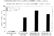
この実験から、本発明化合物はハツカネズミ骨髄幹細胞の有意な細胞増殖を誘導することが分かる。その応答程度は大腸菌LPSによって誘導されるものにほぼ等しいが、有意な応答を誘導するのに必要な最小濃度はそれよりも高い。モノフォスフォリレート化物はジフォスフォリレート化物よりも応答誘導が穏やかである。図1は異なる細胞調整物で行った一組の三つの独立的した代表的な実験を示している。
【0082】
3. マクロファージ倍養液の上澄液の酸化窒素産生量の決定
手順
二匹の6週齢のオスのC57/BL6ハツカネズミをCO2吸入で殺し、頸部脱臼した。ハツカネズミをアルコールでを洗浄し、後股関節部の表皮を完全に除去した。大腿骨および脛骨骨組織は関節破壊して除去した。肉はメスを使用して大きく除去し、骨組織をきれいし、骨組織の端部をはさみで切断した。骨髄ははさみを切断した骨組織の端部に1mlのダルベッコの修正イーグル媒体(Dulbecco's Modified Eagle Medium)(DH媒体)を三回注射して骨ルーメンから抽出した。細胞をDH媒体に再懸濁し、300gで5分間遠心分離した。上澄を捨て、細胞を20%ウマ血清(HS)および30%L929培養液上清を補ったDH媒体に40000 細胞/mlの密度で再懸濁した。L929はネズミの繊維芽細胞細胞株で、その上澄にはマクロファージ(M-CSF)のための成育因子が豊富に含まれている。この細胞懸濁液をシャーレ上で12mlアリクォットに分割し、8% CO2且つ100% RH下のインキュベータで37℃で8日間培養した。8日後、幹細胞は成熟したマクロファージ細胞に分化した。マクロファージ細胞は冷たいPBS緩衝剤中で4℃で45分間培養して掻き取った。遠心分離後、上澄液を除去し、5%ウシ胎仔血清(FCS)、グルタミン、アルギニン、葉酸、メルカプトエタノールおよび抗生物質(ペニシリンおよびストレプトマイシン)を補ったDH媒体に細胞を再懸濁した。幹細胞を集め、細胞密度を700000 細胞/mlに調整した。
【0083】
FCSとアミノ酸と抗生物質とを補ったDH媒体に予め溶した製品を96-ウエルミクロタイタープレートに連続的且つ直接に希釈した。9つの希釈を3.16の希釈比で実行した。製品を連続3回テストした。各ミクロタイタープレートにはただの媒体を含む負の対照を含めた。各ウエルの最終体積は100μlにした。ミクロタイタープレートを37℃、8% CO2-100%RH下のインキュベータで1時間培養して媒体を緩衝させた。1時間以後、細胞懸濁液の100μlを製品に加え,培養を22時間継続した。
22時間後、400gで5分間ミクロタイタープレートを遠心分離し、100μlの上澄液流体を取り、ミクロタイタープレートに移した。100μlのグリース試薬 [5mg/mlのサルファニルアミド+の0.5mg/mlのN-(1-ナフチルエチレンジアミン)塩酸塩の2.5%燐酸水溶液]を各ウエルに加えた。ミクロタイタープレートは分光光度計で562ナノメートル波長の基準に対して690ナノメートルで読んだ。亜硝酸塩濃度は酸化窒素含量に比例した。亜硝酸塩含量は標準曲線に基づいて求めた。1〜25μMの範囲でリニアーな関係が示された。
結果は、負の対照値を求めた後、平均±+標準偏差で表し、投与量対応答曲線とうしてプロットした。
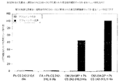
この実験で、本発明化合物はネズミのマクロファージ細胞によって酸化窒素の産生を誘導し、投与量対応答曲線と一致した。ジフォスフォリレート化物は大腸菌LPSよりも非常に大きく増殖を誘導するが、有意な応答を誘導するのに必要な濃度はより高い。モノフォスフォリレート化物はジフォスフォリレート化物および大腸菌LPSより弱い応答を誘導する。図2は互いに異なる細胞標品で独立して行った三つ測定値から得られた代表的な実験を表している。
【0084】
4. 本発明化合物のヒト肺胞マクロファージ細胞によるα -TNF 産生能力の決定
手順
肺胞マクロファージ細胞の調整
ヒト肺胞マクロファージ細胞は肺癌患者の肺臓の気管支肺胞を洗浄(BAL)して得られた。BALは肺葉の健康な部分を含む肺組織外科の直後に実施する。洗浄は50ml容量の注射器を用いて0.8% NaClを用して実行される。回収した細胞をマクロファージ細胞が85%を超えるようにした。他の主要な細胞はリンパ球である。遠心分離後、細胞をRPMI媒体に懸濁し、赤血球は遠心分離で除去する(フィコール・パック、Ficoll Pack、研究グレート)。マクロファージ細胞をHBSSで3回洗浄し、24-ウエルのミクロタイタープレートに1mlを種付けした。一つのウエルは合計で1000000の細胞を含んでいる。37℃で1時間培養後、マクロファージ細胞は粘着性になる。ウエルを粘着しない細胞を除去するために1mlのHBSSで3回洗浄する。洗浄後、各マクロファージ細胞-含んでいるウエルに1mlのRPMIを加える。
製品を用いた培養と、α -TNF のアッセイ
肺胞マクロファージ細胞を37℃で5%CO2下で下記製品の0.1μg/ml、1μg/mlおよび10μg/ml濃度の存在下で培養した:
(1)負の対照:RPMI
(2)正の対照:大腸菌LPS(セロタイプO5:B5、Difco、デトロイト、米国)
(3)本発明のモノフォスフォリレート化物(OM-294-MP)
(4)本発明のジフォスフォリレート化物(OM-294-DP)
培養上澄を24時間後に回収し、αTNF含量を分析した(BioSource Cytoscreenキット、Camarillo、CA、U.S.A、1pg/ml.の感度を有する)。
【0085】
結果
本発明のモノフォスフォリレート化およびジフォスフォリレート化誘導体は10μg/mlのような低濃度でαTNFの産生を穏やかに誘導した。本発明のモノフォスフォリレート化誘導体はジフォスフォリレート化物よりも大きくTNF産生を誘導する。LPSの正の対照はテスした三つの濃度で全てαTNFの産生を多量に誘導した。
結果は表(a)にまとめてある。
【0086】
【表3】
5. 大腸菌リポポリサッカリド( LPS )に応答する本発明化合物のヒト肺胞マクロファージ細胞のα TNF 産生の抑制能力
手順
肺胞マクロファージ細胞の調整:
肺癌患者の気管支肺胞洗浄(BAL)によってヒト肺胞マクロファージ細胞を得た。このBALは肺葉の健康な部分を含む肺組織外科の直後に実施される。洗浄は50ml容量の注射器を用いて0.8% NaClを用いて行う。回収した細胞はマクロファージ細胞85%を超える様に作る。他の大多数の細胞はリンパ球である。遠心分離後、細胞をRPMI媒体に懸濁し、赤血球をフィコール・パック(研究グレート)で遠心分離で除去する。マクロファージ細胞をHBSSで3回洗浄し、各ウエルに1mlの濃度で、合計1000000細胞を含むように24-ウエルのミクロタイタープレートに種付けする。37℃で1時間培養後、マクロファージ細胞は粘着性になる。粘着性のない細胞を除去するために1mlのHBSSでウエルを3回洗浄する。洗浄後、各マクロファージ-含んでいるウエルに1mlのRPMIを加える。
【0088】
製品の培養とα TNF 分析:
肺胞マクロファージ細胞を、37℃で5% CO2下で大腸菌LPSの存在したで1mg/mlで培養する(O5:B5セロタイプ、Difco、デトロイト、米国)。同時に下記製品を10μg/mlの濃度で加えた:
(1) 負の対照:RPMI
(2) 本発明のモノフォスフォリレート化物(OM-294-MP)
(3) 本発明のジフォスフォリレート化物(OM-294-DP)
培養上清液を24時間後に回収し、TNF含量を分析した(BioSource Cytoscreenキット、Camarillo、CA、U.S.A、感度1pg/ml)。
結果
ジフォスフォリレート化誘導体はLPSによって一般に誘導されるαTNFの産生をかなり抑制する。モノフォスフォリレート化誘導体はLPSによって誘導されるαTNFの産生を部分的に抑制する。
結果は表(a)に記載してある。
【0089】
【表4】
6. 樹状細胞成熟での OM-294-MP および OM-294-DP の影響
予備樹状細胞を樹状細胞への成熟を誘導するOM-294-MPおよびOM-294-DP製品の能力を評価した。下記のパラメータを測定した:FITC-Dextran結合体移入およびCD40、CD80、CD83、CD86表面マーカの発現
手続
細胞:末しょう血液の単核細胞を6人の健康なドナーのバフィーコートから単離した。ドナーは供血前にいかなる処置も受けなかった。
細胞標品:付着選択によって精製した単核細胞を10%のウシ胎仔血清(Sigma-Aldrich: St.-Louis, MO, U.S.A)、GM-CSF(10ng/ml、IM-HGMI、Immungenex Corp., Los Angelos, CA, U.S.A.)およびIL-4(10ng/ml、204-IL、R&D System, Minneapolis, MN, USA)を含むRPMI-1640媒体に1×10⁶細胞/ml密度で再懸濁し、直径10cmのシャーレ(P10、Falcon、Becton Dicinson、プリマス、UK)に分割(一枚のP10皿に10×106の細胞)し、6日間培養する(3日以後は新鮮な媒体に交換)。この細胞は前樹枝状(predendritic)細胞(DC-6)と呼ばれる。成熟した樹枝状細胞へこの前樹枝状細胞の成熟を細胞をOM-294-MP、OM-294-DPまたはLPSと一緒に3日以上にわたって以下の「製品セクション」に記載の濃度で培養することで達成する。9日目(DC-9)に細胞を採り入れ、樹状細胞成熟の各種インジケータ:CD40、CD80、CD83、CD86表面マーカのアセスメントおよびFITC-Dextran接合体のテークアップ能力を分析した。これらの全てのパラメータはEPICS-XL-MCLモデルFACで分析した(Coulter Immunology、Hialeah、フィンランド)。Lanzavecchia et al., J. Exp. Med., 179 (1994) 09; Lanzavecchia et al., J. Exp. Med., 182 (1995) 389。
【0091】
データ分析:
表面マーカの発現はLPS(正のモード)により刺激される細胞の平均蛍光%として現れる。FITC-Dextran接合体は塩基性媒体中に維持した細胞に対するテークアップ率で計算し、%で表される。t-スチューデント試験による統計分析では正のモードのデータで得られるを異なる試験からのデータと比較する。データ有意水準はp < 0.05にセットした。
製品:
OM-294-MPおよびOM-294-DPの原液は0.9% NaCl/水中に1mg/mlの濃度で調製し、OM-294-MPの場合には0.1%のトリエチルアミンを加えた。この溶液を37℃で20分間培養し、3分間の強力に撹拌した後、RPMI-1640培地で100μg/mlに希釈し、10mg/mlの濃度(図3、6、7、8)または0.02〜25μg/mlの濃度(図4、5)で用いた。
【0092】
基準製品:PBS中の大腸菌リポポリサッカリド(LPS、DIFCO、デトロイト、MI、USA)の5mg/ml原液。中間体100μg/ml溶液はRPMI 1640培地に調整した。テストすべき濃度は10μg/ml(図3、6、7、8)または0.02〜10μg/ml(図4、5)の範囲である。
結果
単核細胞分化で生じる早熟な樹状細胞(DC-6)はGM-CSFおよびIL-4の接合によってFITC-Dextran接合体を組み入れることができる。成熟過程中に細胞はFITC-Dextran接合体を組み入れる能力を失う。分析はDC-9 分化段に達した時に実施される。
結果は非刺激細胞(塩基性媒体)(図3)で観測されるFITC-Dextran接合体の移入%で表される。LPSまたはOM-294-MPで処理した細胞はそれぞれそれらの食細胞の容量の10%および19%だけしか保持しないが、OM-294-DPで刺激された細胞はFITC-Dextran接合体を組み入れる能力を完全に保持する(98、99%)。投与量対応答曲線は、OM-294-MPは0.02%g〜25μg/mlの濃度範囲でDC-6をDC-9細胞へ分化させるのを誘導する著しい能力を有することを示している。図(4)は低濃度でテストしたもの、また、図(5)は高濃度でテストしたもの。
【0093】
共刺激表面マーカの発現はDC成熟性を査定するために他の判定基準である。CD40、CD80、CD83、CD86の発現をテストした。結果はこれらマーカーのLPS-誘導発現に基づく平均蛍光%で表してらある。
OM-294-MPはテストした全ての表面マーカ:CD40(39%)、CD80(62%)、CD83(60%)、CD86(77%)の発現を増加させる。図(6、7、8、9)を参照。
OM-294-DPは調べたマーカーの発現に基礎培地と同様な作用を与えるが、その影響はLPS働きの20%を超えない。
【0094】
7. DC-6 段での単核細胞および前樹枝状細胞によるα TNF および IL-12 p70 の産生における OM-294-DP および OM-294-MP 製品の影響
DC-6細胞(5×10⁵/500μl媒体)をLPS(10μg/ml)またはOM-294-MP(10μg/ml)またはOM-294-DP(10μg/ml)によって4時間、6時間、24時間刺激した。
手順
インビボ実験条件:
抹消血液の単核細胞を6匹の健康なドナーのバフィーゴートから回収した(ドナーは供血前にいかなる処置もしなかった)。単核細胞をフィコール勾配で単離し、付着選択によって精製した。ゆるく粘着している単核細胞を採り入れ、その細胞の一つの画分を単核細胞として保存した。精製した単核細胞を10%のFCSを含むRPMI-1640媒体中に1×10⁶細胞/mlで再懸濁する。10×106細胞/P10で直径10cmのシャーレ(P10−falcon、Becton Dickinson、プリマス、UK)に分けた。細胞はGM-CSF(10ng/ml)およびIL-4(10ng/ml)を含む全RPMI 1640媒体で6日間培養した。6日目に細胞を採り入れてHBSSで洗浄し、5×10⁵細胞/ウエルの密度で500μlの全RPMI媒体中に24-ウエルプレートに種付けし、LPS(10μg/ml)、OM-294-MP(10μg/ml)またはOM-294-DP(10μg/ml)で刺激し、αTNFおよびIL-12 p70を4、6、24時間後に回収した培養液上澄のエリサによって分析した。
製品:OM-294-MPおよびOM-294-DP製品(無菌水の1mg/ml原液)を37℃で20分間培養した。3分間は強力な撹拌下に保持し、100mg/mlに希釈し、RPMI 1640培地で10μg/mlの最終濃度で使った。
【0095】
基準製品:大腸菌リポポリサッカリド(LPS、DIFCO、デトロイト、MI、USA)、PBS中の5mg/ml原液、培地の中間溶液:100μg/ml、10μg/mlの最終濃度で使用。α TNF および IL-12 p70 のアッセイ:αTNF KHC3012キット (Biosource提供のバッチ#PP003-J061703(Biosource、International Camarillo、CA、USA)。エリサは供給業者の取扱い説明書に記載の方法で行った。IL-12 p70はヒトのIL-12キット(番号D1200、ロット990 6232、R & Dsystems、ミネアポリス、MN、USA)を使用して培養液上澄中でエリサ分析した。
【0096】
結果
α TNF
OM-294-MPは時間-コース産生およびαTNF濃度(図10)に対してLPSと同様にDC-6細胞によるαTNF産生を刺激する。両方の製品の場合に6時間〜24時間の間にαTNFピークが見られる。
OM-294-DPはDC-6細胞によるαTNF産生に小さな刺激作用しか有しない。
IL-12 p70
一般的に言って、IL-12は単核細胞(図12)およびDC-6細胞(図11)中でγIFN(LPS+γINF、OM-294-MP+γIFN)の存在で誘導される。サイトカイン産生開始期は単核細胞よりDCの方が早い。
【0097】
8. ネズミ免疫化モデルでの Plasmodium berghei circumsporozoite 表面蛋白のC末端領域の合成ペプチド( Pb CS His6-242-310 )による OM-294-MP および OM-29 4-DP アジュバント性質の評価
抗原:PbCS(HHHHHHGGMN NKNNNNDDSY IPSAEKILEFVKQIRDSITE EWSQCNVTCG SGIRVRKRKRG SNKKAEDLTL EDIDTEI)ペプチド(以下、His6-242-310という)はPlasmodium berghei circumsporozoiteANKA株の表面蛋白の242-310アミノ酸配列に6つのヒスチジン、2つのグリシンおよび1つのメチオニン残基をN末端伸びを加えたものに対応し、Merrifield Atheron合成法で得られる(Athenon et al., Bioorg Chem., 8: (1979) 350 - 351)。このポリペチドは0.4mmol/g.の置換率を有するp-アルコキシベンジルアルコール樹脂(ワング樹脂)上に調製された。10モル倍の過剰なF-mocアミノ酸誘導体を30分のカップリング時間で使いた。ペプチドをサイズ排除クロマトグラフ(セファデックスG25、Pharmacia、スウェーデン)および逆相クロマトグラフ(W-Porex 5C-4、250×10mm、Phenomenex、Torrance、CA、U.S.A)で精製した(40分、勾配、10〜50%アセトニトリル-0.1%トリフルオロ酢酸混合物(v/v)、3ml/分の流量)。ペプチドのアミノ酸組成はKnecht and Changの方法(Anal. Chem.、58、(1986)2373-2379)で決定し、分子量はVoyager DEモデル装置を使用して質量分析で検査した(Perspective Framingham、MA、USA)。抗原原液は0.9% NaCl/水中に0.4mg/mlの濃度でpH 8.0で調製した。
【0098】
アジュバント:OM-294-DPおよびOM-294-MPの原液は0.9% NaCl-水中に1mg/mlの濃度で調製した(OM-294-MPの場合には0.1%トリエチルアミンを入れた)。正の対照は不完全フロイントアジュバント(Difco、デトロイト、MI、USAからのIFA)から成る。負の対照は0.9% NaCl溶液である。
抗原 - アジュバント混合物:抗原の一容積とアジュバントの一容積とを3分間攪拌して混合する。
免疫化:6週齢のメスのBALB/cマウス(1グループに6匹のハツカネズミ)に下記の混合物の0.1mlを後尾の皮下に注射して3回免疫化した:
【0099】
【表5】
【表6】
リンパ性器官および採血法:
血清サンプリング:
採血法は第0、3、7、9週に実施する。血液は37℃に6分間放置し、4℃で一晩保持する。その後、血清は-80℃で抗体アッセイ時まで凍結保存する。
鼠径部リンパ節および脾臓の回収:
各動物グループの一部を第4または9週以後殺し、鼠径部リンパ節および脾臓を外科的に除去する。
抗 Pb CS His 6 242-310 抗体価の決定:
Pb Cs His6 242-310抗原に対する抗体のアッセイをエリサで行う。抗原の結合を4℃の湿った室内で一晩培養して96-ウエルのミクロタイタープレート(Maxisorp F96、Nunc、DK)で実施する。各ウエルは0.001mg/mlのPb CS His6 242-310の抗原を含む0.1mlのPBS(リン酸緩衝生理食塩水)を含んでいる。ミクロタイタープレートのブロックは1%のウシ血清アルブミン(BSA、Fluka、スイス)を含むPBSで行う。プレートを0.05%トゥイーン20(シグマ、セントルイス、MO、USA)を含むPBSで洗浄する。第0、3、7、9週目に回収した血清サンプルを希釈緩衝剤(2.5%のスキムミルク微粉と0.05%のトゥイーン20を含むPBS)で連続的に希釈し、ミクロタイタープレートに移し、1時間室温で放置する(RT)。プレートをPBSで洗浄し、アルカリホスファターゼ(シグマ、セントルイス、MO、USA)に結合したハツカネズミのポリクローナル抗Igを含む希釈液を各プレートに分配し、1時間室温で培養した。プレートをPBSで洗浄し、アルカリホスファターゼ基質、p-ニトロフェニルフォスフェート(シグマ、セントルイス、MO、USA)を加えて特異抗体を呈色反応で表す。405ナノメートルでの吸光度をミクロタイタープレートリーダーで読む(Dynatech 25000のエリサリーダー、アッシュフォード、ミドルセックス、UK)。各血清サンプルは2回測定した。結果は各グループのハツカネズミに関する全ての測定値の平均を表す。抗体価は正の応答を与える最も高い希釈度で表した。すなわち、ODはバックグラウンド ノイズレベル±3SDより大きい。
【0102】
ELISPOT アッセイ:
ELISPOTミクロタイタープレートの50μg/mlで抗体溶液を加え、ウエル底をニトロセルロース(ミリポア、Molsheim、フランス)で覆うことで、ネズミのγ-インターフェロン(O1E703B2)に対する抗体を湿った室で4℃で一晩培養して結合させる。10%のウシ胎仔血清(FCS、Fakola、スイス)を含むDMEM媒体を加えてブロックを行い(Life Technology、グランドアイランド、NY、USA)、37℃に2時間放置した。リンパ性器官から得られる細胞(鼠径部リンパ節および脾臓)をミクロタイタープレートで200000細胞/ウエルの密度で培養し、次いで、100000 P815細胞(Pb CS 245-252の短いペプチドを抗原投与したものとしないもの)と一緒に37℃で24時間共培養した。培養後、細胞を除去し、洗浄する。第2のネズミの抗γIFN抗体-ビオチン複合体を(ANI、1% BSAを含むPBS中の2μg/ml)を加え、2時間培養し、ストレプトアビジン-アルカリホスファターゼ接合体(Boehringerマンハイム、マンハイム、GFR)を加え、37℃で1時間培養した。その後、0.05%トゥイーン20を含むPBSで3回洗浄し、PBSで3回洗浄した。抗γIFN免疫複合体の存在はBCIP/NBT基質(シグマ、セントルイス、MO、USA)を加えることによって示した。この反応は水道水で洗浄することにより停止させた。ΓIFNに対して正である点は立体顕微鏡下で数える。ペプチドを抗原投与した細胞とペプチドが存在しない細胞とを数え、その差から点比を数えた。結果は各グループのハツカネズミに対して平均測定値で記録した。培養した血液細胞100万当たりの点の数で表した。
【0103】
結果
抗体応答:一回、二回、三回免疫注射したハツカネズミに対するエリサで求めたPb CS His6-242-310に対する抗体の産生はグラフに示してある。対照は抗原を一回だけ注射したもで、抗体価は非常に弱い。OM-294-MPまたはOM-294-DPを混合して単一の抗原注射した時に得られる抗体価は不完全フロイントアジュバント(IFA)に同じ抗原を加えたものとほとんど同程度の高い応答を示す(図13)。2回の注射で、OM-294-MPおよびOM-294-DPアジュバントは血清学的応答を引き出すことができ、それはIFAのそれより大きい(図14)。次の三回の注射でOM-294-MPおよびOM-294-DPアジュバントは血清学的な応答を引き出すことができ、それはIFAのそれより大きい(図15)。
図13:エリサ、最初の免疫化注射後3週間で実施。
図14:エリサ、第2回免疫化注射後4週間で実施。
図15:エリサ、第3回免疫化注射後2週間で実施。
図16:一回、二回、三回目の免疫化注射の前後で測定した抗体価。
免疫化注射前と、二回、三回目の免疫化注射後の各グループの動物の抗体価は平均値で示してある(図16)。
【0104】
CTL 応答: 免疫化で使ったPb CS His6-242-310ペプチドに存在するT細胞エピトープPb CS 245-252の認識はELISPOT試験によってよく示される。免疫済み動物に記録されたTリンパ球応答(鼠径のリンパ節および脾臓、第2回注射後1週および第3回注射後の2週間後にそれぞれ回収)はγインターフェロン(γ IFN)の正の点のカウントの上昇で示される。図(17、18、19、20)に記載の結果は各希釈度および各グループのハツカネズミに対する測定値の平均として計算されている。培地の100万細胞当たりの点の数で表してある。
OM-294-MPおよびOM-294-DPアジュバントは脾臓および鼠径部リンパ節由来のリンパ球のCTL応答を大幅お増加させる。脾臓応答は鼠径リンパ節応答より高い。OM-294-MPおよびOM-294-DPアジュバントにより誘導されるCTL活性性はIFAによるものよりも明らかに優れている。
【0105】
9. 毛細管電気泳動にる非共有結合性 OM-294- 抗原複合体
この実施例ではワクチン製造時にOM-294-DPとPb CS His6-242-310ペプチドとの間に形成される非共有結合性錯体が存在することを毛細管電気泳動を用いて示す。
手順
分析法:
20mMホウ砂緩衝剤(ジナトリウム四硼酸塩十水化物、メルク、N°6306) 1N NaOHでpH 7.4に調整(Fluka N°72072)。
ゾーン-分割毛細管(接手なし)、長さ30cm、直径50μm。
検出はBeckmanのPACE MDQモデル装置(Beckman、Brea、CA、USA)を使用し、200ナノメートルで行った。
【0106】
【表7】
結果
このワクチン製造時に見られる抗原-アジュバント複合体の形成は電気泳動線図上でアジュバントピークの消滅と、抗原ピークがシフトし、複合体形成に特有な新しいピークができることで示される(図21)。
10. BDIX ラットの PROb 同系腫瘍性遺伝子由来の細胞の注射により生じた腹膜癌腫の処理
この実験は、数mmの肉腫瘍を有するラットに一連のi.v.注射でOM-294-DPを投与した時の抗腫瘍効果を示すためのものである。
手順
動物: In-bred BDIXラット株は1937にH. Druckreyによって作られた。Fribourg Max Planck Institute (RFA)からのラットの一部がこのコロニーの最初の供与源となり、それは1971年以後実験動物ハウスで単一系統が維持されている。この系統では一対の姉妹-兄弟が次世代の子孫を残すために選択される。この実施例で使われるラットは本出願人の実験室に代って増殖を実施しているExperimental Animal Breeding Center in Iffa-Credo (Arbresle, France)から得た。使用したラットは3か月±1週間の年齢のオスである。
【0108】
PROb 細胞注射による腫瘍誘導:
PROb 細胞オリジン:
DHD/K12細胞株は、1、2-ジメチルヒドラジン処理によってIn-bred BDIXラット株に生じさせた大腸癌腫断片の移植から得た。この付着性細胞株はトリプシン処理に対する感度に従って2つのサブラインに再分割した。分けるのが困難な細胞にDHD/K12-TRの名を付けた。このDHD/K12-TR細胞をIn-bred BDIXラットに注射するとラットは必ず腫瘍を引き起こした。この株をクローン化した。この実験ではこのDHD/K12-TRbクローンだけをPROb細胞として用いた。
培養条件:粘着性PROb細胞を10%ウシ胎仔血清(FCS, Anval, Betton, France) を加えたHarm's F10媒体(バイオ-ホイッテカー、Walkersville、USA)から成る完全培地で37℃で溶封ビン(Falcon, Becton Dickinson, New Jersey, USA)中で培養した。培地の交換は3日毎に行った。交会に達した時に細胞を開放し、3〜5分の間に2mlのEDTA/トリプシン溶液で掻き取り、次に2〜3分の間に同じ溶液2mlで3回洗う。細胞を完全媒体へ再懸濁させ、FCSを加えてトリプシンの働きを停止させる。マイコプラズマおよび細菌性細胞の汚染がないことは規則的な間隔でHoechst fluorochrome 33258でDNA染色(Aldrich Chimie, Steinheim, GFR)して検査した。
【0109】
腹膜癌腫の誘導: PROb細胞を「培養条件」の所で説明したようにしてその支持体から開放し、トリパンブルー色素溶液で数えて細胞の生存性を査定した。細胞をHam's F10媒体に兼抱くする。エーテル麻酔下で共通遺伝子BDIXラットへ106の生きたPROb細胞を腹こう内投与(i.p)して腹膜癌腫を誘導した。腫瘍細胞注射は10日目に行った。この条件で全てのラットに観血的腹水を伴う腹膜癌腫ができ、細胞注射から第6日〜第12日の間に死ぬ。
腹膜癌腫の処理: 処置は癌腫の直径が数mmの結節になる腫瘍細胞注射後13日目に開始する。処置ではOM-294-DPを10回i.v.注射で1mg/kg体重量かつ0.9% NaCl溶液に溶かした0.6mg/mlの投与量で投与した。注射は1週間に3回陰茎静脈に投与した(月曜日、水曜日、金曜日)。対照群はただのビヒクル(すなわち0.9% NaCl)のみで処置した。
処置効果の確認:D42(腫瘍細胞注射後6週目)にラットを殺し、切開して癌腫の発達を盲検試験で査定した。癌腫体積の測定は信頼性がないが、癌腫を4つの異なるタイプに分類することはできる。結節の数および直径に応じて下記の5つのクラスを定義することができる:
【0110】
クラス0:結節が見えない。
クラス1:直径0.1〜0.2cmの結節が容易に数えられる。
クラス2:0.5〜0.1の無数の結節。
クラス3:腹膜腔が結節により侵略され、直径1cmのものも測定される。
クラス4:大きさが2、3cmの腫瘍で体くうが完全に侵略される。
腹水容積と動物の体重変化:
腹水容積は動物を2回計ることにより測定した。0.9% NaClの注射だけで処置をしなかった対照ラットのグループは癌腫の発達を許したままにした。
処置効率の確認:
処置したグループのラットの生残率を対照群の生残率と比較し、処理したグループのラットの癌腫および腹水容積を対照群のラットの測定と比較した。
統計解析:免疫療法の効果の統計的有意差を癌腫分類用Kruskal-Wallis試験を用して決定した。同様に、腹水容積データに対して分散分析試験を実施し、ログランク試験で生残率を求めた。
【0111】
結果
癌腫: OM-294-DPはこのモデルで著しい抗腫瘍活性を示す。この活性は無腫瘍動物の数によって証明された(クラス0)。また、NaCl対照との相違は腫瘍体積で有意(p<0.05)である。腹水容積におけるOM-294-DPを用いた処置も有意(p < 0.05)である。
【0112】
【表8】
生残:
各動物はD42に殺した。生残は腫瘍細胞の注射後42日目に決定した。OM-294-DPを処置した動物は90%生存したが、未処置のグループの動物の生存率は20%であった。
ラット生残率はOM-284-DPによって大幅に延長した(p<0.001)。
体重: 表(b)のデータが示すように、OM-294-DPはNaClのみを投与した動物に比較して体重変化に有意な影響を及ぼさない。
【0114】
【表9】
11. Heliobacter pylori のウレアーゼ B サブユニットを鼻噴投与するネズミの免疫化モデルにおける OM-294-DP アジュバントの評価
コレラトキシン(CT)ベースのアジュバントの存在下に経口または鼻投与でHeliobacter pyloriのウレアーゼBサブユニットを免疫化すると、ハツカネズミはHeliobacter pylori感染から保護されてということは分かっている(Corthesy-Teulaz I., et al., Gastroenterology, 109: (1995): 115; Michetti P. et al., astroenterology, 116: (1999) 804; Saldinger P.F. et al.,Gastroenterology 115: (1998) 891)。免疫化したハツカネズミの血清中で測定されるこの抗Ure B体液性応答をは主としてIgG1タイプ(Th2応答)である。OM-294-DPのアジュバント効果の評価は、OM-294-DPの存在下に組換え型Heliobacter pyloriのウレアーゼBサブユニット(UreB)を鼻噴投与で1週間間隔で4回投与して免疫化したBALB/cハツカネズミ(n=6)で行った。対照のBALB/cハツカネズミはOM-294-DPアジュバントのみで免疫化した。最後のブースタ後2週目に各ハツカネズミから血液をサンプリングし、エリサ(全IgG、IgG1およびIgG2a)によって血清抗UreBIgを分析した。
【0116】
手順
動物: BALB/c/Ola/HsDハツカネズミ(ハーランド、ホルスト、オランダ):24匹のハツカネズミ
抗原: 上記方法(Michetti達、Gastroenterology、107:(1994)1002)で大腸菌(M15株、Qiagen、Hilden、GFR)で発現させた組換え型蛋白HpUreB 1-569
アジュバント:OM-294-DP(2.2mg/ml原液)
免疫化規則:
各々が6匹のハツカネズミを含む下記の4つのグループを作った:
グループA: 6匹のBALB/cハツカネズミに週一回、4週間の間、OM-294-DPのみを25μg鼻噴投与して(各服用時に25μl)免疫化した。
グループB: 6匹のBALB/cハツカネズミに週一回、4週間の間、50μgのUre B1-569+25μgのOM-294-DP(各服用時に25μl)を鼻噴投与して免疫化した。
最後の経鼻免疫化してから2週間後にグループA及びBの各ハツカネズミの尾から血液のサンプルを採った。
【0117】
血清 IgG のアッセイ:
被覆用緩衝剤( pH 9.6 ):1リットル当たりNa2CO3(1.59g、15mM)、NaHCO3(2.93g、34.8mM)、チメロサール(0.01%)、PBS- トゥイーン緩衝剤 pH 7.4:1リットル当たり、NaCl(8.0g、137mM)、KH2PO4(0.2g、1.5mM)、Na2HPO4(1.15g、8.0mM)、KCl(0.2g、2.7mM)、トゥイーン 20 ( 0.1% 1ml )、シトラート / リン酸緩衝液 pH 5.0 :1リットル当たり、クエン酸(9.32g、44.4mM)、Na2HPO4(14.6g、103mM)、基質溶液 10x(O-phenyldiamine = OPD)(10倍濃縮):(シトラート緩衝剤の10mg/ml)、アジ化ナトリウム溶液:1%、停止溶液:シトラート/リン酸緩衝液0.01%アジ化ナトリウム0.1M、pH 5.0
【0118】
方法: 抗原溶液(1999年5月26日に調整したUreB 1-569、0.5mg/ml原液)は被覆用緩衝剤pH9.6中に5μg/ml濃度で調製した(50mlの緩衝剤に対してUreB溶液500μl)。100μlを3つの96-ウエル丸底プレートの各ウエルへピペットで移した(各ウエル当たり0.5μgのUreB)。プレートを37℃で2時間培養した。上澄はプレートから捨てる。100μlのPBS-0.1%トゥイーン溶液+5%粉乳を加えてウエルをブロックした。プレートを37℃で30分間培養した。ブロック溶液を捨て、ウエルを100μlのPBS-トゥイーンで3回洗浄した。上澄を捨てた。テストする各ハツカネズミ血清の1:200希釈液をPBS-0.1%のトゥイーン緩衝剤(1mlのPBS-トゥイーン中に血清5μl)中に調製した。漿液(100μl)を3枚のプレートに分ける(全免疫IgGを検出するためのプレート1、IgG1を検出するためのプレート1およびIgG2aを検出するためのプレート1)。
培養は4℃で一晩実行する。ウエルを100μlのPBS-トゥイーンで3回洗浄した。PBS-トゥイーン緩衝剤中に希釈した1:500 ビオチン-結合抗IgG全抗体(Amersham、Cat # RPN 1177)、抗IgG1抗体(Amersham Cat # RPN 1180)および抗IgG2a抗体(Pharmingen Cat #02012D)溶液を調製した。100μlの抗-IgG全抗体溶液のプレートN°1に加え、プレートN°2に100μlの抗-IgG1溶液、そして、プレートN°3に100μlの抗-IgG2a抗体溶液を加えた。培養は37℃で1時間実行した。ウエルをPBS-トゥイーンで3回洗浄した。PBS-トゥイーン緩衝剤中にストレプトアビジン-HRP(Dako、Cat# p03977)の1:1000希釈液を調製し、各ウエルに100μlの溶液を加えた。培養は37℃で1時間行った。ウエルはPBS-トゥイーン緩衝剤で3回洗浄した。0.1のMシトラート/リン酸緩衝液中にOPD溶液を10倍希釈することによって基質溶液(10x)を調製した。希釈したOPD溶液に1μlのH2O2を加えた。各ウエルに50μlの基質溶液を加えた。10〜20分発色させる。反応は50μlの停止緩衝剤を加えることで終了させる。吸光度はブランクとして負の対照を用いて492ナノメートルで読んだ(標準は620ナノメートルで読んだ)。
統計解析: データは平均±SD(n=6)を表す。p値はt-学生試験から誘導いた。有意データ水準はp < 0.05にした。
【0119】
結果
経鼻投与で予めUreB-1-569+ OM-294-DPで免疫化したハツカネズミで抗UreB体液免疫を作り出す:抗-UreB 1-569 IgG1は血液中に存在する。
ハツカネズミ血清中のHpのUreBに対する抗体の存在をエリサで測定する。UreB(0.5μg/ウエル)を炭素塩緩衝剤pH 9.6で丸底の96-ウエルプレートに分配する。特異抗体をウサギ抗IgG全抗体、抗IgG1、IgG2a抗体によって検出する。結果は492ナノメートルで読んだ光学濃度(OD)で示した。自然のハツカネズミの血清の測定値より3倍以上大きなODを正とみなした。OM-294-DPのみで免疫化したハツカネズミの血清には抗UreB抗体は検出されなかった。Ure+OM-294-DPで免疫化されたマウスも抗UreB IgG全抗体 (OD = 0.274±0.130、p < 0.05)および抗UreB IgG1(OD = 0.212±0.128、p < 0.05)を生じるが、抗UreB IgG2a抗体は生じない(OD = 0.008±0.005、有意差なし)。
鼻噴投与(UreB)+OM-294-DPによるHeliobacter pyloiのウレアーゼBサブユニットで免疫化したBALB/cハツカネズミは主としてIgG1タイプの抗UreB体液性応答を生じる。従って、OM-294-DPは経鼻投与によるアジュバントとして働き、Th2タイプの体液性免疫の発達を促進する。
【0120】
12. OM-294-MP 、 OM-294-DP と H1N1 との抗原との組合せ:ハツカネズミに1回ま 2 回皮下投与した後に生じる特異抗体の決定
手順
この研究は風邪抗原H1N1(262195A/B、北京ヘムアグルチニン、Solvay Duphar、Weesp、NL)に対するOM-294-MPおよびOM-294-DPのアジュバント効果を示すためのものである。この目的のために60匹のBALB/cハツカネズミ(雌、処置開始時に8週齢)を6つのグループに次のように分けた:
【0121】
【表10】
抗原: H1N1原液は0.9% NaCl中に25μg/mlの濃度で調製した。
アジュバント: OM-294-DPおよびOM-294-MPの原液は注射水の1mg/ml濃度に調製し、OM-294-MPには0.1%トリエタノールアミンを加えた。負の対照は抗原を含まない0.9% NaCl溶液から成る。
抗原 - アジュバント混合物: アジュバントを37℃で20分間放置した後、3分間攪拌した。それから、上記表の通り、抗原とNaCl(0.9%)を加えた。抗原-アジュバント混合物を簡単に攪拌した後、ロータリ撹拌機で室温で15分間攪拌し、最後に全部の混合物を3分間攪拌した。
【0123】
免疫規則: 0日と14日に注射した。上記の表に示した混合物を皮下投与した(各動物に75μlを側に、全部で150μl)。採血法は14日および18日にした(眼窩穿刺)。
抗 H1N1 Igのアッセイ: 以下のH1N1用血清免疫グロブリンをエリサで2回分析した:IgG1、IgG2aおよびIgM。簡単にいうと、ミクロタイタープレート(NUNC、Immunoplate Roskilde、DK)を重炭酸塩緩衝剤pH 9.6中で4℃で100μlのH1N1(0.5μg)と一緒に(一晩被覆)培養した。0.5%トゥイーン-20(メルク Hohenbrunn、D)で洗浄した後に、漿液を50-、200-、800-倍に希釈した(希釈溶液:リン酸緩衝生理食塩水(PBS)1%+ウシ血清アルブミン(BSA、シグマ、セントルイス、MO、USA)0.02%+トゥイーン-20)。希釈した各血清サンプルの100μlをウエルに加えた。培養は37℃で45分間行った。
【0124】
第2の洗浄後、H1N1に対するIgG1、IgG2aおよびIgMを、予めPBS/BSA/トゥイーン緩衝剤(250-、1000-、500倍に希釈)に希釈した100μlの抗IgG1抗体(抗マウス・ラット抗体)−ペルオキシダーゼ接合体(Serotec、オックスフォード、UK)、IgG2a-ペルオキシダーゼ接合体(Pharmingen、サンディエゴ、CA、USA)およびIgM-ビオチン複合体(Pharmingen、サンディエゴ、CA、USA)と一緒に37℃で30分間培養した。IgMの場合にはさらに洗浄した後、ストレプトアビジン-ペルオキシダーゼ接合体(Dako、Glostrup、DK)の1:100希釈液と一緒に37℃で30分間第3回目の培養をする必要がある。
【0125】
洗浄ステップ後、ペルオキシダーゼに結合した抗IgG1および抗IgG2二次性抗体を検出するために、100μlのフェニレン1,2-ジアミン溶液(OPD、メルク、ダルムシュタット、GFR)を加えた(IgMの場合には3',3',5',5'−テトラメチルベンジジン(TMB、シグマ、セントルイス、MO、USA)を用いる)。室温で20分間培養した後、100μlの2N H2SO4を加えて反応を停止させる。吸光度値はバイオ-ラド3550モデルのプレートリーダーで490ナノメートルで読みとる。
結果
490ナノメートルで読んだ各読取り結果はml当たりの任意単位(A.U.)で示してある。これは各サンプルを28日にグループB(H1N1のみを注射した動物)から集めたサンプルプールの各種希釈液から調製した標準品と比較することで行った。サンプルプールの50-倍希釈液は1000A.U./ml濃度に対応する。各結果は対応する希釈比(50、200または800回)に対して修正した。ここでは各グループの平均値とおよび標準偏差(SD)のみを報告する。
【0126】
【表11】
【表12】
【表13】
これらの結果はOM-294-MPおよびOM-294-DPアジュバントが実験したモデルにおいて有効であることをしている。すなわち、両方とも1、2の注射の後に免疫グロブリンのサブクラス(IgG1a、IgG2a、IgM)とは無関係にハツカネズミのH1N1に対する抗体滴定量が大幅に上昇する(図22、23、24を参照)。
【0130】
13. OM-294-OM および OM-294-DP とオバルブミン抗原との組合せ: 1 回または2回皮下投与後のハツカネズミの特異抗体の決定
手順
この研究はオバルブミン抗原(Fluka Chemie、Buchs、スイス)に対するOM-294-MPおよびOM-294-DPのアジュバント効果を示すことを目的にしている。そのために、50匹のBALB/cハツカネズミ(メス、処理開始時に8週齢)を以下の5つのグループに分けた:
【0131】
【表14】
抗原: オバルブミン原液は0.9% NaCl中に0.5mg/ml濃度で調製した。
アジュバント: OM-294-DPおよびOM-294-MPの原液は注射水1mg/ml濃度で調製し、OM-294-MPの場合にはそれに0.1%トリエタノールアミンを加えた。負の対照は抗原を含まない0.9% NaCl溶液にした。
抗原 - アジュバント混合物: アジュバントは37℃に20分間放置した後、3分間攪拌処理し、それから上記の表に示すように抗原と(0.9%)NaClとを加えらる。抗原/アジュバント混合物を簡単に攪拌した後、ロータリ撹拌機で室温で15分間攪拌し、全部の混合物を3分間攪拌した。
【0133】
免疫化規則: 注射は0日および14日に行った。表に記載の混合物を皮下に(75μlを各側面、動物当たり全体で150μl)投与した。採血法は14日および28日に行った(眼窩穿刺)。
抗オバルブミン免疫グロブリンのアッセイ: 以下のオバルブミンに対する血清免疫グロブリンを2回エリサで分析した:IgG1、IgG2a、IgM。簡単に説明すると、ミクロタイタープレート(NUNC、Immunoplate Roskilde、DK)を重炭酸塩緩衝剤pH 9.6中で4℃で100μlオバルブミン(0.5μg)と一緒に(一晩被覆)培養した。0.5%トゥイーン-20(メルク Hohenbrunn、D)で洗浄した後に、漿液を50-、200-、800-倍に希釈した(希釈溶液:リン酸緩衝生理食塩水(PBS)1%+ウシ血清アルブミン(BSA、シグマ、セントルイス、MO、USA)0.02%+トゥイーン-20)。希釈した各血清サンプルの100μlをウエルに加えた。培養は37℃で45分間行った。
第2の洗浄後、オバルブミンに対するIgG1、IgG2aおよびIgMを、予めPBS/BSA/トゥイーン緩衝剤(250-、1000-、500倍に希釈)に希釈した100μlの抗IgG1抗体(抗マウス・ラット抗体)−ペルオキシダーゼ接合体(Serotec、オックスフォード、UK)、IgG2a-ペルオキシダーゼ接合体(Pharmingen、サンディエゴ、CA、USA)およびIgM-ビオチン複合体(Pharmingen、サンディエゴ、CA、USA)と一緒に37℃で30分間培養した。IgMの場合にはさらに洗浄した後、ストレプトアビジン-ペルオキシダーゼ接合体(Dako、Glostrup、DK)の1:100希釈液と一緒に37℃で30分間第3回目の培養をする必要がある。
【0134】
洗浄ステップ後、ペルオキシダーゼに結合した抗IgG1および抗IgG2二次性抗体を検出するために、100μlのフェニレン1,2-ジアミン溶液(OPD、メルク、ダルムシュタット、GFR)を加えた(IgMの場合には3',3',5',5'−テトラメチルベンジジン(TMB、シグマ、セントルイス、MO、USA)を用いる)。室温で20分間培養した後、100μlの2N H2SO4を加えて反応を停止させる。吸光度値はバイオ-ラド3550モデルのプレートリーダーで490ナノメートルで読みとる。
結果
490ナノメートルで読んだ各読取り結果はml当たりの任意単位(A.U.)で示してある。これは各サンプルを28日にグループB(オバルブミンのみを注射した動物)から集めたサンプルプールの各種希釈液から調製した標準品と比較することで行った。サンプルプールの50-倍希釈液は1000A.U./ml濃度に対応する。各結果は対応する希釈比(50、200または800回)に対して修正した。ここでは各グループの平均値とおよび標準偏差(SD)のみを報告する。
【0135】
【表15】
【表16】
【表17】
これらの結果はOM-294-MPおよびOM-294-DPアジュバントが実験したモデルにおいて有効であることをしている。すなわち、両方とも1、2の注射の後に免疫グロブリンのサブクラス(IgG1a、IgG2a、IgM)とは無関係にハツカネズミのオバルブミンに対する抗体滴定量が大幅に上昇する(図25、26、27を参照)。
【0139】
14. OM-294-MP および OM-294-DP と TT 抗原( Tetanos トキソイド)との組合せ: 1 回または2回皮下投与後のハツカネズミの特異抗体の決定
手順
この研究はTT抗原(Massachussetts Biologic Laboratoires、ママ、USA)に対するOM-294-MPおよびOM-294-DPのアジュバント効果を示すことを目的にしている。そのために、40匹のBALB/cハツカネズミ(メス、処理開始時に8週齢)を以下の4つのグループに分けた:
【0140】
【表18】
抗原: TT原液は0.9% NaCl中に0.2mg/ml濃度で調製した。
アジュバント: OM-294-DPおよびOM-294-MPの原液は注射水1mg/ml濃度で調製し、OM-294-MPの場合にはそれに0.1%トリエタノールアミンを加えた。負の対照は抗原を含まない0.9% NaCl溶液にした。
抗原 - アジュバント混合物: アジュバントは37℃に20分間放置した後、3分間攪拌処理し、それから上記の表に示すように抗原と(0.9%)NaClとを加えらる。抗原/アジュバント混合物を簡単に攪拌した後、ロータリ撹拌機で室温で15分間攪拌し、全部の混合物を3分間攪拌した。
免疫化規則: 注射は0日および14日に行った。表に記載の混合物を皮下に(75μlを各側面、動物当たり全体で150μl)投与した。採血法は14日および28日に行った(眼窩穿刺)。
【0142】
抗 TT 免疫グロブリンのアッセイ: 以下のTTに対する血清免疫グロブリンを2回エリサで分析した:IgG1、IgG2a、IgM。簡単に説明すると、ミクロタイタープレート(NUNC、Immunoplate Roskilde、DK)を重炭酸塩緩衝剤pH 9.6中で4℃で100μlのTT(0.5μg)と一緒に(一晩被覆)培養した。0.5%トゥイーン-20(メルク Hohenbrunn、D)で洗浄した後に、漿液を50-、200-、800-倍に希釈した(希釈溶液:リン酸緩衝生理食塩水(PBS)1%+ウシ血清アルブミン(BSA、シグマ、セントルイス、MO、USA)0.02%+トゥイーン-20)。希釈した各血清サンプルの100μlをウエルに加えた。培養は37℃で45分間行った。
第2の洗浄後、オバルブミンに対するIgG1、IgG2aおよびIgMを、予めPBS/BSA/トゥイーン緩衝剤(250-、1000-、500倍に希釈)に希釈した100μlの抗IgG1抗体(抗マウス・ラット抗体)−ペルオキシダーゼ接合体(Serotec、オックスフォード、UK)、IgG2a-ペルオキシダーゼ接合体(Pharmingen、サンディエゴ、CA、USA)およびIgM-ビオチン複合体(Pharmingen、サンディエゴ、CA、USA)と一緒に37℃で30分間培養した。IgMの場合にはさらに洗浄した後、ストレプトアビジン-ペルオキシダーゼ接合体(Dako、Glostrup、DK)の1:100希釈液と一緒に37℃で30分間第3回目の培養をする必要がある。
【0143】
洗浄ステップ後、ペルオキシダーゼに結合した抗IgG1および抗IgG2二次性抗体を検出するために、100μlのフェニレン1,2-ジアミン溶液(OPD、メルク、ダルムシュタット、GFR)を加えた(IgMの場合には3',3',5',5'−テトラメチルベンジジン(TMB、シグマ、セントルイス、MO、USA)を用いる)。室温で20分間培養した後、100μlの2N H2SO4を加えて反応を停止させる。吸光度値はバイオ-ラド3550モデルのプレートリーダーで490ナノメートルで読みとる。
結果
490ナノメートルで読んだIgG1およびIgG2aの読取り結果はml当たりの任意単位(A.U.)で示してある。これは各サンプルを28日にグループB(TTのみを注射した動物)から集めたサンプルプールの各種希釈液から調製した標準品と比較することで行った。サンプルプールの50-倍希釈液は1000A.U./ml濃度に対応する。各結果は対応する希釈比(50、200または800回)に対して修正した。ここでは各グループの平均値とおよび標準偏差(SD)のみを報告する。
【0144】
TTに対するIgMのアッセイでは、アッセイのバックグラウンドノイズが高いため、上記の実施例で行ったようなIgG1およびIgG2aに対する特異的IgMの測定法ではグループB(TT単独)とグループC、D(アジュバントとTT)との間には明確なカット差は見られない。従って、ここでは特異的IgMの滴定量は各サンプルの連続的に希釈し(上記のA.Uの代わりに)、各サンプルに対してグループA(NaCl)に対する平均吸光度±3SDよりも高い吸光度を与える最大希釈度を結果に記録する。こうして得られた滴定量はバックグラウンドノイズレベルと区別できなくなる吸光度になる前の血清サンプルの希釈回数を示す。表(c)には以下で説明するIgMに対する最終希釈度が記載してある。
【0145】
【表19】
【表20】
【表21】
これらの結果はOM-294-MPおよびOM-294-DPアジュバントが実験したモデルにおいて有効であることをしている。すなわち、両方とも1、2の注射の後にハツカネズミのTTに対しするIgG1a、IgG2a抗体の滴定量が大幅に上昇する(図28、29を参照)。反対に、TTに対する特異的IgMを産生する動物はほんのわずかしかいない。
【0149】
15. ライシェマニア( Leishmania ) gp63 抗原の皮下投与による CBA ハツカネズミ免疫化モデルでの OM-294-MP アジュバントの評価
手順
CBAマウスの尾部に皮下注射で8日間隔で2回、2μgの投与量でgp63を投与した。アジュバントOM-294-MPは抗原の2回の投与量に混ぜ、BCGは最初の投与量にだけを混ぜた。各ハツカネズミには2×50μgのOM-294-MPか、200μgのBCGを投与した。対照群にはアジュバントなしで抗原だけを注射した。第2回目の注射後から10日目に鼠径および大動脈周囲のリンパ節細胞を培養し(各グループの3匹のハツカネズミ)、3H−TdR)チミジンテークアップを測定して精製したgp63抗原に対する増殖応答を分析した。また、インビボでgp63抗原で再免疫化したリンパ節リンパ球から分泌されるγIFNおよびIL-4としてのサイトカイン産生を3H-Td添加前にリンパ節リンパ球培養の各上澄サンプルのエリサで決定した(MIF100IFN M4000 IL-4キット、R & Dsystems, Europe Ltd. Abingdon、UK)。
【0150】
表に示した3H-TdRテークアップの値は各cpmで発現された算術平均±標準偏差(三重)を表し、上澄液のサイトカインに関する値はml当たりにpgで発現された算術平均±標準偏差(三重)に対応する。
抗原: gp63原液は0.9% NaCl中の40μg/ml濃度で調製した。
アジュバント: OM-294-MPの原液は0.1%トリエタノールアミンを加えた注射水の1mg/ml濃度で調製した。負の対照は抗原を含まないPBS溶液から成る。
抗原 - アジュバント混合物: アジュバントは37℃で10分間放置してから3分間攪拌処理し、それから、抗原(1体積)とアジュバント(1体積)とを混ぜ、簡単に攪拌した。その後、37℃で20分間の培養し、最後に全部の混合物を3分間攪拌した。
【0151】
結果
gp63抗原で免疫化したハツカネズミにおいてOM-294-MPアジュバントはBCGより良くリンパ球増殖応答を誘導する(表(a)および図30(a))。事実、アジュバントなしで免疫化した動物からの倍養液に関して、OM-294-MP製品では3.1〜6倍の増殖率で増加するが、BCGでは3.5倍を超えない(2.6〜3.5)。
これらのリンパ球倍養液では、サイトカイン産生が上澄液中に測定される(表(b)および図30(b))。gp63抗原がアジュバントとしてのOM-294-MPまたはBCGで処置されたハツカネズミと量的に同じγIFNの分泌を誘導する点が注目される。これに対して、OM-294-MPアジュバントは(抗gp63)免疫リンパ球による実質的な量のIL-4の分泌に好ましいと思われ、BCGで処置したネズミのリンパ球はサイトカインをわずかにしか分泌しないか、分泌が検知できない。
【0152】
【表22】
【表23】
OM-294-MPアジュバントは、リンパ球増殖および抗原-誘導性γIFNおよびIL-4産生で分析した時に、gp63(Leishmania parasiteの両性抗原)で免疫化されたCBAハツカネズミの特異的インビトロT応答を強化する。
【0155】
16. Leishmania mexicana LmCPb 抗原を経皮下投与した時の CBA ハツカネズミの免疫化モデルに基づく抗 LmCPb T - 一次応答時の OM-294-MP アジュバントの効率
手順
CBAハツカネズミの尾部に2μgのLmCPbを50μgのOM-294-MPアジュバントと組合せてまたは単独で一回の注射で投与した。対照群には生理食塩緩衝剤を一回の注射で投与した(非免疫被検体)。11日後に鼠径および大動脈周囲のリンパ節細胞を培養し(各々グループから3匹のハツカネズミ)、トリチウムチミジンテークアップ(3HTdR)を測定することにより、精製後のLmCPb抗原、Leishmania mexicanaの全無べん毛体およびコンカンバリン(concanvalin) A(ConA)に対する増殖反応を評価した。Leishmania mexicana LmCPb抗原をインビトロで再抗原投与したリンパ節リンパ球か無べん毛体によるγIFNおよびIL-4サイトカイン産生は、3H-TdR添加前にリンパ節リンパ球の各培養上清のサンプルを用してエリサによっても決定したた(MIF00 γIFN及びM4000 IL-4キット、R & Dsystems Europe Ltd.、Abingdon、UK)。
【0156】
表に示した3H-TdRテークアップの値は各cpmで発現された算術平均±標準偏差(三重)を表し、上澄液のサイトカインに関する値はml当たりにpgで発現された算術平均±標準偏差(三重)に対応する。
抗原: LmCPb原液はPBS(2x)中の40μg/ml濃度で調製した。
アジュバント: OM-294-MPの原液は0.1%トリエタノールアミンを加えた注射水の1mg/ml濃度で調製した。負の対照は抗原を含まないPBS溶液から成る。
抗原 - アジュバント混合物: アジュバントは37℃で10分間放置してから3分間攪拌処理し、それから、抗原(1体積)とアジュバント(1体積)とを混ぜ、簡単に攪拌した。その後、37℃で20分間の培養し、最後に全部の混合物を3分間攪拌した。
【0157】
結果
培地に刺激がないとOM-294-MPアジュバントはリンパ球の発達を促進し、自発的に増殖し(表(a)および図31(a))、痕跡量のγIFNを分泌する(表(b)および図31(b))。この反応は倍養液に精製したLmCPb抗原または全パラサイトのエキスを加えると著しく強くなる。この実験では、アジュバントが感作リンパ球(抗LmCPb)の誘導上の透明な影響を発揮するのを見られ、相当量のIL-4を分泌させることができる(表、b)および図31(b))。
【0158】
【表24】
【表25】
OM-294-MPアジュバントは一次T応答中も非常に効果的である(次に単一のワクチン注射)。この応答上のアジュバント効果(リンパ球増殖、サイトカイン産生の誘導)は2回のワクチン注射に対する応答時に観測されるものと類似している。
【0161】
17. Leishmania mexicana LmCPb 抗原の皮下投与による CBA ハツカネズミの免疫化モデルに基づく OM-294-MP および OM-294-DP アジュバントの評価: BCG との比較手順
CBAハツカネズミ(各グループ8匹のハツカネズミ)の尾部に精製した3〜5μg のLmCPbを8日間隔で2回皮下注射で投与した。OM-294-MPおよびOM-294-DPアジュバントを両方の抗原投与量に混ぜ、BCGは最初のものにのみ混ぜた。各ハツカネズミに2×50μgのOMアジュバントまたは200μgのBCGを与えた。第2回目の注射後の8日後に大動脈周囲および鼠径のリンパ節(各グループにつき三匹のハツカネズミ)を除去し、細胞培養して精製したLmCPb抗原に対する増殖反応、または、Leishmania mexicana全調整品、無べん毛体またはコンカナバリンA(Con A)に対する増殖反応を分析した。増殖反応はトリチウムチミジンテークアップ(3HTdR)を測定することにより評価した。3H-TdR添加前にリンパ節リンパ球の各培養上清のサンプルを用いてLeishmania mexicanaまたは無べん毛体またはCon AのLmCPb抗原によってインビトロで再抗原投与されたリンパ節リンパ球が産生するγIFNおよびIL-4サイトカインをエリサで求めた(MIF00 γIFN及びM4000 IL-4キット、R & Dsystems Europe Ltd.、Abingdon、UK)。
【0162】
表に示した3H-TdRテークアップの値は標準抗体価の%で表した算術平均±標準偏差、3H-TdRテークアップに対するcpmで表した算術平均±標準偏差(三重)、サイトカイン産生に対するmlで表した算術平均±標準偏差(三重)を表す。
抗原: LmCPb原液は0.9% NaCl中に60〜100μg/ml濃度で調製した。
アジュバント: OM-294-MPおよびOM-294-DPの原液は0.1%トリエタノールアミンを加えた注射水の1mg/ml濃度で調製した。負の対照は抗原を含まないPBS溶液から成る。
抗原 - アジュバント混合物: アジュバントは37℃で10分間放置してから3分間攪拌処理し、それから、抗原(1体積)とアジュバント(1体積)とを混ぜ、簡単に攪拌した。その後、37℃で20分間の培養し、最後に全部の混合物を3分間攪拌した。
【0163】
結果
LmCPb抗原で免疫化したハツカネズミ(表(a)および(b)、図32の(a)および(b))において、OM-294-MPおよびOM-294-DP製品はgp63に対するハツカネズミで観測された免疫応答と同様な効果を出す。従って、LmCPb(15μg/ml)の存在下で抗原にOM-294-MPおよびOM-294-DPアジュバントをプラスして免疫化したハツカネズミからの倍養液の増殖は、抗原のみを投与した(アジュバントなし)ハツカネズミからの倍養液よりも23倍、28倍高い。増殖増加度は単に11倍であるので、この条件でのBCGのインパクトは弱い。同様な効果はLeishmania parasiteの精製抗原または全抽出物を抗原投与した倍養液およびテストした全ての抗原濃度で見られる。
LmCPb抗原に応答するγIFNの産生はOM-294-DP製品を用いた方がBCGを用いた場合より少し高い(表(b)および図32(b))。この実験では抗原が培地から加えられなかったとしてもリンパ球が増殖し、ある程度の量のγIFNを分泌するというる点に注意する必要がある。この場合、OM-294-DPアジュバントはBCGよりいくらか効果的な傾向にある。OM-294-MP、OM-294-DPおよびBCGアジュバント間の明確な相違は上記のようにIL-4を産生可能なリンパ球の成長に関して見られる。OM-294-MPおよびOM-294-DPアジュバントの影響で生じるIL-4の量はにリンパ球に対する強力な非特異的刺激剤であるCon Aに曝されたリンパ球が分泌する量に相当するので有意である(表(a)および(b)を参照)。
【0164】
【表26】
【表27】
OM-294-MPおよびOM-294-DPアジュバントはライシェマニア(LmCPbプロテアーゼ)の可溶性抗原に対して効果的に免疫応答を強化する。これは有意量のγIFNおよびIL-4産生の誘導とそれに続く増殖反応の増加によってインビトロで反映されている。
実施例 VI
【0167】
【表28】
この溶液は0.1M HClでpH 7.5に調整され、0.22μm Steritop Ecpress 1000メンブランス膜で限界濾過して殺菌される(PES膜、90mM、SCGP T10RE、ミリポア会社、ベッドフォード、MA、USA)。無菌液は1mlの無菌アンプルに分ける。
【0169】
【表29】
この溶液は0.1M HClでpH 7.4に調整され、0.22μl Steritop Ecpress 1000メンブランス膜で限界濾過して殺菌される(PES膜、90mM、SCGP T10 RE、ミリポア会社、ベッドフォード、MA、USA)。無菌液はマルチ無菌アンプルに分け、冷凍乾燥した。
【図面の簡単な説明】
【図1】 異なる細胞調整物で行った骨髄幹細胞増殖誘導に関する三つの独立的した代表的な実験の結果(実施例V)。
【図2】 異なる細胞標品で独立して行った鼠のマクロファージ細胞でのNOの誘導に関する三つ測定値から得られた代表的な実験結果(実施例V)。
【図3】 Dextran-FITC結合体の誘導に関する結果。
【図4】 Dextran-FITC結合体の誘導に関する結果。
【図5】 Dextran-FITC結合体の誘導に関する結果。
【図6】 CD40共刺激表面マーカー発現結果。
【図7】 CD86共刺激表面マーカー発現結果。
【図8】 CD83共刺激表面マーカー発現結果。
【図9】 CD80共刺激表面マーカー発現結果。
【図10】 αTNF産生におけるOM-294-MPおよびOM-294-DPの効果。
【図11】 IL-12 p70産生におけるOM-294-MPおよびOM-294-DPの効果。
【図12】 IL-12 p70産生におけるOM-294-MPの効果。
【図13】 エリサ2。
【図14】 エリサ3。
【図15】 エリサ4。
【図16】 1,2.3回の免疫後の抗体価。
【図17】 第2回免疫化処置後1週間後のPbCS245〜252で刺激された鼠径部脾臓節のγIFN-産生リンパ球ELISPOT。
【図18】 第2回免疫化処置後1週間後のPbCS245〜252で刺激された鼠径部脾臓節のγIFN-産生リンパ球ELISPOT。
【図19】 第2回免疫化処置後1週間後のPbCS245〜252で刺激された鼠径部脾臓節のγIFN-産生リンパ球ELISPOT。
【図20】 第3回免疫化処置後1週間後のPbCS245〜252で刺激された鼠径部脾臓節のγIFN-産生リンパ球ELISPOT。
【図21】 OM-294-DPの単独、Pb CS His6-242-310抗原単独、PbCSHis6-242-310-OM-294-DP複合体の電気泳動図。
【図22】 H1N1に対するIgG1特異抗体。
【図23】 H1N1に対するIgG2特異抗体。
【図24】 H1N1に対するIgM特異抗体。
【図25】 オバルブミンに対するIgG1特異抗体。
【図26】 オバルブミンに対するIgG2特異抗体。
【図27】 H1N1に対するIgM特異抗体。
【図28】 TTに対するIgG1特異抗体。
【図29】 TTに対するIgG2特異抗体。
【図30】 (a)および(b)はOM-294-MPアジュバンドの作用下の抗gp63免疫応答の増加:BCGとの比較。
【図31】 (a)および(b)はLmCPb抗原で予めインビト路免疫化されたハツカネズミに由来するインビトリンパ節リンパ球応答:一次応答時のOM-294-MPアジュバントの作用。
【図32】 (a)および(b)はOM-294-MPアジュバント作用下の抗LrnCPb免疫応答の増加:BCGとの比較。
【図33】 合成概要1。
【図34】 合成概要2。
【図35】 合成概要3。
【図36】 合成概要4。
【図37】 合成概要5。
【図38】 合成概要6。
【図39】 スペクトル1。
【図40】 スペクトル2。
【図41】 スペクトル3。
【図42】 スペクトル4。
【図43】 スペクトル5。
【図44】 スペクトル6。
【図45】 スペクトル7。
【図46】 スペクトル8。
【図47】 スペクトル9。[0001]
TECHNICAL FIELD OF THE INVENTION
The present invention relates to the field of chemistry, particularly to pharmaceuticals.
In particular, the present invention relates to a quasi-dipeptide compound (hereinafter referred to as a dipeptide-like compound) obtained from a hydroxylated amino acid in which the free amine functional group of the amino acid is amidated with a fatty acid.
[0002]
In particular, the invention relates to N-acylated dipeptide-like compounds of the general formula (I) below, wherein at least one hydroxyl group is esterified with a neutral or charged acid group:
[0003]
Embedded image
[here
R1And R2Represents an acyl group derived from a saturated or unsaturated carboxylic acid having 2 to 24 carbon atoms, straight or branched, hydroxyl, alkyl, alkoxy, acyloxy, amino, acylamino, acylthio and ((C1-24) alkyl ) May have one or more substituents selected from the group consisting of thio groups,
m, p and q are integers from 1 to 10,
n is a number from 0 to 10,
X and Y represent hydrogen or an acid group that may be in neutral or charged form, provided that at least one of the substituents X and Y represents an acid group in neutral or charged form,
A and B independently represent an oxygen atom, a sulfur atom or an imino group —NH—]
The substituents X and Y are preferably selected from the following 1) to 9):
1) Carboxy [(C1-5) alkyl]
2) CH-[(CH2) mCOOH] [(CH2) nCOOH]
(Where m = 0 to 5, n = 0 to 5)
3) Phosphono [(C1-5) alkyl]
4) Dihydroxyphosphoryloxy [(C1-5) alkyl]
5) Dimethoxyphosphoryl
6) Phosphono
7) Hydroxysulfonyl
8) Hydroxysulfonyl [(C1-5) alkyl]
9) Hydroxysulfonyloxy [(C1-5) alkyl]
[0005]
Examples of the acid group in which the substituents X and / or Y are neutral include a free carboxyl group, sulfonic acid group or phosphoric acid group. Charged forms of acid groups can include carboxylates, sulfonates or phosphates, ie addition salts of organic or inorganic bases, especially bases for pharmaceutical use. Bases other than for pharmaceutical use provide a means for identification, purification and separation.
The same applies when X and / or Y represents a carboxyalkyl, arenyl biscarboxyl, hydroxysulfonyl, hydroxysulfonylalkyl, hydroxysulfonyloxyalkyl, phosphonoalkyl, phosphoryloxyalkyl group.
[0006]
Bases that form salts for pharmaceutical use include alkali bases such as sodium hydroxide, potassium or lithium, ammonium salts, alkaline earth metal bases such as calcium hydroxide or strontium, magnesium salts, ferrous metal salts and organic bases. Bases such as primary, secondary and tertiary amines such as methylamine, diethylamine, monoethanolamine, diethanolamine, benzylamine, N-methylbenzylamine, veratrylamine, trimethoxybenzylamine and lysine and ornithine Organic bases derived from sexual amino acids or amino sugars.
Examples of bases other than those for pharmaceutical use include brucine, strychnine, agmatine, homarine, glucosamine, N-methylglucoamine or N-methylmorpholine. As already mentioned, salts derived from these can be used as separation and identification means.
[0007]
An important molecule when m is 1 and n is 0 is derived from serine. A possible molecule when m is 2 and n is 0 is derived from homoserine. When m is 3 and n is 0, it becomes a pentahomoserine compound, and when m is 4 and n is 0, it becomes a hexahomoserine compound.
When p is 3 and q is 1, the important compound is a citrulline, ornithine or arginine compound. When p is 4 and q is 1, a homoarginine or lysine compound is exemplified.
Of the dipeptide-like compounds included in the present invention, particular attention should be paid to compounds of the following general formula (I ′)
[0008]
Embedded image
[here
R1And R2Represents an acyl group derived from a saturated or unsaturated carboxylic acid chain having 2 to 24 carbon atoms, straight or branched, hydroxyl, alkyl, alkoxy, acyloxy, amino, acylamino, acylthio and ((C1-24) May have one or more substituents selected from the group consisting of (alkyl) thio groups,
m, p and q are integers from 1 to 10,
n is an integer from 0 to 10,
X and Y represent hydrogen or a phosphono group]
[0010]
That is,
3- (3-dodecanoyloxytetradecanoylamino) 9- (3-hydroxytetradecanoylamino) 4-oxo-5-azadecane-1, 10-
3- (3-Dodecanoyloxy-tetradecanoylamino) 9- (3-hydroxytetradecanoylamino) 4-oxo-5-azadecane-1,10-
3- (3-Hydroxytetradecanoylamino) 9- (3-dodecanoyloxytetradecanoylamino) 4-oxo-5-azadecane-1,10-
3- (3-Dodecanoyloxytetradecanoylamino) 9- (3-hydroxytetradecanoylamino) 4-oxo-5-azadecane-1,10-diol 1-dihydrogen phosphate and its organic or inorganic base addition salts ,
3- (3-Hydroxytetradecanoylamino) 9- (3-dodecanoyloxytetradecanoylamino) 4-oxo-5-azadecane-1,10-diol 1-dihydrogen phosphate and its organic or inorganic base addition salts ,
3- (3-Hydroxytetraecanoylamino) 9- (3-dodecanoyloxytetradecanoylamino) 4-oxo-5-azadecane-1,10-diol 10-dihydrogen phosphate and its organic or inorganic base addition salts .
[0011]
R1And R2Includes acyl derivatives having various lengths of saturated or unsaturated branched or upright chains, which may be the same or different from each other, alkyl, amino, acylamino, hydroxyl, alkoxy, acyloxyacylthio and alkylthio groups One or more substituents selected from the group consisting of:
Examples of acylated substituted derivatives are ricinoyl, 12-hydroxystearoyl, 2-hydroxy-3-methylbutyroyl, 3-hydroxy-2-aminopentanonyl, palmitoyl, elaidyl, eleostearoyl, arachidoyl, arachidonyl, gadorail, Behenyl, erucyl, 8-methyldecanoyl, 9-methyldecanoyl, dodecanohexanoyl or eicosapentanoyl.
[0012]
Compounds of general formula (I), especially mono- and bis-phosphorylated compounds called OM-294-MP (MP) and OM-294-DP (DP) under the code names are important pharmacological properties, It mainly has properties related to immunomodification. They are used in particular for the treatment of diseases associated with lack of immune defense system or overexpression of immune response. The dose varies depending on which treatment is used. They can also be used as adjuvants or response enhancers in cancer treatment and vaccine production.
Other uses include use as vectors for therapeutic molecules. This is due to its ability to make covalent complexes based on its hydrophilic or hydrophobic action. It also promotes the formation of therapeutic molecules and transport to membrane receptors, cell membranes and cytoplasm due to their amphiphilic properties. The compounds of the present invention can be administered alone or together with therapeutic molecules by oral, parenteral, rectal, topical, transdermal or transmucosal routes. The compounds of the present invention can also be subjected to ex vivo immediate culture in blood cells alone or together with therapeutic molecules to promote the formation of immune response cells prior to injection and return to parenteral administration in vivo.
[0013]
The above-described MP and DP molecules exhibit similar properties as adjuvants of the immune system when used in combination with appropriate antigens, for example in vaccines against diseases of viral, parasitic, microbial or fungal origin. In contrast, the compounds of the present invention exhibit completely different properties and induce cytokine production and maturation of immune competent stem cells derived from hematopoietic and lymphoid organs.
MP compounds serve to promote mononuclear cell maturation and differentiation into functional dendritic cells in the presence or absence of appropriate antigens, and promote immunity through body fluids and cells. On the other hand, DP compounds exhibit antitumor properties.
[0014]
The compounds of the present invention are particularly important because of their low toxicity, and can be taken to humans and animals at a unit dosage of 0.025 mg to 100 mg, and the daily dose is 0.05 to 200 mg.
The invention further relates to a process for the preparation of the dipeptide-like compound of general formula (I). In the method of the present invention, the amine functional groups at the (q + 1) position and the ω position of a diamino acid are blocked by a blocking agent that can be easily subjected to acid hydrolysis and hydrogenolysis, and free carboxyl functional groups are used as reducing agents To make the corresponding alcohol, free of the amine function at the (q + 1) position, then the formula R2Carboxylic acid functional derivatives of OH (where R2Is acylated as defined above), followed by hydrogenolysis to free the terminal amine functional group by general formula (II):
[0015]
Embedded image
(here
R2Represents an acyl group derived from a saturated or unsaturated linear or branched carboxylic acid having 2 to 24 carbon atoms, may have one or more of the above substituents, and p and q are 1 Represents an integer from ~ 10)
Make diamino alcohol,
In the presence of a peptide condensing agent in an inert solvent, this diamino alcohol is represented by the following general formula (III):
[0017]
Embedded image
(Where R1Represents an acyl group derived from a saturated or unsaturated linear or branched carboxylic acid having 2 to 24 carbon atoms, and may have one or more of the above substituents, and m is 1 to 10 N represents an integer from 0 to 0, A represents an oxygen atom, a sulfur atom or an imino group NH, X represents an acid group, and may be in the form of an ester if necessary. Good)
Is condensed with a ω-hydroxy, ω-amino or ω-thioamino acid compound to give a dipeptide-like compound of general formula (IV):
[0019]
Embedded image
(Where the substituent R1, R2And the subscripts n, m, p, q have the meanings defined above)
The alcohol function of this formula may be optionally substituted with alkyl or acyl, or may be substituted with alkyl or acyl, and if necessary, other substitution reagents in the presence of a coupling agent. Which can be substituted with catalytic hydrogenation or other deprotection methods to give derivatives of general formula (I):
[0021]
Embedded image
(Where the substituent and A, B, X, Y, R1, R2, N, m, p and q have the same meaning as above.
The invention further relates to a process for the preparation of a phosphodipeptide-like compound of general formula (I ′):
[0023]
Embedded image
[here
R1And R2Represents an acyl group derived from a saturated or unsaturated carboxylic acid chain having 2 to 24 carbon atoms, straight or branched, hydroxyl, alkyl, alkoxy, acyloxy, amino, acylamino, acylthio and ((C1-24) May have one or more substituents selected from the group consisting of (alkyl) thio groups,
m, p and q are integers from 1 to 10,
n is an integer from 0 to 10,
X and Y represent hydrogen or a phosphono group, which may be neutral or charged]
The method of the present invention has the formula: H2N (CH2)pCHNH2(CH2)q + 1Block the amine functions at the (q + 1) and ω positions of the COOH diamino acid with a blocking agent that is easily acid hydrolyzed and hydrocracked, and react the carboxyl group in the free form with a reducing agent. The corresponding alcohol, free of the amine function at the (q + 1) position, and then the formula R2OH (where R2Is acyl-substituted with a carboxylic acid functional derivative of the above definition), and then the terminal amine functional group is liberated by hydrogenolysis to give the general formula (II):
[0025]
Embedded image
(here
R2Represents an acyl group derived from a saturated or unsaturated linear or branched carboxylic acid having 2 to 24 carbon atoms, may have one or more of the above substituents, and p and q are 1 Represents an integer from ~ 10)
Make amino alcohol
In the presence of a peptide condensing agent in an inert solvent, this amino alcohol is represented by the following general formula (III ′)
[0027]
Embedded image
(Where R1Represents an acyl group derived from a saturated or unsaturated linear or branched carboxylic acid having 2 to 24 carbon atoms, and may have one or more of the above substituents, and m is 1 to 10 N represents an integer from 0 to 0, A represents an oxygen atom, a sulfur atom or an imino group NH, and X represents the following formula:
[0029]
Embedded image
(Represents a dialkyloxy- or diaryloxy-phosphoryl group)
The following general formula (IV ′):
[0031]
Embedded image
(Where the substituent R1, R2And the subscripts n, m, p, and q have the meanings as defined above, and R represents a group that can be easily hydrocracked.)
And, if necessary, other alcohol functional groups of this formula can be phosphorylated with a phosphorylating agent in the presence of a coupling agent (if necessary) and optionally catalyzed. Hydrogenation to acyl group R2Unblock the alcohol functional group present if necessary, liberate the phosphate functional group, and then unblock the second phosphate functional group present if necessary by hydrogenolysis. To derivatives of formula (V):
[0033]
Embedded image
(Where Y represents a hydrogen atom or a phosphono group).
Further, a step of forming a salt with an organic or inorganic base is performed as necessary.
[0035]
The stereochemistry of the chiral center of the acylamino group is first determined by the amino acid structure used initially, and the stereochemistry of the acylamino group depends on the structure of the fatty acid used initially. Starting from diamino acids or racemates of L or D structure. It is possible to start with a hydroxy, amino acid or racemic mixture of L, D structure. All stereoisomers or diastereoisomers of this type are included within the scope of the present invention.
The process of the present invention can also be defined by the presently preferred operating procedure outlined in the following
[0036]
1. Block the amine function at the ω position of the ornithine derivative chain by N-benzyloxycarbonyl substitution. First, the acid functional group is reacted with a copper salt in an alkaline medium, the copper carboxylate is reacted with benzyl chloroformate, the chelate of copper in an acidic environment to free the erbonic acid group, Organic Preparations and N-benzyloxycarbonyl substituted derivatives are obtained according to the methods disclosed in Procedures International, 23, 1992, 191-194.
[0037]
2. tert-Butyloxycarbonyl substitution is performed with an alkyl pyrocarbonate, such as tert-butyl pyrocarbonate, in an alkaline medium to block the carboxylamine functional group at the α-position of the ornithine derivative.
When tert-butyl pyrocarbonate reacts with an amine function, an ω-benzyloxycarbonylamino α-tert-butylcarbonylaminocarboxyl derivative is obtained.
[0038]
3. Convert the carboxyl functionality to the primary alcohol functionality according to the method disclosed in Tetrahedron Letters, 32 (1991) 923-926. In this method, a carboxyl derivative is reacted with an alkyl chloroformate (eg, isobutyl chloroformate) to form a mixed anhydride, which is reacted with an alkali or alkaline earth metal borohydrate to form a primary alcohol functional group. To the corresponding hydroxylated derivative.
[0039]
4. Removal of the tert-butyloxycarbonyl group at the α position is carried out using trifluoroacetic acid. At the same time, trifluoroacetate corresponding to the amine functional group is formed.
5. The acylation of the free amine function is R2Starting from a salt of trifluoroacetic acid with a mixed anhydride prepared from OH acid and alkyl chloroformate.
6. Release of the terminal amine function is accomplished by hydrogenolysis in the presence of a noble metal based catalyst (eg platinum or palladium supported on carbon or iridium).
[0040]
7. Peptide coupling or linking between the amino compound of formula (II) and the phosphoryl derivative of formula (III ′) can be carried out by coupling agents such as 1-isobutyloxy-2-isobutyloxycarbonyl-1,2-dihydro The reaction is carried out in the presence of quinoline in the presence of an inert solvent (for example, a halogen-containing solvent) or in the presence of carbodiimide.
Thus, a dipeptide-like compound of the general formula (IV ′) is obtained. Acyl group R of this compound2The resulting hydroxyl functionality is blocked as necessary.
[0041]
8. Acyl group R2In order to make the hydroxyl functional group free, hydrogenolysis is performed in the presence of a noble metal such as palladium coated on a substrate such as carbon.
9. Catalytic hydrogenation in the presence of noble metal oxides such as platinum oxide to free the phosphate groups.
10. Phosphorylation of the dipeptide-like derivative (IV ') is performed in two steps (Helv. Chim. Acta, 70 (1987), 175). In the first step, compound (IV ′) is converted to a dialkyl or diaryl-N, N-dialkyl phosphoramidite in a polar solvent such as hydrofuran in the presence of a coupling agent, for example [1H] -tetrazoletetra. React with. The resulting phosphite is oxidized to a phosphate by an aromatic peroxycarboxylic acid such as peroxyphthalic acid, m-chloroperbenzoic acid or nitroperbenzoic acid. To free the phosphate group Y (formula V), catalytic hydrogenation is performed in the presence of a noble metal such as palladium impregnated in carbon.
[0042]
11. Phosphorylation of homoserine derivatives is performed with diphenylphosphoryl halides in the presence of pyridine and N, N-dialkylaminopyridine (Helv. Chim. Acta, 58: (1975), 518). First, tert-butyl pyrocar in an alkaline medium
The amine function is blocked with tert-butoxycarbonyl substitution with a bonate, then the carbonyl substituent is blocked to form a cesium salt and the carboxyl function is benzylated with benzyl halide in dimethylformamide or dimethylacetamide.
[0043]
12. Acrylation of the nitrogen atom of the homoserine derivative removes the protection of the amine function with trifluoroacetic acid to give the amine trifluoroacetate salt and the carboxylic acid R in the presence of an amine such as N-methylmorpholine.1React with the mixed anhydride obtained by reaction of OH with alkyl chloroformate.
[0044]
The invention further relates to intermediates of the general formulas (II) (III) and (III ′) in the form of pure enantiomers or stereoisomer mixtures.
The invention further comprises at least one compound of general formula (I) as an active ingredient in neutral or charged form, in combination with a pharmaceutically acceptable toxic, inert excipient or carrier. The present invention relates to a pharmaceutical composition.
The invention particularly relates to a pharmaceutical composition comprising as active ingredient at least one salt of a compound of general formula (I) together with an organic or inorganic base for therapeutic purposes.
[0045]
The present invention further relates to a pharmaceutical composition based on a compound of general formula (I) in the form of a pure enantiomer or in the form of a mixture of stereoisomers, in combination with a pharmaceutical excipient or carrier.
Pharmaceutical preparations include transmucosal administration, transdermal, topical administration, parenteral, enteral or inhalation administration, for example, sugar-coated tablets, non-sugar-coated tablets, capsules, injection solutions, suspensions, sprays, gels, salves or speeds. Absorbing liquid is suitable.
The compound of the present invention is preferably administered in an aqueous solution or suspension, and as an injection solution neutralized with an amine or hydroxylamine as necessary.
Examples of the present invention will be described below, but the present invention is not limited to the following examples. Reaction schemes 1-6 of the present invention are outlined in FIGS. 33-38.
[0046]
Example I
Four-(Diphenyloxyphosphoryloxy ) -2-[(R) 3- Dodecanoyloxytetradecanoylamino ] Butanoic acid
1.N α -tert- Butyloxycarbonyl -DL- Homoserine
2 g homoserine (16.78 mmol) was dissolved in 20 ml water and 16.78 ml 1M NaOH and 3.006 g cesium carbonate (9.23 mmol) were added to the resulting solution. After stirring for 5 minutes, the solution was cooled in an ice / water bath. Next, 60 ml of dioxane and tert-butyl pyrocarbonate were added. The reaction was stirred in an ice / water bath for 1 hour and then at room temperature for 5 hours. The solvent was then removed in vacuo. The dried residue was used directly in the next step.
[0047]
2.N α -tert- Butyloxycarbonyl-benzyl -DL- Homoselinate
20 ml of dimethylformamide was added to the residue of
[0048]
3.Benzyl N α -tert- Butyloxycarbonyl ( O- ( Diphenyloxyphosphoryl ) -DL- Homoselinate
The residue from the previous step is dried under high vacuum and then dissolved in methylene chloride (60 ml). Then 4,11 g of 4-dimethylaminopyridine (33.56 mmol) is added to the solution, the reaction is stirred for 10 minutes and 12 ml of pyridine and 6.95 ml of chlorophosphate (33.56 mmol) are added. The solution is stirred for 18 hours at room temperature and then washed with 1N-hydrochloride (5 × 20 ml), water (30 ml) and brine (30 ml). The organic layer is dried over anhydrous magnesium sulfate and the solvent is removed in vacuo. The residue is purified by flash chromatography (hexane / ethyl acetate = 4: 1). The main fraction is concentrated to crystallize the residue. As a result, 7,49 g of a phosphorylated compound was obtained (yield: 82.4%). Melting point: 63.5-64.0 ° C.
[0049]
Four . Benzyl O- ( Diphenyloxyphosphoryl ) -DL- Homoselinate
The phosphorylated compound from the previous step (7.88 g, 15.4 mmol) is dissolved in 15 ml trifluoroacetic acid and the solution is stirred at room temperature for 2.5 hours. The solvent is removed in vacuo and the residue is purified by flash chromatography (MeOH / CH2Cl2 = 10: 1). The main fraction is concentrated and the residue is crystallized at room temperature. As a result, 7,17 g of the phosphorylated compound is recovered (yield: 88.9%). This is used as is in the next step.
[0050]
Five. Benzyl 2-[(R) -3- Dodecanoyloxytetradecanoylamino ]-Four-( Diphenyloxyphosphoryloxy ) Butonate
4.284 g (10.07 mmol) of (R) 3-dodecanoyloxytetradecanoic acid prepared according to the method described in Bull. Chem. Soc. Jpn., 60 (1987), 2205 — 2214 is dissolved in 30 ml of tetrahydrofuran Is cooled to −15 ° C. in an ice-cold brine bath. Then 1,108 ml (10.07 mmol) N-methylmorpholine and 1.31 ml (10.07 mmol) isobutyl chloroformate are added. Stirring is continued for 30 minutes. To the reaction is added 5.724 g (10.07 mmol) of benzyl O- (diphenyloxyphosphoryl) -DL-homoselinate dissolved in a mixture of 30 ml of tetrahydrofuran and 5 ml of triethylamine. After stirring at room temperature overnight, the solvent is removed in vacuo and 20 ml of water is added to the residue. The resulting mixture is extracted with ethyl acetate (2 × 30 ml) and the organic layer is washed successively with water (20 ml) and brine (20 ml) and dried over magnesium sulfate. The solvent was evaporated and the residue was purified by flash chromatography (hexane-ethyl acetate 2: 1, Rf = 0.29), yield 7.455 g or 87.1%, m.p. 31.0 ° -32.1 ° C.1H-NMR (250 MHz, CDCl3), δ ppm: 7.4 -7.1 (15H, m), 6.90 (2d, 1H, 3J 7.6Hz =, NH), 5.3-5.1 (3H, m), 4.7 (1H, m) , 4.35 (2H, m), 2.45 (2H, m), 2.4-2.1 (4H, m), 1.6 (4H, m), 1.4-1.1 (34H, m), 0.9 (6H, t).13C-NMR (63 MHz, CDCl3) (δ ppm): 173.01, 171.08, 169.66, 150.18 (d (2JP, C =) 7.1 Hz) 135.01, 129.60, 128.33, 128.14, 127.96, 125.21, 119.80 (d (3JP, C =) 5.0Hz), 70.69, 67.05, 65.19 (d, 2JP, C = 5.6Hz), 49.13, 40.97, 40.77 (two diasts), 34.20, 33.98, 33.82, 31.70, 29.42, 29.34, 29.14, 28.94, 25.01, 24.47, 13.91.
[0051]
6.Four-( Diphenyloxyphosphoryloxy ) -2-[(R) -3- Dodecanoyloxytetradecanoylamino ] -Butanoic acid
Prepare a solution of 2.23 g or 2.6 mmol of the benzyl ester obtained in
The reaction is stirred for 1 hour at room temperature and the catalyst is removed by rapid filtration through a membrane. Concentration of the filtrate gives a colorless solution. The resulting compound is confirmed to be homogeneous by thin layer chromatography and NMR. This compound is used directly in the next step without purification. Rf = 0.75 (dichloromethane-methanol-triethylamine 10: 1: 0.5).1H-RMN (250 MHz, CDCl3) (δ ppm): 7.4 −7.1 (10H, m), 6.85 (2d, 1H, NH), 5.15 (1H, m), 4.6 (1H, m), 4.35 (2H, m ), 2.45 (2H, m), 2.4-2.15 (4H, m), 1.6 (4H, m), 1.4-1.1 (34H, m), 0.9 (6H, t).13C-NMR (63 MHz, CDCl3) (δ ppm): 173.35, 171.30 (2 diast.), 172.75, 170.37, 150.0 (d, 2JP, C = 7.5 Hz), 129.55, 125.28, 119.71 (d, 3JP, C = 4.4 Hz), 70.78, 65.65, (d, 2JP, C = 5.9 Hz), 49.00, 40.77, 40.63 (2 diast.), 34.13, 33.86, 33.76, 31.59, 29.31, 29.25, 29.03, 28.82, 24.88, 24.68, 22.36, 13.76.
[0052]
4- (Diphenyloxyphosphoryloxy) -2-[(R) -3-benzyloxytetradecanoylamino] -butanoic acid was converted to (R) -3-dodecanoyloxytetradecanoic acid (R) in
[0053]
Example II
(2R) -5-amino -2-[(R) -3- Benzyloxytetradecanoylamino ] -Pentane -1- Ol
1.D- Ornithine copper salt
To 30 ml of 1M sodium hydroxide solution of D-ornithine (5.25 g or 30 mmol) was added 50 ml of an aqueous solution of cupric sulfate pentahydrate (3.814 g or 15.3 mmol). Stirring was continued for 2 hours in an ice-water bath. Solvent was evaporated to dryness. When 60 ml of methanol was added, a purple solid was obtained. It was separated and washed with dioxane and methanol.
[0054]
2.( 2R ) -Five- amino -Five- Benzyloxycarbonylamino) pentanoic acid copper
The resulting purple solid was dissolved in 40 ml of 1M soda ash and 70 ml of dioxane, the solution was cooled in an ice water bath and 5.14 ml (36 mmol) of benzyl chloroformate was added. The mixture was stirred for 3 hours in an ice water bath. Thereafter, stirring was continued for 15 hours at room temperature. The purple precipitate was collected and washed with 95% ethanol (40 ml), water (50 ml), and ethanol (60 ml). The precipitate was dried in an oven (under vacuum, T <45 ° C.). The yield for the two-stage process was 8.27 g, or 93% of the expected yield.
[0055]
3.( 2R ) -Five-( Benzyloxycarbonylamino) -2- (tert- Butyloxycarbonylamino ) Pentanoic acid
The copper salt obtained in
[0056]
Four. ( 2R ) -Five-( Benzyloxycarbonylamino ) -2- (tert- Butyloxycarbonylamino ) Pentane -1 -Ol
To a cooled solution (-15 ° C.) of 5.45 g (ie 14.8 mmol) of the diaminopentanoic acid derivative obtained in
[0057]
Five.2,5- Diaminopentane -1- Ambroking of ol derivatives
6.32 g (18 mmol) of (2R) -5- (benzyloxycarbonylamino) -2- (tert-butyloxycarbonylamino) -pentan-1-ol obtained in
[0058]
6.( 2R ) -Five-( Benzyloxycarbonylamino) -2-[(R) -3- Benzyloxyte Tradecanoylamino ] Pentane -1- Ol
5.27 g (15.8 mmol) of (R) -3-benzyloxytetradecanoic acid (Bull. Chem. Soc., Jpn., 60 (1987), 2197-) dissolved in 30 ml of tetrahydrofuran in a solution previously cooled to −15 ° C. 2204), 1.89 ml (15.8 mmol) N-methylmorpholine and 2.21 ml IBCF (15.8 mmol). The reaction was kept at −15 ° C. with stirring for 30 minutes. To this solution, 5.25 g (14.4 mmol) of the trifluoroacetate salt of the above example dissolved in 30 ml of tetrahydrofuran and 1.44 ml of triethylamine were added. Stirring was continued at room temperature for 16 hours and 30 ml of water and 60 ml of ethyl acetate were added. The organic layer was separated and the aqueous layer was extracted only once with ethyl acetate (60 ml). The organic layers were combined, washed with water (30 ml) and brine (30 ml) and dried over anhydrous magnesium sulfate. The solvent was evaporated and the residue was recrystallized from an ethyl acetate / hexane mixture (yield 5.842 g or 71.2%). m.p. = 117.5 (118 ° C.), Rf = 0.32, ethyl acetate-petroleum ether 3: 1.
1H-NMR (250 MHz, CDCl3) (δ ppm): 7.4-7.2 (10H, m), 6.5 (2d, 1H, NH), 5.1 (2H, s), 4.9 (m, 1H, NH), 4.5 (2d , AB, 2H), 3.8 (2H, m), 3.5 (2H, m), 3.1 (2H, m), 2.4 (2H, m), 2.4 (2H, m), 1.6-1.4 (6H, m), 1.4-1.2 (18H, m), 0.9 (3H, t).13C-NMR (63 MHz, CDCl3) (δ ppm): 172.24, 156.49, 138.06, 136.53, 128.46, 128.04, 127.87, 76.76, 71.39, 66.60, 65.44, 51.54, 41.43, 40.65, 33.76, 31.87, 29.61, 29.30, 28.01 , 26.47, 25.05, 22.65, 14.09.
[0059]
7.( 2R ) -Five- amino -2-[(R) -3- Benzyloxytetradecanoylamino ] Pentane -1- Ol
(2R) -5- (benzyloxycarbonylamino) -2-[(R) -3-benzyloxytetradecanoylamino] pentan-1-ol dissolved in 300 ml of HPLC grade ethanol in a three-necked flask ( To a solution of 3.0 g, 5.27 mmol) and 6 ml triethylamine was added 150 mg of 20% palladium / carbon. The air was removed in vacuo and hydrogen was added to the flask. The reaction was stirred at room temperature for 2 hours before ultrafiltration of the catalyst was removed and the filtrate was concentrated to give a homogeneous white solid that could be used without purification in the next step as indicated by TLC. Rf = 0.2, dichloromethane-methanol-triethylamine 5: 10.5, m.p. = 47-48 ° C.
1H-NMR (250 MHz, CDCl3) (δ ppm): 7.4 −7.2 (5H, m) 6.75 (d, 1H, NH) 4.5 (2d, AB, 2H), 3.9 (2H, m), 3.5 (2H, m ), 2.3-2.6 (7H, m), 1.7-1.2 (24H, m), 0.9 (3H, t).13C-NMR (63 MHz, CDCl3) (δ ppm): 171.86, 138.13, 128.37, 127.87, 127.75, 76.81, 71.50, 64.57, 51.38, 41.51, 41.17, 33.89, 31.82, 29.26, 28.57, 28.03, 25.07, 22.60, 14.04 .
(2R) -5-amino-2-[(R) -3-dodecanoyloxytetradecanoylamino] pentan-1-ol was obtained in
[0060]
Example III
3-[(R) -3-Dodecanoyloxytetradecanoylamino ]-Four- Oxo -Five- Aza -9-[(R) -3- Hydroxytetradecanoylamino ] -Decane -1 , Ten- Diol 1- Dihydrogen phosphate
1.Peptide coupling
A solution of (2Rs) -4- (diphenyloxyphosphoryloxy) -2-[(R) -3-dodecanoyloxytetradecanoylamino] butanoic acid obtained in Example I dissolved in 20 ml of methylene chloride (1.0%). 363.6 mg (1.2 mmol) IDQ (1-isobutyloxy-2-isobutyloxycarbonyl-1,2-dihydroquinoline) is suspended in mmol). After stirring for 15 minutes, 1.0 mmol of (2R) -5-amino-2-[(R) -benzyloxytetradecanoylamino] pentan-1-ol of Example II dissolved in 10 ml of methylene chloride was added, The reaction is maintained under stirring for 4 hours.
The resulting solution is concentrated and the residue is flash chromatographed (CH2Cl2/ Acetone = 5: 2, Rf 0.23). Removal of the solvent gives a colorless thick solution of the phosphorylated dipeptide-like compound (0.620 g, yield 52.7%). Rf = 0.49, dichloromethane-methanol-triethylamine (10: 1: 0.5).
1H-NMR (250 MHz, CDCl3) (δ ppm): 7.40 −7.15 (m, 15), 7.00 (m, 1H), 6.90 and 6.80 (2d, 2 diast., 1H), 6.65 (d, 1H) (3 x NH), 5.15 (m, 1H), 4.50 (m, 3H), 4.30 (m, 2H), 3.85 (m, 2H), 3.45 (m, 2H), 3.15 (m, 2H), 2.41-2.14 ( m, 8H), 1.6-1.4 (m, 8H), 1.4-1.1 (m, 54H), 0.9 (t, 9H, 3CH3).13C-NMR (63 MHz, CDCl3) (δ ppm): 173.11, 171.68, 170.52 (2 diast.), 169.94 (2 diast), 150.0 (d, JPC = 7.2 Hz), 138.0 (2 diast.), 129.58, 127.99 , 127.49, 127.26, 125.24, 119.73 (t, JPC = 5.0 Hz), 76.48, 71.12, 70.71, 65.86 (broad spin), 64.22, 50.96, 49.71 (broad spin), 41.46, 41.05, 39.07, 34.13, 34.00, 32.70 , 31.61, 29.34, 29.06, 28.87, 27.98, 25.25, 24.92, 24.72, 22.38, 13.80.
[0061]
2.1- ( Diphenyloxyphosphoryloxy ) -3-[(R) -3- Dodecanoyloxytetradecanoylamino ]-Four- Oxo -Five- Aza -9-[(R) -3- Hydroxytetradecanoylamino ] Deccan -Ten- Ol
A solution of the phospholated dipeptide-like compound obtained above (488 mg, 0.42 mmol) and acetic acid (1.9 ml) dissolved in 65 ml of HPLC great ethanol is placed in a three-necked round bottom flask and supported on carbon. 200 mg of palladium containing 10% Pd was added. After the air was removed in vacuo, hydrogen was added to the flask. The reaction was stirred for 2 hours at room temperature, then the catalyst was removed by ultrafiltration and the solvent was removed in vacuo to recover the crude product in 92% yield. A sample of this crude product is flash chromatographed (CH2Cl2/ Acetone 5: 4, Rf = 0.24), a glassy solid was obtained. Rf = 0.68, methylene chloride-methanol 5: 2.
13C-NMR (63 MHz, CDCl3) (δ ppm) (a few signals in the form of doublets due to the presence of diastereoisomers were observed): 173.60, 173.15, 170.67, 170.60, 170.27, 170.07 , 150.24 (d), 129.92, 125.66, 120.05, 119.90 (2d), 71.11, 71.05, 68.83, 66.21 (wide spin), 64.71, 51.38, 50.32, 50.12, 43.25, 43.12, 41.66, 41.57, 39,30, 37.26 34.45, 32.84, 31.86, 29.62, 29.5, 29.29, 29.13, 28.08, 25.57, 25.19, 24.97, 22.62, 14.03.
[0062]
3.3-[(R) -3- Dodecanoyloxytetradecanoylamino -Four- Oxo -Five- Aza -9-[(R) -3- Hydroxytetradecanoylamino ] Deccan -1 , Ten- Diol 1- Dihydrogen Sulfate
1- (Diphenyloxyphosphoryloxy) -3-dissolved in absolute ethanol (20 ml) platinum oxide (137 mg) pre-activated with hydrogen in absolute ethanol (5 ml) for 10 minutes in a three-necked round bottom flask [(R) -3-Dodecanoyloxytetradecanoylamino] -4-oxo-5-aza-9-[(R) -3-hydroxytetradecanoylamino] decan-10-ol (411 mg, 0.38 mmol) A solution of was added. After the air was removed by high vacuum, hydrogen was added to the flask. The reaction was stirred at room temperature for 2-3 hours, the catalyst was removed by ultrafiltration and finally the solvent was removed in vacuo. As a result, a crude product in the form of a white solid was obtained (crude product yield: 98%). Rf = 0.50, chloroform-methanol-water, 6: 4: 0.6.
3-[(R) -3-Hydroxytetradecanoylamino] -4-oxo-5-aza-9-[(R) -3-dodecanoyloxytetradecanoylamino] -decane-1,10-diol 1 -Dihydrogen phosphate, according to the same reaction scheme (Schema 3) (Figure 35), 4- (diphenyloxyphosphoryloxy) -2-[(R) -3-benzyloxytetradecanoylamino] butanoic acid and (2R) -5-Amino-2-[(R) -3-dodecanoyloxytetradecanoylamino] pentan-1-ol is obtained as starting material.
[0063]
In addition, 3-[(R) -3-dodecanoyloxytetradecanoylamino] -4-oxo-5-aza-9-[(R) -3-hydroxytetradecanoylamino] -decan-1, 10- Diol 1-dihydrogen phosphate is obtained in the following reaction scheme starting from aspartic acid (
[0064]
Example IV
3-[(R) -3-Dodecanoyloxytetradecanoylamino ]-Four- Oxo -Five- Aza -9-[(R) -3- Hydroxytetradecanoylamino ] Deccan -1 , Ten-
1- (Diphenyloxyphosphoryloxy) -3-[(R) -3-dodecanoyloxytetradecanoylamino] -4-oxo-5-aza-9-[(R) -3-benzyloxytetradecanoylamino ] Decan-10-ol (985 mg, 0.84 mmol) in tetrahydrofuran (35 ml) in the presence of [1H] -tetrazole (182 mg) and dibenzyl N, N'-diethylphosphoramidite (0.58 ml, purity 85%) at room temperature for 30 minutes. A solution of m-chloroperoxybenzoic acid (535 mg) dissolved in 25 ml of methylene chloride is added to the Yasphite intermediate and oxidized at a temperature of 0 ° C to -20 ° C. 20 minutes later, Na2S2OThree(20 ml) of solution is added to neutralize excess oxidant and the organic layer is diluted with ether. The organic layer is separated and Na2S2OThree(5 × 20 ml) in water, NaHCOThreeAfter washing with a solution of (2 × 20 ml) followed by aqueous hydrochloric acid (20 ml), MgSOFourDry above and concentrate. The crude product was purified by flash chromatography on silica gel (CH2Cl2-Acetone 10: 3). The protected diphosphorylated derivative thus obtained (900 mg, 75% yield) (Rf 0.64, dichloromethane-acetone 5: 2) was dissolved in 10% palladium-carbon in HPLC great methanol (1000 ml). (300 mg) in the presence of catalytic hydrogenation at room temperature and atmospheric pressure for 4 hours. The catalyst was removed by ultrafiltration, and the filtrate was concentrated under reduced pressure to give crude 10- (dihydroxyphosphoryloxy) -1- (diphenyloxyphosphoryloxy) -3-[(R) -3-dodecanoyloxytetradecanoyl. Amino] -4-oxo-5-aza-[(R) -3-hydroxytetradecanoylamino] decane was recovered in 89% yield (Rf = 0.63, chloroform-methanol-water 6: 4: 0.6). The crude product was catalytically hydrogenated over platinum oxide (380 mg) in HPLC great ethanol (130 ml) at atmospheric pressure at room temperature for 24 hours. The catalyst is removed by ultrafiltration and the filtrate is concentrated to give the free bis-dihydrogen phosphate compound (Rf = 0.20 (chloroform-MeOH-water 6: 4: 0.6).
[0065]
3-[(R) -3-Hydroxytetradecanoylamino] -4-oxo-5-aza-9-[(R) -3-dodecanoyloxytetradecanoylamino] -decane-1,10-
Also, 3-[(R) -3-dodecanoyloxytetradecanoylamino] -4-oxo-5-aza-9-[(R) -3-hydroxytetradecanoylamino] decane-1,10-
[0066]
Example V
Purification and analysis of the compounds of the present invention
1.Purification of monophosphorylated and diphosphorylated compounds
Monophosphorylated and diphosphorylated compounds were dissolved in a water-isopropanol mixture (1: 1 vol / vol) containing 0.1% triethylamine and the pH was adjusted to the range of 8-9. The required amount of 2M ammonium bicarbonate was then added to a concentration of 25 mM.
Purification was performed by reverse phase HPLC under the following conditions:
column: Bondapack C18 Prep Pak, 40 × 200mm, 15-20μm, 300 Na, Waters
Mobile phase:
A: Isopropanol-water, 1: 1, vol / vol, 50 mM ammonium bicarbonate
B: Isopropanol-water, 2: 8, vol / vol, 50 mM ammonium bicarbonate
Flow rate: 40ml / min
Elution: Isocratic adsorption on the column: 40% B (60% A), 10 minutes
A: B gradient: 40-80% B within 10 minutes
Isocratic elution: 80% B, 30 minutes
Washing step: 100% B, 10 minutes
detection: UV, 210 nanometers (wavelength)
[0067]
Under the above elution conditions, the retention time of the monophosphorylated product varies from 25 to 30 minutes, and the retention time of the diphosphorylated product varies from 18 to 25 minutes. If the presence of a monophenyl compound is observed (the deprotection process in the final dephenylation is incomplete), a more precise purification step is required. This purification is carried out under the following conditions:
column: Kromasil C18, 21 × 250mm, 5μm, 100 na, Macherey-Nagel
Moving layer:
A: Isopropanol-water, 1: 1, v / v, 50 mM ammonium bicarbonate
B: Isopropanol-water, 2: 8, v / v, 50 mM ammonium bicarbonate
Flow rate: 10ml / min
Elution: Isocratic adsorption on the column: 40% B (60% A), 10 minutes
Isocratic elution:
Phosphorylated product of mono compound: 80% B, 30 minutes
Diphosphorate: 74% B, 30 minutes
Washing: 100% B, 10 minutes
detection: UV, 210, 254 nanometers (wavelength)
Fractions containing monophosphorylates or diphosphorylates were collected in the form of ammonium salts and concentrated by adsorption on C18 phase, Hondapack, 15-20 μm, 300 Na, Water. The sodium salt of monophosphorylate or diphosphorylate can be obtained by washing with 10 g / l NaCl solution in water-isopropanol (9: 1, v: v). The column is flushed with 5 volumes of water-isopropanol mixture (9: 1, v / v) to remove excess NaCl, and then the compound is eluted with pure isopropanol. The solvent is dried and evaporated on a Rotavapor. Add the required amount of water (0.1% triethanolamine in the case of monophosphorate) to a target concentration of 2 mg / ml. Sterile filtration is performed with a 0,2 μm filter (Express Membrane, Millipore). The Steriflip line is recommended for volumes below 50 ml, and the Steritop line is recommended for volumes above 50 ml.
When handling monophosphorylated products, it is preferable to sonicate the solution at room temperature (3 × 10 seconds) before sterile filtration.
[0068]
2.Monitoring and purification yield
After each step, each fraction is analyzed by reverse phase analytical HPLC chromatograph under the following conditions:
column: Supelcosil C18, 3μm, 4.6 × 150mm, 100 na, Supelco
Mobile phase:
A: Water: Acetonitrile (1: 1, v / v), 5 mM TBAP
B: Water-isopropanol (1: 9, v / v) 5 mM TBAP
TBAP: Tetrabutylammonium phosphate
Flow rate: 1ml / min
Elution: A: B gradient (75: 25-0: 100) within 37.5 minutes
detection: UV, 210, 254 nanometers (wavelength)
The observed retention times for mono and diphosphorates on the chromatograph are 25.5 ± 0.5 and 20.8 ± 0.5 minutes. The purified yield is 57-94% for monophosphorates and 71-92% for diphosphorates. Mono- and diphosphorylates are obtained in 311 mg and 189 mg.
[0069]
3.Assay and analysis at the final product purity level
Quantitative assay and purity level analysis of the resulting product was performed by HPLC / UV at the above chromatographic operating conditions. This assay varied the purity levels of mono and diphosphorylated products from different lots from 99 to 100%. LC / ES-MS analysis was performed to show the presence of inert impurities in the UV range (electrospray type ionization, positive mode). For this purpose, (5 mM) tetrabutylammonium phosphate was replaced with (25 mM) ammonium acetate to meet the ionization requirements at the electrospray interface.
Another quantitative method was also used to analyze the final solution. For example, quantitative analysis of all phosphates (Ames, BN, Methods in Enzymology VII (1966), 115_117 adopted), amino acid quantitative analysis (Hughes et al., J. Chromatography, 389: (1987) 327-333 And acyl chain quantitative analysis (adopted Miller, LT, Hewlett Packard Application Note (1984), 228-237).
[0070]
Four. Spectroscopic analysis
4.1.Mass spectrometry
Mono and diphosphorylated ES-MS spectra (negative and positive modes) were plotted using three mass spectrometers (Finnigan LCQ, ion trap, Micromass Quattro II, triple quadrupole, Hewlett-Packard MSD Single quadrupole). Supplementary MS / MS analysis was also performed. The product identification and purity spectrum is shown in the accompanying drawings.
ES-MS Spectrum (positive mode)
Diphosphorate:
(Micromass Quattro II:
Low energy level, major quasimolecular ion is 1014.6 [M + H]+Observed at m / z ratio. Sodium adduct is 1036.6 [M + Na]+, 1058.6 [M-H + 2Na]+, 1080.5 [M-2H + 3Na]+ Observed at m / z ratio.
916.5 [M-98 + H] according to fragmentation degree+And 818.6 [M-98-H]+Two m / z fragments are observed. This fact indicates that there are two phosphoryl groups on the molecule. As shown in spectrum 3 (FIG. 41), the relative intensity of the observed ions varies greatly depending on the applied energy level.
[0071]
Monophosphorate:
(Micromass Quattro II: Spectrum 2) (Figure 40)
For monophosphorylated products, a slightly different ionization diagram is obtained due to the presence of triethanolamine (TEoA) in the solution being analyzed. The major quasimolecular ion is 934.4 [M + H]+Of sodium adduct [M + H]+And potassium adduct [M + K]+ Are observed at m / z ratios of 956.3 and 972.3, respectively. The second group of addition products is 1083.4 [M + TEA + H]+ M / z ratio, 1105.3 [M + TeOH + Na] m / z ratio, 1121.3 [M + TeOH + K]+Observed at the same m / z ratio. The presence of phosphoryl group in the molecule is 836.4 [M-98 + H]+Detected at high energy levels corresponding to the m / z ratio of.
[0072]
ES-MS Spectrum (negative mode)
The ion species observed in ES-MS spectra in the negative mode of mono and diphosphorates agree well with the results obtained in the positive mode.
FAB Ionization analysis (positive method)
At low degradation levels, mono and diphosphorates are sodium adducts [M + Na]+Are shown at m / z ratios of 956.5 and 1036.5, respectively.
At the high resolution level (3-nitrobenyl alcohol matrix), a peak was observed for the monophosphate product at an m / z ratio of 956.667. This is the expected molecular formula: C49H69O11NThreeP2Corresponds to Na (expected value: 956.668 atomic mass units).
In the case of the diphosphorylated product, a peak was recorded at an m / z ratio of 1036.635, which is the expected molecular formula: C49H97O14NThreeP2Corresponds to Na (calculated value: 1036.634 atomic mass unit).
All MS analyzes proved that the resulting product was at a high purity level.
[0073]
4.2Nuclear magnetic resonance
Of mono and diphosphorylated products1H-NMR and13C-NMR spectra were determined at 250.13 and 62.89 MHz with the DPX Brucker model instrument and at 500-499.87 and 125.7 MHz with the Varian Unity Inova system.31PRMN spectra were recorded at 121.6mHz (DPX Brucker). The identification and purity spectra of these products are set forth in the accompanying drawings.
1 H-NMR Spectrum (spectrum Four , Five (Figure 42 , 43 )
Monophosphorate:
CDClThreeThe spectrum (spectrum A) recorded with + 0.1% triethanolamine (TEoA) shows a signal corresponding to three protons of nitrogen atoms N (5), N (2a) and N (2b) of 7 to 9.5 Observed between ppm (see enlarged spectrum window). The H-N (2a) and H-N (2b) signals appearing in two doublet forms indicate the presence of a stereoisomer mixture. One of the diastereoisomers is observed to win (result of different purification steps).
[0074]
Diphosphorate:
CDClThree−CDThreeFor spectra recorded with OD (3: 1 v / v) (spectrum 5), CDThreeAs a result of species substitution in the presence of OD, signals corresponding to H-N (5), H-N (2a) and H-N (2b) are no longer visible.
Additional information assigned to each signal was obtained from homo and heteronuclear correlation experiments (1H-1H-NMR): COZY,1H-13C-NMR: HSQC & HMBC).
13 C-NMR Spectrum (
Mono and diphosphorylated products have low solubility13Observation of C-NMR spectrum is very difficult.
31 P-NMR Spectrum (
A single peak is observed for mono and diphosphorates.
[0075]
(Pharmacological study of the compound of the present invention)
1.Endotoxin determination by Limuls Chromogenic test
Endotoxicity was determined in the Limuls Amoebocyte Lysate study (Chromatogenic LAL of Chrrles River Endosafe, batch # EK412 E, Charleston, USA). This test is based on the activation of the enzyme cascade present in LAL with lipopolysaccharide (LPS) or structural analogs. This enzyme activation is indicated by cleavage of the chromogen linked to the peptide by the action of a protease, and the final stage of this enzyme cascade is represented by the following reaction scheme:
[0076]
[Table 1]
This enzymatic reaction is carried out at 37 ° C. and time-course chromogen formation is measured at 405 nanometers. The time required to achieve an OD of 0.2 units in the final stage of this time-course determination assay was recorded and the internal homosexual activity was calculated based on the LPS standard (standard curve).
The results are expressed in EU (endotoxin units) in relation to the standard preparation of E. coli liposaccharide. In this assay, 1 EU corresponds to an LPS equivalent of 0.08 mg.
The results showed considerable diversity. This is normal for this type of quantitative assay, which essentially gives a magnitude index. The LAL test is mainly performed to show the non-coexistence of pyrogens in medicine (upper endotoxin concentration). This pyrogen quantitative assay should be compared to a well-known series of standardized experiments.
result
The results (mean ± standard deviation) obtained with the compounds of the invention are listed in Table (A):
[0078]
[Table 2]
The compound of the present invention is 10 more than LPS in LAL test.6Double inactive. Therefore, OM-294-DP and OM-294-MP are particularly important when considered to act as biopharmaceutical activities and immunomodulators because of their low toxicity (both in vivo and in vitro).
[0080]
2.LPS Determination of bone marrow stem cell proliferation in mice in response to stimulation or compounds of the present invention
procedure
Two 6-week-old male C57 / BL6 mice2He was killed by inhalation and dislocated in the neck. The mouse was washed with alcohol to completely remove the epidermis of the posterior hip joint. Femur and tibia bone tissue was removed by joint destruction. The meat was largely removed using a scalpel, the bone tissue was cleaned, and the ends of the bone tissue were cut with scissors. Bone marrow scissors were cut from the bone lumen with three injections of 1 ml Dulbecco's Modified Eagle Medium (DH medium) at the end of the bone tissue. Cells were suspended in DH medium and centrifuged at 300 g for 5 minutes. The supernatant was discarded, and the stem cells were suspended in DH medium supplemented with 20% fetal calf serum (FCS). The cell concentration was adjusted to 500,000 cells / ml.
Products pre-dissolved in DH medium supplemented with FCS, amino acids and antibiotics were serially and directly diluted into 96-well microtiter plates. Nine dilutions were performed at a dilution ratio of 3.16. The product was tested six times in succession. Each microtiter plate included a negative control containing just medium. The final volume of each well was 100 μl. Microtiter plate at 37 ° C, 8% CO2The medium was buffered by incubation in an incubator under -100% RH for 1 hour. After 1 hour, 100 μl of cell suspension was added to the product and the culture was continued for 7 days.
[0081]
Growth was determined by measuring the oxidation of viable chondriosome pigment substrate (XTT).
After 7 days, the microtiter plate was centrifuged at 400 g for 5 minutes. 100 μl of the supernatant was taken and discarded. Each well contains 1 mg / ml XTT sodium 3- [1-phenylamino-carbonyl] -3,4-tetrazolium] -bis [(4-methoxy-6-nitro) benzenesulfonate] in RPMI medium and 0.008 mg /
From this experiment, it can be seen that the compounds of the present invention induce significant cell proliferation of murine bone marrow stem cells. The degree of response is approximately equal to that induced by E. coli LPS, but the minimum concentration required to induce a significant response is higher. The monophosphorylated product has a milder induction of response than the diphosphorylated product. FIG. 1 shows a set of three independent representative experiments performed with different cell preparations.
[0082]
3.Determination of nitric oxide production in the supernatant of macrophage culture solution
procedure
Two 6-week-old male C57 / BL6 mice2He was killed by inhalation and dislocated in the neck. The mouse was washed with alcohol to completely remove the epidermis of the posterior hip joint. Femur and tibia bone tissue was removed by joint destruction. The meat was largely removed using a scalpel, the bone tissue was cleaned, and the ends of the bone tissue were cut with scissors. Bone marrow was extracted from bone lumen by three injections of 1 ml Dulbecco's Modified Eagle Medium (DH medium) at the end of the bone tissue from which the scissors were cut. Cells were resuspended in DH medium and centrifuged at 300 g for 5 minutes. The supernatant was discarded and the cells were resuspended in DH medium supplemented with 20% horse serum (HS) and 30% L929 culture supernatant at a density of 40,000 cells / ml. L929 is a murine fibroblast cell line whose supernatant is rich in growth factors for macrophages (M-CSF). This cell suspension is divided into 12 ml aliquots on a petri dish and 8% CO2The cells were cultured at 37 ° C. for 8 days in an incubator under 100% RH. After 8 days, the stem cells differentiated into mature macrophage cells. Macrophage cells were scraped after incubation in cold PBS buffer at 4 ° C. for 45 minutes. After centrifugation, the supernatant was removed and the cells were resuspended in DH medium supplemented with 5% fetal calf serum (FCS), glutamine, arginine, folic acid, mercaptoethanol and antibiotics (penicillin and streptomycin). Stem cells were collected and the cell density was adjusted to 700,000 cells / ml.
[0083]
Products pre-dissolved in DH medium supplemented with FCS, amino acids and antibiotics were serially and directly diluted into 96-well microtiter plates. Nine dilutions were performed at a dilution ratio of 3.16. The product was tested three times in succession. Each microtiter plate included a negative control containing just medium. The final volume of each well was 100 μl. Microtiter plate at 37 ° C, 8% CO2The medium was buffered by incubation in an incubator under -100% RH for 1 hour. After 1 hour, 100 μl of the cell suspension was added to the product and culturing continued for 22 hours.
After 22 hours, the microtiter plate was centrifuged at 400 g for 5 minutes, 100 μl of the supernatant fluid was taken and transferred to the microtiter plate. 100 μl of grease reagent [5 mg / ml sulfanilamide + 0.5 mg / ml N- (1-naphthylethylenediamine) hydrochloride 2.5% aqueous phosphoric acid] was added to each well. The microtiter plate was read at 690 nanometers against a 562 nanometer wavelength reference on a spectrophotometer. The nitrite concentration was proportional to the nitric oxide content. The nitrite content was determined based on a standard curve. A linear relationship was shown in the range of 1-25 μM.
Results were expressed as mean ± standard deviation after negative control values were determined and plotted as dose vs. response curves.
In this experiment, the compounds of the invention induced nitric oxide production by murine macrophage cells, consistent with a dose-response curve. Diphosphorylates induce growth much greater than E. coli LPS, but the concentration required to induce a significant response is higher. Monophosphorylates induce a weaker response than diphosphorylates and E. coli LPS. FIG. 2 represents a representative experiment obtained from three measurements performed independently on different cell preparations.
[0084]
Four. Α of the compound of the present invention by human alveolar macrophage cells -TNF Determination of production capacity
procedure
Regulation of alveolar macrophage cells
Human alveolar macrophage cells were obtained by washing (BAL) the lung bronchoalveoli of lung cancer patients. BAL is performed immediately after pulmonary tissue surgery involving the healthy part of the lung lobe. Washing is performed with 0.8% NaCl using a 50 ml syringe. The collected cells were allowed to have more than 85% macrophage cells. The other major cells are lymphocytes. After centrifugation, the cells are suspended in RPMI medium and the red blood cells are removed by centrifugation (Ficoll Pack, Ficoll Pack, Research Great). Macrophage cells were washed 3 times with HBSS and 1 ml was seeded in a 24-well microtiter plate. One well contains a total of 1000000 cells. After 1 hour incubation at 37 ° C, the macrophage cells become adherent. Wash well 3 times with 1 ml HBSS to remove non-adherent cells. After washing, add 1 ml RPMI to each macrophage cell-containing well.
Incubation with product and α -TNF Assay
Alveolar macrophage cells at 37 ° C with 5% CO2The following products were cultured in the presence of 0.1 μg / ml, 1 μg / ml and 10 μg / ml concentrations of the following products:
(1) Negative control: RPMI
(2) Positive control: E. coli LPS (Serotype O5: B5, Difco, Detroit, USA)
(3) Monophosphorate of the present invention (OM-294-MP)
(4) Diphosphorylated product of the present invention (OM-294-DP)
Culture supernatants were collected after 24 hours and analyzed for αTNF content (BioSource Cytoscreen kit, Camarillo, CA, U.S.A, with sensitivity of 1 pg / ml.).
[0085]
result
The monophosphorylated and diphosphorylated derivatives of the present invention gently induced αTNF production at concentrations as low as 10 μg / ml. The monophosphorylated derivatives of the present invention induce TNF production to a greater extent than diphosphorylated products. The positive control for LPS induced a large amount of αTNF production at all three concentrations tested.
The results are summarized in Table (a).
[0086]
[Table 3]
Five. E. coli lipopolysaccharide ( LPS In human alveolar macrophage cells of the compound of the present invention in response to TNF Ability to suppress production
procedure
Regulation of alveolar macrophage cells:
Human alveolar macrophage cells were obtained by bronchoalveolar lavage (BAL) from lung cancer patients. This BAL is performed immediately after pulmonary tissue surgery involving the healthy part of the lung lobe. Washing is performed with 0.8% NaCl using a 50 ml syringe. The collected cells are made to exceed 85% of macrophage cells. The majority of other cells are lymphocytes. After centrifugation, the cells are suspended in RPMI medium and the red blood cells are removed by centrifugation with Ficoll pack (Research Great). Macrophage cells are washed 3 times with HBSS and seeded in 24-well microtiter plates at a concentration of 1 ml in each well to contain a total of 1000000 cells. After 1 hour incubation at 37 ° C, the macrophage cells become adherent. Wash
[0088]
Product culture and alpha TNF analysis:
Alveolar macrophage cells, 5% CO at 37 ° C2Under culture at 1 mg / ml in the presence of E. coli LPS (O5: B5 serotype, Difco, Detroit, USA). At the same time the following products were added at a concentration of 10 μg / ml:
(1) Negative control: RPMI
(2) Monophosphorate of the present invention (OM-294-MP)
(3) Diphosphorylated product of the present invention (OM-294-DP)
The culture supernatant was collected 24 hours later and analyzed for TNF content (BioSource Cytoscreen kit, Camarillo, CA, U.S.A,
result
Diphosphorylated derivatives significantly suppress αTNF production generally induced by LPS. Monophosphorylated derivatives partially inhibit the production of αTNF induced by LPS.
The results are listed in Table (a).
[0089]
[Table 4]
6.In dendritic cell maturation OM-294-MP and OM-294-DP Impact of
The ability of OM-294-MP and OM-294-DP products to induce maturation of pre-dendritic cells into dendritic cells was evaluated. The following parameters were measured: FITC-Dextran conjugate transfer and CD40, CD80, CD83, CD86 surface marker expression
procedure
cell: Peripheral blood mononuclear cells were isolated from buffy coats of 6 healthy donors. Donors did not receive any treatment prior to donation.
Cell preparation: 10% fetal bovine serum (Sigma-Aldrich: St.-Louis, MO, USA), GM-CSF (10 ng / ml, IM-HGMI, Immunenex Corp., Los Angelos, 1 × 10 in RPMI-1640 media containing CA, USA) and IL-4 (10 ng / ml, 204-IL, R & D System, Minneapolis, Minn., USA)6Resuspend at cell / ml density and divide into 10cm diameter petri dishes (P10, Falcon, Becton Dicinson, Plymouth, UK) (10x10 per P10 dish)6Cells) and culture for 6 days (change to fresh medium after 3 days). This cell is called a predendritic cell (DC-6). To mature this dendritic cell into mature dendritic cells, cultivate the cells with OM-294-MP, OM-294-DP or LPS at the concentrations described in the “Product Section” for 3 days or longer. To achieve. Cells were harvested on day 9 (DC-9) and analyzed for dendritic cell maturation indicators: CD40, CD80, CD83, CD86 surface marker assessment and FITC-Dextran conjugate take-up ability. All these parameters were analyzed with the EPICS-XL-MCL model FAC (Coulter Immunology, Hialeah, Finland). Lanzavecchia et al., J. Exp. Med., 179 (1994) 09; Lanzavecchia et al., J. Exp. Med., 182 (1995) 389.
[0091]
Data analysis:
The expression of surface markers appears as the average fluorescence% of cells stimulated by LPS (positive mode). FITC-Dextran conjugates are calculated as percent take-up for cells maintained in basic medium and are expressed as a percentage. Statistical analysis with t-student test compares the data obtained with positive mode data with data from different tests. The data significance level was set at p <0.05.
Product:
Stock solutions of OM-294-MP and OM-294-DP were prepared at a concentration of 1 mg / ml in 0.9% NaCl / water, and in the case of OM-294-MP, 0.1% triethylamine was added. This solution was incubated at 37 ° C. for 20 minutes, stirred vigorously for 3 minutes, and then diluted to 100 μg / ml with RPMI-1640 medium to a concentration of 10 mg / ml (FIGS. 3, 6, 7, 8) or 0.02 to Used at a concentration of 25 μg / ml (FIGS. 4 and 5).
[0092]
Standard product: 5 mg / ml stock solution of E. coli lipopolysaccharide (LPS, DIFCO, Detroit, MI, USA) in PBS. The intermediate 100 μg / ml solution was adjusted to RPMI 1640 medium. The concentration to be tested ranges from 10 μg / ml (FIGS. 3, 6, 7, 8) or 0.02 to 10 μg / ml (FIGS. 4, 5).
result
Premature dendritic cells (DC-6) generated by mononuclear cell differentiation can incorporate FITC-Dextran conjugates by conjugation of GM-CSF and IL-4. During the maturation process, cells lose the ability to incorporate FITC-Dextran conjugates. Analysis is performed when the DC-9 differentiation stage is reached.
Results are expressed as% transfer of FITC-Dextran conjugate observed in unstimulated cells (basic medium) (Figure 3). Cells treated with LPS or OM-294-MP retain only 10% and 19% of their phagocyte capacity, respectively, while cells stimulated with OM-294-DP have the ability to incorporate FITC-Dextran conjugates Is completely retained (98, 99%). The dose versus response curve shows that OM-294-MP has a significant ability to induce DC-6 to differentiate into DC-9 cells in a concentration range of 0.02% g to 25 μg / ml. Figure (4) shows a test at a low concentration, and Figure (5) shows a test at a high concentration.
[0093]
The expression of costimulatory surface markers is another criterion for assessing DC maturity. The expression of CD40, CD80, CD83, CD86 was tested. Results are expressed as% mean fluorescence based on LPS-induced expression of these markers.
OM-294-MP increases the expression of all surface markers tested: CD40 (39%), CD80 (62%), CD83 (60%), CD86 (77%). See figures (6, 7, 8, 9).
OM-294-DP has a similar effect to the basal medium on the expression of the markers examined, but the effect does not exceed 20% of the LPS activity.
[0094]
7.DC-6 Α by mononuclear cells and pre-dendritic cells in the stage TNF and IL-12 p70 In the production of OM-294-DP and OM-294-MP Product impact
DC-6 cells (5 × 10Five/ 500 μl medium) was stimulated with LPS (10 μg / ml) or OM-294-MP (10 μg / ml) or OM-294-DP (10 μg / ml) for 4, 6 and 24 hours.
procedure
In vivo experimental conditions:
Peripheral blood mononuclear cells were collected from the buffy goats of 6 healthy donors (the donors did not take any treatment prior to donation). Mononuclear cells were isolated on a Ficoll gradient and purified by attachment selection. Loosely adherent mononuclear cells were taken and one fraction of the cells was stored as mononuclear cells. 1 × 10 purified mononuclear cells in RPMI-1640 medium containing 10% FCS6Resuspend at cells / ml. 10x106Cells / P10 were divided into petri dishes having a diameter of 10 cm (P10-falcon, Becton Dickinson, Plymouth, UK). Cells were cultured for 6 days in total RPMI 1640 medium containing GM-CSF (10 ng / ml) and IL-4 (10 ng / ml). Take cells on
Product: OM-294-MP and OM-294-DP products (1 mg / ml stock solution of sterile water) were incubated at 37 ° C. for 20 minutes. It was kept under vigorous stirring for 3 minutes, diluted to 100 mg / ml and used in RPMI 1640 medium at a final concentration of 10 μg / ml.
[0095]
Standard product: E. coli lipopolysaccharide (LPS, DIFCO, Detroit, MI, USA), 5 mg / ml stock solution in PBS, medium intermediate solution: used at final concentrations of 100 μg / ml, 10 μg / ml.α TNF and IL-12 p70 Assay: ΑTNF KHC3012 kit (Batch # PP003-J061703 from Biosource (Biosource, International Camarillo, CA, USA). Elisa was performed as described in the supplier's instructions. IL-12 p70 is human IL-12. Elisa analysis was performed in the culture supernatant using a kit (No. D1200, Lot 990 6232, R & Dsystems, Minneapolis, MN, USA).
[0096]
result
α TNF
OM-294-MP stimulates αTNF production by DC-6 cells as well as LPS for time-course production and αTNF concentration (FIG. 10). An αTNF peak is seen between 6 and 24 hours for both products.
OM-294-DP has only a small stimulating effect on αTNF production by DC-6 cells.
IL-12 p70
Generally speaking, IL-12 is induced in the presence of γIFN (LPS + γINF, OM-294-MP + γIFN) in mononuclear cells (FIG. 12) and DC-6 cells (FIG. 11). Initiation of cytokine production is earlier in DC than in mononuclear cells.
[0097]
8.In a murine immunization model Plasmodium berghei circumsporozoite Synthetic peptides in the C-terminal region of surface proteins ( Pb CS His6-242-310 )by OM-294-MP and OM-29 4-DP Evaluation of adjuvant properties
antigen: PbCS (HHHHHHGGMN NKNNNNDDSY IPSAEKILEFVKQIRDSITE EWSQCNVTCG SGIRVRKRKRG SNKKAEDLTL EDIDTEI) peptide (hereinafter referred to as His6-242-310)Plasmodium berghei Corresponds to the surface protein of circumsporozoite ANKA strain 242-310 amino acid sequence plus 6 histidines, 2 glycines and 1 methionine residue plus N-terminal extension, obtained by Merrifield Atheron synthesis method (Athenon et al., Bioorg Chem., 8: (1979) 350-351). This polypeptide was prepared on p-alkoxybenzyl alcohol resin (Wang resin) having a substitution rate of 0.4 mmol / g. A 10 molar excess of F-moc amino acid derivative was used with a coupling time of 30 minutes. Peptides were purified by size exclusion chromatograph (Sephadex G25, Pharmacia, Sweden) and reverse phase chromatograph (W-Porex 5C-4, 250 × 10 mm, Phenomenex, Torrance, CA, USA) (40 min, gradient, 10 -50% acetonitrile-0.1% trifluoroacetic acid mixture (v / v), flow rate of 3 ml / min). The amino acid composition of the peptide was determined by the method of Knecht and Chang (Anal. Chem., 58, (1986) 2373-2379) and the molecular weight was examined by mass spectrometry using a Voyager DE model device (Perspective Framingham, MA, USA). Antigen stock solution was prepared at pH 8.0 at a concentration of 0.4 mg / ml in 0.9% NaCl / water.
[0098]
Adjuvant: Stock solutions of OM-294-DP and OM-294-MP were prepared in 0.9% NaCl-water at a concentration of 1 mg / ml (in the case of OM-294-MP, 0.1% triethylamine was added). The positive control consists of incomplete Freund's adjuvant (IFA from Difco, Detroit, MI, USA). The negative control is a 0.9% NaCl solution.
antigen - Adjuvant mixture: Mix one volume of antigen and one volume of adjuvant with stirring for 3 minutes.
Immunization: 6 week old female BALB / c mice (6 mice per group) were immunized 3 times by subcutaneous injection of 0.1 ml of the following mixture in the tail:
[0099]
[Table 5]
[Table 6]
Lymphoid organs and blood sampling:
Serum sampling:
Blood sampling is performed at
Collection of inguinal lymph nodes and spleen:
A portion of each animal group is sacrificed after the 4th or 9th week and the inguinal lymph nodes and spleen are surgically removed.
Anti Pb CS His 6 242-310 Determination of antibody titer:
Pb Cs His6 Assay of antibodies against the 242-310 antigen is performed in Elisa. Antigen binding is performed overnight in a 96-well microtiter plate (Maxisorp F96, Nunc, DK) in a humidified room at 4 ° C. Each well contains 0.001 mg / ml Pb CS His6 Contains 0.1 ml PBS (phosphate buffered saline) containing 242-310 antigen. The microtiter plate is blocked with PBS containing 1% bovine serum albumin (BSA, Fluka, Switzerland). Plates are washed with PBS containing 0.05% Tween 20 (Sigma, St. Louis, MO, USA). Serum samples collected at
[0102]
ELISPOT Assay:
Add the antibody solution at 50 μg / ml in an ELISPOT microtiter plate and cover the well bottom with nitrocellulose (Millipore, Molsheim, France) to immunize the antibody against murine γ-interferon (O1E703B2) in a moist chamber at 4 ° C. Incubate overnight and bind. DMEM medium containing 10% fetal calf serum (FCS, Fakola, Switzerland) was added to perform blocking (Life Technology, Grand Island, NY, USA) and left at 37 ° C. for 2 hours. Cells from lymphoid organs (inguinal lymph nodes and spleen) were cultured in microtiter plates at a density of 200,000 cells / well, and then 100,000 P815 cells (not to be challenged with a short peptide of Pb CS 245-252) ) At 37 ° C. for 24 hours. After culturing, the cells are removed and washed. Add a second murine anti-γIFN antibody-biotin complex (ANI, 2 μg / ml in PBS containing 1% BSA), incubate for 2 hours, streptavidin-alkaline phosphatase conjugate (Boehringer Mannheim, Mannheim, GFR) ) And added, and cultured at 37 ° C. for 1 hour. Thereafter, the plate was washed 3 times with PBS containing 0.05
[0103]
result
Antibody response: Pb CS His determined by Elisa on mice that were immunized once, twice, or three times6The production of antibodies against -242-310 is shown in the graph. The control is a single injection of antigen and the antibody titer is very weak. The antibody titer obtained when OM-294-MP or OM-294-DP is mixed with a single antigen is almost as high as that obtained by adding the same antigen to incomplete Freund's adjuvant (IFA). (Figure 13). With two injections, OM-294-MP and OM-294-DP adjuvant can elicit a serological response, which is greater than that of IFA (FIG. 14). In the next three injections, OM-294-MP and OM-294-DP adjuvant can elicit a serological response, which is greater than that of IFA (FIG. 15).
Figure 13: Elisa, performed 3 weeks after the first immunization injection.
Figure 14: Elisa, performed 4 weeks after the second immunization injection.
Figure 15: Elisa, performed 2 weeks after the third immunization injection.
Figure 16: Antibody titers measured before and after the first, second and third immunization injections.
The antibody titers of the animals in each group before immunization injection and after the second and third immunization injections are shown as average values (FIG. 16).
[0104]
CTL response: Pb CS His used for immunization6Recognition of the T cell epitope Pb CS 245-252 present in the -242-310 peptide is well demonstrated by the ELISPOT test. T lymphocyte responses recorded in immunized animals (inguinal lymph nodes and spleen, collected 1 week after the second injection and 2 weeks after the 3rd injection, respectively) are positive for γ-interferon (γ IFN) Indicated by an increase in the count. The results described in the figure (17, 18, 19, 20) are calculated as the average of the measurements for each dilution and each group of mice. Expressed as the number of points per million cells of the medium.
OM-294-MP and OM-294-DP adjuvants greatly increase the CTL response of lymphocytes from spleen and inguinal lymph nodes. The spleen response is higher than the inguinal lymph node response. The CTL activity induced by OM-294-MP and OM-294-DP adjuvant is clearly superior to that by IFA.
[0105]
9.Non-covalent binding in capillary electrophoresis. OM-294- Antigen complex
In this example, OM-294-DP and Pb CS His6The presence of non-covalent complexes formed with the -242-310 peptide is shown using capillary electrophoresis.
procedure
Analytical method:
20 mM borax buffer (disodium tetraborate decahydrate, Merck, N ° 6306) Adjusted to pH 7.4 with 1N NaOH (Fluka N ° 72072).
Zone-divided capillary tube (without joint),
Detection was performed at 200 nanometers using a Beckman PACE MDQ model device (Beckman, Brea, CA, USA).
[0106]
[Table 7]
result
The formation of the antigen-adjuvant complex seen during the production of this vaccine is shown by the disappearance of the adjuvant peak on the electrophoretic diagram and the shift of the antigen peak to form a new peak characteristic of complex formation (FIG. 21).
Ten. BDIX The rat's PROb Treatment of peritoneal carcinoma caused by injection of cells derived from syngeneic oncogenes
This experiment is intended to show the anti-tumor effect when OM-294-DP is administered to rats having a meat tumor of several mm in a series of i.v.
procedure
animal: In-bred BDIX rat strain was created in 1937 by H. Druckrey. Some of the rats from the Fribourg Max Planck Institute (RFA) became the first source of this colony, which has been maintained in a laboratory strain house since 1971 in a single strain. In this lineage a pair of sister-brothers are selected to leave the next generation of offspring. The rats used in this example were obtained from the Experimental Animal Breeding Center in Iffa-Credo (Arbresle, France) performing growth on behalf of Applicants' laboratory. The rats used are males of 3 months ± 1 week of age.
[0108]
PROb Tumor induction by cell injection:
PROb Cellular origin:
The DHD / K12 cell line was obtained from transplantation of colon carcinoma fragments generated in In-bred BDIX rat lines by treatment with 1,2-dimethylhydrazine. This adherent cell line was subdivided into two sublines according to sensitivity to trypsin treatment. The name of DHD / K12-TR is given to cells that are difficult to separate. When these DHD / K12-TR cells were injected into In-bred BDIX rats, the rats always caused tumors. This strain was cloned. In this experiment, only this DHD / K12-TRb clone was used as a PROb cell.
Culture conditions: Sealed bottle (Falcon, Becton) at 37 ° C in complete medium consisting of Harm's F10 medium (Bio-Whitker, Walkersville, USA) with adherent PROb cells plus 10% fetal calf serum (FCS, Anval, Betton, France) Dickinson, New Jersey, USA). The medium was changed every 3 days. When the association is reached, the cells are released, scraped with 2 ml of EDTA / trypsin solution for 3-5 minutes, and then washed 3 times with 2 ml of the same solution for 2-3 minutes. Resuspend cells in complete medium and add trypsin to stop trypsin action. The absence of mycoplasma and bacterial cell contamination was examined by DNA staining (Aldrich Chimie, Steinheim, GFR) with Hoechst fluorochrome 33258 at regular intervals.
[0109]
Induction of peritoneal carcinoma: PROb cells were released from their support as described in “Culture Conditions” and counted with trypan blue dye solution to assess cell viability. Combine cells with Ham's F10 medium. To common gene BDIX rats under
Treatment of peritoneal carcinomaTreatment begins 13 days after tumor cell injection that results in a nodule with a diameter of several millimeters in the carcinoma. In the treatment, OM-294-DP was administered 10 times i.v. at a dose of 1 mg / kg body weight and 0.6 mg / ml dissolved in 0.9% NaCl solution. Injections were given 3 times a week into the penile vein (Monday, Wednesday, Friday). The control group was treated with only vehicle (ie 0.9% NaCl).
Confirmation of treatment effect: Rats were killed at D42 (6 weeks after tumor cell injection), incised and the development of carcinoma was assessed in a blinded study. Although measurement of carcinoma volume is not reliable, carcinomas can be classified into four different types. Depending on the number and diameter of the nodules, the following five classes can be defined:
[0110]
Class 0: No nodule is visible.
Class 1: Nodules with a diameter of 0.1 to 0.2 cm can be easily counted.
Class 2: Countless nodules between 0.5 and 0.1.
Class 3: Peritoneal cavity is invaded by a nodule and 1 cm in diameter is also measured.
Class 4: The body is completely invaded by a tumor that is a few centimeters in size.
Ascites volume and animal weight change:
Ascites volume was measured by measuring the animals twice. A group of control rats that were not treated with only 0.9% NaCl injection remained allowed for carcinoma development.
Confirmation of treatment efficiency:
The survival rate of the treated group rats was compared with the survival rate of the control group, and the carcinoma and ascites volume of the treated group rats were compared with the measurements of the control group rats.
Statistical analysis: Statistically significant differences in the effects of immunotherapy were determined using the Kruskal-Wallis test for cancer classification. Similarly, an ANOVA test was performed on ascites volume data, and the survival rate was determined by a log rank test.
[0111]
result
carcinoma: OM-294-DP shows significant antitumor activity in this model. This activity was demonstrated by the number of tumor free animals (class 0). Also, the difference from NaCl control is significant (p <0.05) in tumor volume. Treatment with OM-294-DP in ascites volume is also significant (p <0.05).
[0112]
[Table 8]
survival:
Each animal was killed at D42. Survival was determined on
The survival rate of rats was greatly prolonged by OM-284-DP (p <0.001).
body weight: As the data in Table (b) show, OM-294-DP has no significant effect on body weight changes compared to animals receiving NaCl alone.
[0114]
[Table 9]
11.Heliobacter pylori Urease B In a mouse immunization model with nasal injection of subunits OM-294-DP Adjuvant evaluation
It has been found that immunization of the urease B subunit of Heliobacter pylori by oral or nasal administration in the presence of cholera toxin (CT) -based adjuvants protects mice from Heliobacter pylori infection (Corthesy-Teulaz I. , et al., Gastroenterology, 109: (1995): 115; Michetti P. et al., astroenterology, 116: (1999) 804; Saldinger PF et al., Gastroenterology 115: (1998) 891). This anti-Ure B humoral response measured in the serum of immunized mice is mainly of the IgG1 type (Th2 response). To evaluate the adjuvant effect of OM-294-DP, immunization was carried out by urethral administration of urease B subunit (UreB) of
[0116]
procedure
animal: BALB / c / Ola / HsD mice (Harland, Horst, Netherlands): 24 mice
antigen: Recombinant protein HpUreB 1-569 expressed in E. coli (M15 strain, Qiagen, Hilden, GFR) by the above method (Michetti et al., Gastroenterology, 107: (1994) 1002)
Adjuvant: OM-294-DP (2.2mg / ml stock solution)
Immunization rules:
The following four groups were created, each containing 6 mice:
Group A: Six BALB / c mice were immunized once a week for 4 weeks with 25 μg of OM-294-DP alone (25 μl at each dose).
Group B: Six BALB / c mice were immunized once a week for 4 weeks by nasal injection of 50 μg Ure B1-569 + 25 μg OM-294-DP (25 μl at each dose).
Two weeks after the last nasal immunization, blood samples were taken from the tails of each group A and B mouse.
[0117]
serum IgG Assay:
Coating buffer ( pH 9.6 ): Na per liter2COThree(1.59 g, 15 mM), NaHCOThree(2.93g, 34.8mM), thimerosal (0.01%),PBS- Tween buffer pH 7.4: 1 liter, NaCl (8.0 g, 137 mM), KH2POFour(0.2g, 1.5mM), Na2HPOFour(1.15g, 8.0mM), KCl (0.2g, 2.7mM),Tween 20 ( 0.1% 1ml ), Citrate / Phosphate buffer pH 5.0 :Citric acid (9.32 g, 44.4 mM), Na per liter2HPOFour(14.6g, 103mM),Substrate solution 10x(O-phenyldiamine = OPD) (10-fold concentration): (10 mg / ml of citrate buffer),Sodium azide solution: 1%,Stop solution: Citrate / phosphate buffer solution 0.01% sodium azide 0.1M, pH 5.0
[0118]
Method: Antigen solution (UreB 1-569 prepared on May 26, 1999, 0.5 mg / ml stock solution) was prepared at a concentration of 5 μg / ml in coating buffer pH 9.6 (UreB against 50 ml buffer) 500 μl of solution). 100 μl was pipetted into each well of three 96-well round bottom plates (0.5 μg UreB per well). The plate was incubated at 37 ° C. for 2 hours. Discard the supernatant from the plate. 100 μl of PBS-0.1% tween solution + 5% milk powder was added to block the wells. Plates were incubated for 30 minutes at 37 ° C. The blocking solution was discarded and the wells were washed 3 times with 100 μl PBS-Tween. Discarded the supernatant. A 1: 200 dilution of each murine serum to be tested was prepared in PBS-0.1% Tween Buffer (5 μl serum in 1 ml PBS-Tween). Serum (100 μl) is divided into 3 plates (
Incubate overnight at 4 ° C. Wells were washed 3 times with 100 μl PBS-Tween. A 1: 500 biotin-conjugated anti-IgG total antibody (Amersham, Cat # RPN 1177), anti-IgG1 antibody (Amersham Cat # RPN 1180) and anti-IgG2a antibody (Pharmingen Cat # 02012D) solution diluted in PBS-Tween Buffer Prepared. 100 μl of anti-IgG total antibody solution was added to plate N ° 1, 100 μl of anti-IgG1 solution was added to plate N ° 2, and 100 μl of anti-IgG2a antibody solution was added to plate N ° 3. Incubation was carried out at 37 ° C. for 1 hour. Wells were washed 3 times with PBS-Tween. A 1: 1000 dilution of streptavidin-HRP (Dako, Cat # p03977) was prepared in PBS-Tween buffer and 100 μl of solution was added to each well. The culture was performed at 37 ° C. for 1 hour. Wells were washed 3 times with PBS-Tween buffer. A substrate solution (10 ×) was prepared by diluting the OPD solution 10-fold in 0.1 M citrate / phosphate buffer. 1 μl H in diluted OPD solution2O2Was added. 50 μl of substrate solution was added to each well. Let develop color for 10-20 minutes. The reaction is terminated by adding 50 μl stop buffer. Absorbance was read at 492 nanometers using a negative control as a blank (standard read at 620 nanometers).
Statistical analysis: Data represent mean ± SD (n = 6). The p-value was derived from the t-student exam. Significant data level was p <0.05.
[0119]
result
Anti-UreB humoral immunity is generated in mice that have been immunized with UreB-1-569 + OM-294-DP by intranasal administration: Anti-UreB 1-569 IgG1 is present in the blood.
The presence of antibodies to UreB of Hp in the mouse serum is determined with Elisa. Dispense UreB (0.5 μg / well) into a round bottom 96-well plate with carbon salt buffer pH 9.6. Specific antibodies are detected by rabbit anti-IgG whole antibody, anti-IgG1, IgG2a antibody. Results were expressed as optical density (OD) read at 492 nanometers. An OD that was more than 3 times greater than that of natural mouse serum was considered positive. No anti-UreB antibody was detected in the serum of mice immunized with OM-294-DP alone. Mice immunized with Ure + OM-294-DP also produce anti-UreB IgG whole antibody (OD = 0.274 ± 0.130, p <0.05) and anti-UreB IgG1 (OD = 0.212 ± 0.128, p <0.05), but anti-UreB IgG1 UreB IgG2a antibody does not occur (OD = 0.008 ± 0.005, no significant difference).
BALB / c mice immunized with the urease B subunit of Heliobacter pyloi by nasal injection (UreB) + OM-294-DP produce an anti-UreB humoral response mainly of IgG1 type. Therefore, OM-294-DP acts as an adjuvant by nasal administration and promotes the development of Th2 type humoral immunity.
[0120]
12.OM-294-MP , OM-294-DP When H1N1 And antigen combination: once in
procedure
This study is intended to demonstrate the adjuvant effect of OM-294-MP and OM-294-DP on the cold antigen H1N1 (262195A / B, Beijing hemagglutinin, Solvay Duphar, Weesp, NL). For this purpose, 60 BALB / c mice (female, 8 weeks old at the start of treatment) were divided into 6 groups as follows:
[0121]
[Table 10]
antigen: H1N1 stock solution was prepared in 0.9% NaCl at a concentration of 25 μg / ml.
Adjuvant: Stock solutions of OM-294-DP and OM-294-MP were prepared at a concentration of 1 mg / ml of injection water, and 0.1% triethanolamine was added to OM-294-MP. The negative control consists of 0.9% NaCl solution without antigen.
antigen - Adjuvant mixtureThe adjuvant was left at 37 ° C. for 20 minutes and then stirred for 3 minutes. Then, antigen and NaCl (0.9%) were added as shown in the table above. The antigen-adjuvant mixture was briefly stirred, then stirred for 15 minutes at room temperature on a rotary stirrer, and finally the entire mixture was stirred for 3 minutes.
[0123]
Immune regulation: Injections on
Anti H1N1 Ig assayThe following serum immunoglobulins for H1N1 were analyzed twice with Elisa: IgG1, IgG2a and IgM. Briefly, microtiter plates (NUNC, Immunoplate Roskilde, DK) were cultured (overnight coated) with 100 μl H1N1 (0.5 μg) at 4 ° C. in bicarbonate buffer pH 9.6. After washing with 0.5% Tween-20 (Merck Hohenbrunn, D), the serum was diluted 50-, 200-, and 800-fold (diluted solution: phosphate buffered saline (PBS) 1% + bovine serum albumin ( BSA, Sigma, St. Louis, MO, USA) 0.02% + Tween-20). 100 μl of each diluted serum sample was added to the wells. Incubation was performed at 37 ° C. for 45 minutes.
[0124]
After the second wash, IgG1, IgG2a and IgM against H1N1 were previously diluted in PBS / BSA / Tween buffer (250-, 1000-, 500-fold diluted) 100 μl of anti-IgG1 antibody (anti-mouse / rat antibody) Incubate with peroxidase conjugate (Serotec, Oxford, UK), IgG2a-peroxidase conjugate (Pharmingen, San Diego, CA, USA) and IgM-biotin complex (Pharmingen, San Diego, CA, USA) at 37 ° C for 30 minutes did. In the case of IgM, after a further wash, a third incubation at 37 ° C. for 30 minutes with a 1: 100 dilution of streptavidin-peroxidase conjugate (Dako, Glostrup, DK) is required.
[0125]
After the washing step, 100 μl of
result
Each reading read at 490 nanometers is given in arbitrary units (A.U.) per ml. This was done by comparing each sample to a standard prepared from various dilutions in the sample pool collected from Group B (animals injected with H1N1 only) on the 28th. A 50-fold dilution of the sample pool corresponds to a concentration of 1000 A.U./ml. Each result was corrected for the corresponding dilution ratio (50, 200 or 800 times). Only the mean and standard deviation (SD) for each group are reported here.
[0126]
[Table 11]
[Table 12]
[Table 13]
These results indicate that OM-294-MP and OM-294-DP adjuvant are effective in the model studied. That is, in both cases, after 1 and 2 injections, the antibody titer to H1N1 in mice is greatly increased regardless of the immunoglobulin subclass (IgG1a, IgG2a, IgM) (see FIGS. 22, 23, 24).
[0130]
13. OM-294-OM and OM-294-DP And Ovalbumin antigen combination: 1 Of murine specific antibodies after single or double subcutaneous administration
procedure
This study aims to show the adjuvant effect of OM-294-MP and OM-294-DP on the ovalbumin antigen (Fluka Chemie, Buchs, Switzerland). To that end, 50 BALB / c mice (female, 8 weeks old at the start of treatment) were divided into the following five groups:
[0131]
[Table 14]
antigen: The ovalbumin stock solution was prepared at a concentration of 0.5 mg / ml in 0.9% NaCl.
Adjuvant: Stock solutions of OM-294-DP and OM-294-MP were prepared at a concentration of 1 mg / ml water for injection. In the case of OM-294-MP, 0.1% triethanolamine was added thereto. The negative control was 0.9% NaCl solution without antigen.
antigen - Adjuvant mixtureThe adjuvant is left at 37 ° C. for 20 minutes, then stirred for 3 minutes, and then the antigen and (0.9%) NaCl are added as shown in the table above. The antigen / adjuvant mixture was briefly stirred and then stirred for 15 minutes at room temperature on a rotary stirrer and the entire mixture was stirred for 3 minutes.
[0133]
Immunization rules: Injections were performed on
Anti-Ovalbumin Immunoglobulin Assay: Serum immunoglobulins against the following ovalbumin were analyzed twice with Elisa: IgG1, IgG2a, IgM. Briefly, microtiter plates (NUNC, Immunoplate Roskilde, DK) were cultured (overnight coated) with 100 μl ovalbumin (0.5 μg) at 4 ° C. in bicarbonate buffer pH 9.6. After washing with 0.5% Tween-20 (Merck Hohenbrunn, D), the serum was diluted 50-, 200-, and 800-fold (diluted solution: phosphate buffered saline (PBS) 1% + bovine serum albumin ( BSA, Sigma, St. Louis, MO, USA) 0.02% + Tween-20). 100 μl of each diluted serum sample was added to the wells. Incubation was performed at 37 ° C. for 45 minutes.
After the second wash, 100 μl of anti-IgG1 antibody (anti-mouse / rat antibody) in which IgG1, IgG2a and IgM against ovalbumin were previously diluted in PBS / BSA / Tween buffer (250-, 1000-, 500-fold diluted) Incubate with peroxidase conjugate (Serotec, Oxford, UK), IgG2a-peroxidase conjugate (Pharmingen, San Diego, CA, USA) and IgM-biotin complex (Pharmingen, San Diego, CA, USA) at 37 ° C for 30 minutes did. In the case of IgM, after a further wash, a third incubation at 37 ° C. for 30 minutes with a 1: 100 dilution of streptavidin-peroxidase conjugate (Dako, Glostrup, DK) is required.
[0134]
After the washing step, 100 μl of
result
Each reading read at 490 nanometers is given in arbitrary units (A.U.) per ml. This was done by comparing each sample to a standard prepared from various dilutions in the sample pool collected from Group B (animals injected with only ovalbumin) on the 28th. A 50-fold dilution of the sample pool corresponds to a concentration of 1000 A.U./ml. Each result was corrected for the corresponding dilution ratio (50, 200 or 800 times). Only the mean and standard deviation (SD) for each group are reported here.
[0135]
[Table 15]
[Table 16]
[Table 17]
These results indicate that OM-294-MP and OM-294-DP adjuvant are effective in the model studied. That is, the antibody titer to murine ovalbumin is significantly increased after injection of 1 or 2 regardless of the immunoglobulin subclass (IgG1a, IgG2a, IgM) (see FIGS. 25, 26, 27).
[0139]
14. OM-294-MP and OM-294-DP When TT antigen( Tetanos Combination with Toxoid): 1 Of murine specific antibodies after single or double subcutaneous administration
procedure
This study aims to show the adjuvant effect of OM-294-MP and OM-294-DP on TT antigen (Massachussetts Biologic Laboratoires, Mama, USA). To that end, 40 BALB / c mice (female, 8 weeks old at the start of treatment) were divided into the following four groups:
[0140]
[Table 18]
antigen: TT stock solution was prepared in 0.9% NaCl at a concentration of 0.2 mg / ml.
Adjuvant: Stock solutions of OM-294-DP and OM-294-MP were prepared at a concentration of 1 mg / ml water for injection. In the case of OM-294-MP, 0.1% triethanolamine was added thereto. The negative control was 0.9% NaCl solution without antigen.
antigen - Adjuvant mixtureThe adjuvant is left at 37 ° C. for 20 minutes, then stirred for 3 minutes, and then the antigen and (0.9%) NaCl are added as shown in the table above. The antigen / adjuvant mixture was briefly stirred and then stirred for 15 minutes at room temperature on a rotary stirrer and the entire mixture was stirred for 3 minutes.
Immunization rules: Injections were performed on
[0142]
Anti TT Immunoglobulin assay: Serum immunoglobulins against the following TT were analyzed twice with Elisa: IgG1, IgG2a, IgM. Briefly, microtiter plates (NUNC, Immunoplate Roskilde, DK) were cultured (overnight coating) with 100 μl TT (0.5 μg) at 4 ° C. in bicarbonate buffer pH 9.6. After washing with 0.5% Tween-20 (Merck Hohenbrunn, D), the serum was diluted 50-, 200-, and 800-fold (diluted solution: phosphate buffered saline (PBS) 1% + bovine serum albumin ( BSA, Sigma, St. Louis, MO, USA) 0.02% + Tween-20). 100 μl of each diluted serum sample was added to the wells. Incubation was performed at 37 ° C. for 45 minutes.
After the second wash, 100 μl of anti-IgG1 antibody (anti-mouse / rat antibody) in which IgG1, IgG2a and IgM against ovalbumin were previously diluted in PBS / BSA / Tween buffer (250-, 1000-, 500-fold diluted) Incubate with peroxidase conjugate (Serotec, Oxford, UK), IgG2a-peroxidase conjugate (Pharmingen, San Diego, CA, USA) and IgM-biotin complex (Pharmingen, San Diego, CA, USA) at 37 ° C for 30 minutes did. In the case of IgM, after a further wash, a third incubation at 37 ° C. for 30 minutes with a 1: 100 dilution of streptavidin-peroxidase conjugate (Dako, Glostrup, DK) is required.
[0143]
After the washing step, 100 μl of
result
The readings of IgG1 and IgG2a read at 490 nanometers are given in arbitrary units (A.U.) per ml. This was done by comparing each sample to a standard prepared from various dilutions in the sample pool collected from Group B (animals injected with TT only) on the 28th. A 50-fold dilution of the sample pool corresponds to a concentration of 1000 A.U./ml. Each result was corrected for the corresponding dilution ratio (50, 200 or 800 times). Only the mean and standard deviation (SD) for each group are reported here.
[0144]
In the assay of IgM against TT, the background noise of the assay is high. Therefore, in the specific IgM assay for IgG1 and IgG2a as in the above examples, group B (TT alone) and group C, D (adjuvant and There is no clear cut difference with TT). Therefore, here the specific IgM titer is serially diluted for each sample (instead of the AU above), giving each sample the highest absorbance that is higher than the mean absorbance ± 3SD for group A (NaCl) Record the dilution in the results. The titer obtained in this way indicates the number of dilutions of the serum sample before the absorbance becomes indistinguishable from the background noise level. Table (c) lists the final dilutions for IgM as described below.
[0145]
[Table 19]
[Table 20]
[Table 21]
These results indicate that OM-294-MP and OM-294-DP adjuvant are effective in the model studied. That is, both titers of IgG1a and IgG2a antibodies against murine TT after 1 and 2 injections are significantly increased (see FIGS. 28 and 29). In contrast, only a few animals produce specific IgM for TT.
[0149]
15.Lychemania ( Leishmania ) gp63 By subcutaneous administration of antigen CBA In the mouse immunization model OM-294-MP Adjuvant evaluation
procedure
Gp63 was administered at a dose of 2 μg twice subcutaneously into the tail of CBA mice twice at 8 day intervals. Adjuvant OM-294-MP was mixed with two doses of antigen and BCG was mixed only with the first dose. Each mouse received 2 × 50 μg OM-294-MP or 200 μg BCG. The control group was injected with antigen alone without adjuvant. On
[0150]
Shown in the tableThreeH-TdR take-up values represent the arithmetic mean ± standard deviation (triple) expressed at each cpm, and the values for supernatant cytokines are expressed as the arithmetic mean ± standard deviation (triple) expressed in pg per ml Correspond.
antigen: The gp63 stock solution was prepared at a concentration of 40 μg / ml in 0.9% NaCl.
Adjuvant: The stock solution of OM-294-MP was prepared at a concentration of 1 mg / ml of injection water to which 0.1% triethanolamine was added. Negative control consists of PBS solution without antigen.
antigen - Adjuvant mixtureThe adjuvant was left at 37 ° C. for 10 minutes and then stirred for 3 minutes, and then the antigen (1 volume) and the adjuvant (1 volume) were mixed and stirred briefly. Thereafter, incubation was carried out at 37 ° C. for 20 minutes, and finally the whole mixture was stirred for 3 minutes.
[0151]
result
In mice immunized with gp63 antigen, OM-294-MP adjuvant induces a lymphocyte proliferative response better than BCG (Table (a) and FIG. 30 (a)). In fact, with respect to culture fluids from animals immunized without adjuvant, the OM-294-MP product increases with a growth rate of 3.1-6 fold, but does not exceed 3.5 fold with BCG (2.6-3.5).
In these lymphocyte doubling solutions, cytokine production is measured in the supernatant (Table (b) and FIG. 30 (b)). It is noted that the gp63 antigen induces the same amount of γIFN secretion as mice treated with OM-294-MP or BCG as adjuvant. In contrast, OM-294-MP adjuvant appears to be preferred for the secretion of substantial amounts of IL-4 by (anti-gp63) immune lymphocytes, while BCG-treated murine lymphocytes produce only a small amount of cytokines. Does not secrete or cannot detect secretion.
[0152]
[Table 22]
[Table 23]
OM-294-MP adjuvant enhances specific in vitro T response of CBA mice immunized with gp63 (Leishmania parasite amphoteric antigen) when analyzed for lymphocyte proliferation and antigen-induced γIFN and IL-4 production To do.
[0155]
16.Leishmania mexicana LmCPb When the antigen is administered transdermally CBA Anti-muscle based immunization model LmCPb T - During the primary response OM-294-MP Adjuvant efficiency
procedure
2 μg LmCPb was administered to the tail of CBA mice in combination with 50 μg OM-294-MP adjuvant or alone in a single injection. In the control group, a physiological saline buffer was administered by a single injection (non-immunized subject). Eleven days later, lymph node cells around the inguinal and aorta were cultured (three mice from each group) and tritiated thymidine take-up (ThreeBy measuring (HTdR), the proliferative response of the purified LmCPb antigen, Leishmania mexicana to all flagella and concanvalin A (ConA) was evaluated. ΓIFN and IL-4 cytokine production by lymph node lymphocytes or flagellar bodies that were re-antigenated with Leishmania mexicana LmCPb antigen in vitroThreePrior to the addition of H-TdR, it was also determined by Elisa using samples of each culture supernatant of lymph node lymphocytes (MIF00 γIFN and M4000 IL-4 kit, R & Dsystems Europe Ltd., Abingdon, UK).
[0156]
Shown in the tableThreeH-TdR take-up values represent the arithmetic mean ± standard deviation (triple) expressed at each cpm, and the values for supernatant cytokines are expressed as the arithmetic mean ± standard deviation (triple) expressed in pg per ml Correspond.
antigen: LmCPb stock solution was prepared at a concentration of 40 μg / ml in PBS (2 ×).
Adjuvant: The stock solution of OM-294-MP was prepared at a concentration of 1 mg / ml of injection water to which 0.1% triethanolamine was added. Negative control consists of PBS solution without antigen.
antigen - Adjuvant mixtureThe adjuvant was left at 37 ° C. for 10 minutes and then stirred for 3 minutes, and then the antigen (1 volume) and the adjuvant (1 volume) were mixed and stirred briefly. Thereafter, incubation was carried out at 37 ° C. for 20 minutes, and finally the whole mixture was stirred for 3 minutes.
[0157]
result
If the medium is unstimulated, OM-294-MP adjuvant promotes lymphocyte development, grows spontaneously (Table (a) and Figure 31 (a)), and secretes trace amounts of γIFN (Table (b) ) And FIG. 31 (b)). This reaction becomes markedly stronger when purified LmCPb antigen or total parasite extract is added to the nutrient solution. In this experiment, an adjuvant was seen to exert a clear effect on the induction of sensitized lymphocytes (anti-LmCPb), which can secrete significant amounts of IL-4 (Table, b) and FIG. 31 ( b)).
[0158]
[Table 24]
[Table 25]
OM-294-MP adjuvant is also very effective during the primary T response (and then a single vaccine injection). This adjuvant effect on response (lymphocyte proliferation, induction of cytokine production) is similar to that observed in response to two vaccinations.
[0161]
17.Leishmania mexicana LmCPb By subcutaneous administration of antigen CBA Based on the immunization model of mice OM-294-MP and OM-294-DP Evaluation of adjuvant: BCG Comparison procedure
Purified 3-5 μg LmCPb was administered subcutaneously twice at 8 day intervals in the tail of CBA mice (8 mice in each group). OM-294-MP and OM-294-DP adjuvant were mixed in both antigen doses and BCG was mixed only in the first one. Each mouse received 2 × 50 μg OM adjuvant or 200 μg BCG. Eight days after the second injection, the peri-aortic and inguinal lymph nodes (three mice per group) were removed, proliferative response to LmCPb antigen purified by cell culture, or all Leishmania mexicana preparations, Proliferative response to flagellum or concanavalin A (Con A) was analyzed. Proliferative reaction is tritium thymidine take-up (ThreeIt was evaluated by measuring (HTdR).ThreeΓIFN produced by lymph node lymphocytes re-antigened in vitro with Leishmania mexicana or flagellar body or Con A LmCPb antigen using samples from each culture supernatant of lymph node lymphocytes before H-TdR addition And IL-4 cytokines were determined by Elisa (MIF00 γIFN and M4000 IL-4 kit, R & Dsystems Europe Ltd., Abingdon, UK).
[0162]
Shown in the tableThreeH-TdR take-up values are arithmetic mean ± standard deviation expressed as% of standard antibody titer,ThreeArithmetic mean ± standard deviation (triple) expressed in cpm for H-TdR take-up and arithmetic mean ± standard deviation (triple) expressed in ml for cytokine production.
antigen: LmCPb stock solution was prepared in 0.9% NaCl at a concentration of 60-100 μg / ml.
Adjuvant: Stock solutions of OM-294-MP and OM-294-DP were prepared at a concentration of 1 mg / ml of injection water with 0.1% triethanolamine added. Negative control consists of PBS solution without antigen.
antigen - Adjuvant mixtureThe adjuvant was left at 37 ° C. for 10 minutes and then stirred for 3 minutes, and then the antigen (1 volume) and the adjuvant (1 volume) were mixed and stirred briefly. Thereafter, incubation was carried out at 37 ° C. for 20 minutes, and finally the whole mixture was stirred for 3 minutes.
[0163]
result
In mice immunized with LmCPb antigen (Tables (a) and (b), Figure 32 (a) and (b)), OM-294-MP and OM-294-DP products were observed in mice against gp63. It produces the same effect as the immune response. Therefore, the growth of culture broth from mice immunized with OM-294-MP and OM-294-DP adjuvant plus antigen in the presence of LmCPb (15 μg / ml) was administered with antigen alone (adjuvant). None) 23 and 28 times higher than doubling solution from mice. Since the proliferation increase is only 11 times, the impact of BCG under this condition is weak. Similar effects are seen with the purified antigens of Leishmania parasite or the whole nutrient solution and the concentration of all antigens tested.
The production of γIFN in response to LmCPb antigen is slightly higher with the OM-294-DP product than with BCG (Table (b) and FIG. 32 (b)). Note that in this experiment lymphocytes proliferate and secrete some amount of γIFN even if no antigen is added from the medium. In this case, OM-294-DP adjuvant tends to be somewhat more effective than BCG. A clear difference between OM-294-MP, OM-294-DP and BCG adjuvant is seen with respect to the growth of lymphocytes capable of producing IL-4 as described above. The amount of IL-4 produced by the effects of OM-294-MP and OM-294-DP adjuvant corresponds to the amount secreted by lymphocytes exposed to Con A, a potent non-specific stimulator for lymphocytes. (See Tables (a) and (b)).
[0164]
[Table 26]
[Table 27]
OM-294-MP and OM-294-DP adjuvants effectively enhance the immune response against soluble antigens of Leishmania (LmCPb protease). This is reflected in vitro by the induction of significant amounts of γIFN and IL-4 production followed by increased proliferative responses.
Example VI
[0167]
[Table 28]
This solution is adjusted to pH 7.5 with 0.1 M HCl and sterilized by ultrafiltration with a 0.22
[0169]
[Table 29]
The solution is adjusted to pH 7.4 with 0.1 M HCl and sterilized by ultrafiltration with a 0.22
[Brief description of the drawings]
FIG. 1. Results of three independent representative experiments on bone marrow stem cell proliferation induction performed with different cell preparations (Example V).
FIG. 2: Representative experimental results (Example V) obtained from three measurements of NO induction in sputum macrophage cells performed independently with different cell preparations.
FIG. 3 shows the results for induction of Dextran-FITC conjugate.
FIG. 4 shows the results for induction of Dextran-FITC conjugate.
FIG. 5 shows the results for induction of Dextran-FITC conjugate.
FIG. 6 shows CD40 costimulatory surface marker expression results.
FIG. 7: CD86 costimulatory surface marker expression results.
FIG. 8 shows CD83 costimulatory surface marker expression results.
FIG. 9: CD80 costimulatory surface marker expression results.
FIG. 10: Effects of OM-294-MP and OM-294-DP on αTNF production.
FIG. 11: Effects of OM-294-MP and OM-294-DP on IL-12 p70 production.
FIG. 12: Effect of OM-294-MP on IL-12 p70 production.
FIG. 13
FIG. 14
FIG. 15:
FIG. 16: Antibody titer after 1, 2.3 immunizations.
FIG. 17: γIFN-producing lymphocyte ELISPOT in the groin spleen node stimulated with PbCS245-252 one week after the second immunization treatment.
FIG. 18: γIFN-producing lymphocytes ELISPOT in the groin spleen node stimulated with PbCS245-252 one week after the second immunization treatment.
FIG. 19: γIFN-producing lymphocyte ELISPOT in the groin spleen node stimulated with PbCS245-252 one week after the second immunization treatment.
FIG. 20. γIFN-producing lymphocyte ELISPOT in the groin spleen node stimulated with PbCS245-252 one week after the third immunization treatment.
FIG. 21: OM-294-DP alone, Pb CS His6Electrophorogram of -242-310 antigen alone, PbCSHis6-242-310-OM-294-DP complex.
FIG. 22: IgG1-specific antibody against H1N1.
FIG. 23 shows an IgG2-specific antibody against H1N1.
FIG. 24. IgM-specific antibody against H1N1.
FIG. 25: IgG1-specific antibody against ovalbumin.
FIG. 26: IgG2-specific antibody against ovalbumin.
FIG. 27: IgM-specific antibody against H1N1.
FIG. 28: IgG1-specific antibody against TT.
FIG. 29: IgG2-specific antibody against TT.
FIG. 30 (a) and (b) Increased anti-gp63 immune response under the action of OM-294-MP adjuvant: Comparison with BCG.
FIG. 31 (a) and (b) In vitro lymph node lymphocyte response from mice previously immunized with LmCPb antigen in vitro: Effect of OM-294-MP adjuvant during primary response.
FIG. 32 (a) and (b) Increased anti-LrnCPb immune response under the action of OM-294-MP adjuvant: Comparison with BCG.
FIG. 33
FIG. 34:
FIG. 35:
FIG. 36:
FIG. 37
38 is a
FIG. 39 is
FIG. 40 is
FIG. 41
FIG. 42
FIG. 43
FIG. 44
FIG. 45:
FIG. 46
FIG. 47
Claims (14)
R1およびR2は直鎖または分岐した2〜24の炭素原子を有する飽和または不飽和のカルボン酸鎖に由来するアシル基を表し、ヒドロキシル、アルキル、アルコキシ、アシロキシ、アミノ、アシルアミノ、アシルチオおよび((C1−24)アルキル)チオ基から成られる群の中から選択される一つまたは一つ以上の置換基を有していてもよく、
mは1〜4であり、nは0であり、pは3または4であり、qは1で、
XおよびYは水素またはホスホノ基を表し、ただし、置換基XおよびYの少なくとも一つはホスホノ基を表す)]Compounds of general formula (I)
m is 1-4, n is 0, p is 3 or 4, q is 1,
X and Y represent hydrogen or a phosphono group, provided that at least one of the substituents X and Y represents a phosphono group)]
R1およびR2は直鎖または分岐した2〜24の炭素原子を有する飽和または不飽和のカルボン酸鎖に由来するアシル基を表し、ヒドロキシル、アルキル、アルコキシ、アシロキシ、アミノ、アシルアミノ、アシルチオおよび((C1−24)アルキル)チオ基から成られる群の中から選択される一つまたは一つ以上の置換基を有していてもよく、
mは1〜4であり、nは0であり、pは3または4であり、qは1で、
XおよびYは水素またはホスホノ基を表し、ただし、置換基XおよびYの少なくとも一つはホスホノ基を表す)]
で表される請求項1または2に記載のホスホジペプチド様化合物の製造方法であって、
式:H2N(CH2)pCHNH2(CH2)q-1COOHのジアミノ酸の非末端位置と末端位置のアミン官能基を容易に酸加水分解および水素化分解されるブロッキング剤でブロックし、遊離した形にあるカルボキシル基を還元剤と反応させて対応するアルコールにし、非末端位置のアミン官能基をフリーにした後に式R2OH(ここでR2は上記定義のもの)のカルボン酸の活性誘導体を用いてアシル置換し、次いで、末端位置のアミン官能基を水素化分解によって遊離させて、一般式(II):
のジアミノアルコールを作り、このジアミノアルコールを不活性溶媒中でペプチド縮合剤の存在下で下記一般式(III):
のω-ヒドロキシアミノアミノ酸誘導体と縮合させて下記一般式(IV):
のジペプチド様化合物にし、必要な場合には、上記式(IV)中のアルコール官能基を必要に応じて用いるカップリング剤の存在下でホスホリル化剤によってホスホリル化し、R2中にアルコール基が存在する場合には触媒水素化によってそれをフリーにし、第2のホスフェート基が存在する場合にはそのホスフェート基を第2の水素化分解でフリーにして下記一般式(V):
の誘導体とし、必要な場合には有機または無機塩基を用いてさらに塩の形にする。The following general formula (I):
m is 1-4, n is 0, p is 3 or 4, q is 1,
X and Y represent hydrogen or a phosphono group, provided that at least one of the substituents X and Y represents a phosphono group)]
A method for producing a phosphodipeptide-like compound according to claim 1 or 2, wherein
Formula: H 2 N (CH 2 ) p CHNH 2 (CH 2 ) q-1 COOH Blocks the non-terminal and terminal amine functional groups of the diamino acid with a blocking agent that can be easily acid hydrolyzed and hydrogenolyzed. The carboxyl group in the free form is reacted with a reducing agent to give the corresponding alcohol and the amine function at the non-terminal position is freed, and then the carboxyl of formula R 2 OH (where R 2 is as defined above). Acyl substitution with an active derivative of the acid followed by liberation of the terminal amine function by hydrogenolysis yields the general formula (II):
The diamino alcohol is prepared in the presence of a peptide condensing agent in an inert solvent in the following general formula (III):
Is condensed with an ω-hydroxyamino amino acid derivative of the following general formula (IV):
If necessary, the alcohol functional group in the above formula (IV) is phosphorylated with a phosphorylating agent in the presence of a coupling agent to be used, and an alcohol group is present in R 2. To free it by catalytic hydrogenation, and when a second phosphate group is present, the phosphate group is freed by a second hydrogenolysis to give the following general formula (V):
And, if necessary, further salted with an organic or inorganic base.
R1およびR2は直鎖または分岐した2〜24の炭素原子を有する飽和または不飽和のカルボン酸鎖に由来するアシル基を表し、ヒドロキシル、アルキル、アルコキシ、アシロキシ、アミノ、アシルアミノ、アシルチオおよび((C1−24)アルキル)チオ基から成られる群の中から選択される一つまたは一つ以上の置換基を有していてもよく、
mは1〜4であり、nは0であり、pは3または4であり、qは1であり、
XおよびYは水素またはホスホノ基を表し、ただし、置換基XおよびYの少なくとも一つはホスホノ基を表す)]A pharmaceutical composition comprising at least one compound of the following general formula (I) according to claim (I) as an active ingredient, and a pharmaceutically acceptable inactive excipient or carrier together or mixed therewith object:
m is 1-4, n is 0, p is 3 or 4, q is 1,
X and Y represent hydrogen or a phosphono group, provided that at least one of the substituents X and Y represents a phosphono group)]
Applications Claiming Priority (4)
| Application Number | Priority Date | Filing Date | Title |
|---|---|---|---|
| FR9801396 | 1998-06-30 | ||
| FR9801396 | 1998-06-30 | ||
| FRFR9801396 | 1998-06-30 | ||
| PCT/IB1999/001170 WO2000000462A1 (en) | 1998-06-30 | 1999-06-23 | Novel acyl pseudodipeptides, preparation method and pharmaceutical compositions containing same |
Publications (2)
| Publication Number | Publication Date |
|---|---|
| JP2002519338A JP2002519338A (en) | 2002-07-02 |
| JP4699609B2 true JP4699609B2 (en) | 2011-06-15 |
Family
ID=9522667
Family Applications (1)
| Application Number | Title | Priority Date | Filing Date |
|---|---|---|---|
| JP2000557223A Expired - Fee Related JP4699609B2 (en) | 1998-06-30 | 1999-06-23 | Novel acyl-dipeptide-like compound, process for its production and pharmaceutical composition containing it |
Country Status (21)
| Country | Link |
|---|---|
| US (2) | US7157092B1 (en) |
| EP (1) | EP1091928B1 (en) |
| JP (1) | JP4699609B2 (en) |
| KR (1) | KR100766016B1 (en) |
| CN (1) | CN1168702C (en) |
| AR (1) | AR029877A1 (en) |
| AT (1) | ATE355266T1 (en) |
| AU (1) | AU761396B2 (en) |
| BR (1) | BR9911329A (en) |
| CA (1) | CA2337807C (en) |
| CZ (1) | CZ302062B6 (en) |
| DE (1) | DE69935330T2 (en) |
| DK (1) | DK1091928T3 (en) |
| EE (1) | EE04489B1 (en) |
| ES (1) | ES2284275T3 (en) |
| HU (1) | HUP0102475A3 (en) |
| PL (1) | PL195764B1 (en) |
| RU (1) | RU2223262C2 (en) |
| SK (1) | SK287073B6 (en) |
| TW (1) | TWI255808B (en) |
| WO (1) | WO2000000462A1 (en) |
Families Citing this family (68)
| Publication number | Priority date | Publication date | Assignee | Title |
|---|---|---|---|---|
| AU761396B2 (en) * | 1998-06-30 | 2003-06-05 | Om Pharma | Novel acyl pseudodipeptides, preparation method and pharmaceutical compositions containing same |
| AU1581400A (en) * | 1999-12-22 | 2001-07-03 | Om Pharma | Acyl pseudopeptides bearing a functionalised auxiliary spacer |
| JP2004529100A (en) * | 2001-01-30 | 2004-09-24 | ユニバーシティ オブ バージニア パテント ファウンデーション | Agonists and antagonists of sphingosine-1-phosphate receptor |
| ES2335979T3 (en) | 2001-09-14 | 2010-04-07 | Cytos Biotechnology Ag | IMMUNOSTIMULATOR CPG PACKAGING IN VIRUS SIMILAR PARTICLES: PREPARATION METHOD AND ITS USE. |
| KR101228376B1 (en) | 2002-07-18 | 2013-01-31 | 사이토스 바이오테크놀로지 아게 | Hapten-carrier conjugates and uses thereof |
| AU2004224762B2 (en) | 2003-03-26 | 2009-12-24 | Kuros Us Llc | Packaging of immunostimulatory oligonucleotides into virus-like particles: method of preparation and use |
| CA2565500A1 (en) | 2004-05-28 | 2005-12-15 | Glaxosmithkline Biologicals S.A. | Vaccine compositions comprising virosomes and a saponin adjuvant |
| GB0503337D0 (en) | 2005-02-17 | 2005-03-23 | Glaxosmithkline Biolog Sa | Compositions |
| EP1861122A1 (en) | 2005-03-23 | 2007-12-05 | GlaxoSmithKline Biologicals S.A. | Composition |
| EP1973608A1 (en) | 2005-12-14 | 2008-10-01 | Cytos Biotechnology AG | Immunostimulatory nucleic acid packaged particles for the treatment of hypersensitivity |
| ES2707499T3 (en) | 2005-12-22 | 2019-04-03 | Glaxosmithkline Biologicals Sa | Pneumococcal polysaccharide conjugate vaccine |
| GB0607088D0 (en) | 2006-04-07 | 2006-05-17 | Glaxosmithkline Biolog Sa | Vaccine |
| EP2051728A4 (en) * | 2006-03-09 | 2010-03-31 | Om Pharma | Immunomodulatory compounds and treatment of diseases related to an overproduction of inflammatory cytokines |
| EP1998802A2 (en) | 2006-03-30 | 2008-12-10 | GlaxoSmithKline Biologicals S.A. | Immunogenic composition |
| SG172696A1 (en) | 2006-06-12 | 2011-07-28 | Cytos Biotechnology Ag | Processes for packaging oligonucleotides into virus-like particles of rna bacteriophages |
| EP2422810B1 (en) | 2006-07-17 | 2014-10-22 | GlaxoSmithKline Biologicals s.a. | Influenza vaccine |
| MX2009000650A (en) | 2006-07-18 | 2009-07-02 | Secretary Of The Army The Unit | Vaccines for malaria. |
| US8323664B2 (en) | 2006-07-25 | 2012-12-04 | The Secretary Of State For Defence | Live vaccine strains of Francisella |
| PL2086582T3 (en) | 2006-10-12 | 2013-04-30 | Glaxosmithkline Biologicals Sa | Vaccine comprising an oil in water emulsion adjuvant |
| EA021391B1 (en) | 2007-03-02 | 2015-06-30 | Глаксосмитклайн Байолоджикалс С.А. | Method of raising an immune response, vaccine composition, use thereof and kit |
| AR066405A1 (en) | 2007-04-20 | 2009-08-19 | Glaxosmithkline Biolog Sa | VACCINE |
| RS54349B1 (en) | 2007-06-26 | 2016-02-29 | Glaxosmithkline Biologicals S.A. | Vaccine comprising streptococcus pneumoniae capsular polysaccharide conjugates |
| EP2020240A1 (en) * | 2007-07-30 | 2009-02-04 | Paul-Ehrlich-Institut Bundesamt für Sera und Impfstoffe | Methods and substances for the treatment of Alzheimer's |
| ES2626634T3 (en) | 2007-12-19 | 2017-07-25 | The Henry M. Jackson Foundation For The Advancement Of Military Medicine, Inc. | Soluble forms of Hendra and Nipah virus F glycoprotein and uses thereof |
| CN101952321B (en) | 2007-12-24 | 2016-05-11 | 葛兰素史密斯克莱生物公司 | Recombinant RSV incorporate antigen |
| WO2009127676A1 (en) | 2008-04-16 | 2009-10-22 | Glaxosmithkline Biologicals S.A. | Vaccine |
| CA2745603A1 (en) | 2008-12-03 | 2010-06-10 | Proyecto De Biomedicina Cima, S.L. | Use of phenol soluble modulins for the development of vaccines |
| WO2010079081A1 (en) | 2009-01-07 | 2010-07-15 | Glaxosmithkline Biologicals S.A. | Methods for recovering a virus or a viral antigen produced by cell culture |
| GB0901411D0 (en) | 2009-01-29 | 2009-03-11 | Secr Defence | Treatment |
| GB0901423D0 (en) | 2009-01-29 | 2009-03-11 | Secr Defence | Treatment |
| JP5843615B2 (en) | 2009-02-06 | 2016-01-13 | グラクソスミスクライン バイオロジカルズ ソシエテ アノニム | Purification of viruses or viral antigens by density gradient ultracentrifugation |
| WO2010094663A1 (en) | 2009-02-17 | 2010-08-26 | Glaxosmithkline Biologicals S.A. | Inactivated dengue virus vaccine with aluminium-free adjuvant |
| GB0906234D0 (en) | 2009-04-14 | 2009-05-20 | Secr Defence | Vaccine |
| DK2445526T3 (en) | 2009-06-24 | 2016-06-27 | Glaxosmithkline Biologicals Sa | Recombinant RSV antigens. |
| SG176807A1 (en) | 2009-06-24 | 2012-01-30 | Id Biomedical Corp Quebec | Vaccine |
| EP3178490B1 (en) | 2009-07-15 | 2022-04-20 | GlaxoSmithKline Biologicals S.A. | Rsv f protein compositions and methods for making same |
| GB0913680D0 (en) | 2009-08-05 | 2009-09-16 | Glaxosmithkline Biolog Sa | Immunogenic composition |
| JP5774010B2 (en) | 2009-09-25 | 2015-09-02 | グラクソスミスクライン バイオロジカルズ ソシエテ アノニム | Immunodiffusion assay for influenza virus |
| GB0919117D0 (en) | 2009-10-30 | 2009-12-16 | Glaxosmithkline Biolog Sa | Process |
| WO2011101332A1 (en) | 2010-02-16 | 2011-08-25 | Proyecto De Biomedicina Cima, S.L. | Compositions based on the fibronectin extracellular domain a for the treatment of melanoma |
| CA2797059A1 (en) | 2010-05-03 | 2011-11-10 | Glaxosmithkline Biologicals S.A. | Novel method |
| GB201009273D0 (en) | 2010-06-03 | 2010-07-21 | Glaxosmithkline Biolog Sa | Novel vaccine |
| KR20130140009A (en) | 2010-10-15 | 2013-12-23 | 글락소스미스클라인 바이오로지칼즈 에스.에이. | Cytomegalovirus gb antigen |
| EP2505640A1 (en) | 2011-03-29 | 2012-10-03 | Neo Virnatech, S.L. | Vaccine compositions for birnavirus-borne diseases |
| UY34073A (en) | 2011-05-17 | 2013-01-03 | Glaxosmithkline Biolog Sa | IMPROVED VACCINE OF STREPTOCOCCUS PNEUMONIAE AND PREPARATION METHODS. |
| BR112014023092A8 (en) | 2012-03-18 | 2017-07-25 | Glaxosmithkline Biologicals Sa | IMMUNOGENIC COMPOSITION, METHOD FOR THE PREVENTION OF INFECTION OR DISEASE BY HPV IN AN INDIVIDUAL, AND, KIT |
| WO2014024026A1 (en) | 2012-08-06 | 2014-02-13 | Glaxosmithkline Biologicals S.A. | Method for eliciting in infants an immune response against rsv and b. pertussis |
| US20140037680A1 (en) | 2012-08-06 | 2014-02-06 | Glaxosmithkline Biologicals, S.A. | Novel method |
| ES2924914T3 (en) | 2013-03-15 | 2022-10-11 | Glaxosmithkline Biologicals Sa | Human rhinovirus vaccine |
| EP3024476A1 (en) | 2013-07-26 | 2016-06-01 | INSERM (Institut National de la Santé et de la Recherche Médicale) | Methods and pharmaceutical compositions for the treatment of bacterial infections |
| EP3030260A1 (en) | 2013-08-05 | 2016-06-15 | GlaxoSmithKline Biologicals S.A. | Combination immunogenic compositions |
| US10392603B2 (en) | 2013-08-30 | 2019-08-27 | The Chemo-Sero-Therapeutic Research Institute | Method of viral purification |
| CN104436157A (en) | 2013-09-23 | 2015-03-25 | 恩金生物有限公司 | Influenza vaccine and therapy |
| EP3556353A3 (en) | 2014-02-25 | 2020-03-18 | Merck Sharp & Dohme Corp. | Lipid nanoparticle vaccine adjuvants and antigen delivery systems |
| WO2015165480A1 (en) | 2014-04-30 | 2015-11-05 | Institute For Research In Biomedicine | Human cytomegalovirus vaccine compositions and method of producing the same |
| JP6664338B2 (en) | 2014-06-13 | 2020-03-13 | グラクソスミスクライン バイオロジカルズ ソシエテ アノニム | Immunogenic combination |
| WO2016180852A1 (en) | 2015-05-12 | 2016-11-17 | INSERM (Institut National de la Santé et de la Recherche Médicale) | Methods for preparing antigen-specific t cells from an umbilical cord blood sample |
| EP3313435A1 (en) | 2015-06-26 | 2018-05-02 | Institute for Research in Biomedicine | Novel vaccines in prevention and treatment of malaria |
| GB201614799D0 (en) | 2016-09-01 | 2016-10-19 | Glaxosmithkline Biologicals Sa | Compositions |
| WO2018162450A1 (en) | 2017-03-06 | 2018-09-13 | Fundación Para La Investigación Médica Aplicada | New inmunostimulatory compositions comprising an entity of cold inducible rna-binding protein with an antigen for the activation of dendritic cells |
| EP3581201A1 (en) | 2018-06-15 | 2019-12-18 | GlaxoSmithKline Biologicals S.A. | Escherichia coli o157:h7 proteins and uses thereof |
| US11911553B2 (en) * | 2018-07-06 | 2024-02-27 | Fenwal, Inc. | Systems and methods for concentrating cells with a syringe and a centrifuge |
| US20230201334A1 (en) | 2019-07-24 | 2023-06-29 | Glaxosmithkline Biologicals Sa | Modified human cytomegalovirus proteins |
| CN112329267B (en) * | 2020-11-26 | 2022-08-16 | 中国核动力研究设计院 | Combined geometric neutron transport processing method and device based on characteristic line method |
| EP4255919A2 (en) | 2020-12-02 | 2023-10-11 | GlaxoSmithKline Biologicals S.A. | Donor strand complemented fimh |
| WO2022161598A1 (en) | 2021-01-26 | 2022-08-04 | Eth Zurich | Antibodies broadly targeting coronaviruses and uses thereof |
| WO2022162012A2 (en) | 2021-01-26 | 2022-08-04 | Eth Zurich | Antibodies broadly targeting coronaviruses and uses thereof |
| WO2023144665A1 (en) | 2022-01-28 | 2023-08-03 | Glaxosmithkline Biologicals Sa | Modified human cytomegalovirus proteins |
Citations (2)
| Publication number | Priority date | Publication date | Assignee | Title |
|---|---|---|---|---|
| WO1994005676A1 (en) * | 1992-09-04 | 1994-03-17 | Max-Delbrück-Centrum für Molekulare Medizin | Novel phospho and phosphono-(n-acyl)-serines and their production |
| JPH09505071A (en) * | 1993-11-17 | 1997-05-20 | ラボラトワル・オーエム・ソシエテ・アノニム | Glucosamine disaccharides, processes for their production, pharmaceutical compositions containing them and their use |
Family Cites Families (4)
| Publication number | Priority date | Publication date | Assignee | Title |
|---|---|---|---|---|
| JPS61227586A (en) * | 1985-03-30 | 1986-10-09 | Dai Ichi Seiyaku Co Ltd | Disaccharide derivative and salt thereof |
| US4746742A (en) * | 1985-11-28 | 1988-05-24 | Toho Yakuhin Kogyo Kabushiki Kaisha | Analogs of nonreducing monosaccharide moiety of lipid A |
| EP0668289A4 (en) * | 1993-09-07 | 1998-10-21 | Suntory Ltd | Novel disaccharide derivative. |
| AU761396B2 (en) * | 1998-06-30 | 2003-06-05 | Om Pharma | Novel acyl pseudodipeptides, preparation method and pharmaceutical compositions containing same |
-
1999
- 1999-06-23 AU AU42848/99A patent/AU761396B2/en not_active Ceased
- 1999-06-23 AT AT99957636T patent/ATE355266T1/en not_active IP Right Cessation
- 1999-06-23 PL PL99345040A patent/PL195764B1/en not_active IP Right Cessation
- 1999-06-23 CA CA2337807A patent/CA2337807C/en not_active Expired - Fee Related
- 1999-06-23 KR KR1020007015066A patent/KR100766016B1/en not_active IP Right Cessation
- 1999-06-23 EP EP99957636A patent/EP1091928B1/en not_active Expired - Lifetime
- 1999-06-23 DE DE69935330T patent/DE69935330T2/en not_active Expired - Lifetime
- 1999-06-23 DK DK99957636T patent/DK1091928T3/en active
- 1999-06-23 HU HU0102475A patent/HUP0102475A3/en unknown
- 1999-06-23 ES ES99957636T patent/ES2284275T3/en not_active Expired - Lifetime
- 1999-06-23 EE EEP200000791A patent/EE04489B1/en not_active IP Right Cessation
- 1999-06-23 BR BR9911329-5A patent/BR9911329A/en not_active Application Discontinuation
- 1999-06-23 US US09/720,045 patent/US7157092B1/en not_active Expired - Fee Related
- 1999-06-23 SK SK1887-2000A patent/SK287073B6/en not_active IP Right Cessation
- 1999-06-23 CN CNB998077615A patent/CN1168702C/en not_active Expired - Fee Related
- 1999-06-23 WO PCT/IB1999/001170 patent/WO2000000462A1/en active IP Right Grant
- 1999-06-23 JP JP2000557223A patent/JP4699609B2/en not_active Expired - Fee Related
- 1999-06-23 RU RU2001102614/04A patent/RU2223262C2/en not_active IP Right Cessation
- 1999-06-23 CZ CZ20004893A patent/CZ302062B6/en not_active IP Right Cessation
- 1999-06-30 AR ARP990103172A patent/AR029877A1/en active IP Right Grant
- 1999-07-30 TW TW088112968A patent/TWI255808B/en not_active IP Right Cessation
-
2006
- 2006-02-28 US US11/363,691 patent/US20060148678A1/en not_active Abandoned
Patent Citations (2)
| Publication number | Priority date | Publication date | Assignee | Title |
|---|---|---|---|---|
| WO1994005676A1 (en) * | 1992-09-04 | 1994-03-17 | Max-Delbrück-Centrum für Molekulare Medizin | Novel phospho and phosphono-(n-acyl)-serines and their production |
| JPH09505071A (en) * | 1993-11-17 | 1997-05-20 | ラボラトワル・オーエム・ソシエテ・アノニム | Glucosamine disaccharides, processes for their production, pharmaceutical compositions containing them and their use |
Also Published As
| Publication number | Publication date |
|---|---|
| RU2223262C2 (en) | 2004-02-10 |
| TWI255808B (en) | 2006-06-01 |
| WO2000000462A1 (en) | 2000-01-06 |
| AU4284899A (en) | 2000-01-17 |
| CZ20004893A3 (en) | 2001-05-16 |
| EP1091928B1 (en) | 2007-02-28 |
| SK287073B6 (en) | 2009-11-05 |
| HUP0102475A2 (en) | 2001-11-28 |
| DE69935330T2 (en) | 2007-10-31 |
| AR029877A1 (en) | 2003-07-23 |
| CA2337807C (en) | 2012-08-21 |
| KR100766016B1 (en) | 2007-10-11 |
| KR20010083078A (en) | 2001-08-31 |
| BR9911329A (en) | 2001-10-16 |
| CZ302062B6 (en) | 2010-09-22 |
| DE69935330D1 (en) | 2007-04-12 |
| EE200000791A (en) | 2002-02-15 |
| EP1091928A1 (en) | 2001-04-18 |
| DK1091928T3 (en) | 2007-07-23 |
| CA2337807A1 (en) | 2000-01-06 |
| CN1168702C (en) | 2004-09-29 |
| EE04489B1 (en) | 2005-06-15 |
| PL345040A1 (en) | 2001-11-19 |
| AU761396B2 (en) | 2003-06-05 |
| ATE355266T1 (en) | 2006-03-15 |
| US20060148678A1 (en) | 2006-07-06 |
| CN1306504A (en) | 2001-08-01 |
| PL195764B1 (en) | 2007-10-31 |
| ES2284275T3 (en) | 2007-11-01 |
| US7157092B1 (en) | 2007-01-02 |
| SK18872000A3 (en) | 2001-07-10 |
| JP2002519338A (en) | 2002-07-02 |
| HUP0102475A3 (en) | 2001-12-28 |
Similar Documents
| Publication | Publication Date | Title |
|---|---|---|
| JP4699609B2 (en) | Novel acyl-dipeptide-like compound, process for its production and pharmaceutical composition containing it | |
| US7799762B2 (en) | Acyl pseudodipeptides which carry a functionalised auxialiary arm | |
| KR100711561B1 (en) | Immunological adjuvant compound | |
| EP1420825A2 (en) | Immunomodulatory compounds and methods of use thereof | |
| MX2008011360A (en) | Immunomodulatory compounds and treatment of diseases related to an overproduction of inflammatory cytokines. | |
| KR100898844B1 (en) | Acyl pseudodipeptides which carry a functionalised auxiliary arm | |
| US20130022628A1 (en) | Acyl pseudopeptides which carry a functionalized auxiliary arm |
Legal Events
| Date | Code | Title | Description |
|---|---|---|---|
| A621 | Written request for application examination |
Free format text: JAPANESE INTERMEDIATE CODE: A621 Effective date: 20060623 |
|
| A131 | Notification of reasons for refusal |
Free format text: JAPANESE INTERMEDIATE CODE: A131 Effective date: 20090630 |
|
| A521 | Request for written amendment filed |
Free format text: JAPANESE INTERMEDIATE CODE: A523 Effective date: 20090924 |
|
| A131 | Notification of reasons for refusal |
Free format text: JAPANESE INTERMEDIATE CODE: A131 Effective date: 20100928 |
|
| A521 | Request for written amendment filed |
Free format text: JAPANESE INTERMEDIATE CODE: A523 Effective date: 20101209 |
|
| A01 | Written decision to grant a patent or to grant a registration (utility model) |
Free format text: JAPANESE INTERMEDIATE CODE: A01 Effective date: 20110201 |
|
| A61 | First payment of annual fees (during grant procedure) |
Free format text: JAPANESE INTERMEDIATE CODE: A61 Effective date: 20110303 |
|
| LAPS | Cancellation because of no payment of annual fees |