JP4694154B2 - Porous and resorbable graft fixation pin - Google Patents
Porous and resorbable graft fixation pin Download PDFInfo
- Publication number
- JP4694154B2 JP4694154B2 JP2004185117A JP2004185117A JP4694154B2 JP 4694154 B2 JP4694154 B2 JP 4694154B2 JP 2004185117 A JP2004185117 A JP 2004185117A JP 2004185117 A JP2004185117 A JP 2004185117A JP 4694154 B2 JP4694154 B2 JP 4694154B2
- Authority
- JP
- Japan
- Prior art keywords
- tissue
- bone
- fixing device
- poly
- graft
- Prior art date
- Legal status (The legal status is an assumption and is not a legal conclusion. Google has not performed a legal analysis and makes no representation as to the accuracy of the status listed.)
- Expired - Fee Related
Links
Images


Classifications
-
- A—HUMAN NECESSITIES
- A61—MEDICAL OR VETERINARY SCIENCE; HYGIENE
- A61B—DIAGNOSIS; SURGERY; IDENTIFICATION
- A61B17/00—Surgical instruments, devices or methods, e.g. tourniquets
- A61B17/56—Surgical instruments or methods for treatment of bones or joints; Devices specially adapted therefor
- A61B17/58—Surgical instruments or methods for treatment of bones or joints; Devices specially adapted therefor for osteosynthesis, e.g. bone plates, screws, setting implements or the like
- A61B17/68—Internal fixation devices, including fasteners and spinal fixators, even if a part thereof projects from the skin
- A61B17/84—Fasteners therefor or fasteners being internal fixation devices
- A61B17/86—Pins or screws or threaded wires; nuts therefor
- A61B17/866—Material or manufacture
-
- A—HUMAN NECESSITIES
- A61—MEDICAL OR VETERINARY SCIENCE; HYGIENE
- A61B—DIAGNOSIS; SURGERY; IDENTIFICATION
- A61B17/00—Surgical instruments, devices or methods, e.g. tourniquets
- A61B17/56—Surgical instruments or methods for treatment of bones or joints; Devices specially adapted therefor
- A61B17/58—Surgical instruments or methods for treatment of bones or joints; Devices specially adapted therefor for osteosynthesis, e.g. bone plates, screws, setting implements or the like
- A61B17/68—Internal fixation devices, including fasteners and spinal fixators, even if a part thereof projects from the skin
- A61B17/84—Fasteners therefor or fasteners being internal fixation devices
- A61B17/842—Flexible wires, bands or straps
-
- A—HUMAN NECESSITIES
- A61—MEDICAL OR VETERINARY SCIENCE; HYGIENE
- A61B—DIAGNOSIS; SURGERY; IDENTIFICATION
- A61B17/00—Surgical instruments, devices or methods, e.g. tourniquets
- A61B17/56—Surgical instruments or methods for treatment of bones or joints; Devices specially adapted therefor
- A61B17/58—Surgical instruments or methods for treatment of bones or joints; Devices specially adapted therefor for osteosynthesis, e.g. bone plates, screws, setting implements or the like
- A61B17/68—Internal fixation devices, including fasteners and spinal fixators, even if a part thereof projects from the skin
- A61B17/84—Fasteners therefor or fasteners being internal fixation devices
- A61B17/86—Pins or screws or threaded wires; nuts therefor
- A61B17/864—Pins or screws or threaded wires; nuts therefor hollow, e.g. with socket or cannulated
-
- A—HUMAN NECESSITIES
- A61—MEDICAL OR VETERINARY SCIENCE; HYGIENE
- A61B—DIAGNOSIS; SURGERY; IDENTIFICATION
- A61B17/00—Surgical instruments, devices or methods, e.g. tourniquets
- A61B2017/00004—(bio)absorbable, (bio)resorbable, resorptive
Landscapes
- Health & Medical Sciences (AREA)
- Orthopedic Medicine & Surgery (AREA)
- Surgery (AREA)
- Life Sciences & Earth Sciences (AREA)
- Heart & Thoracic Surgery (AREA)
- Animal Behavior & Ethology (AREA)
- Engineering & Computer Science (AREA)
- Biomedical Technology (AREA)
- Neurology (AREA)
- Medical Informatics (AREA)
- Molecular Biology (AREA)
- Nuclear Medicine, Radiotherapy & Molecular Imaging (AREA)
- General Health & Medical Sciences (AREA)
- Public Health (AREA)
- Veterinary Medicine (AREA)
- Materials For Medical Uses (AREA)
- Surgical Instruments (AREA)
- Prostheses (AREA)
Description
本発明は、靭帯や腱などの裂傷または損傷した結合組織を修復及び置換するための組織固定装置及び方法に関する。 The present invention relates to a tissue fixation device and method for repairing and replacing connective tissue that has been torn or damaged, such as ligaments and tendons.
人体の骨に付着していた靭帯、腱、または他の軟組織の完全または部分的な分離は、特にスポーツ選手の間では比較的一般的な傷害である。このような傷害は通常、このような軟組織に加わった過剰な応力によるものである。例えば、組織が分離する傷害は、落下などの事故、仕事中やスポーツ中、または他の状況及び/または活動中の過度の行使によって起こり得る。 Complete or partial separation of ligaments, tendons, or other soft tissue attached to the bones of the human body is a relatively common injury, especially among athletes. Such injury is usually due to excessive stress applied to such soft tissue. For example, an injury that separates tissue can be caused by an accident such as a fall, excessive exercise during work or sports, or in other situations and / or activities.
一般に「捻挫」と呼ばれる部分的な分離の場合、治癒中に過度の応力に曝されないように配慮し、治癒に十分な時間をかければ、このような損傷は自然に治癒する場合が多い。しかしながら、付着していた骨から靭帯や腱が完全に分離してしまった場合、または外傷により断裂してしまった場合は、一時的または永久に傷害が残ることがある。幸い、このような分離した組織を再び付着させる外科技術及び/または重度に損傷した組織を完全に置換する外科技術が多数存在する。 In the case of partial separation, commonly referred to as “sprain”, such damage often heals spontaneously if care is taken not to be exposed to excessive stress during healing and sufficient time is allowed for healing. However, if the ligament or tendon is completely separated from the bone that is attached, or if it is torn due to trauma, injury may remain temporarily or permanently. Fortunately, there are a number of surgical techniques that reattach such detached tissue and / or completely replace severely damaged tissue.
このようなある技術では、分離した組織の再付着に、金属ステープル、縫合糸、及び海綿骨ねじなどの従来の取付け装置が用いられている。このような従来の取付け装置は、目的の骨に腱代替物または靭帯代替物(人体の他の部分から採取された自己組織から形成されることが多い)を取り付けるために用いられてきた。特許文献1に別の技術が詳細に開示されている。この技術では、例えば、前十字靭帯の正常な取付け位置に脛骨及び/または大腿骨を通る骨トンネルが形成され、人体の膝前十字靭帯が置換または修復される。靭帯移植片は、少なくとも一端に骨プラグを備え、骨トンネル内に適合する大きさである。次いで、縫合糸がそれぞれの骨プラグの外端に取り付けられ、次いでその縫合糸が大腿骨及び/または脛骨のトンネル内に通される。次いで、大腿骨プラグ及び/または脛骨プラグが、縫合糸の後側の適切な骨トンネル内に挿入される。次いで、縫合糸が引張られる(骨プラグが大腿骨トンネル及び脛骨トンネルの双方に配置されている場合は、同時に反対方向に引張る)。この操作により、骨プラグが所望の位置に配置され、靭帯代替物または腱代替物に所望の張力が付与される。最後に、骨プラグを所定の位置に保持しながら、骨ねじが、それぞれの骨プラグと関連する骨トンネルの側壁との間に挿入され、締まり嵌めにより骨ねじが所定の位置に固定される。 One such technique uses conventional attachment devices such as metal staples, sutures, and cancellous bone screws to reattach detached tissue. Such conventional attachment devices have been used to attach tendon substitutes or ligament substitutes (often formed from self tissue taken from other parts of the human body) to the target bone. Another technique is disclosed in detail in Patent Document 1. In this technique, for example, a bone tunnel through the tibia and / or femur is formed at the normal attachment position of the anterior cruciate ligament, and the anterior cruciate ligament of the human body is replaced or repaired. The ligament graft has a bone plug at least at one end and is sized to fit within a bone tunnel. A suture is then attached to the outer end of each bone plug and the suture is then threaded through the femoral and / or tibial tunnel. The femoral plug and / or tibial plug is then inserted into the appropriate bone tunnel behind the suture. The suture is then pulled (if the bone plug is placed in both the femoral tunnel and the tibial tunnel, it will pull in the opposite direction simultaneously). This operation places the bone plug in the desired position and applies the desired tension to the ligament substitute or tendon substitute. Finally, while holding the bone plugs in place, bone screws are inserted between each bone plug and the associated bone tunnel sidewall, and the bone screws are locked in place by an interference fit.
言及することを以って本明細書の一部とする特許文献2に、別の取付け技術が開示されている。この特許文献には、前十字靭帯(ACL)再建のためのクロスピンシステムが開示されている。このようなシステムでは、ドリルガイドを用いて、ピン、ねじ、またはロッドが脛骨または大腿骨の骨トンネル内に直接、横断するように案内され、置換靭帯が骨トンネル内に固定される。 Another reference technique is disclosed in US Pat. This patent document discloses a cross-pin system for anterior cruciate ligament (ACL) reconstruction. In such systems, a drill guide is used to guide a pin, screw, or rod to traverse directly into the bone tunnel of the tibia or femur and to secure the replacement ligament in the bone tunnel.
前十字靭帯外科手術が開放再建から関節鏡‐内視鏡再建へ進歩したことにより、外科医は、大腿骨及び脛骨の双方における移植片固定に関連した様々な選択の問題に直面している。このような靭帯固定装置は、多くの場合は力学的な研究により整形外科分野に導入されてきたが、力学的研究は手術後のリハビリの実際の要求に適っていないため、スポーツ力学に任されることになる。従って、早期治癒段階で、外科医が最も信頼性が高い固定装置を決定するのが困難である。 With the advancement of anterior cruciate ligament surgery from open reconstruction to arthroscopic-endoscopic reconstruction, surgeons are faced with various choice issues related to graft fixation in both the femur and tibia. Such ligament fixation devices have often been introduced into the orthopedic field by mechanical research, but mechanical research is not suitable for the actual requirements of post-operative rehabilitation and is therefore left to sports mechanics. Will be. Therefore, it is difficult for the surgeon to determine the most reliable fixation device in the early healing phase.
この10年間に、好結果が得られる前十字靭帯(ALC)外科手術について幾つかの原則が確立された。これらの重要な原則の例の一部として、解剖学的置換、大腿顆切痕侵害がないこと、初期の可動域、及び強固な固定を挙げることができる。しかしながら、ある種の現在利用されている装置は、繰り返し運動により固定強度が低下してしまう。更に、骨トンネル内の移植片の固定位置によっては、骨により移植片が磨耗し、最終的には移植片が断裂したり、損壊する恐れがある。 During the last decade, several principles have been established for successful anterior cruciate ligament (ALC) surgery. Some examples of these important principles can include anatomical replacement, no femoral condyle infringement, initial range of motion, and firm fixation. However, certain currently used devices lose their fixed strength due to repeated motion. Further, depending on the fixation position of the graft in the bone tunnel, the bone may be worn out by the bone, and the graft may eventually be torn or damaged.
再建の成功は、患者ができるだけ早く日常生活に戻りたいという願望を含む様々な因子に左右される。この目的を達成するためには、治癒を早めること、及び移植片の安定性及び固定を維持することが重要である。
既存の技術及び手技にもかかわらず、強固な固定を維持しながら治癒を早め、かつ移植片の断裂や損壊を回避する組織固定装置の要望がある。 Despite existing techniques and procedures, there is a need for a tissue fixation device that accelerates healing while maintaining strong fixation and avoids rupture or breakage of the graft.
本発明は、生体適合性かつ生体再吸収性材料から形成された、外面、基端部分、先端部分、及び長手方向の軸を有する細長い本体を含む移植用の組織固定装置を提供する。本体内部に形成された内部キャビティは、固定装置の基端部で開口しており、固定装置の先端部分の基端側で終わっている。本体の外面は、内部キャビティと連通した少なくとも1つの開口を有しており、これにより、内部キャビティが、少なくとも1つの開口を介して本体の外面の外側に治療物質を供給するために治療物質を受容することができる。一実施形態では、組織固定装置は、骨移植片及び/または軟組織移植片を固定するように適合されたピンとすることができる。 The present invention provides a tissue fixation device for implantation that includes an elongated body having an outer surface, a proximal portion, a distal portion, and a longitudinal axis formed from a biocompatible and bioresorbable material. The internal cavity formed inside the main body opens at the proximal end portion of the fixing device and ends at the proximal end side of the distal end portion of the fixing device. The outer surface of the body has at least one opening in communication with the internal cavity so that the internal cavity can deliver therapeutic material to the outside of the outer surface of the body via the at least one opening. Can accept. In one embodiment, the tissue fixation device can be a pin adapted to secure a bone graft and / or soft tissue graft.
本発明の別の態様では、組織固定装置は、移植された後に所定の位置に細長い本体を保持するために、その細長い本体の外面の少なくとも一部に表面構造を有することができる。このような表面構造は、限定するものではないが、粗い領域、ねじ、バーブ、フック、及びそれらの組み合わせを含むことができる。本発明の別の実施形態では、固定装置は平滑な外面を有する。 In another aspect of the present invention, the tissue fixation device can have a surface structure on at least a portion of the outer surface of the elongate body to retain the elongate body in place after being implanted. Such surface structures can include, but are not limited to, rough areas, screws, barbs, hooks, and combinations thereof. In another embodiment of the invention, the fixation device has a smooth outer surface.
本発明の一実施形態では、細長い本体の外面を多孔性とし、本体の外面の少なくとも1つの開口を、内部キャビティと外面との間に延在する多孔性基質から形成することができる。別の実施形態では、本体の外面の少なくとも1つの開口が通路を介して内部キャビティと連通している。この通路は、非多孔性の本体の外面を貫通するのが好ましい。 In one embodiment of the invention, the outer surface of the elongate body can be porous and at least one opening in the outer surface of the body can be formed from a porous matrix that extends between the inner cavity and the outer surface. In another embodiment, at least one opening in the outer surface of the body is in communication with the internal cavity via the passage. This passage preferably passes through the outer surface of the non-porous body.
本発明の装置で送達できる治療物質は、組織断片、成長因子、基質タンパク質、ペプチド、抗体、酵素、サイトカイン、ウイルス、核酸、ペプチド、単離された細胞、血小板、及びそれらの組み合わせなどの生物活性物質とすることができる。治療物質はまた、接着剤とすることができる。 The therapeutic agents that can be delivered with the devices of the present invention are biological activities such as tissue fragments, growth factors, substrate proteins, peptides, antibodies, enzymes, cytokines, viruses, nucleic acids, peptides, isolated cells, platelets, and combinations thereof. It can be a substance. The therapeutic substance can also be an adhesive.
本発明はまた、組織固定装置の使用方法も提供する。 The present invention also provides a method of using the tissue fixation device.
強固な固定を維持しながら治癒を早め、かつ移植片の断裂や損壊を回避する組織固定装置が提供される。 There is provided a tissue fixation device that accelerates healing while maintaining strong fixation and avoids rupture or damage of the graft.
本発明は、患者の体内に靱帯や腱などの軟組織移植片及び/または骨移植片(骨部分を含む)を固定できる生体移植用の再吸収性装置を提供する。この装置は、組織移植片を骨に確実に固定するべく骨トンネル内に移植できるように適合されている。この装置の1つの利点は、この装置内に1または複数のチャネルが形成されており、これにより、固定を改善し、かつ/または治癒を早めるために生物活性物質などの治療物質を送達できることである。 The present invention provides a resorbable device for living body transplantation capable of fixing soft tissue grafts such as ligaments and tendons and / or bone grafts (including bone parts) in the body of a patient. The device is adapted to be implanted within a bone tunnel to ensure that the tissue graft is secured to the bone. One advantage of this device is that one or more channels are formed in the device so that a therapeutic substance such as a bioactive substance can be delivered to improve fixation and / or accelerate healing. is there.
図1‐図3を参照すると、本体12を含む固定装置10の一実施形態が例示されている。本体12は、基端部分14、先端部分16、外壁17、及びその本体を通る長手方向の軸xを含む。本体12は、様々な形状及び大きさにすることができる硬質部材であるのが好ましい。しかしながら、例示的な実施形態では、装置10は、ほぼ全長に亘って実質的に一定の直径を有する実質的に円筒状である。この装置の先端部分16は、予め穿孔された孔すなわち切開部に装置を挿入し易くするための円錐状の先端部18を有することができる。先端部18は、先端20に向かって先端側にテーパ状であるのが好ましい。
1-3, one embodiment of a
装置10はまた、基端部分14に形成された開口24から先端方向に延びた内部キャビティ22を含む。一実施形態では、キャビティ22は、先端部分16及び先端部18の基端側まで延びた止まり孔を形成している。キャビティ22は、本体12が多孔性材料から形成された場合に存在する網状の孔(不図示)を介して外壁17の外部領域に連通しているのが好ましい。別法では、1または複数のチャネル26が、キャビティ22と本体の外壁17に形成された1または複数の開口28とが連通するように形成されている。別の実施形態では、キャビティ22に注入された任意の流体または他の材料を、外壁17の開口28または孔から外壁17の外部領域に分泌させることができる。外壁17に形成された開口28及び/または孔の数は、約1〜100の範囲、より好ましくは約5〜25の範囲とすることができる。
The
内部キャビティ22と外壁17との間を治療物質が通過可能な多孔性材料から本体12が形成されている実施形態では、孔の平均直径は約0.01mm〜5mmの範囲である。当業者であれば、好適な孔の密度を決定することができるであろう。
In embodiments where the
上記したように、キャビティ22は、先端部分16の基端側でキャビティ22が終わるように、本体12の長さよりも短い長さを有するのが好ましい。当業者であれば、キャビティの寸法は、本体12の構造特性を犠牲にすることなく、固体、様々な粘度の流体、及び懸濁剤を受容できる寸法にすべきであることを理解できよう。例示的な実施形態では、キャビティは、長さが約30mm〜35mmの範囲、直径が約0.5mm〜5mmの範囲とすることができる。
As described above, the
チャネル26及び開口28は、治療物質がキャビティ22から装置10の外部領域に通過するのに十分な寸法を有するべきである。従って、チャネル26及び開口28の直径は、約0.5mm〜1.5mmの範囲である。チャネル26の長さは本体12の直径の関数であり、この長さは通常、約0.5mm〜1.5mmの範囲である。当業者であれば、キャビティ22及びチャネル26は、不規則な形状、円形、楕円形、長方形、及び三角形などの様々な断面形状を有することができることを理解できよう。当業者であれば、キャビティ22及びチャネル26は、機械加工及び/または成形を含む様々な方法で本体12に形成できることを理解できよう。
本体12は、患者の体内(例えば、患者の関節)で挿入し易く、かつ組織移植片を固定し易い寸法を有するのが好ましい。本体12の長さは、例えば、大腿骨や脛骨に形成された骨トンネル内に跨り、その骨トンネル内に維持されるのに十分な長さを有するべきである。本体12の長さは、約15mm〜65mmの範囲とするのが好ましい。また、本体12の直径は、使用目的によって異なるが、いずれの場合も骨トンネル内に締り嵌めできる直径とすべきである。例示的な実施形態では、本体12の直径は約1mm〜10mmの範囲である。
The
上記したように、本体12の外壁すなわち外面17は、図4に示されているように、概ね平滑であって表面構造を含まないようにすることができる。別法では、外壁17は、装置が移植された後に、その装置を所定の位置に固定するのを助ける表面構造を有することもできる。このような表面構造は、粗い領域、ねじ、バーブ、フック、及びそれらの組み合わせを含むことができる。一実施形態では、表面構造は、外壁17全体に亘って延在してもよいし、外壁の一部のみに延在してもよい。
As described above, the outer wall or
装置10は、組織を取り付けるために本体12が患者の体内に移植された後、時間と共に徐々に分解するように、生体適合性の生体再吸収性材料から形成されるのが好ましい。当業者であれば、装置の使用目的によって好適な吸収プロフィールを決定し、装置10を形成するのに用いる材料を変更して吸収プロフィールを合わせることができる。好ましくは、装置10は、約12週間〜60週間の範囲で完全に吸収されるような材料から形成される。
The
本発明の一実施形態では、装置は、合成ポリマー、天然ポリマー、またはそれらの組み合わせを含む生体適合性ポリマーから形成することができる。ここで用いる用語「合成ポリマー」は、例えば、自然発生する生体物質から形成されたとしても、天然には見られないポリマーを指す。用語「天然ポリマー」は、自然発生するポリマーを指す。装置が少なくとも1種類の合成ポリマーを含む実施形態では、好適な生体適合性ポリマーは、脂肪族ポリエステル、ポリ(アミノ酸)、コポリ(エーテル−エステル)、ポリアルキレンオキサレート、ポリアミド、チロシン由来ポリカーボネート、ポリ(イミノカーボネート)(poly(iminocarbonates))、ポリオルトエステル、ポリオキサエステル(polyoxaesters)、ポリアミドエステル、アミン基を含むポリオキサエステル、ポリ無水物、ポリホスファゼン、ポリウレタン、ポリ(エーテルウレタン)、ポリ(エステルウレタン)、及びそれらの混合物からなる群からポリマーを含むことができる。本発明に用いるのに適した合成ポリマーには、コラーゲン、エラスチン、トロンビン、フィブロネクチン、デンプン、ポリ(アミノ酸)、ポリ(フマル酸ポリプロピレン)(poly(propylene fumarate))、ゼラチン、アルギン酸塩、ペクチン、リボ核酸、デオキシリボ核酸、ポリペプチド、タンパク質、多糖、ポリヌクレオチド、及びそれらの組み合わせが含まれる。 In one embodiment of the present invention, the device can be formed from a biocompatible polymer including synthetic polymers, natural polymers, or combinations thereof. As used herein, the term “synthetic polymer” refers to a polymer that is not found in nature, for example, even if formed from a naturally occurring biological material. The term “natural polymer” refers to a naturally occurring polymer. In embodiments where the device comprises at least one synthetic polymer, suitable biocompatible polymers are aliphatic polyesters, poly (amino acids), copoly (ether-esters), polyalkylene oxalates, polyamides, tyrosine-derived polycarbonates, poly (Iminocarbonates), polyorthoesters, polyoxaesters, polyamide esters, polyoxaesters containing amine groups, polyanhydrides, polyphosphazenes, polyurethanes, poly (ether urethanes), poly ( Ester urethane), and polymers from the group consisting of mixtures thereof can be included. Synthetic polymers suitable for use in the present invention include collagen, elastin, thrombin, fibronectin, starch, poly (amino acid), poly (propylene fumarate), gelatin, alginate, pectin, ribo Nucleic acids, deoxyribonucleic acids, polypeptides, proteins, polysaccharides, polynucleotides, and combinations thereof are included.
本発明において、脂肪族ポリエステルとして、限定するものではないが、ラクチド(乳酸、D‐乳酸、L‐乳酸、及びメソ乳酸を含む);グリコリド(グリコール酸を含む);ε-カプロラクトン;p-ジオキサノン(1,4-ジオキサノン-2-オン);トリメチレンカーボネート(1,3-ジオキサン-2-オン);トリメチレンカーボネートのアルキル誘導体;δ-バレロラクトン;β-ブチロラクトン;γ-ブチロラクトン;ε-デカラクトン;ヒドロキシブチレート;ヒドロキシバレレート(hydroxyvalerate);1,4-ジオキセパン-2-オン(1,4-dioxepan-2-one)(その二量体である1,5,8,12-テトラオキササイクロテトラデカン-7,14-ジオン(1,5,8,12-tetraoxacyclotetradecane-7,14-dione)を含む);1,5-ジオキセパン-2-オン(1,5-dioxepan-2-one);6,6-ジメチル-1,4-ジオキサン-2-オン;2,5-ジケトモルホリン(2,5-diketomorpholine);ピバロラクトン(pivalolactone);α,α-ジエチルプロピオラクトン;エチレンカーボネート;エチレンオキサレート;3-メチル-1,4-ジオキサン-2,5-ジオン;3,3-ジエチル-1,4-ジオキサン-2,5-ジオン;6,6-ジメチル-ジオキセパン-2-オン;6,8-ジオキサビシクロオクタン-7-オン(6,8-dioxabicycloctane-7-one)、及びそれらの混合物であるホモポリマー及びコポリマーが挙げられる。本発明において、ポリ(イミノカーボネート)は、ケムニッツァー及びコーン(Kemnitzer and Kohn)著、編集ドーム(Domb)ら、「生体分解性ポリマーの手引き(Handbook of Biodegradable Polymers)」、ハワードアカデミックプレス(Hardwood Academic Press)、1997年、p.251-272に記載されているこれらのポリマーを含むことを理解されたい。本発明において、コポリ(エーテル‐エステル)は、コーン及びヨーン(Cohn and Younes)著、ジャーナル・オブ・バイオマテリアル・サーチ(Journal of Biomaterials Research)、第22巻、1988年、p. 993-1009、並びにコーン(Cohn)著、ポリマー・プレプリント(Polymer Preprints)(ポリマー化学のACSディビジョン(ACS Division of Polymer Chemistry))、第30(1)巻、1989年、p.498(例えば、PEO/PLA)に記載されているこれらのコポリ(エステル‐エーテル)を含むことを理解されたい。本発明において、ポリアルキレンオキサレートは、米国特許第4,208,511号、同第4,141,087号、同第4,130,639号、同第4,140,678号、同第4,105,034号、及び同第4,205,399号に記載されたポリアルキレンオキサレートを含む。L-乳酸、D,L-乳酸、乳酸、グリコリド、グリコール酸、パラ-ジオキサノン(para-dioxanone)、トリメチレンカーボネート、及びε-カプロラクトン等からなるポリホスファゼン、コポリマー、ターポリマー、及びより高いオーダーの混合されたモノマー系ポリマーは、言及することを以って本明細書の一部とする、アルコック(Allcock)著、「高分子科学の専門辞典(The Encyclopedia of Polymer Science)」、ワイレイ・インターサイエンス、ジョンワイレイ・アンド・サンズ(Wiley Intersciences, John Wiley & Sons)、第13巻、1988年、p.31-41、並びに言及することを以って本明細書の一部とする、Vandorpeら著、編集ドーム(Domb)ら、「生体分解性ポリマーの手引き(Handbook of Biodegradable Polymers)」、ハワードアカデミックプレス(Hardwood Academic Press)、1997年、p.161-182に記載されている。ポリ酸無水物は、HOOC-C6 H4 -O-(CH2 )m-O-C6 H4- COOH(mは2から8までの整数)、並びにそのコポリマーであってCが最大12個の脂肪族系α-ω2酸を含む形の2酸に由来するものを含む。アミン及び/またはアミド基を含むポリオキサエステル(polyoxaesters)、ポリオキサアミド(polyoxaamides)、及びポリオキサエステル(polyoxaesters)は、米国特許出願第5,464,929号、同第5,595,751号、同第5,597,579号、同第5,607,687号、同第5,618,552号、同第5,620,698号、同第5,645,850号、同第5,648,088号、同第5,698,213号、同第5,700,583号、及び同第5,859,150号の内の1或いは複数の米国特許出願に記載されている。ポリオルトエステルは、ヘラー(Heller)著、編集ドーム(Domb)ら、「生体分解性ポリマーの手引き(Handbook of Biodegradable Polymers)」、ハワードアカデミックプレス(Hardwood Academic Press)、1997年、p.99-118に記載されているもの等を含む。 In the present invention, the aliphatic polyester includes, but is not limited to, lactide (including lactic acid, D-lactic acid, L-lactic acid, and mesolactic acid); glycolide (including glycolic acid); ε-caprolactone; p-dioxanone (1,4-dioxanon-2-one); trimethylene carbonate (1,3-dioxane-2-one); alkyl derivatives of trimethylene carbonate; δ-valerolactone; β-butyrolactone; γ-butyrolactone; ε-decalactone Hydroxybutyrate; hydroxyvalerate; 1,4-dioxepan-2-one (1,5,8,12-tetraoxacyclo) Including tetradecane-7,14-dione (including 1,5,8,12-tetraoxacyclotetradecane-7,14-dione); 1,5-dioxepan-2-one; 6 , 6-Dimethyl-1,4-dioxane-2-one; 2,5-diketomol Phosphorus (2,5-diketomorpholine); pivalolactone; α, α-diethylpropiolactone; ethylene carbonate; ethylene oxalate; 3-methyl-1,4-dioxane-2,5-dione; Diethyl 1,4-dioxane-2,5-dione; 6,6-dimethyl-dioxepan-2-one; 6,8-dioxabicyclooctane-7-one; And homopolymers and copolymers that are mixtures thereof. In the present invention, poly (iminocarbonates) are described by Kemnitzer and Kohn, Domb et al., "Handbook of Biodegradable Polymers", Howard Academic Press. ), 1997, p.251-272, which should be understood to include these polymers. In the present invention, copoly (ether-ester) is a product of Cohn and Younes, Journal of Biomaterials Research, Vol. 22, 1988, p. 993-1009, And Cohn, Polymer Preprints (ACS Division of Polymer Chemistry), Volume 30 (1), 1989, p.498 (eg PEO / PLA) It should be understood that these copoly (ester-ether) s are described in In the present invention, polyalkylene oxalates are disclosed in U.S. Pat. Nos. 4,208,511, 4,141,087, 4,130,639, 4,140,678, and 4th. , 105,034, and 4,205,399. Polyphosphazenes, copolymers, terpolymers, and higher orders of L-lactic acid, D, L-lactic acid, lactic acid, glycolide, glycolic acid, para-dioxanone, trimethylene carbonate, ε-caprolactone, etc. Mixed monomeric polymers are described by Allcock, “The Encyclopedia of Polymer Science”, Wiley Interscience, which is incorporated herein by reference. , Wiley Intersciences, John Wiley & Sons, Vol. 13, 1988, p. 31-41, and hereby incorporated by reference, Vandorpe et al. Written and edited by Domb et al., "Handbook of Biodegradable Polymers", Hardard Academic Press, 1997, p. 161-182. It is described in. Polyanhydrides, HOOC-C 6 H 4 -O- (CH 2) mOC 6 H 4 - COOH (m is an integer from 2 to 8), as well as copolymers thereof derived from diacids in which C contains up to 12 aliphatic α-ω2 acids. Polyoxaesters, polyoxaamides, and polyoxaesters containing amine and / or amide groups are described in US Pat. Nos. 5,464,929 and 5,595,751. No. 5,597,579, No. 5,607,687, No. 5,618,552, No. 5,620,698, No. 5,645,850, No. 5, 648,088, US Pat. No. 5,698,213, US Pat. No. 5,700,583, and US Pat. No. 5,859,150. Polyorthoesters are described by Heller, Editorial Domb et al., "Handbook of Biodegradable Polymers", Howard Academic Press, 1997, p. 99-118. Including those described in.
ここで用いる用語「グリコリド」は、ポリグリコール酸を含むことを理解されたい。更に、用語「ラクチド」は、L‐乳酸、D‐乳酸、及びそれらの混合物、並びに乳酸ポリマー及び乳酸コポリマーを含むことを理解されたい。 It should be understood that the term “glycolide” as used herein includes polyglycolic acid. Further, it should be understood that the term “lactide” includes L-lactic acid, D-lactic acid, and mixtures thereof, as well as lactic acid polymers and lactic acid copolymers.
上記したように、本発明の装置は、生体環境に適当な期間で吸収され得る生体吸収性または生体再吸収性材料から形成されるのが好ましい。生体条件下での吸収時間は、本発明の装置を形成する時の2つの異なったコポリマーの組み合わせに基づいて変化し得る。例えば、ε‐カプロラクトンとグリコリド(比較的吸収の早いポリマー)との比率が35:65のコポリマーを、ε‐カプロラクトンとL‐乳酸(比較的吸収が早いポリマー)との比率が40:60のコポリマーと混合して、生体適合性組織固定装置を形成することができる。用いる加工技術によって、2つの構成物は、ランダムに相互連結された生体連続相にするか、または構成物は、2つの構成物の層間に十分に一体化された境界面を備えたラミネート型多層体の形態の諧調構造とすることができる。本体12の微小構造は、使用目的に合わせるため、及び組織を固定するために最適化することができる。
As described above, the device of the present invention is preferably formed from a bioabsorbable or bioresorbable material that can be absorbed in a suitable period of time in a biological environment. The absorption time under biological conditions can vary based on the combination of two different copolymers when forming the device of the present invention. For example, a copolymer having a ratio of ε-caprolactone to glycolide (relatively fast absorbing polymer) of 35:65 and a ratio of ε-caprolactone to L-lactic acid (relatively fast absorbing polymer) of 40:60 To form a biocompatible tissue fixation device. Depending on the processing technique used, the two components can be randomly interconnected biocontinuous phases, or the component can be a laminated multilayer with a well-integrated interface between the layers of the two components. It can be a gradation structure of the form of the body. The microstructure of the
例示的な実施形態では、装置10は、リン酸三カルシウムなどの添加物を含むまたは含まないポリ乳酸から形成される。
In the exemplary embodiment,
別の実施形態では、ポリマー混合物を用いて、諧調構造に1つの構成物から別の構成物に移行する装置を形成することができる。例えば、ε-カプロラクトン-コグリコリドのエラストマーをε-カプロラクトン-コラクチドを混合して(例えば、モル比で約5:95)、例えば軟骨から骨への移行に類似した要領で、柔らかいスポンジ材料から硬い硬質材料に移行する装置を形成することができる。当業者であれば、他のポリマー混合物を用いて同様の階調効果を得たり、または異なった階調を得ることができる(例えば、異なった吸収プロフィール、異なった応力応答プロフィール、または異なった弾性率)。例えば、このようなデザインの特徴により、所定の経路を介した治療物質の移送を可能にする多孔性領域を確立することができる。 In another embodiment, the polymer mixture can be used to form a device that transitions from one component to another in a gradation structure. For example, an epsilon-caprolactone-coglycolide elastomer is mixed with epsilon-caprolactone-colactide (eg, about 5:95 molar ratio), for example, similar to the transition from cartilage to bone, from a soft sponge material to a hard hard A device can be formed that transitions to the material. One skilled in the art can use other polymer blends to achieve similar tone effects or to obtain different tones (eg, different absorption profiles, different stress response profiles, or different elasticity). rate). For example, such design features can establish a porous region that allows for the transfer of a therapeutic substance via a predetermined route.
装置が少なくとも1種類の天然ポリマーを含む一実施形態では、好適な天然ポリマーとして、限定するものではないが、フィブリン系材料、コラーゲン系材料、ヒアルロン酸系材料、糖タンパク質系材料、セルロース系材料、絹、及びそれらの組み合わせを挙げることができる。 In one embodiment where the device comprises at least one natural polymer, suitable natural polymers include, but are not limited to, fibrin materials, collagen materials, hyaluronic acid materials, glycoprotein materials, cellulosic materials, Mention may be made of silk and combinations thereof.
本発明の別の実施形態では、装置は生体適合性セラミック材料から形成することができる。好適な生体適合性セラミック材料として、例えば、ヒドロキシアパタイト、α‐リン酸三カルシウム、β‐リン酸三カルシウム、生体活性ガラス、リン酸カルシウム、硫酸カルシウム、炭酸カルシウム、異種及び同種骨材料、及びそれらの組み合わせを挙げることができる。本発明に用いられる好適な生体活性ガラス物質には、リン酸カルシウムガラス、または吸収時間を制御するべく様々な量の固体粒子が加えられたリン酸カルシウムガラスを含むケイ酸塩が含まれる。リン酸カルシウム生体活性ガラスに含めることができる好適な化合物として、限定するものではないが、酸化マグネシウム、酸化ナトリウム、酸化カリウム、及びそれらの組み合わせを挙げることができる。 In another embodiment of the present invention, the device can be formed from a biocompatible ceramic material. Suitable biocompatible ceramic materials include, for example, hydroxyapatite, α-tricalcium phosphate, β-tricalcium phosphate, bioactive glass, calcium phosphate, calcium sulfate, calcium carbonate, heterogeneous and homogenous bone materials, and combinations thereof Can be mentioned. Suitable bioactive glass materials for use in the present invention include calcium phosphate glass or silicates including calcium phosphate glass with various amounts of solid particles added to control the absorption time. Suitable compounds that can be included in the calcium phosphate bioactive glass include, but are not limited to, magnesium oxide, sodium oxide, potassium oxide, and combinations thereof.
使用する場合、本発明の装置10は、骨、組織、または組織移植片の固定に有利であり、例示的な実施形態では、ACL置換物を大腿骨及び/または脛骨に固定する処置に有利である。ACLを置換する処置では、体の他の部分から採取した自家組織などの移植片を採取し、及び本発明の装置を用いてその移植片を損傷したALCと置換する。当業者であれば、自家組織に加えて、同種材料及び合成材料を含む様々な生体適合性材料を用いて、本発明の装置で固定する移植片を形成することができることを理解されたい。
When used, the
採取する移植片は、骨‐腱‐骨移植片及び軟組織移植片の両方を含むことができる。骨‐組織‐骨移植片は、骨部分を含む単一体として、すなわち細長い腱領域の一側に結合した「骨プラグ」及び好ましくはその他側に結合した第2の骨プラグとして採取される。別法では、移植片は、採取してから一端または両端がホイップ縫合(whip stiching)されて安全な束になった半腱または薄腱などの腱から主に形成される軟組織移植片とすることができる。 Harvested grafts can include both bone-tendon-bone grafts and soft tissue grafts. The bone-tissue-bone graft is harvested as a single body including a bone portion, ie, a “bone plug” coupled to one side of the elongated tendon region and preferably a second bone plug coupled to the other side. Alternatively, the graft may be a soft tissue graft that is formed primarily from tendons, such as hemi-tendons or thin tendons that have been harvested and then one end or both ends whipstiched into a safe bundle. Can do.
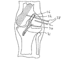
本発明の固定装置を利用する例示的な技術では、脛骨及び大腿骨において置換ACLの配置に適した部位に骨トンネルを形成する。次いで、移植片の両端(骨プラグまたは安全な束)が大腿骨トンネル及び脛骨トンネル内に配置されるように移植片を外科的に移植する。次いで、移植片の両端を本発明の固定装置で骨トンネル内に固定することができる。図5Aに、移植片の各端部が脛骨トンネル31及び大腿骨トンネル32のそれぞれに位置するように人体の膝関節内に配置された移植片30が示されている。移植片30は、大腿骨トンネル31内の骨プラグ36、及び脛骨トンネル31内に延在する腱部分34を含む。
In an exemplary technique utilizing the fixation device of the present invention, a bone tunnel is formed in the tibia and femur at a site suitable for placement of a replacement ACL. The graft is then surgically implanted so that both ends (bone plug or secure bundle) of the graft are placed in the femoral and tibial tunnels. Then, both ends of the graft can be fixed in the bone tunnel with the fixation device of the present invention. FIG. 5A shows the
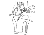
移植片が大腿骨トンネル32内に配置された状態で、大腿骨トンネル32と交差する1または複数の通路38を形成することができる。この通路38は、図5Bに示されているように固定装置10を受容することができる。通路38は、特に骨‐腱‐骨移植片の場合、骨トンネル内に配置された移植片を通過するように形成することができる。通路38が形成されたら、装置10をその通路38に挿入して、図5Cに示されているように移植片30を固定することができる。
With the graft positioned within the
固定装置10は、移植片30を通過して骨トンネル32を横断してその移植片30を固定する。骨‐腱‐骨移植片であるか或いは軟組織移植片のいずれかの場合、装置10は、移植片を直接通過して骨トンネル内に移植片を吊ることができる。加えて、装置10の挿入により、移植片を骨トンネル側壁に圧迫して骨と移植片を接触させて治癒を早めることができる。
The
図5Dに、2つの通路38内に配置された2つの固定装置10によって大腿骨トンネル内に固定された移植片30が例示されている。脛骨トンネル31内に維持されている移植片の脛骨側端部を、同様の方法で所定の位置に固定することもできるし、また当分野で周知の他の方法で固定することもできる。
FIG. 5D illustrates the
上記したように1または複数の固定装置10が移植されたら、治療物質を内部キャビティ22を介して外科部位に送達することができる。一実施形態では、装置を外科的に配置する前に装置10の内部キャビティ22に治療物質が導入され、配置後にその治療物質が外壁17から分泌される。代替の実施形態では、患者の体内に固定装置が移植された後に治療物質が内部キャビティ22内に導入される。固体、流体、または懸濁剤とすることができる治療物質を、本体12の基端部分24の孔から内部キャビティ22に注入して、その治療物質が外壁17のチャネル26または多孔性外壁を介して装置から放出されようにできる。
Once the one or
治療物質は、装置の外面17に亘って均一に送達することもできるし、また外面17の特定の領域に集中して送達することもできる。例えば、付着の促進及び治癒を早めるための移植片と骨が接触する領域に多量の治療物質を送達することができる。当業者であれば、本体12の外部への治療物質の分布の制御が、開口の配置、開口の大きさ、多孔性、送達圧力、治療物質の粘度、及びそれらの組み合わせを調節するなどの様々な方法によって達成することができる。
The therapeutic substance can be delivered uniformly over the
一実施形態では、治療物質は、生物起源とすることができる生物活性物質及び/または接着剤である。このような物質として、限定するものではないが、ヒアルロン酸、フィブリン接着剤、フィブリンクロット、コラーゲンゲル、アルギン酸ゲル、ゼラチン‐レゾルシン‐ホルマリン接着剤、ムラサキガイ系接着剤、ジヒドロキシフェニルアラニン(DOPA)系接着剤、キトサン、トランスグルタミナーゼ、ポリ(アミノ酸)系接着剤、セルロース系接着剤、多糖系接着剤、合成アクリラート系接着剤、血小板多血漿(PRP)、血小板欠血漿(PPP)、PRPのクロット、PPPのクロット、マトリゲル(Matrigel)、モノステアロイル・グリセロール・コスクシネート(Monostearoyl Glycerol co-Succinate)(MGSA)、モノステアロイル・グリセロール・コスクシネート/ポリエチレングリコール(Monostearoyl Glycerol co-Succinate/polyethylene glycol)(MGSA/PEG)コポリマー、ラミニン、エラスチン、プロテオグリカン、及びそれらの組み合わせが含まれる。 In one embodiment, the therapeutic agent is a bioactive agent and / or an adhesive that can be of biological origin. Such materials include, but are not limited to, hyaluronic acid, fibrin glue, fibrin clot, collagen gel, alginic acid gel, gelatin-resorcin-formalin adhesive, mussel adhesive, dihydroxyphenylalanine (DOPA) adhesive Agent, chitosan, transglutaminase, poly (amino acid) -based adhesive, cellulose-based adhesive, polysaccharide-based adhesive, synthetic acrylate-based adhesive, platelet-rich plasma (PRP), platelet-poor plasma (PPP), PRP clot, PPP Clot, Matrigel, Monostearoyl Glycerol co-Succinate (MGSA), Monostearoyl Glycerol co-Succinate / polyethylene glycol (Monostearoyl Glycerol co-Succinate / polyethylene glycol) GSA / PEG) copolymers, laminin, elastin, proteoglycans, and combinations thereof.
接着剤として、好適な架橋剤、例えば、ジビニルスルホン(DVS:divinyl sulfone)、ポリエチレングリコール・ジビニルスルホン(VS‐PEG‐VS)、ヒドロキシエチル・メタクリレート・ジビニルスルホン(HEMA‐DIS‐HEMA)、ホルムアルデヒド、グルタルアルデヒド、アルデヒド、イソクリアネート(isocryanate)、ハロゲン化アルキル、ハロゲン化アリル、イミドエステル(imidoesters)、N置換マレイミド、アシル化合物、カルボジイミド、ヒドロキシクロライド(hydroxychloride)、N‐ヒドロキシスクシンイミド、光(例えば、青色光、UV光)、pH、温度、及びそれらの組み合わせを挙げることができる。 Adhesives suitable as cross-linking agents such as divinyl sulfone (DVS), polyethylene glycol divinyl sulfone (VS-PEG-VS), hydroxyethyl methacrylate divinyl sulfone (HEMA-DIS-HEMA), formaldehyde, Glutaraldehyde, aldehyde, isocryanate, alkyl halide, allyl halide, imidoesters, N-substituted maleimide, acyl compound, carbodiimide, hydroxychloride, N-hydroxysuccinimide, light (eg, Blue light, UV light), pH, temperature, and combinations thereof.
別の実施形態では、治療物質は、創傷部位に存在すると治癒を促進し、かつ/または病変組織を再生する生物活性成分とすることができる。実際に治癒を促進または早める化合物または物質であるのに加えて、この生物活性成分は、感染を防止する化合物または物質(例えば、抗菌物質及び抗生物質)、炎症を抑制する化合物または物質(例えば、抗炎症剤)、ヒアルロン酸や酸化再生セルロース(例えば、エシコン社が販売するINTERCEED及びSurgicel(登録商標))などの癒着の形成を防止または最小化する化合物、及び免疫系を抑制する化合物または物質(例えば、免疫抑制剤)を含み得る。 In another embodiment, the therapeutic agent can be a bioactive ingredient that promotes healing and / or regenerates diseased tissue when present at the wound site. In addition to being a compound or substance that actually accelerates or accelerates healing, this bioactive ingredient can be a compound or substance that prevents infection (eg, antibacterials and antibiotics), a compound or substance that inhibits inflammation (eg, Anti-inflammatory agents), compounds that prevent or minimize adhesion formation, such as hyaluronic acid and oxidized regenerated cellulose (for example, INTERCEED and Surgicel (registered trademark) sold by ESICON), and compounds or substances that suppress the immune system ( For example, an immunosuppressive agent) may be included.
一例として、本発明の装置で送達できる他のタイプの生物活性成分には、組織断片、異種成長因子、自己成長因子、タンパク質(基質タンパク質を含む)、ペプチド、抗体、酵素、血小板、糖タンパク質、ホルモン、サイトカイン、グリコサミノグリカン、核酸、鎮痛薬、ウイルス、ウイルス粒子、及び細胞類が含まれる。機能的に同じまたは異なった1または複数の生物活性成分を送達できることを理解されたい。 By way of example, other types of bioactive components that can be delivered with the devices of the present invention include tissue fragments, heterologous growth factors, self-growth factors, proteins (including substrate proteins), peptides, antibodies, enzymes, platelets, glycoproteins, Hormones, cytokines, glycosaminoglycans, nucleic acids, analgesics, viruses, virus particles, and cells. It should be understood that one or more bioactive ingredients that are functionally the same or different can be delivered.
好適な生物活性成分の例には、治癒を促進し、かつ/または損傷した組織の再生を促すことが分かっている複数の異種または自己成長因子が含まれる。典型的な成長因子として、限定するものではないが、TGF‐β、骨形態形成タンパク質、軟骨由来形態形成タンパク質、線維芽成長因子、血小板由来成長因子、血管内皮細胞由来成長因子(VEGF)、上皮成長因子、インスリン様成長因子、肝細胞成長因子、及びそれらの断片を挙げることができる。更に、好適な生物活性成分には、上記した物質のアゴニスト及びアンタゴニストも含まれる。また、成長因子には、上記した成長因子の組み合わせも含み得る。加えて、成長因子は、血中の血小板によって供給される自己成長因子を含むことができる。このような場合、血小板由来成長因子は、様々な成長因子の不確定な混合物である。 Examples of suitable bioactive ingredients include a plurality of xenogeneic or self-growth factors known to promote healing and / or promote regeneration of damaged tissue. Typical growth factors include, but are not limited to, TGF-β, bone morphogenetic protein, cartilage-derived morphogenic protein, fibroblast growth factor, platelet-derived growth factor, vascular endothelial cell-derived growth factor (VEGF), epithelium Mention may be made of growth factors, insulin-like growth factors, hepatocyte growth factors, and fragments thereof. Further suitable biologically active ingredients include agonists and antagonists of the substances mentioned above. The growth factor may also include a combination of the above growth factors. In addition, growth factors can include self-growth factors supplied by platelets in the blood. In such cases, the platelet derived growth factor is an uncertain mixture of various growth factors.
本発明の装置の内部キャビティを介して送達できるタンパク質には、細胞から分泌されるタンパク質や、キャビティ内に存在する、例えば血小板などの他の生物学的な源が含まれる。単離された形態のタンパク質は通常、約55%以上の純度、すなわち他の細胞タンパク質、分子、及びデブリなどから単離されたものを指す。より好ましくは、単離されたタンパク質は少なくとも65%の純度、最も好ましくは少なくとも約75%〜95%の純度である。上記にもかかわらず、当業者であれば、約55%未満の純度のタンパク質が本発明の範囲内に含まれることを理解できよう。ここで用いる用語「タンパク質」は、糖タンパク質、リポタンパク質、プロテオグリカン、ペプチド、及びそれらの断片を包含する。生物活性成分として有用なタンパク質の例として、限定するものではないが、プレイオトロフィン、エンドセリン、テネイシン、フィブロネクチン、フィブロネクチン、フィブリノーゲン、ビトロネクチン、V‐CAM、I‐CAM、N‐CAM、セレクチン、カドヘリン、インテグリン、ラミニン、アクチン、ミオシン、コラーゲン、ミクロフィラメント、中間フィラメント、抗体、エラスチン、フィブリン、及びそれらの断片を挙げることができる。 Proteins that can be delivered through the internal cavity of the device of the present invention include proteins secreted from cells and other biological sources present in the cavity, such as platelets. Isolated form of protein usually refers to a purity of about 55% or more, ie, isolated from other cellular proteins, molecules, debris, and the like. More preferably, the isolated protein is at least 65% pure, most preferably at least about 75% to 95% pure. Notwithstanding the above, those skilled in the art will appreciate that proteins of less than about 55% purity are within the scope of the present invention. The term “protein” as used herein encompasses glycoproteins, lipoproteins, proteoglycans, peptides, and fragments thereof. Examples of proteins useful as biologically active ingredients include, but are not limited to, pleiotrophin, endothelin, tenascin, fibronectin, fibronectin, fibrinogen, vitronectin, V-CAM, I-CAM, N-CAM, selectin, cadherin, Mention may be made of integrins, laminins, actin, myosin, collagen, microfilaments, intermediate filaments, antibodies, elastin, fibrin and fragments thereof.
細胞接着である役割を果たす高度に荷電した多糖であるグリコサミノグリカンは、本発明に従った生物活性成分としても機能し得る。生物活性成分として有用なグリコサミノグリカンの例として、限定するものではないが、ヘパラン硫酸、ヘパリン、コンドロイチン硫酸、デルマタン硫酸、ケラタン硫酸、ヒアルロナン(ヒアルロン酸としても知られる)、及びそれらの組み合わせを挙げることができる。 Glycosaminoglycans, highly charged polysaccharides that play a role in cell adhesion, can also function as bioactive ingredients according to the present invention. Examples of glycosaminoglycans useful as biologically active ingredients include, but are not limited to, heparan sulfate, heparin, chondroitin sulfate, dermatan sulfate, keratan sulfate, hyaluronan (also known as hyaluronic acid), and combinations thereof Can be mentioned.
本装置はまた、限定するものではないが、骨細胞、骨芽細胞、破骨細胞、線維芽細胞、幹細胞、多能性細胞、軟骨細胞前駆体、軟骨細胞、内皮細胞、マクロファージ、白血球、脂肪細胞、単球、形質細胞、肥満細胞、臍帯細胞、間質細胞、間葉幹細胞、上皮細胞、筋芽細胞、テノサイト(tenocytes)、靱帯繊維芽細胞、ニューロン、及び骨髄細胞を含む細胞を送達することができる。細胞は通常、同族のリガンド(例えば、刺激物質)に応答する表面受容体分子をその表面に有する。刺激物質は、同族の受容体を有する細胞のその受容体に接触するとその細胞の生理作用を誘発するリガンドである。例えば、刺激物質(またはリガンド)に応答して、細胞は、Ca+2のようなセカンドメッセンジャーをかなりのレベルで産生し、このセカンドメッセンジャーがプロテインキナーゼC(本発明の例として)などのタンパク質のリン酸化などの細胞プロセスに影響を与える。場合によっては、細胞が適当な刺激物質で刺激されると、その細胞は、通常はタンパク質の形態(糖タンパク質、プロテオグリカン、及びリポタンパク質を含む)である細胞内メッセンジャーを分泌する。この細胞内メッセンジャーには、抗体(例えば、形質細胞から分泌される)、ホルモン(例えば、パラ分泌ホルモン、自己分泌ホルモン、または外分泌ホルモン)、サイトカイン、またはそれらの天然または合成の断片が含まれ得る。 The device also includes, but is not limited to, bone cells, osteoblasts, osteoclasts, fibroblasts, stem cells, pluripotent cells, chondrocyte precursors, chondrocytes, endothelial cells, macrophages, leukocytes, fat Deliver cells, including cells, monocytes, plasma cells, mast cells, umbilical cells, stromal cells, mesenchymal stem cells, epithelial cells, myoblasts, tenocytes, ligament fibroblasts, neurons, and bone marrow cells be able to. Cells usually have surface receptor molecules on their surface that respond to cognate ligands (eg, stimulants). A stimulant is a ligand that induces a physiological effect of a cell that has a cognate receptor upon contact with that receptor. For example, in response to a stimulant (or ligand), the cell produces a second messenger such as Ca +2 at a significant level, which is linked to protein phosphorous such as protein kinase C (as an example of the present invention). Affects cellular processes such as oxidation. In some cases, when cells are stimulated with a suitable stimulant, they secrete intracellular messengers, usually in the form of proteins (including glycoproteins, proteoglycans, and lipoproteins). This intracellular messenger can include antibodies (eg, secreted from plasma cells), hormones (eg, paracrine, autocrine, or exocrine hormones), cytokines, or natural or synthetic fragments thereof. .
本発明の装置はまた、核酸、ウイルス、またはウイルス粒子が少なくとも1つの目的の遺伝子産物をコードする目的の遺伝子を特定の細胞または細胞類に送達する遺伝子治療技術と共に用いることができる。従って、生物活性成分を、核酸(例えば、DNA、RNA、またはオリゴヌクレオチド)、ウイルス、ウイルス粒子、または非ウイルスベクターとすることができる。ウイルス及びウイルス粒子は、DNAウイルスまたはRNAウイルス、或いはそれらに由来するものとすることができる。目的の遺伝子産物は、タンパク質、ポリペプチド、干渉リボ核酸(iRNA)、及びそれらの組み合わせからなる群から選択されるのが好ましい。 The devices of the present invention can also be used in conjunction with gene therapy techniques in which a nucleic acid, virus, or viral particle delivers a gene of interest that encodes at least one gene product of interest to a particular cell or cells. Thus, the biologically active component can be a nucleic acid (eg, DNA, RNA, or oligonucleotide), virus, viral particle, or non-viral vector. Viruses and virus particles can be derived from or derived from DNA viruses or RNA viruses. The gene product of interest is preferably selected from the group consisting of proteins, polypeptides, interfering ribonucleic acids (iRNA), and combinations thereof.
適当な核酸及び/またはウイルス物質(すなわち、ウイルスまたはウイルス粒子)が本装置の内部キャビティ内に挿入されると、その核酸またはウイルス物質が細胞内に取り込まれ、その核酸またはウイルス物質がコードするあらゆるタンパク質がその細胞から局所的に産生され得る。一実施形態では、核酸またはウイルス物質は、内部キャビティ内の細胞によって取り込まれ、別の実施形態では、核酸またはウイルス物質は、外壁17を取り囲んでいる組織の細胞によって取り込まれる。当業者であれば、産生されるタンパク質を、上記した種類のタンパク質や、傷害や疾患を治癒する、感染症を抑える、または炎症反応を抑制する組織の能力を高める類似のタンパク質とすることができることを理解できよう。また、核酸を用いて、組織修復プロセスまたは他の正常な生理プロセスにマイナスの影響を与え得る不所望の遺伝子産物の発現をブロックすることができる。DNA、RNA、及びウイルス物質は、遺伝子発現ノックアウトとしても知られる発現をブロックする機能を達成するためによく用いられる。
When the appropriate nucleic acid and / or viral material (ie, virus or viral particle) is inserted into the internal cavity of the device, the nucleic acid or viral material is taken up into the cell and any nucleic acid or viral material encoded Proteins can be produced locally from the cells. In one embodiment, the nucleic acid or viral material is taken up by cells within the internal cavity, and in another embodiment, the nucleic acid or viral material is taken up by cells of tissue surrounding the
当業者であれば、外科医が医療技術の原理及び該当する治療目的に基づいて生物活性成分を決定できることを理解されたい。当業者であればまた、上記した実施形態に基づいて本発明の更なる特徴及び利点を理解できよう。従って、本発明は、添付の特許請求の範囲は別として、図示及び説明に限定されるものではない。 One skilled in the art will appreciate that the surgeon can determine the bioactive component based on the principles of medical technology and the relevant therapeutic purpose. One skilled in the art will also appreciate further features and advantages of the invention based on the above-described embodiments. Accordingly, the invention is not limited to the illustrations and the description, apart from the appended claims.
本発明は、生体適合性かつ生体再吸収性材料から形成された、外面、基端部分、先端部分、及び長手方向の軸を有する細長い本体を含む移植用の組織固定装置に適用可能である。本体内部に形成された内部キャビティは、固定装置の基端部で開口しており、固定装置の先端部分の基端側で終わっている。本体の外面は、内部キャビティと連通した少なくとも1つの開口を有しており、これにより、内部キャビティが、少なくとも1つの開口を介して本体の外面の外側に治療物質を供給するために治療物質を受容することができる。一態様では、本発明は、骨移植片及び/または軟組織移植片を固定するように適合されたピンとすることができる組織固定装置に適用可能である。 The present invention is applicable to tissue fixation devices for implantation that include an elongated body having an outer surface, a proximal portion, a distal portion, and a longitudinal axis formed from a biocompatible and bioresorbable material. The internal cavity formed inside the main body opens at the proximal end portion of the fixing device and ends at the proximal end side of the distal end portion of the fixing device. The outer surface of the body has at least one opening in communication with the internal cavity so that the internal cavity can deliver therapeutic material to the outside of the outer surface of the body via the at least one opening. Can accept. In one aspect, the present invention is applicable to a tissue fixation device that can be a pin adapted to secure a bone graft and / or soft tissue graft.
本発明の別の態様では、移植された後に所定の位置に細長い本体を保持するために、その細長い本体の外面の少なくとも一部に表面構造を有する組織固定装置に適用可能である。このような表面構造は、限定するものではないが、粗い領域、ねじ、バーブ、フック、及びそれらの組み合わせを含むことができる。本発明の別の態様では、平滑な外面を有する固定装置に適用可能である。 Another aspect of the present invention is applicable to tissue fixation devices having a surface structure on at least a portion of the outer surface of the elongate body to retain the elongate body in place after implantation. Such surface structures can include, but are not limited to, rough areas, screws, barbs, hooks, and combinations thereof. Another aspect of the present invention is applicable to a fixation device having a smooth outer surface.
本発明の一態様では、本発明は、細長い本体の外面を多孔性とし、本体の外面の少なくとも1つの開口を、内部キャビティと外面との間に延在する多孔性基質から形成することができる固定装置に適用可能である。別の態様では、本発明は、本体の外面の少なくとも1つの開口が通路を介して内部キャビティと連通している固定装置に適用可能である。この通路は、非多孔性の本体の外面を貫通するのが好ましい。 In one aspect of the invention, the invention can make the outer surface of the elongate body porous and at least one opening in the outer surface of the body can be formed from a porous matrix extending between the inner cavity and the outer surface. Applicable to fixing devices. In another aspect, the present invention is applicable to a fixation device in which at least one opening in the outer surface of the body is in communication with an internal cavity via a passage. This passage preferably passes through the outer surface of the non-porous body.
別の態様では、本発明は、送達できる治療物質を、組織断片、成長因子、基質タンパク質、ペプチド、抗体、酵素、サイトカイン、ウイルス、核酸、ペプチド、単離された細胞、血小板、及びそれらの組み合わせなどの生物活性物質とすることができる装置に適用可能である。治療物質はまた、接着剤とすることができる。 In another aspect, the invention provides therapeutic agents that can be delivered to tissue fragments, growth factors, substrate proteins, peptides, antibodies, enzymes, cytokines, viruses, nucleic acids, peptides, isolated cells, platelets, and combinations thereof. The present invention can be applied to a device that can be a biologically active substance. The therapeutic substance can also be an adhesive.
別の態様では、本発明は、組織固定装置の使用方法に適用可能である。 In another aspect, the present invention is applicable to a method of using a tissue fixation device.
本発明の実施態様は以下の通りである。
(A)生体移植用の組織固定装置であって、
外面、基端部分、先端部分、及び細長い本体を通る長手方向の軸を有する、生体適合性の生体再吸収性の材料からなる前記細長い本体と、
前記本体の前記基端部分の開口から前記本体内を通り、前記先端部分の基端側で終わっている内部キャビティと、
前記本体の前記外面に形成された少なくとも1つの開口とを含み、
前記少なくとも1つの開口のそれぞれが、前記少なくとも1つの開口を介して前記本体の前記外面の外側に治療物質を送達できるように、前記内部キャビティと連通していることを特徴とする固定装置。
(1)前記細長い本体が、骨移植片及び/または軟組織移植片を固定できるように適合されたピンであることを特徴とする実施態様(A)に記載の固定装置。
(2)前記細長い本体が、ラクチド;グリコリド;ε-カプロラクトン;ヒドロキシブチレート;ヒドロキシバレレート(hydroxyvalerate);1,4-ジオキセパン-2-オン(1,4-dioxepan-2-one);1,5,8,12-テトラオキササイクロテトラデカン-7,14-ジオン(1,5,8,12-tetraoxacyclotetradecane-7,14-dione);1,5-ジオキセパン-2-オン(1,5-dioxepan-2-one);6,6-ジメチル-1,4-ジオキサン-2-オン;2,5-ジケトモルホリン(2,5-diketomorpholine);p-ジオキサノン(1,4-ジオキサノン-2-オン);トリメチレンカーボネート(1,3-ジオキサン-2-オン);トリメチレンカーボネートのアルキル誘導体;δ-バレロラクトン;β-ブチロラクトン;γ-ブチロラクトン;ε-デカラクトン;ピバロラクトン(pivalolactone);α,α-ジエチルプロピオラクトン;エチレンカーボネート;エチレンオキサレート;3-メチル-1,4-ジオキサン-2,5-ジオン;3,3-ジエチル-1,4-ジオキサン-2,5-ジオン;及び6,8-ジオキサビシクロオクタン-7-オン(6,8-dioxabicycloctane-7-one)からなる群から選択されるモノマーから形成されるポリマーまたはコポリマーから製造されることを特徴とする実施態様(1)に記載の固定装置。
(3)前記細長い本体が、ポリ乳酸、脂肪族ポリエステル、ポリ(アミノ酸)、ポリ(フマル酸ポリプロピレン)(poly(propylene fumarate))、コポリ(エーテル−エステル)、ポリアルキレンオキサレート、ポリアミド、チロシン由来ポリカーボネート、ポリ(イミノカーボネート)(poly(iminocarbonates))、ポリオルトエステル、ポリオキサエステル(polyoxaesters)、ポリアミドエステル、アミン基を含むポリオキサエステル、ポリ無水物、ポリホスファゼン、ポリウレタン、ポリ(エーテルウレタン)、ポリ(エステルウレタン)、生合成ポリマー、及びそれらの組み合わせからなる群から選択されたポリマーまたはコポリマーから形成されることを特徴とする実施態様(A)に記載の固定装置。
(4)前記細長い本体が、約15mm〜65mmの範囲の長さを有することを特徴とする実施態様(1)に記載の固定装置。
(5)前記細長い本体の前記外面の少なくとも一部が、移植された後に前記細長い本体を所定の位置に保持するために表面構造を有することを特徴とする実施態様(A)に記載の固定装置。
Embodiments of the present invention are as follows.
(A) a tissue fixing device for living transplantation,
Said elongated body of biocompatible bioresorbable material having an outer surface, a proximal portion, a distal portion, and a longitudinal axis through the elongated body;
An internal cavity passing through the body from the opening of the proximal end portion of the body and ending on the proximal end side of the distal end portion;
And at least one opening formed in the outer surface of the body,
A fixation device, wherein each of the at least one opening is in communication with the internal cavity such that a therapeutic substance can be delivered to the outside of the outer surface of the body via the at least one opening.
(1) The fixation device according to embodiment (A) , wherein the elongated body is a pin adapted to fix a bone graft and / or soft tissue graft.
(2) The elongated body is lactide; glycolide; ε-caprolactone; hydroxybutyrate; hydroxyvalerate; 1,4-dioxepan-2-one; 5,8,12-tetraoxacyclotetradecane-7,14-dione (1,5,8,12-tetraoxacyclotetradecane-7,14-dione); 1,5-dioxepan-2-one (1,5-dioxepan- 2-one); 6,6-dimethyl-1,4-dioxane-2-one; 2,5-diketomorpholine; p-dioxanone (1,4-dioxanon-2-one) Trimene carbonate (1,3-dioxane-2-one); alkyl derivatives of trimethylene carbonate; δ-valerolactone; β-butyrolactone; γ-butyrolactone; ε-decalactone; pivalolactone; α, α-diethyl Propiolactone; ethylene carbonate; ethylene oxalate; 3-methyl-1, 4-dioxane-2,5-dione; 3,3-diethyl-1,4-dioxane-2,5-dione; and 6,8-dioxabicyclooctane-7-one The fixing device according to embodiment (1), characterized in that it is made from a polymer or copolymer formed from a monomer selected from the group consisting of one).
(3) The elongated body is derived from polylactic acid, aliphatic polyester, poly (amino acid), poly (propylene fumarate), copoly (ether-ester), polyalkylene oxalate, polyamide, and tyrosine. Polycarbonate, poly (iminocarbonates), polyorthoesters, polyoxaesters, polyamide esters, polyoxaesters containing amine groups, polyanhydrides, polyphosphazenes, polyurethanes, poly (ether urethanes) The fixing device according to embodiment (A) , characterized in that it is formed from a polymer or copolymer selected from the group consisting of: poly (ester urethane), biosynthetic polymers, and combinations thereof.
(4) The fixing device according to embodiment (1), wherein the elongated body has a length in a range of about 15 mm to 65 mm.
(5) The fixation device according to embodiment (A) , wherein at least a portion of the outer surface of the elongate body has a surface structure to hold the elongate body in place after implantation. .
(6)前記表面構造が、粗い領域、ねじ、バーブ、フック、及びそれらの組み合わせからなる群から選択されることを特徴とする実施態様(5)に記載の固定装置。
(7)前記細長い本体の前記外面が平滑であることを特徴とする実施態様(A)に記載の固定装置。
(8)前記細長い本体の前記外面が多孔性であり、前記外面に形成された前記少なくとも1つの開口が、前記内部キャビティと前記外面との間に延在する多孔性基質によって得られたものであることを特徴とする実施態様(A)に記載の固定装置。
(9)前記少なくとも1つの開口が、少なくとも1つの通路を介して前記内部キャビティと連通していることを特徴とする実施態様(A)に記載の固定装置。
(10)前記外面が非多孔性であることを特徴とする実施態様(9)に記載の固定装置。
(6) The fixing device according to the embodiment (5), wherein the surface structure is selected from the group consisting of a rough region, a screw, a barb, a hook, and a combination thereof.
(7) The fixing device according to the embodiment (A) , wherein the outer surface of the elongated body is smooth.
(8) The outer surface of the elongated body is porous, and the at least one opening formed in the outer surface is obtained by a porous substrate extending between the inner cavity and the outer surface. A fixing device according to embodiment (A) , characterized in that it is.
(9) The fixing device according to the embodiment (A) , wherein the at least one opening communicates with the internal cavity via at least one passage.
(10) The fixing device according to the embodiment (9), wherein the outer surface is non-porous.
(11)前記ピンの直径が約1mm〜10mmの範囲であることを特徴とする実施態様(1)に記載の固定装置。
(12)前記ピンの吸収プロフィールが約12週間〜60週間の範囲であることを特徴とする実施態様(1)に記載の固定装置。
(13)前記内部キャビティの直径が、約0.5mm〜5mmの範囲であることを特徴とする実施態様(A)に記載の固定装置。
(14)前記多孔性基質の孔の平均直径が約0.01mm〜5mmの範囲であることを特徴とする実施態様(8)に記載の固定装置。
(15)前記治療物質が生物活性物質であることを特徴とする実施態様(A)に記載の固定装置。
(11) The fixing device according to the embodiment (1), wherein the pin has a diameter of about 1 mm to 10 mm.
(12) The fixation device according to embodiment (1), wherein the absorption profile of the pin ranges from about 12 weeks to 60 weeks.
(13) The fixing device according to the embodiment (A) , wherein a diameter of the internal cavity is in a range of about 0.5 mm to 5 mm.
(14) The fixing device according to embodiment (8), wherein an average diameter of the pores of the porous substrate is in the range of about 0.01 mm to 5 mm.
(15) The fixation device according to embodiment (A) , wherein the therapeutic substance is a bioactive substance.
(16)前記生物活性物質が、組織断片、成長因子、タンパク質、鎮痛薬、抗体、酵素、サイトカイン、グリコサミノグリカン、ウイルス、ウイルス粒子、核酸、ペプチド、単離された細胞、血小板、及びそれらの組み合わせからなる群から選択されることを特徴とする実施態様(15)に記載の固定装置。
(17)前記治療物質が接着剤であることを特徴とする実施態様(A)に記載の固定装置。
(18)前記接着剤が、ヒアルロン酸、フィブリン接着剤、フィブリンクロット、コラーゲンゲル、ゼラチン‐レゾルシン‐ホルマリン接着剤、ムラサキガイ系接着剤、ジヒドロキシフェニルアラニン(DOPA)系接着剤、キトサン、トランスグルタミナーゼ、ポリ(アミノ酸)系接着剤、セルロース系接着剤、合成アクリラート系接着剤、血小板多血漿(PRP)、マトリゲル(Matrigel)、モノステアロイル・グリセロール・コスクシネート(Monostearoyl Glycerol co-Succinate)(MGSA)、モノステアロイル・グリセロール・コスクシネート/ポリエチレングリコール(Monostearoyl Glycerol co-Succinate/polyethylene glycol)(MGSA/PEG)コポリマー、ラミニン、エラスチン、プロテオグリカン、及びそれらの組み合わせからなる群から選択されるアンカー剤を含むことを特徴とする実施態様(17)に記載の固定装置。
(19)前記接着剤が、ジビニルスルホン(DVS:divinyl sulfone)、ポリエチレングリコール・ジビニルスルホン(VS‐PEG‐VS)、ヒドロキシエチルメタクリレート・ジビニルスルホン(HEMA‐DIS‐HEMA)、ホルムアルデヒド、グルタルアルデヒド、アルデヒド、イソクリアネート(isocryanate)、ハロゲン化アルキル、ハロゲン化アリル、イミドエステル(imidoesters)、N置換マレイミド、アシル化合物、カルボジイミド、ヒドロキシクロライド(hydroxychloride)、N‐ヒドロキシスクシンイミド、光、pH、温度、及びそれらの組み合わせからなる群から選択される化学架橋剤を含むことを特徴とする実施態様(17)に記載の固定装置。
(20)前記本体の前記外面に形成された前記少なくとも1つの開口の数が、約5〜25の範囲であることを特徴とする実施態様(9)に記載の固定装置。
(16) The biologically active substance is a tissue fragment, growth factor, protein, analgesic, antibody, enzyme, cytokine, glycosaminoglycan, virus, virus particle, nucleic acid, peptide, isolated cell, platelet, and them The fixing device according to embodiment (15), wherein the fixing device is selected from the group consisting of:
(17) The fixing device according to the embodiment (A) , wherein the therapeutic substance is an adhesive.
(18) The adhesive is hyaluronic acid, fibrin adhesive, fibrin clot, collagen gel, gelatin-resorcin-formalin adhesive, mussel adhesive, dihydroxyphenylalanine (DOPA) adhesive, chitosan, transglutaminase, poly (Amino acid) adhesive, cellulose adhesive, synthetic acrylate adhesive, platelet-rich plasma (PRP), Matrigel, Monostearoyl Glycerol co-Succinate (MGSA), monostearoyl Selected from the group consisting of glycerol cosuccinate / polyethylene glycol (MGSA / PEG) copolymer, laminin, elastin, proteoglycan, and combinations thereof Fixing apparatus according to embodiment (17), characterized in that it comprises an anchoring agent.
(19) The adhesive is divinyl sulfone (DVS), polyethylene glycol divinyl sulfone (VS-PEG-VS), hydroxyethyl methacrylate divinyl sulfone (HEMA-DIS-HEMA), formaldehyde, glutaraldehyde, aldehyde , Isocryanate, alkyl halides, allyl halides, imidoesters, N-substituted maleimides, acyl compounds, carbodiimides, hydroxychloride, N-hydroxysuccinimide, light, pH, temperature, and the like The fixing device according to embodiment (17), comprising a chemical crosslinking agent selected from the group consisting of:
(20) The fixing device according to the embodiment (9), wherein the number of the at least one opening formed in the outer surface of the main body is in a range of about 5 to 25.
(21)前記少なくとも1つの開口の直径が約0.5mm〜1.5mmの範囲であることを特徴とする実施態様(9)に記載の固定装置。
(22)前記細長い本体が実質的に円筒状であることを特徴とする実施態様(A)に記載の固定装置。
(23)前記細長い本体の前記先端部分が先端に向かってテーパ状であることを特徴とする実施態様(A)に記載の固定装置。
(B) 組織移植片を骨に固定するための方法であって、
骨に骨トンネルを形成するステップと、
基端部の開口から内部に向かって延びた長手方向のチャネルを備えた細長い本体の形態であり、外面に形成された少なくとも1つの開口が前記チャネルと連通している組織固定装置を用意するステップと、
前記組織片の一部を前記骨トンネル内に配置するステップと、
前記組織固定装置を前記骨トンネル内に挿入して、前記組織移植片をその骨トンネル内に固定するステップと、
前記組織固定装置の前記チャネル内に治療物質を注入して、前記少なくとも1つの開口を介して前記組織固定装置の側壁の外側領域に前記治療物質が分泌されるようにするステップとを含むことを特徴とする方法。
(24)前記治療物質が生物活性物質であることを特徴とする実施態様(B)に記載の方法。
(25)前記生物活性物質が、組織断片、成長因子、タンパク質、鎮痛薬、抗体、酵素、サイトカイン、グリコサミノグリカン、ウイルス、ウイルス粒子、核酸、ペプチド、単離された細胞、血小板、及びそれらの組み合わせからなる群から選択されることを特徴とする実施態様(24)に記載の方法。
(21) The fixing device according to embodiment (9), wherein the diameter of the at least one opening is in the range of about 0.5 mm to 1.5 mm.
(22) The fixing device according to the embodiment (A) , wherein the elongated body is substantially cylindrical.
(23) The fixing device according to the embodiment (A) , wherein the distal end portion of the elongated main body is tapered toward the distal end.
(B) A method for securing a tissue graft to bone,
Forming a bone tunnel in the bone;
Providing a tissue fixation device in the form of an elongate body with a longitudinal channel extending inwardly from an opening in the proximal end, wherein at least one opening formed in the outer surface communicates with the channel. When,
Placing a portion of the tissue piece within the bone tunnel;
Inserting the tissue fixation device into the bone tunnel and securing the tissue graft within the bone tunnel;
Injecting a therapeutic substance into the channel of the tissue fixation device so that the therapeutic substance is secreted through the at least one opening to an outer region of the sidewall of the tissue fixation device. Feature method.
(24) The method according to embodiment (B) , wherein the therapeutic substance is a bioactive substance.
(25) The biologically active substance is a tissue fragment, growth factor, protein, analgesic, antibody, enzyme, cytokine, glycosaminoglycan, virus, virus particle, nucleic acid, peptide, isolated cell, platelet, and them The method of embodiment (24), characterized in that it is selected from the group consisting of:
(26)前記治療物質が接着剤であることを特徴とする実施態様(B)に記載の方法。
(27)前記接着剤が、ヒアルロン酸、フィブリン接着剤、フィブリンクロット、コラーゲンゲル、ゼラチン‐レゾルシン‐ホルマリン接着剤、ムラサキガイ系接着剤、ジヒドロキシフェニルアラニン(DOPA)系接着剤、キトサン、トランスグルタミナーゼ、ポリ(アミノ酸)系接着剤、セルロース系接着剤、多糖系接着剤、合成アクリラート系接着剤、ポリウレタン系接着剤、血小板多血漿(PRP)、血小板欠血漿(PPP)、マトリゲル(Matrigel)、モノステアロイル・グリセロール・コスクシネート(Monostearoyl Glycerol co-Succinate)(MGSA)、モノステアロイル・グリセロール・コスクシネート/ポリエチレングリコール(Monostearoyl Glycerol co-Succinate/polyethylene glycol)(MGSA/PEG)コポリマー、ラミニン、エラスチン、プロテオグリカン、及びそれらの組み合わせからなる群から選択されるアンカー剤を含むことを特徴とする実施態様(26)に記載の方法。
(28)前記接着剤が、ジビニルスルホン(DVS:divinyl sulfone)、ポリエチレングリコール・ジビニルスルホン(VS‐PEG‐VS)、ヒドロキシエチルメタクリレート・ジビニルスルホン(HEMA‐DIS‐HEMA)、ホルムアルデヒド、グルタルアルデヒド、アルデヒド、イソクリアネート(isocryanate)、ハロゲン化アルキル、ハロゲン化アリル、イミドエステル(imidoesters)、N置換マレイミド、アシル化合物、カルボジイミド、ヒドロキシクロライド(hydroxychloride)、N‐ヒドロキシスクシンイミド、光、pH、温度、及びそれらの組み合わせからなる群から選択される架橋剤を含むことを特徴とする実施態様(26)に記載の方法。
(26) The method according to embodiment (B) , wherein the therapeutic substance is an adhesive.
(27) The adhesive is hyaluronic acid, fibrin adhesive, fibrin clot, collagen gel, gelatin-resorcin-formalin adhesive, mussel adhesive, dihydroxyphenylalanine (DOPA) adhesive, chitosan, transglutaminase, poly (Amino acid) adhesive, cellulose adhesive, polysaccharide adhesive, synthetic acrylate adhesive, polyurethane adhesive, platelet-rich plasma (PRP), platelet-poor plasma (PPP), Matrigel, monostearoyl Monoesterearoyl Glycerol co-Succinate (MGSA), Monostearoyl Glycerol co-Succinate / polyethylene glycol (MGSA / PEG) copolymer, laminin, elastin, plastic Teogurikan, and method of
(28) The adhesive is divinyl sulfone (DVS), polyethylene glycol divinyl sulfone (VS-PEG-VS), hydroxyethyl methacrylate divinyl sulfone (HEMA-DIS-HEMA), formaldehyde, glutaraldehyde, aldehyde , Isocryanate, alkyl halides, allyl halides, imidoesters, N-substituted maleimides, acyl compounds, carbodiimides, hydroxychloride, N-hydroxysuccinimide, light, pH, temperature, and the like The method of embodiment (26), comprising a cross-linking agent selected from the group consisting of:
10 固定装置
12 本体
14 基端部分
16 先端部分
17 外壁
18 先端部
20 先端
22 内部キャビティ
24 開口
26 チャネル
28 開口
30 移植片
31 脛骨トンネル
32 大腿骨トンネル
34 腱部分
36 骨プラグ
38 通路
DESCRIPTION OF
Claims (6)
生体適合性で生体再吸収性の材料からなる細長い本体であって、外面、基端部分、先端部分、及び前記細長い本体を通る長手方向の軸を有する、細長い本体と、
前記本体の前記基端部分の開口から前記本体内を通り、前記先端部分の基端側で終わっている内部キャビティと、
前記本体の前記外面に形成された少なくとも1つの開口と、
を含み、
前記少なくとも1つの開口のそれぞれが、前記少なくとも1つの開口を介して前記本体の前記外面の外側に治療物質を送達できるように、前記内部キャビティと連通しており、
前記細長い本体の前記外面が多孔性であり、前記外面に形成された前記少なくとも1つの開口が、前記内部キャビティと前記外面との間に延在する多孔性基質によって得られたものである、固定装置。 In tissue fixation device for a biological implantation,
An elongated body made of bioresorbable materials biocompatible outer surface, a proximal portion, distal portion, and a longitudinal axis through said elongated body, a thin elongate body,
An internal cavity passing through the body from the opening of the proximal end portion of the body and ending on the proximal end side of the distal end portion;
At least one opening formed in the outer surface of the body ;
Including
Each of said at least one opening, wherein such can deliver the therapeutic agent to the outside of said outer surface of said body via at least one opening in communication with said interior cavity,
The outer surface of the elongate body is porous and the at least one opening formed in the outer surface is obtained by a porous substrate extending between the inner cavity and the outer surface apparatus.
前記細長い本体が、骨移植片および軟組織移植片の少なくともいずれかを固定できるように適合されたピンである、固定装置。 The fixing device according to claim 1,
A fixation device wherein the elongate body is a pin adapted to fix at least one of a bone graft and a soft tissue graft.
前記細長い本体が、ラクチド;グリコリド;ε-カプロラクトン;ヒドロキシブチレート;ヒドロキシバレレート(hydroxyvalerate);1,4-ジオキセパン-2-オン(1,4-dioxepan-2-one);1,5,8,12-テトラオキササイクロテトラデカン-7,14-ジオン(1,5,8,12-tetraoxacyclotetradecane-7,14-dione);1,5-ジオキセパン-2-オン(1,5-dioxepan-2-one);6,6-ジメチル-1,4-ジオキサン-2-オン;2,5-ジケトモルホリン(2,5-diketomorpholine);p-ジオキサノン(1,4-ジオキサノン-2-オン);トリメチレンカーボネート(1,3-ジオキサン-2-オン);トリメチレンカーボネートのアルキル誘導体;δ-バレロラクトン;β-ブチロラクトン;γ-ブチロラクトン;ε-デカラクトン;ピバロラクトン(pivalolactone);α,α-ジエチルプロピオラクトン;エチレンカーボネート;エチレンオキサレート;3-メチル-1,4-ジオキサン-2,5-ジオン;3,3-ジエチル-1,4-ジオキサン-2,5-ジオン;及び6,8-ジオキサビシクロオクタン-7-オン(6,8-dioxabicycloctane-7-one)からなる群から選択されるモノマーから形成されるポリマーまたはコポリマーから製造される、固定装置。 The fixing device according to claim 1 or 2,
The elongated body is lactide; glycolide; ε-caprolactone; hydroxybutyrate; hydroxyvalerate; 1,4-dioxepan-2-one; 1,5,8 , 12-Tetraoxacyclotetradecane-7,14-dione (1,5,8,12-tetraoxacyclotetradecane-7,14-dione); 1,5-dioxepan-2-one (1,5-dioxepan-2-one) 6,6-dimethyl-1,4-dioxane-2-one; 2,5-diketomorpholine; p-dioxanone (1,4-dioxanon-2-one); trimethylene Carbonate (1,3-dioxane-2-one); trimethylene carbonate alkyl derivative; δ-valerolactone; β-butyrolactone; γ-butyrolactone; ε-decalactone; pivalolactone; α, α-diethylpropiolactone Ethylene carbonate; ethylene oxalate; 3-methyl-1,4-dio Sun-2,5-dione; 3,3-diethyl-1,4-dioxane-2,5-dione; and 6,8-dioxabicyclooctane-7-one An anchoring device made from a polymer or copolymer formed from a monomer selected from the group consisting of:
前記細長い本体が、ポリ乳酸、脂肪族ポリエステル、ポリ(アミノ酸)、ポリ(フマル酸ポリプロピレン)(poly(propylene fumarate))、コポリ(エーテル−エステル)、ポリアルキレンオキサレート、ポリアミド、チロシン由来ポリカーボネート、ポリ(イミノカーボネート)(poly(iminocarbonates))、ポリオルトエステル、ポリオキサエステル(polyoxaesters)、ポリアミドエステル、アミン基を含むポリオキサエステル、ポリ無水物、ポリホスファゼン、ポリウレタン、ポリ(エーテルウレタン)、ポリ(エステルウレタン)、生合成ポリマー、及びそれらの組み合わせからなる群から選択されたポリマーまたはコポリマーから形成される、固定装置。 The fixing device according to claim 1 or 2,
The elongated body is made of polylactic acid, aliphatic polyester, poly (amino acid), poly (propylene fumarate), copoly (ether-ester), polyalkylene oxalate, polyamide, tyrosine-derived polycarbonate, poly (Iminocarbonates), polyorthoesters, polyoxaesters, polyamide esters, polyoxaesters containing amine groups, polyanhydrides, polyphosphazenes, polyurethanes, poly (ether urethanes), poly ( An anchoring device formed from a polymer or copolymer selected from the group consisting of ester urethane), biosynthetic polymers, and combinations thereof.
前記細長い本体の前記外面が平滑である、固定装置。 The fixing device according to any one of claims 1 to 4,
A fixation device wherein the outer surface of the elongate body is smooth.
前記治療物質が、組織断片、成長因子、タンパク質、鎮痛薬、抗体、酵素、サイトカイン、グリコサミノグリカン、ウイルス、ウイルス粒子、核酸、ペプチド、単離された細胞、血小板、及びそれらの組み合わせからなる群から選択される生活活性物質である、固定装置。 The fixing device according to any one of claims 1 to 5 ,
The therapeutic substance comprises a tissue fragment, a growth factor, a protein, an analgesic, an antibody, an enzyme, a cytokine, a glycosaminoglycan, a virus, a virus particle, a nucleic acid, a peptide, an isolated cell, a platelet, and combinations thereof A fixation device, which is a life-active substance selected from the group.
Applications Claiming Priority (2)
| Application Number | Priority Date | Filing Date | Title |
|---|---|---|---|
| US602797 | 2003-06-24 | ||
| US10/602,797 US7300439B2 (en) | 2003-06-24 | 2003-06-24 | Porous resorbable graft fixation pin |
Publications (3)
| Publication Number | Publication Date |
|---|---|
| JP2005013731A JP2005013731A (en) | 2005-01-20 |
| JP2005013731A5 JP2005013731A5 (en) | 2007-05-24 |
| JP4694154B2 true JP4694154B2 (en) | 2011-06-08 |
Family
ID=33418641
Family Applications (1)
| Application Number | Title | Priority Date | Filing Date |
|---|---|---|---|
| JP2004185117A Expired - Fee Related JP4694154B2 (en) | 2003-06-24 | 2004-06-23 | Porous and resorbable graft fixation pin |
Country Status (6)
| Country | Link |
|---|---|
| US (1) | US7300439B2 (en) |
| EP (1) | EP1491156B1 (en) |
| JP (1) | JP4694154B2 (en) |
| AU (1) | AU2004202107B2 (en) |
| CA (1) | CA2467262C (en) |
| DE (1) | DE602004018708D1 (en) |
Families Citing this family (66)
| Publication number | Priority date | Publication date | Assignee | Title |
|---|---|---|---|---|
| US8209259B2 (en) * | 2003-01-09 | 2012-06-26 | Adp Dealer Services, Inc. | Software business platform with networked, association-based business entity access management |
| GB0329654D0 (en) | 2003-12-23 | 2004-01-28 | Smith & Nephew | Tunable segmented polyacetal |
| WO2005069884A2 (en) * | 2004-01-16 | 2005-08-04 | Osteobiologics, Inc. | Bone-tendon-bone implant |
| WO2006062518A2 (en) | 2004-12-08 | 2006-06-15 | Interpore Spine Ltd. | Continuous phase composite for musculoskeletal repair |
| US8535357B2 (en) * | 2004-12-09 | 2013-09-17 | Biomet Sports Medicine, Llc | Continuous phase compositions for ACL repair |
| US20060217717A1 (en) * | 2005-03-24 | 2006-09-28 | Dale Whipple | Methods and devices for stabilizing a bone anchor |
| US20080208253A1 (en) * | 2006-05-18 | 2008-08-28 | Dreyfuss Peter J | Self-punching swivel anchor and method for knotless fixation of tissue |
| US20090187216A1 (en) | 2006-05-18 | 2009-07-23 | Arthrex, Inc. | Fenestrated swivel anchor for knotless fixation of tissue |
| US11801043B2 (en) | 2005-03-30 | 2023-10-31 | Arthrex, Inc. | Suture anchor for knotless fixation of tissue |
| US20090192546A1 (en) * | 2005-03-30 | 2009-07-30 | Reinhold Schmieding | Fenestrated suture anchor and method for knotless fixation of tissue |
| US20070161985A1 (en) * | 2005-12-05 | 2007-07-12 | Kentomia, Llc . | Screws configured to engage bones, and methods of attaching implants to skeletal regions |
| US8435292B2 (en) * | 2005-12-20 | 2013-05-07 | Depuy Mitek, Inc. | Methods for ligament reconstruction |
| US20070162123A1 (en) * | 2005-12-20 | 2007-07-12 | Whittaker Gregory R | Materials and methods for ligament reconstruction |
| US20070156145A1 (en) * | 2005-12-30 | 2007-07-05 | Kentomia, Llc | Therapeutic constructions, spinal plates, cervical plates, hooks and screws |
| US20070179609A1 (en) * | 2006-01-27 | 2007-08-02 | Medicinelodge, Inc. | Therapeutic agent eluding implant with percutaneous supply |
| US8900321B2 (en) * | 2006-03-20 | 2014-12-02 | Zimmer, Inc. | Implant anchoring device |
| JP5389337B2 (en) | 2006-05-18 | 2014-01-15 | オーソレクス,インコーポレイテッド | Swivel anchor for tissue nodule fixation |
| CN101594831B (en) | 2006-11-30 | 2011-09-14 | 史密夫和内修有限公司 | Fiber reinforced composite material |
| EP2142353A1 (en) | 2007-04-18 | 2010-01-13 | Smith & Nephew PLC | Expansion moulding of shape memory polymers |
| JP5520814B2 (en) | 2007-04-19 | 2014-06-11 | スミス アンド ネフュー インコーポレーテッド | Multimodal shape memory polymer |
| DE602008006181D1 (en) | 2007-04-19 | 2011-05-26 | Smith & Nephew Inc | GRAFT FIXATION |
| US20090138092A1 (en) * | 2007-11-28 | 2009-05-28 | Johnston Brent W | Therapeutic Structures for Utilization in Temporomandibular Joint Replacement Systems |
| EP2323587B1 (en) * | 2008-08-13 | 2016-08-03 | SMed - TA/TD LLC | Drug delivery implants |
| US9616205B2 (en) | 2008-08-13 | 2017-04-11 | Smed-Ta/Td, Llc | Drug delivery implants |
| US9700431B2 (en) | 2008-08-13 | 2017-07-11 | Smed-Ta/Td, Llc | Orthopaedic implant with porous structural member |
| US20100042213A1 (en) | 2008-08-13 | 2010-02-18 | Nebosky Paul S | Drug delivery implants |
| US10842645B2 (en) | 2008-08-13 | 2020-11-24 | Smed-Ta/Td, Llc | Orthopaedic implant with porous structural member |
| WO2010019788A1 (en) * | 2008-08-13 | 2010-02-18 | Smed-Ta/Td. Llc | Drug delivery implants |
| JP5687622B2 (en) | 2008-08-29 | 2015-03-18 | スメド−ティーエイ/ティーディー・エルエルシー | Orthopedic implant |
| JP5658154B2 (en) * | 2008-08-29 | 2015-01-21 | スメド−ティーエイ/ティーディー・エルエルシー | Drug delivery implants |
| SG195588A1 (en) * | 2008-10-17 | 2013-12-30 | Univ Singapore | Resorbable scaffolds for bone repair and long bone tissue engineering |
| CA2742077A1 (en) | 2008-10-30 | 2010-05-06 | Depuy Spine, Inc. | Systems and methods for delivering bone cement to a bone anchor |
| US8574273B2 (en) | 2009-09-09 | 2013-11-05 | Innovision, Inc. | Bone screws and methods of use thereof |
| US10539487B2 (en) | 2010-03-04 | 2020-01-21 | Ventana Medical Systems, Inc. | Systems and methods for monitoring tissue sample processing |
| CA2788664C (en) | 2010-03-04 | 2023-04-11 | Ventana Medical Systems, Inc. | Processing system for processing specimens using acoustic energy |
| US10126216B2 (en) | 2011-02-17 | 2018-11-13 | Ventana Medical Systems, Inc. | Method for tissue sample fixation |
| US9555171B2 (en) | 2010-09-30 | 2017-01-31 | Depuy Mitek, Llc | Methods and devices for collecting separate components of whole blood |
| US8551525B2 (en) | 2010-12-23 | 2013-10-08 | Biostructures, Llc | Bone graft materials and methods |
| US10482475B2 (en) | 2011-02-10 | 2019-11-19 | Adp Dealer Services, Inc. | Systems and methods for providing targeted advertising |
| US8470046B2 (en) | 2011-04-25 | 2013-06-25 | Warsaw Orthopedic, Inc. | Bone augmentation device and method |
| US8617176B2 (en) * | 2011-08-24 | 2013-12-31 | Depuy Mitek, Llc | Cross pinning guide devices and methods |
| US9155580B2 (en) | 2011-08-25 | 2015-10-13 | Medos International Sarl | Multi-threaded cannulated bone anchors |
| DE102012005418A1 (en) * | 2012-03-16 | 2013-09-19 | Klaus Wellner | Tendon suture kit for reunion of e.g. flexor tendon of hands, has suture double thread material which is provided on the proximal and distal ends of the monofilament threads with counter-rotating double check anchors |
| DE102012025143A1 (en) | 2012-11-07 | 2014-05-08 | Heraeus Medical Gmbh | Method of drug release and drug release systems |
| US20140277183A1 (en) * | 2013-03-15 | 2014-09-18 | Smed-Ta/Td, Llc | Fixation of orthopaedic devices |
| EP2967694B1 (en) | 2013-03-15 | 2022-04-20 | INNOVISION, Inc. | Bone screws |
| US11080734B2 (en) | 2013-03-15 | 2021-08-03 | Cdk Global, Llc | Pricing system for identifying prices for vehicles offered by vehicle dealerships and other entities |
| JP2015000208A (en) * | 2013-06-17 | 2015-01-05 | 株式会社ジーシー | Fracture part treatment member |
| SG11201701024WA (en) * | 2014-08-27 | 2017-03-30 | Neuronano Ab | Method of implantation of a medical device into neural tissue |
| WO2016127522A1 (en) | 2015-02-09 | 2016-08-18 | 张英泽 | Porous bionic internal fixation apparatus for promoting fracture healing |
| CN104586496A (en) * | 2015-02-09 | 2015-05-06 | 张英泽 | Microporous bionic fracture fixing device capable of promoting fracture healing |
| CN104783878B (en) * | 2015-04-29 | 2016-10-12 | 天津博硕倍生物科技有限公司 | Absorbable self-locking hold-down bars and preparation method thereof |
| CN107666884B (en) * | 2015-05-28 | 2019-11-26 | 4科技有限公司 | Eccentric tissue anchors with tensional element |
| US10332068B2 (en) | 2016-04-21 | 2019-06-25 | Cdk Global, Llc | Systems and methods for stocking an automobile |
| US10853769B2 (en) | 2016-04-21 | 2020-12-01 | Cdk Global Llc | Scheduling an automobile service appointment in a dealer service bay based on diagnostic trouble codes and service bay attributes |
| US10867285B2 (en) | 2016-04-21 | 2020-12-15 | Cdk Global, Llc | Automatic automobile repair service scheduling based on diagnostic trouble codes and service center attributes |
| US11540866B2 (en) * | 2017-03-29 | 2023-01-03 | Bone Solutions, Inc. | Implant of osteostimulative material |
| US10722280B2 (en) * | 2017-03-29 | 2020-07-28 | Bone Solutions, Inc. | Implant of osteostimulative material |
| US10326858B2 (en) | 2017-05-23 | 2019-06-18 | Cdk Global, Llc | System and method for dynamically generating personalized websites |
| JP7247438B2 (en) * | 2017-09-15 | 2023-03-29 | ディーエスエム アイピー アセッツ ビー.ブイ. | Medical fixation device for polymer cables |
| US11190608B2 (en) | 2018-03-21 | 2021-11-30 | Cdk Global Llc | Systems and methods for an automotive commerce exchange |
| US11501351B2 (en) | 2018-03-21 | 2022-11-15 | Cdk Global, Llc | Servers, systems, and methods for single sign-on of an automotive commerce exchange |
| US11160592B2 (en) | 2018-10-18 | 2021-11-02 | Biedermann Technologies Gmbh & Co. Kg | Method of using bone plate with polyaxial injection screws |
| US11080105B1 (en) | 2020-11-18 | 2021-08-03 | Cdk Global, Llc | Systems, methods, and apparatuses for routing API calls |
| US11514021B2 (en) | 2021-01-22 | 2022-11-29 | Cdk Global, Llc | Systems, methods, and apparatuses for scanning a legacy database |
| US11803535B2 (en) | 2021-05-24 | 2023-10-31 | Cdk Global, Llc | Systems, methods, and apparatuses for simultaneously running parallel databases |
Citations (2)
| Publication number | Priority date | Publication date | Assignee | Title |
|---|---|---|---|---|
| WO2001076494A1 (en) * | 2000-04-10 | 2001-10-18 | Sdgi Holdings, Inc. | Fenestrated surgical screw and method |
| WO2002071958A1 (en) * | 2001-03-13 | 2002-09-19 | Ethicon, Inc. | Method and apparatus for fixing a graft in a bone tunnel |
Family Cites Families (28)
| Publication number | Priority date | Publication date | Assignee | Title |
|---|---|---|---|---|
| US4257411A (en) | 1979-02-08 | 1981-03-24 | Cho Kenneth O | Cruciate ligament surgical drill guide |
| FR2594027B1 (en) * | 1986-02-11 | 1994-04-15 | Propulsion Ste Europeenne | BIOCOMPATIBLE ANCHORING ANCHOR AND PROSTHESIS COMPRISING SUCH ANKLE. |
| CH672058A5 (en) | 1986-08-05 | 1989-10-31 | Synthes Ag | |
| JP2860663B2 (en) | 1989-06-28 | 1999-02-24 | タキロン株式会社 | Biodegradable and absorbable surgical molding |
| CA2062012C (en) * | 1991-03-05 | 2003-04-29 | Randall D. Ross | Bioabsorbable interference bone fixation screw |
| US5236431A (en) * | 1991-07-22 | 1993-08-17 | Synthes | Resorbable fixation device with controlled stiffness for treating bodily material in vivo and introducer therefor |
| US5266075A (en) | 1992-10-05 | 1993-11-30 | Roy Clark | Tendon threader for endosteal ligament mounting |
| US5249899A (en) | 1992-10-28 | 1993-10-05 | Wilson Robert L | Head bolt and driver therefore |
| US5350380A (en) | 1993-01-15 | 1994-09-27 | Depuy Inc. | Method for securing a ligament replacement in a bone |
| US5354300A (en) | 1993-01-15 | 1994-10-11 | Depuy Inc. | Drill guide apparatus for installing a transverse pin |
| US5397356A (en) | 1993-01-15 | 1995-03-14 | Depuy Inc. | Pin for securing a replacement ligament to a bone |
| US5431651A (en) | 1993-02-08 | 1995-07-11 | Goble; E. Marlowe | Cross pin and set screw femoral and tibial fixation method |
| JPH07222752A (en) * | 1994-02-09 | 1995-08-22 | Terumo Corp | Bone fixing means |
| US5674224A (en) | 1994-11-18 | 1997-10-07 | Howell; Stephen M. | Bone mulch screw assembly for endosteal fixation of soft tissue grafts and method for using same |
| US5601562A (en) | 1995-02-14 | 1997-02-11 | Arthrex, Inc. | Forked insertion tool and metnod of arthroscopic surgery using the same |
| US5868749A (en) | 1996-04-05 | 1999-02-09 | Reed; Thomas M. | Fixation devices |
| US5688284A (en) | 1996-09-20 | 1997-11-18 | Medicinelodge, Inc. | Variable angle drill guide and ligament fixation method |
| US5891150A (en) | 1996-12-04 | 1999-04-06 | Chan; Kwan-Ho | Apparatus and method for fixing a ligament in a bone tunnel |
| US5849013A (en) | 1997-01-14 | 1998-12-15 | Whittaker; Gregory R. | Method and apparatus for fixing a bone block in a bone tunnel |
| US6280472B1 (en) | 1997-07-23 | 2001-08-28 | Arthrotek, Inc. | Apparatus and method for tibial fixation of soft tissue |
| JP2000166937A (en) * | 1998-11-30 | 2000-06-20 | Takiron Co Ltd | In-vivo decomposition absorptive screw |
| US6048343A (en) | 1999-06-02 | 2000-04-11 | Mathis; John M. | Bone screw system |
| DE19949285C2 (en) | 1999-10-12 | 2002-08-14 | Impag Gmbh Medizintechnik | bone screw |
| DE59901090D1 (en) | 1999-12-23 | 2002-05-02 | Storz Karl Gmbh & Co Kg | Decentralized drive screw |
| US6878166B2 (en) | 2000-08-28 | 2005-04-12 | Ron Clark | Method and implant for securing ligament replacement into the knee |
| US6652563B2 (en) * | 2001-10-02 | 2003-11-25 | Arthrex, Inc. | Suture anchor with internal suture loop |
| US6939135B2 (en) * | 2002-06-03 | 2005-09-06 | Schubert L. Sapian | Growth factor releasing biofunctional dental implant |
| US7354442B2 (en) * | 2003-05-05 | 2008-04-08 | Warsaw Orthopedic, Inc. | Bone anchor and methods of using the same |
-
2003
- 2003-06-24 US US10/602,797 patent/US7300439B2/en active Active
-
2004
- 2004-05-13 CA CA002467262A patent/CA2467262C/en not_active Expired - Fee Related
- 2004-05-18 AU AU2004202107A patent/AU2004202107B2/en not_active Ceased
- 2004-06-23 DE DE602004018708T patent/DE602004018708D1/en active Active
- 2004-06-23 JP JP2004185117A patent/JP4694154B2/en not_active Expired - Fee Related
- 2004-06-23 EP EP04253755A patent/EP1491156B1/en not_active Expired - Fee Related
Patent Citations (2)
| Publication number | Priority date | Publication date | Assignee | Title |
|---|---|---|---|---|
| WO2001076494A1 (en) * | 2000-04-10 | 2001-10-18 | Sdgi Holdings, Inc. | Fenestrated surgical screw and method |
| WO2002071958A1 (en) * | 2001-03-13 | 2002-09-19 | Ethicon, Inc. | Method and apparatus for fixing a graft in a bone tunnel |
Also Published As
| Publication number | Publication date |
|---|---|
| EP1491156A1 (en) | 2004-12-29 |
| CA2467262A1 (en) | 2004-12-24 |
| AU2004202107B2 (en) | 2006-07-20 |
| CA2467262C (en) | 2008-07-08 |
| AU2004202107A1 (en) | 2005-01-20 |
| DE602004018708D1 (en) | 2009-02-12 |
| US7300439B2 (en) | 2007-11-27 |
| JP2005013731A (en) | 2005-01-20 |
| US20040267263A1 (en) | 2004-12-30 |
| EP1491156B1 (en) | 2008-12-31 |
Similar Documents
| Publication | Publication Date | Title |
|---|---|---|
| JP4694154B2 (en) | Porous and resorbable graft fixation pin | |
| JP6054356B2 (en) | Base for reconstruction of connective tissue | |
| US20200306029A1 (en) | Complex braided scaffolds for improved tissue regeneration | |
| US8435306B2 (en) | Devices and methods for treating defects in the tissue of a living being | |
| US7901461B2 (en) | Viable tissue repair implants and methods of use |
Legal Events
| Date | Code | Title | Description |
|---|---|---|---|
| A521 | Written amendment |
Free format text: JAPANESE INTERMEDIATE CODE: A523 Effective date: 20070402 |
|
| A621 | Written request for application examination |
Free format text: JAPANESE INTERMEDIATE CODE: A621 Effective date: 20070611 |
|
| RD04 | Notification of resignation of power of attorney |
Free format text: JAPANESE INTERMEDIATE CODE: A7424 Effective date: 20071128 |
|
| RD04 | Notification of resignation of power of attorney |
Free format text: JAPANESE INTERMEDIATE CODE: A7424 Effective date: 20080911 |
|
| A131 | Notification of reasons for refusal |
Free format text: JAPANESE INTERMEDIATE CODE: A131 Effective date: 20100126 |
|
| A601 | Written request for extension of time |
Free format text: JAPANESE INTERMEDIATE CODE: A601 Effective date: 20100426 |
|
| A602 | Written permission of extension of time |
Free format text: JAPANESE INTERMEDIATE CODE: A602 Effective date: 20100430 |
|
| A601 | Written request for extension of time |
Free format text: JAPANESE INTERMEDIATE CODE: A601 Effective date: 20100526 |
|
| A602 | Written permission of extension of time |
Free format text: JAPANESE INTERMEDIATE CODE: A602 Effective date: 20100531 |
|
| A521 | Written amendment |
Free format text: JAPANESE INTERMEDIATE CODE: A523 Effective date: 20100608 |
|
| A131 | Notification of reasons for refusal |
Free format text: JAPANESE INTERMEDIATE CODE: A131 Effective date: 20100706 |
|
| A601 | Written request for extension of time |
Free format text: JAPANESE INTERMEDIATE CODE: A601 Effective date: 20101006 |
|
| A602 | Written permission of extension of time |
Free format text: JAPANESE INTERMEDIATE CODE: A602 Effective date: 20101012 |
|
| A601 | Written request for extension of time |
Free format text: JAPANESE INTERMEDIATE CODE: A601 Effective date: 20101105 |
|
| A602 | Written permission of extension of time |
Free format text: JAPANESE INTERMEDIATE CODE: A602 Effective date: 20101110 |
|
| A601 | Written request for extension of time |
Free format text: JAPANESE INTERMEDIATE CODE: A601 Effective date: 20101206 |
|
| A602 | Written permission of extension of time |
Free format text: JAPANESE INTERMEDIATE CODE: A602 Effective date: 20101209 |
|
| A521 | Written amendment |
Free format text: JAPANESE INTERMEDIATE CODE: A523 Effective date: 20110106 |
|
| TRDD | Decision of grant or rejection written | ||
| A01 | Written decision to grant a patent or to grant a registration (utility model) |
Free format text: JAPANESE INTERMEDIATE CODE: A01 Effective date: 20110201 |
|
| A01 | Written decision to grant a patent or to grant a registration (utility model) |
Free format text: JAPANESE INTERMEDIATE CODE: A01 |
|
| A61 | First payment of annual fees (during grant procedure) |
Free format text: JAPANESE INTERMEDIATE CODE: A61 Effective date: 20110223 |
|
| FPAY | Renewal fee payment (event date is renewal date of database) |
Free format text: PAYMENT UNTIL: 20140304 Year of fee payment: 3 |
|
| R150 | Certificate of patent or registration of utility model |
Ref document number: 4694154 Country of ref document: JP Free format text: JAPANESE INTERMEDIATE CODE: R150 Free format text: JAPANESE INTERMEDIATE CODE: R150 |
|
| R250 | Receipt of annual fees |
Free format text: JAPANESE INTERMEDIATE CODE: R250 |
|
| R250 | Receipt of annual fees |
Free format text: JAPANESE INTERMEDIATE CODE: R250 |
|
| R250 | Receipt of annual fees |
Free format text: JAPANESE INTERMEDIATE CODE: R250 |
|
| R250 | Receipt of annual fees |
Free format text: JAPANESE INTERMEDIATE CODE: R250 |
|
| R250 | Receipt of annual fees |
Free format text: JAPANESE INTERMEDIATE CODE: R250 |
|
| R250 | Receipt of annual fees |
Free format text: JAPANESE INTERMEDIATE CODE: R250 |
|
| LAPS | Cancellation because of no payment of annual fees |