JP4615805B2 - NY-ESO-1 peptide derivative and use thereof - Google Patents
NY-ESO-1 peptide derivative and use thereof Download PDFInfo
- Publication number
- JP4615805B2 JP4615805B2 JP2001538942A JP2001538942A JP4615805B2 JP 4615805 B2 JP4615805 B2 JP 4615805B2 JP 2001538942 A JP2001538942 A JP 2001538942A JP 2001538942 A JP2001538942 A JP 2001538942A JP 4615805 B2 JP4615805 B2 JP 4615805B2
- Authority
- JP
- Japan
- Prior art keywords
- seq
- isolated
- cells
- complex
- peptide
- Prior art date
- Legal status (The legal status is an assumption and is not a legal conclusion. Google has not performed a legal analysis and makes no representation as to the accuracy of the status listed.)
- Expired - Fee Related
Links
- 108090000765 processed proteins & peptides Proteins 0.000 title claims description 154
- 102100025570 Cancer/testis antigen 1 Human genes 0.000 title description 46
- 101000856237 Homo sapiens Cancer/testis antigen 1 Proteins 0.000 title description 46
- 210000004027 cell Anatomy 0.000 claims description 133
- 102000004196 processed proteins & peptides Human genes 0.000 claims description 66
- 102000025850 HLA-A2 Antigen Human genes 0.000 claims description 41
- 108010074032 HLA-A2 Antigen Proteins 0.000 claims description 41
- 230000027455 binding Effects 0.000 claims description 39
- 238000000034 method Methods 0.000 claims description 36
- 206010028980 Neoplasm Diseases 0.000 claims description 33
- 210000001151 cytotoxic T lymphocyte Anatomy 0.000 claims description 30
- YBJHBAHKTGYVGT-ZKWXMUAHSA-N (+)-Biotin Chemical compound N1C(=O)N[C@@H]2[C@H](CCCCC(=O)O)SC[C@@H]21 YBJHBAHKTGYVGT-ZKWXMUAHSA-N 0.000 claims description 22
- 150000001413 amino acids Chemical class 0.000 claims description 21
- 235000001014 amino acid Nutrition 0.000 claims description 19
- 102000015736 beta 2-Microglobulin Human genes 0.000 claims description 15
- 108010081355 beta 2-Microglobulin Proteins 0.000 claims description 15
- 229960002685 biotin Drugs 0.000 claims description 12
- 239000011616 biotin Substances 0.000 claims description 12
- 239000000203 mixture Substances 0.000 claims description 12
- 235000020958 biotin Nutrition 0.000 claims description 11
- 230000009089 cytolysis Effects 0.000 claims description 11
- 108020004707 nucleic acids Proteins 0.000 claims description 11
- 102000039446 nucleic acids Human genes 0.000 claims description 11
- 150000007523 nucleic acids Chemical class 0.000 claims description 11
- 235000018417 cysteine Nutrition 0.000 claims description 8
- XUJNEKJLAYXESH-UHFFFAOYSA-N cysteine Natural products SCC(N)C(O)=O XUJNEKJLAYXESH-UHFFFAOYSA-N 0.000 claims description 8
- 235000004279 alanine Nutrition 0.000 claims description 7
- 125000003295 alanine group Chemical group N[C@@H](C)C(=O)* 0.000 claims description 7
- 230000005867 T cell response Effects 0.000 claims description 6
- 239000013604 expression vector Substances 0.000 claims description 6
- 108090001008 Avidin Proteins 0.000 claims description 5
- 125000000151 cysteine group Chemical group N[C@@H](CS)C(=O)* 0.000 claims description 5
- 238000002955 isolation Methods 0.000 claims description 5
- 239000002671 adjuvant Substances 0.000 claims description 4
- 238000002156 mixing Methods 0.000 claims description 4
- 239000002773 nucleotide Substances 0.000 claims description 4
- 125000003729 nucleotide group Chemical group 0.000 claims description 4
- 230000001939 inductive effect Effects 0.000 claims description 3
- 125000001433 C-terminal amino-acid group Chemical group 0.000 claims description 2
- 125000003275 alpha amino acid group Chemical group 0.000 claims 4
- 108091028043 Nucleic acid sequence Proteins 0.000 claims 2
- 230000003993 interaction Effects 0.000 claims 2
- 238000012544 monitoring process Methods 0.000 claims 1
- FWMNVWWHGCHHJJ-SKKKGAJSSA-N 4-amino-1-[(2r)-6-amino-2-[[(2r)-2-[[(2r)-2-[[(2r)-2-amino-3-phenylpropanoyl]amino]-3-phenylpropanoyl]amino]-4-methylpentanoyl]amino]hexanoyl]piperidine-4-carboxylic acid Chemical compound C([C@H](C(=O)N[C@H](CC(C)C)C(=O)N[C@H](CCCCN)C(=O)N1CCC(N)(CC1)C(O)=O)NC(=O)[C@H](N)CC=1C=CC=CC=1)C1=CC=CC=C1 FWMNVWWHGCHHJJ-SKKKGAJSSA-N 0.000 description 55
- 239000000427 antigen Substances 0.000 description 38
- 108091007433 antigens Proteins 0.000 description 38
- 102000036639 antigens Human genes 0.000 description 38
- 238000002474 experimental method Methods 0.000 description 23
- 210000001744 T-lymphocyte Anatomy 0.000 description 22
- 238000003556 assay Methods 0.000 description 21
- 239000011651 chromium Substances 0.000 description 17
- 239000000523 sample Substances 0.000 description 14
- 108090000623 proteins and genes Proteins 0.000 description 12
- 102000004169 proteins and genes Human genes 0.000 description 12
- 239000013598 vector Substances 0.000 description 12
- 235000018102 proteins Nutrition 0.000 description 11
- 201000011510 cancer Diseases 0.000 description 9
- 230000014509 gene expression Effects 0.000 description 9
- 238000013459 approach Methods 0.000 description 8
- 201000001441 melanoma Diseases 0.000 description 7
- 230000000638 stimulation Effects 0.000 description 7
- 238000012360 testing method Methods 0.000 description 7
- 101001136986 Homo sapiens Proteasome subunit beta type-8 Proteins 0.000 description 6
- 102100037850 Interferon gamma Human genes 0.000 description 6
- 108010074328 Interferon-gamma Proteins 0.000 description 6
- 102100035760 Proteasome subunit beta type-8 Human genes 0.000 description 6
- 210000004899 c-terminal region Anatomy 0.000 description 6
- 108020001507 fusion proteins Proteins 0.000 description 6
- 102000037865 fusion proteins Human genes 0.000 description 6
- 238000004519 manufacturing process Methods 0.000 description 6
- 238000012545 processing Methods 0.000 description 6
- 108010090804 Streptavidin Proteins 0.000 description 5
- 230000000890 antigenic effect Effects 0.000 description 5
- 230000000694 effects Effects 0.000 description 5
- 238000003114 enzyme-linked immunosorbent spot assay Methods 0.000 description 5
- 230000035755 proliferation Effects 0.000 description 5
- 238000006467 substitution reaction Methods 0.000 description 5
- 210000003171 tumor-infiltrating lymphocyte Anatomy 0.000 description 5
- 229960005486 vaccine Drugs 0.000 description 5
- QNAYBMKLOCPYGJ-REOHCLBHSA-N L-alanine Chemical compound C[C@H](N)C(O)=O QNAYBMKLOCPYGJ-REOHCLBHSA-N 0.000 description 4
- ROHFNLRQFUQHCH-YFKPBYRVSA-N L-leucine Chemical compound CC(C)C[C@H](N)C(O)=O ROHFNLRQFUQHCH-YFKPBYRVSA-N 0.000 description 4
- 108091054437 MHC class I family Proteins 0.000 description 4
- 239000007983 Tris buffer Substances 0.000 description 4
- 241000700618 Vaccinia virus Species 0.000 description 4
- 238000006243 chemical reaction Methods 0.000 description 4
- 230000001461 cytolytic effect Effects 0.000 description 4
- 239000012636 effector Substances 0.000 description 4
- 210000004698 lymphocyte Anatomy 0.000 description 4
- 239000002609 medium Substances 0.000 description 4
- YBYRMVIVWMBXKQ-UHFFFAOYSA-N phenylmethanesulfonyl fluoride Chemical compound FS(=O)(=O)CC1=CC=CC=C1 YBYRMVIVWMBXKQ-UHFFFAOYSA-N 0.000 description 4
- 239000002243 precursor Substances 0.000 description 4
- 210000002966 serum Anatomy 0.000 description 4
- 239000000243 solution Substances 0.000 description 4
- 238000010561 standard procedure Methods 0.000 description 4
- LENZDBCJOHFCAS-UHFFFAOYSA-N tris Chemical compound OCC(N)(CO)CO LENZDBCJOHFCAS-UHFFFAOYSA-N 0.000 description 4
- 102000004190 Enzymes Human genes 0.000 description 3
- 108090000790 Enzymes Proteins 0.000 description 3
- 102000008949 Histocompatibility Antigens Class I Human genes 0.000 description 3
- 101001136981 Homo sapiens Proteasome subunit beta type-9 Proteins 0.000 description 3
- 108010002350 Interleukin-2 Proteins 0.000 description 3
- ODKSFYDXXFIFQN-BYPYZUCNSA-N L-arginine Chemical compound OC(=O)[C@@H](N)CCCN=C(N)N ODKSFYDXXFIFQN-BYPYZUCNSA-N 0.000 description 3
- DAQAKHDKYAWHCG-UHFFFAOYSA-N Lactacystin Natural products CC(=O)NC(C(O)=O)CSC(=O)C1(C(O)C(C)C)NC(=O)C(C)C1O DAQAKHDKYAWHCG-UHFFFAOYSA-N 0.000 description 3
- 206010027476 Metastases Diseases 0.000 description 3
- 102000004245 Proteasome Endopeptidase Complex Human genes 0.000 description 3
- 108090000708 Proteasome Endopeptidase Complex Proteins 0.000 description 3
- 102100035764 Proteasome subunit beta type-9 Human genes 0.000 description 3
- 206010046865 Vaccinia virus infection Diseases 0.000 description 3
- KZSNJWFQEVHDMF-UHFFFAOYSA-N Valine Natural products CC(C)C(N)C(O)=O KZSNJWFQEVHDMF-UHFFFAOYSA-N 0.000 description 3
- 230000002159 abnormal effect Effects 0.000 description 3
- 230000006037 cell lysis Effects 0.000 description 3
- 239000003153 chemical reaction reagent Substances 0.000 description 3
- 238000010367 cloning Methods 0.000 description 3
- 238000000684 flow cytometry Methods 0.000 description 3
- 238000000338 in vitro Methods 0.000 description 3
- 238000001727 in vivo Methods 0.000 description 3
- 238000011534 incubation Methods 0.000 description 3
- 230000003834 intracellular effect Effects 0.000 description 3
- DAQAKHDKYAWHCG-RWTHQLGUSA-N lactacystin Chemical compound CC(=O)N[C@H](C(O)=O)CSC(=O)[C@]1([C@@H](O)C(C)C)NC(=O)[C@H](C)[C@@H]1O DAQAKHDKYAWHCG-RWTHQLGUSA-N 0.000 description 3
- 210000000265 leukocyte Anatomy 0.000 description 3
- 239000003550 marker Substances 0.000 description 3
- 239000000463 material Substances 0.000 description 3
- 210000003819 peripheral blood mononuclear cell Anatomy 0.000 description 3
- 239000013612 plasmid Substances 0.000 description 3
- 238000003757 reverse transcription PCR Methods 0.000 description 3
- 238000010186 staining Methods 0.000 description 3
- 238000001890 transfection Methods 0.000 description 3
- 210000004881 tumor cell Anatomy 0.000 description 3
- 208000007089 vaccinia Diseases 0.000 description 3
- 108091032973 (ribonucleotides)n+m Proteins 0.000 description 2
- 102100030346 Antigen peptide transporter 1 Human genes 0.000 description 2
- 102100030343 Antigen peptide transporter 2 Human genes 0.000 description 2
- VYZAMTAEIAYCRO-UHFFFAOYSA-N Chromium Chemical compound [Cr] VYZAMTAEIAYCRO-UHFFFAOYSA-N 0.000 description 2
- 108020004414 DNA Proteins 0.000 description 2
- 238000011510 Elispot assay Methods 0.000 description 2
- 108010002586 Interleukin-7 Proteins 0.000 description 2
- 108010023335 Member 2 Subfamily B ATP Binding Cassette Transporter Proteins 0.000 description 2
- BKAYIFDRRZZKNF-VIFPVBQESA-N N-acetylcarnosine Chemical compound CC(=O)NCCC(=O)N[C@H](C(O)=O)CC1=CN=CN1 BKAYIFDRRZZKNF-VIFPVBQESA-N 0.000 description 2
- XYFCBTPGUUZFHI-UHFFFAOYSA-N Phosphine Chemical compound P XYFCBTPGUUZFHI-UHFFFAOYSA-N 0.000 description 2
- 102000007056 Recombinant Fusion Proteins Human genes 0.000 description 2
- 108010008281 Recombinant Fusion Proteins Proteins 0.000 description 2
- FAPWRFPIFSIZLT-UHFFFAOYSA-M Sodium chloride Chemical compound [Na+].[Cl-] FAPWRFPIFSIZLT-UHFFFAOYSA-M 0.000 description 2
- 108091008874 T cell receptors Proteins 0.000 description 2
- 102000016266 T-Cell Antigen Receptors Human genes 0.000 description 2
- 101800000849 Tachykinin-associated peptide 2 Proteins 0.000 description 2
- 238000004458 analytical method Methods 0.000 description 2
- 230000030741 antigen processing and presentation Effects 0.000 description 2
- 239000011324 bead Substances 0.000 description 2
- 239000000872 buffer Substances 0.000 description 2
- 230000022534 cell killing Effects 0.000 description 2
- 229910052804 chromium Inorganic materials 0.000 description 2
- 230000002860 competitive effect Effects 0.000 description 2
- 239000002299 complementary DNA Substances 0.000 description 2
- 238000012258 culturing Methods 0.000 description 2
- 230000002950 deficient Effects 0.000 description 2
- 239000003814 drug Substances 0.000 description 2
- 238000005516 engineering process Methods 0.000 description 2
- 210000003527 eukaryotic cell Anatomy 0.000 description 2
- MHMNJMPURVTYEJ-UHFFFAOYSA-N fluorescein-5-isothiocyanate Chemical compound O1C(=O)C2=CC(N=C=S)=CC=C2C21C1=CC=C(O)C=C1OC1=CC(O)=CC=C21 MHMNJMPURVTYEJ-UHFFFAOYSA-N 0.000 description 2
- 230000002068 genetic effect Effects 0.000 description 2
- 239000005090 green fluorescent protein Substances 0.000 description 2
- 230000002209 hydrophobic effect Effects 0.000 description 2
- 230000028993 immune response Effects 0.000 description 2
- 230000002163 immunogen Effects 0.000 description 2
- 230000005847 immunogenicity Effects 0.000 description 2
- 208000015181 infectious disease Diseases 0.000 description 2
- 230000005764 inhibitory process Effects 0.000 description 2
- 238000002372 labelling Methods 0.000 description 2
- 210000001165 lymph node Anatomy 0.000 description 2
- 238000012986 modification Methods 0.000 description 2
- 230000004048 modification Effects 0.000 description 2
- 238000010172 mouse model Methods 0.000 description 2
- 230000036961 partial effect Effects 0.000 description 2
- 230000001575 pathological effect Effects 0.000 description 2
- 230000002062 proliferating effect Effects 0.000 description 2
- 230000002285 radioactive effect Effects 0.000 description 2
- 230000002829 reductive effect Effects 0.000 description 2
- 230000004044 response Effects 0.000 description 2
- 239000000126 substance Substances 0.000 description 2
- 229940124597 therapeutic agent Drugs 0.000 description 2
- 241000701161 unidentified adenovirus Species 0.000 description 2
- 230000003442 weekly effect Effects 0.000 description 2
- MTCFGRXMJLQNBG-REOHCLBHSA-N (2S)-2-Amino-3-hydroxypropansäure Chemical compound OC[C@H](N)C(O)=O MTCFGRXMJLQNBG-REOHCLBHSA-N 0.000 description 1
- ZKHQWZAMYRWXGA-KQYNXXCUSA-J ATP(4-) Chemical compound C1=NC=2C(N)=NC=NC=2N1[C@@H]1O[C@H](COP([O-])(=O)OP([O-])(=O)OP([O-])([O-])=O)[C@@H](O)[C@H]1O ZKHQWZAMYRWXGA-KQYNXXCUSA-J 0.000 description 1
- ZKHQWZAMYRWXGA-UHFFFAOYSA-N Adenosine triphosphate Natural products C1=NC=2C(N)=NC=NC=2N1C1OC(COP(O)(=O)OP(O)(=O)OP(O)(O)=O)C(O)C1O ZKHQWZAMYRWXGA-UHFFFAOYSA-N 0.000 description 1
- 102000002260 Alkaline Phosphatase Human genes 0.000 description 1
- 108020004774 Alkaline Phosphatase Proteins 0.000 description 1
- 208000023275 Autoimmune disease Diseases 0.000 description 1
- 108050001427 Avidin/streptavidin Proteins 0.000 description 1
- 241000894006 Bacteria Species 0.000 description 1
- 102100026189 Beta-galactosidase Human genes 0.000 description 1
- 108091003079 Bovine Serum Albumin Proteins 0.000 description 1
- 208000035473 Communicable disease Diseases 0.000 description 1
- 101150074155 DHFR gene Proteins 0.000 description 1
- KCXVZYZYPLLWCC-UHFFFAOYSA-N EDTA Chemical compound OC(=O)CN(CC(O)=O)CCN(CC(O)=O)CC(O)=O KCXVZYZYPLLWCC-UHFFFAOYSA-N 0.000 description 1
- 241000588724 Escherichia coli Species 0.000 description 1
- 208000000461 Esophageal Neoplasms Diseases 0.000 description 1
- 238000012413 Fluorescence activated cell sorting analysis Methods 0.000 description 1
- 108010024636 Glutathione Proteins 0.000 description 1
- 108010053070 Glutathione Disulfide Proteins 0.000 description 1
- 108010043121 Green Fluorescent Proteins Proteins 0.000 description 1
- 102000004144 Green Fluorescent Proteins Human genes 0.000 description 1
- 108010025076 Holoenzymes Proteins 0.000 description 1
- 241000282412 Homo Species 0.000 description 1
- 101000889345 Homo sapiens Cancer/testis antigen 2 Proteins 0.000 description 1
- 101000938702 Homo sapiens N-acetyltransferase ESCO1 Proteins 0.000 description 1
- XUJNEKJLAYXESH-REOHCLBHSA-N L-Cysteine Chemical compound SC[C@H](N)C(O)=O XUJNEKJLAYXESH-REOHCLBHSA-N 0.000 description 1
- 229930064664 L-arginine Natural products 0.000 description 1
- 235000014852 L-arginine Nutrition 0.000 description 1
- COLNVLDHVKWLRT-QMMMGPOBSA-N L-phenylalanine Chemical compound OC(=O)[C@@H](N)CC1=CC=CC=C1 COLNVLDHVKWLRT-QMMMGPOBSA-N 0.000 description 1
- KZSNJWFQEVHDMF-BYPYZUCNSA-N L-valine Chemical compound CC(C)[C@H](N)C(O)=O KZSNJWFQEVHDMF-BYPYZUCNSA-N 0.000 description 1
- ROHFNLRQFUQHCH-UHFFFAOYSA-N Leucine Natural products CC(C)CC(N)C(O)=O ROHFNLRQFUQHCH-UHFFFAOYSA-N 0.000 description 1
- GDBQQVLCIARPGH-UHFFFAOYSA-N Leupeptin Natural products CC(C)CC(NC(C)=O)C(=O)NC(CC(C)C)C(=O)NC(C=O)CCCN=C(N)N GDBQQVLCIARPGH-UHFFFAOYSA-N 0.000 description 1
- 108060001084 Luciferase Proteins 0.000 description 1
- 239000005089 Luciferase Substances 0.000 description 1
- 208000007433 Lymphatic Metastasis Diseases 0.000 description 1
- 102000043129 MHC class I family Human genes 0.000 description 1
- 231100000002 MTT assay Toxicity 0.000 description 1
- 238000000134 MTT assay Methods 0.000 description 1
- 206010027459 Metastases to lymph nodes Diseases 0.000 description 1
- 108091061960 Naked DNA Proteins 0.000 description 1
- 206010030155 Oesophageal carcinoma Diseases 0.000 description 1
- 102000004160 Phosphoric Monoester Hydrolases Human genes 0.000 description 1
- 108090000608 Phosphoric Monoester Hydrolases Proteins 0.000 description 1
- 108010004729 Phycoerythrin Proteins 0.000 description 1
- 108010076039 Polyproteins Proteins 0.000 description 1
- 239000012980 RPMI-1640 medium Substances 0.000 description 1
- 230000010799 Receptor Interactions Effects 0.000 description 1
- 230000006052 T cell proliferation Effects 0.000 description 1
- PZBFGYYEXUXCOF-UHFFFAOYSA-N TCEP Chemical compound OC(=O)CCP(CCC(O)=O)CCC(O)=O PZBFGYYEXUXCOF-UHFFFAOYSA-N 0.000 description 1
- XSQUKJJJFZCRTK-UHFFFAOYSA-N Urea Chemical compound NC(N)=O XSQUKJJJFZCRTK-UHFFFAOYSA-N 0.000 description 1
- 239000002253 acid Substances 0.000 description 1
- 150000001294 alanine derivatives Chemical class 0.000 description 1
- 230000000735 allogeneic effect Effects 0.000 description 1
- 210000000612 antigen-presenting cell Anatomy 0.000 description 1
- 108010005774 beta-Galactosidase Proteins 0.000 description 1
- 239000011230 binding agent Substances 0.000 description 1
- 238000002306 biochemical method Methods 0.000 description 1
- 230000015572 biosynthetic process Effects 0.000 description 1
- 238000007413 biotinylation Methods 0.000 description 1
- 230000006287 biotinylation Effects 0.000 description 1
- 230000000903 blocking effect Effects 0.000 description 1
- 210000004369 blood Anatomy 0.000 description 1
- 239000008280 blood Substances 0.000 description 1
- 239000004202 carbamide Substances 0.000 description 1
- 125000003178 carboxy group Chemical group [H]OC(*)=O 0.000 description 1
- 238000003320 cell separation method Methods 0.000 description 1
- 239000006285 cell suspension Substances 0.000 description 1
- 201000007698 cell type cancer Diseases 0.000 description 1
- 239000003638 chemical reducing agent Substances 0.000 description 1
- 238000003776 cleavage reaction Methods 0.000 description 1
- 238000012875 competitive assay Methods 0.000 description 1
- 238000007796 conventional method Methods 0.000 description 1
- 238000002784 cytotoxicity assay Methods 0.000 description 1
- 231100000263 cytotoxicity test Toxicity 0.000 description 1
- 230000001419 dependent effect Effects 0.000 description 1
- 238000001514 detection method Methods 0.000 description 1
- 230000001627 detrimental effect Effects 0.000 description 1
- 239000000032 diagnostic agent Substances 0.000 description 1
- 229940039227 diagnostic agent Drugs 0.000 description 1
- 238000010790 dilution Methods 0.000 description 1
- 239000012895 dilution Substances 0.000 description 1
- 238000004090 dissolution Methods 0.000 description 1
- 231100000673 dose–response relationship Toxicity 0.000 description 1
- 238000010828 elution Methods 0.000 description 1
- 108010048367 enhanced green fluorescent protein Proteins 0.000 description 1
- 210000002919 epithelial cell Anatomy 0.000 description 1
- 201000004101 esophageal cancer Diseases 0.000 description 1
- 239000012894 fetal calf serum Substances 0.000 description 1
- 238000001943 fluorescence-activated cell sorting Methods 0.000 description 1
- 230000004927 fusion Effects 0.000 description 1
- RWSXRVCMGQZWBV-WDSKDSINSA-N glutathione Chemical compound OC(=O)[C@@H](N)CCC(=O)N[C@@H](CS)C(=O)NCC(O)=O RWSXRVCMGQZWBV-WDSKDSINSA-N 0.000 description 1
- YPZRWBKMTBYPTK-BJDJZHNGSA-N glutathione disulfide Chemical compound OC(=O)[C@@H](N)CCC(=O)N[C@H](C(=O)NCC(O)=O)CSSC[C@@H](C(=O)NCC(O)=O)NC(=O)CC[C@H](N)C(O)=O YPZRWBKMTBYPTK-BJDJZHNGSA-N 0.000 description 1
- 210000002443 helper t lymphocyte Anatomy 0.000 description 1
- 238000004128 high performance liquid chromatography Methods 0.000 description 1
- 210000005260 human cell Anatomy 0.000 description 1
- 230000008348 humoral response Effects 0.000 description 1
- 125000001165 hydrophobic group Chemical group 0.000 description 1
- 230000036039 immunity Effects 0.000 description 1
- 238000001114 immunoprecipitation Methods 0.000 description 1
- 230000003308 immunostimulating effect Effects 0.000 description 1
- 210000003000 inclusion body Anatomy 0.000 description 1
- 230000006698 induction Effects 0.000 description 1
- 108010061181 influenza matrix peptide (58-66) Proteins 0.000 description 1
- 150000002605 large molecules Chemical class 0.000 description 1
- GDBQQVLCIARPGH-ULQDDVLXSA-N leupeptin Chemical compound CC(C)C[C@H](NC(C)=O)C(=O)N[C@@H](CC(C)C)C(=O)N[C@H](C=O)CCCN=C(N)N GDBQQVLCIARPGH-ULQDDVLXSA-N 0.000 description 1
- 108010052968 leupeptin Proteins 0.000 description 1
- 239000003446 ligand Substances 0.000 description 1
- 230000000670 limiting effect Effects 0.000 description 1
- 238000011068 loading method Methods 0.000 description 1
- 210000002751 lymph Anatomy 0.000 description 1
- 108010026228 mRNA guanylyltransferase Proteins 0.000 description 1
- 229920002521 macromolecule Polymers 0.000 description 1
- 238000007898 magnetic cell sorting Methods 0.000 description 1
- 238000005259 measurement Methods 0.000 description 1
- 108020004999 messenger RNA Proteins 0.000 description 1
- 239000002923 metal particle Substances 0.000 description 1
- MYWUZJCMWCOHBA-VIFPVBQESA-N methamphetamine Chemical compound CN[C@@H](C)CC1=CC=CC=C1 MYWUZJCMWCOHBA-VIFPVBQESA-N 0.000 description 1
- 108091005601 modified peptides Proteins 0.000 description 1
- 230000035772 mutation Effects 0.000 description 1
- YPZRWBKMTBYPTK-UHFFFAOYSA-N oxidized gamma-L-glutamyl-L-cysteinylglycine Natural products OC(=O)C(N)CCC(=O)NC(C(=O)NCC(O)=O)CSSCC(C(=O)NCC(O)=O)NC(=O)CCC(N)C(O)=O YPZRWBKMTBYPTK-UHFFFAOYSA-N 0.000 description 1
- 239000002245 particle Substances 0.000 description 1
- 230000007170 pathology Effects 0.000 description 1
- 230000037361 pathway Effects 0.000 description 1
- 229950000964 pepstatin Drugs 0.000 description 1
- 108010091212 pepstatin Proteins 0.000 description 1
- FAXGPCHRFPCXOO-LXTPJMTPSA-N pepstatin A Chemical compound OC(=O)C[C@H](O)[C@H](CC(C)C)NC(=O)[C@H](C)NC(=O)C[C@H](O)[C@H](CC(C)C)NC(=O)[C@H](C(C)C)NC(=O)[C@H](C(C)C)NC(=O)CC(C)C FAXGPCHRFPCXOO-LXTPJMTPSA-N 0.000 description 1
- 210000005259 peripheral blood Anatomy 0.000 description 1
- 239000011886 peripheral blood Substances 0.000 description 1
- 210000005105 peripheral blood lymphocyte Anatomy 0.000 description 1
- 229910000073 phosphorus hydride Inorganic materials 0.000 description 1
- 239000004033 plastic Substances 0.000 description 1
- 229920003023 plastic Polymers 0.000 description 1
- 210000004896 polypeptide structure Anatomy 0.000 description 1
- 125000002924 primary amino group Chemical group [H]N([H])* 0.000 description 1
- 230000008569 process Effects 0.000 description 1
- 230000000750 progressive effect Effects 0.000 description 1
- 210000001236 prokaryotic cell Anatomy 0.000 description 1
- 230000001681 protective effect Effects 0.000 description 1
- 238000000159 protein binding assay Methods 0.000 description 1
- 238000000746 purification Methods 0.000 description 1
- 239000011535 reaction buffer Substances 0.000 description 1
- 230000009257 reactivity Effects 0.000 description 1
- 108020003175 receptors Proteins 0.000 description 1
- 238000003259 recombinant expression Methods 0.000 description 1
- 230000000306 recurrent effect Effects 0.000 description 1
- 238000010839 reverse transcription Methods 0.000 description 1
- 238000004007 reversed phase HPLC Methods 0.000 description 1
- 229920006395 saturated elastomer Polymers 0.000 description 1
- 238000009738 saturating Methods 0.000 description 1
- 230000007017 scission Effects 0.000 description 1
- 238000000926 separation method Methods 0.000 description 1
- 239000011780 sodium chloride Substances 0.000 description 1
- 239000007790 solid phase Substances 0.000 description 1
- 230000004936 stimulating effect Effects 0.000 description 1
- 239000006228 supernatant Substances 0.000 description 1
- 239000013595 supernatant sample Substances 0.000 description 1
- 239000000725 suspension Substances 0.000 description 1
- 238000003786 synthesis reaction Methods 0.000 description 1
- 229920002994 synthetic fiber Polymers 0.000 description 1
- 229940124598 therapeutic candidate Drugs 0.000 description 1
- 230000001225 therapeutic effect Effects 0.000 description 1
- 210000001519 tissue Anatomy 0.000 description 1
- 238000004448 titration Methods 0.000 description 1
- 239000000439 tumor marker Substances 0.000 description 1
- 230000006433 tumor necrosis factor production Effects 0.000 description 1
- 239000004474 valine Substances 0.000 description 1
- 238000012800 visualization Methods 0.000 description 1
- 238000001262 western blot Methods 0.000 description 1
Images
Classifications
-
- C—CHEMISTRY; METALLURGY
- C07—ORGANIC CHEMISTRY
- C07K—PEPTIDES
- C07K14/00—Peptides having more than 20 amino acids; Gastrins; Somatostatins; Melanotropins; Derivatives thereof
- C07K14/435—Peptides having more than 20 amino acids; Gastrins; Somatostatins; Melanotropins; Derivatives thereof from animals; from humans
- C07K14/46—Peptides having more than 20 amino acids; Gastrins; Somatostatins; Melanotropins; Derivatives thereof from animals; from humans from vertebrates
- C07K14/47—Peptides having more than 20 amino acids; Gastrins; Somatostatins; Melanotropins; Derivatives thereof from animals; from humans from vertebrates from mammals
- C07K14/4701—Peptides having more than 20 amino acids; Gastrins; Somatostatins; Melanotropins; Derivatives thereof from animals; from humans from vertebrates from mammals not used
- C07K14/4748—Tumour specific antigens; Tumour rejection antigen precursors [TRAP], e.g. MAGE
-
- A—HUMAN NECESSITIES
- A61—MEDICAL OR VETERINARY SCIENCE; HYGIENE
- A61K—PREPARATIONS FOR MEDICAL, DENTAL OR TOILETRY PURPOSES
- A61K39/00—Medicinal preparations containing antigens or antibodies
- A61K39/46—Cellular immunotherapy
- A61K39/461—Cellular immunotherapy characterised by the cell type used
- A61K39/4611—T-cells, e.g. tumor infiltrating lymphocytes [TIL], lymphokine-activated killer cells [LAK] or regulatory T cells [Treg]
-
- A—HUMAN NECESSITIES
- A61—MEDICAL OR VETERINARY SCIENCE; HYGIENE
- A61K—PREPARATIONS FOR MEDICAL, DENTAL OR TOILETRY PURPOSES
- A61K39/00—Medicinal preparations containing antigens or antibodies
- A61K39/46—Cellular immunotherapy
- A61K39/464—Cellular immunotherapy characterised by the antigen targeted or presented
- A61K39/4643—Vertebrate antigens
- A61K39/4644—Cancer antigens
- A61K39/464484—Cancer testis antigens, e.g. SSX, BAGE, GAGE or SAGE
- A61K39/464488—NY-ESO
Description
【0001】
(関連出願)
本願は、特許出願第08/725,182号(1996年10月3日出願、現米国特許第5,804,381号)の一部継続出願である特許出願第08/937,263号(1997年9月15日出願)の一部継続出願である特許出願第09/062,422号(1998年4月17日出願)の一部継続出願である特許出願第09/165,546号(1998年10月2日出願)の一部継続出願である特許出願第09/440,621号(1999年11月15日出願)の一部継続出願である特許出願第09/514,036号(2000年2月5日出願)の一部継続出願である。これらの出願は全て、参考文献として本明細書の一部を構成する。また本願は、特許出願第09/049,850号(1998年3月27日出願)の一部継続出願である特許出願第09/275,993号(1999年3月25日出願)の一部継続出願でもある。
【0002】
(技術分野)
本発明は、癌関連抗原由来のHLA結合ペプチドに関する。これらのペプチドはクラスI分子に結合し、これらのペプチドが結合した細胞の細胞溶解性Tリンパ球による溶解を誘発する。
【0003】
(背景技術および従来技術)
例えば感染症、癌、自己免疫疾患などといった多くの病理学的状態が、一定の分子の不適切な発現を特徴とすることは、かなり確立された事実である。したがってこれらの分子は、特定の病理学的状態または異常状態の「マーカー」として役立つ。これらの分子は、診断上の「ターゲット」(すなわちこれらの異常状態を診断するために同定されるべき物質)として使用される他、診断薬および/または治療薬を創出するために使用できる試薬としても役立つ。癌マーカーを使って特定のマーカーに特異的な抗体を産生させることはその一例であるが、決してこれに限るわけではない。また、MHC分子と複合体を形成するペプチドを使って、異常細胞に対する細胞溶解性T細胞を生成させることも、その限定されない一例である。
【0004】
このような物質の製造は、もちろん、それら物質の製造に使用される試薬の供給源を前提としている。細胞からの精製は面倒で確実というには程遠い方法である。もう一つの好ましい方法は、特定のマーカーをコードする核酸分子を単離した後、単離されたコード分子を使って所望の分子を発現させることである。
【0005】
今までのところ、例えばヒト腫瘍中のような抗原の検出には、2つの戦略が使用されてきた。以下、これらの戦略を遺伝子的アプローチおよび生化学的アプローチと呼ぶことにする。遺伝子的アプローチは、例えば参考文献として本明細書の一部を構成するdePlaenら,Proc.Natl.Sci.USA 85:2275(1988)などに、その例を見ることができる。このアプローチでは、腫瘍から得たcDNAライブラリーのプラスミド数百プールをCOS細胞などの受容細胞または腫瘍細胞系の抗原陰性変異株にトランスフェクトし、それらの細胞を特異的抗原の発現について調べる。生物学的アプローチは、例えば参考文献として本明細書の一部を構成するO.Mandelboimら,Nature 369:69(1994)などにその例を見ることができるが、このアプローチは、腫瘍細胞のMHCクラスI分子に結合しているペプチドを酸溶出した後、逆相高速液体クロマトグラフィー(HPLC)を行うことに基づいている。抗原ペプチドは、抗原処理能力が欠損した突然変異細胞株の空のMHCクラスI分子に抗原ペプチドが結合し、細胞傷害性Tリンパ球との特異的反応を誘発することによって同定される。これらの特異的反応には、MTTアッセイや51Cr放出アッセイで測定することができる例えばCTL増殖の誘導、TNF放出およびターゲット細胞の溶解などがある。
【0006】
抗原の分子的定義を目的とする上記2つのアプローチには次の欠点がある。第1に、これらの方法は著しく煩雑であり、時間と費用を要する。第2に、これらの方法は、所定の特異性を持つ細胞傷害性T細胞株(CTL)の樹立に依存している。
【0007】
抗原の同定および分子的定義を目的とする2つの既知のアプローチが本質的に抱えている課題は、どちらの方法でもヒト腫瘍中の新規抗原を今までにごく少数しか定義づけることに成功していないという事実が如実に物語っている。例えばvan der Bruggenら,Science 254:1643−1647(1991)、Brichardら,J.Exp.Med.178:489−495(1993)、Coulieら,J.Exp.Med.180:35−42(1994)、Kawakamiら,Proc.Natl.Acad.Sci.USA 91:3515−3519(1994)などを参照されたい。
【0008】
さらにまた上記の方法論は、問題にしている癌タイプの樹立永久細胞株が入手できることを前提としている。癌タイプによっては細胞株を樹立することが極めて困難であることは、例えばOettgenら,Immunol.Allerg.Clin.North.Am.10:607−637(1990)などに示されているとおりである。また、一部の上皮細胞タイプの癌はインビトロでCTLに対する感受性に乏しいため、常法による分析は不可能であることも知られている。これらの課題に触発されて、当技術分野では、癌関連抗原の新たな同定方法の開発が行われてきた。
【0009】
重要な方法論の一つは、参考文献として本明細書の一部を構成するSahinら,Proc.Natl.Acad.Sci.USA 92:11810−11913(1995)に記載されている。また米国特許第5,698,396号および米国特許出願第08/479,328号(1996年1月3日出願)も参照されたい。これら3つの参考文献はいずれも本明細書の一部を構成する。要約すると、この方法ではcDNAライブラリーを原核宿主中で発現させる。(ライブラリーは腫瘍試料から取得される)。次に、高力価の体液性応答を惹起する抗原を検出するために、発現させたライブラリーを希釈吸収血清によるイムノスクリーニングにかける。この方法論はSEREX法(「Serological identification of antigens by Recombinant Expression Cloning(組換え発現クローニングによる抗原の血清学的同定)」)と呼ばれている。この方法論は、新しい腫瘍関連抗原の検出に使用されていると共に、先に同定された腫瘍関連抗原の発現を確認するためにも使用されている。上記特許出願およびSahinら(前掲)ならびにCrewら,EMBO J 144:2333−2340(1995)を参照されたい。
【0010】
SEREX法は食道癌試料に応用されて現在一つの抗原が同定されており、それをコードする核酸分子が単離、クローニングされている。例えば、前掲の米国特許第5,804,381号を参照されたい。この抗原とその切断型は、癌患者の血清中の抗体と反応することが見出されている。また、この分子に由来するペプチドはMHC分子と結合して、細胞溶解性T細胞応答とヘルパーT細胞応答の両方を誘発することも見出されている。これらのペプチドの変異体も同様に使用できることがわかっている。
【0011】
癌免疫学分野における問題点の1つは、インビボの細胞溶解性Tリンパ球応答を同定し定量するために使用することができる確かなプロトコールがないことである。そのため、免疫応答を特徴づけ、ワクチン試験を監視することは困難である。細胞溶解性T細胞の解析は、複数のT細胞ターゲットを含む複合体を使用することによって著しく容易になることが見いだされた。より具体的に述べると、これらの複合体は2つの結合パートナー、例えばアビジンまたはストレプトアビジンとビオチン間の既知のアビディティーに基づいている。各アビジン/ストレプトアビジンの分子が4つのビオチン分子に結合できることはよく知られている。アビジン/ストレプトアビジン−ビオチン系を使って複数の細胞溶解性T細胞ターゲット、すなわちHLA分子などのMHC分子、β2−ミクログロブリンおよびHLA分子に結合するペプチドを含んでなる複数の免疫複合体を含む複合体が形成される構築物は、参考文献として本明細書の一部を構成する特許出願第09/049,850号(1998年3月27日出願)などに教示されている。この複合体を標識して、試料中の目的とする細胞溶解性T細胞を単離または決定するために使用することができる。以下に説明する発明ではこのような複合体を利用した。
【0012】
(好ましい態様の詳細な説明)
実施例1
例えば米国特許第5,804,381号などに論じられているNY−ESO−1の解析により、この抗原を提示する分子はHLA−A2であることがわかった。そこで、参考文献として本明細書の一部を構成するD’Amaroら,Human Immunol.43:13−18(1995)およびDrijfhoutら,Human Immunol.43:1−12(1995)に記載のモデルを使って、このHLA−A2結合モチーフを満たすペプチドを全て同定するために、NY−ESO−1アミノ酸配列のスクリーニングを行った。これによって推定されたアミノ酸配列に相当するペプチドを全て標準的方法で合成し、参考文献として本明細書の一部を構成するKnuthら,Proc.Natl.Acad.Sci.USA 81:3511−3515(1984)にならって、それらのペプチドを細胞傷害性アッセイに使用した。具体的に述べると、細胞株CEMX721.174.T2(以下「T2」という)を使用した。なぜなら、この細胞株はHLA−A2を発現させるが、抗原をMHC複合体化ペプチドにはプロセシングしないため、ここに述べるタイプの実験には理想的だからである。T2細胞の試料を標準的方法により100μCiのNa(51Cr)O4 で標識した後、3回洗浄し、次に10μg/mlのペプチドおよび2.5μg/mlのβ2−ミクログロブリンと共にインキュベートした。インキュベーションは室温で1時間行った。次に、90:1のエフェクター/ターゲット比でレスポンダー細胞(CTL NW38−IVS−1の懸濁液100μl)を加え、5%CO2 の水飽和雰囲気下に37℃で4時間インキュベートした。次に、プレートを200×gで5分間遠心分離し、上清100μlを取り出し、放射能を測定した。51Cr放出率を既知の方法で決定したところ、ペプチドSLLMWITQCFL(配列番号1)、SLLMWITQC(配列番号2)およびQLSLLMWIT(配列番号3)の3つが、HLA−A2拘束性NY−ESO−1特異的CTLのもっともよい刺激因子であることがわかった。NW−MEL−38ならびに細胞株SK−MEL−37およびMZ−MEL−19をターゲットとして使用した場合も、同様の結果を得た。
【0013】
実施例2
次の実験群では、HLA−A2分子に結合し、CTL溶解を誘発する配列番号1、2および3の能力を確認した。
【0014】
同じ脊椎傍領域に局在した再発性転移を伴う緩徐進行性メラノーマを呈する患者から、リンパ節または転移巣の試料を採取した。被験者の腫瘍から採取したRNAの逆転写により、その腫瘍はNY−ESO−1を発現させることがわかった。さらに患者の血清は、高力価の抗NY−ESO−1抗体を示した。
【0015】
外科的に切除したリンパ節または転移巣を、10%ウシ胎仔血清を補足した滅菌RPMI1640培地中で、細断した。0.24mM Asn、0.55mM Arg、1.5mM Gln、10%ヒトA+プール血清、100U/ml IL−2および10ng/mlのIL−7を補足したイスコフ(Iscove)のダルベッコ培地2mlが入っている24穴組織培養プレートに、細胞の懸濁液を入れた。細胞を2〜3週間培養した後、参考文献として本明細書の一部を構成するCzerkinskyら,J.Immunol.Meth 110:29(1998)に従って、IFN−γELISPOTによるアッセイを行った。簡単に述べると、短期培養物2×10³ 細胞/ウェルを5×104 細胞/ウェルのT2細胞または同じ数のT2細胞+1μMの配列番号1〜3の一つと混合した。各培養は二連で行った。
【0016】
平均スポット数は、対照培養では19スポット、配列番号2では424スポット、配列番号3では358スポット、配列番号1では396スポットだった。上昇したこれらの数値は、腫瘍浸潤リンパ球20個につきNY−ESO−1特異的T細胞約1個の頻度に相当する。次に、参考文献として本明細書の一部を構成するValmoriら,J.Immunol 161:6956(1998)に従って反応性T細胞を培養して、単クローン性にした。TILに由来する24個のTIL由来クローンのうち5個のクローンは、上記のCTLアッセイで試験すると、NY−ESO−1由来ペプチドと反応することがわかった。
【0017】
実施例3
本実施例の実験では、NY−ESO−1を発現させるA2+ 細胞またはNY−ESO−1を発現させないA2+ 細胞を溶解する能力について、上記CTL ESO5を調べた。上述した51Cr放出アッセイで細胞株NA8−MEL(A2+ ,NY−ESO−1- )、SK−MEL37(A2+ ,NY−ESO−1+ )およびMe275(A2+ ,NY−ESO−1+ )を試験した。
【0018】
その結果、NA8−MelはNY−ESO−1ペプチド配列番号2を加えると溶解されるが、配列番号2が存在しないと溶解されないことがわかった。配列番号2の有無はSK−MEL37とMe275の溶解とは無関係であり、これらの細胞株はどちらも全ての条件で溶解された。これらの結果は、外から加えられた配列番号2または内在する配列番号2をCTL ESO5が認識することを示している。
【0019】
実施例4
次に、配列番号1、2および3のうちのどれがCTLによる認識に最適なT細胞エピトープを構成するかを決定するための実験を行った。この決定を行うために、配列番号1、2および3に相当する合成ペプチドを機能競合結合アッセイで試験し、次に特異的CTLによる認識について試験した。
【0020】
使用した機能競合結合アッセイは、参考文献として本明細書の一部を構成するValmoriら,J.Immunol.161:6956−6962(1998)に教示されているものであるが、本明細書でも詳しく説明する。
【0021】
ペプチドYMDGTMSQV(配列番号4)はHLA−A* 0201分子に結合し、LAU 132/2として知られるHLA−A* 0201拘束性CTLクローンによる溶解を誘発することが知られている。Valmoriら,Canc.Res.59:2167(1999)を参照されたい。T2細胞を抗クラスIモノクローナル抗体W6/32の存在下に51Crで標識した。様々な濃度の配列番号1、2または3(50μl)を標識細胞の試料50μl(1000細胞/ウェル)と共に室温で15分間インキュベートした。次に、最適用量未満(1nM)の配列番号4(50μl)を50μlのT細胞(5000細胞/ウェル)と共に加えた。37℃で4時間のインキュベーション後に51Cr放出を測定した。次に、ターゲット細胞溶解の50%阻害を達成するのに必要な各ペプチドの濃度を[nM]50%として決定した。比較を容易にするために、既知の高親和性HLA−A* 0201結合剤である基準ペプチドFluMA58−66(配列番号5:GILGFVFTL)の[nM]50%を、試験ペプチドに関して決定された[nM]50%値で割った値として、各ペプチドの相対的競合活性を計算した。
【0022】
その結果、配列番号3は競合剤として配列番号4の1/100の効力を持つことがわかった。また配列番号2の効力は1/250だった。意外なことに、それぞれのペプチド長を考えると、配列番号1は配列番号2より10倍強い競合性を持っていた。
【0023】
これらの結果から、配列番号2のカルボキシ末端にあるシステインは、HLA−A2分子に対する弱い結合の原因であることが示唆された。この点を調べるために、アラニン、ロイシンまたはバリンなどの非極性側鎖を持つ様々な疎水性アミノ酸(それぞれ配列番号6〜8)でカルボキシ末端システインを置換することにより、配列番号2の誘導体を3つ作製した。これらのペプチドのそれぞれを使って上記の機能競合アッセイを行った。結果を下記表1に示す。置換は全てペプチド結合を明確かつ劇的に増大させたことから、これら3つの置換ペプチドの全てに関して、この位置の疎水残基はいずれも同様の効果を持つことがわかる。
【0024】
【表1】

上記のように、本明細書に記載のペプチドを特異的CTLによる認識に関して試験した。これらの実験では、ターゲットT2細胞を37℃にて1時間51Crで標識した後、2回洗浄した。次に試料(50μl中に1000標識細胞)を様々な濃度のペプチドと共に15分間インキュベートした。次にエフェクター細胞(50μl)を加えた。加えたエフェクター細胞はESO1特異的CTLクローンESO5である。リンパ球:ターゲット比は30:1とした。37℃で4時間のインキュベーション後に、100μlの上澄み試料を調べることによってクロム放出を測定した。
【0025】
特異的溶解率は次式のように計算した:
100×[(実験値−自発的放出)]/(総量−自発的放出)
結果を、50%最大活性を与えるペプチドのナノモル濃度として、下記の表2に記載する。また表2には、ペプチド配列番号2の[nM]50%を試験ペプチドの[nM]50%で割った値として計算される「相対的抗原活性」も記載する。CTLによる類似体ペプチドの認識は親ペプチドと同等以上だった。これらの結果は、天然NY−ESO−1ペプチドのなかでは配列番号2が最適に認識される抗原ペプチドであることを示している。また、置換ペプチドの中では、配列番号8が配列番号2と同じ程度に効率よく認識され、他の置換ペプチドは配列番号2より効率よく認識された。配列番号6は配列番号2よりも1000倍効率よく認識された。
【0026】
【表2】
本実施例の実験では、後述の実施例で使用する抗原の四量化複合体を作製するための基本的技術を説明する。所望の四量体を作製するには、最初に、改変HLA−A* 0201分子をコードする構築物を作製する必要があった。これを行うために、HLA−A* 0201陽性細胞から全RNAを抽出し、次に同分子に特異的なプライマーと逆転写ポリメラーゼ連鎖反応(RT−PCR)を用いて、HLA−A* 0201をクローン化した。参考文献として本明細書の一部を構成するAltmanら,Science 274:94−96(1996年10月4日)に従った。ただし新しい3’プライマー、すなわち5’−GCAGGATCCCGGCTCCCATCCTCA GGGTGAGGGGC−3’(配列番号9)を使用した。RT−PCRと同時に、使用するベクターでのタンパク質発現を最適化するために、アミノ末端ヌクレオチド配列を改変した。参考文献として本明細書の一部を構成するGarbocziら,Proc.Natl.Acad.Sci.USA 89:3429(1992)を参照されたい。これを終えてから、当該分子の細胞外コード部分を、再び特異的プライマーを用いて増幅した。得られた構築物を、HLA−A* 0201重鎖の3’末端にBirAビオチン化認識部位をインフレームに生成するベクターに再クローニングした。改変HLA−A* 0201とβ2−ミクログロブリンを別個の大腸菌培養物で過剰発現させた。得られた封入体を精製し、HLA組換えタンパク質とβ2−ミクログロブリン組換えタンパク質を尿素溶液に可溶化した後、リフォールディング溶液中、4℃でリフォールディングさせることにより、複合体を形成させた。(リフォールディング溶液は100mM Tris(pH8.0)、L−アルギニン400mM、EDTA2mM、還元型グルタチオン5mM、酸化型グルタチオン0.5mM、PMSF0.1mM、HLA重鎖およびβ2−ミクログロブリン1μMならびに対象ペプチド10μMを含んだ。)リフォールディング溶液を標準的技術で7.5mlに濃縮した。次に、リフォールディング緩衝液をBirA反応緩衝液(Tris 100mM(pH7.5)、NaCl 200mM、MgCl2 5mM、PMSF 100μM、ロイペプチン1μMおよびペプスタチン1μM)と交換した(最後の3つは使用直前に加えた)。
【0027】
次に、HLA−A2複合体を含むリフォールディング混合物を酵素50μM、200mM Tris中の100mMビオチンおよび100mMアデノシン三リン酸と混合することにより、複合体をビオチンホロ酵素シンターゼ(BirA酵素)でビオチン化した。混合物を一晩、室温でインキュベートした。次に、ビオチン化複合体を精製し、フィコエリトリン標識ストレプトアビジンと混合して、四量体構造を形成させた。これらを単離し、1mg/mlの濃度で少量ずつ再構成した。
【0028】
実施例7
本実施例の実験は、NY−ESO−1特異的T細胞の頻度を評価するために計画した。ビオチン、HLA−A2およびペプチドの蛍光四量体は、特許出願第09/275,993号(1999年3月25日出願)および第09/049,850号(1998年3月27日出願)(どちらも参考文献として本明細書の一部を構成する)と同様に上記実施例6に記載のRomeroら,J.Exp.Med.188:641(1998)およびAltmanら,Science274:94(1996)に従って調製した。抗原ペプチドとして、Flu Ma 58−66(配列番号5)と、アラニンをカルボキシ末端とするESO−1誘導体(配列番号6)を使用した。この誘導体を選択した理由は、その親和性が高いからである。というのも、HLA−A2への結合親和性が高ければ、安定な四量体の作製が容易になるからである。
【0029】
四量体を組み立てた後、各ペプチドに特異的なCTLクローンを2%FCSを含む20μlのPBS中で適当な四量体と混合し、室温で1時間インキュベートした後、FITCで標識した抗CD8抗体またはFITCで標識した抗CD8抗体と「CYC」で標識した抗CD45RA抗体との混合物20μlを加えた。その混合物を4℃で30分間インキュベートした後、細胞を上記と同じ緩衝液で洗浄し、次にフローサイトメトリーによって解析した。
【0030】
NY−ESO−1誘導体(配列番号6)の四量体はESO5を特異的に染色したが、FluMaに特異的なT細胞は染色せず、逆もまた同様だった。
【0031】
実施例8
本実施例の実験は、NY−ESO−1ペプチドを含む四量体を使ったNY−ESO−1特異的T細胞の検出および単離を検証するために計画した。
【0032】
配列番号2またはアラニン末端置換体(配列番号6)で刺激した濃縮CD8+ T細胞試料。(刺激は、自家抗原提示細胞に対象ペプチドを負荷することによって行った。)
上記のように四量体を作製し、それを使って、刺激の14日後にCD8+ 細胞を染色した。細胞を選別し、上記のようにIFN−γELISPOTで試験した。
【0033】
CD8+ 四量体+ 細胞だけがIFN−γ産生細胞を含むことがわかった。
【0034】
選別したCD8+ 四量体+ 細胞とCD8+ 四量体- 細胞をPHA刺激によって2週間増殖させ、両細胞集団を、配列番号2をパルスしたまたは配列番号2をパルスしていないMe275およびT2細胞での51Cr放出アッセイ法で、上述のようにアッセイした。
【0035】
CD8+ 四量体+ 細胞だけが、配列番号2をパルスした細胞とアラニン類似体をパルスした細胞の両方を効果的に殺滅した。
【0036】
実施例9
本実施例の実験は、NY−ESO−1抗原が細胞内でプロセシングされて、HLA−A2分子によって提示されるペプチドを生成し、その結果としてCTLによる溶解を刺激するかどうかを決定するために計画した。
【0037】
上記実施例6に記載の四量体を、配列番号2のペプチドを使って調製した。次に、これらの四量体を使って、NY−ESO−1陽性メラノーマを持つと以前に診断されたことがある患者から採取した細胞の試料を染色した。詳しくは、参考文献として本明細書の一部を構成するDunbarら,J.Immunol 162(12):6959−62(1999)および特許出願第09/049,850号(1998年3月27日)を参照されたいが、簡単に述べると、四量体を使って末梢血リンパ球(「PBL」)試料を37℃で15分間染色した後、37℃のPBS/1%FCS中で洗浄し、次に標識抗CD8抗体と共に氷上で30分間インキュベートした。次に細胞を氷冷PBS/1%FCS中で3回洗浄した後、フローサイトメトリーによって解析した。陽性細胞に10μMの配列番号2のペプチドをパルスし、IL−2(200u/ml)中で5日間培養することにより、陽性細胞をクローン化した。
【0038】
これらの陽性CTLのうち、4つを増殖させ、下記の実験で試験した。
【0039】
実施例10
次に、上記CTLを殺細胞特異性について試験した。この試験を行うために、先にNY−ESO−1陽性と型別されている細胞株、先にNY−ESO−1陰性と型別されている細胞株、1μMの配列番号2のペプチドをパルスしたT2細胞の試料、およびパルスを行っていないT2細胞の試料を、CTLと1:1および0.3:1のエフェクター/ターゲット比で混合した。細胞の殺滅は上記の51Cr放出アッセイ法で決定した。
【0040】
その結果、NY−ESO−1陽性細胞と配列番号2をパルスしたT2細胞はどちらも殺滅され、他の細胞は殺滅されないことがわかった。このことから、NY−ESO−1の細胞内プロセシングは、配列番号2特異的CTLによって認識されるペプチドの生成をもたらすことが実証された。
【0041】
実施例11
この実施例に記載する実験は、異なるCTLの混合物が配列番号1、2および3で得られた陽性結果の原因であるかどうか、または単一のCTLクローンが全てのペプチドを認識するかどうかを決定するために計画した。これらの実験では、配列番号2、配列番号1、またはNY−ESO−1アミノ酸157〜166からなるペプチド、すなわちSLLMWITQCF(配列番号11)のいずれか一つをT2細胞にパルスした。配列番号1の最初の8アミノ酸から構成されるペプチドなので、配列番号3も試験した。様々な濃度のペプチドをT2細胞へのパルス添加に使用した。対照として配列番号5を使用した。上記と同じ51Cr放出アッセイを使用した。CTLを試験する前に、それらがそれぞれ単一のT細胞受容体を発現させているかどうかを決定するためのアッセイを行った。
【0042】
その結果、全てのT細胞は配列番号2、3および11を認識するが、配列番号1または切断型ペプチド157〜164、すなわち配列番号11の最初の8アミノ酸から構成されるペプチドは認識できないことがわかった。これらは、3つのペプチドは全てクローンCTLによって認識されたことを示している。
【0043】
実施例12
この実施例に記載する実験は、配列番号2のペプチドを生成させるために必要な細胞内プロセシングの条件を決定するために計画した。
【0044】
Cerundoloら,Nature 345(6274):449−52(1990)には、MHC Iプロセシング経路に明確な遮断点を持つことを特徴とする突然変異型プロセシング細胞が記載されている。これらの実験ではこれらの細胞を使用した。具体的に述べると、親細胞株すなわち「細胞株45」、TAP、LMP2およびLMP7陰性である「細胞株174」、TAP1およびTAP2をコードするベクターがトランスフェクトされている細胞株174のトランスフェクタント、ならびにTAP1、TAP2およびLMP7をコードするベクターがトランスフェクトされている細胞株174のトランスフェクタントを使用した。(「TAP」という略号は「transporter associated with antigen processing(抗原プロセシング関連輸送タンパク質)」を表し、「LMP」という略号は「low molecular mass polyprotein(低分子量ポリプロテイン)」を表す)。
【0045】
上述したトランスフェクタントの他に、NY−ESO−1をコードするワクシニアウイルス構築物を、全ての細胞に感染多重度(M.O.I.)5で90分間トランスフェクトした。
【0046】
対照として、ワクシニアウイルス構築物を添加する1時間前に、100μMのラクタシスチンを含む50μlの培地に同数の細胞(106 細胞)を懸濁した。90分の感染期間後に細胞を洗浄し、1μMのラクタシスチンを含む5mlの培地に懸濁し、一晩生育してワクシニアベクターの発現を許した。LMP2とLMP7はプロテアソームサブユニットである。
【0047】
次に、上述したタイプの51Cr放出アッセイで、細胞をCTLと混合した。
【0048】
その結果、NY−ESO−1をコードするワクシニアをTAP欠損細胞で発現させても、それらの細胞はCTL溶解感受性にならないことがわかった。一方、プロテアソームサブユニットLMP2およびLMP7の有無はCTLによって認識されるエピトープの提示を損なわなかった。このことは使用した対照によって裏付けられる。なぜなら、174/TAP細胞は、LMP7に依存して提示されることがわかっている配列番号5のペプチドを提示せず、一方、LMP7をコードするベクターのトランスフェクションまたはラクタシスチンの添加により、提示に関する遮断は排除されたからである。
【0049】
これらの結果から、NY−ESO−1エピトープの提示には非プロテアソームプロテアーゼが関与することが極めて強く示唆される。
【0050】
実施例13
本実施例の実験は、C末端システイン残基の改変によって調製した配列番号2のペプチド類似体が変化した免疫原性を持つかどうかを決定するために計画した。上述したタイプの51Cr放出アッセイを行った。このアッセイでは、ペプチド(配列番号2)およびターゲット細胞を200μMのジチオスレイタール(DTT)またはTCEP(トリス(2−カルボキシメチル)ホスフィン)と混合した。
【0051】
その結果、還元剤の存在下で配列番号2の抗原性は10倍増大することがわかった。配列番号8を使って並行実験を行ったが、抗原性の増大はみられなかった。
【0052】
実施例14
上記実施例13で観察された結果を見て、改変ペプチドを試験することにした。配列番号7および8の他に、式:
SLLMWITQI
(配列番号12)を合成し、以下の10merも同様に合成した:
SLLMWITQCV(配列番号13)
SLLMWITQCF(配列番号11(上記))
SLLMWITQCI(配列番号14)
SLLMWITQAL(配列番号15)
SLLMWITQAI(配列番号16)
SLLMWITQAF(配列番号17)。
さらに、C末端システインが−NH2 −CO−CH2 側鎖で修飾されている配列番号2も調製した。これらのペプチドは全て上述したタイプのCTLアッセイで試験した。配列番号7、8および12のペプチドは全て、NY−ESO−1 157−165特異的CTLによる認識を、配列番号2と比較して約1000倍増大させる。配列番号14のペプチドも配列番号2より効率よく認識された。
【0053】
実施例15
本実施例の実験では、NY−ESO−1特異的CTLの増殖を刺激する能力を様々なペプチド類似体について調べた。
【0054】
簡単に述べると、一般的なアッセイ方法で高力価のNY−ESO−1特異抗体を示す2人のメラノーマ患者からPBLを採取した。次に、標準的なプロトコールに従って、これらのPBLを100nMまたは10nMの配列番号2または配列番号8で刺激した。配列番号8は2週間で14倍大きいNY−ESO−1特異的CTLの増殖をもたらした。3週間では増殖は配列番号2を使った場合の54倍に達した。これらのCTLを、配列番号2を使って、上述したタイプの四量体で染色したところ、染色は陽性であったことから、上記CTLは問題のNY−ESO−1エピトープを認識することがわかった。
【0055】
10nMの配列番号8がCTLを刺激したこと、そして天然ペプチドの必要量が実際にこれより高かったことは、注目に値する。さらに、標準的な方法論による追加実験では、この実施例に記載の増殖CTLがNY−ESO−1陽性腫瘍細胞を殺滅する能力を持つことが確認された。
【0056】
実施例16
本実施例の実験は、上記NY−ESO−1由来ペプチドを使った四量体の反応性を評価するために計画した。
【0057】
配列番号2または配列番号6のペプチドを使って、実施例6および7に記載した四量体を調製した。次にこれらを、HLA−A2とNY−ESO−1由来ペプチドとの複合体と反応することがわかっているCTLクローンを染色する能力について調べた。例えば参考文献として本明細書の一部を構成するValmoriら,Canc.Res.(2000)を参照されたい。染色は上述のように行った。
【0058】
その結果、どちらの四量体もCTLクローンを用量依存的に染色することがわかった。試験した最高のペプチド用量(100μg/ml)では、蛍光強度はバックグラウンドより103 高かった。中間強度の蛍光シグナルに必要な四量体の濃度は、配列番号2を使った四量体では約12μg/ml、配列番号6では3μg/mlだった。
【0059】
さらに、上述のように配列番号2で刺激した後、末梢血リンパ球を14日間バルク培養した物の染色を検討した。数個の異なるポリクローナル集団に対して、2つの四量体による平均蛍光強度は同等だった。以下の説明で言及する四量体は配列番号2を使った四量体である。
【0060】
実施例17
この実施例では、HLA−A2分子と配列番号2との複合体に特異的なさらなる細胞溶解性Tリンパ球の単離について説明する。
【0061】
末梢血単核細胞(「PBMC」)を、HLA−A* 0201陽性である2人のメラノーマ患者から単離した。CD8特異抗体で被覆した磁気ビーズを用いる磁気細胞選別法によって、CD8+ 細胞を分離した。CD8- 細胞を放射線照射(3000ラド)した後、抗原提示細胞(「APC」)として使用した。CD8+ 細胞の刺激は、約1×10⁸ 細胞/ウェルを2〜4×106 個のAPCおよび1μMの配列番号2のペプチドと混合することによって行った。0.24mM Asn、0.55mM Arg、1.5mM Gln、8%ヒトA+ プール血清、ヒト組換えIL−2(100U/ml)およびヒト組換えIL−7(10ng/ml)を補足した2mlのイスコフのダルベッコ培地で、細胞とペプチドを培養した。細胞は毎週刺激した。週に一度の刺激を2ラウンド行った後、試験した2つの培養物は9.8%および2.5%のCD8+ HLA−A2/NY−ESO−1四量体陽性リンパ球を示した。
【0062】
次に、配列番号2および配列番号6を含む四量化複合体に対して陽性なCTLを見いだすために、陽性培養物を試験した。両者に対して陽性なものを選別し、限界希釈法によってクローン化した。2つのクローンを全集団の代表として選択した。以下、これらのクローンをLAU155/18およびLAU50/13と呼ぶことにする。下記の実験では、これらのクローンをValmoriら(前掲)に記載の細胞株LAU156/5と共に使用する。
【0063】
実施例18
上記実施例に記載した3つのHLA−A2/NY−ESO−1特異的CTLクローンを、そのターゲット細胞溶解能力について、上記実施例1および3に記載した51Cr放出アッセイで試験した。3つの細胞株は全て、飽和濃度の配列番号2でパルスされた上記TAP欠損T2細胞を効率よく溶解することができた。半最大溶解は3:1のリンパ球/ターゲット細胞比で観察された。
【0064】
上記3つのクローンはメラノーマ細胞株Me275も効率よく溶解することができた。この細胞株は3つのCTL株の一つを得た患者と同じ患者から採取したリンパ節転移巣から得られた。すなわちこの細胞株は3つのCTL株の1つにとって自家細胞である。このメラノーマ細胞株は十分なレベルのNY−ESO−1タンパク質、mRNAを発現させると共に、参考文献として本明細書の一部を構成するLetheらの米国特許第5,811,519号に記載されている相同遺伝子LAGE−1も発現させる。
【0065】
これらのCTLはいずれも、同様に上述した同種異系細胞株Me242を認識しなかった。この細胞株もNY−ESO−1とLAGEを発現させるが、HLA−A2陰性である。
【0066】
総合すると、これらのデータは、上記3つのCTLクローンがNY−ESO−1/HLA−A2特異的であり、腫瘍反応性であることを示している。
【0067】
実施例19
本実施例の実験では、HLA−A2分子に対するペプチド結合を評価するために使用した機能競合アッセイを説明する。このアッセイは参考文献として本明細書の一部を構成するValmori ら,J. Immunol 160:1750-1758(1998)に記載されている。このアッセイは、上記実施例4で説明したように配列番号4の認識の阻害に基づいている。下記のペプチドを試験した。競合アッセイの結果を記載する。
【0068】
【0069】
MHC分子への結合およびT細胞受容体相互作用における単一アミノ酸の役割を決定する第一段階として、配列番号20〜27の単一Ala置換ペプチドを合成した。C末端システインが結合にとって不利であることは上に示した通りなので、Ala置換ムテイン配列番号6を基本ペプチドとして使用した。その結果を上に示した。2位の置換は予想通りペプチド結合を1/100に低下させた。5位および6位の置換も結合をかなり低下させた。意外にも、配列番号23のペプチドは格段に高い結合能力を持っていた。
【0070】
実施例20
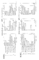
本実施例の実験は、NY−ESO−1特異的CTLによる抗原認識の効率を評価するために計画した。これを行うために、上述したタイプの4時間51Cr放出アッセイを使用し、同様に上述したT2細胞株を使って、ペプチド滴定を行った。測定の基準は、配列番号2によって達成される50%最大溶解とした。50%溶解に必要な配列番号2の量は試験した3つのCTLクローン、すなわち上述したクローンLAU50/13、LAU155/18および156/5によって異なった(7nM、12nMおよび3nM)。この結果を図1に示す。図1から、配列番号1および3は、3つのクローンのいずれに対しても、抗原として効率が低いことがわかる。1つのクローン(LAU156/5)は配列番号3を検出さえしなかった。
【0071】
C末端に変異を持つ類似体は、概して配列番号2より良い抗原だった。配列番号6は最も効率的な抗原ペプチドであるようだった。最大溶解の50%を得るのに必要な濃度は、これら3つのクローンのいずれについても、配列番号2より3〜4桁低かった。配列番号7および19も配列番号2よりも高い効率で認識されたが、抗原活性の増加率は様々だった。ある例では、配列番号7によって相対的抗原活性は105 倍増加したが、別のCTLにはわずかな効果しかなかった。
【0072】
配列番号8は、3つのクローンのうちの2つによって、より効率よく認識された。
【0073】
配列番号20〜27の単一アラニン置換に関して、各クローンは認識の微細特異性を示した。配列番号27はこれを最も明確に示した。クローンのうちの2つは配列番号27を配列番号2と同程度かそれ以上に認識し、クローンのうちの1つは配列番号27を全く認識しなかった。クローンのうちの2つは、同様の認識パターンを示した。3〜7位のペプチドは全てどのCTLクローンによる認識にとっても極めて重要であるらしかった。
【0074】
実施例21
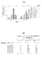
本実施例の実験は、様々なペプチドの特異的応答誘発能力を評価するために行った。メラノーマ患者のPBMCから取得した高濃縮CD8+ 細胞を、先の実施例で説明したように配列番号1、2、3、6、7、8または19の一つを使ってインビトロで刺激した。刺激の1週間後に、それらの細胞を、上述のように配列番号2を含有する四量体と抗CD8抗体とを使って染色した。
【0075】
1週間後、培養物中のCD8+ リンパ球の約0.1〜0.2%は四量体陽性だった。配列番号3は弱い刺激因子だったが、配列番号1および全てのカルボキシ置換誘導体は、この細胞集団を、配列番号2よりも増殖させた。これらの結果を図2Aに示す。
【0076】
追加実験では、IFN−γELISPOTアッセイにより、製造者の使用説明書に従って、培養物を試験した。簡単に述べると、プレートをIFN−γ特異抗体で一晩コーティングし、6回洗浄した。T2細胞(5×10⁴ 細胞/ウェル)をレスポンダーT細胞(2×104 細胞/ウェル)および1μMペプチドと一緒に加えた。正副一対の培養物を調製し、37℃で20時間インキュベートした。次に細胞を除去し、第2のビオチン化IFN−γ抗体およびストレプトアビジン−アルカリホスファターゼ複合体を使ってプレートを発色させた。立体顕微鏡を使用してスポット数を数えた。このアッセイを行った理由は、四量体を使用してペプチド類似体によるT細胞増殖を試験する方法では、親配列と交差反応しない特異的T細胞を検出できないかもしれないからである。
【0077】
図2Bにこれらの結果を示す。配列番号2を使ってもペプチド類似体を使っても、同等な数の特異的細胞が得られた。この結果から、配列番号3は免疫原性に乏しいこと、およびカルボキシ置換ペプチドで誘発された特異的T細胞の大半は配列番号2と完全に交差反応することが裏付けられる。
【0078】
上記の説明からわかるように、本発明は、
SLLMWITQX(配列番号10)
[ここに、Xはシステイン以外の任意のアミノ酸であり、好ましくは非極性側鎖を持つ疎水性アミノ酸、例えばAla、Val、Leu、Ile、Pro、Phe、Met、TrpまたはGlyであるか、または非荷電極性側鎖を持つアミノ酸、例えばSer、Asp、Glu、ThrまたはTyrであり、最も好ましくはAla、Ile、Val、LeuまたはSerである]という配列を持つペプチドに関する。これらのペプチドは、HLA−A2陽性であり病変を伴ってNY−ESO−1を発現させる患者への投与によって治療的に使用することができ、また診断的に、すなわちHLA−A2陽性細胞が存在するかどうか、関連CTLが存在するかどうかなどを決定するために使用することもできる。
【0079】
もう一つの態様では、Xは2つのアミノ酸であって、その第1アミノ酸はシステインまたはアラニンであり、かつ第2アミノ酸は任意のアミノ酸である。第2アミノ酸はPhe、Ile、ValまたはLeuであることが好ましい。この態様では、配列番号11または14の場合のように、第1アミノ酸がシステインであり、第2アミノ酸がPheまたはIleであることが最も好ましい。
【0080】
本発明のさらにもう一つの態様は、配列番号6に記載のペプチドの類似体、すなわち配列番号6中のアミノ酸のうちC末端アミノ酸以外の少なくとも1つがアラニンで置換されているペプチドである。配列番号27は特に好ましいが、配列番号20〜26に記載の他のペプチドも本発明の一部である。
【0081】
HLA−A2分子はMHCクラスI分子であり、ペプチドとクラスI分子との複合体に応答するT細胞は一般にCD8+ 細胞である。
【0082】
配列番号6によって定義されるペプチドは、配列番号10のコア配列によって定義されるペプチドの典型例である。このペプチドは、上述のようにHLA−A2分子に結合する。したがって、これはHLA−A2の「マーカー」であると共に、上述のようにCTLの増殖を刺激するペプチド/MHC複合体の一成分でもある。同様に、Ile、LeuまたはValが配列番号10の構造におけるカルボキシ末端であるペプチドも使用することができる。
【0083】
ペプチドを佐剤と混合することにより、治療用組成物を形成させることができる。また、本発明のペプチドをコードするヌクレオチド配列からなる核酸分子(いわゆる「ミニ遺伝子」)も本発明の一部である。これらのミニ遺伝子は、プロモーターと作動可能に連結した状態で発現ベクター中に組み込むことができる。1または複数のペプチドの多重コピーを含む2以上の本発明ペプチドをコードする構築物も本発明の一部である。これらの構築物は、例えば組換えベクターまたはいわゆる「裸のDNA」、すなわち1または複数の所望のペプチドをコードする小さい核酸分子などの形をとることができる。同様に、DNAまたはベクターを含む真核細胞または原核細胞などの組換え細胞も、本発明の一部である。
【0084】
これらのペプチドはHLA分子に結合する能力を持つので、本ペプチドが試料中の細胞に結合するかどうかを決定することによってHLA−A2陽性細胞(HLA−A* 0201陽性細胞など)の有無を決定するための試薬として役立つ。この「リガンド/受容体」タイプの反応は当技術分野ではよく知られており、その決定には様々な方法論を利用することができる。
【0085】
本発明のさらなる一側面は、トランスフェクトされた細胞によるペプチドの合成を指図するのに必要な情報を保持している、いわゆる「ミニ遺伝子」である。1または複数の抗原ペプチドをコードするミニ遺伝子を設計し、それをプラスミドを使ったトランスフェクションまたはワクシニアもしくはアデノウイルスへのクローニングによって、宿主細胞ゲノムに導入することができる。例えば参考文献として本明細書の一部を構成するZajacら,Int.J.Cancer 71:496(1997)を参照されたい。組換えワクシニアウイルスベクターなどのこれらの組換えベクターは、融合タンパク質が生成するように構築することができる。例えば上記のように、融合タンパク質の一部分が望ましい腫瘍拒絶抗原前駆体または腫瘍拒絶抗原である融合タンパク質を構築することができ、これに追加のタンパク質またはペプチドセグメントを含めることができる。融合タンパク質に加えることができる追加タンパク質またはペプチドセグメントの典型例は、決してこのタイプに限るわけではないが、レポータータンパク質またはレポーターペプチド、すなわち発現が起こったことがわかるように観察可能なシグナルを与えるタンパク質またはペプチド、例えば緑色蛍光タンパク質である。他のレポータータンパク質には、β−ガラクトシダーゼ、ルシフェラーゼ、dhfr、および参考文献として本明細書の一部を構成するChengら,Nature Biotechnology 14:606(1996)に記載の「eGFP」すなわち強化緑色蛍光タンパク質などがあるが、これらに限らない。当業者が入手できる様々なレポータータンパク質は様々な方法で使用することができ、また実際、様々な方法で使用されている。例えば「GFP」および「eGFP」を使って感染細胞を可視化することにより、フローサイトメトリーを使った場合の追跡と感染細胞の単離を容易にすることができる。ウェスタンブロッティング、免疫沈降などの方法を使用する場合は、他のレポータータンパク質が役立つ。これらの技術は当技術分野では標準的な技術であり、ここに繰り返して説明する必要はない。融合ペプチドからの腫瘍拒絶抗原前駆体または腫瘍拒絶抗原の切断を容易にするタンパク質またはペプチドセグメントも、含めることができる。融合タンパク質は上述のように2以上の腫瘍拒絶抗原を含むことができ、1または複数の腫瘍拒絶抗原の関連MHC分子への送達を促進するタンパク質またはペプチドを含むこともできる。そのようなタンパク質およびペプチドは当技術分野ではよく知られており、ここで詳しく説明する必要はない。
【0086】
本明細書に記載の組換えレポーターベクターをトランスフェクトした組換え細胞も本発明の一部である。これらの細胞は例えば任意のタイプの真核細胞であることができ、ヒト細胞は特に好ましい。これらの細胞は、例えば腫瘍拒絶抗原前駆体または腫瘍拒絶抗原の製造などに使用することができる。また、これらの細胞はエクスビボで、特定のMHC分子と腫瘍拒絶抗原との複合体に特異的な細胞溶解性T細胞を生成させるためにも使用することができる。これは、融合タンパク質の発現およびTRA(すなわち腫瘍拒絶抗原)のプロセシング後に生成するMHC分子とTRAとの複合体に特異的な試料中の細胞の増殖が誘発されるように、トランスフェクト細胞をT細胞の供給源(例えば血液試料)に接触させるだけで行うことができる。これらの細胞は、本明細書に示すようにその表面に適当な複合体を提示しインビボで同じタイプのT細胞応答を誘発するので、非増殖性にした場合はワクチン物質として使用することもできる。同様に、ベクターをワクチン物質そのものとして使用して、細胞表面上のMHC分子とペプチドとの複合体に対するT細胞応答を必要とする患者に投与することもできる。もちろんエクスビボで生成させたT細胞を患者の処置に使用することもできる。
【0087】
上記のペプチドを他の腫瘍拒絶抗原に由来するペプチドと組み合わせて「ポリトープ」を形成させることができる。代表的なペプチドには、いずれも参考文献として本明細書の一部を構成する米国特許出願第08/672,351号、第08/718,964号(現米国特許第 号)、第08/487,135号(現米国特許第 号)、第08/530,569号および第08/880,963号に記載されているものがある。
【0088】
使用することができる他のペプチドには、いずれも参考文献として本明細書の一部を構成する次の文献に記載されているものがある:米国特許第5,405,940号、第5,487,974号、第5,519,117号、第5,530,096号、第5,554,506号、第5,554,724号、第5,558,995号、第5,585,461号、第5,589,334号、第5,648,226号および第5,683,886号、PCT国際公開第92/20356号、第94/14459号、第96/10577号、第96/21673号、第97/10837号、第97/26535号および第97/31017号ならびに係属中の米国特許出願第08/713,354号。
【0089】
ポリトープは、このタイプの分子が免疫応答を刺激しかつ/または誘発することができるかどうかを決定するために様々な方法で一つに連結することができる、2以上の潜在的に免疫原性または免疫刺激性のペプチド群である。
【0090】
これらのペプチドは直接連結することもできるし、隣接配列を使って連結することもできる。関連エピトープ配列の直接結合を教示しているThompsonら,Proc.Natl.Acad.Sci.USA 92(13):5845−5849(1995)を参照されたい。ポリトープのワクチンとしての使用はよく知られている。例えばGilbertら,Nat.Biotechnol.15(12):1280−1284(1997)、Thomsonら(前掲)、Thomsonら,J.Immunol.157(2):822−826(1996)、Tamら,J.Exp.Med.171(1):299−306(1990)(いずれも参考文献として本明細書の一部を構成する)を参照されたい。特に前記Tamの文献は、マウスモデルで使用した場合にポリトープが抗体および防御免疫の両方の生成に有用であることを示している。さらにこの文献は、ポリトープが消化されると、MHCによって提示されることが可能であり実際にMHCによって提示されるペプチドが生成することを示している。Tamは、ポリトープ「ストリング」からプロセシングされた個々のエピトープがCTLによって認識されることを示すことによって、これを示している。このアプローチは、例えばどのくらいの数のエピトープであれば1つのポリトープに連結しても認識を誘発することができるかを決定する際に、また様々な組合わせのエピトープの効力を決定するためなどに使用することができる。様々な組合わせは腫瘍拒絶抗原の特定のサブセットを発現させる患者に合わせて「テーラーメード」することができる。これらのポリトープは、ポリペプチド構造として導入するか、核酸送達システムを使って導入することができる。詳しく述べると、当技術分野では、個々のエピトープまたは上述したようなポリトープをコードするDNAを導入するために数多くの様々な方法を利用することができる。例えば参考文献として本明細書の一部を構成するAllsoppら,Eur.J.Immunol.26(8):1951−1959(1996)を参照されたい。アデノウイルス、ポックスウイルス、Tyウイルス様粒子、プラスミド、細菌などを使用することができる。これらのシステムは、ある与えられた同様の状況下でヒトにはどのシステムが最も適当であるかを決定するために、マウスモデルで試験することができる。またヒト臨床試験で試験することもできる。
【0091】
また、試料中の細胞溶解性T細胞の有無を決定するために上記のペプチドを使用することも、本発明の特徴である。上述したように、試料中のCTLはペプチド/MHC複合体と反応する。したがって試料中にCTLが存在することがわかっている場合は、本発明のペプチドをHLA−A2陽性細胞、例えばHLA−A* 0201陽性細胞に加え、次に例えば放射活性クロム放出、TNF産生などを決定するか、またはT細胞活性を決定する他の任意の方法を実施することにより、HLA−A2陽性細胞を「溶解」することができる。同様に、本発明ペプチドの1つをHLA−A2陽性細胞と共に試料に加え、HLA−A2陽性細胞の溶解を、例えば51Cr放出、TNFの存在などによって決定することにより、試料中に特異的腫瘍浸潤リンパ球(TIL)が存在するかどうかを決定することができる。また、CTLはELISPOT解析によって検出することもできる。例えばSchmittelら(1997)J.Immunol.Methods210:167−174およびLalvaniら(1997)J.Exp.Med.126:859を、またはMHCクラスI/ペプチドの蛍光原性四量化複合体のFACS解析(Dunbarら(1998)Current Biology 8:413−416、Romeroら,J.Exp.Med.188:1641−1650(1998))(前記の文献は全て本明細書の一部を構成する)参照されたい。詳しく述べると、前記複合体は第1結合パートナーと第2結合パートナーを含み、第1および第2結合パートナーは互いに特異的である。これらは、例えばアビジンまたはストレプトアビジンとビオチン、ビオチンに特異的な抗体またはその結合部分などであることができる。重要な特徴は第2結合パートナーが、MHC分子、β2−ミクログロブリン分子および前記MHC分子に特異的に結合するペプチドから構成される複数の複合体に結合されなければならず、前記多成分複合体が標識されなければならないということである。MHC分子はHLA−A2分子であることが好ましいが、任意のHLA分子を使用できることは当業者には理解されるだろう。
【0092】
第2結合パートナーはビオチンであることが好ましいが、例えばHLA/β2−ミクログロブリン/ペプチド複合体の一成分に特異的な抗体、例えばHLA特異抗体、β2−ミクログロブリン特異抗体などであってもよい。同様に、第1結合パートナーは例えば組換えまたは天然プロテインL、組換えまたは天然プロテインA、さらには第2の抗体であることができる。複合体は可溶型であるか、または除去可能な固相、例えば磁気ビーズに結合した形をとることができる。
【0093】
本発明の大分子中のHLA/β2−ミクログロブリン/ペプチド複合体の数は様々である。これは少なくとも2つの複合体を含み、好ましくは少なくとも4つの複合体を含むが、それ以上の複合体が存在してもよい。
【0094】
結合パートナーとHLA/β2−ミクログロブリン/ペプチドとの複合体は、当技術分野で知られている任意のラベルを使って標識することができる。蛍光ラベルの例は上述した。アルカリホスファターゼなどの酵素ラベル、金属粒子、合成物質製の着色プラスチック、放射性ラベルなどは、全て使用することができる。
【0095】
第1結合パートナーに特異的に結合する第3結合パートナーを使用してもよい。例えば第1結合パートナーがストレプトアビジンであり、かつ第2結合パートナーがビオチンである場合、第3結合パートナーはストレプトアビジン特異抗体であることができる。3以上の結合パートナーを使用する場合は、HLA/β2−ミクログロブリン/ペプチド複合体との結合が妨害されない限り、上記のラベルはどの結合パートナーに結合させてもよい。
【0096】
上記複合体は、例えば試料中に存在する、HLA/β2−ミクログロブリン/ペプチド複合体に特異的な細胞溶解性T細胞を同定または単離するために使用することができる。このような細胞溶解性T細胞は、本発明の免疫複合体に結合する。好ましい一態様では、被験試料を細胞溶解性T細胞に特異的に結合する反応物で処理する。この場合、前記ラベルは検出可能なシグナルを与える。次に、標識されたCTLを含む試料を上記複合体と接触させ、結合させる。標識されたCTLは周知の標準的細胞分離法によって分離することができる。好ましくはFACSを使用するが、他の分離方法も当業者には同様に知られている。使用するペプチドは当業者の必要に応じて選択され、問題にしている特定のMHC系の性質などに依存するだろう。
【0097】
また、本方法は、ワクチンなどの特定の治療薬を投与した後の腫瘍の状態を監視するためにも使用することができる。さらに本方法論は、上述のように細胞溶解性T細胞前駆体を同定するためにも使用することができるので、考えうる治療法の候補者を、彼らが適当なT細胞前駆体を持つかどうかを決定することによって同定することもできる。
【0098】
もちろん、本ペプチドはCTLの産生を誘発するために使用することもできる。上述のように、CTL前駆体は適当な複合体に直面するとCTLになる。このようないわば「直面」を引き起すことにより、CTLを生成させることができる。これは、このようなCTLを生成させるためにインビボでもエクスビボでも有用である。
【0099】
複数の異なるペプチドを含みそのうちの少なくとも1つが本発明のペプチドであるいわゆる「カクテル」ならびに「ポリトープ」分子およびそれらをコードする核酸分子も本発明の一部である。本明細書で使用する「ポリトープ」という用語は、細胞内プロセシング後にMHC分子によって提示される複数のペプチド配列を含有するように設計された組換え分子を指す。このようなポリトープは、例えば1つのエピトープの繰返しからなったり、いくつかの異なるエピトープからなったりすることができる。
【0100】
本発明の他の特徴と応用は当業者には明らかであり、本明細書に記載する必要はないだろう。
【0101】
本明細書で使用した用語および表現は、限定ではなく説明のために使用されている。これらの用語および表現の使用に、本明細書に示し説明した特徴またはその一部の等価物を排除する意図はなく、本発明の範囲内で様々な変更態様が可能であると理解される。
【図面の簡単な説明】
【図1】 NY−ESO−1に関連するペプチドおよびペプチド類似体の抗原性に関するデータ。
【図2】 図2Aおよび図2Bはそれぞれ、NY−ESO−1ペプチドおよび同類似体のインビトロ免疫原性に関する結果(2A)およびELISPOTアッセイの結果(2B)を示す。
【配列表】
(Related application)
No. 08 / 937,263 (1997), which is a continuation-in-part of Patent Application No. 08 / 725,182 (filed Oct. 3, 1996, now US Pat. No. 5,804,381). Patent Application No. 09 / 165,546 (1998), which is a continuation-in part of Patent Application No. 09 / 062,422 (filed on April 17, 1998), which is a continuation-in-part application of September 15, 1998) Patent application 09 / 514,036 (2000 filed on Oct. 2, 1999), which is a partial continuation application of patent application 09 / 440,621 (filed on November 15, 1999). This is a partial continuation application. All of these applications are hereby incorporated by reference. This application is a part of Patent Application No. 09 / 275,993 (filed on Mar. 25, 1999), which is a continuation-in-part of Patent Application No. 09 / 049,850 (filed on Mar. 27, 1998). It is also a continuation application.
[0002]
(Technical field)
The present invention relates to an HLA-binding peptide derived from a cancer-associated antigen. These peptides bind to class I molecules and induce lysis by cytolytic T lymphocytes of cells to which these peptides are bound.
[0003]
(Background technology and conventional technology)
It is a well established fact that many pathological conditions such as infectious diseases, cancer, autoimmune diseases, etc. are characterized by inappropriate expression of certain molecules. These molecules thus serve as “markers” for specific pathological or abnormal conditions. These molecules are used as diagnostic “targets” (ie, substances to be identified to diagnose these abnormal conditions), as well as reagents that can be used to create diagnostic and / or therapeutic agents. Also useful. One example is the production of an antibody specific to a specific marker using a cancer marker, but this is not a limitation. In addition, the use of a peptide that forms a complex with an MHC molecule to generate cytolytic T cells against abnormal cells is an example without limitation.
[0004]
The production of such materials is of course premised on the source of reagents used in the production of those materials. Purification from cells is a cumbersome and far from reliable method. Another preferred method is to isolate a nucleic acid molecule encoding a particular marker and then use the isolated coding molecule to express the desired molecule.
[0005]
So far, two strategies have been used to detect antigens, such as in human tumors. In the following, these strategies will be referred to as genetic approaches and biochemical approaches. Genetic approaches are described in, for example, dePlaen et al., Proc. Natl. Sci. USA 85: 2275 (1988), for example. In this approach, hundreds of plasmid pools of cDNA libraries from tumors are transfected into recipient cells such as COS cells or antigen negative mutants of tumor cell lines, and those cells are examined for expression of specific antigens. Biological approaches are described, for example, in O.D., which is hereby incorporated by reference. Examples can be seen in Mandelboim et al., Nature 369: 69 (1994), etc. This approach is based on reverse phase high performance liquid chromatography after acid elution of peptides bound to MHC class I molecules of tumor cells. (HPLC) based. Antigen peptides are identified by binding the antigenic peptides to empty MHC class I molecules of mutant cell lines lacking antigen processing capacity and inducing specific reactions with cytotoxic T lymphocytes. These specific reactions include MTT assays and 51 These can be measured in a Cr release assay, such as induction of CTL proliferation, TNF release and target cell lysis.
[0006]
The above two approaches aimed at molecular definition of antigens have the following drawbacks. First, these methods are extremely cumbersome and time consuming and expensive. Secondly, these methods rely on the establishment of cytotoxic T cell lines (CTL) with a predetermined specificity.
[0007]
The challenges inherent in the two known approaches aimed at antigen identification and molecular definition have been successful in defining very few new antigens in human tumors by either method. The fact that there is no such thing clearly tells. For example, van der Bruggen et al., Science 254: 1643-1647 (1991), Brichard et al., J. Biol. Exp. Med. 178: 489-495 (1993), Coulie et al., J. MoI. Exp. Med. 180: 35-42 (1994), Kawakami et al., Proc. Natl. Acad. Sci. USA 91: 3515-3519 (1994).
[0008]
Furthermore, the above methodology presupposes that an established permanent cell line of the cancer type in question is available. It is extremely difficult to establish a cell line depending on the cancer type, for example, Oettgen et al., Immunol. Allerg. Clin. North. Am. 10: 607-637 (1990) and the like. It is also known that some epithelial cell type cancers are insensitive to CTL in vitro and cannot be analyzed by conventional methods. Inspired by these issues, new identification methods for cancer-associated antigens have been developed in the art.
[0009]
One important methodology is that of Sahin et al., Proc., Which is hereby incorporated by reference. Natl. Acad. Sci. USA 92: 11810-11913 (1995). See also US Pat. No. 5,698,396 and US application Ser. No. 08 / 479,328 (filed Jan. 3, 1996). These three references all form part of this specification. In summary, this method expresses a cDNA library in a prokaryotic host. (The library is obtained from a tumor sample). The expressed library is then subjected to immunoscreening with diluted absorbed serum to detect antigens that elicit high titer humoral responses. This methodology is based on the SEREX method (" Ser logical identification of antigens by R ecobinant Ex pressure Cloning ("serologic identification of antigens by recombinant expression cloning"). This methodology has been used to detect new tumor associated antigens and to confirm the expression of previously identified tumor associated antigens. See the above patent application and Sahin et al. (Supra) and Crew et al., EMBO J 144: 2333-3340 (1995).
[0010]
The SEREX method has been applied to esophageal cancer samples, and one antigen has now been identified, and a nucleic acid molecule encoding it has been isolated and cloned. See, for example, U.S. Pat. No. 5,804,381, supra. This antigen and its truncated form have been found to react with antibodies in the serum of cancer patients. It has also been found that peptides derived from this molecule bind to MHC molecules and elicit both cytolytic and helper T cell responses. It has been found that variants of these peptides can be used as well.
[0011]
One of the problems in the field of cancer immunology is the lack of a reliable protocol that can be used to identify and quantify cytolytic T lymphocyte responses in vivo. Therefore, it is difficult to characterize the immune response and monitor vaccine trials. It has been found that analysis of cytolytic T cells is greatly facilitated by using a complex containing multiple T cell targets. More specifically, these complexes are based on the known avidity between two binding partners, such as avidin or streptavidin and biotin. It is well known that each avidin / streptavidin molecule can bind to four biotin molecules. A complex comprising a plurality of immune complexes comprising peptides that bind to a plurality of cytolytic T cell targets, ie MLA molecules such as HLA molecules, β2-microglobulin and HLA molecules using an avidin / streptavidin-biotin system Body-forming constructs are taught, for example, in patent application 09 / 049,850 (filed March 27,1998), which is hereby incorporated by reference. This complex can be labeled and used to isolate or determine the desired cytolytic T cells in the sample. In the invention described below, such a complex is used.
[0012]
(Detailed description of preferred embodiments)
Example 1
Analysis of NY-ESO-1 discussed in, for example, US Pat. No. 5,804,381 revealed that the molecule presenting this antigen is HLA-A2. Therefore, D'Amaro et al., Human Immunol. 43: 13-18 (1995) and Drijfhout et al., Human Immunol. 43: 1-12 (1995), the NY-ESO-1 amino acid sequence was screened to identify all peptides that satisfy this HLA-A2 binding motif. All peptides corresponding to the amino acid sequences deduced thereby were synthesized by standard methods and used as a reference by Knuth et al., Proc. Natl. Acad. Sci. USA 81: 3511-3515 (1984), the peptides were used in cytotoxicity assays. Specifically, the cell line CEMX721.174. T2 (hereinafter referred to as “T2”) was used. This is because this cell line expresses HLA-A2 but does not process the antigen into MHC-conjugated peptides, making it ideal for the types of experiments described here. Samples of T2 cells were prepared by standard methods using 100 μCi Na ( 51 Cr) O Four After labeling with, washed 3 times and then incubated with 10 μg / ml peptide and 2.5 μg / ml β2-microglobulin. Incubation was for 1 hour at room temperature. Next, responder cells (100 μl of a suspension of CTL NW38-IVS-1) were added at an effector / target ratio of 90: 1 and 5% CO2. 2 For 4 hours at 37 ° C. in a water-saturated atmosphere. Next, the plate was centrifuged at 200 × g for 5 minutes, 100 μl of the supernatant was taken out, and the radioactivity was measured. 51 When the Cr release rate was determined by a known method, three of the peptides SLLMWITQCFL (SEQ ID NO: 1), SLLMWITQC (SEQ ID NO: 2) and QLSLLMWIT (SEQ ID NO: 3) were found to be HLA-A2 restricted NY-ESO-1-specific CTLs. It was found to be the best stimulating factor. Similar results were obtained when NW-MEL-38 and cell lines SK-MEL-37 and MZ-MEL-19 were used as targets.
[0013]
Example 2
In the next experimental group, the ability of SEQ ID NOs: 1, 2 and 3 to bind to HLA-A2 molecules and induce CTL lysis was confirmed.
[0014]
Samples of lymph nodes or metastases were taken from patients with slowly progressive melanoma with recurrent metastases localized in the same paravertebral region. By reverse transcription of RNA collected from the tumor of the subject, the tumor was found to express NY-ESO-1. In addition, patient sera showed high titers of anti-NY-ESO-1 antibodies.
[0015]
Surgically excised lymph nodes or metastases were minced in sterile RPMI 1640 medium supplemented with 10% fetal calf serum. Contains 2 ml of Iscove Dulbecco's medium supplemented with 0.24 mM Asn, 0.55 mM Arg, 1.5 mM Gln, 10% human A + pool serum, 100 U / ml IL-2 and 10 ng / ml IL-7 The cell suspension was placed in a 24-well tissue culture plate. After culturing the cells for 2-3 weeks, Czerkinsky et al., J. Chem. Immunol. The assay by IFN-γ ELISPOT was performed according to Meth 110: 29 (1998). Briefly, short-
[0016]
The average number of spots was 19 spots in the control culture, 424 spots in SEQ ID NO: 2, 358 spots in SEQ ID NO: 3, and 396 spots in SEQ ID NO: 1. These elevated values correspond to a frequency of about 1 NY-ESO-1 specific T cell per 20 tumor infiltrating lymphocytes. Next, Valmori et al., J., which constitutes a part of this specification as a reference. Reactive T cells were cultured according to Immunol 161: 6956 (1998) to make them monoclonal. Five of the 24 TIL-derived clones derived from TIL were found to react with NY-ESO-1-derived peptides when tested in the CTL assay described above.
[0017]
Example 3
In the experiment of this example, A2 expressing NY-ESO-1 + A2 that does not express cells or NY-ESO-1 + The CTL ESO5 was examined for its ability to lyse cells. Mentioned above 51 Cell line NA8-MEL (A2 in Cr release assay + , NY-ESO-1 - ), SK-MEL37 (A2 + , NY-ESO-1 + ) And Me275 (A2 + , NY-ESO-1 + ) Was tested.
[0018]
As a result, it was found that NA8-Mel was dissolved when NY-ESO-1 peptide SEQ ID NO: 2 was added, but was not dissolved when SEQ ID NO: 2 was not present. The presence or absence of SEQ ID NO: 2 is independent of lysis of SK-MEL37 and Me275, and both of these cell lines were lysed under all conditions. These results indicate that CTL ESO5 recognizes externally added SEQ ID NO: 2 or endogenous SEQ ID NO: 2.
[0019]
Example 4
Next, experiments were performed to determine which of SEQ ID NOs: 1, 2, and 3 constitutes the optimal T cell epitope for recognition by CTL. To make this determination, synthetic peptides corresponding to SEQ ID NOs: 1, 2 and 3 were tested in a functional competition binding assay and then tested for recognition by specific CTLs.
[0020]
The functional competitive binding assay used is described in Valmori et al., J., which is hereby incorporated by reference. Immunol. 161: 6956-6962 (1998), which is also described in detail herein.
[0021]
The peptide YMDGTMSQV (SEQ ID NO: 4) is HLA-A * HLA-A that binds to 0201 molecules and is known as LAU 132/2 * It is known to induce lysis by 0201 restricted CTL clones. Valmori et al., Canc. Res. 59: 2167 (1999). T2 cells in the presence of anti-class I monoclonal antibody W6 / 32 51 Labeled with Cr. Various concentrations of SEQ ID NO: 1, 2 or 3 (50 μl) were incubated with 50 μl (1000 cells / well) of labeled cell samples for 15 minutes at room temperature. Next, suboptimal dose (1 nM) of SEQ ID NO: 4 (50 μl) was added along with 50 μl of T cells (5000 cells / well). After 4 hours incubation at 37 ° C 51 Cr release was measured. Next, the concentration of each peptide required to achieve 50% inhibition of target cell lysis was determined as [nM] 50%. To facilitate comparison, known high affinity HLA-A * Relative competitive activity of each peptide as [nM] 50% of the reference peptide FluMA58-66 (SEQ ID NO: 5: GILGFVFTL), the 0201 binder, divided by the [nM] 50% value determined for the test peptide. Was calculated.
[0022]
As a result, it was found that SEQ ID NO: 3 has 1/100 the potency of SEQ ID NO: 4 as a competitor. The potency of SEQ ID NO: 2 was 1/250. Surprisingly, considering the length of each peptide, SEQ ID NO: 1 had 10 times stronger competitiveness than SEQ ID NO: 2.
[0023]
These results suggested that the cysteine at the carboxy terminus of SEQ ID NO: 2 is responsible for weak binding to the HLA-A2 molecule. To investigate this point, the derivative of SEQ ID NO: 2 was substituted by replacing the carboxy-terminal cysteine with various hydrophobic amino acids with non-polar side chains such as alanine, leucine or valine (SEQ ID NO: 6-8, respectively). One was made. Each of these peptides was used to perform the functional competition assay described above. The results are shown in Table 1 below. All substitutions clearly and dramatically increased peptide bonds, indicating that for all three of these substituted peptides, any hydrophobic residue at this position has a similar effect.
[0024]
[Table 1]
As described above, the peptides described herein were tested for recognition by specific CTLs. In these experiments, target T2 cells were incubated for 1 hour at 37 ° C. 51 After labeling with Cr, it was washed twice. Samples (1000 labeled cells in 50 μl) were then incubated for 15 minutes with various concentrations of peptide. Effector cells (50 μl) were then added. The added effector cells are the ESO1-specific CTL clone ESO5. The lymphocyte: target ratio was 30: 1. After 4 hours incubation at 37 ° C., chromium release was measured by examining 100 μl supernatant samples.
[0025]
Specific dissolution rate was calculated as:
100 × [(experimental value−spontaneous release)] / (total amount−spontaneous release)
The results are listed in Table 2 below as the nanomolar concentration of peptide giving 50% maximum activity. Table 2 also lists “relative antigenic activity” calculated as 50% of peptide SEQ ID NO: 2 divided by 50% of test peptide [nM]. The recognition of analog peptides by CTL was equal or better than the parent peptide. These results indicate that SEQ ID NO: 2 is the antigen peptide that is optimally recognized among the natural NY-ESO-1 peptides. Among the substituted peptides, SEQ ID NO: 8 was recognized as efficiently as SEQ ID NO: 2, and other substituted peptides were recognized more efficiently than SEQ ID NO: 2. SEQ ID NO: 6 was recognized 1000 times more efficiently than SEQ ID NO: 2.
[0026]
[Table 2]
In the experiment of this example, a basic technique for preparing a tetramerized complex of an antigen used in the examples described later will be described. To make the desired tetramer, first the modified HLA-A * There was a need to create a construct encoding the 0201 molecule. To do this, HLA-A * Total RNA was extracted from 0201 positive cells, and then HLA-A using primers specific for the same molecule and reverse transcription polymerase chain reaction (RT-PCR). * 0201 was cloned. Altman et al., Science 274: 94-96 (October 4, 1996), which forms part of this specification as a reference, was followed. However, a new 3 ′ primer, namely 5′-GCAGGATCCCGGCTCCCATCCCTCA GGGTGGAGGGC-3 ′ (SEQ ID NO: 9) was used. Concurrently with RT-PCR, the amino terminal nucleotide sequence was modified to optimize protein expression in the vector used. Garboczi et al., Proc., Which forms part of this specification as a reference. Natl. Acad. Sci. USA 89: 3429 (1992). After this, the extracellular coding portion of the molecule was amplified again using specific primers. The resulting construct was designated as HLA-A * It was recloned into a vector that generated a BirA biotinylation recognition site in-frame at the 3 ′ end of the 0201 heavy chain. Modified HLA-A * 0201 and β2-microglobulin were overexpressed in separate E. coli cultures. The obtained inclusion body was purified, the HLA recombinant protein and β2-microglobulin recombinant protein were solubilized in a urea solution, and then refolded at 4 ° C. in a refolding solution to form a complex. . (The refolding solution was 100 mM Tris (pH 8.0), L-arginine 400 mM,
[0027]
The complex was then biotinylated with biotin holoenzyme synthase (BirA enzyme) by mixing the refolding mixture containing the HLA-A2 complex with 50 μM enzyme, 100 mM biotin and 100 mM adenosine triphosphate in 200 mM Tris. The mixture was incubated overnight at room temperature. The biotinylated complex was then purified and mixed with phycoerythrin labeled streptavidin to form a tetrameric structure. These were isolated and reconstituted in small portions at a concentration of 1 mg / ml.
[0028]
Example 7
The experiments in this example were designed to evaluate the frequency of NY-ESO-1-specific T cells. Fluorescent tetramers of biotin, HLA-A2 and peptides are described in patent applications 09 / 275,993 (filed March 25, 1999) and 09 / 049,850 (filed March 27, 1998) ( Both of which are part of the present specification as references), as described in Romero et al. Exp. Med. 188: 641 (1998) and Altman et al., Science 274: 94 (1996). As an antigen peptide, Flu Ma 58-66 (SEQ ID NO: 5) and an ESO-1 derivative (SEQ ID NO: 6) having an alanine carboxy terminus were used. The reason for selecting this derivative is that its affinity is high. This is because the production of a stable tetramer is facilitated if the binding affinity to HLA-A2 is high.
[0029]
After assembling the tetramer, CTL clones specific for each peptide were mixed with the appropriate tetramer in 20 μl PBS containing 2% FCS, incubated for 1 hour at room temperature, and then anti-CD8 labeled with FITC. 20 μl of a mixture of anti-CD8 antibody labeled with antibody or FITC and anti-CD45RA antibody labeled with “CYC” was added. After incubating the mixture at 4 ° C. for 30 minutes, the cells were washed with the same buffer as above and then analyzed by flow cytometry.
[0030]
The tetramer of NY-ESO-1 derivative (SEQ ID NO: 6) stained ESO5 specifically, but did not stain FluMa-specific T cells, and vice versa.
[0031]
Example 8
The experiments in this example were designed to verify the detection and isolation of NY-ESO-1 specific T cells using a tetramer containing NY-ESO-1 peptide.
[0032]
Concentrated CD8 stimulated with SEQ ID NO: 2 or alanine terminal substitution (SEQ ID NO: 6) + T cell sample. (Stimulation was performed by loading the target peptide onto autoantigen-presenting cells.)
Tetramers are made as described above and used to generate CD8 after 14 days of stimulation. + Cells were stained. Cells were sorted and tested with IFN-γ ELISPOT as described above.
[0033]
CD8 + Tetramer + Only cells were found to contain IFN-γ producing cells.
[0034]
Selected CD8 + Tetramer + Cells and CD8 + Tetramer - Cells were grown for 2 weeks with PHA stimulation and both cell populations were expanded with Me275 and T2 cells pulsed with SEQ ID NO: 2 or not pulsed with SEQ ID NO: 2. 51 Assayed as described above with Cr release assay.
[0035]
CD8 + Tetramer + Only the cells effectively killed both the cells pulsed with SEQ ID NO: 2 and the cells pulsed with the alanine analog.
[0036]
Example 9
The experiment of this example is to determine whether NY-ESO-1 antigen is processed intracellularly to produce a peptide presented by the HLA-A2 molecule and consequently stimulate lysis by CTL. Planned.
[0037]
The tetramer described in Example 6 above was prepared using the peptide of SEQ ID NO: 2. These tetramers were then used to stain samples of cells taken from patients who had previously been diagnosed with NY-ESO-1 positive melanoma. For details, see Dunbar et al., J., which constitutes a part of this specification as a reference. See Immunol 162 (12): 6959-62 (1999) and patent application 09 / 049,850 (March 27, 1998). Briefly, tetramers are used to produce peripheral blood lymph. Sphere (“PBL”) samples were stained at 37 ° C. for 15 minutes, then washed in PBS / 1% FCS at 37 ° C. and then incubated with labeled anti-CD8 antibody for 30 minutes on ice. Cells were then washed 3 times in ice-cold PBS / 1% FCS and analyzed by flow cytometry. Positive cells were cloned by pulsing the positive cells with 10 μM of the peptide of SEQ ID NO: 2 and culturing in IL-2 (200 u / ml) for 5 days.
[0038]
Of these positive CTLs, 4 were grown and tested in the following experiment.
[0039]
Example 10
The CTL were then tested for cell killing specificity. To perform this test, a cell line previously typed as NY-ESO-1 positive, a cell line previously typed as NY-ESO-1 negative, 1 μM peptide of SEQ ID NO: 2 was pulsed Samples of treated T2 cells and non-pulsed T2 cells were mixed with CTL at an effector / target ratio of 1: 1 and 0.3: 1. Cell killing 51 Determined by Cr release assay.
[0040]
As a result, it was found that both NY-ESO-1 positive cells and T2 cells pulsed with SEQ ID NO: 2 were killed, and other cells were not killed. This demonstrated that intracellular processing of NY-ESO-1 results in the production of peptides recognized by SEQ ID NO: 2 specific CTLs.
[0041]
Example 11
The experiment described in this example determines whether a mixture of different CTLs is responsible for the positive results obtained with SEQ ID NOs: 1, 2 and 3, or whether a single CTL clone recognizes all peptides. Planned to decide. In these experiments, T2 cells were pulsed with any one of SEQ ID NO: 2, SEQ ID NO: 1, or a peptide consisting of NY-ESO-1 amino acids 157-166, ie, SLLMWIQCF (SEQ ID NO: 11). Since it is a peptide composed of the first 8 amino acids of SEQ ID NO: 1, SEQ ID NO: 3 was also tested. Various concentrations of peptides were used for pulsing T2 cells. SEQ ID NO: 5 was used as a control. Same as above 51 A Cr release assay was used. Prior to testing the CTLs, an assay was performed to determine whether each expressed a single T cell receptor.
[0042]
As a result, all T cells recognize SEQ ID NOs: 2, 3 and 11, but cannot recognize SEQ ID NO: 1 or truncated peptides 157-164, ie, peptides composed of the first 8 amino acids of SEQ ID NO: 11. all right. These indicate that all three peptides were recognized by clone CTL.
[0043]
Example 12
The experiment described in this example was designed to determine the conditions of intracellular processing necessary to produce the peptide of SEQ ID NO: 2.
[0044]
Cerundolo et al., Nature 345 (6274): 449-52 (1990) describes mutant processing cells characterized by having a distinct blocking point in the MHC I processing pathway. These cells were used in these experiments. Specifically, the transfection of the parent cell line “cell line 45”, TAP, LMP2 and LMP7 negative “cell line 174”, cell line 174 transfected with vectors encoding TAP1 and TAP2. Tanto and transfectants of cell line 174 transfected with vectors encoding TAP1, TAP2 and LMP7 were used. (The abbreviation “TAP” stands for “transport associated with antigen processing”, and the abbreviation “LMP” stands for “low molecular mass polyprotein”.)
[0045]
In addition to the transfectants described above, vaccinia virus constructs encoding NY-ESO-1 were transfected into all cells for 90 minutes at a multiplicity of infection (MOI) of 5.
[0046]
As a control, one hour before addition of the vaccinia virus construct, the same number of cells (10% in 50 μl medium containing 100 μM lactacystin). 6 Cells) were suspended. Cells were washed after a 90 minute infection period, suspended in 5 ml of medium containing 1 μM lactacystin and grown overnight to allow expression of the vaccinia vector. LMP2 and LMP7 are proteasome subunits.
[0047]
Next, the type of 51 Cells were mixed with CTL in a Cr release assay.
[0048]
As a result, it was found that even when vaccinia encoding NY-ESO-1 was expressed in TAP-deficient cells, those cells were not susceptible to CTL lysis. On the other hand, the presence or absence of proteasome subunits LMP2 and LMP7 did not impair the presentation of the epitope recognized by CTL. This is supported by the controls used. Because 174 / TAP cells do not present the peptide of SEQ ID NO: 5, which has been shown to be dependent on LMP7, while transfection of the vector encoding LMP7 or addition of lactacystin This is because the blockage has been eliminated.
[0049]
These results strongly suggest that non-proteasome proteases are involved in the presentation of the NY-ESO-1 epitope.
[0050]
Example 13
The experiments in this example were designed to determine whether the peptide analog of SEQ ID NO: 2 prepared by modification of the C-terminal cysteine residue has altered immunogenicity. Of the type mentioned above 51 A Cr release assay was performed. In this assay, the peptide (SEQ ID NO: 2) and target cells were mixed with 200 μM dithiothretal (DTT) or TCEP (Tris (2-carboxymethyl) phosphine).
[0051]
As a result, it was found that the antigenicity of SEQ ID NO: 2 increased 10-fold in the presence of a reducing agent. Parallel experiments using SEQ ID NO: 8 did not show any increase in antigenicity.
[0052]
Example 14
In view of the results observed in Example 13 above, it was decided to test the modified peptides. In addition to SEQ ID NOs: 7 and 8, the formula:
SLLMWIQI
(SEQ ID NO: 12) was synthesized and the following 10mer was synthesized as well:
SLLMWITCQCV (SEQ ID NO: 13)
SLLMWITQCF (SEQ ID NO: 11 (above))
SLLMWITQCI (SEQ ID NO: 14)
SLLMWIQAL (SEQ ID NO: 15)
SLLMWITQAI (SEQ ID NO: 16)
SLLMWITQAF (SEQ ID NO: 17).
Further, the C-terminal cysteine is -NH 2 -CO-CH 2 SEQ ID NO: 2 modified with side chains was also prepared. All these peptides were tested in a CTL assay of the type described above. All of the peptides of SEQ ID NO: 7, 8 and 12 increase recognition by NY-ESO-1 157-165 specific CTL by about 1000-fold compared to SEQ ID NO: 2. The peptide of SEQ ID NO: 14 was also recognized more efficiently than SEQ ID NO: 2.
[0053]
Example 15
In the experiments of this example, the ability to stimulate proliferation of NY-ESO-1-specific CTLs was examined for various peptide analogs.
[0054]
Briefly, PBLs were collected from two melanoma patients who showed high titer NY-ESO-1 specific antibodies in a general assay method. These PBLs were then stimulated with 100 nM or 10 nM SEQ ID NO: 2 or SEQ ID NO: 8 according to standard protocols. SEQ ID NO: 8 resulted in 14-fold greater proliferation of NY-ESO-1-specific CTL in 2 weeks. In 3 weeks, the growth reached 54 times that of using SEQ ID NO: 2. When these CTLs were stained with the tetramer of the type described above using SEQ ID NO: 2, the staining was positive, indicating that the CTL recognizes the NY-ESO-1 epitope in question. It was.
[0055]
It is noteworthy that 10 nM SEQ ID NO: 8 stimulated CTL and that the required amount of natural peptide was actually higher. Furthermore, additional experiments with standard methodologies confirmed that the proliferating CTL described in this example had the ability to kill NY-ESO-1-positive tumor cells.
[0056]
Example 16
The experiment of this example was designed to evaluate the reactivity of the tetramer using the above NY-ESO-1-derived peptide.
[0057]
The tetramers described in Examples 6 and 7 were prepared using the peptides of SEQ ID NO: 2 or SEQ ID NO: 6. These were then examined for their ability to stain CTL clones known to react with complexes of HLA-A2 and NY-ESO-1-derived peptides. For example, Valmori et al., Canc. Res. (2000). Staining was performed as described above.
[0058]
As a result, both tetramers were found to stain CTL clones in a dose-dependent manner. At the highest peptide dose tested (100 μg / ml), the fluorescence intensity is 10% above background. Three it was high. The concentration of tetramer required for the intermediate intensity fluorescent signal was about 12 μg / ml for the tetramer using SEQ ID NO: 2 and 3 μg / ml for SEQ ID NO: 6.
[0059]
Further, after stimulation with SEQ ID NO: 2 as described above, staining of peripheral blood lymphocytes cultured for 14 days was examined. For several different polyclonal populations, the mean fluorescence intensity with the two tetramers was comparable. The tetramer referred to in the following description is a tetramer using SEQ ID NO: 2.
[0060]
Example 17
This example describes the isolation of additional cytolytic T lymphocytes specific for the complex of HLA-A2 molecule and SEQ ID NO: 2.
[0061]
Peripheral blood mononuclear cells ("PBMC") * Isolated from two melanoma patients who were 0201 positive. CD8 by magnetic cell sorting using magnetic beads coated with a CD8 specific antibody. + Cells were separated. CD8 - Cells were irradiated (3000 rads) and then used as antigen presenting cells (“APC”). CD8 + Cell stimulation is about 1 × 10 8 2-4
[0062]
Next, positive cultures were tested to find CTL positive for the tetramerization complex comprising SEQ ID NO: 2 and SEQ ID NO: 6. Those positive for both were selected and cloned by limiting dilution. Two clones were selected as representatives of the entire population. Hereinafter these clones will be referred to as LAU155 / 18 and LAU50 / 13. In the experiments below, these clones are used with the cell line LAU156 / 5 as described in Valmori et al. (Supra).
[0063]
Example 18
The three HLA-A2 / NY-ESO-1-specific CTL clones described in the above examples were described in Examples 1 and 3 above for their target cell lysis ability. 51 Tested with Cr release assay. All three cell lines were able to efficiently lyse the TAP-deficient T2 cells pulsed with a saturating concentration of SEQ ID NO: 2. Half-maximal lysis was observed at a 3: 1 lymphocyte / target cell ratio.
[0064]
The three clones were able to efficiently lyse the melanoma cell line Me275. This cell line was obtained from lymph node metastases taken from the same patient who obtained one of the three CTL lines. That is, this cell line is an autologous cell for one of the three CTL lines. This melanoma cell line expresses sufficient levels of NY-ESO-1 protein, mRNA and is described in US Pat. No. 5,811,519 to Lethe et al., Which is hereby incorporated by reference. The homologous gene LAGE-1 is also expressed.
[0065]
None of these CTLs similarly recognized the allogeneic cell line Me242 described above. This cell line also expresses NY-ESO-1 and LAGE, but is HLA-A2 negative.
[0066]
Taken together, these data indicate that the three CTL clones are NY-ESO-1 / HLA-A2 specific and tumor reactive.
[0067]
Example 19
The experiment in this example describes the functional competition assay used to assess peptide binding to the HLA-A2 molecule. This assay is described in Valmori et al., J. Immunol 160: 1750-1758 (1998), which is hereby incorporated by reference. This assay is based on inhibition of recognition of SEQ ID NO: 4 as described in Example 4 above. The following peptides were tested: Describe the results of the competition assay.
[0068]
[0069]
As a first step in determining the role of single amino acids in binding to MHC molecules and T cell receptor interactions, single Ala-substituted peptides of SEQ ID NOs: 20-27 were synthesized. Since the C-terminal cysteine is detrimental to binding as indicated above, Ala-substituted mutein SEQ ID NO: 6 was used as the basic peptide. The results are shown above. The substitution at
[0070]
Example 20
The experiment of this example was designed to evaluate the efficiency of antigen recognition by NY-ESO-1-specific CTL. 4 hours of the type described above to do this 51 Peptide titration was performed using the Cr release assay and using the T2 cell line described above as well. The standard of measurement was the 50% maximum lysis achieved by SEQ ID NO: 2. The amount of SEQ ID NO: 2 required for 50% lysis was different for the three CTL clones tested, namely clones LAU50 / 13, LAU155 / 18 and 156/5 described above (7 nM, 12 nM and 3 nM). The result is shown in FIG. From FIG. 1, it can be seen that SEQ ID NOs: 1 and 3 are less efficient as antigens for all three clones. One clone (LAU156 / 5) did not even detect SEQ ID NO: 3.
[0071]
Analogs with mutations at the C-terminus were generally better antigens than SEQ ID NO: 2. SEQ ID NO: 6 appeared to be the most efficient antigenic peptide. The concentration required to obtain 50% of maximum lysis was 3-4 orders of magnitude lower than SEQ ID NO: 2 for all three clones. SEQ ID NOs: 7 and 19 were also recognized with higher efficiency than SEQ ID NO: 2, but the rate of increase in antigen activity varied. In one example, SEQ ID NO: 7 provides a relative antigenic activity of 10 Five Although doubling, another CTL had little effect.
[0072]
SEQ ID NO: 8 was recognized more efficiently by two of the three clones.
[0073]
For the single alanine substitutions of SEQ ID NOs: 20-27, each clone showed fine specificity of recognition. SEQ ID NO: 27 showed this most clearly. Two of the clones recognized SEQ ID NO: 27 as well as or higher than SEQ ID NO: 2, and one of the clones did not recognize SEQ ID NO: 27 at all. Two of the clones showed similar recognition patterns. The peptides at positions 3-7 all seemed very important for recognition by any CTL clone.
[0074]
Example 21
The experiment of this example was conducted to evaluate the ability of various peptides to induce specific responses. Highly enriched CD8 obtained from PBMC of melanoma patients + Cells were stimulated in vitro using one of SEQ ID NO: 1, 2, 3, 6, 7, 8, or 19 as described in the previous examples. One week after stimulation, the cells were stained with a tetramer containing SEQ ID NO: 2 and anti-CD8 antibody as described above.
[0075]
One week later, CD8 in culture + About 0.1-0.2% of lymphocytes were tetramer positive. Although SEQ ID NO: 3 was a weak stimulator, SEQ ID NO: 1 and all carboxy substituted derivatives allowed this cell population to grow more than SEQ ID NO: 2. These results are shown in FIG. 2A.
[0076]
In additional experiments, cultures were tested by IFN-γ ELISPOT assay according to manufacturer's instructions. Briefly, plates were coated overnight with IFN-γ specific antibody and washed 6 times. T2 cells (5 × 10 Four Cells / well) responder T cells (2 × 10 Four Cells / well) and 1 μM peptide. A pair of primary and secondary cultures was prepared and incubated at 37 ° C. for 20 hours. The cells were then removed and the plate developed using a second biotinylated IFN-γ antibody and streptavidin-alkaline phosphatase complex. The number of spots was counted using a stereo microscope. The reason for performing this assay is that tetramer-based methods for testing T cell proliferation by peptide analogs may not detect specific T cells that do not cross-react with the parent sequence.
[0077]
FIG. 2B shows these results. An equivalent number of specific cells was obtained using either SEQ ID NO: 2 or the peptide analog. This result confirms that SEQ ID NO: 3 is poorly immunogenic and that most of the specific T cells induced with carboxy-substituted peptides are completely cross-reactive with SEQ ID NO: 2.
[0078]
As can be seen from the above description, the present invention
SLLMWIQQ (SEQ ID NO: 10)
[Wherein X is any amino acid other than cysteine, preferably a hydrophobic amino acid having a non-polar side chain, such as Ala, Val, Leu, Ile, Pro, Phe, Met, Trp or Gly, or An amino acid having an uncharged polar side chain, such as Ser, Asp, Glu, Thr or Tyr, most preferably Ala, Ile, Val, Leu or Ser]. These peptides can be used therapeutically by administration to patients who are HLA-A2 positive and express NY-ESO-1 with pathology, and diagnostically, ie there are HLA-A2 positive cells present It can also be used to determine whether to do so, whether there is an associated CTL, etc.
[0079]
In another embodiment, X is two amino acids, the first amino acid is cysteine or alanine, and the second amino acid is any amino acid. The second amino acid is preferably Phe, Ile, Val or Leu. In this embodiment, it is most preferred that the first amino acid is cysteine and the second amino acid is Phe or Ile, as in SEQ ID NO: 11 or 14.
[0080]
Yet another embodiment of the present invention is an analog of the peptide shown in SEQ ID NO: 6, ie, a peptide in which at least one of the amino acids in SEQ ID NO: 6 other than the C-terminal amino acid is substituted with alanine. Although SEQ ID NO: 27 is particularly preferred, other peptides described in SEQ ID NOs: 20-26 are also part of the present invention.
[0081]
HLA-A2 molecules are MHC class I molecules, and T cells that respond to peptides and class I molecule complexes are generally CD8. + It is a cell.
[0082]
The peptide defined by SEQ ID NO: 6 is a typical example of a peptide defined by the core sequence of SEQ ID NO: 10. This peptide binds to the HLA-A2 molecule as described above. It is therefore a “marker” for HLA-A2 and a component of the peptide / MHC complex that stimulates CTL proliferation as described above. Similarly, peptides in which Ile, Leu or Val is the carboxy terminus in the structure of SEQ ID NO: 10 can also be used.
[0083]
A therapeutic composition can be formed by mixing the peptide with an adjuvant. A nucleic acid molecule (so-called “minigene”) consisting of a nucleotide sequence encoding the peptide of the present invention is also a part of the present invention. These minigenes can be incorporated into expression vectors in an operably linked state with a promoter. Constructs that encode two or more peptides of the invention comprising multiple copies of one or more peptides are also part of the invention. These constructs can take the form of, for example, recombinant vectors or so-called “naked DNA”, ie, small nucleic acid molecules encoding one or more desired peptides. Similarly, recombinant cells such as eukaryotic cells or prokaryotic cells containing DNA or vectors are also part of this invention.
[0084]
Since these peptides have the ability to bind to HLA molecules, HLA-A2 positive cells (HLA-A) are determined by determining whether the peptide binds to cells in the sample. * It serves as a reagent for determining the presence or absence of 0201 positive cells). This “ligand / receptor” type of reaction is well known in the art, and various methodologies can be used to determine it.
[0085]
A further aspect of the present invention is the so-called “minigene”, which retains the information necessary to direct the synthesis of peptides by transfected cells. A minigene encoding one or more antigenic peptides can be designed and introduced into the host cell genome by transfection with a plasmid or cloning into a vaccinia or adenovirus. For example, Zajac et al., Int. J. et al. Cancer 71: 496 (1997). These recombinant vectors, such as recombinant vaccinia virus vectors, can be constructed to produce a fusion protein. For example, as described above, a fusion protein can be constructed in which a portion of the fusion protein is the desired tumor rejection antigen precursor or tumor rejection antigen, which can include additional proteins or peptide segments. Typical examples of additional proteins or peptide segments that can be added to a fusion protein are by no means limited to this type, but are reporter proteins or reporter peptides, ie proteins that give an observable signal so that it can be seen that expression has occurred Or a peptide, such as green fluorescent protein. Other reporter proteins include β-galactosidase, luciferase, dhfr, and “eGFP” or enhanced green fluorescent protein described in Cheng et al., Nature Biotechnology 14: 606 (1996), which is hereby incorporated by reference. However, it is not limited to these. The various reporter proteins available to those skilled in the art can be used in various ways and in fact are used in various ways. For example, visualization of infected cells using “GFP” and “eGFP” can facilitate tracking and isolation of infected cells using flow cytometry. Other reporter proteins are useful when using methods such as Western blotting or immunoprecipitation. These techniques are standard techniques in the art and need not be repeated here. Protein or peptide segments that facilitate cleavage of the tumor rejection antigen precursor or tumor rejection antigen from the fusion peptide can also be included. The fusion protein can include two or more tumor rejection antigens as described above, and can also include a protein or peptide that facilitates delivery of one or more tumor rejection antigens to related MHC molecules. Such proteins and peptides are well known in the art and need not be described in detail here.
[0086]
Recombinant cells transfected with the recombinant reporter vectors described herein are also part of the invention. These cells can be, for example, any type of eukaryotic cell, with human cells being particularly preferred. These cells can be used, for example, for producing a tumor rejection antigen precursor or a tumor rejection antigen. These cells can also be used ex vivo to generate cytolytic T cells specific for a complex of a specific MHC molecule and a tumor rejection antigen. This allows T cells to be transfected with T cells so that proliferation of the cells in the sample specific for the complex of MHC molecules and TRA generated after fusion protein expression and TRA (ie, tumor rejection antigen) processing occurs. This can be done simply by contacting a cell source (eg, a blood sample). These cells can also be used as vaccine substances when rendered non-proliferative because they present the appropriate complex on their surface as shown herein and elicit the same type of T cell response in vivo. . Similarly, the vector can be used as the vaccine material itself to be administered to a patient in need of a T cell response to a complex of MHC molecules and peptides on the cell surface. Of course, T cells generated ex vivo can also be used to treat patients.
[0087]
The above peptides can be combined with peptides derived from other tumor rejection antigens to form “polytopes”. Representative peptides include U.S. patent application Ser. Nos. 08 / 672,351, 08 / 718,964 (current U.S. Pat. No.), 08/08, all of which are incorporated herein by reference. No. 487,135 (current US Pat. No.), 08 / 530,569 and 08 / 880,963.
[0088]
Other peptides that can be used are those described in the following references, all of which are hereby incorporated by reference: US Pat. Nos. 5,405,940, 5, 487,974, 5,519,117, 5,530,096, 5,554,506, 5,554,724, 5,558,995, 5,585, No. 461, No. 5,589,334, No. 5,648,226 and No. 5,683,886, PCT International Publication No. 92/20356, No. 94/14459, No. 96/10577, No. 96 / 21673, 97/10837, 97/26535 and 97/31017 and pending US application Ser. No. 08 / 713,354.
[0089]
Polytopes can be linked together in various ways to determine whether this type of molecule can stimulate and / or elicit an immune response, two or more potentially immunogenic Or it is an immunostimulatory peptide group.
[0090]
These peptides can be linked directly, or can be linked using flanking sequences. Thompson et al., Proc., Which teaches direct binding of related epitope sequences. Natl. Acad. Sci. USA 92 (13): 5845-5849 (1995). The use of polytopes as vaccines is well known. For example, Gilbert et al., Nat. Biotechnol. 15 (12): 1280-1284 (1997), Thomson et al. (Supra), Thomson et al., J. MoI. Immunol. 157 (2): 822-826 (1996), Tam et al., J. MoI. Exp. Med. 171 (1): 299-306 (1990), both of which are hereby incorporated by reference. In particular, the Tam literature indicates that polytopes are useful for the generation of both antibodies and protective immunity when used in mouse models. Furthermore, this document shows that once a polytope is digested, it can be presented by MHC and actually produces a peptide presented by MHC. Tam demonstrates this by showing that individual epitopes processed from the polytope “string” are recognized by CTL. This approach can be used, for example, to determine how many epitopes can be linked to a single polytope to trigger recognition, to determine the efficacy of various combinations of epitopes, etc. Can be used. Various combinations can be “tailored” for patients expressing a particular subset of tumor rejection antigens. These polytopes can be introduced as polypeptide structures or using nucleic acid delivery systems. In particular, many different methods are available in the art to introduce DNA encoding individual epitopes or polytopes as described above. See, for example, Allsopp et al., Eur. J. et al. Immunol. 26 (8): 1951-1959 (1996). Adenoviruses, poxviruses, Ty virus-like particles, plasmids, bacteria, and the like can be used. These systems can be tested in a mouse model to determine which systems are most appropriate for humans under certain given similar circumstances. It can also be tested in human clinical trials.
[0091]
It is also a feature of the invention that the above peptides are used to determine the presence or absence of cytolytic T cells in a sample. As described above, CTL in the sample reacts with the peptide / MHC complex. Therefore, if it is known that CTL is present in the sample, the peptide of the present invention is treated with HLA-A2 positive cells such as HLA-A. * In addition to 0201 positive cells, HLA-A2 positive cells are then “lysed”, eg, by determining radioactive chromium release, TNF production, etc., or by performing any other method of determining T cell activity. be able to. Similarly, one of the peptides of the present invention is added to a sample together with HLA-A2 positive cells to lyse HLA-A2 positive cells, for example 51 By determining by Cr release, presence of TNF, etc., it can be determined whether specific tumor infiltrating lymphocytes (TIL) are present in the sample. CTL can also be detected by ELISPOT analysis. See, for example, Schmittel et al. (1997) J. MoI. Immunol. Methods 210: 167-174 and Lalvani et al. (1997) J. MoI. Exp. Med. 126: 859, or FACS analysis of fluorogenic tetramerization complexes of MHC class I / peptides (Dunbar et al. (1998) Current Biology 8: 413-416, Romero et al., J. Exp. Med. 188: 1641-1650 (1998)) (all the above references constitute part of this specification). Specifically, the complex includes a first binding partner and a second binding partner, wherein the first and second binding partners are specific to each other. These can be, for example, avidin or streptavidin and biotin, an antibody specific for biotin or a binding portion thereof. An important feature is that the second binding partner must be bound to a plurality of complexes composed of MHC molecules, β2-microglobulin molecules and peptides that specifically bind to the MHC molecules, and the multicomponent complex Is that it must be labeled. Although it is preferred that the MHC molecule is an HLA-A2 molecule, those skilled in the art will appreciate that any HLA molecule can be used.
[0092]
The second binding partner is preferably biotin, but may be, for example, an antibody specific for one component of the HLA / β2-microglobulin / peptide complex, such as an HLA-specific antibody, β2-microglobulin-specific antibody, or the like. . Similarly, the first binding partner can be, for example, recombinant or natural protein L, recombinant or natural protein A, and even a second antibody. The complex can be in soluble form or can be in a form bound to a removable solid phase, such as magnetic beads.
[0093]
The number of HLA / β2-microglobulin / peptide complexes in the large molecules of the invention varies. This includes at least two complexes, and preferably includes at least four complexes, although more complexes may be present.
[0094]
The complex of the binding partner and HLA / β2-microglobulin / peptide can be labeled using any label known in the art. Examples of fluorescent labels are described above. Enzyme labels such as alkaline phosphatase, metal particles, colored plastics made of synthetic materials, radioactive labels, etc. can all be used.
[0095]
A third binding partner that specifically binds to the first binding partner may be used. For example, if the first binding partner is streptavidin and the second binding partner is biotin, the third binding partner can be a streptavidin specific antibody. If more than two binding partners are used, the above label may be bound to any binding partner as long as binding to the HLA / β2-microglobulin / peptide complex is not hindered.
[0096]
The complex can be used, for example, to identify or isolate cytolytic T cells specific for the HLA / β2-microglobulin / peptide complex present in the sample. Such cytolytic T cells bind to the immune complex of the present invention. In a preferred embodiment, the test sample is treated with a reaction that specifically binds to cytolytic T cells. In this case, the label gives a detectable signal. Next, a sample containing labeled CTL is brought into contact with and bound to the complex. Labeled CTL can be separated by well-known standard cell separation methods. Preferably FACS is used, but other separation methods are known to those skilled in the art as well. The peptide used will be selected according to the needs of the person skilled in the art and will depend on the nature of the particular MHC system in question.
[0097]
The method can also be used to monitor the status of a tumor after administration of a particular therapeutic agent such as a vaccine. Furthermore, the methodology can also be used to identify cytolytic T cell progenitors as described above, so that potential therapeutic candidates can be identified as to whether they have the appropriate T cell progenitors. Can also be identified.
[0098]
Of course, the peptide can also be used to induce CTL production. As mentioned above, the CTL precursor becomes CTL when it encounters the appropriate complex. By causing such a so-called “face”, the CTL can be generated. This is useful both in vivo and ex vivo to generate such CTLs.
[0099]
So-called “cocktail” and “polytope” molecules and nucleic acid molecules encoding them, which comprise a plurality of different peptides, at least one of which is a peptide of the invention, are also part of the invention. As used herein, the term “polytope” refers to a recombinant molecule designed to contain multiple peptide sequences presented by MHC molecules after intracellular processing. Such polytopes can consist, for example, of repeating one epitope or several different epitopes.
[0100]
Other features and applications of the invention will be apparent to those skilled in the art and need not be described herein.
[0101]
The terms and expressions used herein are used for purposes of explanation and not limitation. It is understood that the use of these terms and expressions is not intended to exclude the features shown and described herein, or some equivalents thereof, and that various modifications are possible within the scope of the present invention.
[Brief description of the drawings]
FIG. 1 Data on antigenicity of peptides and peptide analogs related to NY-ESO-1.
FIGS. 2A and 2B show the results for in vitro immunogenicity of NY-ESO-1 peptide and analogs (2A) and the results of ELISPOT assay (2B), respectively.
[Sequence Listing]
Claims (42)
Applications Claiming Priority (7)
| Application Number | Priority Date | Filing Date | Title |
|---|---|---|---|
| US09/440,621 US6417165B1 (en) | 1988-03-27 | 1999-11-15 | NY-ESO-1-peptide derivatives, and uses thereof |
| US09/440,621 | 1999-11-15 | ||
| US09/514,036 US6689742B1 (en) | 2000-02-25 | 2000-02-25 | NY-ESO-1 peptide derivatives, and uses thereof |
| US09/514,036 | 2000-02-25 | ||
| US09/676,005 US6605711B1 (en) | 1999-11-15 | 2000-09-29 | NY-ESO-1 peptide derivatives, and uses thereof |
| US09/676,005 | 2000-09-29 | ||
| PCT/US2000/042010 WO2001036453A2 (en) | 1999-11-15 | 2000-11-08 | Ny-eso-1 nanopeptide derivatives, and uses thereof |
Publications (3)
| Publication Number | Publication Date |
|---|---|
| JP2003533175A JP2003533175A (en) | 2003-11-11 |
| JP2003533175A5 JP2003533175A5 (en) | 2006-12-21 |
| JP4615805B2 true JP4615805B2 (en) | 2011-01-19 |
Family
ID=27412062
Family Applications (1)
| Application Number | Title | Priority Date | Filing Date |
|---|---|---|---|
| JP2001538942A Expired - Fee Related JP4615805B2 (en) | 1999-11-15 | 2000-11-08 | NY-ESO-1 peptide derivative and use thereof |
Country Status (5)
| Country | Link |
|---|---|
| EP (1) | EP1230261B1 (en) |
| JP (1) | JP4615805B2 (en) |
| AU (1) | AU776058B2 (en) |
| CA (1) | CA2390659C (en) |
| WO (1) | WO2001036453A2 (en) |
Families Citing this family (11)
| Publication number | Priority date | Publication date | Assignee | Title |
|---|---|---|---|---|
| GB9923337D0 (en) * | 1999-10-04 | 1999-12-08 | Isis Innovation | Binding agents |
| CA2398743C (en) * | 2000-01-28 | 2015-06-23 | The Government Of The United States Of America | Novel mhc class ii restricted t cell epitopes from the cancer antigen, ny eso-1 |
| AU2003283963A1 (en) | 2002-09-27 | 2004-04-19 | Ludwig Institute For Cancer Research | Mage-c2 antigenic peptides and uses thereof |
| ES2390967T3 (en) * | 2004-01-20 | 2012-11-20 | Aichi Prefecture | Epitope / peptide recognized by LTC specific for Ep-CAM with restriction by ALH-A2402 and its use |
| SG153827A1 (en) * | 2004-06-17 | 2009-07-29 | Mannkind Corp | Epitope analogs |
| US20160017031A1 (en) * | 2006-06-01 | 2016-01-21 | Receptor Logic, LLC | T cell receptor mimic rl9a |
| CA2971677A1 (en) | 2014-12-23 | 2016-06-30 | Margaret Anne Brimble | Amino acid and peptide conjugates and uses thereof |
| BR112018017174A2 (en) | 2016-02-26 | 2019-01-02 | Auckland Uniservices Ltd | amino acid and peptide conjugates and conjugation process |
| BR112020003907A2 (en) * | 2017-08-30 | 2020-09-08 | Auckland Uniservices Limited | peptide conjugates, conjugation process and uses of the same |
| CN112442118B (en) * | 2019-08-30 | 2023-02-14 | 深圳普瑞金生物药业股份有限公司 | TCR and application thereof |
| US20210369828A1 (en) * | 2020-05-28 | 2021-12-02 | Case Western Reserve University | Plant virus based cancer antigen vaccine |
Family Cites Families (1)
| Publication number | Priority date | Publication date | Assignee | Title |
|---|---|---|---|---|
| AU9572098A (en) * | 1997-10-08 | 1999-04-27 | Government Of The United States Of America, As Represented By The Secretary Of The Department Of Health And Human Services, The | Novel human cancer antigen ny eso-1/cag-3 and gene encoding same |
-
2000
- 2000-11-08 CA CA002390659A patent/CA2390659C/en not_active Expired - Fee Related
- 2000-11-08 WO PCT/US2000/042010 patent/WO2001036453A2/en active IP Right Grant
- 2000-11-08 AU AU30791/01A patent/AU776058B2/en not_active Ceased
- 2000-11-08 EP EP00990987A patent/EP1230261B1/en not_active Expired - Lifetime
- 2000-11-08 JP JP2001538942A patent/JP4615805B2/en not_active Expired - Fee Related
Also Published As
| Publication number | Publication date |
|---|---|
| WO2001036453A2 (en) | 2001-05-25 |
| EP1230261A2 (en) | 2002-08-14 |
| AU3079101A (en) | 2001-05-30 |
| JP2003533175A (en) | 2003-11-11 |
| CA2390659C (en) | 2010-03-02 |
| EP1230261B1 (en) | 2008-06-25 |
| AU776058B2 (en) | 2004-08-26 |
| CA2390659A1 (en) | 2001-05-25 |
| WO2001036453A3 (en) | 2001-12-27 |
Similar Documents
| Publication | Publication Date | Title |
|---|---|---|
| ES2245783T3 (en) | INTRACELLULAR DOMAIN OF PROTEIN HER-2 / NEU FOR THE PREVENTION OR TREATMENT OF MALIGNAL NEOPLASIAS. | |
| JP4106179B2 (en) | MAGE-3 peptide presented by HLA class II molecules | |
| JP4652390B2 (en) | MAGE-A1 peptide presented by HLA class II molecules | |
| JP4102027B2 (en) | MAGE-A3 peptide presented by HLA class II molecules | |
| US7368107B2 (en) | Tumor antigen peptides derived from cyclophilin B | |
| US7049413B2 (en) | MAGE-A3 peptides presented by HLA class II molecules | |
| US6710172B1 (en) | Tumor antigens and CTL clones isolated by a novel procedure | |
| US6605711B1 (en) | NY-ESO-1 peptide derivatives, and uses thereof | |
| EP2043679A2 (en) | Cytotoxic t-lymphocyte-inducing immunogens for prevention, treatment, and diagnosis of cancer | |
| JP4615805B2 (en) | NY-ESO-1 peptide derivative and use thereof | |
| US6417165B1 (en) | NY-ESO-1-peptide derivatives, and uses thereof | |
| US7311914B2 (en) | MAGE-A4 antigenic peptides and uses thereof | |
| ES2316191T3 (en) | NEW ANTIGENIC TUMOR PROTEIN SART-3 AND ANTIGENIC TUMOR PEPTIDE OF THE SAME. | |
| US6689742B1 (en) | NY-ESO-1 peptide derivatives, and uses thereof | |
| KR20020047249A (en) | MAGE-A12 Antigenic Peptides and Uses Thereof | |
| EP0789591B1 (en) | Methods for identifying individuals suffering from a cellular abnormality | |
| US7041502B2 (en) | Isolated peptides which bind to HLA-B18 molecules and uses thereof | |
| JP2002536996A (en) | Tyrosine kinase receptor EphA3 antigen peptide | |
| US6680056B1 (en) | Method for identifying individuals suffering from a cellular abnormality some of whose abnormal cells present complexes of hla-cw 1601/mage-1 derived peptides, and methods for treating said individuals | |
| WO2003104259A2 (en) | PEPTIDES PRESENTED BY HLA-B18 AND Cw16 MOLECULES, AND USES THEREOF |
Legal Events
| Date | Code | Title | Description |
|---|---|---|---|
| A521 | Request for written amendment filed |
Free format text: JAPANESE INTERMEDIATE CODE: A523 Effective date: 20040903 |
|
| A521 | Request for written amendment filed |
Free format text: JAPANESE INTERMEDIATE CODE: A821 Effective date: 20040903 |
|
| A521 | Request for written amendment filed |
Free format text: JAPANESE INTERMEDIATE CODE: A523 Effective date: 20061102 |
|
| A621 | Written request for application examination |
Free format text: JAPANESE INTERMEDIATE CODE: A621 Effective date: 20061102 |
|
| A131 | Notification of reasons for refusal |
Free format text: JAPANESE INTERMEDIATE CODE: A131 Effective date: 20091009 |
|
| RD03 | Notification of appointment of power of attorney |
Free format text: JAPANESE INTERMEDIATE CODE: A7423 Effective date: 20100121 |
|
| A521 | Request for written amendment filed |
Free format text: JAPANESE INTERMEDIATE CODE: A821 Effective date: 20100122 |
|
| A02 | Decision of refusal |
Free format text: JAPANESE INTERMEDIATE CODE: A02 Effective date: 20100303 |
|
| A521 | Request for written amendment filed |
Free format text: JAPANESE INTERMEDIATE CODE: A523 Effective date: 20100705 |
|
| A911 | Transfer to examiner for re-examination before appeal (zenchi) |
Free format text: JAPANESE INTERMEDIATE CODE: A911 Effective date: 20100720 |
|
| A131 | Notification of reasons for refusal |
Free format text: JAPANESE INTERMEDIATE CODE: A131 Effective date: 20100827 |
|
| A521 | Request for written amendment filed |
Free format text: JAPANESE INTERMEDIATE CODE: A523 Effective date: 20100916 |
|
| TRDD | Decision of grant or rejection written | ||
| A01 | Written decision to grant a patent or to grant a registration (utility model) |
Free format text: JAPANESE INTERMEDIATE CODE: A01 Effective date: 20101014 |
|
| A01 | Written decision to grant a patent or to grant a registration (utility model) |
Free format text: JAPANESE INTERMEDIATE CODE: A01 |
|
| A61 | First payment of annual fees (during grant procedure) |
Free format text: JAPANESE INTERMEDIATE CODE: A61 Effective date: 20101021 |
|
| R150 | Certificate of patent or registration of utility model |
Free format text: JAPANESE INTERMEDIATE CODE: R150 |
|
| FPAY | Renewal fee payment (event date is renewal date of database) |
Free format text: PAYMENT UNTIL: 20131029 Year of fee payment: 3 |
|
| R250 | Receipt of annual fees |
Free format text: JAPANESE INTERMEDIATE CODE: R250 |
|
| R250 | Receipt of annual fees |
Free format text: JAPANESE INTERMEDIATE CODE: R250 |
|
| R250 | Receipt of annual fees |
Free format text: JAPANESE INTERMEDIATE CODE: R250 |
|
| R250 | Receipt of annual fees |
Free format text: JAPANESE INTERMEDIATE CODE: R250 |
|
| LAPS | Cancellation because of no payment of annual fees |