JP4536655B2 - Cell introduction agent, cell introduction method, method for producing cell introduction agent, composition for producing cell introduction agent, and kit for producing cell introduction agent - Google Patents
Cell introduction agent, cell introduction method, method for producing cell introduction agent, composition for producing cell introduction agent, and kit for producing cell introduction agent Download PDFInfo
- Publication number
- JP4536655B2 JP4536655B2 JP2005505672A JP2005505672A JP4536655B2 JP 4536655 B2 JP4536655 B2 JP 4536655B2 JP 2005505672 A JP2005505672 A JP 2005505672A JP 2005505672 A JP2005505672 A JP 2005505672A JP 4536655 B2 JP4536655 B2 JP 4536655B2
- Authority
- JP
- Japan
- Prior art keywords
- cell
- cell introduction
- introduction agent
- producing
- composition
- Prior art date
- Legal status (The legal status is an assumption and is not a legal conclusion. Google has not performed a legal analysis and makes no representation as to the accuracy of the status listed.)
- Expired - Lifetime
Links
- 239000000203 mixture Substances 0.000 title claims description 86
- 238000004519 manufacturing process Methods 0.000 title claims description 49
- 238000000034 method Methods 0.000 title claims description 31
- 210000004027 cell Anatomy 0.000 claims description 244
- VSIIXMUUUJUKCM-UHFFFAOYSA-D pentacalcium;fluoride;triphosphate Chemical compound [F-].[Ca+2].[Ca+2].[Ca+2].[Ca+2].[Ca+2].[O-]P([O-])([O-])=O.[O-]P([O-])([O-])=O.[O-]P([O-])([O-])=O VSIIXMUUUJUKCM-UHFFFAOYSA-D 0.000 claims description 124
- 229910052586 apatite Inorganic materials 0.000 claims description 122
- 239000003795 chemical substances by application Substances 0.000 claims description 115
- BVKZGUZCCUSVTD-UHFFFAOYSA-L Carbonate Chemical compound [O-]C([O-])=O BVKZGUZCCUSVTD-UHFFFAOYSA-L 0.000 claims description 92
- 239000011246 composite particle Substances 0.000 claims description 73
- 239000002245 particle Substances 0.000 claims description 71
- BHPQYMZQTOCNFJ-UHFFFAOYSA-N Calcium cation Chemical compound [Ca+2] BHPQYMZQTOCNFJ-UHFFFAOYSA-N 0.000 claims description 58
- 229910001424 calcium ion Inorganic materials 0.000 claims description 58
- BVKZGUZCCUSVTD-UHFFFAOYSA-M Bicarbonate Chemical compound OC([O-])=O BVKZGUZCCUSVTD-UHFFFAOYSA-M 0.000 claims description 39
- NBIIXXVUZAFLBC-UHFFFAOYSA-K phosphate Chemical compound [O-]P([O-])([O-])=O NBIIXXVUZAFLBC-UHFFFAOYSA-K 0.000 claims description 34
- 108091033319 polynucleotide Proteins 0.000 claims description 26
- 102000040430 polynucleotide Human genes 0.000 claims description 26
- 239000002157 polynucleotide Substances 0.000 claims description 26
- 238000012546 transfer Methods 0.000 claims description 23
- 229940085991 phosphate ion Drugs 0.000 claims description 12
- 238000002156 mixing Methods 0.000 claims description 10
- KRHYYFGTRYWZRS-UHFFFAOYSA-M Fluoride anion Chemical compound [F-] KRHYYFGTRYWZRS-UHFFFAOYSA-M 0.000 claims description 8
- 210000004102 animal cell Anatomy 0.000 claims description 4
- 229910001427 strontium ion Inorganic materials 0.000 claims description 3
- PWYYWQHXAPXYMF-UHFFFAOYSA-N strontium(2+) Chemical compound [Sr+2] PWYYWQHXAPXYMF-UHFFFAOYSA-N 0.000 claims description 2
- 108020004414 DNA Proteins 0.000 description 94
- 239000013076 target substance Substances 0.000 description 62
- 108090000623 proteins and genes Proteins 0.000 description 38
- 239000002609 medium Substances 0.000 description 37
- 230000014509 gene expression Effects 0.000 description 32
- 239000000243 solution Substances 0.000 description 29
- 238000001890 transfection Methods 0.000 description 29
- 229910052731 fluorine Inorganic materials 0.000 description 22
- 239000011737 fluorine Substances 0.000 description 21
- 229910052588 hydroxylapatite Inorganic materials 0.000 description 21
- XYJRXVWERLGGKC-UHFFFAOYSA-D pentacalcium;hydroxide;triphosphate Chemical compound [OH-].[Ca+2].[Ca+2].[Ca+2].[Ca+2].[Ca+2].[O-]P([O-])([O-])=O.[O-]P([O-])([O-])=O.[O-]P([O-])([O-])=O XYJRXVWERLGGKC-UHFFFAOYSA-D 0.000 description 21
- QORWJWZARLRLPR-UHFFFAOYSA-H tricalcium bis(phosphate) Chemical compound [Ca+2].[Ca+2].[Ca+2].[O-]P([O-])([O-])=O.[O-]P([O-])([O-])=O QORWJWZARLRLPR-UHFFFAOYSA-H 0.000 description 19
- 239000001506 calcium phosphate Substances 0.000 description 18
- 229960001714 calcium phosphate Drugs 0.000 description 18
- 229910000389 calcium phosphate Inorganic materials 0.000 description 18
- 235000011010 calcium phosphates Nutrition 0.000 description 18
- 230000008859 change Effects 0.000 description 18
- XJMOSONTPMZWPB-UHFFFAOYSA-M propidium iodide Chemical compound [I-].[I-].C12=CC(N)=CC=C2C2=CC=C(N)C=C2[N+](CCC[N+](C)(CC)CC)=C1C1=CC=CC=C1 XJMOSONTPMZWPB-UHFFFAOYSA-M 0.000 description 18
- 210000001163 endosome Anatomy 0.000 description 17
- 239000013612 plasmid Substances 0.000 description 17
- YCKRFDGAMUMZLT-UHFFFAOYSA-N Fluorine atom Chemical compound [F] YCKRFDGAMUMZLT-UHFFFAOYSA-N 0.000 description 16
- 108060001084 Luciferase Proteins 0.000 description 14
- 239000005089 Luciferase Substances 0.000 description 14
- 239000000463 material Substances 0.000 description 13
- KCXVZYZYPLLWCC-UHFFFAOYSA-N EDTA Chemical compound OC(=O)CN(CC(O)=O)CCN(CC(O)=O)CC(O)=O KCXVZYZYPLLWCC-UHFFFAOYSA-N 0.000 description 11
- 239000000126 substance Substances 0.000 description 11
- -1 Enedynes Chemical compound 0.000 description 10
- 150000004649 carbonic acid derivatives Chemical class 0.000 description 10
- 239000013078 crystal Substances 0.000 description 10
- 239000003814 drug Substances 0.000 description 10
- 238000012258 culturing Methods 0.000 description 9
- 229940079593 drug Drugs 0.000 description 9
- 230000000694 effects Effects 0.000 description 9
- 229910052712 strontium Inorganic materials 0.000 description 9
- 108091003079 Bovine Serum Albumin Proteins 0.000 description 8
- 239000006144 Dulbecco’s modified Eagle's medium Substances 0.000 description 8
- XDHNQDDQEHDUTM-UHFFFAOYSA-N bafliomycin A1 Natural products COC1C=CC=C(C)CC(C)C(O)C(C)C=C(C)C=C(OC)C(=O)OC1C(C)C(O)C(C)C1(O)OC(C(C)C)C(C)C(O)C1 XDHNQDDQEHDUTM-UHFFFAOYSA-N 0.000 description 8
- 239000012091 fetal bovine serum Substances 0.000 description 8
- VEXZGXHMUGYJMC-UHFFFAOYSA-N Hydrochloric acid Chemical compound Cl VEXZGXHMUGYJMC-UHFFFAOYSA-N 0.000 description 7
- 241000700605 Viruses Species 0.000 description 7
- 238000002441 X-ray diffraction Methods 0.000 description 7
- 230000015572 biosynthetic process Effects 0.000 description 7
- 239000000872 buffer Substances 0.000 description 7
- 239000011575 calcium Substances 0.000 description 7
- 201000010099 disease Diseases 0.000 description 7
- 208000037265 diseases, disorders, signs and symptoms Diseases 0.000 description 7
- 238000004090 dissolution Methods 0.000 description 7
- 230000012202 endocytosis Effects 0.000 description 7
- 239000000843 powder Substances 0.000 description 7
- 102000004169 proteins and genes Human genes 0.000 description 7
- 230000002378 acidificating effect Effects 0.000 description 6
- 230000003247 decreasing effect Effects 0.000 description 6
- 239000007788 liquid Substances 0.000 description 6
- PUZPDOWCWNUUKD-UHFFFAOYSA-M sodium fluoride Chemical compound [F-].[Na+] PUZPDOWCWNUUKD-UHFFFAOYSA-M 0.000 description 6
- CIOAGBVUUVVLOB-UHFFFAOYSA-N strontium atom Chemical compound [Sr] CIOAGBVUUVVLOB-UHFFFAOYSA-N 0.000 description 6
- XDHNQDDQEHDUTM-XJKSCTEHSA-N (3z,5e,7r,8s,9r,11e,13e,15s,16r)-16-[(2s,3r,4s)-4-[(2r,4r,5s,6r)-2,4-dihydroxy-5-methyl-6-propan-2-yloxan-2-yl]-3-hydroxypentan-2-yl]-8-hydroxy-3,15-dimethoxy-5,7,9,11-tetramethyl-1-oxacyclohexadeca-3,5,11,13-tetraen-2-one Chemical compound CO[C@H]1\C=C\C=C(C)\C[C@@H](C)[C@H](O)[C@H](C)\C=C(/C)\C=C(OC)\C(=O)O[C@@H]1[C@@H](C)[C@@H](O)[C@H](C)[C@]1(O)O[C@H](C(C)C)[C@@H](C)[C@H](O)C1 XDHNQDDQEHDUTM-XJKSCTEHSA-N 0.000 description 5
- QTBSBXVTEAMEQO-UHFFFAOYSA-N Acetic acid Natural products CC(O)=O QTBSBXVTEAMEQO-UHFFFAOYSA-N 0.000 description 5
- 238000004458 analytical method Methods 0.000 description 5
- 229910052791 calcium Inorganic materials 0.000 description 5
- 230000007423 decrease Effects 0.000 description 5
- 238000002474 experimental method Methods 0.000 description 5
- 238000001415 gene therapy Methods 0.000 description 5
- 210000003494 hepatocyte Anatomy 0.000 description 5
- 230000003834 intracellular effect Effects 0.000 description 5
- 238000011160 research Methods 0.000 description 5
- 238000010361 transduction Methods 0.000 description 5
- 230000026683 transduction Effects 0.000 description 5
- 108090000790 Enzymes Proteins 0.000 description 4
- 102000004190 Enzymes Human genes 0.000 description 4
- 238000000134 MTT assay Methods 0.000 description 4
- 231100000002 MTT assay Toxicity 0.000 description 4
- PXIPVTKHYLBLMZ-UHFFFAOYSA-N Sodium azide Chemical compound [Na+].[N-]=[N+]=[N-] PXIPVTKHYLBLMZ-UHFFFAOYSA-N 0.000 description 4
- 230000009368 gene silencing by RNA Effects 0.000 description 4
- 230000012010 growth Effects 0.000 description 4
- 230000001293 nucleolytic effect Effects 0.000 description 4
- IOLCXVTUBQKXJR-UHFFFAOYSA-M potassium bromide Chemical compound [K+].[Br-] IOLCXVTUBQKXJR-UHFFFAOYSA-M 0.000 description 4
- 238000005215 recombination Methods 0.000 description 4
- 108091032973 (ribonucleotides)n+m Proteins 0.000 description 3
- OYPRJOBELJOOCE-UHFFFAOYSA-N Calcium Chemical compound [Ca] OYPRJOBELJOOCE-UHFFFAOYSA-N 0.000 description 3
- OKTJSMMVPCPJKN-UHFFFAOYSA-N Carbon Chemical compound [C] OKTJSMMVPCPJKN-UHFFFAOYSA-N 0.000 description 3
- IAZDPXIOMUYVGZ-UHFFFAOYSA-N Dimethylsulphoxide Chemical compound CS(C)=O IAZDPXIOMUYVGZ-UHFFFAOYSA-N 0.000 description 3
- 241000699666 Mus <mouse, genus> Species 0.000 description 3
- NBIIXXVUZAFLBC-UHFFFAOYSA-N Phosphoric acid Chemical compound OP(O)(O)=O NBIIXXVUZAFLBC-UHFFFAOYSA-N 0.000 description 3
- 238000012228 RNA interference-mediated gene silencing Methods 0.000 description 3
- 108700019146 Transgenes Proteins 0.000 description 3
- 108700005077 Viral Genes Proteins 0.000 description 3
- 229930192649 bafilomycin Natural products 0.000 description 3
- XDHNQDDQEHDUTM-ZGOPVUMHSA-N bafilomycin A1 Natural products CO[C@H]1C=CC=C(C)C[C@H](C)[C@H](O)[C@H](C)C=C(C)C=C(OC)C(=O)O[C@@H]1[C@@H](C)[C@@H](O)[C@H](C)[C@]1(O)O[C@H](C(C)C)[C@@H](C)[C@H](O)C1 XDHNQDDQEHDUTM-ZGOPVUMHSA-N 0.000 description 3
- 229960005069 calcium Drugs 0.000 description 3
- 229910052799 carbon Inorganic materials 0.000 description 3
- 230000003915 cell function Effects 0.000 description 3
- 230000003833 cell viability Effects 0.000 description 3
- 239000000460 chlorine Substances 0.000 description 3
- 229910052801 chlorine Inorganic materials 0.000 description 3
- 238000000975 co-precipitation Methods 0.000 description 3
- 210000000805 cytoplasm Anatomy 0.000 description 3
- 239000001963 growth medium Substances 0.000 description 3
- 238000010348 incorporation Methods 0.000 description 3
- 238000002329 infrared spectrum Methods 0.000 description 3
- 239000003112 inhibitor Substances 0.000 description 3
- 150000002500 ions Chemical class 0.000 description 3
- 238000001638 lipofection Methods 0.000 description 3
- 210000004962 mammalian cell Anatomy 0.000 description 3
- 229910052748 manganese Inorganic materials 0.000 description 3
- 238000005259 measurement Methods 0.000 description 3
- 230000006798 recombination Effects 0.000 description 3
- 150000003839 salts Chemical class 0.000 description 3
- 210000002966 serum Anatomy 0.000 description 3
- 239000012679 serum free medium Substances 0.000 description 3
- 239000011775 sodium fluoride Substances 0.000 description 3
- 235000013024 sodium fluoride Nutrition 0.000 description 3
- 229910001631 strontium chloride Inorganic materials 0.000 description 3
- AHBGXTDRMVNFER-UHFFFAOYSA-L strontium dichloride Chemical compound [Cl-].[Cl-].[Sr+2] AHBGXTDRMVNFER-UHFFFAOYSA-L 0.000 description 3
- 239000000725 suspension Substances 0.000 description 3
- VRYALKFFQXWPIH-PBXRRBTRSA-N (3r,4s,5r)-3,4,5,6-tetrahydroxyhexanal Chemical compound OC[C@@H](O)[C@@H](O)[C@H](O)CC=O VRYALKFFQXWPIH-PBXRRBTRSA-N 0.000 description 2
- JKMHFZQWWAIEOD-UHFFFAOYSA-N 2-[4-(2-hydroxyethyl)piperazin-1-yl]ethanesulfonic acid Chemical compound OCC[NH+]1CCN(CCS([O-])(=O)=O)CC1 JKMHFZQWWAIEOD-UHFFFAOYSA-N 0.000 description 2
- 108020005544 Antisense RNA Proteins 0.000 description 2
- UXVMQQNJUSDDNG-UHFFFAOYSA-L Calcium chloride Chemical compound [Cl-].[Cl-].[Ca+2] UXVMQQNJUSDDNG-UHFFFAOYSA-L 0.000 description 2
- CURLTUGMZLYLDI-UHFFFAOYSA-N Carbon dioxide Chemical compound O=C=O CURLTUGMZLYLDI-UHFFFAOYSA-N 0.000 description 2
- 102000008186 Collagen Human genes 0.000 description 2
- 108010035532 Collagen Proteins 0.000 description 2
- 238000004566 IR spectroscopy Methods 0.000 description 2
- 229910019142 PO4 Inorganic materials 0.000 description 2
- OAICVXFJPJFONN-UHFFFAOYSA-N Phosphorus Chemical compound [P] OAICVXFJPJFONN-UHFFFAOYSA-N 0.000 description 2
- 102100032709 Potassium-transporting ATPase alpha chain 2 Human genes 0.000 description 2
- 108010083204 Proton Pumps Proteins 0.000 description 2
- FAPWRFPIFSIZLT-UHFFFAOYSA-M Sodium chloride Chemical compound [Na+].[Cl-] FAPWRFPIFSIZLT-UHFFFAOYSA-M 0.000 description 2
- JXLYSJRDGCGARV-WWYNWVTFSA-N Vinblastine Natural products O=C(O[C@H]1[C@](O)(C(=O)OC)[C@@H]2N(C)c3c(cc(c(OC)c3)[C@]3(C(=O)OC)c4[nH]c5c(c4CCN4C[C@](O)(CC)C[C@H](C3)C4)cccc5)[C@@]32[C@H]2[C@@]1(CC)C=CCN2CC3)C JXLYSJRDGCGARV-WWYNWVTFSA-N 0.000 description 2
- 238000010521 absorption reaction Methods 0.000 description 2
- PMMURAAUARKVCB-UHFFFAOYSA-N alpha-D-ara-dHexp Natural products OCC1OC(O)CC(O)C1O PMMURAAUARKVCB-UHFFFAOYSA-N 0.000 description 2
- 239000003242 anti bacterial agent Substances 0.000 description 2
- 230000000259 anti-tumor effect Effects 0.000 description 2
- 229940088710 antibiotic agent Drugs 0.000 description 2
- 239000002246 antineoplastic agent Substances 0.000 description 2
- 239000007864 aqueous solution Substances 0.000 description 2
- 239000007853 buffer solution Substances 0.000 description 2
- 230000003139 buffering effect Effects 0.000 description 2
- 229920001436 collagen Polymers 0.000 description 2
- 239000003184 complementary RNA Substances 0.000 description 2
- 210000004748 cultured cell Anatomy 0.000 description 2
- 230000001419 dependent effect Effects 0.000 description 2
- 239000002552 dosage form Substances 0.000 description 2
- 238000005265 energy consumption Methods 0.000 description 2
- 238000005516 engineering process Methods 0.000 description 2
- 239000007850 fluorescent dye Substances 0.000 description 2
- 238000001727 in vivo Methods 0.000 description 2
- 239000003068 molecular probe Substances 0.000 description 2
- 230000000144 pharmacologic effect Effects 0.000 description 2
- 239000010452 phosphate Substances 0.000 description 2
- 239000011574 phosphorus Substances 0.000 description 2
- 229910052698 phosphorus Inorganic materials 0.000 description 2
- 229920000447 polyanionic polymer Polymers 0.000 description 2
- 239000002244 precipitate Substances 0.000 description 2
- 238000002360 preparation method Methods 0.000 description 2
- UCSJYZPVAKXKNQ-HZYVHMACSA-N streptomycin Chemical compound CN[C@H]1[C@H](O)[C@@H](O)[C@H](CO)O[C@H]1O[C@@H]1[C@](C=O)(O)[C@H](C)O[C@H]1O[C@@H]1[C@@H](NC(N)=N)[C@H](O)[C@@H](NC(N)=N)[C@H](O)[C@H]1O UCSJYZPVAKXKNQ-HZYVHMACSA-N 0.000 description 2
- 210000001519 tissue Anatomy 0.000 description 2
- 231100000419 toxicity Toxicity 0.000 description 2
- 230000001988 toxicity Effects 0.000 description 2
- 238000003151 transfection method Methods 0.000 description 2
- 229960003048 vinblastine Drugs 0.000 description 2
- JXLYSJRDGCGARV-XQKSVPLYSA-N vincaleukoblastine Chemical compound C([C@@H](C[C@]1(C(=O)OC)C=2C(=CC3=C([C@]45[C@H]([C@@]([C@H](OC(C)=O)[C@]6(CC)C=CCN([C@H]56)CC4)(O)C(=O)OC)N3C)C=2)OC)C[C@@](C2)(O)CC)N2CCC2=C1NC1=CC=CC=C21 JXLYSJRDGCGARV-XQKSVPLYSA-N 0.000 description 2
- 238000005406 washing Methods 0.000 description 2
- XLYOFNOQVPJJNP-UHFFFAOYSA-N water Chemical compound O XLYOFNOQVPJJNP-UHFFFAOYSA-N 0.000 description 2
- WHTVZRBIWZFKQO-AWEZNQCLSA-N (S)-chloroquine Chemical compound ClC1=CC=C2C(N[C@@H](C)CCCN(CC)CC)=CC=NC2=C1 WHTVZRBIWZFKQO-AWEZNQCLSA-N 0.000 description 1
- 101150028074 2 gene Proteins 0.000 description 1
- AZKSAVLVSZKNRD-UHFFFAOYSA-M 3-(4,5-dimethylthiazol-2-yl)-2,5-diphenyltetrazolium bromide Chemical compound [Br-].S1C(C)=C(C)N=C1[N+]1=NC(C=2C=CC=CC=2)=NN1C1=CC=CC=C1 AZKSAVLVSZKNRD-UHFFFAOYSA-M 0.000 description 1
- STQGQHZAVUOBTE-UHFFFAOYSA-N 7-Cyan-hept-2t-en-4,6-diinsaeure Natural products C1=2C(O)=C3C(=O)C=4C(OC)=CC=CC=4C(=O)C3=C(O)C=2CC(O)(C(C)=O)CC1OC1CC(N)C(O)C(C)O1 STQGQHZAVUOBTE-UHFFFAOYSA-N 0.000 description 1
- 241000186361 Actinobacteria <class> Species 0.000 description 1
- JQDZUSDVVHXANW-UHFFFAOYSA-N C1=CC(=C23)C4=NC5=CC=CC=C5N4C(=O)C2=CC=CC3=C1NCCN1CCOCC1 Chemical compound C1=CC(=C23)C4=NC5=CC=CC=C5N4C(=O)C2=CC=CC3=C1NCCN1CCOCC1 JQDZUSDVVHXANW-UHFFFAOYSA-N 0.000 description 1
- 101100297347 Caenorhabditis elegans pgl-3 gene Proteins 0.000 description 1
- 108010041986 DNA Vaccines Proteins 0.000 description 1
- 230000007064 DNA hydrolysis Effects 0.000 description 1
- 229940021995 DNA vaccine Drugs 0.000 description 1
- WEAHRLBPCANXCN-UHFFFAOYSA-N Daunomycin Natural products CCC1(O)CC(OC2CC(N)C(O)C(C)O2)c3cc4C(=O)c5c(OC)cccc5C(=O)c4c(O)c3C1 WEAHRLBPCANXCN-UHFFFAOYSA-N 0.000 description 1
- 241000196324 Embryophyta Species 0.000 description 1
- 238000005033 Fourier transform infrared spectroscopy Methods 0.000 description 1
- 238000001157 Fourier transform infrared spectrum Methods 0.000 description 1
- 101150066002 GFP gene Proteins 0.000 description 1
- 241000238631 Hexapoda Species 0.000 description 1
- 101000934888 Homo sapiens Succinate dehydrogenase cytochrome b560 subunit, mitochondrial Proteins 0.000 description 1
- 208000026350 Inborn Genetic disease Diseases 0.000 description 1
- FBOZXECLQNJBKD-ZDUSSCGKSA-N L-methotrexate Chemical compound C=1N=C2N=C(N)N=C(N)C2=NC=1CN(C)C1=CC=C(C(=O)N[C@@H](CCC(O)=O)C(O)=O)C=C1 FBOZXECLQNJBKD-ZDUSSCGKSA-N 0.000 description 1
- 241000124008 Mammalia Species 0.000 description 1
- 241000699670 Mus sp. Species 0.000 description 1
- 101710204212 Neocarzinostatin Proteins 0.000 description 1
- 229930193140 Neomycin Natural products 0.000 description 1
- 206010028980 Neoplasm Diseases 0.000 description 1
- 229930182555 Penicillin Natural products 0.000 description 1
- JGSARLDLIJGVTE-MBNYWOFBSA-N Penicillin G Chemical compound N([C@H]1[C@H]2SC([C@@H](N2C1=O)C(O)=O)(C)C)C(=O)CC1=CC=CC=C1 JGSARLDLIJGVTE-MBNYWOFBSA-N 0.000 description 1
- 206010057249 Phagocytosis Diseases 0.000 description 1
- 108091030071 RNAI Proteins 0.000 description 1
- 108020004459 Small interfering RNA Proteins 0.000 description 1
- 102100025393 Succinate dehydrogenase cytochrome b560 subunit, mitochondrial Human genes 0.000 description 1
- 229940122803 Vinca alkaloid Drugs 0.000 description 1
- 238000000333 X-ray scattering Methods 0.000 description 1
- 238000002835 absorbance Methods 0.000 description 1
- 238000009825 accumulation Methods 0.000 description 1
- 125000000218 acetic acid group Chemical group C(C)(=O)* 0.000 description 1
- 239000002253 acid Substances 0.000 description 1
- 230000009471 action Effects 0.000 description 1
- 229910000147 aluminium phosphate Inorganic materials 0.000 description 1
- 230000003698 anagen phase Effects 0.000 description 1
- 125000000129 anionic group Chemical group 0.000 description 1
- 229940045799 anthracyclines and related substance Drugs 0.000 description 1
- 230000003432 anti-folate effect Effects 0.000 description 1
- 229940127074 antifolate Drugs 0.000 description 1
- 229940041181 antineoplastic drug Drugs 0.000 description 1
- 210000004507 artificial chromosome Anatomy 0.000 description 1
- 238000001636 atomic emission spectroscopy Methods 0.000 description 1
- 230000001580 bacterial effect Effects 0.000 description 1
- 229930195731 calicheamicin Natural products 0.000 description 1
- HXCHCVDVKSCDHU-LULTVBGHSA-N calicheamicin Chemical compound C1[C@H](OC)[C@@H](NCC)CO[C@H]1O[C@H]1[C@H](O[C@@H]2C\3=C(NC(=O)OC)C(=O)C[C@](C/3=C/CSSSC)(O)C#C\C=C/C#C2)O[C@H](C)[C@@H](NO[C@@H]2O[C@H](C)[C@@H](SC(=O)C=3C(=C(OC)C(O[C@H]4[C@@H]([C@H](OC)[C@@H](O)[C@H](C)O4)O)=C(I)C=3C)OC)[C@@H](O)C2)[C@@H]1O HXCHCVDVKSCDHU-LULTVBGHSA-N 0.000 description 1
- 201000011510 cancer Diseases 0.000 description 1
- 239000001569 carbon dioxide Substances 0.000 description 1
- 229910002092 carbon dioxide Inorganic materials 0.000 description 1
- 230000015556 catabolic process Effects 0.000 description 1
- 125000002091 cationic group Chemical group 0.000 description 1
- 238000004113 cell culture Methods 0.000 description 1
- 239000006143 cell culture medium Substances 0.000 description 1
- 210000000170 cell membrane Anatomy 0.000 description 1
- 238000005119 centrifugation Methods 0.000 description 1
- 239000003153 chemical reaction reagent Substances 0.000 description 1
- 150000001804 chlorine Chemical class 0.000 description 1
- 229960003677 chloroquine Drugs 0.000 description 1
- WHTVZRBIWZFKQO-UHFFFAOYSA-N chloroquine Natural products ClC1=CC=C2C(NC(C)CCCN(CC)CC)=CC=NC2=C1 WHTVZRBIWZFKQO-UHFFFAOYSA-N 0.000 description 1
- 230000008045 co-localization Effects 0.000 description 1
- 239000002131 composite material Substances 0.000 description 1
- 238000002425 crystallisation Methods 0.000 description 1
- 230000008025 crystallization Effects 0.000 description 1
- 230000009089 cytolysis Effects 0.000 description 1
- STQGQHZAVUOBTE-VGBVRHCVSA-N daunorubicin Chemical compound O([C@H]1C[C@@](O)(CC=2C(O)=C3C(=O)C=4C=CC=C(C=4C(=O)C3=C(O)C=21)OC)C(C)=O)[C@H]1C[C@H](N)[C@H](O)[C@H](C)O1 STQGQHZAVUOBTE-VGBVRHCVSA-N 0.000 description 1
- 238000006731 degradation reaction Methods 0.000 description 1
- 239000008367 deionised water Substances 0.000 description 1
- 229910021641 deionized water Inorganic materials 0.000 description 1
- 230000001687 destabilization Effects 0.000 description 1
- 238000011161 development Methods 0.000 description 1
- 230000018109 developmental process Effects 0.000 description 1
- 229960005501 duocarmycin Drugs 0.000 description 1
- VQNATVDKACXKTF-XELLLNAOSA-N duocarmycin Chemical compound COC1=C(OC)C(OC)=C2NC(C(=O)N3C4=CC(=O)C5=C([C@@]64C[C@@H]6C3)C=C(N5)C(=O)OC)=CC2=C1 VQNATVDKACXKTF-XELLLNAOSA-N 0.000 description 1
- 229930184221 duocarmycin Natural products 0.000 description 1
- 239000000975 dye Substances 0.000 description 1
- 238000000921 elemental analysis Methods 0.000 description 1
- 239000004052 folic acid antagonist Substances 0.000 description 1
- 230000006870 function Effects 0.000 description 1
- 208000016361 genetic disease Diseases 0.000 description 1
- 230000007062 hydrolysis Effects 0.000 description 1
- 238000006460 hydrolysis reaction Methods 0.000 description 1
- GPRLSGONYQIRFK-UHFFFAOYSA-N hydron Chemical compound [H+] GPRLSGONYQIRFK-UHFFFAOYSA-N 0.000 description 1
- 230000005847 immunogenicity Effects 0.000 description 1
- 238000000338 in vitro Methods 0.000 description 1
- 238000011065 in-situ storage Methods 0.000 description 1
- 238000002347 injection Methods 0.000 description 1
- 239000007924 injection Substances 0.000 description 1
- 238000007918 intramuscular administration Methods 0.000 description 1
- 238000007912 intraperitoneal administration Methods 0.000 description 1
- 239000002502 liposome Substances 0.000 description 1
- 210000004185 liver Anatomy 0.000 description 1
- 239000003120 macrolide antibiotic agent Substances 0.000 description 1
- 230000007246 mechanism Effects 0.000 description 1
- 239000013028 medium composition Substances 0.000 description 1
- 239000000155 melt Substances 0.000 description 1
- 229960000485 methotrexate Drugs 0.000 description 1
- 239000011259 mixed solution Substances 0.000 description 1
- 238000010369 molecular cloning Methods 0.000 description 1
- 239000004570 mortar (masonry) Substances 0.000 description 1
- QZGIWPZCWHMVQL-UIYAJPBUSA-N neocarzinostatin chromophore Chemical compound O1[C@H](C)[C@H](O)[C@H](O)[C@@H](NC)[C@H]1O[C@@H]1C/2=C/C#C[C@H]3O[C@@]3([C@@H]3OC(=O)OC3)C#CC\2=C[C@H]1OC(=O)C1=C(O)C=CC2=C(C)C=C(OC)C=C12 QZGIWPZCWHMVQL-UIYAJPBUSA-N 0.000 description 1
- 229960004927 neomycin Drugs 0.000 description 1
- 108020004707 nucleic acids Proteins 0.000 description 1
- 102000039446 nucleic acids Human genes 0.000 description 1
- 150000007523 nucleic acids Chemical class 0.000 description 1
- 238000005457 optimization Methods 0.000 description 1
- 239000008188 pellet Substances 0.000 description 1
- 229940049954 penicillin Drugs 0.000 description 1
- 230000010412 perfusion Effects 0.000 description 1
- 230000002688 persistence Effects 0.000 description 1
- 230000008782 phagocytosis Effects 0.000 description 1
- 238000001556 precipitation Methods 0.000 description 1
- 239000013630 prepared media Substances 0.000 description 1
- 229910052761 rare earth metal Inorganic materials 0.000 description 1
- BOLDJAUMGUJJKM-LSDHHAIUSA-N renifolin D Natural products CC(=C)[C@@H]1Cc2c(O)c(O)ccc2[C@H]1CC(=O)c3ccc(O)cc3O BOLDJAUMGUJJKM-LSDHHAIUSA-N 0.000 description 1
- 238000001878 scanning electron micrograph Methods 0.000 description 1
- 239000011734 sodium Substances 0.000 description 1
- 239000007974 sodium acetate buffer Substances 0.000 description 1
- 239000011780 sodium chloride Substances 0.000 description 1
- 239000007787 solid Substances 0.000 description 1
- 238000004611 spectroscopical analysis Methods 0.000 description 1
- 229960005322 streptomycin Drugs 0.000 description 1
- 238000007920 subcutaneous administration Methods 0.000 description 1
- 230000001502 supplementing effect Effects 0.000 description 1
- 230000002459 sustained effect Effects 0.000 description 1
- 238000002560 therapeutic procedure Methods 0.000 description 1
- 238000013518 transcription Methods 0.000 description 1
- 230000035897 transcription Effects 0.000 description 1
- 230000009466 transformation Effects 0.000 description 1
- 238000013519 translation Methods 0.000 description 1
- 230000032258 transport Effects 0.000 description 1
- 241000701161 unidentified adenovirus Species 0.000 description 1
- 241001430294 unidentified retrovirus Species 0.000 description 1
- 210000005253 yeast cell Anatomy 0.000 description 1
- 229950009268 zinostatin Drugs 0.000 description 1
Images


Classifications
-
- A—HUMAN NECESSITIES
- A61—MEDICAL OR VETERINARY SCIENCE; HYGIENE
- A61K—PREPARATIONS FOR MEDICAL, DENTAL OR TOILETRY PURPOSES
- A61K48/00—Medicinal preparations containing genetic material which is inserted into cells of the living body to treat genetic diseases; Gene therapy
- A61K48/0008—Medicinal preparations containing genetic material which is inserted into cells of the living body to treat genetic diseases; Gene therapy characterised by an aspect of the 'non-active' part of the composition delivered, e.g. wherein such 'non-active' part is not delivered simultaneously with the 'active' part of the composition
-
- A—HUMAN NECESSITIES
- A61—MEDICAL OR VETERINARY SCIENCE; HYGIENE
- A61P—SPECIFIC THERAPEUTIC ACTIVITY OF CHEMICAL COMPOUNDS OR MEDICINAL PREPARATIONS
- A61P43/00—Drugs for specific purposes, not provided for in groups A61P1/00-A61P41/00
Description
本発明は、ポリヌクレオチド等の目的の物質を細胞内に導入するための細胞導入剤、細胞導入方法、細胞導入剤の製造方法、細胞導入剤製造用組成物、および、細胞導入剤製造用キットに関する。 The present invention relates to a cell introduction agent for introducing a target substance such as a polynucleotide into a cell, a cell introduction method, a method for producing a cell introduction agent, a composition for producing a cell introduction agent, and a kit for producing a cell introduction agent. About.
哺乳動物細胞へのDNAの導入は遺伝子の構造、機能および制御に関する極めて有効な研究手法となっており、遺伝子治療やDNAワクチンなどの分野で期待されている。従来の遺伝子導入法としては、レトロウイルス、アデノウイルスなどの組換え体を遺伝子治療用のベクターとして用いるウイルス法が一般的である。 Introduction of DNA into mammalian cells has become an extremely effective research technique for gene structure, function and control, and is expected in the fields of gene therapy and DNA vaccines. As a conventional gene transfer method, a virus method using a recombinant such as retrovirus or adenovirus as a vector for gene therapy is common.
しかし、ウイルス自体を用いることにはいくつかの問題が指摘されている。すなわち、予期しない変異ウイルスの出現による毒性の危険性や、ウイルスの免疫原性によって生体内で活性が中和される可能性があること、さらに使用可能な遺伝子サイズに制限があり、比較的小サイズのものしか使用できないこと、製造や輸送が困難であること、高価格であることなどが指摘されている。 However, several problems have been pointed out in using the virus itself. That is, there is a risk of toxicity due to the appearance of an unexpected mutant virus, there is a possibility that the activity may be neutralized in vivo by the immunogenicity of the virus, and there is a restriction on the size of the gene that can be used. It has been pointed out that it can only be used in sizes, is difficult to manufacture and transport, and is expensive.
このため、基礎研究や遭伝子治療への応用のために、ウイルスベクターに代わるウイルスを用いない遺伝子導入(トランスフェクション)技術の開発が現在盛んになされている。非ウイルス性遺伝子導入方法としては、DNAとカルシウムの共沈物を用いるリン酸カルシウム法、リポソーム等のカチオン性脂質とアニオン性のDNAとの複合体粒子を形成するリポフェクション法等が様々な方法が知られている。 For this reason, development of gene transfer (transfection) technology that does not use a virus instead of a virus vector has been actively carried out for basic research and application to an encounter therapy. Various non-viral gene transfer methods are known, such as the calcium phosphate method using a coprecipitate of DNA and calcium, and the lipofection method for forming complex particles of cationic lipids such as liposomes and anionic DNA. ing.
これらの非ウイルス性遺伝子導入方法は、基礎研究や遺伝子治療への応用のために、技術開発が現在盛んになされている。しかしながら、依然として、非ウイルス性遺伝子導入技術はウイルス法に比べて遺伝子導入や発現の効率が著しく劣っている。 These non-viral gene introduction methods are currently being actively developed for basic research and gene therapy applications. However, non-viral gene transfer technology is still inferior in gene transfer and expression efficiency compared to the virus method.
本発明は、上記に鑑みてなされたものであって、目的の物質(薬剤、タンパク質、およびポリヌクレオチド等)を細胞内に導入するための細胞導入剤であって、細胞への導入効率が高く、優れた再現性及び生体適合性を有する細胞導入剤、細胞導入方法、細胞導入剤の製造方法、細胞導入剤製造用組成物、および、細胞導入剤製造用キットを提供することを目的とする。 The present invention has been made in view of the above, and is a cell introduction agent for introducing a target substance (drug, protein, polynucleotide, etc.) into a cell, and has high introduction efficiency into the cell. An object of the present invention is to provide a cell introduction agent, a cell introduction method, a cell introduction agent production method, a cell introduction agent production composition, and a cell introduction agent production kit having excellent reproducibility and biocompatibility. .
本発明者らは、上記課題を解決するため鋭意研究の結果、リン酸カルシウム系材料とDNAの複合体粒子において、pH6.0に変化させてから所定時間内にpH8.0において存在していた複合体粒子の少なくとも50%が溶解するように複合体粒子のpHに対する溶解速度を調節することにより、細胞内でのDNAの放出時間をコントロールすることができ、効率的な細胞内導入を実現できることを見いだし、この知見に基づいて本発明を完成させるに至った。 As a result of diligent research to solve the above-mentioned problems, the present inventors have found that the complex particles of calcium phosphate-based material and DNA existed at pH 8.0 within a predetermined time after changing to pH 6.0. It was found that by adjusting the dissolution rate of the composite particles with respect to the pH so that at least 50% of the particles are dissolved, the release time of DNA in the cells can be controlled and efficient intracellular introduction can be realized. The present invention has been completed based on this finding.
複合体粒子は、エンドサイトーシスにより細胞内に取り込まれ、エンドソームから細胞質へ放出される。エンドソーム内のpHは酸性(約pH5)であるので、エンドサイトーシスにより取り込まれた複合体粒子は、pH7前後からpH5といった外部pHの変化にさらされる。本発明は、複合体粒子のpHに対する溶解速度を所定の範囲に調節することにより、複合体粒子が細胞内に取り込まれた後速やかに溶解してDNAを放出するので、極めて高い細胞導入効率を実現するものである。
Complex particles are taken up into cells by endocytosis and released from endosomes into the cytoplasm. Since the pH in the endosome is acidic (about pH 5), the complex particles taken up by endocytosis are exposed to external pH changes from about
従来、リン酸カルシウム法におけるトランスフェクション効率については、複合体粒子がいかに効率よく細胞内に取り込まれるか、また、複合体粒子が細胞内に取り込まれた後、いかに効率よくエンドソームから脱出できるかという観点から検討がなされてきた。しかし、細胞内に取り込まれた後、複合体粒子が溶解してDNAを細胞内に放出する際のメカニズムについて考察した例は報告されていない。すなわち、本発明は、細胞内に取り込まれた後、エンドソーム内の酸性pHを利用して複合体粒子を速やかに溶解させるべく、複合体粒子のpHに対する溶解速度をコントロールしたことを特徴とする。 Conventionally, regarding the transfection efficiency in the calcium phosphate method, from the viewpoint of how efficiently complex particles are taken into cells and how efficiently they can escape from endosomes after being taken into cells. Consideration has been made. However, no example has been reported that considers the mechanism when complex particles dissolve and release DNA into cells after being taken up into cells. That is, the present invention is characterized in that the dissolution rate of the composite particles with respect to the pH is controlled so that the composite particles are rapidly dissolved using the acidic pH in the endosome after being taken into the cells.
すなわち、本発明は以下のとおりである。
[1] 目的の物質を細胞内に導入するための細胞導入剤であって、
少なくとも前記目的の物質およびリン酸カルシウム系材料から構成される複合体粒子を含有し、
pH8.0からpH6.0に変化させた場合において、pH6.0に変化させてから所定時間内に、pH8.0において存在していた前記複合体粒子の少なくとも50%が溶解する特性を有することを特徴とする細胞導入剤。
[2] 前記複合体粒子の平均粒径が、500nm以下であることを特徴とする[1]に記載の細胞導入剤。
[3] 前記リン酸カルシウム系材料が、炭酸アパタイトであることを特徴とする[1]または[2]に記載の細胞導入剤。
[4] 前記目的の物質が、マイナスに帯電している物質である[1]〜[3]のいずれか一項に記載の細胞導入剤。
[5] 前記目的の物質が、薬剤、タンパク質、およびポリヌクレオチドからなる群より選ばれる少なくとも一種の物質であることを特徴とする[1]〜[4]のいずれか一項に記載の細胞導入剤。
[6] [1]〜[5]のいずれか一項に記載の細胞導入剤を用いて、目的の物質を細胞内に導入することを特徴とする細胞導入方法。
[7] 目的の物質を細胞内に導入するために用いられ前記目的の物質および炭酸アパタイトから構成される複合体粒子を含有する細胞導入剤の製造方法であって、
少なくともカルシウムイオン、リン酸イオン、炭酸水素イオン、および前記目的の物質を含有する組成物を調製することにより、前記複合体粒子を形成する工程を含むことを特徴とする細胞導入剤の製造方法。
[8] 前記カルシウムイオンおよび前記目的の物質を含有する第1の溶液を調製する工程、
前記リン酸イオンおよび前記炭酸水素イオンを含有する第2の溶液を調製する工程、および、
前記第1の溶液と前記第2の溶液とを混合して前記組成物を調製する工程を含むことを特徴とする[7]に記載の細胞導入剤の製造方法。
[9] 前記組成物のカルシウムイオン濃度が、0.1mM以上であることを特徴とする[7]または[8]に記載の細胞導入剤の製造方法。
[10] 前記組成物のリン酸イオン濃度が、0.1mM以上であることを特徴とする[7]〜[9]のいずれか一項に記載の細胞導入剤の製造方法。
[11] 前記組成物の炭酸水素イオン濃度が、1.0mM以上であることを特徴とする[7]〜[10]のいずれか一項に記載の細胞導入剤の製造方法。
[12] 前記組成物が、さらにフッ素イオンまたはストロンチウムイオンを含有することを特徴とする[7]〜[11]のいずれか一項に記載の細胞導入剤の製造方法。
[13] 前記組成物のpHが、pH6.0〜9.0であることを特徴とする[7]〜[12]のいずれか一項に記載の細胞導入剤の製造方法。
[14] 前記組成物を10℃以上に保持することにより、前記複合体粒子を形成することを特徴とする[7]〜[13]のいずれか一項に記載の細胞導入剤の製造方法。
[15] 目的の物質を細胞内に導入するために用いられ前記目的の物質および炭酸アパタイトから構成される複合体粒子を含有する細胞導入剤を製造するための細胞導入剤製造用組成物であって、
少なくともカルシウムイオン、リン酸イオン、および炭酸水素イオンを含有し、
前記目的の物質を添加することにより前記細胞導入剤を製造することを特徴とする細胞導入剤製造用組成物。
[16] 目的の物質を細胞内に導入するために用いられ前記目的の物質および炭酸アパタイトから構成される複合体粒子を含有する細胞導入剤を製造するための細胞導入剤製造用組成物であって、
少なくともリン酸イオン、および炭酸水素イオンを含有し、
前記目的の物質とカルシウムイオンを添加することにより前記細胞導入剤を製造することを特徴とする細胞導入剤製造用組成物。
[17] 目的の物質を細胞内に導入するために用いられ前記目的の物質および炭酸アパタイトから構成される複合体粒子を含有する細胞導入剤を製造するための細胞導入剤製造用キットであって、
少なくともカルシウムイオンを含有する第1成分と、
少なくともリン酸イオンおよび炭酸水素イオンを含有する第2成分とからなり、
前記第1成分に前記目的の物質を添加した後、当該第1成分および前記第2成分を混合することにより、前記複合体粒子を含有する細胞導入剤を製造することを特徴とする細胞導入剤製造用キット。
That is, the present invention is as follows.
[1] A cell introduction agent for introducing a target substance into cells,
Containing composite particles composed of at least the target substance and a calcium phosphate-based material,
When changing from pH 8.0 to pH 6.0, at least 50% of the composite particles existing at pH 8.0 are dissolved within a predetermined time after changing to pH 6.0. A cell introduction agent characterized by the above.
[2] The cell introduction agent according to [1], wherein an average particle size of the composite particles is 500 nm or less.
[3] The cell introduction agent according to [1] or [2], wherein the calcium phosphate material is carbonate apatite.
[4] The cell introduction agent according to any one of [1] to [3], wherein the target substance is a negatively charged substance.
[5] The cell introduction according to any one of [1] to [4], wherein the target substance is at least one substance selected from the group consisting of drugs, proteins, and polynucleotides. Agent.
[6] A cell introduction method comprising introducing a target substance into a cell using the cell introduction agent according to any one of [1] to [5].
[7] A method for producing a cell introduction agent, which is used for introducing a target substance into cells and contains composite particles composed of the target substance and carbonate apatite,
A method for producing a cell introduction agent, comprising a step of forming the composite particles by preparing a composition containing at least calcium ions, phosphate ions, hydrogen carbonate ions, and the target substance.
[8] A step of preparing a first solution containing the calcium ions and the target substance,
Preparing a second solution containing the phosphate ions and the bicarbonate ions; and
The method for producing a cell introduction agent according to [7], comprising a step of preparing the composition by mixing the first solution and the second solution.
[9] The method for producing a cell introduction agent according to [7] or [8], wherein the calcium ion concentration of the composition is 0.1 mM or more.
[10] The method for producing a cell introduction agent according to any one of [7] to [9], wherein the phosphate ion concentration of the composition is 0.1 mM or more.
[11] The method for producing a cell introduction agent according to any one of [7] to [10], wherein the bicarbonate ion concentration of the composition is 1.0 mM or more.
[12] The method for producing a cell introduction agent according to any one of [7] to [11], wherein the composition further contains a fluorine ion or a strontium ion.
[13] The method for producing a cell introduction agent according to any one of [7] to [12], wherein the composition has a pH of 6.0 to 9.0.
[14] The method for producing a cell introduction agent according to any one of [7] to [13], wherein the composite particles are formed by maintaining the composition at 10 ° C or higher.
[15] A composition for producing a cell introduction agent for producing a cell introduction agent, which is used for introducing a target substance into cells and contains composite particles composed of the target substance and carbonate apatite. And
Contains at least calcium ions, phosphate ions, and bicarbonate ions,
A composition for producing a cell introduction agent, wherein the cell introduction agent is produced by adding the target substance.
[16] A composition for producing a cell introduction agent for producing a cell introduction agent, which is used for introducing a target substance into cells and contains composite particles composed of the target substance and carbonate apatite. And
Contains at least phosphate ions and bicarbonate ions,
A composition for producing a cell introduction agent, wherein the cell introduction agent is produced by adding the target substance and calcium ions.
[17] A kit for producing a cell introduction agent for producing a cell introduction agent, which is used for introducing a target substance into cells and contains composite particles composed of the target substance and carbonate apatite. ,
A first component containing at least calcium ions;
A second component containing at least phosphate ions and bicarbonate ions,
A cell introduction agent containing the composite particle is produced by adding the target substance to the first component and then mixing the first component and the second component. Production kit.
本発明の細胞導入剤を用いることによって、目的物質を極めて効率的に細胞内に導入することが可能となる。本発明の細胞導入剤に含まれる複合体粒子はpH8.0からpH6.0に変化させると少なくとも50%が溶解する特性を有するため、細胞内に取り込まれた後は、速やかに目的物質を放出する。また、低分子量の医薬品からDNA等の比較的分子量の大きいポリアニオンまで、様々な物質を細胞内に導入することが可態である。また、本発明の細胞導入剤に含まれる複合体粒子は、リン酸カルシウム系材料から構成されるため、細胞内に取り込まれやすく、毒性の心配が少ない。 By using the cell introduction agent of the present invention, the target substance can be introduced into cells extremely efficiently. The composite particles contained in the cell introduction agent of the present invention have a property that at least 50% is dissolved when pH 8.0 is changed to pH 6.0, so that the target substance is quickly released after being taken into the cells. To do. In addition, it is possible to introduce various substances into cells from low molecular weight drugs to polyanions having a relatively high molecular weight such as DNA. Moreover, since the composite particle contained in the cell introduction agent of the present invention is composed of a calcium phosphate-based material, it is easily taken into the cell and there is little concern about toxicity.
また、前記複合体粒子の平均粒径を500nm以下とすることにより、細胞内への取り込み効率をより高めることができる。さらに、前記リン酸カルシウム系材料として炭酸アパタイトを用いることにより、大きな結晶への成長が抑えられ細胞内への取り込み効率をより高めることができる。また、前記目的の物質として、薬剤、タンパク質、およびポリヌクレオチドを用いることにより、疾患の治療、遺伝子組換え技術、RNA干渉(RNA Interference)等に好適に利用することができる。
In addition, by making the average particle size of the
また、本発明の細胞導入方法は、本発明の細胞導入剤を用いるため、目的物質を極めて効率的に細胞内に導入することができる。したがって、疾患の治療、遺伝子組換え技術、RNA干渉(RNA interference)等に好適に利用することができる。 In addition, since the cell introduction method of the present invention uses the cell introduction agent of the present invention, the target substance can be introduced into cells extremely efficiently. Therefore, it can be suitably used for disease treatment, gene recombination techniques, RNA interference, and the like.
また、本発明の細胞導入剤の製造方法は、少なくともカルシウムイオン、リン酸イオン、炭酸水素イオン、および目的の物質を含有する組成物を調製することにより、目的の物質および炭酸アパタイトから構成される複合体粒子を含有する細胞導入剤を製造する方法であり、本発明の細胞導入剤を簡便に製造することができる。また、本発明の細胞導入剤の製造方法において、カルシウムイオンおよび目的の物質を含有する第1の溶液を調製し、これとは別に、リン酸イオンおよび炭酸水素イオンを含有する第2の溶液を調製し、当該第1の溶液と第2の溶液とを混合して前記組成物を調製することによって、複合体粒子の生成効率を高めることができる。また、前記組成物のカルシウムイオン濃度を、0.1mM以上とすることにより、前記複合体粒子の生成効率を高めることができる。また、前記組成物のリン酸イオン濃度を、0.1mM以上とすることにより、前記複合体粒子の生成効率を高めることができる。また、前記組成物の炭酸水素イオン濃度を、1.0mM以上とすることにより、前記複合体粒子の生成効率を高めることができる。また、前記組成物のpHを、pH6.0〜9.0にコントロールすることにより、細胞導入効率の高い細胞導入剤を製造することができる。また、前記組成物を10℃以上に保持することにより、細胞導入効率の高い細胞導入剤を製造することができる。 The method for producing a cell introduction agent of the present invention is composed of a target substance and carbonate apatite by preparing a composition containing at least calcium ions, phosphate ions, bicarbonate ions, and the target substance. This is a method for producing a cell introduction agent containing complex particles, and the cell introduction agent of the present invention can be produced easily. In the method for producing a cell introduction agent of the present invention, a first solution containing calcium ions and a target substance is prepared, and separately from this, a second solution containing phosphate ions and hydrogen carbonate ions is prepared. The production efficiency of the composite particles can be increased by preparing and mixing the first solution and the second solution to prepare the composition. Moreover, the production | generation efficiency of the said composite particle can be improved by making the calcium ion concentration of the said composition into 0.1 mM or more. Moreover, the production | generation efficiency of the said composite particle | grain can be improved by making the phosphate ion density | concentration of the said composition into 0.1 mM or more. Moreover, the production | generation efficiency of the said composite particle | grain can be improved by making the hydrogencarbonate ion density | concentration of the said composition into 1.0 mM or more. Moreover, the cell introduction agent with high cell introduction efficiency can be manufactured by controlling pH of the said composition to pH 6.0-9.0. Further, by maintaining the composition at 10 ° C. or higher, a cell introduction agent having high cell introduction efficiency can be produced.
また、本発明の細胞導入剤製造用組成物は、少なくともカルシウムイオン、リン酸イオン、および炭酸水素イオンを含有するため、その使用に際しては、目的の物質を添加するだけで本発明の細胞導入剤を簡便に調製することができる。また、もうひとつの本発明の細胞導入剤製造用組成物は、少なくともリン酸イオン、および炭酸水素イオンを含有するため、使用に際しては、目的の物質およびカルシウムイオンを添加するだけで本発明の細胞導入剤を簡便に調製することができる。 In addition, since the composition for producing a cell introduction agent of the present invention contains at least calcium ions, phosphate ions, and hydrogen carbonate ions, the cell introduction agent of the present invention can be simply added by adding the target substance. Can be easily prepared. Another composition for producing a cell introduction agent of the present invention contains at least a phosphate ion and a bicarbonate ion. Therefore, in use, the cell of the present invention can be obtained simply by adding the target substance and calcium ion. The introduction agent can be easily prepared.
また、本発明の細胞導入剤製造用キットは、少なくともカルシウムイオンを含有する第1成分と、少なくともリン酸イオンおよび炭酸水素イオンを含有する第2成分とからなり、使用に際しては、前記第1成分に目的の物質を添加し、当該第1成分および前記第2成分を混合するだけで、高い導入効率を実現できる細胞導入剤を簡便に調製することができる。 The kit for producing a cell introduction agent of the present invention comprises a first component containing at least calcium ions and a second component containing at least phosphate ions and hydrogen carbonate ions. A cell introduction agent capable of realizing high introduction efficiency can be prepared simply by adding a target substance to the mixture and mixing the first component and the second component.
以下に、本発明の実施形態について下記の順に詳細に説明する。
〔I〕細胞導入剤
〔II〕細胞導入剤の製造方法
〔III〕細胞導入方法
Hereinafter, embodiments of the present invention will be described in detail in the following order.
[I] Cell transfer agent [II] Method for producing cell transfer agent [III] Cell transfer method
〔I〕細胞導入剤
本発明の細胞導入剤は、目的の物質を細胞内に導入するために用いられるものであり、少なくとも目的の物質およびリン酸カルシウム系材料から構成される複合体粒子を含有するものである。
[I] Cell Introducing Agent The cell introducing agent of the present invention is used for introducing a target substance into cells, and contains at least composite particles composed of the target substance and a calcium phosphate-based material. It is.
本発明の細胞導入剤に含まれる複合体粒子は、pH8.0からpH6.0に変化させた場合において、pH6.0に変化させてから所定時間内に、pH8.0において存在していた前記複合体粒子の少なくとも50%が溶解する特性を有することを特徴とする。以下、本願明細書において、pH7超からpH7以下へpHを変化させることによって複合体粒子が溶解する性質を「pH溶解性」と呼ぶ。
When the complex particles contained in the cell introduction agent of the present invention are changed from pH 8.0 to pH 6.0, the complex particles existed at pH 8.0 within a predetermined time after the change to pH 6.0. It is characterized in that at least 50% of the composite particles have the property of dissolving. Hereinafter, in the present specification, the property that the composite particles are dissolved by changing the pH from above
エンドソーム内のpHは酸性(約pH5)であるので、エンドサイトーシスにより取り込まれた複合体粒子は、pH7前後からpH5といった外部pHの変化にさらされるが、本発明の複合体粒子は、pH溶解性が高いため、細胞内に取り込まれた後は、速やかに溶解して目的の物質を放出する。これによって、目的物質を細胞内に効率的に導入することができる。
Since the pH in the endosome is acidic (about pH 5), the complex particles taken up by endocytosis are exposed to external pH changes from about
本発明において、複合体粒子に要求されるpH溶解性は、pH8.0からpH6.0に変化させることによってpH8.0において存在していた複合体粒子の50%以上が所定時間内に溶解するレベルであれば足りるが、本発明においては複合体粒子のpH溶解性が高いほど好ましい。 In the present invention, the pH solubility required for the composite particles is such that 50% or more of the composite particles existing at pH 8.0 are dissolved within a predetermined time by changing from pH 8.0 to pH 6.0. The level is sufficient, but in the present invention, the higher the pH solubility of the composite particles, the better.
複合体粒子のpH溶解性の高さは、(1)複合体粒子が溶解するために要するpH変化の幅、(2)複合体粒子が溶解するために要する時間、および、(3)pH変化によって溶解する複合体粒子の割合の3つのパラメーターによって規定される。すなわち、複合体粒子が溶解するために要するpH変化がわずかであるほど、複合体粒子のpH溶解性が高いことを意味する。また、複合体粒子が溶解するために要する時間が短いほど、複合体粒子のpH溶解性が高いことを意味する。また、pH変化によって溶解する複合体粒子の割合が高いほど、複合体粒子のpH溶解性が高いことを意味する。 The high pH solubility of the composite particles includes (1) the range of pH change required for the composite particles to dissolve, (2) time required for the composite particles to dissolve, and (3) pH change. Is defined by three parameters of the proportion of composite particles dissolved. That is, the smaller the pH change required for the composite particles to dissolve, the higher the pH solubility of the composite particles. In addition, the shorter the time required for the composite particles to dissolve, the higher the pH solubility of the composite particles. Moreover, it means that the higher the ratio of the composite particles dissolved by pH change, the higher the pH solubility of the composite particles.
本発明において、複合体粒子の50%以上が溶解するために必要とされるpH変化は、スタート時のpH値がpH7.0超pH8.0以下であり、pH変化後のpH値がpH6.0以上pH7.0以下である。本発明においては、このpH変化の幅が狭いほど好ましい。本発明では、pH変化前のpH値がpH7.0超pH8.0以下、好ましくはpH7.0超pH7.5以下、より好ましくはpH7.0超pH7.2以下、さらに好ましくはpH7.0超pH7.1以下、特に好ましくはpH7.0超pH7.05以下、最も好ましくはpH7.0超pH7.01以下であることが好ましい。また、pH変化後のpH値がpH6.0以上pH7.0以下、好ましくはpH6.5以上pH7.0以下、より好ましくはpH6.7以上pH7.0以下、さらに好ましくはpH6.8以上pH7.0以下、さらに好ましくはpH6.9以上pH7.0以下、特に好ましくはpH6.95以上pH7.0以下であることが好ましい。
In the present invention, the pH change required for 50% or more of the composite particles to dissolve is such that the pH value at the start is more than pH 7.0 and not more than 8.0 and the pH value after the pH change is pH 6. It is 0 or more and pH 7.0 or less. In this invention, it is so preferable that the range of this pH change is narrow. In the present invention, the pH value before pH change is more than pH 7.0 and pH 8.0 or less, preferably more than pH 7.0 and less than pH 7.5, more preferably more than pH 7.0 and less than pH 7.2, and still more preferably more than pH 7.0. It is preferable that the pH is 7.1 or less, particularly preferably more than pH 7.0 or less than pH 7.05, and most preferably more than pH 7.0 or less than pH 7.01. In addition, the pH value after pH change is pH 6.0 or more and pH 7.0 or less, preferably pH 6.5 or more and pH 7.0 or less, more preferably pH 6.7 or more and pH 7.0 or less, and further preferably pH 6.8 or more and
また、複合体粒子の50%以上が溶解するために要する時間が、10分以内であることが好ましく、5分以内であることがより好ましく、2分以内であることがさらに好ましく、1分以内であることが特に好ましい。 The time required for 50% or more of the composite particles to dissolve is preferably within 10 minutes, more preferably within 5 minutes, further preferably within 2 minutes, and within 1 minute. It is particularly preferred that
また、pH変化によって溶解する複合体粒子の割合が、pH変化前に存在していた複合体粒子の50%以上、好ましくは80%以上、より好ましくは100%であることが好ましい。 Further, the ratio of the composite particles dissolved by the pH change is preferably 50% or more, preferably 80% or more, more preferably 100% of the composite particles existing before the pH change.
すなわち、本発明に用いる複合体粒子は、pHをわずかに酸性側に変化させることによって、速やかに溶解する特性を有することが好ましい。 That is, it is preferable that the composite particles used in the present invention have a property of rapidly dissolving by changing the pH slightly to the acidic side.
このような複合体粒子は、目的の物質とリン酸カルシウム系材料とから構成される。目的の物質としては、リン酸カルシウム系材料と複合体粒子を形成しうる物質であれば、特に限定なく使用することができる。具体的には薬剤や、タンパク質、ポリヌクレオチド等のポリアニオンを使用できる。さらに本発明における目的の物質としてはマイナスに帯電している物質が好ましい。 Such composite particles are composed of a target substance and a calcium phosphate material. The target substance can be used without particular limitation as long as it can form a composite particle with a calcium phosphate material. Specifically, drugs, polyanions such as proteins and polynucleotides can be used. Furthermore, the target substance in the present invention is preferably a negatively charged substance.
本発明に使用可能な薬剤の具体例としては、抗癌剤および抗腫瘍抗生物質を挙げることができる。抗癌剤の具体例には、メトトレキセート(Methotrexate、抗葉酸剤)、ビンブラスチン(Vinblastine、ビンカアルカロイド)、アントラサイクリン(Antracyclines;ダウノマイシン(Daunomycin)、アドリアマイシン(Adriamysin))が含まれる。抗腫瘍抗生物質にはドゥオカルマイシン(Duocarmycin)、エネダインズ(Enediynes)、ネオカルジノスタチン(Neocarzinostatin)、カリケアマイシン(Calicheamicin)、マクロライド(Macrolide)を含む。このような薬剤を用いて複合体粒子を形成することにより、薬剤の細胞内導入効率を向上させることができるため、各種の疾患治療に好適に利用することができる。 Specific examples of drugs that can be used in the present invention include anticancer drugs and antitumor antibiotics. Specific examples of the anticancer agent include methotrexate (antifolate), vinblastine (Vinblastine, vinca alkaloid), anthracycline (Antracycline), daunomycin (Adriamysin), and the like. Antitumor antibiotics include Duocarmycin, Enedynes, Neocarzinostatin, Calicheamicin, Macrolide. By forming complex particles using such a drug, the intracellular introduction efficiency of the drug can be improved, and therefore, it can be suitably used for treating various diseases.
ポリヌクレオチドとしては、DNA、RNAのいずれも使用することができる他、DNAおよびRNAからなる混成ポリヌクレオチド等も使用することができる。例えば、本発明の細胞導入剤を用いて遺伝子組換えを行う場合は、発現させようとする遺伝子を含むベクターDNAを用いて複合体粒子を形成すればよい。ここでDNAとしては、環状のプラスミドDNA、直鎖プラスミドDNA、人工染色体、三重鎖DNAなどのいかなるDNAを用いてもよい。あるいは、細胞機能を調整することができるRNA、例えばアンチセンスRNA、RNA干渉を生じさせるsiRNAを用いて複合体粒子を形成してもよい。 As the polynucleotide, either DNA or RNA can be used, and a hybrid polynucleotide composed of DNA and RNA can also be used. For example, when gene recombination is performed using the cell introduction agent of the present invention, complex particles may be formed using vector DNA containing the gene to be expressed. Here, any DNA such as circular plasmid DNA, linear plasmid DNA, artificial chromosome, triplex DNA, etc. may be used as the DNA. Alternatively, complex particles may be formed using RNA capable of adjusting cell function, such as antisense RNA, siRNA that causes RNA interference.
本発明において、複合体粒子を構成するリン酸カルシウム系材料は、CaとPO4を主要成分とする材料である。本発明においては、リン酸カルシウム系材料がアパタイト類であることが好ましい。アパタイト類としては、ハイドロキシアパタイト、炭酸アパタイト等を用いることができるが、特に炭酸アパタイトを用いることが好ましい。 In the present invention, the calcium phosphate material constituting the composite particle is a material mainly composed of Ca and PO 4 . In the present invention, the calcium phosphate material is preferably apatites. As the apatites, hydroxyapatite, carbonate apatite and the like can be used, and it is particularly preferable to use carbonate apatite.
本発明に好適に用いられる炭酸アパタイトは、Ca10-mXm(PO4)6(CO3)1-nYnで表される。ここで、Xは、炭酸アパタイトにおけるCaを部分的に置換しうる元素であり、例えばSr、Mn、希土類元素等を例示できる。mは、0以上1以下の正数であり、0以上0.1以下であることが好ましく、0以上0.01以下であることがより好ましく、0以上0.001以下であることが特に好ましい。また、Yは、炭酸アパタイトにおけるCO3を部分的に置換しうる単位であり、OH、F、Cl等を例示できる。nは、0以上0.1以下の正数であり、0以上0.01以下であることが好ましく、0以上0.001以下であることがより好ましく、0以上0.0001以下であることが特に好ましい。 The carbonate apatite preferably used in the present invention is represented by Ca 10-m X m (PO 4 ) 6 (CO 3 ) 1-n Y n . Here, X is an element that can partially replace Ca in carbonate apatite, and examples thereof include Sr, Mn, and rare earth elements. m is a positive number of 0 or more and 1 or less, preferably 0 or more and 0.1 or less, more preferably 0 or more and 0.01 or less, and particularly preferably 0 or more and 0.001 or less. . Y is a unit that can partially substitute CO 3 in carbonate apatite, and examples thereof include OH, F, and Cl. n is a positive number of 0 or more and 0.1 or less, preferably 0 or more and 0.01 or less, more preferably 0 or more and 0.001 or less, and more preferably 0 or more and 0.0001 or less. Particularly preferred.
本発明の細胞導入剤に含まれる複合体粒子の平均粒径は、500nm以下であることが好ましく、400nm以下がより好ましく、300nm以下がさらに好ましく、200nm以下が特に好ましい。複合体粒子の平均粒径が小さいほど、複合体粒子の細胞内への取り込み効率を向上させることができる。複合体粒子の平均粒径の下限については特に限定はないが、通常は20nm以上である。 The average particle size of the composite particles contained in the cell introduction agent of the present invention is preferably 500 nm or less, more preferably 400 nm or less, further preferably 300 nm or less, and particularly preferably 200 nm or less. The smaller the average particle size of the composite particles, the more efficient the incorporation of the composite particles into the cells. The lower limit of the average particle size of the composite particles is not particularly limited, but is usually 20 nm or more.
本発明の細胞導入剤は前記複合体粒子を含有するものである。本発明の細胞導入剤は、目的の物質を変性させることなく細胞に導入できる限り、その剤型には特別の制限がなく、粉末、固形物、溶液等、どのような剤型であっても構わない。 The cell introduction agent of the present invention contains the composite particle. The cell introduction agent of the present invention is not particularly limited in its dosage form as long as it can be introduced into cells without denaturing the target substance, and any dosage form such as powder, solid, solution, etc. I do not care.
〔II〕細胞導入剤の製造方法
次に、本発明の細胞導入剤の製造方法について説明する。本発明の細胞導入剤の製造方法は、少なくともカルシウムイオン、リン酸イオン、炭酸水素イオン、および、目的の物質の4成分を必須成分として含有する組成物を調製することにより、前記複合体粒子を形成することを特徴とする方法である。この組成物は水溶液として調製することが好ましい。
[II] Method for Producing Cell Introducing Agent Next, the method for producing the cell introducing agent of the present invention will be described. In the method for producing a cell introduction agent of the present invention, the composite particle is prepared by preparing a composition containing at least four components of at least calcium ions, phosphate ions, bicarbonate ions, and a target substance as essential components. It is a method characterized by forming. This composition is preferably prepared as an aqueous solution.
ここで、組成物中におけるカルシウムイオン濃度を、0.1mM以上とすることが好ましく、0.5mM以上とすることがより好ましく、1mM以上とすることがさらに好ましい。また、カルシウムイオン濃度を、1M以下とすることが好ましく、100mM以下とすることがより好ましく、10mM以下とすることがさらに好ましい。 Here, the calcium ion concentration in the composition is preferably 0.1 mM or more, more preferably 0.5 mM or more, and further preferably 1 mM or more. The calcium ion concentration is preferably 1 M or less, more preferably 100 mM or less, and even more preferably 10 mM or less.
また、リン酸イオン濃度は、0.1mM以上とすることが好ましく、0.5mM以上とすることがより好ましく、1mM以上とすることがさらに好ましい。また、リン酸イオン濃度を、1M以下とすることが好ましく、100mM以下とすることがより好ましく、10mM以下とすることがさらに好ましい。 The phosphate ion concentration is preferably 0.1 mM or more, more preferably 0.5 mM or more, and even more preferably 1 mM or more. The phosphate ion concentration is preferably 1 M or less, more preferably 100 mM or less, and even more preferably 10 mM or less.
また、炭酸水素イオン濃度は、1.0mM以上とすることが好ましく、5mM以上とすることがより好ましく、10mM以上とすることがさらに好ましい。また、炭酸水素イオン濃度を、10M以下とすることが好ましく、1M以下とすることがより好ましく、100mM以下とすることがさらに好ましい。 The bicarbonate ion concentration is preferably 1.0 mM or more, more preferably 5 mM or more, and even more preferably 10 mM or more. The bicarbonate ion concentration is preferably 10 M or less, more preferably 1 M or less, and even more preferably 100 mM or less.
これらのイオンの組成物中へ供給形態は特に限定されないが、これらのカルシウムイオン源、リン酸イオン源、炭酸水素イオン源となる塩を水溶液中に添加することが好ましい。添加量は、カルシウムイオン濃度、リン酸イオン濃度、炭酸水素イオン濃度が、上述の範囲内となるようコントロールすることが好ましい。 The supply form of these ions into the composition is not particularly limited, but it is preferable to add these calcium ion source, phosphate ion source, and bicarbonate ion source salt to the aqueous solution. The amount added is preferably controlled so that the calcium ion concentration, phosphate ion concentration, and bicarbonate ion concentration are within the above-mentioned ranges.
また、組成物中における目的の物質の濃度は、通常1μg/L〜1mg/Lとすることが好ましいが、目的の物質を細胞内に導入可能な限り、この範囲を超過あるいは下回ってもよい。 In addition, the concentration of the target substance in the composition is usually preferably 1 μg / L to 1 mg / L, but may be in excess or lower than this range as long as the target substance can be introduced into the cells.
上記必須4成分の混合順序は特に限定されず、いかなる混合順序で組成物を調製してもよいが、好ましい調製方法の一例として、カルシウムイオンおよび目的の物質を含有する第1の溶液を調製するとともに、別途、リン酸イオンおよび炭酸水素イオンを含有する第2の溶液を調製し、第1の溶液と第2の溶液とを混合することにより、必須4成分を含有する組成物を調製することが好ましい。ここで、最終的に組成物に含まれることになるカルシウムイオンの全量を第1の溶液に含有せしめる必要はない。すなわち、最終的に組成物に含まれることになるカルシウムイオンの一部を第1の溶液に含有せしめ、残りのカルシウムイオンを第2の溶液に含有せしめてもよい。 The mixing order of the four essential components is not particularly limited, and the composition may be prepared in any mixing order. As an example of a preferable preparation method, a first solution containing calcium ions and a target substance is prepared. In addition, separately, a second solution containing phosphate ions and bicarbonate ions is prepared, and the first solution and the second solution are mixed to prepare a composition containing the essential four components. Is preferred. Here, it is not necessary for the first solution to contain the entire amount of calcium ions that will eventually be included in the composition. That is, a part of calcium ions that will eventually be included in the composition may be contained in the first solution, and the remaining calcium ions may be contained in the second solution.
組成物のカルシウムイオン濃度を比較的高く設定する場合、目的の物質を最後に添加すると、目的の物質と複合体を形成する前に、炭酸アパタイトの沈殿物が生じる場合があるが、カルシウムイオンおよび目的の物質を含有する第1の溶液と、リン酸イオンおよび炭酸水素イオンを含有する第2の溶液とを混合することにより、複合体粒子を効率的に形成させることができる。 When the calcium ion concentration of the composition is set to be relatively high, a final addition of the target substance may result in a carbonate apatite precipitate before forming a complex with the target substance. By mixing the first solution containing the target substance and the second solution containing phosphate ions and bicarbonate ions, composite particles can be formed efficiently.
また、上記組成物中に、フッ素イオン、塩素イオン、Sr、Mn等を添加することにより、炭酸アパタイトにおけるCaまたはCO3を部分的に置換してもよい。ここで、フッ素イオン、塩素イオン、Sr、Mnの添加量は、形成される複合体粒子のpH溶解性、粒径範囲に著しい影響を与えない範囲内とする。 In addition, Ca or CO 3 in the carbonate apatite may be partially substituted by adding fluorine ions, chlorine ions, Sr, Mn or the like to the composition. Here, the addition amount of fluorine ions, chlorine ions, Sr, and Mn is set within a range that does not significantly affect the pH solubility and particle size range of the formed composite particles.
また、複合体粒子を形成するという本発明の目的を逸脱しない範囲内で、他の成分を組成物に添加することもできる。 In addition, other components can be added to the composition within a range not departing from the object of the present invention to form composite particles.
例えば、目的物質を導入する標的となる細胞を培養するための培地を利用して上記組成物を調製してもよい。通常、細胞導入剤を培地に添加すると培地の組成が変化し、その組成変化が大きい場合は、予め細胞導入剤に含まれる各成分の濃度を計算し最終的に意図する培地組成となるよう培地を調節する必要があるため、実験操作が煩雑となる。しかし、予め液体培地を利用して上述の必須4成分を含有する組成物を調製し、この液体培地中で複合体粒子を形成させ、これを細胞導入剤として用いることにより、細胞導入剤添加による培地の成分の組成比の大幅な変化を防ぐことができる。 For example, the composition may be prepared using a medium for culturing cells that are targets for introducing the target substance. Usually, when the cell introduction agent is added to the medium, the composition of the medium changes, and when the composition change is large, the concentration of each component contained in the cell introduction agent is calculated in advance, and the medium composition is finally obtained as intended. Therefore, the experimental operation becomes complicated. However, by preparing a composition containing the above-mentioned essential four components using a liquid medium, forming complex particles in this liquid medium, and using this as a cell introduction agent, by adding a cell introduction agent A significant change in the composition ratio of the components of the medium can be prevented.
培地を利用して上記組成物を調製する場合、まず、培地を液体培地として調製する。そして、カルシウムイオン、リン酸イオン、炭酸水素イオン、および、目的の物質の必須4成分のうち、液体培地中に含まれない成分または必要濃度に達していない成分について、該当する成分を液体培地に補うことによって、必須4成分を含有する組成物を調製する。 When preparing the composition using a medium, first, the medium is prepared as a liquid medium. And among the essential 4 components of calcium ion, phosphate ion, hydrogen carbonate ion, and the target substance, for the component not included in the liquid medium or the component that has not reached the required concentration, the corresponding component is added to the liquid medium. By supplementing, a composition containing the four essential components is prepared.
また、上記組成物を簡便に調製するため、細胞内に導入しようとする物質を添加するだけで上記組成物を調製できる細胞導入剤製造用組成物を提供することもできる。このような細胞導入剤製造用組成物は、カルシウムイオン、リン酸イオン、および、炭酸水素イオンの少なくとも3成分を所定の割合で含有する組成物として提供される。使用に際しては、この細胞導入剤製造用組成物に、細胞内に導入しようとする目的の物質を所定量添加すれば、カルシウムイオン、リン酸イオン、炭酸水素イオン、および、目的の物質の必須4成分を含有する組成物を容易に調製することができる。
Moreover, in order to prepare the said composition simply, the composition for cell introduction agent manufacture which can prepare the said composition only by adding the substance which is going to introduce | transduce into a cell can also be provided. Such a composition for producing a cell introduction agent is provided as a composition containing at least three components of calcium ion, phosphate ion and bicarbonate ion in a predetermined ratio. In use, if a predetermined amount of a target substance to be introduced into cells is added to the composition for producing a cell introduction agent, calcium ions, phosphate ions, hydrogen carbonate ions, and
また、細胞内に導入しようとする目的の物質およびカルシウムイオンを添加するだけで上記組成物を調製できる細胞導入剤製造用組成物を提供することもできる。このような細胞導入剤製造用組成物は、リン酸イオン、炭酸水素イオンの少なくとも2成分を所定の割合で含有する組成物として提供される。使用に際しては、この細胞導入剤製造用組成物に目的の物質およびカルシウムイオンを所定量添加すれば、カルシウムイオン、リン酸イオン、炭酸水素イオン、および、目的の物質の必須4成分を含有する組成物を容易に調製することができる。 It is also possible to provide a composition for producing a cell introduction agent capable of preparing the above composition simply by adding a target substance to be introduced into cells and calcium ions. Such a composition for producing a cell introduction agent is provided as a composition containing at least two components of phosphate ions and hydrogen carbonate ions in a predetermined ratio. In use, if a predetermined amount of the target substance and calcium ions are added to the composition for producing a cell introduction agent, the composition contains calcium ions, phosphate ions, hydrogen carbonate ions, and essential four components of the target substance. Can be easily prepared.
このような組成物は、例えば溶液の形態で提供してもよいし、粉末状、ペースト状で提供してもよい。また、このような細胞導入剤製造用組成物において、各種イオンは、塩の形で含まれていてもよい。 Such a composition may be provided, for example, in the form of a solution, or in the form of a powder or a paste. In such a composition for producing a cell introduction agent, various ions may be contained in the form of a salt.
また、上記組成物を簡便に調製するための他の形態として、少なくともカルシウムイオンを含有する第1成分と、少なくともリン酸イオンおよび炭酸水素イオンを所定の割合で含有する第2成分とからなる細胞導入剤製造用キットを提供してもよい。細胞導入剤製造用キットの使用に際しては、第1成分に前記目的の物質を混合した後、当該第1成分および前記第2成分を混合することにより、カルシウムイオン、リン酸イオン、炭酸水素イオン、および、目的の物質の必須4成分を含有する組成物を容易に調製することができる。ここで、第1成分および第2成分は、それぞれ溶液の形態で提供してもよいし、粉末状、ペースト状で提供してもよい。また、第1成分および第2成分において、各種イオンは、塩の形で含まれていてもよい。 Moreover, as another embodiment for easily preparing the composition, a cell comprising a first component containing at least calcium ions and a second component containing at least phosphate ions and hydrogen carbonate ions in a predetermined ratio. An introductory kit may be provided. When using the kit for producing a cell introduction agent, after mixing the target substance with the first component, the first component and the second component are mixed to obtain calcium ions, phosphate ions, bicarbonate ions, And the composition containing four essential components of the target substance can be prepared easily. Here, the first component and the second component may be provided in the form of a solution, or may be provided in the form of powder or paste. In the first component and the second component, various ions may be included in the form of a salt.
カルシウムイオン、リン酸イオン、炭酸水素イオン、および、目的の物質の必須4成分を含有する組成物を調製した後、この組成物を所定時間放置すると、目的の物質および炭酸アパタイトから構成される複合体粒子が形成される。 After preparing a composition containing the essential four components of calcium ion, phosphate ion, hydrogen carbonate ion and the target substance, this composition is allowed to stand for a predetermined period of time to form a composite composed of the target substance and carbonate apatite. Body particles are formed.
ここで、組成物の温度を10℃以上に保持しながら複合体粒子を形成させることが好ましい。より好ましくは25℃以上、さらに好ましくは37℃以上、特に好ましくは50℃以上に保持することが好ましい。また、目的の物質が変性しない範囲であれば特に温度の上限はないが、通常80℃以下、好ましくは70℃以下であれば問題ない。 Here, it is preferable to form composite particles while maintaining the temperature of the composition at 10 ° C. or higher. More preferably, the temperature is maintained at 25 ° C. or higher, more preferably 37 ° C. or higher, particularly preferably 50 ° C. or higher. The upper limit of the temperature is not particularly limited as long as the target substance is not denatured, but there is no problem if it is usually 80 ° C. or lower, preferably 70 ° C. or lower.
また、組成物のpHをpH6.0〜9.0に調整して、複合体粒子を形成させることが好ましい。より好ましくはpH7.0以上、さらに好ましくはpH7.1以上、特に好ましくはpH7.2以上、最も好ましくはpH7.5以上とすることが好ましく、また、pH9.0以下、さらに好ましくはpH8.5以下、特に好ましくはpH8.0以下とすることが好ましい。 Moreover, it is preferable to adjust the pH of the composition to pH 6.0 to 9.0 to form composite particles. More preferably pH 7.0 or higher, further preferably pH 7.1 or higher, particularly preferably pH 7.2 or higher, most preferably pH 7.5 or higher, pH 9.0 or lower, further preferably pH 8.5. Hereinafter, the pH is particularly preferably 8.0 or less.
上記組成物を調製後、複合体粒子が形成されるまで組成物を放置する。複合体粒子の形成に必要とされる時間は、通常1分〜24時間程度であり、多くの場合は10分〜1時間程度で顕微鏡下で観察できる複合体粒子が形成される。 After preparing the composition, the composition is allowed to stand until composite particles are formed. The time required for forming the composite particles is usually about 1 minute to 24 hours, and in many cases, composite particles that can be observed under a microscope are formed in about 10 minutes to 1 hour.
この複合体粒子を含有する組成物は、そのまま細胞導入剤として用いることができるが、必要に応じて他の成分を添加してから用いてもよい。また、組成物から複合体粒子を単離して粉末状としたり、あるいは、複合体粒子を錠剤等に固形化したりして、細胞導入剤として用いてもよい。 The composition containing the composite particles can be used as a cell introduction agent as it is, but may be used after adding other components as necessary. Alternatively, the composite particles may be isolated from the composition to form a powder, or the composite particles may be solidified into a tablet or the like and used as a cell introduction agent.
〔III〕細胞導入方法
本発明の細胞導入方法は、本発明の細胞導入剤を用いて、目的の物質を細胞内に導入することを特徴とする。ここで、細胞に導入する目的物質は、細胞機能を調整することができるマイナスに帯電した物質であることが好ましい。
[III] Cell Introduction Method The cell introduction method of the present invention is characterized by introducing a target substance into cells using the cell introduction agent of the present invention. Here, the target substance to be introduced into the cell is preferably a negatively charged substance that can adjust the cell function.
本発明において、目的物質を導入する標的となる細胞としては、細菌細胞、放線菌細胞、酵母細胞、カビ細胞、植物細胞、昆虫細胞、動物細胞等の各種細胞を使用することができる。このうち、動物細胞、中でも哺乳類細胞を好ましく使用することができる。目的物質を導入する標的となる細胞は、in vitro、in vivoのいずれも含まれる。すなわち、培養細胞、培養組織、生体などいかなる細胞を用いてもよい。 In the present invention, various cells such as bacterial cells, actinomycetes cells, yeast cells, mold cells, plant cells, insect cells, and animal cells can be used as the target cells into which the target substance is introduced. Of these, animal cells, particularly mammalian cells, can be preferably used. The target cells into which the target substance is introduced include both in vitro and in vivo. That is, any cell such as a cultured cell, a cultured tissue, or a living body may be used.
培養細胞を用いる場合は、本発明の細胞導入剤を含有する培地を調製し、この培地を用いて通常の培養条件にて培養することによって、細胞内に目的物質を導入することができる。 In the case of using cultured cells, a target substance can be introduced into the cells by preparing a medium containing the cell introduction agent of the present invention and culturing under normal culture conditions using the medium.
また、本発明の細胞導入剤を各種疾患治療のための医薬として用いる場合は、例えば、薬理活性を有する物質とリン酸カルシウム系材料から構成される複合体粒子を含む細胞導入剤を調製し、これを哺乳類動物(ヒトを含む)の皮下や筋肉内、腹腔内あるいは血管内等に投与して、生体細胞に薬理活性を有する物質を直接導入することもできる。 Further, when the cell introduction agent of the present invention is used as a medicament for treating various diseases, for example, a cell introduction agent containing complex particles composed of a substance having a pharmacological activity and a calcium phosphate material is prepared and used. Substances having pharmacological activity can also be directly introduced into living cells by administration into the subcutaneous, intramuscular, intraperitoneal, or intravascular space of mammals (including humans).
また、遺伝子治療のための医薬として用いる場合、細胞機能を調整することができるポリヌクレオチド(例えば、ベクターDNA、アンチセンスRNA、RNAi等)とリン酸カルシウム系材料から構成される複合体粒子を含む細胞導入剤を調製し、対象とする細胞への導入及び発現させることができる。遺伝子治療を行う対象となる疾患としては、例えば、癌又は遺伝病などの疾患が挙げられる。 In addition, when used as a drug for gene therapy, introduction of a cell containing complex particles composed of a polynucleotide (eg, vector DNA, antisense RNA, RNAi, etc.) capable of adjusting cell function and a calcium phosphate material. An agent can be prepared and introduced into target cells and expressed. Examples of the disease for which gene therapy is performed include diseases such as cancer and genetic diseases.
以下、実施例に基づき、本発明についてさらに詳細に説明する。なお、本発明は下記実施例に限定されるものではない。 Hereinafter, the present invention will be described in more detail based on examples. In addition, this invention is not limited to the following Example.
なお、実施例で使用した試薬は下記のとおりである。SV40プロモーター制御下にあるルシフェラーゼを含むpGL3(プロメガ社製)とCMV制御下にあるGFP遺伝子を含むpEGFP−N2(クロンテック社製)は、XL−1 Blue(Molecular Cloningに示されている方法によって)で増やし、そしてQiagenプラスミドキットによって精製した。Propidium iodide(PI)とLysoSensorTM Green DND−189はそれぞれSigma社およびMolecular Probe社より購入した。リポフェクタミンとDMEM(カタログ番号12800)はGibco BRL社より購入した。 The reagents used in the examples are as follows. PGL3 (Promega) containing luciferase under SV40 promoter control and pEGFP-N2 (Clontech) containing GFP gene under CMV control are XL-1 Blue (by the method shown in Molecular Cloning). And purified by Qiagen plasmid kit. Propidium iodide (PI) and LysoSensor ™ Green DND-189 were purchased from Sigma and Molecular Probe, respectively. Lipofectamine and DMEM (catalog number 12800) were purchased from Gibco BRL.
また、細胞培養については、HeLa、HepG2およびNIH3T3細胞株は75cm2フラスコで10%ウシ胎仔血清(FBS)、50μg/mlペニシリン、50μg/mlストレプトマイシン、100μg/mlネオマイシン含有Dulbecco’s modified Eagle’s medium(DMEM,Gibco BRL)中で37℃、5%CO2雰囲気下において培養した。初代培養肝細胞は雄のICRマウス(5−7週令)(SLC、静岡、日本)の肝臓から既知のin situでのパーヒュージョンを改変した方法を用いて単離し、そしてコラーゲンをコートした24穴プレートに播種し、DMEMの代わりにWilliams’E(WE)培地(Gibco BRL)を同様にして用いて培養した。 As for cell culture, the HeLa, HepG2 and NIH3T3 cell lines were 10% fetal bovine serum (FBS), 50 μg / ml penicillin, 50 μg / ml streptomycin, 100 μg / ml neomycin-containing Dulbecco's modified Eagle's in 75 cm 2 flasks. The cells were cultured in medium (DMEM, Gibco BRL) at 37 ° C. in a 5% CO 2 atmosphere. Primary cultured hepatocytes were isolated from the livers of male ICR mice (5-7 weeks old) (SLC, Shizuoka, Japan) using a known modified in situ perfusion method and coated with collagen 24 It seed | inoculated to the well plate and it culture | cultivated using Williams'E (WE) culture medium (Gibco BRL) similarly instead of DMEM.
実施例1 細胞導入剤
〔1〕細胞導入剤の調製
3〜6μ1の1M塩化カルシウム溶液を2μ1のプラスミドDNAを含む無血清炭酸水素イオン緩衝(DMEMもしくはWE)培地(pH7.5)1mlに懸濁し、37℃で30分培養し、DNA/炭酸アパタイトの複合体粒子を生じさせた。
Example 1 Cell Introducing Agent [1] Preparation of Cell Introducing Agent 3-6 μl of 1M calcium chloride solution was suspended in 1 ml of serum-free bicarbonate buffer (DMEM or WE) medium (pH 7.5) containing 2 μl of plasmid DNA. The mixture was cultured at 37 ° C. for 30 minutes to produce DNA / carbonate apatite composite particles.
使用したDMEM培地、WE培地の組成を下記に示す。

フッ素含有、又はストロンチウム含有の炭酸アパタイトは、0.01〜3μ1の1Mフッ化ナトリウム又は塩化ストロンチウムを、カルシウムイオンとDNAと共に加えて同上の条件で培養して生成した。 Fluorine-containing or strontium-containing carbonate apatite was produced by adding 0.01 to 3 μl of 1M sodium fluoride or strontium chloride together with calcium ions and DNA and culturing under the same conditions as above.
炭酸水素イオンが緩衝作用を持たせている細胞培養培地(DMEMもしくはWilliams’E培地,pH7.5)にたった3mMだけカルシウムイオンを添加し、そして37℃で30分培養することで、顕微鏡下で観察できる粒子ができた。この粒子の生成は炭酸水素イオンで緩衝させている培地もしくは溶液で生じ、全カルシウムイオン(4.8mM)および全リン酸(0.9mM)を同量含むHepes緩衝培地もしくは溶液(pH 7.5)では生じなかった。このことはリン酸とカルシウムイオンとともに炭酸塩が粒子の形成に関与していることを示唆している。 By adding only 3 mM calcium ion to a cell culture medium (DMEM or Williams'E medium, pH 7.5) in which bicarbonate ions have a buffering effect, and culturing at 37 ° C. for 30 minutes under a microscope Particles that can be observed were produced. This particle formation occurs in a medium or solution buffered with bicarbonate ions, and a Hepes buffer medium or solution (pH 7.5) containing the same amount of total calcium ions (4.8 mM) and total phosphate (0.9 mM). ) Did not occur. This suggests that carbonate, together with phosphate and calcium ions, is involved in particle formation.
〔2〕生成した粒子の分析
生成した粒子を化学分析、赤外分光法、X線回折によって分析した。
[2] Analysis of generated particles The generated particles were analyzed by chemical analysis, infrared spectroscopy, and X-ray diffraction.
(化学分析)
上記と同じ方法で、6mMカルシウムイオンを用いDNAを加えず炭酸アパタイトを生成し、遠心分離および蒸留脱イオン水で沈殿した粒子を凍結乾燥させた。他のアパタイト粒子も上記のように精製して凍結乾燥させた。カルシウムとリンの成分はSPS1500 VR Atomic Absorption Spectrophotometerを用いて測定した。炭素とフッ素はCHNS−932(Leco,USA)とSX−elements micro analyzer,YS−10(Yanaco,日本)をそれぞれ用いて測定した。
(Chemical analysis)
In the same manner as above, carbonate apatite was produced using 6 mM calcium ions without adding DNA, and the particles precipitated by centrifugation and distilled deionized water were lyophilized. Other apatite particles were purified as described above and lyophilized. Calcium and phosphorus components were measured using an
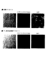
(赤外分光法)
上記のように調製したアパタイト粒子のフーリエ変換赤外分光分析はFT/IR−230(JASCO)を用いて行った。サンプルをすり鉢の中にいれ、そして約1mgを完全に300mgの分光用の臭化カリウムと混ぜ合わせた。臭化カリウムの窓板の中で0.5torr減気圧下において8000kgの加重を加えることで薄いペレットを調製した。
(Infrared spectroscopy)
The Fourier transform infrared spectroscopic analysis of the apatite particles prepared as described above was performed using FT / IR-230 (JASCO). Samples were placed in a mortar and about 1 mg was thoroughly mixed with 300 mg of spectroscopic potassium bromide. Thin pellets were prepared by applying a weight of 8000 kg in a potassium bromide pane under 0.5 torr depressurization.
(X線回折)
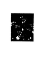
調製した粒子のX線粉体解析はM18XHF−SRA回折システムを用いて行った。
(X-ray diffraction)
X-ray powder analysis of the prepared particles was performed using an M18XHF-SRA diffraction system.
元素分析により炭素が3%、リンが17%、カルシウムイオンが32%存在することがわかった。図1−1に示したFT−IRスペクトルの1410から1540cm-1間と約880cm-1におけるピークから、生成した粒子が炭酸塩を含むこと、また、図1−2に示した1000から1100cm-1および550から650cm-1にあるピークより、さらにリン酸を含むことがわかった。X線回折パターン(図1−3)はその幅の広い回折ピークが象徴するように、炭酸アパタイトがハイドロキシアパタイト(図1−4)よりも結晶性が低いことを示している。これは炭酸アパタイト本来の性質である。 Elemental analysis revealed 3% carbon, 17% phosphorus, and 32% calcium ions. From the peaks between 1410 and 1540 cm −1 and about 880 cm −1 of the FT-IR spectrum shown in FIG. 1-1, it is confirmed that the produced particles contain carbonate, and that the generated particles show 1000 to 1100 cm − shown in FIG. From the peaks at 1 and 550 to 650 cm −1 , it was found that phosphoric acid was further contained. The X-ray diffraction pattern (FIGS. 1-3) shows that carbonate apatite is less crystalline than hydroxyapatite (FIGS. 1-4), as symbolized by its broad diffraction peak. This is a natural property of carbonate apatite.
実施例2 細胞への遺伝子導入
〔1〕ルシフェラーゼの発現レベル測定
実施例1〔1〕によって調製した本発明の細胞導入剤のトランスフェクション効率を調べるため、本発明の細胞導入剤を用いたルシフェラーゼの発現レベルを測定した。
Example 2 Gene transfer into cells [1] Expression level measurement of luciferase To examine the transfection efficiency of the cell transfer agent of the present invention prepared in Example 1 [1], luciferase using the cell transfer agent of the present invention was examined. The expression level was measured.
対数増殖期にある細胞を24穴プレート1穴につき約50000個、遺伝子導入を行う前日に播種した。調製しておいたDNA含有粒子を含む培地を、10%FBSとともに洗浄した細胞に添加した。 About 50000 cells in a logarithmic growth phase per well of a 24-well plate were seeded the day before gene transfer. The prepared medium containing the DNA-containing particles was added to the cells washed with 10% FBS.
4時間培養後、その培地を血清を含む培地に置換し、その細胞を1日間培養した。ルシフェラーゼの遺伝子発現は市販のキット(プロメガ社)とフォトンカウンティング(TD−20/20 ルミノメーター,USA)を用いて測定した。それぞれの遺伝子導入実験は3回繰り返して行い、トランスフェクション効率は光強度の平均値を細胞のタンパク質量(mg)で割った値で表した。 After culturing for 4 hours, the medium was replaced with a medium containing serum, and the cells were cultured for 1 day. Luciferase gene expression was measured using a commercially available kit (Promega) and photon counting (TD-20 / 20 luminometer, USA). Each gene transfer experiment was repeated three times, and transfection efficiency was expressed as the average value of light intensity divided by the amount of protein (mg) in the cells.
初代肝細胞はコラーゲンを被覆した24穴プレートに播種し、3時間培養後に形質転換した。リン酸カルシウム−DNAの共沈殿による形質転換はJordan M.らによる方法(Jordan, M., Schallhorn, A. & Wurm FM. Transfecting mammalian cells: optimization of critical parameters affecting calcium-phosphate precipitate formation. Nucleic acids research 24, 596-601 (1996))にて行った。簡単に言えば、12μgのプラスミドDNAを300μ1の250mM塩化カルシウム溶液中に加えた。この溶液を300μ1の2×HBS(50mM Hepes、140mM塩化ナトリウム、1.5mM Na2HPO4・2H20、pH7.05)に加え、そして即座に、2回ゆっくりとピペッティングをすることで攪拌した。
Primary hepatocytes were seeded in a 24-well plate coated with collagen, and transformed after 3 hours of culture. Transformation by coprecipitation of calcium phosphate-DNA is described in Jordan M. et al. (Jordan, M., Schallhorn, A. & Wurm FM. Transfecting mammalian cells: optimization of critical parameters affecting calcium-phosphate precipitate formation. Nucleic acids research 24, 596-601 (1996)). Briefly, 12 μg of plasmid DNA was added in 300 μl of 250 mM calcium chloride solution. This solution is added to 300 μl of 2 × HBS (50 mM Hepes, 140 mM sodium chloride, 1.5 mM Na 2 HPO 4 .
DNA/リン酸カルシウム混合物を室温にそれぞれの示されている時間放置した。1mlの血清入り培地中に室温に放置しておいた混合液を100μ1滴下し、そしてその培地中で4時間培養し、上記のように新しい血清含有培地に交換した後1日培養した。 The DNA / calcium phosphate mixture was left at room temperature for each indicated time. 100 μl of the mixed solution that had been left at room temperature in 1 ml of serum-containing medium was dropped, and cultured in that medium for 4 hours. After replacing with fresh serum-containing medium as described above, the culture was continued for one day.
カルシウムイオン濃度、炭酸水素イオン緩衝培地のpH、そして培養温度の影響を調べるため、カルシウムイオン濃度、培養温度を変化させて、10%FBS含有培地でHeLa細胞を用いた遺伝子導入でトランスフェクション効率を評価した。 In order to investigate the effects of calcium ion concentration, pH of bicarbonate ion buffer medium, and culture temperature, transfection efficiency was improved by gene introduction using HeLa cells in medium containing 10% FBS by changing the calcium ion concentration and culture temperature. evaluated.
図2−1に、炭酸水素イオン緩衝培地のpHに依存したDNA/炭酸アパタイト粒子の生成におけるDMEM中に添加されたカルシウムイオン濃度を変化させたときのトランスフェクション効率を、図2−2に炭酸水素イオン緩衝培地のpHに依存したDNA/炭酸アパタイト粒子の生成における温度を変化させたときのトランスフェクション効率を示す。ここでは、トランスフェクション効率を10%FBS含有培地でHeLa細胞を用いた遺伝子導入におけるルシフェラーゼの発現レベルで検討している。 Fig. 2-1 shows the transfection efficiency when the calcium ion concentration added in DMEM in the production of DNA / carbonate apatite particles depending on the pH of the bicarbonate ion buffer medium is changed. Fig. 6 shows transfection efficiency when the temperature in the production of DNA / carbonate apatite particles depending on the pH of a hydrogen ion buffer medium is changed. Here, transfection efficiency is examined by the expression level of luciferase in gene transfer using HeLa cells in a medium containing 10% FBS.
炭酸アパタイト粒子の生成は、カルシウムイオン濃度、炭酸イオン緩衝培地のpH、そして培養温度に依存していた。高いトランスフェクション効率を得るために充分な量のDNA/炭酸アパタイト複合体を生成するために必要な最良のカルシウムイオン濃度は、培地のpH(図2−1)または温度(図2−2)と負の相関を有していた。初期の高効率なトランスフェクションよりも効率が低下するのは粒子形成量が少なくなることに起因している(顕微鏡による観察結果)。液状培地のpH、培養温度および/又はカルシウムイオン濃度の向上が粒子の生成に必要である過飽和溶液の形成に寄与している。粒子形成に関する全てのパラメーターをコントロールするだけで高い再現性が得られる融通の利くトランスフェクションシステムをはじめて本発明者らは開発することが出来た。 The production of carbonate apatite particles was dependent on the calcium ion concentration, the pH of the carbonate ion buffer medium, and the culture temperature. The best calcium ion concentration needed to produce a sufficient amount of DNA / carbonated apatite complex to obtain high transfection efficiency is determined by the pH (Figure 2-1) or temperature (Figure 2-2) of the medium. It had a negative correlation. The decrease in efficiency compared to the initial high-efficiency transfection is attributed to the reduced amount of particles formed (observation results with a microscope). Improvements in the pH of the liquid medium, the culture temperature and / or the calcium ion concentration contribute to the formation of a supersaturated solution that is necessary for the production of particles. We were able to develop a flexible transfection system for the first time that high reproducibility can be achieved simply by controlling all parameters related to particle formation.
有効な遺伝子キャリアーとしての炭酸アパタイトの役割を評価するために、本発明者は頻繁に用いられる二種類のトランスフェクション法即ち上記のリン酸カルシウム共沈法とリポフェクション法を含む、さまざまなトランスフェクション法とそのトランスフェクション効率について比較してみた。 In order to evaluate the role of carbonate apatite as an effective gene carrier, the present inventor has developed a variety of transfection methods, including two frequently used transfection methods, namely the calcium phosphate coprecipitation method and the lipofection method described above. We compared transfection efficiency.
図2−3に、HeLa細胞を用いて、炭酸アパタイト、ハイドロキシアパタイト、リポフェクトアミンによる遺伝子導入を行ったときの遺伝子発現レベルを、図2−4に、HepG2細胞を用いて、炭酸アパタイト、ハイドロキシアパタイト、リポフェクトアミンによる遺伝子導入を行ったときの遺伝子発現レベルを、図2−5に、NIH3T3細胞を用いて、炭酸アパタイト、ハイドロキシアパタイト、リポフェクトアミンによる遺伝子導入を行ったときの遺伝子発現レベルを、図2−6に、マウス初代培養肝細胞を用いて、炭酸アパタイト、ハイドロキシアパタイト、リポフェクトアミンによる遺伝子導入を行ったときの遺伝子発現レベルを、それぞれ示す。 Fig.2-3 shows the gene expression level when HeLa cells are used to introduce a gene with carbonate apatite, hydroxyapatite, and lipofectamine, and Fig.2-4 shows that the HepG2 cells are used with carbonate apatite, hydroxya Figure 2-5 shows the gene expression level when gene transfer using apatite and lipofectamine is performed. Fig. 2-5 shows the gene expression level when gene transfer using carbonate apatite, hydroxyapatite and lipofectamine is performed using NIH3T3 cells. Fig. 2-6 shows the gene expression levels when gene introduction with carbonate apatite, hydroxyapatite, and lipofectamine was performed using mouse primary cultured hepatocytes.
ここで、DNA/炭酸アパタイト複合体粒子は3から6mMのカルシウムイオン濃度(カルシウムイオン濃度が明記されていないものはすべて3mM)と2μgのプラスミドDNAを1mlの炭酸水素イオン緩衝培地(pH7.5)に加え、37℃で30分放置した。また、図2−3に示すHeLa細胞の湯合は、6mMカルシウムイオンを2μgDNAと一緒に1mlの無血清培地(上記に示した)に添加して粒子を生成し、その培地を100μ1(200ngDNA)または20μl(40ngDNA)用いて、FBS入り培地中で遺伝子導入実験を行った。DNA/ハイドロキシアパタイト複合体粒子はJordan M.らの方法にしたがって生成した。DNA/リポフェクトアミン複合体は1:6の重量比率で製造元のプロトコールに従って調製した。細胞への遺伝子導入は上述の方法で行い、Tissue culture plateの各ウェル中に(別段の記載がなければ)2μgのDNAを加えて、異なる方法で実施した。 Here, the DNA / carbonate apatite complex particles are 3 to 6 mM calcium ion concentration (3 mM for all calcium ions not specified) and 2 μg plasmid DNA in 1 ml bicarbonate ion buffer (pH 7.5). And left at 37 ° C. for 30 minutes. In addition, the hot water of the HeLa cell shown in FIG. 2-3 is obtained by adding 6 mM calcium ion together with 2 μg DNA to 1 ml of serum-free medium (shown above) to produce particles, and the medium is made 100 μ1 (200 ng DNA). Alternatively, 20 μl (40 ng DNA) was used for gene transfer experiments in a medium containing FBS. DNA / hydroxyapatite composite particles are described in Jordan M .; It was produced according to these methods. The DNA / lipofectamine complex was prepared according to the manufacturer's protocol at a weight ratio of 1: 6. The gene was introduced into the cells by the method described above, and 2 μg of DNA was added to each well of the tissue culture plate (unless otherwise stated), and the method was different.
例えばHeLa細胞では、炭酸アパタイトを用いることでトランスフェクションさせたときのルシフェラーゼ発現レベルはリポフェクションとリン酸カルシウム共沈法と比較したとき、それぞれ15倍以上、25倍以上高かった(図2−3)。効率的な導入遺伝子の発現にはナノグラム量のDNAで十分であった(図2−3)。 For example, in HeLa cells, the luciferase expression level when transfected using carbonate apatite was 15 times or more and 25 times or more higher when compared with lipofection and calcium phosphate coprecipitation (FIG. 2-3). Nanogram amounts of DNA were sufficient for efficient transgene expression (FIGS. 2-3).
トランスフェクション効率はHepG2(図2−4)、NIH3T3細胞(図2−5)、そしてマウスの初代培養肝細胞(図2−6)においても顕著に高いという結果を得た。 The results showed that the transfection efficiency was significantly higher in HepG2 (FIG. 2-4), NIH3T3 cells (FIG. 2-5), and mouse primary cultured hepatocytes (FIG. 2-6).
4時間のDNAの細胞内取り込み後、EDTAで細胞を処理した結果、ルシフェラーゼの遺伝子発現レベルが半分に減少したこと(図2−3)は、長時間にわたるDNAの取り込みは効率的な遺伝子発現に寄与するが核酸分解酵素によるDNAの加水分解と相殺されていることを示唆しており、これはPIラベルしたDNAの蛍光強度の持続によっても判る(図は無し)。 As a result of treating cells with EDTA after 4 hours of DNA uptake into cells, the gene expression level of luciferase was reduced by half (Fig. 2-3). This suggests that it contributes but offsets the hydrolysis of DNA by nucleolytic enzymes, which is also seen by the persistence of the fluorescence intensity of the PI-labeled DNA (not shown).
〔2〕MTTアッセイ
細胞の生存率が高いためにトランスフェクション効率が高く現れたのではないことを明らかにするためにHeLa細胞を用いてMTTアッセイを行った。
[2] MTT assay In order to clarify that the transfection efficiency did not appear to be high due to the high cell viability, the MTT assay was performed using HeLa cells.
HeLa細胞は遺伝子導入後、上記の方法で1日培養した。30μ1のMTT(臭化3−(4,5−ジメチルチアゾール−2−イル)−2,5−ジフェニルテトラゾリウム)溶液(5mg/ml)をそれぞれのウェル中に加え、そして4時間培養した。0.5mlのDMSOを、培地を除いた後に加えた。結晶が溶解し、37℃で5分培養した後、波長570nmの吸光度を630nmの波長を参照波長としてマイクロプレートリーダーで計測した。 HeLa cells were cultured for 1 day after the gene introduction by the above method. 30 μl of MTT (3- (4,5-dimethylthiazol-2-yl) -2,5-diphenyltetrazolium bromide) solution (5 mg / ml) was added into each well and incubated for 4 hours. 0.5 ml DMSO was added after removing the medium. After the crystals were dissolved and incubated at 37 ° C. for 5 minutes, absorbance at a wavelength of 570 nm was measured with a microplate reader using a wavelength of 630 nm as a reference wavelength.
図2−7にHeLa細胞における細胞の生存率をMTTアッセイで検討した結果を示す。 Fig. 2-7 shows the results of examination of cell viability in HeLa cells by MTT assay.
〔3〕細胞導入剤に含まれる複合体粒子の分析
(走査型電子顕微鏡(SEM))
遺伝子導入のプロトコールで述べた様に調製したDNA−炭酸アパタイトの混合液の液滴を炭素コートのSEMのステージに加え、乾燥させた後に、SEM(S−800、日立、日本)で観察した。
[3] Analysis of complex particles contained in cell introduction agent (scanning electron microscope (SEM))
Drops of the DNA-carbonate apatite mixture prepared as described in the gene transfer protocol were added to a carbon-coated SEM stage, dried, and then observed with SEM (S-800, Hitachi, Japan).
図3−1に走査型電子頼微鏡像を示す。スケールバーは600nmである。 Fig. 3-1 shows a scanning electronic microscopic image. The scale bar is 600 nm.
アパタイト構造存在下では、炭酸塩はアパタイト結晶の成長を制限し、そして溶解度を高める。本発明者らは生成した炭酸アパタイトを走査型電子顕微鏡によって観察を行い、結晶の成長が、大半は直径50nmから300nmに抑制されていることを明らかにした(図3−1、物件提出書にて提出した図を参照。)。 In the presence of an apatite structure, carbonate limits the growth of apatite crystals and increases solubility. The present inventors observed the produced carbonate apatite with a scanning electron microscope, and clarified that the crystal growth was mostly suppressed from a diameter of 50 nm to 300 nm (see FIG. 3-1, property submission). (Refer to the figure submitted.)
(共焦点レーザー顕微鏡)
巨大粒子は小さい粒子よりも食作用によって取り込まれにくいので、本発明者らは炭酸塩の結晶の大きさを抑制したときの効果の検証を、アパタイトに吸着させたPI(propidium iodide)ラベルしたプラスミドDNAの細胞への取り込みを観察することで行った。
(Confocal laser microscope)
Since large particles are less likely to be taken in by phagocytosis than small particles, the present inventors have verified the effect of suppressing the size of carbonate crystals by using a PI (propidium iodide) labeled plasmid adsorbed on apatite. This was done by observing DNA uptake into cells.
pGL3 ベクターはPIでPI/DNA比が1:1になるようにラベルし、そしてこのラベルしたプラスミドで生成した粒子(遺伝子導入のプロトコールに従って生成)をHeLa細胞と一緒に6時間培養した。酸性部位は5μMのLyso Sensorを用いて、Molecular Probe社の示す方法に従ってラベルした。そして細胞膜に結合している粒子をPBSに溶解した5mMのEDTAを用いて除去した後、LEICA TCS−NTを用いて観察した。 The pGL3 vector was labeled with PI so that the PI / DNA ratio was 1: 1, and particles generated with this labeled plasmid (generated according to the gene transfer protocol) were incubated with HeLa cells for 6 hours. Acidic sites were labeled using a 5 μM Lyso Sensor according to the method indicated by Molecular Probe. And after removing the particle | grains couple | bonded with the cell membrane using 5 mM EDTA melt | dissolved in PBS, it observed using LEICA TCS-NT.
図3−2(物件提出書にて提出した図を参照。)に、アパタイトを介したDNAの放出と発現を示す。図3−2において、(a)ではDNAの取り込みは見られない(コントロール実験)。これは、エンドサイトーシスがエネルギー消耗(50mMの2−デオキシグルコースと1mMのアジ化ナトリウム添加)により、ブロックされているためである。DNA/炭酸アパタイト粒子は1mlの無血清培地(図2を参照して説明したもの)中で、6mMのカルシウムイオンと2μgのDNAを用いて生成した。1mlの懸濁液の20μ1および100μ1にそれぞれ40ng(b)、200ng(c)のDNAがあり、4時間細胞に取り込ませた。(d)は2μgのDNAをハイドロキシアパタイトに吸着させ、そして同じ時間だけ細胞に取り込ませた。 Fig. 3-2 (see the figure submitted in the property submission form) shows the release and expression of DNA via apatite. In FIG. 3-2, no DNA uptake is observed in (a) (control experiment). This is because endocytosis is blocked by energy consumption (addition of 50 mM 2-deoxyglucose and 1 mM sodium azide). DNA / carbonate apatite particles were generated using 6 mM calcium ions and 2 μg DNA in 1 ml serum-free medium (as described with reference to FIG. 2). There were 40 ng (b) and 200 ng (c) of DNA in 20 μl and 100 μl of a 1 ml suspension, respectively, and the cells were taken up for 4 hours. In (d), 2 μg of DNA was adsorbed on hydroxyapatite and taken up into cells for the same time.
1分培養することによって粒子を形成させたとき、炭酸アパタイトを用いたほうが(図3−2(c))ハイドロキシアパタイトを用いる(図3−2(d))よりも、少なくとも10倍効率的にDNAは細胞内に取り込まれた。 When particles are formed by culturing for 1 minute, using carbonate apatite (FIG. 3-2 (c)) is at least 10 times more efficient than using hydroxyapatite (FIG. 3-2 (d)). DNA was taken up into the cells.
さらに長い時間(30分)培養して粒子を形成させると、巨大なハイドロキシアパタイト粒子を生じ、DNAを細胞内に取り込む効率がかなり低下する(本文中には示さない)ために、トランスフェクション効率が顕著に低下した(図2−3)。 Incubating for a longer time (30 minutes) to form particles produces huge hydroxyapatite particles, and the efficiency of DNA incorporation into cells is significantly reduced (not shown in the text). Remarkably decreased (FIGS. 2-3).
従って、炭酸アパタイトは、結晶の成長を抑制するというその固有の性質のために、DNAを細胞内に取り込みやすくし、ハイドロキシアパタイトよりも優れた遺伝子導入キャリアーとなることを、本発明者らの発見は示している。 Therefore, the present inventors have found that carbonate apatite makes it easier to incorporate DNA into cells due to its inherent property of suppressing crystal growth and is a better gene transfer carrier than hydroxyapatite. Shows.
トランスジーン発現におけるエンドソームからのDNAの脱出の役割を評価するために、PIラベルしたプラスミドDNAのエンドサイトーシスに引き続き、我々はエンドソームをLysoSensor(エンドソームの蛍光プローブ)でラベルした。その結果、細胞によるDNAの取り込みから6時間後、多くのDNAがエンドソームより、それら2つが共局在した後に放出されているのがわかった(図3−3、物件提出書にて提出した図を参照。)。 To assess the role of DNA escape from endosomes in transgene expression, following endocytosis of PI-labeled plasmid DNA, we labeled endosomes with LysoSensor (endosomal fluorescent probe). As a result, 6 hours after the uptake of DNA by the cells, it was found that a lot of DNA was released from the endosome after the two co-localized (Figure 3-3, Figure submitted in the property submission form). See).
〔4〕細胞導入におけるpHの影響
細胞導入におけるpHの影響を調べるため、V−ATPase(エンドソームを酸性化するのに用いられるプロトンポンプ)の特異的阻害剤であるバフィロマイシンAlを用いてトランスフェクション効率の変化を調べた。HeLa細胞をDNA/炭酸アパタイト複合体粒子および200nMバフィロマイシンAlとともに6時間培養した。5mMのEDTA含有PBSで洗ったあと、細胞を1日培養し、ルシフェラーゼの発現を検出した。結果を図4−1に示す。
[4] Effect of pH in cell introduction In order to investigate the effect of pH in cell introduction, transfection was performed using bafilomycin Al, a specific inhibitor of V-ATPase (a proton pump used to acidify endosomes). The change in the injection efficiency was investigated. HeLa cells were cultured with DNA / carbonate apatite complex particles and 200 nM bafilomycin Al for 6 hours. After washing with PBS containing 5 mM EDTA, the cells were cultured for 1 day, and the expression of luciferase was detected. The results are shown in Fig. 4-1.
V−ATPase(エンドソームを酸性化するのに用いられるプロトンポンプ)の特異的阻害剤であるバフィロマイシンAlで処理すると、HeLa細胞のトランスフェクション効率を劇的に減少させた。この結果は炭酸アパタイトを溶解しDNAをアパタイトから脱出させるのに酸性の環境が必要であることを示している。 Treatment with bafilomycin Al, a specific inhibitor of V-ATPase (a proton pump used to acidify endosomes), dramatically reduced the transfection efficiency of HeLa cells. This result indicates that an acidic environment is required to dissolve carbonate apatite and allow DNA to escape from the apatite.
この仮説を検討するのに、我々はフッ素を添加するとアパタイトの溶解度が下がることから、フッ素を添加した炭酸アパタイトを合成し、そしてトランスフェクション効率における粒子の溶解度の影響について検討を行った。DNA/炭酸アパタイト粒子を形成するときに加えたフッ素イオン(0.01から3mM)もしくはストロンチウムイオン(0.01から3mM)の濃度を上昇させたときのルシフェラーゼ発現の変化を調べた。結果を図4−2に示す。 To examine this hypothesis, we added fluoride to reduce the solubility of apatite, so we synthesized carbonate apatite to which fluorine was added, and examined the effect of particle solubility on transfection efficiency. Changes in luciferase expression when the concentration of fluorine ions (0.01 to 3 mM) or strontium ions (0.01 to 3 mM) added when forming DNA / carbonate apatite particles was increased were examined. The results are shown in Fig. 4-2.
驚いたことに、炭酸アパタイトに含まれるフッ素の量を増大させると、有意に(100倍)トランスフェクション効率が低下した(図4−2)。 Surprisingly, increasing the amount of fluorine contained in carbonate apatite significantly (100 times) reduced transfection efficiency (FIG. 4-2).
フッ素を添加した炭酸アパタイトのトランスフェクション効率が低下したのはDNAの細胞への取り込みが減少したためではないことを示すために、本発明者らはPIでラベルしたpEGFPプラスミドを遺伝子導入した。 In order to show that the transfection efficiency of fluorinated carbonate apatite was not due to a decrease in DNA uptake into cells, the present inventors introduced a pEGFP plasmid labeled with PI.
図4−3(物件提出書にて提出した図を参照。)に炭酸アパタイトおよび、フッ素添加炭酸アパタイトによるPIラベルしたプラスミドDNA(pEGFP−N2)の取り込みとGFPタンパク質の発現を観察した結果を示す。DNAを含む粒子を4時間培養した後、さらに20時間培養し、PBSに5mM EDTAで処理してから観察した。 Fig. 4-3 (see the figure submitted in the property submission form) shows the results of observation of uptake of PI-labeled plasmid DNA (pEGFP-N2) and expression of GFP protein by carbonate apatite and fluorinated carbonate apatite. . After culturing the particles containing DNA for 4 hours, the cells were further cultured for 20 hours, treated with 5 mM EDTA in PBS, and then observed.
細胞内に取り込まれたプラスミドDNAの量は同じであったが、炭酸アパタイトではほぼ50%の細胞にGFPの発現が見られた(図4−3、上段)のに対し、フッ素を添加した炭酸アパタイトを用いたときには、GFPを発現している細胞は見られなかった(図4−3、下段)。 Although the amount of plasmid DNA incorporated into the cells was the same, the expression of GFP was observed in almost 50% of the cells with carbonate apatite (FIG. 4-3, upper panel), whereas carbon dioxide added with fluorine. When apatite was used, cells expressing GFP were not seen (FIG. 4-3, lower row).
DNAの取り込み期間が短いときのトランスジーン発現の効率を促進するのに、我々は少量のフッ素を添加することで炭酸アパタイトの溶解度を若干減少させることによる影響を調べた。 To promote the efficiency of transgene expression when the DNA uptake period is short, we investigated the effect of slightly reducing the solubility of carbonate apatite by adding a small amount of fluorine.
図4−4にDNA/炭酸アパタイト粒子の形成時におけるμMレベルのフッ素イオンを添加したときのルシフェラーゼ発現量の変化を示す。ここでは、粒子と一緒に細胞を培養した後、細胞をEDTAで洗い、さらに1日、上記のように培養した。 FIG. 4-4 shows changes in the expression level of luciferase when adding μM level fluorine ions during the formation of DNA / carbonate apatite particles. Here, after culturing the cells with the particles, the cells were washed with EDTA and further cultured as described above for one day.
驚いたことに、炭酸アパタイトの生成中に、1μMのフッ化ナトリウムを添加することで、ルシフェラーゼの遺伝子発現が15倍促進された。さらにエンドソームのpHを上昇させ、核酸分解酵素に対してDNAを保護することが知られているクロロキンで処理すると、同様な遺伝子発現の促進が見られた(図4−4)。 Surprisingly, the addition of 1 μM sodium fluoride during the production of carbonate apatite promoted luciferase gene expression 15-fold. Further, when the pH of the endosome was raised and treated with chloroquine, which is known to protect DNA against nucleolytic enzymes, similar gene expression was observed (FIGS. 4-4).
これらの結果から、微量のフッ素イオンを含む結晶によってエンドソームの緩衝作用とプロトンの消費が起きている中で、DNAが中間の速さで放出されることにより、顕著な核酸分解酵素によるDNA分解が避けられていることを示しており、PIの蛍光が持続すること(図にはない)は、転写や翻訳に際し、より完全なDNAが存在することを示している。 From these results, while the endosome buffering action and proton consumption are caused by crystals containing a small amount of fluorine ions, DNA is released at an intermediate rate, so that DNA degradation by remarkable nucleolytic enzymes is prevented. This shows that the fluorescence of PI is sustained (not shown in the figure), indicating that more complete DNA is present during transcription and translation.
〔5〕アパタイトのpH依存溶解度の研究
アパタイトの溶解度とトランスフェクション効率の間の関係を調べるために、生成したアパタイトの液中に酸を添加した後の、溶液の濁度を溶解度の指標として測定した。
[5] Study on pH-dependent solubility of apatite In order to investigate the relationship between apatite solubility and transfection efficiency, the turbidity of the solution after addition of acid was measured as an index of solubility. did.
DNAを含まない6mMのカルシウムイオン溶液を用いてトランスフェクションのプロトコールに従って生成した、異なるアパタイトの溶解度の特徴を、1MのHClでpHを7.0から6.8に調製し、V−500(Jasco、日本)分光光度計を用いて320nmの濁度を異なる間隔で測定して検討していった。SPS 1500 VR 原子吸光分光光度計は、凍結乾爆した異なるアパタイトの粉末をゆっくりと振とう(10rpmで6時間)した後に37℃で酢酸緩衝溶液(pH6.5のものとpH5.5の溶液)中で放出されるカルシウムイオンを検出するのに用いた。
The solubility characteristics of the different apatites, generated according to the transfection protocol using 6 mM calcium ion solution without DNA, were adjusted from 7.0 to 6.8 with 1 M HCl and V-500 (Jasco , Japan) Using a spectrophotometer, turbidity at 320 nm was measured at different intervals and examined. The
図5−1(物件提出書にて提出した図を参照。)にpH7.5で炭酸アパタイトを生成させるときに0〜3mMのフッ素イオンを添加して調製したフッ素添加炭酸アパタイト(実験プロトコールに記載)の溶解度を、アパタイト懸濁液に1N塩酸を加えてpH7.0に調製した直後に波長320nmにおける濁度で測定した結果を示す。 Fluorine-added carbonate apatite prepared by adding 0 to 3 mM fluoride ions when producing carbonate apatite at pH 7.5 in Fig. 5-1 (see the figure submitted in the property submission form) (described in the experimental protocol) ) Is measured by turbidity at a wavelength of 320 nm immediately after adjusting the pH to 7.0 by adding 1N hydrochloric acid to the apatite suspension.
1N塩酸を添加することでpHを7.5から7.0に調整した後、濁度の変化が示すように、添加するフッ化ナトリウム濃度を上昇させることで生成する炭酸アパタイトの溶解度は徐々に減少した(図5−1)。この結果はフッ素を添加した炭酸アパタイトのトランスフェクション効率が徐々に減少するという結果と一致していた(図4−2)。 After adjusting the pH from 7.5 to 7.0 by adding 1N hydrochloric acid, the solubility of carbonate apatite produced by increasing the concentration of sodium fluoride to be added gradually increases as shown by the change in turbidity. It decreased (FIG. 5-1). This result was consistent with the result that the transfection efficiency of carbonate apatite to which fluorine was added gradually decreased (FIG. 4-2).
アパタイトの溶解度が結晶化の度合いに関係しているか検討するために、我々はアパタイトのX線散乱を検討した(図1−4、1−5、1−6)。その結果、結晶度が高いほうが溶解度は低いことがわかった(図5−1)。換言すれば、結晶性が高いアパタイト(図1−4、図1−8)は低いトランスフェクション効率を示すことを示している(図2−3、4−2)。 In order to examine whether the solubility of apatite is related to the degree of crystallization, we examined X-ray scattering of apatite (FIGS. 1-4, 1-5, 1-6). As a result, it was found that the higher the crystallinity, the lower the solubility (FIG. 5-1). In other words, apatite with high crystallinity (FIGS. 1-4 and 1-8) shows low transfection efficiency (FIGS. 2-3 and 4-2).
炭酸アパタイト中のフッ素含量の増加に伴う結晶性の増加(図1−6〜1−8)によってトランスフェクション効率は後々に低下していく(図4−2)。トランスフェクション効率の低下はフッ素を添加した炭酸アパタイトの溶解度によるためであり、他のフッ素を添加したことによる影響によるものではないことを示すのに、我々は炭酸アパタイトに添加したときのストロンチウムの影響について検討した。ストロンチウムはアパタイトの結晶性を上げ、フッ素よりも効果は小さいが、溶解性を減少させる。 Transfection efficiency decreases later (FIG. 4-2) due to the increase in crystallinity (FIGS. 1-6 to 1-8) accompanying the increase in fluorine content in carbonate apatite. Although we show that the decrease in transfection efficiency is due to the solubility of carbonate apatite with the addition of fluorine and not the effect of adding other fluorine, we have the effect of strontium when added to carbonate apatite. Was examined. Strontium increases the crystallinity of apatite and is less effective than fluorine, but decreases solubility.
図5−2(物件提出書にて提出した図を参照。)にpH7.0および6.8における炭酸アパタイト、フッ素添加炭酸アパタイト、ストロンチウムを含む炭酸アパタイトの溶解度を示す。 The solubility of carbonate apatite containing carbonate apatite, fluorinated carbonate apatite, and strontium at pH 7.0 and 6.8 is shown in FIG.
pHを7.5から7.0および6.8に低下させることにより、炭酸アパタイトは1分以内に完全に溶解したが、対して、フッ素を添加した炭酸アパタイトは部分的に溶けるだけであった(図5−2)。 By reducing the pH from 7.5 to 7.0 and 6.8, the carbonate apatite was completely dissolved within 1 minute, whereas the carbonate apatite with the addition of fluorine was only partially dissolved. (FIG. 5-2).
期待通り、炭酸アパタイトの沈殿中に塩化ストロンチウムを添加したところ、フッ素を添加したときほどではないが溶解度は減少した(図5−2)。さらに、トランスフェクション効率は、DNA/炭酸アパタイト複合粒子の生成中に塩化ストロンチウムの濃度を上昇させることにより徐々に減少した(図4−2)。これらの結果より、我々の発見はアパタイトの溶解による細胞質内でのDNAの放出は炭酸アパタイトを介したトランスフェクションにおいて重要な役割を果たしていることを明確に示唆している。 As expected, when strontium chloride was added during the precipitation of carbonate apatite, the solubility decreased not as much as when fluorine was added (FIG. 5-2). Furthermore, the transfection efficiency was gradually decreased by increasing the concentration of strontium chloride during the production of DNA / carbonate apatite composite particles (FIG. 4-2). From these results, our findings clearly suggest that the release of DNA in the cytoplasm by lysis of apatite plays an important role in transfection via carbonate apatite.
本発明者らはハイドロキシアパタイトを含むアパタイトの溶解度を、pH6.5と5.5の酢酸ナトリウム緩衝液でアパタイト粉末から放出されたカルシウムイオンを原子発光分析測定することによって比較した。フッ素添加炭酸アパタイトが炭酸アパタイトに比べて溶解度が低く、ハイドロキシアパタイトと比べて高かった(図5−3を参照)。 The present inventors compared the solubility of apatite containing hydroxyapatite by atomic emission spectrometry measurement of calcium ions released from the apatite powder with sodium acetate buffers at pH 6.5 and 5.5. The solubility of fluorinated carbonate apatite was lower than that of carbonate apatite and higher than that of hydroxyapatite (see FIG. 5-3).
エンドソームにおいて結晶と結合しているDNAの放出が遺伝子発現にとって本当の重要なファクターであることを証明するため、本発明者らはPI(pH感受性の色素)ラベルしたプラスミドDNAを用い、4時間の細胞内取り込み後、EDTA(図5−4−a、d、物件提出書にて提出した図を参照。以下同様。)により細胞表面に結合したDNAを除去し、さらに5時間放置した(図5−4−b、c、e)。PIの蛍光強度は遊離のプラスミドDNAでは核酸分解酵素の作用により、5時間後には大幅に減少していることが判る(図5−4−b、e)。また、4時間の細胞内取り込み後、バフィロマイシンA1で処理した細胞では蛍光強度が保持されていることが判る(図5−4−c)。 In order to demonstrate that the release of DNA associated with crystals in endosomes is a truly important factor for gene expression, we have used PI (pH sensitive dye) labeled plasmid DNA for 4 hours. After intracellular uptake, DNA bound to the cell surface was removed by EDTA (see FIGS. 5-4-a and d, see the figure submitted in the property submission form, the same applies below) and left for another 5 hours (FIG. 5). -4-b, c, e). It can be seen that the fluorescence intensity of PI is significantly reduced in free plasmid DNA after 5 hours due to the action of nucleolytic enzyme (FIGS. 5-4-b and e). Moreover, it turns out that the fluorescence intensity is hold | maintained in the cell processed with bafilomycin A1 after intracellular uptake | capture for 4 hours (FIG. 5-4-c).
フッ素添加した炭酸アパタイトによって細胞内に取り込まれたDNA(図5−4−f)はほとんど蛍光強度が減少していない(図5−4−g)ことは、加水分解の速度が速いにもかかわらず、遺伝子発現にはDNAの放出が必須であることを示している。さらに、結晶の溶解によるV−ATPaseに誘導された大量のプロトンの蓄積がエンドソームへの不活化塩素の流入をもたらし、エンドゾームの膨張と破裂をもたらすことから、炭酸アパタイトの高い溶解性がDNAの細胞質への放出の際のエンドソームの不安定化にも寄与していると思われる(図3−3)。 Although the fluorescence intensity of DNA (Fig. 5-4-f) taken into cells by the fluorinated carbonate apatite is hardly decreased (Fig. 5-4-g), the hydrolysis rate is high. In other words, DNA release is essential for gene expression. Furthermore, since the accumulation of a large amount of protons induced by V-ATPase due to the dissolution of crystals leads to the inflow of inactivated chlorine into the endosome, leading to the expansion and rupture of endosomes, the high solubility of carbonate apatite makes the cytoplasm of DNA It seems to contribute to the destabilization of endosomes during release into the body (Fig. 3-3).
以上のように、本発明にかかる細胞導入剤は、細胞導入効率が高く、優れた再現性、生体適合性を有するため、遺伝子組換え技術、各種疾患の治療等に好適に利用することができる。 As described above, since the cell introduction agent according to the present invention has high cell introduction efficiency and excellent reproducibility and biocompatibility, it can be suitably used for gene recombination techniques, treatment of various diseases, and the like. .
Claims (17)
少なくともカルシウムイオン、リン酸イオン、炭酸水素イオン、および前記ポリヌクレオチドを含有する組成物を調製することにより、前記複合体粒子を形成する工程を含むことを特徴とする細胞導入剤の製造方法。 A method for producing a cell introduction agent, which is used for introducing a polynucleotide into a cell and contains composite particles composed of the polynucleotide and carbonate apatite,
A method for producing a cell introduction agent, comprising a step of forming the composite particle by preparing a composition containing at least calcium ion, phosphate ion, bicarbonate ion and the polynucleotide .
前記リン酸イオンおよび前記炭酸水素イオンを含有する第2の溶液を調製する工程、および、
前記第1の溶液と前記第2の溶液とを混合して前記組成物を調製する工程を含むことを特徴とする請求項1に記載の細胞導入剤の製造方法。Preparing a first solution containing the calcium ions and the polynucleotide ;
Preparing a second solution containing the phosphate ions and the bicarbonate ions; and
The method for producing a cell introduction agent according to claim 1 , comprising a step of preparing the composition by mixing the first solution and the second solution.
少なくともカルシウムイオン、リン酸イオン、および炭酸水素イオンを含有し、
前記ポリヌクレオチドを添加することにより前記細胞導入剤を製造することを特徴とする細胞導入剤製造用組成物。 A composition for producing a cell introduction agent for producing a cell introduction agent, which is used for introducing a polynucleotide into a cell and contains composite particles composed of the polynucleotide and carbonate apatite,
Contains at least calcium ions, phosphate ions, and bicarbonate ions,
A composition for producing a cell introduction agent, which comprises producing the cell introduction agent by adding the polynucleotide .
少なくともリン酸イオン、および炭酸水素イオンを含有し、
前記ポリヌクレオチドとカルシウムイオンを添加することにより前記細胞導入剤を製造することを特徴とする細胞導入剤製造用組成物。 A composition for producing a cell introduction agent for producing a cell introduction agent, which is used for introducing a polynucleotide into a cell and contains composite particles composed of the polynucleotide and carbonate apatite,
Contains at least phosphate ions and bicarbonate ions,
A composition for producing a cell introduction agent, which comprises producing the cell introduction agent by adding the polynucleotide and calcium ions.
少なくともカルシウムイオンを含有する第1成分と、
少なくともリン酸イオンおよび炭酸水素イオンを含有する第2成分とからなり、
前記第1成分に前記ポリヌクレオチドを添加した後、当該第1成分および前記第2成分を混合することにより、前記複合体粒子を含有する細胞導入剤を製造することを特徴とする細胞導入剤製造用キット。 A kit for producing a cell introduction agent for producing a cell introduction agent, which is used for introducing a polynucleotide into a cell and contains composite particles composed of the polynucleotide and carbonate apatite,
A first component containing at least calcium ions;
A second component containing at least phosphate ions and bicarbonate ions,
A cell introduction agent comprising the complex particles is produced by adding the polynucleotide to the first component and then mixing the first component and the second component. For kit.
Applications Claiming Priority (3)
| Application Number | Priority Date | Filing Date | Title |
|---|---|---|---|
| US42529102P | 2002-11-12 | 2002-11-12 | |
| US46727703P | 2003-05-02 | 2003-05-02 | |
| PCT/JP2003/014376 WO2004043495A1 (en) | 2002-11-12 | 2003-11-12 | Delivery agent, method of delivering a target substance to cells, method for producing delivery agent, composition for producing delivery agent, and kit for producing delivery agent |
Publications (2)
| Publication Number | Publication Date |
|---|---|
| JP2006509838A JP2006509838A (en) | 2006-03-23 |
| JP4536655B2 true JP4536655B2 (en) | 2010-09-01 |
Family
ID=32314583
Family Applications (1)
| Application Number | Title | Priority Date | Filing Date |
|---|---|---|---|
| JP2005505672A Expired - Lifetime JP4536655B2 (en) | 2002-11-12 | 2003-11-12 | Cell introduction agent, cell introduction method, method for producing cell introduction agent, composition for producing cell introduction agent, and kit for producing cell introduction agent |
Country Status (3)
| Country | Link |
|---|---|
| JP (1) | JP4536655B2 (en) |
| AU (1) | AU2003279570A1 (en) |
| WO (1) | WO2004043495A1 (en) |
Families Citing this family (15)
| Publication number | Priority date | Publication date | Assignee | Title |
|---|---|---|---|---|
| JP2007043972A (en) * | 2005-08-10 | 2007-02-22 | Tokyo Institute Of Technology | Apatite particle and method for producing the same, gene-apatite particle complex, method for producing the same, and method for gene transfer and kit for gene transfer |
| JP2007063194A (en) * | 2005-08-31 | 2007-03-15 | Tokyo Institute Of Technology | Material-introducing carrier, method for introducing material and kit for introducing material |
| JP2007153842A (en) * | 2005-12-07 | 2007-06-21 | Hideki Aoki | Vitamin apatite composite material |
| JP2008193977A (en) * | 2007-02-14 | 2008-08-28 | National Institute Of Advanced Industrial & Technology | Gene-delivering method and base material |
| JP5416510B2 (en) * | 2009-08-26 | 2014-02-12 | 花王株式会社 | Tooth whitening or brightening screening method |
| JP5436650B1 (en) | 2012-11-28 | 2014-03-05 | 浩文 山本 | Superapatite ultrafine nanoparticles |
| JP6443907B2 (en) * | 2014-02-17 | 2018-12-26 | 浩文 山本 | Accumulation promoter for promoting accumulation of contrast agent in tumor |
| EP3115454B1 (en) * | 2014-03-04 | 2020-02-12 | Hirofumi Yamamoto | Novel rna sequence having anti-tumour activity |
| JP6588765B2 (en) * | 2015-08-07 | 2019-10-09 | 学校法人 神野学園 | DNA adsorption carrier and method of using the same |
| JP6302531B2 (en) * | 2015-11-09 | 2018-03-28 | 医療法人 医潤会 | Drug introduction agent for bioadministration and production method |
| US10449155B2 (en) | 2015-11-09 | 2019-10-22 | Medical Corporation Ijunkai | Drug introducing agent for administration into a living body and manufacturing method |
| JP7016317B2 (en) * | 2016-08-08 | 2022-02-21 | 浩文 山本 | Adjuvant composition |
| JPWO2018199121A1 (en) * | 2017-04-24 | 2020-02-27 | 浩文 山本 | Agent for preventing or treating inflammatory bowel disease |
| JP6868306B2 (en) * | 2017-05-29 | 2021-05-12 | 株式会社ビーエムジー | Cell introduction agent |
| CN116870104A (en) * | 2023-08-07 | 2023-10-13 | 上海章合生物科技有限公司 | Application of bamboo liquid particle compound in medicines or daily chemicals for treating dermatitis |
Family Cites Families (5)
| Publication number | Priority date | Publication date | Assignee | Title |
|---|---|---|---|---|
| US5407659A (en) * | 1991-10-22 | 1995-04-18 | Mallinckrodt Medical, Inc. | Treated calcium/oxyanion-containing particles for medical diagnostic imaging |
| US5962427A (en) * | 1994-02-18 | 1999-10-05 | The Regent Of The University Of Michigan | In vivo gene transfer methods for wound healing |
| EP0830149A1 (en) * | 1995-06-06 | 1998-03-25 | Osteogenics Inc. | Biocompatible hydroxyapatite formulations and uses therefor |
| JP2002348234A (en) * | 2001-05-28 | 2002-12-04 | Purotekku:Kk | Drug-encapsulating inorganic microparticle, manufacturing method thereof and preparation comprising drug-encapsulating inorganic microparticle |
| JP3898046B2 (en) * | 2001-12-07 | 2007-03-28 | 独立行政法人科学技術振興機構 | Method for producing hydroxyapatite and use thereof |
-
2003
- 2003-11-12 WO PCT/JP2003/014376 patent/WO2004043495A1/en active Application Filing
- 2003-11-12 JP JP2005505672A patent/JP4536655B2/en not_active Expired - Lifetime
- 2003-11-12 AU AU2003279570A patent/AU2003279570A1/en not_active Abandoned
Also Published As
| Publication number | Publication date |
|---|---|
| JP2006509838A (en) | 2006-03-23 |
| WO2004043495A1 (en) | 2004-05-27 |
| AU2003279570A1 (en) | 2004-06-03 |
Similar Documents
| Publication | Publication Date | Title |
|---|---|---|
| JP4536655B2 (en) | Cell introduction agent, cell introduction method, method for producing cell introduction agent, composition for producing cell introduction agent, and kit for producing cell introduction agent | |
| Badieyan et al. | Transcript-activated collagen matrix as sustained mRNA delivery system for bone regeneration | |
| Elangovan et al. | Chemically modified RNA activated matrices enhance bone regeneration | |
| Raftery et al. | Highly versatile cell-penetrating peptide loaded scaffold for efficient and localised gene delivery to multiple cell types: From development to application in tissue engineering | |
| ES2587963T3 (en) | RNA with a combination of unmodified and modified nucleotides for protein expression | |
| Blacklock et al. | Gene delivery in vitro and in vivo from bioreducible multilayered polyelectrolyte films of plasmid DNA | |
| Qu et al. | Magnesium-containing nanostructured hybrid scaffolds for enhanced dentin regeneration | |
| JP4613011B2 (en) | Pharmaceutical composition for improving in vivo gene transfer | |
| AU2016287436B2 (en) | UTRs increasing the translation efficiency of RNA molecules | |
| JP5697123B2 (en) | Method for introducing nucleic acid into cells using acidified polyethylenimine | |
| Tenkumo et al. | Gene transfection of human mesenchymal stem cells with a nano‐hydroxyapatite–collagen scaffold containing DNA‐functionalized calcium phosphate nanoparticles | |
| US10717980B2 (en) | MicroRNA-200 based approaches for modulating bone formation inhibition and bone regeneration | |
| Feng et al. | Delivery of therapeutic miRNAs using nanoscale zeolitic imidazolate framework for accelerating vascularized bone regeneration | |
| Jiang et al. | Bone response to the multilayer BMP‐2 gene coated porous titanium implant surface | |
| Toita et al. | Phosphatidylserine liposome multilayers mediate the M1-to-M2 macrophage polarization to enhance bone tissue regeneration | |
| US20190233820A1 (en) | Nanoparticles functionalized with gene editing tools and related methods | |
| White et al. | Functional delivery of large genomic DNA to human cells with a peptide‐lipid vector | |
| Yu et al. | 3-D scaffold platform for optimized non-viral transfection of multipotent stem cells | |
| US20160017368A1 (en) | Inorganic coatings for the enhancement of chemical transfection | |
| Oh et al. | Plasmid DNA-loaded asymmetrically porous membrane for guided bone regeneration | |
| EP3988137A1 (en) | Nucleic acid-calcium phosphate nanoparticle complex and application thereof in biomineralization | |
| Song et al. | MiR-148b laden titanium implant promoting osteogenic differentiation of rat bone marrow mesenchymal stem cells | |
| WO2019010304A1 (en) | Mineral coated microparticles for gene delivery in chronic wound therapy | |
| CN102031269A (en) | Method for preparing cationic amino acid-containing polypeptide-modified nano calcium phosphate gene vector | |
| US20070077306A1 (en) | Apatite particle, method of producing the same, apatite particle-gene complex, and method of gene transfection |
Legal Events
| Date | Code | Title | Description |
|---|---|---|---|
| A621 | Written request for application examination |
Free format text: JAPANESE INTERMEDIATE CODE: A621 Effective date: 20061010 |
|
| A711 | Notification of change in applicant |
Free format text: JAPANESE INTERMEDIATE CODE: A711 Effective date: 20071023 |
|
| A711 | Notification of change in applicant |
Free format text: JAPANESE INTERMEDIATE CODE: A711 Effective date: 20071023 |
|
| A131 | Notification of reasons for refusal |
Free format text: JAPANESE INTERMEDIATE CODE: A131 Effective date: 20100126 |
|
| A521 | Request for written amendment filed |
Free format text: JAPANESE INTERMEDIATE CODE: A523 Effective date: 20100326 |
|
| RD02 | Notification of acceptance of power of attorney |
Free format text: JAPANESE INTERMEDIATE CODE: A7422 Effective date: 20100326 |
|
| A521 | Request for written amendment filed |
Free format text: JAPANESE INTERMEDIATE CODE: A821 Effective date: 20100329 |
|
| TRDD | Decision of grant or rejection written | ||
| A01 | Written decision to grant a patent or to grant a registration (utility model) |
Free format text: JAPANESE INTERMEDIATE CODE: A01 Effective date: 20100525 |
|
| A01 | Written decision to grant a patent or to grant a registration (utility model) |
Free format text: JAPANESE INTERMEDIATE CODE: A01 |
|
| A61 | First payment of annual fees (during grant procedure) |
Free format text: JAPANESE INTERMEDIATE CODE: A61 Effective date: 20100616 |
|
| FPAY | Renewal fee payment (event date is renewal date of database) |
Free format text: PAYMENT UNTIL: 20130625 Year of fee payment: 3 |
|
| R150 | Certificate of patent or registration of utility model |
Ref document number: 4536655 Country of ref document: JP Free format text: JAPANESE INTERMEDIATE CODE: R150 Free format text: JAPANESE INTERMEDIATE CODE: R150 |
|
| R250 | Receipt of annual fees |
Free format text: JAPANESE INTERMEDIATE CODE: R250 |
|
| S111 | Request for change of ownership or part of ownership |
Free format text: JAPANESE INTERMEDIATE CODE: R313113 |
|
| R350 | Written notification of registration of transfer |
Free format text: JAPANESE INTERMEDIATE CODE: R350 |
|
| R250 | Receipt of annual fees |
Free format text: JAPANESE INTERMEDIATE CODE: R250 |
|
| R250 | Receipt of annual fees |
Free format text: JAPANESE INTERMEDIATE CODE: R250 |
|
| R250 | Receipt of annual fees |
Free format text: JAPANESE INTERMEDIATE CODE: R250 |
|
| R250 | Receipt of annual fees |
Free format text: JAPANESE INTERMEDIATE CODE: R250 |
|
| R250 | Receipt of annual fees |
Free format text: JAPANESE INTERMEDIATE CODE: R250 |
|
| EXPY | Cancellation because of completion of term |