JP4480800B2 - Methods for generating activated T cells and antigen pulse labeled antigen presenting cells - Google Patents
Methods for generating activated T cells and antigen pulse labeled antigen presenting cells Download PDFInfo
- Publication number
- JP4480800B2 JP4480800B2 JP54847798A JP54847798A JP4480800B2 JP 4480800 B2 JP4480800 B2 JP 4480800B2 JP 54847798 A JP54847798 A JP 54847798A JP 54847798 A JP54847798 A JP 54847798A JP 4480800 B2 JP4480800 B2 JP 4480800B2
- Authority
- JP
- Japan
- Prior art keywords
- cells
- antigen
- peptide
- cell
- muc
- Prior art date
- Legal status (The legal status is an assumption and is not a legal conclusion. Google has not performed a legal analysis and makes no representation as to the accuracy of the status listed.)
- Expired - Fee Related
Links
- 210000001744 T-lymphocyte Anatomy 0.000 title abstract description 200
- 108091007433 antigens Proteins 0.000 title abstract description 162
- 102000036639 antigens Human genes 0.000 title abstract description 162
- 239000000427 antigen Substances 0.000 title abstract description 161
- 238000000034 method Methods 0.000 title abstract description 88
- 210000000612 antigen-presenting cell Anatomy 0.000 title description 17
- 239000002502 liposome Substances 0.000 claims abstract description 88
- 238000000338 in vitro Methods 0.000 claims abstract description 26
- 239000000203 mixture Substances 0.000 claims abstract description 21
- 210000004027 cell Anatomy 0.000 claims description 110
- 108010008707 Mucin-1 Proteins 0.000 claims description 101
- 102100034256 Mucin-1 Human genes 0.000 claims description 92
- KXJTWOGIBOWZDJ-LELJLAJGSA-N l-blp25 Chemical compound C([C@@H](C(=O)NCC(=O)N[C@@H](C(C)C)C(=O)N[C@@H]([C@@H](C)O)C(=O)N[C@@H](CO)C(=O)N[C@@H](C)C(=O)N1[C@@H](CCC1)C(=O)N[C@@H](CC(O)=O)C(=O)N[C@@H]([C@@H](C)O)C(=O)N[C@@H](CCCNC(N)=N)C(=O)N1[C@@H](CCC1)C(=O)N[C@@H](C)C(=O)N1[C@@H](CCC1)C(=O)NCC(=O)N[C@@H](CO)C(=O)N[C@@H]([C@@H](C)O)C(=O)N[C@@H](C)C(=O)N1[C@@H](CCC1)C(=O)N1[C@@H](CCC1)C(O)=O)NC(=O)[C@H](C)NC(=O)[C@H]1N(CCC1)C(=O)[C@H]1N(CCC1)C(=O)[C@H](C)NC(=O)[C@@H](NC(=O)[C@@H](N)CO)[C@@H](C)O)C1=CNC=N1 KXJTWOGIBOWZDJ-LELJLAJGSA-N 0.000 claims description 47
- 108010028921 Lipopeptides Proteins 0.000 claims description 36
- 150000001413 amino acids Chemical class 0.000 claims description 29
- 108010017838 cyclo-(lysyl-prolyl-glycyl-prolyl-glycyl-glutamyl-prolyl-glycyl-prolyl-glycyl)cyclo(1epsilon-6gamma)glycyl-glycine Proteins 0.000 claims description 28
- 230000005867 T cell response Effects 0.000 claims description 26
- 150000002632 lipids Chemical class 0.000 claims description 21
- 230000027455 binding Effects 0.000 claims description 20
- 239000002671 adjuvant Substances 0.000 claims description 8
- 239000002691 unilamellar liposome Substances 0.000 claims description 8
- 125000001312 palmitoyl group Chemical group O=C([*])C([H])([H])C([H])([H])C([H])([H])C([H])([H])C([H])([H])C([H])([H])C([H])([H])C([H])([H])C([H])([H])C([H])([H])C([H])([H])C([H])([H])C([H])([H])C([H])([H])C([H])([H])[H] 0.000 claims description 6
- 102000008949 Histocompatibility Antigens Class I Human genes 0.000 claims description 5
- -1 alum Chemical compound 0.000 claims description 5
- 210000004899 c-terminal region Anatomy 0.000 claims description 5
- 108010088652 Histocompatibility Antigens Class I Proteins 0.000 claims description 3
- 125000002252 acyl group Chemical group 0.000 claims description 3
- 125000001931 aliphatic group Chemical group 0.000 claims description 3
- 230000013595 glycosylation Effects 0.000 claims description 3
- 238000006206 glycosylation reaction Methods 0.000 claims description 3
- YVGGHNCTFXOJCH-UHFFFAOYSA-N DDT Chemical compound C1=CC(Cl)=CC=C1C(C(Cl)(Cl)Cl)C1=CC=C(Cl)C=C1 YVGGHNCTFXOJCH-UHFFFAOYSA-N 0.000 claims description 2
- 125000003074 decanoyl group Chemical group [H]C([H])([H])C([H])([H])C([H])([H])C([H])([H])C([H])([H])C([H])([H])C([H])([H])C([H])([H])C([H])([H])C(*)=O 0.000 claims description 2
- GZQKNULLWNGMCW-PWQABINMSA-N lipid A (E. coli) Chemical compound O1[C@H](CO)[C@@H](OP(O)(O)=O)[C@H](OC(=O)C[C@@H](CCCCCCCCCCC)OC(=O)CCCCCCCCCCCCC)[C@@H](NC(=O)C[C@@H](CCCCCCCCCCC)OC(=O)CCCCCCCCCCC)[C@@H]1OC[C@@H]1[C@@H](O)[C@H](OC(=O)C[C@H](O)CCCCCCCCCCC)[C@@H](NC(=O)C[C@H](O)CCCCCCCCCCC)[C@@H](OP(O)(O)=O)O1 GZQKNULLWNGMCW-PWQABINMSA-N 0.000 claims description 2
- 125000001419 myristoyl group Chemical group O=C([*])C([H])([H])C([H])([H])C([H])([H])C([H])([H])C([H])([H])C([H])([H])C([H])([H])C([H])([H])C([H])([H])C([H])([H])C([H])([H])C([H])([H])C([H])([H])[H] 0.000 claims description 2
- 125000003696 stearoyl group Chemical group O=C([*])C([H])([H])C([H])([H])C([H])([H])C([H])([H])C([H])([H])C([H])([H])C([H])([H])C([H])([H])C([H])([H])C([H])([H])C([H])([H])C([H])([H])C([H])([H])C([H])([H])C([H])([H])C([H])([H])C([H])([H])[H] 0.000 claims description 2
- 230000029226 lipidation Effects 0.000 claims 2
- 229940037003 alum Drugs 0.000 claims 1
- 125000001924 fatty-acyl group Chemical group 0.000 claims 1
- 125000003473 lipid group Chemical group 0.000 claims 1
- 210000005105 peripheral blood lymphocyte Anatomy 0.000 abstract description 58
- 206010028980 Neoplasm Diseases 0.000 abstract description 30
- 201000011510 cancer Diseases 0.000 abstract description 12
- 230000004913 activation Effects 0.000 abstract description 9
- 210000003071 memory t lymphocyte Anatomy 0.000 abstract description 7
- 238000001727 in vivo Methods 0.000 abstract description 6
- 229940030156 cell vaccine Drugs 0.000 abstract description 5
- 208000037265 diseases, disorders, signs and symptoms Diseases 0.000 abstract description 5
- 201000010099 disease Diseases 0.000 abstract description 4
- 210000000265 leukocyte Anatomy 0.000 abstract description 4
- 230000003612 virological effect Effects 0.000 abstract description 3
- 108090000765 processed proteins & peptides Proteins 0.000 description 209
- 102000004196 processed proteins & peptides Human genes 0.000 description 65
- 102100036011 T-cell surface glycoprotein CD4 Human genes 0.000 description 32
- 102000004127 Cytokines Human genes 0.000 description 25
- 108090000695 Cytokines Proteins 0.000 description 25
- 102100034922 T-cell surface glycoprotein CD8 alpha chain Human genes 0.000 description 25
- 229940024606 amino acid Drugs 0.000 description 24
- LFQSCWFLJHTTHZ-UHFFFAOYSA-N Ethanol Chemical compound CCO LFQSCWFLJHTTHZ-UHFFFAOYSA-N 0.000 description 22
- 230000004044 response Effects 0.000 description 20
- 239000002609 medium Substances 0.000 description 19
- 230000009696 proliferative response Effects 0.000 description 19
- 238000002965 ELISA Methods 0.000 description 16
- 108090000978 Interleukin-4 Proteins 0.000 description 16
- 102000004388 Interleukin-4 Human genes 0.000 description 16
- 239000012634 fragment Substances 0.000 description 16
- 229940035032 monophosphoryl lipid a Drugs 0.000 description 16
- 108010002350 Interleukin-2 Proteins 0.000 description 15
- 102000000588 Interleukin-2 Human genes 0.000 description 15
- 238000003556 assay Methods 0.000 description 15
- 239000006228 supernatant Substances 0.000 description 15
- 102100025137 Early activation antigen CD69 Human genes 0.000 description 14
- 101000934374 Homo sapiens Early activation antigen CD69 Proteins 0.000 description 14
- 230000000638 stimulation Effects 0.000 description 12
- 101000914514 Homo sapiens T-cell-specific surface glycoprotein CD28 Proteins 0.000 description 11
- 108091054437 MHC class I family Proteins 0.000 description 11
- 102100027213 T-cell-specific surface glycoprotein CD28 Human genes 0.000 description 11
- 210000001151 cytotoxic T lymphocyte Anatomy 0.000 description 11
- 239000000463 material Substances 0.000 description 11
- 238000004007 reversed phase HPLC Methods 0.000 description 11
- 108090000174 Interleukin-10 Proteins 0.000 description 10
- 230000000890 antigenic effect Effects 0.000 description 10
- 238000004128 high performance liquid chromatography Methods 0.000 description 10
- 102000043129 MHC class I family Human genes 0.000 description 9
- 108700018351 Major Histocompatibility Complex Proteins 0.000 description 9
- NWIBSHFKIJFRCO-WUDYKRTCSA-N Mytomycin Chemical compound C1N2C(C(C(C)=C(N)C3=O)=O)=C3[C@@H](COC(N)=O)[C@@]2(OC)[C@@H]2[C@H]1N2 NWIBSHFKIJFRCO-WUDYKRTCSA-N 0.000 description 9
- 238000002474 experimental method Methods 0.000 description 9
- 238000011534 incubation Methods 0.000 description 9
- 230000020382 suppression by virus of host antigen processing and presentation of peptide antigen via MHC class I Effects 0.000 description 9
- 238000012360 testing method Methods 0.000 description 9
- 101001057504 Homo sapiens Interferon-stimulated gene 20 kDa protein Proteins 0.000 description 8
- 101001055144 Homo sapiens Interleukin-2 receptor subunit alpha Proteins 0.000 description 8
- 108010065805 Interleukin-12 Proteins 0.000 description 8
- 102100026878 Interleukin-2 receptor subunit alpha Human genes 0.000 description 8
- 241001465754 Metazoa Species 0.000 description 8
- 125000003275 alpha amino acid group Chemical group 0.000 description 8
- 238000001516 cell proliferation assay Methods 0.000 description 8
- 230000001472 cytotoxic effect Effects 0.000 description 8
- 229960005160 dimyristoylphosphatidylglycerol Drugs 0.000 description 8
- BPHQZTVXXXJVHI-AJQTZOPKSA-N ditetradecanoyl phosphatidylglycerol Chemical compound CCCCCCCCCCCCCC(=O)OC[C@H](COP(O)(=O)OC[C@@H](O)CO)OC(=O)CCCCCCCCCCCCC BPHQZTVXXXJVHI-AJQTZOPKSA-N 0.000 description 8
- 230000000694 effects Effects 0.000 description 8
- 239000013642 negative control Substances 0.000 description 8
- 239000012071 phase Substances 0.000 description 8
- 230000009257 reactivity Effects 0.000 description 8
- KILNVBDSWZSGLL-KXQOOQHDSA-N 1,2-dihexadecanoyl-sn-glycero-3-phosphocholine Chemical compound CCCCCCCCCCCCCCCC(=O)OC[C@H](COP([O-])(=O)OCC[N+](C)(C)C)OC(=O)CCCCCCCCCCCCCCC KILNVBDSWZSGLL-KXQOOQHDSA-N 0.000 description 7
- 239000004698 Polyethylene Substances 0.000 description 7
- 208000009956 adenocarcinoma Diseases 0.000 description 7
- 238000010828 elution Methods 0.000 description 7
- 230000001965 increasing effect Effects 0.000 description 7
- YBJHBAHKTGYVGT-ZKWXMUAHSA-N (+)-Biotin Chemical compound N1C(=O)N[C@@H]2[C@H](CCCCC(=O)O)SC[C@@H]21 YBJHBAHKTGYVGT-ZKWXMUAHSA-N 0.000 description 6
- JVJGCCBAOOWGEO-RUTPOYCXSA-N (2s)-2-[[(2s)-2-[[(2s)-2-[[(2s)-2-[[(2s)-4-amino-2-[[(2s,3s)-2-[[(2s,3s)-2-[[(2s)-2-azaniumyl-3-hydroxypropanoyl]amino]-3-methylpentanoyl]amino]-3-methylpentanoyl]amino]-4-oxobutanoyl]amino]-3-phenylpropanoyl]amino]-4-carboxylatobutanoyl]amino]-6-azaniumy Chemical compound OC[C@H](N)C(=O)N[C@@H]([C@@H](C)CC)C(=O)N[C@@H]([C@@H](C)CC)C(=O)N[C@@H](CC(N)=O)C(=O)N[C@H](C(=O)N[C@@H](CCC(O)=O)C(=O)N[C@@H](CCCCN)C(=O)N[C@@H](CC(C)C)C(O)=O)CC1=CC=CC=C1 JVJGCCBAOOWGEO-RUTPOYCXSA-N 0.000 description 6
- WEVYAHXRMPXWCK-UHFFFAOYSA-N Acetonitrile Chemical compound CC#N WEVYAHXRMPXWCK-UHFFFAOYSA-N 0.000 description 6
- 108010002586 Interleukin-7 Proteins 0.000 description 6
- 230000006052 T cell proliferation Effects 0.000 description 6
- 239000003795 chemical substances by application Substances 0.000 description 6
- HVYWMOMLDIMFJA-DPAQBDIFSA-N cholesterol Chemical compound C1C=C2C[C@@H](O)CC[C@]2(C)[C@@H]2[C@@H]1[C@@H]1CC[C@H]([C@H](C)CCCC(C)C)[C@@]1(C)CC2 HVYWMOMLDIMFJA-DPAQBDIFSA-N 0.000 description 6
- 230000016396 cytokine production Effects 0.000 description 6
- 210000004443 dendritic cell Anatomy 0.000 description 6
- 238000001514 detection method Methods 0.000 description 6
- 238000011026 diafiltration Methods 0.000 description 6
- 230000028993 immune response Effects 0.000 description 6
- 230000036039 immunity Effects 0.000 description 6
- 238000009169 immunotherapy Methods 0.000 description 6
- 238000001819 mass spectrum Methods 0.000 description 6
- YBYRMVIVWMBXKQ-UHFFFAOYSA-N phenylmethanesulfonyl fluoride Chemical compound FS(=O)(=O)CC1=CC=CC=C1 YBYRMVIVWMBXKQ-UHFFFAOYSA-N 0.000 description 6
- 230000003389 potentiating effect Effects 0.000 description 6
- 230000002829 reductive effect Effects 0.000 description 6
- 239000007790 solid phase Substances 0.000 description 6
- 210000001519 tissue Anatomy 0.000 description 6
- 238000005406 washing Methods 0.000 description 6
- 229920002684 Sepharose Polymers 0.000 description 5
- 230000006044 T cell activation Effects 0.000 description 5
- 239000002253 acid Substances 0.000 description 5
- 238000013459 approach Methods 0.000 description 5
- 238000004113 cell culture Methods 0.000 description 5
- 239000012228 culture supernatant Substances 0.000 description 5
- 238000005516 engineering process Methods 0.000 description 5
- 229930195733 hydrocarbon Natural products 0.000 description 5
- 150000002430 hydrocarbons Chemical class 0.000 description 5
- 238000002955 isolation Methods 0.000 description 5
- 238000004519 manufacturing process Methods 0.000 description 5
- 229960004857 mitomycin Drugs 0.000 description 5
- 238000002156 mixing Methods 0.000 description 5
- 239000002245 particle Substances 0.000 description 5
- 239000000047 product Substances 0.000 description 5
- 230000035755 proliferation Effects 0.000 description 5
- 238000011160 research Methods 0.000 description 5
- 238000010186 staining Methods 0.000 description 5
- 230000004936 stimulating effect Effects 0.000 description 5
- 241000283707 Capra Species 0.000 description 4
- 238000012286 ELISA Assay Methods 0.000 description 4
- 241000699666 Mus <mouse, genus> Species 0.000 description 4
- 239000004677 Nylon Substances 0.000 description 4
- 108010058846 Ovalbumin Proteins 0.000 description 4
- 102000003992 Peroxidases Human genes 0.000 description 4
- MTCFGRXMJLQNBG-UHFFFAOYSA-N Serine Natural products OCC(N)C(O)=O MTCFGRXMJLQNBG-UHFFFAOYSA-N 0.000 description 4
- VYPSYNLAJGMNEJ-UHFFFAOYSA-N Silicium dioxide Chemical compound O=[Si]=O VYPSYNLAJGMNEJ-UHFFFAOYSA-N 0.000 description 4
- DTQVDTLACAAQTR-UHFFFAOYSA-N Trifluoroacetic acid Chemical compound OC(=O)C(F)(F)F DTQVDTLACAAQTR-UHFFFAOYSA-N 0.000 description 4
- 238000001261 affinity purification Methods 0.000 description 4
- 239000012491 analyte Substances 0.000 description 4
- 238000004458 analytical method Methods 0.000 description 4
- 230000001413 cellular effect Effects 0.000 description 4
- 231100000433 cytotoxic Toxicity 0.000 description 4
- 230000002950 deficient Effects 0.000 description 4
- 239000012636 effector Substances 0.000 description 4
- 238000000684 flow cytometry Methods 0.000 description 4
- 239000010410 layer Substances 0.000 description 4
- 239000003446 ligand Substances 0.000 description 4
- 229920001778 nylon Polymers 0.000 description 4
- 244000052769 pathogen Species 0.000 description 4
- 108040007629 peroxidase activity proteins Proteins 0.000 description 4
- 239000013641 positive control Substances 0.000 description 4
- 238000002360 preparation method Methods 0.000 description 4
- 125000002924 primary amino group Chemical group [H]N([H])* 0.000 description 4
- 230000037452 priming Effects 0.000 description 4
- 108090000623 proteins and genes Proteins 0.000 description 4
- 230000004043 responsiveness Effects 0.000 description 4
- 238000012163 sequencing technique Methods 0.000 description 4
- 125000003607 serino group Chemical group [H]N([H])[C@]([H])(C(=O)[*])C(O[H])([H])[H] 0.000 description 4
- 210000002966 serum Anatomy 0.000 description 4
- 229960005486 vaccine Drugs 0.000 description 4
- QTBSBXVTEAMEQO-UHFFFAOYSA-N Acetic acid Chemical compound CC(O)=O QTBSBXVTEAMEQO-UHFFFAOYSA-N 0.000 description 3
- 239000004215 Carbon black (E152) Substances 0.000 description 3
- 108010074328 Interferon-gamma Proteins 0.000 description 3
- 108010002352 Interleukin-1 Proteins 0.000 description 3
- 102000000589 Interleukin-1 Human genes 0.000 description 3
- KDXKERNSBIXSRK-UHFFFAOYSA-N Lysine Natural products NCCCCC(N)C(O)=O KDXKERNSBIXSRK-UHFFFAOYSA-N 0.000 description 3
- 239000004472 Lysine Substances 0.000 description 3
- 102000043131 MHC class II family Human genes 0.000 description 3
- 108091054438 MHC class II family Proteins 0.000 description 3
- 229930040373 Paraformaldehyde Natural products 0.000 description 3
- 239000012980 RPMI-1640 medium Substances 0.000 description 3
- 108010090804 Streptavidin Proteins 0.000 description 3
- 108091008874 T cell receptors Proteins 0.000 description 3
- 102000016266 T-Cell Antigen Receptors Human genes 0.000 description 3
- 208000036142 Viral infection Diseases 0.000 description 3
- 102000015736 beta 2-Microglobulin Human genes 0.000 description 3
- 108010081355 beta 2-Microglobulin Proteins 0.000 description 3
- 229960002685 biotin Drugs 0.000 description 3
- 235000020958 biotin Nutrition 0.000 description 3
- 239000011616 biotin Substances 0.000 description 3
- 125000002091 cationic group Chemical group 0.000 description 3
- 238000005119 centrifugation Methods 0.000 description 3
- 235000012000 cholesterol Nutrition 0.000 description 3
- 150000001875 compounds Chemical class 0.000 description 3
- 230000000875 corresponding effect Effects 0.000 description 3
- 210000004748 cultured cell Anatomy 0.000 description 3
- 230000003013 cytotoxicity Effects 0.000 description 3
- 231100000135 cytotoxicity Toxicity 0.000 description 3
- 230000001419 dependent effect Effects 0.000 description 3
- 239000003814 drug Substances 0.000 description 3
- 230000003511 endothelial effect Effects 0.000 description 3
- 238000001943 fluorescence-activated cell sorting Methods 0.000 description 3
- 210000002443 helper t lymphocyte Anatomy 0.000 description 3
- 230000005847 immunogenicity Effects 0.000 description 3
- 238000010348 incorporation Methods 0.000 description 3
- 230000003993 interaction Effects 0.000 description 3
- 230000003834 intracellular effect Effects 0.000 description 3
- 239000007788 liquid Substances 0.000 description 3
- 238000011068 loading method Methods 0.000 description 3
- 239000006166 lysate Substances 0.000 description 3
- 230000002101 lytic effect Effects 0.000 description 3
- 210000002540 macrophage Anatomy 0.000 description 3
- 239000012528 membrane Substances 0.000 description 3
- 210000004379 membrane Anatomy 0.000 description 3
- 230000003278 mimic effect Effects 0.000 description 3
- 238000012986 modification Methods 0.000 description 3
- 230000004048 modification Effects 0.000 description 3
- 229940092253 ovalbumin Drugs 0.000 description 3
- 230000020477 pH reduction Effects 0.000 description 3
- 229920002866 paraformaldehyde Polymers 0.000 description 3
- 102000004169 proteins and genes Human genes 0.000 description 3
- 238000000746 purification Methods 0.000 description 3
- 108020003175 receptors Proteins 0.000 description 3
- 102000005962 receptors Human genes 0.000 description 3
- 230000000284 resting effect Effects 0.000 description 3
- 239000000243 solution Substances 0.000 description 3
- 210000004881 tumor cell Anatomy 0.000 description 3
- 230000009385 viral infection Effects 0.000 description 3
- 239000011534 wash buffer Substances 0.000 description 3
- 108010022366 Carcinoembryonic Antigen Proteins 0.000 description 2
- 102100025475 Carcinoembryonic antigen-related cell adhesion molecule 5 Human genes 0.000 description 2
- 201000009030 Carcinoma Diseases 0.000 description 2
- 102000004190 Enzymes Human genes 0.000 description 2
- 108090000790 Enzymes Proteins 0.000 description 2
- 238000012413 Fluorescence activated cell sorting analysis Methods 0.000 description 2
- 108010015899 Glycopeptides Proteins 0.000 description 2
- 102000002068 Glycopeptides Human genes 0.000 description 2
- 101001133056 Homo sapiens Mucin-1 Proteins 0.000 description 2
- 241000701044 Human gammaherpesvirus 4 Species 0.000 description 2
- 241000725303 Human immunodeficiency virus Species 0.000 description 2
- 108010064593 Intercellular Adhesion Molecule-1 Proteins 0.000 description 2
- 102100037877 Intercellular adhesion molecule 1 Human genes 0.000 description 2
- 102100037850 Interferon gamma Human genes 0.000 description 2
- 108010038453 Interleukin-2 Receptors Proteins 0.000 description 2
- 102000010789 Interleukin-2 Receptors Human genes 0.000 description 2
- 102000015696 Interleukins Human genes 0.000 description 2
- 108010063738 Interleukins Proteins 0.000 description 2
- ZDXPYRJPNDTMRX-VKHMYHEASA-N L-glutamine Chemical compound OC(=O)[C@@H](N)CCC(N)=O ZDXPYRJPNDTMRX-VKHMYHEASA-N 0.000 description 2
- 229930182816 L-glutamine Natural products 0.000 description 2
- 102100033762 Proheparin-binding EGF-like growth factor Human genes 0.000 description 2
- 102000007066 Prostate-Specific Antigen Human genes 0.000 description 2
- 108010072866 Prostate-Specific Antigen Proteins 0.000 description 2
- 239000004365 Protease Substances 0.000 description 2
- 108020004511 Recombinant DNA Proteins 0.000 description 2
- FAPWRFPIFSIZLT-UHFFFAOYSA-M Sodium chloride Chemical compound [Na+].[Cl-] FAPWRFPIFSIZLT-UHFFFAOYSA-M 0.000 description 2
- 101710120037 Toxin CcdB Proteins 0.000 description 2
- 239000007983 Tris buffer Substances 0.000 description 2
- 241000287433 Turdus Species 0.000 description 2
- XSQUKJJJFZCRTK-UHFFFAOYSA-N Urea Chemical compound NC(N)=O XSQUKJJJFZCRTK-UHFFFAOYSA-N 0.000 description 2
- 241000700605 Viruses Species 0.000 description 2
- HMNZFMSWFCAGGW-XPWSMXQVSA-N [3-[hydroxy(2-hydroxyethoxy)phosphoryl]oxy-2-[(e)-octadec-9-enoyl]oxypropyl] (e)-octadec-9-enoate Chemical compound CCCCCCCC\C=C\CCCCCCCC(=O)OCC(COP(O)(=O)OCCO)OC(=O)CCCCCCC\C=C\CCCCCCCC HMNZFMSWFCAGGW-XPWSMXQVSA-N 0.000 description 2
- 230000002159 abnormal effect Effects 0.000 description 2
- 230000003213 activating effect Effects 0.000 description 2
- 125000000129 anionic group Chemical group 0.000 description 2
- OHDRQQURAXLVGJ-HLVWOLMTSA-N azane;(2e)-3-ethyl-2-[(e)-(3-ethyl-6-sulfo-1,3-benzothiazol-2-ylidene)hydrazinylidene]-1,3-benzothiazole-6-sulfonic acid Chemical compound [NH4+].[NH4+].S/1C2=CC(S([O-])(=O)=O)=CC=C2N(CC)C\1=N/N=C1/SC2=CC(S([O-])(=O)=O)=CC=C2N1CC OHDRQQURAXLVGJ-HLVWOLMTSA-N 0.000 description 2
- 230000001580 bacterial effect Effects 0.000 description 2
- 230000008901 benefit Effects 0.000 description 2
- 239000008280 blood Substances 0.000 description 2
- 239000000872 buffer Substances 0.000 description 2
- 125000003178 carboxy group Chemical group [H]OC(*)=O 0.000 description 2
- 239000006143 cell culture medium Substances 0.000 description 2
- 230000022131 cell cycle Effects 0.000 description 2
- 238000009172 cell transfer therapy Methods 0.000 description 2
- 238000004587 chromatography analysis Methods 0.000 description 2
- 239000011248 coating agent Substances 0.000 description 2
- 238000000576 coating method Methods 0.000 description 2
- 230000000139 costimulatory effect Effects 0.000 description 2
- 238000004163 cytometry Methods 0.000 description 2
- 230000007402 cytotoxic response Effects 0.000 description 2
- 230000034994 death Effects 0.000 description 2
- 239000007857 degradation product Substances 0.000 description 2
- 239000003599 detergent Substances 0.000 description 2
- ZBCBWPMODOFKDW-UHFFFAOYSA-N diethanolamine Chemical compound OCCNCCO ZBCBWPMODOFKDW-UHFFFAOYSA-N 0.000 description 2
- 239000003085 diluting agent Substances 0.000 description 2
- 238000010790 dilution Methods 0.000 description 2
- 239000012895 dilution Substances 0.000 description 2
- 229940079593 drug Drugs 0.000 description 2
- 238000000132 electrospray ionisation Methods 0.000 description 2
- 238000002330 electrospray ionisation mass spectrometry Methods 0.000 description 2
- 239000012149 elution buffer Substances 0.000 description 2
- 239000008393 encapsulating agent Substances 0.000 description 2
- 238000005538 encapsulation Methods 0.000 description 2
- 229940088598 enzyme Drugs 0.000 description 2
- 238000011156 evaluation Methods 0.000 description 2
- 230000007717 exclusion Effects 0.000 description 2
- 239000012467 final product Substances 0.000 description 2
- 238000009472 formulation Methods 0.000 description 2
- 230000012010 growth Effects 0.000 description 2
- VBZWSGALLODQNC-UHFFFAOYSA-N hexafluoroacetone Chemical compound FC(F)(F)C(=O)C(F)(F)F VBZWSGALLODQNC-UHFFFAOYSA-N 0.000 description 2
- 230000003308 immunostimulating effect Effects 0.000 description 2
- 208000015181 infectious disease Diseases 0.000 description 2
- 230000005764 inhibitory process Effects 0.000 description 2
- 238000002347 injection Methods 0.000 description 2
- 239000007924 injection Substances 0.000 description 2
- PGLTVOMIXTUURA-UHFFFAOYSA-N iodoacetamide Chemical compound NC(=O)CI PGLTVOMIXTUURA-UHFFFAOYSA-N 0.000 description 2
- 150000002500 ions Chemical class 0.000 description 2
- 210000004698 lymphocyte Anatomy 0.000 description 2
- 238000002794 lymphocyte assay Methods 0.000 description 2
- 125000003588 lysine group Chemical group [H]N([H])C([H])([H])C([H])([H])C([H])([H])C([H])([H])C([H])(N([H])[H])C(*)=O 0.000 description 2
- 239000012139 lysis buffer Substances 0.000 description 2
- 239000011159 matrix material Substances 0.000 description 2
- 238000005259 measurement Methods 0.000 description 2
- 230000011278 mitosis Effects 0.000 description 2
- 230000035772 mutation Effects 0.000 description 2
- IPCSVZSSVZVIGE-UHFFFAOYSA-N palmitic acid group Chemical group C(CCCCCCCCCCCCCCC)(=O)O IPCSVZSSVZVIGE-UHFFFAOYSA-N 0.000 description 2
- 230000003071 parasitic effect Effects 0.000 description 2
- 230000001717 pathogenic effect Effects 0.000 description 2
- 239000008188 pellet Substances 0.000 description 2
- 210000005259 peripheral blood Anatomy 0.000 description 2
- 239000011886 peripheral blood Substances 0.000 description 2
- 235000020030 perry Nutrition 0.000 description 2
- 238000012545 processing Methods 0.000 description 2
- 125000006853 reporter group Chemical group 0.000 description 2
- 238000012216 screening Methods 0.000 description 2
- 230000035945 sensitivity Effects 0.000 description 2
- 239000007787 solid Substances 0.000 description 2
- 238000010532 solid phase synthesis reaction Methods 0.000 description 2
- 230000001954 sterilising effect Effects 0.000 description 2
- 238000004659 sterilization and disinfection Methods 0.000 description 2
- 238000006467 substitution reaction Methods 0.000 description 2
- 239000000758 substrate Substances 0.000 description 2
- 230000001629 suppression Effects 0.000 description 2
- 239000000725 suspension Substances 0.000 description 2
- 230000005068 transpiration Effects 0.000 description 2
- 230000003827 upregulation Effects 0.000 description 2
- 239000013598 vector Substances 0.000 description 2
- 239000003643 water by type Substances 0.000 description 2
- CJDRUOGAGYHKKD-XMTJACRCSA-N (+)-Ajmaline Natural products O[C@H]1[C@@H](CC)[C@@H]2[C@@H]3[C@H](O)[C@@]45[C@@H](N(C)c6c4cccc6)[C@@H](N1[C@H]3C5)C2 CJDRUOGAGYHKKD-XMTJACRCSA-N 0.000 description 1
- MTCFGRXMJLQNBG-REOHCLBHSA-N (2S)-2-Amino-3-hydroxypropansäure Chemical compound OC[C@H](N)C(O)=O MTCFGRXMJLQNBG-REOHCLBHSA-N 0.000 description 1
- PORPENFLTBBHSG-MGBGTMOVSA-N 1,2-dihexadecanoyl-sn-glycerol-3-phosphate Chemical group CCCCCCCCCCCCCCCC(=O)OC[C@H](COP(O)(O)=O)OC(=O)CCCCCCCCCCCCCCC PORPENFLTBBHSG-MGBGTMOVSA-N 0.000 description 1
- TWJNQYPJQDRXPH-UHFFFAOYSA-N 2-cyanobenzohydrazide Chemical group NNC(=O)C1=CC=CC=C1C#N TWJNQYPJQDRXPH-UHFFFAOYSA-N 0.000 description 1
- 102000002260 Alkaline Phosphatase Human genes 0.000 description 1
- 108020004774 Alkaline Phosphatase Proteins 0.000 description 1
- 101710145634 Antigen 1 Proteins 0.000 description 1
- 108010039627 Aprotinin Proteins 0.000 description 1
- 239000004475 Arginine Substances 0.000 description 1
- MJJIHRWNWSQTOI-VEVYYDQMSA-N Asp-Thr-Arg Chemical compound OC(=O)C[C@H](N)C(=O)N[C@@H]([C@H](O)C)C(=O)N[C@@H](CCCN=C(N)N)C(O)=O MJJIHRWNWSQTOI-VEVYYDQMSA-N 0.000 description 1
- 102100035526 B melanoma antigen 1 Human genes 0.000 description 1
- 241000894006 Bacteria Species 0.000 description 1
- 208000035143 Bacterial infection Diseases 0.000 description 1
- 102100026189 Beta-galactosidase Human genes 0.000 description 1
- 108010041397 CD4 Antigens Proteins 0.000 description 1
- 102100032912 CD44 antigen Human genes 0.000 description 1
- 108010032795 CD8 receptor Proteins 0.000 description 1
- 150000008574 D-amino acids Chemical class 0.000 description 1
- 108020004414 DNA Proteins 0.000 description 1
- 102100025012 Dipeptidyl peptidase 4 Human genes 0.000 description 1
- 239000006144 Dulbecco’s modified Eagle's medium Substances 0.000 description 1
- 238000011510 Elispot assay Methods 0.000 description 1
- 241000283074 Equus asinus Species 0.000 description 1
- 229920001917 Ficoll Polymers 0.000 description 1
- 210000000712 G cell Anatomy 0.000 description 1
- 229930186217 Glycolipid Natural products 0.000 description 1
- 108010036972 HLA-A11 Antigen Proteins 0.000 description 1
- 108010074032 HLA-A2 Antigen Proteins 0.000 description 1
- 102000025850 HLA-A2 Antigen Human genes 0.000 description 1
- 241000282412 Homo Species 0.000 description 1
- 101000874316 Homo sapiens B melanoma antigen 1 Proteins 0.000 description 1
- 101000868273 Homo sapiens CD44 antigen Proteins 0.000 description 1
- 101000908391 Homo sapiens Dipeptidyl peptidase 4 Proteins 0.000 description 1
- 101000935043 Homo sapiens Integrin beta-1 Proteins 0.000 description 1
- 101001063392 Homo sapiens Lymphocyte function-associated antigen 3 Proteins 0.000 description 1
- 101000946889 Homo sapiens Monocyte differentiation antigen CD14 Proteins 0.000 description 1
- 101000738771 Homo sapiens Receptor-type tyrosine-protein phosphatase C Proteins 0.000 description 1
- 108010001336 Horseradish Peroxidase Proteins 0.000 description 1
- 206010062016 Immunosuppression Diseases 0.000 description 1
- 102100025304 Integrin beta-1 Human genes 0.000 description 1
- 102100025390 Integrin beta-2 Human genes 0.000 description 1
- 102000008070 Interferon-gamma Human genes 0.000 description 1
- 108010050904 Interferons Proteins 0.000 description 1
- 102000014150 Interferons Human genes 0.000 description 1
- 108090000176 Interleukin-13 Proteins 0.000 description 1
- 102000003812 Interleukin-15 Human genes 0.000 description 1
- 108090000172 Interleukin-15 Proteins 0.000 description 1
- 108010002616 Interleukin-5 Proteins 0.000 description 1
- 108090001005 Interleukin-6 Proteins 0.000 description 1
- CKLJMWTZIZZHCS-REOHCLBHSA-N L-aspartic acid Chemical compound OC(=O)[C@@H](N)CC(O)=O CKLJMWTZIZZHCS-REOHCLBHSA-N 0.000 description 1
- WHUUTDBJXJRKMK-VKHMYHEASA-N L-glutamic acid Chemical compound OC(=O)[C@@H](N)CCC(O)=O WHUUTDBJXJRKMK-VKHMYHEASA-N 0.000 description 1
- AYFVYJQAPQTCCC-GBXIJSLDSA-N L-threonine Chemical compound C[C@@H](O)[C@H](N)C(O)=O AYFVYJQAPQTCCC-GBXIJSLDSA-N 0.000 description 1
- 241000222722 Leishmania <genus> Species 0.000 description 1
- 239000000232 Lipid Bilayer Substances 0.000 description 1
- 108010064548 Lymphocyte Function-Associated Antigen-1 Proteins 0.000 description 1
- 102100030984 Lymphocyte function-associated antigen 3 Human genes 0.000 description 1
- 102000008072 Lymphokines Human genes 0.000 description 1
- 108010074338 Lymphokines Proteins 0.000 description 1
- 108010037255 Member 7 Tumor Necrosis Factor Receptor Superfamily Proteins 0.000 description 1
- 229930192392 Mitomycin Natural products 0.000 description 1
- 102100035877 Monocyte differentiation antigen CD14 Human genes 0.000 description 1
- 108010063954 Mucins Proteins 0.000 description 1
- 102000015728 Mucins Human genes 0.000 description 1
- 241001529936 Murinae Species 0.000 description 1
- 101001133053 Mus musculus Mucin-1 Proteins 0.000 description 1
- 241000699670 Mus sp. Species 0.000 description 1
- 235000021360 Myristic acid Nutrition 0.000 description 1
- TUNFSRHWOTWDNC-UHFFFAOYSA-N Myristic acid Chemical group CCCCCCCCCCCCCC(O)=O TUNFSRHWOTWDNC-UHFFFAOYSA-N 0.000 description 1
- 101710160107 Outer membrane protein A Proteins 0.000 description 1
- 235000021314 Palmitic acid Nutrition 0.000 description 1
- 108090000526 Papain Proteins 0.000 description 1
- 102000057297 Pepsin A Human genes 0.000 description 1
- 108090000284 Pepsin A Proteins 0.000 description 1
- 108091005804 Peptidases Proteins 0.000 description 1
- 108010033276 Peptide Fragments Proteins 0.000 description 1
- 102000007079 Peptide Fragments Human genes 0.000 description 1
- 244000046052 Phaseolus vulgaris Species 0.000 description 1
- 235000010627 Phaseolus vulgaris Nutrition 0.000 description 1
- 239000002202 Polyethylene glycol Substances 0.000 description 1
- 102100037422 Receptor-type tyrosine-protein phosphatase C Human genes 0.000 description 1
- 102100037486 Reverse transcriptase/ribonuclease H Human genes 0.000 description 1
- 239000006146 Roswell Park Memorial Institute medium Substances 0.000 description 1
- 230000018199 S phase Effects 0.000 description 1
- 206010070834 Sensitisation Diseases 0.000 description 1
- 241000145525 Spinach latent virus Species 0.000 description 1
- AYFVYJQAPQTCCC-UHFFFAOYSA-N Threonine Natural products CC(O)C(N)C(O)=O AYFVYJQAPQTCCC-UHFFFAOYSA-N 0.000 description 1
- 239000004473 Threonine Substances 0.000 description 1
- 238000011467 adoptive cell therapy Methods 0.000 description 1
- 230000002776 aggregation Effects 0.000 description 1
- 238000004220 aggregation Methods 0.000 description 1
- 239000010441 alabaster Substances 0.000 description 1
- 125000000539 amino acid group Chemical group 0.000 description 1
- 230000003302 anti-idiotype Effects 0.000 description 1
- 230000007503 antigenic stimulation Effects 0.000 description 1
- 229960004405 aprotinin Drugs 0.000 description 1
- ODKSFYDXXFIFQN-UHFFFAOYSA-N arginine Natural products OC(=O)C(N)CCCNC(N)=N ODKSFYDXXFIFQN-UHFFFAOYSA-N 0.000 description 1
- 229940009098 aspartate Drugs 0.000 description 1
- 230000000599 auto-anti-genic effect Effects 0.000 description 1
- 210000003719 b-lymphocyte Anatomy 0.000 description 1
- 208000022362 bacterial infectious disease Diseases 0.000 description 1
- 239000011324 bead Substances 0.000 description 1
- 230000009286 beneficial effect Effects 0.000 description 1
- 108010005774 beta-Galactosidase Proteins 0.000 description 1
- 238000004166 bioassay Methods 0.000 description 1
- 230000015572 biosynthetic process Effects 0.000 description 1
- 230000000903 blocking effect Effects 0.000 description 1
- 210000004369 blood Anatomy 0.000 description 1
- 210000000481 breast Anatomy 0.000 description 1
- 210000004900 c-terminal fragment Anatomy 0.000 description 1
- OSGAYBCDTDRGGQ-UHFFFAOYSA-L calcium sulfate Chemical compound [Ca+2].[O-]S([O-])(=O)=O OSGAYBCDTDRGGQ-UHFFFAOYSA-L 0.000 description 1
- 239000004202 carbamide Substances 0.000 description 1
- 239000000969 carrier Substances 0.000 description 1
- 150000001768 cations Chemical class 0.000 description 1
- 230000032823 cell division Effects 0.000 description 1
- 238000011072 cell harvest Methods 0.000 description 1
- 210000000170 cell membrane Anatomy 0.000 description 1
- 230000008859 change Effects 0.000 description 1
- 238000007385 chemical modification Methods 0.000 description 1
- 239000003153 chemical reaction reagent Substances 0.000 description 1
- SURLGNKAQXKNSP-DBLYXWCISA-N chlorin Chemical compound C\1=C/2\N/C(=C\C3=N/C(=C\C=4NC(/C=C\5/C=CC/1=N/5)=CC=4)/C=C3)/CC\2 SURLGNKAQXKNSP-DBLYXWCISA-N 0.000 description 1
- 238000003501 co-culture Methods 0.000 description 1
- 230000006957 competitive inhibition Effects 0.000 description 1
- 230000001143 conditioned effect Effects 0.000 description 1
- 238000012790 confirmation Methods 0.000 description 1
- 230000002596 correlated effect Effects 0.000 description 1
- 125000004122 cyclic group Chemical group 0.000 description 1
- 229960003067 cystine Drugs 0.000 description 1
- 230000009089 cytolysis Effects 0.000 description 1
- 230000001086 cytosolic effect Effects 0.000 description 1
- 238000002784 cytotoxicity assay Methods 0.000 description 1
- 231100000263 cytotoxicity test Toxicity 0.000 description 1
- 230000003247 decreasing effect Effects 0.000 description 1
- 230000007547 defect Effects 0.000 description 1
- 238000012217 deletion Methods 0.000 description 1
- 230000037430 deletion Effects 0.000 description 1
- 238000013461 design Methods 0.000 description 1
- 238000011161 development Methods 0.000 description 1
- 238000003745 diagnosis Methods 0.000 description 1
- 230000004069 differentiation Effects 0.000 description 1
- 230000029087 digestion Effects 0.000 description 1
- BFMYDTVEBKDAKJ-UHFFFAOYSA-L disodium;(2',7'-dibromo-3',6'-dioxido-3-oxospiro[2-benzofuran-1,9'-xanthene]-4'-yl)mercury;hydrate Chemical compound O.[Na+].[Na+].O1C(=O)C2=CC=CC=C2C21C1=CC(Br)=C([O-])C([Hg])=C1OC1=C2C=C(Br)C([O-])=C1 BFMYDTVEBKDAKJ-UHFFFAOYSA-L 0.000 description 1
- 208000035475 disorder Diseases 0.000 description 1
- 231100000673 dose–response relationship Toxicity 0.000 description 1
- 238000012137 double-staining Methods 0.000 description 1
- 238000012377 drug delivery Methods 0.000 description 1
- 230000009977 dual effect Effects 0.000 description 1
- 230000002708 enhancing effect Effects 0.000 description 1
- 238000003114 enzyme-linked immunosorbent spot assay Methods 0.000 description 1
- 230000001747 exhibiting effect Effects 0.000 description 1
- 238000000605 extraction Methods 0.000 description 1
- 238000001125 extrusion Methods 0.000 description 1
- 239000000706 filtrate Substances 0.000 description 1
- MHMNJMPURVTYEJ-UHFFFAOYSA-N fluorescein-5-isothiocyanate Chemical compound O1C(=O)C2=CC(N=C=S)=CC=C2C21C1=CC=C(O)C=C1OC1=CC(O)=CC=C21 MHMNJMPURVTYEJ-UHFFFAOYSA-N 0.000 description 1
- 235000015244 frankfurter Nutrition 0.000 description 1
- 230000006870 function Effects 0.000 description 1
- 229940044627 gamma-interferon Drugs 0.000 description 1
- 238000004817 gas chromatography Methods 0.000 description 1
- 238000007429 general method Methods 0.000 description 1
- 102000054766 genetic haplotypes Human genes 0.000 description 1
- 229930195712 glutamate Natural products 0.000 description 1
- 239000001963 growth medium Substances 0.000 description 1
- 230000007773 growth pattern Effects 0.000 description 1
- PJJJBBJSCAKJQF-UHFFFAOYSA-N guanidinium chloride Chemical compound [Cl-].NC(N)=[NH2+] PJJJBBJSCAKJQF-UHFFFAOYSA-N 0.000 description 1
- 208000006454 hepatitis Diseases 0.000 description 1
- 231100000283 hepatitis Toxicity 0.000 description 1
- 239000013542 high molecular weight contaminant Substances 0.000 description 1
- HNDVDQJCIGZPNO-UHFFFAOYSA-N histidine Natural products OC(=O)C(N)CC1=CN=CN1 HNDVDQJCIGZPNO-UHFFFAOYSA-N 0.000 description 1
- 239000005556 hormone Substances 0.000 description 1
- 229940088597 hormone Drugs 0.000 description 1
- 230000005745 host immune response Effects 0.000 description 1
- 210000005104 human peripheral blood lymphocyte Anatomy 0.000 description 1
- 210000004408 hybridoma Anatomy 0.000 description 1
- 230000002209 hydrophobic effect Effects 0.000 description 1
- 230000003053 immunization Effects 0.000 description 1
- 238000002649 immunization Methods 0.000 description 1
- 238000003125 immunofluorescent labeling Methods 0.000 description 1
- 230000001506 immunosuppresive effect Effects 0.000 description 1
- 230000001024 immunotherapeutic effect Effects 0.000 description 1
- 239000012535 impurity Substances 0.000 description 1
- 230000002779 inactivation Effects 0.000 description 1
- 230000006698 induction Effects 0.000 description 1
- ZPNFWUPYTFPOJU-LPYSRVMUSA-N iniprol Chemical compound C([C@H]1C(=O)NCC(=O)NCC(=O)N[C@H]2CSSC[C@H]3C(=O)N[C@@H](CCCCN)C(=O)N[C@@H](C)C(=O)N[C@@H](CCCNC(N)=N)C(=O)N[C@H](C(N[C@H](C(=O)N[C@@H](CCCNC(N)=N)C(=O)N[C@@H](CC=4C=CC(O)=CC=4)C(=O)N[C@@H](CC=4C=CC=CC=4)C(=O)N[C@@H](CC=4C=CC(O)=CC=4)C(=O)N[C@@H](CC(N)=O)C(=O)N[C@@H](C)C(=O)N[C@@H](CCCCN)C(=O)N[C@@H](C)C(=O)NCC(=O)N[C@@H](CC(C)C)C(=O)N[C@@H](CSSC[C@H](NC(=O)[C@H](CC(O)=O)NC(=O)[C@H](CCC(O)=O)NC(=O)[C@H](C)NC(=O)[C@H](CO)NC(=O)[C@H](CCCCN)NC(=O)[C@H](CC=4C=CC=CC=4)NC(=O)[C@H](CC(N)=O)NC(=O)[C@H](CC(N)=O)NC(=O)[C@H](CCCNC(N)=N)NC(=O)[C@H](CCCCN)NC(=O)[C@H](C)NC(=O)[C@H](CCCNC(N)=N)NC2=O)C(=O)N[C@@H](CCSC)C(=O)N[C@@H](CCCNC(N)=N)C(=O)N[C@@H]([C@@H](C)O)C(=O)N[C@@H](CSSC[C@H](NC(=O)[C@H](CC=2C=CC=CC=2)NC(=O)[C@H](CC(O)=O)NC(=O)[C@H]2N(CCC2)C(=O)[C@@H](N)CCCNC(N)=N)C(=O)N[C@@H](CC(C)C)C(=O)N[C@@H](CCC(O)=O)C(=O)N2[C@@H](CCC2)C(=O)N2[C@@H](CCC2)C(=O)N[C@@H](CC=2C=CC(O)=CC=2)C(=O)N[C@@H]([C@@H](C)O)C(=O)NCC(=O)N2[C@@H](CCC2)C(=O)N3)C(=O)NCC(=O)NCC(=O)N[C@@H](C)C(O)=O)C(=O)N[C@@H](CCC(N)=O)C(=O)N[C@H](C(=O)N[C@@H](CC=2C=CC=CC=2)C(=O)N[C@H](C(=O)N1)C(C)C)[C@@H](C)O)[C@@H](C)CC)=O)[C@@H](C)CC)C1=CC=C(O)C=C1 ZPNFWUPYTFPOJU-LPYSRVMUSA-N 0.000 description 1
- 229940047124 interferons Drugs 0.000 description 1
- 230000004073 interleukin-2 production Effects 0.000 description 1
- 229940047122 interleukins Drugs 0.000 description 1
- 230000002147 killing effect Effects 0.000 description 1
- 238000002372 labelling Methods 0.000 description 1
- 230000006674 lysosomal degradation Effects 0.000 description 1
- 230000002132 lysosomal effect Effects 0.000 description 1
- 201000004792 malaria Diseases 0.000 description 1
- 210000004962 mammalian cell Anatomy 0.000 description 1
- 238000013507 mapping Methods 0.000 description 1
- 239000003550 marker Substances 0.000 description 1
- 201000001441 melanoma Diseases 0.000 description 1
- 238000012737 microarray-based gene expression Methods 0.000 description 1
- 238000000386 microscopy Methods 0.000 description 1
- 238000013508 migration Methods 0.000 description 1
- 230000005012 migration Effects 0.000 description 1
- 230000000394 mitotic effect Effects 0.000 description 1
- 239000003607 modifier Substances 0.000 description 1
- 229940051875 mucins Drugs 0.000 description 1
- 238000012243 multiplex automated genomic engineering Methods 0.000 description 1
- WQEPLUUGTLDZJY-UHFFFAOYSA-N n-Pentadecanoic acid Natural products CCCCCCCCCCCCCCC(O)=O WQEPLUUGTLDZJY-UHFFFAOYSA-N 0.000 description 1
- 210000004898 n-terminal fragment Anatomy 0.000 description 1
- 108020004707 nucleic acids Proteins 0.000 description 1
- 102000039446 nucleic acids Human genes 0.000 description 1
- 150000007523 nucleic acids Chemical class 0.000 description 1
- 239000002773 nucleotide Substances 0.000 description 1
- 125000003729 nucleotide group Chemical group 0.000 description 1
- 108010028895 oncolipin Proteins 0.000 description 1
- 230000003647 oxidation Effects 0.000 description 1
- 238000007254 oxidation reaction Methods 0.000 description 1
- 230000000242 pagocytic effect Effects 0.000 description 1
- 210000000496 pancreas Anatomy 0.000 description 1
- 229940055729 papain Drugs 0.000 description 1
- 235000019834 papain Nutrition 0.000 description 1
- 230000007170 pathology Effects 0.000 description 1
- 230000037361 pathway Effects 0.000 description 1
- 229940111202 pepsin Drugs 0.000 description 1
- 150000002978 peroxides Chemical class 0.000 description 1
- 210000001539 phagocyte Anatomy 0.000 description 1
- 125000001095 phosphatidyl group Chemical group 0.000 description 1
- 150000003904 phospholipids Chemical class 0.000 description 1
- 229920000573 polyethylene Polymers 0.000 description 1
- 229920001223 polyethylene glycol Polymers 0.000 description 1
- 229920000642 polymer Polymers 0.000 description 1
- 239000002243 precursor Substances 0.000 description 1
- 210000004986 primary T-cell Anatomy 0.000 description 1
- 230000008569 process Effects 0.000 description 1
- 230000002062 proliferating effect Effects 0.000 description 1
- 230000001737 promoting effect Effects 0.000 description 1
- 235000019419 proteases Nutrition 0.000 description 1
- 230000001681 protective effect Effects 0.000 description 1
- 230000017854 proteolysis Effects 0.000 description 1
- 230000002797 proteolythic effect Effects 0.000 description 1
- 230000006337 proteolytic cleavage Effects 0.000 description 1
- 235000021251 pulses Nutrition 0.000 description 1
- 238000011002 quantification Methods 0.000 description 1
- 230000002285 radioactive effect Effects 0.000 description 1
- 238000010188 recombinant method Methods 0.000 description 1
- 230000001105 regulatory effect Effects 0.000 description 1
- 230000002441 reversible effect Effects 0.000 description 1
- 150000003839 salts Chemical class 0.000 description 1
- 238000003118 sandwich ELISA Methods 0.000 description 1
- 229920006395 saturated elastomer Polymers 0.000 description 1
- 201000004409 schistosomiasis Diseases 0.000 description 1
- 230000028327 secretion Effects 0.000 description 1
- 230000008313 sensitization Effects 0.000 description 1
- 238000000926 separation method Methods 0.000 description 1
- 239000002356 single layer Substances 0.000 description 1
- 150000003384 small molecules Chemical class 0.000 description 1
- 239000011780 sodium chloride Substances 0.000 description 1
- 230000009870 specific binding Effects 0.000 description 1
- 238000010183 spectrum analysis Methods 0.000 description 1
- 238000009987 spinning Methods 0.000 description 1
- 210000004989 spleen cell Anatomy 0.000 description 1
- 230000000087 stabilizing effect Effects 0.000 description 1
- 238000010561 standard procedure Methods 0.000 description 1
- 239000007858 starting material Substances 0.000 description 1
- 210000000130 stem cell Anatomy 0.000 description 1
- 239000000021 stimulant Substances 0.000 description 1
- 239000000126 substance Substances 0.000 description 1
- 239000012134 supernatant fraction Substances 0.000 description 1
- 239000013589 supplement Substances 0.000 description 1
- 238000001308 synthesis method Methods 0.000 description 1
- 238000003786 synthesis reaction Methods 0.000 description 1
- 108010066082 tartrate-sensitive acid phosphatase Proteins 0.000 description 1
- WROMPOXWARCANT-UHFFFAOYSA-N tfa trifluoroacetic acid Chemical compound OC(=O)C(F)(F)F.OC(=O)C(F)(F)F WROMPOXWARCANT-UHFFFAOYSA-N 0.000 description 1
- 229940124597 therapeutic agent Drugs 0.000 description 1
- 238000002560 therapeutic procedure Methods 0.000 description 1
- 230000002992 thymic effect Effects 0.000 description 1
- 231100000331 toxic Toxicity 0.000 description 1
- 230000002588 toxic effect Effects 0.000 description 1
- 238000012546 transfer Methods 0.000 description 1
- LENZDBCJOHFCAS-UHFFFAOYSA-N tris Chemical compound OCC(N)(CO)CO LENZDBCJOHFCAS-UHFFFAOYSA-N 0.000 description 1
- 201000002311 trypanosomiasis Diseases 0.000 description 1
- 210000004926 tubular epithelial cell Anatomy 0.000 description 1
- OUYCCCASQSFEME-UHFFFAOYSA-N tyrosine Natural products OC(=O)C(N)CC1=CC=C(O)C=C1 OUYCCCASQSFEME-UHFFFAOYSA-N 0.000 description 1
- 241000712461 unidentified influenza virus Species 0.000 description 1
- 210000004291 uterus Anatomy 0.000 description 1
- 229940125575 vaccine candidate Drugs 0.000 description 1
- 239000003981 vehicle Substances 0.000 description 1
- XLYOFNOQVPJJNP-UHFFFAOYSA-N water Substances O XLYOFNOQVPJJNP-UHFFFAOYSA-N 0.000 description 1
- 230000003442 weekly effect Effects 0.000 description 1
Images


Classifications
-
- C—CHEMISTRY; METALLURGY
- C07—ORGANIC CHEMISTRY
- C07K—PEPTIDES
- C07K14/00—Peptides having more than 20 amino acids; Gastrins; Somatostatins; Melanotropins; Derivatives thereof
- C07K14/435—Peptides having more than 20 amino acids; Gastrins; Somatostatins; Melanotropins; Derivatives thereof from animals; from humans
- C07K14/46—Peptides having more than 20 amino acids; Gastrins; Somatostatins; Melanotropins; Derivatives thereof from animals; from humans from vertebrates
- C07K14/47—Peptides having more than 20 amino acids; Gastrins; Somatostatins; Melanotropins; Derivatives thereof from animals; from humans from vertebrates from mammals
- C07K14/4701—Peptides having more than 20 amino acids; Gastrins; Somatostatins; Melanotropins; Derivatives thereof from animals; from humans from vertebrates from mammals not used
- C07K14/4727—Mucins, e.g. human intestinal mucin
-
- A—HUMAN NECESSITIES
- A61—MEDICAL OR VETERINARY SCIENCE; HYGIENE
- A61K—PREPARATIONS FOR MEDICAL, DENTAL OR TOILETRY PURPOSES
- A61K39/00—Medicinal preparations containing antigens or antibodies
- A61K39/46—Cellular immunotherapy
- A61K39/461—Cellular immunotherapy characterised by the cell type used
- A61K39/4611—T-cells, e.g. tumor infiltrating lymphocytes [TIL], lymphokine-activated killer cells [LAK] or regulatory T cells [Treg]
-
- A—HUMAN NECESSITIES
- A61—MEDICAL OR VETERINARY SCIENCE; HYGIENE
- A61K—PREPARATIONS FOR MEDICAL, DENTAL OR TOILETRY PURPOSES
- A61K39/00—Medicinal preparations containing antigens or antibodies
- A61K39/46—Cellular immunotherapy
- A61K39/464—Cellular immunotherapy characterised by the antigen targeted or presented
- A61K39/4643—Vertebrate antigens
- A61K39/4644—Cancer antigens
- A61K39/464469—Tumor associated carbohydrates
- A61K39/46447—Mucins, e.g. MUC-1
-
- A—HUMAN NECESSITIES
- A61—MEDICAL OR VETERINARY SCIENCE; HYGIENE
- A61P—SPECIFIC THERAPEUTIC ACTIVITY OF CHEMICAL COMPOUNDS OR MEDICINAL PREPARATIONS
- A61P35/00—Antineoplastic agents
-
- A—HUMAN NECESSITIES
- A61—MEDICAL OR VETERINARY SCIENCE; HYGIENE
- A61K—PREPARATIONS FOR MEDICAL, DENTAL OR TOILETRY PURPOSES
- A61K39/00—Medicinal preparations containing antigens or antibodies
- A61K2039/51—Medicinal preparations containing antigens or antibodies comprising whole cells, viruses or DNA/RNA
- A61K2039/515—Animal cells
- A61K2039/5158—Antigen-pulsed cells, e.g. T-cells
-
- A—HUMAN NECESSITIES
- A61—MEDICAL OR VETERINARY SCIENCE; HYGIENE
- A61K—PREPARATIONS FOR MEDICAL, DENTAL OR TOILETRY PURPOSES
- A61K39/00—Medicinal preparations containing antigens or antibodies
- A61K2039/555—Medicinal preparations containing antigens or antibodies characterised by a specific combination antigen/adjuvant
- A61K2039/55511—Organic adjuvants
- A61K2039/55555—Liposomes; Vesicles, e.g. nanoparticles; Spheres, e.g. nanospheres; Polymers
-
- A—HUMAN NECESSITIES
- A61—MEDICAL OR VETERINARY SCIENCE; HYGIENE
- A61K—PREPARATIONS FOR MEDICAL, DENTAL OR TOILETRY PURPOSES
- A61K39/00—Medicinal preparations containing antigens or antibodies
-
- Y—GENERAL TAGGING OF NEW TECHNOLOGICAL DEVELOPMENTS; GENERAL TAGGING OF CROSS-SECTIONAL TECHNOLOGIES SPANNING OVER SEVERAL SECTIONS OF THE IPC; TECHNICAL SUBJECTS COVERED BY FORMER USPC CROSS-REFERENCE ART COLLECTIONS [XRACs] AND DIGESTS
- Y02—TECHNOLOGIES OR APPLICATIONS FOR MITIGATION OR ADAPTATION AGAINST CLIMATE CHANGE
- Y02A—TECHNOLOGIES FOR ADAPTATION TO CLIMATE CHANGE
- Y02A50/00—TECHNOLOGIES FOR ADAPTATION TO CLIMATE CHANGE in human health protection, e.g. against extreme weather
- Y02A50/30—Against vector-borne diseases, e.g. mosquito-borne, fly-borne, tick-borne or waterborne diseases whose impact is exacerbated by climate change
Abstract
Description
発明の背景
抗原特異的主要組織適合性複合体(MHC)制限T細胞反応は、ウイルス性感染および腫瘍に対する免疫応答の重要な構成要素である。免疫療法の設計および開発は、標的抗原と、MHCクラスIおよびクラスII分子と共にT細胞に有効に提示される能力を理解すること依存している。
現代の技術では、MHCクラスIおよびクラスII分子の抗原結合溝(antigen binding groove)に対する標的抗原ペプチドの親和性を測定することが可能である。しかし、我々がもっている異なるT細胞のレパートリーを考えると、異種交配したヒトの集団においてある抗原性ペプチドの免疫原性を推定することは容易な仕事ではない。さらに、複雑さは、しばしば「自己」ペプチドとして認識されるある種の腫瘍抗原によってさらに増大する。したがって、腫瘍抗原をコードする遺伝子は、ヌクレオチド配列に何らの変化も伴わずに正常な自己由来細胞において発現される。
胸、子宮、膵臓、および結腸直腸性のもののような多くのアデノ癌腫は、細胞表面でかなり発現され、異常(グリコシル化不足の)MUC−1ムチンを分泌する。グリコシル化不足の結果として、これらのアデノ癌腫上のMUC−1ムチンは、ペプチドエピトープを露出する。Hullら、Cancer Commun.1巻:261−267頁(1989年);Burchellら、Cancer Res.、47巻:5476−5482頁(1987年)。これは、MUC−1ムチンが、先端表面に発現され、そしてかなりグリコシル化され、したがって隠れた(潜在性)ペプチドコアを有する20個のアミノ酸単位の保存的縦列反復のペプチドコアを有する、正常な管状上皮細胞と対照的である。この正常な状況で、MUC−1の抗原性領域は、免疫学的に遮断されていると思われる。
MUC−1ムチンペプチドコアの縦列反復領域におけるペプチドエピトープは、ある種のアデノ癌腫の免疫療法用の強力な標的抗原として認識されてきた。Gendlerら、J.Biol.Chem.263巻:12820−12823頁(1988年);Siddiquiら、Proc.Natl.Acad.Sci.U.S.A.85巻:2320−2323頁(1988年);Longeneckerら、Immunologists,1巻:89−95頁(1993年)。MUC−1ペプチド特異的T細胞が、MUC−1ムチンを担持している腫瘍細胞を死滅させる潜在力を有することが示された。Agrawalら、J.Immunol.156巻:2089−2095頁(1996年)。以下の許容ペプチドエピトープも規定されている。すなわち、(1)クラスII制限CD4+T細胞応答についてのMUC−1ペプチドコアのエピドープと、(2)HLA.A11、HLA.A2.1、HLA.A3およびHLA.A1に結合する能力を示すエピトープである。Agrawalら、Cancer Res.55巻:2257−2261頁(1995年);Domenechら、J.Immunol.155巻;4766−4774頁(1995年)。様々の癌の免疫療法のための強力なワクチン候補としてこれらのペプチドの利用性は、強力なCD4+およびCD8+T細胞応答を発生するそれらの能力に依存する。一般に、インビボ初回感作(プライミング)後に、マウスにける標的ペプチドの免疫原性を測定することは可能であろう。
MUC−1抗原ペプチド特異的CD4+およびCD8+T細胞を、健全な多重産の供与体から得られたPBLから単離できたが、未産の女性または男性からは得られないことが報告されている(Agrawalら、J.Immunol.およびAgrawalら、Cancer Res.上記)。しかし、それらの研究では、対象をインビボで初回感作し、そして単離T細胞を、抗原として溶解性MUC−1抗原ペプチドを用いてインビトロで刺激した。
最近の研究では、ペプチドで処理させたか、または「負荷させた」変異体T2またはRMA−SセルラインでT細胞を刺激することによって、一次CD8+細胞毒性T細胞リンパ球(CTL)応答をインビトロで生じることが示唆されている。DeBruijnら、Eur.J.Immunol.21巻:2963−2970頁(1992年);DeBruijnら、Eur.J.Immunol.22巻:3013−3020頁(1992年);Staussら、Proc.Natl.Acad.Sci.U.S.A.89巻:7871−7875頁(1992年);Houbtersら、Eur.J.Immunol.23巻:2072−2077頁(1993年)。本明細書で使用される場合、ペプチドを「負荷した(loaded)」リポソームは、膜関連または血管内のペプチド抗原のいずれかにより調製された製品である。このような「負荷リポソーム」は、ペプチド抗原を細胞に「負荷(load)」する送達媒体として使用される。したがって、「負荷細胞(load cell)」は、ペプチド抗原を有効に受けるか、または摂取したものである。負荷された抗原提示細胞(antigen presenting cell:APC)は、ペプチド抗原を摂取し、そしてMHCクラスIまたはクラスII分子の状況において細胞表面において抗原を発現するものである。さらに、高濃度の外因性ペプチドを有するネズミ脾臓細胞を用いて、抗原特異的CTLをインビトロで生成することができることが示された。Alexanderら、J.Exp.Med.173巻:849−858頁(1991年);Carboneら、J.Exp.Med.167巻:1767−1779頁(1988年)。
外因的に供される可溶性ペプチドは、一般的に、MHCクラスII分子の状況での提示のための内因性リソソーム性提示経路を通過する。Townsendら、Annu.Rev.Immunol.7巻;601−624頁(1989年);Unanueら、Science236巻:551−557頁(1987年)。pH非感受性リポソームが、クラスII制限提示のためのAPCを感作することが示された。さらに、高濃度の被包抗原ペプチドで、pH非感受性リポソームが、MHCクラスIおよびMHCクラスII分子の両方による提示のための外因性および細胞質位置の両方に抗原を送達できることが示された。Hardingら、J.Immunol.147巻;2860−2963頁(1991年);Zhouら、Immunomethods、4巻:229−235頁(1994年)。
可溶性ペプチドでパルス標識したPBLは、インビトロで一次T細胞を誘導する能力に欠けることを示した。Germainら、Annu.Rev.Immunol.II巻:403−450頁(1993年)。リポソーム被包抗原を、マクロファージではないDCによって有効に出現させて、一次CTLを刺激することも示された。Nairら、J.Virol.67巻:4062−4069頁(1993年)。しかし、樹状細胞(DC)の精製および単離は、困難な仕事であり、そして多数のPBLまたは胸腺幹細胞を必要とする。
当初、樹状細胞は、無垢なT細胞を初回免疫するAPCである可能性があると考えられた。Steinman、Annu.Rev.Immunol.9巻:271−296頁(1991年)。一次抗原特異的CTL応答をインビトロ刺激するためのAPCとして樹状細胞を使用している(DeBrujinら、Eur.J.Immunol.22巻、上記、Nairら、上記);Macatoniaら、J.Exp.Med.169巻:1255−1264頁(1989年);Macatoniaら、Immunology.74巻:399−406頁(1991年);Mehta−Damaniら、J.Immunol.153巻:996−1003頁(1994年)、Nairら、J.Exp.Med.175巻:609−612頁(1992年)。DCは、未感作T細胞で強力な凝集の能力があり、そしてB7.1およびB7.2のような高密度の補助分子を発現することが示唆された。このような補助分子は、無垢の休止T細胞の刺激に重要である(Steinman、上記)。B7.1は、共刺激分子と称される「二次シグナル」レセプターの1つである。それは、CD28のためのリガンドであり、そしてTH1応答の誘導に重要である。B7.2は、CD28リガンドでもあり、そしてTH2応答性に関連がある。さらに、共刺激分子のカテゴリーに入るのは、ICAM−1であり、それはLFAの天然のリガンドであるが、MUC−1に結合することも示される。Reginbaldら、Cancer Res.56巻:4244頁(1996年)。
しかし、DCは、(1)免疫療法のための種々のペプチドの免疫原性を測定すること、そして(2)養子細胞療法用の延長のためのT細胞の刺激のための良い候補ではない。この点で、先行技術は、DCを用いた抗原特異的CD8+CTL応答の発生に関する。この先行技術では、どのように抗原特異的CD4+CTL応答を生じさせるかが示唆されない。当業者は、CD4+細胞毒性T細胞が、クラスII制限ペプチド提示を介して存在することを認識しよう。さらに、これらの先行技術では、CD8+(T細胞毒性)およびCD4+(Tヘルパー)である抗原特異的T細胞の混合物をどのように生成するかは示唆されていない。
発明の要約
本発明は、
(a)リポソーム被包ペプチド抗原を複数の末梢血リンパ球と混合して、抗原負荷抗原提示細胞(antigen-loaded antigen-presenting cell)を生成することと、
(b)無垢(naive)のまたは免疫性欠如(anergic)のT細胞を該抗原負荷抗原提示細胞と混合することと、
(c)段階(b)の混合物から活性化T細胞を単離することと
を含んでなる活性化T細胞を生成する方法を提供する。
他の実施形態では、本発明は、上記活性化T細胞が、Tヘルパー細胞であるような方法を提供し、そして上記活性化T細胞が細胞毒性T細胞である方法を提供する。
さらに他の実施形態では、本発明は、上記リポソームがモノホスホリル脂質Aを含むような方法を提供する。
さらに他の実施形態では、本発明は、上記ペプチド抗原がBLP−25であるような方法を提供する。
本発明は、段階(b)の混合が、IL−7とIL−12を含むような方法も含む。
本発明は、さらに、上記活性化T細胞が、CD4レセプターを含むような方法、および上記活性化T細胞が、CD8レセプターを含む方法を含む。
さらに他の実施形態では、本発明は、上記活性化T細胞が抗原特異的であるような方法を含む。
他の実施形態では、本発明は、上記抗原がMUC−1であるか、または上記抗原がBLP−25であるような方法を含む。
他の実施形態では、本発明は、リポソーム被包ペプチド抗原を複数の末梢血リンパ球と混合して抗原負荷抗原提示細胞を産生させることを特徴とする細胞ワクチンを生成する方法を提供する。
本発明は、複数の末梢血リンパ球をリポソーム被包ペプチド抗原と混合して抗原負荷抗原提示細胞を産生させることによって生成される細胞性ワクチンの薬学的有効量によって患者を処置することを特徴とする、癌に罹った患者を治療する方法も提供する。
【図面の簡単な説明】
図1:リポソーム被包抗原BLP−25に対する種々の正常な供与体の末梢血リンパ球由来のT細胞の増殖応答。
総数17種の供与体からのT細胞応答を、(1A)高い、(1B)中程度、(1C)応答なし、に等級分けした。「材料および方法」の記載に従って、増殖アッセイを行った。T細胞を、BLP−25を1μg□、10μg○および
![]()
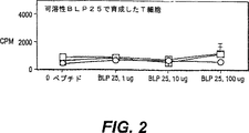
図2:3種の高応答動物の正常供与体からのT細胞増殖応答から得た代表試験。これらのT細胞を、可溶性ペプチドBLP−25および自己由来APCの存在下で育成した。自己由来APCの存在下で可溶性ペプチドに対するT細胞増殖応答を測定した。T細胞を、1μg□、10μg○、および
![]()
図3:リポソームBLP−25で負荷された自己由来APCで刺激したT細胞の抗原特異性。正常な供与体のPBLから単離されるT細胞を、2週間、PLB−25(10μg)を含むリポソームで負荷した自己由来PBLの存在下で培養した(「材料および方法」に示すとおり)。これらのT細胞を、(3A)リポソーム抗原ペプチドまたは(3B)溶解性ペプチド負荷自己由来APCに対するそれらの増殖応答について試験した。不適切な抗原対照として、HLA.Aw68.1(残渣61〜84)からの24種のアミノ酸ペプチドを可溶性形態で使用した。
図4:CD4、CD8およびMHCクラスI分子に対して特異的であるMabによる抗原ペプチド特異的T細胞の増殖応答の遮断。全ての遮断Mabおよびイソタイプ対照抗体を、20μg/mlで使用した。T細胞を、「材料および方法」に示すとおり、2週間、BLP−25(10μg)で負荷した自己由来APCの存在下で培養し、そして増殖応答を、抗体を伴うかまたは伴わないBLP−25(10μg)で、またはリポソーム負荷APCを含む対照ペプチドBLP24Mで負荷した自己由来APCについて試験した。
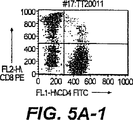
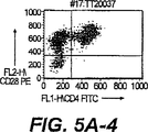
図5A:自己由来APCに負荷したリポソームBLP−25(10μg)で培養したT細胞の表現型。CD4+およびCD8+T細胞の両方の存在が観察された。T細胞の全ては、CD28+であった。上部(I)および下部(II)パネルは、2種の別々の供与体から得た培養T細胞から得たデータを表す。
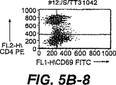
図5B:空のリポソームを含む自己由来APC(左のパネルI、抗原なし)、またはリポソームを含むBLP−25(10μg)(右のパネルII)の存在下で培養したT細胞の表現型(TCRおよび活性化分子)。パネルIに比べてより高い含有率のCD25+およびCD69+T細胞は、これらのT細胞の抗原特異的刺激を表す。
全てのドットブロットグラフでのマーカーを、イソタイプ対照抗体染色細胞の98%以上を排除するための手段で設定し、同様の方法で処置した(イソタイプ対照データは示されず)。培養細胞の同定を確認するためにロイコゲート(CD14/CD45)およびTCR(CD3/αβ)染色を行った。
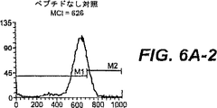
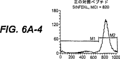
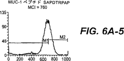
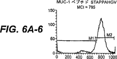
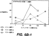
図6A:MUC−1ペプチドSTAPPAHGVおよびSAPDTRPAPによってT2変異体細胞でのHLA.A2発現の上昇制御。SIINFEKLを、正の対照として使用し、そしてH−2Kb(61−69)ペプチドを、HLA−A2上昇制御のための負の対照として使用した。x軸は、蛍光強度を表し、そしてy軸は、細胞数を表す。HLA.A2の増加発現を可視的に試験するためにマーカーM1およびM2を設定した。平均チャネル強度(MCI)を、各ヒストグラムに示す。
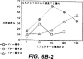
図6B:自己由来APCに負荷したリポソームBLP−25(10μg)で培養したT細胞の細胞毒性活性。3種の供与体全てが、HLA.A2+であった。標的は、指示ペプチドで負荷したT2細胞であった。負の対照から得た51Cr放出、SIINFEKL負荷T2細胞を、各データの点から減じた。
図6C:抗−HLAクラスIMAb(W6/32)は、STAPPAHGV負荷T2細胞のT−細胞(供与体番号2由来)による死滅を阻害する。供与体番号1および番号3でこの遮断試験を行って、同様の結果を伴った。イソタイプ対照抗体は、W6/32と同様の濃度であった。
図7:固相で希釈画分を被覆することによって検出されるときの18 22分溶出、BCP8反応性を示す一次RP−HPLCクロマトグラム。GαMIgG2bHRP基礎ELISAを介して結合BCP8MAbを検出した。MCF−7由来HLAクラスI分子(W6/32精製)を、酸溶出させ、そしてZORBAX C8マトリックスを用いたRP−HPLCにかけた。
図8:図1でBCP8反応性について同定した17分ピークの二次ZorbaxC8RP−HPLCの結果。貯蔵BCP8正の画分の二次RP−HPLCでの16−19分での強力なBCP8反応性。
図9:合成MUC−1ペプチドによる、単離ペプチドに対するBCP8の結合の阻害。
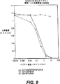
図10:二次RP−HPLCから得た17−19分ピークのBCP8親和性単離によってサンプルを得た。親和性単離ペプチドを、BCP8から酸溶出させ、そしてZORBAX C8で再クロマトグラフィーにかけた。電気スプレー質量スペクトルは、酸溶出の間、より長い配列の分解産物であると推定される、MHCクラスIタンパク質から溶出した3つのフラグメントを示す。これらのフラグメントは、MUC−1ムチン(配列データから得た。示さない)に関連した唯一のものであった一方で、他は起源が分からないものであった。
図11:間接染色手段で、モノクローナル抗体MA2.1(抗−HLA.A2.1)を用いてT2細胞で測定した蛍光の平均チャンネル強度(δMCI)(=MCI(サンプル)−MCI(未供給対照))での改変。20μg/mlのβ2マイクログロブリンの存在下で、MUC−1縦列反復から得た合成9マーまたは10マーの40μモルを用いて、3×105T2細胞を一晩培養した。正の対照ペプチドFLPSDYFPSV δMCI=951.3。FLPSDYFPSVも示される。
図12:MUC−1ペプチド刺激T細胞の細胞毒性活性。データを、3種のIILA.A2+供与体で示す。
詳細な説明
本発明は、活性化T細胞の生成に関する。異なる実施形態は、抗原提示細胞として、(a)無垢(naive)または免疫性欠如(anergic)のT細胞または両方の混合物と、(b)リポソーム被包抗原と、(c)自己由来性全末梢血リンパ球(PBL)(autologous whole peripheral blood lympocyte)とを用いて活性化細胞を生成することに関与する。
好ましい実施形態では、本発明は、MUC−1ペプチド特異的で、活性化されたCD4+およびCD8+T細胞の集団の生成に関し、それは、無垢のT細胞を、先にリポソーム被包ペプチド抗原で負荷したPBL(APCとして)で活性化させてインビトロで生成される。一般に、得られた抗原特異的CD4+およびCD8+T細胞は、活性化T細胞と関連した表層分子を有し、そして培養物中に高濃度のγ−IFNおよび中程度の濃度のIL−10を産生するが、ほんの痕跡量のIL−4を産生する。
本明細書で使用される場合、「活性化T細胞(activated T-cell)」は、細胞周期のG1相、S相、G2相またはM(有糸分裂)相にあるものである。したがって、「活性化T細胞」は、有糸分裂および/または細胞分裂を行いつつあるものである。活性化T細胞は、Tヘルパー(TH)細胞または細胞毒性T細胞(細胞毒性Tリンパ球(CTLまたはTC))であってよい。無垢のT細胞の活性化は、APC(抗原/MHC複合体を含む)に、そしてIL−1、IL−2、IL−12、IL−13、γ−IFNおよび類似のリンホカインのような分子にこのような細胞を露出することによって開始されうる。抗原/MHC複合体は、T細胞の表面にあるレセプター(T細胞レセプター(TCR))と相互作用する。Golubら編、Immunology:A Synthesis,第2章:「The T-cell Receptor」(1991年)。
本明細書で使用される場合、「初回感作(priming)」は、活性化および/または記憶を生じるようなやり方で、抗原に動物(ヒトを含めた)または培養細胞を露出することを意味するために使用される。標的抗原に対するCD4+およびCD8+T細胞応答の生成は、通常、自然の感染を介するか、または意図的免疫化を介するかのいずれかでインビボ感作による。
本明細書で使用される場合、「無垢な(naive)」T細胞は、外来抗原(非自己由来性)抗原に露出されなかったものであるか、または潜在性自己由来抗原に露出させなかったものである。「無垢な」T細胞は、しばしば、「未感作(unprimed)」T細胞と称される。当業者は、「休止(resting)」細胞が、細胞周期のG0相にあり、したがって、有糸分裂を分割または経過していないことを認識できる。当業者は、さらに、「免疫性欠如(anergic)」T細胞が、特性を機能させる能力がないものである、すなわち正常な免疫応答を指向する抗体を欠く細胞のようなものであることも認識できる。疾患に罹った患者から得たT細胞は、感作されたが、免疫性を欠如するT細胞を含む可能性がある。
当業者は、適切な補助分子が、T細胞の活性のために使用される可能性もあることを認識する。補助分子は、T細胞レセプターにおいて抗原−MHC相互作用を促進する分子である。補助分子は、それに限定されないが、当初の結合を促進または増大させること、結合を安定化すること、シグナル移入および分離を含めた様々な役割を示す。このような補助分子の例としては、限定されないが、B7.1(CD28に結合する)、B7.2(CD28に結合する)、およびICAM−1(LFA−1に結合する)が挙げられる。
他の実施形態では、本発明は、抗原提示細胞として、リポソーム被包抗原および自己由来性末梢血リンパ球(PBL)と一緒に、無垢のT細胞、記憶T細胞、および免疫性欠如T細胞、またはそれら3種の細胞型の混合物を用いた活性化T細胞の生成に関する。本明細書に使用される場合、「記憶表現型(memory phenotype)」T細胞としても知られる「記憶T細胞(memory T-cell)」を、先にペプチド抗原に遭遇したが、現在休止しており、そして活性化される能力のあるT細胞のクラスを明示するために使用する。記憶T細胞は、抗原に露出されて、そしてその後、刺激抗原の存在なしに体内で、長期にわたって生存するT細胞である。しかし、これらの記憶T細胞は、「回想(recall)」抗原に応答する。
一般に、記憶T細胞は、ペプチド抗原に対する無垢なT細胞応答と比較した場合、「回想」抗原にいっそう敏感である。記憶細胞を、CD45R0、CD58、CD11α、CD29、CD44およびCD26のような、分化T細胞用のマーカーであるある種の細胞表面抗原の存在によって認識することができる。
当業者に既知の技術によって、記憶T細胞を単離する。簡便には、総T細胞集団を単離し、続いて抗CD45R0、抗CD44または抗CD26のモノクローナル抗体を用いた蛍光活性化細胞ソーティング(FACS)を行う。Hollsbergら、Cellular Immunology 149巻:170頁(1993年);Brunoら、Immunity 2(1)巻:37頁(1995年);およびJournal of Immunology 150巻(パート1):3119頁(1993年)参照。
A.抗原
1 一般的に有用な抗原
抗原特異的MHCクラスIIおよびクラスI制限CD4+およびCD8+T細胞応答は、種々の病原性症状に対する重要な宿主免疫応答である。特に興味があるのは、抗原特異的T細胞応答の生成である。本明細書において使用される場合、「抗原特異的」T細胞応答とは、異なるアミノ酸配列を有するペプチド(対照ペプチド)のような他の刺激によっては明らかでない、ペプチドのようなある特定の抗原刺激物に対するT細胞応答(増殖、細胞毒性、サイトカイン分泌)である。限定されないが、CD25およびCD69を含めたT細胞活性化の特徴である細胞表面分子の外観を評価することによってT細胞の応答性を測定する。そのようなアッセイは、当該技術分野において知られている。
本方法は、一般に、非常に様々な抗原に使用する。これらの抗原は、それがT細胞特異的免疫応答を引出すことができる限り、ほとんどあらゆる化学構築物のものでありうる。それらは、少なくとも1種のT細胞特異的エピトープを含みうる。例示の抗原は、ペプチド、炭化水素、脂質および特にそれらの組合せから誘導しうる。特に重要な抗原は、ペプチド、リポペプチドおよびグリコペプチドである。イデオタイプおよび抗イデオタイプが特に含まれる。
それに対する対象の方法を使用するのに非常に有利である抗原としては、腫瘍抗原が挙げられる。腫瘍抗原は、通常、腫瘍の存在と相互に関係している天然または外来の抗原である。腫瘍抗原が正常な組織から異常組織を分化させるのに有用である限り、それらは、診断のみならず、治療を行うのための標的としても有用である。したがって、腫瘍抗原に対するT細胞特異的免疫応答を生じる本発明の方法を使用することは、本発明の重要な態様である。
腫瘍抗原は、当業界で既知である。実際、数種の例は、十分に特徴づけされ、そして最近、腫瘍特異的療法を生じさせる上で非常な興味の対象となっている。腫瘍抗原の非限定的な例としては、癌胎児性抗原(CEA)、プロステート特異的抗原(PSA)、メラノーマ抗原(MAGE、BAGE、GAGE)、およびMUC−1のようなムチンである。
MUC−1ムチン抗原は、多数のアデノ癌腫に対する免疫性を生じる強力な免疫療法標的として認識してきた。Longeneckerら、Immunologists 1巻:89頁(1993年)。したがって、本発明の1つの実施形態は、APCの表面にあるクラスIおよびクラスII分子のいずれか、または両方に結合する能力がある「MUC−1誘導体」に関する。
「MUC−1誘導体」は、一般に、ペプチドまたはペプチドを基礎とするものである。1つの実施形態では、このペプチドは、MUC−1のコアのタンデム反復を含む。さらに他の実施形態では、本発明は、配列STAPPAHGVTSAPDTRPAPGSTAPPをもつこのコア領域から得られる25種のアミノ酸ペプチドに関する。このコア領域は、誘導体がT細胞活性化の特徴を残す方法で、以下に詳細に記述される修飾を行うこともできる。
MUC−1誘導体は、MUC−1タンパク質のフラグメントである可能性がある。このようなフラグメントは、グリコシル化されていても、またはグリコシル化されていないものでもよい。本発明にしたがって、本発明の範囲内のフラグメントは、精製MUC−1または、ペプシンまたはパパインのようなプロテアーゼでの消化を含む方法を用いて、組換えDNA法によるMUC−1から得ることができる。もちろん、MUC−1フラグメントは、組換え法によって直接的に作製することもできる。さらに、本発明に含まれるMUC−1フラグメントは、アプライド・バイオシステムズ(Applied Biosystems)によって市販で供給されるもののような自動ペプチド合成機、多重ペプチドシステムおよび他のものを用いて合成することができるか、または当業界で既知の技術を用いて、手作業により製造することができる。Geysenら、J.Immunol.Methods 102巻:259頁(1978年)参照。
MUC−1誘導体としては、グリコシル化または非グリコシル化合成ペプチドも挙げられる。さらに、本発明の範囲内のMUC−1誘導体としては、タンパク質分解性切断耐性MUC−1フラグメントまたは、D−アミノ酸のような1種またはそれ以上の非天然のアミノ酸を含むMUC−1フラグメントが挙げられる。そのような誘導体は、有益なT細胞特異性を残しながら、増加した半減期の循環の利益を得ることが予測される。
他の実施形態では、MUC−1誘導体は、アミノ酸配列DTR(Asp−Thr−Arg)またはDTRP(Asp−Thr−Arg−Pro)を伴う、MUC−1の細胞外縦列反復領域の一部を含む。好ましくは、これは、配列SAPDTRP(Ser−Ala−Pro−Asp−Thr−Arg−Pro)を含む。特に好ましいペプチドは、TSAPDTRPAである。
いくつかの好ましいMUC−1誘導体は、基本的に、MUC−1ムチンの1種のペプチドコアの反復体から構成される。元来のMUC−1分子中のMUC−1ペプチドコア反復体は、20種のアミノ酸配列PDTRPAPGSTAPPAHGVTSA(Pro−Asp−Arg−Thr−Pro−Ala−Pro−Gly−Ser−Thr−Ala−Pro−Pro−Ala−His−Gly−Val−Thr−Ser−Ala)を含む。有用な合成誘導体としては、この配列の「直鎖の順列(linear permutations)」、例えば、反復が単にPDTRよりむしろGVTSまたはTSAPで始まるGVTSAPDTRPAPGSTAPPAHまたはTSAPDTRPAPGSTAPPAHGVが挙げられる。他の類似の順列も可能である。
さらに、不可欠のT細胞活性化活性が維持されるように、コア配列の1種またはそれ以上のアミノ酸を、好ましくは当業界で知られる保存的手段で改変しうる。以下の群のアミノ酸:(a)G、A、V、LおよびI、(b)GおよびP、(c)S、C、T、M、(d)F、YおよびW、(e)H、KおよびR、(f)D、E、NおよびQの中で、典型的な置換を行うことができる。ある種の好ましい置換は、以下の群:(i)SおよびT;(ii)PおよびG;および(iii)A、V、LおよびIによって行うことができる。
他のMUC−1誘導体は、基本的に、MUC−1ムチンの1種の切断ペプチドコアの反復体、例えばGVTSAPDTRPAPGSTAから構成される。もちろん、この切断コア配列は、上述のとおり変更、そしてさもなければ改変できる。
いくつかの実施形態は、コア反復体およびそれの誘導体(上述のとおり)の多量体(multimer)を意図する。多量体は、同じコア反復体または誘導体の複数コピーを含むことができるか、またはそれらは、混合および組合わせることができる。これらの多量体は、大きさを厳密に限定しないが、通常、それらの免疫刺激特性を失い、そして、反復の数が増加するにつれて、免疫抑制をも現実に起こしうる。したがって、使用される反復の数は、3より少ないことが好ましい。いくつかの好ましいMUC−1誘導体は、MUC−1コアペプチドSTAPPAHGVTSAPDTRPAPGSTAPPから誘導される7−20のアミノ酸長ペプチドの1から3のコピーを含む。
上述のとおり、これらの好ましいMUC−1誘導体は、当業界で知られる方法にしたがって、グリコシル化されるか、または部分的にグリコシル化されうる。さらに、MUC−1およびMUC−1誘導体を、ポリエチレングリコールのような大型分子量高分子で修飾することができる。さらに、脂質修飾は、それらがリポソームを有する誘導体の被包または相互作用を促進しうるので好ましい。この目的のために有用な例示の脂質部分としては、限定されないが、パルミトイル、ミリストイル、ステアロイルおよびデカノイル基が挙げられるか、またはさらに一般的には任意のC2からC30の飽和、モノ飽和またはポリ不飽和脂肪族アシル基が挙げられる。
化学修飾するのに好都合には、MUC−1ペプチドに修飾になじみ易い側鎖を有する1種またはそれ以上のアミノ酸を含めることのがしばしば有用である。好ましいアミノ酸は、6−アミノ酸で容易に修飾できるリシンである。アスパルテートおよびグルタメートの側鎖カルボキシルは、セリン、トレオニンおよびチロシン水酸基、シスチンスルフィニル基およびヒスチジンアミノ基である場合、容易に修飾する。
本発明の範囲内のMUC−I誘導体の例示も、非ペプチド「模擬」、すなわちMUC−1タンパク質の1つまたはそれ以上の機能的特徴を模倣する化合物である。模擬は、一般に、水溶性で、タンパク分解に耐性があり、そして非免疫性がある。形態的に制限した、MUC−1を模倣する環状有機ペプチドは、例えばSaragoviら、Science 253巻:792頁(1991年)によって記述される公知の方法によって生成できる。
「MUC−1炭化水素誘導体」をも意図する。ここに使用される場合、このような誘導体は、MUC−1誘導体の免疫刺激特性を残すグリコペプチドに該当する。このような炭化水素誘導体は、MUC−1タンパク質に付着する炭化水素の全てまたは一部を含むことができる。MUC−1炭化水素の少なくとも1つの特性を模倣する模擬物も使用できる。
当業者は、活性化T細胞を生成させるために他の抗原を使用できることを認識する。このような抗原の例としては、限定されないが、非自己(外来)ペプチド抗原、およびウイルス、腫瘍、細菌または他の病原体由来のペプチド抗原が挙げられる。
2.他の有用な抗原の同定
本方法に有用な全抗原を、種々のT細胞応答を測定する確認済みの方法を用いて同定することができる一方、特定のエピトープと関連したさらに特異的な応答を生成させることも有用である。このアプローチにより、より小さく、いっそう経済的に製造される抗原性刺激剤の使用が可能になる。したがって、好ましい抗原は、小型分子、特に、約100アミノ酸未満、そして通常約60アミノ酸未満の桁数でのペプチドまたはペプチド誘導体である。
当該技術分野において知られる方法によって、いったん天然(大型)の抗原を同定したら、その抗原性は1個または数個の特異的エピトープに対するものに高めることができる。1つの古典的な方法は、より小型の抗原を誘導する大型の抗原のタンパク分解処理を含む。さらに、組換えDNA技術によってタンパク質抗原のフラグメントを製造し、分析して、特定のエピトープを同定できる。さらに、インビトロ合成法によって小さいペプチドを製造し、分析できる。
手をつけていない(intact)抗原の部分を作成し、それらを分析するランダムアプローチに対する代替法として、さらに生物学的に関連の深いアプローチが可能である。MHCクラスIおよび/またはクラスII分子に結合する抗原性フラグメントは、特に重要であるので、1つの例示のアプローチは、MHC分子自身を単離し、そしてその後それに関連したペプチドを単離することである。一般に、この方法によって、腫瘍抗原の特に有用なエピトープをさらに規定することがうまくできる。
典型的な方法では、目的の抗原を発現する一次腫瘍細胞またはセルラインのいずれかを提供する。さらに、マクロファージのような食作用性抗原提示細胞(またはあらゆるAPC)に大型抗原(またはそれの一部)をあたえることができ、したがってこれらの細胞は、これらの方法の出発材料として使える。MHCクラスIまたはクラスII分子は、抗体アフィニティー(MHC特異的抗体)およびクロマトグラフィー技術のような公知の方法を用いてこれらの出発細胞から単離することができる。
その後、単離MHC分子を、処理して結合ペプチドを放出させる。これは、例えば、洗剤、尿素、塩化グアニジニウム、二価陽イオン、種々の塩、そしてpHで極値のもののような結合ペプチドおよびMHC分子の間の相互作用を中断させる剤で処置することによって達成できる。放出されるペプチドは、常套のクロマトグラフィーおよび抗体アフィニティー(抗原特異的抗体を用いた)方法論を用いてさらに精製することができる。その後、例えば、ペプチドシーケンシング、ガスクロマトグラフィーおよび/または質量光学顕微鏡を用いて、精製ペプチドを配列および構造決定にかけることができる。
この方法では、クラスIおよび/またはクラスII分子に関連した最も有効なペプチドエピトープの配列/構造を決定できる。この配列/構造の情報で供給されると、上で詳述されるとおり、決定配列の変更を行い、公知のT細胞アッセイを用いて分析することができる。
以下に示される例で、この方法をMUC−1系に適用した。MUC−1コア反復の一部に対応する7マー配列TSAPDTRを、有効なクラスI関連ペプチドとして同定した。GVTSAPDTR、VTSAPDTRP、TSAPDTRPA、SAPDTRPAP、APDTRPAPG、PDTRPAPGSおよびDTRPAPGSTを含む関連する9マーのペプチドを作成した。これらの内の各々を、MUC−1特異的細胞毒性T細胞応答について分析し、そしてTSAPDTRPAが例外的によく作用することが分かった。したがって、この配列は、本発明によってMUC−1特異的活性化T細胞を生成する好ましい抗原を表す。
B.リポソーム被包抗原
本発明の1つの実施形態では、リポソーム中に抗原を被包する。リポソームを製造し、リポソーム中にペプチドを含めた種々の分子を被包する技術は、技術者にとって既知である。リポソームは、水性区分を囲む1つまたはそれ以上の脂質二重層から構成される顕微鏡下の小胞である。一般に、Bakker−Woudenbergら、Eur.J.Clin.Microbiol.Infect.Dis.12巻(補足版1):S61(1993年)、およびKim,Drugs 46巻:618頁(1993年)参照。リポソームは、細胞膜に対する組成で類似し、そして結果として、リポソームは、一般に、安全に投与することができ、そして生物分解性がある。
製造の方法によって、リポソームは、単層または多層であってよく、そして0.02μmから10μmより大きい範囲にある直径を示す大きさで変化しうる。種々の剤は、リポソーム中で被包することができる。内部水性空間(複数の空間)内の二層中の疎水性剤部分および親水性剤部分。例えば、Machyら、LIPOSOMES IN CELL BIOLOGY AND PHARMACHOLOGY(John Libbey、1987年)およびOstroら、American J.Hosp.Pharm.46巻:1576頁(1989年)参照。
リポソームは、実質的にあらゆる型の細胞に吸着でき、そしてその後被包剤を放出する。代替的に、リポソームは、標的細胞を融合し、それによってリポソームの内容物が、標的細胞に注ぐ。代替的に、吸収リポソームを、食作用のある細胞によって細胞内取込みを行うことができる。リポソーム脂質のリソソーム内分解および被包剤の放出によって、細胞内取込み作用を行う。Scherphofら、Ann.N.Y.Acad.Sci.、446巻:368頁(1985年)。
以下の手段は、多重層(MLV−型)、pH無感応なリポソームを製造するのに使用できる。リポソームの塊状液体組成物は、約3:1:0.25のモル比で、約30mMの最終総脂質濃度で、ジパルミトイルホスファチジルコリン(DPPC)、コレステロール(Chol)およびジミリストイルホスファチジルグリセロール(DMPG)(ジェンザイム(Genzyme)、マサチューセッツ州ケンブリッジ)を含む。モノフォスホリル脂質A(MPLA)(RIBI Immunochm Research Inc.)、モンタナ州ハミルトン)(またはアバンティ・リキッドA(登録商標);Avanti Polar Lipids,Inc.);エイ・アール 35007、アラバスター、インダストリアル・パーク・ドライブ700番)は、約1%から約5%(w/w)のバルク脂質の濃度で脂質混合物に含まれ、そしてリポペプチド濃度は、約50から約1000μg/mlである。MPLAは、有効なアジュバントとしての役割を果たして、Tリンンパ球に特異的なAPCによってリポソーム抗原の出現が増加することを生じることが示された。Alving.C.R.、Immunobiol.187巻:430−446頁(1993年)(21)。
技術者は、デトックス・アルム、QS21、完全および/または不完全フロイントアジュバント、MDPおよびリピッドAのような他のこのようなアジュバントも適切であることを認識する。塊状脂質、MPLAおよびリポペプチド(約12mLの最終産物について約192mgDPPC、約33mgChol、約15mgDMPG、約2.4から12mgMPLAおよび約0.6から12mgのBLP−25(MUC−1から25aaペプチド)を、約5.3mLのエタノールに溶解する。本明細書に使用されるとおり、リポペプチドは、パルミチン酸、ミリスチン酸および同等物のようなアミノまたはカルボキシ末端脂質部分を含むペプチドである。
1つの実施形態では、BLP−25(マルミトイル化MUC−1ペプチド誘導体)のようなリポペプチドは、リポソームでの被包のための抗原ペプチドとして使用する。したがって、MUC−1のこのリポペプチド誘導体を、MPLXと一緒に、小型のpH不感応リポソームに被包する。
エタノール溶液を約50℃に暖め、そして30g針を介して、同じ温度で迅速に攪拌したPBSのような約100mLの適切な緩衝液に注入する。生じたリポソーム懸濁液(主に小型の単層小胞、SUV)は、エタノールを激減させ、そして約300kDの分子量カットオフ(MWCO)を示すサルトリウス細胞でダイアフィルトレーションによって濃縮される。最初、容量を約10−20mLに減少させ、そしてその後、その産物を、約100mLのPBSでダイアフィルトレーション物を連続置換することによって洗浄する。容量を、約12mL未満まで減少させ、そしてダイアフィルトレーションセルから除去した後、約12mLの最終容量まで再構築する。
都合により、その後、産物を、3回、20,000ポンド/平方インチでフレンチ圧力セル(SLM Aminco、ニューヨーク州ロチェスター)に通過させて、全てのリポソーム粒子を、滅菌に使用される0.22μmフィルターを通過する大きさに減少させることを確認する。サイズ分析では、平均粒子サイズは、わずかに0.1μmより下であることが示される。
陰イオンリポソームベクターも試験した。これらには、細胞内取込みおよびエンドソーム酸化を伴うエンドソーム膜を分離または融合するpH感受性リポソームが含まれる。
リポソームベクターの中でも、陽イオン性リポソームは、インビトロで哺乳類細胞感染を指示する上でのそれらの効力のため、最も研究されている。それらは、しばしば、核酸の送達に使用されるが、他の治療剤の送達に使用することができる。それらは、医薬またはホルモンである。
陽イオン性脂質は、これらの複合体が、インビボで、陰イオン分子が豊富である生理学的環境に不適合であるように見える場合、自然には見られず、そして細胞毒性である可能性がある。リポソームは、細胞内皮系に優先的に食作用される。しかし、細胞内皮系を、大用量のリポソーム粒子での飽和、または製薬学上の手段による選択的マクロファージ不活性化を含めた数種の方法によって包囲する。Classenら、Biochim.Biophys.Acta.802巻:428頁(1984年)。さらに、グリコリピッドまたはポリエチレンゲル誘導リン脂質をリポソーム膜に取込むと、細胞内皮系による摂取が明らかに減少することが示された。Allenら、Biochim.Biophys.Acta 1068巻:133頁(1991年);Allenら、Biochim.Biophys.Acta 1150巻:9頁(1993年)。
陽イオンリポソーム調製物は、常套の方法論によって作成できる。例えば、Felgnerら、Proc.Nat’l Acad.Sci USA 84巻:7413頁(1987年);Schreier、J.of Liposome Res.2巻:145頁(1992年);Changら、上記(1988年)参照。リポフェクチン(Lipofectin▲R▼)Life Technologies,Inc.、米国メリーランド州ゲイザースバーグ)も利用可能である。リポソームの量およびDNAの量は、用量応答曲線に基づいて各細胞型について最適化することができる。Felgnerら、上記。使用した方法についてのある種の最初の検討については、Wassefら、Immunomethods 4巻:217−222頁(1994年)およびWeiner,A.L.、Immunomethods 4巻:217−222頁(1994年)参照。
本発明の方法で使用される他の適切なリポソームとしては、多重層小胞(MLV)、オリゴ層小胞(OLV)、単層小胞(UV)、小型単層小胞(SUV)、中程度の大きさの単層小胞(MUV)、大型単層小胞(LUV)、巨大単層小胞(GUV)、多重層小胞(MVU)、逆相蒸散法(REV)によって生じる単一またはオリゴ層小胞、逆相蒸散法によって生じる多重層小胞(MLV−REV)、安定な複数層小胞(SPLV)、凍結および解凍MLV(FATMLV)、押出法によって製造した小胞(VET)、フレンチプレスによって製造した小胞(FPV)、溶解よって製造した小胞(FUV)、脱水−再水和小胞(DRV)、およびバブルソーム(BSV)が挙げられる。技術者は、これらのリポソームを作成するための技術が当業界で既知であることを認識する。COLLOIDAL DRUG DELIVERY SYSTEMS、66巻(J.Kreuter編、Marcel Dekker,Inc.、1994年)参照。
C.T細胞
技術者に既知の技術を用いて、無垢の、記憶および免疫性欠如のT細胞を作成する。例えば、以下の手段を使用する。T細胞増強については、約30−50×106PBLを、1mLのAIM−V培地に懸濁させ、そして予備調整した5mLナイロンウールカラム(ロビンズ・サイエンティフィック、カリフォルニア州サニーバール)を培地に負荷する。負荷ナイロンウールカラムを、約37℃で、約45分間インキュベートし、そしてその後、穏和な(約37℃)AIM−V培地で洗浄することによって、非粘着T細胞を溶出する。溶出T細胞を、「無垢の」または免疫性欠如T細胞として使用する。
技術者は、他の既知の技術が、T細胞を製造するのに使用できることを認識する。多くのこのような技術は、CURRENT PROTOCOLS IN IMMUNOLOGY(John E.Coligan編、John Wiley & Sons、ニューヨーク、1991年)の3.1.2から3.6.4頁に記述されている。市販で入手可能なカラムを、T細胞増強のために一般に使用し、そのいくつかは、CD4+およびCD8+T細胞の増強に特異性がある。他の方法としては、ダイナビーズ(DYNAL、レイク・サクセス、ニューヨーク11042)およびMiniMACS(ミルテニ・バイオテック,インク.(Milteni Biotec.Inc.)、カリフォルニア州95603アーバン)のような親和性ビーズが挙げられる。
D.抗原提示細胞
本発明の1つの実施形態では、末梢血リンパ球(PBL)をAPCとして使用する。PBLを、当業界で認識された手段を用いて単離する。例えば、「軟層」を、フィコール−ハイパーク勾配遠心分離のような方法を用いて末梢血サンプルから収集し、他の構成要素からPBL(末梢血リンパ球)を分離するのに使用する。CURRENT PROTOCOLS IN IMMUNOLOGY(John E.Coligan編、John Wiley & Sons、ニューヨーク、1991年)の7.0.5から7.1.5頁に記述される技術を参照のこと。
E.活性化T細胞の作成
PBL、無垢なまたは免疫性欠如のT細胞およびリポソーム被包抗原を、上述のとおり作成する。PBLの内のいくつかを凍結し、そして後期再刺激用の自己由来APCとして使用する。全細胞のインキュベーションを、約37℃の温度を示し、そしてCO2および湿気を供給したインキュベーターで行った。1つの実施形態では、活性化のために使用したT細胞およびAPCのために使用したPBLは自己由来のものである。したがって、本発明の1つの実施形態で、以下の手段を、活性化T細胞を作成するのに使用する。
1)PBLを、リポソーム被包抗原と混合して、リポソーム抗原負荷PBLを作成する。例えば、0.9mlのAIM V(血清不含リンパ球培地)(ライフ・テクノロジーズ)中の約104から約109PBLS(例えば、〜2×10⁶PBL)を、リポペプチド配合物(例えば、PBS中で0.1mlの容量で約0.1μgから約1mgリポペプチド、および約0.1μgから約1mgのMPLCのような脂質)を含む1用量のリポソームに添加し、そしてCO2供給インキュベーター中で約37℃で、約1時間から約18時間インキュベートする。他の適切な細胞培地は、当業界で既知である。このような培地としては、限定されないが、RPMI 1640、DMEMおよびマッコイのものが挙げられる。すなわち、PBLを、マイトマイシンC(または3000ラッドでのガンマ照射)で処理し、そしてその後洗浄する。
2)(〜1×10⁶細胞および1×10⁶リポソーム抗原負荷PBL)/ml(段階(1)から)を、25cm2組織培養容器(フラスコまたはプレートのような)中で総量約〜10ml AIM V培地に懸濁させ、そして37℃インキュベーターに入れる。T細胞およびPBLを、ウエル当たり約103から107個の細胞の範囲内で使用できる。
3)約24時間後、組換えIL−7(10ng/ml)(インターメディコ、オンタリオ州マークハム)およびIL−12(100pg/ml)(R & D Systems)、ミネソタ州ミネアポリス)を、段階(2)の混合物に添加し、そしてCO2を用い、37℃で5−7日間インキュベートする。IL−12は、生成中のCD4+T細胞のサイトカインプロフィールでのTH1パターンへの移動を好むことが示された。Hsiehら、Science、260巻:547−549頁、1993年(27)。技術者は、TH1応答パターンが、IL−4、IL−6およびIL−10の相対的不在下でIL−2およびガンマ−IFNの産生によって特徴づけられることを認識する。IL−12も、CD8+T細胞の一次活性化の間じゅうγ−IFN産生の導入で共同に作用する。Gajewskiら、J.Immunol.154巻:5637−5648頁、1995年。組換えIL−7(rIL−7)は、前駆体CTLの成長および分化を増強することが先に例示された。Aldersonら、J.Exp.Med.、172巻:577−587頁、1990年;Kosら、Eur.J.Immunol.、22巻:3183−3185頁、1992年。
他の適切なサイトカインも、この段階に使用する。例えば、IL−1、IL−2、IL−4、ガンマインターフェロンまたはIL−15を、あらゆる組合わせで使用する。適切な組合わせとしては、限定されないが、IL−2とIL−4;IL−2とIL−5;IL−2とガンマ−IFN;IL−2とIL−1が挙げられる。技術者は、活性化に使用したサイトカインの濃度および組合わせは、細胞、実験条件および選択したサイトカインによって変化することを認識する。最適なT細胞刺激について分析によって、最適レベルを測定しうる。
4)段階(3)に続いて、全T細胞およびPBLを収集し、そして新鮮なAPCを添加し、そして混合物を、約24時間インキュベートする。この段階で添加したAPCを、段階(1)で記述されるとおり、ペプチド抗原で負荷したリポソームで負荷する。
5)段階(4)の後、追加のIL−7(約1から約50pg/ml)およびIL−12(約10から約500pg/ml)を培養用培地に加える。1つの実施形態では、10ng/mlのIL−7および100pg/mlのIL−12を使用する。
6)細胞混合物を、37℃で、CO2および湿気を用いて、インキュベーターで約5−7日間インキュベートする。この時点で、上清をサイトカインスクリーニングのために収集する。このような材料を、スクリーニングに使用するまで、−80℃で保持する。
7)取得物である活性化T細胞を、技術者に既知の手段を用いて収集する。典型的な手段では、組織培養フラスコに存在する細胞および培地を単離し、そして細胞を、AMV培地で二回洗浄し、そして収穫細胞として使用する。代替的に、PBLを単離するためのものに類似するフィコールの手段を用いて、生細胞を分離する。
F.T細胞の特徴
1.増殖アッセイ
T細胞増殖アッセイは、当業界で既知である。このようなアッセイを、特異的結合抗原がT細胞増殖を刺激するかどうかを測定するのに使用する。例えば、AIM V培地中の約105T細胞および5×10⁴APC/ウエル/200μlを用いた96穴プレートを用いて、このようなアッセイを設定する。他のT細胞およびAPC濃度を使用できる。APCを、マイトマイシンCで予備処理して、増殖を阻害する。対照または試験溶解性ペプチドで負荷したAPC、およびペプチドまたはリポペプチドを含むリポソームを、T細胞の刺激のための剤として使用する。約5−6日後、細胞に、3H−Tdr(約1μCi/ウエル)でパルス標識する。パルス標識の12−18後、細胞を収穫し、そしてT細胞への3H−Tdr組込みを測定する。
適切な細胞収穫機が、フィルター紙上の細胞を収集する。フィルター紙に結合したか、または捕捉された放射活性を結合することによって、放射活性のT細胞への取込みを評価する。
2.T細胞表面でのCD抗原の評価
どのCD抗原が、活性化T細胞の表面に存在することを決定するために、当業界で認識されている技術を使用する。一般に、蛍光顕微鏡を介して、または血球計算法モードで使用される蛍光活性化細胞ソーター(FACS)を介してT細胞を「染色」するのにフルオロクロムで標識した抗−CD抗原抗体を、使用する。CURRENT PROTOCOLS IN IMMUNOLOGY(John E.Coligan編、John Wiley & Sons、ニューヨーク、1995年)1巻、第5章を参照のこと。
3.サイトカイン産生
当業界で認識された技術を用いて、細胞によるサイトカイン産生の測定を行う。技術者は、ELISA技術を用いて、細胞培養培地中でインターロイキンまたはインターフェロンのようなサイトカインの濃度を測定することを知っている。あるサイトカインが特異的細胞型の刺激を導くバイオアッセイによって、インターロイキンのようなサイトカインを検出する。適切なサイトカイン検出技術は、ELISPOTアッセイであり、そしてフローサイトメトリーを用いた細胞内サイトカイン決定法である。CURRENT PROTOCOLS IN IMMUNOLOGY(John E.Coligan編、John Wiley&Sons、ニューヨーク、1991年)1巻、第2章を参照のこと。
G.研究技術
本発明の1つの実施形態では、本発明は、抗原特異的T細胞応答を生成するのに有効である抗原およびエピトープを同定する方法に関する。この方法は、
(1)抗原およびエピトープ候補である種々のペプチドでの、適切な培養細胞(T細胞、PBL)を初回感作する段階と、
(2)段階(1)で感作した細胞の増殖およびサイトカイン産生を評価する段階とを含む。
増殖およびサイトカイン産生を、上述のとおり評価する。代替的に、サイトカイン産生を、以下の方法を用いて評価する。分析物(分析されるべきサイトカイン)に対する1つの抗体を、固相(96穴プレートのような)に付着させ、したがってサイトカインに結合(または捕捉)するのに使用する、サンドイッチ型ELISAアッセイを使用する。分析物について分析されるべきサンプルを、固相に露出する。結合および洗浄後、二次抗−分析物抗体(レポーター基を含む)を加える。適切なレポーター基としては、限定されないが、放射活性アイソトープ、蛍光基、および酵素(西洋ワサビペルオキシダーゼ(HRP)、アルカリ性ホスファターゼ、βガラクトシダーゼ)が挙げられる。レセプター標識抗体を、適切な時間インキュベートし、そして固相を洗浄して未結合レポーターを除去する。その後、公知の標準法でサンプル値を比較することによってサンプル(例えば、放射活性測定;色素原または比色評価(酵素接合抗体について))内の分析物の量を測定するのに適切な検出技術を使用する。
技術者は、十分に公知な技術を、ある抗原中の特異的アミノ酸の欠失および突然変異について使用することができることを認識する。このような技術を用いて、所望のT細胞活性化について必要とされるエピトープを同定する。例えば、重複配列地図作成技術を使用する。特に、候補抗原から得た9つのアミノ酸長ペプチドを、T細胞刺激活性について試験する。その時に公知のタンパク質配列を介して、1つのアミノ酸を進行することによって重複9aa配列を作成することによって、一連のペプチドを作成する。この方法では、最高のT細胞刺激活性を示すペプチドを同定する。
他の実施形態では、本発明は、
(1)目的の抗原に対するT細胞応答を生成し、
(2)細胞表面抗原相同性、サイトカイン産生、および技術者に十分に知られるパラメーターを評価することによって、T細胞応答を特徴づけること
を特徴とする、T細胞免疫応答を特徴づける方法に関する。例えば、1型、2型、3型、および0型は、公知のT細胞応答型である。
H.治療方法
本発明による方法を用いて、種々のペプチドを、免疫療法用の細胞ワクチンを生成するのに使用する。このようなワクチンは、例えば、自己由来PBLのようなペプチド抗原負荷APCである。さらに、抗原刺激T細胞を、「適合T細胞移入療法」として知られる技術であるワクチン化するのに使用することもできる。本発明によるワクチンを、(a)患者へのワクチン投与に伴う疾患発生の回避および/または(b)疾患に罹った患者の治療のために使用する。
例えば、BLP−25ペプチド抗原を、リポソーム内に組込み、そして上述のとおり、PBLに抗原を負荷するのに使用する。負荷PBLは、細胞ワクチンとして患者に注入しなおすことができる。負荷PBLも、上述のとおり、インビトロで自己由来T細胞を活性化するのに使用することができる。これは、抗原特異的T細胞の集団の拡張をする。その後、これらの活性化T細胞を、例えばアデノ癌腫に罹った患者に再投与する。適合T細胞移入療法について当業界で認識されている技術の説明については、Bartelsら、Annals of Surgical Oncology、3(1)巻:67頁(1996年)を参照のこと。
本発明によるT細胞活性化法は、免疫療法でワクチンとして使用するため、または癌、腫瘍、ウイルス感染、および細菌感染のような種々の病態のための候補である抗原に対する細胞毒性およびヘルパーT細胞応答を生じさせるために使用される。したがって、本発明による方法を使用して、候補抗原を選択し、リポソームに被包し、自己由来PBLを負荷するのに使用し、そして負荷PBLを患者に注入するか、または代替的に、PBLを、自己由来T細胞を活性化するのに使用する。刺激されたT細胞を、前述の障害の内の1つに罹っている患者に再投与する。技術者は、ペプチド抗原が、患者に影響を及ぼす疾患の型に基づいて選択されることを認識する。例えば、MUC−1抗原を、アデノ癌腫の治療に使用される抗原特異的T細胞を作成するのに使用する。他の例としては、限定されるものではないが、以下の抗原が挙げられる。
1つの実施形態では、エピトープは、リーシュマニア、マラリア、トリパノソーマ症、バジシア症、住血吸虫症に連結したエピトープのような寄生関連エピトープである。適切な寄生関連エピトープとしては、限定されるものではないが、以下のものが挙げられる。

特定の型の癌およびウイルス感染に罹っている患者のT細胞が、ペプチド抗原での刺激に対して不感応性、または免疫性欠如になることが観察された。多数の研究では、T細胞応答性のこの欠如は、ペプチド抗原での刺激を伴うIL−2産生での欠陥に起因すると考えた。外来性IL−2の添加によって、T細胞は、抗原に応答できるようになる。Beverlyら、International Immunology4(6)巻:661頁(1992年)、およびWottonら、Iluman Immunology 42(2)巻:95頁(1995年)参照。しかし、インビボでのIL−2の高レベルの投与は、毒性のある副作用を示す。これらの副作用は、IL−2が投与前にリポソームに被包されるときに際立って減少される。
したがって、他の実施形態では、本発明は、T細胞免疫応答を高め、そしてペプチド抗原に対するインビトロでのT細胞抗原を逆行させる方法を含む。一般的方法は、遊離IL−2またはオンコリピン(リポソームに被包したIL−2)と一緒に免疫性欠如または無垢のT細胞を同時培養し、続いて抗原負荷APCを有する結果物であるT細胞を活性化させることを含む。
前述の詳細な説明および以下の例は、本発明を例示するものであって、本発明の範囲を限定するものではない。
例
例1
本例は、クラスI制限抗原特異的細胞毒性T細胞応答を生じるMUC−1モデルの使用法を例示する。
1.材料および方法
ペプチド。種々の合成ペプチドを、ミリガン/バイオリサーチ・モデル9050ペプチド合成機(ミリポア(Millipore)、マサチューセッツ州マルボロー)を用い、Fmocアミノ酸での自動化固相合成によって生成した。以下のペプチドを、HPLCによって測定したとおり95%純度であるこれらの研究に使用した:先にp−24Hと称されたヒトMUC−I 24−アミノ酸ペプチド(BP24)(Agrawalら、Cancer Research(1995年)(BP24についてのアミノ酸配列:TAPPAHGVTSAPDTRPAPGSTAPP);BP−25(BP−25についてのアミノ酸配列:STAPPAHGVTSAPDTRPAPGSTAPP)、マウスMUC−1、24アミノ酸ペプチド(BP−24M)DSTSSPVHSGTSSPATSAPEDSTS。先行のペプチドのいずれもが、そのペプチド(脂質ペプチド)のカルボキシ末端に加えられたリシン残基においてパルミトイル基を含む。例えば、BLP−24およびBLP−25は、それぞれ、カルボキシ末端の付近に加えたリシンのイプシロン・アミノ酸基にパルミトイル基を有するBP−24およびBP−25の脂質ペプチド版である。BLP−25は、BP−25と、追加のリシンおよびグリシン残基のアミノ酸配列を示し、そしてBLP−24は、BP−24の類似体である。
抗体。W6/32(panの抗HLAクラスI抗体)。OKT8(抗CD8)およびOKT4(抗CD4)抗体を、T細胞応答を遮断するのに使用した。抗体(W6/32、OKT4、OKT8)およびそれらのイソタイプ対照抗体を、ATCCから得たハイブリドーマの培養上清から精製した。FAC染色については、抗CD4−FITC/抗CD8−PE、抗CD25−PEおよび抗CD3−PE、抗CD28−PE、抗TCRαβ−FITC、CD4−FITC、CD8−FITC、CD3−FITC、CD69−PEおよびイソタイプ対照抗体は、Becton&Dickinson(カリフォルニア州マウンテン・ビュー)から得た。
リポソームの製造−エタノール注入プロセス。リポソームの塊状液体組成物は、3:1:0.25のモル比で、そして30mMの総脂質濃度で、ジパルミトイルホスファチジルコリン(DPPC)、コレステロール(Chol)およびジミリストイルホスファチジルグリセロール(DMPG)(ジェンザイム、マサチューセッツ州ケンブリッジ)から構成される。モノホスホリル脂質A(MPLA)(RIBIイムノケム・リサーチ・インク、モンタナ州ハミルトン)を、約1%から約5%(w/w)の塊状脂質の濃度で脂質混合物に含ませ、そしてリポペプチド濃度は、約50から約1000μg/mLであった。12mLの最終産物のための塊状脂質、MPLAおよびリポペプチド(192mgDPPC、33mgChol、15mgDMPG、2.4から12mgMPLAおよび0.6から12mgのリポペプチド)を、5.3mLのエタノールに溶解させた。エタノール溶液を、50℃に加温し、そして30g針を介して、同じ温度で迅速に攪拌した100mLのPBSに注入した。生じたリポソーム懸濁液(おおむね小型単層小胞、SUV)から、エタノールを激減させ、そして300kDの分子量カットオフ(MWCO)を示すサルトリウスセルでのダイアフィルトレーションによって濃縮した。最初、量を、10−20mLに減少させ、そしてその後産物を、100mLのPBSを用いたダイアフィルトレーション物の連続置換によって洗浄した。量を、12mL未満に減少させ、そしてダイアフィルトレーションセルから除去した後、12mLの最終量まで再構築した。その後、産物を、3回、20,000ポンド/平方インチでフレンチ圧力セル(SLM Aminco、ニューヨーク州ロチェスター)を介して通過させ、全リポソーム粒子が、滅菌に使用される0.22μフィルターを通過する大きさまで減少したことを確認した。サイズ分析では、平均粒子サイズが0.1μよりわずかに下であることが示された。
サイトカイン。ヒト組換えサイトカイン、IL−12(R&D Systems、ミネソタ州ミネアポリス、IL−7(インターメディコ、オンタリオ州マークハム)を、使用直前に、血清不含AIM−V培地(ライフ・テクノロジーズ)に希釈した。
リポソーム被包ペプチドを用いたAPCを負荷するための一般的手段
ヒト末梢血リンパ球(PBL)を、フィコール−ハイパーク(Pharmacia、(Uppsala、Sweden)で遠心分離することによって、ヘパリン化血液から精製した。遠心分離によって得たフィコール−血液干渉層を、収集し、そして使用前にRPMIで二回洗浄した。
簡潔には、0.9mL AIM−V培地中に2−10×106PBLまで、リポペプチド配合を含むリポソームの1用量を添加し、そしてPBLを、CO2供給インキュベーターを用いて、37℃で一晩インキュベートした。インキュベーション後、PBLを、マイトマイシンCまたはγ−照射(3000ラッド)で処理し、続いてAIM−V培地で洗浄した。
バルクでのT細胞培養 T細胞増強については、30−50×106PBLを、1mLのAIM−V培地に懸濁させ、そして予備調整した5mLナイロンウールカラムRobins Scientific、(カリフォルニア州サニーバール)を培地に負荷する。負荷ナイロンウールカラムを、約37℃で、約45分間インキュベートし、その後、穏和な(約37℃)AIM−V培地で洗浄することによって、非粘着T細胞を溶出する。最初に、バルク培養を、25cm2組織培養滅菌フラスコ(コーニング・グラス、ニューヨーク州コーニング)で開始した。負荷した107抗原、16−24時間、AIM−V培地中の10mLの総量でガンマ照射自己由来PBLの存在下で107T細胞を培養し、その時点で、培養に、ヒトrIL−7(10μg/mL)およびヒトrIL−12(100pg/ml)を供給し、そしてさらに6−8日間以上放置した。培養の7−10日の終わりに、生存T細胞を収集し、そしてリポソーム抗原で負荷した自己由来APCで再刺激した。再刺激の2回の保存的サイクル(14−16日)の後、T細胞を収集し、計数した。上清を、サイトカインスクリーニングの14日目にバルク培養物から収集し、そしてアッセイに使用されるまで、−80℃に凍結したままにした。反復した凍結/解凍を避けた。
T細胞増殖アッセイ 上述のとおり、培養の14−16日の終わりに得たT細胞(5×10⁴−1×10⁵)を、5日間の期間、37℃、7%CO2および湿気で、96穴平底組織培養プレート中でAIM−V培地中200μLの総量での、種々の抗原含有リポソーム、溶解性BP24、溶解性BLP−24M、負荷または未負荷自己由来性リポソーム、およびマイトマイシンC処理APC5(5×10⁴−105)の存在下でインキュベートした。各群を、5複写ウエルで培養した。5日目の終わりに、100μlの培養上清を、各ウエルから収集し、そして以下に記載するとおり、ELISAによってγ−IFNまたはIL−4の存在について試験した。サイトカインについて上清を収集した後、1μCi3H−Tdr Amersham Canada Limited、(オンタリオ州オークスビル)を、各ウエルに加え、そして一晩インキュベートした。プレートを収集し、マトリックスベータ計数計で計数した後に、細胞を増殖するDNAへの3H−Tdrの取込みを測定した。
細胞毒性Tリンパ球アッセイ。CTLアッセイについては、3つの(HLA.A2+)正常なドナーのPBLを使用した。T細胞を、上述のとおり、バルク培養で2週間育成した。2週目の終わりに、生きたT細胞を、フラスコから収穫し、そして計数した。標的は、突然変異体T2細胞であった。Houbiersら、Eur.J.Immunol 23巻:2072−2077頁(1993年);Staussら、Proc.Natl.Acad.Sci.U.S.A.89巻:7871−7875頁(1992年)。公知の方法を用いてMUC−1ミニマーペプチドATAPPAIIGVおよびSAPDTRPAPを用いて、HLA.A2発現のMUC−1ペプチド依存性上昇制御を試験した。Townsendら、Nature 346巻:476頁(1989年)。T2細胞に、種々のMUC−1合成ペプチドを、外来性β2mの存在下200μMで一晩負荷した。Houbiersら、Eur.J.Immunol 23巻:2072−2077頁(1993年);Staussら、Proc.Natl.Acad.Sci.U.S.A.89巻:7871−7875頁(1992年)。その後、これらのペプチド負荷T2標的細胞に、90分間、51Cr(NaCrO4を用いて)を負荷し、そして厳密に洗浄した。先に記述されるとおり、CTLアッセイを行った。Agrawalら、J.Immunol.156巻:2089頁(1996年)。比死滅率を、実験的放出−自然発生的放出/最大放出−自然発生的放出×100として計算した。使用したエフェクター対標的比は、10:1、20:1、60:1、および120:1であった。各群を、4つの複写物で設定し、そして平均比死滅率を計算した。
培養で生成されたγ−IFN、IL−10およびIL−4のELISAによる概算
上清 バルク培養物から収集した上清画分を、別々にγ−IFNおよびIL−4およびIL−10について試験した。増殖アッセイプレート中で、上清を個別に収集し、そしてその後γ−IFNおよびIL−4の存在について試験した。
γ−IFNアッセイによって、96穴NuneELISAプレートを一晩、4℃で、NaHCO3緩衝液(0.1M、pH9.5)中のマウス抗−ヒトγ−IFNモノクローナル抗体(ジェンザイム、マサチューセッツ州ケンブリッジ)を用いて被覆した。その後、正の対照ヒトγ−IFN(R&D Systems)および組換えγ−IFN対照標準と同様に試験サンプルを、2時間インキュベートし、続いて二次抗体(ヤギ抗−ヒトγ−IFN)によって2時間行い、続いて三次抗体(ビオチン・ロバ抗−ヤギIgG)で45分間室温で行った。30分間、接合体(ペルオキシダーゼ接合ストレプトアビジン、Jackson Laboratories、メイン州バー・ハーバー)とインキュベートし、そしてABTSペルオキシダーゼ基質およびH2O2(Kirkegaard and Perry Laboratories、メリーラント州ゲーサーズバーグ)を添加することによって、このインキュベーション段階を行った。この時点で、プレートを、動態モード[分子サーモマックス二重波長(405−490nm)]でELISAリーダー(Molecular Devices Corporation、カリフォルニア州メンロー・パーク)で読取った。
IL−4アッセイについては、ELISAは、2段階手段であり、そしてペルオキシダーゼ接合ストレプトアビジン段階は、二次抗体(ビオチンマウス抗−ヒトIL−4)とインキュベーションした後に来た。試薬およびモノクローナル抗体のほとんどは、(Pharmingen(カリフォルニア州サンディエゴ)から得た。検出されたサイトカインの定量の範囲は、IL−4については31−1000pg/mlであり、そしてγ−IFNについては25−800pg/MLであった。
IL−10アッセイについては、ELISAは、二次抗体(ビオチンラット抗−ヒトおよび細菌性IL−10ポリクローナル抗体)とのインキュベーションを、ペルオキシド接合ストレプトアビジンとのインキュベーションに続けた二段階手段であった。モノクローナルおよびポリクローナル抗体の全ては、Pharmingen(カリフォルニア州サンディエゴ))から得た。IL−10検出の範囲は、100−6400pg/mLであった。
細胞表面免疫蛍光染色。細胞表面抗原の検出については、バルク培養から得たT細胞収穫(先に記述されるとおり)を、1%BSAを含む冷PBSで一回洗浄し、続いて10μlヤギIg(3mg/ml)を細胞ペレットに加え、そして氷上で10分間インキュベートし、それに1%BSAおよび3%ヒトAB血清を含む50μlのPBSを添加した。
間接染色については、細胞を、30−45分間、氷上で試験抗体またはイソタイプ対照抗体(2μgの5×105T細胞)とインキュベートした。10%BSAを含むPBSの100μlを添加し、そして細胞は、4℃で、冷PBSで一回洗浄した。二次抗体である、1%BSAおよび3%ヒトAB血清を含むPBSで希釈したヤギ抗マウスIgGI−PE(70μlの1:100希釈)を添加し、そして4℃で、氷上で30−45分間インキュベートした。10%BSAを含むPBS(200μl)を添加し、そして細胞を、4℃で、PBSで一回洗浄した。直接的に標識した試験抗体については、唯一単一のインキュベーション段階を行った。単一染色については、細胞を、PBSを含む2%パラホルムアルデヒド(PFA)に再懸濁させた。二重染色については、10μlのマウスIg(3mg/ml)を細胞ペレットに添加し、そして10分間氷に載せ、そして1%BSAおよび3%ヒトAB血清を含む50μlのPBSで添加した。10μlの二次間接標識抗体を添加し、そして氷上に30−45分間インキュベートし、続いてPBSを含む10%BSAの10μlを添加し、冷PBSに一回洗浄し、そしてPBSを含む2%PFA中で再懸濁した。平行して、適切なイソタイプ対照抗体を、同様の方法で細胞を染色するのに常に使用した。一般に、サンプルを、FACSortR(Becton & Dickinson)を用いた流動細胞計測法によって稼動および分析した。正の細胞含有率は、対照イソタイプ適合抗体染色細胞の少なくとも98%で排除するために設定された領域を越えた蛍光強度を表す細胞の画分として規定した。
2.結果
A.合成MUC−1リポペプチドに対するインビトロT細胞応答の導入
これらの研究で使用した合成MUC−1リポペプチド(BLP−25)を、数種のHLAクラスI分子、例えばHLA−A11、A3、A2.1およびA1に結合する許容量を有するという事実により選択した。Domenechら、J.Innunol.155巻:4766頁(1995年)。さらに、MUC−1ペプチドBP−24(BLP−25の配列の範囲内で含まれる)は、HLAクラスII制限CD4+T細胞応答のための許容エピトープである。Agrawalら、Cancer Res.55巻、上記。BLP−25は、ペプチドのカルボキシ末端に付着したパルミトイル鎖を有する、アミノ酸配列BP−25である。脂質側鎖は、リポソームへのBP−25ペプチドの結合を促進する。
BP−24は、BLP−25(およびBP−25)のアミノ末端セリンを欠く。BLP−25(およびBP−25)に存在するアミノ末端セリンは、数種のHLAクラスI分子に結合する追加の能力を付与することが示された。Domenechら、上記。
Agrawalら、Cancer Res.55巻:2257頁(1995年)に記載される方法を用いて、可溶性形態でのBLP−25に対する、正常な赤十字のドナー(n=4)の軟層から単離したPBLの一次、二次および三次インビトロT細胞増殖応答を測定した。一次、二次および三次刺激全てで、抗原特異的T細胞増殖(S.I.<3)は目立って観察されなかった。
上述したエタノール注入手段を用いて、BLP−25リポペプチドおよびモノホスホリル脂質A(MPLA)を含むsuvを作成した。無垢なT細胞を、上述のとおり正常な赤十字のドナーのPBLから単離し、そしてBLP−25で負荷した自己由来PBLまたは対照抗原含有SUVリポソームの存在下で培養した。種々の濃度のペプチドを含有するBLP−25リポソーム調製物で予備負荷した自己由来でマイトマイシンC処理したPBLでT細胞を培養した。培養は、自己由来APC(BLP−25抗原/リポソーム負荷)と週ごとに再刺激しながら、1週間、2週間または3週間行った。
無垢のT細胞を刺激するために可溶性ペプチド抗原を使用する場合、一次および二次刺激の間にペプチド特異的増殖反応は観察されなかった。しかし、T細胞を、二週間の期間、抗原−リポソーム負荷自己由来PBLで二回刺激し、そしてその後、多数の試験または対照抗原ペプチドリポソーム負荷自己由来APCに対する増殖応答について試験する場合、3H−チミジウム取込みによって測定されるとおり、強力な抗原特異的T細胞増殖応答があった。T細胞の増殖応答は、リポソーム調製物中の抗原ペプチドBLP−25の用量に依存した。したがって、リポソーム被包MUC−1ペプチドで処理した無垢のT細胞で強力な増殖応答を観察した。
以下の対照を使用した。BLP−25リポソームで負荷したAPCで培養したT細胞の応答を、(1)可溶性抗原ペプチド;BP24、BP24Mおよび(2)BLP−25のマウス相同物を含むリポソーム、BLP−24Mリポペプチドと比較した。これらの対照抗原ペプチドに対する増殖性T細胞応答はなかった。BLP−25/リポソームに対する、ドナーPBLから得たT細胞の増殖応答を3つの群に等級分けした。17名のドナー以外に、6名は、BLP−25に対する「高い応答動物」であった(図1A)。7名のドナーは、「中程度の応答」(図1B)であり、そして4名のドナーから得たT細胞は、BLP−25(図1C)に対して何らT細胞増殖応答を示さなかった。T細胞応答性での差異は、個々のドナーの様々なMHCハプロタイプに起因する可能性がある。
可溶性BLP−25ペプチドでの、高応答動物から得たT細胞の刺激により、ペプチド特異的増殖は観察されなかった。
(1)BLP−25リポソームで負荷した自己由来PBL;(2)BLP−24リポソームで負荷した自己由来PBL;および(3)BLP−24Mリポソームの存在下で、T細胞の応答を研究するために、別の実験を行った(図3Aおよび3B)。BLP−24Mリポソームに対してでなく、BLP−24に対する増殖応答があることが観察された。BLP−24の応答は、BLP−25に対する応答と同じほど強くなかった。これは、BLP−25でのアミノ末端セリンによって供される追加のエピトープによる可能性があった。このセリン残基は、BLP−25での追加のHLA−結合許容量に寄与することが知られている。Domenechら、上記。
B.MUC−1ペプチドBLP−25応答性T細胞は、抗体依存手段で上清にタイプ1サイトカインを産生する。
2回の刺激の終わり(すなわち、培養の2週間後)に、BLP−25リポソーム負荷自己由来PBLで刺激されたバルク培養T細胞の培地から培養上清を収集した。上述のとおり、ELISAアッセイによって、上清を、分泌サイトカインγ−IFN、IL−4およびIL−10の存在について試験した。際立った濃度のγ−IFNおよびいくらかのIL−10があった(表I)。検出不可能かまたは少量のIL−4のみが、培養上清で検出された。γ−IFNの量は、抗原ペプチドの用量および特異性に関連性を示した。
応答動物T細胞群のない培地から得た上清において生成されるγ−IFN(〜100−200pg/ml)は相当に少なかった。空のリポソームで負荷した(MPLAとともにではあるが、ペプチドなしで)自己由来PBLの存在下で培養したT細胞から得た上清は、ペプチド含有リポソーム(約450から約1200pg/mlのγ−IFNレベル)で負荷したPBLで培養した細胞から得た上清に比較して、非常に低いレベルのγ−IFN(〜100pg/ml)を示した。
表IIは、増殖アッセイの間じゅう96穴マイクロタイタープレート中の上清で分泌されたサイトカインを示す。生成したγ−IFNの量を、ペプチド特異的T細胞増殖応答の用量と特異性に一致した。しかし、おそらくこれらのT細胞培養を刺激するのに使用したリポソーム調製物中のMPLAの存在により、ペプチドまたは可溶性ペプチド対照なしのリポソームで刺激したT細胞中においてγ−IFN(〜200pg/ml)の低レベル産生があった。全体的に、上清において生成されたIL−4の量は、γ−IFNに比較してより低く、そして<100pg/mlの範囲内であった(表II)。
C.CD4およびCD8に対する、モノクローナル抗体によるBLP−25ペプチド特異的T細胞増殖応答を遮断する。
(1)CD4;(2)CD8;および(3)HLAクラスI分子に対するモノクローナル抗体(Mab)、およびそれらの各自のイソタイプ対照抗体を、インビトロ培養でBLP−25ペプチドに対応するT細胞の型を試験する増殖アッセイで使用した。図4で示されるとおり、抗CD4および抗CD8mAbの両方が、抗原特異的T細胞増殖応答を部分的に遮断した。抗HLAクラスI(W6/32)も、T細胞増殖応答を部分的に遮断した。対照的に、各自のイソタイプ対照抗体は、T細胞増殖応答を遮断しなかった。これらの結果は、CD4+およびCD8+T細胞応答の両方を生じることを示した。
D.APCとしてリポソーム処理済み自己由来PBLとして存在したBLP−25ペプチドに対応するT細胞の表現型。
応答動物培養でのT細胞の表現型を試験するフローサイトメトリー実験を行った(図5Aおよび5B)。CD4+およびCD8+T細胞の状況での活性化マーカーCD25およびCD69の存在を試験した。当業者は、CD3およびαβTCRが、T細胞用のマーカーであることを認識する。CD28は、B7.1およびB7.2についてのリガンドである。CD69は、「初期」T細胞活性化抗原である。CD25(IL−2レセプター)は、IL−2について高い親和性を示すT細胞活性化抗原でもある。
インビトロ培養T細胞の表面上でのCD3、αβTCRおよびCD28の存在は、抗原特異的でもある正常なT細胞が、インビトロ技術を用いた本研究で生じることを示唆する。これは、現在の方法論が、非生理学上の人為構造を生じないことも示す。フローサイトメトリーを用いて測定した培養T細胞での高密度CD25(IL−2レセプター)の存在は、これらの細胞が活性化され、そしてIL−2に応答することを示唆する。
さらに、BLP−25応答動物の培養物から得たT細胞を、αβTCRの存在およびCD28分子の発現について試験した(図5Aおよび5B)。CD3+T細胞のほとんどは、TCRαβ+およびCD28+であった。これらのインビトロ培養物でのCD4/CD8比は、およそ21であった(図5A)。CD4+細胞集団の中で、その細胞のおよそ50%は、CD25+であり、25%細胞は、CD69+であった(図5B)。CD28+T細胞集団の中で、その細胞のおよそ30−40%がCD25+であり、およそ50−60%細胞がCD69+であった。CD4+およびCD8+細胞集団の全ては、TCRαβ+およびCD28+であった。
可溶性MUC−1ペプチドで処理した培養物と比較した場合、培養でのある時間でのCD25およびCD69発現を伴う活性化T細胞の含有率が、リポソーム抗原ペプチドで負荷した自己由来APCで刺激された培養物でより高いことも示された。Agrawalら、J.Immunol.、上記も参照。
Agrawalらは、比較的低い含有率の刺激T細胞はCD69+であったことを報告した。しかし、先にT細胞上での初期活性化マーカーとして認識されたCD69+は、現在、抗原刺激と相互関係があることを示さなかった。Gibbonsら、Cytometry.23巻:260−261頁、1996年。PHA刺激T細胞が、30分で高いCD69発現を示し、CD69発現は、培養3 4日目まで現れていることが示された。PHA応答と対照的に、抗原刺激T細胞では、CD69は、初期(1時間−3日)に発現しない一方で、高いCD69発現は、インビトロ培養のおよそ6日後にT細胞表面で見られた。MUC−1抗原特異的T細胞で、刺激後6日目でのCD69発現が、これらのT細胞でのCD8およびCD4の発現に相互関係があることが分かった。
本研究では、そのいくつかがCD4+であり、そのいくつかはCD8+である抗原特異的T細胞の混合集団の生成が示される。増殖応答を、抗−CD4および抗−CD8抗体の両方によって遮断されたので、これは、CD4+およびCD8+T細胞の両方が、抗原刺激に応答していることを示唆する。技術者は、一般に、CD4+(Tヘルパー)およびCD8+(T細胞毒性)T細胞の両方が、病原体に対する有効な保護応答を生じる、すなわち腫瘍に対する免疫性を付与する上で必要であることを認識する。しかし、技術者は、ある種のCD4+T細胞が、CD8+T細胞の助けなしに、病原体に対して有効であることも認識する。
2つの型のCD4+Tヘルパー細胞がある。タイプ1のCD4+細胞は、CD8+T細胞応答(細胞毒性応答)で助けるサイトカインを生じる。タイプ2のCD4+T細胞は、抗体を生じるB細胞を助ける。病原体の特性によって、タイプ1またはタイプ2のCD4+T細胞を必要とする。しかし、あるCD4+T細胞それら自身は、細胞毒性であり、そしてエフェクター機能を生じることができる。
E.MUC−1ペプチド(BLP−25)含有リポソームで培養したAPCで刺激したT細胞のインビトロ細胞毒性応答
前述の方法を用いて、リポソームBLP−25でパルス標識した自己由来APCで刺激したT細胞の細胞毒性活性を測定した。T細胞の源は、3つのHLA.A2+ドナーから得たPBLであった。標的T2細胞(HLA.A2+)を、BLP−25のペプチド配列内に含まれる9マーのペプチド(STAPPAHGV、SAPDTRPAP)で負荷した。STAPPAHGVでの負荷が、SAPDTRPAPでするよりT2細胞でのHLA.A2発現のより高い上昇制御にいたることが観察される(図6A)。
3名のドナーの中で、リポソームBLP−25で刺激したT細胞は、種々のエフェクター:標的比でT2標的を溶解する能力を示した(図6B)。負の対照は、8マーのオボアルブミン(OVA)ペプチド(SIINFEKL)であり、それは、T2細胞でのHLA.A2発現を強力に上昇制御する(図6A)。さらに、イソタイプ対照抗体(IgG1)でなくキンマの葉(pan)抗クラスI HLA MAb(W/32)の添加は、STAPPAHGV負荷T2細胞の比死滅を減少させる(図6C)。これらのデータは、本方法が、細胞毒性のような特異的な生物学的に適切なT細胞応答を生じるために使用できる。
例2
この例は、T細胞抗原の小さな抗原部分を同定するための決定する例示技術を提供する。上に検討されるとおり、小さな抗原は、いっそう指向されたT細胞応答およびいっそう経済的な生成の利益を示す。これらの方法は、基本的に、細胞から得たクラスIまたはクラスII分子を単離し、そして加工抗原のどの部分が、T細胞に存在するかを決定することに関与する。いったん加工抗原の構造を決定すると、それらは、T細胞エピトープのさらなる精製についての概念を形成することができる。
1.材料および方法
化合物。ヤギの抗−マウスIgG2b−西洋ワサビペルオキシダーゼ(GαM−IgG2bHRPO)は、(Southern Biotechnology Associates Inc.、アラバマ州バーミンガム)から購入し、そして製造業者の説明にしたがって使用した。ストレプトアビジン−HRPO、H2O2基質およびABTS色素原を、Kirkegaard and Perry Laboratories Inc.、メリーラント州ゲーサーズバーグ)から得たキットとして得た。マキシソーブマイクロタイタープレートを、NUNC(Roskilde、Denmark)から得た。
NP40、アプロチニン、リューペプチンは、Sigma Chemical Co.(ミズーリ州セントルイス)から入手し、ヨードアセトアミドは、メルク(Merck)(Frankfurter Strrasse、Darmstadt)から、PMSFはBoehringer Mannheim(Laval,Quebec,Canada)から得た。CNBr−セファロースおよびセファロース4bは、Parmacia LKB Biotechnology AB(Uppsala、Sweden)から購入し、ジエタノールアミンは、Fisher Scientific(ニュージャージー州フェアー・ローン)から得た。他の全ての化合物は、BDH(Toronto,Ontario,Canada)から購入した。
癌腫セルライン由来のHLAクラスIの単離 各候補セルラインのおよそ2−3×10⁹細胞を、ローラーボトルで育成した。PBS洗浄後、10mlの溶菌緩衝液(1mg/mlのNaN3、1%NP40、10μg/mlのリューペプチンおよびアプロトニン、1mM PMSF、および1.8mg/mlヨードアセトアミドを含むPBS)で2−8℃で45分間それらを回転させることによって、細胞をローラーボトル中で溶解させた。溶解物を貯蔵し、そして3600gで15分間回転させ、その後、上清を、28℃で、100,000gで45分間回転させた。等級分けした溶解物を、さらに使用するまで−20℃で凍結保存した。DAUDI(粘着性なし)を、スピナーフラスコで育成した。上述のとおり、遠心分離し、PBSで洗浄し、そして溶菌緩衝液で洗剤抽出することによって、細胞を収集した。
HLAクラスIの単離のための方法は、Mesherおよび共同研究者の方法から適合させた。Mesherら、Methods in Enzymology 92巻:86頁(1983年)。製造業者の説明にしたがって、10mgのモノクローナル抗体を5mlの膨張CNBr−セファロースに結合させることによって、HLAクラスI特異的親和性カラム(W6/32およびMA2.1)を作成した。セファロース4bプレカラムを、HLAクラスI特異的親和性カラムと縦列に結合させ、カラム洗浄緩衝液(1mg/mlのNaN3、および0.1%NP40を含むPBS)で平衡化し、溶出緩衝液(0.1%NP40、0.15M NaCl(pH11.0)を伴う50mMジエタノールアミン)で予備溶出させ、その後カラム洗浄緩衝液で再平衡化させた。CAPAN−1については、HLA.A2特異的MAb BB7.2を、W6/32カラムと縦列で使用した。
溶解物を溶かせ、そして0.3ml/分でカラムに負荷した。予備カラムを切離した後、アフィニティーカラムを、カラム洗浄緩衝液で頻繁に洗浄し、その後溶出緩衝液で個々に溶出した。1mlの画分を収集し、そしてすぐに未滴定の1Mトリス/HClで中和させた。その画分でのHLAクラスI活性を、一般に、固相キャッチャーとしてのアフィニティーカラムとして同じ抗体を用い、そして検出剤としてビオチニル化9H1(抗−β2−マイクログロブリン)またはビオチニル化W6/32(適切には、モノモルヒン性抗−HLAクラスI−A、B、C)を用いて2部位ELISAアッセイによって観察した。ELISAによるクラスI反応性画分を貯蔵し、1mM PMSFの最終濃度にし、そして次に使用するまで凍結した。
癌腫セルラインから得た親和性精製HLAクラスI材料からのMUC−1ペプチドの単離および同定。使用した方法は、以下に記述するとおり、セルラインからセルラインまでわずかに変化した。一般に、アフィニティー精製HLAクラスI材料を溶かし、そして10%トリフルオロ酢酸(TFA)でpH1から2まで酸性にし、その後0.45μ、続いて0.22μを介して濾過して、不純物を除去した。ある場合には、セントリコン3(ミリポア(Millipore)、マサチューセッツ州マールボロ)フィルター(3000ダルトンを切捨て)を使用して、酸溶出物からあらゆる高分子量タンパク質を除去した。放出ペプチドを、ウォーターズ996DA検出機(ミリポア(Millipore)、マサチューセッツ州マールボロ)を具備するウォーターズ600E HPLC上でゾルバックス(Zorbax)C8カラム(4.6×250mm)で、逆相HPLCによって分離した。サンプルを、可動相(0.05%TFA)に負荷し、そして同じ可動相上で、30分かけて、1ml/分の流速で、0−60%アセトニトリルの勾配で溶出した。1ml画分を収集し、そして一晩、マイクロタイターのウエル(<15%アセトニトリルを含むまで希釈した)上に1/10希釈で、画分を被覆することによって直接ELISAでMUC−1活性について試験した。
画分被覆ウエルを、ELISA希釈剤中20ng/ウエル BCP8モノクローナル抗体で釣上げた。BCP8正の画分を、GαM IgG2b HRPO(サザン・バイオテック)によって検出した。これらを貯蔵し、凍結乾燥し、そして二次RP−HPLCの前に少量の0.05%TFAに再懸濁させた。二次HPLCは、一次HPLCの反復であるか、または、同じ修飾剤で、200μl/分の流速で、50分かけて、0から100%アセトニトリル勾配で、H2O中.1%ヘキサフルオロアセトン(HFA)(pH8.1)で平衡化した狭孔のハイペリシルC18カラムを使用するかのいずれかであった。Nijmanら、J.Immunol 23巻:1215頁(1993年);Hendersonら、Proc.Natl.Acad.Sci.U.S.A.90巻:10275頁(1983年)。さらに、上述のとおり、画分を、直接ELISAでのBCP8活性について試験し、貯蔵し、そして凍結乾燥した。
二次PR−HPLC(貯蔵画分)を、BCP8モノクローナル抗体を用いて溶液中でアフィニティー精製した。凍結乾燥して貯蔵した画分を、少量のPBSに再懸濁させた。材料を、一晩、2−8℃で、250−300μgのBCP8で回転させた。BCP8および結合ペプチドを、プロテインG−セエファロースの1mlカラムで遊離ペプチドから分離し、その後8%酢酸(pH2)で溶出させた。プロテインG溶出画分を、二次RP−HPLC当たりのようにゾルボックスC8を用いて再クロマトグラフィーにかけた。ELISAによるBCP8反応性画分を、電気スプレー質量分析法によって分析した。
ペプチドの質量スペクトラム分析およびシーケンシング。正のイオンモードで操作するエレクトロスプレーイオン化(ESI)源を具備するマイクロマスVGクアトロ三重四重極質量分析計で、溶出画分の質量スペクトラム分析を行った。
質量スペクトラムシーケンシング。二次四重極に衝突セルを具備するサイエックス(Ssciex)のエレクトロスプレー三重四重極質量分析計でペプチドフラグメントのシーケンシングを行った。これらのフラグメントの配列特性を、衝突セル中の衝突活性化壊変を介して試験し、そして配列を、娘イオンからコンピュータ取込みし、三次四重極で分析した。
合成MUC−1ペプチドによるクラスIのMHCの推定MUC−1ペプチドモチーフへのMAb BCP8の結合の競合阻害。MCF−7から単離したペプチドに対するBCP8抗体の結合の特異性を示すために、二次HPLCの19分後に溶出する画分を、PBSで1/6に希釈し、そして一晩、マイクロタイターウエルに被覆し、その後上述のとおり遮断した。ウエルを、100μl ELISA希釈剤中の2ngのBCP8でインキュベートし、それによりさらに0−10μMで変化する量のペプチドを含んだ。この実験に使用したペプチドは、9マー:SAPDTRPAPおよびGVTSAPDTFR、および16マー:GVTSAPDTRPAPGSTAであった。その後、上述のとおりELISAを進行させた。
ペプチド被覆固相上でのMAb BCP8ペプチド特異性の確認。一晩、1mg/mlのNaN3を含む100μlPBS中0−4000ナノモル/ウエルで、マイクロタイタープレート上に合成ペプチドを被覆させた。50ng/ウエルのBCP8抗体を使用する以外は上述のとおり、直接ELISAを行った。
T2細胞培養。T2突然変異セルラインを、Kevin Kane博士、免疫学科、アルバータ大学、Edmonton、Canadaによって無理なく供した。T2セルラインは、5%FBS、1%ニュートリドーマーHU(Boehringer Mannheim、カリフォルニア州サンディエゴ)、1%L−グルタミンを有するRPMI1640で培養した。
合成ペプチド。これらの実験に以下の合成ペプチドを使用した。MUC−1ペプチドを、MUC−1のタンデム反復由来の滑り配列であった(表III):9マー:GVTSAPDTR、VTSAPDTRP、TSAPDTRPA、SAPDTRPAP、APDTRPAPG、PDTRPAPGS;10マー:GVTSAPDTRP、VTSAPDTRPA、TSAPDTRPAP、SAPDTRPAPG、APDTRPAPGS、PDTRPAPGST;7マー;TSAPDTR。正の対照ペプチド(インフルエンザウイルス由来):FLPSDYFPSVおよびオバルブミンSIINFEKLから。ヒトMUC−I 25アミノ酸ペプチドSTAPPAHGVTSAPDTRPAPGSTAPP、および上述のとおり、カルボキシ基末端リシン残基にパルミトイル基を含むそのリポペプチド誘導体。ミリガン/バイオリサーチ・モデル9500ペプチド合成機(ミリポア(Millipore)、マサチューセッツ州マールボロ)を用い、Fmocアミノ酸を用いた自動化固相合成によって種々の合成ペプチドを製造した。ペプチドは、HPLCによって測定されるとおり>95%純度であった。
リポソーム抗原。ヒトMUC−1の25アミノ酸ペプチドSTAPPAHGVTSAPDTRPAPGSTAPPのリポペプチド誘導体を用いて、リポソーム抗原を配合した。脂質担体は、ジパルミトイルホスファチジルクロリン(DPPC)、コレステロール(Chol)およびジミリストイルホスファチジルグリセロール(DMPG)(ジェンザイム、マサチューセッツ州ケンブリッジ)、モノホスホリル脂質A(MPLA)(Ribiイムノケム・リサーチ・インク、モンタナ州ハミルトン)を、アジュバントとして脂質混合物中に含んだ。
APCとのリポソームペプチドのインキュベーション。リポソームを、例1で上述されるとおりペプチドに負荷した。
バルクでのT細胞培養。T細胞培養は、例1に上述されるとおりであった。
細胞毒性Tリンパ球アッセイ。CTLアッセイについては、3つ(HLA.A2+)の正常なドナーのPBLを使用した。上述のとおり、T細胞をバルク培養で二週間育成した。二週間の終わりに、生きたT細胞を、フラスコから収集し、そして計数した。CTL標的として使用したT2細胞を、外来のβ2mの存在下で、200μMで、一晩、種々のMUC−1合成ペプチドに負荷した。Houbiersら、Eur.J.Immunol,23巻:2072−2077頁(1993年);Staussら、Proc.Natl.Acad.Sci.U.S.A.,89巻:7871−7875頁(1992年)。その後、これらのペプチド負荷T2標的細胞を、90分間、51Cr(NaCrO4を用いた)で負荷し、そして厳密に洗浄した。先に記述されるとおり、CTLアッセイを行った。Agrawalら、上記。比死滅率を、実験放出−自発的放出/最大放出−自発的放出×100として計算した。エフェクター対標的比は、(1、10、50、100および150)として使用した。各群を、4重の複写に設定し、そして平均比死滅率を計算した。負の対照OVAペプチドとして、SIINFEKL負荷標的T2細胞を、細胞毒性アッセイで使用し、そしてMUC−1ペプチド特異的な比死滅率を得る前に、SIINFEKL負荷標的の比死滅率を、各データ点から減じた。
2.結果
A.MUC−1+腫瘍セルラインから単離したMHCクラスI関連ペプチド
MCF−7細胞からクラスI MHC分子のW6/32アフィニティー精製後、W6/32正のピークでのペプチドを、酸性化により遊離させ、そしてRP−HPLCによって分離した。210nmで測定した一般的クロマトグラムは、18−23分間の画分溶出中でBCP8抗体と強力な反応性を示した(図7)。負の対照抗体B195.3との反応性は注目されなかった。Liuら、Glycocoujugate Journal,12巻:607−617頁(1995年);Reddishら、Glycoconjugate Journal,13巻:1−12頁(1996年)。画分18−23を、同じ条件下でゾルバックC8カラムで再クロマトグラフィーにかけ、それにより、16.5から17.5分で溶出する画分に、17から17.5分で強いBCP8反応性を示す可視ピークで、いっそう明瞭なクロマトグラムを生じた(図8)。二次HPLCのわずかに異なる溶出プロフィールは、二次HPLCにわずかに異なる勾配の溶出を使用するためである(「材料および方法」参照)。さらに、負の対照MAb B195.3で反応性は注目されなかった。
BCP8反応性画分由来のペプチド貯蔵物の質量スペクトラム分析法は、長さおよびペプチドの数の両方で、多数の変異を示した。それらの大半が、長さで10から20のアミノ酸の間に入りながらも、これらのペプチドが、長さで5から20アミノ酸の間の範囲にあることは、分子量から明らかである。溶出したクラスI関連MUC−1ペプチドについてのMAb BCP8の特異性を試験した。抗−MUC−1 MAb BCP8(Xingら、Cancer Res.52巻:2310−7(1992年))が、最小エピトープPDTRPAと反応することは、最近のISOBM仕事(M.R.Priceら、Tumor Biology,19巻:1−20頁(1998年)でのISOBM TD−4ワークショップでの要旨報告:MUC−1ムチンに対する56モノクローナル抗体の分析)で示された。図9では、最小エピトープPDTRPAを含む2つの合成ペプチドが、単離MUC−1ペプチドへのPCP8の結合を阻害する一方で、合成ペプチドGVTSAPDTRは、結合を阻害しないことが示される。
第二のアプローチでは、MCF−7由来のW6/32正の材料を、酸処理し、そしてBCP8でアフィニティー精製にかけた。プロテインGカラムから溶出した後に得たA280ピークの画分では、アフィニティー精製したペプチドを、酸性化によって抗体から遊離させ、そして上述のとおりゾルバックC8 RP−HPLCカラムで分離した。17−18分で溶出する画分は、強力なBCP8を示し、そして質量スペクトラム分析およびシーケンシングにかけた。
質量スペクトラム(図10)は、3つの目立ったフラグメントが、長さで少なくとも7つのアミノ酸の大型ペプチドの分解産物であることが分かったことを示した。おそらくTSAで出発するこの配列の長さが、不明確なままであるが、2つのトリペプチドフラグメントH+(TSA)およびH+(DTR)は、それらが、それぞれ、MUC−1縦列反復の7アミノ酸配列のN末端およびC末端フラグメントを構築することを示す。フラグメントH+(TSAPDT)の存在およびアルギニンの向うに他のあらゆるフラグメントの不在が、そのペプチドが7マー(TSAPDTR)か、または長いペプチドのいずれかである可能性があるという結論になる。
W6/32を用いたクラスI分子の連続アフィニティー精製、続いてHLA2.1特異的MAb、MA2.2を行う、CAPAN−1(HLA.A2)のためのプロトコールを行った。その後、酸溶出ペプチドを、3000ダルトンのセントリコン3回転フィルターで回転させて、可能性のある高い分子量の混入物を排除した。ペプチド溶出物は、それぞれ、再度17−18分で、MAb BCP8検出MUC−1ペプチド活性の単一ピークを示しもするゾルボックスC8カラム上でRP−HPLCにかけた。
B.MUC−1負の対照細胞
ダウディ(Daudi)の細胞が、MUC1発現に陰性であるが、HLAクラスI重鎖(W6/32抗−HLAクラスIA、B、C)および軽鎖(9H1抗−ヒトβ2−マイクログロブリン)については陽性であることが(FACS分析によって)示された。それらは、それらの非粘着性成長パターンによる修飾を除いてCAPAN−1セルラインと同一に加工した。W6/32でのアフィニティー精製、酸性化、続いてサイズ排除(セントリコン3の回転)し、そしてゾルボックスC8カラムでの濾過液のRP−HPLCの後、BCP8との際立った反応性は観察されなかった。
C.MUC−1誘導合成ペプチドによるT2突然変異体細胞表面でのHLA.A2発現の上昇抑制
チェッカーボード実験では、T2細胞でのクラスI上昇抑制についてのβ2−マイクログロブリンの最適な濃度が20μg/ml(Nijimanら、Eur.J.Immunol,23巻:1215頁(1993年))であり、そしてペプチドの量は、20μモル/ウエル(結果は示されない)として最適化されると決定された。エレクトロスプレー質量分析法によるMUC−1の7マーTSAPDTR MHCクラスI関連ペプチド配列の知見(図10)に基づいて、我々は、合成TSAPDTRペプチドが、T2細胞でのHLA.A2発現を上昇抑制できるかどうかを試験した。TSAPDTR合成ペプチドを、他の9および10マーペプチドについて類似の他の条件と共に50μM/ウエルで過剰に使用した。7マーペプチドTSAPDTRは、T2細胞表面でのHLA.A2の発現を際立って上昇抑制しない(δMCI=2、データは示されず)。その後、我々は、TSAPDTRPAの9マーがT2細胞でクラスI発現を上昇抑制するかどうかを試験した。図11は、TSA9マーが、クラスI発現の強力な上昇抑制を起こさせることを示す。反復実験では、他のMUC−1の9マーによるクラスI発現の上昇抑制より強力であるTSA9マーによるT2細胞でのクラスI発現の強力な上昇抑制が認識された(表III)。
D.インビトロ細胞毒性Tリンパ球応答
T2突然変異体細胞でのHLA.A2への結合の能力があるべきであることが示されるMUC−1ペプチドが、標的細胞でのMHCクラスI HLA.A2分子の状況でCPLエピトープとして認識できるかどうかを決定するために、我々は、インビトロCTLアッセイを行った。APCとしてリポソームパルス標識した自己由来PBLでの25アミノ酸MUC−1ペプチドのリポソーム誘導体を用いて、3つのHLA.A2+ドナーのPBLから得たT細胞を、MUC−1抗原に対してインビトロで初回感作させた。2週間のインビトロ培養に続いて刺激T細胞を回収し(2週毎に再刺激)、そして5時間の51Cr放出アッセイでの標的としてのMUC−1誘導合成ペプチド(9マーおよび7マー)負荷T2細胞に対するそれらの細胞毒性活性について試験した(図6)。試験された7つのMUC−1誘導合成ペプチドのなかでも、9マーTSA...およびSAP...ペプチドで負荷された標的は、同じ2ドナーにより最高程度に特異的に死滅した。7マーTSA...ペプチドに負荷された標的は、高いE/T比でドナー2により一週間で死滅された。他のペプチド負荷T2標的は、比死滅の程度をすぐに低くすることを示した。 Background of the Invention
Antigen-specific major histocompatibility complex (MHC) restricted T cell responses are an important component of immune responses against viral infections and tumors. The design and development of immunotherapy relies on understanding the target antigen and the ability to be effectively presented to T cells with MHC class I and class II molecules.
Modern technology can measure the affinity of a target antigen peptide for the antigen binding groove of MHC class I and class II molecules. However, given the repertoire of different T cells we have, it is not an easy task to estimate the immunogenicity of an antigenic peptide in a heterogeneous human population. Furthermore, the complexity is further increased by certain tumor antigens that are often recognized as “self” peptides. Thus, the gene encoding the tumor antigen is expressed in normal autologous cells without any change in the nucleotide sequence.
Many adenocarcinomas such as those of the breast, uterus, pancreas, and colorectal are highly expressed on the cell surface and secrete abnormal (underglycosylated) MUC-1 mucins. As a result of the lack of glycosylation, MUC-1 mucins on these adenocarcinomas expose peptide epitopes. Hull et al., Cancer Commun. 1: 261-267 (1989); Burchel et al., Cancer Res. 47: 5476-5482 (1987). This is because normal MUC-1 mucins are expressed on the apical surface and have a conserved tandem repeat peptide core of 20 amino acid units that is highly glycosylated and thus has a hidden (latent) peptide core. In contrast to tubular epithelial cells. In this normal situation, the antigenic region of MUC-1 appears to be immunologically blocked.
Peptide epitopes in the tandem repeat region of the MUC-1 mucin peptide core have been recognized as potent target antigens for immunotherapy of certain adenocarcinomas. Gendler et al. Biol. Chem. 263: 12820-12823 (1988); Siddiqui et al., Proc. Natl. Acad. Sci. U. S. A. 85: 2320-2323 (1988); Longenecker et al., Immunologists, 1: 89-95 (1993). MUC-1 peptide-specific T cells have been shown to have the potential to kill tumor cells carrying MUC-1 mucins. Agrawal et al. Immunol. 156: 2089-2095 (1996). The following permissible peptide epitopes are also defined. (1) Class II restricted CD4+Epidoping of MUC-1 peptide core for T cell response; (2) HLA. A11, HLA. A2.1, HLA. A3 and HLA. It is an epitope showing the ability to bind to A1. Agrawal et al., Cancer Res. 55: 2257-2261 (1995); Domenech et al., J. Biol. Immunol. 155; 4766-4774 (1995). The availability of these peptides as potent vaccine candidates for immunotherapy of various cancers is the potent CD4+And CD8+Depends on their ability to generate T cell responses. In general, it will be possible to measure the immunogenicity of a target peptide in mice after in vivo priming (priming).
MUC-1 antigen peptide specific CD4+And CD8+It has been reported that T cells could be isolated from PBL obtained from healthy multi-productive donors, but not from unborn women or men (Agrawal et al., J. Immunol. And Agrawal). Et al., Cancer Res., Supra). However, in those studies, subjects were primed in vivo and isolated T cells were stimulated in vitro using lytic MUC-1 antigenic peptides as antigens.
In recent studies, primary CD8 was stimulated by stimulating T cells with peptide-treated or “loaded” mutant T2 or RMA-S cell lines.+It has been suggested to produce a cytotoxic T cell lymphocyte (CTL) response in vitro. DeBruijn et al., Eur. J. et al. Immunol. 21: 2963-2970 (1992); DeBruijn et al., Eur. J. et al. Immunol. 22: 3013-3020 (1992); Stauss et al., Proc. Natl. Acad. Sci. U. S. A. 89: 7871-7875 (1992); Houters et al., Eur. J. et al. Immunol. 23: 2072-2077 (1993). As used herein, a peptide “loaded” liposome is a product prepared with either membrane-associated or intravascular peptide antigens. Such “load liposomes” are used as delivery vehicles to “load” peptide antigens into cells. Thus, a “load cell” is one that has effectively received or ingested a peptide antigen. Loaded antigen presenting cells (APCs) are those that ingest peptide antigens and express antigens on the cell surface in the context of MHC class I or class II molecules. Furthermore, it has been shown that murine spleen cells with high concentrations of exogenous peptides can be used to generate antigen-specific CTL in vitro. Alexander et al. Exp. Med. 173: 849-858 (1991); Carbone et al., J. MoI. Exp. Med. 167: 1767-1779 (1988).
Exogenously provided soluble peptides generally pass through the endogenous lysosomal presentation pathway for presentation in the context of MHC class II molecules. Townsend et al., Annu. Rev. Immunol. 7; 601-624 (1989); Unanue et al., Science 236: 551-557 (1987). It was shown that pH-insensitive liposomes sensitize APC for class II restriction presentation. Furthermore, at high concentrations of encapsulated antigenic peptides, it was shown that pH insensitive liposomes can deliver antigen to both exogenous and cytoplasmic locations for presentation by both MHC class I and MHC class II molecules. Harding et al. Immunol. 147; 2860-2963 (1991); Zhou et al., Immunomethods, 4: 229-235 (1994).
PBL pulsed with a soluble peptide was shown to lack the ability to induce primary T cells in vitro. Germain et al., Annu. Rev. Immunol. Volume II: 403-450 (1993). It has also been shown that liposome-encapsulated antigens are effectively expressed by DCs that are not macrophages to stimulate primary CTLs. Nair et al. Virol. 67: 4062-4069 (1993). However, purification and isolation of dendritic cells (DC) is a difficult task and requires a large number of PBL or thymic stem cells.
Initially, it was thought that dendritic cells could be APCs that prime an innocuous T cell. Steinman, Annu. Rev. Immunol. 9: 271-296 (1991). Dendritic cells are used as APCs to stimulate primary antigen-specific CTL responses in vitro (DeBrujin et al., Eur. J. Immunol. Vol. 22, supra, Nair et al., Supra); Exp. Med. 169: 1255-1264 (1989); Mactonia et al., Immunology. 74: 399-406 (1991); Mehta-Damani et al., J. MoI. Immunol. 153: 996-1003 (1994), Nair et al., J. Biol. Exp. Med. 175: 609-612 (1992). DCs were suggested to be capable of strong aggregation in naïve T cells and to express high density accessory molecules such as B7.1 and B7.2. Such ancillary molecules are important for stimulation of pure resting T cells (Steinman, supra). B7.1 is one of the “secondary signal” receptors called costimulatory molecules. It is a ligand for CD28 and THIt is important for the induction of one response. B7.2 is also a CD28 ligand and
However, DC is not a good candidate for (1) measuring the immunogenicity of various peptides for immunotherapy and (2) stimulating T cells for prolongation for adoptive cell therapy. In this regard, the prior art is based on antigen-specific CD8 using DC.+It relates to the generation of a CTL response. In this prior art, how is antigen-specific CD4+There is no suggestion of generating a CTL response. The person skilled in the art is CD4+It will be appreciated that cytotoxic T cells are present via class II restricted peptide presentation. Furthermore, in these prior arts, CD8+(T cytotoxicity) and CD4+It is not suggested how to generate a mixture of antigen-specific T cells that are (T helpers).
Summary of invention
The present invention
(A) mixing a liposome-encapsulated peptide antigen with a plurality of peripheral blood lymphocytes to generate antigen-loaded antigen-presenting cells;
(B) mixing naive or anergic T cells with the antigen-loaded antigen-presenting cells;
(C) isolating activated T cells from the mixture of step (b)
A method of generating activated T cells comprising
In another embodiment, the present invention provides a method wherein the activated T cell is a T helper cell, and provides a method wherein the activated T cell is a cytotoxic T cell.
In yet another embodiment, the present invention provides a method wherein the liposome comprises monophosphoryl lipid A.
In yet another embodiment, the present invention provides a method wherein the peptide antigen is BLP-25.
The present invention also includes a method wherein the mixing of step (b) comprises IL-7 and IL-12.
The present invention further includes a method wherein the activated T cell comprises a CD4 receptor, and a method wherein the activated T cell comprises a CD8 receptor.
In yet another embodiment, the invention includes a method wherein the activated T cells are antigen specific.
In other embodiments, the invention includes methods wherein the antigen is MUC-1 or the antigen is BLP-25.
In another embodiment, the present invention provides a method for producing a cellular vaccine comprising mixing a liposome-encapsulated peptide antigen with a plurality of peripheral blood lymphocytes to produce antigen-loaded antigen-presenting cells.
The invention features treating a patient with a pharmaceutically effective amount of a cellular vaccine produced by mixing a plurality of peripheral blood lymphocytes with a liposome-encapsulated peptide antigen to produce antigen-loaded antigen-presenting cells. A method of treating a patient suffering from cancer is also provided.
[Brief description of the drawings]
FIG. 1: Proliferative response of T cells from peripheral blood lymphocytes of various normal donors to the liposome-encapsulated antigen BLP-25.
T cell responses from a total of 17 donors were graded as (1A) high, (1B) moderate, (1C) no response. Proliferation assays were performed as described in “Materials and Methods”. T cells were treated with 1 μg □, 10 μg ○ of BLP-25 and
![]()
FIG. 2: Representative test obtained from T cell proliferation response from normal donors of three highly responsive animals. These T cells were grown in the presence of soluble peptide BLP-25 and autologous APC. T cell proliferative response to soluble peptides was measured in the presence of autologous APC. T cells are 1 μg □, 10 μg ○, and
![]()
FIG. 3: Antigen specificity of T cells stimulated with autologous APC loaded with liposomes BLP-25. T cells isolated from normal donor PBLs were cultured in the presence of autologous PBL loaded with liposomes containing PLB-25 (10 μg) for 2 weeks (as shown in Materials and Methods). These T cells were tested for their proliferative response to (3A) liposomal antigen peptides or (3B) lytic peptide loaded autologous APCs. As an inappropriate antigen control, HLA. Twenty-four amino acid peptides from Aw 68.1 (residues 61-84) were used in soluble form.
FIG. 4: Blocking the proliferative response of antigen peptide-specific T cells by Mabs that are specific for CD4, CD8 and MHC class I molecules. All blocked Mabs and isotype control antibodies were used at 20 μg / ml. T cells were cultured in the presence of autologous APC loaded with BLP-25 (10 μg) for 2 weeks as indicated in “Materials and Methods” and the proliferative response was BLP-25 with or without antibodies. Autologous APCs loaded at (10 μg) or with the control peptide BLP24M containing liposome-loaded APCs were tested.
FIG. 5A: Phenotype of T cells cultured with liposomal BLP-25 (10 μg) loaded on autologous APC. The presence of both CD4 + and CD8 + T cells was observed. All of the T cells were CD28 +. The upper (I) and lower (II) panels represent data obtained from cultured T cells obtained from two separate donors.
FIG. 5B: Phenotype of T cells cultured in the presence of autologous APC with empty liposomes (left panel I, no antigen) or BLP-25 (10 μg) with liposomes (right panel II) (TCR). And activated molecules). Higher content of CD25 + and CD69 + T cells compared to panel I represents antigen-specific stimulation of these T cells.
Markers on all dot blot graphs were set by means to exclude more than 98% of isotype control antibody stained cells and were treated in a similar manner (isotype control data not shown). To confirm the identity of the cultured cells, leucogate (CD14 / CD45) and TCR (CD3 / αβ) staining was performed.
FIG. 6A: HLA. In T2 mutant cells with MUC-1 peptides STAPPAHGV and SAPPDRPAP. Increased control of A2 expression. SIINFEKL was used as a positive control and H-2Kb (61-69) peptide was used as a negative control for HLA-A2 upregulation. The x-axis represents the fluorescence intensity and the y-axis represents the cell number. HLA. Markers M1 and M2 were set to visually test for increased expression of A2. Average channel intensity (MCI) is shown in each histogram.
FIG. 6B: Cytotoxic activity of T cells cultured with liposomal BLP-25 (10 μg) loaded on autologous APC. All three donors are HLA. A2+Met. The target was T2 cells loaded with the indicator peptide. Obtained from a negative control51Cr released, SIINFEKL loaded T2 cells were subtracted from each data point.
FIG. 6C: Anti-HLA class IMAb (W6 / 32) inhibits the death of STAPPAHGV-loaded T2 cells by T-cells (from donor number 2). This block test was performed with
FIG. 7: Primary RP-HPLC chromatogram showing 1822 minute elution, BCP8 reactivity as detected by coating the diluted fraction with solid phase. GαMIgG2bBound BCP8MAb was detected via HRP-based ELISA. MCF-7 derived HLA class I molecules (W6 / 32 purified) were acid eluted and subjected to RP-HPLC using a ZORBAX C8 matrix.
FIG. 8: Secondary Zorbax C8RP-HPLC results for the 17 minute peak identified for BCP8 reactivity in FIG. Strong BCP8 reactivity at 16-19 min on second order RP-HPLC of stored BCP8 positive fraction.
FIG. 9: Inhibition of BCP8 binding to isolated peptides by synthetic MUC-1 peptides.
Figure 10: Samples were obtained by BCP8 affinity isolation of the 17-19 min peak obtained from secondary RP-HPLC. Affinity isolated peptides were acid eluted from BCP8 and rechromatographed with ZORBAX C8. The electrospray mass spectrum shows three fragments eluted from the MHC class I protein that are presumed to be longer sequence degradation products during acid elution. These fragments were the only ones associated with MUC-1 mucin (obtained from sequence data, not shown) while others were of unknown origin.
FIG. 11: Mean channel intensity (δMCI) of fluorescence measured with T2 cells using monoclonal antibody MA2.1 (anti-HLA.A2.1) by indirect staining means (= MCI)(sample)-MCI(Non supply control)). 3 × 10 4 using 40 μmoles of synthetic 9-mer or 10-mer obtained from MUC-1 tandem repeats in the presence of 20 μg / ml β2 microglobulinFiveT2 cells were cultured overnight. Positive control peptide FLPSDYFPSV δMCI = 951.3. FLPSDYFPPSV is also shown.
FIG. 12: Cytotoxic activity of MUC-1 peptide stimulated T cells. The data was obtained from three types of IILA. A2+Indicated by donor.
Detailed description
The present invention relates to the generation of activated T cells. Different embodiments include: (a) naive or anergic T cells or a mixture of both, (b) liposome-encapsulated antigen, and (c) autologous whole periphery as antigen-presenting cells. It is involved in generating activated cells using blood lymphocytes (PBL) (autologous whole peripheral blood lympocytes).
In a preferred embodiment, the present invention provides MUC-1 peptide specific and activated CD4+And CD8+Regarding the generation of a population of T cells, it is generated in vitro by activating solid T cells with PBL (as APC) previously loaded with liposome-encapsulated peptide antigens. In general, the resulting antigen-specific CD4+And CD8+T cells have surface molecules associated with activated T cells and produce high concentrations of γ-IFN and moderate concentrations of IL-10 in culture, but only trace amounts of IL-4. Produce.
As used herein, “activated T-cell” refers to G of the cell cycle.1Phase, S phase, G2It is in the phase or M (mitotic) phase. Thus, “activated T cells” are those undergoing mitosis and / or cell division. Activated T cells are T helpers (TH) Cells or cytotoxic T cells (cytotoxic T lymphocytes (CTL or TC)). The activation of innocuous T cells is induced by APC (including antigen / MHC complex) and by molecules such as IL-1, IL-2, IL-12, IL-13, γ-IFN and similar lymphokines. It can be initiated by exposing such cells. The antigen / MHC complex interacts with a receptor on the surface of the T cell (T cell receptor (TCR)). Edited by Golub et al., Immunology: A Synthesis, Chapter 2: “The T-cell Receptor” (1991).
As used herein, “priming” means exposing an animal (including a human) or cultured cells to an antigen in a manner that results in activation and / or memory. Used to do. CD4 against target antigen+And CD8+Generation of T cell responses is usually by in vivo sensitization, either through natural infection or through intentional immunization.
As used herein, “naive” T cells were either not exposed to foreign antigens (non-autologous) antigens or not exposed to latent autologous antigens. Is. “Innocent” T cells are often referred to as “unprimed” T cells. One skilled in the art will recognize that "resting" cells are G cells in the cell cycle.0It can be recognized that it is in a phase and therefore has not split or passed mitosis. Those skilled in the art further recognize that “anergic” T cells are those that are not capable of functioning properties, ie, cells that lack antibodies directed to a normal immune response. it can. T cells obtained from a diseased patient may contain T cells that have been sensitized but lack immunity.
One skilled in the art will recognize that suitable accessory molecules may also be used for T cell activity. Auxiliary molecules are molecules that promote antigen-MHC interactions at the T cell receptor. Auxiliary molecules exhibit a variety of roles including, but not limited to, promoting or increasing initial binding, stabilizing binding, signal transfer and separation. Examples of such auxiliary molecules include, but are not limited to, B7.1 (binding to CD28), B7.2 (binding to CD28), and ICAM-1 (binding to LFA-1).
In other embodiments, the invention provides, as antigen presenting cells, together with liposome-encapsulated antigen and autologous peripheral blood lymphocytes (PBLs), pure T cells, memory T cells, and immune deficient T cells Alternatively, it relates to the generation of activated T cells using a mixture of these three cell types. As used herein, a “memory T-cell”, also known as a “memory phenotype” T cell, has previously encountered a peptide antigen but is now resting. And is used to define the class of T cells capable of being activated. Memory T cells are T cells that are exposed to an antigen and then survive in the body for a long time without the presence of a stimulating antigen. However, these memory T cells respond to “recall” antigens.
In general, memory T cells are more sensitive to “recollection” antigens when compared to innocent T cell responses to peptide antigens. Memory cells can be recognized by the presence of certain cell surface antigens that are markers for differentiated T cells, such as CD45R0, CD58, CD11α, CD29, CD44 and CD26.
Memory T cells are isolated by techniques known to those skilled in the art. Conveniently, the total T cell population is isolated, followed by fluorescence activated cell sorting (FACS) using anti-CD45R0, anti-CD44 or anti-CD26 monoclonal antibodies. See Hollsberg et al., Cellular Immunology 149: 170 (1993); Bruno et al., Immunity 2 (1): 37 (1995); and Journal of Immunology 150 (Part 1): 3119 (1993). .
A.antigen
1 Commonly useful antigen
Antigen-specific MHC class II and class I restricted CD4+And CD8+The T cell response is an important host immune response against a variety of pathogenic conditions. Of particular interest is the generation of antigen-specific T cell responses. As used herein, an “antigen-specific” T cell response is a specific antigenic stimulus such as a peptide that is not evident by other stimuli such as a peptide having a different amino acid sequence (control peptide). T cell response to growth (proliferation, cytotoxicity, cytokine secretion). T cell responsiveness is measured by assessing the appearance of cell surface molecules that are characteristic of T cell activation, including but not limited to CD25 and CD69. Such assays are known in the art.
This method is generally used for a wide variety of antigens. These antigens can be of almost any chemical construct so long as it can elicit a T cell specific immune response. They can contain at least one T cell specific epitope. Exemplary antigens can be derived from peptides, hydrocarbons, lipids and particularly combinations thereof. Particularly important antigens are peptides, lipopeptides and glycopeptides. Particularly included are idiotypes and anti-idiotypes.
Antigens that are very advantageous for using the subject methods therefor include tumor antigens. Tumor antigens are natural or foreign antigens that are usually correlated with the presence of a tumor. As long as tumor antigens are useful for differentiating abnormal tissues from normal tissues, they are useful not only for diagnosis but also as targets for treatment. Therefore, it is an important aspect of the present invention to use the methods of the present invention that produce a T cell specific immune response against tumor antigens.
Tumor antigens are known in the art. In fact, several examples have been well characterized and have recently been of great interest in producing tumor specific therapies. Non-limiting examples of tumor antigens are carcinoembryonic antigen (CEA), prostate specific antigen (PSA), melanoma antigen (MAGE, BAGE, GAGE), and mucins such as MUC-1.
The MUC-1 mucin antigen has been recognized as a potent immunotherapeutic target that generates immunity against a number of adenocarcinomas. Longenecker et al., Immunologists 1: 89 (1993). Accordingly, one embodiment of the present invention relates to a “MUC-1 derivative” capable of binding to either or both class I and class II molecules on the surface of APC.
“MUC-1 derivatives” are generally peptides or peptides based. In one embodiment, the peptide comprises a core tandem repeat of MUC-1. In yet another embodiment, the present invention provides the sequence STAPPAHGVTSAPDTRPAIt relates to 25 amino acid peptides obtained from this core region with PGSTAPP. This core region can also be modified as described in detail below in a way that the derivative retains the characteristics of T cell activation.
The MUC-1 derivative may be a fragment of the MUC-1 protein. Such a fragment may be glycosylated or non-glycosylated. In accordance with the present invention, fragments within the scope of the present invention can be obtained from recombinant MUC-1 by recombinant DNA methods using methods involving purification MUC-1 or digestion with a protease such as pepsin or papain. . Of course, MUC-1 fragments can also be made directly by recombinant methods. Furthermore, the MUC-1 fragments included in the present invention can be synthesized using automated peptide synthesizers, multiple peptide systems and others such as those supplied commercially by Applied Biosystems. Or it can be manufactured manually using techniques known in the art. Geysen et al. Immunol. Methods 102: 259 (1978).
MUC-1 derivatives also include glycosylated or non-glycosylated synthetic peptides. Further, MUC-1 derivatives within the scope of the present invention include proteolytic cleavage resistant MUC-1 fragments or MUC-1 fragments comprising one or more unnatural amino acids such as D-amino acids. It is done. Such derivatives are expected to benefit from increased half-life circulation while leaving beneficial T cell specificity.
In other embodiments, the MUC-1 derivative comprises a portion of the extracellular tandem repeat region of MUC-1 with the amino acid sequence DTR (Asp-Thr-Arg) or DTRP (Asp-Thr-Arg-Pro). . Preferably this comprises the sequence SAPDTRP (Ser-Ala-Pro-Asp-Thr-Arg-Pro). A particularly preferred peptide is TSAPDTRPA.
Some preferred MUC-1 derivatives are basically composed of one peptide core repeat of MUC-1 mucin. The MUC-1 peptide core repeat in the original MUC-1 molecule has the 20 amino acid sequence PDTRPAPGSTAPPAHGVTSA (Pro-Asp-Arg-Thr-Pro-Ala-Pro-Gly-Ser-Thr-Ala-Pro-Pro -Ala-His-Gly-Val-Thr-Ser-Ala). Useful synthetic derivatives include “linear permutations” of this sequence, eg, GVTSAPPDTRPAPGSTAPPAH or TSAPDTRPAPGSTAPPAHGV where the repeats begin with GVTS or TSAP rather than just PDTR. Other similar permutations are possible.
Furthermore, one or more amino acids of the core sequence may be modified, preferably by conservative means known in the art, so that essential T cell activation activity is maintained. The following groups of amino acids: (a) G, A, V, L and I, (b) G and P, (c) S, C, T, M, (d) F, Y and W, (e) H , K and R, (f) D, E, N and Q typical substitutions can be made. Certain preferred substitutions can be made by the following groups: (i) S and T; (ii) P and G; and (iii) A, V, L and I.
Other MUC-1 derivatives are basically composed of one truncated peptide core repeat of MUC-1 mucin, such as GVTSAPDTRPAPGSTA. Of course, this truncated core sequence can be altered and otherwise modified as described above.
Some embodiments contemplate multimers of core repeats and derivatives thereof (as described above). Multimers can contain multiple copies of the same core repeat or derivative, or they can be mixed and combined. These multimers are not strictly limited in size, but usually lose their immunostimulatory properties and can actually cause immunosuppression as the number of repeats increases. Therefore, the number of iterations used is preferably less than 3. Some preferred MUC-1 derivatives are the MUC-1
As mentioned above, these preferred MUC-1 derivatives can be glycosylated or partially glycosylated according to methods known in the art. Furthermore, MUC-1 and MUC-1 derivatives can be modified with large molecular weight polymers such as polyethylene glycol. In addition, lipid modifications are preferred because they can promote encapsulation or interaction of derivatives with liposomes. Exemplary lipid moieties useful for this purpose include, but are not limited to, palmitoyl, myristoyl, stearoyl and decanoyl groups, or more generally any C2To C30And saturated, monosaturated or polyunsaturated aliphatic acyl groups.
Conveniently for chemical modification, it is often useful to include one or more amino acids with side chains in the MUC-1 peptide that are amenable to modification. A preferred amino acid is lysine, which can be easily modified with 6-amino acids. The side chain carboxyls of aspartate and glutamate are easily modified when they are serine, threonine and tyrosine hydroxyl groups, cystine sulfinyl groups and histidine amino groups.
Also exemplary of MUC-I derivatives within the scope of the present invention are non-peptide “mock”, ie compounds that mimic one or more functional features of the MUC-1 protein. The mock is generally water soluble, resistant to proteolysis, and non-immune. Morphologically limited cyclic organic peptides that mimic MUC-1 can be generated by known methods described, for example, by Saragovi et al., Science 253: 792 (1991).
“MUC-1 hydrocarbon derivatives” are also contemplated. As used herein, such derivatives correspond to glycopeptides that retain the immunostimulatory properties of MUC-1 derivatives. Such hydrocarbon derivatives can include all or part of the hydrocarbon attached to the MUC-1 protein. Simulates that mimic at least one characteristic of MUC-1 hydrocarbons can also be used.
One skilled in the art will recognize that other antigens can be used to generate activated T cells. Examples of such antigens include, but are not limited to, non-self (foreign) peptide antigens, and peptide antigens derived from viruses, tumors, bacteria or other pathogens.
2. Identification of other useful antigens
While all antigens useful in this method can be identified using validated methods that measure various T cell responses, it is also useful to generate more specific responses associated with specific epitopes. . This approach allows the use of smaller and more economically produced antigenic stimulants. Thus, preferred antigens are small molecules, particularly peptides or peptide derivatives with digits of less than about 100 amino acids and usually less than about 60 amino acids.
Once a natural (large) antigen is identified by methods known in the art, its antigenicity can be increased to one or several specific epitopes. One classical method involves proteolytic processing of large antigens that induce smaller antigens. In addition, fragments of protein antigens can be produced and analyzed by recombinant DNA techniques to identify specific epitopes. In addition, small peptides can be produced and analyzed by in vitro synthesis methods.
As an alternative to the random approach of creating and analyzing portions of intact antigens, a more biologically relevant approach is possible. Since antigenic fragments that bind to MHC class I and / or class II molecules are particularly important, one exemplary approach is to isolate the MHC molecule itself and then isolate the peptide associated with it. . In general, this method can successfully further define particularly useful epitopes of tumor antigens.
Typical methods provide either primary tumor cells or cell lines that express the antigen of interest. In addition, phagocytic antigen-presenting cells such as macrophages (or any APC) can be provided with a large antigen (or part thereof) and thus these cells can be used as starting material for these methods. MHC class I or class II molecules can be isolated from these starting cells using known methods such as antibody affinity (MHC specific antibodies) and chromatographic techniques.
The isolated MHC molecule is then processed to release the bound peptide. This is accomplished, for example, by treating with detergents, urea, guanidinium chloride, divalent cations, various salts, and agents that disrupt the interaction between binding peptides and MHC molecules such as those extreme at pH. it can. The released peptide can be further purified using conventional chromatography and antibody affinity (using antigen-specific antibodies) methodology. The purified peptide can then be subjected to sequence and structure determination using, for example, peptide sequencing, gas chromatography and / or mass light microscopy.
In this way, the sequence / structure of the most effective peptide epitopes associated with class I and / or class II molecules can be determined. Once supplied with this sequence / structure information, the determined sequence can be altered and analyzed using known T cell assays, as detailed above.
In the example shown below, this method was applied to the MUC-1 system. The 7-mer sequence TSAPDTR corresponding to a portion of the MUC-1 core repeat was identified as a valid class I related peptide. GVTSAPDTR, VTSAPDTRP, TSAPDTRPA, SAPDTRPAP, APDTRPAPG, PDTRPAPGS and DTRPAPGSTRelated 9-mer peptides containing were generated.Each of these was analyzed for MUC-1 specific cytotoxic T cell responses and TSAPDTRPA was found to work exceptionally well. This sequence thus represents a preferred antigen that generates MUC-1-specific activated T cells according to the present invention.
B.Liposome-encapsulated antigen
In one embodiment of the invention, the antigen is encapsulated in liposomes. Techniques for producing liposomes and encapsulating various molecules including peptides in liposomes are known to those skilled in the art. Liposomes are microscopic vesicles composed of one or more lipid bilayers surrounding an aqueous compartment. See generally Bakker-Woudenberg et al., Eur. J. et al. Clin. Microbiol. Infect. Dis. 12 (supplement 1): S61 (1993) and Kim, Drugs 46: 618 (1993). Liposomes are similar in composition to the cell membrane, and as a result, liposomes are generally safe to administer and are biodegradable.
Depending on the method of manufacture, the liposomes can be monolayer or multilayer and can vary in size to exhibit a diameter in the range of 0.02 μm to greater than 10 μm. Various agents can be encapsulated in liposomes. Hydrophobic agent part and hydrophilic agent part in two layers in the internal aqueous space (several spaces). See, for example, Machy et al., LIPOSOMES IN CELL BIOLOGY AND PHARMMACHOLOGY (John Libey, 1987) and Ostro et al., American J. Biol. Hosp. Pharm. 46: 1576 (1989).
Liposomes can adsorb to virtually any type of cell and then release the encapsulant. Alternatively, the liposomes fuse the target cells, thereby pouring the liposome contents into the target cells. Alternatively, absorbed liposomes can be taken up intracellularly by phagocytic cells. Intracellular uptake is achieved by lysosomal degradation of liposomal lipids and release of encapsulants. Scherphof et al., Ann. N. Y. Acad. Sci. 446: 368 (1985).
The following means can be used to produce multi-layered (MLV-type), pH insensitive liposomes. Liposome bulk liquid composition is about 30: 1 at a molar ratio of about 3: 1: 0.25.mMThe final total lipid concentrations include dipalmitoyl phosphatidylcholine (DPPC), cholesterol (Chol) and dimyristoyl phosphatidylglycerol (DMPG) (Genzyme, Cambridge, Mass.). Monophosphoryl Lipid A (MPLA) (RIBI Immunochm Research Inc., Hamilton, MT) (or Avanti Liquid A®; Avanti Polar Lipids, Inc.); A-R 35007, Alabaster, Industrial Park Drive No. 700) is included in the lipid mixture at a bulk lipid concentration of about 1% to about 5% (w / w), and the lipopeptide concentration is about 50 to about 1000 μg / ml. MPLA has been shown to serve as an effective adjuvant, resulting in an increase in the appearance of liposomal antigen by APC specific for T lymphocytes. Alving. C. R. , Immunobiol. 187: 430-446 (1993) (21).
The technician recognizes that other such adjuvants such as Detox Alm, QS21, complete and / or incomplete Freund's adjuvant, MDP and lipid A are also suitable. Bulk lipid, MPLA and lipopeptide (about 192 mg DPPC, about 33 mgChol, about 15 mg DMPG, about 2.4 to 12 mg MPLA and about 0.6 to 12 mg BLP-25 (MUC-1 to 25aa peptide) for about 12 mL of final product, Dissolve in about 5.3 mL of ethanol As used herein, lipopeptides are peptides that contain amino or carboxy terminal lipid moieties such as palmitic acid, myristic acid and the like.
In one embodiment, lipopeptides such as BLP-25 (malmitoylated MUC-1 peptide derivative) are used as antigenic peptides for encapsulation in liposomes. Thus, this lipopeptide derivative of MUC-1 is encapsulated in small pH insensitive liposomes along with MPLX.
The ethanol solution is warmed to about 50 ° C. and injected through a 30 g needle into about 100 mL of a suitable buffer such as PBS that is rapidly stirred at the same temperature. The resulting liposome suspension (mainly small unilamellar vesicles, SUV) is enriched by diafiltration with Sartorius cells that deplete ethanol and exhibit a molecular weight cut-off (MWCO) of about 300 kD. Initially, the volume is reduced to about 10-20 mL and then the product is washed by serial replacement of the diafiltration with about 100 mL of PBS. The volume is reduced to less than about 12 mL and reconstituted to a final volume of about 12 mL after removal from the diafiltration cell.
Conveniently, the product is then passed three times at 20,000 pounds per square inch through a French pressure cell (SLM Aminco, Rochester, NY) and all liposome particles are passed through a 0.22 μm filter used for sterilization. Make sure to reduce the size to pass through. Size analysis shows that the average particle size is only below 0.1 μm.
Anionic liposome vectors were also tested. These include pH sensitive liposomes that separate or fuse endosomal membranes with intracellular uptake and endosomal oxidation.
Among liposome vectors, cationic liposomes are most studied due to their efficacy in directing mammalian cell infection in vitro. They are often used for delivery of nucleic acids, but can be used for delivery of other therapeutic agents. They are medicines or hormones.
Cationic lipids are not found in nature and may be cytotoxic if these complexes appear to be incompatible with physiological environments rich in anionic molecules in vivo. . Liposomes are preferentially phagocytosed by the cellular endothelial system. However, the cellular endothelial system is surrounded by several methods, including saturation with large doses of liposomal particles, or selective macrophage inactivation by pharmaceutical means. Classen et al., Biochim. Biophys. Acta. 802: 428 (1984). Furthermore, incorporation of glycolipids or polyethylene gel-derived phospholipids into liposome membranes has been shown to clearly reduce uptake by the cellular endothelial system. Allen et al., Biochim. Biophys. Acta 1068: 133 (1991); Allen et al., Biochim. Biophys. Acta 1150: 9 (1993).
Cationic liposome preparations can be made by conventional methodologies. For example, Felgner et al., Proc. Nat'l Acad. Sci USA 84: 7413 (1987); Schreier, J. et al. of Liposome Res. 2: 145 (1992); see Chang et al., Supra (1988). Lipofectin (Lipofectin▲ R ▼) Life Technologies, Inc. , Gaithersburg, Maryland, USA) is also available. The amount of liposomes and the amount of DNA can be optimized for each cell type based on dose response curves. Felgner et al., Supra. For some initial discussion of the methods used, see Wassef et al., Immunomethods 4: 217-222 (1994) and Weiner, A. et al. L. , Immunomethods 4: 217-222 (1994).
Other suitable liposomes used in the methods of the present invention include multilamellar vesicles (MLV), oligolamellar vesicles (OLV), unilamellar vesicles (UV), small unilamellar vesicles (SUV), medium Single-sized vesicles (MUV), large unilamellar vesicles (LUV), giant unilamellar vesicles (GUV), multilamellar vesicles (MVU), single-phase transpiration (REV) Or oligolamellar vesicles, multilamellar vesicles (MLV-REV) produced by reverse phase transpiration, stable multilamellar vesicles (SPLV), frozen and thawed MLV (FATMLV), vesicles produced by extrusion (VET) Vesicles (FPV) produced by French press, vesicles produced by lysis (FUV), dehydrated-rehydrated vesicles (DRV), and bubblesomes (BSV). Those skilled in the art recognize that techniques for making these liposomes are known in the art. See COLLOIDAL DRUG DELIVERY SYSTEMS, Volume 66 (Edited by J. Kreuter, Marcel Dekker, Inc., 1994).
C.T cell
Innocent, memory and lack of immunity T cells are generated using techniques known to the technician. For example, the following means are used. For T cell enhancement, about 30-50 × 106PBL is suspended in 1 mL of AIM-V medium and a preconditioned 5 mL nylon wool column (Robins Scientific, Sunnyvale, Calif.) Is loaded onto the medium. Non-adherent T cells are eluted by incubating the loaded nylon wool column at about 37 ° C. for about 45 minutes and then washing with mild (about 37 ° C.) AIM-V medium. Eluted T cells are used as “innocent” or immune deficient T cells.
The technician will recognize that other known techniques can be used to produce T cells. Many such techniques are described on pages 3.1.2 to 3.6.4 of CURRENT PROTOCOLS IN IMMUNOLOGY (John E. Coligan, John Wiley & Sons, New York, 1991). Commercially available columns are commonly used for T cell enhancement, some of which are specific for CD4 + and CD8 + T cell enhancement. Other methods include affinity beads such as Dynabeads (DYNAL, Lake Success, New York 11042) and MiniMACS (Milteni Biotech, Inc., 95603 Urban, CA). .
D.Antigen-presenting cells
In one embodiment of the invention, peripheral blood lymphocytes (PBL) are used as APC. PBL is isolated using art recognized means. For example, “soft layer” is collected from peripheral blood samples using methods such as Ficoll-Hypaque gradient centrifugation and used to separate PBL (peripheral blood lymphocytes) from other components. See the techniques described on pages 7.0.5 to 7.1.5 of CURRENT PROTOCOLS IN IMMUNOLOGY (John E. Coligan, John Wiley & Sons, New York, 1991).
E.Generation of activated T cells
PBL, innocuous or immune deficient T cells and liposome-encapsulated antigens are made as described above. Some of the PBLs are frozen and used as autologous APC for late restimulation. Whole cell incubation exhibits a temperature of about 37 ° C. and
1) PBL is mixed with liposome-encapsulated antigen to create liposome antigen-loaded PBL. For example, about 10 in 0.9 ml AIM V (serum free lymphocyte medium) (Life Technologies)FourTo about 109PBLS (eg ~ 2x10⁶A dose of lipopeptide formulation (eg, about 0.1 μg to about 1 mg lipopeptide, and about 0.1 μg to about 1 mg of MPLC-like lipid in a volume of 0.1 ml in PBS). Added to the liposomes and CO2Incubate at about 37 ° C. in a feed incubator for about 1 hour to about 18 hours. Other suitable cell culture media are known in the art. Such media include, but are not limited to, RPMI 1640, DMEM and McCoy's. That is, PBL is treated with mitomycin C (or gamma irradiation at 3000 rads) and then washed.
2) (~ 1x10⁶Cells and 1 × 10⁶Liposome antigen loading PBL) / ml (from stage (1)), 25 cm2Suspend in a total volume of about 10 ml AIM V medium in a tissue culture vessel (such as a flask or plate) and place in a 37 ° C. incubator. About 10 cells per well of T cells and PBLsThreeTo 107Can be used within a range of individual cells.
3) After approximately 24 hours, recombinant IL-7 (10 ng / ml) (Intermedico, Markham, Ontario) and IL-12 (100 pg / ml) (R & D Systems), Minneapolis, MN Added to the mixture of 2) and CO2And incubate at 37 ° C. for 5-7 days. IL-12 is produced during CD4+It has been shown to favor migration to the TH1 pattern in the cytokine profile of T cells. Hsieh et al., Science, 260: 547-549, 1993 (27). The technician recognizes that the TH1 response pattern is characterized by the production of IL-2 and gamma-IFN in the relative absence of IL-4, IL-6 and IL-10. IL-12 is also CD8+Synergistically with the introduction of γ-IFN production throughout the primary activation of T cells. Gajewski et al. Immunol. 154: 5637-5648, 1995. Recombinant IL-7 (rIL-7) was previously exemplified to enhance precursor CTL growth and differentiation. Alderson et al. Exp. Med. 172: 577-587, 1990; Kos et al., Eur. J. et al. Immunol. 22: 3183-3185, 1992.
Other suitable cytokines are also used at this stage. For example, IL-1, IL-2, IL-4, gamma interferon or IL-15 is used in any combination. Suitable combinations include, but are not limited to IL-2 and IL-4; IL-2 and IL-5; IL-2 and gamma-IFN; IL-2 and IL-1. The technician will recognize that the concentration and combination of cytokines used for activation will vary depending on the cell, the experimental conditions and the selected cytokine. Optimal levels can be determined by analysis for optimal T cell stimulation.
4) Following step (3), total T cells and PBL are collected and fresh APC is added and the mixture is incubated for about 24 hours. The APC added at this stage is loaded with liposomes loaded with peptide antigens as described in stage (1).
5) After step (4), add additional IL-7 (about 1 to about 50 pg / ml) and IL-12 (about 10 to about 500 pg / ml) to the culture medium. In one embodiment, 10 ng / ml IL-7 and 100 pg / ml IL-12 are used.
6) The cell mixture is mixed with CO at 37 ° C.2And incubate in an incubator for about 5-7 days. At this point, the supernatant is collected for cytokine screening. Such materials are kept at -80 ° C until used for screening.
7) The acquired activated T cells are collected using means known to the technician. In a typical procedure, cells and medium present in tissue culture flasks are isolated and the cells are washed twice with AMV medium and used as harvested cells. Alternatively, live cells are separated using Ficoll's means similar to those for isolating PBLs.
F.Characteristics of T cells
1. Proliferation assay
T cell proliferation assays are known in the art. Such an assay is used to determine whether a specific binding antigen stimulates T cell proliferation. For example, about 10 in AIM V mediumFiveT cells and 5 × 10FourSuch an assay is set up using 96-well plates with APC / well / 200 μl. Other T cell and APC concentrations can be used. APC is pretreated with mitomycin C to inhibit proliferation. APC loaded with control or test lytic peptides and liposomes containing peptides or lipopeptides are used as agents for stimulation of T cells. After about 5-6 days, the cellsThreePulse-label with H-Tdr (approximately 1 μCi / well). After 12-18 of pulse labeling, cells are harvested and directed to T cellsThreeMeasure H-Tdr incorporation.
A suitable cell harvester collects the cells on the filter paper. Incorporation of radioactivity into T cells is assessed by binding radioactivity bound to filter paper or captured.
2. Evaluation of CD antigen on T cell surface
Art-recognized techniques are used to determine which CD antigen is present on the surface of activated T cells. In general, an anti-CD antigen antibody labeled with fluorochrome is used to “stain” T cells via a fluorescence microscope or via a fluorescence activated cell sorter (FACS) used in the cytometry mode. To do. See CURRENT PROTOCOLS IN IMMUNOLOGY (John E. Coligan, John Wiley & Sons, New York, 1995),
3. Cytokine production
Measurement of cytokine production by cells is performed using techniques recognized in the art. The technician is aware of using ELISA techniques to measure the concentration of cytokines such as interleukins or interferons in cell culture media. A cytokine, such as an interleukin, is detected by a bioassay in which a cytokine induces stimulation of a specific cell type. A suitable cytokine detection technique is the ELISPOT assay and an intracellular cytokine determination method using flow cytometry. See CURRENT PROTOCOLS IN IMMUNOLOGY (John E. Coligan, John Wiley & Sons, New York, 1991),
G.Research technology
In one embodiment of the invention, the invention relates to a method for identifying antigens and epitopes that are effective in generating an antigen-specific T cell response. This method
(1) priming appropriate cultured cells (T cells, PBL) with various peptides that are antigens and epitope candidates;
(2) The step of evaluating the proliferation and cytokine production of the cells sensitized in step (1).
Proliferation and cytokine production are assessed as described above. Alternatively, cytokine production is assessed using the following method. Use a sandwich ELISA assay, where one antibody against the analyte (the cytokine to be analyzed) is attached to a solid phase (such as a 96-well plate) and thus used to bind (or capture) the cytokine. . The sample to be analyzed for analyte is exposed to the solid phase. After binding and washing, a secondary anti-analyte antibody (containing a reporter group) is added. Suitable reporter groups include, but are not limited to, radioactive isotopes, fluorescent groups, and enzymes (horseradish peroxidase (HRP), alkaline phosphatase, β-galactosidase). The receptor labeled antibody is incubated for an appropriate time and the solid phase is washed to remove unbound reporter. A detection technique suitable for determining the amount of analyte in a sample (eg, radioactivity measurement; chromogen or colorimetric evaluation (for enzyme-conjugated antibodies)) by comparing sample values with known standard methods. Is used.
Those skilled in the art recognize that well-known techniques can be used for deletions and mutations of specific amino acids in an antigen. Such techniques are used to identify the epitopes required for the desired T cell activation. For example, a duplicate sequence mapping technique is used. In particular, nine amino acid long peptides obtained from candidate antigens are tested for T cell stimulating activity. A series of peptides is then created by creating overlapping 9aa sequences by advancing one amino acid through a known protein sequence. In this method, a peptide that exhibits the highest T cell stimulating activity is identified.
In other embodiments, the invention provides:
(1) generate a T cell response to the antigen of interest,
(2) Characterize T cell responses by assessing cell surface antigen homology, cytokine production, and parameters well known to the technician.
To a method of characterizing a T cell immune response. For example,
H.Method of treatment
Using the method according to the invention, various peptides are used to produce cellular vaccines for immunotherapy. Such a vaccine is, for example, a peptide antigen loaded APC such as autologous PBL. In addition, antigen-stimulated T cells can be used to vaccinate, a technique known as “matched T cell transfer therapy”. The vaccine according to the invention is used for (a) avoiding the occurrence of disease associated with vaccine administration to the patient and / or (b) treating patients suffering from the disease.
For example, BLP-25 peptide antigen is incorporated into liposomes and used to load antigen to PBL as described above. The loaded PBL can be reinjected into the patient as a cellular vaccine. Loaded PBL can also be used to activate autologous T cells in vitro as described above. This expands the population of antigen-specific T cells. These activated T cells are then re-administered to a patient with, for example, adenocarcinoma. See Bartels et al., Anals of Surgical Oncology, 3 (1): 67 (1996) for a description of techniques recognized in the art for matched T cell transfer therapy.
T cell activation methods according to the present invention are cytotoxic and helper T cells against antigens that are candidates for use as vaccines in immunotherapy or for various pathologies such as cancer, tumors, viral infections, and bacterial infections. Used to generate a response. Thus, using the method according to the present invention, candidate antigens are selected, encapsulated in liposomes, used to load autologous PBL, and the loaded PBL is injected into the patient, or alternatively, PBL Is used to activate autologous T cells. Stimulated T cells are re-administered to a patient suffering from one of the aforementioned disorders. The technician recognizes that the peptide antigen is selected based on the type of disease that affects the patient. For example, the MUC-1 antigen is used to generate antigen-specific T cells that are used in the treatment of adenocarcinoma. Other examples include, but are not limited to, the following antigens:
In one embodiment, the epitope is a parasitic associated epitope, such as an epitope linked to leishmania, malaria, trypanosomiasis, basicosis, schistosomiasis. Suitable parasitic associated epitopes include, but are not limited to:
It has been observed that T cells of patients suffering from certain types of cancer and viral infections become insensitive or lack of immunity to stimulation with peptide antigens. In many studies, this lack of T cell responsiveness was attributed to a defect in IL-2 production with stimulation with peptide antigens. Addition of exogenous IL-2 allows T cells to respond to the antigen. See Beverly et al., International Immunology 4 (6): 661 (1992) and Wotton et al., Iluman Immunology 42 (2): 95 (1995). However, administration of high levels of IL-2 in vivo exhibits toxic side effects. These side effects are markedly reduced when IL-2 is encapsulated in liposomes prior to administration.
Accordingly, in other embodiments, the invention includes a method of enhancing a T cell immune response and reversing an in vitro T cell antigen against a peptide antigen. The general method is to co-culture immune deficient or innocuous T cells with free IL-2 or oncolipin (IL-2 encapsulated IL-2), followed by T cells resulting in antigen loaded APC Activation.
The foregoing detailed description and the following examples illustrate the invention and do not limit the scope of the invention.
Example
Example 1
This example illustrates the use of the MUC-1 model to produce a class I restricted antigen specific cytotoxic T cell response.
1.Materials and methods
peptide. A variety of synthetic peptides were generated by automated solid phase synthesis with Fmoc amino acids using a Milligan / Bioresearch Model 9050 peptide synthesizer (Millipore, Marlborough, Mass.). The following peptides were used in these studies that were 95% pure as determined by HPLC: human MUC-I 24-amino acid peptide (BP24) previously referred to as p-24H (Agrawal et al., Cancer Research (1995). Year) (amino acid sequence for BP24: TAPPAHGVTSAPPDTRPAPGSTAPP); BP-25 (amino acid sequence for BP-25: TAPPAHGVTSAPDTRPAPPGSTAPP), mouse MUC-1, 24-amino acid peptide (BP-24M) DSTPSSPVHSTGTSSPATSAPEDTS, all of the preceding peptides. Contains a palmitoyl group at the lysine residue added to the carboxy terminus of the peptide (lipid peptide), eg, BLP-24 and B LP-25 is a lipid peptide version of BP-24 and BP-25 each having a palmitoyl group in the epsilon amino acid group of lysine added in the vicinity of the carboxy terminus. The lysine and glycine residue amino acid sequences are shown, and BLP-24 is an analog of BP-24.
antibody. W6 / 32 (pan anti-HLA class I antibody). OKT8 (anti-CD8) and OKT4 (anti-CD4) antibodies were used to block T cell responses. Antibodies (W6 / 32, OKT4, OKT8) and their isotype control antibodies were purified from hybridoma culture supernatants obtained from ATCC. For FAC staining, anti-CD4-FITC / anti-CD8-PE, anti-CD25-PE and anti-CD3-PE, anti-CD28-PE, anti-TCRαβ-FITC, CD4-FITC, CD8-FITC, CD3-FITC, CD69-PE And isotype control antibodies were obtained from Becton & Dickinson (Mountain View, CA).
Liposome production-ethanol injection process. Liposome bulk liquid composition is dipalmitoylphosphatidylcholine (DPPC), cholesterol (Chol) and dimyristoylphosphatidylglycerol (DMPG) (Genzyme, at a molar ratio of 3: 1: 0.25 and a total lipid concentration of 30 mM. (Cambridge, Massachusetts). Monophosphoryl lipid A (MPLA) (RIBI Immunochem Research, Inc., Hamilton, MT) is included in the lipid mixture at a concentration of about 1% to about 5% (w / w) bulk lipid, and the lipopeptide concentration is About 50 to about 1000 μg / mL. Agglomerated lipids, MPLA and lipopeptides (192 mg DPPC, 33 mgChol, 15 mg DMPG, 2.4 to 12 mg MPLA and 0.6 to 12 mg lipopeptide) for 12 mL final product were dissolved in 5.3 mL ethanol. The ethanol solution was warmed to 50 ° C. and injected via a 30 g needle into 100 mL PBS rapidly stirred at the same temperature. The resulting liposome suspension (generally small unilamellar vesicles, SUV) was depleted of ethanol and concentrated by diafiltration in a Sartorius cell exhibiting a 300 kD molecular weight cutoff (MWCO). Initially the volume was reduced to 10-20 mL and then the product was washed by serial replacement of the diafiltration with 100 mL PBS. The volume was reduced to less than 12 mL and reconstituted to a final volume of 12 mL after removal from the diafiltration cell. The product is then passed 3 times through a French pressure cell (SLM Aminco, Rochester, NY) at 20,000 pounds per square inch and all liposome particles pass through a 0.22μ filter used for sterilization. It was confirmed that it decreased to the size. Size analysis indicated that the average particle size was slightly below 0.1μ.
Cytokine. Human recombinant cytokine, IL-12 (R & D Systems, Minneapolis, MN, IL-7 (Intermedico, Markham, ON)) was diluted in serum-free AIM-V medium (Life Technologies) just prior to use.
General means for loading APC with liposome-encapsulated peptides
Human peripheral blood lymphocytes (PBL) were purified from heparinized blood by centrifuging with Ficoll-Hypaque (Pharmacia, (Uppsala, Sweden) The ficoll-blood interference layer obtained by centrifugation was collected. And washed twice with RPMI before use.
Briefly, 2-10
T cell culture in bulk For T cell enhancement, 30-50 × 106PBL is suspended in 1 mL of AIM-V medium and pre-conditioned 5 mL nylon wool column Robins Scientific, (Sunnyvale, Calif.) Is loaded onto the medium. Non-adherent T cells are eluted by incubating the loaded nylon wool column at about 37 ° C. for about 45 minutes, followed by washing with mild (about 37 ° C.) AIM-V medium. First, the bulk culture is 25 cm2Start with a tissue culture sterile flask (Corning Glass, Corning, NY). 10 loaded7Antigen, 16-24 hours, 10 ml total in AIM-V medium in the presence of gamma irradiated autologous PBL7T cells were cultured, at which time the cultures were fed with human rIL-7 (10 μg / mL) and human rIL-12 (100 pg / ml) and left for an additional 6-8 days or longer. At the end of 7-10 days in culture, viable T cells were collected and restimulated with autologous APC loaded with liposomal antigen. After two conservative cycles of restimulation (14-16 days), T cells were collected and counted. The supernatant was collected from the bulk culture on
T cell proliferation assay As described above, T cells obtained at the end of 14-16 days in culture (5 × 10 5Four-1 x 10Five) For a period of 5 days at 37 ° C., 7% CO2Various antigen-containing liposomes, soluble BP24, soluble BLP-24M, loaded or unloaded autologous liposomes, and mitomycin in a total volume of 200 μL in AIM-V medium in 96-well flat bottom tissue culture plates C processing APCFive(5 × 10Four-10Five) In the presence of. Each group was cultured in 5 replicate wells. At the end of
Cytotoxic T lymphocyte assay. For the CTL assay, three (HLA.A2+) Normal donor PBL was used. T cells were grown for 2 weeks in bulk culture as described above. At the end of the second week, live T cells were harvested from the flask and counted. The target was mutant T2 cells. Houbiers et al., Eur. J. et al. Immunol 23: 2072-2077 (1993); Stauss et al., Proc. Natl. Acad. Sci. U. S. A. 89: 7871-7875 (1992). Using known methods, the MUC-1 minimal peptide ATAPPAIIGV and SAPDTRPAP were used to generate HLA. MUC-1 peptide-dependent increase control of A2 expression was tested. Townsend et al., Nature 346: 476 (1989). T2 cells were loaded with various MUC-1 synthetic peptides overnight at 200 μM in the presence of exogenous β2m. Houbiers et al., Eur. J. et al. Immunol 23: 2072-2077 (1993); Stauss et al., Proc. Natl. Acad. Sci. U. S. A. 89: 7871-7875 (1992). These peptide-loaded T2 target cells are then51Cr (NaCrOFourAnd was washed thoroughly. CTL assays were performed as previously described. Agrawal et al. Immunol. 156: 2089 (1996). Specific mortality was calculated as experimental release−spontaneous release / maximum release−spontaneous release × 100. The effector to target ratios used were 10: 1, 20: 1, 60: 1, and 120: 1. Each group was set up with 4 replicates and the average specific mortality was calculated.
Estimation by ELISA of γ-IFN, IL-10 and IL-4 produced in culture
Supernatant Supernatant fractions collected from bulk cultures were tested separately for γ-IFN and IL-4 and IL-10. In the proliferation assay plate, supernatants were collected individually and then tested for the presence of γ-IFN and IL-4.
By γ-IFN assay, 96 well Nune ELISA plates were washed overnight at 4 ° C. with NaHCO 3.ThreeCoated with mouse anti-human γ-IFN monoclonal antibody (Genzyme, Cambridge, Mass.) In buffer (0.1 M, pH 9.5). The test sample was then incubated for 2 hours as with positive control human γ-IFN (R & D Systems) and recombinant γ-IFN control, followed by secondary antibody (goat anti-human γ-IFN) for 2 hours. Followed by tertiary antibody (biotin donkey anti-goat IgG) for 45 minutes at room temperature. Incubate with conjugate (peroxidase-conjugated streptavidin, Jackson Laboratories, Bar Harbor, Maine) for 30 minutes, and ABTS peroxidase substrate and H2O2This incubation step was performed by adding (Kirkegaard and Perry Laboratories, Gaithersburg, Md.). At this point, the plate was read with an ELISA reader (Molecular Devices Corporation, Menlo Park, Calif.) In kinetic mode [Molecular Thermomax Dual Wavelength (405-490 nm)].
For the IL-4 assay, the ELISA was a two-step procedure and the peroxidase-conjugated streptavidin step came after incubation with a secondary antibody (biotin mouse anti-human IL-4). Most of the reagents and monoclonal antibodies were obtained from (Pharmingen (San Diego, Calif.). The range of quantification of detected cytokines was 31-1000 pg / ml for IL-4 and 25- for γ-IFN. It was 800 pg / ML.
For the IL-10 assay, the ELISA was a two-step procedure that followed incubation with secondary antibodies (biotin rat anti-human and bacterial IL-10 polyclonal antibodies) followed by incubation with peroxide-conjugated streptavidin. All monoclonal and polyclonal antibodies were obtained from Pharmingen (San Diego, CA). The range of IL-10 detection was 100-6400 pg / mL.
Cell surface immunofluorescence staining.For detection of cell surface antigens, T cell harvests obtained from bulk culture (as described above) were washed once with cold PBS containing 1% BSA followed by 10 μl goat Ig (3 mg / ml). Added to the cell pellet and incubated on ice for 10 minutes, to which was added 50 μl PBS containing 1% BSA and 3% human AB serum.
For indirect staining, cells are tested for 30-45 minutes on ice on test antibody or isotype control antibody (2 μg 5 × 10 5FiveT cells). 100 μl of PBS containing 10% BSA was added and the cells were washed once with cold PBS at 4 ° C. A secondary antibody, goat anti-mouse IgGI-PE (70 μl 1: 100 dilution) diluted in PBS containing 1% BSA and 3% human AB serum is added and at 4 ° C. on ice for 30-45 minutes Incubated. PBS containing 10% BSA (200 μl) was added and the cells were washed once with PBS at 4 ° C. For the directly labeled test antibody, only a single incubation step was performed. For single staining, cells were resuspended in 2% paraformaldehyde (PFA) containing PBS. For double staining, 10 μl mouse Ig (3 mg / ml) was added to the cell pellet and placed on ice for 10 minutes and added with 50 μl PBS containing 1% BSA and 3% human AB serum. Add 10 μl of secondary indirect labeled antibody and incubate on ice for 30-45 minutes, followed by addition of 10 μl of 10% BSA with PBS, wash once with cold PBS, and 2% PFA with PBS Resuspended in. In parallel, the appropriate isotype control antibody was always used to stain cells in a similar manner. In general, samples are FACSortROperated and analyzed by flow cytometry using (Becton & Dickinson). Positive cell content was defined as the fraction of cells that exhibited fluorescence intensity beyond the area set for exclusion in at least 98% of control isotype matched antibody stained cells.
2.result
A. Introduction of in vitro T cell response to synthetic MUC-1 lipopeptide
The synthetic MUC-1 lipopeptide (BLP-25) used in these studies was selected due to the fact that it has the capacity to bind to several HLA class I molecules, such as HLA-A11, A3, A2.1 and A1 did. Domainech et al. Innunol. 155: 4766 (1995). Furthermore, the MUC-1 peptide BP-24 (contained within the sequence of BLP-25) is HLA class II restricted CD4+Permissive epitope for T cell response. Agrawal et al., Cancer Res. 55, above. BLP-25 is the amino acid sequence BP-25 with a palmitoyl chain attached to the carboxy terminus of the peptide. The lipid side chain facilitates binding of the BP-25 peptide to the liposome.
BP-24 lacks the amino terminal serine of BLP-25 (and BP-25). The amino terminal serine present in BLP-25 (and BP-25) has been shown to confer additional ability to bind to several HLA class I molecules. Domenech et al., Supra.
Agrawal et al., Cancer Res. 55: 2257 (1995), using primary and secondary PBL isolated from the soft layer of normal red cross donors (n = 4) against BLP-25 in soluble form. And tertiary in vitro T cell proliferation response was measured. No antigen-specific T cell proliferation (SI <3) was noticeably observed with all primary, secondary and tertiary stimuli.
A suv containing BLP-25 lipopeptide and monophosphoryl lipid A (MPLA) was prepared using the ethanol injection means described above. Innocent T cells were isolated from normal Red Cross donor PBL as described above and cultured in the presence of autologous PBL loaded with BLP-25 or SUV containing control antigen. T cells were cultured in autologous mitomycin C-treated PBL preloaded with BLP-25 liposome preparations containing various concentrations of peptides. Cultures were carried out for 1 week, 2 weeks or 3 weeks with restimulation weekly with autologous APC (BLP-25 antigen / liposome loading).
When using soluble peptide antigens to stimulate innocuous T cells, no peptide-specific proliferative response was observed during primary and secondary stimulation. However, when T cells are stimulated twice with antigen-liposome-loaded autologous PBL for a period of two weeks and then tested for a proliferative response to multiple test or control antigen peptide liposome-loaded autologous APCs,ThreeThere was a strong antigen-specific T cell proliferative response as measured by H-thymidium uptake. The proliferative response of T cells was dependent on the dose of antigenic peptide BLP-25 in the liposome preparation. Therefore, a strong proliferative response was observed in the solid T cells treated with liposome-encapsulated MUC-1 peptide.
The following controls were used. Responses of T cells cultured with APC loaded with BLP-25 liposomes were compared to (1) soluble antigen peptides; BP24, BP24M and (2) liposomes containing mouse homologues of BLP-25, BLP-24M lipopeptide . There was no proliferative T cell response to these control antigen peptides. The proliferative response of T cells obtained from donor PBL to BLP-25 / liposomes was graded into three groups. In addition to 17 donors, 6 were “high responders” to BLP-25 (FIG. 1A). Seven donors had a “moderate response” (FIG. 1B), and T cells obtained from four donors did not show any T cell proliferative response to BLP-25 (FIG. 1C). . Differences in T cell responsiveness may be due to the various MHC haplotypes of individual donors.
Peptide specific proliferation was not observed upon stimulation of T cells from highly responsive animals with soluble BLP-25 peptide.
To study the response of T cells in the presence of (1) autologous PBL loaded with BLP-25 liposomes; (2) autologous PBL loaded with BLP-24 liposomes; and (3) BLP-24M liposomes. Another experiment was performed (FIGS. 3A and 3B). It was observed that there was a proliferative response to BLP-24 but not to BLP-24M liposomes. The response of BLP-24 was not as strong as the response to BLP-25. This could be due to the additional epitope provided by the amino terminal serine at BLP-25. This serine residue is known to contribute to additional HLA-binding capacity at BLP-25. Domenech et al., Supra.
B. MUC-1 peptide BLP-25 responsive T cells produce
At the end of two stimulations (ie, 2 weeks after culture), culture supernatants were collected from the medium of bulk cultured T cells stimulated with BLP-25 liposome-loaded autologous PBL. As described above, supernatants were tested for the presence of secreted cytokines γ-IFN, IL-4, and IL-10 by ELISA assay. There were significant concentrations of γ-IFN and some IL-10 (Table I). Only undetectable or a small amount of IL-4 was detected in the culture supernatant. The amount of γ-IFN was related to the dose and specificity of the antigenic peptide.
Significantly less γ-IFN (˜100-200 pg / ml) was produced in supernatants obtained from medium without responding animal T cell populations. Supernatants obtained from T cells cultured in the presence of autologous PBL loaded with empty liposomes (along with MPLA but without peptide) were loaded with peptide-containing liposomes (about 450 to about 1200 pg / ml γ-IFN Levels) showed very low levels of γ-IFN (˜100 pg / ml) compared to supernatants obtained from cells cultured with PBL loaded.
Table II shows the cytokines secreted in the supernatant throughout the 96-well microtiter plate during the proliferation assay. The amount of γ-IFN produced was consistent with the dose and specificity of the peptide-specific T cell proliferative response. However, probably due to the presence of MPLA in the liposome preparation used to stimulate these T cell cultures, γ-IFN (˜200 pg / ml) in T cells stimulated with liposomes without peptide or soluble peptide control. There was low level production. Overall, the amount of IL-4 produced in the supernatant was lower compared to γ-IFN and was in the range <100 pg / ml (Table II).
C. Blocks BLP-25 peptide-specific T cell proliferative response by monoclonal antibodies to CD4 and CD8.
(1) CD4; (2) CD8; and (3) monoclonal antibodies (Mabs) to HLA class I molecules, and their respective isotype control antibodies, and T cell types corresponding to BLP-25 peptides in in vitro culture. Used in the proliferation assay to be tested. As shown in FIG. 4, both anti-CD4 and anti-CD8 mAb partially blocked the antigen-specific T cell proliferative response. Anti-HLA class I (W6 / 32) also partially blocked the T cell proliferative response. In contrast, each isotype control antibody did not block the T cell proliferative response. These results indicate that CD4+And CD8+It was shown to produce both T cell responses.
D. T cell phenotype corresponding to BLP-25 peptide present as liposome-treated autologous PBL as APC.
A flow cytometry experiment was performed to test the phenotype of T cells in responding animal cultures (FIGS. 5A and 5B). CD4+And CD8+The presence of activation markers CD25 and CD69 in the T cell context was tested. One skilled in the art recognizes that CD3 and αβTCR are markers for T cells. CD28 is a ligand for B7.1 and B7.2. CD69 is an “early” T cell activation antigen. CD25 (IL-2 receptor) is also a T cell activating antigen that exhibits high affinity for IL-2.
The presence of CD3, αβTCR and CD28 on the surface of in vitro cultured T cells suggests that normal T cells that are also antigen specific will arise in this study using in vitro techniques. This also indicates that current methodologies do not result in non-physiological artifacts. The presence of high density CD25 (IL-2 receptor) in cultured T cells measured using flow cytometry suggests that these cells are activated and respond to IL-2.
In addition, T cells obtained from cultures of BLP-25 responsive animals were tested for the presence of αβTCR and expression of the CD28 molecule (FIGS. 5A and 5B). CD3+Most T cells are TCRαβ+And CD28+Met. The CD4 / CD8 ratio in these in vitro cultures was approximately 21 (FIG. 5A). CD4+In the cell population, approximately 50% of the cells are
When compared to cultures treated with soluble MUC-1 peptide, the content of activated T cells with CD25 and CD69 expression at some time in culture was stimulated with autologous APC loaded with liposomal antigen peptide It was also shown to be higher in culture. Agrawal et al. Immunol. See also above.
Agrawal et al. Found that the relatively low content of stimulated T cells is CD69.+Reported that. However, CD69 previously recognized as an early activation marker on T cells+Did not currently show any correlation with antigen stimulation. Gibbons et al., Cytometry. 23: 260-261, 1996. PHA-stimulated T cells showed high CD69 expression at 30 minutes, indicating that CD69 expression appeared until
This study demonstrates the generation of a mixed population of antigen-specific T cells, some of which are CD4 + and some of which are CD8 +. Since the proliferative response was blocked by both anti-CD4 and anti-CD8 antibodies, this suggests that both CD4 + and CD8 + T cells are responding to antigenic stimulation. Technicians generally recognize that both CD4 + (T helper) and CD8 + (T cytotoxic) T cells are required to generate an effective protective response against pathogens, i.e. confer immunity against tumors. . However, technicians also recognize that certain CD4 + T cells are effective against pathogens without the help of CD8 + T cells.
There are two types of CD4 + T helper cells.
E. In vitro cytotoxic response of T cells stimulated with APC cultured in liposomes containing MUC-1 peptide (BLP-25)
Using the method described above, the cytotoxic activity of T cells stimulated with autologous APC pulsed with liposomes BLP-25 was measured. The source of T cells is three HLA. A2+PBL obtained from a donor. Target T2 cells (HLA.A2+) Was loaded with a 9-mer peptide (STAPPAHGV, SAPDTRPAP) contained within the peptide sequence of BLP-25. The load with STAPPAHGV was higher than that with SAPDTRPAP. It is observed that it leads to higher upregulation of A2 expression (FIG. 6A).
Among the three donors, T cells stimulated with liposomal BLP-25 showed the ability to lyse T2 targets at various effector: target ratios (FIG. 6B). The negative control is the 8-mer ovalbumin (OVA) peptide (SIINFEKL), which is HLA. Strongly upregulates A2 expression (FIG. 6A). In addition, an isotype control antibody (IgG1) But not bean anti-class I HLA MAb (W / 32) reduces the specific killing of STAPPAHGV-loaded T2 cells (FIG. 6C). These data can be used by the method to generate specific biologically relevant T cell responses such as cytotoxicity.
Example 2
This example provides an exemplary technique for determining the small antigen portion of a T cell antigen. As discussed above, small antigens show a more directed T cell response and a more economical generation benefit. These methods basically involve isolating a class I or class II molecule obtained from a cell and determining which part of the processed antigen is present in the T cell. Once the structure of processed antigens is determined, they can form the concept for further purification of T cell epitopes.
1.Materials and methods
Compound.Goat anti-mouse IgG2b-Horseradish peroxidase (GαM-IgG2bHRPO) was purchased from (Southern Biotechnology Associates Inc., Birmingham, Alab.) And used according to the manufacturer's instructions. Streptavidin-HRPO, H2O2Substrates and ABTS chromogens were obtained from Kirkegaard and Perry Laboratories Inc. Obtained as a kit from Gaithersburg, Maryland). Maxisorb microtiter plates were obtained from NUNC (Roskilde, Denmark).
NP40, aprotinin, and lupeptin are available from Sigma Chemical Co. (St. Louis, MO), iodoacetamide was obtained from Merck (Frankfurter Strasse, Darmstadt) and PMSF was obtained from Boehringer Mannheim (Laval, Quebec, Canada). CNBr-Sepharose and Sepharose 4b were purchased from Pharmacia LKB Biotechnology AB (Uppsala, Sweden) and diethanolamine was obtained from Fisher Scientific (Fair Lawn, NJ). All other compounds were purchased from BDH (Toronto, Ontario, Canada).
Isolation of HLA class I from carcinoma cell lines Approximately 2-3 × 10 for each candidate cell line9Cells were grown in roller bottles. After washing with PBS, 10 ml of lysis buffer (1 mg / ml NaNThreeCells were lysed in roller bottles by spinning them at 2-8 ° C. for 45 minutes with 1% NP40, 10 μg / ml leupeptine and aprotonin, PBS containing 1 mM PMSF, and 1.8 mg / ml iodoacetamide) I let you. The lysate was stored and spun at 3600 g for 15 minutes, after which the supernatant was spun at 100,000 g for 45 minutes at 28 ° C. Graded lysates were stored frozen at −20 ° C. until further use. DAUDI (non-sticky) was grown in a spinner flask. Cells were collected by centrifugation, washing with PBS, and detergent extraction with lysis buffer as described above.
The method for HLA class I isolation was adapted from the method of Mesher and co-workers. Mesher et al., Methods in Enzymology 92:86 (1983). HLA class I specific affinity columns (W6 / 32 and MA2.1) were made by binding 10 mg monoclonal antibody to 5 ml expanded CNBr-Sepharose according to the manufacturer's instructions. Sepharose 4b precolumn was coupled in tandem with an HLA class I specific affinity column and column wash buffer (1 mg / ml NaNThree, And PBS containing 0.1% NP40) and pre-eluted with elution buffer (50 mM diethanolamine with 0.1% NP40, 0.15 M NaCl, pH 11.0) followed by column wash buffer. Re-equilibrated. For CAPAN-1, HLA. A2-specific MAb BB7.2 was used in tandem with the W6 / 32 column.
The lysate was dissolved and loaded onto the column at 0.3 ml / min. After detaching the preliminary column, the affinity column was washed frequently with column wash buffer and then eluted individually with elution buffer. 1 ml fractions were collected and immediately neutralized with untitrated 1 M Tris / HCl. HLA class I activity in that fraction is generally determined using the same antibody as an affinity column as a solid phase catcher and biotinylated 9H1 (anti-β2-microglobulin) or biotinylated W6 / 32 (as appropriate) as a detection agent. Were observed by a two-site ELISA assay using monomorphin anti-HLA class IA, B, C). Class I reactive fractions by ELISA were stored to a final concentration of 1 mM PMSF and then frozen until use.
Isolation and identification of MUC-1 peptide from affinity purified HLA class I material obtained from a carcinoma cell line. The method used varied slightly from cell line to cell line as described below. In general, affinity purified HLA class I material was dissolved and acidified with 10% trifluoroacetic acid (TFA) to pH 1-2 and then filtered through 0.45μ followed by 0.22μ to remove impurities. In some cases, Centricon 3 (Millipore, Marlborough, Mass.) Filters (truncated 3000 Daltons) were used to remove any high molecular weight protein from the acid eluate. The released peptides were separated by reverse phase HPLC on a Zorbax C8 column (4.6 × 250 mm) on a Waters 600E HPLC equipped with a Waters 996DA detector (Millipore, Marlborough, Mass.). Samples were loaded on the mobile phase (0.05% TFA) and eluted on the same mobile phase over 30 minutes at a flow rate of 1 ml / min with a gradient of 0-60% acetonitrile. 1 ml fractions were collected and tested for MUC-1 activity in a direct ELISA by coating the fractions overnight at 1/10 dilution onto microtiter wells (diluted to contain <15% acetonitrile) did.
Fraction coated wells were picked up with 20 ng / well BCP8 monoclonal antibody in ELISA diluent. BCP8 positive fraction was expressed as GαM IgG2b Detection was by HRPO (Southern Biotech). These were stored, lyophilized and resuspended in a small amount of 0.05% TFA prior to secondary RP-HPLC. Secondary HPLC is a repeat of primary HPLC or with the same modifier, with a flow rate of 200 μl / min at a flow rate of 200 μl / min over 50 minutes with a gradient of 0 to 100% H2During O. Either a narrow hole hypericyl C18 column equilibrated with 1% hexafluoroacetone (HFA) (pH 8.1) was used. Nijman et al. Immunol 23: 1215 (1993); Henderson et al., Proc. Natl. Acad. Sci. U. S. A. 90: 10275 (1983). In addition, fractions were tested for BCP8 activity in a direct ELISA, stored and lyophilized as described above.
Secondary PR-HPLC (stock fraction) was affinity purified in solution using BCP8 monoclonal antibody. The lyophilized and stored fractions were resuspended in a small amount of PBS. The material was spun overnight at 2-8 ° C. with 250-300 μg BCP8. BCP8 and bound peptide were separated from free peptide on a 1 ml column of protein G-sepharose and then eluted with 8% acetic acid (pH 2). Protein G elution fractions were rechromatographed using solbox C8 as per secondary RP-HPLC. The BCP8 reactive fraction by ELISA was analyzed by electrospray mass spectrometry.
Peptide mass spectrum analysis and sequencing. Mass spectrum analysis of the eluted fractions was performed on a Micromass VG Quattro Triple Quadrupole mass spectrometer equipped with an electrospray ionization (ESI) source operating in positive ion mode.
Mass spectrum sequencing. Peptide fragments were sequenced on a Sciex electrospray triple quadrupole mass spectrometer equipped with a collision cell in the secondary quadrupole. The sequence characteristics of these fragments were tested via collision-activated decay in a collision cell, and the sequences were computer-incorporated from daughter ions and analyzed with a tertiary quadrupole.
Competitive inhibition of binding of MAb BCP8 to the putative MUC-1 peptide motif of class I MHC by synthetic MUC-1 peptides. To demonstrate the specificity of BCP8 antibody binding to peptides isolated from MCF-7, the fraction eluting 19 minutes after secondary HPLC was diluted 1/6 with PBS and overnight in microtiter wells. And then blocked as described above. Wells were incubated with 2 ng BCP8 in 100 μl ELISA diluent, thereby containing an additional amount of peptide varying from 0-10 μM. The peptides used in this experiment were 9mer: SAPDTRPAP and GVTSAPDTFR, and 16mer: GVTSAPDTRPAPSTA. Thereafter, the ELISA was allowed to proceed as described above.
Confirmation of MAb BCP8 peptide specificity on peptide-coated solid phase. Overnight, 1 mg / ml NaNThreeSynthetic peptides were coated on microtiter plates at 0-4000 nmol / well in 100 μl PBS containing. Direct ELISA was performed as described above, except that 50 ng / well of BCP8 antibody was used.
T2 cell culture. The T2 mutant cell line was reasonably provided by Dr. Kevin Kane, Department of Immunology, University of Alberta, Edmonton, Canada. T2 cell lines were cultured in RPMI 1640 with 5% FBS, 1% Nutridormer HU (Boehringer Mannheim, San Diego, Calif.), 1% L-glutamine.
Synthetic peptide. The following synthetic peptides were used in these experiments. The MUC-1 peptide was a sliding sequence derived from a tandem repeat of MUC-1 (Table III): 9mer: GVTTSAPDTR, VTSAPDTRP, TSAPDTRPA, SAPDTRPAP, APDTRPAPG, PDTRPAPS; 10mer: GVTSAPDTRP, VTSAPDTRPPA, TSAPDTRPAP APDTRPAPGS, PDTRPAPGST; 7mer; TSAPDTR. Positive control peptide (from influenza virus): from FLPSDYFPSV and ovalbumin SIINFEKL. Human MUC-I 25 amino acid peptide STAPPAHGVTSAPPDTRPAPGSTAPP and its lipopeptide derivative containing a palmitoyl group at the carboxy-terminal lysine residue as described above. Various synthetic peptides were produced by automated solid phase synthesis using Fmoc amino acids using a Milligan / Bioresearch Model 9500 peptide synthesizer (Millipore, Marlborough, Mass.). The peptide was> 95% pure as measured by HPLC.
Liposome antigen. Liposome antigens were formulated using a lipopeptide derivative of the human MUC-1 25 amino acid peptide STAPPAHGVTSAPPDTRPAPGSTAPP. Lipid carriers include dipalmitoyl phosphatidyl chlorin (DPPC), cholesterol (Chol) and dimyristoyl phosphatidyl glycerol (DMPG) (Genzyme, Cambridge, Mass.), Monophosphoryl lipid A (MPLA) (Ribi Immunochem Research, Inc., Hamilton, Montana) ) Was included in the lipid mixture as an adjuvant.
Incubation of liposomal peptides with APC. Liposomes were loaded onto the peptides as described above in Example 1.
T cell culture in bulk. T cell culture was as described above in Example 1.
Cytotoxic T lymphocyte assay. For CTL assays, three (HLA.A2+) Normal donor PBL. As described above, T cells were grown in bulk culture for 2 weeks. At the end of two weeks, live T cells were collected from the flask and counted. T2 cells used as CTL targets were loaded with various MUC-1 synthetic peptides at 200 μM overnight in the presence of exogenous β2m. Houbiers et al., Eur. J. et al. Immunol, 23: 2072-2077 (1993); Stauss et al., Proc. Natl. Acad. Sci. U. S. A. 89: 7871-7875 (1992). These peptide loaded T2 target cells are then51Cr (NaCrOFourAnd was washed thoroughly. CTL assays were performed as previously described. Agrawal et al., Supra. Specific mortality was calculated as experimental release−spontaneous release / maximum release−spontaneous release × 100. The effector to target ratio was used as (1, 10, 50, 100 and 150). Each group was set up in quadruplicate and the average specific mortality was calculated. As a negative control OVA peptide, SIINFEKL-loaded target T2 cells were used in cytotoxicity assays, and before obtaining a MUC-1 peptide-specific specific kill rate, the specific kill rate of SIINFEKL-loaded target was calculated from each data point. Reduced.
2.result
A. MUC-1+MHC class I related peptides isolated from tumor cell lines
After W6 / 32 affinity purification of class I MHC molecules from MCF-7 cells, peptides at the W6 / 32 positive peak were released by acidification and separated by RP-HPLC. The general chromatogram measured at 210 nm showed strong reactivity with the BCP8 antibody in the fraction elution for 18-23 minutes (FIG. 7). Reactivity with the negative control antibody B195.3 was not noted. Liu et al., Glycojujugate Journal, 12: 607-617 (1995); Reddish et al., Glycoconjugate Journal, 13: 1-12 (1996). Fractions 18-23 were rechromatographed on a Zorbach C8 column under the same conditions, which resulted in strong BCP8 reactivity at 17-17.5 minutes to fractions eluting from 16.5 to 17.5 minutes. The visible peak shown produced a clearer chromatogram (Figure 8). The slightly different elution profile of secondary HPLC is due to the use of slightly different gradient elution for secondary HPLC (see “Materials and Methods”). Furthermore, no reactivity was noted with the negative control MAb B195.3.
Mass spectral analysis of peptide stocks from the BCP8 reactive fraction showed multiple mutations, both in length and number of peptides. It is clear from the molecular weight that these peptides are in the range between 5 and 20 amino acids in length, while most of them fall between 10 and 20 amino acids in length. The specificity of MAb BCP8 for the eluted class I related MUC-1 peptides was tested. Anti-MUC-1 MAb BCP8 (Xing et al., Cancer Res. 52: 2310-7 (1992)) has been shown to react with minimal epitope PDTRPA in recent ISOBM work (MR Price et al., Tumor Biology). 19: Summary report at ISOBM TD-4 workshop at 1-20 (1998): Analysis of 56 monoclonal antibodies against MUC-1 mucin). In FIG. 9, two synthetic peptides containing the minimal epitope PDTRPA inhibit PCP8 binding to the isolated MUC-1 peptide while the synthetic peptide GVTSAPDTRIs shown not to inhibit binding.
In the second approach, WCF 6/32 positive material from MCF-7 was acid treated and subjected to affinity purification with BCP8. In the fraction of the A280 peak obtained after elution from the protein G column, affinity purified peptides were liberated from the antibody by acidification and separated on a Zorbach C8 RP-HPLC column as described above. The fraction eluting at 17-18 minutes showed strong BCP8 and was subjected to mass spectrum analysis and sequencing.
The mass spectrum (FIG. 10) showed that the three prominent fragments were found to be degradation products of a large peptide of at least 7 amino acids in length. The length of this sequence, possibly starting with TSA, remains unclear, but the two tripeptide fragments H+(TSA) and H+(DTR) indicates that they construct N-terminal and C-terminal fragments of the 7 amino acid sequence of MUC-1 tandem repeats, respectively. Fragment H+The presence of (TSAPDT) and the absence of any other fragment over arginine concludes that the peptide may be either a 7mer (TSAPPDTR) or a long peptide.
A protocol for CAPAN-1 (HLA.A2) was performed, with continuous affinity purification of class I molecules using W6 / 32 followed by HLA2.1 specific MAb, MA2.2. The acid-eluting peptide was then spun on a 3000 dalton centricon tri-rotary filter to eliminate possible high molecular weight contaminants. Each peptide eluate was again subjected to RP-HPLC on a solbox C8 column that also showed a single peak of MAb BCP8-detected MUC-1 peptide activity, again at 17-18 minutes.
B. MUC-1 negative control cells
Daudi cells are negative for MUC1 expression, but for HLA class I heavy chains (W6 / 32 anti-HLA class IA, B, C) and light chains (9H1 anti-human β2-microglobulin) It was shown to be positive (by FACS analysis). They were processed identically to the CAPAN-1 cell line except for modifications due to their non-sticky growth pattern. After affinity purification with W6 / 32, acidification followed by size exclusion (centricon 3 rotation) and RP-HPLC of the filtrate on a solbox C8 column, no significant reactivity with BCP8 is observed It was.
C. HLA. On the surface of T2 mutant cells with MUC-1 derived synthetic peptides. Increased suppression of A2 expression
In the checkerboard experiment, the optimal concentration of β2-microglobulin for inhibition of class I elevation in T2 cells is 20 μg / ml (Nijiman et al., Eur. J. Immunol, 23: 1215 (1993)), The amount of peptide was then determined to be optimized as 20 μmol / well (results not shown). Based on the knowledge of the 7-mer TSAPDTR MHC class I related peptide sequence of MUC-1 by electrospray mass spectrometry (FIG. 10), we found that the synthetic TSAPDTR peptide is HLA. It was tested whether the A2 expression could be suppressed. The TSAPDTR synthetic peptide was used in excess at 50 μM / well with other conditions similar for the other 9 and 10 mer peptides. The 7-mer peptide TSAPDTR is an HLA. A2 expression is not markedly suppressed (δMCI = 2, data not shown). After that, we have TSAPDTRPAWere tested to see if T9 cells up-regulated class I expression. FIG. 11 shows that the TSA9 mer causes a strong rise suppression of class I expression. In repeated experiments, it was recognized that TSA9mer, which was more potent than other MUC-1 9mers, was more potent in suppressing class I expression in T2 cells (Table III).
D. In vitro cytotoxic T lymphocyte response
HLA. In T2 mutant cells. A MUC-1 peptide shown to be capable of binding to A2 is a MHC class I HLA. To determine if it can be recognized as a CPL epitope in the context of the A2 molecule, we performed an in vitro CTL assay. Using a liposomal derivative of a 25 amino acid MUC-1 peptide with self-derived PBL liposome-labeled as APC, three HLA. A2+T cells obtained from donor PBLs were primed in vitro to the MUC-1 antigen. Stimulated T cells were collected following 2 weeks of in vitro culture (restimulation every 2 weeks) and 5 hours of51MUC-1 derived synthetic peptides (9-mer and 7-mer) as targets in the Cr release assay were tested for their cytotoxic activity against T2 cells loaded (FIG. 6). Of the seven MUC-1-derived synthetic peptides tested, 9-mer TSA. . . And SAP. . . The peptide-loaded target was killed to the highest degree by the same two donors. 7mer TSA. . . The peptide loaded target was killed in one week by
Claims (18)
前記MUC−1リポペプチドが共有結合的に脂質化されたアミノ酸を少なくとも1つ有する、MUC−1リポペプチド。 A MUC-1 lipopeptide comprising the sequence TAPPAHGVTSAPPDTRPAPGSTAPP of BP24 in a sequence when ignoring lipidation and / or glycosylation ,
Before SL MUC-1 lipopeptide has at least one covalently lipidated amino acids, MUC-1 lipopeptide.
Applications Claiming Priority (3)
| Application Number | Priority Date | Filing Date | Title |
|---|---|---|---|
| US4594997P | 1997-05-08 | 1997-05-08 | |
| US60/045,949 | 1997-05-08 | ||
| PCT/US1998/009288 WO1998050527A1 (en) | 1997-05-08 | 1998-05-07 | Method for generating activated t-cells and antigen-pulsed antigen-presenting cells |
Publications (3)
| Publication Number | Publication Date |
|---|---|
| JP2001525668A JP2001525668A (en) | 2001-12-11 |
| JP2001525668A5 JP2001525668A5 (en) | 2005-12-02 |
| JP4480800B2 true JP4480800B2 (en) | 2010-06-16 |
Family
ID=21940709
Family Applications (1)
| Application Number | Title | Priority Date | Filing Date |
|---|---|---|---|
| JP54847798A Expired - Fee Related JP4480800B2 (en) | 1997-05-08 | 1998-05-07 | Methods for generating activated T cells and antigen pulse labeled antigen presenting cells |
Country Status (10)
| Country | Link |
|---|---|
| US (1) | US6600012B1 (en) |
| EP (3) | EP2341072A3 (en) |
| JP (1) | JP4480800B2 (en) |
| AT (2) | ATE491465T1 (en) |
| AU (1) | AU727863B2 (en) |
| CA (1) | CA2289742C (en) |
| DE (2) | DE69839443D1 (en) |
| DK (1) | DK1634949T3 (en) |
| ES (1) | ES2357960T3 (en) |
| WO (1) | WO1998050527A1 (en) |
Families Citing this family (31)
| Publication number | Priority date | Publication date | Assignee | Title |
|---|---|---|---|---|
| DE19906744A1 (en) * | 1999-02-18 | 2000-08-24 | Martin Roecken | Pharmaceutical composition useful for tumor therapy comprises tumor-reactive helper T cells that produce high levels of interferon gamma and little or no interleukin-4 |
| JP2002541811A (en) * | 1999-04-09 | 2002-12-10 | バイオミラ,インコーポレイテッド | Telomerase-specific cancer vaccine |
| DE19917195B4 (en) * | 1999-04-16 | 2006-09-28 | Immatics Biotechnologies Gmbh | Peptide for triggering an immune reaction against tumor cells, pharmaceutical compositions containing them, their uses, nucleic acid coding therefor and expression vector containing said nucleic acid |
| DE60030450T2 (en) * | 1999-09-08 | 2007-08-30 | Transgene S.A. | MUC-1-derived peptides |
| GB9921337D0 (en) | 1999-09-09 | 1999-11-10 | Axis Genetics Plc | Modified plant viruses |
| GB0007150D0 (en) * | 2000-03-24 | 2000-05-17 | Lamellar Therapeutics Limited | Immunotherapeutic methods and compositions |
| AU2002309141B2 (en) * | 2001-03-27 | 2007-08-09 | Oncothyreon Inc. | Vaccine for modulating between T1 and T2 immune responses |
| WO2003077865A2 (en) | 2002-03-15 | 2003-09-25 | Baxter International Inc. | Methods and compositions for directing cells to target organs |
| AU2003224878B2 (en) * | 2002-04-15 | 2009-07-09 | Oncothyreon Inc. | Synthetic glyco-lipo-peptides as vaccines |
| AU2003258710A1 (en) * | 2002-09-05 | 2004-03-29 | Universitat Zu Koln | Immunogenic muc1 glycopeptides |
| MXPA05003843A (en) * | 2002-10-10 | 2006-02-17 | Us Dept Veterans Affairs | Detection, localization and staging of tumors using labeled activated lymphocytes directed to a tumor specific epitope. |
| US20050045205A1 (en) * | 2003-08-29 | 2005-03-03 | Stach Steven R. | Apparatus and method for cleaning printed circuit boards |
| GB0322448D0 (en) | 2003-09-25 | 2003-10-29 | Lamellar Therapeutics Ltd | Using lamellar bodies to modify linear biological macro molecules |
| ES2616337T3 (en) * | 2003-12-12 | 2017-06-12 | Government Of The United States Of America, As Repr. By The Secr. Of The Dept. Of Health And Human Services And His Successors | A human cytotoxic T lymphocyte epitope and its agonist epitope of the non-variable number of MUC-1 tandem repeat sequences |
| TWI395591B (en) | 2004-04-01 | 2013-05-11 | Oncothyreon Inc | Mucinous glycoprotein (muc-1) vaccine |
| EP1812052A1 (en) * | 2004-11-02 | 2007-08-01 | Biomedical Research Models, Inc. | Methods of cancer treatment/prevention using cancer cell-specific surface antigens |
| AU2006274651B2 (en) | 2005-06-28 | 2012-09-27 | Oncothyreon Inc. | Method of treating patients with a mucinous glycoprotein (MUC-1) vaccine |
| CA2637600A1 (en) | 2006-01-17 | 2007-07-26 | Health Research, Inc. | Heteroduplex tracking assay |
| US20120039984A1 (en) | 2008-07-03 | 2012-02-16 | University Of Georgia Research Foundation, Inc. | Glycopeptide and uses thereof |
| US20100059084A1 (en) * | 2008-09-10 | 2010-03-11 | Austin American Technology Corporation | Cleaning and testing ionic cleanliness of electronic assemblies |
| EP2574170A4 (en) | 2010-05-10 | 2013-11-20 | Ascend Biopharmaceuticals Pty Ltd | Immunostimulatory and vaccine compositions |
| WO2012116225A2 (en) | 2011-02-24 | 2012-08-30 | Oncothyreon Inc. | Muc1 based glycolipopeptide vaccine with adjuvant |
| ES2664325T3 (en) | 2012-01-03 | 2018-04-19 | The United States Of America, As Represented By The Secretary, Department Of Health And Human Services | Natural CTL epitopes and MUC1 tumor antigen agonists |
| WO2014183823A1 (en) | 2013-05-14 | 2014-11-20 | Merck Patent Gmbh | Method of treating lung cancer by vaccination with muc-1 lipopeptide |
| WO2015058831A1 (en) | 2013-10-25 | 2015-04-30 | Merck Patent Gmbh | Elevated biomarker expression in lung cancer patients responding to treatment with muc-1 lipopeptide vaccines |
| CN110325205B (en) | 2016-12-22 | 2023-08-15 | 库尔生物制药有限公司 | T cell modulating multimeric polypeptides and methods of use thereof |
| WO2018129474A1 (en) | 2017-01-09 | 2018-07-12 | Cue Biopharma, Inc. | T-cell modulatory multimeric polypeptides and methods of use thereof |
| CA3054955A1 (en) | 2017-03-15 | 2018-09-20 | Cue Biopharma, Inc. | Methods for modulating an immune response |
| GB201812647D0 (en) | 2018-08-03 | 2018-09-19 | Chancellor Masters And Scholars Of The Univ Of Oxford | Viral vectors and methods for the prevention or treatment of cancer |
| JP2022536581A (en) * | 2019-06-19 | 2022-08-18 | キュー バイオファーマ, インコーポレイテッド | Multimeric T cell regulatory polypeptides and methods of use thereof |
| WO2021231376A2 (en) | 2020-05-12 | 2021-11-18 | Cue Biopharma, Inc. | Multimeric t-cell modulatory polypeptides and methods of use thereof |
Family Cites Families (5)
| Publication number | Priority date | Publication date | Assignee | Title |
|---|---|---|---|---|
| EP0765381B1 (en) | 1994-06-13 | 1999-08-11 | Unilever N.V. | Bleach activation |
| WO1995034638A1 (en) * | 1994-06-14 | 1995-12-21 | The Board Of Trustees Of Leland Stanford Junior University | Methods for in vivo t cell activation by antigen-pulsed dendritic cells |
| WO1996022067A2 (en) * | 1994-12-27 | 1996-07-25 | United Biomedical, Inc. | Peptide ratchet libraries for ctl-inducing vaccines and therapeutics |
| AU6330896A (en) * | 1995-06-07 | 1996-12-30 | Governors Of The University Of Alberta, The | A method for eliciting a th1-specific immune response |
| EP1012276A2 (en) | 1997-02-24 | 2000-06-28 | Therion Biologics Corporation | Recombinant pox virus for immunization against muc1 tumor-associated antigen |
-
1998
- 1998-05-07 JP JP54847798A patent/JP4480800B2/en not_active Expired - Fee Related
- 1998-05-07 DE DE69839443T patent/DE69839443D1/en not_active Expired - Lifetime
- 1998-05-07 CA CA2289742A patent/CA2289742C/en not_active Expired - Fee Related
- 1998-05-07 ES ES05076809T patent/ES2357960T3/en not_active Expired - Lifetime
- 1998-05-07 AT AT05076809T patent/ATE491465T1/en active
- 1998-05-07 DE DE69842060T patent/DE69842060D1/en not_active Expired - Lifetime
- 1998-05-07 EP EP10180388.0A patent/EP2341072A3/en not_active Withdrawn
- 1998-05-07 EP EP05076809A patent/EP1634949B1/en not_active Expired - Lifetime
- 1998-05-07 WO PCT/US1998/009288 patent/WO1998050527A1/en active IP Right Grant
- 1998-05-07 AT AT98921011T patent/ATE394474T1/en not_active IP Right Cessation
- 1998-05-07 EP EP98921011A patent/EP0986636B1/en not_active Expired - Lifetime
- 1998-05-07 DK DK05076809.2T patent/DK1634949T3/en active
- 1998-05-07 AU AU73712/98A patent/AU727863B2/en not_active Ceased
-
2000
- 2000-02-03 US US09/497,232 patent/US6600012B1/en not_active Expired - Lifetime
Also Published As
| Publication number | Publication date |
|---|---|
| DE69842060D1 (en) | 2011-01-27 |
| CA2289742C (en) | 2013-07-16 |
| ATE491465T1 (en) | 2011-01-15 |
| AU7371298A (en) | 1998-11-27 |
| EP2341072A3 (en) | 2013-10-30 |
| CA2289742A1 (en) | 1998-11-12 |
| EP1634949A1 (en) | 2006-03-15 |
| DK1634949T3 (en) | 2011-03-28 |
| EP0986636B1 (en) | 2008-05-07 |
| ES2357960T3 (en) | 2011-05-04 |
| EP1634949B1 (en) | 2010-12-15 |
| EP0986636A1 (en) | 2000-03-22 |
| EP2341072A2 (en) | 2011-07-06 |
| WO1998050527A1 (en) | 1998-11-12 |
| AU727863B2 (en) | 2001-01-04 |
| ATE394474T1 (en) | 2008-05-15 |
| US6600012B1 (en) | 2003-07-29 |
| JP2001525668A (en) | 2001-12-11 |
| DE69839443D1 (en) | 2008-06-19 |
Similar Documents
| Publication | Publication Date | Title |
|---|---|---|
| JP4480800B2 (en) | Methods for generating activated T cells and antigen pulse labeled antigen presenting cells | |
| US20200157146A1 (en) | Controlled modulation of amino acid side chain length of peptide antigens | |
| US10450356B2 (en) | PRAME derived peptides and immunogenic compositions comprising these | |
| Reddish et al. | Anti‐MUC1 class I restricted CTLs in metastatic breast cancer patients immunized with a synthetic MUC1 peptide | |
| US5662907A (en) | Induction of anti-tumor cytotoxic T lymphocytes in humans using synthetic peptide epitopes | |
| EP2089423B1 (en) | Antigen specific multi epitope vaccines | |
| Agrawal et al. | Rapid induction of primary human CD4+ and CD8+ T cell responses against cancer-associated MUC1 peptide epitopes. | |
| AU703120B2 (en) | Composition and methods for enhancing immune responses mediated by antigen-presenting cells | |
| US20060069238A1 (en) | Synthetic glyco-lipo-peptides as vaccines | |
| CA2441809C (en) | Liposomal vaccines comprising dilipopeptides for modulating t1 and t2 immune responses | |
| AU4139400A (en) | Telomerase-specific cancer vaccine | |
| Vivo | Interaction between Phosphatidylserine and | |
| AU4111999A (en) | Induction of anti-tumor cytotoxic T lymphocytes in humans using synthetic peptide epitopes |
Legal Events
| Date | Code | Title | Description |
|---|---|---|---|
| A521 | Request for written amendment filed |
Free format text: JAPANESE INTERMEDIATE CODE: A523 Effective date: 20050427 |
|
| A621 | Written request for application examination |
Free format text: JAPANESE INTERMEDIATE CODE: A621 Effective date: 20050427 |
|
| A131 | Notification of reasons for refusal |
Free format text: JAPANESE INTERMEDIATE CODE: A131 Effective date: 20080507 |
|
| A601 | Written request for extension of time |
Free format text: JAPANESE INTERMEDIATE CODE: A601 Effective date: 20080801 |
|
| A602 | Written permission of extension of time |
Free format text: JAPANESE INTERMEDIATE CODE: A602 Effective date: 20080908 |
|
| A521 | Request for written amendment filed |
Free format text: JAPANESE INTERMEDIATE CODE: A523 Effective date: 20081107 |
|
| A131 | Notification of reasons for refusal |
Free format text: JAPANESE INTERMEDIATE CODE: A131 Effective date: 20090317 |
|
| A601 | Written request for extension of time |
Free format text: JAPANESE INTERMEDIATE CODE: A601 Effective date: 20090617 |
|
| A602 | Written permission of extension of time |
Free format text: JAPANESE INTERMEDIATE CODE: A602 Effective date: 20090727 |
|
| A521 | Request for written amendment filed |
Free format text: JAPANESE INTERMEDIATE CODE: A523 Effective date: 20090917 |
|
| A711 | Notification of change in applicant |
Free format text: JAPANESE INTERMEDIATE CODE: A711 Effective date: 20091030 |
|
| TRDD | Decision of grant or rejection written | ||
| A01 | Written decision to grant a patent or to grant a registration (utility model) |
Free format text: JAPANESE INTERMEDIATE CODE: A01 Effective date: 20100223 |
|
| A01 | Written decision to grant a patent or to grant a registration (utility model) |
Free format text: JAPANESE INTERMEDIATE CODE: A01 |
|
| A61 | First payment of annual fees (during grant procedure) |
Free format text: JAPANESE INTERMEDIATE CODE: A61 Effective date: 20100317 |
|
| FPAY | Renewal fee payment (event date is renewal date of database) |
Free format text: PAYMENT UNTIL: 20130326 Year of fee payment: 3 |
|
| R150 | Certificate of patent or registration of utility model |
Free format text: JAPANESE INTERMEDIATE CODE: R150 |
|
| FPAY | Renewal fee payment (event date is renewal date of database) |
Free format text: PAYMENT UNTIL: 20130326 Year of fee payment: 3 |
|
| FPAY | Renewal fee payment (event date is renewal date of database) |
Free format text: PAYMENT UNTIL: 20140326 Year of fee payment: 4 |
|
| FPAY | Renewal fee payment (event date is renewal date of database) |
Free format text: PAYMENT UNTIL: 20140326 Year of fee payment: 4 |
|
| R250 | Receipt of annual fees |
Free format text: JAPANESE INTERMEDIATE CODE: R250 |
|
| R250 | Receipt of annual fees |
Free format text: JAPANESE INTERMEDIATE CODE: R250 |
|
| R250 | Receipt of annual fees |
Free format text: JAPANESE INTERMEDIATE CODE: R250 |
|
| LAPS | Cancellation because of no payment of annual fees |