JP4472832B2 - Egg collection needle sheath for insertion into the probe collection path of the probe - Google Patents
Egg collection needle sheath for insertion into the probe collection path of the probe Download PDFInfo
- Publication number
- JP4472832B2 JP4472832B2 JP2000114217A JP2000114217A JP4472832B2 JP 4472832 B2 JP4472832 B2 JP 4472832B2 JP 2000114217 A JP2000114217 A JP 2000114217A JP 2000114217 A JP2000114217 A JP 2000114217A JP 4472832 B2 JP4472832 B2 JP 4472832B2
- Authority
- JP
- Japan
- Prior art keywords
- sheath
- egg
- collection needle
- egg collection
- guide path
- Prior art date
- Legal status (The legal status is an assumption and is not a legal conclusion. Google has not performed a legal analysis and makes no representation as to the accuracy of the status listed.)
- Expired - Fee Related
Links
Images
Landscapes
- Surgical Instruments (AREA)
- Media Introduction/Drainage Providing Device (AREA)
Description
【0001】
【発明の属する技術分野】
本発明は、探触子に装着もしくは探触子に形成されている採卵針ガイド内に挿入して使用する採卵針用シースに関する。
【0002】
【従来の技術】
従来より、超音波診断装置を使用し、生体の卵巣を撮像し、その映像を見ながら卵子を採取することが行われている。このような場合、超音波診断装置に接続される超音波プローブ(探触子)に、採卵針ガイドを装着したものを生体内に挿入する。そして、採卵針ガイド内に採卵針を挿入して、卵子を採取する。
【0003】
【発明が解決しようとする課題】
採卵針ガイドも探触子とともに生体内に挿入される。このため、採卵針ガイドの外面および内面には、生体内分泌物が付着する。採卵針ガイド内への採卵針の挿入による卵子の採取が完了すると、採卵針ガイドも探触子とともに生体内により抜去される。採卵針ガイドは、ディスポーザブルのものもあるが、医療廃棄物となるため、大半は洗浄、滅菌のうえ再利用されている。しかし、採卵針ガイド内の洗浄は容易なものではなく、ガイド内面に付着した粘性の高い生体内分泌物の除去は困難であり、次回にガイド内に採卵針を挿入すると、採卵針によりガイド内面に付着した生体内分泌物が削り取られ、生体内に流入させる危険性がある。
【0004】
そこで、本発明では、採卵針ガイドの通路もしくは探触子に形成された採卵針通路の内面への生体内分泌物の付着を抑制し、採卵針ガイドを洗浄、滅菌のうえ再利用しても、前回の使用時における生体内分泌物の生体内流入の危険性を極めて少ないものとすることができる採卵針誘導路内挿入用の採卵針用シースを提供するものである。
【0005】
【課題を解決するための手段】
上記目的を達成するものは、探触子に装着された採卵針挿入ガイド内に形成された採卵針誘導路内もしくは探触子に設けられた採卵針誘導路内に挿入される採卵針用シースであり、該採卵針用シースは、前記採卵針誘導路内に挿入可能かつ内部に採卵針を挿入可能な中空状本体部と、前記採卵針誘導路への装着状態を保持するための装着保持部とを備え、かつ前記採卵針誘導路内に装着後の前記シースの先端部は、前記誘導路の先端より突出し、かつ、該シースの突出部を用いてシースを引っ張ることにより、該シースを前記誘導路の先端側より抜去可能である採卵針誘導路内挿入用の採卵針用シースである。
【0006】
そして、前記誘導路への装着状態を保持するための装着保持部は、前記シースの後端に形成された該誘導路の内径より大きい拡径部と、前記シースの先端部に形成された折り曲げ可能部とにより構成されていることが好ましい。また、前記誘導路への装着状態を保持するための装着保持部は、前記シースの先端部に形成された前記誘導路の内径より大きい大径部と、前記シースの後端部に形成された折り曲げ可能部とにより構成されていてもよい。さらに、前記誘導路への装着状態を保持するための装着保持部は、前記シースの一端部に形成され、かつ前記誘導路の端部内に押し込み可能かつ押し込まれた状態にて前記誘導路の内壁面に当接することにより、該誘導路への装着状態を保持するリブにより構成されていてもよい。また、前記誘導路への装着状態を保持するための装着保持部は、前記シースの一端部に形成され、かつ前記誘導路の端部内に押し込み可能かつ押し込まれた状態にて前記誘導路の内壁と嵌合することにより、該誘導路への装着状態を保持する拡径部により構成されていてもよい。
【0007】
そして、前記シースの後端部は、テーパー状に拡径する拡径部となっていることが好ましい。また、前記シースの後端には、拡径部を有しかつシースより取り外し可能なハブが装着されていることが好ましい。さらに、前記シースの後端部には、該シースの全長より長い抜去用線状体が取り付けられていることが好ましい。
【0008】
【発明の実施の形態】
本発明の採卵針誘導路内挿入用の採卵針用シースを図面に示す実施例を用いて説明する。
図1は、採卵針挿入ガイドを装着した探触子(超音波診断装置用プローブ)および本発明の採卵針誘導路内挿入用の採卵針用シースの外観図である。図2は、本発明の実施例の採卵針誘導路内挿入用の採卵針用シースの部分省略外観図である。図3は、図2に示した採卵針誘導路内挿入用の採卵針用シースの部分省略断面図である。図4は、採卵針挿入ガイドを装着した探触子(超音波診断装置用プローブ)に本発明の採卵針誘導路内挿入用の採卵針用シースを装着した状態を示す外観図である。図5は、本発明の採卵針誘導路内挿入用の採卵針用シースの使用状態を説明する説明図である。具体的には、図5は、図4の採卵針挿入ガイドを装着した探触子(超音波診断装置用プローブ)に本発明の採卵針誘導路内挿入用の採卵針用シースを装着し、さらに、シース内に採卵針を挿入した状態のシース先端部の拡大断面図である。
【0009】
本発明の採卵針誘導路内挿入用の採卵針用シース1は、探触子2に装着された採卵針挿入ガイド3内に形成された採卵針誘導路32内もしくは探触子20に設けられた採卵針誘導路82内に挿入される採卵針用シースである。採卵針用シース1は、採卵針誘導路32内に挿入可能かつ内部に採卵針5を挿入可能な中空状本体部11と、採卵針誘導路32への装着状態を保持するための装着保持部13とを備え、かつ採卵針誘導路32内に装着後に抜去可能である。
【0010】
図1ないし図5に示す実施例の採卵針誘導路内挿入用の採卵針用シース1について説明する。
図1ないし図5に示す実施例の採卵針誘導路内挿入用の採卵針用シース1は、探触子2に装着された採卵針挿入ガイド3内に形成された採卵針誘導路32内に挿入される採卵針用シースである。
探触子2(超音波診断装置用プローブ)は、経膣プローブであり、先端が曲面状に形成されたプローブ本体21を備え、内部に超音波発信および受信機構が収納されている。そして、プローブ本体21の外面には、採卵針挿入ガイド3が装着されている。採卵針挿入ガイド3は、ガイド管31とガイド管固定具33a、33bからなり、プローブ本体21に対して着脱可能となっている。ガイド管31は、先端が斜めにカットされ、刃先状先端面を有する管状体であり、一般的には、内径0.7〜2.5mm、長さ150〜450mmとなっている。ガイド管31は、通常、ステンレス鋼、チタン、チタン合金などの金属管により形成されている。
【0011】
この実施例の採卵針用シース1は、採卵針誘導路32内に挿入可能かつ内部に採卵針5を挿入可能な中空状本体部11と、採卵針誘導路32への装着状態を保持するための装着保持部13とを備えるシース本体10と、このシース本体の後端に取り付けられたハブ12を備える。
シース本体10は、全体がほぼ同一内径および同一外径を有するチューブ体であり、先端部は、軸方向に並行にチューブ体を所定の長さ部分的に除去することにより形成された装着保持部13を備えている。この実施例のシース1では、軸方向に並行にチューブ体をほぼ半分を切り取ることにより装着保持部13が形成されている。
シース本体10としては、全長が、160〜520mm、内径が、0.5〜2.3mm、外径が、0.6〜2.4mmが好適である。装着保持部13としては、長さが、5〜50mm、シース1の軸方向に並行にチューブ体を切り取る量としては、1/3から2/3程度、言い換えれば、装着保持部13の断面円弧の長さは、シース1の中空状本体部11の円周の長さの1/3〜2/3程度であることが好ましい。
【0012】
シース本体10の形成材料としては、薄肉に形成してもある程度の強度、硬度を備えるとともに、ある程度の可撓性を備えるものが好ましい。シース本体10の形成材料としては、具体的には、PTFE、ETFE、FEP、PFA等のフッ素系樹脂、ポリプロピレン、ポリエチレン等のオレフィン系樹脂、ポリアミド(ナイロン6、ナイロン66)などが使用できる。
シース本体10の後端には、ハブ12が取り付けられている。ハブ12は、外径が若干シース本体10(言い換えれば、本体部11)の後端の内径より大きく形成された筒状部と、これと連続しかつテーパー状に後端に向かって拡径するテーパー部を備えている。ハブ12の後端は、挿入されるガイド管31の後端の内径より大きいものとなっている。このため、シース1のガイド管31の先端方向への抜け止めとして機能する。さらに、ハブ12のテーパー部は、採卵針誘導口としても機能する。ハブ12は、筒状部をシース本体の後端に強制嵌入することにより装着されている。しかし、ハブ12は、シース本体には固着されていない。つまり、ハブ12はシース本体に接着剤などにより固定はされていない。このため、ハブ12を保持した状態で、シース本体を先端側より強く引っ張ることにより、ハブ12は、シース本体より離脱させることができる。ハブ12としては、ステンレス鋼、チタン、チタン合金などの金属管により形成されている。
【0013】
そして、シース1は、ガイド管31に挿入後、ガイド管31の後端がハブ12に当接する状態までガイド管31内に形成される誘導路(通路)32内に挿入される。この状態にて、シース1の先端部(装着保持部13の先端部)は、ガイド管31より突出する。そして。図5に示すように、ガイド管31より突出する部分の装着保持部13をガイド管31の後端側に折り曲げる。これにより、シース1は、先端側への移動が規制され、ハブ12と共同することにより、ガイド管31への装着状態を保持する。その後、図5に示すように、シース1内には、採卵針5が挿入される。そして、使用後、採卵針を抜去し、必要であればガイド管31の先端部の汚れを拭き取った後、鉗子などにより折り曲げられている装着保持部13を把持し引っ張ることにより、ハブ12がシース本体10より離脱し、シース本体をガイド管31の先端側より抜去することができる。
【0014】
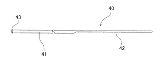
次に、図6ないし図8に示す実施例の採卵針誘導路内挿入用の採卵針用シース40について説明する。
図6は、本発明の他の実施例の採卵針誘導路内挿入用の採卵針用シースの部分省略外観図である。図7は、図6に示した採卵針誘導路内挿入用の採卵針用シースの先端部の拡大断面図である。図8は、採卵針挿入ガイドを装着した探触子(超音波診断装置用プローブ)に本発明の採卵針誘導路内挿入用の採卵針用シースを装着した状態を示す外観図である。
図6ないし図8に示す実施例の採卵針誘導路内挿入用の採卵針用シース40は、探触子2に装着された採卵針挿入ガイド3内に形成された採卵針誘導路32内に挿入される採卵針用シースである。
探触子2(超音波診断装置用プローブ)および採卵針挿入ガイド3は、上述した通りである。
【0015】
この実施例の採卵針用シース40は、採卵針挿入ガイド3の採卵針誘導路32内に挿入可能かつ内部に採卵針5を挿入可能な中空状本体部41と、採卵針誘導路32への装着状態を保持するための装着保持部42と、先端大径部43を備える。
シース40は、全体がほぼ同一内径および同一外径を有するチューブ体であり、後端部は、軸方向に並行にチューブ体を所定の長さ部分的に除去することにより形成された装着保持部42となっている。この実施例のシース40では、軸方向に並行にチューブ体をほぼ半分を切り取ることにより装着保持部42が形成されている。
シース40としては、全長が、155〜500mm、内径が、0.5〜2.3mm、外径が、0.6〜2.4mmが好適である。装着保持部42としては、長さが、5〜50mm、シース40の軸方向に並行にチューブ体を切り取る量としては、1/3から2/3程度、言い換えれば、装着保持部13の断面円弧の長さは、シース40の中空状本体部41の円周の長さの1/3〜2/3程度であることが好ましい。
【0016】
シース40の形成材料としては、薄肉に形成してもある程度の強度、硬度を備えるとともに、ある程度の可撓性を備えるものが好ましい。シース40の形成材料としては、具体的には、PTFE、ETFE、FEP、PFA等のフッ素系樹脂、ポリプロピレン、ポリエチレン等のオレフィン系樹脂、ポリアミド(ナイロン6、ナイロン66)などが使用できる。
シース40の先端には、大径部43が形成されている。大径部43の外径は、挿入されるガイド管31の先端の内径より大きいものとなっている。また、このシース40が挿入されるガイド管31の先端は、刃先状先端面ではなく軸にほぼ直交するように切断されている。大径部43は、リング状部材をシース40の先端に固着することにより形成されている。なお、大径部43は、図6におよび図7に示すような環状のリング状部材に限定されるものではなく、例えば、後述する図13のように、複数の突起により形成してもよい。大径部43の形成材料としては、どのようなものでもよいが、例えば、PTFE、ETFE、FEP、PFA等のフッ素系樹脂、フッ素樹脂系エラストマー、ポリプロピレン、ポリエチレン等のオレフィン系樹脂、ポリアミド(ナイロン6、ナイロン66)、ポリエステル(ポリエチレンテレフタレート、ポリブチレンテレフタレート)、ポリオレフィン(ポリエチレン、ポリプロピレン、エチレン−プロピレンコポリマー)、ポリウレタン(ポリエステル系ポリウレタン、ポリエーテル系ポリウレタン)などが使用できる。そして、この大径部43は、シース40に固着されてる。固着は、熱融着、接着剤等により行われる。また、大径部43の外面は、図7に示すように曲面(具体的には、面取りされた状態)となっていることが好ましい。このようにすることにより、生体内挿入時に生体内壁に損傷を与えることを防止できる。また、大径部43は、上記のように別部材を固着するものに限定されない。例えば、大径部は、シース40の先端を熱加工などにより、拡径したもの、さらには、丸みをおびた折り返し状としたものでもよい。
【0017】
そして、シース40は、ガイド管31の先端側より、ガイド管31の先端がシース40の先端部43に当接する状態までガイド管31内に形成される誘導路32内に挿入される。この状態にて、シース40の後端部(言い換えれば、装着保持部42の後端部)は、ガイド管31より突出する。そして。図8に示すように、ガイド管31より突出する部分の装着保持部42をガイド管31の先端側に折り曲げる。これにより、シース40は、ガイド管31に装着されるとともに、大径部43と共同することにより、装着状態を保持する。その後、シース40内には、採卵針が挿入される。そして、使用後、採卵針を抜去し、必要であればガイド管31の先端部の汚れを拭き取った後、鉗子などにより折り曲げられている装着保持部42をシース40の軸方向とほぼ並行となる状態に戻し、鉗子などにより、シース40の大径部43を保持し引っ張ることにより、シース40をガイド管31の先端側より抜去することができる。
【0018】
次に、図9ないし図12に示す実施例の採卵針誘導路内挿入用の採卵針用シース50,60について説明する。
図9は、本発明の他の実施例の採卵針誘導路内挿入用の採卵針用シースの部分省略外観図である。図10は、図9に示した採卵針誘導路内挿入用の採卵針用シースを採卵針ガイド内に装着した状態の部分省略拡大断面図である。図11は、本発明の他の実施例の採卵針誘導路内挿入用の採卵針用シースを採卵針ガイド内に装着した状態の部分省略拡大断面図である。図12は、採卵針挿入ガイドを装着した探触子(超音波診断装置用プローブ)に図9もしくは図11に示す採卵針誘導路内挿入用の採卵針用シースを装着した状態を示す外観図である。
図9ないし図12に示す実施例の採卵針誘導路内挿入用の採卵針用シース50,60は、探触子2に装着された採卵針挿入ガイド3内に形成された採卵針誘導路32内に挿入される採卵針用シースである。
探触子2(超音波診断装置用プローブ)および採卵針挿入ガイド3は、上述した通りである。
【0019】
この実施例の採卵針用シース50、60は、採卵針挿入ガイド3は採卵針誘導路32内に挿入可能かつ内部に採卵針5を挿入可能な中空状本体部51と、採卵針誘導路32への装着状態を保持するための装着保持部55、65と、後端拡径部52を備える。
シース50は、全体がほぼ同一内径および同一外径を有するチューブ体であり、後端部は、テーパー状に後端に向かって拡径するテーパー部52となっている。シース50の後端(テーパー部52の後端部)は、挿入されるガイド管31の後端の内径より大きいものとなっている。このため、シース50のガイド管31の先端方向への抜け止めとして機能する。さらに、テーパー部52は、採卵針誘導口としても機能する。そして、テーパー部の先端部もしくは本体部51の基端部には、装着保持部55が設けられており、装着保持部55は、図9および図10に示す実施例では、環状リブにより形成されている。環状リブ55の外径は、若干ガイド管31の内径より大きいものとなっている。ガイド管31内に装着保持部55である環状リブは強制嵌入可能であり、嵌入された状態にて、変形した装着保持部55はガイド管31の内壁面を押圧し、装着保持部の復元力により、シース50のガイド管31への装着状態を保持する。
なお、装着保持部の形状は、上述した環状リブに限定されるものではなく、図11に示すシース60のような、所定の長さを有し、かつ外径が若干ガイド管31の内径より大きい筒状拡径部65であってもよい。さらに、装着保持部としては、後述する図13のように、複数の突起により形成してもよい。
【0020】
シース50,60としては、全長が、155〜485mm、内径が、0.5〜2.3mm、外径が、0.6〜2.4mmが好適である。装着保持部55,65の外径は、0.6〜2.8mm程度であることが好ましい。また、装着保持部65の長さは、1〜15mm程度であることが好ましい。
シース50,60の形成材料としては、薄肉に形成してもある程度の強度、硬度を備えるとともに、ある程度の可撓性を備えるものが好ましい。シース50,60の形成材料としては、具体的には、PTFE、ETFE、FEP、PFA等のフッ素系樹脂、ポリプロピレン、ポリエチレン等のオレフィン系樹脂、ポリアミド(ナイロン6、ナイロン66)などが使用できる。
【0021】
そして、シース50,60は、ガイド管31に後端側からガイド管31の後端が装着保持部55,65に当接する状態まで挿入する。そして、シース50,60の後端を強く押すことにより、装着保持部55,65は若干変形するとともにガイド管31の後端内に収納される。これにより、シース50,60は、ガイド管31への装着状態を保持する。この状態にて、シース50,60の先端は、図10ないし図12に示すように、ガイド管31より若干突出する。シース50,60のガイド管31への装着時におけるシース50,60の先端部のガイド管31からの突出長さは、1〜15mm程度であることが好ましい。その後、シース50,60内には、採卵針5が挿入される。そして、使用後、採卵針を抜去し、次いで、ガイド管31よりシース50,60を抜去する。シース50,60の抜去は、必要によりガイド管31の先端部の汚れを拭き取った後、鉗子などによりシース50,60の後端部(テーパー部)を把持し引っ張ることにより、シース50,60をガイド管31の後端側より抜去することができる。また、シース50,60の抜去は、必要によりガイド管31の先端部の汚れを拭き取った後、鉗子などによりシース50,60のガイド管31より突出する先端を把持し強制的に引っ張ることにより、シース50,60をガイド管31の先端側より抜去することもできる。
【0022】
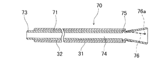
次に、図13ないし図15に示す実施例の採卵針誘導路内挿入用の採卵針用シース70について説明する。
図13は、本発明の他の実施例の採卵針誘導路内挿入用の採卵針用シースの部分省略外観図である。図14は、図13に示した採卵針誘導路内挿入用の採卵針用シースを採卵針ガイド内に装着した状態の部分省略拡大断面図である。図15は、図13の採卵針誘導路内挿入用の採卵針用シースの使用状態を説明する説明図である。
図13ないし図15に示す実施例の採卵針誘導路内挿入用の採卵針用シース70は、探触子2に装着された採卵針挿入ガイド3内に形成された採卵針誘導路32内に挿入される採卵針用シースである。
探触子2(超音波診断装置用プローブ)および採卵針挿入ガイド3は、上述した通りである。
【0023】
この実施例の採卵針用シース70は、採卵針挿入ガイド3の採卵針誘導路32内に挿入可能かつ内部に採卵針5を挿入可能な中空状本体部71と、採卵針誘導路32への装着状態を保持するための装着保持部75と、後端拡径部72を備える。
シース70は、全体がほぼ同一内径および同一外径を有するチューブ体であり、後端部は、テーパー状に後端に向かって拡径するテーパー部72となっている。シース70の後端(テーパー部72の後端部)は、挿入されるガイド管31の後端の内径より大きいものとなっている。このため、シース70のガイド管31の先端方向への抜け止めとして機能する。さらに、テーパー部72は、採卵針誘導口としても機能する。そして、テーパー部の先端部もしくは本体部71の基端部には、装着保持部75が設けられており、装着保持部55は、図13および図14に示す実施例では、環状に配置された複数の突起により形成されている。複数の突起部分を結ぶことにより仮想される外径は、若干ガイド管31の内径より大きいものとなっている。ガイド管31内に装着保持部55である突起は、強制嵌入可能であり、嵌入された状態にて、変形した装着保持部55はガイド管31の内壁面を押圧し、装着保持部の復元力により、シース70のガイド管31への装着状態を保持する。突起は、少なくとも2つ設けることが好ましく、特に、2〜8程度が好適である。突起が2つの場合には、向かい合うよう配置し、3以上の場合には、シース70の中心軸に対して等角度に配置することが好ましい。
なお、装着保持部の形状は、上述した突起に限定されるものではなく、図9および図10に示すシース50のような環状リブ、さらには、図11に示すシース60のような、所定の長さを有する筒状拡径部65であってもよい。
【0024】
シース70としては、全長が、155〜485mm、内径が、0.5〜2.3mm、外径が、0.6〜2.4mmが好適である。
シース70の形成材料としては、薄肉に形成してもある程度の強度、硬度を備えるとともに、ある程度の可撓性を備えるものが好ましい。シース70の形成材料としては、具体的には、PTFE、ETFE、FEP、PFA等のフッ素系樹脂、ポリプロピレン、ポリエチレン等のオレフィン系樹脂、ポリアミド(ナイロン6、ナイロン66)などが使用できる。
そして、シース70は、抜去用線状体76を備えている。抜去用線状体は、図13および図14に示すように、一端部が、固定部76aにおいてシース70(具体的には、テーパー部)に固定されるとともに、テーパー部の外面に巻き付けられている。抜去用線状体76の他端部は、固定部76a付近の線状体の下に挟み込まれており、巻き付け状態が保持されている。
【0025】
抜去用線状体76は、テーパー部の外面への巻き付けを解除し、シース70の内腔内に挿入した際に、シース70の先端よりある程度の長さ突出するような長さを有している。この場合のシース70先端より突出する抜去用線状体76部分の長さとしては、1〜50mm程度が好適である。抜去用線状体76としては、ステンレス線、アモルファス合金線などの金属線または繊維が好ましい。アモルファス合金線としては、鉄−ケイ素−ホウ素系合金、コバルト−ケイ素−ホウ素系合金、鉄−コバルト−クロム−モリブデン−ケイ素−ホウ素系合金などを用いて形成したアモルファス合金線が、好適に使用できる。繊維としては、ポリエステル系繊維、ナイロン6、ナイロン66、ナイロン46などのポリアミド系繊維、ポリエチレン、ポリプロピレンなどのポリオレフィン系繊維などの合成繊維、さらには、ガラス繊維、アモルファス繊維などが好適である。
【0026】
そして、シース70は、ガイド管31に後端側からガイド管31の後端が装着保持部75に当接する状態まで挿入する。そして、シース70の後端を強く押すことにより、装着保持部75は若干変形するとともにガイド管31の後端内に収納される。これにより、シース70は、ガイド管31内に装着される。この状態にて、シース70の先端は、図14および図15に示すように、ガイド管31より突出する。シース70のガイド管31への装着時におけるシース70の先端部のガイド管31からの突出長さは、1〜15mm程度であることが好ましい。その後、シース70内には、採卵針5が挿入される。そして、使用後、採卵針を抜去し、次いで、ガイド管31よりシース70を抜去する。シース70の抜去は、必要によりガイド管31の先端部の汚れを拭き取った後、テーパー部の外面に巻き付けられている抜去用線状体76を解き、シース70の内腔内に挿入し、抜去用線状体76の他端部76bをガイド管31より露出させる。この際、図示しないシース70内腔内より小径の棒状部材の先端に抜去用線状体76の他端を巻き付けてシース70内を貫通させてもよい。そして、鉗子などにより、シース70の先端より露出するシース70の他端部76b(図示しないシース70内腔内より小径の棒状部材の先端に抜去用線状体76の他端を巻き付けてシース70内を貫通させた場合には、棒状部材でもよい)を把持し引っ張ることにより、シース70をガイド管31の先端側より抜去することができる。
【0027】
次に、図16に示す実施例について説明する。
図16は、採卵針誘導路を備える探触子(超音波診断装置用プローブ)に本発明の採卵針誘導路内挿入用の採卵針用シースを装着した状態を示す外観図である。
この実施例の採卵針用シース80は、探触子20自体に設けられた採卵針誘導路82内に挿入される採卵針用シース80である。このシース80は、図6および図7に示し上述した構成を備えている。しかし、この図示した例に限定されるものではなく、上述したすべての実施例の採卵針用シースは、探触子20自体に設けられた採卵針誘導路内に挿入される採卵針用シースとしても使用できる。
【0028】
【発明の効果】
本発明の採卵針誘導路内挿入用の採卵針用シースは、探触子に装着された採卵針挿入ガイド内に形成された採卵針誘導路内もしくは探触子に設けられた採卵針誘導路内に挿入される採卵針用シースであり、該採卵針用シースは、前記採卵針誘導路内に挿入可能かつ内部に採卵針を挿入可能な中空状本体部と、前記採卵針誘導路への装着状態を保持するための装着保持部とを備え、かつ前記採卵針誘導路内に装着後に抜去可能である。
【0029】
このシースを採卵針誘導路内に装着した状態にて採卵針の導入を行うことにより、使用時における採卵針誘導路内への生体分泌物などの侵入を抑制し、さらに、採卵針抜去時における採卵針先端部に付着した生体内分泌物の採卵針誘導路内への侵入を防止する。よって、採卵針ガイドもしくは探触子の採卵針誘導路内の汚染を抑制でき、採卵針ガイドもしくは探触子を洗浄、滅菌のうえ再利用しても、前回の使用時における生体内分泌物の次回の使用者の生体内への流入を極めて少ないものとすることができる。
【0030】
そして、前記シースの先端部は、前記誘導路の先端より突出し、かつ、該シースの突出部を用いてシースを引っ張ることにより、該誘導路内より抜去可能であるとすることにより、シースの先端部に付着した生体内分泌物の採卵針誘導路内への侵入を防止することができる。
【0031】
また、前記誘導路への装着状態を保持するための装着保持部は、前記シースの後端に形成された該誘導路の内径より大きい拡径部と、前記シースの先端部に形成された折り曲げ可能部とにより構成することにより、シースの採卵針誘導路内への装着が容易となる。
【0032】
また、前記誘導路への装着状態を保持するための装着保持部は、前記シースの先端に形成された前記誘導路の内径より大きい拡径部と、前記シースの後端部に形成された折り曲げ可能部とにより構成することにより、シースの採卵針誘導路内への装着が容易となる。
【0033】
また、前記誘導路への装着状態を保持するための装着保持部は、前記シースの一端部に形成され、かつ前記誘導路の端部内に押し込み可能かつ押し込まれた状態にて前記誘導路の内壁面に当接することにより、該誘導路への装着状態を保持するリブにより構成することにより、シースの採卵針誘導路内への装着が容易となる。
【0034】
また、前記誘導路への装着状態を保持するための装着保持部は、前記シースの一端部に形成され、かつ前記誘導路の端部内に押し込み可能かつ押し込まれた状態にて前記誘導路の内壁と嵌合することにより、該誘導路への装着状態を保持する拡径部により構成することにより、シースの採卵針誘導路内への装着が容易となる。
【図面の簡単な説明】
【図1】図1は、採卵針挿入ガイドを装着した探触子(超音波診断装置用プローブ)および本発明の採卵針誘導路内挿入用の採卵針用シースの外観図である。
【図2】図2は、本発明の実施例の採卵針誘導路内挿入用の採卵針用シースの部分省略外観図である。
【図3】図3は、図2に示した採卵針誘導路内挿入用の採卵針用シースの部分省略断面図である。
【図4】図4は、採卵針挿入ガイドを装着した探触子(超音波診断装置用プローブ)に本発明の採卵針誘導路内挿入用の採卵針用シースを装着した状態を示す外観図である。
【図5】図5は、本発明の採卵針誘導路内挿入用の採卵針用シースの使用状態を説明する説明図である。
【図6】図6は、本発明の他の実施例の採卵針誘導路内挿入用の採卵針用シースの部分省略外観図である。
【図7】図7は、図6に示した採卵針誘導路内挿入用の採卵針用シースの先端部の拡大断面図である。
【図8】図8は、採卵針挿入ガイドを装着した探触子(超音波診断装置用プローブ)に本発明の採卵針誘導路内挿入用の採卵針用シースを装着した状態を示す外観図である。
【図9】図9は、本発明の他の実施例の採卵針誘導路内挿入用の採卵針用シースの部分省略外観図である。
【図10】図10は、図9に示した採卵針誘導路内挿入用の採卵針用シースを採卵針ガイド内に装着した状態の部分省略拡大断面図である。
【図11】図11は、本発明の他の実施例の採卵針誘導路内挿入用の採卵針用シースを採卵針ガイド内に装着した状態の部分省略拡大断面図である。
【図12】図12は、採卵針挿入ガイドを装着した探触子(超音波診断装置用プローブ)に図9または図11に示す採卵針誘導路内挿入用の採卵針用シースを装着した状態を示す外観図である。
【図13】図13は、本発明の他の実施例の採卵針誘導路内挿入用の採卵針用シースの部分省略外観図である。
【図14】図14は、図13に示した採卵針誘導路内挿入用の採卵針用シースを採卵針ガイド内に装着した状態の部分省略拡大断面図である。
【図15】図15は、図13の採卵針誘導路内挿入用の採卵針用シースの使用状態を説明する説明図である。
【図16】図16は、採卵針誘導路を備える探触子(超音波診断装置用プローブ)に本発明の採卵針誘導路内挿入用の採卵針用シースを装着した状態を示す外観図である。
【符号の説明】
1、40、50、60、70 採卵針用シース
2、20 探触子
3 採卵針挿入ガイド
5 採卵針
10 シース本体
11 中空状本体部
32 採卵針誘導路[0001]
BACKGROUND OF THE INVENTION
The present invention relates to a sheath for an egg-collecting needle used by being inserted into an egg-collecting needle guide attached to a probe or formed on the probe.
[0002]
[Prior art]
2. Description of the Related Art Conventionally, an ultrasonic diagnostic apparatus is used to image a living ovary and collect an egg while viewing the image. In such a case, an ultrasonic probe (probe) connected to the ultrasonic diagnostic apparatus, to which an egg collection needle guide is attached, is inserted into the living body. Then, the egg collection needle is inserted into the egg collection needle guide to collect the egg.
[0003]
[Problems to be solved by the invention]
The egg collection needle guide is also inserted into the living body together with the probe. For this reason, the in-vivo secretion adheres to the outer surface and the inner surface of the egg collection needle guide. When the collection of the egg by inserting the egg collection needle into the egg collection needle guide is completed, the egg collection needle guide is also removed from the living body together with the probe. Some egg collection needle guides are disposable, but they are used as medical waste, so most are reused after being cleaned and sterilized. However, it is not easy to clean the inside of the egg collection needle guide, and it is difficult to remove the highly viscous in-vivo secretions adhering to the inner surface of the guide. The next time the egg collection needle is inserted into the guide, There is a risk of adhering in-vivo secretions being scraped off and flowing into the living body.
[0004]
Therefore, in the present invention, it is possible to suppress adherence of biological secretions to the inner surface of the egg collection needle passage formed in the passage of the egg collection needle guide or the probe, and the egg collection needle guide can be reused after washing, sterilization, It is an object of the present invention to provide an egg collection needle sheath for insertion into an egg collection needle guide path that can reduce the risk of in vivo secretion of in vivo secretions during previous use.
[0005]
[Means for Solving the Problems]
What achieves the above object is a sheath for an egg-collecting needle that is inserted into an egg-collecting needle guide path formed in an egg-inserting needle insertion guide attached to the probe or an egg-collecting needle guide path provided in the probe. The egg collection needle sheath has a hollow main body part that can be inserted into the egg collection needle guide path and into which the egg collection needle can be inserted, and an attachment holder for holding the attachment state to the egg collection needle guide path And after mounting in the egg collection needle guiding path The distal end portion of the sheath projects from the distal end of the guide path, and the sheath is pulled from the distal end side of the guide path by pulling the sheath using the projecting portion of the sheath. It is a sheath for egg-collecting needles for insertion in an egg-collecting needle guiding path that can be removed.
[0006]
An attachment holding portion for holding the attachment state to the guide path includes an enlarged diameter portion formed at a rear end of the sheath that is larger than an inner diameter of the guide path, and a bent portion formed at a distal end portion of the sheath. It is preferable that it is comprised by the possible part. In addition, a mounting holding portion for holding the mounting state to the guide path is formed at a larger diameter portion than the inner diameter of the guide path formed at the distal end portion of the sheath and at the rear end portion of the sheath. You may be comprised by the bendable part. Further, a mounting holding portion for holding the mounting state on the guide path is formed at one end portion of the sheath and can be pushed into the end portion of the guide path, and the inside of the guide path is pushed in. You may comprise by the rib which hold | maintains the mounting state to this guide path by contact | abutting to a wall surface. In addition, an attachment holding portion for holding the attachment state to the guide path is formed at one end of the sheath, and can be pushed into the end of the guide path and can be pushed into the inner wall of the guide path. And may be constituted by an enlarged-diameter portion that holds the mounting state on the guide path.
[0007]
And it is preferable that the rear-end part of the said sheath becomes a diameter-expanded part which expands in a taper shape. Moreover, it is preferable that a hub having an enlarged diameter portion and removable from the sheath is attached to the rear end of the sheath. Furthermore, it is preferable that an extraction linear body longer than the entire length of the sheath is attached to the rear end portion of the sheath.
[0008]
DETAILED DESCRIPTION OF THE INVENTION
The egg-collecting needle sheath for insertion into the egg-collecting needle guiding path of the present invention will be described with reference to the embodiments shown in the drawings.
FIG. 1 is an external view of a probe (a probe for an ultrasonic diagnostic apparatus) equipped with an egg collection needle insertion guide and an egg collection needle sheath for insertion into an egg collection needle guide path according to the present invention. FIG. 2 is a partially omitted external view of an egg collection needle sheath for insertion into an egg collection needle guide path according to an embodiment of the present invention. FIG. 3 is a partially omitted cross-sectional view of the egg collection needle sheath for insertion into the egg collection needle guide path shown in FIG. 2. FIG. 4 is an external view showing a state in which a probe (an ultrasonic diagnostic apparatus probe) equipped with an egg collection needle insertion guide is equipped with an egg collection needle sheath for insertion into the egg collection needle guide path of the present invention. FIG. 5 is an explanatory view for explaining a use state of the egg-collecting needle sheath for insertion into the egg-collecting needle guiding path of the present invention. Specifically, FIG. 5 shows that the probe (ultrasound diagnostic device probe) equipped with the egg-needle insertion guide of FIG. 4 is fitted with the egg-needle sheath for insertion into the egg-needle guide path of the present invention, Furthermore, it is an expanded sectional view of the sheath distal end portion in a state where the egg collection needle is inserted into the sheath.
[0009]
An egg
[0010]
The egg
The egg-collecting
The probe 2 (a probe for an ultrasonic diagnostic apparatus) is a transvaginal probe, and includes a probe
[0011]
The
The
The
[0012]
A material for forming the
A
[0013]
Then, the
[0014]
Next, the egg
FIG. 6 is a partially omitted external view of an egg collection needle sheath for insertion into an egg collection needle guide path according to another embodiment of the present invention. FIG. 7 is an enlarged cross-sectional view of the distal end portion of the egg collection needle sheath for insertion into the egg collection needle guide path shown in FIG. 6. FIG. 8 is an external view showing a state in which a probe (an ultrasonic diagnostic apparatus probe) equipped with an egg collection needle insertion guide is equipped with an egg collection needle sheath for insertion into the egg collection needle guide path of the present invention.
The egg
The probe 2 (the probe for an ultrasonic diagnostic apparatus) and the egg collection
[0015]
The
The
The
[0016]
A material for forming the
A
[0017]
The
[0018]
Next, the egg
FIG. 9 is a partially omitted external view of an egg collection needle sheath for insertion into an egg collection needle guide path according to another embodiment of the present invention. FIG. 10 is a partially omitted enlarged cross-sectional view of a state where the egg-collecting needle sheath for insertion into the egg-collecting needle guiding path shown in FIG. 9 is mounted in the egg-collecting needle guide. FIG. 11 is a partially omitted enlarged cross-sectional view showing a state where an egg collection needle sheath for insertion into an egg collection needle guide path according to another embodiment of the present invention is mounted in an egg collection needle guide. FIG. 12 is an external view showing a state where an egg collection needle sheath for insertion in an egg collection needle guide path shown in FIG. 9 or FIG. 11 is attached to a probe (an ultrasonic diagnostic apparatus probe) equipped with an egg collection needle insertion guide. It is.
The egg
The probe 2 (the probe for an ultrasonic diagnostic apparatus) and the egg collection
[0019]
In this embodiment, the egg
The
The shape of the mounting holding portion is not limited to the annular rib described above, and has a predetermined length, such as the
[0020]
The
The material for forming the
[0021]
The
[0022]
Next, the egg-collecting
FIG. 13 is a partially omitted external view of an egg collection needle sheath for insertion into an egg collection needle guide path according to another embodiment of the present invention. FIG. 14 is a partially omitted enlarged cross-sectional view showing a state where the egg-collecting needle sheath for insertion into the egg-collecting needle guiding path shown in FIG. 13 is mounted in the egg-collecting needle guide. FIG. 15 is an explanatory view for explaining the use state of the egg-collecting needle sheath for insertion into the egg-collecting needle guiding path of FIG.
An egg
The probe 2 (the probe for an ultrasonic diagnostic apparatus) and the egg collection
[0023]
The
The
Note that the shape of the mounting holding portion is not limited to the above-described protrusion, but is a predetermined shape such as an annular rib such as the
[0024]
The
As a material for forming the
And the
[0025]
The extraction
[0026]
Then, the
[0027]
Next, the embodiment shown in FIG. 16 will be described.
FIG. 16 is an external view showing a state where an egg collection needle sheath for insertion into an egg collection needle guide path of the present invention is attached to a probe (probe for an ultrasonic diagnostic apparatus) having an egg collection needle guide path.
The egg
[0028]
【The invention's effect】
An egg collection needle sheath for insertion into an egg collection needle guide path according to the present invention is an egg collection needle guide path formed in an egg collection needle insertion guide mounted on a probe or an egg collection needle guide path provided in the probe. An egg collection needle sheath inserted into the egg collection needle sheath, wherein the egg collection needle sheath is inserted into the egg collection needle guide path and into which the egg collection needle can be inserted, and the egg collection needle guide path An attachment holding portion for holding the attachment state, and can be removed from the egg collection needle guide path after attachment.
[0029]
By introducing the egg collection needle in a state where this sheath is mounted in the egg collection needle guide path, invasion of biological secretions and the like into the egg collection needle guide path during use is suppressed, and further, when the egg collection needle is removed Prevents in-vivo secretions adhering to the tip of the egg collection needle from entering the egg collection needle guiding path. Therefore, contamination of the egg guide guide or probe in the egg guide path can be suppressed, and even if the egg guide or probe is washed, sterilized and reused, the next time the in-vivo secretions from the previous use The inflow of the user into the living body can be made extremely small.
[0030]
And the tip of the sheath is Taxiway Projecting from the tip of the sheath and pulling the sheath using the projection of the sheath, Taxiway By making it possible to withdraw from the inside, it is possible to prevent in vivo secretions adhering to the distal end portion of the sheath from entering the egg collection needle guide path.
[0031]
In addition, Taxiway An attachment holding portion for holding the attachment state to the sheath is formed on the rear end of the sheath. Taxiway By constructing it with an enlarged diameter portion larger than the inner diameter of this and a bendable portion formed at the distal end portion of the sheath, the sheath can be easily mounted in the egg collection needle guiding path.
[0032]
In addition, Taxiway An attachment holding portion for holding the attachment state to the sheath is formed at the distal end of the sheath. Taxiway By constructing it with an enlarged diameter portion larger than the inner diameter of the inner sheath and a bendable portion formed at the rear end portion of the sheath, the sheath can be easily mounted in the egg collection needle guiding path.
[0033]
In addition, Taxiway A mounting holding portion for holding the mounting state is formed at one end of the sheath, and the Taxiway Can be pushed into the end of the Taxiway By contacting the inner wall surface of the Taxiway By configuring the rib with the mounting state of the sheath, the sheath can be easily mounted in the egg collection needle guiding path.
[0034]
In addition, Taxiway A mounting holding portion for holding the mounting state is formed at one end of the sheath, and the Taxiway Can be pushed into the end of the Taxiway By fitting with the inner wall of the Taxiway By configuring with a diameter-enlarged portion that maintains the mounting state of the sheath, the mounting of the sheath into the egg-collecting needle guiding path is facilitated.
[Brief description of the drawings]
FIG. 1 is an external view of a probe (a probe for an ultrasonic diagnostic apparatus) equipped with an egg collection needle insertion guide and an egg collection needle sheath for insertion in an egg collection needle guide path according to the present invention.
FIG. 2 is a partially omitted external view of an egg collection needle sheath for insertion into an egg collection needle guiding path according to an embodiment of the present invention.
FIG. 3 is a partially omitted cross-sectional view of an egg collection needle sheath for insertion into the egg collection needle guide path shown in FIG. 2;
FIG. 4 is an external view showing a state where an egg collection needle sheath for insertion in an egg collection needle guide path according to the present invention is attached to a probe (an ultrasonic diagnostic apparatus probe) equipped with an egg collection needle insertion guide. It is.
FIG. 5 is an explanatory view for explaining a use state of an egg collection needle sheath for insertion into an egg collection needle guiding path according to the present invention.
FIG. 6 is a partially omitted external view of an egg collection needle sheath for insertion into an egg collection needle guiding path according to another embodiment of the present invention.
7 is an enlarged cross-sectional view of a distal end portion of an egg collection needle sheath for insertion into the egg collection needle guide path shown in FIG. 6;
FIG. 8 is an external view showing a state in which a probe (an ultrasonic diagnostic apparatus probe) equipped with an egg collection needle insertion guide is equipped with an egg collection needle sheath for insertion into the egg collection needle guide path according to the present invention. It is.
FIG. 9 is a partially omitted external view of an egg collection needle sheath for insertion into an egg collection needle guide path according to another embodiment of the present invention.
FIG. 10 is a partially omitted enlarged cross-sectional view showing a state where an egg-collecting needle sheath for insertion into an egg-collecting needle guiding path shown in FIG. 9 is mounted in an egg-collecting needle guide.
FIG. 11 is a partially omitted enlarged cross-sectional view showing a state where an egg collection needle sheath for insertion into an egg collection needle guiding path according to another embodiment of the present invention is mounted in an egg collection guide.
12 is a view showing a state in which an egg collection needle sheath for insertion into the egg collection needle guide path shown in FIG. 9 or FIG. 11 is attached to a probe (an ultrasonic diagnostic apparatus probe) equipped with an egg collection needle insertion guide. FIG.
FIG. 13 is a partially omitted external view of an egg collection needle sheath for insertion into an egg collection needle guide path according to another embodiment of the present invention.
FIG. 14 is a partially omitted enlarged cross-sectional view showing a state where an egg collection needle sheath for insertion into an egg collection needle guide path shown in FIG. 13 is mounted in an egg collection needle guide.
15 is an explanatory view for explaining a use state of the egg-collecting needle sheath for insertion into the egg-collecting needle guiding path of FIG. 13;
FIG. 16 is an external view showing a state in which an egg collection needle sheath for insertion into an egg collection needle guide path according to the present invention is attached to a probe (probe for an ultrasonic diagnostic apparatus) including an egg collection needle guide path. is there.
[Explanation of symbols]
1, 40, 50, 60, 70 Egg collection needle sheath
2, 20 Probe
3 Egg collection needle insertion guide
5 Egg collection needle
10 Sheath body
11 Hollow body
32 Egg collection needle guideway
Claims (8)
Priority Applications (1)
| Application Number | Priority Date | Filing Date | Title |
|---|---|---|---|
| JP2000114217A JP4472832B2 (en) | 2000-04-14 | 2000-04-14 | Egg collection needle sheath for insertion into the probe collection path of the probe |
Applications Claiming Priority (1)
| Application Number | Priority Date | Filing Date | Title |
|---|---|---|---|
| JP2000114217A JP4472832B2 (en) | 2000-04-14 | 2000-04-14 | Egg collection needle sheath for insertion into the probe collection path of the probe |
Publications (3)
| Publication Number | Publication Date |
|---|---|
| JP2001293002A JP2001293002A (en) | 2001-10-23 |
| JP2001293002A5 JP2001293002A5 (en) | 2007-05-31 |
| JP4472832B2 true JP4472832B2 (en) | 2010-06-02 |
Family
ID=18626057
Family Applications (1)
| Application Number | Title | Priority Date | Filing Date |
|---|---|---|---|
| JP2000114217A Expired - Fee Related JP4472832B2 (en) | 2000-04-14 | 2000-04-14 | Egg collection needle sheath for insertion into the probe collection path of the probe |
Country Status (1)
| Country | Link |
|---|---|
| JP (1) | JP4472832B2 (en) |
Cited By (1)
| Publication number | Priority date | Publication date | Assignee | Title |
|---|---|---|---|---|
| CN102860859A (en) * | 2012-09-26 | 2013-01-09 | 广州品致医疗器械有限公司 | Puncture needle of oocyte collector |
Families Citing this family (1)
| Publication number | Priority date | Publication date | Assignee | Title |
|---|---|---|---|---|
| AU2014337092B2 (en) * | 2013-10-18 | 2019-05-23 | May Health Us Inc. | Methods and systems for the treatment of polycystic ovary syndrome |
-
2000
- 2000-04-14 JP JP2000114217A patent/JP4472832B2/en not_active Expired - Fee Related
Cited By (2)
| Publication number | Priority date | Publication date | Assignee | Title |
|---|---|---|---|---|
| CN102860859A (en) * | 2012-09-26 | 2013-01-09 | 广州品致医疗器械有限公司 | Puncture needle of oocyte collector |
| CN102860859B (en) * | 2012-09-26 | 2015-03-04 | 广州品致医疗器械有限公司 | Puncture needle of oocyte collector |
Also Published As
| Publication number | Publication date |
|---|---|
| JP2001293002A (en) | 2001-10-23 |
Similar Documents
| Publication | Publication Date | Title |
|---|---|---|
| WO2017006684A1 (en) | Lens cleaner for endoscope | |
| JP2006187556A (en) | Cleaning brush for endoscope | |
| EP1829474B1 (en) | Washing brush for cleaning a conduit | |
| US20210339297A1 (en) | Surgical instrument cleaning brush assembly for use with a borescope | |
| EP2898816A1 (en) | Insertion aid, insertion body, and insertion device | |
| JP5344738B2 (en) | Endoscope cleaning tool | |
| JP3583542B2 (en) | Endoscope | |
| JP4472832B2 (en) | Egg collection needle sheath for insertion into the probe collection path of the probe | |
| WO2022013942A1 (en) | Endoscope conduit cleaning tool and endoscope system | |
| CN110799082B (en) | Endoscope with a detachable handle | |
| JP2001061760A (en) | Cover sheath type rigid endoscope | |
| JP6065723B2 (en) | Endoscope cleaning tool, endoscope system | |
| JP4338464B2 (en) | Endoscope cleaning tool | |
| JP3911158B2 (en) | Endoscopy cleaning brush | |
| JP6300274B2 (en) | Endoscope lens wiping device and endoscope provided with the wiping device | |
| JP2606839Y2 (en) | Endoscope device with endoscope cover system | |
| JP6495027B2 (en) | Endoscope | |
| JPH0889477A (en) | Cleaning auxiliary | |
| JPH08280603A (en) | Cover type endoscope | |
| JP2018134119A (en) | Lens cleaning tool for endoscope | |
| JP2018134120A (en) | Lens cleaning tool for endoscope | |
| JP3385661B2 (en) | Endoscope fouling prevention mechanism | |
| JP6600614B2 (en) | Cotton swab | |
| JP6749140B2 (en) | Medical treatment tool | |
| JP2006263352A (en) | Binding thread for endoscope, sheath fixing method, flexible tube for endoscope and endoscope |
Legal Events
| Date | Code | Title | Description |
|---|---|---|---|
| A521 | Written amendment |
Free format text: JAPANESE INTERMEDIATE CODE: A523 Effective date: 20070405 |
|
| A621 | Written request for application examination |
Free format text: JAPANESE INTERMEDIATE CODE: A621 Effective date: 20070405 |
|
| A977 | Report on retrieval |
Free format text: JAPANESE INTERMEDIATE CODE: A971007 Effective date: 20091110 |
|
| A131 | Notification of reasons for refusal |
Free format text: JAPANESE INTERMEDIATE CODE: A131 Effective date: 20091201 |
|
| A521 | Written amendment |
Free format text: JAPANESE INTERMEDIATE CODE: A523 Effective date: 20100122 |
|
| TRDD | Decision of grant or rejection written | ||
| A01 | Written decision to grant a patent or to grant a registration (utility model) |
Free format text: JAPANESE INTERMEDIATE CODE: A01 Effective date: 20100302 |
|
| A01 | Written decision to grant a patent or to grant a registration (utility model) |
Free format text: JAPANESE INTERMEDIATE CODE: A01 |
|
| A61 | First payment of annual fees (during grant procedure) |
Free format text: JAPANESE INTERMEDIATE CODE: A61 Effective date: 20100304 |
|
| FPAY | Renewal fee payment (event date is renewal date of database) |
Free format text: PAYMENT UNTIL: 20130312 Year of fee payment: 3 |
|
| R150 | Certificate of patent or registration of utility model |
Ref document number: 4472832 Country of ref document: JP Free format text: JAPANESE INTERMEDIATE CODE: R150 Free format text: JAPANESE INTERMEDIATE CODE: R150 |
|
| FPAY | Renewal fee payment (event date is renewal date of database) |
Free format text: PAYMENT UNTIL: 20130312 Year of fee payment: 3 |
|
| S111 | Request for change of ownership or part of ownership |
Free format text: JAPANESE INTERMEDIATE CODE: R313113 |
|
| FPAY | Renewal fee payment (event date is renewal date of database) |
Free format text: PAYMENT UNTIL: 20130312 Year of fee payment: 3 |
|
| R360 | Written notification for declining of transfer of rights |
Free format text: JAPANESE INTERMEDIATE CODE: R360 |
|
| FPAY | Renewal fee payment (event date is renewal date of database) |
Free format text: PAYMENT UNTIL: 20130312 Year of fee payment: 3 |
|
| R350 | Written notification of registration of transfer |
Free format text: JAPANESE INTERMEDIATE CODE: R350 |
|
| FPAY | Renewal fee payment (event date is renewal date of database) |
Free format text: PAYMENT UNTIL: 20140312 Year of fee payment: 4 |
|
| R250 | Receipt of annual fees |
Free format text: JAPANESE INTERMEDIATE CODE: R250 |
|
| R250 | Receipt of annual fees |
Free format text: JAPANESE INTERMEDIATE CODE: R250 |
|
| R250 | Receipt of annual fees |
Free format text: JAPANESE INTERMEDIATE CODE: R250 |
|
| R250 | Receipt of annual fees |
Free format text: JAPANESE INTERMEDIATE CODE: R250 |
|
| R250 | Receipt of annual fees |
Free format text: JAPANESE INTERMEDIATE CODE: R250 |
|
| S531 | Written request for registration of change of domicile |
Free format text: JAPANESE INTERMEDIATE CODE: R313531 |
|
| S533 | Written request for registration of change of name |
Free format text: JAPANESE INTERMEDIATE CODE: R313533 |
|
| R350 | Written notification of registration of transfer |
Free format text: JAPANESE INTERMEDIATE CODE: R350 |
|
| R250 | Receipt of annual fees |
Free format text: JAPANESE INTERMEDIATE CODE: R250 |
|
| R250 | Receipt of annual fees |
Free format text: JAPANESE INTERMEDIATE CODE: R250 |
|
| LAPS | Cancellation because of no payment of annual fees |


