JP4441121B2 - A novel trypsin family serine protease - Google Patents
A novel trypsin family serine protease Download PDFInfo
- Publication number
- JP4441121B2 JP4441121B2 JP2000579724A JP2000579724A JP4441121B2 JP 4441121 B2 JP4441121 B2 JP 4441121B2 JP 2000579724 A JP2000579724 A JP 2000579724A JP 2000579724 A JP2000579724 A JP 2000579724A JP 4441121 B2 JP4441121 B2 JP 4441121B2
- Authority
- JP
- Japan
- Prior art keywords
- protein
- pro
- testec
- dna
- present
- Prior art date
- Legal status (The legal status is an assumption and is not a legal conclusion. Google has not performed a legal analysis and makes no representation as to the accuracy of the status listed.)
- Expired - Fee Related
Links
- 102000012479 Serine Proteases Human genes 0.000 title claims abstract description 51
- 108010022999 Serine Proteases Proteins 0.000 title claims abstract description 51
- 102000004142 Trypsin Human genes 0.000 title claims description 48
- 108090000631 Trypsin Proteins 0.000 title claims description 48
- 239000012588 trypsin Substances 0.000 title claims description 48
- 108090000623 proteins and genes Proteins 0.000 claims abstract description 275
- 102000004169 proteins and genes Human genes 0.000 claims abstract description 211
- 238000000034 method Methods 0.000 claims description 79
- 125000003275 alpha amino acid group Chemical group 0.000 claims description 55
- 150000001413 amino acids Chemical class 0.000 claims description 55
- 108091005804 Peptidases Proteins 0.000 claims description 36
- 239000004365 Protease Substances 0.000 claims description 33
- 238000012360 testing method Methods 0.000 claims description 28
- 150000001875 compounds Chemical class 0.000 claims description 26
- 230000000694 effects Effects 0.000 claims description 26
- 239000002773 nucleotide Substances 0.000 claims description 24
- 125000003729 nucleotide group Chemical group 0.000 claims description 24
- 238000012216 screening Methods 0.000 claims description 24
- 239000013598 vector Substances 0.000 claims description 21
- 239000000758 substrate Substances 0.000 claims description 18
- 238000004519 manufacturing process Methods 0.000 claims description 17
- 238000007792 addition Methods 0.000 claims description 5
- 238000012258 culturing Methods 0.000 claims description 5
- 238000003780 insertion Methods 0.000 claims description 5
- 230000037431 insertion Effects 0.000 claims description 5
- 239000012228 culture supernatant Substances 0.000 claims description 4
- 238000012217 deletion Methods 0.000 claims description 4
- 230000037430 deletion Effects 0.000 claims description 4
- 238000006467 substitution reaction Methods 0.000 claims description 4
- 102100037486 Reverse transcriptase/ribonuclease H Human genes 0.000 claims 2
- 101150004094 PRO2 gene Proteins 0.000 abstract description 86
- 241000282414 Homo sapiens Species 0.000 abstract description 56
- 210000001550 testis Anatomy 0.000 abstract description 56
- 230000004720 fertilization Effects 0.000 abstract description 25
- 230000004069 differentiation Effects 0.000 abstract description 11
- 230000006870 function Effects 0.000 abstract description 11
- 208000000509 infertility Diseases 0.000 abstract description 9
- 230000036512 infertility Effects 0.000 abstract description 9
- 231100000535 infertility Toxicity 0.000 abstract description 9
- 239000003433 contraceptive agent Substances 0.000 abstract description 5
- 238000011161 development Methods 0.000 abstract description 5
- 229940124558 contraceptive agent Drugs 0.000 abstract description 4
- 239000003814 drug Substances 0.000 abstract description 4
- 230000035800 maturation Effects 0.000 abstract description 2
- 235000018102 proteins Nutrition 0.000 description 193
- 241000699666 Mus <mouse, genus> Species 0.000 description 75
- 108020004414 DNA Proteins 0.000 description 74
- 239000002299 complementary DNA Substances 0.000 description 58
- 235000001014 amino acid Nutrition 0.000 description 54
- 210000004027 cell Anatomy 0.000 description 50
- 229960001322 trypsin Drugs 0.000 description 46
- 238000003752 polymerase chain reaction Methods 0.000 description 38
- 239000000523 sample Substances 0.000 description 37
- 102000035195 Peptidases Human genes 0.000 description 34
- 108090000765 processed proteins & peptides Proteins 0.000 description 33
- 239000012634 fragment Substances 0.000 description 32
- 230000014509 gene expression Effects 0.000 description 30
- 239000000047 product Substances 0.000 description 27
- 235000019419 proteases Nutrition 0.000 description 24
- 238000003757 reverse transcription PCR Methods 0.000 description 23
- 210000001519 tissue Anatomy 0.000 description 21
- 150000007523 nucleic acids Chemical class 0.000 description 20
- 239000013604 expression vector Substances 0.000 description 18
- 108020004707 nucleic acids Proteins 0.000 description 17
- 102000039446 nucleic acids Human genes 0.000 description 17
- 102000001708 Protein Isoforms Human genes 0.000 description 16
- 108010029485 Protein Isoforms Proteins 0.000 description 16
- 102000036639 antigens Human genes 0.000 description 16
- 108091007433 antigens Proteins 0.000 description 16
- 241001465754 Metazoa Species 0.000 description 15
- 239000000427 antigen Substances 0.000 description 15
- 239000000243 solution Substances 0.000 description 15
- 108020004999 messenger RNA Proteins 0.000 description 14
- 241000124008 Mammalia Species 0.000 description 13
- 238000006243 chemical reaction Methods 0.000 description 13
- 108091034117 Oligonucleotide Proteins 0.000 description 12
- 239000000074 antisense oligonucleotide Substances 0.000 description 12
- 238000012230 antisense oligonucleotides Methods 0.000 description 12
- 238000004587 chromatography analysis Methods 0.000 description 12
- 238000000746 purification Methods 0.000 description 12
- 241000699670 Mus sp. Species 0.000 description 11
- 238000010367 cloning Methods 0.000 description 11
- 102000004190 Enzymes Human genes 0.000 description 10
- 108090000790 Enzymes Proteins 0.000 description 10
- 239000003623 enhancer Substances 0.000 description 10
- 210000001161 mammalian embryo Anatomy 0.000 description 10
- 230000001235 sensitizing effect Effects 0.000 description 10
- FWMNVWWHGCHHJJ-SKKKGAJSSA-N 4-amino-1-[(2r)-6-amino-2-[[(2r)-2-[[(2r)-2-[[(2r)-2-amino-3-phenylpropanoyl]amino]-3-phenylpropanoyl]amino]-4-methylpentanoyl]amino]hexanoyl]piperidine-4-carboxylic acid Chemical compound C([C@H](C(=O)N[C@H](CC(C)C)C(=O)N[C@H](CCCCN)C(=O)N1CCC(N)(CC1)C(O)=O)NC(=O)[C@H](N)CC=1C=CC=CC=1)C1=CC=CC=C1 FWMNVWWHGCHHJJ-SKKKGAJSSA-N 0.000 description 9
- 241000196324 Embryophyta Species 0.000 description 9
- 238000010586 diagram Methods 0.000 description 9
- 229940088598 enzyme Drugs 0.000 description 9
- 210000002216 heart Anatomy 0.000 description 9
- 238000000338 in vitro Methods 0.000 description 9
- 241000283707 Capra Species 0.000 description 8
- 108010076504 Protein Sorting Signals Proteins 0.000 description 8
- 238000009396 hybridization Methods 0.000 description 8
- 230000036961 partial effect Effects 0.000 description 8
- 102000004196 processed proteins & peptides Human genes 0.000 description 8
- 241000282693 Cercopithecidae Species 0.000 description 7
- 241000588724 Escherichia coli Species 0.000 description 7
- 238000004458 analytical method Methods 0.000 description 7
- PCHJSUWPFVWCPO-UHFFFAOYSA-N gold Chemical compound [Au] PCHJSUWPFVWCPO-UHFFFAOYSA-N 0.000 description 7
- 239000010931 gold Substances 0.000 description 7
- 229910052737 gold Inorganic materials 0.000 description 7
- 210000004408 hybridoma Anatomy 0.000 description 7
- 230000002209 hydrophobic effect Effects 0.000 description 7
- 239000003112 inhibitor Substances 0.000 description 7
- 238000002347 injection Methods 0.000 description 7
- 239000007924 injection Substances 0.000 description 7
- 239000013612 plasmid Substances 0.000 description 7
- 210000004340 zona pellucida Anatomy 0.000 description 7
- 108091032973 (ribonucleotides)n+m Proteins 0.000 description 6
- 244000061176 Nicotiana tabacum Species 0.000 description 6
- 235000002637 Nicotiana tabacum Nutrition 0.000 description 6
- 108091028043 Nucleic acid sequence Proteins 0.000 description 6
- 230000003321 amplification Effects 0.000 description 6
- 238000001962 electrophoresis Methods 0.000 description 6
- 210000002865 immune cell Anatomy 0.000 description 6
- 210000004185 liver Anatomy 0.000 description 6
- 238000003199 nucleic acid amplification method Methods 0.000 description 6
- 238000000926 separation method Methods 0.000 description 6
- 210000002966 serum Anatomy 0.000 description 6
- 241000282412 Homo Species 0.000 description 5
- 206010035226 Plasma cell myeloma Diseases 0.000 description 5
- 241000700159 Rattus Species 0.000 description 5
- 230000027455 binding Effects 0.000 description 5
- 230000004071 biological effect Effects 0.000 description 5
- 239000002775 capsule Substances 0.000 description 5
- 230000015556 catabolic process Effects 0.000 description 5
- 230000007910 cell fusion Effects 0.000 description 5
- 210000000349 chromosome Anatomy 0.000 description 5
- 238000006731 degradation reaction Methods 0.000 description 5
- 239000000284 extract Substances 0.000 description 5
- 238000002523 gelfiltration Methods 0.000 description 5
- 210000004602 germ cell Anatomy 0.000 description 5
- 238000001727 in vivo Methods 0.000 description 5
- 210000003734 kidney Anatomy 0.000 description 5
- 239000007791 liquid phase Substances 0.000 description 5
- 210000004698 lymphocyte Anatomy 0.000 description 5
- 238000012986 modification Methods 0.000 description 5
- 230000004048 modification Effects 0.000 description 5
- 201000000050 myeloid neoplasm Diseases 0.000 description 5
- 239000002904 solvent Substances 0.000 description 5
- 230000009261 transgenic effect Effects 0.000 description 5
- MTCFGRXMJLQNBG-REOHCLBHSA-N (2S)-2-Amino-3-hydroxypropansäure Chemical compound OC[C@H](N)C(O)=O MTCFGRXMJLQNBG-REOHCLBHSA-N 0.000 description 4
- 102100026041 Acrosin Human genes 0.000 description 4
- 108090000107 Acrosin Proteins 0.000 description 4
- 108020000948 Antisense Oligonucleotides Proteins 0.000 description 4
- 241000255789 Bombyx mori Species 0.000 description 4
- 241000283690 Bos taurus Species 0.000 description 4
- 238000002965 ELISA Methods 0.000 description 4
- LFQSCWFLJHTTHZ-UHFFFAOYSA-N Ethanol Chemical compound CCO LFQSCWFLJHTTHZ-UHFFFAOYSA-N 0.000 description 4
- 102000004366 Glucosidases Human genes 0.000 description 4
- 108010056771 Glucosidases Proteins 0.000 description 4
- 241000238631 Hexapoda Species 0.000 description 4
- 108010021625 Immunoglobulin Fragments Proteins 0.000 description 4
- 102000008394 Immunoglobulin Fragments Human genes 0.000 description 4
- 102100034343 Integrase Human genes 0.000 description 4
- 241000283973 Oryctolagus cuniculus Species 0.000 description 4
- 102000001253 Protein Kinase Human genes 0.000 description 4
- 108010092799 RNA-directed DNA polymerase Proteins 0.000 description 4
- MTCFGRXMJLQNBG-UHFFFAOYSA-N Serine Natural products OCC(N)C(O)=O MTCFGRXMJLQNBG-UHFFFAOYSA-N 0.000 description 4
- 241000282887 Suidae Species 0.000 description 4
- 241000700605 Viruses Species 0.000 description 4
- 239000002671 adjuvant Substances 0.000 description 4
- 230000008859 change Effects 0.000 description 4
- 239000003795 chemical substances by application Substances 0.000 description 4
- 238000003776 cleavage reaction Methods 0.000 description 4
- 230000004927 fusion Effects 0.000 description 4
- 102000037865 fusion proteins Human genes 0.000 description 4
- 108020001507 fusion proteins Proteins 0.000 description 4
- 238000001415 gene therapy Methods 0.000 description 4
- 230000003053 immunization Effects 0.000 description 4
- 239000007788 liquid Substances 0.000 description 4
- 239000000203 mixture Substances 0.000 description 4
- 210000000056 organ Anatomy 0.000 description 4
- 238000002360 preparation method Methods 0.000 description 4
- 108060006633 protein kinase Proteins 0.000 description 4
- 238000001742 protein purification Methods 0.000 description 4
- 230000007017 scission Effects 0.000 description 4
- 238000012163 sequencing technique Methods 0.000 description 4
- 230000014616 translation Effects 0.000 description 4
- 229920000936 Agarose Polymers 0.000 description 3
- WVDDGKGOMKODPV-UHFFFAOYSA-N Benzyl alcohol Chemical compound OCC1=CC=CC=C1 WVDDGKGOMKODPV-UHFFFAOYSA-N 0.000 description 3
- 108090000317 Chymotrypsin Proteins 0.000 description 3
- 108020004635 Complementary DNA Proteins 0.000 description 3
- 101150112014 Gapdh gene Proteins 0.000 description 3
- 108090000143 Mouse Proteins Proteins 0.000 description 3
- 108091092724 Noncoding DNA Proteins 0.000 description 3
- 238000000636 Northern blotting Methods 0.000 description 3
- 239000002202 Polyethylene glycol Substances 0.000 description 3
- DNIAPMSPPWPWGF-UHFFFAOYSA-N Propylene glycol Chemical compound CC(O)CO DNIAPMSPPWPWGF-UHFFFAOYSA-N 0.000 description 3
- 238000012181 QIAquick gel extraction kit Methods 0.000 description 3
- 102000007056 Recombinant Fusion Proteins Human genes 0.000 description 3
- 108010008281 Recombinant Fusion Proteins Proteins 0.000 description 3
- 240000004808 Saccharomyces cerevisiae Species 0.000 description 3
- 235000014680 Saccharomyces cerevisiae Nutrition 0.000 description 3
- 238000001042 affinity chromatography Methods 0.000 description 3
- 239000011543 agarose gel Substances 0.000 description 3
- 125000000539 amino acid group Chemical group 0.000 description 3
- 210000000628 antibody-producing cell Anatomy 0.000 description 3
- 230000015572 biosynthetic process Effects 0.000 description 3
- 230000037396 body weight Effects 0.000 description 3
- 210000004556 brain Anatomy 0.000 description 3
- 238000010804 cDNA synthesis Methods 0.000 description 3
- 238000003200 chromosome mapping Methods 0.000 description 3
- 229960002376 chymotrypsin Drugs 0.000 description 3
- 230000000295 complement effect Effects 0.000 description 3
- RGWHQCVHVJXOKC-SHYZEUOFSA-J dCTP(4-) Chemical compound O=C1N=C(N)C=CN1[C@@H]1O[C@H](COP([O-])(=O)OP([O-])(=O)OP([O-])([O-])=O)[C@@H](O)C1 RGWHQCVHVJXOKC-SHYZEUOFSA-J 0.000 description 3
- 230000018109 developmental process Effects 0.000 description 3
- 210000003527 eukaryotic cell Anatomy 0.000 description 3
- 230000001605 fetal effect Effects 0.000 description 3
- 238000004128 high performance liquid chromatography Methods 0.000 description 3
- 238000002649 immunization Methods 0.000 description 3
- 238000011813 knockout mouse model Methods 0.000 description 3
- 210000004072 lung Anatomy 0.000 description 3
- 239000006166 lysate Substances 0.000 description 3
- 210000004962 mammalian cell Anatomy 0.000 description 3
- 235000013336 milk Nutrition 0.000 description 3
- 239000008267 milk Substances 0.000 description 3
- 210000004080 milk Anatomy 0.000 description 3
- 230000035772 mutation Effects 0.000 description 3
- 210000003101 oviduct Anatomy 0.000 description 3
- -1 peppermint Chemical compound 0.000 description 3
- 239000000546 pharmaceutical excipient Substances 0.000 description 3
- 229920001223 polyethylene glycol Polymers 0.000 description 3
- 230000008569 process Effects 0.000 description 3
- 229940024999 proteolytic enzymes for treatment of wounds and ulcers Drugs 0.000 description 3
- 238000005215 recombination Methods 0.000 description 3
- 230000006798 recombination Effects 0.000 description 3
- 230000005070 ripening Effects 0.000 description 3
- 210000000717 sertoli cell Anatomy 0.000 description 3
- 210000000952 spleen Anatomy 0.000 description 3
- 239000003381 stabilizer Substances 0.000 description 3
- 241000701161 unidentified adenovirus Species 0.000 description 3
- 241001430294 unidentified retrovirus Species 0.000 description 3
- XLYOFNOQVPJJNP-UHFFFAOYSA-N water Substances O XLYOFNOQVPJJNP-UHFFFAOYSA-N 0.000 description 3
- GOJUJUVQIVIZAV-UHFFFAOYSA-N 2-amino-4,6-dichloropyrimidine-5-carbaldehyde Chemical group NC1=NC(Cl)=C(C=O)C(Cl)=N1 GOJUJUVQIVIZAV-UHFFFAOYSA-N 0.000 description 2
- 102100030988 Angiotensin-converting enzyme Human genes 0.000 description 2
- 241000228212 Aspergillus Species 0.000 description 2
- 244000063299 Bacillus subtilis Species 0.000 description 2
- 235000014469 Bacillus subtilis Nutrition 0.000 description 2
- 241000894006 Bacteria Species 0.000 description 2
- 108091003079 Bovine Serum Albumin Proteins 0.000 description 2
- 241000701822 Bovine papillomavirus Species 0.000 description 2
- 108020004705 Codon Proteins 0.000 description 2
- 108010047041 Complementarity Determining Regions Proteins 0.000 description 2
- 108091035707 Consensus sequence Proteins 0.000 description 2
- 229920002261 Corn starch Polymers 0.000 description 2
- 241000699800 Cricetinae Species 0.000 description 2
- 235000019750 Crude protein Nutrition 0.000 description 2
- FBPFZTCFMRRESA-KVTDHHQDSA-N D-Mannitol Chemical compound OC[C@@H](O)[C@@H](O)[C@H](O)[C@H](O)CO FBPFZTCFMRRESA-KVTDHHQDSA-N 0.000 description 2
- 101150074155 DHFR gene Proteins 0.000 description 2
- 102000002322 Egg Proteins Human genes 0.000 description 2
- 108010000912 Egg Proteins Proteins 0.000 description 2
- 108010010803 Gelatin Proteins 0.000 description 2
- 102000005720 Glutathione transferase Human genes 0.000 description 2
- 108010070675 Glutathione transferase Proteins 0.000 description 2
- ZRALSGWEFCBTJO-UHFFFAOYSA-N Guanidine Chemical compound NC(N)=N ZRALSGWEFCBTJO-UHFFFAOYSA-N 0.000 description 2
- 102000003839 Human Proteins Human genes 0.000 description 2
- 108090000144 Human Proteins Proteins 0.000 description 2
- 241000701024 Human betaherpesvirus 5 Species 0.000 description 2
- HNDVDQJCIGZPNO-YFKPBYRVSA-N L-histidine Chemical compound OC(=O)[C@@H](N)CC1=CN=CN1 HNDVDQJCIGZPNO-YFKPBYRVSA-N 0.000 description 2
- 108010053229 Lysyl endopeptidase Proteins 0.000 description 2
- 101710175625 Maltose/maltodextrin-binding periplasmic protein Proteins 0.000 description 2
- 108700026244 Open Reading Frames Proteins 0.000 description 2
- 241000282579 Pan Species 0.000 description 2
- 241000282520 Papio Species 0.000 description 2
- 241001494479 Pecora Species 0.000 description 2
- ISWSIDIOOBJBQZ-UHFFFAOYSA-N Phenol Chemical compound OC1=CC=CC=C1 ISWSIDIOOBJBQZ-UHFFFAOYSA-N 0.000 description 2
- 241001505332 Polyomavirus sp. Species 0.000 description 2
- 241000288906 Primates Species 0.000 description 2
- 102100029500 Prostasin Human genes 0.000 description 2
- 241000283984 Rodentia Species 0.000 description 2
- FAPWRFPIFSIZLT-UHFFFAOYSA-M Sodium chloride Chemical compound [Na+].[Cl-] FAPWRFPIFSIZLT-UHFFFAOYSA-M 0.000 description 2
- 108091081024 Start codon Proteins 0.000 description 2
- IQFYYKKMVGJFEH-XLPZGREQSA-N Thymidine Chemical compound O=C1NC(=O)C(C)=CN1[C@@H]1O[C@H](CO)[C@@H](O)C1 IQFYYKKMVGJFEH-XLPZGREQSA-N 0.000 description 2
- 102000006601 Thymidine Kinase Human genes 0.000 description 2
- 108020004440 Thymidine kinase Proteins 0.000 description 2
- JLCPHMBAVCMARE-UHFFFAOYSA-N [3-[[3-[[3-[[3-[[3-[[3-[[3-[[3-[[3-[[3-[[3-[[5-(2-amino-6-oxo-1H-purin-9-yl)-3-[[3-[[3-[[3-[[3-[[3-[[5-(2-amino-6-oxo-1H-purin-9-yl)-3-[[5-(2-amino-6-oxo-1H-purin-9-yl)-3-hydroxyoxolan-2-yl]methoxy-hydroxyphosphoryl]oxyoxolan-2-yl]methoxy-hydroxyphosphoryl]oxy-5-(5-methyl-2,4-dioxopyrimidin-1-yl)oxolan-2-yl]methoxy-hydroxyphosphoryl]oxy-5-(6-aminopurin-9-yl)oxolan-2-yl]methoxy-hydroxyphosphoryl]oxy-5-(6-aminopurin-9-yl)oxolan-2-yl]methoxy-hydroxyphosphoryl]oxy-5-(6-aminopurin-9-yl)oxolan-2-yl]methoxy-hydroxyphosphoryl]oxy-5-(6-aminopurin-9-yl)oxolan-2-yl]methoxy-hydroxyphosphoryl]oxyoxolan-2-yl]methoxy-hydroxyphosphoryl]oxy-5-(5-methyl-2,4-dioxopyrimidin-1-yl)oxolan-2-yl]methoxy-hydroxyphosphoryl]oxy-5-(4-amino-2-oxopyrimidin-1-yl)oxolan-2-yl]methoxy-hydroxyphosphoryl]oxy-5-(5-methyl-2,4-dioxopyrimidin-1-yl)oxolan-2-yl]methoxy-hydroxyphosphoryl]oxy-5-(5-methyl-2,4-dioxopyrimidin-1-yl)oxolan-2-yl]methoxy-hydroxyphosphoryl]oxy-5-(6-aminopurin-9-yl)oxolan-2-yl]methoxy-hydroxyphosphoryl]oxy-5-(6-aminopurin-9-yl)oxolan-2-yl]methoxy-hydroxyphosphoryl]oxy-5-(4-amino-2-oxopyrimidin-1-yl)oxolan-2-yl]methoxy-hydroxyphosphoryl]oxy-5-(4-amino-2-oxopyrimidin-1-yl)oxolan-2-yl]methoxy-hydroxyphosphoryl]oxy-5-(4-amino-2-oxopyrimidin-1-yl)oxolan-2-yl]methoxy-hydroxyphosphoryl]oxy-5-(6-aminopurin-9-yl)oxolan-2-yl]methoxy-hydroxyphosphoryl]oxy-5-(4-amino-2-oxopyrimidin-1-yl)oxolan-2-yl]methyl [5-(6-aminopurin-9-yl)-2-(hydroxymethyl)oxolan-3-yl] hydrogen phosphate Polymers Cc1cn(C2CC(OP(O)(=O)OCC3OC(CC3OP(O)(=O)OCC3OC(CC3O)n3cnc4c3nc(N)[nH]c4=O)n3cnc4c3nc(N)[nH]c4=O)C(COP(O)(=O)OC3CC(OC3COP(O)(=O)OC3CC(OC3COP(O)(=O)OC3CC(OC3COP(O)(=O)OC3CC(OC3COP(O)(=O)OC3CC(OC3COP(O)(=O)OC3CC(OC3COP(O)(=O)OC3CC(OC3COP(O)(=O)OC3CC(OC3COP(O)(=O)OC3CC(OC3COP(O)(=O)OC3CC(OC3COP(O)(=O)OC3CC(OC3COP(O)(=O)OC3CC(OC3COP(O)(=O)OC3CC(OC3COP(O)(=O)OC3CC(OC3COP(O)(=O)OC3CC(OC3COP(O)(=O)OC3CC(OC3COP(O)(=O)OC3CC(OC3CO)n3cnc4c(N)ncnc34)n3ccc(N)nc3=O)n3cnc4c(N)ncnc34)n3ccc(N)nc3=O)n3ccc(N)nc3=O)n3ccc(N)nc3=O)n3cnc4c(N)ncnc34)n3cnc4c(N)ncnc34)n3cc(C)c(=O)[nH]c3=O)n3cc(C)c(=O)[nH]c3=O)n3ccc(N)nc3=O)n3cc(C)c(=O)[nH]c3=O)n3cnc4c3nc(N)[nH]c4=O)n3cnc4c(N)ncnc34)n3cnc4c(N)ncnc34)n3cnc4c(N)ncnc34)n3cnc4c(N)ncnc34)O2)c(=O)[nH]c1=O JLCPHMBAVCMARE-UHFFFAOYSA-N 0.000 description 2
- 238000002835 absorbance Methods 0.000 description 2
- 238000005377 adsorption chromatography Methods 0.000 description 2
- 238000000246 agarose gel electrophoresis Methods 0.000 description 2
- 210000004102 animal cell Anatomy 0.000 description 2
- CKLJMWTZIZZHCS-REOHCLBHSA-N aspartic acid group Chemical group N[C@@H](CC(=O)O)C(=O)O CKLJMWTZIZZHCS-REOHCLBHSA-N 0.000 description 2
- 230000001580 bacterial effect Effects 0.000 description 2
- SESFRYSPDFLNCH-UHFFFAOYSA-N benzyl benzoate Chemical compound C=1C=CC=CC=1C(=O)OCC1=CC=CC=C1 SESFRYSPDFLNCH-UHFFFAOYSA-N 0.000 description 2
- 239000011230 binding agent Substances 0.000 description 2
- 210000004369 blood Anatomy 0.000 description 2
- 239000008280 blood Substances 0.000 description 2
- 210000004899 c-terminal region Anatomy 0.000 description 2
- 210000004978 chinese hamster ovary cell Anatomy 0.000 description 2
- HVYWMOMLDIMFJA-DPAQBDIFSA-N cholesterol Chemical compound C1C=C2C[C@@H](O)CC[C@]2(C)[C@@H]2[C@@H]1[C@@H]1CC[C@H]([C@H](C)CCCC(C)C)[C@@]1(C)CC2 HVYWMOMLDIMFJA-DPAQBDIFSA-N 0.000 description 2
- 238000007796 conventional method Methods 0.000 description 2
- 239000008120 corn starch Substances 0.000 description 2
- 235000018417 cysteine Nutrition 0.000 description 2
- XUJNEKJLAYXESH-UHFFFAOYSA-N cysteine Natural products SCC(N)C(O)=O XUJNEKJLAYXESH-UHFFFAOYSA-N 0.000 description 2
- 125000000151 cysteine group Chemical group N[C@@H](CS)C(=O)* 0.000 description 2
- 230000002950 deficient Effects 0.000 description 2
- 238000001514 detection method Methods 0.000 description 2
- 229940039227 diagnostic agent Drugs 0.000 description 2
- 239000000032 diagnostic agent Substances 0.000 description 2
- 238000000502 dialysis Methods 0.000 description 2
- LOKCTEFSRHRXRJ-UHFFFAOYSA-I dipotassium trisodium dihydrogen phosphate hydrogen phosphate dichloride Chemical compound P(=O)(O)(O)[O-].[K+].P(=O)(O)([O-])[O-].[Na+].[Na+].[Cl-].[K+].[Cl-].[Na+] LOKCTEFSRHRXRJ-UHFFFAOYSA-I 0.000 description 2
- 238000004821 distillation Methods 0.000 description 2
- 239000012153 distilled water Substances 0.000 description 2
- 238000009826 distribution Methods 0.000 description 2
- 238000005516 engineering process Methods 0.000 description 2
- 239000012894 fetal calf serum Substances 0.000 description 2
- 210000003754 fetus Anatomy 0.000 description 2
- 239000000796 flavoring agent Substances 0.000 description 2
- 235000013355 food flavoring agent Nutrition 0.000 description 2
- 230000002538 fungal effect Effects 0.000 description 2
- 239000008273 gelatin Substances 0.000 description 2
- 229920000159 gelatin Polymers 0.000 description 2
- 235000019322 gelatine Nutrition 0.000 description 2
- 235000011852 gelatine desserts Nutrition 0.000 description 2
- 238000010353 genetic engineering Methods 0.000 description 2
- HNDVDQJCIGZPNO-UHFFFAOYSA-N histidine Natural products OC(=O)C(N)CC1=CN=CN1 HNDVDQJCIGZPNO-UHFFFAOYSA-N 0.000 description 2
- 239000005556 hormone Substances 0.000 description 2
- 229940088597 hormone Drugs 0.000 description 2
- FDGQSTZJBFJUBT-UHFFFAOYSA-N hypoxanthine Chemical compound O=C1NC=NC2=C1NC=N2 FDGQSTZJBFJUBT-UHFFFAOYSA-N 0.000 description 2
- 238000001114 immunoprecipitation Methods 0.000 description 2
- 206010022000 influenza Diseases 0.000 description 2
- 238000004255 ion exchange chromatography Methods 0.000 description 2
- 238000001155 isoelectric focusing Methods 0.000 description 2
- 238000002955 isolation Methods 0.000 description 2
- 101150066555 lacZ gene Proteins 0.000 description 2
- 239000002502 liposome Substances 0.000 description 2
- HQKMJHAJHXVSDF-UHFFFAOYSA-L magnesium stearate Chemical compound [Mg+2].CCCCCCCCCCCCCCCCCC([O-])=O.CCCCCCCCCCCCCCCCCC([O-])=O HQKMJHAJHXVSDF-UHFFFAOYSA-L 0.000 description 2
- 239000000463 material Substances 0.000 description 2
- 230000021121 meiosis Effects 0.000 description 2
- 238000010369 molecular cloning Methods 0.000 description 2
- 229930014626 natural product Natural products 0.000 description 2
- 239000003921 oil Substances 0.000 description 2
- 235000019198 oils Nutrition 0.000 description 2
- 210000004681 ovum Anatomy 0.000 description 2
- 238000010647 peptide synthesis reaction Methods 0.000 description 2
- 239000002953 phosphate buffered saline Substances 0.000 description 2
- 239000002504 physiological saline solution Substances 0.000 description 2
- 229920001184 polypeptide Polymers 0.000 description 2
- 238000001556 precipitation Methods 0.000 description 2
- 239000003755 preservative agent Substances 0.000 description 2
- 210000001236 prokaryotic cell Anatomy 0.000 description 2
- 108010031970 prostasin Proteins 0.000 description 2
- 238000012514 protein characterization Methods 0.000 description 2
- 238000003127 radioimmunoassay Methods 0.000 description 2
- 238000001953 recrystallisation Methods 0.000 description 2
- 238000011160 research Methods 0.000 description 2
- 230000000717 retained effect Effects 0.000 description 2
- 238000004366 reverse phase liquid chromatography Methods 0.000 description 2
- 238000005185 salting out Methods 0.000 description 2
- 230000028327 secretion Effects 0.000 description 2
- 230000007651 self-proliferation Effects 0.000 description 2
- 239000008159 sesame oil Substances 0.000 description 2
- 235000011803 sesame oil Nutrition 0.000 description 2
- 238000002741 site-directed mutagenesis Methods 0.000 description 2
- 238000002415 sodium dodecyl sulfate polyacrylamide gel electrophoresis Methods 0.000 description 2
- 238000000638 solvent extraction Methods 0.000 description 2
- 241000894007 species Species 0.000 description 2
- 238000001228 spectrum Methods 0.000 description 2
- 230000008093 supporting effect Effects 0.000 description 2
- 208000024891 symptom Diseases 0.000 description 2
- 210000001541 thymus gland Anatomy 0.000 description 2
- 238000013518 transcription Methods 0.000 description 2
- 230000035897 transcription Effects 0.000 description 2
- 238000013519 translation Methods 0.000 description 2
- 238000000108 ultra-filtration Methods 0.000 description 2
- 239000003981 vehicle Substances 0.000 description 2
- UUUHXMGGBIUAPW-UHFFFAOYSA-N 1-[1-[2-[[5-amino-2-[[1-[5-(diaminomethylideneamino)-2-[[1-[3-(1h-indol-3-yl)-2-[(5-oxopyrrolidine-2-carbonyl)amino]propanoyl]pyrrolidine-2-carbonyl]amino]pentanoyl]pyrrolidine-2-carbonyl]amino]-5-oxopentanoyl]amino]-3-methylpentanoyl]pyrrolidine-2-carbon Chemical compound C1CCC(C(=O)N2C(CCC2)C(O)=O)N1C(=O)C(C(C)CC)NC(=O)C(CCC(N)=O)NC(=O)C1CCCN1C(=O)C(CCCN=C(N)N)NC(=O)C1CCCN1C(=O)C(CC=1C2=CC=CC=C2NC=1)NC(=O)C1CCC(=O)N1 UUUHXMGGBIUAPW-UHFFFAOYSA-N 0.000 description 1
- ZXXTYLFVENEGIP-UHFFFAOYSA-N 2-amino-3,7-dihydropurin-6-one;3,7-dihydropurine-2,6-dione Chemical compound O=C1NC(N)=NC2=C1NC=N2.O=C1NC(=O)NC2=C1NC=N2 ZXXTYLFVENEGIP-UHFFFAOYSA-N 0.000 description 1
- SNBCLPGEMZEWLU-QXFUBDJGSA-N 2-chloro-n-[[(2r,3s,5r)-3-hydroxy-5-(5-methyl-2,4-dioxopyrimidin-1-yl)oxolan-2-yl]methyl]acetamide Chemical compound O=C1NC(=O)C(C)=CN1[C@@H]1O[C@H](CNC(=O)CCl)[C@@H](O)C1 SNBCLPGEMZEWLU-QXFUBDJGSA-N 0.000 description 1
- XZKIHKMTEMTJQX-UHFFFAOYSA-N 4-Nitrophenyl Phosphate Chemical compound OP(O)(=O)OC1=CC=C([N+]([O-])=O)C=C1 XZKIHKMTEMTJQX-UHFFFAOYSA-N 0.000 description 1
- TVZGACDUOSZQKY-LBPRGKRZSA-N 4-aminofolic acid Chemical compound C1=NC2=NC(N)=NC(N)=C2N=C1CNC1=CC=C(C(=O)N[C@@H](CCC(O)=O)C(O)=O)C=C1 TVZGACDUOSZQKY-LBPRGKRZSA-N 0.000 description 1
- UZOVYGYOLBIAJR-UHFFFAOYSA-N 4-isocyanato-4'-methyldiphenylmethane Chemical compound C1=CC(C)=CC=C1CC1=CC=C(N=C=O)C=C1 UZOVYGYOLBIAJR-UHFFFAOYSA-N 0.000 description 1
- 244000215068 Acacia senegal Species 0.000 description 1
- 101710186708 Agglutinin Proteins 0.000 description 1
- 241000589155 Agrobacterium tumefaciens Species 0.000 description 1
- 102000002260 Alkaline Phosphatase Human genes 0.000 description 1
- 108020004774 Alkaline Phosphatase Proteins 0.000 description 1
- GUBGYTABKSRVRQ-XLOQQCSPSA-N Alpha-Lactose Chemical compound O[C@@H]1[C@@H](O)[C@@H](O)[C@@H](CO)O[C@H]1O[C@@H]1[C@@H](CO)O[C@H](O)[C@H](O)[C@H]1O GUBGYTABKSRVRQ-XLOQQCSPSA-N 0.000 description 1
- 101100136076 Aspergillus oryzae (strain ATCC 42149 / RIB 40) pel1 gene Proteins 0.000 description 1
- 241000416162 Astragalus gummifer Species 0.000 description 1
- DWRXFEITVBNRMK-UHFFFAOYSA-N Beta-D-1-Arabinofuranosylthymine Natural products O=C1NC(=O)C(C)=CN1C1C(O)C(O)C(CO)O1 DWRXFEITVBNRMK-UHFFFAOYSA-N 0.000 description 1
- 102100026189 Beta-galactosidase Human genes 0.000 description 1
- 102100021257 Beta-secretase 1 Human genes 0.000 description 1
- 241000167854 Bourreria succulenta Species 0.000 description 1
- 101100268670 Caenorhabditis elegans acc-3 gene Proteins 0.000 description 1
- 241000282472 Canis lupus familiaris Species 0.000 description 1
- 102000011632 Caseins Human genes 0.000 description 1
- 108010076119 Caseins Proteins 0.000 description 1
- 102000053642 Catalytic RNA Human genes 0.000 description 1
- 108090000994 Catalytic RNA Proteins 0.000 description 1
- 241000700198 Cavia Species 0.000 description 1
- 108091026890 Coding region Proteins 0.000 description 1
- 108700010070 Codon Usage Proteins 0.000 description 1
- FBPFZTCFMRRESA-FSIIMWSLSA-N D-Glucitol Natural products OC[C@H](O)[C@H](O)[C@@H](O)[C@H](O)CO FBPFZTCFMRRESA-FSIIMWSLSA-N 0.000 description 1
- FBPFZTCFMRRESA-JGWLITMVSA-N D-glucitol Chemical compound OC[C@H](O)[C@@H](O)[C@H](O)[C@H](O)CO FBPFZTCFMRRESA-JGWLITMVSA-N 0.000 description 1
- WQZGKKKJIJFFOK-QTVWNMPRSA-N D-mannopyranose Chemical compound OC[C@H]1OC(O)[C@@H](O)[C@@H](O)[C@@H]1O WQZGKKKJIJFFOK-QTVWNMPRSA-N 0.000 description 1
- 102000012410 DNA Ligases Human genes 0.000 description 1
- 108010061982 DNA Ligases Proteins 0.000 description 1
- 239000006144 Dulbecco’s modified Eagle's medium Substances 0.000 description 1
- 102000003951 Erythropoietin Human genes 0.000 description 1
- 108090000394 Erythropoietin Proteins 0.000 description 1
- 241000282326 Felis catus Species 0.000 description 1
- 241000233866 Fungi Species 0.000 description 1
- 230000005526 G1 to G0 transition Effects 0.000 description 1
- 241000287828 Gallus gallus Species 0.000 description 1
- WQZGKKKJIJFFOK-GASJEMHNSA-N Glucose Natural products OC[C@H]1OC(O)[C@H](O)[C@@H](O)[C@@H]1O WQZGKKKJIJFFOK-GASJEMHNSA-N 0.000 description 1
- 229920000084 Gum arabic Polymers 0.000 description 1
- 101000894895 Homo sapiens Beta-secretase 1 Proteins 0.000 description 1
- 101001030211 Homo sapiens Myc proto-oncogene protein Proteins 0.000 description 1
- 101710146024 Horcolin Proteins 0.000 description 1
- 206010020649 Hyperkeratosis Diseases 0.000 description 1
- UGQMRVRMYYASKQ-UHFFFAOYSA-N Hypoxanthine nucleoside Natural products OC1C(O)C(CO)OC1N1C(NC=NC2=O)=C2N=C1 UGQMRVRMYYASKQ-UHFFFAOYSA-N 0.000 description 1
- 102000009786 Immunoglobulin Constant Regions Human genes 0.000 description 1
- 108010009817 Immunoglobulin Constant Regions Proteins 0.000 description 1
- 238000012218 Kunkel's method Methods 0.000 description 1
- GUBGYTABKSRVRQ-QKKXKWKRSA-N Lactose Natural products OC[C@H]1O[C@@H](O[C@H]2[C@H](O)[C@@H](O)C(O)O[C@@H]2CO)[C@H](O)[C@@H](O)[C@H]1O GUBGYTABKSRVRQ-QKKXKWKRSA-N 0.000 description 1
- 101710189395 Lectin Proteins 0.000 description 1
- 241000282567 Macaca fascicularis Species 0.000 description 1
- 241000282560 Macaca mulatta Species 0.000 description 1
- 101710179758 Mannose-specific lectin Proteins 0.000 description 1
- 101710150763 Mannose-specific lectin 1 Proteins 0.000 description 1
- 101710150745 Mannose-specific lectin 2 Proteins 0.000 description 1
- 101150106280 Mchr1 gene Proteins 0.000 description 1
- 244000246386 Mentha pulegium Species 0.000 description 1
- 235000016257 Mentha pulegium Nutrition 0.000 description 1
- 235000004357 Mentha x piperita Nutrition 0.000 description 1
- 101100465339 Mus musculus Prss39 gene Proteins 0.000 description 1
- CHJJGSNFBQVOTG-UHFFFAOYSA-N N-methyl-guanidine Natural products CNC(N)=N CHJJGSNFBQVOTG-UHFFFAOYSA-N 0.000 description 1
- 108700020796 Oncogene Proteins 0.000 description 1
- 108090000526 Papain Proteins 0.000 description 1
- 108090000284 Pepsin A Proteins 0.000 description 1
- 102000057297 Pepsin A Human genes 0.000 description 1
- 102000010292 Peptide Elongation Factor 1 Human genes 0.000 description 1
- 108010077524 Peptide Elongation Factor 1 Proteins 0.000 description 1
- 108090000882 Peptidyl-Dipeptidase A Proteins 0.000 description 1
- HCBIBCJNVBAKAB-UHFFFAOYSA-N Procaine hydrochloride Chemical compound Cl.CCN(CC)CCOC(=O)C1=CC=C(N)C=C1 HCBIBCJNVBAKAB-UHFFFAOYSA-N 0.000 description 1
- 101800004937 Protein C Proteins 0.000 description 1
- 101150114721 Prss40 gene Proteins 0.000 description 1
- 239000012980 RPMI-1640 medium Substances 0.000 description 1
- 238000010240 RT-PCR analysis Methods 0.000 description 1
- 108020005091 Replication Origin Proteins 0.000 description 1
- 241000235070 Saccharomyces Species 0.000 description 1
- 101800001700 Saposin-D Proteins 0.000 description 1
- 102400000827 Saposin-D Human genes 0.000 description 1
- 229930006000 Sucrose Natural products 0.000 description 1
- CZMRCDWAGMRECN-UGDNZRGBSA-N Sucrose Chemical compound O[C@H]1[C@H](O)[C@@H](CO)O[C@@]1(CO)O[C@@H]1[C@H](O)[C@@H](O)[C@H](O)[C@@H](CO)O1 CZMRCDWAGMRECN-UGDNZRGBSA-N 0.000 description 1
- 108010022394 Threonine synthase Proteins 0.000 description 1
- 229920001615 Tragacanth Polymers 0.000 description 1
- 102000004357 Transferases Human genes 0.000 description 1
- 108090000992 Transferases Proteins 0.000 description 1
- 102000004243 Tubulin Human genes 0.000 description 1
- 108090000704 Tubulin Proteins 0.000 description 1
- 101100068489 Vicia faba AGPC gene Proteins 0.000 description 1
- 241000269370 Xenopus <genus> Species 0.000 description 1
- HMNZFMSWFCAGGW-XPWSMXQVSA-N [3-[hydroxy(2-hydroxyethoxy)phosphoryl]oxy-2-[(e)-octadec-9-enoyl]oxypropyl] (e)-octadec-9-enoate Chemical compound CCCCCCCC\C=C\CCCCCCCC(=O)OCC(COP(O)(=O)OCCO)OC(=O)CCCCCCC\C=C\CCCCCCCC HMNZFMSWFCAGGW-XPWSMXQVSA-N 0.000 description 1
- 230000005856 abnormality Effects 0.000 description 1
- 239000000205 acacia gum Substances 0.000 description 1
- 235000010489 acacia gum Nutrition 0.000 description 1
- 239000002253 acid Substances 0.000 description 1
- 150000007513 acids Chemical class 0.000 description 1
- 230000009471 action Effects 0.000 description 1
- 230000004913 activation Effects 0.000 description 1
- 239000004480 active ingredient Substances 0.000 description 1
- 239000013543 active substance Substances 0.000 description 1
- 239000000654 additive Substances 0.000 description 1
- 238000005273 aeration Methods 0.000 description 1
- 239000000910 agglutinin Substances 0.000 description 1
- 230000002776 aggregation Effects 0.000 description 1
- 238000004220 aggregation Methods 0.000 description 1
- 238000013019 agitation Methods 0.000 description 1
- 239000000783 alginic acid Substances 0.000 description 1
- 235000010443 alginic acid Nutrition 0.000 description 1
- 229920000615 alginic acid Polymers 0.000 description 1
- 229960001126 alginic acid Drugs 0.000 description 1
- 150000004781 alginic acids Chemical class 0.000 description 1
- 125000005600 alkyl phosphonate group Chemical group 0.000 description 1
- 125000003277 amino group Chemical group 0.000 description 1
- 229940126575 aminoglycoside Drugs 0.000 description 1
- 229960003896 aminopterin Drugs 0.000 description 1
- 239000003708 ampul Substances 0.000 description 1
- 230000019552 anatomical structure morphogenesis Effects 0.000 description 1
- 230000000692 anti-sense effect Effects 0.000 description 1
- 239000003963 antioxidant agent Substances 0.000 description 1
- 230000003078 antioxidant effect Effects 0.000 description 1
- 239000007864 aqueous solution Substances 0.000 description 1
- 230000008901 benefit Effects 0.000 description 1
- 229960000686 benzalkonium chloride Drugs 0.000 description 1
- 235000019445 benzyl alcohol Nutrition 0.000 description 1
- 229960002903 benzyl benzoate Drugs 0.000 description 1
- CADWTSSKOVRVJC-UHFFFAOYSA-N benzyl(dimethyl)azanium;chloride Chemical compound [Cl-].C[NH+](C)CC1=CC=CC=C1 CADWTSSKOVRVJC-UHFFFAOYSA-N 0.000 description 1
- WQZGKKKJIJFFOK-VFUOTHLCSA-N beta-D-glucose Chemical compound OC[C@H]1O[C@@H](O)[C@H](O)[C@@H](O)[C@@H]1O WQZGKKKJIJFFOK-VFUOTHLCSA-N 0.000 description 1
- 108010005774 beta-Galactosidase Proteins 0.000 description 1
- IQFYYKKMVGJFEH-UHFFFAOYSA-N beta-L-thymidine Natural products O=C1NC(=O)C(C)=CN1C1OC(CO)C(O)C1 IQFYYKKMVGJFEH-UHFFFAOYSA-N 0.000 description 1
- 210000004204 blood vessel Anatomy 0.000 description 1
- 210000001124 body fluid Anatomy 0.000 description 1
- 239000010839 body fluid Substances 0.000 description 1
- 239000006172 buffering agent Substances 0.000 description 1
- 238000010805 cDNA synthesis kit Methods 0.000 description 1
- 125000003178 carboxy group Chemical group [H]OC(*)=O 0.000 description 1
- 239000000969 carrier Substances 0.000 description 1
- 238000004113 cell culture Methods 0.000 description 1
- 238000003163 cell fusion method Methods 0.000 description 1
- 239000001913 cellulose Substances 0.000 description 1
- 229920002678 cellulose Polymers 0.000 description 1
- 235000019693 cherries Nutrition 0.000 description 1
- 235000013330 chicken meat Nutrition 0.000 description 1
- 235000012000 cholesterol Nutrition 0.000 description 1
- 239000011248 coating agent Substances 0.000 description 1
- 239000003240 coconut oil Substances 0.000 description 1
- 235000019864 coconut oil Nutrition 0.000 description 1
- 239000000356 contaminant Substances 0.000 description 1
- 230000002254 contraceptive effect Effects 0.000 description 1
- 230000001419 dependent effect Effects 0.000 description 1
- 239000002274 desiccant Substances 0.000 description 1
- 239000005546 dideoxynucleotide Substances 0.000 description 1
- 230000029087 digestion Effects 0.000 description 1
- 102000004419 dihydrofolate reductase Human genes 0.000 description 1
- 238000003113 dilution method Methods 0.000 description 1
- SWSQBOPZIKWTGO-UHFFFAOYSA-N dimethylaminoamidine Natural products CN(C)C(N)=N SWSQBOPZIKWTGO-UHFFFAOYSA-N 0.000 description 1
- 239000002552 dosage form Substances 0.000 description 1
- 239000000975 dye Substances 0.000 description 1
- 230000004064 dysfunction Effects 0.000 description 1
- 229940105423 erythropoietin Drugs 0.000 description 1
- GATNOFPXSDHULC-UHFFFAOYSA-N ethylphosphonic acid Chemical compound CCP(O)(O)=O GATNOFPXSDHULC-UHFFFAOYSA-N 0.000 description 1
- 238000002474 experimental method Methods 0.000 description 1
- 239000003925 fat Substances 0.000 description 1
- 235000019197 fats Nutrition 0.000 description 1
- 238000000855 fermentation Methods 0.000 description 1
- 230000004151 fermentation Effects 0.000 description 1
- 239000007850 fluorescent dye Substances 0.000 description 1
- 235000003599 food sweetener Nutrition 0.000 description 1
- 238000004108 freeze drying Methods 0.000 description 1
- 238000007429 general method Methods 0.000 description 1
- 210000001368 germline stem cell Anatomy 0.000 description 1
- 239000008103 glucose Substances 0.000 description 1
- 210000002149 gonad Anatomy 0.000 description 1
- 239000008187 granular material Substances 0.000 description 1
- 125000000487 histidyl group Chemical group [H]N([H])C(C(=O)O*)C([H])([H])C1=C([H])N([H])C([H])=N1 0.000 description 1
- 235000001050 hortel pimenta Nutrition 0.000 description 1
- 102000053563 human MYC Human genes 0.000 description 1
- 238000003018 immunoassay Methods 0.000 description 1
- 230000002163 immunogen Effects 0.000 description 1
- 238000011534 incubation Methods 0.000 description 1
- 230000002401 inhibitory effect Effects 0.000 description 1
- 230000000977 initiatory effect Effects 0.000 description 1
- 238000010253 intravenous injection Methods 0.000 description 1
- 239000000644 isotonic solution Substances 0.000 description 1
- 239000007951 isotonicity adjuster Substances 0.000 description 1
- 238000002372 labelling Methods 0.000 description 1
- 239000008101 lactose Substances 0.000 description 1
- 150000002611 lead compounds Chemical class 0.000 description 1
- 230000000670 limiting effect Effects 0.000 description 1
- 239000000314 lubricant Substances 0.000 description 1
- 235000019359 magnesium stearate Nutrition 0.000 description 1
- 235000010355 mannitol Nutrition 0.000 description 1
- 239000003550 marker Substances 0.000 description 1
- 230000013011 mating Effects 0.000 description 1
- 238000000691 measurement method Methods 0.000 description 1
- 239000002609 medium Substances 0.000 description 1
- 239000012528 membrane Substances 0.000 description 1
- MYWUZJCMWCOHBA-VIFPVBQESA-N methamphetamine Chemical compound CN[C@@H](C)CC1=CC=CC=C1 MYWUZJCMWCOHBA-VIFPVBQESA-N 0.000 description 1
- YACKEPLHDIMKIO-UHFFFAOYSA-N methylphosphonic acid Chemical compound CP(O)(O)=O YACKEPLHDIMKIO-UHFFFAOYSA-N 0.000 description 1
- 230000000813 microbial effect Effects 0.000 description 1
- 239000003094 microcapsule Substances 0.000 description 1
- 239000007758 minimum essential medium Substances 0.000 description 1
- 230000000394 mitotic effect Effects 0.000 description 1
- 238000002156 mixing Methods 0.000 description 1
- 102000035118 modified proteins Human genes 0.000 description 1
- 108091005573 modified proteins Proteins 0.000 description 1
- 239000007923 nasal drop Substances 0.000 description 1
- 239000013642 negative control Substances 0.000 description 1
- 239000002736 nonionic surfactant Substances 0.000 description 1
- 210000000287 oocyte Anatomy 0.000 description 1
- 210000001672 ovary Anatomy 0.000 description 1
- 235000019834 papain Nutrition 0.000 description 1
- 229940055729 papain Drugs 0.000 description 1
- 101150040383 pel2 gene Proteins 0.000 description 1
- 101150050446 pelB gene Proteins 0.000 description 1
- 229940111202 pepsin Drugs 0.000 description 1
- 210000001322 periplasm Anatomy 0.000 description 1
- 229940021222 peritoneal dialysis isotonic solution Drugs 0.000 description 1
- 230000035699 permeability Effects 0.000 description 1
- 239000012071 phase Substances 0.000 description 1
- WVDDGKGOMKODPV-ZQBYOMGUSA-N phenyl(114C)methanol Chemical compound O[14CH2]C1=CC=CC=C1 WVDDGKGOMKODPV-ZQBYOMGUSA-N 0.000 description 1
- 239000008055 phosphate buffer solution Substances 0.000 description 1
- PTMHPRAIXMAOOB-UHFFFAOYSA-L phosphoramidate Chemical compound NP([O-])([O-])=O PTMHPRAIXMAOOB-UHFFFAOYSA-L 0.000 description 1
- 239000000419 plant extract Substances 0.000 description 1
- 229920000729 poly(L-lysine) polymer Polymers 0.000 description 1
- 239000000244 polyoxyethylene sorbitan monooleate Substances 0.000 description 1
- 235000010482 polyoxyethylene sorbitan monooleate Nutrition 0.000 description 1
- 229920000053 polysorbate 80 Polymers 0.000 description 1
- 229940068968 polysorbate 80 Drugs 0.000 description 1
- OXCMYAYHXIHQOA-UHFFFAOYSA-N potassium;[2-butyl-5-chloro-3-[[4-[2-(1,2,4-triaza-3-azanidacyclopenta-1,4-dien-5-yl)phenyl]phenyl]methyl]imidazol-4-yl]methanol Chemical compound [K+].CCCCC1=NC(Cl)=C(CO)N1CC1=CC=C(C=2C(=CC=CC=2)C2=N[N-]N=N2)C=C1 OXCMYAYHXIHQOA-UHFFFAOYSA-N 0.000 description 1
- 230000003389 potentiating effect Effects 0.000 description 1
- 239000000843 powder Substances 0.000 description 1
- 239000002243 precursor Substances 0.000 description 1
- 229960001309 procaine hydrochloride Drugs 0.000 description 1
- 230000001737 promoting effect Effects 0.000 description 1
- 235000019833 protease Nutrition 0.000 description 1
- 229960000856 protein c Drugs 0.000 description 1
- 238000002331 protein detection Methods 0.000 description 1
- 230000035484 reaction time Effects 0.000 description 1
- 102000027426 receptor tyrosine kinases Human genes 0.000 description 1
- 108091008598 receptor tyrosine kinases Proteins 0.000 description 1
- 230000002829 reductive effect Effects 0.000 description 1
- 230000001850 reproductive effect Effects 0.000 description 1
- 108091008146 restriction endonucleases Proteins 0.000 description 1
- 230000002441 reversible effect Effects 0.000 description 1
- 108091092562 ribozyme Proteins 0.000 description 1
- 230000000630 rising effect Effects 0.000 description 1
- CVHZOJJKTDOEJC-UHFFFAOYSA-N saccharin Chemical compound C1=CC=C2C(=O)NS(=O)(=O)C2=C1 CVHZOJJKTDOEJC-UHFFFAOYSA-N 0.000 description 1
- 235000019204 saccharin Nutrition 0.000 description 1
- 229940081974 saccharin Drugs 0.000 description 1
- 239000000901 saccharin and its Na,K and Ca salt Substances 0.000 description 1
- 150000003839 salts Chemical class 0.000 description 1
- 239000012090 serum-supplement Substances 0.000 description 1
- 239000007974 sodium acetate buffer Substances 0.000 description 1
- 239000011780 sodium chloride Substances 0.000 description 1
- 238000010532 solid phase synthesis reaction Methods 0.000 description 1
- 229960002920 sorbitol Drugs 0.000 description 1
- 239000003549 soybean oil Substances 0.000 description 1
- 235000012424 soybean oil Nutrition 0.000 description 1
- 230000021595 spermatogenesis Effects 0.000 description 1
- 108090000851 spermosin Proteins 0.000 description 1
- 210000004989 spleen cell Anatomy 0.000 description 1
- 238000004659 sterilization and disinfection Methods 0.000 description 1
- 239000000126 substance Substances 0.000 description 1
- 239000005720 sucrose Substances 0.000 description 1
- 150000005846 sugar alcohols Polymers 0.000 description 1
- 239000000725 suspension Substances 0.000 description 1
- 239000003765 sweetening agent Substances 0.000 description 1
- 230000008961 swelling Effects 0.000 description 1
- 238000001308 synthesis method Methods 0.000 description 1
- 238000003786 synthesis reaction Methods 0.000 description 1
- 230000002194 synthesizing effect Effects 0.000 description 1
- 239000003826 tablet Substances 0.000 description 1
- 229940124597 therapeutic agent Drugs 0.000 description 1
- 230000001225 therapeutic effect Effects 0.000 description 1
- RYYWUUFWQRZTIU-UHFFFAOYSA-K thiophosphate Chemical compound [O-]P([O-])([O-])=S RYYWUUFWQRZTIU-UHFFFAOYSA-K 0.000 description 1
- 229940104230 thymidine Drugs 0.000 description 1
- 230000001131 transforming effect Effects 0.000 description 1
- 230000014621 translational initiation Effects 0.000 description 1
- 238000011282 treatment Methods 0.000 description 1
- 239000002753 trypsin inhibitor Substances 0.000 description 1
- 108010036927 trypsin-like serine protease Proteins 0.000 description 1
- 238000005199 ultracentrifugation Methods 0.000 description 1
- 238000002211 ultraviolet spectrum Methods 0.000 description 1
- 241000701447 unidentified baculovirus Species 0.000 description 1
- 210000004291 uterus Anatomy 0.000 description 1
- 235000015112 vegetable and seed oil Nutrition 0.000 description 1
- 239000008158 vegetable oil Substances 0.000 description 1
- 235000021247 β-casein Nutrition 0.000 description 1
Images


Classifications
-
- C—CHEMISTRY; METALLURGY
- C12—BIOCHEMISTRY; BEER; SPIRITS; WINE; VINEGAR; MICROBIOLOGY; ENZYMOLOGY; MUTATION OR GENETIC ENGINEERING
- C12N—MICROORGANISMS OR ENZYMES; COMPOSITIONS THEREOF; PROPAGATING, PRESERVING, OR MAINTAINING MICROORGANISMS; MUTATION OR GENETIC ENGINEERING; CULTURE MEDIA
- C12N9/00—Enzymes; Proenzymes; Compositions thereof; Processes for preparing, activating, inhibiting, separating or purifying enzymes
- C12N9/14—Hydrolases (3)
- C12N9/48—Hydrolases (3) acting on peptide bonds (3.4)
- C12N9/50—Proteinases, e.g. Endopeptidases (3.4.21-3.4.25)
- C12N9/64—Proteinases, e.g. Endopeptidases (3.4.21-3.4.25) derived from animal tissue
- C12N9/6421—Proteinases, e.g. Endopeptidases (3.4.21-3.4.25) derived from animal tissue from mammals
- C12N9/6424—Serine endopeptidases (3.4.21)
-
- A—HUMAN NECESSITIES
- A61—MEDICAL OR VETERINARY SCIENCE; HYGIENE
- A61K—PREPARATIONS FOR MEDICAL, DENTAL OR TOILETRY PURPOSES
- A61K38/00—Medicinal preparations containing peptides
Landscapes
- Health & Medical Sciences (AREA)
- Engineering & Computer Science (AREA)
- Life Sciences & Earth Sciences (AREA)
- Chemical & Material Sciences (AREA)
- Biomedical Technology (AREA)
- Organic Chemistry (AREA)
- Zoology (AREA)
- Bioinformatics & Cheminformatics (AREA)
- Wood Science & Technology (AREA)
- Genetics & Genomics (AREA)
- Microbiology (AREA)
- Biotechnology (AREA)
- Molecular Biology (AREA)
- Biochemistry (AREA)
- General Engineering & Computer Science (AREA)
- General Health & Medical Sciences (AREA)
- Medicinal Chemistry (AREA)
- Micro-Organisms Or Cultivation Processes Thereof (AREA)
- Enzymes And Modification Thereof (AREA)
- Peptides Or Proteins (AREA)
- Measuring Or Testing Involving Enzymes Or Micro-Organisms (AREA)
- Medicines That Contain Protein Lipid Enzymes And Other Medicines (AREA)
- Detergent Compositions (AREA)
Abstract
Description
技術分野
本発明は新規トリプシンファミリーセリンプロテアーゼ、その遺伝子、並びにそれらの製造及び用途に関する。
背景技術
雄性配偶子である精子は、雄の生殖器官である精巣において、主に(1)生殖幹細胞である精原細胞の自己増殖と精子への分化の開始、(2)精母細胞の減数分裂と遺伝子の組み換え、(3)半数体精子細胞の精子への形態形成、の3段階の過程を経て生産される。こうして形成された精子は交尾により雌体に排出され、卵管を通過し、雌性配偶子である卵子と結合し受精に至る(K.YomogidaおよびY.Nishimune(1998)蛋白質核酸酵素,511−521)。
受精において、精子は卵管を通り卵子表面の透明帯と結合し、その透明帯を通過し、さらに卵子と融合する必要がある。これら受精の過程にはさまざまなプロテアーゼが関与している。例えば精子型アンジオテンシン変換酵素(testis ACE)は、そのノックアウトマウスの解析(J.H.Kregeら(1995)Nature375,146−148;C.R.Esther Jrら(1996)Lab.Invest.74,953−965)から、精子が卵管を通る際に重要な役割を果たしていることが示されており(J.R.Hagamanら,1998,Proc.Natl.Acad.Sci.USA 95:2552−2557)、また、前駆体タンパク質変換酵素4(PC4)も、雄のノックアウトマウスでは受精が著しく低下する(M.Mbikayら(1997)Proc.Natl.Acad.Sci.USA 94,6842−6846)。
セリンプロテアーゼに関しては、in vitroの受精が各種のトリプシンインヒビターにより阻害されることから、精子が透明帯を通過する際、精子(特に先端)に含まれるトリプシン様セリンプロテアーゼが透明帯を消化することが示唆されている(P.M.Saling(1981)Proc.Natl.Acad.Sci.USA78,6231−6235;D.A.BenauおよびB.T.Storey(1987)Biol.Reprod.36,282−292;D.Y.LiuおよびH.W.Baker(1993)Biol.Reprod.48,340−348)。従来は精子先端に存在するトリプシンファミリーセリンプロテアーゼであるアクロシン(acrosin)がこの働きを担っていると考えられていた(C.R.Brown(1983)J.Reprod.Fertil.69,289−295;H.Kremlingら(1991)Genomics 11,828−834;U.Klemmら(1990)Differentiation 42,160−166)。しかしそのノックアウトマウスがほぼ正常な受精をすることが判明し、アクロシン以外の精子に存在するセリンプロテアーゼが透明帯の消化を行っていることが示唆されている(T.Babaら(1994)J.Biol.Chem.269,31845−31849;I.M.Adhamら(1997)Mol.Reprod,Dev.46,370−376)。ホヤではスペルモシン(spermosin)というトリプシンファミリーセリンプロテアーゼが精子で発現しており(H.Sawadaら(1984)J.Biol.Chem.259,2900−2904)、この特異抗体がホヤの受精を濃度依存的に阻害することが判明している(H.Sawadaら(1996)Biochem.Biophys.Res.Commun.222,499−504)。また、最近マウスの精子先端に特異的に発現しているトリプシンファミリーセリンプロテアーゼであるTESP1、TESP2のcDNAがクローニングされた(N.Kohnoら(1998)Biochem.Biophys.Res.Commun.245,658−665)。しかし、この遺伝子が有する受精の際の役割はいまだ判明していない。これまでに透明帯を消化する働きを持つ、精子に存在するセリンプロテアーゼは報告されていない。
発明の開示
本発明は、精子の形成や機能に関連する新規トリプシンファミリーセリンプロテアーゼおよびそれらをコードする遺伝子、並びにそれらの製造方法および用途を提供することを課題とする。
本発明者らは、76A5sc2と命名された遺伝子のポリメラーゼ連鎖反応による増幅の結果、76A5sc2遺伝子とは異なる配列を有する遺伝子断片を見出した。本発明者らは、この遺伝子断片を基に、成体マウス精巣に特異的に発現する2つの新規トリプシンファミリーセリンプロテアーゼ(「Tespec PRO−1」及び「Tespec PRO−2」)の全オープンリーディングフレーム(ORF)を含むcDNAをクローニングし、さらにこれら遺伝子の組織における発現について解析した。
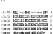
単離された「Tespec PRO−1」(Testis specific expressed serine proteinase−1)は321アミノ酸をコードすると予想され、その予想アミノ酸配列はトリプシンファミリーセリンプロテアーゼモチーフである「Trypsin−His」および「Trypsin−Ser」活性部位を有し、また他のトリプシンファミリーセリンプロテアーゼであるアクロシンやプロスタシン、トリプシンなどと、この2つのモチーフおよびその近傍において非常に高い相同性を示した。しかし、それ以外の領域においては核酸およびアミノ酸レベルで高い相同性を示す既知遺伝子は存在せず、新規なトリプシンファミリーセリンプロテアーゼであることが判明した。
一方、「Tespec PRO−2」は319アミノ酸をコードすると予想され、「Trypsin−His」活性部位を保持していた。[Trypsin−Ser」活性部位については12アミノ酸よりなるモチーフ中、2アミノ酸がモチーフとは異なるが、他の既知トリプシンファミリーセリンプロテアーゼにおいてもこのような例は存在するため、「Tespec PRO−2」はプロテアーゼとして機能すると考えられる。「Tespec PRO−2」もまた、核酸およびアミノ酸レベルで高い相同性を示す既知遺伝子は存在せず、新規なトリプシンファミリーセリンプロテアーゼであることが判明した。
「Tespec PRO−2」には、興味深いことに「Tespec PRO−2」の前半領域と「Tespec PRO−1」の後半領域がつながったスプライシングアイソフォームが存在した。このため、これら2つのプロテアーゼは染色体上で非常に近い位置に存在することが示唆された。「Tespec PRO−2」には種々のスプライシングアイソフォームが存在するが、「Tespec PRO−2」以外のアイソフォームは長いORFが開かず、プロテアーゼをコードしていなかった。「Tespec PRO−1」および「Tespec PRO−2」は核酸レベル、アミノ酸レベルでそれぞれ52.2%、33.1%の相同性を有していた。
本発明者らは、また、マウス「Tespec PRO−2」の核酸配列を基に、RT−PCR及びRACEを用いて、ヒト「Tespec PRO−2」のcDNAのクローニングを行った。これにより取得されたヒト「Tespec PRO−2」とマウス「Tespec PRO−2]は核酸レベル、アミノ酸レベルでそれぞれ74.2%、69.8%の相同性を有することが判明した。また、ヒト「Tespec PRO−2」は第8染色体にコードされていることが判明した。
本発明者らはさらに、マウス「Tespec PRO−1」の核酸配列を基に、RT−PCR及びRACEを用いて、ヒト「Tespec PRO−3」のcDNAのクローニングに成功した。また、ヒト「Tespec PRO−3」のカウンターパートであるマウス「Tespec PRO−3」のcDNAのクローニングにも成功した。
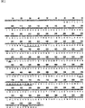
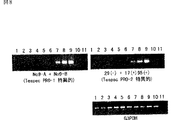
「Tespec PRO−1」をコードする領域をプローブとしたマウスノーザンブロットの解析より、この遺伝子は成体マウス精巣でのみ発現が見られ、他の組織や胎児期ではその発現は確認されなかった。同様にRT−PCRによる解析からも、「Tespec PRO−1」の発現は成体精巣で非常に高いことが判明した。また、「Tespec PRO−1」は生後18日目以降の精巣で発現の上昇が確認できたが、生後12日目以前の精巣や精子形成不全の変異体マウス精巣ではその発現が全く見られなかった。同様な解析を「Tespec PRO−2」でも行ったところ、「Tespec PRO−1」と同様の発現パターンであることが判明した。これら事実は「Tespec PRO−1]及び「Tespec PRO−2」が、精子の分化・熟成または精子の機能(受精)に関与していることを示唆する。一方、トリプシンファミリーセリンプロデアーゼに関しては、受精の際に重要な役割を担っていることが示唆されている。
従って、本発明者らは、見出した遺伝子によりコードされるタンパク質が、受精の鍵となるセリンプロテアーゼである可能性があり、新しい不妊症の治療薬や不妊症診断薬の開発、あるいは新しい避妊薬の開発に有用であると考えた。
本発明は、精子の形成や機能に関連すると考えられる新規トリプシンファミリーセリンプロテアーゼ、それらをコードする遺伝子、並びにそれらの製造方法および用途に関し、より具体的には、
1. 配列番号:2、4、6、8、10のいずれかに記載のアミノ酸配列からなるタンパク質、
2. 配列番号:2、4、6、8、10のいずれかに記載のアミノ酸配列からなるタンパク質と機能的に同等な、下記(a)または(b)に記載のタンパク質、
(a) 配列番号:2、4、6、8、10のいずれかに記載のアミノ酸配列において1若しくは複数のアミノ酸が欠失、付加、挿入および/または他のアミノ酸による置換により修飾されたアミノ酸配列からなるタンパク質、
(b) 配列番号:1、3、5、7、9のいずれかに記載の塩基配列からなるDNAとハイブリダイズするDNAがコードするタンパク質、
3. (1)または(2)に記載のタンパク質の部分ペプチド、
4. (1)または(2)に記載のタンパク質と他のペプチドとからなる融合タンパク質、
5. (1)〜(3)のいずれかに記載のタンパク質をコードするDNA、
6. (5)に記載のDNAが挿入されたベクター、
7. (5)に記載のDNAを発現可能に保持する形質転換体、
8. (7)に記載の形質転換体を培養し、該形質転換体またはその培養上清から発現させたタンパク質を回収する工程を含む、(1)〜(3)のいずれかに記載のタンパク質の製造方法、
9. (1)または(2)に記載のタンパク質の基質をスクリーニングする方法であって、
(a)該タンパク質に被検試料を接触させる工程、
(b)該タンパク質の被検試料に対するプロテアーゼ活性を検出する工程、および
(c)該プロテアーゼ活性により分解または切断を受ける化合物を選択する工程、を含む方法、
10. (9)に記載の方法により単離されうる、(1)または(2)に記載のタンパク質の基質、
11. (1)または(2)に記載のタンパク質の活性を阻害する化合物をスクリーニングする方法であって、
(a)被検試料の存在下で該タンパク質に(10)に記載の基質を接触させる工程、
(b)該タンパク質による該基質に対するプロテアーゼ活性を検出する工程、および
(c)被検試料非存在下において検出した場合と比較して、該プロテアーゼ活性を低下させる化合物を選択する工程、を含む方法、
12. (11)に記載の方法により単離されうる、(1)または(2)に記載のタンパク質の活性を阻害する化合物、
13. (1)または(2)に記載のタンパク質に結合する抗体、
14. (13)に記載の抗体と、(1)または(2)に記載のタンパク質が含まれると予想される試料とを接触させ、該抗体と該タンパク質との免疫複合体の生成を検出又は測定することを含んでなる、該タンパク質を検出又は測定する方法、
15. 配列番号:1、3、5、7または9のいずれかに記載の塩基配列からなるDNAと特異的にハイブリダイズし、少なくとも15塩基の鎖長を有するヌクレオチド、を提供するものである。
本発明は、新規トリプシンファミリーセリンプロテアーゼを提供する。本発明のタンパク質に含まれる「Tespec PRO−1」と命名されたマウスタンパク質のアミノ酸配列を配列番号:2に、「Tespec PRO−2」と命名されたマウスおよびヒトタンパク質のアミノ酸配列をそれぞれ配列番号:4および6に、また「Tespec PRO−3」と命名されたマウスおよびヒトタンパク質のアミノ酸配列をそれぞれ配列番号:8および10に示し、それらのタンパク質をコードするcDNAの塩基配列をそれぞれ配列番号:1、3、5、7および9に示す。
本発明のタンパク質に含まれる[Tespec PRO−1」及び「Tespec PRO−2」は、マウス精巣において高い発現が観察された(実施例5および6)。これらのタンパク質は精子、特にその先端領域に存在するならば、精子が透明帯を消化し受精を成立させる際の鍵となるプロテアーゼである可能性がある。従って、本発明のタンパク質は、不妊症治療薬や不妊症診断薬あるいは避妊薬の開発に有用であると考えられる。
また、本発明のタンパク質にはマウス「Tespec PRO−1」タンパク質、マウス「Tespec PRO−2」タンパク質、ヒト「Tespec PRO−2」タンパク質、マウス「Tespec PRO−3」タンパク質、またはヒト「Tespec PRO−3」タンパク質と機能的に同等なタンパク質を包含する。ここで「機能的に同等」とは、マウス「Tespec PRO−1」タンパク質、マウス「Tespec PRO−2」タンパク質、ヒト「Tespec PRO−2」タンパク質、マウス「Tespec PRO−3」タンパク質、またはヒト「Tespec PRO−3」タンパク質と同等の生物学的特性を有していることを意味する。生物学的特性としては、例えば、▲1▼活性の特性として、トリプシンファミリーセリンプロテアーゼ活性を有すること、▲2▼配列の構造的特性として、トリプシンファミリーセリンプロテアーゼモチーフ(「Trypsin−His(PROSITE PS00134)」、「Trypsin−Ser(PROSITE PS00135)」)および/またはそれと類似した配列を有することや、マウス「Tespec PRO−1」タンパク質、マウス「Tespec PRO−2」タンパク質、ヒト「Tespec PRO−2」タンパク質、マウス[Tespec PRO−3]タンパク質、もしくはヒト「Tespec PRO−3」タンパク質のアミノ酸配列と高い相同性が見られること(後述)、▲3▼発現特性として、精巣に発現すること、などが挙げられる。
このようなタンパク質を得るための方法としては、例えば、タンパク質のアミノ酸配列に変異を導入する方法が用いられている。アミノ酸配列に変異が導入されたタンパク質を得るためには、例えば、合成オリゴヌクレオチドプライマーを利用した部位特異的変異誘発法(Kramer,W.and Fritz,H.J.Methods in Enzymol.(1987)154,350−367)やPCRによる部位特異的変異誘発システム(GIBCO−BRL社)、Kunkel法(Methods Enzymol.85,2763−2766(1988))を使用することができる。これらの方法により、配列番号:2、4、6、8または10に示されたアミノ酸配列からなるタンパク質において、その生物学的特性に影響を与えないよう、1若しくは複数個のアミノ酸の欠失、付加、挿入及び/又は他のアミノ酸による置換により修飾されたタンパク質を得ることができる。
変異するアミノ酸の個数は、配列番号:2、4、6、8または10に示されたアミノ酸配列からなるタンパク質の生物学的特性を保持しつる限り特に制限はない。配列番号:2、4、6、8または10のいずれかに示されるアミノ酸配列中の1又は2個以上、好ましくは、2個以上30個以下、より好ましくは2個以上10個以下のアミノ酸が欠失したもの、配列番号:2、4、6、8または10のいずれかに示されるアミノ酸配列に1又は2個以上、好ましくは、2個以上30個以下、より好ましくは2個以上10個以下のアミノ酸が付加したもの、配列番号:2、4、6、8または10のいずれかに示されるアミノ酸配列中の1又は2個以上、好ましくは、2個以上30個以下、より好ましくは2個以上10個以下のアミノ酸が他のアミノ酸で置換されたものが挙げられる。また、アミノ酸の変異部位も、配列番号:2、4、6、8または10に示されたアミノ酸配列からなるタンパク質の生物学的特性を保持しうる限り特に制限はない。
あるアミノ酸配列に対する1または複数個のアミノ酸残基の欠失、付加および/または他のアミノ酸による置換により修飾されたアミノ酸配列を有するタンパク質がその生物学的活性を維持することはすでに知られている(Mark,D.F.et al.,Proc.Natl.Acad.Sci.USA(1984)81,5662−5666、Zoller,M.J.& Smith,M.Nucleic Acids Research(1982)10,6487−6500、Wang,A.et al.,Science224,1431−1433、Dalbadie−McFarland,G.et al.,Proc.Natl.Acad.Sci.USA(1982)79,6409−6413)。
例えば、本発明のタンパク質に1または複数個のアミノ酸残基が付加されたタンパク質として、融合タンパク質が挙げられる。融合タンパク質は、本発明のタンパク質と他のペプチドとが融合したものである。融合タンパク質は人為的に作製することもできる。例えば、本発明のタンパク質をコードするDNAと他のペプチドをコードするDNAをフレームが一致するように連結してこれを発現ベクターに導入し、宿主で発現させればよく、すでに公知の手法を用いることができる。本発明のタンパク質との融合に付される他のペプチドとしては、特に限定されない。例えば、ペプチドとしては、FLAG(Hopp,T.P.et al.,BioTechnology(1988)6,1204−1210)、6個のHis(ヒスチジン)残基からなる6×His、10×His、インフルエンザ凝集素(HA)、ヒトc−mycの断片、VSV−GPの断片、p18HIVの断片、T7−tag、HSV−tag、E−tag、SV40T抗原の断片、lck tag、α−tubulinの断片、B−tag、ProteinCの断片等、すでに公知であるペプチドが使用される。またタンパク質としては、例えばGST(グルタチオン・S・トランスフェラーゼ)、HA(インフルエンザ凝集素)、イムノグロブリン定常領域、β−ガラクトシダーゼ、MBP(マルトース結合タンパク質)等が挙げられる。市販されているこれらをコードするDNAを融合させたものを用いることができる。
また、当業者にとっては、周知技術であるハイブリダイゼーション技術(Sambrook,J et al.,Molecular Cloning 2nd ed.9.47−9.58,Cold Spring Harbor Lab.press,1989)を用いて、上記本発明のタンパク質をコードするDNA(配列番号:1、3、5、7または9に記載のDNA)またはその一部を基に、これと相同性の高いDNAを単離して、該DNAから上記本発明のタンパク質と機能的に同等なタンパク質を得ることも当業者が通常行い得ることである。本発明のタンパク質には、ストリンジェントな条件下で上記本発明のタンパク質をコードするDNAまたはその一部とハイブリダイズするDNAがコードし、上記本発明のタンパク質と機能的に同等なタンパク質が含まれる。ハイブリダイズするDNAを他の生物から単離する場合、生物種に制限はないが、例えば、ヒト、マウス、ラット、ウシ、サル、ブタなどを挙げることができる。「ストリンジェントな条件」とは、通常、「42℃、2xSSC、0.1% SDS」程度であり、好ましくは「50℃、2xSSC、0.1% SDS」程度であり、さらに好ましくは「65℃、2xSSC、0.1% SDS」程度である。これらの条件において、温度を上げる程に高い相同性を有するDNAを得ることができる。
ハイブリダイズ技術により得られたDNAがコードするタンパク質は、通常、配列番号:2、4、6、8または10に記載のアミノ酸配列と高い相同性を有する。「高い相同性」とは、少なくとも60%以上の相同性、好ましくは70%以上の相同性、さらに好ましくは80%以上の相同性、さらに好ましくは95%以上の相同性を指す。タンパク質の相同性を決定するには、文献(Wilbur,W.J.and Lipman,D.J.Proc.Natl.Acad.Sci.USA(1983)80,726−730)に記載のアルゴリズムにしたがえばよい。
本発明のタンパク質は、該タンパク質を産生する細胞や宿主あるいは精製方法により、アミノ酸配列、分子量、等電点又は糖鎖の有無や形態が異なっていてもよい。得られたタンパク質が配列番号:2、4、6、8または10に記載のアミノ酸配列からなるタンパク質の生物学的特性を保持している限り、本発明のタンパク質に含まれる。
本発明のタンパク質は、天然のタンパク質として、また遺伝子組換技術を利用して組み換えタンパク質として製造することが可能である。天然のタンパク質は、本発明のタンパク質が存在すると考えられる組織若しくは細胞(例えば精巣)からタンパク質を抽出し、後述する本発明の抗体を利用したアフィニティークロマトグラフィーを行うことにより調製することができる。
また、組み換えタンパク質を製造するには、本発明のタンパク質をコードするDNAを発現制御領域、例えばエンハンサー、プロモーターの制御のもとで発現可能なように発現ベクターに組み込み、この発現ベクターにより宿主細胞を形質転換し、タンパク質を発現させることができる。
具体的には、例えば、哺乳類細胞を使用する場合、常用される有用なプロモーター/エンハンサー、本発明のタンパク質をコードするDNA、その3’側下流にポリAシグナルを機能的に結合させたDNAあるいはそれを含むベクターを構築することができる。例えばプロモーター/エンハンサーとしては、ヒトサイトメガロウイルス前期プロモーター/エンハンサー(human cytomegalovirus immediateearly promoter/enhancer)を挙げることができる。
また、その他にタンパク質発現に使用できるプロモーター/エンハンサーとして、レトロウイルス、ポリオーマウイルス、アデノウイルス、シミアンウイルス40(SV40)等のウイルスプロモーター/エンハンサーやヒトエロンゲーションファクター1α(HEF1α)の哺乳類細胞由来のプロモーター/エンハンサー等を用いることができる。
例えば、SV40プロモーター/エンハンサーを使用する場合、Mulliganらの方法(Nature(1979)277,108)、また、HEF1αプロモーター/エンハンサーを使用する場合、Mizushimaらの方法(Nucleic Acids Res.(1990)18,5322)に従えば容易に実施することができる。
複製開始点としては、SV40、ポリオーマウイルス、アデノウイルス、ウシパピローマウイルス(BPV)等の由来のものを用いることもできる。さらに、宿主細胞系で遺伝子コピー数増幅のため、発現ベクターは選択マーカーとして、アミノグリコシドトランスフェラーゼ(APH)遺伝子、チミジンキナーゼ(TK)遺伝子、大腸菌キサンチングアニンホスホリポシルトランスフェラーゼ(Ecogpt)遺伝子、ジヒドロ葉酸還元酵素(dhfr)遺伝子等を含むことができる。
大腸菌を使用する場合、常用される有用なプロモーター、ポリペプチド分泌のためのシグナル配列、発現させる遺伝子を機能的に結合させて発現させることができる。例えばプロモーターとしては、lacZプロモーター、araBプロモーターを挙げることができる。lacZプロモーターを使用する場合、Wardらの方法(Nature(1098)341,544−546;FASEB J.(1992)6,2422−2427)、araBプロモーターを使用する場合、Betterらの方法(Science(1988)240,1041−1043)に従えばよい。
タンパク質分泌のためのシグナル配列としては、大腸菌のペリプラズムに産生させる場合、pelBシグナル配列(Lei,S.P.et al J.Bacteriol.(1987)169,4379)を使用すればよい。
本発明のタンパク質を製造するための発現ベクターは、本発明に好適に使用される発現ベクターであればいかなる発現ベクターであってよい。本発明の発現ベクターとしては、例えば、アデノウイルスベクター「pAdexLcw」やレトロウイルスベクター「pZIPneo」などが挙げられる。また哺乳動物由来の発現ベクター、例えばpEF、pCDM8、昆虫細胞由来の発現ベクター、例えばpBacPAK8、植物由来の発現ベクター、例えばpMH1、pMH2、動物ウイルス由来の発現ベクター、例えばpHSV、pMV、pAdexLcw、レトロウイルス由来の発現ベクター。例えばpZIpneo、酵母由来の発現ベクター、例えばpNV11、SP−Q01、枯草菌由来の発現ベクター、例えばpPL608、pKTH50、大腸菌由来の発現ベクター、例えばpQE、pGEAPP、pGEMEAPP、pMALp2、pREP4が挙げられる。
本発明において、タンパク質の製造のために、任意の産生系を使用することができる。タンパク質製造のための産生系は、in vitroおよびin vivoの産生系がある。in vitroの産生系としては、真核細胞を使用する産生系や原核細胞を使用する産生系が挙げられる。
真核細胞を使用する場合、動物細胞、植物細胞、真菌細胞を用いる産生系がある。動物細胞としては、哺乳類細胞、例えばCHO(J.Exp.Med.(1995)108,945)、COS、ミエローマ、BHK(baby hamster kidney)、HeLa、Vero、両生類細胞、例えばアフリカツメガエル卵母細胞(Valle,et al.,Nature(1981)291,358−340)、あるいは昆虫細胞、例えばsf9、sf21、Tn5が知られている。CHO細胞としては、特にDHFR遺伝子を欠損したCHO細胞であるdhfr−CHO(Proc.Natl.Acad.Sci.USA(1980)77,4216−4220)やCHO K−1(Proc.Natl.Acad.Sci.USA(1968)60,1275)を好適に使用することができる。
植物細胞としては、ニコチアナ・タバカム(Nicotiana tabacum)由来の細胞が知られており、これをカルス培養すればよい。真菌細胞としては、酵母、例えばサッカロミセス(Saccharomyces)属、例えばサッカロミセス・セレビシエ(Saccharomyces cerevisiae)、糸状菌、例えばアスペルギルス属(Aspergillus)属、例えばアスペルギルス・ニガー(Aspergillus niger)が知られている。
原核細胞を使用する場合、細菌細胞を用いる産生系がある。細菌細胞としては、大腸菌(E.coli)、枯草菌が知られている。
これらの細胞を目的とするDNAにより形質転換し、形質転換された細胞をin vitroで培養することによりタンパク質が得られる。培養は、公知の方法に従い行う。真核細胞であれば例えば、培養液としてDMEM、MEM、RPMI1640、IMDMを使用することができる。その際、牛胎児血清(FCS)等の血清補液を併用することもできるし、無血清培養してもよい。培養時のpHは約6〜8であるのが好ましい。培養は通常約30〜40℃で約15〜200時間行い、必要に応じて培地の交換、通気、攪拌を加える。
一方、in vivoの産生系としては、動物を使用する産生系や植物を使用する産生系が挙げられる。これらの動物又は植物に目的とするDNAを導入し、動物又は植物の体内でタンパク質を産生させ、回収する。本発明における「宿主」とは、これらの動物、植物を包含する。
動物を使用する場合、哺乳類動物、昆虫を用いる産生系がある。哺乳類動物としては、ヤギ、ブタ、ヒツジ、マウス、ウシを用いることができる(VickiGlaser,SPECTRUM Biotechnology Applications,1993)。また、哺乳類動物を用いる場合、トランスジェニック動物を用いることができる。例えば、目的とするDNAをヤギβカゼインのような乳汁中に固有に産生されるタンパク質をコードする遺伝子の途中に挿入して融合遺伝子として調製する。このDNAが挿入された融合遺伝子を含むDNA断片をヤギの胚へ注入し、この胚を雌のヤギへ導入する。胚を受容したヤギから生まれるトランスジェニックヤギ又はその子孫が産生する乳汁からタンパク質を得る。トランスジェニックヤギから産生されるタンパク質を含む乳汁量を増加させるために、適宜ホルモンをトランスジェニックヤギに使用してもよい(Ebert,K.M.et al.,Bio/Technology(1994)12,699−702)。
また、昆虫としては、例えばカイコを用いることができる。カイコを用いる場合、目的とするDNAを挿入したバキュロウイルスをカイコに感染させ、このカイコの体液より所望のタンパク質を得る(Susumu,M.et al.,Nature(1985)315,592−594)。
さらに植物を使用する場合、例えばタバコを用いることができる。タバコを用いる場合、目的とするDNAを植物発現用ベクター、例えばpMON530に挿入し、このベクターをアグロバクテリウム・ツメファシエンス(Agrobacterium tumefaciens)のようなバクテリアに導入する。このバクテリアをタバコ、例えばニコチアナ・タバカム(Nicotiana tabacum)に感染させ、本タバコの葉より所望のポリペプチドを得る(Julian,K.−C.Ma et al.,Eur.J.Immunol.(1994)24,131−138)。
上記で得られた本発明のタンパク質は、細胞内外、宿主から単離し実質的に純粋で均一なタンパク質として精製することができる。タンパク質の分離、精製は、通常のタンパク質の精製で使用されている分離、精製方法を使用すればよく、何ら限定されるものではない。例えば、クロマトグラフィーカラム、フィルター、限外濾過、塩析、溶媒沈殿、溶媒抽出、蒸留、免疫沈降、SDS−ポリアクリルアミドゲル電気泳動、等電点電気泳動法、透析、再結晶等を適宜選択、組み合わせればタンパク質を分離、精製することができる。
クロマトグラフィーとしては、例えばアフィニティークロマトグラフィー、イオン交換クロマトグラフィー、疎水性クロマトグラフィー、ゲル濾過、逆相クロマトグラフィー、吸着クロマトグラフィー等が挙げられる(Strategies for Protein Purification and Characterization:A Laboratory Course Manual.Ed Daniel R.Marshak et al.,Cold Spring Harbor Laboratory Press,1996)。これらのクロマトグラフィーは、液相クロマトグラフィー、例えばHPLC、FPLC等の液相クロマトグラフィーを用いて行うことができる。本発明は、これらの精製方法を用い、高度に精製されたタンパク質も包含する。
なお、タンパク質を精製前又は精製後に適当なタンパク修飾酵素を作用させることにより、任意に修飾を加えたり部分的にペプチドを除去することもできる。タンパク修飾酵素としては、トリプシン、キモトリプシン、リシルエンドペプチダーゼ、プロテインキナーゼ、グルコシダーゼ、プロテインキナーゼ、グルコシダーゼが用いられる。
本発明は、また、本発明のタンパク質の部分ペプチドも含む。このようなペプチドは、例えば、本発明のタンパク質に結合する抗体を得るための免疫原として利用しうる。この場合、ペプチドは、少なくとも12アミノ酸以上の鎖長を有し、好ましくは、20アミノ酸以上の鎖長を有する。本発明のタンパク質の部分ペプチドは、遺伝子工学的手法、公知のペプチド合成法、あるいは本発明のタンパク質を適切なペプチダーゼで切断することによって製造することができる。ペプチド合成法としては、たとえば固相合成法、液相合成法のいずれによっても良い。
遺伝子工学的手法を利用して宿主内で発現させた本発明のタンパク質若しくはその部分ペプチドは、細胞内外から単離し実質的に純粋で均一なタンパク質として精製することができる。タンパク質の分離、精製は、通常のタンパク質の精製で使用されている分離、精製方法を使用すればよく、何ら限定されるものではない。例えば、クロマトグラフィーカラム、フィルター、限外濾過、塩析、溶媒沈殿、溶媒抽出、蒸留、免疫沈降、SDS−ポリアクリルアミドゲル電気泳動、等電点電気泳動法、透析、再結晶等を適宜選択、組み合わせればタンパク質を分離、精製することができる。
クロマトグラフィーとしては、例えばアフィニティークロマトグラフィー、イオン交換クロマトグラフィー、疎水性クロマトグラフィー、ゲル濾過、逆相クロマトグラフィー、吸着クロマトグラフィー等が挙げられる(Strategies for Protein Purification and Characterization:A Laboratory Course Manual.Ed Daniel R.Marshak et al.,Cold Spring Harbor Laboratory Press,1996)。これらのクロマトグラフィーは、液相クロマトグラフィー、例えばHPLC、FPLC等の液相クロマトグラフィーを用いて行うことができる。本発明は、これらの精製方法を用い、高度に精製されたタンパク質も包含する。
なお、タンパク質を精製前又は精製後に適当なタンパク修飾酵素を作用させることにより、任意に修飾を加えたり部分的にペプチドを除去することもできる。タンパク修飾酵素としては、トリプシン、キモトリプシン、リシルエンドペプチダーゼ、プロテインキナーゼ、グルコシダーゼ、プロテインキナーゼ、グルコシダーゼが用いられる。
また、本発明は、上記本発明のタンパク質をコードするDNAを提供する。本発明のDNAは、in vivo、in vitroで本発明のタンパク質を製造するのみならず、例えば、哺乳動物、例えばヒトの遺伝子治療に用いることもできる。特に、本発明の遺伝子は、不妊症の遺伝子治療への応用が期待される。遺伝子治療に用いる場合には、本発明のDNAが挿入されたベクターを生体内の標的部位へ投与する。投与方法は、ex vivo法であっても、in vivo法であってもよい。なお、本発明のベクターには、このように遺伝子治療に用いられるベクターも含まれる。
本発明のタンパク質をコードするゲノムDNA又はcDNAは、当業者に周知のハイフリダイゼーション技術を用いて、それぞれゲノムライブラリーまたはcDNAライブラリー等をスクリーニングして得ることができる。
得られたDNA又はDNA断片をプローブとして、さらにゲノムライブラリーやcDNAライブラリーをスクリーニングすることにより、異なる細胞、組織、臓器又は種から遺伝子を得ることができる。ゲノムライブラリーまたはcDNAライブラリーは、例えばSambrook,J.et al.,Molecular Cloning、Cold Spring Harbor Laboratory Press(1989)に記載の方法により調製してもよいし、市販のDNAライブラリーを用いてもよい。
また、得られたcDNAの塩基配列を決定することにより、それがコードする翻訳領域を決定でき、本発明のタンパク質のアミノ酸配列を得ることができる。
具体的には、例えば次のようにすればよい。まず、本発明のタンパク質を発現する細胞、組織、臓器からmRNAを単離する。mRNAの単離は公知の方法、例えば、グアニジン超遠心法(Chirgwin,J.M.et al.,Biochemistry(1979)18,5294−5299)、AGPC法(Chomczynski,P.and Sacchi,N.,Anal.Biochem.(1987)162,156−159)等により全RNAを調製し、mRNA Purification Kit(Pharmacia)等を使用して全RNAからmRNAを精製する。また、QuickPrep mRNA Purification Kit(Pharmacia)を用いることによりmRNAを直接調製することもできる。
得られたmRNAから逆転写酵素を用いてcDNAを合成する。cDNAの合成は、AMVReverse Transcriptase First−strand cDNA Synthesis Kit(生化学工業)等を用いて行うこともできる。また、本明細書に記載されたプローブを用いて、5’−Ampli FINDER RACE Kit(Clontech製)およびポリメラーゼ連鎖反応(polymerase chainreaction;PCR)を用いた5’−RACE法(Frohman,M.A.et al.,Proc.Natl.Acad.Sci.U.S.A.(1988)85,8998−9002;Belyavsky,A.et al.,Nucleic Acids Res.(1989)17,2919−2932)に従い、cDNAの合成および増幅を行うことができる。
得られたPCR産物から目的とするDNA断片を調製し、ベクターDNAと連結する。さらに、これより組換えベクターを作製し、大腸菌等に導入してコロニーを選択して所望の組換えベクターを調製する。目的とするDNAの塩基配列を公知の方法、例えば、ジデオキシヌクレオチドチェインターミネーション法により確認すればよい。
また、本発明のDNAは、発現に使用する宿主のコドン使用頻度を考慮して、より発現効率の高い配列を設計することができる(Grantham,R.et al.,Nucelic Acids Research(1981)9,r43−r74)。また、本発明のDNAを市販のキットや公知の方法によって改変することができる。例えば、制限酵素による消化、合成オリゴヌクレオチドや適当なDNAフラグメントの挿入、リンカーの付加、開始コドン(ATG)及び/又は終始コドン(TAA、TGA 又はTAG)の挿入等が挙げられる。
本発明のDNAは、具体的には例えば配列番号:1の塩基配列において48位の塩基Aから1010位の塩基CからなるDNA、配列番号:3の塩基配列において69位の塩基Aから1025位の塩基CからなるDNA、配列番号:5の塩基配列において73位の塩基Aから867位の塩基AからなるDNA、配列番号:7の塩基配列において38位の塩基Aから1000位の塩基AからなるDNA、配列番号:9の塩基配列において41位の塩基Aから1096位の塩基CからなるDNAを包含する。
本発明のDNAはまた、配列番号:1、3、5、7または9のいずれかに記載の塩基配列からなるDNAとストリンジェントな条件下でハイブリダイズするDNAであり、且つ本発明のタンパク質と機能的に同等なタンパク質をコードするDNAを含む。
「ストリンジェントな条件」とは、通常、「42℃、2xSSC、0.1% SDS」程度であり、好ましくは「50℃、2xSSC、0.1%SDS」程度であり、さらに好ましくは「65℃、2xSSC、0.1%SDS」程度である。これらの条件において、温度を上げる程に高い相同性を有するDNAを得ることができる。
上記のハイブリダイズするDNAは、例えば天然由来のDNA(例えばcDNA又はゲノムDNA)であってよい。天然由来のDNAの場合、機能的に同等なタンパク質をコードするDNAを単離するための生物としては、例えば、ヒト、マウス、ラット、ウシ、サル、ブタなどが挙げられる。例えばこれらの生物において、実施例において本発明のタンパク質をコードするcDNAとハイブリダイズするmRNAが検出される組織(例えば精巣)由来のcDNAを用いて本発明のDNAを単離することができる。また、本発明のタンパク質をコードするDNAはcDNAやゲノムDNAの他、合成DNAであってもよい。
また、本発明は、本発明のタンパク質の基質のスクリーニング方法を提供する。本発明のタンパク質の「基質」とは、本発明のタンパク質が結合することにより特定の部位で分解または切断を受ける化合物を指す。
基質となる化合物としては、タンパク質に制限されない。例えば、トリプシン(trypsin)やキモトリプシン(Chymotrypsin)は、タンパク質のみならず、ペプチド化合物の誘導体のアミド結合やエステル結合も切断することが知られている(Farmer,D.A.,et al.,J Biol Chem.(1975)250,7366−7371;del Castillo,L.M.,et al.,Biochim Biophys Acta.(1971)235,358−69)。従って、本発明において、基質としては本発明のタンパク質が結合することにより特定の部位で分解または切断を受ける限り限定されず、ペプチドやその類縁体、誘導体(ペプチド性化合物)、または非ペプチド性化合物などでもよい。
本発明の基質のスクリーニング方法は、(a)本発明のタンパク質に被検試料を接触させる工程、(b)本発明のタンパク質の被検試料に対するプロテアーゼ活性を検出する工程、および(c)本発明のタンパク質のプロテアーゼ活性により分解若しくは切断を受ける化合物を選択する工程、を含む。
スクリーニングに使用される被検試料としては、本発明のタンパク質の基質を含むことが期待される試料であれば特に制限はなく、例えば、細胞抽出液、動物組織抽出液、遺伝子ライブラリーの発現産物、精製若しくは粗精製タンパク質、ペプチド、ペプチドの類縁体若しくは誘導体、非ペプチド性化合物、合成化合物、天然化合物などが挙げられる。
本発明のタンパク質に結合する基質のスクリーニングにおいては、例えば、本発明のタンパク質と被検試料とを混合し、インキュベートした後、被検試料の変化(切断、分解)を測定する。例えば、タンパク質を被検試料とする場合、被検試料を直接、またはアジド化したり、蛍光物質と結合させ、反応前後の試料の変化をUVスペクトル(R.J.Beynon and J.S.Bond,Proteolytic enzymes(1989)IRL press 25−55)や、HPLC(Maier M,et al.(1988)FEBS Lett.232,395−398,Gau W,et al.Adv Exp Med Biol.(1983)156,483−494)で検出し、プロテアーゼ活性を測定することができる。
また、ペプチド(及び類縁体、誘導体)を被検試料とする場合、数アミノ酸(1〜5アミノ酸の場合が多いが、特に制限はない)からなるペプチド(及び類縁体、誘導体)を本発明のタンパク質と混合し、インキュベート後被検試料の変化を測定する。例えば、被検試料のカルボキシル末端に蛍光性化合物(MEC;Kawabata S,et al.(1988)Eur.J.Biochem.172,17−25,AMC; Morita T,et al.(1977)JBiochem(Tokyo).82,1495−1498,AFC;Garrett JR,et al.(1985)Histochem.J.17,805−817,など)を導入しておき、被検試料が切断を受けた際に生じる蛍光性化合物のスペクトル変化を指標にプロテアーゼ活性を測定すればよい。また、その他の蛍光標識ペプチド基質を用いたスクリーニング方法を用いることもできる(R.J.Beynon and J.S.Bond,Proteolytic enzymes(1989)IRL press25−55,Gossrau R,et al.(1984)Adv Exp Med Biol.167,191−207,Yu JX,et al.(1994)J Biol Chem.269,18843−18848)。
また、上記方法の原理を利用して、被検化合物として合成化合物、または天然物バンク、ランダムファージペプチドディスプレイライブラリー、もしくはピンペプチド合成物等を用いてスクリーニングすることや、コンビナトリアルケミストリー技術によるハイスループットスクリーニングも可能である(Wrighton NC;Farrell FX; Chang R;Kashyap AK;Barbone FP;Mulcahy LS;Johnson DL;Barrett RW;Jolliffe LK;Dower WJ.,Small peptides as potent mimetics of the protein hormone erythropoietin,Science(UNITED STATES)Jul 26 1996,273 p458−64、Verdine GL.,The combinatorial chemistry of nature.Nature(ENGLAND)Nov7 1996,384 p11−13、Hogan JC Jr.,Directed combinatorial chemistry.Nature(ENGLAND)Nov 7 1996,384 p17−9)。
上記スクリーニング方法を利用して、本発明のタンパク質の基質が単離されれば、本発明のタンパク質の該基質に対するプロテアーゼ活性を阻害する活性を指標に、本発明のタンパク質の阻害剤をスクリーニングすることができる。従って、本発明は、また、本発明のタンパク質の活性を阻害する化合物をスクリーニングする方法を提供する。
この方法は、(a)被検試料の存在下で本発明のタンパク質にその基質を接触させる工程、(b)本発明のタンパク質の該基質に対するプロテアーゼ活性を検出する工程、および(c)被検試料が非存在下において検出した場合と比較して、該プロテアーゼ活性を低下させる化合物を選択する工程、を含む。
スクリーニングに使用される本発明のタンパク質としては、天然のタンパク質、組換えタンパク質、またはそれらの部分ペプチドであってもよい。また、スクリーニングに使用される被検試料としては、特に制限はなく、例えば、細胞培養上清、遺伝子ライブラリーの発現産物、ペプチド、ペプチドの類縁体若しくは誘導体、精製若しくは粗精製タンパク質(抗体を含む)、非ペプチド性化合物、合成化合物、微生物発酵生産物、海洋生物抽出液、植物抽出液、細胞抽出液、動物組織抽出液、が挙げられる。
本発明のタンパク質の阻害剤のスクリーニングは、例えば、上記文献「R.J.Beynon and J.S.Bond,Proteolytic enzymes(1989)IRL press25−55;Maier M,et al.(1988)FEBS Lett.232,395−398;Gau W,et al.Adv Exp Med Biol.(1983)156,483−494);Kawabata S,et al.(1988)Eur.J.Biochem.172,17−25,;MoritaT,et al.(1977)J Biochem(Tokyo).82,1495−1498;Garrett JR,et al.(1985)Histochem.J.17,805−817;Gossrau R,et al.(1984)Adv Exp Med Biol.167,191−207,Yu JX,et al.(1994)J Biol Chem.269,18843−18848」に記載の系を利用して行なうことができる。また、ペプチド性基質をリード化合物とし、その構造の一部を修飾したり置換した化合物を、本発明のタンパク質の阻害剤スクリーニングにおける被検化合物として用いることも可能である(Okamoto S,et al.(1993)Methods Enzymol.222,328−340)。
上述したように、本発明のタンパク質は、その発現特性等から、精子の分化・熟成または精子の機能(受精)に関与していることが示唆される。本発明のスクリーニング方法により単離される上記阻害剤は、本発明のタンパク質の受精への関与を解析するために利用しうる。例えば、本発明のタンパク質の阻害剤を用いて、インビトロ受精解析(Y.Toyoda et al.,1971,Jpn.J.Anim.Reprod.16:147−151;Y.Kuribayashi et al.,1996,Fertil.Steril.66:1012−1017)を行い、該阻害剤が受精を阻害するか否かを検討することにより、本発明のタンパク質の受精への関与を判定することができる。受精を阻害しうる本発明のタンパク質の阻害剤には、例えば、新たな避妊薬としての利用が考えられる。
本発明のスクリーニング方法を利用して得られる化合物をヒトや他の哺乳動物、例えばマウス、ラット、モルモット、ウサギ、ニワトリ、ネコ、イヌ、ヒツジ、ブタ、ウシ、サル、マントヒヒ、チンパンジーの医薬として使用する場合、常用される手段に従って実施することができる。
例えば、必要に応じて糖衣を施した錠剤、カプセル剤、エリキシル剤、マイクロカプセル剤として経口的に、あるいは水もしくはそれ以外の薬学的に許容し得る液との無菌性溶液、又は懸濁液剤の注射剤の形で非経口的に使用できる。例えば本発明のタンパク質と結合活性を有する化合物を生理学的に認められる担体、香味剤、賦形剤、ベヒクル、防腐剤、安定剤、結合剤とともに一般に認められた製薬実施に要求される単位用量形態で混和することによって製造することができる。これら製剤における有効成分量は指示された範囲の適当な容量が得られるようにするものである。
錠剤、カプセル剤に混和することができる添加剤としては、例えばゼラチン、コーンスターチ、トラガントガム、アラビアゴムのような結合剤、結晶性セルロースのような賦形剤、コーンスターチ、ゼラチン、アルギン酸のような膨化剤、ステアリン酸マグネシウムのような潤滑剤、ショ糖、乳糖又はサッカリンのような甘味剤、ペパーミント、アカモノ油又はチェリーのような香味剤が用いられる。調剤単位形態がカプセルである場合には、上記の材料にさらに油脂のような液状担体を含有することができる。注射のための無菌組成物は注射用蒸留水のようなベヒクル中の活性物質、胡麻油、椰子油のような天然産出植物油を溶解又は懸濁させるための通常の製剤実施に従って処方することができる。
注射用の水溶液としては、例えば生理食塩水、ブドウ糖やその他の補助薬を含む等張液、例えばD−ソルビトール、D−マンノース、D−マンニトール、塩化ナトリウムが挙げられ、適当な溶解補助剤、例えばアルコール、具体的にはエタノール、ポリアルコール、例えばプロピレングリコール、ポリエチレングリコール、非イオン性界面活性剤、例えばポリソルベート80(TM)、HCO−50と併用してもよい。
油性液としてはゴマ油、大豆油があげられ、溶解補助剤として安息香酸ベンジル、ベンジルアルコールと併用してもよい。また、緩衝剤、例えばリン酸塩緩衝液、酢酸ナトリウム緩衝液、無痛化剤、例えば塩化ペンザルコニウム、塩酸プロカイン、安定剤、例えばベンジルアルコール、フェノール、酸化防止剤と配合してもよい。調製された注射液は通常、適当なアンプルに充填させる。
本発明のスクリーニング方法により得られる化合物の投与量は、症状により差異はあるが、経口投与の場合、一般的に成人(体重60kgとして)においては、1日あたり約0.1から100mg、好ましくは約1.0から50mg、より好ましくは約1.0から20mgである。
非経口的に投与する場合は、その1回の投与量は投与対象、対象臓器、症状、投与方法によっても異なるが、例えば注射剤の形では通常成人(体重60kgとして)においては、1日あたり約0.01から30mg、好ましくは約0.1から20mg、より好ましくは約0.1から10mg程度を静脈注射により投与するのが好都合である。他の動物の場合も、体重60kg当たりに換算した量を投与することができる。
また、本発明は、本発明のタンパク質に結合する抗体を提供する。このような抗体は、本発明のタンパク質の検出や精製などに利用しうる。また、上述したin vitro受精解析において利用しうる。抗体は、公知の手段を用いてモノクローナル抗体又はポリクローナル抗体として得ることができる。
本発明のタンパク質に対して特異的に結合する抗体は、タンパク質を感作抗原として使用して、これを通常の免疫方法にしたがって免疫し、得られる免疫細胞を通常の細胞融合法によって公知の親細胞と融合させ、通常のスクリーニング法により、抗体産生細胞をスクリーニングすることによって作製できる。
具体的には、本発明のタンパク質に対して特異的に結合するモノクローナル抗体又はポリクローナル抗体を作製するには次のようにすればよい。
例えば、抗体取得の感作抗原として使用される本発明のタンパク質は、その由来となる動物種に制限されないが哺乳動物、例えばヒト、マウス又はラット由来のタンパク質が好ましく、特にヒト由来のタンパク質が好ましい。ヒト由来のタンパク質は、本明細書に開示される遺伝子配列又はアミノ酸配列を用いて得ることができる。
本発明において、感作抗原として使用されるタンパク質は、本発明のタンパク質またはその部分ペプチドを用いることができる。タンパク質の部分ペプチドとしては、例えばタンパク質のアミノ基(N)末端断片やカルボキシ(C)末端断片が挙げられる。本発明における「抗体」とはタンパク質の全長又は断片に結合する抗体を意味する。
本発明のタンパク質又はその断片をコードする遺伝子を公知の発現ベクター系に挿入して本明細書で述べた宿主細胞を形質転換させた後、その宿主細胞内外又は、宿主から目的のタンパク質又はその断片を公知の方法で得、このタンパク質を感作抗原として用いればよい。また、タンパク質を発現する細胞又はその溶解物あるいは化学的に合成した本発明のタンパク質またはその部分ペプチドを感作抗原として使用してもよい。
感作抗原で免疫される哺乳動物としては、特に限定されるものではないが、一般的にはげっ歯目、ウサギ目、霊長目の動物が使用される。モノクローナル抗体を作成する場合は、細胞融合に使用する親細胞との適合性を考慮して選択するのが好ましい。
げっ歯目の動物としては、例えば、マウス、ラット、ハムスター等が使用される。ウサギ目の動物としては、例えば、ウサギが使用される。霊長目の動物としては、例えばサルが使用される。サルとしては、狭鼻下目のサル(旧世界ザル)、例えば、カニクイザル、アカゲザル、マントヒヒ、チンパンジー等が使用される。
感作抗原を動物に免疫するには、公知の方法にしたがって行われる。例えば、一般的方法として、感作抗原を哺乳動物の腹腔内又は、皮下に注射することにより行われる。具体的には、感作抗原をPBS(Phosphate−Buffered Saline)や生理食塩水等で適当量に希釈、懸濁したものを所望により通常のアジュバント、例えば、フロイント完全アジュバントを適量混合し、乳化後、哺乳動物に投与し、以降フロイント不完全アジュバントに適量混合した感作抗原を、4〜21日毎に数回投与するのが好ましい。また、感作抗原免疫時に適当な担体を使用することができる。このように免疫し、血清中に所望の抗体レベルが上昇するのを常法により確認する。
ここで、本発明のタンパク質に対するポリクローナル抗体を得るには、血清中の所望の抗体レベルが上昇したことを確認した後、抗原を感作した哺乳動物の血液を取り出す。この血液から公知の方法により血清を分離する。ポリクローナル抗体としてポリクローナル抗体を含む血清を使用してもよいし、必要に応じこの血清からポリクローナル抗体を含む画分をさらに単離してもよい。
モノクローナル抗体を得るには、上記抗原を感作した哺乳動物の血清中に所望の抗体レベルが上昇するのを確認した後に、哺乳動物から免疫細胞を取り出し、細胞融合に付せばよい。この際、細胞融合に使用される好ましい免疫細胞として、特に脾細胞が挙げられる。前記免疫細胞と融合される他方の親細胞としては、好ましくは哺乳動物のミエローマ細胞が挙げられる。
前記免疫細胞とミエローマ細胞の細胞融合は基本的には公知の方法、例えば、ミルステインらの方法(Galfre,G.and Milstein,C.,Methods Enzymol.(1981)73,3−46)等に準じて行うことができる。
細胞融合により得られたハイブリドーマは、通常の選択培養液、例えばHAT培養液(ヒポキサンチン、アミノプテリンおよびチミジンを含む培養液)で培養することにより選択される。当該HAT培養液での培養は、目的とするハイブリドーマ以外の細胞(非融合細胞)が死滅するのに十分な時間、通常数日〜数週間継続する。ついで、通常の限界希釈法を実施し、目的とする抗体を産生するハイブリドーマのスクリーニングおよびクローニングが行われる。
また、ヒト以外の動物に抗原を免疫して上記ハイブリドーマを得る他に、ヒトリンパ球、例えばEBウイルスに感染したヒトリンパ球をin vitroでタンパク質、タンパク質発現細胞又はその溶解物で感作し、感作リンパ球をヒト由来の永久分裂能を有するミエローマ細胞、例えばU266と融合させ、タンパク質への結合活性を有する所望のヒト抗体を産生するハイブリドーマを得ることもできる(特開昭63−17688号公報)。
さらに、ヒト抗体遺伝子のレパートリーを有するトランスジェニック動物に抗原となるタンパク質、タンパク質発現細胞又はその溶解物を免疫して抗体産生細胞を取得し、これをミエローマ細胞と融合させたハイブリドーマを用いてタンパク質に対するヒト抗体を取得してもよい(国際公開92−03918号、国際公開93−2227号、国際公開94−02602号、国際公開94−25585号、国際公開96−33735号および国際公開96−34096号参照)。
ハイブリドーマを用いて抗体を産生する以外に、抗体を産生する感作リンパ球等の免疫細胞を癌遺伝子(oncogene)により不死化させた細胞を用いてもよい。
このように得られたモノクローナル抗体はまた、遺伝子組換え技術を用いて産生させた組換え型抗体として得ることができる(例えば、Borrebaeck,C.A.K.and Larrick,J.W.,THERAPEUTIC MONOCLONAL ANTIBODIES Published in the United Kingdom by MACMILLAN PUBLISHERS LTD,1990参照)。組換え型抗体は、それをコートするDNAをハイブリドーマ又は抗体を産生する感作リンパ球等の免疫細胞からクローニングし、適当なベクターに組み込んで、これを宿主に導入し産生させる。本発明は、この組換え型抗体を包含する。
本発明の抗体は、本発明のタンパク質に結合するかぎり、その抗体断片や抗体修飾物であってよい。例えば、抗体断片としては、Fab、F(ab’)2、Fv又はH鎖とL鎖のFvを適当なリンカーで連結させたシングルチェインFv(scFv)(Huston,J.S.et al.,Proc.Natl.Acad.Sci.U.S.A.(1988)85,5879−5883)が挙げられる。具体的には、抗体を酵素、例えば、パパイン、ペプシンで処理し抗体断片を生成させるか、又は、これら抗体断片をコードする遺伝子を構築し、これを発現ベクターに導入した後、適当な宿主細胞で発現させる(例えば、Co,M.S.et al.,J.Immunol.(1994)152,2968−2976;Better,M.and Horwitz,A.H.,MethodsEnzymol.(1989)178,476−496;Pluckthun,A.and Skerra,A.,Methods Enzymol.(1989)178,497−515;Lamoyi,E.,Methods Enzymol.(1986)121,652−663;Rousseaux,J.et al.,Methods Enzymol.(1986)121,663−669;Bird,R.E.and Walker,B.W.,Trends Biotechnol.(1991)9,132−137参照)。
抗体修飾物として、ポリエチレングリコール(PEG)等の各種分子と結合した抗体を使用することもできる。本発明における「抗体」にはこれらの抗体修飾物も包含される。このような抗体修飾物を得るには、得られた抗体に化学的な修飾を施すことによって得ることができる。これらの方法はこの分野において既に確立されている。
また、本発明の抗体は、公知の技術を使用して非ヒト抗体由来の可変領域とヒト抗体由来の定常領域からなるキメラ抗体又は非ヒト抗体由来のCDR(相補性決定領域)とヒト抗体由来のFR(フレームワーク領域)及び定常領域からなるヒト型化抗体として得ることができる。
前記のように得られた抗体は、均一にまで精製することができる。本発明で使用される抗体の分離、精製は通常のタンパク質で使用されている分離、精製方法を使用すればよく、何ら限定されるものではない。上記で得られた抗体の濃度測定は吸光度の測定又は酵素結合免疫吸着検定法(Enzyme−linked immunosorbentassay;ELISA)等により行うことができる。
また、本発明の抗体の抗原結合活性を測定する方法として、ELISA、EIA(酵素免疫測定法)、RIA(放射免疫測定法)あるいは蛍光抗体法を用いることができる。例えば、ELISAを用いる場合、本発明の抗体を同相化したプレートに本発明のタンパク質を添加し、次いで目的の抗体を含む試料、例えば、抗体産生細胞の培養上清や精製抗体を加える。酵素、例えばアルカリフォスファターゼ等で標識した抗体を認識するに次抗体を添加し、プレートをインキュベーション、洗浄した後、p−ニトロフェニル燐酸などの酵素基質を加えて吸光度を測定することで抗原結合活性を評価することができる。タンパク質としてタンパク質の断片、例えばそのC末端からなる断片あるいはN末端からなる断片を使用してもよい。本発明の抗体の活性評価には、BIAcore(Pharmacia製)を使用することができる。
これらの手法を用いることにより、本発明の抗体と試料中に含まれる本発明のタンパク質が含まれると予想される試料とを接触させ、前記抗体とタンパク質との免疫複合体を検出又は測定することからなる本発明のタンパク質の検出又は測定方法を実施することができる。本発明のタンパク質の検出又は測定方法は、タンパク質を特異的に検出又は測定することができるため、タンパク質を用いた種々の実験等に有用である。
また、本発明は、配列番号:1、3、5、7または9に示される塩基配列からなるDNA(その相補鎖を含む)と特異的にハイブリダイズし、少なくとも15塩基の鎖長を有するヌクレオチドを提供する。ここで「特異的にハイブリダイズする」とは、通常のハイブリダイゼーション条件下、好ましくは上記したストリンジェントなハイブリダイゼーション条件下で、他のタンパク質をコードするDNAとクロスハイブリダイゼーションが有意に生じないことを指す。このようなヌクレオチドは、本発明のタンパク質をコードするDNAを検出、単離するためのプローブとして、また増幅するためのプライマーとして利用可能である。特異的なハイブリダイゼーションのためのストリンジェンシーは、ハイブリダイズ反応の温度、反応時間、プローブ又はプライマー濃度、プローブ又はプライマーの長さ、塩強度などを考慮することにより、当業者であれば適宜選択することができる。
本発明のマウス「Tespec PRO−1」および「Tespec PRO−2」遺伝子は精巣に特異的に発現し、またマウスにおいて生後18日以降、生殖細胞に特異的に発現していると考えられる。このため、これらのDNAは生殖細胞のマーカー(検査薬)としても利用することが可能である。また、本発明の遺伝子は、精子の分化・成熟、および/または受精の成立を含む精子の機能に関与している可能性が考えられるため、これらDNAは、不妊症の検査に利用することも考えられる。
また、「配列番号:1、3、5、7または9に示される塩基配列からなるDNA(その相補鎖を含む)と特異的にハイブリダイズし、少なくとも15塩基の鎖長を有するヌクレオチド」には、例えばアンチセンスオリゴヌクレオチドやリボザイムも含まれる。アンチセンスオリゴヌクレオチドは、本発明のタンパク質の産生細胞に作用して、該タンパク質をコードするDNA 又はmRNAに結合することにより、その転写又は翻訳を阻害したり、mRNAの分解を促進したりして、本発明のタンパク質の発現を抑制することにより、結果的に本発明のタンパク質の作用を抑制する効果を有する。アンチセンスオリゴヌクレオチドとしては、例えば、配列番号:1、3、5、7または9に示される塩基配列中のいずれかの箇所にハイブリダイズするアンチセンスオリゴヌクレオチドを含む。アンチセンスオリゴヌクレオチドは、好ましくは配列番号:1、3、5、7または9に示される塩基配列中の連続する少なくとも15個以上のヌクレオチドに対するアンチセンスオリゴヌクレオチドである。さらに好ましくは、前記連続する少なくとも15個以上のヌクレオチドが翻訳開始コドンを含む、前記のアンチセンスオリゴヌクレオチドである。
アンチセンスオリゴヌクレオチドとしては、それらの誘導体や修飾体を使用することができる。このような修飾体として、例えばメチルホスホネート型又はエチルホスホネート型のような低級アルキルホスホネート修飾体、ホスホロチオエート修飾体又はホスホロアミデート修飾体等が挙げられる。
アンチセンスオリゴヌクレオチドとしては、DNA又はmRNAの所定の領域を構成するヌクレオチドに対応するヌクレオチドが全て相補的であるもののみならず、DNA又はmRNAとオリゴヌクレオチドとが配列番号:1、3、5、7または9に示される塩基配列に選択的に安定にハイブリダイズできる限り、1又は複数個のヌクレオチドのミスマッチが存在していてもよい。このようなオリゴヌクレオチドとしては、少なくとも15個の連続したヌクレオチド配列領域で、少なくとも70%、好ましくは少なくとも80%、より好ましくは90%、さらに好ましくは95%以上の塩基配列上の相同性を有するものを示す。なお、相同性を決定するためのアルゴリズムは、上記した文献に記載のアルゴリズムを使用すればよい。
本発明のアンチセンスオリゴヌクレオチドは、それらに対して不活性な適当な基剤と混和して塗布剤、パップ剤等の外用剤とすることができる。また、必要に応じて、賦形剤、等張化剤、溶解補助剤、安定化剤、防腐剤、無痛化剤等を加えて錠剤、散財、顆粒剤、カプセル剤、リポソームカプセル剤、注射剤、液剤、点鼻剤など、さらに凍結乾燥剤とすることができる。これらは常法にしたがって調製することができる。
本発明のアンチセンスオリゴヌクレオチド誘導体はin vivoでもin vitroでも適用できる。また、患者の患部に直接適用するか、又は血管内に投与するなどして結果的に患部に到達し得るように患者に適用してもよい。さらには、持続性、膜透過性を高めるアンチセンス封入素材を用いることもできる。例えば、リポソーム、ポリ−L−リジン、リピッド、コレステロール、リポフェクチン又はこれらの誘導体が挙げられる。
本発明のアンチセンスオリゴヌクレオチド誘導体の投与量は、患者の状態に応じて適宜調整し、好ましい量を用いることができる。例えば、0.1〜100mg/kg好ましくは0.1〜50mg/kgの範囲で投与することができる。
発明を実施するための最良の形態
次に、本発明を実施例によりさらに具体的に説明するが、本発明は下記実施例に限定されるものではない。
[実施例1]「Tespec PRO−1」遺伝子断片の単離
Mouse heart cDNA plasmid library(GIBCO社,5×109cfu/ml)から、5×104クローンからなるプラスミドを単離、精製し、これを鋳型として、本発明者らが「76A5sc2」と命名した遺伝子に由来する遺伝子特異的プライマー「76A5sc2−B」とベクタープライマー「SPORT RV」を用いて以下の様にPCRをおこなった。
SuperScript Mouse heart cDNA library及びSuperScript Mouse testis cDNA library(GIBCO社,5×109cfu/ml)を100倍に希釈したものを、16本の3ml LB−Amp培地に1μlずつ加え、30℃で培養したのちQIAspin mini prep kit(QIAGEN社)でプラスミドを得た(このプラスミドにはそれぞれ5×10⁴の独立なクローンが存在する)。マウス心臓由来のプラスミドを鋳型とし、76A5sc2−B(配列番号:11/5’GAT CMA CAG GTG CCA GTC ATC A 3’)とSPORT SP6(配列番号:12/5’ATT TAG GTG ACA CTA TAG AA3’)のプライマーで、Ampli Taq Gold(Perkin Elmer社)をポリメラーゼとして用い、95℃12分の後、「96℃,20秒、55℃,20秒、72℃,2分」×40サイクル、続いて72℃3分の条件でPCRをおこなった。
PCR反応液を1.5%アガロースゲルで電気泳動を行い約0.7KbのPCR生成物を切り出し、QIAquick Gel Extraction Kit(QIAGEN社)でPCR生成物を回収した。このPCR生成物をpGEM T easy vector(PROMEGA社)にT4 DNA ligase(PROMEGA社)を用いてTAクローニングした。
出現したコロニーから8つを選択し、コロニーPCRにより挿入断片を以下のようにして増幅した。
SPORT FW(配列番号:13/5’TGT AAA ACG ACG GCC AGT 3’)、SPORT RV(配列番号:14/5’CAG GAA ACA GCT ATG ACC 3’)及びKOD dashポリメラーゼを含む20μlのPCR反応溶液に組み替え体を持つコロニーを直接懸濁し、94℃1分の後、「96℃ 15秒、55℃ 5秒、72℃ 25秒」を32サイクルの条件でPCRをおこなった。
増幅されたPCR生成物はアガロースゲル電気泳動で確認し、必要に応じてPCR生成物をMicrospin S−300あるいはS−400ゲルろ過(Pharmacia社)で精製した。
シークエンス反応のテンプレートとして、上記のコロニーPCRやRT−PCRなどのPCR産物を用いてシークエンスを行った。PCR反応後アガロースゲル電気泳動で生成物を確認し、夾雑物が混入している場合はアガロースゲルより目的PCR産物を切り出し、そうでない場合は上記のゲルろ過で精製した。シークエンス反応はDye Terminator Cycle Sequencing FS Ready Reaction Kit、dRhodamine Terminator Cycle Sequencing FS ready Reaction Kit、またはBigDye Terminator Cycle Sequencing FS ready Reaction Kit(Perkin−Elmer社)を用いたサイクルシークエンシングを行った。プライマーには、SPORT FWおよびSPORT RVを用いた。未反応のプライマー、ヌクレオチド等を96 well precipitation HL kit(AGTC社)で除去し、ABI 377あるいはABI 377XL DNA Sequencer(Perkin−Elmer社)で塩基配列を決定した。
その結果、7つは76A5sc2由来の塩基配列であったが、1つは(インサートサイズ約0.5Kb)異なる塩基配列を持っていた。そこでこの塩基配列をGCGのデータベースサーチで解析した。また、この塩基配列はORFが開いていたのでアミノ酸に翻訳し、アミノ酸配列もGCGのデータベースで解析した。その結果、この遺伝子断片は核酸及びアミノ酸レベルで多数の既知トリプシンファミリーセリンプロテアーゼ(trypsin family serine proteinase)と一部の領域で相同性を持っていることが判明した。しかしこの遺伝子断片の全領域において高い相同性を示す既知遺伝子はなく、新規な遺伝子であることが判明した。また、このアミノ酸配列はトリプシンファミリーセリンプロテアーゼモチーフである「Trypsin−His(PROSITE PS00134)」モチーフを有することが判明し、新規なプロテアーゼの断片であることが示唆された。
[実施例2][Tespec PRO−1」遺伝子の全長cDNAのクローニング
実施例1においてSuperScript Mouse heart cDNA libraryより得たプラスミドをテンプレートとし、実施例1で単離された遺伝子断片より新たにデザインしたNo9−C(配列番号:15/5’ATG CTT CTG CTA TCG TGG AAG G3’)とベクタープライマーSPORT FWあるいはSPORTT7(配列番号:16/5’TAA TAC GAC TCA CTA TAG GG3’)のプライマーセット、及び該遺伝子断片より新たにデザインしたNo9−B(配列番号:17/5’CTT TGT GCT GAG GTC TTC AGT G3’)とベクタープライマーSPORT RVのプライマーセットでAmpli Taq Goldをポリメラーゼとして用いてplasmid library BACEをおこなった。PCR反応は95℃12分の後、「96℃20秒、55℃20秒、72℃5分」×42サイクル、次いで72℃3分の条件でおこなった。
PCR反応液をアガロースゲルで電気泳動し、PCR産物の確認を行った。さらにこれらPCR産物について、直接またはpGEM T easy vectorにクローニングしたのち塩基配列を決定した。
3’RACEをおこなったところ、2つのPCRバンドが得られたのでその塩基配列を決定した。一方のバンドの塩基配列は他方のバンドの塩基配列の途中にpoly Aが付加したものであった。
一方、5’RACEの結果も同様に、PCR産物は長短2つのバンドを与えたので、それぞれのバンドをサブクローニングした後、塩基配列を決定した。その結果、この両者の3’側の塩基配列は同一であり、スプライシングアイソフォーム(splicing isoform)であることが判明した。
5’RACEで出現した短い方のバンドと、3’RACEの長い方のバンドの塩基配列をつなぎあわせることによりプロテアーゼをコードする塩基配列が得られたので、これを「Tespec PRO−1」(Testis specific expressed serine proteinase−1)と名付けた。
得られた「Tespec PRO−1」cDNAは1033塩基で、321アミノ酸をコードしていると予想される(図1)。その塩基配列を配列番号:1に、アミノ酸配列を配列番号:2に示す。アミノ酸配列にはN末端にシグナルペプチドと思われる疎水性アミノ酸領域が存在し、またC末端にも疎水性アミノ酸に富む領域が存在していた。このアミノ酸配列はGCGでの解析結果からトリプシンファミリーセリンプロテアーゼモチーフである「Trypsin−His(PROSITE PS00134)」および「Trypsin−Ser(PROSITE PS00135)」を持つことが判明した。PROSITEによれば、「あるタンパク質がセリンおよびヒスチジン活性化部位の両方を有している場合は、そのタンパク質がトリプシンファミリーセリンプロテアーゼである確率は100%である(”if a protein includes both the serine and histidine active site signatures,the probability of it being a trypsin famiy serine protease is 100%”)」とされている(S.Brenner,1988,Nature 334:528−530;N.D.Rawlings and A.J.Barrett,1994,Meth.Enzymol.244:19−61)ため、「Tespec PRO−1」はトリプシンファミリーセリンプロテアーゼであると考えられる。この遺伝子について塩基配列及びアミノ酸配列についてGCGのデータベースサーチをおこなった。その結果上述の2つのモチーフとその近傍ではアクロシンやプロスタシン、トリプシンなど既知のトリプシンファミリーセリンプロテアーゼと高い相同性を示し、またプロテアーゼ活性に必要なアスパラギン酸残基や、プロテアーゼ活性に必要と思われる分子内ジスルフィド結合に必要なシステインの位置も他のプロテアーゼと比較してよく保存されていた(図3)。しかしそれ以外の領域においては、核酸およびアミノ酸レベルで高い相同性を示す既知の遺伝子、タンパク質は存在せず、新規なトリプシンファミリーセリンプロテアーゼであることが判明した。
[実施例3]「Tespec PRO−2」遺伝子の全長cDNAのクローニング
実施例2において「Tespec PRO−1」のクローニングの際の5’RACEで取得できた分子量の大きいバンド(5’側で「Tespec PRO−1」ではない塩基配列を持つ)について、その塩基配列より新たに合成したプライマー(No9−GあるいはNo9−J)を用いて、実施例1に示したSuperScript Mouse testis cDNA libraryから得たプラスミドをテンプレートとし、更に3’及び5’RACEをおこなった。
具体的には、No9−G(配列番号:18/5’CAG TCA ATG TCA CTG TGG TCA T3’)とSPORT FW、及びNo9−J(配列番号:19/5’ACT TGC CGT TGG TGC CCA CTT C3’)とSPORT RVのプライマーセットでAmpli Taq Goldをポリメラーゼとして用い、95℃12分の後、「96℃ 20秒、55℃ 20秒、72℃ 5分」×42サイクル、次いで72℃3分の条件でPCRをおこなった。
PCR産物について、直接またはpGEM T easy vectorにクローニングしたのち塩基配列を決定した。
3’RACEをおこない、2つの3’RACE産物を得た。両者の塩基配列を決定したところ、両者とも5’側の塩基配列は同一であるが、3’側の塩基配列が異なっていた。一方は上述した「Tespec PRO−1」の塩基配列の途中にpoly Aが付加したものと同一であった。他方の3’側の塩基配列は「Tespec PRO−1」のものとは異なる配列を持っていた。
5’RACEをおこなうと、マルチバンドを与えたのでサブクローニングをした後、それらの塩基配列を決定した。その結果これらはすべて3’側の配列が共通であり、スプライシングアイソフォームであることが判明した。その内の1つは長いORFが開いており、この5’RACE産物と、先に述べた3’側の塩基配列が「Tespec PRO−1」のものとは異なる3’RACE産物をつなぎあわせることにより、プロテアーゼをコードすると思われる塩基配列が得られたので、これを「Tespec PRO−2」と名付けた。その塩基配列を配列番号:3に、アミノ酸配列を配列番号:4に示す。
得られた「Tespec PRO−2」cDNAは1034塩基で(図2)、5’非コード領域は68塩基であるのに対し3’非コード領域が9塩基と非常に短かく、またpoly AシグナルもGATAAAと一般のmRNA(AAUAAA)よりも弱いと思われる。このcDNAより「Tespec PRO−2」は319アミノ酸をコードすると予想され、そのアミノ酸配列はN末端にシグナルペプチドと思われる領域が存在するが、「Tespec PRO−1」とは異なりC末端には疎水性アミノ酸に富む領域は存在しない。またこのアミノ酸配列はトリプシンファミリーセリンプロテアーゼモチーフである「Trypsin−His」を持つが、「Trypsin−Ser」については12アミノ酸よりなるモチーフ([DNSTAGC]−[GSTAPIMVQH]−X−X−G−[DE]−S−G−[GS]−[SAPHV]−[LIVMFYWH]−[LIVMFYSTANQH])中、2アミノ酸がこのコンセンサスから外れている(GKCQGDSGAPMV)。しかし既知のトリプシンファミリーセリンプロテアーゼにおいても、コンセンサス配列から数アミノ酸異なった配列を持つものも存在するため、取得した「Tespec PRO−2」も、プロテアーゼとして機能するものと思われる。
「Tespec PRO−2」についても塩基配列及びアミノ酸配列について、GCGのデータベースサーチをおこなった。その結果、「TespecPRO−1」同様上述の2つのモチーフとその近傍では、既知のトリプシンファミリーセリンプロテアーゼと高い相同性を示し、またプロテアーゼ活性に必要なアスパラギン酸残基や、プロテアーゼ活性に必要と思われる分子内ジスルフィド結合に必要なシステインの位置も他のプロテアーゼと比較してよく保存されていたが(図3)、それ以外の領域においては、核酸およびアミノ酸レベルで高い相同性を示す既知の遺伝子、タンパク質は存在せず、新規なトリプシンファミリーセリンプロテアーゼであることが判明した。
[実施例4]「Tespec PRO−1」および「Tespec PRO−2」のスプライシングアイソフォーム
「Tespec PRO−1」と「Tespec PRO−2」の核酸及びアミノ酸レベルでの相同性はそれぞれ52.2%、33.1%であった。これらの値は他の既知トリプシンファミリーセリンプロテアーゼに対する相同性と同程度である。
実施例3の5’RACEで取得できた「Tespec PRO−2」のスプライシングアイソフォームは、スプライシングジャンクション部分や「Tespec PRO−2」では使用されない領域の塩基配列に終止コドンが現れ、長いORFを見出すことはできずプロテアーゼをコードしていないと考えられた。スプライシングアイソフォームを、RT−PCRを用いて以下の様により詳しく解析した。
cDNAクローニングによって得られた塩基配列を基に、No9−P(配列番号:20/5’GCA CTG GAA TGA CAA CAT GAT GC3’)、No9−Q(配列番号:21/5’ATT GGCGTG GCA AGT AGG AGC A3’)、No9−N(配列番号:22/5’CGA GTC TCC CAG TTA GCACAG A 3’)、No9−M’(配列番号:23/5’CGG TGA CTT GGT CAT GTC TGT G 3’)、No9−K(配列番号:24/5’GGA TCC ATG AAA CGA TGG AAG GAC AGA AG 3’)、No9−G、No9−J、No9−0(配列番号:25/5’CGC AGA GTT CTG CTC ATA CAT A 3’)の各プライマーを合成した。これらを用い、マウスの組織より調製したcDNAをテンプレートとして、Ampli Taq Goldをポリメラーゼとして、95℃12分の後、「96℃ 20秒、60℃ 20秒、72℃ 1分」を40サイクル、次いで72℃3分の条件でRT−PCRをおこなった。PCR反応液は1.5%Seakem GTGアガロース(TaKaRa社)で電気泳動を行った。
RT−PCRによる解析の結果(図4および5)、「Tespec PRO−2」のスプライシングアイソフォームのうち、5’側がBox2−I〜2−III〜2−VIというアイソフォームが主要であると思われ、「Tespec PRO−2」よりその存在は多いと思われる。また、Box2−Iを使用したアイソフォームとしては、Box2−VIを経てBox2−VIIあるいはBox1−IIへとつながっていくcDNA(後者は「Tespec PRO−1」とのキメラcDNAと思われる)がRT−PCRで確認できたが、Box2−IIHbを使用したアイソフォームとしては、Box2−VIを経てBox 1−IIへとつながっていく「Tespec PRO−1」とのキメラ型のcDNAのみが確認できた(図4および5)。このことは、「Tespec PRO−2」と「Tespec PRO−1」が染色体上で非常に接近しており、また「Tespec PRO−2」のpolyAシグナルが弱いために起きているものと思われる。また、このような一見無意味と思われるような(短いタンパク質しかコードできない)スプライシングアイソフォームが存在する意義は不明であるが、「Tespec PRO−2」の発現が転写時のみならずスプライシングによっても制御されている可能性も考えられる。
[実施例5]「Tespec PRO−1」及び「TespecPRO−2」遺伝子発現の組織分布
「Tespec PRO−1」と[Tespec PRO−2」の発現がどの組織で見られるかをRT−PCRで確認した。成体マウスの10種の組織(肝臓、脳、胸腺、心臓、肺、脾臓、精巣、子宮、腎臓、10〜11日目胎児)より単離した全RNA(Ambion社)をSuperScript II(GIBCO社)を逆転写酵素として用い、(dT)30VNプライマーでcDNA合成をしRT−PCRのテンプレートとした。また、QUICK−Clone cDNA mouse 7day embryo,17day embryo(CLONTECH社)もRT−PCRのテンプレートとして用いた。
「Tespec PRO−1」特異的プライマーとしてNo9−A(配列番号:26/5’GGC ATG TAG CTC ACT GGC ATG 3’)とNo9−Bを、「Tespec PRO−2」特異的プライマーとして29(−)(配列番号:27/5’GGA CCA GCA AGA ATC AGT TCT G3’)と17(+)95(+)(配列番号:28/5’CTG CTA CCA GTT CTA ATT TGC C 3’)を用いた。G3PDHのコントロールプライマーとしてG3PDH5’(配列番号:29/5’GAG ATT GTT GCC ATC AAC GAC C3’)とG3PDH3’(配列番号:30/5’GTT GAA GTC GCA GGA GAC AAC C3’)を用いた。Ampli Taq Goldをポリメラーゼとして、95℃ 12分の後、「96℃ 20秒、60℃ 20秒、72℃ 30秒」×42サイクル(G3PDHの場合は28サイクル)、次いで72℃ 3分の条件でPCRをおこなった。PCR反応液は1.5%Seakem GTGアガロース(TaKaRa社)で電気泳動を行った。
その結果、「Tespec PRO−1」及び「Tespec PRO−2」の両者とも精巣で高い発現が確認された(図6)。興味深いことに、この遺伝子はマウス心臓cDNAプラスミドライブラリーよりクローニングしたにもかかわらず、心臓での発現はほとんどないことが判明した。また、精巣以外の組織においても、目的とする大きさのバンドが非常に弱いながらも観察された。
さらに、「Tespec PRO−1」のコーディング領域の一部(Box 1−IIを完全に含む領域、110〜401核酸残基)、及び「Tespec PRO−2」のexon2−VI付近(340〜723核酸残基)(「Tespec PRO−2」には多くのスプライシングアイソフォームが存在し、このプローブはそれらの共通領域であるので「Tespec PRO−2」特異的なプローブではなく、「Tespec PRO−2」及びそのスプライシングアイソフォームを認識するものである)をプローブとしてmouse MTN blot(CLONTECH社)による組織分布を解析した。
成体マウス精巣のcDNAをテンプレートとし、No9−AとNo9−Bのプライマーで増幅したRT−PCR生成物をMegaprime DNA labelling system(Amersherm社)を使用し[α−32P]dCTPでラベリングし、未反応の[α−32P]dCTPを除去したものを「Tespec PRO−1」プローブとした。同様にNo9−GとNo9−Jのプライマーで増幅した後、[α−32P]dCTPでラベリングしたものを[Tespec PRO−2」プローブとした。Mouse Multiple Tissue Northern(MTN)blotおよびMouse Embryo Multiple Tissue Northern(MTN)blot(CLONTECH社)を用い、ExpressHyb Hybridization Solution(CLONTECH社)中で、添付マニュアルの方法に従い、68℃でハイブリダイゼーションを行った。
「Tespec PRO−1」のプローブを用いたところ、精巣にのみ約1.2Kbのバンドが観察できた(図7)。また、精巣以外の組織や胎児期にはこのバンドは観察できなかった。「Tespec PRO−2」のプローブを用いたところ、「Tespec PRO−1」と同様に精巣にのみ約1.2Kbのバンドが観察でき(図7)、精巣以外の組織や胎児期ではバンドは観察できなかった。
以上の結果より「Tespec PRO−1」及び「Tespec PRO−2」の両者は精巣特異的な発現をしていることが判明した。
[実施例6]「Tespec PRO−1」及び「Tespec PRO−2」遺伝子の精巣での発現時期
マウスにおいて原始生殖細胞は受精後7日目の胎児において出現し、生殖巣への移動(受精後11日目)後、前精原細胞へと分化をする(受精後13日目)。この前精原細胞はそれ以降停止期に入るが、出生後生殖幹細胞である精原細胞として自己増殖と精子への分化を開始する。精原細胞から精母細胞、精子細胞を経て熟成した精子へと分化するのに約34日かかる(実際には精原細胞にも分化段階があるためそれを含めると約42日である)。そのため出生後のマウスの精巣を経日的に採取し、そこでの「Tespec PRO−1」及び「Tespec PRO−2」遺伝子の発現を確認することで、これらの遺伝子がどの分化段階の精子で発現しているか、あるいは精巣内の支持細胞(セルトリ細胞やラディッヒ細胞)で発現しているかが判明する。
一方、突然変異体のマウスとして第5染色体異常に起因するW(Witespotting)がある(P.Besmerら(1993)Dev.Suppl.,125−137)。これは精原細胞及び精母細胞に発現する受容体型チロシンキナーゼであるc−ki先に異常のある変異体で、これにより精巣内のセルトリ細胞やラディヒ細胞などの支持細胞は正常であるにもかかわらず、生殖細胞が欠損したり(完全変異型)、精原細胞以降の分化が不全となったり(不完全変異型)する。そこでこの変異体マウスW/Wvの精巣での「Tespec PRO−1」及び「Tespec PRO−2」の発現も確認した。
出生後4日、8日、12日、18日、42日のマウス精巣より単離した全RNA、及び生後56日目の異なる3匹のW/Wvマウス精巣より単離した全RNAを用いて作成したcDNAをテンプレートとしてRT−PCRをおこなった。このとき同時に成体マウスの精巣、肝臓由来のcDNAを用いたRT−PCRも実施した。プライマーは上記実施例5の「Tespec PRO−1」特異的プライマー、「Tespec PRO−2」特異的プライマーを使用し、40サイクル(G3PDHは29サイクル)のPCRを実施例5と同様に行った。
RT−PCRの結果、「Tespec PRO−1及び「Tespec PRO−2」は生後18日目以降の精巣で発現の上昇が見られたが、両遺伝子とも生後12日目以前及びW/Wv変異体マウスの精巣では発現が全く認められなかった(図8)。またネガティブコントロールとしておこなった肝臓においても、発現は確認できなかった。以上の結果は、「Tespec PRO−1」及び「Tespec PRO−2」の両者の発現が、セルトリ細胞やラディッヒ細胞のような支持細胞ではなく、生殖細胞が分化した精母細胞あるいは減数分裂後の精子細胞あたりから上昇していることを示唆する。
[実施例7]ヒト「Tespec PRO−2」全長cDNAのクローニング
マウス「Tespec PRO−2」の塩基配列を基にしてヒト「Tespec PRO−2」のクローニングをおこなった。ヒト精巣poly A+RNA(CLONTECH社)をSuperScript II(GIBCO社)を逆転写酵素として用い、(dT)30VNプライマーでcDNA合成をしRT−PCRのテンプレートとし、マウス「Tespec PRO−2」由来のNo9−GとNo9−QをプライマーとしてAmpliTaqGoldをポリメラーゼとして、95℃12分の後、「96℃ 20秒、55℃ 20秒、72℃ 30秒」×42サイクル、次いで72℃3分の低ストリンジェンシーの条件でPCRをおこなった。
その結果得られたRT−PCR産物について、直接塩基配列を決定した。その結果、このPCR産物はマウス「Tespec PRO−2」の塩基配列と約80%の相同性を持つ、ヒト「Tespec PRO−2」の遺伝子断片であることが判明した。この塩基配列を基に5’RACE用プライマーとしてh−B(配列番号:31/5’AGA GGT CAC TGT CGA GCT GGG3’)、h−D(配列番号:32/5’TGT GAA TAA TGA CCT TCT GCA C3’)を、3’RACE用プライマーとしてh−A(配列番号:33/5’TTC AGC AAC ATC CAC TCG GAG A 3’)、h−C(配列番号:34/5’AAG CAA GTG CAG AAG GTC ATT A 3’)を作成し、humantesitis Marathon ready cDNA(CLONTECH社)をテンプレートとして、添付マニュアルの方法に従いネスティド(nested)3’及び5’RACEを実施した。その結果、ヒト「Tespec PRO−2」の全長cDNAをクローニングすることに成功した。その塩基配列を配列番号:5に、アミノ酸配列を配列番号:6に示す。
ヒト「Tespec PRO−2」のcDNAは1035塩基で、265アミノ酸をコードしていると予想される(図9)。ヒト及びマウス「Tespec PRO−2」では、核酸及びアミノ酸レベルで互いにそれぞれ74.2%、69.8%の相同性を持つが、マウス「Tespec PRO−2」に比較してアミノ酸配列のC末端が54残基短く、それに伴い核酸配列の3’非コード領域が長くなっている(図10,11)。また、N末端にはシグナルペプチドと思われる領域が存在し、C末端領域も疎水性アミノ酸に富んでいる。ヒト「Tespec PRO−2」の予想されるアミノ酸配列には、トリプシンファミリーセリンプロテアーゼモチーフである「Trypsin−His」を持つ。更に「Trypsin−Ser」については12アミノ酸よりなるモチーフ([DNSTAGC]−[GSTAPIMVQH]−X−X−G−[DE]−S−G−[GS]−[SAPHV]−[LIVMFYWH]−[LIVMFYSTANQH])中、1アミノ酸(GIFKGDSGAPLV)がこのコンセンサスから外れている(マウス「Tespec PRO−2」では12アミノ酸モチーフ中2アミノ酸がコンセンサスから外れている)。
データベースサーチの結果、取得したヒト[Tespec PRO−2」は、核酸およびアミノ酸レベルで高い相同性を示す既知の遺伝子、タンパク質は存在せず、新規なトリプシンファミリーセリンプロテアーゼであることが判明した。
[実施例8]ヒト「Tespec PRO−2」の染色体マッピング
ヒト染色体パネル(CORRIELL CELL REPOSITORIES社製)をテンプレートとし、h−Aとh−F(配列番号:35/5’CAT TGG TCG TTA CCC ACT GTG C 3’)のプライマーで、Advantage cDNA polymerase(CLONTECH社)をポリメラーゼとして、95℃ 1分の後、「96℃ 15秒、60℃ 15秒、68℃ 30秒」×37サイクル、次いで68℃ 3分の条件でPCRをおこなった。PCR反応液は1.5%Seakem GTGアガロース(TaKaRa)で電気泳動を行った。
電気泳動の結果を基に染色体マッピングを行った結果、ヒト「Tespec PRO−2」は第8染色体上に存在することが判明した(図12)。
[実施例9]ヒト「Tespec PRO−3」遺伝子の全長cDNAクローニング
ヒト精巣poly A+RNA(CLONTECH社)をSuperScript II(GIBCO社)を逆転写酵素として用い、(dT)30VNプライマーでcDNA合成をしRT−PCRのテンプレートとし、マウス「Tespec PRO−1」由来の塩基配列より合成したPRO1−E(配列番号:36/5’ATT CTC AAT GAG TGG TGG GTT CT 3’)とPRO1−D(配列番号:37/5’CCA GCACAC AGC ATA TTC TTG G 3’)をプライマーとして用い、AmpliTaqGoldをポリメラーゼとして、95℃ 12分の後、「96℃ 20秒、50℃ 20秒、72℃ 45秒」×5サイクル、次いで「96℃ 20秒、60℃ 20秒、72℃ 45秒」×35サイクル、次いで72℃ 3分の低ストリンジェンシーの条件でPCRをおこなった。
その結果得られたRT−PCR産物をゲルろ過後、塩基配列を決定したところ、トリプシンファミリーセリンプロテアーゼをコードする遺伝子断片であることが判明した。この遺伝子断片をアミノ酸に翻訳するとそのアミノ酸配列には「Trypsin−His」のモチーフが含まれていた。この遺伝子断片の塩基配列のデータベースサーチをおこなったところ、ヒト EST(AA781356、aj25c04.s1 Soares−testis−NHT Homo sapiens cDNA clone 1391334 3’,mRNA sequence.)と一部重なり合うことが判明した。このESTをアミノ酸に翻訳すると「Trypsin−Ser」モチーフが存在した。そこで得られた遺伝子断片の塩基配列を基に、5’RACE用プライマーとしてhPRO3−B(配列番号:38/5’GGA AAC AGC TCC TCG GAA TAT AAG C 3’)およびhPRO3−D(配列番号:39/5’TGG ATG GGC TAG TTA AGT CGT TGG T 3’)を、3’RACE用プライマーとしてhPRO3−A(配列番号:40/5’TTC GAG GGA AGA ACT CGG TAT TC 3’)およびhPRO3−C(配列番号:41/5’TGT GAA AAC GGA TCT GAT GAA AGC G 3’)を作製し、human testis Marathon ready cDNA(CLONTECH社)をテンプレートとして、添付のマニュアルの方法に従いnested RACEを実施し、全長cDNAのクローニングをおこなった。RACE産物を直接またはpGEM T easy vectorにサブクローニングしてその塩基配列を決定した。その塩基配列を配列番号:9に、アミノ酸配列を配列番号:10に示す。
この新規ヒト遺伝子は、マウス「Tespec PRO−1」の塩基配列より合成したプライマーを用いて得られたにもかかわらず、マウス「Tespec PRO−1」よりもデータベース中のマウス精巣(mouse testis)EST(AA497965、AA497934、AA497919等)に、より相同性が高かった(図14)。そこで、この遺伝子をヒト「Tespec PRO−3」と名付けた。
ヒト「Tespec PRO−3」のcDNAは1123塩基で、352アミノ酸をコードしていると予想される(図13)。N末端にはシグナルペプチドと思われる配列が存在し、「Trypsin His」及び「Trypsin−Ser」モチーフを有していた。また分子内ジスルフィド結合をしていると思われるCys残基も他のセリンプロテアーゼと比較してよく保存されていた。
[実施例10]マウス「Tespec PRO−3」遺伝子の全長cDNAクローニング
上述したヒト「Tespec PRO−3」のマウスカウンターパートであるマウス「Tespec PRO−3」は、上述の数個のマウス精巣ESTの塩基配列を含む遺伝子であると思われる。この遺伝子のマウスESTは合計8つデータベースに登録されており、4つは精巣由来、1つは腎臓由来、残りの3つは組織不明のcDNA由来であった。そこでこれらのESTからプライマーを作成し、mouse testis Marathon ready cDNAをテンプレートとしたRACEをおこない、マウス「Tespec PRO−3」の全長cDNA配列のクローニングをおこなった。
マウスEST(AA497965,AA497934,AA497919,AA497949,AA271404,AA238183,AA240375,AA105229)の塩基配列を基に、5’RACE用プライマーとしてmPRO3−B(配列番号:42/5’CAC CTA CTG CCA GGA TCT GTG G 3’)及びmPRO3−D(配列番号:43/5’GGC TAT TTT CTC AAT CCA CAG GGT A 3’)を、3’RACE用プライマーとしてmPRO3−A(配列番号:44/5’ATA GAG TGG GAG GAA TGC TTA CAG A 3’)及びmPRO3−C(配列番号:45/5’GCT ACG ATG CTT GCC AGG GTG 3’)を作成し、mouse testis Marathon ready cDNA(CLONTECH)をテンプレートとして、添付マニュアルの方法に従いnested RACEを実施した。RACE産物を直接またはpGEM Teasy vectorにサブクローニングしてその塩基配列を決定した。その塩基配列を配列番号:7に、アミノ酸配列を配列番号:8に示す。
得られたマウス「Tespec PRO−3」のcDNAは1028塩基で、321アミノ酸をコードしていると思われる(図15)。予想されるアミノ酸配列は、「Trypsin−Ser」モチーフは持つが「Trypsin His」モチーフは6アミノ酸のコンセンサス[LIVM]−[ST]−A−[STAG]−H−Cから1アミノ酸異なる(LTVAHC)。しかしマウス「Tespec PRO−2」同様、既知のトリプシンファミリーセリンプロテアーゼにおいても、コンセンサス配列から数アミノ酸異なった配列を持つものも存在するため、得られたマウス「Tespec PRO−3」もプロテアーゼとして機能するものと考えられる。また、N末端にはシグナルペプチドと思われる疎水性領域が存在し、また分子内ジスルフィド結合をしていると思われるCys残基も他のセリンプロテアーゼと比較してよく保存されていた。
ヒト及びマウス「Tespec PRO−3」は、核酸及びアミノ酸レベルでそれぞれ70.2%、および59.6%の相同性を持つ(図16、17)。ヒト「Tespec PRO−3」に比べてマウス「Tespec PRO−3」は、核酸レベルにおいて5’側が約100塩基短く、またアミノ酸レベルにおいてもN末端側が約30アミノ酸短いことが判明した。
産業上の利用の可能性
本発明により、新規トリプシンファミリーセリンプロテアーゼおよびその遺伝子が提供された。本発明のタンパク質は、精子の分化・熟成または精子の機能(受精)に関与していることを示唆される。したがって、本発明のプロテアーゼやその遺伝子は、新しい不妊症の治療薬や不妊症診断薬の開発、あるいは新しい避妊薬の開発への利用が期待される。
【図面の簡単な説明】
図1は、マウス「Tespec PRO−1」のcDNA配列およびアミノ酸配列を示す図である。トリプシンファミリーセリンプロテアーゼ活性部位を下線で示した。また、ポリAシグナルを波線で示した。
図2は、マウス「Tespec PRO−2」のcDNA配列およびアミノ酸配列を示す図である。トリプシンファミリーセリンプロテアーゼ活性部位を下線で示した。また、ポリAシグナルを波線で示した。
図3は、マウス「Tespec PRO−1」、「Tespec PRO−2」および既知のプロテアーゼのアミノ酸配列のアラインメントを示す図である。全てで一致するアミノ酸は「*」で、類似するアミノ酸は「・」で表した。またトリプシンファミリーセリンプロテアーゼ活性部位を枠で囲んで示した。
図4は、マウス精巣のRT−PCRによる「Tespec PRO−1」、「Tespec PRO−2」cDNAの増幅結果を表す図である。上段に使用したプライマーの位置を、下段にRT−PCRによる増幅産物の電気泳動の図を示す。
図5は、マウス「Tespec PRO−1」、「Tespec PRO−2」およびそれらのスプライシングアイソフォームの構造を表す模式図である。長方形の下の数字は塩基数を示す。
図6は、RT−PCRによるマウス「Tespec PRO−1」、「Tespec PRO−2」の組織での発現を表す図である。上段に使用したプライマーの位置を、下段にRT−PCRによる増幅産物の電気泳動の図を示す。1;肝臓、2;脳、3;胸腺、4;心臓、5;肺、6;脾臓、7;精巣、8;卵巣、9;腎臓、10;10〜11日目胎児、11;蒸留水(対照)。
図7は、ノーザンブロットによるマウス「Tespec PRO−1」、「Tespec PRO−2」の組織での発現を表す図である。上段に使用したプローブの位置を、下段にノーザンブロットの結果を示す。1;7日胚、2;11日胚、3;15日胚、4;17日胚、5;心臓、6;脳、7;脾臓、8;肺、9;肝臓、10;骨格筋、11;腎臓、12;精巣。
図8は、RT−PCR解析によるマウス「Tespec PRO−1」、「Tespec PRO−2」の精巣での発現時期を示す図である。1;W/Wv No.1精巣、2;W/Wv No.2精巣、3;W/Wv No.3精巣、4;生後4日精巣、5;生後8日精巣、6;生後12日精巣、7;生後18日精巣、8;生後42日精巣、9;成体精巣、10;成体肝臓、11;蒸留水(対照)。
図9は、ヒト「Tespec PRO−2」のcDNA配列およびアミノ酸配列を示す図である。トリプシンファミリーセリンプロテアーゼ活性部位を下線で示した。また、ポリAシグナルを波線で示した。
図10は、マウスおよびヒト「Tespec PRO−2」の塩基配列の比較を示す図である。2つで一致する塩基は枠で囲んだ。
図11は、マウスおよびヒト「Tespec PRO−2」のアミノ酸配列の比較を示す図である。2つで一致するアミノ酸は「*」で、類似するアミノ酸は「・」で示した。また、トリプシンファミリーセリンプロテアーゼ活性部位を枠で囲んだ。
図12は、ヒト「Tespec PRO−2」の染色体マッピングのためのPCRの結果を示す図である。
図13は、ヒト「Tespec PRO−3」のcDNA配列およびアミノ酸配列を示す図である。トリプシンファミリーセリンプロテアーゼ活性部位を下線で示した。また、ポリAシグナルを波線で示した。
図14は、「Tespec PRO−1」および「Tespec PRO−3」の塩基配列の相同性の比較を表す図である。なお、塩基配列の相同性は、マウス「Tespec PRO−1」においては全長塩基配列、マウス「Tespec PRO−3」においては約400bPのEST領域、ヒト「Tespec PRO−3」においては実施例9に記載の低ストリンジェンシーRT−PCRで取得した約200bpの領域を比較した。
図15は、マウス「Tespec PRO−3」のcDNA配列およびアミノ酸配列を示す図である。トリプシンファミリーセリンプロテアーゼ活性部位を下線で示した。また、ポリAシグナルを波線で示した。
図16は、マウス「Tespec PRO−3」(m.Tespec PRO−3)およびヒト「Tespec PRO−3」(h.Tespec PRO−3)の塩基配列の比較を示す図である。2つで一致する塩基は枠で囲んだ。
図17は、マウス「Tespec PRO−3」(m.Tespec PRO−3)およびヒト「Tespec PRO−3」(h.Tespec PRO−3)のアミノ酸配列の比較を示す図である。2つで一致するアミノ酸残基は枠で囲んだ。 Technical field
The present invention relates to a novel trypsin family serine protease, its gene, and their production and use.
Background art
Sperm, the male gamete, mainly in the testes, the male reproductive organs, (1) the initiation of spermatogonial spermatogonia self-proliferation and differentiation into sperm, (2) meiotic meiosis It is produced through a three-step process of gene recombination and (3) morphogenesis of haploid sperm cells into sperm. The sperm thus formed is expelled to the female body by mating, passes through the fallopian tube, binds to the female gamete, and leads to fertilization (K. Yomogida and Y. Nishimune (1998) protein nucleic acid enzyme, 511-521). ).
In fertilization, the sperm must pass through the fallopian tube, bind to the zona pellucida on the surface of the ovum, pass through the zona pellucida, and further fuse with the ovum. Various proteases are involved in these fertilization processes. For example, sperm-type angiotensin converting enzyme (testis ACE) has been analyzed in its knockout mice (JH Krege et al. (1995) Nature 375, 146-148; CR Esther Jr et al. (1996) Lab. Invest. 74, 953. -965) show that sperm play an important role in passing through the fallopian tubes (JR Hagaman et al., 1998, Proc. Natl. Acad. Sci. USA 95: 2552-2557). Precursor protein converting enzyme 4 (PC4) is also significantly reduced in fertilization in male knockout mice (M. Mbkay et al. (1997) Proc. Natl. Acad. Sci. USA 94, 6842-6646).
As for serine proteases, in vitro fertilization is inhibited by various trypsin inhibitors, so when sperm passes through the zona pellucida, trypsin-like serine protease contained in the sperm (especially at the tip) may digest the zona pellucida. (PM Saling (1981) Proc. Natl. Acad. Sci. USA 78, 6231-6235; DA Benau and B. T. Story (1987) Biol. Reprod. 36, 282-292. DY Liu and HW Baker (1993) Biol.Reprod. 48, 340-348). Previously, it was thought that acrosin, a trypsin family serine protease present at the sperm tip, plays this role (CR Brown (1983) J. Reprod. Fertil. 69, 289-295; H. Kremling et al. (1991) Genomics 11, 828-834; U. Klemm et al. (1990) Differentiation 42, 160-166). However, it has been found that the knockout mouse undergoes almost normal fertilization, suggesting that serine proteases present in sperm other than acrosin are digesting the zona pellucida (T. Baba et al. (1994) J. Biol. Biol.Chem.269, 31845-31849; IM Adham et al. (1997) Mol.Reprod, Dev.46,370-376). In squirts, a trypsin family serine protease called spermosin is expressed in sperm (H. Sawada et al. (1984) J. Biol. Chem. 259, 2900-2904), and this specific antibody is a concentration-dependent effect on the fertilization of squirts. (H. Sawada et al. (1996) Biochem. Biophys. Res. Commun. 222, 499-504). Recently, cDNAs of TESP1 and TESP2, which are trypsin family serine proteases specifically expressed at the sperm tip of mice, have been cloned (N. Kohno et al. (1998) Biochem. Biophys. Res. Commun. 245, 658- 665). However, the role of this gene during fertilization has not yet been clarified. So far, no serine protease present in sperm that has a function of digesting the zona pellucida has been reported.
Disclosure of the invention
An object of the present invention is to provide novel trypsin family serine proteases related to sperm formation and function, genes encoding them, methods for producing them, and uses.
As a result of amplification by a polymerase chain reaction of a gene designated 76A5sc2, the present inventors have found a gene fragment having a sequence different from that of the 76A5sc2 gene. The present inventors based on this gene fragment, all open reading frames of two novel trypsin family serine proteases (“Testec PRO-1” and “Testec PRO-2”) that are specifically expressed in adult mouse testis ( The cDNA containing ORF) was cloned and further analyzed for the expression of these genes in tissues.
The isolated “Testspec PROexpressed serine proteinase-1” is predicted to encode 321 amino acids, the predicted amino acid sequences of which are trypsin family serine protease motifs “Trypsin-His” and “Trypsin-Serre”. It has an active site and showed very high homology with these tryptic family serine proteases, acrosin, prostasin, trypsin, etc., in the vicinity of these two motifs. However, in other regions, there is no known gene showing high homology at the nucleic acid and amino acid level, and it has been found to be a novel trypsin family serine protease.
On the other hand, “Testec PRO-2” was predicted to encode 319 amino acids and retained the “Trypsin-His” active site. Regarding the trypsin-Ser active site, 2 amino acids are different from the motif in the 12 amino acid motif. However, since such an example also exists in other known trypsin family serine proteases, “Tespec PRO-2” It is thought to function as a protease. “Testec PRO-2” has also been found to be a novel trypsin family serine protease with no known genes showing high homology at the nucleic acid and amino acid levels.
Interestingly, in “Testec PRO-2”, there was a splicing isoform in which the first half region of “Testec PRO-2” and the second half region of “Testec PRO-1” were connected. For this reason, it was suggested that these two proteases exist very close to each other on the chromosome. There are various splicing isoforms in "Tspec PRO-2", but isoforms other than "Tspec PRO-2" did not open long ORFs and did not encode protease. “Testec PRO-1” and “Testec PRO-2” had 52.2% and 33.1% homology at the nucleic acid level and amino acid level, respectively.
The present inventors also cloned the cDNA of human “Testec PRO-2” using RT-PCR and RACE based on the nucleic acid sequence of mouse “Testec PRO-2”. The human “Testec PRO-2” and mouse “Testec PRO-2” thus obtained were found to have a homology of 74.2% and 69.8% at the nucleic acid level and amino acid level, respectively. It has been found that “Tespec PRO-2” is encoded by
Furthermore, the present inventors succeeded in cloning the cDNA of human “Tespec PRO-3” using RT-PCR and RACE based on the nucleic acid sequence of mouse “Tespec PRO-1”. In addition, the cDNA of the mouse “Testec PRO-3”, which is the counterpart of the human “Testec PRO-3”, was also successfully cloned.
This gene was expressed only in the adult mouse testis and was not confirmed in other tissues or the fetal stage, from analysis of mouse Northern blots using the region encoding "Tspec PRO-1" as a probe. Similarly, from the analysis by RT-PCR, it was found that the expression of “Testec PRO-1” is very high in the adult testis. In addition, although the expression of “Testec PRO-1” was confirmed in the testis after the 18th day of life, the expression was not observed at all in the testis before the 12th day of birth or in the mutant mouse testis with spermatogenesis dysfunction. It was. A similar analysis was performed on “Testec PRO-2”, and it was found that the expression pattern was the same as that of “Testec PRO-1”. These facts suggest that “Testec PRO-1” and “Testec PRO-2” are involved in sperm differentiation / ripening or sperm function (fertilization). On the other hand, it has been suggested that the trypsin family serine prodase plays an important role during fertilization.
Therefore, the present inventors have the possibility that the protein encoded by the found gene may be a serine protease that is the key to fertilization, and develop new infertility treatments and infertility diagnostic agents, or new contraceptives. Thought to be useful in the development of.
The present invention relates to novel trypsin family serine proteases considered to be related to sperm formation and function, genes encoding them, and production methods and uses thereof, more specifically,
1. A protein comprising the amino acid sequence of any one of SEQ ID NOs: 2, 4, 6, 8, 10;
2. A protein according to (a) or (b) below, which is functionally equivalent to a protein consisting of the amino acid sequence described in any one of SEQ ID NOs: 2, 4, 6, 8, 10;
(A) Amino acid sequence in which one or more amino acids are modified by deletion, addition, insertion and / or substitution with other amino acids in the amino acid sequence of SEQ ID NO: 2, 4, 6, 8, 10 A protein consisting of,
(B) a protein encoded by a DNA that hybridizes with a DNA comprising the nucleotide sequence of any one of SEQ ID NOs: 1, 3, 5, 7, and 9,
3. (1) or a partial peptide of the protein according to (2),
4). A fusion protein comprising the protein according to (1) or (2) and another peptide,
5. DNA encoding the protein according to any one of (1) to (3),
6). A vector into which the DNA according to (5) is inserted,
7). A transformant capable of expressing the DNA according to (5),
8). The process for producing the protein according to any one of (1) to (3), comprising a step of culturing the transformant according to (7) and recovering the protein expressed from the transformant or a culture supernatant thereof. Method,
9. A method for screening a substrate of the protein according to (1) or (2),
(A) a step of bringing a test sample into contact with the protein;
(B) detecting protease activity of the protein against a test sample; and
(C) selecting a compound that undergoes degradation or cleavage by the protease activity.
10. A substrate of the protein according to (1) or (2), which can be isolated by the method according to (9),
11. A method for screening a compound that inhibits the activity of the protein according to (1) or (2),
(A) contacting the protein according to (10) with the protein in the presence of a test sample;
(B) detecting protease activity against the substrate by the protein; and
(C) selecting a compound that reduces the protease activity as compared to the case of detection in the absence of a test sample,
12 A compound that inhibits the activity of the protein according to (1) or (2), which can be isolated by the method according to (11),
13. An antibody that binds to the protein according to (1) or (2),
14 The antibody according to (13) is brought into contact with a sample expected to contain the protein according to (1) or (2), and the production of an immune complex between the antibody and the protein is detected or measured. A method for detecting or measuring the protein, comprising:
15. It specifically hybridizes with DNA consisting of the base sequence described in any one of SEQ ID NOs: 1, 3, 5, 7 or 9 and provides a nucleotide having a chain length of at least 15 bases.
The present invention provides a novel trypsin family serine protease. The amino acid sequence of the mouse protein named “Testec PRO-1” contained in the protein of the present invention is shown in SEQ ID NO: 2, and the amino acid sequences of the mouse protein and human protein named “Testec PRO-2” are shown in SEQ ID NO: 2, respectively. : 4 and 6, and the amino acid sequences of the mouse and human proteins named “Testec PRO-3” are shown in SEQ ID NOs: 8 and 10, respectively. The nucleotide sequences of cDNAs encoding these proteins are shown in SEQ ID NOs: 1, 3, 5, 7 and 9.
High expression was observed in [Testec PRO-1] and “Testec PRO-2” contained in the protein of the present invention in the mouse testis (Examples 5 and 6). These proteins, if present in the sperm, especially in the tip region, may be key proteases in digesting the zona pellucida and establishing fertilization. Therefore, it is considered that the protein of the present invention is useful for the development of infertility therapeutic agents, infertility diagnostic agents or contraceptives.
In addition, the protein of the present invention includes mouse “Testec PRO-1” protein, mouse “Testec PRO-2” protein, human “Testec PRO-2” protein, mouse “Tespec PRO-3” protein, or human “Testec PRO−”. 3 ”includes proteins functionally equivalent to the protein. As used herein, “functionally equivalent” means mouse “Testec PRO-1” protein, mouse “Testec PRO-2” protein, human “Testec PRO-2” protein, mouse “Testec PRO-3” protein, or human “ It means having biological properties equivalent to the “Tspec PRO-3” protein. Biological characteristics include, for example, (1) activity characteristics, trypsin family serine protease activity, and (2) sequence structural characteristics, trypsin family serine protease motif (“Trypsin-His (PROSITE PS00134) ”,“ Trypsin-Ser (PROSITE PS00135) ”) and / or a similar sequence, mouse“ Testec PRO-1 ”protein, mouse“ Testec PRO-2 ”protein, human“ Testec PRO-2 ”protein , High homology with the amino acid sequence of mouse [Testec PRO-3] protein or human “Testec PRO-3” protein (described later), and (3) expression characteristics in testis It is current, and the like.
As a method for obtaining such a protein, for example, a method of introducing a mutation into the amino acid sequence of the protein is used. In order to obtain a protein in which a mutation is introduced into an amino acid sequence, for example, a site-directed mutagenesis method using a synthetic oligonucleotide primer (Kramer, W. and Fritz, HJ Methods in Enzymol. (1987) 154). 350-367), site-directed mutagenesis system by PCR (GIBCO-BRL), Kunkel method (Methods Enzymol. 85, 2762-2766 (1988)). By these methods, in the protein consisting of the amino acid sequence shown in SEQ ID NO: 2, 4, 6, 8 or 10, deletion of one or more amino acids so as not to affect the biological properties thereof, Modified proteins can be obtained by addition, insertion and / or substitution with other amino acids.
The number of amino acids to be mutated is not particularly limited as long as the biological properties of the protein consisting of the amino acid sequence shown in SEQ ID NO: 2, 4, 6, 8 or 10 are retained. 1 or 2 or more, preferably 2 or more and 30 or less, more preferably 2 or more and 10 or less amino acids in the amino acid sequence shown in any one of SEQ ID NOs: 2, 4, 6, 8 or 10 Deleted, 1 or 2 or more, preferably 2 or more and 30 or less, more preferably 2 or more and 10 in the amino acid sequence shown in any of SEQ ID NOs: 2, 4, 6, 8 or 10 The following amino acid added, 1 or 2 or more, preferably 2 or more and 30 or less, more preferably 2 in the amino acid sequence shown in any of SEQ ID NOs: 2, 4, 6, 8 or 10 Examples include those in which no less than 10 amino acids are substituted with other amino acids. The amino acid mutation site is not particularly limited as long as it retains the biological characteristics of the protein consisting of the amino acid sequence shown in SEQ ID NO: 2, 4, 6, 8 or 10.
It is already known that proteins having amino acid sequences modified by deletion, addition and / or substitution by other amino acids of one or more amino acid residues to a certain amino acid sequence maintain their biological activity. (Mark, DF et al., Proc. Natl. Acad. Sci. USA (1984) 81, 5562-5666, Zoller, MJ & Smith, M. Nucleic Acids Research (1982) 10, 6487-. 6500, Wang, A. et al., Science 224, 1431-1433, Dalbadee-McFarland, G. et al., Proc. Natl. Acad. Sci. USA (1982) 79, 6409-6413).
For example, a fusion protein may be mentioned as a protein in which one or more amino acid residues are added to the protein of the present invention. The fusion protein is a fusion of the protein of the present invention and another peptide. Fusion proteins can also be made artificially. For example, a DNA encoding the protein of the present invention and a DNA encoding another peptide may be linked so that the frames coincide with each other, introduced into an expression vector, and expressed in a host. be able to. Other peptides to be subjected to fusion with the protein of the present invention are not particularly limited. For example, as a peptide, FLAG (Hopp, TP et al., BioTechnology (1988) 6, 1204-1210), 6 × His consisting of 6 His (histidine) residues, 10 × His, influenza aggregation Element (HA), human c-myc fragment, VSV-GP fragment, p18HIV fragment, T7-tag, HSV-tag, E-tag, SV40T antigen fragment, lck tag, α-tubulin fragment, B- A peptide already known, such as a tag or a fragment of Protein C, is used. Examples of the protein include GST (glutathione S transferase), HA (influenza agglutinin), immunoglobulin constant region, β-galactosidase, MBP (maltose binding protein) and the like. A commercially available DNA encoding these can be used.
Further, those skilled in the art can use the hybridization technique (Sambrook, J et al., Molecular Cloning 2nd ed. 9.47-9.58, Cold Spring Harbor Lab. Press, 1989), which is a well-known technique. Based on the DNA encoding the protein of the invention (DNA described in SEQ ID NOs: 1, 3, 5, 7 or 9) or a part thereof, a DNA having high homology with this DNA is isolated and the above-mentioned DNA is isolated from the DNA. Obtaining a protein functionally equivalent to the protein of the invention is also usually possible by those skilled in the art. The protein of the present invention includes a protein that is functionally equivalent to the protein of the present invention, encoded by a DNA that encodes the protein of the present invention or a part thereof that hybridizes under stringent conditions. . When isolating hybridizing DNA from other organisms, the species is not limited, and examples include humans, mice, rats, cows, monkeys, pigs, and the like. “Stringent conditions” are usually about “42 ° C., 2 × SSC, 0.1% SDS”, preferably about “50 ° C., 2 × SSC, 0.1% SDS”, and more preferably “65 ° C. It is about “° C., 2 × SSC, 0.1% SDS”. Under these conditions, DNA having high homology can be obtained as the temperature is increased.
The protein encoded by the DNA obtained by the hybridization technique usually has high homology with the amino acid sequence described in SEQ ID NO: 2, 4, 6, 8, or 10. “High homology” refers to at least 60% homology, preferably 70% homology, more preferably 80% homology, more preferably 95% homology. To determine protein homology, the algorithm described in the literature (Wilbur, WJ and Lipman, DJ Proc. Natl. Acad. Sci. USA (1983) 80, 726-730) was used. Just do it.
The protein of the present invention may differ in amino acid sequence, molecular weight, isoelectric point, presence / absence of sugar chain, and form depending on the cell producing the protein, the host, or the purification method. As long as the obtained protein retains the biological properties of the protein consisting of the amino acid sequence set forth in SEQ ID NO: 2, 4, 6, 8, or 10, it is included in the protein of the present invention.
The protein of the present invention can be produced as a natural protein or as a recombinant protein using a gene recombination technique. The natural protein can be prepared by extracting the protein from a tissue or cell (for example, testis) where the protein of the present invention is considered to exist and performing affinity chromatography using the antibody of the present invention described later.
In order to produce a recombinant protein, the DNA encoding the protein of the present invention is incorporated into an expression vector so that it can be expressed under the control of an expression control region, for example, an enhancer or promoter, and the host cell is transformed with this expression vector. It can be transformed to express the protein.
Specifically, for example, when mammalian cells are used, useful promoters / enhancers commonly used, DNA encoding the protein of the present invention, DNA having a poly A signal functionally linked to the 3 ′ downstream thereof, or A vector containing it can be constructed. For example, examples of the promoter / enhancer include human cytomegalovirus early promoter / enhancer (human cytomegalovirus immediate promoter / enhancer).
In addition, as promoters / enhancers that can be used for protein expression, virus promoters / enhancers such as retroviruses, polyomaviruses, adenoviruses, and simian viruses 40 (SV40) and human elongation factor 1α (HEF1α) derived from mammalian cells. A promoter / enhancer or the like can be used.
For example, when using the SV40 promoter / enhancer, the method of Mulligan et al. (Nature (1979) 277, 108), and when using the HEF1α promoter / enhancer, the method of Mishimashima et al. (Nucleic Acids Res. (1990) 18, 5322) can be easily implemented.
As the replication origin, those derived from SV40, polyoma virus, adenovirus, bovine papilloma virus (BPV) and the like can also be used. Furthermore, in order to amplify the gene copy number in the host cell system, the expression vector is used as a selectable marker as aminoglycoside transferase (APH) gene, thymidine kinase (TK) gene, E. coli xanthine guanine phospholiposyltransferase (Ecogpt) gene, dihydrofolate reductase ( dhfr) gene and the like.
When Escherichia coli is used, it can be expressed by functionally combining a useful promoter commonly used, a signal sequence for polypeptide secretion, and a gene to be expressed. For example, examples of the promoter include lacZ promoter and araB promoter. When using the lacZ promoter, the method of Ward et al. (Nature (1098) 341, 544-546; FASEB J. (1992) 6, 2422-2427), when using the araB promoter, the method of Better et al. (Science (1988) ) 240, 1041-1043).
As a signal sequence for protein secretion, a pelB signal sequence (Lei, SP et al J. Bacteriol. (1987) 169, 4379) may be used in the case of producing it in the periplasm of E. coli.
The expression vector for producing the protein of the present invention may be any expression vector as long as it is preferably used in the present invention. Examples of the expression vector of the present invention include adenovirus vector “pAdexLcw” and retrovirus vector “pZIPneo”. Further, expression vectors derived from mammals such as pEF, pCDM8, insect cell-derived expression vectors such as pBacPAK8, plant-derived expression vectors such as pMH1, pMH2, animal virus-derived expression vectors such as pHSV, pMV, pAdexLcw, retroviruses Derived expression vector. Examples thereof include pZIpneo, yeast-derived expression vectors such as pNV11, SP-Q01, Bacillus subtilis-derived expression vectors such as pPL608 and pKTH50, and Escherichia coli-derived expression vectors such as pQE, pGEAPP, pGEMEAPP, pMALp2, and pREP4.
In the present invention, any production system can be used for protein production. Production systems for protein production include in vitro and in vivo production systems. Examples of in vitro production systems include production systems using eukaryotic cells and production systems using prokaryotic cells.
When eukaryotic cells are used, there are production systems using animal cells, plant cells, and fungal cells. Animal cells include mammalian cells such as CHO (J. Exp. Med. (1995) 108,945), COS, myeloma, BHK (baby hamster kidney), HeLa, Vero, amphibian cells such as Xenopus oocytes ( Valle, et al., Nature (1981) 291, 358-340), or insect cells such as sf9, sf21, Tn5 are known. Examples of CHO cells include dhfr-CHO (Proc. Natl. Acad. Sci. USA (1980) 77, 4216-4220) and CHO K-1 (Proc. Natl. Acad. Sci), which are CHO cells deficient in the DHFR gene. USA (1968) 60, 1275) can be preferably used.
As plant cells, cells derived from Nicotiana tabacum are known and may be cultured in callus. Fungal cells include yeasts such as Saccharomyces, such as Saccharomyces cerevisiae, and filamentous fungi such as Aspergillus, such as Aspergillus.
When prokaryotic cells are used, there are production systems using bacterial cells. Known bacterial cells include Escherichia coli (E. coli) and Bacillus subtilis.
Proteins are obtained by transforming these cells with the target DNA and culturing the transformed cells in vitro. Culture is performed according to a known method. If it is a eukaryotic cell, DMEM, MEM, RPMI1640, and IMDM can be used as a culture solution, for example. At that time, a serum supplement such as fetal calf serum (FCS) can be used in combination, or serum-free culture may be performed. The pH during culture is preferably about 6-8. Culture is usually performed at about 30 to 40 ° C. for about 15 to 200 hours, and medium exchange, aeration, and agitation are added as necessary.
On the other hand, examples of in vivo production systems include production systems using animals and production systems using plants. A target DNA is introduced into these animals or plants, and proteins are produced and collected in the animals or plants. The “host” in the present invention includes these animals and plants.
When animals are used, there are production systems using mammals and insects. As mammals, goats, pigs, sheep, mice, and cows can be used (VickiGlaser, SPECTRUM Biotechnology Applications, 1993). Moreover, when using a mammal, a transgenic animal can be used. For example, the target DNA is prepared as a fusion gene by inserting it into the middle of a gene encoding a protein inherently produced in milk such as goat β casein. A DNA fragment containing the fusion gene into which this DNA has been inserted is injected into a goat embryo and the embryo is introduced into a female goat. Protein is obtained from the milk produced by the transgenic goat born from the goat that received the embryo or its offspring. To increase the amount of milk containing protein produced from the transgenic goat, hormones may be used in the transgenic goat as appropriate (Ebert, KM et al., Bio / Technology (1994) 12, 699). -702).
Moreover, as an insect, a silkworm can be used, for example. When a silkworm is used, the silkworm is infected with a baculovirus into which the target DNA has been inserted, and a desired protein is obtained from the body fluid of this silkworm (Susumu, M. et al., Nature (1985) 315, 592-594).
Furthermore, when using a plant, tobacco can be used, for example. When tobacco is used, the DNA of interest is inserted into a plant expression vector such as pMON530, and this vector is introduced into a bacterium such as Agrobacterium tumefaciens. This bacterium is infected with tobacco, for example Nicotiana tabacum, and the desired polypeptide is obtained from the leaves of this tobacco (Julian, K.-C. Ma et al., Eur. J. Immunol. (1994). 24, 131-138).
The protein of the present invention obtained as described above can be isolated from the inside or outside of a cell or from a host and purified as a substantially pure and homogeneous protein. The separation and purification of the protein may be performed using the separation and purification methods used in normal protein purification, and is not limited at all. For example, a chromatography column, filter, ultrafiltration, salting out, solvent precipitation, solvent extraction, distillation, immunoprecipitation, SDS-polyacrylamide gel electrophoresis, isoelectric focusing, dialysis, recrystallization, etc. are appropriately selected. When combined, proteins can be separated and purified.
Examples of the chromatography include affinity chromatography, ion exchange chromatography, hydrophobic chromatography, gel filtration, reverse phase chromatography, adsorption chromatography and the like (Stratesies for Protein Purification and Characterization: A Laboratory Course Manual. Ed Daniel). R. Marshak et al., Cold Spring Harbor Laboratory Press, 1996). These chromatography can be performed using liquid phase chromatography, for example, liquid phase chromatography, such as HPLC and FPLC. The present invention also encompasses highly purified proteins using these purification methods.
In addition, the protein can be arbitrarily modified or the peptide can be partially removed by allowing an appropriate protein-modifying enzyme to act before or after the protein is purified. As the protein modifying enzyme, trypsin, chymotrypsin, lysyl endopeptidase, protein kinase, glucosidase, protein kinase, glucosidase are used.
The present invention also includes partial peptides of the protein of the present invention. Such a peptide can be used, for example, as an immunogen for obtaining an antibody that binds to the protein of the present invention. In this case, the peptide has a chain length of at least 12 amino acids or more, and preferably has a chain length of 20 amino acids or more. The partial peptide of the protein of the present invention can be produced by genetic engineering techniques, known peptide synthesis methods, or by cleaving the protein of the present invention with an appropriate peptidase. As a peptide synthesis method, for example, either a solid phase synthesis method or a liquid phase synthesis method may be used.
The protein of the present invention or a partial peptide thereof expressed in a host using genetic engineering techniques can be isolated from inside or outside the cell and purified as a substantially pure and homogeneous protein. The separation and purification of the protein may be performed using the separation and purification methods used in normal protein purification, and is not limited at all. For example, a chromatography column, filter, ultrafiltration, salting out, solvent precipitation, solvent extraction, distillation, immunoprecipitation, SDS-polyacrylamide gel electrophoresis, isoelectric focusing, dialysis, recrystallization, etc. are appropriately selected. When combined, proteins can be separated and purified.
Examples of the chromatography include affinity chromatography, ion exchange chromatography, hydrophobic chromatography, gel filtration, reverse phase chromatography, adsorption chromatography and the like (Stratesies for Protein Purification and Characterization: A Laboratory Course Manual. Ed Daniel). R. Marshak et al., Cold Spring Harbor Laboratory Press, 1996). These chromatography can be performed using liquid phase chromatography, for example, liquid phase chromatography, such as HPLC and FPLC. The present invention also encompasses highly purified proteins using these purification methods.
In addition, the protein can be arbitrarily modified or the peptide can be partially removed by allowing an appropriate protein-modifying enzyme to act before or after the protein is purified. As the protein modifying enzyme, trypsin, chymotrypsin, lysyl endopeptidase, protein kinase, glucosidase, protein kinase, glucosidase are used.
The present invention also provides DNA encoding the protein of the present invention. The DNA of the present invention can be used not only for producing the protein of the present invention in vivo and in vitro, but also for gene therapy of, for example, mammals such as humans. In particular, the gene of the present invention is expected to be applied to gene therapy for infertility. When used for gene therapy, the vector into which the DNA of the present invention is inserted is administered to a target site in the living body. The administration method may be an ex vivo method or an in vivo method. In addition, the vector used for gene therapy is also included in the vector of the present invention.
Genomic DNA or cDNA encoding the protein of the present invention can be obtained by screening a genomic library or cDNA library, respectively, using a high-hydridation technique well known to those skilled in the art.
Genes can be obtained from different cells, tissues, organs or species by further screening genomic libraries or cDNA libraries using the obtained DNA or DNA fragment as a probe. Genomic libraries or cDNA libraries are described in, for example, Sambrook, J. et al. et al. , Molecular Cloning, Cold Spring Harbor Laboratory Press (1989), or a commercially available DNA library may be used.
Further, by determining the base sequence of the obtained cDNA, the translation region encoded by the cDNA can be determined, and the amino acid sequence of the protein of the present invention can be obtained.
Specifically, for example, the following may be performed. First, mRNA is isolated from cells, tissues, and organs that express the protein of the present invention. Isolation of mRNA is a known method such as guanidine ultracentrifugation (Chirgwin, JM et al., Biochemistry (1979) 18, 5294-5299), AGPC method (Chomczynski, P. and Sacchi, N.,). Anal. Biochem. (1987) 162, 156-159) etc., and mRNA is purified from the total RNA using mRNA Purification Kit (Pharmacia) etc. Alternatively, mRNA can be directly prepared by using QuickPrep mRNA Purification Kit (Pharmacia).
CDNA is synthesized from the obtained mRNA using reverse transcriptase. The synthesis of cDNA can also be performed using AMV Reverse Transcrispase First-strand cDNA Synthesis Kit (Seikagaku Corporation). In addition, a 5′-RACE method (Frohman, MA) using 5′-Ampli FINDER RACE Kit (Clontech) and polymerase chain reaction (PCR) using the probes described herein. et al., Proc. Natl. Acad. Sci. U.S.A. (1988) 85, 8998-9002; Belyavsky, A. et al., Nucleic Acids Res. (1989) 17, 2919-2932) cDNA synthesis and amplification can be performed.
A target DNA fragment is prepared from the obtained PCR product and ligated with vector DNA. Further, a recombinant vector is prepared from this, introduced into Escherichia coli, etc., and colonies are selected to prepare a desired recombinant vector. What is necessary is just to confirm the base sequence of the target DNA by a well-known method, for example, dideoxynucleotide chain termination method.
In addition, the DNA of the present invention can be designed with a higher expression efficiency in consideration of the codon usage frequency of the host used for expression (Grantham, R. et al., Nucelic Acids Research (1981) 9). , R43-r74). Moreover, the DNA of the present invention can be modified by a commercially available kit or a known method. Examples include digestion with restriction enzymes, insertion of synthetic oligonucleotides and appropriate DNA fragments, addition of linkers, insertion of start codon (ATG) and / or stop codon (TAA, TGA or TAG).
Specifically, the DNA of the present invention is, for example, a DNA comprising base 48 at position 48 to base C at
The DNA of the present invention is also a DNA that hybridizes under stringent conditions with the DNA comprising the nucleotide sequence of any one of SEQ ID NOs: 1, 3, 5, 7, or 9, and the protein of the present invention. Includes DNA encoding functionally equivalent proteins.
“Stringent conditions” are usually about “42 ° C., 2 × SSC, 0.1% SDS”, preferably about “50 ° C., 2 × SSC, 0.1% SDS”, and more preferably “65 ° C. C., 2 × SSC, 0.1% SDS ”. Under these conditions, DNA having high homology can be obtained as the temperature is increased.
The hybridizing DNA may be, for example, naturally-derived DNA (for example, cDNA or genomic DNA). In the case of naturally derived DNA, examples of organisms for isolating DNA encoding functionally equivalent proteins include humans, mice, rats, cows, monkeys, and pigs. For example, in these organisms, the DNA of the present invention can be isolated using cDNA derived from a tissue (for example, testis) in which mRNA that hybridizes with the cDNA encoding the protein of the present invention is detected in the Examples. Further, the DNA encoding the protein of the present invention may be synthetic DNA in addition to cDNA and genomic DNA.
The present invention also provides a method for screening a substrate for the protein of the present invention. The “substrate” of the protein of the present invention refers to a compound that undergoes degradation or cleavage at a specific site by binding of the protein of the present invention.
The compound serving as a substrate is not limited to a protein. For example, trypsin and chymotrypsin are known to cleave not only proteins but also amide bonds and ester bonds of peptide compound derivatives (Farmer, DA, et al., J Biol Chem. (1975) 250, 7366-7371; del Castillo, LM, et al., Biochim Biophys Acta. (1971) 235, 358-69). Accordingly, in the present invention, the substrate is not limited as long as it is decomposed or cleaved at a specific site by binding of the protein of the present invention, and is a peptide, its analog, derivative (peptidic compound), or non-peptidic compound Etc.
The substrate screening method of the present invention comprises (a) a step of contacting a test sample with the protein of the present invention, (b) a step of detecting protease activity of the protein of the present invention against the test sample, and (c) the present invention. Selecting a compound that undergoes degradation or cleavage by the protease activity of the protein.
The test sample used for screening is not particularly limited as long as it is a sample expected to contain the substrate of the protein of the present invention. For example, cell extract, animal tissue extract, gene library expression product , Purified or crude proteins, peptides, peptide analogs or derivatives, non-peptidic compounds, synthetic compounds, natural compounds, and the like.
In screening for a substrate that binds to the protein of the present invention, for example, the protein of the present invention and a test sample are mixed and incubated, and then the change (cleavage, degradation) of the test sample is measured. For example, when a protein is used as a test sample, the test sample is directly or azidized or bound to a fluorescent substance, and the change in the sample before and after the reaction is measured with a UV spectrum (RJ Beyon and JS Bond, Proteolytic enzymes (1989) IRL press 25-55) and HPLC (Maier M, et al. (1988) FEBS Lett. 232, 395-398, Gau W, et al. Adv Exp Med Biol. (1983) 156, 483 -494) and the protease activity can be measured.
Further, when peptides (and analogs and derivatives) are used as test samples, peptides (and analogs and derivatives) consisting of several amino acids (1 to 5 amino acids in many cases, but not particularly limited) are used in the present invention. Mix with protein and measure change in test sample after incubation. For example, a fluorescent compound (MEC; Kawabata S, et al. (1988) Eur. J. Biochem. 172, 17-25, AMC; Morita T, et al. (1977) JBiochem (Tokyo) ), 82, 1495-1498, AFC; Garret JR, et al. (1985) Histochem. J. 17, 805-817, etc.), and the fluorescence generated when the test sample is cleaved. The protease activity may be measured using the spectrum change of the compound as an index. In addition, screening methods using other fluorescently labeled peptide substrates can also be used (RJ Beynon and JS Bond, Proteolytic enzymes (1989) IRL press25-55, Gossru R, et al. (1984). Adv Exp Med Biol. 167, 191-207, Yu JX, et al. (1994) J Biol Chem. 269, 18843-18848).
In addition, using the principle of the above method, screening using a synthetic compound or a natural product bank, a random phage peptide display library, or a pin peptide synthetic compound as a test compound, or high throughput by combinatorial chemistry technology screening can also be (Wrighton NC; Farrell FX; Chang R; Kashyap AK; Barbone FP; Mulcahy LS; Johnson DL; Barrett RW; Jolliffe LK;. Dower WJ, Small peptides as potent mimetics of the protein hormone erythropoietin, Science ( UNITED STATES) ul 26 1996, 273 p458-64, Verdine GL., The combinatorial chemistry of nature, Nature (ENGLAND) Nov7 1996, 384 p11-13, Hogan JC Jr. -9).
If the substrate of the protein of the present invention is isolated using the above screening method, the inhibitor of the protein of the present invention is screened using the activity of the protein of the present invention to inhibit the protease activity against the substrate as an index. Can do. Accordingly, the present invention also provides a method of screening for compounds that inhibit the activity of the protein of the present invention.
This method comprises the steps of (a) contacting the substrate of the protein of the present invention in the presence of a test sample, (b) detecting the protease activity of the protein of the present invention against the substrate, and (c) the test Selecting a compound that reduces the protease activity as compared to when detected in the absence of the sample.
The protein of the present invention used for screening may be a natural protein, a recombinant protein, or a partial peptide thereof. The test sample used for screening is not particularly limited. For example, cell culture supernatant, gene library expression product, peptide, peptide analog or derivative, purified or crude protein (including antibody) ), Non-peptidic compounds, synthetic compounds, microbial fermentation products, marine organism extracts, plant extracts, cell extracts, animal tissue extracts.
Screening for inhibitors of the protein of the present invention can be performed, for example, by the above-mentioned document “RJ Beynon and JS Bond, Proteolytic enzymes (1989) IRL press 25-55; Maier M, et al. (1988) FEBS Lett. Gau W, et al. Adv Exp Med Biol. (1983) 156,483-494); Kawabata S, et al. (1988) Eur.J.Biochem.172, 17-25; (1977) J Biochem (Tokyo). 82, 1495-1498; Garrett JR, et al. (1985) Histochem. J. 17, 805-817; et al. (1984) Adv Exp Med Biol.167,191-207, Yu JX, et al. can be carried out using the system described in (1994) J Biol Chem.269,18843-18848 ". In addition, a compound in which a peptide substrate is used as a lead compound and a part of the structure thereof is modified or substituted can be used as a test compound in the inhibitor screening of the protein of the present invention (Okamoto S, et al.). (1993) Methods Enzymol. 222, 328-340).
As described above, the expression characteristics of the protein of the present invention suggest that it is involved in sperm differentiation / ripening or sperm function (fertilization). The inhibitor isolated by the screening method of the present invention can be used for analyzing the participation of the protein of the present invention in fertilization. For example, in vitro fertilization analysis (Y. Toyoda et al., 1971, Jpn. J. Anim. Reprod. 16: 147-151; Y. Kuribahashi et al., 1996, Fertil) using inhibitors of the proteins of the present invention. , Steril. 66: 1012-1017) and examining whether the inhibitor inhibits fertilization, the involvement of the protein of the present invention in fertilization can be determined. As an inhibitor of the protein of the present invention that can inhibit fertilization, for example, use as a new contraceptive is conceivable.
The compound obtained by using the screening method of the present invention is used as a medicine for humans and other mammals such as mice, rats, guinea pigs, rabbits, chickens, cats, dogs, sheep, pigs, cows, monkeys, baboons and chimpanzees. In this case, it can be carried out according to commonly used means.
For example, tablets, capsules, elixirs, microcapsules as required, or aseptic solutions or suspensions with water or other pharmaceutically acceptable liquids It can be used parenterally in the form of injections. For example, a unit dosage form required for generally accepted pharmaceutical practice with physiologically recognized carriers, flavoring agents, excipients, vehicles, preservatives, stabilizers, binders and compounds having binding activity with the protein of the present invention Can be produced by mixing with. The amount of active ingredient in these preparations is such that an appropriate volume within the indicated range can be obtained.
Examples of additives that can be mixed in tablets and capsules include binders such as gelatin, corn starch, gum tragacanth and gum arabic, excipients such as crystalline cellulose, swelling agents such as corn starch, gelatin and alginic acid Lubricants such as magnesium stearate, sweeteners such as sucrose, lactose or saccharin, flavoring agents such as peppermint, red mono oil or cherry are used. When the dispensing unit form is a capsule, the above material can further contain a liquid carrier such as fats and oils. Sterile compositions for injection can be formulated according to conventional pharmaceutical practice for dissolving or suspending active substances in vehicles such as distilled water for injection, naturally occurring vegetable oils such as sesame oil and coconut oil.
Aqueous solutions for injection include, for example, isotonic solutions containing physiological saline, glucose and other adjuvants such as D-sorbitol, D-mannose, D-mannitol and sodium chloride, and suitable solubilizers such as You may use together with alcohol, specifically ethanol, polyalcohol, for example, propylene glycol, polyethyleneglycol, nonionic surfactant, for example, polysorbate 80 (TM), HCO-50.
Examples of the oily liquid include sesame oil and soybean oil, which may be used in combination with benzyl benzoate or benzyl alcohol as a solubilizer. Moreover, you may mix | blend with buffering agents, such as a phosphate buffer solution, sodium acetate buffer solution, a soothing agent, for example, benzalkonium chloride, procaine hydrochloride, stabilizers, such as benzyl alcohol, phenol, and antioxidant. The prepared injection solution is usually filled into a suitable ampoule.
The dose of the compound obtained by the screening method of the present invention varies depending on the symptoms, but in the case of oral administration, generally for adults (with a body weight of 60 kg), about 0.1 to 100 mg per day, preferably About 1.0 to 50 mg, more preferably about 1.0 to 20 mg.
When administered parenterally, the single dose varies depending on the administration subject, target organ, symptom and administration method. For example, in the form of an injection, it is usually per day for an adult (with a body weight of 60 kg). It is convenient to administer about 0.01 to 30 mg, preferably about 0.1 to 20 mg, more preferably about 0.1 to 10 mg by intravenous injection. In the case of other animals, an amount converted per 60 kg body weight can be administered.
The present invention also provides an antibody that binds to the protein of the present invention. Such an antibody can be used for detection and purification of the protein of the present invention. Moreover, it can utilize in the in vitro fertilization analysis mentioned above. The antibody can be obtained as a monoclonal antibody or a polyclonal antibody using a known means.
The antibody that specifically binds to the protein of the present invention uses the protein as a sensitizing antigen, immunizes it according to a normal immunization method, and the obtained immune cells are known by a conventional cell fusion method. It can be produced by fusing with cells and screening antibody-producing cells by a conventional screening method.
Specifically, a monoclonal antibody or a polyclonal antibody that specifically binds to the protein of the present invention may be prepared as follows.
For example, the protein of the present invention used as a sensitizing antigen for obtaining an antibody is not limited to the animal species from which it is derived, but is preferably a protein derived from a mammal such as a human, mouse or rat, and particularly preferably a protein derived from a human. . Human-derived proteins can be obtained using the gene sequences or amino acid sequences disclosed herein.
In the present invention, the protein of the present invention or a partial peptide thereof can be used as the protein used as the sensitizing antigen. Examples of the partial peptide of the protein include an amino group (N) terminal fragment and a carboxy (C) terminal fragment of the protein. The “antibody” in the present invention means an antibody that binds to the full length or fragment of a protein.
After a gene encoding the protein of the present invention or a fragment thereof is inserted into a known expression vector system and the host cell described in this specification is transformed, the target protein or fragment thereof from inside or outside the host cell or from the host Can be obtained by a known method, and this protein can be used as a sensitizing antigen. In addition, cells expressing the protein or a lysate thereof, or a chemically synthesized protein of the present invention or a partial peptide thereof may be used as a sensitizing antigen.
Mammals immunized with the sensitizing antigen are not particularly limited, but generally rodents, rabbits, and primates are used. When preparing a monoclonal antibody, it is preferable to select in consideration of compatibility with the parent cell used for cell fusion.
Examples of rodent animals include mice, rats, and hamsters. For example, a rabbit is used as the animal of the order of rabbit eyes. For example, monkeys are used as the primate animals. As monkeys, monkeys (old world monkeys) with narrow nose, such as cynomolgus monkeys, rhesus monkeys, baboons, chimpanzees and the like are used.
In order to immunize an animal with a sensitizing antigen, a known method is performed. For example, as a general method, a sensitizing antigen is injected into a mammal intraperitoneally or subcutaneously. Specifically, the sensitized antigen is diluted to an appropriate amount with PBS (Phosphate-Buffered Saline), physiological saline, or the like, and mixed with an appropriate amount of a normal adjuvant, for example, Freund's complete adjuvant, if necessary, and emulsified. The sensitizing antigen is preferably administered several times every 4 to 21 days after being administered to a mammal and then mixed in an appropriate amount with Freund's incomplete adjuvant. In addition, an appropriate carrier can be used during immunization with the sensitizing antigen. Immunization is carried out in this manner, and it is confirmed by a conventional method that the desired antibody level increases in the serum.
Here, in order to obtain a polyclonal antibody against the protein of the present invention, after confirming that the desired antibody level in the serum has increased, the blood of the mammal sensitized with the antigen is taken out. Serum is separated from this blood by a known method. A serum containing a polyclonal antibody may be used as the polyclonal antibody, and a fraction containing the polyclonal antibody may be further isolated from this serum if necessary.
In order to obtain a monoclonal antibody, after confirming that a desired antibody level rises in the serum of a mammal sensitized with the antigen, immune cells are taken out from the mammal and subjected to cell fusion. In this case, spleen cells are particularly preferable as immune cells used for cell fusion. The other parent cell to be fused with the immune cell is preferably a mammalian myeloma cell.
The cell fusion between the immunocytes and myeloma cells is basically performed by a known method such as the method of Milstein et al. (Galfre, G. and Milstein, C., Methods Enzymol. (1981) 73, 3-46). It can be done according to this.
The hybridoma obtained by cell fusion is selected by culturing in a normal selective culture solution, for example, a HAT culture solution (a culture solution containing hypoxanthine, aminopterin and thymidine). Culturing with the HAT culture solution is continued for a time sufficient for the cells other than the target hybridoma (non-fused cells) to die, usually several days to several weeks. Subsequently, a normal limiting dilution method is performed, and screening and cloning of the hybridoma producing the target antibody are performed.
In addition to immunizing animals other than humans to obtain the above hybridomas, human lymphocytes such as human lymphocytes infected with EB virus are sensitized in vitro with proteins, protein-expressing cells or lysates thereof. Lymphocytes can be fused with human-derived myeloma cells having permanent mitotic activity, such as U266, to obtain a hybridoma that produces a desired human antibody having binding activity to the protein (Japanese Patent Laid-Open No. 63-17688). .
Furthermore, a transgenic animal having a repertoire of human antibody genes is immunized with an antigen protein, a protein-expressing cell or a lysate thereof to obtain an antibody-producing cell, and this is fused with a myeloma cell to hybridize the protein. Human antibodies may also be obtained (WO 92-03918, WO 93-2227, WO 94-02602, WO 94-25585, WO 96-33735 and WO 96-34096. reference).
In addition to producing antibodies using hybridomas, cells obtained by immortalizing immune cells such as sensitized lymphocytes that produce antibodies with an oncogene may be used.
The monoclonal antibody thus obtained can also be obtained as a recombinant antibody produced using a gene recombination technique (for example, Borrebaeck, CAK and Larick, JW, THERAPEUTIC). MONOCLONAL ANTIBODIES Published in the United Kingdom by MACMILLAN PUBLISHERS LTD, 1990). A recombinant antibody is produced by cloning DNA that coats it from immune cells such as hybridomas or sensitized lymphocytes that produce antibodies, incorporating the DNA into an appropriate vector, and introducing it into a host. The present invention encompasses this recombinant antibody.
The antibody of the present invention may be an antibody fragment or a modified antibody as long as it binds to the protein of the present invention. For example, antibody fragments include Fab, F (ab ')2, Fv or H chain and L chain Fv linked by an appropriate linker (Fuston, JS et al., Proc. Natl. Acad. Sci. USA) 1988) 85, 5879-5883). Specifically, the antibody is treated with an enzyme such as papain or pepsin to generate antibody fragments, or a gene encoding these antibody fragments is constructed and introduced into an expression vector, and then an appropriate host cell. (Eg, Co, MS et al., J. Immunol. (1994) 152, 2968-2976; Better, M. and Horwitz, AH, Methods Enzymol. (1989) 178, 476- Pluckthun, A. and Skerra, A., Methods Enzymol. (1989) 178, 497-515; Lamoyi, E., Methods Enzymol. (1986) 121, 652-663, Rousseaux, J. et al. En . Ymol (1986) 121,663-669; Bird, R.E.and Walker, B.W., see Trends Biotechnol (1991) 9,132-137)..
Antibodies modified with various molecules such as polyethylene glycol (PEG) can also be used as modified antibodies. The “antibody” in the present invention includes these modified antibodies. In order to obtain such a modified antibody, it can be obtained by chemically modifying the obtained antibody. These methods are already established in this field.
Further, the antibody of the present invention is derived from a chimeric antibody comprising a variable region derived from a non-human antibody and a constant region derived from a human antibody or a CDR (complementarity determining region) derived from a non-human antibody and a human antibody using a known technique. It can be obtained as a humanized antibody consisting of FR (framework region) and constant region.
The antibody obtained as described above can be purified to homogeneity. Separation and purification of the antibody used in the present invention may be carried out using a separation and purification method used for ordinary proteins, and is not limited at all. The concentration of the antibody obtained as described above can be measured by measuring the absorbance, enzyme-linked immunosorbent assay (ELISA), or the like.
In addition, as a method for measuring the antigen binding activity of the antibody of the present invention, ELISA, EIA (enzyme immunoassay), RIA (radioimmunoassay) or fluorescent antibody method can be used. For example, when ELISA is used, the protein of the present invention is added to a plate in which the antibody of the present invention is in phase, and then a sample containing the target antibody, for example, a culture supernatant or purified antibody of antibody-producing cells is added. To recognize an antibody labeled with an enzyme such as alkaline phosphatase, the following antibody is added, the plate is incubated and washed, and then an enzyme substrate such as p-nitrophenyl phosphate is added to measure the absorbance. Can be evaluated. A protein fragment such as a fragment consisting of the C terminus or a fragment consisting of the N terminus may be used as the protein. BIAcore (manufactured by Pharmacia) can be used for evaluating the activity of the antibody of the present invention.
By using these techniques, the antibody of the present invention is brought into contact with the sample expected to contain the protein of the present invention contained in the sample, and the immune complex of the antibody and protein is detected or measured. The method for detecting or measuring a protein of the present invention comprising: Since the protein detection or measurement method of the present invention can specifically detect or measure the protein, it is useful for various experiments using the protein.
The present invention also specifically relates to a nucleotide having a chain length of at least 15 bases, which specifically hybridizes with DNA comprising the base sequence shown in SEQ ID NO: 1, 3, 5, 7 or 9 (including its complementary strand). I will provide a. Here, “specifically hybridize” means that cross-hybridization with DNA encoding another protein does not occur significantly under normal hybridization conditions, preferably under the above-described stringent hybridization conditions. Point to. Such a nucleotide can be used as a probe for detecting and isolating the DNA encoding the protein of the present invention and as a primer for amplifying. The stringency for specific hybridization is appropriately selected by those skilled in the art by considering the temperature of the hybridization reaction, reaction time, probe or primer concentration, probe or primer length, salt strength, etc. be able to.
It is considered that the mouse “Testec PRO-1” and “Testec PRO-2” genes of the present invention are specifically expressed in the testis, and are specifically expressed in germ cells from the 18th day after birth. Therefore, these DNAs can also be used as germ cell markers (test agents). Further, since the gene of the present invention may be involved in sperm functions including sperm differentiation / maturation and / or fertilization, these DNAs may be used for infertility testing. Conceivable.
In addition, “a nucleotide that hybridizes specifically with DNA (including its complementary strand) consisting of the base sequence shown in SEQ ID NO: 1, 3, 5, 7 or 9 and has a chain length of at least 15 bases” For example, antisense oligonucleotides and ribozymes are also included. Antisense oligonucleotides act on the protein-producing cells of the present invention and bind to DNA or mRNA encoding the protein, thereby inhibiting transcription or translation or promoting degradation of mRNA. By suppressing the expression of the protein of the present invention, the effect of suppressing the action of the protein of the present invention is obtained. Examples of the antisense oligonucleotide include an antisense oligonucleotide that hybridizes to any position in the base sequence shown in SEQ ID NOs: 1, 3, 5, 7, or 9. The antisense oligonucleotide is preferably an antisense oligonucleotide to at least 15 or more consecutive nucleotides in the base sequence shown in SEQ ID NO: 1, 3, 5, 7 or 9. More preferably, the antisense oligonucleotide is such that at least 15 or more consecutive nucleotides contain a translation initiation codon.
As antisense oligonucleotides, derivatives or modifications thereof can be used. Examples of such a modification include a lower alkyl phosphonate modification such as a methyl phosphonate type or an ethyl phosphonate type, a phosphorothioate modification, or a phosphoramidate modification.
Antisense oligonucleotides include not only those in which nucleotides corresponding to nucleotides constituting a predetermined region of DNA or mRNA are all complementary, but also DNA or mRNA and oligonucleotide are represented by SEQ ID NOs: 1, 3, 5, One or a plurality of nucleotide mismatches may exist as long as the base sequence shown in 7 or 9 can be selectively and stably hybridized. Such oligonucleotides have at least 15% of nucleotide sequence homology in at least 15 consecutive nucleotide sequence regions, preferably at least 80%, more preferably 90%, and even more preferably 95% or more. Show things. In addition, what is necessary is just to use the algorithm as described in the above-mentioned literature for the algorithm for determining homology.
The antisense oligonucleotide of the present invention can be mixed with an appropriate base that is inactive to them to prepare an external preparation such as a coating agent or a poultice. If necessary, excipients, isotonic agents, solubilizers, stabilizers, preservatives, soothing agents, etc., and tablets, powders, granules, capsules, liposome capsules, injections Furthermore, it can be made into a freeze-drying agent, such as a liquid preparation and a nasal drop. These can be prepared according to conventional methods.
The antisense oligonucleotide derivative of the present invention can be applied in vivo or in vitro. Moreover, you may apply to a patient so that it can reach an affected part as a result by administering directly to an affected part of a patient, or administering in a blood vessel. Furthermore, an antisense encapsulating material that enhances durability and membrane permeability can also be used. For example, liposome, poly-L-lysine, lipid, cholesterol, lipofectin or derivatives thereof can be mentioned.
The dosage of the antisense oligonucleotide derivative of the present invention can be appropriately adjusted according to the patient's condition, and a preferred amount can be used. For example, it can be administered in the range of 0.1 to 100 mg / kg, preferably 0.1 to 50 mg / kg.
BEST MODE FOR CARRYING OUT THE INVENTION
EXAMPLES Next, although an Example demonstrates this invention further more concretely, this invention is not limited to the following Example.
[Example 1] Isolation of "Tespec PRO-1" gene fragment
Mouse heart cDNA plasmid library (GIBCO, 5 × 10)9cfu / ml) to 5 × 10⁴Using a gene-specific primer “76A5sc2-B” and a vector primer “SPORT RV” derived from a gene named “76A5sc2” by the present inventors, a plasmid consisting of a clone was isolated and purified, and used as a template. PCR was performed as follows.
SuperScript Mouse heart cDNA library and SuperScript Mouse testis cDNA library (GIBCO, 5 × 10)9(cfu / ml) diluted 100 times was added to each of 16 3 ml LB-Amp media and cultured at 30 ° C., and then a plasmid was obtained with QIAspin mini prep kit (QIAGEN). 5x10 each4Independent clones exist). 76A5sc2-B (SEQ ID NO: 11 / 5′GAT CMA CAG GTG CCA
The PCR reaction solution was electrophoresed on a 1.5% agarose gel to cut out a PCR product of about 0.7 Kb, and the PCR product was recovered with QIAquick Gel Extraction Kit (QIAGEN). This PCR product was TA cloned into pGEM T easy vector (PROMEGA) using T4 DNA ligase (PROMEGA).
Eight out of the appearing colonies were selected, and the insert fragment was amplified by colony PCR as follows.
20 μl PCR reaction solution containing SPORT FW (SEQ ID NO: 13 / 5′TGT AAA ACG
The amplified PCR product was confirmed by agarose gel electrophoresis, and the PCR product was purified by Microspin S-300 or S-400 gel filtration (Pharmacia) as necessary.
Sequencing was performed using the PCR products such as colony PCR and RT-PCR described above as a template for the sequencing reaction. After the PCR reaction, the product was confirmed by agarose gel electrophoresis. When contaminants were contaminated, the target PCR product was excised from the agarose gel, otherwise it was purified by the gel filtration described above. Sequence reaction was Dye Terminator Cycle Sequencing FS Ready Reaction Kit, dRhodamine Terminator Cycle Sequencing Cycle Re Cycle Reacting Kit, or BigDye Terminator Cycle SPORT FW and SPORT RV were used as primers. Unreacted primers, nucleotides and the like were removed with 96-well preparation HL kit (AGTC), and the base sequence was determined with ABI 377 or ABI 377XL DNA Sequencer (Perkin-Elmer).
As a result, seven were base sequences derived from 76A5sc2, while one had a different base sequence (insert size of about 0.5 Kb). Therefore, this base sequence was analyzed by GCG database search. Moreover, since this ORF was open, this base sequence was translated into amino acids, and the amino acid sequence was also analyzed in the GCG database. As a result, it was found that this gene fragment has homology in a part of regions with many known trypsin family serine proteases at the nucleic acid and amino acid levels. However, there was no known gene showing high homology in the entire region of this gene fragment, and it was found to be a novel gene. Further, this amino acid sequence was found to have a “Trypsin-His (PROSITE PS00134)” motif which is a trypsin family serine protease motif, suggesting that it is a novel protease fragment.
[Example 2] Cloning of full-length cDNA of [Testec PRO-1] gene
No9-C (SEQ ID NO: 15 / 5′ATG CTT CTG CTA TCG TGG AAG) newly designed from the gene fragment isolated in Example 1 using the plasmid obtained from SuperScript Mouse heart cDNA library in Example 1 as a template. G9 ′) and a primer set of vector primer SPORT FW or SPORTT7 (SEQ ID NO: 16 / 5′TAA TAC GAC TCA CTA TAG GG3 ′), and No9-B newly designed from the gene fragment (SEQ ID NO: 17/5) 'CTT TGT GCT GAG GTC TTC AGT G3') and vector primer SPORT RV primer set using Ampli Taq Gold as polymerase It was carried out id library BACE. PCR reaction was performed at 95 ° C. for 12 minutes, followed by “96 ° C. for 20 seconds, 55 ° C. for 20 seconds, 72 ° C. for 5 minutes” × 42 cycles, and then at 72 ° C. for 3 minutes.
The PCR reaction solution was electrophoresed on an agarose gel to confirm the PCR product. Further, these PCR products were cloned directly or into pGEM T easy vector, and then the nucleotide sequences were determined.
When 3'RACE was performed, two PCR bands were obtained, and the nucleotide sequence was determined. The base sequence of one band was obtained by adding poly A in the middle of the base sequence of the other band.
On the other hand, since the PCR product gave two long and short bands as well, the nucleotide sequence was determined after subcloning each band. As a result, it was found that the base sequences on the 3 'side of both were the same and were splicing isoforms.
A base sequence encoding a protease was obtained by connecting the base sequence of the shorter band that appeared in 5'RACE and the longer band of 3'RACE, so that this was referred to as "Tspec PRO-1" (Testisspecific expressed serineproteinase-1).
The obtained “Testec PRO-1” cDNA has 1033 bases and is predicted to encode 321 amino acids (FIG. 1). The base sequence is shown in SEQ ID NO: 1, and the amino acid sequence is shown in SEQ ID NO: 2. In the amino acid sequence, a hydrophobic amino acid region considered to be a signal peptide was present at the N-terminus, and a region rich in hydrophobic amino acids was also present at the C-terminus. This amino acid sequence was found to have "Trypsin-His (PROSITE PS00134)" and "Trypsin-Ser (PROSITE PS00135)" which are trypsin family serine protease motifs from the results of analysis by GCG. According to PROSITE, “If a protein has both serine and histidine activation sites, there is a 100% probability that the protein is a trypsin family serine protease (“ if a protein includes both the serine and histidine active site signatures, the probabilities of it being a trypsin family serine protease is is 100% "and J. R. A.3, S. Brenner, 1988, Nature. , 1994, Meth. Enzymol. 244: 19-61). pec PRO-1 "is considered to be a trypsin-family serine protease. A GCG database search was performed on the nucleotide sequence and amino acid sequence of this gene. As a result, in the vicinity of the two motifs mentioned above and in the vicinity, they show high homology with known trypsin family serine proteases such as acrosin, prostasin, and trypsin, and aspartic acid residues necessary for protease activity and protease activity. The position of cysteine required for intramolecular disulfide bonds was also well conserved compared to other proteases (FIG. 3). However, in other regions, there are no known genes and proteins showing high homology at the nucleic acid and amino acid levels, and it has been found that these are novel trypsin family serine proteases.
[Example 3] Cloning of full-length cDNA of "Tespec PRO-2" gene
Regarding the band having a large molecular weight (having a base sequence that is not “Testec PRO-1” on the 5 ′ side) obtained by 5′RACE in the cloning of “Testec PRO-1” in Example 2, from the base sequence Using newly synthesized primers (No9-G or No9-J), plasmids obtained from SuperScript Mouse testis cDNA library shown in Example 1 were used as templates, and 3 ′ and 5 ′ RACE were further performed.
Specifically, No9-G (SEQ ID NO: 18 / 5′CAG TCA ATG TCA CTG TGG TCA T3 ′) and SPORT FW, and No9-J (SEQ ID NO: 19 / 5′ACT TGC CGT TGG TGC CCA CTT C3 ') And Ampli Taq Gold with SPORT RV primer set as polymerase, 95 ° C for 12 minutes, then '96 ° C for 20 seconds, 55 ° C for 20 seconds, 72 ° C for 5 minutes' x 42 cycles, then 72 ° C for 3 minutes PCR was performed under conditions.
The PCR product was cloned directly or into pGEM T easy vector and the nucleotide sequence was determined.
A 3'RACE was performed and two 3'RACE products were obtained. When both base sequences were determined, the base sequences on the 5 'side were the same in both cases, but the base sequences on the 3' side were different. One of them was the same as that in which poly A was added in the middle of the base sequence of “Testec PRO-1” described above. The other 3'-side base sequence had a different sequence from that of "Testec PRO-1."
When 5'RACE was performed, a multiband was given, and after subcloning, the base sequence was determined. As a result, it was found that all of them had a common 3 'sequence and were splicing isoforms. One of them is a long ORF open, and this 5 'RACE product is connected to a 3' RACE product whose 3 'base sequence is different from that of "Tspec PRO-1". As a result, a base sequence that was supposed to encode a protease was obtained, and this was named “Testec PRO-2”. The base sequence is shown in SEQ ID NO: 3, and the amino acid sequence is shown in SEQ ID NO: 4.
The obtained “Tspec PRO-2” cDNA has 1034 bases (FIG. 2), the 5 ′ non-coding region is 68 bases, whereas the 3 ′ non-coding region is 9 bases very short, and the poly A signal Seems to be weaker than GATAAA and general mRNA (AAUAAA). From this cDNA, “Testec PRO-2” is predicted to encode 319 amino acids, and the amino acid sequence has a region that appears to be a signal peptide at the N-terminus, but unlike “Testec PRO-1,” it is hydrophobic at the C-terminus. There is no region rich in sex amino acids. This amino acid sequence has “Trypsin-His” which is a trypsin family serine protease motif, but “Trypsin-Ser” has a motif consisting of 12 amino acids ([DNSTAGC]-[GSTAPIMMVQH] -XX-G- [DE ] -S-G- [GS]-[SAPHV]-[LIVMFYWH]-[LIVMFYSTANQH]), two amino acids are out of this consensus (GKCQGDSGAPMV). However, some known trypsin family serine proteases also have a sequence that differs from the consensus sequence by several amino acids, and thus the obtained “Testec PRO-2” is considered to function as a protease.
A database search of GCG was also performed for the base sequence and amino acid sequence of “Tespec PRO-2”. As a result, the two motifs mentioned above and in the vicinity thereof as well as “TspecPRO-1” showed high homology with known trypsin family serine proteases, and also required aspartic acid residues necessary for protease activity and protease activity. The position of cysteine required for intramolecular disulfide bond was also well conserved compared to other proteases (Fig. 3), but in other regions, known genes showing high homology at the nucleic acid and amino acid level The protein was found to be absent and a novel trypsin family serine protease.
[Example 4] Splicing isoforms of "Testec PRO-1" and "Testec PRO-2"
The homology at the nucleic acid and amino acid level of “Testec PRO-1” and “Testec PRO-2” was 52.2% and 33.1%, respectively. These values are comparable to homologies to other known trypsin family serine proteases.
In the splicing isoform of “Tspec PRO-2” obtained by 5′RACE in Example 3, a stop codon appears in the base sequence of the splicing junction part or the region not used in “Tspec PRO-2”, and a long ORF is found. It was not possible to encode a protease. Splicing isoforms were analyzed in more detail using RT-PCR as follows.
Based on the base sequence obtained by cDNA cloning, No9-P (SEQ ID NO: 20 / 5′GCA CTG GAA TGA CAA CAT GAT GC3 ′), No9-Q (SEQ ID NO: 21 / 5′ATT GGCTGTG GCA AGT AGG AGC A3 ′), No9-N (SEQ ID NO: 22 / 5′CGA GTC TCC CAG
As a result of analysis by RT-PCR (FIGS. 4 and 5), among the splicing isoforms of “Testec PRO-2”, the isoforms of Box 2-I to 2-III to 2-VI on the 5 ′ side are considered to be main. It seems that there is more existence than "Tespec PRO-2". As isoforms using Box2-I, cDNA that is linked to Box2-VII or Box1-II via Box2-VI (the latter is considered to be a chimeric cDNA with “Tspec PRO-1”) is RT. -It was confirmed by PCR, but as the isoform using Box2-IIHb, only a chimeric cDNA with "Tspec PRO-1" that was connected to Box1-II via Box2-VI was confirmed. (FIGS. 4 and 5). This is probably because “Testec PRO-2” and “Testec PRO-1” are very close together on the chromosome, and the polyA signal of “Testec PRO-2” is weak. In addition, the significance of the existence of such a splicing isoform that seems to be meaningless (only a short protein can be encoded) is unknown, but the expression of “Tspec PRO-2” is not only caused by transcription but also by splicing. There is a possibility that it is controlled.
[Example 5] Tissue distribution of "Testec PRO-1" and "Testec PRO-2" gene expression
It was confirmed by RT-PCR in which tissues the expression of “Testec PRO-1” and [Testec PRO-2 ”was observed. Total RNA (Ambion) isolated from 10 tissues of adult mice (liver, brain, thymus, heart, lung, spleen, testis, uterus, kidney, fetus 10-11) SuperScript II (GIBCO) As a reverse transcriptase, (dT)30CDNA synthesis was carried out using VN primers and used as a template for RT-PCR. In addition, QUICK-Clone cDNA mouse 7day embryo and 17day embryo (CLONTECH) were also used as templates for RT-PCR.
No9-A (SEQ ID NO: 26 / 5′GGC ATG TAG CTC
As a result, both “Testec PRO-1” and “Testec PRO-2” were confirmed to be highly expressed in the testis (FIG. 6). Interestingly, although this gene was cloned from a mouse heart cDNA plasmid library, it was found to have little expression in the heart. In addition to the testis, a band of the desired size was observed although it was very weak.
Furthermore, a part of the coding region of “Tespec PRO-1” (region completely containing Box 1-II, 110-401 nucleic acid residues), and the vicinity of exon2-VI of “Tespec PRO-2” (340-723 nucleic acids) Residues) ("Testec PRO-2" has many splicing isoforms, and since this probe is a common region of them, it is not a "Testec PRO-2" specific probe, but "Testec PRO-2" And tissue splicing isoforms) were used as probes to analyze the tissue distribution by mouse MTN blot (CLONTECH).
Using RT-PCR product amplified with No9-A and No9-B primers using cDNA of adult mouse testis as a template, using Megaprime DNA labeling system (Amersherm) [α-32Labeled with P] dCTP and unreacted [α-32The probe from which P] dCTP was removed was used as a “Testec PRO-1” probe. Similarly, after amplification with No9-G and No9-J primers, [α-32The one labeled with P] dCTP was used as a [Testec PRO-2] probe. Using Mouse Multiple Tissue Northern (MTN) bloat and Mouse Embryo Multiple Tissue North (MTN) bloat (CLONTECH), in accordance with Express Hyb Hybridization Solution in 68 ° C.
When the “Testec PRO-1” probe was used, a band of about 1.2 Kb could be observed only in the testis (FIG. 7). In addition, this band could not be observed in tissues other than the testis or in the fetal period. When using the “Testec PRO-2” probe, a band of about 1.2 Kb can be observed only in the testis as in “Testec PRO-1” (FIG. 7), and the band is observed in tissues other than the testis and in the fetal stage. could not.
From the above results, it was found that both “Testec PRO-1” and “Testec PRO-2” have testis-specific expression.
[Example 6] Expression timing of "Testec PRO-1" and "Testec PRO-2" genes in testis
In mice, primordial germ cells appear in the fetus on the seventh day after fertilization, migrate to the gonad (11 days after fertilization), and then differentiate into pre-spermatogonial cells (13 days after fertilization). The pre-spermatogonia then enter a stationary phase, but they begin self-proliferation and differentiation into sperm as spermatogonia that are post-natal germline stem cells. It takes about 34 days to differentiate from spermatogonia into sperm matured via spermatocytes and sperm cells (actually, since spermatogonia also have a differentiation stage, it takes about 42 days). Therefore, the testes of postnatal mice are sampled daily, and the expression of the “Tspec PRO-1” and “Tspec PRO-2” genes therein is confirmed, so that these genes are expressed in which sperm at which stage of differentiation. Or expressed in the supporting cells (Sertoli cells or Radich cells) in the testis.
On the other hand, as a mutant mouse, there is W (Witespotting) caused by an abnormality in chromosome 5 (P. Besmer et al. (1993) Dev. Suppl., 125-137). This is a mutant of c-ki, which is a receptor tyrosine kinase that is expressed in spermatogonia and spermatocytes, so that the supporting cells such as Sertoli cells and Radich cells in the testis are normal. Regardless, germ cells are deficient (completely mutated) or differentiation after spermatogonia becomes incomplete (incompletely mutated). Therefore, the expression of “Testec PRO-1” and “Testec PRO-2” in the testes of this mutant mouse W / Wv was also confirmed.
Using total RNA isolated from the testis of 4 days, 8 days, 12 days, 18 days and 42 days after birth, and total RNA isolated from 3 different W / Wv mouse testes at 56 days after birth RT-PCR was performed using the prepared cDNA as a template. At the same time, RT-PCR using testis and liver-derived cDNA of adult mice was also performed. As the primer, the “Testec PRO-1” specific primer and the “Testec PRO-2” specific primer of Example 5 were used, and PCR for 40 cycles (G3PDH was 29 cycles) was performed in the same manner as in Example 5.
As a result of RT-PCR, "Tespec PRO-1 and" Tspec PRO-2 "showed an increase in expression in the testis after the 18th day of birth, but both genes were found before the 12th day of birth and the W / Wv mutant. No expression was observed in the mouse testis (FIG. 8). In addition, expression could not be confirmed in the liver used as a negative control. The above results indicate that the expression of both “Tspec PRO-1” and “Tspec PRO-2” is not a support cell such as Sertoli cell or Radig cell, but a spermatocyte in which a germ cell has differentiated or after meiosis. This suggests that the sperm cell is rising.
[Example 7] Cloning of human "Tespec PRO-2" full-length cDNA
Based on the base sequence of mouse “Testec PRO-2”, human “Testec PRO-2” was cloned. Human testis poly A + RNA (CLONTECH) using SuperScript II (GIBCO) as a reverse transcriptase, (dT)30CDNA synthesis with VN primer was used as a template for RT-PCR, No9-G and No9-Q derived from mouse “Testec PRO-2” were used as primers, and AmpliTaq Gold was used as a polymerase at 95 ° C. for 12 minutes, followed by “96 ° C. for 20 seconds. , 55 ° C. for 20 seconds, 72 ° C. for 30 seconds ”× 42 cycles, followed by PCR under conditions of low stringency at 72 ° C. for 3 minutes.
The base sequence was directly determined for the resulting RT-PCR product. As a result, it was found that this PCR product was a gene fragment of human “Testec PRO-2” having about 80% homology with the base sequence of mouse “Testec PRO-2”. Based on this nucleotide sequence, h-B (SEQ ID NO: 31 / 5′AGA GGT CAC TGT CGA GCT GGG3 ′), h-D (SEQ ID NO: 32 / 5′TGT GAA TAA TGA CCT TCT) are used as 5′RACE primers. GCA C3 ′) as a primer for 3 ′ RACE, h-A (SEQ ID NO: 33/5 ′ TTC AGC AAC ATC CAC
The cDNA of human “Tespec PRO-2” is expected to encode 265 amino acids with 1035 bases (FIG. 9). In human and mouse “Testec PRO-2”, they have 74.2% and 69.8% homology with each other at the nucleic acid and amino acid levels, respectively, but compared to mouse “Testec PRO-2”, the C-terminal of the amino acid sequence. Is 54 residues shorter and the 3 ′ non-coding region of the nucleic acid sequence is longer (FIGS. 10 and 11). In addition, a region that appears to be a signal peptide exists at the N-terminus, and the C-terminal region is also rich in hydrophobic amino acids. The predicted amino acid sequence of human “Testec PRO-2” has “Trypsin-His” which is a trypsin family serine protease motif. Furthermore, about “Trypsin-Ser”, a motif consisting of 12 amino acids ([DNSTAGC]-[GSTAPIMVQH] -XX-G- [DE] -S-G- [GS]-[SAPHV]-[LIVMFYWH]-[LIVMFYSTANQH] ] 1 amino acid (GIFKGDSG)A(PLV) deviates from this consensus (2 amino acids out of 12 amino acid motifs are deviated from the consensus in the mouse “Tespec PRO-2”).
As a result of database search, the obtained human [Testec PRO-2] was found to be a novel trypsin family serine protease without any known genes and proteins showing high homology at the nucleic acid and amino acid levels.
[Example 8] Chromosome mapping of human "Tespec PRO-2"
Human cDNA Panel (CORRIELL CELL REPOSITORIES) as a template, h-A and h-F (SEQ ID NO: 35 / 5'CAT TGG TCG TTA CCC ACT GTG C 3 ') primers, Advantage cDNA polymerase (CLONTECH) ) As a polymerase, PCR was performed under the conditions of 95 ° C. for 1 minute, “96 ° C. for 15 seconds, 60 ° C. for 15 seconds, 68 ° C. for 30 seconds” × 37 cycles, and then 68 ° C. for 3 minutes. The PCR reaction solution was subjected to electrophoresis with 1.5% Seechem GTG agarose (TaKaRa).
As a result of performing chromosome mapping based on the result of electrophoresis, it was found that human “Testec PRO-2” exists on chromosome 8 (FIG. 12).
[Example 9] Full-length cDNA cloning of human "Tspec PRO-3" gene
Human testis poly A + RNA (CLONTECH) using SuperScript II (GIBCO) as a reverse transcriptase, (dT)30PRO1-E (SEQ ID NO: 36 / 5'ATT CTC AAT GAG TGG TGG GTT CT 3 ') synthesized from a base sequence derived from mouse "Tespec PRO-1" by synthesizing cDNA with VN primer and using as template for RT-PCR And PRO1-D (SEQ ID NO: 37 / 5′CCA GCACAC AGC ATA
The resulting RT-PCR product was subjected to gel filtration and the nucleotide sequence was determined. As a result, it was found to be a gene fragment encoding a trypsin family serine protease. When this gene fragment was translated into amino acids, the amino acid sequence contained a “Trypsin-His” motif. A database search for the nucleotide sequence of this gene fragment revealed that it partially overlapped with human EST (AA781356, aj25c04.s1 Soares-testis-NHT Homo sapiens cDNA clone 1391334 3 ', mRNA sequence.). When this EST was translated into amino acids, a “Trypsin-Ser” motif was present. Based on the base sequence of the gene fragment thus obtained, hPRO3-B (SEQ ID NO: 38 / 5′GGA AAC AGC TCC TCG GAA
Although this novel human gene was obtained using a primer synthesized from the base sequence of mouse “Testec PRO-1,” mouse testis EST in the database was better than mouse “Testec PRO-1”. (AA497965, AA497934, AA479919, etc.) were more homologous (FIG. 14). Therefore, this gene was named human “Testec PRO-3”.
The cDNA of human “Tespec PRO-3” has 1123 bases and is predicted to encode 352 amino acids (FIG. 13). The N-terminal had a sequence that appeared to be a signal peptide, and had “Trypsin His” and “Trypsin-Ser” motifs. Cys residues that appear to have intramolecular disulfide bonds were also well preserved compared to other serine proteases.
[Example 10] Full-length cDNA cloning of mouse "Tespec PRO-3" gene
The mouse “Testec PRO-3”, which is the mouse counterpart of the above-mentioned human “Testec PRO-3”, is considered to be a gene containing the above-mentioned several mouse testis EST base sequences. A total of 8 mouse ESTs of this gene were registered in the database, 4 from testis, 1 from kidney, and the other 3 from cDNA of unknown tissue. Therefore, primers were prepared from these ESTs, RACE was performed using mouse testis Marathon ready cDNA as a template, and the full-length cDNA sequence of the mouse “Tespec PRO-3” was cloned.
Based on the base sequence of mouse EST (AA479965, AA497934, AA497919, AA497949, AA271404, AA238183, AA240375, AA105229), mPRO3-B (SEQ ID NO: 42 / 5'CAC CTA CTG CCA GGA TCT GT GPRO 3A) and mPRO3-D (SEQ ID NO: 43 / 5'GGC TAT TTT CTC AAT CCA CAG GGT A 3 ') as mPRO3-A (SEQ ID NO: 44 / 5'ATA GAG TGG) GAG GAA TGC
The obtained mouse “Testec PRO-3” cDNA has 1028 bases and seems to encode 321 amino acids (FIG. 15). The predicted amino acid sequence has the “Trypsin-Ser” motif but the “Trypsin His” motif differs from the 6 amino acid consensus [LIVM]-[ST] -A- [STAG] -HC by one amino acid (LTVAHC) . However, similar to the mouse “Testec PRO-2”, some known trypsin family serine proteases have a sequence that differs from the consensus sequence by several amino acids, and thus the mouse “Testec PRO-3” obtained also functions as a protease. It is considered a thing. In addition, a hydrophobic region that seems to be a signal peptide is present at the N-terminus, and Cys residues that are thought to have intramolecular disulfide bonds were well conserved compared to other serine proteases.
Human and mouse “Testec PRO-3” have 70.2% and 59.6% homology at the nucleic acid and amino acid levels, respectively (FIGS. 16, 17). It was found that the mouse “Testec PRO-3” was shorter by about 100 bases on the 5 ′ side at the nucleic acid level and about 30 amino acids shorter on the N-terminal side at the amino acid level than the human “Testec PRO-3”.
Industrial applicability
According to the present invention, a novel trypsin family serine protease and its gene are provided. It is suggested that the protein of the present invention is involved in sperm differentiation / ripening or sperm function (fertilization). Therefore, the protease of the present invention and its gene are expected to be used for the development of new infertility therapeutics and infertility diagnostics, or new contraceptives.
[Brief description of the drawings]
FIG. 1 is a view showing a cDNA sequence and an amino acid sequence of mouse “Testec PRO-1”. The trypsin family serine protease active site is underlined. Further, the poly A signal is indicated by a wavy line.
FIG. 2 is a view showing the cDNA sequence and amino acid sequence of mouse “Testec PRO-2”. The trypsin family serine protease active site is underlined. Further, the poly A signal is indicated by a wavy line.
FIG. 3 shows an alignment of the amino acid sequences of mouse “Testec PRO-1”, “Testec PRO-2” and known proteases. All matching amino acids are represented by “*”, and similar amino acids are represented by “•”. The trypsin family serine protease active site is shown in a box.
FIG. 4 is a diagram showing the amplification results of “Testec PRO-1” and “Testec PRO-2” cDNAs by RT-PCR of mouse testis. The position of the primer used in the upper row is shown, and the diagram of electrophoresis of amplification products by RT-PCR is shown in the lower row.
FIG. 5 is a schematic diagram showing the structures of mouse “Teste PRO-1”, “Teste PRO-2” and their splicing isoforms. The number below the rectangle indicates the number of bases.
FIG. 6 is a diagram showing expression of mouse “Testec PRO-1” and “Testspec PRO-2” in tissues by RT-PCR. The position of the primer used in the upper row is shown, and the diagram of electrophoresis of amplification products by RT-PCR is shown in the lower row. 1; liver, 2; brain, 3; thymus, 4; heart, 5; lung, 6; spleen, 7; testis, 8; ovary, 9; kidney, 10; Control).
FIG. 7 is a diagram showing expression of mouse “Testec PRO-1” and “Testec PRO-2” in tissues by Northern blotting. The position of the probe used in the upper row is shown, and the result of Northern blotting is shown in the lower row. 1; 7 day embryo, 2; 11 day embryo, 3; 15 day embryo, 4; 17 day embryo, 5; heart, 6; brain, 7; spleen, 8; lung, 9; liver, 10; Kidney, 12 testis.
FIG. 8 is a diagram showing the expression times of testes of mice “Testec PRO-1” and “Testec PRO-2” by RT-PCR analysis. 1; W / Wv No. 1 1 testis, 2; W / Wv No. 2 testis, 3; W / Wv No. 2 3 testis, 4; 4 day-old testis, 5; 8 day-old testis, 6; 12-day testis, 7; 18-day testis, 8; 42-day testis, 9; adult testis, 10; adult liver, 11; Distilled water (control).
FIG. 9 shows the cDNA sequence and amino acid sequence of human “Testec PRO-2”. The trypsin family serine protease active site is underlined. Further, the poly A signal is indicated by a wavy line.
FIG. 10 is a diagram showing a comparison of base sequences of mouse and human “Tespec PRO-2”. The two matching bases are boxed.
FIG. 11 is a view showing a comparison of amino acid sequences of mouse and human “Tespec PRO-2”. Two identical amino acids are indicated by “*”, and similar amino acids are indicated by “•”. The trypsin family serine protease active site is boxed.
FIG. 12 is a view showing the results of PCR for chromosome mapping of human “Testec PRO-2”.
FIG. 13 shows the cDNA sequence and amino acid sequence of human “Testec PRO-3”. The trypsin family serine protease active site is underlined. Further, the poly A signal is indicated by a wavy line.
FIG. 14 is a diagram showing a comparison of the base sequence homology between “Testec PRO-1” and “Testec PRO-3”. In addition, the homology of the nucleotide sequence is that of the full-length nucleotide sequence in the mouse “Testec PRO-1”, the EST region of about 400 bP in the mouse “Testec PRO-3”, and that in the human “Testec PRO-3” in Example 9. The approximately 200 bp region obtained by the described low stringency RT-PCR was compared.
FIG. 15 is a view showing the cDNA sequence and amino acid sequence of mouse “Testec PRO-3”. The trypsin family serine protease active site is underlined. Further, the poly A signal is indicated by a wavy line.
FIG. 16 is a view showing a comparison of the nucleotide sequences of mouse “Testec PRO-3” (m. Testec PRO-3) and human “Testec PRO-3” (h. Testec PRO-3). The two matching bases are boxed.
FIG. 17 shows a comparison of the amino acid sequences of mouse “Testec PRO-3” (m. Testec PRO-3) and human “Testec PRO-3” (h. Testec PRO-3). Two identical amino acid residues are boxed.
Claims (9)
(a) 配列番号:4または6に記載のアミノ酸配列において1若しくは10個以下のアミノ酸が欠失、付加、挿入および/または他のアミノ酸による置換により修飾されたアミノ酸配列からなるタンパク質。
(b) 配列番号:3または5に記載の塩基配列からなるDNAとストリンジェントな条件下でハイブリダイズするDNAがコードするタンパク質。The protein according to (a) or (b) below , which has trypsin family serine protease activity .
(A) A protein comprising an amino acid sequence in which 1 or 10 or less amino acids are modified by deletion, addition, insertion and / or substitution by another amino acid in the amino acid sequence of SEQ ID NO: 4 or 6.
(B) A protein encoded by a DNA that hybridizes under stringent conditions with a DNA comprising the nucleotide sequence set forth in SEQ ID NO: 3 or 5.
(a)該タンパク質に被検試料を接触させる工程、
(b)該タンパク質の被検試料に対するプロテアーゼ活性を検出する工程、および
(c)該プロテアーゼ活性により分解または切断を受ける化合物を選択する工程、を含む方法。A method for screening a substrate of the protein according to claim 1, comprising:
(A) a step of bringing a test sample into contact with the protein;
(B) detecting a protease activity of the protein against a test sample, and (c) selecting a compound that is degraded or cleaved by the protease activity.
Applications Claiming Priority (2)
| Application Number | Priority Date | Filing Date | Title |
|---|---|---|---|
| JP31336698 | 1998-11-04 | ||
| PCT/JP1999/006111 WO2000026352A1 (en) | 1998-11-04 | 1999-11-02 | Novel trypsin family serine proteases |
Related Child Applications (1)
| Application Number | Title | Priority Date | Filing Date |
|---|---|---|---|
| JP2009205741A Division JP5058227B2 (en) | 1998-11-04 | 2009-09-07 | A novel trypsin family serine protease |
Publications (1)
| Publication Number | Publication Date |
|---|---|
| JP4441121B2 true JP4441121B2 (en) | 2010-03-31 |
Family
ID=18040401
Family Applications (2)
| Application Number | Title | Priority Date | Filing Date |
|---|---|---|---|
| JP2000579724A Expired - Fee Related JP4441121B2 (en) | 1998-11-04 | 1999-11-02 | A novel trypsin family serine protease |
| JP2009205741A Expired - Fee Related JP5058227B2 (en) | 1998-11-04 | 2009-09-07 | A novel trypsin family serine protease |
Family Applications After (1)
| Application Number | Title | Priority Date | Filing Date |
|---|---|---|---|
| JP2009205741A Expired - Fee Related JP5058227B2 (en) | 1998-11-04 | 2009-09-07 | A novel trypsin family serine protease |
Country Status (6)
| Country | Link |
|---|---|
| US (1) | US20090035799A1 (en) |
| EP (3) | EP1895001B1 (en) |
| JP (2) | JP4441121B2 (en) |
| AT (2) | ATE518950T1 (en) |
| AU (1) | AU6369399A (en) |
| WO (1) | WO2000026352A1 (en) |
Families Citing this family (1)
| Publication number | Priority date | Publication date | Assignee | Title |
|---|---|---|---|---|
| FR2826022B1 (en) * | 2001-06-13 | 2005-02-04 | Centre Nat Rech Scient | METHOD FOR DETERMINING THE EXISTENCE OF MIXTURES OF SPECIES OF ANIMAL OR VEGETABLE ORIGINS IN ORGANIC SUBSTRATES |
Family Cites Families (10)
| Publication number | Priority date | Publication date | Assignee | Title |
|---|---|---|---|---|
| EP0239102A3 (en) | 1986-03-28 | 1989-07-12 | Tsuji, Kimiyoshi | Process for the formation of human-human hybridoma |
| WO1992003918A1 (en) | 1990-08-29 | 1992-03-19 | Genpharm International, Inc. | Transgenic non-human animals capable of producing heterologous antibodies |
| WO1993002227A1 (en) | 1991-07-15 | 1993-02-04 | Eco-Tec Limited | Process and apparatus for treating fluoride containing acid solutions |
| SG48760A1 (en) | 1992-07-24 | 2003-03-18 | Abgenix Inc | Generation of xenogenetic antibodies |
| AU6819494A (en) | 1993-04-26 | 1994-11-21 | Genpharm International, Inc. | Transgenic non-human animals capable of producing heterologous antibodies |
| JPH08205893A (en) * | 1994-12-08 | 1996-08-13 | Sumitomo Metal Ind Ltd | HCV protease activity assay |
| JP4312259B2 (en) | 1995-04-27 | 2009-08-12 | アムジェン フレモント インク. | Human antibodies derived from immunized XenoMouse |
| AU2466895A (en) | 1995-04-28 | 1996-11-18 | Abgenix, Inc. | Human antibodies derived from immunized xenomice |
| WO1998036054A1 (en) * | 1997-02-13 | 1998-08-20 | Amrad Operations Pty. Ltd. | Novel molecules |
| WO1999026647A1 (en) * | 1997-11-20 | 1999-06-03 | Novo Nordisk A/S | Use of heparin-binding protein for the modulation or prophylaxis of apoptosis of mammalian cells |
-
1999
- 1999-11-02 EP EP07023492A patent/EP1895001B1/en not_active Expired - Lifetime
- 1999-11-02 JP JP2000579724A patent/JP4441121B2/en not_active Expired - Fee Related
- 1999-11-02 AT AT07023493T patent/ATE518950T1/en not_active IP Right Cessation
- 1999-11-02 AU AU63693/99A patent/AU6369399A/en not_active Abandoned
- 1999-11-02 EP EP99951213A patent/EP1136548A4/en not_active Withdrawn
- 1999-11-02 WO PCT/JP1999/006111 patent/WO2000026352A1/en active Application Filing
- 1999-11-02 AT AT07023492T patent/ATE518949T1/en not_active IP Right Cessation
- 1999-11-02 EP EP07023493A patent/EP1895002B1/en not_active Expired - Lifetime
-
2008
- 2008-09-08 US US12/206,511 patent/US20090035799A1/en not_active Abandoned
-
2009
- 2009-09-07 JP JP2009205741A patent/JP5058227B2/en not_active Expired - Fee Related
Also Published As
| Publication number | Publication date |
|---|---|
| WO2000026352A1 (en) | 2000-05-11 |
| EP1895001B1 (en) | 2011-08-03 |
| EP1895002A1 (en) | 2008-03-05 |
| JP5058227B2 (en) | 2012-10-24 |
| EP1895001A1 (en) | 2008-03-05 |
| JP2010011861A (en) | 2010-01-21 |
| US20090035799A1 (en) | 2009-02-05 |
| EP1136548A1 (en) | 2001-09-26 |
| EP1136548A4 (en) | 2002-09-25 |
| EP1895002B1 (en) | 2011-08-03 |
| ATE518950T1 (en) | 2011-08-15 |
| ATE518949T1 (en) | 2011-08-15 |
| AU6369399A (en) | 2000-05-22 |
Similar Documents
| Publication | Publication Date | Title |
|---|---|---|
| JP2020036596A (en) | Novel hemopoietin receptor protein, NR10 | |
| WO2001023556A1 (en) | Novel hemopoietin receptor protein, nr12 | |
| WO2006073052A1 (en) | Novel angiogensis inhibitor | |
| US7829083B2 (en) | Antibodies to brain specific membrane protein | |
| WO2001009345A1 (en) | Novel genes encoding protein kinase/protein phosphatase | |
| JP4838961B2 (en) | Transporter genes OATP-B, C, D, and E | |
| JP4441121B2 (en) | A novel trypsin family serine protease | |
| WO2002020770A1 (en) | Method of screening antitumor agent by using interaction between arf protein and hk33 protein | |
| EP1120426A1 (en) | Novel g protein-coupled receptors | |
| US20020168721A1 (en) | TSG-like gene | |
| JP4590107B2 (en) | New fetal gene | |
| US7794973B2 (en) | YS68 gene involved in primitive hematopoiesis | |
| JP2008099690A (en) | Novel hemopoietin receptor protein, nr12 | |
| US7067297B2 (en) | Mannosyltransferase polypeptides and polynucleotides encoding them and methods for making and using them | |
| JP4942904B2 (en) | Novel transcription factor having a zinc finger domain | |
| WO2001014551A1 (en) | NOVEL bHLH TYPE TRANSCRIPTION FACTOR GENE DEC2 | |
| JP2002000279A (en) | Ys 68 gene participating in primary hemopoiesis | |
| JP2004267003A (en) | New gene present in human leukocyte antigen region | |
| WO2000031255A1 (en) | Novel ubiquitin-like protein | |
| WO2001083738A1 (en) | Sex determinative differentiation regulatory factor |
Legal Events
| Date | Code | Title | Description |
|---|---|---|---|
| A621 | Written request for application examination |
Free format text: JAPANESE INTERMEDIATE CODE: A621 Effective date: 20061026 |
|
| RD04 | Notification of resignation of power of attorney |
Free format text: JAPANESE INTERMEDIATE CODE: A7424 Effective date: 20061026 |
|
| A131 | Notification of reasons for refusal |
Free format text: JAPANESE INTERMEDIATE CODE: A131 Effective date: 20090709 |
|
| A521 | Request for written amendment filed |
Free format text: JAPANESE INTERMEDIATE CODE: A523 Effective date: 20090907 |
|
| A131 | Notification of reasons for refusal |
Free format text: JAPANESE INTERMEDIATE CODE: A131 Effective date: 20091014 |
|
| A521 | Request for written amendment filed |
Free format text: JAPANESE INTERMEDIATE CODE: A523 Effective date: 20091130 |
|
| TRDD | Decision of grant or rejection written | ||
| A01 | Written decision to grant a patent or to grant a registration (utility model) |
Free format text: JAPANESE INTERMEDIATE CODE: A01 Effective date: 20091221 |
|
| A01 | Written decision to grant a patent or to grant a registration (utility model) |
Free format text: JAPANESE INTERMEDIATE CODE: A01 |
|
| A61 | First payment of annual fees (during grant procedure) |
Free format text: JAPANESE INTERMEDIATE CODE: A61 Effective date: 20100108 |
|
| FPAY | Renewal fee payment (event date is renewal date of database) |
Free format text: PAYMENT UNTIL: 20130115 Year of fee payment: 3 |
|
| R150 | Certificate of patent or registration of utility model |
Free format text: JAPANESE INTERMEDIATE CODE: R150 |
|
| FPAY | Renewal fee payment (event date is renewal date of database) |
Free format text: PAYMENT UNTIL: 20130115 Year of fee payment: 3 |
|
| FPAY | Renewal fee payment (event date is renewal date of database) |
Free format text: PAYMENT UNTIL: 20130115 Year of fee payment: 3 |
|
| FPAY | Renewal fee payment (event date is renewal date of database) |
Free format text: PAYMENT UNTIL: 20140115 Year of fee payment: 4 |
|
| R250 | Receipt of annual fees |
Free format text: JAPANESE INTERMEDIATE CODE: R250 |
|
| R250 | Receipt of annual fees |
Free format text: JAPANESE INTERMEDIATE CODE: R250 |
|
| LAPS | Cancellation because of no payment of annual fees |