JP4421160B2 - Ocular analyte sensor - Google Patents
Ocular analyte sensor Download PDFInfo
- Publication number
- JP4421160B2 JP4421160B2 JP2001517929A JP2001517929A JP4421160B2 JP 4421160 B2 JP4421160 B2 JP 4421160B2 JP 2001517929 A JP2001517929 A JP 2001517929A JP 2001517929 A JP2001517929 A JP 2001517929A JP 4421160 B2 JP4421160 B2 JP 4421160B2
- Authority
- JP
- Japan
- Prior art keywords
- analyte
- ophthalmic lens
- competitor
- fluid
- concentration
- Prior art date
- Legal status (The legal status is an assumption and is not a legal conclusion. Google has not performed a legal analysis and makes no representation as to the accuracy of the status listed.)
- Expired - Lifetime
Links
Images


Classifications
-
- A—HUMAN NECESSITIES
- A61—MEDICAL OR VETERINARY SCIENCE; HYGIENE
- A61B—DIAGNOSIS; SURGERY; IDENTIFICATION
- A61B5/00—Measuring for diagnostic purposes; Identification of persons
- A61B5/145—Measuring characteristics of blood in vivo, e.g. gas concentration, pH value; Measuring characteristics of body fluids or tissues, e.g. interstitial fluid, cerebral tissue
- A61B5/1455—Measuring characteristics of blood in vivo, e.g. gas concentration, pH value; Measuring characteristics of body fluids or tissues, e.g. interstitial fluid, cerebral tissue using optical sensors, e.g. spectral photometrical oximeters
-
- A—HUMAN NECESSITIES
- A61—MEDICAL OR VETERINARY SCIENCE; HYGIENE
- A61B—DIAGNOSIS; SURGERY; IDENTIFICATION
- A61B5/00—Measuring for diagnostic purposes; Identification of persons
-
- A—HUMAN NECESSITIES
- A61—MEDICAL OR VETERINARY SCIENCE; HYGIENE
- A61B—DIAGNOSIS; SURGERY; IDENTIFICATION
- A61B5/00—Measuring for diagnostic purposes; Identification of persons
- A61B5/145—Measuring characteristics of blood in vivo, e.g. gas concentration, pH value; Measuring characteristics of body fluids or tissues, e.g. interstitial fluid, cerebral tissue
- A61B5/14532—Measuring characteristics of blood in vivo, e.g. gas concentration, pH value; Measuring characteristics of body fluids or tissues, e.g. interstitial fluid, cerebral tissue for measuring glucose, e.g. by tissue impedance measurement
-
- A—HUMAN NECESSITIES
- A61—MEDICAL OR VETERINARY SCIENCE; HYGIENE
- A61B—DIAGNOSIS; SURGERY; IDENTIFICATION
- A61B5/00—Measuring for diagnostic purposes; Identification of persons
- A61B5/41—Detecting, measuring or recording for evaluating the immune or lymphatic systems
- A61B5/411—Detecting or monitoring allergy or intolerance reactions to an allergenic agent or substance
-
- A—HUMAN NECESSITIES
- A61—MEDICAL OR VETERINARY SCIENCE; HYGIENE
- A61B—DIAGNOSIS; SURGERY; IDENTIFICATION
- A61B5/00—Measuring for diagnostic purposes; Identification of persons
- A61B5/0002—Remote monitoring of patients using telemetry, e.g. transmission of vital signals via a communication network
-
- Y—GENERAL TAGGING OF NEW TECHNOLOGICAL DEVELOPMENTS; GENERAL TAGGING OF CROSS-SECTIONAL TECHNOLOGIES SPANNING OVER SEVERAL SECTIONS OF THE IPC; TECHNICAL SUBJECTS COVERED BY FORMER USPC CROSS-REFERENCE ART COLLECTIONS [XRACs] AND DIGESTS
- Y10—TECHNICAL SUBJECTS COVERED BY FORMER USPC
- Y10S—TECHNICAL SUBJECTS COVERED BY FORMER USPC CROSS-REFERENCE ART COLLECTIONS [XRACs] AND DIGESTS
- Y10S977/00—Nanotechnology
- Y10S977/902—Specified use of nanostructure
- Y10S977/904—Specified use of nanostructure for medical, immunological, body treatment, or diagnosis
- Y10S977/905—Specially adapted for travel through blood circulatory system
Abstract
Description
【0001】
光がアクセスできる眼の流体中の被分析物の量を決定するために、レセプタ半体を含む眼のレンズを使用することができる。レセプタ半体は、特定の被分析物又は検出可能な標識の付いた競合物質半体のいずれかと結合させることができる。被分析物によりレセプタ半体から移動された検出可能な標識のついた競合物質半体の量は測定され、なみだ、眼房水及び間質液といったような眼の流体中の被分析物濃度を決定する手段を提供する。眼の流体中の被分析物の濃度は、転じて、血液又は細胞内流体といったようなアクセス不可能な体内の流体又は組織標本中の被分析物の濃度を表わす。
【0002】
被分析物、特にグルコースを測定するためのさまざまな非侵襲性の又は最小限に侵襲性の方法が記述されてきた。例えばMarchの米国特許第3,958,560号及び第4,014,321号は、角膜の一方の側に置いた光源を用いて患者の目を自動的に走査するグルコースセンサーを開示している。角膜のもう1方の側に配置されたセンサーが、角膜内を通過する光を検出する。患者の眼房水中の偏光の平面を回転させるグルコースレベルは、検出された放射線の量の関数である。しかしながら、このセンサーシステムは、体内組織又は体液標本中のグルコース又はその他の被分析物を検出できる生体分子の使用を駆使していないことから、必ずしも特定的でなく即ちグルコース以外の被分析物の検出に広く応用できない。生物学的分子は、周知のとおり、特定の被分析物についてのきわめて特定的かつ感応性の高い検出試薬を提供することができる。
【0003】
Schultzの米国特許第4,344,438号は、分析対象の血漿構成成分のための特定的レセプタ部位を含むチャンバが関与する、光学手段による血漿中の低分子量化合物を監視するためのシステムを開示している。しかしながらこのシステムは、皮下注射針を用いて血液流内に移植されなくてはならないことから、非常に侵襲的である。このシステムはまた、本来、装置のまわりの凝固、閉塞及び免疫反応、全身的過敏及び異物反応を含めたその他の不利な反応をひき起こす危険性をはらんでいる。
【0004】
本発明の実施形態は、眼の流体中の被分析物を検出するべく被分析物/競合物質半体結合部位を含むレセプタ半体を含んでなる眼のレンズを利用することによって、先行技術のこれらの欠点を克服する。本発明の実施形態に従った眼のレンズを用いて、さまざまな被分析物の濃度を測定することができる。このような被分析物には、電解質及び小分子(例えば、ナトリウム、カリウム、塩化物、フェニルアラニン、尿酸、ガラクトース、グルコース、システイン、ホモシステイン、カルシウム、エタノール、アセチルコリン、及びアセチルコリン類似体、オルニチン、血液尿素窒素、クレアチニン)、金属元素(例えば、鉄、銅、マグネシウム)、ポリペプチドホルモン(例えば、甲状腺刺激ホルモン、成長ホルモン、インシュリン、黄体化ホルモン、絨毛膜性生殖腺刺激ホルモン)、慢性投与される薬剤(例えば、ジラシチン、フェノバルビチル、プロプラノロール)、急性投与される薬剤(例えば、コカイン、ヘロイン、ケタミン)、小分子ホルモン(例えば、甲状腺ホルモン、ACTH,エストロゲン、コルチゾル、エストロゲン及びその他の代謝性ステロイド)、炎症及び/又はアレルギーマーカー(例えば、ヒスタミン、IgE、サイトカイン)、脂質(例えば、コレステロール)、血漿タンパク質及び酵素(例えば、補体、凝固因子、肝機能酵素、心損傷酵素、フェリチン)、感染マーカー(例えば、ウイルス成分、IgM、IgGなどといったような免疫グロブリン、プロテアーゼ、プロテアーゼ阻害物質)、及び/又は代謝物質(例えば、乳酸塩、ケトン体)が含まれるが、これらに制限されるわけではない。
【0005】
本発明の実施形態に従った眼のレンズは、霊長類好ましくはヒトを含めた哺乳動物における治療経過又は疾病レベルを監視するために使用することができる。さらに、本発明の実施形態に従った眼のレンズは、被分析物を非侵襲的に検出する1つの方法を提供することから、このようなレベルを監視する、より伝統的な形態に比較した場合に全く異なる利点を提供する。本発明の実施形態に従った眼のレンズは同様に、例えば、妊娠についてテストする(β−HCGを検出する)ため、血液化学(電解質、Ca2PO4、マグネシウム、ビリルビン、アルカリ性ホスファターゼ、乳酸デヒドロゲナーゼ、アラニンアミノトランスフェラーゼなど)を査定するため、及び感染を検出する(例えば、CMV、EBV、肝炎及びHIVといったようなウイルス又はStaphlococcus、Streptococcusなどといった細菌の成分を検出することによる)ためといった、診断目的でも有用である。これらは、同様に、有効な治療法として使用するための化合物を査定する過程の間にテスト化合物の血中レベルを監視するためにも有用である。
【0006】
本発明の実施形態に従った眼のレンズは、反復的に被分析物測定を提供するべく長期的に装着されてもよいし、また、一回の被分析物測定のために装着することもできる。定性的測定及び定量的測定の両方を実施することができる。
【0007】
眼のレンズ
本発明の実施形態に従った眼のレンズは、コンタクトレンズのように取外し可能なレンズであってもまた、眼内レンズ、結膜下レンズ又は角膜内レンズといった永久的に移植されたレンズであってもよい。本願発明についてその優先権が請求されている米国特許出願第60/150,792号及び第60/185,980号を参照のこと。永久的に移植されるレンズは、眼に障害(例えば白内障)が起きている個人ばかりか、糖尿病といったような被分析物の測定を必要とする慢性的条件を有する個人において使用するのに特に適している。
【0008】
眼のレンズは、矯正レンズであってもよいし又は、視力に影響を及ぼさないような形で構築することもできる。コンタクトレンズは任意にはわずかに色を含んでいてよく、好ましくは使い捨てであり、こうしてユーザーにとっての感染の危険性が低減される。本書で使用する「眼のレンズ」という語は、同様に、眼の盲嚢(のう)にとどまりうるシャント又はインプラントをも意味し得る。
【0009】
レセプタ半体
眼のレンズ8はレセプタ半体を含んでなる。このレセプタ半体は、検出されるべき被分析物のための結合部位9を含む。結合部位は同様に、結合のために被分析物と競合する半体をも結合させ、したがって本書では「被分析物/競合物質半体結合部位」と呼ばれる。競合物質半体及び被分析物の両方の被分析物/競合物質半体結合部位への結合は可逆的である。レセプタ半体として使用される分子の性質は、検出されるべき被分析物の特定の被分析物によって左右されるが、最小限、被分析物/競合物質半体結合部位を含有するのに充分であるような分子の部分を内含している。
【0010】
例えば、グルコースが検出されるべき被分析物である場合、レセプタ半体は好ましくはコンカナバリンA(Mansouri & Schultz,Bio/Tech 2,385,1984)であるが、抗生物質、ボロン酸、遺伝子工学処理を受けた細菌フルオリプロテイン又はグルコースオキシダーゼといったようなその他の半体を使用することも可能である。フェニルアラニンが検出されるべき被分析物である場合、レセプタ半体は好ましくはフェニルアラニンヒドロキシラーゼの活性部位を含む。尿酸−ウリカーゼ、アルコール−アルコールデヒドロゲナーゼ、銅−セルロプラスミン、ガラクトース−ガラクトキナーゼ、システイン−及び/又はホモシステイン−シスタチオニンシンセターゼ、アセチルコリン−アセチルコリンエステラーゼ、オルニチン−ジアミノオキシダーゼなどといったその他の被分析物/レセプタ半体結合対を決定することは、充分に当業者の技能範囲内に入る。
【0011】
競合物質半体
被分析物を検出する上で使用するために、本発明の実施形態に従った眼のレンズは、好ましくは検出可能な標識をもつ競合物質半体を含んでなる。競合物質半体は被分析物/競合物質半体結合部位に結合するために被分析物と競合する。検出可能な標識は、本来、競合物質半体の一部を成している可能性がある。代替的には、検出可能な標識は、天然には競合物質半体に付随されていないが共有結合といったような化学的結合により付着されている標識であり得る。好ましい実施形態では、競合物質半体は螢光標識を含んでいる。発光標識又は比色標識といったようなその他の検出可能な標識も同様に使用可能である。
【0012】
ここでもまた、特定の被分析物/競合物質半体結合部位に結合するため被分析物と競合することになる競合物質半体を選択することは、当業者の技能範囲内に充分入る。例えば、以上で開示されている被分析物/レセプタ半体結合対と共に使用できる競合物質半体には、フルオレセインデキストラン(これはコンカナバリンAに対する結合についてグルコースと競合する)、フルオレセインポリグルタミルウレート(これはウリカーゼに対する結合について尿酸と競合する)、フルオレセインナノルオール(これはアルコールデヒドロゲナーゼに対する結合についてアルコールと競合する)、フルオレセイン−グルタミンフェニルアセテート(これは、フェニルアラニンヒドロキシラーゼに対する結合についてフェニルアラニンと競合する)、フルオレセイン−エリスロクプレイン(これはセルロプラスミンに対する結合について銅と競合する)、フルオレセイン−2,3,6−トリ−O−メチルガラクトース(これはガラクトキナーゼに対する結合についてガラクトースと競合する)、フルオレセイン−S−アデノシルポリホモシステイン(これはシスタチオニンシンセターゼに対する結合についてシステイン及びホシステインと競合する)、フルオロポリグルタミルプロスチグミン(これはアセチルコリンエステラーゼに対する結合のためアセチルコリンと競合する)、及びフルオロスペルミン(これはジアミンオキシダーゼに対する結合についてオルニチンと競合する)が含まれる。
【0013】
最も好ましくは、検出可能な標識は、競合物質半体が被分析物/競合物質半体結合部位に結合されない場合に、より容易に検出可能である。かくして、競合物質半体が結合された場合に消光されるものの競合物質半体が結合されなかった場合には消光されないフルオレセイン、インドシアニングリーン、マラカイトグリーン及びローダミンといったような螢光標識が、本発明の実施形態に従った眼のレンズにおいて使用するために好適である。
【0014】
眼のレンズ内にレセプタ半体及び競合物質半体を提供する
眼のレンズ内にレセプタ半体及び競合物質半体を提供するにはさまざまなオプションが利用可能である。さまざまなタイプの眼のレンズの構築は、当該技術分野において周知である。コンタクトレンズの構築については、例えば米国特許第5,965,631号、第5,894,002号、第5,849,811号、第5,807,944号、第5,776,381号、第5,426,158号、第4,099,859号、第4,229,273号、第4,168,112号、第4,217,038号、第4,409,258号、第4,388,164号、第4,332,922号、第4,143,947号、第4,311,573号、第4,589,964号及び第3,925,178号の中で教示されている。
【0015】
眼内レンズインプラントの構築については、なかんづく、米国特許第6,051,025号、第5,868,697号、第5,762,836号、第5,609,640号、第5,071,432号、第5,041,133号及び第5,007,928号の中で教示されている。結膜下レンズについては例えば米国特許第5,476,511号、第5,400,144号及び第5,127,901号の中で教示されている。角膜内レンズは、米国特許第6,090,141号、第5,984,961号、第5,123,921号及び第4,799,931号の中で教示されている。
【0016】
1つの実施形態においては、レセプタ半体は眼のレンズ材料に共有結合される。もう1つの実施形態においては、眼のレンズは、孔を含む重合体網細工を含んでなる。孔は、競合物質半体が被分析物/競合物質半体結合部位に対し可逆的に結合できるようにするものの、レセプタ半体及び競合物質半体が眼のレンズから外に拡散するのを防ぐようなサイズのものである。この目的に適した重合体が、当該技術分野において既知であり、ポリエチレングリコールヒドロゲル(PEGH)(March et al., 2000)の安定した重合体といったようなヒドロゲル及びネルフィルコンAといったような改質ポリビニルアルコールが含まれる。
【0017】
もう1つの実施形態においては、眼のレンズは、レセプタ半体層、高分子電解質層及び競合物質半体層を含んでなる。高分子電解質層は、一般に多数のイオン又はイオン化可能な官能基を伴う高分子量の重合体である1つ以上の高分子電解質を内含する。高分子電解質層中の少なくとも1つの高分子電解質は、レセプタ半体及び競合物質半体層の全体的電荷とは反対の電荷をもつ。適切な高分子電解質には、正に帯電したPDDA(ポリジアリルジメチルアンモニウム塩化物)及び負に帯電したPAA(ポリアクリル酸)が内含される。層の組立ては、相反する電荷をもつ多価イオンの連続吸着に基づいている。センサー及びスペーシング高分子電解質は、多孔質ポリビニルアルコール又は水素マトリクス上に10〜15回の被着サイクルで、均質な薄膜(1〜10nm)として被着させられ、結果として、きわめて生体適合性のある検知用膜のための100〜500nmの厚いコーティングがもたらされる。グルコース検出のために適した眼のレンズの構築の標準的なシーケンスには、PDDA、PAA、PDDA、コンカナバリンA、PDDA、PAA、PDDA、フルオレセインデキストラン、PDDA、PAA、PDDA、PAA、コンカナバリンA、PAA、フルオレセインデキストラン、PAAなどといった超薄膜(1〜10nm)の被着サイクルが関与する。このような層を含む眼のレンズを構築するための技術が、例えばWO99/35520明細書の中で教示されている。
【0018】
本発明の実施形態に従った眼のレンズは、以下で記述するような被分析物濃度を測定するための説明書と共に、1つのキットの形で提供され得る。本発明は眼のレンズが標準的にコンタクトレンズ1である個々の患者が使用するように意図されたキットならびに、本書に記述されている眼のレンズのうちのいずれか又はその均等物を含むことのできる医師用のキットを提供している。
【0019】
被分析物センサーシステム
本発明の実施形態に従った眼のレンズは、被分析物センサーシステム内で使用することができる。被分析物センサーシステムは、眼のレンズ及び検出可能な標識を検出するように構成された検出器を含んでなる。例えば、標識が発光標識である場合、検出器は、ルミノメータを内含でき、標識が比色標識である場合には、検出器は比色計を内含でき、標識が蛍光標識である場合、検出器は螢光光度計を内含しうる。このような装置の構築は、当該技術分野において周知である。蛍光標識を励起することになる波長をもつ光は、例えば、レーザー又は発光ダイオードといったような光源によって提供されうる。本発明の実施形態と共に使用するのに適した螢光光度計は、Power Technology. Inc(Little Rock, AR)製の発光ダイオードを用いて構築できる(March et al., Diabetes Technol. & Ther. 2,27−30,2000参照)。
【0020】
検出器は、独立型装置、卓上型装置又は手持ち式装置であり得る。便宜上、検出器は、小型化された装置であり、例えば眼鏡のフレーム内に取りつけた、又はシャツやセーターといった衣服にクリップどめした、又は首のまわりに吊下げた、又は手首のまわりに装着した又はベルトやキーリングにクリップ留めした状態で、個人的備品として装着又は持ち運びされうる。
【0021】
上述のように被分析物センサーシステム内で眼のレンズを使用した、本発明の実施形態は、眼の流体中の被分析物濃度を測定する方法を提供する。この測定は、それ自体、血液又は細胞内流体といったような体液又は体内組織の中の被分析物濃度の測定を提供するように操作され得る。例えば眼房水及び血液中のグルコース濃度の間の関係は、周知のことである。Sullmann, HANDBUCH DER PHYSIOLOGISCHEN CHEMIE, Vol. ll/a, p. 867ff. Springer, Berlin, 1956;Graymore, THE EYE, Vol. 1, p.348,Davson, ed., Academic Press, NY,1962;De Berardinis et al., Exp. Eye Res. 4,179,1965;Pohjola, Acta Ophthalmologica Suppl.88,1966;Reim et al., Ophthalmologica154,39−50,1967;Kinsey & Reddy, Prince, ed., THE RABBIT AND EYE RESEARCH, C.C, Thomas, Springfield, IL, 1964,p.218.を参照のこと。体内組織又は体液中のもう1つの被分析物の濃度と眼の流体中の被分析物の濃度の間の関係は、当該技術分野において周知の方法により決定され得る。例えば、March et al., Diabetes Care 5,259−65,1982を参照のこと。検出器は、例えば血液といった関連する体液又は体内組織中の被分析物の濃度を反映する値へと被分析物濃度の測定値を変換するように構成されてもよい。
【0022】
望ましい場合、被分析物センサーシステムは同様に、検出可能な標識が検出されたか否か及び/又は検出されている検出可能な標識の量を表わす信号を伝送するように構成された送信機を含むこともできる。輸液ポンプその他のポンプといったような体液又は組織中の被分析物の濃度を変動させるように構成された装置が該信号を受信し、信号に対する濃度応答を変動させることができる。被分析物センサーシステムからの信号は、検出器により生成される連続的又は不連続な遠隔計測信号を含むことができる。ポンプは、信号に応えて、インシュリンといったような調節物質半体を適切な量だけユーザーに提供することにより、体内の被分析物レベルを調整することができる。輸液ポンプは、予め選択され得るか又は一部のケースでは予めプログラミングされうる投与計画に従って、ヒト及びその他の動物を含めた患者に対し選択された薬剤を送達するため、当該技術分野において周知のものである。本発明において使用するためのポンプは、長期にわたり制御された用量で哺乳動物にインシュリンといったような特定の薬剤を送達するため、ヒトを含めた哺乳動物の体の外に装着させることもできるし、あるいはまた、体内に直接移植することもできる。このようなポンプは、周知のものであり、米国特許第5,957,890号、第4,923,375号、第4,573,994号及び第3,731,681号に記述されている。一定のレベルに最適に維持されるべき、バルビタール、バクロフェン、テオフィリンといった薬剤及び心臓及び血圧用薬剤も同じく輸液ポンプを用いて提供できる。
【0023】
実施例
本発明に従った被分析物センサーシステムの実施例が、図1及び図2に示されている。図1は、コンタクトレンズ1、螢光光度計といったような放射線検出器5、及び、コンタクトレンズ1内に収納された競合物質半体中の蛍光標識を励起することになる第1の波長をもつ光3を発出する(好ましくは低出力のものである)レーザー又は発光ダイオードといったような放射線源2を利用する被分析物センサーシステムの概略図である。光3に応答して、レセプタ半体に結合されていない競合物質半体はかくして第2の異なる波長の光4を(例えば螢光により)発出することになり、これは放射線検出器5によって検出され測定される。放射線検出器5及び放射線源2は、図1に示されているような手持ち式ユニットとして合わせて実施されうる。
【0024】
好都合なことに、放射線源2及び放射線検出器5の小型化されたバージョンを、1つの眼鏡の形に組込むべく構成されうる。その1つの実施例が図2A及び2Bの中に示されている。図2A及び2Bに示されている被分析物センサーシステムは、レセプタ半体と蛍光標識のついた競合物質半体とを含有する重合体9を含んでなる眼内レンズ8を利用している。眼鏡7のフレームの中に発光ダイオード6が取付けられている。この発光ダイオード6は、競合物質半体内の蛍光標識を励起させることになる第1の波長で光3を発出する。かくして、レセプタ半体に結合されていない競合物質半体は、第2の異なる波長の光4を発出することになり、この光は、眼鏡フレーム7内に発光ダイオード6と合わせて取付けられている螢光光度計5により検出され測定され得る。体内の被分析物の適切なレベルを維持するべくインシュリンといったような調節物質半体を提供しうる輸液ポンプ11に対し、遠隔計測信号10が伝送される。遠隔計測信号10はアナログであってもデジタルであってもよく、また無線周波数又は赤外線伝送などを介して無線でも、またワイヤ60などのようなワイヤ又はケーブルを介して伝送され得る。遠隔計測信号10が無線で伝送される場合、被分析物センサーシステムは、このような無線伝送のためのアンテナ50、51を内含することができる。アンテナ50は、望まれる場合、眼鏡フレーム7内に埋込むこともできる。図2Cに示されているように、アンテナ50、51はそれぞれの無線送信機52及び無線受信機53と連結され得る。
【0025】
遠隔計測信号10は、被分析物が放射線検出器5によって検出されるか否かについての質的情報を内含し得る。例えば、検出された光4が予め定められた閾値以上である場合、遠隔計測信号10は、「検出済み」の状況を表わし得る(例えば遠隔計測信号10の存在)。検出された光4が閾値より低い場合、遠隔計測信号10は「未検出」状況を表わし得る(例えば遠隔計測信号10の不在)。代替的には、遠隔計測信号10は、被分析物濃度の変化を示すことができる。
【0026】
任意には、遠隔計測信号10は、放射線検出器5によりどれほどの光4が検出されているかについての量的情報を内含しうる。例えば、振幅及び/又は周波数が光4の量を表わす場合、検出された光4の量に応答して遠隔計測信号10の振幅及び/又は周波数を変動させることができる。もう1つの例としては、遠隔計測信号10は、検出された光4の量を表わすデジタルデータを内含することができる。
【0027】
遠隔計測信号10がアナログである場合、該遠隔計測信号10は遠隔計測信号10の生成用変調器を内含しうる検出器5によって生成されうる。遠隔計測信号10がデジタルである場合、該遠隔計測信号10は、アナログ−デジタル(A/D)変換器70によって生成されうる。同様に、放射線検出器5により検出された光4の量を、CRTスクリーン又は液晶表示装置(「LCD」)といったような表示装置71(これは表示駆動機構を内含しうる)上に示すこともできる。
【0028】
本開示中に引用された全ての特許及び特許出願は、本書に参考として明示的に内含されている。上述の開示は本発明を全体的に記述している。本発明の範囲を制限する意図を全く含まない、単なる一例として提供される以下の特定の例を参照することにより、さらに完全に理解することが可能である。
【0029】
例1
眼内グルコースセンサーの構築
眼内グルコースセンサーを構築するために、ポリエチレングリコールヒドロゲルの構造的に安定した重合体(PEGH,Shearwater Polymers Inc.)を使用する。PEGHを眼内レンズ(Alcon Laboratories, 円周6mm,厚み1mm)の中に固定化させる。Ballerstadt & Schultz, Anal. Chim. Acta 345,203−12,1997,and Russell & Pishko, Anal. Chem. 71,3126−32,1999により記述されているようにUV光下での重合により競合物質半体としてフルオレセインイソチオシアネートデキストラン(FITC−デキストラン、Sigma)を取込み、化学的に固定化された懸垂テトラメチルローダミン イソチオシアネートコンカナバリンA(TRITC−ConA、Sigma)をレセプタ半体としてPEGH内に取込む。FITC−デキストランがTRITC−ConAに結合されている間、FITC螢光は螢光共振エネルギー伝達を介して消光される。グルコース濃度が増大するとFITC−デキストランが遊離され、グリコース濃度と正比例する螢光が結果としてもたらされる。
【0030】
図4は、in vitroで3つのグルコース濃度での我々の螢光眼内レンズの螢光強度の関係を示す。フルオレセイン螢光のピークである518nmにおいて0〜500mg%の間で、正比例関係が起こる。575nmでのピークは、TRITC−ConA内のローダミンに起因している。
【0031】
例2
眼内グルコースセンサーのin vivo移植
例1で記述した眼内レンズグルコースセンサーを、112mg%の血中グルコース濃度で生きたニュージーランドウサギの目の前眼房内に移植する。インプラントは目の中で緑色螢光(518nm)の明るい斑点として見える。生体顕微鏡スリットランプでの入念な検査では、移植から6カ月後に、いかなる毒性兆候も、拒絶もまた反応も示されていない。
【図面の簡単な説明】
【図1】 本発明の被分析物センサーシステムの実施例を示す図である。
【図2A】 目球及び眼鏡と、センサーシステムの配置とを示す側断面である。
【図2B】 目球及び眼鏡と、センサーシステムの配置とを示す正面面である。
【図2C】 センサーシステムの概略図である。
【図3A】 目球を示す正面図である。
【図3B】 目球の側断面図である。
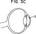
【図3C】 目球の側断面図である。
【図4】 3つのグルコース濃度に対する螢光眼内レンズの螢光強度の関係を示すグラフである。[0001]
An ophthalmic lens containing the receptor half can be used to determine the amount of analyte in the ocular fluid that the light can access. The receptor half can be bound to either a specific analyte or a competitor half with a detectable label. The amount of a competitor with a detectable label that is displaced from the receptor half by the analyte is measured, and the analyte concentration in the ocular fluid such as scent, aqueous humor and interstitial fluid Provide a means to determine The concentration of the analyte in the ocular fluid in turn represents the concentration of the analyte in an inaccessible bodily fluid or tissue sample, such as blood or intracellular fluid.
[0002]
Various non-invasive or minimally invasive methods have been described for measuring analytes, particularly glucose. For example, March U.S. Pat. Nos. 3,958,560 and 4,014,321 disclose a glucose sensor that automatically scans a patient's eye using a light source placed on one side of the cornea. . A sensor located on the other side of the cornea detects light passing through the cornea. The glucose level that rotates the plane of polarized light in the patient's aqueous humor is a function of the amount of radiation detected. However, this sensor system is not necessarily specific because it does not take advantage of the use of biomolecules that can detect glucose or other analytes in a body tissue or fluid sample, ie detection of analytes other than glucose. It cannot be applied widely. Biological molecules, as is well known, can provide highly specific and sensitive detection reagents for a particular analyte.
[0003]
Schultz U.S. Pat. No. 4,344,438 discloses a system for monitoring low molecular weight compounds in plasma by optical means involving a chamber containing a specific receptor site for the plasma component to be analyzed. is doing. However, this system is very invasive because it must be implanted into the blood stream using a hypodermic needle. This system also inherently carries the risk of causing other adverse reactions including coagulation, occlusion and immune responses around the device, systemic hypersensitivity and foreign body reactions.
[0004]
Embodiments of the present invention utilize prior art ophthalmic lenses comprising a receptor half that includes an analyte / competitor half binding site to detect an analyte in an ocular fluid. Overcoming these drawbacks. The concentration of various analytes can be measured using an ophthalmic lens according to embodiments of the present invention. Such analytes include electrolytes and small molecules (eg, sodium, potassium, chloride, phenylalanine, uric acid, galactose, glucose, cysteine, homocysteine, calcium, ethanol, acetylcholine, and acetylcholine analogs, ornithine, blood Urea nitrogen, creatinine), metal elements (eg, iron, copper, magnesium), polypeptide hormones (eg, thyroid stimulating hormone, growth hormone, insulin, luteinizing hormone, chorionic gonadotropin), chronically administered drugs (Eg diracitin, phenobarbityl, propranolol), drugs administered acutely (eg cocaine, heroin, ketamine), small molecule hormones (eg thyroid hormone, ACTH, estrogen, cortisol, estrogen and its Metabolic steroids), inflammation and / or allergy markers (eg, histamine, IgE, cytokines), lipids (eg, cholesterol), plasma proteins and enzymes (eg, complement, coagulation factors, liver function enzymes, cardiac injury enzymes, Ferritin), infection markers (eg, immunoglobulins such as viral components, IgM, IgG, etc., proteases, protease inhibitors), and / or metabolites (eg, lactate, ketone bodies). It is not done.
[0005]
An ophthalmic lens according to embodiments of the present invention can be used to monitor treatment progress or disease levels in primates, preferably mammals including humans. Furthermore, an ophthalmic lens according to an embodiment of the present invention provides a way to non-invasively detect an analyte, and thus compared to a more traditional form that monitors such levels. Offer completely different advantages in some cases. An ophthalmic lens according to an embodiment of the present invention can also be tested for pregnancy (detecting β-HCG), for example, for blood chemistry (electrolyte, Ca2POFour, Magnesium, bilirubin, alkaline phosphatase, lactate dehydrogenase, alanine aminotransferase, etc.) and detect infections (eg CMV, EBV, hepatitis and HIV viruses or bacterial components such as Staphlococcus, Streptococcus, etc.) It is also useful for diagnostic purposes, such as by detecting. They are also useful for monitoring blood levels of test compounds during the process of assessing compounds for use as effective therapies.
[0006]
An ophthalmic lens according to embodiments of the present invention may be worn on a long-term basis to provide repeated analyte measurements, or may be worn for a single analyte measurement. it can. Both qualitative and quantitative measurements can be performed.
[0007]
Eye lens
An ophthalmic lens according to embodiments of the present invention may be a removable lens, such as a contact lens, or a permanently implanted lens, such as an intraocular lens, a subconjunctival lens or an intracorneal lens. Also good. See U.S. Patent Application Nos. 60 / 150,792 and 60 / 185,980, whose priority is claimed for the present invention. Permanently implanted lenses are particularly suitable for use in individuals with eye disorders (eg, cataracts) as well as individuals with chronic conditions that require analyte measurements such as diabetes ing.
[0008]
The ophthalmic lens may be a corrective lens or may be constructed in a manner that does not affect vision. The contact lens may optionally contain a slight color and is preferably disposable, thus reducing the risk of infection for the user. As used herein, the term “eye lens” can also refer to a shunt or implant that can remain in the cecum of the eye.
[0009]
Half receptor
The ophthalmic lens 8 comprises a receptor half. This receptor half contains a binding site 9 for the analyte to be detected. The binding site also binds the half that competes with the analyte for binding and is therefore referred to herein as the “analyte / competitor half binding site”. Binding of both the competitor half and the analyte to the analyte / competitor half binding site is reversible. The nature of the molecule used as the receptor half depends on the specific analyte of the analyte to be detected, but is minimally sufficient to contain the analyte / competitor half binding site. It contains the part of the molecule that
[0010]
For example, if glucose is the analyte to be detected, the receptor half is preferably concanavalin A (Mansouri & Schultz, Bio / Tech 2,385, 1984), but antibiotics, boronic acids, genetic engineering It is also possible to use other halves, such as bacterial fluorprotein or glucose oxidase that have been subjected to the treatment. When phenylalanine is the analyte to be detected, the receptor half preferably contains the active site of phenylalanine hydroxylase. Other analytes / receptor half such as urate-uricase, alcohol-alcohol dehydrogenase, copper-celluloplasmin, galactose-galactokinase, cysteine- and / or homocysteine-cystathionine synthetase, acetylcholine-acetylcholinesterase, ornithine-diaminooxidase Determining body binding pairs is well within the skill of the artisan.
[0011]
Competitor half
For use in detecting an analyte, an ophthalmic lens according to an embodiment of the present invention preferably comprises a competitor half with a detectable label. The competitor half competes with the analyte for binding to the analyte / competitor half binding site. The detectable label may inherently form part of the competitor half. Alternatively, the detectable label can be a label that is not naturally associated with the competitor half, but is attached by a chemical bond, such as a covalent bond. In a preferred embodiment, the competitor half contains a fluorescent label. Other detectable labels such as luminescent labels or colorimetric labels can be used as well.
[0012]
Again, selecting a competitor half that will compete with the analyte for binding to a particular analyte / competitor half binding site is well within the skill of the artisan. For example, competitor halves that can be used with the analyte / receptor half binding pairs disclosed above include fluorescein dextran (which competes with glucose for binding to concanavalin A), fluorescein polyglutamyl urate (this Competes with uric acid for binding to uricase), fluorescein nanolol (which competes with alcohol for binding to alcohol dehydrogenase), fluorescein-glutamine phenylacetate (which competes with phenylalanine for binding to phenylalanine hydroxylase), fluorescein Erythrocuplain (which competes with copper for binding to ceruloplasmin), fluorescein-2,3,6-tri-O-methylgalactose It competes with galactose for binding to galactokinase), fluorescein-S-adenosylpolyhomocysteine (which competes with cysteine and foscysteine for binding to cystathionine synthetase), fluoropolyglutamylprostigmine (which is acetylated) And fluorspermine (which competes with ornithine for binding to diamine oxidase).
[0013]
Most preferably, the detectable label is more easily detectable when the competitor half is not bound to the analyte / competitor half binding site. Thus, fluorescent labels such as fluorescein, indocyanine green, malachite green and rhodamine that are quenched when the competitor half is bound but not when the competitor half is not bound are present in the present invention. Suitable for use in ophthalmic lenses according to the embodiments.
[0014]
Providing a receptor half and a competitor half in the lens of the eye
Various options are available to provide a receptor half and a competitor half in the ophthalmic lens. The construction of various types of ophthalmic lenses is well known in the art. Regarding the construction of contact lenses, for example, U.S. Pat. Nos. 5,965,631, 5,894,002, 5,849,811, 5,807,944, 5,776,381, No. 5,426,158, No. 4,099,859, No. 4,229,273, No. 4,168,112, No. 4,217,038, No. 4,409,258, No. 4 388,164, 4,332,922, 4,143,947, 4,311,573, 4,589,964, and 3,925,178. ing.
[0015]
For the construction of intraocular lens implants, see, inter alia, US Pat. Nos. 6,051,025, 5,868,697, 5,762,836, 5,609,640, 5,071, 432, 5,041,133 and 5,007,928. Subconjunctival lenses are taught, for example, in U.S. Pat. Nos. 5,476,511, 5,400,144, and 5,127,901. Intracorneal lenses are taught in US Pat. Nos. 6,090,141, 5,984,961, 5,123,921 and 4,799,931.
[0016]
In one embodiment, the receptor half is covalently bonded to the ophthalmic lens material. In another embodiment, the ophthalmic lens comprises a polymer network that includes holes. The pores allow the competitor half to reversibly bind to the analyte / competitor half binding site, but prevent the receptor half and competitor half from diffusing out of the eye lens. It is the one of the size. Suitable polymers for this purpose are known in the art and include modified hydrogels such as stable polymers of polyethylene glycol hydrogel (PEGH) (March et al., 2000) and modified polyvinyls such as nerfilcon A. Contains alcohol.
[0017]
In another embodiment, the ophthalmic lens comprises a receptor half layer, a polyelectrolyte layer, and a competitor half layer. The polyelectrolyte layer typically includes one or more polyelectrolytes that are high molecular weight polymers with multiple ions or ionizable functional groups. At least one polyelectrolyte in the polyelectrolyte layer has a charge opposite to the overall charge of the receptor half and competitor half layer. Suitable polyelectrolytes include positively charged PDDA (polydiallyldimethylammonium chloride) and negatively charged PAA (polyacrylic acid). The assembly of the layers is based on continuous adsorption of multiply charged ions with opposite charges. The sensor and spacing polyelectrolyte are deposited as a homogeneous thin film (1-10 nm) in 10-15 deposition cycles on a porous polyvinyl alcohol or hydrogen matrix, resulting in a very biocompatible A thick coating of 100-500 nm for some sensing films is provided. Standard sequences for construction of ophthalmic lenses suitable for glucose detection include PDDA, PAA, PDDA, concanavalin A, PDDA, PAA, PDDA, fluorescein dextran, PDDA, PAA, PDDA, PAA, concanavalin A, PAA , Fluorescein dextran, PAA, and other ultra-thin (1-10 nm) deposition cycles are involved. Techniques for constructing ophthalmic lenses comprising such layers are taught, for example, in WO 99/35520.
[0018]
An ophthalmic lens according to embodiments of the present invention may be provided in the form of a kit with instructions for measuring analyte concentration as described below. The present invention includes kits intended for use by individual patients whose ophthalmic lenses are typically contact lenses 1, as well as any of the ophthalmic lenses described herein, or equivalents thereof. A kit for doctors who can do this is provided.
[0019]
Analyte sensor system
An ophthalmic lens according to embodiments of the present invention can be used in an analyte sensor system. The analyte sensor system comprises an eye lens and a detector configured to detect a detectable label. For example, if the label is a luminescent label, the detector can include a luminometer, if the label is a colorimetric label, the detector can include a colorimeter, and if the label is a fluorescent label, The detector can include a fluorometer. The construction of such a device is well known in the art. Light having a wavelength that will excite the fluorescent label may be provided by a light source, such as a laser or a light emitting diode. Fluorometers suitable for use with embodiments of the present invention can be constructed using light emitting diodes from Power Technology. Inc (Little Rock, AR) (March et al., Diabetes Technol. & Ther. 2). 27-30, 2000).
[0020]
The detector can be a stand-alone device, a tabletop device or a handheld device. For convenience, the detector is a miniaturized device, for example, mounted in a frame of glasses, clipped into a garment such as a shirt or sweater, suspended around the neck, or mounted around the wrist Or clipped to a belt or key ring and can be worn or carried as personal equipment.
[0021]
Embodiments of the present invention using an ophthalmic lens in an analyte sensor system as described above provide a method for measuring an analyte concentration in an ocular fluid. This measurement can itself be manipulated to provide a measurement of the analyte concentration in body fluids or tissues such as blood or intracellular fluids. For example, the relationship between aqueous humor and glucose concentration in the blood is well known. Sullmann, HANDBUCH DER PHYSIOLOGISCHEN CHEMIE, Vol. Ll / a, p. 867ff. Springer, Berlin, 1956; Graymore, THE EYE, Vol. 1, p. 348, Davson, ed., Academic Press, NY, 1962; De Berardinis et al., Exp. Eye Res. 4, 179, 1965; Pohjola, Acta Ophthalmologica Suppl. 88, 1966; Reim et al., Ophthalmologica 154, 39- 50, 1967; Kinsey & Reddy, Prince, ed., THE RABBIT AND EYE RESEARCH, CC, Thomas, Springfield, IL, 1964, p. 218. checking ... The relationship between the concentration of another analyte in the body tissue or fluid and the concentration of the analyte in the ocular fluid can be determined by methods well known in the art. See, for example, March et al., Diabetes Care 5, 259-65, 1982. The detector may be configured to convert the analyte concentration measurement into a value that reflects the concentration of the analyte in an associated body fluid or body tissue, such as blood.
[0022]
If desired, the analyte sensor system also includes a transmitter configured to transmit a signal representative of whether a detectable label has been detected and / or the amount of detectable label being detected. You can also A device configured to vary the concentration of an analyte in a body fluid or tissue, such as an infusion pump or other pump, can receive the signal and vary the concentration response to the signal. The signal from the analyte sensor system can include a continuous or discontinuous telemetry signal generated by the detector. In response to the signal, the pump can adjust the analyte level in the body by providing the user with an appropriate amount of a modulator half, such as insulin. Infusion pumps are well known in the art for delivering selected drugs to patients, including humans and other animals, according to a dosing regime that can be preselected or in some cases preprogrammed. It is. Pumps for use in the present invention can be worn outside the body of mammals, including humans, to deliver certain drugs such as insulin to mammals at controlled doses over time, Alternatively, it can be transplanted directly into the body. Such pumps are well known and are described in US Pat. Nos. 5,957,890, 4,923,375, 4,573,994, and 3,731,681. . Drugs such as barbital, baclofen, theophylline and cardiac and blood pressure drugs that should be optimally maintained at a certain level can also be provided using infusion pumps.
[0023]
Example
An embodiment of an analyte sensor system according to the present invention is shown in FIGS. FIG. 1 has a first wavelength that will excite a fluorescent label in a contact lens 1, a radiation detector 5 such as a fluorimeter, and a competitor half housed in the contact lens 1. 1 is a schematic diagram of an analyte sensor system that utilizes a radiation source 2 such as a laser or light emitting diode that emits light 3 (preferably of low power). In response to
[0024]
Conveniently, a miniaturized version of the radiation source 2 and the radiation detector 5 can be configured to be integrated into one spectacle. One example of that is shown in FIGS. 2A and 2B. The analyte sensor system shown in FIGS. 2A and 2B utilizes an intraocular lens 8 comprising a polymer 9 containing a receptor half and a fluorescently labeled competitor half. A light emitting diode 6 is mounted in the frame of the glasses 7. The light emitting diode 6 emits light 3 at a first wavelength that will excite the fluorescent label in the competitor half. Thus, the competitor half that is not coupled to the receptor half will emit a second different wavelength of light 4 that is mounted in the eyeglass frame 7 along with the light emitting diode 6. It can be detected and measured by a fluorometer 5. A
[0025]
The
[0026]
Optionally, the
[0027]
If the
[0028]
All patents and patent applications cited in this disclosure are expressly incorporated herein by reference. The above disclosure generally describes the present invention. A more complete understanding can be obtained by reference to the following specific examples, which are provided by way of example only, without any intention to limit the scope of the invention.
[0029]
Example 1
Construction of an intraocular glucose sensor
To construct an intraocular glucose sensor, a structurally stable polymer of polyethylene glycol hydrogel (PEGH, Shearwater Polymers Inc.) is used. PEGH is immobilized in an intraocular lens (Alcon Laboratories, circumference 6 mm, thickness 1 mm). Acter 345, 203-12, 1997, and Russell & Pishko, Anal. Chem. 71, 3126-32, 1999, as described by Ballerstadt & Schultz, Anal. Chim. Fluorescein isothiocyanate dextran (FITC-dextran, Sigma) is incorporated as a body, and chemically immobilized suspended tetramethylrhodamine isothiocyanate concanavalin A (TRITC-ConA, Sigma) is incorporated into PEGH as a receptor half. While FITC-dextran is coupled to TRITC-ConA, FITC fluorescence is quenched via fluorescence resonant energy transfer. Increasing the glucose concentration releases FITC-dextran, resulting in a fluorescence that is directly proportional to the glucose concentration.
[0030]
FIG. 4 shows the relationship of the fluorescence intensity of our fluorescent intraocular lens at three glucose concentrations in vitro. A direct proportional relationship occurs between 0-500 mg% at 518 nm, the peak of fluorescein fluorescence. The peak at 575 nm is due to rhodamine in TRITC-ConA.
[0031]
Example 2
In vivo implantation of an intraocular glucose sensor
The intraocular lens glucose sensor described in Example 1 is implanted into the anterior chamber of the eyes of a living New Zealand rabbit with a blood glucose concentration of 112 mg%. The implant appears as bright spots of green fluorescence (518 nm) in the eye. Careful examination with a biomicroscopic slit lamp shows no signs of toxicity, rejection or reaction 6 months after transplantation.
[Brief description of the drawings]
FIG. 1 is a diagram showing an embodiment of an analyte sensor system of the present invention.
FIG. 2A is a side cross-section showing the eyeball and glasses and the arrangement of the sensor system.
FIG. 2B is a front view showing the eyeball and glasses and the arrangement of the sensor system.
FIG. 2C is a schematic diagram of a sensor system.
FIG. 3A is a front view showing an eyeball.
FIG. 3B is a side sectional view of the eyeball.
FIG. 3C is a side sectional view of the eyeball.
FIG. 4 is a graph showing the relationship of fluorescence intensity of a fluorescent intraocular lens to three glucose concentrations.
Claims (23)
高分子電解質層;及び
競合物質半体層、
を含んでなる、請求項1〜6のいずれか1項記載の眼のレンズ。Receptor half layer;
A polyelectrolyte layer; and a competitor half layer,
The ophthalmic lens according to claim 1, comprising:
第1及び第2の高分子電解質層;及び
中の競合物質半体が検出可能な標識を含んでいる競合物質半体層、
を含んでなる、眼の流体中のグルコースを検出するための、請求項1〜8のいずれか1項記載の眼のレンズ。A receptor half layer in which the receptor half contains a glucose / competitor half binding site;
First and second polyelectrolyte layers; and a competitor half layer in which the competitor half contains a detectable label;
9. An ophthalmic lens according to any one of the preceding claims for detecting glucose in an ocular fluid comprising:
(b) 検出可能な標識を検出するように構成された検出器(5)、
を含んでなる被分析物センサーシステム。(A) an ophthalmic lens (1) according to any one of claims 1 to 11; and (b) a detector (5) configured to detect a detectable label;
An analyte sensor system comprising:
(b)検出可能な標識を検出するように構成された検出器(5);及び
(c)検出可能な標識が検出器によって検出されたか否かを示す信号(10)をインシュリンポンプ(11)に伝送するように構成され、該ポンプが血液中のグルコース濃度を変動させるように構成されている、検出器に連結された送信機、
を含んでなる、請求項12記載の被分析物センサーシステム。(A) a receptor half comprising an analyte / competitor half binding site and a competitor half comprising a detectable label, wherein the receptor half is concanavalin A and comprises a detectable label An ophthalmic lens (1) for detecting glucose in ocular fluid, wherein the competitor half is fluorescein dextran;
(B) a detector (5) configured to detect a detectable label; and (c) a signal (10) indicating whether or not the detectable label has been detected by the detector. A transmitter coupled to a detector, wherein the transmitter is configured to transmit to and wherein the pump is configured to vary the glucose concentration in the blood;
13. The analyte sensor system of claim 12, comprising:
(a)請求項1〜11のいずれか1項記載の眼のレンズと眼の流体とを接触させる工程;及び
(b)検出可能な標識を検出する工程であって、該検出可能な標識の量が眼の流体中の被分析物の濃度を表わしている工程、
を含んでなる方法。In a method for measuring the concentration of an analyte in an eye fluid:
(A) contacting the ophthalmic lens according to any one of claims 1 to 11 with an eye fluid; and (b) detecting a detectable label comprising the detectable label. A process wherein the amount represents the concentration of the analyte in the ocular fluid;
Comprising a method.
(a)請求項1〜11のいずれか1項記載の眼のレンズと眼の流体とを接触させる工程;及び
(b)検出可能な標識を検出する工程であって、検出された検出可能な標識の量が体液又は流体中の被分析物の濃度を表わしている工程;
(c)体内組織又は体液中の被分析物の濃度を変動させるように構成されたポンプに対し眼の流体内の被分析物の濃度を示す信号を伝送する工程;及び
(d)ポンプを介して体内組織又は体液まで調節物質半体を提供し、かくして体内組織又は体液中の被分析物の濃度が変動させられる工程、
を含んでなる方法。A method for varying the concentration of an analyte in a body fluid or fluid comprising:
(A) contacting the ophthalmic lens according to any one of claims 1 to 11 with an eye fluid; and (b) detecting a detectable label, the detected detectable The amount of label representing the concentration of the analyte in the body fluid or fluid;
(C) transmitting a signal indicative of the concentration of the analyte in the ocular fluid to a pump configured to vary the concentration of the analyte in the body tissue or fluid; and (d) via the pump. Providing a modulating substance half to the body tissue or body fluid, thus varying the concentration of the analyte in the body tissue or body fluid,
Comprising a method.
Applications Claiming Priority (5)
| Application Number | Priority Date | Filing Date | Title |
|---|---|---|---|
| US15079299P | 1999-08-26 | 1999-08-26 | |
| US60/150,792 | 1999-08-26 | ||
| US18598000P | 2000-03-01 | 2000-03-01 | |
| US60/185,980 | 2000-03-01 | ||
| PCT/EP2000/008285 WO2001013783A1 (en) | 1999-08-26 | 2000-08-24 | Ocular analyte sensor |
Publications (2)
| Publication Number | Publication Date |
|---|---|
| JP2003507717A JP2003507717A (en) | 2003-02-25 |
| JP4421160B2 true JP4421160B2 (en) | 2010-02-24 |
Family
ID=26848047
Family Applications (1)
| Application Number | Title | Priority Date | Filing Date |
|---|---|---|---|
| JP2001517929A Expired - Lifetime JP4421160B2 (en) | 1999-08-26 | 2000-08-24 | Ocular analyte sensor |
Country Status (15)
| Country | Link |
|---|---|
| US (2) | US6681127B2 (en) |
| EP (1) | EP1206213B1 (en) |
| JP (1) | JP4421160B2 (en) |
| KR (1) | KR100675706B1 (en) |
| CN (1) | CN1196436C (en) |
| AT (1) | ATE287661T1 (en) |
| AU (1) | AU767995B2 (en) |
| BR (1) | BR0013609B1 (en) |
| CA (1) | CA2381520C (en) |
| DE (1) | DE60017755T2 (en) |
| HK (1) | HK1048244B (en) |
| HU (1) | HU226183B1 (en) |
| MX (1) | MXPA02002029A (en) |
| NO (1) | NO20020851D0 (en) |
| WO (1) | WO2001013783A1 (en) |
Families Citing this family (82)
| Publication number | Priority date | Publication date | Assignee | Title |
|---|---|---|---|---|
| US6544193B2 (en) * | 1996-09-04 | 2003-04-08 | Marcio Marc Abreu | Noninvasive measurement of chemical substances |
| US6120460A (en) * | 1996-09-04 | 2000-09-19 | Abreu; Marcio Marc | Method and apparatus for signal acquisition, processing and transmission for evaluation of bodily functions |
| JP4421160B2 (en) * | 1999-08-26 | 2010-02-24 | アイセンス・アクチエンゲゼルシャフト | Ocular analyte sensor |
| US7998412B2 (en) | 2000-01-07 | 2011-08-16 | Smart Holograms Limited | Ophthalmic device comprising a holographic sensor |
| GB0016841D0 (en) | 2000-07-07 | 2000-08-30 | Stanley Christopher J | Optical device for measurement of analytes in tears |
| US20040152963A1 (en) * | 2001-04-27 | 2004-08-05 | March Wayne Front | Apparatus for measuring blood glucose concentrations |
| US6827966B2 (en) * | 2001-05-30 | 2004-12-07 | Novartis Ag | Diffusion-controllable coatings on medical device |
| US6650915B2 (en) * | 2001-09-13 | 2003-11-18 | Fovioptics, Inc. | Non-invasive measurement of blood analytes using photodynamics |
| US20050010091A1 (en) * | 2003-06-10 | 2005-01-13 | Woods Joe W. | Non-invasive measurement of blood glucose using retinal imaging |
| US20050239155A1 (en) * | 2002-01-04 | 2005-10-27 | Javier Alarcon | Entrapped binding protein as biosensors |
| US20030153026A1 (en) * | 2002-01-04 | 2003-08-14 | Javier Alarcon | Entrapped binding protein as biosensors |
| US6939298B2 (en) * | 2002-02-28 | 2005-09-06 | Gmp Vision Solutions, Inc | Device and method for monitoring aqueous flow within the eye |
| US9848815B2 (en) | 2002-04-22 | 2017-12-26 | Geelux Holdings, Ltd. | Apparatus and method for measuring biologic parameters |
| US8849379B2 (en) * | 2002-04-22 | 2014-09-30 | Geelux Holdings, Ltd. | Apparatus and method for measuring biologic parameters |
| WO2004001373A2 (en) | 2002-04-22 | 2003-12-31 | Marcio Marc Abreu | Apparatus and method for measuring biologic parameters |
| US8328420B2 (en) * | 2003-04-22 | 2012-12-11 | Marcio Marc Abreu | Apparatus and method for measuring biologic parameters |
| US6895264B2 (en) * | 2002-08-26 | 2005-05-17 | Fovioptics Inc. | Non-invasive psychophysical measurement of glucose using photodynamics |
| US7244394B2 (en) | 2002-10-03 | 2007-07-17 | Novartis Ag | Methods and kits for assays of analytes of interest in tears |
| US20050038329A1 (en) * | 2002-11-20 | 2005-02-17 | Morris Carol Ann | Methods and kits for assays of rapid screening of diabetes |
| WO2004064629A1 (en) * | 2003-01-21 | 2004-08-05 | Ehrfeld Miktotechnik Ag | Sensor system for detecting analytes in tear fluid |
| WO2004075782A2 (en) * | 2003-02-26 | 2004-09-10 | Alfred, E. Mann Institute For Biomedical Engineering At The University Of Southern California | An implantable device with sensors for differential monitoring of internal condition |
| DE602004022638D1 (en) * | 2003-02-28 | 2009-10-01 | Eyesense Ag | BIOMOLECULES CONTAINING COPOLYMERS |
| US20040181172A1 (en) * | 2003-03-12 | 2004-09-16 | Carney Fiona Patricia | Devices for collecting analytes of interest in tears |
| WO2005000109A2 (en) | 2003-06-27 | 2005-01-06 | University Of Maryland Biotechnology Institute | Quaternary nitrogen heterocyclic compounds for detecting aqueous monosaccharides in physiological fluids |
| EP1651946A1 (en) | 2003-07-30 | 2006-05-03 | Novartis AG | Reflection hologram sensor in contact lens |
| EP1654543B1 (en) | 2003-08-07 | 2010-07-07 | EyeSense AG | Ophthalmic sensor |
| US7214190B1 (en) | 2003-09-09 | 2007-05-08 | Kitchener Clark Wilson | Apparatus and method for noninvasive monitoring of analytes in body fluids |
| EP1664909A1 (en) * | 2003-09-25 | 2006-06-07 | Smart Holograms Limited | Ophthalmic device comprising a holographic sensor |
| US10227063B2 (en) | 2004-02-26 | 2019-03-12 | Geelux Holdings, Ltd. | Method and apparatus for biological evaluation |
| US20060258920A1 (en) * | 2004-04-14 | 2006-11-16 | Oculir, Inc. | Non-Invasive Analyte Measurement Glasses and Method of Use |
| DE602005017492D1 (en) * | 2004-06-14 | 2009-12-17 | Eyesense Ag | COMBINED DEVICE FOR MEASURING BLOOD SUGAR FROM EYES LIQUID |
| JP4455216B2 (en) | 2004-08-06 | 2010-04-21 | キヤノン株式会社 | Detection device |
| CA2543964A1 (en) * | 2005-04-19 | 2006-10-19 | Anton Sabeta | Ophthalmic lens characterization |
| EP1951110B1 (en) | 2005-10-24 | 2012-10-03 | Marcio Marc Aurelio Martins Abreu | Apparatus for measuring biologic parameters |
| US8515518B2 (en) * | 2005-12-28 | 2013-08-20 | Abbott Diabetes Care Inc. | Analyte monitoring |
| JP4615470B2 (en) | 2006-03-29 | 2011-01-19 | 卓郎 簑和田 | Disease treatment and prevention methods and medicines using cerebral cognition |
| ATE413836T1 (en) | 2006-07-24 | 2008-11-15 | Eyesense Ag | DEVICE FOR MEASURING AN ANALYTE IN AN EYE FLUID |
| US8090426B2 (en) * | 2006-10-27 | 2012-01-03 | Felder Robin A | Microelectronic biosensor plug |
| DE102007003341B4 (en) | 2007-01-17 | 2018-01-04 | Eyesense Ag | Eyepiece sensor and measuring system for detecting an analyte in an eye fluid |
| US8446341B2 (en) * | 2007-03-07 | 2013-05-21 | University Of Washington | Contact lens with integrated light-emitting component |
| JP2010526960A (en) * | 2007-05-10 | 2010-08-05 | ボーグワーナー・インコーポレーテッド | Collaborative blade and hub structure for cooling fans |
| DE102007024642A1 (en) | 2007-05-24 | 2008-11-27 | Eyesense Ag | Hydrogel implant for sensor of metabolites on the eye |
| US7802883B2 (en) | 2007-12-20 | 2010-09-28 | Johnson & Johnson Vision Care, Inc. | Cosmetic contact lenses having a sparkle effect |
| EP2174586B1 (en) | 2008-10-02 | 2014-12-10 | EyeSense AG | Implantable sensor element |
| ATE507768T1 (en) | 2008-10-02 | 2011-05-15 | Eyesense Ag | IMPLANTATION DEVICE FOR METABOLITE SENSORS |
| CA2683467C (en) * | 2008-10-24 | 2016-01-26 | Jin Zhang | Contact lens integrated with a biosensor for detection of glucose and other components in tears |
| US20100168851A1 (en) * | 2008-12-30 | 2010-07-01 | David Paul Vanderbilt | Surface Modified Biomedical Devices |
| US8815178B2 (en) * | 2009-03-26 | 2014-08-26 | Arizona Board Of Regents On Behalf Of Arizona State University | Integrated device for surface-contact sampling, extraction and electrochemical measurements |
| US20120177576A1 (en) * | 2009-09-18 | 2012-07-12 | Jun Jack Hu | Optical device and method for non-invasive real-time testing of blood sugar levels |
| US8741591B2 (en) | 2009-10-09 | 2014-06-03 | The Research Foundation For The State University Of New York | pH-insensitive glucose indicator protein |
| CN103328980B (en) | 2010-12-17 | 2016-01-20 | 视觉股份公司 | There is the competitive biology sensor of the susceptibility of raising |
| CN103370623B (en) | 2010-12-17 | 2016-05-04 | 视觉股份公司 | The purposes of hydrogel in the biology sensor of sensitivity with raising |
| EP2508935A1 (en) | 2011-04-08 | 2012-10-10 | Nxp B.V. | Flexible eye insert and glucose measuring system |
| CA2843950A1 (en) * | 2011-08-01 | 2013-02-07 | Massachusetts Institute Of Technology | Photoluminescent nanostructure-based sensors |
| US11363951B2 (en) | 2011-09-13 | 2022-06-21 | Glaukos Corporation | Intraocular physiological sensor |
| US8874182B2 (en) | 2013-01-15 | 2014-10-28 | Google Inc. | Encapsulated electronics |
| US9730638B2 (en) | 2013-03-13 | 2017-08-15 | Glaukos Corporation | Intraocular physiological sensor |
| US9289623B2 (en) * | 2013-03-15 | 2016-03-22 | Johnson & Johnson Vision Care, Inc. | Method and device for monitoring and treatment of seasonal affective disorder |
| US20140350373A1 (en) * | 2013-05-21 | 2014-11-27 | Johnson & Johnson Vision Care, Inc. | Ophthalmic lens with a passive event-based coloration system |
| US9084561B2 (en) | 2013-06-17 | 2015-07-21 | Google Inc. | Symmetrically arranged sensor electrodes in an ophthalmic electrochemical sensor |
| US9044075B2 (en) * | 2013-06-24 | 2015-06-02 | Google Inc. | Container |
| ES2531189B2 (en) * | 2013-08-05 | 2015-12-18 | Universidad De Alicante | Procedure for quantification of ocular opacity of an intraocular lens |
| CA2980036A1 (en) | 2013-10-11 | 2015-04-16 | Marcio Marc Abreu | Method and apparatus for biological evaluation |
| US9701458B2 (en) | 2013-12-19 | 2017-07-11 | Verily Life Sciences Llc | Packaging for an active contact lens |
| US9576168B2 (en) | 2013-12-30 | 2017-02-21 | Verily Life Sciences Llc | Conditional retrieval |
| AU2015204588A1 (en) | 2014-01-10 | 2016-07-21 | Marcio Marc Abreu | Devices to monitor and provide treatment at an Abreu brain tunnel |
| CA2936229A1 (en) | 2014-01-10 | 2015-07-16 | Marcio Marc Abreu | Device for measuring the infrared output of the abreu brain thermal tunnel |
| CA2936247A1 (en) | 2014-01-22 | 2015-07-30 | Marcio Marc Abreu | Devices and methods for transdermal drug delivery |
| JP2016008818A (en) * | 2014-06-20 | 2016-01-18 | ソニー株式会社 | Detection apparatus and method, and program |
| KR101638106B1 (en) * | 2014-07-02 | 2016-07-11 | 충남대학교 산학협력단 | Fluorescent Poly(N-isopropyl acrylamide) Microgel, method for preparing of the same and sensor including the same |
| EP2979662A1 (en) * | 2014-08-01 | 2016-02-03 | Akkolens International B.V. | Intraocular lens with electricity generator and additional functional systems |
| WO2016054079A1 (en) | 2014-09-29 | 2016-04-07 | Zyomed Corp. | Systems and methods for blood glucose and other analyte detection and measurement using collision computing |
| US11872018B2 (en) | 2015-03-10 | 2024-01-16 | Brain Tunnelgenix Technologies Corp. | Devices, apparatuses, systems, and methods for measuring temperature of an ABTT terminus |
| EP3291886A1 (en) * | 2015-05-04 | 2018-03-14 | Ecole Polytechnique Federale de Lausanne (EPFL) | Ophthalmic contact lens with a compressible affinity matrix |
| JP2017023328A (en) * | 2015-07-21 | 2017-02-02 | 富士ゼロックス株式会社 | Optically active substance concentration calculation system and program |
| CH711991B1 (en) * | 2015-12-22 | 2023-11-30 | Dosilab AG | Dosimeter device. |
| US9554738B1 (en) | 2016-03-30 | 2017-01-31 | Zyomed Corp. | Spectroscopic tomography systems and methods for noninvasive detection and measurement of analytes using collision computing |
| WO2018017842A1 (en) * | 2016-07-20 | 2018-01-25 | University Of Maryland, Baltimore | Silicone hydrogel based fluorescent assay and contact lens |
| CN110536654A (en) | 2017-04-06 | 2019-12-03 | 提尔迪克斯有限公司 | Ophthalmic device and its application method |
| EP3732455B1 (en) * | 2017-12-28 | 2021-09-15 | Transitions Optical, Ltd. | Method for measuring optical characteristics of a contact lens |
| CN111936853A (en) | 2018-03-13 | 2020-11-13 | 目立康株式会社 | Specifying system, computing device, specifying method, and program |
| WO2019175662A1 (en) * | 2018-03-13 | 2019-09-19 | Menicon Co. Ltd. | Glucose sensing contact lens |
Family Cites Families (43)
| Publication number | Priority date | Publication date | Assignee | Title |
|---|---|---|---|---|
| US3769961A (en) | 1972-07-20 | 1973-11-06 | I Fatt | Conjunctival device |
| US4168112A (en) * | 1978-01-05 | 1979-09-18 | Polymer Technology Corporation | Contact lens with a hydrophilic, polyelectrolyte complex coating and method for forming same |
| US4344438A (en) | 1978-08-02 | 1982-08-17 | The United States Of America As Represented By The Department Of Health, Education And Welfare | Optical sensor of plasma constituents |
| US4298801A (en) * | 1980-01-17 | 1981-11-03 | General Electric Company | L-U Arm handle assembly |
| US4330299A (en) | 1981-03-09 | 1982-05-18 | Evreka, Inc. | Article and method for measuring glucose level in body fluids |
| DE3313047A1 (en) | 1983-04-12 | 1984-10-18 | Max Planck Gesellschaft zur Förderung der Wissenschaften e.V., 3400 Göttingen | ARRANGEMENT FOR MEASURING DIFFUSING PARTICLES |
| EP0236023A3 (en) | 1986-03-03 | 1988-09-07 | Seymour Norman Blackman | Non-invasive blood glucose level monitoring method |
| US5263992A (en) | 1986-10-17 | 1993-11-23 | Bio-Metric Systems, Inc. | Biocompatible device with covalently bonded biocompatible agent |
| SU1534406A1 (en) | 1987-11-25 | 1990-01-07 | Военно-Медицинская Краснознаменная Академия Им.С.М.Кирова | Method of diagnosis of latent form of diabetes mellitus |
| US6040194A (en) | 1989-12-14 | 2000-03-21 | Sensor Technologies, Inc. | Methods and device for detecting and quantifying substances in body fluids |
| US5342789A (en) | 1989-12-14 | 1994-08-30 | Sensor Technologies, Inc. | Method and device for detecting and quantifying glucose in body fluids |
| US5222495A (en) | 1990-02-02 | 1993-06-29 | Angiomedics Ii, Inc. | Non-invasive blood analysis by near infrared absorption measurements using two closely spaced wavelengths |
| GB9022304D0 (en) | 1990-10-15 | 1990-11-28 | Ares Serono Res & Dev Ltd | Assay technique |
| US5435307A (en) * | 1991-03-29 | 1995-07-25 | The United States Of America As Represented By The Secretary Of The Department Of Health And Human Services | Surface fluorescent monitor |
| US5203328A (en) * | 1991-07-17 | 1993-04-20 | Georgia Tech Research Corporation | Apparatus and methods for quantitatively measuring molecular changes in the ocular lens |
| US5246867A (en) | 1992-01-17 | 1993-09-21 | University Of Maryland At Baltimore | Determination and quantification of saccharides by luminescence lifetimes and energy transfer |
| DE4208645A1 (en) | 1992-03-18 | 1993-09-23 | Bayer Ag | OPTICAL SOLID PHASE BIOSENSOR BASED ON FLUORESCENT COLOR-MARGINED POLYIONIC LAYERS |
| US5433197A (en) | 1992-09-04 | 1995-07-18 | Stark; Edward W. | Non-invasive glucose measurement method and apparatus |
| US6256522B1 (en) * | 1992-11-23 | 2001-07-03 | University Of Pittsburgh Of The Commonwealth System Of Higher Education | Sensors for continuous monitoring of biochemicals and related method |
| US5341805A (en) | 1993-04-06 | 1994-08-30 | Cedars-Sinai Medical Center | Glucose fluorescence monitor and method |
| US5383452A (en) | 1993-07-01 | 1995-01-24 | Buchert; Janusz | Method, apparatus and procedure for non-invasive monitoring blood glucose by measuring the polarization ratio of blood luminescence |
| US5352411A (en) | 1993-08-13 | 1994-10-04 | Khuri Raja N | Device for determination of tear constituents |
| GB2284809B (en) | 1993-11-07 | 1998-04-29 | Japan Res Dev Corp | A fluorescent phenylboronic acid suitable for use in the detection of saccharides |
| WO1997019188A1 (en) * | 1995-11-22 | 1997-05-29 | Minimed, Inc. | Detection of biological molecules using chemical amplification and optical sensors |
| JP3599878B2 (en) | 1996-03-01 | 2004-12-08 | テルモ株式会社 | Blood glucose meter |
| US6120460A (en) | 1996-09-04 | 2000-09-19 | Abreu; Marcio Marc | Method and apparatus for signal acquisition, processing and transmission for evaluation of bodily functions |
| US5830139A (en) | 1996-09-04 | 1998-11-03 | Abreu; Marcio M. | Tonometer system for measuring intraocular pressure by applanation and/or indentation |
| US5898004A (en) | 1996-11-06 | 1999-04-27 | University Of Pittsburgh Of The Commonwealth System Of Higher Education | Polymerized crystalline colloidal array sensors |
| US5854078A (en) | 1996-11-06 | 1998-12-29 | University Of Pittsburgh | Polymerized crystalline colloidal array sensor methods |
| WO1998022820A1 (en) | 1996-11-21 | 1998-05-28 | Lawrence Livermore National Laboratory | Detection of biological molecules using boronate-based chemical amplification and optical sensors |
| US6197928B1 (en) | 1997-03-14 | 2001-03-06 | The Regents Of The University Of California | Fluorescent protein sensors for detection of analytes |
| US6540895B1 (en) * | 1997-09-23 | 2003-04-01 | California Institute Of Technology | Microfabricated cell sorter for chemical and biological materials |
| JP3543923B2 (en) | 1997-12-25 | 2004-07-21 | 富士写真フイルム株式会社 | Glucose concentration measurement device |
| JP2002510515A (en) * | 1998-04-06 | 2002-04-09 | ザ・ジェネラル・ホスピタル・コーポレイション | Non-invasive tissue glucose level monitoring |
| DE69941085D1 (en) | 1998-07-17 | 2009-08-20 | Univ Maryland | MANIPULATED PROTEINS FOR ANALYSIS PROOF |
| WO2000010007A2 (en) | 1998-08-17 | 2000-02-24 | California Institute Of Technology | Devices and methods for analysis of non-ionic solutes |
| WO2000016099A1 (en) | 1998-09-11 | 2000-03-23 | Sensor Technologies, Inc. | Recombinant reduced valency carbohydrate binding ligands |
| AU6142799A (en) * | 1998-09-11 | 2000-03-27 | Amira Medical | Device for determination of an analyte in a body fluid intergrated with an insulin pump |
| AU4803600A (en) | 1999-04-27 | 2000-11-10 | University Of Pittsburgh | Apparatus for optically monitoring concentration of a bioanalyte in blood and related methods |
| JP4421160B2 (en) * | 1999-08-26 | 2010-02-24 | アイセンス・アクチエンゲゼルシャフト | Ocular analyte sensor |
| US6366793B1 (en) * | 1999-09-10 | 2002-04-02 | Beckman Coulter, Inc. | Minimally invasive methods for measuring analtes in vivo |
| WO2001018237A1 (en) | 1999-09-10 | 2001-03-15 | University Of Maryland, Baltimore | Inactive enzymes as non-consuming sensors |
| GB0016841D0 (en) | 2000-07-07 | 2000-08-30 | Stanley Christopher J | Optical device for measurement of analytes in tears |
-
2000
- 2000-08-24 JP JP2001517929A patent/JP4421160B2/en not_active Expired - Lifetime
- 2000-08-24 CN CNB008120722A patent/CN1196436C/en not_active Expired - Fee Related
- 2000-08-24 KR KR1020027002417A patent/KR100675706B1/en not_active IP Right Cessation
- 2000-08-24 HU HU0202889A patent/HU226183B1/en not_active IP Right Cessation
- 2000-08-24 EP EP00960520A patent/EP1206213B1/en not_active Expired - Lifetime
- 2000-08-24 WO PCT/EP2000/008285 patent/WO2001013783A1/en active Search and Examination
- 2000-08-24 BR BRPI0013609-3A patent/BR0013609B1/en not_active IP Right Cessation
- 2000-08-24 AU AU72796/00A patent/AU767995B2/en not_active Ceased
- 2000-08-24 MX MXPA02002029A patent/MXPA02002029A/en active IP Right Grant
- 2000-08-24 CA CA002381520A patent/CA2381520C/en not_active Expired - Fee Related
- 2000-08-24 AT AT00960520T patent/ATE287661T1/en not_active IP Right Cessation
- 2000-08-24 DE DE60017755T patent/DE60017755T2/en not_active Expired - Lifetime
-
2001
- 2001-02-15 US US09/784,471 patent/US6681127B2/en not_active Expired - Lifetime
-
2002
- 2002-02-21 NO NO20020851A patent/NO20020851D0/en not_active Application Discontinuation
-
2003
- 2003-01-23 HK HK03100590.1A patent/HK1048244B/en not_active IP Right Cessation
- 2003-09-03 US US10/655,502 patent/US6850786B2/en not_active Expired - Lifetime
Also Published As
| Publication number | Publication date |
|---|---|
| BR0013609B1 (en) | 2009-01-13 |
| CA2381520A1 (en) | 2001-03-01 |
| JP2003507717A (en) | 2003-02-25 |
| HU226183B1 (en) | 2008-06-30 |
| CN1371256A (en) | 2002-09-25 |
| AU767995B2 (en) | 2003-11-27 |
| US6850786B2 (en) | 2005-02-01 |
| NO20020851L (en) | 2002-02-21 |
| EP1206213A1 (en) | 2002-05-22 |
| NO20020851D0 (en) | 2002-02-21 |
| AU7279600A (en) | 2001-03-19 |
| KR20020026599A (en) | 2002-04-10 |
| KR100675706B1 (en) | 2007-02-01 |
| US6681127B2 (en) | 2004-01-20 |
| HK1048244A1 (en) | 2003-03-28 |
| DE60017755D1 (en) | 2005-03-03 |
| US20010034500A1 (en) | 2001-10-25 |
| BR0013609A (en) | 2002-05-21 |
| EP1206213B1 (en) | 2005-01-26 |
| US20040059207A1 (en) | 2004-03-25 |
| DE60017755T2 (en) | 2005-06-30 |
| CN1196436C (en) | 2005-04-13 |
| ATE287661T1 (en) | 2005-02-15 |
| CA2381520C (en) | 2009-05-26 |
| HK1048244B (en) | 2005-07-29 |
| HUP0202889A2 (en) | 2003-01-28 |
| MXPA02002029A (en) | 2002-08-20 |
| WO2001013783A1 (en) | 2001-03-01 |
Similar Documents
| Publication | Publication Date | Title |
|---|---|---|
| JP4421160B2 (en) | Ocular analyte sensor | |
| US6980842B2 (en) | Ocular analyte sensor | |
| JP4152193B2 (en) | Blood glucose concentration measuring device | |
| JP4754770B2 (en) | Noninvasive measuring instrument | |
| JP4280629B2 (en) | Optical sensor containing particles for INSITU measurement of analyte | |
| JP5543066B2 (en) | Optical fiber device for detecting analyte and manufacturing method thereof | |
| JP4673888B2 (en) | Combined device for measuring blood glucose levels from ocular fluid | |
| AU2017201863A1 (en) | Noninvasive measurements of chemical substances | |
| ZA200201427B (en) | Ocular analyte sensor. |
Legal Events
| Date | Code | Title | Description |
|---|---|---|---|
| A521 | Request for written amendment filed |
Free format text: JAPANESE INTERMEDIATE CODE: A821 Effective date: 20060711 |
|
| A711 | Notification of change in applicant |
Free format text: JAPANESE INTERMEDIATE CODE: A711 Effective date: 20060711 |
|
| A521 | Request for written amendment filed |
Free format text: JAPANESE INTERMEDIATE CODE: A821 Effective date: 20060711 |
|
| A621 | Written request for application examination |
Free format text: JAPANESE INTERMEDIATE CODE: A621 Effective date: 20070808 |
|
| TRDD | Decision of grant or rejection written | ||
| A01 | Written decision to grant a patent or to grant a registration (utility model) |
Free format text: JAPANESE INTERMEDIATE CODE: A01 Effective date: 20091104 |
|
| A01 | Written decision to grant a patent or to grant a registration (utility model) |
Free format text: JAPANESE INTERMEDIATE CODE: A01 |
|
| A61 | First payment of annual fees (during grant procedure) |
Free format text: JAPANESE INTERMEDIATE CODE: A61 Effective date: 20091202 |
|
| FPAY | Renewal fee payment (event date is renewal date of database) |
Free format text: PAYMENT UNTIL: 20121211 Year of fee payment: 3 |
|
| R150 | Certificate of patent or registration of utility model |
Ref document number: 4421160 Country of ref document: JP Free format text: JAPANESE INTERMEDIATE CODE: R150 Free format text: JAPANESE INTERMEDIATE CODE: R150 |
|
| FPAY | Renewal fee payment (event date is renewal date of database) |
Free format text: PAYMENT UNTIL: 20131211 Year of fee payment: 4 |
|
| R250 | Receipt of annual fees |
Free format text: JAPANESE INTERMEDIATE CODE: R250 |
|
| R250 | Receipt of annual fees |
Free format text: JAPANESE INTERMEDIATE CODE: R250 |
|
| R250 | Receipt of annual fees |
Free format text: JAPANESE INTERMEDIATE CODE: R250 |
|
| R250 | Receipt of annual fees |
Free format text: JAPANESE INTERMEDIATE CODE: R250 |
|
| R250 | Receipt of annual fees |
Free format text: JAPANESE INTERMEDIATE CODE: R250 |
|
| R250 | Receipt of annual fees |
Free format text: JAPANESE INTERMEDIATE CODE: R250 |
|
| R250 | Receipt of annual fees |
Free format text: JAPANESE INTERMEDIATE CODE: R250 |
|
| R250 | Receipt of annual fees |
Free format text: JAPANESE INTERMEDIATE CODE: R250 |
|
| EXPY | Cancellation because of completion of term |