JP4414410B2 - 画像再構成方法 - Google Patents
画像再構成方法 Download PDFInfo
- Publication number
- JP4414410B2 JP4414410B2 JP2006126902A JP2006126902A JP4414410B2 JP 4414410 B2 JP4414410 B2 JP 4414410B2 JP 2006126902 A JP2006126902 A JP 2006126902A JP 2006126902 A JP2006126902 A JP 2006126902A JP 4414410 B2 JP4414410 B2 JP 4414410B2
- Authority
- JP
- Japan
- Prior art keywords
- value
- dimensional
- lor
- radiation activity
- count value
- Prior art date
- Legal status (The legal status is an assumption and is not a legal conclusion. Google has not performed a legal analysis and makes no representation as to the accuracy of the status listed.)
- Active
Links
- 238000000034 method Methods 0.000 title claims description 73
- 230000000694 effects Effects 0.000 claims description 29
- 230000005855 radiation Effects 0.000 claims description 29
- 238000003384 imaging method Methods 0.000 claims description 16
- 238000004364 calculation method Methods 0.000 claims description 12
- 239000007787 solid Substances 0.000 claims description 10
- 238000006243 chemical reaction Methods 0.000 claims description 6
- 238000005259 measurement Methods 0.000 claims description 4
- 230000001186 cumulative effect Effects 0.000 claims description 2
- 206010028980 Neoplasm Diseases 0.000 description 15
- 238000001514 detection method Methods 0.000 description 10
- 238000005516 engineering process Methods 0.000 description 4
- 230000005251 gamma ray Effects 0.000 description 4
- 230000010354 integration Effects 0.000 description 4
- 238000002600 positron emission tomography Methods 0.000 description 4
- WQZGKKKJIJFFOK-GASJEMHNSA-N Glucose Natural products OC[C@H]1OC(O)[C@H](O)[C@@H](O)[C@@H]1O WQZGKKKJIJFFOK-GASJEMHNSA-N 0.000 description 2
- 201000011510 cancer Diseases 0.000 description 2
- 238000013461 design Methods 0.000 description 2
- 238000010586 diagram Methods 0.000 description 2
- 201000010099 disease Diseases 0.000 description 2
- 208000037265 diseases, disorders, signs and symptoms Diseases 0.000 description 2
- YCKRFDGAMUMZLT-BJUDXGSMSA-N fluorine-18 atom Chemical compound [18F] YCKRFDGAMUMZLT-BJUDXGSMSA-N 0.000 description 2
- 239000008103 glucose Substances 0.000 description 2
- 238000012360 testing method Methods 0.000 description 2
- 238000007476 Maximum Likelihood Methods 0.000 description 1
- 150000001413 amino acids Chemical class 0.000 description 1
- WQZGKKKJIJFFOK-VFUOTHLCSA-N beta-D-glucose Chemical compound OC[C@H]1O[C@@H](O)[C@H](O)[C@@H](O)[C@@H]1O WQZGKKKJIJFFOK-VFUOTHLCSA-N 0.000 description 1
- 239000003795 chemical substances by application Substances 0.000 description 1
- 239000003086 colorant Substances 0.000 description 1
- 238000012937 correction Methods 0.000 description 1
- 239000013078 crystal Substances 0.000 description 1
- 230000002498 deadly effect Effects 0.000 description 1
- 238000002405 diagnostic procedure Methods 0.000 description 1
- 230000035622 drinking Effects 0.000 description 1
- 230000009977 dual effect Effects 0.000 description 1
- 238000009434 installation Methods 0.000 description 1
- 238000010253 intravenous injection Methods 0.000 description 1
- 239000003446 ligand Substances 0.000 description 1
- 238000004519 manufacturing process Methods 0.000 description 1
- 230000002503 metabolic effect Effects 0.000 description 1
- 238000012545 processing Methods 0.000 description 1
- 238000011160 research Methods 0.000 description 1
- 230000035945 sensitivity Effects 0.000 description 1
- 208000024891 symptom Diseases 0.000 description 1
Images
Classifications
-
- G—PHYSICS
- G06—COMPUTING; CALCULATING OR COUNTING
- G06T—IMAGE DATA PROCESSING OR GENERATION, IN GENERAL
- G06T11/00—2D [Two Dimensional] image generation
- G06T11/003—Reconstruction from projections, e.g. tomography
- G06T11/006—Inverse problem, transformation from projection-space into object-space, e.g. transform methods, back-projection, algebraic methods
-
- A—HUMAN NECESSITIES
- A61—MEDICAL OR VETERINARY SCIENCE; HYGIENE
- A61B—DIAGNOSIS; SURGERY; IDENTIFICATION
- A61B6/00—Apparatus or devices for radiation diagnosis; Apparatus or devices for radiation diagnosis combined with radiation therapy equipment
- A61B6/02—Arrangements for diagnosis sequentially in different planes; Stereoscopic radiation diagnosis
- A61B6/03—Computed tomography [CT]
- A61B6/037—Emission tomography
-
- G—PHYSICS
- G06—COMPUTING; CALCULATING OR COUNTING
- G06T—IMAGE DATA PROCESSING OR GENERATION, IN GENERAL
- G06T2211/00—Image generation
- G06T2211/40—Computed tomography
- G06T2211/424—Iterative
Landscapes
- Engineering & Computer Science (AREA)
- Physics & Mathematics (AREA)
- Health & Medical Sciences (AREA)
- Life Sciences & Earth Sciences (AREA)
- Theoretical Computer Science (AREA)
- General Physics & Mathematics (AREA)
- Medical Informatics (AREA)
- Surgery (AREA)
- Veterinary Medicine (AREA)
- Pathology (AREA)
- Radiology & Medical Imaging (AREA)
- Biomedical Technology (AREA)
- Heart & Thoracic Surgery (AREA)
- Molecular Biology (AREA)
- Nuclear Medicine, Radiotherapy & Molecular Imaging (AREA)
- Animal Behavior & Ethology (AREA)
- General Health & Medical Sciences (AREA)
- Public Health (AREA)
- Optics & Photonics (AREA)
- Algebra (AREA)
- High Energy & Nuclear Physics (AREA)
- Mathematical Analysis (AREA)
- Mathematical Optimization (AREA)
- Mathematical Physics (AREA)
- Pure & Applied Mathematics (AREA)
- Biophysics (AREA)
- Nuclear Medicine (AREA)
- Image Analysis (AREA)
- Image Processing (AREA)
Description
但し、多くの癌症状は早期に発見することが可能となり、治癒率も向上している。科学技術の恩恵に賜り、現在の分子生物と画像工学の結合は、多くの非侵入性の画像検出装置を生み、致命的な疾病に対する早期発見の機会を増やすことができ、これらの検出装置内で更に高感度の特質を具えたPET(Positron Emission Tomography)が画像検出の重要な役割を担っている。
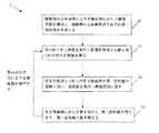
(a)複数個の立体画素がそれぞれ第一輻射活性値(radioactivity)を有する;
(b)各LOR上の立体画素に対し投影領域に投影転換を実行し、測定計数値と比較し複数個の校正計数値を得る;
(c)それぞれLOR上の各立体画素の第一輻射活性値(radioactivity)に対し演算を実行し、各立体画素の重み付け値を得る;
(d)LOR上の各立体画素の重み付け値に従い逆投影を実行し、各LORの校正計数値とLOR上の各立体画素の重み付け値に対し計算を実行し、LOR上の各立体画素の第二輻射活性値を得る;
(e)画像空間を再構成し、各立体画素の第二輻射活性値を第一輻射活性値(radioactivity)にする;
(f)ステップ(b)乃至(f)を反復実行する。
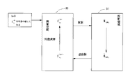
(b1)それぞれLORが通過する立体画素の第一輻射活性値(radioactivity)に対して投影領域への投影転換を実行し、LORの投影計数値を推測する;
(b2)推測計数値と測定計数値を比較し、校正計数値を得る。校正計数値は測定計数値と推測計数値の比になる;
(d1)閾値を設定し、第二輻射活性値が閾値より小さい場合、第二輻射活性値を0にする。
LOR上の各立体画素の重み付け値と校正計数値を相互に乗算し乗積値を得る;
少なくとも一つのLORの乗積値の累計より第二輻射活性値を得る;
閾値を設定し、第二輻射活性値が閾値より小さい場合、第二輻射活性値は0とする;
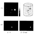
腫瘍90は、人工器官の中心(z=0mm)に配置し、腫瘍91はz=20mmの位置上に配置する。更に、本発明の二次元平面撮像の三次元画像再構成方法を利用して画像再構成を実行し、公知の技術の焦点面画像再構成方法と比較する。
図10の(B)は本発明の方法によるもので、z=0mm及びz=20mmの画像結果である。これらの結果より明らかなように、本発明の方法は、腫瘍のz軸方法の位置を正確に判断でき、公知の技術よりも優れた画像を得ることができる。
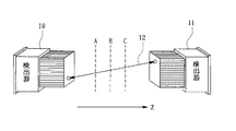
12 LOR(Line of Response)
13 人工器官
2 容器
20〜23 プロセス
210〜212 プロセス
220〜221 プロセス
230〜232 プロセス
30 画像空間
31 投影領域
4 画像空間
40、43 立体画素
50、51、52 LOR(Line of Response)
90、91 腫瘍
Claims (7)
- 被検出物が発生する複数個のLORに対し画像再構成を実行し、各LORは対応する測定計数値を有し、平面撮像画像再構成方法は、
(a)複数個の立体画素がそれぞれ第一輻射活性値(radioactivity)を有し、
(b)各LOR上の立体画素に対し投影領域に投影転換を実行し、測定計数値と比較し複数個の校正計数値を得て、
(c)それぞれLOR上の各立体画素の第一輻射活性値(radioactivity)に対して演算を実行し各立体画素の重み付け値を得る
(d)LOR上の各立体画素の重み付け値に従い逆投影を実行し、各LORの校正計数値とLOR上の各立体画素の重み付け値に対し計算を実行し、各立体画素の第二輻射活性値を得る、
(e)画像空間を再構成し、各立体画素の第二輻射活性値を第一輻射活性値(radioactivity)とし、
(f)ステップ(b)乃至(f)を反復実行する、
上記のステップより構成されることを特徴とした、二次元平面撮像により三次元画像情報を表す再構成方法。 - 前記ステップ(d)は更に、(d1)閾値を設定し、第二輻射活性値が閾値より小さい場合、第二輻射活性値を0にする、ステップを設けることを特徴とした、請求項1に記載の二次元平面撮像により三次元画像情報を表す再構成方法。
- 前記校正計数値は測定計数値と推測計数値の比であることを特徴とした、請求項1に記載の二次元平面撮像により三次元画像情報を表す再構成方法。
- 前記重み付け値は、LOR上の各立体画素の第一輻射活性値(radioactivity)と推測計数値の和の比であることを特徴とした、請求項1に記載の二次元平面撮像により三次元画像情報を表す再構成方法。
- 前記ステップ(b)は更に、
(b1)それぞれLORが通過する立体画素の第一輻射活性値(radioactivity)に対して投影領域への投影転換を実行してLORの投影計数値を推測し、
(b2)推測計数値と測定計数値を比較して校正計数値を得る。校正計数値は測定計数値と推測計数値の比であり、
上記のステップを設けることを特徴とした、請求項1に記載の二次元平面撮像により三次元画像情報を表す再構成方法。 - 前記計算は更に、
(d2)LOR上の各立体画素の重み付け値と校正計数値を相互に乗算して乗積値を得る;
少なくとも一つのLORの乗積値の累計より第二輻射活性値を得て、
(d3)閾値を設定し、第二輻射活性値が閾値より小さい場合、第二輻射活性値は0とする、上記ステップを設けることを特徴とした、請求項1に記載の二次元平面撮像により三次元画像情報を表す再構成方法。 - 前記計算は更に、
(d4)閾値を設定し、第二輻射活性値が閾値より小さい場合、第二輻射活性値を0にする、ステップを設けることを特徴とした、請求項6に記載の二次元平面撮像により三次元画像情報を表す再構成方法。
Applications Claiming Priority (1)
| Application Number | Priority Date | Filing Date | Title |
|---|---|---|---|
| TW095113726A TWI337329B (en) | 2006-04-18 | 2006-04-18 | Image reconstruction method for structuring two-dimensional planar imaging into three-dimension imaging |
Publications (2)
| Publication Number | Publication Date |
|---|---|
| JP2007286020A JP2007286020A (ja) | 2007-11-01 |
| JP4414410B2 true JP4414410B2 (ja) | 2010-02-10 |
Family
ID=38604879
Family Applications (1)
| Application Number | Title | Priority Date | Filing Date |
|---|---|---|---|
| JP2006126902A Active JP4414410B2 (ja) | 2006-04-18 | 2006-04-28 | 画像再構成方法 |
Country Status (3)
| Country | Link |
|---|---|
| US (1) | US7778452B2 (ja) |
| JP (1) | JP4414410B2 (ja) |
| TW (1) | TWI337329B (ja) |
Families Citing this family (18)
| Publication number | Priority date | Publication date | Assignee | Title |
|---|---|---|---|---|
| DE102006029718A1 (de) * | 2006-06-28 | 2008-01-10 | Siemens Ag | Verfahren zur Auswertung zweier Abbilder sowie medizinisches Abbildungssystem |
| US7949172B2 (en) * | 2007-04-27 | 2011-05-24 | Siemens Medical Solutions Usa, Inc. | Iterative image processing |
| WO2008141293A2 (en) * | 2007-05-11 | 2008-11-20 | The Board Of Regents Of The University Of Oklahoma One Partner's Place | Image segmentation system and method |
| US8546763B2 (en) * | 2008-01-22 | 2013-10-01 | Shimadzu Corporation | Positron computed tomography device |
| CN102013116B (zh) * | 2010-11-30 | 2012-11-07 | 华润万东医疗装备股份有限公司 | 基于脑血管旋转造影术进行三维重建的方法 |
| US8787644B2 (en) * | 2011-06-14 | 2014-07-22 | Kabushiki Kaisha Toshiba | Method and device for calculating voxels defining a tube-of-response using a central-ray-filling algorithm |
| TWI505105B (zh) * | 2012-07-31 | 2015-10-21 | Nat Univ Tsing Hua | 將正子攝影中三重事件數據再利用之系統、方法及程式 |
| KR101378757B1 (ko) * | 2012-08-30 | 2014-03-27 | 한국원자력연구원 | 물질 원소 정보 획득 및 영상 차원의 선택이 가능한 방사선 영상화 장치 |
| TWI494897B (zh) * | 2012-11-20 | 2015-08-01 | Iner Aec Executive Yuan | 一種三維射束覓跡的投影方法 |
| WO2016036352A1 (en) * | 2014-09-03 | 2016-03-10 | Hewlett-Packard Development Company, L.P. | Presentation of a digital image of an object |
| WO2017066248A1 (en) | 2015-10-16 | 2017-04-20 | Varian Medical Systems, Inc. | Iterative image reconstruction in image-guided radiation therapy |
| US10255696B2 (en) | 2015-12-11 | 2019-04-09 | Shanghai United Imaging Healthcare Co., Ltd. | System and method for image reconstruction |
| EP3234919B1 (en) | 2015-12-11 | 2021-03-17 | Shanghai United Imaging Healthcare Co., Ltd. | System and method for image reconstruction |
| CN106911893B (zh) * | 2017-02-23 | 2020-04-03 | 北京建筑大学 | 一种单像素计算成像方法 |
| TWI742891B (zh) * | 2020-10-23 | 2021-10-11 | 行政院原子能委員會核能研究所 | 平面式腦功能用正子攝影裝置 |
| CN113576504B (zh) * | 2021-08-02 | 2023-06-27 | 南华大学 | 一种用于中低原子序数物质的μ子成像方法 |
| TWI780853B (zh) | 2021-08-05 | 2022-10-11 | 行政院原子能委員會核能研究所 | 平面式加馬成像探頭位置訊號處理方法 |
| CN116540872B (zh) * | 2023-04-28 | 2024-06-04 | 中广电广播电影电视设计研究院有限公司 | Vr数据处理方法、装置、设备、介质及产品 |
Family Cites Families (6)
| Publication number | Priority date | Publication date | Assignee | Title |
|---|---|---|---|---|
| US5224037A (en) * | 1991-03-15 | 1993-06-29 | Cti, Inc. | Design of super-fast three-dimensional projection system for Positron Emission Tomography |
| US5793045A (en) | 1997-02-21 | 1998-08-11 | Picker International, Inc. | Nuclear imaging using variable weighting |
| US6804325B1 (en) | 2002-10-25 | 2004-10-12 | Southeastern Universities Research Assn. | Method for position emission mammography image reconstruction |
| EP1639550A1 (en) * | 2003-06-18 | 2006-03-29 | Philips Intellectual Property & Standards GmbH | Motion compensated reconstruction technique |
| US7227149B2 (en) * | 2004-12-30 | 2007-06-05 | General Electric Company | Method and system for positron emission tomography image reconstruction |
| US7405405B2 (en) * | 2005-05-17 | 2008-07-29 | General Electric Company | Method and system for reconstructing an image in a positron emission tomography (PET) system |
-
2006
- 2006-04-18 TW TW095113726A patent/TWI337329B/zh active
- 2006-04-28 JP JP2006126902A patent/JP4414410B2/ja active Active
- 2006-11-22 US US11/562,878 patent/US7778452B2/en active Active
Also Published As
| Publication number | Publication date |
|---|---|
| JP2007286020A (ja) | 2007-11-01 |
| TW200741584A (en) | 2007-11-01 |
| US20070242867A1 (en) | 2007-10-18 |
| US7778452B2 (en) | 2010-08-17 |
| TWI337329B (en) | 2011-02-11 |
Similar Documents
| Publication | Publication Date | Title |
|---|---|---|
| JP4414410B2 (ja) | 画像再構成方法 | |
| Bailey et al. | Quantitative SPECT/CT: SPECT joins PET as a quantitative imaging modality | |
| Dewaraja et al. | MIRD pamphlet no. 23: quantitative SPECT for patient-specific 3-dimensional dosimetry in internal radionuclide therapy | |
| Frey et al. | Accuracy and precision of radioactivity quantification in nuclear medicine images | |
| Demirkaya et al. | Image processing with MATLAB: applications in medicine and biology | |
| Conti | Focus on time-of-flight PET: the benefits of improved time resolution | |
| EP3067864B1 (en) | Iterative reconstruction with enhanced noise control filtering | |
| Jaszczak et al. | Single photon emission computed tomography (SPECT) principles and instrumentation | |
| Chen et al. | Performance characteristics of the digital uMI550 PET/CT system according to the NEMA NU2-2018 standard | |
| US8193505B2 (en) | System and method for scatter normalization of PET images | |
| US8467584B2 (en) | Use of multifocal collimators in both organ-specific and non-specific SPECT acquisitions | |
| Piccinelli et al. | Advances in single-photon emission computed tomography hardware and software | |
| Jaszczak | SPECT: state-of-the-art scanners and reconstruction strategies | |
| Kang et al. | Initial results of a mouse brain PET insert with a staggered 3-layer DOI detector | |
| Shi et al. | Reconstruction of x-ray fluorescence computed tomography from sparse-view projections via L1-norm regularized EM algorithm | |
| Li et al. | Joint regional uptake quantification of Thorium-227 and Radium-223 using a multiple-energy-window projection-domain quantitative SPECT method | |
| Da Silva et al. | Absolute quantitation of myocardial activity in phantoms | |
| Perez et al. | Towards quantification of functional breast images using dedicated SPECT with non-traditional acquisition trajectories | |
| Pal et al. | 2D linear and iterative reconstruction algorithms for a PET-insert scanner | |
| Hutton et al. | SPECT and SPECT/CT | |
| Musa et al. | Simulation and evaluation of high-performance cost-effective positron emission mammography scanner | |
| Gong et al. | Implementation and evaluation of an expectation maximization reconstruction algorithm for gamma emission breast tomosynthesis | |
| Von Schulthess | Molecular Anatomic Imaging: PET/CT, PET/MR and SPECT CT | |
| Us | Reduction of Limited Angle Artifacts in Medical Tomography via Image Reconstruction | |
| Rahmim | Statistical list-mode image reconstruction and motion compensation techniques in high-resolution positron emission tomography (PET) |
Legal Events
| Date | Code | Title | Description |
|---|---|---|---|
| A621 | Written request for application examination |
Free format text: JAPANESE INTERMEDIATE CODE: A621 Effective date: 20060428 |
|
| A131 | Notification of reasons for refusal |
Free format text: JAPANESE INTERMEDIATE CODE: A131 Effective date: 20090210 |
|
| A601 | Written request for extension of time |
Free format text: JAPANESE INTERMEDIATE CODE: A601 Effective date: 20090508 |
|
| A521 | Request for written amendment filed |
Free format text: JAPANESE INTERMEDIATE CODE: A523 Effective date: 20090608 |
|
| A521 | Request for written amendment filed |
Free format text: JAPANESE INTERMEDIATE CODE: A523 Effective date: 20090608 |
|
| A602 | Written permission of extension of time |
Free format text: JAPANESE INTERMEDIATE CODE: A602 Effective date: 20090611 |
|
| TRDD | Decision of grant or rejection written | ||
| A01 | Written decision to grant a patent or to grant a registration (utility model) |
Free format text: JAPANESE INTERMEDIATE CODE: A01 Effective date: 20091110 |
|
| A01 | Written decision to grant a patent or to grant a registration (utility model) |
Free format text: JAPANESE INTERMEDIATE CODE: A01 |
|
| A61 | First payment of annual fees (during grant procedure) |
Free format text: JAPANESE INTERMEDIATE CODE: A61 Effective date: 20091119 |
|
| FPAY | Renewal fee payment (event date is renewal date of database) |
Free format text: PAYMENT UNTIL: 20121127 Year of fee payment: 3 |
|
| R150 | Certificate of patent or registration of utility model |
Ref document number: 4414410 Country of ref document: JP Free format text: JAPANESE INTERMEDIATE CODE: R150 Free format text: JAPANESE INTERMEDIATE CODE: R150 |
|
| FPAY | Renewal fee payment (event date is renewal date of database) |
Free format text: PAYMENT UNTIL: 20131127 Year of fee payment: 4 |
|
| R250 | Receipt of annual fees |
Free format text: JAPANESE INTERMEDIATE CODE: R250 |
|
| R250 | Receipt of annual fees |
Free format text: JAPANESE INTERMEDIATE CODE: R250 |
|
| R250 | Receipt of annual fees |
Free format text: JAPANESE INTERMEDIATE CODE: R250 |
|
| R250 | Receipt of annual fees |
Free format text: JAPANESE INTERMEDIATE CODE: R250 |
|
| R250 | Receipt of annual fees |
Free format text: JAPANESE INTERMEDIATE CODE: R250 |
|
| R250 | Receipt of annual fees |
Free format text: JAPANESE INTERMEDIATE CODE: R250 |
|
| R250 | Receipt of annual fees |
Free format text: JAPANESE INTERMEDIATE CODE: R250 |
|
| R250 | Receipt of annual fees |
Free format text: JAPANESE INTERMEDIATE CODE: R250 |
|
| R250 | Receipt of annual fees |
Free format text: JAPANESE INTERMEDIATE CODE: R250 |
|
| R250 | Receipt of annual fees |
Free format text: JAPANESE INTERMEDIATE CODE: R250 |
|
| R250 | Receipt of annual fees |
Free format text: JAPANESE INTERMEDIATE CODE: R250 |
|
| R250 | Receipt of annual fees |
Free format text: JAPANESE INTERMEDIATE CODE: R250 |
|
| R250 | Receipt of annual fees |
Free format text: JAPANESE INTERMEDIATE CODE: R250 |


