JP4392163B2 - Genetic marker for lung cancer - Google Patents
Genetic marker for lung cancer Download PDFInfo
- Publication number
- JP4392163B2 JP4392163B2 JP2002504687A JP2002504687A JP4392163B2 JP 4392163 B2 JP4392163 B2 JP 4392163B2 JP 2002504687 A JP2002504687 A JP 2002504687A JP 2002504687 A JP2002504687 A JP 2002504687A JP 4392163 B2 JP4392163 B2 JP 4392163B2
- Authority
- JP
- Japan
- Prior art keywords
- lung cancer
- blood
- markers
- pcr
- cells
- Prior art date
- Legal status (The legal status is an assumption and is not a legal conclusion. Google has not performed a legal analysis and makes no representation as to the accuracy of the status listed.)
- Expired - Fee Related
Links
Images


Classifications
-
- C—CHEMISTRY; METALLURGY
- C07—ORGANIC CHEMISTRY
- C07H—SUGARS; DERIVATIVES THEREOF; NUCLEOSIDES; NUCLEOTIDES; NUCLEIC ACIDS
- C07H21/00—Compounds containing two or more mononucleotide units having separate phosphate or polyphosphate groups linked by saccharide radicals of nucleoside groups, e.g. nucleic acids
- C07H21/04—Compounds containing two or more mononucleotide units having separate phosphate or polyphosphate groups linked by saccharide radicals of nucleoside groups, e.g. nucleic acids with deoxyribosyl as saccharide radical
-
- C—CHEMISTRY; METALLURGY
- C07—ORGANIC CHEMISTRY
- C07K—PEPTIDES
- C07K16/00—Immunoglobulins [IGs], e.g. monoclonal or polyclonal antibodies
- C07K16/18—Immunoglobulins [IGs], e.g. monoclonal or polyclonal antibodies against material from animals or humans
- C07K16/28—Immunoglobulins [IGs], e.g. monoclonal or polyclonal antibodies against material from animals or humans against receptors, cell surface antigens or cell surface determinants
- C07K16/30—Immunoglobulins [IGs], e.g. monoclonal or polyclonal antibodies against material from animals or humans against receptors, cell surface antigens or cell surface determinants from tumour cells
-
- C—CHEMISTRY; METALLURGY
- C12—BIOCHEMISTRY; BEER; SPIRITS; WINE; VINEGAR; MICROBIOLOGY; ENZYMOLOGY; MUTATION OR GENETIC ENGINEERING
- C12Q—MEASURING OR TESTING PROCESSES INVOLVING ENZYMES, NUCLEIC ACIDS OR MICROORGANISMS; COMPOSITIONS OR TEST PAPERS THEREFOR; PROCESSES OF PREPARING SUCH COMPOSITIONS; CONDITION-RESPONSIVE CONTROL IN MICROBIOLOGICAL OR ENZYMOLOGICAL PROCESSES
- C12Q1/00—Measuring or testing processes involving enzymes, nucleic acids or microorganisms; Compositions therefor; Processes of preparing such compositions
- C12Q1/68—Measuring or testing processes involving enzymes, nucleic acids or microorganisms; Compositions therefor; Processes of preparing such compositions involving nucleic acids
- C12Q1/6809—Methods for determination or identification of nucleic acids involving differential detection
-
- C—CHEMISTRY; METALLURGY
- C12—BIOCHEMISTRY; BEER; SPIRITS; WINE; VINEGAR; MICROBIOLOGY; ENZYMOLOGY; MUTATION OR GENETIC ENGINEERING
- C12Q—MEASURING OR TESTING PROCESSES INVOLVING ENZYMES, NUCLEIC ACIDS OR MICROORGANISMS; COMPOSITIONS OR TEST PAPERS THEREFOR; PROCESSES OF PREPARING SUCH COMPOSITIONS; CONDITION-RESPONSIVE CONTROL IN MICROBIOLOGICAL OR ENZYMOLOGICAL PROCESSES
- C12Q1/00—Measuring or testing processes involving enzymes, nucleic acids or microorganisms; Compositions therefor; Processes of preparing such compositions
- C12Q1/68—Measuring or testing processes involving enzymes, nucleic acids or microorganisms; Compositions therefor; Processes of preparing such compositions involving nucleic acids
- C12Q1/6876—Nucleic acid products used in the analysis of nucleic acids, e.g. primers or probes
- C12Q1/6883—Nucleic acid products used in the analysis of nucleic acids, e.g. primers or probes for diseases caused by alterations of genetic material
- C12Q1/6886—Nucleic acid products used in the analysis of nucleic acids, e.g. primers or probes for diseases caused by alterations of genetic material for cancer
-
- G—PHYSICS
- G01—MEASURING; TESTING
- G01N—INVESTIGATING OR ANALYSING MATERIALS BY DETERMINING THEIR CHEMICAL OR PHYSICAL PROPERTIES
- G01N33/00—Investigating or analysing materials by specific methods not covered by groups G01N1/00 - G01N31/00
- G01N33/48—Biological material, e.g. blood, urine; Haemocytometers
- G01N33/50—Chemical analysis of biological material, e.g. blood, urine; Testing involving biospecific ligand binding methods; Immunological testing
- G01N33/53—Immunoassay; Biospecific binding assay; Materials therefor
- G01N33/574—Immunoassay; Biospecific binding assay; Materials therefor for cancer
- G01N33/57407—Specifically defined cancers
- G01N33/57423—Specifically defined cancers of lung
-
- G—PHYSICS
- G01—MEASURING; TESTING
- G01N—INVESTIGATING OR ANALYSING MATERIALS BY DETERMINING THEIR CHEMICAL OR PHYSICAL PROPERTIES
- G01N33/00—Investigating or analysing materials by specific methods not covered by groups G01N1/00 - G01N31/00
- G01N33/48—Biological material, e.g. blood, urine; Haemocytometers
- G01N33/50—Chemical analysis of biological material, e.g. blood, urine; Testing involving biospecific ligand binding methods; Immunological testing
- G01N33/53—Immunoassay; Biospecific binding assay; Materials therefor
- G01N33/574—Immunoassay; Biospecific binding assay; Materials therefor for cancer
- G01N33/57484—Immunoassay; Biospecific binding assay; Materials therefor for cancer involving compounds serving as markers for tumor, cancer, neoplasia, e.g. cellular determinants, receptors, heat shock/stress proteins, A-protein, oligosaccharides, metabolites
-
- C—CHEMISTRY; METALLURGY
- C12—BIOCHEMISTRY; BEER; SPIRITS; WINE; VINEGAR; MICROBIOLOGY; ENZYMOLOGY; MUTATION OR GENETIC ENGINEERING
- C12Q—MEASURING OR TESTING PROCESSES INVOLVING ENZYMES, NUCLEIC ACIDS OR MICROORGANISMS; COMPOSITIONS OR TEST PAPERS THEREFOR; PROCESSES OF PREPARING SUCH COMPOSITIONS; CONDITION-RESPONSIVE CONTROL IN MICROBIOLOGICAL OR ENZYMOLOGICAL PROCESSES
- C12Q1/00—Measuring or testing processes involving enzymes, nucleic acids or microorganisms; Compositions therefor; Processes of preparing such compositions
- C12Q1/68—Measuring or testing processes involving enzymes, nucleic acids or microorganisms; Compositions therefor; Processes of preparing such compositions involving nucleic acids
- C12Q1/6844—Nucleic acid amplification reactions
- C12Q1/686—Polymerase chain reaction [PCR]
-
- C—CHEMISTRY; METALLURGY
- C12—BIOCHEMISTRY; BEER; SPIRITS; WINE; VINEGAR; MICROBIOLOGY; ENZYMOLOGY; MUTATION OR GENETIC ENGINEERING
- C12Q—MEASURING OR TESTING PROCESSES INVOLVING ENZYMES, NUCLEIC ACIDS OR MICROORGANISMS; COMPOSITIONS OR TEST PAPERS THEREFOR; PROCESSES OF PREPARING SUCH COMPOSITIONS; CONDITION-RESPONSIVE CONTROL IN MICROBIOLOGICAL OR ENZYMOLOGICAL PROCESSES
- C12Q2600/00—Oligonucleotides characterized by their use
- C12Q2600/158—Expression markers
Description
【0001】
[技術分野]
本発明は、残留肺癌の診断および鑑別方法に関する。本発明はさらに、肺癌用の新規に同定された細胞マーカーの使用に関する。これらのマーカーとしては、シンデカン1、コラーゲン1α2、ならびに2つの新規タンパク質7013および7018が挙げられる。
【0002】
[発明の背景]
肺癌は工業国における最も一般的な癌の1つであり、非常に高い死亡率を有する。初期診断および有効な治療は現時点では利用可能でない。胸部X線はしばしば、肺癌スクリーニングに用いられるが、それは初期段階の癌の検出には有用でない。CTスキャンも用いられ、初期段階の癌の検出を可能にし得るが、この手法は時間を要し、被曝の危険性を有する。
【0003】
癌細胞または微小転移はしばしば、黒色腫、甲状腺癌および前立腺癌患者の血流中で検出される。現在、逆転写ベースのポリメラーゼ連鎖反応(RT−PCR)は、癌特異的遺伝子またはマーカーを増幅することにより、無数の正常細胞内の単一癌細胞を検出し得るひとつの強力な方法である。これは、微小転移のRT−PCR検出を、予後、適切な治療の選択、および各治療の効力のモニタリングのための有望な診断手法にする。さらに、血液試験はいかなる健康的被害をも誘導しないが、X線またはCTスキャンのような方法は有害作用をもたらす。さらに血液試験は、内視鏡検査および生検と比較して、引き起こす物理的不快が非常に小さい。肺癌はしばしば、初期段階、すなわち症候性疾患および多数の肺癌が脳、骨および肝臓におけるような遠方の転移として再発する前でさえ、血行性転移を誘導する。これは、肺癌誘導性の血行性転移によるものである。しかしながらこの知識は、それが再発の診断および検出が極初期段階でのこれらの血行性転移に基づいてなされ得るということを示唆するため、有益である。しかしながら、血液中の転移性肺癌細胞の同定に利用可能な良好なマーカーはない。現在、肺癌マーカー、例えばサイトケラチン−19およびCEAは、RT−PCR(逆転写酵素ポリメラーゼ連鎖反応)による非小細胞肺癌の診断のために用いられるが、特異性を欠き、多数の擬陽性および陰性を生じる。
【0004】
微小転移のRT−PCRは、次いで、多数の患者人口、血行性転移の高発生率、不十分な予後および高次癌治療のための高医療費のために、肺癌の検出に特に有益である。さらに特異的抗体は、今までのところ肺癌に利用可能ではない。
【0005】
したがって、特異的マーカー、および血行性転移を検出することによる肺癌の診断方法が必要とされる。
【0006】
[発明の概要]
一実施形態は、患者から血液または非肺組織を単離すること、および、以下の:シンデカン1、コラーゲン1α2、7013および7018からの少なくとも1つのマーカーの存在を判別することによる肺癌の鑑別方法である。本方法は、マーカーであるサイトケラチン−19の存在を判別することも含み得る。さらなる実施形態では、少なくとも2つのマーカーを判別する。さらなる実施形態では、2より多いマーカーを判別する。判別方法は当業者に既知の任意のものであり得るが、RT−PCRおよび/または抗体結合も含み得る。患者は任意の生体であり得るが、一実施形態では、哺乳動物、特にヒト、イヌまたはネコである。
【0007】
さらなる実施形態は、転移性癌細胞を単離または除去する方法であって、癌細胞を含む細胞または非肺組織を、シンデカン1、コラーゲン1α2、7013および7018からなる群から選択される少なくとも1つのマーカーに特異的な抗体で処理することによる方法である。本方法は、マーカーであるサイトケラチン−18に特異的な抗体も含み得る。一実施形態では、抗体は金属粒子、蛍光粒子、クロマトグラフィービーズ、クロマトグラフィーゲルおよび固体支持体からなる群から選択される部分に結合される。さらなる実施形態では、2つのマーカーが用いられる。さらなる実施形態では、3つ以上のマーカーが用いられる。
【0008】
さらなる実施形態は、マーカー7013として本明細書中でも同定される配列番号16の少なくとも17個のヌクレオチドを含むポリヌクレオチドである。
【0009】
さらなる実施形態は、マーカー7018として本明細書中でも同定される配列番号17の少なくとも17個のヌクレオチドを含むポリヌクレオチドである。
【0010】
さらなる実施形態は、患者における固形腫瘍の転移の鑑別方法であって、以下の:上記患者から血液または骨髄を単離すること、ならびにシンデカン1、コラーゲン1α2、7013および7018からなる群から選択される少なくとも1つのマーカーの存在を判別することによる方法である。本方法は、サイトケラチン−18を同定することも包含し得る。一実施形態では、上記固形腫瘍は胆管、結腸、乳房、子宮、食道および喉頭からなる群から選択される。
【0011】
さらなる実施形態は、癌腫の鑑別方法であって、癌細胞を得ること、ならびに7013、7018およびその両方からなる群から選択されるマーカーの存在を判別することによる方法である。
【0012】
[好ましい実施形態の詳細な説明]
ディファレンシャルディスプレイを用いて、血中を循環し、正常血液細胞で発現しない、肺癌細胞で特異的に発現するmRNAを同定した。ディファレンシャルディスプレイ方法は、細胞中で発現する遺伝子のより包括的表現を可能にする古典的手法からの変法である。
【0013】
候補遺伝子を同定するために、3つの主な技術が目下利用可能である。すなわち、サブトラクションライブラリー、ディファレンシャルディスプレイおよびDNAマイクロチップアレイである。しかしながらサブトラクションライブラリーは、潜在的に有用であるが、遺伝子配列の部分的類似性を示す遺伝子も同定することができ、非常に労力を要しかつ時間集約的技法である。DNAマイクロチップアレイは、使用が迅速かつ容易であるが、血液試料中の稀少遺伝子を検出するのに十分に感受性ではない。これは、本方法が特異的マーカーを発現しない正常細胞の大きなバックグラウンド(無数の白血球)内に特異的マーカーを発現する稀少微小転移を検出できる必要があるため、特に重要である。ディファレンシャルディスプレイ(dd−PCR)は、多重縮重プライマーの組合せを用いてすべての既存の遺伝子の増幅を可能にし、したがって稀少マーカーまたは遺伝子が検出されるという見込みを増大させる。
【0014】
しかしながら、できるだけ多数の遺伝子を表現するようにディファレンシャルディスプレイ技術を改良するために、選択的増幅断片長多型(selective Amplified Fragment Length Polymorphism(s−AFLP))を実施例1で用いた。このアプローチは、1)遺伝子の3’末端のみを増幅する、2)より再現可能なゲルパターンを生成する、3)より少数の余分な遺伝子を同定する、および4)より選択的なPCR条件を用いるという利点を有する。本方法は、4つのマーカー、すなわちSynd、Col、7013および7018の同定に至った。
【0015】
これらのマーカーを用いて、疾患のあらゆる段階における患者の血中の肺癌細胞を判別し得る。当業者に既知の任意の方法を用いて、血中または任意の転移性組織中のマーカーを判別し得る。例えば、マーカーに特異的である細胞で発現するmRNAが判別され得る。
【0016】
あるいは、タンパク質それ自体は、免疫技法、例えばウエスタンブロット、FACS技術、ELISAおよび当業者に既知のその他の方法を用いて同定され得る。これら抗体または機能性抗体部分は、既知の方法を用いて購入、単離、または生産され得る。
【0017】
一実施形態では、マーカーの遺伝子発現が同定される。マーカーに関連した遺伝子の発現の判別を可能にする任意の方法が用いられ得る。典型的には、本方法は、遺伝子の発現に起因するmRNAを増幅する。一実施形態では、血液または組織からのmRNAのRT−PCRが用いられる。さらなる実施形態では、これらのマーカーを含む血中の細胞を判別するために抗体が用いられる。例えば細胞選別を用いて、これらのマーカーに対する抗体で蛍光標識されていた細胞を判別し得る。
【0018】
一実施形態では、本方法は、血中または骨髄中の肺癌細胞の存在を判定するために用いられる。しかしながら、普通はこれらのマーカーを生じない任意の組織からのmRNAが用いられ得ることが想定される。例えば、典型的に転移の部位である器官からのmRNAが用いられ得る。したがって、さらなる実施形態では、本方法は、肝臓または脳のような器官中の肺癌細胞を判別するために用いられる。
【0019】
一実施形態では、本方法は、疾患の極初期段階での肺癌細胞の存在を判別するために用いられる(さらなる実施形態では、肺癌細胞は寛解後に判別される)か、あるいは再発を判別するために用いられる。
【0020】
さらなる実施形態では、マーカーは、in vivoまたはin vitroで肺癌細胞を標的化するために用いられる。一実施形態では、癌細胞を標的とし、血液から除去させるアフィニティー技法を用いて、身体から癌細胞が除かれる。
【0021】
さらなる実施形態では、マーカーを用いて、血中もしくは骨髄中の転移性細胞を、または胆管、結腸、乳房、子宮、食道および喉頭のような癌により生成される転移性器官もしくは組織を鑑別する。あるいはマーカーは、血液から転移性細胞を除去するために用いられ得る。
【0022】
さらなる実施形態では、マーカー7013および7018は、癌細胞またはその他の細胞が上皮由来のものであるか否かを判別するために用いられる。例えば細胞が一方または両方のマーカーを発現する場合、それは上皮由来のものであると考えられる。
【0023】
患者は、癌を有することが可能な任意の動物であり得る。一実施形態では、患者は哺乳動物である。さらなる実施形態では、哺乳動物はペット、例えばイヌまたはネコである。さらなる実施形態では、患者はヒトである。
【0024】
ここで以下の実施例を参照して本発明を説明する。しかしながら、これらの実施例は本発明を単に説明するものであって、本発明を限定するものではない、と理解される。
【0025】
実施例1および2では、マーカーは、種々のディファレンシャルディスプレイ技法を用いて同定される。同定されるマーカーは、実施例2で説明される。
【0026】
実施例1
s−AFLPディファレンシャルディスプレイ
s−AFLPは、cDNAライブラリーからの制限断片の選択的PCR増幅に基づいている。2組の選択的プライマーを用いた。第1のもの、すなわち図1の選択的プライマーAは、3つの部分:コア配列、酵素特異的配列および3’末端の2つの選択的ヌクレオチドからなる。選択的配列の変異のために、これは42=16種類のプライマーを提供する。第2のもの、すなわち図1の選択的プライマーBも、3つの部分:アンカー配列、ポリT配列および蛍光標識される3’末端の2つの選択的ヌクレオチドからなる。ポリT(A、GおよびC)後の3つの選択的配列のために、これは次に、3×4=12種類のプライマーを提供する。したがって、ポリA領域を含めた3’末端cDNA制限断片はすべて、選択的PCRのこの方法により生成される192(16×12)群に含まれた。増幅断片のゲル表示上の各シグナルは、cDNAライブラリー中の非オーバーラップ遺伝子を示した。この技法を用いて、肺癌検体から単離されるRNAを、健常個体の血液からのRNAと比較した。癌検体中で過剰発現した示差的に発現した遺伝子を、腫瘍細胞播種に関する一般的遺伝子マーカーのための候補と考えた。
【0027】
s−AFLPを成功させるためには、優れた品質のRNAが重要であった。しかしながら肺検体は、肺胞マクロファージからの豊富なRNaseの存在により、RNAを調製するのが最も難しい組織の1つである。したがって、NCCRI(Chuou-ku, Tokyo, Japan)から急速凍結肺癌検体を購入し、過去に記載されたようなAGPC法により全RNAを精製した(Tominaga, K, Miura, Y. Arakawa, T. Koboyashi, K. and Mitsuhashi, M. Clin. Chem., 42, 1750-1783, 1998)。アガロースゲル電気泳動またはマイクロキャピラリーチップを用いて、RNAの品質を評価し、2つのrRNAバンドの存在を確証した。次にs−AFLPに関して許容可能な検体を用いた。6つの肺癌検体を用い、各試料を異なる患者から得た。6つのうちの4つは腺癌であり、2つは扁平上皮癌であった。20の対照血液は、悪性疾患またはその他の疾患の病歴または現在の診断を有さない健常有志から得た。
【0028】
腫瘍および血液検体の全RNA調製物
新鮮な凍結腫瘍検体を液体窒素中で小片に壊した。グアニジン法により、100mg検体から全RNAを抽出した(Chomzynski, P. and Sacchi, N. 1987. Single-step method of RNA isolation by acid guanidine thiocyanate-phenol-chloroform extraction. Anal. Biochem. 162: 156-159)。末梢血試料を、ヘパリン凝固防止剤含有試験管中に患者および健常有志の静脈から採取した。第1の試験管中には1〜2mlの末梢血、および第2の試験管中には10mlの末梢血の、2つの試料を各被験者から採取した。第1の試験管は、針が皮膚をさしたときに針が引っかかった上皮細胞で汚染された可能性があるため廃棄し、第2の試験管のみをアッセイした。メーカーの使用説明書に従ってRiboCap(商標)(RNature, Irvine, CA)を用いて、末梢血試料から全RNAを抽出した。18sおよび28sリボソームRNAバンドに関してアガロースゲル電気泳動により精製全RNA品質を検査し、UV分光分析器によりそれらの量を測定した。
【0029】
ディファレンシャルディスプレイs−AFLP解析
選択的プライマー技法を用いて、ディファレンシャル「s−AFLP」変法を実行した。
【0030】
6つの肺癌検体(肺癌組織)および5名の健常個体を含む2つのプールした健常血液試料をそれぞれs−AFLP解析のために用いた。全アッセイに用いたオリゴマーはすべて、Sawady Technology (Tokyo, Japan)から入手した。30μgの全RNAから、二本鎖cDNAを合成した。65℃で5分間変性後、以下の:50μg/mlのオリゴ(dT)12-18プライマー(Life Technologies, USA)、500μMの各デオキシヌクレオチドトリホスフェート(dNTP)(Life Technologies)、50mMのトリス−HCl(pH8.3)、75mMのKCl、3mMのMgCl2、10mMのジチオトレイトール(DTT)、20単位のRNAsin(Life Technologies)および10,000単位/mlのMMLV逆転写酵素(Life Technologies)を含有した50μlの反応ミックス中で37℃で90分間、RNAを逆転写した。最終濃度のために以下の構成成分:25mMのトリス−HCl(pH8.3)、100mMのKCl、10mMの(NH4)2SO4、5mMのMgCl2、250μMの各dNTP、0.15mMのNAD、5mMのDTT、250単位/mlのDNAポリメラーゼ(Life Technologies)、および30単位/mlのDNAリガーゼ(Life Technologies)を添加することにより、16℃で120分間、第2鎖反応を実施した。反応後、試験管を氷上に置き、12.5μlの0.25Mエチレンジアミン四酢酸(EDTA;pH7.5)を酵素不活性化のために添加した。生成物をフェノール/クロロホルムで1回抽出し、エタノール沈殿させた。二本鎖cDNAをX単位の4塩基認識制限酵素Mbol(↓GATC;New England Biolabs, USA)で37℃で60分間消化した。消化断片の5’末端のリン酸塩残基を仔牛腸アルカリホスファターゼ(CIAP;Takara, Japan)を用いて除去して、連結過程中の自己連結を回避させた。20単位のCIAPおよび10μlの10×アルカリ性リン酸塩緩衝液(500mMトリス−HCl(pH9.0)、10mM MgCl2)を添加し、37℃で30分間インキュベートした。生成物をフェノール/クロロホルムで2回抽出し、エタノール沈殿させた。断片をオリゴマーと連結させて、プライミング部位に導入した。連結のために以下のオリゴマーを用いた:(P1)5’−P−GATCCCCTATAGTGAGTC−3’(リンカーオリゴマー)[配列番号1];(P2)5’GACTCACTATAGGG−P−3’(ヘルパーオリゴマー)[配列番号2]。ヘルパーオリゴマーを3’末端でリン酸化して、オリゴマー二量体の生成を防止した。1:100:200モル比の消化断片:ヘルパーオリゴマー:リンカーオリゴマーを用いて、連結反応を実施した。
【0031】
45μlの反応物中で15μlのLigation High(Toyobo)を添加することにより、16℃で一夜、反応を実施した。QIAquick Nucleotide Removalキット(Qiagen, Germany)による非連結オリゴマーの除去後、ポリAテールを含む3’末端の断片を増幅し、2種類の選択的プライマーを用いて検出した。それらのうちの1つ(P3)は、3つの部分:アンカーオリゴマーの相補的配列、Mbol認識配列(GATC)および3’末端の2つの縮重ヌクレオチド(5’−GACTCACTATAGGGATCNN−3’)[配列番号3]からなっていた。P3は、選択的配列(NN)の変異のために、42=16型を有した。第2の選択的プライマー(P4)も3つの部分:アンカー配列、スルホローダミン101標識で標識されたポリT配列および3’末端の2つの縮重ヌクレオチド(5’−TCTCCTTTTTTTTTTTTTTTTTTVN−3’)[配列番号4]からなっていた。選択的配列「V」がT以外の任意のヌクレオチドであるため、P4は3×4=12型を有する。したがってポリA領域を含む3’末端cDNA制限断片はすべて、各々別々に192PCR反応により192(=16×12)群に分類される。PCRは、ExTaqDNAポリメラーゼ(Takara, Japan)を用いて実行し、サイクリングパラメータは94℃で30秒、56℃で1分および72℃で1分(30サイクル)であった。HitachiSQ−3000蛍光DNAシーケンサーで、1XTBE(0.09Mトリス−ホウ酸塩および0.02M EDTA)および7M尿素を含有するポリアクリルアミドゲル(4%T、5%C)電気泳動により、増幅産物を分離した。
【0032】
当該cDNAをゲルから切り出し、粉砕浸漬法により精製した。図2は、6つの候補を産生する試料ゲルを示す。バンドは、正常血液より肺癌RNA中でより豊富である特定断片を示す。6つの肺癌組織レーンのうちの少なくとも2つにおいてバンドが存在し、どの正常血液レーンにも存在しない場合に、バンドを単離した。ディファレンシャルディスプレイはしばしば多数の擬陽性を生じるため、選択判定基準は重要であった。pGEM−Teasyベクター系(Promega, USA)を用いて、単離断片をサブクローニングした。ミニプレパレーションにより精製された生成物を、Big Dyeターミネーターサイクルシーケンシングキット(Applied Biosystems, CA, USA)およびABIPrism377DNAシーケンシング装置(Applied Biosystems)を用いて、T7プライマー(5’−TAATACGACTCACTATAG−3’)[配列番号15]を用いてシーケンシングした。
【0033】
ノーザンブロット解析
ホルムアルデヒドゲル電気泳動により全RNAを分離し、ナイロン膜(+)(Amersham Pharmacia, England)に移した。候補をpCR2−1を用いてクローニングした。配列解析により、クローンを検証した。プラスミドDNAを適切な酵素で制限処理し、in vitro転写のために用いた。
【0034】
選択方法
最初に、健常血液中では陰性であり、肺癌中では陽性である121個の候補を同定した。これら121個の候補をシーケンシング後、GenBankならびに発現配列タグ(EST)データベース中の既知の配列との相同性に関して、配列を解析した。
【0035】
真のシグナル対偽のシグナルの選択のための手法を以下に示す:当該バンドをディスプレイゲルから切り出し、DNAをクローニングした。次に当該単離物をシーケンシングした。遺伝子が血球中に普通に存在することを配列データが示した場合、それを廃棄した。次に特定プライマーを設計し、各クローンに関してPCRを実行した。まず正常血液RNAをPCRにより増幅し、PCR産物が産生された場合には、候補を廃棄した。残りの21候補を、肺癌患者からの血液に対して検査し、シグナルがいかなる患者においても見出されない場合には候補を廃棄した。この手法から、4つの候補の単離を生じた(実施例2参照)。
【0036】
【表1】

実施例2
候補遺伝子
候補マーカーを表1に示す。見出された第1の候補マーカーをシンデカン1遺伝子(ヌクレオチド配列PubMedアクセッション番号BC008765、タンパク質配列アクセッション番号AAH08765)と同定した。図6は、s−AFLPによりもたらされた断片の配列を提示する(配列番号18)。この配列のblast検索は、シンデカン1遺伝子(genbankアクセッション番号Z48199)のエキソン2〜6に相当した。シンデカン1は、細胞外マトリックスおよび成長因子と結合するプロテオグリカンのファミリーからの細胞表面膜貫通ヘパラン硫酸プロテオグリカンである。いくつかの癌において、この遺伝子の調節の不全が明らかにされた。
【0038】
次の候補は、コラーゲン1α2と同定した(ヌクレオチド配列PubMedアクセッション番号J00114、タンパク質配列アクセッション番号AAA51996)。図7は、s−AFLPにより提供された旧断片(配列番号20)ならびに新規断片(配列番号19)の配列を提供する。この配列のblast検索は、コラーゲンプロ−α2(1)遺伝子(genbankアクセッション番号J03464)のエキソン1に相当した。これは、特に肺における広範発現遺伝子である。当該遺伝子が脂肪芽細胞腫におけるPLAG1(多形腺腫遺伝子1)との融合タンパク質として関わることは興味深い。
【0039】
第3の候補は、7013(配列番号16)と呼ばれ、ESTデータベースに対して検索した場合、新規遺伝子と同定された。図8は、s−AFLPから得られる断片の配列(配列番号21)を示す。この断片を用いて、プライマー伸長法により大型断片を同定した(配列番号16)。この大型断片をpCRII中にクローニングし、その結果生じたプラスミドp7013は、ブダペスト条約下で、822080437778のラベルを付したFEDEX発送により2001年6月21日にATCC(Bethesda, Maryland, USA)に寄託され、ATCCアクセッション番号を割り当てられた。cDNAライブラリーから大型クローンを単離し、プラスミドp7013/12は、ブダペスト条約下で、822080437778のラベルを付したFEDEX発送により2001年6月21日にATCC(Bethesda, Maryland, USA)に寄託され、ATCCアクセッション番号 を割り当てられた。プラスミドは、pCMV6−XL4中に約2200塩基の挿入物を有する。BLAST検索の実行に際して、最高の相同性を示すEST(GenBankアクセッション番号AK002208)、およびゲノムクローン(GenBankアクセッション番号AL035408)を同定した。配列は任意の既知の遺伝子とのいかなる相同性も示さないが、いくつかの上皮癌において増幅を受けると報告されている領域である染色体1のq32領域に局在した。
【0040】
次の候補は7018(配列番号17)と呼ばれ、これも新規の遺伝子である。図9は、s−AFLPから得られる断片の配列を提示する(配列番号22)。この配列を用いて、cDNAにプライマー伸長して、大型断片を得た(配列番号17)。この大型断片をプラスミドpCRII中でクローニングし、その結果生じたプラスミドp7018は、ブダペスト条約下で、822080437778のラベルを付したFEDEX発送により2001年6月21日にATCC(Bethesda, Maryland, USA)に寄託され、ATCCアクセッション番号を割り当てられた。BLAST検索の実行に際して、最高の相同性を示したが、既知の遺伝子とのいかなる相同性も示さない2つのEST(GenBankアクセッション番号AW956727およびAW452795)を同定した。さらにそれは、今までのところいかなる染色体領域にも局在化されていない。
【0041】
実施例3および4は、正常肺細胞および肺癌細胞系でマーカーが発現するか否かを判別する。
【0042】
実施例3
正常肺細胞および肺細胞系におけるマーカーの発現
これらのマーカーが正常ヒト肺細胞中で発現したか否かを判別するために、ヒト肺小気道上皮細胞、肺からの微小血管上皮細胞、気管支上皮細胞および正常ヒト肺繊維芽細胞からの同一量のRNAを用いて、正常ヒト肺RNAでRT−PCRを実施した。
【0043】
正常肺または肺癌細胞系からの全RNA調製
TRI REAGENT(登録商標)プロトコール(Molecular Research Center, Inc., Cincinnati, OH, USA)を用いた酸−グアニジンチオシアネート/フェノール/クロロホルム抽出により、細胞から全RNAを単離した。RNAを完全に精製する必要はなく、実際、プライマーがイントロンに及ぶ領域で設計される場合、多少のDNA夾雑は許容可能である。
【0044】
RT−PCR
逆転写前に、すべてのRNA試料をDNaseIで処理して、ゲノムDNA夾雑がないことを保証した。全RNA1μg毎に1単位のDNaseI(Life Technologies)を用いた。生成物をフェノール/クロロホルムで2回抽出し、エタノール沈降させた。細胞系からのRNAの各0.5μgに対して200単位のMMLV転写酵素を用いて(42℃で50分間)、かつ血液全RNAからの各1μgに対して100単位の転写酵素ReverTraAce(商標)を用いて(37℃で60分間)、cDNAを合成した。
【0045】
まず、全RNAの各1μgに対する100単位の逆転写酵素ReverTraAce(商標)(TOYOBO, Japan)、50μg/mlのオリゴ(dT)12-18プライマー(Life Technologies)、500μMの各デオキシヌクレオチドトリホスフェート(dNTP)(Life Technologies)を用いて、37℃で60分間、cDNA合成を実施した。すべてのPCRに関して、50ngの全RNA、10pmolのプライマー対および0.125単位のEX TaqDNAポリメラーゼを、25μl反応物の総容量中に用いた。各遺伝子マーカーに関するプライマー対およびサイクリング条件は以下の通りであった:SYNDマーカーをプライマーrP5(5’−TCATGTGTGCAACAGGGTAT−3’)[配列番号5]およびプライマーP6(5’−AATATTCCTGATTCCAGCCC−3’)[配列番号6]を用いて増幅した。サイクリング条件は、94℃で30秒、65℃で1分および72℃で1分(30サイクル)であった。プライマーP7(5‘OAGAGCATTGTGCAATACAGTTTCATTAACTCCT−3’)[配列番号7]およびプライマーP8(5’−GGTTTTCTTACAAAGGTTGACATTTTCCTAACAG−3’)[配列番号8]を用いて、COLを増幅した。サイクリングパラメータは、94℃で30秒、58℃で1分および72℃で1分(20サイクル)であった。第2ラウンドのPCR反応に関しては、反応ミックスは1μlの第1ラウンドPCR産物を鋳型として、ならびにプライマー対P3およびP4を含入した。PCRの第2段階は、94℃で30秒、60℃で1分および72℃で1分を20回実施した。7013を、プライマーP9(5’−AATGAAGGAGACATCTGGAGTGTGCG−3’)[配列番号9]およびプライマーP10(5’−AGAAAAGAAAGATTAAGGTTCCCATCTGCG−3’)[配列番号10]を用いて増幅した。サイクリング条件は94℃で30秒、63℃で1分および72℃で1分(38サイクル)であった。7018を、プライマーP11(5’−ATCCATGCACGTCACTTTCCTTTCC−3’)[配列番号11]およびプライマーP12(5’−TCAAGTAGGCACAACCCAGTCCT−3’)[配列番号12]を用いて増幅した。サイクリング条件は94℃で30秒、63℃で1分および72℃で1分(38サイクル)であった。CK−19を、プライマーP13(5’−CAAGATCCTGAGTGACATGCGAAG−3’)[配列番号13]およびプライマーP14(5’−CGCTGATCAGCGCCTGGATATG−3’)[配列番号14]を用いて増幅した。サイクリングパラメータは94℃で30秒、60℃で1分および72℃で1分(20サイクル)であった。第2ラウンドのPCR反応に関しては、1μlの第1ラウンドPCR産物が鋳型であり、P13およびP14の同一プライマー対は同一サイクルパラメータを有した。PCR後、4%NuSieveGTGアガロースゲル(Takara, Japan)上での電気泳動により、1μlのPCR産物を解析した。
【0046】
陽性対照として、全試料にβ−アクチンに関してPCRを施した。4つの候補、7018、7013、シンデカン1(Synd)、コラーゲン1A2(Col)のプライマー、および陽性対照サイトケラチン−19(Ck−19)のプライマーをRT−PCRのために用いた。サイトケラチン−19は、上皮細胞マーカーとして既知である。これらのマーカーに対する5つのプライマー対の使用により、PCR増幅を実施した。細胞系に関しては、0.1容量のRT反応物、10μMの各プライマーおよび1.24単位のTaqDNAポリメラーゼを、50μl反応物の総容量中に用いた。全血に関しては、0.1容量のRT反応物、10μmの各プライマーおよび0.125単位のEXTaqDNAポリメラーゼを、25μl反応物の総容量中に用いた。各プライマーのPCR条件は、表2に示したとおりである。アガロースゲル電気泳動によりPCR産物を解析し、シーケンシングによりいくつかの生成物を確認した。
【0047】
【表2】
正常細胞のPCRの結果を表3に示す。これは、4つの新規単離マーカーが検査した正常肺細胞系中でしばしば発現したことを示す。さらに、新規マーカー7013および7018は、肺上皮細胞に特異的である(表3、ならびに図3も参照)。シンデカン1は、5つのRNAすべてで発現することが見出された。コラーゲン遺伝子も5つすべてで発現した。7013遺伝子は、7018と同様に、全体的肺癌組織および上皮細胞でのみ見出された。肺癌サイトケラチン−19に関するマーカーも、全体的肺および上皮細胞でのみ見出された。
【0049】
【表3】
実施例4
肺癌細胞系におけるマーカーの発現
これらのマーカーが肺癌細胞系中で発現したか否かを判別するために、同量のRNAを用いて12個の肺癌細胞系からのRNAで、RT−PCRを実施した(表4参照)。アガロースゲル電気泳動により全PCR産物を解析し、シーケンシングによりいくつかの生成物を確認した。RNAすべてが同等に、陽性対照−アクチンを十分に増幅した。癌細胞系Lu99(Yamada, et al. Giant cell carcinomas of the lung producing colony-stimulating factor in vitro and in vivo. Jpn. J. Cancer Res., 76:967-976, 1985)、PC13(大細胞癌)、A549(Imanishi, et al. Inhibition of growth of human lung adenocarcinoma cell lines by anti-transforming growth factor-alpha monoclonal antibody. J. Natl. Cancer Inst., 81:220-23, 1989)、PC14、NCI−H441(腺癌)、PC1およびQG56(扁平上皮癌)を用いた。
【0051】
図4に示したように、6つの肺癌細胞系中のRNA中のマーカーの存在もRT−PCRにより調べた。系のうち4つは腺癌で、レーン5および6のものは扁平上皮癌であった。シンデカン1は、6つすべてで発現したことが判明した。コラーゲン遺伝子は、6つのうち4つでは強く、2つでは弱く見出された。興味深いことに、7013および7018は、異なる発現パターンを示した。7013は、6つのうち4つで見出され、そして7018は6つの系のうち5つに見出された。これは、6つのうち5つに見出されたサイトケラチン−19に匹敵する。扁平上皮系5082は、サイトケラチン−19を発現しなかったが、7018を発現した。
【0052】
【表4】
実施例5は、これらのマーカーを用いた血中の肺癌細胞の判別方法を提供する。実施例5では、RT−PCRを用いて、血液試料中のこれらのマーカーに関するmRNAの存在を判別する。
【0054】
実施例5
患者血液中のマーカーの発現
1998年11月から2000年4月までの間に慶応大学病院で診断され、治療された肺癌患者68名、ならびに同一時期の慶応大学病院の転移性肺癌患者7名を調査した。患者の特徴を表5に示す。
【0055】
4つの候補7018、7013、シンデカン1(synd)、コラーゲン1A2(Col)、および陽性対照サイトケラチン−19(Ck−19)を用いたRT−PCRを用いて、患者の血液試料からのRNAを試験した。陽性結果として認めるためには、試料においてマーカーが良好に増幅し、そして−アクチンも良好に増幅しなければならない。
【0056】
全血からの全RNA調製物
ヘパリン凝固防止剤含有試験管中に、患者および健常有志の肘前静脈から、末梢血試料を採取した。標準低張溶液により赤血球(RBC)を溶解し、全血球集団をRiboCap注射器フィルター(RNAture, Irvine, CA)上に収集した。グアニジン溶液を添加し、その後標準AGPC法を用いることにより、注射器フィルターからRNAを溶離した。18sおよび28sリボソームRNAバンドを有するアガロースゲル電気泳動により精製全RNA品質を解析し、UV分光計によりそれらの量を測定した。調製後、RNAペレットを20μlのジエチルピロカーボネート(DEPC)処理水中に再懸濁し、−80℃で保存した。
【0057】
RT−PCR
逆転写前に、全RNA試料をDNaseIで処理して、可能性のあるいかなるゲノムDNA夾雑をも除去した。細胞系からのRNAの各0.5μgに対して200単位のMMLV転写酵素を用いて(42℃で50分間)、全血液RNAからの各1μgに対して100単位の転写酵素ReverTraAce(商標)を用いて(37℃で60分間)、cDNAを合成した。
【0058】
全試料を、陽性対照としてのβ−アクチンに関するPCRに付した。図5は、血液RNAからの陽性試験の一例を示す。4つの候補7018、7013、シンデカン1(synd)、コラーゲン1A2(Col)、ならびに上皮細胞マーカーとして既知であるサイトケラチン−19(Ck−19)のプライマーをRT−PCRのために用いた。細胞系のRT−PCRに関しては、0.1容量のRT反応物、10μMの各プライマーおよび1.24単位のTaqDNAポリメラーゼを、50μl反応物の総容量中に用いた。全血のRT−PCRに関しては、0.1容量のRT反応物、10μmの各プライマーおよび0.125単位のEXTaqDNAポリメラーゼを、25μl反応物の総容量中に用いた。PCR条件は、表2と同様であった。
【0059】
【表5】
表6では、同定されたマーカーの存在に関して、肺癌患者および対照健常有志からの血液のRT−PCRの結果を試験した。段階I〜IV患者を、40名の健常有志と同様に、マーカーの発現に関して試験した。要約すると、結果は、4つの遺伝子7018、7013、Syndおよびcolは健常対照血中では発現しないが、癌細胞系中では発現することを示した(表6参照)。
【0061】
【表6】
特に表7では、肺癌患者の血液試料において、各遺伝子はそれぞれ3%、41%、19%および16%発現したが、これら4つの遺伝子のうちの少なくとも1つは患者の血液試料の57%で発現する。また、サイトケラチン−19を用いると、これら5つの遺伝子のうちの少なくとも1つは71%の患者で発現する。1つまたはそれ以上のこれらの遺伝子は、腺癌の80.5%、および扁平上皮癌の68.4%で発現する。シンデカンは、13名の肺癌患者で見出され、対照(健常)患者では見出されなかった。コラーゲンは、肺癌患者においてのみ見出された。7013マーカーは、肺癌患者において広範に発現したが、対照では全く発現しなかった。7018だけは、対照試料ならびに肺癌患者において発現することが判明した。
【0063】
【表7】
したがって、患者の54%が、4つのマーカーのうちの少なくとも1つまたはそれ以上を表示した。
【0065】
要するに、肺癌患者の血液中に存在し、正常血液中には存在しない4つの新規の潜在的マーカーが同定された。マーカーのうちの2つは新規の遺伝子である。
【0066】
新規のマーカーの組合せは、検査した肺癌患者の血液の54%で観察され(表5)、これらのマーカーとサイトケラチン−19との組合せは、検査した肺癌患者の血液の68%で見出されるマーカーを生じた。したがって組合せは、初期段階の肺癌の微小転移の診断のために、または再発に関する診断のために理想的である。
【0067】
実施例6では、任意の型または段階の肺癌において肺癌細胞を判別する5つのマーカーを用いるアッセイの能力を調べる。
【0068】
実施例6
肺癌の型および段階による解析
異なる型および段階の肺癌を有する患者を、4つのマーカーの発現に関して試験した(表8参照)。
【0069】
【表8】
実施例5と同様に、69名の肺癌患者の血液試料を慶応大学病院から得た。腫瘍−結節−転移(TNM)スコア(Mountain CE: A new international staging system for lung cancer. Chest 89:225S-233S, 1986)に従って、段階分類を実施した。RNAを精製し、RT−PCRを実施例5と同様に実施した。血液試料中の各マーカーの発現を解析し、結果を表9に示す。
【0071】
表9では、マーカーは後期段階で多少多く発現するようであるが、5つすべての組合せを用いる場合、それらは疾患の全段階で判別され得ることが観察され得る。したがって、これら5つの遺伝子を組合せて用いると、少なくとも1つの遺伝子は段階IAの56%で、IBの33%、IIの50%、IIIAの92%、IIIBの60%、ならびにIVの87%で発現する。
【0072】
【表9】
転移性疾患を有する患者からの血液を試験した場合、少なくとも1つのマーカーがすべての型の転移性疾患において検出されたことが観察され得る(表10参照)。これは、マーカーを用いて肺癌の全段階における疾患を検出すること、ならびに再発性転移を検出することができることを示唆する。
【0074】
【表10】
実施例11
同定されたマーカーのうちの1つまたはそれ以上からのmRNAを用いた肺癌の存在の診断および解析
患者から血液を単離し、血液試料からのRNAを精製した。以下のマーカーのうちの少なくとも1つを用いて、RT−PCRを実施する:シンデカン1、コラーゲン1α2、7013および7018。−アクチンのような対照も実施する。あるいは、付加的マーカーであるサイトケラチン−19も、RT−PCRにより試験する。結果をプールし、各マーカーの発現に関して解析する。
【0076】
実施例12
同定されたマーカーのうちの1つまたはそれ以上に対する抗体を用いた肺癌の存在の診断および解析
マーカー:シンデカン1、コラーゲン1α2、7013および7018ならびにサイトケラチン−19に特異的なモノクローナルまたはポリクローナル抗体を購入し、調製し、または患者の血液から単離した。当業者に既知の方法、例えばハイブリドーマ技法、融合タンパク質のウサギへの注射、ヒト化抗体の産生、ライブラリー技法等を用いて、抗体を調製する。さらに、全抗体または機能性部分を用い得る。患者からの血液を単離し、1つまたはそれ以上の同定されたマーカーに対する抗体で細胞を処置する。抗体を蛍光標識するか、あるいは二次抗体を蛍光標識する。FACS技法を用いて蛍光標識の存在により、これらのマーカーを有する細胞を判別する。異なる蛍光標識抗体を用いて各マーカーを判別し、単一血液試料中の2つ以上のマーカーの判別を可能にする。
【0077】
実施例13
血液または骨髄からの癌細胞の単離
シンデカン1、コラーゲン1α2、7013および7018ならびにサイトケラチン−18からなる群から選択される少なくとも1つのマーカーに特異的な抗体で、当該細胞または当該細胞を含む非肺組織を処置することにより、転移性癌細胞を血液または骨髄から単離する。
【0078】
抗体を、金属粒子、蛍光粒子、クロマトグラフィービーズ、クロマトグラフィーゲルおよび固体支持体からなる群から選択される部分と結合させる。
【0079】
次に、癌細胞の転移元である部位の特定のために、または癌に特異的な活性化免疫細胞の産生のために、単離細胞を用い得る。あるいは本方法を用いて、血液から細胞を精製し得る。
【0080】
実施例14
全長cDNAクローンの単離
s−AFLP技法から得たクローンを用いて、全長cDNAクローンの検出のためのプライマーを設計する。ヒトcDNAライブラリーをStratagene(Torrey Pines, CA)から購入する。7013(配列番号16)または7018(配列番号17)に特異的なプローブを用いて、ライブラリーをスクリーニングする。陽性クローンを得た後、PCRを用いてクローンを試験し、シーケンシングする。
【0081】
実施例15
細胞上皮由来/癌腫の鑑別
マーカー7013および7018は上皮由来を有する細胞を特異的に判別したため、当該マーカーは、細胞が上皮由来を有するか否か、あるいは癌の場合にはそれが癌腫であるか否かを鑑別するために用いられる。マーカー7013および7018のみを用いて、FACS解析のために、実施例12と同様に細胞を処置する。細胞が一方または両方のマーカーにより蛍光標識される場合、細胞は上皮由来であるか、あるいは癌腫であると確定される。
【図面の簡単な説明】
【図1】 好ましい実施形態で用いられる、修飾ディファレンシャルディスプレイ手法である選択的増幅断片長多型(s−AFLP)を示す。
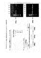
【図2】 正常血液レーンよりも肺癌RNAでより豊富であった6つの候補バンドを示すポリアクリルアミドゲルである。H:健常ヒト、L:肺癌組織。
【図3】 各候補マーカーならびにサイトケラチン−19およびβ−アクチンに関する肺細胞のRT−PCRを示すゲルである。
【図4】 各候補マーカーならびにサイトケラチン−19およびβ−アクチンに関する肺細胞のRT−PCRを示すゲルである。
【図5】 血液RNAからの陽性PCRおよび陰性PCRの一例である。
【図6】 シンデカン1に関する配列情報ならびにプローブ配列である。
【図7】 コラーゲン−1Proα2に関する配列情報ならびにプローブ配列である。
【図8】 7013遺伝子に関する配列情報ならびにプローブ配列である。
【図9】 7018遺伝子に関する配列情報ならびにプローブ配列である。[0001]
[Technical field]
The present invention relates to a method for diagnosis and differentiation of residual lung cancer. The invention further relates to the use of newly identified cell markers for lung cancer. These markers include
[0002]
[Background of the invention]
Lung cancer is one of the most common cancers in industrialized countries and has a very high mortality rate. Initial diagnosis and effective treatment are not currently available. Chest x-rays are often used for lung cancer screening, but it is not useful for early stage cancer detection. CT scans can also be used to allow early stage cancer detection, but this approach is time consuming and has a risk of exposure.
[0003]
Cancer cells or micrometastases are often detected in the bloodstream of melanoma, thyroid cancer, and prostate cancer patients. Currently, reverse transcription-based polymerase chain reaction (RT-PCR) is one powerful method that can detect single cancer cells in myriad normal cells by amplifying cancer-specific genes or markers. This makes RT-PCR detection of micrometastasis a promising diagnostic approach for prognosis, selection of appropriate treatments, and monitoring the efficacy of each treatment. Furthermore, blood tests do not induce any health hazards, but methods such as X-rays or CT scans have adverse effects. Furthermore, blood tests cause very little physical discomfort compared to endoscopy and biopsy. Lung cancer often induces hematogenous metastases even at an early stage, ie symptomatic disease and even before many lung cancers recur as distant metastases such as in the brain, bone and liver. This is due to hematogenous metastasis induced by lung cancer. However, this knowledge is beneficial because it suggests that diagnosis and detection of recurrence can be made based on these hematogenous metastases at the very early stages. However, there are no good markers available for identifying metastatic lung cancer cells in the blood. Currently, lung cancer markers such as cytokeratin-19 and CEA are used for diagnosis of non-small cell lung cancer by RT-PCR (Reverse Transcriptase Polymerase Chain Reaction), but lack specificity and have many false positives and negatives. Arise.
[0004]
RT-PCR of micrometastasis is then particularly beneficial for the detection of lung cancer due to the large patient population, high incidence of hematogenous metastasis, poor prognosis and high medical costs for higher cancer treatment . Furthermore, specific antibodies are not currently available for lung cancer.
[0005]
Therefore, there is a need for a specific marker and a method for diagnosing lung cancer by detecting hematogenous metastases.
[0006]
[Summary of Invention]
One embodiment is a method for differentiating lung cancer by isolating blood or non-lung tissue from a patient and determining the presence of at least one marker from the following: Syndecan 1, Collagen 1α2, 7013 and 7018 is there. The method can also include determining the presence of the marker cytokeratin-19. In a further embodiment, at least two markers are determined. In further embodiments, more than two markers are determined. The discrimination method can be any known to those skilled in the art, but can also include RT-PCR and / or antibody binding. The patient can be any living body, but in one embodiment is a mammal, particularly a human, dog or cat.
[0007]
A further embodiment is a method for isolating or removing metastatic cancer cells, wherein the cell or non-pulmonary tissue comprising cancer cells is at least one selected from the group consisting of
[0008]
A further embodiment is identified herein as marker 7013Of SEQ ID NO: 16A polynucleotide comprising at least 17 nucleotidesis there.
[0009]
A further embodiment is identified herein as marker 7018Of SEQ ID NO: 17A polynucleotide comprising at least 17 nucleotides.
[0010]
A further embodiment is a method of differentiating solid tumor metastasis in a patient, selected from the group consisting of: isolating blood or bone marrow from the patient, and
[0011]
A further embodiment is a method for differentiating carcinomas by obtaining cancer cells and determining the presence of a marker selected from the group consisting of 7013, 7018 and both.
[0012]
Detailed Description of Preferred Embodiments
Differential display was used to identify mRNAs that circulate in the blood and that are specifically expressed in lung cancer cells that are not expressed in normal blood cells. The differential display method is a variation from the classical approach that allows a more comprehensive expression of genes expressed in cells.
[0013]
Three main techniques are currently available for identifying candidate genes. That is, a subtraction library, a differential display, and a DNA microchip array. However, although the subtraction library is potentially useful, it can also identify genes that show partial similarity in gene sequences, and is a very laborious and time intensive technique. DNA microchip arrays are quick and easy to use, but are not sensitive enough to detect rare genes in blood samples. This is particularly important because the method needs to be able to detect rare micrometastasis that expresses a specific marker within a large background (innumerable white blood cells) of normal cells that do not express the specific marker. Differential display (dd-PCR) allows the amplification of all existing genes using a combination of multiple degenerate primers, thus increasing the likelihood that rare markers or genes will be detected.
[0014]
However, in order to improve the differential display technique to represent as many genes as possible, selective Amplified Fragment Length Polymorphism (s-AFLP) was used in Example 1. This approach 1) amplifies only the 3 ′ end of the gene, 2) produces a more reproducible gel pattern, 3) identifies fewer extra genes, and 4) more selective PCR conditions. It has the advantage of using. This method led to the identification of four markers: Synd, Col, 7013 and 7018.
[0015]
These markers can be used to distinguish lung cancer cells in the patient's blood at any stage of the disease. Any method known to those skilled in the art can be used to determine markers in blood or any metastatic tissue. For example, mRNA expressed in cells that are specific for the marker can be discriminated.
[0016]
Alternatively, the protein itself can be identified using immunization techniques such as Western blot, FACS technology, ELISA, and other methods known to those skilled in the art. These antibodies or functional antibody portions can be purchased, isolated or produced using known methods.
[0017]
In one embodiment, marker gene expression is identified. Any method that allows discrimination of the expression of a gene associated with a marker can be used. Typically, the method amplifies mRNA resulting from gene expression. In one embodiment, RT-PCR of mRNA from blood or tissue is used. In further embodiments, antibodies are used to distinguish cells in the blood that contain these markers. For example, cell sorting can be used to distinguish cells that have been fluorescently labeled with antibodies to these markers.
[0018]
In one embodiment, the method is used to determine the presence of lung cancer cells in blood or bone marrow. However, it is normally envisioned that mRNA from any tissue that does not yield these markers can be used. For example, mRNA from an organ that is typically the site of metastasis can be used. Thus, in a further embodiment, the method is used to discriminate lung cancer cells in organs such as the liver or brain.
[0019]
In one embodiment, the method is used to determine the presence of lung cancer cells at the very early stages of the disease (in a further embodiment, lung cancer cells are determined after remission) or to determine recurrence. Used for.
[0020]
In further embodiments, the markers are used to target lung cancer cells in vivo or in vitro. In one embodiment, cancer cells are removed from the body using affinity techniques that target cancer cells and remove them from the blood.
[0021]
In further embodiments, the markers are used to differentiate metastatic cells in the blood or bone marrow, or metastatic organs or tissues generated by cancer such as the bile duct, colon, breast, uterus, esophagus and larynx. Alternatively, the marker can be used to remove metastatic cells from the blood.
[0022]
In a further embodiment,
[0023]
The patient can be any animal capable of having cancer. In one embodiment, the patient is a mammal. In a further embodiment, the mammal is a pet, such as a dog or cat. In a further embodiment, the patient is a human.
[0024]
The invention will now be described with reference to the following examples. However, it is understood that these examples are merely illustrative of the invention and are not intended to limit the invention.
[0025]
In Examples 1 and 2, the markers are identified using a variety of differential display techniques. The identified markers are described in Example 2.
[0026]
Example 1
s-AFLP differential display
s-AFLP is based on selective PCR amplification of restriction fragments from a cDNA library. Two sets of selective primers were used. The first, ie, selective primer A of FIG. 1, consists of three parts: a core sequence, an enzyme specific sequence and two selective nucleotides at the 3 'end. Due to selective sequence variation, this provides 42 = 16 different primers. The second, ie, selective primer B of FIG. 1, also consists of three parts: an anchor sequence, a poly T sequence and two selective nucleotides at the 3 'end that are fluorescently labeled. For the three selective sequences after poly T (A, G and C), this then provides 3 × 4 = 12 different primers. Thus, all 3'-end cDNA restriction fragments including the polyA region were included in the 192 (16x12) group generated by this method of selective PCR. Each signal on the gel display of the amplified fragment indicated a non-overlapping gene in the cDNA library. Using this technique, RNA isolated from lung cancer specimens was compared with RNA from healthy individual blood. Differentially expressed genes that were overexpressed in cancer specimens were considered candidates for general genetic markers for tumor cell seeding.
[0027]
Excellent quality RNA was important for the success of s-AFLP. However, lung specimens are one of the most difficult tissues to prepare RNA due to the presence of abundant RNases from alveolar macrophages. Therefore, rapidly frozen lung cancer specimens were purchased from NCCRI (Chuou-ku, Tokyo, Japan) and total RNA was purified by the AGPC method as previously described (Tominaga, K, Miura, Y. Arakawa, T. Koboyashi). , K. and Mitsuhashi, M. Clin. Chem., 42, 1750-1783, 1998). RNA quality was assessed using agarose gel electrophoresis or microcapillary chips to confirm the presence of two rRNA bands. Next, an acceptable specimen for s-AFLP was used. Six lung cancer specimens were used and each sample was obtained from a different patient. Four of the six were adenocarcinomas and two were squamous cell carcinomas. Twenty control blood were obtained from healthy volunteers with no history or current diagnosis of malignancy or other diseases.
[0028]
Total RNA preparation of tumor and blood samples
Fresh frozen tumor specimens were broken into small pieces in liquid nitrogen. Total RNA was extracted from a 100 mg sample by the guanidine method (Chomzynski, P. and Sacchi, N. 1987. Single-step method of RNA isolation by acid guanidine thiocyanate-phenol-chloroform extraction. Anal. Biochem. 162: 156-159 ). Peripheral blood samples were collected from patients and healthy volunteer veins in heparin anticoagulant containing tubes. Two samples were taken from each subject, 1-2 ml of peripheral blood in the first tube and 10 ml of peripheral blood in the second tube. The first tube was discarded because it may have been contaminated with the epithelial cells that the needle caught when the needle touched the skin, and only the second tube was assayed. Total RNA was extracted from peripheral blood samples using RiboCap ™ (RNature, Irvine, CA) according to the manufacturer's instructions. Purified total RNA quality was checked by agarose gel electrophoresis for 18s and 28s ribosomal RNA bands and their quantity was measured by UV spectroanalyzer.
[0029]
Differential display s-AFLP analysis
A differential “s-AFLP” variant was performed using selective primer techniques.
[0030]
Two pooled healthy blood samples containing 6 lung cancer specimens (lung cancer tissue) and 5 healthy individuals were each used for s-AFLP analysis. All oligomers used in all assays were obtained from Sawady Technology (Tokyo, Japan). Double-stranded cDNA was synthesized from 30 μg of total RNA. After denaturation at 65 ° C. for 5 minutes, the following: 50 μg / ml oligo (dT)12-18Primer (Life Technologies, USA), 500 μM of each deoxynucleotide triphosphate (dNTP) (Life Technologies), 50 mM Tris-HCl (pH 8.3), 75 mM KCl, 3 mM MgCl2RNA for 90 minutes at 37 ° C. in 50 μl reaction mix containing 10 mM dithiothreitol (DTT), 20 units RNAsin (Life Technologies) and 10,000 units / ml MMLV reverse transcriptase (Life Technologies) Was reverse transcribed. For final concentration the following components: 25 mM Tris-HCl (pH 8.3), 100 mM KCl, 10 mM (NHFour)2SOFour5 mM MgCl2, 250 μM each dNTP, 0.15 mM NAD, 5 mM DTT, 250 units / ml DNA polymerase (Life Technologies), and 30 units / ml DNA ligase (Life Technologies) at 120 ° C. The second strand reaction was performed for minutes. After the reaction, the test tube was placed on ice and 12.5 μl of 0.25 M ethylenediaminetetraacetic acid (EDTA; pH 7.5) was added for enzyme inactivation. The product was extracted once with phenol / chloroform and ethanol precipitated. Double-stranded cDNA was digested with X-unit 4-base recognition restriction enzyme Mbol (↓ GATC; New England Biolabs, USA) at 37 ° C. for 60 minutes. The phosphate residue at the 5 'end of the digested fragment was removed using calf intestinal alkaline phosphatase (CIAP; Takara, Japan) to avoid self-ligation during the ligation process. 20 units of CIAP and 10 μl of 10 × alkaline phosphate buffer (500 mM Tris-HCl (pH 9.0), 10 mM MgCl2) Was added and incubated at 37 ° C. for 30 minutes. The product was extracted twice with phenol / chloroform and ethanol precipitated. The fragment was ligated with the oligomer and introduced into the priming site. The following oligomers were used for ligation: (P1) 5′-P-GATCCCCTATATAGTGAGTC-3 ′ (linker oligomer) [SEQ ID NO: 1]; (P2) 5 ′ GACTCACTATAGGGG-P-3 ′ (helper oligomer) [sequence Number 2]. Helper oligomers were phosphorylated at the 3 'end to prevent oligomer dimer formation. The ligation reaction was performed using a 1: 100: 200 molar ratio of digested fragment: helper oligomer: linker oligomer.
[0031]
The reaction was performed overnight at 16 ° C. by adding 15 μl Ligation High (Toyobo) in 45 μl reaction. After removal of unligated oligomers with the QIAquick Nucleotide Removal kit (Qiagen, Germany), the 3 'end fragment containing the poly A tail was amplified and detected using two selective primers. One of them (P3) consists of three parts: the complementary sequence of the anchor oligomer, the Mbol recognition sequence (GATC) and two degenerate nucleotides at the 3 ′ end (5′-GACTCACTATAGGGATCNN-3 ′) [SEQ ID NO: 3]. P3 is 4 due to mutations in the selective sequence (NN).2= 16 type. The second selective primer (P4) also has three parts: an anchor sequence, a poly T sequence labeled with sulforhodamine 101 label and two degenerate nucleotides at the 3 ′ end (5′-TCTCCTTTTTTTTTTTTTTTTTTVN-3 ′) [SEQ ID NO: 4]. Since the optional sequence “V” is any nucleotide other than T, P4 has a 3 × 4 = 12 type. Thus, all 3 'terminal cDNA restriction fragments containing the poly A region are each separately classified into the 192 (= 16x12) group by the 192 PCR reaction. PCR was performed using ExTaq DNA polymerase (Takara, Japan) with cycling parameters of 94 ° C. for 30 seconds, 56 ° C. for 1 minute and 72 ° C. for 1 minute (30 cycles). Separation of amplification products by electrophoresis on polyacrylamide gel (4% T, 5% C) containing 1XTBE (0.09M Tris-borate and 0.02M EDTA) and 7M urea on Hitachi SQ-3000 fluorescent DNA sequencer did.
[0032]
The cDNA was excised from the gel and purified by a pulverizing dipping method. FIG. 2 shows a sample gel producing 6 candidates. Bands indicate specific fragments that are more abundant in lung cancer RNA than normal blood. A band was isolated when a band was present in at least two of the six lung cancer tissue lanes and not in any normal blood lane. Selection criteria were important because differential displays often produce a large number of false positives. The isolated fragment was subcloned using the pGEM-Teasy vector system (Promega, USA). The product purified by mini-preparation was purified from T7 primer (5'-TAATACGACTCACTATAG-3 ') using the Big Dye terminator cycle sequencing kit (Applied Biosystems, CA, USA) and ABIPrism377 DNA sequencing apparatus (Applied Biosystems). Sequencing was performed using [SEQ ID NO: 15].
[0033]
Northern blot analysis
Total RNA was separated by formaldehyde gel electrophoresis and transferred to nylon membrane (+) (Amersham Pharmacia, England). Candidates were cloned using pCR2-1. The clones were verified by sequence analysis. Plasmid DNA was restricted with the appropriate enzyme and used for in vitro transcription.
[0034]
Selection method
Initially, 121 candidates were identified that were negative in healthy blood and positive in lung cancer. After sequencing these 121 candidates, the sequences were analyzed for homology with GenBank as well as known sequences in the expressed sequence tag (EST) database.
[0035]
The procedure for selection of true signal versus false signal is shown below: The band was excised from the display gel and the DNA was cloned. The isolate was then sequenced. If the sequence data indicated that the gene is normally present in the blood cell, it was discarded. Specific primers were then designed and PCR was performed on each clone. First, normal blood RNA was amplified by PCR, and when a PCR product was produced, the candidate was discarded. The remaining 21 candidates were tested against blood from lung cancer patients and were discarded if no signal was found in any patient. This procedure resulted in the isolation of 4 candidates (see Example 2).
[0036]
[Table 1]
Example 2
Candidate gene
Candidate markers are shown in Table 1. The first candidate marker found was the
[0038]
The next candidate was identified as collagen 1α2 (nucleotide sequence PubMed accession number J00114, protein sequence accession number AAA51996.).FIG. 7 shows the old fragment provided by s-AFLP (SEQ ID NO: 20) And new fragments (SEQ ID NO: 19). A blast search of this sequence corresponded to
[0039]
The third candidate is called 7013 (SEQ ID NO: 16) and was identified as a novel gene when searched against the EST database. FIG. 8 shows the sequence of a fragment obtained from s-AFLP (SEQ ID NO: 21). Using this fragment, a large fragment was identified by the primer extension method (SEQ ID NO: 16). This large fragment was cloned into pCRII and the resulting plasmid p7013 was deposited with ATCC (Bethesda, Maryland, USA) on June 21, 2001 by FEDEX shipping under the Budapest Treaty labeled 82020804377778. , ATCC accession numberIssueAssigned. A large clone was isolated from a cDNA library and plasmid p7013 / 12 was deposited with ATCC (Bethesda, Maryland, USA) on June 21, 2001 by FEDEX shipping under the Budapest Treaty labeled 8202080437778. Assigned accession number. The plasmid has an insert of about 2200 bases in pCMV6-XL4. Upon performing the BLAST search, the EST (GenBank accession number AK002208) and genomic clone (GenBank accession number AL035408) exhibiting the highest homology were identified. The sequence did not show any homology with any known gene, but was localized in the q32 region of
[0040]
The next candidate is called 7018 (SEQ ID NO: 17), which is also a novel gene. FIG. 9 presents the sequence of the fragment obtained from s-AFLP (SEQ ID NO: 22). Using this sequence, the primer was extended to cDNA to obtain a large fragment (SEQ ID NO: 17). This large fragment was cloned into plasmid pCRII and the resulting plasmid p7018 was deposited with ATCC (Bethesda, Maryland, USA) on June 21, 2001 by FEDEX shipping under the Budapest Treaty labeled 82020804377778. ATCC accession numberIssueAssigned. In performing a BLAST search, two ESTs (GenBank accession numbers AW95727 and AW4552795) were identified that showed the highest homology but did not show any homology with known genes. Moreover, it has so far not been localized to any chromosomal region.
[0041]
Examples 3 and 4 determine whether markers are expressed in normal lung cells and lung cancer cell lines.
[0042]
Example 3
Marker expression in normal lung cells and lung cell lines
To determine whether these markers were expressed in normal human lung cells, the same amount from human lung small airway epithelial cells, microvascular epithelial cells from the lung, bronchial epithelial cells and normal human lung fibroblasts RT-PCR was performed with normal human lung RNA using
[0043]
Total RNA preparation from normal lung or lung cancer cell lines
Total RNA was isolated from cells by acid-guanidine thiocyanate / phenol / chloroform extraction using the TRI REAGENT® protocol (Molecular Research Center, Inc., Cincinnati, OH, USA). It is not necessary to completely purify the RNA, and in fact, some DNA contamination is acceptable if the primer is designed in the region spanning the intron.
[0044]
RT-PCR
Prior to reverse transcription, all RNA samples were treated with DNase I to ensure no genomic DNA contamination. One unit of DNase I (Life Technologies) was used for every 1 μg of total RNA. The product was extracted twice with phenol / chloroform and ethanol precipitated. Using 200 units of MMLV transcriptase for each 0.5 μg of RNA from the cell line (50 minutes at 42 ° C.) and 100 units of transcriptase RiverTraAce ™ for each 1 μg of blood total RNA (60 minutes at 37 ° C.) was used to synthesize cDNA.
[0045]
First, 100 units of reverse transcriptase RiverTraAce ™ (TOYOBO, Japan), 50 μg / ml oligo (dT) for each 1 μg of total RNA.12-18CDNA synthesis was performed at 37 ° C. for 60 minutes using primers (Life Technologies) and 500 μM of each deoxynucleotide triphosphate (dNTP) (Life Technologies). For all PCRs, 50 ng total RNA, 10 pmol primer pairs and 0.125 units EX Taq DNA polymerase were used in a total volume of 25 μl reaction. The primer pair and cycling conditions for each gene marker were as follows: SYND marker was primer rP5 (5′-TCATGTTGCAACAGGGGTAT-3 ′) [SEQ ID NO: 5] and primer P6 (5′-AATTTCCTGATTCCAGCCCC-3 ′) [sequence] No. 6] was used for amplification. Cycling conditions were 94 ° C. for 30 seconds, 65 ° C. for 1 minute and 72 ° C. for 1 minute (30 cycles). COL was amplified using primer P7 (5'OAGAGCATTGTGCAATACAGTTTCATTAACTCCT-3 ') [SEQ ID NO: 7] and primer P8 (5'-GGTTTTCTTACAAAGGTTGACATTTCCTAACAG-3') [SEQ ID NO: 8]. Cycling parameters were 94 ° C. for 30 seconds, 58 ° C. for 1 minute and 72 ° C. for 1 minute (20 cycles). For the second round PCR reaction, the reaction mix contained 1 μl of the first round PCR product as template and the primer pairs P3 and P4. The second stage of PCR was performed 20 times at 94 ° C. for 30 seconds, 60 ° C. for 1 minute and 72 ° C. for 1 minute. 7013 was amplified using primer P9 (5'-AATGAAGGAGACACATCTGGAGTGTGCG-3 ') [SEQ ID NO: 9] and primer P10 (5'-AGAAAAGAAAGAATTAAGGTTCCCCATCTGCG-3') [SEQ ID NO: 10]. Cycling conditions were 94 ° C for 30 seconds, 63 ° C for 1 minute and 72 ° C for 1 minute (38 cycles). 7018 was amplified using primer P11 (5'-ATCCATGCACGCTCTTTCCTTTCC-3 ') [SEQ ID NO: 11] and primer P12 (5'-TCAAGTAGGCCAACCCAGTCCT-3') [SEQ ID NO: 12]. Cycling conditions were 94 ° C for 30 seconds, 63 ° C for 1 minute and 72 ° C for 1 minute (38 cycles). CK-19 was amplified using primer P13 (5'-CAAGATCCCTGAGTGACATGCGAAG-3 ') [SEQ ID NO: 13] and primer P14 (5'-CGCTGATCAGCGCCTGGATATG-3') [SEQ ID NO: 14]. Cycling parameters were 94 ° C for 30 seconds, 60 ° C for 1 minute and 72 ° C for 1 minute (20 cycles). For the second round PCR reaction, 1 μl of the first round PCR product was the template and the same primer pair of P13 and P14 had the same cycle parameters. After PCR, 1 μl of PCR product was analyzed by electrophoresis on 4% NuSieve GTG agarose gel (Takara, Japan).
[0046]
As a positive control, all samples were subjected to PCR for β-actin. Four candidates, 7018, 7013, Syndecan 1 (Synd), collagen 1A2 (Col), and positive control cytokeratin-19 (Ck-19) primers were used for RT-PCR. Cytokeratin-19 is known as an epithelial cell marker. PCR amplification was performed by using 5 primer pairs for these markers. For cell lines, 0.1 volume of RT reaction, 10 μM of each primer and 1.24 units of Taq DNA polymerase were used in a total volume of 50 μl reaction. For whole blood, 0.1 volume of RT reaction, 10 μm of each primer and 0.125 units of EXTaq DNA polymerase were used in a total volume of 25 μl reaction. The PCR conditions for each primer are as shown in Table 2. PCR products were analyzed by agarose gel electrophoresis, and several products were confirmed by sequencing.
[0047]
[Table 2]
The results of PCR of normal cells are shown in Table 3. This indicates that four new isolated markers were often expressed in the normal lung cell lines examined. In addition,
[0049]
[Table 3]
Example 4
Marker expression in lung cancer cell lines
To determine whether these markers were expressed in lung cancer cell lines, RT-PCR was performed with RNA from 12 lung cancer cell lines using the same amount of RNA (see Table 4). All PCR products were analyzed by agarose gel electrophoresis and several products were confirmed by sequencing. All RNA was equally amplified positive control-actin. Cancer cell line Lu99 (Yamada, et al. Giant cell carcinomas of the lung producing colony-stimulating factor in vitro and in vivo. Jpn. J. Cancer Res., 76: 967-976, 1985), PC13 (large cell carcinoma) A549 (Imanishi, et al. Inhibition of growth of human lung adenocarcinoma cell lines by anti-transforming growth factor-alpha monoclonal antibody. J. Natl. Cancer Inst., 81: 220-23, 1989), PC14, NCI-H441. (Adenocarcinoma), PC1 and QG56 (squamous cell carcinoma) were used.
[0051]
As shown in FIG. 4, the presence of markers in RNA in six lung cancer cell lines was also examined by RT-PCR. Four of the lines were adenocarcinoma and those in
[0052]
[Table 4]
Example 5 provides a method for distinguishing lung cancer cells in blood using these markers. In Example 5, RT-PCR is used to determine the presence of mRNA for these markers in the blood sample.
[0054]
Example 5
Expression of markers in patient blood
We investigated 68 lung cancer patients diagnosed and treated at Keio University Hospital between November 1998 and April 2000, and 7 metastatic lung cancer patients at Keio University Hospital at the same time. Patient characteristics are shown in Table 5.
[0055]
Test RNA from patient blood samples using RT-PCR with four
[0056]
Total RNA preparation from whole blood
Peripheral blood samples were collected from patients and healthy volunteers from the antecubital vein in heparin anticoagulant containing tubes. Red blood cells (RBC) were lysed with a standard hypotonic solution and the whole blood cell population was collected on a RiboCap syringe filter (RNAture, Irvine, CA). RNA was eluted from the syringe filter by adding a guanidine solution and then using the standard AGPC method. Purified total RNA quality was analyzed by agarose gel electrophoresis with 18s and 28s ribosomal RNA bands and their quantity was measured by UV spectrometer. After preparation, the RNA pellet was resuspended in 20 μl diethylpyrocarbonate (DEPC) treated water and stored at −80 ° C.
[0057]
RT-PCR
Prior to reverse transcription, total RNA samples were treated with DNase I to remove any potential genomic DNA contamination. Using 200 units of MMLV transcriptase for each 0.5 μg of RNA from the cell line (50 minutes at 42 ° C.), 100 units of the transcriptase RiverTraAce ™ for each 1 μg of whole blood RNA. Used (60 minutes at 37 ° C.) to synthesize cDNA.
[0058]
All samples were subjected to PCR for β-actin as a positive control. FIG. 5 shows an example of a positive test from blood RNA. Four
[0059]
[Table 5]
In Table 6, the RT-PCR results of blood from lung cancer patients and control healthy volunteers were tested for the presence of the identified markers. Stages I-IV patients were tested for marker expression, similar to 40 healthy volunteers. In summary, the results showed that the four
[0061]
[Table 6]
In particular, in Table 7, each gene was expressed in 3%, 41%, 19% and 16%, respectively, in a lung cancer patient blood sample, but at least one of these 4 genes was present in 57% of the patient blood sample. To express. Also, with cytokeratin-19, at least one of these five genes is expressed in 71% of patients. One or more of these genes are expressed in 80.5% of adenocarcinoma and 68.4% of squamous cell carcinoma. Syndecan was found in 13 lung cancer patients and not in control (healthy) patients. Collagen was found only in lung cancer patients. The 7013 marker was widely expressed in lung cancer patients but not at all in the controls. Only 7018 was found to be expressed in control samples as well as lung cancer patients.
[0063]
[Table 7]
Thus, 54% of patients displayed at least one or more of the four markers.
[0065]
In summary, four new potential markers have been identified that are present in the blood of lung cancer patients and not in normal blood. Two of the markers are novel genes.
[0066]
New marker combinations were observed in 54% of the blood of examined lung cancer patients (Table 5), and the combination of these markers with cytokeratin-19 was found in 68% of the blood of examined lung cancer patients Produced. The combination is therefore ideal for diagnosis of early stage lung cancer micrometastasis or for diagnosis of recurrence.
[0067]
Example 6 examines the ability of an assay using five markers to distinguish lung cancer cells in any type or stage of lung cancer.
[0068]
Example 6
Analysis by type and stage of lung cancer
Patients with different types and stages of lung cancer were tested for the expression of four markers (see Table 8).
[0069]
[Table 8]
As in Example 5, blood samples of 69 lung cancer patients were obtained from Keio University Hospital. Stage classification was performed according to the tumor-nodule-metastasis (TNM) score (Mountain CE: A new international staging system for lung cancer. Chest 89: 225S-233S, 1986). RNA was purified and RT-PCR was performed as in Example 5. The expression of each marker in the blood sample was analyzed, and the results are shown in Table 9.
[0071]
In Table 9, the markers appear to be expressed a little more in the late stage, but it can be observed that if all five combinations are used, they can be discriminated at all stages of the disease. Thus, using these five genes in combination, at least one gene is 56% of stage IA, 33% of IB, 50% of II, 92% of IIIA, 60% of IIIB, and 87% of IV To express.
[0072]
[Table 9]
When testing blood from patients with metastatic disease, it can be observed that at least one marker was detected in all types of metastatic disease (see Table 10). This suggests that markers can be used to detect disease at all stages of lung cancer as well as recurrent metastases.
[0074]
[Table 10]
Example 11
Diagnosis and analysis of the presence of lung cancer using mRNA from one or more of the identified markers
Blood was isolated from the patient and RNA from the blood sample was purified. RT-PCR is performed using at least one of the following markers:
[0076]
Example 12
Diagnosis and analysis of the presence of lung cancer using antibodies against one or more of the identified markers
Markers:
[0077]
Example 13
Isolation of cancer cells from blood or bone marrow
By treating the cell or non-pulmonary tissue containing the cell with an antibody specific for at least one marker selected from the group consisting of
[0078]
The antibody is conjugated to a moiety selected from the group consisting of metal particles, fluorescent particles, chromatography beads, chromatography gels and solid supports.
[0079]
The isolated cells can then be used to identify the site from which the cancer cells have metastasized, or to produce activated immune cells specific for the cancer. Alternatively, the method can be used to purify cells from blood.
[0080]
Example 14
Isolation of full-length cDNA clones
Primers for the detection of full length cDNA clones are designed using clones obtained from the s-AFLP technique. A human cDNA library is purchased from Stratagene (Torrey Pines, CA). The library is screened using a probe specific for 7013 (SEQ ID NO: 16) or 7018 (SEQ ID NO: 17). After obtaining a positive clone, the clone is tested and sequenced using PCR.
[0081]
Example 15
Differentiation of cell epithelium / carcinoma
Since
[Brief description of the drawings]
FIG. 1 shows a selective amplified fragment length polymorphism (s-AFLP), a modified differential display approach, used in a preferred embodiment.
FIG. 2 is a polyacrylamide gel showing six candidate bands that were more abundant in lung cancer RNA than in normal blood lanes. H: Healthy human, L: Lung cancer tissue.
FIG. 3 is a gel showing RT-PCR of lung cells for each candidate marker and cytokeratin-19 and β-actin.
FIG. 4 is a gel showing RT-PCR of lung cells for each candidate marker and cytokeratin-19 and β-actin.
FIG. 5 is an example of positive and negative PCR from blood RNA.
FIG. 6 shows sequence
FIG. 7 shows sequence information and probe sequences related to collagen-1Proα2.
FIG. 8 shows sequence information and probe sequences related to the 7013 gene.
FIG. 9 shows sequence information and probe sequences related to the 7018 gene.
Claims (8)
患者から単離された血液または非肺組織を用いて、配列番号16の塩基配列を含む遺伝子から発現したmRNA又はタンパク質の存在を判別すること
を含む方法。A method for distinguishing lung cancer,
A method comprising determining the presence of mRNA or protein expressed from a gene comprising the nucleotide sequence of SEQ ID NO: 16 using blood or non-pulmonary tissue isolated from a patient.
Applications Claiming Priority (3)
| Application Number | Priority Date | Filing Date | Title |
|---|---|---|---|
| US21572700P | 2000-06-21 | 2000-06-21 | |
| US24397600P | 2000-10-27 | 2000-10-27 | |
| PCT/US2001/019980 WO2001098539A2 (en) | 2000-06-21 | 2001-06-21 | Gene markers for lung cancer |
Publications (3)
| Publication Number | Publication Date |
|---|---|
| JP2004500895A JP2004500895A (en) | 2004-01-15 |
| JP2004500895A5 JP2004500895A5 (en) | 2006-05-18 |
| JP4392163B2 true JP4392163B2 (en) | 2009-12-24 |
Family
ID=26910326
Family Applications (1)
| Application Number | Title | Priority Date | Filing Date |
|---|---|---|---|
| JP2002504687A Expired - Fee Related JP4392163B2 (en) | 2000-06-21 | 2001-06-21 | Genetic marker for lung cancer |
Country Status (6)
| Country | Link |
|---|---|
| US (2) | US7214781B2 (en) |
| EP (1) | EP1356093A2 (en) |
| JP (1) | JP4392163B2 (en) |
| CN (1) | CN1471587A (en) |
| AU (1) | AU2001270082A1 (en) |
| WO (1) | WO2001098539A2 (en) |
Cited By (1)
| Publication number | Priority date | Publication date | Assignee | Title |
|---|---|---|---|---|
| WO2011111820A1 (en) * | 2010-03-11 | 2011-09-15 | 株式会社インテックシステム研究所 | Diagnostic marker for lung cancer or cervical cancer |
Families Citing this family (15)
| Publication number | Priority date | Publication date | Assignee | Title |
|---|---|---|---|---|
| EP1383922A4 (en) | 2001-04-10 | 2005-03-30 | Agensys Inc | Nucleid acid and corresponding protein entitled 158p3d2 useful in treatment and detection of cancer |
| EP1510587B1 (en) * | 2002-05-21 | 2011-07-13 | Sysmex Corporation | Nucleic acid amplification primers for detecting cytokeratins and examination method with the use of the primers |
| GB0215509D0 (en) * | 2002-07-04 | 2002-08-14 | Novartis Ag | Marker genes |
| US20050186215A1 (en) * | 2004-02-04 | 2005-08-25 | Kwok Tim T. | CUDR as biomarker for cancer progression and therapeutic response |
| CA2617693A1 (en) | 2005-08-17 | 2007-02-22 | Medexis S.A. | Composition and method for determination of ck19 expression |
| JP4723655B2 (en) * | 2006-02-03 | 2011-07-13 | ヒルズ・ペット・ニュートリシャン・インコーポレーテッド | A method to evaluate renal function by measuring ghrelin hormone levels in cats |
| WO2007130967A2 (en) * | 2006-05-01 | 2007-11-15 | Siemens Healthcare Diagnostics Inc. | Novel oligonucleotide primers and methods for dna replication |
| KR101443214B1 (en) | 2007-01-09 | 2014-09-24 | 삼성전자주식회사 | A composition, kit and microarray for diagnosing the risk of lung cancer recurrence in a patient after lung cancer treatment or a lung cancer patient |
| WO2009043115A1 (en) * | 2007-10-04 | 2009-04-09 | Fermiscan Australia Pty Limited | Method for identifying biomarkers which are diagnostic for a pathological condition |
| CN103476428B (en) | 2010-09-09 | 2016-10-26 | 北京同为时代生物技术有限公司 | For diagnosing blood markers thing and the monoclonal antibody thereof of epitheliogenic cancerg |
| JP6386450B2 (en) | 2012-06-06 | 2018-09-05 | フンダシオ、インスティトゥト、デ、レセルカ、ビオメディカ(イエレベ、バルセロナ)Fundacio Institut De Recerca Biomedica (Irb Barcelona) | Methods for diagnosis, prognosis and treatment of lung cancer metastasis |
| KR20200058540A (en) | 2017-10-02 | 2020-05-27 | 비스테라, 인크. | Antibody molecules against CD138 and uses thereof |
| WO2020007898A1 (en) * | 2018-07-04 | 2020-01-09 | INSERM (Institut National de la Santé et de la Recherche Médicale) | Methods and compositions for treating brain injury or neurodegenerative disease |
| WO2020257289A2 (en) * | 2019-06-17 | 2020-12-24 | Visterra, Inc. | Humanized antibody molecules to cd138 and uses thereof |
| KR20210020417A (en) * | 2019-08-14 | 2021-02-24 | 연세대학교 산학협력단 | Method for diagnosing lung cancer using methionyl-tRNA synthetase and pan-cytokeratin |
Family Cites Families (2)
| Publication number | Priority date | Publication date | Assignee | Title |
|---|---|---|---|---|
| US5272057A (en) * | 1988-10-14 | 1993-12-21 | Georgetown University | Method of detecting a predisposition to cancer by the use of restriction fragment length polymorphism of the gene for human poly (ADP-ribose) polymerase |
| DE69933998T2 (en) | 1998-03-18 | 2007-10-04 | Corixa Corp., Seattle | COMPOUNDS AND METHODS FOR THERAPY AND DIAGNOSIS OF LUNG CANCER |
-
2001
- 2001-06-21 AU AU2001270082A patent/AU2001270082A1/en not_active Abandoned
- 2001-06-21 WO PCT/US2001/019980 patent/WO2001098539A2/en not_active Application Discontinuation
- 2001-06-21 JP JP2002504687A patent/JP4392163B2/en not_active Expired - Fee Related
- 2001-06-21 US US10/297,277 patent/US7214781B2/en not_active Expired - Fee Related
- 2001-06-21 EP EP01948623A patent/EP1356093A2/en not_active Withdrawn
- 2001-06-21 CN CNA018115438A patent/CN1471587A/en active Pending
-
2005
- 2005-10-19 US US11/255,001 patent/US20060275783A1/en not_active Abandoned
Cited By (1)
| Publication number | Priority date | Publication date | Assignee | Title |
|---|---|---|---|---|
| WO2011111820A1 (en) * | 2010-03-11 | 2011-09-15 | 株式会社インテックシステム研究所 | Diagnostic marker for lung cancer or cervical cancer |
Also Published As
| Publication number | Publication date |
|---|---|
| US20030215828A1 (en) | 2003-11-20 |
| WO2001098539A2 (en) | 2001-12-27 |
| CN1471587A (en) | 2004-01-28 |
| US20060275783A1 (en) | 2006-12-07 |
| EP1356093A2 (en) | 2003-10-29 |
| JP2004500895A (en) | 2004-01-15 |
| WO2001098539A3 (en) | 2003-08-14 |
| US7214781B2 (en) | 2007-05-08 |
| AU2001270082A1 (en) | 2002-01-02 |
Similar Documents
| Publication | Publication Date | Title |
|---|---|---|
| US20060275783A1 (en) | Gene markers for lung cancer | |
| US6960433B1 (en) | Method of diagnosing, monitoring, staging, imaging and treating prostate cancer | |
| JP3422776B2 (en) | Novel method for diagnosing, monitoring, staging, imaging and treating breast cancer | |
| JP2002523760A (en) | Novel methods for diagnosing, monitoring, staging, imaging and treating various cancers | |
| WO2001009318A1 (en) | Liver cancer-associated genes | |
| WO2000028090A2 (en) | Diagnostic assay for cancer | |
| JPH0728759B2 (en) | Method of detecting tumorigenicity | |
| WO2012019300A1 (en) | Endometrial cancer biomarkers and methods of identifying and using same | |
| JP2002527758A (en) | Methods for diagnosing, monitoring, staging, imaging and treating prostate cancer | |
| JP2002541805A (en) | Compounds for immunotherapy and diagnosis of breast cancer and methods for their use | |
| JP5773315B2 (en) | CXCL4L1 as a biomarker for pancreatic cancer | |
| JP2002525031A (en) | Novel methods of diagnosing, monitoring, staging, imaging and treating colorectal cancer | |
| JP2002522046A (en) | Novel way to diagnose, monitor, stage, image and treat lung cancer | |
| WO2009066820A1 (en) | Characterization of cxcl-16 as a tumor associated marker of colorectal cancer | |
| EP3460476B1 (en) | Biomarker composition comprising lrp-1 as active ingredient, for diagnosis of radiation-resistant cancer or prediction of radiation therapy prognosis | |
| CA2328377A1 (en) | A novel method of diagnosing, monitoring, and staging prostate cancer | |
| WO2009066821A1 (en) | Characterization of esm-1 as a tumor associated marker of colorectal cancer | |
| JPH08500731A (en) | Diagnostic method | |
| KR100763902B1 (en) | A breast cancer related protein a gene encoding the same and a method for diagnosing a breast cancer using the protein and gene | |
| Kekeeva et al. | Analysis of SYT/SSX1 and SYT/SSX2 fusion genes in synovial sarcoma | |
| WO2000023108A1 (en) | Method of diagnosing, monitoring, staging, imaging and treating prostate cancer | |
| KR20080010597A (en) | A multiplex rt-pcr primer, a composition and a kit comprising the same for diagnosing chronic myeloid leukemia | |
| JP2002526760A (en) | Novel method for diagnosing, monitoring, staging, imaging and treating gastrointestinal cancer | |
| KR102560020B1 (en) | A Composition for Diagnosing Cancer | |
| JP4330372B2 (en) | Method for detecting metastatic cancer cells derived from gastric cancer |
Legal Events
| Date | Code | Title | Description |
|---|---|---|---|
| A521 | Written amendment |
Free format text: JAPANESE INTERMEDIATE CODE: A523 Effective date: 20060322 |
|
| A621 | Written request for application examination |
Free format text: JAPANESE INTERMEDIATE CODE: A621 Effective date: 20060322 |
|
| A711 | Notification of change in applicant |
Free format text: JAPANESE INTERMEDIATE CODE: A711 Effective date: 20070112 |
|
| A521 | Written amendment |
Free format text: JAPANESE INTERMEDIATE CODE: A821 Effective date: 20070112 |
|
| A131 | Notification of reasons for refusal |
Free format text: JAPANESE INTERMEDIATE CODE: A131 Effective date: 20090421 |
|
| A521 | Written amendment |
Free format text: JAPANESE INTERMEDIATE CODE: A523 Effective date: 20090709 |
|
| A131 | Notification of reasons for refusal |
Free format text: JAPANESE INTERMEDIATE CODE: A131 Effective date: 20090804 |
|
| A521 | Written amendment |
Free format text: JAPANESE INTERMEDIATE CODE: A523 Effective date: 20090820 |
|
| TRDD | Decision of grant or rejection written | ||
| A01 | Written decision to grant a patent or to grant a registration (utility model) |
Free format text: JAPANESE INTERMEDIATE CODE: A01 Effective date: 20090929 |
|
| A01 | Written decision to grant a patent or to grant a registration (utility model) |
Free format text: JAPANESE INTERMEDIATE CODE: A01 |
|
| A61 | First payment of annual fees (during grant procedure) |
Free format text: JAPANESE INTERMEDIATE CODE: A61 Effective date: 20091009 |
|
| FPAY | Renewal fee payment (event date is renewal date of database) |
Free format text: PAYMENT UNTIL: 20121016 Year of fee payment: 3 |
|
| R150 | Certificate of patent or registration of utility model |
Ref document number: 4392163 Country of ref document: JP Free format text: JAPANESE INTERMEDIATE CODE: R150 Free format text: JAPANESE INTERMEDIATE CODE: R150 |
|
| FPAY | Renewal fee payment (event date is renewal date of database) |
Free format text: PAYMENT UNTIL: 20131016 Year of fee payment: 4 |
|
| S531 | Written request for registration of change of domicile |
Free format text: JAPANESE INTERMEDIATE CODE: R313531 |
|
| S533 | Written request for registration of change of name |
Free format text: JAPANESE INTERMEDIATE CODE: R313533 |
|
| FPAY | Renewal fee payment (event date is renewal date of database) |
Free format text: PAYMENT UNTIL: 20131016 Year of fee payment: 4 |
|
| R350 | Written notification of registration of transfer |
Free format text: JAPANESE INTERMEDIATE CODE: R350 |
|
| LAPS | Cancellation because of no payment of annual fees |