JP4334023B2 - Use of recombinant human uteroglobin in the treatment of inflammatory and fibrotic conditions - Google Patents
Use of recombinant human uteroglobin in the treatment of inflammatory and fibrotic conditions Download PDFInfo
- Publication number
- JP4334023B2 JP4334023B2 JP50097999A JP50097999A JP4334023B2 JP 4334023 B2 JP4334023 B2 JP 4334023B2 JP 50097999 A JP50097999 A JP 50097999A JP 50097999 A JP50097999 A JP 50097999A JP 4334023 B2 JP4334023 B2 JP 4334023B2
- Authority
- JP
- Japan
- Prior art keywords
- rhug
- pla
- fibronectin
- mice
- fibrosis
- Prior art date
- Legal status (The legal status is an assumption and is not a legal conclusion. Google has not performed a legal analysis and makes no representation as to the accuracy of the status listed.)
- Expired - Fee Related
Links
- 101000777301 Homo sapiens Uteroglobin Proteins 0.000 title claims description 26
- 238000011282 treatment Methods 0.000 title abstract description 36
- 230000002757 inflammatory effect Effects 0.000 title abstract description 22
- 230000003176 fibrotic effect Effects 0.000 title abstract description 21
- 239000000203 mixture Substances 0.000 claims abstract description 33
- 230000007812 deficiency Effects 0.000 claims abstract description 9
- 239000003580 lung surfactant Substances 0.000 claims abstract description 9
- 229940066294 lung surfactant Drugs 0.000 claims abstract description 6
- 230000008021 deposition Effects 0.000 claims description 41
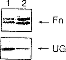
- 108010067306 Fibronectins Proteins 0.000 claims description 40
- 102000016359 Fibronectins Human genes 0.000 claims description 40
- 108090000203 Uteroglobin Proteins 0.000 claims description 36
- 230000004761 fibrosis Effects 0.000 claims description 33
- 206010016654 Fibrosis Diseases 0.000 claims description 32
- 102000003848 Uteroglobin Human genes 0.000 claims description 29
- 230000005764 inhibitory process Effects 0.000 claims description 15
- 238000004220 aggregation Methods 0.000 claims description 14
- 239000003112 inhibitor Substances 0.000 claims description 13
- 230000002776 aggregation Effects 0.000 claims description 9
- 238000002347 injection Methods 0.000 claims description 8
- 239000007924 injection Substances 0.000 claims description 8
- 208000005069 pulmonary fibrosis Diseases 0.000 claims description 7
- 230000015572 biosynthetic process Effects 0.000 claims description 6
- 239000003795 chemical substances by application Substances 0.000 claims description 6
- 238000001802 infusion Methods 0.000 claims description 6
- 239000000443 aerosol Substances 0.000 claims description 4
- 238000009825 accumulation Methods 0.000 claims description 3
- 201000002793 renal fibrosis Diseases 0.000 claims description 2
- 210000003437 trachea Anatomy 0.000 claims description 2
- 238000000034 method Methods 0.000 abstract description 34
- 238000001727 in vivo Methods 0.000 abstract description 16
- 238000001514 detection method Methods 0.000 abstract description 11
- 238000003556 assay Methods 0.000 abstract description 2
- 241000699670 Mus sp. Species 0.000 description 49
- 108090000623 proteins and genes Proteins 0.000 description 44
- 241000699666 Mus <mouse, genus> Species 0.000 description 42
- 102000004169 proteins and genes Human genes 0.000 description 39
- 210000003734 kidney Anatomy 0.000 description 37
- 230000000694 effects Effects 0.000 description 34
- 210000004072 lung Anatomy 0.000 description 33
- 230000004054 inflammatory process Effects 0.000 description 32
- 206010061218 Inflammation Diseases 0.000 description 31
- 210000001519 tissue Anatomy 0.000 description 24
- 206010001052 Acute respiratory distress syndrome Diseases 0.000 description 22
- 208000013616 Respiratory Distress Syndrome Diseases 0.000 description 21
- 238000009739 binding Methods 0.000 description 21
- 210000000056 organ Anatomy 0.000 description 19
- 238000000502 dialysis Methods 0.000 description 18
- 238000002474 experimental method Methods 0.000 description 18
- 238000000338 in vitro Methods 0.000 description 16
- 241000283973 Oryctolagus cuniculus Species 0.000 description 15
- 230000001434 glomerular Effects 0.000 description 15
- 239000004094 surface-active agent Substances 0.000 description 15
- 241001465754 Metazoa Species 0.000 description 14
- 210000004369 blood Anatomy 0.000 description 14
- 239000008280 blood Substances 0.000 description 14
- 239000012634 fragment Substances 0.000 description 14
- 230000002265 prevention Effects 0.000 description 14
- 238000010166 immunofluorescence Methods 0.000 description 13
- 239000013543 active substance Substances 0.000 description 12
- 230000001965 increasing effect Effects 0.000 description 12
- 208000017169 kidney disease Diseases 0.000 description 12
- 208000034486 Multi-organ failure Diseases 0.000 description 11
- 208000010718 Multiple Organ Failure Diseases 0.000 description 11
- 229920001436 collagen Polymers 0.000 description 11
- 208000029744 multiple organ dysfunction syndrome Diseases 0.000 description 11
- 150000001413 amino acids Chemical class 0.000 description 10
- YZXBAPSDXZZRGB-DOFZRALJSA-N arachidonic acid Chemical compound CCCCC\C=C/C\C=C/C\C=C/C\C=C/CCCC(O)=O YZXBAPSDXZZRGB-DOFZRALJSA-N 0.000 description 10
- 206010006475 bronchopulmonary dysplasia Diseases 0.000 description 10
- 238000011813 knockout mouse model Methods 0.000 description 10
- 239000012528 membrane Substances 0.000 description 10
- 210000002381 plasma Anatomy 0.000 description 10
- 229940063649 survanta Drugs 0.000 description 10
- 102000004190 Enzymes Human genes 0.000 description 9
- 108090000790 Enzymes Proteins 0.000 description 9
- 210000004027 cell Anatomy 0.000 description 9
- 239000003085 diluting agent Substances 0.000 description 9
- 210000004185 liver Anatomy 0.000 description 9
- 206010015943 Eye inflammation Diseases 0.000 description 8
- 206010033645 Pancreatitis Diseases 0.000 description 8
- 230000001154 acute effect Effects 0.000 description 8
- 230000006378 damage Effects 0.000 description 8
- 201000010099 disease Diseases 0.000 description 8
- 208000037265 diseases, disorders, signs and symptoms Diseases 0.000 description 8
- 239000012530 fluid Substances 0.000 description 8
- 230000002401 inhibitory effect Effects 0.000 description 8
- 208000014674 injury Diseases 0.000 description 8
- 210000002966 serum Anatomy 0.000 description 8
- 230000009885 systemic effect Effects 0.000 description 8
- 238000001262 western blot Methods 0.000 description 8
- 208000009304 Acute Kidney Injury Diseases 0.000 description 7
- 108010035532 Collagen Proteins 0.000 description 7
- 102000008186 Collagen Human genes 0.000 description 7
- 208000022559 Inflammatory bowel disease Diseases 0.000 description 7
- 241001529936 Murinae Species 0.000 description 7
- 208000033626 Renal failure acute Diseases 0.000 description 7
- 201000011040 acute kidney failure Diseases 0.000 description 7
- 208000012998 acute renal failure Diseases 0.000 description 7
- 208000011341 adult acute respiratory distress syndrome Diseases 0.000 description 7
- 201000000028 adult respiratory distress syndrome Diseases 0.000 description 7
- 230000002950 deficient Effects 0.000 description 7
- 239000003814 drug Substances 0.000 description 7
- 238000005259 measurement Methods 0.000 description 7
- 210000000496 pancreas Anatomy 0.000 description 7
- 239000000523 sample Substances 0.000 description 7
- 239000000758 substrate Substances 0.000 description 7
- 102000009091 Amyloidogenic Proteins Human genes 0.000 description 6
- 108010048112 Amyloidogenic Proteins Proteins 0.000 description 6
- 102000012422 Collagen Type I Human genes 0.000 description 6
- 108010022452 Collagen Type I Proteins 0.000 description 6
- IAZDPXIOMUYVGZ-UHFFFAOYSA-N Dimethylsulphoxide Chemical compound CS(C)=O IAZDPXIOMUYVGZ-UHFFFAOYSA-N 0.000 description 6
- WSFSSNUMVMOOMR-UHFFFAOYSA-N Formaldehyde Chemical compound O=C WSFSSNUMVMOOMR-UHFFFAOYSA-N 0.000 description 6
- FAPWRFPIFSIZLT-UHFFFAOYSA-M Sodium chloride Chemical compound [Na+].[Cl-] FAPWRFPIFSIZLT-UHFFFAOYSA-M 0.000 description 6
- 239000003937 drug carrier Substances 0.000 description 6
- 239000006166 lysate Substances 0.000 description 6
- 239000008194 pharmaceutical composition Substances 0.000 description 6
- 239000000047 product Substances 0.000 description 6
- 238000003757 reverse transcription PCR Methods 0.000 description 6
- 238000002415 sodium dodecyl sulfate polyacrylamide gel electrophoresis Methods 0.000 description 6
- 239000000126 substance Substances 0.000 description 6
- 230000008733 trauma Effects 0.000 description 6
- 230000002792 vascular Effects 0.000 description 6
- 206010003210 Arteriosclerosis Diseases 0.000 description 5
- 201000001320 Atherosclerosis Diseases 0.000 description 5
- 108020004414 DNA Proteins 0.000 description 5
- 206010035664 Pneumonia Diseases 0.000 description 5
- 238000002105 Southern blotting Methods 0.000 description 5
- 230000003110 anti-inflammatory effect Effects 0.000 description 5
- 229940114079 arachidonic acid Drugs 0.000 description 5
- 235000021342 arachidonic acid Nutrition 0.000 description 5
- 208000011775 arteriosclerosis disease Diseases 0.000 description 5
- 210000004204 blood vessel Anatomy 0.000 description 5
- 230000001684 chronic effect Effects 0.000 description 5
- 238000010790 dilution Methods 0.000 description 5
- 239000012895 dilution Substances 0.000 description 5
- 229940079593 drug Drugs 0.000 description 5
- 238000006460 hydrolysis reaction Methods 0.000 description 5
- 238000001114 immunoprecipitation Methods 0.000 description 5
- 239000011159 matrix material Substances 0.000 description 5
- 210000004379 membrane Anatomy 0.000 description 5
- 150000003904 phospholipids Chemical class 0.000 description 5
- 230000002685 pulmonary effect Effects 0.000 description 5
- 230000002829 reductive effect Effects 0.000 description 5
- 102000004506 Blood Proteins Human genes 0.000 description 4
- 108010017384 Blood Proteins Proteins 0.000 description 4
- LFQSCWFLJHTTHZ-UHFFFAOYSA-N Ethanol Chemical compound CCO LFQSCWFLJHTTHZ-UHFFFAOYSA-N 0.000 description 4
- 206010051920 Glomerulonephropathy Diseases 0.000 description 4
- 206010053159 Organ failure Diseases 0.000 description 4
- 206010067584 Type 1 diabetes mellitus Diseases 0.000 description 4
- 102100031083 Uteroglobin Human genes 0.000 description 4
- 230000004913 activation Effects 0.000 description 4
- 239000000872 buffer Substances 0.000 description 4
- HVYWMOMLDIMFJA-DPAQBDIFSA-N cholesterol Chemical compound C1C=C2C[C@@H](O)CC[C@]2(C)[C@@H]2[C@@H]1[C@@H]1CC[C@H]([C@H](C)CCCC(C)C)[C@@]1(C)CC2 HVYWMOMLDIMFJA-DPAQBDIFSA-N 0.000 description 4
- 238000011161 development Methods 0.000 description 4
- 230000018109 developmental process Effects 0.000 description 4
- 239000000539 dimer Substances 0.000 description 4
- 238000001962 electrophoresis Methods 0.000 description 4
- 238000010363 gene targeting Methods 0.000 description 4
- 230000007062 hydrolysis Effects 0.000 description 4
- 208000015181 infectious disease Diseases 0.000 description 4
- 230000004968 inflammatory condition Effects 0.000 description 4
- 238000001990 intravenous administration Methods 0.000 description 4
- 230000000670 limiting effect Effects 0.000 description 4
- 230000009788 parenchymal fibrosis Effects 0.000 description 4
- YBYRMVIVWMBXKQ-UHFFFAOYSA-N phenylmethanesulfonyl fluoride Chemical compound FS(=O)(=O)CC1=CC=CC=C1 YBYRMVIVWMBXKQ-UHFFFAOYSA-N 0.000 description 4
- 229920002401 polyacrylamide Polymers 0.000 description 4
- 210000002307 prostate Anatomy 0.000 description 4
- 210000003491 skin Anatomy 0.000 description 4
- 239000011780 sodium chloride Substances 0.000 description 4
- 239000007787 solid Substances 0.000 description 4
- 238000010186 staining Methods 0.000 description 4
- 230000009261 transgenic effect Effects 0.000 description 4
- ZIIUUSVHCHPIQD-UHFFFAOYSA-N 2,4,6-trimethyl-N-[3-(trifluoromethyl)phenyl]benzenesulfonamide Chemical compound CC1=CC(C)=CC(C)=C1S(=O)(=O)NC1=CC=CC(C(F)(F)F)=C1 ZIIUUSVHCHPIQD-UHFFFAOYSA-N 0.000 description 3
- 108010088751 Albumins Proteins 0.000 description 3
- 102000009027 Albumins Human genes 0.000 description 3
- 208000035143 Bacterial infection Diseases 0.000 description 3
- 241000283690 Bos taurus Species 0.000 description 3
- -1 CC17 Proteins 0.000 description 3
- 208000011231 Crohn disease Diseases 0.000 description 3
- PEDCQBHIVMGVHV-UHFFFAOYSA-N Glycerine Chemical compound OCC(O)CO PEDCQBHIVMGVHV-UHFFFAOYSA-N 0.000 description 3
- 108010001336 Horseradish Peroxidase Proteins 0.000 description 3
- 108090000144 Human Proteins Proteins 0.000 description 3
- 102000003839 Human Proteins Human genes 0.000 description 3
- 102000015439 Phospholipases Human genes 0.000 description 3
- 108010064785 Phospholipases Proteins 0.000 description 3
- 108060008539 Transglutaminase Proteins 0.000 description 3
- 206010046851 Uveitis Diseases 0.000 description 3
- 239000000654 additive Substances 0.000 description 3
- 230000000996 additive effect Effects 0.000 description 3
- 238000004458 analytical method Methods 0.000 description 3
- 208000022362 bacterial infectious disease Diseases 0.000 description 3
- 239000000306 component Substances 0.000 description 3
- 238000012790 confirmation Methods 0.000 description 3
- 239000002537 cosmetic Substances 0.000 description 3
- 238000004132 cross linking Methods 0.000 description 3
- 239000000835 fiber Substances 0.000 description 3
- 210000002950 fibroblast Anatomy 0.000 description 3
- 238000009472 formulation Methods 0.000 description 3
- 210000002216 heart Anatomy 0.000 description 3
- 210000004969 inflammatory cell Anatomy 0.000 description 3
- 238000003780 insertion Methods 0.000 description 3
- 230000037431 insertion Effects 0.000 description 3
- 208000028867 ischemia Diseases 0.000 description 3
- 230000003907 kidney function Effects 0.000 description 3
- 150000003071 polychlorinated biphenyls Chemical class 0.000 description 3
- 230000035935 pregnancy Effects 0.000 description 3
- 230000002028 premature Effects 0.000 description 3
- 108090000765 processed proteins & peptides Proteins 0.000 description 3
- 210000001147 pulmonary artery Anatomy 0.000 description 3
- 230000001850 reproductive effect Effects 0.000 description 3
- 230000035939 shock Effects 0.000 description 3
- 241000894007 species Species 0.000 description 3
- 238000001356 surgical procedure Methods 0.000 description 3
- 238000012360 testing method Methods 0.000 description 3
- 230000008719 thickening Effects 0.000 description 3
- 230000000451 tissue damage Effects 0.000 description 3
- 231100000827 tissue damage Toxicity 0.000 description 3
- 102000003601 transglutaminase Human genes 0.000 description 3
- 238000002054 transplantation Methods 0.000 description 3
- 210000004291 uterus Anatomy 0.000 description 3
- 210000003462 vein Anatomy 0.000 description 3
- XLYOFNOQVPJJNP-UHFFFAOYSA-N water Chemical compound O XLYOFNOQVPJJNP-UHFFFAOYSA-N 0.000 description 3
- 108091032973 (ribonucleotides)n+m Proteins 0.000 description 2
- WRGQSWVCFNIUNZ-GDCKJWNLSA-N 1-oleoyl-sn-glycerol 3-phosphate Chemical compound CCCCCCCC\C=C/CCCCCCCC(=O)OC[C@@H](O)COP(O)(O)=O WRGQSWVCFNIUNZ-GDCKJWNLSA-N 0.000 description 2
- QFVHZQCOUORWEI-UHFFFAOYSA-N 4-[(4-anilino-5-sulfonaphthalen-1-yl)diazenyl]-5-hydroxynaphthalene-2,7-disulfonic acid Chemical compound C=12C(O)=CC(S(O)(=O)=O)=CC2=CC(S(O)(=O)=O)=CC=1N=NC(C1=CC=CC(=C11)S(O)(=O)=O)=CC=C1NC1=CC=CC=C1 QFVHZQCOUORWEI-UHFFFAOYSA-N 0.000 description 2
- 208000023769 AA amyloidosis Diseases 0.000 description 2
- QGZKDVFQNNGYKY-UHFFFAOYSA-N Ammonia Chemical compound N QGZKDVFQNNGYKY-UHFFFAOYSA-N 0.000 description 2
- 208000037260 Atherosclerotic Plaque Diseases 0.000 description 2
- 201000001178 Bacterial Pneumonia Diseases 0.000 description 2
- 206010006895 Cachexia Diseases 0.000 description 2
- 241000282693 Cercopithecidae Species 0.000 description 2
- 201000004624 Dermatitis Diseases 0.000 description 2
- 208000022461 Glomerular disease Diseases 0.000 description 2
- 239000012981 Hank's balanced salt solution Substances 0.000 description 2
- 241000282412 Homo Species 0.000 description 2
- 208000032571 Infant acute respiratory distress syndrome Diseases 0.000 description 2
- 108010085895 Laminin Proteins 0.000 description 2
- 102000007547 Laminin Human genes 0.000 description 2
- 208000031942 Late Onset disease Diseases 0.000 description 2
- GDBQQVLCIARPGH-UHFFFAOYSA-N Leupeptin Natural products CC(C)CC(NC(C)=O)C(=O)NC(CC(C)C)C(=O)NC(C=O)CCCN=C(N)N GDBQQVLCIARPGH-UHFFFAOYSA-N 0.000 description 2
- 206010028974 Neonatal respiratory distress syndrome Diseases 0.000 description 2
- 206010028980 Neoplasm Diseases 0.000 description 2
- 102000004264 Osteopontin Human genes 0.000 description 2
- 108010081689 Osteopontin Proteins 0.000 description 2
- 238000010222 PCR analysis Methods 0.000 description 2
- 206010036790 Productive cough Diseases 0.000 description 2
- RJKFOVLPORLFTN-LEKSSAKUSA-N Progesterone Chemical compound C1CC2=CC(=O)CC[C@]2(C)[C@@H]2[C@@H]1[C@@H]1CC[C@H](C(=O)C)[C@@]1(C)CC2 RJKFOVLPORLFTN-LEKSSAKUSA-N 0.000 description 2
- 201000004681 Psoriasis Diseases 0.000 description 2
- 206010038910 Retinitis Diseases 0.000 description 2
- 206010052779 Transplant rejections Diseases 0.000 description 2
- XSQUKJJJFZCRTK-UHFFFAOYSA-N Urea Chemical compound NC(N)=O XSQUKJJJFZCRTK-UHFFFAOYSA-N 0.000 description 2
- 108010031318 Vitronectin Proteins 0.000 description 2
- 102100035140 Vitronectin Human genes 0.000 description 2
- 208000027418 Wounds and injury Diseases 0.000 description 2
- 239000004480 active ingredient Substances 0.000 description 2
- AWUCVROLDVIAJX-UHFFFAOYSA-N alpha-glycerophosphate Natural products OCC(O)COP(O)(O)=O AWUCVROLDVIAJX-UHFFFAOYSA-N 0.000 description 2
- 239000000427 antigen Substances 0.000 description 2
- 108091007433 antigens Proteins 0.000 description 2
- 102000036639 antigens Human genes 0.000 description 2
- 238000000211 autoradiogram Methods 0.000 description 2
- 239000011324 bead Substances 0.000 description 2
- 210000003123 bronchiole Anatomy 0.000 description 2
- 201000011510 cancer Diseases 0.000 description 2
- 230000001413 cellular effect Effects 0.000 description 2
- 238000006243 chemical reaction Methods 0.000 description 2
- 239000003153 chemical reaction reagent Substances 0.000 description 2
- 238000002512 chemotherapy Methods 0.000 description 2
- 235000012000 cholesterol Nutrition 0.000 description 2
- 230000035602 clotting Effects 0.000 description 2
- IQFVPQOLBLOTPF-HKXUKFGYSA-L congo red Chemical compound [Na+].[Na+].C1=CC=CC2=C(N)C(/N=N/C3=CC=C(C=C3)C3=CC=C(C=C3)/N=N/C3=C(C4=CC=CC=C4C(=C3)S([O-])(=O)=O)N)=CC(S([O-])(=O)=O)=C21 IQFVPQOLBLOTPF-HKXUKFGYSA-L 0.000 description 2
- 210000004748 cultured cell Anatomy 0.000 description 2
- 230000034994 death Effects 0.000 description 2
- 230000003247 decreasing effect Effects 0.000 description 2
- 239000003792 electrolyte Substances 0.000 description 2
- 210000002257 embryonic structure Anatomy 0.000 description 2
- 238000006911 enzymatic reaction Methods 0.000 description 2
- 210000002919 epithelial cell Anatomy 0.000 description 2
- 210000003722 extracellular fluid Anatomy 0.000 description 2
- 239000007850 fluorescent dye Substances 0.000 description 2
- 230000006870 function Effects 0.000 description 2
- 238000001631 haemodialysis Methods 0.000 description 2
- 230000000322 hemodialysis Effects 0.000 description 2
- 230000028993 immune response Effects 0.000 description 2
- 238000002991 immunohistochemical analysis Methods 0.000 description 2
- 238000013115 immunohistochemical detection Methods 0.000 description 2
- 238000003364 immunohistochemistry Methods 0.000 description 2
- 239000012678 infectious agent Substances 0.000 description 2
- 230000028709 inflammatory response Effects 0.000 description 2
- 102000006495 integrins Human genes 0.000 description 2
- 108010044426 integrins Proteins 0.000 description 2
- 230000003993 interaction Effects 0.000 description 2
- 230000003834 intracellular effect Effects 0.000 description 2
- 238000007918 intramuscular administration Methods 0.000 description 2
- 238000010253 intravenous injection Methods 0.000 description 2
- GDBQQVLCIARPGH-ULQDDVLXSA-N leupeptin Chemical compound CC(C)C[C@H](NC(C)=O)C(=O)N[C@@H](CC(C)C)C(=O)N[C@H](C=O)CCCN=C(N)N GDBQQVLCIARPGH-ULQDDVLXSA-N 0.000 description 2
- 108010052968 leupeptin Proteins 0.000 description 2
- 239000007788 liquid Substances 0.000 description 2
- 238000004519 manufacturing process Methods 0.000 description 2
- 230000001404 mediated effect Effects 0.000 description 2
- 201000002652 newborn respiratory distress syndrome Diseases 0.000 description 2
- 229940021182 non-steroidal anti-inflammatory drug Drugs 0.000 description 2
- 238000012261 overproduction Methods 0.000 description 2
- 210000004923 pancreatic tissue Anatomy 0.000 description 2
- 230000001575 pathological effect Effects 0.000 description 2
- 108010091212 pepstatin Proteins 0.000 description 2
- FAXGPCHRFPCXOO-LXTPJMTPSA-N pepstatin A Chemical compound OC(=O)C[C@H](O)[C@H](CC(C)C)NC(=O)[C@H](C)NC(=O)C[C@H](O)[C@H](CC(C)C)NC(=O)[C@H](C(C)C)NC(=O)[C@H](C(C)C)NC(=O)CC(C)C FAXGPCHRFPCXOO-LXTPJMTPSA-N 0.000 description 2
- 206010034674 peritonitis Diseases 0.000 description 2
- DHRLEVQXOMLTIM-UHFFFAOYSA-N phosphoric acid;trioxomolybdenum Chemical compound O=[Mo](=O)=O.O=[Mo](=O)=O.O=[Mo](=O)=O.O=[Mo](=O)=O.O=[Mo](=O)=O.O=[Mo](=O)=O.O=[Mo](=O)=O.O=[Mo](=O)=O.O=[Mo](=O)=O.O=[Mo](=O)=O.O=[Mo](=O)=O.O=[Mo](=O)=O.OP(O)(O)=O DHRLEVQXOMLTIM-UHFFFAOYSA-N 0.000 description 2
- 238000001556 precipitation Methods 0.000 description 2
- 230000008569 process Effects 0.000 description 2
- 201000001474 proteinuria Diseases 0.000 description 2
- 230000002285 radioactive effect Effects 0.000 description 2
- 238000011160 research Methods 0.000 description 2
- 230000004044 response Effects 0.000 description 2
- 206010039073 rheumatoid arthritis Diseases 0.000 description 2
- HEMHJVSKTPXQMS-UHFFFAOYSA-M sodium hydroxide Inorganic materials [OH-].[Na+] HEMHJVSKTPXQMS-UHFFFAOYSA-M 0.000 description 2
- 210000003802 sputum Anatomy 0.000 description 2
- 208000024794 sputum Diseases 0.000 description 2
- 239000013589 supplement Substances 0.000 description 2
- 239000000725 suspension Substances 0.000 description 2
- 208000024891 symptom Diseases 0.000 description 2
- 238000007910 systemic administration Methods 0.000 description 2
- 201000000596 systemic lupus erythematosus Diseases 0.000 description 2
- 229940124597 therapeutic agent Drugs 0.000 description 2
- 230000001225 therapeutic effect Effects 0.000 description 2
- 238000011144 upstream manufacturing Methods 0.000 description 2
- 210000002700 urine Anatomy 0.000 description 2
- 210000001177 vas deferen Anatomy 0.000 description 2
- 210000005166 vasculature Anatomy 0.000 description 2
- PGOHTUIFYSHAQG-LJSDBVFPSA-N (2S)-6-amino-2-[[(2S)-5-amino-2-[[(2S)-2-[[(2S)-2-[[(2S)-2-[[(2S)-4-amino-2-[[(2S)-2-[[(2S)-2-[[(2S)-2-[[(2S)-2-[[(2S)-5-amino-2-[[(2S)-5-amino-2-[[(2S)-2-[[(2S)-2-[[(2S)-2-[[(2S,3R)-2-[[(2S)-5-amino-2-[[(2S)-2-[[(2S)-2-[[(2S,3R)-2-[[(2S)-2-[[(2S)-2-[[(2S)-2-[[(2S)-2-[[(2S)-5-amino-2-[[(2S)-1-[(2S,3R)-2-[[(2S)-2-[[(2S)-2-[[(2R)-2-[[(2S)-2-[[(2S)-2-[[2-[[(2S)-2-[[(2S)-2-[[(2S)-2-[[(2S)-1-[(2S)-2-[[(2S)-2-[[(2S)-2-[[(2S)-2-amino-4-methylsulfanylbutanoyl]amino]-3-(1H-indol-3-yl)propanoyl]amino]-5-carbamimidamidopentanoyl]amino]propanoyl]pyrrolidine-2-carbonyl]amino]-3-methylbutanoyl]amino]-4-methylpentanoyl]amino]-4-methylpentanoyl]amino]acetyl]amino]-3-hydroxypropanoyl]amino]-4-methylpentanoyl]amino]-3-sulfanylpropanoyl]amino]-4-methylsulfanylbutanoyl]amino]-5-carbamimidamidopentanoyl]amino]-3-hydroxybutanoyl]pyrrolidine-2-carbonyl]amino]-5-oxopentanoyl]amino]-3-hydroxypropanoyl]amino]-3-hydroxypropanoyl]amino]-3-(1H-imidazol-5-yl)propanoyl]amino]-4-methylpentanoyl]amino]-3-hydroxybutanoyl]amino]-3-(1H-indol-3-yl)propanoyl]amino]-5-carbamimidamidopentanoyl]amino]-5-oxopentanoyl]amino]-3-hydroxybutanoyl]amino]-3-hydroxypropanoyl]amino]-3-carboxypropanoyl]amino]-3-hydroxypropanoyl]amino]-5-oxopentanoyl]amino]-5-oxopentanoyl]amino]-3-phenylpropanoyl]amino]-5-carbamimidamidopentanoyl]amino]-3-methylbutanoyl]amino]-4-methylpentanoyl]amino]-4-oxobutanoyl]amino]-5-carbamimidamidopentanoyl]amino]-3-(1H-indol-3-yl)propanoyl]amino]-4-carboxybutanoyl]amino]-5-oxopentanoyl]amino]hexanoic acid Chemical compound CSCC[C@H](N)C(=O)N[C@@H](Cc1c[nH]c2ccccc12)C(=O)N[C@@H](CCCNC(N)=N)C(=O)N[C@@H](C)C(=O)N1CCC[C@H]1C(=O)N[C@@H](C(C)C)C(=O)N[C@@H](CC(C)C)C(=O)N[C@@H](CC(C)C)C(=O)NCC(=O)N[C@@H](CO)C(=O)N[C@@H](CC(C)C)C(=O)N[C@@H](CS)C(=O)N[C@@H](CCSC)C(=O)N[C@@H](CCCNC(N)=N)C(=O)N[C@@H]([C@@H](C)O)C(=O)N1CCC[C@H]1C(=O)N[C@@H](CCC(N)=O)C(=O)N[C@@H](CO)C(=O)N[C@@H](CO)C(=O)N[C@@H](Cc1cnc[nH]1)C(=O)N[C@@H](CC(C)C)C(=O)N[C@@H]([C@@H](C)O)C(=O)N[C@@H](Cc1c[nH]c2ccccc12)C(=O)N[C@@H](CCCNC(N)=N)C(=O)N[C@@H](CCC(N)=O)C(=O)N[C@@H]([C@@H](C)O)C(=O)N[C@@H](CO)C(=O)N[C@@H](CC(O)=O)C(=O)N[C@@H](CO)C(=O)N[C@@H](CCC(N)=O)C(=O)N[C@@H](CCC(N)=O)C(=O)N[C@@H](Cc1ccccc1)C(=O)N[C@@H](CCCNC(N)=N)C(=O)N[C@@H](C(C)C)C(=O)N[C@@H](CC(C)C)C(=O)N[C@@H](CC(N)=O)C(=O)N[C@@H](CCCNC(N)=N)C(=O)N[C@@H](Cc1c[nH]c2ccccc12)C(=O)N[C@@H](CCC(O)=O)C(=O)N[C@@H](CCC(N)=O)C(=O)N[C@@H](CCCCN)C(O)=O PGOHTUIFYSHAQG-LJSDBVFPSA-N 0.000 description 1
- 102000040650 (ribonucleotides)n+m Human genes 0.000 description 1
- QKNYBSVHEMOAJP-UHFFFAOYSA-N 2-amino-2-(hydroxymethyl)propane-1,3-diol;hydron;chloride Chemical compound Cl.OCC(N)(CO)CO QKNYBSVHEMOAJP-UHFFFAOYSA-N 0.000 description 1
- TVZRAEYQIKYCPH-UHFFFAOYSA-N 3-(trimethylsilyl)propane-1-sulfonic acid Chemical compound C[Si](C)(C)CCCS(O)(=O)=O TVZRAEYQIKYCPH-UHFFFAOYSA-N 0.000 description 1
- ZCYVEMRRCGMTRW-UHFFFAOYSA-N 7553-56-2 Chemical compound [I] ZCYVEMRRCGMTRW-UHFFFAOYSA-N 0.000 description 1
- 208000009663 Acute Necrotizing Pancreatitis Diseases 0.000 description 1
- 108700028369 Alleles Proteins 0.000 description 1
- USFZMSVCRYTOJT-UHFFFAOYSA-N Ammonium acetate Chemical compound N.CC(O)=O USFZMSVCRYTOJT-UHFFFAOYSA-N 0.000 description 1
- 239000005695 Ammonium acetate Substances 0.000 description 1
- 206010002199 Anaphylactic shock Diseases 0.000 description 1
- 102000018616 Apolipoproteins B Human genes 0.000 description 1
- 108010027006 Apolipoproteins B Proteins 0.000 description 1
- 102000018655 Apolipoproteins C Human genes 0.000 description 1
- 108010027070 Apolipoproteins C Proteins 0.000 description 1
- 206010003011 Appendicitis Diseases 0.000 description 1
- 108010039627 Aprotinin Proteins 0.000 description 1
- 108010001478 Bacitracin Proteins 0.000 description 1
- 108010006654 Bleomycin Proteins 0.000 description 1
- 238000009010 Bradford assay Methods 0.000 description 1
- 241000283707 Capra Species 0.000 description 1
- 208000002177 Cataract Diseases 0.000 description 1
- 241000251204 Chimaeridae Species 0.000 description 1
- ZAMOUSCENKQFHK-UHFFFAOYSA-N Chlorine atom Chemical compound [Cl] ZAMOUSCENKQFHK-UHFFFAOYSA-N 0.000 description 1
- 206010053567 Coagulopathies Diseases 0.000 description 1
- 108091026890 Coding region Proteins 0.000 description 1
- 206010009900 Colitis ulcerative Diseases 0.000 description 1
- 208000029147 Collagen-vascular disease Diseases 0.000 description 1
- 206010010741 Conjunctivitis Diseases 0.000 description 1
- 241000655605 Cyanocephalus Species 0.000 description 1
- 102000004127 Cytokines Human genes 0.000 description 1
- 108090000695 Cytokines Proteins 0.000 description 1
- 206010048843 Cytomegalovirus chorioretinitis Diseases 0.000 description 1
- 206010012442 Dermatitis contact Diseases 0.000 description 1
- 208000007342 Diabetic Nephropathies Diseases 0.000 description 1
- 206010013647 Drowning Diseases 0.000 description 1
- KCXVZYZYPLLWCC-UHFFFAOYSA-N EDTA Chemical compound OC(=O)CN(CC(O)=O)CCN(CC(O)=O)CC(O)=O KCXVZYZYPLLWCC-UHFFFAOYSA-N 0.000 description 1
- 208000004145 Endometritis Diseases 0.000 description 1
- 241000991587 Enterovirus C Species 0.000 description 1
- 208000010201 Exanthema Diseases 0.000 description 1
- 108010073385 Fibrin Proteins 0.000 description 1
- 102000009123 Fibrin Human genes 0.000 description 1
- BWGVNKXGVNDBDI-UHFFFAOYSA-N Fibrin monomer Chemical compound CNC(=O)CNC(=O)CN BWGVNKXGVNDBDI-UHFFFAOYSA-N 0.000 description 1
- 208000007882 Gastritis Diseases 0.000 description 1
- CEAZRRDELHUEMR-URQXQFDESA-N Gentamicin Chemical compound O1[C@H](C(C)NC)CC[C@@H](N)[C@H]1O[C@H]1[C@H](O)[C@@H](O[C@@H]2[C@@H]([C@@H](NC)[C@@](C)(O)CO2)O)[C@H](N)C[C@@H]1N CEAZRRDELHUEMR-URQXQFDESA-N 0.000 description 1
- 229930182566 Gentamicin Natural products 0.000 description 1
- 206010018364 Glomerulonephritis Diseases 0.000 description 1
- WQZGKKKJIJFFOK-GASJEMHNSA-N Glucose Natural products OC[C@H]1OC(O)[C@H](O)[C@@H](O)[C@@H]1O WQZGKKKJIJFFOK-GASJEMHNSA-N 0.000 description 1
- 102000003886 Glycoproteins Human genes 0.000 description 1
- 108090000288 Glycoproteins Proteins 0.000 description 1
- 206010019799 Hepatitis viral Diseases 0.000 description 1
- 206010020751 Hypersensitivity Diseases 0.000 description 1
- 208000013038 Hypocalcemia Diseases 0.000 description 1
- 201000009794 Idiopathic Pulmonary Fibrosis Diseases 0.000 description 1
- 206010062016 Immunosuppression Diseases 0.000 description 1
- 208000005615 Interstitial Cystitis Diseases 0.000 description 1
- 241000581650 Ivesia Species 0.000 description 1
- YQEZLKZALYSWHR-UHFFFAOYSA-N Ketamine Chemical compound C=1C=CC=C(Cl)C=1C1(NC)CCCCC1=O YQEZLKZALYSWHR-UHFFFAOYSA-N 0.000 description 1
- 208000008839 Kidney Neoplasms Diseases 0.000 description 1
- FBOZXECLQNJBKD-ZDUSSCGKSA-N L-methotrexate Chemical compound C=1N=C2N=C(N)N=C(N)C2=NC=1CN(C)C1=CC=C(C(=O)N[C@@H](CCC(O)=O)C(O)=O)C=C1 FBOZXECLQNJBKD-ZDUSSCGKSA-N 0.000 description 1
- 241000124008 Mammalia Species 0.000 description 1
- 201000005085 Meconium Aspiration Syndrome Diseases 0.000 description 1
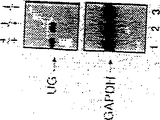
- 101000819572 Mus musculus Glyceraldehyde-3-phosphate dehydrogenase Proteins 0.000 description 1
- 101000777235 Mus musculus Uteroglobin Proteins 0.000 description 1
- 206010051606 Necrotising colitis Diseases 0.000 description 1
- 229930193140 Neomycin Natural products 0.000 description 1
- 108020005187 Oligonucleotide Probes Proteins 0.000 description 1
- 206010058461 Orchitis noninfective Diseases 0.000 description 1
- 206010033078 Otitis media Diseases 0.000 description 1
- 241000609499 Palicourea Species 0.000 description 1
- 206010058096 Pancreatic necrosis Diseases 0.000 description 1
- 206010033647 Pancreatitis acute Diseases 0.000 description 1
- 241000282520 Papio Species 0.000 description 1
- 108010030304 Progesterone-Binding Globulin Proteins 0.000 description 1
- 102100027378 Prothrombin Human genes 0.000 description 1
- 108010094028 Prothrombin Proteins 0.000 description 1
- 238000010240 RT-PCR analysis Methods 0.000 description 1
- 101000777244 Rattus norvegicus Uteroglobin Proteins 0.000 description 1
- 208000001647 Renal Insufficiency Diseases 0.000 description 1
- 206010038389 Renal cancer Diseases 0.000 description 1
- 208000021063 Respiratory fume inhalation disease Diseases 0.000 description 1
- 201000000582 Retinoblastoma Diseases 0.000 description 1
- 241000702670 Rotavirus Species 0.000 description 1
- 239000012722 SDS sample buffer Substances 0.000 description 1
- 206010039710 Scleroderma Diseases 0.000 description 1
- 206010040070 Septic Shock Diseases 0.000 description 1
- 241000580858 Simian-Human immunodeficiency virus Species 0.000 description 1
- CDBYLPFSWZWCQE-UHFFFAOYSA-L Sodium Carbonate Chemical compound [Na+].[Na+].[O-]C([O-])=O CDBYLPFSWZWCQE-UHFFFAOYSA-L 0.000 description 1
- 208000007107 Stomach Ulcer Diseases 0.000 description 1
- 108010000499 Thromboplastin Proteins 0.000 description 1
- 102000002262 Thromboplastin Human genes 0.000 description 1
- 208000007536 Thrombosis Diseases 0.000 description 1
- 241000159243 Toxicodendron radicans Species 0.000 description 1
- 239000013504 Triton X-100 Substances 0.000 description 1
- 229920004890 Triton X-100 Polymers 0.000 description 1
- 201000006704 Ulcerative Colitis Diseases 0.000 description 1
- 206010062903 Urethritis noninfective Diseases 0.000 description 1
- 101710195585 Urinary protein 1 Proteins 0.000 description 1
- 206010046793 Uterine inflammation Diseases 0.000 description 1
- 206010046914 Vaginal infection Diseases 0.000 description 1
- 201000008100 Vaginitis Diseases 0.000 description 1
- 208000036142 Viral infection Diseases 0.000 description 1
- 201000007096 Vulvovaginal Candidiasis Diseases 0.000 description 1
- ABUBSBSOTTXVPV-UHFFFAOYSA-H [U+6].CC([O-])=O.CC([O-])=O.CC([O-])=O.CC([O-])=O.CC([O-])=O.CC([O-])=O Chemical compound [U+6].CC([O-])=O.CC([O-])=O.CC([O-])=O.CC([O-])=O.CC([O-])=O.CC([O-])=O ABUBSBSOTTXVPV-UHFFFAOYSA-H 0.000 description 1
- 230000002159 abnormal effect Effects 0.000 description 1
- 230000005856 abnormality Effects 0.000 description 1
- 201000003229 acute pancreatitis Diseases 0.000 description 1
- 230000002411 adverse Effects 0.000 description 1
- 230000000172 allergic effect Effects 0.000 description 1
- 208000030961 allergic reaction Diseases 0.000 description 1
- 108010001122 alpha(2)-microglobulin Proteins 0.000 description 1
- 229910021529 ammonia Inorganic materials 0.000 description 1
- 229940043376 ammonium acetate Drugs 0.000 description 1
- 235000019257 ammonium acetate Nutrition 0.000 description 1
- 229960000723 ampicillin Drugs 0.000 description 1
- AVKUERGKIZMTKX-NJBDSQKTSA-N ampicillin Chemical compound C1([C@@H](N)C(=O)N[C@H]2[C@H]3SC([C@@H](N3C2=O)C(O)=O)(C)C)=CC=CC=C1 AVKUERGKIZMTKX-NJBDSQKTSA-N 0.000 description 1
- 230000003321 amplification Effects 0.000 description 1
- 229940035676 analgesics Drugs 0.000 description 1
- 208000003455 anaphylaxis Diseases 0.000 description 1
- 239000000730 antalgic agent Substances 0.000 description 1
- 239000003242 anti bacterial agent Substances 0.000 description 1
- 230000003510 anti-fibrotic effect Effects 0.000 description 1
- 229940088710 antibiotic agent Drugs 0.000 description 1
- 238000009175 antibody therapy Methods 0.000 description 1
- 239000003146 anticoagulant agent Substances 0.000 description 1
- 229940121375 antifungal agent Drugs 0.000 description 1
- 239000003429 antifungal agent Substances 0.000 description 1
- 230000000890 antigenic effect Effects 0.000 description 1
- 239000002246 antineoplastic agent Substances 0.000 description 1
- 229960004676 antithrombotic agent Drugs 0.000 description 1
- 239000003443 antiviral agent Substances 0.000 description 1
- 210000000436 anus Anatomy 0.000 description 1
- 230000001640 apoptogenic effect Effects 0.000 description 1
- 229960004405 aprotinin Drugs 0.000 description 1
- 239000007864 aqueous solution Substances 0.000 description 1
- 239000000596 artificial lung surfactant Substances 0.000 description 1
- 239000010425 asbestos Substances 0.000 description 1
- 208000006673 asthma Diseases 0.000 description 1
- 208000010668 atopic eczema Diseases 0.000 description 1
- 230000001363 autoimmune Effects 0.000 description 1
- 230000005784 autoimmunity Effects 0.000 description 1
- 229960003071 bacitracin Drugs 0.000 description 1
- 229930184125 bacitracin Natural products 0.000 description 1
- CLKOFPXJLQSYAH-ABRJDSQDSA-N bacitracin A Chemical compound C1SC([C@@H](N)[C@@H](C)CC)=N[C@@H]1C(=O)N[C@@H](CC(C)C)C(=O)N[C@H](CCC(O)=O)C(=O)N[C@@H]([C@@H](C)CC)C(=O)N[C@@H]1C(=O)N[C@H](CCCN)C(=O)N[C@@H]([C@@H](C)CC)C(=O)N[C@H](CC=2C=CC=CC=2)C(=O)N[C@@H](CC=2N=CNC=2)C(=O)N[C@H](CC(O)=O)C(=O)N[C@@H](CC(N)=O)C(=O)NCCCC1 CLKOFPXJLQSYAH-ABRJDSQDSA-N 0.000 description 1
- 201000005008 bacterial sepsis Diseases 0.000 description 1
- 210000002469 basement membrane Anatomy 0.000 description 1
- 230000009286 beneficial effect Effects 0.000 description 1
- 102000015736 beta 2-Microglobulin Human genes 0.000 description 1
- 108010081355 beta 2-Microglobulin Proteins 0.000 description 1
- WQZGKKKJIJFFOK-VFUOTHLCSA-N beta-D-glucose Chemical compound OC[C@H]1O[C@@H](O)[C@H](O)[C@@H](O)[C@@H]1O WQZGKKKJIJFFOK-VFUOTHLCSA-N 0.000 description 1
- 230000004071 biological effect Effects 0.000 description 1
- 230000005540 biological transmission Effects 0.000 description 1
- 238000001574 biopsy Methods 0.000 description 1
- HOQPTLCRWVZIQZ-UHFFFAOYSA-H bis[[2-(5-hydroxy-4,7-dioxo-1,3,2$l^{2}-dioxaplumbepan-5-yl)acetyl]oxy]lead Chemical compound [Pb+2].[Pb+2].[Pb+2].[O-]C(=O)CC(O)(CC([O-])=O)C([O-])=O.[O-]C(=O)CC(O)(CC([O-])=O)C([O-])=O HOQPTLCRWVZIQZ-UHFFFAOYSA-H 0.000 description 1
- 229960001561 bleomycin Drugs 0.000 description 1
- OYVAGSVQBOHSSS-UAPAGMARSA-O bleomycin A2 Chemical compound N([C@H](C(=O)N[C@H](C)[C@@H](O)[C@H](C)C(=O)N[C@@H]([C@H](O)C)C(=O)NCCC=1SC=C(N=1)C=1SC=C(N=1)C(=O)NCCC[S+](C)C)[C@@H](O[C@H]1[C@H]([C@@H](O)[C@H](O)[C@H](CO)O1)O[C@@H]1[C@H]([C@@H](OC(N)=O)[C@H](O)[C@@H](CO)O1)O)C=1N=CNC=1)C(=O)C1=NC([C@H](CC(N)=O)NC[C@H](N)C(N)=O)=NC(N)=C1C OYVAGSVQBOHSSS-UAPAGMARSA-O 0.000 description 1
- 239000012503 blood component Substances 0.000 description 1
- 210000001124 body fluid Anatomy 0.000 description 1
- 239000010839 body fluid Substances 0.000 description 1
- 210000000988 bone and bone Anatomy 0.000 description 1
- 210000001185 bone marrow Anatomy 0.000 description 1
- 210000000233 bronchiolar non-ciliated Anatomy 0.000 description 1
- 239000002775 capsule Substances 0.000 description 1
- 239000004202 carbamide Substances 0.000 description 1
- 239000002327 cardiovascular agent Substances 0.000 description 1
- 229940125692 cardiovascular agent Drugs 0.000 description 1
- 230000015556 catabolic process Effects 0.000 description 1
- 230000021164 cell adhesion Effects 0.000 description 1
- 238000004113 cell culture Methods 0.000 description 1
- 239000013592 cell lysate Substances 0.000 description 1
- 210000000170 cell membrane Anatomy 0.000 description 1
- 229920002301 cellulose acetate Polymers 0.000 description 1
- 239000005482 chemotactic factor Substances 0.000 description 1
- 239000000460 chlorine Substances 0.000 description 1
- 229910052801 chlorine Inorganic materials 0.000 description 1
- 230000007882 cirrhosis Effects 0.000 description 1
- 208000019425 cirrhosis of liver Diseases 0.000 description 1
- 238000004140 cleaning Methods 0.000 description 1
- 239000012459 cleaning agent Substances 0.000 description 1
- 238000000749 co-immunoprecipitation Methods 0.000 description 1
- 239000011248 coating agent Substances 0.000 description 1
- 238000000576 coating method Methods 0.000 description 1
- 206010009887 colitis Diseases 0.000 description 1
- 239000002299 complementary DNA Substances 0.000 description 1
- 230000009918 complex formation Effects 0.000 description 1
- 150000001875 compounds Chemical class 0.000 description 1
- 238000010276 construction Methods 0.000 description 1
- 208000010247 contact dermatitis Diseases 0.000 description 1
- 238000011109 contamination Methods 0.000 description 1
- 238000013270 controlled release Methods 0.000 description 1
- 230000001276 controlling effect Effects 0.000 description 1
- 210000004087 cornea Anatomy 0.000 description 1
- 238000007821 culture assay Methods 0.000 description 1
- 201000003146 cystitis Diseases 0.000 description 1
- 230000009089 cytolysis Effects 0.000 description 1
- 208000001763 cytomegalovirus retinitis Diseases 0.000 description 1
- 229940127089 cytotoxic agent Drugs 0.000 description 1
- 230000007547 defect Effects 0.000 description 1
- 230000007123 defense Effects 0.000 description 1
- 238000006731 degradation reaction Methods 0.000 description 1
- 230000003111 delayed effect Effects 0.000 description 1
- KXGVEGMKQFWNSR-LLQZFEROSA-N deoxycholic acid Chemical compound C([C@H]1CC2)[C@H](O)CC[C@]1(C)[C@@H]1[C@@H]2[C@@H]2CC[C@H]([C@@H](CCC(O)=O)C)[C@@]2(C)[C@@H](O)C1 KXGVEGMKQFWNSR-LLQZFEROSA-N 0.000 description 1
- 229960003964 deoxycholic acid Drugs 0.000 description 1
- KXGVEGMKQFWNSR-UHFFFAOYSA-N deoxycholic acid Natural products C1CC2CC(O)CCC2(C)C2C1C1CCC(C(CCC(O)=O)C)C1(C)C(O)C2 KXGVEGMKQFWNSR-UHFFFAOYSA-N 0.000 description 1
- 108010054169 dextrostix Proteins 0.000 description 1
- 208000033679 diabetic kidney disease Diseases 0.000 description 1
- 238000002405 diagnostic procedure Methods 0.000 description 1
- 102000038379 digestive enzymes Human genes 0.000 description 1
- 108091007734 digestive enzymes Proteins 0.000 description 1
- 150000002013 dioxins Chemical class 0.000 description 1
- 229940042399 direct acting antivirals protease inhibitors Drugs 0.000 description 1
- 230000008034 disappearance Effects 0.000 description 1
- 230000006806 disease prevention Effects 0.000 description 1
- 238000010494 dissociation reaction Methods 0.000 description 1
- 230000005593 dissociations Effects 0.000 description 1
- 238000009826 distribution Methods 0.000 description 1
- 239000002552 dosage form Substances 0.000 description 1
- 231100000673 dose–response relationship Toxicity 0.000 description 1
- 230000004064 dysfunction Effects 0.000 description 1
- 230000002500 effect on skin Effects 0.000 description 1
- 235000013601 eggs Nutrition 0.000 description 1
- 238000000635 electron micrograph Methods 0.000 description 1
- 230000013020 embryo development Effects 0.000 description 1
- 239000000839 emulsion Substances 0.000 description 1
- 206010014665 endocarditis Diseases 0.000 description 1
- 238000005516 engineering process Methods 0.000 description 1
- 230000007613 environmental effect Effects 0.000 description 1
- 239000003344 environmental pollutant Substances 0.000 description 1
- YQGOJNYOYNNSMM-UHFFFAOYSA-N eosin Chemical compound [Na+].OC(=O)C1=CC=CC=C1C1=C2C=C(Br)C(=O)C(Br)=C2OC2=C(Br)C(O)=C(Br)C=C21 YQGOJNYOYNNSMM-UHFFFAOYSA-N 0.000 description 1
- 230000002327 eosinophilic effect Effects 0.000 description 1
- 210000000918 epididymis Anatomy 0.000 description 1
- 201000010063 epididymitis Diseases 0.000 description 1
- 239000003822 epoxy resin Substances 0.000 description 1
- 210000003743 erythrocyte Anatomy 0.000 description 1
- HBNYJWAFDZLWRS-UHFFFAOYSA-N ethyl isothiocyanate Chemical compound CCN=C=S HBNYJWAFDZLWRS-UHFFFAOYSA-N 0.000 description 1
- 201000005884 exanthem Diseases 0.000 description 1
- 238000000605 extraction Methods 0.000 description 1
- 235000019197 fats Nutrition 0.000 description 1
- 210000003754 fetus Anatomy 0.000 description 1
- 229950003499 fibrin Drugs 0.000 description 1
- 239000000834 fixative Substances 0.000 description 1
- IRSCQMHQWWYFCW-UHFFFAOYSA-N ganciclovir Chemical compound O=C1NC(N)=NC2=C1N=CN2COC(CO)CO IRSCQMHQWWYFCW-UHFFFAOYSA-N 0.000 description 1
- 229960002963 ganciclovir Drugs 0.000 description 1
- 201000005917 gastric ulcer Diseases 0.000 description 1
- 208000021302 gastroesophageal reflux disease Diseases 0.000 description 1
- 210000001035 gastrointestinal tract Anatomy 0.000 description 1
- 238000003209 gene knockout Methods 0.000 description 1
- 229960002518 gentamicin Drugs 0.000 description 1
- 231100000852 glomerular disease Toxicity 0.000 description 1
- 239000008103 glucose Substances 0.000 description 1
- 150000002327 glycerophospholipids Chemical group 0.000 description 1
- 239000008187 granular material Substances 0.000 description 1
- 239000003102 growth factor Substances 0.000 description 1
- 230000003394 haemopoietic effect Effects 0.000 description 1
- 210000003128 head Anatomy 0.000 description 1
- 230000036541 health Effects 0.000 description 1
- 238000005534 hematocrit Methods 0.000 description 1
- 208000006454 hepatitis Diseases 0.000 description 1
- 231100000283 hepatitis Toxicity 0.000 description 1
- 230000006801 homologous recombination Effects 0.000 description 1
- 238000002744 homologous recombination Methods 0.000 description 1
- 239000005556 hormone Substances 0.000 description 1
- 229940088597 hormone Drugs 0.000 description 1
- 230000000705 hypocalcaemia Effects 0.000 description 1
- 230000002519 immonomodulatory effect Effects 0.000 description 1
- 230000008088 immune pathway Effects 0.000 description 1
- 238000003119 immunoblot Methods 0.000 description 1
- 239000002955 immunomodulating agent Substances 0.000 description 1
- 239000012133 immunoprecipitate Substances 0.000 description 1
- 238000012744 immunostaining Methods 0.000 description 1
- 230000001506 immunosuppresive effect Effects 0.000 description 1
- 230000001939 inductive effect Effects 0.000 description 1
- 230000008595 infiltration Effects 0.000 description 1
- 238000001764 infiltration Methods 0.000 description 1
- 208000027866 inflammatory disease Diseases 0.000 description 1
- 208000030603 inherited susceptibility to asthma Diseases 0.000 description 1
- ZPNFWUPYTFPOJU-LPYSRVMUSA-N iniprol Chemical compound C([C@H]1C(=O)NCC(=O)NCC(=O)N[C@H]2CSSC[C@H]3C(=O)N[C@@H](CCCCN)C(=O)N[C@@H](C)C(=O)N[C@@H](CCCNC(N)=N)C(=O)N[C@H](C(N[C@H](C(=O)N[C@@H](CCCNC(N)=N)C(=O)N[C@@H](CC=4C=CC(O)=CC=4)C(=O)N[C@@H](CC=4C=CC=CC=4)C(=O)N[C@@H](CC=4C=CC(O)=CC=4)C(=O)N[C@@H](CC(N)=O)C(=O)N[C@@H](C)C(=O)N[C@@H](CCCCN)C(=O)N[C@@H](C)C(=O)NCC(=O)N[C@@H](CC(C)C)C(=O)N[C@@H](CSSC[C@H](NC(=O)[C@H](CC(O)=O)NC(=O)[C@H](CCC(O)=O)NC(=O)[C@H](C)NC(=O)[C@H](CO)NC(=O)[C@H](CCCCN)NC(=O)[C@H](CC=4C=CC=CC=4)NC(=O)[C@H](CC(N)=O)NC(=O)[C@H](CC(N)=O)NC(=O)[C@H](CCCNC(N)=N)NC(=O)[C@H](CCCCN)NC(=O)[C@H](C)NC(=O)[C@H](CCCNC(N)=N)NC2=O)C(=O)N[C@@H](CCSC)C(=O)N[C@@H](CCCNC(N)=N)C(=O)N[C@@H]([C@@H](C)O)C(=O)N[C@@H](CSSC[C@H](NC(=O)[C@H](CC=2C=CC=CC=2)NC(=O)[C@H](CC(O)=O)NC(=O)[C@H]2N(CCC2)C(=O)[C@@H](N)CCCNC(N)=N)C(=O)N[C@@H](CC(C)C)C(=O)N[C@@H](CCC(O)=O)C(=O)N2[C@@H](CCC2)C(=O)N2[C@@H](CCC2)C(=O)N[C@@H](CC=2C=CC(O)=CC=2)C(=O)N[C@@H]([C@@H](C)O)C(=O)NCC(=O)N2[C@@H](CCC2)C(=O)N3)C(=O)NCC(=O)NCC(=O)N[C@@H](C)C(O)=O)C(=O)N[C@@H](CCC(N)=O)C(=O)N[C@H](C(=O)N[C@@H](CC=2C=CC=CC=2)C(=O)N[C@H](C(=O)N1)C(C)C)[C@@H](C)O)[C@@H](C)CC)=O)[C@@H](C)CC)C1=CC=C(O)C=C1 ZPNFWUPYTFPOJU-LPYSRVMUSA-N 0.000 description 1
- 229940102223 injectable solution Drugs 0.000 description 1
- 229910052500 inorganic mineral Inorganic materials 0.000 description 1
- 230000009027 insemination Effects 0.000 description 1
- 230000003434 inspiratory effect Effects 0.000 description 1
- 208000036971 interstitial lung disease 2 Diseases 0.000 description 1
- 210000000936 intestine Anatomy 0.000 description 1
- 238000007912 intraperitoneal administration Methods 0.000 description 1
- 238000011835 investigation Methods 0.000 description 1
- 229910052740 iodine Inorganic materials 0.000 description 1
- 239000011630 iodine Substances 0.000 description 1
- 230000002427 irreversible effect Effects 0.000 description 1
- 230000000302 ischemic effect Effects 0.000 description 1
- 239000000644 isotonic solution Substances 0.000 description 1
- 208000011379 keloid formation Diseases 0.000 description 1
- 229960003299 ketamine Drugs 0.000 description 1
- 201000010982 kidney cancer Diseases 0.000 description 1
- 201000006370 kidney failure Diseases 0.000 description 1
- 238000002430 laser surgery Methods 0.000 description 1
- 231100000518 lethal Toxicity 0.000 description 1
- 230000001665 lethal effect Effects 0.000 description 1
- 230000023404 leukocyte cell-cell adhesion Effects 0.000 description 1
- 239000008297 liquid dosage form Substances 0.000 description 1
- 239000012669 liquid formulation Substances 0.000 description 1
- 239000007791 liquid phase Substances 0.000 description 1
- 230000033001 locomotion Effects 0.000 description 1
- 230000007774 longterm Effects 0.000 description 1
- 230000004777 loss-of-function mutation Effects 0.000 description 1
- 210000005265 lung cell Anatomy 0.000 description 1
- 230000004199 lung function Effects 0.000 description 1
- 210000001161 mammalian embryo Anatomy 0.000 description 1
- 230000008774 maternal effect Effects 0.000 description 1
- 238000000691 measurement method Methods 0.000 description 1
- 239000002207 metabolite Substances 0.000 description 1
- 229960000485 methotrexate Drugs 0.000 description 1
- 230000002906 microbiologic effect Effects 0.000 description 1
- 239000011707 mineral Substances 0.000 description 1
- 238000002156 mixing Methods 0.000 description 1
- 238000012986 modification Methods 0.000 description 1
- 230000004048 modification Effects 0.000 description 1
- 101150069922 mug gene Proteins 0.000 description 1
- 201000006417 multiple sclerosis Diseases 0.000 description 1
- 230000017074 necrotic cell death Effects 0.000 description 1
- 208000013435 necrotic lesion Diseases 0.000 description 1
- 208000004995 necrotizing enterocolitis Diseases 0.000 description 1
- 230000031990 negative regulation of inflammatory response Effects 0.000 description 1
- 229960004927 neomycin Drugs 0.000 description 1
- 230000007935 neutral effect Effects 0.000 description 1
- 238000003199 nucleic acid amplification method Methods 0.000 description 1
- 235000016709 nutrition Nutrition 0.000 description 1
- 239000002751 oligonucleotide probe Substances 0.000 description 1
- 230000001151 other effect Effects 0.000 description 1
- 210000003101 oviduct Anatomy 0.000 description 1
- 230000016087 ovulation Effects 0.000 description 1
- 239000012188 paraffin wax Substances 0.000 description 1
- 230000036961 partial effect Effects 0.000 description 1
- 230000007170 pathology Effects 0.000 description 1
- 229950000964 pepstatin Drugs 0.000 description 1
- 239000000137 peptide hydrolase inhibitor Substances 0.000 description 1
- 201000006195 perinatal necrotizing enterocolitis Diseases 0.000 description 1
- 210000004303 peritoneum Anatomy 0.000 description 1
- 239000012071 phase Substances 0.000 description 1
- WTJKGGKOPKCXLL-RRHRGVEJSA-N phosphatidylcholine Chemical compound CCCCCCCCCCCCCCCC(=O)OC[C@H](COP([O-])(=O)OCC[N+](C)(C)C)OC(=O)CCCCCCCC=CCCCCCCCC WTJKGGKOPKCXLL-RRHRGVEJSA-N 0.000 description 1
- 239000003358 phospholipase A2 inhibitor Substances 0.000 description 1
- 102000036213 phospholipid binding proteins Human genes 0.000 description 1
- 108091011000 phospholipid binding proteins Proteins 0.000 description 1
- 230000035479 physiological effects, processes and functions Effects 0.000 description 1
- 239000002504 physiological saline solution Substances 0.000 description 1
- 239000006187 pill Substances 0.000 description 1
- 231100000614 poison Toxicity 0.000 description 1
- 231100000719 pollutant Toxicity 0.000 description 1
- 229920000647 polyepoxide Polymers 0.000 description 1
- 238000002360 preparation method Methods 0.000 description 1
- 238000003825 pressing Methods 0.000 description 1
- 239000000186 progesterone Substances 0.000 description 1
- 229960003387 progesterone Drugs 0.000 description 1
- 208000037821 progressive disease Diseases 0.000 description 1
- 230000000069 prophylactic effect Effects 0.000 description 1
- 238000011321 prophylaxis Methods 0.000 description 1
- 230000001681 protective effect Effects 0.000 description 1
- 235000018102 proteins Nutrition 0.000 description 1
- 229940039716 prothrombin Drugs 0.000 description 1
- 238000000746 purification Methods 0.000 description 1
- 210000002321 radial artery Anatomy 0.000 description 1
- 206010037844 rash Diseases 0.000 description 1
- 230000000306 recurrent effect Effects 0.000 description 1
- 239000004627 regenerated cellulose Substances 0.000 description 1
- 230000001105 regulatory effect Effects 0.000 description 1
- 108091008146 restriction endonucleases Proteins 0.000 description 1
- 102000029752 retinol binding Human genes 0.000 description 1
- 108091000053 retinol binding Proteins 0.000 description 1
- 230000002441 reversible effect Effects 0.000 description 1
- 238000012552 review Methods 0.000 description 1
- 229910052895 riebeckite Inorganic materials 0.000 description 1
- 238000005185 salting out Methods 0.000 description 1
- 239000012488 sample solution Substances 0.000 description 1
- 210000003752 saphenous vein Anatomy 0.000 description 1
- 230000037390 scarring Effects 0.000 description 1
- 238000013391 scatchard analysis Methods 0.000 description 1
- 201000004409 schistosomiasis Diseases 0.000 description 1
- 230000003248 secreting effect Effects 0.000 description 1
- 230000028327 secretion Effects 0.000 description 1
- 238000001338 self-assembly Methods 0.000 description 1
- 238000000926 separation method Methods 0.000 description 1
- 230000036303 septic shock Effects 0.000 description 1
- 239000000779 smoke Substances 0.000 description 1
- 239000001488 sodium phosphate Substances 0.000 description 1
- 229910000162 sodium phosphate Inorganic materials 0.000 description 1
- 239000007909 solid dosage form Substances 0.000 description 1
- 239000000243 solution Substances 0.000 description 1
- 238000001228 spectrum Methods 0.000 description 1
- 210000000952 spleen Anatomy 0.000 description 1
- 230000002269 spontaneous effect Effects 0.000 description 1
- 239000003381 stabilizer Substances 0.000 description 1
- 239000008223 sterile water Substances 0.000 description 1
- 150000003431 steroids Chemical class 0.000 description 1
- 238000003860 storage Methods 0.000 description 1
- 239000012536 storage buffer Substances 0.000 description 1
- 238000007920 subcutaneous administration Methods 0.000 description 1
- 230000004083 survival effect Effects 0.000 description 1
- 238000003786 synthesis reaction Methods 0.000 description 1
- 239000006188 syrup Substances 0.000 description 1
- 235000020357 syrup Nutrition 0.000 description 1
- 230000008718 systemic inflammatory response Effects 0.000 description 1
- 239000003826 tablet Substances 0.000 description 1
- 230000008685 targeting Effects 0.000 description 1
- IMCGHZIGRANKHV-AJNGGQMLSA-N tert-butyl (3s,5s)-2-oxo-5-[(2s,4s)-5-oxo-4-propan-2-yloxolan-2-yl]-3-propan-2-ylpyrrolidine-1-carboxylate Chemical compound O1C(=O)[C@H](C(C)C)C[C@H]1[C@H]1N(C(=O)OC(C)(C)C)C(=O)[C@H](C(C)C)C1 IMCGHZIGRANKHV-AJNGGQMLSA-N 0.000 description 1
- 238000002560 therapeutic procedure Methods 0.000 description 1
- 210000001541 thymus gland Anatomy 0.000 description 1
- 230000017423 tissue regeneration Effects 0.000 description 1
- 230000020192 tolerance induction in gut-associated lymphoid tissue Effects 0.000 description 1
- 206010044008 tonsillitis Diseases 0.000 description 1
- 230000000699 topical effect Effects 0.000 description 1
- 239000003440 toxic substance Substances 0.000 description 1
- 239000010891 toxic waste Substances 0.000 description 1
- 238000002627 tracheal intubation Methods 0.000 description 1
- 238000012546 transfer Methods 0.000 description 1
- 230000001052 transient effect Effects 0.000 description 1
- 238000004627 transmission electron microscopy Methods 0.000 description 1
- HADKRTWCOYPCPH-UHFFFAOYSA-M trimethylphenylammonium hydroxide Chemical compound [OH-].C[N+](C)(C)C1=CC=CC=C1 HADKRTWCOYPCPH-UHFFFAOYSA-M 0.000 description 1
- RYFMWSXOAZQYPI-UHFFFAOYSA-K trisodium phosphate Chemical compound [Na+].[Na+].[Na+].[O-]P([O-])([O-])=O RYFMWSXOAZQYPI-UHFFFAOYSA-K 0.000 description 1
- 210000005239 tubule Anatomy 0.000 description 1
- 208000001072 type 2 diabetes mellitus Diseases 0.000 description 1
- 229960005486 vaccine Drugs 0.000 description 1
- 201000010653 vesiculitis Diseases 0.000 description 1
- 201000001862 viral hepatitis Diseases 0.000 description 1
- 230000009385 viral infection Effects 0.000 description 1
- 239000004034 viscosity adjusting agent Substances 0.000 description 1
- 239000011782 vitamin Substances 0.000 description 1
- 229940088594 vitamin Drugs 0.000 description 1
- 229930003231 vitamin Natural products 0.000 description 1
- 235000013343 vitamin Nutrition 0.000 description 1
- 238000005406 washing Methods 0.000 description 1
- 239000001993 wax Substances 0.000 description 1
- 238000005303 weighing Methods 0.000 description 1
- 208000016261 weight loss Diseases 0.000 description 1
- 230000004580 weight loss Effects 0.000 description 1
- 230000029663 wound healing Effects 0.000 description 1
Images


Classifications
-
- A—HUMAN NECESSITIES
- A61—MEDICAL OR VETERINARY SCIENCE; HYGIENE
- A61K—PREPARATIONS FOR MEDICAL, DENTAL OR TOILETRY PURPOSES
- A61K38/00—Medicinal preparations containing peptides
- A61K38/16—Peptides having more than 20 amino acids; Gastrins; Somatostatins; Melanotropins; Derivatives thereof
- A61K38/17—Peptides having more than 20 amino acids; Gastrins; Somatostatins; Melanotropins; Derivatives thereof from animals; from humans
-
- C—CHEMISTRY; METALLURGY
- C12—BIOCHEMISTRY; BEER; SPIRITS; WINE; VINEGAR; MICROBIOLOGY; ENZYMOLOGY; MUTATION OR GENETIC ENGINEERING
- C12N—MICROORGANISMS OR ENZYMES; COMPOSITIONS THEREOF; PROPAGATING, PRESERVING, OR MAINTAINING MICROORGANISMS; MUTATION OR GENETIC ENGINEERING; CULTURE MEDIA
- C12N15/00—Mutation or genetic engineering; DNA or RNA concerning genetic engineering, vectors, e.g. plasmids, or their isolation, preparation or purification; Use of hosts therefor
- C12N15/09—Recombinant DNA-technology
- C12N15/63—Introduction of foreign genetic material using vectors; Vectors; Use of hosts therefor; Regulation of expression
- C12N15/79—Vectors or expression systems specially adapted for eukaryotic hosts
- C12N15/85—Vectors or expression systems specially adapted for eukaryotic hosts for animal cells
- C12N15/8509—Vectors or expression systems specially adapted for eukaryotic hosts for animal cells for producing genetically modified animals, e.g. transgenic
-
- A—HUMAN NECESSITIES
- A01—AGRICULTURE; FORESTRY; ANIMAL HUSBANDRY; HUNTING; TRAPPING; FISHING
- A01K—ANIMAL HUSBANDRY; AVICULTURE; APICULTURE; PISCICULTURE; FISHING; REARING OR BREEDING ANIMALS, NOT OTHERWISE PROVIDED FOR; NEW BREEDS OF ANIMALS
- A01K67/00—Rearing or breeding animals, not otherwise provided for; New or modified breeds of animals
- A01K67/027—New or modified breeds of vertebrates
- A01K67/0275—Genetically modified vertebrates, e.g. transgenic
- A01K67/0276—Knock-out vertebrates
-
- A—HUMAN NECESSITIES
- A61—MEDICAL OR VETERINARY SCIENCE; HYGIENE
- A61K—PREPARATIONS FOR MEDICAL, DENTAL OR TOILETRY PURPOSES
- A61K38/00—Medicinal preparations containing peptides
- A61K38/16—Peptides having more than 20 amino acids; Gastrins; Somatostatins; Melanotropins; Derivatives thereof
- A61K38/17—Peptides having more than 20 amino acids; Gastrins; Somatostatins; Melanotropins; Derivatives thereof from animals; from humans
- A61K38/1703—Peptides having more than 20 amino acids; Gastrins; Somatostatins; Melanotropins; Derivatives thereof from animals; from humans from vertebrates
- A61K38/1709—Peptides having more than 20 amino acids; Gastrins; Somatostatins; Melanotropins; Derivatives thereof from animals; from humans from vertebrates from mammals
-
- A—HUMAN NECESSITIES
- A61—MEDICAL OR VETERINARY SCIENCE; HYGIENE
- A61K—PREPARATIONS FOR MEDICAL, DENTAL OR TOILETRY PURPOSES
- A61K38/00—Medicinal preparations containing peptides
- A61K38/16—Peptides having more than 20 amino acids; Gastrins; Somatostatins; Melanotropins; Derivatives thereof
- A61K38/17—Peptides having more than 20 amino acids; Gastrins; Somatostatins; Melanotropins; Derivatives thereof from animals; from humans
- A61K38/395—Alveolar surfactant peptides; Pulmonary surfactant peptides
-
- A—HUMAN NECESSITIES
- A61—MEDICAL OR VETERINARY SCIENCE; HYGIENE
- A61K—PREPARATIONS FOR MEDICAL, DENTAL OR TOILETRY PURPOSES
- A61K8/00—Cosmetics or similar toiletry preparations
- A61K8/18—Cosmetics or similar toiletry preparations characterised by the composition
- A61K8/30—Cosmetics or similar toiletry preparations characterised by the composition containing organic compounds
- A61K8/64—Proteins; Peptides; Derivatives or degradation products thereof
-
- A—HUMAN NECESSITIES
- A61—MEDICAL OR VETERINARY SCIENCE; HYGIENE
- A61K—PREPARATIONS FOR MEDICAL, DENTAL OR TOILETRY PURPOSES
- A61K8/00—Cosmetics or similar toiletry preparations
- A61K8/18—Cosmetics or similar toiletry preparations characterised by the composition
- A61K8/96—Cosmetics or similar toiletry preparations characterised by the composition containing materials, or derivatives thereof of undetermined constitution
- A61K8/98—Cosmetics or similar toiletry preparations characterised by the composition containing materials, or derivatives thereof of undetermined constitution of animal origin
- A61K8/981—Cosmetics or similar toiletry preparations characterised by the composition containing materials, or derivatives thereof of undetermined constitution of animal origin of mammals or bird
-
- A—HUMAN NECESSITIES
- A61—MEDICAL OR VETERINARY SCIENCE; HYGIENE
- A61P—SPECIFIC THERAPEUTIC ACTIVITY OF CHEMICAL COMPOUNDS OR MEDICINAL PREPARATIONS
- A61P1/00—Drugs for disorders of the alimentary tract or the digestive system
-
- A—HUMAN NECESSITIES
- A61—MEDICAL OR VETERINARY SCIENCE; HYGIENE
- A61P—SPECIFIC THERAPEUTIC ACTIVITY OF CHEMICAL COMPOUNDS OR MEDICINAL PREPARATIONS
- A61P1/00—Drugs for disorders of the alimentary tract or the digestive system
- A61P1/04—Drugs for disorders of the alimentary tract or the digestive system for ulcers, gastritis or reflux esophagitis, e.g. antacids, inhibitors of acid secretion, mucosal protectants
-
- A—HUMAN NECESSITIES
- A61—MEDICAL OR VETERINARY SCIENCE; HYGIENE
- A61P—SPECIFIC THERAPEUTIC ACTIVITY OF CHEMICAL COMPOUNDS OR MEDICINAL PREPARATIONS
- A61P1/00—Drugs for disorders of the alimentary tract or the digestive system
- A61P1/18—Drugs for disorders of the alimentary tract or the digestive system for pancreatic disorders, e.g. pancreatic enzymes
-
- A—HUMAN NECESSITIES
- A61—MEDICAL OR VETERINARY SCIENCE; HYGIENE
- A61P—SPECIFIC THERAPEUTIC ACTIVITY OF CHEMICAL COMPOUNDS OR MEDICINAL PREPARATIONS
- A61P11/00—Drugs for disorders of the respiratory system
-
- A—HUMAN NECESSITIES
- A61—MEDICAL OR VETERINARY SCIENCE; HYGIENE
- A61P—SPECIFIC THERAPEUTIC ACTIVITY OF CHEMICAL COMPOUNDS OR MEDICINAL PREPARATIONS
- A61P11/00—Drugs for disorders of the respiratory system
- A61P11/06—Antiasthmatics
-
- A—HUMAN NECESSITIES
- A61—MEDICAL OR VETERINARY SCIENCE; HYGIENE
- A61P—SPECIFIC THERAPEUTIC ACTIVITY OF CHEMICAL COMPOUNDS OR MEDICINAL PREPARATIONS
- A61P13/00—Drugs for disorders of the urinary system
- A61P13/02—Drugs for disorders of the urinary system of urine or of the urinary tract, e.g. urine acidifiers
-
- A—HUMAN NECESSITIES
- A61—MEDICAL OR VETERINARY SCIENCE; HYGIENE
- A61P—SPECIFIC THERAPEUTIC ACTIVITY OF CHEMICAL COMPOUNDS OR MEDICINAL PREPARATIONS
- A61P13/00—Drugs for disorders of the urinary system
- A61P13/08—Drugs for disorders of the urinary system of the prostate
-
- A—HUMAN NECESSITIES
- A61—MEDICAL OR VETERINARY SCIENCE; HYGIENE
- A61P—SPECIFIC THERAPEUTIC ACTIVITY OF CHEMICAL COMPOUNDS OR MEDICINAL PREPARATIONS
- A61P13/00—Drugs for disorders of the urinary system
- A61P13/10—Drugs for disorders of the urinary system of the bladder
-
- A—HUMAN NECESSITIES
- A61—MEDICAL OR VETERINARY SCIENCE; HYGIENE
- A61P—SPECIFIC THERAPEUTIC ACTIVITY OF CHEMICAL COMPOUNDS OR MEDICINAL PREPARATIONS
- A61P13/00—Drugs for disorders of the urinary system
- A61P13/12—Drugs for disorders of the urinary system of the kidneys
-
- A—HUMAN NECESSITIES
- A61—MEDICAL OR VETERINARY SCIENCE; HYGIENE
- A61P—SPECIFIC THERAPEUTIC ACTIVITY OF CHEMICAL COMPOUNDS OR MEDICINAL PREPARATIONS
- A61P15/00—Drugs for genital or sexual disorders; Contraceptives
-
- A—HUMAN NECESSITIES
- A61—MEDICAL OR VETERINARY SCIENCE; HYGIENE
- A61P—SPECIFIC THERAPEUTIC ACTIVITY OF CHEMICAL COMPOUNDS OR MEDICINAL PREPARATIONS
- A61P15/00—Drugs for genital or sexual disorders; Contraceptives
- A61P15/02—Drugs for genital or sexual disorders; Contraceptives for disorders of the vagina
-
- A—HUMAN NECESSITIES
- A61—MEDICAL OR VETERINARY SCIENCE; HYGIENE
- A61P—SPECIFIC THERAPEUTIC ACTIVITY OF CHEMICAL COMPOUNDS OR MEDICINAL PREPARATIONS
- A61P15/00—Drugs for genital or sexual disorders; Contraceptives
- A61P15/06—Antiabortive agents; Labour repressants
-
- A—HUMAN NECESSITIES
- A61—MEDICAL OR VETERINARY SCIENCE; HYGIENE
- A61P—SPECIFIC THERAPEUTIC ACTIVITY OF CHEMICAL COMPOUNDS OR MEDICINAL PREPARATIONS
- A61P15/00—Drugs for genital or sexual disorders; Contraceptives
- A61P15/08—Drugs for genital or sexual disorders; Contraceptives for gonadal disorders or for enhancing fertility, e.g. inducers of ovulation or of spermatogenesis
-
- A—HUMAN NECESSITIES
- A61—MEDICAL OR VETERINARY SCIENCE; HYGIENE
- A61P—SPECIFIC THERAPEUTIC ACTIVITY OF CHEMICAL COMPOUNDS OR MEDICINAL PREPARATIONS
- A61P17/00—Drugs for dermatological disorders
-
- A—HUMAN NECESSITIES
- A61—MEDICAL OR VETERINARY SCIENCE; HYGIENE
- A61P—SPECIFIC THERAPEUTIC ACTIVITY OF CHEMICAL COMPOUNDS OR MEDICINAL PREPARATIONS
- A61P17/00—Drugs for dermatological disorders
- A61P17/02—Drugs for dermatological disorders for treating wounds, ulcers, burns, scars, keloids, or the like
-
- A—HUMAN NECESSITIES
- A61—MEDICAL OR VETERINARY SCIENCE; HYGIENE
- A61P—SPECIFIC THERAPEUTIC ACTIVITY OF CHEMICAL COMPOUNDS OR MEDICINAL PREPARATIONS
- A61P17/00—Drugs for dermatological disorders
- A61P17/06—Antipsoriatics
-
- A—HUMAN NECESSITIES
- A61—MEDICAL OR VETERINARY SCIENCE; HYGIENE
- A61P—SPECIFIC THERAPEUTIC ACTIVITY OF CHEMICAL COMPOUNDS OR MEDICINAL PREPARATIONS
- A61P19/00—Drugs for skeletal disorders
-
- A—HUMAN NECESSITIES
- A61—MEDICAL OR VETERINARY SCIENCE; HYGIENE
- A61P—SPECIFIC THERAPEUTIC ACTIVITY OF CHEMICAL COMPOUNDS OR MEDICINAL PREPARATIONS
- A61P27/00—Drugs for disorders of the senses
- A61P27/02—Ophthalmic agents
-
- A—HUMAN NECESSITIES
- A61—MEDICAL OR VETERINARY SCIENCE; HYGIENE
- A61P—SPECIFIC THERAPEUTIC ACTIVITY OF CHEMICAL COMPOUNDS OR MEDICINAL PREPARATIONS
- A61P29/00—Non-central analgesic, antipyretic or antiinflammatory agents, e.g. antirheumatic agents; Non-steroidal antiinflammatory drugs [NSAID]
-
- A—HUMAN NECESSITIES
- A61—MEDICAL OR VETERINARY SCIENCE; HYGIENE
- A61P—SPECIFIC THERAPEUTIC ACTIVITY OF CHEMICAL COMPOUNDS OR MEDICINAL PREPARATIONS
- A61P3/00—Drugs for disorders of the metabolism
- A61P3/08—Drugs for disorders of the metabolism for glucose homeostasis
- A61P3/10—Drugs for disorders of the metabolism for glucose homeostasis for hyperglycaemia, e.g. antidiabetics
-
- A—HUMAN NECESSITIES
- A61—MEDICAL OR VETERINARY SCIENCE; HYGIENE
- A61P—SPECIFIC THERAPEUTIC ACTIVITY OF CHEMICAL COMPOUNDS OR MEDICINAL PREPARATIONS
- A61P31/00—Antiinfectives, i.e. antibiotics, antiseptics, chemotherapeutics
- A61P31/10—Antimycotics
-
- A—HUMAN NECESSITIES
- A61—MEDICAL OR VETERINARY SCIENCE; HYGIENE
- A61P—SPECIFIC THERAPEUTIC ACTIVITY OF CHEMICAL COMPOUNDS OR MEDICINAL PREPARATIONS
- A61P37/00—Drugs for immunological or allergic disorders
- A61P37/02—Immunomodulators
- A61P37/06—Immunosuppressants, e.g. drugs for graft rejection
-
- A—HUMAN NECESSITIES
- A61—MEDICAL OR VETERINARY SCIENCE; HYGIENE
- A61P—SPECIFIC THERAPEUTIC ACTIVITY OF CHEMICAL COMPOUNDS OR MEDICINAL PREPARATIONS
- A61P37/00—Drugs for immunological or allergic disorders
- A61P37/08—Antiallergic agents
-
- A—HUMAN NECESSITIES
- A61—MEDICAL OR VETERINARY SCIENCE; HYGIENE
- A61P—SPECIFIC THERAPEUTIC ACTIVITY OF CHEMICAL COMPOUNDS OR MEDICINAL PREPARATIONS
- A61P43/00—Drugs for specific purposes, not provided for in groups A61P1/00-A61P41/00
-
- A—HUMAN NECESSITIES
- A61—MEDICAL OR VETERINARY SCIENCE; HYGIENE
- A61P—SPECIFIC THERAPEUTIC ACTIVITY OF CHEMICAL COMPOUNDS OR MEDICINAL PREPARATIONS
- A61P7/00—Drugs for disorders of the blood or the extracellular fluid
-
- A—HUMAN NECESSITIES
- A61—MEDICAL OR VETERINARY SCIENCE; HYGIENE
- A61P—SPECIFIC THERAPEUTIC ACTIVITY OF CHEMICAL COMPOUNDS OR MEDICINAL PREPARATIONS
- A61P9/00—Drugs for disorders of the cardiovascular system
-
- A—HUMAN NECESSITIES
- A61—MEDICAL OR VETERINARY SCIENCE; HYGIENE
- A61Q—SPECIFIC USE OF COSMETICS OR SIMILAR TOILETRY PREPARATIONS
- A61Q17/00—Barrier preparations; Preparations brought into direct contact with the skin for affording protection against external influences, e.g. sunlight, X-rays or other harmful rays, corrosive materials, bacteria or insect stings
-
- A—HUMAN NECESSITIES
- A61—MEDICAL OR VETERINARY SCIENCE; HYGIENE
- A61Q—SPECIFIC USE OF COSMETICS OR SIMILAR TOILETRY PREPARATIONS
- A61Q19/00—Preparations for care of the skin
-
- C—CHEMISTRY; METALLURGY
- C07—ORGANIC CHEMISTRY
- C07K—PEPTIDES
- C07K14/00—Peptides having more than 20 amino acids; Gastrins; Somatostatins; Melanotropins; Derivatives thereof
- C07K14/435—Peptides having more than 20 amino acids; Gastrins; Somatostatins; Melanotropins; Derivatives thereof from animals; from humans
- C07K14/46—Peptides having more than 20 amino acids; Gastrins; Somatostatins; Melanotropins; Derivatives thereof from animals; from humans from vertebrates
- C07K14/47—Peptides having more than 20 amino acids; Gastrins; Somatostatins; Melanotropins; Derivatives thereof from animals; from humans from vertebrates from mammals
- C07K14/4701—Peptides having more than 20 amino acids; Gastrins; Somatostatins; Melanotropins; Derivatives thereof from animals; from humans from vertebrates from mammals not used
- C07K14/4721—Lipocortins
-
- A—HUMAN NECESSITIES
- A01—AGRICULTURE; FORESTRY; ANIMAL HUSBANDRY; HUNTING; TRAPPING; FISHING
- A01K—ANIMAL HUSBANDRY; AVICULTURE; APICULTURE; PISCICULTURE; FISHING; REARING OR BREEDING ANIMALS, NOT OTHERWISE PROVIDED FOR; NEW BREEDS OF ANIMALS
- A01K2207/00—Modified animals
- A01K2207/15—Humanized animals
-
- A—HUMAN NECESSITIES
- A01—AGRICULTURE; FORESTRY; ANIMAL HUSBANDRY; HUNTING; TRAPPING; FISHING
- A01K—ANIMAL HUSBANDRY; AVICULTURE; APICULTURE; PISCICULTURE; FISHING; REARING OR BREEDING ANIMALS, NOT OTHERWISE PROVIDED FOR; NEW BREEDS OF ANIMALS
- A01K2217/00—Genetically modified animals
-
- A—HUMAN NECESSITIES
- A01—AGRICULTURE; FORESTRY; ANIMAL HUSBANDRY; HUNTING; TRAPPING; FISHING
- A01K—ANIMAL HUSBANDRY; AVICULTURE; APICULTURE; PISCICULTURE; FISHING; REARING OR BREEDING ANIMALS, NOT OTHERWISE PROVIDED FOR; NEW BREEDS OF ANIMALS
- A01K2217/00—Genetically modified animals
- A01K2217/07—Animals genetically altered by homologous recombination
- A01K2217/075—Animals genetically altered by homologous recombination inducing loss of function, i.e. knock out
-
- A—HUMAN NECESSITIES
- A01—AGRICULTURE; FORESTRY; ANIMAL HUSBANDRY; HUNTING; TRAPPING; FISHING
- A01K—ANIMAL HUSBANDRY; AVICULTURE; APICULTURE; PISCICULTURE; FISHING; REARING OR BREEDING ANIMALS, NOT OTHERWISE PROVIDED FOR; NEW BREEDS OF ANIMALS
- A01K2227/00—Animals characterised by species
- A01K2227/10—Mammal
- A01K2227/105—Murine
-
- A—HUMAN NECESSITIES
- A01—AGRICULTURE; FORESTRY; ANIMAL HUSBANDRY; HUNTING; TRAPPING; FISHING
- A01K—ANIMAL HUSBANDRY; AVICULTURE; APICULTURE; PISCICULTURE; FISHING; REARING OR BREEDING ANIMALS, NOT OTHERWISE PROVIDED FOR; NEW BREEDS OF ANIMALS
- A01K2267/00—Animals characterised by purpose
- A01K2267/03—Animal model, e.g. for test or diseases
- A01K2267/035—Animal model for multifactorial diseases
- A01K2267/0368—Animal model for inflammation
-
- A—HUMAN NECESSITIES
- A61—MEDICAL OR VETERINARY SCIENCE; HYGIENE
- A61K—PREPARATIONS FOR MEDICAL, DENTAL OR TOILETRY PURPOSES
- A61K2800/00—Properties of cosmetic compositions or active ingredients thereof or formulation aids used therein and process related aspects
- A61K2800/74—Biological properties of particular ingredients
- A61K2800/78—Enzyme modulators, e.g. Enzyme agonists
- A61K2800/782—Enzyme inhibitors; Enzyme antagonists
-
- A—HUMAN NECESSITIES
- A61—MEDICAL OR VETERINARY SCIENCE; HYGIENE
- A61K—PREPARATIONS FOR MEDICAL, DENTAL OR TOILETRY PURPOSES
- A61K2800/00—Properties of cosmetic compositions or active ingredients thereof or formulation aids used therein and process related aspects
- A61K2800/80—Process related aspects concerning the preparation of the cosmetic composition or the storage or application thereof
- A61K2800/86—Products or compounds obtained by genetic engineering
-
- Y—GENERAL TAGGING OF NEW TECHNOLOGICAL DEVELOPMENTS; GENERAL TAGGING OF CROSS-SECTIONAL TECHNOLOGIES SPANNING OVER SEVERAL SECTIONS OF THE IPC; TECHNICAL SUBJECTS COVERED BY FORMER USPC CROSS-REFERENCE ART COLLECTIONS [XRACs] AND DIGESTS
- Y02—TECHNOLOGIES OR APPLICATIONS FOR MITIGATION OR ADAPTATION AGAINST CLIMATE CHANGE
- Y02A—TECHNOLOGIES FOR ADAPTATION TO CLIMATE CHANGE
- Y02A50/00—TECHNOLOGIES FOR ADAPTATION TO CLIMATE CHANGE in human health protection, e.g. against extreme weather
- Y02A50/30—Against vector-borne diseases, e.g. mosquito-borne, fly-borne, tick-borne or waterborne diseases whose impact is exacerbated by climate change
-
- Y—GENERAL TAGGING OF NEW TECHNOLOGICAL DEVELOPMENTS; GENERAL TAGGING OF CROSS-SECTIONAL TECHNOLOGIES SPANNING OVER SEVERAL SECTIONS OF THE IPC; TECHNICAL SUBJECTS COVERED BY FORMER USPC CROSS-REFERENCE ART COLLECTIONS [XRACs] AND DIGESTS
- Y10—TECHNICAL SUBJECTS COVERED BY FORMER USPC
- Y10S—TECHNICAL SUBJECTS COVERED BY FORMER USPC CROSS-REFERENCE ART COLLECTIONS [XRACs] AND DIGESTS
- Y10S530/00—Chemistry: natural resins or derivatives; peptides or proteins; lignins or reaction products thereof
- Y10S530/827—Proteins from mammals or birds
- Y10S530/836—Mucus; mucous glands; bursa; arthral fluid; spinal fluid
-
- Y—GENERAL TAGGING OF NEW TECHNOLOGICAL DEVELOPMENTS; GENERAL TAGGING OF CROSS-SECTIONAL TECHNOLOGIES SPANNING OVER SEVERAL SECTIONS OF THE IPC; TECHNICAL SUBJECTS COVERED BY FORMER USPC CROSS-REFERENCE ART COLLECTIONS [XRACs] AND DIGESTS
- Y10—TECHNICAL SUBJECTS COVERED BY FORMER USPC
- Y10S—TECHNICAL SUBJECTS COVERED BY FORMER USPC CROSS-REFERENCE ART COLLECTIONS [XRACs] AND DIGESTS
- Y10S530/00—Chemistry: natural resins or derivatives; peptides or proteins; lignins or reaction products thereof
- Y10S530/827—Proteins from mammals or birds
- Y10S530/848—Lungs
-
- Y—GENERAL TAGGING OF NEW TECHNOLOGICAL DEVELOPMENTS; GENERAL TAGGING OF CROSS-SECTIONAL TECHNOLOGIES SPANNING OVER SEVERAL SECTIONS OF THE IPC; TECHNICAL SUBJECTS COVERED BY FORMER USPC CROSS-REFERENCE ART COLLECTIONS [XRACs] AND DIGESTS
- Y10—TECHNICAL SUBJECTS COVERED BY FORMER USPC
- Y10S—TECHNICAL SUBJECTS COVERED BY FORMER USPC CROSS-REFERENCE ART COLLECTIONS [XRACs] AND DIGESTS
- Y10S530/00—Chemistry: natural resins or derivatives; peptides or proteins; lignins or reaction products thereof
- Y10S530/827—Proteins from mammals or birds
- Y10S530/85—Reproductive organs or embryos
Landscapes
- Health & Medical Sciences (AREA)
- Life Sciences & Earth Sciences (AREA)
- General Health & Medical Sciences (AREA)
- Veterinary Medicine (AREA)
- Chemical & Material Sciences (AREA)
- Animal Behavior & Ethology (AREA)
- Public Health (AREA)
- Engineering & Computer Science (AREA)
- Bioinformatics & Cheminformatics (AREA)
- Organic Chemistry (AREA)
- Medicinal Chemistry (AREA)
- Pharmacology & Pharmacy (AREA)
- Chemical Kinetics & Catalysis (AREA)
- General Chemical & Material Sciences (AREA)
- Nuclear Medicine, Radiotherapy & Molecular Imaging (AREA)
- Zoology (AREA)
- Genetics & Genomics (AREA)
- Immunology (AREA)
- Epidemiology (AREA)
- Gastroenterology & Hepatology (AREA)
- Dermatology (AREA)
- Proteomics, Peptides & Aminoacids (AREA)
- Biotechnology (AREA)
- Pulmonology (AREA)
- Reproductive Health (AREA)
- Endocrinology (AREA)
- Urology & Nephrology (AREA)
- Birds (AREA)
- Wood Science & Technology (AREA)
- General Engineering & Computer Science (AREA)
- Biomedical Technology (AREA)
- Environmental Sciences (AREA)
- Molecular Biology (AREA)
- Diabetes (AREA)
- Biophysics (AREA)
- Biochemistry (AREA)
- Gynecology & Obstetrics (AREA)
- Physics & Mathematics (AREA)
- Marine Sciences & Fisheries (AREA)
- Microbiology (AREA)
Abstract
Description
関連出願の他所参照
本発明は米国出願No. 08/864,357の部分継続出願であり、その開示は参照することによりここに取り入れられている。
発明の分野
本発明は、一般的に、天然ヒトウテログロビン(hUG)または組換えヒトウテログロビン(rhUG)を用いた、炎症性および線維症状態の治療に関する。UG(hUGまたはrhUG)の新規生理的役割および治療は明らかにされている。特に、本発明は、PLA2sを阻害するため、および/または、フィブロネクチン沈着を予防するためにhUGまたはrhUGを投与することによる、炎症および線維症状態の治療に関する。本発明は、更に、炎症性および線維症状態により特徴づけられる、新生児呼吸窮迫症候群(RDS)および気管支肺異形成症(BPD)、肺の危機的臨床状態、および糸球体腎症、腎疾患、の治療方法を提供する。
本出願に引用されている文献は、本発明が関係する従来技術に関するものであり、それぞれは参照することによりここにとり入れられている。
発明の背景
炎症および線維症状態
炎症および線維症疾患の治療の改善された治療剤の研究は、近年多くの注目を集めていた。新生児RDS、肺表面活性物質欠損疾病、は未熟新生児における主な死因の一つであるので、特に興味が持たれる状況である。表面活性物質の導入は劇的にRDS患者の生存を改善するが、この患者人口の多くの割合である慢性炎症および線維症疾患の発生は主要な問題である。同様に、遺伝性フィブロネクチン沈着糸球体腎症は、患者の腎臓が閉塞され血液を最早濾過しなくなるとき、腎障害の最終段階を導くことになる。腎症は、臓器を非機能的にし、結局、生命を維持することができなくなる、腎のフィブロネクチン沈着および繊維化により特徴づけられる。
PLA2(ホスホリパーゼA2)は、グリセロリン脂質のSn2位のエステル結合を加水分解する内因性酵素の1クラスであり、炎症および線維症状態に関わっている多くのタンパク質の一つである。それはまた、肺の界面活性物質リン脂質の加水分解にも関与している。ウテログロビン(CC10、CC16、CC17、尿蛋白1、P-1、プロゲステロン結合タンパク質、PCB結合タンパク質、クララ細胞分泌タンパク質(CCSP)、ブラストキニン、レチノール結合タンパク質、リン脂質結合タンパク質、およびα2−ミクログロブリン、としても知られる)は、in vitroでPLA2の活性を阻害する。
ウテログロビンは小球状ホモ二量体タンパク質である。それは分子量15.8kDaを有し、しかし電気泳動ゲルにおいて、10kDaに相当する大きさとして移動する。ヒトウテログロビンは成人肺に豊富で、その総可溶性タンパク質の約7%までを含んでいる。しかしながら、その発現は妊娠末期まで発生中のヒト胎児において完全には活性化されない。従って、前期新生児の細胞外肺分泌液は成人のそれよりもはるかに少ないUGを含んでいる。UGは、また、膵によっても発現される。
PLA2は、細胞のリン脂質貯蔵器からアラキドン酸(AA)を放出するので炎症反応に臨界的役割を演じている。AAは、アラキドン酸カスケードと称される過程で、多くの強力な炎症メディエーターに代謝される。
いくつかの急性および慢性の臨床状態が、上昇した血清または局在PLA2活性により特徴づけられてきた(以下の表1を参照)。
臨床使用に用い得る有効なPLA2阻害剤はない。今日、ほんの少しのPLA2阻害剤が臨床治験に進行中であるが、しかしどれも市販が許可されるに至っていない。
フィブロネクチン(Fn)は、数種の異なった型で存在し、異なった組織から分泌される200kDa糖タンパク質である。Fnは必須タンパク質であり、マウスFn遺伝子の標的破壊により、胚形成に中心的役割を有することが示された。Fnは、また、炎症、細胞接着、組織再生および線維形成に基本的な役割を演じており、傷害部位に沈着される。結晶フィブロネクチン(pFn)は肝臓により分泌され、血漿中を循環する。肺においては、細胞Fn(cFn)は、炎症および傷害において分泌される。両タイプのFnは、炎症細胞および繊維芽細胞に対する走化性因子である。炎症細胞および繊維芽細胞の大多数が、肺線維症および究極の死を導くことのできる炎症症状の発現中、肺に浸潤する。高いレベルのFnが、肺の新生児RDSおよびBPD、および腎の糸球体腎症、のようなヒトの臨床状態において検出されていた。

精製ヒトUGのアミノ酸分析は、他の「UG様」タンパク質、例えばウサギUG、と類似しているが、同じではないことを示している。70アミノ酸のうち39が、ヒトとウサギUGの間で同一である(図1を参照)。ヒトUG/CC10、ラットCC10、マウスCC10、およびウサギUGを含む「UG様」タンパク質は、種特異的および組織特異的抗原差を、それらのin vivoでの組織分布および生化学的活性における相異と同様に、示している。UG様タンパク質は、ラット肺、ヒト尿、喀痰、血液成分、ウサギ子宮、ラットおよびヒト前立腺、およびヒト肺を含む、起源の組織および種、に関する多くの異なった文脈で記載されてきた。現時点では、これらタンパク質について生理的役割は知られていない。
長年の研究にもかかわらず、これらタンパク質のin vivoでの生化学的役割は不明確である。UG様タンパク質の間の構造的同一性がないので、タンパク質が、in vitroに基いたヒトにおいてin vivo治療機能を有しているかどうか、あるいは構造的に関連したタンパク質により表わされる他の活性を有しているかどうか、を予測するのを不可能としている。例えば、ヒトウテログロビンは、同じ測定でウサギUGが結合するプロゲステロンの量の5%より少なく結合する。ヒトUGはウサギUGの等電点(5.4)より低い等電点(4.6)を有する。
Strippら(1996)は、ウテログロビンの発現を除去するようにつくられたウテログロビンノックアウトマウスに関する研究を報告している。マウスは、ウテログロビン分泌顆粒の代りにわずかな細胞内構造を示すが、しかし他の表現型を有さないクララ細胞を有している。この観察は、肺機能は肺炎症を伴っており、そして線維症が予想されるので非常に有意である。更に、このノックアウトマウスは、腎、膵、または生殖異常の証拠を示さなかったので、ウテログロビンタンパク質は、in vivoにおいて炎症あるいは線維症を制御する有意な役割を有していないことを示している。
発明の目的
それゆえに、本発明の目的は、組換えヒトウテログロビン(rhUG)、またはその断片、または誘導体のPLA2阻害有効量、および医薬として許容し得る担体または稀釈剤を含む、医薬組成物を提供するにある。
本発明の更なる目的は、組換えヒトウテログロビン、またはその断片、またはその誘導体の、フィブロネククチン結合有効量および医薬として許容される担体または稀釈剤を含む医薬組成物を提供するにある。
本発明の付加的目的は、標的適応症の治療用活性剤のPLA2阻害またはフィブロネクチン結合有効量を含む医薬組成物を提供するにある。
更に本発明の目的は、PLA2酵素を阻害する治療を必要とする哺乳類において、in vivoでPLA2酵素を阻害する方法であって、該哺乳類に天然または組換えヒトウテログロビン、またはその断片、またはその誘導体のPLA2阻害有効量を投与することを含む方法を提供するにある。
なお更に本発明の目的は、患者の炎症状態の治療または予防方法であって、そのような処置を必要とする患者において、天然または組換えUG、またはその断片、またはその誘導体の抗炎症有効量を投与することを含む方法を提供するにある。
本発明の目的は、患者の線維症の状態の治療または予防方法であって、そのような処置を必要とする患者において、天然または組換えUG、またはその断片、またはその誘導体のフィブロネクチン結合有効量を投与することを含む方法を提供するにある。
本発明の付加的目的は、内因性UGの欠損により特徴づけられた炎症性または線維症性状態の治療または予防方法であって、天然または組換えUG、またはその断片、またはその誘導体の補償量を投与することを含む方法を提供するにある。
本発明の更なる目的は、天然または組換えUG、またはその断片、またはその誘導体の、PLA2阻害有効量またはフィブロネククチン結合有効量を含む化粧料組成物を提供するにある。
また、本発明の目的は、天然または組換えUG、またはその断片、またはその誘導体の、PLA2阻害有効量またはフィブロネクチン結合有効量を含む血液補助剤を提供するにある。
最後に、本発明の目的は、臨床試料中のウテログロビン−フィブロネクチン複合体の定量測定法を提供するにある。
発明の要約
ウテログロビンが、in vivoでPLA2の阻害およびフィブロネクチン沈着および線維症の予防に中心的役割を演じていることが、今や発見された。トランスジェニックウテログロビン「ノックアウト」マウスの新しい株、および肺炎症および線維症を含む新生児呼吸窮迫症候群(RDS)のサルモデルにおいて行われた実験の組み合わせは、これらの効果を表わした。本発明のウテログロビンノックアウトマウス(以下、「UK KOマウス」という)は、それぞれ初期および末期発症疾病として致死的糸球体腎症および腎実質線維症を呈した。正常マウスに外因性Fnを投与すると、腎にFn沈着が起こるが、しかしFnおよびrhUGの当モル量の投与はそうでない。
In vivoでのPLA2の減少がrhUGの存在下に行われた。第1の実験において、UG KOマウスの表現型は、機能的UG遺伝子を有する同腹仔におけるPLA2活性に比較してUGの不存在下で、血清PLA2活性が有意に上昇した。第2の実験において、RDSにかかっている、早産児サルにrhUGを投与すると肺の細胞外液のPLA2活性を阻害することが証明された。
他の実験では、in vitro PLA2はRDSの治療に使用された人工表面活性物質(典型的にはSurvanta)を破壊することができ、UGはこの破壊を阻害することができる、ことを証明した。これら実験は、UGが気管支内または静脈内投与についでin vivoでPLA2阻害とFn沈着を融解することを証明している。
ウテログロビンノックアウトマウスを用いた実験では、rhUGは、ウテログロビンが欠損していることが見出された、あるいはタンパク質自体が機能欠損変異を被っている状態を治療するのに使用し得ることを証明している。rhUGは、機能的内因性ウテログロビンが、循環中あるいは炎症または線維症の部位において欠失しているような炎症または線維症状態を治療または予防するのに使用され得ることが、今や見出された。血清および/または気管支肺胞洗滌検査中のhUGレベルの減少が新生児BPDをおこす危険のある早産児を含む、ある種の肺炎症または線維症状態において見出された。UGは、そのような炎症および線維症状態を予防または治療するために、欠損または欠陥内因性ウテログロビンの補充に使用され得ることが見出された。
本発明の観点によると、本発明は炎症状態の治療を必要とする患者に対して、UGの抗炎症有効量を投与することを含むin vivoでの炎症状態の治療方法を提供する。
本発明の更なる観点によると、本発明は、in vivoで可溶性PLA2酵素を阻害する、そのような治療を必要とする患者に、UGのPLA2阻害有効量を患者に投与することを含む、in vivoで可溶性PLA2酵素を阻害する方法を提供する。
本発明のなお更なる観点によると、線維症の状態の治療または予防方法であって、そのような処置を必要とする患者に、UGのフィブロネクチン結合有効量を投与することを含む方法を提供する。
本発明の更なる観点は、本発明は、UGのフィブロネクチン結合量を加えることにより繊維形成を治療または予防する方法を提供する。
更なる本発明の観点によれば、本発明は、内因性機能的UGの欠損により特徴づけられた炎症性または線維症性状態の治療または予防方法であって、そのような処置を必要とする患者に、UGの補償量を投与することを含む方法を提供する。
本発明は、また、医薬として許容される担体または稀釈剤と共同して有効量のrhUGを含む医薬組成物を提供する。組成物は注射用溶液の形で、および気管支内投与用の液体または半エーロゾルであり得る。
更なる観点によれば、本発明は、UGおよび肺表面活性物質[例えば、Survanta(ウシ肺抽出物、Abbot Labs)およびExosurf(化学的合成肺表面活性物質、Glaxo-Wellcome)]を医薬として許容し得る担体または稀釈剤と共同して含む医薬組成物を提供する。
本発明は、また、rhUG、標的適応症の治療用活性物質、のPLA2阻害またはフィブロネクチン結合有効量および担体を含む医薬組成物を包含する。rhUGのPLA2阻害またはフィブロネクチン結合有効量は炎症を減少し、それによって活性物質の有効量が治療部位に到達することを確実にする。
別の観点において、本発明は臨床試料中のウテログロビン−フィブロネクチン複合体の定量測定であって、ウテログロビン−フィブロネクチン複合体が存在すると推定される臨床試料を抗原捕捉試薬、例えば単一特異性ウサギポリクローナル抗体、と接触させ、不溶性支持体に固定化し;抗原検出試薬、例えばフィブロネクチンに特異的な抗体、を試料に加え;そして支持体に結合した複合体の存在を、例えば、第2抗体、例えば標準的酵素反応を用いて西洋ワサビペルオキシダーゼのような酵素と結合した抗IgG抗体、を使用し、酵素基質が色素性または蛍光性化合物に変換され、標準分光学的または蛍光装置を用いて定量する、測定法を提供する。
本発明は、また、ヒトウテログロビンのPLA2阻害および/またはフィブロネクチン結合有効量、および医薬として許容し得る担体または稀釈剤を含む、化粧料組成物および血液補助剤を提供する。
【図面の簡単な説明】
本発明は、更に詳細に、以下の付属する図面を参照して記述される:
図1はUG様タンパク質の配列を示す;
図2Aは、トランスジェニックUGノックアウトマウスの所定のターゲティング構造を示す;制限部位はB=BamIII、E=EcoRI、H=HindIII;
図2B−2Dは、PCRおよびサザンブロット解析によるトランスジェニック胚の後代の遺伝子構造の検査を示す;(B)標的ES R1細胞クローンのサザンブロット解析、図中wt=野生型;(C)は仔の尾生検からのゲノムDNAの代表的なPCR解析を示す;表現型およびそれらの相当するPCR産物は以下のとおりである:
UG+/+、304bp;UG+/-、304および667bp;UG-/-、667bp;(D)マウス尾ゲノムDNAのサザンブロット;図2EはRT-PCR解析によるUG-/-マウスの肺組織におけるUG-mRNAの不存在の確認を示す;UG+/+、UG+/-、およびUG-/-表現型を有する同腹仔の肺組織から抽出した総RNAのRT-PCR解析;273bp RT-PCR産物は、UG+/+およびUG+/-の肺には検出されたが、しかしUG-/-マウスでは欠けていた;図2Fはウエスタン解析によるUG-/-マウスの肺におけるUGタンパク質の不存在の確認を示す;肺溶解物からのタンパク質(各30μg)を非還元条件下で、4−20%グラディエントSDS-PAGEを用いた電気泳動により分析し、ウサギ抗マウスUGを用いて免疫ブロッティングにかけた;図2Gは、細気管支上皮細胞における免疫組織化学法を用いたUG-/-マウスの肺組織切片にUGが不存在であることの確認を示す;UG+/+マウス(上部パネル)の細気管支上皮細胞上の暗染色はUGを示す;UG-/-マウス肺(下部パネル)に免疫反応性が欠けていることに注意。
図3A−3Jは、正常対UG-/-マウスからの腎切片の組織病理学的検査の比較を示し、ノックアウトマウスにのみ異常実質線維症および糸球体Fn沈着を示している;UG+/+(A)およびUG-/-(B)同腹仔からの腎切片のH & E染色を示す;(C)重症実質線維症の10ヶ月令マウスの腎切片;(D)同じマウスの(C)と同じ腎の領域で尿細管肥厚を示す(倍率40X、g=糸球体;f=線維症;t=管);(E)重症腎疾患のUG-/-マウスの糸球体沈着の透過型電顕(倍率6000X);(F)(E)における挿入部分を60,000Xに拡大、これはFn原線維と一致する、コラーゲン(col)および短い広範性のものを示す、長い線状の線維構造を示す;(G)ネズミFn抗体を用いたUG+/+マウスからの腎切片のFn免疫蛍光;(H)重症腎疾患の対UG-/-マウスからの腎切片のFn免疫蛍光;UG+/+(I)マウスおよびUG-/-(J)マウスの腎切片のMasonトリクロム染色;UG-/-マウス腎切片の糸球体上の青みをおびた染色はコラーゲンである(倍率約40X)。
図4Aは、UG-/-マウスの腎においてのみFn凝集の存在を示す;UG+/+およびUG-/-マウスの血漿、腎、および肝からのFnの免疫沈降およびウエスタンブロッティング;マルチマーFnバンド(太線矢印)はUG-/-マウスの腎溶解物にのみ検出された。
図4Bおよび4Cはin vitroでのUG-Fn複合体の形成を示す;(B)UGおよびFnの当モル濃度をインキュベートしFnまたはUG抗体のいずれかと、免疫沈降し、ウエスタンブロッティングにより検出した;免疫沈降はFn(レーン2、上パネル)およびUG(レーン2、下パネル)を含む;両パネルのレーン1はFnおよびUG標準品を示す;(C)125I-UGおよびFnの当モル濃度を4℃で1時間インキュベートした、得られた複合体を6%非還元、非失活ポリアクリルアミドゲルの電気泳動により分析した;レーン1、クーマシーブルー染色Fn-UGヘテロマー;レーン2、そのオートラジオグラム。
図4Dは正常マウスの血漿にはUG-Fn複合体が存在するが、UG-/-マウスには存在しないことを示す;UG+/+マウスおよびUG-/-マウスからの血漿とFn抗体の免疫沈降およびFn抗体およびUG抗体のウエスタンブロッティング;Fn(上パネル);UG(下パネル);std=UGおよびFn標準。
図4Eは、in vitroでのUGによるFn自己凝集の投与量依存性阻害を示す;種々の量のUGの不存在(レーン2)および存在(レーン3−5)における非標識Fnと125I-Fnのアフィニティ架橋;UG不存在で生じた、非常に高分子量の強度、放射性Fnバンド(レーン2)は、投与量依存的に減少している;レーン1、UGおよびDSSの不存在での非標識Fnと125I-Fn;開放矢印頭−マルチマーFn;低細矢印=220kDaFn。
図4Fは、UGによるFn−コラーゲン複合体形成の阻害を示す;UG不存在(レーン3)および存在(レーン4)における非標識Fnと125I-コラーゲンのアフィニティ架橋;レーン1、クーマシーブルー染色コラーゲンI;コラーゲンIのα1−α1鎖およびコラーゲンIのα2−α2鎖;レーン2、UGおよびDSSの不存在下125I-コラーゲンIおよび非標識Fn。
図5A−5Fは、UG不存在下においてのみの、正常およびUG-/-マウスの腎におけるFn沈着の免疫組織化学分析を示す;(A)FnおよびUGの当モル濃度混合物を静脈内投与された野生型マウスの腎切片;(B)UGなしに(A)と同じFn投与量を受けたUG+/+マウス;明らかに健康な、FnおよびUGの混合物を投与されたUG-/-マウス;(D)Fnのみ((C)と同じ投与量)、UGなし、を投与されたUG-/-マウス;(E)可溶性hFnのみを補充した培地で増殖した培養細胞によるFn−線維化;(F)可溶性hFnおよびUGの当モル濃度混合物を拭くむ培地を加えた(E)と同じ細胞培養(倍率40X、g=糸球体)。
図6A−6Bは、臨床試料中のUG-Fn複合体を検出するための診断検査用構成を示す。
図7は、UG二量体が8.0kDa MWCO透析膜を通過するが、3.5kDa MWCO透析膜は通過しないことを示す。
好適な実施態様の詳細な説明
rhUG
本発明のrhUGは、実質的に天然ヒトUGタンパク質と同じアミノ酸配列を有している。天然ヒトタンパク質のアミノ酸配列と「実質的に同じ」アミノ酸配列を有するアミノ酸配列は、天然ヒトタンパク質に少なくとも75%同一性を有するrhUGを含む。好適な実施態様において、rhUGは少なくとも85%同一性を有し、最も好適な実施態様においては、rhUGは、天然UGに少なくとも98%の同一性を有している。
また、本発明の方法に含まれるものにUGの断片または誘導体の使用がある。UGの「断片」は、天然タンパク質配列の6またはそれ以上の連続的なアミノ酸を有する天然hUGアミノ酸配列の一部を云う。用語「誘導体」は、1またはそれ以上のアミノ酸配列および/または1またはそれ以上の化学的部分、例えばアシル化剤またはスルホン化剤を付加したものを含み、但し誘導体は親分子の生物活性を保持しているものを含む、UGのペプチド類縁体を云う。
更に、本発明の方法で使用されるUGは実質的に純粋である。用語「実質的に純粋」とは、約75%ないし約100%の純度を有するUGを云う。好ましい実施態様において、UGは約90%ないし約100%の純度を有し、最も好ましい実施態様においてはUGは少なくとも95%の純度を有している。
UGの臨床使用
本発明は、別の観点において、動物またはヒトである哺乳類にUGの有効量を投与することを含む、炎症または線維症状態を治療または予防する方法を提供する。
ヒトの炎症/線維症状態におけるUG欠損、PLA2活性、およびフィブロネクチン凝集および沈着の間の官憲は以下に要約される。
新生児気管支肺異形成症(新生児BPD)
新生児BPDは、通常呼吸窮迫症候群(RDS)の結果、新生児において肺組織の重症炎症および不可逆的線支障により特徴づけられる。しかし、この状態はまた胎便吸引症候群または感染症によっても起こる。
HUGの欠損は、肺hUGの合成が、妊娠後期に開始される、表面活性物質と共に制御されるので、この状態に関連していた。従って、重篤な早産児は表面活性物質と共にUGも欠乏している。hUG欠損は、新生児BPDにみられる炎症および線維症に関連している、増加したPLA2活性およびFn関連線維症の原因となる。ある種の幼児は、多分過剰のPLA2活性のゆえに、合成表面活性物質に反応しない。それゆえ、UGが新生児BPDの治療に使用され得る。
好適な投与経路は、気管内経由の直接点滴注入または全身経路である。
多臓器不全(MOF)
過剰PLA2は、細菌性敗血症または外傷によるMOFに関係している。この状態は、急速な、多量の組織損傷および肺、腎、膵、腸、および脈管構造の臓器機能の損失を含む全身性炎症反応により特徴づけられている。最近の証拠は、全身の可溶性ホスホリパーゼA2活性の上昇、組織細胞膜の直接溶解、および肺表面活性物質のように、必須リン脂質の加水分解、としてMOFの引き金を指摘している。臨床設定において直接PLA2阻害を試みたが不成功であった。
MOFにおいて、内因性UGの量は、PLA2の超活性化を無効にするには不十分であった。外因性に供給されたUGはMOFと闘うために使用することができる。
遠隔臓器不全(ROF)は、外傷または感染により、最初に影響を受けた臓器以外の臓器に対する損傷を含む。しばしば、遠隔臓器不全は、一つ以上の遠隔臓器を含み、多臓器不全となる。例えば、膵炎は、アルコール摂取、感染、または外傷、に応じた膵臓の炎症であり、成人呼吸窮迫症候群(ARDS)、急性腎不全(ARF)、および全身性ショックをひきおこす。炎症性大腸疾患または腹膜炎の再発症状の発現はROF/MOFをもたらすことができる。ROF/MOFは高レベルの循環、活性化PLA2に関連している。hUGの全身適用はROF/MOFを予防することができた。ROF/MOF患者にUGの即時注射は重症度を減じ、PLA2仲介臓器不全およびショックを除去することができた。
膵炎
膵炎の全ての型において、全身性および局在性の両者のI型可溶性PLA2活性の上昇を含む。膵炎は、しばしば、肺の可溶性PLA2活性の上昇により特徴づけられる、肺動脈閉塞不全またはARDSをもたらす。それゆえ、in vovoでのI型PLA2sの阻害剤として、UGは急性膵炎の2つの型の治療に優れた候補であり、膵炎の全ての急性型における肺動脈閉塞不全に対する予防処置として優れている。
好適な投与経路は静脈経路である。
炎症性大腸疾患
潰瘍性大腸炎、二蜂巣胃炎(direticulitis)、およびクローン病を含む、炎症性大腸疾患はII型可溶性PLA2の局在産生および活性の上昇により特徴づけられる。循環可溶性PLA2活性はIBDでも上昇している。IBDでは、PLA2活性の上昇の結果(膵炎の場合と類似する)、重症例では、肺動脈閉塞症またはARDSの原因となる。
IBDにおいて外因性UGを適応する原理は、膵炎の場合と同じである:PLA2、Fn凝集および/または沈着の阻害による炎症反応のダウンレギュレーションおよび遠隔臓器の関与(肺および腎)を防止すること、である。
好適な投与経路は、入院患者において静脈経路である。
細菌性肺炎
細菌性肺炎が治癒した患者のBAL液は、死亡した患者よりもUGレベルが2−3倍高いことが示されていた。肺の細菌感染は、内因性可溶性PLA2を過剰に活性化すると思われる。UGは、この効果を阻害または制御するために投与される。
好適な投与経路は、もし患者が挿管されていれば気管支内経路であり、またもしそうでなければ静脈経路である。
透析の合併症
透析の主な合併症血栓症、即ち自然発生凝血塊である。これらはしばしば、患者において、血管への挿入部位に栓ができ、治療を妨げ、ならびに虚血性、ときには生命を危うくする症状の発症の原因となる。血液透析患者の第2の問題は、透析された血液が患者の主循環に戻る近接血管の炎症および/または線維症である。近接血管の線維症は通常、透析された血液の返りに対し抵抗、または圧力、の増加として検出される。第3の問題は、血管への挿入部位の線維症と閉鎖、あるいは、ろうである。第4の問題は、促進されたアテローム性動脈硬化であり、第5番目は、Fn沈着によるのと最もよく似た、残余腎機能の喪失である。
内因性UGが処置中に透析されてしまう可能性は、これらの問題に対し説明が必要である。内因性UGの選択的除去により、循環するFnを、凝血塊形成の集中を形成して自由に凝集し、あるいは、互いにあるいは血管内腔に粘着することにより、凝集反応をおこすように刺激して赤血球細胞上に自由に沈着するように残す。トランスグルタミナーゼ(TGs)は、基底膜、皮膚および凝血塊に見出される高分子の格子を形成する役目のある酵素である。活性化されたTGs、に対する基質として競合する遊離UGが存在しないと、Fnおよびその他の凝血塊の成分は架橋する。
近接静脈および血管への挿入部位、両者の炎症および線維症、ならびに促進されたアテローム性硬化症は血管内腔中ぬいFnの沈着により説明することができる。血管内壁にフィブロネクチン沈着は血小板および白血球細胞接着を促進し、両者はPLA2阻害がなくて悪化する。Fnの血管沈着はアテローム性斑に見出される脂肪、コレステロールおよびタンパク質の局所沈着を促進する。フィブロネクチンはアテローム性斑ならびに腎症および初期および残った腎機能に関連する腎糸球体沈着の主要成分であることが知られている。それゆえ、UG投与は炎症およびフィブロネクチン沈着を減じることによってこれらの問題を減少し除くことができる。
UGの好適な投与経路は透析前、中、後の点滴である。
代りに、内因性UGの消失は、透析液にUGの添加、またはUGで透析膜をコートする、あるいはこれらの両方により予防することができる。
臓器移植
用語、「臓器」は、例えば、腎、肝、および心臓のような固体臓器、ならびに骨髄、角膜および皮膚をいう。
臓器移植拒絶には二つの形がある:急性と慢性である。急性拒絶は、PLA2活性および移植片をしばしば破壊する炎症細胞による浸潤を含む炎症過程である。
慢性拒絶過程は、移植片を閉じ込めるアテローム性動脈硬化を含む移植片のFn-仲介線維症を包含する。それゆえ、UGの投与は、急性および慢性移植拒絶の両者の治療および予防に使用される。
好適な投与経路は注射によるものである。
臓器移植の別の面は、ドナーから分離する前の、移植中および、急性拒絶に寄与する、レシピエントの中における臓器の虚血である。虚血はPLA2活性の上昇と組織の壊死をもたらすことが知られている。それゆえ、UGはそのような虚血の防止に使用することができる。UGの好ましい形は、点滴液として、あるいはex vivoで臓器が保存される貯蔵バッファー中としてである。
I型糖尿病の予防
I型糖尿病は自己免疫反応による膵組織の破壊からおこる。膵臓は、通常、可溶性PLA2およびhUGを循環中に分泌する。壊死病変が、本発明のウテログロビンノックアウト(KO)マウス(以下、「UG NOマウス」という)の膵臓中に報告されている。
ウテログロビンがないと、UG KOマウスは、免疫反応の引き金を引くことができる類似の膵組織破壊を示す。それゆえ、UGはI型糖尿病のゆっくりした進行を予防または停止させるのに使用することができる。好適な投与経路は注射による。
腎症の予防および治療
UG NOにおける腎Fn沈着および線維症は、ヒトの腎症におけるFn沈着と線維症に類似している。それゆえ、UG投与は、患者の腎症の進行をII型糖尿病のようなリスクをもって、予防または遅延することができる。
眼炎症の予防および治療
ブドウ膜炎、網膜炎、および手術後の炎症を含む眼の炎症はPLA2活性の増加により特徴づけられる。それゆえ、UGは眼の炎症を減じるために局所、眼内、または全身的に投与することができる。
動脈硬化症
動脈硬化症は身体中の血管の線維症肥厚である。それは脈管構造の壁におけるFn沈着により開示および/または仲介される。アテローム性硬化症は、Fn沈着に加え、コレステロール沈着を含む動脈硬化の一形態である。それゆえ、UGは、動脈硬化を予防または減少するために投与することができる。
急性腎不全
急性腎不全(ARF)は、典型的には、可溶性PLA2の放出および活性化をもたらす、遠隔臓器不全症、感染または直接外傷の、帰結である。ARF中の腎の損傷は、非常に重症で、炎症により促進される急性組織損傷を伴い、腎の線維症に変化し、長期的には腎機能の減少を導く。UGの抗炎症および抗線維症の特徴は、UG KOマウスにより示されたように腎に特に関連性がある。
好適な投与経路は注射または全身投与によるものである。
全体として、以下に挙げる非限定的な状態の一覧はPLA2および/またはフィブロネクチン沈着の阻害に関連したものであり、それらの各々は、本発明の方法による治療または予防の候補である:
関節/骨: 関節リウマチ;
自己免疫: 関節リウマチ、多発性硬化症、I型糖尿病、ブドウ膜炎、乾癬、全身性紅斑性狼瘡(SLE)、およびクローン病;
膵: 膵炎;
腹膜: 腹膜炎、虫垂炎;
血管性/全身性:敗血症ショック;膠原血管病、動脈硬化症、アテローム性硬化症、アナフィラキシーショック、住血吸虫症、および外傷誘発ショック;
腎性: 急性腎不全、腎の細菌感染、腎ガンによる炎症、化学療法または抗体療法による線維症の予防、糖尿病性腎症の予防、突発性腎症の予防および/または治療;
肝: 肝炎、ウイルス性肝炎、および肝硬変;
膀胱: 膀胱炎、尿道の炎症、尿管の炎症、および間質性膀胱炎;
生殖性/女性: 膣炎、炎症性経管、骨盤肉炎症性疾患、子宮の炎症(卵管炎)、子宮内膜炎、膣カンジダ症、および輸卵管の炎症または線維症;
生殖性/男性: 陰茎炎症、前立腺炎症、精管および精嚢の炎症、精巣の炎症、および精管、精巣上体、および前立腺の炎症;
眼: ブドウ膜炎、網膜炎、外傷、化学薬品または煙りによる火傷、CMV網膜炎による眼炎症、結膜炎(細菌感染)、ウイルス感染、感染剤による眼の炎症、白内障摘出、レーザー手術、角膜移植、ガンの摘出を含む眼手術後の眼炎症、網膜芽細胞腫(ガン)による眼の炎症、放射線曝露による眼の炎症、アレルギー反応による眼の炎症;
心臓: 心内膜炎;
肺: 気管支喘息、ARDS、肺炎、突発性肺線維症、化学療法(ブレオマイシン、メトトレキセート)による肺線維症、環境科学物質(アスベスト、クリーニング液、汚染物質、例えば自動車排気ガス中のダイオキシン、PCB類)の曝露による肺線維症、煙吸入、溺れから回復中の肺炎症、および新生児RDS;
消化管: 炎症性大腸疾患、大腸炎、クローン病、二蜂巣胃炎、新生児壊死性全腸炎、感染性剤による炎症、ロタウイルス、ポリオウイルス、HIV、胃潰瘍、胃食道逆流症、および扁桃炎;
痔:
移植: 炎症または線維症および拒絶を制御するため、臓器または組織の移植手術後に投与;
耳: 中耳炎;および
皮膚: 乾癬、発疹、アレルギー性および皮膚炎、強皮症、接触皮膚炎、薬物性皮膚炎(ツタウルシ、ウルシ、およびPCB類、塩素、アンモニア、クリーニング剤、毒性物質への曝露による)。
更に、UGは、単独または上記した疾病状態の治療または予防に典型的に使用される、その他の活性物質または組成物と組み合せて投与される。そのような活性物質または組成物は、限定はされないが、ステロイド、非ステロイド性抗炎症剤(NSAIDs)、化学療法剤、鎮痛剤、免疫療法剤、抗ウイルス剤、抗真菌剤、ワクチン、免疫抑制剤、造血増殖因子、ホルモン、サイトカイン、抗体、抗血栓剤、心臓血管薬、または排卵誘発剤、を含む。その他含まれるものには、経口耐性薬(oral tolerance drugs)、ビタミンおよびミネラルである。
更に、UGは、他の治療薬または予防薬でそのような薬物による炎症または免疫反応を抑制するために注射または投与される部位における炎症を抑制するための担体としても投与することができる。これに関しては、UGは、標的適応症に対する活性成分の有効量を投与すればよい。
UGは、また、創傷治癒、または皮膚炎症反応の間におこる線維症、瘢痕、またはケロイド形成を制限するのに使用する化粧料組成物として投与される。
更に、UGは血液補充剤、即ち合成または寄付されたヒト血液への添加物として使用される。
最後に、UGは、精子、in vitro受精卵、およびヒトおよび他の動物における母体子宮に移す前の胚に、加えることによって人工的授精率を増加するために使用することができる。
本発明は、PLA2およびフィブロネクチン関連状態の予防または治療におけるUGの使用に関する。疾病状態の予防に関しては、「予防」は、感受性のある、または強力に感受性のある集団における疾病の展開を予防する、またはその重症度あるいは進行を制限することを云い、これに対して用語「治療」は疾病または病的状態の改善を云う。
UGは、静脈内に、また、新生児RDS/BPDおよび成人RDSの治療の場合には、気管内挿管を経て液体または半エーロゾルの型で投与される。他の可能な投与経路は局所、眼、皮膚、経皮、肛門、全身、筋肉内、遅崩壊性、経口、経膣、十二指腸内、腹腔内、および結腸内を含む。そのような組成物は、投与量を投与を必要とする主体または患者に、医学、栄養学あるいは獣医学分野の当業者によく知られた技術により、年齢、性、体重、および特定の主体または患者の状態、および投与経路を考慮して投与することができる。本発明の組成物は制御放出製剤で投与することもできる。組成物は、他の活性剤と共に、または連続して、再び、年齢、性、体重、および特定の主体または患者の状態、および投与経路を考慮して投与することができる。
本発明の組成物の例としては、経口投与用の可食性組成物、例えば、カプセル、錠剤、ピルのような固体または液体の剤型、口、鼻、肛門、膣等開口部のための類似の液体製剤、懸濁液、シロップ、またはエリキシル剤のような製剤;および滅菌懸濁液またはエマルジョンのような非経口、皮下、皮内、筋肉内、または静脈内投与(例えば、注射投与)用製剤、を含む。しかしながら、組成物中の有効成分は、血流中に投与されたとき、血液タンパク質の沈殿により凝固が生じるようなタンパク質と複合体をつくる可能性があるので、当業者はこの点を考慮すべきである。
そのような組成物において、UGは、滅菌水、生理食塩水、グリコース、DMSO、エタノール、その他のような適当な担体、稀釈剤、あるいは添加剤と混合することができる。UGは、両構成するため、例えば、等張液、生理食塩水、グルコース、またはDMSOバッファー中で凍結乾燥の形態として提供することができる。ある種の生理食塩溶液中ではrhUGはいくらか沈殿が見られた;この観察は、本発明化合物を単離する手段として、例えば「塩析」操作により、使うことができる。
更に、本発明は、また、UGが提供されるキットを包含する。キットは、適当な担体、稀釈剤または添加剤を含む分離された収納函を含む。キットは、共同または段階投与のための、上にあげた状態の悪い効果を減じるあるいは緩和する付加薬剤を含むことができる。付加薬剤は、別の函またはUGと混合して提供することができる。更に、キットは混合、あるいは成分の合わせ方、および/または投与方法についての指示を含むこともできる。
本発明は、また、内因性機能的UGの欠損により特徴づけられる炎症または線維症状態を治療または予防する方法であって、そのような処置を必要とする患者に、UGの補償量を投与することを含む方法を意図している。用語「補償量」は、総UG(内因性機能的UGおよび外因性UG)からその正常範囲内の局所肺あるいは全身濃度をもたらすに必要なUGの量を意味する。より特異的には、内因性UGの局所肺濃度の正常範囲は約>50μg UG/mgアルブミンまたは>50μg/lである。血清UG濃度の正常範囲は>15μg/lである。
本発明の組成物は、予期した目的、即ち、PLA2類の選択的阻害および炎症の減少および/または線維症状態においてフィブロネクチンの凝集および/または沈着を緩和するために、フィブロネクチンの結合と云う所望の効果を得るためのUGの血漿または組織レベルを増加させる目的、を達成するために有効な量の天然および/または組換えhUGを含む。組成物は、医薬として許容される担体または稀釈剤と共に、実質的に純粋な天然および/または組換えヒトUGの有効量を含む。
更なる面において、本発明の組成物は医薬として許容される担体または稀釈剤と共に、天然および/または組換えhUGおよび肺表面活性物質の有効量を含む。PLA2活性および/またはフィブロネクチン沈着の阻害に充分なUGの肺への局所気管支内投与は、実質的に純粋なUGを単回または多回投与において0.2μg/kgないし500mg/kgタンパク質の範囲である。UGは通常、単回または多回投与量で、20ng/kgないし500mg/kgの単回量、あるいは10gまでの連続点滴で投与される。天然および/または組換えhUGは、人工肺表面活性物質、例えばSurvanta、と共に、気管支内経路で投与される。UGおよびSurvanta(5mls/kg)は、一緒に投与され、UGは、治療的に機能を保持しつつ、表面活性物質とは結合しない。肺表面活性物質は、一般的に、約10−90重量%の量、更に、普通は約20−80重量%で組成物中に存在する。表面活性物質は、その非常に低表面張力によって肺の内表面全体に広がり、UGを運ぶ。PLA2活性および/またはフィブロネクチン沈着を阻害するに充分な静脈内注射を経るUGの全身投与には、実質的に純粋な天然および/または組換えhUGをタンパク質0.5μgないし数グラムの連続点滴投与量で、延長された時間(日)を必要とする。
注射および半エーロゾル気管支内デリバリーに適した剤型は、天然および/または組換えhUGの水溶液と粘度調節剤および安定剤を含む。
ここで使われる用語、「PLA2阻害有効量」はPLA2活性を阻害し、患者の組織または身体の炎症を減じあるいは緩和するUGの量を意味する。用語「フィブロネクチン結合有効量」はフィブロネクチンに結合してその凝集および/または沈着を減じ、線維化または線維症を予防または減少するUGの量を意味する。ここで使用される用語、「抗炎症量」は組織または身体において炎症を減じまたは緩和する量を意味する。典型的には、炎症および線維症状態の治療のために成人に投与されたUGの量は、一回に0.2μg/kgないし500mg/kg、あるいは数グラムで延長された期間にわたって投与される。新生児には、新生児RDSの治療において、典型的には、一回投与で50ng/kgないし100mg/kg、あるいは10gまでであり、連続して、延長された時までの範囲である。連続点滴投与の有効かつ安全な速さは50ng/kg/時から500mg/kg/時の間である。
実施例
本発明は、以下の非限定的実施例を参照して更に記載される。部および%は、特に断らない限り重量基準である。
実施例1:In vivo実験
組換えヒトUGはMantileら(1993)の方法により得た。
P. cyanocephalus種の雄1頭および雌1頭、体重約400グラム、それぞれは妊娠142日で帝王切開により出産された。これはRDSの確立されたモデルである(Coalson, J.J., et al. Baboonb Model of BPD. II: Pathologic features, Exp. Mol. Pathol. 37: 335-350(1982))
分娩後、新生仔をケタミン(10mg/k)で麻酔し、2.5mm直径の気管内管で挿管した。血液ガスおよび圧力は、経皮的に橈骨動脈に入れた動脈ラインを経てモニターした。深部静脈ラインを経皮的に伏在静脈につけ、それを通して液体、抗生物質、および薬物を投与した。動物は自動制御赤外線加温器に保ち、標準時間サイクルで通気し、圧力制御ベンチレーターで湿度を保ち、36−37℃に保持した。最初の設定は、FiO21.0、速度40/分、I/E比1:1.5、終末呼気陽圧(PHEP)4cm H2O、および最大吸気圧(PIP)は適正な胸部可動域に必要なようにした。FiO2は1.0に保ち、PIPはPaCO2を40±10トールに保持するよう制御した。血液ガス、ヘマトクリット、電解質、プロトロンビン時間、部分トロンポプラスチン時間、およびdextrostixは1時間毎にモニターした。試験のため採取した血液は大人のヒヒのヘパリン処理血液で容積測定のため入れ替えた。静注液は10cc/kg/時で電解質を投与し、心拍数が180回/分を超えた時に必要により増加した。炭酸二ナトリウム(2meq/kg)を塩基欠乏が−10を超えた時に投与した。アンピシリン(50mg/kg/日、2回に分別して投与)およびゲンタマイシン(5mg/kg/日、2回に分別して投与)を実験期間中連続的に投与した。
1動物は表面活性物質プラスPBS(処置No. 1)を投与し、2番目の動物は(処置No.2)表面活性物質プラス1mg/kgのrhUGの2回投与をした。使用した表面活性物質はSurvanta(Ross Labs)で、これは、ウシ肺組織由来、リン脂質に加え表面活性物質アポタンパク質BおよびCを含む表面活性物質製剤である。最初のrhUGの投与は表面活性物質と共に与え、2回目は最初から4時間後に投与した。動物は、動脈血液ガス、電解質およびEKGをモニターした。表面活性物質治療を開始後50時間後に屠殺した。肺を24および48時間にプロテアーゼ阻害剤を含むPBS(PMSF、10μg/mlロイペプチン、10μg/mlペプスタチンおよびバシトラシン)で洗浄した。PLA2活性を測定するまで−80℃に凍結した。総タンパク質はBradford法(BioRad)で測定した。肺洗浄液中のPLA2活性をLevinらの方法(1986;上記)で測定し、以下の表に示した。
実施例2:In vitroでの可溶性PLA 2 による人工表面活性物質の加水分解の阻害
rhUGはin vitroで可溶性PLA2による人工表面活性物質の加水分解を阻害する。Survantaはウシ肺由来の人工表面活性物質であり、RDSの未熟児および成人のRDS(ARDS)の治療に使われる。I群可溶性PLA2、即ちブタ、膵臓PLA2(Boehringer Mannheim)によるSurvantaの加水分解は、基質として、蛍光ホスファチジルコリン基質(Cayman Chemicals)と競合して、産物としてアラキドン酸を生成する能力によって特徴づけられる。
SurvantaはI群可溶性PLA2によりin vitroで分解される基質である。Survantaはヒト肺の細胞外液中に見出されるPLA2によりin vitroで急速に分解される。RhUGはin vitroでSurvantaの分解を阻害する。
実施例3:UGノックアウトマウスの作成
トランスジェニックUG KOマウスを、哺乳動物生理におけるウテログロビンの役割を決める目的、およびいくつかの炎症臨床状態における治療剤としてUGに対するモデルを作りだす目的で創造した。第一の段階は、内因性ネズミウテログロビン遺伝子を標的にし翻訳する適当なDNAベクターを構築することであった。エキソン3を含む3.2kb BamHI-EcoRI DNA断片、および129/SVJマウス株(Ray、1993)からのウテログロビン遺伝子のフランキング配列を、Leiら(1996)に記載のpPNWベクターの相当する部位にサブクローニングした。エキソン2の0.9kb断片を含む部分およびその上流配列をPCRで増幅した[プライマー、Primer-L(イントロン1から):5’-TTC CAA GGC AGA ACA TTT GAG AC-3’;Primer-R(エキソン2から):5’-TCT GAG CCA GGG TTG AAA GGC-3’と共に]配列には遺伝子ターゲッティングベクターに方向選択的にサブクローニングするため、末端にNotIおよびXhoI制限酵素部位を入れた。この構築において、27アミノ酸をコードするExon 2の79bpが除かれた。PCR断片はpPNW中のネオマイシン耐性をコードする遺伝子の上流におき、遺伝子ターゲッティングベクター、pPNWUGを作成した。ベクターは図2Aに示す。ここで、PGK−neoカセットはウテログロビン遺伝子を阻害し、タンパク質コード配列を分解する。
pPNWUG遺伝子ターゲッティングベクターをNotIで直線化し、Nagy, A., et al. PNAS 90:8424(1993)に従ってES R1細胞に電気穿孔した。電気穿孔した細胞をガンシクロビルおよびG-418選択を行い156クローンを得た。サザンブロット(DNA)解析で、156クローン中3個の相同組換えから、図2Bに示すように、野生型ウテログロビンアレレの5.1kb HindIII断片、および8.2kb HindIII断片を同定した。これらのES R1クローンをCapecchi, Science 244: 1288(1989)に従ってC57BL/6の尾胞に注射した。異なったキメラ作成者の系統から伝わったマウスの2つの異なった系統が生成した。標的ウテログロビン遺伝子座をもつヘテロ接合体の子(UG+/-)を交配し、子孫の遺伝子型をPCRで解析し(図2Cに示す)同じくサザンブロット(図2Dに示す)で解析した。
実施例4:UG遺伝子ノックアウトおよびネズミUG(mUG)タンパク質の不存在の証明
ホモ接合ノックアウトマウス(UG-/-)は検出し得るmUGを有していないことを証明するため、ウテログロビン遺伝子ターゲティングマウスを、肺を含むいくつかの臓器でmUG-mRNAおよびmUGの発現をテストした。実験方法は研究所動物福祉および使用委員会により認可された。全RNAsを、UG+/+、UG+/-およびUG-/-マウスの異なった臓器から単離した。mUG-mRNAを検出するため逆転写ポリメラーゼ連鎖反応(RT−PCR)を使用した。標的分子はmUG特異的プライマー、mPr(5’-ATC TTG CTT ACA CAG AGG ACT TC-3’)を用いて逆転写した、そして生じたcDNAをPCRプライマーmPrおよびmPl(5’-ATC GCC ATC ACA ATC ACT GT-3’)を用いて増幅した。PCR産物をUG遺伝子配列のエキソン−2由来のオリゴヌクレオチドプローブ、mPp(5’-ATC AGA GTC TGG TTA TGT GGC ATC C-3’)と共にハイブリダイズ(hybridized)した。マウスGAPDH RT-PCRで使用したプライマーおよびプローブは以下のとおりである:mGAPDH-r(5’-GGC ATC GAA GGT GGA AGA GT-3’);mGAPDH-l(5’-ATG GCC TTC CGT GTT CCT AC-3’);mGAPDH-p(5’-GAA GGT GGT GAA GCA GGC ATC TGA GG-3’)。図2Eは、mUG-mRNAはUG+/+、およびUG+/-の肺には検出されたが、UG-/-マウスには検出されなかったことを示す。類似のデータ(示さない)は、mUG-mRNAはUG-/-マウスの前立腺または子宮には存在しないが、しかし完全なウテログロビン遺伝子のマウスには存在することを示している。
肺のmUGタンパク質の免疫沈降およびウエスタンブロット解析は、図2Fに示したように、同様な結果を与えた。UG+/+、およびUG-/-マウスの腎、肝、および肺から得た組織溶解物を、2mMフェニルメチルスルフォニルフルオリドおよびアプロチニン、ロイペプチン、およびペプスタチンA、各20μg/mlを含有するバッファー(10mM Tris-HCl、pH7.5、1% Triton X-100、0.2%デオキシコール酸、150mM NaCl、5mM EDTA)にホモジナイズすることにより調製した。ホモジネート(homogenates)を17,500xgで、4℃、30分間遠心分離し、記載された方法(E. Harlow and D. Lane, Antibodies; a laboratory manual, 1st Ed. Cold Spring Harbor Laboratory Press, Cold Spring Harbor, NY, 1988)により、組織溶解物または血漿タンパク質(1mg/ml)をネズミFnに対するウサギ抗体と共に免疫沈降反応を行った(1:100稀釈)。精製ネズミFn(mFn)およびrhUG(Mantile, G. et al., J. Biol. Chem. 267: 20343(1993))の共免疫沈降反応をmFnとrhUGの等モルを10%グリセロール、50mM Tris-HCl、pH7.5、250mM NaCl、4.3mMリン酸ナトリウムの存在下で、4℃、1時間、インキュベート(incubating)し、次いで抗mFn抗体(1:100稀釈)を加えることにより行った。抽出組織タンパク質(30μg)または免疫沈降物の等しい量が、還元条件下、4−20%または6%SDSポリアクリルアミドゲル、次いでネズミFn(1:2000稀釈)またはUG(1:2000稀釈)に対するウサギ抗体のウエスタンブロッティングにより証明された。UG-/-マウスからの組織または体液にはmUGは検出されなかったが、UG+/+およびUG+/-マウスからの組織はmUGタンパク質を含有していた。
最後に、UG-/-の肺の組織病理学検査では、細気管支上皮細胞のmUG特異的免疫染色のみが欠けていた。UG+/+、UG+/-およびUG-/-マウスからの肺組織をブアン液(Bouin’s fluid)または10%中性緩衝ホルマリン固定液で固定し、パラフィンに包埋し4−6ミクロンの切片とした。それらをヘモトキシリン・エオジン(H & E)染色した。選択した組織は、コラーゲン検出にMassonトリクロム法、フィブリンにはPTAH、あるいはアミロイドタンパク質にはコンゴレッドにより染色した。MUGおよびmFnの免疫組織化学検出には、VactastainウサギElite ABCキット(Vector Laboratories)を使用した。mUGに対するウサギ抗体(CytImmune)は、mUGアミノ酸配列(Lys28からThr49、特に、KPFNPGSDLQNAGTQLKRLVDT)に相当する合成ペプチド(Peptide Technologies, Inc.)を使用することにより上昇した。mFn(GIBCO BRL)に対するウサギ抗体は1:1000の稀釈で使用し、mUGに対する抗体は1:500で使用した。
これら、3つの結果はホモ接合体ウテログロビンノックアウトマウス、UG-/-、はmUGタンパク質、あるいはタンパク質の検出し得る片を欠損していることが確認された。
実施例5:ウテログロビンノックアウトマウスの表現型
UG+/-マウスの交雑種に生まれた179マウスの内、46(26%)は+/+、90(50%)は+/−、および43(24%)はUG-/-遺伝子型であって、破壊されたmUG座はメンデル様式に従って遺伝すること、UG+/+、UG+/-およびUG-/-マウスは出生時等しく生存能力があることを示している。しかし、UG-/-マウスは悪液質、重症タンパク尿、および深い体重損失に関連した低カルシウム血症により特徴づけられる、進行性疾病を展開する新規な表現型を呈した。タンパク尿は、異常に高いアルブミンおよびその他の血清タンパク質が尿中に排泄される状態である。それは糸球体機能不全および腎不全を示唆する。冒された動物の腎の(上記に肺の場合について記載したように)組織病理学的検査は、図3に示すように劇症腎糸球体疾病を表わしている。UG+/+マウスの糸球体と比較すると、UG-/-マウスのそれは低細胞性であり、大量の好酸性タンパク性沈着を有している。UG-/-マウスの致命的な腎疾病の経過は、初期発症(4−5週令)または遅延発症(10ヶ月令)であった。最初4週令には健康に見えたこれらのUG-/-マウスは、2ヶ月令時に巣状糸球体沈着を有していた。約10ヶ月令において、これらのマウスは、初期発症で死亡したマウスに類似して著しい悪液質を有していた。ヘテロ接合体はUG-/-マウスで観察された腎疾患の温和な形態を有していた。遅発症疾患のマウスの腎の組織病理は、初期発症疾患と同じく重症腎糸球体症ばかりでなく、著しい腎実質の線維症および尿管肥厚を有していた(図3を参照)。UG-/-マウスにおいては優勢な病状が腎に見出されたが、組織病理学的研究で、血管指向性に見える膵の壊死優勢な病巣領域を見出した。更に、アポトーシス小体を示唆する胸腺および脾臓構造中に病巣領域が見出された。興味あることには、膵臓はmUG遺伝子を発現する、そしてこの臓器は、また、I群細胞外PLA2の豊富な起源である。と云うのは、これは原理的に消化酵素であり、その活性化は組織損傷の原因となる。
ウテログロビンタンパク質は免疫調節および抗炎症性質を有しているので、そして反応性アミロイドーシスは炎症に反応して発症することが知られているので、mUG欠如マウスにおける糸球体沈着はアミロイドタンパク質であるようである。反応性アミロイドーシスは、アミロイドタンパク質および免疫複合体の沈着により特徴づけられる。UG-/-マウスにおける腎沈着の同定は、腎切片の免疫組織化学により開始された。UG-/-およびUG+/+マウスの腎切片をコンゴレッド染色し、偏光下で観察した。アミロイドタンパク質はこの検査で陽性複屈折を与える;しかし、UG-/-マウスの糸球体は明らかに陰性であった。UG-/-マウスの糸球体のIgA、IgGまたはIgM免疫複合体の免疫蛍光研究および主要なアミロイドタンパク質の存在での免疫組織化学分析は、また、陰性であった。従って、UG-/-マウスの糸球体沈着はアミロイドタンパク質も免疫複合体も有していなかった、それゆえ、炎症反応の結果とは言えない。
実施例6:UG -/- 腎におけるFnおよびコラーゲンの検出
UG-/-マウスの腎沈着を、その構造および形態を解明するため透過型電子顕微鏡で観察した。糸球体病変を有するUG-/-マウスからの腎をホルマリン固定し、エポキシ樹脂に包埋した。薄切片を電子顕微鏡下で観察するために酢酸ウランおよびクエン酸鉛で染色した。電顕写真は6000Xまたは60,000Xのいずれかで行った。沈着は一次的に2つのタイプの原繊維構造を含んでいた:比較的多くはない長い筋のある原繊維の1型、他は短い拡散した、豊富な型(図3Eおよび3F)。コラーゲンおよびフィブロネクチンのようなECMタンパク質は、類似の繊維構造を生産するので、UG-/-マウスの糸球体沈着はこれらのタンパク質を含有しているであろう。
糸球体沈着を、次に、抗mFn抗体を用いて免疫蛍光法により分析した。ホルマリン固定標識切片を、ウサギ抗mFnおよびEITC複合ヤギ抗ウサギIgGを用いて免疫蛍光に使用した。同様に、免疫蛍光研究をmFn、コラーゲンIおよびIII、ビトロネクチン、ラミニン、およびオステオポンチンに特異的な抗体を用いて、行った。エピフルオレセンス(epifluorescence)をZeiss Axiophot microscopeを用い写真に撮った。野生型マウスの腎糸球体中のFn特異性免疫蛍光は可視的に検出されなかった(図3G)、UG-/-同腹仔の糸球体のそれは強力であった(図3H)。Massonトリクロム染色を用いたとき、UG+/+マウスの糸球体は陰性であった(図3I)、そしてUG-/-のそれは(図3J)陽性であった。このことは糸球体沈着中にコラーゲンの存在を示唆している。コラーゲンIおよびコラーゲンIII特異的抗体を用いた、免疫蛍光はこの結果を確認した。Fnは他の細胞内マトリックス(ECM)タンパク質と相互作用することが知られているので、我々は、UG+/+およびUG-/-マウスの糸球体にラミニン、ビトロネクチンおよびオステオポンチンの存在を、免疫蛍光により試験した。結果は陰性であった。
実施例7:UG -/- マウスの腎はFnを過剰生産しない
Fnの過剰生産が、腎糸球体に沈着するためであるかを調べるために、UG-/-およびUG+/+マウスの腎、肺、および肝のFn-mRNAの相対量をRT-PCRおよび濃度法で調べた。結果は、Fn-mRNAの相対量がUG-/-およびUG+/+動物で必然的に同じであることを示している。それゆえ、Fn-mRNAの過剰生産はUG-/-マウスの糸球体におけるFn沈着の原因ではなさそうである。そこで我々は、UG-/-およびUG+/+マウスの血漿、腎、および肝のFnタンパク質を還元下でSDS-PAGE、およびウエスタンブロッティングにより調べた。野生型マウスの血漿、腎および肝に、220kD Fn種のみが検出された;しかしUG-/-マウスの血漿および肝溶解物は220kD Fnバンドを示したので、腎溶解物は別のはっきりした、共有結合したマルチマーのFnバンドを含んでいた(図4A)。
実施例8:UG -/- マウスの上昇した血清PLA 2 活性
現時点の概念に基いて、Fn−マトリックス組み立ての初期段階および繊維化は、少なくとも1細胞表面においては、インテグリンの活性化およびFnの自己凝集を含むと考えられている。UGは強力な可溶性ホスホリパーゼA2(sPLA2)、免疫経路の重要な酵素、の阻害剤であるので、UG-/-マウスにmUGの欠損は、糸球体腎炎、炎症性腎疾患、の展開に寄与すると考えられる。それゆえ、血清PLA2活性を、年、性、体重、にあわせてUG+/+(n=3)およびUG-/-マウス(n=3)で調べた。動物は屠殺し、各試料の血清PLA2活性をPLA2測定キット(Caymen Chemical)を用いて、3度、製造者の指示書に従って測定した。血清中タンパク質濃度はBradfrod assay(Bio Rad)で測定し、PLA2の比活性を計算した。UG-/-マウス[36+3.3(SEM)]の血清PLA2比活性(μモル/分/mgタンパク)は、UG+/+マウス[18+2.8(SEM)]よりも有意に高かった(p<0.05)。これらの結果は、高いPLA2活性はリゾホスファチジン酸(LPA)産生の増加を導き、従ってUG-/-マウスのインテグリン活性化および、Fn−自己凝集を促進する可能性があげられる。
実施例9:in vitroでのウテログロビンおよびフィブロネクチンの相互作用
ウテログロビンがどのようにしてFn自己組み立てを予防するのかを更に理解するために、in vitroでのmFn-Fn相互作用を破壊するrhUGの能力をしらべた。RhUGとmFnの等モル濃度をタンパク結合またはその他の作用を起こさせるためにインキュベートし、抗Fn抗体で免疫沈降させ、免疫沈降を還元条件下でSDS-PAGEにより解析した。前に述べたように、mFnあるいはmUG抗体のいずれかのウエスタンブロッティングで各タンパク質を検出した。結果は、rhUGでフィブロネクチン櫓¥は共免疫沈降することを示した(図4B)。これらの結果を確認するため、125I-rhUGをmFnとインキュベートし、複合体を6%ポリアクリルアミドゲルを用い、非失活、非還元条件下で、電気泳動により解析した(図4C)。オートラジオグラム中のFn-UGヘテロマーの検出(レーン2)はin vitroでUGと作用した可溶性Fnを示している。Fn-UGヘテロマー化がin vivoでおこるかどうかを確かめるため、UG+/+およびUG-/-マウスの血漿を、rhUGと交叉反応しない抗mFn抗体と免疫沈降した(図4D)。抗mFn抗体は、UG+/+の血漿のmFnおよびrhUGの療法と共沈殿するが、しかしUG-/-マウスのときはしなかった。このことはFn-UGヘテロマーがUG+/+マウスの血漿に存在することを示唆している。それゆえ、Fn-UG複合体はin vitroで生じた単にアーチファクトでなく、血清中に自然に生じるものである。
Fnに結合するUGの特異性と親和性をしらべるために、125I-Fnを非標識FnとUG存在、不存在でインキュベートした。複合体はジスクシンイミド基質(DSS)と親和―交叉結合した。ヒトFn(hFn)でコートした24穴プレートを用い(Collaborative Biomedical Products)、3μlの125I-Fn(比活性6mCi/mg、ICN Biomedicals)をUGまたはFn(10-2−10-6M)、500μl HBSS中の存在、不存在下で、室温、2時間インキュベートした。UG抗体とFnのSDS-PAGEおよびウエスタンブロッティングではUGコンタミを検出することはできなかった。放射能標識複合体をPBSで2回洗滌し、1N-NaOHに可溶化し、1N HClで中和、放射能をガンマカウンターで測定した。別の実験で、125I-hFn(3μl)を20μl(1μg/ml)マウスFn、40μl HBSS中、pH7.6で、還元rhUG(5−500μg)の増加する濃度を存在または非存在で、室温、2時間インキュベートした。試料は0.20mM DSS、室温、20分、架橋させ、SDS−試料バッファー中で5分ボイルし、4−20%SDSポリアクリルアミドゲル中で電気泳動し、オートラジオグラフを行った。UGがないと、125I-Fnは高分子、放射性複合体を非標識Fnと共に形成した。しかし、UGがあると、Fn−Fn凝集の形成はUG濃度依存的に阻害された(図4E)。UGに対するFnの結合親和力とFn自体に対するそれとの間に差があるかをしらべるため、結合試験を125I-Fnを非標識Fnとインキュベートし、UGの濃度を変えて多穴プレートに固定化する実験を行った。別の実験で、非標識、固定Fnと125I-Fnの結合実験を非標識可溶性Fnの種々の濃度を変えて行った。両タイプの結合の実験からのデータのScatchard解析は解離定数(kds)、Fmに結合するUGは13nM、Fn自体の結合は176nMの直線を示した。これらの結果はFnに対する相対的に高いUGの結合親和性のゆえに、UGは効果的にFn自己凝集を妨げる。放射性ヨード(125I)−コラーゲンIを、UGの存在、非存在下に、非標識Fnとインキュベートするアフィニティ−クロスリンク実験をFnについて上記したのと同様に行った。失活または非失活125I−コラーゲンI(比活性65.4mCi/mg)15μlを、Fnと還元UG(250μg)の存在、非存在下に、インキュベートし、アフィニティ−クロスリンクし、電気泳動し、オートラジオグラフを行った。結果はUGは高分子量125I−コフーゲン-Fn凝集を妨げる(図4F)。
実施例10:rhUGによる糸球体hFn沈着のin vivo阻害
rhUGが、Fn凝集から腎糸球体を防御するかどうかを試験するため、可溶性ヒトFn(hFn)のみ、あるいは等モル濃度のrhUGを混じたhFnをUG+/+および明らかに健康なUG-/-同腹仔にヒトFn(500μg/150μl PBS)を2ヶ月令、約22g、UG+/+および健康、UG-/-マウスの尾静脈に投与した。同様に、対照マウスをhFn500μgと等モル濃度のrhUGまたはアルブミン(150μl PBS中)の混合物を注射した。最終注射24時間後に、マウスを屠殺し、種々の臓器をホルマリンバッファーで固定した。腎その他の臓器の組織切片を単一特異性抗hFn抗体(GIBCO BRL;クローン1)およびFTCC複合ウサギ抗マウスIgG(Cappel)と免疫蛍光によりしらべた。別の実験で、UG+/+マウスにhFnのみ1mg/150μl PBSを1日1回、3連続日、注射した。
注射したヒトFnの原理は外因性ネズミFnと投与したhFnの間を区別することができることにある。HFnの静注と種々の組織における免疫組織化学的検出の方法は、記載した。
hFnとrhUGの混合物(1:1モル比)あるいはhFnのみ注射した野生型UG+/+マウスの糸球体におけるヒトFn免疫蛍光は類似していた(図5Aおよび5B)。しかし、hFnとUGの混合物を注射したUG-/-マウスは糸球体中で少しばかりhFn特異的免疫蛍光を示した(図5C)。それに対し、Fnのみを投与したものは強い免疫蛍光を示した(図5D)。hFnおよびBSAの混合物の投与は、対照として、防御効果は示さなかった。
このUG防御効果はUG+/+マウスにFnを大量に投与すれば克服できるかをしらべるために、1頭当りhFn1mgを毎日、3連続日投与した。UG+/+マウスに低濃度のhFn(500μg/動物)静注は効果がなかったが(図5A)、高投与量(3mg/動物)の投与は、著しい蓄積を示した。それゆえ、UGは糸球体Fn沈着を予防し、そして、UG-/-マウスと反対にUG+/+マウスは、内因性UGの存在のゆえに可溶性Fnの蓄積に対する高いしきい値を有している。
実施例11:組織培養細胞におけるrhUGによる線維症およびFnマトリックス組立の阻害
UGがFn線維症化とマトリックス組み立てを予防するかどうかを、典型的なin vitro組織培養測定において、決めるために、マウス胚繊維芽細胞を可溶性hFnのみあるいはhFnとrhUGの等モル濃度の混合物を含む培地に培養した。培養細胞(CRL6336、ATCC)のFnマトリックス組み立ておよび繊維化は記載に従った。HFnのみで処理した培養の細胞にみられる繊維化の程度はhFnおよびrhUGの混合物をくわえたもの(図5F)と比較して高かった(図5E)
実施例12:臨床試料中のUG-Fn複合体の検出
血清、BAL液、および喀痰のような体液の臨床試料中のUG-Fn複合体の検出は、ヒト疾病におけるこの複合体の役割を決めるのに重要である。UG-Fn複合体の検出用の液体相診断測定が開発され、測定フォーマットは図6に示される。捕捉抗体、固体支持体に共有結合で結合、はヒトタンパク質に対してあげられた単特異的ウサギポリクローナルである。固体支持体yはビーズ、例えば磁気ビーズ、チューブ、ELSAプレート、であり得る。固体支持体は、種々の試料のより一致した結果を得るために各結合反応の後に洗滌ステップを実行するための柔軟性を与える。検出抗体はFnに特異的であり、多くの市販品から得られる。抗IgG抗体は西洋わさびペルオキシダーゼ(HRP)のような酵素に結合し、分子鎖の一端で抗FnIgGを検出するのに使用される。この検出は、標準的酵素反応で行われ、酵素基質は色素源または蛍光化合物に変換され、それが分光測定器またはフロリメーター(Amersham)により定量される。この測定の検出限界は、試料液ml当りUG-Fn複合体500μgである。
実施例13:ウテログロビン欠損症
hUGの一過性だが急性欠損は、血液透析、腹膜透析および連続透析(CRRT)を含む臨床透析として知られる、血液浄化技術により作り出される。全ての臨床透析は、血液以外の尿素のような化学代謝物、β2−ミクログロブリンのような小タンパク質、を含む有毒体廃棄物を濾過する半透膜の使用を包含する。
UGは極端に小型のタンパク質であり、SDS-PAGEで異常な移動をすることで知られ、真の分子量が15.7kDaにも拘わらず分子量約10−13kDaに相当する。それゆえ、UGダイマーは、透析実験において、10−13kDaとして行動することが予想される。驚くべきことに、ダイマーは非常に小型なので、8.0kDa MWCO透析膜を通過することが見出された。UGは、また、14.0kDa MWCO透析膜を通過する。
これら実験に使用された透析膜の組成は、もし同じでないなら、臨床透析用として製造され使用された大多数の膜の組成と類似している。それらは再生セルローズまたはセルローズアセテートからなる。
本実験では、2つの部分精製(1つは>90%純度、他は約70%純度)rhUG細胞溶解物の1.0mlアリコートを、バッファー添加物なしに、非緩衝50mM酢酸アンモニウム1000mlに対して、3サイズの透析チューブを使用して透析した、膜サイズは、3.5kDa、8.0kDa、および14.0kDa(Spectra/Por; Thomas Scientific)である。48時間にわたる、透析で各試料に対し4回バッファーをとりかえて、全て室温で行った(約25−27℃)。各透析チューブは、直接チューブに簡単に圧力をかけ(圧搾する)、漏れを観察することにより、初めと終わりに漏れをチェックした。その結果、漏れはなかった。チューブは両端を二重にクランプし、漏れに対して更に安全を講じた。
図7は、これらの結果のSDS-PAGE分析を示す。90%純度前透析試料は、レーン1、2、3、の3つの透析後試料近辺に、レーン7、8に示した。UGダイマーは、8.0kDa MWCO膜で透析した試料に示されるレーンには最早存在しない。これらの結果は、部分的に精製したUG調製物の別のバッチで、後に、確認された。
本発明は、現在最も実際的であり好ましい実施態様であると考えられていることに関連して記載されているが、本発明は、開示された実施態様に限定されるべきでなく、逆に、付属する請求項の精神と範囲内に含まれる種々の変形例および等価の配列をカバーすることを、意図していることが理解されるべきである。
参考文献
1. Levin, S.W.ら、Life Sci. 38: 1813-1819(1986);
2. Singh, G.ら、Biochem. Biophys. Acta 1039: 348-355(1990);
3. Mantile, G.ら、J. Biol. Chem. 268: 20343-20351(1990);
4. Singh, G.ら、J. Histochem. Cytochem. 36: 73-80(1987)
5. Bernard, A.ら、Clin. Chem. 38: 434-435(1992);
6. Dhanireddy, R.ら、Pediatric Res. 23: 463A(1988);
7. Dhanireddy, R.ら、Pediatric Res. 33: 323A(1993);
8. Piomelli, D.ら、Op. In Cell Biol. 5: 274-280(1993);
9. Krishnan, R.S.ら、Science 158: 490-492;
10. Beier, H. Verhandl Deut. Zool. Ges. Heiderberg(1968);
11. Umland, T.C.ら、Nature Struct. Biol. 1: 538-545(1994);
12. Hard, T.ら、Nature Struct. Biol. 2: 9380989(1995);
13. Umland, T.C.ら、Nature Stuct. Biol. 2: 919-922(1995);
14. Stripp, B.R.ら、Am. T. Physio. 271(Lung Cell. Mol. Physiol. 15): L656-L664(1996);
15. Lesur, O.ら、Am. T. Respir. Crit. Care Med. 152: 290-297(1995);
16. Glaser, K.B., Adv. Pharmacol. 32: 31-66(1995);
17. Tykka, H.T.ら、Scand. J. Gastroenterol. 20: 5-12(1985);
18. Sheuer, W., Klin. Wochenschr. 67: 153-159(1989);
19. Barnes, H.J.ら、J. Mol. Biol., Feb. 23, 1996
20. Aoki, A.ら、Mol. Hum. Reprod. 2: 419-497(1996);
21. Anderson and Kurkland, Microbiological Reviews 54: 198-210(1990);
22. Miele, L.ら、J. Biol. Chem. 265: 6427-6435(1990);
23. Coalson, J.J.ら、Exp. Mol. Pathol. 37: 355-360(1982);
24. Nagy, A.ら、Proc. Natl. Acad. Sci. 90: 8424(1993);
25. Capecchi, M.R., Science, 244: 1288(1989)
26. Harlow, E. and Lane D. Antibodies: A Laboratory Manuel, 1st Ed, Cold Spring Harbor Laboratory Press, Cold Spring Harbor, NY, 1988;
27. Mantile, G.ら、J. Biol. Chem. 267: 20343(1993);
28. Ruoslahti, E. Ann Rev. Biochem. 57: 375(1988);
29. R.O. Hynes, Fibronectins, New York: Springer-Verlag(1990);
30. Chernousor, M.A.ら、J. Biol. Chem. 266: 10857(1991);
31. Zhang, Qら、J. Cell. Biol. 127: 1447(1994);
32. Wu, C.ら、Cell 83: 715(1995);
33. Zhang, Q.ら、J. Biol. Chem. 271: 33284(1996);
34. Border, W.A.ら、J. Clin. Invest. 90: 1(1992)
35. Peri, Aら、J. Clin. Invest. 92: 2099(1995);
36. Peri, Aら、J. Clin. Invest. 96: 343(1995);
37. Oh, E.ら、Proc. Natl. Acad. Sci.(USA)78: 3218(1981);
38. Mosher, D.F.ら、Curr, Biol. 4.: 810(1992).See other related applications
The present invention is a continuation-in-part of US Application No. 08 / 864,357, the disclosure of which is hereby incorporated by reference.
Field of Invention
The present invention relates generally to the treatment of inflammatory and fibrotic conditions using native human uteroglobin (hUG) or recombinant human uteroglobin (rhUG). Novel physiological roles and treatments of UG (hUG or rhUG) have been elucidated. In particular, the present invention relates to PLA2It relates to the treatment of inflammatory and fibrotic conditions by administering hUG or rhUG to inhibit s and / or to prevent fibronectin deposition. The invention is further characterized by neonatal respiratory distress syndrome (RDS) and bronchopulmonary dysplasia (BPD), pulmonary critical clinical conditions, and glomerulonephropathy, renal disease, characterized by inflammatory and fibrotic conditions A method of treatment is provided.
The documents cited in this application are related to the prior art to which the present invention pertains, and each is incorporated herein by reference.
Background of the Invention
Inflammatory and fibrotic conditions
Research on improved therapeutic agents for the treatment of inflammatory and fibrotic diseases has received much attention in recent years. Neonatal RDS, a pulmonary surfactant-deficient disease, is one of the main causes of death in premature newborns and is a particularly interesting situation. While the introduction of surfactants dramatically improves the survival of RDS patients, the development of chronic inflammatory and fibrotic diseases, a large proportion of this patient population, is a major problem. Similarly, hereditary fibronectin-deposited glomerulonephropathy will lead to the final stage of kidney damage when the patient's kidney is blocked and no longer filters blood. Nephropathy is characterized by renal fibronectin deposition and fibrosis, which renders organs non-functional and ultimately unable to sustain life.
PLA2(Phospholipase A2) Is a class of endogenous enzymes that hydrolyze the ester bond at the Sn2 position of glycerophospholipids, and is one of many proteins involved in inflammatory and fibrotic conditions. It is also involved in the hydrolysis of pulmonary surfactant phospholipids. Uteroglobin (CC10, CC16, CC17,
Uteroglobin is a small globular homodimeric protein. It has a molecular weight of 15.8 kDa, but migrates as a size corresponding to 10 kDa in an electrophoresis gel. Human uteroglobin is abundant in adult lung and contains up to about 7% of its total soluble protein. However, its expression is not fully activated in the developing human fetus until late pregnancy. Therefore, the neonatal extracellular lung secretion contains much less UG than that of adults. UG is also expressed by the pancreas.
PLA2Plays a critical role in the inflammatory response by releasing arachidonic acid (AA) from the cellular phospholipid reservoir. AA is metabolized to many powerful inflammatory mediators in a process called the arachidonic acid cascade.
Some acute and chronic clinical conditions have elevated serum or localized PLA2Have been characterized by activity (see Table 1 below).
Effective PLA for clinical use2There are no inhibitors. Today, just a little PLA2Inhibitors are ongoing in clinical trials, but none have been marketed.
Fibronectin (Fn) is a 200 kDa glycoprotein that exists in several different forms and is secreted from different tissues. Fn is an essential protein and has been shown to have a central role in embryogenesis by targeted disruption of the mouse Fn gene. Fn also plays a fundamental role in inflammation, cell adhesion, tissue regeneration and fibrosis and is deposited at the site of injury. Crystalline fibronectin (pFn) is secreted by the liver and circulates in the plasma. In the lung, cellular Fn (cFn) is secreted in inflammation and injury. Both types of Fn are chemotactic factors for inflammatory cells and fibroblasts. The majority of inflammatory cells and fibroblasts infiltrate the lungs during the development of inflammatory conditions that can lead to pulmonary fibrosis and ultimate death. High levels of Fn were detected in human clinical conditions such as pulmonary neonatal RDS and BPD, and glomerulonephropathy of the kidney.
Amino acid analysis of purified human UG shows that it is similar but not the same to other “UG-like” proteins such as rabbit UG. 39 of the 70 amino acids are identical between human and rabbit UG (see Figure 1). “UG-like” proteins, including human UG / CC10, rat CC10, mouse CC10, and rabbit UG, differentiate species-specific and tissue-specific antigenic differences in their tissue distribution and biochemical activity in vivo. As well as show. UG-like proteins have been described in many different contexts regarding tissues and species of origin, including rat lung, human urine, sputum, blood components, rabbit uterus, rat and human prostate, and human lung. At present, no physiological role is known for these proteins.
Despite years of research, the biochemical role of these proteins in vivo is unclear. Since there is no structural identity between UG-like proteins, the protein has an in vivo therapeutic function in humans based in vitro or has other activities exhibited by structurally related proteins. It is impossible to predict whether or not For example, human uteroglobin binds less than 5% of the amount of progesterone to which rabbit UG binds in the same measurement. Human UG has an isoelectric point (4.6) lower than that of rabbit UG (5.4).
Stripp et al. (1996) report a study on uteroglobin knockout mice designed to eliminate uteroglobin expression. Mice have few intracellular structures instead of uteroglobin-secreting granules, but have Clara cells that do not have other phenotypes. This observation is very significant as lung function is accompanied by lung inflammation and fibrosis is expected. In addition, this knockout mouse showed no evidence of renal, pancreatic, or reproductive abnormalities, indicating that uteroglobin protein has no significant role in controlling inflammation or fibrosis in vivo .
Object of the invention
Therefore, the object of the present invention is the PLA of recombinant human uteroglobin (rhUG), or a fragment or derivative thereof.2It is to provide a pharmaceutical composition comprising an inhibitory effective amount and a pharmaceutically acceptable carrier or diluent.
It is a further object of the present invention to provide a pharmaceutical composition comprising an effective amount of fibronectin-binding of recombinant human uteroglobin, or a fragment or derivative thereof, and a pharmaceutically acceptable carrier or diluent.
An additional object of the present invention is the PLA, an active agent for the treatment of targeted indications2It is in providing a pharmaceutical composition comprising an effective amount of inhibition or fibronectin binding.
A further object of the present invention is PLA2PLA in vivo in mammals in need of treatment that inhibits the enzyme2A method for inhibiting an enzyme comprising the PLA of natural or recombinant human uteroglobin, or a fragment thereof, or a derivative thereof.2It is in providing a method comprising administering an inhibitory effective amount.
Still further, an object of the present invention is a method of treating or preventing an inflammatory condition in a patient, wherein an anti-inflammatory effective amount of natural or recombinant UG, or a fragment or derivative thereof, in a patient in need of such treatment. To provide a method comprising administering.
An object of the present invention is a method of treating or preventing a fibrotic condition in a patient, wherein a fibronectin binding effective amount of natural or recombinant UG, or a fragment thereof, or a derivative thereof in a patient in need of such treatment. To provide a method comprising administering.
An additional object of the present invention is a method for the treatment or prevention of inflammatory or fibrotic conditions characterized by endogenous UG deficiency, comprising a compensation amount of natural or recombinant UG, or a fragment thereof, or a derivative thereof. To provide a method comprising administering.
A further object of the present invention is the PLA of natural or recombinant UG, or a fragment or derivative thereof.2It is in providing a cosmetic composition comprising an effective amount of inhibition or an effective amount of fibronectin binding.
It is also an object of the present invention to provide a PLA of natural or recombinant UG, or a fragment or derivative thereof.2The object is to provide a blood supplement comprising an effective inhibitory amount or an effective amount of fibronectin binding.
Finally, an object of the present invention is to provide a quantitative measurement method for uteroglobin-fibronectin complex in clinical samples.
Summary of invention
Uteroglobin is PLA in vivo2It has now been discovered that it plays a central role in the inhibition of and the prevention of fibronectin deposition and fibrosis. A combination of experiments performed in a new strain of transgenic uteroglobin “knockout” mice and a monkey model of neonatal respiratory distress syndrome (RDS) involving lung inflammation and fibrosis represented these effects. The uteroglobin knockout mouse of the present invention (hereinafter referred to as “UK KO mouse”) exhibited lethal glomerulonephropathy and renal parenchymal fibrosis as early and late-onset diseases, respectively. Administration of exogenous Fn to normal mice results in Fn deposition in the kidney, but not equimolar amounts of Fn and rhUG.
PLA in vivo2The decrease was made in the presence of rhUG. In the first experiment, the phenotype of UG KO mice is PLA in littermates with a functional UG gene.2Serum PLA in the absence of UG compared to activity2Activity increased significantly. In the second experiment, when rhUG was administered to RDS, premature infant monkeys, PLA in the extracellular fluid of the lungs2It has been demonstrated to inhibit activity.
In other experiments, in vitro PLA2Proved that the artificial surfactant used to treat RDS (typically Survanta) can be destroyed and UG can inhibit this destruction. These experiments show that UG is treated with PLA in vivo following intrabronchial or intravenous administration.2Proven to melt inhibition and Fn deposition.
In experiments with uteroglobin knockout mice, rhUG has been found to be deficient in uteroglobin or can be used to treat conditions in which the protein itself has suffered a loss-of-function mutation. ing. It has now been found that rhUG can be used to treat or prevent inflammatory or fibrotic conditions where functional endogenous uteroglobin is missing in the circulation or at the site of inflammation or fibrosis . Decreased hUG levels during serum and / or bronchoalveolar lavage studies have been found in certain pulmonary inflammation or fibrosis conditions, including preterm infants at risk of developing neonatal BPD. It has been found that UG can be used to replace defective or defective endogenous uteroglobin to prevent or treat such inflammatory and fibrotic conditions.
In accordance with an aspect of the present invention, the present invention provides a method for treating an inflammatory condition in vivo comprising administering an anti-inflammatory effective amount of UG to a patient in need of treatment for the inflammatory condition.
According to a further aspect of the present invention, the present invention provides soluble PLA in vivo.2UG PLA for patients in need of such treatment that inhibit the enzyme2In vivo soluble PLA, including administering an effective inhibitory amount to a patient2A method of inhibiting an enzyme is provided.
According to a still further aspect of the present invention, there is provided a method of treating or preventing a fibrotic condition comprising administering to a patient in need of such treatment an effective amount of fibronectin binding of UG. .
A further aspect of the present invention provides a method for treating or preventing fiber formation by adding a fibronectin binding level of UG.
According to a further aspect of the invention, the invention is a method for the treatment or prevention of an inflammatory or fibrotic condition characterized by a loss of endogenous functional UG, which requires such treatment. A method is provided comprising administering to a patient a compensation amount of UG.
The present invention also provides a pharmaceutical composition comprising an effective amount of rhUG in combination with a pharmaceutically acceptable carrier or diluent. The composition can be in the form of an injectable solution and a liquid or semi-aerosol for intrabronchial administration.
According to a further aspect, the present invention provides pharmaceutically acceptable UG and lung surfactants [eg Survanta (bovine lung extract, Abbot Labs) and Exosurf (chemically synthesized lung surfactant, Glaxo-Wellcome)] A pharmaceutical composition is provided comprising in combination with a possible carrier or diluent.
The present invention also includes rhUG, an active substance for the treatment of targeted indications, PLA2Pharmaceutical compositions comprising an effective amount of inhibitory or fibronectin binding and a carrier are included. rhUG PLA2An effective amount of inhibition or fibronectin binding reduces inflammation, thereby ensuring that an effective amount of active agent reaches the treatment site.
In another aspect, the present invention relates to a quantitative measurement of uteroglobin-fibronectin complex in a clinical sample, wherein the clinical sample presumed to contain uteroglobin-fibronectin complex is used as an antigen capture reagent, such as a monospecific rabbit polyclonal antibody. , Contacted with and immobilized on an insoluble support; an antigen detection reagent, eg, an antibody specific for fibronectin, is added to the sample; and the presence of the complex bound to the support is determined, eg, with a second antibody, eg, standard Using an anti-IgG antibody conjugated with an enzyme such as horseradish peroxidase using an enzymatic reaction, the enzyme substrate is converted to a pigmented or fluorescent compound, and quantified using standard spectroscopic or fluorescent equipment Provide law.
The present invention also provides a human uteroglobin PLA.2Cosmetic compositions and blood supplements comprising an effective amount of inhibition and / or fibronectin binding, and a pharmaceutically acceptable carrier or diluent are provided.
[Brief description of the drawings]
The invention will be described in further detail with reference to the accompanying drawings in which:
FIG. 1 shows the sequence of a UG-like protein;
FIG. 2A shows the predetermined targeting structure of transgenic UG knockout mice; restriction sites are B = BamIII, E = EcoRI, H = HindIII;
Figures 2B-2D show examination of the progeny gene structure of transgenic embryos by PCR and Southern blot analysis; (B) Southern blot analysis of target ES R1 cell clones, wt = wild type; (C) offspring A representative PCR analysis of genomic DNA from a tail biopsy is shown; phenotypes and their corresponding PCR products are as follows:
UG+ / +304bp; UG+/-304 and 667 bp;
3A-3J shows normal vs. UG-/-Comparison of histopathological examination of kidney sections from mice, showing abnormal parenchymal fibrosis and glomerular Fn deposition only in knockout mice; UG+ / +(A) and UG-/-(B) shows H & E staining of kidney sections from litters; (C) kidney sections of 10-month-old mice with severe parenchymal fibrosis; (D) tubules in the same kidney area as (C) of the same mouse Shows thickening (magnification 40X, g = glomerular; f = fibrosis; t = tube); (E) UG of severe kidney disease-/-Transmission electron microscopy of mouse glomerular deposition (magnification 6000X); (F) (E) expanded to 60,000X, which is consistent with Fn fibrils, collagen (col) and short broad Show long linear fiber structure; (G) UG with murine Fn antibody+ / +Fn immunofluorescence of kidney sections from mice; (H) severe kidney disease versus UG-/-Fn immunofluorescence of kidney sections from mice; UG+ / +(I) Mouse and UG-/-(J) Mason trichrome staining of mouse kidney section; UG-/-The bluish stain on the glomeruli of mouse kidney sections is collagen (magnification about 40X).
Figure 4A shows UG-/-Shows the presence of Fn aggregation only in mouse kidney; UG+ / +And UG-/-Immunoprecipitation and Western blotting of Fn from mouse plasma, kidney and liver; multimeric Fn band (bold arrow) is UG-/-Only detected in mouse kidney lysates.
Figures 4B and 4C show the formation of UG-Fn complexes in vitro; (B) equimolar concentrations of UG and Fn were incubated and immunoprecipitated with either Fn or UG antibody and detected by Western blotting; Immunoprecipitation includes Fn (
FIG. 4D shows that UG-Fn complex is present in normal mouse plasma.-/-Indicates absence in mouse; UG+ / +Mouse and UG-/-Plasma from mice and immunoprecipitation of Fn antibodies and Western blotting of Fn and UG antibodies; Fn (upper panel); UG (lower panel); std = UG and Fn standards.
FIG. 4E shows dose-dependent inhibition of Fn self-aggregation by UG in vitro; unlabeled Fn in the absence (lane 2) and presence (lanes 3-5) of various amounts of UG.125Affinity cross-linking of I-Fn; very high molecular weight intensity, radioactive Fn band (lane 2) produced in the absence of UG is dose-dependently reduced; in the absence of
FIG. 4F shows inhibition of Fn-collagen complex formation by UG; unlabeled Fn in the absence (lane 3) and presence (lane 4)125I-collagen affinity cross-linking;
Figures 5A-5F show normal and UG only in the absence of UG.-/-Shows immunohistochemical analysis of Fn deposition in mouse kidney; (A) Kidney section of wild type mouse intravenously administered equimolar mixture of Fn and UG; (B) Same Fn as in (A) without UG UG received dose+ / +Mice; obviously healthy, UG administered with a mixture of Fn and UG-/-Mice; (D) Fn alone (same dose as (C)), no UG-/-Mice; (E) Fn-fibrosis by cultured cells grown in medium supplemented only with soluble hFn; (F) Same cell culture as (E) with medium wiped with an equimolar mixture of soluble hFn and UG. 40X magnification, g = glomerulus).
6A-6B show a diagnostic test configuration for detecting UG-Fn complexes in clinical samples.
FIG. 7 shows that the UG dimer passes through the 8.0 kDa MWCO dialysis membrane but not through the 3.5 kDa MWCO dialysis membrane.
Detailed Description of the Preferred Embodiment
rhUG
The rhUG of the present invention has substantially the same amino acid sequence as the native human UG protein. An amino acid sequence having an amino acid sequence that is “substantially the same” as that of a native human protein comprises rhUG that has at least 75% identity to the native human protein. In a preferred embodiment, rhUG has at least 85% identity, and in the most preferred embodiment, rhUG has at least 98% identity to native UG.
Also included in the methods of the invention is the use of UG fragments or derivatives. A “fragment” of UG refers to a portion of a natural hUG amino acid sequence having 6 or more consecutive amino acids of the natural protein sequence. The term “derivative” includes one or more amino acid sequences and / or one or more chemical moieties, such as those added with acylating or sulfonating agents, provided that the derivative retains the biological activity of the parent molecule. Refers to peptide analogues of UG, including
Furthermore, the UG used in the method of the present invention is substantially pure. The term “substantially pure” refers to UG having a purity of about 75% to about 100%. In a preferred embodiment, UG has a purity of about 90% to about 100%, and in a most preferred embodiment, UG has a purity of at least 95%.
Clinical use of UG
In another aspect, the present invention provides a method of treating or preventing an inflammatory or fibrotic condition comprising administering an effective amount of UG to an animal or human mammal.
UG deficiency, PLA in human inflammatory / fibrotic conditions2The activity and the constitution during fibronectin aggregation and deposition are summarized below.
Neonatal bronchopulmonary dysplasia (neonatal BPD)
Neonatal BPD is characterized by severe inflammation of the lung tissue and irreversible fibrosis in the neonate, usually as a result of respiratory distress syndrome (RDS). However, this condition is also caused by meconium aspiration syndrome or infection.
HUG deficiency was associated with this condition because the synthesis of pulmonary hUG is regulated with surface-active substances that begin in late pregnancy. Thus, severe preterm infants are deficient in UG as well as surface active substances. hUG deficiency is associated with increased PLA associated with inflammation and fibrosis in neonatal BPD2Causes activity and Fn-related fibrosis. Some infants may have an excess of PLA2Because of its activity, it does not react to synthetic surface-active substances. UG can therefore be used to treat neonatal BPD.
The preferred route of administration is direct instillation via the trachea or systemic route.
Multiple organ failure (MOF)
Excess PLA2Is associated with MOF due to bacterial sepsis or trauma. This condition is characterized by a systemic inflammatory response that includes rapid, massive tissue damage and loss of organ function in the lung, kidney, pancreas, intestine, and vasculature. Recent evidence shows that systemic soluble phospholipase A2It points out MOF triggers as increased activity, direct lysis of tissue cell membranes, and hydrolysis of essential phospholipids, like lung surfactant. Direct PLA in clinical settings2Attempted inhibition was unsuccessful.
In MOF, the amount of endogenous UG is PLA2It was not enough to disable the superactivation. Exogenously supplied UG can be used to combat MOF.
Remote organ failure (ROF) includes damage to organs other than the organ originally affected by trauma or infection. Often, distant organ failure involves more than one distant organ, resulting in multiple organ failure. For example, pancreatitis is inflammation of the pancreas in response to alcohol consumption, infection, or trauma, causing adult respiratory distress syndrome (ARDS), acute renal failure (ARF), and systemic shock. The development of recurrent symptoms of inflammatory bowel disease or peritonitis can lead to ROF / MOF. ROF / MOF is a high level of circulating, activated PLA2Is related to. Systemic application of hUG could prevent ROF / MOF. Immediate injection of UG in ROF / MOF patients reduces severity, PLA2Mediating organ failure and shock could be eliminated.
Pancreatitis
Both systemic and localized type I soluble PLA in all types of pancreatitis2Includes increased activity. Pancreatitis is often pulmonary soluble PLA2It results in pulmonary artery occlusion failure or ARDS, characterized by increased activity. Therefore, type I PLA in vovo2As an inhibitor of s, UG is an excellent candidate for the treatment of two types of acute pancreatitis and is an excellent prophylactic treatment for pulmonary artery occlusion failure in all acute forms of pancreatitis.
The preferred route of administration is the intravenous route.
Inflammatory bowel disease
Inflammatory bowel disease, including ulcerative colitis, direticulitis, and Crohn's disease is a type II soluble PLA2Is characterized by increased local production and activity. Circulating soluble PLA2Activity is also increased in IBD. In IBD, PLA2As a result of increased activity (similar to pancreatitis), severe cases cause pulmonary artery occlusion or ARDS.
The principle of applying exogenous UG in IBD is the same as in pancreatitis: PLA2Preventing down-regulation of inflammatory response and remote organ involvement (lung and kidney) by inhibiting Fn aggregation and / or deposition.
The preferred route of administration is the intravenous route in hospitalized patients.
Bacterial pneumonia
BAL fluid in patients with cured bacterial pneumonia has been shown to have 2-3 times higher UG levels than those who died. Bacterial infection of the lung is due to endogenous soluble PLA2Appears to be excessively activated. UG is administered to inhibit or control this effect.
The preferred route of administration is the endobronchial route if the patient is intubated and the venous route if not.
Complications of dialysis
The main complication of dialysis is thrombosis, a spontaneous clot. Often, in patients, the site of insertion into the blood vessel can be plugged, preventing treatment and causing ischemic and sometimes life-threatening symptoms. A second problem for hemodialysis patients is inflammation and / or fibrosis of adjacent blood vessels where dialyzed blood returns to the patient's main circulation. Proximal vascular fibrosis is usually detected as an increase in resistance or pressure to the return of dialyzed blood. The third problem is fibrosis and closure at the site of insertion into the blood vessel, or wax. The fourth problem is accelerated atherosclerosis, and the fifth is the loss of residual renal function, most similar to that due to Fn deposition.
The potential for endogenous UG to be dialyzed during treatment needs to be accounted for. Selective removal of endogenous UG stimulates circulating Fn to agglutinate freely, forming a clot formation concentration, or to adhere to each other or to the blood vessel lumen Leave free to deposit on red blood cells. Transglutaminases (TGs) are enzymes that serve to form a macromolecular lattice found in basement membranes, skin and clots. In the absence of competing free UG as a substrate for activated TGs, Fn and other clot components crosslink.
Proximal vein and vascular insertion sites, inflammation and fibrosis of both, and accelerated atherosclerosis can be explained by the deposition of Fn in the vascular lumen. Fibronectin deposition on the inner wall of blood vessels promotes platelet and leukocyte cell adhesion, both of which are PLA2It gets worse without inhibition. Vascular deposition of Fn promotes the local deposition of fat, cholesterol and protein found in atherosclerotic plaques. Fibronectin is known to be a major component of renal glomerular deposition associated with atheromatous plaques and nephropathy and early and remaining renal function. Thus, UG administration can reduce and eliminate these problems by reducing inflammation and fibronectin deposition.
The preferred route of administration of UG is infusion before, during and after dialysis.
Alternatively, the disappearance of endogenous UG can be prevented by adding UG to the dialysate, coating the dialysis membrane with UG, or both.
Organ transplantation
The term “organ” refers to solid organs such as, for example, kidney, liver and heart, as well as bone marrow, cornea and skin.
There are two forms of organ transplant rejection: acute and chronic. Acute rejection is PLA2It is an inflammatory process that involves infiltration by inflammatory cells that often destroy activity and grafts.
The chronic rejection process involves Fn-mediated fibrosis of the graft, including atherosclerosis that traps the graft. Therefore, administration of UG is used for the treatment and prevention of both acute and chronic transplant rejection.
The preferred route of administration is by injection.
Another aspect of organ transplantation is ischemia of the organ in the recipient prior to separation from the donor during transplantation and contributing to acute rejection. Ischemia is PLA2It is known to cause increased activity and tissue necrosis. Therefore, UG can be used to prevent such ischemia. The preferred form of UG is as an infusion or in a storage buffer where the organ is preserved ex vivo.
Prevention of type I diabetes
Type I diabetes arises from the destruction of pancreatic tissue by an autoimmune reaction. The pancreas is usually soluble PLA2And secretes hUG into the circulation. Necrotic lesions have been reported in the pancreas of the uteroglobin knockout (KO) mouse of the present invention (hereinafter referred to as “UG NO mouse”).
In the absence of uteroglobin, UG KO mice show similar pancreatic tissue destruction that can trigger an immune response. Therefore, UG can be used to prevent or stop the slow progression of type I diabetes. The preferred route of administration is by injection.
Prevention and treatment of nephropathy
Renal Fn deposition and fibrosis in UG NO is similar to Fn deposition and fibrosis in human nephropathy. Therefore, UG administration can prevent or delay the progression of nephropathy in patients with risks such as type II diabetes.
Prevention and treatment of ocular inflammation
Eye inflammation, including uveitis, retinitis, and inflammation after surgery, is PLA2Characterized by an increase in activity. Therefore, UG can be administered locally, intraocularly or systemically to reduce eye inflammation.
Arteriosclerosis
Arteriosclerosis is the thickening of vascular fibrosis in the body. It is disclosed and / or mediated by Fn deposition in the walls of the vasculature. Atherosclerosis is a form of arteriosclerosis that includes cholesterol deposition in addition to Fn deposition. Therefore, UG can be administered to prevent or reduce arteriosclerosis.
Acute renal failure
Acute renal failure (ARF) is typically soluble PLA2Is the consequence of remote organ failure, infection or direct trauma resulting in the release and activation of. Renal damage during ARF is very severe, with acute tissue damage promoted by inflammation, changes to renal fibrosis, and leads to decreased renal function in the long term. The anti-inflammatory and anti-fibrotic features of UG are particularly relevant to the kidney as demonstrated by UG KO mice.
The preferred route of administration is by injection or systemic administration.
Overall, the list of non-limiting states listed below is PLA2And / or associated with inhibition of fibronectin deposition, each of which is a candidate for treatment or prevention by the methods of the present invention:
Joint / bone: rheumatoid arthritis;
Autoimmunity: rheumatoid arthritis, multiple sclerosis, type I diabetes, uveitis, psoriasis, systemic lupus erythematosus (SLE), and Crohn's disease;
Pancreas: pancreatitis;
Peritoneum: peritonitis, appendicitis;
Vascular / systemic: septic shock; collagen vascular disease, arteriosclerosis, atherosclerosis, anaphylactic shock, schistosomiasis, and trauma-induced shock;
Renal: acute renal failure, bacterial infection of the kidney, inflammation due to renal cancer, prevention of fibrosis by chemotherapy or antibody therapy, prevention of diabetic nephropathy, prevention and / or treatment of idiopathic nephropathy;
Liver: hepatitis, viral hepatitis, and cirrhosis;
Bladder: Cystitis, urethral inflammation, ureteral inflammation, and interstitial cystitis;
Reproductive / Female: Vaginitis, inflammatory tube, pelvic flesh inflammatory disease, uterine inflammation (tubal inflammation), endometritis, vaginal candidiasis, and oviduct inflammation or fibrosis;
Reproductive / Male: Penile inflammation, prostate inflammation, vas deferens and seminal vesicle inflammation, testicular inflammation, and vas deferens, epididymis and prostate inflammation;
Eyes: uveitis, retinitis, trauma, burns caused by chemicals or smoke, eye inflammation due to CMV retinitis, conjunctivitis (bacterial infection), viral infection, eye inflammation due to infectious agents, cataract extraction, laser surgery, corneal transplant, Eye inflammation after eye surgery including removal of cancer, eye inflammation due to retinoblastoma (cancer), eye inflammation due to radiation exposure, eye inflammation due to allergic reaction;
Heart: endocarditis;
Lungs: bronchial asthma, ARDS, pneumonia, idiopathic pulmonary fibrosis, pulmonary fibrosis due to chemotherapy (bleomycin, methotrexate), environmental scientific substances (asbestos, cleaning fluids, pollutants such as dioxins in PCBs, PCBs) Exposure to pulmonary fibrosis, smoke inhalation, lung inflammation recovering from drowning, and neonatal RDS;
Gastrointestinal tract: inflammatory bowel disease, colitis, Crohn's disease, double cell gastritis, neonatal necrotizing enterocolitis, inflammation caused by infectious agents, rotavirus, poliovirus, HIV, gastric ulcer, gastroesophageal reflux disease, and tonsillitis;
痔:
Transplant: Administered after organ or tissue transplant surgery to control inflammation or fibrosis and rejection;
Ear: Otitis media; and
Skin: Psoriasis, rash, allergic and dermatitis, scleroderma, contact dermatitis, drug dermatitis (due to poison ivy, urushi, and PCBs, exposure to chlorine, ammonia, cleaning agents, toxic substances).
Furthermore, UG is administered alone or in combination with other active substances or compositions typically used for the treatment or prevention of the above mentioned disease states. Such active substances or compositions include, but are not limited to, steroids, non-steroidal anti-inflammatory drugs (NSAIDs), chemotherapeutic agents, analgesics, immunotherapeutic agents, antiviral agents, antifungal agents, vaccines, immunosuppression Agents, hematopoietic growth factors, hormones, cytokines, antibodies, antithrombotic agents, cardiovascular agents, or ovulation-inducing agents. Others included are oral tolerance drugs, vitamins and minerals.
Furthermore, UG can also be administered as a carrier for suppressing inflammation at the site where it is injected or administered with other therapeutic or prophylactic agents to suppress inflammation or immune response due to such drugs. In this regard, the UG may be administered an effective amount of the active ingredient for the target indication.
UG is also administered as a cosmetic composition used to limit fibrosis, scarring, or keloid formation that occurs during wound healing or skin inflammatory reactions.
In addition, UG is used as a blood replenisher, ie an additive to synthetic or donated human blood.
Finally, UG can be used to increase the rate of artificial insemination by adding to sperm, in vitro fertilized eggs, and embryos before transfer to the maternal uterus in humans and other animals.
The present invention provides PLA2And the use of UG in the prevention or treatment of fibronectin-related conditions. With respect to prevention of disease states, “prevention” refers to preventing or limiting the severity or progression of disease in a susceptible or strongly susceptible population, whereas the term “ “Treatment” refers to amelioration of a disease or pathological condition.
UG is administered intravenously and, in the case of neonatal RDS / BPD and adult RDS, via endotracheal intubation and in liquid or semi-aerosol form. Other possible routes of administration include topical, ocular, dermal, transdermal, anal, systemic, intramuscular, slow disintegrating, oral, vaginal, intraduodenal, intraperitoneal, and intracolonic. Such a composition can be administered to a subject or patient in need of administration by age, sex, weight, and a particular subject or patient by techniques well known to those skilled in the medical, nutritional or veterinary arts. It can be administered taking into account the patient's condition and route of administration. The compositions of the present invention can also be administered in controlled release formulations. The composition can be administered with other active agents or in succession again, taking into account age, sex, weight, and particular subject or patient condition, and route of administration.
Examples of compositions of the present invention include edible compositions for oral administration, eg, solid or liquid dosage forms such as capsules, tablets, pills, analogs for mouth, nose, anus, vaginal openings etc. For liquid formulations, suspensions, syrups, or elixirs; and for parenteral, subcutaneous, intradermal, intramuscular, or intravenous administration (eg, injection administration) such as sterile suspensions or emulsions Formulation. However, the active ingredient in the composition may form a complex with a protein that, when administered into the bloodstream, causes clotting due to the precipitation of blood proteins, so one skilled in the art should consider this point. It is.
In such compositions, UG can be mixed with a suitable carrier, diluent or additive such as sterile water, saline, glycolose, DMSO, ethanol, etc. UG can be provided in lyophilized form in, for example, isotonic solution, saline, glucose, or DMSO buffer for both configurations. There was some precipitation of rhUG in certain physiological saline solutions; this observation can be used as a means of isolating the compounds of the invention, for example, by a “salting out” operation.
Furthermore, the present invention also includes a kit in which UG is provided. The kit includes a separate storage box containing a suitable carrier, diluent or additive. The kit can include additional agents for joint or phase administration that reduce or alleviate the adverse effects of the above conditions. Additional drugs can be provided mixed with another box or UG. In addition, the kit can include instructions for mixing or combining the components and / or methods of administration.
The present invention is also a method of treating or preventing an inflammatory or fibrotic condition characterized by a loss of endogenous functional UG, wherein a compensating amount of UG is administered to a patient in need of such treatment It is intended to include a method. The term “compensation amount” means the amount of UG necessary to bring local pulmonary or systemic concentrations within the normal range from total UG (endogenous functional UG and exogenous UG). More specifically, the normal range of local lung concentration of endogenous UG is about> 50 μg UG / mg albumin or> 50 μg / l. The normal range of serum UG concentration is> 15 μg / l.
The composition of the present invention has the expected purpose, ie PLA2Increase plasma or tissue levels of UG to achieve the desired effect of binding fibronectin to reduce fibronectin aggregation and / or deposition in reduced inhibition of inflammation and / or inflammation and / or fibrotic conditions An amount of natural and / or recombinant hUG effective to achieve the purpose. The composition comprises an effective amount of substantially pure natural and / or recombinant human UG together with a pharmaceutically acceptable carrier or diluent.
In a further aspect, the compositions of the present invention comprise an effective amount of natural and / or recombinant hUG and pulmonary surfactant together with a pharmaceutically acceptable carrier or diluent. PLA2Local intrabronchial administration of UG sufficient to inhibit activity and / or fibronectin deposition ranges from 0.2 μg / kg to 500 mg / kg protein in single or multiple doses of substantially pure UG. UG is usually administered in single or multiple doses, a single dose of 20 ng / kg to 500 mg / kg, or continuous infusion up to 10 g. Natural and / or recombinant hUG is administered by an intrabronchial route with an artificial lung surfactant, such as Survanta. UG and Survanta (5 mls / kg) are administered together, and UG remains therapeutically functional but does not bind to the surfactant. Lung surfactant is generally present in the composition in an amount of about 10-90% by weight, more usually about 20-80% by weight. Surface active substances spread across the inner surface of the lungs due to their very low surface tension and carry UG. PLA2For systemic administration of UG via intravenous injection sufficient to inhibit activity and / or fibronectin deposition, substantially pure natural and / or recombinant hUG at a continuous infusion dose of 0.5 μg to several grams of protein, Requires extended time (days).
Suitable dosage forms for injection and semi-aerosol intrabronchial delivery include aqueous solutions of natural and / or recombinant hUG and viscosity modifiers and stabilizers.
The term used here is "PLA2Inhibitory effective dose is PLA2By the amount of UG that inhibits activity and reduces or reduces inflammation in the patient's tissue or body. The term “fibronectin binding effective amount” means the amount of UG that binds to fibronectin and reduces its aggregation and / or deposition and prevents or reduces fibrosis or fibrosis. As used herein, the term “anti-inflammatory amount” means an amount that reduces or reduces inflammation in a tissue or body. Typically, the amount of UG administered to an adult for the treatment of inflammatory and fibrotic conditions is administered over a period extended by 0.2 μg / kg to 500 mg / kg, or several grams at a time. For neonates, in the treatment of neonatal RDS, it is typically 50 ng / kg to 100 mg / kg, alternatively up to 10 g in a single dose, ranging from continuous to extended times. The effective and safe rate of continuous infusion is between 50 ng / kg / hr and 500 mg / kg / hr.
Example
The invention will be further described with reference to the following non-limiting examples. Parts and percentages are by weight unless otherwise specified.
Example 1:In vivo experiment
Recombinant human UG was obtained by the method of Mantile et al. (1993).
One male and one female of the P. cyanocephalus species, weighing approximately 400 grams, each was delivered by cesarean section at 142 days of gestation. This is an established model of RDS (Coalson, J.J., et al. Baboonb Model of BPD. II: Pathologic features, Exp. Mol. Pathol. 37: 335-350 (1982))
After delivery, the newborn was anesthetized with ketamine (10 mg / k) and intubated with a 2.5 mm diameter endotracheal tube. Blood gas and pressure were monitored via an arterial line percutaneously placed in the radial artery. A deep vein line was percutaneously placed into the saphenous vein through which fluids, antibiotics, and drugs were administered. The animals were kept in an automatically controlled infrared warmer, vented with a standard time cycle, kept humidity with a pressure controlled ventilator and held at 36-37 ° C. The first setting is FiO21.0, speed 40 / min, I / E ratio 1: 1.5, positive end expiratory pressure (PHEP) 4cm H2O, and maximum inspiratory pressure (PIP) were required for proper chest range of motion. FiO2Is kept at 1.0 and PIP is PaCO2Was controlled at 40 ± 10 Torr. Blood gas, hematocrit, electrolyte, prothrombin time, partial thromboplastin time, and dextrostix were monitored every hour. Blood collected for testing was replaced with heparinized blood from adult baboons for volumetric measurements. The IV solution was administered at 10 cc / kg / hour, and increased as necessary when the heart rate exceeded 180 beats / minute. Disodium carbonate (2 meq / kg) was administered when base deficiency exceeded -10. Ampicillin (50 mg / kg / day, divided into two doses) and gentamicin (5 mg / kg / day, divided into two doses) were administered continuously throughout the experimental period.
One animal received surface active substance plus PBS (treatment No. 1) and the second animal (treatment No. 2) received two doses of surface active substance plus 1 mg / kg rhUG. The surfactant used was Survanta (Ross Labs), which is a surfactant formulation derived from bovine lung tissue and containing surfactant apoproteins B and C in addition to phospholipids. The first administration of rhUG was given with the surface active substance and the second administration was given 4 hours after the beginning. The animals were monitored for arterial blood gas, electrolytes and EKG. The surfactant treatment was sacrificed 50 hours after the start. Lungs were lavaged with PBS containing protease inhibitors (PMSF, 10 μg / ml leupeptin, 10 μg / ml pepstatin and bacitracin) at 24 and 48 hours. PLA2Frozen at −80 ° C. until activity was measured. Total protein was measured by the Bradford method (BioRad). PLA in lung lavage fluid2The activity was measured by the method of Levin et al. (1986; supra) and is shown in the table below.
Example 2:Soluble PLA in vitro 2 Of hydrolysis of artificial surface-active substances by water
rhUG is soluble PLA in vitro2Inhibits hydrolysis of artificial surface active substances. Survanta is an artificial surfactant derived from bovine lung and is used to treat RDS (ARDS) in RDS premature infants and adults. Group I soluble PLA2Ie, porcine, pancreatic PLA2Hydrolysis of Survanta by (Boehringer Mannheim) is characterized by the ability to compete with a fluorescent phosphatidylcholine substrate (Cayman Chemicals) as a substrate to produce arachidonic acid as a product.
Survanta is a group I soluble PLA2Is a substrate that is degraded in vitro. Survanta is a PLA found in the extracellular fluid of human lungs2Is rapidly degraded in vitro. RhUG inhibits the degradation of Survanta in vitro.
Example 3:Creating a UG knockout mouse
Transgenic UG KO mice were created with the aim of determining the role of uteroglobin in mammalian physiology and creating models for UG as therapeutic agents in several inflammatory clinical conditions. The first step was to construct an appropriate DNA vector that targets and translates the endogenous murine uteroglobin gene. A 3.2 kb BamHI-EcoRI DNA
The pPNWUG gene targeting vector was linearized with NotI and electroporated into ES R1 cells according to Nagy, A., et al. PNAS 90: 8424 (1993). The electroporated cells were subjected to ganciclovir and G-418 selection to obtain 156 clones. Southern blot (DNA) analysis identified 5.1 kb HindIII and 8.2 kb HindIII fragments of wild-type uteroglobin allele from 3 homologous recombination out of 156 clones, as shown in FIG. 2B. These ES R1 clones were injected into C57BL / 6 tail vesicles according to Capecchi, Science 244: 1288 (1989). Two different strains of mice generated from different chimer creator lines were generated. Heterozygous offspring with a target uteroglobin locus (UG+/-And the progeny genotypes were analyzed by PCR (shown in FIG. 2C) and also by Southern blot (shown in FIG. 2D).
Example 4:UG gene knockout and proof of absence of murine UG (mUG) protein
Homozygous knockout mice (UG-/-In order to prove that it has no detectable mUG, uteroglobin gene targeting mice were tested for expression of mUG-mRNA and mUG in several organs including the lung. The experimental method was approved by the Institute Animal Welfare and Use Committee. Total RNAs, UG+ / +, UG+/-And UG-/-Isolated from different organs of mice. Reverse transcription polymerase chain reaction (RT-PCR) was used to detect mUG-mRNA. The target molecule was reverse transcribed using mUG-specific primer, mPr (5'-ATC TTG CTT ACA CAG AGG ACT TC-3 '), and the resulting cDNA was PCR primers mPr and mPl (5'-ATC GCC ATC ACA ATC ACT GT-3 ′) was used for amplification. The PCR product was hybridized with the oligonucleotide probe derived from exon-2 of the UG gene sequence, mPp (5'-ATC AGA GTC TGG TTA TGT GGC ATC C-3 '). Primers and probes used in mouse GAPDH RT-PCR are as follows: mGAPDH-r (5'-GGC ATC GAA GGT GGA AGA GT-3 '); mGAPDH-l (5'-ATG GCC TTC CGT GTT CCT AC-3 ′); mGAPDH-p (5′-GAA GGT GGT GAA GCA GGC ATC TGA GG-3 ′). Figure 2E shows that mUG-mRNA is UG+ / +, And UG+/-Detected in the lungs of UG-/-Indicates that it was not detected in the mouse. Similar data (not shown) show that mUG-mRNA is UG-/-It is not present in the mouse prostate or uterus, but is present in the complete uteroglobin gene mouse.
Immunoprecipitation and Western blot analysis of lung mUG protein gave similar results as shown in FIG. 2F. UG+ / +, And UG-/-Tissue lysates from mouse kidney, liver and lung were buffered with 10 mM Tris-HCl, pH 7.5, 1 containing 2 mM phenylmethylsulfonyl fluoride and aprotinin, leupeptin, and pepstatin A, 20 μg / ml each. % Triton X-100, 0.2% deoxycholic acid, 150 mM NaCl, 5 mM EDTA). The homogenates were centrifuged at 17,500 × g for 30 minutes at 4 ° C. and the method described (E. Harlow and D. Lane, Antibodies; a laboratory manual, 1st Ed. Cold Spring Harbor Laboratory Press, Cold Spring Harbor, NY, 1988), tissue lysates or plasma proteins (1 mg / ml) were immunoprecipitated with a rabbit antibody against murine Fn (1: 100 dilution). The co-immunoprecipitation reaction of purified murine Fn (mFn) and rhUG (Mantile, G. et al., J. Biol. Chem. 267: 20343 (1993)) was carried out using equimolar amounts of mFn and rhUG in 10% glycerol, 50 mM Tris- This was done by incubating at 4 ° C. for 1 hour in the presence of HCl, pH 7.5, 250 mM NaCl, 4.3 mM sodium phosphate and then adding anti-mFn antibody (1: 100 dilution). Equal amounts of extracted tissue protein (30 μg) or immunoprecipitates under reducing conditions, 4-20% or 6% SDS polyacrylamide gel, then rabbit against murine Fn (1: 2000 dilution) or UG (1: 2000 dilution) This was verified by Western blotting of the antibody. UG-/-MUG was not detected in tissues or fluids from mice, but UG+ / +And UG+/-Tissue from mice contained mUG protein.
Finally, UG-/-His lung histopathology was lacking only mUG-specific immunostaining of bronchiole epithelial cells. UG+ / +, UG+/-And UG-/-Lung tissue from mice was fixed with Bouin's fluid or 10% neutral buffered formalin fixative and embedded in paraffin to give 4-6 micron sections. They were stained with hemotoxylin and eosin (H & E). Selected tissues were stained with the Masson trichrome method for collagen detection, PTAH for fibrin, or Congo red for amyloid protein. The Vactastain rabbit Elite ABC kit (Vector Laboratories) was used for immunohistochemical detection of MUG and mFn. The rabbit antibody against mUG (CytImmune) was raised by using a synthetic peptide (Peptide Technologies, Inc.) corresponding to the mUG amino acid sequence (Lys28 to Thr49, in particular KPFNPGSDLQNAGTQLKRLVDT). The rabbit antibody against mFn (GIBCO BRL) was used at a dilution of 1: 1000, and the antibody against mUG was used at 1: 500.
These three results are homozygous uteroglobin knockout mice, UG-/-, Was confirmed to lack the mUG protein or a detectable piece of the protein.
Example 5:Phenotype of uteroglobin knockout mice
UG+/-Of 179 mice born to crossbred mice, 46 (26%) + / +, 90 (50%) +/-, and 43 (24%) UG-/-Genotype, the destroyed mUG locus is inherited according to Mendelian style, UG+ / +, UG+/-And UG-/-Mice are shown to be equally viable at birth. But UG-/-Mice exhibited a novel phenotype that developed progressive disease characterized by hypocalcemia associated with cachexia, severe proteinuria, and deep weight loss. Proteinuria is a condition in which abnormally high albumin and other serum proteins are excreted in the urine. It suggests glomerular dysfunction and renal failure. Histopathological examination of the affected animal's kidneys (as described above for the lung case) is indicative of fulminant glomerular disease as shown in FIG. UG+ / +UG compared to mouse glomeruli-/-In mice it is hypocellular and has a large amount of eosinophilic proteinaceous deposits. UG-/-The course of fatal kidney disease in mice was early onset (4-5 weeks old) or delayed onset (10 months old). These UGs that looked healthy in the first 4 weeks of age-/-Mice had focal glomerular deposition at 2 months of age. At about 10 months of age, these mice had marked cachexia similar to mice that died of early onset. Heterozygote is UG-/-Had a mild form of kidney disease observed in mice. The kidney histopathology of late-onset disease mice, as well as early-onset disease, had not only severe glomerulopathy, but also significant renal parenchymal fibrosis and ureteral thickening (see FIG. 3). UG-/-In mice, a prevailing pathology was found in the kidney, but histopathological studies found a focal area of pancreatic necrosis that appeared to be vascular-oriented. In addition, focal regions were found in the thymus and spleen structures suggesting apoptotic bodies. Interestingly, the pancreas expresses the mUG gene, and this organ is also a group I extracellular PLA2Is an abundant origin. This is in principle a digestive enzyme and its activation causes tissue damage.
Since uteroglobin protein has immunomodulatory and anti-inflammatory properties, and reactive amyloidosis is known to develop in response to inflammation, glomerular deposition in mUG-deficient mice appears to be an amyloid protein It is. Reactive amyloidosis is characterized by the deposition of amyloid protein and immune complexes. UG-/-Identification of kidney deposits in mice was initiated by immunohistochemistry of kidney sections. UG-/-And UG+ / +Mouse kidney sections were stained with Congo red and observed under polarized light. Amyloid protein gives positive birefringence in this test; but UG-/-Mouse glomeruli were clearly negative. UG-/-Immunofluorescence studies of mouse glomerular IgA, IgG or IgM immune complexes and immunohistochemical analysis in the presence of major amyloid proteins were also negative. Therefore, UG-/-Mouse glomerular deposition did not have amyloid protein or immune complexes and therefore could not be the result of an inflammatory response.
Example 6:UG -/- Detection of Fn and collagen in the kidney
UG-/-Mouse kidney deposits were observed with a transmission electron microscope to elucidate their structure and morphology. UG with glomerular lesions-/-Kidneys from mice were fixed in formalin and embedded in epoxy resin. Thin sections were stained with uranium acetate and lead citrate for observation under an electron microscope. Electron micrographs were taken at either 6000X or 60,000X. The deposition primarily contained two types of fibril structure: one type of fibrils with relatively few long streaks, the other a short diffuse, abundant type (FIGS. 3E and 3F). ECM proteins like collagen and fibronectin produce similar fiber structures, so UG-/-Mouse glomerular deposits will contain these proteins.
Glomerular deposition was then analyzed by immunofluorescence using an anti-mFn antibody. Formalin-fixed labeled sections were used for immunofluorescence using rabbit anti-mFn and EITC conjugated goat anti-rabbit IgG. Similarly, immunofluorescence studies were performed using antibodies specific for mFn, collagen I and III, vitronectin, laminin, and osteopontin. Epifluorescence was photographed using a Zeiss Axiophot microscope. No Fn-specific immunofluorescence was detected visually in the glomeruli of wild-type mice (FIG. 3G), UG-/-That of the littermate glomeruli was strong (FIG. 3H). UG when using Masson trichrome staining+ / +Mouse glomeruli were negative (Figure 3I) and UG-/-It was positive (Figure 3J). This suggests the presence of collagen during glomerular deposition. Immunofluorescence using collagen I and collagen III specific antibodies confirmed this result. Since Fn is known to interact with other intracellular matrix (ECM) proteins, we+ / +And UG-/-The presence of laminin, vitronectin and osteopontin in mouse glomeruli was examined by immunofluorescence. The result was negative.
Example 7:UG -/- Mouse kidney does not overproduce Fn
To find out if Fn overproduction is due to deposition in the glomeruli, UG-/-And UG+ / +Relative amounts of Fn-mRNA in mouse kidney, lung, and liver were examined by RT-PCR and concentration methods. The result shows that the relative amount of Fn-mRNA is UG-/-And UG+ / +Inevitably the same in animals. Therefore, overproduction of Fn-mRNA-/-It is unlikely to cause Fn deposition in the mouse glomeruli. So we UG-/-And UG+ / +Mouse plasma, kidney, and liver Fn proteins were examined under reduced conditions by SDS-PAGE and Western blotting. Only 220 kD Fn species were detected in plasma, kidney and liver of wild type mice; but UG-/-Since mouse plasma and liver lysate showed a 220 kD Fn band, the kidney lysate contained another distinct, covalently linked multimeric Fn band (FIG. 4A).
Example 8:UG -/- Elevated serum PLA in mice 2 Activity
Based on the current concept, the initial stages of Fn-matrix assembly and fibrosis are thought to involve integrin activation and Fn self-aggregation at least on the cell surface. UG is a powerful soluble phospholipase A2(SPLA2) Because it is an inhibitor of important enzymes in the immune pathway, UG-/-MUG deficiency in mice is thought to contribute to the development of glomerulonephritis and inflammatory kidney disease. Therefore, serum PLA2UG for activity, age, sex and weight+ / +(N = 3) and UG-/-Investigation was carried out with mice (n = 3). Animals are sacrificed and serum PLA for each sample2PLA activity2Using a measuring kit (Caymen Chemical), it was measured three times according to the manufacturer's instructions. Serum protein concentration is measured by Bradrod assay (Bio Rad), PLA2The specific activity of was calculated. UG-/-Serum PLA of mouse [36 + 3.3 (SEM)]2Specific activity (μmol / min / mg protein) is UG+ / +Significantly higher than mice [18 + 2.8 (SEM)] (p <0.05). These results are high PLA2Activity leads to increased production of lysophosphatidic acid (LPA) and thus UG-/-It is possible to promote mouse integrin activation and Fn-self-aggregation.
Example 9:Interaction of uteroglobin and fibronectin in vitro
To better understand how uteroglobin prevents Fn self-assembly, we examined the ability of rhUG to disrupt mFn-Fn interactions in vitro. Equimolar concentrations of RhUG and mFn were incubated to cause protein binding or other effects, immunoprecipitated with anti-Fn antibodies, and immunoprecipitation was analyzed by SDS-PAGE under reducing conditions. As previously mentioned, each protein was detected by Western blotting of either mFn or mUG antibody. The results showed that fibronectin was co-immunoprecipitated with rhUG (FIG. 4B). To see these results,125I-rhUG was incubated with mFn, and the complex was analyzed by electrophoresis using 6% polyacrylamide gel under non-inactivated and non-reducing conditions (FIG. 4C). Detection of Fn-UG heteromers in the autoradiogram (lane 2) indicates soluble Fn that reacted with UG in vitro. To confirm whether Fn-UG heteromerization occurs in vivo, UG+ / +And UG-/-Mouse plasma was immunoprecipitated with an anti-mFn antibody that did not cross-react with rhUG (FIG. 4D). Anti-mFn antibody is UG+ / +Co-precipitated with mFn and rhUG therapy in plasma, but UG-/-I didn't do it with mice. This means that the Fn-UG heteromer is UG+ / +It is suggested to be present in mouse plasma. Therefore, the Fn-UG complex is not just an artifact generated in vitro, but occurs naturally in serum.
To investigate the specificity and affinity of UG binding to Fn,125I-Fn was incubated with and without unlabeled Fn. The complex was affinity-crosslinked with disuccinimide substrate (DSS). Using 24-well plates coated with human Fn (hFn) (Collaborative Biomedical Products), 3 μl125I-Fn (
Example 10:In vivo inhibition of glomerular hFn deposition by rhUG
To test whether rhUG protects the glomeruli from Fn aggregation, soluble human Fn (hFn) alone or hFn mixed with equimolar concentrations of rhUG+ / +And obviously healthy UG-/-Litters were given human Fn (500μg / 150μl PBS) for 2 months, approximately 22g, UG+ / +And health, UG-/-It was administered to the tail vein of mice. Similarly, control mice were injected with a mixture of 500 μg hFn and equimolar concentrations of rhUG or albumin (in 150 μl PBS). Twenty-four hours after the final injection, the mice were sacrificed and various organs were fixed with formalin buffer. Tissue sections of kidney and other organs were examined by immunofluorescence with monospecific anti-hFn antibody (GIBCO BRL; clone 1) and FTCC-conjugated rabbit anti-mouse IgG (Cappel). In another experiment, UG+ / +Mice were injected with 1 mg / 150 μl PBS of hFn only once a day for 3 consecutive days.
The principle of injected human Fn is that it can distinguish between exogenous murine Fn and administered hFn. Methods for intravenous injection of HFn and immunohistochemical detection in various tissues have been described.
Wild type UG injected with a mixture of hFn and rhUG (1: 1 molar ratio) or hFn only+ / +Human Fn immunofluorescence in mouse glomeruli was similar (FIGS. 5A and 5B). However, UG injected with a mixture of hFn and UG-/-Mice showed some hFn-specific immunofluorescence in the glomeruli (FIG. 5C). In contrast, those administered with Fn alone showed strong immunofluorescence (FIG. 5D). Administration of a mixture of hFn and BSA showed no protective effect as a control.
This UG defense effect is UG+ / +In order to investigate whether the administration of a large amount of Fn to mice can be overcome, 1 mg of hFn per head was administered daily for 3 consecutive days. UG+ / +In mice, low concentrations of hFn (500 μg / animal) intravenously were ineffective (FIG. 5A), but high dose (3 mg / animal) administration showed significant accumulation. Therefore, UG prevents glomerular Fn deposition and UG-/-UG opposite the mouse+ / +Mice have a high threshold for soluble Fn accumulation due to the presence of endogenous UG.
Example 11:Inhibition of fibrosis and Fn matrix assembly by rhUG in tissue culture cells
To determine whether UG prevents Fn fibrosis and matrix assembly in a typical in vitro tissue culture assay, mouse embryo fibroblasts can be mixed with soluble hFn alone or an equimolar mixture of hFn and rhUG. It was cultured in a medium containing. Fn matrix assembly and fiberization of cultured cells (CRL6336, ATCC) was as described. The degree of fibrosis observed in cells treated with HFn alone was higher compared to the mixture containing hFn and rhUG (Figure 5F) (Figure 5E).
Example 12:Detection of UG-Fn complex in clinical samples
Detection of the UG-Fn complex in clinical samples of body fluids such as serum, BAL fluid, and sputum is important in determining the role of this complex in human disease. A liquid phase diagnostic measurement for detection of UG-Fn complex has been developed and the measurement format is shown in FIG. The capture antibody, covalently attached to the solid support, is a monospecific rabbit polyclonal raised against human proteins. The solid support y can be a bead, such as a magnetic bead, a tube, an ELSA plate. The solid support provides the flexibility to perform a washing step after each binding reaction to obtain more consistent results for the various samples. The detection antibody is specific for Fn and is obtained from many commercial products. Anti-IgG antibodies bind to enzymes such as horseradish peroxidase (HRP) and are used to detect anti-FnIgG at one end of the molecular chain. This detection is carried out with standard enzymatic reactions and the enzyme substrate is converted into a chromogen or fluorescent compound, which is quantified by a spectrophotometer or a fluorimeter (Amersham). The detection limit of this measurement is 500 μg of UG-Fn complex per ml of sample solution.
Example 13:Uteroglobin deficiency
Transient but acute defects in hUG are created by blood purification techniques known as clinical dialysis, including hemodialysis, peritoneal dialysis and continuous dialysis (CRRT). All clinical dialysis involves the use of a semi-permeable membrane to filter toxic waste, including chemical metabolites such as urea other than blood, and small proteins such as β2-microglobulin.
UG is an extremely small protein, and is known to move abnormally on SDS-PAGE, and corresponds to a molecular weight of about 10-13 kDa despite its true molecular weight of 15.7 kDa. Therefore, UG dimer is expected to act as 10-13 kDa in dialysis experiments. Surprisingly, it was found that dimers are so small that they pass through an 8.0 kDa MWCO dialysis membrane. UG also passes through a 14.0 kDa MWCO dialysis membrane.
The composition of the dialysis membranes used in these experiments is similar to the composition of the majority of membranes manufactured and used for clinical dialysis, if not the same. They consist of regenerated cellulose or cellulose acetate.
In this experiment, a 1.0 ml aliquot of two partially purified (one> 90% pure, the other about 70% pure) rhUG cell lysate without buffer addition to 1000 ml of unbuffered 50 mM ammonium acetate. Dialyzed using 3 size dialysis tubing, membrane sizes are 3.5 kDa, 8.0 kDa, and 14.0 kDa (Spectra / Por; Thomas Scientific). The buffer was changed four times for each sample by dialysis over 48 hours, all at room temperature (about 25-27 ° C.). Each dialysis tube was checked for leaks at the beginning and end by simply pressing (squeezing) the tube directly and observing the leaks. As a result, there was no leakage. The tube was double clamped at both ends to provide additional safety against leakage.
FIG. 7 shows an SDS-PAGE analysis of these results. 90% pure predialyzed samples are shown in
Although the present invention has been described in connection with what is presently considered to be the most practical and preferred embodiments, the present invention should not be limited to the disclosed embodiments, but conversely It should be understood that it is intended to cover various modifications and equivalent arrangements included within the spirit and scope of the appended claims.
References
1. Levin, S.W., et al., Life Sci.38: 1813-1819 (1986);
2. Singh, G. et al., Biochem. Biophys. Acta1039: 348-355 (1990);
3. Mantile, G. et al., J. Biol. Chem.268: 20343-20351 (1990);
4. Singh, G. et al., J. Histochem. Cytochem.36: 73-80 (1987)
5. Bernard, A. et al., Clin. Chem.38: 434-435 (1992);
6. Dhanireddy, R. et al., Pediatric Res.twenty three: 463A (1988);
7. Dhanireddy, R. et al., Pediatric Res.33: 323A (1993);
8. Piomelli, D. et al., Op. In Cell Biol.Five: 274-280 (1993);
9. Krishnan, R.S., et al., Science158: 490-492;
10. Beier, H. Verhandl Deut. Zool. Ges. Heiderberg (1968);
11. Umland, T.C., et al., Nature Struct. Biol.1: 538-545 (1994);
12. Hard, T. et al., Nature Struct. Biol.2: 9380989 (1995);
13. Umland, T.C., et al., Nature Stuct. Biol.2: 919-922 (1995);
14. Stripp, B.R., et al., Am. T. Physio.271(Lung Cell. Mol. Physiol. 15): L656-L664 (1996);
15. Lesur, O. et al., Am. T. Respir. Crit. Care Med.152: 290-297 (1995);
16. Glaser, K.B., Adv. Pharmacol.32: 31-66 (1995);
17. Tykka, H.T., et al., Scand. J. Gastroenterol.20: 5-12 (1985);
18. Sheuer, W., Klin. Wochenschr.67: 153-159 (1989);
19. Barnes, H.J. et al., J. Mol. Biol., Feb. 23, 1996
20. Aoki, A. et al., Mol. Hum. Reprod.2: 419-497 (1996);
21. Anderson and Kurkland, Microbiological Reviews54: 198-210 (1990);
22. Miele, L. et al., J. Biol. Chem.265: 6427-6435 (1990);
23. Coalson, J.J., et al., Exp. Mol. Pathol.37: 355-360 (1982);
24. Nagy, A. et al., Proc. Natl. Acad. Sci.90: 8424 (1993);
25. Capecchi, M.R., Science,244: 1288 (1989)
26. Harlow, E. and Lane D. Antibodies: A Laboratory Manuel, 1st Ed, Cold Spring Harbor Laboratory Press, Cold Spring Harbor, NY, 1988;
27. Mantile, G. et al., J. Biol. Chem.267: 20343 (1993);
28. Ruoslahti, E. Ann Rev. Biochem.57: 375 (1988);
29. R.O. Hynes, Fibronectins, New York: Springer-Verlag (1990);
30. Chernousor, M.A., et al., J. Biol. Chem.266: 10857 (1991);
31. Zhang, Q et al., J. Cell. Biol.127: 1447 (1994);
32. Wu, C. et al., Cell83: 715 (1995);
33. Zhang, Q. et al., J. Biol. Chem.271: 33284 (1996);
34. Border, W.A., et al., J. Clin. Invest.90: 1 (1992)
35. Peri, A et al., J. Clin. Invest.92: 2099 (1995);
36. Peri, A et al., J. Clin. Invest.96: 343 (1995);
37. Oh, E. et al., Proc. Natl. Acad. Sci. (USA)78: 3218 (1981);
38. Mosher, D.F., et al., Curr, Biol.Four.: 810 (1992).
Claims (9)
Applications Claiming Priority (3)
| Application Number | Priority Date | Filing Date | Title |
|---|---|---|---|
| US08/864,357 | 1997-05-28 | ||
| US08/864,357 US6255281B1 (en) | 1997-05-28 | 1997-05-28 | Use of recombinant human uteroglobin in treatment of inflammatory and fibrotic conditions |
| PCT/US1998/011026 WO1998053846A1 (en) | 1997-05-28 | 1998-05-28 | Use of recombinant human uteroglobin in treatment of inflammatory and fibrotic conditions |
Publications (2)
| Publication Number | Publication Date |
|---|---|
| JP2002511856A JP2002511856A (en) | 2002-04-16 |
| JP4334023B2 true JP4334023B2 (en) | 2009-09-16 |
Family
ID=25343097
Family Applications (1)
| Application Number | Title | Priority Date | Filing Date |
|---|---|---|---|
| JP50097999A Expired - Fee Related JP4334023B2 (en) | 1997-05-28 | 1998-05-28 | Use of recombinant human uteroglobin in the treatment of inflammatory and fibrotic conditions |
Country Status (13)
| Country | Link |
|---|---|
| US (3) | US6255281B1 (en) |
| EP (1) | EP1023081B1 (en) |
| JP (1) | JP4334023B2 (en) |
| KR (2) | KR20010013056A (en) |
| CN (2) | CN1250280C (en) |
| AT (1) | ATE374618T1 (en) |
| AU (1) | AU7707498A (en) |
| CA (1) | CA2291740C (en) |
| DE (1) | DE69838509T2 (en) |
| ES (1) | ES2296335T3 (en) |
| IL (1) | IL133178A0 (en) |
| NZ (1) | NZ501090A (en) |
| WO (1) | WO1998053846A1 (en) |
Families Citing this family (17)
| Publication number | Priority date | Publication date | Assignee | Title |
|---|---|---|---|---|
| US20060025348A1 (en) * | 1997-05-28 | 2006-02-02 | Pilon Aprile L | Methods and compositions for the treatment of fibrotic conditions & impaired lung function & to enhance lymphocyte production |
| US6255281B1 (en) * | 1997-05-28 | 2001-07-03 | Claragen, Inc. And U.S. Government | Use of recombinant human uteroglobin in treatment of inflammatory and fibrotic conditions |
| US20060281681A1 (en) * | 1997-05-28 | 2006-12-14 | Pilon Aprile L | Methods and compositions for the reduction of neutrophil influx and for the treatment of bronchpulmonary dysplasia, respiratory distress syndrome, chronic lung disease, pulmonary fibrosis, asthma and chronic obstructive pulmonary disease |
| US20040047857A1 (en) * | 1997-05-28 | 2004-03-11 | Pilon Aprile L. | Methods and compositions for the treatment of fibrotic conditions & impaired lung function & to enhance lymphocyte production |
| EP1173195B1 (en) * | 1999-04-21 | 2007-10-10 | THE GOVERNMENT OF THE UNITED STATES OF AMERICA, as represented by THE SECRETARY, DEPARTMENT OF HEALTH AND HUMAN SERVICES | UTEROGLOBIN IN THE TREATMENT OF IgA MEDIATED AUTOIMMUNE DISORDERS |
| CN1297942A (en) * | 1999-11-30 | 2001-06-06 | 上海博容基因开发有限公司 | Human uterine globin 13 as one new kind of polypeptide and polynucleotides encoding this polypeptide |
| CN1323809A (en) * | 2000-05-16 | 2001-11-28 | 上海博德基因开发有限公司 | New polypeptide-human uteroglobin 12 and polynucleotide for coding same |
| CN1323824A (en) * | 2000-05-16 | 2001-11-28 | 上海博德基因开发有限公司 | New polypeptide-human uteroglobin 9 and polynucleotides for coding same |
| WO2005028653A1 (en) | 2003-09-22 | 2005-03-31 | Biospecific Gmbh & Co. Kg | Method for neutralizing effects of secreted pla2 iia |
| WO2008039941A2 (en) | 2006-09-27 | 2008-04-03 | The Government Of The Usa As Represented By The Secretary Of The Dpt. Of Health And Human Services | Scgb3a2 as a growth factor and anti-apoptotic agent |
| NZ588895A (en) | 2008-05-13 | 2012-07-27 | Clarassance Inc | Recombinant human CC10 (rhCC10 aka uteroglobin) and compositions thereof for use in the treatment of nasal rhinitis |
| CN102834115B (en) | 2009-10-15 | 2015-01-14 | 克拉莱森公司 | Recombinant human CC10 protein for treatment of influenza |
| US9168285B2 (en) | 2009-10-15 | 2015-10-27 | Therabron Therapeutics, Inc. | Recombinant human CC10 protein for treatment of influenza and ebola |
| US9546200B2 (en) | 2011-06-09 | 2017-01-17 | Board Of Supervisors Of Louisiana State University And Agricultural And Mechanical College | Peptide for protection of allergic respiratory disorders |
| CN103333257A (en) * | 2013-07-09 | 2013-10-02 | 许颖 | Fusion protein possessing excellent protecting effects against high-dose radiation and preparation method thereof |
| CN103816534A (en) * | 2014-03-25 | 2014-05-28 | 王斌 | Fibronectin (FN) oral agent for preventing and curing gastric and intestinal ulcers |
| CN106492065A (en) * | 2016-10-20 | 2017-03-15 | 王凯茜 | A kind of Chinese medicine for treating rheumatic fever |
Family Cites Families (9)
| Publication number | Priority date | Publication date | Assignee | Title |
|---|---|---|---|---|
| US4917826A (en) * | 1985-10-18 | 1990-04-17 | The Upjohn Company | Cyclic hydrocarbons with an aminoalkyl sidechain |
| JP2656944B2 (en) * | 1987-04-30 | 1997-09-24 | クーパー ラボラトリーズ | Aerosolization of protein therapeutics |
| SE461616B (en) * | 1987-05-08 | 1990-03-05 | Biocarb Ab | PROCEDURE AND TEST RATE FOR DIAGNOSIS OF IGA NEPHROPATI WITH DETERMINATION OF FIBRONECTIN-IGA COMPLEX |
| US5266562A (en) * | 1987-11-19 | 1993-11-30 | The United States Of America As Represented By The Secretary Of The Department Of Health And Human Services | Anti-inflammatory agents |
| BE1006919A3 (en) | 1993-03-15 | 1995-01-24 | Ley Marc Jozef Philemon De | PHARMACEUTICAL COMPOSITION BASED ON CLARA cell protein CC10, AND THEIR USE AS MEDICINE AND diagnostic. |
| US5470885A (en) * | 1993-09-29 | 1995-11-28 | The Research Foundation Of The State University Of New York | Fluorocarbons as anti-inflammatory agents |
| US5935860A (en) * | 1995-03-07 | 1999-08-10 | The George Washington University | Use of uteroglobin expression as a molecular marker for prostatic intraepithelial neoplasia |
| US5696092A (en) | 1995-03-07 | 1997-12-09 | George Washington University | Methods and compositions for inhibiting metastasis of epithelial cell-derived cancers |
| US6255281B1 (en) * | 1997-05-28 | 2001-07-03 | Claragen, Inc. And U.S. Government | Use of recombinant human uteroglobin in treatment of inflammatory and fibrotic conditions |
-
1997
- 1997-05-28 US US08/864,357 patent/US6255281B1/en not_active Expired - Lifetime
-
1998
- 1998-05-28 ES ES98925036T patent/ES2296335T3/en not_active Expired - Lifetime
- 1998-05-28 JP JP50097999A patent/JP4334023B2/en not_active Expired - Fee Related
- 1998-05-28 IL IL13317898A patent/IL133178A0/en unknown
- 1998-05-28 NZ NZ501090A patent/NZ501090A/en not_active IP Right Cessation
- 1998-05-28 CA CA2291740A patent/CA2291740C/en not_active Expired - Fee Related
- 1998-05-28 DE DE69838509T patent/DE69838509T2/en not_active Expired - Lifetime
- 1998-05-28 EP EP98925036A patent/EP1023081B1/en not_active Expired - Lifetime
- 1998-05-28 AT AT98925036T patent/ATE374618T1/en not_active IP Right Cessation
- 1998-05-28 WO PCT/US1998/011026 patent/WO1998053846A1/en active IP Right Grant
- 1998-05-28 KR KR19997011035A patent/KR20010013056A/en not_active Application Discontinuation
- 1998-05-28 CN CNB988077027A patent/CN1250280C/en not_active Expired - Fee Related
- 1998-05-28 AU AU77074/98A patent/AU7707498A/en not_active Abandoned
- 1998-05-28 KR KR1020067022946A patent/KR20060132030A/en not_active Application Discontinuation
- 1998-05-28 CN CNA2006100085062A patent/CN1846718A/en active Pending
-
2001
- 2001-05-21 US US09/861,688 patent/US20020173460A1/en not_active Abandoned
-
2007
- 2007-02-26 US US11/711,405 patent/US20080064633A1/en not_active Abandoned
Also Published As
| Publication number | Publication date |
|---|---|
| US20020173460A1 (en) | 2002-11-21 |
| CA2291740C (en) | 2011-07-19 |
| DE69838509D1 (en) | 2007-11-15 |
| KR20010013056A (en) | 2001-02-26 |
| EP1023081A4 (en) | 2001-05-16 |
| CN1250280C (en) | 2006-04-12 |
| ATE374618T1 (en) | 2007-10-15 |
| CN1846718A (en) | 2006-10-18 |
| WO1998053846A1 (en) | 1998-12-03 |
| CA2291740A1 (en) | 1998-12-03 |
| CN1281367A (en) | 2001-01-24 |
| IL133178A0 (en) | 2001-03-19 |
| KR20060132030A (en) | 2006-12-20 |
| US6255281B1 (en) | 2001-07-03 |
| EP1023081B1 (en) | 2007-10-03 |
| JP2002511856A (en) | 2002-04-16 |
| EP1023081A1 (en) | 2000-08-02 |
| AU7707498A (en) | 1998-12-30 |
| WO1998053846A9 (en) | 2000-03-30 |
| NZ501090A (en) | 2005-04-29 |
| DE69838509T2 (en) | 2008-07-03 |
| ES2296335T3 (en) | 2008-04-16 |
| US20080064633A1 (en) | 2008-03-13 |
Similar Documents
| Publication | Publication Date | Title |
|---|---|---|
| JP4334023B2 (en) | Use of recombinant human uteroglobin in the treatment of inflammatory and fibrotic conditions | |
| Grass et al. | Expression of human group II PLA2 in transgenic mice results in epidermal hyperplasia in the absence of inflammatory infiltrate. | |
| Kurbegovic et al. | Pkd1 transgenic mice: adult model of polycystic kidney disease with extrarenal and renal phenotypes | |
| US6492325B1 (en) | Use of α1β1 integrin receptor inhibitors and TGF-β1 inhibitors in the treatment of kidney disease | |
| US9125862B2 (en) | Methods for the treatment of Prader-Willi-like syndrome or non-organic failure to thrive (NOFITT) feeding disorder using an agonist of the oxytocin receptor | |
| JPH08509373A (en) | Recombinant viruses and their use in gene therapy | |
| US20020160948A1 (en) | Recombinant human uteroglobin in treatment of inflammatory and fibrotic conditions | |
| CA2329757C (en) | Use of .alpha.1.beta.1 integrin receptor inhibitors and tgf-.beta.1 inhibitors in the treatment of kidney disease | |
| CZ305800B6 (en) | Pharmaceutical composition, method for the diagnosis of retinal dystrophy, recombinant polypeptide, process for preparing such polypeptide, recombinant antibody and fragment thereof | |
| Marschner et al. | The long pentraxin PTX3 is an endogenous inhibitor of hyperoxaluria-related nephrocalcinosis and chronic kidney disease | |
| US20050261180A1 (en) | Use of recombinant human uteroglobin in treatment of inflammatory and fibrotic conditions | |
| AU2002300876B2 (en) | Use of Recombinant Human Uteroglobin in Treatment of Inflammatory and Fibrotic Conditions | |
| JPWO2003099319A1 (en) | Arteriosclerosis preventive and therapeutic drug | |
| WO2020010958A1 (en) | Application of metrnl protein or gene in blocked blood vessel disease | |
| US20190289835A1 (en) | Mouse Models Having a Knockin Scavenger Receptor Class B Type I | |
| MXPA99010851A (en) | Use of recombinant human uteroglobin in treatment of inflammatory and fibroticconditions | |
| EP0904102A1 (en) | Milk of transgenic animals containing human alpha 1-antitrypsin and use of human alpha 1-antitrypsin to treat bile acid related diseases | |
| MXPA01000607A (en) | Use of recombinant human uteroglobin in treatment of inflammatory and fibrotic conditions | |
| JP2001502163A (en) | Platelet activating factor acetylhydrolase | |
| Dong et al. | The gene therapy for corneal pathology with novel nonsense cystinosis mouse lines created by CRISPR Gene Editing | |
| JPH09227401A (en) | Therapeutic agent for keratopathy and protectant for cornea | |
| de Vree | Defects in hepatobiliary lipid transport: genetics and therapy of progressive familial cholestasis type 3 | |
| MASILAMANI et al. | Publications and Presentations |
Legal Events
| Date | Code | Title | Description |
|---|---|---|---|
| A621 | Written request for application examination |
Free format text: JAPANESE INTERMEDIATE CODE: A621 Effective date: 20050527 |
|
| A131 | Notification of reasons for refusal |
Free format text: JAPANESE INTERMEDIATE CODE: A131 Effective date: 20081014 |
|
| A601 | Written request for extension of time |
Free format text: JAPANESE INTERMEDIATE CODE: A601 Effective date: 20090114 |
|
| A602 | Written permission of extension of time |
Free format text: JAPANESE INTERMEDIATE CODE: A602 Effective date: 20090223 |
|
| A601 | Written request for extension of time |
Free format text: JAPANESE INTERMEDIATE CODE: A601 Effective date: 20090213 |
|
| A602 | Written permission of extension of time |
Free format text: JAPANESE INTERMEDIATE CODE: A602 Effective date: 20090323 |
|
| A601 | Written request for extension of time |
Free format text: JAPANESE INTERMEDIATE CODE: A601 Effective date: 20090313 |
|
| A602 | Written permission of extension of time |
Free format text: JAPANESE INTERMEDIATE CODE: A602 Effective date: 20090427 |
|
| A521 | Request for written amendment filed |
Free format text: JAPANESE INTERMEDIATE CODE: A523 Effective date: 20090410 |
|
| TRDD | Decision of grant or rejection written | ||
| A01 | Written decision to grant a patent or to grant a registration (utility model) |
Free format text: JAPANESE INTERMEDIATE CODE: A01 Effective date: 20090602 |
|
| A01 | Written decision to grant a patent or to grant a registration (utility model) |
Free format text: JAPANESE INTERMEDIATE CODE: A01 |
|
| A61 | First payment of annual fees (during grant procedure) |
Free format text: JAPANESE INTERMEDIATE CODE: A61 Effective date: 20090623 |
|
| FPAY | Renewal fee payment (event date is renewal date of database) |
Free format text: PAYMENT UNTIL: 20120703 Year of fee payment: 3 |
|
| R150 | Certificate of patent or registration of utility model |
Free format text: JAPANESE INTERMEDIATE CODE: R150 |
|
| FPAY | Renewal fee payment (event date is renewal date of database) |
Free format text: PAYMENT UNTIL: 20120703 Year of fee payment: 3 |
|
| FPAY | Renewal fee payment (event date is renewal date of database) |
Free format text: PAYMENT UNTIL: 20130703 Year of fee payment: 4 |
|
| R250 | Receipt of annual fees |
Free format text: JAPANESE INTERMEDIATE CODE: R250 |
|
| R250 | Receipt of annual fees |
Free format text: JAPANESE INTERMEDIATE CODE: R250 |
|
| R250 | Receipt of annual fees |
Free format text: JAPANESE INTERMEDIATE CODE: R250 |
|
| R250 | Receipt of annual fees |
Free format text: JAPANESE INTERMEDIATE CODE: R250 |
|
| LAPS | Cancellation because of no payment of annual fees |