JP4326134B2 - Method and apparatus for performing a surgical procedure - Google Patents
Method and apparatus for performing a surgical procedure Download PDFInfo
- Publication number
- JP4326134B2 JP4326134B2 JP2000321049A JP2000321049A JP4326134B2 JP 4326134 B2 JP4326134 B2 JP 4326134B2 JP 2000321049 A JP2000321049 A JP 2000321049A JP 2000321049 A JP2000321049 A JP 2000321049A JP 4326134 B2 JP4326134 B2 JP 4326134B2
- Authority
- JP
- Japan
- Prior art keywords
- disc space
- distal
- kit
- distractor
- cannula
- Prior art date
- Legal status (The legal status is an assumption and is not a legal conclusion. Google has not performed a legal analysis and makes no representation as to the accuracy of the status listed.)
- Expired - Lifetime
Links
Images


Classifications
-
- A—HUMAN NECESSITIES
- A61—MEDICAL OR VETERINARY SCIENCE; HYGIENE
- A61B—DIAGNOSIS; SURGERY; IDENTIFICATION
- A61B17/00—Surgical instruments, devices or methods, e.g. tourniquets
- A61B17/02—Surgical instruments, devices or methods, e.g. tourniquets for holding wounds open; Tractors
- A61B17/025—Joint distractors
-
- A—HUMAN NECESSITIES
- A61—MEDICAL OR VETERINARY SCIENCE; HYGIENE
- A61B—DIAGNOSIS; SURGERY; IDENTIFICATION
- A61B1/00—Instruments for performing medical examinations of the interior of cavities or tubes of the body by visual or photographical inspection, e.g. endoscopes; Illuminating arrangements therefor
- A61B1/00064—Constructional details of the endoscope body
- A61B1/00105—Constructional details of the endoscope body characterised by modular construction
-
- A—HUMAN NECESSITIES
- A61—MEDICAL OR VETERINARY SCIENCE; HYGIENE
- A61B—DIAGNOSIS; SURGERY; IDENTIFICATION
- A61B17/00—Surgical instruments, devices or methods, e.g. tourniquets
- A61B17/16—Bone cutting, breaking or removal means other than saws, e.g. Osteoclasts; Drills or chisels for bones; Trepans
- A61B17/1604—Chisels; Rongeurs; Punches; Stamps
-
- A—HUMAN NECESSITIES
- A61—MEDICAL OR VETERINARY SCIENCE; HYGIENE
- A61B—DIAGNOSIS; SURGERY; IDENTIFICATION
- A61B17/00—Surgical instruments, devices or methods, e.g. tourniquets
- A61B17/16—Bone cutting, breaking or removal means other than saws, e.g. Osteoclasts; Drills or chisels for bones; Trepans
- A61B17/1662—Bone cutting, breaking or removal means other than saws, e.g. Osteoclasts; Drills or chisels for bones; Trepans for particular parts of the body
- A61B17/1671—Bone cutting, breaking or removal means other than saws, e.g. Osteoclasts; Drills or chisels for bones; Trepans for particular parts of the body for the spine
-
- A—HUMAN NECESSITIES
- A61—MEDICAL OR VETERINARY SCIENCE; HYGIENE
- A61F—FILTERS IMPLANTABLE INTO BLOOD VESSELS; PROSTHESES; DEVICES PROVIDING PATENCY TO, OR PREVENTING COLLAPSING OF, TUBULAR STRUCTURES OF THE BODY, e.g. STENTS; ORTHOPAEDIC, NURSING OR CONTRACEPTIVE DEVICES; FOMENTATION; TREATMENT OR PROTECTION OF EYES OR EARS; BANDAGES, DRESSINGS OR ABSORBENT PADS; FIRST-AID KITS
- A61F2/00—Filters implantable into blood vessels; Prostheses, i.e. artificial substitutes or replacements for parts of the body; Appliances for connecting them with the body; Devices providing patency to, or preventing collapsing of, tubular structures of the body, e.g. stents
- A61F2/02—Prostheses implantable into the body
- A61F2/30—Joints
- A61F2/46—Special tools or methods for implanting or extracting artificial joints, accessories, bone grafts or substitutes, or particular adaptations therefor
- A61F2/4603—Special tools or methods for implanting or extracting artificial joints, accessories, bone grafts or substitutes, or particular adaptations therefor for insertion or extraction of endoprosthetic joints or of accessories thereof
- A61F2/4611—Special tools or methods for implanting or extracting artificial joints, accessories, bone grafts or substitutes, or particular adaptations therefor for insertion or extraction of endoprosthetic joints or of accessories thereof of spinal prostheses
-
- A—HUMAN NECESSITIES
- A61—MEDICAL OR VETERINARY SCIENCE; HYGIENE
- A61B—DIAGNOSIS; SURGERY; IDENTIFICATION
- A61B1/00—Instruments for performing medical examinations of the interior of cavities or tubes of the body by visual or photographical inspection, e.g. endoscopes; Illuminating arrangements therefor
- A61B1/313—Instruments for performing medical examinations of the interior of cavities or tubes of the body by visual or photographical inspection, e.g. endoscopes; Illuminating arrangements therefor for introducing through surgical openings, e.g. laparoscopes
- A61B1/3135—Instruments for performing medical examinations of the interior of cavities or tubes of the body by visual or photographical inspection, e.g. endoscopes; Illuminating arrangements therefor for introducing through surgical openings, e.g. laparoscopes for examination of the epidural or the spinal space
-
- A—HUMAN NECESSITIES
- A61—MEDICAL OR VETERINARY SCIENCE; HYGIENE
- A61B—DIAGNOSIS; SURGERY; IDENTIFICATION
- A61B17/00—Surgical instruments, devices or methods, e.g. tourniquets
- A61B17/00234—Surgical instruments, devices or methods, e.g. tourniquets for minimally invasive surgery
-
- A—HUMAN NECESSITIES
- A61—MEDICAL OR VETERINARY SCIENCE; HYGIENE
- A61B—DIAGNOSIS; SURGERY; IDENTIFICATION
- A61B17/00—Surgical instruments, devices or methods, e.g. tourniquets
- A61B17/02—Surgical instruments, devices or methods, e.g. tourniquets for holding wounds open; Tractors
- A61B17/025—Joint distractors
- A61B2017/0256—Joint distractors for the spine
-
- A—HUMAN NECESSITIES
- A61—MEDICAL OR VETERINARY SCIENCE; HYGIENE
- A61F—FILTERS IMPLANTABLE INTO BLOOD VESSELS; PROSTHESES; DEVICES PROVIDING PATENCY TO, OR PREVENTING COLLAPSING OF, TUBULAR STRUCTURES OF THE BODY, e.g. STENTS; ORTHOPAEDIC, NURSING OR CONTRACEPTIVE DEVICES; FOMENTATION; TREATMENT OR PROTECTION OF EYES OR EARS; BANDAGES, DRESSINGS OR ABSORBENT PADS; FIRST-AID KITS
- A61F2/00—Filters implantable into blood vessels; Prostheses, i.e. artificial substitutes or replacements for parts of the body; Appliances for connecting them with the body; Devices providing patency to, or preventing collapsing of, tubular structures of the body, e.g. stents
- A61F2/02—Prostheses implantable into the body
- A61F2/30—Joints
- A61F2/44—Joints for the spine, e.g. vertebrae, spinal discs
- A61F2/442—Intervertebral or spinal discs, e.g. resilient
-
- A—HUMAN NECESSITIES
- A61—MEDICAL OR VETERINARY SCIENCE; HYGIENE
- A61F—FILTERS IMPLANTABLE INTO BLOOD VESSELS; PROSTHESES; DEVICES PROVIDING PATENCY TO, OR PREVENTING COLLAPSING OF, TUBULAR STRUCTURES OF THE BODY, e.g. STENTS; ORTHOPAEDIC, NURSING OR CONTRACEPTIVE DEVICES; FOMENTATION; TREATMENT OR PROTECTION OF EYES OR EARS; BANDAGES, DRESSINGS OR ABSORBENT PADS; FIRST-AID KITS
- A61F2/00—Filters implantable into blood vessels; Prostheses, i.e. artificial substitutes or replacements for parts of the body; Appliances for connecting them with the body; Devices providing patency to, or preventing collapsing of, tubular structures of the body, e.g. stents
- A61F2/02—Prostheses implantable into the body
- A61F2/30—Joints
- A61F2/44—Joints for the spine, e.g. vertebrae, spinal discs
- A61F2/4455—Joints for the spine, e.g. vertebrae, spinal discs for the fusion of spinal bodies, e.g. intervertebral fusion of adjacent spinal bodies, e.g. fusion cages
-
- A—HUMAN NECESSITIES
- A61—MEDICAL OR VETERINARY SCIENCE; HYGIENE
- A61F—FILTERS IMPLANTABLE INTO BLOOD VESSELS; PROSTHESES; DEVICES PROVIDING PATENCY TO, OR PREVENTING COLLAPSING OF, TUBULAR STRUCTURES OF THE BODY, e.g. STENTS; ORTHOPAEDIC, NURSING OR CONTRACEPTIVE DEVICES; FOMENTATION; TREATMENT OR PROTECTION OF EYES OR EARS; BANDAGES, DRESSINGS OR ABSORBENT PADS; FIRST-AID KITS
- A61F2/00—Filters implantable into blood vessels; Prostheses, i.e. artificial substitutes or replacements for parts of the body; Appliances for connecting them with the body; Devices providing patency to, or preventing collapsing of, tubular structures of the body, e.g. stents
- A61F2/02—Prostheses implantable into the body
- A61F2/30—Joints
- A61F2/46—Special tools or methods for implanting or extracting artificial joints, accessories, bone grafts or substitutes, or particular adaptations therefor
- A61F2/4603—Special tools or methods for implanting or extracting artificial joints, accessories, bone grafts or substitutes, or particular adaptations therefor for insertion or extraction of endoprosthetic joints or of accessories thereof
-
- A—HUMAN NECESSITIES
- A61—MEDICAL OR VETERINARY SCIENCE; HYGIENE
- A61F—FILTERS IMPLANTABLE INTO BLOOD VESSELS; PROSTHESES; DEVICES PROVIDING PATENCY TO, OR PREVENTING COLLAPSING OF, TUBULAR STRUCTURES OF THE BODY, e.g. STENTS; ORTHOPAEDIC, NURSING OR CONTRACEPTIVE DEVICES; FOMENTATION; TREATMENT OR PROTECTION OF EYES OR EARS; BANDAGES, DRESSINGS OR ABSORBENT PADS; FIRST-AID KITS
- A61F2/00—Filters implantable into blood vessels; Prostheses, i.e. artificial substitutes or replacements for parts of the body; Appliances for connecting them with the body; Devices providing patency to, or preventing collapsing of, tubular structures of the body, e.g. stents
- A61F2/02—Prostheses implantable into the body
- A61F2/28—Bones
- A61F2002/2835—Bone graft implants for filling a bony defect or an endoprosthesis cavity, e.g. by synthetic material or biological material
-
- A—HUMAN NECESSITIES
- A61—MEDICAL OR VETERINARY SCIENCE; HYGIENE
- A61F—FILTERS IMPLANTABLE INTO BLOOD VESSELS; PROSTHESES; DEVICES PROVIDING PATENCY TO, OR PREVENTING COLLAPSING OF, TUBULAR STRUCTURES OF THE BODY, e.g. STENTS; ORTHOPAEDIC, NURSING OR CONTRACEPTIVE DEVICES; FOMENTATION; TREATMENT OR PROTECTION OF EYES OR EARS; BANDAGES, DRESSINGS OR ABSORBENT PADS; FIRST-AID KITS
- A61F2/00—Filters implantable into blood vessels; Prostheses, i.e. artificial substitutes or replacements for parts of the body; Appliances for connecting them with the body; Devices providing patency to, or preventing collapsing of, tubular structures of the body, e.g. stents
- A61F2/02—Prostheses implantable into the body
- A61F2/30—Joints
- A61F2002/30001—Additional features of subject-matter classified in A61F2/28, A61F2/30 and subgroups thereof
- A61F2002/30108—Shapes
- A61F2002/3011—Cross-sections or two-dimensional shapes
- A61F2002/30112—Rounded shapes, e.g. with rounded corners
- A61F2002/30113—Rounded shapes, e.g. with rounded corners circular
- A61F2002/30115—Rounded shapes, e.g. with rounded corners circular circular-O-shaped
-
- A—HUMAN NECESSITIES
- A61—MEDICAL OR VETERINARY SCIENCE; HYGIENE
- A61F—FILTERS IMPLANTABLE INTO BLOOD VESSELS; PROSTHESES; DEVICES PROVIDING PATENCY TO, OR PREVENTING COLLAPSING OF, TUBULAR STRUCTURES OF THE BODY, e.g. STENTS; ORTHOPAEDIC, NURSING OR CONTRACEPTIVE DEVICES; FOMENTATION; TREATMENT OR PROTECTION OF EYES OR EARS; BANDAGES, DRESSINGS OR ABSORBENT PADS; FIRST-AID KITS
- A61F2/00—Filters implantable into blood vessels; Prostheses, i.e. artificial substitutes or replacements for parts of the body; Appliances for connecting them with the body; Devices providing patency to, or preventing collapsing of, tubular structures of the body, e.g. stents
- A61F2/02—Prostheses implantable into the body
- A61F2/30—Joints
- A61F2002/30001—Additional features of subject-matter classified in A61F2/28, A61F2/30 and subgroups thereof
- A61F2002/30108—Shapes
- A61F2002/3011—Cross-sections or two-dimensional shapes
- A61F2002/30138—Convex polygonal shapes
- A61F2002/30153—Convex polygonal shapes rectangular
-
- A—HUMAN NECESSITIES
- A61—MEDICAL OR VETERINARY SCIENCE; HYGIENE
- A61F—FILTERS IMPLANTABLE INTO BLOOD VESSELS; PROSTHESES; DEVICES PROVIDING PATENCY TO, OR PREVENTING COLLAPSING OF, TUBULAR STRUCTURES OF THE BODY, e.g. STENTS; ORTHOPAEDIC, NURSING OR CONTRACEPTIVE DEVICES; FOMENTATION; TREATMENT OR PROTECTION OF EYES OR EARS; BANDAGES, DRESSINGS OR ABSORBENT PADS; FIRST-AID KITS
- A61F2/00—Filters implantable into blood vessels; Prostheses, i.e. artificial substitutes or replacements for parts of the body; Appliances for connecting them with the body; Devices providing patency to, or preventing collapsing of, tubular structures of the body, e.g. stents
- A61F2/02—Prostheses implantable into the body
- A61F2/30—Joints
- A61F2002/30001—Additional features of subject-matter classified in A61F2/28, A61F2/30 and subgroups thereof
- A61F2002/30316—The prosthesis having different structural features at different locations within the same prosthesis; Connections between prosthetic parts; Special structural features of bone or joint prostheses not otherwise provided for
- A61F2002/30535—Special structural features of bone or joint prostheses not otherwise provided for
- A61F2002/30593—Special structural features of bone or joint prostheses not otherwise provided for hollow
-
- A—HUMAN NECESSITIES
- A61—MEDICAL OR VETERINARY SCIENCE; HYGIENE
- A61F—FILTERS IMPLANTABLE INTO BLOOD VESSELS; PROSTHESES; DEVICES PROVIDING PATENCY TO, OR PREVENTING COLLAPSING OF, TUBULAR STRUCTURES OF THE BODY, e.g. STENTS; ORTHOPAEDIC, NURSING OR CONTRACEPTIVE DEVICES; FOMENTATION; TREATMENT OR PROTECTION OF EYES OR EARS; BANDAGES, DRESSINGS OR ABSORBENT PADS; FIRST-AID KITS
- A61F2/00—Filters implantable into blood vessels; Prostheses, i.e. artificial substitutes or replacements for parts of the body; Appliances for connecting them with the body; Devices providing patency to, or preventing collapsing of, tubular structures of the body, e.g. stents
- A61F2/02—Prostheses implantable into the body
- A61F2/30—Joints
- A61F2/30767—Special external or bone-contacting surface, e.g. coating for improving bone ingrowth
- A61F2/30771—Special external or bone-contacting surface, e.g. coating for improving bone ingrowth applied in original prostheses, e.g. holes or grooves
- A61F2002/30772—Apertures or holes, e.g. of circular cross section
- A61F2002/30774—Apertures or holes, e.g. of circular cross section internally-threaded
-
- A—HUMAN NECESSITIES
- A61—MEDICAL OR VETERINARY SCIENCE; HYGIENE
- A61F—FILTERS IMPLANTABLE INTO BLOOD VESSELS; PROSTHESES; DEVICES PROVIDING PATENCY TO, OR PREVENTING COLLAPSING OF, TUBULAR STRUCTURES OF THE BODY, e.g. STENTS; ORTHOPAEDIC, NURSING OR CONTRACEPTIVE DEVICES; FOMENTATION; TREATMENT OR PROTECTION OF EYES OR EARS; BANDAGES, DRESSINGS OR ABSORBENT PADS; FIRST-AID KITS
- A61F2/00—Filters implantable into blood vessels; Prostheses, i.e. artificial substitutes or replacements for parts of the body; Appliances for connecting them with the body; Devices providing patency to, or preventing collapsing of, tubular structures of the body, e.g. stents
- A61F2/02—Prostheses implantable into the body
- A61F2/30—Joints
- A61F2/30767—Special external or bone-contacting surface, e.g. coating for improving bone ingrowth
- A61F2/30771—Special external or bone-contacting surface, e.g. coating for improving bone ingrowth applied in original prostheses, e.g. holes or grooves
- A61F2002/30772—Apertures or holes, e.g. of circular cross section
- A61F2002/30784—Plurality of holes
- A61F2002/30785—Plurality of holes parallel
-
- A—HUMAN NECESSITIES
- A61—MEDICAL OR VETERINARY SCIENCE; HYGIENE
- A61F—FILTERS IMPLANTABLE INTO BLOOD VESSELS; PROSTHESES; DEVICES PROVIDING PATENCY TO, OR PREVENTING COLLAPSING OF, TUBULAR STRUCTURES OF THE BODY, e.g. STENTS; ORTHOPAEDIC, NURSING OR CONTRACEPTIVE DEVICES; FOMENTATION; TREATMENT OR PROTECTION OF EYES OR EARS; BANDAGES, DRESSINGS OR ABSORBENT PADS; FIRST-AID KITS
- A61F2/00—Filters implantable into blood vessels; Prostheses, i.e. artificial substitutes or replacements for parts of the body; Appliances for connecting them with the body; Devices providing patency to, or preventing collapsing of, tubular structures of the body, e.g. stents
- A61F2/02—Prostheses implantable into the body
- A61F2/30—Joints
- A61F2/30767—Special external or bone-contacting surface, e.g. coating for improving bone ingrowth
- A61F2/30771—Special external or bone-contacting surface, e.g. coating for improving bone ingrowth applied in original prostheses, e.g. holes or grooves
- A61F2002/30772—Apertures or holes, e.g. of circular cross section
- A61F2002/30784—Plurality of holes
- A61F2002/30787—Plurality of holes inclined obliquely with respect to each other
-
- A—HUMAN NECESSITIES
- A61—MEDICAL OR VETERINARY SCIENCE; HYGIENE
- A61F—FILTERS IMPLANTABLE INTO BLOOD VESSELS; PROSTHESES; DEVICES PROVIDING PATENCY TO, OR PREVENTING COLLAPSING OF, TUBULAR STRUCTURES OF THE BODY, e.g. STENTS; ORTHOPAEDIC, NURSING OR CONTRACEPTIVE DEVICES; FOMENTATION; TREATMENT OR PROTECTION OF EYES OR EARS; BANDAGES, DRESSINGS OR ABSORBENT PADS; FIRST-AID KITS
- A61F2/00—Filters implantable into blood vessels; Prostheses, i.e. artificial substitutes or replacements for parts of the body; Appliances for connecting them with the body; Devices providing patency to, or preventing collapsing of, tubular structures of the body, e.g. stents
- A61F2/02—Prostheses implantable into the body
- A61F2/30—Joints
- A61F2/44—Joints for the spine, e.g. vertebrae, spinal discs
- A61F2002/448—Joints for the spine, e.g. vertebrae, spinal discs comprising multiple adjacent spinal implants within the same intervertebral space or within the same vertebra, e.g. comprising two adjacent spinal implants
-
- A—HUMAN NECESSITIES
- A61—MEDICAL OR VETERINARY SCIENCE; HYGIENE
- A61F—FILTERS IMPLANTABLE INTO BLOOD VESSELS; PROSTHESES; DEVICES PROVIDING PATENCY TO, OR PREVENTING COLLAPSING OF, TUBULAR STRUCTURES OF THE BODY, e.g. STENTS; ORTHOPAEDIC, NURSING OR CONTRACEPTIVE DEVICES; FOMENTATION; TREATMENT OR PROTECTION OF EYES OR EARS; BANDAGES, DRESSINGS OR ABSORBENT PADS; FIRST-AID KITS
- A61F2/00—Filters implantable into blood vessels; Prostheses, i.e. artificial substitutes or replacements for parts of the body; Appliances for connecting them with the body; Devices providing patency to, or preventing collapsing of, tubular structures of the body, e.g. stents
- A61F2/02—Prostheses implantable into the body
- A61F2/30—Joints
- A61F2/46—Special tools or methods for implanting or extracting artificial joints, accessories, bone grafts or substitutes, or particular adaptations therefor
- A61F2/4603—Special tools or methods for implanting or extracting artificial joints, accessories, bone grafts or substitutes, or particular adaptations therefor for insertion or extraction of endoprosthetic joints or of accessories thereof
- A61F2002/4629—Special tools or methods for implanting or extracting artificial joints, accessories, bone grafts or substitutes, or particular adaptations therefor for insertion or extraction of endoprosthetic joints or of accessories thereof connected to the endoprosthesis or implant via a threaded connection
-
- A—HUMAN NECESSITIES
- A61—MEDICAL OR VETERINARY SCIENCE; HYGIENE
- A61F—FILTERS IMPLANTABLE INTO BLOOD VESSELS; PROSTHESES; DEVICES PROVIDING PATENCY TO, OR PREVENTING COLLAPSING OF, TUBULAR STRUCTURES OF THE BODY, e.g. STENTS; ORTHOPAEDIC, NURSING OR CONTRACEPTIVE DEVICES; FOMENTATION; TREATMENT OR PROTECTION OF EYES OR EARS; BANDAGES, DRESSINGS OR ABSORBENT PADS; FIRST-AID KITS
- A61F2/00—Filters implantable into blood vessels; Prostheses, i.e. artificial substitutes or replacements for parts of the body; Appliances for connecting them with the body; Devices providing patency to, or preventing collapsing of, tubular structures of the body, e.g. stents
- A61F2/02—Prostheses implantable into the body
- A61F2/30—Joints
- A61F2/46—Special tools or methods for implanting or extracting artificial joints, accessories, bone grafts or substitutes, or particular adaptations therefor
- A61F2002/4635—Special tools or methods for implanting or extracting artificial joints, accessories, bone grafts or substitutes, or particular adaptations therefor using minimally invasive surgery
-
- A—HUMAN NECESSITIES
- A61—MEDICAL OR VETERINARY SCIENCE; HYGIENE
- A61F—FILTERS IMPLANTABLE INTO BLOOD VESSELS; PROSTHESES; DEVICES PROVIDING PATENCY TO, OR PREVENTING COLLAPSING OF, TUBULAR STRUCTURES OF THE BODY, e.g. STENTS; ORTHOPAEDIC, NURSING OR CONTRACEPTIVE DEVICES; FOMENTATION; TREATMENT OR PROTECTION OF EYES OR EARS; BANDAGES, DRESSINGS OR ABSORBENT PADS; FIRST-AID KITS
- A61F2/00—Filters implantable into blood vessels; Prostheses, i.e. artificial substitutes or replacements for parts of the body; Appliances for connecting them with the body; Devices providing patency to, or preventing collapsing of, tubular structures of the body, e.g. stents
- A61F2/02—Prostheses implantable into the body
- A61F2/30—Joints
- A61F2/46—Special tools or methods for implanting or extracting artificial joints, accessories, bone grafts or substitutes, or particular adaptations therefor
- A61F2/4657—Measuring instruments used for implanting artificial joints
- A61F2002/4662—Measuring instruments used for implanting artificial joints for measuring penetration depth
-
- A—HUMAN NECESSITIES
- A61—MEDICAL OR VETERINARY SCIENCE; HYGIENE
- A61F—FILTERS IMPLANTABLE INTO BLOOD VESSELS; PROSTHESES; DEVICES PROVIDING PATENCY TO, OR PREVENTING COLLAPSING OF, TUBULAR STRUCTURES OF THE BODY, e.g. STENTS; ORTHOPAEDIC, NURSING OR CONTRACEPTIVE DEVICES; FOMENTATION; TREATMENT OR PROTECTION OF EYES OR EARS; BANDAGES, DRESSINGS OR ABSORBENT PADS; FIRST-AID KITS
- A61F2/00—Filters implantable into blood vessels; Prostheses, i.e. artificial substitutes or replacements for parts of the body; Appliances for connecting them with the body; Devices providing patency to, or preventing collapsing of, tubular structures of the body, e.g. stents
- A61F2/02—Prostheses implantable into the body
- A61F2/30—Joints
- A61F2/46—Special tools or methods for implanting or extracting artificial joints, accessories, bone grafts or substitutes, or particular adaptations therefor
- A61F2002/4681—Special tools or methods for implanting or extracting artificial joints, accessories, bone grafts or substitutes, or particular adaptations therefor by applying mechanical shocks, e.g. by hammering
-
- A—HUMAN NECESSITIES
- A61—MEDICAL OR VETERINARY SCIENCE; HYGIENE
- A61F—FILTERS IMPLANTABLE INTO BLOOD VESSELS; PROSTHESES; DEVICES PROVIDING PATENCY TO, OR PREVENTING COLLAPSING OF, TUBULAR STRUCTURES OF THE BODY, e.g. STENTS; ORTHOPAEDIC, NURSING OR CONTRACEPTIVE DEVICES; FOMENTATION; TREATMENT OR PROTECTION OF EYES OR EARS; BANDAGES, DRESSINGS OR ABSORBENT PADS; FIRST-AID KITS
- A61F2230/00—Geometry of prostheses classified in groups A61F2/00 - A61F2/26 or A61F2/82 or A61F9/00 or A61F11/00 or subgroups thereof
- A61F2230/0002—Two-dimensional shapes, e.g. cross-sections
- A61F2230/0004—Rounded shapes, e.g. with rounded corners
- A61F2230/0006—Rounded shapes, e.g. with rounded corners circular
-
- A—HUMAN NECESSITIES
- A61—MEDICAL OR VETERINARY SCIENCE; HYGIENE
- A61F—FILTERS IMPLANTABLE INTO BLOOD VESSELS; PROSTHESES; DEVICES PROVIDING PATENCY TO, OR PREVENTING COLLAPSING OF, TUBULAR STRUCTURES OF THE BODY, e.g. STENTS; ORTHOPAEDIC, NURSING OR CONTRACEPTIVE DEVICES; FOMENTATION; TREATMENT OR PROTECTION OF EYES OR EARS; BANDAGES, DRESSINGS OR ABSORBENT PADS; FIRST-AID KITS
- A61F2230/00—Geometry of prostheses classified in groups A61F2/00 - A61F2/26 or A61F2/82 or A61F9/00 or A61F11/00 or subgroups thereof
- A61F2230/0002—Two-dimensional shapes, e.g. cross-sections
- A61F2230/0017—Angular shapes
- A61F2230/0019—Angular shapes rectangular
Description
【0001】
【発明の属する技術分野】
本発明は椎体間の脊柱手順とこのような手順を実行する器具に使用する技術に関する。さらに詳細には、限定的ではないが、本発明は、内視鏡による椎体間の外科技術の方法及び器具に関する。
【0002】
【従来の技術】
通常、隣接する椎骨の端部プレートの間に配置される椎間板は、脊柱を安定させ、椎骨の間に力を分配し、椎体への衝撃を和らげる。椎間板は、障害、疾病または老化によって移動するか損傷を受ける。ヘルニアになるか、破裂した環状繊維は、神経の損傷、痛み、麻痺、筋肉の弱化、及び無力症を生じる。さらに、通常の老化の進行の結果として、椎間板が脱水化し、固くなり、椎間板の空隙の高さを低減し、脊柱を不安定性にし、可動性を低減する。椎間板の最も典型的な外科的治療は、椎間板切除術(椎間板材料の一部または全部を外科的に除去する)を含む。この椎間板切除術は、痛み、異常な接合機構、関節炎及び神経の損傷の早期の発症を解決するために、隣接する椎骨の固定が必要になる。
【0003】
椎間板の空隙を矯正する従来の外科的手順は、介在する組織に大きな傷を生じる。これらの切開手順は、 長い切開部分、広い範囲での筋肉の剥離、組織の延長、除神経及び組織の血管新生が必要になる。これらの外科手術の大部分は、外科的な手順の間に通常の麻酔と組織の破壊を使用することによって手術後の快復時間として数時間及び数週間を必要とする。ある場合には、これらの組織侵入型の手順は、あとまで残る傷跡や痛みを生じ、これらは、外科的な介入に導く痛みよりもさらに深刻な問題である。
【0004】
切開手順で生じる組織への傷を最小限にする試みである1つの型式の切開手順は、椎間板の空隙への穴を通じての接近方法を使用することである。この方法は、一回の切開で椎間板の空隙に1つまたは複数の移植部材を配置することができるという利点がある。しかしながら、この方法は、手術場所での組織の切開及び収縮によって手術場所の後方の筋肉及び組織が障害及び損傷が生じるという欠点がある。
【0005】
身体内への侵入を最小限にするための技術は、特に、脊柱及び神経外科の分野において身体内の深い部位に接近する必要性と生体干渉組織を損傷する危険性によって望ましいものがある。経皮的な脊柱の手順の開発は回復時間及び手術後の痛みを緩和する上で大きな改良を生じる。なぜならば、それらは、筋肉の切開を最小限にし、局所的な麻酔で実行することができるからである。例えば、Jacobsonへ付与された米国特許第4,545,374号は、好ましくは蛍光透視法を用いたX線を使用した側方接近法を用いて、経皮的な腰の椎間板切除術を示している。この手順は、制限される。なぜならば、他にも制限がある中で切開場所を直接見ることができないからである。
【0006】
脊柱及び介在構造を関節内視鏡で見ることを含む他の手順が開発された。Kambinに付与された米国特許第4,573,448及び5,395,317号は、後方側方方法でヘルニアの椎間板の経皮的減圧術を開示している。ヘルニアの椎間板の部分は、椎間円板に対して配置されたカニューレを通して放出される。米国特許第5,395,317号のカブリンの特許は、内視鏡用の作業カニューレ及び目視カニューレの双方を経皮的に配置することを含むバイポータル(biportal)手順を示す。この手順によって、椎間板手順の目視、吸引、潅注及び切除術を同時に行うことを可能にする。これらの方法は、柔らかい組織構造への損傷を避け、通路を通じて骨を取り除く必要性を避けることを目的とする。しかしながら、これらの方法は、制限がある。なぜならば、それらは、例えば、椎間板の空隙の延伸、椎間板の空隙の準備、及び移植部材の椎間板の空隙への挿入への関心が生じないからである。米国特許第5,395,317号の方法は、患者へ複数の入口を必要とし、米国特許第4,573,448号の方法は、作業空隙を直接見ることができない。
【0007】
最小限に体に侵入する方法を使用する脊柱外科手術を実行するための器具及び方法の例は、Moleyらに付与された米国特許第5,792,044号及び米国特許第5,902,231号に開示されている。また本発明は、脊柱の外科手術を実行するために体への侵入を最小限にする方法を用いた改良及び技術に関する。
【0008】
【課題を解決するための手段】
本発明は、患者の隣接する椎骨の間の椎間板の空隙を準備するためのキットであって、患者の組織を通して挿入するためのカニューレであって、該カニューレの近位側の端部と遠位側の端部との間に延び前記近位側の端部および前記遠位側の端部において開口する作業通路を有するカニューレと、椎間板の空隙を、椎間板の空隙の延伸高さまで延伸するための、遠位側のヘッドを有するディストラクタであって、前記作業通路を通って延び得る寸法にされた、前記遠位側のヘッドから近位側に延びる軸を有するディストラクタと、前記ディストラクタの前記遠位側のヘッドに隣接して脊柱の椎間板空間内に配置するような寸法にされた、遠位側部分を含む手術用器具であって、前記遠位側部分から近位側に延び、前記ディストラクタの前記軸に隣接して前記作業通路を通して延び得る寸法にされた軸を更に含む、手術用器具と、を含む、キットを提供するものである。
本発明の1つの側面は、体内への侵入を最小限にする穴を通じての接近方法を用いて脊柱の椎間板の空隙で1つまたは複数の椎間板固定装置を挿入することを含む。本発明の他の側面は、体内への侵入を最小限にする穴を通じての接近方法を用いて脊柱の椎間板の空隙で外科的な手順を実行することを含む。
【0009】
本発明の他の側面によれば、隣接する椎骨の間の椎間板の空隙で外科的な手順を実行する方法が提供される。この方法は、椎間板の空隙への穴を通じての接近方法を用いて患者の皮膚及び組織を通しての作業通路をつくるためにカニューレを挿入すること、作業通路を通して目視部材を挿入すること、少なくとも1つの椎体間固定装置の挿入のために作業通路を通る椎間板の空隙を準備することを含む。1つの形態において、作業通路を通して椎間板の空隙に接近するために脊椎関節突起切除術が実行される。
【0010】
本発明の他の側面によれば、隣接する椎骨の間の椎間板の空隙に少なくとも1つの椎体間固定装置を挿入する方法が提供される。この方法は、椎間板の空隙に穴を通じての接近方法を用いて患者の皮膚及び組織を通して椎間板の空隙への作業通路をつくり、少なくとも1つの固定装置を両側に配置するための作業通路を介して椎間板の空隙を準備し、隣接する椎骨が少なくとも1つの椎体間固定装置によって両側が支持されるように作業通路を通して椎間板の空隙に少なくとも1つの固定装置を挿入することと、を含む。
【0011】
本発明の他の側面によれば、患者の隣接する椎骨の間の椎間板の高さを復元する方法が提供される。この方法は、椎間板の空隙への作業通路をつくるために患者の皮膚及び組織を通るカニューレを挿入すること、カニューレを通って椎間板の空隙に延びるディストラクタによって椎間板の空隙の高さまで隣接する椎間板を延伸することと、カニューレを介してディストラクタに隣接する椎間板の空隙にシムを挿入することと、を有する。シムは、ブレードを有し、このブレードは、隣接する椎骨の端部プレートに接触するように、延伸された椎間板の高さに対応する高さを備えている。
【0012】
本発明の他の側面によれば、患者の隣接する椎骨の間に移植部材を挿入する椎間板の空隙を準備する方法が準備される。この方法は、椎間板の空隙に作業通路をつくるために患者の皮膚及び組織を通してカニューレを挿入すること、ディストラクタを椎間板の空隙に配置することによって椎間板の空隙を所定の椎間板の高さに延伸すること、を含み、このディストラクタは、作業通路を貫通するステムに取り付けられており、前端と後端との間に延びる本体部分を有する。本体部分は、上面と、対向する下面と、上面と下面との間に延びる対向する第1と第2との側壁と、を有する。さらにディストラクタは、各々が本体の前端から後端に向かって近位的に延びる第1のフランジと第2のフランジとを有する。第1のフランジは、第1の側壁でスロットを形成し、第2のフランジは第2の側壁を有するスロットを形成し、作業通路を通してカッタを挿入し、カッタは、上方切削縁を有する上方部材と、下方切削縁を有する下方部材と、上方部材と下方部材との間に延びる一対の対向側壁とを有し、前記カッタの各側壁がスロットの1つに受けられるようにディストラクタの本体部分にカッタを前進させることによって隣接する椎骨を切削することを含む。
【0013】
本発明の他の目的、特徴、利点側面及び利益は詳細な図面及びその説明から明らかになるであろう。
【0014】
以下、本発明の一実施形態を図面を参照して詳細に説明する。
本発明の原理の理解を促進する目的で、図面に示された実施例を参照し、この実施例を説明するために特定の用語が使用される。それにもかかわらず、本発明の観点の制限は意図されるものではない。説明する方法、システムまたは装置の変形例及び変更例及び本発明の原理の他の用途は、本発明が関連する当業者によって理解されるものと考慮される。
【0015】
本発明の範囲は、広範な外科的手順に対する用途、例えば、椎間板の空隙の後方、後方側部または側方への接近法を用いて椎弓切開術(ラミノトミィ、ラミネクトミィ)、椎間孔切除術(フォラメノトミィ)、脊椎関節切除術(ファセステクトミィ)及び椎間板切除術の様な脊柱の外科的手順に対する用途を有する。本発明の装置及び器具は、単一の作業通路を介していくつかの型式の外科的手順の各々を実行することができる新しい外科的技術への用途を有する。本発明は、椎間板の空隙に移植部材を挿入するために椎間板の空隙を準備するための外科的な技術に対する用途を有する。さらに本発明は、単一の側方の方法で椎間板の空隙に1つまたは複数の移植部材を挿入するために椎間板の空隙を準備する穴を通じての接近方法によって身体に最小限に侵入する外科的手順における用途を有する。
【0016】
図1を参照すると、椎間板の空隙へ最小限侵入する内視鏡の方法を提供するカニューレ組立体15の1つの例が提供される。また、カニューレ組立体が椎間板の空隙への侵入を最小限にし、外科手術場所を見ることができるようにする保護スリーブを有する限り、カニューレ組立体15の他の形状が考慮されることは理解すべきである。カニューレ組立体15は、作業端部21と近位の第2の端部22との間に作業通路25を形成するカニューレ20を有する。カニューレ20の長さは、カニューレ20が外科手術場所に配置されるとき、第2の端部22が患者の皮膚の上に配置されるような寸法である。
【0017】
また、カニューレ組立体15は、カニューレ20に取付可能な内視鏡組立体30を有する。内視鏡組立体30は、接眼レンズのような目視装置32を有する上端31と、照射部材38と、作業通路25内に配置された細長い目視部材34とを有する。目視部材34は、カニューレ20の遠位作業端部21に隣接して配置可能な遠位端34aを有する。使用される特別の目視部材は、本発明にとって重要なことではない。外科手術場所を見ることができる適当な目視部材を考慮することができる。図示した実施形態において、目視部材34の遠位端34aがカニューレ20から延長及び後退可能であり、作業通路25の周りで回転可能であり、作業通路25の周りの種々の場所で配置可能である。1つの実施形態において、細長い目視部材30は、光ファイバスコープと遠位端34aにレンズを有する。光ファイバスコープは、照明ファイバと画像伝送ファイバ(図示せず)とを含む。別の例として、目視部材は、剛性の内視鏡、または舵取り可能で曲がることできる先端を有する内視鏡とを有する。
【0018】
カニューレ組立体15は、作業通路25に隣接して光学系を支持することができる形状または装置を考慮することができる。図1に示す実施形態において、カニューレ20に内視鏡組立体30を取り付けるために固定部材33が設けられており、細長い目視部材34がカニューレ20の作業通路25に配置されている。固定部材33は、カニューレ20の第2の端部22に取り付けられたクランプ35を有する。クランプ35は、カニューレの外面23にクランプされ、近位端22で作業通路25用の開口を維持している。作業通路25は、カニューレ20を通じて外科的手順を実行するために1つまたは複数の外科的工具を受ける寸法である。
【0019】
カニューレ組立体15は、カニューレ20内で目視部材34に沿って延びる潅注及び吸引部材16及び17を含む。内視鏡組立体30は、クランプ35から取り除かれる着脱可能な内視鏡36を含む。本発明によって考慮されるモジュラー型内視鏡組立体は、1998年12月25日に出願された米国特許第09/160,882号に説明されており、この出願は、その全体が参照によりこの明細書に組み込まれている。カニューレ及び内視鏡組立体は、Foleyらに付与された米国特許第5,792,044号及び米国特許第5,902,231号に説明されている。この特許は、全体が参照によりここに組み込まれている。
【0020】
また、本発明は、1つまたは複数の移植部材を挿入し、椎間板の空隙に移植部材を挿入する椎間板の空隙を準備するためにカニューレ組立体とともに使用する器具を考慮する。特定の器具は、ディストラクタ、シム、のみ、ディストラクタカッタ、移植部材ホルダ、リーマ及びドリルを含む。椎体または椎間板に外科的な手順を実行する他の器具は、それが、カニューレ20の作業通路25を通って体内への侵入を最小限にするように使用することができるかぎり当業者によって行われるような外科的な手順が考慮される。
【0021】
図2において、椎間板の空隙を延伸するディストラクタ40が提供される。ディストラクタ40は、近位端42と遠位端43との間に延びる軸44を有する。軸44は、カニューレ20を通して延びるのに十分な長さを有し、近位端42は、カニューレ20の近位端22の外側に配置されている。ヘッド46が遠位端43より延びている。ヘッド46は、軸44と一体的に形成されているように図示されているが、ヘッド46は、例えば、軸44とネジによる接続によって取り外し可能であるように形成されてもよい。 ヘッド46は、延伸された椎間板の空隙の所望の高さに対応する支持面46aと支持面46bとの間に高さhを有する。近位端42は、挿入を容易にするためにスラップハンマ等のような駆動工具に接続することができる。スラップハンマの1つの例は、図8にを参照して以下に説明する。また、ディストラクタ40は、外科医によって手で椎間板の空隙に挿入される。
【0022】
ディストラクタ40は、椎骨の端部プレートを横断する椎間板の空隙の支持面46a及び46bに挿入される。ディストラクタ40は、ヘッド46を回転するために回転され、支持面46a及び46bは、椎骨の端部プレートの各々に接触する。また、椎間板の空隙のヘッド46を回転するためにディストラクタ40に回転力を付与する形状のレンチまたは他の工具を近位端42に接続することができることが考慮される。さらに椎間板の空隙を所望の椎間板の空隙の高さに等しく延伸するために高さが変化する多数のディストラクタ40を設けることが考慮される。ブレード54の挿入深さは、目視部材30を使用して直接見ることによって監視することができる。また、椎間板の空隙でディストラクタ40を見ることができるX線影像または影像案内ナビゲーション技術が考慮されている。影像案内ナビゲーション用の器具及び技術は、Foreyらに付与される米国特許第6,021,343号に開示されている。
【0023】
図3(a)及び図3(b)を参照すると、延伸された椎間板の空隙の延伸を維持するシム50が設けられている。シム50は、ディストラクタ40によって延伸された椎間板の空隙の延伸を維持するためにカニューレ20を通して延長可能である。シム50は、カニューレ20を通って延びることができる十分な長さの軸52を有し、この軸は、ブレード54に接続される。ブレード54は、第1の側面55aと第2の側面55bとを有する。ブレード54は、平坦なブレードとして示され、種々のブレード形状は本発明の軸52と関連して使用されることが考慮される。軸52は、近位端56に延びている。軸52は、ブレード54の側面55aと55bと同一平面である対向側面53a及び53bを有する。軸52は、所定の材料からつくられることが好ましく、カニューレ20を通じて動作場所に接近するために外科医に空隙を提供する必要がある場合に軸52が軸線Aから離れるように曲がることができる形状を有する。
【0024】
ブレード54は、椎間板の空隙の上の椎骨の端部プレートに接触するための上面54aと椎間板の空隙の下の椎骨の端部プレートに接触する底面54bとを有する。ブレード54は、上面54aと下面54bとの間に延びる前端60を有する。好ましくは、前端60は、ブレード54を椎間板の空隙に挿入することを容易にするために丸くされている。またブレード54は、一対の肩部62a及び62bを含む。1つの肩部62aは軸52と上面54aとの間に延びており、他の肩部62bは軸52と底面54bとの間に延びている。ブレード54が椎間板の空隙に挿入されるとき、側面55a、55bは椎間板の空隙を保護し、連続的な外科手順の間に椎間板の空隙に側方の組織及び他の解剖学上の構造が移動することが防止される。
【0025】
ブレード54は、前端60と肩部62a、62bとの間に延びる長さlを有する。好ましくは、長さlは、椎間板の空隙の深さ及びブレード54の所望の挿入深さに基づいて選択される。またブレード54は、上面54aと底面54bとの間に高さhlを有する。高さh1は、ディストラクタ40によって最終的に延伸された後、延伸された椎間板の空隙の高さに基づいて選択されることが好ましい。ブレード54は、第1の側面55aと第2の側面55bとの間で測定された厚さt2を有する。高さhlの厚さtlに対する比は約2.0より大きいことが考慮される。最も好ましい形態において、この比は約5.0より大きい。軸52は、高さh2と、好ましくはブレードの厚さt1に対応する厚さt2とを有する。しかしながら、厚さtl及び厚さt2は、異なる値を有することも考慮される。ブレード54の高さhlは、軸52の高さh2より高いことが好ましい。
【0026】
シム50の特定の実施形態において、ブレード54は、約1.5mmの厚さt1を有する。最も小さい寸法のブレード54の高さhlは8.0mmである。さらに2mm毎の大きな高さhlが追加される。特定の実施形態の軸52は、6.0mmの高さh2と約1.5mmの高さの厚さt2を有する。シム50は、アルミニウムまたは他の材料からつくられ、これらの材料は、軸を視界の外に効率よく出すために外科手術中に外科医が軸52を曲げ、曲げた状態に維持することができるようにする。
【0027】
図4(a)−図4(c)を参照すると、椎間板の空隙にシム50を打ち込むか駆動するドライバ70が示されている。ドライバ70は、通路72と遠位端73と近位端76との間に延びるハンドル74とを含む。通路72は、ハンドル74の遠位端73に取り付けられるか形成されそこから遠位方向に延びている。ドライバ70は、カニューレ20を通ってシム50とともに使用するために特に適している。なぜならば、通路72は、軸52の近位端56上に端部が入れられているからである。好ましくは、通路72は、通路72に軸の挿入と、シム50に関してのドライバ70の操作とを容易にするために図4(b)に示すようにハンドル74から片寄っている。
【0028】
通路72は、その周りに延びている壁80を有する。通路72は、対向端部の開口78,79と、通路72の長さに沿って延びている壁80によって形成されたレセプタクル73とを有する。レセプタクル73は、中にシム50の軸52を摺動可能に受ける寸法である。通路72は、シム50の肩部62a、62bに接触する形状の駆動端部77を含み、軸52に衝撃を与えることなくブレード54で駆動力を分配する。
【0029】
ドライバ70の使用は、シム50に関連して説明する。ブレード54は、延伸された椎間板の空隙に隣接した所望の挿入場所に配置されている。通路72は、近位端56に開口78を配置することによって軸52に端部が入れられる。ドライバ70は、駆動端部77がブレード54に隣接して配置されるまで軸52に沿って摺動する。ドライバ70を肩部62a、62bから離れるように短い距離引き、駆動端部77が肩部62a、62bに当たるように下方への力を加えることによってブレード54を椎間板の空隙に挿入するように駆動力を設けることができることは理解できよう。これは、ブレード54が椎間板の空隙の所望の深さに挿入されるまで繰り返される。また、ドライバ70を打つハンマまたは他の装置を通して駆動力がドライバ70を通して加えられることが理解できよう。ブレード54の挿入深さは、目視部材30を使用して直接見ることによって観察することができる。椎間板の空隙でブレード54を見ることができるX線影像または影像案内ナビゲーション技術も考慮されている。したがって、ブレード54は放射線透過性であることが好ましい。
【0030】
椎間板の空隙の予め形成されたキャビティを準備するためのボックスのみは図5(a)及び図5(b)に示されている。ボックスのみ90は、スラップハンマ等のような移植器具の取付用の係合穴93を有するハンドル92を有する。のみ90は、椎間板の空隙にキャビティを形成するために手によって操作することとできるようにすることも考慮される。ボックスのみ90は、ハンドル92から延びる軸94と、切削ヘッド96と接触する軸94を含む。軸94は、長手方向の軸線91を有し、カニューレ20を通って延びるために十分な長さを有する。切削ヘッド96は、長手方向の軸線91に平行な軸94から延びている第1のアーム97と、対向する第2のアーム99とを有する。上方切断ブレード98及び対向する下方切断ブレード100は、第1のアーム97と第2のアーム99との間に配置されている。第1のアーム97及び第2のアーム99は、骨の小片及び切削片を受けるために内側空洞106を画成する。第1のアーム97及び第2のアーム99の一方または双方は、指示目盛り104を含み、この指示目盛り104は、ボックスのみの切削深さを示し、これは外科医が椎間板の空隙への切削深さを決定することができるようにする。
【0031】
非切削延長部103は、第1のアーム97に取り付けられている。同様に非切削延長部102は、第1のアーム99に取り付けられている。非切削延長部103及び102は、長手方向の軸線に平行な方向に切削ブレード98及び100を越えて遠位方向に延びるように配置されている。非切削延長部103は、切削縁を越えて少なくとも一部が遠位方向に延びている上方案内面103a及び下方案内面103bを含む。同様に、非切削延長部102は、同一の上方及び下方案内面102a及び102bを有する。案内部分は、切削ブレード98及び100の前に隣接する椎骨端部プレートの表面に接触する。好ましくは、非切削延長部103及び102は、丸くなっており、2つの端部プレートの間の椎間板の空隙に切削ブレード98及び100の心出しをするために隣接する椎骨の対向する端部プレートの内面に従う。2つの切削ブレードが対向する端部プレートの間で心出しするとき、通常、ブレードは、各端部プレートから等しい量の骨を切削し、端部プレートの間で位置的に片寄り開口をつくることを防止する。開口の片寄りは不適当な移植部材の配置及び過剰な骨の除去を生じ、これは、移植部材の内面の沈降の危険性を増大させる。
【0032】
本発明によるのみの他の実施形態は、図6(a)及び図6(b)に示された湾曲したのみ110である。屈曲のみ110は、椎間板の空隙に湾曲するように予備成形されるキャビティを準備するためのものであり、統一された穴を通じての接近方法を介して椎間板の空隙の両側に移植部材を配置する椎間板の空隙を準備するようになっている。屈曲のみ110は、ハンドル112を有し、ハンドル112は、スラップハンマ等のような衝撃工具を取り付ける係合穴113を有する。さらに、のみ110は、ハンドル112から延び、切削ヘッド116に接触する軸114を有する。軸114は、その長さの一部に沿って曲率半径Rを有する曲線の長手方向軸線111を画成する。のみ110がカニューレ20を通って挿入されのみ110が椎間板の空隙に適当に配置され整列されるように半径Rが設けられている。切削ヘッド116は、第1のアーム117と長手方向の軸線111にほぼ平行な軸114から延びている対向する第2のアーム119とを含む。上方切削ブレード118及び対向する下方切削ブレード120は、第1のアーム117と第2のアーム119との間に配置されている。第1のアーム117及び第2のアーム119は、切削ヘッド116から後方に流れる骨片と切削くずを保持するために内側キャビティ126を画成する。
【0033】
非切削延長部123が第1のアーム117に取り付けられている。同様に、非切削延長部122は、第2のアーム119に取り付けられている。非切削延長部123及び122は、長手方向の軸線に平行な方向に切削ブレード118及び120を越えて遠位方向に延びるように配置されている。非切削延長部123は、一部が切削縁を越えて遠位方向に延びている上方案内面123a及び下方案内面123bを有する。案内面は、切削ブレード118及び120の前に椎骨の端部プレートに接触する。好ましくは、非切削延長部123及び122は、丸くなっており、2つの端部プレートの間の椎間板の空隙で切削ブレード118及び120の心出しを行うために隣接する椎骨の対向する端部プレートの内面に従う。2つの切削ブレードが対向する端部プレートの間で心出しするとき、通常、ブレードは、各端部プレートから等しい量の骨を切削し、端部プレートの間で位置的に片寄った開口をつくることを防止する。開口の片寄りは不適当な移植部材の配置及び過剰な骨の除去を生じ、これは、移植部材の内面の沈降の危険性を増大させる。
【0034】
使用時において、のみ90,110は、のみに接続された目視部材30によってカニューレ20を通じて配置される。切削ヘッド96,116は、直接見ることによって隣接する端部プレートの間の椎間板の空隙に整列するように配置される。非切削縁は椎間板の空隙に挿入され、のみ90の延長部102,103の案内面またはのみ110の延長部122,123の案内面は椎骨の端部プレートに接触する。次に必要ならば手か、スラップハンマを使用することによって切削ヘッド96,116が前進し、のみ90のブレード98,100またはのみ110のブレード118,120が挿入経路に沿って椎骨の端部プレートの組織を除去する。直接見るか、影像案内ナビゲーション器具、椎間板の空隙に挿入された目視部材、またはx線または蛍光透視画像を介してのみ90,110の深さを監視することができる。
【0035】
図7(a)−図7(d)を参照すると、ステム216の遠位端にディストラクタ218を含むディストラクタカッタ組立体210が示されている。ステム216の近位端にはステム216にハンドル212を取り付ける連結部材214がある。ディストラクタ218は、前端222と後端224との間に延びる本体部分220を有する。第1のフランジ226及び第2のフランジ228が前端222で本体部分220に取付けられ、前端222から後端224に向かって延びている。図示した実施形態において、第1の端壁246が前端222と第1のフランジ226との間に延びており、第2の端壁248が前端222と第2のフランジ228との間で延びている。しかしながら、フランジ226,228は、本体部分220に取り外し可能に延びている。
【0036】
本体部分220は、第1の側壁230と対向する第2の側壁232とを有する。側壁230,232の各々は、本体部分220の上面234と対向する下面236との間で隣接する椎骨に向かって延びている。第1のフランジ226と第1の側壁230との間に第1のスロット242が形成されている。第2のフランジ228と第2の側壁232との間に第2のスロット244が形成されている。スロット242及び244は、さらに以下に示すように切削器具252の遠位端を収容することができる寸法である「d」の幅を有する。
【0037】
本体部分220は、さらに上面234と対向する下面との間に延びるように形成されるキャビティ238を含む。本体部分220は、延伸された椎間板の空隙の所望の高さに対応する、上面234と下面236との間に高さH1を有する。図示した実施形態において、本体部分220の前端部分は、ディストラクタ218を椎間板の空隙へ容易に挿入するために前端222で上面234と下面236との間で低い高さH2に向かって傾斜している。好ましくは、フランジ226,228は、本体部分220の高さH1以下である高さを有し、前端222に向かって本体部分220のテーパに対応するテーパ部分を有する。椎間板の空隙へのディストラクタ218の挿入深さは、直接見るか、椎間板の空隙に挿入された目視部材を備えた影像案内ナビゲーション器具で監視するか、ディストラクタ218のX線または蛍光影像によって監視される。
【0038】
本発明の他の側面によれば、ディストラクタ218と協働して脊柱器具組立体250を形成する切削器具すなわちカッタ252が設けられる。脊柱器具組立体250は、隣接する椎骨の延伸を行い、椎間板の空隙及び/又は隣接する椎骨から材料を切削し、移植部材挿入場所を形成する。カッタ252は、軸264と軸264の遠位端の切削ヘッド253とを有する。軸264は、第1の側壁258と対向する側壁260とを有する。側壁258及び260は、上方部材254と対向する下方部材256によって接続される。上方部材254は、遠位端に上方切削縁254aを有し、下方部材256は、遠位端に下方切削縁256aを有する。図示した実施形態は、矩形の断面形状を備えた軸264を有し、他の形状は、例えば、円形または矩形の断面形状を含むことも考慮される。
【0039】
軸264は、カッタ252の遠位端で開放する内側通路262を有する。図7(b)に示すように、通路262は、ディストラクタ組立体210のステム216と本体部分220を受ける寸法の遠位第1の部分262aを有し、ハンドル212がステム216から外されている。好ましくは、第1の部分262aは、本体部分220よりわずかに大きい寸法と、本体部分220の形状に近い形状を有する。これは、本体部分220と切削ヘッド253との間にスリップ嵌合を提供し、切削ヘッド253を椎間板の空隙と隣接する椎体の骨材料に案内する。通路262の残りの近位部分の断面は多数の形状を有するが、通路262の全長は、第1の部分262aの形状に対応する形状を有することは理解できよう。
【0040】
上方部材254は開口268を有し、下方部材256は、開口268と同一の開口を270を有する。これらの開口は、ディストラクタ218のキャビティ238と整列しており、器具の組立体が骨材料を切削した後椎間板の空隙から引かれるとき、キャビティ238から切削された材料の除去を容易にする。カッタの側壁260は、中に形成された凹所272を有し、カッタの側壁258は、同様の形状の凹所(図示せず)を有する。この凹所は、ディストラクタ218の前端222を越えて切削縁254a及び256aを前進させることができる。端壁246,248は、本体部分220の前端222を越えて切削ヘッド253が前進しすぎることを防止する。
【0041】
側壁260は、ディストラクタ210のステム216の深さの目盛りを見ることを可能にすることによってディストラクタ218に関して切削ヘッド253の位置を目で観察する及び/又は確認することができるようにする。軸264は、カッタ252の制御及び監視を行うために近位端に隣接した影像案内ナビゲーション用のプローブ274を有する。さらに細部観察プローブ274は、Foleyらに付与された米国特許第6,021,343号で提供されている。また、それが椎間板の空隙に挿入されるとき、切削ヘッド253を見るためにX線の使用及び蛍光影像技術が考慮されている。
【0042】
ヘッド246は、移植部材を挿入する準備された椎間板の空隙の所望の高さに対応する高さH3を有する。椎骨の端部プレートに平行であり、ディストラクタ218の本体部分220によって案内される上方部材254及び下方部材256を備えたカッタ252が椎間板の空隙に挿入されることは理解できよう。さらに、高さH3が高くなる多数のカッタ252が提供され、椎骨の端部プレートから骨材料を除去するためにディストラクタ218に連続して挿入される。カッタ252の把持及び制御を容易にするためにカッタのハンドル278に取り付けることができるようにカッタ252の近位端に標準の連結部材76が設けられている。またカッタ252の近位端は、切削縁254a、256aを骨材料に駆動することができるようにスラップハンマ等のような駆動工具と接続されるか、接触される。
【0043】
図8は、ボックスのみ90の取付穴93、のみ110の取付穴113、カッタ252、ディストラクタ組立体210または移植部材ホルダ130に係合可能なスラップハンマの一例を示す(図9参照)。スラップハンマ150は、ハンドル152と、ハンドル152の反対側のねじが形成された端部155に延びる軸154とを有する。ねじが形成された端部155は、穴93,113のめねじに係合する。スラップハンマ150は、軸154に沿って摺動する重り156を含む。スラップハンマ150は、接続器具に当たるとき制御された力を可能にする。またスラップハンマは、椎間板の空隙から衝撃を受けた器具を取り外す手段を提供する。
【0044】
図9を参照すると、移植ホルダ130及び移植部材Iのような椎体間固定装置の実施形態が示されている。移植ホルダ130は、ハンドル136と遠位端133との間に延びる軸132を有する。軸132は、カニューレ20を通って延びるのに十分な長さを有する。衝撃ホルダ130は、目視部材30で直接見ることによって椎間板の空隙に挿入することができるように移植部材を着脱可能に取り付ける。遠位端133はねじが形成された延長部135を含み、この延長部135は、移植ホルダ130に係合するために移植部材Iのめねじが形成された開口内に受けられる。ハンドル136は、移植部材を椎間板の空隙に挿入するために打たれる近位端137を有する。1つの実施形態において、近位端137は、スラップハンマ150に係合するように構成されためねじが形成された開口138を含む。また、外科医は、ハンドル136を用いて手によって椎間板の空隙に移植部材Iを挿入する。この明細書で説明した目視技術またはこれらの公知の技術に加えて、椎間板の空隙への移植部材Iの挿入深さを制御するために軸132に調整可能な深さの停止部134が設けられている。移植部材Iが椎間板の空隙に駆動されると、移植部材は、移植ホルダ130から開放される。
【0045】
移植部材Iは、カニューレ20を通って椎間板の空隙に挿入可能なように適した装置である。椎間板の高さを復元し、部材の整列及びバランスを復元し、神経の根本を保護し、前面への重力支持を復元し、不安定な再生椎間板空隙を動かないようにする移植部材の使用が考慮される。本発明の技術によって挿入される移植部材は、椎間板の空隙を準備するために本発明の器具及び装置を用いて従来のように移植され、移植部材Iを保持しカニューレ20を通って椎間板の空隙に挿入することができる器具が考慮される。
【0046】
移植部材Iは、スペーサまたは椎体間固定装置である。移植部材Iは、図9に示すように細長い断面を有するか、円形、楕円形、矩形、台形または矩形の断面を有する。移植部材Iは、単一の移植部材が椎間板の空隙に挿入されるか、または2つまたはそれ以上の移植部材を挿入するか、椎間板の空隙の両側に配置するような寸法である。椎体間固定装置の形の移植部材Iは、米国特許第5,890,556号に示されており、この例は参照によりここに組み込まれている。適当な椎体間固定装置の他の例は、1999年10月20日に出願された出願番号第60/160,506号に説明されており、この出願は全体が参照によりここに組み込まれている。他の例において、バナナ形状の移植部材の例が1999年10月21日に出願された出願番号第60/160,667号に示されており、この出願は全体が参照にによりここに組み込まれている。
【0047】
椎間板に挿入された椎体間固定装置は、骨形成または骨成長材料G(図12)の室または貯留穴を形成する中空の内部を有することが好ましい。この装置は、椎間板の空隙に移植する前に骨成長材料が詰められ、この骨成長材料は、1つまたは複数の装置を挿入した後、室または貯留穴に挿入される。骨成長成分は、隣接する椎骨の間の固定及び骨成長を促進するために装置によって画成された中空の内側を充填する。また、骨成長または骨成長材料は、椎間板の空隙の装置の周りに詰められる。
【0048】
適当な骨形成材料又は組成分として自家移植片、同種移植片、異種移植片、無機質脱落骨、及びバイオセラミックス及びポリマーのような合成及び天然骨移植片及び骨成長因子が考慮される。この明細書で使用される骨形成材料または骨形成成分という用語は、自家移植片、同種移植片、異種移植片、骨移植片の代替物及び天然、合成及び組み替え型のたんぱく質、ホルモン等を含む、骨成長または骨の治療を促進する材料を広く含むものである。
【0049】
本発明の1つの側面による脊柱の外科的手順のステップが図10(a)−図10(h)に示されている。図示したステップ(a)−(h)の各々から理解することができるように、本発明は、カニューレ20によって示すような椎間板の空隙に接近する穴を通じての接近方法を考慮する。また次の外科手順は、カニューレ20′によって支持される中間の後方から接近する方法か、または他の後方、後方の側方及び前方からの接近法のような脊柱に接近する方法に対する用途を有する。以下の議論において、穴を通じての接近方法についての参照がなされる。
【0050】
技術の第1のステップにおいて、ガイドワイヤ170は、椎体Vの小面結合部分に皮膚及び組織を通して前進することができる。皮膚を通してガイドワイヤ170の貫通を容易にするために皮膚に小さい切開が形成される。さらに、Kワイヤであるガイドワイヤは、椎骨Vでその適当な位置決めを確認するためにX線撮影法または影像案内制御法によって挿入することができる。ガイドワイヤの位置決めは、本発明の作業通路カニューレを通して案内される外科手順に依存する。好ましくは、ガイドワイヤ170は、もし必要であれば、小槌によって打たれ、椎骨に固定される。
【0051】
好ましい方法の連続ステップにおいて、図10(b)ないし図10(d)に示すように、ガイドワイヤ170上を一連の組織拡張器が前進される。別の例として、拡張器は、ガイドワイヤの補助なしに下の組織を少し切開することによって前進させることができる。特定の実施形態において、一連の連続した大きな拡張期171,172及び173は、互いに同心的にガイドワイヤ170上に配置され、柔らかい組織を連続的に拡張するために本体内に前進される。特定の実施形態において、拡張器は、連続して大きな直径を有し、解剖学的な方法とカニューレ20の作業通路の所望の寸法に依存して最も小さい拡張器から最も大きい拡張器まで増加する寸法を有する。
【0052】
図示した技術の次のステップにおいて、図10(e)に示すように、作業通路カニューレ20は大きな拡張器173上を前進し、図10(f)に示すように拡張器及びガイドワイヤ170が除去される。好ましくは、作業通路カニューレ20は、それが大きな拡張器173の大きな直径上を容易に前進することができるような内径を有する。種々の寸法の作業通路を有するカニューレは、解剖学的な領域及び外科的手順に依存して考慮される。
【0053】
カニューレ20を所定の位置に配置することによって、患者の皮膚の間で脊柱に隣接した作業スペースへの作業通路が形成される。カニューレ20の長さは、実行される特定の外科的作業によって作業空間を包囲する解剖によって決定される。例えば、腰部の脊柱において、患者は椎体が皮膚に接近する頸部の脊柱で実行される同様の手順より長いカニューレ20を必要とする。
【0054】
本発明の外科的技術によれば、作業通路のカニューレ20は、少なくとも最初患者の柔らかい組織及び皮膚によってのみ支持される。したがって、好ましい実施形態の1つの側面において、カニューレ20は、カニューレの外面に取り付けられた取付ブラケット27を有する(図10(f))。この取付ブラケット27は、公知の構成であるテーブルをベースとした可撓性支持アーム160に取り付けることができる。好ましくは、可撓性支持アーム160は、図10(h)に示すようなボルト及びウイングナット161によってブラケット27に係合されるが他の固定材料も考慮される。可撓性アーム160は、外科テーブルに取り付けることができ、カニューレ20用として堅固な支持を提供するために固定位置に容易に調整することができる。可撓性アーム160は、手術場所から離れるようなことが必要なときに、手順にわたって使用される種々の器具を操作するために外科医が適当なスペースを操作することができるようにする。図10(g)を参照すると、カニューレ20が患者内に入れられるときに、内視鏡組立体30は、カニューレの近位端に係合することができる。内視鏡組立体30は、図1に示すように上述したように内視鏡を提供する。内視鏡は、図1の部材34のような作業通路に隣接したカニューレ20を貫通して延びる細長い目視部材を備えている。
【0055】
外科医は、カニューレ20によって支持される内視鏡組立体30によって、カニューレ20の作業通路25の下の領域を直接見ることができる。外科医は、作業通路25内でカニューレの遠位端を越えて作業空隙に目視部材34を自由に操作することができる。舵取り可能な先端の鏡の場合、レンズを保持する目視部材30の第2の端部34aは、異なる位置に操作することができる。種々のタイプの目視部材によって、内視鏡の操作及び位置決めは、実行すべき手順によっては制限されない。例えば、骨鉗子、キューレット、穿孔器、延伸器、延伸器カッタ、のみ、シム及び移植部材ホルダをカニューレ20の作業通路25を通って作業空隙まで伸ばすことができる。これらの種々の器具及び機器は、作業通路を通って嵌合するように構成されている。本発明は、作業通路用の特定の寸法及び有効な直径には制限されない。なぜならば、部品の寸法は、実行される外科手術の場所の解剖的構造及び手順の型式に依存するからである。
【0056】
本発明の1つの重要な特徴は、カニューレ20の作業通路25の大きな直径によって達成される。この大きな直径は、外科手順を実行する外科医が複数の器具または機器を作業スペースに導入することができるようにする。上述したように、例えば、延伸器及びシム、のみ、及びシム、延伸器カッタ、または延伸器カッタ及びシムまたは移植片及びシムを作業通路を通して一緒に延伸することができる。同様に、本発明は、実行すべき特定の外科手順によって使用される器具または機器の同時の導入を考慮する。例えば、椎間板の管状体を通過する穴を開ける穿孔器及びヘルニヤの椎間板の核心を切削する動力カッタのような椎間板切除術用器具を挿入することができる。適当な寸法のキューレット及び骨鉗子が作業スペースに作業通路を通って同時に延伸することができる。外科医は作業スペースで案内されるすべての作業が目視部材を通して直接見ることができるので、1つの器具を除去し、他のものを挿入することなく、組織の除去及び骨の切断作業を実行するために器具の各々を容易に操作することができる。さらに、目視部材に対する広範な動きを可能にする本発明の側面は、外科医がはっきりと目標となる組織を見、作業空間で実行される外科的な手順をはっきりと観察することを可能にする。
【0057】
外科医は、人間の身体における広範な範囲で広範な手順を実行すると同じ利益について利用することができる。例えば、ファセテクトミは、作業通路カニューレ20を特定の結合部に向けることによって作業通路を導入することができる。またこの装置は、2つの隣接する椎骨の固定場所及び固定装置または材料の移植用の場所を準備するために使用することができる。
【0058】
例えば、1つの外科手術を図11(a)ないし図11(h)を参照して説明する。当業者は、図11(a)ないし図11(h)が椎体の中心線に対して斜めの向きで椎間板の空隙へ接近するために小面結合を除去する必要がある椎間板の空隙への穴を通じての接近方法を示していることを理解できるであろう。
【0059】
この方法では椎間板の空隙の準備及び1つの方法によって1つ又は複数の移植部材を椎間板の両側に挿入することが可能になる。
固定すべき特定の椎間板の空隙の後方で皮膚の切開が行われる。皮膚の下の組織が連続的に削られるか、後退させられるとき、作業通路のカニューレ20は、椎間板に隣接して予想された作業空間に向かって次第に前進し、図10(a)ないし図10(h)に示すように可撓性アーム160で取付けられる。内視鏡組立体30は、カニューレ20に取り付けられ、後の手順のステップは、目視部材34から直接見ることで実行することができる。隣接する椎骨本体の一部がカニューレ20を介して切除され、カニューレ20を通して椎間板切除術が実行される。通常、この準備は、開口を通して椎間板の管状体の開口を準備すること、全体を削ること、この開口を通して椎間板の椎間円板の全体または一部を切除することを含む。もし、一部の椎間板切除術が実行される場合には、ディストラクタの挿入を可能にすることができるように十分な材料が除去される。
【0060】
連続ステップにおいて、椎間板の空隙は、所望の椎間板の空隙の高さにまで延伸される。図11(a)に示すように、ディストラクタ40は、椎間板の空隙にカニューレ20を通して挿入される。この椎間板の空隙は、図11(b)に示すような椎間板の空隙でヘッド46を90度回転することによってディストラクタ40によって延伸される。椎間板の空隙は、所望の椎間板の空隙の高さが得られるまで連続的に延伸される。椎間板の空隙を連続的に延伸するために、ディストラクタ40を取除いた後に延伸された椎間板の空隙を維持するために図11(c)に示すようにディストラクタ40に隣接した椎間板の空隙にシム50が挿入される。好ましくは、ドライバ70は、所望の深さまでシム50を駆動するために使用される。もし必要であれば、第1のディストラクタは除去され、次の大きなディストラクタ40が椎間板の空隙に挿入され、第1のシムが除去される。次の大きなディストラクタの高さに対応する他のシム50が上述したように挿入される。所望の椎間板の空隙の延伸が達成されたとき、最終的なディストラクタは、椎間板の空隙から取り除かれ、椎間板の空隙の準備は、椎間板の空隙に残る隣接したシム50に続く。椎間板の空隙の延伸を維持することに加えて、シム50は、椎間板の空隙をシールドし、作業空隙への神経及び組織の移動を防止する。シム50の屈曲可能な軸は、作業通路25に明らかな接近を提供するためにカニューレ20の近位端上に曲げることができる。もし必要ならば、完全な椎間板切除術は、シムが椎間板の空隙を開放位置に支持する間、完了することができる。
【0061】
図11(d)に示すように、椎間板切除術を完了するためにカニューレ20の作業通路25を通って挿入することができる1つのタイプの器具は、米国特許出願第09/181,353号に説明されているような回転カッタ310であり、これは参照によりこの明細書に組み込まれている。この回転カッタ310は、ヘッド312を有し、このヘッド312は、シムに沿って挿入され背面側の端部プレートで残りの椎間板材料と骨増殖体を除去するために一度または二度軸314によって回転される。ブレード316,318は、椎間板を切削し、椎間板の空隙から除去するためにブレード316,318の間のトラフ320にその切削片を入れる。骨増殖対の除去は、椎間板の空隙へのみ90,110の配置を容易にする。続いて、椎間板の材料を安全に取り除くために大きな回転カッタを挿入することができる。
【0062】
図11(e)及び図11(f)に示すように、ボックスのみ110または屈曲のみ110は、患者の解剖学的形態及び1つまたは複数の移植部材の所望の位置に依存して1つまたは複数の移植部材の挿入場所を形成するために使用される。のみ90,110は、シム50が椎間板の空隙の延伸を維持する間隣接する椎骨の端部プレートを切削し移植部材の挿入用のキャビティまたはトラク(trac)を形成するためにカニューレ20を通して挿入される。もし必要ならば、カニューレ20は、骨の除去のために椎間板の空隙に関して異なる切削角度を提供するために皮膚及び組織を通して操作することができる。のみ90,110を除去した後、残りの柔らかい組織及び骨材料を除去するために椎間板切除術用の骨鉗子及び他の器具をカニューレ20を通して挿入することができる。
【0063】
図11(g)に示すように、ディストラクタカッタ250は、さらに他のディストラクタ40及びのみ90として使用することができる。ディストラクタ組立体210のディストラクタ218は、隣接する椎骨の間の所望の高さに椎間板の空隙を延伸するためにカニューレ20の作業通路25を通じて椎間板の空隙Dに挿入される。必要であれば、椎間板の空隙は、所望の椎間板の空隙を得るまで高さH1が高くなる多数のディストラクタ218によって連続して挿入されるかまたは延伸される。
【0064】
所望の延伸が達成されたとき、ハンドル212は、ステム216の近位端から除去され、カッタ252は、椎間板の空隙及びディストラクタ218に隣接して切削ヘッド253を位置決めするためにステム216の近位端上をスライドする。カッタ252は、椎骨の端部プレートの骨材料が切削縁254a及び256aによって切削されるようにディストラクタ218の本体部分220上を前進させる。切削材料の少なくとも一部がディストラクタ218のキャビティ238にたまる。カッタ252は、ディストラクタ218が椎間板の空隙の延伸を維持する間、移植部材の挿入が可能なように通路またはトラク(trac)を形成する。もし必要であれば、カニューレ20はさらに骨の除去が可能になるように椎間板の空隙に関して異なる切削角度を提供するために皮膚及び組織を通して操作することができる。フランジ226,228は、カッタ252が本体部分220上を前進するときに隣接する脈管構造及び神経を保護し、本体部分218は、均一で制御された切削深さ及び隣接する椎体の骨の切削を提供するように切削縁を案内する。
【0065】
移植部材I、好ましくは、固定装置、骨ドエル、プッシュイン移植部材、ねじが形成された移植部材等は、カニューレ20の作業通路25を通り移植部材ホルダ130を介して患者の椎間板の空隙で準備されたキャビティまたはトラクに前進される。挿入深さは、深さ停止部134を介して制御することができる。図11(h)の移植部材Iは細長く、椎体の両側を支持する。また図12に示すように複数の移植部材を挿入することができる。第1の椎体間固定装置162は、椎間板の空隙の対向するカニューレ20の両側の場所のうちの第1の場所に配置される。第2の椎体間固定装置164は、椎間板の空隙の両側の場所のうち第2の場所に配置することができる。第1及び第2の装置162,164は、隣接する椎骨の両側を支持し、骨成長材料Gが詰められる。
【0066】
いくつかの場合において、準備ステップは、端部プレートをブリーディング骨を低減することによって椎骨の端部プレートを準備することを含む。この場合において、いくつかの吸引及び潅注が有利である。上述した手順は、作業通路のカニューレ20を通って延びる器具及び機器によって内視鏡組立体30から直接見ることによって案内される。
【0067】
また、移植材料は、椎体間固定装置を使用しないか、または挿入された装置の周りに詰められるかによって椎間板の空隙の準備された骨に直接配置することができる。この移植材料は、椎間板の空隙に作業通路カニューレ20を通して送ることができる。
【0068】
本発明を図示し、図面を用いて詳細を説明したが、図示を目的とするだけで 制限を目的とするものではない。好ましい実施形態を図示したが、変形例及び改造も本発明の精神内にあるものも保護されるべきである。
【図面の簡単な説明】
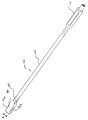
【図1】本発明の用途を有するカニューレ及び目視部材の側面図である。
【図2】本発明の用途を有するディストラクタの斜視図である。
【図3】図3aは、本発明の用途を有するシムの平面図である。
図3bは、シムの側面図である。
【図4】図4aは、図3aのシム用のドライバの平面図である。
図4bは、図4aのドライバの側面図である。
図4cは、図4aのドライバの端面図である。
【図5】図5aは、本発明の用途を有するのみの1つの実施形態の斜視図である。
図5bは、図5aのみの切削ヘッドの拡大斜視図である。
【図6】図6aは、本発明の用途を有する他のみの1つの実施形態の斜視図である。
図6bは、図6aのみの切削ヘッドの拡大斜視図である。
【図7a】本発明の他の実施形態によるディストラクタカッタ組立体の斜視図である。
【図7b】図7aのライン7b−7bを通る断面図である。
【図7c】図7aのディストラクタカッタ組立体の遠位端部分の拡大平面図である。
【図7d】図7aのディストラクタカッタ組立体の遠位端部分の拡大側面図である。
【図8】本発明による用途を有するスラップハンマの斜視図である。
【図9】本発明による用途を有する移植部材ホルダ及び移植部材の斜視図である。
【図10】図10aから図10hは、本発明による椎間板の空隙に接近する種々の方法のステップを示す図である。
【図11a】椎間板の空隙への椎体間固定装置の挿入の準備のために椎間板の空隙を準備する方法のステップを示す図である。
【図11b】椎間板の空隙への椎体間固定装置の挿入の準備のために椎間板の空隙を準備する方法のステップを示す図である。
【図11c】椎間板の空隙への椎体間固定装置の挿入の準備のために椎間板の空隙を準備する方法のステップを示す図である。
【図11d】椎間板の空隙への椎体間固定装置の挿入の準備のために椎間板の空隙を準備する方法のステップを示す図である。
【図11e】椎間板の空隙への椎体間固定装置の挿入の準備のために椎間板の空隙を準備する方法のステップを示す図である。
【図11f】椎間板の空隙への椎体間固定装置の挿入の準備のために椎間板の空隙を準備する方法のステップを示す図である。
【図11g】椎間板の空隙への椎体間固定装置の挿入の準備のために椎間板の空隙を準備する方法のステップを示す図である。
【図11h】椎間板の空隙への椎体間固定装置の挿入の準備のために椎間板の空隙を準備する方法のステップを示す図である。
【図12】椎間板の空隙への椎体間固定装置の両側の配置を示す平面図である。[0001]
BACKGROUND OF THE INVENTION
The present invention relates to an intervertebral spinal procedure and techniques used in instruments that perform such procedures. More particularly, but not exclusively, the present invention relates to endoscopic interbody surgical methods and instruments.
[0002]
[Prior art]
Usually, an intervertebral disc placed between the end plates of adjacent vertebrae stabilizes the spine, distributes forces between the vertebrae, and softens the impact on the vertebral bodies. Intervertebral discs are moved or damaged by injury, disease or aging. Herniated or ruptured annular fibers cause nerve damage, pain, paralysis, muscle weakness, and asthenia. Furthermore, as a result of normal aging progression, the intervertebral disc becomes dehydrated and hardened, reducing the height of the disc space, making the spinal column unstable and reducing mobility. The most typical surgical treatment of the disc involves a discectomy (surgically removing some or all of the disc material). This discectomy requires the fixation of adjacent vertebrae to resolve the early onset of pain, abnormal joint mechanisms, arthritis and nerve damage.
[0003]
Conventional surgical procedures to correct the disc space create large wounds in the intervening tissue. These incision procedures require long incisions, extensive muscle detachment, tissue extension, denervation and tissue angiogenesis. Most of these surgeries require hours and weeks of recovery time after surgery by using normal anesthesia and tissue destruction during the surgical procedure. In some cases, these tissue invasive procedures produce scars and pain that persist, which are more serious problems than the pain that leads to surgical intervention.
[0004]
One type of incision procedure that is an attempt to minimize tissue damage caused by the incision procedure is to use an approach through a hole in the disc space. This method has the advantage that one or more implants can be placed in the disc space with a single incision. However, this method has the disadvantage that muscles and tissue behind the surgical site are damaged and damaged by the incision and contraction of the tissue at the surgical site.
[0005]
Techniques for minimizing intrusion into the body are desirable, especially in the field of spinal column and neurosurgery, due to the need to access deeper sites within the body and the risk of damaging biointerfering tissue. The development of a percutaneous spinal procedure results in significant improvements in relieving recovery time and post-operative pain. This is because they can be performed with local anesthesia with minimal muscle incision. For example, US Pat. No. 4,545,374 to Jacobson shows a percutaneous lumbar discectomy, preferably using a lateral approach using x-rays using fluoroscopy. This procedure is limited. This is because the incision site cannot be seen directly with other restrictions.
[0006]
Other procedures have been developed including viewing the spinal column and intervening structures with an arthroscope. U.S. Pat. Nos. 4,573,448 and 5,395,317 to Kambin disclose percutaneous decompression of a hernia disc in a posterior lateral manner. The portion of the herniated disc is released through a cannula placed against the disc. The Kabrin patent of US Pat. No. 5,395,317 shows a biportal procedure that involves placing both a working cannula for an endoscope and a visual cannula percutaneously. This procedure makes it possible to simultaneously perform visual, aspiration, irrigation and excision of the disc procedure. These methods aim to avoid damage to soft tissue structures and avoid the need to remove bone through the passage. However, these methods have limitations. This is because they are not interested in, for example, extending the disc space, preparing the disc space, and inserting the implant into the disc space. The method of US Pat. No. 5,395,317 requires multiple entrances to the patient, and the method of US Pat. No. 4,573,448 cannot directly see the working gap.
[0007]
Examples of instruments and methods for performing spinal surgery using a minimally invading method are disclosed in US Pat. Nos. 5,792,044 and 5,902,231 to Moley et al. The present invention also relates to improvements and techniques using methods that minimize body penetration to perform spinal surgery.
[0008]
[Means for Solving the Problems]
The present invention is a kit for preparing a disc space between adjacent vertebrae of a patient, a cannula for insertion through a patient's tissue, the proximal end and the distal end of the cannula A cannula having a working channel extending between the proximal end and the distal end and extending the disc space to an extended height of the disc space A distractor having a distal head, the distractor having an axis extending proximally from the distal head, dimensioned to extend through the working channel; and A surgical instrument including a distal portion dimensioned to be disposed within a spinal disc space adjacent to the distal head, extending proximally from the distal portion; The axis of the distractor Adjacent further comprising a shaft that is sized to be extended through the working channel, including a surgical instrument, there is provided a kit.
One aspect of the present invention involves inserting one or more intervertebral disc fixation devices in a spinal disc space using a through-hole approach that minimizes entry into the body. Another aspect of the invention involves performing a surgical procedure on the spinal disc space using a through-hole approach that minimizes entry into the body.
[0009]
According to another aspect of the invention, a method is provided for performing a surgical procedure in an intervertebral disc space between adjacent vertebrae. The method includes inserting a cannula to create a working channel through the patient's skin and tissue using an approach through a hole in the disc space, inserting a viewing member through the working channel, at least one vertebra. Providing an intervertebral disc space through the working channel for insertion of the interbody fusion device. In one form, a spinoarthrotomy is performed to access the disc space through the working channel.
[0010]
According to another aspect of the invention, a method is provided for inserting at least one interbody fusion device into an intervertebral disc space between adjacent vertebrae. The method uses an approach through a hole in the disc space to create a working channel through the patient's skin and tissue to the disc space and through the working channel to place at least one fixation device on both sides. And inserting at least one fixation device into the disc space through the working channel such that adjacent vertebrae are supported on both sides by at least one interbody fusion device.
[0011]
According to another aspect of the invention, a method is provided for restoring the height of an intervertebral disc between adjacent vertebrae of a patient. This method involves inserting a cannula through the patient's skin and tissue to create a working passageway to the disc space, and the adjacent discs up to the height of the disc space by a distractor extending through the cannula into the disc space. Stretching and inserting a shim through the cannula into the disc space adjacent to the distractor. The shim has a blade that has a height corresponding to the height of the extended disc so that it contacts the end plate of the adjacent vertebra.
[0012]
In accordance with another aspect of the present invention, a method is provided for preparing a disc space for inserting an implant between adjacent vertebrae of a patient. This method extends the disc space to a predetermined disc height by inserting a cannula through the patient's skin and tissue to create a working channel in the disc space and placing a distractor in the disc space. The distractor is attached to a stem that extends through the working channel and has a body portion that extends between a front end and a rear end. The body portion has an upper surface, opposing lower surfaces, and opposing first and second sidewalls extending between the upper and lower surfaces. The distractor further includes a first flange and a second flange, each extending proximally from the front end to the rear end of the body. The first flange forms a slot with a first side wall, the second flange forms a slot with a second side wall, the cutter is inserted through the working channel, and the cutter is an upper member having an upper cutting edge And a lower member having a lower cutting edge, and a pair of opposing side walls extending between the upper member and the lower member, the body part of the distractor such that each side wall of the cutter is received in one of the slots Cutting adjacent vertebrae by advancing the cutter.
[0013]
Other objects, features, advantages and benefits of the present invention will become apparent from the detailed drawings and description thereof.
[0014]
Hereinafter, an embodiment of the present invention will be described in detail with reference to the drawings.
For the purpose of promoting an understanding of the principles of the invention, reference will now be made to the embodiment illustrated in the drawings and specific language will be used to describe the embodiment. Nevertheless, no limitation on the aspect of the invention is intended. Variations and modifications of the described methods, systems or devices and other uses of the principles of the present invention are considered to be understood by those skilled in the art to which the present invention pertains.
[0015]
The scope of the present invention is for a wide range of surgical procedures, such as laminectomy (laminotomy, laminectomiy), intervertebral resection using posterior, posterior or lateral approach to the disc space. (Foramenotomi), spinal arthrotomy (facetectomies) and spinal surgical procedures such as discectomy. The devices and instruments of the present invention have application to new surgical techniques that can perform each of several types of surgical procedures through a single working channel. The present invention has application to surgical techniques for preparing an intervertebral disc space for insertion of an implant into the intervertebral disc space. In addition, the present invention provides a surgical device that minimally penetrates the body by an approach through a hole that prepares the disc space for insertion of one or more implants into the disc space in a single lateral manner. Has application in procedure.
[0016]
Referring to FIG. 1, one example of a cannula assembly 15 is provided that provides an endoscopic method of minimally entering the disc space. It will also be appreciated that other shapes of cannula assembly 15 are contemplated so long as the cannula assembly has a protective sleeve that minimizes entry into the disc space and allows viewing of the surgical site. Should. The cannula assembly 15 has a
[0017]
The cannula assembly 15 has an
[0018]
The cannula assembly 15 can take any shape or device capable of supporting the optical system adjacent to the working
[0019]
Cannula assembly 15 includes irrigation and
[0020]
The present invention also contemplates an instrument for use with a cannula assembly to prepare an intervertebral disc space for inserting one or more implants and inserting the implant into the intervertebral disc space. Specific instruments include distractors, shims, only, distractor cutters, implant member holders, reamers and drills. Other instruments that perform surgical procedures on the vertebral body or disc are performed by those skilled in the art as long as it can be used to minimize penetration into the body through the working
[0021]
In FIG. 2, a
[0022]
The
[0023]
Referring to FIGS. 3 (a) and 3 (b), a
[0024]
The
[0025]
The
[0026]
In a particular embodiment of
[0027]
Referring to FIGS. 4 (a) -4 (c), a
[0028]
The
[0029]
The use of
[0030]
Only the box for preparing the pre-formed cavity of the disc space is shown in FIGS. 5 (a) and 5 (b). The box only 90 has a handle 92 having an
[0031]
The non-cutting extension 103 is attached to the first arm 97. Similarly, the non-cutting extension 102 is attached to the first arm 99. Non-cutting extensions 103 and 102 are arranged to extend distally beyond cutting
[0032]
Another embodiment only in accordance with the present invention is the only curved 110 shown in FIGS. 6 (a) and 6 (b). The
[0033]
A
[0034]
In use, only 90, 110 are placed through
[0035]
Referring to FIGS. 7 (a)-7 (d), a
[0036]
The main body portion 220 has a
[0037]
The body portion 220 further includes a cavity 238 formed to extend between the
[0038]
In accordance with another aspect of the present invention, a cutting instrument or
[0039]
The
[0040]
The
[0041]
The
[0042]
The
[0043]
FIG. 8 shows an example of a slap hammer that can be engaged with the mounting
[0044]
Referring to FIG. 9, an embodiment of an interbody fusion device such as a
[0045]
Implant I is a device suitable for insertion through
[0046]
Implant member I is a spacer or an interbody fusion device. The implantation member I has an elongated cross section as shown in FIG. 9, or a circular, oval, rectangular, trapezoidal or rectangular cross section. Transplant member I is dimensioned such that a single graft member is inserted into the disc space, or two or more graft members are inserted, or placed on either side of the disc space. An implant I in the form of an interbody fusion device is shown in US Pat. No. 5,890,556, an example of which is incorporated herein by reference. Another example of a suitable interbody fusion device is described in application Ser. No. 60 / 160,506, filed Oct. 20, 1999, which is hereby incorporated by reference in its entirety. In another example, an example of a banana shaped implant is shown in Application No. 60 / 160,667, filed October 21, 1999, which application is hereby incorporated by reference in its entirety. .
[0047]
The interbody fusion device inserted into the intervertebral disc preferably has a hollow interior that forms a chamber or reservoir for bone formation or bone growth material G (FIG. 12). The device is filled with bone growth material prior to implantation into the disc space, and the bone growth material is inserted into the chamber or reservoir after inserting one or more devices. The bone growth component fills the hollow interior defined by the device to promote fixation and bone growth between adjacent vertebrae. Also, the bone growth or bone growth material is packed around the disc space device.
[0048]
Suitable osteogenic materials or compositions include autografts, allografts, xenografts, mineral deciduous bone, and synthetic and natural bone grafts and bone growth factors such as bioceramics and polymers. As used herein, the term osteogenic material or osteogenic component includes autografts, allografts, xenografts, bone graft substitutes and natural, synthetic and recombinant proteins, hormones, etc. Widely includes materials that promote bone growth or bone treatment.
[0049]
The steps of the spinal surgical procedure according to one aspect of the present invention are illustrated in FIGS. 10 (a) -10 (h). As can be seen from each of the illustrated steps (a)-(h), the present invention contemplates a method of access through a hole approaching the disc space as shown by
[0050]
In the first step of the technique, the
[0051]
In successive steps of the preferred method, a series of tissue dilators are advanced over the
[0052]
In the next step of the illustrated technique, the working
[0053]
By placing the
[0054]
According to the surgical technique of the present invention, the working
[0055]
The surgeon can directly view the area under the working
[0056]
One important feature of the present invention is achieved by the large diameter of the working
[0057]
Surgeons can take advantage of the same benefits by performing a wide range of procedures on the human body. For example, facetectomi can introduce a working channel by directing the working
[0058]
For example, one surgical operation will be described with reference to FIGS. 11 (a) to 11 (h). Those skilled in the art will recognize that the facet coupling must be removed in order for FIGS. 11 (a) through 11 (h) to access the disc space in an oblique orientation relative to the centerline of the vertebral body. You can see that it shows how to approach through a hole.
[0059]
This method allows for the preparation of the disc space and one method to insert one or more implants on both sides of the disc.
A skin incision is made behind the particular disc space to be fixed. As the tissue under the skin is continuously scraped or retracted, the working
[0060]
In a continuous step, the disc space is stretched to the desired disc space height. As shown in FIG. 11 (a), the
[0061]
One type of instrument that can be inserted through working
[0062]
As shown in FIGS. 11 (e) and 11 (f), the box only 110 or the bend only 110 may be one or more depending on the patient's anatomy and the desired location of the implant or implants. Used to form the insertion site for multiple implants. Only 90, 110 are inserted through the
[0063]
As shown in FIG. 11 (g), the
[0064]
When the desired stretch is achieved, the handle 212 is removed from the proximal end of the
[0065]
Implant member I, preferably a fixation device, bone dwell, push-in implant member, threaded implant member, etc., is prepared in the patient's disc space via
[0066]
In some cases, the preparing step comprises preparing the end plate of the vertebra by reducing the bleeding bone of the end plate. In this case, several aspirations and irrigations are advantageous. The procedure described above is guided by viewing directly from the
[0067]
Also, the implant material can be placed directly into the prepared bone in the disc space depending on whether the interbody fusion device is not used or packed around the inserted device. This implant can be delivered through the working
[0068]
While the present invention has been illustrated and described in detail with the aid of drawings, it is intended for purposes of illustration only and not for the purpose of limitation. While preferred embodiments have been illustrated, variations and modifications as well as those that are within the spirit of the invention should be protected.
[Brief description of the drawings]
FIG. 1 is a side view of a cannula and viewing member having an application of the present invention.
FIG. 2 is a perspective view of a distractor having applications of the present invention.
FIG. 3a is a plan view of a shim having an application of the present invention.
FIG. 3b is a side view of the shim.
4a is a plan view of the shim driver of FIG. 3a. FIG.
FIG. 4b is a side view of the driver of FIG. 4a.
FIG. 4c is an end view of the driver of FIG. 4a.
FIG. 5a is a perspective view of one embodiment having only the application of the present invention.
FIG. 5b is an enlarged perspective view of the cutting head of FIG. 5a only.
FIG. 6a is a perspective view of only one other embodiment having the use of the present invention.
6b is an enlarged perspective view of the cutting head of FIG. 6a only.
7a is a perspective view of a distractor cutter assembly according to another embodiment of the present invention. FIG.
7b is a cross-sectional view through line 7b-7b of FIG. 7a.
7c is an enlarged plan view of the distal end portion of the distractor cutter assembly of FIG. 7a.
7d is an enlarged side view of the distal end portion of the distractor cutter assembly of FIG. 7a.
FIG. 8 is a perspective view of a slap hammer having application according to the present invention.
FIG. 9 is a perspective view of an implant member holder and implant having use in accordance with the present invention.
FIGS. 10a to 10h illustrate various method steps for accessing an intervertebral disc space in accordance with the present invention.
FIG. 11a illustrates steps of a method for preparing an intervertebral disc space in preparation for insertion of an interbody fusion device into the intervertebral disc space.
FIG. 11b illustrates steps of a method for preparing an intervertebral disc space in preparation for insertion of an interbody fusion device into the intervertebral disc space.
FIG. 11c illustrates steps of a method for preparing an intervertebral disc space in preparation for insertion of an interbody fusion device into the intervertebral disc space.
FIG. 11d illustrates steps of a method for preparing an intervertebral disc space in preparation for insertion of an interbody fusion device into the intervertebral disc space.
FIG. 11e shows the steps of a method for preparing an intervertebral disc space in preparation for insertion of an interbody fusion device into the intervertebral disc space.
FIG. 11f shows the steps of a method for preparing an intervertebral disc space in preparation for insertion of an interbody fusion device into the intervertebral disc space.
FIG. 11g shows the steps of a method for preparing an intervertebral disc space in preparation for insertion of an interbody fusion device into the intervertebral disc space.
FIG. 11h shows the steps of a method for preparing an intervertebral disc space in preparation for insertion of an interbody fusion device into the intervertebral disc space.
FIG. 12 is a plan view showing the arrangement of both sides of the interbody fusion device in the intervertebral disc space.
Claims (16)
患者の組織を通して挿入するためのカニューレであって、該カニューレの近位側の端部と遠位側の端部との間に延び前記近位側の端部および前記遠位側の端部において開口する作業通路を有するカニューレと、
椎間板の空隙を、椎間板の空隙の延伸高さまで延伸するための、遠位側のヘッドを有するディストラクタであって、前記作業通路を通って延び得る寸法にされた、前記遠位側のヘッドから近位側に延びる軸を有するディストラクタと、
前記ディストラクタの前記遠位側のヘッドに隣接して脊柱の椎間板の空隙内に配置するような寸法にされた、遠位側部分を含む手術用器具であって、前記遠位側部分から近位側に延び、前記ディストラクタの前記軸に隣接して前記作業通路を通して延び得る寸法にされた軸を更に含む、手術用器具と、
を含み、
前記ディストラクタの前記遠位側のヘッドは前端および後端の間に延び、
前記遠位側のヘッドは、さらに、上記隣接する椎骨の一方に接触する上面と、前記隣接する椎骨の他方に接触する反対側の下面と、前記上面と前記下面との間を延びる、対向する第1および第2の側壁とを有し、
前記ディストラクタは、さらに、第1のフランジと第2のフランジとを有し、前記第1のフランジおよび第2のフランジはそれぞれ、前記遠位側のヘッドの前記前端から前記遠位側のヘッドの前記後端へ向かって、近位側にむかって延び、
前記第1のフランジは、前記遠位側のヘッドの前記第1の側壁とともにスロットを形成し、前記第2のフランジは、前記遠位側のヘッドの前記第2の側壁とともにスロットを形成し、
前記キットの前記手術用器具はカッタであり、前記カッタは、上方切削縁を有する上方部材と、下方切削縁を有する下方部材と、前記上方部材および前記下方部材の間に延びる一対の対向する側壁とを有し、
前記カッタは、該カッタの前記側壁が前記ディストラクタの前記遠位側のヘッドに隣接する前記スロットのうちの対応するスロットに受け入れられた状態で、該カッタが前記ディストラクタの前記遠位側のヘッド上を遠位側にむかって前進することができるような寸法にされた、遠位側に開口する通路を含む、キット。A kit for preparing a disc space between adjacent vertebrae of a patient,
A cannula for insertion through a patient's tissue, extending between a proximal end and a distal end of the cannula, at the proximal end and the distal end A cannula having an open working channel;
A distractor having a distal head for extending the disc space to the extended height of the disc space, the distractor being dimensioned to extend through the working channel, from the distal head A distractor having a proximally extending axis;
A surgical instrument including a distal portion sized to be positioned within a spinal disc space adjacent to the distal head of the distractor, wherein the surgical instrument is proximate to the distal portion. A surgical instrument further comprising a shaft extending to a distal side and dimensioned to extend through the working channel adjacent to the shaft of the distractor;
Only including,
The distal head of the distractor extends between a front end and a rear end;
The distal head is further opposed to extend between an upper surface contacting one of the adjacent vertebrae, an opposite lower surface contacting the other of the adjacent vertebrae, and the upper and lower surfaces. First and second side walls,
The distractor further includes a first flange and a second flange, each of the first flange and the second flange being from the front end of the distal head to the distal head. Towards the rear end of the
The first flange forms a slot with the first side wall of the distal head, the second flange forms a slot with the second side wall of the distal head;
The surgical instrument of the kit is a cutter, the cutter comprising an upper member having an upper cutting edge, a lower member having a lower cutting edge, and a pair of opposing side walls extending between the upper member and the lower member. And
The cutter has the side wall of the cutter received in a corresponding one of the slots adjacent to the distal head of the distractor and the cutter is on the distal side of the distractor. are sized to be able to move forward toward the upper head distally, passage including that opens distally kit.
該キットは更に第2の手術用器具を含み、該第2の手術用器具は、遠位側のブレードを備えたシムを含み、前記遠位側のブレードは、該遠位側のブレードの上面と底面との間に高さを有し、該遠位側のブレードの両側の側面の間に厚さを有し、前記高さは前記厚さより少なくとも2倍大きく、前記遠位側のブレードは、該遠位側のブレードの近位端に設けられる1対の肩部を有し、前記高さは、延伸された前記椎間板の空隙の高さに対応し、前記手術用器具の前記軸は、前記遠位側のブレードから近位側に延びる、キット。The kit according to claim 1, wherein
The kit further includes a second surgical instrument, the second surgical instrument including a shim with a distal blade, the distal blade being an upper surface of the distal blade. Between the sides of the distal blade, the height is at least twice as large as the thickness, and the distal blade is A pair of shoulders provided at the proximal end of the distal blade, the height corresponding to the height of the extended disc space, and the axis of the surgical instrument is A kit extending proximally from the distal blade.
前記シムの前記遠位側のブレードは、前記椎間板の空隙から前記ディストラクタの前記遠位側のヘッドが除去されたときに前記延伸された椎間板の空隙の高さを維持することができるように前記隣接する椎骨に接触して配置可能であり、前記シムの前記ブレードは、間に高さを画定する両側の椎骨接触面と、前記両側の椎骨接触面の間に延び、間に厚さを画定する両側の側面とを有し、前記厚さに対する前記高さの比は5より大きい、キット。The kit according to claim 2,
The distal blade of the shim can maintain the height of the elongated disc space when the distal head of the distractor is removed from the disc space. The blade of the shim can be placed in contact with the adjacent vertebrae, and the blades of the shim extend between the vertebral contact surfaces on both sides defining the height therebetween and the vertebral contact surfaces on both sides. A kit having demarcating sides and the ratio of the height to the thickness is greater than 5.
前記シムの前記軸は両側の側面を有し、該軸の両側の側面は、該軸に沿って前記ブレー
ドの前記両側の側面の延長部分を形成し、前記軸の両側の側面は、その間に、前記遠位側のブレードの前記両側の側面の間における前記遠位側のブレードの前記厚さと同じ厚さを画定する、キット。The kit according to claim 3,
The shaft of the shim has side surfaces on both sides, the side surfaces on both sides of the shaft form an extension of the side surfaces on both sides of the blade along the axis, and the side surfaces on both sides of the shaft are in between A kit defining a thickness equal to the thickness of the distal blade between the sides of the distal blade.
前記遠位側のブレードは、前記軸から前記遠位側のブレードの前記両側の椎骨接触面まで延びる一対の肩部を含む、キット。The kit according to claim 4, wherein
The distal blade includes a pair of shoulders extending from the shaft to the vertebral contact surfaces on both sides of the distal blade.
第2の椎間板の空隙の高さまで前記椎間板の空隙を延伸する寸法にされた遠位側のヘッドと、前記遠位側のヘッドから近位側に延びる軸とを有する、第2のディストラクタ、および
前記第2の椎間板の空隙の高さに対応する高さを有する遠位側のブレードを含む第2のシム、
を含む、キット。The kit of claim 2, further comprising:
A second distractor having a distal head dimensioned to extend the disc space to the height of the second disc space, and an axis extending proximally from the distal head; And a second shim comprising a distal blade having a height corresponding to the height of the second disc space,
Including a kit.
中を通して前記シムの前記軸を受け入れるための両側の端部の開口を有する通路を含むドライバであって、前記遠位側のブレードを前記椎間板の空隙内へ駆動するために、前記シムの前記軸に沿って移動して、近位側を向いている前記遠位側のブレードの前記肩部に接触する、ドライバ、
を含む、キット。The kit of claim 2, further comprising:
A driver comprising a passage having openings at opposite ends for receiving the shaft of the shim therethrough, the shaft of the shim for driving the distal blade into the disc space A screwdriver, contacting the shoulder of the distal blade facing proximally,
Including a kit.
前記シムの前記軸は、前記遠位側のブレードの前記両側の側面と同一平面の両側の側面を有し、また、前記シムの軸は、その両側の側面の間における厚さが、前記遠位側のブレードの前記両側の側面の間における前記ブレードの厚さと同じである、キット。The kit according to claim 7, wherein
The shaft of the shim has side surfaces on both sides that are coplanar with the side surfaces on both sides of the distal blade, and the shim shaft has a thickness between the side surfaces on the far side. A kit having a thickness equal to the thickness of the blade between the lateral sides of the distal blade.
前記カニューレの前記作業通路内の前記ディストラクタおよび前記手術用器具とともに、前記椎間板の空隙を直接見るために前記作業通路を通して挿入する寸法にされた目視部材を含む、キット。The kit of claim 1, further comprising:
A kit comprising a viewing member dimensioned to be inserted through the working channel for direct viewing of the disc space with the distractor and the surgical instrument in the working channel of the cannula.
前記手術用器具の前記軸が前記カニューレの前記作業通路内に配置されている間に、前記椎間板の空隙内に前記カニューレの前記作業通路を通して配置することができる寸法にされた少なくとも一つの移植部材を含む、キット。The kit of claim 1, further comprising:
At least one implant member sized to be positioned through the working channel of the cannula within the disc space while the shaft of the surgical instrument is positioned within the working channel of the cannula. Including a kit.
前記椎間板の空隙に隣接する骨構造に係合するように患者に挿入するためのガイドワイヤと、
前記ガイドワイヤ上、前記患者の皮膚および組織を通って前記椎間板の空隙へ配置するための寸法にされた、中空の拡張器と、
を含み、前記カニューレの前記作業通路は、前記拡張器が前記椎間板の空隙への前記カニューレの挿入をガイドするように該作業通路を通る前記拡張器を受け入れる寸法にされる、キット。The kit of claim 1, further comprising:
A guide wire for insertion into a patient to engage bone structure adjacent to the disc space;
A hollow dilator dimensioned for placement on the guidewire through the patient's skin and tissue into the disc space;
And wherein the working channel of the cannula is dimensioned to receive the dilator through the working channel such that the dilator guides insertion of the cannula into the disc space.
前記カッタは、前記カッタと前記ディストラクタの前記軸が前記カニューレの前記作業通路を通して延びている状態で、前記ディストラクタの前記軸の周りに位置決めできるような寸法にされている、キット。The kit according to claim 1 , wherein
The kit is sized such that the cutter and the shaft of the distractor can be positioned about the shaft of the distractor with the shaft of the cutter and the distractor extending through the working channel of the cannula.
さらに、前記カッタおよび前記ディストラクタの前記軸とともに前記作業通路内に設けられる光学系を含む、キット。The kit according to claim 12 ,
The kit further includes an optical system provided in the working path along with the shaft of the cutter and the distractor.
さらに、前記椎間板の空隙内の第1の両側位置に配置するための寸法にされた第1の椎体間固定装置と、前記椎間板の空隙内の第2の両側位置に配置するための寸法にされた第2の椎体間固定装置とを含み、
前記第1および第2の椎体間固定装置は、さらに、前記光学系が前記カニューレの前記作業通路内に配置されるときに、前記カニューレの前記作業通路を通して配置するための寸法にされている、キット。The kit according to claim 13 ,
In addition, a first interbody fusion device dimensioned for placement at a first bilateral location within the disc space and a size for placement at a second bilateral location within the disc space. A second interbody fusion device,
The first and second interbody fusion devices are further dimensioned for placement through the working channel of the cannula when the optical system is disposed within the working channel of the cannula. ,kit.
さらに、曲線の切削通路を画成する曲がった軸を有するのみを含む、キット。The kit according to claim 1 , wherein
The kit further includes only having a curved axis defining a curved cutting path.
前記作業通路は、円形の断面形状を有する、キット。The kit according to claim 1, wherein
The working path has a circular cross-sectional shape.
Applications Claiming Priority (2)
| Application Number | Priority Date | Filing Date | Title |
|---|---|---|---|
| US16055099P | 1999-10-20 | 1999-10-20 | |
| US60/160550 | 1999-10-20 |
Publications (3)
| Publication Number | Publication Date |
|---|---|
| JP2001198135A JP2001198135A (en) | 2001-07-24 |
| JP2001198135A5 JP2001198135A5 (en) | 2008-10-23 |
| JP4326134B2 true JP4326134B2 (en) | 2009-09-02 |
Family
ID=22577347
Family Applications (1)
| Application Number | Title | Priority Date | Filing Date |
|---|---|---|---|
| JP2000321049A Expired - Lifetime JP4326134B2 (en) | 1999-10-20 | 2000-10-20 | Method and apparatus for performing a surgical procedure |
Country Status (2)
| Country | Link |
|---|---|
| US (4) | US6575899B1 (en) |
| JP (1) | JP4326134B2 (en) |
Cited By (1)
| Publication number | Priority date | Publication date | Assignee | Title |
|---|---|---|---|---|
| WO2023140415A1 (en) * | 2022-01-18 | 2023-07-27 | (주)스파인닥터스 | Medical instrument set for transplanting spine insert |
Families Citing this family (225)
| Publication number | Priority date | Publication date | Assignee | Title |
|---|---|---|---|---|
| US6770074B2 (en) * | 1988-06-13 | 2004-08-03 | Gary Karlin Michelson | Apparatus for use in inserting spinal implants |
| US7799036B2 (en) * | 1998-08-20 | 2010-09-21 | Zimmer Spine, Inc. | Method and apparatus for securing vertebrae |
| US6187000B1 (en) | 1998-08-20 | 2001-02-13 | Endius Incorporated | Cannula for receiving surgical instruments |
| JP4326134B2 (en) * | 1999-10-20 | 2009-09-02 | ウォーソー・オーソペディック・インコーポレーテッド | Method and apparatus for performing a surgical procedure |
| US6500180B1 (en) * | 1999-10-20 | 2002-12-31 | Sdgi Holdings, Inc. | Methods and instrumentation for distraction of a disc space |
| US6530929B1 (en) * | 1999-10-20 | 2003-03-11 | Sdgi Holdings, Inc. | Instruments for stabilization of bony structures |
| AU4161201A (en) * | 2000-02-22 | 2001-09-03 | Sdgi Holdings Inc | Instruments and techniques for disc space preparation |
| US7056321B2 (en) | 2000-08-01 | 2006-06-06 | Endius, Incorporated | Method of securing vertebrae |
| US7985247B2 (en) | 2000-08-01 | 2011-07-26 | Zimmer Spine, Inc. | Methods and apparatuses for treating the spine through an access device |
| EP1363565A2 (en) | 2000-08-08 | 2003-11-26 | SDGI Holdings, Inc. | Implantable joint prosthesis |
| JP2004516044A (en) * | 2000-08-08 | 2004-06-03 | エスディージーアイ・ホールディングス・インコーポレーテッド | Method and apparatus for improving stereotactic body transplantation |
| AU2729302A (en) | 2000-11-13 | 2002-05-21 | Frank H Boehm Jr | Device and method for lumbar interbody fusion |
| US7220262B1 (en) * | 2001-03-16 | 2007-05-22 | Sdgi Holdings, Inc. | Spinal fixation system and related methods |
| US6719794B2 (en) * | 2001-05-03 | 2004-04-13 | Synthes (U.S.A.) | Intervertebral implant for transforaminal posterior lumbar interbody fusion procedure |
| US7144393B2 (en) * | 2001-05-15 | 2006-12-05 | Dipoto Gene P | Structure for receiving surgical instruments |
| AU2002336694A1 (en) * | 2001-11-01 | 2003-05-12 | Lawrence M. Boyd | Devices and methods for the restoration of a spinal disc |
| DE10154163A1 (en) | 2001-11-03 | 2003-05-22 | Advanced Med Tech | Device for straightening and stabilizing the spine |
| US9155544B2 (en) * | 2002-03-20 | 2015-10-13 | P Tech, Llc | Robotic systems and methods |
| US7004947B2 (en) * | 2002-06-24 | 2006-02-28 | Endius Incorporated | Surgical instrument for moving vertebrae |
| US7306603B2 (en) | 2002-08-21 | 2007-12-11 | Innovative Spinal Technologies | Device and method for percutaneous placement of lumbar pedicle screws and connecting rods |
| US7127857B2 (en) * | 2002-09-04 | 2006-10-31 | Connor Sports Flooring Corporation | Subfloor assembly for athletic playing surface having improved deflection characteristics |
| US6648888B1 (en) | 2002-09-06 | 2003-11-18 | Endius Incorporated | Surgical instrument for moving a vertebra |
| WO2004039235A2 (en) * | 2002-10-25 | 2004-05-13 | Endius Incorporated | Apparatus and methods for shielding body structures during surgery |
| US20040106997A1 (en) * | 2002-11-01 | 2004-06-03 | Lieberson Robert E. | Apparatus and method for creating a surgical channel |
| US20040116777A1 (en) * | 2002-12-13 | 2004-06-17 | Jeffrey Larson | Guided retractor and methods of use |
| US7014608B2 (en) * | 2002-12-13 | 2006-03-21 | Synthes Spine Company, Lp | Guided retractor and methods of use |
| US20060155170A1 (en) * | 2002-12-13 | 2006-07-13 | Synthes Spine Company, Lp | Guided retractor and methods of use |
| US7771478B2 (en) | 2003-04-04 | 2010-08-10 | Theken Spine, Llc | Artificial disc prosthesis |
| US7291173B2 (en) | 2003-05-06 | 2007-11-06 | Aesculap Ii, Inc. | Artificial intervertebral disc |
| US7105024B2 (en) * | 2003-05-06 | 2006-09-12 | Aesculap Ii, Inc. | Artificial intervertebral disc |
| US7645232B2 (en) | 2003-05-16 | 2010-01-12 | Zimmer Spine, Inc. | Access device for minimally invasive surgery |
| US6945975B2 (en) * | 2003-07-07 | 2005-09-20 | Aesculap, Inc. | Bone fixation assembly and method of securement |
| US6945974B2 (en) * | 2003-07-07 | 2005-09-20 | Aesculap Inc. | Spinal stabilization implant and method of application |
| DE10330698B4 (en) * | 2003-07-08 | 2005-05-25 | Aesculap Ag & Co. Kg | Intervertebral implant |
| DE10330699B3 (en) | 2003-07-08 | 2005-02-17 | Aesculap Ag & Co. Kg | Surgical instrument for handling an implant |
| DE10339170B4 (en) * | 2003-08-22 | 2009-10-15 | Aesculap Ag | Intervertebral implant |
| US7578820B2 (en) * | 2003-09-02 | 2009-08-25 | Moore Jeffrey D | Devices and techniques for a minimally invasive disc space preparation and implant insertion |
| US7955355B2 (en) | 2003-09-24 | 2011-06-07 | Stryker Spine | Methods and devices for improving percutaneous access in minimally invasive surgeries |
| US8002798B2 (en) | 2003-09-24 | 2011-08-23 | Stryker Spine | System and method for spinal implant placement |
| US7655012B2 (en) | 2003-10-02 | 2010-02-02 | Zimmer Spine, Inc. | Methods and apparatuses for minimally invasive replacement of intervertebral discs |
| US20050090899A1 (en) * | 2003-10-24 | 2005-04-28 | Dipoto Gene | Methods and apparatuses for treating the spine through an access device |
| US20050090822A1 (en) * | 2003-10-24 | 2005-04-28 | Dipoto Gene | Methods and apparatus for stabilizing the spine through an access device |
| US7731737B2 (en) * | 2003-10-24 | 2010-06-08 | Zimmer Spine, Inc. | Methods and apparatuses for fixation of the spine through an access device |
| US7618442B2 (en) | 2003-10-21 | 2009-11-17 | Theken Spine, Llc | Implant assembly and method for use in an internal structure stabilization system |
| WO2005039651A2 (en) * | 2003-10-23 | 2005-05-06 | Trans1 Inc. | Tools and tool kits for performing minimally invasive procedures on the spine |
| US7144368B2 (en) * | 2003-11-26 | 2006-12-05 | Synthes Spine Company, Lp | Guided retractor and methods of use |
| US7648506B2 (en) * | 2003-12-16 | 2010-01-19 | Depuy Acromed, Inc. | Pivoting implant holder |
| US7648507B2 (en) * | 2003-12-16 | 2010-01-19 | Depuy Acromed, Inc. | Pivoting implant holder |
| US7311712B2 (en) * | 2004-02-26 | 2007-12-25 | Aesculap Implant Systems, Inc. | Polyaxial locking screw plate assembly |
| US20050240197A1 (en) * | 2004-04-23 | 2005-10-27 | Kmiec Stanley J Jr | Device and method for inserting, positioning and removing an implant |
| DE202004009542U1 (en) | 2004-06-16 | 2004-08-12 | Aesculap Ag & Co. Kg | Artificial intervertebral disk, comprising core with intensely curved upper and less curved lower surface |
| US20060004398A1 (en) * | 2004-07-02 | 2006-01-05 | Binder Lawrence J Jr | Sequential dilator system |
| US20060009541A1 (en) * | 2004-07-09 | 2006-01-12 | Yih-Fang Chen | Saturant for friction material containing friction modifying layer |
| US8182491B2 (en) * | 2004-08-06 | 2012-05-22 | Depuy Spine, Inc. | Rigidly guided implant placement |
| US8016835B2 (en) * | 2004-08-06 | 2011-09-13 | Depuy Spine, Inc. | Rigidly guided implant placement with control assist |
| US8414648B2 (en) | 2004-08-09 | 2013-04-09 | Si-Bone Inc. | Apparatus, systems, and methods for achieving trans-iliac lumbar fusion |
| US9949843B2 (en) | 2004-08-09 | 2018-04-24 | Si-Bone Inc. | Apparatus, systems, and methods for the fixation or fusion of bone |
| US20070156241A1 (en) | 2004-08-09 | 2007-07-05 | Reiley Mark A | Systems and methods for the fixation or fusion of bone |
| US8425570B2 (en) | 2004-08-09 | 2013-04-23 | Si-Bone Inc. | Apparatus, systems, and methods for achieving anterior lumbar interbody fusion |
| US8388667B2 (en) | 2004-08-09 | 2013-03-05 | Si-Bone, Inc. | Systems and methods for the fixation or fusion of bone using compressive implants |
| US20180228621A1 (en) | 2004-08-09 | 2018-08-16 | Mark A. Reiley | Apparatus, systems, and methods for the fixation or fusion of bone |
| US8470004B2 (en) | 2004-08-09 | 2013-06-25 | Si-Bone Inc. | Apparatus, systems, and methods for stabilizing a spondylolisthesis |
| US20060036251A1 (en) | 2004-08-09 | 2006-02-16 | Reiley Mark A | Systems and methods for the fixation or fusion of bone |
| US9662158B2 (en) * | 2004-08-09 | 2017-05-30 | Si-Bone Inc. | Systems and methods for the fixation or fusion of bone at or near a sacroiliac joint |
| US8444693B2 (en) | 2004-08-09 | 2013-05-21 | Si-Bone Inc. | Apparatus, systems, and methods for achieving lumbar facet fusion |
| US20060047296A1 (en) * | 2004-08-31 | 2006-03-02 | Sdg Holdings, Inc. | Annulus replacement system and technique |
| US20060052812A1 (en) * | 2004-09-07 | 2006-03-09 | Michael Winer | Tool for preparing a surgical site for an access device |
| TWI327064B (en) * | 2004-09-23 | 2010-07-11 | Spine Solutions Inc | An intervertebral implant |
| US7763024B2 (en) * | 2004-09-23 | 2010-07-27 | Spine Solutions, Inc. | Adjustable cutting of cutout in vertebral bone |
| US7666189B2 (en) * | 2004-09-29 | 2010-02-23 | Synthes Usa, Llc | Less invasive surgical system and methods |
| US20060089649A1 (en) * | 2004-10-26 | 2006-04-27 | Ullrich Peter F Jr | Surgical instruments and method of using same |
| US7569061B2 (en) | 2004-11-16 | 2009-08-04 | Innovative Spinal Technologies, Inc. | Off-axis anchor guidance system |
| US8021392B2 (en) * | 2004-11-22 | 2011-09-20 | Minsurg International, Inc. | Methods and surgical kits for minimally-invasive facet joint fusion |
| US7837713B2 (en) * | 2004-11-22 | 2010-11-23 | Minsurg International, Inc. | Methods and surgical kits for minimally-invasive facet joint fusion |
| US20060111779A1 (en) * | 2004-11-22 | 2006-05-25 | Orthopedic Development Corporation, A Florida Corporation | Minimally invasive facet joint fusion |
| EP1814474B1 (en) | 2004-11-24 | 2011-09-14 | Samy Abdou | Devices for inter-vertebral orthopedic device placement |
| US20060287584A1 (en) * | 2005-06-16 | 2006-12-21 | Javier Garcia-Bengochia | Surgical retractor extensions |
| JP4907908B2 (en) * | 2005-06-29 | 2012-04-04 | ルネサスエレクトロニクス株式会社 | Driving circuit and display device |
| US7566302B2 (en) * | 2005-07-28 | 2009-07-28 | Synthes Usa, Llc | Expandable access device |
| US20070073397A1 (en) * | 2005-09-15 | 2007-03-29 | Mckinley Laurence M | Disc nucleus prosthesis and its method of insertion and revision |
| US7835784B2 (en) * | 2005-09-21 | 2010-11-16 | Medtronic Navigation, Inc. | Method and apparatus for positioning a reference frame |
| US20070072466A1 (en) * | 2005-09-27 | 2007-03-29 | Manabu Miyamoto | Instrument for endoscope |
| US7658739B2 (en) | 2005-09-27 | 2010-02-09 | Zimmer Spine, Inc. | Methods and apparatuses for stabilizing the spine through an access device |
| EP1951159B1 (en) | 2005-11-23 | 2015-08-19 | Trinity Orthopedics | Percutaneous transpedicular access, fusion, discectomy, and stabilization system |
| US20070213718A1 (en) * | 2006-02-14 | 2007-09-13 | Sdgi Holdings, Inc. | Treatment of the vertebral column |
| US20070227547A1 (en) * | 2006-02-14 | 2007-10-04 | Sdgi Holdings, Inc. | Treatment of the vertebral column |
| US20070213717A1 (en) * | 2006-02-14 | 2007-09-13 | Sdgi Holdings, Inc. | Biological fusion in the vertebral column |
| US7520888B2 (en) * | 2006-02-14 | 2009-04-21 | Warsaw Orthopedic, Inc. | Treatment of the vertebral column |
| US20070233089A1 (en) * | 2006-02-17 | 2007-10-04 | Endius, Inc. | Systems and methods for reducing adjacent level disc disease |
| US8454621B2 (en) * | 2006-09-19 | 2013-06-04 | Warsaw Orthopedic, Inc. | Instruments and methods for spinal implant revision |
| US7963967B1 (en) | 2006-10-12 | 2011-06-21 | Woodse Enterprises, Inc. | Bone preparation tool |
| US20100286477A1 (en) * | 2009-05-08 | 2010-11-11 | Ouyang Xiaolong | Internal tissue visualization system comprising a rf-shielded visualization sensor module |
| US8840621B2 (en) | 2006-11-03 | 2014-09-23 | Innovative Spine, Inc. | Spinal access systems and methods |
| US8057481B2 (en) * | 2006-11-03 | 2011-11-15 | Innovative Spine, Llc | System and method for providing surgical access to a spine |
| US8979931B2 (en) | 2006-12-08 | 2015-03-17 | DePuy Synthes Products, LLC | Nucleus replacement device and method |
| US8480675B2 (en) * | 2007-03-07 | 2013-07-09 | Vertech, Inc. | Betts osteotome |
| US7799058B2 (en) * | 2007-04-19 | 2010-09-21 | Zimmer Gmbh | Interspinous spacer |
| JP5250632B2 (en) | 2007-10-05 | 2013-07-31 | ジンテス ゲゼルシャフト ミット ベシュレンクテル ハフツング | Expansion system |
| US9005288B2 (en) | 2008-01-09 | 2015-04-14 | Providence Medical Techonlogy, Inc. | Methods and apparatus for accessing and treating the facet joint |
| WO2009094225A2 (en) * | 2008-01-25 | 2009-07-30 | The Johns Hopkins University | Biodegradable nerve guides |
| US20090275992A1 (en) * | 2008-04-30 | 2009-11-05 | Phan Christopher U | Apparatus and methods for inserting facet screws |
| US8636740B2 (en) * | 2008-05-08 | 2014-01-28 | Aesculap Implant Systems, Llc | Minimally invasive spinal stabilization system |
| US20090287165A1 (en) * | 2008-05-15 | 2009-11-19 | Warsaw Orthopedic, Inc. | Cannulated Device with Atraumatic Tip for Delivering Local Depot to a Joint Space and Methods of Use Thereof |
| EP2328492B1 (en) | 2008-06-06 | 2018-03-28 | Providence Medical Technology, Inc. | Facet joint implants and delivery tools |
| US20090312764A1 (en) * | 2008-06-11 | 2009-12-17 | Marino James F | Intraosseous transpedicular methods and devices |
| BRPI0916460A2 (en) * | 2008-07-31 | 2016-02-16 | Surgical Innovations Ltd | endoscopy surgical instrument |
| US8876851B1 (en) | 2008-10-15 | 2014-11-04 | Nuvasive, Inc. | Systems and methods for performing spinal fusion surgery |
| US20100121139A1 (en) | 2008-11-12 | 2010-05-13 | Ouyang Xiaolong | Minimally Invasive Imaging Systems |
| US20110009694A1 (en) * | 2009-07-10 | 2011-01-13 | Schultz Eric E | Hand-held minimally dimensioned diagnostic device having integrated distal end visualization |
| WO2010075555A2 (en) | 2008-12-26 | 2010-07-01 | Scott Spann | Minimally-invasive retroperitoneal lateral approach for spinal surgery |
| EP2389126A4 (en) * | 2008-12-31 | 2013-10-30 | Spineology Inc | System and method for performing percutaneous spinal interbody fusion |
| US9168047B2 (en) * | 2009-04-02 | 2015-10-27 | John T. To | Minimally invasive discectomy |
| US8439925B2 (en) * | 2009-05-11 | 2013-05-14 | Trinity Orthopedics, Llc | Transiliac-transsacral method of performing lumbar spinal interventions |
| BRPI1012883B8 (en) | 2009-07-09 | 2021-06-22 | R Tree Innovations Llc | interbody device |
| DE102009048835A1 (en) * | 2009-10-09 | 2011-04-14 | Surgitaix Ag | Drive for bone saw has connection rod for driving saw blade of bone saw, where multiple drive units are provided for driving connection rod and drive units are interconnected in parallel with each other |
| US9028553B2 (en) | 2009-11-05 | 2015-05-12 | DePuy Synthes Products, Inc. | Self-pivoting spinal implant and associated instrumentation |
| US8764806B2 (en) | 2009-12-07 | 2014-07-01 | Samy Abdou | Devices and methods for minimally invasive spinal stabilization and instrumentation |
| US8394108B2 (en) | 2010-06-18 | 2013-03-12 | Spine Wave, Inc. | Screw driver for a multiaxial bone screw |
| US8142437B2 (en) | 2010-06-18 | 2012-03-27 | Spine Wave, Inc. | System for percutaneously fixing a connecting rod to a spine |
| US8454664B2 (en) | 2010-06-18 | 2013-06-04 | Spine Wave, Inc. | Method for fixing a connecting rod to a thoracic spine |
| US8512383B2 (en) | 2010-06-18 | 2013-08-20 | Spine Wave, Inc. | Method of percutaneously fixing a connecting rod to a spine |
| US8777954B2 (en) | 2010-06-18 | 2014-07-15 | Spine Wave, Inc. | Pedicle screw extension for use in percutaneous spinal fixation |
| US8845732B2 (en) * | 2010-09-03 | 2014-09-30 | Globus Medical, Inc. | Expandable fusion device and method of installation thereof |
| US9579214B1 (en) * | 2011-03-01 | 2017-02-28 | John W. McClellan | Peripheral vertebral body spacer implant and insertion tool |
| US8518087B2 (en) | 2011-03-10 | 2013-08-27 | Interventional Spine, Inc. | Method and apparatus for minimally invasive insertion of intervertebral implants |
| US8394129B2 (en) | 2011-03-10 | 2013-03-12 | Interventional Spine, Inc. | Method and apparatus for minimally invasive insertion of intervertebral implants |
| CN103561689B (en) | 2011-03-11 | 2016-01-20 | Fbc设备有限公司 | Spinal implant |
| US9572481B2 (en) | 2011-05-13 | 2017-02-21 | Intuitive Surgical Operations, Inc. | Medical system with multiple operating modes for steering a medical instrument through linked body passages |
| US20130046353A1 (en) * | 2011-08-17 | 2013-02-21 | Smith & Nephew, Inc. | Oval tunnel dilators |
| US8845728B1 (en) | 2011-09-23 | 2014-09-30 | Samy Abdou | Spinal fixation devices and methods of use |
| US9622779B2 (en) | 2011-10-27 | 2017-04-18 | DePuy Synthes Products, Inc. | Method and devices for a sub-splenius / supra-levator scapulae surgical access technique |
| US9808232B2 (en) | 2011-11-01 | 2017-11-07 | DePuy Synthes Products, Inc. | Dilation system |
| US9561059B1 (en) | 2011-11-23 | 2017-02-07 | Nuvasive, Inc. | Minimally invasive facet release |
| CN103327913B (en) | 2011-12-03 | 2015-09-09 | 欧罗波罗斯医学有限公司 | For removing Safety cutter head and the system of target tissue fast |
| US20130226240A1 (en) | 2012-02-22 | 2013-08-29 | Samy Abdou | Spinous process fixation devices and methods of use |
| US10363140B2 (en) | 2012-03-09 | 2019-07-30 | Si-Bone Inc. | Systems, device, and methods for joint fusion |
| US9044321B2 (en) | 2012-03-09 | 2015-06-02 | Si-Bone Inc. | Integrated implant |
| US8778026B2 (en) | 2012-03-09 | 2014-07-15 | Si-Bone Inc. | Artificial SI joint |
| US9138265B2 (en) | 2012-03-19 | 2015-09-22 | Aesculap Implant Systems, Llc | Apparatus and method for visualizing insertion of a fixation element into an implant |
| US9265490B2 (en) | 2012-04-16 | 2016-02-23 | DePuy Synthes Products, Inc. | Detachable dilator blade |
| EP3818947B1 (en) | 2012-05-04 | 2023-08-30 | SI-Bone, Inc. | Fenestrated implant |
| US9198767B2 (en) | 2012-08-28 | 2015-12-01 | Samy Abdou | Devices and methods for spinal stabilization and instrumentation |
| US20140067069A1 (en) | 2012-08-30 | 2014-03-06 | Interventional Spine, Inc. | Artificial disc |
| US9480855B2 (en) | 2012-09-26 | 2016-11-01 | DePuy Synthes Products, Inc. | NIR/red light for lateral neuroprotection |
| US9320617B2 (en) | 2012-10-22 | 2016-04-26 | Cogent Spine, LLC | Devices and methods for spinal stabilization and instrumentation |
| US8663332B1 (en) | 2012-12-13 | 2014-03-04 | Ouroboros Medical, Inc. | Bone graft distribution system |
| US10022245B2 (en) | 2012-12-17 | 2018-07-17 | DePuy Synthes Products, Inc. | Polyaxial articulating instrument |
| US10070969B2 (en) | 2013-01-17 | 2018-09-11 | Stryker European Holdings I, Llc | Annulus plug for intervertebral disc repair |
| US9277928B2 (en) | 2013-03-11 | 2016-03-08 | Interventional Spine, Inc. | Method and apparatus for minimally invasive insertion of intervertebral implants |
| US8864830B2 (en) | 2013-03-12 | 2014-10-21 | Spine Wave, Inc. | Apparatus and methods for inserting an interbody fusion device |
| US9827020B2 (en) | 2013-03-14 | 2017-11-28 | Stryker European Holdings I, Llc | Percutaneous spinal cross link system and method |
| CA2846149C (en) | 2013-03-14 | 2018-03-20 | Stryker Spine | Systems and methods for percutaneous spinal fusion |
| US9993353B2 (en) | 2013-03-14 | 2018-06-12 | DePuy Synthes Products, Inc. | Method and apparatus for minimally invasive insertion of intervertebral implants |
| US9936983B2 (en) | 2013-03-15 | 2018-04-10 | Si-Bone Inc. | Implants for spinal fixation or fusion |
| US9314254B2 (en) * | 2013-03-15 | 2016-04-19 | DePuy Synthes Products, Inc. | Methods and devices for removing a spinal disc |
| US9295500B2 (en) | 2013-06-12 | 2016-03-29 | Spine Wave, Inc. | Screw driver with release for a multiaxial bone screw |
| KR20160040226A (en) | 2013-07-19 | 2016-04-12 | 아우로보로스 메디컬, 아이엔씨 | An anti-clogging device for a vacuum-assisted, tissue removal system |
| US9186259B2 (en) | 2013-09-09 | 2015-11-17 | Ouroboros Medical, Inc. | Expandable trials |
| US11147688B2 (en) | 2013-10-15 | 2021-10-19 | Si-Bone Inc. | Implant placement |
| US9839448B2 (en) | 2013-10-15 | 2017-12-12 | Si-Bone Inc. | Implant placement |
| US9408716B1 (en) | 2013-12-06 | 2016-08-09 | Stryker European Holdings I, Llc | Percutaneous posterior spinal fusion implant construction and method |
| US9744050B1 (en) | 2013-12-06 | 2017-08-29 | Stryker European Holdings I, Llc | Compression and distraction system for percutaneous posterior spinal fusion |
| US10159579B1 (en) | 2013-12-06 | 2018-12-25 | Stryker European Holdings I, Llc | Tubular instruments for percutaneous posterior spinal fusion systems and methods |
| US10478313B1 (en) | 2014-01-10 | 2019-11-19 | Nuvasive, Inc. | Spinal fusion implant and related methods |
| US10342579B2 (en) | 2014-01-13 | 2019-07-09 | Trice Medical, Inc. | Fully integrated, disposable tissue visualization device |
| US9370295B2 (en) | 2014-01-13 | 2016-06-21 | Trice Medical, Inc. | Fully integrated, disposable tissue visualization device |
| US11547446B2 (en) | 2014-01-13 | 2023-01-10 | Trice Medical, Inc. | Fully integrated, disposable tissue visualization device |
| US9730802B1 (en) | 2014-01-14 | 2017-08-15 | Nuvasive, Inc. | Spinal fusion implant and related methods |
| US9788856B2 (en) | 2014-03-11 | 2017-10-17 | Stryker European Holdings I, Llc | Endoscopic surgical systems and methods |
| US9693882B2 (en) | 2014-06-03 | 2017-07-04 | DePuy Synthes Products, Inc. | Optical trial device |
| US9980737B2 (en) | 2014-08-04 | 2018-05-29 | Medos International Sarl | Flexible transport auger |
| US9924979B2 (en) | 2014-09-09 | 2018-03-27 | Medos International Sarl | Proximal-end securement of a minimally invasive working channel |
| US10264959B2 (en) | 2014-09-09 | 2019-04-23 | Medos International Sarl | Proximal-end securement of a minimally invasive working channel |
| US10111712B2 (en) | 2014-09-09 | 2018-10-30 | Medos International Sarl | Proximal-end securement of a minimally invasive working channel |
| JP6542362B2 (en) | 2014-09-18 | 2019-07-10 | エスアイ−ボーン・インコーポレイテッドSi−Bone, Inc. | Matrix implant |
| WO2016044731A1 (en) | 2014-09-18 | 2016-03-24 | Si-Bone Inc. | Implants for bone fixation or fusion |
| US9060876B1 (en) | 2015-01-20 | 2015-06-23 | Ouroboros Medical, Inc. | Stabilized intervertebral scaffolding systems |
| CN104546038A (en) * | 2015-01-31 | 2015-04-29 | 青岛大学医学院附属医院 | Posterior spinal minimally invasive surgery visual field establishing system |
| US20180049754A1 (en) * | 2015-03-13 | 2018-02-22 | Redemed S.R.L. | Intervertebral prosthesis, apparatus for implanting intervertebral prostheses and surgical method for implanting intervertebral prostheses, particularly for percutaneous mini-invasive surgery procedures |
| US10786264B2 (en) | 2015-03-31 | 2020-09-29 | Medos International Sarl | Percutaneous disc clearing device |
| US10376206B2 (en) | 2015-04-01 | 2019-08-13 | Si-Bone Inc. | Neuromonitoring systems and methods for bone fixation or fusion procedures |
| EP3560412B1 (en) | 2015-08-11 | 2020-11-18 | Trice Medical, Inc. | Fully integrated disposable tissue visualization device |
| US11672562B2 (en) | 2015-09-04 | 2023-06-13 | Medos International Sarl | Multi-shield spinal access system |
| CN113143355A (en) | 2015-09-04 | 2021-07-23 | 美多斯国际有限公司 | Multi-shield spinal access system |
| US10987129B2 (en) | 2015-09-04 | 2021-04-27 | Medos International Sarl | Multi-shield spinal access system |
| US11439380B2 (en) | 2015-09-04 | 2022-09-13 | Medos International Sarl | Surgical instrument connectors and related methods |
| US11744447B2 (en) | 2015-09-04 | 2023-09-05 | Medos International | Surgical visualization systems and related methods |
| US10857003B1 (en) | 2015-10-14 | 2020-12-08 | Samy Abdou | Devices and methods for vertebral stabilization |
| US10299838B2 (en) | 2016-02-05 | 2019-05-28 | Medos International Sarl | Method and instruments for interbody fusion and posterior fixation through a single incision |
| US9883953B1 (en) | 2016-09-21 | 2018-02-06 | Integrity Implants Inc. | Stabilized laterovertically-expanding fusion cage systems with tensioner |
| US10898175B2 (en) | 2016-10-04 | 2021-01-26 | Jgmg Bengochea, Llc | Retractor extension clip systems |
| US10485678B2 (en) * | 2016-10-04 | 2019-11-26 | Javier Garcia-Bengochea | Instruments and methods for orthopedic implant assembly |
| US10744000B1 (en) | 2016-10-25 | 2020-08-18 | Samy Abdou | Devices and methods for vertebral bone realignment |
| US10973648B1 (en) | 2016-10-25 | 2021-04-13 | Samy Abdou | Devices and methods for vertebral bone realignment |
| WO2018132502A1 (en) | 2017-01-10 | 2018-07-19 | Anza Innovations Inc. | Expandable intervertebral fusion device |
| US10624588B2 (en) | 2017-01-16 | 2020-04-21 | General Electric Company | System and method for predicting an excitation pattern of a deep brain stimulation |
| US10716553B2 (en) | 2017-04-19 | 2020-07-21 | Pantheon Spinal, Llc | Spine surgery retractor system and related methods |
| US11871968B2 (en) | 2017-05-19 | 2024-01-16 | Providence Medical Technology, Inc. | Spinal fixation access and delivery system |
| US10716684B2 (en) * | 2017-06-14 | 2020-07-21 | Vertebration, Inc. | Spine surgery method and instrumentation |
| US10966843B2 (en) | 2017-07-18 | 2021-04-06 | DePuy Synthes Products, Inc. | Implant inserters and related methods |
| AU2018308092A1 (en) | 2017-07-24 | 2020-02-20 | Integrity Implants Inc. | Surgical implant and related methods |
| US11045331B2 (en) | 2017-08-14 | 2021-06-29 | DePuy Synthes Products, Inc. | Intervertebral implant inserters and related methods |
| US10709578B2 (en) | 2017-08-25 | 2020-07-14 | Integrity Implants Inc. | Surgical biologics delivery system and related methods |
| US11116519B2 (en) | 2017-09-26 | 2021-09-14 | Si-Bone Inc. | Systems and methods for decorticating the sacroiliac joint |
| WO2019136263A1 (en) | 2018-01-04 | 2019-07-11 | Providence Medical Technology, Inc. | Facet screw and delivery device |
| WO2019169302A1 (en) | 2018-03-01 | 2019-09-06 | Integrity Implants Inc. | Expandable fusion device with independent expansion systems |
| US11622753B2 (en) | 2018-03-29 | 2023-04-11 | Trice Medical, Inc. | Fully integrated endoscope with biopsy capabilities and methods of use |
| CN108498133B (en) * | 2018-06-22 | 2024-01-05 | 王志荣 | Multifunctional vertebral endplate processor |
| CN108670329B (en) * | 2018-06-22 | 2024-01-05 | 王志荣 | Vertebral endplate treatment device |
| RU188745U1 (en) * | 2018-07-02 | 2019-04-23 | Федеральное государственное бюджетное учреждение "Новосибирский научно-исследовательский институт травматологии и ортопедии им. Я.Л. Цивьяна" Министерства здравоохранения Российской Федерации (ФГБУ "ННИИТО им. Я.Л. Цивьяна" Минздрава России) | ROLLER PUSHED |
| US11179248B2 (en) | 2018-10-02 | 2021-11-23 | Samy Abdou | Devices and methods for spinal implantation |
| EP3923829A4 (en) | 2019-02-14 | 2022-12-14 | SI-Bone, Inc. | Implants for spinal fixation and or fusion |
| US11369419B2 (en) | 2019-02-14 | 2022-06-28 | Si-Bone Inc. | Implants for spinal fixation and or fusion |
| US11013530B2 (en) | 2019-03-08 | 2021-05-25 | Medos International Sarl | Surface features for device retention |
| US11241252B2 (en) | 2019-03-22 | 2022-02-08 | Medos International Sarl | Skin foundation access portal |
| US11129727B2 (en) | 2019-03-29 | 2021-09-28 | Medos International Sari | Inflatable non-distracting intervertebral implants and related methods |
| US11813026B2 (en) | 2019-04-05 | 2023-11-14 | Medos International Sarl | Systems, devices, and methods for providing surgical trajectory guidance |
| US11672570B2 (en) | 2019-11-27 | 2023-06-13 | Si-Bone Inc. | Bone stabilizing implants and methods of placement across SI Joints |
| US11883303B2 (en) | 2019-12-30 | 2024-01-30 | Vertebration, Inc. | Spine surgery method and instrumentation |
| USD956225S1 (en) | 2020-05-12 | 2022-06-28 | Innovasis, Inc. | Surgical retractor |
| USD956223S1 (en) | 2020-05-12 | 2022-06-28 | Innovasis, Inc. | Surgical retractor |
| US11432810B2 (en) | 2020-05-12 | 2022-09-06 | Innovasis, Inc. | Systems and methods for surgical retraction |
| USD956224S1 (en) | 2020-05-12 | 2022-06-28 | Innovasis, Inc. | Surgical retractor |
| EP4259015A1 (en) | 2020-12-09 | 2023-10-18 | SI-Bone, Inc. | Sacro-iliac joint stabilizing implants and methods of implantation |
| US11771517B2 (en) | 2021-03-12 | 2023-10-03 | Medos International Sarl | Camera position indication systems and methods |
Family Cites Families (62)
| Publication number | Priority date | Publication date | Assignee | Title |
|---|---|---|---|---|
| US2235979A (en) * | 1940-06-03 | 1941-03-25 | Albert L Brown | Surgical and diagnostic instrument |
| US3486505A (en) | 1967-05-22 | 1969-12-30 | Gordon M Morrison | Orthopedic surgical instrument |
| US3848601A (en) | 1972-06-14 | 1974-11-19 | G Ma | Method for interbody fusion of the spine |
| US3964480A (en) | 1974-10-03 | 1976-06-22 | Froning Edward C | Apparatus for sterotaxic lateral extradural disc puncture |
| US3941127A (en) * | 1974-10-03 | 1976-03-02 | Froning Edward C | Apparatus and method for stereotaxic lateral extradural disc puncture |
| US4059115A (en) | 1976-06-14 | 1977-11-22 | Georgy Stepanovich Jumashev | Surgical instrument for operation of anterior fenestrated spondylodessis in vertebral osteochondrosis |
| EP0077159A1 (en) | 1981-10-14 | 1983-04-20 | Brian Norman Atkins | Vertebrae spreader |
| US4545374A (en) | 1982-09-03 | 1985-10-08 | Jacobson Robert E | Method and instruments for performing a percutaneous lumbar diskectomy |
| GB2130889B (en) | 1982-11-26 | 1986-06-18 | Wolf Gmbh Richard | Rectoscope |
| US4573448A (en) | 1983-10-05 | 1986-03-04 | Pilling Co. | Method for decompressing herniated intervertebral discs |
| US4610243A (en) | 1985-05-06 | 1986-09-09 | Charles D. Ray, Ltd. | Malleable force-fulcrum retractor |
| GB8513702D0 (en) | 1985-05-30 | 1985-07-03 | Gill S S | Expansible trocar |
| US4743256A (en) | 1985-10-04 | 1988-05-10 | Brantigan John W | Surgical prosthetic implant facilitating vertebral interbody fusion and method |
| US4873969A (en) * | 1987-12-11 | 1989-10-17 | Huebsch Donald L | Method and apparatus for removal of bone cement |
| US5484437A (en) | 1988-06-13 | 1996-01-16 | Michelson; Gary K. | Apparatus and method of inserting spinal implants |
| US5772661A (en) | 1988-06-13 | 1998-06-30 | Michelson; Gary Karlin | Methods and instrumentation for the surgical correction of human thoracic and lumbar spinal disease from the antero-lateral aspect of the spine |
| US5593409A (en) | 1988-06-13 | 1997-01-14 | Sofamor Danek Group, Inc. | Interbody spinal fusion implants |
| US6210412B1 (en) | 1988-06-13 | 2001-04-03 | Gary Karlin Michelson | Method for inserting frusto-conical interbody spinal fusion implants |
| AU7139994A (en) | 1988-06-13 | 1995-01-03 | Karlin Technology, Inc. | Apparatus and method of inserting spinal implants |
| US5217463A (en) | 1990-04-11 | 1993-06-08 | Mikhail W F Michael | Method for performing knee surgery and retractors for use therein |
| US5241972A (en) | 1991-05-03 | 1993-09-07 | Meditron Devices, Inc. | Method for debulking tissue to remove pressure on a nerve |
| US5269797A (en) | 1991-09-12 | 1993-12-14 | Meditron Devices, Inc. | Cervical discectomy instruments |
| US5313962A (en) | 1991-10-18 | 1994-05-24 | Obenchain Theodore G | Method of performing laparoscopic lumbar discectomy |
| US5195541A (en) | 1991-10-18 | 1993-03-23 | Obenchain Theodore G | Method of performing laparoscopic lumbar discectomy |
| US5395317A (en) | 1991-10-30 | 1995-03-07 | Smith & Nephew Dyonics, Inc. | Unilateral biportal percutaneous surgical procedure |
| US5423825A (en) | 1992-06-10 | 1995-06-13 | Levine; Andrew S. | Spinal fusion instruments and methods |
| US5354302A (en) | 1992-11-06 | 1994-10-11 | Ko Sung Tao | Medical device and method for facilitating intra-tissue visual observation and manipulation of distensible tissues |
| US5334150A (en) | 1992-11-17 | 1994-08-02 | Kaali Steven G | Visually directed trocar for laparoscopic surgical procedures and method of using same |
| JP3695755B2 (en) | 1993-02-10 | 2005-09-14 | ジンマー スパイン、インク. | Spinal fusion surgery tool kit |
| US5439464A (en) | 1993-03-09 | 1995-08-08 | Shapiro Partners Limited | Method and instruments for performing arthroscopic spinal surgery |
| DE4328690B4 (en) | 1993-08-26 | 2006-08-17 | SDGI Holdings, Inc., Wilmington | Intervertebral implant for vertebral body blocking and implantation instrument for positioning the intervertebral implant |
| FR2714285B1 (en) | 1993-12-24 | 1996-03-15 | Impact | Device for performing a percutaneous nucleotomy. |
| US5431658A (en) | 1994-02-14 | 1995-07-11 | Moskovich; Ronald | Facilitator for vertebrae grafts and prostheses |
| US5782919A (en) | 1995-03-27 | 1998-07-21 | Sdgi Holdings, Inc. | Interbody fusion device and method for restoration of normal spinal anatomy |
| US5722977A (en) | 1996-01-24 | 1998-03-03 | Danek Medical, Inc. | Method and means for anterior lumbar exact cut with quadrilateral osteotome and precision guide/spacer |
| CA2199462C (en) | 1996-03-14 | 2006-01-03 | Charles J. Winslow | Method and instrumentation for implant insertion |
| US5792044A (en) | 1996-03-22 | 1998-08-11 | Danek Medical, Inc. | Devices and methods for percutaneous surgery |
| US5700264A (en) | 1996-07-01 | 1997-12-23 | Zucherman; James F. | Apparatus and method for preparing a site for an interbody fusion implant |
| US6159214A (en) | 1996-07-31 | 2000-12-12 | Michelson; Gary K. | Milling instrumentation and method for preparing a space between adjacent vertebral bodies |
| US6063088A (en) | 1997-03-24 | 2000-05-16 | United States Surgical Corporation | Method and instrumentation for implant insertion |
| US5968098A (en) | 1996-10-22 | 1999-10-19 | Surgical Dynamics, Inc. | Apparatus for fusing adjacent bone structures |
| TW375522B (en) | 1996-10-24 | 1999-12-01 | Danek Medical Inc | Devices for percutaneous surgery under direct visualization and through an elongated cannula |
| US5836948A (en) | 1997-01-02 | 1998-11-17 | Saint Francis Medical Technologies, Llc | Spine distraction implant and method |
| US5897593A (en) | 1997-03-06 | 1999-04-27 | Sulzer Spine-Tech Inc. | Lordotic spinal implant |
| US6042582A (en) | 1997-05-20 | 2000-03-28 | Ray; Charles D. | Instrumentation and method for facilitating insertion of spinal implant |
| US6086595A (en) | 1997-08-29 | 2000-07-11 | Sulzer Spine-Tech Inc. | Apparatus and method for spinal stabilization |
| US6059790A (en) | 1997-08-29 | 2000-05-09 | Sulzer Spine-Tech Inc. | Apparatus and method for spinal stabilization |
| US6004326A (en) | 1997-09-10 | 1999-12-21 | United States Surgical | Method and instrumentation for implant insertion |
| US5803904A (en) | 1997-10-28 | 1998-09-08 | Mehdizadeh; Hamid | Nerve root retractor and disc space separator |
| US6146420A (en) * | 1997-12-10 | 2000-11-14 | Sdgi Holdings, Inc. | Osteogenic fusion device |
| EP1069864B1 (en) | 1998-04-09 | 2004-09-01 | SDGI Holdings, Inc. | Vertebral body distraction device |
| US6083228A (en) | 1998-06-09 | 2000-07-04 | Michelson; Gary K. | Device and method for preparing a space between adjacent vertebrae to receive an insert |
| DE69940641D1 (en) * | 1998-10-02 | 2009-05-07 | Synthes Gmbh | Ffe |
| US6174311B1 (en) * | 1998-10-28 | 2001-01-16 | Sdgi Holdings, Inc. | Interbody fusion grafts and instrumentation |
| US6126664A (en) | 1999-01-19 | 2000-10-03 | Synthes (Usa) | Device and method for locating and resecting bone |
| ATE464847T1 (en) | 1999-01-25 | 2010-05-15 | Warsaw Orthopedic Inc | INSTRUMENT FOR CREATION OF AN INTERVERBEL SPACE FOR ACCOMMODATION OF AN IMPLANT |
| US6325827B1 (en) | 1999-02-01 | 2001-12-04 | Blacksheep Technologies, Inc. | Intervertebral implant |
| WO2000045709A1 (en) | 1999-02-04 | 2000-08-10 | Sdgi Holdings, Inc. | Methods and instrumentation for vertebral interbody fusion |
| US6241770B1 (en) * | 1999-03-05 | 2001-06-05 | Gary K. Michelson | Interbody spinal fusion implant having an anatomically conformed trailing end |
| US6113602A (en) | 1999-03-26 | 2000-09-05 | Sulzer Spine-Tech Inc. | Posterior spinal instrument guide and method |
| US6500180B1 (en) * | 1999-10-20 | 2002-12-31 | Sdgi Holdings, Inc. | Methods and instrumentation for distraction of a disc space |
| JP4326134B2 (en) | 1999-10-20 | 2009-09-02 | ウォーソー・オーソペディック・インコーポレーテッド | Method and apparatus for performing a surgical procedure |
-
2000
- 2000-10-20 JP JP2000321049A patent/JP4326134B2/en not_active Expired - Lifetime
- 2000-10-20 US US09/692,932 patent/US6575899B1/en not_active Expired - Lifetime
-
2003
- 2003-06-05 US US10/455,678 patent/US7320688B2/en not_active Expired - Lifetime
-
2008
- 2008-01-22 US US12/009,823 patent/US8308803B2/en active Active
-
2012
- 2012-10-02 US US13/633,448 patent/US20130030535A1/en not_active Abandoned
Cited By (1)
| Publication number | Priority date | Publication date | Assignee | Title |
|---|---|---|---|---|
| WO2023140415A1 (en) * | 2022-01-18 | 2023-07-27 | (주)스파인닥터스 | Medical instrument set for transplanting spine insert |
Also Published As
| Publication number | Publication date |
|---|---|
| US7320688B2 (en) | 2008-01-22 |
| US8308803B2 (en) | 2012-11-13 |
| US20030199871A1 (en) | 2003-10-23 |
| US20130030535A1 (en) | 2013-01-31 |
| US20080208258A1 (en) | 2008-08-28 |
| US6575899B1 (en) | 2003-06-10 |
| JP2001198135A (en) | 2001-07-24 |
Similar Documents
| Publication | Publication Date | Title |
|---|---|---|
| JP4326134B2 (en) | Method and apparatus for performing a surgical procedure | |
| US7169152B2 (en) | Methods and instruments for interbody surgical techniques | |
| US10143502B2 (en) | Methods and devices for improving percutaneous access in minimally invasive surgeries | |
| US7993378B2 (en) | Methods for percutaneous spinal surgery | |
| KR100499543B1 (en) | Improved methods and instrumentation for the surgical correction of human thoracic and lumbar spinal disease from the lateral aspect of the spine | |
| US7763055B2 (en) | Instruments and methods for stabilization of bony structures | |
| US7578820B2 (en) | Devices and techniques for a minimally invasive disc space preparation and implant insertion | |
| JP4223812B2 (en) | Percutaneous surgical apparatus and method | |
| AU2001296975A1 (en) | Methods and instruments for interbody surgical techniques |
Legal Events
| Date | Code | Title | Description |
|---|---|---|---|
| A711 | Notification of change in applicant |
Free format text: JAPANESE INTERMEDIATE CODE: A712 Effective date: 20061011 |
|
| A521 | Written amendment |
Free format text: JAPANESE INTERMEDIATE CODE: A523 Effective date: 20071016 |
|
| A621 | Written request for application examination |
Free format text: JAPANESE INTERMEDIATE CODE: A621 Effective date: 20071016 |
|
| A131 | Notification of reasons for refusal |
Free format text: JAPANESE INTERMEDIATE CODE: A131 Effective date: 20080411 |
|
| A601 | Written request for extension of time |
Free format text: JAPANESE INTERMEDIATE CODE: A601 Effective date: 20080710 |
|
| A602 | Written permission of extension of time |
Free format text: JAPANESE INTERMEDIATE CODE: A602 Effective date: 20080715 |
|
| A601 | Written request for extension of time |
Free format text: JAPANESE INTERMEDIATE CODE: A601 Effective date: 20080808 |
|
| A602 | Written permission of extension of time |
Free format text: JAPANESE INTERMEDIATE CODE: A602 Effective date: 20080813 |
|
| A521 | Written amendment |
Free format text: JAPANESE INTERMEDIATE CODE: A523 Effective date: 20080829 |
|
| A524 | Written submission of copy of amendment under section 19 (pct) |
Free format text: JAPANESE INTERMEDIATE CODE: A524 Effective date: 20080829 |
|
| A131 | Notification of reasons for refusal |
Free format text: JAPANESE INTERMEDIATE CODE: A131 Effective date: 20081104 |
|
| A601 | Written request for extension of time |
Free format text: JAPANESE INTERMEDIATE CODE: A601 Effective date: 20090203 |
|
| A602 | Written permission of extension of time |
Free format text: JAPANESE INTERMEDIATE CODE: A602 Effective date: 20090206 |
|
| A521 | Written amendment |
Free format text: JAPANESE INTERMEDIATE CODE: A523 Effective date: 20090421 |
|
| TRDD | Decision of grant or rejection written | ||
| A01 | Written decision to grant a patent or to grant a registration (utility model) |
Free format text: JAPANESE INTERMEDIATE CODE: A01 Effective date: 20090519 |
|
| A01 | Written decision to grant a patent or to grant a registration (utility model) |
Free format text: JAPANESE INTERMEDIATE CODE: A01 |
|
| A61 | First payment of annual fees (during grant procedure) |
Free format text: JAPANESE INTERMEDIATE CODE: A61 Effective date: 20090609 |
|
| FPAY | Renewal fee payment (event date is renewal date of database) |
Free format text: PAYMENT UNTIL: 20120619 Year of fee payment: 3 |
|
| R150 | Certificate of patent or registration of utility model |
Free format text: JAPANESE INTERMEDIATE CODE: R150 Ref document number: 4326134 Country of ref document: JP Free format text: JAPANESE INTERMEDIATE CODE: R150 |
|
| FPAY | Renewal fee payment (event date is renewal date of database) |
Free format text: PAYMENT UNTIL: 20130619 Year of fee payment: 4 |
|
| R250 | Receipt of annual fees |
Free format text: JAPANESE INTERMEDIATE CODE: R250 |
|
| R250 | Receipt of annual fees |
Free format text: JAPANESE INTERMEDIATE CODE: R250 |
|
| R250 | Receipt of annual fees |
Free format text: JAPANESE INTERMEDIATE CODE: R250 |
|
| R250 | Receipt of annual fees |
Free format text: JAPANESE INTERMEDIATE CODE: R250 |
|
| R250 | Receipt of annual fees |
Free format text: JAPANESE INTERMEDIATE CODE: R250 |
|
| R250 | Receipt of annual fees |
Free format text: JAPANESE INTERMEDIATE CODE: R250 |
|
| R250 | Receipt of annual fees |
Free format text: JAPANESE INTERMEDIATE CODE: R250 |
|
| R250 | Receipt of annual fees |
Free format text: JAPANESE INTERMEDIATE CODE: R250 |
|
| R250 | Receipt of annual fees |
Free format text: JAPANESE INTERMEDIATE CODE: R250 |