JP4265701B2 - Polysulfone porous membrane - Google Patents
Polysulfone porous membrane Download PDFInfo
- Publication number
- JP4265701B2 JP4265701B2 JP34116598A JP34116598A JP4265701B2 JP 4265701 B2 JP4265701 B2 JP 4265701B2 JP 34116598 A JP34116598 A JP 34116598A JP 34116598 A JP34116598 A JP 34116598A JP 4265701 B2 JP4265701 B2 JP 4265701B2
- Authority
- JP
- Japan
- Prior art keywords
- membrane
- film
- weight
- area
- polysulfone
- Prior art date
- Legal status (The legal status is an assumption and is not a legal conclusion. Google has not performed a legal analysis and makes no representation as to the accuracy of the status listed.)
- Expired - Lifetime
Links
- 239000012528 membrane Substances 0.000 title claims description 77
- 229920002492 poly(sulfone) Polymers 0.000 title claims description 35
- 239000011148 porous material Substances 0.000 claims description 44
- 229920005989 resin Polymers 0.000 claims description 26
- 239000011347 resin Substances 0.000 claims description 26
- 230000015271 coagulation Effects 0.000 claims description 25
- 238000005345 coagulation Methods 0.000 claims description 25
- 229920000036 polyvinylpyrrolidone Polymers 0.000 claims description 25
- 235000013855 polyvinylpyrrolidone Nutrition 0.000 claims description 25
- XLYOFNOQVPJJNP-UHFFFAOYSA-N water Substances O XLYOFNOQVPJJNP-UHFFFAOYSA-N 0.000 claims description 24
- 239000011550 stock solution Substances 0.000 claims description 15
- 239000012510 hollow fiber Substances 0.000 claims description 12
- 239000002904 solvent Substances 0.000 claims description 11
- 239000001267 polyvinylpyrrolidone Substances 0.000 claims description 8
- 238000007599 discharging Methods 0.000 claims description 2
- 239000000243 solution Substances 0.000 claims description 2
- 230000003796 beauty Effects 0.000 claims 1
- 230000001580 bacterial effect Effects 0.000 description 17
- 239000008280 blood Substances 0.000 description 16
- 210000004369 blood Anatomy 0.000 description 16
- 239000002158 endotoxin Substances 0.000 description 14
- 238000001914 filtration Methods 0.000 description 12
- 238000000465 moulding Methods 0.000 description 11
- 239000007788 liquid Substances 0.000 description 10
- MHABMANUFPZXEB-UHFFFAOYSA-N O-demethyl-aloesaponarin I Natural products O=C1C2=CC=CC(O)=C2C(=O)C2=C1C=C(O)C(C(O)=O)=C2C MHABMANUFPZXEB-UHFFFAOYSA-N 0.000 description 8
- 238000000502 dialysis Methods 0.000 description 8
- 239000003795 chemical substances by application Substances 0.000 description 7
- 230000009545 invasion Effects 0.000 description 7
- PEDCQBHIVMGVHV-UHFFFAOYSA-N Glycerine Chemical compound OCC(O)CO PEDCQBHIVMGVHV-UHFFFAOYSA-N 0.000 description 6
- ZMXDDKWLCZADIW-UHFFFAOYSA-N N,N-Dimethylformamide Chemical compound CN(C)C=O ZMXDDKWLCZADIW-UHFFFAOYSA-N 0.000 description 6
- 230000007423 decrease Effects 0.000 description 6
- 238000001035 drying Methods 0.000 description 6
- 239000000706 filtrate Substances 0.000 description 6
- 229920001477 hydrophilic polymer Polymers 0.000 description 6
- 238000000034 method Methods 0.000 description 6
- 230000015572 biosynthetic process Effects 0.000 description 5
- 230000007547 defect Effects 0.000 description 5
- 238000004519 manufacturing process Methods 0.000 description 5
- 229920002635 polyurethane Polymers 0.000 description 5
- 239000004814 polyurethane Substances 0.000 description 5
- IAZDPXIOMUYVGZ-UHFFFAOYSA-N Dimethylsulphoxide Chemical compound CS(C)=O IAZDPXIOMUYVGZ-UHFFFAOYSA-N 0.000 description 4
- LFQSCWFLJHTTHZ-UHFFFAOYSA-N Ethanol Chemical compound CCO LFQSCWFLJHTTHZ-UHFFFAOYSA-N 0.000 description 4
- SECXISVLQFMRJM-UHFFFAOYSA-N N-Methylpyrrolidone Chemical compound CN1CCCC1=O SECXISVLQFMRJM-UHFFFAOYSA-N 0.000 description 4
- 238000002156 mixing Methods 0.000 description 4
- 238000004382 potting Methods 0.000 description 4
- 238000012545 processing Methods 0.000 description 4
- 238000000746 purification Methods 0.000 description 4
- 238000000926 separation method Methods 0.000 description 4
- 230000001954 sterilising effect Effects 0.000 description 4
- 238000004659 sterilization and disinfection Methods 0.000 description 4
- FXHOOIRPVKKKFG-UHFFFAOYSA-N N,N-Dimethylacetamide Chemical compound CN(C)C(C)=O FXHOOIRPVKKKFG-UHFFFAOYSA-N 0.000 description 3
- 238000001514 detection method Methods 0.000 description 3
- 238000009826 distribution Methods 0.000 description 3
- 235000011187 glycerol Nutrition 0.000 description 3
- 239000000203 mixture Substances 0.000 description 3
- 230000035515 penetration Effects 0.000 description 3
- 108090000623 proteins and genes Proteins 0.000 description 3
- 102000004169 proteins and genes Human genes 0.000 description 3
- 230000035939 shock Effects 0.000 description 3
- 238000012360 testing method Methods 0.000 description 3
- 239000007864 aqueous solution Substances 0.000 description 2
- 230000017531 blood circulation Effects 0.000 description 2
- 230000000052 comparative effect Effects 0.000 description 2
- 230000000694 effects Effects 0.000 description 2
- 230000004927 fusion Effects 0.000 description 2
- 230000003907 kidney function Effects 0.000 description 2
- 239000000463 material Substances 0.000 description 2
- 239000000047 product Substances 0.000 description 2
- 229920006395 saturated elastomer Polymers 0.000 description 2
- 238000003756 stirring Methods 0.000 description 2
- 239000000126 substance Substances 0.000 description 2
- 230000001225 therapeutic effect Effects 0.000 description 2
- 238000002560 therapeutic procedure Methods 0.000 description 2
- 238000012546 transfer Methods 0.000 description 2
- 239000002699 waste material Substances 0.000 description 2
- RYHBNJHYFVUHQT-UHFFFAOYSA-N 1,4-Dioxane Chemical compound C1COCCO1 RYHBNJHYFVUHQT-UHFFFAOYSA-N 0.000 description 1
- 241000894006 Bacteria Species 0.000 description 1
- CURLTUGMZLYLDI-UHFFFAOYSA-N Carbon dioxide Chemical compound O=C=O CURLTUGMZLYLDI-UHFFFAOYSA-N 0.000 description 1
- 206010064553 Dialysis amyloidosis Diseases 0.000 description 1
- IAYPIBMASNFSPL-UHFFFAOYSA-N Ethylene oxide Chemical compound C1CO1 IAYPIBMASNFSPL-UHFFFAOYSA-N 0.000 description 1
- 229920000219 Ethylene vinyl alcohol Polymers 0.000 description 1
- 101710145505 Fiber protein Proteins 0.000 description 1
- 239000004952 Polyamide Substances 0.000 description 1
- 239000002202 Polyethylene glycol Substances 0.000 description 1
- 239000004372 Polyvinyl alcohol Substances 0.000 description 1
- 230000004913 activation Effects 0.000 description 1
- 125000000217 alkyl group Chemical group 0.000 description 1
- 230000002785 anti-thrombosis Effects 0.000 description 1
- 125000003118 aryl group Chemical group 0.000 description 1
- 230000023555 blood coagulation Effects 0.000 description 1
- 235000011089 carbon dioxide Nutrition 0.000 description 1
- 230000004087 circulation Effects 0.000 description 1
- 239000000470 constituent Substances 0.000 description 1
- 238000011109 contamination Methods 0.000 description 1
- 238000011161 development Methods 0.000 description 1
- 238000009792 diffusion process Methods 0.000 description 1
- 201000010099 disease Diseases 0.000 description 1
- 208000037265 diseases, disorders, signs and symptoms Diseases 0.000 description 1
- 238000004090 dissolution Methods 0.000 description 1
- 238000010894 electron beam technology Methods 0.000 description 1
- 238000011051 endospecy test Methods 0.000 description 1
- 238000011049 filling Methods 0.000 description 1
- 239000012634 fragment Substances 0.000 description 1
- 230000006870 function Effects 0.000 description 1
- 125000000524 functional group Chemical group 0.000 description 1
- 230000002538 fungal effect Effects 0.000 description 1
- 239000003292 glue Substances 0.000 description 1
- 238000001631 haemodialysis Methods 0.000 description 1
- 230000000322 hemodialysis Effects 0.000 description 1
- 238000010191 image analysis Methods 0.000 description 1
- 230000001771 impaired effect Effects 0.000 description 1
- 239000002054 inoculum Substances 0.000 description 1
- 210000001503 joint Anatomy 0.000 description 1
- 210000003041 ligament Anatomy 0.000 description 1
- 230000035699 permeability Effects 0.000 description 1
- 239000012466 permeate Substances 0.000 description 1
- 125000001997 phenyl group Chemical group [H]C1=C([H])C([H])=C(*)C([H])=C1[H] 0.000 description 1
- 229920002647 polyamide Polymers 0.000 description 1
- 229920001223 polyethylene glycol Polymers 0.000 description 1
- 229920000642 polymer Polymers 0.000 description 1
- 229920002451 polyvinyl alcohol Polymers 0.000 description 1
- 238000001556 precipitation Methods 0.000 description 1
- 230000002035 prolonged effect Effects 0.000 description 1
- 238000003908 quality control method Methods 0.000 description 1
- 230000005855 radiation Effects 0.000 description 1
- 238000011160 research Methods 0.000 description 1
- 150000003839 salts Chemical class 0.000 description 1
- 238000001878 scanning electron micrograph Methods 0.000 description 1
- 229910052709 silver Inorganic materials 0.000 description 1
- 239000004332 silver Substances 0.000 description 1
- HXJUTPCZVOIRIF-UHFFFAOYSA-N sulfolane Chemical compound O=S1(=O)CCCC1 HXJUTPCZVOIRIF-UHFFFAOYSA-N 0.000 description 1
- 208000024891 symptom Diseases 0.000 description 1
- 229920001059 synthetic polymer Polymers 0.000 description 1
- 210000002435 tendon Anatomy 0.000 description 1
- 229920002554 vinyl polymer Polymers 0.000 description 1
Images
Landscapes
- Separation Using Semi-Permeable Membranes (AREA)
- External Artificial Organs (AREA)
Description
【0001】
【発明の属する技術分野】
本発明は、体外循環による血中老廃物の除去を目的とした医療用分離膜に関するもので、血液浄化、特に腎機能を代用するための血液透析、血液濾過、および血液濾過透析の分野で利用されるものである。
【0002】
【従来の技術】
近年、腎機能の低下により血液中の老廃物除去能力が低い患者に対し、透析膜を用いた透析療法が行われ患者の延命がなされいる。一方、このような透析療法の長期化に伴い、透析アミロイド−シスと呼ばれる合併症が出現している。これはアミロイドと呼ばれる繊維蛋白が靭帯、腱、関節などに沈着し、さまざまな臨床症状をもたらす疾患である。このアミロイドを構成する蛋白の一つとしてβ2 −ミクログロブリンが同定されて以来、これら低分子蛋白の除去が治療目標の一つとなり、それを可能とする高性能透析膜の市場要求が高まった。高性能透析膜に求められる特性としては、β2 −ミクログロブリンに代表される低分子蛋白の高い除去性能、および優れた生体適合性であるが、これらを満足する膜素材として合成高分子であるポリスルホン系樹脂が注目されており、ポリスルホン系樹脂を主体とする高性能透析膜の開発が積極的に進められている。
【0003】
ところが、ポリスルホン系樹脂は疎水性が高く、そのままでは水濡れ性が悪いため濾過性能が十分に発揮できない。さらに、本発明のように血液浄化分野で使用される場合、血液凝固系の活性化を抑制する必要もあり、膜表面を親水化するために親水性高分子やグリセリン等の親水化剤が添加される場合が多い。これらの親水化剤は膜表面に存在するため、製造プロセスにおける乾燥時に親水化剤が糊の役目を果たし、隣接する膜同士で固着が生じる結果、ポッティング剤の浸透不良による成型不良が発生することがあった。
【0004】
この欠点を改善する試みは、例えば、中空糸膜の外表面に大きな開孔部を作って隣接する膜同士の接触面積を軽減する技術として、特開平7−289863に開示されている。しかしながら、エンドトキシンカットフィルターの使用によって透析液の水質管理状況が飛躍的に向上した一方で、透析液供給カプラー等の構造因による透析液汚染は依然として発生しており、使用時にカプラーからはがれ落ちた菌塊片が、膜外表面の開孔部から膜内部の多孔質部に侵入してくる可能性があった。しかも、これら高性能透析膜においては、侵入時の物理的ショックで菌塊片から遊離したエンドトキシンが緻密層を透過し、血液側に移行して生体を刺激するおそれがあった。
【0005】
【発明が解決しようとする課題】
本発明は、製造時に膜固着による成型不良を起こすことなく、しかも、透析液に含まれる菌塊片が膜内部へ侵入しないポリスルホン系多孔質膜を提供することを目的とする。
【0006】
【課題を解決するための手段】
本発明者らは、上記課題を解決するために鋭意研究した結果、ポリスルホン系樹脂とポリビニルピロリドンからなり、内表面側に緻密層、外表面に開孔部を持った中空糸膜において、特定の孔面積の存在率、平均孔面積、および開孔率を特定の範囲にすると、中空糸同士の固着が防止できるのみではなく、汚染透析液から由来する菌塊片の膜内部への侵入を高率に阻止できることを見出し、本発明を完成するに至った。すなわち、本発明は、ポリスルホン系樹脂とポリビニルピロリドンからなり、内表面側に緻密層、外表面に開孔部を有する中空糸膜であって、該中空糸膜は二重紡糸口金からポリビニルピロリドンのポリスルホン系樹脂に対する割合が0.25〜0.45である製膜原液と、溶剤が10〜25重量%で残りが水である内部凝固液とを吐出して得られるものであり、外表面における開孔部の開孔率が10〜30%、外表面における孔面積が0.5μm2以上の孔の存在率が10%以下、かつ、孔面積が0.1μm2以下の孔の存在率が75%以下であること、および外表面における平均孔面積が0.05〜0.35μm2であることを特徴とするポリスルホン系多孔質膜に関するものである。
【0007】
本発明の膜は、ポリスルホン系樹脂と親水性高分子からなるが、膜を構成する主な成分はポリスルホン系樹脂であり、下記に示す化学構造式(1)、もしくは(2)のユニットの繰り返し構造からなる。これ以外にも芳香環上に官能基やアルキル基が結合した、いわゆるポリスルホン誘導体も本発明の範疇に含まれる。なお、式中のArはパラ置換の二価フェニル基を示す。
−O−Ar−C(CH3)3−Ar−O−Ar−SO3−Ar− (1)
−O−Ar−SO3−Ar− (2)
【0008】
膜を構成する第二の成分は親水性高分子であり、主に膜の親水化と孔形成を目的として添加されている。親水性高分子はポリスルホン系樹脂と共通の溶剤に溶解し、相溶性を有するという点からビニル系高分子が好ましく、例えば、ポリビニルピロリドン、ポリエチレングリコール、ポリアミド、ポリビニルアルコール、エチレンビニルアルコール共重合体から選択することができる。中でも、ポリビニルピロリドンはポリスルホン系樹脂と適度な親和性を有し、膜表面に残って親水化による抗血栓化や濾過性能に寄与できるため、もっとも好ましい。これらの親水性高分子の含有率については、最終的に膜表面を親水化できていればよいので、3〜12重量%であれば十分である。より好ましくは5〜9重量%である。したがって、膜の残りの部分、88〜97重量%がポリスルホン系樹脂である。
【0009】
本発明の多孔質膜の構造は、内径が80〜400μmの中空部と厚みが35〜85μmの膜厚部を持つ中空糸状であり、血液浄化用途として十分な耐圧性と引っ張り強度を兼ね備えている。内径がこれ以下に小さいと血流抵抗が高まって血流速度が確保できないが、必要以上に大きくなっても血中の物質移動効率が低下して治療効果の低下につながる。また、膜厚は薄すぎると強度が保てずに潰れやリークの原因となり、厚すぎると膜中の物質移動抵抗が大きくなって透過性能が低下する。この中空糸膜は、内表面側に分離機能を有する緻密層、外表面側に支持体としての粗密層からなる非対称構造をなし、しかも、透析液と接する外表面には制御された孔分布を持った開孔部を有して本発明の効果を発揮している。
【0010】
本発明の膜の孔分布は、乾燥膜の外表面の走査型電子顕微鏡写真を画像解析することで数値化される。具体的には膜に付着した孔径保持材や充填液を水洗後、冷エタノールから凍結乾燥した膜を銀蒸着し、電子顕微鏡で倍率6000倍における膜の外表面写真を撮影する。これを90mm×70mmの大きさにプリントし、写真の全範囲を画像解析ソフトを用いてパソコンに取り込み、画像を二値化することで、外表面の各々の開孔部について孔面積を求めることができる。その結果、本発明者らは、孔面積の分布や平均孔面積と透析液中の菌塊片侵入量や成型性との間に一定の関係があること、および開孔率と成形性との間にも一定の関係があって、いずれをも制御する必要性があることを見出した。
【0011】
まず第一に、特定の孔面積を有する孔の存在率について説明するが、本発明でいう存在率とは、取り込んだ画像中の孔の総数に対する任意の孔面積の孔の総数の百分率と定義され、下記の式(3)で与えられる。なお、10ピクセル以下はノイズと見なして計数から除外した。
通常、透析液の一部は膜の外表面の開孔部から膜内部の多孔部に流れ込むが、その透析液にカプラー等由来の菌塊片が含まれると菌塊片が膜内部まで侵入し、侵入時の物理的なショックで菌塊片から遊離したエンドトキシンの一部が、緻密層を通過して血液側に移行してくることがある。一般に、菌体の大きさは長径が1〜3μmであるため、孔面積が0.5μm2 以下では菌塊片の侵入は殆ど起こらない。菌塊片の侵入を事実上阻止するには、孔面積0.5μm2 以上の孔の存在率を10%以下に抑えることが必要であり、7%以下に抑えるとさらに好ましい。もっとも好ましくは5%以下である。一方、孔面積が小さな孔が増えすぎると今度は成型上の問題が起こりやすい。特に、孔面積が0.1μm2 以下の孔が増えると隣接する膜同士で固着が生じ、膜間へのポッティング剤の浸透不良によって中空糸膜内外の分離が不完全になる。このような固着による成型不良を無くすには、孔面積が0.1μm2 以下の孔の存在率を75%以下におさえる必要がある。より好ましくは60%以下、もっとも好ましくは45%以下である。
【0013】
第二に平均孔面積について説明するが、本発明でいう平均孔面積とは、取り込んだ画像中の全ての孔の孔面積の平均値と定義され、下記の式(4)で与えられる。ここでも、10ピクセル以下はノイズと見なして計数から除外した。
平均孔面積=画像中の孔面積の総和/画像中の孔総数 (4)
平均孔面積も菌塊片の侵入だけではなく、成型性、特に膜同士の固着にも関係している。これは、小さいほど隣接する膜同士が固着する傾向が強くなり、成型不良を生じやすい。反対に大きいほど菌塊片の侵入が起こるため、0.05〜0.35μm2 の範囲に抑える必要がある。より好ましくは0.10〜0.30μm2 、もっとも好ましくは0.10〜0.20μm2 の範囲である。
【0014】
一方、これらのパラメーターに加えて外表面の開孔率も成形上、重要なパラメーターである。本発明でいう開孔率とは、取り込んだ画像の面積に対する開孔部の孔面積の総和の百分率と定義され、下記の式(5)で与えられる。ここでも、10ピクセル以下はノイズとみなして計数から除外した。
開孔率は膜同士の固着への寄与に大きく関与し、開孔率が小さいと隣接する膜同士の接触面積が増えて固着が起こり、ひどい場合は、束全体が棒状に固着することさえある。このため、開孔率は10%以上を確保する必要がある。しかし、開孔率を不必要に大きくすると、今度は膜の長軸方向へのしなり、すなわち、腰の強さが損なわれる結果、成型時にポッティング部での糸流れによる成型不良が多発する。腰の強さを損なわないために開孔率は30%を上限とするべきで、したがって、外表面の開孔率の範囲は10〜30%であることが必要である。より好ましい範囲は15〜30%である。
【0016】
次に、本発明のポリスルホン系多孔質膜を製造する方法として、親水性高分子にポリビニルピロリドン(以下、PVPという)を用いる場合について例示する。該膜を製造するために用いる製膜原液は、ポリスルホン系樹脂、PVP、および溶媒の3成分を基本構成成分とする。製膜原液の組成として、ポリスルホン系樹脂の濃度は製膜可能な粘度を有し、かつ、膜としての特徴を発揮できる範囲であればよく、通常10〜25重量%、好ましくは15〜20重量%である。10重量%未満では膜としての十分な強度を得ることができず、25重量%を超えるとポリマー密度が高まって慣通孔が減少し、十分な透過性能が得られないため実用的ではない。これらのポリスルホン系樹脂は、重量平均分子量が1〜5万のものが市販されており、それを使用すれば十分である。特に、限定はしない。
【0017】
PVPは主としてポリスルホン系多孔質膜の孔形成、および残存して親水性を付与させるために使用される。驚くべきことに、PVPとポリスルホン系樹脂の割合が孔形成、特に膜の外表面における孔形成に関与していることが、本発明者らの鋭意研究の結果、見出された。詳細な原理は未だ不明な部分もあるが、ポリスルホン系樹脂に対してPVPの分子サイズがはるかに大きいことが主な要因ではないかと思われる。すなわち、ポリスルホン系樹脂に対するPVPの割合がある範囲で低くなると、吐出された原液粘度が低下してPVPの拡散によるミクロ相分離速度が早まって、PVPの小胞同志の融合が進む。その結果、数としては少ないが、比較的面積の大きい孔が形成される。反対にPVPの割合が高くなると、原液粘度の上昇のためにPVPの小胞同志の融合速度が低下し、その一方でポリスルホン系樹脂の析出が進行する結果として、面積の小さい孔が多数形成されて開孔率も高くなるものと考えられる。
【0018】
このように膜の外表面に面積の大きい孔がある場合、たとえその数が少なくても、透析液中の菌塊片が孔から膜内部に侵入し、侵入時の物理的ショックで遊離したエンドトキシンが緻密層を通過して血液側に移行する可能性が生じてくる。反対に面積の小さい孔が増えると膜同士の固着が増えたり、開孔率が上がりすぎて膜の腰の強さが低下して、成型不良の要因となってくる。したがって、以上を満たすには、製膜原液におけるPVPのポリスルホン系樹脂に対する割合が0.25〜0.45が好ましく、0.30〜0.40であればさらに好ましい。
【0019】
PVPは分子量別に様々な種類が市販されているので、それらを使用すればよく、特に限定はしない。ただし、上述のように外表面の開孔に重要であると同時に、膜表面を親水化する目的もある。この観点から、製膜時に膜表面に残存しやすいものが好ましく、分子量が大きいほどその傾向にあるので、重量平均分子量が少なくとも10万以上のものを使用するとよい。
溶媒はポリスルホン系樹脂、およびPVPを共に溶解する溶媒であり、ジメチルスルホキシンド、N,N−ジメチルアセトアミド、N,N−ジメチルホルムアミド、N−メチル−2−ピロリドン、スルホラン、ジオキサン等から選択されるが、これらの各々の組み合わせは任意である。また、凝固速度を制御する目的で少量の水や塩類を添加することもできる。
【0020】
以上の系からなる製膜原液を用いてポリスルホン系多孔質膜を得るには、公知の乾湿式法を用いればよい。製膜原液と内部凝固液とを30〜60℃に保温された2重管構造の環状ノズル(二重紡糸口金)より同時に吐出し、凝固浴に導入する。その際、ノズル吐出から凝固浴に導入する前に空中走行させる。このノズルの吐出面と凝固浴表面の空中走行長は、通常10〜100cm、特に30〜85cmが好ましい。10cmより短いと凝固が不完全なまま凝固浴に達する結果、外表面にも緻密層が形成されるので本発明の膜が得られない。反対に100cmを超えると糸揺れが生じて凝固不完全な糸同士の接着が起こる可能性があり、製造プロセス上好ましくない。
【0021】
また、空中走行部の雰囲気も、本発明を達成する上で重要であり、走行部周辺をフードで囲って密閉し、内部を湿潤状態に保持する。湿潤状態は下部の凝固浴から発生する水蒸気を利用し、凝固浴の温度を30〜70℃の範囲で調整して、フード内を水蒸気で飽和させればよい。より好ましくは45〜60℃の範囲である。
内部凝固液は製膜原液に対して凝固性の高いものより、低いものを用いた方が紡糸安定性は良く、水と溶剤の混合液を用いることが好ましい。溶剤としてN,N−ジメチルアセトアミド、N,N−ジメチルホルムアミド、N−メチル−2−ピロリドン、ジメチルスルホキシド等から選択される。内部凝固液の好ましい組成は、溶剤が5〜40重量%であり、残りが水である。水の割合がこれ以上高まると、膜として十分な透水性能が達成できない可能性がある。より好ましくは溶剤が10〜25重量%である。
【0022】
上記のように凝固させた中空糸は、内表面側に緻密層、外表面に開孔部を有する非対称の多孔質構造を有している。この中空糸膜をカセに巻き取って一定束長にカットした後、残存している溶剤を水洗し、次いで、乾燥処理前に孔径保持剤として、例えば、グリセリン水溶液を付着させ、70〜80℃で10時間以上乾燥処理を行えば、本発明の膜が得られる。
当該膜を使用する際には、両端をポリウレタン等でポッティングして所定の膜面積を有するモジュ−ルに成型し、必要に応じて滅菌処理を行う。モジュ−ル化は公知の方法に従えばよく、特に限定はしない。滅菌方法も用途に応じて公知の方法から選択すればよく、例えばエチレンオキサイトガス滅菌、高圧蒸気滅菌、放射線滅菌等の処理をすればよい。
【0023】
【発明の実施の形態】
次に、実施例および参考例によって本発明を詳細に説明するが、本発明は、それに限定されるものではない。なお、実施例で用いた諸数値は、以下の手順によって測定した。
(外表面の孔面積、孔の存在率、および開孔率)膜を流水下で1時間水洗後、ドライアイス含有エタノールで凍結乾燥させた。この膜を専用の試料台に固定して銀蒸着後、走査型電子顕微鏡(日立製:S−2460N、以下、SEMという)にて倍率6000倍の外表面写真を撮影した。画像処理は、この写真(90mm×70mm)をイメージスキャナ−で取り込み、処理ソフト(コーシン・グラフィク・シスタムズ社製:カラーマジシャン7、バージョン1.0)を用いて、取り込み範囲を写真全面、解像度320、明るさ2、256階調で実施した。この画像を処理ソフト(NIHイメージ、バージョン1.57)により二値化し、各々の孔の孔面積を算出した。なお、10ピクセル以下の画像はノイズと見なし、計数から除外した。また、電子線照射により孔径の揃ったメンブレンフィルタ−(ミリポア社製:アイソポア、孔直径2μm)における真円の孔を同時測定して、キャリブリーションを行った。
【0024】
(菌塊逆濾過試験)自家作成した高菌塊含有水溶液を用いて、透析液(AK−ソリタ・DL、清水製薬株式会社製)を調製した。用いた菌塊量は、エンドトキシンの含有量を測定することで代用した。エンドトキシン濃度として15800EU/リットルの菌塊汚染透析液を含む回路をモジュ−ル透析液入側に接続し、透析液出側には栓をした。モジュ−ルの血液出側に回路を接続、血液入側には栓をした。透析液入側回路にポンプをセットし、流速200cc/分にて2リットルを血液出側に逆濾過させて排出後、血液出側より逆濾過液を採取した。採取した逆濾過液中に含まれるエンドトキシン量をエンドスペシ−(生化学工業社製:ES−50セット)により定量し、下記の式(6)から逆濾過率を算出した。なお、式中のC0は透析液中のエンドトキシン濃度、C1は逆濾過液中のエンドトキシン濃度を示す。
逆濾過率(%)=(C1/C0)×100 (6)
【0025】
【実施例1】
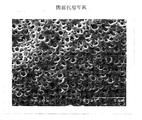
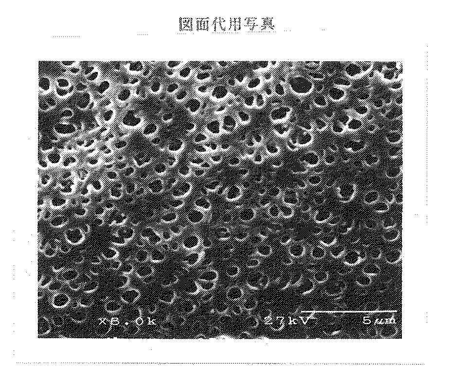
ポリスルホン系樹脂(Amoco社製:P−1700)17重量%、PVP(BASF社製:K90)7重量%、N,N−ジメチルアセトアミド(以下、DMACという)76重量%を50℃で8時間攪拌溶解、脱泡し製膜原液を得た。内部凝固液はDMAC15重量%と水85重量%とを混和して調製した。この製膜原液と内部凝固液を55度に保温した二重紡糸口金から吐出させ、フードで密閉した60cmの空中走行部を経て凝固浴に導入した。凝固浴は52.5℃の温水とし、フード内部は水蒸気の飽和状態にあった。凝固浴を通過させ、カセに巻き取った膜を熱水で洗浄した。さらに孔径保持剤として15重量%のグリセリン水溶液を付着させ、70℃で12時間乾燥処理を行った。得られた膜を膜面積1.5m2 のモジュ−ルにポリウレタンを用いて成型し、水を充填して25KGyのγ線を照射した。
この膜を図1に示すSEM写真をもとに画像処理した結果、外表面における孔面積0.5μm2 以上の孔の割合が3.0%、孔面積0.1μm2 以下の孔の割合が42.9%で、平均孔面積は0.16μm2 、開孔率は15.5%であった。この膜は固着がなく、成形性は良好であった。また、逆濾過液中のエンドトキシン濃度は検出限界以下(9.0EU/リットル以下)であったため、事実上侵入を認めなかった。
【0026】
【実施例2】
ポリスルホン系樹脂(Amoco社製:P−1700)17重量%、PVP(BASF社製:K90)4.5重量%、DMAC78.5重量%を混合し、50℃で8時間攪拌溶解、脱泡し製膜原液を得た。内部凝固液はDMAC20重量%と水80重量%とを混和して調製した。空中走行長を45cm、凝固浴温度を65℃とした以外は、実施例1と同条件で乾燥膜を得た。得られた膜を膜面積1.5m2 のモジュ−ルにポリウレタンを用いて成型し、水を充填して25KGyのγ線を照射した。
実施例1と同様に、SEM写真をもとに画像処理した結果、外表面における孔面積0.5μm2 以上の孔の割合が9.3%、孔面積0.1μm2 以下の孔の割合が39.6%で、平均孔面積が0.19μm2 、開孔率が10.6%であった。この膜も固着はなく、良好に成型できた。また、逆濾過液中のエンドトキシン濃度は検出限界以下(9.0EU/リットル以下)であったため、事実上侵入を認めなかった。
【0027】
【比較例1】
ポリスルホン系樹脂(Amoco社製:P−1700)17重量%、PVP(BASF社製:K90)9.0重量%、DMAC74.0重量%を混合し、50℃で8時間攪拌溶解、脱泡し製膜原液を得た。内部凝固液はDMAC20重量%と水80重量%とを混和して調製した。空中走行長を60cm、凝固浴温度を55℃とした以外は、実施例1と同条件で乾燥膜を得た。得られた膜を膜面積1.5m2 のモジュ−ルにポリウレタンを用いて成型し、水を充填して25KGyのγ線を照射した。
この膜は、外表面における孔面積0.5μm2 以上の孔の割合が0.8%、孔面積0.1μm2 以下の孔の割合が88.5%であり、平均孔面積は0.03μm2 、開孔率は3.3%であった。乾燥後は膜固着が激しく、そのままでは成型することができなかった。補修して成型後、菌塊逆濾過試験を実施したところ、逆濾過液中のエンドトキシン濃度は検出限界以下(9.0EU/リットル以下)と、事実上侵入を認めなかった。
【0028】
【比較例2】
ポリスルホン系樹脂(Amoco社製:P−1700)17重量%、PVP(BASF社製:K90)3.5重量%、DMAC79.5重量%を混合し、50℃で8時間攪拌溶解、脱泡し製膜原液を得た。内部凝固液はDMAC15重量%と水85重量%とを混和して調製した。空中走行長を45cm、凝固浴温度を25℃とした以外は、実施例1と同条件で乾燥膜を得た。得られた膜を膜面積1.5m2 のモジュ−ルにポリウレタンを用いて成型し、水を充填して25KGyのγ線を照射した。
この膜は、外表面における孔面積が0.5μm2 以上の孔の割合が47.5%、孔面積が0.1μm2 以下の孔の割合が18.8%で、平均孔面積は0.59μm2 、開孔率は35.8%であった。乾燥後の膜固着はなく、成型はできたが、ポッティング部全体に渡って糸流れが見られた。菌塊逆濾過試験を実施したところ、逆濾過率は0.13%であり、菌塊片の侵入によるエンドトキシンの逆濾過が認められた。
【0029】
【発明の効果】
本発明のポリスルホン系多孔質膜は、製造時に膜固着による成型不良を起こすことなく、しかも、透析液に含まれる菌塊片が膜内部へ侵入してエンドトキシンの逆濾過が事実上起こらないため、血液浄化分野で好適に使用できる。
【図面の簡単な説明】
【図1】本発明で得られる膜の一例として、実施例1で得られた膜のSEM写真(倍率6000倍)を示す。[0001]
BACKGROUND OF THE INVENTION
The present invention relates to a medical separation membrane for the purpose of removing blood waste products by extracorporeal circulation, and is used in the fields of blood purification, in particular, hemodialysis, blood filtration, and blood filtration dialysis for substituting renal function. It is what is done.
[0002]
[Prior art]
In recent years, dialysis therapy using a dialysis membrane is performed on patients who have low ability to remove waste products in the blood due to a decrease in renal function, thereby extending the life of the patients. On the other hand, with such prolonged dialysis therapy, a complication called dialysis amyloidosis has emerged. This is a disease in which a fiber protein called amyloid is deposited on ligaments, tendons, joints, etc. and causes various clinical symptoms. Since β 2 -microglobulin was identified as one of the proteins that make up this amyloid, removal of these low molecular weight proteins has become one of the therapeutic goals, and the market demand for high-performance dialysis membranes that enable this has increased. . The properties required for high-performance dialysis membranes include high removal performance of low molecular weight proteins typified by β 2 -microglobulin and excellent biocompatibility, but synthetic polymers are the membrane materials that satisfy these requirements. Polysulfone-based resins have attracted attention, and development of high-performance dialysis membranes mainly composed of polysulfone-based resins has been actively promoted.
[0003]
However, the polysulfone-based resin has high hydrophobicity, and as it is, the wettability is poor, so that the filtration performance cannot be sufficiently exhibited. Furthermore, when used in the field of blood purification as in the present invention, it is necessary to suppress the activation of the blood coagulation system, and a hydrophilic agent such as a hydrophilic polymer or glycerin is added to make the membrane surface hydrophilic. Often done. Since these hydrophilizing agents are present on the membrane surface, the hydrophilizing agent serves as a glue when drying in the manufacturing process, and as a result of sticking between adjacent membranes, poor molding due to poor penetration of the potting agent may occur. was there.
[0004]
An attempt to improve this drawback is disclosed, for example, in Japanese Patent Laid-Open No. 7-289863 as a technique for reducing the contact area between adjacent membranes by creating a large aperture on the outer surface of the hollow fiber membrane. However, the use of endotoxin cut filters dramatically improved the quality control of dialysate water, while dialysate contamination due to structural factors such as dialysate supply couplers still occurred and bacteria that had fallen off the coupler during use. There was a possibility that the lumps penetrated into the porous portion inside the membrane from the opening portion on the outer surface of the membrane. Moreover, in these high-performance dialysis membranes, endotoxins released from the bacterial clumps due to physical shock during entry may permeate the dense layer and migrate to the blood side to irritate the living body.
[0005]
[Problems to be solved by the invention]
An object of the present invention is to provide a polysulfone-based porous membrane that does not cause molding failure due to membrane adhesion during production and that does not allow bacterial clumps contained in the dialysate to enter the membrane.
[0006]
[Means for Solving the Problems]
As a result of diligent research to solve the above problems, the inventors of the present invention, in a hollow fiber membrane comprising a polysulfone-based resin and polyvinylpyrrolidone, having a dense layer on the inner surface side and an aperture on the outer surface, Setting the abundance ratio, average pore area, and open area ratio within a specific range not only prevents the hollow fibers from sticking to each other, but also increases the penetration of the bacterial clumps derived from the contaminated dialysate into the membrane. As a result, the present invention has been completed. That is, the present invention is a hollow fiber membrane comprising a polysulfone-based resin and polyvinyl pyrrolidone , having a dense layer on the inner surface side and an open portion on the outer surface, and the hollow fiber membrane is made of polyvinyl pyrrolidone from a double spinneret. It is obtained by discharging a film-forming stock solution having a ratio of 0.25 to 0.45 with respect to the polysulfone-based resin, and an internal coagulation liquid in which the solvent is 10 to 25% by weight and the balance is water, open porosity is 10-30% of the opening, the presence rate of pore area is 0.5 [mu] m 2 or more holes than 10% at the outer surface and the hole area are abundance of 0.1 [mu] m 2 or less holes it is 75% or less, the average pore area in and outer surface is related to the polysulfone porous membrane, which is a 0.05~0.35μm 2.
[0007]
The membrane of the present invention is composed of a polysulfone resin and a hydrophilic polymer. The main component constituting the membrane is a polysulfone resin, and the chemical structural formula (1) or (2) unit shown below is repeated. Consists of structure. In addition, so-called polysulfone derivatives in which a functional group or an alkyl group is bonded to an aromatic ring are also included in the scope of the present invention. In the formula, Ar represents a para-substituted divalent phenyl group.
-O-Ar-C (CH3) 3-Ar-O-Ar-SO3-Ar- (1)
-O-Ar-SO3-Ar- (2)
[0008]
The second component constituting the membrane is a hydrophilic polymer, and is added mainly for the purpose of hydrophilizing the membrane and forming pores. The hydrophilic polymer is preferably a vinyl polymer from the viewpoint that it is soluble in a common solvent with the polysulfone resin and has compatibility, for example, polyvinyl pyrrolidone, polyethylene glycol, polyamide, polyvinyl alcohol, ethylene vinyl alcohol copolymer. You can choose. Among them, polyvinyl pyrrolidone is most preferable because it has a moderate affinity with the polysulfone resin and can remain on the membrane surface and contribute to antithrombosis and filtration performance by hydrophilization. Regarding the content of these hydrophilic polymers, it is sufficient that the membrane surface is finally hydrophilized, so 3 to 12% by weight is sufficient. More preferably, it is 5 to 9% by weight. Therefore, the remaining part of the membrane, 88 to 97% by weight, is a polysulfone resin.
[0009]
The structure of the porous membrane of the present invention is a hollow fiber shape having a hollow part with an inner diameter of 80 to 400 μm and a film part with a thickness of 35 to 85 μm, and has sufficient pressure resistance and tensile strength for blood purification applications. . If the inner diameter is smaller than this, the resistance to blood flow increases and the blood flow velocity cannot be ensured. However, even if it becomes larger than necessary, the substance transfer efficiency in the blood decreases and the therapeutic effect decreases. On the other hand, if the film thickness is too thin, the strength cannot be maintained and causes crushing and leakage. If it is too thick, the mass transfer resistance in the film increases and the permeation performance decreases. This hollow fiber membrane has an asymmetric structure consisting of a dense layer having a separation function on the inner surface side and a rough dense layer as a support on the outer surface side, and has a controlled pore distribution on the outer surface in contact with the dialysate. The effect of the present invention is exhibited by having a hole portion having the opening.
[0010]
The pore distribution of the membrane of the present invention is quantified by image analysis of a scanning electron micrograph of the outer surface of the dry membrane. Specifically, the pore diameter maintaining material and the filling liquid adhering to the film are washed with water, and then the film freeze-dried from cold ethanol is vapor-deposited, and a photograph of the outer surface of the film is taken with an electron microscope at a magnification of 6000 times. This is printed in a size of 90 mm x 70 mm, the entire range of the photograph is taken into a personal computer using image analysis software, and the image is binarized to obtain the hole area for each aperture on the outer surface. Can do. As a result, the present inventors have found that there is a certain relationship between the distribution of the pore area, the average pore area, the amount of inoculum of the bacterial mass in the dialysate and the moldability, and the rate of openness and moldability. It was found that there is a certain relationship between them and it is necessary to control both.
[0011]
First of all, the abundance ratio of holes having a specific hole area will be described. The abundance ratio in the present invention is defined as a percentage of the total number of holes of an arbitrary hole area with respect to the total number of holes in the captured image. And is given by the following formula (3). Note that 10 pixels or less were regarded as noise and excluded from the count.
Usually, a part of the dialysate flows from the opening on the outer surface of the membrane into the porous part inside the membrane, but if the dialysate contains a bacterial mass derived from a coupler or the like, the bacterial mass penetrates into the membrane. Some of the endotoxins released from the bacterial clumps due to physical shock during invasion may pass through the dense layer and migrate to the blood side. Generally, since the major axis has a major axis of 1 to 3 μm, invasion of the bacterial mass hardly occurs when the pore area is 0.5 μm 2 or less. In order to effectively prevent the intrusion of the bacterial clumps, it is necessary to suppress the abundance ratio of pores having a pore area of 0.5 μm 2 or more to 10% or less, and more preferably to 7% or less. Most preferably, it is 5% or less. On the other hand, if the number of holes having a small hole area increases too much, a molding problem is likely to occur. In particular, when the number of pores having a pore area of 0.1 μm 2 or less increases, the adjacent membranes stick to each other, and the separation of the inside and outside of the hollow fiber membrane becomes incomplete due to poor penetration of the potting agent between the membranes. In order to eliminate such molding defects due to adhesion, it is necessary to keep the existence ratio of holes having a hole area of 0.1 μm 2 or less to 75% or less. More preferably, it is 60% or less, and most preferably 45% or less.
[0013]
Second, the average pore area will be described. The average pore area in the present invention is defined as an average value of the pore areas of all the holes in the captured image, and is given by the following formula (4). Again, 10 pixels or less were considered noise and excluded from the count.
Average pore area = total pore area in image / total number of pores in image (4)
The average pore area is related not only to the intrusion of fungal clumps but also to the moldability, particularly the adhesion between the membranes. This is because the smaller the film is, the stronger the tendency of adjacent films to stick together, which tends to cause molding defects. On the contrary, the larger the size, the more the bacterial mass invades, so it is necessary to keep it in the range of 0.05 to 0.35 μm 2 . More preferably 0.10~0.30μm 2, and most preferably from 0.10~0.20μm 2.
[0014]
On the other hand, in addition to these parameters, the porosity of the outer surface is also an important parameter for molding. The aperture ratio as used in the present invention is defined as a percentage of the total aperture area of the aperture portion with respect to the captured image area, and is given by the following equation (5). Again, 10 pixels or less were considered noise and were excluded from the count.
The porosity is greatly related to the contribution to the adhesion between the membranes. If the porosity is small, the contact area between adjacent membranes increases and the adhesion occurs. In severe cases, the entire bundle may even stick to a rod shape. . For this reason, it is necessary to ensure a hole area ratio of 10% or more. However, if the hole area ratio is increased unnecessarily, the film is bent in the longitudinal direction of the membrane, that is, the strength of the waist is impaired. As a result, molding defects frequently occur due to the yarn flow at the potting portion. In order not to impair the strength of the waist, the opening rate should be 30% as the upper limit, and therefore the range of the opening rate of the outer surface needs to be 10 to 30%. A more preferable range is 15 to 30%.
[0016]
Next, as a method for producing the polysulfone-based porous membrane of the present invention, a case where polyvinyl pyrrolidone (hereinafter referred to as PVP) is used as the hydrophilic polymer will be exemplified. The membrane-forming stock solution used for producing the membrane has three components, ie, a polysulfone resin, PVP, and a solvent, as basic constituent components. As the composition of the membrane forming stock solution, the concentration of the polysulfone resin may be in a range that has a viscosity capable of forming a film and can exhibit the characteristics as a membrane, and is usually 10 to 25% by weight, preferably 15 to 20% by weight. %. If it is less than 10% by weight, sufficient strength as a film cannot be obtained, and if it exceeds 25% by weight, the polymer density increases, the number of through holes decreases, and sufficient permeation performance cannot be obtained, which is not practical. These polysulfone resins having a weight average molecular weight of 1 to 50,000 are commercially available, and it is sufficient to use them. There is no particular limitation.
[0017]
PVP is mainly used for pore formation of a polysulfone-based porous membrane and for remaining to impart hydrophilicity. Surprisingly, as a result of the present inventors, it has been found that the ratio of PVP and polysulfone-based resin is involved in pore formation, particularly pore formation on the outer surface of the membrane. Although the detailed principle is still unclear, it seems that the main factor is that the molecular size of PVP is much larger than that of polysulfone resin. That is, when the ratio of PVP to polysulfone-based resin is lowered within a certain range, the viscosity of the discharged stock solution is lowered, the microphase separation speed due to the diffusion of PVP is accelerated, and the fusion of PVP vesicles proceeds. As a result, although the number is small, a hole having a relatively large area is formed. On the other hand, when the proportion of PVP increases, the fusion rate of PVP vesicles decreases due to an increase in the viscosity of the stock solution, while precipitation of polysulfone resin proceeds, resulting in the formation of many pores with a small area. Therefore, it is considered that the hole area ratio is also increased.
[0018]
In this way, when there are pores with a large area on the outer surface of the membrane, even if the number is small, endotoxin released by physical shock at the time of invasion of bacterial clumps in the dialysate from the pores. May pass through the dense layer and migrate to the blood side. On the other hand, when the number of holes having a small area increases, the adhesion between the films increases, the hole area ratio increases too much, and the strength of the film lowers, which causes molding defects. Therefore, in order to satisfy the above, the ratio of the PVP to the polysulfone resin in the membrane forming stock solution is preferably 0.25 to 0.45, and more preferably 0.30 to 0.40.
[0019]
Since various types of PVP are commercially available for different molecular weights, they may be used without any particular limitation. However, as described above, it is important for opening the outer surface and at the same time has the purpose of hydrophilizing the membrane surface. From this point of view, those which are likely to remain on the film surface at the time of film formation are preferred, and the higher the molecular weight, the more the tendency is. Therefore, it is preferable to use those having a weight average molecular weight of at least 100,000.
The solvent is a solvent that dissolves both the polysulfone resin and PVP, and is selected from dimethyl sulfoxide, N, N-dimethylacetamide, N, N-dimethylformamide, N-methyl-2-pyrrolidone, sulfolane, dioxane and the like. However, each of these combinations is arbitrary. A small amount of water or salts can be added for the purpose of controlling the coagulation rate.
[0020]
In order to obtain a polysulfone-based porous membrane using a membrane-forming stock solution comprising the above system, a known dry-wet method may be used. The film-forming stock solution and the internal coagulation liquid are simultaneously discharged from a double-pipe annular nozzle (double spinneret) kept at 30 to 60 ° C. and introduced into a coagulation bath. In that case, it is made to run in the air before it introduce | transduces into a coagulation bath from nozzle discharge. The aerial running length of the nozzle discharge surface and the coagulation bath surface is usually 10 to 100 cm, particularly preferably 30 to 85 cm. If it is shorter than 10 cm, the film of the present invention cannot be obtained because a dense layer is formed on the outer surface as a result of reaching the coagulation bath with incomplete coagulation. On the other hand, if the length exceeds 100 cm, yarn swaying may occur and bonding between incompletely solidified yarns may occur, which is not preferable in the manufacturing process.
[0021]
The atmosphere of the aerial traveling part is also important for achieving the present invention, and the periphery of the traveling part is enclosed and sealed with a hood, and the inside is kept in a wet state. In the wet state, water vapor generated from the lower coagulation bath is used, the temperature of the coagulation bath is adjusted in the range of 30 to 70 ° C., and the inside of the hood is saturated with water vapor. More preferably, it is the range of 45-60 degreeC.
It is better to use a lower internal coagulation liquid than one having high coagulation property with respect to the film-forming stock solution, and it is preferable to use a mixture of water and a solvent. The solvent is selected from N, N-dimethylacetamide, N, N-dimethylformamide, N-methyl-2-pyrrolidone, dimethyl sulfoxide and the like. A preferable composition of the internal coagulation liquid is 5 to 40% by weight of the solvent and the rest is water. When the ratio of water increases further, there is a possibility that sufficient water permeability as a membrane cannot be achieved. More preferably, the solvent is 10 to 25% by weight.
[0022]
The hollow fiber solidified as described above has an asymmetric porous structure having a dense layer on the inner surface side and an open portion on the outer surface. The hollow fiber membrane is wound around a casserole and cut into a fixed bundle length, and then the remaining solvent is washed with water, and then, for example, an aqueous glycerin solution is attached as a pore size retaining agent before drying treatment, and 70 to 80 ° C. If the drying treatment is carried out for 10 hours or more, the film of the present invention can be obtained.
When the membrane is used, both ends are potted with polyurethane or the like, molded into a module having a predetermined membrane area, and sterilized as necessary. The modularization may be performed according to a known method and is not particularly limited. The sterilization method may be selected from known methods according to the application. For example, treatment such as ethylene oxide gas sterilization, high-pressure steam sterilization, and radiation sterilization may be performed.
[0023]
DETAILED DESCRIPTION OF THE INVENTION
Next, although an example and a reference example explain the present invention in detail, the present invention is not limited to it. Various numerical values used in the examples were measured by the following procedure.
(Pore area on outer surface, presence rate of pores, and open area ratio) The membrane was washed with running water for 1 hour and then freeze-dried with ethanol containing dry ice. This film was fixed on a dedicated sample stage and silver was deposited, and then an outer surface photograph at a magnification of 6000 was taken with a scanning electron microscope (Hitachi: S-2460N, hereinafter referred to as SEM). For image processing, the photograph (90 mm × 70 mm) is captured by an image scanner, and the processing range (color magician 7, version 1.0, manufactured by Koshin Graphic Systams Co., Ltd.) is used to capture the entire range of the photograph with a resolution of 320. The brightness was 2,256 gradations. This image was binarized by processing software (NIH image, version 1.57), and the hole area of each hole was calculated. An image of 10 pixels or less was regarded as noise and excluded from the count. In addition, calibration was performed by simultaneously measuring a perfect hole in a membrane filter (made by Millipore: Isopore, hole diameter 2 μm) having a uniform hole diameter by electron beam irradiation.
[0024]
(Cell mass reverse filtration test) A dialysate (AK-Sorita DL, manufactured by Shimizu Pharmaceutical Co., Ltd.) was prepared using an aqueous solution containing a high cell mass produced in-house. The amount of bacterial mass used was substituted by measuring the endotoxin content. A circuit containing 15800 EU / liter of bacterial soil-contaminated dialysate as an endotoxin concentration was connected to the module dialysate inlet side, and the dialysate outlet side was plugged. A circuit was connected to the blood outlet side of the module, and a stopper was attached to the blood inlet side. A pump was set in the dialysate inlet side circuit, and 2 liters were reversely filtered to the blood outlet side at a flow rate of 200 cc / min and discharged, and then the reverse filtrate was collected from the blood outlet side. The amount of endotoxin contained in the collected reverse filtrate was quantified with Endospecy (manufactured by Seikagaku Corporation: ES-50 set), and the reverse filtration rate was calculated from the following formula (6). In the formula, C0 represents the endotoxin concentration in the dialysate, and C1 represents the endotoxin concentration in the reverse filtrate.
Reverse filtration rate (%) = (C1 / C0) × 100 (6)
[0025]
[Example 1]
A polysulfone resin (Amoco: P-1700) 17% by weight, PVP (BASF: K90) 7% by weight, N, N-dimethylacetamide (hereinafter referred to as DMAC) 76% by weight is stirred at 50 ° C. for 8 hours. Dissolution and defoaming were performed to obtain a film-forming stock solution. The internal coagulation liquid was prepared by mixing 15% by weight of DMAC and 85% by weight of water. The film-forming stock solution and the internal coagulation liquid were discharged from a double spinneret maintained at 55 ° C., and introduced into the coagulation bath through a 60 cm aerial running section sealed with a hood. The coagulation bath was warm water of 52.5 ° C., and the inside of the hood was saturated with water vapor. The membrane wound through a coagulation bath and wound on a cake was washed with hot water. Further, a 15% by weight glycerin aqueous solution was adhered as a pore size retaining agent, and a drying treatment was performed at 70 ° C. for 12 hours. The obtained membrane was molded into a module having a membrane area of 1.5 m 2 using polyurethane, filled with water and irradiated with 25 KGy of γ rays.
As a result of image processing of this film on the basis of the SEM photograph shown in FIG. 1, the ratio of holes having a hole area of 0.5 μm 2 or more on the outer surface is 3.0%, and the ratio of holes having a hole area of 0.1 μm 2 or less is It was 42.9%, the average pore area was 0.16 μm 2 , and the aperture ratio was 15.5%. This film did not adhere and had good moldability. Further, since the endotoxin concentration in the reverse filtrate was below the detection limit (9.0 EU / liter or less), virtually no invasion was observed.
[0026]
[Example 2]
Polysulfone resin (Amoco: P-1700) 17% by weight, PVP (BASF: K90) 4.5% by weight, DMAC 78.5% by weight were mixed, dissolved and defoamed with stirring at 50 ° C. for 8 hours. A film-forming stock solution was obtained. The internal coagulation liquid was prepared by mixing 20% by weight of DMAC and 80% by weight of water. A dry film was obtained under the same conditions as in Example 1 except that the air travel length was 45 cm and the coagulation bath temperature was 65 ° C. The obtained membrane was molded into a module having a membrane area of 1.5 m 2 using polyurethane, filled with water and irradiated with 25 KGy of γ rays.
As in Example 1, as a result of image processing based on SEM photographs, the ratio of holes having a hole area of 0.5 μm 2 or more on the outer surface was 9.3%, and the ratio of holes having a hole area of 0.1 μm 2 or less was The average pore area was 0.19 μm 2 and the aperture ratio was 10.6%. This film also did not stick and could be molded well. Further, since the endotoxin concentration in the reverse filtrate was below the detection limit (9.0 EU / liter or less), virtually no invasion was observed.
[0027]
[Comparative Example 1]
Polysulfone resin (Amoco: P-1700) 17% by weight, PVP (BASF: K90) 9.0% by weight, DMAC 74.0% by weight were mixed, dissolved and defoamed at 50 ° C. for 8 hours. A film-forming stock solution was obtained. The internal coagulation liquid was prepared by mixing 20% by weight of DMAC and 80% by weight of water. A dry film was obtained under the same conditions as in Example 1 except that the air travel length was 60 cm and the coagulation bath temperature was 55 ° C. The obtained membrane was molded into a module having a membrane area of 1.5 m 2 using polyurethane, filled with water and irradiated with 25 KGy of γ rays.
In this membrane, the ratio of the pores having a pore area of 0.5 μm 2 or more on the outer surface is 0.8%, the ratio of the pores having a pore area of 0.1 μm 2 or less is 88.5%, and the average pore area is 0.03 μm. 2 and the hole area ratio was 3.3%. After drying, the film adhered strongly and could not be molded as it was. After repairing and molding, the bacterial mass reverse filtration test was carried out. As a result, the endotoxin concentration in the reverse filtrate was below the detection limit (9.0 EU / liter or less), and virtually no invasion was observed.
[0028]
[Comparative Example 2]
Polysulfone resin (Amoco: P-1700) 17% by weight, PVP (BASF: K90) 3.5% by weight, DMAC 79.5% by weight were mixed, dissolved and defoamed at 50 ° C. for 8 hours with stirring. A film-forming stock solution was obtained. The internal coagulation liquid was prepared by mixing 15% by weight of DMAC and 85% by weight of water. A dry film was obtained under the same conditions as in Example 1 except that the air travel length was 45 cm and the coagulation bath temperature was 25 ° C. The obtained membrane was molded into a module having a membrane area of 1.5 m 2 using polyurethane, filled with water and irradiated with 25 KGy of γ rays.
In this membrane, the ratio of pores having a pore area of 0.5 μm 2 or more on the outer surface was 47.5%, the ratio of pores having a pore area of 0.1 μm 2 or less was 18.8%, and the average pore area was 0.00. It was 59 μm 2 and the hole area ratio was 35.8%. Although there was no film sticking after drying and molding was possible, yarn flow was observed throughout the potting part. When the bacterial mass reverse filtration test was performed, the reverse filtration rate was 0.13%, and endotoxin reverse filtration due to invasion of the bacterial mass fragments was observed.
[0029]
【The invention's effect】
The polysulfone-based porous membrane of the present invention does not cause molding defects due to membrane fixation during production, and the bacterial clumps contained in the dialysate enter the inside of the membrane, so that endotoxin reverse filtration does not actually occur. It can be suitably used in the blood purification field.
[Brief description of the drawings]
FIG. 1 shows an SEM photograph (magnification: 6000 times) of the film obtained in Example 1 as an example of the film obtained in the present invention.
Claims (1)
外表面における開孔部の開孔率が10〜30%、外表面における孔面積が0.5μm2以上の孔の存在率が10%以下、かつ、孔面積が0.1μm2以下の孔の存在率が75%以下であること、および外表面における平均孔面積が0.05〜0.35μm2であることを特徴とするポリスルホン系多孔質膜。A hollow fiber membrane comprising a polysulfone resin and polyvinyl pyrrolidone , having a dense layer on the inner surface side and an aperture on the outer surface, the hollow fiber membrane having a ratio of polyvinyl pyrrolidone to the polysulfone resin from the double spinneret It is obtained by discharging a film-forming stock solution of 0.25 to 0.45, and an internal coagulation solution in which the solvent is 10 to 25% by weight and the balance is water,
The hole area ratio of the hole portion on the outer surface is 10 to 30%, the hole area ratio of the hole area on the outer surface is 0.5 μm 2 or more, and the hole area is 0.1 μm 2 or less. that the presence of not more than 75%, Oyo polysulfone porous membrane, wherein the average pore area of beauty outer surface is 0.05~0.35μm 2.
Priority Applications (1)
| Application Number | Priority Date | Filing Date | Title |
|---|---|---|---|
| JP34116598A JP4265701B2 (en) | 1998-11-16 | 1998-11-16 | Polysulfone porous membrane |
Applications Claiming Priority (1)
| Application Number | Priority Date | Filing Date | Title |
|---|---|---|---|
| JP34116598A JP4265701B2 (en) | 1998-11-16 | 1998-11-16 | Polysulfone porous membrane |
Publications (2)
| Publication Number | Publication Date |
|---|---|
| JP2000140589A JP2000140589A (en) | 2000-05-23 |
| JP4265701B2 true JP4265701B2 (en) | 2009-05-20 |
Family
ID=18343852
Family Applications (1)
| Application Number | Title | Priority Date | Filing Date |
|---|---|---|---|
| JP34116598A Expired - Lifetime JP4265701B2 (en) | 1998-11-16 | 1998-11-16 | Polysulfone porous membrane |
Country Status (1)
| Country | Link |
|---|---|
| JP (1) | JP4265701B2 (en) |
Families Citing this family (16)
| Publication number | Priority date | Publication date | Assignee | Title |
|---|---|---|---|---|
| JP3594032B1 (en) | 2003-08-29 | 2004-11-24 | 東洋紡績株式会社 | Highly permeable hollow fiber membrane blood purifier |
| JP3551971B1 (en) | 2003-11-26 | 2004-08-11 | 東洋紡績株式会社 | Polysulfone permselective hollow fiber membrane |
| JP3642065B1 (en) | 2004-03-22 | 2005-04-27 | 東洋紡績株式会社 | Permselective separation membrane and method for producing a selectively permeable separation membrane |
| JP4501530B2 (en) * | 2004-05-19 | 2010-07-14 | 東洋紡績株式会社 | Highly permeable hollow fiber membrane blood purifier |
| JP4587024B2 (en) * | 2004-05-20 | 2010-11-24 | 東洋紡績株式会社 | Polysulfone-based selectively permeable hollow fiber membrane with excellent blood compatibility |
| JP4666248B2 (en) * | 2004-05-27 | 2011-04-06 | 東洋紡績株式会社 | High strength high water permeability hollow fiber membrane blood purifier |
| JP2005334428A (en) * | 2004-05-28 | 2005-12-08 | Toyobo Co Ltd | High water permeability hollow fiber membrane type hemocatharsis apparatus with superior hemocompatibility |
| JP4587025B2 (en) * | 2004-06-01 | 2010-11-24 | 東洋紡績株式会社 | Polysulfone-based selectively permeable hollow fiber membrane with excellent blood compatibility |
| JP4706194B2 (en) * | 2004-06-01 | 2011-06-22 | 東洋紡績株式会社 | Highly permeable hollow fiber membrane blood purifier |
| JP4288602B2 (en) * | 2004-06-09 | 2009-07-01 | 東洋紡績株式会社 | Polysulfone permselective hollow fiber membrane |
| JP4599934B2 (en) * | 2004-08-10 | 2010-12-15 | 東洋紡績株式会社 | Hollow fiber membrane module |
| EP1795254B1 (en) | 2004-08-10 | 2014-05-21 | Nipro Corporation | Process for production of a polysulfone type selectively permeable hollow fiber membrane module |
| JP4748350B2 (en) * | 2004-12-27 | 2011-08-17 | 東洋紡績株式会社 | Method for producing polysulfone-based hollow fiber membrane |
| JP4843992B2 (en) * | 2005-04-26 | 2011-12-21 | 東洋紡績株式会社 | Blood purifier |
| JP4493097B2 (en) * | 2005-10-14 | 2010-06-30 | 旭化成クラレメディカル株式会社 | Continuous blood filtration device |
| DE102007019051B3 (en) * | 2007-04-23 | 2008-10-09 | Fresenius Medical Care Deutschland Gmbh | Hollow fiber capillary membrane and process for its preparation |
-
1998
- 1998-11-16 JP JP34116598A patent/JP4265701B2/en not_active Expired - Lifetime
Also Published As
| Publication number | Publication date |
|---|---|
| JP2000140589A (en) | 2000-05-23 |
Similar Documents
| Publication | Publication Date | Title |
|---|---|---|
| US5232601A (en) | High flux hollow fiber membrane | |
| JP4265701B2 (en) | Polysulfone porous membrane | |
| JP3047403B2 (en) | Permeation-selective asymmetric membrane suitable for hemodialysis and method for producing the membrane | |
| CN1125669C (en) | Polysulfone hollow fiber semipermeable membrane | |
| CN1032043C (en) | Polysulfone-based hollow fiber membrane and process for manufacturing the same | |
| RU2667068C2 (en) | Porous membrane, blood purifying module incorporating porous membrane and method for producing porous membrane | |
| US6042783A (en) | Hollow yarn membrane used for blood purification and blood purifier | |
| WO2004024216A1 (en) | Plasma purification membrane and plasma purification system | |
| TW200938239A (en) | Microporous hollow fiber membrane for blood treatment | |
| US10888823B2 (en) | Membrane with improved permeability and selectivity | |
| US20210245108A1 (en) | Microporous Membrane And Methods To Make Same | |
| JPWO2000027447A1 (en) | blood purifier | |
| JP3714686B2 (en) | Polysulfone-based hollow fiber membrane and method for producing the same | |
| JP2703266B2 (en) | Polysulfone hollow fiber membrane and method for producing the same | |
| JP4683402B2 (en) | Hollow fiber membrane for blood purification, method for producing the same, and blood purifier | |
| JPH09308685A (en) | Hollow fiber membrane for blood purification and blood purifier | |
| JP3431622B1 (en) | High-performance plasma purification membrane | |
| JP4190361B2 (en) | Hollow fiber type body fluid treatment device, hollow fiber bundle used therefor, and method for producing them | |
| JPH0970431A (en) | Method for producing polysulfone hollow fiber type artificial kidney and artificial kidney | |
| JP2005058906A (en) | Polymer porous membrane, blood purifier, and method for producing polymer porous membrane | |
| JP2005224604A (en) | Hemocatharsis membrane and hemocatharsis apparatus using the same | |
| JP3334705B2 (en) | Polysulfone-based selectively permeable hollow fiber membrane | |
| JP4093134B2 (en) | Hollow fiber blood purification membrane | |
| JP2001038171A (en) | Hollow fiber membrane | |
| JPWO1998034719A1 (en) | Hollow fiber membrane and its manufacturing method |
Legal Events
| Date | Code | Title | Description |
|---|---|---|---|
| A621 | Written request for application examination |
Free format text: JAPANESE INTERMEDIATE CODE: A621 Effective date: 20050826 |
|
| A977 | Report on retrieval |
Free format text: JAPANESE INTERMEDIATE CODE: A971007 Effective date: 20070712 |
|
| A131 | Notification of reasons for refusal |
Free format text: JAPANESE INTERMEDIATE CODE: A131 Effective date: 20070731 |
|
| A521 | Written amendment |
Free format text: JAPANESE INTERMEDIATE CODE: A523 Effective date: 20070928 |
|
| RD02 | Notification of acceptance of power of attorney |
Free format text: JAPANESE INTERMEDIATE CODE: A7422 Effective date: 20070928 |
|
| TRDD | Decision of grant or rejection written | ||
| A01 | Written decision to grant a patent or to grant a registration (utility model) |
Free format text: JAPANESE INTERMEDIATE CODE: A01 Effective date: 20090210 |
|
| A01 | Written decision to grant a patent or to grant a registration (utility model) |
Free format text: JAPANESE INTERMEDIATE CODE: A01 |
|
| A61 | First payment of annual fees (during grant procedure) |
Free format text: JAPANESE INTERMEDIATE CODE: A61 Effective date: 20090210 |
|
| R150 | Certificate of patent or registration of utility model |
Free format text: JAPANESE INTERMEDIATE CODE: R150 |
|
| FPAY | Renewal fee payment (event date is renewal date of database) |
Free format text: PAYMENT UNTIL: 20120227 Year of fee payment: 3 |
|
| FPAY | Renewal fee payment (event date is renewal date of database) |
Free format text: PAYMENT UNTIL: 20120227 Year of fee payment: 3 |
|
| FPAY | Renewal fee payment (event date is renewal date of database) |
Free format text: PAYMENT UNTIL: 20130227 Year of fee payment: 4 |
|
| FPAY | Renewal fee payment (event date is renewal date of database) |
Free format text: PAYMENT UNTIL: 20130227 Year of fee payment: 4 |
|
| S111 | Request for change of ownership or part of ownership |
Free format text: JAPANESE INTERMEDIATE CODE: R313111 |
|
| FPAY | Renewal fee payment (event date is renewal date of database) |
Free format text: PAYMENT UNTIL: 20130227 Year of fee payment: 4 |
|
| R350 | Written notification of registration of transfer |
Free format text: JAPANESE INTERMEDIATE CODE: R350 |
|
| FPAY | Renewal fee payment (event date is renewal date of database) |
Free format text: PAYMENT UNTIL: 20140227 Year of fee payment: 5 |
|
| EXPY | Cancellation because of completion of term |