JP4256169B2 - Method for determining the prognosis of cancer patients using TUCAN - Google Patents
Method for determining the prognosis of cancer patients using TUCAN Download PDFInfo
- Publication number
- JP4256169B2 JP4256169B2 JP2002588143A JP2002588143A JP4256169B2 JP 4256169 B2 JP4256169 B2 JP 4256169B2 JP 2002588143 A JP2002588143 A JP 2002588143A JP 2002588143 A JP2002588143 A JP 2002588143A JP 4256169 B2 JP4256169 B2 JP 4256169B2
- Authority
- JP
- Japan
- Prior art keywords
- tucan
- level
- reagent
- biomarker
- patients
- Prior art date
- Legal status (The legal status is an assumption and is not a legal conclusion. Google has not performed a legal analysis and makes no representation as to the accuracy of the status listed.)
- Expired - Fee Related
Links
Images


Classifications
-
- G—PHYSICS
- G01—MEASURING; TESTING
- G01N—INVESTIGATING OR ANALYSING MATERIALS BY DETERMINING THEIR CHEMICAL OR PHYSICAL PROPERTIES
- G01N2800/00—Detection or diagnosis of diseases
- G01N2800/52—Predicting or monitoring the response to treatment, e.g. for selection of therapy based on assay results in personalised medicine; Prognosis
Abstract
Description
本発明は、国立衛生研究所により授与された、助成金番号AG15402、CA69381、およびNS36821の下の政府の補助を用いてなされた。合衆国政府は、本発明において特定の権利を有する。 This invention was made with government support under grant numbers AG15402, CA69381, and NS36821 awarded by the National Institutes of Health. The United States government has certain rights in this invention.
(発明の背景)
本発明は、一般に、癌、より詳細には、癌の診断または予後のために用いられ得るバイオマーカーに関する。
(Background of the Invention)
The present invention relates generally to cancer, and more particularly to biomarkers that can be used for the diagnosis or prognosis of cancer.
癌は、毎年、診断される100万人より多くの人、ならびに彼等の家族および友人に根深く影響する、主要な公衆衛生上の問題のままである。米国の人々が、成長し、そして年をとるにつれて、より多くの人々が、癌に罹患する。癌を早期に検出するためのスクリーニング試験の使用は、しばしば、より副作用の少ない、より効果的な処置を導く。早期に癌が見出された患者はまた、症状が現れるまで癌が見出されなかった患者よりも、これらの癌により死亡する確率が低いようである。 Cancer remains a major public health problem that deeply affects more than one million people diagnosed each year, as well as their families and friends. As people in the United States grow and age, more and more people suffer from cancer. The use of screening tests for early detection of cancer often leads to more effective treatments with fewer side effects. Patients who have found cancer early also appear to be less likely to die from these cancers than patients who have not found cancer until symptoms appear.
1つの型の癌のスクリーニング試験は、患者から得られた流体または組織中のバイオマーカー(例えば、腫瘍マーカー)の検出を含む。腫瘍マーカーは、癌細胞により産生される物質であって、代表的には正常な細胞により産生されない物質である。これらの物質は、一般的に、癌に罹患した患者の体液または組織において検出され得る。不幸なことに、いくつかの腫瘍マーカーはまた、癌に罹患していない人の体液または組織において有意な量で検出され得、このことは、特定のマーカーを、診断について信頼性の低いものにする。それにも関らず、腫瘍マーカーは、癌を診断するための重要なツールのままである。 One type of cancer screening test involves the detection of biomarkers (eg, tumor markers) in fluids or tissues obtained from patients. A tumor marker is a substance that is produced by cancer cells and is typically not produced by normal cells. These substances can generally be detected in body fluids or tissues of patients suffering from cancer. Unfortunately, some tumor markers can also be detected in significant amounts in the body fluids or tissues of people who do not have cancer, which makes certain markers unreliable for diagnosis To do. Nevertheless, tumor markers remain an important tool for diagnosing cancer.
腫瘍マーカーについての別の重要な使用は、進行した癌について処置されている患者をモニタリングするための使用である。この目的のために腫瘍マーカーを測定することは、治療が、癌を減少させているか否かを決定するために、胸部X腺、コンピューター断層撮影(CT)スキャン、骨のスキャン、または他の複雑な試験を繰り返すより侵襲性が低く、より時間がかからず、かつより費用がかからないものであり得る。 Another important use for tumor markers is for monitoring patients being treated for advanced cancer. Measuring tumor markers for this purpose may include determining whether a treatment is reducing cancer, a chest X-gland, a computed tomography (CT) scan, a bone scan, or other complex. Can be less invasive, less time consuming, and less expensive than repeating a simple test.
腫瘍マーカーについてのさらなる重要な使用は、癌患者の生存の予後を決定するための使用である。そのような予後方法は、外科的処置された患者にさらなる治療的選択が与えられ得るように、癌の再発を経験する可能性のある患者を特定するために用いられ得る。生存の予後のために有用なバイオマーカーはまた、検査または手術のときに測定可能な転移を示していない患者における転移の危険性を決定するために特に有効であり得る。癌患者における転移の可能性の知見は、処置の選択肢の選択における重要な因子であり得る。例えば、転移を経験する可能性がある癌患者は、特に攻撃的な様式を用いて有利に処置され得る。 A further important use for tumor markers is the use to determine the prognosis of cancer patient survival. Such prognostic methods can be used to identify patients who are likely to experience a recurrence of cancer, so that additional therapeutic options can be given to surgically treated patients. Biomarkers useful for prognosis of survival can also be particularly effective in determining the risk of metastasis in patients who do not show measurable metastases at the time of examination or surgery. Knowledge of the possibility of metastasis in cancer patients can be an important factor in selecting treatment options. For example, cancer patients who may experience metastasis can be advantageously treated using a particularly aggressive manner.
従って、癌のための診断指標または予後指標として用いられ得るバイオマーカーを同定する必要性が存在する。本発明は、この必要性を満たし、そして関連する利点も提供する。 Thus, there is a need to identify biomarkers that can be used as diagnostic or prognostic indicators for cancer. The present invention fulfills this need and provides related advantages.
(発明の要旨)
本発明は、癌患者の生存についての予後を決定するための方法を提供する。1つの方法は、以下:(a)癌患者由来の新生物細胞含有サンプルのTUCANのレベルを測定する工程、および(b)サンプル中のTUCANのレベルと、参照のTUCANのレベルとを比較する工程を包含し、ここで、サンプル中の低レベルのTUCANは、患者の増大した生存と相関する。
(Summary of the Invention)
The present invention provides a method for determining the prognosis for survival of cancer patients. One method includes: (a) measuring the level of TUCAN in a neoplastic cell-containing sample from a cancer patient, and (b) comparing the level of TUCAN in the sample with the level of a reference TUCAN. Where low levels of TUCAN in the sample correlate with increased patient survival.
癌患者の生存についての予後を決定するための別の方法は、以下:(a)癌患者由来の新生物細胞含有サンプルにおける、TUCANおよび1つ以上のバイオマーカーのレベルを測定する工程であって、1つ以上のバイオマーカーが、cIAP2、Apaf1、Bcl−2、およびSmacからなる群より選択される工程、ならびに(b)サンプル中のTUCANおよび1つ以上の選択されたバイオマーカーのレベルと、TUCANおよび1つ以上の選択されたバイオマーカーの参照のレベルとを比較する工程を包含し、ここでサンプル中の低レベルのTUCANおよび高レベルのApaf1、Bcl−2またはSmacのいずれか、あるいは低レベルのTUCANおよび低レベルのcIAP2は、該患者の増大した生存に対応する。 Another method for determining the prognosis for survival of a cancer patient is the following: (a) measuring the level of TUCAN and one or more biomarkers in a neoplastic cell-containing sample from a cancer patient, The one or more biomarkers are selected from the group consisting of cIAP2, Apaf1, Bcl-2, and Smac, and (b) the level of TUCAN and one or more selected biomarkers in the sample; Comparing the level of reference to TUCAN and one or more selected biomarkers, wherein a low level of TUCAN and a high level of Apaf1, Bcl-2 or Smac in the sample, or low Levels of TUCAN and low levels of cIAP2 correspond to increased survival of the patient.
癌患者の生存についての予後を決定するさらなる方法は、以下:(a)該癌患者由来の新生物細胞含有サンプルにおけるTUCANのレベルを測定する工程、および(b)患者を、第1のグループの患者または第2のグループの患者のいずれかに属するとして分類する工程であって、低レベルのTUCANを有する第1のグループの患者が、高レベルのTUCANを有する第2のグループの患者よりも、増大した生存可能性を有するとして分類される、工程を包含する。 Additional methods for determining the prognosis for survival of a cancer patient include the following: (a) measuring the level of TUCAN in a neoplastic cell-containing sample from the cancer patient, and (b) the patient from the first group. Categorizing as belonging to either a patient or a second group of patients, wherein a first group of patients having a low level of TUCAN is more than a second group of patients having a high level of TUCAN Includes a step classified as having increased viability.
本発明はまた、癌を有する患者の処置経過の有効性をモニタリングするための方法を提供する。この方法は、(a)処置の前の癌患者由来の新生細胞含有サンプル中のTUCANレベルを決定する工程、および(b)処置後の患者由来の新生細胞含有サンプル中のTUCANレベルを決定し、それにより、処置前のTUCANレベルと処置後のTUCANレベルとの比較によって処置の有効性を示す工程を包含する。 The present invention also provides a method for monitoring the effectiveness of a treatment course in a patient with cancer. The method includes (a) determining TUCAN levels in a neoplastic cell-containing sample from a cancer patient prior to treatment; and (b) determining TUCAN levels in a neoplastic cell-containing sample from a patient after treatment; Thereby, including the step of showing the effectiveness of the treatment by comparing the TUCAN level before treatment with the TUCAN level after treatment.
(発明の詳細な説明)
本発明は、タンパク質を含有するCARDドメイン(PCT公開 WO01/16170中でCARD−Xとして公知である)、TUCAN(カスパーゼ9の腫瘍アップレギュレートCARD含有アンタゴニスト(Tumor Up−regulated CARD−containing Antagonist of Caspase Nine))の発現が、独立してかまたは他のバイオマーカーと組み合わせて、癌を有する患者についての臨床的な結果を効果的に予測するために使用され得るという知見に関する。
(Detailed description of the invention)
The present invention relates to CARD domains containing proteins (known as CARD-X in PCT Publication WO 01/16170), TUCAN (Tumor Up-regulated CARD-containing Antagonist of Caspase-9). It relates to the finding that the expression of Nine)) can be used to effectively predict clinical outcomes for patients with cancer, either independently or in combination with other biomarkers.
本発明の予後診断法は、患者が再発のおそれにある場合の測定のために有用である。癌再発は、種々の型の癌に関連する懸念である。例えば、結腸癌の外科的除去を受けた患者のうち、第II病期の結腸癌を有する患者の25%〜40%、および第III病期の結腸癌を有する患者の約50%が、癌の再発を経験する。癌の再発についての1つの説明は、比較的初期段階の疾患(例えば、第II病期または第III病期)を有する患者は、すでに手術によって除去されなかった、罹患した臓器の外側に広がった少量の癌を有しているということがある。これらの癌細胞は、微小転移巣と呼ばれ、現在利用可能である試験では一般的には検出し得ない。本発明の予後診断法は、癌再発しそうな外科的に処置された患者を同定するために使用され得、その結果、化学療法、照射、生物学的改変剤および他の適切な治療法のような手術前または手術後の補助を含む、さらなる治療オプションを提供し得る。この方法は、試験または手術のときに、測定可能な転位を示さない患者における転位の危険性を測定するために特に有効である。 The prognosis method of the present invention is useful for measurement when a patient is at risk of recurrence. Cancer recurrence is a concern associated with various types of cancer. For example, among patients who have undergone surgical removal of colon cancer, 25% to 40% of patients with stage II colon cancer and about 50% of patients with stage III colon cancer have cancer Experience relapse. One explanation for cancer recurrence is that patients with relatively early stage disease (eg, stage II or stage III) have spread outside the affected organs that have not been removed by surgery. May have a small amount of cancer. These cancer cells are called micrometastasis and are generally not detectable in currently available tests. The prognostic methods of the present invention can be used to identify surgically treated patients who are likely to relapse cancer, such as chemotherapy, irradiation, biological modifiers and other suitable therapies. Additional treatment options may be provided, including pre-operative or post-operative assistance. This method is particularly effective for measuring the risk of dislocation in patients who do not show measurable dislocation during testing or surgery.
本発明の予後診断法はまた、癌を有する患者のための適切な処置工程を決定するために有用である。処置工程とは、診断後または癌の処置後の患者のために行なわれる治療測定のことをいう。例えば、癌の再発、拡散、または患者生存についての可能性の決定は、治療に対してより保存的もしくはより根治的アプローチが行なわれるべきか否か、または処置様式が組み合わせられるべきか否かを決定するのを助け得る。例えば、癌再発が起こりそうな場合、化学療法、照射、免疫療法、生物学的改変剤療法、遺伝子治療、ワクチンなどによる手術前の処置もしくは手術後の処置、または患者が処置される時間幅を調節するのに有利であり得る。 The prognostic methods of the present invention are also useful for determining the appropriate treatment steps for patients with cancer. A treatment step refers to a therapeutic measurement performed for a patient after diagnosis or after treatment for cancer. For example, determining the likelihood of cancer recurrence, spread, or patient survival will determine whether a more conservative or more radical approach to therapy should be taken, or whether treatment modalities should be combined You can help decide. For example, if cancer recurrence is likely, pre- or post-surgical treatment with chemotherapy, irradiation, immunotherapy, biological modifier therapy, gene therapy, vaccines, etc. It may be advantageous to adjust.
本明細書中の実施例IIおよびVIIIに開示されるように、高められたレベルのTUCANは、それぞれ、結腸腫瘍試料の49%および64%で見出された。単変量解析を使用して、より長期の疾患に罹患していない生存(DFS)とTUCANの低い発現(p=0.0004)との間の有意な相関を測定した。実施例IVに示されるように、腫瘍が低レベルのTUCANを含んでいる患者の78%(50人中39人)は、このタンパク質の高い発現を有する患者においては44%(48人中21人)のみであるのに比べ、この研究によってカバーされる時間の間、生存しかつ疾患を有さないままであった。実施例VIIIはまた、TUCAN免疫染色が、生存していた患者と比べて、結腸癌で死亡した患者においてより有意に高いことを示す。 As disclosed in Examples II and VIII herein, elevated levels of TUCAN were found in 49% and 64% of colon tumor samples, respectively. Univariate analysis was used to determine a significant correlation between survival without longer disease (DFS) and low expression of TUCAN (p = 0.0004). As shown in Example IV, 78% (39/50) of patients whose tumors contain low levels of TUCAN are 44% (21/48) of patients with high expression of this protein. ) Only survived and disease free for the time covered by this study. Example VIII also shows that TUCAN immunostaining is significantly higher in patients who died of colon cancer compared to those who were alive.
実施例IVに示されるように、5年間のメジアンの追跡において、TUCANの高い発現を有する患者の49%は、結腸癌の再発を有するかまたは結腸癌によって死亡し、そして腫瘍がこのタンパク質を低レベルで発現する患者のグループにおいては、19%のみが再発し
4%が疾患で亡くなった。多変量解析は、高いTUCANの存在が、結腸癌に由来する死の危険性を17倍増加させたことを示した(p=000004)。従って、癌を有する患者由来のサンプル中の高レベルのTUCANは、腫瘍転位の増加された可能性および減少された生存に関連する。同様に、癌を有する患者由来のサンプル中の低レベルのTUCANは、腫瘍転位の減少された可能性および生存の増加された可能性に関連する。
As shown in Example IV, in 5 years of median follow-up, 49% of patients with high expression of TUCAN have recurrence of colon cancer or die from colon cancer, and tumors reduce this protein In the group of patients who developed at the level, only 19% relapsed and 4% died of the disease. Multivariate analysis showed that the presence of high TUCAN increased the risk of death from colon cancer by 17-fold (p = 000004). Thus, high levels of TUCAN in samples from patients with cancer are associated with increased likelihood of tumor metastasis and decreased survival. Similarly, low levels of TUCAN in samples from patients with cancer are associated with reduced likelihood of tumor metastasis and increased survival.
低レベルのcIAP2および低レベルのTUCANの組み合わせが、非常に都合よい結果を有する初期段階の結腸癌のサブグループを同定したことがまた、本明細書中で開示される。同齢集団の92人の患者中の約3分の1の患者が、低レベルのcIAP2および低レベルのTUCANの組み合わせを有していた(33/92(36%))。これらの33人の患者のうち、32人(97%)が生存し、かつ30人(91%)が本研究によって対象とされる時間において疾患を有さなかったが、これとは対照的に、他のカテゴリーでは、56%が生存し、44%が疾患を有さなかった。同様に、同齢集団の81人の患者中の17人が、高レベルのApaf1発現および低レベルのTUCAN発現の組み合わせを有した。高レベルのApaf1発現および低レベルのTUCAN発現を特徴とする全ての患者(17人)は、この特徴によって特徴付けられなかった患者については65%(81人中53人)のみが生存しかつ53%(81人中43人)で再発しなかったこと比べ、この調査の終わりにおいても生存しかつ再発も有さなかった。従って、癌を有する患者由来のサンプル中の高レベルのcIAP2または低レベルのApaf1と組み合わせた高レベルのTUCANは、腫瘍転位の増加された可能性および生存の減少された可能性に関連する。それに対して、低レベルのcIAP2または高レベルのApaf1と組み合わせた低レベルのTUCANは、腫瘍転位の減少された可能性および生存の増加された可能性に関連する。 It is also disclosed herein that the combination of low levels of cIAP2 and low levels of TUCAN has identified an early stage colon cancer subgroup with very favorable results. Approximately one third of the 92 patients in the age group had a combination of low levels of cIAP2 and low levels of TUCAN (33/92 (36%)). Of these 33 patients, 32 (97%) survived and 30 (91%) had no disease at the time covered by this study, in contrast. In the other categories, 56% survived and 44% had no disease. Similarly, 17 out of 81 patients in the age group had a combination of high levels of Apaf1 expression and low levels of TUCAN expression. All patients (17) characterized by high levels of Apaf1 expression and low levels of TUCAN expression, only 65% (53/81) of those who were not characterized by this feature survived and 53 % Survived at the end of the study and had no recurrence compared to no recurrence (43/81). Thus, high levels of TUCAN in combination with high levels of cIAP2 or low levels of Apaf1 in samples from patients with cancer are associated with increased likelihood of tumor metastasis and reduced survival. In contrast, low levels of TUCAN combined with low levels of cIAP2 or high levels of Apaf1 are associated with reduced likelihood of tumor translocation and increased likelihood of survival.
これらの結果に基づいて、本発明は、新生物状態の診断、癌に罹患している患者の生存の予知、およびバイオマーカーとしてTUCANを使用する癌の段階の決定のための方法を提供する。TUCANは、単独でかまたは癌に罹患している患者の生存を予知するための特異的バイオマーカーのような他の予知インディケーターと組み合わせて使用され得る。 Based on these results, the present invention provides methods for the diagnosis of neoplastic conditions, the prediction of survival of patients suffering from cancer, and the determination of the stage of cancer using TUCAN as a biomarker. TUCAN can be used alone or in combination with other predictive indicators such as specific biomarkers to predict the survival of patients suffering from cancer.
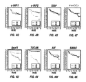
本明細書中に開示されるように、Apaf1、サルビビン(Survivin)、XIAP、cIAP1、およびcIAP2の高められたレベルが、それぞれ結腸腫瘍試料の38%、54%、74%、61%および35%において見出された。単変量解析を使用して、より長い疾患に罹患していない生存(DFS)と、cIAP2(p=0.0002)、β−カテニン(p=0.04)、変異p53タンパク質(p=0.03)の低レベルの発現との間、またはより長い疾患に罹患していない生存(DFS)Apaf1(p=0.00008)、Bcl−2(p=0.005)、およびSMAC(p=0.03)の高レベルの発現突然変異誘発との間の優位な相関を決定した(図4aを参照のこと)。従って、腫瘍が低レベルのTUCANを含んでいる患者の78%(50人中39人)は、このタンパク質の高レベルの発現を有する患者の44%(48人中21人)と比べ、この研究によって対象とされる時間の間、生存したままでありかつ疾患を有さなかった。同様に、高いcIAP2レベルを有する患者において36%(33人中12人)のみ癌を有していなかったが、低レベルのcIAP2発現者(expressor)のうちの74%(61人中45人)が、この調査の終わりに癌を有していなかった。5年間のメジアンの追跡において、高いcIAP2レベルを有する患者の60%が再発し、そして46%が結腸癌によって死亡した。それに対して低レベルのcIAP2グループにおいては、20%が再発し、18%が結腸癌に関連して死亡した。 As disclosed herein, elevated levels of Apaf1, Survivin, XIAP, cIAP1, and cIAP2 are 38%, 54%, 74%, 61%, and 35% of colon tumor samples, respectively. Found in. Using univariate analysis, survival without longer disease (DFS), cIAP2 (p = 0.0002), β-catenin (p = 0.04), mutant p53 protein (p = 0.0). 03) low level expression or survival without longer disease (DFS) Apaf1 (p = 0.00008), Bcl-2 (p = 0.005), and SMAC (p = 0) (0.03) was determined to have a dominant correlation with high levels of expression mutagenesis (see FIG. 4a). Thus, 78% (39/50) of patients whose tumors contain low levels of TUCAN compared to 44% (21/48) of patients with high levels of expression of this protein. Remained alive and disease free for the time covered by. Similarly, only 36% (12/33) of patients with high cIAP2 levels did not have cancer, but 74% (45/61) of low level cIAP2 expressors However, he did not have cancer at the end of this study. At 5 years of median follow-up, 60% of patients with high cIAP2 levels relapsed and 46% died from colon cancer. In contrast, in the low level cIAP2 group, 20% relapsed and 18% died in connection with colon cancer.
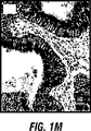
本明細書中でさらに開示されるように、高レベルのApaf1はより長い生存に関連し、低レベルのApaf1発現を有する患者において62人中28人(45%)しか疾患を有さなかったが、結腸癌患者の38人中33人(87%)が疾患を有さないままであった。対照的に、低レベルのApaf1を有する患者のうちの43%が再発し、そして35%が結腸癌によって死亡した。一方、14%のみが、高レベルのApaf1同齢集団において癌再発を有するかまたは死亡した。低レベルのBcl−2もまた、乏しい全体としての生存と関連した。高レベルのBcl−2グループにおいて死亡した患者においては24%(76人中18人)が結腸癌によって死亡したのに対し、このタンパク質の低い発現を有する18人の患者のうちの11人(61%)が結腸癌によって死亡した。同様に、腫瘍が低レベルのApaf1染色を含む患者は、Bcl−2を過剰発現した患者と比較してより悪い全生存を有した(図1N)。多変量解析は、高レベルのApaf1およびBcl−2発現が、75%(p=0.004)および82%(p=0.00006)の差をつけた、結腸癌による死の減少に関連することを示した。従って、結腸癌を有する患者由来のサンプル中の、減少されたレベルのApaf1またはBcl−2は、腫瘍転位の増加された発生率および減少された生存数に確実に関連する。 As further disclosed herein, high levels of Apaf1 are associated with longer survival, although only 28 of 62 (45%) had disease in patients with low levels of Apaf1 expression. Of the 38 patients with colon cancer, 33 (87%) remained disease free. In contrast, 43% of patients with low levels of Apaf1 relapsed and 35% died from colon cancer. On the other hand, only 14% had cancer recurrence or died in the high level Apaf1 cohort. Low levels of Bcl-2 were also associated with poor overall survival. Eleven of 61 patients with low expression of this protein (61%) compared with 24% (18/76) of those who died in the high-level Bcl-2 group died from colon cancer. %) Died of colon cancer. Similarly, patients whose tumors contained low levels of Apaf1 staining had worse overall survival compared to patients who overexpressed Bcl-2 (FIG. 1N). Multivariate analysis is associated with decreased death from colon cancer, with high levels of Apaf1 and Bcl-2 expression differing by 75% (p = 0.004) and 82% (p = 0.00006) Showed that. Thus, decreased levels of Apaf1 or Bcl-2 in samples from patients with colon cancer are reliably associated with increased incidence of tumor metastasis and decreased survival.
低レベルのcIAP2および高レベルのApaf1の組み合わせが、非常に都合よい結果を有する初期段階の結腸癌患者のサブグループを同定したという観測がまた、本明細書中で開示される。分析された腫瘍のうちの約4分の1(25/94(27%))が、低レベルのcIAP2および高レベルのApaf1両方を含んだ。これらの25人の患者のうち、25人全てが、術後、調査の終わりにおいて(5年間のメジアン追跡)生存したままでありかつ疾患を有さなかった。従って、他の分類の患者についてそれぞれ50%および64%のみであったのに比べ、このグループの患者についての5年間のメジアンが疾患を有さず全体として生存している比率は、100%であった。従って、結腸癌を有する患者由来のサンプル中の増加されたレベルのcIAP2および減少されたレベルのApaf1は、腫瘍転位の増加された機会および減少された生存に関連する。 The observation that a combination of low levels of cIAP2 and high levels of Apaf1 has identified a subgroup of early stage colon cancer patients with very favorable results is also disclosed herein. Approximately one-quarter of the tumors analyzed (25/94 (27%)) contained both low levels of cIAP2 and high levels of Apaf1. Of these 25 patients, all 25 remained alive and free of disease at the end of the study postoperatively (5 years median follow-up). Therefore, compared to only 50% and 64% for the other categories of patients, respectively, the proportion of 5-year median survival for this group of patients without disease was 100%. there were. Thus, increased levels of cIAP2 and decreased levels of Apaf1 in samples from patients with colon cancer are associated with increased chance of tumor translocation and decreased survival.
本明細書中で使用される場合、用語「レベル」は、TUCANのようなバイオマーカー分子の量、集積量または比率をいう。レベルは、例えば、遺伝子によってコードされるメッセンジャーRNA(mRNA)の量もしくは合成比、遺伝子によってコードされる所定のアミノ酸配列に対応するポリペプチドの量もしくは合成比、または細胞中で集積された分子の生物化学形態の量もしくは合成比によって表され得、例えば、ポリペプチド、核酸もしくは低分子の特定の合成後改変の量を含む。この用語は、サンプル中の分子の絶対量または分子の相対量をいうために使用され得るこれらの量としては、定常状態または非定常状態条件下で決定された量を含む。分子の発現レベルは、サンプル中のコントロール分子と比較して決定され得る。 As used herein, the term “level” refers to the amount, accumulation or ratio of a biomarker molecule such as TUCAN. The level can be, for example, the amount or synthesis ratio of messenger RNA (mRNA) encoded by a gene, the amount or synthesis ratio of a polypeptide corresponding to a given amino acid sequence encoded by a gene, or the amount of molecules accumulated in a cell. It can be represented by the amount of biochemical form or the synthesis ratio, including for example the amount of a specific post-synthesis modification of a polypeptide, nucleic acid or small molecule. The term can be used to refer to the absolute amount of molecules or the relative amount of molecules in a sample, including amounts determined under steady state or non-steady state conditions. The expression level of the molecule can be determined relative to a control molecule in the sample.
TUCAN mRANまたはポリペプチドに関して使用される場合、レベルという用語は、配列番号1に示される核酸配列もしくは配列番号2に示されるTUCANポリペプチド、または実質的に同一であるヌクレオチド配列もしくはアミノ酸配列の合成の、程度、量または比率をいう。TUCANの核酸配列およびアミノ酸配列もまた、PCT公開WO01/16170に記載され、これは、本明細書中に参考として援用される。cIAP2 mRNAまたはポリペプチド発現に関して使用される場合、レベルという用語は、配列番号5に示される核酸配列もしくは配列番号6に示されるCIAP2ポリペプチド、または実質的に同一のヌクレオチドもしくはアミノ酸配列の合成の、程度、量、または比率のことをいう。β−カテニン mRNAまたはポリペプチドに関して使用される場合、レベルという用語は、配列番号7に示される核酸配列もしくは配列番号8に示されるβ−カテニンポリペプチド、または実質的に同一であるヌクレオチド配列もしくはアミノ酸配列の合成の、程度、量または比率をいう。Apaf1 mRNAまたはポリペプチドに関して使用される場合、レベルという用語は、配列番号9に示される核酸配列もしくは配列番号10に示されるApaf1ポリペプチド、または実質的に同一であるヌクレオチド配列もしくはアミノ酸配列の合成の、程度、量または比率をいう。Bcl−2 mRNAまたはポリペプチドに関して使用される場合、レベルという用語は、配列番号11に示される核酸配列もしくは配列番号12に示されるBcl−2ポリペプチド、または実質的に同一であるヌクレオチド配列もしくはアミノ酸配列の合成の、程度、量または比率をいう。Smac mRNAまたはポリペプチドに関して使用される場合、レベルという用語は、配列番号13に示される核酸配列もしくは配列番号14に示されるSmacポリペプチド、または実質的に同一であるヌクレオチド配列もしくはアミノ酸配列の合成の、程度、量または比率をいう。これらおよび癌の他のバイオマーカー(XIAP、cIAP1、サルビビン、Bcl−XL、Bax、BAG1、変異p53、p53およびMIB−1を含む)のレベルは、遺伝子発現レベルまたはポリペプチド発現レベルであり得る。 When used in reference to TUCAN mRAN or polypeptide, the term level refers to the synthesis of the nucleic acid sequence shown in SEQ ID NO: 1 or the TUCAN polypeptide shown in SEQ ID NO: 2, or a nucleotide sequence or amino acid sequence that is substantially identical. , Degree, quantity or ratio. The nucleic acid and amino acid sequences of TUCAN are also described in PCT Publication WO 01/16170, which is hereby incorporated by reference. When used in reference to cIAP2 mRNA or polypeptide expression, the term level refers to the synthesis of the nucleic acid sequence set forth in SEQ ID NO: 5 or the CIAP2 polypeptide set forth in SEQ ID NO: 6, or a substantially identical nucleotide or amino acid sequence. Refers to degree, quantity, or ratio. When used in reference to β-catenin mRNA or polypeptide, the term level refers to the nucleic acid sequence set forth in SEQ ID NO: 7 or β-catenin polypeptide set forth in SEQ ID NO: 8, or a nucleotide sequence or amino acid that is substantially identical. The degree, amount or ratio of sequence synthesis. When used in reference to Apaf1 mRNA or polypeptide, the term level refers to the synthesis of the nucleic acid sequence shown in SEQ ID NO: 9 or the Apaf1 polypeptide shown in SEQ ID NO: 10, or a nucleotide sequence or amino acid sequence that is substantially identical. , Degree, quantity or ratio. When used in reference to Bcl-2 mRNA or polypeptide, the term level refers to the nucleic acid sequence set forth in SEQ ID NO: 11 or the Bcl-2 polypeptide set forth in SEQ ID NO: 12, or a nucleotide sequence or amino acid that is substantially identical. The degree, amount or ratio of sequence synthesis. When used in reference to Smac mRNA or polypeptide, the term level refers to the synthesis of the nucleic acid sequence set forth in SEQ ID NO: 13 or the Smac polypeptide set forth in SEQ ID NO: 14, or a nucleotide or amino acid sequence that is substantially identical. , Degree, quantity or ratio. The levels of these and other biomarkers of cancer (including XIAP, cIAP1, salvivin, Bcl-XL, Bax, BAG1, mutations p53, p53 and MIB-1) can be gene expression levels or polypeptide expression levels.
参照アミノ酸として実質的に同一のアミノ酸配列を有するアミノ酸配列は、参照アミノ酸配列に対して、かなりの程度の配列同一性または類似性(例えば、少なくとも70%、80%、90%、95%、98%、または100%の配列同一性または類似性)を含む。このような変化、ギャップ、および挿入は、天然に存在する変異であり得るかまたはポリペプチドを含むサンプルのプロセシングから生じ得る。参照ヌクレオチド配列と実質的に同一であるヌクレオチド配列は、参照ヌクレオチド配列に対して、かなりの程度の配列同一性または類似性(例えば、少なくとも70%、80%、90%、95%、98%、または100%の配列同一性または類似性)を含む。このような差異は、遺伝子の変異および多型のような個体間の遺伝性差異に起因し得る。ヌクレオチドとアミノ酸配列との間の差異は、Smith−WatermanアルゴリズムおよびBLASTホモロジー検索プログラム(Altsculら、J.Mol.Biol.215:403−410(1990))のような入手可能なアルゴリズムおよびプログラムを使用して決定し得る。 An amino acid sequence having an amino acid sequence that is substantially identical as a reference amino acid has a significant degree of sequence identity or similarity (eg, at least 70%, 80%, 90%, 95%, 98) to the reference amino acid sequence. % Or 100% sequence identity or similarity). Such changes, gaps, and insertions can be naturally occurring mutations or can result from processing of a sample containing the polypeptide. A nucleotide sequence that is substantially identical to a reference nucleotide sequence has a significant degree of sequence identity or similarity (eg, at least 70%, 80%, 90%, 95%, 98%, Or 100% sequence identity or similarity). Such differences may be due to genetic differences between individuals such as genetic variations and polymorphisms. Differences between nucleotide and amino acid sequences use available algorithms and programs such as the Smith-Waterman algorithm and the BLAST homology search program (Altscul et al., J. Mol. Biol. 215: 403-410 (1990)). Can be determined.
分子の遺伝子発現レベルは、バイオマーカー遺伝子の合成の、量、集積または比率を意味することが意図される。この遺伝子発現レベルは、例えば、遺伝子によってコードされるhnRNAまたはmRNAの量または転写比によって表され得る。遺伝子発現レベルは、同様に、例えば、定常状態または非定常状態条件下で、測定された集積量もしくは相対量または合成比をいう。 The gene expression level of a molecule is intended to mean the amount, accumulation or ratio of biomarker gene synthesis. This gene expression level can be represented, for example, by the amount of hnRNA or mRNA encoded by the gene or the transcription ratio. Gene expression level also refers to the amount of accumulation or relative amount or synthesis ratio measured, for example, under steady state or non-steady state conditions.
ポリペプチド発現レベルは、バイオマーカーポリペプチドの合成の、量、集積または比率を意味することが意図される。このポリペプチド発現レベルは、例えば、ポリペプチド、ポリペプチドの前駆体形態またはポリペプチドの翻訳後修飾形態によってあらわされ得る。合成後修飾から生じたポリペプチドの種々の生物化学形態は、サンプル中に含まれる細胞中に存在し得る。このような修飾としては、翻訳後修飾、タンパク質分解、高分子複合体の形成が上げられる。ポリペプチドの翻訳後修飾としては、例えば、リン酸化、脂質化、プレニル化、硫酸化、ヒドロキシル化、アセチル化、炭水化物の添加、補欠分子族または補因子の添加、ジスルフィド結合の形成などが挙げられる。さらに、ポリペプチドのフラグメントは、ポリペプチド発現レベルの定義内に含まれることが理解される。フラグメントとしては、例えば、アミノ末端、カルボキシ末端、または全長ポリペプチドの内部欠失が挙げられ得る。このような修飾を伴うかまたは伴わない集積または合成比は、この用語の意味に含まれる。同様に、ポリペプチド発現レベルはまた、例えば、定常状態または非定常状態条件下で測定されたポリペプチドの絶対量または合成比をいう。 Polypeptide expression level is intended to mean the amount, accumulation or ratio of biomarker polypeptide synthesis. This polypeptide expression level can be represented, for example, by a polypeptide, a precursor form of a polypeptide or a post-translationally modified form of a polypeptide. Various biochemical forms of the polypeptide resulting from post-synthetic modification may be present in the cells contained in the sample. Such modifications include post-translational modifications, proteolysis, and polymer complex formation. Post-translational modifications of polypeptides include, for example, phosphorylation, lipidation, prenylation, sulfation, hydroxylation, acetylation, addition of carbohydrates, addition of prosthetic groups or cofactors, formation of disulfide bonds, etc. . Furthermore, it is understood that fragments of a polypeptide are included within the definition of polypeptide expression level. Fragments can include, for example, an amino terminus, a carboxy terminus, or an internal deletion of a full-length polypeptide. Accumulation or synthesis ratios with or without such modifications are included within the meaning of the term. Similarly, polypeptide expression level also refers to the absolute amount of a polypeptide or the synthesis ratio measured, for example, under steady state or non-steady state conditions.
本明細書中で使用される場合、用語「参照レベル」は、患者の新生細胞含有サンプル中のバイオマーカーの発現の試験レベルを評価するために使用されるバイオマーカーのコントロールの発現レベルをいう。例えば、患者の新生細胞中のTUCANのレベルが、TUCANの参照レベルよりも高い場合、この細胞は、TUCANの高レベルの発現または過剰産生を有していると考えられる。逆に、患者の新生細胞中のTUCANレベルが参照レベルよりも低い場合、この細胞は、TUCANの低レベルの発現または生産不足を有していると考えられる。 As used herein, the term “reference level” refers to the expression level of a biomarker control used to assess the test level of biomarker expression in a neoplastic cell-containing sample of a patient. For example, if the level of TUCAN in a patient's neoplastic cells is higher than the reference level for TUCAN, the cell is considered to have a high level of expression or overproduction of TUCAN. Conversely, if the TUCAN level in the patient's neoplastic cells is lower than the reference level, the cell is considered to have a low level of expression or underproduction of TUCAN.
参照レベルを複数の方法によって決定し得る。ただし、得られた参照レベルは生物マーカーのレベルを正確に提供し、そのレベルよりも高くに第1のグループの患者が存在し、この第1のグループの患者は、参照レベル未満の生物マーカーレベルを有する第2のグループの患者の生存率とは異なる生存確率を有する。この参照レベルは、例えば、試験される新生物細胞の組織と同じ組織由来の非腫瘍性癌細胞における生物マーカーの発現レベルを測定することによって、決定され得る。参照レベルはまた、インビトロ培養細胞の生物マーカーのレベルでもあり得、これらは、腫瘍細胞を刺激するように操作され得るか、または参照レベルを正確に決定する発現レベルを生じる任意の他の様式で、操作され得る。 The reference level can be determined by several methods. However, the obtained reference level provides an accurate level of the biomarker, and there is a first group of patients above that level, and the first group of patients has a biomarker level below the reference level. A second group of patients with a survival probability different from the survival rate. This reference level can be determined, for example, by measuring the expression level of the biomarker in a non-neoplastic cancer cell derived from the same tissue as the neoplastic cell tissue being tested. Reference levels can also be the levels of biomarkers of in vitro cultured cells, which can be engineered to stimulate tumor cells or in any other manner that results in an expression level that accurately determines the reference level. Can be manipulated.
参照レベルはまた、同じ癌を患う患者集団において、生物マーカー(例えば、TUCAN)のレベルの比較によっても決定され得る。このことは、例えば、患者の全体のコホートが図で示されるヒストグラム分析によって達成され得、ここで、第1軸は生物マーカーのレベルを示し、そして第2軸は、新生物細胞が所定のレベルで生物マーカーを発現しているコホートにおける患者数を示す。患者の2つ以上の別個の群は、同じレベルまたは類似のレベルの生物マーカーを有するコホートの集団の部分集合の同定によって決定され得る。次いで、参照レベルの決定が、これらの別個の群を最良に区別するレベルに基づいて、なされ得る。参照レベルはまた、2つ以上のマーカーのレベルを示し得る。2つ以上のマーカーは、例えば、各々の生物マーカーのレベルに対する値の比率によって示され得る。 The reference level can also be determined by comparing the levels of biomarkers (eg, TUCAN) in a population of patients suffering from the same cancer. This can be achieved, for example, by histogram analysis in which the entire cohort of patients is shown graphically, where the first axis indicates the level of the biomarker and the second axis indicates a predetermined level of neoplastic cells. Shows the number of patients in the cohort expressing the biomarker. Two or more distinct groups of patients can be determined by identifying a subset of a cohort population having the same or similar levels of biomarkers. A reference level determination can then be made based on the level that best distinguishes these distinct groups. A reference level may also indicate the level of two or more markers. Two or more markers can be indicated, for example, by the ratio of the value to the level of each biomarker.
参照レベルは、あらゆる患者に等しく適用可能な1つの数であり得るか、または参照レベルは患者の特定の部分集団に従って変動し得る。例えば、高齢の男性は、同じ癌に対して、若年の男性とは異なる参照レベルを有し得、そして女性は同じ癌に対して男性とは異なる参照レベルを有し得る。さらに、参照レベルは、各々の患者それぞれに対して決定される、いくつかのレベルであり得る。例えば、この参照レベルは、同一患者中の非腫瘍細胞における生物マーカーレベルに対する、患者の新生物細胞中の生物マーカーの特定の比率であり得る。従って、各患者に対する参照レベルは、1つ以上の生物マーカー(例えば、TUCAN)の参照比率によって規定され得、ここで、参照比率は、本明細書中に記載される参照レベルを決定するための任意の方法によって、決定され得る。 The reference level can be a single number that is equally applicable to every patient, or the reference level can vary according to a particular subset of patients. For example, an older man may have a different reference level for a same cancer than a younger man, and a woman may have a different reference level for a same cancer than a man. Furthermore, the reference level can be several levels determined for each individual patient. For example, the reference level can be a specific ratio of a biomarker in a patient's neoplastic cells to a biomarker level in non-tumor cells in the same patient. Thus, the reference level for each patient can be defined by a reference ratio of one or more biomarkers (eg, TUCAN), where the reference ratio is used to determine the reference level described herein. It can be determined by any method.
本明細書中で使用される場合、用語「新生物細胞」とは、形質転換されて、その結果、正常な恒常性の増殖制御を有さずに増殖する、任意の細胞をいう。このような細胞は、増殖細胞の良性または悪性の病変を結果として生じ得る。このような病変は、生体の種々の組織および器官に局在され得る。以下の表1は、新生物細胞が誘導され得る例示的な型の癌の一覧表を提供する。 As used herein, the term “neoplastic cell” refers to any cell that has been transformed so that it proliferates without normal homeostatic growth control. Such cells can result in benign or malignant lesions of proliferating cells. Such lesions can be localized in various tissues and organs of the organism. Table 1 below provides a list of exemplary types of cancers from which neoplastic cells can be induced.
本明細書中で使用される場合、用語「癌」とは、異常な細胞の制御されない増殖によって特徴付けられる疾患のクラスを意味することが意図され、悪性腫瘍として特徴付けれるにせよ、良性腫瘍として特徴付けれるにせよ、軟組織性腫瘍として特徴付けれるにせよ、固形物性腫瘍として特徴付けれるにせよ、公知の全ての癌および新生物状態の全てを含む。特定の癌としては、消化性癌および胃腸癌(例えば、肛門癌、胆管癌、胃腸類癌腫、結腸癌、食道癌、胆嚢癌、肝臓癌、膵臓癌、直腸癌、虫垂癌、小腸癌、および胃癌);乳癌;卵巣癌;肺癌;腎臓癌;CNS癌;白血病ならびに黒色腫が挙げられる。例示によって、公知の癌の一覧表は以下の表1に提示される。 As used herein, the term “cancer” is intended to mean a class of diseases characterized by the uncontrolled proliferation of abnormal cells, whether characterized as malignant tumors, benign tumors. All known cancers and all neoplastic conditions, whether characterized as soft tissue tumors or solid solid tumors, are included. Specific cancers include digestive cancer and gastrointestinal cancer (eg, anal cancer, bile duct cancer, gastrointestinal carcinoma, colon cancer, esophageal cancer, gallbladder cancer, liver cancer, pancreatic cancer, rectal cancer, appendix cancer, small intestine cancer, and Breast cancer; ovarian cancer; lung cancer; kidney cancer; CNS cancer; leukemia as well as melanoma. By way of illustration, a list of known cancers is presented in Table 1 below.

本明細書中で使用される場合、用語「特異的に反応性な」とは、抗体に関して使用される場合、指示される標的ポリペプチドに対する、抗体の識別力ある結合をいう。このような識別性であるべき結合のため、抗体は実質的に他のポリペプチドと交差反応しない。特異的な反応性は、結合特異性、結合親和力(アフィニティー)および結合親和力(アビディティ−)のような結合特性を含み得る。例えば、抗体は、約10−4M以上、10−6M以上、10−7M以上、10−8M以上、10−9M以上、または10−10M以上の結合親和性(Kd)で、標的ポリペプチドを結合し得る。抗体の結合を検出または測定するためのいくつかの方法は、当該分野で公知であり、そして本明細書中において開示される。 As used herein, the term “specifically reactive” when used with respect to an antibody refers to the discriminative binding of the antibody to the indicated target polypeptide. Because of such distinct binding, the antibody does not substantially cross-react with other polypeptides. Specific reactivity may include binding properties such as binding specificity, binding affinity (affinity) and binding affinity (avidity). For example, an antibody has a binding affinity (Kd) of about 10 −4 M or more, 10 −6 M or more, 10 −7 M or more, 10 −8 M or more, 10 −9 M or more, or 10 −10 M or more. The target polypeptide can be bound. Several methods for detecting or measuring antibody binding are known in the art and disclosed herein.
本明細書中で使用される場合、用語「サンプル」とは、任意の生物学的流体、細胞、組織、器官またはそれらの一部を意味することが意図され、これは、新生物細胞(例えば、結腸、直腸、乳房、卵巣、前立腺、腎臓、肺、血液、脳、または新生物細胞を含むかもしくは含むことが推測される他の器官もしくは組織に由来の細胞)を包含するか、または潜在的に包含する。この用語は、個体中に存在するサンプル、および個体から獲得されるかまたは個体に由来するサンプルを包含する。例えば、サンプルは生検によって獲得された試料の組織学的切片であり得るか、または組織培養中に置かれるかもしくは組織培養に適合される細胞であり得る。サンプルはさらに、亜細胞画分もしくは亜細胞抽出物であり得るか、または粗製もしくは実質的に純粋な核酸分子もしくはタンパク質調製物であり得る。 As used herein, the term “sample” is intended to mean any biological fluid, cell, tissue, organ, or part thereof, which is a neoplastic cell (eg, , Colon, rectum, breast, ovary, prostate, kidney, lung, blood, brain, or cells from other organs or tissues that contain or are suspected of containing neoplastic cells) or latent Is included. The term encompasses samples present in an individual and samples obtained from or derived from an individual. For example, the sample can be a histological section of the sample obtained by biopsy, or can be a cell that is placed in or adapted to tissue culture. The sample can further be a subcellular fraction or subcellular extract, or can be a crude or substantially pure nucleic acid molecule or protein preparation.
本明細書中で使用される場合、用語「疾患に罹患していない生存」とは、腫瘍の再生および/または蔓延のないこと、および診断後の患者の運命(例えば、腫瘍の再発なしに生存している患者)をいう。語句「全体的な生存率」とは、その患者が腫瘍の再発を有するかどうかにかかわらず、診断後の患者の運命をいう。 As used herein, the term “survival without disease” refers to the absence of tumor regeneration and / or spread and the fate of a patient after diagnosis (eg, survival without tumor recurrence). Patient). The phrase “overall survival” refers to the fate of the patient after diagnosis, regardless of whether the patient has tumor recurrence.
本明細書中で使用される場合、用語「再発危険性」とは、癌の診断後の患者における、腫瘍の再発または蔓延の可能性をいい、ここで、この可能性は本発明のプロセスに従って決定される。 As used herein, the term “risk of recurrence” refers to the likelihood of tumor recurrence or spread in a patient after diagnosis of cancer, where this possibility is in accordance with the process of the invention. It is determined.
腫瘍再発は、癌の診断後、新生物細胞または癌性細胞のさらなる増殖をいう。特に、再発とは、癌性組織においてさらなる癌性細胞の増殖が生じる場合に生じ得る。腫瘍の蔓延とは、例えば腫瘍転移の間の、癌細胞の局所的な組織および器官、または離れた組織および器官への播種をいう。腫瘍の再発、特に転移は、癌に対する外科手術処置を受けた患者の中での死亡率の重要な原因である。従って、腫瘍の再発または蔓延は、疾患のない患者の生存率および患者の全体的な生存率と相関関係付けられる。 Tumor recurrence refers to further proliferation of neoplastic or cancerous cells after diagnosis of cancer. In particular, recurrence can occur when further cancerous cell growth occurs in cancerous tissue. Tumor spread refers to the dissemination of cancer cells to local tissues and organs or distant tissues and organs, for example, during tumor metastasis. Tumor recurrence, particularly metastasis, is an important cause of mortality among patients who have undergone surgical treatment for cancer. Thus, tumor recurrence or spread is correlated with disease-free patient survival and overall patient survival.
本発明は、癌患者に対して、生存率を予測するため、そして処置の効力をモニターするための生物マーカーとしてのTUCANの使用に関する。TUCANは、アポトーシスを制御する役割を有する、CARDドメイン含有タンパク質である。アポトーシスは、本質的に全ての自己再生する組織において、細胞の形成と細胞の更新との間で恒常性が維持されることを確実とする、生理学的プロセスである。組織の恒常性の維持に加えて、アポトーシスはまた、種々の外部からの刺激(増殖因子除去、カルシウムレベルの変化、フリーラジカル、細胞傷害性リンホカイン、いくつかのウイスルによる感染、照射および大部分の化学療法剤が挙げられる)に応答して生じる。従って、アポトーシスは、例えば、代謝経路において生じる制御機構と同様な制御機構に供されるようである、誘導性の事象である。このことに関して、アポトーシスの非制御はまた、例えば、対応する正常細胞よりも長時間生存するいくつかの型の癌細胞において、そして成熟前にニューロンが死滅する神経変性疾患において、生じ得、そして観察される。ウイルス感染において、アポトーシスの誘導は、疾患のプロセスの病理生理学において顕著に現れ得る。何故なら、ウイルス感染の免疫ベースの根絶は、アポトーシスを生じる免疫細胞の攻撃による、ウイルス産生宿主細胞の排除に依存するからである。 The present invention relates to the use of TUCAN as a biomarker for cancer patients to predict survival and to monitor the efficacy of treatment. TUCAN is a CARD domain-containing protein that has a role in controlling apoptosis. Apoptosis is a physiological process that ensures that homeostasis is maintained between cell formation and cell renewal in essentially all self-renewing tissues. In addition to maintaining tissue homeostasis, apoptosis also affects various external stimuli (growth factor removal, calcium level changes, free radicals, cytotoxic lymphokines, infection with some viruses, irradiation and most Occurs in response to chemotherapeutic agents). Thus, apoptosis is an inducible event that appears to be subject to a control mechanism similar to that occurring, for example, in metabolic pathways. In this regard, deregulation of apoptosis can also occur and be observed in, for example, some types of cancer cells that survive longer than the corresponding normal cells and in neurodegenerative diseases where neurons die before maturation Is done. In viral infections, induction of apoptosis can be prominent in the pathophysiology of the disease process. This is because the immune-based eradication of viral infection relies on the elimination of virus-producing host cells by attack of immune cells that cause apoptosis.
アポトーシスの原理的なエフェクターは、カスパーゼ(Caspase)(システインアスパルチルプロテアーゼ(Cysteine Aspartyl Protease)についての略語を示す)として既知の細胞内プロテアーゼのファミリーである。カスパーゼは、本質的に全ての動物細胞において不活性なチモーゲンとして見出される。アポトーシスの間、これらカスパーゼは、特定のアスパラギン酸残基でタンパク質分解性のプロセッシングによって活性化され、その結果、代表的に2つの大サブユニットおよび2つの少サブユニットを含むヘテロテトラマーからなる活性なプロテアーゼに組み立てられるサブユニットの生成を生じる(ThornberryおよびLazebnik、Science 281:1312〜1316(1998))。アポトーシス現象は、細胞内でのカスパーゼの活性化、その結果生じた特定の基質タンパク質のタンパク質分解性の切断によって、直接的または間接的に生じる。 The principle effector of apoptosis is a family of intracellular proteases known as Caspases (which stands for abbreviations for Cysteine Aspartyl Protease). Caspases are found as inactive zymogens in essentially all animal cells. During apoptosis, these caspases are activated by proteolytic processing at specific aspartic acid residues, resulting in an active heterotetramer typically comprising two large subunits and two small subunits. It results in the generation of subunits that are assembled into proteases (Thornberry and Lazebnik, Science 281: 1312-1316 (1998)). Apoptosis occurs directly or indirectly by the activation of caspases in cells and the resulting proteolytic cleavage of certain substrate proteins.
TUCANは、少なくとも2つのタンパク質ドメイン(これらのうち1つはCARD(カスパーゼ関連随補充ドメイン)である)を含む。CARDは、いくつかのカスパーゼのN末端プロドメインおよびCARD含有プロカスパーゼの活性を活性化するかまたは抑制するかのいずれかのアポトーシス調節タンパク質において見出されたタンパク質相互作用モチーフである。哺乳動物において、8つのCARD保有カスパーゼ(プロカスパーゼ1、プロカスパーゼ2、プロカスパーゼ4、プロカスパーゼ5、プロカスパーゼ9、プロカスパーゼ11、プロカスパーゼ12およびプロカスパーゼ13が挙げられる)が同定された。現在のところ、複数の非カスパーゼCARD含有タンパク質(Apafl、Nodl(CARD4)、NAC(DEPCAP)、Raidd(CRADD)、Cardiak(Rip2,RICK)、BcllO(CIPER)、ARC(Nop30)、Asc、CARD9、CARD10、CARD11、CARD14、cIAP1、cIAP2およびCLANが挙げられる)が、発見され、そして機能的に特徴付けられた。多数のこれらのタンパク質のCARDドメインは、プロテアーゼ活性を促進するかまたは阻害するかのいずれかの、特定のCARD保有カスパーゼのCARD含有プロドメインに結合され得る。
TUCAN contains at least two protein domains, one of which is CARD (caspase-associated supplemental domain). CARD is a protein interaction motif found in apoptosis-regulating proteins that either activate or inhibit the activity of several caspase N-terminal prodomains and CARD-containing procaspases. In mammals, eight CARD-carrying caspases have been identified, including
TUCANのCARDドメインは、それぞれのCARDおよびプロカスパーゼ9に選択的に結合する(実施例IXを参照のこと)。さらに、プロカスパーゼ9へのTUCANの結合は、Apaflと相互作用するためのプロカスパーゼ9の能力を妨害することが示された。プロカスパーゼ9とApaflとの間の相互作用を阻害することによって、TUCANは、ミトコンドリア経路/チトクロムc経路におけるアポトーシスシグナル伝達を阻害する。この観察と一貫して、TUCANの過剰発現が、カスパーゼ活性化のためのミトコンドリア経路を活性化することが公知である刺激(Bax、DNA損傷薬物およびスタウロスポリンが挙げられる)によって誘発されたアポトーシスを減少させるという知見である。対照的に、代替の経路(GraBおよびFas(TNFファミリーデスレセプター)が挙げられる)を介して誘発されるアポトーシスは、TUCANによって阻害されない。さらに、安定なトランスフェクションまたは一過性のトランスフェクションによる細胞内のTUCANの過剰発現は、Apafl/カスパーゼ9依存性刺激(Bax、VP16およびスタウロスポリンが挙げられる)によって誘発されるが、Apafl/カスパーゼ9非依存性刺激(FasおよびグランザイムB)によっては誘発されないアポトーシスおよびカスパーゼ活性化を阻害する。TUCANのこれらの細胞機能性は、ミトコンドリアのシグナル伝達経路誘発アポトーシスを阻害するのに重要な役割を果たすことを示す。
The CARD domain of TUCAN selectively binds to the respective CARD and procaspase 9 (see Example IX). Furthermore, binding of TUCAN to
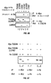
TUCANはまた、NACタンパク質(Apaflアポトソーム(apoptosome)のCARD保有調節因子)のセグメントと類似するアミノ酸を共有するN末端ドメインを含む(Chuら J Biol Chem 276:9239−9245(2001)5およびHlaingら J Biol Chem 276:9230−9238(2001))。このTUCANのN末端ドメインは、いくつかの候補リン酸化部位(アミノ酸72、286、313および416でのPKC(S/T−x−R/K)部位、289、376、398、414および416でのカゼインキナーゼII(S/T−x−D/E)部位および187ならびに289でのMAPキナーゼ/CDK(S/T−P)部位が挙げられる)を含む。SDS−PAGE実験におけるそれらの異なる移動度によって同定されたTUCANの観察された複数の形態(例えば、図6Bを参照のこと)は、TUCANの異なったリン酸化形態であり得る。TUCANはまた、残基243〜246での候補カスパーゼ切断部位(DEED)を含む。 TUCAN also contains an N-terminal domain that shares amino acids similar to segments of the NAC protein, the CARD-bearing regulator of Apaffl apoptosomes (Chu et al. J Biol Chem 276: 9239-9245 (2001) 5 and Hlaing et al. J Biol Chem 276: 9230-9238 (2001)). This N-terminal domain of TUCAN has several candidate phosphorylation sites (PKC (S / TxR / K) sites at amino acids 72, 286, 313 and 416), 289, 376, 398, 414 and 416. Casein kinase II (S / TxD / E) site and 187 and MAP kinase / CDK (S / TP) site at 289). The observed multiple forms of TUCAN identified by their different mobilities in SDS-PAGE experiments (see, eg, FIG. 6B) can be different phosphorylated forms of TUCAN. TUCAN also contains a candidate caspase cleavage site (DEED) at residues 243-246.
これらの分子および他の分子の特徴付けならびにTUCANの細胞機能性は、例えば、Pathanら J.Biol.Chem.276:32220−32229(2001)(その全体が本明細書中に参考として援用される)に記載される。 The characterization of these and other molecules and the cellular functionality of TUCAN are described, for example, in Pathan et al. Biol. Chem. 276: 32220-32229 (2001), which is incorporated herein by reference in its entirety.
本明細書中の実施例VIIに開示されるように、TUCANの相対的に高いレベルは、いくつかのヒト癌細胞株において見出される。さらに、実施例IIおよびVIIIに開示されるように、正常な結腸の粘膜と比較して、TUCANの免疫染色は、悪性の形質転換に関連する、この抗アポトーシスタンパク質の異常な過剰発現を示す初期段階の結腸癌の約3分の2において病理学的に増強した。プロカスパーゼ9のノックアウトマウス由来の細胞の研究は、p53依存経路における腫瘍サプレッサーとしてのプロカスパーゼ9機能を示した(Soengasら Science 284:156−159(1999))。プロカスパーゼ9の調節におけるTUCANの役割を考慮して、TUCANの過剰発現は、プロカスパーゼ9の損失と機能的に同等であり得、TUCANの増強したレベルが、腫瘍の病原性または進行を促進し得ることを示す。本明細書中の実施例IVおよびVIIIに示されるように、結腸癌患者(患者の腫瘍は、高いレベルのTUCANを含む)は、記録保管用標本を使用する、遡及的な分析に起因して、これらの疾患がもとで死亡する傾向が実際に強い。
As disclosed in Example VII herein, relatively high levels of TUCAN are found in several human cancer cell lines. Furthermore, as disclosed in Examples II and VIII, compared to normal colonic mucosa, TUCAN immunostaining is an early indication of abnormal overexpression of this anti-apoptotic protein associated with malignant transformation. Pathologically enhanced in about two thirds of stage colon cancer. Studies of cells from
従って、本発明は、TUCANを使用して癌患者に対する生存の予後を決定するための方法を提供する。この発明は、(a)癌患者由来の新生物細胞含有サンプル中のTUCANのレベルを測定する工程、および(b)TUCANの参照レベルとサンプル中のTUCANレベルとを比較する工程(ここで、サンプル中の低いレベルのTUCANが、患者の増大した生存率と相関する)を包含する。 Accordingly, the present invention provides a method for determining the prognosis of survival for cancer patients using TUCAN. The invention comprises (a) measuring the level of TUCAN in a neoplastic cell-containing sample from a cancer patient, and (b) comparing the reference level of TUCAN with the TUCAN level in the sample, wherein the sample Low levels of TUCAN correlate with increased survival of patients).
TUCANの決定された基本レベルまたは参照レベルを上回る、新生物細胞含有サンプル中のTUCANレベルは、腫瘍の再発または腫瘍の広がりにおける有意な因子であり得る。腫瘍細胞を用いて決定した参照レベルが上回る場合、TUCANのレベルは、高いか、または過剰産生と特徴付けられる。高いTUCANかまたは過剰産生したTUCANは、腫瘍の再発または腫瘍の広がりの増大した危険性を示され得る。低いTUCANまたは減産したTUCANは、腫瘍の再発または腫瘍の広がりの減少した危険性を示され得る。 TUCAN levels in neoplastic cell-containing samples above the determined basal or reference level of TUCAN can be a significant factor in tumor recurrence or tumor spread. If the reference level determined using tumor cells is exceeded, the level of TUCAN is high or characterized as overproduction. High TUCAN or overproduced TUCAN can indicate an increased risk of tumor recurrence or tumor spread. Low TUCAN or reduced TUCAN may indicate a risk of tumor recurrence or reduced tumor spread.
癌患者の生存率を予知するための本発明の方法は、患者からサンプルを得る工程、および1つ以上のバイオマーカー(例えば、TUCAN)のレベルを測定する工程を包含する。バイオマーカー(例えば、TUCAN)のレベルを使用して、疾患を有さないかまたは癌患者の全体の生存率に対する予後を本明細書中で提供される相関に基づいて決定する。腫瘍の再発または広がりの可能性は、腫瘍細胞中のTUCANのレベルと相関するので、このような予後は、可能である。例えば、実施例VIおよびVIIIに示されるように、TUCAN発現レベルが低い場合、癌の再発の可能性は低いことが見出された。患者由来の新生物細胞含有サンプル中のTUCANのレベルは、疾患状態の評価における単独の因子として使用され得るか、または他の予測的な方法に加えて使用され得る。 The methods of the present invention for predicting the survival rate of a cancer patient include obtaining a sample from the patient and measuring the level of one or more biomarkers (eg, TUCAN). The level of a biomarker (eg, TUCAN) is used to determine the prognosis for the overall survival of patients without disease or cancer patients based on the correlation provided herein. Such a prognosis is possible because the likelihood of tumor recurrence or spread correlates with the level of TUCAN in the tumor cells. For example, as shown in Examples VI and VIII, it was found that when the TUCAN expression level is low, the likelihood of cancer recurrence is low. The level of TUCAN in a neoplastic cell-containing sample from a patient can be used as a single factor in the assessment of disease state or can be used in addition to other predictive methods.
TUCANを使用して、生存を予知し得るか、または様々な型の癌を罹患する患者に対する処置の過程の有効性をモニターし得る。実施例VIIに記載されるように、TUCANは、複数の異なる癌細胞型(白血病、黒色腫ならびに胸部癌、卵巣癌、肺癌、CNA癌、前立腺癌および腎臓癌が挙げられる)に存在する。上記されるようにまた、TUCANの細胞機能性は、ミトコンドリアシグナル伝達経路誘発アポトーシスの抑制である。ミトコンドリアシグナル伝達経路誘発アポトーシスは、任意の細胞型において生じ得、そして任意の細胞型において、調節不全かまたは抑制の状態になり得るアポトーシス機構であり、正常なホメオスタシスの増殖制御を伴わずに、増殖するような細胞の形質転換を生じる。従って、TUCANのレベルは、腫瘍の再発または任意の型の細胞を有する患者の生存率と相関され得る。本明細書中で提供されるガイダンスおよび他の周知の方法を使用して、当業者は、特定の腫瘍細胞型おけるTUCANのレベルが、患者の生存率と相関するか否かを決定し得る。決定されたTUCANの参照レベルと癌患者の生存率との間の相関を有して、当業者は、癌患者の生存率についての予後を決定するための方法、および本明細書中に記載される癌を有する患者を処置する過程の有効性をモニターするための方法を実行し得る。 TUCAN can be used to predict survival or monitor the effectiveness of the course of treatment for patients with various types of cancer. As described in Example VII, TUCAN exists in several different cancer cell types including leukemia, melanoma and breast cancer, ovarian cancer, lung cancer, CNA cancer, prostate cancer and kidney cancer. As also noted above, the cellular functionality of TUCAN is suppression of mitochondrial signaling pathway induced apoptosis. Mitochondrial signaling pathway-induced apoptosis is an apoptotic mechanism that can occur in any cell type and can be dysregulated or suppressed in any cell type and proliferates without growth control of normal homeostasis Cause transformation of the cells. Thus, the level of TUCAN can be correlated with tumor recurrence or survival of patients with any type of cells. Using the guidance provided herein and other well-known methods, one skilled in the art can determine whether the level of TUCAN in a particular tumor cell type correlates with patient survival. Having a correlation between the determined reference level of TUCAN and the survival rate of cancer patients, one of skill in the art is described in methods and methods herein for determining a prognosis for cancer patient survival rates. A method for monitoring the effectiveness of the process of treating a patient with certain cancers may be implemented.
本発明の方法において、サンプルは、例えば、生検手順を使用して得られた細胞または組織であり得るか、あるいは細胞を含む流体サンプル(例えば、血液、血清、精液、尿または便)であり得る。当業者は、癌の型、そして必要である場合、生検サンプルを得るための適切な方法に依存する、適切なサンプルを決定し得る。可能である場合、最少の侵襲性回収手順を使用して、患者からサンプルを得ることが所得され得る。例えば、患者からの流体サンプル(例えば、血液、唾液、血清、精液、尿または便)を得ることは、組織サンプルを回収することよりも侵襲性でない。 In the methods of the present invention, the sample can be, for example, a cell or tissue obtained using a biopsy procedure, or a fluid sample (eg, blood, serum, semen, urine or stool) containing cells. obtain. One skilled in the art can determine an appropriate sample depending on the type of cancer and, if necessary, the appropriate method for obtaining a biopsy sample. Where possible, it can be earned to obtain a sample from a patient using a minimally invasive recovery procedure. For example, obtaining a fluid sample (eg, blood, saliva, serum, semen, urine or stool) from a patient is less invasive than collecting a tissue sample.
1つの実施形態において、TUCANのレベルは、選択的結合剤(例えば、TUCANポリペプチドと特異的に反応する抗体)を使用して、TUCANの量を測定することによって決定され得る。他の選択的結合剤としては、TUCANポリペプチドに結合するポリペプチド例えば、TUCAN CARDドメイン(アミノ酸345〜431(配列番号3)を含むTUCANポリペプチド、およびカスパーゼ9ポリペプチド(このアミノ酸配列(配列番号4)は、Prositeデータベース中のP55211として参照される)、またはTUCANポリペプチドに結合するその改変体が挙げられる。プロカスパーゼおよびそれ自体へのTUCANの選択的結合は、実施例IXに記載される。
In one embodiment, the level of TUCAN can be determined by measuring the amount of TUCAN using a selective binding agent (eg, an antibody that specifically reacts with a TUCAN polypeptide). Other selective binding agents include polypeptides that bind to a TUCAN polypeptide, such as a TUCAN CARD domain (TUCAN polypeptide comprising amino acids 345-431 (SEQ ID NO: 3)) and a
親和性結合アッセイの必須の全ての様式は、サンプル中のTUCANまたは別のバイオマーカーポリペプチド(例えば、cIAP2、Apaf1、Smac、β−カテニン、Bc1−2またはp53)のレベルの決定における使用のために適用可能である。このような方法は、迅速であり、有効性であり、そして感受性である。さらに、親和性結合方法は単純であり、そして多様な臨床的な設定および多様な特定の必要性に適するような状態下で実行されるように改変され得る。公知であり、そして本発明の方法に使用され得る親和性結合アッセイとしては、可溶性相形式および固相形式の両方が挙げられる。可溶性相親和性結合アッセイ(soluble phase affinity binding assay)の特定の例は、バイオマーカーの選択的抗体または他の結合剤を使用する免疫沈降である。固相型式は、迅速であり、そして感受性または正確さを失うことなく同時に複数の異なるサンプルでより容易に実行され得るので、本発明の方法にとって有利である。さらに、固相親和性結合アッセイは、高スループットスクリーニングおよび自動化に適切である。 All essential formats of affinity binding assays are for use in determining the level of TUCAN or another biomarker polypeptide (eg, cIAP2, Apaf1, Smac, β-catenin, Bc1-2 or p53) in a sample. It is applicable to. Such a method is rapid, effective and sensitive. Furthermore, the affinity binding method is simple and can be modified to be performed under conditions suitable for a variety of clinical settings and a variety of specific needs. Affinity binding assays that are known and can be used in the methods of the invention include both soluble and solid phase formats. A specific example of a soluble phase affinity binding assay is immunoprecipitation using a biomarker selective antibody or other binding agent. The solid phase format is advantageous for the method of the present invention because it is rapid and can be more easily performed on multiple different samples at the same time without loss of sensitivity or accuracy. Furthermore, solid phase affinity binding assays are suitable for high throughput screening and automation.
固相親和性結合アッセイの特定の例としては、免疫組織化学的結合アッセイ、免疫親和性結合アッセイ(例えば、ELISAおよび放射免疫アッセイ(RIA))が挙げられる。他の固相親和性結合アッセイは、当業者に公知であり、そして本発明の方法に適用可能である。親和性結合アッセイは、一般に、目的の分析物またはリガンドに対して選択的な抗体結合分子を用いる使用のためにフォーマットされるが、特に任意の結合剤は、選択的結合抗体と代替的に置換され得る。このような結合剤としては、例えば、高分子(例えば、ポリペプチド、ペプチド、核酸分子、脂質および糖)ならびに低分子化合物が挙げられる。特定の分析物またはリガンドに選択的に結合するこのような分子を同定するための方法は、当該分野で公知であり、そしてこれらとしては、例えば、表面提示ライブラリーおよびコンビナトリアルライブラリーが挙げられる。従って、親和性結合アッセイにおいて使用されるような抗体以外の分子について、必要とされる全ての分子は、バイオマーカーに対する選択的結合活性を示すような結合剤である。 Particular examples of solid phase affinity binding assays include immunohistochemical binding assays, immunoaffinity binding assays (eg, ELISA and radioimmunoassay (RIA)). Other solid phase affinity binding assays are known to those of skill in the art and are applicable to the methods of the invention. Affinity binding assays are generally formatted for use with antibody binding molecules that are selective for the analyte or ligand of interest, but in particular any binding agent can be substituted for the selective binding antibody. Can be done. Such binders include, for example, macromolecules (eg, polypeptides, peptides, nucleic acid molecules, lipids and sugars) and low molecular compounds. Methods for identifying such molecules that selectively bind to a particular analyte or ligand are known in the art, and include, for example, surface display libraries and combinatorial libraries. Thus, for molecules other than antibodies as used in affinity binding assays, all molecules required are binding agents that exhibit selective binding activity for biomarkers.
親和性結合アッセイ(例えば、免疫親和性結合アッセイ)の種々の様式としては、例えば、免疫組織学化学的方法、固相ELISAおよびRIAならびにそれらの改変が挙げられる。このようなそれらの改変としては、例えば、捕獲アッセイおよびサンドイッチアッセイ、ならびに競合アッセイ形式との組み合わせのいずれかの様式の使用が挙げられる。使用のための免疫親和性結合アッセイのこれらの様式または形式の選択は、使用者の意図に依存する。このような方法は、一般的な実験室マニュアル(例えば、HarlowおよびLane,Using Antibodies:A Laboratory Manual,Cold Spring Harbor Laboratory Press,New York(1999))の記載に、見出され得る。 Various modes of affinity binding assays (eg, immunoaffinity binding assays) include, for example, immunohistochemical methods, solid phase ELISA and RIA, and modifications thereof. Such modifications include, for example, the use of any format of capture and sandwich assays and combinations with competitive assay formats. The choice of these formats or formats of immunoaffinity binding assays for use depends on the user's intention. Such methods can be found in descriptions in general laboratory manuals (eg, Harlow and Lane, Using Antibodies: A Laboratory Manual, Cold Spring Harbor Laboratory Press, New York (1999)).
本発明の方法において有用な抗体としては、ポリクローナル抗体およびモノクローナル抗体、ならびにこのような抗体の抗原結合フラグメントが挙げられる。ポリクローナル抗体またはモノクローナル抗体を調製する方法は、当業者に周知であり、そして実施例IおよびHarlowおよびLane,Antibodies:A Laboratory Manual,Cold Spring Harbor Laboratory Press(1988)に記載される。 Antibodies useful in the methods of the present invention include polyclonal and monoclonal antibodies, and antigen-binding fragments of such antibodies. Methods for preparing polyclonal or monoclonal antibodies are well known to those of skill in the art and are described in Example I and Harlow and Lane, Antibodies: A Laboratory Manual, Cold Spring Harbor Laboratory Press (1988).
本発明の方法に有用な抗体としてはまた、天然に存在する抗体ならびに天然に存在しない抗体(例えば、一本鎖抗体、キメラ抗体、二機能性抗体およびヒト化抗体、ならびにそれらの抗原結合フラグメントを含む)が挙げられる。このような天然に存在しない抗体は、固相ペプチド合成を使用して構築され得るか、組換え的に産生され得るか、または、例えば、Huseら(Science 246:1275−1281(1989))によって記載されるような可変性の重鎖および可変性の軽鎖からなるコンビナトリアルなライブラリーをスクにーニングすることによって得られることができる。これらの方法、ならびに例えば、キメラ抗体、ヒト化抗体、CDR移植抗体、一本鎖抗体および二機能性抗体を作製する他の方法は、当業者に周知である(WinterおよびHarris,Immunol.Today 14:243−246(1993);Wardら、Nature 341:544−546(1989);HarlowおよびLane,前出,1988);Hilyardら、Protein Engineering:A Practical approach(IRL Press 1992);Borrabeck,Antibody Engineering,第2編(Oxford University Press 1995))。 Antibodies useful in the methods of the invention also include naturally occurring antibodies as well as non-naturally occurring antibodies (eg, single chain antibodies, chimeric antibodies, bifunctional antibodies and humanized antibodies, and antigen-binding fragments thereof). Included). Such non-naturally occurring antibodies can be constructed using solid phase peptide synthesis, can be produced recombinantly, or, for example, by Huse et al. (Science 246: 1275-1281 (1989)). It can be obtained by screening a combinatorial library of variable heavy chains and variable light chains as described. These methods, as well as other methods of making, for example, chimeric antibodies, humanized antibodies, CDR-grafted antibodies, single chain antibodies and bifunctional antibodies are well known to those skilled in the art (Winter and Harris, Immunol. Today 14 243-246 (1993); Ward et al., Nature 341: 544-546 (1989); Harlow and Lane, supra, 1988); Hiryard et al. , Second Edition (Oxford University Press 1995)).
親和性結合を利用する形式は、種々の検出標識および分析されたサンプル中のバイオマーカーの量を定量化するための当該分野で公知のシステムとの組み合わせにおいて使用され得る。検出システムとしては、直接的手段および間接的手段の両方によって結合されたバイオマーカーの検出が挙げられる。直接的検出方法としては、バイオマーカー特異的反応性抗体または結合剤の標識化が挙げられる。間接的検出システムとしては、例えば、標識された二次抗体および結合剤の使用が挙げられる。 Formats that utilize affinity binding can be used in combination with various detection labels and systems known in the art for quantifying the amount of biomarker in the analyzed sample. Detection systems include the detection of biomarkers bound by both direct and indirect means. Direct detection methods include labeling of biomarker specific reactive antibodies or binding agents. Indirect detection systems include, for example, the use of labeled secondary antibodies and binding agents.
二次抗体、標識および検出システムは、当該分野で周知であり、そして商業的に得られ得るか、または当該分野で周知の技術によって得ることができる。バイオマーカー選択的結合剤とともに使用される検出可能な標識およびシステムは、バイオマーカーへの試薬の結合を損なうべきではない。さらに、複数の抗体および標識システムは、所望である場合、結合アッセイの感受性を増強するような結合されたバイオマーカー特異的反応性抗体を検出するために使用され得る。 Secondary antibodies, labels and detection systems are well known in the art and can be obtained commercially or by techniques well known in the art. The detectable label and system used with the biomarker selective binding agent should not impair the binding of the reagent to the biomarker. In addition, multiple antibodies and labeling systems can be used to detect bound biomarker specific reactive antibodies that enhance the sensitivity of the binding assay, if desired.
検出可能な標識は、分析方法によって定量され得るか測定され得る必須の任意の標識であり得る。このような標識としては、例えば、酵素、放射性同位元素、蛍光色素ならびに化学発光化合物および生物発光化合物が挙げられる。酵素標識の特定の例としては、西洋ワサビペルオキシダーゼ(HRP)、アルカリ性ホスファターゼ(AP)、β−ガラクトシダーゼ、ウレアーゼおよびルシフェラーゼが挙げられる。 The detectable label can be any essential label that can be quantified or measured by analytical methods. Such labels include, for example, enzymes, radioisotopes, fluorescent dyes and chemiluminescent and bioluminescent compounds. Specific examples of enzyme labels include horseradish peroxidase (HRP), alkaline phosphatase (AP), β-galactosidase, urease and luciferase.
西洋ワサビペルオキシダーゼの検出システムは、例えば、色素生産基質のテトラメチルベンジジン(TMB)とともに使用され得、過酸化水素の存在において、450nmでの吸光度を測定することによって検出可能である可溶性産物を得る。アルカリ性ホスファターゼ検出システムは、色素生産基質のp−ニトロフェニルリン酸とともに使用さえ得、例えば、405nmでの吸光度を測定することによって検出可能である可溶性産物を得る。同様に、β−ガラクトシダーゼの検出システムは、色素生産基質のo−ニトロフェニル−β−D−ガラクトピラノシド(ONPG)とともに使用され得、410nmでの吸光度を測定することによって検出可能である可溶性産物を得るか、またはウレアーゼの検出システムは、基質(例えば、尿素−ブロモクレゾールパープル(Sigma Immunochemicals, St.Louis,MO))とともに使用され得る。ルシフェリンは、ATP依存性酸化の後に発光するルシフェラーゼに対する基質化合物である。 The horseradish peroxidase detection system can be used, for example, with the chromogenic substrate tetramethylbenzidine (TMB) to obtain a soluble product that is detectable by measuring absorbance at 450 nm in the presence of hydrogen peroxide. The alkaline phosphatase detection system can even be used with the chromogenic substrate p-nitrophenyl phosphate, for example, to obtain a soluble product that is detectable by measuring absorbance at 405 nm. Similarly, a detection system for β-galactosidase can be used with the chromogenic substrate o-nitrophenyl-β-D-galactopyranoside (ONPG), a soluble that is detectable by measuring absorbance at 410 nm. The product can be obtained or a urease detection system can be used with a substrate, such as urea-bromocresol purple (Sigma Immunochemicals, St. Louis, MO). Luciferin is a substrate compound for luciferase that emits light after ATP-dependent oxidation.
蛍光色素検出標識は、光または別のエネルギー供給原による励起の後、紫外線の光または可視波長の光の放出を介して検出可能に表される。DAPI、フルオレセイン、Hoechst 33258、R−フィコシアニン、B−フィコエリトリン、R−フィコエリトリン、ローダミン、テキサスレッドおよびリーサミン(lissamine)は、本発明の親和性結合形式において使用され得る蛍光色素検出標識の特定の例である。特に有用な蛍光色素は、フルオレセインまたはローダミンである。 The fluorochrome detection label is detectably represented via emission of light of ultraviolet or visible wavelength after excitation by light or another energy source. DAPI, fluorescein, Hoechst 33258, R-phycocyanin, B-phycoerythrin, R-phycoerythrin, rhodamine, Texas red and lissamine are specific examples of fluorochrome detection labels that can be used in the affinity binding format of the present invention. is there. Particularly useful fluorescent dyes are fluorescein or rhodamine.
化学発光および生物発光の検出レベルは、感受性(バイオマーカーの非放射活性検出)にとって都合がよく、そして様々な供給源(例えば、Amersham Lifesciences,Inc.(Arlington Heights,IL))から市販され得る。 Chemiluminescence and bioluminescence detection levels are convenient for sensitivity (non-radioactive detection of biomarkers) and can be commercially obtained from a variety of sources, such as Amersham Lifesciences, Inc. (Arlington Heights, IL).
あるいは、放射性同位元素は、本発明の方法において検出可能な標識として使用され得る。ヨウ素−125は、検出可能な標識として有用な放射性同位元素の特定の例である。 Alternatively, radioisotopes can be used as detectable labels in the methods of the invention. Iodine-125 is a specific example of a radioisotope useful as a detectable label.
検出可能な標識からのシグナルは、以下を使用して分析され得る:例えば、色素生産基質からの色を検出するための分光光度計;特定波長の光の存在における蛍光発光を検出するための蛍光光度計;または放射線を検出するための放射線計数管(例えば、ヨウ素−125の検出のためのγ計数管)。酵素結合二次抗体の検出について、例えば、結合剤の量の定量的分析は、製造者の指示書に従って、分光光度計(例えば、EMAX Microplate Reader(Molecular Devices,Menlo Park,CA)を使用してなされ得る。所望である場合、本発明のアッセイは、自動化され得るか、または機械的に実施され、そして複数のサンプルからのシグナルは、同時に検出され得る。 The signal from the detectable label can be analyzed using, for example: a spectrophotometer to detect color from a chromogenic substrate; fluorescence to detect fluorescence emission in the presence of light of a specific wavelength A photometer; or a radiation counter for detecting radiation (eg, a gamma counter for the detection of iodine-125). For detection of enzyme-linked secondary antibodies, for example, quantitative analysis of the amount of binding agent is performed using a spectrophotometer (eg, EMAX Microplate Reader (Molecular Devices, Menlo Park, Calif.) According to the manufacturer's instructions. If desired, the assays of the invention can be automated or performed mechanically, and signals from multiple samples can be detected simultaneously.
本発明の予後形式は、米国特許第4,376,110号および同第4,778,751号に記載されるように、前、後、または同時であり得る。本明細書に記載される種々のアッセイ形式のための分離工程(結合していない2次抗体の除去を含む)は、当該分野で公知の方法(HarlowおよびLane、前出)によって実行され得る。例えば、適切な緩衝液を用いる洗浄は、濾過、吸入、減圧または磁気分離(magnetic separation)、および遠心分離に続き得る。 The prognostic form of the present invention can be before, after, or simultaneous, as described in US Pat. Nos. 4,376,110 and 4,778,751. Separation steps for various assay formats described herein (including removal of unbound secondary antibody) can be performed by methods known in the art (Harlow and Lane, supra). For example, washing with a suitable buffer can be followed by filtration, inhalation, vacuum or magnetic separation, and centrifugation.
バイオマーカーについて選択的な結合剤はまた、バイオマーカーに標的化される(新生物細胞を表す)画像化方法において利用され得る。これらの画像化技術は、標準的な処置(例えば、結腸のような胃腸管系器官の外科的切除、および放射線治療が挙げられる)に続く、原発部位における残りの新生物細胞の同定において有用性を有する。さらに、新生物細胞を検出する画像化技術は、転移の第二部位の検出において有用性を有する。バイオマーカー特異的結合剤は、(例えば、111インジウム)を用いて放射標識され得、そしてKahnら、Journal of Urology 152:1952−1955(1994)によって記載されるように、静脈内に注入され得る。バイオマーカーについて選択的な結合剤は、例えば、TUCANまたは別のバイオマーカー(例えば、cIAP2、Apaf1、Smac、β−カテニン、Bcl−2またはp53)と特異的に反応するモノクローナル抗体であり得る。画像化は、例えば、Kahnら(前出)によって記載されるような、放射免疫シンチグラフィーによって達成され得る。 Binders that are selective for biomarkers can also be utilized in imaging methods targeted to biomarkers (representing neoplastic cells). These imaging techniques are useful in identifying remaining neoplastic cells at the primary site following standard treatments such as surgical excision of gastrointestinal organs such as the colon, and radiation therapy. Have Furthermore, imaging techniques that detect neoplastic cells have utility in detecting a second site of metastasis. Biomarker specific binding agents can be radiolabeled with (eg, 111 indium) and injected intravenously as described by Kahn et al., Journal of Urology 152: 1952-1955 (1994). . A binding agent selective for a biomarker can be, for example, a monoclonal antibody that specifically reacts with TUCAN or another biomarker (eg, cIAP2, Apaf1, Smac, β-catenin, Bcl-2 or p53). Imaging can be accomplished by radioimmunoscintigraphy, for example, as described by Kahn et al. (Supra).
TUCANまたは別のバイオマーカー(例えば、cIAP2、Apaf1、Smac、β−カテニン、Bcl−2またはp53)のレベルはまた、核酸プローブのようなバイオマーカーのための選択的結合剤を用いて、バイオマーカーのmRNAまたはDNAの量を測定することによって決定され得る。mRNAレベルを検出するために使用される方法としては、バイオマーカーをコードするmRNAのハイブリダイゼーションまたは増幅の検出が挙げられる。この検出は、the Current Protocols in Molecular Biology(John Wiley & Sons,1999);米国特許第5,882,864号などにおいて例示されるような、当業者に公知の方法のうちの1つを用いて、インビトロまたはインサイチュのいずれかでのmRNAの分析によって実行され得る。検出されるTUCAN mRNAまたは他のバイオマーカーのmRNAは、TUCAN遺伝子もしくはそのフラグメント、あるいはcIAP2遺伝子、Bcl−2遺伝子、p53遺伝子、b−カテニン遺伝子、survivin遺伝子もしくはApaf1遺伝子、またはそれらのフラグメントの任意のRNA転写物である。 The level of TUCAN or another biomarker (eg, cIAP2, Apaf1, Smac, β-catenin, Bcl-2 or p53) can also be measured using a selective binding agent for the biomarker such as a nucleic acid probe. Can be determined by measuring the amount of mRNA or DNA. Methods used to detect mRNA levels include detection of hybridization or amplification of mRNA encoding a biomarker. This detection is accomplished using one of the methods known to those skilled in the art, as exemplified in the Current Protocols in Molecular Biology (John Wiley & Sons, 1999); US Pat. No. 5,882,864, etc. Can be performed by analysis of mRNA either in vitro or in situ. TUCAN mRNA or other biomarker mRNA to be detected is any of TUCAN gene or fragment thereof, or cIAP2 gene, Bcl-2 gene, p53 gene, b-catenin gene, survivin gene or Apaf1 gene, or a fragment thereof. RNA transcript.
相補的プローブを用いる特異的または選択的なハイブリダイゼーションによって核酸分子を検出するための、当該分野で周知の方法が多数存在する。簡単に言うと、ハイブリダイゼーションによる検出のために、検出可能なレベルを有する、TUCAN遺伝子に相補的なTUCAN核酸プローブは、このプローブのTUCAN RNAへのアニーリングを可能にする条件下で、癌を有する個体または癌を有することが疑われる個体から得られた新生物細胞含有サンプルに添加され得る。サンプル中のTUCAN RNAを検出するための方法としては、例えば、RT−PCRの使用が挙げられ得る。条件は、溶液および固相のハイブリダイゼーション手順の両方について当該分野において周知である。さらに、ハイブリダイゼーション条件の最適化は、所望される場合、異なる温度、持続時間および異なる緩衝液条件におけるサンプルのアリコートのハイブリダイゼーションによって実行され得る。このような手順は慣用的であり、そして当業者に周知である。アニーリング後、このサンプルを洗浄し、そしてシグナルを測定し、そして適切なコントロール値または標準値と比較する。ハイブリダイゼーションシグナルの程度は、TUCANのmRNAレベルに直接比例する。新生物細胞含有サンプルにおけるTUCAN mRNAのレベルは、TUCAN mRNAに対する適切な参照レベルと比較される。他のバイオマーカー(例えば、cIAP2、Apaf1、Smac、β−カテニン、Bcl−2またはp53)mRNAのレベルは、同様に決定され得、そして特定のバイオマーカーに対する適切な参照レベルと比較され得る。 There are many methods well known in the art for detecting nucleic acid molecules by specific or selective hybridization using complementary probes. Briefly, a TUCAN nucleic acid probe complementary to the TUCAN gene that has a detectable level for detection by hybridization has cancer under conditions that allow the probe to anneal to TUCAN RNA. It can be added to a neoplastic cell-containing sample obtained from an individual or an individual suspected of having cancer. A method for detecting TUCAN RNA in a sample may include, for example, the use of RT-PCR. Conditions are well known in the art for both solution and solid phase hybridization procedures. Furthermore, optimization of hybridization conditions can be performed by hybridization of aliquots of samples at different temperatures, durations and different buffer conditions, if desired. Such procedures are conventional and well known to those skilled in the art. After annealing, the sample is washed and the signal is measured and compared to the appropriate control or standard value. The degree of hybridization signal is directly proportional to the TUCAN mRNA level. The level of TUCAN mRNA in the neoplastic cell-containing sample is compared to an appropriate reference level for TUCAN mRNA. The levels of other biomarkers (eg, cIAP2, Apaf1, Smac, β-catenin, Bcl-2 or p53) mRNA can be similarly determined and compared to appropriate reference levels for a particular biomarker.
方法の他の例としては、PCRおよび他の増幅方法(例えば、RT−PCR、5’RACEもしくは3’RACE、RNase保護、RNAブロット、ドットブロットまたは他の膜ベースの技術、ディップスティック、ピン、ELISAまたは固体支持体上に固定された二次元アレイ)が挙げられる。これらの方法は、定質的測定または定量的測定のいすれかを用いて実行され得、これらの全ては、当業者に周知である。 Other examples of methods include PCR and other amplification methods (eg, RT-PCR, 5′RACE or 3′RACE, RNase protection, RNA blot, dot blot or other membrane-based techniques, dipsticks, pins, ELISA or a two-dimensional array fixed on a solid support). These methods can be carried out using either qualitative or quantitative measurements, all of which are well known to those skilled in the art.
PCRまたはRT−PCRは、単離されたRNAまたは粗細胞溶解調製物を用いて使用され得る。出発材料の量を制限する場合、PCRは有益である。PCR法のさらなる記載は、例えば、Dieffenbach,C.W.およびDveksler,G.S.、PCR Primer:A Laboratory Manual、Cold Spring Harbor Press,Plainview,New York(1995)に見出され得る。マイクロアレイのような多数サンプル形式は、単一アッセイにおいて、多数の異なるサンプルを分析することの利点を提供する。対照的に、固相ディップスティックベースの方法は、即時の結果のために、患者の流体サンプルを迅速に分析することが可能であるという利点を提供する。 PCR or RT-PCR can be used with isolated RNA or crude cell lysis preparations. PCR is beneficial when limiting the amount of starting material. Further descriptions of PCR methods can be found, for example, in Dieffenbach, C .; W. And Dveksler, G. et al. S. , PCR Primer: A Laboratory Manual, Cold Spring Harbor Press, Plainview, New York (1995). Multiple sample formats such as microarrays offer the advantage of analyzing a number of different samples in a single assay. In contrast, solid phase dipstick-based methods offer the advantage of being able to quickly analyze a patient fluid sample for immediate results.
バイオマーカー(例えば、cIAP2、TUCAN、Apaf1、β−カテニン、Bcl−2またはSmac)の発現レベルをハイブリダイゼーションによって測定するために有用な核酸プローブとしては、例えば、本明細書中に提供されるヌクレオチド配列を用いて調製されるプローブが挙げられる。全体のcDNA配列およびそのフラグメント(cIAP2、TUCAN、Apaf1、β−カテニン、Bcl−2またはSmacのヌクレオチド配列に対応するオリゴヌクレオチドが挙げられ、そしてこれらは、cIAP2 RNA、TUCAN RNA、Apaf1 RNA、β−カテニン RNA、Bcl−2 RNAまたはSmac RNAに特異的または選択的にハイブリダイズし得る)に対応する核酸分子は、ハイブリダイゼーション方法にとって有用である。 Nucleic acid probes useful for measuring the expression level of a biomarker (eg, cIAP2, TUCAN, Apaf1, β-catenin, Bcl-2 or Smac) by hybridization include, for example, the nucleotides provided herein Examples include probes prepared using sequences. The entire cDNA sequence and its fragments (cIAP2, TUCAN, Apaf1, β-catenin, Bcl-2, or Smac include oligonucleotides corresponding to the nucleotide sequence, and these include cIAP2 RNA, TUCAN RNA, Apaf1 RNA, β- Nucleic acid molecules corresponding to catenin RNA, Bcl-2 RNA or Smac RNA) are useful for hybridization methods.
参照レベルは、患者の癌性細胞におけるバイオマーカーのレベルを評価するために使用されるバイオマーカー(例えば、cIAP2、TUCAN、Apaf1、Smac、β−カテニンまたはBcl−2)のレベルである。特に、患者の癌性細胞におけるバイオマーカーのレベルが参照レベルよりも高い場合、これらの細胞は高レベルのバイオマーカーまたは過剰産生のバイオマーカーを有するとみなされる。逆に、患者の癌際細胞におけるバイオマーカーのレベルが参照レベルよりも低い場合、これらの細胞は、低レベルのバイオマーカーまたは過少産生のバイオマーカーを有するとみなされる。 The reference level is the level of a biomarker (eg, cIAP2, TUCAN, Apaf1, Smac, β-catenin or Bcl-2) used to assess the level of the biomarker in the patient's cancerous cells. In particular, if the level of biomarkers in the patient's cancerous cells is higher than the reference level, these cells are considered to have high levels of biomarkers or overproduced biomarkers. Conversely, if the level of biomarker in the patient's cancer cells is lower than the reference level, these cells are considered to have low levels of biomarkers or underproduction biomarkers.
高レベルのバイオマーカー遺伝子(例えば、cIAP2、TUCAN、Apaf1、Smac、β−カテニン、Bcl−2またはp53)、あるいは過剰産生のバイオマーカー遺伝子は、決定した基礎レベルより高いのバイオマーカーのレベルと関連する。従って、癌細胞におけるバイオマーカー(例えば、cIAP2、TUCAN、Apaf1、Smac、β−カテニン、Bcl−2またはp53)の参照レベルまたは基礎レベルは、「カットオフ」値として同定され、「カットオフ」値より上では、バイオマーカーの存在と、減少または増加した腫瘍の再発または広がりとの間に有意な相関が存在する。当業者は、いくつかの「カットオフ」値が明確でない(臨床的相関はカットオフの片側の値の範囲にわたって、なお有意である)ことを認識する;しかし、癌細胞の型に対するバイオマーカーレベルの至適なカットオフ値(例えば、変動するH−スコアなど)を選択することが可能である。至適なカットオフ値における改善が、参照値または基礎値を決定するために使用される統計学的方法の高度化、および参照値または基礎値を決定するために使用されるサンプルの数および供給源に依存して決定され得ることが理解される。 A high level biomarker gene (eg, cIAP2, TUCAN, Apaf1, Smac, β-catenin, Bcl-2 or p53) or an overproduced biomarker gene is associated with a biomarker level higher than the determined basal level To do. Thus, a reference level or basal level of a biomarker (eg, cIAP2, TUCAN, Apaf1, Smac, β-catenin, Bcl-2 or p53) in cancer cells is identified as a “cutoff” value and a “cutoff” value. Above, there is a significant correlation between the presence of biomarkers and reduced or increased tumor recurrence or spread. Those skilled in the art recognize that some “cut-off” values are unclear (clinical correlation is still significant over a range of values on one side of the cut-off); however, biomarker levels for cancer cell types It is possible to select an optimal cut-off value (for example, a variable H-score). An improvement in the optimal cut-off value enhances the statistical method used to determine the reference or basal value, and the number and supply of samples used to determine the reference or basal value It will be understood that it can be determined depending on the source.
このような過剰産生は、代表的に、絶対的なバイオマーカーレベルに関して計算されないが、相対的測定を用いて決定される。これらの相対的測定値は、内部標準を用いた定量化の目的のために例示される;しかし、決定の他の標準または方法(例えば、外部標準、バイオマーカーポリペプチド測定値、バイオマーカーmRNA測定値、タンパク質の絶対値、mRNAレベルまたはDNAレベルなどとの比較)が、使用され得ることが理解される。 Such overproduction is typically not calculated with respect to absolute biomarker levels, but is determined using relative measurements. These relative measurements are exemplified for quantification purposes using internal standards; however, other standards or methods of determination (eg, external standards, biomarker polypeptide measurements, biomarker mRNA measurements) It is understood that comparisons with values, absolute values of proteins, mRNA levels or DNA levels etc. can be used.
参照レベルはまた、癌を有する患者の集団(例えば、同一段階の癌を有する患者)におけるバイオマーカーレベルの比較によって決定され得る。これは、ヒストグラム分析(試験した患者の全体集団が図解される)によって達成され得、ここで、第一の軸はバイオマーカーのレベルを示し、そして第二の軸は、腫瘍細胞が所定レベルのバイオマーカーを含む集団における患者数を示す。患者の2つ以上の別個の群は、同一レベルまたは類似レベルのバイオマーカーを有する集団のサブセット集団の同定によって決定され得る。次いで、参照レベルの決定は、これらの別個の群を最も良好に区別するバイオマーカーレベルに基づいてなされ得る。 The reference level can also be determined by comparison of biomarker levels in a population of patients with cancer (eg, patients with the same stage of cancer). This can be accomplished by histogram analysis (where the entire population of patients tested is illustrated), where the first axis indicates the level of the biomarker and the second axis indicates that the tumor cells are at a predetermined level. The number of patients in the population containing the biomarker is shown. Two or more distinct groups of patients can be determined by identifying subset populations of populations having the same or similar levels of biomarkers. Reference level determinations can then be made based on the biomarker levels that best distinguish these distinct groups.
参照レベルが、参照バイオマーカーより高いのレベルを発現する癌患者に対して、参照バイオマーカーより低いのレベルを発現する癌患者における、腫瘍の再発または広がりの可能性を区別するという検証は、単一の可変分析または複数の可変分析を使用して実行され得る。これらの方法は、1つ以上の可変の結果と所定の結果との間の相関の可能性を決定する。特定の場合において、これらの方法は、バイオマーカーレベル(または別の変数と結合したバイオマーカーレベル)と、疾患を有さないか、または癌患者の全般的な生存との間の相関の可能性を決定する。これらの分析を実行するために、当業者に周知の複数の方法の任意の1つが、使用され得る。単一可変分析の例は、Kaplan−Meir法またはログランク試験(log−rank test)である。複数の可変分析の例は、Cox比例危険回帰モデル(例えば、実施例VIを参照のこと)である。 Validation of distinguishing the likelihood of tumor recurrence or spread in cancer patients expressing lower levels than the reference biomarker versus cancer patients expressing higher levels than the reference biomarker is simply It can be performed using one variable analysis or multiple variable analyses. These methods determine the likelihood of correlation between one or more variable results and a predetermined result. In certain cases, these methods may correlate between biomarker levels (or biomarker levels combined with another variable) and the overall survival of patients with no disease or cancer. To decide. Any one of a number of methods well known to those skilled in the art can be used to perform these analyses. Examples of a single variable analysis are the Kaplan-Meir method or the log-rank test. An example of multiple variable analyzes is the Cox proportional risk regression model (see, eg, Example VI).
参照レベルの集団ベースの決定(例えば、ヒストグラム分析による)は、異なるバイオマーカーレベルを有する患者の2つ以上の別個の群を決定するために、十分なサイズの患者集団を用いて実施され得る。代表的に、このような集団は、少なくとも25人の患者、例えば、少なくとも50人の患者、少なくとも75人の患者および少なくとも100人の患者を含む。同様に、決定される参照レベルの検証はまた、少なくとも25人の患者、例えば、少なくとも50人の患者、少なくとも75人の患者および少なくとも100人の患者を含み得る。 Reference level population-based determination (eg, by histogram analysis) can be performed using a patient population of sufficient size to determine two or more distinct groups of patients having different biomarker levels. Typically, such a population includes at least 25 patients, such as at least 50 patients, at least 75 patients and at least 100 patients. Similarly, verification of the determined reference level may also include at least 25 patients, eg, at least 50 patients, at least 75 patients, and at least 100 patients.
参照レベルは、単一の数(各々の患者に等しく適用可能である)であり得るか、または参照レベルは、患者集団の特定のサブ集団に従って変動し得る。例えば、男性は、同一の癌について女性とは異なる参照レベルを有し得る。さらに、参照レベルは、各個別の患者に対して決定されるレベルであり得る。例えば、参照レベルは、同一の患者内の非腫瘍細胞におけるバイオマーカーレベルに対する患者の腫瘍細胞におけるバイオマーカーレベルの特定の比となり得る。従って、各患者についての参照レベルは、バイオマーカーレベルの参照比によって禁止され得、ここで、参照比は、上記の参照レベルを決定するための任意の方法によって決定され得る。 The reference level can be a single number (which is equally applicable to each patient) or the reference level can vary according to a particular sub-population of the patient population. For example, a male may have a different reference level than a female for the same cancer. Further, the reference level can be a level determined for each individual patient. For example, the reference level can be a specific ratio of biomarker levels in a patient's tumor cells to biomarker levels in non-tumor cells within the same patient. Thus, the reference level for each patient can be prohibited by the reference ratio of biomarker levels, where the reference ratio can be determined by any method for determining the reference level described above.
さらに、参照レベルは、患者の2つの群を分け得るが、複数の集団を分ける多くの参照値が存在し得ることは、本発明の範囲内である。例えば、2つの参照値は、高レベルのバイオマーカーを有する患者の第一群を、中間レベルのバイオマーカーを有する患者の第二群と低レベルのバイオマーカーを有する患者の第三群から分離し得る。異なる数の参照レベルは、曲線(例えば、連続する線)を禁止するのに十分であり得、これは、患者におけるバイオマーカーレベルの機能として、疾患を有さない可能性または患者における全般的な生存の可能性を記載する。このような曲線は、「連続した」バイオマーカーレベルを構成し、ここで、疾患を有さないかまたは患者における全般的な生存の可能性は、このような患者におけるバイオマーカーに比例する。2つ以上のバイオマーカーレベルはまた、このような曲線によって示され得る。 Furthermore, while the reference level can divide two groups of patients, it is within the scope of the present invention that there can be many reference values dividing multiple populations. For example, two reference values separate the first group of patients with high levels of biomarkers from the second group of patients with intermediate levels of biomarkers and the third group of patients with low levels of biomarkers. obtain. Different numbers of reference levels may be sufficient to prohibit curves (eg, continuous lines), which may be a disease-free or general Describe the probability of survival. Such a curve constitutes “continuous” biomarker levels, where the likelihood of having no disease or overall survival in a patient is proportional to the biomarker in such a patient. Two or more biomarker levels can also be indicated by such a curve.
参照レベルはまた、細胞の1つ以上の区画におけるバイオマーカータンパク質(例えば、cIAP2、TUCAN、Apaf1、Smac、β−カテニン、Bcl−2またはp53)のレベルを示す。代表的に、参照レベルは、(a)細胞全体、(b)核、または(c)細胞質におけるバイオマーカータンパク質のレベルを示す。このレベルは、そのタンパク質の細胞区画化が、腫瘍の再発または特定の癌の広がりの危険性と相関する場合、有用である。同様に、参照レベルは、異なる区画におけるバイオマーカータンパク質のレベル比(例えば、細胞全体のバイオマーカータンパク質に対する核バイオマーカータンパク質の比、または細胞質のバイオマーカータンパク質に対する核の比)であり得る。 The reference level also indicates the level of biomarker protein (eg, cIAP2, TUCAN, Apaf1, Smac, β-catenin, Bcl-2 or p53) in one or more compartments of the cell. Typically, the reference level indicates the level of biomarker protein in (a) whole cells, (b) nuclei, or (c) cytoplasm. This level is useful when the cellular compartmentalization of the protein correlates with the risk of tumor recurrence or the spread of certain cancers. Similarly, the reference level can be the level ratio of biomarker protein in different compartments (eg, the ratio of nuclear biomarker protein to whole cell biomarker protein, or the ratio of nucleus to cytoplasmic biomarker protein).
バイオマーカー(例えば、cIAP2、TUCAN、Apaf1またはSmac)の参照レベルは、疾患を有さない可能性、または癌についての全般的な生存の可能性の統計学的に有意な指標であることが見出される、別の可変指標と組み合わせてさらに使用され得る。このような指標としては、既知の癌マーカー(例えば、結腸癌マーカーとしては、シアロシル−TnCEA、CA19−9、およびLASAが挙げられる)の存在またはレベルが挙げられるか、あるいは臨床的指標または病理学的指標(例えば、年齢、腫瘍サイズ、腫瘍病歴、臨床段階、家族の病歴など)があり得る。例えば、癌の臨床段階はまた、疾患を有さないか、または全般的な生存の、統計学的に有意な指標であり、ここで、バイオマーカーの参照レベルは、癌の臨床段階に従って変動し得る。例えば、バイオマーカーのレベル(例えば、所定の患者について癌の臨床段階IIに関連する低レベルのTUCAN)は、ともに、疾患を有さない可能性または全般的な生存の可能性の増大についての指標である。従って、バイオマーカーの参照レベルは、疾患を有さない可能性または癌についての全般的な生存の可能性の、別の統計学的に有意な指標関数として変動し得る。 A reference level of a biomarker (eg, cIAP2, TUCAN, Apaf1 or Smac) has been found to be a statistically significant indicator of the likelihood of having no disease or overall survival for cancer. Can be further used in combination with another variable indicator. Such indicators include the presence or level of known cancer markers (eg, colon cancer markers include sialosyl-TnCEA, CA19-9, and LASA), or clinical indicators or pathology There may be an objective index (eg, age, tumor size, tumor history, clinical stage, family history, etc.). For example, the clinical stage of cancer is also a statistically significant indicator of disease free or overall survival, where the reference level of the biomarker varies according to the clinical stage of the cancer obtain. For example, the level of a biomarker (eg, a low level of TUCAN associated with cancer clinical stage II for a given patient) is both an indicator of increased likelihood of having the disease or overall survival. It is. Thus, the reference level of a biomarker can vary as another statistically significant indicator function of the likelihood of having no disease or overall survival for cancer.
癌細胞におけるバイオマーカー(例えば、cIAP2、Apaf1、TUCAN、Bcl−2またはSmac)のレベルは、互いに相関し得、そして他の分子と相関し得る。なぜなら、これらの分子は、癌細胞の過剰増殖状態に寄与する、一般的に調節不全の分子経路に関与するからである。従って、1つ以上のさらなるバイオマーカーとTUCANとの組み合わせは、癌患者の生存についての予後を決定するための本発明の方法において使用され得る。第2のバイオマーカーまたはさらなるバイオマーカーは、例えば、Apaf1、cIAP1、cIAP2、survivin、AIF、Bcl−2、Bcl−XL、Bax、Bid、BAG1、p53、変異体p53、β−カテニン、MIB−1または別の周知の腫瘍マーカー(例えば、以下に記載される例示的な市販の腫瘍マーカー)であり得る。さらに、TUCANと、1つ以上のバイオマーカーとの組み合わせの使用は、増加した予後の有意性または予後決定における信頼を提供する。 The levels of biomarkers (eg, cIAP2, Apaf1, TUCAN, Bcl-2 or Smac) in cancer cells can be correlated with each other and with other molecules. This is because these molecules are typically involved in dysregulated molecular pathways that contribute to the hyperproliferative state of cancer cells. Thus, the combination of one or more additional biomarkers and TUCAN can be used in the methods of the invention to determine the prognosis for survival of cancer patients. The second biomarker or additional biomarkers are, for example, Apaf1, cIAP1, cIAP2, survivin, AIF, Bcl-2, Bcl-XL, Bax, Bid, BAG1, p53, mutant p53, β-catenin, MIB-1 Or another well-known tumor marker, such as the exemplary commercially available tumor marker described below. Furthermore, the use of a combination of TUCAN and one or more biomarkers provides increased prognostic significance or confidence in prognosis determination.
従って、本発明は、癌患者の生存についての予後を決定するための方法を提供し、この方法は、2つ以上のバイオマーカーの使用を包含する。この方法は、(a)癌患者に由来する新生物細胞含有サンプルにおいて、TUCANのレベル、ならびにcIAP2、Apaf1、Bcl−2およびSmacからなる群より選択される1つ以上のバイオマーカーのレベルを測定する工程、ならびに(b)上記サンプルにおけるTUCANおよび1つ以上の選択されたバイオマーカーのレベルと、TUCANおよびバイオマーカーの参照レベルとを比較する工程によって実施される。ここで、上記サンプルにおける、低レベルのTUCANと高レベルの任意のApaf1、Bcl−2またはSmac、あるいは低レベルのTUCANと低レベルのcIAP2は、上記患者の増大した生存と相関する。 Accordingly, the present invention provides a method for determining the prognosis for survival of a cancer patient, which method involves the use of more than one biomarker. The method measures (a) a level of TUCAN and one or more biomarkers selected from the group consisting of cIAP2, Apaf1, Bcl-2 and Smac in a neoplastic cell-containing sample derived from a cancer patient. And (b) comparing the level of TUCAN and one or more selected biomarkers in the sample with a reference level of TUCAN and the biomarker. Here, low levels of TUCAN and high levels of any Apaf1, Bcl-2 or Smac, or low levels of TUCAN and low levels of cIAP2 in the sample correlate with increased survival of the patient.
本発明の方法は、例えば、増大または減少した発現が改善された生存と相関する、TUCANと、1つ以上のバイオマーカー(例えば、任意のcIAP2、Apaf1、Bcl−2、Smacまたは癌に対する別の既知のバイオマーカーまたは標準的なバイオマーカー)との組み合わせを選択することによって実施され得る。選択されたバイオマーカーは、複数の型の癌にとって有用な、一般的な診断マーカーまたは予後マーカー(例えば、CA125、CEAもしくはLDH)であり得るか、あるいは癌特異的診断マーカーまたは予後マーカー(例えば、結腸癌マーカー(例えば、シアロシル−TnCEA、CA19−9またはLASA)、乳癌マーカー(例えば、CA15−2、Her−2/neuおよびCA27.29)、卵巣癌マーカー(例えば、CA72−4)、肺癌(例えば、神経特異的エノラーゼ(NSE)および組織ポリペプチド抗原(TPA))、前立腺癌(例えば、PSA、前立腺特異的膜抗原および前立腺酸性ホスファターゼ)、黒色腫(例えば、S−100およびTA−90)、ならびに他の型の癌に特異的な他のバイオマーカー)であり得る。当業者は、TUCANと組み合わせて、検出のために有用な診断マーカーまたは予後マーカーを選択することが可能である。同様に、3つ以上、4つ以上もしくは5つ以上または多くのバイオマーカーが、癌患者の生存についての予後を決定するために一緒に使用され得る。 The methods of the invention can be used, for example, for TUCAN and one or more biomarkers (eg, any cIAP2, Apaf1, Bcl-2, Smac or another against cancer, where increased or decreased expression correlates with improved survival. Can be performed by selecting combinations with known biomarkers or standard biomarkers. The selected biomarker can be a general diagnostic marker or prognostic marker (eg, CA125, CEA or LDH) useful for multiple types of cancer, or a cancer-specific diagnostic marker or prognostic marker (eg, Colon cancer markers (eg, sialosyl-TnCEA, CA19-9 or LASA), breast cancer markers (eg, CA15-2, Her-2 / neu and CA27.29), ovarian cancer markers (eg, CA72-4), lung cancer ( For example, nerve specific enolase (NSE) and tissue polypeptide antigen (TPA)), prostate cancer (eg, PSA, prostate specific membrane antigen and prostate acid phosphatase), melanoma (eg, S-100 and TA-90) , As well as other biomarkers specific to other types of cancer) That. One skilled in the art can select useful diagnostic or prognostic markers for detection in combination with TUCAN. Similarly, three or more, four or more or five or more or many biomarkers can be used together to determine the prognosis for survival of cancer patients.
2つ以上のバイオマーカーの使用は、予後の結果に増大した信頼性を提供し得る。例えば、本明細書中に開示されるように、低いcIAPと低いTUCANとの組み合わせ、および高いApaf1と低いTUCANとの組み合わせは、増大した、疾患に罹患していない生存と相関した(実施例Vを参照のこと)。特に、新生物細胞含有サンプルにおけるcIAPおよびTUCANのレベルについて試験した33人の患者のうち、他のカテゴリーに属する患者については56%が生存し、そして44%が疾患を有さないのに対して、低いIAPおよび低いTUCANを有する患者の97%は生存し続けた(91%は疾患を有さない)。さらに、新生物細胞含有サンプルにおけるApaf1およびTUCANのレベルについて試験した17人の患者のうち、他のカテゴリーに属する患者については65%が生存し、そして53%が疾患を有さないのに対して、高いApaf1および低いTUCANを有する患者の100%は生存し続け、そして疾患を有さなかった。当業者は、このような相関が、本明細書中に記載される方法を用いて、バイオマーカーの他の組み合わせを使用して観察され得ることを認識する。
The use of more than one biomarker can provide increased confidence in the prognostic results. For example, as disclosed herein, a combination of low cIAP and low TUCAN and a combination of high Apaf1 and low TUCAN correlated with increased disease-free survival (Example V checking). In particular, of the 33 patients tested for cIAP and TUCAN levels in neoplastic cell-containing samples, 56% survived and 44% had no disease for patients in other categories 97% of patients with low IAP and low TUCAN remained alive (91% had no disease). Furthermore, of the 17 patients tested for Apaf1 and TUCAN levels in neoplastic cell-containing samples, 65% survived and 53% had no disease for patients in
本発明の予後方法において有用なバイオマーカーの組み合わせとしては、例えば、cIAP2およびTUCAN、Apaf1およびTUCAN、ならびにバイオマーカー(例えば、cIAP2、Apaf1、Bcl−2およびSmacならびに他の分子(AIF、Bcl−2、Bcl−XL、Bax、Bid、BAG1、p53、変異p53、β−カテニン、MIB−1ならびに種々の他の一般的バイオマーカーおよび腫瘍特異的バイオマーカー(例えば、本明細書中に上記されるような市販の診断マーカー)を含む))とTUCANとの、多数の他の組み合わせが挙げられる。これらのバイオマーカーの上昇したレベルは、評価された全ての癌検体の部分において観察されたので(実施例IIを参照のこと)、このような組み合わせは、癌細胞の転移状態の有用な指標であり得る。さらに、別の結腸癌マーカーであるKi−67と相関する種々のバイオマーカーのレベルの上昇、およびバイオマーカーの発現間(例えば、患者集団におけるcIAP2とTUCANとの間(p=0.003))の陽性の相関が観察された。 Biomarker combinations useful in the prognostic methods of the present invention include, for example, cIAP2 and TUCAN, Apaf1 and TUCAN, and biomarkers (eg, cIAP2, Apaf1, Bcl-2 and Smac and other molecules (AIF, Bcl-2 , Bcl-XL, Bax, Bid, BAG1, p53, mutant p53, β-catenin, MIB-1, and various other general and tumor-specific biomarkers (eg, as described above herein) And other commercially available combinations of TUCAN) and TUCAN. Since elevated levels of these biomarkers were observed in a portion of all cancer specimens evaluated (see Example II), such a combination is a useful indicator of cancer cell metastasis status. possible. In addition, increased levels of various biomarkers that correlate with another colon cancer marker, Ki-67, and expression of the biomarkers (eg, between cIAP2 and TUCAN (p = 0.003) in the patient population). A positive correlation was observed.
本発明はまた、癌に罹患する患者についての処置過程の有効性をモニタリングするための方法を提供する。この方法は、以下の工程:(a)癌患者由来の新生物細胞含有サンプル中のTUCANのレベルを決定する工程、および(b)処置後の患者由来の新生物細胞含有サンプル中のTUCANのレベルを決定する工程、を包含し、これによって、処置前のTUCANレベルと処置後のバイオマーカーレベルとの比較が、処置の有効性を示す。 The present invention also provides a method for monitoring the effectiveness of a treatment process for a patient suffering from cancer. The method comprises the following steps: (a) determining the level of TUCAN in a neoplastic cell-containing sample from a cancer patient, and (b) the level of TUCAN in a neoplastic cell-containing sample from a patient after treatment. A comparison of the pre-treatment TUCAN level with the post-treatment biomarker level indicates the effectiveness of the treatment.
処置過程に関して使用される場合、「有効性」とは、この処置過程が腫瘍の再発または広がりの危険性を低減する能力をいい、従って、患者が疾患を有さない可能性または患者の全体的生存の可能性を増大させる能力をいう。この方法は、患者の腫瘍細胞におけるバイオマーカー(例えば、cIAP、TUCAN、Apaf1またはSmac)のレベルが、患者の非腫瘍細胞におけるcIAP、TUCAN、Apaf1およびSmacのレベルと比較して異常である場合、特に有用性を有する。処置の前および後の患者由来の新生物細胞含有サンプル中のバイオマーカーレベルの比較は、それによって、バイオマーカーレベルが非腫瘍細胞のレベルに戻っている(これは、より有効な処置過程を意味している)か否か、またはバイオマーカーレベルが異常なままであるかもしくは異常性を増している(これは、処置過程があまり有効でないことを示している)か否かを示すように働く。例えば、処置後の患者サンプル中のApaf1、Bcl−2またはSmacのレベルの増加は、Apaf1またはSmacの高いレベルが、結腸癌の再発の低い発生率と相関するので、この処置が有効であることを示す。さらに、処置後の患者サンプルにおける低レベルのβ−カテニン、cIAP2またはTUCANは、低レベルのβ−カテニン、cIAP2またはTUCANが、結腸癌の再発の低い発生率と相関するので、この処置が有効であることを示す。 “Efficacy” when used in reference to a course of treatment refers to the ability of this course of treatment to reduce the risk of tumor recurrence or spread, and thus the likelihood that the patient has no disease or the patient's overall The ability to increase the chances of survival. This method can be used when the level of a biomarker (eg, cIAP, TUCAN, Apaf1 or Smac) in a patient's tumor cells is abnormal compared to the level of cIAP, TUCAN, Apaf1 and Smac in a patient's non-tumor cells; Particularly useful. Comparison of biomarker levels in neoplastic cell-containing samples from patients before and after treatment, thereby returning biomarker levels to the level of non-tumor cells (this implies a more effective treatment process) Or whether the biomarker level remains abnormal or increases in anomalies (this indicates that the treatment process is not very effective) . For example, an increase in the level of Apaf1, Bcl-2 or Smac in a patient sample after treatment indicates that this treatment is effective because high levels of Apaf1 or Smac correlate with a low incidence of recurrence of colon cancer Indicates. Furthermore, low levels of β-catenin, cIAP2 or TUCAN in patient samples after treatment are effective because low levels of β-catenin, cIAP2 or TUCAN correlate with a low incidence of recurrence of colon cancer. Indicates that there is.
癌を有する患者は、高レベルの特定のバイオマーカーまたは低レベルのバイオマーカーが、この患者から得られた新生物細胞含有サンプルにおいて測定されるか否かにしたがって分類され得る。患者についての予後の決定は、この患者が割り当てられたグループが、この患者が割り当てられなかったグループに関して、より高いかまたはより低い、疾患に罹患していない生存の可能性または全体的生存の可能性と相関するか否かを決定することによって、なされ得る。 Patients with cancer can be classified according to whether high levels of specific biomarkers or low levels of biomarkers are measured in a neoplastic cell-containing sample obtained from the patient. The prognostic decision for a patient is that the group to which this patient is assigned is higher or lower than the group to which this patient has not been assigned, the likelihood of disease-free survival or overall survival. This can be done by determining whether it correlates with gender.
従って、本発明はまた、患者の分類を含む、癌患者の生存についての予後を決定する方法を提供する。この方法は、以下の工程:(a)癌患者由来の新生物細胞含有サンプル中のTUCANレベルを測定する工程、および(b)第一または第二の患者グループのいずれかに属するとして患者を分類する工程、によって実施され、ここで、低レベルのTUCANを有する第一のグループの患者は、高レベルのTUCANを有する第二のグループの患者と比較して、生存の可能性が増大しているとして分類される。 Thus, the present invention also provides a method for determining the prognosis for cancer patient survival, including patient classification. The method comprises the following steps: (a) measuring TUCAN levels in a neoplastic cell-containing sample from a cancer patient, and (b) classifying the patient as belonging to either the first or second patient group. Wherein a first group of patients having a low level of TUCAN has an increased likelihood of survival compared to a second group of patients having a high level of TUCAN. Classified as
高レベルのTUCANまたはTUCANの過剰発現は、腫瘍の再発または広がりの危険性が増大した患者と相関する。従って、高レベルのTUCANを有する第一のグループに属する患者は、低レベルのTUCANを有する第二のグループと比較して、腫瘍の再発または広がりの危険性が増大しているとして分類される。低レベルのTUCANを有する第一のグループに属する患者は、高レベルのTUCANを有する第二のグループと比較して、生存の可能性が増大しているとして分類される。 High levels of TUCAN or TUCAN overexpression correlate with patients with increased risk of tumor recurrence or spread. Thus, patients belonging to the first group with a high level of TUCAN are classified as having an increased risk of tumor recurrence or spread compared to a second group with a low level of TUCAN. Patients belonging to the first group with low levels of TUCAN are classified as having an increased likelihood of survival compared to the second group with high levels of TUCAN.
癌患者の生存についての予後を決定する方法は、1以上のさらなるバイオマーカーを使用して実施され得る。種々のバイオマーカー(公知の癌マーカーおよび本明細書中に開示される予後バイオマーカーを含む)は、癌患者の生存についての予後を決定するために、TUCANと組み合わせて使用され得る。1実施形態において、この方法は、以下の工程:(a)癌患者由来の新生物細胞含有サンプル中のcIAP2レベルを決定する工程、および(b)第一または第二の患者グループのいずれかに属するとして患者を分類する工程、を包含し、ここで、低レベルのTUCANおよび低レベルのcIAP2を有する第一のグループの患者は、高レベルのTUCANおよび高レベルのcIAP2を有する第二のグループの患者と比較して、生存の可能性が増大しているとして分類される。 The method of determining a prognosis for survival of a cancer patient can be performed using one or more additional biomarkers. Various biomarkers (including known cancer markers and prognostic biomarkers disclosed herein) can be used in combination with TUCAN to determine the prognosis for survival of cancer patients. In one embodiment, the method comprises the following steps: (a) determining cIAP2 levels in a neoplastic cell-containing sample from a cancer patient, and (b) either the first or second patient group. Classifying patients as belonging, wherein a first group of patients having a low level of TUCAN and a low level of cIAP2 is a second group of patients having a high level of TUCAN and a high level of cIAP2. Classified as having an increased likelihood of survival compared to the patient.
別の実施形態において、この方法は、以下の工程:(a)癌患者由来の新生物細胞含有サンプル中のApaf1、SmacおよびBcl−2からなる群より選択されるバイオマーカーのレベルを決定する工程、ならびに(b)第一または第二の患者グループのいずれかに属するとして患者を分類する工程、を包含し、ここで、低レベルのTUCANおよび高レベルのApaf1、SmacまたはBcl−2のいずれかを有する第一のグループの患者は、高レベルのTUCANおよび低レベルのApaf1、SmacまたはBcl−2のいずれかを有する第二のグループの患者と比較して、生存の可能性が増大しているとして分類される。 In another embodiment, the method comprises the following steps: (a) determining the level of a biomarker selected from the group consisting of Apaf1, Smac and Bcl-2 in a neoplastic cell-containing sample from a cancer patient. And (b) classifying the patient as belonging to either the first or second patient group, wherein a low level of TUCAN and a high level of either Apaf1, Smac or Bcl-2 The first group of patients with increased risk of survival compared to the second group of patients with high levels of TUCAN and low levels of either Apaf1, Smac or Bcl-2 Classified as
患者サンプル中の1以上のバイオマーカーのレベルを決定し、参照レベルと比較した後、次いで、患者は、疾患に罹患していない生存の特定の可能性または全体的生存の特定の可能性を有するグループへと分類される。次いで、患者についての疾患を有さない可能性または全体的生存の可能性は、そのグループ中の患者についての患者についての疾患を有さない可能性または全体的生存の可能性に基づいて評価される。 After determining the level of one or more biomarkers in a patient sample and comparing it to a reference level, the patient then has a particular chance of survival without disease or a particular chance of overall survival Classified into groups. The likelihood of having no disease or overall survival for the patient is then assessed based on the likelihood of having no disease for the patient or the likelihood of overall survival for the patients in the group. The
例えば、癌患者由来の新生物細胞含有サンプルは、参照レベルと比較して、高レベルのApaf1、Bcl−2またはSmacを有すると決定され得る。次いで、この患者は、高レベルのApaf1、Bcl−2またはSmacを有する患者のグループへと分類される。(癌細胞中で低レベルのApaf1、Bcl−2またはSmacを発現する患者のグループと比較して)癌細胞中で高レベルのApaf1、Bcl−2またはSmacを発現する患者のグループについての、疾患が存在しない可能性または全体的生存の可能性が増大していることが発見されているので、特定の癌患者は、疾患が存在しない生存の可能性または全体的生存の可能性が増大しているとみなされる。 For example, a neoplastic cell-containing sample from a cancer patient can be determined to have a high level of Apaf1, Bcl-2 or Smac compared to a reference level. The patient is then classified into a group of patients with high levels of Apaf1, Bcl-2 or Smac. Disease for a group of patients expressing high levels of Apaf1, Bcl-2 or Smac in cancer cells (as compared to a group of patients expressing low levels of Apaf1, Bcl-2 or Smac in cancer cells) Certain cancer patients have an increased chance of survival without disease or overall survival. Is considered to be.
逆に、癌患者由来の新生物細胞含有サンプルは、参照レベルと比較して、高レベルのcIAP2、β−カテニンまたはTUCANを有すると決定され得る。次いで、この患者は、高レベルのcIAP2、β−カテニンまたはTUCANを有する患者のグループへと分類される。(癌細胞中で低レベルのcIAP2、β−カテニンまたはTUCANを発現する患者のグループと比較して)癌細胞中で高レベルのcIAP2、β−カテニンまたはTUCANを発現する患者のグループについての、疾患が存在しない生存の可能性または全体的生存の可能性が減少していることが発見されているので、特定の癌患者は、疾患に罹患していない生存の可能性または全体的生存の可能性が減少しているとみなされる。 Conversely, a neoplastic cell-containing sample from a cancer patient can be determined to have high levels of cIAP2, β-catenin or TUCAN compared to a reference level. The patient is then classified into a group of patients with high levels of cIAP2, β-catenin or TUCAN. Disease for a group of patients expressing high levels of cIAP2, β-catenin or TUCAN in cancer cells (as compared to a group of patients expressing low levels of cIAP2, β-catenin or TUCAN in cancer cells) It has been discovered that the likelihood of non-existence or overall survival has been reduced, so certain cancer patients may have the chance of survival without disease or overall survival Is considered to be decreasing.
本発明の方法は、癌の発達についての個体の感受性を決定するために適用可能である。この方法は、種々の癌(胃腸、肺、結腸、前立腺、乳房、卵巣、皮膚、血液および腎臓の癌を含む)に対して適用可能である。特に、結腸癌は、腺腫様結腸ポリープとして公知の前悪性前駆病変から発達する。複数の疫学的研究は、家族の一員が一旦腺腫様結腸ポリープを発症すると、その兄弟姉妹は、結腸腺腫および結腸癌の両方を発症する危険性が非常に上昇していることを実証した。当業者は、本発明の方法が、例えば、適切な生検サンプルを収集することによって、結腸新生物状態(例えば、腺腫様結腸ポリープ)を含む新生物状態について本明細書中に記載されるように実施され得る。 The methods of the present invention are applicable to determine an individual's sensitivity to cancer development. This method is applicable to a variety of cancers, including gastrointestinal, lung, colon, prostate, breast, ovary, skin, blood and kidney cancers. In particular, colon cancer develops from premalignant precursor lesions known as adenomatous colon polyps. Multiple epidemiological studies have demonstrated that once a family member develops an adenomatous colon polyp, their siblings are at significantly increased risk of developing both colon adenoma and colon cancer. One skilled in the art will recognize that the methods of the present invention are described herein for neoplastic conditions, including colon neoplastic conditions (eg, adenomatous colon polyps), eg, by collecting appropriate biopsy samples. Can be implemented.
癌患者の生存についての予後を決定するための本発明の方法は、腫瘍進行の任意の病期において患者に適用可能であり、そしてさらに、腫瘍進行の病期を決定するために使用され得る。腫瘍の病期とは、腫瘍の進行の程度をいう。腫瘍発達の種々の病期は、Markman「Basic Cancer Medicine」、Saunders(Zorab,R.編)(1997)中に例示されるように、当業者に周知である。例えば、癌は、以下の3つの一般的な病期に病期分けされ得る:局在、局所的広がりおよび離れた広がり。癌はまた、TNMシステムを使用して病期分けされ得、このシステムは、罹患した組織および近傍の組織内への直接的広がりの程度、リンパ節近傍への広がりの程度、ならびに離れた器官への広がりの程度を考慮する。これらの特徴に基づいて、癌の広がりは、0〜IVまでのローマ数字を割り当てることによって要約され得る。当業者は、特定の型の癌に適切な病期分け系を選択し得る。 The methods of the invention for determining the prognosis for survival of a cancer patient are applicable to the patient at any stage of tumor progression and can further be used to determine the stage of tumor progression. Tumor stage refers to the extent of tumor progression. Various stages of tumor development are well known to those skilled in the art, as illustrated in Markman “Basic Cancer Medicine”, Saunders (Zorab, R. Ed.) (1997). For example, cancer can be staged into three general stages: localization, local spread, and distant spread. Cancer can also be staged using the TNM system, which measures the extent of direct spread into affected and nearby tissues, the extent of spread near lymph nodes, and to distant organs. Consider the extent of spread. Based on these characteristics, the spread of cancer can be summarized by assigning Roman numerals from 0 to IV. One skilled in the art can select an appropriate staging system for a particular type of cancer.
特に、結腸癌は、Dukes系、Astler−Coller系およびAJCC/TNM系を使用して段階分けされ得、これらの系は、結腸または直腸の壁の層、結腸および直腸に隣接する器官、ならびに遠く離れた他の器官に関連する癌の広がりを記載する。Dukes病期Aは、AJCC/TNM病期IおよびAstler−Coller病期A、BIと等価であり;Duke病期Bは、AJCC/TNM病期IIおよびAstler−Coller病期B2、B3と等価である。Dukes病期Cは、AJCC/TNM病期IIIおよびAstler−Coller病期C1、C2、C3と等価である。結腸直腸がんのAJCC/TNM病期は、以下のようである:病期0:この癌は、結腸または直腸の内層(粘膜)を越えて増殖していない。この病期は、またはインサイチュ癌腫または粘膜内癌腫としても公知である;病期I:この癌は、粘膜から粘膜下へと増殖しているか、または固有(propria)筋層へも増殖しているかもしれないが、壁自体の外側に、リンパ節のような近傍組織へと広がってはいない;病期II:この癌は、結腸または直腸の壁を越えて、最外層へと増殖しており、そして他の近傍組織を浸潤しているかもしれないが、近傍のリンパ節へと広がってはいない;病期III:この癌は、任意の大きさであり得るが、3個以下の近傍リンパ節に広がっているか、または4個以上の節に広がっているが、身体の他の部分へは広がっていない;病期IV:この癌は、離れた器官(例えば、肝臓、肺、腹膜または卵巣)に広がっている。 In particular, colon cancer can be staged using the Dukes system, the Astler-Coller system, and the AJCC / TNM system, which includes the colon or rectal wall layer, the colon and the organ adjacent to the rectum, and distant Describe the spread of cancer in relation to other distant organs. Dukes stage A is equivalent to AJCC / TNM stage I and Astler-Coller stage A, BI; Duke stage B is equivalent to AJCC / TNM stage II and Astler-Coller stage B2, B3 is there. Dukes stage C is equivalent to AJCC / TNM stage III and Astler-Coller stage C1, C2, C3. The AJCC / TNM stage of colorectal cancer is as follows: Stage 0: The cancer has not grown beyond the lining (mucosa) of the colon or rectum. This stage, also known as in situ or intramucosal carcinoma; Stage I: The cancer has grown from the mucosa to the submucosa or has also grown into the propria muscle layer. May not, but spread outside the wall itself to nearby tissues such as lymph nodes; Stage II: The cancer has spread beyond the wall of the colon or rectum to the outermost layer And may have invaded other nearby tissues, but has not spread to nearby lymph nodes; Stage III: The cancer may be of any size but no more than 3 nearby lymph nodes Has spread to nodes, or has spread to 4 or more nodes, but has not spread to other parts of the body; Stage IV: This cancer is a distant organ (eg, liver, lung, peritoneum or ovary) ).
腫瘍発達の初期段階は、原発性腫瘍の器官に対して局所的なリンパ節以上には検出可能に広がっていない、腫瘍発達中の病期をいうことが理解されるべきである。代表的に、初期病期は、病期IおよびIIであるとみなされる。 It should be understood that an early stage of tumor development refers to a stage during tumor development that does not detectably spread beyond the lymph nodes local to the organ of the primary tumor. Typically, early stages are considered stage I and II.
本発明の方法の予測的価値は、癌の初期病期の患者の場合に特に有効である。これは、本発明の方法が、検査の時点で測定可能な転移を実証しない患者における転移の危険性を決定する際に、有利に有効であるからである。当業者は、病期Iの癌に罹患している癌患者についての生存率の予後指標が、病期IVの癌に罹患している癌患者についての生存率の予後指標とは異なり得ることを理解する。例えば、病期Iの癌患者についての予後は、癌の継続的増殖および/または転移の可能性に向けられ得、一方で、病期IVの癌患者についての予後は、癌を処置するための治療方法の有効性の可能性に向けられ得る。 The predictive value of the method of the present invention is particularly effective in patients with early stages of cancer. This is because the method of the present invention is advantageously effective in determining the risk of metastasis in patients who do not demonstrate measurable metastasis at the time of the examination. One skilled in the art will recognize that the prognostic index of survival for cancer patients suffering from stage I cancer may differ from the prognostic index of survival for cancer patients suffering from stage IV cancer. to understand. For example, the prognosis for stage I cancer patients may be directed to the possibility of continued growth and / or metastasis of cancer, while the prognosis for stage IV cancer patients is for treating cancer. It may be directed to the potential effectiveness of the treatment method.
癌進行の病期は、1以上のバイオマーカーのレベル(例えば、TUCAN、Apaf1またはIAP(例えば、cIAP2)またはSmacのレベル)と相関し得る。従って、癌患者由来のサンプルにおけるバイオマーカーのレベルの決定は、このサンプルを特定の病期の癌のバイオマーカー指標の参照レベルと比較することによって、得られたサンプル由来の腫瘍の病期を決定するために使用され得る。 The stage of cancer progression can be correlated with the level of one or more biomarkers (eg, the level of TUCAN, Apaf1 or IAP (eg, cIAP2) or Smac). Therefore, the determination of the level of a biomarker in a sample from a cancer patient determines the stage of the tumor from the resulting sample by comparing this sample with the reference level of the biomarker index for a particular stage of cancer. Can be used to
本発明の方法は、癌または新生物状態を有するかまたは有する疑いのある個体から単離または獲得された、種々の異なる型のサンプルを用いる使用のために適用可能である。例えば、本発明の1以上の予後形式における使用のために適用可能なサンプルとしては、組織サンプルおよび細胞サンプルが挙げられる。組織サンプルまたは細胞サンプルは、例えば、生検または手術によって患者から得られた流体サンプルから獲得され得る。例えば、転移していない固形腫瘍の場合、手術によって取り出された腫瘍由来の組織サンプルは、従来の技術によって試験するために、獲得および調製され得る。さらに、サンプルは、例えば、周知の生検手順を使用して、患者から取り出され得る。例えば、結腸癌の場合、非常に小さい隆起したポリープのサンプルを得るために、結腸鏡がスネアに嵌合されて、結腸の壁に対する損傷なしにポリープを取り出し得る(ポリープ切除術);または小さい平らなポリープを得るために、生検鉗子が結腸鏡に装着されて、小さい組織サンプルを収集し得る。 The methods of the present invention are applicable for use with a variety of different types of samples isolated or obtained from individuals having or suspected of having a cancer or neoplastic condition. For example, samples applicable for use in one or more prognostic formats of the present invention include tissue samples and cell samples. A tissue sample or cell sample can be obtained from a fluid sample obtained from a patient, for example, by biopsy or surgery. For example, in the case of a solid tumor that has not metastasized, a tumor-derived tissue sample removed by surgery can be obtained and prepared for testing by conventional techniques. In addition, the sample can be removed from the patient using, for example, well-known biopsy procedures. For example, in the case of colon cancer, a colonoscope can be fitted to the snare to obtain a very small raised polyp sample, and the polyp can be removed without damage to the colon wall (polypectomy); or a small flat To obtain a polyp, a biopsy forceps can be attached to the colonoscope to collect a small tissue sample.
以下に記載するように、そして方法の形式に依存して、この組織は、全体で使用され得るか、または当該分野で公知の種々の方法に供されて、サンプルをより小さい細片、細胞凝集物または個々の細胞へと分離し得る。さらに、ポリメラーゼ連鎖反応(PCR)のような増幅方法と組み合わせた場合に、単一細胞サンプルは、ハイブリダイゼーション検出方法を使用する本発明の予後アッセイにおける使用に十分である。同様に、バイオマーカーのポリペプチドレベルを測定する場合、酵素カップリングまたは光度測定増強を用いる信号の増幅は、いくつかまたは少数の細胞だけを用いて使用され得る。 As described below, and depending on the format of the method, this tissue can be used throughout or subjected to various methods known in the art to divide the sample into smaller debris, cell agglutination. Or can be separated into individual cells. Furthermore, when combined with amplification methods such as polymerase chain reaction (PCR), a single cell sample is sufficient for use in the prognostic assays of the present invention using hybridization detection methods. Similarly, when measuring polypeptide levels of biomarkers, signal amplification using enzyme coupling or photometric enhancement can be used with only a few or a few cells.
生検または手術から得られた全組織は、新生物細胞含有サンプルの1例である。腫瘍組織細胞サンプルは、以下に記載の形式のいずれかを使用してアッセイされ得る。例えば、腫瘍組織サンプルは、取り付けられ、そしてバイオマーカー核酸プローブとインサイチュでハイブリダイズされ得る。タンパク質検出方法およびインサイチュ活性アッセイを使用する類似の組織学的形式もまた、全組織腫瘍細胞サンプル中のバイオマーカーポリペプチドを検出するために使用され得る。タンパク質検出方法としては、例えば、本明細書中の実施例IIに記載されるような、バイオマーカー特異的抗体を用いる染色が挙げられる。このような組織学的方法および当該分野で周知の他の方法は、新生物細胞含有サンプルの供給源として全組織を使用する本発明の予後方法における使用のために適用可能である。サンプルを調製および取り付けするための方法は、当該分野で周知である。 Whole tissue obtained from a biopsy or surgery is an example of a neoplastic cell-containing sample. Tumor tissue cell samples can be assayed using any of the formats described below. For example, a tumor tissue sample can be attached and hybridized in situ with a biomarker nucleic acid probe. Similar histological formats using protein detection methods and in situ activity assays can also be used to detect biomarker polypeptides in whole tissue tumor cell samples. Protein detection methods include, for example, staining with a biomarker specific antibody, as described in Example II herein. Such histological methods and other methods well known in the art are applicable for use in the prognostic methods of the invention that use whole tissue as a source of neoplastic cell-containing samples. Methods for preparing and attaching samples are well known in the art.
新生物状態または癌を有するかまたは有することが疑われる個体由来の個々の細胞および細胞凝集物は、バイオマーカーのRNAまたはポリペプチドの発現の増加または減少について分析され得る新生物細胞含有サンプルの別の例である。これらの細胞は、培養物中で増殖され得、そして上記のような手順を使用して分析され得る。バイオマーカー発現に関連する細胞表面マーカーを発現する全細胞サンプルは、蛍光活性化細胞分類または磁気活性化細胞分類(FACSまたはMACS)を、表面マーカーに選択的な標識された結合剤を用いて、または例えば、特定の細胞集団について選択的な結合剤を用いて使用し、次いで、この集団内のバイオマーカーのレベルを決定して、迅速に試験され得る。バイオマーカーのレベルは、例えば、バイオマーカーについての結合特異的反応剤を使用して、またはバイオマーカー特異的プローブへのハイブリダイゼーションによって、決定され得る。全細胞サンプル中のバイオマーカーのレベルを測定するための他の方法は、当該分野で公知であり、そして以下に記載される予後形式のいずれかにおいて同様に適用可能である。 Individual cells and cell aggregates from individuals with or suspected of having a neoplastic condition or cancer may be analyzed for increased or decreased biomarker RNA or polypeptide expression. It is an example. These cells can be grown in culture and analyzed using procedures as described above. Whole cell samples that express cell surface markers related to biomarker expression can be obtained using fluorescently activated cell classification or magnetically activated cell classification (FACS or MACS) with a labeled binding agent selective for the surface marker, Or, for example, can be used with a selective binding agent for a particular cell population, and then the level of a biomarker within this population can be determined and rapidly tested. The level of a biomarker can be determined, for example, using a binding specific reactant for the biomarker or by hybridization to a biomarker specific probe. Other methods for measuring biomarker levels in whole cell samples are known in the art and are equally applicable in any of the prognostic formats described below.
個体から獲得された組織腫瘍細胞サンプルまたは全細胞腫瘍細胞サンプルはまた、本明細書中に記載される形式のいずれかを使用して、細胞を溶解し、そしてこの溶解物、その分画された部分またはその精製成分中のバイオマーカーのレベルを測定することによって、バイオマーカーレベルの増加または減少について分析され得る。例えば、ハイブリダイゼーション形式が使用される場合、バイオマーカーRNAは、PCRまたは当該分野で周知の他の増幅手順(例えば、バイオマーカー核酸分子のレベルを直接測定するための、RT−PCR、5’RACEまたは3’RACE)を使用して、溶解物から直接増幅され得る。RNAはまた、単離され得、そして例えば、溶液ハイブリダイゼーションによって直接的に探査され得るか、または固定化RNAへのハイブリダイゼーションによって間接的に探査され得る。同様に、ポリペプチド検出形式を使用してバイオマーカーのレベルを決定する場合、溶解物は直接アッセイされ得るか、またはバイオマーカーについて富化するために、さらに分画され得る。例えば、免疫化学的方法(例えば、イムノブロット分析(実施例IIIを参照のこと))が、新生物細胞含有サンプルを使用して実施され得る。全腫瘍細胞サンプルでの使用に適用可能な多数の他の方法は、当業者に周知であり、そして本発明の方法においてしかるべく使用され得る。 A tissue tumor cell sample or whole cell tumor cell sample obtained from an individual also lysed the cell using any of the formats described herein, and this lysate, its fractionated By measuring the level of the biomarker in the portion or its purified component, it can be analyzed for an increase or decrease in the biomarker level. For example, if a hybridization format is used, the biomarker RNA may be PCR or other amplification procedure well known in the art (eg, RT-PCR, 5′RACE for directly measuring the level of a biomarker nucleic acid molecule). Or can be amplified directly from the lysate using 3′RACE). RNA can also be isolated and probed directly by, for example, solution hybridization, or indirectly by hybridization to immobilized RNA. Similarly, when determining the level of a biomarker using a polypeptide detection format, the lysate can be assayed directly or further fractionated to enrich for the biomarker. For example, immunochemical methods (eg, immunoblot analysis (see Example III)) can be performed using neoplastic cell-containing samples. Many other methods applicable for use with whole tumor cell samples are well known to those of skill in the art and can be used accordingly in the methods of the invention.
腫瘍組織または細胞サンプルは、個体から直接的に獲得され得るか、あるいは、試験用に他の供給源から獲得され得る。同様に、細胞サンプルは、新たに単離されたときに試験され得るか、または正確さまたは感受性を実質的に失うことなく、短期間もしくは長期間凍結保存された後に試験され得る。サンプルが、不確定な期間の後に試験される場合、そのサンプルは、獲得され得、次いで、凍結保存され得るか、または、例えば、短期間4℃で保存され得る。本発明の予後方法の利点は、サンプルの組織学的分析を必要としないことである。従って、サンプルは、最初に脱凝集され得るか、溶解され得るか、分画され得るか、または精製され得、そして活性成分が、後の診断のために保存される。 Tumor tissue or cell samples can be obtained directly from the individual or can be obtained from other sources for testing. Similarly, cell samples can be tested when freshly isolated, or can be tested after being cryopreserved for short or long periods without substantial loss of accuracy or sensitivity. If a sample is tested after an indeterminate period, the sample can be obtained and then stored frozen or, for example, stored at 4 ° C. for a short period of time. An advantage of the prognostic method of the present invention is that it does not require histological analysis of the sample. Thus, the sample can be first disaggregated, lysed, fractionated, or purified, and the active ingredient is stored for later diagnosis.
本発明の予後方法は、腫瘍細胞サンプルではない種々の異なる型のサンプルを用いる用途に適用可能である。例えば、細胞外空間(循環液および他の体液を含む)に放出されるバイオマーカーポリペプチドまたはそのフラグメントが、バイオマーカーポリペプチドに関連する分泌型ポリペプチドまたはフラグメントを検出するための予後方法において使用され得る。このような場合、本発明の方法は、新生物状態または癌を有するかまたは有することが疑われる個体から収集された液体サンプルを用いて適用可能である。 The prognostic method of the present invention is applicable to applications using a variety of different types of samples that are not tumor cell samples. For example, a biomarker polypeptide or fragment thereof released into the extracellular space (including circulating fluid and other body fluids) is used in a prognostic method for detecting a secreted polypeptide or fragment associated with the biomarker polypeptide Can be done. In such cases, the methods of the present invention are applicable using a liquid sample collected from an individual who has or is suspected of having a neoplastic condition or cancer.
バイオマーカーレベルについて測定され得る液体サンプルとしては、例えば、血液、血清、リンパ液、尿、または糞便が挙げられる。他の体液は、当業者に公知であり、そして、同様に、本発明の予後方法においてサンプルとしての用途に適用可能である。液体サンプルを分析する1つの利点は、それらが、生検および手術に必要とされるような浸襲手順を伴わずに十分な量で容易に獲得可能であることである。液体サンプル(例えば、血液、血清、および尿)の分析は、一般的に、本明細書中に記載される、バイオマーカーポリペプチドレベルを測定する予後形式である。バイオマーカーに関連するポリエピトープは、溶液形態で循環しているので、この方法は、細胞溶解物、それらの分画部分、または精製成分から発現レベルを測定する方法と同様である。 Liquid samples that can be measured for biomarker levels include, for example, blood, serum, lymph, urine, or feces. Other body fluids are known to those skilled in the art and are equally applicable for use as samples in the prognostic methods of the invention. One advantage of analyzing liquid samples is that they can be easily acquired in sufficient quantities without invasive procedures as required for biopsy and surgery. Analysis of fluid samples (eg, blood, serum, and urine) is generally a prognostic format for measuring biomarker polypeptide levels as described herein. Since the polyepitope associated with the biomarker circulates in solution form, this method is similar to the method of measuring expression levels from cell lysates, fractions thereof, or purified components.
新生物状態および癌は、腫瘍性細胞含有サンプル、循環液、または個体から獲得され得る他の体液中のバイオマーカーレベルを測定することにより、診断、予測、または予後され得る。本明細書中に記載される場合、バイオマーカーのレベルは、当該分野で公知の種々の方法により測定され得る。 Neoplastic conditions and cancers can be diagnosed, predicted, or prognosed by measuring biomarker levels in neoplastic cell-containing samples, circulating fluids, or other body fluids that can be obtained from an individual. As described herein, the level of a biomarker can be measured by various methods known in the art.
当業者は、本明細書中に提供される教示および手引きが与えられれば、適切なアッセイ系を容易に決定し得、そしてRNAまたはポリペプチドの測定に基づく方法を選択し得る。サンプルの型、利用可能性、および量のような考慮もまた、特定の予後形式の選択に影響を与える。例えば、サンプルが腫瘍細胞サンプルであり、少量しか利用可能でない場合、例えば、PCR増幅によってバイオマーカーRNAの量を測定するか、または例えば、FACS分析によってバイオマーカーポリペプチドを測定する予後形式が、バイオマーカーのレベルを決定するのに適切な選択であり得る。あるいは、サンプルが血液サンプルであり、そしてユーザーが多くの異なるサンプルを同時に分析する(例えば、臨床環境にて)場合、バイオマーカーポリペプチドの量を測定する多サンプル形式(例えば、酵素連結イムノソルベントアッセイ(ELISA))が、バイオマーカーのレベルを決定するのに適切な選択であり得る。さらに、腫瘍性細胞または病理学的細胞から体液中に放出されるバイオマーカー核酸分子はまた、例えば、PCRまたはRT−PCRによって分析され得る。当業者は、どの形式が特定の適用に受け入れられるか、および当該分野で公知のどの方法または改変が特定の型の形式と適合可能かを認知しているかまたは決定し得る。 One skilled in the art can readily determine an appropriate assay system given the teachings and guidance provided herein, and can select methods based on RNA or polypeptide measurements. Considerations such as sample type, availability, and volume also affect the selection of a particular prognostic format. For example, if the sample is a tumor cell sample and only a small amount is available, the prognostic format of measuring the amount of biomarker RNA, for example by PCR amplification, or measuring the biomarker polypeptide, for example by FACS analysis, is It may be a suitable choice for determining the level of the marker. Alternatively, if the sample is a blood sample and the user analyzes many different samples simultaneously (eg, in a clinical environment), a multi-sample format that measures the amount of the biomarker polypeptide (eg, an enzyme linked immunosorbent assay) (ELISA)) may be a suitable choice for determining the level of a biomarker. In addition, biomarker nucleic acid molecules released into the body fluid from neoplastic or pathological cells can also be analyzed, for example, by PCR or RT-PCR. One skilled in the art may know or determine which formats are acceptable for a particular application and which methods or modifications known in the art are compatible with a particular type of format.
核酸プローブは、当該分野で周知の方法を用いて、組換え合成または化学合成され得る。さらに、ハイブリダイゼーションプローブは、種々の検出可能な標識(例えば、放射性同位元素、蛍光タグ、レポーター酵素、ビオチン、および他のリガンドを含む)で標識され得る。さらに、このような検出可能な標識は、分光光度測定検出のために、例えば、比色指標基質または測光指標基質と連結され得る。このようなプローブを標識および検出する方法は、当該分野で周知であり、そして例えば、Sambrookら、Molecular Cloning:A Laboratory Manual、第2版、Cold Spring Harbor Press、Plainview、New York(1989)、およびAusubelら、Current Protocols in Molecular Biology(補遺47)、John Wiley & Sons、New York(1999)に記載されているのが見出され得る。 Nucleic acid probes can be synthesized recombinantly or chemically using methods well known in the art. In addition, hybridization probes can be labeled with a variety of detectable labels, including, for example, radioisotopes, fluorescent tags, reporter enzymes, biotin, and other ligands. Further, such detectable labels can be linked, for example, with a colorimetric indicator substrate or a photometric indicator substrate for spectrophotometric detection. Methods for labeling and detecting such probes are well known in the art and include, for example, Sambrook et al., Molecular Cloning: A Laboratory Manual, 2nd Edition, Cold Spring Harbor Press, Plainview, New York (1989), and As described in Ausubel et al., Current Protocols in Molecular Biology (Appendix 47), John Wiley & Sons, New York (1999).
サンプル中のバイオマーカーを検出するのに有用な核酸プローブは、当業者に容易に決定される、種々のストリンジェンシー条件下でハイブリダイズされ得る。特定のアッセイに依存して、当業者は、ストリンジェンシー条件を、特定のサンプル型における特定のバイオマーカーの検出に最適化するよう容易に変更し得る。 Nucleic acid probes useful for detecting biomarkers in a sample can be hybridized under various stringency conditions, as readily determined by one skilled in the art. Depending on the particular assay, one skilled in the art can readily modify stringency conditions to optimize the detection of a particular biomarker in a particular sample type.
一般的には、ハイブリッドの安定性は、イオン濃度および温度の関数である。代表的には、ハイブリダイゼーション反応は、低ストリンジェンシーの条件下で実施され、その後、種々の(しかしより高い)ストリンジェンシーで洗浄される。中程度にストリンジェントなハイブリダイゼーションとは、核酸分子(例えば、プローブ)が相補的な核酸分子に結合することを許容する条件をいう。ハイブリダイズした核酸分子は、一般的に、少なくとも60%の同一性、少なくとも75%の同一性、少なくとも85%の同一性、または少なくとも90%の同一性を有する。中程度にストリンジェントな条件は、50%ホルムアミド、5×Denhart溶液、5×SSPE、0.2% SDS中、42℃でのハイブリダイゼーション、それに続く0.2×SSPE、0.2% SDS中、42℃での洗浄と等価な条件である。高ストリンジェンシー条件は、例えば、50%ホルムアミド、5×Denhart溶液、5×SSPE,0.2% SDS中、42℃でのハイブリダイゼーション、それに続く、0.1×SSPEおよび0.1%SDS中、65℃での洗浄により提供され得る。 In general, hybrid stability is a function of ion concentration and temperature. Typically, the hybridization reaction is performed under conditions of low stringency and then washed with various (but higher) stringencies. Moderately stringent hybridization refers to conditions that allow a nucleic acid molecule (eg, a probe) to bind to a complementary nucleic acid molecule. Hybridized nucleic acid molecules generally have at least 60% identity, at least 75% identity, at least 85% identity, or at least 90% identity. Moderately stringent conditions are: 50% formamide, 5 × Denhart solution, 5 × SSPE, 0.2% SDS, hybridization at 42 ° C., followed by 0.2 × SSPE, 0.2% SDS , Equivalent to cleaning at 42 ° C. High stringency conditions include, for example, 50% formamide, 5 × Denhart solution, 5 × SSPE, 0.2% SDS, hybridization at 42 ° C., followed by 0.1 × SSPE and 0.1% SDS. Can be provided by washing at 65 ° C.
低ストリンジェンシーハイブリダイゼーションとは、10%ホルムアミド、5×Denhart溶液、6×SSPE、0.2% SDS中、22℃でのハイブリダイゼーション、それに続く1×SSPE、0.2% SDS中、37℃での洗浄と等価な条件をいう。Denhart溶液は、1%Ficoll、1%ポリビニルピロリドン、および1%ウシ血清アルブミン(BSA)を含む。20×SSPE(塩化ナトリウム、リン酸ナトリウム、エチレンジアミド四酢酸(EDTA))は、3M塩化ナトリウム、0.2Mリン酸ナトリウム、および0.025M(EDTA)を含む。他の適切な中程度のストリンジェンシーおよび高ストリンジェンシーのハイブリダイゼーション緩衝液および条件は、当業者に周知であり、そして例えば、Sambrookら、Molecular Cloning:A Laboratory Manual、第2版、Cold Spring Harbor Press、Plainview、New York(1989);およびAusubelら、前出、1999)に記載されている。ポリペプチドをコードする核酸分子は、中程度にストリンジェントな条件下または高ストリンジェンシー条件下で、cIAP2、TUCAN、Apaf1、Bcl−2、Smac、β−カテニン、または別のバイオマーカーの核酸配列の全配列、または実質的な部分(例えば、代表的に、少なくとも15〜30ヌクレオチド)にハイブリダイズする。 Low stringency hybridization refers to 10% formamide, 5 × Denhart solution, 6 × SSPE, 0.2% SDS, hybridization at 22 ° C., followed by 1 × SSPE, 0.2% SDS, 37 ° C. This is equivalent to cleaning with The Denhart solution contains 1% Ficoll, 1% polyvinylpyrrolidone, and 1% bovine serum albumin (BSA). 20 × SSPE (sodium chloride, sodium phosphate, ethylenediamide tetraacetic acid (EDTA)) contains 3M sodium chloride, 0.2M sodium phosphate, and 0.025M (EDTA). Other suitable moderate and high stringency hybridization buffers and conditions are well known to those of skill in the art and are described, for example, by Sambrook et al., Molecular Cloning: A Laboratory Manual, 2nd edition, Cold Spring Harbor Press. , Plainview, New York (1989); and Ausubel et al., Supra, 1999). A nucleic acid molecule encoding a polypeptide can be a nucleic acid sequence of cIAP2, TUCAN, Apaf1, Bcl-2, Smac, β-catenin, or another biomarker under moderately stringent or high stringency conditions. It hybridizes to the entire sequence, or a substantial portion (eg, typically at least 15-30 nucleotides).
本発明は、多量または少量の特定のバイオマーカー(cIAP2、TUCAN、Apaf1、Bcl−2、βカテニン、およびSmacを含む)が、癌を有する患者の生存度の指標となるという発見に関連する。これらのバイオマーカーの過剰発現または発現抑制は、制御されない増殖を誘導する癌細胞の遺伝子的機能不全に寄与し得る。従って、癌細胞におけるバイオマーカーのレベルの、正常細胞と一致するレベルへの調節は、癌細胞を、より正常な増殖状態に戻すために使用され得る。過剰発現されるバイオマーカー遺伝子(例えば、cIAP2、TUCAN、およびb−カテニン)の場合、種々のストラテジーが、遺伝子発現を減少させるために使用され得る。例えば、cIAP2、TUCAN、およびb−カテニンの転写もしくは翻訳の開始、またはcIAP2、TUCAN、およびb−カテニンポリペプチドの活性量の減少が、これらのバイオマーカーのレベルを正常細胞で示されるレベルまで減少させるために使用され得る。発現抑制されるバイオマーカー遺伝子(Apaf1、Bcl−2、およびSmac)の場合、種々のストラテジーが、遺伝子発現を増大するのに使用され得る。例えば、外因性核酸分子由来のApaf1、Bcl−2、もしくはSmacの導入、Apaf1、Bcl−2、もしくはSmacの転写または翻訳の促進、または活性なApaf1、Bcl−2、またはSmacポリペプチドの量の促進が、これらのバイオマーカーのレベルを正常な細胞で示されるレベルまで増大させるのに使用され得る。 The present invention relates to the discovery that large or small amounts of specific biomarkers (including cIAP2, TUCAN, Apaf1, Bcl-2, β-catenin, and Smac) are indicative of the survival of patients with cancer. Overexpression or suppression of these biomarkers can contribute to genetic dysfunction of cancer cells that induce uncontrolled proliferation. Thus, regulation of biomarker levels in cancer cells to a level consistent with normal cells can be used to return the cancer cells to a more normal proliferative state. In the case of over-expressed biomarker genes (eg, cIAP2, TUCAN, and b-catenin), various strategies can be used to reduce gene expression. For example, initiation of transcription or translation of cIAP2, TUCAN, and b-catenin, or a decrease in the amount of cIAP2, TUCAN, and b-catenin polypeptide activity reduces the levels of these biomarkers to those shown in normal cells. Can be used to In the case of biomarker genes (Apafl, Bcl-2, and Smac) that are repressed, various strategies can be used to increase gene expression. For example, introduction of Apaf1, Bcl-2, or Smac from an exogenous nucleic acid molecule, promotion of transcription or translation of Apaf1, Bcl-2, or Smac, or the amount of active Apaf1, Bcl-2, or Smac polypeptide Enhancement can be used to increase the levels of these biomarkers to the levels shown in normal cells.
従って、本発明は、さらに、新生物状態(例えば、癌)の進行を、腫瘍性細胞増殖を減少させることにより処置または減少させる方法を提供する。1つの実施形態において、本方法は、Apaf1、Bcl−2、またはSmacをコードする核酸を、腫瘍性細胞に投与する工程および腫瘍性細胞増殖を減少させるのに有効な量でApaf1、Bcl−2、またはSmacポリペプチドを発現する工程を包含する。別の実施形態において、腫瘍性細胞増殖を減少する方法は、十分な条件下で、細胞中でApaf1、Bcl−2、またはSmacの量を増加する有効量の薬剤と、腫瘍性細胞とを接触させる工程を包含する。 Thus, the present invention further provides a method of treating or reducing the progression of a neoplastic condition (eg, cancer) by reducing neoplastic cell proliferation. In one embodiment, the method comprises administering a nucleic acid encoding Apaf1, Bcl-2, or Smac to a neoplastic cell and an amount effective to reduce neoplastic cell proliferation. Or expressing a Smac polypeptide. In another embodiment, a method of reducing neoplastic cell growth comprises contacting a neoplastic cell with an effective amount of an agent that increases the amount of Apaf1, Bcl-2, or Smac in the cell under sufficient conditions. Including the step of
このような薬剤は、例えば、細胞中でのバイオマーカーポリペプチドの量を増大することにより(例えば、増加したmRNA発現を刺激することにより)、直接的にかまたは間接的にバイオマーカーの量を増大し得る。Apaf1、Bcl−2、またはSmacのmRNA発現は、例えば、Apaf1、Bcl−2、またはSmacの遺伝子の転写を誘導または抑制解除することにより、および遺伝子発現を調節する転写因子として作用する細胞タンパク質の発現を調節することにより、増大され得る。薬剤は、Apaf1、Bcl−2、またはSmacのmRNAまたはポリペプチドの安定性を増大することにより(例えば、細胞分解活性(例えば、プロテアーゼ活性)を減少させることにより)、Apaf1、Bcl−2、またはSmacの量を増大するよう作用し得る。Apaf1、Bcl−2、またはSmacの発現の調節を媒介する分子(例えば、レセプターおよび対応するシグナル伝達分子)はまた、細胞中でのApaf1、Bcl−2、またはSmacの量を増大する薬剤の標的であり得る。例えば、Apaf1、Bcl−2、またはSmacの発現を刺激するシグナル伝達経路は、例えば、Apaf1、Bcl−2、またはSmacの合成速度または遺伝子発現が活発である時間を増加させることにより、Apaf1、Bcl−2、またはSmacの発現レベルを増大するよう調節され得る。 Such agents can directly or indirectly reduce the amount of biomarker, for example, by increasing the amount of biomarker polypeptide in a cell (eg, by stimulating increased mRNA expression). May increase. Apaf1, Bcl-2, or Smac mRNA expression is, for example, a cellular protein that acts as a transcription factor that regulates gene expression by inducing or derepressing transcription of Apaf1, Bcl-2, or Smac genes. It can be increased by regulating expression. An agent can increase Apaf1, Bcl-2, or Smac mRNA or polypeptide stability (eg, by reducing cytolytic activity (eg, protease activity)), Apaf1, Bcl-2, or Can act to increase the amount of Smac. Molecules (eg, receptors and corresponding signaling molecules) that mediate the regulation of Apaf1, Bcl-2, or Smac expression are also targets for drugs that increase the amount of Apaf1, Bcl-2, or Smac in the cell It can be. For example, a signal transduction pathway that stimulates the expression of Apaf1, Bcl-2, or Smac can, for example, increase the rate of Apaf1, Bcl-2, or Smac synthesis or the time during which gene expression is active, thereby increasing Apaf1, Bcl, -2, or can be adjusted to increase the expression level of Smac.
結果的に、細胞中でのバイオマーカーの量の減少は、上記と反対のバイオマーカーの転写、翻訳、またはタンパク質の安定性の変化を誘導することにより影響を受け得る。このように、さらなる実施形態において、腫瘍性細胞の増殖を減少する方法は、十分な条件下で、細胞中でcIAP2、b−カテニン、またはTUCANの量を減少する有効量の薬剤と、腫瘍性細胞とを接触する工程を包含する。 Consequently, a decrease in the amount of biomarker in a cell can be affected by inducing a change in transcription, translation, or protein stability of the opposite biomarker. Thus, in a further embodiment, a method of reducing the growth of neoplastic cells comprises an effective amount of an agent that reduces the amount of cIAP2, b-catenin, or TUCAN in the cell under sufficient conditions, and a neoplastic Contacting the cells.
細胞中でのバイオマーカー(例えば、cIAP2、TUCAN、b−カテニン、Bcl−2、Apaf1、またはSmac)の量は、例えば、外因性核酸分子由来のバイオマーカーの発現を増加させることにより、バイオマーカーポリペプチドまたはその機能的アナログを細胞中に導入することにより、バイオマーカーポリペプチドのインヒビターを細胞中に導入することにより、そして細胞中のバイオマーカーのレベルを調節する遺伝子またはタンパク質産物の発現または活性を調節することにより、調節され得る。細胞中のバイオマーカーの量はまた、バイオマーカーmRNAの転写または翻訳をブロックするアンチセンス分子を用いて調節され得る。詳細には、細胞は、cIAP2、β−カテニン、またはTUCAN核酸分子に相補的な配列で形質転換され得る。このような方法は、当該分野で周知であり、そしてセンスもしくはアンチセンスオリゴヌクレオチドまたはそれより長いフラグメントは、バイオマーカーをコードする配列のコード領域または制御領域に沿って種々の位置から設計され得る。従って、アンチセンス分子は、バイオマーカー活性を調節するため、または遺伝子機能の調節を達成するために使用され得る。 The amount of a biomarker (eg, cIAP2, TUCAN, b-catenin, Bcl-2, Apaf1, or Smac) in a cell is increased by, for example, increasing the expression of a biomarker derived from an exogenous nucleic acid molecule. Expression or activity of a gene or protein product that regulates the level of a biomarker in a cell by introducing a polypeptide or functional analog thereof into the cell, introducing an inhibitor of a biomarker polypeptide into the cell, and Can be adjusted by adjusting. The amount of biomarker in the cell can also be regulated using antisense molecules that block transcription or translation of the biomarker mRNA. Specifically, cells can be transformed with sequences complementary to cIAP2, β-catenin, or TUCAN nucleic acid molecules. Such methods are well known in the art, and sense or antisense oligonucleotides or longer fragments can be designed from various locations along the coding or control region of a sequence encoding a biomarker. Thus, antisense molecules can be used to modulate biomarker activity or to achieve regulation of gene function.
酵素的RNA分子であるリボザイムはまた、バイオマーカーmRNA(例えば、cIAP2、β−カテニン、またはTUCAN)の特異的切断を触媒するために使用され得る。リボザイム作用の機構は、相補的標的であるバイオマーカーRNAに対するリボザイム分子の配列特異的ハイブリダイゼーション、それに続くヌクレオチド内部分解切断を含む。任意の潜在的なRNA標的内の特異的なリボザイム切断部位は、以下の配列を含むリボザイム切断部位についてバイオマーカーRNAをスキャンすることにより同定される:GUA、GUU、およびGUC。一旦同定されると、この切断部位を含む標的遺伝子の領域に対応する15リボヌクレオチドと20リボヌクレオチドとの間の短いRNA配列は、そのオリゴヌクレオチドを手術不能にし得る二次構造の特徴について評価され得る。候補標的の適合性はまた、リボヌクレアーゼ保護アッセイを用いて、相補的オリゴヌクレオチドとのハイブリダイゼーションに対するアクセシビリティを試験することによって評価され得る。本発明のアンチセンス分子およびリボザイムは、核酸分子の合成について当該分野で公知の任意の方法により調製され得る。 Ribozymes that are enzymatic RNA molecules can also be used to catalyze specific cleavage of biomarker mRNAs (eg, cIAP2, β-catenin, or TUCAN). The mechanism of ribozyme action involves sequence specific hybridization of the ribozyme molecule to a complementary target biomarker RNA, followed by endonucleolytic cleavage. Specific ribozyme cleavage sites within any potential RNA target are identified by scanning the biomarker RNA for ribozyme cleavage sites comprising the following sequences: GUA, GUU, and GUC. Once identified, a short RNA sequence between 15 and 20 ribonucleotides corresponding to the region of the target gene containing this cleavage site is evaluated for secondary structural features that can render the oligonucleotide inoperable. obtain. The suitability of candidate targets can also be assessed by testing accessibility to hybridization with complementary oligonucleotides using a ribonuclease protection assay. The antisense molecules and ribozymes of the present invention can be prepared by any method known in the art for the synthesis of nucleic acid molecules.
RNA干渉(RNAi)はまた、バイオマーカーmRNA(例えば、cIAP2、β−カテニン、またはTUCAN)の量を調節するために使用され得る。RNAiは、翻訳後RNA分解による配列特異的遺伝子サイレンシングのプロセスである。このプロセスは、配列中のサイレンシングを受ける遺伝子と相同な二本鎖RNA(dsRNA)により開始される。RNAiに適切な二本鎖RNA(dsRNA)は、19RNA塩基対を形成し、各々の3’末端に2ヌクレオチドのオーバーハングを残す、標的化される遺伝子に対応する約21連続ヌクレオチドのセンス鎖およびアンチセンス鎖を含む(Elbashirら、Nature 411:494−498(2001);Bass、Nature 411:428−429(2001);Zamore、Nat.Struct.Biol.8:746−750(2001))。約25〜30ヌクレオチドのdsRNAもまた、RNAiとしての使用に成功している(Karabinosら、Proc.Natl.Acad.Sci.98:7863−7868(2001))。dsRNAは、インビトロで合成され得、そして当該分野で公知の手段により細胞中に導入され得る。 RNA interference (RNAi) can also be used to regulate the amount of biomarker mRNA (eg, cIAP2, β-catenin, or TUCAN). RNAi is a process of sequence-specific gene silencing by post-translational RNA degradation. This process is initiated by double-stranded RNA (dsRNA) that is homologous to the gene undergoing silencing in the sequence. A double stranded RNA (dsRNA) suitable for RNAi forms a 19 RNA base pair, leaving a 2 nucleotide overhang at each 3 'end, and a sense strand of about 21 contiguous nucleotides corresponding to the targeted gene and Contains the antisense strand (Elbashir et al., Nature 411: 494-498 (2001); Bass, Nature 411: 428-429 (2001); Zamore, Nature. Struct. Biol. 8: 746-750 (2001)). Approximately 25-30 nucleotide dsRNA has also been successfully used as RNAi (Karabinos et al., Proc. Natl. Acad. Sci. 98: 7863-7868 (2001)). dsRNA can be synthesized in vitro and introduced into cells by means known in the art.
細胞(癌細胞を含む)中への核酸分子の導入のための種々の方法が、当該分野で公知である。このような方法としては、マイクロインジェクション、エレクトロポレーション、リポフェクション、リン酸カルシウム媒介トランスフェクション、DEAE−デキストラン媒介トランスフェクション、ポリブレン媒介トランスフェクションまたはポリリジン媒介トランスフェクション、および抗体、グラミシジンS、人工ウイルスエンベロープまたは他の細胞内キャリア(例えば、TAT)への結合が挙げられる。例えば、細胞は、Cibelliら、Nat.Biotech.16:642−646(1998)またはLambおよびGearhart、Cur.Opin.Gen.Dev.5:342−348(1995)に記載されるようにマイクロインジェクションにより;またはChoi(米国特許第6,069,010号)またはLambおよびGearhart、Cur.Opin.Gen.Dev 5.342−348(1995)に記載されるようにリポフェクションにより;Current Protocols in Molecular Biology、John Wiley and Sons、pp9.16.4〜.9.16.11(2000)またはCibelliら、Nat.Biotech.16:642−646(1998)に記載されるようにエレクトロポレーションにより;あるいは、酵母スフェロプラストとの融合により(LambおよびGearhart、Cur.Opin.Gen.Dev.5:342−348(1995))、形質転換され得る。 Various methods for introducing nucleic acid molecules into cells (including cancer cells) are known in the art. Such methods include microinjection, electroporation, lipofection, calcium phosphate mediated transfection, DEAE-dextran mediated transfection, polybrene mediated transfection or polylysine mediated transfection, and antibodies, gramicidin S, artificial viral envelopes or other Examples include binding to an intracellular carrier (eg, TAT). For example, the cells are described in Ciberli et al., Nat. Biotech. 16: 642-646 (1998) or Lamb and Gearhart, Cur. Opin. Gen. Dev. 5: 342-348 (1995); or by Choi (US Pat. No. 6,069,010) or Lamb and Gearhart, Cur. Opin. Gen. By Devofection as described in Dev 5.342-348 (1995); Current Protocols in Molecular Biology, John Wiley and Sons, pp 9.16.4-. 9.16.11 (2000) or Cibelli et al., Nat. Biotech. 16: 642-646 (1998); or by fusion with yeast spheroplasts (Lamb and Gearhart, Cur. Opin. Gen. Dev. 5: 342-348 (1995). ), Can be transformed.
バイオマーカーポリペプチド(例えば、Apaf1、Bcl−2、またはSmac)、または癌細胞の増殖を減少させるのに有用な他の分子をコードする核酸は、当該分野で周知の適切なベクターを用いて、インビボまたはインビトロのいずれかで、哺乳動物細胞に送達され得る。バイオマーカーポリペプチドをコードする核酸を哺乳動物細胞に送達するのに適切なベクターとしては、ウイルスベクターおよび非ウイルスベクター(例えば、プラスミドベクター)が挙げられる。このようなベクターは、治療量のバイオマーカーポリペプチド(例えば、Apaf1、Bcl−2、またはSmac)を提供するため、ならびにアンチセンス核酸分子およびリボザイムを送達するために有用である。 Nucleic acids encoding biomarker polypeptides (eg, Apaf1, Bcl-2, or Smac), or other molecules useful for reducing cancer cell growth, can be obtained using appropriate vectors well known in the art, It can be delivered to mammalian cells either in vivo or in vitro. Suitable vectors for delivering nucleic acid encoding a biomarker polypeptide to mammalian cells include viral vectors and non-viral vectors (eg, plasmid vectors). Such vectors are useful for providing therapeutic amounts of biomarker polypeptides (eg, Apaf1, Bcl-2, or Smac) and for delivering antisense nucleic acid molecules and ribozymes.
ウイルスベースの系は、種々の細胞に比較的高レベルの異種核酸を導入し得るという利点を提供する。バイオマーカーポリペプチド(例えば、Bcl−2、Smac、またはApaf1)をコードする核酸を哺乳動物細胞に導入するのに適切なウイルスベクターは、当該分野で周知である。これらのウイルスベクターとしては、例えば、単純疱疹ウイルスベクター(Gellerら、Science、241:1667−1669(1988));ワクシニアウイルスベクター(Picciniら、Meth.Enzymology、153:545−563(1987));サイトメガロウイルスベクター(Mocarskiら、Viral Vectors、Y.GluzmanおよびS.H.Hughes編、Cold Spring Harbor Laboratory、Cold Spring Harbor、N.Y.、1988、pp.78−84));モロニーマウス白血病ウイルス(Danosら、Proc.Natl.Acad.Sci.USA、85:6460−6464(1988);Blaeseら、Science、270:475−479(1995);Onoderaら、J.Virol.、72:1769−1774(1998));アデノウイルスベクター(Berkner、Biotechniques、6:616−626(1988);Cottenら、Proc.Natl.Acad.Sci.USA、89:6094−6098(1992);Grahamら、Meth.Mol.Biol.、7:109−127(1991);Liら、Human Gene Therapy、4:403−409(1993);Zabnerら、Nature Genetics、6:75−83(1994));アデノ随伴ウイルスベクター(Goldmanら、Human Gene Therapy、10:2261−2268(1997);Greelishら、Nature Med.、5:439−443(1999);Wangら、Proc.Natl.Acad.Sci.USA、96:3906−3910(1999);Snyderら、Nature Med.、5:64−70(1999);Herzogら、Nature Med.、5:56−63(1999));レトロウイルスベクター(Donahueら、Nature Med.、4:181−186(1998);Shacklefordら、Proc.Natl.Acad.Sci.USA、85:9655−9659(1988);米国特許第4,405,712号、同第4,650,764号、および同第5,252,479号、ならびにWIPO公開WO92/07573、WO90/06997、WO89/05345、WO92/05266、およびWO92/14829;ならびにレンチウイルスベクター(Kafriら、Nature Genetics、17:314−317(1997))が挙げられる。永久的発現および一過的発現の両方が、本発明の方法において有用であり得ることが理解される。 Viral-based systems offer the advantage that relatively high levels of heterologous nucleic acid can be introduced into various cells. Viral vectors suitable for introducing nucleic acid encoding a biomarker polypeptide (eg, Bcl-2, Smac, or Apaf1) into mammalian cells are well known in the art. Examples of these viral vectors include herpes simplex virus vector (Geller et al., Science, 241: 1667-1669 (1988)); Cytomegalovirus vector (Mocarski et al., Viral Vectors, edited by Y. Gluzman and SH Hughes, Cold Spring Harbor Laboratory, Cold Spring Harbor, NY, 1988, pp. 78-84); (Danos et al., Proc. Natl. Acad. Sci. USA, 85: 6460-6464 (1988); Blaese et al., S ence, 270: 475-479 (1995); Onodera et al., J. Virol., 72: 1769-1774 (1998)); adenoviral vectors (Berkner, Biotechniques, 6: 616-626 (1988); Natl.Acad.Sci.USA, 89: 6094-6098 (1992); Graham et al., Meth.Mol.Biol., 7: 109-127 (1991); Li et al., Human Gene Therapy, 4: 403-409 ( 1993); Zabbner et al., Nature Genetics, 6: 75-83 (1994)); Adeno-associated viral vectors (Goldman et al., Human Gene Therapy, 10: 2261-2268). 997); Grelish et al., Nature Med., 5: 439-443 (1999); Wang et al., Proc. Natl. Acad. Sci. USA, 96: 3906-3910 (1999); Snyder et al., Nature Med. 64-70 (1999); Herzog et al., Nature Med., 5: 56-63 (1999)); retroviral vectors (Donahue et al., Nature Med., 4: 181-186 (1998); Shackleford et al., Proc. Natl. Acad.Sci.USA, 85: 9655-9659 (1988); U.S. Patent Nos. 4,405,712, 4,650,764, and 5,252,479, and WIPO publication WO92 / 07573, WO90 / 0697, WO 89/05345, WO 92/05266, and WO 92/14829; and lentiviral vectors (Kafri et al., Nature Genetics, 17: 314-317 (1997)). It is understood that both permanent expression and transient expression can be useful in the methods of the invention.
Apaflポリペプチド、Bcl−2ポリペプチドまたはSmacポリペプチドをコードする組換え核酸は、例えば、ベクター標的性遺伝子発現または組織限定的遺伝子発現によって、特定の組織または器官系に指向され得る。従って、Apaflポリペプチド、Bcl−2ポリペプチドまたはSmacポリペプチドをコードする核酸の治療投与のために有用なベクターは、ポリペプチドの組織特異的発現を提供する調節エレメントを含み得る。例えば、Apaflポリペプチド、Bcl−2ポリペプチドまたはSmacポリペプチドをコードする核酸配列は、細胞特異的プロモーターに作動可能に連結され得る。 A recombinant nucleic acid encoding an Apafl polypeptide, Bcl-2 polypeptide or Smac polypeptide can be directed to a particular tissue or organ system, for example, by vector targeted gene expression or tissue restricted gene expression. Accordingly, vectors useful for therapeutic administration of nucleic acids encoding Apafl, Bcl-2 or Smac polypeptides can include regulatory elements that provide tissue-specific expression of the polypeptide. For example, a nucleic acid sequence encoding an Apafl polypeptide, Bcl-2 polypeptide or Smac polypeptide can be operably linked to a cell-specific promoter.
任意の種々の誘導可能なプロモーターまたはエンハンサーもまた、本発明の核酸またはベクター中に含まれ得、これらは、Apaflポリペプチド、Bcl−2ポリペプチドまたはSmacポリペプチド、あるいは細胞増殖を調節するために有用な別の分子(例えば、アンチセンス核酸分子またはリボザイム)の発現を、追加刺激または追加分子によって制御することを可能にする。このような誘導可能な系としては、例えば、以下が挙げられる:テトラサイクリン誘導系(GossenおよびBizard,Proc.Natl.Acad.Sci.USA,89:5547−5551(1992);Gossenら、Science,268:1766−1769(1995);Clontech,Palo Alto,CA);重金属によって誘導されるメタロチオネンプロモーター;エクジソンまたは関連ステロイド(例えば、ムリステロン(muristerone))に応答性の昆虫ステロイドホルモン(Noら、Proc.Natl.Acad.Sci.USA,93:3346−335l(1996);Yaoら、Nature,366:476−479(1993);Invitrogen,Carlsbad,CA);グルココルチコイドおよびエストロゲンのようなステロイドによって誘導されるマウス乳癌ウイルス(MMTV)(Leeら、Nature,294:228−232(1981);ならびに、温度変化によって誘導可能な熱ショックプロモーター。 Any of a variety of inducible promoters or enhancers can also be included in the nucleic acids or vectors of the invention, which are used to regulate Apafl, Bcl-2 or Smac polypeptides, or cell proliferation. Allows expression of another useful molecule (eg, an antisense nucleic acid molecule or ribozyme) to be controlled by a boost or an additional molecule. Such inducible systems include, for example, the following: tetracycline inducible system (Gossen and Bizzard, Proc. Natl. Acad. Sci. USA, 89: 5547-5551 (1992); Gossen et al., Science, 268. : 1766-1769 (1995); Clontech, Palo Alto, CA); metallothionene promoter induced by heavy metals; insect steroid hormones (No et al., Proc) responsive to ecdysone or related steroids (eg, muristerone) Natl.Acad.Sci.USA, 93: 3346-335l (1996); Yao et al., Nature, 366: 476-479 (1993); Invitrogen, Carls. ad, CA); glucocorticoid and mouse mammary tumor virus induced by steroids such as estrogen (MMTV) (Lee et al., Nature, 294: 228-232 (1981); and, possible heat shock promoters inducible by temperature changes.
治療投与のために特に有用な誘導可能な系は、個体に投与される所定の薬物レベルに応答して、治療用生成物レベルを送達し、かつその薬物の非存在下において治療用生成物の発現をほとんど有さないかまたは全く有さないように、調節され得る誘導可能なプロモーターを利用する。1つのこのような系は、改変したアデノウイルスベクター中で、ミフェプリストンのような抗黄体ホルモンによって誘導可能であるGa14融合体を利用する(Burienら、Proc.Natl.Acad.Sci.USA,96:355−360(1999)。GENE SWITCHの誘導可能な発現系(米国特許第5,935,934号および同第5,874,534号)は、このような系の一例である。他の誘導可能な系は、アデノ随伴ウイルスベクターにおいて、FKBP12およびFRAPのラパマイシン結合ドメインを含有する転写性アクチベータの再構築を誘導するために、薬物ラパマイシンを使用し(Yeら、Science,283:88−91(1999))、転写を制御するためにテトラサイクリンを使用し(Baron Nucleic Acids Res.25:2723−2729,(1997))、そして、遺伝子発現を調節するために合成二量体(dimerizer)を使用する(Pollockら、Methods Enzymol.306:263−281(1999))。このような調節可能で誘導可能な系が、有利である。なぜなら、治療用生成物の発現レベルが、個体に投与される薬物の量によって制御され得るか、または所望の場合、この治療生成物の発現が、薬物の投与を停止することによって終結され得るからである。 A particularly useful inducible system for therapeutic administration delivers a therapeutic product level in response to a predetermined drug level administered to an individual, and in the absence of that drug, Utilizes an inducible promoter that can be regulated so that it has little or no expression. One such system utilizes a Ga14 fusion that is inducible by an anti-luteal hormone such as mifepristone in a modified adenoviral vector (Burien et al., Proc. Natl. Acad. Sci. USA, 96: 355-360 (1999) The inducible expression system of GENE SWITCH (US Pat. Nos. 5,935,934 and 5,874,534) is an example of such a system. An inducible system uses the drug rapamycin to induce the reconstitution of a transcriptional activator containing the rapamycin binding domain of FKBP12 and FRAP in adeno-associated viral vectors (Ye et al., Science, 283: 88-91). (1999)) using tetracycline to control transcription ( aron Nucleic Acids Res. 25: 2723-2729, (1997)) and using synthetic dimers to regulate gene expression (Pollock et al., Methods Enzymol. 306: 263-281 (1999)). Such a regulatable and inducible system is advantageous because the expression level of the therapeutic product can be controlled by the amount of drug administered to the individual or, if desired, the treatment This is because product expression can be terminated by stopping the administration of the drug.
本発明の種々の実施形態の活性化に実質的に影響しない改変もまた、本明細書中に適用される本発明の定義内に包含されることが、理解される。従って、以下の実施例は、本発明の例示と解釈されるが、本発明を限定しない。 It is understood that modifications that do not substantially affect the activation of the various embodiments of the invention are also encompassed within the definition of the invention applied herein. Accordingly, the following examples are to be construed as illustrative of the invention, but do not limit the invention.
(実施例I)
(IAPおよびApaflの免疫検出のための抗体の作製)
この実施例は、IAPおよびApaflを検出するために有用な抗体の調製および特徴付けを示す。
Example I
(Preparation of antibodies for immunodetection of IAP and Apafl)
This example demonstrates the preparation and characterization of antibodies useful for detecting IAP and Apaf1.
サルビビン、XIAP、Apafl、AIFおよびSmacの免疫検出のために、抗血清を、組換えタンパク質および合成ペプチドに対して惹起した。癌を分析するためにこれらの抗体を使用する前に、これらの抗体の意図したタンパク質標的に対する特異性を、SDS−PAGE/免疫ブロット分析によって確認した。データの例を、図3に与える。図3Aは、インビトロで翻訳された、サルビビン、XIAP、cIAP1、cIAP2、NAIP、BRUCEを示し、バキュロイウイルスCp−IAPタンパク質を、ポリクローナルXIAP抗血清(AR−27A)を使用して、SDS−PAGE/免疫ブロット分析に供した。XIAP抗血清とのインキュベーションは、インビトロで翻訳されたタンパク質のうちXIAPのみを検出した。界面活性剤溶解物を、種々の正常なヒト組織から調製し、全タンパク質含有量(50μg)について正規化し、そしてサルビビン(B)、Apafl(C)、SMAC(D)またはAIF(E)に特異的な抗血清を使用して、SDS−PAGE/免疫ブロットアッセイに供した;分子量マーカーを、キロダルトン(F)で示した。さらに、結腸癌(T)検体および正常な結腸粘膜(N)検体の5つの対応する対からの溶解物を、全タンパク質含有量(1レーンあたり100mg)について分析し、そしてc−IAP1、c−IAP2、XIAP、サルビビン、Apaf−1、およびTUCAN(G)に特異的な抗血清を使用して、SDS−PAGE/免疫ブロット分析に供した。抗体検出を、ECL方法によって実施した。免疫ブロットデータを、Pro−Imageソフトウエアシステムを使用するスキャニングデンシトメトリーによって、定量した。 Antisera were raised against recombinant proteins and synthetic peptides for immunodetection of salvivin, XIAP, Apafl, AIF and Smac. Prior to using these antibodies to analyze cancer, the specificity of these antibodies for the intended protein target was confirmed by SDS-PAGE / immunoblot analysis. An example of the data is given in FIG. FIG. 3A shows in vitro translated salvivin, XIAP, cIAP1, cIAP2, NAIP, BRUCE, baculovirus Cp-IAP protein was analyzed using SDS-PAGE using polyclonal XIAP antiserum (AR-27A). / Used for immunoblot analysis. Incubation with XIAP antiserum detected only XIAP of the in vitro translated proteins. Detergent lysates were prepared from various normal human tissues, normalized for total protein content (50 μg) and specific for salvivin (B), Apafl (C), SMAC (D) or AIF (E) Anti-serum was used for SDS-PAGE / immunoblot assay; molecular weight markers were indicated in kilodaltons (F). In addition, lysates from five corresponding pairs of colon cancer (T) specimens and normal colon mucosa (N) specimens were analyzed for total protein content (100 mg per lane) and c-IAP1, c- Antisera specific for IAP2, XIAP, salvivin, Apaf-1, and TUCAN (G) were used for SDS-PAGE / immunoblot analysis. Antibody detection was performed by the ECL method. Immunoblot data was quantified by scanning densitometry using the Pro-Image software system.
抗XIAP抗血清は、予測された約57kDaのXIAPタンパク質と特異的に反応したが、他のIAPファミリーメンバー(サルビビン、cIAP1、cIAP2、NAIP、BRUCE、およびバキュロウイルスCp−IAPを含む)(これら全てを、cDNAからのインビトロ転写および翻訳によって作製した)とは反応しなかった(図3A)。同様に、抗サルビビン抗血清の単一特異性(monospecificity)を、組換えIAPファミリーメンバーのタンパク質、ならびにサルビビンタンパク質を発現する腫瘍細胞株に対して、サルビビン mRNAおよびタンパク質を欠いた正常細胞由来の溶解物のSDS−PAGE/免疫ブロット分析によって実証した(図3B)。抗Smac抗血清は、GST−Smac組換えタンパク質に対して特異的な反応性を示した(図3C)。この抗体は、RS11細胞およびJurkat細胞、ならびに数種のヒト組織(例えば、表皮、脳および精巣)において、多量のSmacタンパク質を検出した。正常結腸においてわずかに検出可能なSmacレベルは、結腸癌溶解物における相対的に多量のこのタンパク質とは、対照的であった。 The anti-XIAP antiserum reacted specifically with the expected approximately 57 kDa XIAP protein, but other IAP family members (including salvivin, cIAP1, cIAP2, NAIP, BRUCE, and baculovirus Cp-IAP) all Did not react with (made by in vitro transcription and translation from cDNA) (FIG. 3A). Similarly, the monospecificity of anti-salvivin antiserum is compared to recombinant IAP family member proteins and tumor cell lines expressing salvivin proteins from normal cells lacking salvivin mRNA and proteins. The lysate was demonstrated by SDS-PAGE / immunoblot analysis (Figure 3B). Anti-Smac antiserum showed specific reactivity against GST-Smac recombinant protein (FIG. 3C). This antibody detected large amounts of Smac protein in RS11 and Jurkat cells, as well as in several human tissues (eg, epidermis, brain and testis). The slightly detectable Smac level in the normal colon was in contrast to the relatively large amount of this protein in the colon cancer lysate.
サルビビン、Apafl、XIAPおよびSmacに対するポリクローナル抗血清を、組換えタンパク質免疫原を用いてウサギ内で作製した。サルビビン(全長タンパク質)およびApafl(264〜282残基)を、宿主株として大腸菌BL21(DE3)を使用して、pGEXベクター由来のGST融合タンパク質として生成した。このタンパク質精製方法は、以前に記載されている。さらなる抗Apaf−1血清を、免疫原として合成ペプチドを使用して、ウサギ内で作製した。マレイミド活性化キャリアタンパク質であるKLHおよびOVA(Pierce,Inc.)への結合体化を可能にするように付加されたN末端システインを有する、ヒト(hu)Apaf−1の264〜282残基に対応するペプチド(NH2−CGPKYVVPVESSLGKEKGLE−アミド(配列番号15))を、合成した。このペプチド結合体を使用して、ポリクローナル抗血清(AR−23)をウサギ内で作製した。アフィニティー精製したHis6タグ化XIAP BIR2組換えタンパク質を、公式の方法を使用して生成し、そしてこれを免疫原として使用して、XIAP特異的抗血清(AR−27A)を作製した。抗AIF血清を、ヒトAIFの151〜170残基に対応する合成ペプチドを使用して、ウサギ内で作製した。ニュージーランド白色雌性ウサギに、0.25mlのKLHペプチド(1mg/ml)、0.25mlのOVAペプチド(1mg/ml)、または組換えタンパク質(1回の免疫付与あたり、0.1〜0.25μgのタンパク質)および0.5mlのフロイント完全アジュバント(10ヶ所の注射部位にわたって分割された用量)の混合物を皮下注射し、次いで、1週間の間隔を空けて3回ブーストし、続いて1ヶ月の間隔を空けて、フロイント不完全アジュバント中、KLHペプチド、OVAペプチド、または組換えタンパク質の免疫原を各々0.25mg用いて、さらに3〜20回ブーストし、各ブーストの1〜3週間後に血液を収集して、免疫血清を得た。Bcl−2、Bcl−XL、Bax、およびTUCANの特異的抗血清の作製は、記載されている。抗c−IAP1抗体および抗c−IAP2抗体を、Santa Cruz Biotechnology Inc.,CA and R&D Systems,Inc.から得、β−カテニン抗血清をBD Transduction Laboratoriesから得、そしてp53クローンDO−7抗血清、MIB−1抗血清、およびBAG1クローンKS−6C8抗血清を、DAKOから得た。
Polyclonal antisera against salvivin, Apafl, XIAP and Smac were generated in rabbits using recombinant protein immunogens. Salvivin (full length protein) and Apafl (264-282 residues) were generated as a GST fusion protein from the pGEX vector using E. coli BL21 (DE3) as the host strain. This protein purification method has been previously described. Additional anti-Apaf-1 serum was generated in rabbits using synthetic peptides as immunogens. 264-282 residues of human (hu) Apaf-1 with an N-terminal cysteine added to allow conjugation to the maleimide activated carrier proteins KLH and OVA (Pierce, Inc.) The corresponding peptide (NH2-CGPKYVVPVESLGGKEKGLE-amide (SEQ ID NO: 15)) was synthesized. Using this peptide conjugate, a polyclonal antiserum (AR-23) was generated in rabbits. Affinity purified His6-tagged XIAP BIR2 recombinant protein was generated using official methods and was used as an immunogen to generate XIAP-specific antiserum (AR-27A). Anti-AIF serum was generated in rabbits using a synthetic peptide corresponding to residues 151-170 of human AIF. New Zealand white female rabbits were treated with 0.25 ml KLH peptide (1 mg / ml), 0.25 ml OVA peptide (1 mg / ml), or recombinant protein (0.1-0.25 μg per immunization). Protein) and 0.5 ml of Freund's complete adjuvant (dose divided over 10 injection sites) subcutaneously, then boosted 3 times at 1 week intervals, followed by 1 month interval Empty and
(実施例II)
(正常な結腸粘膜および結腸癌におけるIAPおよび他の生物マーカーの免疫組織化学分析)
この実施例は、102人の個体から得た組織サンプルを表す組織マイクロアレイにおける、IAPおよび他の生物マーカーの免疫組織化学分析を示す。
Example II
(Immunohistochemical analysis of IAP and other biomarkers in normal colon mucosa and colon cancer)
This example shows an immunohistochemical analysis of IAP and other biomarkers in a tissue microarray representing tissue samples obtained from 102 individuals.
組織マイクロアレイを、第II期疾患(DukesのB段階)を示す、相対的に同種のコホートの102人の患者由来の初代腫瘍検体を使用して、単一の施設に構築した。患者を、治療目的で、外科的切除によって処置した。結腸癌検体を、Department of Pathology,Yonsei University,College of Medicine,Seoul,Koreaから得た。サンプルを、DukesのB段階[American Joint Committee on Cancer and Union Internationale Contre le Cancer(AJCC/UICC)基準によって規定されるような、第II期疾患]を有する、1986年と1996年との間に生存した患者由来の5つの初代の腫瘍から採取した。DukesのB2段階(T3N0M0)を有する患者が、コホートの91%を構成したのに対して、9%は、DukesのB3段階(T4N0M0)である癌に罹患した。全ての患者を、結腸に関連した断片の外科的切除によって処置した。手術後のアジュバント化学療法を、全ての場合において初めから行ったわけではなかった。しかし、化学療法を、数人の患者について、再発後に投与した。臨床データは、60ヶ月までの追跡のメジアンを表す。 Tissue microarrays were constructed in a single facility using primary tumor specimens from 102 patients from a relatively homogeneous cohort showing stage II disease (Dukes stage B). The patient was treated by surgical resection for therapeutic purposes. Colon cancer specimens were obtained from Department of Pathology, Yonsei University, College of Medicine, Seoul, Korea. Samples survived between 1986 and 1996 with Stage B disease as defined by Dukes's B stage [American Joint Committee on Cancer and Union International Cancer (AJCC / UICC) criteria] Were taken from 5 primary tumors from patients. Patients with Dukes 'B2 stage (T3N0M0) constituted 91% of the cohort, whereas 9% suffered from cancer that was Dukes' B3 stage (T4N0M0). All patients were treated by surgical excision of fragments associated with the colon. Adjuvant chemotherapy after surgery was not performed from the beginning in all cases. However, chemotherapy was administered after recurrence for several patients. Clinical data represents the median of follow-up up to 60 months.
結腸癌マイクロアレイを構築するために、1mm直径の組織の2〜5個のシリンダーを、8%ホルマリン固定化した腫瘍を含有するアーカイバル(archival)パラフィンブロックの代表的な領域から採取し、そして新たなレシピエントパラフィンブロック中に、特注の精密機器(Beecher Instruments,Silver Spring,MD)を用いて整列させた。連続的な切片(4μm)を、3−アミノプロピルトリエトキシシラン(APES)でコーティングしたスライド(Sigma)に適用した。 To construct a colon cancer microarray, 2-5 cylinders of 1 mm diameter tissue were taken from a representative area of an archival paraffin block containing 8% formalin-fixed tumor and fresh Aligned in a custom recipient paraffin block using a custom-made precision instrument (Beecher Instruments, Silver Spring, MD). Serial sections (4 μm) were applied to slides (Sigma) coated with 3-aminopropyltriethoxysilane (APES).
IAPファミリーメンバーであるサルビビン、XIAP、cIAP1、およびcIAP2(Figure 1A)、ならびに他のマーカー(例えば、Apafl、Smac、AIF、Bcl−2、Bcl−XL、Bax、BAG1、β−カテニン、MIB−1、およびp53)に特異的な抗血清を使用して、マイクロアレイを免疫染色した。脱ろうした組織切片を、自動免疫染色器(Dako Universal Staining System)を使用するEnvision−Plus−Horse Radish Peroxidase(HRP)システム(DAKO)を用いる、詳細に記載したジアミノベンジジン(DAB)ベースの検出方法を使用して、免疫染色した。サルビビン、XIAP、Apafl、TUCAN、AIF、Smac、Bax、およびBidに特異的な抗血清を、1:3000〜1:10000(v/v)で適用し、Bcl−2およびBcl−XLに特異的な抗血清を、1:2000(v/v)で適用した。c−IAP1抗体、c−IAP2抗体およびβ−カテニン抗体の希釈は、1:600(v/v)であり、BAG1抗体およびMIB−1抗体の希釈は、1:100であり、そしてp53抗体の希釈は、1:50であった。試験した全ての組織について、免疫染色手順を、免疫前血清を並列して使用して行い、結果の特異性を検証した。抗体特異性の初期確認はまた、5〜10μg/mlの合成ペプチド免疫原または組換えタンパク質免疫原のいずれかを用いて血清を予め吸収する実験を含んだ。腫瘍免疫染色のスコア付けは、染色強度スコア(0/1/2/3)を掛けた免疫陽性細胞の割合(0〜100)に基づき、0〜300のスコアを得た。全ての免疫染色の結果を、免疫陽性細胞のおよその割合(0〜100%)および0〜3スケールに対する免疫強度に従って定量し、次いで、免疫スコアを、免疫陽性度%および免疫強度の結果(0〜300)から算出した。 The IAP family members salvivin, XIAP, cIAP1, and cIAP2 (Figure 1A), as well as other markers (eg, Apfl, Smac, AIF, Bcl-2, Bcl-XL, Bax, BAG1, β-catenin, MIB-1 And the microarray was immunostained using antisera specific for p53). Deaminobenzidine (DAB) based detection method as described in detail using the Envision-Plus-Horse Radix Peroxidase (HRP) system (DAKO) using an automated immunostainer (Dako Universal Staining System). Were used for immunostaining. Antisera specific for salvivin, XIAP, Apafl, TUCAN, AIF, Smac, Bax, and Bid are applied at 1: 3000 to 1: 10000 (v / v) and specific for Bcl-2 and Bcl-XL Antiserum was applied at 1: 2000 (v / v). The dilutions of c-IAP1, c-IAP2 and β-catenin antibodies are 1: 600 (v / v), the dilutions of BAG1 and MIB-1 antibodies are 1: 100, and p53 antibodies The dilution was 1:50. For all tissues tested, immunostaining procedures were performed using preimmune serum in parallel to verify the specificity of the results. Initial confirmation of antibody specificity also included experiments where serum was preabsorbed with either 5-10 μg / ml synthetic peptide immunogen or recombinant protein immunogen. Tumor immunostaining was scored from 0 to 300 based on the proportion of immunopositive cells (0-100) multiplied by the staining intensity score (0/1/2/3). All immunostaining results were quantified according to the approximate percentage of immunopositive cells (0-100%) and the immunity intensity against the 0-3 scale, and then the immunity score was calculated as% immunopositivity and immunity intensity results (0 ~ 300).
組織切片を、上記のように、種々の抗血清を使用して、免疫染色し、続いて、ジアミノベンジジン比色基質(褐色)を用いるHRPaseベースの方法を使用して、検出した。核を、ヘマトキシリン(青色)を用いて対比染色した。代表的なデータを、図1に示す。図1Aは、cIAP2について染色した結腸癌のマイクロアレイスライドを示す(×5倍の拡大率)。正常な結腸上皮免疫染色の例を、cIAP1(B;×100)、サルビビン(D;×150)、Smac(E;×150)、AIF(G;×150)、およびTucan(K;×20)について示す。浸潤癌の領域における免疫染色の結果を、Smac(F;×400)、AIF(H;×250)、Apafl(I、J;×200)、TUCAN(L ×20;M ×400)、およびBcl−2(N;×150)について示す。悪性結腸上皮、およびその隣接の正常結腸上皮の例を、cIAP2(C;×40)、p53(O;×150)およびMIB−1(P;×400)について示す。 Tissue sections were immunostained using various antisera as described above, followed by detection using an HRPase-based method with a diaminobenzidine colorimetric substrate (brown). Nuclei were counterstained with hematoxylin (blue). Representative data is shown in FIG. FIG. 1A shows a colon cancer microarray slide stained for cIAP2 (× 5 × magnification). Examples of normal colonic epithelial immunostaining include cIAP1 (B; x100), salvivin (D; x150), Smac (E; x150), AIF (G; x150), and Tucan (K; x20) Show about. The results of immunostaining in the area of invasive cancer were expressed as Smac (F; x400), AIF (H; x250), Apafl (I, J; x200), TUCAN (L x20; M x400), and Bcl. -2 (N; x150). Examples of malignant colon epithelium and its adjacent normal colon epithelium are shown for cIAP2 (C; x40), p53 (O; x150) and MIB-1 (P; x400).
アレイ上の102人の腫瘍検体のうちのいくつか(約65%)が、特定のスライドに依存して、隣接性の正常な結腸粘膜(59〜70)を含み、これにより、正常上皮 対 悪性上皮についての免疫染色結果を並べて比較することが可能になる。さらに、結腸癌を有するとは診断されていない個体由来の正常結腸の4検体を、別々に染色した。cIAP1タンパク質およびcIAP2タンパク質に対する免疫反応性を、試験した正常な結腸粘膜検体の、それぞれ、62/62(100%)および34/65(52%)において検出した。非悪性上皮におけるcIAP1染色の強度は、陰窩の基底から管腔表面まで徐々に増加した(図1B)。対照的に、cIAP2の低い免疫反応性が、陰窩−絨毛軸に沿って、より均一に分布したが、しばしば絨毛の上方領域における免疫強度のわずかな増加が、明らかであった。XIAPはまた、非悪性結腸上皮において発現され(63/63[100%])そして、これは、cIAP1と類似の勾配で分布し、XIAP免疫反応性は、絨毛の上方部分において、最も高かった。低強度のサルビビン免疫染色が、正常な結腸上皮を含有する検体の60/62(97%)において存在した。サルビビン免疫反応性は、優勢には、陰窩上皮細胞の核であり、そして免疫強度を徐々により増加させ、そして優勢には、陰窩−絨毛軸に沿って管腔表面に向かう、細胞質であった(図1D)。正常な結腸粘膜におけるApaf1の免疫組織化学分析は、検体の58/60(97%)における免疫反応性の存在を明らかにした。Apaflの免疫反応性は、正常な結腸上皮細胞における核周囲および細胞質位置において、優勢に存在し、その強度は、細胞が陰窩基底から絨毛の上方に移動するにつれて、わずかに増加した。Apaflと一緒に、Smacタンパク質の細胞内濃度は、正常な結腸粘膜検体の58/62(94%)において、管腔表面に向かって増加した(図1E)。相対的に高度な主に顆粒状のAIF細胞質発現は、100%の検体(60/60)において、結腸陰窩に沿って均一に分布した(図1G)。これらの免疫染色結果の特異性を、免疫前血清または同等の免疫原で予め吸収させた免疫抗血清のいずれかを使用して行うコントロール染色によって確認した。 Some of the 102 tumor specimens on the array (about 65%) contain adjacent normal colonic mucosa (59-70), depending on the particular slide, which allows normal epithelial vs. malignant It becomes possible to compare the immunostaining results for the epithelium side by side. In addition, 4 specimens of normal colon from individuals not diagnosed with colon cancer were stained separately. Immunoreactivity against cIAP1 and cIAP2 proteins was detected in 62/62 (100%) and 34/65 (52%) of the normal colon mucosa specimens tested, respectively. The intensity of cIAP1 staining in non-malignant epithelium gradually increased from the base of the crypt to the luminal surface (FIG. 1B). In contrast, the low immunoreactivity of cIAP2 was more uniformly distributed along the crypt-villus axis, but often a slight increase in immune intensity in the upper region of the villi was evident. XIAP was also expressed in non-malignant colonic epithelium (63/63 [100%]) and it was distributed with a gradient similar to cIAP1, and XIAP immunoreactivity was highest in the upper part of the villi. Low intensity salvivin immunostaining was present in 60/62 (97%) of specimens containing normal colonic epithelium. Salvivin immunoreactivity is predominantly the nucleus of the crypt epithelial cells and gradually increases immunity intensity, and predominantly is the cytoplasm toward the luminal surface along the crypt-villus axis. (FIG. 1D). Immunohistochemical analysis of Apaf1 in normal colonic mucosa revealed the presence of immunoreactivity in 58/60 (97%) of specimens. Apaf1 immunoreactivity predominates at the perinuclear and cytoplasmic locations in normal colonic epithelial cells, and its intensity increased slightly as the cells migrated from the crypt base to above the villi. Along with Apaf1, the intracellular concentration of Smac protein increased towards the luminal surface in 58/62 (94%) of normal colon mucosa specimens (FIG. 1E). Relatively high predominantly granular AIF cytoplasmic expression was evenly distributed along the colonic crypts in 100% of specimens (60/60) (FIG. 1G). The specificity of these immunostaining results was confirmed by control staining performed using either preimmune serum or immune antisera preabsorbed with an equivalent immunogen.
マイクロアレイによる腫瘍組織の免疫組織学化学分析は、これらのアポトーシス調節タンパク質の発現における、癌特異的な変更のいくつかの例を明らかにした。図1は、腫瘍検体についての免疫染色結果のいくつかの例を示す。全ての調査したタンパク質に関して、免疫染色の平均強度は、正常な結腸上皮と比較して、浸潤癌において有意により高かった(図1C、E〜F、K〜M、O、P)(ただし、Bcl−2、Bax、およびAIF(図1G、H)を除く)。さらに、免疫染色の結果は、試験した検体の間で広範に変化したが、癌の検体の一部の免疫スコアは、正常な検体と比較した場合、免疫反応性において明らかな上昇を示す群内でクラスター形成した。例えば、全ての結腸検体は、200未満のcIAP1免疫スコアを有したが、94個の浸潤癌検体のうち35個(37%)は、200の免疫スコアを有し(p<0.0001)、従って、これらのことは、結腸癌の亜群が、この抗アポトーシスタンパク質のレベルの病理学的上昇を生じさせることを示唆する。同様に、cIAP2免疫スコアは、正常な結腸上皮について、100未満であり、浸潤癌とは対照的に、94個のうちの25個(27%)が、100より高い免疫スコアを有した(p<0.0001)。同様に、全ての正常な結腸上皮サンプルが、XIAPについて、190未満の免疫スコアを有したが、一方、浸潤癌の97個のうちの34個(35%)が、190より高いXIAP免疫スコアを有した(p<0.0001)。非悪性上皮についてのサルビビン免疫スコアは、190未満であり、浸潤癌と比較して、190より高いスコアが、100人の検体のうちの33人(33%)について、見出された(p<0.0001)。Apaflについて、免疫スコアの2つのクラスターが、正常上皮と悪性上皮の両方について現れた。大部分の正常な結腸検体(50/60;83%)は、100未満の免疫スコアを有した。腫瘍において、同様に低い免疫スコア(100未満)を有する検体の群が、観察されたが(64/102;63%)(図1J)、免疫スコアが100を超えて(140〜280の範囲)クラスター形成する、癌のさらなる群(38/102;37%)を、同定した(図1I)。これらの結果は、試験した全ての生物マーカーについて、発現の腫瘍関連アップレギュレーションの証拠が、評価された癌検体の一部において観察されたことを示す。 Immunohistochemical analysis of tumor tissue by microarray revealed some examples of cancer-specific changes in the expression of these apoptosis regulatory proteins. FIG. 1 shows some examples of immunostaining results for tumor specimens. For all investigated proteins, the average intensity of immunostaining was significantly higher in invasive cancers compared to normal colon epithelium (FIGS. 1C, EF, KM, O, P) (however, Bcl -2, Bax, and AIF (Figure 1G, H) excluded). In addition, immunostaining results varied widely between the specimens tested, but some immune scores for cancer specimens were within the group showing a clear increase in immunoreactivity when compared to normal specimens. Clustered. For example, all colon specimens had a cIAP1 immune score of less than 200, but 35 (37%) of 94 invasive cancer specimens had an immune score of 200 (p <0.0001) Thus, these suggest that a subgroup of colon cancer produces a pathological increase in the level of this anti-apoptotic protein. Similarly, the cIAP2 immunity score was less than 100 for normal colon epithelium, in contrast to invasive cancer, 25 of 94 (27%) had an immunity score higher than 100 (p <0.0001). Similarly, all normal colon epithelial samples had an immune score of less than 190 for XIAP, while 34 (97%) of 97 invasive cancers had an XIAP immune score higher than 190. (P <0.0001). The salvivin immunity score for non-malignant epithelium is less than 190, and a score higher than 190 was found for 33 (33%) of 100 specimens compared to invasive cancer (p < 0.0001). For Apafl, two clusters of immune scores appeared for both normal and malignant epithelium. Most normal colon specimens (50/60; 83%) had an immune score of less than 100. In the tumor, a group of specimens with a similarly low immune score (less than 100) was observed (64/102; 63%) (FIG. 1J), but the immune score exceeded 100 (range 140-280). An additional group of cancers (38/102; 37%) that clustered was identified (FIG. 1I). These results indicate that for all biomarkers tested, evidence of tumor-related up-regulation of expression was observed in some of the cancer specimens evaluated.
cIAP2、サルビビン、およびβ−カテニンの上昇したレベルは、高いKi−67標識指標(それぞれp=0.006、p=0.005、およびp=<0.0001)と相関する。統計学的分析は、サルビビンのレベルとXIAPおよびcIAP1(p=0.01)またはcIAP2(p=0.008)のレベルとの間に有意な相関があることを明らかにした。サルビビンの上昇したレベルは、Bcl−2の高発現と関連していた。cIAP2およびTUCAN(p=0.003)の発現の間のポジティブな相関は、観察されたポジティブな影響と一致し、低レベルのこれらのタンパク質の組み合わせは、本発明者らの結腸癌患者のコホートにおいて生存性を有する。しかし、TUCANとBcl−2またはAIFとの間の逆相関は、統計学的な有意性に達しなかった。cIAP2とApaflまたはBcl−2との間に有意な関連は見出されなかった。本発明者らのコホートにおいて良好な予後マーカーであることが示唆されるBcl−2は、いくつかのプロアポトーシスタンパク質(例えば、Apafl(p<0.0001)、AIF(p=0.002)およびSmac(p=0.008))と有意に相関するだけでなく、乳癌(#7874)の初期段階での長期間の生存を予想することが発見されている抗アポトーシスBAG1タンパク質とも相関する。その80%がp53点ミスセンス変異に関連するp53の増加した核濃度は、Bcl−XL(p<0.0001)の発現の増加と相関した。 Elevated levels of cIAP2, salvivin, and β-catenin correlate with high Ki-67 labeling indices (p = 0.006, p = 0.005, and p = <0.0001, respectively). Statistical analysis revealed a significant correlation between the levels of salvivin and the levels of XIAP and cIAP1 (p = 0.01) or cIAP2 (p = 0.008). Increased levels of salvivin were associated with high expression of Bcl-2. The positive correlation between the expression of cIAP2 and TUCAN (p = 0.003) is consistent with the positive effects observed, and the low level of these protein combinations is consistent with our cohort of colon cancer patients Viable in However, the inverse correlation between TUCAN and Bcl-2 or AIF did not reach statistical significance. No significant association was found between cIAP2 and Apaf1 or Bcl-2. Bcl-2, which is suggested to be a good prognostic marker in our cohort, has several pro-apoptotic proteins such as Apafl (p <0.0001), AIF (p = 0.002) and Not only significantly correlates with Smac (p = 0.008)), but also correlates with anti-apoptotic BAG1 protein which has been found to predict long-term survival in early stages of breast cancer (# 7874). Increased nuclear concentrations of p53, 80% of which are associated with p53 point missense mutations, correlated with increased expression of Bcl-XL (p <0.0001).
(実施例III)
(結腸癌におけるIAPsおよびApaflの免疫ブロット分析)
この実施例は、5つの凍結した結腸癌標本におけるIAPs、Apaf1および他のアポトーシスレギュレーターの免疫ブロット分析を示す。
Example III
(Immunoblot analysis of IAPs and Apaf1 in colon cancer)
This example shows immunoblot analysis of IAPs, Apaf1 and other apoptosis regulators in 5 frozen colon cancer specimens.
免疫組織化学的データを確認するために、5つの凍結した結腸癌標本が、IAPs、Apafl、および他のタンパク質について特異的な抗体を用いて免疫ブロット分析について、十分な量の隣接する正常な(N)組織および腫瘍(T)組織の両方を有することを同定した。これらの組織標本の界面活性剤溶解物を調製し、そしてSDS−PAGE/免疫ブロット分析の前に総タンパク質含量について標準化した(図3E)。デンシトメトリー(Densitometry)分析をまた実行して、バンド強度を定量し、そしてローディングコントロールブロットからの結果を使用して、全てのデータを標準化した(図3F)。 To confirm the immunohistochemical data, five frozen colon cancer specimens were prepared with sufficient amounts of adjacent normal (for immunoblot analysis using antibodies specific for IAPs, Apaf1 and other proteins). N) identified as having both tissue and tumor (T) tissue. Surfactant lysates of these tissue specimens were prepared and normalized for total protein content prior to SDS-PAGE / immunoblot analysis (FIG. 3E). Densitometry analysis was also performed to quantify band intensity and use the results from the loading control blot to normalize all data (FIG. 3F).
支質に対する癌細胞の割合が高い(>70%)を有する結腸癌標本(n=10)を、免疫ブロット分析について選択した。このタンパク質溶解物を、さらなる顕微解剖または分別せずに調製した。同じ患者由来の腫瘍溶解物、および正常な粘膜のサンプルを、完全なプロテアーゼインヒビターカクテル(SIGMA)、Pan−カスパーゼインヒビター、z−Asp−2.6−ジクロロベンゾイルオキシ−メチルケトンおよびZVAD−fmkを含む改変したRIPA緩衝液(50mM トリス[pH 7.4]、150mM NaCl、0.25% Na−デオキシコレート、1% NP40、1mM EDTA、1mM Na3VO4、1mM NaF、1mM PMSF)を用いて調製し、総タンパク質含量(100μg)に対して標準化し、そしてSDS−PAGE(12%および15%ゲル)により解析した。タンパク質定量を、Bio−Rad Protein Assay Kit(Bio−Rad)を用いて実行した。タンパク質を、PVDF膜(Amersham Pharmacia)にトランスファー(150 mA、4℃、一晩)した。室温で2時間、5%スキムミルクを含むTBST(50mM トリス[pH 7.6]、150 mM NaCl、0.05% Tween 20)中でブロッキングした後、ブロットを、4℃で1:1,000〜1:10,000(v/v)の希釈を用いて、特定のIAPファミリーメンバー(ApaflおよびTUCAN)に特異的な抗血清と伴に一晩インキュベートした。HRPase−結合体化2次ヤジ抗ウサギ(Bio−RadまたはSanta Cruzのいずれかの)抗体と伴に室温で1時間インキュベートした後、免疫検出を、X線フィルム(Kodak/XAR)への曝露と伴に、増強した化学蛍光(ECL)方法(Amersham)によって達成した。デンシトメトリーを実行して、Image−pro Plusソフトウェアを用いてバンドの強度を定量した。 Colon cancer specimens (n = 10) with a high ratio of cancer cells to stroma (> 70%) were selected for immunoblot analysis. This protein lysate was prepared without further microdissection or fractionation. A tumor lysate from the same patient and a sample of normal mucosa were modified containing a complete protease inhibitor cocktail (SIGMA), Pan-caspase inhibitor, z-Asp-2.6-dichlorobenzoyloxy-methyl ketone and ZVAD-fmk Prepared using the prepared RIPA buffer (50 mM Tris [pH 7.4], 150 mM NaCl, 0.25% Na-deoxycholate, 1% NP40, 1 mM EDTA, 1 mM Na3VO4, 1 mM NaF, 1 mM PMSF) Normalized to content (100 μg) and analyzed by SDS-PAGE (12% and 15% gels). Protein quantification was performed using the Bio-Rad Protein Assay Kit (Bio-Rad). The protein was transferred (150 mA, 4 ° C. overnight) to a PVDF membrane (Amersham Pharmacia). After blocking in TBST (50 mM Tris [pH 7.6], 150 mM NaCl, 0.05% Tween 20) with 5% skim milk for 2 hours at room temperature, the blots were from 1: 1,000 to 4 ° C. A dilution of 1: 10,000 (v / v) was used to incubate overnight with antisera specific for specific IAP family members (Apafl and TUCAN). After incubation with HRPase-conjugated secondary goat anti-rabbit (either Bio-Rad or Santa Cruz) antibody for 1 hour at room temperature, immunodetection was performed with exposure to X-ray film (Kodak / XAR) Accompanied by the enhanced chemifluorescence (ECL) method (Amersham). Densitometry was performed to quantify the intensity of the bands using Image-pro Plus software.
より高いレベルのcIAP2、XIAP、サルビビンおよびApaflを、上昇した全ての標本において検出し、ケースが一致した正常組織と比較した。cIAP1タンパク質、および抗アポトーシスタンパク質TUCANのレベルは、正常な標本と比較していくつかの腫瘍標本において上昇していたが、他では上昇していなかった。二次ECL抗体(Biorad)を用いたプレブロッキング手順の間に得られた非特異的バンドを、ローディングコントロールとして供給した。 Higher levels of cIAP2, XIAP, salvivin and Apafl were detected in all elevated specimens and compared to case-matched normal tissues. The levels of cIAP1 protein and anti-apoptotic protein TUCAN were elevated in some tumor specimens compared to normal specimens, but not others. Non-specific bands obtained during the pre-blocking procedure with secondary ECL antibody (Biorad) were provided as a loading control.
免疫ブロッティングの結果は、実施例II(図1E、F)に記載される免疫組織化学的観察を確認した。より高いレベルのcIAP2、XIAP、サルビビンおよびApaflを、ケースが一致した正常な組織と比較して上昇したレベルを全ての標本について検出した。cIAP1およびTUCANのレベルは、正常な標本と比較していくつかの腫瘍標本で上昇していたが、他のものでは上昇していなかった。 The results of immunoblotting confirmed the immunohistochemical observations described in Example II (FIGS. 1E, F). Higher levels of cIAP2, XIAP, salvivin, and Apafl were detected for all specimens as compared to case-matched normal tissues. cIAP1 and TUCAN levels were elevated in some tumor specimens compared to normal specimens, but not others.
(実施例IV)
(臨床学的結果とタンパク質発現との相関)
患者の生存とバイオマーカーとの関連を分析するために、図3に示される正常な結腸上皮および結腸癌について得られた免疫供給源の比較を使用して、データの二分化について理論的なカットオフ(cut−off)を設定した。
Example IV
(Correlation between clinical results and protein expression)
In order to analyze the association between patient survival and biomarkers, a comparison of the immune sources obtained for normal colon epithelium and colon cancer shown in FIG. Set off (cut-off).
臨床学的データは、5年までのメジアンで、再発および全体的な生存に関する組織マイクロアレイに関する全ての患者標本について利用可能であった。患者を以下のように分類した:(i)疾患を有さずに生存(A);(ii)再発性の疾患を有して生存(R);または(iii)死(D)。表2に示されるように、A、R、およびDグループの患者を比較した場合に、cIAP1、XIAPまたはサルビビンについての免疫スコアにおいて有意な差異は観察されなかった。cIAP2免疫染色は、疾患で死亡(D)したか、または手術後再発(R)したかのいずれかの結腸癌患者に置いて有意に高かった(p<0.0001)。対照的に、Apaflについての免疫スコアは、疾患を有さずに生存している患者と比較して、再発(R)または死(D)した患者のグループにおいて有意に低かった(表2)(p<0.0001)。不対のt検定方法を、A、R、およびDグループの患者におけるXIAP、サルビビン、cIAP1、cIAP2、およびApafl免疫スコアの比較のために使用した。P値は、組み合わせた群RおよびDと群Aとの比較をいう。 Clinical data were available for all patient specimens on tissue microarray for recurrence and overall survival, with median up to 5 years. Patients were classified as follows: (i) survival without disease (A); (ii) survival with recurrent disease (R); or (iii) death (D). As shown in Table 2, no significant difference was observed in immune scores for cIAP1, XIAP or salvivin when comparing patients in groups A, R, and D. cIAP2 immunostaining was significantly higher in colon cancer patients who either died of disease (D) or relapsed after surgery (R) (p <0.0001). In contrast, the immune score for Apaf1 was significantly lower in the group of patients who relapsed (R) or died (D) compared to patients who survived without disease (Table 2) (Table 2). p <0.0001). An unpaired t-test method was used for comparison of XIAP, salvivin, cIAP1, cIAP2, and Apafl immune scores in patients in groups A, R, and D. P value refers to a comparison of the combined groups R and D with group A.
(表2:結腸癌患者についての免疫染色結果の要約) (Table 2: Summary of immunostaining results for colon cancer patients)
この方法に基づいて、高レベルのApafl、TUCAN、サルビビン、XIAP、cIAPl、およびcIAP2を、それぞれ38%、49%、54%、74%、61%および35%の腫瘍標本において見出した。一変量分析において、有意な相関を、このコホートにおいて、より長い疾患に罹患していない生存(DFS)と低発現のcIAP2(p=0.0002)、TUCAN(p=0.0004)、β−カテニン(p=0.04)、変異p53タンパク質(p=0.03)、または高レベルのApafl(p=0.00008)、Bcl−2(p=0.005)、およびSMAC(p=0.03)との間で観察した(図4a)。従って、その腫瘍が低レベルのTUCANを含む患者の78%(39/50)は生存し続け、そして高発現のこのタンパク質を有する患者の44%(21/48)のみと比較して、この研究によって網羅される時間の間疾患を有さない。同様に、74%(45/61)の低cIAP2発現体は、36%(12/33)のみの高cIAP2レベルを有する発現体と比較して最後の手術時に結腸癌のない人生が約束されている。対照的に、高レベルのApaflは、結腸癌患者のこのコホートにおけるより長い生存と関連しており、患者の33/38(87%)は、低いApaf1発現を伴なう結腸癌患者の28/62(45%)のみと比較して、疾患がないままである。 Based on this method, high levels of Apafl, TUCAN, salvivin, XIAP, cIAPl, and cIAP2 were found in 38%, 49%, 54%, 74%, 61%, and 35% tumor specimens, respectively. In univariate analysis, a significant correlation was found in this cohort with longer disease-free survival (DFS) and low expression of cIAP2 (p = 0.0002), TUCAN (p = 0.0004), β- Catenin (p = 0.04), mutant p53 protein (p = 0.03), or high levels of Apafl (p = 0.00008), Bcl-2 (p = 0.005), and SMAC (p = 0) 0.03) (Fig. 4a). Thus, 78% (39/50) of patients whose tumors contained low levels of TUCAN remained alive and compared with only 44% (21/48) of patients with high expression of this protein Have no disease for the time covered by. Similarly, 74% (45/61) low cIAP2 expressers are promised a life without colon cancer at the last surgery compared to those with high cIAP2 levels of only 36% (12/33) Yes. In contrast, high levels of Apafl are associated with longer survival in this cohort of colon cancer patients, and 33/38 (87%) of patients accounted for 28/28 of colon cancer patients with low Apaf1 expression. It remains disease free compared to 62 (45%) alone.
全体的な生存性の最も有意な改善を、その結腸癌標本が低レベルのTUCAN(p<.0001)を含む患者のグループにおいて観察した(図4b)。患者の54%(26/48)の患者が高レベルのTUCANタンパク質を示すことに対して、低いTUCANを発現する50人の患者の間で、4%(2/50)のみが死亡した。有意な相関をまた、より長い全体的な生存と低cIAP2(p=0.01)または低変異p53タンパク質(p=0.03)との間で観察した。低いBcl−2レベルは、全体的な乏しい生存と関連していた。このタンパク質の低い発現を有する18人の患者のうち、高Bcl−2群(18/76)で死亡した24%の患者と比較して、11人(61%)が結腸癌で死亡した。同様に、その腫瘍が低いApafl染色を含む患者は、Bcl−2を過剰発現する患者と比較してより劣悪な全体的な生存を有していた(図1N)。 The most significant improvement in overall survival was observed in a group of patients whose colon cancer specimens contained low levels of TUCAN (p <0.0001) (FIG. 4b). Of the 50 patients who expressed low TUCAN, only 4% (2/50) died, whereas 54% (26/48) of patients showed high levels of TUCAN protein. A significant correlation was also observed between longer overall survival and low cIAP2 (p = 0.01) or low mutant p53 protein (p = 0.03). Low Bcl-2 levels were associated with overall poor survival. Of the 18 patients with low expression of this protein, 11 (61%) died of colon cancer compared to 24% of patients who died in the high Bcl-2 group (18/76). Similarly, patients whose tumors contained low Apfl staining had worse overall survival compared to patients overexpressing Bcl-2 (FIG. 1N).
Bcl−2の上昇したレベルは、全体的な生存(p=0.0008)および疾患を有さずに生存(p=0.005)の両方について優位な利点を与えた。その腫瘍が高Bcl−2を明らかにする76人の患者のうち、低Bcl−2レベルを有する39%および33%の患者と比較して、58人(76%)が生存し、そして50人(66%)が再発しなかった。その抗アポトーシス機能に依存せずに、Bcl−2は、細胞周期への移行を遅延し得、そしてその周期からの細胞の退去を促進し得る。従って、臨床学的結果におけるBcl−2のポジティブな効果は、その細胞周期阻害の役割に関連し得る。 Elevated levels of Bcl-2 gave significant advantages for both overall survival (p = 0.0008) and survival without disease (p = 0.005). Of the 76 patients whose tumors reveal high Bcl-2, 58 (76%) survived and 50 compared to 39% and 33% of patients with low Bcl-2 levels (66%) did not relapse. Independent of its anti-apoptotic function, Bcl-2 can delay the transition to the cell cycle and promote cell withdrawal from that cycle. Thus, the positive effect of Bcl-2 on clinical outcome may be related to its role in cell cycle inhibition.
図4は、疾患を伴なわない生存(A)および結腸癌患者についての全体的な生存(B)とのバイオマーカーの免疫染色データの相関を示す。全てのバイオマーカーデータおよび結果測定を、STATISTICAソフトウェアシステム(StatSoft)を用いてデータベースに入力した。logランクテストを、患者の生存についての免疫スコアデータの相関について使用した。Kaplan−Meier曲線は、患者のこのコホートについての生存と研究されたバイオマーカーとの相関を例示する。 FIG. 4 shows the correlation of biomarker immunostaining data with disease free survival (A) and overall survival for colon cancer patients (B). All biomarker data and outcome measures were entered into the database using the STATISTICA software system (StatSoft). The log rank test was used for correlation of immune score data for patient survival. The Kaplan-Meier curve illustrates the correlation of patient survival for this cohort with the biomarkers studied.
要約すると、5年までのメジアンで、高いcIAP2レベルを有する患者の60%が再発し、そして46%が結腸癌で死亡した。その一方で、低cIAP2群において、20%が再発し、そして18%が結腸癌に関連して死亡した。同じ時間で、TUCANの高い発現を伴なう患者の49%は、再発するか、または結腸癌で死亡し、そしてその腫瘍が低レベルのこのタンパク質を発現した患者のグループにおいて19%のみが再発し、そして4%が疾患で死亡した。対照的に、低いApaflを有する患者の43%が再発し、そして35%が結腸癌で死亡した。その一方で14%のみが癌を再発するか、または高いApaflコホートで死亡した。従って、これらの発見は、高レベルの抗アポトーシスタンパク質cIAP2および低レベルのプロアポトーシスタンパク質Apaflが、結腸癌の初期段階を有する患者において逆の結果と関連することを示す。有意な差異は、cIAP2、Apafl、またはTUCANについての高発現群 対 低発現群における患者の年齢、または性別において見られなかった。 In summary, by median up to 5 years, 60% of patients with high cIAP2 levels relapsed and 46% died of colon cancer. On the other hand, in the low cIAP2 group, 20% relapsed and 18% died in connection with colon cancer. At the same time, 49% of patients with high expression of TUCAN relapse or died of colon cancer and only 19% in the group of patients whose tumors expressed low levels of this protein. And 4% died from the disease. In contrast, 43% of patients with low Apafl relapsed and 35% died of colon cancer. On the other hand, only 14% relapsed or died in a high Apafl cohort. Thus, these findings indicate that high levels of the anti-apoptotic protein cIAP2 and low levels of the pro-apoptotic protein Apafl are associated with opposite results in patients with early stages of colon cancer. No significant difference was found in the age or sex of patients in the high versus low expression groups for cIAP2, Apafl, or TUCAN.
(実施例V)
(cIAP2およびApafl発現データの組み合わせ分析)
この実施例は、cIAP2およびApaflの発現データの組み合わせた分析を示す。
(Example V)
(Combination analysis of cIAP2 and Apafl expression data)
This example shows a combined analysis of cIAP2 and Apafl expression data.
特定のタンパク質は、有意な予後価値を有していたので、組み合わせた2つのバイオマーカーが、異なる生存特性を有する患者のサブグループを同定し得るか否かを決定した。2つの好ましい変数(低cIAP2および高Apafl)を有する患者を、このコホートにおいて全ての他の患者と比較した。 Because certain proteins had significant prognostic value, it was determined whether the two combined biomarkers could identify patient subgroups with different survival characteristics. Patients with two favorable variables (low cIAP2 and high Apaf1) were compared to all other patients in this cohort.
図5は、バイオマーカーと結腸癌患者についての疾患を有さずに生存(A、B)および全体的な生存(C、D)とのその組み合わせの相関を示す。Kaplan−Meier曲線を使用して、パネルAおよびBは、疾患に罹患していない生存におけるポジティブな影響を有するバイオマーカー[低cIAP2および高Apafl(A);低cIAP2および低TUCAN(B)]の組み合わせを例示する。生存に不利な効果を有するマーカーの2つの組み合わせは、パネルC(低Apaflおよび高TUCAN)、およびパネルD(低Bcl−2および高cIAP2)に示される。 FIG. 5 shows the correlation of the biomarker and its combination of survival (A, B) and overall survival (C, D) without disease for colon cancer patients. Using Kaplan-Meier curves, Panels A and B show biomarkers [low cIAP2 and high Apafl (A); low cIAP2 and low TUCAN (B)] with a positive impact on disease-free survival. The combination is illustrated. Two combinations of markers that have an adverse effect on survival are shown in Panel C (low Apaf1 and high TUCAN), and Panel D (low Bcl-2 and high cIAP2).
cIAP2およびApaflについて都合よく分析された94人の患者のサンプルのうち、25人(27%)は、低cIAP2および高Apafl免疫スコアの両方を有していた。52%の疾患を有していない患者、または64%の他の生存患者(OSについてp=0.00007;DFSについてp<0.0001)と比較して、低cIAP2および高Apaflの組み合わせを有するこれらの患者の全て(100%)は生存し、そして診断の5年後でも疾患を有していなかった(図5A;DFS)。同じ時点で、低cIAP2と低TUCANの組み合わせを有する患者97%が生存し、そして59%の生存患者および50%の疾患を有していない他の患者と比較して(p=0.00001)、94%が疾患を有していなかった(図5B;DFS)。従って、cIAP2とApaflとの組み合わせ、またはcIAP2とTUCAN免疫染色データの組み合わせは、別個の生存特性を有する結腸癌患者のサブグループを同定する。しかし、2つの不利なバイオマーカー(低Apaflおよび高TUCAN)を有する患者を、他の患者と比較した場合に、この組み合わせのタンパク質を有する患者の34%および90%の他の患者が、診断の5年後に生存していた(p<0.0001)(図4C)。この不一致は、手術の最後でもなお大きく、それぞれ0%および90%が生存し続けていた。組み合わせデータを、別の対の不利なバイオマーカー(cIAP2高およびBcl−2低)について試験した場合に、手術の5年後にこの群で生存している患者はいなかったが、他の患者では75%生存していた(p=004)(図4D)。これらの結果は、LERSデータ分析の結果と一致していた。
Of the 94 patient samples that were conveniently analyzed for cIAP2 and Apafl, 25 (27%) had both low cIAP2 and high Apafl immune scores. Has a combination of low cIAP2 and high Apafl compared to patients who do not have 52% disease or 64% other surviving patients (p = 0.00007 for OS; p <0.0001 for DFS) All of these patients (100%) survived and had no disease even after 5 years of diagnosis (Figure 5A; DFS). At the same time, 97% of patients with a combination of low cIAP2 and low TUCAN survive and compared to 59% surviving patients and 50% other patients without disease (p = 0.00001) 94% had no disease (FIG. 5B; DFS). Thus, the combination of cIAP2 and Apaf1, or the combination of cIAP2 and TUCAN immunostaining data identifies a subgroup of colon cancer patients with distinct survival characteristics. However, when patients with two adverse biomarkers (low Apaf1 and high TUCAN) are compared to other patients, 34% and 90% of patients with this combination of proteins are diagnosed Survived after 5 years (p <0.0001) (FIG. 4C). This discrepancy was still significant at the end of the surgery, with 0% and 90% remaining alive, respectively. When the combination data was tested for another pair of adverse biomarkers (cIAP2 high and Bcl-2 low), no patients were alive in this
(実施例VI)
(多変数分析は、結腸癌の初期段階における生存の独立した予後指標としてcIAP2、Apafl、TUCANおよびBcl−2を同定する)
この実施例は、多変数分析が、cIAP2、Apafl、TUCANおよびBcl−2は、結腸癌の初期段階における生存の独立した予後指標であることを確認することを示す。
Example VI
(Multivariate analysis identifies cIAP2, Apaf1, TUCAN and Bcl-2 as independent prognostic indicators of survival in the early stages of colon cancer)
This example shows that multivariate analysis confirms that cIAP2, Apaf1, TUCAN and Bcl-2 are independent prognostic indicators of survival in the early stages of colon cancer.
多変数Cox比例ハザードモデルを、バイオマーカーの上昇したレベルが、疾患に罹患していない生存(DFS)および全体的な生存(OS)に関連するか否かを評価するために適合させた。この変数は、この研究に関する少数の患者に起因してT3N0M0およびT4N0M0のサブグループに階層化されなかった。さらに、このデータ採掘システムLERS(ラフなセット(Rough Set)に基づいた実施例からの学習)を使用して、免疫組織化学的染色データの多変数分析を実行した。 A multivariable Cox proportional hazards model was adapted to assess whether elevated levels of biomarkers are associated with disease free survival (DFS) and overall survival (OS). This variable was not stratified into T3N0M0 and T4N0M0 subgroups due to the small number of patients on the study. In addition, multivariate analysis of immunohistochemical staining data was performed using this data mining system LERS (learning from examples based on Rough Set).
この計画において、アルゴリズムLEM2をこのデータについて最も適用性があると決定し、従って多変数分析について使用した。高cIAP2および高TUCANの存在は、それぞれ2.7倍(p=0.01)および17倍(p=000004)の患者のこのコホートにおける結腸癌からの死の危険性を増加させた。高ApaflおよびBcl−2発現は、結腸癌による死の相対的な危険性の75%(p=0.004)および82%(p=0.00006)の減少と関連していた。 In this scheme, algorithm LEM2 was determined to be most applicable for this data and was therefore used for multivariate analysis. The presence of high cIAP2 and high TUCAN increased the risk of death from colon cancer in this cohort of 2.7 times (p = 0.01) and 17 times (p = 000004) patients, respectively. High Apafl and Bcl-2 expression was associated with a 75% (p = 0.004) and 82% (p = 0.00006) reduction in the relative risk of death from colon cancer.
疾患に罹患していない生存とタンパク質レベルとの関係が多変数分析によって評価される場合に、cIAP2およびTUCANは、それぞれ6倍および3.4倍の再発の危険性を増大させる高レベルのこれらのタンパク質と予後の有意性(p=0.000005、p=0.0005)を維持した。ApaflおよびBcl−2はまた、その有意な予後の役割を保持し(p=0.006、p=0.0004)、約75%まで結腸癌の再発の危険率を減少させた。さらに、高レベルのSmacは、63%まで再発の危険性を減少させた。このコホートにおける患者の全ての生存についてSmacの役割は確認されなかった。まとめると、これらの発見は、cIAP2、Apafl、TUCAN、Bcl−2およびこれらの組み合わせについての免疫染色が、結腸癌の初期段階を有する患者について予後有意性を有し得ることを示す。表3A〜Bは、バイオマーカーの上昇したレベルが、疾患に罹患していない生存または全ての生存と関連するか否かを評価するための後方段階的Cox比例ハザード逆行分析(backward stepwise Cox proportional hazards regression analysis)を用いた段階IIの結腸癌患者におけるDFS(A)およびOS(B)の多変数分析を示す。 When the relationship between disease free survival and protein levels is assessed by multivariate analysis, cIAP2 and TUCAN increase the risk of recurrence by 6 and 3.4 fold, respectively. Protein and prognostic significance (p = 0.000005, p = 0.0005) were maintained. Apafl and Bcl-2 also retained their significant prognostic role (p = 0.006, p = 0.004) and reduced the risk of recurrence of colon cancer by approximately 75%. Furthermore, high levels of Smac reduced the risk of recurrence by 63%. The role of Smac was not confirmed for all patient survival in this cohort. Taken together, these findings indicate that immunostaining for cIAP2, Apaf1, TUCAN, Bcl-2 and combinations thereof can have prognostic significance for patients with early stages of colon cancer. Tables 3A-B show a backward stepwise Cox proportional hazards retrograde analysis to assess whether elevated levels of biomarkers are associated with unaffected or all-survival survival. 2 shows a multivariate analysis of DFS (A) and OS (B) in patients with stage II colon cancer using regression analysis.
(表3:第II病期の結腸癌腫患者のDFS(A)およびOS(B)の多変量解析) (Table 3: Multivariate analysis of DFS (A) and OS (B) in patients with stage II colon carcinoma)
(複数の癌細胞株におけるTUCANの発現)
この実施例は、TUCANが幾つかの腫瘍細胞株において発現されていることを示している。
(TUCAN expression in multiple cancer cell lines)
This example shows that TUCAN is expressed in several tumor cell lines.
癌におけるTUCANの発現を決定するために、60のヒト腫瘍細胞株のNCIパネル(Weinsteinら、Science 17:343〜349(1997))を、TUCANに特異的な抗血清を使用して免疫ブロッティングによって分析した(図6A)。このパネルに含まれる細胞株を以下の表4に示す。 To determine the expression of TUCAN in cancer, an NCI panel of 60 human tumor cell lines (Weinstein et al., Science 17: 343-349 (1997)) was obtained by immunoblotting using antisera specific for TUCAN. Analyzed (Figure 6A). The cell lines included in this panel are shown in Table 4 below.
(表4 60のヒト腫瘍細胞株のNCIパネル) Table 4 NCI panel of 60 human tumor cell lines
これらの癌細胞株のうちの幾つかにおける内因性TUCANタンパク質のレベルを、トランスフェクトしたHEK293T細胞およびJurkat細胞と比較した。一過的にトランスフェクトしたHEK293T細胞において産生されるプラスミドに由来するTUCANのレベルは、MCF7乳癌細胞において見出されるTUCANの内因性レベルに匹敵した(図6B)。安定にトランスフェクトしたJurkat細胞において産生されたプラスミド由来のTUCANのレベルは、OVACAR5卵巣癌細胞株およびNCI−H322M肺癌細胞株で測定された内因性のTUCANに量的に匹敵した。 The levels of endogenous TUCAN protein in some of these cancer cell lines were compared to transfected HEK293T cells and Jurkat cells. The level of TUCAN derived from the plasmid produced in transiently transfected HEK293T cells was comparable to the endogenous level of TUCAN found in MCF7 breast cancer cells (FIG. 6B). The level of plasmid-derived TUCAN produced in stably transfected Jurkat cells was quantitatively comparable to endogenous TUCAN measured in the OVACAR5 ovarian cancer cell line and the NCI-H322M lung cancer cell line.
要約すると、TUCANは種々の腫瘍細胞株(ヒト胸部腫瘍、卵巣腫瘍、肺腫瘍、CNS腫瘍、白血病、腎臓腫瘍、前立腺腫瘍、皮膚および結腸腫瘍から得られる細胞を含む)において発現される。 In summary, TUCAN is expressed in various tumor cell lines, including cells obtained from human breast tumors, ovarian tumors, lung tumors, CNS tumors, leukemias, kidney tumors, prostate tumors, skin and colon tumors.
(実施例VIII)
(結腸癌におけるTUCAN発現レベルの上昇と患者の生存率の減少は相関する)
この実施例は、TUCAN発現が結腸癌において増大していることを示し、かつTUCANの増大は、結腸癌患者の生存率の減少と相関する。
Example VIII
(Increased TUCAN expression level in colon cancer correlates with decreased patient survival)
This example shows that TUCAN expression is increased in colon cancer, and an increase in TUCAN correlates with a decrease in survival of colon cancer patients.
抗TUCAN抗体を使用して、TUCANタンパク質の発現を、一貫した臨床的段階(Duke B;第II病期)および処置(アジュバント化学療法を使用しない外科手術)における患者に由来する102のパラフィン包埋結腸癌標本の収集物を免疫組織化学的方法によって分析した。全ての102の腫瘍検体が単一のスライドガラス上で分析でき、これによって技術上の人為的影響に起因する免疫強度の差異を最小化するように、組織顕微鏡を操作した(図7)。 Using an anti-TUCAN antibody, the expression of TUCAN protein was 102 paraffin-embedded from patients in a consistent clinical stage (Duke B; stage II) and treatment (surgery without adjuvant chemotherapy) A collection of colon cancer specimens was analyzed by immunohistochemical methods. The tissue microscope was manipulated so that all 102 tumor specimens could be analyzed on a single glass slide, thereby minimizing differences in immune intensity due to technical artifacts (FIG. 7).
免疫組織化学分析のための正常ヒト組織を、Bouin溶液(Sigma)中で固定してパラフィンで包埋した生検標本および検死標本から得た。結腸癌腫標本を、延世大学医学部病理学専攻(韓国;ソウル)から入手した。組織サンプルとしては、American Joint Committee on Cancer and Union Internationale Contre le Cancer(AJCC/UICC)基準に規定されるような、第II病期(Duke B病期)を有して1986〜1996年に生存した患者に由来する102の原発性腫瘍を含む。全ての患者を、結腸の浸潤切片の外科的切除によって処置する。術後アジュバント化学療法を全ての症例において初期に実施しなかった。しかし、化学療法剤を再発後に一部の患者に投与した。臨床データは、60ヶ月間までのメジアン追跡を示している。 Normal human tissues for immunohistochemical analysis were obtained from biopsy specimens and autopsy specimens that were fixed in Bouin solution (Sigma) and embedded in paraffin. Colon carcinoma specimens were obtained from Yonsei University School of Medicine Department of Pathology (Korea; Seoul). Tissue samples survived in 1986-1996 with stage II (Duke B stage) as defined by the American Joint Committee on Cancer and Union International Cancer (AJCC / UICC) criteria. Includes 102 primary tumors from patients. All patients are treated by surgical resection of infiltrated sections of the colon. No postoperative adjuvant chemotherapy was given early in all cases. However, chemotherapeutic agents were administered to some patients after relapse. Clinical data shows median follow-up up to 60 months.
結腸癌マイクロアレイを構築するために、直径1mmの組織の2〜5のシリンダーを、8%ホルマリン固定した腫瘍を含む永久パラフィン塊の代表的な領域から採取し、そして、カスタムビルド精密機器(Beecher Instruments,Silver Spring,MD)を使用して新規のレシピエントパラフィン塊へと配置した。連続した切片(4μm)を、Rentropら、Histochem J.18:271〜276(1986)に記載されるように、3−アミノプロピルトリエトキシシラン(APES)被覆スライド(Sigma)に適用した。 To construct a colon cancer microarray, 2-5 cylinders of 1 mm diameter tissue were taken from a representative area of a permanent paraffin mass containing a 8% formalin-fixed tumor and a custom built precision instrument (Beecher Instruments) , Silver Spring, MD) and placed into a new recipient paraffin mass. Serial sections (4 μm) were obtained from Rentrop et al., Histochem J. et al. 18: 271-276 (1986) as applied to 3-aminopropyltriethoxysilane (APES) coated slides (Sigma).
免疫組織化学のために、脱ろう組織切片を、ジアミノベンジジン(DAB)に基づく検出方法を用いて免疫染色したが、ここで、自動免疫染色機(Dako Univeral Staining System)を用いてEnvision−Plus−Horse Radish Peroxidase(HRP)system(DAKO)を使用した(Krajewskiら、Proc.Natl.Acad.Sci.USA 96:5752〜5757(1999))。抗TUCAN抗体は、1:5000(v/v)で適用した。5μg/mlの合成ペプチド(BUR215)免疫原または組換えGST−CARD/TUCANタンパク質(BUR206)免疫原のいずれかによって予備吸収した抗血清とのインキュベーションを用いてこの結果の特異性の確証を得るために使用した。腫瘍染色のスコア付けは、免疫陽性細胞(0〜100)のパーセンテージと染色強度スコア(0/1/2/3)との積に基づいていており、この積は0〜300のスコアとなる。 For immunohistochemistry, dewaxed tissue sections were immunostained using a detection method based on diaminobenzidine (DAB), where an Envision-Plus- was used using an automated immunostaining machine (Dako Universal Staining System). A Horse Radish Peroxidase (HRP) system (DAKO) was used (Krajewski et al., Proc. Natl. Acad. Sci. USA 96: 5752-5757 (1999)). Anti-TUCAN antibody was applied at 1: 5000 (v / v). To obtain confirmation of the specificity of this result using incubation with antisera preabsorbed by either 5 μg / ml synthetic peptide (BUR215) immunogen or recombinant GST-CARD / TUCAN protein (BUR206) immunogen Used for. Tumor staining scoring is based on the product of the percentage of immunopositive cells (0-100) and the staining intensity score (0/1/2/3), which results in a score of 0-300.
データを、JMP統計ソフトウェアパッケージ(SAS Institute)を使用して解析した。片側t検定法およびピヤソンχ2検定法を、患者の生存とTUCAN免疫染色データとの相関について使用した。 Data was analyzed using the JMP statistical software package (SAS Institute). One-sided t-test and Pyason χ 2 test were used for correlation between patient survival and TUCAN immunostaining data.
分析された102腫瘍検体のうち、66は、同一の切片の中に隣接した正常な結腸上皮を含み、正常細胞に対する腫瘍におけるTUCAN免疫染色の強度の比較を可能にする。TUCAN免疫強度は、66のこれらの標本のうち42(64%)の正常結腸上皮細胞と比較して浸潤癌細胞においてより強力であったが、これは、大まかに3分の2の結腸癌がTUCANタンパク質のアップレギュレートされたレベルを有する。TUCAN免疫反応性は、これらの細胞の細胞質に亘って放射状に存在していた(図7)。TUCAN免疫原とともに予め吸収された予備免疫血清または抗TUCAN抗血清のいずれかを使用して実施した対照染色によって、これらの結果が抗TUCANにとって特異的であることを確認した。 Of the 102 tumor specimens analyzed, 66 contain adjacent normal colon epithelium in the same section, allowing comparison of the intensity of TUCAN immunostaining in tumors against normal cells. TUCAN immunity intensity was more potent in infiltrating cancer cells compared to 42 (64%) normal colon epithelial cells out of 66 of these specimens, which is roughly 2/3 of colon cancer Has an up-regulated level of TUCAN protein. TUCAN immunoreactivity was present radially across the cytoplasm of these cells (FIG. 7). Control staining performed using either pre-absorbed pre-immune serum or anti-TUCAN antiserum with TUCAN immunogen confirmed that these results were specific for anti-TUCAN.
腫瘍免疫染色の結果は、免疫強度(0〜3のスケールでランク付けした)、免疫陽性百分率(0〜100%)および免疫スコア(これは、免疫強度と免疫百分率との積である)に関してスコア付けし、そして、これらのデータは患者の生存情報と相関していた。TUCAN免疫染色は、疾患を有さずに生存している患者(n=61)または再発した疾患を有して生存している患者(n=10)と比較して、これらの癌によって死亡した患者(n=31)の中で有意に高かった。TUCAN免疫染色の結果の要約は、以下の表5において示す。 Tumor immunostaining results were scored in terms of immune intensity (ranked on a scale of 0-3), immune positive percentage (0-100%) and immune score (which is the product of immune intensity and immune percentage) These data were correlated with patient survival information. TUCAN immunostaining died of these cancers compared to patients surviving without disease (n = 61) or surviving with relapsed disease (n = 10) Significantly higher among patients (n = 31). A summary of the results of TUCAN immunostaining is shown in Table 5 below.
(表5 TUCAN免疫染色の結果の概要) (Table 5 Summary of results of TUCAN immunostaining)
(実施例IX)
(TUCANは、プロカスパーゼ−9およびそれ自体に対して選択的に結合する)
この実施例は、TUCANがプロカスパーゼ−9およびそれ自体に選択的に結合することを示す。CARDSは、これらの互いに結合する能力について知られているので、TUCANをCARD含有タンパク質であるプロカスパーゼ1、プロカスパーゼ2、プロカスパーゼ9、Apaf1、Nod1(CARD4)、CED4、NAC(DEFCAP)、Cardiak(RIP2)、Raidd(CRADD)、Bcl10(CIPER;huE10)、cIAP1、cIAP2、CLAN、CARD、CARD9、およびそれ自体との相互作用について試験した。これらの中で、TUCANは、プロカスパーゼ−9およびTUCAN自体とのみ会合した。
Example IX
(TUCAN selectively binds to procaspase-9 and itself)
This example shows that TUCAN selectively binds to procaspase-9 and itself. Since CARDS is known for their ability to bind to each other, TUCAN is a CARD containing protein,
図8は、代表的結果がFlagエピトープタグまたはMycエピトープタグのいずれかを含むTUCANを使用して実施する同時免疫沈降実験から得られることを示している。TUCANポリペプチドを、エピトープタグ化プロカスパーゼ−9または他のタンパク質と一緒に、HEK293T細胞中で一過性トランスフェクションによって発現した。触媒システインがアラニンで置換されている(Cys287/Ala)プロカスパーゼ−9の不活性変異体を、アポトーシスの導入を回避するためにこれらの実験のために使用した(Cardoneら,Science 282:1318〜1321(1998))。細胞溶解物は、トランスフェクトした細胞から調製され、そして、免疫沈降を抗Flag抗体または抗Myc抗体を使用して実施して、その後、SDS−PAGE/免疫ブロッティング分析を行った。代表的な結果は、図8Aにおいて提供した。この図8は、TUCANが、プロカスパーゼと同時免疫沈降するが、CARD含有タンパク質Apaf1とは同時免疫沈降しないことを示す。TUCANはまた、CARD含有タンパク質、プロカスパーゼ−1、プロカスパーゼ−2、Nod1、CED4、NAC、Cardiak、Raidd、Bcl10、CLAN、CARD9、cIAP1およびcIAP2と同時免疫沈降しなかった。さらに、同時免疫沈降実験は、DED含有カスパーゼ、プロカスパーゼ−8、プロカスパーゼ−10との相互作用しなかったのでTUCANは、カスパーゼと非特異的に会合しなかった(図8A)。 FIG. 8 shows that representative results are obtained from co-immunoprecipitation experiments performed using TUCAN containing either Flag or Myc epitope tags. TUCAN polypeptide was expressed by transient transfection in HEK293T cells along with epitope-tagged procaspase-9 or other proteins. An inactive mutant of (Cys287 / Ala) procaspase-9 in which the catalytic cysteine is replaced with alanine was used for these experiments to avoid the introduction of apoptosis (Cardone et al., Science 282: 1318- 1321 (1998)). Cell lysates were prepared from the transfected cells and immunoprecipitation was performed using anti-Flag or anti-Myc antibodies followed by SDS-PAGE / immunoblotting analysis. Representative results are provided in FIG. 8A. This FIG. 8 shows that TUCAN co-immunoprecipitates with procaspase but not the CARD containing protein Apaf1. TUCAN also did not co-immunoprecipitate with CARD containing proteins, procaspase-1, procaspase-2, Nod1, CED4, NAC, Cardiac, Raid, Bcl10, CLAN, CARD9, cIAP1 and cIAP2. Furthermore, since co-immunoprecipitation experiments did not interact with DED-containing caspases, procaspase-8, and procaspase-10, TUCAN did not associate nonspecifically with caspases (FIG. 8A).
プロカスパーゼ−9との相互作用についてTUCANの内のCARDドメインの役割を決定するために、TUCANのフラグメントを発現した。このTUCANフラグメントは、本質的にCARD(残基345〜431)のみを含むかまたはCARDを欠く(残基1〜337)(ΔCARD)。プロカスパーゼ−9は、全長TUCANおよびCARDのみのフラグメントと同時免疫沈降するが、TUCANのΔCARDフラグメントとは同時免疫沈降しない(図8B)。従って、TUCANのCARDドメインは、プロカスパーゼ−9との会合のために必要かつ十分である。TUCANの自己会合はまた、HAおよびMycタグ化タンパク質を使用し、全長タンパク質、CARDのみのタンパク質、およびΔCARDタンパク質と対照する、同時免疫沈降実験によって確認される。全長TUCANは、全長TUCANおよびCARDのみのフラグメントと相互作用するが、ΔCARDフラグメントとは相互作用しない(図8C)。従って、TUCANのCARDドメインは、自己会合のために必要かつ十分である。 To determine the role of the CARD domain within TUCAN for interaction with procaspase-9, a fragment of TUCAN was expressed. This TUCAN fragment essentially contains only CARD (residues 345-431) or lacks CARD (residues 1-337) (ΔCARD). Procaspase-9 co-immunoprecipitates with full-length TUCAN and CARD-only fragments, but does not co-immunoprecipitate with TUCAN ΔCARD fragments (FIG. 8B). Thus, the TUCAN CARD domain is necessary and sufficient for association with procaspase-9. TUCAN self-association is also confirmed by co-immunoprecipitation experiments using HA and Myc-tagged proteins and contrasting with full-length protein, CARD-only protein, and ΔCARD protein. Full-length TUCAN interacts with full-length TUCAN and CARD-only fragments, but not with ΔCARD fragments (FIG. 8C). Thus, the TUCAN CARD domain is necessary and sufficient for self-association.
本出願に亘って、種々の刊行物は括弧内で参照されている。これらの刊行物の開示の全体が、本出願が関連する技術の状態をより完全に記載するために本明細書中で本出願における参考として援用される。 Throughout this application, various publications are referenced within parentheses. The entire disclosures of these publications are incorporated herein by reference in this application to more fully describe the state of the art to which this application pertains.
本発明は、開示された実施形態を参照して記載されるが、当業者は、詳説された具体的な実施形態は本発明の単なる例示であると容易に解釈する。種々の改変が本発明の精神を逸脱することなく実施され得ることが理解されるべきである。
Although the present invention will be described with reference to the disclosed embodiments, those skilled in the art will readily interpret the specific embodiments detailed as merely illustrative of the invention. It should be understood that various modifications can be made without departing from the spirit of the invention.
Claims (46)
該結腸癌患者由来の結腸癌細胞含有サンプルにおけるTUCANのレベルを測定するための、TUCANに対する選択的結合剤、TUCANと特異的に反応する抗体、またはTUCAN遺伝子と相補的な核酸プローブ;
を含み、
参照のTUCANのレベルと比較して低い該サンプル中のTUCANレベルは、該結腸癌患者の増大した生存と相関する、
試薬。A reagent for determining a prognosis for survival of a colon cancer patient comprising:
A selective binding agent for TUCAN, an antibody specifically reacting with TUCAN, or a nucleic acid probe complementary to the TUCAN gene for measuring the level of TUCAN in a colon cancer cell-containing sample from the colon cancer patient;
Including
A TUCAN level in the sample that is low compared to the level of the reference TUCAN correlates with increased survival of the colon cancer patient;
reagent.
該結腸癌患者由来の結腸癌細胞含有サンプルにおける、TUCANおよび1つ以上のバイオマーカーのレベルを測定するための、
TUCANに対する選択的結合剤および/もしくは該1つ以上のバイオマーカーに対する選択的結合剤;
TUCANと特異的に反応する抗体および/もしくは該1つ以上のバイオマーカーと特異的に反応する抗体;または
TUCAN遺伝子と相補的な核酸プローブおよび/もしくは該1つ以上のバイオマーカーの遺伝子と相補的な核酸プローブ;
を含み、該1つ以上のバイオマーカーが、以下:cIAP2、Apaf1、Bcl−2、およびSmacからなる群より選択され、
TUCANおよび該1つ以上の選択されたバイオマーカーの参照のレベルと比較した該サンプルにおける、低レベルのTUCANおよび高レベルのApaf1、Bcl−2またはSmacのいずれか、あるいは低レベルのTUCANおよび低レベルのcIAP2は、該結腸癌患者の増大した生存と相関する、
試薬。A reagent for determining the prognosis for survival of a colon cancer patient comprising:
For measuring the level of TUCAN and one or more biomarkers in a colon cancer cell-containing sample from the colon cancer patient;
A selective binding agent for TUCAN and / or a selective binding agent for the one or more biomarkers;
An antibody specifically reactive with TUCAN and / or an antibody specifically reactive with the one or more biomarkers; or a nucleic acid probe complementary to the TUCAN gene and / or complementary to the gene of the one or more biomarkers A nucleic acid probe;
Wherein the one or more biomarkers are selected from the group consisting of: cIAP2, Apaf1, Bcl-2, and Smac;
Low levels of TUCAN and high levels of either Apaf1, Bcl-2 or Smac, or low levels of TUCAN and low levels in the sample compared to reference levels of TUCAN and the one or more selected biomarkers Of cIAP2 correlates with increased survival of the colon cancer patients,
reagent.
処置の前および後の結腸癌患者由来の結腸癌細胞含有サンプルにおけるTUCANのレベルを決定するための、TUCANに対する選択的結合剤、TUCANと特異的に反応する抗体、またはTUCAN遺伝子と相補的な核酸プローブ;
を含み、
該処置前のTUCANレベルと該処置後のTUCANレベルとの比較により、該処置の効果が示されることを特徴とする、
試薬。A reagent for monitoring the effect of a treatment process on a patient suffering from colon cancer, the reagent comprising:
A selective binding agent for TUCAN, an antibody specifically reactive with TUCAN, or a nucleic acid complementary to the TUCAN gene to determine the level of TUCAN in colon cancer cell-containing samples from colon cancer patients before and after treatment probe;
Including
A comparison between the TUCAN level before the treatment and the TUCAN level after the treatment indicates the effect of the treatment,
reagent.
該結腸癌患者由来の結腸癌細胞含有サンプルにおけるTUCANのレベルを測定するための、TUCANに対する選択的結合剤、TUCANと特異的に反応する抗体、またはTUCAN遺伝子と相補的な核酸プローブ;
を含み、
低レベルのTUCANを有する第1のグループの患者が、高レベルのTUCANを有する第2のグループの患者と比較して、増大した生存可能性を有するとして分類され得ることを特徴とする、
試薬。A reagent for determining a prognosis for survival of a colon cancer patient comprising:
A selective binding agent for TUCAN, an antibody specifically reacting with TUCAN, or a nucleic acid probe complementary to the TUCAN gene for measuring the level of TUCAN in a colon cancer cell-containing sample from the colon cancer patient;
Including
Characterized in that a first group of patients having a low level of TUCAN can be classified as having increased survival potential compared to a second group of patients having a high level of TUCAN.
reagent.
前記結腸癌患者由来の結腸癌細胞含有サンプル中のcIAP2のレベルを決定するための、cIAP2に対する選択的結合剤、cIAP2と特異的に反応する抗体、またはcIAP2遺伝子と相補的な核酸プローブ;
をさらに含み、
低レベルのTUCANおよび低レベルのcIAP2を有する第1のグループの患者が、高レベルのTUCANおよび高レベルのcIAP2を有する第2のグループの患者と比較して、増大した生存可能性を有するとして分類され得ることを特徴とする、
試薬。36. The reagent of claim 35, wherein the reagent comprises
A selective binding agent for cIAP2, an antibody specifically reactive with cIAP2, or a nucleic acid probe complementary to the cIAP2 gene to determine the level of cIAP2 in a colon cancer cell- containing sample from said colon cancer patient;
Further including
A first group of patients with a low level of TUCAN and a low level of cIAP2 is classified as having an increased survival potential compared to a second group of patients with a high level of TUCAN and a high level of cIAP2. Characterized in that it can be
reagent.
前記結腸癌患者由来の結腸癌細胞含有サンプルにおける、Apaf1、SmacおよびBcl−2からなる群より選択されるバイオマーカーのレベルを決定するための、該バイオマーカーに対する選択的結合剤、該バイオマーカーと特異的に反応する抗体、または該バイオマーカーの遺伝子と相補的な核酸プローブ;
をさらに含み、
低レベルのTUCANおよび高レベルのApaf1、SmacまたはBcl−2のいずれかを有する第1のグループの患者が、高レベルのTUCANおよび低レベルのApaf1、SmacまたはBcl−2のいずれかを有する第2のグループの患者と比較して、増大した生存可能性を有するとして分類され得ることを特徴とする、
試薬。36. The reagent of claim 35, wherein the reagent comprises
A selective binding agent for the biomarker for determining the level of a biomarker selected from the group consisting of Apaf1, Smac and Bcl-2 in the colon cancer cell-containing sample from the colon cancer patient; An antibody that reacts specifically, or a nucleic acid probe complementary to the gene of the biomarker;
Further including
A first group of patients with a low level of TUCAN and a high level of either Apaf1, Smac or Bcl-2 has a second level with either a high level of TUCAN and a low level of Apaf1, Smac or Bcl-2 Characterized in that it can be categorized as having an increased survival potential compared to a group of patients,
reagent.
処置前および処置後の前記結腸癌患者由来の結腸癌細胞含有サンプル中のcIAP2、Apaf1、SmacおよびBcl−2からなる群より選択されるバイオマーカーのレベルを決定するための、該バイオマーカーに対する選択的結合剤、該バイオマーカーと特異的に反応する抗体、または該バイオマーカーの遺伝子と相補的な核酸プローブ;
をさらに含み、
処置前の該選択されたバイオマーカーおよびTUCANのレベルが、該処置の効果を示すために、処置後の該選択されたバイオマーカーおよびTUCANのレベルと比較され得ることを特徴とする、
試薬。30. The reagent of claim 28, wherein the reagent comprises
Selection for the biomarker to determine the level of a biomarker selected from the group consisting of cIAP2, Apaf1, Smac and Bcl-2 in a colon cancer cell-containing sample from the colon cancer patient before and after treatment A binding agent, an antibody specifically reacting with the biomarker, or a nucleic acid probe complementary to the gene of the biomarker;
Further including
The level of the selected biomarker and TUCAN before treatment can be compared to the level of the selected biomarker and TUCAN after treatment to show the effect of the treatment,
reagent.
Applications Claiming Priority (3)
| Application Number | Priority Date | Filing Date | Title |
|---|---|---|---|
| US28922301P | 2001-05-07 | 2001-05-07 | |
| US35693402P | 2002-02-12 | 2002-02-12 | |
| PCT/US2002/014487 WO2002090931A2 (en) | 2001-05-07 | 2002-05-07 | Methods for determining the prognosis for cancer patients using tucan |
Publications (3)
| Publication Number | Publication Date |
|---|---|
| JP2005501225A JP2005501225A (en) | 2005-01-13 |
| JP2005501225A5 JP2005501225A5 (en) | 2005-12-22 |
| JP4256169B2 true JP4256169B2 (en) | 2009-04-22 |
Family
ID=30117968
Family Applications (1)
| Application Number | Title | Priority Date | Filing Date |
|---|---|---|---|
| JP2002588143A Expired - Fee Related JP4256169B2 (en) | 2001-05-07 | 2002-05-07 | Method for determining the prognosis of cancer patients using TUCAN |
Country Status (5)
| Country | Link |
|---|---|
| EP (1) | EP1456645B1 (en) |
| JP (1) | JP4256169B2 (en) |
| AT (1) | ATE366938T1 (en) |
| CA (1) | CA2446825A1 (en) |
| DE (1) | DE60221151T2 (en) |
Families Citing this family (2)
| Publication number | Priority date | Publication date | Assignee | Title |
|---|---|---|---|---|
| EP1996940B1 (en) | 2006-02-09 | 2011-12-21 | University of South Florida | Detection of cancer by elevated levels of bcl-2 |
| IT201700003916A1 (en) * | 2017-01-16 | 2018-07-16 | Univ Degli Studi Di Verona | Cell lysis buffer and its uses in assays for detecting a cellular protein or cellular protein activity. |
Family Cites Families (3)
| Publication number | Priority date | Publication date | Assignee | Title |
|---|---|---|---|---|
| EP1265582A2 (en) * | 1999-09-29 | 2002-12-18 | Human Genome Sciences, Inc. | Colon and colon cancer associated polynucleotides and polypeptides |
| EP1226162A4 (en) * | 1999-10-27 | 2004-06-23 | Millennium Pharm Inc | Novel molecules of the card-related protein family and uses thereof |
| US6110691A (en) * | 2000-01-06 | 2000-08-29 | Board Of Regents, The University Of Texas System | Activators of caspases |
-
2002
- 2002-05-07 JP JP2002588143A patent/JP4256169B2/en not_active Expired - Fee Related
- 2002-05-07 CA CA002446825A patent/CA2446825A1/en not_active Abandoned
- 2002-05-07 DE DE60221151T patent/DE60221151T2/en not_active Expired - Fee Related
- 2002-05-07 EP EP02736682A patent/EP1456645B1/en not_active Expired - Lifetime
- 2002-05-07 AT AT02736682T patent/ATE366938T1/en not_active IP Right Cessation
Also Published As
| Publication number | Publication date |
|---|---|
| DE60221151T2 (en) | 2008-03-20 |
| EP1456645B1 (en) | 2007-07-11 |
| JP2005501225A (en) | 2005-01-13 |
| EP1456645A4 (en) | 2005-02-23 |
| DE60221151D1 (en) | 2007-08-23 |
| ATE366938T1 (en) | 2007-08-15 |
| CA2446825A1 (en) | 2002-11-14 |
| EP1456645A2 (en) | 2004-09-15 |
Similar Documents
| Publication | Publication Date | Title |
|---|---|---|
| US7163801B2 (en) | Methods for determining the prognosis for cancer patients using tucan | |
| Rao et al. | Huntingtin-interacting protein 1 is overexpressed in prostate and colon cancer and is critical for cellular survival | |
| EP2115472B1 (en) | Cancer biomarkers | |
| EP1800130B1 (en) | Methods and compositions for evaluating breast cancer prognosis | |
| US20100279300A1 (en) | Methods for determining the prognosis for patients with a prostate neoplastic condition | |
| US8759004B2 (en) | Compounds and methods for detection of carcinomas and their precursor lesions | |
| US20030224467A1 (en) | AIB1 as a prognostic marker and predictor of resistance to endocrine therapy | |
| KR20070049637A (en) | Methods and compositions for the detection of ovarian disease | |
| JP2012024089A (en) | Gene, composition, kit, and method for identification, assessment, prevention, and therapy of human prostate cancer | |
| JP2011036247A (en) | Composition, kit, and method for identification, assessment, prevention, and therapy of cervical cancer | |
| WO2006105642A1 (en) | Biomarkers for the detection of lung cancer and uses thereof | |
| JP2014512515A (en) | Cancer diagnosis and treatment | |
| US20160245817A1 (en) | Mucin 5b as a pancreatic cyst fluid specific biomarker for accurate diagnosis of mucinous cysts and other markers useful for detection of pancreatic malignancy | |
| Furihata et al. | Prognostic significance of cyclin E and p53 protein overexpression in carcinoma of the renal pelvis and ureter | |
| CA2687997A1 (en) | Diagnostic methods and markers | |
| JP2021513658A (en) | Use of BMMF1 REP protein as a biomarker for colorectal cancer | |
| JP7324300B2 (en) | Use of BMMF1 REP protein as a biomarker for prostate cancer | |
| US9599624B2 (en) | BARD1 isoforms in lung and colorectal cancer and use thereof | |
| JP2023145633A (en) | Use of bmmf1 rep protein as biomarker for breast cancer | |
| JP4256169B2 (en) | Method for determining the prognosis of cancer patients using TUCAN | |
| EP3217175B1 (en) | Arteriosclerosis detection method using deoxyhypusine synthase gene as indicator | |
| WO2002090931A2 (en) | Methods for determining the prognosis for cancer patients using tucan | |
| Lu et al. | The expression of human epididymis protein 4 and cyclindependent kinase inhibitor p27Kip1 in human ovarian carcinoma | |
| US20150004621A1 (en) | Biological marker for early cancer detection and methods for cancer detection (bf819) | |
| US20130203059A1 (en) | Method for Diagnosis of Bladder Cancer and Related Kits |
Legal Events
| Date | Code | Title | Description |
|---|---|---|---|
| A521 | Request for written amendment filed |
Free format text: JAPANESE INTERMEDIATE CODE: A523 Effective date: 20050427 |
|
| A621 | Written request for application examination |
Free format text: JAPANESE INTERMEDIATE CODE: A621 Effective date: 20050427 |
|
| A977 | Report on retrieval |
Free format text: JAPANESE INTERMEDIATE CODE: A971007 Effective date: 20080416 |
|
| A131 | Notification of reasons for refusal |
Free format text: JAPANESE INTERMEDIATE CODE: A131 Effective date: 20080509 |
|
| A521 | Request for written amendment filed |
Free format text: JAPANESE INTERMEDIATE CODE: A523 Effective date: 20080808 |
|
| A131 | Notification of reasons for refusal |
Free format text: JAPANESE INTERMEDIATE CODE: A131 Effective date: 20080909 |
|
| A521 | Request for written amendment filed |
Free format text: JAPANESE INTERMEDIATE CODE: A523 Effective date: 20081204 |
|
| TRDD | Decision of grant or rejection written | ||
| A01 | Written decision to grant a patent or to grant a registration (utility model) |
Free format text: JAPANESE INTERMEDIATE CODE: A01 Effective date: 20090106 |
|
| A01 | Written decision to grant a patent or to grant a registration (utility model) |
Free format text: JAPANESE INTERMEDIATE CODE: A01 |
|
| A61 | First payment of annual fees (during grant procedure) |
Free format text: JAPANESE INTERMEDIATE CODE: A61 Effective date: 20090129 |
|
| FPAY | Renewal fee payment (event date is renewal date of database) |
Free format text: PAYMENT UNTIL: 20120206 Year of fee payment: 3 |
|
| R150 | Certificate of patent or registration of utility model |
Free format text: JAPANESE INTERMEDIATE CODE: R150 |
|
| LAPS | Cancellation because of no payment of annual fees |