JP4237945B2 - AKT nucleic acids, polypeptides and uses thereof - Google Patents
AKT nucleic acids, polypeptides and uses thereof Download PDFInfo
- Publication number
- JP4237945B2 JP4237945B2 JP2000606725A JP2000606725A JP4237945B2 JP 4237945 B2 JP4237945 B2 JP 4237945B2 JP 2000606725 A JP2000606725 A JP 2000606725A JP 2000606725 A JP2000606725 A JP 2000606725A JP 4237945 B2 JP4237945 B2 JP 4237945B2
- Authority
- JP
- Japan
- Prior art keywords
- akt3
- sequence
- nucleic acid
- vector
- protein
- Prior art date
- Legal status (The legal status is an assumption and is not a legal conclusion. Google has not performed a legal analysis and makes no representation as to the accuracy of the status listed.)
- Expired - Fee Related
Links
- 150000007523 nucleic acids Chemical class 0.000 title claims abstract description 128
- 102000039446 nucleic acids Human genes 0.000 title claims abstract description 113
- 108020004707 nucleic acids Proteins 0.000 title claims abstract description 113
- 108090000765 processed proteins & peptides Proteins 0.000 title claims abstract description 60
- 102000004196 processed proteins & peptides Human genes 0.000 title claims abstract description 46
- 229920001184 polypeptide Polymers 0.000 title claims abstract description 41
- 101150051155 Akt3 gene Proteins 0.000 claims abstract description 193
- 239000013598 vector Substances 0.000 claims abstract description 117
- 230000014509 gene expression Effects 0.000 claims abstract description 64
- 230000006907 apoptotic process Effects 0.000 claims abstract description 33
- 101000798007 Homo sapiens RAC-gamma serine/threonine-protein kinase Proteins 0.000 claims abstract description 27
- 102000057471 human AKT3 Human genes 0.000 claims abstract description 25
- 230000030833 cell death Effects 0.000 claims abstract description 21
- 210000004027 cell Anatomy 0.000 claims description 167
- 108090000623 proteins and genes Proteins 0.000 claims description 136
- 102000004169 proteins and genes Human genes 0.000 claims description 51
- 239000013612 plasmid Substances 0.000 claims description 49
- 230000001105 regulatory effect Effects 0.000 claims description 43
- 241000700605 Viruses Species 0.000 claims description 36
- 241000701161 unidentified adenovirus Species 0.000 claims description 33
- 239000013603 viral vector Substances 0.000 claims description 27
- 125000003275 alpha amino acid group Chemical group 0.000 claims description 24
- 230000002950 deficient Effects 0.000 claims description 18
- 230000010076 replication Effects 0.000 claims description 17
- 239000008194 pharmaceutical composition Substances 0.000 claims description 15
- 238000004519 manufacturing process Methods 0.000 claims description 14
- FWMNVWWHGCHHJJ-SKKKGAJSSA-N 4-amino-1-[(2r)-6-amino-2-[[(2r)-2-[[(2r)-2-[[(2r)-2-amino-3-phenylpropanoyl]amino]-3-phenylpropanoyl]amino]-4-methylpentanoyl]amino]hexanoyl]piperidine-4-carboxylic acid Chemical compound C([C@H](C(=O)N[C@H](CC(C)C)C(=O)N[C@H](CCCCN)C(=O)N1CCC(N)(CC1)C(O)=O)NC(=O)[C@H](N)CC=1C=CC=CC=1)C1=CC=CC=C1 FWMNVWWHGCHHJJ-SKKKGAJSSA-N 0.000 claims description 13
- 241000702421 Dependoparvovirus Species 0.000 claims description 11
- 241001430294 unidentified retrovirus Species 0.000 claims description 11
- 210000004962 mammalian cell Anatomy 0.000 claims description 10
- 239000002502 liposome Substances 0.000 claims description 9
- 241000700618 Vaccinia virus Species 0.000 claims description 8
- 239000003981 vehicle Substances 0.000 claims description 8
- 230000003612 virological effect Effects 0.000 claims description 8
- 241001529453 unidentified herpesvirus Species 0.000 claims description 7
- 101001018196 Homo sapiens Mitogen-activated protein kinase kinase kinase 5 Proteins 0.000 claims description 4
- 230000001580 bacterial effect Effects 0.000 claims description 4
- 230000001413 cellular effect Effects 0.000 claims description 4
- 150000002632 lipids Chemical class 0.000 claims description 4
- 229920002307 Dextran Polymers 0.000 claims description 3
- 108010061100 Nucleoproteins Proteins 0.000 claims description 2
- 102000011931 Nucleoproteins Human genes 0.000 claims description 2
- 238000012258 culturing Methods 0.000 claims description 2
- 229940079593 drug Drugs 0.000 claims description 2
- 239000003814 drug Substances 0.000 claims description 2
- 210000005253 yeast cell Anatomy 0.000 claims description 2
- 102100033127 Mitogen-activated protein kinase kinase kinase 5 Human genes 0.000 claims 1
- 238000001415 gene therapy Methods 0.000 abstract description 15
- 230000001225 therapeutic effect Effects 0.000 abstract description 6
- 230000017074 necrotic cell death Effects 0.000 abstract description 4
- 206010021143 Hypoxia Diseases 0.000 abstract description 3
- 230000007954 hypoxia Effects 0.000 abstract description 3
- 108020004414 DNA Proteins 0.000 description 101
- 238000000034 method Methods 0.000 description 63
- 235000018102 proteins Nutrition 0.000 description 46
- 230000000694 effects Effects 0.000 description 39
- 108091008611 Protein Kinase B Proteins 0.000 description 37
- 238000010367 cloning Methods 0.000 description 34
- 239000012634 fragment Substances 0.000 description 34
- 235000001014 amino acid Nutrition 0.000 description 31
- 229940024606 amino acid Drugs 0.000 description 30
- 108091026890 Coding region Proteins 0.000 description 29
- 150000001413 amino acids Chemical class 0.000 description 29
- 239000002299 complementary DNA Substances 0.000 description 29
- 241001465754 Metazoa Species 0.000 description 25
- 238000001890 transfection Methods 0.000 description 25
- 239000000203 mixture Substances 0.000 description 24
- 108091028043 Nucleic acid sequence Proteins 0.000 description 22
- 239000013604 expression vector Substances 0.000 description 22
- 101150045355 akt1 gene Proteins 0.000 description 21
- 239000000243 solution Substances 0.000 description 21
- 108091007960 PI3Ks Proteins 0.000 description 19
- 108090000430 Phosphatidylinositol 3-kinases Proteins 0.000 description 19
- 102000003993 Phosphatidylinositol 3-kinases Human genes 0.000 description 19
- 238000001727 in vivo Methods 0.000 description 19
- 239000002773 nucleotide Substances 0.000 description 18
- 239000000523 sample Substances 0.000 description 18
- 238000000338 in vitro Methods 0.000 description 17
- 125000003729 nucleotide group Chemical group 0.000 description 17
- 210000001519 tissue Anatomy 0.000 description 17
- 238000009396 hybridization Methods 0.000 description 16
- 238000012216 screening Methods 0.000 description 16
- 238000003556 assay Methods 0.000 description 15
- 239000003550 marker Substances 0.000 description 15
- 238000003752 polymerase chain reaction Methods 0.000 description 15
- 102000053602 DNA Human genes 0.000 description 14
- 230000004913 activation Effects 0.000 description 14
- 108020004999 messenger RNA Proteins 0.000 description 14
- 238000011282 treatment Methods 0.000 description 14
- 230000006870 function Effects 0.000 description 13
- 239000012528 membrane Substances 0.000 description 13
- 239000000047 product Substances 0.000 description 13
- 108091034117 Oligonucleotide Proteins 0.000 description 12
- 108010005774 beta-Galactosidase Proteins 0.000 description 12
- 210000002216 heart Anatomy 0.000 description 12
- 238000006467 substitution reaction Methods 0.000 description 11
- 238000013518 transcription Methods 0.000 description 11
- 230000035897 transcription Effects 0.000 description 11
- 240000004808 Saccharomyces cerevisiae Species 0.000 description 10
- 235000014680 Saccharomyces cerevisiae Nutrition 0.000 description 10
- 239000000556 agonist Substances 0.000 description 10
- 230000000890 antigenic effect Effects 0.000 description 10
- 238000013459 approach Methods 0.000 description 10
- 230000027455 binding Effects 0.000 description 10
- 238000006243 chemical reaction Methods 0.000 description 10
- 239000013613 expression plasmid Substances 0.000 description 10
- 230000001177 retroviral effect Effects 0.000 description 10
- 235000002639 sodium chloride Nutrition 0.000 description 10
- 230000004083 survival effect Effects 0.000 description 10
- 101150107888 AKT2 gene Proteins 0.000 description 9
- LFQSCWFLJHTTHZ-UHFFFAOYSA-N Ethanol Chemical compound CCO LFQSCWFLJHTTHZ-UHFFFAOYSA-N 0.000 description 9
- PEDCQBHIVMGVHV-UHFFFAOYSA-N Glycerine Chemical compound OCC(O)CO PEDCQBHIVMGVHV-UHFFFAOYSA-N 0.000 description 9
- TWRXJAOTZQYOKJ-UHFFFAOYSA-L Magnesium chloride Chemical compound [Mg+2].[Cl-].[Cl-] TWRXJAOTZQYOKJ-UHFFFAOYSA-L 0.000 description 9
- 108091000080 Phosphotransferase Proteins 0.000 description 9
- 230000001939 inductive effect Effects 0.000 description 9
- 238000003780 insertion Methods 0.000 description 9
- 230000037431 insertion Effects 0.000 description 9
- 102000020233 phosphotransferase Human genes 0.000 description 9
- 108091032973 (ribonucleotides)n+m Proteins 0.000 description 8
- 241000588724 Escherichia coli Species 0.000 description 8
- ISWSIDIOOBJBQZ-UHFFFAOYSA-N Phenol Chemical compound OC1=CC=CC=C1 ISWSIDIOOBJBQZ-UHFFFAOYSA-N 0.000 description 8
- 102000005765 Proto-Oncogene Proteins c-akt Human genes 0.000 description 8
- WQZGKKKJIJFFOK-FPRJBGLDSA-N beta-D-galactose Chemical compound OC[C@H]1O[C@@H](O)[C@H](O)[C@@H](O)[C@H]1O WQZGKKKJIJFFOK-FPRJBGLDSA-N 0.000 description 8
- 238000012217 deletion Methods 0.000 description 8
- 230000037430 deletion Effects 0.000 description 8
- 238000002372 labelling Methods 0.000 description 8
- 241000894007 species Species 0.000 description 8
- 230000014616 translation Effects 0.000 description 8
- 241000701022 Cytomegalovirus Species 0.000 description 7
- 125000000539 amino acid group Chemical group 0.000 description 7
- 239000005557 antagonist Substances 0.000 description 7
- 230000000295 complement effect Effects 0.000 description 7
- 239000003446 ligand Substances 0.000 description 7
- 230000035772 mutation Effects 0.000 description 7
- 230000037361 pathway Effects 0.000 description 7
- 230000026731 phosphorylation Effects 0.000 description 7
- 238000006366 phosphorylation reaction Methods 0.000 description 7
- 108091008146 restriction endonucleases Proteins 0.000 description 7
- 150000003839 salts Chemical class 0.000 description 7
- 230000002103 transcriptional effect Effects 0.000 description 7
- 238000012546 transfer Methods 0.000 description 7
- 102000004190 Enzymes Human genes 0.000 description 6
- 108090000790 Enzymes Proteins 0.000 description 6
- ZHNUHDYFZUAESO-UHFFFAOYSA-N Formamide Chemical compound NC=O ZHNUHDYFZUAESO-UHFFFAOYSA-N 0.000 description 6
- HVLSXIKZNLPZJJ-TXZCQADKSA-N HA peptide Chemical compound C([C@@H](C(=O)N[C@@H](CC(O)=O)C(=O)N[C@@H](C(C)C)C(=O)N1[C@@H](CCC1)C(=O)N[C@@H](CC(O)=O)C(=O)N[C@@H](CC=1C=CC(O)=CC=1)C(=O)N[C@@H](C)C(O)=O)NC(=O)[C@H]1N(CCC1)C(=O)[C@@H](N)CC=1C=CC(O)=CC=1)C1=CC=C(O)C=C1 HVLSXIKZNLPZJJ-TXZCQADKSA-N 0.000 description 6
- WCUXLLCKKVVCTQ-UHFFFAOYSA-M Potassium chloride Chemical compound [Cl-].[K+] WCUXLLCKKVVCTQ-UHFFFAOYSA-M 0.000 description 6
- 108010076504 Protein Sorting Signals Proteins 0.000 description 6
- 241000714474 Rous sarcoma virus Species 0.000 description 6
- JLCPHMBAVCMARE-UHFFFAOYSA-N [3-[[3-[[3-[[3-[[3-[[3-[[3-[[3-[[3-[[3-[[3-[[5-(2-amino-6-oxo-1H-purin-9-yl)-3-[[3-[[3-[[3-[[3-[[3-[[5-(2-amino-6-oxo-1H-purin-9-yl)-3-[[5-(2-amino-6-oxo-1H-purin-9-yl)-3-hydroxyoxolan-2-yl]methoxy-hydroxyphosphoryl]oxyoxolan-2-yl]methoxy-hydroxyphosphoryl]oxy-5-(5-methyl-2,4-dioxopyrimidin-1-yl)oxolan-2-yl]methoxy-hydroxyphosphoryl]oxy-5-(6-aminopurin-9-yl)oxolan-2-yl]methoxy-hydroxyphosphoryl]oxy-5-(6-aminopurin-9-yl)oxolan-2-yl]methoxy-hydroxyphosphoryl]oxy-5-(6-aminopurin-9-yl)oxolan-2-yl]methoxy-hydroxyphosphoryl]oxy-5-(6-aminopurin-9-yl)oxolan-2-yl]methoxy-hydroxyphosphoryl]oxyoxolan-2-yl]methoxy-hydroxyphosphoryl]oxy-5-(5-methyl-2,4-dioxopyrimidin-1-yl)oxolan-2-yl]methoxy-hydroxyphosphoryl]oxy-5-(4-amino-2-oxopyrimidin-1-yl)oxolan-2-yl]methoxy-hydroxyphosphoryl]oxy-5-(5-methyl-2,4-dioxopyrimidin-1-yl)oxolan-2-yl]methoxy-hydroxyphosphoryl]oxy-5-(5-methyl-2,4-dioxopyrimidin-1-yl)oxolan-2-yl]methoxy-hydroxyphosphoryl]oxy-5-(6-aminopurin-9-yl)oxolan-2-yl]methoxy-hydroxyphosphoryl]oxy-5-(6-aminopurin-9-yl)oxolan-2-yl]methoxy-hydroxyphosphoryl]oxy-5-(4-amino-2-oxopyrimidin-1-yl)oxolan-2-yl]methoxy-hydroxyphosphoryl]oxy-5-(4-amino-2-oxopyrimidin-1-yl)oxolan-2-yl]methoxy-hydroxyphosphoryl]oxy-5-(4-amino-2-oxopyrimidin-1-yl)oxolan-2-yl]methoxy-hydroxyphosphoryl]oxy-5-(6-aminopurin-9-yl)oxolan-2-yl]methoxy-hydroxyphosphoryl]oxy-5-(4-amino-2-oxopyrimidin-1-yl)oxolan-2-yl]methyl [5-(6-aminopurin-9-yl)-2-(hydroxymethyl)oxolan-3-yl] hydrogen phosphate Polymers Cc1cn(C2CC(OP(O)(=O)OCC3OC(CC3OP(O)(=O)OCC3OC(CC3O)n3cnc4c3nc(N)[nH]c4=O)n3cnc4c3nc(N)[nH]c4=O)C(COP(O)(=O)OC3CC(OC3COP(O)(=O)OC3CC(OC3COP(O)(=O)OC3CC(OC3COP(O)(=O)OC3CC(OC3COP(O)(=O)OC3CC(OC3COP(O)(=O)OC3CC(OC3COP(O)(=O)OC3CC(OC3COP(O)(=O)OC3CC(OC3COP(O)(=O)OC3CC(OC3COP(O)(=O)OC3CC(OC3COP(O)(=O)OC3CC(OC3COP(O)(=O)OC3CC(OC3COP(O)(=O)OC3CC(OC3COP(O)(=O)OC3CC(OC3COP(O)(=O)OC3CC(OC3COP(O)(=O)OC3CC(OC3COP(O)(=O)OC3CC(OC3CO)n3cnc4c(N)ncnc34)n3ccc(N)nc3=O)n3cnc4c(N)ncnc34)n3ccc(N)nc3=O)n3ccc(N)nc3=O)n3ccc(N)nc3=O)n3cnc4c(N)ncnc34)n3cnc4c(N)ncnc34)n3cc(C)c(=O)[nH]c3=O)n3cc(C)c(=O)[nH]c3=O)n3ccc(N)nc3=O)n3cc(C)c(=O)[nH]c3=O)n3cnc4c3nc(N)[nH]c4=O)n3cnc4c(N)ncnc34)n3cnc4c(N)ncnc34)n3cnc4c(N)ncnc34)n3cnc4c(N)ncnc34)O2)c(=O)[nH]c1=O JLCPHMBAVCMARE-UHFFFAOYSA-N 0.000 description 6
- 239000013599 cloning vector Substances 0.000 description 6
- 230000034994 death Effects 0.000 description 6
- 201000010099 disease Diseases 0.000 description 6
- 208000037265 diseases, disorders, signs and symptoms Diseases 0.000 description 6
- 238000005516 engineering process Methods 0.000 description 6
- 229940088598 enzyme Drugs 0.000 description 6
- 238000002744 homologous recombination Methods 0.000 description 6
- 230000006801 homologous recombination Effects 0.000 description 6
- 238000010369 molecular cloning Methods 0.000 description 6
- 239000001488 sodium phosphate Substances 0.000 description 6
- 238000013519 translation Methods 0.000 description 6
- 238000005406 washing Methods 0.000 description 6
- XLYOFNOQVPJJNP-UHFFFAOYSA-N water Substances O XLYOFNOQVPJJNP-UHFFFAOYSA-N 0.000 description 6
- MTCFGRXMJLQNBG-REOHCLBHSA-N (2S)-2-Amino-3-hydroxypropansäure Chemical compound OC[C@H](N)C(O)=O MTCFGRXMJLQNBG-REOHCLBHSA-N 0.000 description 5
- 108020003589 5' Untranslated Regions Proteins 0.000 description 5
- 108091003079 Bovine Serum Albumin Proteins 0.000 description 5
- 101000599951 Homo sapiens Insulin-like growth factor I Proteins 0.000 description 5
- 241000701044 Human gammaherpesvirus 4 Species 0.000 description 5
- 102100037852 Insulin-like growth factor I Human genes 0.000 description 5
- 108010055717 JNK Mitogen-Activated Protein Kinases Proteins 0.000 description 5
- AYFVYJQAPQTCCC-GBXIJSLDSA-N L-threonine Chemical compound C[C@@H](O)[C@H](N)C(O)=O AYFVYJQAPQTCCC-GBXIJSLDSA-N 0.000 description 5
- 102100023482 Mitogen-activated protein kinase 14 Human genes 0.000 description 5
- 241000700159 Rattus Species 0.000 description 5
- 108020004511 Recombinant DNA Proteins 0.000 description 5
- 206010063837 Reperfusion injury Diseases 0.000 description 5
- 108010022394 Threonine synthase Proteins 0.000 description 5
- 238000007792 addition Methods 0.000 description 5
- 230000000692 anti-sense effect Effects 0.000 description 5
- 230000000903 blocking effect Effects 0.000 description 5
- 239000013592 cell lysate Substances 0.000 description 5
- 230000002759 chromosomal effect Effects 0.000 description 5
- 210000000349 chromosome Anatomy 0.000 description 5
- 238000013270 controlled release Methods 0.000 description 5
- 102000004419 dihydrofolate reductase Human genes 0.000 description 5
- 239000003937 drug carrier Substances 0.000 description 5
- 238000005538 encapsulation Methods 0.000 description 5
- 230000001965 increasing effect Effects 0.000 description 5
- 208000015181 infectious disease Diseases 0.000 description 5
- -1 lambda) Substances 0.000 description 5
- 210000004185 liver Anatomy 0.000 description 5
- 229910001629 magnesium chloride Inorganic materials 0.000 description 5
- 230000001404 mediated effect Effects 0.000 description 5
- 239000002609 medium Substances 0.000 description 5
- 238000012986 modification Methods 0.000 description 5
- 208000010125 myocardial infarction Diseases 0.000 description 5
- 238000003199 nucleic acid amplification method Methods 0.000 description 5
- 238000007899 nucleic acid hybridization Methods 0.000 description 5
- 150000003905 phosphatidylinositols Chemical class 0.000 description 5
- 238000001556 precipitation Methods 0.000 description 5
- 238000002360 preparation method Methods 0.000 description 5
- 238000000746 purification Methods 0.000 description 5
- 239000000126 substance Substances 0.000 description 5
- 239000012085 test solution Substances 0.000 description 5
- 102100037263 3-phosphoinositide-dependent protein kinase 1 Human genes 0.000 description 4
- 102100026189 Beta-galactosidase Human genes 0.000 description 4
- HEDRZPFGACZZDS-UHFFFAOYSA-N Chloroform Chemical compound ClC(Cl)Cl HEDRZPFGACZZDS-UHFFFAOYSA-N 0.000 description 4
- 101000600756 Homo sapiens 3-phosphoinositide-dependent protein kinase 1 Proteins 0.000 description 4
- 101001117146 Homo sapiens [Pyruvate dehydrogenase (acetyl-transferring)] kinase isozyme 1, mitochondrial Proteins 0.000 description 4
- 206010061216 Infarction Diseases 0.000 description 4
- 102000019145 JUN kinase activity proteins Human genes 0.000 description 4
- 108091061960 Naked DNA Proteins 0.000 description 4
- 239000000020 Nitrocellulose Substances 0.000 description 4
- 241001494479 Pecora Species 0.000 description 4
- 102000001253 Protein Kinase Human genes 0.000 description 4
- FAPWRFPIFSIZLT-UHFFFAOYSA-M Sodium chloride Chemical compound [Na+].[Cl-] FAPWRFPIFSIZLT-UHFFFAOYSA-M 0.000 description 4
- AYFVYJQAPQTCCC-UHFFFAOYSA-N Threonine Natural products CC(O)C(N)C(O)=O AYFVYJQAPQTCCC-UHFFFAOYSA-N 0.000 description 4
- 239000004473 Threonine Substances 0.000 description 4
- 108060008682 Tumor Necrosis Factor Proteins 0.000 description 4
- 102000000852 Tumor Necrosis Factor-alpha Human genes 0.000 description 4
- 239000002253 acid Substances 0.000 description 4
- OIRDTQYFTABQOQ-KQYNXXCUSA-N adenosine Chemical compound C1=NC=2C(N)=NC=NC=2N1[C@@H]1O[C@H](CO)[C@@H](O)[C@H]1O OIRDTQYFTABQOQ-KQYNXXCUSA-N 0.000 description 4
- 239000002671 adjuvant Substances 0.000 description 4
- 230000002424 anti-apoptotic effect Effects 0.000 description 4
- 239000000427 antigen Substances 0.000 description 4
- 108091007433 antigens Proteins 0.000 description 4
- 102000036639 antigens Human genes 0.000 description 4
- 239000011324 bead Substances 0.000 description 4
- 238000010276 construction Methods 0.000 description 4
- 210000000805 cytoplasm Anatomy 0.000 description 4
- 230000007547 defect Effects 0.000 description 4
- 230000005014 ectopic expression Effects 0.000 description 4
- 210000003527 eukaryotic cell Anatomy 0.000 description 4
- 230000005714 functional activity Effects 0.000 description 4
- 239000000499 gel Substances 0.000 description 4
- 230000002068 genetic effect Effects 0.000 description 4
- 239000003102 growth factor Substances 0.000 description 4
- 239000003112 inhibitor Substances 0.000 description 4
- NOESYZHRGYRDHS-UHFFFAOYSA-N insulin Chemical compound N1C(=O)C(NC(=O)C(CCC(N)=O)NC(=O)C(CCC(O)=O)NC(=O)C(C(C)C)NC(=O)C(NC(=O)CN)C(C)CC)CSSCC(C(NC(CO)C(=O)NC(CC(C)C)C(=O)NC(CC=2C=CC(O)=CC=2)C(=O)NC(CCC(N)=O)C(=O)NC(CC(C)C)C(=O)NC(CCC(O)=O)C(=O)NC(CC(N)=O)C(=O)NC(CC=2C=CC(O)=CC=2)C(=O)NC(CSSCC(NC(=O)C(C(C)C)NC(=O)C(CC(C)C)NC(=O)C(CC=2C=CC(O)=CC=2)NC(=O)C(CC(C)C)NC(=O)C(C)NC(=O)C(CCC(O)=O)NC(=O)C(C(C)C)NC(=O)C(CC(C)C)NC(=O)C(CC=2NC=NC=2)NC(=O)C(CO)NC(=O)CNC2=O)C(=O)NCC(=O)NC(CCC(O)=O)C(=O)NC(CCCNC(N)=N)C(=O)NCC(=O)NC(CC=3C=CC=CC=3)C(=O)NC(CC=3C=CC=CC=3)C(=O)NC(CC=3C=CC(O)=CC=3)C(=O)NC(C(C)O)C(=O)N3C(CCC3)C(=O)NC(CCCCN)C(=O)NC(C)C(O)=O)C(=O)NC(CC(N)=O)C(O)=O)=O)NC(=O)C(C(C)CC)NC(=O)C(CO)NC(=O)C(C(C)O)NC(=O)C1CSSCC2NC(=O)C(CC(C)C)NC(=O)C(NC(=O)C(CCC(N)=O)NC(=O)C(CC(N)=O)NC(=O)C(NC(=O)C(N)CC=1C=CC=CC=1)C(C)C)CC1=CN=CN1 NOESYZHRGYRDHS-UHFFFAOYSA-N 0.000 description 4
- 230000003834 intracellular effect Effects 0.000 description 4
- 208000012947 ischemia reperfusion injury Diseases 0.000 description 4
- 230000007246 mechanism Effects 0.000 description 4
- 230000004048 modification Effects 0.000 description 4
- 229920001220 nitrocellulos Polymers 0.000 description 4
- 230000036961 partial effect Effects 0.000 description 4
- 108060006633 protein kinase Proteins 0.000 description 4
- 238000003259 recombinant expression Methods 0.000 description 4
- 230000006798 recombination Effects 0.000 description 4
- 239000011734 sodium Substances 0.000 description 4
- 238000010561 standard procedure Methods 0.000 description 4
- 238000003786 synthesis reaction Methods 0.000 description 4
- 230000008685 targeting Effects 0.000 description 4
- 230000009466 transformation Effects 0.000 description 4
- 241000701447 unidentified baculovirus Species 0.000 description 4
- CSCPPACGZOOCGX-UHFFFAOYSA-N Acetone Chemical compound CC(C)=O CSCPPACGZOOCGX-UHFFFAOYSA-N 0.000 description 3
- 229920000936 Agarose Polymers 0.000 description 3
- UXVMQQNJUSDDNG-UHFFFAOYSA-L Calcium chloride Chemical compound [Cl-].[Cl-].[Ca+2] UXVMQQNJUSDDNG-UHFFFAOYSA-L 0.000 description 3
- 241000282472 Canis lupus familiaris Species 0.000 description 3
- 241000283707 Capra Species 0.000 description 3
- 102000004163 DNA-directed RNA polymerases Human genes 0.000 description 3
- 108090000626 DNA-directed RNA polymerases Proteins 0.000 description 3
- YQYJSBFKSSDGFO-UHFFFAOYSA-N Epihygromycin Natural products OC1C(O)C(C(=O)C)OC1OC(C(=C1)O)=CC=C1C=C(C)C(=O)NC1C(O)C(O)C2OCOC2C1O YQYJSBFKSSDGFO-UHFFFAOYSA-N 0.000 description 3
- WSFSSNUMVMOOMR-UHFFFAOYSA-N Formaldehyde Chemical compound O=C WSFSSNUMVMOOMR-UHFFFAOYSA-N 0.000 description 3
- NYHBQMYGNKIUIF-UUOKFMHZSA-N Guanosine Chemical compound C1=NC=2C(=O)NC(N)=NC=2N1[C@@H]1O[C@H](CO)[C@@H](O)[C@H]1O NYHBQMYGNKIUIF-UUOKFMHZSA-N 0.000 description 3
- 241000238631 Hexapoda Species 0.000 description 3
- 241000282412 Homo Species 0.000 description 3
- 108010001336 Horseradish Peroxidase Proteins 0.000 description 3
- VEXZGXHMUGYJMC-UHFFFAOYSA-N Hydrochloric acid Chemical compound Cl VEXZGXHMUGYJMC-UHFFFAOYSA-N 0.000 description 3
- DGAQECJNVWCQMB-PUAWFVPOSA-M Ilexoside XXIX Chemical class C[C@@H]1CC[C@@]2(CC[C@@]3(C(=CC[C@H]4[C@]3(CC[C@@H]5[C@@]4(CC[C@@H](C5(C)C)OS(=O)(=O)[O-])C)C)[C@@H]2[C@]1(C)O)C)C(=O)O[C@H]6[C@@H]([C@H]([C@@H]([C@H](O6)CO)O)O)O.[Na+] DGAQECJNVWCQMB-PUAWFVPOSA-M 0.000 description 3
- 102000001706 Immunoglobulin Fab Fragments Human genes 0.000 description 3
- 108010054477 Immunoglobulin Fab Fragments Proteins 0.000 description 3
- 108090001061 Insulin Proteins 0.000 description 3
- XUJNEKJLAYXESH-REOHCLBHSA-N L-Cysteine Chemical compound SC[C@H](N)C(O)=O XUJNEKJLAYXESH-REOHCLBHSA-N 0.000 description 3
- 108090000157 Metallothionein Proteins 0.000 description 3
- 241001529936 Murinae Species 0.000 description 3
- 206010028851 Necrosis Diseases 0.000 description 3
- 238000012408 PCR amplification Methods 0.000 description 3
- 108010038512 Platelet-Derived Growth Factor Proteins 0.000 description 3
- 102000010780 Platelet-Derived Growth Factor Human genes 0.000 description 3
- 108010029485 Protein Isoforms Proteins 0.000 description 3
- 102000001708 Protein Isoforms Human genes 0.000 description 3
- 108010045717 Proto-Oncogene Proteins c-akt Proteins 0.000 description 3
- 102100033810 RAC-alpha serine/threonine-protein kinase Human genes 0.000 description 3
- MTCFGRXMJLQNBG-UHFFFAOYSA-N Serine Natural products OCC(N)C(O)=O MTCFGRXMJLQNBG-UHFFFAOYSA-N 0.000 description 3
- 108091081024 Start codon Proteins 0.000 description 3
- 239000004098 Tetracycline Substances 0.000 description 3
- 108700005077 Viral Genes Proteins 0.000 description 3
- 206010000891 acute myocardial infarction Diseases 0.000 description 3
- 230000003321 amplification Effects 0.000 description 3
- 230000004071 biological effect Effects 0.000 description 3
- 239000012620 biological material Substances 0.000 description 3
- 230000033228 biological regulation Effects 0.000 description 3
- 210000004899 c-terminal region Anatomy 0.000 description 3
- 239000001110 calcium chloride Substances 0.000 description 3
- 229910001628 calcium chloride Inorganic materials 0.000 description 3
- 235000011148 calcium chloride Nutrition 0.000 description 3
- 210000004413 cardiac myocyte Anatomy 0.000 description 3
- 125000002091 cationic group Chemical group 0.000 description 3
- 210000000170 cell membrane Anatomy 0.000 description 3
- 230000009134 cell regulation Effects 0.000 description 3
- 238000005119 centrifugation Methods 0.000 description 3
- 150000001875 compounds Chemical class 0.000 description 3
- 230000007423 decrease Effects 0.000 description 3
- 238000001514 detection method Methods 0.000 description 3
- 230000029087 digestion Effects 0.000 description 3
- BNIILDVGGAEEIG-UHFFFAOYSA-L disodium hydrogen phosphate Chemical compound [Na+].[Na+].OP([O-])([O-])=O BNIILDVGGAEEIG-UHFFFAOYSA-L 0.000 description 3
- 229910000397 disodium phosphate Inorganic materials 0.000 description 3
- 235000019800 disodium phosphate Nutrition 0.000 description 3
- 238000009826 distribution Methods 0.000 description 3
- 238000004520 electroporation Methods 0.000 description 3
- 239000003623 enhancer Substances 0.000 description 3
- 239000012091 fetal bovine serum Substances 0.000 description 3
- 230000004927 fusion Effects 0.000 description 3
- 108700004026 gag Genes Proteins 0.000 description 3
- 239000001963 growth medium Substances 0.000 description 3
- 238000009957 hemming Methods 0.000 description 3
- 102000057989 human MAP3K5 Human genes 0.000 description 3
- 210000004408 hybridoma Anatomy 0.000 description 3
- 230000028993 immune response Effects 0.000 description 3
- 230000007574 infarction Effects 0.000 description 3
- 230000002401 inhibitory effect Effects 0.000 description 3
- 238000002347 injection Methods 0.000 description 3
- 239000007924 injection Substances 0.000 description 3
- 238000007918 intramuscular administration Methods 0.000 description 3
- 238000001990 intravenous administration Methods 0.000 description 3
- 108010045069 keyhole-limpet hemocyanin Proteins 0.000 description 3
- 238000001638 lipofection Methods 0.000 description 3
- 239000006166 lysate Substances 0.000 description 3
- 235000011147 magnesium chloride Nutrition 0.000 description 3
- 239000003921 oil Substances 0.000 description 3
- 235000019198 oils Nutrition 0.000 description 3
- 210000000056 organ Anatomy 0.000 description 3
- 238000004806 packaging method and process Methods 0.000 description 3
- 239000002245 particle Substances 0.000 description 3
- 230000007170 pathology Effects 0.000 description 3
- 239000002504 physiological saline solution Substances 0.000 description 3
- 230000004481 post-translational protein modification Effects 0.000 description 3
- 239000001103 potassium chloride Substances 0.000 description 3
- 235000011164 potassium chloride Nutrition 0.000 description 3
- 230000008569 process Effects 0.000 description 3
- 238000005215 recombination Methods 0.000 description 3
- 239000012266 salt solution Substances 0.000 description 3
- 239000013605 shuttle vector Substances 0.000 description 3
- 230000019491 signal transduction Effects 0.000 description 3
- 239000002904 solvent Substances 0.000 description 3
- 239000008223 sterile water Substances 0.000 description 3
- 230000000638 stimulation Effects 0.000 description 3
- 238000007920 subcutaneous administration Methods 0.000 description 3
- 229960002180 tetracycline Drugs 0.000 description 3
- 229930101283 tetracycline Natural products 0.000 description 3
- 235000019364 tetracycline Nutrition 0.000 description 3
- 150000003522 tetracyclines Chemical class 0.000 description 3
- 238000002560 therapeutic procedure Methods 0.000 description 3
- 230000005030 transcription termination Effects 0.000 description 3
- 230000009261 transgenic effect Effects 0.000 description 3
- 239000001226 triphosphate Substances 0.000 description 3
- 241001515965 unidentified phage Species 0.000 description 3
- YBJHBAHKTGYVGT-ZKWXMUAHSA-N (+)-Biotin Chemical compound N1C(=O)N[C@@H]2[C@H](CCCCC(=O)O)SC[C@@H]21 YBJHBAHKTGYVGT-ZKWXMUAHSA-N 0.000 description 2
- BFSVOASYOCHEOV-UHFFFAOYSA-N 2-diethylaminoethanol Chemical compound CCN(CC)CCO BFSVOASYOCHEOV-UHFFFAOYSA-N 0.000 description 2
- OPIFSICVWOWJMJ-AEOCFKNESA-N 5-bromo-4-chloro-3-indolyl beta-D-galactoside Chemical compound O[C@@H]1[C@@H](O)[C@@H](O)[C@@H](CO)O[C@H]1OC1=CNC2=CC=C(Br)C(Cl)=C12 OPIFSICVWOWJMJ-AEOCFKNESA-N 0.000 description 2
- HRPVXLWXLXDGHG-UHFFFAOYSA-N Acrylamide Chemical compound NC(=O)C=C HRPVXLWXLXDGHG-UHFFFAOYSA-N 0.000 description 2
- 102000007469 Actins Human genes 0.000 description 2
- 108010085238 Actins Proteins 0.000 description 2
- 102000007698 Alcohol dehydrogenase Human genes 0.000 description 2
- 108010021809 Alcohol dehydrogenase Proteins 0.000 description 2
- 102000002260 Alkaline Phosphatase Human genes 0.000 description 2
- 108020004774 Alkaline Phosphatase Proteins 0.000 description 2
- 208000024827 Alzheimer disease Diseases 0.000 description 2
- 102100021569 Apoptosis regulator Bcl-2 Human genes 0.000 description 2
- 241000894006 Bacteria Species 0.000 description 2
- 102100023995 Beta-nerve growth factor Human genes 0.000 description 2
- 241000283690 Bos taurus Species 0.000 description 2
- 108010078791 Carrier Proteins Proteins 0.000 description 2
- 102100035904 Caspase-1 Human genes 0.000 description 2
- 108090000426 Caspase-1 Proteins 0.000 description 2
- 108020004705 Codon Proteins 0.000 description 2
- 102000004127 Cytokines Human genes 0.000 description 2
- 108090000695 Cytokines Proteins 0.000 description 2
- 102000004594 DNA Polymerase I Human genes 0.000 description 2
- 108010017826 DNA Polymerase I Proteins 0.000 description 2
- 230000004543 DNA replication Effects 0.000 description 2
- 241000450599 DNA viruses Species 0.000 description 2
- 102100023274 Dual specificity mitogen-activated protein kinase kinase 4 Human genes 0.000 description 2
- 102100023401 Dual specificity mitogen-activated protein kinase kinase 6 Human genes 0.000 description 2
- 238000002965 ELISA Methods 0.000 description 2
- 108010013369 Enteropeptidase Proteins 0.000 description 2
- 102100029727 Enteropeptidase Human genes 0.000 description 2
- 241000701533 Escherichia virus T4 Species 0.000 description 2
- 241000282326 Felis catus Species 0.000 description 2
- 108700028146 Genetic Enhancer Elements Proteins 0.000 description 2
- 102100039289 Glial fibrillary acidic protein Human genes 0.000 description 2
- 101710193519 Glial fibrillary acidic protein Proteins 0.000 description 2
- WQZGKKKJIJFFOK-GASJEMHNSA-N Glucose Natural products OC[C@H]1OC(O)[C@H](O)[C@@H](O)[C@@H]1O WQZGKKKJIJFFOK-GASJEMHNSA-N 0.000 description 2
- WHUUTDBJXJRKMK-UHFFFAOYSA-N Glutamic acid Natural products OC(=O)C(N)CCC(O)=O WHUUTDBJXJRKMK-UHFFFAOYSA-N 0.000 description 2
- DHCLVCXQIBBOPH-UHFFFAOYSA-N Glycerol 2-phosphate Chemical compound OCC(CO)OP(O)(O)=O DHCLVCXQIBBOPH-UHFFFAOYSA-N 0.000 description 2
- DHMQDGOQFOQNFH-UHFFFAOYSA-N Glycine Chemical compound NCC(O)=O DHMQDGOQFOQNFH-UHFFFAOYSA-N 0.000 description 2
- 102000002254 Glycogen Synthase Kinase 3 Human genes 0.000 description 2
- 108010014905 Glycogen Synthase Kinase 3 Proteins 0.000 description 2
- 101001115395 Homo sapiens Dual specificity mitogen-activated protein kinase kinase 4 Proteins 0.000 description 2
- 101000624426 Homo sapiens Dual specificity mitogen-activated protein kinase kinase 6 Proteins 0.000 description 2
- 101000779418 Homo sapiens RAC-alpha serine/threonine-protein kinase Proteins 0.000 description 2
- 101100310938 Homo sapiens SRC gene Proteins 0.000 description 2
- 241000701024 Human betaherpesvirus 5 Species 0.000 description 2
- 206010020751 Hypersensitivity Diseases 0.000 description 2
- 108060003951 Immunoglobulin Proteins 0.000 description 2
- 102000004877 Insulin Human genes 0.000 description 2
- 102100021244 Integral membrane protein GPR180 Human genes 0.000 description 2
- 108010074328 Interferon-gamma Proteins 0.000 description 2
- 102000013462 Interleukin-12 Human genes 0.000 description 2
- 108010065805 Interleukin-12 Proteins 0.000 description 2
- KFZMGEQAYNKOFK-UHFFFAOYSA-N Isopropanol Chemical compound CC(C)O KFZMGEQAYNKOFK-UHFFFAOYSA-N 0.000 description 2
- DCXYFEDJOCDNAF-REOHCLBHSA-N L-asparagine Chemical compound OC(=O)[C@@H](N)CC(N)=O DCXYFEDJOCDNAF-REOHCLBHSA-N 0.000 description 2
- CKLJMWTZIZZHCS-REOHCLBHSA-N L-aspartic acid Chemical compound OC(=O)[C@@H](N)CC(O)=O CKLJMWTZIZZHCS-REOHCLBHSA-N 0.000 description 2
- WHUUTDBJXJRKMK-VKHMYHEASA-N L-glutamic acid Chemical compound OC(=O)[C@@H](N)CCC(O)=O WHUUTDBJXJRKMK-VKHMYHEASA-N 0.000 description 2
- ZDXPYRJPNDTMRX-VKHMYHEASA-N L-glutamine Chemical compound OC(=O)[C@@H](N)CCC(N)=O ZDXPYRJPNDTMRX-VKHMYHEASA-N 0.000 description 2
- ROHFNLRQFUQHCH-YFKPBYRVSA-N L-leucine Chemical compound CC(C)C[C@H](N)C(O)=O ROHFNLRQFUQHCH-YFKPBYRVSA-N 0.000 description 2
- KDXKERNSBIXSRK-YFKPBYRVSA-N L-lysine Chemical compound NCCCC[C@H](N)C(O)=O KDXKERNSBIXSRK-YFKPBYRVSA-N 0.000 description 2
- COLNVLDHVKWLRT-QMMMGPOBSA-N L-phenylalanine Chemical compound OC(=O)[C@@H](N)CC1=CC=CC=C1 COLNVLDHVKWLRT-QMMMGPOBSA-N 0.000 description 2
- QIVBCDIJIAJPQS-VIFPVBQESA-N L-tryptophane Chemical compound C1=CC=C2C(C[C@H](N)C(O)=O)=CNC2=C1 QIVBCDIJIAJPQS-VIFPVBQESA-N 0.000 description 2
- OUYCCCASQSFEME-QMMMGPOBSA-N L-tyrosine Chemical compound OC(=O)[C@@H](N)CC1=CC=C(O)C=C1 OUYCCCASQSFEME-QMMMGPOBSA-N 0.000 description 2
- ROHFNLRQFUQHCH-UHFFFAOYSA-N Leucine Natural products CC(C)CC(N)C(O)=O ROHFNLRQFUQHCH-UHFFFAOYSA-N 0.000 description 2
- KDXKERNSBIXSRK-UHFFFAOYSA-N Lysine Natural products NCCCCC(N)C(O)=O KDXKERNSBIXSRK-UHFFFAOYSA-N 0.000 description 2
- 241000699666 Mus <mouse, genus> Species 0.000 description 2
- 241000699670 Mus sp. Species 0.000 description 2
- 102000016349 Myosin Light Chains Human genes 0.000 description 2
- 108010067385 Myosin Light Chains Proteins 0.000 description 2
- 206010028980 Neoplasm Diseases 0.000 description 2
- 108010025020 Nerve Growth Factor Proteins 0.000 description 2
- 108010058765 Oncogene Protein pp60(v-src) Proteins 0.000 description 2
- 241000283973 Oryctolagus cuniculus Species 0.000 description 2
- 102000035195 Peptidases Human genes 0.000 description 2
- 108091005804 Peptidases Proteins 0.000 description 2
- NBIIXXVUZAFLBC-UHFFFAOYSA-N Phosphoric acid Chemical compound OP(O)(O)=O NBIIXXVUZAFLBC-UHFFFAOYSA-N 0.000 description 2
- 108010004729 Phycoerythrin Proteins 0.000 description 2
- 102000010995 Pleckstrin homology domains Human genes 0.000 description 2
- 108050001185 Pleckstrin homology domains Proteins 0.000 description 2
- 229920001213 Polysorbate 20 Polymers 0.000 description 2
- ONIBWKKTOPOVIA-UHFFFAOYSA-N Proline Natural products OC(=O)C1CCCN1 ONIBWKKTOPOVIA-UHFFFAOYSA-N 0.000 description 2
- 102100032314 RAC-gamma serine/threonine-protein kinase Human genes 0.000 description 2
- 206010039491 Sarcoma Diseases 0.000 description 2
- 238000012300 Sequence Analysis Methods 0.000 description 2
- 241000713896 Spleen necrosis virus Species 0.000 description 2
- 241000282887 Suidae Species 0.000 description 2
- 102000016266 T-Cell Antigen Receptors Human genes 0.000 description 2
- IQFYYKKMVGJFEH-XLPZGREQSA-N Thymidine Chemical compound O=C1NC(=O)C(C)=CN1[C@@H]1O[C@H](CO)[C@@H](O)C1 IQFYYKKMVGJFEH-XLPZGREQSA-N 0.000 description 2
- 102000006601 Thymidine Kinase Human genes 0.000 description 2
- 108020004440 Thymidine kinase Proteins 0.000 description 2
- 108700009124 Transcription Initiation Site Proteins 0.000 description 2
- 239000007983 Tris buffer Substances 0.000 description 2
- QIVBCDIJIAJPQS-UHFFFAOYSA-N Tryptophan Natural products C1=CC=C2C(CC(N)C(O)=O)=CNC2=C1 QIVBCDIJIAJPQS-UHFFFAOYSA-N 0.000 description 2
- 102100031988 Tumor necrosis factor ligand superfamily member 6 Human genes 0.000 description 2
- 108050002568 Tumor necrosis factor ligand superfamily member 6 Proteins 0.000 description 2
- DRTQHJPVMGBUCF-XVFCMESISA-N Uridine Chemical compound O[C@@H]1[C@H](O)[C@@H](CO)O[C@H]1N1C(=O)NC(=O)C=C1 DRTQHJPVMGBUCF-XVFCMESISA-N 0.000 description 2
- 208000027418 Wounds and injury Diseases 0.000 description 2
- 230000002378 acidificating effect Effects 0.000 description 2
- 230000002776 aggregation Effects 0.000 description 2
- 238000004220 aggregation Methods 0.000 description 2
- 230000004075 alteration Effects 0.000 description 2
- 238000004458 analytical method Methods 0.000 description 2
- 210000004102 animal cell Anatomy 0.000 description 2
- 239000003242 anti bacterial agent Substances 0.000 description 2
- 229940088710 antibiotic agent Drugs 0.000 description 2
- 125000003118 aryl group Chemical group 0.000 description 2
- 230000002238 attenuated effect Effects 0.000 description 2
- 230000008901 benefit Effects 0.000 description 2
- 230000015572 biosynthetic process Effects 0.000 description 2
- 210000004369 blood Anatomy 0.000 description 2
- 239000008280 blood Substances 0.000 description 2
- 229940098773 bovine serum albumin Drugs 0.000 description 2
- 210000004556 brain Anatomy 0.000 description 2
- 239000001506 calcium phosphate Substances 0.000 description 2
- 229910000389 calcium phosphate Inorganic materials 0.000 description 2
- 235000011010 calcium phosphates Nutrition 0.000 description 2
- 230000000747 cardiac effect Effects 0.000 description 2
- 238000004113 cell culture Methods 0.000 description 2
- 230000007910 cell fusion Effects 0.000 description 2
- 239000008004 cell lysis buffer Substances 0.000 description 2
- 238000012512 characterization method Methods 0.000 description 2
- 239000003153 chemical reaction reagent Substances 0.000 description 2
- 238000004587 chromatography analysis Methods 0.000 description 2
- 238000012761 co-transfection Methods 0.000 description 2
- 238000012790 confirmation Methods 0.000 description 2
- 230000006378 damage Effects 0.000 description 2
- 230000001419 dependent effect Effects 0.000 description 2
- 238000013461 design Methods 0.000 description 2
- 239000008121 dextrose Substances 0.000 description 2
- 238000010586 diagram Methods 0.000 description 2
- 239000003085 diluting agent Substances 0.000 description 2
- 238000009510 drug design Methods 0.000 description 2
- 230000004064 dysfunction Effects 0.000 description 2
- 108700004025 env Genes Proteins 0.000 description 2
- DEFVIWRASFVYLL-UHFFFAOYSA-N ethylene glycol bis(2-aminoethyl)tetraacetic acid Chemical compound OC(=O)CN(CC(O)=O)CCOCCOCCN(CC(O)=O)CC(O)=O DEFVIWRASFVYLL-UHFFFAOYSA-N 0.000 description 2
- MHMNJMPURVTYEJ-UHFFFAOYSA-N fluorescein-5-isothiocyanate Chemical compound O1C(=O)C2=CC(N=C=S)=CC=C2C21C1=CC=C(O)C=C1OC1=CC(O)=CC=C21 MHMNJMPURVTYEJ-UHFFFAOYSA-N 0.000 description 2
- 108020001507 fusion proteins Proteins 0.000 description 2
- 102000037865 fusion proteins Human genes 0.000 description 2
- 238000001502 gel electrophoresis Methods 0.000 description 2
- 210000005046 glial fibrillary acidic protein Anatomy 0.000 description 2
- 102000005396 glutamine synthetase Human genes 0.000 description 2
- 108020002326 glutamine synthetase Proteins 0.000 description 2
- PCHJSUWPFVWCPO-UHFFFAOYSA-N gold Chemical compound [Au] PCHJSUWPFVWCPO-UHFFFAOYSA-N 0.000 description 2
- 230000001900 immune effect Effects 0.000 description 2
- 230000003053 immunization Effects 0.000 description 2
- 238000002649 immunization Methods 0.000 description 2
- 238000003018 immunoassay Methods 0.000 description 2
- 230000002163 immunogen Effects 0.000 description 2
- 102000018358 immunoglobulin Human genes 0.000 description 2
- 238000001114 immunoprecipitation Methods 0.000 description 2
- 230000006872 improvement Effects 0.000 description 2
- 238000011534 incubation Methods 0.000 description 2
- 230000006882 induction of apoptosis Effects 0.000 description 2
- 230000005764 inhibitory process Effects 0.000 description 2
- 230000000977 initiatory effect Effects 0.000 description 2
- 239000007972 injectable composition Substances 0.000 description 2
- 229940102223 injectable solution Drugs 0.000 description 2
- 208000014674 injury Diseases 0.000 description 2
- 229940125396 insulin Drugs 0.000 description 2
- 229940117681 interleukin-12 Drugs 0.000 description 2
- 238000007912 intraperitoneal administration Methods 0.000 description 2
- 238000001155 isoelectric focusing Methods 0.000 description 2
- 238000002955 isolation Methods 0.000 description 2
- 210000003734 kidney Anatomy 0.000 description 2
- 210000003292 kidney cell Anatomy 0.000 description 2
- 239000004816 latex Substances 0.000 description 2
- 229920000126 latex Polymers 0.000 description 2
- 239000007788 liquid Substances 0.000 description 2
- 230000004807 localization Effects 0.000 description 2
- 230000033001 locomotion Effects 0.000 description 2
- 230000002503 metabolic effect Effects 0.000 description 2
- 238000000520 microinjection Methods 0.000 description 2
- 230000005012 migration Effects 0.000 description 2
- 238000013508 migration Methods 0.000 description 2
- 235000013336 milk Nutrition 0.000 description 2
- 239000008267 milk Substances 0.000 description 2
- 210000004080 milk Anatomy 0.000 description 2
- 238000001823 molecular biology technique Methods 0.000 description 2
- 230000009456 molecular mechanism Effects 0.000 description 2
- 229910000403 monosodium phosphate Inorganic materials 0.000 description 2
- 235000019799 monosodium phosphate Nutrition 0.000 description 2
- 238000002703 mutagenesis Methods 0.000 description 2
- 231100000350 mutagenesis Toxicity 0.000 description 2
- 230000006654 negative regulation of apoptotic process Effects 0.000 description 2
- 229940053128 nerve growth factor Drugs 0.000 description 2
- 239000000346 nonvolatile oil Substances 0.000 description 2
- 230000003287 optical effect Effects 0.000 description 2
- 230000002018 overexpression Effects 0.000 description 2
- 210000000496 pancreas Anatomy 0.000 description 2
- 239000008188 pellet Substances 0.000 description 2
- COLNVLDHVKWLRT-UHFFFAOYSA-N phenylalanine Natural products OC(=O)C(N)CC1=CC=CC=C1 COLNVLDHVKWLRT-UHFFFAOYSA-N 0.000 description 2
- YBYRMVIVWMBXKQ-UHFFFAOYSA-N phenylmethanesulfonyl fluoride Chemical compound FS(=O)(=O)CC1=CC=CC=C1 YBYRMVIVWMBXKQ-UHFFFAOYSA-N 0.000 description 2
- 108010079892 phosphoglycerol kinase Proteins 0.000 description 2
- 108700004029 pol Genes Proteins 0.000 description 2
- 238000002264 polyacrylamide gel electrophoresis Methods 0.000 description 2
- 230000008488 polyadenylation Effects 0.000 description 2
- 229920000642 polymer Polymers 0.000 description 2
- 239000000256 polyoxyethylene sorbitan monolaurate Substances 0.000 description 2
- 235000010486 polyoxyethylene sorbitan monolaurate Nutrition 0.000 description 2
- 230000000861 pro-apoptotic effect Effects 0.000 description 2
- 230000000644 propagated effect Effects 0.000 description 2
- 102000005962 receptors Human genes 0.000 description 2
- 108020003175 receptors Proteins 0.000 description 2
- 230000002829 reductive effect Effects 0.000 description 2
- 238000011160 research Methods 0.000 description 2
- 239000011347 resin Substances 0.000 description 2
- 229920005989 resin Polymers 0.000 description 2
- 230000004044 response Effects 0.000 description 2
- 238000007423 screening assay Methods 0.000 description 2
- 238000002864 sequence alignment Methods 0.000 description 2
- 210000002966 serum Anatomy 0.000 description 2
- 230000011664 signaling Effects 0.000 description 2
- 210000002027 skeletal muscle Anatomy 0.000 description 2
- 229910052708 sodium Inorganic materials 0.000 description 2
- 239000011780 sodium chloride Substances 0.000 description 2
- PUZPDOWCWNUUKD-UHFFFAOYSA-M sodium fluoride Chemical compound [F-].[Na+] PUZPDOWCWNUUKD-UHFFFAOYSA-M 0.000 description 2
- 238000010186 staining Methods 0.000 description 2
- 239000000758 substrate Substances 0.000 description 2
- 230000001629 suppression Effects 0.000 description 2
- MPLHNVLQVRSVEE-UHFFFAOYSA-N texas red Chemical compound [O-]S(=O)(=O)C1=CC(S(Cl)(=O)=O)=CC=C1C(C1=CC=2CCCN3CCCC(C=23)=C1O1)=C2C1=C(CCC1)C3=[N+]1CCCC3=C2 MPLHNVLQVRSVEE-UHFFFAOYSA-N 0.000 description 2
- 150000007970 thio esters Chemical class 0.000 description 2
- 238000010361 transduction Methods 0.000 description 2
- 230000026683 transduction Effects 0.000 description 2
- 238000002054 transplantation Methods 0.000 description 2
- QORWJWZARLRLPR-UHFFFAOYSA-H tricalcium bis(phosphate) Chemical compound [Ca+2].[Ca+2].[Ca+2].[O-]P([O-])([O-])=O.[O-]P([O-])([O-])=O QORWJWZARLRLPR-UHFFFAOYSA-H 0.000 description 2
- LENZDBCJOHFCAS-UHFFFAOYSA-N tris Chemical compound OCC(N)(CO)CO LENZDBCJOHFCAS-UHFFFAOYSA-N 0.000 description 2
- 230000005751 tumor progression Effects 0.000 description 2
- OUYCCCASQSFEME-UHFFFAOYSA-N tyrosine Natural products OC(=O)C(N)CC1=CC=C(O)C=C1 OUYCCCASQSFEME-UHFFFAOYSA-N 0.000 description 2
- 230000029812 viral genome replication Effects 0.000 description 2
- 238000001262 western blot Methods 0.000 description 2
- DGVVWUTYPXICAM-UHFFFAOYSA-N β‐Mercaptoethanol Chemical compound OCCS DGVVWUTYPXICAM-UHFFFAOYSA-N 0.000 description 2
- DIGQNXIGRZPYDK-WKSCXVIASA-N (2R)-6-amino-2-[[2-[[(2S)-2-[[2-[[(2R)-2-[[(2S)-2-[[(2R,3S)-2-[[2-[[(2S)-2-[[2-[[(2S)-2-[[(2S)-2-[[(2R)-2-[[(2S,3S)-2-[[(2R)-2-[[(2S)-2-[[(2S)-2-[[(2S)-2-[[2-[[(2S)-2-[[(2R)-2-[[2-[[2-[[2-[(2-amino-1-hydroxyethylidene)amino]-3-carboxy-1-hydroxypropylidene]amino]-1-hydroxy-3-sulfanylpropylidene]amino]-1-hydroxyethylidene]amino]-1-hydroxy-3-sulfanylpropylidene]amino]-1,3-dihydroxypropylidene]amino]-1-hydroxyethylidene]amino]-1-hydroxypropylidene]amino]-1,3-dihydroxypropylidene]amino]-1,3-dihydroxypropylidene]amino]-1-hydroxy-3-sulfanylpropylidene]amino]-1,3-dihydroxybutylidene]amino]-1-hydroxy-3-sulfanylpropylidene]amino]-1-hydroxypropylidene]amino]-1,3-dihydroxypropylidene]amino]-1-hydroxyethylidene]amino]-1,5-dihydroxy-5-iminopentylidene]amino]-1-hydroxy-3-sulfanylpropylidene]amino]-1,3-dihydroxybutylidene]amino]-1-hydroxy-3-sulfanylpropylidene]amino]-1,3-dihydroxypropylidene]amino]-1-hydroxyethylidene]amino]-1-hydroxy-3-sulfanylpropylidene]amino]-1-hydroxyethylidene]amino]hexanoic acid Chemical compound C[C@@H]([C@@H](C(=N[C@@H](CS)C(=N[C@@H](C)C(=N[C@@H](CO)C(=NCC(=N[C@@H](CCC(=N)O)C(=NC(CS)C(=N[C@H]([C@H](C)O)C(=N[C@H](CS)C(=N[C@H](CO)C(=NCC(=N[C@H](CS)C(=NCC(=N[C@H](CCCCN)C(=O)O)O)O)O)O)O)O)O)O)O)O)O)O)O)N=C([C@H](CS)N=C([C@H](CO)N=C([C@H](CO)N=C([C@H](C)N=C(CN=C([C@H](CO)N=C([C@H](CS)N=C(CN=C(C(CS)N=C(C(CC(=O)O)N=C(CN)O)O)O)O)O)O)O)O)O)O)O)O DIGQNXIGRZPYDK-WKSCXVIASA-N 0.000 description 1
- KJAXEBRGQOHHOY-VXRVIWLSSA-N (4s)-4-[[(2s)-2-[[(2s)-2-[[(2s)-2-[[(2s)-2-[[(2s,3r)-2-[[(2s)-2-[[(2s)-1-[(2s)-2-[(2-aminoacetyl)amino]-5-(diaminomethylideneamino)pentanoyl]pyrrolidine-2-carbonyl]amino]-5-(diaminomethylideneamino)pentanoyl]amino]-3-hydroxybutanoyl]amino]-3-hydroxypropan Chemical compound N([C@@H](CCCN=C(N)N)C(=O)N[C@@H]([C@H](O)C)C(=O)N[C@@H](CO)C(=O)N[C@@H](CO)C(=O)N[C@@H](CC=1C=CC=CC=1)C(=O)N[C@@H](C)C(=O)N[C@@H](CCC(O)=O)C(=O)NCC(O)=O)C(=O)[C@@H]1CCCN1C(=O)[C@H](CCCN=C(N)N)NC(=O)CN KJAXEBRGQOHHOY-VXRVIWLSSA-N 0.000 description 1
- WRIDQFICGBMAFQ-UHFFFAOYSA-N (E)-8-Octadecenoic acid Natural products CCCCCCCCCC=CCCCCCCC(O)=O WRIDQFICGBMAFQ-UHFFFAOYSA-N 0.000 description 1
- UHDGCWIWMRVCDJ-UHFFFAOYSA-N 1-beta-D-Xylofuranosyl-NH-Cytosine Natural products O=C1N=C(N)C=CN1C1C(O)C(O)C(CO)O1 UHDGCWIWMRVCDJ-UHFFFAOYSA-N 0.000 description 1
- OWEGMIWEEQEYGQ-UHFFFAOYSA-N 100676-05-9 Natural products OC1C(O)C(O)C(CO)OC1OCC1C(O)C(O)C(O)C(OC2C(OC(O)C(O)C2O)CO)O1 OWEGMIWEEQEYGQ-UHFFFAOYSA-N 0.000 description 1
- 101150028074 2 gene Proteins 0.000 description 1
- YKBGVTZYEHREMT-KVQBGUIXSA-N 2'-deoxyguanosine Chemical compound C1=NC=2C(=O)NC(N)=NC=2N1[C@H]1C[C@H](O)[C@@H](CO)O1 YKBGVTZYEHREMT-KVQBGUIXSA-N 0.000 description 1
- CKTSBUTUHBMZGZ-SHYZEUOFSA-N 2'‐deoxycytidine Chemical compound O=C1N=C(N)C=CN1[C@@H]1O[C@H](CO)[C@@H](O)C1 CKTSBUTUHBMZGZ-SHYZEUOFSA-N 0.000 description 1
- UFBJCMHMOXMLKC-UHFFFAOYSA-N 2,4-dinitrophenol Chemical compound OC1=CC=C([N+]([O-])=O)C=C1[N+]([O-])=O UFBJCMHMOXMLKC-UHFFFAOYSA-N 0.000 description 1
- LQJBNNIYVWPHFW-UHFFFAOYSA-N 20:1omega9c fatty acid Natural products CCCCCCCCCCC=CCCCCCCCC(O)=O LQJBNNIYVWPHFW-UHFFFAOYSA-N 0.000 description 1
- 108020005345 3' Untranslated Regions Proteins 0.000 description 1
- QFVHZQCOUORWEI-UHFFFAOYSA-N 4-[(4-anilino-5-sulfonaphthalen-1-yl)diazenyl]-5-hydroxynaphthalene-2,7-disulfonic acid Chemical compound C=12C(O)=CC(S(O)(=O)=O)=CC2=CC(S(O)(=O)=O)=CC=1N=NC(C1=CC=CC(=C11)S(O)(=O)=O)=CC=C1NC1=CC=CC=C1 QFVHZQCOUORWEI-UHFFFAOYSA-N 0.000 description 1
- HKJKONMZMPUGHJ-UHFFFAOYSA-N 4-amino-5-hydroxy-3-[(4-nitrophenyl)diazenyl]-6-phenyldiazenylnaphthalene-2,7-disulfonic acid Chemical compound OS(=O)(=O)C1=CC2=CC(S(O)(=O)=O)=C(N=NC=3C=CC=CC=3)C(O)=C2C(N)=C1N=NC1=CC=C([N+]([O-])=O)C=C1 HKJKONMZMPUGHJ-UHFFFAOYSA-N 0.000 description 1
- QSBYPNXLFMSGKH-UHFFFAOYSA-N 9-Heptadecensaeure Natural products CCCCCCCC=CCCCCCCCC(O)=O QSBYPNXLFMSGKH-UHFFFAOYSA-N 0.000 description 1
- 206010067484 Adverse reaction Diseases 0.000 description 1
- 230000007730 Akt signaling Effects 0.000 description 1
- 108010088751 Albumins Proteins 0.000 description 1
- 102000009027 Albumins Human genes 0.000 description 1
- 108700028369 Alleles Proteins 0.000 description 1
- 108010039627 Aprotinin Proteins 0.000 description 1
- 239000004475 Arginine Substances 0.000 description 1
- DCXYFEDJOCDNAF-UHFFFAOYSA-N Asparagine Natural products OC(=O)C(N)CC(N)=O DCXYFEDJOCDNAF-UHFFFAOYSA-N 0.000 description 1
- 241000271566 Aves Species 0.000 description 1
- 210000002237 B-cell of pancreatic islet Anatomy 0.000 description 1
- 101150017888 Bcl2 gene Proteins 0.000 description 1
- 239000002126 C01EB10 - Adenosine Substances 0.000 description 1
- 241000701157 Canine mastadenovirus A Species 0.000 description 1
- 101150044789 Cap gene Proteins 0.000 description 1
- 108090000565 Capsid Proteins Proteins 0.000 description 1
- 206010007556 Cardiac failure acute Diseases 0.000 description 1
- 208000024172 Cardiovascular disease Diseases 0.000 description 1
- 108090000397 Caspase 3 Proteins 0.000 description 1
- 108090000567 Caspase 7 Proteins 0.000 description 1
- 102100029855 Caspase-3 Human genes 0.000 description 1
- 102100038902 Caspase-7 Human genes 0.000 description 1
- 102100026548 Caspase-8 Human genes 0.000 description 1
- 108090000538 Caspase-8 Proteins 0.000 description 1
- 102100026550 Caspase-9 Human genes 0.000 description 1
- 108090000566 Caspase-9 Proteins 0.000 description 1
- 108010076667 Caspases Proteins 0.000 description 1
- 102000011727 Caspases Human genes 0.000 description 1
- 102100038909 Caveolin-2 Human genes 0.000 description 1
- 241000282693 Cercopithecidae Species 0.000 description 1
- 102100023321 Ceruloplasmin Human genes 0.000 description 1
- 108020004638 Circular DNA Proteins 0.000 description 1
- 108091035707 Consensus sequence Proteins 0.000 description 1
- 241000186216 Corynebacterium Species 0.000 description 1
- MIKUYHXYGGJMLM-GIMIYPNGSA-N Crotonoside Natural products C1=NC2=C(N)NC(=O)N=C2N1[C@H]1O[C@@H](CO)[C@H](O)[C@@H]1O MIKUYHXYGGJMLM-GIMIYPNGSA-N 0.000 description 1
- SRUWWOSWHXIIIA-UKPGNTDSSA-N Cyanoginosin Chemical compound N1C(=O)[C@H](CCCN=C(N)N)NC(=O)[C@@H](C)[C@H](C(O)=O)NC(=O)[C@H](CC(C)C)NC(=O)[C@H](C)NC(=O)C(=C)N(C)C(=O)CC[C@H](C(O)=O)N(C)C(=O)[C@@H](C)[C@@H]1\C=C\C(\C)=C\[C@H](C)[C@@H](O)CC1=CC=CC=C1 SRUWWOSWHXIIIA-UKPGNTDSSA-N 0.000 description 1
- UHDGCWIWMRVCDJ-PSQAKQOGSA-N Cytidine Natural products O=C1N=C(N)C=CN1[C@@H]1[C@@H](O)[C@@H](O)[C@H](CO)O1 UHDGCWIWMRVCDJ-PSQAKQOGSA-N 0.000 description 1
- NYHBQMYGNKIUIF-UHFFFAOYSA-N D-guanosine Natural products C1=2NC(N)=NC(=O)C=2N=CN1C1OC(CO)C(O)C1O NYHBQMYGNKIUIF-UHFFFAOYSA-N 0.000 description 1
- 101150077031 DAXX gene Proteins 0.000 description 1
- 102000012410 DNA Ligases Human genes 0.000 description 1
- 108010061982 DNA Ligases Proteins 0.000 description 1
- 230000004544 DNA amplification Effects 0.000 description 1
- 102000052510 DNA-Binding Proteins Human genes 0.000 description 1
- 108700020911 DNA-Binding Proteins Proteins 0.000 description 1
- 102000016928 DNA-directed DNA polymerase Human genes 0.000 description 1
- 108010014303 DNA-directed DNA polymerase Proteins 0.000 description 1
- 102100028559 Death domain-associated protein 6 Human genes 0.000 description 1
- CKTSBUTUHBMZGZ-UHFFFAOYSA-N Deoxycytidine Natural products O=C1N=C(N)C=CN1C1OC(CO)C(O)C1 CKTSBUTUHBMZGZ-UHFFFAOYSA-N 0.000 description 1
- 102100036912 Desmin Human genes 0.000 description 1
- 108010044052 Desmin Proteins 0.000 description 1
- BWGNESOTFCXPMA-UHFFFAOYSA-N Dihydrogen disulfide Chemical compound SS BWGNESOTFCXPMA-UHFFFAOYSA-N 0.000 description 1
- 108090000204 Dipeptidase 1 Proteins 0.000 description 1
- 206010061818 Disease progression Diseases 0.000 description 1
- KCXVZYZYPLLWCC-UHFFFAOYSA-N EDTA Chemical compound OC(=O)CN(CC(O)=O)CCN(CC(O)=O)CC(O)=O KCXVZYZYPLLWCC-UHFFFAOYSA-N 0.000 description 1
- 241000283086 Equidae Species 0.000 description 1
- 102000001690 Factor VIII Human genes 0.000 description 1
- 108010054218 Factor VIII Proteins 0.000 description 1
- 241000233866 Fungi Species 0.000 description 1
- 108091006027 G proteins Proteins 0.000 description 1
- 102000001267 GSK3 Human genes 0.000 description 1
- 108060006662 GSK3 Proteins 0.000 description 1
- 102000030782 GTP binding Human genes 0.000 description 1
- 108091000058 GTP-Binding Proteins 0.000 description 1
- 108010001515 Galectin 4 Proteins 0.000 description 1
- 102100039556 Galectin-4 Human genes 0.000 description 1
- 241000287828 Gallus gallus Species 0.000 description 1
- 108700039691 Genetic Promoter Regions Proteins 0.000 description 1
- 239000004471 Glycine Substances 0.000 description 1
- 102000003886 Glycoproteins Human genes 0.000 description 1
- 108090000288 Glycoproteins Proteins 0.000 description 1
- 102000006771 Gonadotropins Human genes 0.000 description 1
- 108010086677 Gonadotropins Proteins 0.000 description 1
- 206010019280 Heart failures Diseases 0.000 description 1
- 108091005904 Hemoglobin subunit beta Proteins 0.000 description 1
- 208000009889 Herpes Simplex Diseases 0.000 description 1
- 101000971171 Homo sapiens Apoptosis regulator Bcl-2 Proteins 0.000 description 1
- 101000740981 Homo sapiens Caveolin-2 Proteins 0.000 description 1
- 101000950669 Homo sapiens Mitogen-activated protein kinase 9 Proteins 0.000 description 1
- 101000798015 Homo sapiens RAC-beta serine/threonine-protein kinase Proteins 0.000 description 1
- 101001059454 Homo sapiens Serine/threonine-protein kinase MARK2 Proteins 0.000 description 1
- 241000598171 Human adenovirus sp. Species 0.000 description 1
- 108010091358 Hypoxanthine Phosphoribosyltransferase Proteins 0.000 description 1
- 102100029098 Hypoxanthine-guanine phosphoribosyltransferase Human genes 0.000 description 1
- XQFRJNBWHJMXHO-RRKCRQDMSA-N IDUR Chemical compound C1[C@H](O)[C@@H](CO)O[C@H]1N1C(=O)NC(=O)C(I)=C1 XQFRJNBWHJMXHO-RRKCRQDMSA-N 0.000 description 1
- 108010031794 IGF Type 1 Receptor Proteins 0.000 description 1
- 108700002232 Immediate-Early Genes Proteins 0.000 description 1
- 102000008394 Immunoglobulin Fragments Human genes 0.000 description 1
- 108010021625 Immunoglobulin Fragments Proteins 0.000 description 1
- 108700005091 Immunoglobulin Genes Proteins 0.000 description 1
- 102100039688 Insulin-like growth factor 1 receptor Human genes 0.000 description 1
- 102100037850 Interferon gamma Human genes 0.000 description 1
- 102000008070 Interferon-gamma Human genes 0.000 description 1
- 108091092195 Intron Proteins 0.000 description 1
- 102000011782 Keratins Human genes 0.000 description 1
- 108010076876 Keratins Proteins 0.000 description 1
- 101150062031 L gene Proteins 0.000 description 1
- ZQISRDCJNBUVMM-UHFFFAOYSA-N L-Histidinol Natural products OCC(N)CC1=CN=CN1 ZQISRDCJNBUVMM-UHFFFAOYSA-N 0.000 description 1
- ONIBWKKTOPOVIA-BYPYZUCNSA-N L-Proline Chemical compound OC(=O)[C@@H]1CCCN1 ONIBWKKTOPOVIA-BYPYZUCNSA-N 0.000 description 1
- QNAYBMKLOCPYGJ-REOHCLBHSA-N L-alanine Chemical compound C[C@H](N)C(O)=O QNAYBMKLOCPYGJ-REOHCLBHSA-N 0.000 description 1
- ODKSFYDXXFIFQN-BYPYZUCNSA-P L-argininium(2+) Chemical compound NC(=[NH2+])NCCC[C@H]([NH3+])C(O)=O ODKSFYDXXFIFQN-BYPYZUCNSA-P 0.000 description 1
- HNDVDQJCIGZPNO-YFKPBYRVSA-N L-histidine Chemical compound OC(=O)[C@@H](N)CC1=CN=CN1 HNDVDQJCIGZPNO-YFKPBYRVSA-N 0.000 description 1
- ZQISRDCJNBUVMM-YFKPBYRVSA-N L-histidinol Chemical compound OC[C@@H](N)CC1=CNC=N1 ZQISRDCJNBUVMM-YFKPBYRVSA-N 0.000 description 1
- AGPKZVBTJJNPAG-WHFBIAKZSA-N L-isoleucine Chemical compound CC[C@H](C)[C@H](N)C(O)=O AGPKZVBTJJNPAG-WHFBIAKZSA-N 0.000 description 1
- FFEARJCKVFRZRR-BYPYZUCNSA-N L-methionine Chemical compound CSCC[C@H](N)C(O)=O FFEARJCKVFRZRR-BYPYZUCNSA-N 0.000 description 1
- FBOZXECLQNJBKD-ZDUSSCGKSA-N L-methotrexate Chemical compound C=1N=C2N=C(N)N=C(N)C2=NC=1CN(C)C1=CC=C(C(=O)N[C@@H](CCC(O)=O)C(O)=O)C=C1 FBOZXECLQNJBKD-ZDUSSCGKSA-N 0.000 description 1
- KZSNJWFQEVHDMF-BYPYZUCNSA-N L-valine Chemical compound CC(C)[C@H](N)C(O)=O KZSNJWFQEVHDMF-BYPYZUCNSA-N 0.000 description 1
- GDBQQVLCIARPGH-UHFFFAOYSA-N Leupeptin Natural products CC(C)CC(NC(C)=O)C(=O)NC(CC(C)C)C(=O)NC(C=O)CCCN=C(N)N GDBQQVLCIARPGH-UHFFFAOYSA-N 0.000 description 1
- 206010067125 Liver injury Diseases 0.000 description 1
- 239000004472 Lysine Substances 0.000 description 1
- 108010068304 MAP Kinase Kinase 4 Proteins 0.000 description 1
- 102000002569 MAP Kinase Kinase 4 Human genes 0.000 description 1
- 102000001291 MAP Kinase Kinase Kinase Human genes 0.000 description 1
- 108060006687 MAP kinase kinase kinase Proteins 0.000 description 1
- 239000007993 MOPS buffer Substances 0.000 description 1
- GUBGYTABKSRVRQ-PICCSMPSSA-N Maltose Natural products O[C@@H]1[C@@H](O)[C@H](O)[C@@H](CO)O[C@@H]1O[C@@H]1[C@@H](CO)OC(O)[C@H](O)[C@H]1O GUBGYTABKSRVRQ-PICCSMPSSA-N 0.000 description 1
- 241000124008 Mammalia Species 0.000 description 1
- 102000003792 Metallothionein Human genes 0.000 description 1
- 102100037809 Mitogen-activated protein kinase 9 Human genes 0.000 description 1
- 241000713333 Mouse mammary tumor virus Species 0.000 description 1
- 102000007474 Multiprotein Complexes Human genes 0.000 description 1
- 108010085220 Multiprotein Complexes Proteins 0.000 description 1
- 102100038895 Myc proto-oncogene protein Human genes 0.000 description 1
- 101710135898 Myc proto-oncogene protein Proteins 0.000 description 1
- 102000047918 Myelin Basic Human genes 0.000 description 1
- 108700028031 Myelin Basic Proteins 0.000 description 1
- 101800000597 N-terminal peptide Proteins 0.000 description 1
- 102400000108 N-terminal peptide Human genes 0.000 description 1
- 238000005481 NMR spectroscopy Methods 0.000 description 1
- 206010061309 Neoplasm progression Diseases 0.000 description 1
- 208000012902 Nervous system disease Diseases 0.000 description 1
- 102000008763 Neurofilament Proteins Human genes 0.000 description 1
- 108010088373 Neurofilament Proteins Proteins 0.000 description 1
- 208000025966 Neurological disease Diseases 0.000 description 1
- 238000000636 Northern blotting Methods 0.000 description 1
- 101710163270 Nuclease Proteins 0.000 description 1
- 101710149086 Nuclease S1 Proteins 0.000 description 1
- ZQPPMHVWECSIRJ-UHFFFAOYSA-N Oleic acid Natural products CCCCCCCCC=CCCCCCCCC(O)=O ZQPPMHVWECSIRJ-UHFFFAOYSA-N 0.000 description 1
- 239000005642 Oleic acid Substances 0.000 description 1
- 108010038807 Oligopeptides Proteins 0.000 description 1
- 102000015636 Oligopeptides Human genes 0.000 description 1
- 108010053291 Oncogene Protein v-akt Proteins 0.000 description 1
- 108700026244 Open Reading Frames Proteins 0.000 description 1
- 108010067372 Pancreatic elastase Proteins 0.000 description 1
- 108090000526 Papain Proteins 0.000 description 1
- 241001631646 Papillomaviridae Species 0.000 description 1
- 235000019483 Peanut oil Nutrition 0.000 description 1
- 102000057297 Pepsin A Human genes 0.000 description 1
- 108090000284 Pepsin A Proteins 0.000 description 1
- 241000286209 Phasianidae Species 0.000 description 1
- 102000012288 Phosphopyruvate Hydratase Human genes 0.000 description 1
- 108010022181 Phosphopyruvate Hydratase Proteins 0.000 description 1
- OAICVXFJPJFONN-UHFFFAOYSA-N Phosphorus Chemical compound [P] OAICVXFJPJFONN-UHFFFAOYSA-N 0.000 description 1
- 241000276498 Pollachius virens Species 0.000 description 1
- 239000004365 Protease Substances 0.000 description 1
- 108090000315 Protein Kinase C Proteins 0.000 description 1
- 102000003923 Protein Kinase C Human genes 0.000 description 1
- 241001112090 Pseudovirus Species 0.000 description 1
- 101710113459 RAC-alpha serine/threonine-protein kinase Proteins 0.000 description 1
- 102100032315 RAC-beta serine/threonine-protein kinase Human genes 0.000 description 1
- 108020005067 RNA Splice Sites Proteins 0.000 description 1
- 101000600752 Rattus norvegicus 3-phosphoinositide-dependent protein kinase 1 Proteins 0.000 description 1
- 101100162376 Rattus norvegicus Akt3 gene Proteins 0.000 description 1
- 208000001647 Renal Insufficiency Diseases 0.000 description 1
- 108020005091 Replication Origin Proteins 0.000 description 1
- 101150001535 SRC gene Proteins 0.000 description 1
- 101000955420 Saccharomyces cerevisiae (strain ATCC 204508 / S288c) Xanthine phosphoribosyltransferase 1 Proteins 0.000 description 1
- 108091081021 Sense strand Proteins 0.000 description 1
- 102100028904 Serine/threonine-protein kinase MARK2 Human genes 0.000 description 1
- 108010071390 Serum Albumin Proteins 0.000 description 1
- 102000007562 Serum Albumin Human genes 0.000 description 1
- BQCADISMDOOEFD-UHFFFAOYSA-N Silver Chemical compound [Ag] BQCADISMDOOEFD-UHFFFAOYSA-N 0.000 description 1
- 241000700584 Simplexvirus Species 0.000 description 1
- KEAYESYHFKHZAL-UHFFFAOYSA-N Sodium Chemical compound [Na] KEAYESYHFKHZAL-UHFFFAOYSA-N 0.000 description 1
- 238000002105 Southern blotting Methods 0.000 description 1
- 208000006011 Stroke Diseases 0.000 description 1
- 241000282898 Sus scrofa Species 0.000 description 1
- 230000024932 T cell mediated immunity Effects 0.000 description 1
- 108091008874 T cell receptors Proteins 0.000 description 1
- 108010092262 T-Cell Antigen Receptors Proteins 0.000 description 1
- RYYWUUFWQRZTIU-UHFFFAOYSA-N Thiophosphoric acid Chemical class OP(O)(S)=O RYYWUUFWQRZTIU-UHFFFAOYSA-N 0.000 description 1
- 101710150448 Transcriptional regulator Myc Proteins 0.000 description 1
- YZCKVEUIGOORGS-NJFSPNSNSA-N Tritium Chemical group [3H] YZCKVEUIGOORGS-NJFSPNSNSA-N 0.000 description 1
- 239000013504 Triton X-100 Substances 0.000 description 1
- 229920004890 Triton X-100 Polymers 0.000 description 1
- 102000004243 Tubulin Human genes 0.000 description 1
- 108090000704 Tubulin Proteins 0.000 description 1
- 108091023045 Untranslated Region Proteins 0.000 description 1
- KZSNJWFQEVHDMF-UHFFFAOYSA-N Valine Natural products CC(C)C(N)C(O)=O KZSNJWFQEVHDMF-UHFFFAOYSA-N 0.000 description 1
- 102000013127 Vimentin Human genes 0.000 description 1
- 108010065472 Vimentin Proteins 0.000 description 1
- 208000036142 Viral infection Diseases 0.000 description 1
- ZSZXYWFCIKKZBT-ZVDPZPSOSA-N [(2r)-3-[[(2s,3s,5r,6s)-2,6-dihydroxy-3,4,5-triphosphonooxycyclohexyl]oxy-hydroxyphosphoryl]oxy-2-hexadecanoyloxypropyl] hexadecanoate Chemical compound CCCCCCCCCCCCCCCC(=O)OC[C@@H](OC(=O)CCCCCCCCCCCCCCC)COP(O)(=O)OC1[C@H](O)[C@H](OP(O)(O)=O)C(OP(O)(O)=O)[C@H](OP(O)(O)=O)[C@H]1O ZSZXYWFCIKKZBT-ZVDPZPSOSA-N 0.000 description 1
- 206010000059 abdominal discomfort Diseases 0.000 description 1
- 230000002159 abnormal effect Effects 0.000 description 1
- 230000005856 abnormality Effects 0.000 description 1
- NBIIXXVUZAFLBC-HOSYLAQJSA-N ac1l2fov Chemical compound O[32P](O)(O)=O NBIIXXVUZAFLBC-HOSYLAQJSA-N 0.000 description 1
- 102000035181 adaptor proteins Human genes 0.000 description 1
- 108091005764 adaptor proteins Proteins 0.000 description 1
- 229960005305 adenosine Drugs 0.000 description 1
- 230000006838 adverse reaction Effects 0.000 description 1
- 230000004520 agglutination Effects 0.000 description 1
- 235000004279 alanine Nutrition 0.000 description 1
- 230000007815 allergy Effects 0.000 description 1
- 108010050122 alpha 1-Antitrypsin Proteins 0.000 description 1
- 108010026331 alpha-Fetoproteins Proteins 0.000 description 1
- VREFGVBLTWBCJP-UHFFFAOYSA-N alprazolam Chemical compound C12=CC(Cl)=CC=C2N2C(C)=NN=C2CN=C1C1=CC=CC=C1 VREFGVBLTWBCJP-UHFFFAOYSA-N 0.000 description 1
- WNROFYMDJYEPJX-UHFFFAOYSA-K aluminium hydroxide Chemical compound [OH-].[OH-].[OH-].[Al+3] WNROFYMDJYEPJX-UHFFFAOYSA-K 0.000 description 1
- 229910000147 aluminium phosphate Inorganic materials 0.000 description 1
- 125000003368 amide group Chemical group 0.000 description 1
- 125000003277 amino group Chemical group 0.000 description 1
- 238000010171 animal model Methods 0.000 description 1
- 238000003782 apoptosis assay Methods 0.000 description 1
- 230000005775 apoptotic pathway Effects 0.000 description 1
- 229960004405 aprotinin Drugs 0.000 description 1
- 239000007864 aqueous solution Substances 0.000 description 1
- ODKSFYDXXFIFQN-UHFFFAOYSA-N arginine Natural products OC(=O)C(N)CCCNC(N)=N ODKSFYDXXFIFQN-UHFFFAOYSA-N 0.000 description 1
- 235000009582 asparagine Nutrition 0.000 description 1
- 229960001230 asparagine Drugs 0.000 description 1
- 235000003704 aspartic acid Nutrition 0.000 description 1
- 210000003719 b-lymphocyte Anatomy 0.000 description 1
- 239000013602 bacteriophage vector Substances 0.000 description 1
- WQZGKKKJIJFFOK-VFUOTHLCSA-N beta-D-glucose Chemical compound OC[C@H]1O[C@@H](O)[C@H](O)[C@@H](O)[C@@H]1O WQZGKKKJIJFFOK-VFUOTHLCSA-N 0.000 description 1
- DRTQHJPVMGBUCF-PSQAKQOGSA-N beta-L-uridine Natural products O[C@H]1[C@@H](O)[C@H](CO)O[C@@H]1N1C(=O)NC(=O)C=C1 DRTQHJPVMGBUCF-PSQAKQOGSA-N 0.000 description 1
- OQFSQFPPLPISGP-UHFFFAOYSA-N beta-carboxyaspartic acid Natural products OC(=O)C(N)C(C(O)=O)C(O)=O OQFSQFPPLPISGP-UHFFFAOYSA-N 0.000 description 1
- 102000006635 beta-lactamase Human genes 0.000 description 1
- 229920001222 biopolymer Polymers 0.000 description 1
- 230000001851 biosynthetic effect Effects 0.000 description 1
- 229960002685 biotin Drugs 0.000 description 1
- 235000020958 biotin Nutrition 0.000 description 1
- 239000011616 biotin Substances 0.000 description 1
- 238000009835 boiling Methods 0.000 description 1
- 210000000481 breast Anatomy 0.000 description 1
- 239000000872 buffer Substances 0.000 description 1
- AIYUHDOJVYHVIT-UHFFFAOYSA-M caesium chloride Chemical compound [Cl-].[Cs+] AIYUHDOJVYHVIT-UHFFFAOYSA-M 0.000 description 1
- 125000003178 carboxy group Chemical group [H]OC(*)=O 0.000 description 1
- 239000000969 carrier Substances 0.000 description 1
- 239000003054 catalyst Substances 0.000 description 1
- 238000006555 catalytic reaction Methods 0.000 description 1
- 229920006317 cationic polymer Polymers 0.000 description 1
- 230000003915 cell function Effects 0.000 description 1
- 230000010261 cell growth Effects 0.000 description 1
- 230000004663 cell proliferation Effects 0.000 description 1
- 230000008859 change Effects 0.000 description 1
- 239000003638 chemical reducing agent Substances 0.000 description 1
- 235000013330 chicken meat Nutrition 0.000 description 1
- 239000003593 chromogenic compound Substances 0.000 description 1
- 239000013611 chromosomal DNA Substances 0.000 description 1
- 238000004440 column chromatography Methods 0.000 description 1
- 238000002591 computed tomography Methods 0.000 description 1
- 230000001268 conjugating effect Effects 0.000 description 1
- 230000006552 constitutive activation Effects 0.000 description 1
- 239000000599 controlled substance Substances 0.000 description 1
- 210000004351 coronary vessel Anatomy 0.000 description 1
- 238000004132 cross linking Methods 0.000 description 1
- 239000012531 culture fluid Substances 0.000 description 1
- 210000004748 cultured cell Anatomy 0.000 description 1
- XUJNEKJLAYXESH-UHFFFAOYSA-N cysteine Natural products SCC(N)C(O)=O XUJNEKJLAYXESH-UHFFFAOYSA-N 0.000 description 1
- 235000018417 cysteine Nutrition 0.000 description 1
- UHDGCWIWMRVCDJ-ZAKLUEHWSA-N cytidine Chemical compound O=C1N=C(N)C=CN1[C@H]1[C@H](O)[C@@H](O)[C@H](CO)O1 UHDGCWIWMRVCDJ-ZAKLUEHWSA-N 0.000 description 1
- 230000009089 cytolysis Effects 0.000 description 1
- 230000001120 cytoprotective effect Effects 0.000 description 1
- RGWHQCVHVJXOKC-SHYZEUOFSA-J dCTP(4-) Chemical compound O=C1N=C(N)C=CN1[C@@H]1O[C@H](COP([O-])(=O)OP([O-])(=O)OP([O-])([O-])=O)[C@@H](O)C1 RGWHQCVHVJXOKC-SHYZEUOFSA-J 0.000 description 1
- 230000007850 degeneration Effects 0.000 description 1
- 230000003412 degenerative effect Effects 0.000 description 1
- 238000002716 delivery method Methods 0.000 description 1
- 239000005549 deoxyribonucleoside Substances 0.000 description 1
- 210000005045 desmin Anatomy 0.000 description 1
- 239000003599 detergent Substances 0.000 description 1
- 235000014113 dietary fatty acids Nutrition 0.000 description 1
- 235000020805 dietary restrictions Nutrition 0.000 description 1
- 230000004069 differentiation Effects 0.000 description 1
- 230000005750 disease progression Effects 0.000 description 1
- VHJLVAABSRFDPM-QWWZWVQMSA-N dithiothreitol Chemical compound SC[C@@H](O)[C@H](O)CS VHJLVAABSRFDPM-QWWZWVQMSA-N 0.000 description 1
- 208000002173 dizziness Diseases 0.000 description 1
- 239000002552 dosage form Substances 0.000 description 1
- 229940126534 drug product Drugs 0.000 description 1
- 230000009977 dual effect Effects 0.000 description 1
- 230000002526 effect on cardiovascular system Effects 0.000 description 1
- 230000002900 effect on cell Effects 0.000 description 1
- 238000001962 electrophoresis Methods 0.000 description 1
- 230000013020 embryo development Effects 0.000 description 1
- 239000000839 emulsion Substances 0.000 description 1
- 230000002255 enzymatic effect Effects 0.000 description 1
- 230000009144 enzymatic modification Effects 0.000 description 1
- SFNALCNOMXIBKG-UHFFFAOYSA-N ethylene glycol monododecyl ether Chemical compound CCCCCCCCCCCCOCCO SFNALCNOMXIBKG-UHFFFAOYSA-N 0.000 description 1
- 230000003203 everyday effect Effects 0.000 description 1
- 238000002474 experimental method Methods 0.000 description 1
- 238000000605 extraction Methods 0.000 description 1
- 229960000301 factor viii Drugs 0.000 description 1
- 235000013861 fat-free Nutrition 0.000 description 1
- 229930195729 fatty acid Natural products 0.000 description 1
- 239000000194 fatty acid Substances 0.000 description 1
- 150000004665 fatty acids Chemical class 0.000 description 1
- 230000002349 favourable effect Effects 0.000 description 1
- 238000001943 fluorescence-activated cell sorting Methods 0.000 description 1
- 230000002538 fungal effect Effects 0.000 description 1
- 101150098622 gag gene Proteins 0.000 description 1
- 201000006585 gastric adenocarcinoma Diseases 0.000 description 1
- 238000012817 gel-diffusion technique Methods 0.000 description 1
- 238000001476 gene delivery Methods 0.000 description 1
- 238000007429 general method Methods 0.000 description 1
- 238000010353 genetic engineering Methods 0.000 description 1
- 235000013922 glutamic acid Nutrition 0.000 description 1
- 239000004220 glutamic acid Substances 0.000 description 1
- ZDXPYRJPNDTMRX-UHFFFAOYSA-N glutamine Natural products OC(=O)C(N)CCC(N)=O ZDXPYRJPNDTMRX-UHFFFAOYSA-N 0.000 description 1
- 229910052737 gold Inorganic materials 0.000 description 1
- 239000010931 gold Substances 0.000 description 1
- 239000002622 gonadotropin Substances 0.000 description 1
- 230000012010 growth Effects 0.000 description 1
- 229940029575 guanosine Drugs 0.000 description 1
- 210000002064 heart cell Anatomy 0.000 description 1
- 210000005003 heart tissue Anatomy 0.000 description 1
- 231100000753 hepatic injury Toxicity 0.000 description 1
- 210000003630 histaminocyte Anatomy 0.000 description 1
- HNDVDQJCIGZPNO-UHFFFAOYSA-N histidine Natural products OC(=O)C(N)CC1=CN=CN1 HNDVDQJCIGZPNO-UHFFFAOYSA-N 0.000 description 1
- 239000005556 hormone Substances 0.000 description 1
- 229940088597 hormone Drugs 0.000 description 1
- 230000028996 humoral immune response Effects 0.000 description 1
- 230000002209 hydrophobic effect Effects 0.000 description 1
- 125000002887 hydroxy group Chemical group [H]O* 0.000 description 1
- 210000003016 hypothalamus Anatomy 0.000 description 1
- 238000003384 imaging method Methods 0.000 description 1
- 125000001841 imino group Chemical group [H]N=* 0.000 description 1
- 210000001822 immobilized cell Anatomy 0.000 description 1
- 210000000987 immune system Anatomy 0.000 description 1
- 238000003119 immunoblot Methods 0.000 description 1
- 230000000951 immunodiffusion Effects 0.000 description 1
- 238000010166 immunofluorescence Methods 0.000 description 1
- 230000016784 immunoglobulin production Effects 0.000 description 1
- 239000012133 immunoprecipitate Substances 0.000 description 1
- 230000001506 immunosuppresive effect Effects 0.000 description 1
- 238000002650 immunosuppressive therapy Methods 0.000 description 1
- 238000003017 in situ immunoassay Methods 0.000 description 1
- 238000010952 in-situ formation Methods 0.000 description 1
- 238000012625 in-situ measurement Methods 0.000 description 1
- 238000011065 in-situ storage Methods 0.000 description 1
- 230000002779 inactivation Effects 0.000 description 1
- 210000003000 inclusion body Anatomy 0.000 description 1
- 230000006698 induction Effects 0.000 description 1
- 230000002458 infectious effect Effects 0.000 description 1
- 238000001802 infusion Methods 0.000 description 1
- ZPNFWUPYTFPOJU-LPYSRVMUSA-N iniprol Chemical compound C([C@H]1C(=O)NCC(=O)NCC(=O)N[C@H]2CSSC[C@H]3C(=O)N[C@@H](CCCCN)C(=O)N[C@@H](C)C(=O)N[C@@H](CCCNC(N)=N)C(=O)N[C@H](C(N[C@H](C(=O)N[C@@H](CCCNC(N)=N)C(=O)N[C@@H](CC=4C=CC(O)=CC=4)C(=O)N[C@@H](CC=4C=CC=CC=4)C(=O)N[C@@H](CC=4C=CC(O)=CC=4)C(=O)N[C@@H](CC(N)=O)C(=O)N[C@@H](C)C(=O)N[C@@H](CCCCN)C(=O)N[C@@H](C)C(=O)NCC(=O)N[C@@H](CC(C)C)C(=O)N[C@@H](CSSC[C@H](NC(=O)[C@H](CC(O)=O)NC(=O)[C@H](CCC(O)=O)NC(=O)[C@H](C)NC(=O)[C@H](CO)NC(=O)[C@H](CCCCN)NC(=O)[C@H](CC=4C=CC=CC=4)NC(=O)[C@H](CC(N)=O)NC(=O)[C@H](CC(N)=O)NC(=O)[C@H](CCCNC(N)=N)NC(=O)[C@H](CCCCN)NC(=O)[C@H](C)NC(=O)[C@H](CCCNC(N)=N)NC2=O)C(=O)N[C@@H](CCSC)C(=O)N[C@@H](CCCNC(N)=N)C(=O)N[C@@H]([C@@H](C)O)C(=O)N[C@@H](CSSC[C@H](NC(=O)[C@H](CC=2C=CC=CC=2)NC(=O)[C@H](CC(O)=O)NC(=O)[C@H]2N(CCC2)C(=O)[C@@H](N)CCCNC(N)=N)C(=O)N[C@@H](CC(C)C)C(=O)N[C@@H](CCC(O)=O)C(=O)N2[C@@H](CCC2)C(=O)N2[C@@H](CCC2)C(=O)N[C@@H](CC=2C=CC(O)=CC=2)C(=O)N[C@@H]([C@@H](C)O)C(=O)NCC(=O)N2[C@@H](CCC2)C(=O)N3)C(=O)NCC(=O)NCC(=O)N[C@@H](C)C(O)=O)C(=O)N[C@@H](CCC(N)=O)C(=O)N[C@H](C(=O)N[C@@H](CC=2C=CC=CC=2)C(=O)N[C@H](C(=O)N1)C(C)C)[C@@H](C)O)[C@@H](C)CC)=O)[C@@H](C)CC)C1=CC=C(O)C=C1 ZPNFWUPYTFPOJU-LPYSRVMUSA-N 0.000 description 1
- 229910052500 inorganic mineral Inorganic materials 0.000 description 1
- 230000010354 integration Effects 0.000 description 1
- 229960003130 interferon gamma Drugs 0.000 description 1
- 210000003963 intermediate filament Anatomy 0.000 description 1
- 238000001361 intraarterial administration Methods 0.000 description 1
- 230000031146 intracellular signal transduction Effects 0.000 description 1
- 238000007917 intracranial administration Methods 0.000 description 1
- 238000010253 intravenous injection Methods 0.000 description 1
- 238000007914 intraventricular administration Methods 0.000 description 1
- 238000005342 ion exchange Methods 0.000 description 1
- AGPKZVBTJJNPAG-UHFFFAOYSA-N isoleucine Natural products CCC(C)C(N)C(O)=O AGPKZVBTJJNPAG-UHFFFAOYSA-N 0.000 description 1
- 229960000310 isoleucine Drugs 0.000 description 1
- QXJSBBXBKPUZAA-UHFFFAOYSA-N isooleic acid Natural products CCCCCCCC=CCCCCCCCCC(O)=O QXJSBBXBKPUZAA-UHFFFAOYSA-N 0.000 description 1
- 238000001948 isotopic labelling Methods 0.000 description 1
- 201000006370 kidney failure Diseases 0.000 description 1
- 229910052747 lanthanoid Inorganic materials 0.000 description 1
- GDBQQVLCIARPGH-ULQDDVLXSA-N leupeptin Chemical compound CC(C)C[C@H](NC(C)=O)C(=O)N[C@@H](CC(C)C)C(=O)N[C@H](C=O)CCCN=C(N)N GDBQQVLCIARPGH-ULQDDVLXSA-N 0.000 description 1
- 108010052968 leupeptin Proteins 0.000 description 1
- 210000002751 lymph Anatomy 0.000 description 1
- 210000004698 lymphocyte Anatomy 0.000 description 1
- 210000003712 lysosome Anatomy 0.000 description 1
- 230000001868 lysosomic effect Effects 0.000 description 1
- 238000002595 magnetic resonance imaging Methods 0.000 description 1
- 238000007726 management method Methods 0.000 description 1
- 238000013507 mapping Methods 0.000 description 1
- 239000000463 material Substances 0.000 description 1
- 230000004066 metabolic change Effects 0.000 description 1
- 238000001466 metabolic labeling Methods 0.000 description 1
- 239000002207 metabolite Substances 0.000 description 1
- MYWUZJCMWCOHBA-VIFPVBQESA-N methamphetamine Chemical compound CN[C@@H](C)CC1=CC=CC=C1 MYWUZJCMWCOHBA-VIFPVBQESA-N 0.000 description 1
- 229930182817 methionine Natural products 0.000 description 1
- SXTAYKAGBXMACB-UHFFFAOYSA-N methionine S-imide-S-oxide Natural products CS(=N)(=O)CCC(N)C(O)=O SXTAYKAGBXMACB-UHFFFAOYSA-N 0.000 description 1
- 229960000485 methotrexate Drugs 0.000 description 1
- 244000005700 microbiome Species 0.000 description 1
- 108010067094 microcystin Proteins 0.000 description 1
- 235000010755 mineral Nutrition 0.000 description 1
- 239000011707 mineral Substances 0.000 description 1
- 239000002480 mineral oil Substances 0.000 description 1
- 235000010446 mineral oil Nutrition 0.000 description 1
- 238000002156 mixing Methods 0.000 description 1
- 230000037230 mobility Effects 0.000 description 1
- 210000004877 mucosa Anatomy 0.000 description 1
- 239000003471 mutagenic agent Substances 0.000 description 1
- 231100000707 mutagenic chemical Toxicity 0.000 description 1
- 210000000066 myeloid cell Anatomy 0.000 description 1
- 208000031225 myocardial ischemia Diseases 0.000 description 1
- 210000000107 myocyte Anatomy 0.000 description 1
- NFVJNJQRWPQVOA-UHFFFAOYSA-N n-[2-chloro-5-(trifluoromethyl)phenyl]-2-[3-(4-ethyl-5-ethylsulfanyl-1,2,4-triazol-3-yl)piperidin-1-yl]acetamide Chemical compound CCN1C(SCC)=NN=C1C1CN(CC(=O)NC=2C(=CC=C(C=2)C(F)(F)F)Cl)CCC1 NFVJNJQRWPQVOA-UHFFFAOYSA-N 0.000 description 1
- 229930014626 natural product Natural products 0.000 description 1
- 239000013642 negative control Substances 0.000 description 1
- 210000005044 neurofilament Anatomy 0.000 description 1
- 230000000926 neurological effect Effects 0.000 description 1
- 210000002569 neuron Anatomy 0.000 description 1
- 230000006576 neuronal survival Effects 0.000 description 1
- 239000002858 neurotransmitter agent Substances 0.000 description 1
- 230000007935 neutral effect Effects 0.000 description 1
- 238000001683 neutron diffraction Methods 0.000 description 1
- 231100000252 nontoxic Toxicity 0.000 description 1
- 230000003000 nontoxic effect Effects 0.000 description 1
- ZQPPMHVWECSIRJ-KTKRTIGZSA-N oleic acid Chemical compound CCCCCCCC\C=C/CCCCCCCC(O)=O ZQPPMHVWECSIRJ-KTKRTIGZSA-N 0.000 description 1
- 210000004248 oligodendroglia Anatomy 0.000 description 1
- 238000002515 oligonucleotide synthesis Methods 0.000 description 1
- 238000011275 oncology therapy Methods 0.000 description 1
- 230000008520 organization Effects 0.000 description 1
- 230000003204 osmotic effect Effects 0.000 description 1
- 201000008482 osteoarthritis Diseases 0.000 description 1
- 229940055729 papain Drugs 0.000 description 1
- 235000019834 papain Nutrition 0.000 description 1
- 238000007911 parenteral administration Methods 0.000 description 1
- 230000008506 pathogenesis Effects 0.000 description 1
- 239000000312 peanut oil Substances 0.000 description 1
- 229940111202 pepsin Drugs 0.000 description 1
- 239000003208 petroleum Substances 0.000 description 1
- 239000000546 pharmaceutical excipient Substances 0.000 description 1
- 239000000825 pharmaceutical preparation Substances 0.000 description 1
- 229920001523 phosphate polymer Polymers 0.000 description 1
- 239000011574 phosphorus Substances 0.000 description 1
- 229910052698 phosphorus Inorganic materials 0.000 description 1
- 238000003566 phosphorylation assay Methods 0.000 description 1
- 230000000865 phosphorylative effect Effects 0.000 description 1
- 239000013600 plasmid vector Substances 0.000 description 1
- 229920001983 poloxamer Polymers 0.000 description 1
- 229920002401 polyacrylamide Polymers 0.000 description 1
- 229920000768 polyamine Polymers 0.000 description 1
- 229920005862 polyol Polymers 0.000 description 1
- 150000003077 polyols Chemical class 0.000 description 1
- 239000000276 potassium ferrocyanide Substances 0.000 description 1
- 239000002244 precipitate Substances 0.000 description 1
- 125000002924 primary amino group Chemical group [H]N([H])* 0.000 description 1
- 230000005522 programmed cell death Effects 0.000 description 1
- 210000001236 prokaryotic cell Anatomy 0.000 description 1
- 230000009465 prokaryotic expression Effects 0.000 description 1
- 230000035755 proliferation Effects 0.000 description 1
- 230000001737 promoting effect Effects 0.000 description 1
- 235000019833 protease Nutrition 0.000 description 1
- 238000013197 protein A assay Methods 0.000 description 1
- 238000000751 protein extraction Methods 0.000 description 1
- 229940043437 protein kinase A inhibitor Drugs 0.000 description 1
- 239000012656 protein kinase A inhibitor Substances 0.000 description 1
- 238000001742 protein purification Methods 0.000 description 1
- 238000001243 protein synthesis Methods 0.000 description 1
- 230000017854 proteolysis Effects 0.000 description 1
- 238000003127 radioimmunoassay Methods 0.000 description 1
- 102000016914 ras Proteins Human genes 0.000 description 1
- 230000009467 reduction Effects 0.000 description 1
- 101150066583 rep gene Proteins 0.000 description 1
- 102000003702 retinoic acid receptors Human genes 0.000 description 1
- 108090000064 retinoic acid receptors Proteins 0.000 description 1
- 238000012552 review Methods 0.000 description 1
- PYWVYCXTNDRMGF-UHFFFAOYSA-N rhodamine B Chemical compound [Cl-].C=12C=CC(=[N+](CC)CC)C=C2OC2=CC(N(CC)CC)=CC=C2C=1C1=CC=CC=C1C(O)=O PYWVYCXTNDRMGF-UHFFFAOYSA-N 0.000 description 1
- 239000002342 ribonucleoside Substances 0.000 description 1
- 230000003248 secreting effect Effects 0.000 description 1
- 125000003607 serino group Chemical group [H]N([H])[C@]([H])(C(=O)[*])C(O[H])([H])[H] 0.000 description 1
- 239000008159 sesame oil Substances 0.000 description 1
- 235000011803 sesame oil Nutrition 0.000 description 1
- 229910052709 silver Inorganic materials 0.000 description 1
- 239000004332 silver Substances 0.000 description 1
- 150000003384 small molecules Chemical class 0.000 description 1
- 210000000329 smooth muscle myocyte Anatomy 0.000 description 1
- 235000015424 sodium Nutrition 0.000 description 1
- RTWIRLHWLMNVCC-WQYNNSOESA-M sodium (2S)-2-[[(2S)-2-[[(2S)-2-amino-4-[hydroxy(methyl)phosphoryl]butanoyl]amino]propanoyl]amino]propanoate Chemical compound [Na+].[O-]C(=O)[C@H](C)NC(=O)[C@H](C)NC(=O)[C@@H](N)CCP(C)(O)=O RTWIRLHWLMNVCC-WQYNNSOESA-M 0.000 description 1
- AJPJDKMHJJGVTQ-UHFFFAOYSA-M sodium dihydrogen phosphate Chemical compound [Na+].OP(O)([O-])=O AJPJDKMHJJGVTQ-UHFFFAOYSA-M 0.000 description 1
- FQENQNTWSFEDLI-UHFFFAOYSA-J sodium diphosphate Chemical compound [Na+].[Na+].[Na+].[Na+].[O-]P([O-])(=O)OP([O-])([O-])=O FQENQNTWSFEDLI-UHFFFAOYSA-J 0.000 description 1
- 239000011775 sodium fluoride Substances 0.000 description 1
- 235000013024 sodium fluoride Nutrition 0.000 description 1
- 229960002901 sodium glycerophosphate Drugs 0.000 description 1
- CMZUMMUJMWNLFH-UHFFFAOYSA-N sodium metavanadate Chemical compound [Na+].[O-][V](=O)=O CMZUMMUJMWNLFH-UHFFFAOYSA-N 0.000 description 1
- 229910000162 sodium phosphate Inorganic materials 0.000 description 1
- 229940048086 sodium pyrophosphate Drugs 0.000 description 1
- REULQIKBNNDNDX-UHFFFAOYSA-M sodium;2,3-dihydroxypropyl hydrogen phosphate Chemical compound [Na+].OCC(O)COP(O)([O-])=O REULQIKBNNDNDX-UHFFFAOYSA-M 0.000 description 1
- 239000007787 solid Substances 0.000 description 1
- 238000000527 sonication Methods 0.000 description 1
- 239000003549 soybean oil Substances 0.000 description 1
- 235000012424 soybean oil Nutrition 0.000 description 1
- 230000009870 specific binding Effects 0.000 description 1
- 108020003113 steroid hormone receptors Proteins 0.000 description 1
- 102000005969 steroid hormone receptors Human genes 0.000 description 1
- 230000004936 stimulating effect Effects 0.000 description 1
- 125000001424 substituent group Chemical group 0.000 description 1
- 229910052717 sulfur Inorganic materials 0.000 description 1
- 125000004434 sulfur atom Chemical group 0.000 description 1
- 239000006228 supernatant Substances 0.000 description 1
- 239000004094 surface-active agent Substances 0.000 description 1
- 238000001356 surgical procedure Methods 0.000 description 1
- 239000000725 suspension Substances 0.000 description 1
- 238000007910 systemic administration Methods 0.000 description 1
- 238000012360 testing method Methods 0.000 description 1
- 210000001550 testis Anatomy 0.000 description 1
- XOGGUFAVLNCTRS-UHFFFAOYSA-N tetrapotassium;iron(2+);hexacyanide Chemical compound [K+].[K+].[K+].[K+].[Fe+2].N#[C-].N#[C-].N#[C-].N#[C-].N#[C-].N#[C-] XOGGUFAVLNCTRS-UHFFFAOYSA-N 0.000 description 1
- 235000019818 tetrasodium diphosphate Nutrition 0.000 description 1
- 239000001577 tetrasodium phosphonato phosphate Substances 0.000 description 1
- 238000011200 topical administration Methods 0.000 description 1
- 230000001131 transforming effect Effects 0.000 description 1
- YNDXUCZADRHECN-JNQJZLCISA-N triamcinolone acetonide Chemical compound C1CC2=CC(=O)C=C[C@]2(C)[C@]2(F)[C@@H]1[C@@H]1C[C@H]3OC(C)(C)O[C@@]3(C(=O)CO)[C@@]1(C)C[C@@H]2O YNDXUCZADRHECN-JNQJZLCISA-N 0.000 description 1
- 229910052722 tritium Inorganic materials 0.000 description 1
- 210000004881 tumor cell Anatomy 0.000 description 1
- 230000005740 tumor formation Effects 0.000 description 1
- 238000000539 two dimensional gel electrophoresis Methods 0.000 description 1
- 238000011144 upstream manufacturing Methods 0.000 description 1
- DRTQHJPVMGBUCF-UHFFFAOYSA-N uracil arabinoside Natural products OC1C(O)C(CO)OC1N1C(=O)NC(=O)C=C1 DRTQHJPVMGBUCF-UHFFFAOYSA-N 0.000 description 1
- 229940045145 uridine Drugs 0.000 description 1
- 239000004474 valine Substances 0.000 description 1
- 235000013311 vegetables Nutrition 0.000 description 1
- 230000002861 ventricular Effects 0.000 description 1
- 210000005048 vimentin Anatomy 0.000 description 1
- 230000006648 viral gene expression Effects 0.000 description 1
- 230000009385 viral infection Effects 0.000 description 1
- 238000002424 x-ray crystallography Methods 0.000 description 1
- 229910000166 zirconium phosphate Inorganic materials 0.000 description 1
Images

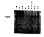
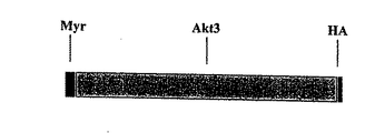


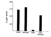
Classifications
-
- C—CHEMISTRY; METALLURGY
- C12—BIOCHEMISTRY; BEER; SPIRITS; WINE; VINEGAR; MICROBIOLOGY; ENZYMOLOGY; MUTATION OR GENETIC ENGINEERING
- C12N—MICROORGANISMS OR ENZYMES; COMPOSITIONS THEREOF; PROPAGATING, PRESERVING, OR MAINTAINING MICROORGANISMS; MUTATION OR GENETIC ENGINEERING; CULTURE MEDIA
- C12N15/00—Mutation or genetic engineering; DNA or RNA concerning genetic engineering, vectors, e.g. plasmids, or their isolation, preparation or purification; Use of hosts therefor
- C12N15/09—Recombinant DNA-technology
- C12N15/11—DNA or RNA fragments; Modified forms thereof; Non-coding nucleic acids having a biological activity
- C12N15/113—Non-coding nucleic acids modulating the expression of genes, e.g. antisense oligonucleotides; Antisense DNA or RNA; Triplex- forming oligonucleotides; Catalytic nucleic acids, e.g. ribozymes; Nucleic acids used in co-suppression or gene silencing
-
- C—CHEMISTRY; METALLURGY
- C12—BIOCHEMISTRY; BEER; SPIRITS; WINE; VINEGAR; MICROBIOLOGY; ENZYMOLOGY; MUTATION OR GENETIC ENGINEERING
- C12N—MICROORGANISMS OR ENZYMES; COMPOSITIONS THEREOF; PROPAGATING, PRESERVING, OR MAINTAINING MICROORGANISMS; MUTATION OR GENETIC ENGINEERING; CULTURE MEDIA
- C12N9/00—Enzymes; Proenzymes; Compositions thereof; Processes for preparing, activating, inhibiting, separating or purifying enzymes
- C12N9/10—Transferases (2.)
- C12N9/12—Transferases (2.) transferring phosphorus containing groups, e.g. kinases (2.7)
- C12N9/1205—Phosphotransferases with an alcohol group as acceptor (2.7.1), e.g. protein kinases
-
- A—HUMAN NECESSITIES
- A61—MEDICAL OR VETERINARY SCIENCE; HYGIENE
- A61P—SPECIFIC THERAPEUTIC ACTIVITY OF CHEMICAL COMPOUNDS OR MEDICINAL PREPARATIONS
- A61P1/00—Drugs for disorders of the alimentary tract or the digestive system
- A61P1/16—Drugs for disorders of the alimentary tract or the digestive system for liver or gallbladder disorders, e.g. hepatoprotective agents, cholagogues, litholytics
-
- A—HUMAN NECESSITIES
- A61—MEDICAL OR VETERINARY SCIENCE; HYGIENE
- A61P—SPECIFIC THERAPEUTIC ACTIVITY OF CHEMICAL COMPOUNDS OR MEDICINAL PREPARATIONS
- A61P13/00—Drugs for disorders of the urinary system
- A61P13/12—Drugs for disorders of the urinary system of the kidneys
-
- A—HUMAN NECESSITIES
- A61—MEDICAL OR VETERINARY SCIENCE; HYGIENE
- A61P—SPECIFIC THERAPEUTIC ACTIVITY OF CHEMICAL COMPOUNDS OR MEDICINAL PREPARATIONS
- A61P37/00—Drugs for immunological or allergic disorders
- A61P37/02—Immunomodulators
- A61P37/06—Immunosuppressants, e.g. drugs for graft rejection
-
- A—HUMAN NECESSITIES
- A61—MEDICAL OR VETERINARY SCIENCE; HYGIENE
- A61P—SPECIFIC THERAPEUTIC ACTIVITY OF CHEMICAL COMPOUNDS OR MEDICINAL PREPARATIONS
- A61P43/00—Drugs for specific purposes, not provided for in groups A61P1/00-A61P41/00
-
- A—HUMAN NECESSITIES
- A61—MEDICAL OR VETERINARY SCIENCE; HYGIENE
- A61P—SPECIFIC THERAPEUTIC ACTIVITY OF CHEMICAL COMPOUNDS OR MEDICINAL PREPARATIONS
- A61P9/00—Drugs for disorders of the cardiovascular system
-
- A—HUMAN NECESSITIES
- A61—MEDICAL OR VETERINARY SCIENCE; HYGIENE
- A61P—SPECIFIC THERAPEUTIC ACTIVITY OF CHEMICAL COMPOUNDS OR MEDICINAL PREPARATIONS
- A61P9/00—Drugs for disorders of the cardiovascular system
- A61P9/10—Drugs for disorders of the cardiovascular system for treating ischaemic or atherosclerotic diseases, e.g. antianginal drugs, coronary vasodilators, drugs for myocardial infarction, retinopathy, cerebrovascula insufficiency, renal arteriosclerosis
-
- C—CHEMISTRY; METALLURGY
- C12—BIOCHEMISTRY; BEER; SPIRITS; WINE; VINEGAR; MICROBIOLOGY; ENZYMOLOGY; MUTATION OR GENETIC ENGINEERING
- C12N—MICROORGANISMS OR ENZYMES; COMPOSITIONS THEREOF; PROPAGATING, PRESERVING, OR MAINTAINING MICROORGANISMS; MUTATION OR GENETIC ENGINEERING; CULTURE MEDIA
- C12N2799/00—Uses of viruses
- C12N2799/02—Uses of viruses as vector
- C12N2799/021—Uses of viruses as vector for the expression of a heterologous nucleic acid
- C12N2799/022—Uses of viruses as vector for the expression of a heterologous nucleic acid where the vector is derived from an adenovirus
-
- C—CHEMISTRY; METALLURGY
- C12—BIOCHEMISTRY; BEER; SPIRITS; WINE; VINEGAR; MICROBIOLOGY; ENZYMOLOGY; MUTATION OR GENETIC ENGINEERING
- C12N—MICROORGANISMS OR ENZYMES; COMPOSITIONS THEREOF; PROPAGATING, PRESERVING, OR MAINTAINING MICROORGANISMS; MUTATION OR GENETIC ENGINEERING; CULTURE MEDIA
- C12N2799/00—Uses of viruses
- C12N2799/02—Uses of viruses as vector
- C12N2799/021—Uses of viruses as vector for the expression of a heterologous nucleic acid
- C12N2799/027—Uses of viruses as vector for the expression of a heterologous nucleic acid where the vector is derived from a retrovirus
-
- C—CHEMISTRY; METALLURGY
- C12—BIOCHEMISTRY; BEER; SPIRITS; WINE; VINEGAR; MICROBIOLOGY; ENZYMOLOGY; MUTATION OR GENETIC ENGINEERING
- C12N—MICROORGANISMS OR ENZYMES; COMPOSITIONS THEREOF; PROPAGATING, PRESERVING, OR MAINTAINING MICROORGANISMS; MUTATION OR GENETIC ENGINEERING; CULTURE MEDIA
- C12N2799/00—Uses of viruses
- C12N2799/02—Uses of viruses as vector
- C12N2799/021—Uses of viruses as vector for the expression of a heterologous nucleic acid
- C12N2799/028—Uses of viruses as vector for the expression of a heterologous nucleic acid where the vector is derived from a herpesvirus
Landscapes
- Health & Medical Sciences (AREA)
- Engineering & Computer Science (AREA)
- Life Sciences & Earth Sciences (AREA)
- Bioinformatics & Cheminformatics (AREA)
- Chemical & Material Sciences (AREA)
- Organic Chemistry (AREA)
- General Health & Medical Sciences (AREA)
- Medicinal Chemistry (AREA)
- Public Health (AREA)
- Veterinary Medicine (AREA)
- Chemical Kinetics & Catalysis (AREA)
- General Chemical & Material Sciences (AREA)
- Nuclear Medicine, Radiotherapy & Molecular Imaging (AREA)
- Pharmacology & Pharmacy (AREA)
- Genetics & Genomics (AREA)
- Animal Behavior & Ethology (AREA)
- Molecular Biology (AREA)
- Biomedical Technology (AREA)
- Zoology (AREA)
- Wood Science & Technology (AREA)
- General Engineering & Computer Science (AREA)
- Immunology (AREA)
- Biotechnology (AREA)
- Biochemistry (AREA)
- Urology & Nephrology (AREA)
- Microbiology (AREA)
- Heart & Thoracic Surgery (AREA)
- Cardiology (AREA)
- Gastroenterology & Hepatology (AREA)
- Transplantation (AREA)
- Physics & Mathematics (AREA)
- Biophysics (AREA)
- Plant Pathology (AREA)
- Vascular Medicine (AREA)
- Medicines That Contain Protein Lipid Enzymes And Other Medicines (AREA)
- Micro-Organisms Or Cultivation Processes Thereof (AREA)
- Peptides Or Proteins (AREA)
- Medicines Containing Material From Animals Or Micro-Organisms (AREA)
- Medicines Containing Antibodies Or Antigens For Use As Internal Diagnostic Agents (AREA)
- Saccharide Compounds (AREA)
Abstract
Description
【0001】
本発明は、単離された核酸、それらを含むベクターおよび治療における、特に遺伝子治療におけるそれらの使用に関する。より具体的には、本発明はAktアイソフォーム(Akt3と称する)をコードする核酸、および遺伝子治療におけるそれらの使用に関する。活性化Akt3の発現は、アポトーシス刺激キナーゼ1(ASK1)によって誘発されるアポトーシス性細胞死を防止する。
【0002】
【従来技術】
Aktおよびアポトーシス:
アポトーシス(計画的細胞死)は、胚の発育および種々の疾患(例えば退行性神経疾患および心脈管性疾患)の発生病理に重要な役割を果たす(MacLellan et al.(1998);Barinaga (1997a);Barinaga (1997b))。したがって、最近の研究は、計画的細胞死の調節または実行に必要な種々のプロアポトーシスおよびアンチアポトーシス遺伝子生成物の特定を目的としていた。アンチアポトーシス遺伝子(例えばBcl2またはBcl−x)は、種々の刺激によって誘発される計画的細胞死を抑制する。他方、プロアポトーシス遺伝子(例えばBaxまたはBad)の発現は計画的細胞死をもたらす(Aams et al (1998))。計画的細胞死の実行は、カスパーゼ−1関連プロテイナーゼ(カスパーゼ−1、カスパーゼ−3、カスパーゼ−7、カスパーゼ−8およびカスパーゼ−9などを含む)によって仲介される(Thorneberry et al.(1998))。
【0003】
細胞生存/細胞死の調節に必要な2つの細胞内シグナリング経路が最近研究されている。アポトーシス刺激キナーゼ1(ASK1)の活性化は種々のタイプの細胞にアポトーシスをもたらすが(Ichijo et al.(1997))、一方、ホスホイノシチド3−キナーゼ(PI3K)およびAktを必要とする経路は細胞防御をもたらす。ASK1活性は、腫瘍壊死因子アルファ(TNFα)処理またはFas連結によって誘発されることが示された(Ichijo et al.(1997);Chang et al.(1998))。ASK1優性ネガティブ変異体の過剰発現はTNFαまたはFasリガンドによって誘発されるアポトーシスを抑制し、ASK1がTNFαまたはFasリガンドによって誘発されるアポトーシス性細胞死において重要な役割を果たすことを示唆している。ASK1がアポトーシスを誘発する分子的メカニズムは明らかではない。ASK1の異所性発現がストレスによって活性化される種々のシグナリング経路、例えばMKK4/JNKおよびMKK6/p38経路(これらはASK1誘発アポトーシスを仲介すると思われる(Ichijo et al.(1997))の活性化をもたらすことが示された。
【0004】
PI3K/Akt経路はまた細胞生存/細胞死の調節に重要らしい(Kulil et al.(1997);Kauffmann-Zeh et al.;Hemmings (1997);Dudek et al.(1997))。生存因子、例えば血小板由来増殖因子(PDGF)、神経増殖因子(NGF)およびインスリン様増殖因子−1(IGF−1)は、PI3Kの活性を誘発することによって種々の条件下で細胞の生存を促進する(Kulik et al.(1997);Hemmings (1997))。活性化PI3Kは、ホスファチジルイノシトール(3,4,5)−トリホスフェート(PtdIns(3,4,5)−P3)の産生をもたらす(PtdIns(3,4,5)−P3はセリン/スレオニンキナーゼを含むAH/PH−ドメインに結合してその活性を誘発する)(Frankel et al.(1995);Hemmings (1997b);Downward (1998);Alessi et al.(1996))。PI3Kまたは優性ネガティブAkt変異体の特異的な抑制物質は、これら増殖因子またはサイトカインの生存促進活性を停止させる。さらに、構成的に活性なPI3KまたはAkt変異体の誘発は、細胞が通常アポトーシス性細胞死に至る条件下で細胞の生存を促進する(Kulik et al.(1997);Dudek et al.(1997))。これらの所見は、PI3K/Akt経路は細胞の生存とアポトーシスの調節に重要な役割を果たすことを示している。
【0005】
ヒトのAkt蛋白質キナーゼの2つのアイソフォーム、Akt1およびAkt2が特定された(Staal (1987))。ラットのAkt配列もまた特定された(Konishi et al.(1995))。ヒトのAkt1のC末端のセリン−473はその調節作用に必須であることが示された(Stokeo et al.(1997);Stephens et al.(1998))。増殖因子による刺激に際してPI3Kが活性化される。PI3Kの生産物、PtdIns(3,4,5)−PはAkt1と結合し、Akt1を細胞質から細胞質内膜の近傍へ移動させ、ここでAkt1の残基スレオニン308およびセリン473はリン酸化される(Downward (1998))。これら残基のリン酸化はAkt1の活性化に極めて重要である。最近特定された蛋白質キナーゼ、PDK1はスレオニン308のリン酸化に必要であることが示されたが、一方セリン473をリン酸化するキナーゼは未だ特定されていない(Stokeo et al.(1997);Stephens et al.(1998))。
【0006】
遺伝子治療:
遺伝子治療は、患者(例えば患者の罹患細胞または器官)に遺伝情報を導入することによって欠損または異常(変異、異常な発現など)を修正することを含む。この遺伝情報は、in vitroで細胞に導入され、続いてこの改変細胞が身体に再導入されるか、またはin vivoで直接適当な部位に導入される。これに関して種々のトランスフェクション技術および遺伝子導入技術が文献に記載されている(例えば以下を参照されたい:Roemer & Friedman, Eur. J. Biochem. 208:211(1992))。これらの技術には、“裸出DNA”のトランスフェクション技術並びにDNAおよびDEAE−デキストラン複合体(Pagano et al. J. Virol. 1:891(1967))、DNAおよび核蛋白質複合体(Kaneda et al. Science 243:375(1989))、DNAおよび脂質複合体(Felgner et al. PNAS 84:7413(1987))を必要とする技術、リポソームの使用(Fraley et al. J. Biol. Chem. 255:10431(1980)) などが含まれる。 より最近では、遺伝子導入用ベクターとしてウイルスの使用が、物理的トランスフェクション技術に代わる有望な選択肢として出現してきた。これに関して、レトロウイルス、ヘルペスウイルス、アデノ付随ウイルスおよびアデノウイルスを含む種々のウイルスが一定の細胞集団に感染する能力について調べられた。
本明細書に引用したいずれの文献も、そのような文献が本出願に対する“従来技術”として入手可能であることを認めたものと解されるべきではない。
【0007】
【発明が解決しようとする課題】
本発明の第一の目的は、新規なAkt蛋白質またはポリペプチドをコードする単離核酸に関する。より具体的には、本発明は、Cys−Gln−Gln−Ser−Asp−Cys−Gly−Met−Leu−Gly−Asn−Trp−Lys−Lysなる配列または実質的に同様の配列を含むヒトAkt3蛋白質をコードする単離核酸に関する。ここで前記核酸は下記から選ばれる特性を有する:
A.それは、配列番号5および配列番号6に由来するオリゴヌクレオチドプライマー対を用いてポリメラーゼ連鎖反応(PCR)によって増幅できる;
B.それは、配列番号1に示すヌクレオチド配列を含む核酸とストリンジェントな条件下でハイブリダイズする;
C.それは、配列番号2、そのスプライス変異体およびその対立遺伝子座変異体から成る群から選ばれるアミノ酸配列を含むポリペプチドをコードする;
D.それは、Cys−Gln−Gln−Ser−Asp−Cys−Gly−Met−Leu−Gly−Asn−Trp−Lys−Lysなる配列または実質的に同様の配列を有するペプチド内のエピトープに対して産生された抗体と特異的に結合するポリペプチドをコードする。
好ましくは、本核酸は配列番号2に示すアミノ酸配列を含むヒトAkt3蛋白質をコードする。より好ましくは、本核酸は配列番号1に示す配列を含む。本核酸は、場合によってポリペプチドタグをコードする配列を含み、それによってタグされたキメラAkt3蛋白質をコードすることができる。別の特徴では、本核酸は、場合によってミリスチル化配列をコードする配列を含むことができる。好ましい実施態様では、本核酸は哺乳類細胞でAkt3蛋白質の発現を可能にする領域を含む。
【0008】
本発明はまた上記の核酸を含有するベクターに関する。好ましくは、ヒトAkt3蛋白質をコードする核酸は、発現調節を有する宿主細胞でヒトAkt3を発現させることができる発現制御配列と機能的に結合される。この発現制御配列は、哺乳類細胞で機能することができるプロモーターを含むことができる。本ベクターはプラスミドDNA分子またはウイルスベクターであろう。好ましいウイルスベクターには、レトロウイルス、アデノウイルス、アデノ付随ウイルス、ヘルペスウイルスおよびワクシニアウイルスが含まれる。したがって、本発明はさらに、そのゲノム内に上記のAkt3をコードする核酸を含む複製欠損組み換えウイルスに関する。
別の実施態様では、本発明は上記のベクターでトランスフェクトされた宿主細胞に関する。本宿主細胞は細菌細胞、酵母細胞または哺乳類細胞であろう。さらに別の実施態様では、本発明はヒトAkt3蛋白質を製造する方法に関する。本方法は、ヒトAkt3蛋白質を発現させる条件下で上記の宿主細胞を培養液中で培養することを含む。
【0009】
本発明はさらに、Cys−Gln−Gln−Ser−Asp−Cys−Gly−Met−Leu−Gly−Asn−Trp−Lys−Lysなる配列または実質的に同様の配列を含み、下記から選ばれる特性を有する単離ヒトAkt3蛋白質に関する:
A.それは上記の核酸によってコードされる;
B.それは、配列番号2に示すアミノ酸配列、そのスプライス変異体またはその対立遺伝子座変異体を含む;さらに
C.それは、Cys−Gln−Gln−Ser−Asp−Cys−Gly−Met−Leu−Gly−Asn−Trp−Lys−Lysなる配列または実質的に同様の配列を有するペプチド内のエピトープに対して産生された抗体と特異的に結合する。
好ましくは、本蛋白質は配列番号2に示すアミノ酸配列を含む。本蛋白質は場合によってタグ配列を含むことができる。さらに別の実施態様では、本蛋白質は場合によってミリスチル化配列を含むことができる。
【0010】
本発明はまた抗原性ペプチドおよびそれに対する抗体に関する。より具体的には、本発明は、Cys−Gln−Gln−Ser−Asp−Cys−Gly−Met−Leu−Gly−Asn−Trp−Lys−Lysなる配列または実質的に同様の配列を含む抗原性ペプチドであって、さらにヒトAkt3蛋白質の断片であるものに関する。ここで前記Akt3蛋白質は下記から選ばれる特性を有する:
A.それは上記の核酸によってコードされる;
B.それは、配列番号2に示すアミノ酸配列、そのスプライス変異体またはその対立遺伝子座変異体を含む;さらに
C.それは、Cys−Gln−Gln−Ser−Asp−Cys−Gly−Met−Leu−Gly−Asn−Trp−Lys−Lysなる配列または実質的に同様の配列を有するペプチド内のエピトープに対して産生された抗体と特異的に結合する。
好ましくは、本抗原性ペプチドは本質的にCys−Gln−Gln−Ser−Asp−Cys−Gly−Met−Leu−Gly−Asn−Trp−Lys−Lysなる配列またはその抗原性断片から成る。さらに別の実施態様では、本発明は、上記のヒトAkt3蛋白質と特異的に結合する抗体に関する。好ましい実施態様では、本抗体は、Cys−Gln−Gln−Ser−Asp−Cys−Gly−Met−Leu−Gly−Asn−Trp−Lys−Lysなる配列を有するペプチド内のエピトープを特異的に認識する。本抗体はポリクローナルでもモノクローナルでもよい。
【0011】
本発明はまた、上記の核酸を細胞に投与することによって細胞のアポトーシスまたは壊死を抑制する方法に関する。好ましくは、本核酸は調節領域に機能的に連結される。本核酸はプラスミドでもウイルスベクターでもよい。好ましい細胞には心臓細胞が含まれる。より好ましくはこの細胞は心筋細胞である。好ましい実施態様では、この細胞は心筋梗塞または虚再灌流障害をもつ患者の細胞である。したがって、本発明はまた、心筋梗塞または虚再灌流障害をもつ患者に調節領域に機能的に連結された上記の核酸を投与することによって、心筋梗塞または虚血再灌流障害を治療する方法に関する。好ましくは、本核酸は患者の心筋細胞に投与される。本核酸はプラスミドまたはウイルスベクターの形態を有するであろう。好ましいウイルスベクターにはレトロウイルス、アデノウイルス、アデノ付随ウイルス、ワクシニアウイルスおよびHSVウイルスが含まれる。
【0012】
本発明はまた、人間または動物の身体を外科的または内科的に処置するための医薬組成物の製造にこれら核酸またはベクターを使用することに関する。本発明はまた、上記のベクター(特にウイルスベクター)および核酸を含む任意の医薬組成物に関する。
【0013】
さらに別の実施態様では、本発明は、Akt3蛋白質を候補分子と接触させ、前記分子の存在下でAkt3活性を検出することによって細胞でAkt3活性を刺激または抑制する分子をスクリーニングする方法に関する。候補分子はAkt3の作動薬または拮抗薬のいずれかであろう。好ましい実施態様では、Akt3は細胞内で核酸から発現され、測定されるAkt3活性はアポトーシスの抑制である。アポトーシスの抑制はマーカー遺伝子の存在によって測定できる。
【0014】
一般に本発明はまた、細胞内のAkt3蛋白質レベルを増加させることによって細胞のAkt3活性を増加させる方法に関する。好ましい実施態様では、Akt3蛋白質を発現させることができる条件下でAkt3をコードするベクターで細胞をトランスフェクトした。同様に、本発明は、細胞内のAkt3蛋白質レベルを低下させることによって細胞のAkt3活性を抑制する方法に関する。Akt3蛋白質レベルは、Akt3アンチセンス核酸を前記アンチセンス核酸が細胞内の条件でAkt3mRNAとハイブリダイズするような条件下で細胞に導入することによって低下させることができる。Akt3蛋白質レベルはまた、Akt3と特異的に結合する一本鎖Fv抗体(scFv)を、Akt3と結合してこれを不活化するために十分なレベルで細胞に導入することによって低下させることができる。
【0015】
添付図面において:
図1は図1はAkt3配列のアラインメントである。図1Aは、ヒトAkt1(配列番号14)、Akt2(配列番号13)およびAkt3(配列番号2)のアミノ酸配列のアラインメントで、図1Bは、ラットAkt(配列番号17)およびヒトAkt3(配列番号2)アミノ酸配列のアラインメントである。
図2はAkt3mRNAの組織分布パターンを示す。Akt3特異的プローブは実施例で述べたように調製した。多数のヒト組織mRNAブロット(Clontech)をAkt3特異的プローブとハイブリダイズさせた(詳細については実施例を参照されたい)。
図3は活性化Akt3変異体の構築を示す。図3Aは活性化Akt3の模式図である。ヒトAkt3の完全長コード配列をヒトSrc遺伝子由来ミリスチル化シグナル(Myr)とN−末端でフレーム内で融合させ、さらにC−末端でHAタグ(HA)とフレーム内で融合させた(実施例を参照されたい)。図3Bは活性化Akt3のHEK293細胞での異所性発現を示す。HEK293細胞にCMV6−MyrAkt3HAまたは発現プラスミド(CMV6)のみをトランスフェクトした。トランスフェクション後24時間で、細胞溶解物を調製し、HA抗体を用いてイムノブロットを実施した。図3Cは、活性化Akt3はAkt活性をもつことを示す。HEK293細胞に、活性化Akt3のための発現プラスミド(MyrAkt3HA)または発現ベクターのみ(CMV6)をトランスフェクトした。トランスフェクション後24時間で、細胞溶解物を調製し、HA抗体を用いてイムノブロットを実施した。イムノペレットのAkt3キナーゼ活性は、GSK3に由来する基質ペプチドを用いて測定した。Bkgd:非トランスフェクト細胞のバックグラウンドレベル、CMV6:CMV6トランスフェクト細胞、Akt3cak:構成的に活性化されたAkt3のための発現プラスミド(CMV6−MyrAkt3HA)でトランスフェクトされた細胞(実施例を参照のこと)。
図4は活性化Akt3はHEK293細胞でASK1誘発細胞死を抑制することを示す。HEK細胞に表示のプラスミドとともにCMV−βgalプラスミド(0.1mg)をトランスフェクトした。各トランスフェクションのDNA量はCMVベクターを添加することによって一定に保たれた。Akt3cak:CMV6−MyrAkt3HA(0.4mg)、ASK1:pCDNA3HA−ASK1−fl(0.4mg)。トランスフェクション後2日して、細胞を固定し、X−gal染色を施した。光学顕微鏡下でβ−gal陽性細胞(青色細胞)の数を5つの異なる視野で数えた。
【0016】
【課題を解決するための手段】
本発明は、新規なAkt蛋白質またはポリペプチド(Akt3と称する)をコードする単離核酸を有利に提供する。したがって、本発明の第一の目的は、哺乳類細胞でのその発現を可能にすることができる領域の制御下で新規なAkt3蛋白質またはポリペプチドをコードする単離核酸に関する。本発明はまた、Akt蛋白質またはポリペプチドをコードする核酸を含むベクターに関する。本発明はまた、人間または動物の身体の外科的および/または内科的処置のための医薬組成物の調製にこれら核酸またはベクターを使用することに関する。本発明はまた、上記のベクター(特にウイルスベクター)および核酸を含む任意の医薬組成物に関する。
本発明の種々の特徴は、前記核酸、ベクター、ウイルス、組成物および治療方法に関する下記の章で極めて詳細に説明される。種々の章から成る本構成は本発明の理解を容易にすることを意図したもので、本発明を限定しようとするものではない。
【0017】
定義:
以下の用語の定義が本明細書を通して使用され、本発明の範囲と実践の理解に役立つであろう。
具体的な実施態様で、“約”または“およそ”という用語はある数値または範囲の20%以内、好ましくは10%以内、より好ましくは5%以内を意味する。
“核酸”とは、共有結合によって連結されたヌクレオチドと称されるサブユニットで構成されたポリマー化合物である。核酸にはポリリボ核酸(RNA)およびポリデオキシリボ核酸(DNA)(これらは両者とも一本鎖でも二本鎖でもよい)が含まれる。DNAにはcDNA、ゲノムDNA、合成DNAおよび半合成DNAが含まれる。
“遺伝子”とは、ポリペプチドをコードするヌクレオチドの集合物を指し、cDNAおよびゲノムDNAが含まれる。
“組み換え(リコンビナント)DNA分子”とは、分子生物学的操作を経たDNA分子である。
【0018】
“ベクター”とは、宿主細胞に核酸を導入する任意の手段である。ベクターはレプリコンであって、付加したまた別のDNAセグメントを増殖させるためにこの別のDNAセグメントが前記レプリコンに結合されている。“レプリコン”とは、in vivoでDNA複製の自律的ユニットとして機能する(すなわちそれ自身の制御下で複製することができる)任意の遺伝的エレメント(例えばプラスミド、コスミド、染色体、ウイルス)である。“ベクター”という用語は、in vitro、ex vivoまたはin vivoで細胞に核酸を導入するウイルス性および非ウイルス性の両手段を含む。ウイルスベクターには、下記で極めて詳細に説明するようにレトロウイルス、アデノ付随ウイルス、ポックスウイルス、バキュロウイルス、ワクシニアウイルス、ヘルペスシンプレックスウイルス、エプスタイン=バーウイルスおよびアデノウイルスベクターが含まれる。非ウイルスベクターには、プラスミド、リポソーム、荷電をもつ脂質(サイトフェクチン)、DNA蛋白質複合体およびバイオポリマーが含まれる。核酸の他に、ベクターはまた1つまたは2つ以上の調節領域、および/または核酸移入の成果(どの組織に移入されたか、発現の期間など)の選別、測定およびモニタリングに役立つ選別マーカーを含むことができる。
【0019】
“クローニングベクター”とは、レプリコン(例えばプラスミド、ファージまたはコスミド)で、別の付加DNAセグメントを増殖させるために前記レプリコンに別のDNAセグメントを結合させることができる。クローニングベクターは1つの細胞タイプでの複製能力および別の細胞タイプでの発現能力を有するであろう(シャトルベクター)。
“カセット”とは、特定の制限部位に挿入することができるDNAセグメントを指す。このDNAセグメントは問題のポリペプチドをコードし、さらにこのカセットおよび制限部位は、転写および翻訳のための適切な読み枠への本カセットの挿入を保証できるようにデザインされる。
外因性または異種DNAが細胞内に導入されたとき、この細胞は前記外因性または異種DNAを“トランスフェクト”されたという。前記トランスフェクトされたDNAが表現型の変化を生じたとき、この細胞は外因性または異種DNAによって”形質転換”されたという。形質転換DNAは、細胞のゲノムを構成している染色体DNAに組み込まれる(共有結合により連結される)ことができる。
【0020】
“核酸分子”とは、リボヌクレオシド(アデノシン、グアノシン、ウリジンまたはシチジン;“RNA分子”)またはデオキシリボヌクレオシド(デオキシアデノシン、デオキシグアノシン、デオキシチミジンまたはデオキシシチジン;“DNA分子”)のリン酸エステルポリマーまたはその任意のホスホエステル類似体、例えばホスホロチオエートおよびチオエステルを指し、これらは一本鎖型または二本鎖らせんである。二本鎖DNA−DNA、DNA−RNAおよびRNA−RNAらせんが可能である。核酸分子、特にDNAまたはRNA分子という用語は、それら分子の一次および二次構造を指すだけであって、それらをいずれの特定の三次構造にも限定するものではない。したがってこの用語には、とりわけ直線状または環状DNA分子で見いだされる二本鎖DNA(例えば制限フラグメント)、プラスミドおよび染色体が含まれる。特定の二本鎖DNA分子の構造についていう場合、本明細書ではDNAの非転写鎖(すなわちmRNAと相同な配列を有する鎖)の5’から3’方向の配列のみを提示するという一般的慣行にしたがって配列が記載される。”組み換え(リコンビナント)DNA分子”とは、分子生物学的操作を経たDNA分子である。
【0021】
核酸分子は、その一本鎖形が別の核酸分子、例えばcDNA、ゲノムDNAまたはRNAと適当な温度および溶液イオン強度条件下で(Sambrook et al.上掲書参照)アニールすることができるとき、前記核酸分子はこれら他の核酸分子と“ハイブリダイズ”することができるという。温度およびイオン強度条件はハイブリダイゼーションの“厳正度(ストリンジェンシー)”を決定する。相同な核酸についての予備的スクリーニングの場合、低いストリンジェンシーのハイブリダイゼーション条件(Tmが55°Cに対応する)を用いることができる。これは、例えば5×SSC、0.1%のSDS、0.25%のミルクおよびホルムアミド無し;または5×SSC、0.5%SDSである。中等度のストリンジェンシーのハイブリダイゼーション条件はより高いTmに対応し、例えば40%ホルムアミドと5×または6×SSCである。高いストリンジェンシーのハイブリダイゼーション条件はもっとも高いTmに対応し、例えば50%のホルムアミド、5×または6×SSCである。ハイブリダイゼーションには2つの核酸が相補的な配列を含むことが必要であるが、ただしハイブリダイゼーションのストリンジェンシーに依存するが塩基間のミスマッチも可能である。核酸をハイブリダイズさせるために適したストリンジェンシーは、核酸の長さおよび相補性の度合い、当分野で周知の変数に左右される。2つのヌクレオチド間の類似性または相同性の度合いが大きければ大きいほど、これらの配列を含む核酸ハイブリッドのTm値は高くなる。核酸のハイブリダイゼーションの相対的安定性(Tmの高さに対応する)は以下の順序で低下する:RNA:RNA、DNA:RNA、DNA:DNA。長さが100ヌクレオチド以上のハイブリッドの場合、Tmを算出する等式が推定された(Sambrook et al.上掲書9.50-0.51参照)。より短い核酸(すなわちオリゴヌクレオチド)とのハイブリダイゼーションの場合、ミスマッチの位置はより重要となり、オリゴヌクレオチドの長さはその特異性を決定する(Sambrook et al.上掲書9.50-0.51参照)。好ましくは、ハイブリダイズできる核酸の最小の長さは少なくとも約10ヌクレオチド、好ましくは少なくとも約15ヌクレオチド、より好ましくは少なくとも約20ヌクレオチドである。
具体的な実施態様では、“標準的なハイブリダイゼーション条件”はTm55°Cを指し、上記の条件を利用する。好ましい実施態様では、Tmは60°Cで、より好ましい実施態様では、Tmは65°Cである。
【0022】
本明細書で用いられるように、“オリゴヌクレオチド”という用語は、核酸、一般に少なくとも18ヌクレオチドで、ゲノムDNA分子、cDNA分子、またはAkt3をコードするmRNA分子とハイブリダイズすることができるものを指す。オリゴヌクレオチドは、例えば32P−ヌクレオチドで標識するか、またはビオチンのような標識を共有結合させたヌクレオチドで標識することができる。ある実施態様では、標識オリゴヌクレオチドは、Akt3をコードする核酸の存在を検出するためのプローブとして用いることができる。別の実施態様では、オリゴヌクレオチド(その一方または両方を標識してもよい)は、完全長のAkt3またはAkt3のフラグメントをクローニングするために、またはAkt3をコードする核酸の存在を検出するためにPCRプライマーとして用いることができる。さらに別の実施態様では、本発明のオリゴヌクレオチドは、Akt3DNA分子と三重らせんを形成することができる。一般に、オリゴヌクレオチドは合成によって、好ましくは核酸合成装置で調製される。したがって、オリゴヌクレオチドは天然に存在しないホスホエステル類似体結合、例えばチオエステル結合などにより調製できる。
【0023】
DNAの“コード配列”は二本鎖DNA配列で、これは、適当な調節配列の制御下に配置されたとき細胞内でin vitroまたはin vivoで転写されポリペプチドに翻訳される。コード配列の境界は5'(アミノ)末端の開始コドンおよび3’(カルボキシ)末端の終止コドンによって決められる。コード配列は、原核細胞配列、真核細胞のmRNAに由来するcDNA、真核細胞(例えば哺乳類)のDNAに由来するゲノムDNA配列および合成DNAさえも含むが、ただしこれらに限定されない。コード配列が真核細胞での発現を目的としている場合は、通常はポリアデニル化シグナルおよび転写終了配列がコード配列の3’側に配置されるであろう。
【0024】
転写および翻訳制御配列は、DNA調節配列、例えばプロモーター、エンハンサー、ターミネーターなどで、宿主細胞でのコード配列の発現をもたらす。
“プロモーター配列”は細胞内のRNAポリメラーゼと結合し下流の(3'方向)コード配列の転写を開始させることができるDNA調節領域である。本発明での定義の場合、前記プロモーターはその3’末端で転写開始部位と接し、上流(5'方向)に伸長し、バックグラウンドを越える検出可能なレベルの転写を開始させるために必要な最低数の塩基またはエレメントを含む。プロモーター配列内に、転写開始部位(簡便にはヌクレアーゼS1によるマッピングによって範囲が定められる)およびRNAポリメラーゼの結合に必要な蛋白質結合ドメイン(コンセンサス配列)が見いだされるであろう。
RNAポリメラーゼがコード配列をmRNAに転写するとき、コード配列は細胞内で転写および翻訳制御配列の“制御下”にあり、続いてこれはトランス−RNAスプライス処理を受け(コード配列がイントロンを含む場合)、さらにコード配列によってコードされる蛋白質に翻訳される。
【0025】
本明細書で用いられるように、“相同”という用語は“共通の進化起源”をもつ蛋白質間の関係を指し、これら蛋白質はスーパーファミリー由来の蛋白質(例えば免疫グロブリンスーパーファミリー)および異なる種の同種蛋白質(例えばミオシンの軽鎖など)を含む(Reeck et al. Cell 50:667(1987))。そのような蛋白質(およびそれらのコード遺伝子)は、その高い配列類似性によって示されるように配列相同性を有する。
したがって、“配列類似性”という用語は、共通の進化起源を共有する場合もしない場合も蛋白質における核酸またはアミノ酸配列間の同一性または一致の度合いを指す(Reeck et al. 上掲書参照)。しかしながら、一般的な用法および本出願では、例えば“高度に”というような副詞で修飾される場合は、“相同”という用語は配列類似性を意味し、共通の進化起源を意味しない。
【0026】
具体的な実施態様では、ヌクレオチドの少なくとも約50%、好ましくは少なくとも約75%、もっとも好ましくは少なくとも約90または95%がDNA配列の一定の長さにわたってマッチするとき、2つのDNA配列は“実質的に相同”または“実質的に類似”である。実質的に相同な配列は、配列データバンクで入手可能な標準的ソフトを用いて比較するか、または例えば個々の系で規定されるストリンジェントな条件下でサザンハイブリダイゼーション実験で比較することによって特定できる。適当なハイブリダイゼーション条件を規定することは当業者の範囲内である(例えば以下を参照されたい:Maniatis et al. 上掲書;DNA Cloning, Vols I & II, 上掲書;Nucleic Acid Hybridization, 上掲書)。
【0027】
“アンチセンス核酸”とは、センス配列と相補的なヌクレオチド配列である。アンチセンス核酸は、センス鎖によってコードされるポリペプチドの発現を下方調節または阻止するために用いることができる。
転写および翻訳制御配列はDNA調節配列、例えばプロモーター、エンハンサー、ターミネーターなどで、宿主細胞でのコード配列の発現をもたらす。
“シグナル配列”は、細胞の表面で発現させるために蛋白質のコード配列の開始部に含まれる。この配列は成熟ポリペプチドのN−末端でシグナルペプチドをコードし、シグナルペプチドは宿主細胞にこのポリペプチドの移動を指令する。“移動シグナル配列”という用語は、この種のシグナル配列を指すために本明細書では使用される。移動シグナル配列は、真核細胞および原核細胞に本来存在する多様な蛋白質に結合して見いだされ、しばしば両タイプの生物で機能を有する。
【0028】
“調節領域”とは、第二の核酸配列の発現を調節する核酸配列を意味する。調節領域は、個々の核酸の発現に本来必要な配列(同種領域)を含むことができるが、また異なる蛋白質または合成蛋白質でもその発現に必要な異種起源の配列を(異種領域)含むことができる。特に、この配列は真核細胞遺伝子またはウイルス遺伝子の配列でもよいし、また特異的態様もしくは非特異的態様で、または誘発性もしくは非誘発性態様で遺伝子の転写を刺激または抑制する誘導配列でもよい。調節領域には、複製起点、RNAスプライス部位、プロモーター、エンハンサー、転写終了配列、ポリペプチドを標的細胞の分泌経路に誘導するシグナル配列、およびプロモーターが含まれる。
【0029】
“異種起源”の調節領域は、発現される核酸に天然には付随していない調節領域である。異種調節領域には、異なる種の調節領域、異なる遺伝子の調節領域、ハイブリッド調節配列、および天然には存在しないが当業者がデザインできる調節配列が含まれる。
“異種”DNAとは、細胞内または細胞の染色体部位に天然には分布していないDNAを指す。好ましくは異種DNAにはその細胞にとって外来性の遺伝子が含まれる。
“相同組み換え”とは、外来DNA配列の別のDNA分子への挿入、例えばベクターの染色体内挿入を指す。好ましくは、相同組み換えではベクターは特定の染色体部位を標的とする。固有の相同組み換えの場合、相補的な結合およびベクターの染色体への取り込みを可能にするために、ベクターは染色体の配列に対して十分に長い相同領域を含む。相同な領域が長ければ長いほど、さらに配列類似性の度合いが大きければ大きいほど、相同組み換えの効率は増加するであろう。。
【0030】
“ポリペプチド”は共有結合によって連結されたアミノ酸残基で構成されたポリマー化合物である。アミノ酸は下記の構造を有する:
【0031】
【化1】

アミノ酸は側鎖Rを基準にして7つの群に分類される:(1)鎖式側鎖、(2)ヒドロキシ(OH)を含む側鎖、(3)硫黄原子を含む側鎖、(4)酸性またはアミド基を含む側鎖、(5)塩基性の基を含む側鎖、(6)芳香環を含む側鎖、(7)プロリン、側鎖がアミノ基と融合したイミノ酸。
“蛋白質”は生細胞で構造的および機能的役割を果たすポリペプチドである。
ポリペプチドまたは蛋白質の“変種”は、ポリペプチドまたは蛋白質に由来し、ポリペプチドまたは蛋白質の少なくとも1つの生物学的特性を保持する一切の類似体、フラグメント、誘導体または変異体である。ポリペプチドまたは蛋白質の多様な変種が天然に存在する。これらの変種は蛋白質をコードする構造遺伝子のヌクレオチド配列に相違を有することを特徴とする対立遺伝子座変異体でもよいし、種々のスプライシングによる修飾、または翻訳後修飾を含むものでもよい。当業者は、ただ1つまたは多数のアミノ酸置換、欠失、付加または置き換えをもつ変種を製造できる。これらの変種にはとりわけ以下が含まれる:(a)1つまたは2つ以上のアミノ酸残基が保存的または非保存的アミノ酸で置換されている変種、(b)1つまたは2つ以上のアミノ酸がポリペプチドまたは蛋白質に付加されている変種、(c)1つまたは2つ以上のアミノ酸が置換基を含む変種、および(d)ポリペプチドまたは蛋白質が別のポリペプチド(例えば血清アルブミン)と融合している変種。これら変種を得る技術(遺伝子的(サプレッション、欠失、突然変異誘発など)、化学的および酵素的技術を含む)は当業者にはよく知られている。好ましくは本発明の変種は少なくとも約14アミノ酸を含む。
そのような対立遺伝子座変異体、類似体、フラグメント、誘導体、変異体および改変物(また別のmRNAスプライシング形およびまた別の翻訳後修飾形を含む)が、ポリペプチドの生物学的特性のいずれかを保持するペプチド誘導体を生じる場合、それらは本発明の範囲内に包含される。
【0033】
“異種蛋白質”とは天然には該当細胞内で産生されない蛋白質を指す。
アミノ酸の約40%以上が同一であるか、または60%以上が類似している(機能的に同一)とき、この2つのアミノ酸配列は”実質的に相同”であるか、または“実質的に類似”している。好ましくは、類似または相同配列は、例えばGCG(Genetics Computer Group, Program Manual for the GCG Package, Version 7, Madison, Wisconsin)パイルアップ(pileup)プログラムを用いたアラインメントによって特定できる。
“一致する”という用語は、類似性または相同性が測定される分子とその正確な位置が同じであろうと異なっていようと、類似配列または相同配列を指すために本明細書では用いられる。核酸またはアミノ酸配列のアラインメントはスペースを含むことができる。したがって、“一致する”という用語は、配列類似性を意味し、アミノ酸残基またはヌクレオチド塩基の番号には及ばない。
【0034】
Akt3タンパク質をコードする遺伝子
本発明は、Akt3の全長若しくは天然型、およびそれらのヒトAkt3特異的抗原断片のいかなるものをも含む、本発明のヒトAkt3タンパク質またはポリペプチドをコードする遺伝子の単離を企図する。本明細書で使用する、「Akt3」とはAkt3ポリペプチドを指し、「akt3」とはAKT3ポリペプチドをコードする遺伝子を指す。本発明の目的のために、「Akt3」という用語は、アポトーシスを阻害することが可能であり、かつ配列Cys−Gln−Gln−Ser−Asp−Cys−Gly−Met−Leu−Gly−Asn−Trp−Lys−Lys、或いは実質的に類似の配列を含むタンパク質またはポリペプチドのいかなるものをも意味する。好ましくは、配列Cys−Gln−Gln−Ser−Asp−Cys−Gly−Met−Leu−Gly−Asn−Trp−Lys−Lys、或いは実質的に類似の配列は、Akt3タンパク質のC末端に生じる。
【0035】
好ましくは、本発明に係る新規なAkt3は配列番号2に記載するアミノ酸配列からなる。本発明に係る好適な核酸は、配列番号2に記載するアミノ酸配列をコードする。より好ましくは、その核酸は配列番号1に記載する配列からなる。
【0036】
本発明の第一の主題は、選択肢として哺乳動物細胞中で発現させる領域の制御下、新規なAktタンパク質またはポリペプチドをコードする単離された核酸に関する。また、本発明は前記Aktタンパク質またはポリペプチドをコードする核酸を含むベクターに、およびこれらの核酸類またはベクター類を人間または動物の外科的および/または治療的処置を目的とする薬学的組成物の調製に使用することに関する。本発明は、さらにウイルス性ベクターのようなベクターを含むいかなる薬学的組成物、および上記に定義した核酸に関する。
【0037】
本発明に従うと、当該技術分野の熟練度の範囲内で慣用的な分子生物学、微生物学、および組換えDNA技術が用いられる。このような手法は、文献で十分に説明されている。例えば、Sambrook, Fritsch & Maniatis, Molecular Cloning: A Laboratory Manual. Second Edition (1989) Cold Spring Harbor Laboratory Press, Cold Spring Harbor, New York (ここで「Sambrookら、1989」とする); DNA Cloning: A Practical Approach, Volumes I and II (D. N. Glover ed. 1985); Oligonucleotide Synthesis (M. J. Gait ed. 1984); Nucleic Acid Hybridization [B. D. Hames & S. J. Higgins eds. (1985)]; Transcription And Translation [B. D. Hames & S. J. Higgins eds. (1984)]; Animal Cell Culture [R. I. Freshney, ed. (1986)]; Immobilized Cells AND Enzymes [IRL Press, (1986)]; B. Perbal, A Practical Guide To Molecular Cloning (1984); F. M. Ausubel et al. (eds.), Current Protocols in Molecular Biology, John Wiley & Sons, Inc. (1994)を参照。
【0038】
ゲノムDNAであるかcDNAであるかにかかわらず、いかなる供給源、特にヒトcDNAまたはゲノムライブラリーからAkt3をコードする遺伝子を単離することができる。akt3遺伝子を得る一般的な方法は、上記のように周知である(例えば、上掲「Sambrookら、1989」を参照)。
【0039】
したがって、いかなる動物細胞でもakt3遺伝子の分子クローニング用の核酸供給源として役に立つことができる可能性がある。クローン化されたDNA(例えば、DNAライブラリー)から公知である標準的な手順によってDNAを得ることができる。好ましくは、前記タンパク質発現の高レベルを有する組織(Akt3発現の高いレベルの形跡がある細胞であるから、心臓、膵臓および骨格筋cDNA等)から作成されたcDNAライブラリーから、または化学合成によって、cDNAクローニングによって、或いは所望の細胞から精製されたゲノムDNA、その断片のクローニングによって得られる。例えば、上掲「Sambrookら、1989」; Glover, D. M. (ed.). 1985, DNA Cloning: A Practical Approach. MRL Press. Ltd., Oxford. U.K. Vol. I. IIを参照。ゲノムDNAに由来するクローンは、コード領域に加えて、調節およびイントロンDNA領域を含むかもしれないが、cDNAに由来するクローンは、イントロン配列を含まないことになる。供給源が何であれ、前記遺伝子は、その遺伝子の増殖用に適当なベクターに分子的にクローニングされねばならない。
【0040】
DNA断片が生成されると、所望のakt3遺伝子を含む特定のDNA断片の確認は数々の方式で達成される。例えば、DNA断片は標識されたプローブへの核酸ハイブリダイゼーションによってスクリーニングできる(Benton and Davis, 1977, Science 196: 180; Grunstein and Hogness, 1975, Proc. Natl. Acad. Sci. U.S.A. 72: 3961)。そのプローブに対して実質的な相同性を有するこれらのDNA断片がハイブリダイズすることになる。上述のように、相同性が高ければ、よりストリンジェントな条件を使用することができる。特定の実施形態において、ノーザンハイブリダイゼーション条件を使用して、akt3遺伝子のmRNAスプライシング変異体を同定する。
【0041】
遺伝子の性質に基づいてさらなる選択を実施することができるが、これらは例えば、その遺伝子が本明細書に開示されたAkt3タンパク質の等電的性質、電気泳動的性質、アミノ酸組成、または部分アミノ酸配列を有するタンパク質をコードするかどうか等である。こうして、前記遺伝子の存在は、その発現産物の物理的、化学的、または免疫学的性質に基づくアッセイによって検出できる。例えば、適切なRNAをハイブリッド選択するcDNAクローンまたはDNAクローンは、選択でき、これらはAkt3ついて知られているのと同様なまたは同一の電気泳動的移動度、等電点電気泳動または非平衡pHゲル電気泳動的挙動、タンパク質分解酵素マップ、或いは抗原性性質等を有するタンパク質を生産する。特定の実施形態において、発現されたタンパク質は、ポリクローナル抗体によって認識されるが、それはアミノ酸配列Cys−Gln−Gln−Ser−Asp−Cys−Gly−Met−Leu−Gly−Asn−Trp−Lys−Lys内にあるようなヒトAkt3に特異的なエピトープに対して、作製される。
【0042】
本発明は、本発明のAkt3のアレル変異体、スプライシング変異体、類似体、および誘導体をであって、Akt3と同一または相同的機能活性を有するものをコードする遺伝子(cDNA等)、並びに他種からのその相同体にも関する。Akt3に関連する誘導体および類似体の製造および使用は、本発明の範囲内である。そのような変異体、類似体、誘導体および相同体類は、配列Cys−Gln−Gln−Ser−Asp−Cys−Gly−Met−Leu−Gly−Asn−Trp−Lys−Lys、または実質的に類似の配列を保持すべきである。特定の実施形態では、その誘導体または類似体は、機能的に活性であり、すなわち本発明の全長、野生型のAkt3に関連する一つ以上の機能的活性を示すことができる。
【0043】
機能的に等価な分子をもたらす置換、付加、または欠失によってコード核酸配列を改変して、Akt3誘導体を作ることができる。好ましくは、天然型Akt3と比べて高められた、または増加された機能的活性を有する誘導体が作られる。
【0044】
核酸コード配列の縮退のために、akt3遺伝子と実質的に同一のアミノ酸配列をコードする他のDNA配列(単一アミノ酸変異体を含むアミノ酸配列を含めて)を本発明の実施に際して使用してもよい。これらには、限定はされないが、アレル遺伝子、他種からの相同的遺伝子、およびその配列内の同一のアミノ酸残基をコードする異なるコドンの置換によって改変され、かくしてサイレント変質を生むakt3遺伝子の全てまたは部分を含む核酸配列が包含される。同様に、本発明のAkt3誘導体には、限定はされないが、一次配列としてAkt3タンパク質のアミノ酸配列の全てまたは部分(機能的に等価なアミノ酸残基によってその配列内残基が置換され、保存的なアミノ酸置換が結果としてもたらされている改変された配列を含む)を含むものが包含される。例えば、配列内の一つ以上のアミノ酸残基は、類似の極性であり、機能的等価体として作用する別のアミノ酸で置換することができるが、これによりサイレント変質がもたらされる。その配列内のアミノ酸に対する置換体は、該アミノ酸が属するクラスの他のメンバーから選択され得る。例えば、非極性(疎水性)アミノ酸には、アラニン、ロイシン、イソロイシン、バリン、プロリン、フェニルアラニン、トリプトファン、およびメチオニンが含まれる。芳香環構造を有するアミノ酸は、フェニルアラニン、トリプトファンおよびチロシンである。極性で中性アミノ酸には、グリシン、セリン、トレオニン、システイン、チロシン、アスパラギン、およびグルタミンが含まれる。正に帯電した(塩基性)アミノ酸には、アルギニン、リシン、およびヒスチジンが含まれる。負に帯電した(酸性)アミノ酸には、アスパラギン酸およびグルタミン酸が含まれる。そのような改変は、ポリアクリルアミドゲル電気泳動または等電点によって測定される見かけの分子量に影響を及ぼすとは予想されない。
【0045】
特に好ましい置換は、以下の通りである。
正の電荷が維持されるように、ArgをLysで置換およびその逆;
負の電荷が維持されるように、AspをGluで置換およびその逆;
遊離OHが維持され得るように、ThrをSerで置換;および
遊離CONH2OHが維持され得るように、AsnをGlnで置換。
【0046】
アミノ酸置換は、また特に好ましい性質を持つアミノ酸で置換するために導入してもよい。例えば、別のCysとのジスルフィド架橋用の潜在的な部位にCysを導入してもよい。特に「触媒」部位としてHisを導入してもよい。すなわち、Hisは酸若しくは塩基として作用し、生化学的触媒反応において最も知られたアミノ酸である。Proは、その独特な平面構造の理由から導入されてもよく、タンパク質構造のβターンを誘発する。
【0047】
本発明のAkt3誘導体および類似体をコードする遺伝子は、公知である様々な方法によって作製することができる。その作製をもたらす操作は、遺伝子またはタンパク質段階で起こりうる。例えば、クローン化Akt3遺伝子配列を公知である無数の戦略によって、修飾できる(上掲Sambrookら、1989)。その配列を制限エンドヌクレアーゼ、続いて所望ならさらに酵素的改変によって適当な部位で開裂し、単離そして連結(インビトロ)できる。Akt3の誘導体または類似体をコードする遺伝子を作製するにあたって、修飾された遺伝子が、所望の活性がコードされている領域中で翻訳終止シグナルに邪魔されずにAkt3遺伝子と同一の翻訳リーディングフレーム内にあるのが保証されるように注意が払われるべきである。
【0048】
さらに、Akt3コード核酸配列をインビトロまたはインビボで変異させて、翻訳、開始および/または終止配列を創製および/または破壊するか、或いはコード領域中に変異を創製し、そして/または新たな制限エンドヌクレアーゼ部位を形成するかまたは既に存在する部位を破壊して、さらなるインビトロ修飾を容易にすることができる。好ましくは、そのような変異が変異されたAkt3遺伝子産物の機能的活性を増強する。公知であるいかなる突然変異の手法をも使用することができ、それらは限定されないが、次のものを含む。すなわち、インビトロ部位特異的突然変異(Hutchinson, C., et al., 1978, J. Biol. Chem. 253: 6551; Zoller and Smith, 1984, DNA 3: 479-488: Oliphant et al., 1986, Gene 44: 177; Hutchinson, et al., 1986, Proc. Natl. Acad. Sci. U.S.A. 83: 710)、TAB(登録商標)リンカー(Pharmacia)の使用等である。部位特異的突然変異には、PCR手法が好ましい(Higuchi, 1989, "Using PCR to Engineer DNA", in PCR Technology: Principles and Applications for DNA Amplification, H. Erlich, ed., Stockton Press, Chapter 6, pp. 61-70を参照)。
【0049】
確認され、単離された遺伝子を適当なクローニングベクターにそれから挿入できる。当該技術分野で公知である多数のベクター/宿主系を用いることができる。限定はされないが、可能なベクター類には、プラスミドまたは改変されたウイルスが含まれる。しかし、ベクター系は用いる宿主細胞に適合しなくてはならない。ベクターの実例としては、大腸菌、ラムダ誘導体等のバクテリオファージ、またはpBR322誘導体またはpUCプラスミド誘導体(例えば、pGEXベクター、pmal−c、pFLAG)等プラスミドが挙げられるが、これらには限定されない。前記クローニングベクターへの挿入は、例えばDNA断片を相補的な付着末端を有するクローニングベクターに連結することによって達成することができる。しかしながら、そのDNAを断片化するのに使用する相補的な制限酵素部位がクローニングベクター中に存在しない場合、DNA分子の末端を酵素的に修飾してもよい。別法として、望ましいいかなる部位でもDNA末端に核酸配列(リンカー)を連結することによって生成され得る。これらの連結されたリンカーは、制限エンドヌクレアーゼ認識配列をコードする化学合成された特定のオリゴヌクレオチドを含む。組換え分子を形質転換、トランスフェクション、感染、電気穿孔等で宿主細胞に導入でき、前記遺伝子配列の多くのコピーが生成される。好ましくは、クローン化遺伝子がシャトルベクタープラスミド上に含まれ、それは大腸菌等のクローン化細胞中での拡張さらに、もしこれが望ましいならば、適当な発現細胞株への続く挿入のための容易な精製をもたらす。例えば、大腸菌プラスミドからの配列を酵母2μプラスミドからの配列に結合することによって、大腸菌および酵母菌(サッカロミセスセレビシエ)の両方での複製用に、一種以上の生物中で複製できるベクターであるシャトルベクターを作製することができる。
【0050】
Akt3ポリペプチドの発現
Akt3、その抗原性断片、誘導体、類似体または機能的活性な誘導体(そのキメラタンパク質を含む)をコードする核酸配列を適当な発現ベクターに挿入することができるが、これはすなわち挿入されたタンパク質コード配列の転写および翻訳に必要な要素を含むベクターである。このような要素を本明細書では、「プロモーター」と定義する。かくして、本発明の核酸は本発明の発現ベクター中でプロモーターに作動的に関連付けられている。cDNAおよびゲノム配列の両方ともそのような調節配列の制御下、クローニングおよび発現され得る。また、好ましくは、発現ベクターが複製開始点を含む。
【0051】
必要な転写および翻訳シグナルは、組換え発現ベクター上に提供され得る。或いは、Akt3および/またはその側面領域をコードする天然型遺伝子によって供給されてもよい。
【0052】
可能な宿主・ベクター系としては、ウイルス(ワクシニア・ウイルス、アデノウイルス等)に感染した哺乳動物細胞系、ウイルス(バキュロウイルス等)に感染した昆虫細胞系、酵母ベクターを含む酵母のような微生物、またはバクテリオファージ、DNA、プラスミドDNA、若しくはコスミドDNAで形質転換されたバクテリアが挙げられるが、これらには限定されない。ベクターの発現要素は、その能力および特異性が変わり得る。利用する宿主/ベクター系に依存して、多数の適当な転写および翻訳要素のいずれか1つを使用することができる。
【0053】
組換えによってコード配列を一体化した後、本発明の組換えAkt3タンパク質、その機能的な断片、誘導体、キメラ構築体、または類似体は、染色体中に発現される。これに関して、いくつかの増幅系の1つを用いて、安定な遺伝子発現の高レベルを達成できる(上掲、Sambrookら、1989を参照)。
【0054】
Akt3をコードする核酸を含む前記組換えベクターが導入された細胞を適当な細胞増殖培地中、その細胞によるAkt3の発現がもたらされる条件下で培養する。DNA断片のクローニングベクターへの挿入について、以前に報告された方法の1つを使用して、適当な転写/翻訳制御シグナルおよびタンパク質コード配列からなる遺伝子を含む発現ベクターを構築する。これらの方法には、インビトロ組換えDNAおよび合成手法、並びにインビトロ組換え(遺伝子組換え)が含まれる。
【0055】
Akt3ポリペプチドをコードする核酸は、作動的に結合されて、いかなる調節領域(すなわち、公知であるプロモーター/エンハンサー要素)によって制御されてもよいが、これらの調節要素は発現について選択された宿主標的腫瘍において、機能的でなければならない。調節領域は、宿主細胞での機能的な転写のためのプロモーターと、同様に目的遺伝子の3'に位置する領域であり、転写の終止シグナルおよびポリアデニル化部位を規定するものとを含む。これら全ての要素が1つの発現ベクターを構成する。
【0056】
本発明において使用できるプロモーターは、構成的プロモーターと調節される(誘導可能な)プロモーターの両方を包含する。そのプロモーターは、当然前記核酸の発現に関与する。それは異種供給源からでもよい。特に、それは真核生物遺伝子またはウイルス遺伝子のプロモーター配列でもよい。例えば、それは感染することが望ましい細胞のゲノムに由来するプロモーター配列であってもよい。同様に、ウイルス(例えばElAおよびMLPのようなアデノウイルス、サイトメガウイルス、またはラウス肉腫ウイルス等)のゲノムに由来するプロモーター配列であってもよい。さらに、活性化若しくは調節配列、或いは組織特異的または優勢発現を可能にする配列(エノラーゼおよびGFAPプロモーター等)を付加することによって、プロモーターを修飾してもよい。加えて、核酸がプロモーター配列を含まないときは、それを挿入できる。
【0057】
本発明の実施に有用ないくつかのプロモーター類は、偏在するプロモーター(HPRT、ビメンチン、アクチン、チューブリン等)、中間径フィラメントプロモーター(デスミン、ニューロフィラメント、ケラチン、GFAP等)、治療遺伝子プロモーター(MDRタイプ、CFTR、第VIII因子等)、組織特異的プロモーター(平滑筋細胞のアクチンプロモーター等)、分裂細胞で優先的に活性化されるプロモーター、刺激に応答するプロモーター(ステロイドホルモン受容体、レチノイン酸受容体等)プロモーター、テトラサイクリン調節転写モジュレイター、サイトメガロウイルス(CMV)、前初期レトロウイルス性LTR、メタロチオネイン、SV−40、アデノウイルスEla、およびアデノウイルス主後期(MLP)プロモーターである。テトラサイクリン調節転写モジュレイターおよびCMVプロモーターは、WO96/01313、US5,168,062および5,385,839に記載されており、それらの内容は本明細書に参考文献として援用される。
【0058】
さらに詳しくは、Akt3タンパク質の発現が、当該技術分野で公知のいかなるプロモーター/エンハンサー要素によっても制御され得るが、これらの制御要素は発現用に選択された宿主中で機能的でなければならない。遺伝子発現を制御するのに使用されるプロモーター類としては、限定はされないが以下のものが挙げられる。すなわち、SV40初期プロモーター領域(Benoist and Chambon, 1981, Nature 290: 304-310)、ラウス肉腫ウイルスの3'LTRに含まれるプロモーター(Yamamoto, et al., 1980, cell 22: 787-797)、ヘルペスチミジンキナーゼプロモーター(Wagner et al., 1981, Proc. Natl. Acad. Sci. U.S.A. 78: 1441-1445)、メタロチオネイン遺伝子の調節配列(Brinster et al., Nature 296: 39-42)、β−ラクタマーゼプロモーターのような原核生物発現ベクター(Villa-Kamaroff, et al., 1978, Proc. Natl. Acad. Sci. U.S.A. 75: 3727-3731)、またはtacプロモーター(DeBoer, et al., 1983, Proc. Natl. Acad. Sci. U.S.A. 80: 21-25、また"Useful protein from recombinant bacteria" in Scientific American, 1980, 242: 74-94を参照)、Gal4プロモーターのような酵母または他の真菌類からのプロモーター要素、ADC(アルコールデヒドロゲナーゼ)プロモーター、PGK(ホスホグリセロールキナーゼ)プロモーター、アルカリホスファターゼプロモーター、組織特異性を示し、トランスジェニック動物に利用されてきた動物転写調節領域、膵臓細葉細胞中で活性なエラスターゼI遺伝子調節領域(Swift et al., 1984, Cell 38: 639-646; Ornitz et al., 1986, Cold Spring Harbor Symp. Quant. Biol. 50: 399-409; MacDonald, 1987, Hepatology 7: 425-515)、膵臓β細胞中で活性なインスリン遺伝子制御領域(Hanahan, 1985, Nature 315: 115-122)、リンパ細胞中で活性な免疫グロブリン遺伝子調節領域(Crosschedl et al., 1984, Cell 38: 647-658; Adames et al., 1985, Nature 318: 533-538; Alexander et al., 1987, Mol. Cell. Biol. 7: 1436-1444)、睾丸、乳房、リンパおよびマスト細胞中で活性なマウス乳房腫瘍ウイルス調節領域(Leder et al., 1986, Cell 45: 485-495)、肝臓で活性なアルブミン遺伝子調節領域(Pinkert et al., 1987, Genes and Devel. 1: 268-276)、肝臓で活性なαフェトプロテイン遺伝子調節領域(Krumlauf et al., 1985, Mol. Cell. Biol. 5: 1639-1648; Hammer et al., 1987, Science 235: 53-58)、肝臓で活性なα1−抗トリプシン遺伝子調節領域(Kelsey et al., 1987, Genes and Devel. 1: 161-171)、骨髄性細胞中で活性なβグロビン遺伝子調節領域(Morgam et al., 1985, Nature 315: 338-340; Kollias et al., 1986, Cell 46: 89-94)、脳のオリゴデンドロサイト中で活性なミエリン塩基性タンパク質遺伝子調節領域(Readhead et al., 1987, Cell 48: 703-712)、骨格筋で活性なミオシン軽鎖2−遺伝子調節領域(Sani, 1985,Nature 314: 283-286)、および視床下部で活性な性腺刺激ホルモン放出遺伝子調節領域(Mason et al., 1986, Science 234: 1372-1378)である。
【0059】
Akt3をコードする核酸を含有する本発明の発現ベクターは、次の5つの一般的なアプローチによって確認することができる。(a)所望のプラスミドDNAまたは特定のmRNAのPCR増幅、(b)核酸ハイブリダイゼーション、(c)選択マーカー遺伝子機能の存在または不存在、(d)適当な制限エンドヌクレアーゼを用いる解析、および(e)挿入配列の発現である。第1のアプローチでは、PCRによって核酸を増幅し、増幅産物の検出をもたらすことができる。第2のアプローチでは、発現ベクターに挿入された外来遺伝子の存在を、挿入マーカー遺伝子に相同的である配列を含むプローブを用いるハイブリダイゼーションによって検出できる。第3のアプローチでは、ベクター中への外来遺伝子の挿入によって引き起こされる、ある種の「選択マーカー」遺伝子機能(例えば、βガラクトシダーゼ活性、チミジンキナーゼ活性、抗生物質への耐性、形質転換表現型、バキュロウイルスにおける吸蔵体の形成等)の存在または不存在に基づいて組換えベクター/宿主系を確認および選択できる。別の例では、ベクターの前記「選択マーカー」遺伝子配列内にAkt3をコードする核酸が挿入された場合、Akt3挿入体を含有する組換え体をその遺伝子機能の不存在によって確認することができる。第4のアプローチでは、組換え発現ベクターを適当な制限酵素での消化によって確認する。第5のアプローチでは、発現されたタンパク質が機能的に活性なコンフォメーションを取るとすれば、その組換え体によって発現される遺伝子産物の活性、生化学的または免疫学的な特徴についてアッセイすることにより組換え発現ベクターを確認することができる。
【0060】
本発明のDNA配列を発現させるのに、様々な宿主/発現ベクター系が用いられる。例えば、有用な発現ベクターは、染色体DNA配列、非染色体DNA配列および合成DNA配列のセグメントからなる。適当なベクター類としては、SV40および既知のバクテリアプラスミドの誘導体(大腸菌プラスミドcolE1、pCR1、pBR322、pMal-C2、pET、pGEX(Smith et al., 1988, gene 67: 31-40)、pMB9およびそれらの誘導体、RP4等のプラスミド等)、ファージDNAS(例えばNM989のようなファージ1の多数の誘導体、およびM13、糸状菌一本鎖ファージDNAのような他のファージDNA)、酵母プラスミド(2mプラスミドまたはその誘導体等)、真核生物細胞で有用なベクター(昆虫または哺乳動物細胞で有用なベクター等)、プラスミドおよびファージDNAの組み合わせに由来するベクター(ファージDNAまたは他の発現調節配列を使用するように修飾されたプラスミド等)、およびその他が挙げられる。
【0061】
例えば、バキュロウイルス発現系においては、以下のものを用いることができる。限定はされないが、pVL941(BamH1クローニング部位;Summers)、pVL1393(BamH1、SmaI、XbaI、EcoRI、NotI、XmaIII、BglII、およびPstIクローニング部位;Invitrogen)、pVL1392(BglII、PstI、NotI、XmaIII、EcoRI、XbaI、SmaI、およびBamHIクローニング部位; SummersおよびInvitrogen)、およびpBlueBacIII(BamHI、BglII、PstI、NcoI、およびHindIIIクローニング部位、青/白の組換え体のスクリーニングが可能である; Invitrogen)のような非融合型の導入ベクター類、ならびに限定はされないが、pAc700(BamHIおよびKpnIクローニング部位であり、そのBamHI認識部位が開始コドンより始まるもの;Summers)、pAc701およびpAc702(pAc700と同一であるが、異なるリーディングフレームを有する)、pAc360(ポリヘドリン開始コドンの36bp下流のBamHIクローニング部位;Invitrogen(195))、およびpBlueBacHisA、B、C(BamHI、BglII、PstI、NcoI、およびHindIIIクローニング部位、ProBond精製用N末端ペプチドを有する3個の異なるリーディングフレームとプラークの青/白組換え体のスクリーニングが可能である;Invitrogen(220))のような融合型の導入ベクター類である。
【0062】
本発明に使用することが企図される哺乳動物発現ベクター類には、ジヒドロ葉酸レダクターゼ(DHFR)プロモーターのような誘発可能なプロモーターを有するベクターが含まれ、これは例えばDHFR発現ベクター有するいかなるベクター、またはpED等のDHFR/メトトレキセート共増幅ベクター(PstI、SalI、SbaI、SmaI、およびEcoRIクローニング部位を持ち、その発現ベクターはクローン化遺伝子とDHFRの両者を発現する;Kaufman, Current Protocol in Molecular Biology, 16, 12 (1991)を参照)である。代わりに、pEE14のようなグルタミンシンテターゼ/メチオニンスルホキシミン共増幅ベクター(HindIII、XbaI、SmaI、SbaI、EcoRI、およびBcIIクローニング部位、そのベクターはグルタミンシンテターゼとクローン化遺伝子を発現する;Clontech)がある。別の実施形態において、エプスタイン・バーウイルス(EBR)の制御下エピソーム発現を指向するベクターを用いることができるが、これは例えば、pREP4(BamHI、SfiI、XhoI、NotI、NheI、HindIII、NheI、PvuII、およびKpnIクローニング部位、構成的ラウス肉腫ウイルスLTR(RSV-LTR)プロモーター、ハイグロマイシン選択マーカー;Invitrogen)、pCEP4(BamHI、SfiI、XhoI、NotI、NheI、HindIII、NheI、PvuII、およびKpnIクローニング部位、構成的ヒトサイトメガウイルス(hCMV)前初期遺伝子、ハイグロマイシン選択マーカー;Invitrogen)、pMEP4(KpnI、PvuI、NheI、HindIII、NotI、XhoI、SfiI、BamHIクローニング部位、誘発可能なメタロチオネインIIa遺伝子プロモーター、ハイグロマイシン選択マーカー;Invitrogen)、pREP8(BamHI、XhoI、NotI、HindIII、NheI、およびKpnIクローニング部位、RSV-LTRプロモーター、ヒスチジノール選択マーカー;Invitrogen)、pREP9(KpnI、NheI、HindIII、NotI、XhoI、SfiI、およびBamHIクローニング部位、RSV-LTRプロモーター、G418選択マーカー;Invitrogen)、およびpEBVHis(RSV-LTRプロモーター、ハイグロマイシン選択マーカー、ProBondレジンを介して精製可能であり、かつエンテロキナーゼで開裂されるN末端ペプチド;Invitrogen)である。本発明に使用するのに選択可能な哺乳動物発現ベクター類には、pRc/CMV(HindIII、BstXI、NotI、SbaI、およびApaIクローニング部位、G148選択マーカー;Invitrogen)、pRc/RSV(HindIII、SpelI、BstXI、NotI、XbaIクローニング部位、G148選択マーカー;Invitrogen)、およびその他が含まれる。本発明に従って、使用するワクシニアウイルス哺乳動物発現ベクター類(上掲Kaufman, 1991を参照)には、限定はされないがpSCII(SmaIクローニング部位、TKおよびβ-gal選択)、pMJ601(SalI、SmaI、AflI、NarI、BspMII、BamHI、ApaI、NheI、SacII、KpnI、およびHindIIIクローニング部位、TKおよびβ-gal選択)、およびpTKgptF1S(EcoRI、PstI、SalI、AccI、HindII、SbaI、BamHI、およびHpaクローニング部位、TKおよびまたはXPRT選択)が含まれる。
【0063】
本発明に従えば、Akt3を発現させるのに酵母発現系もまた使用できる。例えば、2つ挙げると、非融合型pYE2ベクター(XbaI、SphI、ShoI、NotI、GstXI、EcoRI、BstXI、BamHI、SacI、KpnI、およびHindIIIクローニング部位;Invitrogen)または融合型pYESHisA、B、C(XbaI、SphI、ShoI、NotI、BstXI、EcoRI、BamHI、SacI、KpnI、およびHindIIIクローニング部位、ProBondレジンで精製され、かつエンテロキナーゼで開裂されるN末端ペプチド;Invitrogen)であるが、これらは本発明に従って用いることができる。
【0064】
特定の組換えDNA分子が確認され、単離されると、公知であるいくつかの方法を用いて、増殖する。適当な宿主系と増殖条件が確立されると、組換え発現ベクターを増殖し、そして大量に調製することができる。前記で解説した通り、使用できる発現ベクターには、以下のベクター類またはそれらの誘導体が含まれるが、これらには限定されない。いくつかのみを挙げれば、ヒトまたは動物ウイルス(ワシニアウイルスまたはアデノウイルス等)、昆虫ウイルス(バキュロウイルス等)、酵母ベクター、バクテリオファージベクター(ラムダ等)、およびプラスミドとコスミドDNAベクターである。
【0065】
さらに、挿入配列の発現を調節するか、または所望の特定の様式に遺伝子産物を修飾し、プロセスする宿主細胞株が選択される。異なる宿主細胞は、タンパク質の翻訳と翻訳後プロセスおよび修飾について特徴、特異的な機序を備える。発現される外来タンパク質の望ましい修飾とプロセスを確実にする適切な細胞株または宿主系を選択することができる。酵母中での発現によって、生物学的に活性な産物を生産することができる。真核生物細胞中での発現によって、ネイティブなフォールディングの可能性を増加することができる。さらに、哺乳動物細胞中での発現によって、Akt3活性を再構成するか、または構成するツールを提供できる。またさらに、異なるベクター/宿主発現系は、プロセス反応、例えば蛋白分解に異なる程度、影響を及ぼす。
【0066】
ベクターは、公知である方法によって所望の宿主細胞に導入されるが、これらは例えば、トランスフェクション、電気穿孔、マイクロインジェクション、形質導入、細胞融合、DEAEデキストラン、リン酸カルシュウム沈降法、リポフェクション(リソソーム融合)、遺伝子銃またはDNAベクター輸送体の使用(例えば、Wu et al., 1992, J. Biol. Chem. 267: 963-967; Wu and Wu, 1988, J. Biol. Chem. 263: 14621-14624: Hartmutら、カナダ特許出願2,012,311号、1990年3月15日出願を参照)である。
【0067】
上述のように培養流体を回収するか、または封入体を可溶化(例えば、界面活性剤と処理)し、さらに所望ならば超音波処理、若しくは他の機械的な処理によって、可溶型タンパク質を得ることができる。可溶化された、すなわち可溶タンパク質は様々な手法によって単離することができるが、これらは例えば、ポリアクリルアミドゲル電気泳動(PAGE)、等電点電気泳動法、二次元ゲル電気泳動、クロマトグラフィー(イオン交換、アフィニティー、免疫アフィニティー、サイズカラムクロマトグラフィー等)、遠心分離、示差溶解、免疫沈降法、またはタンパク質の精製用の他のいかなる標準的な手法によってである。
【0068】
Akt3に対する抗体
本発明によれば、融合タンパク質を含む、組換えまたは化学合成により製造されるAkt3ポリペプチドおよびその断片、他の誘導体またはアナログは、抗原または免疫原として抗体産生に用いることができる。好ましくは、抗体はヒトAkt3に特異的に結合するが、他の型のAktには結合しないものである。より好ましくは、抗体は配列:Cys-GIn-GIn-Ser-Asp-Cys-Gly-Met-Leu-Gly-Asn-Trp-Lys-Lys またはこれと実質的に類似の配列を有するポリペプチド内のエピトープを認識するものである。
【0069】
イムノグロブリン(抗体)またはT細胞抗原受容体などの免疫系の抗原認識分子と特異的に相互作用しうる場合に、分子は「抗原性」である。抗原性ポリペプチドは少なくとも5アミノ酸、好ましくは少なくとも10アミノ酸を含む。分子の抗原性部分は、抗体もしくはT細胞受容体認識に対して免疫優性である部分とすることができるか、または抗原性部分を免疫のためのキャリア分子とコンジュゲートすることにより、分子に対する抗体を産生させるために用いられる部分とすることができる。抗原性分子は、それ自体免疫原性、即ちキャリアなしに免疫応答を誘導しうるものである必要はない。
【0070】
そのような抗体はポリクローナル抗体、モノクローナル抗体、キメラ抗体、一本鎖抗体、Fab断片およびFab発現ライブラリーを包含するが、これらに限定されない。本発明の抗Akt3抗体は、交叉反応性であってもよく、例えば異なる種由来のAkt3を認識しうる。ポリクローナル抗体は交叉反応性である可能性が大きい。また、本発明の抗体はAkt3の単一型、例えばヒトAkt3に対して特異的であり得る。そのような抗体はヒトAkt3に対して特異的であるのが好ましい。
【0071】
当該技術分野において公知の種々の方法がポリクローナル抗体の製造に使用することができる。抗体の製造には、Akt3ポリペプチドまたはその誘導体を注射することにより種々の宿主動物を免役することができ、当該動物にはウサギ、マウス、ラット、ヒツジ、ヤギ等が包含されるが、これらに限定されない。一態様において、Akt3ポリペプチドまたはその断片は、免疫原性キャリア、例えばウシ血清アルブミン(BSA)またはキーホールリンペットヘモシニアン(KLH)とコンジュゲートさせることができる。宿主の種により種々のアジュバンドを用いて免疫学的応答を増強させることができ、アジュバンドには(完全および不完全)フロイトアジュバンド、水酸化アルミニウムなどのミネラルゲル、リソレクチン、プルロニックポリオール、ポリアミン、ペプチド、油性エマルジョン、キーホールリンペットヘモシニアン、ジニトロフェノールなどの界面活性剤、並びに潜在的にBCG(bacille Calmette-Guerin)およびCorynebacterium parvumなどの有用なヒトアジュバンドが包含されるが、これらに限定されない。
【0072】
Akt3ポリペプチド、またはその断片、アナログもしくは誘導体に対するモノクローナル抗体の調製には、培養における連続細胞系により抗体分子を産生する任意の技術を用いることができる。これらは、元々KohlerとMilsteinにより開発されたハイブリドーマ技術[Nature 256:495-497 (1975)]、同様にトリオマ技術、ヒトB細胞ハイブリドーマ技術[Kozbor等, Immunology Today 4:72 1983); Cote等, Proc. Natl. Acad. Sci. U.S.A. 80:2026-2030 (1983)]、ヒトモノクローナル抗体を製造するためのEBV−ハイブリドーマ技術[Cole等., in Monoclonal Antibodies and Cancer Therapy, Alan R. Liss. Inc., pp. 77-96 (1985)]が包含されるが、これらに限定されない。本発明の別の態様において、モノクローナル抗体は、無菌動物において製造することができる[国際特許公開WO 89/12690, 1989年12月28日公開]。実際、本発明によれば、Akt3ポリペプチドに特異的なマウス抗体分子由来の遺伝子を適当な生物活性のあるヒト抗体分子由来の遺伝子と共にスプライシングすることにより、キメラ抗体を製造するために開発された技術[Morrison等, J. Bacterial. 159:870 (1984): Neuberger等, Nature 312:604-608 (1984): Takeda等, Nature 314:452-454 (1985)]を用いることができるが、そのような抗体は本発明の範囲内である。それ自体が免疫応答、特にアレルギー反応を誘導する異種抗体よりヒト抗体またはヒト型化抗体が非常に好ましいため、そのようなヒト抗体またはヒト型化キメラ抗体はヒトの疾患または疾病(後述する)の治療において用いるのが好ましい。
【0073】
本発明によれば、一本鎖Fv(scFv)抗体の製造のために記載される技術[Huston に対する米国特許第5,476,786号 および第5,132,405号; 米国特許第4,946,778号]を適用して、Akt3ポリペプチド特異的な一本鎖抗体を製造することができる。本発明の別の態様は、Akt3ポリペプチドまたはその誘導体もしくはアナログに対して所望の特異性を有するモノクローナルFab断片を迅速且つ簡単に特定しうる、Fab発現ライブラリー[Huse 等, Science 246:1275-1281 (1989)]の構築のために記載される技術を利用する。
【0074】
抗体分子のイデオタイプを含む抗体断片は、公知の技術により産生させることができる。例えば、そのような断片は、抗体分子のペプシン消化により製造しうるF(ab')2断片;F(ab')2断片のジスルフィド架橋を還元することにより製造しうるFab'断片、および抗体をパパインと還元剤で処理することにより製造しうるFab断片が含まれるが、これらに限定されない。
【0075】
抗体に製造において、所望の抗体のスクリーニングは、当該技術分野において知られた技術、例えばラジオイムノアッセイ、ELISA(酵素結合免疫吸着アッセイ)、サンドイッチイムノアッセイ、免疫放射定量測定法、ゲル拡散沈降反応、免疫拡散アッセイ、in situイムノアッセイ(例えば金コロイド、酵素または放射線標識を使用)、ウェスタンブロット、沈降反応、凝集反応(例えばゲル凝集アッセイ,血液凝集アッセイ)、完全固定アッセイ、免疫蛍光アッセイ、プロテインAアッセイおよび免疫電気泳動アッセイなどにより実施することができる。ある態様において、抗体結合は、一次抗体上の標識を検出することにより検出する。別の態様において、一次抗体は、二次抗体または試薬の一次抗体への結合を検出することにより検出する。他の態様において、二次抗体は標識される。イムノアッセイにおいて結合を検出する多くの手段が当該技術分野において知られており、これらは本発明の範囲内である。例えば、Akt3ポリペプチドの特異的エピトープを認識する抗体を選択するには、1つはそのようなエピトープを含むAkt3ポリペプチド断片に結合する生成物に対する生成したハイブリドーマをアッセイする。好ましい断片は、配列:Cys-GIn-GIn-Ser-Asp-Cys-Gly-Met-Leu-Gly-Asn-Trp-Lys-Lysを含む。特定の種の動物由来のAkt3ポリペプチドに対して特異的な抗体を選択するには、その種の動物の細胞により発現したまたはそれから単離したAkt3ポリペプチドとの陽性結合に基づいて選択することができる。
【0076】
前述の抗体は、例えばウェスタンブロット、Akt3ポリペプチドイメージング、適当な生理的試料におけるその標識のin situ測定など、上述したまたは当該技術分野において知られた検出技術の任意のものを使用する、Akt3ポリペプチドの局在および活性に関して知られている方法において用いることができる。
【0077】
特定の態様において、Akt3ポリペプチドの活性を作動させるまたは拮抗する抗体を産生させることができる。そのような抗体は、リガンドを特定する後述のアッセイを用いて試験することができる。特に、そのような抗体は細胞内で発現するscFv抗体でありうる。
【0078】
遺伝子治療およびトランスジェニックベクター
アポトーシスおよび壊死による心筋細胞の死は、急性心筋梗塞および心不全の原因となる。ヒトAkt3は、ASK1誘導アポトーシスおよび低酸素症誘導アポトーシス並びに細胞死を阻害する。故に、本発明は、ヒトAkt3タンパク質をコードする核酸を患者に投与する遺伝子治療を含む。急性心筋梗塞の場合、Akt3を用いる遺伝子治療は、細胞死の量および最終的な梗塞の大きさを減少させると期待され、これにより梗塞後の機能が改善し、生活の質が向上し、死亡率が減少する。また、梗塞の大きさの減少は、梗塞後に心不全に進行する患者の数を減少させると期待される。心機能不全を有する患者において、Akt3を用いた遺伝子治療により筋細胞の損失の低減は、心室膨張の進行を遅延させ、疾病の進行を遅くし、生活の質を向上させ、入院の必要性を減少させると期待される。
【0079】
急性心筋梗塞において、虚血−再潅流障害の進行は細胞死を招く。Akt3は細胞死を阻害する。故に、Akt3遺伝子治療は、虚血−再潅流障害を伴う、例えば心筋虚血再潅流障害、発作、肝障害、腎不全、器官移植(特に心臓移植)および冠動脈バイパス移植を含む他の疾病状態の治療に有効であると期待されるが、これらには限定されない。また、Akt3遺伝子治療は、アルツハイマー病、肝変性および変形性関節症を含むがこれらには限定されない、アポトーシスを介した細胞死を伴う他の疾病状態の治療に有効であると期待される。
【0080】
ベクターに適切に組込まれた本発明の核酸、およびこれを含有する医薬組成物は、多くの病状の治療に用いることができる。これらは、任意のタイプの組織、特に心臓においてインビボにて遺伝子の運搬および発現に用いることができる。さらに、治療すべき病状に基づいて治療を目的とすることができる(特定の組織への運搬は、特にベクターの選択により決定することができ、発現は特定のプロモーターの選択により決定される)。本発明の核酸またはベクターは、低酸素症誘導細胞死、アポトーシス、心筋梗塞、壊死、細胞増殖、代謝産物の合成、タンパク質合成、DNA複製および/または転写などから保護するなどの種々の細胞機能に特異的に作用しうるタンパク質のヒトまたは動物におけるインビボおよび細胞内産生のために有利に使用される。故に、本発明は、ある原因によるまたは異なる病状から生じる、特にアポトーシスを伴う細胞機能障害を特異的、局所的および有効に治療することを可能にする。
【0081】
上述のように、ベクターは本発明の核酸を宿主細胞中に運搬するための任意の手段である。好ましいベクターは、レトロウィルス、ヘルペスウィルス、アデノウィルスおよびアデノ随伴ウイルスなどのウィルスベクターが好ましい。故に、Akt3タンパク質またはそのポリペプチドドメイン断片をコードする遺伝子は、ウィルスベクターを用いてまたはDNAの直接導入により細胞内または細胞外に導入される。標的組織における発現は、受容体またはリガンドを用いて特定の細胞をトランスジェニックベクターの標的にすることより、組織特異的プロモーターを用いて、またはこれら両方により実施することができる。
【0082】
本発明の発現ベクターは、既に指摘したように、Akt3活性のモジュレーターのスクリーニングまたは生物学的試験のための細胞をトランスフェクトするため、または細胞外遺伝子治療のため、例えばAkt3活性のレベルを増強または減少させるため、インビボまたは細胞内にAkt3遺伝子もしくはAkt3アンチセンス遺伝子をデリバリーするために用いることができる。抗Akt3 scFvを発現するベクターは、後に述べる技術を用いて導入することができる。
【0083】
インビボまたは細胞外標的化および治療手段において慣用されるウィルスベクターは、DNAに基づくベクターおよびレトロウィルスベクターである。ウィルスベクターを構築するおよび使用する方法は、当該技術分野において知られている[例えば、Miller とRosman, BioTechniques 7:980-990 (1992)を参照]。ウィルスベクターは、標的細胞中にて自立的に複製し得ない複製欠損であるのが好ましい。一般的に、本発明の範囲内で使用される複製欠損ウィルスベクターのゲノムは、標的細胞中においてウィルスの複製に必要な少なくとも1つの領域を欠失している。これらの領域は、当業者に知られた任意の技術により、(全体または一部において)削除するかまたは非機能的にすることができる。これらの技術は、全体的除去、置換(他の配列によって、特に挿入された核酸によって)、部分的欠失または(複製に)必須な領域への1つ以上の塩基の挿入が包含される。そのような技術は、遺伝子操作技術を用いてまたは突然変異誘発剤で処理することにより、インビボ(単離されたDNAにおいて)または原位置[in situ]にて実施することができる。複製欠損ウィルスは、ウィルス粒子の封入に必要なゲノム配列を保持しているのが好ましい。
【0084】
DNAウィルスベクターは、例えば単純ヘルペス(HSV)、パピローマウイルス、エプスタイン‐バーウイルス(EBV)、アデノウィルス、アデノ随伴ウイルス(AAV)、ワクシニアウイルスなどの弱毒化または欠損DNAウィルスを包含するが、これらに限定されない。
【0085】
完全にまたはほとんど全てのウィルス遺伝子を欠失した欠損ウィルスが好ましい。欠損ウィルスは、細胞中に導入した後に複製能が無くなり、故に増殖ウィルス感染を引き起こさない。欠損ウィルスベクターの使用は、ベクターが他の細胞に感染しうることなく、特定の、局所の細胞への投与を可能にする。故に、特定の組織を特異的な標的とすることができる。特別なベクターの例には、欠損ヘルペスウィルス1(HSV1)ベクター[Kaplitt等, Molec. Cell. Neurosci. 2:320-330 (1991)]、グリコプロテインL遺伝子を欠損する欠損ヘルペスウィルス[特許公開RD 371005 A]または他の欠損ヘルペスウィルス[1994年9月29日公開の国際特許公開WO 94/21807;1994年4月2日公開の国際特許公開WO 92/05263]、弱毒化アデノウィルスベクター、例えばStratford-Perricaudet等[J. din. Invest. 90:626-630 (1992) ;さらにLa Salle等, .Science 259: 988-990 (1993) を参照]に記載のベクター、および欠損アデノ随伴ウィルスベクター[Samulski等, J. Virol. 61:3096-3101 (1987);Samulski等, J. Virol. 63:3822-3828 (1989);Lebkowski等, Mol. Cell. Biol. 8:3988-3996 (1988)]が包含されるが、これらに限定されない。
【0086】
インビボ投与には、適当な免疫抑制治療をウィルスベクター、例えばアデノウィルスベクターと同時に用いて、ウィルスベクターおよびトランスフェクションされた細胞の免疫不活化を回避するのが好ましい。例えば、免疫抑制性サイトカイン、例えばインターロイキン−12(IL−12)、インターフェロン−γ(IFN−γ)または抗CD4抗体を投与して、ウィルスベクターに対する体液性または細胞性免疫応答を阻止することができる[例えばWilson, Nature Medicine (1995)を参照]。また、最小限の抗原を発現するよう操作されたウィルスベクターを用いることは有利である。
【0087】
当然、本発明は、遺伝子治療用途のためのAkt3の治療上の有効量を発現するベクターのデリバリーを考慮している。本明細書において用いる用語「治療上の有効量」は、宿主の活性、機能および反応の臨床的に顕著な欠損を少なくとも15%まで、好ましくは少なくとも50%まで、より好ましくは少なくとも90%まで低減する、最も好ましくは完全に阻止するために十分な量を意味する。また、治療上の有効量は、宿主における臨床的に顕著な状態の改善を引き起こすのに十分な量である。
【0088】
アデノウィルスベクター
好ましい態様において、ベクターはアデノウィルスベクターである。アデノウィルスは、修飾して本発明の核酸を種々の細胞タイプに効果的にデリバリーし得る、真核細胞性DNAウィルスである。アデノウィルスの種々の血清型が存在する。これら血清型の好ましいものには、タイプ2もしくはタイプ5のヒトアデノウィルス(Ad2もしくはAd5)または動物起源のアデノウィルス(W094/26914を参照)が挙げられ、これらは本発明の範囲内である。本発明の範囲内において使用しうる動物起源のアデノウィルスは、イヌ、ウシ、ネズミ(例えばMavl. Beard等, Virology 75 (1990) 81)、ヒツジ、ブタおよびサル(例えばSAV)起源のアデノウィルスが包含される。動物起源のアデノウィルスは、イヌのアデノウィルスであるのが好ましく、より好ましくはCAV2アデノウィルスである(例えばManhattanまたはA26/61株(ATCC VR-800))。
【0089】
本発明の複製欠損アデノウィルスベクターは、ITR、封入化配列および目的の核酸を含むのが好ましい。より好ましくはアデノウィルスベクターの少なくともE1領域が非機能的なものである。E1領域における欠失は、Ad5アデノウィルスの配列の455〜3329ヌクレオチド(PvuII-BglII断片)または382〜3446ヌクレオチド(HinfII-Sau3A断片)であるのが好ましい。他の領域もまた、特にE3領域(W095/02697)、E2領域(W094/28938)、E4領域(W094/28152,W094/12649およびW095/02697)または後期遺伝子L1−L5の任意の領域を変異させてもよい。
【0090】
好ましい態様において、アデノウィルスベクターは、E1領域(Ad1.0)において欠損を有する。E1欠失アデノウィルスの例は、EP 185,573に開示されており、その内容は本明細書に引用することにより組み入れる。別の好ましい態様において、アデノウィルスベクターはE1およびE4領域(Ad3.0)において欠失を有する。E1/E4欠失アデノウィルスの例はW095/02697およびW096/22378に開示されており、その内容は本明細書に引用することにより組み入れる。他の好ましい態様において、アデノウィルスベクターは、E4領域および核酸配列をE1領域に挿入した、E1領域における欠失を有する(FR94 13355を参照、その内容は本明細書に引用することにより組み入れる)。
【0091】
本発明の複製欠損組換えアデノウィルスは、当業者に知られた任意の技術により作成することができる(Levrero等, Gene 101 (1991) 195,EP 185573;Graham, EMBO J. 3 (1984) 2917)。特に、これらはアデノウィルスと特に目的のDNA配列を運搬するプラスミドの間での相同的組換えにより作成することができる。相同的組換えは、アデノウィルスとプラスミドの適当な細胞系への同時トランスフェクションの後に達成される。用いられる細胞系は、好ましくは(i)前記成分によりトランスフェクション可能であり、(ii)複製欠損アデノウィルスのゲノムの一部分を補足しうる配列を含有するものであり、組換えの危険性を回避するための組込み型であるのが好ましい。使用しうる細胞系の例は、そのゲノム中に組込まれたAd5アデノウィルスのゲノムの左側部分(12%)を含有するヒト胎児性腎臓細胞系293(Graham等, J. Gen. Virol. 36 (1977) 59)、並びに特許出願W094/26914およびW095/02697に記載されるE1およびE4の機能を補足しうる細胞系である。組換えアデノウィルスは、当業者によく知られた標準分子生物学的技術を用いて回収し、そして精製される。
【0092】
アデノ随伴ウィルスベクター
アデノ随伴ウィルス(AAV)は、安定で且つ部位特異的な様式で感染させる細胞のゲノムに組込まれうる比較的サイズの小さいDNAウィルスである。これらは、細胞増殖、形態または分化における作用を何ら誘導することなく、広範囲の細胞に感染することができ、そしてこれらはヒトの病態に包含されないものである。AAVゲノムはクローン化され、配列決定され、そして特徴付けされている。これは約4700塩基を含み、ウィルスの複製起点として保存されている、約145塩基の逆方向末端反復(ITR)領域を各末端に含有する。ゲノムの残留物は、2つの必須領域に分割され、封入機能:ウィルス複製およびウィルス遺伝子の発現に伴われるrep遺伝子を含むゲノムの左側部分:ウィルスのカプシドタンパク質をコードするcap遺伝子を含むゲノムの右側部分を運搬する。
【0093】
遺伝子導入のためのインビトロおよびインビボにおけるAAV由来のベクターの使用は記載されている(WO 91/18088;WO 93/09239;US 4,797,368;US 5,139,941;EP 488 528を参照)。これらの刊行物には、repおよび/またはcap遺伝子が欠損し、目的の遺伝子により置換されている種々のAAV誘導構築物および前記目的の遺伝子をインビトロ(培養細胞中)またはインビボ(生物中に直接)導入するためのこれら構築物の使用が記載されている。本発明の複製欠損組換えAAVは、2つのAAV逆方向末端反復(ITR)領域により挟まれている目的の核酸配列を含むプラスミドとAAV封入遺伝子(repおよびcap遺伝子)を運搬するプラスミドを、ヒトヘルパーウィルス(例えばアデノウィルス)を用いて細胞系中に同時トランスフェクションして、感染させることにより作成することができる。作成されるAAV組換え体は、次いで標準技術により精製される。
【0094】
従って、本発明はまた、そのゲノムがAAVのITRにより挟まれたAkt3をコードする核酸配列を含む、AAV誘導組換えウィルスに関する。本発明はまた、AAV由来の2つのITRにより挟まれたAkt3をコードする核酸配列を含むプラスミドに関する。そのようなプラスミドは、適する場合にはリポソームベクター(偽ウィルス)中に組込まれるプラスミドを用いて、核酸配列を導入するために使用することができる。
【0095】
レトロウィルスベクター
別の態様において、遺伝子はレトロウィルスベクター、例えばAnderson等, 米国特許第5.399.346号:Mann等,1983. Cell 33:153::Temin等,米国特許第4,650,764号:Temin等, 米国特許第4,980,289:Markowitz等, 1988. J. Virol. 62:1120:Temin等, 米国特許第5,124,263号:EP 453242,EP 178220:Bernstein等,Genet. Eng. 7 (1985) 235:McCormick, BioTechnology 3 (1985) 689:Dougherty 等による、1995年3月16日公開の国際特許公開WO95/07358およびKuo等, 1993. Blood 82:845に記載されるベクター中に導入することができる。レトロウィルスは、分裂細胞に感染する組込みウィルスである。レトロウィルスのゲノムは、2つのLTR、封入化配列および3つのコード領域(gag、polおよびenv)を含む。組換えレトロウィルスベクターにおいて、gag、polおよびenv遺伝子は通常は全体または一部において欠失しており、目的の異種核酸配列で置換されている。これらのベクターは、異なるタイプのレトロウィルス、例えばHIV、MoMuLV(ネズミモロネー白血病ウィルス)、MSV(ネズミモロネー肉腫ウイルス)、HaSv(ハービー肉腫ウィルス)、SNV(脾臓懐死ウィルス)、RSV(ラウス肉腫ウィルス)およびフレンドウィルスから構築することができる。欠損レトロウィルスベクターはWO95/02697に開示されている。
【0096】
通常、核酸配列を含む組換えレトロウィルスを構築するためには、LTR、封入化配列およびコード配列を含むプラスミドを構築する。この構築物はパッケージング細胞をトランスフェクションするために用いられ、細胞系はプラスミド中において欠損しているレトロウィルス機能をトランス[in trans]で補足しうるものである。故に、パッケージング細胞は通常、gag、polおよびenv遺伝子を発現しうる。そのようなパッケージング細胞は先行技術に記載され、特に細胞系PA3I7 (US4,86I.719)、PsiCRIP細胞系 (WO 90/02806)および GP-envAm-12細胞系 (WO 89/07150)である。さらに、組換えレトロウィルスベクターは、LTR内に転写活性を抑制するための変異、同様にgag遺伝子の一部を包含しうる多数の封入化配列(Bender等, J. Virol. 61 (1987) 1639)を含有しうる。組換えレトロウィルスベクターは、当業者に知られた標準技術により精製される。
【0097】
レトロウィルスベクターは、感染粒子として機能するようまたは1回のトランスフェクションを行うように構築することができる。前者の場合、ウィルスは発癌形質転換性の原因となるものを除いてその遺伝子の全てを保持するように、そして異種遺伝子を発現するよう変異させる。非感染性ウィルスベクターは、ウィルス封入シグナルを破壊するが、異種遺伝子および封入シグナルを含むよう操作された同時導入ウィルスを封入するために必要な構造遺伝子を保持するように作成する。故に、産生されるウィルス粒子は、別のウィルスを産生することができない。
【0098】
標的遺伝子のデリバリーは、1995年10月公開の国際特許公開WO 95/28494に記載されている。
【0099】
非ウィルスベクター
また、ベクターはリポフェクションによりインビボにて導入することができる。過去10年間で、インビトロにおける核酸の封入およびトランスフェクションのためのリポソームの使用は増加している。合成カチオン脂質は、リポソーム媒介トランスフェクションに伴う問題点および危険性を制限するよう設計され、これを用いてマーカーをコードする遺伝子のインビボトランスフェクションのためのリポソームを作製することができる[Feigner等, Proc. Nail. Acad. Sci. U.S.A. 84:7413-7417 (1987); Mackey等, Proc. Natl. Acad. Sci. U.S.A. 85:8027-8031 (1988); Ulmer等, Science 259:1745-1748 (1993) を参照]。カチオン脂質の使用は、陰性荷電した核酸の封入を促進することができ、さらに陰性荷電した細胞膜との融合を促進することができる[FeignerとRingold, Science 337:387-388 (1989)]。特に有用な脂質化合物および核酸運搬のための組成物は、国際特許公開WO95/18863およびWO96/17823、並びに米国特許第号5,459,127に記載されている。インビボで特定の器官に外来遺伝子を導入するためのリポフェクションの使用はある利点を有する。特定の細胞に対するリポソームの分子標的化はある範囲の利益を示す。特定の細胞タイプに対する指向性トランスフェクションが細胞異質性の組織、例えば膵臓、肝臓、腎臓および脳などにおいて特に有利であることは明らかである。脂質は、標的化の目的のために他の分子と化学的結合させることができる[上述のMackey等を参照]。標的とされるポリペプチド、例えばホルモンもしくは神経伝達物質、および抗体などのタンパク質、または非ペプチド分子は、リポソームと化学結合させることができる。
【0100】
他の分子、例えばカチオン性オリゴペプチド(例えば国際特許公開WO95/21931)、DNA結合タンパク質由来のペプチド(例えば国際特許公開WO96/25508)またはカチオン性ポリマー(例えば国際特許公開WO95/21931)などもまた、インビトロにおける核酸のトランスフェクションを促進するために有用である。
【0101】
裸のDNAプラスミドとしてベクターをインビボで導入することが可能である(米国特許第5,693,622号、第5,589,466号および第5,580,859号)。遺伝子治療用の裸のDNAベクターは、当該技術分野において知られた方法、例えばトランスフェクション、エレクトロポレーション、マイクロインジェクション、形質導入、細胞融合、DEAEデキストラン法、リン酸カルシウム沈殿法、遺伝子銃の使用またはDNAベクタートランスポーターの使用により所望の宿主細胞中に導入することができる[例えば、Wu等, J. Biol. Chem. 267:963-967 (1992); WuとWu, J. Biol. Chem. 263:14621-14624 (1988); Hartmut等, 1990年3月15日出願のカナダ国特許出願第2,012,311号; Williams等, Proc. Natl. Acad. Sci. USA 88:2726-2730 (1991)を参照]。受容体媒介DNAデリバリー法もまた使用することができる[Curiel等, Hum. Gene Ther. 3:147-154 (1992); WuとWu, J. Biol. Chem. 262:4429-4432 (1987)]。好ましい裸のDNAベクターには、条件的複製起点を有するpCORプラスミド(WO97/10343を参照)および複製起点とマーカー遺伝子を欠失するミニサークルプラスミド[minicircie](WO96/26270を参照)が含まれる。
【0102】
医薬組成物およびデリバリー
本発明はまた医薬組成物に関する。そのような組成物は、既に定義した、Aktタンパク質もしくはポリペプチドまたはAktタンパク質もしくはポリペプチドをコードする核酸、および医薬上許容しうるキャリアまたはベヒクルを含む。本発明の組成物は、特に遺伝子治療のための生体物質製剤に適する。故に、好ましい態様において、組成物はヒトAkt3タンパク質またはポリペプチドをコードする核酸を含む。
【0103】
本発明のウィルス性または非ウィルス性の任意のベクターは、医薬上許容しうるキャリアまたはベヒクル中においてインビボで導入されるのが好ましい。用語「医薬上許容しうる」は、生理的に許容しうるものであり、ヒトに投与した場合に例えば胃の不調、めまいなどの典型的なアレルギーまたは同様な不都合な反応を生じない分子それ自体および組成物を意味する。好ましくは、本明細書にて用いる用語「医薬上許容しうる」は、連邦政府、州政府または米国薬局方、または動物において、より好ましくはヒトにおいて使用するための他の一般的に認知された薬局方に列記された管理機関により承認されていることを意味する。用語「キャリヤ」は、化合物を投与する際に用いる、希釈剤、アジュバンド、賦形剤またはベヒクルを意味する。そのような医薬キャリヤは水および、例えば石油、動物油、植物油または合成油、例えば落花生油、大豆油、鉱油、ゴマ油などを包含する油などの無菌液体とすることができる。水または水溶液、生理食塩水並びに水溶性デキストロースおよびグリセロール溶液をキャリヤとして、特に注射可能な溶液において用いることが好ましい。適する医薬キャリヤは、E. W. Martinによる“Remington's Pharmaceutical Sciences"に記載されている。
【0104】
本発明の医薬組成物は、局所投与、経口投与、非経口投与、鼻腔投与、静脈内投与、筋肉内投与、皮下投与、眼内投与などの目的で製剤化しうる。好ましくは、医薬組成物は注射可能な製剤用の医薬上許容しうるベヒクルを含有する。これらは、特に無菌の等張性生理食塩水(リン酸一ナトリウムもしくは二ナトリウム、塩化カリウム、塩化カルシウムもしくは塩化マグネシウムなどの塩またはその塩の混合物)、または乾燥、特に凍結乾燥したもので、適する無菌の水または生理食塩水の添加により注射可能な溶液を形成しうる組成物とすることができる。
【0105】
組成物は、特に等張性の、無菌の生理食塩水(リン酸一ナトリウムもしくは二ナトリウム、塩化カリウム、塩化カルシウムもしくは塩化マグネシウムなどの塩またはその塩の混合物)、または乾燥、特に凍結乾燥した組成物であり、その場合に応じて、無菌水もしくは生理食塩水の添加にて注射可能な溶液を構成しうるものである。
【0106】
好ましい無菌の注射可能な製剤は、無毒性の非経口的に許容しうる溶媒または希釈液中の溶液または懸濁液とすることができる。医薬上許容しうるキャリアまたはベヒクルは、塩溶液、緩衝塩溶液、等張塩溶液(例えばリン酸一ナトリウムもしくは二ナトリウム、塩化カリウム、塩化カルシウムもしくは塩化マグネシウムなどの塩またはその塩の混合物)、リンガー液、デキストロース、水、無菌水、グリセロール、エタノールおよびこれらの組合せである。1,3−ブタノールおよび無菌の不揮発性油は、溶媒または懸濁媒体として適宜用いられる。合成モノ−またはジ−グリセリドを含む任意の混合不揮発性油を用いることができる。脂肪酸、例えばオレイン酸もまた、注射可能な製剤中における使用が見出される。
【0107】
本発明の核酸の投与量は、それ単独でまたは投与に際して用いられるベクター中に組み込まれたもののいずれかにおいて、異なるパラメータに従って、特に用いられる投与様式、問題とされる病態、発現させるべき遺伝子または所望される治療期間に応じて調節することができる。一般的には、本発明の組換えウィルスの場合、これらは104〜1014pfuの範囲、好ましくは106〜1010pfuの投与形態において製剤化され、投与される。用語「pfu(プラーク形成単位)」は、ウィルス溶液の感染力に相当し、適する細胞培養物の感染および通常は48時間後の感染細胞プラーク数の測定により決定される。ウィルス溶液のpfu力価を測定する技術は、文献に詳細に記載されている。
【0108】
本明細書にて用いられる用語「治療上の有効量」は、宿主の活性、機能および反応における臨床上の顕著な欠損を少なくとも15%まで、好ましくは少なくとも59%まで、より好ましくはすくなくとも90%まで減少させる、そして最も好ましくは防止するのに十分な量を意味する。また、治療上の有効量は、宿主において臨床上の顕著な状態の改善を引き起こすのに十分な量を意味する。
【0109】
本発明の組成物は、非経口的または粘膜を介して、例えば経口的、鼻腔にもしくは直腸に、または経皮的に投入することができる。好ましくは、投与は皮経口的、例えば静脈注射、さらに細動脈内投与、筋肉内投与、経皮的投与、皮下的投与、腹腔内投与、心室内投与および頭蓋内投与が包含されるが、これらに限定されない。組成物の投与は、治療すべき部位に、特に心臓内に直接注射することにより導入することができる。
【0110】
心臓への投与の好ましい経路は、心臓内への直接注射である(米国特許第5,693,622号)。心臓は、当該技術分野で利用可能な任意の技術、例えば磁気共鳴造影法またはコンピュータ断層撮影法などを用いて造影することができ、治療用組成物は例えば定位注射により投与される。心臓への投与はまた、カテーテルを介して行うことができる(米国特許5,851,521号)。
【0111】
他の態様において、ヒトAkt3ポリペプチドまたは当該ポリペプチドをコードする核酸を含む組成物は、制御放出システムにおいてデリバリーすることができる。例えば核酸またはポリペプチドは、静脈内注入、移植可能な浸透圧ポンプ、経皮パッチ、リポソームまたは他の投与様式を用いて投与することができる。ある態様において、ポンプを使用してもよい[上述のLanger;Sefton, CRC Crit. Ref. Biomed. Eng. 14:201 (1987);Buchwald等, Surgery 88:507 (1980);Saudek等, N. Engl. J. Med. 321:574 (1989)を参照]。他の態様において、ポリマー物質を用いることができる「Medical Applications of Controlled Release. Langer と Wise (eds.), CRC Press: Boca Raton. Florida (1974);Controlled Drug Bioavailability. Drug Product Design and Performance. Smolen and Ball (eds.), Wiley: New York (1984): Ranger and Peppas, J. Macromol. Sci. Rev. Macromol. Chem. 23:61 (1983)を参照;さらにLevy等, Science 228:190 (1985); During等, Ann. Neurol. 25:351 (1989); Howard等, J.Neurosurg. 71:105 (1989)を参照」。他の態様において、制御放出システムを治療の標的、即ち心臓の近位に位置させることができ、故に全身性投与のフラクションのみが必要とされる[例えばGoodson, in Medical Applications of Controlled Release, supra, vol. 2. pp. 115-138 (1984)を参照]。他の制御放出システムは、Langerによる評論[Science 249:1527-1533 (1990)]において検討されている。
【0112】
故に、本発明の組成物は、静脈内、動脈内、腹膜内、筋肉内または皮下における投与経路によりデリバリーすることができる。また、適切に製剤化された組成物は、鼻腔内投与または経口投与により投与することができる。生体物質の一定供給は、治療上有効な投与量(即ち被検者において代謝の変化を誘導するのに有効な投与量)を必要とされる間隔、例えば1日、毎12時間などで供給することができる。これらのパラメータは、治療する病状の重大性、実施される食事制限などの他の作用、被検者の体重、年齢および性別、並びに当業者による標準有効医療行為に従って容易に決定しうる他の基準次第である。
【0113】
本発明の範囲内の生体物質が投与される生物はヒトであるのが好ましいが、任意の動物とすることができる。故に、本発明の方法および医薬組成物が任意の動物、特にほ乳類に対する投与、即ち獣医学的用途に特に適することは当業者であれば容易に理解することができ、そして動物には家庭の動物、例えばネコまたはイヌ、家畜、例えばウシ、ウマ、ヤギ、ヒツジおよびブタなど、野生動物(自然または動物園内における野生動物)、実験動物、例えばマウス、ラット、ヒツジ、ヤギ、ブタ、イヌ、ネコなど、鳥類、例えばニワトリ、七面鳥、鳴き鳥などが含まれるがこれに限定されない。
【0114】
スクリーニングアッセイ
本発明のAkt3タンパク質をコードする遺伝子の同定および単離は、天然のものから単離できるよりもはるかに多い量でのAkt3発現、または特別に操作されて、細胞のトランスフェクション後または形質転換後に発現するAkt3活性を示す指示細胞におけるAkt3発現を提供する。従って、本発明は、Akt3ポリペプチドの構造に基づくアゴニストおよびアンタゴニストの合理的設計に加えて、当該技術分野において知られた種々のスクリーニングアッセイを用いてAkt3の特異的リガンドを特定する別法を考慮するものである。
【0115】
Akt3はアポトーシスから細胞を保護する。故に、アポトーシスを阻害するその能力を促進するAkt3のアゴニストは、心筋梗塞または虚血再潅流障害を被る患者の治療におけるその活性を改善すると期待される。一方、増加した細胞の生存は腫瘍の進行の要因であり、故に腫瘍形成および/または進行の原因となっている。従って、Akt3活性の阻害剤は、腫瘍細胞の生存を減少させ、その結果として腫瘍の退行をもたらすと期待される。
【0116】
当該技術分野において知られた任意のスクリーニング技術を使用して、Akt3アゴニストまたはアンタゴニストをスクリーニングすることができる。例えば、ヒトAkt3およびASK1の両方を発現する適する細胞系、例えばヒト胎児性腎臓細胞HEK293などは、マーカー遺伝子、例えばβ−ガラクトシダーゼをコードする核酸を用いてトランスフェクションすることができる。次いで、細胞はアゴニストまたはアンタゴニストを含む試験溶液に露呈され、次にβ−ガラクトシダーゼ活性について染色される。試験溶液に露呈していないコントロール細胞と比較してβ−gal陽性細胞がより多く存在することは、試験溶液中にAkt3アゴニストが存在することを示す。逆に、試験溶液に露呈していないコントロール細胞と比較してβ−gal陽性細胞がより少なく存在することは、試験溶液中にAkt3アンタゴニストが存在することを示す。
【0117】
本発明は、低分子リガンドまたはリガンドのアナログおよび模倣するものについてのスクリーニング、同様にインビボにてAkt3と結合する、そしてこれを作動するまたはこれと拮抗する天然分子についてのスクリーニングを考慮するものである。例えば、Akt3活性を作動するまたはこれと拮抗する分子についての本発明のアッセイを用いて、天然生成物のライブラリーをスクリーニングすることができる。
【0118】
Akt3の一次配列、および機能が知られているタンパク質の配列との類似性の知見は、タンパク質の阻害剤またはアゴニストとしての最初の手かがりを提供することができる。アゴニストの同定およびスクリーニングは、さらにタンパク質の構造的特徴を、例えばX線結晶学、中性子回折、核磁気共鳴スペクトロメトリーおよび構造決定のための他の技術を用いて決定することにより促進される。これらの技術は、アゴニストおよびアンタゴニストの合理的設計または同定を提供する。
【0119】
他の手法は組換えバクテリオファージを用いて、大きなライブラリーを作成する。ファージ法[ScottとSmith. 1990. Science 249:386-390 (1990); Cwirla.等, Proc. Natl. Acad. Sci.. 87:6378-6382 (1990); Deviin等, Science. 249:404-406 (1990)]を用いて、非常に大きなライブラリー(106〜108の化学エンティティ)を構築することができる。第2の手法は主に化学的方法を用いる、例えばGeysen法[Geysen等, Molecular Immunolog' 23:709-715 (1986): Geysen等, J. Immvnologic Method 102:259-274 (1987)]およびFodor等の方法[Science 251:767-773 (1991)]である。Furka等[[14th International Congress of Biochemistry. Volume 5, Abstract FR:013 (1988); Furka, Int. J. Peplide Protein Res. 37:487-493 (1991)]、Houghion[1986年12月に特許された、米国特許第4,631,211号]およびRutter等[1991年4月23日付で特許された、米国特許第5,010,175号]は、アゴニストまたはアンタゴニストとして試験することができるペプチドの混合物の製造方法を記載している。
【0120】
別の態様において、合成ライブラリー[Needels等, Proc. Natl. Acad. Sci. USA 90:10700-4 (1993); Ohimeyer等, Proc. Natl. Acad. Sci. USA 90:10922-10926 (1993);Lam等, 国際特許公開WO 92/00252;Kocis等, 国際特許公開WO 9428028、各々について本明細書にて引用することによりその全てを組み入れる]などを用いて、本発明のAkt3リガンドについてスクリーニングすることができる。
【0121】
スクリーニングは、Akt3を発現する組換え細胞、または精製したタンパク質を用いて、例えば上述のように組換えにより製造した精製タンパク質を用いて実施することができる。例えば、上述の引用文献において記載したように、標識した可溶性Akt3を用いてライブラリーをスクリーニングすることができる。
【0122】
ある態様において、Akt3を直接標識してもよい。別の態様において、標識二次試薬を用いて、Akt3が目的の分子、例えば固相支持体に結合させた分子に結合するのを検出することができる。結合は、酵素標識による発色団のin situ形成により検出することができる。適する酵素には、アルカリフォスファターゼおよび西洋わさびペルオキシダーゼが包含されるが、これらに限定されない。別の態様において、2種類の発色性基質と目的の別々のアクセプター分子上における2種類の酵素標識を用いる2色アッセイを用いてもよい。交叉反応性および単一反応性リガンドは、2色アッセイを用いて特定することができる。
【0123】
本発明において使用する他の標識には、着色ラテックスビーズ、磁性ビーズ、蛍光標識(例えばフルオレセインイソチオシアネート(FITC)、フィコエリトリン(PE)、テキサスレッド(TR)、ローダミン、一連の遊離またはキレートランタニド塩、特にEu3+、2〜3の蛍光発色団[fluorophore])、化学発光性分子、ラジオアイソトープまたは磁気共鳴造影標識が含まれる。2色アッセイは、2種類以上の着色ラテックスビーズまたは異なる波長にて放射する蛍光発色団を用いて実施することができる。標識は、可視的にまたは機械的/光学的手段により検出することができる。機械的/光学的手段には、蛍光分析装置、即ちFACSに類似したものおよびマイクロマニピュレーター転移手段が含まれる。
【0124】
本明細書にて説明したように、Akt3タンパク質レベルは、タンパク質の代謝標識により評価することができる。代謝標識が[35S]−メチオニンを補足した培養培地の存在下に組織検査のインビトロインキュベーション中に生じるため、検出されるマーカーの各レベルは、インビトロ条件により影響を受ける。本発明は、[35S]−メチオニンを用いた代謝(または生合成)標識に加えて、さらに[14C]−アミノ酸および[3H]−アミノ酸(安定部分におけるトリチウム置換を有する)を用いた標識を考慮するものである。故に、化合物の試料またはライブラリーは、この部分の標識後に直接分析することができるが、前記標識は例えば銀、金、クマシーブルーまたはアミド−シュワルツを用いる比色染色、さらに2〜3の技術:同位元素標識、例えば[32P]−オルトリン酸、[125I]、[131I]を用いた標識;蛍光または化学発光タグ;および標識抗体またはマーカーの特異的結合パートナーを用いた免疫検出である。
本発明は、下記の実施例を参照することによりより理解することができるが、これらは本発明の説明のために示すものであり、本発明を何ら限定するものではない。
【0125】
【実施例】
一般的な分子生物学的手法
プラスミドDNAの予備的な抽出、セシウムクロライドグラジエントによるプラスミドDNAの遠心、アガロースまたはアクリルアミドゲル電気泳動、エレクトロエリューションによるDNA断片の精製、フェノールまたはフェノール/クロロホルムを用いたタンパク質抽出、塩溶媒中でのDNAのエタノールまたはイソプロパノール沈殿、大腸菌の形質転換などの分子細胞生物学において伝統的に使用されてきた方法は同分野における技術者によく知られているし、文献に十分な説明が記述されている[Maniatis T. et al.,“Molecular Cloning, a Laboratory Manual", Cold Spring Harbor Laboratory, Cold Spring Harbor, N.Y., 1982;(1989年第二版); Ausubel F.M. et al. (eds), "Current Protocols in Molecular Biology", John Wiley & Sons, New York, 1987 ]。
【0126】
従来のクローニング法にはPBr322やpUC型プラスミドそしてM13シリーズのファージが含まれている。これらは商業的に入手可能である(Bethesda Research Laboratories社)。
【0127】
ライゲーションに関しては、DNAフラグメントはアガロースまたはアクリルアミドゲル電気泳動によりサイズによって分離し、フェノールまたはフェノール/クロロホルム混合液により抽出し、エタノール沈殿をし、それから供給元が推奨する方法に従ってファージT4 DNA ライゲース(Biolads社)の存在下でインキュベートする。
【0128】
5'突出末端のフィルインは供給元の仕様書に従い、大腸菌DNAポリメレラーゼI(Biolads社)由来のクレノーフラグメントを利用して行った。3'突出末端の除去は供給元が推奨する方法に従って使用したファージT4 DNAポリメレラーゼ(Biolads社)の存在下で行った。5'突出末端の除去はS1ヌクレアーゼを用いた処理制御によって行った。
【0129】
合成オリゴヌクレオチドによるインビトロダイレクトミュータジェネシスは、テイラーら[Nucleic Acids Res. 13 (1985) 8749-8764]により開発された方法に従い、アマシャム社から販売されているキットを使用して行った。
【0130】
PCR法[Polymerase-catalyzed Chain Reaction, Saiki R. K. at al., Science 230 (1985) 1350-1354; Mullis K.B. and Faloona F.A., Meth. Enzym. 155 (1987) 335-350]によるDNA断片の酵素的増幅は“DNAサーマルサイクラー”(Perkin Elmer Cetus社)を用い、製造元の仕様書に従って実施可能である。
【0131】
核酸配列の確認はサンガーらによって開発された方法[Proc. Natl. Acad. Sci. USA, 74 (1977) 5463-5467]により、アマシャム社から販売されているキットを使用して実施可能である。
プラスミドDNAはキアゲンプラスミド精製システムにより、製造元の使用説明書に従って精製可能である。
【0132】
実施例1:ヒトAkt3のクローニング
本実施例ではAkt3タンパク質をコードする核酸のクローニングについて述べる。
実施例1.1:Akt3遺伝子のcDNAライブラリースクリーニング
データベースサーチによって、ヒトcDNAクローンの一つがヒトAkt1とAkt2に相同性を有するが、それらとは異なっているヒトcDNAのストレッチを含んでいることが明らかとなった。これ以前には未知のヒトAktのアイソフォーム(ここにヒトAkt3と命名する)の完全長コード配列を単離するために、ヒトAkt3のN末端側に対する5'−UTRとコード領域に相当するcDNAプローブを使用し、ヒト心臓由来cDNAライブラリーをスクリーニングした。
【0133】
ヒトcDNAクローン(ID# 479072)は購入した(Genome System Inc.社)。ヒトAkt3の5'−UTR(非翻訳領域)と5'側コード配列の一部をカバーするこのDNAフラグメントは、以下のプライマーを使用したポリメレラーゼ連鎖反応(PCR)により増幅された:Akt3−5'UTR−F3 (5' TCC AAA CCC TAA AGC TGA TAT CAC 3'; SEQ ID NO:3)とAkt3−c−R1(5' CCT GGA TAG CTT CTG TCC ATT C 3'; SEQ ID NO:4)。cDNAプローブはランダムプライムDNAラベリングキット(ベーリンガーマンハイム社)を使用し、供給元の使用説明書に従い、[α−p32]dCTPでラベルした。プローブは製造元の使用説明書に従い、バイオラッド社製クロマトグラフィースピンカラムを用いて精製した。
【0134】
100万個を超えるファージクローンが最初にcDNAファージライブラリースクリーニング(Clontech社, Cat# HL5027t)に対して使用された。宿主細胞XL1−BはLB培地中(20mg/ml テトラサイクリン、0.2%マルトースと10mM MgCl2添加)にて一晩、37℃でインキュベートした。ファージ感染及びメンブレンリフティングはManiatis(1989)の記載に従って行った。メンブレンは変性し、復元し、そしてベイクした後、65℃、4時間、ハイブリダイゼーション液にてプレハイブリダイゼーションを行った。変性させたp32ラベルプローブ(10分間、熱変性させた)をメンブレンに加え、一晩、ハイブリダイゼーションを行った。ハイブリダイゼーション後、メンブレンは65℃にて2×SSC/0.1% SDS、1×SSC/0.1% SDS及び0.5×SSC/0.1% SDSで連続洗浄した。メンブレンを風乾し、コダック社 X線フィルムに感光させた。この一次スクリーニング後、二次、三次スクリーニングに対してポジティブクローンを選択した。結果的に得られたポジティブファージを精製し、宿主細胞BM25.8−25を使用し、製造元(ベーリンガーマンハイム社)の使用説明書に従って、ファージDNAをプラスミドDNAに変換した。
【0135】
完全長配列と更なる特徴付けのために二つのポジティブクローンを選択した。これらのクローンのうちの一つ(クローン#9)はヒトAkt3の5'−UTRとN末端側コード配列の一部(アミノ酸1〜127番目)から成る。第二クローン(クローン#1)はヒトAkt3配列の大部分(アミノ酸15番目〜C末端)と3'−UTRから成る。完全長のcDNA配列はこれら二つの部分的クローンの融合により形成された。ヒトAkt3をコードする完全長の配列はSEQ ID NO:1に示されている。それに相当するアミノ酸配列はSEQ ID NO:2に示されている。ヒトAkt3配列のラットAkt3配列とのアライメントは図1Aに示されている。ヒトAkt3配列のヒトAkt1とAkt2のアライメントは図1Bに示されている。
【0136】
重要なことには、Akt3はAkt1やAkt2よりも短く、そしてAkt2と分子のC末端側においてAkt1、またはAkt2と優位な相同性がない。とりわけ、ヒトAkt3のC末端部の最後の14アミノ酸はヒトAkt1とAkt2にある配列と異なっている。Akt1のC末端のSer473はその制御に極めて重要である(Stokeo et al. 1997, Stephens et al. 1998)。増殖因子刺激によって、PI3Kの活性は上昇する。PI3K産物であるPtdlns(3,4,5)−PはAkt1に結合し、その結果、細胞質から内部細胞質膜の近傍の場所へのAkt1の移動が起こり、そしてそこでAkt1の308番目のスレオニンと473番目のセリン残基がリン酸化される(Downward,1998)。これらのアミノ酸残基のリン酸化はAkt1の活性化に重要である。Thr308のリン酸化は最近、同定されたプロテインキナーゼ、PDK1によるものである。しかし、Ser473をリン酸化するキナーゼ(リン酸化酵素)は未だ同定されていない(Stokeo et al. 1997, Stephens et al. 1998)。ヒトAkt3にはSer473がないということが、異なるリン酸化パターンがあること、そして故に、Akt3活性の異なった制御を暗示している。
【0137】
実施例1.2:ノーザンブロット解析によるAkt3遺伝子の組織発現分布の決定
Akt3の発現パターンを調べるため、ヒトマルチティッシュmRNAブロット(ヒト多組織由来mRNAブロット)をヒトAkt3に特異的な5'−UTR配列由来でp32にてラベル化したcDNAプローブを用い、ハイブリダイゼーションを行った。ヒトマルチティッシュmRNAブロットはクロンテック社から購入した。5'−UTRに相当するヒトAkt3 cDNA断片は以下のプライマーを使用し、PCR増幅を行った: Akt3−5' UTR−F1(5'-TTT CGG AGG CTC TAG TTT GGT G-3'; SEQ ID NO:15)とAkt3−5' UTR−R1(5'-CCC AAC TTG GAG AAA TGG TAC-3' ; SEQ ID NO:16)。製造元の使用説明書に従い、ランダムプライムDNAラベリングキット(ベーリンガーマンハイム社)を使用して50ngのcDNAをラベリングした。ハイブリダイジングブロットの使用は製造元の使用説明書に従い、それを精製そしてボイル(煮沸変性)したプローブと68℃、1時間はイブリダイゼーションさせて行った(クロンテック社)。ハイブリダイゼーション後、そのブロットを溶液1(2×SSC, 0.05%SDS)中で室温にて2回洗浄(各洗浄につき20分間)した。それから、そのブロットを溶液2(0.1%SSC, 0.1%SDS)中で50℃にて30分間、洗浄した。洗浄後、そのブロットをコダック社X線フィルムに、−80℃で一晩感光させた。その結果はAkt3が各組織で遍在的に発現しており、特に心臓で観察された発現レベルが最も高くなっていることを実証している(図2)。
【0138】
実施例2:Akt3発現プラスミドの構築
PI3Kの活性化は血清、または増殖因子による刺激により誘導される。活性化型PI3KはPI3をPI3−Pに変換する、そしてそれはAkt1のAH/pHドメインに結合し、細胞質から細胞質膜へのAkt1の移動を誘導し(Downward 1998, Alessi et al. 1996)、そしてそこでAktはPDK1によってさらにリン酸化され、そして活性化される(Stokoe et al. 1997, Stephens et al. 1998)。N末端にv−Srcのみリスチレーション配列を融合させた(膜局在のために) Aktでは結果的に、Myr−Akt mutant(ミュータント)が膜に局在するようになる。Aktが局在するこの膜は構成的に活性であり(Kulik et al, 1997)、そして種々の刺激によって誘発されるアポトーシスを阻害する。
【0139】
本実施例は活性化型Akt3の発現プラスミドの構築について記述するものである。最初の二つの部分的cDNAクローン(上記記述のクローン#1とクローン#9)を完全長のAKT3コーディング配列を得るために融合した。ヒトSrc由来のミリスチレーションからなるDNA配列を完全長のAkt3配列のN末端に融合させた。HA−tag(タグ)配列を完全長のAkt3配列のC末端に融合させた(発現の検出のため)。このキメラMyrAkt3HAに対する配列をCMVプロモーターの制御下に置いた。完全なコンストラクトはCMV6−MyrAkt3HAと呼んだ(図2A)。
【0140】
実施例2.1:CMV6−MyrAktHA
本実施例はAkt3の発現可能なプラスミドの構築とヒトAkt3の構成的な活性化フォームについて記述するものである。完全長のAkt3コーディング配列は以下のプライマーを使用し、クローン#1のPCR増幅によって得られた:hAKT3cl9−PCR5(F): (5'-ATG AGC GAT GTT ACC ATT GTG AAA GAA GGT TGG GTT CAG AAG AGG GGA GAA TAT ATA AAA AAC TGG AGG CCA AG-3'; SEQ ID NO:5)、これはAkt3タンパク質の最初の24アミノ酸のコーディング配列を含んでいる、そしてhAKT3 cll−PCR3(R): (5'-TTA TTT TTT CCA GGT ACC CAG CAT GCC-3'; SEQ ID NO:6)。
【0141】
構成的に活性的なAkt3フォームを作製するため、完全長のAkt3コーディング配列は以下のプライマーを使用し、PCR増幅によって得られた:
【0142】
実施例2.2:ヒトAKT3の発現
本実施例は組織培養におけるヒトAKT3の発現について記述するものである。HEK293細胞とCOS−7細胞は10%ウシ胎児血清(FBS)を添加したDME培地で培養した。細胞は37℃、5% CO2インキュベーター内で増殖させた。
プラスミドCMV6−[MyrAkt3HA]をHEK293細胞へトランジェント(一過性)にトランスフェクションした。コントロールとして、HEK293細胞にCMV6ベクターをトランスフェクトした。各トランスフェクションを行う前日に、細胞は0.2×10⁶/cm2の密度となるようにスプリット(分割)しておいた。トランスフェクションはリポフェクトアミン(Gibco BRL社)を使用し、製造元の使用説明書に従って行った。手短に言えば、DNAをDME培地(血清と抗生物質なしの)と混ぜ合わせた。リポフェクトアミンを添加した(DNA:リポフェクトアミン=1mg:4ml)。軽く混合した後、DNA/リポフェクトアミン混合液を室温で30分間静置した。細胞を1×PBSで洗浄し、DNA/リポフェクトアミン混合液23時間、さらしておいた。トランスフェクション後、細胞を1×PBSで2回洗浄し、培地をDMEM−10%FBSに交換した。
【0143】
トランスフェクションから24時間後、細胞を溶解した。ライセート(溶解物)は抗HA抗体で免疫沈降させ、免疫沈降物のリン酸化活性はAkt1に対する下流ターゲットであるGSK−3(Cross et al. 1995)由来のペプチドを使用して決定した。Aktに対するインビトロリン酸化アッセイはクロスら(Cross et al. 1995)の方法に従い、トランスフェクション後24時間で行った。細胞は1×PBS溶液で2回洗浄し、そして細胞溶解緩衝液(50mM トリス/塩酸, pH7.4, 1mM EDTA, 1mM EGTA,0.5mMバナジル酸ナトリウム(Na3VO4), 0.1% β−メルカプトエタノール, 1% トライトンX-100, 50mM フッ化ナトリウム, 5mM ピロリン酸ナトリウム, 10mM グリセロリン酸ナトリウム, 0.5mM PMSF, 2ug/ml アプロチニン, 2mg/ml ロイペプチンと1mM ミクロシスチン(microcystin) )で溶解した。不溶性画分は4℃、15分間の遠心にて除去した。細胞溶解液はポリクローナル抗HA抗体(BABCO社)と回転装置上で4℃、1時間インキュベートした。プロテインA−アガロースビーズは1時間溶解液に添加しておいた。免疫沈降後、ペレット(沈降物)を洗浄液A(0.5M NaClを添加した細胞溶解緩衝液)で3回洗浄し、洗浄液B(50mM トリス/塩酸, pH7.4, 0.03% Brij35, 0.1mM EGTAと0.1% β−メルカプトエタノール)で3回洗浄し、そしてキナーゼ緩衝液(20mM MOPS, pH7.2, 25mM β−グリセロリン酸ナトリウム pH7.0, 1mMバナジル酸ナトリウム(Na3VO4)と1mMジチオスレイトール)で3回洗浄した。洗浄後、ペレットをキナーゼ反応液[100mM ATP, 0.1mg/ml クロスタイド(Crosstide)基質ペプチド(UBI社), 20mM MgCl2, 10mM プロテインキナーゼAインヒビター/PKI(UBI社)と10mCi (g-32P)-ATP] 40ulに再懸濁させた。反応は30℃で30分間、行った。反応の完了後、混合液を軽く遠心し、そして上清30ulを円形のp81ニトロセルロース紙上に添加した。ニトロセルロース紙は180mM リン酸溶液で3回洗浄した(各洗浄は10分間)、そしてアセトンで2回洗浄した(各洗浄は2分間)。ニトロセルロース紙の放射活性はシンチレーションカウンター機器により測定した。CMV6[MyrAkt3HA]をトランスフェクトしたサンプルのキナーゼ活性はコントロールベクターCMV6をトランスフェクトした細胞のそれ、つまり同アッセイで認められたバックグラウンドレベルと同等よりも20倍高かった(図2B)。
【0144】
トランスフェクトした細胞におけるMyrAkt3HAの発現について調べるために、トランスフェクト細胞から調整した細胞溶解液について抗HA抗体を用いたイムノブロッテングを行った。細胞溶解液は上記のように調整し、そしてSDSポリアクリルアミドゲルにて電気泳動を行った。タンパク質をニトロセルロースメンブレンにトランスファー(転写)し、そしてそれをブロッキング溶液(1×PBS, 0.2% ツイーン20, 5%脱脂乾燥ミルク)にて4℃で一晩処理した。メンブレンをマウスモノクローナル抗HA抗体(ブロッキング液で500倍希釈)と室温にて3時間、インキュベートした。ブロッキング液で3回洗浄(各15分間)した後、メンブレンをHRP(西洋ワサビペルオキシダーゼ)標識ウサギ抗マウスIgG抗体(ブロッキング液で1000倍希釈)と室温にて1時間、インキュベートした。ブロッキング液で3回洗浄(各10分間)した後、0.2%ツイーン20を添加した1×PBSで3回洗浄した。メンブレンをECL(PIERCE社)にて、製造元の使用説明書に従って現像し、そしてコダック社X線フィルムに感光させた。図2Cに示されている通り、CMV6−[MyrAkt3HA]をトランスフェクトしたサンプルにおいては、強い〜60KDのバンド(MyrAkt1HAのサイズと同程度、データ示さず)があるが、CMV6をトランスフェクトしたサンプルでは認められない(ネガティブコントロール)。以上をひとまとめにして考えると、これらのデータはCMV6−[MyrAkt3HA]のトランスフェクションは、結果として機能的なAkt活性を生じさせることを証明している。
【0145】
実施例3:ヒトASK1のクローニング
本実施例はヒトASK1蛋白をコードする核酸のクローニングについて記述するものである。ASK1の完全長のcDNAクローンはヒト心臓由来cDNAファージライブラリーをスクリーニングすることによって得られた。スクリーニングに使用したプローブはpT7T3dをEcoRI/NotIで制限処理して得られたクローン#26237(Image Consortium社)の断片であった。ライブラリースクリーニング、プラーク精製、そしてファージDNAのプラスミドDNAの変換は上記記載の方法により行った。ASK1のコーディング配列の大部分(アミノ酸150〜アミノ酸1376)を含むクローン#4、以下、全長ASK1と呼ぶことにする。全長ASK1は以下のプライマーを使用してPCR増幅を行った:ASK1−Clone#4F(5'-AAG GGC CGC CAG TGT GCT GGA GAG ATG AGC GAT GCC TTC-3'; SEQ ID NO:11)とASK1−Clone#4R(5'-CCC TCT AGA TGC TCA TTC TGC ATT TGA TCC AGC TG-3'; SEQ ID NO:12)。PCR産物は精製し、そしてベクターpCDNA3−nHAのBstX1/XbaIサイトにサブクローニングした。pCDNA3−HA−ASK1(FL)と名付けたプラスミドの配列が正しいことを塩基配列解析により確認した。イチジョウら(1997)は同様に、ヒトASK1のクローニングと配列について記述している。
【0146】
実施例4:ASK1誘発の細胞死の阻害
アポトーシス刺激キナーゼ1(ASK1)の過剰発現はアポトーシス細胞死につながる(イチジョウら. 1997)。本実施例はヒトAkt3の発現がASK1によって誘発される細胞死を阻害することを実証する。ASK1(pCDNA3-HA-ASK1FL)の発現プラスミドのみか、或いはそれとAkt3(CMV6-MyrAkt3HA)の発現プラスミドと組み合わせ、プラスミドCMV−β−galをヒト胎児腎臓由来HEK2932個トランスフェクトした。トランスフェクションから2日後、以下の手順に従ってβ−ガラクトシダーゼ活性により、細胞を染色した。細胞を1×PBS(Mg2+とCa2+が入っていない)で3回洗浄し、3%ホルムアルデヒドのPBS溶液で室温にて10分間、固定した。固定した細胞を1×PBSで3回洗浄し、それから37℃にて湿潤室中で一晩、PBS中で4mMフェロシアン化カリウム、4mMフェリシアン化カリウム、4mM MgCl2、400mg/ml X−Galとした溶液にて染色した。染色細胞を1×PBSで3回洗浄し、そして70%グリセロール溶液で封入した。β−gal陽性細胞を光学顕微鏡下でカウントした。
【0147】
図3に示されているように、ASK1発現プラスミドのトランスフェクション(Akt3発現プラスミドの非存在下)はβ−gal陽性細胞の劇的な減少を引き起こしている。しかし、β−gal陽性細胞の存在を測定することにより、Akt3発現プラスミドとのコトランスフェクションでは有意にASK1誘発の細胞死を阻害している。以上をひとまとめにして考えてみると、これらのデータは活性化型Akt3がASK1によって誘発される細胞死を抑制していることを実証している。Akt3の抗アポプトーシス活性と心臓組織におけるその高発現との組み合わせから、心臓蛋白質としてのその有用性を支持するものである。
【0148】
ASK1は種々のタイプの細胞でアポプトーシスによる細胞死を誘発する(イチジョウら. 1997)。しかし、ASK1が誘発するアポプトーシスの分子機構は解明されていない。ASK1の異所性発現は結果として、MKK4/JNKやMKK6/p38経路のような種々のストレス活性化シグナル経路の活性化につながるということが示されてきた、そしてこれらの経路の活性化はASK1誘発アポプトーシスを仲介していることが示唆されてきた(イチジョウら. 1997)。しかし、p38に対する特異的阻害剤の添加はASK1誘発アポプトーシスに関して、ほとんどまたは全く効果がなく(データは示していない)、このことからASK1はp38キナーゼ活性とは独立してアポプトーシス細胞死を誘発することを示唆している。
【0149】
本結果は活性化型Akt3が有意にASK1誘発アポプトーシスを阻害することを実証しており、ASK、またはその下流のターゲットの一つがASK1誘発アポプトーシス経路を阻害していることを示唆している。IGF−1はPI3K/Akt依存的な機構を通してJNK活性を抑制することが報告されてきた(Okubo et al. 1998)。Akt3は同様にJNKのキナーゼ活性を阻害することにより作用しているのかもしれない。
【0150】
本発明はここに記載されている特定の実施例によって、その範囲を制限されるべきものではない。実際に、ここに記載したものに加え、本発明の種々の態様が前述の明細書本文や添付の図から当業者に対して明瞭となるであろう。その様な態様は追加されるクレーム範囲内に入ることを意図されている。
さらに塩基サイズ、またはアミノ酸サイズの全て、そして記載の核酸若しくはポリペプチドの分子量、若しくは分子量値の全ては概算であり、そして本文明細書に記載されていることが理解されよう。
ここで種々の刊行物が引用されており、そしてその開示はその全体が参照によって本明細書に加入される。
【0151】
Aams JM. et al. 1998. The Bcl-2 portein family: arbiters of cell survival. Science. Vol.281(5381): 1322-1326.
Alessi DR. et al. 1996. Mechanism of activation of protein kinase B by insulin and IGF-1. EMBO J. Vol.15(23): 6541-6551.
Barinaga M. 1998a. Is apoptosis key in Alzheimer's disease? Science Vol.281: 1303-1304.
Barinaga M. 1998b. Stroke-damaged neurons may commit cellular sucide. Science Vol.281: 1302-1303.
Chang HY et al. 1998. Activation of apoptosis signal- regulating kinase (ASK1) by the adapter protein DAXX. Science. Vol.281 (5384): 1860-1863.
Cross DA. et al.1995. Inhibition of glycogen synthase kinase-3 by insulin mediated by protein kinase B. Nature Vol.378(6559):785-789.
Downward J. 1998. Mechanisms and consequences of activation of protein kinase B/Akt. Curr. Opin. Cell Biol. Vol.10(2): 262-267.
Dudek H. et al. 1997. Regulation of neuronal survival by the serine-threonine protein kinase Akt. Science Vol.275(5300): 661-665.
Franke TF. et al. 1995. The protein kinase encoded by the Akt proto-oncogene is a target of PDGF-activated phosphatidylinositol 3-kinase. Cell Vol.81(5): 727-736.
Franke TF. et al. 1997. PI3K: downstream AKTion blocks apoptosis. Cell Vol.88(4): 435-437.
Hemmings BA. 1997a. Akt signalling: linking membrane events to life and death decisions. Science Vol.275(5300): 628-630.
Hemmings BA. 1997b. PtdIns(3.4.5)P3 gets its message across. Science Vol.277: 534.
Ichijo H. et al. 1997. Induction of apoptosis by ASK1, a mammalian MAPKKK that activates SAPK/JNK and p38 signaling pathways. Science. Vol. 275: 90-94.
Kauffmann-Zeh A. et al. 1997. Suppression of c-Myc-induced apoptosis by Ras signalling through PI(3)K and PKB. Nature Vol.385(66I6): 544-548.
Klippel A. et al.1997. A specific product of phosphatidylinositol 3-kinase directly activates the protein kinase Akt through its pleckstrin homology domain. Mol. Cell Biol. Vol.17(1): 338-344.
Komshi H. et al. 1995. Molecular cloning and characterization of a new member of the RAC protein kinase family: association of the pleckstrin homology domain of three types of RAC protein kinase with protein kinase C subspecies and beta gamma subunits of G proteins. Biochem. Biophys. Res. Co/am. Vol.216(2): 526-534.
Kulik G. et al. 1997. Antiapoptotic signalling by the insulin-like growth factor I receptor. phosphatidylinositol 3-kinase. and Akt. Mol. Cell Biol. Vol.l7(3): 1595-1606.
MacLellan WR. et al. 1998. Death by design: programmed cell death in cardiovascular biology and diseases. Circ. Res. Vol.81(2): 137-144.
Okubo Y. et al. 1998. Insulin-like growth factor-1 inhibits the stress-activated protein kinase/c-Jun N-terminal kinase. J. Biol. Chem. Vol.273: 25961-25966.
Pullen N. et al. 1998. Phosphorylation and activation ofp70S6K by PDK1. Science Vol.279: 707-710.
Sambrook J., Fritsch EF. & Maniatis T. 1989. Moleucular Cloning (2nd edition). Staal SP. 1987. Molecular cloning of the Akt oncogene and its human homologues AKT1 and AKT2:amplification of AKT1 in a primary human gastric adenocarcinoma. Proc. Natl. Acad. Sci. USA. Vol.84(14): 5034-5037.
Stephens L. et al. 1998. Protein kinase B kinases that mediated phosphatidylinositol 3.4,5-triphosphate-dependent activation of protein kinase B. Science Vol.279: 710-714.
Stokoe D. et al. 1997. Dual role of phosphatidylinositol-3.4,5-triphosphate in the activation of protein kinase B. Science Vol.277: 567-570.
ThomberryNA. et al. 1998. Caspases: enemies within. Science Vol.281: 1312-1316.
【図面の簡単な説明】
【図1A】 ヒトAkt1(配列番号14)、Akt2(配列番号13)およびAkt3(配列番号2)のアミノ酸配列のアラインメントである。
【図1B】 ラットAkt(配列番号17)およびヒトAkt3(配列番号2)アミノ酸配列のアラインメントである。
【図2】 Akt3mRNAの組織分布パターンを示す。
【図3A】 活性化Akt3の模式図である。
【図3B】 活性化Akt3のHEK293細胞での異所性発現を示す。
【図3C】 活性化Akt3はAkt活性をもつことを示す。
【図4】 活性化Akt3はHEK293細胞でASK1誘発細胞死を抑制することを示す。
【配列表】
The present invention relates to isolated nucleic acids, vectors containing them and their use in therapy, particularly in gene therapy. More specifically, the present invention relates to nucleic acids encoding Akt isoforms (referred to as Akt3) and their use in gene therapy. The expression of activated Akt3 prevents apoptotic cell death induced by apoptosis stimulating kinase 1 (ASK1).
[0002]
[Prior art]
Akt and apoptosis:
Apoptosis (planned cell death) plays an important role in embryonic development and pathogenesis of various diseases such as degenerative neurological and cardiovascular diseases (MacLellan et al. (1998); Barinaga (1997a) ; Barinaga (1997b)). Thus, recent research has aimed at identifying the various pro- and anti-apoptotic gene products necessary for the regulation or execution of planned cell death. Anti-apoptotic genes (eg Bcl2 or Bcl-x) suppress planned cell death induced by various stimuli. On the other hand, expression of pro-apoptotic genes (eg Bax or Bad) leads to planned cell death (Aams et al (1998)). The execution of planned cell death is mediated by caspase-1-related proteinases, including caspase-1, caspase-3, caspase-7, caspase-8 and caspase-9, etc. (Thorneberry et al. (1998)). .
[0003]
Two intracellular signaling pathways necessary for the regulation of cell survival / death have been recently studied. Activation of apoptosis-stimulating kinase 1 (ASK1) results in apoptosis in various cell types (Ichijo et al. (1997)), whereas pathways that require phosphoinositide 3-kinase (PI3K) and Akt are cytoprotective Bring. ASK1 activity has been shown to be induced by tumor necrosis factor alpha (TNFα) treatment or Fas ligation (Ichijo et al. (1997); Chang et al. (1998)). Overexpression of the ASK1 dominant negative mutant suppresses apoptosis induced by TNFα or Fas ligand, suggesting that ASK1 plays an important role in apoptotic cell death induced by TNFα or Fas ligand. The molecular mechanism by which ASK1 induces apoptosis is not clear. Activation of various signaling pathways in which ectopic expression of ASK1 is activated by stress, such as the MKK4 / JNK and MKK6 / p38 pathways, which appear to mediate ASK1-induced apoptosis (Ichijo et al. (1997)) It was shown to bring
[0004]
The PI3K / Akt pathway also appears to be important in the regulation of cell survival / death (Kulil et al. (1997); Kauffmann-Zeh et al .; Hemmings (1997); Dudek et al. (1997)). Survival factors such as platelet derived growth factor (PDGF), nerve growth factor (NGF) and insulin-like growth factor-1 (IGF-1) promote cell survival under various conditions by inducing PI3K activity (Kulik et al. (1997); Hemmings (1997)). Activated PI3K results in the production of phosphatidylinositol (3,4,5) -triphosphate (PtdIns (3,4,5) -P3) (PtdIns (3,4,5) -P3 converts serine / threonine kinases). It binds to and induces the activity of the containing AH / PH-domain) (Frankel et al. (1995); Hemmings (1997b); Downward (1998); Alessi et al. (1996)). Specific inhibitors of PI3K or dominant negative Akt mutants stop the survival promoting activity of these growth factors or cytokines. Furthermore, induction of constitutively active PI3K or Akt mutants promotes cell survival under conditions that normally lead to apoptotic cell death (Kulik et al. (1997); Dudek et al. (1997)). . These findings indicate that the PI3K / Akt pathway plays an important role in the regulation of cell survival and apoptosis.
[0005]
Two isoforms of human Akt protein kinase, Akt1 and Akt2, have been identified (Staal (1987)). The rat Akt sequence has also been identified (Konishi et al. (1995)). Serine-473 at the C-terminus of human Akt1 has been shown to be essential for its regulatory action (Stokeo et al. (1997); Stephens et al. (1998)). PI3K is activated upon stimulation with growth factors. The product of PI3K, PtdIns (3,4,5) -P, binds to Akt1 and moves Akt1 from the cytoplasm to the vicinity of the inner cytoplasm, where Akt1 residues threonine 308 and serine 473 are phosphorylated. (Downward (1998)). Phosphorylation of these residues is extremely important for Akt1 activation. A recently identified protein kinase, PDK1, has been shown to be required for phosphorylation of threonine 308, whereas a kinase that phosphorylates serine 473 has not yet been identified (Stokeo et al. (1997); Stephens et al. al. (1998)).
[0006]
Gene therapy:
Gene therapy includes correcting defects or abnormalities (mutations, abnormal expression, etc.) by introducing genetic information into a patient (eg, a patient's diseased cell or organ). This genetic information is introduced into the cell in vitro, and then the modified cell is reintroduced into the body or directly into the appropriate site in vivo. In this regard, various transfection and gene transfer techniques have been described in the literature (see for example: Roemer & Friedman, Eur. J. Biochem. 208: 211 (1992)). These techniques include “naked DNA” transfection techniques and DNA and DEAE-dextran complexes (Pagano et al. J. Virol. 1: 891 (1967)), DNA and nucleoprotein complexes (Kaneda et al. Science 243: 375 (1989)), techniques requiring DNA and lipid complexes (Felgner et al. PNAS 84: 7413 (1987)), the use of liposomes (Fraley et al. J. Biol. Chem. 255: 10431 (1980)). More recently, the use of viruses as gene transfer vectors has emerged as a promising alternative to physical transfection techniques. In this regard, the ability of various viruses including retroviruses, herpesviruses, adeno-associated viruses and adenoviruses to infect certain cell populations was examined.
Any reference cited herein should not be construed as an admission that such reference is available as “Prior Art” to the present application.
[0007]
[Problems to be solved by the invention]
The first object of the present invention relates to an isolated nucleic acid encoding a novel Akt protein or polypeptide. More specifically, the present invention relates to human Akt3 comprising the sequence Cys-Gln-Gln-Ser-Asp-Cys-Gly-Met-Leu-Gly-Asn-Trp-Lys-Lys or a substantially similar sequence. The present invention relates to an isolated nucleic acid encoding a protein. Wherein said nucleic acid has properties selected from:
A. It can be amplified by polymerase chain reaction (PCR) using oligonucleotide primer pairs derived from SEQ ID NO: 5 and SEQ ID NO: 6;
B. It hybridizes under stringent conditions with a nucleic acid comprising the nucleotide sequence set forth in SEQ ID NO: 1.
C. It encodes a polypeptide comprising an amino acid sequence selected from the group consisting of SEQ ID NO: 2, its splice variants and its allelic variants;
D. It was produced against an epitope within a peptide having the sequence Cys-Gln-Gln-Ser-Asp-Cys-Gly-Met-Leu-Gly-Asn-Trp-Lys-Lys or a substantially similar sequence It encodes a polypeptide that specifically binds to an antibody.
Preferably, the nucleic acid encodes a human Akt3 protein comprising the amino acid sequence shown in SEQ ID NO: 2. More preferably, the nucleic acid comprises the sequence shown in SEQ ID NO: 1. The nucleic acid optionally includes a sequence encoding a polypeptide tag, thereby encoding a chimeric Akt3 protein that is tagged. In another aspect, the nucleic acid can optionally include a sequence encoding a myristylation sequence. In a preferred embodiment, the nucleic acid comprises a region that allows expression of the Akt3 protein in mammalian cells.
[0008]
The present invention also relates to a vector containing the above nucleic acid. Preferably, the nucleic acid encoding the human Akt3 protein is operably linked to an expression control sequence capable of expressing human Akt3 in a host cell having expression regulation. The expression control sequence can include a promoter that can function in mammalian cells. The vector may be a plasmid DNA molecule or a viral vector. Preferred viral vectors include retroviruses, adenoviruses, adeno-associated viruses, herpes viruses and vaccinia viruses. Therefore, the present invention further relates to a replication-deficient recombinant virus comprising in its genome a nucleic acid encoding the above Akt3.
In another embodiment, the present invention relates to a host cell transfected with the above vector. The host cell may be a bacterial cell, a yeast cell or a mammalian cell. In yet another embodiment, the present invention relates to a method for producing a human Akt3 protein. The method includes culturing the above host cell in a culture medium under conditions for expressing human Akt3 protein.
[0009]
The present invention further includes a sequence Cys-Gln-Gln-Ser-Asp-Cys-Gly-Met-Leu-Gly-Asn-Trp-Lys-Lys or a substantially similar sequence, and has the characteristics selected from the following: Concerning an isolated human Akt3 protein having:
A. It is encoded by the nucleic acid described above;
B. It comprises the amino acid sequence shown in SEQ ID NO: 2, a splice variant thereof or an allelic variant thereof;
C. It was produced against an epitope within a peptide having the sequence Cys-Gln-Gln-Ser-Asp-Cys-Gly-Met-Leu-Gly-Asn-Trp-Lys-Lys or a substantially similar sequence It binds specifically with the antibody.
Preferably, the protein comprises the amino acid sequence shown in SEQ ID NO: 2. The protein can optionally include a tag sequence. In yet another embodiment, the protein can optionally include a myristylation sequence.
[0010]
The invention also relates to antigenic peptides and antibodies thereto. More specifically, the present invention relates to an antigenicity comprising the sequence Cys-Gln-Gln-Ser-Asp-Cys-Gly-Met-Leu-Gly-Asn-Trp-Lys-Lys or a substantially similar sequence. It relates to a peptide that is a fragment of the human Akt3 protein. Wherein said Akt3 protein has properties selected from:
A. It is encoded by the nucleic acid described above;
B. It comprises the amino acid sequence shown in SEQ ID NO: 2, a splice variant thereof or an allelic variant thereof;
C. It was produced against an epitope within a peptide having the sequence Cys-Gln-Gln-Ser-Asp-Cys-Gly-Met-Leu-Gly-Asn-Trp-Lys-Lys or a substantially similar sequence It binds specifically with the antibody.
Preferably, the antigenic peptide consists essentially of the sequence Cys-Gln-Gln-Ser-Asp-Cys-Gly-Met-Leu-Gly-Asn-Trp-Lys-Lys or an antigenic fragment thereof. In yet another embodiment, the present invention relates to an antibody that specifically binds to the human Akt3 protein described above. In a preferred embodiment, the antibody specifically recognizes an epitope within a peptide having the sequence Cys-Gln-Gln-Ser-Asp-Cys-Gly-Met-Leu-Gly-Asn-Trp-Lys-Lys. . This antibody may be polyclonal or monoclonal.
[0011]
The present invention also relates to a method for suppressing apoptosis or necrosis of a cell by administering the above-described nucleic acid to the cell. Preferably, the nucleic acid is operably linked to a regulatory region. The nucleic acid may be a plasmid or a viral vector. Preferred cells include heart cells. More preferably, the cell is a cardiomyocyte. In a preferred embodiment, the cell is from a patient with myocardial infarction or hyporeperfusion injury. Accordingly, the present invention also relates to a method for treating myocardial infarction or ischemia reperfusion injury by administering to a patient with myocardial infarction or hyporeperfusion injury a nucleic acid as described above operably linked to a regulatory region. Preferably, the nucleic acid is administered to a patient's cardiomyocytes. The nucleic acid will have the form of a plasmid or viral vector. Preferred viral vectors include retroviruses, adenoviruses, adeno-associated viruses, vaccinia viruses and HSV viruses.
[0012]
The invention also relates to the use of these nucleic acids or vectors in the manufacture of pharmaceutical compositions for the surgical or medical treatment of the human or animal body. The invention also relates to any pharmaceutical composition comprising the above-described vectors (particularly viral vectors) and nucleic acids.
[0013]
In yet another embodiment, the present invention relates to a method of screening for a molecule that stimulates or suppresses Akt3 activity in a cell by contacting Akt3 protein with a candidate molecule and detecting Akt3 activity in the presence of said molecule. Candidate molecules may be either Akt3 agonists or antagonists. In a preferred embodiment, Akt3 is expressed from the nucleic acid in the cell and the Akt3 activity measured is inhibition of apoptosis. Inhibition of apoptosis can be measured by the presence of a marker gene.
[0014]
In general, the present invention also relates to a method of increasing cellular Akt3 activity by increasing intracellular Akt3 protein levels. In a preferred embodiment, cells were transfected with a vector encoding Akt3 under conditions that allow expression of the Akt3 protein. Similarly, the present invention relates to a method for inhibiting cellular Akt3 activity by reducing intracellular Akt3 protein levels. Akt3 protein levels can be reduced by introducing an Akt3 antisense nucleic acid into a cell under conditions such that the antisense nucleic acid hybridizes with Akt3 mRNA under intracellular conditions. Akt3 protein levels can also be reduced by introducing a single chain Fv antibody (scFv) that specifically binds Akt3 into the cell at a level sufficient to bind and inactivate Akt3. .
[0015]
In the attached drawing:
FIG. 1 is an alignment of the Akt3 sequence in FIG. FIG. 1A is an alignment of the amino acid sequences of human Akt1 (SEQ ID NO: 14), Akt2 (SEQ ID NO: 13) and Akt3 (SEQ ID NO: 2), and FIG. 1B shows rat Akt (SEQ ID NO: 17) and human Akt3 (SEQ ID NO: 2). ) Amino acid sequence alignment.
FIG. 2 shows the tissue distribution pattern of Akt3 mRNA. Akt3-specific probes were prepared as described in the examples. A number of human tissue mRNA blots (Clontech) were hybridized with Akt3-specific probes (see Examples for details).
FIG. 3 shows the construction of the activated Akt3 mutant. FIG. 3A is a schematic diagram of activated Akt3. The full-length coding sequence of human Akt3 was fused in frame with the myristylation signal (Myr) derived from the human Src gene at the N-terminus and further in frame with the HA tag (HA) at the C-terminus (Examples). See). FIG. 3B shows ectopic expression of activated Akt3 in HEK293 cells. HEK293 cells were transfected with CMV6-MyrAkt3HA or expression plasmid (CMV6) only. 24 hours after transfection, cell lysates were prepared and immunoblotted with HA antibody. FIG. 3C shows that activated Akt3 has Akt activity. HEK293 cells were transfected with an expression plasmid for activated Akt3 (MyrAkt3HA) or expression vector alone (CMV6). 24 hours after transfection, cell lysates were prepared and immunoblotted with HA antibody. The Akt3 kinase activity of the immunopellet was measured using a substrate peptide derived from GSK3. Bkgd: background level of untransfected cells, CMV6: CMV6 transfected cells, Akt3cak: cells transfected with expression plasmid for constitutively activated Akt3 (CMV6-MyrAkt3HA) (see Examples) thing).
FIG. 4 shows that activated Akt3 suppresses ASK1-induced cell death in HEK293 cells. HEK cells were transfected with CMV-βgal plasmid (0.1 mg) along with the indicated plasmid. The DNA amount of each transfection was kept constant by adding the CMV vector. Akt3cak: CMV6-MyrAkt3HA (0.4 mg), ASK1: pCDNA3HA-ASK1-fl (0.4 mg). Two days after transfection, cells were fixed and subjected to X-gal staining. The number of β-gal positive cells (blue cells) was counted in 5 different fields under a light microscope.
[0016]
[Means for Solving the Problems]
The present invention advantageously provides an isolated nucleic acid encoding a novel Akt protein or polypeptide (referred to as Akt3). Accordingly, a first object of the present invention relates to an isolated nucleic acid encoding a novel Akt3 protein or polypeptide under the control of a region that can allow its expression in mammalian cells. The invention also relates to a vector comprising a nucleic acid encoding an Akt protein or polypeptide. The invention also relates to the use of these nucleic acids or vectors in the preparation of pharmaceutical compositions for surgical and / or medical treatment of the human or animal body. The invention also relates to any pharmaceutical composition comprising the above-described vectors (particularly viral vectors) and nucleic acids.
Various features of the invention are explained in great detail in the following sections relating to said nucleic acids, vectors, viruses, compositions and therapeutic methods. The present arrangement of the various chapters is intended to facilitate understanding of the present invention and is not intended to limit the present invention.
[0017]
Definition:
The following term definitions will be used throughout this specification to help understand the scope and practice of the present invention.
In a specific embodiment, the term “about” or “approximately” means within 20%, preferably within 10%, more preferably within 5% of a numerical value or range.
“Nucleic acid” is a polymeric compound composed of subunits called nucleotides linked by covalent bonds. Nucleic acids include polyribonucleic acid (RNA) and polydeoxyribonucleic acid (DNA), both of which may be single-stranded or double-stranded. DNA includes cDNA, genomic DNA, synthetic DNA and semi-synthetic DNA.
“Gene” refers to a collection of nucleotides that encode a polypeptide and includes cDNA and genomic DNA.
A “recombinant DNA molecule” is a DNA molecule that has undergone molecular biological manipulation.
[0018]
A “vector” is any means of introducing a nucleic acid into a host cell. The vector is a replicon, and this additional DNA segment is linked to the replicon in order to propagate the additional DNA segment added. A “replicon” is any genetic element (eg, plasmid, cosmid, chromosome, virus) that functions as an autonomous unit of DNA replication in vivo (ie, capable of replication under its own control). The term “vector” includes both viral and non-viral means of introducing nucleic acids into cells in vitro, ex vivo or in vivo. Viral vectors include retrovirus, adeno-associated virus, poxvirus, baculovirus, vaccinia virus, herpes simplex virus, Epstein-Barr virus and adenovirus vectors as described in greater detail below. Non-viral vectors include plasmids, liposomes, charged lipids (cytofectins), DNA protein complexes and biopolymers. In addition to the nucleic acid, the vector also includes one or more regulatory regions and / or selection markers that help to select, measure and monitor the outcome of the nucleic acid transfer (such as which tissue has been transferred, the duration of expression, etc.). be able to.
[0019]
A “cloning vector” is a replicon (eg, a plasmid, phage or cosmid) that allows another DNA segment to be ligated to the replicon to propagate another additional DNA segment. Cloning vectors will have the ability to replicate in one cell type and the ability to express in another cell type (shuttle vector).
A “cassette” refers to a DNA segment that can be inserted into a specific restriction site. The DNA segment encodes the polypeptide of interest, and the cassette and restriction sites are designed to ensure insertion of the cassette into the proper reading frame for transcription and translation.
A cell is said to be “transfected” when exogenous or heterologous DNA is introduced into the cell. A cell is said to be “transformed” by exogenous or heterologous DNA when the transfected DNA produces a phenotypic change. The transforming DNA can be integrated (covalently linked) into the chromosomal DNA making up the genome of the cell.
[0020]
“Nucleic acid molecule” refers to a phosphate polymer of ribonucleoside (adenosine, guanosine, uridine or cytidine; “RNA molecule”) or deoxyribonucleoside (deoxyadenosine, deoxyguanosine, deoxythymidine or deoxycytidine; “DNA molecule”) or It refers to any phosphoester analogs such as phosphorothioates and thioesters, which are single-stranded or double-stranded. Double stranded DNA-DNA, DNA-RNA and RNA-RNA helices are possible. The terms nucleic acid molecules, particularly DNA or RNA molecules, only refer to the primary and secondary structures of the molecules and do not limit them to any particular tertiary structure. The term thus includes double-stranded DNA (eg, restriction fragments), plasmids and chromosomes found, inter alia, in linear or circular DNA molecules. When referring to the structure of a particular double-stranded DNA molecule, the general practice herein is to present only the 5 ′ to 3 ′ sequence of the non-transcribed strand of DNA (ie, the strand having a sequence homologous to mRNA). The sequence is described according to A “recombinant DNA molecule” is a DNA molecule that has undergone molecular biological manipulation.
[0021]
A nucleic acid molecule can be annealed when its single-stranded form is annealed with another nucleic acid molecule, such as cDNA, genomic DNA or RNA, under appropriate temperature and solution ionic strength conditions (see Sambrook et al. Supra). A nucleic acid molecule is said to be able to "hybridize" with these other nucleic acid molecules. Temperature and ionic strength conditions determine the “stringency” of hybridization. For preliminary screening for homologous nucleic acids, low stringency hybridization conditions (TmCorresponds to 55 ° C.). This is, for example, 5 × SSC, 0.1% SDS, 0.25% milk and no formamide; or 5 × SSC, 0.5% SDS. Moderate stringency hybridization conditions result in higher TmFor example, 40% formamide and 5 × or 6 × SSC. High stringency hybridization conditions have the highest TmFor example 50% formamide, 5 × or 6 × SSC. Hybridization requires that the two nucleic acids contain complementary sequences, although mismatches between bases are possible depending on the stringency of the hybridization. The appropriate stringency for hybridizing nucleic acids depends on the length of the nucleic acids and the degree of complementation, variables well known in the art. The greater the degree of similarity or homology between two nucleotides, the greater the T of the nucleic acid hybrid containing these sequences.mThe value gets higher. Relative stability of nucleic acid hybridization (TmDecreases in the following order: RNA: RNA, DNA: RNA, DNA: DNA. For hybrids longer than 100 nucleotides, TmThe equation for calculating was estimated (see Sambrook et al., Supra 9.50-0.51). In the case of hybridization with shorter nucleic acids (ie oligonucleotides), the position of the mismatch becomes more important and the length of the oligonucleotide determines its specificity (see Sambrook et al., Supra 9.50-0.51). Preferably, the minimum length of a nucleic acid that can hybridize is at least about 10 nucleotides, preferably at least about 15 nucleotides, more preferably at least about 20 nucleotides.
In a specific embodiment, “standard hybridization conditions” are TmPoint to 55 ° C and use the above conditions. In a preferred embodiment, TmIs 60 ° C., and in a more preferred embodiment TmIs 65 ° C.
[0022]
As used herein, the term “oligonucleotide” refers to a nucleic acid, generally at least 18 nucleotides, that can hybridize to a genomic DNA molecule, cDNA molecule, or mRNA molecule encoding Akt3. Oligonucleotides are for example32It can be labeled with P-nucleotides or with nucleotides covalently linked to a label such as biotin. In certain embodiments, the labeled oligonucleotide can be used as a probe to detect the presence of a nucleic acid encoding Akt3. In another embodiment, the oligonucleotide, one or both of which may be labeled, is used to clone full length Akt3 or a fragment of Akt3, or to detect the presence of a nucleic acid encoding Akt3. It can be used as a primer. In yet another embodiment, the oligonucleotide of the invention is capable of forming a triple helix with an Akt3 DNA molecule. In general, oligonucleotides are prepared by synthesis, preferably in a nucleic acid synthesizer. Thus, oligonucleotides can be prepared with non-naturally occurring phosphoester analog linkages, such as thioester linkages.
[0023]
A DNA "coding sequence" is a double-stranded DNA sequence that is transcribed and translated into a polypeptide in vitro or in vivo within a cell when placed under the control of appropriate regulatory sequences. The boundaries of the coding sequence are determined by a start codon at the 5 '(amino) terminus and a stop codon at the 3' (carboxy) terminus. Coding sequences include, but are not limited to, prokaryotic sequences, cDNA derived from eukaryotic mRNA, genomic DNA sequences derived from eukaryotic (eg, mammalian) DNA, and even synthetic DNA. If the coding sequence is intended for expression in eukaryotic cells, a polyadenylation signal and transcription termination sequence will usually be placed 3 'to the coding sequence.
[0024]
Transcriptional and translational control sequences are DNA regulatory sequences such as promoters, enhancers, terminators and the like that effect expression of the coding sequence in a host cell.
A “promoter sequence” is a DNA regulatory region capable of binding RNA polymerase in a cell and initiating transcription of a downstream (3 ′ direction) coding sequence. In the context of the present invention, the promoter contacts the transcription start site at its 3 ′ end, extends upstream (5 ′ direction), and is the minimum required to initiate a detectable level of transcription beyond background. Contains a number of bases or elements. Within the promoter sequence will be found a transcription initiation site (conveniently defined by mapping with nuclease S1) and a protein binding domain (consensus sequence) required for RNA polymerase binding.
When RNA polymerase transcribes a coding sequence into mRNA, the coding sequence is “under control” of transcriptional and translational control sequences in the cell, which are subsequently subjected to trans-RNA splicing (if the coding sequence contains an intron). ) And further translated into a protein encoded by the coding sequence.
[0025]
As used herein, the term “homologous” refers to the relationship between proteins that have a “common evolutionary origin”, which are proteins from the superfamily (eg, the immunoglobulin superfamily) and homologs of different species. Contains proteins such as myosin light chain (Reeck et al. Cell 50: 667 (1987)). Such proteins (and their coding genes) have sequence homology as indicated by their high sequence similarity.
Thus, the term “sequence similarity” refers to the degree of identity or identity between nucleic acid or amino acid sequences in proteins, whether or not sharing a common evolutionary origin (see Reeck et al., Supra). However, in general usage and in this application, the term “homologous” means sequence similarity and not a common evolutionary origin, for example when modified with an adverb such as “highly”.
[0026]
In a specific embodiment, two DNA sequences are “substantial” when at least about 50%, preferably at least about 75%, most preferably at least about 90 or 95% of the nucleotides match over a certain length of the DNA sequence. Homologous "or" substantially similar ". Substantially homologous sequences are compared using standard software available in the Sequence Data Bank or identified by, for example, Southern hybridization experiments under stringent conditions defined by individual systems it can. Defining appropriate hybridization conditions is within the purview of those skilled in the art (see, eg, Maniatis et al. Supra; DNA Cloning, Vols I & II, supra; Nucleic Acid Hybridization, supra). .
[0027]
An “antisense nucleic acid” is a nucleotide sequence that is complementary to a sense sequence. Antisense nucleic acids can be used to down regulate or prevent expression of the polypeptide encoded by the sense strand.
Transcriptional and translational control sequences are DNA regulatory sequences, such as promoters, enhancers, terminators, and the like, that effect expression of the coding sequence in a host cell.
A “signal sequence” is included at the start of a protein coding sequence for expression on the surface of a cell. This sequence encodes a signal peptide at the N-terminus of the mature polypeptide, which directs the host cell to move the polypeptide. The term “migration signal sequence” is used herein to refer to this type of signal sequence. Migration signal sequences are found bound to a variety of proteins that are naturally present in eukaryotic and prokaryotic cells, and are often functional in both types of organisms.
[0028]
“Regulatory region” means a nucleic acid sequence that regulates the expression of a second nucleic acid sequence. Regulatory regions can contain sequences that are essential for the expression of individual nucleic acids (homogeneous regions), but different proteins or synthetic proteins can also contain sequences of heterologous origin that are required for their expression (heterologous regions). . In particular, this sequence may be a sequence of a eukaryotic gene or a viral gene, and may be an inducible sequence that stimulates or suppresses transcription of the gene in a specific or non-specific manner, or in an inducible or non-inducible manner. . Regulatory regions include origins of replication, RNA splice sites, promoters, enhancers, transcription termination sequences, signal sequences that direct polypeptides to the secretory pathway of target cells, and promoters.
[0029]
A “heterologous” regulatory region is a regulatory region that is not naturally associated with the expressed nucleic acid. Heterologous regulatory regions include regulatory regions of different species, regulatory regions of different genes, hybrid regulatory sequences, and regulatory sequences that are not naturally occurring but can be designed by those skilled in the art.
“Heterologous” DNA refers to DNA that is not naturally distributed within a cell or at a chromosomal site of the cell. Preferably, the heterologous DNA includes a gene that is foreign to the cell.
“Homologous recombination” refers to the insertion of a foreign DNA sequence into another DNA molecule, eg, the insertion of a vector into a chromosome. Preferably, in homologous recombination, the vector targets a specific chromosomal site. In the case of native homologous recombination, the vector contains a sufficiently long region of homology to the chromosomal sequence to allow complementary binding and integration of the vector into the chromosome. The longer the region of homology, and the greater the degree of sequence similarity, the greater the efficiency of homologous recombination. .
[0030]
A “polypeptide” is a polymer compound composed of amino acid residues linked by covalent bonds. Amino acids have the following structure:
[0031]
[Chemical 1]
Amino acids are classified into seven groups based on the side chain R: (1) a chain side chain, (2) a side chain containing hydroxy (OH), (3) a side chain containing a sulfur atom, (4) A side chain containing an acidic or amide group, (5) a side chain containing a basic group, (6) a side chain containing an aromatic ring, (7) proline, an imino acid in which the side chain is fused with an amino group.
A “protein” is a polypeptide that plays a structural and functional role in living cells.
A “variant” of a polypeptide or protein is any analog, fragment, derivative or variant that is derived from the polypeptide or protein and retains at least one biological property of the polypeptide or protein. Various variants of the polypeptide or protein exist in nature. These variants may be allelic variants characterized by differences in the nucleotide sequence of the structural gene encoding the protein, or may include various splicing modifications or post-translational modifications. One skilled in the art can produce variants with only one or many amino acid substitutions, deletions, additions or substitutions. These variants include, among others: (a) variants in which one or more amino acid residues are replaced with conservative or non-conservative amino acids, (b) one or more amino acids. A variant in which is added to a polypeptide or protein, (c) a variant in which one or more amino acids contain a substituent, and (d) a polypeptide or protein is fused to another polypeptide (eg, serum albumin) Variant doing. Techniques for obtaining these variants (including genetic (suppression, deletion, mutagenesis, etc.), chemical and enzymatic techniques) are well known to those skilled in the art. Preferably variants of the invention comprise at least about 14 amino acids.
Such allelic variants, analogs, fragments, derivatives, variants and variants (including other mRNA spliced forms and other post-translationally modified forms) may be present in any of the biological properties of the polypeptide. When resulting in peptide derivatives that retain these, they are included within the scope of the present invention.
[0033]
“Heterologous protein” refers to a protein that is not naturally produced in the cell.
When about 40% or more of the amino acids are identical or more than 60% are similar (functionally identical), the two amino acid sequences are “substantially homologous” or “substantially substantially “Similar”. Preferably, similar or homologous sequences can be identified, for example, by alignment using the GCG (Genetics Computer Group, Program Manual for the GCG Package, Version 7, Madison, Wisconsin) pileup program.
The term “match” is used herein to refer to a similar or homologous sequence, whether the exact position is the same or different from the molecule whose similarity or homology is measured. Nucleic acid or amino acid sequence alignments can include spaces. Thus, the term “match” refers to sequence similarity and does not extend to amino acid residue or nucleotide base numbers.
[0034]
Gene encoding Akt3 protein
The present invention contemplates the isolation of genes encoding the human Akt3 protein or polypeptide of the present invention, including any of the full-length or natural forms of Akt3 and their human Akt3-specific antigen fragments. As used herein, “Akt3” refers to an Akt3 polypeptide and “akt3” refers to a gene encoding an AKT3 polypeptide. For the purposes of the present invention, the term “Akt3” is capable of inhibiting apoptosis and the sequence Cys-Gln-Gln-Ser-Asp-Cys-Gly-Met-Leu-Gly-Asn-Trp -Lys-Lys, or any protein or polypeptide containing a substantially similar sequence. Preferably, the sequence Cys-Gln-Gln-Ser-Asp-Cys-Gly-Met-Leu-Gly-Asn-Trp-Lys-Lys, or a substantially similar sequence occurs at the C-terminus of the Akt3 protein.
[0035]
Preferably, the novel Akt3 according to the present invention consists of the amino acid sequence set forth in SEQ ID NO: 2. A preferred nucleic acid according to the present invention encodes the amino acid sequence set forth in SEQ ID NO: 2. More preferably, the nucleic acid consists of the sequence set forth in SEQ ID NO: 1.
[0036]
The first subject of the invention relates to an isolated nucleic acid encoding a novel Akt protein or polypeptide, optionally under the control of a region to be expressed in a mammalian cell. The present invention also relates to a vector comprising a nucleic acid encoding the Akt protein or polypeptide, and to a pharmaceutical composition intended for surgical and / or therapeutic treatment of humans or animals using these nucleic acids or vectors. It relates to the use in the preparation. The invention further relates to any pharmaceutical composition comprising a vector, such as a viral vector, and a nucleic acid as defined above.
[0037]
In accordance with the present invention, conventional molecular biology, microbiology, and recombinant DNA techniques are used within the skill of the art. Such techniques are explained fully in the literature. For example, Sambrook, Fritsch & Maniatis, Molecular Cloning: A Laboratory Manual. Second Edition (1989) Cold Spring Harbor Laboratory Press, Cold Spring Harbor, New York (herein referred to as “Sambrook et al., 1989”); DNA Cloning: A Practical Approach, Volumes I and II (DN Glover ed. 1985); Oligonucleotide Synthesis (MJ Gait ed. 1984); Nucleic Acid Hybridization [BD Hames & SJ Higgins eds. (1985)]; Transcription And Translation [BD Hames & SJ Higgins eds (1984)]; Animal Cell Culture [RI Freshney, ed. (1986)]; Immobilized Cells AND Enzymes [IRL Press, (1986)]; B. Perbal, A Practical Guide To Molecular Cloning (1984); FM Ausubel et al. (eds.), Current Protocols in Molecular Biology, John Wiley & Sons, Inc. (1994).
[0038]
Regardless of whether it is genomic DNA or cDNA, the gene encoding Akt3 can be isolated from any source, particularly human cDNA or genomic libraries. General methods for obtaining the akt3 gene are well known as described above (see, eg, “Sambrook et al., 1989” above).
[0039]
Thus, any animal cell could potentially serve as a nucleic acid source for molecular cloning of the akt3 gene. DNA can be obtained by standard procedures known from cloned DNA (eg, DNA libraries). Preferably, from a cDNA library made from tissue having a high level of protein expression (cells with evidence of a high level of Akt3 expression, such as heart, pancreas and skeletal muscle cDNA), or by chemical synthesis, It can be obtained by cDNA cloning or by cloning genomic DNA purified from a desired cell or a fragment thereof. See, for example, “Sambrook et al., 1989”; Glover, D. M. (ed.). 1985, DNA Cloning: A Practical Approach. MRL Press. Ltd., Oxford. U.K. Vol. Clones derived from genomic DNA may contain regulatory and intron DNA regions in addition to the coding region, while clones derived from cDNA will not contain intron sequences. Whatever the source, the gene must be molecularly cloned into an appropriate vector for propagation of the gene.
[0040]
Once a DNA fragment is generated, confirmation of a specific DNA fragment containing the desired akt3 gene can be accomplished in a number of ways. For example, DNA fragments can be screened by nucleic acid hybridization to a labeled probe (Benton and Davis, 1977, Science 196: 180; Grunstein and Hogness, 1975, Proc. Natl. Acad. Sci. U.S.A. 72: 3961). Those DNA fragments having substantial homology to the probe will hybridize. As described above, more stringent conditions can be used if the homology is high. In certain embodiments, Northern hybridization conditions are used to identify mRNA splicing variants of the akt3 gene.
[0041]
Further selections can be made based on the nature of the gene, such as, for example, the isoelectric nature, electrophoretic nature, amino acid composition, or partial amino acid sequence of the Akt3 protein whose gene is disclosed herein. Whether or not it encodes a protein having Thus, the presence of the gene can be detected by assays based on the physical, chemical, or immunological properties of the expression product. For example, cDNA clones or DNA clones that hybrid select for the appropriate RNA can be selected, which are similar or identical to the electrophoretic mobility, isoelectric focusing or non-equilibrium pH gels known for Akt3. Produce proteins with electrophoretic behavior, proteolytic enzyme maps, or antigenic properties. In certain embodiments, the expressed protein is recognized by a polyclonal antibody, which has the amino acid sequence Cys-Gln-Gln-Ser-Asp-Cys-Gly-Met-Leu-Gly-Asn-Trp-Lys-Lys. It is generated against an epitope specific for human Akt3 as within.
[0042]
The present invention relates to allelic variants, splicing variants, analogs, and derivatives of Akt3 of the present invention that encode the same or homologous functional activity as Akt3 (such as cDNA), and other species Also related to its homologues from The production and use of derivatives and analogs related to Akt3 is within the scope of the present invention. Such variants, analogs, derivatives and homologs have the sequence Cys-Gln-Gln-Ser-Asp-Cys-Gly-Met-Leu-Gly-Asn-Trp-Lys-Lys, or substantially similar Should hold an array of In certain embodiments, the derivative or analog is functionally active, ie, can exhibit one or more functional activities associated with the full-length, wild-type Akt3 of the present invention.
[0043]
The coding nucleic acid sequence can be modified by substitutions, additions or deletions resulting in a functionally equivalent molecule to create an Akt3 derivative. Preferably, derivatives are made that have an enhanced or increased functional activity compared to native Akt3.
[0044]
Due to the degeneracy of the nucleic acid coding sequence, other DNA sequences (including amino acid sequences including single amino acid variants) encoding amino acid sequences substantially identical to the akt3 gene may be used in the practice of the present invention. Good. These include, but are not limited to, allele genes, homologous genes from other species, and all akt3 genes that are modified by substitution of different codons that encode the same amino acid residue in the sequence, thus producing silent alteration. Or a nucleic acid sequence comprising a portion is included. Similarly, the Akt3 derivative of the present invention includes, but is not limited to, all or part of the amino acid sequence of the Akt3 protein as a primary sequence (residues within the sequence are substituted with functionally equivalent amino acid residues, and are conservative). Including modified sequences resulting in amino acid substitutions). For example, one or more amino acid residues in a sequence can be substituted with another amino acid that is of similar polarity and acts as a functional equivalent, which results in silent alteration. Substitutions for an amino acid in the sequence can be selected from other members of the class to which the amino acid belongs. For example, nonpolar (hydrophobic) amino acids include alanine, leucine, isoleucine, valine, proline, phenylalanine, tryptophan, and methionine. Amino acids having an aromatic ring structure are phenylalanine, tryptophan and tyrosine. Polar and neutral amino acids include glycine, serine, threonine, cysteine, tyrosine, asparagine, and glutamine. Positively charged (basic) amino acids include arginine, lysine, and histidine. Negatively charged (acidic) amino acids include aspartic acid and glutamic acid. Such modifications are not expected to affect the apparent molecular weight as measured by polyacrylamide gel electrophoresis or isoelectric point.
[0045]
Particularly preferred substitutions are as follows:
Replacing Arg with Lys and vice versa so that a positive charge is maintained;
Replacing Asp with Glu and vice versa so that a negative charge is maintained;
Replacing Thr with Ser so that free OH can be maintained; and
Free CONH2Replace Asn with Gln so that OH can be maintained.
[0046]
Amino acid substitutions may also be introduced for substitution with amino acids having particularly favorable properties. For example, Cys may be introduced at a potential site for disulfide crosslinking with another Cys. In particular, His may be introduced as a “catalyst” site. That is, His acts as an acid or base and is the best known amino acid in biochemical catalysis. Pro may be introduced because of its unique planar structure and induces a beta turn of the protein structure.
[0047]
Genes encoding the Akt3 derivatives and analogs of the present invention can be produced by various known methods. The manipulations that result in its production can occur at the gene or protein stage. For example, the cloned Akt3 gene sequence can be modified by a myriad of known strategies (supra Sambrook et al., 1989). The sequence can be cleaved, isolated and ligated (in vitro) at the appropriate site by restriction endonuclease followed by further enzymatic modification if desired. In generating a gene encoding an Akt3 derivative or analog, the modified gene is within the same translational reading frame as the Akt3 gene without being interrupted by a translation termination signal in the region where the desired activity is encoded. Care should be taken to ensure that there is.
[0048]
Further, the Akt3-encoding nucleic acid sequence may be mutated in vitro or in vivo to create and / or destroy translation, initiation and / or termination sequences, or to create mutations in the coding region and / or new restriction endonucleases Sites can be formed or sites already present can be destroyed to facilitate further in vitro modification. Preferably, such mutations enhance the functional activity of the mutated Akt3 gene product. Any known mutation technique can be used, including but not limited to: That is, in vitro site-specific mutations (Hutchinson, C., et al., 1978, J. Biol. Chem. 253: 6551; Zoller and Smith, 1984, DNA 3: 479-488: Oliphant et al., 1986, Gene 44: 177; Hutchinson, et al., 1986, Proc. Natl. Acad. Sci. USA 83: 710), use of TAB® linker (Pharmacia), and the like. For site-specific mutation, the PCR method is preferred (Higuchi, 1989, “Using PCR to Engineer DNA”, in PCR Technology: Principles and Applications for DNA Amplification, H. Erlich, ed., Stockton Press, Chapter 6, pp. See 61-70).
[0049]
The confirmed and isolated gene can then be inserted into an appropriate cloning vector. A large number of vector / host systems known in the art can be used. Possible but not limited vectors include plasmids or modified viruses. However, the vector system must be compatible with the host cell used. Examples of vectors include, but are not limited to, bacteriophages such as E. coli, lambda derivatives, or plasmids such as pBR322 derivatives or pUC plasmid derivatives (eg, pGEX vectors, pmal-c, pFLAG). Insertion into the cloning vector can be achieved, for example, by ligating the DNA fragment to a cloning vector having a complementary sticky end. However, if the complementary restriction enzyme site used to fragment the DNA is not present in the cloning vector, the ends of the DNA molecule may be enzymatically modified. Alternatively, any desired site can be generated by linking a nucleic acid sequence (linker) to the DNA terminus. These linked linkers contain specific chemically synthesized oligonucleotides that encode restriction endonuclease recognition sequences. Recombinant molecules can be introduced into host cells by transformation, transfection, infection, electroporation, etc., producing many copies of the gene sequence. Preferably, the cloned gene is contained on a shuttle vector plasmid, which can be expanded in a cloned cell such as E. coli and, if this is desired, easy purification for subsequent insertion into an appropriate expression cell line. Bring. For example, a shuttle vector, a vector that can replicate in one or more organisms, for replication in both E. coli and yeast (Saccharomyces cerevisiae) by linking sequences from the E. coli plasmid to sequences from the yeast 2μ plasmid. Can be produced.
[0050]
Expression of Akt3 polypeptide
Nucleic acid sequences encoding Akt3, antigenic fragments, derivatives, analogs or functionally active derivatives thereof (including chimeric proteins thereof) can be inserted into an appropriate expression vector, ie, the inserted protein code A vector containing elements necessary for transcription and translation of sequences. Such an element is defined herein as a “promoter”. Thus, the nucleic acids of the invention are operatively associated with a promoter in the expression vectors of the invention. Both cDNA and genomic sequences can be cloned and expressed under the control of such regulatory sequences. Also preferably, the expression vector includes a replication origin.
[0051]
The necessary transcriptional and translational signals can be provided on the recombinant expression vector. Alternatively, it may be supplied by a natural gene encoding Akt3 and / or its flanking region.
[0052]
Possible host / vector systems include mammalian cell systems infected with viruses (vaccinia virus, adenovirus, etc.), insect cell systems infected with viruses (baculovirus, etc.), microorganisms such as yeast including yeast vectors, Examples include, but are not limited to, bacteria transformed with bacteriophage, DNA, plasmid DNA, or cosmid DNA. The expression element of a vector can vary in its ability and specificity. Depending on the host / vector system utilized, any one of a number of suitable transcription and translation elements can be used.
[0053]
After recombining the coding sequence by recombination, the recombinant Akt3 protein, functional fragment, derivative, chimeric construct, or analog of the invention is expressed in the chromosome. In this regard, one of several amplification systems can be used to achieve high levels of stable gene expression (see above, Sambrook et al., 1989).
[0054]
Cells into which the recombinant vector containing a nucleic acid encoding Akt3 has been introduced are cultured in an appropriate cell growth medium under conditions that result in expression of Akt3 by the cells. For insertion of a DNA fragment into a cloning vector, one of the previously reported methods is used to construct an expression vector containing a gene consisting of appropriate transcriptional / translational control signals and protein coding sequences. These methods include in vitro recombinant DNA and synthetic techniques, and in vitro recombination (genetic recombination).
[0055]
The nucleic acid encoding the Akt3 polypeptide may be operably linked and controlled by any regulatory region (ie, a known promoter / enhancer element), although these regulatory elements are selected host targets for expression. It must be functional in the tumor. The regulatory region includes a promoter for functional transcription in the host cell and a region similarly located 3 ′ of the gene of interest and that defines a transcription termination signal and a polyadenylation site. All these elements constitute one expression vector.
[0056]
Promoters that can be used in the present invention include both constitutive promoters and regulated (inducible) promoters. The promoter is naturally involved in the expression of the nucleic acid. It may be from a heterogeneous source. In particular, it may be a promoter sequence of a eukaryotic gene or a viral gene. For example, it may be a promoter sequence derived from the genome of the cell it is desired to infect. Similarly, it may be a promoter sequence derived from the genome of a virus (such as an adenovirus such as ElA and MLP, cytomegalovirus, or rous sarcoma virus). In addition, the promoter may be modified by adding activation or regulatory sequences, or sequences that allow tissue-specific or dominant expression (such as enolase and GFAP promoters). In addition, if the nucleic acid does not contain a promoter sequence, it can be inserted.
[0057]
Some promoters useful in the practice of the present invention include ubiquitous promoters (HPRT, vimentin, actin, tubulin, etc.), intermediate filament promoters (desmin, neurofilament, keratin, GFAP, etc.), therapeutic gene promoters (MDR). Type, CFTR, factor VIII, etc.), tissue-specific promoters (smooth muscle cell actin promoters, etc.), promoters that are preferentially activated in dividing cells, promoters that respond to stimuli (steroid hormone receptors, retinoic acid receptors) Etc.) promoter, tetracycline-regulated transcription modulator, cytomegalovirus (CMV), immediate early retroviral LTR, metallothionein, SV-40, adenovirus Ela, and adenovirus major late (MLP) promoter . Tetracycline-regulated transcription modulators and CMV promoters are described in WO96 / 01313, US 5,168,062 and 5,385,839, the contents of which are hereby incorporated by reference.
[0058]
More particularly, the expression of Akt3 protein can be controlled by any promoter / enhancer element known in the art, but these control elements must be functional in the host selected for expression. Promoters used to control gene expression include, but are not limited to: That is, the SV40 early promoter region (Benoist and Chambon, 1981, Nature 290: 304-310), the promoter contained in the 3 ′ LTR of Rous sarcoma virus (Yamamoto, et al., 1980, cell 22: 787-797), herpes Thymidine kinase promoter (Wagner et al., 1981, Proc. Natl. Acad. Sci. USA 78: 1441-1445), regulatory sequences of the metallothionein gene (Brinster et al., Nature 296: 39-42), β-lactamase promoter Prokaryotic expression vectors such as (Villa-Kamaroff, et al., 1978, Proc. Natl. Acad. Sci. USA 75: 3727-3731) or the tac promoter (DeBoer, et al., 1983, Proc. Natl. Acad. Sci. USA 80: 21-25, also see "Useful protein from recombinant bacteria" in Scientific American, 1980, 242: 74-94), promoter elements from yeast or other fungi such as the Gal4 promoter, ADC (alcohol dehydrogenase) promo , PGK (phosphoglycerol kinase) promoter, alkaline phosphatase promoter, animal transcriptional regulatory region showing tissue specificity and active in transgenic animals, elastase I gene regulatory region active in pancreatic lobule cells (Swift et al , 1984, Cell 38: 639-646; Ornitz et al., 1986, Cold Spring Harbor Symp. Quant. Biol. 50: 399-409; MacDonald, 1987, Hepatology 7: 425-515), in pancreatic beta cells Active insulin gene regulatory region (Hanahan, 1985, Nature 315: 115-122), active immunoglobulin gene regulatory region in lymphocytes (Crosschedl et al., 1984, Cell 38: 647-658; Adames et al., 1985, Nature 318: 533-538; Alexander et al., 1987, Mol. Cell. Biol. 7: 1436-1444), mouse mammary tumor virus regulatory region (Leder et al.) Active in testis, breast, lymph and mast cells. al., 1986, Cell 45: 485-495), albumin residues active in the liver. Child regulatory region (Pinkert et al., 1987, Genes and Devel. 1: 268-276), α-fetoprotein gene regulatory region active in the liver (Krumlauf et al., 1985, Mol. Cell. Biol. 5: 1639-1648 Hammer et al., 1987, Science 235: 53-58), α1-antitrypsin gene regulatory region active in the liver (Kelsey et al., 1987, Genes and Devel. 1: 161-171), in myeloid cells Active β-globin gene regulatory region (Morgam et al., 1985, Nature 315: 338-340; Kollias et al., 1986, Cell 46: 89-94), myelin basic active in brain oligodendrocytes Protein gene regulatory region (Readhead et al., 1987, Cell 48: 703-712), myosin light chain 2-gene regulatory region active in skeletal muscle (Sani, 1985, Nature 314: 283-286), and in the hypothalamus Active gonadotropin releasing gene regulatory region (Mason et al., 1986, Science 234: 1372-1378).
[0059]
The expression vector of the present invention containing a nucleic acid encoding Akt3 can be confirmed by the following five general approaches. (a) PCR amplification of desired plasmid DNA or specific mRNA, (b) nucleic acid hybridization, (c) presence or absence of selectable marker gene function, (d) analysis with appropriate restriction endonucleases, and (e ) Expression of the inserted sequence. In the first approach, nucleic acids can be amplified by PCR, resulting in detection of amplification products. In the second approach, the presence of a foreign gene inserted into an expression vector can be detected by hybridization using a probe containing a sequence that is homologous to the insertion marker gene. In the third approach, certain “selectable marker” gene functions (eg, β-galactosidase activity, thymidine kinase activity, resistance to antibiotics, transformation phenotype, baculo, caused by insertion of foreign genes into the vector. Recombinant vector / host systems can be identified and selected based on the presence or absence of occlusion bodies in viruses, etc.). In another example, if a nucleic acid encoding Akt3 is inserted into the “selection marker” gene sequence of a vector, a recombinant containing the Akt3 insert can be confirmed by the absence of its gene function. In the fourth approach, recombinant expression vectors are confirmed by digestion with appropriate restriction enzymes. In the fifth approach, if the expressed protein assumes a functionally active conformation, it is assayed for the activity, biochemical or immunological characteristics of the gene product expressed by the recombinant. The recombinant expression vector can be confirmed.
[0060]
A variety of host / expression vector systems may be used to express the DNA sequences of this invention. For example, useful expression vectors consist of segments of chromosomal, non-chromosomal and synthetic DNA sequences. Suitable vectors include SV40 and derivatives of known bacterial plasmids (E. coli plasmids colE1, pCR1, pBR322, pMal-C2, pET, pGEX (Smith et al., 1988, gene 67: 31-40), pMB9 and their Derivatives, plasmids such as RP4), phage DNAS (eg, many derivatives of
[0061]
For example, in the baculovirus expression system, the following can be used. Although not limited, pVL941 (BamH1 cloning site; Summers), pVL1393 (BamH1, SmaI, XbaI, EcoRI, NotI, XmaIII, BglII, and PstI cloning sites; Invitrogen), pVL1392 (BglII, PstI, NotI, XmaIII, EcoRI, Can screen for XbaI, SmaI, and BamHI cloning sites; Summers and Invitrogen), and pBlueBacIII (BamHI, BglII, PstI, NcoI, and HindIII cloning sites, blue / white recombinants; Invitrogen) Fusion-type transfer vectors, and without limitation, pAc700 (BamHI and KpnI cloning sites whose BamHI recognition site begins at the start codon; Summers), pAc701 and pAc702 (same as pAc700 but different readings) With frame), pAc360 (polyhead) BamHI cloning site 36 bp downstream of the phosphorus start codon; Invitrogen (195)), and pBlueBacHisA, B, C (BamHI, BglII, PstI, NcoI, and HindIII cloning sites, three different readings with N-terminal peptide for ProBond purification Screening of blue and white recombinants of frames and plaques is possible; fused transfer vectors such as Invitrogen (220)).
[0062]
Mammalian expression vectors contemplated for use in the present invention include vectors having an inducible promoter, such as a dihydrofolate reductase (DHFR) promoter, such as any vector having a DHFR expression vector, or DHFR / methotrexate co-amplification vectors such as pED (with PstI, SalI, SbaI, SmaI, and EcoRI cloning sites, the expression vector expresses both the cloned gene and DHFR; Kaufman, Current Protocol in Molecular Biology, 16, 12 (1991)). Instead, there is a glutamine synthetase / methionine sulphoximine co-amplification vector such as pEE14 (HindIII, XbaI, SmaI, SbaI, EcoRI, and BcII cloning sites, the vector expresses glutamine synthetase and the cloned gene; Clontech) . In another embodiment, a vector directed to episomal expression under the control of Epstein-Barr virus (EBR) can be used, for example, pREP4 (BamHI, SfiI, XhoI, NotI, NheI, HindIII, NheI, PvuII , And KpnI cloning sites, constitutive Rous sarcoma virus LTR (RSV-LTR) promoter, hygromycin selectable marker; Invitrogen), pCEP4 (BamHI, SfiI, XhoI, NotI, NheI, HindIII, NheI, PvuII, and KpnI cloning sites, Constitutive human cytomegalovirus (hCMV) immediate early gene, hygromycin selectable marker; Invitrogen), pMEP4 (KpnI, PvuI, NheI, HindIII, NotI, XhoI, SfiI, BamHI cloning site, inducible metallothionein IIa gene promoter, hygro Invitrogen), pREP8 (BamHI, XhoI, NotI, HindIII, NheI) And KpnI cloning site, RSV-LTR promoter, histidinol selectable marker; Invitrogen), pREP9 (KpnI, NheI, HindIII, NotI, XhoI, SfiI, and BamHI cloning sites, RSV-LTR promoter, G418 selectable marker; Invitrogen), and pEBVHis (RSV-LTR promoter, hygromycin selectable marker, N-terminal peptide that can be purified via ProBond resin and cleaved with enterokinase; Invitrogen). Selectable mammalian expression vectors for use in the present invention include pRc / CMV (HindIII, BstXI, NotI, SbaI, and ApaI cloning sites, G148 selectable marker; Invitrogen), pRc / RSV (HindIII, SpelI, BstXI, NotI, XbaI cloning site, G148 selectable marker; Invitrogen), and others. In accordance with the present invention, the vaccinia virus mammalian expression vectors used (see Kaufman, 1991 above) include, but are not limited to, pSCII (SmaI cloning site, TK and β-gal selection), pMJ601 (SalI, SmaI, AflI). , NarI, BspMII, BamHI, ApaI, NheI, SacII, KpnI, and HindIII cloning sites, TK and β-gal selection), and pTKgptF1S (EcoRI, PstI, SalI, AccI, HindII, SbaI, BamHI, and Hpa cloning sites, TK and / or XPRT selection).
[0063]
According to the present invention, a yeast expression system can also be used to express Akt3. For example, to name two, non-fused pYE2 vectors (XbaI, SphI, ShoI, NotI, GstXI, EcoRI, BstXI, BamHI, SacI, KpnI, and HindIII cloning sites; Invitrogen) or fused pYESHisA, B, C (XbaI , SphI, ShoI, NotI, BstXI, EcoRI, BamHI, SacI, KpnI, and HindIII cloning sites, N-terminal peptides purified with ProBond resin and cleaved with enterokinase; these are in accordance with the present invention Can be used.
[0064]
Once a particular recombinant DNA molecule has been identified and isolated, it is propagated using several known methods. Once a suitable host system and growth conditions are established, recombinant expression vectors can be propagated and prepared in large quantities. As explained above, expression vectors that can be used include, but are not limited to, the following vectors or their derivatives: To name only a few, human or animal viruses (such as Wasinia virus or adenovirus), insect viruses (such as baculovirus), yeast vectors, bacteriophage vectors (such as lambda), and plasmid and cosmid DNA vectors.
[0065]
In addition, a host cell strain may be chosen that modulates the expression of the inserted sequences, or modifies and processes the gene product in the specific fashion desired. Different host cells have characteristic and specific mechanisms for protein translation and post-translational processes and modifications. Appropriate cell lines or host systems can be chosen to ensure the desired modifications and processes in the foreign protein expressed. Biologically active products can be produced by expression in yeast. Expression in eukaryotic cells can increase the likelihood of native folding. Further, expression in mammalian cells can provide a tool to reconstitute or configure Akt3 activity. Still further, different vector / host expression systems affect process reactions, such as proteolysis, to different degrees.
[0066]
The vectors are introduced into the desired host cells by known methods, such as transfection, electroporation, microinjection, transduction, cell fusion, DEAE dextran, calcium phosphate precipitation, lipofection (lysosome fusion). ), Use of gene guns or DNA vector transporters (eg, Wu et al., 1992, J. Biol. Chem. 267: 963-967; Wu and Wu, 1988, J. Biol. Chem. 263: 14621-14624 : Hartmut et al., Canadian Patent Application No. 2,012,311, filed March 15, 1990).
[0067]
The culture fluid is recovered as described above, or the inclusion bodies are solubilized (eg, treated with a detergent) and, if desired, sonication, or other mechanical treatment, to remove soluble protein. Obtainable. Solubilized or soluble proteins can be isolated by various techniques such as polyacrylamide gel electrophoresis (PAGE), isoelectric focusing, two-dimensional gel electrophoresis, chromatography. (Ion exchange, affinity, immunoaffinity, size column chromatography, etc.), centrifugation, differential lysis, immunoprecipitation, or any other standard technique for protein purification.
[0068]
Antibody against Akt3
According to the present invention, Akt3 polypeptides and fragments, other derivatives or analogs produced by recombinant or chemical synthesis, including fusion proteins, can be used for antibody production as antigens or immunogens. Preferably, the antibody specifically binds to human Akt3 but does not bind to other types of Akt. More preferably, the antibody is within the sequence: Cys-GIn-GIn-Ser-Asp-Cys-Gly-Met-Leu-Gly-Asn-Trp-Lys-Lys or a polypeptide having a sequence substantially similar thereto It recognizes epitopes.
[0069]
A molecule is “antigenic” when it can specifically interact with an antigen recognition molecule of the immune system, such as an immunoglobulin (antibody) or T cell antigen receptor. An antigenic polypeptide comprises at least 5 amino acids, preferably at least 10 amino acids. The antigenic portion of the molecule can be an antibody or a portion that is immunodominant for T cell receptor recognition, or an antibody to the molecule by conjugating the antigenic portion with a carrier molecule for immunization It can be the part used to produce The antigenic molecule need not be itself immunogenic, ie, capable of inducing an immune response without a carrier.
[0070]
Such antibodies include but are not limited to polyclonal antibodies, monoclonal antibodies, chimeric antibodies, single chain antibodies, Fab fragments and Fab expression libraries. The anti-Akt3 antibody of the present invention may be cross-reactive, eg, recognize Akt3 from different species. Polyclonal antibodies are likely to be cross-reactive. The antibodies of the invention can also be specific for a single form of Akt3, eg, human Akt3. Such antibodies are preferably specific for human Akt3.
[0071]
Various methods known in the art can be used for the production of polyclonal antibodies. In the production of antibodies, various host animals can be immunized by injecting Akt3 polypeptide or a derivative thereof, and these animals include rabbits, mice, rats, sheep, goats, etc. It is not limited. In one aspect, the Akt3 polypeptide or fragment thereof can be conjugated to an immunogenic carrier such as bovine serum albumin (BSA) or keyhole limpet hemocyanin (KLH). Depending on the host species, various adjuvants can be used to enhance the immunological response, including (complete and incomplete) Freud adjuvants, mineral gels such as aluminum hydroxide, lysolectin, pluronic polyols, polyamines Surfactants such as peptides, oil emulsions, keyhole limpet hemocyanin, dinitrophenol, and potentially useful human adjuvants such as BCG (bacille Calmette-Guerin) and Corynebacterium parvum, It is not limited to.
[0072]
For preparation of monoclonal antibodies against Akt3 polypeptides, or fragments, analogs or derivatives thereof, any technique that produces antibody molecules by continuous cell systems in culture can be used. These include hybridoma technology originally developed by Kohler and Milstein [Nature 256: 495-497 (1975)], as well as trioma technology and human B cell hybridoma technology [Kozbor et al., Immunology Today 4:72 1983); Cote et al., Proc. Natl. Acad. Sci. USA 80: 2026-2030 (1983)], EBV-hybridoma technology for the production of human monoclonal antibodies [Cole et al., In Monoclonal Antibodies and Cancer Therapy, Alan R. Liss. Inc. , pp. 77-96 (1985)]. In another embodiment of the present invention, monoclonal antibodies can be produced in sterile animals [International Patent Publication WO 89/12690, published Dec. 28, 1989]. In fact, according to the present invention, it was developed to produce a chimeric antibody by splicing a gene derived from a mouse antibody molecule specific for an Akt3 polypeptide together with a gene derived from a human antibody molecule having an appropriate biological activity. The technology [Morrison et al., J. Bacterial. 159: 870 (1984): Neuberger et al., Nature 312: 604-608 (1984): Takeda et al., Nature 314: 452-454 (1985)] can be used. Such antibodies are within the scope of the present invention. Since human antibodies or humanized antibodies are highly preferred over heterologous antibodies that themselves induce immune responses, especially allergic reactions, such human antibodies or humanized chimeric antibodies are of human disease or disease (discussed below). Preferably used in therapy.
[0073]
In accordance with the present invention, applying the techniques described for the production of single chain Fv (scFv) antibodies [US Pat. Nos. 5,476,786 and 5,132,405 to Huston; US Pat. No. 4,946,778] Specific single chain antibodies can be produced. Another aspect of the present invention is a Fab expression library [Huse et al., Science 246: 1275-, which can quickly and easily identify monoclonal Fab fragments having the desired specificity for an Akt3 polypeptide or derivative or analog thereof. 1281 (1989)] is used.
[0074]
Antibody fragments that contain the idiotype of the antibody molecule can be produced by known techniques. For example, such fragments can be produced by Fepsin digestion of antibody molecules by pepsin digestion.2Fragment; F (ab ′)2Examples include, but are not limited to, Fab ′ fragments that can be produced by reducing the disulfide bridges of the fragments, and Fab fragments that can be produced by treating an antibody with papain and a reducing agent.
[0075]
In the production of antibodies, screening for the desired antibody can be accomplished by techniques known in the art such as radioimmunoassay, ELISA (enzyme linked immunosorbent assay), sandwich immunoassay, immunoradiometric assay, gel diffusion precipitation reaction, immunodiffusion. Assays, in situ immunoassays (eg using colloidal gold, enzymes or radiolabels), Western blots, precipitation reactions, agglutination reactions (eg gel aggregation assays, blood aggregation assays), complete fixation assays, immunofluorescence assays, protein A assays and immunizations It can be carried out by electrophoresis assay or the like. In certain embodiments, antibody binding is detected by detecting a label on the primary antibody. In another embodiment, the primary antibody is detected by detecting binding of the secondary antibody or reagent to the primary antibody. In other embodiments, the secondary antibody is labeled. Many means are known in the art for detecting binding in an immunoassay and are within the scope of the present invention. For example, to select an antibody that recognizes a specific epitope of an Akt3 polypeptide, one assay the generated hybridoma for a product that binds to an Akt3 polypeptide fragment containing such epitope. A preferred fragment comprises the sequence: Cys-GIn-GIn-Ser-Asp-Cys-Gly-Met-Leu-Gly-Asn-Trp-Lys-Lys. To select antibodies specific for an Akt3 polypeptide from an animal of a particular species, select based on positive binding to an Akt3 polypeptide expressed by or isolated from cells of that species of animal Can do.
[0076]
Such antibodies can be prepared using any of the detection techniques described above or known in the art, such as Western blot, Akt3 polypeptide imaging, in situ measurement of the label in an appropriate physiological sample, etc. It can be used in methods known for peptide localization and activity.
[0077]
In certain embodiments, antibodies can be raised that agonize or antagonize the activity of an Akt3 polypeptide. Such antibodies can be tested using the assays described below that identify the ligand. In particular, such antibodies can be scFv antibodies expressed in cells.
[0078]
Gene therapy and transgenic vectors
Death of cardiomyocytes due to apoptosis and necrosis causes acute myocardial infarction and heart failure. Human Akt3 inhibits ASK1-induced and hypoxia-induced apoptosis and cell death. Thus, the present invention includes gene therapy in which a nucleic acid encoding a human Akt3 protein is administered to a patient. In the case of acute myocardial infarction, gene therapy with Akt3 is expected to reduce the amount of cell death and the final infarct size, thereby improving post-infarct function, improving quality of life, and death The rate decreases. Also, a reduction in infarct size is expected to reduce the number of patients who progress to heart failure after the infarction. In patients with cardiac dysfunction, gene therapy with Akt3 reduces myocyte loss, slows the progression of ventricular expansion, slows disease progression, improves quality of life, and necessitates hospitalization Expected to decrease.
[0079]
In acute myocardial infarction, the progression of ischemia-reperfusion injury results in cell death. Akt3 inhibits cell death. Thus, Akt3 gene therapy is associated with other disease states involving ischemia-reperfusion injury, such as myocardial ischemia reperfusion injury, stroke, liver injury, renal failure, organ transplantation (especially heart transplantation) and coronary artery bypass grafting. It is expected to be effective for treatment, but is not limited thereto. Akt3 gene therapy is also expected to be effective in the treatment of other disease states involving cell death through apoptosis, including but not limited to Alzheimer's disease, liver degeneration and osteoarthritis.
[0080]
The nucleic acids of the present invention, suitably incorporated into vectors, and pharmaceutical compositions containing them can be used for the treatment of many medical conditions. They can be used for gene delivery and expression in vivo in any type of tissue, particularly the heart. Furthermore, treatment can be aimed at based on the condition to be treated (transport to a particular tissue can be determined in particular by selection of the vector, expression is determined by selection of the specific promoter). The nucleic acid or vector of the present invention can be used for various cell functions such as hypoxia-induced cell death, apoptosis, myocardial infarction, necrosis, cell proliferation, metabolite synthesis, protein synthesis, DNA replication and / or transcription. It is advantageously used for in vivo and intracellular production in humans or animals of proteins that can act specifically. Thus, the present invention makes it possible to specifically, locally and effectively treat cell dysfunctions caused by a cause or from different pathologies, particularly with apoptosis.
[0081]
As mentioned above, a vector is any means for delivering a nucleic acid of the invention into a host cell. Preferred vectors are viral vectors such as retroviruses, herpes viruses, adenoviruses and adeno-associated viruses. Therefore, the gene encoding the Akt3 protein or polypeptide domain fragment thereof is introduced into or out of the cell using a viral vector or by direct introduction of DNA. Expression in the target tissue can be performed using a tissue-specific promoter, or both, by targeting specific cells to the transgenic vector using a receptor or ligand.
[0082]
The expression vector of the present invention, as already indicated, enhances the level of Akt3 activity, eg, for transfecting cells for screening or biological testing of modulators of Akt3 activity, or for extracellular gene therapy. To reduce, it can be used to deliver the Akt3 gene or Akt3 antisense gene in vivo or intracellularly. A vector expressing the anti-Akt3 scFv can be introduced using a technique described later.
[0083]
Viral vectors commonly used in vivo or in extracellular targeting and therapeutic means are DNA-based vectors and retroviral vectors. Methods for constructing and using viral vectors are known in the art [see, eg, Miller and Rosman, BioTechniques 7: 980-990 (1992)]. The viral vector is preferably a replication defect that cannot replicate autonomously in the target cell. In general, the genome of a replication-deficient viral vector used within the scope of the present invention is missing at least one region necessary for viral replication in the target cell. These regions can be deleted (in whole or in part) or rendered non-functional by any technique known to those skilled in the art. These techniques include total removal, substitution (by other sequences, especially by inserted nucleic acids), partial deletions or insertion of one or more bases into an essential region (for replication). Such techniques can be performed in vivo (in isolated DNA) or in situ using genetic engineering techniques or by treatment with mutagens. The replication deficient virus preferably retains the genomic sequence necessary for encapsulation of the viral particle.
[0084]
DNA viral vectors include, but are not limited to, attenuated or defective DNA viruses such as herpes simplex (HSV), papillomavirus, Epstein-Barr virus (EBV), adenovirus, adeno-associated virus (AAV), vaccinia virus, and the like. Not.
[0085]
Defect viruses that are completely or substantially deleted of all viral genes are preferred. A defective virus loses the ability to replicate after introduction into a cell and therefore does not cause a productive viral infection. The use of defective viral vectors allows administration to specific, local cells without the vector being able to infect other cells. Thus, specific tissues can be targeted specifically. Examples of special vectors include defective herpesvirus 1 (HSV1) vector [Kaplitt et al., Molec. Cell. Neurosci. 2: 320-330 (1991)], defective herpesvirus deficient in glycoprotein L gene [Patent Publication RD 371005 A] or other defective herpesviruses [International Patent Publication WO 94/21807 published September 29, 1994; International Patent Publication WO 92/05263 published April 2, 1994], attenuated adenoviral vectors such as Stratford -Perricaudet et al [J. din. Invest. 90: 626-630 (1992); see also La Salle et al., Science 259: 988-990 (1993)], and deficient adeno-associated virus vectors [Samulski Et al., J. Virol. 61: 3096-3101 (1987); Samulski et al., J. Virol. 63: 3822-3828 (1989); Lebkowski et al., Mol. Cell. Biol. 8: 3988-3996 (1988)] Including, but not limited to.
[0086]
For in vivo administration, an appropriate immunosuppressive therapy is preferably used in conjunction with a viral vector, such as an adenoviral vector, to avoid immune inactivation of the viral vector and the transfected cells. For example, immunosuppressive cytokines such as interleukin-12 (IL-12), interferon-γ (IFN-γ) or anti-CD4 antibodies can be administered to block humoral or cellular immune responses against viral vectors. Yes [see eg Wilson, Nature Medicine (1995)]. It is also advantageous to use viral vectors that are engineered to express minimal antigen.
[0087]
Of course, the present invention contemplates delivery of a vector that expresses a therapeutically effective amount of Akt3 for gene therapy applications. The term “therapeutically effective amount” as used herein reduces a clinically significant loss of host activity, function and response by at least 15%, preferably by at least 50%, more preferably by at least 90%. Most preferably, it means an amount sufficient to completely block. A therapeutically effective amount is also an amount sufficient to cause a clinically significant improvement in the host.
[0088]
Adenovirus vector
In a preferred embodiment, the vector is an adenovirus vector. Adenoviruses are eukaryotic DNA viruses that can be modified to effectively deliver the nucleic acids of the invention to a variety of cell types. There are various serotypes of adenovirus. Preferred of these serotypes include type 2 or
[0089]
The replication-deficient adenoviral vector of the present invention preferably contains an ITR, an encapsulated sequence and a nucleic acid of interest. More preferably, at least the E1 region of the adenovirus vector is non-functional. The deletion in the E1 region is preferably 455-3329 nucleotides (PvuII-BglII fragment) or 382-3446 nucleotides (HinfII-Sau3A fragment) of the Ad5 adenovirus sequence. Other regions also specifically mutate the E3 region (W095 / 02697), E2 region (W094 / 28938), E4 region (W094 / 28152, W094 / 12649 and W095 / 02697) or any region of the late gene L1-L5 You may let them.
[0090]
In a preferred embodiment, the adenoviral vector has a deletion in the E1 region (Ad1.0). Examples of E1-deleted adenoviruses are disclosed in EP 185,573, the contents of which are incorporated herein by reference. In another preferred embodiment, the adenoviral vector has a deletion in the E1 and E4 regions (Ad3.0). Examples of E1 / E4 deleted adenoviruses are disclosed in W095 / 02697 and W096 / 22378, the contents of which are incorporated herein by reference. In another preferred embodiment, the adenoviral vector has a deletion in the E1 region with the E4 region and nucleic acid sequence inserted into the E1 region (see FR94 13355, the contents of which are incorporated herein by reference).
[0091]
The replication-deficient recombinant adenovirus of the present invention can be prepared by any technique known to those skilled in the art (Levrero et al., Gene 101 (1991) 195, EP 185573; Graham, EMBO J. 3 (1984) 2917). . In particular, they can be made by homologous recombination between adenovirus and in particular a plasmid carrying the DNA sequence of interest. Homologous recombination is achieved after co-transfection of adenovirus and plasmid into an appropriate cell line. The cell line used is preferably (i) transfectable with the above components, and (ii) contains a sequence that can complement a portion of the replication-deficient adenovirus genome, avoiding the risk of recombination. It is preferable to be an embedded type. An example of a cell line that can be used is the human embryonic kidney cell line 293 (Graham et al., J. Gen. Virol. 36 (1977) containing the left part (12%) of the Ad5 adenovirus genome integrated into its genome. 59), and cell lines that can complement the functions of E1 and E4 described in patent applications W094 / 26914 and W095 / 02697. Recombinant adenovirus is recovered and purified using standard molecular biology techniques well known to those skilled in the art.
[0092]
Adeno-associated virus vector
Adeno-associated virus (AAV) is a relatively small DNA virus that can integrate into the genome of cells to be infected in a stable and site-specific manner. They can infect a wide range of cells without inducing any effect on cell proliferation, morphology or differentiation, and they are not encompassed by human pathologies. The AAV genome has been cloned, sequenced and characterized. It contains about 4700 bases and contains an approximately 145 base inverted terminal repeat (ITR) region at each end that is conserved as a viral origin of replication. The genomic residue is divided into two essential regions, the inclusion function: the left side of the genome containing the rep gene associated with viral replication and viral gene expression: the right side of the genome containing the cap gene encoding the viral capsid protein Carry the part.
[0093]
The use of AAV-derived vectors in vitro and in vivo for gene transfer has been described (see WO 91/18088; WO 93/09239; US 4,797,368; US 5,139,941; EP 488 528). These publications include various AAV-derived constructs in which the rep and / or cap genes are deleted and replaced by the gene of interest and the gene of interest in vitro (in cultured cells) or in vivo (directly in the organism). The use of these constructs for introduction is described. The replication-deficient recombinant AAV of the present invention comprises a plasmid containing a nucleic acid sequence of interest sandwiched between two AAV inverted terminal repeat (ITR) regions and a plasmid carrying an AAV-encapsulated gene (rep and cap genes). It can be made by co-transfecting and infecting a cell line with a helper virus (eg, adenovirus). The resulting AAV recombinant is then purified by standard techniques.
[0094]
Accordingly, the present invention also relates to an AAV-derived recombinant virus whose genome comprises a nucleic acid sequence encoding Akt3 flanked by AAV ITRs. The invention also relates to a plasmid comprising a nucleic acid sequence encoding Akt3 sandwiched between two AAV-derived ITRs. Such plasmids can be used to introduce nucleic acid sequences using plasmids that are incorporated into liposomal vectors (pseudoviruses) where appropriate.
[0095]
Retro virus vector
In another embodiment, the gene is a retroviral vector such as Anderson et al., US Pat. No. 5.399.346: Mann et al., 1983. Cell 33: 153 :: Temin et al., US Pat. No. 4,650,764: Temin et al., US Pat. No. 4,980,289. : Markowitz et al., 1988. J. Virol. 62: 1120: Temin et al., US Pat. No. 5,124,263: EP 453242, EP 178220: Bernstein et al., Genet. Eng. 7 (1985) 235: McCormick, BioTechnology 3 (1985) 689 : Dougherty et al., International Patent Publication WO95 / 07358 published on March 16, 1995 and Kuo et al., 1993. Blood 82: 845. Retroviruses are integrated viruses that infect dividing cells. The retroviral genome contains two LTRs, an encapsulated sequence and three coding regions (gag, pol and env). In recombinant retroviral vectors, the gag, pol and env genes are usually deleted in whole or in part and replaced with the heterologous nucleic acid sequence of interest. These vectors are different types of retroviruses such as HIV, MoMuLV (murine molone leucine virus), MSV (murine molone sarcoma virus), HaSv (herbie sarcoma virus), SNV (spleen necrosis virus), RSV (rous sarcoma virus) and Can be constructed from a friend virus. A defective retroviral vector is disclosed in WO95 / 02697.
[0096]
Usually, to construct a recombinant retrovirus that includes a nucleic acid sequence, a plasmid that includes an LTR, an encapsulated sequence, and a coding sequence is constructed. This construct is used to transfect packaging cells, and the cell line is capable of complementing the missing retroviral function in the plasmid with trans. Thus, packaging cells can usually express gag, pol and env genes. Such packaging cells are described in the prior art, in particular the cell lines PA3I7 (US 4,86I.719), the PsiCRIP cell line (WO 90/02806) and GP-envAm-12 cell line (WO 89/07150). Furthermore, the recombinant retroviral vector has a large number of encapsulated sequences (Bender et al., J. Virol. 61 (1987) 1639) that can include mutations in the LTR to suppress transcriptional activity, as well as part of the gag gene. ) May be contained. Recombinant retroviral vectors are purified by standard techniques known to those skilled in the art.
[0097]
Retroviral vectors can be constructed to function as infectious particles or to perform a single transfection. In the former case, the virus is mutated to retain all of its genes except those responsible for oncogenicity and to express heterologous genes. Non-infectious viral vectors are made to disrupt the viral encapsulation signal but retain the structural genes necessary to encapsulate the heterologous gene and the co-transfected virus engineered to contain the encapsulation signal. Therefore, the virus particles that are produced cannot produce another virus.
[0098]
Delivery of target genes is described in International Patent Publication WO 95/28494, published October 1995.
[0099]
Non-viral vector
The vector can also be introduced in vivo by lipofection. In the past decade, the use of liposomes for in vitro nucleic acid encapsulation and transfection has increased. Synthetic cationic lipids are designed to limit the problems and risks associated with liposome-mediated transfection and can be used to make liposomes for in vivo transfection of genes encoding markers [Feigner et al., Proc. Nail. Acad. Sci. USA 84: 7413-7417 (1987); Mackey et al., Proc. Natl. Acad. Sci. USA 85: 8027-8031 (1988); Ulmer et al., Science 259: 1745-1748 (1993) )]. The use of cationic lipids can promote the encapsulation of negatively charged nucleic acids and can further promote fusion with negatively charged cell membranes [Feigner and Ringold, Science 337: 387-388 (1989)]. Particularly useful lipid compounds and nucleic acid delivery compositions are described in International Patent Publications WO95 / 18863 and WO96 / 17823, and US Pat. No. 5,459,127. The use of lipofection to introduce foreign genes into specific organs in vivo has certain advantages. Molecular targeting of liposomes to specific cells shows a range of benefits. It is clear that directed transfection for specific cell types is particularly advantageous in cell heterogeneous tissues such as pancreas, liver, kidney and brain. Lipids can be chemically conjugated with other molecules for targeting purposes [see Mackey et al., Above]. Targeted polypeptides, such as hormones or neurotransmitters, and proteins such as antibodies, or non-peptide molecules can be chemically conjugated to liposomes.
[0100]
Other molecules such as cationic oligopeptides (eg International Patent Publication WO95 / 21931), peptides derived from DNA binding proteins (eg International Patent Publication WO96 / 25508) or cationic polymers (eg International Patent Publication WO95 / 21931) may also be used. Useful for facilitating transfection of nucleic acids in vitro.
[0101]
Vectors can be introduced in vivo as naked DNA plasmids (US Pat. Nos. 5,693,622, 5,589,466 and 5,580,859). Naked DNA vectors for gene therapy can be obtained by methods known in the art, such as transfection, electroporation, microinjection, transduction, cell fusion, DEAE dextran method, calcium phosphate precipitation method, use of gene gun or DNA It can be introduced into the desired host cell by use of a vector transporter [eg Wu et al., J. Biol. Chem. 267: 963-967 (1992); Wu and Wu, J. Biol. Chem. 263: 14621-14624 (1988); Hartmut et al., Canadian Patent Application No. 2,012,311 filed March 15, 1990; Williams et al., Proc. Natl. Acad. Sci. USA 88: 2726-2730 (1991)]. Receptor-mediated DNA delivery methods can also be used [Curiel et al., Hum. Gene Ther. 3: 147-154 (1992); Wu and Wu, J. Biol. Chem. 262: 4429-4432 (1987)] . Preferred naked DNA vectors include pCOR plasmids with a conditional origin of replication (see WO97 / 10343) and minicircle plasmids (minicircie) that lack the origin of replication and marker genes (see WO96 / 26270).
[0102]
Pharmaceutical compositions and delivery
The invention also relates to pharmaceutical compositions. Such a composition comprises an Akt protein or polypeptide or a nucleic acid encoding an Akt protein or polypeptide, as defined above, and a pharmaceutically acceptable carrier or vehicle. The composition of the present invention is particularly suitable for a biological material preparation for gene therapy. Thus, in a preferred embodiment, the composition comprises a nucleic acid encoding a human Akt3 protein or polypeptide.
[0103]
Any viral or non-viral vector of the invention is preferably introduced in vivo in a pharmaceutically acceptable carrier or vehicle. The term “pharmaceutically acceptable” is a molecule that is physiologically acceptable and does not produce typical allergies such as stomach upset, dizziness, or similar adverse reactions when administered to a human. And composition. Preferably, the term “pharmaceutically acceptable” as used herein is federal, state or US pharmacopoeia, or other commonly recognized for use in animals, more preferably in humans. It means that it is approved by the management organization listed in the pharmacopoeia. The term “carrier” means a diluent, adjuvant, excipient, or vehicle with which the compound is administered. Such pharmaceutical carriers can be sterile liquids such as water and oils, including, for example, petroleum, animal, vegetable or synthetic oils, such as peanut oil, soybean oil, mineral oil, sesame oil and the like. Water or aqueous solutions, physiological saline and water-soluble dextrose and glycerol solutions are preferably used as carriers, especially in injectable solutions. Suitable pharmaceutical carriers are described in “Remington's Pharmaceutical Sciences” by E. W. Martin.
[0104]
The pharmaceutical composition of the present invention can be formulated for the purpose of topical administration, oral administration, parenteral administration, nasal administration, intravenous administration, intramuscular administration, subcutaneous administration, intraocular administration and the like. Preferably, the pharmaceutical composition contains a pharmaceutically acceptable vehicle for injectable formulations. These are particularly suitable in sterile isotonic saline (monosodium phosphate or disodium phosphate, salts such as potassium chloride, calcium chloride or magnesium chloride or mixtures of such salts) or dried, in particular freeze-dried A composition capable of forming an injectable solution can be formed by addition of sterile water or physiological saline.
[0105]
Compositions are particularly isotonic, sterile saline (salts such as monosodium or disodium phosphate, potassium chloride, calcium chloride or magnesium chloride or mixtures of such salts), or dried, especially lyophilized compositions Depending on the case, an injectable solution can be constituted by adding sterile water or physiological saline.
[0106]
A preferred sterile injectable preparation may be a solution or suspension in a non-toxic parenterally acceptable solvent or diluent. Pharmaceutically acceptable carriers or vehicles include salt solutions, buffered salt solutions, isotonic salt solutions (eg, salts of sodium or disodium phosphate, potassium chloride, calcium chloride or magnesium chloride or mixtures of such salts), Ringer Liquid, dextrose, water, sterile water, glycerol, ethanol and combinations thereof. 1,3-butanol and sterile, non-volatile oil are suitably used as a solvent or suspending medium. Any mixed fixed oil, including synthetic mono- or di-glycerides can be used. Fatty acids such as oleic acid also find use in injectable formulations.
[0107]
The dosage of the nucleic acid of the present invention, either alone or incorporated into the vector used for administration, depends on the different parameters, in particular the mode of administration used, the pathology in question, the gene to be expressed or desired It can be adjusted according to the duration of treatment. In general, in the case of the recombinant viruses of the invention, these are 10Four-1014pfu range, preferably 106-10TenFormulated and administered in a pfu dosage form. The term “pfu (plaque-forming unit)” corresponds to the infectivity of the virus solution and is determined by infection of a suitable cell culture and usually measuring the number of infected cell plaques after 48 hours. Techniques for measuring the pfu titer of virus solutions are described in detail in the literature.
[0108]
As used herein, the term “therapeutically effective amount” means at least 15%, preferably at least 59%, more preferably at least 90% of a clinically significant defect in host activity, function and response. Means an amount sufficient to reduce and most preferably prevent. A therapeutically effective amount also means an amount sufficient to cause an improvement in a clinically significant condition in the host.
[0109]
The compositions of the present invention can be injected parenterally or through the mucosa, for example, orally, nasally or rectally, or transdermally. Preferably, administration includes oral administration, such as intravenous injection, further intraarteriole administration, intramuscular administration, transdermal administration, subcutaneous administration, intraperitoneal administration, intraventricular administration, and intracranial administration. It is not limited to. Administration of the composition can be introduced at the site to be treated, in particular by direct injection into the heart.
[0110]
The preferred route of administration to the heart is direct injection into the heart (US Pat. No. 5,693,622). The heart can be imaged using any technique available in the art, such as magnetic resonance imaging or computed tomography, and the therapeutic composition is administered, for example, by stereotaxic injection. Administration to the heart can also be performed via a catheter (US Pat. No. 5,851,521).
[0111]
In other embodiments, a composition comprising a human Akt3 polypeptide or a nucleic acid encoding the polypeptide can be delivered in a controlled release system. For example, the nucleic acid or polypeptide can be administered using intravenous infusion, implantable osmotic pumps, transdermal patches, liposomes or other modes of administration. In certain embodiments, a pump may be used [Langer, supra, CRC Crit. Ref. Biomed. Eng. 14: 201 (1987); Buchwald et al., Surgery 88: 507 (1980); Saudek et al., N. Engl. J. Med. 321: 574 (1989)]. In other embodiments, polymeric materials can be used as described in "Medical Applications of Controlled Release. Langer and Wise (eds.), CRC Press: Boca Raton. Florida (1974); Controlled Drug Bioavailability. Drug Product Design and Performance. Smolen and Ball (eds.), Wiley: New York (1984): Ranger and Peppas, J. Macromol. Sci. Rev. Macromol. Chem. 23:61 (1983); see also Levy et al., Science 228: 190 (1985). During et al., Ann. Neurol. 25: 351 (1989); See Howard et al., J. Neurosurg. 71: 105 (1989). In other embodiments, the controlled release system can be located proximal to the therapeutic target, i.e., the heart, and therefore only a fraction of systemic administration is required [e.g. Goodson, in Medical Applications of Controlled Release, supra, vol. 2. pp. 115-138 (1984)]. Other controlled release systems are discussed in a review by Langer [Science 249: 1527-1533 (1990)].
[0112]
Thus, the compositions of the present invention can be delivered by an intravenous, intraarterial, intraperitoneal, intramuscular or subcutaneous route of administration. An appropriately formulated composition can also be administered by intranasal or oral administration. A constant supply of biological material provides a therapeutically effective dose (ie, a dose effective to induce metabolic changes in a subject) at required intervals, such as every day, every 12 hours, etc. be able to. These parameters depend on other criteria such as the severity of the condition being treated, dietary restrictions to be implemented, the subject's weight, age and sex, and other criteria that can be readily determined according to standard effective medical practice by those skilled in the art. It depends on you.
[0113]
The organism to which the biological material within the scope of the present invention is administered is preferably a human, but can be any animal. Thus, it will be readily appreciated by those skilled in the art that the methods and pharmaceutical compositions of the present invention are particularly suitable for administration to any animal, particularly a mammal, i.e. veterinary use, and animals include domestic animals. Wild animals (natural or wild animals in the zoo), laboratory animals such as mice, rats, sheep, goats, pigs, dogs, cats, etc., eg cats or dogs, domestic animals such as cattle, horses, goats, sheep and pigs , But not limited to birds, such as chickens, turkeys, songbirds, and the like.
[0114]
Screening assay
Identification and isolation of the gene encoding the Akt3 protein of the present invention can be achieved by expression of Akt3 in much greater amounts than can be isolated from the natural source, or after specially engineered cells after transfection or transformation. Providing Akt3 expression in an indicator cell that exhibits Akt3 activity to be expressed. Thus, in addition to the rational design of agonists and antagonists based on the structure of Akt3 polypeptides, the present invention contemplates alternative methods of identifying specific ligands for Akt3 using various screening assays known in the art. To do.
[0115]
Akt3 protects cells from apoptosis. Therefore, an agonist of Akt3 that promotes its ability to inhibit apoptosis is expected to improve its activity in the treatment of patients suffering from myocardial infarction or ischemia reperfusion injury. On the other hand, increased cell survival is a factor in tumor progression and therefore causes tumor formation and / or progression. Thus, inhibitors of Akt3 activity are expected to reduce tumor cell survival and consequently tumor regression.
[0116]
Any screening technique known in the art can be used to screen for Akt3 agonists or antagonists. For example, a suitable cell line that expresses both human Akt3 and ASK1, such as human embryonic kidney cell HEK293, can be transfected with a nucleic acid encoding a marker gene such as β-galactosidase. The cells are then exposed to a test solution containing an agonist or antagonist and then stained for β-galactosidase activity. The presence of more β-gal positive cells compared to control cells not exposed to the test solution indicates the presence of an Akt3 agonist in the test solution. Conversely, the presence of fewer β-gal positive cells compared to control cells not exposed to the test solution indicates the presence of an Akt3 antagonist in the test solution.
[0117]
The present invention contemplates screening for small molecule ligands or analogs of ligands and mimetics, as well as natural molecules that bind to and act on or antagonize Akt3 in vivo. . For example, a library of natural products can be screened using the assays of the invention for molecules that agonize or antagonize Akt3 activity.
[0118]
Knowledge of the similarity of the primary sequence of Akt3 and the sequence of proteins of known function can provide the first clue as an inhibitor or agonist of the protein. Agonist identification and screening is further facilitated by determining the structural characteristics of the protein using, for example, X-ray crystallography, neutron diffraction, nuclear magnetic resonance spectrometry, and other techniques for structure determination. These techniques provide for the rational design or identification of agonists and antagonists.
[0119]
Other approaches use recombinant bacteriophages to create large libraries. Phage method [Scott and Smith. 1990. Science 249: 386-390 (1990); Cwirla. Et al., Proc. Natl. Acad. Sci .. 87: 6378-6382 (1990); Deviin et al., Science. 249: 404- 406 (1990)], a very large library (106-108Chemical entity). The second approach mainly uses chemical methods such as the Geysen method [Geysen et al., Molecular Immunolog '23: 709-715 (1986): Geysen et al., J. Immvnologic Method 102: 259-274 (1987)] and Fodor. [Science 251: 767-773 (1991)]. Furka et al. [[14th International Congress of Biochemistry.
[0120]
In another embodiment, a synthetic library [Needels et al., Proc. Natl. Acad. Sci. USA 90: 10700-4 (1993); Ohimeyer et al., Proc. Natl. Acad. Sci. USA 90: 10922-10926 (1993) Lam et al., International Patent Publication WO 92/00252; Kocis et al., International Patent Publication WO 9428028, each of which is incorporated herein by reference in its entirety] etc.] to screen for Akt3 ligands of the invention be able to.
[0121]
Screening can be performed using recombinant cells that express Akt3, or purified proteins, eg, purified recombinantly as described above. For example, the library can be screened with labeled soluble Akt3 as described in the above cited references.
[0122]
In certain embodiments, Akt3 may be directly labeled. In another embodiment, a labeled secondary reagent can be used to detect binding of Akt3 to a molecule of interest, such as a molecule bound to a solid support. Binding can be detected by in situ formation of a chromophore with an enzyme label. Suitable enzymes include, but are not limited to alkaline phosphatase and horseradish peroxidase. In another embodiment, a two-color assay may be used that uses two chromogenic substrates and two enzyme labels on separate acceptor molecules of interest. Cross-reactive and single reactive ligands can be identified using a two-color assay.
[0123]
Other labels used in the present invention include colored latex beads, magnetic beads, fluorescent labels (eg, fluorescein isothiocyanate (FITC), phycoerythrin (PE), Texas Red (TR), rhodamine, a series of free or chelated lanthanide salts, Especially Eu3+, 2-3 fluorophores), chemiluminescent molecules, radioisotopes or magnetic resonance contrast labels. Two-color assays can be performed using two or more colored latex beads or fluorescent chromophores that emit at different wavelengths. The label can be detected visually or by mechanical / optical means. Mechanical / optical means include fluorescence analyzers, ie similar to FACS and micromanipulator transfer means.
[0124]
As explained herein, Akt3 protein levels can be assessed by metabolic labeling of the protein. Metabolic label [35Each level of marker detected is affected by in vitro conditions as it occurs during in vitro incubation of histology in the presence of culture medium supplemented with S] -methionine. The present invention [35In addition to metabolic (or biosynthetic) labeling with S] -methionine,14C] -amino acids and [ThreeConsider labeling with H] -amino acids (with tritium substitution in the stable moiety). Thus, a sample or library of compounds can be analyzed directly after labeling of this portion, which labeling can be performed, for example, with colorimetric staining using, for example, silver, gold, Coomassie blue or Amido-Schwarz, and a few techniques: Isotope labeling, eg [32P] -orthophosphoric acid, [125I], [131Labeling with I]; fluorescent or chemiluminescent tags; and immunodetection using specific binding partners of labeled antibodies or markers.
The present invention can be better understood with reference to the following examples, which are given for illustration of the invention and do not limit the invention in any way.
[0125]
【Example】
General molecular biology techniques
Preliminary extraction of plasmid DNA, centrifugation of plasmid DNA with cesium chloride gradient, agarose or acrylamide gel electrophoresis, DNA fragment purification by electroelution, protein extraction with phenol or phenol / chloroform, DNA in salt solvent Methods traditionally used in molecular cell biology, such as ethanol or isopropanol precipitation of Escherichia coli, and transformation of E. coli are well known to those skilled in the art and are well documented in the literature [ Maniatis T. et al., “Molecular Cloning, a Laboratory Manual”, Cold Spring Harbor Laboratory, Cold Spring Harbor, NY, 1982; (1989 2nd edition); Ausubel FM et al. (Eds), “Current Protocols in Molecular Biology ", John Wiley & Sons, New York, 1987].
[0126]
Conventional cloning methods include PBr322, pUC-type plasmids and M13 series phage. These are commercially available (Bethesda Research Laboratories).
[0127]
For ligation, DNA fragments are separated by size by agarose or acrylamide gel electrophoresis, extracted with phenol or a phenol / chloroform mixture, ethanol precipitated, and then phage T4 DNA ligase (Biolads) according to the method recommended by the supplier. Incubate in the presence of
[0128]
Fill-in of the 5 ′ protruding end was performed using Klenow fragment derived from E. coli DNA polymerase I (Biolads) according to the supplier's specifications. Removal of the 3 ′ overhang was performed in the presence of phage T4 DNA polymerase (Biolads) used according to the method recommended by the supplier. Removal of the 5 ′ protruding end was performed by treatment control using S1 nuclease.
[0129]
In vitro direct mutagenesis with synthetic oligonucleotides is described by Taylor et al. [Nucleic Acids Res.13 (1985) 8749-8764] using a kit sold by Amersham.
[0130]
PCR method [Polymerase-catalyzedChainReaction, Saiki R. K. at al., Science230 (1985) 1350-1354; Mullis K.B. and Faloona F.A., Meth.Enzym.155 (1987) 335-350] can be carried out using a “DNA thermal cycler” (Perkin Elmer Cetus) according to the manufacturer's specifications.
[0131]
Nucleic acid sequence confirmation is a method developed by Sanger et al. [Proc. Natl. Acad. Sci. USA,74 (1977) 5463-5467] can be carried out using a kit sold by Amersham.
Plasmid DNA can be purified by Qiagen plasmid purification system according to the manufacturer's instructions.
[0132]
Example 1: Cloning of human Akt3
This example describes the cloning of the nucleic acid encoding the Akt3 protein.
Example 1.1: cDNA library screening of Akt3 gene
A database search revealed that one of the human cDNA clones contained a stretch of human cDNA that is homologous to human Akt1 and Akt2, but different from them. In order to isolate the full-length coding sequence of a previously unknown human Akt isoform (named here human Akt3), a cDNA corresponding to the 5'-UTR and coding region for the N-terminal side of human Akt3. A probe was used to screen a human heart-derived cDNA library.
[0133]
A human cDNA clone (ID # 479072) was purchased (Genome System Inc.). This DNA fragment covering the 5'-UTR (untranslated region) and part of the 5 'coding sequence of human Akt3 was amplified by polymerase chain reaction (PCR) using the following primers: Akt3-5' UTR-F3 (5 ′ TCC AAA CCC TAA AGC TGA TAT CAC 3 ′; SEQ ID NO: 3) and Akt3-c-R1 (5 ′ CCT GGA TAG CTT CTG TCC ATT C 3 ′; SEQ ID NO: 4). The cDNA probe was labeled with [α-p32] dCTP using a random primed DNA labeling kit (Boehringer Mannheim) according to the manufacturer's instructions. The probe was purified using a BioRad chromatography spin column according to the manufacturer's instructions.
[0134]
Over 1 million phage clones were first used for cDNA phage library screening (Clontech, Cat # HL5027t). Host cells XL1-B were prepared in LB medium (20 mg / ml tetracycline, 0.2% maltose and 10 mM MgCl2Incubation at 37 ° C. overnight. Phage infection and membrane lifting were performed as described by Maniatis (1989). The membrane was denatured, renatured, baked, and prehybridized with a hybridization solution at 65 ° C. for 4 hours. Denatured p32 label probe (heat-denatured for 10 minutes) was added to the membrane and hybridization was performed overnight. After hybridization, the membrane was washed successively at 65 ° C. with 2 × SSC / 0.1% SDS, 1 × SSC / 0.1% SDS, and 0.5 × SSC / 0.1% SDS. The membrane was air-dried and exposed to Kodak X-ray film. After this primary screening, positive clones were selected for secondary and tertiary screening. The resulting positive phage was purified, and the host DNA BM25.8-25 was used to convert the phage DNA into plasmid DNA according to the manufacturer's instructions (Boehringer Mannheim).
[0135]
Two positive clones were selected for full length sequence and further characterization. One of these clones (clone # 9) consists of the 5′-UTR of human Akt3 and a part of the N-terminal coding sequence (
[0136]
Importantly, Akt3 is shorter than Akt1 and Akt2, and there is no dominant homology to Akt2 or Akt1 or Akt2 on the C-terminal side of the molecule. In particular, the last 14 amino acids of the C-terminal part of human Akt3 differ from the sequences in human Akt1 and Akt2. Ser 473 at the C-terminus of Akt1 is crucial for its control (Stokeo et al. 1997, Stephens et al. 1998). Growth factor stimulation increases PI3K activity. The PI3K product Ptdlns (3,4,5) -P binds to Akt1, resulting in the movement of Akt1 from the cytoplasm to a location near the inner cytoplasmic membrane, where there is a 473th threonine and 473 of Akt1. The second serine residue is phosphorylated (Downward, 1998). Phosphorylation of these amino acid residues is important for the activation of Akt1. The phosphorylation of Thr308 is due to a recently identified protein kinase, PDK1. However, a kinase (phosphorylating enzyme) that phosphorylates Ser473 has not yet been identified (Stokeo et al. 1997, Stephens et al. 1998). The absence of Ser473 in human Akt3 implies a different phosphorylation pattern and hence a different control of Akt3 activity.
[0137]
Example 1.2: Determination of tissue expression distribution of Akt3 gene by Northern blot analysis
In order to examine the expression pattern of Akt3, hybridization was performed using a cDNA probe obtained by labeling human multitissue mRNA blot (human multitissue-derived mRNA blot) with p32 derived from a 5'-UTR sequence specific for human Akt3. It was. Human multi-tissue mRNA blot was purchased from Clontech. The human Akt3 cDNA fragment corresponding to 5′-UTR was amplified by PCR using the following primers: Akt3-5′UTR-F1 (5′-TTT CGG AGG CTC TAG TTT GGT G-3 ′; SEQ ID NO: 15) and Akt3-5 ′ UTR-R1 (5′-CCC AAC TTG GAG AAA TGG TAC-3 ′; SEQ ID NO: 16). According to the manufacturer's instructions, 50 ng of cDNA was labeled using a random prime DNA labeling kit (Boehringer Mannheim). The hybridizing blot was used according to the manufacturer's instructions, and was hybridized with a purified and boiled (boiling denatured) probe at 68 ° C. for 1 hour (Clontech). After hybridization, the blot was washed twice at room temperature in solution 1 (2 × SSC, 0.05% SDS) (20 minutes for each wash). The blot was then washed in solution 2 (0.1% SSC, 0.1% SDS) at 50 ° C. for 30 minutes. After washing, the blot was exposed to Kodak X-ray film at −80 ° C. overnight. The results demonstrate that Akt3 is ubiquitously expressed in each tissue, especially the highest level of expression observed in the heart (FIG. 2).
[0138]
Example 2: Construction of Akt3 expression plasmid
PI3K activation is induced by serum or stimulation by growth factors. Activated PI3K converts PI3 to PI3-P, which binds to the AH / pH domain of Akt1, induces Akt1 movement from the cytoplasm to the cytoplasmic membrane (Downward 1998, Alessi et al. 1996), and There, Akt is further phosphorylated and activated by PDK1 (Stokoe et al. 1997, Stephens et al. 1998). In the case of Akt in which only the v-Src is fused with the v-Src at the N-terminus (for membrane localization), Myr-Akt mutant (mutant) is localized in the membrane. This membrane in which Akt is localized is constitutively active (Kulik et al, 1997) and inhibits apoptosis induced by various stimuli.
[0139]
This example describes the construction of an expression plasmid for activated Akt3. The first two partial cDNA clones (clone # 1 and clone # 9 described above) were fused to obtain the full length AKT3 coding sequence. A DNA sequence consisting of myristation derived from human Src was fused to the N-terminus of the full-length Akt3 sequence. The HA-tag (tag) sequence was fused to the C-terminus of the full length Akt3 sequence (for detection of expression). The sequence for this chimeric MyrAkt3HA was placed under the control of the CMV promoter. The complete construct was called CMV6-MyrAkt3HA (FIG. 2A).
[0140]
Example 2.1: CMV6-MyrAktHA
This example describes the construction of an Akt3 expressible plasmid and the constitutive activation form of human Akt3. The full length Akt3 coding sequence was obtained by PCR amplification of
[0141]
To create a constitutively active Akt3 form, the full length Akt3 coding sequence was obtained by PCR amplification using the following primers:
[0142]
Example 2.2: Expression of human AKT3
This example describes the expression of human AKT3 in tissue culture. HEK293 cells and COS-7 cells were cultured in DME medium supplemented with 10% fetal bovine serum (FBS). Cells are 37 ° C, 5% CO2Grow in an incubator.
Plasmid CMV6- [MyrAkt3HA] was transiently transfected into HEK293 cells. As a control, HEV293 cells were transfected with CMV6 vector. The day before each transfection, the cells were 0.2 × 10⁶/cm2It was split (divided) to obtain a density of. Transfection was performed using Lipofectamine (Gibco BRL) according to the manufacturer's instructions. Briefly, DNA was mixed with DME medium (without serum and antibiotics). Lipofectamine was added (DNA: Lipofectamine = 1 mg: 4 ml). After light mixing, the DNA / lipofectamine mixture was allowed to stand at room temperature for 30 minutes. Cells were washed with 1 × PBS and left for 23 hours in a DNA / lipofectamine mixture. After transfection, the cells were washed twice with 1 × PBS and the medium was replaced with DMEM-10% FBS.
[0143]
Cells were lysed 24 hours after transfection. Lysates (lysates) were immunoprecipitated with anti-HA antibodies, and the phosphorylation activity of the immunoprecipitates was determined using a peptide derived from GSK-3 (Cross et al. 1995), a downstream target for Akt1. The in vitro phosphorylation assay for Akt was performed 24 hours after transfection according to the method of Cross et al. (1995). Cells were washed twice with 1 × PBS solution and cell lysis buffer (50 mM Tris / HCl, pH 7.4, 1 mM EDTA, 1 mM EGTA, 0.5 mM sodium vanadate (NaThreeVOFour), 0.1% β-mercaptoethanol, 1% Triton X-100, 50 mM sodium fluoride, 5 mM sodium pyrophosphate, 10 mM sodium glycerophosphate, 0.5 mM PMSF, 2 ug / ml aprotinin, 2 mg / ml leupeptin and 1 mM microcystin )). The insoluble fraction was removed by centrifugation at 4 ° C. for 15 minutes. The cell lysate was incubated with a polyclonal anti-HA antibody (BABCO) at 4 ° C. for 1 hour on a rotating apparatus. Protein A-agarose beads were added to the lysate for 1 hour. After immunoprecipitation, the pellet (precipitate) was washed 3 times with washing solution A (cell lysis buffer supplemented with 0.5 M NaCl), and washing solution B (50 mM Tris / hydrochloric acid, pH 7.4, 0.03% Brij35, 0.1 mM EGTA and Wash three times with 0.1% β-mercaptoethanol) and kinase buffer (20 mM MOPS, pH 7.2, 25 mM β-glycerophosphate sodium pH 7.0, 1 mM sodium vanadylate (NaThreeVOFour) And 1 mM dithiothreitol). After washing, the pellet was treated with kinase reaction solution [100 mM ATP, 0.1 mg / ml Crosstide substrate peptide (UBI), 20 mM MgCl2, 10 mM protein kinase A inhibitor / PKI (UBI) and 10 mCi (g-32P) -ATP] in 40 ul. The reaction was carried out at 30 ° C. for 30 minutes. After completion of the reaction, the mixture was lightly centrifuged and 30 ul of supernatant was added onto a circular p81 nitrocellulose paper. The nitrocellulose paper was washed 3 times with 180 mM phosphoric acid solution (each wash for 10 minutes) and twice with acetone (each wash for 2 minutes). The radioactivity of the nitrocellulose paper was measured with a scintillation counter instrument. The kinase activity of the sample transfected with CMV6 [MyrAkt3HA] was 20 times higher than that of cells transfected with the control vector CMV6, ie, equivalent to the background level observed in the same assay (FIG. 2B).
[0144]
In order to examine the expression of MyrAkt3HA in the transfected cells, the cell lysate prepared from the transfected cells was subjected to immunoblotting using an anti-HA antibody. Cell lysates were prepared as described above and electrophoresed on SDS polyacrylamide gels. The protein was transferred to a nitrocellulose membrane and treated with blocking solution (1 × PBS, 0.2
[0145]
Example 3: Cloning of human ASK1
This example describes the cloning of a nucleic acid encoding human ASK1 protein. A full-length cDNA clone of ASK1 was obtained by screening a human heart-derived cDNA phage library. The probe used for screening was a fragment of clone # 26237 (Image Consortium) obtained by restriction treatment of pT7T3d with EcoRI / NotI. Library screening, plaque purification, and plasmid DNA conversion of phage DNA were performed by the methods described above. Clone # 4 containing most of the coding sequence of ASK1 (
[0146]
Example 4: Inhibition of ASK1-induced cell death
Overexpression of apoptosis-stimulating kinase 1 (ASK1) leads to apoptotic cell death (Ichijo et al. 1997). This example demonstrates that expression of human Akt3 inhibits cell death induced by ASK1. Only ASK1 (pCDNA3-HA-ASK1FL) expression plasmid or Akt3 (CMV6-MyrAkt3HA) expression plasmid was combined, and plasmid CMV-β-gal was transfected with 2932 human embryonic kidney HEK2932 cells. Two days after transfection, cells were stained with β-galactosidase activity according to the following procedure. Cells were washed with 1 × PBS (Mg2+And Ca2+Was washed 3 times and fixed with 3% formaldehyde in PBS for 10 minutes at room temperature. The fixed cells were washed 3 times with 1 × PBS and then in a humid chamber at 37 ° C. overnight in PBS with 4 mM potassium ferrocyanide, 4 mM potassium ferricyanide, 4 mM MgCl.2And 400 mg / ml X-Gal solution. Stained cells were washed 3 times with 1 × PBS and encapsulated with 70% glycerol solution. β-gal positive cells were counted under a light microscope.
[0147]
As shown in FIG. 3, transfection of ASK1 expression plasmid (in the absence of Akt3 expression plasmid) causes a dramatic decrease in β-gal positive cells. However, by measuring the presence of β-gal positive cells, co-transfection with Akt3 expression plasmid significantly inhibited ASK1-induced cell death. Taking the above together, these data demonstrate that activated Akt3 suppresses cell death induced by ASK1. The combination of the anti-apoptotic activity of Akt3 and its high expression in heart tissue supports its usefulness as a cardiac protein.
[0148]
ASK1 induces cell death by apoptosis in various types of cells (Ichijo et al. 1997). However, the molecular mechanism of apoptosis induced by ASK1 has not been elucidated. Ectopic expression of ASK1 has been shown to result in the activation of various stress activation signal pathways such as the MKK4 / JNK and MKK6 / p38 pathways, and activation of these pathways is ASK1 It has been suggested to mediate induced apoptosis (Ichijo et al. 1997). However, the addition of specific inhibitors to p38 has little or no effect on ASK1-induced apoptosis (data not shown), suggesting that ASK1 induces apoptosis cell death independent of p38 kinase activity. It suggests.
[0149]
This result demonstrates that activated Akt3 significantly inhibits ASK1-induced apoptosis, suggesting that ASK, or one of its downstream targets, inhibits the ASK1-induced apoptosis pathway. IGF-1 has been reported to suppress JNK activity through a PI3K / Akt-dependent mechanism (Okubo et al. 1998). Akt3 may also act by inhibiting JNK kinase activity.
[0150]
The present invention should not be limited in scope by the specific embodiments described herein. Indeed, in addition to those described herein, various aspects of the present invention will become apparent to those skilled in the art from the foregoing specification and accompanying drawings. Such embodiments are intended to fall within the scope of the appended claims.
Further, it will be understood that all base sizes, or amino acid sizes, and molecular weights or all molecular weight values of the described nucleic acids or polypeptides are approximate and are described herein.
Various publications are cited herein, the disclosures of which are hereby incorporated by reference in their entirety.
[0151]
Aams JM. Et al. 1998. The Bcl-2 portein family: arbiters of cell survival. Science. Vol.281 (5381): 1322-1326.
Alessi DR. Et al. 1996. Mechanism of activation of protein kinase B by insulin and IGF-1. EMBO J. Vol.15 (23): 6541-6551.
Barinaga M. 1998a. Is apoptosis key in Alzheimer's disease? Science Vol.281: 1303-1304.
Barinaga M. 1998b. Stroke-damaged neurons may commit cellular sucide. Science Vol.281: 1302-1303.
Chang HY et al. 1998. Activation of apoptosis signal- regulating kinase (ASK1) by the adapter protein DAXX. Science. Vol.281 (5384): 1860-1863.
Cross DA. Et al. 1995. Inhibition of glycogen synthase kinase-3 by insulin mediated by protein kinase B. Nature Vol.378 (6559): 785-789.
Downward J. 1998. Mechanisms and consequences of activation of protein kinase B / Akt. Curr. Opin. Cell Biol. Vol.10 (2): 262-267.
Dudek H. et al. 1997. Regulation of neuronal survival by the serine-threonine protein kinase Akt. Science Vol.275 (5300): 661-665.
Franke TF. Et al. 1995. The protein kinase encoded by the Akt proto-oncogene is a target of PDGF-activated phosphatidylinositol 3-kinase. Cell Vol.81 (5): 727-736.
Franke TF. Et al. 1997. PI3K: downstream AKTion blocks apoptosis. Cell Vol.88 (4): 435-437.
Hemmings BA. 1997a.Akt signaling: linking membrane events to life and death decisions.Science Vol. 275 (5300): 628-630.
Hemmings BA. 1997b.PtdIns (3.4.5) P3 gets its message across.Science Vol.277: 534.
Ichijo H. et al. 1997. Induction of apoptosis by ASK1, a mammalian MAPKKK that activates SAPK / JNK and p38 signaling pathways. Science. Vol. 275: 90-94.
Kauffmann-Zeh A. et al. 1997. Suppression of c-Myc-induced apoptosis by Ras signaling through PI (3) K and PKB.Nature Vol.385 (66I6): 544-548.
Klippel A. et al. 1997. A specific product of phosphatidylinositol 3-kinase directly activates the protein kinase Akt through its pleckstrin homology domain. Mol. Cell Biol. Vol.17 (1): 338-344.
Komshi H. et al. 1995. Molecular cloning and characterization of a new member of the RAC protein kinase family: association of the pleckstrin homology domain of three types of RAC protein kinase with protein kinase C subspecies and beta gamma subunits of G proteins.Biochem Biophys. Res. Co / am. Vol.216 (2): 526-534.
Kulik G. et al. 1997. Antiapoptotic signaling by the insulin-like growth factor I receptor.phosphatidylinositol 3-kinase. And Akt. Mol. Cell Biol. Vol.l7 (3): 1595-1606.
MacLellan WR. Et al. 1998. Death by design: programmed cell death in cardiovascular biology and diseases. Circ. Res. Vol. 81 (2): 137-144.
Okubo Y. et al. 1998. Insulin-like growth factor-1 inhibits the stress-activated protein kinase / c-Jun N-terminal kinase. J. Biol. Chem. Vol.273: 25961-25966.
Pullen N. et al. 1998. Phosphorylation and activation ofp70S6K by PDK1. Science Vol.279: 707-710.
Sambrook J., Fritsch EF. & Maniatis T. 1989. Moleucular Cloning (2nd edition). Staal SP. 1987. Molecular cloning of the Akt oncogene and its human homologues AKT1 and AKT2: amplification of AKT1 in a primary human gastric adenocarcinoma.Proc Natl. Acad. Sci. USA. Vol.84 (14): 5034-5037.
Stephens L. et al. 1998. Protein kinase B kinases that mediated phosphatidylinositol 3.4,5-triphosphate-dependent activation of protein kinase B. Science Vol.279: 710-714.
Stokoe D. et al. 1997. Dual role of phosphatidylinositol-3.4,5-triphosphate in the activation of protein kinase B. Science Vol.277: 567-570.
ThomberryNA. Et al. 1998. Caspases: enemies within. Science Vol.281: 1312-1316.
[Brief description of the drawings]
FIG. 1A is an alignment of the amino acid sequences of human Akt1 (SEQ ID NO: 14), Akt2 (SEQ ID NO: 13) and Akt3 (SEQ ID NO: 2).
FIG. 1B is an alignment of amino acid sequences of rat Akt (SEQ ID NO: 17) and human Akt3 (SEQ ID NO: 2).
FIG. 2 shows a tissue distribution pattern of Akt3 mRNA.
FIG. 3A is a schematic diagram of activated Akt3.
FIG. 3B shows ectopic expression of activated Akt3 in HEK293 cells.
FIG. 3C shows that activated Akt3 has Akt activity.
FIG. 4 shows that activated Akt3 suppresses ASK1-induced cell death in HEK293 cells.
[Sequence Listing]
Claims (25)
Applications Claiming Priority (3)
| Application Number | Priority Date | Filing Date | Title |
|---|---|---|---|
| US12510899P | 1999-03-19 | 1999-03-19 | |
| US60/125,108 | 1999-03-19 | ||
| PCT/US2000/006574 WO2000056866A2 (en) | 1999-03-19 | 2000-03-14 | Akt-3 nucleic acids, polypeptides, and uses thereof |
Publications (3)
| Publication Number | Publication Date |
|---|---|
| JP2002539781A JP2002539781A (en) | 2002-11-26 |
| JP2002539781A5 JP2002539781A5 (en) | 2005-04-21 |
| JP4237945B2 true JP4237945B2 (en) | 2009-03-11 |
Family
ID=22418217
Family Applications (1)
| Application Number | Title | Priority Date | Filing Date |
|---|---|---|---|
| JP2000606725A Expired - Fee Related JP4237945B2 (en) | 1999-03-19 | 2000-03-14 | AKT nucleic acids, polypeptides and uses thereof |
Country Status (18)
| Country | Link |
|---|---|
| EP (1) | EP1144600B1 (en) |
| JP (1) | JP4237945B2 (en) |
| KR (1) | KR100700908B1 (en) |
| AT (1) | ATE402995T1 (en) |
| AU (1) | AU782448B2 (en) |
| BR (1) | BR0009170A (en) |
| CA (1) | CA2343074C (en) |
| CY (1) | CY1108459T1 (en) |
| DE (1) | DE60039677D1 (en) |
| DK (1) | DK1144600T3 (en) |
| ES (1) | ES2310515T3 (en) |
| HK (1) | HK1041288B (en) |
| IL (4) | IL141984A0 (en) |
| NO (1) | NO20014537L (en) |
| PT (1) | PT1144600E (en) |
| SI (1) | SI1144600T1 (en) |
| WO (1) | WO2000056866A2 (en) |
| ZA (1) | ZA200108414B (en) |
Families Citing this family (15)
| Publication number | Priority date | Publication date | Assignee | Title |
|---|---|---|---|---|
| US6881555B2 (en) | 1999-03-19 | 2005-04-19 | Aventis Pharmaceuticals Inc. | AKT nucleic acids, polypeptides, and uses thereof |
| SK14692001A3 (en) * | 1999-04-16 | 2002-09-10 | Yale University | Isolated nucleic acid molecule, isolated nos polypeptide, a simulation method, a treatment method, an identification method and a non-human transgenic animal |
| HUP0201663A3 (en) * | 1999-06-11 | 2005-01-28 | Gencell Inc Bridgewater | Induction of vascular endothelial growth factor (vegf) by the serine/threonine protein kinase akt |
| JPWO2003063905A1 (en) * | 2002-01-31 | 2005-05-26 | 株式会社東京大学Tlo | Preventive or therapeutic agent for immune diseases |
| US20060051407A1 (en) * | 2003-06-27 | 2006-03-09 | Yoram Richter | Method of treating ischemia-reperfusion injury |
| US10517883B2 (en) | 2003-06-27 | 2019-12-31 | Zuli Holdings Ltd. | Method of treating acute myocardial infarction |
| WO2005009470A1 (en) * | 2003-07-28 | 2005-02-03 | Osaka Industrial Promotion Organization | Remedy for cardiac failure comprising ask1 inhibitor as the active ingredient and method of screening the same |
| EP2304439A4 (en) | 2008-05-29 | 2012-07-04 | Nuclea Biotechnologies Llc | Anti-phospho-akt antibodies |
| TWI598347B (en) | 2009-07-13 | 2017-09-11 | 基利科學股份有限公司 | Apoptosis signal-regulating kinase inhibitors |
| CN102985418B (en) | 2010-07-02 | 2015-09-09 | 吉利德科学股份有限公司 | Apoptosis signal-regulating kinase inhibitors |
| UY34573A (en) | 2012-01-27 | 2013-06-28 | Gilead Sciences Inc | QUINASE INHIBITOR REGULATING THE APOPTOSIS SIGNAL |
| US9993427B2 (en) | 2013-03-14 | 2018-06-12 | Biorest Ltd. | Liposome formulation and manufacture |
| EP3524243A1 (en) | 2014-09-24 | 2019-08-14 | Gilead Sciences, Inc. | Methods of treating liver disease |
| MA41252A (en) | 2014-12-23 | 2017-10-31 | Gilead Sciences Inc | SOLID FORMS OF AN ASK 1 INHIBITOR |
| PT3237404T (en) | 2014-12-23 | 2021-01-19 | Gilead Sciences Inc | Processes for preparing ask1 inhibitors |
Family Cites Families (7)
| Publication number | Priority date | Publication date | Assignee | Title |
|---|---|---|---|---|
| JPH08109197A (en) * | 1994-10-07 | 1996-04-30 | Sumitomo Electric Ind Ltd | Antigen peptide for rejecting leukemic tumor |
| AU7564396A (en) * | 1995-11-16 | 1997-06-05 | Ciba-Geigy Ag | Rac-proteine kinase as therapeutic agent or in diagnostics |
| US5958773A (en) * | 1998-12-17 | 1999-09-28 | Isis Pharmaceuticals Inc. | Antisense modulation of AKT-1 expression |
| GB9828375D0 (en) * | 1998-12-22 | 1999-02-17 | Janssen Pharmaceutica Nv | Human akt-3 |
| US6043090A (en) * | 1999-03-23 | 2000-03-28 | Isis Pharmaceuticals Inc. | Antisense inhibition of human Akt-2 expression |
| JP2004507202A (en) * | 1999-03-31 | 2004-03-11 | キュラジェン コーポレイション | Nucleic acid containing an open reading frame encoding a polypeptide; "ORFX" |
| HUP0201663A3 (en) * | 1999-06-11 | 2005-01-28 | Gencell Inc Bridgewater | Induction of vascular endothelial growth factor (vegf) by the serine/threonine protein kinase akt |
-
2000
- 2000-03-14 WO PCT/US2000/006574 patent/WO2000056866A2/en active Search and Examination
- 2000-03-14 SI SI200031007T patent/SI1144600T1/en unknown
- 2000-03-14 DK DK00917899T patent/DK1144600T3/en active
- 2000-03-14 KR KR1020017011960A patent/KR100700908B1/en not_active IP Right Cessation
- 2000-03-14 BR BR0009170-7A patent/BR0009170A/en not_active Application Discontinuation
- 2000-03-14 AT AT00917899T patent/ATE402995T1/en active
- 2000-03-14 ES ES00917899T patent/ES2310515T3/en not_active Expired - Lifetime
- 2000-03-14 EP EP00917899A patent/EP1144600B1/en not_active Expired - Lifetime
- 2000-03-14 AU AU38802/00A patent/AU782448B2/en not_active Ceased
- 2000-03-14 CA CA2343074A patent/CA2343074C/en not_active Expired - Fee Related
- 2000-03-14 IL IL14198400A patent/IL141984A0/en unknown
- 2000-03-14 DE DE60039677T patent/DE60039677D1/en not_active Expired - Lifetime
- 2000-03-14 PT PT00917899T patent/PT1144600E/en unknown
- 2000-03-14 IL IL14551400A patent/IL145514A0/en unknown
- 2000-03-14 JP JP2000606725A patent/JP4237945B2/en not_active Expired - Fee Related
-
2001
- 2001-09-18 NO NO20014537A patent/NO20014537L/en unknown
- 2001-09-20 IL IL145514A patent/IL145514A/en not_active IP Right Cessation
- 2001-10-12 ZA ZA200108414A patent/ZA200108414B/en unknown
-
2002
- 2002-04-13 HK HK02102803.1A patent/HK1041288B/en not_active IP Right Cessation
-
2008
- 2008-03-18 IL IL190270A patent/IL190270A/en not_active IP Right Cessation
- 2008-10-27 CY CY20081101217T patent/CY1108459T1/en unknown
Also Published As
| Publication number | Publication date |
|---|---|
| AU782448B2 (en) | 2005-07-28 |
| IL190270A (en) | 2012-12-31 |
| CA2343074C (en) | 2010-09-21 |
| ZA200108414B (en) | 2003-03-26 |
| IL141984A0 (en) | 2002-03-10 |
| WO2000056866A9 (en) | 2002-08-29 |
| SI1144600T1 (en) | 2008-12-31 |
| KR20020011972A (en) | 2002-02-09 |
| EP1144600B1 (en) | 2008-07-30 |
| HK1041288A1 (en) | 2002-07-05 |
| WO2000056866A3 (en) | 2001-02-15 |
| DE60039677D1 (en) | 2008-09-11 |
| CY1108459T1 (en) | 2013-09-04 |
| KR100700908B1 (en) | 2007-03-29 |
| ES2310515T3 (en) | 2009-01-16 |
| NO20014537L (en) | 2001-10-31 |
| ATE402995T1 (en) | 2008-08-15 |
| IL145514A (en) | 2010-05-17 |
| HK1041288B (en) | 2008-11-07 |
| WO2000056866A2 (en) | 2000-09-28 |
| NO20014537D0 (en) | 2001-09-18 |
| EP1144600A2 (en) | 2001-10-17 |
| JP2002539781A (en) | 2002-11-26 |
| PT1144600E (en) | 2008-10-10 |
| CA2343074A1 (en) | 2000-09-28 |
| IL145514A0 (en) | 2002-06-30 |
| DK1144600T3 (en) | 2008-11-24 |
| BR0009170A (en) | 2002-04-30 |
| AU3880200A (en) | 2000-10-09 |
Similar Documents
| Publication | Publication Date | Title |
|---|---|---|
| US7662628B2 (en) | Human Akt3 nucleic acid and uses thereof | |
| IL190270A (en) | Use of akt3 nucleic acids for the manufacture of a medicament for inhibiting cell death, treating myocardial infarction or ischemia reperfusion injury | |
| AU758731B2 (en) | Tao protein kinases and methods of use therefor | |
| AU2005203749A1 (en) | Variants of Traf2 which act as an inhibitor of TNF-ALPHA (TNF alpha) signaling pathway | |
| AU773450B2 (en) | Induction of vascular endothelial growth factor (VEGF) by the serine/threonine protein kinase AKT | |
| JP4548938B2 (en) | MEKK1 (serine threonine kinase) interacting FHA (forkhead binding domain) protein 1 (MIF1) | |
| US7332593B2 (en) | Variants of TRAF2 which act as an inhibitor of TNF-alpha (TNFα) signaling pathway | |
| MXPA01009431A (en) | Akt nucleic acids, polypeptides, and uses thereof | |
| US6890733B1 (en) | MEKK1 (serine threonine kinases)-interacting FHA (forkhead associated domain) protein 1 (M1F1) |
Legal Events
| Date | Code | Title | Description |
|---|---|---|---|
| A131 | Notification of reasons for refusal |
Free format text: JAPANESE INTERMEDIATE CODE: A131 Effective date: 20070220 |
|
| A521 | Request for written amendment filed |
Free format text: JAPANESE INTERMEDIATE CODE: A523 Effective date: 20070516 |
|
| A02 | Decision of refusal |
Free format text: JAPANESE INTERMEDIATE CODE: A02 Effective date: 20071218 |
|
| A521 | Request for written amendment filed |
Free format text: JAPANESE INTERMEDIATE CODE: A523 Effective date: 20080225 |
|
| A911 | Transfer to examiner for re-examination before appeal (zenchi) |
Free format text: JAPANESE INTERMEDIATE CODE: A911 Effective date: 20080423 |
|
| A131 | Notification of reasons for refusal |
Free format text: JAPANESE INTERMEDIATE CODE: A131 Effective date: 20081028 |
|
| A521 | Request for written amendment filed |
Free format text: JAPANESE INTERMEDIATE CODE: A523 Effective date: 20081105 |
|
| TRDD | Decision of grant or rejection written | ||
| A01 | Written decision to grant a patent or to grant a registration (utility model) |
Free format text: JAPANESE INTERMEDIATE CODE: A01 Effective date: 20081209 |
|
| A01 | Written decision to grant a patent or to grant a registration (utility model) |
Free format text: JAPANESE INTERMEDIATE CODE: A01 |
|
| A61 | First payment of annual fees (during grant procedure) |
Free format text: JAPANESE INTERMEDIATE CODE: A61 Effective date: 20081219 |
|
| R150 | Certificate of patent or registration of utility model |
Free format text: JAPANESE INTERMEDIATE CODE: R150 |
|
| FPAY | Renewal fee payment (event date is renewal date of database) |
Free format text: PAYMENT UNTIL: 20111226 Year of fee payment: 3 |
|
| FPAY | Renewal fee payment (event date is renewal date of database) |
Free format text: PAYMENT UNTIL: 20111226 Year of fee payment: 3 |
|
| FPAY | Renewal fee payment (event date is renewal date of database) |
Free format text: PAYMENT UNTIL: 20121226 Year of fee payment: 4 |
|
| FPAY | Renewal fee payment (event date is renewal date of database) |
Free format text: PAYMENT UNTIL: 20121226 Year of fee payment: 4 |
|
| FPAY | Renewal fee payment (event date is renewal date of database) |
Free format text: PAYMENT UNTIL: 20131226 Year of fee payment: 5 |
|
| R250 | Receipt of annual fees |
Free format text: JAPANESE INTERMEDIATE CODE: R250 |
|
| R250 | Receipt of annual fees |
Free format text: JAPANESE INTERMEDIATE CODE: R250 |
|
| LAPS | Cancellation because of no payment of annual fees |