JP4213861B2 - Coil harness assembly for interventional MRI applications - Google Patents
Coil harness assembly for interventional MRI applications Download PDFInfo
- Publication number
- JP4213861B2 JP4213861B2 JP2000522838A JP2000522838A JP4213861B2 JP 4213861 B2 JP4213861 B2 JP 4213861B2 JP 2000522838 A JP2000522838 A JP 2000522838A JP 2000522838 A JP2000522838 A JP 2000522838A JP 4213861 B2 JP4213861 B2 JP 4213861B2
- Authority
- JP
- Japan
- Prior art keywords
- extending
- along
- arm
- coil
- coil assembly
- Prior art date
- Legal status (The legal status is an assumption and is not a legal conclusion. Google has not performed a legal analysis and makes no representation as to the accuracy of the status listed.)
- Expired - Fee Related
Links
- 239000004020 conductor Substances 0.000 claims description 2
- 230000013011 mating Effects 0.000 claims 2
- 238000002595 magnetic resonance imaging Methods 0.000 description 24
- 238000000034 method Methods 0.000 description 10
- 210000001519 tissue Anatomy 0.000 description 9
- 239000003990 capacitor Substances 0.000 description 5
- 238000013152 interventional procedure Methods 0.000 description 5
- 230000008878 coupling Effects 0.000 description 4
- 238000010168 coupling process Methods 0.000 description 4
- 238000005859 coupling reaction Methods 0.000 description 4
- 238000002594 fluoroscopy Methods 0.000 description 4
- 230000007246 mechanism Effects 0.000 description 4
- 239000004033 plastic Substances 0.000 description 4
- 229920003023 plastic Polymers 0.000 description 4
- 238000003384 imaging method Methods 0.000 description 3
- 230000005291 magnetic effect Effects 0.000 description 3
- 239000000463 material Substances 0.000 description 3
- 230000008569 process Effects 0.000 description 3
- 238000005481 NMR spectroscopy Methods 0.000 description 2
- 210000000056 organ Anatomy 0.000 description 2
- 206010061245 Internal injury Diseases 0.000 description 1
- 230000000712 assembly Effects 0.000 description 1
- 238000000429 assembly Methods 0.000 description 1
- 230000008901 benefit Effects 0.000 description 1
- 238000003745 diagnosis Methods 0.000 description 1
- 238000005516 engineering process Methods 0.000 description 1
- 238000000605 extraction Methods 0.000 description 1
- 239000003302 ferromagnetic material Substances 0.000 description 1
- 230000036541 health Effects 0.000 description 1
- 238000012986 modification Methods 0.000 description 1
- 230000004048 modification Effects 0.000 description 1
- 239000002991 molded plastic Substances 0.000 description 1
- 210000004872 soft tissue Anatomy 0.000 description 1
- 230000003068 static effect Effects 0.000 description 1
- 238000001356 surgical procedure Methods 0.000 description 1
- 125000000391 vinyl group Chemical group [H]C([*])=C([H])[H] 0.000 description 1
- 229920002554 vinyl polymer Polymers 0.000 description 1
Images
Classifications
-
- G—PHYSICS
- G01—MEASURING; TESTING
- G01R—MEASURING ELECTRIC VARIABLES; MEASURING MAGNETIC VARIABLES
- G01R33/00—Arrangements or instruments for measuring magnetic variables
- G01R33/20—Arrangements or instruments for measuring magnetic variables involving magnetic resonance
- G01R33/28—Details of apparatus provided for in groups G01R33/44 - G01R33/64
- G01R33/32—Excitation or detection systems, e.g. using radio frequency signals
- G01R33/34—Constructional details, e.g. resonators, specially adapted to MR
- G01R33/34084—Constructional details, e.g. resonators, specially adapted to MR implantable coils or coils being geometrically adaptable to the sample, e.g. flexible coils or coils comprising mutually movable parts
-
- G—PHYSICS
- G01—MEASURING; TESTING
- G01R—MEASURING ELECTRIC VARIABLES; MEASURING MAGNETIC VARIABLES
- G01R33/00—Arrangements or instruments for measuring magnetic variables
- G01R33/20—Arrangements or instruments for measuring magnetic variables involving magnetic resonance
- G01R33/28—Details of apparatus provided for in groups G01R33/44 - G01R33/64
- G01R33/32—Excitation or detection systems, e.g. using radio frequency signals
- G01R33/34—Constructional details, e.g. resonators, specially adapted to MR
- G01R33/34046—Volume type coils, e.g. bird-cage coils; Quadrature bird-cage coils; Circularly polarised coils
- G01R33/34053—Solenoid coils; Toroidal coils
-
- G—PHYSICS
- G01—MEASURING; TESTING
- G01R—MEASURING ELECTRIC VARIABLES; MEASURING MAGNETIC VARIABLES
- G01R33/00—Arrangements or instruments for measuring magnetic variables
- G01R33/20—Arrangements or instruments for measuring magnetic variables involving magnetic resonance
- G01R33/28—Details of apparatus provided for in groups G01R33/44 - G01R33/64
- G01R33/32—Excitation or detection systems, e.g. using radio frequency signals
- G01R33/34—Constructional details, e.g. resonators, specially adapted to MR
- G01R33/34046—Volume type coils, e.g. bird-cage coils; Quadrature bird-cage coils; Circularly polarised coils
- G01R33/34069—Saddle coils
-
- G—PHYSICS
- G01—MEASURING; TESTING
- G01R—MEASURING ELECTRIC VARIABLES; MEASURING MAGNETIC VARIABLES
- G01R33/00—Arrangements or instruments for measuring magnetic variables
- G01R33/20—Arrangements or instruments for measuring magnetic variables involving magnetic resonance
- G01R33/28—Details of apparatus provided for in groups G01R33/44 - G01R33/64
- G01R33/32—Excitation or detection systems, e.g. using radio frequency signals
- G01R33/36—Electrical details, e.g. matching or coupling of the coil to the receiver
- G01R33/3678—Electrical details, e.g. matching or coupling of the coil to the receiver involving quadrature drive or detection, e.g. a circularly polarized RF magnetic field
Landscapes
- Physics & Mathematics (AREA)
- Condensed Matter Physics & Semiconductors (AREA)
- General Physics & Mathematics (AREA)
- Magnetic Resonance Imaging Apparatus (AREA)
Description
【0001】
優先権主張
この出願は、1997年11月28日に出願された予備出願一連番号第60/066,980号の利益を主張する。
(背景)
本発明は、一般に磁気共鳴イメージングに関し、より詳しくは、磁気共鳴イメージングにおいて使用するためのコイルハーネス(coil harness)組立体に関する。
【0002】
磁気共鳴イメージング(「MRI」)は、核磁気共鳴(「NMR」)原理に基づき患者の詳細な、2次元及び3次元画像を得るための周知の手順である。MRIは、柔らかい組織のイメージングに良く適しており、また主として内部損傷又は内科の病気の診断に使用されて来た。典型的なMRIシステムは、一般に、患者の組織の一部を覆い又は取巻く様な大きさの極めて強い均質な磁界を発生することの出来る磁石と、検査中の患者の組織のその部分を取り巻く受信機コイルを含む無線周波数(「RF」)送信機及び受信機システムと、検査中の患者の組織の特定の部分を空間内に配置する磁気勾配システムと、受信機コイルからの信号を受信し、医師又はMRI係員により見るための可視画像の様な解釈できるデータへ信号を処理するための計算機処理/イメージングシステムを含む。MRI技術及び機器に関するその上の情報は、バン・ノストランドの科学百科辞典、英語版、pp.2198−2201及び米国健康及びヒューマンサービス省、「磁気共鳴イメージングシステムとの医療デバイスインタラクシヨン」、1997年2月7日に見出だすことが出来る。MRIにおいて使用される一般原理及び関連機器は周知であり、従って追加の開示は必要ない。
【0003】
「オープン」MRIシステムの出現は、患者により快適な検査プロセスを提供し、またMRI係員及び医師にも、患者の一部がMRIシステムにより観られている間患者へのアクセスを提供した。この様なオープンMRIシステムの例は、AIRIS(登録商標)及びAIRIS(登録商標)IIシステムであり、日立・メデイカルシステムズ・アメリカ社から市販されている。オープンMRIシステムは、MRIシステムが画像を生成しながら、医師及び他のMRI係員が患者にインターベンショナル外科手術(interventional surgery)又は他の医療処置を行うことを可能にする。
【0004】
オープンMRIシステムは、また実時間に近い信号捕捉、画像再構成及び画像表示をこの様なインターベンショナル処置と組合わせる「MR透視法」(Fluoroscopy)を便利にする。従って、MR透視法を利用することにより、医師は、その組織に対して医療処置を遂行しながら組織の2次元又は3次元画像を実質的に実時間(ほぼ1秒当たり1画像)で監視出来るであろう。例えば、もし医師が針又は内視鏡の様なMRと両立出来る道具を特定の器官の中へ、他の器官を避けながら挿入しようと望むならば、医師は内視鏡の通路を、MR画像を観察画面上で観ることにより内部的に監視出来るであろう。
【0005】
インターベンショナルMRI処置に使用するための従来のコイルは、典型的には単一ループソレノイドコイル設計を含む。なぜなら、これらのコイルは極めて狭く(幅が3−5cm)、そのコイルは固有的にオープンで患者のアクセスに対して広い領域を許容する。その様な単一ループソレノイドコイルの不利益は、この様なコイルを用いて遂行されるインターベンショナル処置の種類が制限されることである。その理由は、この単一ループソレノイドコイルは、望ましくない信号対雑音比をもつ傾向があり、更に、それらは比較的大きな検査容積を与えないことである。従って、その様な単一ループソレノイドコイルは、内視鏡器具が患者の身体の中へ、例えば傾斜した角度で入るインターベンショナル処置においては望ましくない。
【0006】
直交コイル(「QD」)装置は望ましい信号対雑音比を与える。しかし、この様なQD装置をインターベンショナル処置において使用する不利益は、コイルを収容しかつコイルを検査される患者の部分の回りに位置決めするためのコイルハーネスが、医師による患者のその部分への直接アクセスを妨げることである。例えば、QD装置のための従来の背柱(spine)/胴体(body)コイルハーネスは、各側から横方向に延びる孔のないコイルフラップを持つ実質的に長方形のベースを含んでいる。2つのフラップは、患者の胴体の回りを包み、患者より上で出会い、患者の回りの連続的な孔のないループを形成する。したがって、これらフラップは検査される患者の組織のその部分を実質的に囲むので、患者のこの部分へのインターベンショナルのアクセスは制限される。
【0007】
したがって、MRI観察プロセス中、及び特に、MR透視法中に、患者への最適のアクセスを与えるように修正されたQDハーネスを提供する必要性が存在する。
【0008】
(概要)
本発明は、MRI観察プロセス中に観察されている患者の組織のその部分へのアクセスを本質的に最適にする直交コイル装置のためのコイルハーネスを提供する。本発明の1つの観点において、直交コイル装置は、2巻のソレノイドコイル及びサドル(saddle)コイルを含む。その2巻のソレノイドコイルは、そのループの軸が患者の軸に本質的に平行な様に方向付けられている。そのサドルコイルは,そのループの軸が本質的に患者の軸に垂直であるように方向付けられる。ソレノイドコイル及びサドルコイルはケーシング内に収容され、このケーシングは、長方形ベースと、そこから横方向に延びる2対の実質的に平行で、かつ柔軟性を有したアームであって、アームの各々は、その端に取付られたカップリングを持ち、アームがループを形成するように一緒に連結される2対のアームと、1対のクロス棒であって、各クロス棒がそれぞれ1対の平行アームの間に延びる1対のクロス棒とを含む。
【0009】
平行な柔軟性のアーム、及び1対の端を別の対に連結して形成されるループは、それらの間が実質的に大きな間隙、好ましくは15−25cmで相互に軸方向に離れている。それ故に、本発明のQDコイルハーネスは、動作状態にある時は、ハーネスの各側に開口を与え、各開口は2つの柔軟性のアームの間でベースからハーネス回りに150°近く延びている。これらの開口は、他のインターベンショナル処置と同様に背柱処置の様なMR透視法処置の間患者へのアクセスを提供する。
【0010】
本発明の別の観点において、MRIイメージングにおいて使用するためのコイル組立体は、ベースを含むハーネスを含む。第1及び第2のアーム部材は、ベースの第1の側からベースより上にこれを覆って延び、また第3及び第4のアーム部材は、ベースの第2の側からベースより上にこれを覆って延びる。第1のクロス部材は第1のアーム部材と第2のアーム部材の間に延び、また第2のクロス部材は第3のアーム部材と第4のアーム部材の間に延びる。第1のアーム部材の端は第3のアーム部材の端と整列し、これに取外し可能に接続され、また第2のアーム部材の端は第4のアーム部材の端と整列し、これに取外し可能に接続される。第1及び第2のコイル部材は各々がハーネスに沿って延びる。
【0011】
(詳細な説明)
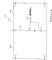
図1及び2に示す様に、この発明の1つの実施例においてはサドルコイル及びソレノイドコイルが利用される。サドルコイル10は、ベース又は共通セグメント12及びベースセグメント12に平行に結合された1対の翼セグメント14a、14bを含む。2巻ソレノイドコイル16は、2つのループ18a、18bを形成するため捩じられている単一コイルであり、これらループは点20においてオーバーラップしている。
【0012】
サドルコイル10及び2巻ソレノイドコイル16の各々は信号取出しポート22、24をそれぞれ含む。これらコイルの各々は、これらコイル内の固有インダクタンスがこれらコンデンサにより打消されるように選択された複数のコンデンサを含む。コイルの各々はMRIシステムの動作の周波数に同調される。動作の周波数は、テスラ(Tesla)の単位における静磁場の強度を磁気回転比(gyromagnetic ratio)で掛算したもので定義され、この磁気回転比は陽子に対してテスラ当たり約42.6MHzである。したがって、0.3T磁石を持つAIRISシステム(上述の)に対して動作の周波数は約12.7MHzである。コンデンサなしのコイルはインダクタンス(及び導体に固有の抵抗)を持つだけである。このコイルは式を使用して共鳴に同調される。
【0013】
【数1】
f=1/(2π√(LC))
【0014】
ここにLはコイルのインダクタンス、Cは付加されたコンデンサのキヤパシタンスである。通常、しかし、1つより多いコンデンサがコイルと直列に置かれる。それで、合計キヤパシタンスは1/C合計=1/C1+1/C2+...として計算される。
【0015】
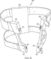
図3に示すように、直交コイル装置のサドルコイル及び2巻ソレノイドコイルを収容するためのコイルハーネス26は、両側及び両端をそれぞれ持つ長方形ベース28を含む。本質的に平行で、柔軟性のアーム30a、30bの2つの相対向する対は、ベース28の両側から横方向に延び、一般にベースより上でこれを覆う。アームの各々は、相対向し、整列する両アームが取外し可能に結合され一緒にループを形成するように、その両端に取付けたカップリング機構32を持つ。ハーネスの軸Aは、ベースの両端を横切って延びかつ相互接続されたアームにより形成されるループを大体中心を通って延びる一つの線により定義される。クロス棒34a、34bは各それぞれのアームの対の間に、好ましくはアームのカップリング機構32の間に延びる。コイルハーネス26は、またMRI処置中に相対向する柔軟性アームを取外し可能に一緒に接続するためのラッチ機構36を含む。少数だけ名をあげると、フック及びラッチ手段、帯及びプラスチックバックル(buckle)又はクリップ接続器、又はプラスチックスナップ接続器を含む種々のラッチ機構が利用出来る。
【0016】
アームの各々は、好ましくは実質的に大きな空隙で相互に離れ、これは好ましくは15−25cmである。相対向する柔軟性アームの対が一緒に結合される時、クロス棒34a、34bは示される様に相互に離れその間に、患者又はハーネス軸Aに平行な空隙を与えても良く、それは約50−70mmである。代わりに、クロス棒34a、34bは相互に隣接して位置決めされその間に意味のある空隙を与えなくても良い。それ故に、本発明のコイルハーネス26は、動作状態(柔軟性アームは取外し可能に一緒に結合)にある時、その各側に円周の回りにベース28の側部からクロス棒34a、34bへほぼ150°延びる開口を与える。クロス棒の間の患者上部の開口はもし望むならば設けても良い。2つの側方の開口は、ベースの形状及びクロス棒の位置決めに依存して変化することが認められ、各クロス棒はベース28の最も近い側面部分から円周に沿い少なくとも125°離れるのが好ましい。しかし、円周に沿い125°より少ない間隔は、本発明の範囲内にあると考えられる。
【0017】
図4に示す様に、サドルコイル10はハーネス26内に収容され、これによりベース又は共通セグメント12は長方形ベース28に沿って延び、また翼部分14a、14bは別個にそれぞれのアーム30a、30bの対に沿って延び、彼等の関連するクロス棒34a、34bの中に延びる。それ故に、サドルコイル10はハーネス26内において、そのループ(翼)の軸は本質的に患者又はハーネス軸Aに垂直である様に方向付けられる。より詳しくは、共通セグメント12は、ベースに沿って延びる。翼セグメント14aは、共通セグメント12から延び、対30aの一方のアーム部材に沿い、クロス棒34aに達しまたそれに沿い、そして対30aの他方のアーム部材に沿い共通セグメント12へ戻る。翼セグメント14bは、共通セグメント12から延び、対30bの一方のアーム部材に沿い、クロス棒34bに達しまたそれに沿い、そして対30bの他方のアーム部材に沿い共通セグメント12へ戻る。
【0018】
図5に示す様に、ソレノイドコイル16はハーネス26内に収容され、これによりクロスオーバ点20はベース28内に存在し、また各ループ18a、18bは、相対向し、取外し可能に接続される柔軟性アームにより形成される別個のハーネスループ19a、19bの中に位置決めされる。図2及び5に示される様に、個々のループ18a、18bの各々は、分離可能な2つのセグメントにより形成され、また点38a、38bにおいて電気的に接続可能となっている。それ故に、2巻ソレノイドコイル16は、そのループの軸が患者に対し又はハーネス軸Aに対し実質的に平行になるように方向付けられる。2つの別個の導電性セグメント70、72は、両ループ18a、18bを電気的に接続するためベース28に沿って延びる。別個の導電性セグメント70、72は点20においてオーバラップするが、この様な点20において相互に電気的な導電性接触にはない。従って、ベース28の一方の側におけるループ18aの側74は、ベース28の他方の側におけるループ18bの側76とセグメント70により電気的に接続される。同様に、ループ18aの側78はループ18bの側80とセグメント72により電気的に接続される。
【0019】
サドルコイル10と2巻ソレノイドコイル16の両方は同じコイルハーネス26内に収容されることは本技術分野に習熟した者にとって明らかであろうが、しかし、それらは明瞭のため図4及び5に分離して示されている。このハーネスは、一般的な形状を保つため十分に堅いが、また好ましくはアームの幾らかの運動を許容するため十分に柔軟性のあるいかなる適当な非強磁性体材料で作ることが出来る。例えば、成型されたプラスチック又は他の高分子材料がハーネスの骨格に利用出来、またコイルはプラスチックの表面に沿い又はプラスチックの中に形成又は機械加工された溝内を通ることが出来る。コイルを持つハーネスの骨格は、次にコイルを患者から隔離するためビニールの様な材料で覆われる。ハーネス骨格及びカバーのため他の材料が利用出来ることは認められる。
【0020】
図3に戻り参照すると、ベース28は、サドルコイル10及び2巻ソレノイドコイル16の収容部分に加えて、コイルの動作に必要な他の回路を含み、またケーブル40を通ってベース28を出る信号取出し線22、24を含む。図6−8に示す様に、オープン位置にあるハーネス26が示され、ソレノイドループ18a、18bは一緒に点38a、38bにおいて正及び負のリード(ピン及びソケット形式のコネクタ)40、42にそれぞれ結合される。同じ目的のため多くの他の電子的カップリング/コネクタがあることは本技術分野に習熟した者にとって明らかであろう。
【0021】
本実施例は、脊柱/胴体コイルハーネスとして使用するための物であるが、本発明の他の実施例は患者の組織の他の部分に使用するための寸法に合わせることが出来る。更に、コイルとハーネスは、胴体の直径に従って信号対雑音比を最適にするため異なる寸法の患者に使用するため修正できる。それ故に、ここに特定した寸法は、胴体ハーネスに対して好ましいが、この様な寸法は広く変化することが出来、特により小さなハーネスが、腕又は脚の様な組織のより小さな部分に対して作られる場合にそうである。なお更に、ベースの相反対側から延びるアーム部材は好ましくは整列し、ループを形成するため取外し可能に接続されるが、ベースの相反対側から延びるアーム部材を持ち、そこにアーム部材は相互に恒久的な方法で接続され又は相互に一体として形成され本発明の範囲内にあるコイル組立体を作ることができる。
【0022】
アームは柔軟性ではなく、実質的に剛性であるものも本発明の範囲内にあることが認められる。2巻ソレノイドコイルを置換した4巻ソレノイドコイルも更に本発明の範囲内にある。この様なコイルは2組の2ソレノイド巻きを持つであろう。各組は柔軟性アームの1つの中に一緒に接近して(2から5cm)位置決めされ、しかるにこれらの組は15から20cm離れている。同様に、他のコイル形式及び形状は本発明のハーネスを使用して確立できるかも知れない。
【0023】
図9を参照すると、本発明の代わりの実施例が示され、そこではだだ1個のクロス部材34’がアーム30bの対の間に設けられる。アーム30aの対はアーム30bの対に前述と同様な方法で接続されるが、異なる点は、直交コイルの翼部分14aを完成させるリンクを与えるため追加の電気的接続を設ける点である。従って、動作可能な様式に接続されると、両方の翼部分14a及び14bは、単一のクロス部材34’に沿って延びる一つのセグメントを含む。この単一のクロス部材には、もし望むならば中央開口を設けることが出来ることも理解される。更に、本発明の範囲から逸脱することなく1組の平行アームの間に2つの別個のクロス部材を同様に設けることが出来る。
【0024】
ここに述べた形状及び装置は、本発明の好ましい実施例を構成するが、この発明はこれらの正確な装置の形状には限定されず、その中に本発明の範囲から逸脱することなく変更がなされ得ることを理解すべきである。
【図面の簡単な説明】
【図1】 本発明の1つの観点により提供される直交コイル装置のサドルコイルの概略の表示を与える。
【図2】 直交コイル装置のソレノイドコイルの概略の表示を与える。
【図3】 本発明の別の観点による動作状態に接続された2対の平行アームを持ったハーネスの斜視図を与える。
【図4】 図3のハーネス内に収容されたサドルコイルの斜視図を与える。
【図5】 図3のハーネス内に収容されたソレノイドコイルの斜視図を与える。
【図6】 オープン位置における図3のハーネスの斜視図を与える。
【図7】 図6のハーネスの1つの接続部分の拡大された斜視図を与える。
【図8】 図6のハーネスの別の接続部分の拡大された斜視図を与える。
【図9】 クロス棒を1本で構成したハーネスの斜視図を与える。[0001]
This application claims the benefit of preliminary application serial number 60 / 066,980 filed on November 28, 1997.
(background)
The present invention relates generally to magnetic resonance imaging, and more particularly to a coil harness assembly for use in magnetic resonance imaging.
[0002]
Magnetic resonance imaging (“MRI”) is a well-known procedure for obtaining detailed two-dimensional and three-dimensional images of a patient based on nuclear magnetic resonance (“NMR”) principles. MRI is well suited for imaging soft tissue and has been used primarily for the diagnosis of internal injury or medical illness. A typical MRI system generally has a magnet capable of generating a very strong and homogeneous magnetic field sized to cover or surround a portion of the patient's tissue and a reception surrounding that portion of the patient's tissue under examination. A radio frequency (“RF”) transmitter and receiver system including a machine coil, a magnetic gradient system that places a particular portion of the patient's tissue under examination in space, and receives signals from the receiver coil; Includes a computer processing / imaging system for processing signals into interpretable data such as visible images for viewing by physicians or MRI personnel. Further information on MRI technology and equipment can be found in Van Nostrand's Scientific Encyclopedia, English, pp. 2198-2201 and US Department of Health and Human Services, “Medical Device Interaction with Magnetic Resonance Imaging System”, February 7, 1997. The general principles and associated equipment used in MRI are well known and therefore no additional disclosure is required.
[0003]
The advent of the “open” MRI system provided a more comfortable examination process for the patient, and also provided MRI attendants and physicians access to the patient while a portion of the patient was being viewed by the MRI system. Examples of such open MRI systems are the AIRIS® and AIRIS® II systems, which are commercially available from Hitachi Medical Systems America. Open MRI systems allow physicians and other MRI personnel to perform interventional surgery or other medical procedures on patients while the MRI system generates images.
[0004]
Open MRI systems also make convenient "MR fluoroscopy" that combines near real-time signal acquisition, image reconstruction and image display with such interventional procedures. Thus, by using MR fluoroscopy, a physician can monitor a two-dimensional or three-dimensional image of the tissue in substantially real time (approximately one image per second) while performing a medical procedure on the tissue. Will. For example, if a physician wishes to insert a tool that is compatible with MR, such as a needle or endoscope, into a particular organ while avoiding other organs, the physician may view the endoscope passageway through the MR image. Can be monitored internally by watching on the observation screen.
[0005]
Conventional coils for use in interventional MRI procedures typically include a single loop solenoid coil design. Because these coils are very narrow (3-5 cm wide), they are inherently open and allow a large area for patient access. The disadvantage of such a single loop solenoid coil is that the type of interventional procedure performed using such a coil is limited. The reason is that these single loop solenoid coils tend to have an undesirable signal to noise ratio and furthermore they do not give a relatively large examination volume. Thus, such a single loop solenoid coil is not desirable in an interventional procedure in which the endoscopic instrument enters the patient's body, for example at an inclined angle.
[0006]
A quadrature coil ("QD") device provides the desired signal to noise ratio. However, the disadvantage of using such a QD device in an interventional procedure is that a coil harness for housing the coil and positioning the coil around the part of the patient being examined is provided to that part of the patient by the physician. Is to prevent direct access. For example, a conventional spine / body coil harness for a QD device includes a substantially rectangular base with a perforated coil flap extending laterally from each side. The two flaps wrap around the patient's torso, meet above the patient, and form a continuous pore-free loop around the patient. Therefore, these flaps substantially surround that portion of the patient's tissue being examined, so that interventional access to this portion of the patient is limited.
[0007]
Therefore, there is a need to provide a QD harness that is modified to provide optimal access to the patient during the MRI observation process, and in particular during MR fluoroscopy.
[0008]
(Overview)
The present invention provides a coil harness for an orthogonal coil device that inherently optimizes access to that portion of patient tissue being observed during the MRI observation process. In one aspect of the invention, the quadrature coil device includes two turns of a solenoid coil and a saddle coil. The two-turn solenoid coil is oriented so that the loop axis is essentially parallel to the patient axis. The saddle coil is oriented so that the axis of the loop is essentially perpendicular to the patient's axis. The solenoid coil and saddle coil are housed in a casing, the casing being a rectangular base and two pairs of substantially parallel and flexible arms extending laterally therefrom, each of the arms Two pairs of arms having a coupling attached to the ends thereof, the arms being joined together to form a loop, and a pair of cross bars, each cross bar being a pair of parallel arms And a pair of cross bars extending between the two.
[0009]
Parallel flexible arms and loops formed by connecting one pair of ends to another pair are axially separated from each other by a substantially large gap, preferably 15-25 cm between them. . Thus, when in operation, the QD coil harness of the present invention provides an opening on each side of the harness, each opening extending from the base between the two flexible arms and nearly 150 ° around the harness. . These openings provide access to the patient during MR fluoroscopy procedures such as spine procedures as well as other interventional procedures.
[0010]
In another aspect of the invention, a coil assembly for use in MRI imaging includes a harness that includes a base. The first and second arm members extend from the first side of the base over the base and the third and fourth arm members extend from the second side of the base above the base. It extends over. The first cross member extends between the first arm member and the second arm member, and the second cross member extends between the third arm member and the fourth arm member. The end of the first arm member is aligned with and detachably connected to the end of the third arm member, and the end of the second arm member is aligned with and detached from the end of the fourth arm member. Connected as possible. Each of the first and second coil members extends along the harness.
[0011]
(Detailed explanation)
As shown in FIGS. 1 and 2, in one embodiment of the invention, a saddle coil and a solenoid coil are utilized. The
[0012]
Each of the
[0013]
[Expression 1]
f = 1 / (2π√ (LC))
[0014]
Here, L is the inductance of the coil, and C is the capacitance of the added capacitor. Usually, however, more than one capacitor is placed in series with the coil. Therefore, the total capacitance is 1 / C total = 1 / C1 + 1 / C2 +. . . Is calculated as
[0015]
As shown in FIG. 3, the
[0016]
Each of the arms are preferably separated from each other by a substantially large gap, which is preferably 15-25 cm. When opposing pairs of flexible arms are coupled together, the cross bars 34a, 34b may be spaced apart from each other as shown to provide a gap parallel to the patient or harness axis A, which is about 50 -70 mm. Instead, the cross bars 34a, 34b may be positioned adjacent to each other and provide no meaningful air gap therebetween. Therefore, when the
[0017]
As shown in FIG. 4, the
[0018]
As shown in FIG. 5, the
[0019]
It will be apparent to those skilled in the art that both the
[0020]
Referring back to FIG. 3, the
[0021]
While this embodiment is intended for use as a spine / body trunk harness, other embodiments of the present invention can be tailored for use with other portions of the patient's tissue. In addition, the coil and harness can be modified for use with different sized patients to optimize the signal to noise ratio according to the torso diameter. Therefore, the dimensions specified here are preferred for the fuselage harness, but such dimensions can vary widely, especially for smaller harnesses for smaller parts of tissue such as arms or legs. That is the case when made. Still further, the arm members extending from opposite sides of the base are preferably aligned and removably connected to form a loop, but have arm members extending from opposite sides of the base, wherein the arm members are mutually connected. Coil assemblies can be made that are connected in a permanent manner or integrally formed with each other and are within the scope of the present invention.
[0022]
It will be appreciated that the arms are not flexible and are substantially rigid within the scope of the present invention. A four-turn solenoid coil replacing the two-turn solenoid coil is also within the scope of the present invention. Such a coil would have two sets of two solenoid turns. Each set is positioned close together (2 to 5 cm) in one of the flexible arms, but these sets are 15 to 20 cm apart. Similarly, other coil types and shapes may be established using the harness of the present invention.
[0023]
Referring to FIG. 9, an alternative embodiment of the present invention is shown in which only one cross member 34 'is provided between the pair of
[0024]
While the shapes and apparatus described herein constitute preferred embodiments of the present invention, the invention is not limited to these precise apparatus shapes and modifications may be made therein without departing from the scope of the invention. It should be understood that this can be done.
[Brief description of the drawings]
FIG. 1 provides a schematic representation of a saddle coil of an orthogonal coil device provided in accordance with one aspect of the present invention.
FIG. 2 provides a schematic representation of a solenoid coil of an orthogonal coil device.
FIG. 3 provides a perspective view of a harness having two pairs of parallel arms connected in an operational state according to another aspect of the present invention.
4 provides a perspective view of a saddle coil housed in the harness of FIG. 3. FIG.
5 provides a perspective view of a solenoid coil housed in the harness of FIG. 3. FIG.
6 provides a perspective view of the harness of FIG. 3 in the open position.
7 provides an enlarged perspective view of one connecting portion of the harness of FIG.
8 provides an enlarged perspective view of another connecting portion of the harness of FIG.
FIG. 9 is a perspective view of a harness composed of a single cross bar.
Claims (10)
ベース,前記ベースの第1の側部から前記ベースより上にそれを覆って延びる第1及び第2のアーム部材,前記ベースの第2の側部から前記ベースより上にそれを覆って延びる第3及び第4のアーム部材,前記第1のアーム部材と前記第2のアーム部材の間に延びる第1のクロス部材,及び前記第3のアーム部材と前記第4のアーム部材の間に延びる第2のクロス部材を含み,前記第1のアーム部材は前記第3のアーム部材と整列しかつこれに取外し可能に接続され,また前記第2のアーム部材は前記第4のアーム部材と整列しかつこれに取外し可能に接続されたハーネスと,前記ベースに沿って延びる共通セグメント,前記共通セグメントから延び,前記第1のアーム部材に沿い前記第1のクロス部材に達しこれに沿い,そして前記第2のアーム部材に沿い前記共通セグメントに戻る第1の翼部材,及び前記共通セグメントから延び,前記第3のアーム部材に沿い前記第2のクロス部材に達しこれに沿い,そして前記第4のアーム部材に沿い前記共通セグメントに戻る第2の翼部材を含み,前記ハーネスに沿って延びる第1のコイル部材と,前記ハーネスに沿って延び,少なくとも第1及び第2のループを含み,前記第1のループはほぼ前記第1のアーム部材と前記第3のアーム部材に沿って延び,前記第2のループはほぼ前記第2のアーム部材と前記第4のアーム部材に沿って延び,前記第1及び第2のループは電気的に接続される第2のコイル部材と,を包含するMRIコイル組立体。An MRI coil assembly,
A base, first and second arm members extending over and above the base from a first side of the base, and a second arm member extending over and over the base from a second side of the base. 3 and 4 arm members, a first cross member extending between the first arm member and the second arm member, and a first cross member extending between the third arm member and the fourth arm member. Two cross members, wherein the first arm member is aligned with and removably connected to the third arm member, and the second arm member is aligned with the fourth arm member and A harness removably connected thereto, a common segment extending along the base, extending from the common segment, reaching the first cross member along the first arm member, and along the second arm of A first wing member that returns to the common segment along a web member, and extends from the common segment to the second cross member along the third arm member and along the fourth arm member. A first wing member extending along the harness, including at least a first and a second loop, the second wing member returning along the harness to the common segment, A loop extending substantially along the first arm member and the third arm member; and a second loop extending substantially along the second arm member and the fourth arm member; And a second coil member electrically connected to the second loop.
縦軸,前記縦軸にほぼ直角に延びる第1の構造用ループ,前記第1の構造用ループから軸方向に離れ,前記縦軸にほぼ直角に延びる第2の構造用ループ,前記縦軸にほぼ平行に,前記第1と第2の構造用ループの間に延びる1本または2本のクロス棒,及び前記第1と第2の構造用ループの間に延びるベースを含むハーネスと,
前記ハーネスに沿って延びる第1のコイル部材と,
前記ハーネスに沿って延びる第2のコイル部材とを包み,
前記第1のコイル部材は,前記ベースに沿って延びる共通セグメントと,前記共通セグメントから部分的に前記第1の構造用ループに沿いその第1の側において延び,前記少なくとも1つのクロス棒に達してそれに沿い,そして部分的に前記第2の構造用ループに沿いその第1の側において延び前記共通セグメントへ戻る第1の翼セグメントと,前記共通セグメントから部分的に前記第1の構造用ループに沿いその第2の側において延び,前記少なくとも1つのクロス棒に達してそれに沿い,そして部分的に前記第2の構造用ループに沿いその第2の側において延び前記共通セグメントへ戻る第2の翼セグメントとを含む
MRIコイル組立体。An MRI coil assembly,
A first structural loop extending substantially perpendicular to the longitudinal axis, a second structural loop extending axially away from the first structural loop and extending substantially perpendicular to the longitudinal axis, A harness including one or two cross bars extending between the first and second structural loops and a base extending between the first and second structural loops substantially parallel,
A first coil member extending along the harness;
A second coil member extending along the harness,
The first coil member includes a common segment extending along the base, and partially extending from the common segment along the first structural loop on its first side to reach the at least one cross bar. A first wing segment extending along the second structural loop and partially on the first side thereof back to the common segment, and partially from the common segment to the first structural loop. Extending on its second side along, reaching and reaching said at least one cross bar and partially extending on its second side along said second structural loop and returning to said common segment An MRI coil assembly including a wing segment.
Applications Claiming Priority (3)
| Application Number | Priority Date | Filing Date | Title |
|---|---|---|---|
| US6698097P | 1997-11-28 | 1997-11-28 | |
| US60/066,980 | 1997-11-28 | ||
| PCT/IB1998/002111 WO1999027846A2 (en) | 1997-11-28 | 1998-11-25 | Coil harness assembly for interventional mri application |
Publications (3)
| Publication Number | Publication Date |
|---|---|
| JP2001524340A JP2001524340A (en) | 2001-12-04 |
| JP2001524340A5 JP2001524340A5 (en) | 2006-01-19 |
| JP4213861B2 true JP4213861B2 (en) | 2009-01-21 |
Family
ID=22072967
Family Applications (1)
| Application Number | Title | Priority Date | Filing Date |
|---|---|---|---|
| JP2000522838A Expired - Fee Related JP4213861B2 (en) | 1997-11-28 | 1998-11-25 | Coil harness assembly for interventional MRI applications |
Country Status (6)
| Country | Link |
|---|---|
| US (1) | US6144203A (en) |
| EP (1) | EP1041922A2 (en) |
| JP (1) | JP4213861B2 (en) |
| CN (1) | CN1129799C (en) |
| AU (1) | AU1502399A (en) |
| WO (1) | WO1999027846A2 (en) |
Families Citing this family (25)
| Publication number | Priority date | Publication date | Assignee | Title |
|---|---|---|---|---|
| US6377836B1 (en) * | 1999-02-17 | 2002-04-23 | Toshiba America Mri, Inc. | RF coil array for vertical field MRI |
| US6591127B1 (en) * | 1999-03-15 | 2003-07-08 | General Electric Company | Integrated multi-modality imaging system and method |
| US6684096B2 (en) * | 2000-12-15 | 2004-01-27 | Berndt P. Schmit | Restraining apparatus and method for use in imaging procedures |
| JP3803323B2 (en) | 2000-12-22 | 2006-08-02 | コーニンクレッカ フィリップス エレクトロニクス エヌ ヴィ | Patient stabilization device in computer-linked tomography |
| US7701209B1 (en) * | 2001-10-05 | 2010-04-20 | Fonar Corporation | Coils for horizontal field magnetic resonance imaging |
| US7906966B1 (en) | 2001-10-05 | 2011-03-15 | Fonar Corporation | Quadrature foot coil antenna for magnetic resonance imaging |
| JP2006527050A (en) | 2003-06-13 | 2006-11-30 | コーニンクレッカ フィリップス エレクトロニクス エヌ ヴィ | Connection system for split top RF coil |
| US8401615B1 (en) | 2004-11-12 | 2013-03-19 | Fonar Corporation | Planar coil flexion fixture for magnetic resonance imaging and use thereof |
| US20060173390A1 (en) * | 2005-01-31 | 2006-08-03 | Robert Van Wyk | Immobilizing assembly and methods for use in diagnostic and therapeutic procedures |
| DE102005056711B3 (en) * | 2005-11-28 | 2007-05-10 | Siemens Ag | Magnetic resonance apparatus with main unit and moving couch for patient, includes array of local couplers limiting stages of signal recovery to predetermined sections of travel |
| DE102007023028B4 (en) * | 2007-05-16 | 2009-03-05 | Siemens Ag | Arrangement for fixing coils |
| DE102007030629A1 (en) * | 2007-07-02 | 2009-01-08 | Siemens Ag | Arrangement for storing a patient |
| US8290565B2 (en) * | 2008-04-04 | 2012-10-16 | Mayo Foundation For Medical Education And Research | System and method for cyclic motion encoding for enhanced visualization of slip interfaces with MRI |
| WO2009128365A1 (en) * | 2008-04-18 | 2009-10-22 | 株式会社 日立メディコ | Magnetic resonance imaging apparatus, receiving coil and method of manufacturing the coil |
| CN101403788B (en) * | 2008-10-31 | 2011-03-23 | 深圳市特深电气有限公司 | Nuclear magnetic resonance receiving coil |
| US9864032B2 (en) * | 2010-01-05 | 2018-01-09 | National Health Research Institutes | Magnetic resonance imaging system |
| WO2011158625A1 (en) * | 2010-06-16 | 2011-12-22 | 株式会社 日立メディコ | Rf reception coil and magnetic resonance imaging device using same |
| EP2560017A3 (en) * | 2011-08-18 | 2013-04-17 | Agron Lumiani | MRT local coil apparatus for diagnostics, intervention and therapy |
| EP2560016A1 (en) * | 2011-08-18 | 2013-02-20 | Agron Lumiani | MRT local coil device for diagnosis and intervention |
| DE102012201370B4 (en) * | 2012-01-31 | 2016-02-11 | Siemens Aktiengesellschaft | Bracket for Double Loop Coil (Double Loop Coil) for e.g. MCP recordings |
| DE102012213594B4 (en) * | 2012-08-01 | 2016-07-28 | Siemens Healthcare Gmbh | MR surface coil with integrated automatic patient fixation |
| CN103823196B (en) * | 2012-11-19 | 2017-04-05 | 西门子(深圳)磁共振有限公司 | A kind of connector and armarium |
| WO2014177975A1 (en) * | 2013-05-02 | 2014-11-06 | Koninklijke Philips N.V. | A detachable connector and splittable rf coil housings |
| CN105487029A (en) * | 2015-12-23 | 2016-04-13 | 深圳市特深电气有限公司 | Multichannel radio frequency coil device for NMR imaging system |
| CN111060856B (en) * | 2020-01-07 | 2021-03-16 | 厦门大学 | Nuclear magnetic resonance transverse radio frequency coil shaping device and transverse radio frequency coil shaping method |
Family Cites Families (11)
| Publication number | Priority date | Publication date | Assignee | Title |
|---|---|---|---|---|
| US4692705A (en) * | 1983-12-23 | 1987-09-08 | General Electric Company | Radio frequency field coil for NMR |
| US4897604A (en) * | 1989-02-21 | 1990-01-30 | The Regents Of The University Of California | Method and apparatus for selective adjustment of RF coil size for magnetic resonance imaging |
| DE4024599C2 (en) * | 1989-08-16 | 1996-08-14 | Siemens Ag | High-frequency antenna of an MRI scanner |
| US5510714A (en) * | 1991-08-09 | 1996-04-23 | Hitachi, Ltd. | Magnetic resonance imaging apparatus and RF coil employed therein |
| DE4221759C2 (en) * | 1991-10-11 | 1997-11-20 | Hitachi Medical Corp | Reception coil device for a magnetic resonance imaging device |
| US5457386A (en) * | 1991-11-26 | 1995-10-10 | Hitachi, Ltd. | Multiple-coil adopting a quadrature detection method applied thereto and a signal processing circuit employing the same in an MRI apparatus in a vertical magnetic system |
| US5663645A (en) * | 1994-08-02 | 1997-09-02 | Toshiba America Mri Inc. | Spatially orthogonal rectangular coil pair suitable for vertical magnetic field MRI system |
| US5804969A (en) * | 1995-07-28 | 1998-09-08 | Advanced Mammography Systems, Inc. | MRI RF coil |
| US5682098A (en) * | 1996-01-11 | 1997-10-28 | W. L. Gore & Associates, Inc. | Open quadrature whole volume imaging NMR surface coil array including three figure-8 shaped surface coils |
| DE19618989A1 (en) * | 1996-05-10 | 1997-11-13 | Gore W L & Ass Gmbh | Electromagnetic body coil for magnetic resonance tomographic measurements for medical use |
| DE19618988C2 (en) * | 1996-05-10 | 2001-12-13 | Gore W L & Ass Gmbh | Body coil with vacuum cushion |
-
1998
- 1998-11-23 US US09/198,017 patent/US6144203A/en not_active Expired - Fee Related
- 1998-11-25 WO PCT/IB1998/002111 patent/WO1999027846A2/en not_active Application Discontinuation
- 1998-11-25 AU AU15023/99A patent/AU1502399A/en not_active Abandoned
- 1998-11-25 CN CN98811562.XA patent/CN1129799C/en not_active Expired - Fee Related
- 1998-11-25 EP EP98959108A patent/EP1041922A2/en not_active Withdrawn
- 1998-11-25 JP JP2000522838A patent/JP4213861B2/en not_active Expired - Fee Related
Also Published As
| Publication number | Publication date |
|---|---|
| EP1041922A2 (en) | 2000-10-11 |
| WO1999027846A2 (en) | 1999-06-10 |
| JP2001524340A (en) | 2001-12-04 |
| CN1280675A (en) | 2001-01-17 |
| AU1502399A (en) | 1999-06-16 |
| US6144203A (en) | 2000-11-07 |
| WO1999027846A3 (en) | 1999-07-22 |
| CN1129799C (en) | 2003-12-03 |
Similar Documents
| Publication | Publication Date | Title |
|---|---|---|
| JP4213861B2 (en) | Coil harness assembly for interventional MRI applications | |
| US11039787B2 (en) | Garment MRI antenna array | |
| US5050605A (en) | Magnetic resonance imaging antennas with spiral coils and imaging methods employing the same | |
| JP3983667B2 (en) | Removable / replaceable and / or replaceable MRI / RF coil system and MRI imaging system and method | |
| US9002431B2 (en) | Garment MRI antenna array | |
| JP5654543B2 (en) | Intrarectal quadrature coil and interface device therefor | |
| EP2096454B1 (en) | Open architecture imaging apparatus and coil system for magnetic resonance imaging | |
| JP5930471B2 (en) | Multi-channel endorectal coil and interface device therefor | |
| US6198961B1 (en) | Interventional radio frequency coil assembly for magnetic resonance (MR) guided neurosurgery | |
| US8324899B2 (en) | MR coil with fiber optical connection | |
| US7348778B2 (en) | System and apparatus for a high resolution peripheral vascular coil array | |
| US8624597B2 (en) | RF coil array for cardiac and thoracic magnetic resonance imaging | |
| CN108072854A (en) | Coil device including flexible local coils and rigid local coil | |
| JP2012130701A (en) | System and method for communicating data | |
| JP2002153440A (en) | Inspection device using nuclear survey instrument | |
| WO2016190532A1 (en) | Head coil and magnetic resonance imaging apparatus employing the same | |
| CN112147553A (en) | System for radio frequency coil assembly | |
| US20020151788A1 (en) | Vest coil | |
| JP5020631B2 (en) | Method for performing open peripheral vascular coil and peripheral vascular imaging | |
| US11519980B2 (en) | Contoured radio frequency coil assemblies for a magnetic resonance system | |
| CN219831358U (en) | Knee joint radio frequency coil device and magnetic resonance imaging system | |
| JP3112474B2 (en) | Magnetic resonance imaging equipment | |
| CN219831357U (en) | Shoulder joint radio frequency coil device and magnetic resonance imaging system | |
| US20230266414A1 (en) | Adjustable multistage coil for functional imaging of limb | |
| GB2319846A (en) | Interventional MRI tool with alterable susceptibility |
Legal Events
| Date | Code | Title | Description |
|---|---|---|---|
| A521 | Request for written amendment filed |
Free format text: JAPANESE INTERMEDIATE CODE: A523 Effective date: 20051117 |
|
| A621 | Written request for application examination |
Free format text: JAPANESE INTERMEDIATE CODE: A621 Effective date: 20051117 |
|
| A131 | Notification of reasons for refusal |
Free format text: JAPANESE INTERMEDIATE CODE: A131 Effective date: 20080215 |
|
| A521 | Request for written amendment filed |
Free format text: JAPANESE INTERMEDIATE CODE: A523 Effective date: 20080407 |
|
| RD04 | Notification of resignation of power of attorney |
Free format text: JAPANESE INTERMEDIATE CODE: A7424 Effective date: 20080411 |
|
| A131 | Notification of reasons for refusal |
Free format text: JAPANESE INTERMEDIATE CODE: A131 Effective date: 20080603 |
|
| A521 | Request for written amendment filed |
Free format text: JAPANESE INTERMEDIATE CODE: A523 Effective date: 20080722 |
|
| TRDD | Decision of grant or rejection written | ||
| A01 | Written decision to grant a patent or to grant a registration (utility model) |
Free format text: JAPANESE INTERMEDIATE CODE: A01 Effective date: 20081027 |
|
| A01 | Written decision to grant a patent or to grant a registration (utility model) |
Free format text: JAPANESE INTERMEDIATE CODE: A01 |
|
| A61 | First payment of annual fees (during grant procedure) |
Free format text: JAPANESE INTERMEDIATE CODE: A61 Effective date: 20081031 |
|
| FPAY | Renewal fee payment (event date is renewal date of database) |
Free format text: PAYMENT UNTIL: 20111107 Year of fee payment: 3 |
|
| R150 | Certificate of patent or registration of utility model |
Free format text: JAPANESE INTERMEDIATE CODE: R150 |
|
| FPAY | Renewal fee payment (event date is renewal date of database) |
Free format text: PAYMENT UNTIL: 20111107 Year of fee payment: 3 |
|
| FPAY | Renewal fee payment (event date is renewal date of database) |
Free format text: PAYMENT UNTIL: 20121107 Year of fee payment: 4 |
|
| FPAY | Renewal fee payment (event date is renewal date of database) |
Free format text: PAYMENT UNTIL: 20131107 Year of fee payment: 5 |
|
| LAPS | Cancellation because of no payment of annual fees |


