JP4195508B2 - How to detect colon cancer from stool samples - Google Patents
How to detect colon cancer from stool samples Download PDFInfo
- Publication number
- JP4195508B2 JP4195508B2 JP52762597A JP52762597A JP4195508B2 JP 4195508 B2 JP4195508 B2 JP 4195508B2 JP 52762597 A JP52762597 A JP 52762597A JP 52762597 A JP52762597 A JP 52762597A JP 4195508 B2 JP4195508 B2 JP 4195508B2
- Authority
- JP
- Japan
- Prior art keywords
- sample
- cells
- assay
- lesion
- cancer
- Prior art date
- Legal status (The legal status is an assumption and is not a legal conclusion. Google has not performed a legal analysis and makes no representation as to the accuracy of the status listed.)
- Expired - Lifetime
Links
Images

Classifications
-
- C—CHEMISTRY; METALLURGY
- C12—BIOCHEMISTRY; BEER; SPIRITS; WINE; VINEGAR; MICROBIOLOGY; ENZYMOLOGY; MUTATION OR GENETIC ENGINEERING
- C12Q—MEASURING OR TESTING PROCESSES INVOLVING ENZYMES, NUCLEIC ACIDS OR MICROORGANISMS; COMPOSITIONS OR TEST PAPERS THEREFOR; PROCESSES OF PREPARING SUCH COMPOSITIONS; CONDITION-RESPONSIVE CONTROL IN MICROBIOLOGICAL OR ENZYMOLOGICAL PROCESSES
- C12Q1/00—Measuring or testing processes involving enzymes, nucleic acids or microorganisms; Compositions therefor; Processes of preparing such compositions
- C12Q1/68—Measuring or testing processes involving enzymes, nucleic acids or microorganisms; Compositions therefor; Processes of preparing such compositions involving nucleic acids
- C12Q1/6813—Hybridisation assays
- C12Q1/6834—Enzymatic or biochemical coupling of nucleic acids to a solid phase
-
- C—CHEMISTRY; METALLURGY
- C12—BIOCHEMISTRY; BEER; SPIRITS; WINE; VINEGAR; MICROBIOLOGY; ENZYMOLOGY; MUTATION OR GENETIC ENGINEERING
- C12Q—MEASURING OR TESTING PROCESSES INVOLVING ENZYMES, NUCLEIC ACIDS OR MICROORGANISMS; COMPOSITIONS OR TEST PAPERS THEREFOR; PROCESSES OF PREPARING SUCH COMPOSITIONS; CONDITION-RESPONSIVE CONTROL IN MICROBIOLOGICAL OR ENZYMOLOGICAL PROCESSES
- C12Q1/00—Measuring or testing processes involving enzymes, nucleic acids or microorganisms; Compositions therefor; Processes of preparing such compositions
- C12Q1/68—Measuring or testing processes involving enzymes, nucleic acids or microorganisms; Compositions therefor; Processes of preparing such compositions involving nucleic acids
- C12Q1/6806—Preparing nucleic acids for analysis, e.g. for polymerase chain reaction [PCR] assay
-
- C—CHEMISTRY; METALLURGY
- C12—BIOCHEMISTRY; BEER; SPIRITS; WINE; VINEGAR; MICROBIOLOGY; ENZYMOLOGY; MUTATION OR GENETIC ENGINEERING
- C12Q—MEASURING OR TESTING PROCESSES INVOLVING ENZYMES, NUCLEIC ACIDS OR MICROORGANISMS; COMPOSITIONS OR TEST PAPERS THEREFOR; PROCESSES OF PREPARING SUCH COMPOSITIONS; CONDITION-RESPONSIVE CONTROL IN MICROBIOLOGICAL OR ENZYMOLOGICAL PROCESSES
- C12Q1/00—Measuring or testing processes involving enzymes, nucleic acids or microorganisms; Compositions therefor; Processes of preparing such compositions
- C12Q1/68—Measuring or testing processes involving enzymes, nucleic acids or microorganisms; Compositions therefor; Processes of preparing such compositions involving nucleic acids
- C12Q1/6844—Nucleic acid amplification reactions
- C12Q1/6858—Allele-specific amplification
-
- C—CHEMISTRY; METALLURGY
- C12—BIOCHEMISTRY; BEER; SPIRITS; WINE; VINEGAR; MICROBIOLOGY; ENZYMOLOGY; MUTATION OR GENETIC ENGINEERING
- C12Q—MEASURING OR TESTING PROCESSES INVOLVING ENZYMES, NUCLEIC ACIDS OR MICROORGANISMS; COMPOSITIONS OR TEST PAPERS THEREFOR; PROCESSES OF PREPARING SUCH COMPOSITIONS; CONDITION-RESPONSIVE CONTROL IN MICROBIOLOGICAL OR ENZYMOLOGICAL PROCESSES
- C12Q1/00—Measuring or testing processes involving enzymes, nucleic acids or microorganisms; Compositions therefor; Processes of preparing such compositions
- C12Q1/68—Measuring or testing processes involving enzymes, nucleic acids or microorganisms; Compositions therefor; Processes of preparing such compositions involving nucleic acids
- C12Q1/6876—Nucleic acid products used in the analysis of nucleic acids, e.g. primers or probes
- C12Q1/6883—Nucleic acid products used in the analysis of nucleic acids, e.g. primers or probes for diseases caused by alterations of genetic material
- C12Q1/6886—Nucleic acid products used in the analysis of nucleic acids, e.g. primers or probes for diseases caused by alterations of genetic material for cancer
-
- G—PHYSICS
- G01—MEASURING; TESTING
- G01N—INVESTIGATING OR ANALYSING MATERIALS BY DETERMINING THEIR CHEMICAL OR PHYSICAL PROPERTIES
- G01N33/00—Investigating or analysing materials by specific methods not covered by groups G01N1/00 - G01N31/00
- G01N33/48—Biological material, e.g. blood, urine; Haemocytometers
- G01N33/50—Chemical analysis of biological material, e.g. blood, urine; Testing involving biospecific ligand binding methods; Immunological testing
- G01N33/53—Immunoassay; Biospecific binding assay; Materials therefor
- G01N33/574—Immunoassay; Biospecific binding assay; Materials therefor for cancer
- G01N33/57407—Specifically defined cancers
- G01N33/57419—Specifically defined cancers of colon
-
- C—CHEMISTRY; METALLURGY
- C12—BIOCHEMISTRY; BEER; SPIRITS; WINE; VINEGAR; MICROBIOLOGY; ENZYMOLOGY; MUTATION OR GENETIC ENGINEERING
- C12Q—MEASURING OR TESTING PROCESSES INVOLVING ENZYMES, NUCLEIC ACIDS OR MICROORGANISMS; COMPOSITIONS OR TEST PAPERS THEREFOR; PROCESSES OF PREPARING SUCH COMPOSITIONS; CONDITION-RESPONSIVE CONTROL IN MICROBIOLOGICAL OR ENZYMOLOGICAL PROCESSES
- C12Q2600/00—Oligonucleotides characterized by their use
- C12Q2600/106—Pharmacogenomics, i.e. genetic variability in individual responses to drugs and drug metabolism
-
- C—CHEMISTRY; METALLURGY
- C12—BIOCHEMISTRY; BEER; SPIRITS; WINE; VINEGAR; MICROBIOLOGY; ENZYMOLOGY; MUTATION OR GENETIC ENGINEERING
- C12Q—MEASURING OR TESTING PROCESSES INVOLVING ENZYMES, NUCLEIC ACIDS OR MICROORGANISMS; COMPOSITIONS OR TEST PAPERS THEREFOR; PROCESSES OF PREPARING SUCH COMPOSITIONS; CONDITION-RESPONSIVE CONTROL IN MICROBIOLOGICAL OR ENZYMOLOGICAL PROCESSES
- C12Q2600/00—Oligonucleotides characterized by their use
- C12Q2600/156—Polymorphic or mutational markers
Landscapes
- Chemical & Material Sciences (AREA)
- Life Sciences & Earth Sciences (AREA)
- Health & Medical Sciences (AREA)
- Organic Chemistry (AREA)
- Engineering & Computer Science (AREA)
- Proteomics, Peptides & Aminoacids (AREA)
- Zoology (AREA)
- Wood Science & Technology (AREA)
- Immunology (AREA)
- Analytical Chemistry (AREA)
- Molecular Biology (AREA)
- Biochemistry (AREA)
- Microbiology (AREA)
- Biotechnology (AREA)
- Physics & Mathematics (AREA)
- General Health & Medical Sciences (AREA)
- Genetics & Genomics (AREA)
- Biophysics (AREA)
- Bioinformatics & Cheminformatics (AREA)
- General Engineering & Computer Science (AREA)
- Pathology (AREA)
- Oncology (AREA)
- Hospice & Palliative Care (AREA)
- Biomedical Technology (AREA)
- Hematology (AREA)
- Urology & Nephrology (AREA)
- Chemical Kinetics & Catalysis (AREA)
- Cell Biology (AREA)
- Food Science & Technology (AREA)
- Medicinal Chemistry (AREA)
- General Physics & Mathematics (AREA)
- Measuring Or Testing Involving Enzymes Or Micro-Organisms (AREA)
- Investigating Or Analysing Biological Materials (AREA)
Abstract
Description
発明の分野
本発明は、患者における結腸ガンの早期検出のための方法、およびより詳細には、患者がガン病巣または前ガン病巣を有する場合、サンプルが診断的に関連した情報を含む可能性を確実にするため、または増大させるために結腸ガンの検出のための糞便サンプルを調製する方法、および糞便サンプルの分析方法に関する。
発明の背景
糞便サンプルは、しばしば、医学的診断分析のために調製されなければならない。糞便サンプルは、寄生虫感染、細菌感染、またはウイルス感染から炎症性腸疾患および結腸直腸ガンにわたる医学的状態を診断するのを助けるために分析され得る。
結腸直腸ガンは、西洋社会における主な死亡原因である。しかし、診断が早ければ、ガン組織の外科的除去により効果的に処置され得る。結腸直腸ガンは、結腸直腸上皮内に生じ、そして典型的には、発達の初期段階の間は広範に血管化しない(それゆえ、非侵襲的である)。結腸直腸ガンは、結腸または直腸の上皮裏打ちにおける単一の変異細胞のクローン拡大から生じると考えられる。高度に血管化した、侵襲的でかつ最終的に身体全体に広がる転移性のガンへの移行は、通常10年以上かかる。ガンが侵襲前に検出される場合、ガン組織の外科的除去は有効な治療である。しかし、結腸直腸ガンは、しばしば臨床的徴候(例えば、痛みおよび黒いタール状の糞便)の発現に際してのみ検出される。一般に、このような徴候は、疾患が充分に確立した場合、しばしば、転移が生じた後にのみ現れ、そしてガン組織の外科的切除の後でさえも患者の予後は乏しい。それゆえ、結腸直腸ガンの早期検出は、検出がその罹患率を著しく減少させ得ることにおいて重要である。
内視鏡検査のような侵襲的診断方法は、潜在的にガン性の腫瘍(例えば、ポリープ)の直接的で視覚的な同定、除去、および生検を可能にする。内視鏡検査は高価であり、不快であり、本質的に危険が伴い、それゆえ、結腸直腸ガンを有する個体を同定するために集団をスクリーニングするための実用的な道具ではない。結腸直腸のガンまたは前ガンの存在を示唆する特徴の糞便サンプルの非侵襲的分析は、早期診断のための好ましい代替法であるが、この目的を確実に達成する公知の診断方法は、利用可能でない。
現在の非侵襲的診断方法は、糞便の潜血の存在について、またはガン胎児性抗原の上昇レベルについて糞便サンプルをアッセイする工程を含む。これらは両方とも結腸直腸ガンの存在を示唆する。さらに、分子生物学における最近の発展は、結腸直腸ガンの存在に関連し、そしてこの存在を示唆するDNAの変異または変化の範囲の存在を検出する大きな可能性のある方法を提供する。このような変異の存在は、結腸直腸ガンの初期段階の間の糞便サンプル中に見出されるDNA中に理論上検出され得る。しかし、糞便は、細胞の不均一な集団をもたらす、患者、微生物、および食物由来の細胞および細胞破片を含む。このことが、小さな特定の亜集団の検出を、確実に検出することを不可能にしている。
当該分野において記載された結腸直腸ガンについての糞便診断アッセイは、典型的に、排泄された糞便の無作為にサンプリングされた部分から調製されたサンプルで行われる。しかし、このような方法によって調製されたサンプルは、たとえ、結腸直腸のガンまたは前ガンを有する患者により排泄された糞便から調製された場合であっても、結腸直腸のガンまたは前ガンの存在を示唆する特徴を再現的に与えない。従って、当該分野においては、結腸直腸のガンまたは前ガンを有する患者より排泄された糞便から調製されたサンプル中のガン性物質または前ガン物質の存在を示唆する特徴を再現的に検出する、結腸直腸ガンまたは前ガンの早期診断方法が必要とされている。このような方法が、本明細書中に提供される。
発明の要旨
現在、細胞および細胞破片は、結腸上皮細胞から形成中の糞便の上に、糞便の長さに沿った長軸方向の「筋(stripe)」の物質として流出されることが認識されている。流出された物質は、図1(「C」と称する)に示すように、この長軸方向の筋に制限される。この認識に基づいて、出願人らは、診断試験のための糞便サンプル調製は、サンプルが結腸を通過するにつれて糞便中に流出された何らかの細胞または細胞破片を含有することを確実にするために、代表的なサンプルを採取する工程を含まなければならないことを教示する。従って、本発明の方法は、患者により排泄された糞便の少なくとも1つの横断面部分を得て、そしてガンまたは前ガンを示唆し得る結腸を裏打ちする上皮細胞から流出された細胞または細胞破片の存在をサンプルにおいて検出するためのアッセイを行う工程を包含する。非常に頻繁に、このような細胞は、結腸に沿った異なる位置でのポリープあるいはガン病巣または前ガン病巣に由来する。本発明の目的のために、前ガン病巣は前ガン細胞を含み、そして前ガン細胞はガンに関連しそしてこのような細胞をガン性になりやすくする変異を有する細胞である。図1に示すように、横断面サンプルは、糞便の少なくとも1つの完全な円周(または完全な横断面部分を含む糞便部分)(例えば、冠状断面または矢尻状断面)を含むサンプルである。
好ましい実施態様において、本発明の方法は、患者より排泄された糞便の少なくとも1つの横断面部分を得る工程、および形質転換された細胞のクローン集団由来の破片を検出するためのアッセイを行う工程を包含する。形質転換された細胞は、例えば、1つ以上の変異(本出願の目的のために、変異は、DNAの欠失、置換、付加、改変、挿入、または再編成である)を有する細胞のクローン亜集団を包含する。本発明の好ましい方法は、このような形質転換された細胞の特徴を検出する工程を包含する。このような特徴は、例えば、変異、形質転換された細胞において独特にまたは変化した量で発現されるタンパク質、および血液を含む。本発明の特に好ましい方法は、糞便サンプルの少なくとも1つの横断面部分を得る工程、およびサンプル中の細胞のクローン亜集団の存在を示唆するDNAの特徴を検出するためのアッセイを行う工程を包含する。クローン亜集団は、例えば、p53腫瘍抑制遺伝子中に変異を有する、例えば、ガン細胞または前ガン細胞の亜集団であり得る。本発明の方法によって検出される細胞のクローン亜集団はしばしば、欠失に包含される、遺伝子(単数または複数)を無効にするヘテロ接合性の消失をもたらすDNAの大規模な消失により特徴付けられる。
本発明の方法はまた、糞便の代表的な(すなわち、横断面)サンプルを得る工程、および糞便を緩衝液(例えば、界面活性剤およびプロテイナーゼ、ならびに必要に応じてDNaseインヒビターを含有する緩衝液)中に均一化する工程を包含する。
本発明の方法において、糞便の少なくとも1つの横断面部分について行うアッセイは、結腸を裏打ちする細胞から流出されたガン胎児性抗原の上昇したレベルの存在を検出するためのアッセイであり得る。このようなアッセイはまた、潜血の存在を検出する工程を包含し得る。しかし、本発明の方法は、好ましくは、サンプルを、ガンに潜在的に関連する変異を有する細胞の亜集団を含む細胞から流出された細胞破片に特徴的な分子に特異的に結合する抗体に曝すアッセイを包含する。
本発明の方法は、特にかつ最も好ましくは、代表的な糞便サンプル中の形質転換された細胞の亜集団を示唆するDNAの特徴を検出するために有用である。DNAの特徴は、例えば、ヘテロ接合性の消失、マイクロサテライトの不安定性などを包含する変異であり得る。本発明の方法におけるDNAの特徴についてのアッセイは、代表的な糞便サンプル中に細胞の亜集団が変異していることが知られているか、または推測される、第1の対立遺伝子の数Xと、サンプル中で変異していないことが知られているか、または推測される対立遺伝子の数Yとの間に差異が存在するか否かを決定する工程を包含し得る。統計的有意差は、サンプル中の細胞の亜集団における変異およびガンの潜在的な存在を示唆する。本発明の1つの実施態様において、腫瘍抑制遺伝子の数とガンに関連しない遺伝子の数との間の差異が比較され、この数における統計的有意差は腫瘍抑制遺伝子における変異を示唆する。
本発明の方法の実施に有用なアッセイはまた、多型ヌクレオチドを含む領域における欠失または他の変異の存在を検出するためのアッセイを包含する。このようなアッセイにおいて、母系対立遺伝子および父系対立遺伝子に存在する多型ヌクレオチドの数が決定される(ここで、患者は、多型ヌクレオチドについてヘテロ接合性である)。母系対立遺伝子および父系対立遺伝子における多型ヌクレオチドの数の間の統計的有意差は、2つの対立遺伝子の1つにおける欠失の存在を示唆する。
本発明の方法は、典型的に、サンプル調製および細胞または細胞破片の特徴についてのアッセイの後に、ポリープまたは他の病巣が実際に存在するか否かを決定するための結腸の視覚的試験を包含する。最終的に、ガン組織または前ガン組織の拡散を防ぐために、異常組織の外科的切除を行い得る。
従って、本発明の方法は、不均一なサンプル(例えば、糞便サンプル)中のガン細胞または前ガン細胞の亜集団の存在についてスクリーニングするための手段を提供する。本発明の方法は、結腸上皮の病巣に関連する罹患率および死亡率を減少させる。さらに、本発明の方法は、現在当該分野で利用可能な方法よりも正確なスクリーニング方法を包含する。なぜなら、現在の方法は、ガン細胞または前ガン細胞が形成中の糞便の表面の一部分上またはその中にのみ破片を流出するという観察を利用しているからである。本発明の方法は、糞便の完全な円周にわたって確実にアッセイし、それにより異常が存在している場合にそれを検出する可能性が増加する。本発明のさらなる局面および利点は、その以下の詳細な説明に含まれる。
図面の説明
図1は、形成された糞便を表し、そして糞便の完全な円周由来の物質を含む種々の横断面を示す柱体の図である。「A」と標識された断面は、典型的な冠状断面であり、そして「B」と標識された断面は、典型的な矢尻状断面である。「C」と標識された細片は、長軸方向の筋に沈積したガン組織から流出された物質を表す。
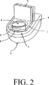
図2は、糞便サンプルを含むための容器の模式図である。
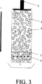
図3は、マルチオリフィスインピーダンスカウンター(multi-orifice impedance counter)の模式図である;ここで、参照番号1は、カラムを通る流れの方向を示す;参照番号2は、カラムの下方に物質を押しこむためのプランジャー手段を示す;参照番号3および4は、異なるサイズのハイブリダーゼーションビーズである;参照番号5は、所望しない粒子を抽出するための随意のフィルターである;参照番号6は、微分インピーダンス(differential impedance)を測定するためのオリフィスの配列を示す;そして参照番号7は回収チャンバーである。
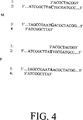
図4は、単一塩基多型の検出のために有用なプライマーを示す図である。
発明の詳細な説明
本発明による方法は、このような集団が患者の結腸に沿った任意の部位に存在する場合、ガン細胞または前ガン細胞のクローン集団から流出された細胞または細胞破片を再現的に含む、糞便サンプルの調製のために有用である。次いで、これらのサンプルは、再現性が高くそして正確な方法でガンを示唆する特徴を検出するためのアッセイを実施するために使用される。このような方法は、これらが患者により排泄された糞便由来の少なくとも1つの横断面サンプルを取り出すことを教示するので、従来技術を超える改善を提供する。少なくとも1つの横断サンプルを得なければならないとの認識がなければ、ガンまたは前ガンの亜集団の細胞が存在しても、それを含むサンプルを再現的に得るための手段は存在しない。
当該分野で記載された方法は、寄生虫、細菌、およびウイルスによる感染とは異なり、結腸ガン、特に初期段階の結腸ガンの存在を示唆する特徴が排泄された糞便の特定の部分にのみ見出されることを認識しない。糞便のサンプリングした部分が、初期段階のガン組織から排泄された細胞および細胞破片をたまたま含む部分を含有しない場合、診断アッセイは、たとえ均一化したとしても、信頼できる方法で結腸直腸ガンの存在を示唆する特徴を必ずしも検出し得るわけではない。すなわち、偽陰性の結果を生じる。
例えば、結腸を裏打ちする上皮において、または初期段階のガン病巣上に形成するポリープ由来の脱落細胞は、ポリープまたは病巣と接触する形成中の糞便部分にのみ脱落する。従って、初期段階の疾患において、形成中の糞便の表面層の小さな部分のみが脱落細胞を含有し、そしてこの部分がサンプル部分として採取されない場合、必ず、結腸ガンの徴候についてのアッセイは偽陰性の結果を生じる。結腸の解剖学および生理学の簡単な総説は、この現象の理解の助けとなる。
典型的な成人の結腸は、大体、6フィートの長さであり、約2〜約3インチの直径を有する。多くの屈曲およびひだがその長さ全体に存在する。結腸は、結腸に侵入する液体状または半液体状の老廃物から水分を除去し、そして比較的固い糞便が、結腸の基部付近の3分の1で形成し始める。上皮細胞は結腸の管腔を裏打ちし、そして管腔の表面は微細な陰窩内に組織化される。結腸直腸上皮細胞は、4〜5日毎に置き換えられる。上皮細胞は、陰窩の基底で急速に分裂し、そして先端(apeces)へと移動する。ここで、細胞はアポトーシス(プログラム細胞死)を受けるようであり、そして細胞破片は管腔へ流出される。結腸直腸管腔の裏打ちは弾力性があり、そして管腔の直径は、任意の所定の時間で結腸を通る糞便の容量によって決定される。結果として、結腸を通過する形成中の糞便の表面は、管腔の上皮裏打ちと直接接触する。従って、流出された上皮細胞(アポトーシスを受けたかもしれないし、受けてないかもしれない)および細胞破片は、結腸を通過するにつれて糞便の表面に取り込まれる。
従って、結腸直腸上皮ガン由来の細胞および細胞破片はまた、形成中の糞便に流出される。多くの結腸直腸ガンは、糞便が比較的固い結腸の領域で発達し、実際、このようなガンの約1/3が直腸において発達する。ガンの存在を示唆するマーカー(細胞、細胞破片、DNA、血液、およびガン胎児性抗原を包含する)は、糞便が結腸を通るにつれてガン組織と接触する形成中の糞便部分に流出される。糞便は比較的固いために、これらのマーカーは糞便の表面上またはその付近に残存する。ここでこれらは沈積し、そして糞便全体に均一に分散されない。糞便がガン腫瘍または前ガン腫瘍を通過するにつれて、腫瘍由来の物質は糞便に沿っているが、病巣を含むガン組織または前ガン組織と直接接触する糞便の円周の一部にのみ沈積される。従って、結腸直腸のガンまたは前ガンを患う患者により排泄された糞便は、ガン組織または前ガン組織に由来する診断上関連する物質の長軸方向の「筋」によって特徴付けられる。
結腸直腸のガンまたは前ガンを患う患者により排泄された糞便の完全な円周由来の物質を含まないサンプルは、ガン組織または前ガン組織に由来する物質を再現的に含まない。一般に、排泄された糞便の無作為な、非横断サンプル(「塗抹標本」)が、臨床台(setting)において分析される。これらにおいて、脱落したガン細胞または前ガン細胞および細胞破片は、サンプルが、たまたま偶然に、細胞が脱落した結腸の領域と接触した糞便の部分を含まない限り、検出される可能性がない。
さらに、ガンは単一の変異細胞のクローン拡大によって典型的に発達し、そして疾患の初期段階(すなわち、外科的除去が有効な治療である時期)において、ガン病巣は、非常に小さく、そして結腸の円周の小さな弧の上にあり得る。従って、このような初期段階のガンに由来する物質は、非常に狭い筋(図1においてCと標識する)において糞便の上またはその中に流出される。その結果、初期段階の結腸直腸のガンまたは前ガンを患う患者により排泄された糞便の完全な円周を含まないサンプルは、偶然によってしか初期段階のガン状態または前ガン状態の存在を示唆する物質を含まない。しかし、結腸直腸ガンの早期検出は、効果的な外科的介入のために非常に重要である。本発明は、患者におけるガンまたは前ガンの存在を示唆する特徴の再現性のある早期検出のための方法を提供する。
図1に示すように、糞便の少なくとも1つの横断サンプル(糞便全体を含む)の分析は、任意の存在するガン細胞または前ガン細胞から流出された細胞および細胞破片の少なくとも一部(たとえ、小さな初期段階のガン組織または前ガン組織(例えば、小さなポリープ)から排泄されていても)が、分析されるべき糞便サンプル部分に存在することを確実にする。実際に、糞便サンプルの少なくとも1つの横断面を採取することは、たとえ、患者が結腸直腸のガンまたは前ガンを有している場合であっても脱落したガン細胞または前ガン細胞を含まない糞便部分を分析する可能性を防止する。
一旦、横断糞便サンプルが得られると、それは公知の方法により均一化されて細胞および細胞破片をサンプル全体に分配させ得る。次いで、サンプル中の細胞および/または細胞破片の存在を検出するために、ホモジネートまたはホモジネートの抽出物についてアッセイが行われる。アッセイは、組織学的細胞アッセイ、タンパク質のような形質転換に特有な分子の存在を検出するために設計された抗体に基づく免疫アッセイ(または他の形式)、または結腸直腸ガンを示唆する変異または遺伝的特徴を検出するためのDNAに基づくアッセイのいずれか1つまたはそれらの組み合わせであり得る。公知のアッセイプロトコル(本明細書中、または同時継続中の出願の出願番号[本出願と同日に出願した代理人の文書番号EXT-001]に開示されるプロトコル)、または今後開発されるアッセイが、本発明の実施に使用され得る。有用な公知のアッセイプロトコルの限定されない例は、米国特許第5,137,806号(選択されたDNA分子における配列の検出)、同第5,348,855号(核酸配列についてのアッセイ)、同第5,512,441号(変異対立遺伝子の検出)、同第5,272,057号および同第5,380,645号(RFLP解析)、同第5,527,676号(p53遺伝子配列の検出)、同第5,330,892号(MCC遺伝子配列の検出)、同第5,352,775号(APC遺伝子配列の検出)、同第5,532,108号(DCC遺伝子配列の検出)、およびWO96/08514(ヒト結腸癌腫関連抗原に対するモノクローナル抗体)に開示されるプロトコルを包含する。これらのそれぞれの開示は、本明細書中に参考として援用される。あるいは、またはさらに、糞便の潜血についてのアッセイを、本明細書中に参考として援用される米国特許第4,333,734号および同第5,196,167号に報告されたように行い得る。本発明の文脈において有用なアッセイはまた、本明細書中に参考として援用される米国特許第5,380,647号に報告されるように、ガン胎児性抗原についてのアッセイを包含する。最後に、サンプルは、本明細書中に参考として援用される米国特許第4,857,300号に報告されるように、ガン細胞または前ガン細胞の存在を示唆する特徴を検出するための組織学的検査のために調製され得る。
少なくとも1つの横断面サンプルを得ることに関連して使用される任意のアッセイプロトコルの目的は、結腸鏡検査またはS字結腸鏡検査のようなその後の侵襲的診断手順のための候補を同定することである。従って、このアッセイは、明白に偽陰性が防止されるべきであるが、ガン病巣または前ガン病巣の存在を明確に検出する必要はない。試験プロトコルの目的は、サンプル中の莫大な量の細胞破片中に、通常、初期段階の形質転換と関連する変異を有するいくつかの細胞が存在するか否かを決定することではなく、むしろ、サンプルが変異細胞亜集団のクローン拡大を示唆する破片を含んでいるか否かを決定することである。最大の利益は、クローン的に拡大した細胞集団(すなわち、ガン病巣または前ガン病巣を含む、形質転換された結腸上皮細胞)の可能性のある存在を検出するために設計されたアッセイによりもたらされる。初期段階のガンまたは前ガンを検出する能力を有するアッセイは、本発明の方法において好ましい。ポリメラーゼ連鎖反応(PCR)を用いるアッセイ、制限断片長多型(RFLP)、または核酸分析についての他の方法は、直腸結腸のガンまたは前ガンの存在を示唆する公知のDNAの特徴を検出するために用いられ得る。細胞破片の量的検出についてのより正確な方法(例えば、DNA断片またはセグメント)が本明細書中に記載される方法による横断面サンプルを分析するために用いられ得る。
好ましいアッセイは、ガンまたは前ガンの発達を示唆するDNAの特徴についてサンプルを調べる。しかし、本発明の方法で使用するためのアッセイは、臨床的に関連する形質転換組織から流出された任意の異常な細胞破片を検出し得る。従って、本発明の好ましい局面によれば、アッセイは、ヘテロ接合性の消失、マイクロサテライトの不安定性、または他の変異を受けた細胞の特徴の存在を検出するために用いられる。
以下の実施例は、本発明による方法の詳細を提供する。しかし、本発明の多くのさらなる局面、特に、実施されるべきアッセイに関しては、以下のそれらの詳細な説明を考慮すれば明らかとなる。
実施例1
糞便サンプルの調製
サンプルを、サンプルが患者により排泄された糞便の少なくとも1つの横断面部分を含むように調製する。横断面部分を、図1に示すように、糞便を通して1つ以上の矢尻状または冠状断面を作製することにより、排泄された糞便から取り出す。取り出された部分は、糞便の完全な円周に由来する物質を含む。あるいは、糞便全体を使用し得る。この部分は、実施されるべきその後の診断アッセイを可能にするための十分な物質を含む。糞便は、好ましくは、試験施設に輸送されるのに十分小さい容器に排泄される。容器を、この容器が従来の方法で排泄された糞便を受けるように、従来のトイレに適合させ得る。容器は、尿はメッシュまたはスクリーンを通過してトイレに入り得るが、糞便は保持されるように、十分な大きさおよび配置のメッシュまたはスクリーンを備え得る。さらに、容器は、糞便から横断面部分を取り出すための手段を備え得る。さらに、容器は、糞便サンプル中に存在する細菌を中和するための、均一化緩衝液または1つ以上の防腐剤(例えば、アルコール、高塩濃度の溶液、抗体、およびカオトロピック塩)を導入するための手段を備え得る。均一化緩衝液は、生理学的に適合可能な緩衝液(例えば、リン酸緩衝化生理食塩水)であり得、そして20〜100mM NaClまたはKClのような塩を含み得る。均一化緩衝液はまた、界面活性剤(例えば、1〜10%SDSまたはトリトン)および/またはプロテイナーゼ(例えば、プロテイナーゼK)を含み得る。緩衝液はまた、DNA分解酵素またはRNA分解酵素のインヒビターを含み得る。
容器は、トイレに合うように適合しているか、または単に排泄された糞便サンプルを受けるために適合しているかによらず、排泄された糞便サンプルおよびそれに添加された任意の溶液を含むため、そして臭気の発散を防ぐために十分な密封手段を備えるべきである。代表的な容器を図2に示す。図に示すように、容器は、便器2の上に直接配置される支持フレーム1を有する。支持フレーム1は、図2に示すように、サンプルの沈積のために一段高い位置に、または排泄された糞便を容器内に密封するために閉鎖位置(示さず)に配置され得る分節カバー3が取り付けられている。支持フレーム1は、さらに支持フレーム1の上部表面5から底部表面6を横切る中央開口部4を有する。底部表面6は、直接、便器2の上部表面7に連結する。支持フレーム1の底部表面6からの伸展物は、排泄された糞便を捕獲するための手段8である。手段8は、支持フレーム1にしっかりと取り付けられ得るか、または糞便の沈積の後の取り出しのために取り外し可能に取り付けられ得る。手段8は、排泄された糞便から少なくとも1つの横断面部分を取り出すためのさらなる手段を備え得る。好ましいサンプルのサイズは、少なくとも5〜10g、または少なくとも5〜10mlである。最小のサンプルサイズの存在を評価するための手段は、最小のサンプルサイズを示す物理的図表(physical diagram)を含み得る。あるいは、最小のサンプルサイズの存在を評価するための手段は、糞便サンプルの沈積に際する最小レベルに対する液体、または機械的装置の置換を含み得る。
一旦得られたら、横断糞便サンプルを適切な緩衝液(例えば、リン酸緩衝化生理食塩水)中で均一化する。均一化手段および均一化のための材料は、当該分野で一般に公知である。従って、特定の均一化方法は、当業者により選択され得、そして用いられるべきアッセイに依存し得る。緩衝液は、界面活性剤、塩、プロテイナーゼ、DNA分解酵素およびRNA分解酵素のインヒビターを含み得る。緩衝液の組成は、実施されるべきアッセイのタイプに依存する。糞便の潜血アッセイが行われる場合は、緩衝液は、血液と反応して発色する(この発色の強度が測定され得る)化合物を含み得る。糞便の潜血の存在を検出するために有用な緩衝液は、当該分野で公知である。試験が特定のタンパク質の存在について実施される場合、緩衝液はこのような腫瘍を標識する抗原を分解し得るプロテイナーゼを含むべきでない。
DNAまたはRNAは、当該分野で公知の方法を用いて、ホモジネートから単離され得る。その後の試験は、単離されたDNAおよびRNAにおいて行われ得る。
実施例2
糞便サンプル中の結腸直腸ガンまたは前ガンの検出のための代表的な計数方法
結腸直腸ガンまたは前ガン病巣の存在に関連するDNAの特徴を、例えば、以下の節で記載される方法を用いて、本発明に従って調製された糞便サンプルにおいて検出し得る。好ましくは、陽性の個体において慎重な内視鏡検査を行い、続いて任意の罹病組織の初期外科的摘出を行う。
A.対照−標的
糞便サンプルを調製し、続いてp53腫瘍抑制遺伝子における欠失または他の変異を検出するために、本発明の方法を用いる。p53遺伝子は、正当な選択である。なぜなら、p53におけるヘテロ接合性の消失が、しばしば結腸直腸ガンと関連するからである。p53のDNAコード領域に対応するmRNA配列を、GenBank受託番号M92424として報告する。排泄された糞便サンプルの少なくとも横断面を、直前に記載されたように、本発明の方法に従って得、そして調製する。サンプルを、分析のためにさらに加工する必要はない。しかし、DNAまたはRNAを、必要に応じて、当該分野で公知の方法によってサンプルから単離し得る。本明細書中に参考として援用されるSmith-Ravinら、Gut,36: 81-86(1995)を参照のこと。
核酸を、例えば、制限消化により小さなフラグメントに剪断または切断し得る。生成される核酸フラグメントのサイズは、重要ではないが、以下に記載の限定が与えられる。変異した疑いのある標的対立遺伝子(本実施例においては、p53)および対照対立遺伝子を選択する。対照対立遺伝子は、結腸ガンにおいて通常変異しないことが知られている任意の対立遺伝子であり得る。
コーディング鎖またはその相補物のいずれかの部分が検出され得る。例示のために、p53のコーディング鎖および対照対立遺伝子の検出を、本明細書中に記載する。p53および対照対立遺伝子の両方に対する相補物は、アンチ相補オリゴヌクレオチドプローブ(単離プローブ)に対するハイブリダイゼーション、およびその後それにより形成された二本鎖の取り出しによって取り出される。一本鎖オリゴヌクレオチドの混合物から相補鎖を取り出す方法は、公知であり、そしてアフィニティークロマトグラフィーのような技術を包含する。二本鎖DNAを一本鎖DNAに変換する際に(例えば、本明細書中に参考として援用されるSambrookら,Molecular Cloning,A Laboratory Manual(1989)を参照のこと)、サンプルを、サンプルから単離除去されるべき配列に相補的である結合単離プローブを充填したアフィニティーカラムに通過させる。従来のカラムクロマトグラフィーは、相補物の単離に適切である。分析されるべきDNAをカラムに通過させる間に、相補的なヌクレオチドが結合された、セファロースまたは他の適切な物質を充填したアフィニティーカラムを使用して、カラム内の相補的なDNAを単離し得る。Sambrookら、前出を参照のこと。あるいは、以下に詳細に議論するように、単離ビーズを、相補物を取り出すために用い得る。
相補鎖の除去後、p53対立遺伝子の少なくとも一部にハイブリダイズする第1のオリゴヌクレオチドプローブおよび対照対立遺伝子の少なくとも一部にハイブリダイズする第2のオリゴヌクレオチドプローブを得る。プローブを、検出可能な標識(例えば、フルオレセイン)または検出可能な粒子で標識する。プローブごとに異なる標識が好ましい。しかし、例えば、サンプルが2つの別々のアリコートにおいてアッセイされる場合は、同一の標識が用いられ得る。プローブを、同一の標識を用いて、または異なる標識を用いて標識し得る。しかし、異なる標識が好ましい。
次いで、標識されたプローブを、ハイブリダイゼーション条件下でサンプルに曝す。このような条件は、当該分野で周知である。例えば、本明細書中で参考として援用されるWallaceら,Nucleic Acids Res.,6: 3543-3557(1979)を参照のこと。異なって標識される(すなわち、異なる放射活性アイソトープ、蛍光手段、または異なるサイズのビーズによって、以下を参照のこと)第1および第2のオリゴヌクレオチドプローブは、サンプルの単一のアリコートに適用される。プローブをハイブリダーゼーション条件下でサンプルに曝した後、サンプルを洗浄してハイブリダイズしなかったプローブを全て除去する。その後、ハイブリダイズしたプローブを、p53ハイブリッドおよび対照対立遺伝子ハイブリッドについて別々に検出する。標準を、バックグランドを確立するためおよび結果を平衡化するために使用し得る。さらに、異なる蛍光標識を用いる場合、プローブの数を、サンプル中の単一の蛍光事象の検出が可能なほど十分に希釈されたサンプル中で、異なる蛍光事象を計数することにより決定し得る。得られた結果の精度を確認するために、2連のサンプルを分析し得る。
検出されたp53の量と検出された対照対立遺伝子の量との間に、統計的に有意な差が存在する場合、その配列を変化させそしてプローブのハイブリダイゼーションを妨げるようにp53に変異が生じたか、またはp53を含むゲノム領域の少なくとも一部が結腸から脱落した細胞の亜集団から失われている、とみなされ得る。従って、患者は、結腸ガンを発達させる危険性があり得るか、または結腸ガンを発達してい得る。統計的有意性を、任意の公知の方法により決定し得る。例えば、Steelら,Principles and Procedures of Statistics: A Biometrical Approach(McGraw,Hill,1980)を参照のこと。統計的方法はまた、同出願人の特許出願である出願番号------(代理人記録番号EXT-001)に概説される。
p53変異の決定は、さらに診断するために、そして必要であれば、患者の症状を処置するために、医師がさらなる処置(例えば、内視鏡手順)を推奨するのを可能にする。以下の実施例は、ハイブリダイゼーション事象を直接定量するのを可能にする方法を例示する。
1.標的および対照のポリヌクレオチドの定量方法
ハイブリダイゼーションプローブと標的または対照との間の結合事象の増大定量を、ハイブリダイゼーションプローブを粒子(例えば、ビーズ(ハイブリダイゼーションビーズ))に結合することにより達成される。サンプル中のポリヌクレオチド量の正確な定量測定を得るために、それぞれのビーズに単一のオリゴヌクレオチドプローブが付着するように、ハイブリダイゼーションビーズを構築する。
a.プローブ−ビーズ組み合わせの調製方法
単一のプローブを、大過剰のハイブリダイゼーションビーズを所定のタイプのオリゴヌクレオチドプローブ(すなわち、第1または第2のオリゴヌクレオチドプローブのいずれか)とインキュベートすることにより、ビーズに付着させる。ビーズへのプローブの結合は、親和性結合対を用いることにより達成される。例えば、ビーズを、アビジンまたはストレプトアビジンで被覆し得、そしてプローブを、ビーズへのプローブの効果的な結合のためにビオチンを用いて標識し得る。ビーズとプローブとの混合物を、100%のプローブがビーズに結合するように撹拌する。次いで、混合物を、マトリックス(例えば、プローブに相補的なオリゴヌクレオチドで被覆されたアフィニティーカラムまたはメンブレン)に曝す。プローブに付着したビーズのみが、マトリックスに接着し、残りは、洗い流される。次いで、結合したプローブを有するビーズが、プローブと相補物との間のハイブリダイゼーションを融解することにより、マトリックスから放出される。マトリックスへの曝露およびカラムの予備洗浄を多数繰り返すことにより、非特異的結合が減少する。さらに、露出したビーズ(すなわち、結合したプローブが存在しない)を、マトリックスに曝してプローブの非存在下でマトリックスに付着することが予測され得るビーズのバックグランド数を測定し得る。
上記のように、プローブに対して大過剰のビーズを用いることにより、非常に大多数の回収されたビーズが、1つの結合したプローブのみを有する。例えば、混合物が1000ビーズに対して1プローブの比率を有する場合、100万個に約1個のビーズのみが2つの付着されたプローブを有し、そして100万個に1個未満より少ないビーズが2個より多い付着したプローブを有すると予測される。従って、ハイブリダイゼーションビーズが、下記のような標的および対照ポリヌクレオチドの正確な定量を可能にするプローブとともに、効果的な1:1の比率で提供される。
下記の各アッセイについて、2つの異なるハイブリダイゼーションビーズを用いる。第1のハイブリダイゼーションビーズは、標的ポリヌクレオチド(例えば、p53対立遺伝子)の少なくとも一部分に相補的である単一の第1のオリゴヌクレオチドプローブに付着される。第2のハイブリダイゼーションビーズ(第1のハイブリダイゼーションビーズとは異なるサイズ)は、対照のポリヌクレオチド(すなわち、サンプル中で変異していないことが知られているかまたは推測されるポリヌクレオチド)の少なくとも一部分に相補的である単一の第2のオリゴヌクレオチドプローブに付着される。
b.標的および対照のポリヌクレオチドを定量するためのビーズの使用
DNAを、周知の方法により融解する(変性させて一本鎖DNAを形成させる)。例えば、本明細書中に参考として援用される、Gyllenstenら,Recombinant DNA Methodology II,565-578(Wu編,1995)を参照のこと。標的および/または対照のポリヌクレオチドを定量するために、コーディング鎖またはその相補鎖のいずれかを検出し得る。例示の目的のために、本実施例は、コーディング鎖の検出を想定する。
2.相補物の取り出し
標的ポリヌクレオチド(例えば、p53)および対照ポリヌクレオチドの一本鎖相補物を、標的または対照の相補物に相補的であるオリゴヌクレオチドプローブに結合させることにより、サンプルから取り出す。このようなプローブ(本明細書中では、単離プローブという)を、サンプルへのそれらの導入前に単離ビーズに付着させる。ビーズは、磁化され得る。従って、磁化された単離ビーズ(付着した単離プローブ(単数または複数)を有する)がサンプル中に導入される場合、付着した単離プローブは、標的または対照の相補物にハイブリダイズする(逆もまた同じ)。単離ビーズは、好ましくは、相補的な結合を飽和させるために、大過剰に導入される。一旦ハイブリダイゼーションが完了したら、サンプルに磁場をかけて磁化した単離ビーズをサンプルから引き出す(ハイブリダイズした相補物を伴う場合、および伴わない場合のいずれの場合でも)。十分量の単離ビーズがサンプル中に導入されたと仮定すれば、単離ビーズを取り出すことにより、サンプルから全ての標的および対照の相補物が効果的に取り出される。
相補物の除去のための別の方法において、ビオチンで標識された過剰のオリゴヌクレオチドプローブを、ハイブリダイゼーション条件下で、融解または脱ハイブリダイズした(一本鎖の)サンプルに曝す。一旦ハイブリダイゼーションが完了したら、固定化したアビジンを含有するカラムにサンプルを曝す。ビオチン標識したプローブを、遊離しているか相補物にハイブリダイズしているかにかかわらず、カラム上のアビジンに結合させる。検出されるべき標的および対照コーディング鎖を含む残りのDNAを、カラムに通す。上記のハイブリダイゼーションビーズの記載とは対照的に、相補物を取り出すためのビーズは、それぞれ多数の相補的なオリゴヌクレオチドプローブを含み得る。
3.標的および対照の定量
2セットのハイブリダイゼーションビーズを、上記のように調製する。第1のセットのハイブリダイゼーションビーズの各メンバー(この全ては互いに同一である)に、標的ポリヌクレオチド(すなわち、ガン病巣の細胞において変化したゲノムの部分)の少なくとも一部に相補的である単一のオリゴヌクレオチドプローブを付着させる。同一のハイブリダイゼーションビーズの第2のセットのそれぞれのメンバー(この全ては互いに同一であるが、しかし第1の組とは同一ではない)に、対照ポリヌクレオチド(すなわち、悪性細胞において変化したと考えられそうにないゲノムの一部)の少なくとも一部に相補的である単一のオリゴヌクレオチドプローブを結合させる。第2のセットのハイブリダイゼーションビーズのメンバーは、第1のセットのハイブリダイゼーションビーズのメンバーと区別されるサイズおよび色である。第1および第2のハイブリダイゼーションビーズはまた、他の特徴に基づいて区別され得る。例えば、ビーズは、それらの蛍光波長により区別される蛍光マーカーを有し得る。異なる電気化学的荷電を有するビーズもまた用いられ得る。ビーズを区別するために使用される正確な様相は、付着した第1のビーズと第2のビーズとの間の区別に基づいて、第1のプローブと第2のプローブとを区別することが可能である限り、必須ではない。
両方のセットのハイブリダイゼーションビーズを、ハイブリダイゼーション条件下でサンプルに曝し、それにより対照および標的へのハイブリダイゼーションを可能にする。次いで、サンプルを洗浄してハイブリダイズしていないビーズ/プローブの組み合わせを除去する。ハイブリダイズしていないビーズ/プローブの組み合わせは、例えば、プローブ配列に相補的な固定化DNAで裏打ちしたカラムにサンプルを通すことにより除去される。従って、二本鎖は通り抜けるが、任意のハイブリダイズしていないビーズ/プローブの組み合わせはカラム上に保持される。続いて、サンプルを、二本鎖を形成した第1および第2のハイブリダイゼーションプローブを定量するために、ハイブリダイゼーションビーズを区別して計数するための手段に曝す。得られた数は、集団中の対照および標的のポリヌクレオチドのコピー数の正確な評価を提供する。なぜなら、区別した計数手段は、個々のビーズを計数するからである。1つのビーズは、1つのプローブに等しく、これは次いで、測定される核酸の1コピーを意味する。
区別して計数する手段の例は、インピーダンス測定装置(例えば、コールターカウンター(Coulter Electronics,Inc.,Miami,Florida))である。サンプルを、それらの電流の区別されるインピーダンスを測定することにより、2つのタイプのハイブリダイゼーションビーズを区別して検出する装置に通過させる。あるいは、装置は、蛍光、色、または他のパラメーターを測定し得る。アッセイの速度を増大させるために、マルチオリフィス装置を用い得る。マルチオリフィスのインピーダンスカウンターを、図2に模式的に示す。マルチオリフィス配列を、電気伝導性の液体(例えば、生理食塩水)を充填したカラムの一端に置く。ハイブリダイズした標的セグメントまたは対照セグメントのいずれかを有するハイブリダイゼーションビーズを、カラムの反対側の末端に挿入する。それぞれのオリフィスは、1回に1つのハイブリダイゼーションビーズのみを収容するのに十分なだけ大きく、そして信頼性のあるインピーダンス測定を可能にするように十分に広い。電圧は、それぞれのオリフィスごとに横切って設定される。それぞれのハイブリダイゼーションビーズ(これは、非伝導性である)は、オリフィスの1つを通過するにつれて、生理食塩水の容量と置き換わり、それによりそのサイズに応じたインピーダンスを生じる。次いで、これにより、ビーズのサイズと直接相関する測定可能な電流の減少を生じる。2つの異なるインピーダンス事象のそれぞれの数を集計することにより、ハイブリダイゼーションビーズ数の正確な評価、従って集団におけるそれぞれのタイプのプローブ数を得ることができる。
第1および第2のハイブリダイゼーションビーズの定量的測定に際し、データを分析して第1および第2のハイブリダイゼーションビーズの量の間の何らかの差が統計的に有意であるかどうかを決定し得る。対照に対する標的の量の減少は、サンプル中の細胞の亜集団における標的対立遺伝子の変異または欠失の指標である。p53遺伝子が標的対立遺伝子である場合、このような変異はガンまたは前ガン条件の指標である。医師は、さらなる処置(例えば、内視鏡手順およびポリープ切除手順)を処方する根拠としてこのような結果を用い得る。
B.単一塩基多型における変異の検出
上記の基本的な方法をまた、母系対立遺伝子と父系対立遺伝子との間の単一塩基多型部位でのヘテロ接合性または他の変異の消失を検出するために適用し得る。このような検出は、典型的に、より大きな欠失または他の変異の指標である。しかし、単一多型ヌクレオチドでの変異は、2つの対立遺伝子の1つにおいて遺伝子の機能を阻害するために必要なすべてであり得る。単一塩基多型領域における変異は、相補的再重複(reduplication)と呼ばれる最近発見された現象のために、検出するのが困難であり得る。相補的再重複において、特定の遺伝子座での2つの対立遺伝子の1つの消失は、残存する対立遺伝子の「再重複」をもたらす。再重複は、通常、残存する対立遺伝子を含む染色体上で起き、そして残存する対立遺伝子位置の非常に近接した染色体上に残存する対立遺伝子を1コピー以上生み出すことを含む。1つ以上の単一塩基多型(すなわち、遺伝子座におけるヘテロ接合性が、遺伝子座の1つ以上の領域における1対上の単一塩基の差違によって決定される)を示す遺伝子座の場合、相補的再重複は、残存する対立遺伝子を含む染色体上への、欠失した配列に対応する配列の重複物の挿入をもたらす。最もストリンジェントなハイブリダイゼーション条件下でさえ、欠失した配列に対するプローブのいくつかは、単一塩基多型の遺伝子座で重複した配列に結合する。従って、このような環境では、欠失は検出され得ない。なぜなら、多型部位(すなわち、単一塩基多型に含まれる対立遺伝子領域)に結合したプローブの数の何らかの真の差違は、他の対立遺伝子の再重複領域に由来する増大により、不明確になり得るからである。
相補的再重複および非特異的プローブ結合に関連した問題は、一般に、本明細書中に記載の方法の実施により解消される。このような方法は、生物学的サンプル中に含まれる細胞の亜集団における特定の遺伝子座に存在する2つの対立遺伝子のうちの1つでの欠失の検出を可能にする。腫瘍抑制対立遺伝子を包含する多くの対立遺伝子は、一定の核酸領域に関して単一多型ヌクレオチドを含む。個体は、通常多型ヌクレオチドについてホモ接合またはヘテロ接合のいずれかであり得る。多くの単一塩基多型ヌクレオチド部位が大部分の対立遺伝子に存在するので、所定の個体が少なくとも1つの単一塩基多型部位でヘテロ接合である可能性は高い。単一塩基多型部位(個体がここでヘテロ接合である)での2つのヌクレオチドのうちの1つにおける統計的に有意な減少は、この部位を含む対立遺伝子における欠失についてのマーカーとして使用され得る。
公知の単一塩基多型を含むゲノム領域は、ヌクレオチドデータベース(例えば、GenBank、EMBL、または任意の他の適切なデータベース)を対照することにより同定され得る。多型の存在は、本明細書中に教示される方法(ゲル電気泳動)により、または他の標準的な方法により決定され得る。発明の目的のために、単一塩基多型は、単一の多型ヌクレオチドがより大きな多型部位の一部を形成するか否かにかかわらず、対立遺伝子の非多型領域に隣接する単一の多型的なヌクレオチドであること(すなわち、単一塩基多型は、より大きなポリヌクレオチド多型の末端ヌクレオチドであり得る)を意図する。ガンの検出のためには、考えられる領域は、ヘテロ接合性の消失が有力である領域であり、例えば、腫瘍抑制遺伝子を含有する領域である。所定の個体は、任意の同定される単一塩基多型領域において多型ポリヌクレオチドについてホモ接合性またはヘテロ接合性であり得る。従って、多くの単一塩基多型領域が同一である場合、少なくとも1つのヘテロ接合性単一塩基多型領域がサンプル中に見出される可能性は増大する。
一旦単一塩基多型部位が同定されると、この個体の正常(すなわち、非ガン性または非前ガン性)細胞においてDNAのどの部位がヘテロ接合性であるかを決定するために、患者から、例えば、血球からDNAサンプルを得る。次いで、糞便サンプルを上記のように調製する。サンプル中の二本鎖DNAを一本鎖DNAに変換する。次いで、両方の対立遺伝子に対するコーディング鎖または非コーディング鎖のいずれかをサンプルから取り出す。以下の考察から明らかであるように、本明細書中に開示した方法は、コーディング鎖または非コーディング鎖のいずれが試験されるかには無関係である。
単一塩基多型の一部に相補的であるオリゴヌクレオチドプローブを、構築する。この部分は、5’-3’(コーディング)鎖または3’-5’(非コーディング)鎖のいずれがテンプレートとして用いられたかにかかわらず、多型ヌクレオチドのすぐ3’側であるヌクレオチドで終了する。図4は、上記の4つの可能なテンプレート鎖それぞれについて多型ポリヌクレオチドのすぐ3’側である4つの可能なプローブを示す(図4における配列は、仮想的であり、そして何らかの実際の配列を表すことを意図しない)。M1で標識した配列は配列番号1であり;M2で標識した配列は配列番号2であり;M3で標識した配列は配列番号3であり;M4で標識した配列は配列番号4であり;F1で標識した配列は配列番号5であり;F2で標識した配列は配列番号6であり;F3で標識した配列は配列番号7であり;そしてF4で標識した配列は配列番号8である。いずれの鎖もヘテロ接合性および/またはその消失を決定するためのプローブ結合のためのテンプレートとして用いられ得るが、テンプレートにハイブリダイズするプローブの配列は、用いた鎖によって異なる。プローブは、効率的でかつ特異的なハイブリダイゼーションを可能にする任意の長さであり得る。図4は、示した仮想的な配列へのハイブリダイゼーションに有用である4つの仮想的なプローブを単に例示するに過ぎない。プローブ配列の長さは、分析されるそれぞれのゲノム領域に適切なように決定され得る。好ましい長さは、約10〜約100ヌクレオチドの間である。プローブのサイズはまた、単一塩基多型を取り巻く領域(すなわち、もし存在するならば、多型の5’または3’側に隣接する領域)のサイズに依存する。オリゴヌクレオチドプローブの構築およびハイブリダイゼーションに関する詳細は、当該分野で公知である。
それぞれの多型領域に対する単一のプローブは、多型ヌクレオチドまで(しかしこれは含まない)の母系および父系の対立遺伝子の両方の領域にハイブリダイズする。多型ヌクレオチドは、ヘテロ接合体において母系および父系の対立遺伝子において異なる。図4は、多型ヌクレオチドを取り囲む領域のごく一部のみを示す。図4に示される対立遺伝子は、多型部位でヘテロ接合である。
プローブは、標準的な方法によってその特定のテンプレートDNAにハイブリダイズする。サンプルは、適宜洗浄され、ハイブリダイズしなかったプローブが除去され得る。プローブに結合したそれぞれの標的領域が、多型ヌクレオチドでヘテロ接合であるか、またはホモ接合であるかを決定するために、本明細書中に参考として援用されるSanger,Proc.Nat’l Acad.Sci.(USA),74: 5463-5467(1977)に報告されるジデオキシチェーンターミネーション法の変法を用いる。この方法は、4つの通常の2’,3’-ジデオキシヌクレオシド三リン酸(ddATP、ddCTP、ddGTP、およびddTTP)の少なくとも2つを用いる工程を包含する。異なる検出可能な標識を、当該分野で公知の方法によって、それぞれのジデオキシヌクレオシド三リン酸(ddNTP)に結合させる。区別して標識されたddNTPは、例えば、Perkin Elmer Corporation(カタログ番号401456)から市販されている。次いで、少なくとも2つの標識されたddNTPを、上記のように母系および父系の対立遺伝子にハイブリダイズしたプローブを有するそれぞれのサンプルに曝す。どの2つのddNTPを用いるかの選択は、ヘテロ接合の多型部位でのヌクレオチドに依存する。3’改変がさらなる3’ヌクレオチドの結合(すなわち、プローブの伸長)を妨げず、そしてプローブの3’末端への改変ヌクレオチドの結合を阻害しない限り、この方法において任意の3’改変ヌクレオチド三リン酸を用い得る。DNAポリメラーゼ(例えば、SequenaseTM(Perkin-Elmer))をサンプル混合物に添加する。対立遺伝子鎖をプライマーとして用いると、ポリメラーゼはプローブの3’末端に1つのddNTPを添加し、取り込まれたddNTPは単一塩基多型部位に存在するヌクレオチドに相補的である。ddNTPは3’ヒドロキシルを有しないので、ハイブリダイズしたプローブのさらなる伸長は生じない。完了後、サンプルを洗浄して過剰なddNTPを除去する。次いで、標識を、それぞれのサンプルにおいて計数する。2つの区別して標識されたddNTPのサンプル中の存在は、多型部位でのヘテロ接合性の指標である。
ヘテロ接合性またはホモ接合性を確立するために、サンプル中のそれぞれの標識の存在量について決定する必要はない。例えば、区別して標識されたデオキシヌクレオシド三リン酸を、ヘテロ接合性またはホモ接合性の決定のために用い得る。2つの異なる標識されたジデオキシヌクレオチドがプローブに取り込まれるという事実だけで、分析されるべき単一塩基多型部位がヘテロ接合性であることを意味する。しかし、患者がどの部位で多型であるかの決定は、ガンの指標であり得る多型部位における変化を検出するために今後の試験に使用され得る、多型のベースラインを確立するために有用である。多型の存在を、本明細書中に教示した方法により、ゲル電気泳動により、または他の標準的な方法により決定し得る。
多型部位にヘテロ接合が存在する場合、2つの区別して標識されたddNTPのそれぞれの量を計数することにより、サンプル中の細胞の亜集団においてヘテロ接合性の消失(すなわち、欠失)が存在するか否かの決定が可能となる。単一塩基多型部位でヘテロ接合である細胞を含有する正常(すなわち、非ガン性)サンプルにおいて、プローブに添加された2つのddNTPのそれぞれの検出量が、(統計的有意差の選択限界内で)等しいことが期待される。しかし、サンプル中の亜集団において2つの対立遺伝子の1つに欠失が起きた場合、取り込まれた(標識された)ddNTPを介して検出される2つの対立遺伝子のそれぞれの量の間に、統計的有意差が存在する。このような差の検出は、サンプル内のゲノムの不安定性の指標である。このようなゲノムの不安定性は、サンプル中のガン細胞または前ガン細胞の可能性を示す。
ddNTPが実際に付着した対立遺伝子を計数する能力を改良するために、上記のように、異なるサイズのハイブリダイゼーションタイプのビーズを用いてddNTPを標識する。標識されたddNTPを含むプローブと結合した対立遺伝子を、上記のように、計数装置(例えば、コールターカウンター)を用いて計数する。また、上記のように、区別した蛍光標識または他の計数手段を、取り込まれたddNTPを別々に検出するために用い得る。
単一塩基多型部位でのヘテロ接合性の検出およびヘテロ接合性の消失の検出を、異なる工程において決定し得る。例えば、プローブを、上記のように多型であると決定されたヌクレオチドのすぐ隣だがこれに含まれない部分にハイブリダイズさせ得る。次いで、4つのddNTPをサンプルに添加し得、洗浄し得、そしてそれぞれの標識の存在または非存在を検出し得る。1つの標識のみが検出されるということは、サンプルを得た個体が潜在的に多型のヌクレオチドのその部位においてホモ接合であることを示す。2つの標識の検出は、個体がヘテロ接合であることを意味する。ヘテロ接合の遺伝子座を記録する。上記で留意したように、ヘテロ接合性のベースラインの決定を、標準的な方法を用いて行い得る。一旦ベースラインが確立されたら、ヘテロ接合性の消失を検出するためにヘテロ接合性を探索して、個体に対して今後の試験が行われる。ガンの検出のために、ヘテロ接合の遺伝子座が、典型的に、腫瘍抑制遺伝子(p53、dcc、apcなどを包含する)を含む染色体領域である。本明細書中に記載の方法を用いて、ヘテロ接合性の腫瘍抑制遺伝子座の「フィンガープリント」が構築され得る。フィンガープリントからの今後の異常(すなわち、欠失)は、ガンの発達についての有益な情報を提供する。
前記の方法の好ましい使用は、結腸ガンの検出にある。代表的な糞便サンプルを、上記のように調製する。二本鎖DNAを一本鎖DNAに変換し、そして検出されるべき鎖の相補物をサンプルから除去する。残存する一本鎖DNAを、ガンに関連する対立遺伝子における公知の単一塩基多型の基礎に基づいて設計された多数コピーのプローブに曝し、上記のように、プローブを、多型ヌクレオチドにすぐ隣接する所望の数のヌクレオチドとハイブリダイズさせる。ハイブリダイゼーションが完了した後、サンプルを洗浄しそして区別して標識されたddNTPおよびDNAポリメラーゼに曝す。次いで、サンプルを洗浄して取り込まれなかったddNTPを除去する。何らかの標識されたddNTPの存在を決定する。2つの標識が検出された場合、サンプルを得た個体は多型ヌクレオチドでヘテロ接合である。遺伝子座のヘテロ接合性および多型対立遺伝子にすぐ隣接する部位に適合するプローブ配列は、ヘテロ接合性の消失について今後の試験する際の参考として注目される。あるいは、一旦患者が遺伝子座でヘテロ接合であると決定されたら、サンプル中の細胞の亜集団におけるヘテロ接合性の消失の存在を決定するために、上記の方法でアッセイが直ちに行われ得る。
C.マイクロサテライトの不安定性の分析
マイクロサテライトは、ゲノムを通して見出されるジヌクレオチドまたはトリヌクレオチドの反復である。特定の配置のマイクロサテライト反復は、しばしば特定のゲノム配列と関連し、そして正常な条件下で安定的に遺伝する。「マイクロサテライトの不安定性」と言われるマイクロサテライトのコピー数の拡大は、典型的にミスマッチ修復の欠損に関連する。従って、マイクロサテライト領域の変化は、患者が他のゲノム領域での変異する危険性があることを示す。
ガン関連遺伝子における変異の指標としてマイクロサテライトの不安定性を検出するためには、まず目的の遺伝子と関連するマイクロサテライト領域を同定しなければならない。このような領域は、典型的にはデータベース(例えば、GenBank、EMBLなど)で同定される。一旦、例えば、p53腫瘍抑制遺伝子に関連する野生型マイクロサテライト領域が同定されたら、マイクロサテライト領域およびマイクロサテライト領域のすぐ5’側およびすぐ3’側の領域に広がるオリゴヌクレオチドプローブを構築する。正確な長さのプローブを、実験者により決定し得る。マイクロサテライトが関連する母系および父系の対立遺伝子(例えば、p53)の両方において、5’および3’側に広がる部分を含むマイクロサテライト領域にハイブリダイズするプローブを構築する。
体組織または体液の適切なサンプルを得て、そして本明細書中に記載のように処理する。二本鎖DNAを変性させ、そして過剰な母系および父系のプローブを上記のようにハイブリダイゼーション条件下でサンプル中に導入する。プローブを、上記のように検出可能に標識する。検出されるべき鎖の相補物を、上記の方法により適宜除去し得る。次いで、サンプルを洗浄してハイブリダイズしていないプローブを除去し、そしてハイブリダイズしたプローブの量を定量的に検出する。
定量的検出を、本明細書中に記載の任意の手段により達成し得る。例えば、プローブを、母系対立遺伝子に付着するプローブが、あるサイズのビーズに付着し、そして父系対立遺伝子に結合するプローブが第1のサイズのビーズから区別され得る第2のサイズのビーズに結合するように、ハイブリダイゼーションビーズに付着させ得る。付着したプローブを有するビーズを、上記のように計数し得る。
母系対立遺伝子に結合しているプローブの量と、父系対立遺伝子に結合しているプローブの量との間の統計的有意差の検出は、マイクロサテライトの不安定性の指標である。以前に述べたように、マイクロサテライトの不安定性は、マイクロサテライトが存在する遺伝子座での変異の指標であり得る。マイクロサテライト領域が腫瘍抑制遺伝子またはガン遺伝子と関連する場合、生物学的サンプル中の細胞の亜集団における対立遺伝子におけるマイクロサテライトの不安定性の検出は、ガンの可能性あるいはガンまたは前ガンが既に発達している可能性の指標である。次いで、本明細書中に記載したように、(侵襲的手段または非侵襲的手段のいずれかにより)さらなる試験を行い得る。
別の実施態様において、マイクロサテライトの「フィンガープリント」を、患者から得たサンプルにおけるガンの原因遺伝子に関連する領域から採取する。このようなフィンガープリントは、標準的な方法により得られ得る。フィンガープリントは、ガンの原因遺伝子(単数または複数)に関連する野生型マイクロサテライトの配列を含む。一旦得たら、ガンの発達と関連し得るマイクロサテライト領域における変化(すなわち、マイクロサテライト不安定性)をモニターするために、フィンガープリントを保存し、そして将来の同じ患者由来のサンプルの試験で使用する。マイクロサテライトの長さおよび/または配列における経時変化を、その病因における初期段階でガン組織を検出および除去するために、さらなる試験および/または処置を処方するために使用し得る。
以下の請求の範囲を考慮すれば、本発明のさらなる実施態様が明らかである。
配列表
(1)一般的情報:
(i)出願人:
(A)名称:エグザクト ラボラトリーズ,インコーポレイテッド
(B)番地:オールド エバーグリーン 12
(C)市:ベッドフォード
(D)州:ニューハンプシャー
(E)国:アメリカ合衆国
(F)郵便番号:03110
(G)電話:
(H)テレファックス:
(I)テレックス:
(ii)発明の名称:糞便サンプルから結腸ガンを検出する方法
(iii)配列数:8
(iv)連絡住所:
(A)名称:パテント アドミニストレイター,テスタ,ハーウィッツ アンド チボルト,エルエルピー
(B)番地:ハイ ストリート 125
(C)市:ボストン
(D)州:マサチューセッツ
(E)国:アメリカ合衆国
(F)郵便番号:02110
(v)コンピューター読み出し形態:
(A)媒体型:フロッピー ディスク
(B)コンピューター:IBM PC 互換用
(C)OS:PC-DOS/MS-DOS
(D)ソフトウェア:パテントイン リリース #1.0,バージョン #1.30
(vi)現在の出願データ:
(A)出願番号:
(B)出願日:
(C)分類:
(viii)代理人/事務所情報:
(A)氏名:メイアーズ,トーマス シー.
(B)登録番号:36,989
(C)照会/記録番号:EXT-002PC
(ix)電話回線情報:
(A)電話:(617)248-7000
(B)テレファックス:(617)248-7100
(2)配列番号1の情報:
(i)配列の特徴:
(A)長さ:9塩基対
(B)型:核酸
(C)鎖の数:一本鎖
(D)トポロジー:直鎖状
(ix)配列の特徴:
(A)特徴を表す記号:misc_feature
(B)存在位置:1..9
(D)他の情報:/注=「M1」
(xi)配列:配列番号1:
![]()
(i)配列の特徴:
(A)長さ:19塩基対
(B)型:核酸
(C)鎖の数:一本鎖
(D)トポロジー:直鎖状
(ix)配列の特徴:
(A)特徴を表す記号:misc_feature
(B)存在位置:1..19
(D)他の情報:/注=「M2」
(xi)配列:配列番号2:
![]()
(i)配列の特徴:
(A)長さ:19塩基対
(B)型:核酸
(C)鎖の数:一本鎖
(D)トポロジー:直鎖状
(ix)配列の特徴:
(A)特徴を表す記号:misc_feature
(B)存在位置:1..19
(D)他の情報:/注=「M3」
(xi)配列:配列番号3:
![]()
(i)配列の特徴:
(A)長さ:9塩基対
(B)型:核酸
(C)鎖の数:一本鎖
(D)トポロジー:直鎖状
(ix)配列の特徴:
(A)特徴を表す記号:misc_feature
(B)存在位置:1..9
(D)他の情報:/注=「M4」
(xi)配列:配列番号4:
![]()
(i)配列の特徴:
(A)長さ:9塩基対
(B)型:核酸
(C)鎖の数:一本鎖
(D)トポロジー:直鎖状
(ix)配列の特徴:
(A)特徴を表す記号:misc_feature
(B)存在位置:1..9
(D)他の情報:/注=「Fl」
(xi)配列:配列番号5:
![]()
(i)配列の特徴:
(A)長さ:19塩基対
(B)型:核酸
(C)鎖の数:一本鎖
(D)トポロジー:直鎖状
(ix)配列の特徴:
(A)特徴を表す記号:misc_feature
(B)存在位置:1..19
(D)他の情報:/注=「F2」
(xi)配列:配列番号6:
![]()
(i)配列の特徴:
(A)長さ:19塩基対
(B)型:核酸
(C)鎖の数:一本鎖
(D)トポロジー:直鎖状
(ix)配列の特徴:
(A)特徴を表す記号:misc_feature
(B)存在位置:1..19
(D)他の情報:/注=「F3」
(xi)配列:配列番号7:
![]()
(i)配列の特徴:
(A)長さ:9塩基対
(B)型:核酸
(C)鎖の数:一本鎖
(D)トポロジー:直鎖状
(ix)配列の特徴:
(A)特徴を表す記号:misc_feature
(B)存在位置:1..9
(D)他の情報:/注=「F4」
(xi)配列:配列番号8:
![]()
The present invention ensures a method for early detection of colon cancer in a patient, and more particularly if the patient has a cancerous or precancerous lesion, the sample may contain diagnostically relevant information In particular, the present invention relates to a method for preparing a stool sample for detection of colon cancer in order to increase, and a method for analyzing a stool sample.
Background of the Invention
Fecal samples often must be prepared for medical diagnostic analysis. Fecal samples can be analyzed to help diagnose medical conditions ranging from parasitic, bacterial, or viral infections to inflammatory bowel disease and colorectal cancer.
Colorectal cancer is the leading cause of death in Western societies. However, if diagnosed early, it can be effectively treated by surgical removal of cancer tissue. Colorectal cancer occurs in the colorectal epithelium and typically does not vascularize extensively during the early stages of development (and is therefore non-invasive). Colorectal cancer is thought to result from clonal expansion of a single mutant cell in the epithelial lining of the colon or rectum. The transition to a highly vascularized, invasive, and eventually metastatic cancer that eventually spreads throughout the body usually takes over 10 years. If the cancer is detected prior to invasion, surgical removal of the cancer tissue is an effective treatment. However, colorectal cancer is often detected only upon the development of clinical signs (eg, pain and black tar-like stool). In general, such signs often appear only after metastasis has occurred if the disease is well established, and the patient has a poor prognosis even after surgical resection of cancer tissue. Therefore, early detection of colorectal cancer is important in that detection can significantly reduce its prevalence.
Invasive diagnostic methods such as endoscopy allow for direct visual identification, removal, and biopsy of potentially cancerous tumors (eg, polyps). Endoscopy is expensive, uncomfortable and inherently dangerous, and is therefore not a practical tool for screening populations to identify individuals with colorectal cancer. Non-invasive analysis of fecal samples with features suggesting the presence of colorectal cancer or precancer is a preferred alternative for early diagnosis, but known diagnostic methods are available that reliably achieve this goal Not.
Current non-invasive diagnostic methods involve assaying stool samples for the presence of stool occult blood or for elevated levels of oncofetal antigen. Both of these suggest the presence of colorectal cancer. Furthermore, recent developments in molecular biology are associated with the presence of colorectal cancer and provide a great potential method for detecting the presence of a range of DNA mutations or changes that suggest this presence. The presence of such mutations can theoretically be detected in DNA found in stool samples during the early stages of colorectal cancer. However, stool contains cells and cell debris from patients, microorganisms, and food that result in a heterogeneous population of cells. This makes it impossible to reliably detect small specific subpopulations.
Fecal diagnostic assays for colorectal cancer described in the art are typically performed on samples prepared from a random sampled portion of excreted feces. However, a sample prepared by such a method will show the presence of colorectal or precancerous, even if prepared from stool excreted by a patient with colorectal or precancerous. Do not give the suggested features reproducibly. Therefore, the art reproducibly detects features suggestive of the presence of cancerous or precancerous substances in samples prepared from feces excreted from patients with colorectal cancer or precancerous. There is a need for an early diagnosis of rectal or precancer. Such a method is provided herein.
Summary of the Invention
Currently, it is recognized that cells and cell debris flow onto the feces forming from colonic epithelial cells as a material of longitudinal “stripe” along the fecal length. The spilled material is limited to this long axis streak, as shown in FIG. 1 (referred to as “C”). Based on this recognition, Applicants have determined that stool sample preparation for diagnostic tests will contain any cells or cell debris that have been shed into the stool as it passes through the colon. Teaches that a step of taking a representative sample must be included. Thus, the method of the present invention obtains at least one cross-sectional portion of feces excreted by a patient and the presence of cells or cell debris drained from epithelial cells lining the colon that may indicate cancer or precancer Performing an assay to detect in a sample. Very often such cells are derived from polyps or cancerous or precancerous lesions at different locations along the colon. For purposes of the present invention, a precancerous lesion includes a precancerous cell, and the precancerous cell is a cell that has a mutation associated with cancer and that makes such a cell susceptible to becoming cancerous. As shown in FIG. 1, a cross-sectional sample is a sample that includes at least one complete circumference (or a stool portion that includes a complete cross-sectional portion) (eg, a coronal or sagittal cross-section).
In a preferred embodiment, the method of the invention comprises the steps of obtaining at least one cross-sectional portion of feces excreted from a patient, and performing an assay to detect debris from a clonal population of transformed cells. Include. A transformed cell can be, for example, a clone of a cell having one or more mutations (for the purposes of this application, mutations are DNA deletions, substitutions, additions, modifications, insertions, or rearrangements). Includes subpopulations. A preferred method of the invention involves detecting the characteristics of such transformed cells. Such features include, for example, mutations, proteins that are expressed in transformed or unique amounts in transformed cells, and blood. Particularly preferred methods of the invention include obtaining at least one cross-sectional portion of a stool sample and performing an assay to detect DNA features indicative of the presence of a clonal subpopulation of cells in the sample. . The clonal subpopulation can be, for example, a subpopulation of cancer cells or precancerous cells, for example, having a mutation in the p53 tumor suppressor gene. The clonal subpopulation of cells detected by the method of the present invention is often characterized by a massive loss of DNA that results in loss of heterozygosity that makes the gene or genes ineffective, which is included in the deletion. .
The methods of the invention also provide for obtaining a representative (ie, cross-sectional) sample of stool and buffering the stool (eg, a buffer containing detergents and proteinases, and optionally a DNase inhibitor). Including the step of homogenizing.
In the methods of the present invention, the assay performed on at least one cross-sectional portion of the stool can be an assay for detecting the presence of elevated levels of oncofetal antigen shed from cells lining the colon. Such an assay may also include detecting the presence of occult blood. However, the methods of the present invention preferably allow a sample to bind to an antibody that specifically binds to a molecule characteristic of cell debris shed from cells containing a subpopulation of cells having mutations potentially associated with cancer. Includes exposed assays.
The methods of the invention are particularly and most preferably useful for detecting DNA features indicative of a subpopulation of transformed cells in a representative stool sample. DNA features can be mutations including, for example, loss of heterozygosity, microsatellite instability, and the like. The assay for DNA characteristics in the methods of the present invention is based on the number of first alleles X, which is known or suspected that a subpopulation of cells is mutated in a representative stool sample. Determining whether there is a difference between the number of alleles known to be mutated in the sample or suspected. Statistical significance suggests a potential presence of mutations and cancer in a subpopulation of cells in the sample. In one embodiment of the invention, the difference between the number of tumor suppressor genes and the number of genes not associated with cancer is compared, and a statistically significant difference in this number indicates a mutation in the tumor suppressor gene.
Assays useful for practicing the methods of the invention also include assays for detecting the presence of deletions or other mutations in regions containing polymorphic nucleotides. In such an assay, the number of polymorphic nucleotides present in the maternal and paternal alleles is determined (where the patient is heterozygous for the polymorphic nucleotide). A statistically significant difference between the number of polymorphic nucleotides in the maternal and paternal alleles suggests the presence of a deletion in one of the two alleles.
The methods of the invention typically involve visual inspection of the colon to determine whether a polyp or other lesion is actually present after sample preparation and assay for cell or cell debris characteristics. To do. Finally, surgical removal of abnormal tissue can be performed to prevent the spread of cancerous tissue or precancerous tissue.
Accordingly, the methods of the present invention provide a means for screening for the presence of cancer cells or sub-populations of precancerous cells in a heterogeneous sample (eg, a stool sample). The methods of the present invention reduce morbidity and mortality associated with colonic epithelial lesions. Furthermore, the methods of the present invention include more accurate screening methods than methods currently available in the art. This is because current methods take advantage of the observation that cancer cells or precancerous cells drain debris only on or into a portion of the forming fecal surface. The methods of the present invention reliably assay over the full circumference of the stool, thereby increasing the likelihood of detecting it when an abnormality is present. Additional aspects and advantages of the present invention are included in the following detailed description thereof.
Description of drawings
FIG. 1 is an illustration of a column representing various formed cross sections representing feces formed and including material from the full circumference of the feces. The cross section labeled “A” is a typical coronal cross section, and the cross section labeled “B” is a typical sagittal cross section. The strip labeled “C” represents the material that has flowed out of the cancer tissue deposited in the longitudinal muscle.
FIG. 2 is a schematic view of a container for containing a stool sample.
FIG. 3 is a schematic diagram of a multi-orifice impedance counter; where
FIG. 4 is a diagram showing primers useful for detection of single nucleotide polymorphisms.
Detailed Description of the Invention
The method according to the present invention reproducibly comprises cells or cell debris drained from a clonal population of cancer cells or precancerous cells when such a population is present at any site along the colon of the patient. Is useful for the preparation of These samples are then used to perform assays to detect features indicative of cancer in a highly reproducible and accurate manner. Such methods provide an improvement over the prior art because they teach taking at least one cross-sectional sample from feces excreted by the patient. Without the knowledge that at least one cross-sectional sample must be obtained, there is no means to reproducibly obtain a sample containing it, even if cells of a cancer or pre-cancer subpopulation are present.
Methods described in the art, unlike infection with parasites, bacteria, and viruses, are found only in certain parts of feces that are excreted with features suggesting the presence of colon cancer, particularly early stage colon cancer I do not recognize that. If the sampled portion of the stool does not contain a portion that happens to contain cells and cell debris excreted from early stage cancer tissue, the diagnostic assay can reliably detect the presence of colorectal cancer, even if homogenized. The suggested features are not necessarily detectable. That is, it produces a false negative result.
For example, polyp-derived shed cells that form in the epithelium lining the colon, or on early stage cancer lesions, shed only in the forming fecal part that contacts the polyp or lesion. Thus, in early-stage disease, only a small part of the surface layer of the forming feces contains shed cells, and if this part is not taken as a sample part, an assay for signs of colon cancer is false negative. Produces results. A brief review of colon anatomy and physiology helps to understand this phenomenon.
A typical adult colon is approximately 6 feet long and has a diameter of about 2 to about 3 inches. Many bends and folds exist throughout their length. The colon removes water from liquid or semi-liquid waste that enters the colon, and relatively hard feces begin to form in the third near the base of the colon. The epithelial cells line the colon lumen and the lumen surface is organized in fine crypts. Colorectal epithelial cells are replaced every 4-5 days. Epithelial cells divide rapidly at the base of the crypt and migrate to the apeces. Here the cells appear to undergo apoptosis (programmed cell death) and cell debris is drained into the lumen. The lining of the colorectal lumen is elastic and the lumen diameter is determined by the volume of feces through the colon at any given time. As a result, the surface of the forming feces that pass through the colon is in direct contact with the luminal epithelial lining. Thus, shed epithelial cells (which may or may not have undergone apoptosis) and cell debris are taken up by the surface of the stool as they pass through the colon.
Thus, cells and cell debris from colorectal epithelial cancer are also drained into the forming feces. Many colorectal cancers develop in areas of the colon where feces are relatively hard, and in fact about one third of such cancers develop in the rectum. Markers that indicate the presence of cancer (including cells, cell debris, DNA, blood, and carcinoembryonic antigen) are shed to the forming fecal part that contacts the cancer tissue as it passes through the colon. Because the stool is relatively hard, these markers remain on or near the surface of the stool. Here they deposit and are not evenly distributed throughout the stool. As stool passes through a cancerous or precancerous tumor, the tumor-derived material is along the stool, but is deposited only on the part of the stool circumference that directly contacts the cancerous tissue or the precancerous tissue containing the lesion . Thus, feces excreted by a patient suffering from colorectal cancer or precancer is characterized by a longitudinal “muscle” of diagnostically relevant material derived from the cancer tissue or precancerous tissue.
A sample that does not contain material from the full circumference of feces excreted by a patient suffering from colorectal cancer or precancer is reproducibly free of material derived from cancer tissue or precancerous tissue. In general, a random, non-crossing sample (“smear”) of excreted feces is analyzed on a clinical setting. In these, shed cancer cells or precancerous cells and cell debris are unlikely to be detected unless the sample happens to coincide with the part of the stool that comes in contact with the area of the colon from which the cells shed.
Furthermore, cancer typically develops by clonal expansion of a single mutant cell, and in the early stages of the disease (ie, when surgical removal is an effective treatment), the cancer focus is very small and the colon Can be on a small arc of the circumference of Thus, material from such early stage cancer is shed on or into the stool in a very narrow muscle (labeled C in FIG. 1). As a result, samples that do not contain the full circumference of feces excreted by patients with early-stage colorectal cancer or pre-cancer are substances that only indicate by chance that there is an early-stage or pre-cancerous condition. Not included. However, early detection of colorectal cancer is very important for effective surgical intervention. The present invention provides a method for reproducible early detection of features indicative of the presence of cancer or precancer in a patient.
As shown in FIG. 1, analysis of at least one cross-sample of stool (including whole stool) can be performed by analyzing at least some of the cells and cell debris shed from any existing or precancerous cells (even small Ensure that early stage cancer tissue or pre-cancerous tissue (eg, excreted from small polyps) is present in the stool sample portion to be analyzed. In fact, taking at least one cross-section of a stool sample can be a stool that does not contain shed or precancerous cells, even if the patient has colorectal or precancerous. Prevent the possibility of analyzing the part.
Once a crossed stool sample is obtained, it can be homogenized by known methods to distribute cells and cell debris throughout the sample. An assay is then performed on the homogenate or extract of homogenate to detect the presence of cells and / or cell debris in the sample. Assays may be histological cell assays, antibody-based immunoassays (or other formats) designed to detect the presence of transformation-specific molecules such as proteins, or mutations that suggest colorectal cancer or It can be any one of DNA-based assays for detecting genetic features or a combination thereof. Known assay protocols (protocols disclosed in the application number of this application or co-pending applications [protocol disclosed in the document number EXT-001 of the agent filed on the same day as this application), or future developed assays Can be used in the practice of the present invention. Non-limiting examples of useful known assay protocols include US Pat. Nos. 5,137,806 (detection of sequences in selected DNA molecules), 5,348,855 (assays for nucleic acid sequences), 5,512,441 (mutant alleles). Detection), 5,272,057 and 5,380,645 (RFLP analysis), 5,527,676 (detection of p53 gene sequence), 5,330,892 (detection of MCC gene sequence), 5,352,775 (APC gene sequence) Detection), 5,532,108 (detection of DCC gene sequences), and protocols disclosed in WO96 / 08514 (monoclonal antibodies against human colon carcinoma-associated antigen). The disclosures of each of these are hereby incorporated by reference. Alternatively, or in addition, assays for fecal occult blood can be performed as reported in US Pat. Nos. 4,333,734 and 5,196,167, incorporated herein by reference. Assays useful in the context of the present invention also include assays for carcinoembryonic antigen as reported in US Pat. No. 5,380,647, incorporated herein by reference. Finally, the sample is subjected to histological examination to detect features indicative of the presence of cancer cells or precancerous cells, as reported in US Pat. No. 4,857,300, incorporated herein by reference. Can be prepared for.
The purpose of any assay protocol used in connection with obtaining at least one cross-sectional sample is to identify candidates for subsequent invasive diagnostic procedures such as colonoscopy or sigmoidoscopy It is. Thus, this assay should clearly prevent false negatives, but does not need to clearly detect the presence of cancerous or precancerous lesions. The purpose of the test protocol is not to determine whether there are several cells in the vast amount of cell debris in the sample that usually have mutations associated with early stage transformation, It is to determine if the sample contains debris suggesting clonal expansion of the mutant cell subpopulation. The greatest benefit comes from assays designed to detect the possible presence of a clonally expanded cell population (ie, transformed colonic epithelial cells, including cancerous or precancerous lesions) . Assays that have the ability to detect early stage or precancer are preferred in the methods of the invention. Assays using polymerase chain reaction (PCR), restriction fragment length polymorphism (RFLP), or other methods for nucleic acid analysis to detect known DNA features that suggest the presence of colorectal or precancerous cancer Can be used. More accurate methods (eg, DNA fragments or segments) for quantitative detection of cell debris can be used to analyze cross-sectional samples according to the methods described herein.
A preferred assay examines a sample for DNA characteristics that are indicative of cancer or precancerous development. However, assays for use in the methods of the present invention can detect any abnormal cell debris shed from clinically relevant transformed tissue. Thus, according to a preferred aspect of the invention, the assay is used to detect the loss of heterozygosity, microsatellite instability, or the presence of other mutated cell features.
The following examples provide details of the method according to the invention. However, many further aspects of the present invention, particularly regarding the assay to be performed, will be apparent in view of their detailed description below.
Example 1
Preparation of fecal samples
The sample is prepared so that the sample includes at least one cross-sectional portion of feces excreted by the patient. The cross-sectional portion is removed from the excreted stool by creating one or more arrowhead or coronal sections through the stool, as shown in FIG. The removed part contains material derived from the complete circumference of the stool. Alternatively, whole feces can be used. This part contains sufficient material to allow subsequent diagnostic assays to be performed. Feces are preferably excreted in a container that is small enough to be transported to the test facility. The container may be adapted to a conventional toilet so that the container receives feces excreted in a conventional manner. The container may comprise a mesh or screen of sufficient size and arrangement so that urine can pass through the mesh or screen and enter the toilet, while feces are retained. Further, the container may comprise means for removing the cross-sectional portion from the stool. In addition, the container introduces a homogenization buffer or one or more preservatives (eg, alcohol, high salt solutions, antibodies, and chaotropic salts) to neutralize bacteria present in the stool sample. Means may be provided. The homogenization buffer can be a physiologically compatible buffer (eg, phosphate buffered saline) and can include a salt such as 20-100 mM NaCl or KCl. The homogenization buffer can also include a detergent (eg, 1-10% SDS or Triton) and / or a proteinase (eg, proteinase K). The buffer may also contain inhibitors of DNA-degrading enzymes or RNases.
Whether the container is adapted to fit the toilet or simply adapted to receive the excreted fecal sample, it contains the excreted fecal sample and any solution added to it, and Sufficient sealing means should be provided to prevent odor emission. A representative container is shown in FIG. As shown in the figure, the container has a
Once obtained, the crossed stool sample is homogenized in an appropriate buffer (eg, phosphate buffered saline). Homogenizing means and materials for homogenization are generally known in the art. Thus, the specific homogenization method can be selected by one skilled in the art and can depend on the assay to be used. The buffer may contain detergents, salts, proteinases, DNA-degrading enzymes and RNase inhibitors. The composition of the buffer depends on the type of assay to be performed. If a fecal occult blood assay is performed, the buffer may contain a compound that develops color in response to blood (the intensity of this color development can be measured). Buffers useful for detecting the presence of fecal occult blood are known in the art. If the test is performed for the presence of a particular protein, the buffer should not contain proteinases that can degrade the antigen that labels such tumors.
DNA or RNA can be isolated from the homogenate using methods known in the art. Subsequent tests can be performed on isolated DNA and RNA.
Example 2
A representative counting method for the detection of colorectal cancer or precancer in stool samples
DNA characteristics associated with the presence of colorectal cancer or precancerous lesions can be detected in stool samples prepared according to the present invention using, for example, the methods described in the following sections. Preferably, a careful endoscopy is performed on positive individuals, followed by an initial surgical removal of any diseased tissue.
A. Control-target
The method of the invention is used to prepare stool samples and subsequently detect deletions or other mutations in the p53 tumor suppressor gene. The p53 gene is a legitimate selection. This is because loss of heterozygosity at p53 is often associated with colorectal cancer. The mRNA sequence corresponding to the DNA coding region of p53 is reported as GenBank accession number M92424. At least a cross-section of the excreted stool sample is obtained and prepared according to the method of the invention as described immediately above. The sample does not need to be further processed for analysis. However, DNA or RNA can be isolated from the sample, if desired, by methods known in the art. See Smith-Ravin et al., Gut, 36: 81-86 (1995), incorporated herein by reference.
Nucleic acids can be sheared or cut into small fragments, for example, by restriction digestion. The size of the nucleic acid fragment produced is not critical, but is given the limitations described below. A target allele suspected of being mutated (in this example, p53) and a control allele are selected. The control allele can be any allele known to not normally mutate in colon cancer.
Any part of the coding strand or its complement can be detected. For purposes of illustration, detection of the coding strand of p53 and the control allele are described herein. Complements to both p53 and the control allele are removed by hybridization to an anti-complementary oligonucleotide probe (isolated probe) and then removal of the duplex formed thereby. Methods for removing complementary strands from a mixture of single stranded oligonucleotides are known and include techniques such as affinity chromatography. When converting double-stranded DNA to single-stranded DNA (see, eg, Sambrook et al., Molecular Cloning, A Laboratory Manual (1989), incorporated herein by reference), the sample is removed from the sample. Pass through an affinity column packed with a bound isolation probe that is complementary to the sequence to be isolated and removed. Conventional column chromatography is suitable for the isolation of complements. While passing the DNA to be analyzed through the column, an affinity column filled with complementary nucleotides and packed with Sepharose or other suitable material can be used to isolate the complementary DNA in the column. . See Sambrook et al., Supra. Alternatively, isolated beads can be used to remove complements, as discussed in detail below.
After removal of the complementary strand, a first oligonucleotide probe that hybridizes to at least a portion of the p53 allele and a second oligonucleotide probe that hybridizes to at least a portion of the control allele is obtained. The probe is labeled with a detectable label (eg, fluorescein) or a detectable particle. Different labels are preferred for each probe. However, for example, if the sample is assayed in two separate aliquots, the same label can be used. The probes can be labeled with the same label or with different labels. However, different labels are preferred.
The labeled probe is then exposed to the sample under hybridization conditions. Such conditions are well known in the art. See, for example, Wallace et al., Nucleic Acids Res. , 6: 3543-3557 (1979). First and second oligonucleotide probes that are labeled differently (ie, by different radioactive isotopes, fluorescent means, or beads of different sizes, see below) are applied to a single aliquot of the sample . After exposing the probe to the sample under hybridization conditions, the sample is washed to remove any unhybridized probe. The hybridized probe is then detected separately for the p53 hybrid and the control allelic hybrid. Standards can be used to establish a background and to balance the results. Further, when using different fluorescent labels, the number of probes can be determined by counting the different fluorescent events in a sample that is sufficiently diluted to allow detection of a single fluorescent event in the sample. Duplicate samples can be analyzed to confirm the accuracy of the results obtained.
If there is a statistically significant difference between the amount of p53 detected and the amount of the control allele detected, p53 is mutated to alter its sequence and prevent probe hybridization Or at least a portion of the genomic region containing p53 may be considered lost from a subpopulation of cells that have shed from the colon. Thus, the patient may be at risk of developing colon cancer or may have developed colon cancer. Statistical significance can be determined by any known method. See, for example, Steel et al., Principles and Procedures of Statistics: A Biometrical Approach (McGraw, Hill, 1980). The statistical method is also outlined in the applicant's patent application, application number ------ (attorney record number EXT-001).
The determination of the p53 mutation allows the physician to recommend further treatment (eg, an endoscopic procedure) for further diagnosis and, if necessary, to treat the patient's symptoms. The following examples illustrate methods that allow direct quantification of hybridization events.
1. Method for quantifying target and control polynucleotides
Increased quantification of binding events between a hybridization probe and a target or control is achieved by binding the hybridization probe to particles (eg, beads (hybridization beads)). In order to obtain an accurate quantitative measurement of the amount of polynucleotide in the sample, hybridization beads are constructed such that a single oligonucleotide probe is attached to each bead.
a. Preparation method of probe-bead combination
A single probe is attached to the bead by incubating a large excess of hybridization beads with a given type of oligonucleotide probe (ie, either the first or second oligonucleotide probe). Binding of the probe to the bead is accomplished by using an affinity binding pair. For example, the beads can be coated with avidin or streptavidin and the probe can be labeled with biotin for effective binding of the probe to the bead. The mixture of beads and probes is agitated so that 100% of the probes are bound to the beads. The mixture is then exposed to a matrix (eg, an affinity column or membrane coated with an oligonucleotide complementary to the probe). Only the beads attached to the probe adhere to the matrix and the rest are washed away. The beads with bound probe are then released from the matrix by melting the hybridization between the probe and complement. Non-specific binding is reduced by multiple exposures to the matrix and column pre-washing. In addition, exposed beads (ie, no bound probe present) can be exposed to the matrix to determine the number of bead backgrounds that can be expected to adhere to the matrix in the absence of probe.
As noted above, by using a large excess of beads relative to the probe, a very large number of recovered beads have only one bound probe. For example, if the mixture has a ratio of 1 probe to 1000 beads, only about 1 bead in 1 million has 2 attached probes, and less than 1 bead in 1 million Expected to have more than two attached probes. Thus, hybridization beads are provided in an effective 1: 1 ratio with probes that allow accurate quantification of target and control polynucleotides as described below.
Two different hybridization beads are used for each assay described below. The first hybridization bead is attached to a single first oligonucleotide probe that is complementary to at least a portion of the target polynucleotide (eg, p53 allele). The second hybridization bead (of a different size than the first hybridization bead) is at least a portion of a control polynucleotide (ie, a polynucleotide known or suspected not to be mutated in the sample). Are attached to a single second oligonucleotide probe that is complementary to.
b. Use of beads to quantify target and control polynucleotides
The DNA is melted by a well-known method (denaturation to form single-stranded DNA). See, for example, Gyllensten et al., Recombinant DNA Methodology II, 565-578 (ed. Wu, 1995), incorporated herein by reference. To quantify the target and / or control polynucleotide, either the coding strand or its complementary strand can be detected. For illustrative purposes, this example assumes detection of the coding strand.
2. Complement removal
Single-stranded complements of the target polynucleotide (eg, p53) and control polynucleotide are removed from the sample by binding to an oligonucleotide probe that is complementary to the target or control complement. Such probes (referred to herein as isolated probes) are attached to the isolated beads prior to their introduction into the sample. The beads can be magnetized. Thus, when magnetized isolated beads (with attached isolation probe (s)) are introduced into the sample, the attached isolated probes will hybridize to the target or control complement (reverse). Is the same). Isolated beads are preferably introduced in large excess to saturate complementary binding. Once hybridization is complete, the sample is subjected to a magnetic field and magnetized isolated beads are withdrawn from the sample (with or without hybridized complement). Assuming that a sufficient amount of isolated beads have been introduced into the sample, removing the isolated beads effectively removes all target and control complements from the sample.
In another method for removal of complement, an excess of oligonucleotide probe labeled with biotin is exposed to a molten or dehybridized (single stranded) sample under hybridization conditions. Once hybridization is complete, the sample is exposed to a column containing immobilized avidin. A biotin-labeled probe is bound to avidin on the column, whether free or hybridized to a complement. The remaining DNA, including the target to be detected and the control coding strand, is passed through the column. In contrast to the description of hybridization beads above, the beads for removing complements can each contain a number of complementary oligonucleotide probes.
3. Target and control quantification
Two sets of hybridization beads are prepared as described above. Each member of the first set of hybridization beads (all of which are identical to each other) has a single complement that is complementary to at least a portion of the target polynucleotide (ie, the portion of the genome altered in the cancer lesion cell). An oligonucleotide probe is attached. Each member of the second set of identical hybridization beads (all of which are identical to each other but not the same as the first set) is considered to have changed in the control polynucleotide (ie, malignant cells). A single oligonucleotide probe that is complementary to at least part of the unlikely part of the genome) is attached. The members of the second set of hybridization beads are of a size and color that is distinct from the members of the first set of hybridization beads. The first and second hybridization beads can also be distinguished based on other characteristics. For example, beads may have fluorescent markers that are distinguished by their fluorescence wavelength. Beads with different electrochemical charges can also be used. The exact aspect used to distinguish the beads can distinguish between the first probe and the second probe based on the distinction between the attached first and second beads As long as it is, it is not essential.
Both sets of hybridization beads are exposed to the sample under hybridization conditions, thereby allowing hybridization to the control and target. The sample is then washed to remove unhybridized bead / probe combinations. Unhybridized bead / probe combinations are removed, for example, by passing the sample through a column lined with immobilized DNA complementary to the probe sequence. Thus, any unhybridized bead / probe combination is retained on the column while the duplex passes through. Subsequently, the sample is exposed to a means for distinguishing and counting hybridization beads in order to quantify the first and second hybridization probes that have formed duplexes. The resulting number provides an accurate assessment of the copy number of control and target polynucleotides in the population. This is because the distinct counting means counts individual beads. One bead is equivalent to one probe, which in turn means one copy of the nucleic acid to be measured.
An example of the means for distinction and counting is an impedance measuring device (for example, Coulter Electronics, Inc., Miami, Florida). The sample is passed through a device that distinguishes and detects the two types of hybridization beads by measuring the differentiated impedance of their currents. Alternatively, the device can measure fluorescence, color, or other parameters. A multi-orifice device can be used to increase the speed of the assay. A multi-orifice impedance counter is schematically shown in FIG. A multi-orifice array is placed at one end of a column filled with an electrically conductive liquid (eg, saline). Hybridization beads with either hybridized target segment or control segment are inserted at the opposite end of the column. Each orifice is large enough to accommodate only one hybridization bead at a time and wide enough to allow reliable impedance measurements. The voltage is set across each orifice. Each hybridization bead (which is non-conductive) replaces the volume of saline as it passes through one of the orifices, thereby producing an impedance depending on its size. This in turn results in a measurable current reduction that directly correlates with the bead size. By counting the number of each of the two different impedance events, an accurate assessment of the number of hybridization beads, and thus the number of each type of probe in the population, can be obtained.
In quantitative measurement of the first and second hybridization beads, the data can be analyzed to determine if any difference between the amount of the first and second hybridization beads is statistically significant. A decrease in the amount of target relative to the control is an indication of a mutation or deletion of the target allele in a subpopulation of cells in the sample. Where the p53 gene is the target allele, such mutations are indicative of cancer or precancerous conditions. The physician may use such results as a basis for prescribing further treatments (eg, endoscopic procedures and polypectomy procedures).
B. Detection of mutations in single nucleotide polymorphisms
The basic methods described above can also be applied to detect loss of heterozygosity or other mutations at single nucleotide polymorphism sites between maternal and paternal alleles. Such detection is typically indicative of a larger deletion or other mutation. However, mutations at a single polymorphic nucleotide may be all that is necessary to inhibit gene function in one of the two alleles. Mutations in single nucleotide polymorphism regions can be difficult to detect due to a recently discovered phenomenon called complementary reduplication. In complementary re-duplication, the loss of one of the two alleles at a particular locus results in a “re-duplication” of the remaining allele. Re-duplication usually involves generating one or more copies of an allele that occurs on a chromosome that contains the remaining allele and remains on a chromosome in close proximity to the remaining allelic position. For a locus that exhibits one or more single nucleotide polymorphisms (ie, heterozygosity at the locus is determined by a single base difference over one pair in one or more regions of the locus) Complementary re-duplication results in the insertion of a sequence duplication corresponding to the deleted sequence onto the chromosome containing the remaining allele. Even under the most stringent hybridization conditions, some of the probes to the deleted sequence bind to overlapping sequences at single nucleotide polymorphism loci. Therefore, no deletion can be detected in such an environment. Because any real difference in the number of probes bound to a polymorphic site (ie, an allelic region contained in a single nucleotide polymorphism) is unclear due to an increase from the re-overlapping region of other alleles. Because it can be.
Problems associated with complementary re-duplication and non-specific probe binding are generally eliminated by the implementation of the methods described herein. Such a method allows detection of a deletion at one of the two alleles present at a particular locus in a subpopulation of cells contained in a biological sample. Many alleles, including tumor suppressor alleles, contain a single polymorphic nucleotide for a given nucleic acid region. An individual can be either homozygous or heterozygous for a polymorphic nucleotide. Since many single nucleotide polymorphic nucleotide sites are present in most alleles, it is likely that a given individual is heterozygous at at least one single nucleotide polymorphic site. A statistically significant decrease in one of the two nucleotides at a single nucleotide polymorphism site (individual is heterozygous here) is used as a marker for deletions in alleles containing this site. obtain.
Genomic regions containing known single nucleotide polymorphisms can be identified by contrasting nucleotide databases (eg, GenBank, EMBL, or any other suitable database). The presence of a polymorphism can be determined by the methods taught herein (gel electrophoresis) or by other standard methods. For purposes of the invention, a single nucleotide polymorphism is a single nucleotide polymorphism adjacent to a non-polymorphic region of an allele, regardless of whether a single polymorphic nucleotide forms part of a larger polymorphic site. It is intended to be a single polymorphic nucleotide (ie, a single nucleotide polymorphism can be the terminal nucleotide of a larger polynucleotide polymorphism). For cancer detection, possible regions are regions where loss of heterozygosity is prominent, for example, regions containing tumor suppressor genes. A given individual can be homozygous or heterozygous for a polymorphic polynucleotide in any identified single base polymorphic region. Thus, when many single nucleotide polymorphic regions are identical, the likelihood that at least one heterozygous single nucleotide polymorphic region is found in the sample is increased.
Once a single nucleotide polymorphism site has been identified, the patient is asked to determine which sites of DNA are heterozygous in normal (ie non-cancerous or non-precancerous) cells of this individual. For example, a DNA sample is obtained from blood cells. A stool sample is then prepared as described above. Convert double-stranded DNA in the sample to single-stranded DNA. Then either the coding or non-coding strand for both alleles is removed from the sample. As will be apparent from the discussion below, the methods disclosed herein are independent of whether the coding strand or the non-coding strand is tested.
An oligonucleotide probe is constructed that is complementary to a portion of a single nucleotide polymorphism. This part ends with a nucleotide that is immediately 3 'to the polymorphic nucleotide, regardless of whether the 5'-3' (coding) strand or the 3'-5 '(non-coding) strand was used as a template. . FIG. 4 shows four possible probes that are immediately 3 ′ to the polymorphic polynucleotide for each of the four possible template strands described above (the sequence in FIG. 4 is hypothetical and represents some actual sequence). Not intended to represent). The sequence labeled with M1 is SEQ ID NO: 1; the sequence labeled with M2 is SEQ ID NO: 2; the sequence labeled with M3 is SEQ ID NO: 3; the sequence labeled with M4 is SEQ ID NO: 4; The labeled sequence is SEQ ID NO: 5; the sequence labeled with F2 is SEQ ID NO: 6; the sequence labeled with F3 is SEQ ID NO: 7; and the sequence labeled with F4 is SEQ ID NO: 8. Either strand can be used as a template for probe binding to determine heterozygosity and / or its loss, but the sequence of the probe that hybridizes to the template will vary depending on the strand used. The probe can be of any length that allows for efficient and specific hybridization. FIG. 4 merely illustrates four virtual probes that are useful for hybridization to the virtual sequences shown. The length of the probe sequence can be determined as appropriate for each genomic region analyzed. A preferred length is between about 10 and about 100 nucleotides. The size of the probe also depends on the size of the region surrounding the single nucleotide polymorphism (ie, the region adjacent to the 5 ′ or 3 ′ side of the polymorphism, if present). Details regarding the construction and hybridization of oligonucleotide probes are known in the art.
A single probe for each polymorphic region hybridizes to both maternal and paternal allele regions up to (but not including) polymorphic nucleotides. Polymorphic nucleotides differ in maternal and paternal alleles in heterozygotes. FIG. 4 shows only a small portion of the region surrounding the polymorphic nucleotide. The allele shown in FIG. 4 is heterozygous at the polymorphic site.
The probe hybridizes to its specific template DNA by standard methods. Samples can be washed as appropriate to remove unhybridized probes. To determine whether each target region bound to a probe is heterozygous or homozygous for a polymorphic nucleotide, Sanger, Proc. Nat'l Acad, incorporated herein by reference. . Sci. (USA), 74: 5463-5467 (1977). A modified version of the dideoxy chain termination method is used. This method involves using at least two of the four conventional 2 ′, 3′-dideoxynucleoside triphosphates (ddATP, ddCTP, ddGTP, and ddTTP). Different detectable labels are attached to each dideoxynucleoside triphosphate (ddNTP) by methods known in the art. Differentially labeled ddNTPs are commercially available from, for example, Perkin Elmer Corporation (Catalog Number 401456). At least two labeled ddNTPs are then exposed to each sample having probes hybridized to the maternal and paternal alleles as described above. The choice of which two ddNTPs to use depends on the nucleotide at the heterozygous polymorphic site. Any 3′-modified nucleotide triphosphate in this method, as long as the 3 ′ modification does not prevent further 3 ′ nucleotide binding (ie, probe extension) and does not inhibit binding of the modified nucleotide to the 3 ′ end of the probe. Can be used. DNA polymerase (eg, Sequenase TM (Perkin-Elmer)) is added to the sample mixture. When using the allelic strand as a primer, the polymerase adds one ddNTP to the 3 'end of the probe and the incorporated ddNTP is complementary to the nucleotide present at the single nucleotide polymorphism site. Since ddNTP does not have a 3 ′ hydroxyl, no further extension of the hybridized probe occurs. After completion, the sample is washed to remove excess ddNTP. The label is then counted in each sample. The presence of two distinctly labeled ddNTPs in the sample is an indication of heterozygosity at the polymorphic site.
In order to establish heterozygosity or homozygosity, it is not necessary to determine the amount of each label present in the sample. For example, differentially labeled deoxynucleoside triphosphates can be used for determination of heterozygosity or homozygosity. Only the fact that two different labeled dideoxynucleotides are incorporated into the probe means that the single nucleotide polymorphic site to be analyzed is heterozygous. However, the determination of where the patient is polymorphic is to establish a polymorphic baseline that can be used in future studies to detect changes in the polymorphic site that may be an indicator of cancer. Useful. The presence of a polymorphism can be determined by the methods taught herein, by gel electrophoresis, or by other standard methods.
If heterozygosity is present at the polymorphic site, there is loss of heterozygosity (ie, deletion) in the subpopulation of cells in the sample by counting the amount of each of the two distinctly labeled ddNTPs It is possible to determine whether or not to do so. In normal (ie, non-cancerous) samples containing cells that are heterozygous at a single nucleotide polymorphism site, the amount of detection of each of the two ddNTPs added to the probe (within the selection limits of statistical significance) Expected to be equal). However, if a deletion occurs in one of the two alleles in a subpopulation in the sample, between each amount of the two alleles detected via incorporated (labeled) ddNTPs, There is a statistically significant difference. Detection of such differences is an indication of genomic instability within the sample. Such genomic instability indicates the possibility of cancer cells or precancerous cells in the sample.
To improve the ability of ddNTPs to count the actually attached alleles, ddNTPs are labeled with different sized hybridization type beads as described above. Alleles bound to the probe containing the labeled ddNTP are counted using a counting device (eg, Coulter counter) as described above. Also, as noted above, a distinct fluorescent label or other counting means can be used to separately detect incorporated ddNTPs.
Detection of heterozygosity at single nucleotide polymorphism sites and detection of loss of heterozygosity can be determined in different steps. For example, a probe can be hybridized to a portion immediately adjacent to but not included in a nucleotide determined to be polymorphic as described above. Four ddNTPs can then be added to the sample, washed, and the presence or absence of each label can be detected. The fact that only one label is detected indicates that the individual from whom the sample was obtained is homozygous at that site for a potentially polymorphic nucleotide. Detection of the two labels means that the individual is heterozygous. Record the heterozygous locus. As noted above, the determination of the heterozygous baseline can be made using standard methods. Once the baseline is established, future testing is performed on the individual searching for heterozygosity to detect loss of heterozygosity. For cancer detection, heterozygous loci are typically chromosomal regions that contain tumor suppressor genes (including p53, dcc, apc, etc.). Using the methods described herein, a “fingerprint” of a heterozygous tumor suppressor locus can be constructed. Future abnormalities (ie, deletions) from the fingerprint provide valuable information about cancer development.
A preferred use of the method is in the detection of colon cancer. A representative stool sample is prepared as described above. Double stranded DNA is converted to single stranded DNA and the complement of the strand to be detected is removed from the sample. The remaining single-stranded DNA is exposed to multiple copies of the probe designed on the basis of known single nucleotide polymorphisms in alleles associated with cancer, and as described above, the probes are immediately exposed to polymorphic nucleotides. Hybridize with the desired number of adjacent nucleotides. After hybridization is complete, the sample is washed and exposed to differentially labeled ddNTPs and DNA polymerase. The sample is then washed to remove unincorporated ddNTPs. Determine the presence of some labeled ddNTP. If two labels are detected, the individual from whom the sample was obtained is heterozygous for the polymorphic nucleotide. Probe sequences that match the site heterozygosity and the site immediately adjacent to the polymorphic allele are of interest for future testing for loss of heterozygosity. Alternatively, once it is determined that the patient is heterozygous at the locus, an assay can be performed immediately in the manner described above to determine the presence of loss of heterozygosity in a subpopulation of cells in the sample.
C. Analysis of microsatellite instability
A microsatellite is a dinucleotide or trinucleotide repeat found throughout the genome. A particular arrangement of microsatellite repeats is often associated with a particular genomic sequence and is stably inherited under normal conditions. The increase in microsatellite copy number, referred to as “microsatellite instability”, is typically associated with a lack of mismatch repair. Thus, changes in the microsatellite region indicate that the patient is at risk of mutating in other genomic regions.
In order to detect microsatellite instability as an indicator of mutations in cancer-related genes, it is necessary to first identify the microsatellite region associated with the gene of interest. Such regions are typically identified in databases (eg, GenBank, EMBL, etc.). Once a wild type microsatellite region associated with, for example, the p53 tumor suppressor gene has been identified, oligonucleotide probes are constructed that extend to the microsatellite region and the region immediately 5 'and immediately 3' to the microsatellite region. The exact length of the probe can be determined by the experimenter. Probes are constructed that hybridize to microsatellite regions containing portions that extend 5 'and 3' in both maternal and paternal alleles (eg, p53) with which the microsatellite is associated.
Appropriate samples of body tissue or fluid are obtained and processed as described herein. Double stranded DNA is denatured and excess maternal and paternal probes are introduced into the sample under hybridization conditions as described above. The probe is detectably labeled as described above. The complement of the strand to be detected can be removed as appropriate by the method described above. The sample is then washed to remove unhybridized probe and the amount of hybridized probe is quantitatively detected.
Quantitative detection can be achieved by any means described herein. For example, a probe that attaches to a maternal allele attaches to a second size bead that a probe that attaches to one size bead and a probe that binds to a paternal allele can be distinguished from the first size bead. As such, it can be attached to hybridization beads. Beads with attached probes can be counted as described above.
Detection of a statistically significant difference between the amount of probe bound to the maternal allele and the amount of probe bound to the paternal allele is an indicator of microsatellite instability. As previously mentioned, microsatellite instability can be indicative of mutations at the locus where the microsatellite is present. If the microsatellite region is associated with a tumor suppressor gene or an oncogene, detection of microsatellite instability in alleles in a subpopulation of cells in a biological sample can indicate whether the cancer or cancer or precancer has already developed It is an index of the possibility of doing. Further testing can then be performed (either by invasive or non-invasive means) as described herein.
In another embodiment, a microsatellite “fingerprint” is taken from a region associated with a causative gene of cancer in a sample obtained from a patient. Such fingerprints can be obtained by standard methods. The fingerprint contains a sequence of wild type microsatellite associated with the cancer causative gene (s). Once obtained, the fingerprint is stored and used in future testing of samples from the same patient to monitor changes in the microsatellite region (ie, microsatellite instability) that may be associated with cancer development. Time courses in the length and / or sequence of the microsatellite can be used to formulate further tests and / or treatments to detect and remove cancer tissue at an early stage in its pathogenesis.
Further embodiments of the invention will be apparent in view of the following claims.
Sequence listing
(1) General information:
(i) Applicant:
(A) Name: Exact Laboratories, Inc.
(B) Address: Old Evergreen 12
(C) City: Bedford
State (D): New Hampshire
(E) Country: United States
(F) Zip code: 03110
(G) Phone:
(H) Telefax:
(I) Telex:
(ii) Title of invention: Method for detecting colon cancer from stool samples
(iii) Number of sequences: 8
(iv) Contact address:
(A) Name: Patent Administrator, Tester, Harwitz and Chibolt, LLP
(B) Address: High Street 125
(C) City: Boston
(D) State: Massachusetts
(E) Country: United States
(F) Zip code: 02110
(v) Computer reading form:
(A) Media type: floppy disk
(B) Computer: IBM PC compatible
(C) OS: PC-DOS / MS-DOS
(D) Software: Patent In Release # 1.0, Version # 1.30
(vi) Current application data:
(A) Application number:
(B) Application date:
(C) Classification:
(viii) Agent / office information:
(A) Name: Mayers, Thomas Sea.
(B) Registration number: 36,989
(C) Inquiry / Record number: EXT-002PC
(ix) Telephone line information:
(A) Phone: (617) 248-7000
(B) Telefax: (617)248-7100
(2) Information of SEQ ID NO: 1
(i) Sequence features:
(A) Length: 9 base pairs
(B) Type: Nucleic acid
(C) Number of chains: single chain
(D) Topology: Linear
(ix) Sequence features:
(A) Symbol representing feature: misc_feature
(B) Location: 1..9
(D) Other information: / Note = “M1”
(xi) Sequence: SEQ ID NO: 1:
![]()
(i) Sequence features:
(A) Length: 19 base pairs
(B) Type: Nucleic acid
(C) Number of chains: single chain
(D) Topology: Linear
(ix) Sequence features:
(A) Symbol representing feature: misc_feature
(B) Location: 1..19
(D) Other information: / Note = “M2”
(xi) Sequence: SEQ ID NO: 2:
![]()
(i) Sequence features:
(A) Length: 19 base pairs
(B) Type: Nucleic acid
(C) Number of chains: single chain
(D) Topology: Linear
(ix) Sequence features:
(A) Symbol representing feature: misc_feature
(B) Location: 1..19
(D) Other information: / Note = “M3”
(xi) Sequence: SEQ ID NO: 3:
![]()
(i) Sequence features:
(A) Length: 9 base pairs
(B) Type: Nucleic acid
(C) Number of chains: single chain
(D) Topology: Linear
(ix) Sequence features:
(A) Symbol representing feature: misc_feature
(B) Location: 1..9
(D) Other information: / Note = “M4”
(xi) Sequence: SEQ ID NO: 4:
![]()
(i) Sequence features:
(A) Length: 9 base pairs
(B) Type: Nucleic acid
(C) Number of chains: single chain
(D) Topology: Linear
(ix) Sequence features:
(A) Symbol representing feature: misc_feature
(B) Location: 1..9
(D) Other information: / Note = "Fl"
(xi) Sequence: SEQ ID NO: 5:
![]()
(i) Sequence features:
(A) Length: 19 base pairs
(B) Type: Nucleic acid
(C) Number of chains: single chain
(D) Topology: Linear
(ix) Sequence features:
(A) Symbol representing feature: misc_feature
(B) Location: 1..19
(D) Other information: / Note = “F2”
(xi) Sequence: SEQ ID NO: 6
![]()
(i) Sequence features:
(A) Length: 19 base pairs
(B) Type: Nucleic acid
(C) Number of chains: single chain
(D) Topology: Linear
(ix) Sequence features:
(A) Symbol representing feature: misc_feature
(B) Location: 1..19
(D) Other information: / Note = “F3”
(xi) Sequence: SEQ ID NO: 7:
![]()
(i) Sequence features:
(A) Length: 9 base pairs
(B) Type: Nucleic acid
(C) Number of chains: single chain
(D) Topology: Linear
(ix) Sequence features:
(A) Symbol representing feature: misc_feature
(B) Location: 1..9
(D) Other information: / Note = “F4”
(xi) Sequence: SEQ ID NO: 8:
![]()
Claims (44)
a)該患者より排泄された糞便の断面部分を含むサンプルを得る工程;および
b)該疾患マーカーの存在を決定するために、該部分についてアッセイを行う工程;
を包含する方法。A method for screening for the presence of a disease marker in a biological sample comprising the following steps:
a) obtaining a sample comprising a cross-sectional portion of feces excreted from the patient; and b) performing an assay on the portion to determine the presence of the disease marker;
Including the method.
a)該患者より排泄された糞便の円周表面からの物質を含むサンプルを得る工程;および
b)該病巣の存在を示唆する特徴を該サンプル中で検出するためのアッセイを行う工程;
を包含する、方法。A method of screening for the presence of a colorectal cancer lesion or a precancerous lesion in a patient comprising the following steps:
process features suggestive of the presence of and b) the disease foci performing an assay for detecting in the sample; obtaining a sample containing a substance from excreted circumferential surface of the feces from a) said patient;
Including the method.
a)該患者より排泄された糞便を得る工程;
b)該糞便の円周表面からの物質を含む該糞便の部分を取り出す工程;および
c)該糞便に流出された細胞または細胞破片の存在を示唆する特徴を検出するために、該円周表面についてアッセイを行い、それによって該病巣の示唆について該糞便をスクリーニングする工程;
を包含する方法。A method of screening for the presence of a colorectal cancer lesion or a precancerous lesion in a patient comprising the following steps:
a) obtaining feces excreted from the patient;
b) removing a portion of the stool containing material from the circumferential surface of the stool ; and c) detecting the feature suggesting the presence of cells or cell debris that have been shed to the stool. Assaying for and thereby screening the stool for indications of the lesion;
Including the method.
a)該患者より排泄された糞便の断面部分を含むサンプルを得る工程;および
b)該病巣から該排泄された糞便に流出された細胞または細胞破片の存在を示唆する特徴を該サンプル中で検出するためのアッセイを行う工程;
を包含する方法。A method of screening for the presence of a colorectal cancer lesion or a precancerous lesion in a patient comprising the following steps:
and b) the disease foci from a spill to the excreted fecal cell or the sample characteristics indicative of the presence of cell debris; obtaining a sample comprising a cross section portion of the excreted fecal matter from a) said patient Conducting an assay for detection at
Including the method.
a)該患者より排泄された糞便を得る工程;
b)該糞便から断面を取り出す工程;および
c)該病巣から該排泄された糞便に流出された細胞または細胞破片の存在を示唆する特徴を該断面で検出するために、該断面についてアッセイを行う工程;
を包含する方法。A method of screening for the presence of a colorectal cancer lesion or a precancerous lesion in a patient comprising the following steps:
a) obtaining feces excreted from the patient;
b) removing a cross-section from the stool; and c) performing an assay on the cross-section to detect a feature in the cross-section that is indicative of the presence of cells or cell debris shed from the lesion to the excreted stool. Process;
Including the method.
a)患者より排泄された糞便の断面部分を含む代表的サンプルを得る工程;および
b)ガン病巣または前ガン病巣から該排泄された糞便に流出された細胞または細胞破片の存在を検出するためのアッセイを行う工程;
を包含する方法。A method for screening for suggestion of colorectal cancer in a stool sample comprising cells and cell debris shed from the circumference of the colon epithelium, comprising the following steps:
a) obtaining a representative sample comprising a cross-section of feces excreted from a patient; and b) detecting the presence of cells or cell debris drained from the cancerous lesion or precancerous lesion to the excreted feces Conducting the assay;
Including the method.
糞便の断面から得られたサンプル中で、ガン病巣または前ガン病巣から排泄された糞便に流出された細胞または細胞破片を検出するためのアッセイを行う工程、
を包含する、方法。A method of screening for suggestion of colorectal cancer in a stool sample comprising the following steps:
In sample obtained from feces of section, the step of performing an assay to detect the outflow cells or cell debris in feces excreted from cancer lesions or precancerous lesions,
Including the method.
糞便の円周表面から得られたサンプル中で、ガン病巣または前ガン病巣から排泄された糞便に流出された細胞または細胞破片を検出するためのアッセイを行う工程、
を包含する、方法。A method of screening for fecal colorectal cancer indications comprising the following steps:
In the resulting sample from the circumferential surface of the stool, the step of performing an assay to detect the outflow cells or cell debris in feces excreted from cancer lesions or precancerous lesions,
Including the method.
a)前記サンプルにおける多型遺伝子座での母系対立遺伝子の量を検出する工程;
b)該サンプルにおける該多型遺伝子座での父系対立遺伝子の量を検出する工程;および
c)該母系対立遺伝子の量と該父系対立遺伝子の量との間に差異が存在するか否かを決定する工程を包含し、
統計的有意差の存在が、該サンプル中の細胞の亜集団における該多型遺伝子座での欠失および病巣の潜在的な存在を示唆する、請求項29に記載の方法。Said assay comprises the following steps:
a) detecting the amount of maternal allele at the polymorphic locus in said sample;
b) detecting the amount of paternal allele at the polymorphic locus in the sample; and c) whether there is a difference between the amount of the maternal allele and the amount of the paternal allele. Including the step of determining,
30. The method of claim 29 , wherein the presence of a statistically significant difference indicates a deletion at the polymorphic locus and a potential presence of a lesion in a subpopulation of cells in the sample.
a)前記円周表面における多型遺伝子座での母系対立遺伝子の量を検出する工程;
b)該円周表面における該多型遺伝子座での父系対立遺伝子の量を検出する工程;および
c)該母系対立遺伝子の量と該父系対立遺伝子の量との間に差異が存在するか否かを決定する工程を包含し、
統計的有意差の存在が、該円周表面中の細胞の亜集団における該多型遺伝子座での欠失および病巣の潜在的な存在を示唆する、請求項31に記載の方法。Said assay comprises the following steps:
a) detecting the amount of a maternal allele at a polymorphic locus on said circumferential surface;
b) detecting the amount of paternal allele at the polymorphic locus on the circumferential surface; and c) whether there is a difference between the amount of the maternal allele and the amount of the paternal allele. Including the step of determining
The presence of a statistically significant difference, suggesting potential presence of deletions and lesions in the polymorphic locus in a subpopulation of cells in the circular circumferential surface, The method of claim 3 1.
a)前記断面における多型遺伝子座での母系対立遺伝子の量を検出する工程;
b)該断面における該多型遺伝子座での父系対立遺伝子の量を検出する工程;および
c)該母系対立遺伝子の量と該父系対立遺伝子の量との間に差異が存在するか否かを決定する工程を包含し、
統計的有意差の存在が、該断面中の細胞の亜集団における該多型遺伝子座での欠失および病巣の潜在的な存在を示唆する、請求項33に記載の方法。Said assay comprises the following steps:
a) detecting the amount of maternal allele at the polymorphic locus in said cross section;
b) detecting the amount of paternal allele at the polymorphic locus in the cross-section; and c) whether there is a difference between the amount of the maternal allele and the amount of the paternal allele. Including the step of determining,
The presence of a statistically significant difference, suggesting potential presence of deletions and lesions in the polymorphic locus in a subpopulation of cells in the cross section A method according to claim 3 3.
a)患者により排泄された糞便の少なくとも断面または円周表面を含むサンプルを得る工程;
b)がんまたは前がんの直腸結腸の病巣から該排泄された糞便に流出された細胞破片の存在を該サンプル中で検出するためのアッセイを行う工程;および
c)該アッセイにおいて陽性の結果を示す患者の結腸の視覚的試験を行って、該がんまたは前がんの直腸結腸の病巣の存在を検出する工程;
を包含する、方法。A method for reducing the incidence of colorectal cancer in a population comprising the following steps:
obtaining a sample comprising at least cross-sectional or circumferential surface of the feces excreted by a) a patient;
b) conducting an assay to detect in the sample the presence of cell debris drained into the excreted stool from a cancer or precancerous colorectal lesion; and c) a positive result in the assay Performing a visual examination of the colon of a patient exhibiting, to detect the presence of colorectal foci of the cancer or precancer ;
Including the method.
Applications Claiming Priority (5)
| Application Number | Priority Date | Filing Date | Title |
|---|---|---|---|
| US1085696P | 1996-01-30 | 1996-01-30 | |
| US60/010,856 | 1996-01-30 | ||
| US08/699,678 US5741650A (en) | 1996-01-30 | 1996-08-14 | Methods for detecting colon cancer from stool samples |
| US08/699,678 | 1996-08-14 | ||
| PCT/US1996/020727 WO1997028450A1 (en) | 1996-01-30 | 1996-12-20 | Methods for detecting colon cancer from stool samples |
Publications (3)
| Publication Number | Publication Date |
|---|---|
| JP2002515973A JP2002515973A (en) | 2002-05-28 |
| JP2002515973A5 JP2002515973A5 (en) | 2004-11-04 |
| JP4195508B2 true JP4195508B2 (en) | 2008-12-10 |
Family
ID=26681667
Family Applications (1)
| Application Number | Title | Priority Date | Filing Date |
|---|---|---|---|
| JP52762597A Expired - Lifetime JP4195508B2 (en) | 1996-01-30 | 1996-12-20 | How to detect colon cancer from stool samples |
Country Status (10)
| Country | Link |
|---|---|
| US (1) | US5741650A (en) |
| EP (1) | EP0817968B1 (en) |
| JP (1) | JP4195508B2 (en) |
| AT (1) | ATE246806T1 (en) |
| AU (1) | AU704696B2 (en) |
| CA (1) | CA2215263C (en) |
| DE (1) | DE69629363T2 (en) |
| DK (1) | DK0817968T3 (en) |
| ES (1) | ES2202497T3 (en) |
| WO (1) | WO1997028450A1 (en) |
Families Citing this family (80)
| Publication number | Priority date | Publication date | Assignee | Title |
|---|---|---|---|---|
| US5952178A (en) | 1996-08-14 | 1999-09-14 | Exact Laboratories | Methods for disease diagnosis from stool samples |
| US6268136B1 (en) | 1997-06-16 | 2001-07-31 | Exact Science Corporation | Methods for stool sample preparation |
| US6406857B1 (en) | 1997-06-16 | 2002-06-18 | Exact Sciences Corporation | Methods for stool sample preparation |
| US6818404B2 (en) | 1997-10-23 | 2004-11-16 | Exact Sciences Corporation | Methods for detecting hypermethylated nucleic acid in heterogeneous biological samples |
| US6280947B1 (en) | 1999-08-11 | 2001-08-28 | Exact Sciences Corporation | Methods for detecting nucleotide insertion or deletion using primer extension |
| AU767833B2 (en) * | 1999-01-10 | 2003-11-27 | Exact Sciences Corporation | Methods of detecting colorectal disease |
| US6503718B2 (en) | 1999-01-10 | 2003-01-07 | Exact Sciences Corporation | Methods for detecting mutations using primer extension for detecting disease |
| EP1155147B1 (en) | 1999-02-25 | 2006-06-14 | EXACT Sciences Corporation | Methods for preserving dna integrity |
| ES2269129T3 (en) | 1999-04-09 | 2007-04-01 | Exact Sciences Corporation | PROCEDURES FOR DETECTING NUCLEIC ACIDS CANCER DEVELOPERS. |
| US6335193B1 (en) | 1999-04-15 | 2002-01-01 | Padmanabhan P Nair | Isolated colonocytes |
| AU772906B2 (en) * | 1999-05-20 | 2004-05-13 | National Research Council Of Canada | Method of diagnosing colorectal adenomas and cancer using proton magnetic resonance spectroscopy |
| US6821784B1 (en) * | 1999-05-20 | 2004-11-23 | The University Of Manitoba | Method of diagnosing colorectal adenomas and cancer using proton magnetic resonance spectroscopy |
| US6586177B1 (en) * | 1999-09-08 | 2003-07-01 | Exact Sciences Corporation | Methods for disease detection |
| US6849403B1 (en) | 1999-09-08 | 2005-02-01 | Exact Sciences Corporation | Apparatus and method for drug screening |
| US6994971B1 (en) | 1999-10-08 | 2006-02-07 | University Of Utah Research Foundation | Particle analysis assay for biomolecular quantification |
| AU7875600A (en) * | 1999-10-08 | 2001-04-23 | University Of Utah Research Foundation | Particle analysis assay for biomolecular quantification |
| US7368233B2 (en) * | 1999-12-07 | 2008-05-06 | Exact Sciences Corporation | Methods of screening for lung neoplasm based on stool samples containing a nucleic acid marker indicative of a neoplasm |
| US6919174B1 (en) * | 1999-12-07 | 2005-07-19 | Exact Sciences Corporation | Methods for disease detection |
| US20030219765A1 (en) * | 2000-03-23 | 2003-11-27 | Jose Costa | Methods for evaluating cancer risk |
| DE10032608A1 (en) * | 2000-07-07 | 2002-01-24 | Magnus Von Knebel Doeberitz Ch | Genes relevant for microsatellite-unstable (MSI +) tumors and their gene products |
| US20020086431A1 (en) * | 2000-08-21 | 2002-07-04 | Markham Walter Bruce | Sample preparation and slide plating apparatus and method |
| FR2815509B1 (en) * | 2000-10-18 | 2004-07-23 | Swap Com | PERSONALIZED INFORMATION SYSTEM ON MOBILE TELEPHONE USING MULTICAST "CELL BROADCAST", AND ACCESS CONTROL SOFTWARE ON BOARD THE TELEPHONE, REMOTE CONTROLLED BY THE SERVICE OPERATOR |
| US6911308B2 (en) | 2001-01-05 | 2005-06-28 | Exact Sciences Corporation | Methods for detecting, grading or monitoring an H. pylori infection |
| US6428964B1 (en) | 2001-03-15 | 2002-08-06 | Exact Sciences Corporation | Method for alteration detection |
| US20030148260A1 (en) * | 2001-08-07 | 2003-08-07 | Bernard Levin | Method of diagnosing colorectal adenomas and cancer using proton maggnetic resonance spectroscopy |
| DE60230787D1 (en) * | 2001-08-31 | 2009-02-26 | Gen Probe Inc | AFFINITY SHIFTING probes for quantifying ANALYTE POLYNUCLEOTIDES |
| AU2003213107A1 (en) | 2002-02-15 | 2003-09-09 | Exact Sciences Corporation | Methods for analysis of molecular events |
| US20040086895A1 (en) * | 2002-11-06 | 2004-05-06 | Crothers Donald M. | Method of electrochemical detection of somatic cell mutations |
| WO2004046386A1 (en) | 2002-11-15 | 2004-06-03 | Genomic Health, Inc. | Gene expression profiling of egfr positive cancer |
| US20040231909A1 (en) | 2003-01-15 | 2004-11-25 | Tai-Yang Luh | Motorized vehicle having forward and backward differential structure |
| AU2004211955B2 (en) * | 2003-02-06 | 2009-05-14 | Cedars-Sinai Medical Center | Gene expression markers for response to EGFR inhibitor drugs |
| AU2004213871B9 (en) * | 2003-02-20 | 2009-09-03 | Genomic Health, Inc. | Use of intronic RNA to measure gene expression |
| JP2007506442A (en) * | 2003-05-30 | 2007-03-22 | ゲノミック ヘルス, インコーポレイテッド | Gene expression markers for response to EGFR inhibitors |
| US20040259101A1 (en) * | 2003-06-20 | 2004-12-23 | Shuber Anthony P. | Methods for disease screening |
| DK3170906T3 (en) * | 2003-06-24 | 2018-11-05 | Genomic Health Inc | PREDICTION OF THE LIKELI REVENUE OF CANCER |
| WO2005008213A2 (en) * | 2003-07-10 | 2005-01-27 | Genomic Health, Inc. | Expression profile algorithm and test for cancer prognosis |
| US20050014165A1 (en) * | 2003-07-18 | 2005-01-20 | California Pacific Medical Center | Biomarker panel for colorectal cancer |
| JP4033820B2 (en) * | 2003-07-29 | 2008-01-16 | 株式会社日立製作所 | Cell recovery method |
| WO2005017501A1 (en) * | 2003-08-14 | 2005-02-24 | National Research Council Of Cananda | Method of diagnosing colorectal adenomas and cancer using infrared spectroscopy |
| US20050095634A1 (en) * | 2003-10-16 | 2005-05-05 | Genomic Health Inc. | qRT-PCR assay system for gene expression profiling |
| EP1685395A2 (en) * | 2003-11-05 | 2006-08-02 | EXACT Sciences Corporation | Repetitive reversed-field affinity electrophoresis and uses therefor |
| ES2360113T3 (en) * | 2003-12-23 | 2011-06-01 | Genomic Health, Inc. | UNIVERSAL AMPLIFICATION OF FRAGMENTED RNA. |
| ES2550614T3 (en) | 2004-04-09 | 2015-11-11 | Genomic Health, Inc. | Gene expression markers to predict the response to chemotherapy |
| WO2005111244A2 (en) * | 2004-05-10 | 2005-11-24 | Exact Sciences Corporation | Methods for detecting a mutant nucleic acid |
| WO2005113769A1 (en) * | 2004-05-14 | 2005-12-01 | Exact Sciences Corporation | Method for stabilizing biological samples for nucleic acid analysis |
| WO2006026654A2 (en) | 2004-08-27 | 2006-03-09 | Exact Sciences Corporation | Method for detecting a recombinant event |
| WO2006047787A2 (en) | 2004-10-27 | 2006-05-04 | Exact Sciences Corporation | Method for monitoring disease progression or recurrence |
| US7930104B2 (en) * | 2004-11-05 | 2011-04-19 | Genomic Health, Inc. | Predicting response to chemotherapy using gene expression markers |
| ATE550440T1 (en) * | 2004-11-05 | 2012-04-15 | Genomic Health Inc | MOLECULAR INDICATORS FOR BREAST CANCER PROGNOSIS AND PREDICTION OF TREATMENT RESPONSE |
| US20090042280A1 (en) * | 2004-12-13 | 2009-02-12 | Geneohm Sciences, Inc., | Fluidic cartridges for electrochemical detection of dna |
| US9777314B2 (en) * | 2005-04-21 | 2017-10-03 | Esoterix Genetic Laboratories, Llc | Analysis of heterogeneous nucleic acid samples |
| US20070248546A1 (en) * | 2005-11-09 | 2007-10-25 | Exact Sciences Corporation | Clinical algorithm for excluding patients identified in virtual imaging |
| ES2355388T3 (en) * | 2006-01-31 | 2011-03-25 | Medical Research Fund Of Tel Aviv Sourasky Medical Center | METHODS FOR THE EARLY DETECTION OF CANCER. |
| CN101772581A (en) | 2007-05-31 | 2010-07-07 | 加利福尼亚太平洋医学中心 | Method to predict or diagnose a gastrointestinal disorder or disease |
| WO2009015299A1 (en) | 2007-07-26 | 2009-01-29 | California Pacific Medical Center | Method to predict or diagnose a gastrointestinal disorder or disease |
| EP2218792A4 (en) * | 2007-11-20 | 2010-12-08 | Olympus Corp | Method for preparing stool sample, solution for preparing stool sample, and kit for collecting stool |
| EP2474626B1 (en) | 2008-02-15 | 2014-08-13 | Mayo Foundation For Medical Education And Research | Detecting neoplasm from a stool sample |
| EP2394170B1 (en) | 2009-02-03 | 2015-04-01 | MDxHealth SA | Methods of detecting colorectal cancer |
| JP5575239B2 (en) | 2009-07-21 | 2014-08-20 | ジェン−プローブ・インコーポレーテッド | Methods and compositions for quantitative detection of nucleic acid sequences over an extended dynamic range |
| US20110236916A1 (en) * | 2010-03-26 | 2011-09-29 | Mayo Foundation For Medical Education And Research | Methods and materials for detecting colorectal neoplasm |
| US8361720B2 (en) | 2010-11-15 | 2013-01-29 | Exact Sciences Corporation | Real time cleavage assay |
| WO2012106525A2 (en) | 2011-02-02 | 2012-08-09 | Exact Sciences Corporation | Digital sequence analysis of dna methylation |
| GB201107466D0 (en) | 2011-05-05 | 2011-06-15 | Loktionov Alexandre | Device and method for non-invasive collection of colorectal mucocellular layer and disease detection |
| WO2013058868A2 (en) | 2011-10-18 | 2013-04-25 | Exact Sciences Corporation | Multiplexed kras mutation detection assay |
| CN118028467A (en) | 2013-03-14 | 2024-05-14 | 梅奥医学教育和研究基金会 | Detection of neoplasms |
| WO2015066695A1 (en) | 2013-11-04 | 2015-05-07 | Exact Sciences Corporation | Multiple-control calibrators for dna quantitation |
| EP3084004A4 (en) | 2013-12-19 | 2017-08-16 | Exact Sciences Corporation | Synthetic nucleic acid control molecules |
| ES2812753T3 (en) | 2014-03-31 | 2021-03-18 | Mayo Found Medical Education & Res | Detection of colorectal neoplasm |
| US10184154B2 (en) | 2014-09-26 | 2019-01-22 | Mayo Foundation For Medical Education And Research | Detecting cholangiocarcinoma |
| US10030272B2 (en) | 2015-02-27 | 2018-07-24 | Mayo Foundation For Medical Education And Research | Detecting gastrointestinal neoplasms |
| CN115927612A (en) | 2015-03-27 | 2023-04-07 | 精密科学公司 | Detecting esophageal disorders |
| WO2017040627A1 (en) | 2015-08-31 | 2017-03-09 | Mayo Foundation For Medical Education And Research | Detecting gastric neoplasm |
| CN116024334A (en) | 2016-04-14 | 2023-04-28 | 梅约医学教育与研究基金会 | Application of detection DMR reagent in screening IPMN-HGD, panIN-3 or PDAC and kit |
| US10370726B2 (en) | 2016-04-14 | 2019-08-06 | Mayo Foundation For Medical Education And Research | Detecting colorectal neoplasia |
| CN109642228B (en) | 2016-07-19 | 2022-09-13 | 精密科学发展有限责任公司 | Methylation control DNA |
| CA3029838A1 (en) | 2016-07-19 | 2018-01-25 | Exact Sciences Development Company, Llc | Nucleic acid control molecules from non-human organisms |
| EP3589732A4 (en) | 2017-02-28 | 2021-05-19 | Mayo Foundation for Medical Education and Research | Detecting prostate cancer |
| AU2018374176A1 (en) | 2017-11-30 | 2020-06-11 | Exact Sciences Corporation | Detecting breast cancer |
| US10507010B1 (en) * | 2018-09-18 | 2019-12-17 | Hong Min Kim | Stool monitoring and health guidance apparatus |
| WO2021215986A1 (en) | 2020-04-21 | 2021-10-28 | Sjoeblom Tobias | Cancer biomarkers |
Family Cites Families (68)
| Publication number | Priority date | Publication date | Assignee | Title |
|---|---|---|---|---|
| US4101279A (en) * | 1977-04-06 | 1978-07-18 | Muhammed Javed Aslam | Device for the collection and processing of stool specimens |
| US4333734A (en) * | 1980-01-18 | 1982-06-08 | Sloan-Kettering Institute For Cancer Research | Diagnostic device for fecal occult blood and method of use |
| US4535058A (en) * | 1982-10-01 | 1985-08-13 | Massachusetts Institute Of Technology | Characterization of oncogenes and assays based thereon |
| US4309782A (en) * | 1980-09-11 | 1982-01-12 | Esteban Paulin | Device for collecting fecal specimens |
| US4445235A (en) * | 1982-09-13 | 1984-05-01 | Pearl Slover | Stool specimen collector |
| US4683195A (en) * | 1986-01-30 | 1987-07-28 | Cetus Corporation | Process for amplifying, detecting, and/or-cloning nucleic acid sequences |
| US4871838A (en) * | 1985-07-23 | 1989-10-03 | The Board Of Rijks Universiteit Leiden | Probes and methods for detecting activated ras oncogenes |
| US4705050A (en) * | 1985-10-02 | 1987-11-10 | Markham Charles W | Moisture-activated floatation device |
| US5348855A (en) * | 1986-03-05 | 1994-09-20 | Miles Inc. | Assay for nucleic acid sequences in an unpurified sample |
| CA1284931C (en) * | 1986-03-13 | 1991-06-18 | Henry A. Erlich | Process for detecting specific nucleotide variations and genetic polymorphisms present in nucleic acids |
| US4981783A (en) * | 1986-04-16 | 1991-01-01 | Montefiore Medical Center | Method for detecting pathological conditions |
| CA1341576C (en) * | 1986-08-11 | 2008-07-08 | Thaddeus P. Dryja | Diagnosis of retinoblastoma |
| US4735905A (en) * | 1986-08-15 | 1988-04-05 | V-Tech, Inc. | Specimen-gathering apparatus and method |
| AU625169B2 (en) * | 1987-03-23 | 1992-07-02 | Imperial Chemical Industries Plc | Molecular markers |
| US4857300A (en) * | 1987-07-27 | 1989-08-15 | Cytocorp, Inc. | Cytological and histological fixative formulation and methods for using same |
| US5688657A (en) * | 1988-03-31 | 1997-11-18 | International Bio-Immune Systems, Inc. | Monoclonal antibodies against human colon carcinoma-associated antigens and uses therefor |
| IL89964A0 (en) * | 1988-04-15 | 1989-12-15 | Montefiore Med Center | Method for determining malignant progression,cdna,polypeptides encoded thereby and pharmaceutical compositions containing the same |
| FR2630000A1 (en) * | 1988-04-18 | 1989-10-20 | Sultan Bernard | BOTTLE FOR COLLECTING AN URINARY BIOLOGICAL SAMPLE FOR CYTOBACTERIOLOGICAL EXAMINATION |
| US5272057A (en) * | 1988-10-14 | 1993-12-21 | Georgetown University | Method of detecting a predisposition to cancer by the use of restriction fragment length polymorphism of the gene for human poly (ADP-ribose) polymerase |
| US5248671A (en) * | 1989-02-15 | 1993-09-28 | Board Of Regents, The University Of Texas System | Methods and compositions for treatment of cancer using oligonucleotides |
| US5087617A (en) * | 1989-02-15 | 1992-02-11 | Board Of Regents, The University Of Texas System | Methods and compositions for treatment of cancer using oligonucleotides |
| US5380645A (en) * | 1989-03-16 | 1995-01-10 | The Johns Hopkins University | Generalized method for assessment of colorectal carcinoma |
| US5527676A (en) * | 1989-03-29 | 1996-06-18 | The Johns Hopkins University | Detection of loss of the wild-type P53 gene and kits therefor |
| US5362623A (en) * | 1991-06-14 | 1994-11-08 | The John Hopkins University | Sequence specific DNA binding by p53 |
| DE69032864T3 (en) * | 1989-03-29 | 2013-01-31 | The Johns Hopkins University | Evidence of failure of the wild-type p53 gene |
| US5196167A (en) * | 1989-04-04 | 1993-03-23 | Helena Laboratories Corporation | Fecal occult blood test product with positive and negative controls |
| ATE94910T1 (en) * | 1989-06-23 | 1993-10-15 | Canon Kk | METHODS FOR THE DETECTION OF NUCLEIC ACIDS. |
| US5137806A (en) * | 1989-12-11 | 1992-08-11 | Board Of Regents, The University Of Texas System | Methods and compositions for the detection of sequences in selected DNA molecules |
| DE69032209T2 (en) * | 1990-01-04 | 1998-12-03 | The Johns Hopkins University, Baltimore, Md. | GENE LACKING IN HUMAN COLORECTAL CARCINOMAS |
| US5126239A (en) * | 1990-03-14 | 1992-06-30 | E. I. Du Pont De Nemours And Company | Process for detecting polymorphisms on the basis of nucleotide differences |
| WO1994000603A1 (en) * | 1992-06-26 | 1994-01-06 | The Trustees Of Princeton University | Method for detecting pre-cancerous or cancerous cells using p90 antibodies or probes |
| ES2264239T3 (en) * | 1990-06-27 | 2006-12-16 | Princeton University | PROTEIN COMPLEX P53 / P90 '. |
| WO1992008133A1 (en) * | 1990-10-29 | 1992-05-14 | Dekalb Plant Genetics | Isolation of biological materials using magnetic particles |
| JPH04203966A (en) * | 1990-11-29 | 1992-07-24 | Yuuken:Kk | Tool for inspecting excrement life blood also serving as simple excrement-sampling equipment and inspection method using it |
| US5352775A (en) * | 1991-01-16 | 1994-10-04 | The Johns Hopkins Univ. | APC gene and nucleic acid probes derived therefrom |
| DE69231734T2 (en) * | 1991-01-16 | 2001-09-20 | Japan Found Cancer | HARVESTED AND SOMATIC MUTATIONS OF THE APC GENE IN HUMAN COLORECTAL CANCER |
| DE69210588T2 (en) * | 1991-02-05 | 1996-09-19 | Lifecodes Corp | Molecular genetic identification using probes that recognize polymorphic sites |
| WO1992014157A1 (en) * | 1991-02-05 | 1992-08-20 | Kamal Bahar | Simple test for detecting carcinoembryonic antigen |
| US5330892A (en) * | 1991-03-13 | 1994-07-19 | The Johns Hopkins University | MCC gene (mutated in colorectal cancer) used for diagnosis of cancer in humans |
| US5378602A (en) * | 1991-05-29 | 1995-01-03 | The Government Of The United States Of America As Represented By The Secretary Of The Department Of Health And Human Services | Highly informative microsatellite repeat polymorphic DNA markers twenty-[seven]six |
| US5468610A (en) * | 1991-05-29 | 1995-11-21 | The Government Of The United States Of America As Represented By The Secretary Of The Department Of Health And Human Services | Three highly informative microsatellite repeat polymorphic DNA markers |
| US5149506A (en) * | 1991-08-09 | 1992-09-22 | Sage Products, Inc. | Stool collection and transport device |
| AU3919293A (en) * | 1992-03-27 | 1993-11-08 | University Of Maryland At Baltimore | Detection of gene mutations with mismatch repair enzymes |
| US5489508A (en) * | 1992-05-13 | 1996-02-06 | University Of Texas System Board Of Regents | Therapy and diagnosis of conditions related to telomere length and/or telomerase activity |
| FI96064C (en) * | 1992-07-15 | 1996-04-25 | Outokumpu Instr Oy | Process for providing cooling and cooling device suitable for cooling |
| US5409586A (en) * | 1992-08-26 | 1995-04-25 | Hitachi, Ltd. | Method for analyzing nucleic acid or protein and apparatus therefor |
| IL103600A0 (en) * | 1992-10-30 | 1993-03-15 | Yeda Res & Dev | Method and kit for cancer diagnosis |
| US5331973A (en) * | 1993-03-15 | 1994-07-26 | Fiedler Paul N | Method for obtaining stool samples for gastrointestinal cancer testing |
| US5492808A (en) * | 1993-05-05 | 1996-02-20 | The Johns Hopkins University | Means for detecting familial colon cancer (FCC) |
| US5466576A (en) * | 1993-07-02 | 1995-11-14 | Fred Hutchinson Cancer Research Center | Modulation of PIF-1-type helicases |
| FR2709761B1 (en) * | 1993-09-10 | 1995-11-24 | Pasteur Institut | Method for detecting molecules containing nucleotide mismatches and for locating these mismatches, and application to the detection of base substitutions or deletions. |
| WO1995009928A1 (en) * | 1993-10-06 | 1995-04-13 | The Regents Of The University Of California | Methods for detecting cancer-associated chromosome deletions using in situ hybridization |
| GB9322795D0 (en) * | 1993-11-05 | 1993-12-22 | Wellcome Found | Novel compounds |
| US5561041A (en) * | 1993-11-12 | 1996-10-01 | The Johns Hopkins University School Of Medicine | Nucleic acid mutation detection by analysis of sputum |
| WO1995015400A1 (en) * | 1993-12-03 | 1995-06-08 | The Johns Hopkins University | Genotyping by simultaneous analysis of multiple microsatellite loci |
| CH686982A5 (en) * | 1993-12-16 | 1996-08-15 | Maurice Stroun | Method for diagnosis of cancers. |
| WO1995018818A1 (en) * | 1994-01-03 | 1995-07-13 | Children's Hospital Of Philadelphia | Polymorphism at codon 36 of the p53 gene |
| US6025127A (en) * | 1994-01-14 | 2000-02-15 | The Johns Hopkins University School Of Medicine | Nucleic acid mutation detection in histologic tissue |
| RU2161309C2 (en) * | 1994-03-18 | 2000-12-27 | Исследовательский Фонд Университета Юты, | Mutations of mts gene in embryonic line and method of detection of predisposition to malignant tumors |
| SE9500023L (en) * | 1994-05-17 | 1995-11-18 | Beki Ab | Ways to detect cancer |
| US5496470A (en) * | 1994-05-27 | 1996-03-05 | Barnes International, Inc. | Magnetic separator |
| US5667987A (en) * | 1994-07-12 | 1997-09-16 | Bristol-Myers Squibb Company | P53 response genes |
| CA2198702C (en) * | 1994-08-31 | 2010-11-02 | David Sidransky | Detection of hypermutable nucleic acid sequence in tissue |
| US5702886A (en) * | 1994-10-05 | 1997-12-30 | The Johns Hopkins University | Chromosome I8Q loss and prognosis in colorectal cancer |
| US6235468B1 (en) * | 1994-10-21 | 2001-05-22 | Zeneca Limited | Method for characterising variability in telomere DNA by PCR |
| US5629178A (en) * | 1994-10-28 | 1997-05-13 | Genetics & Ivf Institute | Method for enhancing amplification in the polymerase chain reaction employing peptide nucleic acid (PNA) |
| US5512441A (en) * | 1994-11-15 | 1996-04-30 | American Health Foundation | Quantative method for early detection of mutant alleles and diagnostic kits for carrying out the method |
| US5463782A (en) * | 1994-11-21 | 1995-11-07 | Eric V. Carlson | Foldable stool sample collection device |
-
1996
- 1996-08-14 US US08/699,678 patent/US5741650A/en not_active Expired - Lifetime
- 1996-12-20 AT AT96946136T patent/ATE246806T1/en active
- 1996-12-20 EP EP96946136A patent/EP0817968B1/en not_active Expired - Lifetime
- 1996-12-20 AU AU22406/97A patent/AU704696B2/en not_active Expired
- 1996-12-20 DE DE69629363T patent/DE69629363T2/en not_active Expired - Lifetime
- 1996-12-20 WO PCT/US1996/020727 patent/WO1997028450A1/en active IP Right Grant
- 1996-12-20 ES ES96946136T patent/ES2202497T3/en not_active Expired - Lifetime
- 1996-12-20 DK DK96946136T patent/DK0817968T3/en active
- 1996-12-20 CA CA002215263A patent/CA2215263C/en not_active Expired - Lifetime
- 1996-12-20 JP JP52762597A patent/JP4195508B2/en not_active Expired - Lifetime
Also Published As
| Publication number | Publication date |
|---|---|
| CA2215263C (en) | 2003-02-18 |
| US5741650A (en) | 1998-04-21 |
| DK0817968T3 (en) | 2003-12-01 |
| AU704696B2 (en) | 1999-04-29 |
| WO1997028450A1 (en) | 1997-08-07 |
| DE69629363D1 (en) | 2003-09-11 |
| DE69629363T2 (en) | 2004-06-09 |
| ES2202497T3 (en) | 2004-04-01 |
| JP2002515973A (en) | 2002-05-28 |
| ATE246806T1 (en) | 2003-08-15 |
| EP0817968A1 (en) | 1998-01-14 |
| AU2240697A (en) | 1997-08-22 |
| EP0817968B1 (en) | 2003-08-06 |
| CA2215263A1 (en) | 1997-08-07 |
Similar Documents
| Publication | Publication Date | Title |
|---|---|---|
| JP4195508B2 (en) | How to detect colon cancer from stool samples | |
| US5952178A (en) | Methods for disease diagnosis from stool samples | |
| US5670325A (en) | Method for the detection of clonal populations of transformed cells in a genomically heterogeneous cellular sample | |
| US5928870A (en) | Methods for the detection of loss of heterozygosity | |
| US6020137A (en) | Methods for the detection of loss of heterozygosity | |
| Wen et al. | Comparison of TP53 mutations identified by oligonucleotide microarray and conventional DNA sequence analysis | |
| CA2352455C (en) | Methods for stool sample preparation | |
| US20020164631A1 (en) | Methods for stool sample preparation | |
| EP0815263B1 (en) | Methods for the detection of clonal populations of transformed cells in a genomically heterogeneous cellular sample | |
| CN110387421A (en) | DNA methylation qPCR kit and application method for lung cancer detection | |
| AU767833B2 (en) | Methods of detecting colorectal disease | |
| US20020004201A1 (en) | Methods for the detection of loss of heterozygosity | |
| US20020004206A1 (en) | Methods of screening for disease | |
| AU720489B2 (en) | Methods for detecting colon cancer from stool samples | |
| CN105779612A (en) | Lynch syndrome gene detection reagent kit and application thereof | |
| JP3325270B6 (en) | Methods for detecting clonal populations of transformed cells in genomically heterogeneous cell samples | |
| EP1291657A2 (en) | Methods for detecting colon cancer from stool samples | |
| CN117604101A (en) | Gene marker combination, kit and detection method | |
| Sugimura et al. | Methods for genetic testing I: Assessing mutations in cancers | |
| Malik | Gene mapping in syndactyly families | |
| JP2006000090A (en) | Method for monoclonality determination of reconstructed gene and method for determining neoplastic lesion utilizing the same |
Legal Events
| Date | Code | Title | Description |
|---|---|---|---|
| A521 | Request for written amendment filed |
Free format text: JAPANESE INTERMEDIATE CODE: A523 Effective date: 20031128 |
|
| A621 | Written request for application examination |
Free format text: JAPANESE INTERMEDIATE CODE: A621 Effective date: 20031128 |
|
| A131 | Notification of reasons for refusal |
Free format text: JAPANESE INTERMEDIATE CODE: A131 Effective date: 20050531 |
|
| A601 | Written request for extension of time |
Free format text: JAPANESE INTERMEDIATE CODE: A601 Effective date: 20050825 |
|
| A602 | Written permission of extension of time |
Free format text: JAPANESE INTERMEDIATE CODE: A602 Effective date: 20051007 |
|
| A521 | Request for written amendment filed |
Free format text: JAPANESE INTERMEDIATE CODE: A523 Effective date: 20051129 |
|
| A131 | Notification of reasons for refusal |
Free format text: JAPANESE INTERMEDIATE CODE: A131 Effective date: 20070403 |
|
| A601 | Written request for extension of time |
Free format text: JAPANESE INTERMEDIATE CODE: A601 Effective date: 20070629 |
|
| A602 | Written permission of extension of time |
Free format text: JAPANESE INTERMEDIATE CODE: A602 Effective date: 20070813 |
|
| A521 | Request for written amendment filed |
Free format text: JAPANESE INTERMEDIATE CODE: A523 Effective date: 20071002 |
|
| TRDD | Decision of grant or rejection written | ||
| A01 | Written decision to grant a patent or to grant a registration (utility model) |
Free format text: JAPANESE INTERMEDIATE CODE: A01 Effective date: 20080916 |
|
| A01 | Written decision to grant a patent or to grant a registration (utility model) |
Free format text: JAPANESE INTERMEDIATE CODE: A01 |
|
| A61 | First payment of annual fees (during grant procedure) |
Free format text: JAPANESE INTERMEDIATE CODE: A61 Effective date: 20080926 |
|
| R150 | Certificate of patent or registration of utility model |
Free format text: JAPANESE INTERMEDIATE CODE: R150 |
|
| FPAY | Renewal fee payment (event date is renewal date of database) |
Free format text: PAYMENT UNTIL: 20111003 Year of fee payment: 3 |
|
| FPAY | Renewal fee payment (event date is renewal date of database) |
Free format text: PAYMENT UNTIL: 20121003 Year of fee payment: 4 |
|
| FPAY | Renewal fee payment (event date is renewal date of database) |
Free format text: PAYMENT UNTIL: 20131003 Year of fee payment: 5 |
|
| R250 | Receipt of annual fees |
Free format text: JAPANESE INTERMEDIATE CODE: R250 |
|
| R250 | Receipt of annual fees |
Free format text: JAPANESE INTERMEDIATE CODE: R250 |
|
| R250 | Receipt of annual fees |
Free format text: JAPANESE INTERMEDIATE CODE: R250 |
|
| R250 | Receipt of annual fees |
Free format text: JAPANESE INTERMEDIATE CODE: R250 |
|
| EXPY | Cancellation because of completion of term |