JP4191797B2 - Recombinant hepatitis C virus RNA replicase - Google Patents
Recombinant hepatitis C virus RNA replicase Download PDFInfo
- Publication number
- JP4191797B2 JP4191797B2 JP51370797A JP51370797A JP4191797B2 JP 4191797 B2 JP4191797 B2 JP 4191797B2 JP 51370797 A JP51370797 A JP 51370797A JP 51370797 A JP51370797 A JP 51370797A JP 4191797 B2 JP4191797 B2 JP 4191797B2
- Authority
- JP
- Japan
- Prior art keywords
- rdrp
- hcv
- recombinant hcv
- rna
- reporter
- Prior art date
- Legal status (The legal status is an assumption and is not a legal conclusion. Google has not performed a legal analysis and makes no representation as to the accuracy of the status listed.)
- Expired - Fee Related
Links
- 108060004795 Methyltransferase Proteins 0.000 title abstract description 70
- 241000711549 Hepacivirus C Species 0.000 title abstract description 56
- 238000000034 method Methods 0.000 claims abstract description 55
- 150000001413 amino acids Chemical class 0.000 claims abstract description 44
- 102000004190 Enzymes Human genes 0.000 claims abstract description 32
- 108090000790 Enzymes Proteins 0.000 claims abstract description 32
- 238000012360 testing method Methods 0.000 claims abstract description 24
- 210000002966 serum Anatomy 0.000 claims abstract description 20
- 210000004962 mammalian cell Anatomy 0.000 claims abstract description 12
- 238000000338 in vitro Methods 0.000 claims abstract description 11
- 238000001727 in vivo Methods 0.000 claims abstract description 10
- 239000013592 cell lysate Substances 0.000 claims abstract description 6
- 125000003275 alpha amino acid group Chemical group 0.000 claims abstract 6
- 210000004027 cell Anatomy 0.000 claims description 78
- 230000000694 effects Effects 0.000 claims description 45
- 108090000623 proteins and genes Proteins 0.000 claims description 44
- 102000004169 proteins and genes Human genes 0.000 claims description 43
- 235000018102 proteins Nutrition 0.000 claims description 42
- 235000001014 amino acid Nutrition 0.000 claims description 38
- 150000001875 compounds Chemical class 0.000 claims description 37
- 229940088598 enzyme Drugs 0.000 claims description 32
- 101800001554 RNA-directed RNA polymerase Proteins 0.000 claims description 28
- 108091032973 (ribonucleotides)n+m Proteins 0.000 claims description 17
- 241000588724 Escherichia coli Species 0.000 claims description 16
- 108020004999 messenger RNA Proteins 0.000 claims description 15
- 108020004414 DNA Proteins 0.000 claims description 14
- 239000006166 lysate Substances 0.000 claims description 13
- 108091026890 Coding region Proteins 0.000 claims description 12
- 108700008625 Reporter Genes Proteins 0.000 claims description 12
- FWMNVWWHGCHHJJ-SKKKGAJSSA-N 4-amino-1-[(2r)-6-amino-2-[[(2r)-2-[[(2r)-2-[[(2r)-2-amino-3-phenylpropanoyl]amino]-3-phenylpropanoyl]amino]-4-methylpentanoyl]amino]hexanoyl]piperidine-4-carboxylic acid Chemical compound C([C@H](C(=O)N[C@H](CC(C)C)C(=O)N[C@H](CCCCN)C(=O)N1CCC(N)(CC1)C(O)=O)NC(=O)[C@H](N)CC=1C=CC=CC=1)C1=CC=CC=C1 FWMNVWWHGCHHJJ-SKKKGAJSSA-N 0.000 claims description 11
- 150000007523 nucleic acids Chemical group 0.000 claims description 9
- 239000000047 product Substances 0.000 claims description 8
- 230000000692 anti-sense effect Effects 0.000 claims description 7
- 238000003786 synthesis reaction Methods 0.000 claims description 7
- 230000002401 inhibitory effect Effects 0.000 claims description 6
- 108020004684 Internal Ribosome Entry Sites Proteins 0.000 claims description 5
- 102000002260 Alkaline Phosphatase Human genes 0.000 claims description 4
- 108020004774 Alkaline Phosphatase Proteins 0.000 claims description 4
- 108060001084 Luciferase Proteins 0.000 claims description 4
- 239000005089 Luciferase Substances 0.000 claims description 4
- 102000016943 Muramidase Human genes 0.000 claims description 4
- 108010014251 Muramidase Proteins 0.000 claims description 4
- 108010062010 N-Acetylmuramoyl-L-alanine Amidase Proteins 0.000 claims description 4
- 239000012139 lysis buffer Substances 0.000 claims description 4
- 229960000274 lysozyme Drugs 0.000 claims description 4
- 239000004325 lysozyme Substances 0.000 claims description 4
- 235000010335 lysozyme Nutrition 0.000 claims description 4
- 238000006467 substitution reaction Methods 0.000 claims description 4
- 101710097941 N-acetylmuramoyl-L-alanine amidase CwlA Proteins 0.000 claims description 3
- 108700026244 Open Reading Frames Proteins 0.000 claims description 3
- 230000006819 RNA synthesis Effects 0.000 claims description 3
- 235000004279 alanine Nutrition 0.000 claims description 3
- 239000000872 buffer Substances 0.000 claims description 3
- 239000003599 detergent Substances 0.000 claims description 3
- HNDVDQJCIGZPNO-UHFFFAOYSA-N histidine Natural products OC(=O)C(N)CC1=CN=CN1 HNDVDQJCIGZPNO-UHFFFAOYSA-N 0.000 claims description 3
- 230000005764 inhibitory process Effects 0.000 claims description 3
- 239000000137 peptide hydrolase inhibitor Substances 0.000 claims description 3
- 238000013519 translation Methods 0.000 claims description 3
- 229940124158 Protease/peptidase inhibitor Drugs 0.000 claims description 2
- 239000000427 antigen Substances 0.000 claims description 2
- 108091007433 antigens Proteins 0.000 claims description 2
- 102000036639 antigens Human genes 0.000 claims description 2
- 230000001131 transforming effect Effects 0.000 claims description 2
- 108020004394 Complementary RNA Proteins 0.000 claims 2
- 101710118046 RNA-directed RNA polymerase Proteins 0.000 claims 2
- 239000003184 complementary RNA Substances 0.000 claims 2
- 108091028043 Nucleic acid sequence Proteins 0.000 claims 1
- 125000003295 alanine group Chemical group N[C@@H](C)C(=O)* 0.000 claims 1
- 230000003247 decreasing effect Effects 0.000 claims 1
- 230000008014 freezing Effects 0.000 claims 1
- 238000007710 freezing Methods 0.000 claims 1
- 238000001502 gel electrophoresis Methods 0.000 claims 1
- 239000002245 particle Substances 0.000 claims 1
- 108091033319 polynucleotide Proteins 0.000 claims 1
- 102000040430 polynucleotide Human genes 0.000 claims 1
- 239000002157 polynucleotide Substances 0.000 claims 1
- 230000000717 retained effect Effects 0.000 claims 1
- 239000012134 supernatant fraction Substances 0.000 claims 1
- 238000010998 test method Methods 0.000 claims 1
- 238000010257 thawing Methods 0.000 claims 1
- 239000003112 inhibitor Substances 0.000 abstract description 26
- 238000012216 screening Methods 0.000 abstract description 10
- 230000014509 gene expression Effects 0.000 description 21
- 238000000746 purification Methods 0.000 description 17
- 108020004705 Codon Proteins 0.000 description 16
- PEDCQBHIVMGVHV-UHFFFAOYSA-N Glycerine Chemical compound OCC(O)CO PEDCQBHIVMGVHV-UHFFFAOYSA-N 0.000 description 12
- 238000003556 assay Methods 0.000 description 11
- 238000003119 immunoblot Methods 0.000 description 11
- 238000002415 sodium dodecyl sulfate polyacrylamide gel electrophoresis Methods 0.000 description 11
- 238000006243 chemical reaction Methods 0.000 description 10
- 238000012986 modification Methods 0.000 description 10
- 230000004048 modification Effects 0.000 description 10
- 239000002773 nucleotide Substances 0.000 description 10
- HMNZFMSWFCAGGW-XPWSMXQVSA-N [3-[hydroxy(2-hydroxyethoxy)phosphoryl]oxy-2-[(e)-octadec-9-enoyl]oxypropyl] (e)-octadec-9-enoate Chemical compound CCCCCCCC\C=C\CCCCCCCC(=O)OCC(COP(O)(=O)OCCO)OC(=O)CCCCCCC\C=C\CCCCCCCC HMNZFMSWFCAGGW-XPWSMXQVSA-N 0.000 description 9
- 239000002609 medium Substances 0.000 description 9
- 125000003729 nucleotide group Chemical group 0.000 description 9
- 239000000523 sample Substances 0.000 description 9
- 238000011534 incubation Methods 0.000 description 8
- 208000015181 infectious disease Diseases 0.000 description 8
- 235000006109 methionine Nutrition 0.000 description 8
- 239000002777 nucleoside Substances 0.000 description 8
- 230000009466 transformation Effects 0.000 description 8
- 241000283973 Oryctolagus cuniculus Species 0.000 description 7
- BPHPUYQFMNQIOC-NXRLNHOXSA-N isopropyl beta-D-thiogalactopyranoside Chemical compound CC(C)S[C@@H]1O[C@H](CO)[C@H](O)[C@H](O)[C@H]1O BPHPUYQFMNQIOC-NXRLNHOXSA-N 0.000 description 7
- 108020004707 nucleic acids Proteins 0.000 description 7
- 102000039446 nucleic acids Human genes 0.000 description 7
- 239000008188 pellet Substances 0.000 description 7
- 125000002924 primary amino group Chemical group [H]N([H])* 0.000 description 7
- 108010076039 Polyproteins Proteins 0.000 description 6
- 230000000295 complement effect Effects 0.000 description 6
- 229930182817 methionine Natural products 0.000 description 6
- 238000002360 preparation method Methods 0.000 description 6
- 241000700618 Vaccinia virus Species 0.000 description 5
- 230000015572 biosynthetic process Effects 0.000 description 5
- 230000006698 induction Effects 0.000 description 5
- 239000003446 ligand Substances 0.000 description 5
- 150000003833 nucleoside derivatives Chemical class 0.000 description 5
- 108090000765 processed proteins & peptides Proteins 0.000 description 5
- 230000010076 replication Effects 0.000 description 5
- 239000011347 resin Substances 0.000 description 5
- 229920005989 resin Polymers 0.000 description 5
- 239000013598 vector Substances 0.000 description 5
- BFSVOASYOCHEOV-UHFFFAOYSA-N 2-diethylaminoethanol Chemical compound CCN(CC)CCO BFSVOASYOCHEOV-UHFFFAOYSA-N 0.000 description 4
- IJGRMHOSHXDMSA-UHFFFAOYSA-N Atomic nitrogen Chemical compound N#N IJGRMHOSHXDMSA-UHFFFAOYSA-N 0.000 description 4
- HEDRZPFGACZZDS-UHFFFAOYSA-N Chloroform Chemical compound ClC(Cl)Cl HEDRZPFGACZZDS-UHFFFAOYSA-N 0.000 description 4
- 208000005176 Hepatitis C Diseases 0.000 description 4
- ISWSIDIOOBJBQZ-UHFFFAOYSA-N Phenol Chemical compound OC1=CC=CC=C1 ISWSIDIOOBJBQZ-UHFFFAOYSA-N 0.000 description 4
- 108091034057 RNA (poly(A)) Proteins 0.000 description 4
- 239000007983 Tris buffer Substances 0.000 description 4
- 229920004890 Triton X-100 Polymers 0.000 description 4
- 239000013504 Triton X-100 Substances 0.000 description 4
- 238000001042 affinity chromatography Methods 0.000 description 4
- 230000000840 anti-viral effect Effects 0.000 description 4
- 239000003443 antiviral agent Substances 0.000 description 4
- 239000007857 degradation product Substances 0.000 description 4
- 201000010099 disease Diseases 0.000 description 4
- 208000037265 diseases, disorders, signs and symptoms Diseases 0.000 description 4
- 239000012634 fragment Substances 0.000 description 4
- BRZYSWJRSDMWLG-CAXSIQPQSA-N geneticin Chemical compound O1C[C@@](O)(C)[C@H](NC)[C@@H](O)[C@H]1O[C@@H]1[C@@H](O)[C@H](O[C@@H]2[C@@H]([C@@H](O)[C@H](O)[C@@H](C(C)O)O2)N)[C@@H](N)C[C@H]1N BRZYSWJRSDMWLG-CAXSIQPQSA-N 0.000 description 4
- 125000001360 methionine group Chemical group N[C@@H](CCSC)C(=O)* 0.000 description 4
- 239000013612 plasmid Substances 0.000 description 4
- 239000012723 sample buffer Substances 0.000 description 4
- 230000003381 solubilizing effect Effects 0.000 description 4
- 239000006228 supernatant Substances 0.000 description 4
- LENZDBCJOHFCAS-UHFFFAOYSA-N tris Chemical compound OCC(N)(CO)CO LENZDBCJOHFCAS-UHFFFAOYSA-N 0.000 description 4
- 108090000994 Catalytic RNA Proteins 0.000 description 3
- 102000053642 Catalytic RNA Human genes 0.000 description 3
- 208000006154 Chronic hepatitis C Diseases 0.000 description 3
- 239000006144 Dulbecco’s modified Eagle's medium Substances 0.000 description 3
- KCXVZYZYPLLWCC-UHFFFAOYSA-N EDTA Chemical compound OC(=O)CN(CC(O)=O)CCN(CC(O)=O)CC(O)=O KCXVZYZYPLLWCC-UHFFFAOYSA-N 0.000 description 3
- LFQSCWFLJHTTHZ-UHFFFAOYSA-N Ethanol Chemical compound CCO LFQSCWFLJHTTHZ-UHFFFAOYSA-N 0.000 description 3
- FUTAPPOITCCWTH-WHFBIAKZSA-N Gly-Asp-Asp Chemical group [H]NCC(=O)N[C@@H](CC(O)=O)C(=O)N[C@@H](CC(O)=O)C(O)=O FUTAPPOITCCWTH-WHFBIAKZSA-N 0.000 description 3
- FFEARJCKVFRZRR-BYPYZUCNSA-N L-methionine Chemical compound CSCC[C@H](N)C(O)=O FFEARJCKVFRZRR-BYPYZUCNSA-N 0.000 description 3
- 239000000020 Nitrocellulose Substances 0.000 description 3
- 108091034117 Oligonucleotide Proteins 0.000 description 3
- MTCFGRXMJLQNBG-UHFFFAOYSA-N Serine Natural products OCC(N)C(O)=O MTCFGRXMJLQNBG-UHFFFAOYSA-N 0.000 description 3
- 238000007792 addition Methods 0.000 description 3
- 238000012867 alanine scanning Methods 0.000 description 3
- 239000011324 bead Substances 0.000 description 3
- 230000003197 catalytic effect Effects 0.000 description 3
- 238000010790 dilution Methods 0.000 description 3
- 239000012895 dilution Substances 0.000 description 3
- 238000005516 engineering process Methods 0.000 description 3
- 239000013604 expression vector Substances 0.000 description 3
- 239000000499 gel Substances 0.000 description 3
- 208000010710 hepatitis C virus infection Diseases 0.000 description 3
- 125000000487 histidyl group Chemical group [H]N([H])C(C(=O)O*)C([H])([H])C1=C([H])N([H])C([H])=N1 0.000 description 3
- 238000003780 insertion Methods 0.000 description 3
- 230000037431 insertion Effects 0.000 description 3
- 239000007788 liquid Substances 0.000 description 3
- 239000000463 material Substances 0.000 description 3
- 229910052751 metal Inorganic materials 0.000 description 3
- 239000002184 metal Substances 0.000 description 3
- 229920001220 nitrocellulos Polymers 0.000 description 3
- 108091092562 ribozyme Proteins 0.000 description 3
- 239000000243 solution Substances 0.000 description 3
- 239000007858 starting material Substances 0.000 description 3
- 229960005486 vaccine Drugs 0.000 description 3
- 241000710831 Flavivirus Species 0.000 description 2
- DHMQDGOQFOQNFH-UHFFFAOYSA-N Glycine Chemical compound NCC(O)=O DHMQDGOQFOQNFH-UHFFFAOYSA-N 0.000 description 2
- VEXZGXHMUGYJMC-UHFFFAOYSA-N Hydrochloric acid Chemical compound Cl VEXZGXHMUGYJMC-UHFFFAOYSA-N 0.000 description 2
- 108060003951 Immunoglobulin Proteins 0.000 description 2
- QNAYBMKLOCPYGJ-REOHCLBHSA-N L-alanine Chemical compound C[C@H](N)C(O)=O QNAYBMKLOCPYGJ-REOHCLBHSA-N 0.000 description 2
- JHKXZYLNVJRAAJ-WDSKDSINSA-N Met-Ala Chemical compound CSCC[C@H](N)C(=O)N[C@@H](C)C(O)=O JHKXZYLNVJRAAJ-WDSKDSINSA-N 0.000 description 2
- PXHVJJICTQNCMI-UHFFFAOYSA-N Nickel Chemical compound [Ni] PXHVJJICTQNCMI-UHFFFAOYSA-N 0.000 description 2
- 101800001014 Non-structural protein 5A Proteins 0.000 description 2
- 108091005804 Peptidases Proteins 0.000 description 2
- 102000007056 Recombinant Fusion Proteins Human genes 0.000 description 2
- 108010008281 Recombinant Fusion Proteins Proteins 0.000 description 2
- 229920002684 Sepharose Polymers 0.000 description 2
- FAPWRFPIFSIZLT-UHFFFAOYSA-M Sodium chloride Chemical compound [Na+].[Cl-] FAPWRFPIFSIZLT-UHFFFAOYSA-M 0.000 description 2
- 241000700605 Viruses Species 0.000 description 2
- 230000009471 action Effects 0.000 description 2
- 230000001154 acute effect Effects 0.000 description 2
- 125000003545 alkoxy group Chemical group 0.000 description 2
- 125000000217 alkyl group Chemical group 0.000 description 2
- 229960000723 ampicillin Drugs 0.000 description 2
- AVKUERGKIZMTKX-NJBDSQKTSA-N ampicillin Chemical compound C1([C@@H](N)C(=O)N[C@H]2[C@H]3SC([C@@H](N3C2=O)C(O)=O)(C)C)=CC=CC=C1 AVKUERGKIZMTKX-NJBDSQKTSA-N 0.000 description 2
- 238000004458 analytical method Methods 0.000 description 2
- 230000008901 benefit Effects 0.000 description 2
- 210000004369 blood Anatomy 0.000 description 2
- 239000008280 blood Substances 0.000 description 2
- 239000000969 carrier Substances 0.000 description 2
- 230000015556 catabolic process Effects 0.000 description 2
- 238000004113 cell culture Methods 0.000 description 2
- 230000006037 cell lysis Effects 0.000 description 2
- 239000003795 chemical substances by application Substances 0.000 description 2
- 230000001684 chronic effect Effects 0.000 description 2
- 238000006731 degradation reaction Methods 0.000 description 2
- 238000012217 deletion Methods 0.000 description 2
- 230000037430 deletion Effects 0.000 description 2
- 230000001419 dependent effect Effects 0.000 description 2
- 238000001514 detection method Methods 0.000 description 2
- 238000011161 development Methods 0.000 description 2
- 230000002255 enzymatic effect Effects 0.000 description 2
- 238000001952 enzyme assay Methods 0.000 description 2
- 210000003722 extracellular fluid Anatomy 0.000 description 2
- 239000000284 extract Substances 0.000 description 2
- 238000000605 extraction Methods 0.000 description 2
- 108060003196 globin Proteins 0.000 description 2
- 102000018146 globin Human genes 0.000 description 2
- 125000002887 hydroxy group Chemical group [H]O* 0.000 description 2
- 102000018358 immunoglobulin Human genes 0.000 description 2
- 238000000099 in vitro assay Methods 0.000 description 2
- 239000003550 marker Substances 0.000 description 2
- 239000012528 membrane Substances 0.000 description 2
- 150000002742 methionines Chemical class 0.000 description 2
- 239000000203 mixture Substances 0.000 description 2
- 229910052757 nitrogen Inorganic materials 0.000 description 2
- YBYRMVIVWMBXKQ-UHFFFAOYSA-N phenylmethanesulfonyl fluoride Chemical compound FS(=O)(=O)CC1=CC=CC=C1 YBYRMVIVWMBXKQ-UHFFFAOYSA-N 0.000 description 2
- 239000002243 precursor Substances 0.000 description 2
- 102000004196 processed proteins & peptides Human genes 0.000 description 2
- 230000002797 proteolythic effect Effects 0.000 description 2
- 108091008146 restriction endonucleases Proteins 0.000 description 2
- 125000003607 serino group Chemical group [H]N([H])[C@]([H])(C(=O)[*])C(O[H])([H])[H] 0.000 description 2
- 238000005063 solubilization Methods 0.000 description 2
- 230000007928 solubilization Effects 0.000 description 2
- 238000000527 sonication Methods 0.000 description 2
- 239000001226 triphosphate Substances 0.000 description 2
- 235000011178 triphosphate Nutrition 0.000 description 2
- 230000029812 viral genome replication Effects 0.000 description 2
- JKMHFZQWWAIEOD-UHFFFAOYSA-N 2-[4-(2-hydroxyethyl)piperazin-1-yl]ethanesulfonic acid Chemical compound OCC[NH+]1CCN(CCS([O-])(=O)=O)CC1 JKMHFZQWWAIEOD-UHFFFAOYSA-N 0.000 description 1
- QFVHZQCOUORWEI-UHFFFAOYSA-N 4-[(4-anilino-5-sulfonaphthalen-1-yl)diazenyl]-5-hydroxynaphthalene-2,7-disulfonic acid Chemical compound C=12C(O)=CC(S(O)(=O)=O)=CC2=CC(S(O)(=O)=O)=CC=1N=NC(C1=CC=CC(=C11)S(O)(=O)=O)=CC=C1NC1=CC=CC=C1 QFVHZQCOUORWEI-UHFFFAOYSA-N 0.000 description 1
- 241000699800 Cricetinae Species 0.000 description 1
- 241000450599 DNA viruses Species 0.000 description 1
- 102000016928 DNA-directed DNA polymerase Human genes 0.000 description 1
- 108010014303 DNA-directed DNA polymerase Proteins 0.000 description 1
- 108090000626 DNA-directed RNA polymerases Proteins 0.000 description 1
- 102000004163 DNA-directed RNA polymerases Human genes 0.000 description 1
- 108010016626 Dipeptides Proteins 0.000 description 1
- 238000002965 ELISA Methods 0.000 description 1
- WHUUTDBJXJRKMK-UHFFFAOYSA-N Glutamic acid Natural products OC(=O)C(N)CCC(O)=O WHUUTDBJXJRKMK-UHFFFAOYSA-N 0.000 description 1
- 239000004471 Glycine Substances 0.000 description 1
- 239000007995 HEPES buffer Substances 0.000 description 1
- 208000005331 Hepatitis D Diseases 0.000 description 1
- 208000037262 Hepatitis delta Diseases 0.000 description 1
- 206010019786 Hepatitis non-A non-B Diseases 0.000 description 1
- 206010019791 Hepatitis post transfusion Diseases 0.000 description 1
- 206010019799 Hepatitis viral Diseases 0.000 description 1
- 108010001336 Horseradish Peroxidase Proteins 0.000 description 1
- QIVBCDIJIAJPQS-VIFPVBQESA-N L-tryptophane Chemical compound C1=CC=C2C(C[C@H](N)C(O)=O)=CNC2=C1 QIVBCDIJIAJPQS-VIFPVBQESA-N 0.000 description 1
- OUYCCCASQSFEME-QMMMGPOBSA-N L-tyrosine Chemical compound OC(=O)[C@@H](N)CC1=CC=C(O)C=C1 OUYCCCASQSFEME-QMMMGPOBSA-N 0.000 description 1
- 102000003960 Ligases Human genes 0.000 description 1
- 108090000364 Ligases Proteins 0.000 description 1
- 239000007987 MES buffer Substances 0.000 description 1
- 241001465754 Metazoa Species 0.000 description 1
- 241000282579 Pan Species 0.000 description 1
- 102000035195 Peptidases Human genes 0.000 description 1
- 208000037581 Persistent Infection Diseases 0.000 description 1
- 241000710778 Pestivirus Species 0.000 description 1
- 108091036407 Polyadenylation Proteins 0.000 description 1
- 239000004365 Protease Substances 0.000 description 1
- 208000035415 Reinfection Diseases 0.000 description 1
- 102100037486 Reverse transcriptase/ribonuclease H Human genes 0.000 description 1
- 108091036066 Three prime untranslated region Proteins 0.000 description 1
- AYFVYJQAPQTCCC-UHFFFAOYSA-N Threonine Natural products CC(O)C(N)C(O)=O AYFVYJQAPQTCCC-UHFFFAOYSA-N 0.000 description 1
- 239000004473 Threonine Substances 0.000 description 1
- 241000219793 Trifolium Species 0.000 description 1
- QIVBCDIJIAJPQS-UHFFFAOYSA-N Tryptophan Natural products C1=CC=C2C(CC(N)C(O)=O)=CNC2=C1 QIVBCDIJIAJPQS-UHFFFAOYSA-N 0.000 description 1
- 108010067390 Viral Proteins Proteins 0.000 description 1
- JLCPHMBAVCMARE-UHFFFAOYSA-N [3-[[3-[[3-[[3-[[3-[[3-[[3-[[3-[[3-[[3-[[3-[[5-(2-amino-6-oxo-1H-purin-9-yl)-3-[[3-[[3-[[3-[[3-[[3-[[5-(2-amino-6-oxo-1H-purin-9-yl)-3-[[5-(2-amino-6-oxo-1H-purin-9-yl)-3-hydroxyoxolan-2-yl]methoxy-hydroxyphosphoryl]oxyoxolan-2-yl]methoxy-hydroxyphosphoryl]oxy-5-(5-methyl-2,4-dioxopyrimidin-1-yl)oxolan-2-yl]methoxy-hydroxyphosphoryl]oxy-5-(6-aminopurin-9-yl)oxolan-2-yl]methoxy-hydroxyphosphoryl]oxy-5-(6-aminopurin-9-yl)oxolan-2-yl]methoxy-hydroxyphosphoryl]oxy-5-(6-aminopurin-9-yl)oxolan-2-yl]methoxy-hydroxyphosphoryl]oxy-5-(6-aminopurin-9-yl)oxolan-2-yl]methoxy-hydroxyphosphoryl]oxyoxolan-2-yl]methoxy-hydroxyphosphoryl]oxy-5-(5-methyl-2,4-dioxopyrimidin-1-yl)oxolan-2-yl]methoxy-hydroxyphosphoryl]oxy-5-(4-amino-2-oxopyrimidin-1-yl)oxolan-2-yl]methoxy-hydroxyphosphoryl]oxy-5-(5-methyl-2,4-dioxopyrimidin-1-yl)oxolan-2-yl]methoxy-hydroxyphosphoryl]oxy-5-(5-methyl-2,4-dioxopyrimidin-1-yl)oxolan-2-yl]methoxy-hydroxyphosphoryl]oxy-5-(6-aminopurin-9-yl)oxolan-2-yl]methoxy-hydroxyphosphoryl]oxy-5-(6-aminopurin-9-yl)oxolan-2-yl]methoxy-hydroxyphosphoryl]oxy-5-(4-amino-2-oxopyrimidin-1-yl)oxolan-2-yl]methoxy-hydroxyphosphoryl]oxy-5-(4-amino-2-oxopyrimidin-1-yl)oxolan-2-yl]methoxy-hydroxyphosphoryl]oxy-5-(4-amino-2-oxopyrimidin-1-yl)oxolan-2-yl]methoxy-hydroxyphosphoryl]oxy-5-(6-aminopurin-9-yl)oxolan-2-yl]methoxy-hydroxyphosphoryl]oxy-5-(4-amino-2-oxopyrimidin-1-yl)oxolan-2-yl]methyl [5-(6-aminopurin-9-yl)-2-(hydroxymethyl)oxolan-3-yl] hydrogen phosphate Polymers Cc1cn(C2CC(OP(O)(=O)OCC3OC(CC3OP(O)(=O)OCC3OC(CC3O)n3cnc4c3nc(N)[nH]c4=O)n3cnc4c3nc(N)[nH]c4=O)C(COP(O)(=O)OC3CC(OC3COP(O)(=O)OC3CC(OC3COP(O)(=O)OC3CC(OC3COP(O)(=O)OC3CC(OC3COP(O)(=O)OC3CC(OC3COP(O)(=O)OC3CC(OC3COP(O)(=O)OC3CC(OC3COP(O)(=O)OC3CC(OC3COP(O)(=O)OC3CC(OC3COP(O)(=O)OC3CC(OC3COP(O)(=O)OC3CC(OC3COP(O)(=O)OC3CC(OC3COP(O)(=O)OC3CC(OC3COP(O)(=O)OC3CC(OC3COP(O)(=O)OC3CC(OC3COP(O)(=O)OC3CC(OC3COP(O)(=O)OC3CC(OC3CO)n3cnc4c(N)ncnc34)n3ccc(N)nc3=O)n3cnc4c(N)ncnc34)n3ccc(N)nc3=O)n3ccc(N)nc3=O)n3ccc(N)nc3=O)n3cnc4c(N)ncnc34)n3cnc4c(N)ncnc34)n3cc(C)c(=O)[nH]c3=O)n3cc(C)c(=O)[nH]c3=O)n3ccc(N)nc3=O)n3cc(C)c(=O)[nH]c3=O)n3cnc4c3nc(N)[nH]c4=O)n3cnc4c(N)ncnc34)n3cnc4c(N)ncnc34)n3cnc4c(N)ncnc34)n3cnc4c(N)ncnc34)O2)c(=O)[nH]c1=O JLCPHMBAVCMARE-UHFFFAOYSA-N 0.000 description 1
- 229960004150 aciclovir Drugs 0.000 description 1
- MKUXAQIIEYXACX-UHFFFAOYSA-N aciclovir Chemical compound N1C(N)=NC(=O)C2=C1N(COCCO)C=N2 MKUXAQIIEYXACX-UHFFFAOYSA-N 0.000 description 1
- 238000013019 agitation Methods 0.000 description 1
- 125000003277 amino group Chemical group 0.000 description 1
- BFNBIHQBYMNNAN-UHFFFAOYSA-N ammonium sulfate Chemical compound N.N.OS(O)(=O)=O BFNBIHQBYMNNAN-UHFFFAOYSA-N 0.000 description 1
- 229910052921 ammonium sulfate Inorganic materials 0.000 description 1
- 235000011130 ammonium sulphate Nutrition 0.000 description 1
- 230000003321 amplification Effects 0.000 description 1
- 239000003242 anti bacterial agent Substances 0.000 description 1
- 229940088710 antibiotic agent Drugs 0.000 description 1
- 125000003118 aryl group Chemical group 0.000 description 1
- 230000001580 bacterial effect Effects 0.000 description 1
- 239000012620 biological material Substances 0.000 description 1
- 230000033228 biological regulation Effects 0.000 description 1
- 210000004899 c-terminal region Anatomy 0.000 description 1
- 239000001110 calcium chloride Substances 0.000 description 1
- 229910001628 calcium chloride Inorganic materials 0.000 description 1
- 229960003669 carbenicillin Drugs 0.000 description 1
- FPPNZSSZRUTDAP-UWFZAAFLSA-N carbenicillin Chemical compound N([C@H]1[C@H]2SC([C@@H](N2C1=O)C(O)=O)(C)C)C(=O)C(C(O)=O)C1=CC=CC=C1 FPPNZSSZRUTDAP-UWFZAAFLSA-N 0.000 description 1
- 238000005277 cation exchange chromatography Methods 0.000 description 1
- 210000003855 cell nucleus Anatomy 0.000 description 1
- 230000019522 cellular metabolic process Effects 0.000 description 1
- 238000005119 centrifugation Methods 0.000 description 1
- 239000007795 chemical reaction product Substances 0.000 description 1
- 239000003153 chemical reaction reagent Substances 0.000 description 1
- 238000004587 chromatography analysis Methods 0.000 description 1
- 238000004140 cleaning Methods 0.000 description 1
- 230000005757 colony formation Effects 0.000 description 1
- 238000004891 communication Methods 0.000 description 1
- 239000002299 complementary DNA Substances 0.000 description 1
- 238000010276 construction Methods 0.000 description 1
- 238000001816 cooling Methods 0.000 description 1
- 210000000805 cytoplasm Anatomy 0.000 description 1
- 231100000433 cytotoxic Toxicity 0.000 description 1
- 230000001472 cytotoxic effect Effects 0.000 description 1
- 230000001627 detrimental effect Effects 0.000 description 1
- 238000003745 diagnosis Methods 0.000 description 1
- 229940042399 direct acting antivirals protease inhibitors Drugs 0.000 description 1
- 239000003814 drug Substances 0.000 description 1
- 229940000406 drug candidate Drugs 0.000 description 1
- 241001493065 dsRNA viruses Species 0.000 description 1
- 238000007824 enzymatic assay Methods 0.000 description 1
- 238000009585 enzyme analysis Methods 0.000 description 1
- 238000001976 enzyme digestion Methods 0.000 description 1
- 238000012869 ethanol precipitation Methods 0.000 description 1
- 238000002474 experimental method Methods 0.000 description 1
- 239000012737 fresh medium Substances 0.000 description 1
- 230000004927 fusion Effects 0.000 description 1
- 108020001507 fusion proteins Proteins 0.000 description 1
- 102000037865 fusion proteins Human genes 0.000 description 1
- 238000002523 gelfiltration Methods 0.000 description 1
- 102000034356 gene-regulatory proteins Human genes 0.000 description 1
- 108091006104 gene-regulatory proteins Proteins 0.000 description 1
- 238000007429 general method Methods 0.000 description 1
- 235000013922 glutamic acid Nutrition 0.000 description 1
- 239000004220 glutamic acid Substances 0.000 description 1
- 239000001963 growth medium Substances 0.000 description 1
- 125000005843 halogen group Chemical group 0.000 description 1
- 238000010438 heat treatment Methods 0.000 description 1
- 108700008776 hepatitis C virus NS-5 Proteins 0.000 description 1
- 208000029570 hepatitis D virus infection Diseases 0.000 description 1
- 230000007062 hydrolysis Effects 0.000 description 1
- 238000006460 hydrolysis reaction Methods 0.000 description 1
- 230000036039 immunity Effects 0.000 description 1
- 238000005462 in vivo assay Methods 0.000 description 1
- 238000012750 in vivo screening Methods 0.000 description 1
- 230000002458 infectious effect Effects 0.000 description 1
- 230000003993 interaction Effects 0.000 description 1
- 230000003834 intracellular effect Effects 0.000 description 1
- 238000005342 ion exchange Methods 0.000 description 1
- 210000003734 kidney Anatomy 0.000 description 1
- 210000003292 kidney cell Anatomy 0.000 description 1
- 210000004185 liver Anatomy 0.000 description 1
- 208000019423 liver disease Diseases 0.000 description 1
- 230000002934 lysing effect Effects 0.000 description 1
- 210000002540 macrophage Anatomy 0.000 description 1
- 238000004519 manufacturing process Methods 0.000 description 1
- 238000005259 measurement Methods 0.000 description 1
- 230000002503 metabolic effect Effects 0.000 description 1
- 238000010369 molecular cloning Methods 0.000 description 1
- 238000002703 mutagenesis Methods 0.000 description 1
- 231100000350 mutagenesis Toxicity 0.000 description 1
- 230000035772 mutation Effects 0.000 description 1
- 239000013642 negative control Substances 0.000 description 1
- 229910052759 nickel Inorganic materials 0.000 description 1
- 238000003199 nucleic acid amplification method Methods 0.000 description 1
- 125000003835 nucleoside group Chemical class 0.000 description 1
- -1 nucleotide triphosphates Chemical class 0.000 description 1
- 230000036961 partial effect Effects 0.000 description 1
- 239000011236 particulate material Substances 0.000 description 1
- 230000026731 phosphorylation Effects 0.000 description 1
- 238000006366 phosphorylation reaction Methods 0.000 description 1
- 229920000962 poly(amidoamine) Polymers 0.000 description 1
- 230000008488 polyadenylation Effects 0.000 description 1
- 239000013641 positive control Substances 0.000 description 1
- 230000004481 post-translational protein modification Effects 0.000 description 1
- 230000001124 posttranscriptional effect Effects 0.000 description 1
- 239000008057 potassium phosphate buffer Substances 0.000 description 1
- 238000001556 precipitation Methods 0.000 description 1
- 230000002265 prevention Effects 0.000 description 1
- 230000008569 process Effects 0.000 description 1
- 230000001681 protective effect Effects 0.000 description 1
- 238000001742 protein purification Methods 0.000 description 1
- 230000017854 proteolysis Effects 0.000 description 1
- 229940024999 proteolytic enzymes for treatment of wounds and ulcers Drugs 0.000 description 1
- 238000003127 radioimmunoassay Methods 0.000 description 1
- 241000897111 recombinant polioviruses Species 0.000 description 1
- 230000002829 reductive effect Effects 0.000 description 1
- 230000004044 response Effects 0.000 description 1
- 238000000926 separation method Methods 0.000 description 1
- 239000012679 serum free medium Substances 0.000 description 1
- 230000001568 sexual effect Effects 0.000 description 1
- 238000002741 site-directed mutagenesis Methods 0.000 description 1
- 239000011780 sodium chloride Substances 0.000 description 1
- FQENQNTWSFEDLI-UHFFFAOYSA-J sodium diphosphate Chemical compound [Na+].[Na+].[Na+].[Na+].[O-]P([O-])(=O)OP([O-])([O-])=O FQENQNTWSFEDLI-UHFFFAOYSA-J 0.000 description 1
- 229940048086 sodium pyrophosphate Drugs 0.000 description 1
- 238000002798 spectrophotometry method Methods 0.000 description 1
- 238000010186 staining Methods 0.000 description 1
- 238000010561 standard procedure Methods 0.000 description 1
- 230000004936 stimulating effect Effects 0.000 description 1
- 239000000126 substance Substances 0.000 description 1
- 239000000758 substrate Substances 0.000 description 1
- 235000019818 tetrasodium diphosphate Nutrition 0.000 description 1
- 239000001577 tetrasodium phosphonato phosphate Substances 0.000 description 1
- 229940124597 therapeutic agent Drugs 0.000 description 1
- 238000002560 therapeutic procedure Methods 0.000 description 1
- 210000001519 tissue Anatomy 0.000 description 1
- 231100000419 toxicity Toxicity 0.000 description 1
- 230000001988 toxicity Effects 0.000 description 1
- 238000012546 transfer Methods 0.000 description 1
- 230000001052 transient effect Effects 0.000 description 1
- 125000002264 triphosphate group Chemical group [H]OP(=O)(O[H])OP(=O)(O[H])OP(=O)(O[H])O* 0.000 description 1
- UNXRWKVEANCORM-UHFFFAOYSA-N triphosphoric acid Chemical compound OP(O)(=O)OP(O)(=O)OP(O)(O)=O UNXRWKVEANCORM-UHFFFAOYSA-N 0.000 description 1
- OUYCCCASQSFEME-UHFFFAOYSA-N tyrosine Natural products OC(=O)C(N)CC1=CC=C(O)C=C1 OUYCCCASQSFEME-UHFFFAOYSA-N 0.000 description 1
- 201000001862 viral hepatitis Diseases 0.000 description 1
- 230000003612 virological effect Effects 0.000 description 1
- 238000005406 washing Methods 0.000 description 1
- XLYOFNOQVPJJNP-UHFFFAOYSA-N water Substances O XLYOFNOQVPJJNP-UHFFFAOYSA-N 0.000 description 1
- 239000011592 zinc chloride Substances 0.000 description 1
Images


Classifications
-
- C—CHEMISTRY; METALLURGY
- C12—BIOCHEMISTRY; BEER; SPIRITS; WINE; VINEGAR; MICROBIOLOGY; ENZYMOLOGY; MUTATION OR GENETIC ENGINEERING
- C12N—MICROORGANISMS OR ENZYMES; COMPOSITIONS THEREOF; PROPAGATING, PRESERVING, OR MAINTAINING MICROORGANISMS; MUTATION OR GENETIC ENGINEERING; CULTURE MEDIA
- C12N9/00—Enzymes; Proenzymes; Compositions thereof; Processes for preparing, activating, inhibiting, separating or purifying enzymes
- C12N9/10—Transferases (2.)
- C12N9/12—Transferases (2.) transferring phosphorus containing groups, e.g. kinases (2.7)
- C12N9/1241—Nucleotidyltransferases (2.7.7)
- C12N9/127—RNA-directed RNA polymerase (2.7.7.48), i.e. RNA replicase
Abstract
Description
本願は、1995年9月27日出願の米国仮出願第60/004,383号に基づく優先権を主張するものである。
発明の分野
本発明は、C型肝炎ウイルス(HCV)に関し、より詳細には、HCVゲノムによりコードされるRNA依存性RNAポリメラーゼ(RDRP)の発現及び精製、HCV−RDRPに対する抗体、ならびに、慢性HCV感染を診断するため、HCVに対して有効な抗ウイルス剤をスクリーニングするために、該酵素を使用する方法に関する。
発明の背景
HCVは、輸血後肝炎及び散発性非A非B型肝炎の原因となる主要な作用因である(Alter, H.J.(1990)J. Gastro. Hepatol. 1: 78-94; Dienstag, J.L.(1983)Gastro 85: 439-462)。スクリーニング法が改善されているにもかかわらず、HCVは依然として、多数の国における急性ウイルス性肝炎の少なくとも25%の原因となっている(Alter, H.J.(1990)前出; Dienstag, J.L.(1983)前出, Alter, M.J. et al.(1990a)J.A.M.A. 264: 2231-2235; Alter, M.J. et al(1992)N. Engl. J. Med. 327: 1899-1905; Alter, M.J. et al.(1990b)N. Engl. J. Med. 321: 1494-1500)。慢性感染した(そして感染性の)保菌者では、HCV感染は潜伏性であることが多く、そのような保菌者では長期にわたり臨床的徴候が見られないことがある。急性感染は慢性感染(70〜100%)及び肝臓病(>50%)へと進行する確率が高いこと、世界中に分布していること、そしてワクチンが存在しないことから、HCVは罹病及び死亡の重大な原因となっている。
HCVは、エンベロープに包まれたウイルスであり、そのゲノムは9.4kbの一本鎖RNA(センス(+))で、単一のポリタンパク質をコードしている。該ポリタンパク質はタンパク分解によりプロセシングされ、少なくとも9種のタンパク質を生じる。HCVはペスティウイルス及びフラビウイルスに関連している(Choo, Q-L. et al.(1989)Science 244: 362-364; Choo, Q-L. et al.(1991)Proc. Natl. Acad. Sci. USA 88: 2451-2455)。既にHCVに感染したことのあるチンパンジーの再感染により、防御免疫が一時的であるか、又は存在しないことが示唆されている(Farci, P. et al(1992)Science 258: 135-140)。さらに、最近のワクチン試験の結果から、有効なワクチンの開発は期待できないことが示されている(Houghton, M. et al.(1994)2nd Internat, Meeting on Hepatitis C(San Diego))。既存の抗ウイルス剤を用いた慢性HCV感染の治療が試みられているが、治癒率は低く、重篤な副作用を生じている(Dienstag, J. L.(1983)前出)。
HCVゲノムのヌクレオチド配列はクローン化され、単一の読み取り枠が同定されている。ワクシニアウイルス発現系を用いて、いくつかの分解産物が仮同定されている(Lin, C. et al.(1994)J. Virol. 68: 5063-5073; Grakoui, A. et al.(1993)J. Virol. 67: 1385-1395)。コーディング領域の種々のセグメントから推定されたアミノ酸配列より合成された様々なペプチドに対して得られた抗体により、種々の推定分解産物が認識された。抗体反応性のペプチドの大きさがSDS−PAGEにより推定された(図1参照)。5Bと表された非構造タンパク質(NS5B)は、SMSY(Ser−Met−Ser−Tyr)というアミノ末端配列を有することが示されている。NS5B領域は、68kdのタンパク質(p68)をコードしており、該タンパク質には、他のRNAウイルスのRNA依存性RNAポリメラーゼに見られる内部GDD(Gly−Asp−Asp)モチーフが含まれる(Koonin, E.V.(1991)J. Gen. Virol. 72: 2197-2206)。しかし、HCV p68にはポリメラーゼ活性は検出されていない。実際、5Bタンパク質(p68)単独では活性を有するRNA依存性RNAポリメラーゼ酵素がコードされず、他のサブユニット、おそらくNS5A遺伝子産物が、触媒活性に必須であるという問題が生じている。これまでに、本発明者ら及び他の研究者らは、融合産物の精製を容易に行うことができるような既存の発現系、及び特異的分解を用いて、NS5Bコーディング領域を融合タンパク質として発現させようと試みているが、活性を有するポリメラーゼは得られていない。
発明の概要
本発明は、HCVの修飾されたNS5Bコーディング領域を哺乳動物又は細菌の宿主細胞で発現させることにより得られる、RDRP活性を有するHCVの組み換えタンパク質(r−HCV−RDRP)を提供する。修飾には、アミノ末端にメチオニン残基を付加し、さらに選択的に、N末端メチオニンと未修飾のNS5B遺伝子産物のN末端セリンとの間に1〜20個の付随的なアミノ酸を挿入することが含まれる。修飾には、アミノ末端において最大9アミノ酸を欠失させ、アミノ末端メチオニンを提供することも含まれる。野生型HCV−RDRPの推定配列によると、2個のメチオニンが天然に存在する。したがって、修飾には、N末端に存在するアミノ酸をいずれかのメチオニンまで欠失させるか、あるいは、2個のメチオニンの中間点までを欠失させ、かつN末端メチオニンコドンを付加することが含まれる。記載された制限内であれば、欠失及び挿入の組み合わせも、考えられる。特異的なプロテアーゼ分解部位を形成するように、付加されたアミノ酸配列を工夫し、インビボ又はインビトロでの組み換えHCV−RDRP発現産物の翻訳後修飾が可能となるようにすることができる。このような転写後修飾は、N末端セリンを有する、NS5Bによりコードされたアミノ酸配列を、正確に作成するために用いることができる。精製を簡便、かつ容易にするため、アフィニティリガンド結合部位を形成するように、付加されたアミノ酸配列を工夫することもできる。本明細書において報告するデータは、野生型のプロセシングを受けたタンパク質の推定SMSY配列の代わりに、MASMSY(配列番号6)というN末端配列を与える、N末端にMA(Met−Ala)ジペプチドを有するr−HCV−RDRPで得られたものである。したがって、NS5Bのコーディング配列は、5’末端にメチオニンコドン(ATG)を含み、さらに選択的に、他のアミノ酸に対するコドンを含むか又は欠失するように修飾される。酵素活性に対して生じうる有害な影響を回避するとともに人工的なエピトープの作出を回避するために、修飾は最少限であることが好ましい。r−HCV−RDRPは、活性RDRPを得るために大腸菌及び哺乳動物細胞で発現させることができる。大腸菌における活性r−HCV−RDRPの発現により、ポリメラーゼ活性には、HCVがコードする他のタンパク質は不要であることが示される。
本発明はさらに、可溶化された形態のr−HCV−RDRP、及び活性を損なうことなく可溶化する方法を提供する。
本発明はまた、可溶化された形態のHCV−RDRPを精製する方法を提供する。他の方法と組み合わされて使用される、一つの方法としては、アフィニティリガンドとしてr−HCV−RDRPに対する抗体を使用したアフィニティクロマトグラフィーがある。他のアフィニティリガンドは、例えば、Wu, J. et al.(1994)Biochemistry 33:14825-14833、及び、Ohlmeyer, M.H.J. et al.(1993)Procl. Nat. Acad. Sci. USA 90: 10922-10926に記載されているようなコンビナトリアル・ライブラリー(combinatorial library)法により得られる。
さらに、本発明は、HCV−RDRPに特異的なポリクローナル抗体又はモノクローナル抗体を提供する。このような抗体は、精製された酵素を抗原として用い、既知の方法により作製することができる。このような抗体は、r−HCV−RDRP又は野生型HCV−RDRPのいずれかに結合する。このような抗体が利用可能になることにより、HCV−RDRPの迅速な精製のためのアフィニティ標識クロマトグラフィー・マトリックスの調製が可能となる。また、抗体により、生物学的物質、例えばHCV感染患者の血清中のHCV−RDRPを迅速に検出することも可能となる。
本発明はさらに、哺乳動物細胞にHCV−RDRPを形質転換し、細胞内で酵素を発現させる方法を提供する。結果的に、本発明は、r−HCV−RDRPを発現する形質転換された哺乳動物細胞系も提供する。該細胞は、抗ウイルス候補化合物の、RDRP活性の阻害剤としての効果をアッセイするために有用である。
したがって、本発明はまた、インビボでRDRP活性の阻害剤となりうる化合物をスクリーニングする方法も提供する。ポリメラーゼの阻害によりウイルス複製及びウイルス遺伝子産物の発現が阻害されるため、阻害活性を有する化合物は、抗ウイルス活性を有している可能性がある。感染細胞に侵入することができない化合物を排除することができるため、インビトロアッセイは有用である。注目すべき候補化合物の一つのクラスは、ヌクレオシド類似体である。これは、細胞内で修飾(リン酸化)された後、酵素の基質部位に結合するか、又は新たに合成されたRNA中に取り込まれ、HCVポリメラーゼの正常な機能、又は類似体を含むRNAのさらなる複製を阻害する化合物である。アシクロビルは、ウイルスポリメラーゼ(DNA依存性DNAポリメラーゼ)を阻害し、プライマー−鋳型機能を阻害(鎖終結)することにより、DNAウイルスの複製を阻害する、極めて有効かつ安全なヌクレオシド類似体の一例である。このような類似体は、ほとんどの場合、ヌクレオチド三リン酸の形態でのみ有効である。インビトロアッセイでは、化合物をヌクレオシド型又はヌクレオシド一リン酸型で投与し、細胞の内在代謝活性によりその型を活性な三リン酸型へと変換させるため、インビボアッセイで必要とされるような三リン酸型の化学合成の工程が不要であるという、便利な方法が提供される。
インビトロでHCV−RDRP活性を測定する方法もまた提供される。このようなアッセイは、酵素の同定、及び精製中の濃度測定を可能にするものである。さらに、該アッセイは、抗ウイルス剤候補として潜在的なRDRP阻害剤をスクリーニングするための、付随的なインビトロの方法を提供する。
原則として、いかなる化合物も、RDRP阻害剤の候補として試験することができる。ある種の化合物クラスが、注目すべき候補と考えられる。これには、ヌクレオシド類似体、オリゴヌクレオチド、及びペプチドが含まれるが、これらに限定されない。平面的な多環芳香族性の特徴を有する化合物も、阻害剤としての可能性を有する。有効なRDRP阻害剤として同定された化合物は、治療薬としての試験を受ける前に、毒性、生物学的利用率、副作用などについてさらにスクリーニングする必要があることが理解されるであろう。しかしながら、HCV−RDRPの阻害剤としての最初の同定は、抗ウイルス治療の開発における重要な第一段階である。r−HCV−RDRPの阻害剤は野生型HCV−RDRPも阻害するということも、認識されるであろう。
本発明の別の態様において、精製されたHCV−RDRP又はr−HCV−RDRPの存在により、HCV感染患者の血清中に存在するRDRP抗体を検出及び測定することが可能となる。このような抗体が少しでも存在するという事実は、それ自体が、本発明による精製r−HCV−RDRPの発現及び調製により可能となる発見である。感染血清中のHCV−RDRPに対する循環する抗体の存在は、感染細胞が溶解し、HCV−RDRPが細胞外液及び血流へと放出され、該細胞外液及び血流にて抗体反応を刺激しうるためであると考えられる。病気の重度が変動するにつれ、放出されるHCV−RDRP量、及びHCV−RDRPに対する抗体の量も変動する。したがって、患者の血清中に存在するHCV−RDRPに対する抗体の量は、感染の存在の指標のみならず、所定の時点における(病気の)重度の指標として使用することもできる。抗HCV−RDRPに対するアッセイは、感染を診断する手段として、そして、経時的な、又は治療に応答した病気の経緯をモニターするための手段として使用され得る。抗HCV−RDRPに対するアッセイは、本明細書に記載されたゲル分離法のような、様々な既知の方法により実施することができる。他の適切な方法には、ELISA及びラジオイムノアッセイが含まれる。抗体を捕捉するための固定化r−HCV−RDRPを用いたサンドイッチ型アッセイでは、酵素、放射性同位元素、蛍光性分子、又は化学発光マーカーなどの適切なマーカーで標識された抗免疫グロブリン試薬が使用され得る。これらはすべて、当業者に理解されるであろう(Antibodies:A laboratory manual, Ed Harlow and David Lane, Cold Spring Harbor Laboratory(1988)pp. 553-611)。
【図面の簡単な説明】
図1 C型肝炎ウイルスゲノム及びポリタンパク質分解産物。HCVポリタンパク質の分解産物は、ワクシニアウイルス発現系を用いて仮同定されている。この系において発現され、プロセシングされた5Bタンパク質のアミノ末端は、SMSY(Ser−Met−Ser−Tyr)である。出版されている報告では、5Bタンパク質がRNAポリメラーゼ活性を有していることは示されていないが、他のRNA依存性RNAポリメラーゼに見られるGDD(Gly−Asp−Asp)モチーフが含まれている。5Bタンパク質単独では活性のあるRNA依存性RNAポリメラーゼ酵素がコードされず、他のサブユニット(おそらくNS5A遺伝子産物)が、触媒活性に必須であるという問題が生じていた。本研究の初期においては、本発明者らは、NS5Bによりコードされるタンパク質が単に他の必須因子が欠如していることが原因でRNA依存性RNAポリメラーゼ活性を示さないのか、確認できていなかった。
図2 T7ポリメラーゼ駆動性Studierベクターを用いた、大腸菌におけるr−HCV RNA依存性RNAポリメラーゼの発現。組み換え技術により作製されたT7ポリメラーゼ駆動性発現ベクターを含む大腸菌を、OD600が0.6に達するまで37℃でインキュベートした。細胞サンプルを得、IPTGを最終濃度1mMとなるよう添加した。IPTG誘導から1、2、及び3時間後にサンプルを回収した。完全な細胞を95℃で1×サンプルバッファー中で溶解し、サンプルを10%SDS−PAGEで分析した。写真は、クーマシーブルーで染色した代表的なゲルを示す。レーン1は分子量マーカー、レーン2は未誘導対照(0時間)、レーン3はIPTG誘導から1時間後、レーン4はIPTG誘導から2時間後、レーン5はIPTG誘導から3時間後を示す。組み換えr−HCV RNA依存性RNAポリメラーゼは、矢印(RDRP)で示されている。
図3 数人の慢性C型肝炎患者は、組み換えHCV RNA依存性RNAポリメラーゼと反応する循環抗体を有している。r−HCV RDRPを発現している細胞を収集し、SDS−PAGEサンプルバッファー中で加熱することにより溶解した。可溶性のタンパク質をSDS−PAGEにより分離し、ニトロセルロース膜へと転移し、イムネティクス・ミニブロッター・テンプレート(Immunetics Miniblotter template)(Hagedorn, et al. FEBS Lett.(1990)264: 59-62)を用いてヒト血清でイムノブロッティングを行った。イムノブロットを、二次抗ヒトホースラディシュペルオキシダーゼ結合抗体、及び増幅化学発光法(ECL, Ambersham)を用いて発色させた。この写真は、イムノブロットを示す。レーン1では正常ヒト血清をプローブとして用い、レーン2(1:500希釈)、レーン3(1:300希釈)、及びレーン4(1:200希釈)では、慢性C型肝炎患者の血清をプローブとして用いた。組み換えHCV RDRPの位置(クーマシー染色及びポンソーS染色により可視化した)は、矢印で示されている。これらのレーン3及び4に見られる低分子量のバンドは、完全な大腸菌溶解物をイムノブロットで使用した際に見られた、RDRPのタンパク分解断片を示す。
図4 非変性条件下でのHCV RDRPの可溶化。標準的な方法を用いて、r−HCV RDRPを発現している細胞を収集し、処理した。不溶性大腸菌ペレット(ペレット、陽性対照)、及びRDRP発現ベクターを含む細胞又は含まない細胞(陰性対照)からの可溶性画分をSDS−PAGEにより分離し、ニトロセルロース膜に転移した。図3に概説されているようにして、ニトロセルロースに結合したタンパク質を、ウサギ免疫前血清及びウサギ抗RDRP血清をプローブとして検出した。ブロットをECL系(Amersham)を用いて発色させた。写真は、イムノブロットを示す。レーン1、3、5、7、及び9では免疫前血清をプローブとして用い、レーン2、4、6、8、及び10では免疫血清をプローブとして用いた。独立に調製された2つの可溶性タンパク質サンプル(IPTC添加#1及び#2、レーン3〜6)について検討した。
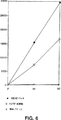
図5 酵素活性を有する組み換えr−HCV RNA依存性RNAポリメラーゼ。精製された組み換えポリオウイルスRDRP(約50ng)、及びr−HCV RDRPを発現している大腸菌(HCV RDRP溶解物)又はRDRPを発現していない対照細胞(対照溶解物)から得られた等量の可溶性タンパク質(約1μg)のポリ(U)ポリメラーゼ活性を示す。インキュベーションは、既述のようにして行い、30分(1サンプル)及び60分(2サンプルの平均)のインキュベーション後の、15μlのインキュベーションから回収されたポリ(U)のCPMを示した(J. Virol.(1986)58: 790-796)。
図6 部分的に精製された組み換えHCV RNA依存性RNAポリメラーゼの酵素アッセイ。r−HCV RDRPを発現している大腸菌の溶解液を調製し、図5と同様にして酵素アッセイを行った。示された実験においては、RDRPが樹脂に結合できるようなpH及びバッファーの条件下で、バッチ精製法でDEAE樹脂を用いた。細胞溶解物(RDRP溶解物と示されている)、及び溶解物とほぼ同じ濃度にまで濃縮された、0.5MのNaClでDEAE樹脂から溶出されたタンパク質(DEAEバッチと示されている)から得られた等量のタンパク質をアッセイした。DEAE樹脂及び他の試験樹脂を用いたさらなる試験で、RDRP酵素活性の部分精製が、ウサギ抗血清を用いたイムノブロッティング及び図4に概説された方法により検出される、タンパク分解されていないRDRPの量の増加と相関することが示された。
図7 組み換えr−HCV RDRPのイムノアフィニティ精製。この写真複写物は、ウサギポリクローナルプロテインAセファロースで精製された抗RDRP抗体を用いて調製されたイムノアフィニティカラムでの最初の結果を示す。出発物質は、組み換えr−HCV RDRPを発現している大腸菌から可溶化されたタンパク質であった。図は、異なる洗浄剤の条件でタンパク質が適用された2つの同一のカラムから溶出されたタンパク質のイムノブロットを示す。レーン1は、20mMトリス(pH7.5)、100mM KCl、0.5mM EdTA、1mM DTT、5% グリセロール、及び0.05% Triton X−100中で可溶化された組み換えRDRPと共に一晩混合された抗体/セファロースビーズから溶出されたタンパク質を示す。これらのビーズを、翌朝10mMリン酸カリウムバッファー(pH7.2)で洗浄し、タンパク質を100mMグリシン(pH2.5)で溶出した。溶出したタンパク質を1Mトリス(pH8.0)中に回収し、pHを再調整した。カラムから溶出したタンパク質を、図4と同様に、SDS−PAGE及びイムノブロッティングにより分析した。レーン2は、0.05% Triton X−100の代わりに0.05% NP−40を使用したこと以外は同一の出発物質と共に混合した同一のビーズから溶出したタンパク質を示す。r−HCV RDRPの位置が示されている。
発明の詳細な説明
定義
「RDRP」とは、RNA合成を触媒する酵素である、RNA依存性RNAポリメラーゼを意味する。これにより合成されたRNAはRNA鋳型と相補的な配列を有する。「HCV−RDRP」はC型肝炎ウイルスのRDRPである。本明細書において開示される修飾されたHCV−RDRPは、r−HCV−RDRPと称す。HCVゲノムのNS5Bと呼ばれる領域は、前記のワクシニアウイルス発現系を用い、HCVポリタンパク質のタンパク質分解産物として同定されている。NS5Bのヌクレオチド配列は配列番号1に含まれている。NS5B配列をコードする推定アミノ酸は、ヌクレオチド7から開始する。配列が5’末端で欠失している場合、残りの配列は、終止コドンは含まずに、残りのコーディング配列の最初と最後のヌクレオチド番号により表記される。例えば、NS5B34-1779とは、配列番号1のヌクレオチド34〜1779を含むNS5Bの一部を示す。
本明細書において例示されるr−HCV−RDRPをコードするアミノ酸配列は、配列番号2に示されている。NS5Bによりコードされるアミノ酸配列は、配列番号2の第3のアミノ酸から開始する。NS5Bによりコードされる配列がN末端で欠失している場合、残りの配列は、△nNS5Bと表記される。ここで、nはNS5BのN末端から欠失したアミノ酸の数である。例えば、△9NS5Bは、配列番号2中のアミノ酸12〜593の配列である。
記載されたように、本発明により、種々のr−HCV−RDRP構築物が企図される。本発明に含まれる修飾コーディング配列は、以下のような一般配列を有する。
ATG−NX−(NS)(NM)(NS)(NY)(NS)(NW)(NT)(NG)(NA)−[NS5B34-1779]
ここで、NXは0〜20アミノ酸をコードする任意のヌクレオチド配列であり、NSはセリンをコードするコドンであり、NMはメチオニンをコードするコドンであり、NYはチロシンをコードするコドンであり、NWはトリプトファンをコードするコドンであり、NTはトレオニンをコードするコドンであり、NGはグルタミン酸をコードするコドンであり、NAはアラニンをコードするコドンである。括弧内のコドンはいずれも、必要ならば、欠失してもよい。必要ならば、最大5個の括弧内のコドンを変異させることができる。「変異」という用語は、NS5B配列により本来コードされるアミノ酸とは異なるアミノ酸をコードするよう変化していることを意味する。例えば、既知のアラニン・スキャニング突然変異導入法により、個々のコドンをアラニンをコードするよう変化させることができる。アラニン・スキャニング突然変異導入法は、機能に負の影響を実質的に与えることなく、置換に耐性のアミノ酸部位を同定するための迅速かつ便利な方法を提供する。アラニン・スキャニングにより置換耐性であることが明らかになった部位は、他のアミノ酸置換に対しても耐性である可能性が高い。好ましい置換残基は、金属(例えばニッケル)カラム用のアフィニティリガンドとして機能する、一つ又は複数のヒスチジン残基である。ヒスチジンの存在により、カラムへの選択的な結合性が提供され、r−HCV−RDRPの精製が促進される。定義されたとおり、[NS5B34-1779]とは、配列番号1の残りのヌクレオチド配列、終止コドンを含まないヌクレオチド34〜1779を示す。前記の配列を作製する方法は、いずれの配列についても、NXがGCTであり、NS5Bによりコードされる最初の9個のアミノ酸をコードする括弧内のコドンがいずれも欠失していない配列についての下記の方法と本質的に同様である。適切な発現ベクターへの挿入を容易にするため、所望の制限部位配列と組み合わせた所望の配列のためのプライマーを合成できることは明らかであろう。ベクターの選択は、宿主細胞、所望のプロモーター型、及び、r−HCV−RDRPと共発現させる付加配列の有無を含む、当該技術分野において既知の要因に基づく。反応条件、PCR、ベクターの挿入、及び宿主細胞の培養は、以下に記載されるか、又は当技術分野において周知のものである。
r−HCV−RDRPに対してさらなる修飾を行うことができる。Gly−Asp−Aspモチーフを含まないC末端領域の約25%の欠失を構築し(r−HCV−RDRP−△C)、その活性を試験した。タンパク質は発現し、抗RDRP血清を用いたイムノブロッティングにより検出することができたが、r−HCV−RDRP−△Cを発現している大腸菌の溶解物は、ポリ(U)アッセイにおいても、鋳型としてグロビンmRNAを用いた場合にも、測定可能な活性を示さなかった。しかし、モデリング試験に基づくと、C末端に近いNS5Bのアミノ酸565〜572(配列番号2の565〜574)の領域が、タンパク質表面に露出していると考えられる。部位特異的突然変異導入法を用いて、コード配列をArg570→His、Arg572→His、及びTry573→Hisへと変化させた(配列番号2の番号を用いた)。タンパク質の一端付近の表面領域に一つ又は複数の付加ヒスチジン残基をクラスター形成させることにより、酵素活性には実質的に影響を与えることなく、金属アフィニティクロマトグラフィーによる精製が容易になる。現存のアミノ酸をヒスチジンに置換する突然変異導入のための部位となりうる他の推定表面領域には、配列番号2におけるアミノ酸47〜56、152〜159、183〜184、210〜215、269〜272、384〜391、及び439〜442が含まれる。
r−HCV−RDRPは事実上いかなる宿主細胞型で発現させることもできるが、阻害剤試験のようなインビトロ試験において有用であるためには、酵素は可溶型であることが好ましい。細胞内で合成される場合には、酵素を可溶化する工程を実施しない限り、酵素は細胞溶解物中で不溶型で存在する。一般的には、宿主細胞を回収し、濃縮し、その後、例えば宿主細胞壁加水分解酵素の使用、超音波処理などの既知の方法により溶解し宿主細胞を破壊する。一般的には、細胞溶解により放出されるタンパク分解酵素から保護するため、プロテアーゼ阻害剤を添加する。非イオン性洗浄剤を使用することもできる。超音波処理された細胞及び細胞内複合体を、上記の成分の存在下、凍結させ、解凍させる。残存する粒子物質を10,000〜35,000×gで遠心分離することにより除去する。r−HCV−RDRPは上清に残る。大腸菌において発現されたr−HCV−RDRPを可溶化するための詳細なプロトコルは後述する。
当該技術分野において既知の技術及び手段により、酵素のさらなる精製が達成される。その方法には、これらに限定されないが、抗体アフィニティクロマトグラフィー、金属結合アフィニティクロマトグラフィー(付加ヒスチジン残基を有する修飾型酵素に特に適した方法である)が含まれ、さらに通常のイオン交換カラム、硫酸アンモニウムによる選択的な沈殿、及び本明細書に具体的に記載された方法に限定されない当該技術分野において既知の他の方法が含まれる。本明細書において「精製された形態」とは、可溶化細胞溶解物中で測定された活性よりも少なくとも4倍大きい特異的活性を有する酵素調製物を意味する。
r−HCV−RDRPに対する抗体は、モノクローナル抗体又はポリクローナル抗体を形成するための様々な既知の方法により作製することができる。r−HCV−RDRPに対する抗体は、感染細胞のHCV−RDRPにも結合する。このことは、抗体リガンドとしてr−HCV−RDRPを用いて、HCV感染患者の血清中にHCV−RDRP対する循環抗体を検出することができるという知見により示された。当該技術分野において周知のように、r−HCV−RDRPの異なるエピトープに対して親和性を有する様々なモノクローナル抗体を選択することができる。酵素活性を阻害する抗体も存在する。抗体とアフィニティカラムとを結合させ、次に酵素を不活化しないような条件で溶出を行うことが容易にできるような適度な親和性を有する抗体も存在する。
ある種の目的、特にHCV−RDRP阻害剤のインビトロスクリーニング及び細胞培養物中でHCVを増殖させることができる細胞系の樹立のためには、哺乳動物細胞が好ましい宿主細胞である。形質転換に使用される既知の細胞系はすべて、原則的に、r−HCV−RDRPを発現させるために形質転換することができる。好ましい細胞系は、HCV、又はフラビウイルスなどの類似のウイルスが感染することが知られている組織に由来するものである。このような細胞系には、例えば、ヒトマクロファージ細胞系U937、ヒト肝臓由来hepG2細胞、及びブタ腎臓細胞系PK15が含まれる。最近発見されたHCV−RNAの3’末端近傍のセグメントは、HCV−RDRPを調節する(宿主がコードしている可能性がある)タンパク質又はペプチドとの相互作用部位である可能性が高い推定クローバー葉型二次構造を有する(Tanaka, T., et al.(1995)Biochem. Biophys. Res. Commun. 215: 744, 749)。このような調節は、r−HCV−RDRP及び野生型酵素の鋳型特異性又は触媒活性の変化という形をとる。調節タンパク質を通常発現している細胞におけるr−HCV−RDRPの発現は、r−HCV−RDRPの発現がHCV感染細胞における発現の様式と極めて類似したインビボ細胞系を提供する。また、細胞中に活性HCV−RDRPが存在すると、HCV複製のための「ジャンプ・スタート(jump start)」を提供することにより、感染により導入されたHCV、又は形質転換により導入されたゲノミックHCV RNAの複製を増強することができる。最も重要な点として、形質転換細胞におけるr−HCV−RDRP活性を測定できることが、インビボでの酵素阻害能についてHCV−RDRPの潜在的な阻害剤をスクリーニングするための重要な鍵となる。
インビトロのr−HCV−RDRPによるRNA合成のアッセイにより、ポリ(A)鋳型を用いるポリ(U)の合成及びグロビンmRNA鋳型を用いるRNAの合成を酵素が触媒できることが示されている。いずれの反応も、記載された反応条件下で、90%を超えてプライマー依存性であった。酵素は、Mg++の非存在下では検出可能な活性を示さず、約20mMのMg++の存在下で最大の活性を示した。抗ウイルス活性を有する化合物をスクリーニングするための方法は、様々な化合物がインビトロでRDRP酵素活性に対して及ぼす効果を試験することにより提供される。インビトロ法には、被検化合物の存在下及び非存在下で合成されたRNA量を比較することが含まれる。被検化合物の非存在下での対照反応と比較して、被検化合物の存在下での反応で合成されたRNAの量が減少している場合、阻害効果が示唆される。
r−HCV−RDRPを発現している安定的に形質転換された細胞系は、完全な哺乳動物細胞内でポリメラーゼを阻害する化合物をインビボスクリーニングするために、特に有用である。このような阻害剤は、感染細胞においてHCV複製の阻害剤となる可能性が高い。インビボ試験は、細胞毒性を有する化合物、及び充分量において細胞に侵入することができない化合物を選別除外しうるという点で有利である。また、さらに、実際の阻害剤の前駆体であり、細胞代謝過程により阻害剤へと変換される化合物を試験することも可能である。前駆体阻害剤の例としては、活性な阻害剤へと変換されるためにはリン酸化される必要があるヌクレオシド類似体、及び細胞内で加水分解を受け活性な阻害剤を形成するような結合により保護された活性基を有する化合物が含まれる。下記に示す一般構造を有する非ヌクレオシド類似体はr−HCV−RDRP阻害剤の注目すべき候補と考えられる。

材料及び方法
材料 − 特に断りのない限り、化学物質はすべてフィッシャー(Fisher)から購入し、酵素はすべてギブコBRL(Gibco BRL)から購入した。AmpliTaqはパーキン・エルマー(Perkin-Elmer)から購入した。他のPCR及びライゲーション用成分はすべてインビトロジェン(Invitrogen)から購入した。リゾチーム、抗生物質、及び染色済タンパク質標本はシグマ(Sigma)から購入した。ヌクレオチド及びポリ(A)はファルマシア(Pharmacia)から購入した。[3H−]UTPはデュポンNEN(Dupont NEN)から購入した。オリゴ(U)はイー.エーレンフェルド(E. Ehrenfeld)氏(カリフォルニア大学,Irvine)の好意で贈られたものである。
HCV NS5B領域のサブクローニング − ワクシニアウイルス発現試験により推定されたN末端(Lin, C. et al.(1994)前出; Grakoui, A. et al.(1993)前出)、及びHCVポリタンパク質の読み取り枠の末端に基づくC末端(Choo, Q-L. et al.(1991)前出)に基づき、NS5B領域の増幅用のPCRプライマーを設計した。鋳型は本来のプロトタイプHCV(1a型)クローン(CDCから得た)(Choo, Q-L. et al.(1989),(1991),前出)であった。以下のプライマー、5’−ATA GCT AGC ATG TCT TAC TCT TGG ACA GG−3’(配列番号3)及び5’−ATA GGA TCC TCA TCG GTT GGG GAG GAG G−3’(配列番号4)を使用して、N末端に最少の変化を有するNS5B領域(SMSY配列番号7の代わりにASMSY配列番号5)を増幅し、方向性をもってNheI及びBamHI制限部位でpET−11a(Novagen)へとクローン化した(Maniatis et al.(1982)Molecular Cloning, Cold Spring Harbor Laboratory, Plainview, New York)。PCR増幅された遺伝子には、5’末端にNheI部位、3’末端にBamHI部位が組み込まれていた。この構築物は、ワクシニアウイルス発現試験により推定された推定野生型NS5Bタンパク質のSMSYアミノ末端ではなく、MASMSYというアミノ末端配列を有する組み換えタンパク質の合成をもたらす。
T4リガーゼを用いて、3:1比(挿入配列:ベクター)で、14.5℃で一晩、挿入配列とベクターとを連結させた(Maniatis,前出)。連結された物質を用いて、CaCl2法により大腸菌(インビトロジェンのTop10(登録商標))を形質転換した。アンピシリンプレート上でコロニーを選択し、単一コロニーから単離したプラスミドDNAのミニプレップを、制限酵素分析を用いて特徴解析した。ミニプレパレーション法により得られたプラスミドDNAを用いて、BL21(λDE3)大腸菌(ノバジェン(Novagen))を形質転換し、プラスミドを含有する生物をアンピシリンを用いて選択し、単一コロニー由来のプラスミドDNAのミニプレパレーションを、制限酵素消化により解析した。
推定HCV RDRPの発現、精製、及び可溶化 − 上記のpET−11a−NS5B構築物を含有するBL21(λDE3)大腸菌を一晩培養し(カルベニシリンを含むM9ZB培地)、翌朝新鮮培地で20倍に希釈した。培養培地のOD600が0.6に達するまで、細胞を37℃でインキュベートした。その後、IPTGを添加して最終濃度を1mMにした。推定RDRPの発現をサンプルバッファー中で90℃で溶解した完全な細胞をSDS−PAGE分析することにより追跡した。
非変性条件下でRDRPを可溶化するため、IPTG誘導の2時間後細胞を回収した。氷上で20分間、20mMトリス(pH7.5)、100mM KCl、0.5mM EDTA、1mM DTT、0.1% Triton X−100、及び30μg/mlリゾチーム中で細胞を溶解することにより、RDRPを可溶化した。サンプルを氷上で0.5インチのプローブ(パルスセッティング)を用いて5分間(Ultrasonics Inc. W-225,アウトプット−セッティング7)超音波処理し、遠心分離(19,000g、4℃、30分間)を行った。これらの調製物から得られた不溶性画分(ペレット)には、RDRPが多く含まれていた。ペレットをSDS−PAGEサンプルバッファーに懸濁し、90℃で10分間加熱し、SDS−PAGEゲルのRDRPマーカーとして使用した。しかし、以下のプロトコルに記載するように、活性な酵素は上清に観察された。
組み換えRDRPを可溶化する方法の概略
1. 5gの大腸菌ペレットを解凍する。
2. 5gのペレットを以下の溶液に再懸濁する。
45ml 溶解バッファー
40μl 100mM PMSF(及び他のプロテアーゼ阻害剤)
150μl リゾチーム(10mg/ml)
溶解バッファー:
20mMトリス(pH7.5)(4℃)
0.5mM EDTA
100mM KCl
1mM DTT
0.1% Triton X−100(又は0.1% NP−40)
10.0%(v/v)グリセロール
3. サンプルを氷上に20分放置し、次に5分間超音波処理(パルスモード;6〜7の間のセッティング)する。超音波処理の間、混合する。
4. 瞬時の超音波処理(sonicating flash)後、溶解物を液体窒素中で凍結させる(溶解物を約1〜2分間液体窒素中に投入する)。
5. 37℃の水浴で溶解物を急速に解凍する。
6. 溶解物を1分間超音波処理する。
7. 超音波処理したサンプル混合物45mlにつき5mlの溶解バッファーをさらに添加する。
8. 全超音波処理済サンプルを50mlの画分(フィッシャー50mlチューブ)に分割する。
9. 溶解物を、ベックマン(Beckman)J−17ローター中、12,500rpm(又はソバール(Sorvall)SS−34ローター中、12,500rpm)、20分間の遠心分離にかける。
10. 上清を清潔な(無菌の)50mlフィッシャーチューブにとり、無菌タンパク質等級のグリセロールを最終濃度10%(例えば、40mlの上清に対し4.44mlのグリセロール)で添加した。この溶液を4℃で保存し、酵素活性を有するHCV RDRPの精製のための出発物質として使用した。
以下の工程を、単独で、又は組み合わせて用いることにより、さらに精製を行う。
ウサギ抗HCV RDRP血清 − 上記のようにしてペレット画分から可溶化されたRDRPを調製用SDS−PAGEにより分離し、ウサギを免疫するために用いた。動物を、既に詳細に開示されている方法(Harlow, E. and D. Lane(1988)Antibodies: A Laboratory manual, Cold Spring Harbor Laboratory, pp. 553-611)で、4〜5週の間隔で免疫した。
イムノブロッティング分析 − イムノブロットは、既に開示されている方法に、二次HRP結合抗体を増幅化学発光系(ELC,アメルシャム(Amersham))と共に用いるという修飾を加えて行った。ウサギ血清を一次抗体として用いる場合には、二次抗体は抗ウサギ免疫グロブリンであった。ヒト血清をスクリーニングする場合には、二次抗体は抗ヒト免疫グロブリンであった。慢性C型肝炎に感染していることが証明されている患者由来の血清は、疾病の管理と予防センター(Centers for Disease Control and Prevention)(アトランタ)のマイケル・ビーチ博士(Dr. Michael Beach)から提供された。
ポリ(U)ポリメラーゼアッセイ − 可溶性画分中の酵素活性は、鋳型としてポリ(A)、プライマーとしてオリゴ(U)を用いたポリ(U)ポリメラーゼアッセイにより測定した(Hey, T.D. et al.(1986)J. Virol. 58: 790-796)。サンプル(およそ2μl)を、50mM HEPES(pH8.0)、各500μMのATP、CTP、及びGTP、4mM DTT、3mM MgAc2、ならびに60μM ZnCl2を含む50μlのインキュベーション中でアッセイした。15μMの濃度の[3H]UTP(特異的活性:27Ci/mol)も添加した。各インキュベーション中に、1μgのポリ(A)、そしてプライマーとして0.5μgのオリゴ(U)を加えた。
インキュベーションを30℃で30〜60分間行い、[3H]ポリ(U)をキャリヤDNAの存在下、TCAで沈殿させ、ワットマン(Whatman)GF/Cフィルターで回収した。フィルターを0.1Mピロリン酸ナトリウム/1N塩酸及び95%エタノールの各々で洗浄した。[3H]ポリ(U)は、液体シンチレーション分光光度測定法(LKB 1218 ラックベータ(RackBeta))により定量した。
リポフェクチンを用いた新生ハムスター腎臓(BHK)細胞の安定的形質転換
1日目(午後)
形質転換のため50%のコンフルエンシー状態になるようにBHK細胞を6穴プレートに分割する。
2日目(午後4時以降)
滅菌チューブ中で以下の溶液を調製する。
(A)50μlミニプレップDNA+50μl無血清培地(DMEM/F12)
(各2)
(B)6.25μlリポフェクチン(ライフ テクノロジーズ(Life Technologies), Gaithersburg, MD)+93.75μl培地
(C)12.5μlリポフェクチン+87.5μl培地
(D)6.25μlリポフェクチン+193.75μl培地(モック形質転換)
(E)12.5μlリポフェクチン+187.5μl培地(モック形質転換)
AとB、及びAとCを穏和に混合し、室温で15分間DNAとリポフェクチンを反応させる。この間、細胞を2mlのDMEM/F12で2回洗浄する。DNA/リポフェクチン複合体に1.8mlのDMEM/F12を添加し、それを穏和に攪拌しながら細胞へと添加する。インキュベーター中に一晩細胞を放置する。
3日目(午前9時)
DNA/リポフェクチンを取り出し、3mlの培地+血清を細胞に添加する。細胞を30〜48時間インキュベートする。1:20、1:50、1:100の細胞を、10mlの600μg/mlゲネチシンを含む培地+血清で、10cmのプレートに分割する。選別のため3〜7日間、コロニー形成のため10〜14日間放置する。形質転換効率を改善するため、リポフェクチンの代わりにスターバースト・デンドリマー(Starburst Dendrimer)(ライフ テクノロジーズ、Gaithersburg, MD)を用いても、同じプロトコルを適用できる。
選別後、クローンコロニーを24穴プレートに播き、コンフルエンシー状態のウェルの培地のRDRP活性をアッセイする。細胞は、600μg/mlのゲネチシン中で維持する。
完全な細胞に侵入しHCV RDRPを阻害する化合物を同定するための、安定的形質転換細胞のHCV RDRP発現の使用
形質転換細胞におけるHCV RDRPの阻害剤候補の効果を決定するための最も直接的な方法は、細胞を化合物と共にインキュベートし十分に洗浄した後、細胞抽出物中のRDRP活性を直接的に測定することである。これは、本明細書に記載のRDRPアッセイにより(HCV鋳型を用いて)行うことができ、細胞系以外には新たな改良を必要としない。簡単に述べると、活性酵素を最大に発現する条件下で、そしてその後の酵素アッセイに十分な量で、細胞をインキュベートする。被検化合物をインキュベーション中に添加し、所望の時間に培地を除去し、細胞外被検化合物を除去するため細胞を十分に洗浄する。本明細書に記載の一般的な方法に従い、細胞抽出物をRDRPアッセイ用に調製する。この方法は比較的迅速に行われ、本発明者らの現在の方法にほんのわずかな変更(新しい細胞系)を必要とするのみである。阻害剤の動力学的研究(細胞内で阻害がどの程度の速度で生じるか)を完全な細胞で行う必要がある場合には、同じインキュベーションを2つ行う。この方法で考えられる唯一の問題点は、細胞には侵入しない化合物が、調製中に細胞溶解物へと混入してしまう可能性があることである。この起こりうる問題を回避するためには、至適な「洗浄」工程がどのようなものであるのかを決定することが必要である。この系の主要な利点は、精製RDRPを用いて試験するには面倒な大きな修飾(ヌクレオシドのリン酸化)をする必要がある化合物を、迅速にスクリーニングできることである。より迅速なスクリーニングは、潜在的な阻害剤と共にインキュベートした細胞に、容易に測定できるレポーター分子(分泌アルカリホスファターゼ又はルシフェラーゼ等)もコードするHCV RNA鋳型を発現するよう遺伝子操作されたプラスミドを、一過性形質転換することにより行いうる。このような系により、完全な細胞におけるHCV RDRP活性が測定される(阻害濃度は細胞の溶解などにより希釈されない)。RDRP活性が阻害される細胞を迅速にスクリーニングすることにより、多数の阻害剤候補を迅速にスクリーニングすることができる。
宿主細胞内で発現したr−HCV−RDRPの活性がレポーター遺伝子の発現に必要であるようなレポーター系が開発されている。mRNAとして発現した際にHCV複製中間体と類似するような構造中にレポーターコーディング配列をアンチセンス型で保持するよう設計された構築物で、宿主細胞を形質転換する。mRNAには、5’末端から始まり、キャップ部位、アンチセンス型、すなわち(−)鎖型のレポーターコーディング領域、同様に(−)鎖型のHCV内部リボソーム侵入部位(internal ribosome entry site)(IRES)因子、(+)鎖型のリボザイム配列、及び(+)鎖型のポリアデニル化部位が含まれる。このようなmRNAは、翻訳されると、レポーター遺伝子の(−)鎖によりコードされるナンセンスタンパク質を生じる。しかし、相補鎖がr−HCV−RDRPにより合成されると、(+)鎖のコーディング配列がレポータータンパク質(例えば、ルシフェラーゼ、蛍光グリーンタンパク質、分泌アルカリホスファターゼなど)として翻訳可能になる。相補鎖合成は細胞質で起こり、キャップ構造付加は宿主細胞核で起こるため、RDRPにより生成された相補鎖には、キャップ構造を有する5’末端が欠けている。しかし、HCV−IRES因子の存在により、キャップ非依存性翻訳が可能となる(IRES因子は、相補鎖中の(+)鎖コーディング配列の5’側に位置する)。リボザイムモチーフの機能は、RDRPによる相補鎖合成の前に、(−)鎖の3’末端からポリ(A)尾部を除去し、付随的にそれ自身が除去されることである。適切なリボザイムモチーフは、例えば、デルタ型肝炎ウイルスのR289ccにより提供される。取り込まれたDNAから転写されるとき、RNA中のレポーターは以下のように図示される。
本発明を、具体的な実施態様及び実施例に関して詳細に開示してきたが、本明細書に記載された一つ又は複数の教示、原理、及び結果を、本発明の範囲に含まれる、当業者により適用される当該技術分野における知識と組み合わせることにより、さらなる実施態様、実施例、及び変更が行われうることが理解されるであろう。
配列表
(1)全体的情報:
(i) 出願人:ハーゲドーン、カート エイチ.(Hagedorn, Curt H.)
アル、レイノルダス エイチ.(Al, Reinoldus H.)
(ii) 発明の名称:組み換えC型肝炎ウイルスRNAレプリカーゼ
(iii) 配列の数:10
(iv) 連絡先:
(A)名宛人:グリーンリー、ウィナー アンド サリバン、ピー.シー.(Greenlee, Winner and Sullivan, P.C.)
(B)街路:5370 マンハッタン サークル(Manhattan Circle)、スウィート(Suite)201
(C)市:ボルダー(Boulder)
(D)州:コロラド
(E)国:米国
(F)ZIP:80303
(v) コンピュータ可読形態:
(A)媒体の型式:フロッピーディスク
(B)コンピュータ:IBM PC互換型
(C)オペレーティングシステム:PC-DOS/MS-DOS
(D)ソフトウェア:PatentIn リリース#1.0、バージョン#1.30
(vi) 最新の出願データ:
(A)出願番号:米国
(B)出願日:1996年9月27日
(C)分類:
(vii) 先願のデータ:
(A)出願番号:US 60/004383
(B)出願日:1995年9月27日
(viii)弁理士/代理人の情報:
(A)名称:グリーンリー、ローランス エル.(Lorance L.)
(B)登録番号:27,894
(C)参照/書類番号:76-95US
(ix) 遠隔通信情報:
(A)電話:(303)499-8080
(B)テレファックス:(303)499-8089
(2)配列番号1に関する情報:
(i) 配列の特性:
(A)長さ:1788塩基対
(B)型:核酸
(C)鎖の数:二本鎖
(D)トポロジー:該当なし
(ii) 配列の種類:cDNAないしmRNA
(iii) ハイポセチカル:No
(ix) 特徴:
(A)特徴を表す記号:CDS
(B)存在位置:1..1782
(xi) 配列の記載:配列番号1:
(i) 配列の特性:
(A)長さ:594アミノ酸
(B)型:アミノ酸
(D)トポロジー:直鎖状
(ii) 配列の種類:タンパク質
(xi) 配列の記載:配列番号2:
(i) 配列の特性:
(A)長さ:29塩基対
(B)型:核酸
(C)鎖の数:一本鎖
(D)トポロジー:直鎖状
(ii) 配列の種類:他の核酸
(A)説明:/desc=「オリゴヌクレオチドプライマー」
(iii) ハイポセチカル:No
(xi) 配列の記載:配列番号3:
![]()
(i) 配列の特性:
(A)長さ:28塩基対
(B)型:核酸
(C)鎖の数:一本鎖
(D)トポロジー:直鎖状
(ii) 配列の種類:他の核酸
(A)説明:/desc=「オリゴヌクレオチドプライマー」
(iii) ハイポセチカル:No
(xi) 配列の記載:配列番号4:
![]()
(i) 配列の特性:
(A)長さ:5アミノ酸
(B)型:アミノ酸
(C)鎖の数:該当なし
(D)トポロジー:不明
(ii) 配列の種類:タンパク質
(iii) ハイポセチカル:No
(v) フラグメント型:N-末端
(xi) 配列の記載:配列番号5:
![]()
(i) 配列の特性:
(A)長さ:6アミノ酸
(B)型:アミノ酸
(C)鎖の数:該当なし
(D)トポロジー:不明
(ii) 配列の種類:タンパク質
(iii) ハイポセチカル:No
(v) フラグメント型:N-末端
(xi) 配列の記載:配列番号6:
![]()
(i) 配列の特性:
(A)長さ:4アミノ酸
(B)型:アミノ酸
(C)鎖の数:該当なし
(D)トポロジー:不明
(ii) 配列の種類:タンパク質
(iii) ハイポセチカル:No
(v) フラグメント型:N-末端
(xi) 配列の記載:配列番号7:
![]()
(i) 配列の特性:
(A)長さ:8アミノ酸
(B)型:アミノ酸
(C)鎖の数:該当なし
(D)トポロジー:不明
(ii) 配列の種類:ペプチド
(iii) ハイポセチカル:Yes
(xi) 配列の記載:配列番号8:
![]()
(i) 配列の特性:
(A)長さ:104塩基対
(B)型:核酸
(C)鎖の数:二本鎖
(D)トポロジー:該当なし
(ii) 配列の種類:他の核酸
(iii) ハイポセチカル:No
(ix) 特徴:
(A)特徴を表す記号:CDS
(B)存在位置:63..104
(xi) 配列の記載:配列番号9:
(i) 配列の特性:
(A)長さ:14アミノ酸
(B)型:アミノ酸
(D)トポロジー:直鎖状
(ii) 配列の種類:タンパク質
(xi) 配列の記載:配列番号10:
![]()
Field of Invention
The present invention relates to hepatitis C virus (HCV), and more particularly, the expression and purification of RNA-dependent RNA polymerase (RDRP) encoded by the HCV genome, antibodies against HCV-RDRP, and diagnosis of chronic HCV infection Therefore, the present invention relates to a method of using the enzyme for screening an antiviral agent effective against HCV.
Background of the Invention
HCV is a major agent responsible for post-transfusion hepatitis and sporadic non-A non-B hepatitis (Alter, H.J. (1990) J. Gastro. Hepatol.1: 78-94; Dienstag, J.L. (1983) Gastro85: 439-462). Despite improved screening methods, HCV is still responsible for at least 25% of acute viral hepatitis in many countries (Alter, HJ (1990) supra; Dienstag, JL (1983) Supra, Alter, MJ et al. (1990a) JAMA264: 2231-2235; Alter, M.J. et al (1992) N. Engl. J. Med.327: 1899-1905; Alter, M.J. et al. (1990b) N. Engl. J. Med.321: 1494-1500). In chronically infected (and infectious) carriers, HCV infection is often latent and such carriers may not have clinical signs over time. Acute infection is likely to progress to chronic infection (70-100%) and liver disease (> 50%), distributed worldwide, and the absence of vaccines, HCV causes morbidity and mortality. It has become a serious cause.
HCV is an enveloped virus whose genome is a 9.4 kb single-stranded RNA (sense (+)) that encodes a single polyprotein. The polyprotein is processed by proteolysis to yield at least nine proteins. HCV is associated with pestiviruses and flaviviruses (Choo, Q-L. Et al. (1989) Science244: 362-364; Choo, Q-L. Et al. (1991) Proc. Natl. Acad. Sci. USA88: 2451-2455). Reinfection of chimpanzees that have already been infected with HCV suggests that protective immunity is transient or absent (Farci, P. et al (1992) Science258: 135-140). Furthermore, recent vaccine trial results indicate that development of an effective vaccine cannot be expected (Houghton, M. et al. (1994) 2nd Internat, Meeting on Hepatitis C (San Diego)). Attempts have been made to treat chronic HCV infection with existing antiviral agents, but the cure rate is low and severe side effects have occurred (Dienstag, J. L. (1983) supra).
The nucleotide sequence of the HCV genome has been cloned and a single open reading frame has been identified. Several degradation products have been tentatively identified using the vaccinia virus expression system (Lin, C. et al. (1994) J. Virol.68: 5063-5073; Grakoui, A. et al. (1993) J. Virol.67: 1385-1395). Various putative degradation products were recognized by antibodies obtained against various peptides synthesized from amino acid sequences deduced from various segments of the coding region. The size of the antibody-reactive peptide was estimated by SDS-PAGE (see FIG. 1). The nonstructural protein (NS5B) designated 5B has been shown to have an amino terminal sequence called SMSY (Ser-Met-Ser-Tyr). The NS5B region encodes a 68 kd protein (p68), which contains an internal GDD (Gly-Asp-Asp) motif found in RNA-dependent RNA polymerases of other RNA viruses (Koonin, EV (1991) J. Gen. Virol.72: 2197-2206). However, no polymerase activity has been detected in HCV p68. In fact, the 5B protein (p68) alone does not encode an active RNA-dependent RNA polymerase enzyme, and the problem arises that other subunits, possibly the NS5A gene product, are essential for catalytic activity. To date, the present inventors and other researchers have expressed the NS5B coding region as a fusion protein using an existing expression system that allows easy purification of the fusion product and specific degradation. Attempts have been made, but no active polymerase has been obtained.
Summary of the Invention
The present invention provides a recombinant protein of HCV having RDRP activity (r-HCV-RDRP) obtained by expressing a modified NS5B coding region of HCV in a mammalian or bacterial host cell. For modification, add a methionine residue at the amino terminus and optionally insert 1-20 additional amino acids between the N-terminal methionine and the N-terminal serine of the unmodified NS5B gene product. Is included. Modifications also include deleting up to 9 amino acids at the amino terminus to provide an amino terminal methionine. According to the deduced sequence of wild type HCV-RDRP, two methionines are naturally present. Thus, modifications include deleting the amino acid present at the N-terminus to either methionine or deleting up to the midpoint of two methionines and adding an N-terminal methionine codon. . Combinations of deletions and insertions are also contemplated within the limits described. The added amino acid sequence can be devised to form a specific protease degradation site to allow post-translational modification of the recombinant HCV-RDRP expression product in vivo or in vitro. Such post-transcriptional modifications can be used to accurately generate an amino acid sequence encoded by NS5B having an N-terminal serine. In order to simplify and facilitate the purification, the added amino acid sequence can also be devised to form an affinity ligand binding site. The data reported herein has a MA (Met-Ala) dipeptide at the N-terminus, giving an N-terminal sequence of MASMSY (SEQ ID NO: 6) instead of the putative SMSY sequence of the wild-type processed protein It was obtained with r-HCV-RDRP. Thus, the NS5B coding sequence contains a methionine codon (ATG) at the 5 'end and is optionally modified to include or delete codons for other amino acids. In order to avoid the detrimental effects that can occur on enzyme activity and to avoid the creation of artificial epitopes, it is preferred that the modification be minimal. r-HCV-RDRP can be expressed in E. coli and mammalian cells to obtain active RDRP. Expression of active r-HCV-RDRP in E. coli indicates that the polymerase activity does not require other proteins encoded by HCV.
The invention further provides a solubilized form of r-HCV-RDRP and a method of solubilizing without loss of activity.
The present invention also provides a method for purifying a solubilized form of HCV-RDRP. One method used in combination with other methods is affinity chromatography using an antibody against r-HCV-RDRP as the affinity ligand. Other affinity ligands are described, for example, in Wu, J. et al. (1994) Biochemistry.33: 14825-14833 and Ohlmeyer, M.H.J. et al. (1993) Procl. Nat. Acad. Sci. USA90: Obtained by combinatorial library method as described in 10922-10926.
Furthermore, the present invention provides a polyclonal or monoclonal antibody specific for HCV-RDRP. Such an antibody can be prepared by a known method using a purified enzyme as an antigen. Such antibodies bind to either r-HCV-RDRP or wild type HCV-RDRP. The availability of such antibodies allows the preparation of affinity labeled chromatography matrices for rapid purification of HCV-RDRP. The antibody also allows for rapid detection of biological material such as HCV-RDRP in the serum of HCV infected patients.
The present invention further provides a method of transforming mammalian cells with HCV-RDRP and expressing the enzyme in the cells. Consequently, the present invention also provides a transformed mammalian cell line that expresses r-HCV-RDRP. The cells are useful for assaying the effects of antiviral candidate compounds as inhibitors of RDRP activity.
Accordingly, the present invention also provides a method of screening for compounds that can be inhibitors of RDRP activity in vivo. Since inhibition of polymerase inhibits viral replication and expression of viral gene products, compounds with inhibitory activity may have antiviral activity. In vitro assays are useful because compounds that cannot enter infected cells can be eliminated. One class of candidate compounds to note is nucleoside analogs. It is modified (phosphorylated) in the cell and then either binds to the enzyme's substrate site or is incorporated into newly synthesized RNA, which is the normal function of HCV polymerase, or of an RNA containing analogs A compound that inhibits further replication. Acyclovir is an example of a highly effective and safe nucleoside analog that inhibits viral viral replication (DNA-dependent DNA polymerase) and inhibits replication of DNA viruses by inhibiting primer-template function (chain termination). . Such analogs are in most cases only effective in the form of nucleotide triphosphates. In an in vitro assay, the compound is administered in nucleoside or nucleoside monophosphate form, and the endogenous metabolic activity of the cell converts that form to the active triphosphate form, so that the triphosphate as required in the in vivo assay. A convenient method is provided that does not require an acid-type chemical synthesis step.
A method for measuring HCV-RDRP activity in vitro is also provided. Such assays allow for enzyme identification and concentration determination during purification. In addition, the assay provides an additional in vitro method for screening potential RDRP inhibitors as antiviral drug candidates.
In principle, any compound can be tested as a candidate RDRP inhibitor. Certain compound classes are considered candidates for attention. This includes, but is not limited to, nucleoside analogs, oligonucleotides, and peptides. Compounds with planar polycyclic aromatic character also have potential as inhibitors. It will be appreciated that compounds identified as effective RDRP inhibitors need to be further screened for toxicity, bioavailability, side effects, etc. before undergoing testing as therapeutic agents. However, the first identification as an inhibitor of HCV-RDRP is an important first step in the development of antiviral therapies. It will also be appreciated that inhibitors of r-HCV-RDRP also inhibit wild type HCV-RDRP.
In another aspect of the invention, the presence of purified HCV-RDRP or r-HCV-RDRP allows detection and measurement of RDRP antibodies present in the serum of HCV infected patients. The fact that any such antibodies are present is a discovery that in itself is made possible by the expression and preparation of purified r-HCV-RDRP according to the invention. The presence of circulating antibodies against HCV-RDRP in the infected serum causes the infected cells to be lysed and HCV-RDRP is released into the extracellular fluid and blood stream, stimulating the antibody reaction in the extracellular fluid and blood stream. It is thought that it is because it is. As the severity of the disease varies, the amount of HCV-RDRP released and the amount of antibody to HCV-RDRP also varies. Thus, the amount of antibody against HCV-RDRP present in the patient's serum can be used not only as an indicator of the presence of infection, but also as an indicator of the severity of (disease) at a given time. The assay for anti-HCV-RDRP can be used as a means to diagnose infection and as a means to monitor the course of disease over time or in response to treatment. Assays for anti-HCV-RDRP can be performed by a variety of known methods, such as the gel separation method described herein. Other suitable methods include ELISA and radioimmunoassay. Sandwich-type assays using immobilized r-HCV-RDRP to capture antibodies use anti-immunoglobulin reagents labeled with appropriate markers such as enzymes, radioisotopes, fluorescent molecules, or chemiluminescent markers Can be done. All of these will be understood by those skilled in the art (Antibodies : A laboratory manualEd Harlow and David Lane, Cold Spring Harbor Laboratory (1988) pp. 553-611).
[Brief description of the drawings]
FIG. 1 Hepatitis C virus genome and polyprotein degradation products. HCV polyprotein degradation products have been tentatively identified using a vaccinia virus expression system. The amino terminus of the 5B protein expressed and processed in this system is SMSY (Ser-Met-Ser-Tyr). Published reports do not show that the 5B protein has RNA polymerase activity, but contain the GDD (Gly-Asp-Asp) motif found in other RNA-dependent RNA polymerases . The 5B protein alone did not encode an active RNA-dependent RNA polymerase enzyme, resulting in the problem that other subunits (probably the NS5A gene product) are essential for catalytic activity. In the early part of this study, we were not able to confirm whether the protein encoded by NS5B does not exhibit RNA-dependent RNA polymerase activity simply due to the lack of other essential factors. .
FIG. 2. Expression of r-HCV RNA-dependent RNA polymerase in E. coli using a T7 polymerase-driven Studio vector. E. coli containing a T7 polymerase-driven expression vector produced by recombinant technology is transformed into OD600Was incubated at 37 ° C. until the value reached 0.6. Cell samples were obtained and IPTG was added to a final concentration of 1 mM. Samples were collected at 1, 2, and 3 hours after IPTG induction. Complete cells were lysed at 95 ° C. in 1 × sample buffer and samples were analyzed by 10% SDS-PAGE. The photo shows a representative gel stained with Coomassie blue.
Figure 3 Several chronic hepatitis C patients have circulating antibodies that react with recombinant HCV RNA-dependent RNA polymerase. Cells expressing r-HCV RDRP were collected and lysed by heating in SDS-PAGE sample buffer. Soluble proteins are separated by SDS-PAGE, transferred to a nitrocellulose membrane, and Immunetics Miniblotter template (Hagedorn, et al. FEBS Lett. (1990)264: 59-62) were used for immunoblotting with human serum. Immunoblots were developed using a secondary anti-human horseradish peroxidase conjugated antibody and amplified chemiluminescence (ECL, Ambersham). This picture shows an immunoblot. In
FIG. 4 Solubilization of HCV RDRP under non-denaturing conditions. Cells expressing r-HCV RDRP were collected and processed using standard methods. Soluble fractions from insoluble E. coli pellets (pellet, positive control) and cells with or without RDRP expression vector (negative control) were separated by SDS-PAGE and transferred to a nitrocellulose membrane. As outlined in FIG. 3, proteins bound to nitrocellulose were detected using rabbit preimmune serum and rabbit anti-RDRP serum as probes. The blot was developed using the ECL system (Amersham). The photograph shows an immunoblot. In
FIG. 5 Recombinant r-HCV RNA-dependent RNA polymerase with enzymatic activity. Purified recombinant poliovirus RDRP (approximately 50 ng) and an equal amount obtained from E. coli expressing r-HCV RDRP (HCV RDRP lysate) or control cells not expressing RDRP (control lysate) The poly (U) polymerase activity of soluble protein (about 1 μg) is shown. Incubations were performed as previously described and showed the CPM of poly (U) recovered from 15 μl incubation after 30 min (1 sample) and 60 min (average of 2 samples) incubation (J. Virol. (1986)58: 790-796).
FIG. 6 Enzymatic assay for partially purified recombinant HCV RNA-dependent RNA polymerase. A lysate of E. coli expressing r-HCV RDRP was prepared, and an enzyme assay was performed in the same manner as in FIG. In the experiments shown, DEAE resin was used in a batch purification process under conditions of pH and buffer that allow RDRP to bind to the resin. From cell lysate (denoted as RDRP lysate) and protein eluted from DEAE resin with 0.5 M NaCl concentrated to approximately the same concentration as lysate (denoted as DEAE batch) The equivalent amount of protein obtained was assayed. In further studies with DEAE resin and other test resins, partial purification of RDRP enzyme activity detected by immunoblotting using rabbit antiserum and the method outlined in FIG. It was shown to correlate with the increase in quantity.
FIG. 7 Immunoaffinity purification of recombinant r-HCV RDRP. This photographic copy shows the first results on an immunoaffinity column prepared with an anti-RDRP antibody purified on rabbit polyclonal protein A sepharose. The starting material was a protein solubilized from E. coli expressing recombinant r-HCV RDRP. The figure shows an immunoblot of protein eluted from two identical columns to which the protein was applied under different detergent conditions.
Detailed Description of the Invention
Definition
“RDRP” means RNA-dependent RNA polymerase, an enzyme that catalyzes RNA synthesis. The synthesized RNA has a sequence complementary to the RNA template. “HCV-RDRP” is RDRP of hepatitis C virus. The modified HCV-RDRP disclosed herein is referred to as r-HCV-RDRP. A region called NS5B of the HCV genome has been identified as a proteolytic product of the HCV polyprotein using the vaccinia virus expression system described above. The nucleotide sequence of NS5B is contained in SEQ ID NO: 1. The deduced amino acid encoding NS5B sequence starts at
The amino acid sequence encoding r-HCV-RDRP exemplified herein is shown in SEQ ID NO: 2. The amino acid sequence encoded by NS5B starts with the third amino acid of SEQ ID NO: 2. If the sequence encoded by NS5B is deleted at the N-terminus, the remaining sequence is ΔnIt is written NS5B. Here, n is the number of amino acids deleted from the N-terminus of NS5B. For example, △9NS5B is the sequence of amino acids 12 to 593 in SEQ ID NO: 2.
As described, various r-HCV-RDRP constructs are contemplated by the present invention. The modified coding sequence included in the present invention has the following general sequence.
ATG-NX-(NS) (NM) (NS) (NY) (NS) (NW) (NT) (NG) (NA)-[NS5B34-1779]
Where NXIs any nucleotide sequence encoding 0-20 amino acids, NSIs a codon encoding serine and NMIs a codon encoding methionine, NYIs a codon encoding tyrosine and NWIs a codon encoding tryptophan and NTIs a codon encoding threonine, NGIs a codon encoding glutamic acid, NAIs a codon encoding alanine. Any of the codons in parentheses may be deleted if necessary. If necessary, up to 5 codons in parentheses can be mutated. The term “mutation” means altered to encode an amino acid that is different from the amino acid originally encoded by the NS5B sequence. For example, individual codons can be altered to encode alanine by known alanine scanning mutagenesis methods. The alanine scanning mutagenesis method provides a quick and convenient way to identify amino acid sites that are resistant to substitution without substantially negatively affecting function. Sites that have been found to be substitution resistant by alanine scanning are likely to be resistant to other amino acid substitutions. Preferred substituted residues are one or more histidine residues that function as affinity ligands for metal (eg nickel) columns. The presence of histidine provides selective binding to the column and facilitates purification of r-HCV-RDRP. As defined, [NS5B34-1779] Represents the remaining nucleotide sequence of SEQ ID NO: 1, nucleotides 34 to 1779 not containing a stop codon. The method for producing the above-mentioned sequence is N for any sequence.XIs essentially the same as the method below for a sequence in which none of the codons in parentheses encoding the first 9 amino acids encoded by NS5B is deleted. It will be apparent that primers for the desired sequence can be synthesized in combination with the desired restriction site sequence to facilitate insertion into an appropriate expression vector. The choice of vector is based on factors known in the art, including the host cell, the desired promoter type, and the presence or absence of additional sequences that are co-expressed with r-HCV-RDRP. Reaction conditions, PCR, vector insertion, and host cell culture are described below or are well known in the art.
Further modifications can be made to r-HCV-RDRP. An approximately 25% deletion of the C-terminal region without the Gly-Asp-Asp motif was constructed (r-HCV-RDRP-ΔC) and its activity was tested. Although the protein was expressed and could be detected by immunoblotting with anti-RDRP serum, the lysate of E. coli expressing r-HCV-RDRP-ΔC was also used as a template in the poly (U) assay. Even when globin mRNA was used, no measurable activity was shown. However, based on modeling tests, the region of NS5B amino acids 565-572 (SEQ ID NO: 2 565-574) close to the C-terminus is considered to be exposed on the protein surface. Using site-directed mutagenesis, the coding sequence was changed to Arg570 → His, Arg572 → His, and Try573 → His (the number of SEQ ID NO: 2 was used). By clustering one or more additional histidine residues in the surface region near one end of the protein, purification by metal affinity chromatography is facilitated without substantially affecting enzyme activity. Other putative surface regions that could serve as sites for mutagenesis replacing existing amino acids with histidine include amino acids 47-56, 152-159, 183-184, 210-215, 269-272 in SEQ ID NO: 2, 384-391 and 439-442 are included.
While r-HCV-RDRP can be expressed in virtually any host cell type, it is preferred that the enzyme be in soluble form in order to be useful in in vitro tests such as inhibitor tests. When synthesized intracellularly, the enzyme is present in an insoluble form in the cell lysate unless the step of solubilizing the enzyme is performed. In general, host cells are collected and concentrated, and then lysed and destroyed by known methods such as the use of host cell wall hydrolase and sonication. In general, a protease inhibitor is added to protect against proteolytic enzymes released by cell lysis. Nonionic detergents can also be used. Sonicated cells and intracellular complexes are frozen and thawed in the presence of the above components. Residual particulate material is removed by centrifugation at 10,000-35,000 × g. r-HCV-RDRP remains in the supernatant. A detailed protocol for solubilizing r-HCV-RDRP expressed in E. coli is described below.
Further purification of the enzyme is achieved by techniques and means known in the art. The methods include, but are not limited to, antibody affinity chromatography, metal binding affinity chromatography (which is a particularly suitable method for a modified enzyme having an added histidine residue), and a normal ion exchange column. This includes selective precipitation with ammonium sulfate and other methods known in the art that are not limited to the methods specifically described herein. By “purified form” herein is meant an enzyme preparation that has a specific activity that is at least 4 times greater than the activity measured in the solubilized cell lysate.
Antibodies against r-HCV-RDRP can be generated by a variety of known methods for forming monoclonal or polyclonal antibodies. Antibodies against r-HCV-RDRP also bind to HCV-RDRP in infected cells. This was shown by the finding that circulating antibodies against HCV-RDRP can be detected in the serum of HCV-infected patients using r-HCV-RDRP as the antibody ligand. As is well known in the art, a variety of monoclonal antibodies with affinity for different epitopes of r-HCV-RDRP can be selected. There are also antibodies that inhibit enzyme activity. There is also an antibody having an appropriate affinity that allows the antibody and the affinity column to be bound and then easily eluted under conditions that do not inactivate the enzyme.
Mammalian cells are preferred host cells for certain purposes, particularly for in vitro screening for HCV-RDRP inhibitors and for the establishment of cell lines capable of growing HCV in cell culture. All known cell lines used for transformation can in principle be transformed to express r-HCV-RDRP. Preferred cell lines are those derived from tissues known to be infected by HCV or similar viruses such as flaviviruses. Such cell lines include, for example, human macrophage cell line U937, human liver-derived hepG2 cells, and porcine kidney cell line PK15. A recently discovered segment near the 3 'end of HCV-RNA is a putative clover that is likely to be the site of interaction with a protein or peptide (possibly host-encoded) that regulates HCV-RDRP It has a leaf-type secondary structure (Tanaka, T., et al. (1995) Biochem. Biophys. Res. Commun.215: 744, 749). Such regulation takes the form of changes in template specificity or catalytic activity of r-HCV-RDRP and wild type enzymes. Expression of r-HCV-RDRP in cells that normally express the regulatory protein provides an in vivo cell line in which the expression of r-HCV-RDRP is very similar to the mode of expression in HCV infected cells. Also, when active HCV-RDRP is present in the cell, it provides a “jump start” for HCV replication, thereby providing HCV introduced by infection or genomic HCV RNA introduced by transformation. Can be replicated. Most importantly, the ability to measure r-HCV-RDRP activity in transformed cells is an important key for screening potential inhibitors of HCV-RDRP for their ability to inhibit enzymes in vivo.
In vitro r-HCV-RDRP RNA synthesis assays indicate that enzymes can catalyze the synthesis of poly (U) using a poly (A) template and the synthesis of RNA using a globin mRNA template. Both reactions were primer dependent over 90% under the reaction conditions described. The enzyme is Mg++No detectable activity in the absence of about 20 mM Mg++Showed the greatest activity in the presence of. Methods for screening compounds with antiviral activity are provided by testing the effect of various compounds on RDRP enzyme activity in vitro. In vitro methods include comparing the amount of RNA synthesized in the presence and absence of a test compound. An inhibitory effect is suggested when the amount of RNA synthesized in the reaction in the presence of the test compound is reduced compared to a control reaction in the absence of the test compound.
A stably transformed cell line expressing r-HCV-RDRP is particularly useful for in vivo screening for compounds that inhibit polymerase in intact mammalian cells. Such inhibitors are likely to be inhibitors of HCV replication in infected cells. In vivo testing is advantageous in that it can screen out compounds that are cytotoxic and that cannot enter cells in sufficient amounts. It is also possible to test compounds that are precursors of actual inhibitors and that are converted to inhibitors by cellular metabolic processes. Examples of precursor inhibitors include nucleoside analogs that need to be phosphorylated to be converted to active inhibitors, and linkages that undergo hydrolysis in cells to form active inhibitors Compounds having an active group protected by Non-nucleoside analogs having the general structure shown below are considered notable candidates for r-HCV-RDRP inhibitors.
Materials and methods
material -Unless otherwise noted, all chemicals were purchased from Fisher and all enzymes were purchased from Gibco BRL. AmpliTaq was purchased from Perkin-Elmer. All other PCR and ligation components were purchased from Invitrogen. Lysozyme, antibiotics, and stained protein specimens were purchased from Sigma. Nucleotides and poly (A) were purchased from Pharmacia. [ThreeH-] UTP was purchased from Dupont NEN. Oligo (U) is e. This is a courtesy of E. Ehrenfeld (University of California, Irvine).
Subcloning of HCV NS5B region-N-terminus (Lin, C. et al. (1994) supra; Grakoui, A. et al. (1993) supra) estimated by vaccinia virus expression test, and reading of HCV polyprotein PCR primers for amplification of the NS5B region were designed based on the C-terminus based on the frame end (Choo, QL. Et al. (1991) supra). The template was the original prototype HCV (type 1a) clone (obtained from CDC) (Choo, Q-L. Et al. (1989), (1991), supra). Using the following primers, 5′-ATA GCT AGC ATG TCT TAC TCT TGG ACA GG-3 ′ (SEQ ID NO: 3) and 5′-ATA GGA TCC TCA TCG GTT GGG GAG GAG G-3 ′ (SEQ ID NO: 4) NS5B region with minimal changes at the N-terminus (ASMSY SEQ ID NO: 5 instead of SMSY SEQ ID NO: 7) was amplified and directionally cloned into pET-11a (Novagen) with NheI and BamHI restriction sites ( Maniatis et al. (1982)Molecular CloningCold Spring Harbor Laboratory, Plainview, New York). The PCR-amplified gene incorporated an NheI site at the 5 'end and a BamHI site at the 3' end. This construct results in the synthesis of a recombinant protein with the amino terminal sequence MASMSY, but not the SMSY amino terminus of the putative wild-type NS5B protein as estimated by the vaccinia virus expression test.
Using T4 ligase, the insert and vector were ligated overnight at 14.5 ° C in a 3: 1 ratio (insert sequence: vector) (Maniatis, supra). Using linked material, CaCl2E. coli (Invitrogen Top10®) was transformed by the method. Colonies were selected on ampicillin plates and plasmid DNA minipreps isolated from single colonies were characterized using restriction enzyme analysis. Plasmid DNA obtained by the mini-preparation method is used to transform BL21 (λDE3) E. coli (Novagen), the organism containing the plasmid is selected using ampicillin, and plasmid DNA derived from a single colony Were analyzed by restriction enzyme digestion.
Expression, purification and solubilization of putative HCV RDRP-BL21 (λDE3) E. coli containing the above pET-11a-NS5B construct was cultured overnight (M9ZB medium with carbenicillin) and diluted 20-fold with fresh medium the next morning . OD of culture medium600The cells were incubated at 37 ° C. until A reached 0.6. IPTG was then added to a final concentration of 1 mM. Putative RDRP expression was followed by SDS-PAGE analysis of intact cells lysed at 90 ° C. in sample buffer.
Cells were harvested 2 hours after IPTG induction to solubilize RDRP under non-denaturing conditions. Allow RDRP by lysing cells in 20 mM Tris (pH 7.5), 100 mM KCl, 0.5 mM EDTA, 1 mM DTT, 0.1% Triton X-100, and 30 μg / ml lysozyme on ice for 20 minutes. Solubilized. Samples are sonicated on ice using a 0.5 inch probe (pulse setting) for 5 minutes (Ultrasonics Inc. W-225, output-setting 7) and centrifuged (19,000 g, 4 ° C., 30 minutes) ) The insoluble fraction (pellet) obtained from these preparations was rich in RDRP. The pellet was suspended in SDS-PAGE sample buffer, heated at 90 ° C. for 10 minutes and used as an RDRP marker for SDS-PAGE gels. However, active enzyme was observed in the supernatant as described in the protocol below.
Overview of methods for solubilizing recombinant RDRP
1. Thaw 5 g E. coli pellet.
2. Resuspend 5 g of pellet in the following solution.
45ml lysis buffer
40 μl 100 mM PMSF (and other protease inhibitors)
150μl lysozyme (10mg / ml)
Lysis buffer:
20 mM Tris (pH 7.5) (4 ° C)
0.5mM EDTA
100 mM KCl
1mM DTT
0.1% Triton X-100 (or 0.1% NP-40)
10.0% (v / v) glycerol
3. The sample is left on ice for 20 minutes and then sonicated (pulse mode; setting between 6-7) for 5 minutes. Mix during sonication.
4). After an instantaneous sonicating flash, the lysate is frozen in liquid nitrogen (the lysate is placed in liquid nitrogen for about 1-2 minutes).
5. Thaw the lysate rapidly in a 37 ° C water bath.
6). Sonicate the lysate for 1 minute.
7. An additional 5 ml of lysis buffer is added per 45 ml of sonicated sample mixture.
8). Divide all sonicated samples into 50 ml fractions (
9. The lysate is centrifuged at 12,500 rpm in a Beckman J-17 rotor (or 12,500 rpm in a Sorvall SS-34 rotor) for 20 minutes.
10. The supernatant was taken into a clean (sterile) 50 ml Fischer tube and sterile protein grade glycerol was added to a final concentration of 10% (eg 4.44 ml glycerol for 40 ml supernatant). This solution was stored at 4 ° C. and used as starting material for purification of HCV RDRP with enzymatic activity.
Further purification is performed by using the following steps alone or in combination.
Rabbit anti-HCV RDRP serum—RDRP solubilized from the pellet fraction as described above was separated by preparative SDS-PAGE and used to immunize rabbits. Animals have already been disclosed in detail (Harlow, E. and D. Lane (1988)Antibodies: A Laboratory manual, Cold Spring Harbor Laboratory, pp. 553-611).
Immunoblotting analysis—Immunoblotting was performed on a previously disclosed method with the modification that a secondary HRP-conjugated antibody was used with an amplified chemiluminescence system (ELC, Amersham). When rabbit serum was used as the primary antibody, the secondary antibody was anti-rabbit immunoglobulin. When screening human serum, the secondary antibody was an anti-human immunoglobulin. Sera from patients who have been proven to be infected with chronic hepatitis C are from Dr. Michael Beach, Centers for Disease Control and Prevention (Atlanta). sponsored.
Poly (U) polymerase assay—Enzyme activity in the soluble fraction was measured by a poly (U) polymerase assay using poly (A) as template and oligo (U) as primer (Hey, TD et al. (1986). ) J. Virol.58: 790-796). Samples (approximately 2 μl) were mixed with 50 mM HEPES (pH 8.0), 500 μM each of ATP, CTP, and GTP, 4 mM DTT, 3 mM MgAc.2, And 60 μM ZnCl2Assayed in a 50 μl incubation. With a concentration of 15 μM [ThreeH] UTP (specific activity: 27 Ci / mol) was also added. During each incubation, 1 μg poly (A) and 0.5 μg oligo (U) as a primer were added.
Incubation is performed at 30 ° C. for 30-60 minutes,ThreeH] Poly (U) was precipitated with TCA in the presence of carrier DNA and collected on Whatman GF / C filters. The filter was washed with 0.1M sodium pyrophosphate / 1N hydrochloric acid and 95% ethanol each. [ThreeH] Poly (U) was quantified by liquid scintillation spectrophotometry (LKB 1218 RackBeta).
Stable transformation of newborn hamster kidney (BHK) cells using lipofectin
Day 1 (afternoon)
Divide BHK cells into 6-well plates to 50% confluency for transformation.
Day 2 (after 4pm)
Prepare the following solutions in a sterile tube.
(A) 50 μl miniprep DNA + 50 μl serum-free medium (DMEM / F12)
(Each 2)
(B) 6.25 μl Lipofectin (Life Technologies, Gaithersburg, MD) +93.75 μl medium
(C) 12.5 μl Lipofectin + 87.5 μl medium
(D) 6.25 μl Lipofectin + 193.75 μl medium (mock transformation)
(E) 12.5 μl Lipofectin + 187.5 μl medium (mock transformation)
A and B, and A and C are gently mixed, and DNA and lipofectin are reacted at room temperature for 15 minutes. During this time, the cells are washed twice with 2 ml DMEM / F12. Add 1.8 ml of DMEM / F12 to the DNA / lipofectin complex and add it to the cells with gentle agitation. Leave the cells overnight in the incubator.
Day 3 (9am)
Remove the DNA / lipofectin and add 3 ml medium + serum to the cells. Incubate cells for 30-48 hours. 1:20, 1:50, 1: 100 cells are split into 10 cm plates with 10 ml of medium plus serum containing 600 μg / ml geneticin. Leave for 3-7 days for selection and 10-14 days for colony formation. To improve transformation efficiency, the same protocol can be applied using Starburst Dendrimer (Life Technologies, Gaithersburg, MD) instead of Lipofectin.
After selection, clonal colonies are plated in 24-well plates and assayed for RDRP activity in the medium of confluent wells. Cells are maintained in 600 μg / ml geneticin.
Use of HCV RDRP expression in stably transformed cells to identify compounds that invade intact cells and inhibit HCV RDRP
The most direct method to determine the effect of a candidate inhibitor of HCV RDRP on transformed cells is to directly measure RDRP activity in cell extracts after incubating the cells with compounds and washing thoroughly. It is. This can be done by the RDRP assay described herein (using HCV templates) and requires no new modifications other than the cell line. Briefly, the cells are incubated under conditions that maximize expression of the active enzyme and in an amount sufficient for subsequent enzyme assays. Test compounds are added during the incubation, the medium is removed at the desired time, and the cells are washed thoroughly to remove extracellular test compounds. Cell extracts are prepared for RDRP assays according to the general methods described herein. This method is relatively quick and requires only minor changes (new cell lines) to our current method. If the kinetic study of the inhibitor (how fast inhibition occurs within the cell) needs to be performed on intact cells, two identical incubations are performed. The only problem that can be considered with this method is that compounds that do not enter the cell can become contaminated into the cell lysate during preparation. In order to avoid this potential problem, it is necessary to determine what the optimal “cleaning” process is. The main advantage of this system is that it can rapidly screen for compounds that require tedious major modifications (nucleoside phosphorylation) to test with purified RDRP. A more rapid screen is to transiently transfer cells genetically engineered to express HCV RNA templates that also encode reporter molecules (such as secreted alkaline phosphatase or luciferase) that can be easily measured in cells incubated with potential inhibitors. This can be done by sexual transformation. Such a system measures HCV RDRP activity in intact cells (inhibitory concentrations are not diluted by cell lysis, etc.). By rapidly screening cells in which RDRP activity is inhibited, a large number of inhibitor candidates can be rapidly screened.
Reporter systems have been developed in which the activity of r-HCV-RDRP expressed in host cells is required for reporter gene expression. A host cell is transformed with a construct designed to retain the reporter coding sequence in an antisense form in a structure similar to that of an HCV replication intermediate when expressed as mRNA. mRNA starts at the 5 'end and is a cap site, an antisense type, ie, a (-) chain type reporter coding region, as well as a (-) chain type HCV internal ribosome entry site (IRES). Includes a factor, a (+) chain type ribozyme sequence, and a (+) chain type polyadenylation site. Such mRNA, when translated, yields a nonsense protein encoded by the (−) strand of the reporter gene. However, when the complementary strand is synthesized by r-HCV-RDRP, the coding sequence of the (+) strand can be translated as a reporter protein (eg, luciferase, fluorescent green protein, secreted alkaline phosphatase, etc.). Since complementary strand synthesis occurs in the cytoplasm and cap structure addition occurs in the host cell nucleus, the complementary strand generated by RDRP lacks the 5 'end with the cap structure. However, the presence of the HCV-IRES factor allows cap-independent translation (the IRES factor is located 5 'to the (+) strand coding sequence in the complementary strand). The function of the ribozyme motif is to remove the poly (A) tail from the 3 'end of the (-) strand and concomitantly remove itself prior to complementary strand synthesis by RDRP. Suitable ribozyme motifs are, for example, R of
Although the present invention has been disclosed in detail with respect to specific embodiments and examples, one or more of the teachings, principles, and results described herein are within the scope of the present invention. It will be understood that further embodiments, examples, and modifications may be made in combination with knowledge in the art applied by.
Sequence listing
(1) Overall information:
(I) Applicant: Hagedon, Kurt H. (Hagedorn, Curt H.)
Al, Reynoldus H. (Al, Reinoldus H.)
(Ii) Title of invention: Recombinant hepatitis C virus RNA replicase
(Iii) Number of sequences: 10
(Iv) Contact information:
(A) Addressee: Greenley, Winner and Sullivan, Pea. Sea. (Greenlee, Winner and Sullivan, P.C.)
(B) Street: 5370 Manhattan Circle, Suite 201
(C) City: Boulder
(D) State: Colorado
(E) Country: United States
(F) ZIP: 80303
(V) Computer-readable form:
(A) Media type: floppy disk
(B) Computer: IBM PC compatible type
(C) Operating system: PC-DOS / MS-DOS
(D) Software: PatentIn Release # 1.0, Version # 1.30
(Vi) Latest application data:
(A) Application number: United States
(B) Application date: September 27, 1996
(C) Classification:
(Vii) Prior application data:
(A) Application number:
(B) Application date: September 27, 1995
(Viii) Patent attorney / agent information:
(A) Name: Greenley, Laurance L. (Lorance L.)
(B) Registration number: 27,894
(C) Reference / Document number: 76-95US
(Ix) Remote communication information:
(A) Telephone: (303) 499-8080
(B) Telefax: (303) 499-8089
(2) Information on SEQ ID NO: 1
(I) Sequence characteristics:
(A) Length: 1788 base pairs
(B) Type: Nucleic acid
(C) Number of strands: double strand
(D) Topology: Not applicable
(Ii) Sequence type: cDNA or mRNA
(Iii) Hypothetical: No
(Ix) Features:
(A) Symbols representing features: CDS
(B) Location: 1..1782
(Xi) Description of sequence: SEQ ID NO: 1:
(I) Sequence characteristics:
(A) Length: 594 amino acids
(B) Type: amino acid
(D) Topology: Linear
(Ii) Sequence type: protein
(Xi) Description of sequence: SEQ ID NO: 2:
(I) Sequence characteristics:
(A) Length: 29 base pairs
(B) Type: Nucleic acid
(C) Number of chains: single chain
(D) Topology: Linear
(Ii) Sequence type: other nucleic acids
(A) Description: / desc = “oligonucleotide primer”
(Iii) Hypothetical: No
(Xi) Description of sequence: SEQ ID NO: 3
![]()
(I) Sequence characteristics:
(A) Length: 28 base pairs
(B) Type: Nucleic acid
(C) Number of chains: single chain
(D) Topology: Linear
(Ii) Sequence type: other nucleic acids
(A) Description: / desc = “oligonucleotide primer”
(Iii) Hypothetical: No
(Xi) Description of sequence: SEQ ID NO: 4
![]()
(I) Sequence characteristics:
(A) Length: 5 amino acids
(B) Type: amino acid
(C) Number of chains: Not applicable
(D) Topology: unknown
(Ii) Sequence type: protein
(Iii) Hypothetical: No
(V) Fragment type: N-terminal
(Xi) Description of sequence: SEQ ID NO: 5:
![]()
(I) Sequence characteristics:
(A) Length: 6 amino acids
(B) Type: amino acid
(C) Number of chains: Not applicable
(D) Topology: unknown
(Ii) Sequence type: protein
(Iii) Hypothetical: No
(V) Fragment type: N-terminal
(Xi) Description of sequence: SEQ ID NO: 6:
![]()
(I) Sequence characteristics:
(A) Length: 4 amino acids
(B) Type: amino acid
(C) Number of chains: Not applicable
(D) Topology: unknown
(Ii) Sequence type: protein
(Iii) Hypothetical: No
(V) Fragment type: N-terminal
(Xi) Sequence description: SEQ ID NO: 7:
![]()
(I) Sequence characteristics:
(A) Length: 8 amino acids
(B) Type: amino acid
(C) Number of chains: Not applicable
(D) Topology: unknown
(Ii) Sequence type: peptide
(Iii) Hypothetical: Yes
(Xi) Sequence description: SEQ ID NO: 8:
![]()
(I) Sequence characteristics:
(A) Length: 104 base pairs
(B) Type: Nucleic acid
(C) Number of strands: double strand
(D) Topology: Not applicable
(Ii) Sequence type: other nucleic acids
(Iii) Hypothetical: No
(Ix) Features:
(A) Symbols representing features: CDS
(B) Location: 63..104
(Xi) Sequence description: SEQ ID NO: 9:
(I) Sequence characteristics:
(A) Length: 14 amino acids
(B) Type: amino acid
(D) Topology: Linear
(Ii) Sequence type: protein
(Xi) Sequence description: SEQ ID NO: 10:
![]()
Claims (22)
MA(S)(M)(S)(Y)(S)(W)(T)(G)(A)−[△9NS5B]
(式中、
括弧内の任意のアミノ酸が選択的に欠失され、括弧内の0〜5個の任意のアミノ酸が選択的にアミノ酸と置換され、かつ△9NS5Bは配列番号2のアミノ酸12〜593位のアミノ酸配列である。)
を含む組み換えHCV−RDRP。The following amino acid sequence:
MA (S) (M) (S) (Y) (S) (W) (T) (G) (A)-[Δ 9 NS5B]
(Where
Arbitrary amino acids in parentheses are selectively deleted, 0 to 5 arbitrary amino acids in parentheses are selectively replaced with amino acids, and Δ 9 NS5B is the amino acid at amino acid positions 12 to 593 of SEQ ID NO: 2 Is an array. )
Recombinant HCV-RDRP comprising
超音波処理された細胞を凍結させ解凍して細胞溶解物を作製する工程と、
RDRP酵素が上清画分に保持されるように10,000〜25,000×gで遠心分離を行うことにより除去され得る粒子を該溶解物から除去する工程と、を含む方法。A method for preparing a soluble RDRP enzyme from a host cell transformed with DNA encoding the recombinant HCV-RDRP according to claim 1, wherein the method comprises a nonionic detergent, a protease inhibitor in an enzyme compatible buffer. And sonicating host cells concentrated in a lysis buffer comprising host cell wall hydrolase;
Freezing and thawing the sonicated cells to produce a cell lysate;
Removing from the lysate particles that can be removed by centrifuging at 10,000-25,000 × g such that the RDRP enzyme is retained in the supernatant fraction.
請求項1に記載の組み換えHCV−RDRPにより触媒されるインビトロRNA合成反応中のRNA産物を測定する工程と、
前記化合物の存在下で合成されたRNA量と非存在下で合成されたRNA量とを比較し、化合物の存在下で合成されたRNA量が減少している場合、化合物がHCV−RDRPに対する阻害活性を有していると判断する工程と、
を含む方法。A method of testing a compound for inhibitory activity against HCV-RDRP comprising:
Measuring an RNA product during an in vitro RNA synthesis reaction catalyzed by the recombinant HCV-RDRP of claim 1;
When the amount of RNA synthesized in the presence of the compound is compared with the amount of RNA synthesized in the absence of the compound and the amount of RNA synthesized in the presence of the compound is decreased, the compound inhibits HCV-RDRP A step of determining that it has activity;
Including methods.
請求項1に記載の組み換えHCV−RDRPをコードするDNAを含む活性組み換えHCV−RDRPを発現しうる哺乳動物細胞系を調製する工程と、
同一細胞内に活性組み換えHCV−RDRPの存在に依存して翻訳されるレポーター遺伝子を該細胞系に形質転換し、それにより、レポーター細胞系が調製される工程と、
該レポーター細胞系を被検化合物の存在下又は非存在下でインキュベートして、それにより被検化合物による組み換えHCV−RDRPの阻害によりレポーター遺伝子の翻訳が阻害される工程と、
を含む方法。A method for testing a compound for inhibitory activity against recombinant HCV-RDRP in vivo comprising:
Preparing a mammalian cell line capable of expressing active recombinant HCV-RDRP comprising DNA encoding the recombinant HCV-RDRP of claim 1;
Transforming the cell line with a reporter gene that is translated in the presence of active recombinant HCV-RDRP in the same cell, thereby preparing the reporter cell line;
Incubating said reporter cell line in the presence or absence of a test compound, whereby inhibition of recombinant HCV-RDRP by the test compound inhibits reporter gene translation;
Including methods.
抗原−抗体複合体が形成されるように抗体を請求項1に記載の組み換えHCV−RDRPと接触させる工程と、
抗原−抗体複合体を未結合の抗体又は前記組み換えHCV−RDRPから分離する工程と、
抗原−抗体複合体を測定又は検出する工程、
とを含む方法。A method for measuring or detecting an antibody against HCV-RDRP in serum obtained from an infected patient comprising:
Contacting the antibody with the recombinant HCV-RDRP of claim 1 such that an antigen-antibody complex is formed;
Separating the antigen-antibody complex from unbound antibody or the recombinant HCV-RDRP;
Measuring or detecting an antigen-antibody complex;
And a method comprising.
Applications Claiming Priority (3)
| Application Number | Priority Date | Filing Date | Title |
|---|---|---|---|
| US438395P | 1995-09-27 | 1995-09-27 | |
| US60/004,383 | 1995-09-27 | ||
| PCT/US1996/015571 WO1997012033A1 (en) | 1995-09-27 | 1996-09-27 | Recombinant hepatitis c virus rna replicase |
Publications (2)
| Publication Number | Publication Date |
|---|---|
| JPH11514862A JPH11514862A (en) | 1999-12-21 |
| JP4191797B2 true JP4191797B2 (en) | 2008-12-03 |
Family
ID=21710540
Family Applications (1)
| Application Number | Title | Priority Date | Filing Date |
|---|---|---|---|
| JP51370797A Expired - Fee Related JP4191797B2 (en) | 1995-09-27 | 1996-09-27 | Recombinant hepatitis C virus RNA replicase |
Country Status (8)
| Country | Link |
|---|---|
| US (2) | US5981247A (en) |
| EP (1) | EP0859833B1 (en) |
| JP (1) | JP4191797B2 (en) |
| AT (1) | ATE318896T1 (en) |
| AU (1) | AU719122B2 (en) |
| CA (1) | CA2233309A1 (en) |
| DE (1) | DE69635864T2 (en) |
| WO (1) | WO1997012033A1 (en) |
Families Citing this family (53)
| Publication number | Priority date | Publication date | Assignee | Title |
|---|---|---|---|---|
| IT1278077B1 (en) | 1995-05-25 | 1997-11-17 | Angeletti P Ist Richerche Bio | METHODOLOGY TO REPRODUCE IN VITRO THE ACTIVITIES OF RNA-DEPENDENT RNA POLYMERASE AND OF TERMINAL NUCLEOTIDYLTRANSPHERASE CODED BY THE |
| FR2761695B1 (en) * | 1997-04-04 | 1999-09-24 | Bio Merieux | RNA POLYMERASE PREFERENTIALLY FUNCTIONING ON RNA MATRIX, ITS PREPARATION AND ITS USE |
| DE69838513T2 (en) * | 1997-12-11 | 2008-07-03 | Smithkline Beecham Corp. | SHORTEN HEPATITIS C-VIRUS PROTEIN NSB5 AND METHODS FOR IDENTIFYING ANTIVIRAL SUBSTANCES |
| EP1058686B1 (en) | 1998-02-25 | 2006-11-02 | Emory University | 2'-fluoronucleosides |
| WO1999051781A1 (en) * | 1998-04-02 | 1999-10-14 | Viropharma Incorporated | Hepatitis c virus ns5b compositions and methods of use thereof |
| US6639053B1 (en) | 1998-06-24 | 2003-10-28 | Tetsuya Toyoda | HCV-derived RNA polymerase gene |
| WO2000033635A2 (en) * | 1998-12-10 | 2000-06-15 | Eli Lilly And Company | Primer-independent rna synthesis catalyzed by hcv polymerase |
| EP1634888A3 (en) | 1999-11-12 | 2007-11-21 | Pharmasset, Inc. | Synthesis of 2'-deoxy-L-nucleosides |
| US7018796B2 (en) * | 2000-04-07 | 2006-03-28 | Shionogi & Co., Ltd. | Preincubation assay methods |
| MY164523A (en) | 2000-05-23 | 2017-12-29 | Univ Degli Studi Cagliari | Methods and compositions for treating hepatitis c virus |
| EP1294735A2 (en) | 2000-05-26 | 2003-03-26 | Novirio Pharmaceuticals Limited | Methods and compositions for treating flaviviruses and pestiviruses |
| GB0029539D0 (en) * | 2000-12-04 | 2001-01-17 | Isis Innovation | Method for identifying modulatorss of transcription |
| CA2437072A1 (en) * | 2001-01-31 | 2002-08-08 | Bristol-Myers Squibb Pharma Company | In vitro system for replication of rna-dependent rna polymerase (rdrp) viruses |
| CA2448097A1 (en) * | 2001-05-22 | 2002-11-28 | University Of Chicago | N4 virion single-stranded dna dependent rna polymerase |
| WO2003025156A2 (en) * | 2001-09-18 | 2003-03-27 | Affinium Pharmaceuticals, Inc. | Methods and apparatuses for purification |
| US20030104445A1 (en) * | 2001-09-25 | 2003-06-05 | Hayes Robert J. | RNA dependent RNA polymerase mediated protein evolution |
| KR100447578B1 (en) * | 2001-11-01 | 2004-09-07 | 주식회사 이매진 | A cell-based assay kit and method for measuring the activity of HCV NS5B replicase and NS3 helicase |
| AU2002360394A1 (en) * | 2001-11-16 | 2003-06-10 | University Of Massachusetts | Facilitation of rna interference |
| US7608600B2 (en) | 2002-06-28 | 2009-10-27 | Idenix Pharmaceuticals, Inc. | Modified 2′ and 3′-nucleoside prodrugs for treating Flaviviridae infections |
| RS114004A (en) | 2002-06-28 | 2007-02-05 | Idenix (Cayman) Limited, | Modified 2' and 3'-nucleoside produgs for treating flaviridae infections |
| CN1849142A (en) | 2002-11-15 | 2006-10-18 | 埃迪尼克斯(开曼)有限公司 | 2'-branched nucleosides and flaviviridae mutation |
| EP2345661A1 (en) | 2003-05-30 | 2011-07-20 | Pharmasset, Inc. | Modified fluorinated nucleoside analogues |
| DK1658302T3 (en) | 2003-07-25 | 2010-11-22 | Idenix Pharmaceuticals Inc | Purine nucleoside analogues for the treatment of diseases caused by Flavirividae, including hepatitis C |
| WO2005070957A1 (en) * | 2004-01-09 | 2005-08-04 | Merck & Co., Inc. | Hcv rna-dependent rna polymerase |
| US7414031B2 (en) | 2004-11-22 | 2008-08-19 | Genelabs Technologies, Inc. | 5-nitro-nucleoside compounds for treating viral infections |
| EP1853617A1 (en) | 2005-02-28 | 2007-11-14 | Genelabs Technologies, Inc. | Tricyclic-nucleoside prodrugs for treating viral infections |
| WO2006116557A1 (en) | 2005-04-25 | 2006-11-02 | Genelabs Technologies, Inc. | Nucleoside compounds for treating viral infections |
| US7964580B2 (en) | 2007-03-30 | 2011-06-21 | Pharmasset, Inc. | Nucleoside phosphoramidate prodrugs |
| US20090318380A1 (en) | 2007-11-20 | 2009-12-24 | Pharmasset, Inc. | 2',4'-substituted nucleosides as antiviral agents |
| AU2009277172B2 (en) | 2008-07-02 | 2014-05-29 | Idenix Pharmaceuticals, Inc. | Compounds and pharmaceutical compositions for the treatment of viral infections |
| EP2166016A1 (en) | 2008-09-18 | 2010-03-24 | Centocor Ortho Biotech Products L.P. | Phosphoramidate Derivatives of Nucleosides |
| AU2009329872B2 (en) | 2008-12-23 | 2016-07-07 | Gilead Pharmasset Llc | Synthesis of purine nucleosides |
| SG172361A1 (en) | 2008-12-23 | 2011-07-28 | Pharmasset Inc | Nucleoside analogs |
| AR074897A1 (en) | 2008-12-23 | 2011-02-23 | Pharmasset Inc | NUCLEOSID PHOSPHORAMIDATES |
| TWI583692B (en) | 2009-05-20 | 2017-05-21 | 基利法瑪席特有限責任公司 | Nucleoside phosphoramidates |
| US8618076B2 (en) | 2009-05-20 | 2013-12-31 | Gilead Pharmasset Llc | Nucleoside phosphoramidates |
| WO2011023801A1 (en) | 2009-08-31 | 2011-03-03 | F. Hoffmann-La Roche Ag | Hcv ns3/4a replicon shuttle vectors |
| MX2012005528A (en) | 2009-11-14 | 2012-06-12 | Hoffmann La Roche | Biomarkers for predicting rapid response to hcv treatment. |
| SG181104A1 (en) | 2009-12-02 | 2012-07-30 | Hoffmann La Roche | Biomarkers for predicting sustained response to hcv treatment |
| US20110245328A1 (en) | 2010-03-31 | 2011-10-06 | Hyunsoon Kang | HCV NS5A Replicon Shuttle Vectors |
| WO2011123672A1 (en) | 2010-03-31 | 2011-10-06 | Pharmasset, Inc. | Purine nucleoside phosphoramidate |
| EP2752422B1 (en) | 2010-03-31 | 2017-08-16 | Gilead Pharmasset LLC | Stereoselective synthesis of phosphorus containing actives |
| MX2012011222A (en) | 2010-04-01 | 2013-01-18 | Centre Nat Rech Scient | Compounds and pharmaceutical compositions for the treatment of viral infections. |
| AR082453A1 (en) | 2010-04-21 | 2012-12-12 | Novartis Ag | FUROPIRIDINE COMPOUNDS, PHARMACEUTICAL COMPOSITIONS THAT CONTAIN THEM AND USES OF THE SAME |
| US9040690B2 (en) | 2010-07-22 | 2015-05-26 | Novartis Ag | 2,3,5-trisubstituted thiophene compounds and uses thereof |
| ES2716158T3 (en) | 2010-11-30 | 2019-06-10 | Gilead Pharmasset Llc | 2'-spiro-nucleotides for the treatment of hepatitis C |
| EP2658857B1 (en) | 2010-12-29 | 2016-11-02 | Inhibitex, Inc. | Substituted purine nucleosides, phosphoroamidate and phosphorodiamidate derivatives for treatment of viral infections |
| WO2012154321A1 (en) | 2011-03-31 | 2012-11-15 | Idenix Pharmaceuticals, Inc. | Compounds and pharmaceutical compositions for the treatment of viral infections |
| TW201329096A (en) | 2011-09-12 | 2013-07-16 | Idenix Pharmaceuticals Inc | Substituted carbonyloxymethylphosphoramidate compounds and pharmaceutical compositions for the treatment of viral infections |
| JP2014526474A (en) | 2011-09-12 | 2014-10-06 | アイディニックス ファーマシューティカルズ インコーポレイテッド | Compounds and pharmaceutical compositions for the treatment of viral infections |
| EP2768838A1 (en) | 2011-10-14 | 2014-08-27 | IDENIX Pharmaceuticals, Inc. | Substituted 3',5'-cyclic phosphates of purine nucleotide compounds and pharmaceutical compositions for the treatment of viral infections |
| US8889159B2 (en) | 2011-11-29 | 2014-11-18 | Gilead Pharmasset Llc | Compositions and methods for treating hepatitis C virus |
| ES2900570T3 (en) | 2013-08-27 | 2022-03-17 | Gilead Pharmasset Llc | Combination formulation of two antiviral compounds |
Family Cites Families (4)
| Publication number | Priority date | Publication date | Assignee | Title |
|---|---|---|---|---|
| US5350671A (en) * | 1987-11-18 | 1994-09-27 | Chiron Corporation | HCV immunoassays employing C domain antigens |
| US5683864A (en) * | 1987-11-18 | 1997-11-04 | Chiron Corporation | Combinations of hepatitis C virus (HCV) antigens for use in immunoassays for anti-HCV antibodies |
| US5726155A (en) * | 1993-08-02 | 1998-03-10 | The Scripps Research Institute | Regulation of oxidative burst using LMWG-derived peptides and analogs |
| IT1278077B1 (en) * | 1995-05-25 | 1997-11-17 | Angeletti P Ist Richerche Bio | METHODOLOGY TO REPRODUCE IN VITRO THE ACTIVITIES OF RNA-DEPENDENT RNA POLYMERASE AND OF TERMINAL NUCLEOTIDYLTRANSPHERASE CODED BY THE |
-
1996
- 1996-09-27 AU AU72007/96A patent/AU719122B2/en not_active Ceased
- 1996-09-27 AT AT96933178T patent/ATE318896T1/en not_active IP Right Cessation
- 1996-09-27 CA CA002233309A patent/CA2233309A1/en not_active Abandoned
- 1996-09-27 US US08/722,806 patent/US5981247A/en not_active Expired - Lifetime
- 1996-09-27 DE DE69635864T patent/DE69635864T2/en not_active Expired - Fee Related
- 1996-09-27 WO PCT/US1996/015571 patent/WO1997012033A1/en active IP Right Grant
- 1996-09-27 EP EP96933178A patent/EP0859833B1/en not_active Expired - Lifetime
- 1996-09-27 JP JP51370797A patent/JP4191797B2/en not_active Expired - Fee Related
-
1999
- 1999-06-25 US US09/337,028 patent/US6248589B1/en not_active Expired - Lifetime
Also Published As
| Publication number | Publication date |
|---|---|
| AU7200796A (en) | 1997-04-17 |
| EP0859833A1 (en) | 1998-08-26 |
| US5981247A (en) | 1999-11-09 |
| WO1997012033A1 (en) | 1997-04-03 |
| DE69635864T2 (en) | 2006-10-26 |
| AU719122B2 (en) | 2000-05-04 |
| CA2233309A1 (en) | 1997-04-03 |
| US6248589B1 (en) | 2001-06-19 |
| EP0859833A4 (en) | 2001-03-28 |
| EP0859833B1 (en) | 2006-03-01 |
| ATE318896T1 (en) | 2006-03-15 |
| JPH11514862A (en) | 1999-12-21 |
| DE69635864D1 (en) | 2006-04-27 |
Similar Documents
| Publication | Publication Date | Title |
|---|---|---|
| JP4191797B2 (en) | Recombinant hepatitis C virus RNA replicase | |
| WO1997012033A9 (en) | Recombinant hepatitis c virus rna replicase | |
| US7148006B2 (en) | Screening methods to identify agents that selectively inhibit hepatitis C virus replication | |
| JP3280384B2 (en) | Method for in vitro regeneration of proteolytic activity of NS3 protease of hepatitis C virus (HCV) | |
| Ishii et al. | Expression of hepatitis C virus NS5B protein: characterization of its RNA polymerase activity and RNA binding | |
| Dimitrova et al. | Protein-protein interactions between hepatitis C virus nonstructural proteins | |
| Qin et al. | Mutational analysis of the structure and functions of hepatitis C virus RNA–dependent RNA polymerase | |
| Phan et al. | The acidic domain of hepatitis C virus NS4A contributes to RNA replication and virus particle assembly | |
| Grassmann et al. | Assignment of the multifunctional NS3 protein of bovine viral diarrhea virus during RNA replication: an in vivo and in vitro study | |
| Al et al. | Expression of recombinant hepatitis C virus non-structural protein 5B in Escherichia coli | |
| JP2010235609A (en) | Hcv ns3 protein fragments having helicase activity and improved solubility | |
| JP2002511763A (en) | A screening method using an ATPase protein derived from a virus belonging to the family Flavivilidate | |
| US6461845B1 (en) | Recombinant hepatitis C virus RNA replicase | |
| JP3878132B2 (en) | Purified active HCVNS2 / 3 protease | |
| JPH05142231A (en) | Diagnostic medicine for c-type hepatitis | |
| EP1342084B1 (en) | Assay for anti-viral agent | |
| AU684177C (en) | Hepatitis G virus and molecular cloning thereof | |
| WO1999043792A1 (en) | Recombinant protein having rna-dependent polymerase activity of human hepatitis c virus and process for producing the same | |
| Daughenbaugh | The role of VPg in translation of calicivirus RNA | |
| KR20020070125A (en) | Recombinant hepatitis c virus ns5b protein, and preparation process and use thereof | |
| CZ314999A3 (en) | Methods for finding out novel antiviral agents |
Legal Events
| Date | Code | Title | Description |
|---|---|---|---|
| A131 | Notification of reasons for refusal |
Free format text: JAPANESE INTERMEDIATE CODE: A131 Effective date: 20060307 |
|
| A601 | Written request for extension of time |
Free format text: JAPANESE INTERMEDIATE CODE: A601 Effective date: 20060607 |
|
| A602 | Written permission of extension of time |
Free format text: JAPANESE INTERMEDIATE CODE: A602 Effective date: 20060724 |
|
| A521 | Request for written amendment filed |
Free format text: JAPANESE INTERMEDIATE CODE: A523 Effective date: 20060828 |
|
| A131 | Notification of reasons for refusal |
Free format text: JAPANESE INTERMEDIATE CODE: A131 Effective date: 20061121 |
|
| A521 | Request for written amendment filed |
Free format text: JAPANESE INTERMEDIATE CODE: A523 Effective date: 20070221 |
|
| TRDD | Decision of grant or rejection written | ||
| A01 | Written decision to grant a patent or to grant a registration (utility model) |
Free format text: JAPANESE INTERMEDIATE CODE: A01 Effective date: 20080902 |
|
| A01 | Written decision to grant a patent or to grant a registration (utility model) |
Free format text: JAPANESE INTERMEDIATE CODE: A01 |
|
| A61 | First payment of annual fees (during grant procedure) |
Free format text: JAPANESE INTERMEDIATE CODE: A61 Effective date: 20080919 |
|
| FPAY | Renewal fee payment (event date is renewal date of database) |
Free format text: PAYMENT UNTIL: 20110926 Year of fee payment: 3 |
|
| R150 | Certificate of patent or registration of utility model |
Free format text: JAPANESE INTERMEDIATE CODE: R150 |
|
| FPAY | Renewal fee payment (event date is renewal date of database) |
Free format text: PAYMENT UNTIL: 20110926 Year of fee payment: 3 |
|
| FPAY | Renewal fee payment (event date is renewal date of database) |
Free format text: PAYMENT UNTIL: 20120926 Year of fee payment: 4 |
|
| FPAY | Renewal fee payment (event date is renewal date of database) |
Free format text: PAYMENT UNTIL: 20130926 Year of fee payment: 5 |
|
| LAPS | Cancellation because of no payment of annual fees |