JP4104665B2 - H. pylori diagnostic agent - Google Patents
H. pylori diagnostic agent Download PDFInfo
- Publication number
- JP4104665B2 JP4104665B2 JP52783598A JP52783598A JP4104665B2 JP 4104665 B2 JP4104665 B2 JP 4104665B2 JP 52783598 A JP52783598 A JP 52783598A JP 52783598 A JP52783598 A JP 52783598A JP 4104665 B2 JP4104665 B2 JP 4104665B2
- Authority
- JP
- Japan
- Prior art keywords
- type
- pylori
- antigens
- nitrocellulose
- specific
- Prior art date
- Legal status (The legal status is an assumption and is not a legal conclusion. Google has not performed a legal analysis and makes no representation as to the accuracy of the status listed.)
- Expired - Fee Related
Links
Images


Classifications
-
- G—PHYSICS
- G01—MEASURING; TESTING
- G01N—INVESTIGATING OR ANALYSING MATERIALS BY DETERMINING THEIR CHEMICAL OR PHYSICAL PROPERTIES
- G01N33/00—Investigating or analysing materials by specific methods not covered by groups G01N1/00 - G01N31/00
- G01N33/48—Biological material, e.g. blood, urine; Haemocytometers
- G01N33/50—Chemical analysis of biological material, e.g. blood, urine; Testing involving biospecific ligand binding methods; Immunological testing
- G01N33/53—Immunoassay; Biospecific binding assay; Materials therefor
- G01N33/569—Immunoassay; Biospecific binding assay; Materials therefor for microorganisms, e.g. protozoa, bacteria, viruses
- G01N33/56911—Bacteria
- G01N33/56922—Campylobacter
-
- G—PHYSICS
- G01—MEASURING; TESTING
- G01N—INVESTIGATING OR ANALYSING MATERIALS BY DETERMINING THEIR CHEMICAL OR PHYSICAL PROPERTIES
- G01N2333/00—Assays involving biological materials from specific organisms or of a specific nature
- G01N2333/195—Assays involving biological materials from specific organisms or of a specific nature from bacteria
- G01N2333/205—Assays involving biological materials from specific organisms or of a specific nature from bacteria from Campylobacter (G)
Abstract
Description
発明の背景
発明の分野
本発明は、一般に細菌性診断技術に関する。特に、本発明は、生物学的試料においてH.pylori感染を正確に検出するため、およびH.pylori感染した患者の抗体的処置の経過をモニタするための方法に関する。
発明の背景
元の名称がCampylobacter pyloriであるヘリコバクターピロリは、高いウレアーゼ活性とカタラーゼ活性を示す、湾曲した、微好気性の、グラム陰性細菌である。最近の研究は、H.pyroli感染が、B型胃炎、十二指腸潰瘍、そして胃ガンの原因か、またはコファクターのいずれかになり得ることを示唆する。例えば、Blaser、Gastroenterology(1987)93:371-383;Dooleyら、New Eng.J.Med.(1989)321:1526-1566;Personnetら、New Eng.J.Med.(1991)325:l127-l131を参照のこと。この点について、H.pyroliは、ヒト胃粘膜を転位増殖し、そして十年間持続し得る感染の原因となる。この条件を有する多くの人々は無症候性であるが、しかし十二指腸潰瘍そして/または胃腺ガンへ進展する無視できないリスクがある。H.pyloriおよび胃性疾患におけるその役割の総説に関して、Telfordら、Trends in Biotech.(1994)12:420-426およびBlaser,M.J.,Scientific Americam(February 1996):104-107を参照のこと。
H.pylori細菌は、特殊なタンパク質の存在または非存在に基づいてI型とII型に分けられる。この点について、H.pyloriは、感染を確立し、そして維持するために機能するいくつかの因子を生じる。例えば、I型とII型細菌の両方が、胃の胃粘膜層の運動性を助ける鞭毛を含む。細菌の両方の型はまた、おそらく、胃の酸性環境を中和するためのウレアーゼを産生する。さらに、細菌の2つの型は、多くの組織特異的コロニー形成を生じる。一方、H.pylori型I株のみが、CagA、CAI抗原またはtagAとして周知の細胞毒性発現と関連する表面−露出免疫優性抗原と同様、VacAまたはCTとして公知である細胞毒素を生じる。VacAそしてCagAの説明に関して、International Publication No.WO 93/18150,1993年9月16日発行、を参照のこと。
十二指腸潰瘍をもつ患者は、VacAとCagAに対する抗体を産生することが示されており、抗体価は疾患の重症度と関係するようである。例えば、ある研究で、十二指腸潰瘍または十二指腸炎をもつ患者の95%以上、そして胃潰瘍をわずらっている患者の70%以上は、CagA血清陽性であることがわかった。Telfordら,Trends in Biotech.(1994)12:420-426。さらに、相関関係がCagA血清反応と胃潰瘍との間に示されている。Telfordら、前出。加えて細胞毒性株のみは、実験動物モデルにおいて胃傷害を生じ得る。例えば、Telfordら、J.Exp.Med.(1994)179:1653-1658を参照のこと。従って、H.pylori I型に感染した個体のみが重篤な疾患へ進展する。
いくつかの分析は、H.pylori感染の診断に関して進展している。これらの分析は、多くの障害から不運に見舞われている。例えば、細菌培養分析は、H.pyloriの検出に関して記載されてきている。米国特許番号第5,498,528は、サルビア中のH.pyloriのような検出に関する方法を記載する。この分析は、H.pyloriの選択的増殖を支持するような培養培地を使用して、試験サンプルをインキュベートすることを要求する。細菌の存在は、上述したように、H.pyloriが産生される酵素ウレアーゼの活性により検出される。ウレアーゼは、培養培地のpH増加の原因となるアンモニウムへの尿素の変化を触媒化する。pH変化は、pH感受性インジケーターの存在により、培地への色の変化により検出し得る。しかしながら、細菌は生長に数日を要求するので、この分析は、時間消費が大きい。この分析は、また不便であり、そして細菌サンプルは、分解されるかまたは研究室への輸送の間にコンタミネーションされる。
抗体検出試験は、細菌培養の代わりの物を提供する。この事については、H.pyloriによりコロニー化される被験体は、体液性免疫反応を増加させて、そして診断の基礎として使用され得る細菌に対する抗体を産生する。IgA抗体は胃液中でみられる一方、IgG抗体は循環中にみられる。しかし、そのような試験は、H.pyloriがCamplycobacter jejuniおよびC.coliと抗原的に交差反応性であるようであるので特異性の欠失を被り得る。
米国特許第4,882,271号は、酵素結合免疫吸着アッセイ(ELISA)において、ウレアーゼ活性を有する交差反応性を伴う問題を回避する試みで、300kDaから700kDaのオーダーの、高分子量細胞結合タンパク質を利用するH.pyloriアッセイを記載する。
国際公開番号WO 96/12965(1996年5月2日公開)は、免疫ブロットアッセイを記述する。ここでは、血清学的サンプルが、19.5kDa、26.5kDaまたは30kDaの分子量を有する2つの抗原成分と反応させられるか、または35kDa、89kDa、116kDaまたは180kDaの分子量に対応する任意の1つの抗原成分と反応させられる。19.5kDaタンパク質がフェリチン様タンパク質であり、26.5kDaおよび30kDaのタンパク質がウレアーゼであり、89kDaタンパク質がVacAであること、ならびに116kDaのタンパク質がCagAであることが発明者により提唱される。35kDaおよび180kDaは特徴付けされなかった。
最後に、欧州特許公開第329,570号1989年8月23日公開は、いくつかのH.pylori株の超音波処理物のプールされた懸濁物を使用する感染ならびに精製されたH.pylori鞭毛を使用する免疫アッセイに関して記載する。
細菌培養よりも早くそしてより高感度であるけれども、上記のような抗体検出試験は、偽陽性と偽陰性の結果を生じ得、そしてH.pylori I型とH.pylori II型感染とを一般的に区別できない。従って、感染がH.pylori I型によるものかもしくはII型によるものかを決定するために付加的な試験が実施されなければならない。
従って、I型感染とII型感染とを容易に区別するH.pyloriに関する正確で効果的な分析の広範囲に広がる利用性は、症候性個体と無症候性個体との両方における感染の診断に関して重要となり得る。
発明の要旨
本発明は、H.pylori感染を診断するための、ならびにH.pylori I型感染とH.pylori II型感染とを区別するために簡便で極めて正確で、効果的な方法を提供する。従って、この方法は、H.pylori I型感染を有する個体のスクリーニングに関する技術を提供する。I型感染が検出される場合、個体は、B型胃炎、ペプチド性潰瘍、そして胃ガンを処置するかまたは抑制する抗生剤を与えられ得る。この方法はまた、H.pylori感染を有する患者における処置経過のモニタリングに有用である。このアッセイ法は、型−共通抗原ならびに細菌由来の特別型−特異的抗原の両方を利用する。
従って、1つの実施態様では、本発明は、H.pylori感染の検出方法に関し、以下の工程を含む:
(a)生物学的サンプルを提供する工程;
(b)生物学的サンプルに存在する場合、H.pylori抗体がH.pylori型−共通抗原および/または型−特異的抗原と結合する条件下で、1つまたはそれより多いH.pylori−共通抗原と生物学的サンプルとを反応させる工程および1つまたはそれより多い精製されたH.pylori I型抗原と生物学的サンプルとを反応させる工程、
それによって、H.pylori感染の存在または非存在を検出する工程。
他の実施態様では、本発明は、生物学的サンプル中のH.pylori I型とH.pylori II型感染とを区別する方法、またはHelicobacter pylori感染に対する治療を受ける被験体をモニタリングする方法に関する。その方法は以下を含む:
(a)1つまたはそれより多いH.pylori型−共通抗原例えば、H.pylori溶解物および/またはH.pyloriウレアーゼを固定化する工程、ならびに1つまたはそれより多い精製された型−特異的H.pylori I型抗原例えば、H.pyloriVacAおよび/またはH.pyloriCagAをニトロセルロース片(strip)上に固定化すること;
(b)生物学的サンプル中に存在する場合、抗H.pylori I型抗体および抗H.pylori II型抗体が溶解物中のH.pylori型−共通抗原およびまたは型−特異的H.pylori I型抗原と結合する条件下で、工程(a)のニトロセルロース片と生物学的サンプルとを接触させる工程;
(c)結合しない抗体を除去する工程;
(d)検出可能に標識した抗ヒト免疫グロブリン抗体を提供する工程;および、
(e)生物学的サンプル中の結合した抗ヒト免疫グロブリン抗体の存在もしくは非存在を検出する工程、
それによって、H.pylori I型またはH.pylori II型感染の存在または非存在を検出する工程。
特に好ましい実施態様において、生物学的サンプルは、ヒト血清サンプルである。
さらに好ましい実施態様において、本発明は、分散して固定化された、1つまたはそれより多いH.pylori型−共通抗原および1つまたはそれより多い精製された型−特異的H.pylori I型抗原を含む膜支持体に関する。
他の実施態様では、本発明は、以下を含有するニトロセルロース支持体に関する;
(a)H.pylori I型VacAポリペプチド;
(b)H.pylori I型CagAポリペプチド;
(c)H.pyloriウレアーゼ;および
(d)ヒトIgG、
ここでH.pyloriポリペプチドおよびウレアーゼ、ならびにヒトIgGは、上記ニトロセルロース支持体上に分散型バンドとして固定化される。
さらなる実施態様では、本発明は、以下を含有するニトロセルロース支持体に関する:
(a)H.pylori I型VacAポリペプチド;
(b)H.pylori I型CagAポリペプチド;
(c)H.pylori溶解物;ならびに
(d)ヒトIgG
ここでH.pyloriおよび溶解物、ならびにヒトIgGは、上記ニトロセルロース支持体上に分散型バンドとして固定化される。
他の実施態様では、本発明は、H.pylori感染を検出するための免疫診断試験キットに関する。このキットは、(a)1つまたはそれより多いH.pylori型−共通抗原;(b)1つまたはそれより多い精製された型−特異的H.pylori I型抗原;および(c)免疫診断試験を実施するための説明書からなる。
さらなる実施態様では、本発明は生物学的サンプル中のH.pylori I型とH.pylori II型感染とを区別するための、またはヘリコバクターピロリ感染に対する治療を受ける個体をモニタリングするための免疫診断試験キットに関する。この試験キットは、(a)ニトロセルロース片に固定化された1以上のH.pylori型−共通抗原、例えば、H.pylori溶解物および/またはH.pyloriウレアーゼ;(b)ニトロセルロース片に固定化された1つまたはそれより多い精製された型−特異的H.pylori I型抗原例えば、H.pylori VacAおよび/またはH.pylori CagA;そして、(c)免疫診断試験を実施するための説明書を含む。
本発明のこれらのおよび他の実施態様は、本明細書の開示を考慮して当業者に容易に思い浮かぶ。
【図面の簡単な説明】
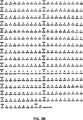
図1は、ストリップ(strip)イムノブロットアッセイ(SIA)に使用するための代表的な試験片(strip)を示す。ヒトIgGは、2つの異なるレベル(レベルI、低コントロール;そしてレベルII、高コントロール)の内部コントロールとして使用される。CagAおよびVacAは、型−特異的H.pylori I型抗原として使用され、そしてHP CEは、型−共通抗原を有するH.pylori溶解物を示す。
図2は、SIAに使用するための別の代表的な試験片を示す。上述のように、ヒトIgGは、2つの異なるレベル(レベルI、低コントロール;そしてレベルII、高コントロール)の内部コントロールとして使用される。CagAおよびVacAは、型−特異的H.pylori I型抗原として使用され、そしてまた、同様に、アッセイ感度を増強するため、ならびに処置に対する応答をモニタする2つの異なるレベルで使用される。ウレアーゼは、型−共通抗原として使用される。
図3A-3B(配列番号: )は、実施例に記載されるSIAで使用される、H.pyloriVacA抗原に関するヌクレオチド配列およびそれに対応するアミノ酸配列を示す。
図4(配列番号: )は、実施例に記載されるSIAで使用される、H.pyloriCagA抗原に関するヌクレオチド配列とそれに対応するアミノ酸配列を示す。
発明の詳細な説明
本発明の実施は、他に示されない限り、当該分野の技術間の免疫学、微生物学、分子生物学、および組換えDNA技術を使用する。このような技術は、文献で十分に説明される。例えば、Sambrookら、Molecular Cloning:A Laboratory Manual(第2編、1989);DNA Cloning:A Practical Approach I&II巻(D.Glover編);Methods In Enzymology(S.ColowickとN.Kaplan編、Academic Press,Inc.);そしてHandbook of Experimental Immunology I-IV巻(D.M.WeirとC.C.Blackwell編Blackwell Scientific Publications)を参照のこと。
本明細書中および添付請求の範囲で使用される場合、単数形「a」、「an」および「the」は、その内容が明らかにそうでないことを指示しない限り、複数の参照体を包含する。加えて、ヌクレオチドとアミノ酸の標準的略号は本明細書で使用される。
I.定義
本発明の記述において、以下の言語が使用され、そして以下に記載されるように、定義されることが意図される。
「H.pylori溶解物」によって、以下に規定されるように、H.pylori I型とII型の両方に対して生成された抗体と反応する、1つまたはそれより多いH.pyloriポリペプチドを含むH.pylori I型またはII型全細菌に由来する抽出物または溶解物を意味する。このようなポリペプチドは、本明細書中の「型−共通」抗原と称される。従って、本明細書中で使用される場合、用語「溶解物」は、少なくとも溶解物中に存在する抗原の1つが型−共通抗原である限りは、いくつかのH.pylori抗原を有する粗抽出物を表す。この溶解物は、さらに精製した型−共通および/または型−特異的抗原を増強され得る。この用語はまた、1つまたは2、3のこのような型−共通抗原のみを含有する粗溶解物に由来する比較的に精製された組成物を示す。このような溶解物は、さらに以下で記載されるように、当該分野で公知の技術を使用して調製される。
単独または組合せのいずれかのこのような溶解物中に存在し得る代表的な抗原は、H.pylori付着因子(例えば、20kDa N-アセチルノイラミニラクトース−結合原線維赤血球凝集素(HpaA)、ホスファチジルエタノールアミンおよびガングリオテトラオシルセラミドと結合する63kDaタンパク質、および保存されたフィンブリン線毛様構造に由来する1つまたはそれより多い型−共通抗原エピトープを含むが、これらに限定されない。例えば、これらの抗原の説明に関して、Telfordら、Trends in Biotech.(1994)12:420-426を参照のこと。溶解物中に存在し得る他の型−共通抗原は、主要フラゲリンFlaAと微量なフラゲリンFlaBのような種々のフラゲリンのいくつかに由来する1つまたはそれより多い型−共通抗原エピトープを含有する。この点に関して、H.pyloriの鞭毛は、それぞれ約53kDaの分子量を有するFlaAおよびFlaBから構成される。FlaAそして/またはFlaBのいずれかまたは両方が、本発明での使用のための型−共通抗原の供給源として使用され得る。別の代表的な型−共通抗原は、細菌の外膜と周辺質のスペースと会合されるH.pyloriウレアーゼを含む。ホロ酵素は、それぞれ、26.5kDa(UreA)と61kDa(UreB)の2つのサブユニットから構成される大きな複合体である。サブユニットまたは3つの組合せのいずれかのホロ酵素に由来する型−共通エピトープは、型−共通抗原として存在し得る。溶解物中に存在し得るか、またはさらに精製された形態で使用され得る別の代表的な型−共通抗原は、「hsp60」として知られるH.pylori熱ショックタンパク質を含む。hsp60に関するDNAおよびそれに対応するアミノ酸配列は公知である。例えば、国際公開番号WO93/18150、1993年9月16日公開を参照のこと。示される全長のhsp60抗原は、約546のアミノ酸および約58kDaの分子量を有する。この溶解物は、本明細書において特に記載されない、他の型−共通抗原を含み得ると理解される。
「型−特異的H.pylori I型抗原」によって、以下に記載されるように、H.pylori I型に対する抗体と優位に反応するが、H.pylori II型に対する抗体とは反応しないH.pylori I型に由来するポリペプチドが意味される。代表的な型−特異的H.pylori I型抗原は以下のものを含む:CTとしても知られるH.pylori VacA;ならびにCAI抗原およびtagAとしても知られるH.pylori CagA;ならびにH.pylori I型に対する抗体と反応し得るが、H.pylori II型に対する抗体とは反応しないこれらの抗原に由来するエピトープ。VacAおよびCagAの両方とも、以下でさらに記載される。本明細書において特に記載されない他の型−特異的H.pylori I型抗原もまた、この定義により取り囲まれると理解し得る。
上記のような、VacA、CagAまたは任意の他の型特異的もしくは型共通抗原のような、型−共通または型−特異的H.pylori抗原に関して使用される場合、用語「ポリペプチド」は、天然、組換えまたは合成のいずれであろうが、多くのH.pylori株のいずれかに由来するVacA、CagAなどを表す。型−特異的H.pylori I型抗原の場合、このポリペプチドは、H.pylori I型株に由来する。型−共通抗原の場合、そのポリペプチドは、H.pylori I型またはII型のいずれかに由来し得る。このポリペプチドは、参照分子の全長アミノ酸配列を含むことを必要としないが、ポリペプチドが適切なH.pylori抗体と反応するために必要である大きさの分子のみを含み得る。従って、参考分子のただ1つまたは2、3のエピトープが存在すべき必要性がある。さらに、このポリペプチドは、全長参考分子または参考分子のフラグメントとH.pyloriポリペプチドの反応性を崩壊しない別のタンパク質との間の融合タンパク質を含み得る。従って、このポリペプチドが、フラグメント、切断された配列および部分的な配列ならびに参考分子のアナログおよび前駆体形態全長配列を含み得ることは容易に明らかである。この用語はまた、ポリペプチドがH.pylori抗体と反応する能力を保持する限り、参考配列の欠損、付加ならびに置換を意味する。
この点について、特に好ましい置換は、一般に本質的に保存的であり、すなわち、それらの側鎖に関係するアミノ酸ファミリー内で生じる置換である。詳細には、アミノ酸は、一般に4ファミリーに分けられる:(1)酸性−−アスパラギン酸およびグルタミン酸;(2)塩基性−−リジン、アルギニン、ヒスチジン;(3)非極性−−アラニン、バリン、ロイシン、イソロイシン、プロリン、フェニルアラニン、メチオニン、トリプトファン;および(4)非荷電極性−−グリシン、アスパラギン、グルタミン、シスチン、セリン、スレオニン、チロシン。フェニルアラニン、トリプトファン、およびチロシンは、時々、芳香族アミノ酸として分類される。例えば、イソロイシンまたはバリンでのロイシンの置換、グルタミン酸でのアスパラギン酸の置換、セリンでのスレオニンの単離された置換体、または構造上関連するアミノ酸でのアミノ酸の類似した保存的置換が、生物学的活性に主要な影響を有しないということは、合理的に予測され得る。それゆえ、参照タンパク分子と実質的に同じアミノ酸配列を有するが、タンパク質の抗体結合能力に実質的に影響しない微小なアミノ酸置換を所有するタンパク質は、参照ポリペプチドの定義範囲内である。
「VacAポリペプチド」により、H.pylori I型細胞毒として公知である抗原から誘導され、そしてH.pylori I型に対する抗体と主に反応するが、H.pylori I型とは反応しない、先に定義したポリペプチドを意味する。VacAタンパク質は、組織培養中の上皮細胞において空胞化を誘導し、また広範囲の組織損傷の原因となり、そしてマウスへ経口投与した場合、潰瘍形成の原因となる。VacAのDNAとこれに対応するアミノ酸配列は公知であり、そして例えば、1993年9月16日に公開された国際公開第WO 93/18150号に報告されている。VacAポリペプチドの遺伝子は、約90〜100kDaの活性分子にプロセシングされる約140kDaの前駆体をコードする。この分子は、次に、ゆっくりとタンパク質分解的に切断される完全な90kDa分子と同時精製される2つのフラグメントを生じる。Telfordら、Trends in Biotech.(1994)12:420-426を参照のこと。従って、本明細書中で使用される「VacAポリペプチド」の定義には、前駆体タンパク質、ならびにH.pylori I型感染を有する個体由来の生物学的サンプルに存在する抗体との特異的な反応性を保持する、プロセシングされた活性分子、そのタンパク質分解性フラグメント、またはその部分もしくはそのムテインが含まれる。例えば、図3A-3Bに示され、そして本明細書中に記載されるアッセイに使用されるVacAポリペプチドは、全長分子のGly-311からIle819までのVacAフラグメント(全長分子を含む)を含み、これは、組み換え体発現を促進するために、ヒトSODの5アミノ酸から154アミノ酸のリンカー配列により融合される。
「CagAポリペプチド」により、H.pylori I型細胞毒関連免疫優性抗原から誘導され、そして主にH.pylori I型に対する抗体と反応するが、H.pylori II型とは反応しない、先に定義したポリペプチドを意味する。CagAは、細菌表面に発現される。CagAのDNAとこれに対応するアミノ酸配列は公知である。例えば、1993年9月16日に公開された国際公開第WO 93/18150号、を参照のこと。そこで記載される全長CagA抗原は、約128kDaの推量分子量を有する約1147アミノ酸を含む。天然のタンパク質は、プロリンリッチなアミノ酸配列の繰り返しをコードする、102bpのDNAセグメントの可変数の繰り返しの存在により、種間でサイズの多様性を示す。Covacciら、Proc.Natl.Acad.Sci.USA(1993)90:5791-5795を参照のこと。従って、CagAの報告された分子量は、約120〜135kDaの範囲内である。それゆえ、本明細書中で使用される「CagAポリペプチド」の定義には、H.pylori I型感染を有する個体由来の生物学的サンプル中の抗体と反応する能力を保持するが、H.pylori II型に対して生成した抗体と実質的に反応しない、任意の種々のCagA改変体、そのフラグメント、およびそのムテインが含まれる。例えば、図4に示され、そして本明細書に記載のアッセイで使用されるCagAポリペプチドは、268アミノ酸の短縮型タンパク質であり、全長分子のGlu-748〜Glu-1015(全長分子を含む)を含む。
「エピトープ」により、特異的B細胞およびT細胞が応答する抗原の部位を意味する。この用語はまた、「抗原決定基」または「抗原決定部位」と交換可能に使用される。エピトープは、エピトープに独特の空間的コンフォメーションにおいて、3個以上のアミノ酸からなり得る。一般に、エピトープは、少なくとも5つのこのようなアミノ酸からなり、そしてより通常、少なくとも8〜10個のようなアミノ酸からなる。アミノ酸の空間的コンフォメーションを決定するための方法は、当業者に公知であり、X−線結晶学および2−次元核磁気共鳴が含まれる。さらに、所与のタンパク質のエピトープの同定は、疎水性研究の使用により、そして部位特異的血清学によるような、当該分野で周知の技法を使用して容易に達成される。例えば、Geysenら、Proc.Natl.Acad.Sci.USA(1984)81:3998-4002(所与の抗原において免疫原性エピトープの位置を決定するためのペプチドを迅速に合成する一般的方法);米国特許第4,708,871号(抗原のエピトープを同定しそして化学的に合成するための手順);およびGeysenら、Molecular Immunology(1986)23:709-715(所与の抗体に対して高い親和性を有するペプチドを同定するための技法)もまた参照のこと。同じエピトープを認識する抗体は、標的抗原に対する別の抗原の結合をブロックするための1つの抗体の能力を示す簡便な免疫アッセイにおいて同定され得る。
「精製された」タンパク質またはポリペプチドは、組換え的にまたは合成的に産生される、または組成物中に存在するタンパク質量が、粗調製物中に存在量よりも実質的に多いように、天然の宿主から単離されるタンパク質である。一般に、精製されたタンパク質は、少なくとも約50%均一であり、そしてより好ましくは、少なくとも約80%から90%均一である。
本明細書中で使用されるように、「生物学的サンプル」は、固体から単離される組織または液体のサンプル、例えば、血液、血漿、血清、糞便、尿、骨髄、胆液、髄液、リンパ液、皮膚サンプル、皮膚の外分泌液、気道、腸管、および尿生殖路、ならびに、胃上皮および胃粘膜由来のサンプル、涙、唾液、乳、血液細胞、器官、生検を含むが、これらに限定されない。そしてまた例えば、培養培地での細胞および組織の増殖から生じる馴化培地(例えば、組換え細胞)および細胞成分を含むがしかし限定されないインビトロ細胞培養成分のサンプル。
本明細書で使用されるように、用語「標識」および「検出し得る標識」は、検出可能な分子(放射活性同位体、蛍光剤、化学発光物質、酵素、酵素基質、酵素補因子、酵素インヒビター、発色団、色素、金属イオン、金属ゾル、リガンド、(例えば、ビオチンまたはハプテン)などを含むが、これらに限定されない)をいう。用語「蛍光剤」は、検出可能領域に蛍光を示し得る物質またはその部分をいう。本発明下で使用され得る標識の具体例には、フルオロセイン、ローダミン、ダンシル、ウンベリフェロン、テキサスレッド、ルミノール、アクラディマムエステル(acradimum ester)、NADPH、およびα−β−ガラクトシダーゼが含まれる。
II.発明の実施態様
本発明は、H.pylori感染を正確に検出し、そしてH.pylori I型感染とH.pylori II型感染とを識別するための新規の診断法の発見に基づく。この方法は、精製されたか、または細菌に由来する溶解物に存在するかのいずれかの1つ以上のH.pylori型−共通抗原、ならびに精製型−特異的H.pylori抗原を利用する。型−共通のそして型−特異的抗原の両方の使用は、偽陽性結果の出現率を減少させる。この方法は、単一アッセイに、感染の検出および存在する感染型の同定の両方を可能にする簡便な1工程アッセイ形式で実施し得る。この方法はまた、サンプルをまず、H.pylori型−共通抗原と反応させ、そして陽性の場合には、1つ以上の型−特異的I型抗原と反応させる、2工程で実施され得る。この方法はまた、型−特異的II型抗原を使用し得る。
さらに具体的には、H.pylori型−共通抗原の使用は、一般に、H.pylori感染の診断を可能にする。1つ以上の型−特異的抗原の存在は、細菌型、すなわち、感染がH.pylori I型および/またはH.pylori II型によって引き起こされるか否かの決定を可能にする。H.pylori型−共通抗原の存在により、陽性結果は、型決定が可能でないサンプルにおいてさえ生じる。それゆえ、偽陰性の出現率は減少される。さらに、H.pylori I型感染が存在する場合、個体は、B型胃炎、十二指腸潰瘍、そして胃ガンの処置または抑制のために抗生物質を投与され得る。さらに、本明細書で記載されるアッセイは、抗生物質治療が効果的か否かを決定するために、被験体の処置の過程のモニターに有用である。
被験体診断技術における使用のための抗原は、種々の技術の使用を生じ得る。
例えば、型−共通抗原は、当該分野で周知の方法を使用して得られ得る溶解物中で提供され得る。一般に、このような方法は、超音波処理、プレッシャーディスインテグレーション(pressure disintegration)、界面活性剤抽出、分画化などを使用して、H.pylori I型細菌またはH.pylori II型細菌のいずれか由来の型−共通タンパク質を抽出することを必要とする。このような溶解物中に存在する型−共通抗原は、標準的精製技術を使用して、所望される場合、さらに精製され得る。このような方法における使用のためのH.pylori株は、American Type Culture Collection(ATCC,Rockville,MD.)を含むいくつかの供給源から容易に入手可能である。例えば、ATCC株名NCTC 11637、11639、および11916は溶解物の供給源としての使用を見出す。別の有用な株は、当該分野で公知である。
型−特異的H.pylori I型抗原はまた、標準的精製技法を使用して得られ得る。この点から、特定の抗原は、カラムクロマトグラフィー、電気泳動法、HPLC、免疫吸着法、アフィニティークロマトグラフィーおよび免疫沈降法のような標準精製技法を使用して、I型H.pylori潰瘍産生株から単離され得る。例えば、H.pylori由来のいくつかの抗原の精製の説明に関しては、1996年5月2日に公開された国際公開第WO 96/12965号を参照のこと。例えば、ATCC株名NCTC 11916は、H.pyloriのI型潰瘍産生株であり、そして、それゆえ、本発明の使用に関して、1つまたはそれを超える型−特異的抗原の供給源として使用され得る。
H.pylori抗原はまた、当該分野で周知の組換え方法を使用して産生し得る。この点に関して、オリゴヌクレオチドプローブは、H.pyloriゲノムの公知の配列に基づいて創案され得、本発明において有用な抗原をコードするH.pylori遺伝子に関するゲノムまたはcDNAライブラリーをプローブするために使用され得る。次いで、この遺伝子は、標準的技術を使用して、そして所望される場合には、全長配列の所望部位で遺伝子を変異させるために用いられる制限酵素を使用して、さらに単離され得る。
同様に、H.pylori遺伝子は、フェノール抽出のような公知の方法を使用して、細菌性細胞から直接単離され得、そして、この配列は任意の所望の改変を生じるためにさらに操作され得る。DNAを得、そして単離するために使用される技術の説明に関しては、例えば、Sambrookら、前出、を参照のこと。最終的に、H.pylori抗原をコードする遺伝子は、公知の方法に基づいて、合成的に産生し得る。ヌクレオチド配列は、所望の特定のアミノ酸配列に関する適切なコドンを用いて設計し得る。一般に、当業者は、配列が発現される意図する宿主のための好ましいコドンを選択する。完全な配列は、標準的な方法により調製されるオーバーラップするオリゴヌクレオチドから一般に組立てられ、そして完全なコドン配列へアセンブリされる。例えば、Edge,Nature(1981)292:756;Nambairら、Science(1984)223:1299;Jayら、J.Biol.Chem.(1984)259:6311を参照のこと。
一旦、所望のポリペプチドのコード配列が、単離または合成されると、それらは、昆虫、哺乳動物、細菌、ウイルスおよび酵母の発現系(これらは全て当該分野で周知である)を含む、種々の系についての任意の適切なベクターまたはレプリコンにクローン化され得る。特に、宿主細胞は、所望のコード配列に作動可能に連結されたコントロール配列を含有する発現ベクターにより形質転換される。
コントロール配列は、使用される特別な宿主細胞と適合性である。例えば、哺乳動物細胞発現に関して代表的なプロモーターには、とりわけ、SV40初期プロモーター、マウス乳癌ウイルスLTRプロモーター、アデノウイルス主要後期プロモーター(AdMLP)、および単純ヘルペスウイルスプロモーターが含まれる。マウスメタロチオネイン遺伝子由来のプロモーターのような他の非ウイルスプロモーターもまた、哺乳動物構築物における使用を見出す。哺乳動物発現は、プロモーターに依存して、恒常的であるかまたは制御されている(誘導性である)かのいずれかであり得る。代表的には、転写終止およびポリアデニル化配列はまた、転写終止コドンの3’側に位置して存在する。転写ターミネーター/ポリアデニル化シグナルの例には、SV40(Sambrookら、前出)由来のものが含まれる。スプライスドナーおよびアクセプターの部位を有するイントロンはまた、本発明の構築物へと設計され得る。
エンハンサーエレメントはまた、発現レベルを増加させるために哺乳動物構築物において使用され得る。例には、SV40初期遺伝子エンハンサー(Dijkemaら、EMBO J.(1985)4:761))およびRous Sarcomaウイルス(Gormanら、Proc.Natl.Acad.Sci.USA(1982b)79:6777)の長末端反復(LTR)に由来するエンハンサー/プロモーターおよびヒトサイトメガロウイルス(Boshartら、Cell(1985)41:521)が含まれる。シグナルペプチドをコードする配列を含むリーダー配列もまた存在し得、哺乳動物細胞において外来タンパク質の分泌を提供する。好ましくは、リーダーフラグメントと目的の遺伝子との間に、リーダー配列がインビボまたはインビトロのいずれかで切断され得るように、コードされるプロセッシング部位が存在する。アデノウイルストリパタイト(tripartite)リーダーは、哺乳動物細胞中の外来タンパク質の分泌を提供するリーダー配列の例である。
一旦完了すると、哺乳動物発現ベクターは、任意のいくつかの哺乳動物細胞の形質転換に使用され得る。哺乳動物細胞への異種ポリヌクレオチドの導入方法は、当該分野で公知であり、そしてデキストラン媒介トランスフェクション、リン酸カルシウム沈澱、ポリブレン媒介トランスフェクション、プロトプラスト融合、エレクトロポレーション、リポソーム内へのポリヌクレオチドのカプセル化、および核へのDNAの直接的マイクロインジェクションを包含する。
発現のための宿主として利用できる哺乳動物細胞系はまた、公知であり、そしてチャイニーズハムスター卵巣細胞、KeLa細胞、ベビーハムスター腎臓(BHK)細胞、サル腎臓細胞(COS)、ヒト肝細胞性ガン細胞(例えば、Hep G2)、およびその他を含むがこれらに限定されないAmerican Type Culture Collection(ATCC)から入手可能な多くの不死化細胞系を包含する。
本発明の構築物はまた、酵母中で発現し得る。酵母ベクターのコントロール配列は、当該分野で公知であり、そしてアルコールデヒドロゲナーゼ(ADH)(欧州特許公報第284,044号)、エノラーゼプロモーター、グルコキナーゼプロモーター、グルコース-6-ホスフェートイソメラーゼプロモーター、グリセルアルデヒド-3-ホスフェートデヒドロゲナーゼ(GAPまたはGAPDH)プロモーター、ヘキソキナーゼプロモーター、ホスホフルクトキナーゼプロモーター、3-ホスホグリセレートムターゼプロモーター、およびピルベートキナーゼ(PyK)プロモーター(欧州特許公報第329,203号)のようなプロモーターを含む。酸ホスファターゼをコードする酵母PHO5遺伝子はまた、有用なプロモーター配列を提供する(Myanoharaら、Proc.Natl.Acad.Sci.USA(1983)80:1)。加えて、天然に生じない合成プロモーターもまた、酵母プロモーターとして機能する。例えば、1つの酵母プロモーターの上流活性化配列(UAS)が、別の酵母プロモーターの転写活性化領域と連結され得、合成ハイブリッドプロモーターを作製する。このようなハイブリッドプロモーターの例には、GAP転写活性化領域に連結されるADH調節領域が含まれる(米国特許第4,876,197号および同第4,880,734号)。ハイブリッドプロモーターの他の例には、GAPもしくはPyK(欧州特許公報第164,556号)のような糖分解酵素遺伝子の転写活性化領域と組み合わされたADH2、GAL4、GAL10またはPHO5遺伝子のいずれかの調節配列からなるプロモーターが含まれる。さらに、酵母プロモーターは、酵母RNAポリメラーゼに結合し、そして転写を開始する能力を有する非酵母起源の天然に生じるプロモーターを含み得る。
酵母発現ベクターに含まれ得る他のコントロールエレメントは、ターミネーター(例えば、GAPDH由来およびエノラーゼ遺伝子(Holland,J.Biol Chem.(1981)256:1385)由来)、および分泌物のためのシグナル配列をコードするリーダー配列である。適切なシグナル配列をコードするDNAは、酵母インベルターゼ遺伝子(欧州特許公開第012,873号;JPO公開第62,096,086号)およびα−因子遺伝子(米国特許第4,588,684号、同第4,546,083号、および同第4,870,008号;欧州特許公開第324,274号;PCT公開第WO89/02463号)のような分泌された酵母タンパク質遺伝子に由来し得る。あるいは、インターフェロンリーダーのような非酵母起源のリーダーはまた、酵母における分泌を提供する(欧州特許公開第060,057号)。
染色体外レプリコンまたは組込みベクターのいずれかである、発現ベクターおよび形質転換ベクターは、多くの酵母への形質転換に関して開発されている。例えば、発現ベクターは、とりわけ、以下の酵母に関して開発されている:Saccharomyces cerevisiae(Hinnenら、Proc.Natl.Acad.Sci.USA(1978)75:1929;Itoら、J.Bacteriol.(1983)153:163);Saccharomyces carlsbergenenis;Candida albicans(Kurtzら、Mol.Cell.Biol.(1986)6:142);Candida maltosa(Kunzeら、J.Basic Microbiol.(1985)25:141);Hansenula polymorpha(Gleesonら、J.Gen.Microbiol.(1986)132:3459;RoggenkampらMol.Gen.Genet.(1986)202:302);Kluyveromyces fragilis(Dasら、J.Bacteriol.(1984)158:1165);Kluyveromyces lactis(De Louvencourtら、J.Bacteriol.(1983)154:737;Van den Bergら、Bio/Technology(1990)8:135);Pichia guillerimondii(Kunzeら、J.Basic Microbiol.(1985)25:141);Pichia pastoris(Creggら、Mol.Cell.Biol.(1985)5:3376;米国特許第4,837,148号および同第4,929,555号);Schizosaccharomyces pombe(BeachおよびNurse、Nature(1981)300:706);およびYarrowia lipolytica(Davidowら、Curr.Genet.(1985)10:380471;Gaillardinら、Curr.Genet.(1985)10:49)。
酵母宿主への外来DNAの導入方法は、当該分野で周知であり、代表的には、スフェロプラストの形質転換またはアルカリカチオンで処理された完全な酵母細胞の形質転換のいずれかを包含する。
細菌発現系はまた、本発明の構築物とともに使用され得る。細菌における使用のためのコントロールエレメントは、オペレーター配列およびリボソーム結合部位を必要に応じて含むプロモーターを含有する。有用なプロモーターには、ガラクトース、ラクトース(lac)およびマルトースのような糖代謝酵素に由来する配列が含まれる。さらなる例には、トリプトファン(trp)のような生合成酵素、b-ラクタマーゼ(bla)プロモーター系、バクテリオファージλPL、およびT5に由来するプロモーター配列が含まれる。さらに、天然に生じないtacプロモーター(米国特許第4,551,433号)のような合成プロモーターはまた、細菌宿主細胞におけると同様に機能する。
前述の系は、E.coliと特に適合性である。しかし、とりわけ、Bacillus spp.、Streptococcus spp.、およびStreptomyces spp.のような細菌宿主における使用のための多数の他の系がまた公知である。これら宿主への外来DNAの導入のための方法は、代表的には、CaCl2または、二価カチオンおよびDMSOのような他の薬剤の使用を包含する。DNAはまた、エレクトロポーレーションにより細菌細胞へ導入され得る。
所望の抗原の発現のための他の系には、昆虫細胞およびこれら細胞における使用に適切なベクターを含む。最も一般的に使用される系は、バキュロウイルスAutographa californica多核体病ウイルス(AcNPV)に由来する。一般に、発現系の成分はトランスファーベクター、通常は、細菌プラスミド(これはバキュロウイルスゲノムのフラグメントおよび発現される異種遺伝子または遺伝子の挿入のための便利な制限部位の両方を有する)を含む。;トランスファーベクター(これはバキュロウイルスゲノムへの異種遺伝子の相同組換えを可能にする)においてバキュロウイルス特異的フラグメントと相同な配列を有する野生型バキュロウイルス;ならびに、適切な昆虫宿主細胞および増殖培地。
ベクターにおける使用のためのプロモーターは、代表的に構造遺伝子由来であり、ウイルスの感染周期の後期に豊富に転写される。例としては、ウイルスポリヘドロン(polyhedron)タンパク質をコードする遺伝子由来の配列が含まれる(Friesenら、(1986)「The Regulation of Baculovirus Gene Expression」:The Molecular Bioligy of Baculoviruses(Walter Doerfler編);欧州特許公開番号第127,839号および第155,476号;ならびに、p10タンパク質をコードする遺伝子Vlakら、J.Gen.Virol.(1988)69:765)。また、プラスミドは通常、ポリヘドリン(polyhedrin)ポリアデニル化シグナルを含有(Millerら、Ann.Rev.Microbiol.(1988)42:177)し、そして原核生物アンピシリン耐性(amp)遺伝子ならびにE.coliの選択および増殖のための複製起点を含む。適切なシグナル配列をコードするDNAもまた、含まれ、そして一般的に、分泌型昆虫タンパク質または分泌型バキュロウイルスタンパク質(例えば、バキュロウイルスポリヘドリン遺伝子(Carbonellら、Gene(1988)73:409)、ならびに哺乳動物シグナル配列(例えば、ヒトα-インターフェロンをコードする遺伝子由来の配列(Maedaら、Nature(1985)315:592);ヒトガストリン放出ペプチド(Lebacq-Verheydenら、Molec.Cell.Biol.(1988)8:3129;ヒトIL-2(Smithら、Proc.Natl.Acad.Sci.USA(1985)82:8404;マウスIL-3(Miyajimaら、Gene(1987)58:273;およびヒトグルコセレブロシダーゼ(Martinら、DNA(1988)7:99)の遺伝子由来である。
現在、外来遺伝子をAcNPVに導入するために最も一般的に用いられる導入ベクターは、pAc373である。また、当業者に公知の多くの他のベクターが、設計されている。これらには、例えば、pVL985(これはポリヘドリン開始コドンをATGからATTに変更し、そしてATTから32bp下流にBamHIクローニング部位を導入する;LuckowおよびSummers、Virology(1989)17:31を参照のこと)。
所望のDNA配列は、公知の技術(SummersおよびSmith、前出;Smithら、Mol.Cell.Biol.(1983)3:2156;ならびにLuckowおよびSummers(1989)を参照のこと)を使用して導入ベクターに挿入され、そして昆虫細胞宿主は、導入ベクター、および野生型バキュロウイルスのゲノムDNAの異種DNAを用いて、同時形質転換される(通常、同時トランスフェクションによる)。ベクターおよびウイルスゲノムは、組み換えが可能である。パッケージされた組換えウイルスが発現され、そして組換え体のプラークが同定され、精製される。バキュロウイルス/昆虫細胞発現系のための材料および方法は、キット形式で、例えばInvitrogen,San Diego CA(「MaxBac」キット)から、市販されている。これらの技術は一般に、当業者に公知であり、SummersおよびSmith,Texas Agricultural Experiment Station Bulletin No.1555(1987)(本明細書中以下「SummersおよびSmith」)に、充分に記載されている。
組換えバキュロウイルス発現ベクターは、種々の昆虫細胞への感染のために開発されている。例えば、組換えバキュロウイルスは、特に:Aedes aegypti、Autographa californica、Bombyx mori、Drosophila melanogaster、Spodoptera frugiperda、およびTrichoplusia ni、について開発されている。
上記の系を用いて調製されたポリペプチドは融合ポリペプチドであることが、しばしば所望される。非融合型タンパク質を用いる場合、これらのタンパク質は細胞内で発現され得るか、または細胞から増殖培地へ分泌され得る。
さらに、例えば、マルチプルタイプ特異的H.pylori I型抗原、またはマルチプルタイプ共通抗原をコードするキメラ遺伝子配列を含むプラスミドが、構築され得る。遺伝子配列は、2シストロン遺伝子配置中に存在し得る。付加的な制御エレメントは、遠位コード領域からのRNAの翻訳に効果的な種々の遺伝子の間に適合され得る。あるいは、複数の抗原をコードする、単一のオープンリーディングフレームを有するキメラ転写単位もまた、構築され得る。融合物が、キメラタンパク質の合成を可能にするように作製され得、あるいはタンパク質プロセシングシグナルが、プロテアーゼによる切断を提供するように操作され得、従って、鋳型RNAの翻訳由来の2つ以上のタンパク質の遊離を可能にする。プロセシングプロテアーゼはまた、この系において、独立して、または抗原コード領域(単数もしくは複数)を有するキメラの一部としてのいずれかで発現され得る。
また、ウイルス系、例えばワクシニアに基づく感染/トランスフェクション系もまた、Tomeiら、J.Virol.(1993)67:4017-4026およびSelbyら、J.Gen.Virol.(1993)74:1103-1113に記載されるように、本発明を用いた使用を見出す。この系において、細胞は最初に、バクテリオファージT7 RNAポリメラーゼをコードするワクシニアウイルス組換え体でインビトロでトランスフェクトされる。このポリメラーゼは、T7プロモーターを保有する鋳型のみを転写するという点で、精巧な特異性を表す。感染の後、細胞は目的のDNAでトランスフェクトされ、T7プロモーターによって駆動される。ワクシニアウイルス組換え体から細胞質において発現されたポリメラーゼは、トランスフェクトされたDNAをRNAに転写し、次いでこのRNAは宿主翻訳機構によってタンパク質に翻訳される。本方法は、高レベルの一時的な、大量のRNAの細胞質産物、およびその翻訳産物を提供する。
発現系および選択された宿主に依存して、本発明の抗原は、目的の抗原が発現されることによる条件下で、発現ベクターによって形質転換された宿主細胞の増殖によって産生される。次いで、抗原は宿主細胞から単離され、そして精製される。発現系が抗原の分泌を提供する場合、抗原は培地から直接精製され得る。抗原が分泌されない場合、抗原は細胞溶解物から単離される。適切な増殖条件の選択および回収方法は、当該分野の内にある。
H.pylori抗原はまた、当業者に公知の方法を使用して、化学合成(例えば、固相または液体ペプチド合成によって)産生され得る。ペプチドの化学合成は、問題の抗原が比較的小さい場合に、望ましいとされ得る。例えば、固相ペプチド合成技術については、J.M.StewartおよびJ.D.Young,Solid Phase Peptide Synthesis、第2版、Pierce Chemical Co.、Rockford、IL(1984)、ならびにG.BaranyおよびR.B.Merrifield、The Peptide:Analysis、Synthesis、Biology、E.GrossおよびJ.Meienhofer編、第2巻、Academic Press、New York、(1980)、3-254頁、;ならびに、古典的な溶液合成については、M.Bodansky、Principles of Peptide Synthesis、Springer-Verlag、Berlin(1984)およびE.GrossおよびJ.Meienhofer、編、The Peptides:Analysis、Synthesis、Biology、上述、第1巻を参照のこと。
H.pylori型共通、およびH.pylori型特異的H.pylori I型抗原は、本明細書中で、生物学的サンプル中のバクテリアに対して指向される反応性抗体の存在を検出するための診断薬として用いられる。さらに、抗原は、治療の最初に得られた結果と、処置経過の最中および後に得られた結果とを比較することにより、抗体治療の経過をモニターするために用いられ得る。例えば、型共通および/または型特異的H.pylori抗原に反応性である抗体の存在は、標準的な電気泳動技術および免疫診断技術(これには競合、直接的な反応、またはサンドイッチ型アッセイのような免疫アッセイが含まれる)を用いて検出され得る。このようなアッセイには、ウエスタンブロット;凝集試験;酵素標識免疫アッセイおよび酵素媒介免疫アッセイ(例えばELISA);ビオチン/アビジン型アッセイ;ラジオイムノアッセイ;イムノ電気泳動;免疫沈降などが含まれるが、これらに限定されない。一般に反応には、検出標識(例えば蛍光、化学発光、放射活性、酵素標識、または染料分子)、または、抗原と抗体との間の複合体の形成、もしくはその複合体と反応した抗体を検出する他の方法が含まれる。
前述のアッセイには、一般に、抗原−抗体複合体が結合した、液相における非結合抗体の、固相支持体からの分離を含む。本発明の実施に用いられ得る固体支持体には以下のような基質を含む:ニトロセルロース(例えば、膜またはマイクロタイターウェル形態);塩化ポリビニル(例えば、シートまたはマイクロタイターウェル);ポロスチレンラテックス(例えば、ビーズまたはマイクロタイタープレート);フッ化ポリビニリジン;ジアゾ化ペーパー;ナイロン膜;活性化ビーズ、磁気応答性ビーズなど。
代表的に、固体支持体は、化合物が充分に支持体に固定されているような、適切な結合条件下で、最初に固相成分(例えば、1つ以上の型共通および/または1つ以上の型特異的H.pylori I型抗原)と反応される。あるときには、抗原の支持体への固定は、より良い結合特性での抗原のタンパク質への最初の結合によって増強され得る。適切な結合タンパク質には、血清アルブミンのような巨大分子(ウシ血清アルブミン(BSA)、キーホールリンペットヘモシアニン、免疫グロブリン分子、チログロブリン、オボアルブミン、および当業者に周知の他のタンパク質を含む)が挙げられるが、これに限定されない。抗原を支持体に結合させるために用いられ得る他の分子には、多糖類、ポリ乳酸、ポリグリコール酸、重合アミノ酸、アミノ酸コポリマーなどが挙げられる。このような分子、およびこれらの分子を抗原に結合する方法は当業者に周知である。例えば、Brinkley,M.A.,Bioconjugate Chem.(1992)3:2-13;Hashidaら、J.Appl.Biochem.(1984)6:56-63;ならびにAnjaneyuluおよびStaros,International J.of Peptide and Protein Res.(1987)30:117-124を参照のこと。
固体支持体の固相成分との反応後、任意の非固定化固相成分は、洗浄によって支持体から除去され、次いで支持体結合型成分は、リガンド部分を含むと予想される生物学的サンプル(例えば、固定化抗原に対する抗体)と、適切な結合条件下で接触される。いかなる非結合リガンドも除去するための洗浄の後、第2の結合部分は、適当な結合条件下で添加され、ここで、第2の結合物は、選択的に結合リガンドと結合され得る。第2の結合物の存在は、次いで当該分野で周知の技術を用いて検出され得る。
さらに特に、ELIZA法が用いられ得、ここでマイクロタイタープレートのウェルは、H.pylori型共通および/または型特異的H.pylori I型抗原(単数および複数)でコートされる。抗H.pylori免疫グロブリン分子を含む、または含むと予想される生物学的サンプルが、次いでコートされたウェルに添加される。一つのマイクロタイタープレートを用いることが所望されるアッセイにおいて、選択された数のウェルが、例えば第1の型特異的抗原部分でコートされ、異なる1セットのウェルが第2の型特異的抗原部分でコートされ、そして第3セットのウェルがH.pylori型共通抗原などでコートされ得る。代わりに、一連のELISAが続々と行われ得、ここで、独立したプレートが、それぞれ型共通、および型特異的抗原部分に用いられる。抗体が固定化抗原に結合するのを可能にするに充分なインキュべーション時間の後、プレート(単数および複数)は非結合型部分を除去するために洗浄され、そして検出可能な標識化第2の結合分子が添加され得る。第2の結合分子は、任意の捕獲サンプル抗体と反応され得、プレートは洗浄され、そして当該分野で周知の方法を用いて第2の結合分子の存在が検出される。
従って、1つの特定の実施態様において、生物学的サンプル由来の、抗型特異的抗原リガンド、および/または抗H.pylori型共通抗原リガンドの結合の存在は、抗体リガンドに指向された抗体を含む第2の結合物を用いて容易に検出され得る。多数の抗ヒト免疫グロブリン(Ig)分子は、当該分野において公知である(例えば、市販されている、ヤギ抗ヒトIgまたはウサギ抗ヒトIg)。本明細書中での使用のためのIg分子は、好ましくはIgG、またはIgA型のものであるが、IgMもまた、ある例においては適切であり得る。Ig分子は、とりわけ、当業者に公知の方法を用いて、容易に検出可能な酵素標識(例えば、セイヨウワサビペルオキシダーゼ、グルコースオキシダーゼ、β−ガラクトシダーゼ、アルカリホスファターゼ、およびウレアーゼ)と、容易に結合され得る。次いで、適切な酵素基質が、検出可能なシグナルを産生するために用いられる。他の関連した実施態様において、競合型ELISA技術は、当業者に公知の方法を用いて実施され得る。
アッセイはまた、細菌性タンパク質、およびそれらの細菌性タンパク質に特異的な抗体が、沈降条件下で複合体を形成するように、溶液中で行われ得る。1つの特定の実施態様においては、H.pylori型共通抗原および/または型特異的抗原が、固相粒子(例えば、アガロースビーズなど)に、当該分野で公知の結合技術を用いて(例えば、直接的な化学結合、または間接的な結合)結合され得る。次いで、抗原コート粒子は、H.pylori I型、またはII型の抗体を含有すると予測される生物学的サンプルとの適切な結合条件下で、接触される。結合抗体間での架橋は、粒子−抗原−抗体複合凝集体の形成を生じる。この凝集体は、洗浄および/または遠心分離を用いてサンプルから沈降、ならびに分離し得る。反応混合物は、任意の多数の標準的な方法、例えば、上記の免疫診断法を用いて、抗体抗原複合体の存在の有無を分析し得る。
なおさらなる実施態様において、免疫親和性マトリックスは、抗H.pylori抗体を含むと予想される生物学的サンプルからのポリクローナル抗体集団が基質に固定されている場合に、提供され得る。これに関して、サンプルの最初の親和性精製は、固定化抗原を用いて行われ得る。従って、得られたサンプル調製物は、親和性支持体における強力な非特異的結合特性を回避して、抗H.pylori部分のみを含有する。多量に、そして抗原結合活性の良好な保持を有する免疫グロブリンを固定化(インタクトな免疫グロブリン、または特異的フラグメントの中の、いずれかに)する多数の方法が、当該分野において公知である。任意の特定の方法によって制限されることなく、固定化したプロテインA、またはプロテインGは、免疫グロブリンを固定するために用いられ得る。
従って、一旦、免疫グロブリン分子が免疫親和性マトリックスを提供するために固定されると、別々の、および異なった標識を有する、H.pylori型共通抗原および型特異的抗原が、適切な結合条件下で結合抗体と接触される。任意の非特異的結合抗原が免疫親和性支持体から洗浄された後に、結合抗原の存在は、当該分野で公知の方法を用いて、各々の特異的な標識についてアッセイすることにより、決定され得る。
H.pylori感染を診断するための、および本発明を用いた処置の経過をモニターするための特に好ましい方法は、例えば、伝統的なウエスタンブロッティング、およびドットブロッティング技術(例えば、RIBA▲R▼(Chiron Corp.,Emeryville,CA)試験)を組み合わせた、当該分野で公知の、ストリップ(strip)イムノブロットアッセイ(SIA)技術の使用を含む。これらのアッセイにおいて、H.pylori型共通、および型特異的H.pylori I型抗原は、個々に、別々の部分として、例えば、バンドまたはドットとして、膜状支持体上に固定される。従って、膜支持体上に「別々に固定化される」ことによって、抗原が混合されずに別々の成分として存在し、その結果、各々の抗原提示との反応性、または反応性の欠損が評価され得ることが意味される。H.pylori抗原に対する抗体を含むと予想される生物学的サンプルは、次いで試験膜と反応される。生物学的サンプル中の、抗H.pylori反応性の可視化は、比色酵素基質との結合において、抗ヒトIgG酵素結合物を用いて達成される。ヒトIgMおよびヒトIgGのような内部コントロールもまた、片(strip)上に提示され得る。アッセイは、手動で行われ得るか、または自動化された形式で用いられ得る。
一般に、VacAおよびCagAのような型特異的抗原は、約0.5μg/ml〜約5μg/ml、より好ましくは約0.5μg/ml〜3μg/ml、そして最も好ましくは約1〜2μg/mlの濃度中で片に適応される。あるいは、抗原の2つの濃度(例えば、低濃度および高濃度)が存在し得、感染の診断のため、および処置に対する応答をモニターするため、の両方に用いられ得る片を提供する。従って、例えば、VacAおよび/またはCagAは、上記のような特別な濃度、ならびに1つ以上の追加のバンドにおいて、約0.005μg/ml〜0.4μg/ml、より好ましくは約0.008μg/ml〜3μg/ml、および最も好ましくは約0.1g/ml〜0.2g/mlの濃度で、提供され得る。
型共通抗原、例えば、H.pylori溶解物が、約0.25μg/ml〜2μg/ml、より好ましくは約0.25μg/ml〜1.5μg/ml、そして最も好ましくは約0.5μg/ml〜1μg/mlの濃度で、適応され得る。試験片に適応される抗原の濃度が、用いられる特異的抗原に依存して変化すること、そして当業者によって容易に決定され得ることが、容易に明らかである。
IgGのようなIgコントロールは、一つの濃度、または2つの濃度(一方は低く、他方は高い)において存在し得る。例えば、IgGは、約50ng/ml〜250ng/ml、より好ましくは約75ng/ml〜200ng/ml、そして最も好ましくは約100ng/ml〜185ng/mlの濃度で存在し得る。より高い濃度のIgGはまた、低濃度のIgGと共に、例えば、約400ng/ml〜1200ng/ml、より好ましくは約450ng/ml〜1000ng/ml、そして最も好ましくは約500ng/ml〜950ng/mlの濃度で存在し得、別の内部コントロールを提供する。
H.pylori溶解物、および適切に固体支持体上に固定された型特異的抗原を含む上記のアッセイ試薬は、上記の免疫アッセイを行うため、適切な使用説明書および他の必要な試薬とともに、キット中に提供され得る。また、キットは、用いられる特定の免疫アッセイ、適切な標識、ならびに他のパッケージされた試薬および材料(すなわち、洗浄緩衝液など)に依存して、含まれ得る。上記のような標準的な免疫アッセイは、これらのキットを用いて行われ得る。
III.実施例
以下は本発明を行うための特別な実施態様の実施例である。実施例は例示的な目的のためにのみ与えられ、そしていかなる方法でも本発明の範囲を制限することを意図しない。
用いた数(例えば、量、温度など)の正確さを確実にするための努力が行われたが、もちろん、いくつかの実験的誤差および偏差が可能である。
実施例1
H.pylori VacAポリペプチドの産生
VacAポリペプチドは、154アミノ酸のヒトスーパーオキシドジスムダーゼ(SOD)の(Hallewellら、Nucl.Acids Res.(1985)13:2017-2134)、5アミノ酸のリンカー(Asn-Leu-Gly-Ile-Leu)、およびH.pylori CCUG17874のVacAアミノ酸配列のGly-311〜Ile-819(Covacciら、Proc.Natl.Acad.Sci.USA(1993)90:5791〜5795;Telfordら、J.Exp.Med.(1994)179:1653〜1658)を含む、71.2kDaの融合タンパク質として、組み換え的に産生する。
特に、短縮型VacAタンパク質をコードするDNAを、PCRによって合成し、そしてSODをコードするDNA配列とインフレームで融合する。次いで、グルコース調節ADH2/GAPDHプロモーター(Cousensら、Gene(1984)61:265-275)を、増幅したフラグメントの5’末端に組み込み、そして得られたカセットを酵母発現ベクターpBS24.1にクローン化する(Pichuantesら、Protein Eng.,Principle and Prac.(1996)5:129-161)。このベクターは、酵母での自己増殖のための2μ、かつ逆方向反復(IR)配列、転写終結を確実にするためのα−因子ターミネーター、選択のためのleu-2およびURA3酵母遺伝子、β−ラクタマーゼ遺伝子ならびにE.coliでの選択および増殖のためのColE1複製起点を含む。高発現レベルのH.pylori VacA組換えタンパク質が、SDS-PAGEにより分離された酵母タンパク質のクーマシーブルー染色およびイムノブロット分析により示されるように、Saccharomyces cerevisiae JSC310(Mat a、leu2、ura3-52、prbl-1122、pep4-3、prcl-407、::pDM15(G418R)、[cir0])において得られた。VacA組換えタンパク質のヌクレオチド配列、および対応するアミノ酸配列を、図3A〜図3Bに示す(配列番号:_)。
培地からブドウ糖を枯渇して数時間後に収集した酵母細胞から、VacA組換えタンパク質を精製した。この状態は、ADH2/GAPDHプロモーターを活性化、および外来タンパク質の産生をはじめるために必要である(Pichuantesら、J.Biol.Chem.(1990)265:13890-13898)。細胞を、ガラスビーズで、Dynomill中で、溶解緩衝液(これは50mM Tris-HCl(pH8.0)、150mM NaCl、1mM EDTA、1mM PMSFを含有する)を用いて破壊した。タンパク質を、溶解緩衝液中の尿素の量を増加(1M〜3M)させながら不溶性画分(48,400×gで30分の遠心から得られた)から回収した。48,400×gで30分の遠心の後、氷上で30分間撹拌しながら、目的のタンパク質を含む沈殿を、4Mの尿素、50mM DTT、1N NaOHを含む溶解緩衝液で溶解した。遠心分離によって細胞の残骸を除去した後、すぐに懸濁液を6N NaOHで、pH8.0まで滴定して戻した。上清を2.3%SDSにし、3分間煮沸し、室温まで冷却し、そしてPBS、0.1mM EDTA、0.1%SDS、pH7.4を含む緩衝液を用いてSephacryl S-400ゲル濾過カラム(Pharmacia)にロードした。組換えVacAタンパク質を含む画分をプールし、そしてAmicon concentrator(YM-10膜)で濃縮した。SDSを2.3%に、およびDTTを50mMに調整した後、さらなる画分のために、懸濁液を同じS-400カラムに戻し、ロードした。この手順は、>90純粋なVacAタンパク質を収集した。
実施例2
H.pylori CagAポリペプチドの産生
268アミノ酸から29.2kDaの分子量を有するCagAポリペプチド(Helicobacter pyloriのChilean株のCagAタンパク質のGlu-748〜Glu-1015アミノ酸を含む)Chetx-1を、以下のように組み換え的に産生した。この縮重型CagAタンパク質をコードするDNAをPCRによって合成し、そして増幅フラグメントの5’末端に開始コドンATGを導入した。ADH2/GAPDHハイブリッドプロモーター(Cousensら、Gene(1984)61:265-275)を、次いでPCR合成フラグメントの5’末端に組み込み、そして得られたカセットを、本質的には上記のように、酵母発現ベクターpBS24.1にクローン化した。得られた組換えプラスミドは、Saccharomyces cerevisiae AD3(Mat a、leu2、ura3-52、prbl-1122、pep4-3、prcl-407、::pDM15(G418R)、LEU2(ΔAD)、[CIR0])を形質転換するために用い、そして組換え抗原の発現を、SDS-PAGEによって分画された酵母タンパク質の、クーマシーブルー染色およびイムノブロット分析によってモニターした。CagA組換えタンパク質のヌクレオチド配列、および対応するアミノ酸配列を図4に示す。
CagA組換えタンパク質を、溶解緩衝液(50mM Tris-HCl、150mM NaCl、lmM EDTA、1mM PMSF、pH8.0)中に懸濁した酵母細胞から精製し、そしてDynomill中でガラスビーズと共に破壊した。48,400×gで30分の遠心分離の後、上清を4M尿素にし、S Sepharose IEC平衡緩衝液でOD289=0.00まで1/10(v/v)に希釈し、そしてタンパク質を0M NaCl〜0.5M NaClの直線勾配の平衡緩衝液で溶出した。CagAタンパク質を含む溶出ピークを回収し、そして30%硫酸アンモニウムで沈殿した。17,700×gで30分の遠心分離の後、沈殿を捨て、そして上記の条件下で、さらに10%固体硫酸アンモニウムで上清を沈殿させる。得られた沈殿物を、48,400×gで30分の遠心分離の後、最小量のPBS/8M尿酸に溶解し、2.3%SDS/50mM DTTに調整し、3分間煮沸し、室温まで冷却し、そしてPBS、0.1mM SDS、1mM EDTA、pH7.4を含む緩衝液を用いてSephacryl S-300ゲル濾過カラム(Pharmacia)にロードした。目的のタンパク質を含む画分を回収し、そしてAmicon concentrator(YM-10膜)で濃縮した。濃縮した物質を、2.3%SDS/50mM DTTにした後、第2の画分のために、懸濁液を同じS-300カラムに戻し、ロードした。この手順は、>90%純粋なCagAタンパク質を収集した。
実施例3
型特異H.pylori抗原、および型共通H.pylori抗原を用いる、H.pylori片免疫ブロットアッセイ(SIA)
A.SIAは以下のようにして行った。型共通抗原を含む抗原の混合物を含む、H.pylori抽出物を、Bioseed、Inc.、Hillsborough、CAから得た。簡単には、抽出物を、ATCC 43504というATCC株名称を有するアメリカンタイプカルチャーコレクションから得たH.pylori細菌から調製した。抽出物を界面活性剤抽出および超音波処理を用いて調製した。
実施例1、および実施例2においてそれぞれ記載されるVacA型特異的抗原およびCagA型特異的抗原を、ニトロセルロース片上の、別々のバンドに1μg/ml〜2μg/mlの濃度で適用した。型共通抗原を含む溶解物を、同じニトロセルロースフィルター片上に0.5μg/ml〜1μg/mlの濃度で別の別個のバンドとしてコーティングした。内部コントロールとして、さらなるバンドを、精製ヒトIgGを低濃度(100ng/ml〜185ng/ml)、および高濃度(500ng/ml〜925ng/ml)で含ませた。他の片を上記のように抗原および溶解物でコーティングしたが、しかしこれらの片上では、溶解物はまず、0.5μg/ml〜1μg/mlの濃度(VacA)および0.5μg/ml以上の濃度(CagA)で溶解物に添加したVacA抗原、およびCagA抗原によって、増強した。図1は、上記のような抗原を有するニトロセルロース片の図である。
イムノブロットアッセイを、1バッチあたり30片を有するバッチ様式で処理した。全ての工程は、室温で行った。それぞれの片に番号を付け、次いで別のチューブに入れ、そこに1mlの希釈液(ウシタンパク質安定剤、ならびに保存剤として、界面活性剤、0.1%アジ化ナトリウム、および0.05%硫酸ゲンタマイシンを含むリン酸緩衝化生理食塩水(PBS))を添加し、そして、H.pylori I型に感染したとわかっている個体、H.pylori I型に感染したことが疑われている血清サンプル、およびコントロール被験体からの血清サンプルを、30μl適用した。チューブをゆっくりと4時間振盪し、吸引によって溶液を除去し、そして1mlの新鮮な希釈液を各チューブに添加した。チューブを30分間振盪し、吸引によって溶液を除去し、そして1mlの洗浄緩衝液(洗浄緩衝液濃縮物(50×)(保存剤として0.01%チメロサールを含むリン酸緩衝化界面活性剤溶液)から作製した)を各チューブに添加した。各チューブの中身を空にして1つの洗浄容器に移し、そして20秒間、旋回させることにより洗浄した。洗浄緩衝液を傾けて取り除き、そして30mlの新しい緩衝液を添加し、そしてこのプロセスを繰り返した。残留溶液は吸引によって取り除き、そして20mlの結合溶液(ペルオキシダーゼ標識ヤギ抗ヒトIgG(重鎖および軽鎖)、ウシタンパク質安定剤、保存剤として0.01%チメロサールを含む)を添加した。容器を110rpmで10分間回転させ、結合溶液を傾けて取り除き、そしてこの洗浄工程を3回繰り返した。残留溶液を吸引によって再び取り除き、そして20mlの基質/顕色剤(メタノール/リン酸緩衝化過酸化水素中の4-クロロ-1-ナフトール(napthol))を添加し、続いて15〜20分間110rpmで回転した。溶液をデカントして、そして片を蒸留水で2回洗浄した。発色した片を吸い取り紙と向かい合わせて置き、そして暗所で30分間乾燥させた。次いで片を、3時間の乾燥中に読みとった。H.pylori抗原バンドの色の強度は、以下のアルゴリズムを用いて-(0)〜4+の範囲の値を割り当てた。低いIgGバンドは1+の値を割り当て、高いIgGバンドには3+の値を割り当てた。次いで、それらのバンドの強度を、IgGコントロールバンドの強度と比較する方法に従って、溶解物のバンド、ならびにVacAのバンド、およびCagAのバンドを0から4+で評価する。
溶解物のバンドに対して1+以上と評価されたH.pylori抗原バンドのいずれかを有するサンプルは、H.pylori感染に陽性であると判断する。CagAおよび/またはVacAのバンドに対して1+以上の反応性に加えて、溶解物のバンドに対する1+以上の反応性は、H.pylori I型感染について陽性であると判断する。どのバンドに対しても1+以上の反応性を示さないサンプルは、陰性と判断する。
このアッセイを用いて、内視鏡検査によって、H.pylori感染に陽性であると決定された個体から得た29サンプル(十二指腸潰瘍を有するのが12、胃潰瘍を有するのが9、胃炎を有するのが8)を、反応性について試験した。別の試験において、十二指腸潰瘍を有するとわかっている80個体を試験した。本結果を、表1および表2にそれぞれ示す。理解され得るように、本アッセイは、H.pylori感染について非常に的中する。

B.別のSIAは、以下のように行った。H.pylori潰瘍は、ニトロセルロース片に狭いバンドとして1μg/ml〜2μg/mlの濃度で適用した。上記のVacA抗原は、同じ片上に1μg/ml〜2μg/ml、および0.1μg/ml〜0.2μg/mlの2つの濃度で適応した。CagA型特異的抗原もまた、その片上に同じ2つの濃度で適用した。内部コントロールとして、さらなるバンドは、精製ヒトIgGを低濃度(100ng/ml〜185ng/ml)、および高濃度(500ng/ml〜925ng/ml)で含んだ。図2は上記のような抗原を有するニトロセルロース片の図である。
アッセイは上記のように行った。
また、H.pylori抗原バンドの色の強度を、上記のように割り当てた。
従って、H.pylori感染検出するための新規の方法、ならびにH.pylori I型およびII型を区別する方法、ならびに処理の経過をモニターする方法を開示する。本発明の好ましい実施態様が、いくつか詳細に記載されているが、明らかな改変が、添付の請求の範囲によって規定されるとおりの、本発明の精神および範囲から逸脱せずに行われ得ることが理解される。 Background of the Invention
Field of Invention
The present invention relates generally to bacterial diagnostic techniques. In particular, the present invention relates to a method for accurately detecting H. pylori infection in biological samples and for monitoring the course of antibody treatment of H. pylori infected patients.
Background of the Invention
Helicobacter pylori, originally named Campylobacter pylori, is a curved, microaerobic, gram-negative bacterium that exhibits high urease and catalase activity. Recent studies suggest that H.pyroli infection can be either a cause or a cofactor of type B gastritis, duodenal ulcer, and gastric cancer. For example, Blaser, Gastroenterology (1987)93: 371-383; Dooley et al., New Eng. J. Med. (1989) 321: 1526-1566; Personnet et al., New Eng. J. Med. (1991)325See: l127-l131. In this regard, H.pyroli translocates and grows human gastric mucosa and causes infections that can persist for decades. Many people with this condition are asymptomatic but have a non-negligible risk of developing to duodenal ulcers and / or gastric adenocarcinoma. For a review of H. pylori and its role in gastric disease, Telford et al., Trends in Biotech. (1994)12: 420-426 and Blaser, M.J., Scientific Americam (February 1996): 104-107.
H. pylori bacteria are divided into type I and type II based on the presence or absence of special proteins. In this regard, H. pylori produces several factors that function to establish and maintain infection. For example, both type I and type II bacteria contain flagella that aid in the motility of the gastric mucosa layer of the stomach. Both types of bacteria also probably produce urease to neutralize the acidic environment of the stomach. Furthermore, the two types of bacteria produce many tissue-specific colonies. On the other hand, only the H. pylori type I strain produces cytotoxins known as VacA or CT, as well as surface-exposed immunodominant antigens associated with cytotoxic expression known as CagA, CAI antigen or tagA. For an explanation of VacA and CagA, see International Publication No. WO 93/18150, published September 16, 1993.
Patients with duodenal ulcers have been shown to produce antibodies against VacA and CagA, and antibody titers appear to be related to disease severity. For example, one study found that more than 95% of patients with duodenal ulcer or duodenal inflammation and more than 70% of patients with stomach ulcers were CagA seropositive. Telford et al., Trends in Biotech. (1994)12: 420-426. Furthermore, a correlation is shown between CagA serum response and gastric ulcer. Telford et al. In addition, only cytotoxic strains can cause gastric injury in experimental animal models. For example, Telford et al., J. Exp. Med. (1994)179See: 1653-1658. Therefore, only individuals infected with H. pylori type I develop into serious disease.
Several analyzes are progressing on the diagnosis of H. pylori infection. These analyzes are suffering from many obstacles. For example, bacterial culture analysis has been described for the detection of H. pylori. US Patent No. 5,498,528 describes a method for detection such as H. pylori in Salvia. This analysis requires that the test sample be incubated using a culture medium that supports the selective growth of H. pylori. The presence of bacteria is detected by the activity of the enzyme urease from which H. pylori is produced as described above. Urease catalyzes the change of urea to ammonium which causes an increase in the pH of the culture medium. The pH change can be detected by a color change to the medium due to the presence of a pH sensitive indicator. However, this analysis is time consuming because bacteria require several days for growth. This analysis is also inconvenient and the bacterial sample is degraded or contaminated during transport to the laboratory.
Antibody detection tests provide an alternative to bacterial cultures. In this regard, subjects colonized with H. pylori increase the humoral immune response and produce antibodies against bacteria that can be used as a diagnostic basis. IgA antibodies are found in gastric juice while IgG antibodies are found in the circulation. However, such tests may suffer from a loss of specificity as H. pylori appears to be antigenically cross-reactive with Camplycobacter jejuni and C. coli.
U.S. Pat.No. 4,882,271 is an attempt to circumvent the problems associated with cross-reactivity with urease activity in enzyme-linked immunosorbent assays (ELISAs) using H.sub.2 cell-binding proteins on the order of 300 kDa to 700 kDa. A pylori assay is described.
International Publication No. WO 96/12965 (published May 2, 1996) describes an immunoblot assay. Here, a serological sample is reacted with two antigen components having a molecular weight of 19.5 kDa, 26.5 kDa or 30 kDa, or any one antigen component corresponding to a molecular weight of 35 kDa, 89 kDa, 116 kDa or 180 kDa and Allowed to react. It is proposed by the inventors that the 19.5 kDa protein is a ferritin-like protein, the 26.5 kDa and 30 kDa proteins are urease, the 89 kDa protein is VacA, and the 116 kDa protein is CagA. 35 kDa and 180 kDa were not characterized.
Finally, European Patent Publication No. 329,570, published August 23, 1989, describes infection using purified suspensions of several H. pylori strains as well as purified H. pylori flagella. Describes the immunoassay used.
Although faster and more sensitive than bacterial culture, antibody detection tests such as those described above can produce false positive and false negative results and are commonly associated with H.pylori type I and H.pylori type II infections. Can not be distinguished. Therefore, additional tests must be performed to determine if the infection is due to H. pylori type I or type II.
Thus, the widespread availability of an accurate and effective analysis on H. pylori that easily distinguishes between type I and type II infections is important for the diagnosis of infection in both symptomatic and asymptomatic individuals Can be.
Summary of the Invention
The present invention provides a simple, highly accurate and effective method for diagnosing H. pylori infection and for distinguishing between H. pylori type I infection and H. pylori type II infection. Thus, this method provides a technique for screening individuals with H. pylori type I infection. If type I infection is detected, the individual can be given antibiotics to treat or suppress type B gastritis, peptide ulcers, and gastric cancer. This method is also useful for monitoring the course of treatment in patients with H. pylori infection. This assay utilizes both type-common antigens as well as special type-specific antigens from bacteria.
Accordingly, in one embodiment, the present invention relates to a method for detecting H. pylori infection comprising the following steps:
(A) providing a biological sample;
(B) one or more H.pylori-commons under conditions where H.pylori antibodies bind to H.pylori type-common antigen and / or type-specific antigens when present in a biological sample Reacting an antigen with a biological sample and reacting one or more purified H. pylori type I antigens with a biological sample;
Thereby detecting the presence or absence of H. pylori infection.
In another embodiment, the present invention relates to a method for distinguishing between H. pylori type I and H. pylori type II infection in a biological sample, or for monitoring a subject undergoing treatment for Helicobacter pylori infection. The method includes:
(A) Immobilizing one or more H. pylori types-common antigens such as H. pylori lysate and / or H. pylori urease, and one or more purified types-specific Immobilizing H.pylori type I antigen, eg H.pyloriVacA and / or H.pyloriCagA, on a nitrocellulose strip;
(B) if present in a biological sample, anti-H. Pylori type I and anti-H. Pylori type II antibodies are H. pylori type-common antigen and / or type-specific H. pylori I in the lysate. Contacting the biological sample with the nitrocellulose piece of step (a) under conditions that bind to the type antigen;
(C) removing unbound antibody;
(D) providing a detectably labeled anti-human immunoglobulin antibody; and
(E) detecting the presence or absence of bound anti-human immunoglobulin antibody in the biological sample;
Thereby detecting the presence or absence of H. pylori type I or H. pylori type II infection.
In particularly preferred embodiments, the biological sample is a human serum sample.
In a further preferred embodiment, the invention relates to one or more H. pylori type-common antigen and one or more purified type-specific H. pylori type I, which are dispersed and immobilized. The present invention relates to a membrane support containing an antigen.
In another embodiment, the present invention relates to a nitrocellulose support containing:
(A) H. pylori type I VacA polypeptide;
(B) H. pylori type I CagA polypeptide;
(C) H. pylori urease; and
(D) human IgG,
Here, H. pylori polypeptide, urease, and human IgG are immobilized as a dispersive band on the nitrocellulose support.
In a further embodiment, the present invention relates to a nitrocellulose support containing:
(A) H. pylori type I VacA polypeptide;
(B) H. pylori type I CagA polypeptide;
(C) H. pylori lysate; and
(D) Human IgG
Here, H. pylori and lysate, and human IgG are immobilized as a dispersed band on the nitrocellulose support.
In another embodiment, the present invention relates to an immunodiagnostic test kit for detecting H. pylori infection. The kit comprises (a) one or more H. pylori type-common antigens; (b) one or more purified type-specific H. pylori type I antigens; and (c) immunodiagnosis. Consists of instructions for conducting the test.
In a further embodiment, the present invention provides an immunodiagnostic test for distinguishing between H. pylori type I and H. pylori type II infection in a biological sample or for monitoring an individual undergoing treatment for Helicobacter pylori infection Related to the kit. The test kit comprises (a) one or more H.pylori type-common antigens immobilized on nitrocellulose strips, eg H.pylori lysate and / or H.pylori urease; (b) immobilized on nitrocellulose strips One or more purified type-specific H.pylori type I antigens, such as H.pylori VacA and / or H.pylori CagA; and (c) instructions for performing an immunodiagnostic test Includes a letter.
These and other embodiments of the present invention will readily occur to those skilled in the art in view of the disclosure herein.
[Brief description of the drawings]
FIG. 1 shows a representative strip for use in a strip immunoblot assay (SIA). Human IgG is used as an internal control at two different levels (Level I, low control; and Level II, high control). CagA and VacA are used as type-specific H. pylori type I antigens, and HP CE represents H. pylori lysates with type-common antigens.
FIG. 2 shows another representative specimen for use in SIA. As mentioned above, human IgG is used as an internal control at two different levels (level I, low control; and level II, high control). CagA and VacA are used as type-specific H. pylori type I antigens and are also used at two different levels to enhance assay sensitivity as well as to monitor response to treatment. Urease is used as a type-common antigen.
Figures 3A-3B (SEQ ID NO: ) Shows the nucleotide sequence and the corresponding amino acid sequence for the H. pylori VacA antigen used in the SIA described in the examples.
Figure 4 (SEQ ID NO: ) Shows the nucleotide sequence and the corresponding amino acid sequence for the H. pyloriCagA antigen used in the SIA described in the examples.
Detailed Description of the Invention
The practice of the present invention uses immunology, microbiology, molecular biology, and recombinant DNA technology among the arts, unless otherwise indicated. Such techniques are explained fully in the literature. For example, Sambrook et al., Molecular Cloning: A Laboratory Manual (2nd edition, 1989); DNA Cloning: A Practical Approach Volume I & II (D. Glover edition); Methods In Enzymology (S. Colowick and N. Kaplan edition, Academic Press, Inc.); and Handbook of Experimental Immunology Volume I-IV (DMWeir and CC Blackwell edited by Blackwell Scientific Publications).
As used herein and in the appended claims, the singular forms “a”, “an”, and “the” include plural referents unless the content clearly dictates otherwise. . In addition, standard abbreviations for nucleotides and amino acids are used herein.
I.Definition
In describing the present invention, the following languages are used and are intended to be defined as described below.
By “H. pylori lysate”, as defined below, one or more H. pylori polypeptides that react with antibodies raised against both H. pylori type I and type II. Means an extract or lysate derived from whole H. pylori type I or type II bacteria. Such polypeptides are referred to herein as “type-common” antigens. Thus, as used herein, the term “lysate” refers to a crude extract having several H. pylori antigens, as long as at least one of the antigens present in the lysate is a type-common antigen. Represents a thing. This lysate can be further enriched with purified type-common and / or type-specific antigens. The term also refers to a relatively purified composition derived from a crude lysate containing only one or a few such type-common antigens. Such lysates are prepared using techniques known in the art, as further described below.
Exemplary antigens that may be present in such lysates, either alone or in combination, are H. pylori attachment factors (eg, 20 kDa N-acetylneuraminilactose-conjugated fibril haemagglutinin (HpaA), phosphatidyl Including, but not limited to, a 63 kDa protein that binds ethanolamine and gangliotetraosylceramide, and one or more type-common antigen epitopes derived from a conserved fimbrin ciliary structure. For a description of these antigens, see Telford et al., Trends in Biotech. (1994).12: See 420-426. Other type-common antigens that may be present in the lysate contain one or more type-common antigen epitopes derived from some of the various flagellins, such as major flagellin FlaA and trace amounts of flagellin FlaB. In this regard, H. pylori flagella is composed of FlaA and FlaB, each having a molecular weight of about 53 kDa. Either or both of FlaA and / or FlaB can be used as a source of type-common antigen for use in the present invention. Another representative type-common antigen includes H. pylori urease associated with the bacterial outer membrane and periplasmic space. The holoenzyme is a large complex composed of two subunits, 26.5 kDa (UreA) and 61 kDa (UreB), respectively. Type-common epitopes derived from holoenzymes in either subunits or combinations of the three may exist as type-common antigens. Another exemplary type-common antigen that may be present in the lysate or used in further purified form includes the H. pylori heat shock protein known as “hsp60”. The DNA for hsp60 and the corresponding amino acid sequence are known. For example, see International Publication No. WO93 / 18150, published September 16, 1993. The full length hsp60 antigen shown has about 546 amino acids and a molecular weight of about 58 kDa. It will be appreciated that the lysate may contain other type-common antigens not specifically described herein.
A "type-specific H. pylori type I antigen" preferentially reacts with antibodies against H. pylori type I, but does not react with antibodies against H. pylori type II, as described below. A polypeptide derived from type I is meant. Representative type-specific H.pylori type I antigens include: H.pylori VacA, also known as CT; and H.pylori CagA, also known as CAI antigen and tagA; and H.pylori type I Epitopes derived from these antigens that can react with antibodies against but not with antibodies against H. pylori type II. Both VacA and CagA are further described below. It can be understood that other type-specific H. pylori type I antigens not specifically described herein are also encompassed by this definition.
When used in reference to a type-common or type-specific H. pylori antigen, such as VacA, CagA or any other type-specific or type-common antigen, as described above, the term “polypeptide” Represents VacA, CagA, etc., derived from any of a number of H. pylori strains, whether recombinant or synthetic. In the case of a type-specific H. pylori type I antigen, the polypeptide is derived from an H. pylori type I strain. In the case of a type-common antigen, the polypeptide can be derived from either H. pylori type I or type II. The polypeptide need not include the full-length amino acid sequence of the reference molecule, but can only include molecules of a size that is necessary for the polypeptide to react with the appropriate H. pylori antibody. Therefore, there should be only one or a few epitopes of the reference molecule. In addition, the polypeptide may comprise a fusion protein between the full-length reference molecule or a fragment of the reference molecule and another protein that does not disrupt the reactivity of the H. pylori polypeptide. Thus, it is readily apparent that the polypeptide can include fragments, truncated and partial sequences, and analog and precursor form full-length sequences of reference molecules. The term also refers to deletions, additions and substitutions of the reference sequence so long as the polypeptide retains the ability to react with H. pylori antibodies.
In this regard, particularly preferred substitutions are generally conservative in nature, ie, substitutions that occur within an amino acid family related to their side chains. Specifically, amino acids are generally divided into four families: (1) acidic—aspartic acid and glutamic acid; (2) basic—lysine, arginine, histidine; (3) nonpolar—alanine, valine, leucine. , Isoleucine, proline, phenylalanine, methionine, tryptophan; and (4) uncharged polar--glycine, asparagine, glutamine, cystine, serine, threonine, tyrosine. Phenylalanine, tryptophan, and tyrosine are sometimes classified as aromatic amino acids. For example, substitution of leucine with isoleucine or valine, substitution of aspartic acid with glutamic acid, isolated substitution of threonine with serine, or similar conservative substitutions of amino acids with structurally related amino acids It can be reasonably predicted that it does not have a major impact on physical activity. Therefore, proteins that have substantially the same amino acid sequence as the reference protein molecule but possess minor amino acid substitutions that do not substantially affect the antibody's ability to bind antibodies are within the definition of the reference polypeptide.
"VacA polypeptide" is derived from an antigen known as H. pylori type I cytotoxin and reacts primarily with antibodies against H. pylori type I, but not with H. pylori type I. Means a defined polypeptide. VacA protein induces vacuolation in epithelial cells in tissue culture, causes extensive tissue damage, and causes ulceration when administered orally to mice. The DNA of VacA and the corresponding amino acid sequence are known and reported, for example, in International Publication No. WO 93/18150 published on September 16, 1993. The VacA polypeptide gene encodes a precursor of about 140 kDa that is processed into an active molecule of about 90-100 kDa. This molecule then yields two fragments that are co-purified with the complete 90 kDa molecule that is slowly proteolytically cleaved. Telford et al., Trends in Biotech. (1994)12: See 420-426. Thus, the definition of “VacA polypeptide” as used herein includes specific reactions with precursor proteins and antibodies present in biological samples from individuals with H. pylori type I infection. Processed active molecules, proteolytic fragments thereof, or portions or muteins thereof that retain sex. For example, the VacA polypeptides used in the assays shown in FIGS. 3A-3B and described herein include VacA fragments (including the full length molecule) of Gly-311 to Ile819 of the full length molecule, This is fused with a 5 to 154 amino acid linker sequence of human SOD to facilitate recombinant expression.
"CagA polypeptide" derived from H.pylori type I cytotoxin-related immunodominant antigen and reacts mainly with antibodies against H.pylori type I, but not with H.pylori type II, as defined above The polypeptide. CagA is expressed on the bacterial surface. CagA DNA and the corresponding amino acid sequence are known. See, for example, International Publication No. WO 93/18150 published September 16, 1993. The full length CagA antigen described therein comprises about 1147 amino acids with a predicted molecular weight of about 128 kDa. Natural proteins exhibit size diversity between species due to the presence of a variable number of repeats of a 102 bp DNA segment that encode repeats of proline-rich amino acid sequences. Covacci et al., Proc.Natl.Acad.Sci.USA (1993)90See: 5791-5795. Thus, the reported molecular weight of CagA is in the range of about 120-135 kDa. Therefore, the definition of `` CagA polypeptide '' as used herein retains the ability to react with antibodies in biological samples from individuals with H. pylori type I infections. Any of a variety of CagA variants, fragments thereof, and muteins thereof that do not substantially react with antibodies generated against pylori type II are included. For example, the CagA polypeptide shown in FIG. 4 and used in the assays described herein is a truncated 268 amino acid protein, full length molecules Glu-748 to Glu-1015 (including full length molecules). including.
By “epitope” is meant the site of an antigen to which specific B and T cells respond. The term is also used interchangeably with “antigenic determinant” or “antigenic determinant site”. An epitope can consist of three or more amino acids in a spatial conformation unique to the epitope. In general, an epitope consists of at least 5 such amino acids and, more usually, consists of at least 8-10 such amino acids. Methods for determining the spatial conformation of amino acids are known to those skilled in the art and include X-ray crystallography and 2-dimensional nuclear magnetic resonance. Furthermore, identification of epitopes for a given protein is readily accomplished using techniques well known in the art, such as by use of hydrophobicity studies and by site-specific serology. For example, Geysen et al., Proc. Natl. Acad. Sci. USA (1984)81: 3998-4002 (a general method for rapidly synthesizing peptides to determine the location of immunogenic epitopes in a given antigen); US Pat. No. 4,708,871 (identifying epitopes of antigen and chemically synthesizing them) See also: Geysen et al., Molecular Immunology (1986) 23: 709-715, a technique for identifying peptides with high affinity for a given antibody. Antibodies that recognize the same epitope can be identified in a simple immunoassay that demonstrates the ability of one antibody to block the binding of another antigen to a target antigen.
A “purified” protein or polypeptide is produced recombinantly or synthetically, or the amount of protein present in the composition is substantially greater than the amount present in the crude preparation. A protein isolated from a natural host. In general, purified protein is at least about 50% uniform, and more preferably at least about 80% to 90% uniform.
As used herein, a “biological sample” is a tissue or fluid sample isolated from a solid, such as blood, plasma, serum, feces, urine, bone marrow, bile, cerebrospinal fluid, Includes, but is not limited to, lymph, skin samples, skin exocrine fluid, respiratory tract, intestinal tract, and urogenital tract, as well as samples derived from gastric epithelium and gastric mucosa, tears, saliva, milk, blood cells, organs, biopsy Not. And also, for example, conditioned media (eg, recombinant cells) resulting from growth of cells and tissues in culture media and samples of in vitro cell culture components, including but not limited to cell components.
As used herein, the terms “label” and “detectable label” refer to a detectable molecule (radioactive isotope, fluorescent agent, chemiluminescent material, enzyme, enzyme substrate, enzyme cofactor, enzyme Inhibitors, chromophores, dyes, metal ions, metal sols, ligands (including but not limited to biotin or haptens) and the like. The term “fluorescent agent” refers to a substance or portion thereof that can exhibit fluorescence in the detectable region. Specific examples of labels that can be used under the present invention include fluorescein, rhodamine, dansyl, umbelliferone, Texas Red, luminol, acradimum ester, NADPH, and α-β-galactosidase. .
II.Embodiment of the Invention
The present invention is based on the discovery of a novel diagnostic method for accurately detecting H. pylori infection and distinguishing between H. pylori type I infection and H. pylori type II infection. This method utilizes one or more H. pylori type-common antigens, either purified or present in lysates from bacteria, as well as purified-specific H. pylori antigens. The use of both type-common and type-specific antigens reduces the incidence of false positive results. This method can be performed in a simple one-step assay format that allows both detection of infection and identification of the type of infection present in a single assay. This method can also be performed in two steps, in which the sample is first reacted with H. pylori type-common antigen and, if positive, with one or more type-specific type I antigens. This method may also use type-specific type II antigens.
More specifically, the use of H. pylori type-common antigen generally allows diagnosis of H. pylori infection. The presence of one or more type-specific antigens makes it possible to determine whether the bacterial type, ie whether the infection is caused by H. pylori type I and / or H. pylori type II. Due to the presence of H. pylori type-common antigen, a positive result occurs even in samples where typing is not possible. Therefore, the incidence of false negatives is reduced. Furthermore, if H. pylori type I infection is present, the individual can be administered antibiotics for the treatment or suppression of type B gastritis, duodenal ulcers, and gastric cancer. Further, the assays described herein are useful for monitoring the course of treatment of a subject to determine whether antibiotic therapy is effective.
Antigens for use in subject diagnostic techniques can result in the use of various techniques.
For example, the type-common antigen can be provided in a lysate that can be obtained using methods well known in the art. In general, such methods use either sonication, pressure disintegration, detergent extraction, fractionation, etc., and either H. pylori type I or H. pylori type II bacteria. Origin type—Requires extraction of a common protein. The type-common antigen present in such lysates can be further purified, if desired, using standard purification techniques. H. pylori strains for use in such methods are readily available from several sources including the American Type Culture Collection (ATCC, Rockville, MD.). For example, ATCC strain names NCTC 11637, 11639, and 11916 find use as a source of lysate. Other useful strains are known in the art.
Type-specific H. pylori type I antigens can also be obtained using standard purification techniques. In this regard, specific antigens can be derived from type I H. pylori ulcer producing strains using standard purification techniques such as column chromatography, electrophoresis, HPLC, immunosorbent, affinity chromatography and immunoprecipitation. Can be isolated. See, eg, International Publication No. WO 96/12965 published May 2, 1996 for a description of the purification of some antigens from H. pylori. For example, the ATCC strain name NCTC 11916 is a type I ulcer producing strain of H. pylori and can therefore be used as a source of one or more type-specific antigens for use in the present invention. .
H. pylori antigens can also be produced using recombinant methods well known in the art. In this regard, oligonucleotide probes can be created based on the known sequences of the H. pylori genome and used to probe genomic or cDNA libraries for H. pylori genes that encode antigens useful in the present invention. obtain. This gene can then be further isolated using standard techniques and, if desired, using restriction enzymes used to mutate the gene at the desired site in the full-length sequence.
Similarly, the H. pylori gene can be isolated directly from bacterial cells using known methods such as phenol extraction, and this sequence can be further manipulated to produce any desired modification. . See, eg, Sambrook et al., Supra, for a description of techniques used to obtain and isolate DNA. Finally, the gene encoding the H. pylori antigen can be produced synthetically based on known methods. Nucleotide sequences can be designed with the appropriate codons for the particular amino acid sequence desired. In general, one skilled in the art will select preferred codons for the intended host in which the sequence will be expressed. The complete sequence is generally assembled from overlapping oligonucleotides prepared by standard methods and assembled into a complete codon sequence. For example, Edge, Nature (1981) 292: 756; Nambair et al., Science (1984)2231299; Jay et al., J. Biol. Chem. (1984)259See: 6311.
Once the coding sequence for the desired polypeptide has been isolated or synthesized, they can be expressed in various forms, including insect, mammalian, bacterial, viral and yeast expression systems, all of which are well known in the art. Can be cloned into any suitable vector or replicon for this system. In particular, the host cell is transformed with an expression vector containing a control sequence operably linked to the desired coding sequence.
The control sequence is compatible with the particular host cell used. For example, representative promoters for mammalian cell expression include, among others, the SV40 early promoter, the mouse mammary tumor virus LTR promoter, the adenovirus major late promoter (AdMLP), and the herpes simplex virus promoter. Other non-viral promoters, such as those derived from the mouse metallothionein gene, will also find use in mammalian constructs. Mammalian expression can be either constitutive or regulated (inducible) depending on the promoter. Typically, transcription termination and polyadenylation sequences are also present 3 'to the transcription termination codon. Examples of transcription terminator / polyadenylation signals include those derived from SV40 (Sambrook et al., Supra). Introns with splice donor and acceptor sites can also be designed into the constructs of the invention.
Enhancer elements can also be used in mammalian constructs to increase expression levels. Examples include the SV40 early gene enhancer (Dijkema et al., EMBO J. (1985)Four: 761)) and Rous Sarcoma virus (Gorman et al., Proc. Natl. Acad. Sci. USA (1982b)79: 6777) enhancer / promoter derived from the long terminal repeat (LTR) and human cytomegalovirus (Boshart et al., Cell (1985)41: 521). A leader sequence containing a sequence encoding a signal peptide may also be present, providing for secretion of the foreign protein in mammalian cells. Preferably, an encoded processing site is present between the leader fragment and the gene of interest so that the leader sequence can be cleaved either in vivo or in vitro. The adenovirus tripartite leader is an example of a leader sequence that provides for secretion of foreign proteins in mammalian cells.
Once complete, the mammalian expression vector can be used to transform any number of mammalian cells. Methods for introducing heterologous polynucleotides into mammalian cells are known in the art and include dextran mediated transfection, calcium phosphate precipitation, polybrene mediated transfection, protoplast fusion, electroporation, encapsulation of polynucleotides in liposomes. And direct microinjection of DNA into the nucleus.
Mammalian cell lines that can be used as hosts for expression are also known, and Chinese hamster ovary cells, KeLa cells, baby hamster kidney (BHK) cells, monkey kidney cells (COS), human hepatocellular carcinoma cells ( Examples include many immortal cell lines available from the American Type Culture Collection (ATCC) including, but not limited to, Hep G2), and others.
The constructs of the invention can also be expressed in yeast. Control sequences for yeast vectors are known in the art and include alcohol dehydrogenase (ADH) (European Patent Publication No. 284,044), enolase promoter, glucokinase promoter, glucose-6-phosphate isomerase promoter, glyceraldehyde-3- Promoters such as phosphate dehydrogenase (GAP or GAPDH) promoter, hexokinase promoter, phosphofructokinase promoter, 3-phosphoglycerate mutase promoter, and pyruvate kinase (PyK) promoter (European Patent Publication No. 329,203) are included. The yeast PHO5 gene encoding acid phosphatase also provides useful promoter sequences (Myanohara et al., Proc. Natl. Acad. Sci. USA (1983).80: 1). In addition, non-naturally occurring synthetic promoters also function as yeast promoters. For example, the upstream activation sequence (UAS) of one yeast promoter can be linked to the transcription activation region of another yeast promoter, creating a synthetic hybrid promoter. Examples of such hybrid promoters include the ADH regulatory region linked to the GAP transcription activation region (US Pat. Nos. 4,876,197 and 4,880,734). Other examples of hybrid promoters include regulatory sequences of any of the ADH2, GAL4, GAL10 or PHO5 genes combined with a transcriptional activation region of a glycolytic enzyme gene such as GAP or PyK (European Patent Publication No. 164,556) A promoter consisting of In addition, a yeast promoter can include naturally occurring promoters of non-yeast origin that have the ability to bind yeast RNA polymerase and initiate transcription.
Other control elements that may be included in the yeast expression vector encode terminators (eg, from GAPDH and enolase genes (Holland, J. Biol Chem. (1981) 256: 1385)), and signal sequences for secretions. It is a leader sequence. DNAs encoding appropriate signal sequences include the yeast invertase gene (European Patent Publication No. 012,873; JPO Publication No. 62,096,086) and the α-factor gene (US Pat. Nos. 4,588,684, 4,546,083, and 4,870,008; It can be derived from secreted yeast protein genes such as European Patent Publication No. 324,274; PCT Publication No. WO89 / 02463). Alternatively, leaders of non-yeast origin, such as interferon leaders, also provide secretion in yeast (European Patent Publication No. 060,057).
Expression vectors and transformation vectors, either extrachromosomal replicons or integration vectors, have been developed for transformation into many yeasts. For example, expression vectors have been developed, inter alia, for the following yeast: Saccharomyces cerevisiae (Hinnen et al., Proc. Natl. Acad. Sci. USA (1978).75: 1929; Ito et al., J. Bacteriol. (1983)153: 163); Saccharomyces carlsbergenenis; Candida albicans (Kurtz et al., Mol. Cell. Biol. (1986)6: 142); Candida maltosa (Kunze et al., J. Basic Microbiol. (1985)twenty five: 141); Hansenula polymorpha (Gleeson et al., J. Gen. Microbiol. (1986)132Roggenkamp et al. Mol. Gen. Genet. (1986) 202: 302); Kluyveromyces fragilis (Das et al., J. Bacteriol. (1984)158: 1165); Kluyveromyces lactis (De Louvencourt et al., J. Bacteriol. (1983)154: 737; Van den Berg et al., Bio / Technology (1990)8: 135); Pichia guillerimondii (Kunze et al., J. Basic Microbiol. (1985)twenty five: 141); Pichia pastoris (Cregg et al., Mol. Cell. Biol. (1985)FiveU.S. Pat. Nos. 4,837,148 and 4,929,555); Schizosaccharomyces pombe (Beach and Nurse, Nature (1981) 300: 706); and Yarrowia lipolytica (Davidow et al., Curr. Genet. (1985)Ten: 380471; Gaillardin et al., Curr. Genet. (1985)Ten: 49).
Methods for introducing foreign DNA into yeast hosts are well known in the art and typically include either transformation of spheroplasts or transformation of complete yeast cells treated with alkali cations.
Bacterial expression systems can also be used with the constructs of the invention. Control elements for use in bacteria contain a promoter that optionally includes an operator sequence and a ribosome binding site. Useful promoters include sequences derived from sugar metabolizing enzymes such as galactose, lactose (lac) and maltose. Further examples include biosynthetic enzymes such as tryptophan (trp), b-lactamase (bla) promoter system, bacteriophage λPL, and promoter sequences derived from T5. In addition, synthetic promoters such as the non-naturally occurring tac promoter (US Pat. No. 4,551,433) also function in the same manner as in bacterial host cells.
The aforementioned system is particularly compatible with E. coli. However, numerous other systems are also known for use in bacterial hosts such as, among others, Bacillus spp., Streptococcus spp., And Streptomyces spp. Methods for introducing foreign DNA into these hosts are typically CaCl22Or includes the use of other agents such as divalent cations and DMSO. DNA can also be introduced into bacterial cells by electroporation.
Other systems for expression of the desired antigen include insect cells and vectors suitable for use in these cells. The most commonly used system is derived from the baculovirus Autographa californica polynuclear disease virus (AcNPV). In general, the components of the expression system comprise a transfer vector, usually a bacterial plasmid, which has both a fragment of the baculovirus genome and a convenient restriction site for insertion of the heterologous gene or gene to be expressed. A wild type baculovirus having a sequence homologous to a baculovirus-specific fragment in a transfer vector (which allows homologous recombination of the heterologous gene into the baculovirus genome); and appropriate insect host cells and growth media.
Promoters for use in vectors are typically derived from structural genes and are abundantly transcribed late in the viral infection cycle. Examples include sequences derived from genes encoding viral polyhedron proteins (Friesen et al., (1986) "The Regulation of Baculovirus Gene Expression": The Molecular Bioligy of Baculoviruses (Walter Doerfler); European Patent Publication Nos. 127,839 and 155,476; and the gene encoding the p10 protein, Vlak et al., J. Gen. Virol. (1988) 69: 765). Also, plasmids usually contain a polyhedrin polyadenylation signal (Miller et al., Ann. Rev. Microbiol. (1988) 42: 177), and prokaryotic ampicillin resistance (amp) gene and E. coli selection and Contains an origin of replication for propagation. DNA encoding the appropriate signal sequence is also included and is generally a secreted insect protein or a secreted baculovirus protein (eg, the baculovirus polyhedrin gene (Carbonell et al., Gene (1988) 73: 409) As well as mammalian signal sequences (eg, sequences derived from genes encoding human α-interferon (Maeda et al., Nature (1985) 315: 592)); human gastrin releasing peptides (Lebacq-Verheyden et al., Molec. Cell. Biol. ( 1988) 8: 3129; human IL-2 (Smith et al., Proc. Natl. Acad. Sci. USA (1985) 82: 8404; mouse IL-3 (Miyajima et al., Gene (1987) 58: 273; and human glucocerebro It is derived from the gene of a sidase (Martin et al., DNA (1988) 7:99).
Currently, the most commonly used transfer vector for introducing foreign genes into AcNPV is pAc373. Many other vectors known to those skilled in the art have also been designed. These include, for example, pVL985 (which changes the polyhedrin start codon from ATG to ATT and introduces a BamHI cloning site 32 bp downstream from ATT; see Luckow and Summers, Virology (1989) 17:31) .
The desired DNA sequence is introduced using known techniques (see Summers and Smith, supra; Smith et al., Mol. Cell. Biol. (1983) 3: 2156; and Luckow and Summers (1989)). The insect cell host is co-transformed (usually by co-transfection) with the transfer vector and heterologous DNA of the wild-type baculovirus genomic DNA. Vectors and viral genomes can be recombined. The packaged recombinant virus is expressed and recombinant plaques are identified and purified. Materials and methods for baculovirus / insect cell expression systems are commercially available in kit format, eg, from Invitrogen, San Diego CA (“MaxBac” kit). These techniques are generally known to those skilled in the art and are fully described in Summers and Smith, Texas Agricultural Experiment Station Bulletin No. 1555 (1987) (hereinafter “Summers and Smith”).
Recombinant baculovirus expression vectors have been developed for infection of various insect cells. For example, recombinant baculoviruses have been developed specifically for: Aedes aegypti, Autographa californica, Bombyx mori, Drosophila melanogaster, Spodoptera frugiperda, and Trichoplusia ni.
It is often desirable that a polypeptide prepared using the above system be a fusion polypeptide. When non-fusion proteins are used, these proteins can be expressed intracellularly or secreted from the cells into the growth medium.
Further, for example, a plasmid containing a chimeric gene sequence encoding multiple type-specific H. pylori type I antigen or multiple type common antigen can be constructed. The gene sequence may be present in a two cistron gene arrangement. Additional control elements can be matched between the various genes that are effective in translating RNA from the distal coding region. Alternatively, a chimeric transcription unit with a single open reading frame encoding multiple antigens can also be constructed. Fusions can be made to allow for the synthesis of chimeric proteins, or the protein processing signal can be engineered to provide cleavage by a protease, and thus two or more proteins from the translation of the template RNA. Allows liberation. Processing proteases can also be expressed either in this system, either independently or as part of a chimera with antigen coding region (s).
Viral systems such as vaccinia-based infection / transfection systems are also described by Tomei et al., J. Virol. (1993) 67: 4017-4026 and Selby et al., J. Gen. Virol. (1993) 74: 1103-1113. Find use with the present invention as described in. In this system, cells are first transfected in vitro with a vaccinia virus recombinant that encodes the bacteriophage T7 RNA polymerase. This polymerase exhibits elaborate specificity in that it only transcribes a template carrying the T7 promoter. Following infection, the cells are transfected with the DNA of interest and driven by the T7 promoter. The polymerase expressed in the cytoplasm from the vaccinia virus recombinant transcribes the transfected DNA into RNA, which is then translated into protein by the host translation machinery. The method provides high levels of transient, large amounts of RNA cytoplasmic products and their translation products.
Depending on the expression system and the selected host, the antigen of the invention is produced by the growth of host cells transformed with the expression vector under conditions by which the antigen of interest is expressed. The antigen is then isolated from the host cell and purified. If the expression system provides for secretion of the antigen, the antigen can be purified directly from the medium. If the antigen is not secreted, the antigen is isolated from the cell lysate. Selection of suitable growth conditions and recovery methods are within the skill of the art.
H. pylori antigens can also be produced chemically (eg, by solid phase or liquid peptide synthesis) using methods known to those skilled in the art. Chemical synthesis of peptides may be desirable when the antigen in question is relatively small. For example, solid phase peptide synthesis techniques include JMStewart and JDYoung, Solid Phase Peptide Synthesis, 2nd edition, Pierce Chemical Co., Rockford, IL (1984), and G. Barany and RBMerrifield, The Peptide: Analysis, Synthesis, Biology, E. Gross and J. Meienhofer,
H. pylori type common and H. pylori type specific H. pylori type I antigens are used herein to detect the presence of reactive antibodies directed against bacteria in a biological sample. Used as a diagnostic agent. Furthermore, the antigen can be used to monitor the course of antibody therapy by comparing the results obtained at the beginning of therapy with those obtained during and after the course of treatment. For example, the presence of antibodies reactive to type-common and / or type-specific H. pylori antigens can be determined by standard electrophoresis and immunodiagnostic techniques (including competitive, direct reactions, or sandwich-type assays). Such as immunoassays are included). Such assays include Western blots; agglutination tests; enzyme-labeled immunoassays and enzyme-mediated immunoassays (eg ELISA); biotin / avidin type assays; radioimmunoassays; immunoelectrophoresis; It is not limited. In general, the reaction detects a detection label (eg, fluorescence, chemiluminescence, radioactivity, enzyme label, or dye molecule), or the formation of a complex between an antigen and an antibody, or an antibody that has reacted with the complex. Other methods are included.
Such assays generally involve separation of unbound antibody in the liquid phase, bound to the antigen-antibody complex, from the solid support. Solid supports that can be used in the practice of the present invention include the following substrates: nitrocellulose (eg, in membrane or microtiter well form); polyvinyl chloride (eg, sheet or microtiter well); For example, beads or microtiter plates); polyvinylidene fluoride; diazotized paper; nylon membranes; activated beads, magnetically responsive beads, etc.
Typically, a solid support is initially a solid phase component (eg, one or more types common and / or one or more) under suitable binding conditions such that the compound is sufficiently immobilized to the support. Type-specific H. pylori type I antigen). In some cases, the fixation of the antigen to the support can be enhanced by the initial binding of the antigen to the protein with better binding properties. Suitable binding proteins include macromolecules such as serum albumin (including bovine serum albumin (BSA), keyhole limpet hemocyanin, immunoglobulin molecules, thyroglobulin, ovalbumin, and other proteins well known to those skilled in the art). However, it is not limited to this. Other molecules that can be used to bind the antigen to the support include polysaccharides, polylactic acid, polyglycolic acid, polymerized amino acids, amino acid copolymers, and the like. Such molecules and methods for binding these molecules to antigens are well known to those skilled in the art. See, for example, Brinkley, MA, Bioconjugate Chem. (1992) 3: 2-13; Hashida et al., J. Appl. Biochem. (1984) 6: 56-63; and Anjaneyulu and Staros, International J. of Peptide and Protein Res. (1987) 30: 117-124.
After reaction of the solid support with the solid phase component, any non-immobilized solid phase component is removed from the support by washing, and then the support bound component is expected to contain a ligand moiety. (Eg, an antibody against an immobilized antigen) and contacted under appropriate binding conditions. After washing to remove any unbound ligand, the second binding moiety is added under appropriate binding conditions, where the second conjugate can be selectively bound to the bound ligand. The presence of the second conjugate can then be detected using techniques well known in the art.
More particularly, the ELIZA method can be used, wherein the wells of a microtiter plate are coated with H. pylori type common and / or type specific H. pylori type I antigen (s). A biological sample containing or expected to contain anti-H. Pylori immunoglobulin molecules is then added to the coated wells. In assays where it is desired to use a single microtiter plate, a selected number of wells are coated, for example, with a first type-specific antigen portion, and a different set of wells is a second type-specific antigen portion. And a third set of wells can be coated with H. pylori type common antigen and the like. Instead, a series of ELISAs can be performed one after the other, where separate plates are used for the type-common and type-specific antigen portions, respectively. After an incubation time sufficient to allow the antibody to bind to the immobilized antigen, the plate (s) are washed to remove unbound portions and the detectable labeled second Of binding molecules can be added. The second binding molecule can be reacted with any capture sample antibody, the plate is washed and the presence of the second binding molecule is detected using methods well known in the art.
Accordingly, in one particular embodiment, the presence of binding of an anti-type specific antigen ligand and / or anti-H. Pylori type common antigen ligand from a biological sample comprises an antibody directed against the antibody ligand. It can be easily detected using the second conjugate. A number of anti-human immunoglobulin (Ig) molecules are known in the art (eg, commercially available goat anti-human Ig or rabbit anti-human Ig). Ig molecules for use herein are preferably of the IgG or IgA type, although IgM may also be appropriate in certain instances. Ig molecules can be readily coupled with easily detectable enzyme labels, such as horseradish peroxidase, glucose oxidase, β-galactosidase, alkaline phosphatase, and urease, among others, using methods known to those skilled in the art. . A suitable enzyme substrate is then used to produce a detectable signal. In other related embodiments, competitive ELISA techniques can be performed using methods known to those skilled in the art.
The assay can also be performed in solution such that the bacterial proteins, and antibodies specific for those bacterial proteins, form a complex under the precipitation conditions. In one particular embodiment, H. pylori type common antigen and / or type specific antigen is bound to solid phase particles (eg, agarose beads, etc.) using binding techniques known in the art (eg, directly Chemical bonds, or indirect bonds). The antigen-coated particles are then contacted under appropriate binding conditions with a biological sample expected to contain H. pylori type I or type II antibodies. Cross-linking between bound antibodies results in the formation of particle-antigen-antibody complex aggregates. This aggregate can be sedimented and separated from the sample using washing and / or centrifugation. The reaction mixture can be analyzed for the presence or absence of antibody-antigen complexes using any of a number of standard methods, such as the immunodiagnostic methods described above.
In still further embodiments, an immunoaffinity matrix can be provided when a polyclonal antibody population from a biological sample expected to contain anti-H. Pylori antibodies is immobilized on a substrate. In this regard, initial affinity purification of the sample can be performed using immobilized antigen. Thus, the resulting sample preparation contains only anti-H. Pylori moieties, avoiding strong non-specific binding properties in the affinity support. Numerous methods are known in the art for immobilizing immunoglobulins (either intact or specific fragments) in large quantities and with good retention of antigen binding activity. Without being limited by any particular method, immobilized protein A or protein G can be used to immobilize immunoglobulins.
Thus, once an immunoglobulin molecule is immobilized to provide an immunoaffinity matrix, H. pylori type common antigens and type specific antigens with separate and different labels are subjected to appropriate binding conditions. In contact with the bound antibody. After any non-specifically bound antigen is washed from the immunoaffinity support, the presence of bound antigen can be determined by assaying for each specific label using methods known in the art. .
Particularly preferred methods for diagnosing H. pylori infection and for monitoring the course of treatment using the present invention include, for example, traditional Western blotting and dot blotting techniques (eg, RIBA▲ R ▼(Chiron Corp., Emeryville, Calif.) Combined with the use of strip immunoblot assay (SIA) technology known in the art. In these assays, H. pylori type common and type-specific H. pylori type I antigens are individually immobilized on a membrane support as separate parts, eg, bands or dots. Therefore, by being "immobilized separately" on the membrane support, the antigens are present as separate components without being mixed, and as a result, the reactivity with each antigen presentation or lack of reactivity is evaluated. It can be done. A biological sample expected to contain antibodies against the H. pylori antigen is then reacted with the test membrane. Visualization of anti-H. Pylori reactivity in a biological sample is achieved using an anti-human IgG enzyme conjugate in binding to a colorimetric enzyme substrate. Internal controls such as human IgM and human IgG can also be presented on the strip. The assay can be performed manually or used in an automated format.
In general, type specific antigens such as VacA and CagA are at a concentration of about 0.5 μg / ml to about 5 μg / ml, more preferably about 0.5 μg / ml to 3 μg / ml, and most preferably about 1-2 μg / ml. Adapted to the piece inside. Alternatively, two concentrations of antigen may be present (eg, low and high), providing a strip that can be used both for diagnosis of infection and for monitoring response to treatment. Thus, for example, VacA and / or CagA may be about 0.005 μg / ml to 0.4 μg / ml, more preferably about 0.008 μg / ml to 3 μg, in particular concentrations as described above, and in one or more additional bands. / ml, and most preferably at a concentration of about 0.1 g / ml to 0.2 g / ml.
Type common antigen, such as H. pylori lysate, is about 0.25 μg / ml to 2 μg / ml, more preferably about 0.25 μg / ml to 1.5 μg / ml, and most preferably about 0.5 μg / ml to 1 μg / ml. Can be adapted at different concentrations. It is readily apparent that the concentration of antigen adapted to the test strip varies depending on the specific antigen used and can be readily determined by one skilled in the art.
An Ig control, such as IgG, can be present at one concentration or at two concentrations, one being low and the other being high. For example, IgG may be present at a concentration of about 50 ng / ml to 250 ng / ml, more preferably about 75 ng / ml to 200 ng / ml, and most preferably about 100 ng / ml to 185 ng / ml. Higher concentrations of IgG can also be combined with lower concentrations of IgG, such as from about 400 ng / ml to 1200 ng / ml, more preferably from about 450 ng / ml to 1000 ng / ml, and most preferably from about 500 ng / ml to 950 ng / ml. May be present at a concentration, providing another internal control.
The above assay reagents comprising H. pylori lysate, and type-specific antigens appropriately immobilized on a solid support, together with appropriate instructions and other necessary reagents to perform the above immunoassay, May be provided in a kit. Kits can also be included depending on the particular immunoassay used, the appropriate label, and other packaged reagents and materials (ie, wash buffers, etc.). Standard immunoassays as described above can be performed using these kits.
III. Example
The following are examples of specific embodiments for carrying out the present invention. The examples are given for illustrative purposes only and are not intended to limit the scope of the invention in any way.
Efforts have been made to ensure accuracy of numbers used (eg, amounts, temperatures, etc.), but of course, some experimental error and deviation are possible.
Example 1
Production of H.pylori VacA polypeptide
VacA polypeptide is a 154 amino acid human superoxide dismutase (SOD) (Hallewell et al., Nucl. Acids Res. (1985) 13: 2017-2134), a 5 amino acid linker (Asn-Leu-Gly-Ile- Leu), and Gly-311 to Ile-819 of the VacA amino acid sequence of H. pylori CCUG17874 (Covacci et al., Proc. Natl. Acad. Sci. USA (1993) 90: 5791 to 5795; Telford et al., J. Exp. Med (1994) 179: 1653-1658) and is produced recombinantly as a 71.2 kDa fusion protein.
In particular, DNA encoding a truncated VacA protein is synthesized by PCR and fused in-frame with a DNA sequence encoding SOD. The glucose-regulated ADH2 / GAPDH promoter (Cousens et al., Gene (1984) 61: 265-275) is then incorporated at the 5 ′ end of the amplified fragment and the resulting cassette is cloned into the yeast expression vector pBS24.1 (Pichuantes et al., Protein Eng., Principle and Prac. (1996) 5: 129-161). This vector contains 2μ for self-growth in yeast and an inverted repeat (IR) sequence, an α-factor terminator to ensure transcription termination, a leu-2 and URA3 yeast gene for selection, β- Contains the lactamase gene and the ColE1 origin of replication for selection and propagation in E. coli. High expression levels of H. pylori VacA recombinant protein, as shown by Coomassie blue staining and immunoblot analysis of yeast proteins separated by SDS-PAGE, were obtained from Saccharomyces cerevisiae JSC310 (Mata, leu2, ura3-52, prbl-1122, pep4-3, prcl-407, :: pDM15 (G418R), [Cir0]). The nucleotide sequence of the VacA recombinant protein and the corresponding amino acid sequence are shown in FIGS. 3A-3B (SEQ ID NO: _).
VacA recombinant protein was purified from yeast cells collected several hours after glucose was depleted from the medium. This state is necessary to activate the ADH2 / GAPDH promoter and begin production of foreign proteins (Pichuantes et al., J. Biol. Chem. (1990) 265: 13890-13898). Cells were disrupted with glass beads in Dynomill using lysis buffer (containing 50 mM Tris-HCl (pH 8.0), 150 mM NaCl, 1 mM EDTA, 1 mM PMSF). Protein was recovered from the insoluble fraction (obtained from centrifugation at 48,400 × g for 30 minutes) with increasing amounts of urea (1M-3M) in lysis buffer. After centrifuging at 48,400 × g for 30 minutes, the precipitate containing the target protein was dissolved in a lysis buffer containing 4M urea, 50 mM DTT, and 1N NaOH while stirring on ice for 30 minutes. After removing cell debris by centrifugation, the suspension was immediately titrated back to pH 8.0 with 6N NaOH. The supernatant is made 2.3% SDS, boiled for 3 minutes, cooled to room temperature, and applied to a Sephacryl S-400 gel filtration column (Pharmacia) using a buffer containing PBS, 0.1 mM EDTA, 0.1% SDS, pH 7.4. Loaded. Fractions containing recombinant VacA protein were pooled and concentrated with an Amicon concentrator (YM-10 membrane). After adjusting the SDS to 2.3% and DTT to 50 mM, the suspension was returned to the same S-400 column and loaded for further fractionation. This procedure collected> 90 pure VacA protein.
Example 2
Production of H.pylori CagA polypeptide
A CagA polypeptide (including Glu-748 to Glu-1015 amino acids of the CagA protein of the Chilean strain of Helicobacter pylori) having a molecular weight of 268 amino acids to 29.2 kDa was produced recombinantly as follows. DNA encoding this degenerate CagA protein was synthesized by PCR, and an initiation codon ATG was introduced at the 5 'end of the amplified fragment. The ADH2 / GAPDH hybrid promoter (Cousens et al., Gene (1984) 61: 265-275) was then incorporated into the 5 ′ end of the PCR synthesis fragment and the resulting cassette was expressed in yeast, essentially as described above. Cloned into vector pBS24.1. The obtained recombinant plasmid was Saccharomyces cerevisiae AD3 (Mata, leu2, ura3-52, prbl-1122, pep4-3, prcl-407, :: pDM15 (G418R), LEU2 (ΔAD), [CIR0]) And the expression of the recombinant antigen was monitored by Coomassie blue staining and immunoblot analysis of yeast proteins fractionated by SDS-PAGE. The nucleotide sequence of the CagA recombinant protein and the corresponding amino acid sequence are shown in FIG.
CagA recombinant protein was purified from yeast cells suspended in lysis buffer (50 mM Tris-HCl, 150 mM NaCl, lmM EDTA, 1 mM PMSF, pH 8.0) and disrupted with glass beads in Dynomill. After centrifugation at 48,400 xg for 30 minutes, the supernatant is made 4M urea and OD with S Sepharose IEC equilibration buffer289= 1/10 (v / v) diluted to 0.00 and protein was eluted with equilibration buffer with a linear gradient from 0M NaCl to 0.5M NaCl. The elution peak containing CagA protein was collected and precipitated with 30% ammonium sulfate. After centrifugation at 17,700 × g for 30 minutes, the precipitate is discarded and the supernatant is further precipitated with 10% solid ammonium sulfate under the conditions described above. The obtained precipitate was centrifuged at 48,400 × g for 30 minutes, dissolved in a minimum amount of PBS / 8M uric acid, adjusted to 2.3% SDS / 50 mM DTT, boiled for 3 minutes, cooled to room temperature, Then, it was loaded onto a Sephacryl S-300 gel filtration column (Pharmacia) using a buffer containing PBS, 0.1 mM SDS, 1 mM EDTA, pH 7.4. Fractions containing the protein of interest were collected and concentrated with an Amicon concentrator (YM-10 membrane). After the concentrated material was 2.3% SDS / 50 mM DTT, the suspension was returned to the same S-300 column and loaded for the second fraction. This procedure collected> 90% pure CagA protein.
Example 3
H.pylori fragment immunoblot assay (SIA) using type-specific H.pylori antigen and common type H.pylori antigen
A. SIA was conducted as follows. H. pylori extracts containing a mixture of antigens including type-common antigens were obtained from Bioseed, Inc., Hillsborough, CA. Briefly, extracts were prepared from H. pylori bacteria obtained from the American Type Culture Collection having the ATCC strain name ATCC 43504. Extracts were prepared using surfactant extraction and sonication.
VacA type specific antigen and CagA type specific antigen described in Example 1 and Example 2, respectively, were applied to separate bands at a concentration of 1 μg / ml to 2 μg / ml on nitrocellulose strips. The lysate containing type common antigen was coated as another separate band on the same nitrocellulose filter strip at a concentration of 0.5 μg / ml to 1 μg / ml. As an internal control, additional bands included purified human IgG at low concentrations (100 ng / ml to 185 ng / ml) and high concentrations (500 ng / ml to 925 ng / ml). Other strips were coated with antigen and lysate as described above, but on these strips, the lysate was first concentrated at a concentration of 0.5 μg / ml to 1 μg / ml (VacA) and above 0.5 μg / ml ( It was enhanced by VacA antigen added to the lysate with CagA) and CagA antigen. FIG. 1 is a diagram of a nitrocellulose piece having an antigen as described above.
The immunoblot assay was processed in a batch format with 30 pieces per batch. All steps were performed at room temperature. Each piece is numbered and then placed in a separate tube in which 1 ml of diluent (bovine protein stabilizer and phosphorus containing 0.05% gentamicin sulfate as a preservative, surfactant, 0.1% sodium azide, and Acid buffered saline (PBS)) and individuals known to be infected with H. pylori type I, serum samples suspected of being infected with H. pylori type I, and control subjects Serum samples from the body were applied 30 μl. The tubes were gently shaken for 4 hours, the solution was removed by aspiration, and 1 ml of fresh dilution was added to each tube. Shake tube for 30 minutes, remove solution by aspiration and make from 1 ml wash buffer (wash buffer concentrate (50 ×) (phosphate buffered surfactant solution with 0.01% thimerosal as preservative)) Was added to each tube. The contents of each tube were emptied and transferred to one wash container and washed by swirling for 20 seconds. Wash buffer was decanted and 30 ml of fresh buffer was added and the process was repeated. The residual solution was removed by aspiration and 20 ml binding solution (peroxidase-labeled goat anti-human IgG (heavy and light chain), bovine protein stabilizer, containing 0.01% thimerosal as a preservative) was added. The vessel was rotated at 110 rpm for 10 minutes, the binding solution was decanted and this washing step was repeated three times. The residual solution is again removed by aspiration and 20 ml of substrate / developer (4-chloro-1-naphthol in methanol / phosphate buffered hydrogen peroxide) is added, followed by 110 rpm for 15-20 minutes Rotated with. The solution was decanted and the pieces were washed twice with distilled water. The colored pieces were placed face-to-face with blotter paper and allowed to dry for 30 minutes in the dark. The strip was then read during 3 hours of drying. The color intensity of the H. pylori antigen band was assigned a value in the range of-(0) to 4+ using the following algorithm. The low IgG band was assigned a value of 1+ and the high IgG band was assigned a value of 3+. The lysate band, as well as the VacA band and the CagA band are then evaluated from 0 to 4+ according to the method of comparing the intensity of those bands with that of the IgG control band.
Samples with any of the H.pylori antigen bands rated at 1+ or higher relative to the lysate band are considered positive for H.pylori infection. In addition to a reactivity of 1+ or higher for the CagA and / or VacA bands, a reactivity of 1+ or higher for the lysate band is considered positive for H. pylori type I infection. Samples that do not show a reactivity of 1+ or higher for any band are considered negative.
Using this assay, 29 samples from individuals determined to be positive for H. pylori infection by endoscopy (12 have duodenal ulcers, 9 have gastric ulcers, have gastritis Were tested for reactivity. In another study, 80 individuals known to have duodenal ulcers were tested. The results are shown in Table 1 and Table 2, respectively. As can be appreciated, this assay is very relevant for H. pylori infection.
B. Another SIA was performed as follows. H. pylori ulcers were applied to nitrocellulose strips as a narrow band at a concentration of 1 μg / ml to 2 μg / ml. The VacA antigen was adapted at two concentrations on the same piece, 1 μg / ml to 2 μg / ml, and 0.1 μg / ml to 0.2 μg / ml. CagA type specific antigen was also applied at the same two concentrations on the strip. As an internal control, additional bands contained purified human IgG at low concentrations (100 ng / ml to 185 ng / ml) and high concentrations (500 ng / ml to 925 ng / ml). FIG. 2 is a diagram of a piece of nitrocellulose having an antigen as described above.
The assay was performed as described above.
Also, the color intensity of the H. pylori antigen band was assigned as described above.
Accordingly, a novel method for detecting H. pylori infection and a method for distinguishing between H. pylori type I and type II and a method for monitoring the progress of treatment are disclosed. While preferred embodiments of the invention have been described in some detail, obvious modifications can be made without departing from the spirit and scope of the invention as defined by the appended claims. Is understood.
Claims (44)
(a)該被験体から生物学的サンプルを提供する工程;ならびに
(b)H.pylori抗体が該生物学的サンプル中に存在する場合に、H.pylori抗体がH.pylori型共通抗原、および/または型特異的H.pylori抗原と結合可能にする条件下で、該生物学的サンプルと、1つ以上の該H.pylori型共通抗原とを反応させる工程、ならびに該生物学的サンプルと、1つ以上の精製した該型特異的H.pylori I型抗原とを反応させる工程、
これらによって、被験体中のH.pylori感染の存在または非存在を検出する工程、を包含する方法。A method for detecting Helicobacter pylori infection in a subject, comprising:
(A) providing a biological sample from the subject; and (b) when the H.pylori antibody is present in the biological sample, the H.pylori antibody is an H.pylori type common antigen, and Reacting the biological sample with one or more H. pylori type common antigens under conditions that allow binding to a type-specific H. pylori antigen, and the biological sample; Reacting with one or more purified said type-specific H. pylori type I antigens,
Thereby detecting the presence or absence of H. pylori infection in the subject.
(c)結合していない抗体を除去する工程;
(d)前記結合した抗体と結合し得る1つ以上の部分を提供する工程;および
(e)該1つ以上の部分の存在または非存在を検出する工程、
これらによって、H.pylori感染の存在または非存在を検出する工程を包含する方法。The method of claim 1, further comprising the following steps:
(C) removing unbound antibody;
(D) providing one or more moieties capable of binding to the bound antibody; and (e) detecting the presence or absence of the one or more moieties;
A method comprising the step of detecting the presence or absence of H. pylori infection by these.
(a)ニトロセルロース片上の、1つ以上のH.pylori型共通抗原を含むH.pylori溶解物を固定する工程、およびニトロセルロース片上に、1つ以上の精製した型特異的H.pylori I抗原を固定する工程;
(b)工程(a)由来の、該ニトロセルロース片を、該生物学的サンプルと、抗H.pylori I型抗体および抗H.pylori II型抗体が、該生物学的サンプル中に存在する場合に、該溶解物中に存在するH.pylori型共通抗原および/または該型特異的H.pylori I型抗原と結合可能にする条件下で、接触させる工程;
(c)結合していない抗体を除去する工程;
(d)検出可能に標識された抗ヒトイムノグロブリン抗体を提供する工程;ならびに
(e)該生物学的サンプル中の結合型抗ヒトイムノグロブリン抗体の存在の有無を検出する工程、
これらによってH.pylori I型感染、および/またはH.pylori II型感染の存在の有無を検出する工程を包含する方法。A method for distinguishing between Helicobacter pylori type I infection and Helicobacter pylori type II infection in a biological sample, the method comprising the following steps:
(A) immobilizing an H.pylori lysate containing one or more H.pylori type common antigens on the nitrocellulose strip and one or more purified type-specific H.pylori I antigens on the nitrocellulose strip Fixing step;
(B) When the nitrocellulose piece from step (a) is present in the biological sample and anti-H. Pylori type I antibody and anti-H. Pylori type II antibody are present in the biological sample Contacting with H. pylori type common antigen and / or said type specific H. pylori type I antigen present in said lysate;
(C) removing unbound antibody;
(D) providing a detectably labeled anti-human immunoglobulin antibody; and (e) detecting the presence or absence of bound anti-human immunoglobulin antibody in the biological sample;
A method comprising the step of detecting the presence or absence of H. pylori type I infection and / or H. pylori type II infection by these.
(a)該被験体から生物学的サンプルを提供する工程;
(b)1つ以上のH.pylori型共通抗原を含むH.pylori溶解物をニトロセルロース片上に固定する工程、および1つ以上の精製した型特異的H.pylori I型抗原をニトロセルロース片上に固定する工程;
(c)工程(b)由来の、該ニトロセルロース片を、該生物学的サンプルと、抗H.pylori I型抗体および抗H.pylori II型抗体が、該生物学的サンプル中に存在する場合に、該溶解物中に存在するH.pylori型共通抗原、および/または該型特異的H.pylori I型抗原と結合可能にする条件下で、接触させる工程;
(d)結合していない抗体を除去する工程;
(e)検出可能に標識された抗ヒトイムノグロブリン抗体を提供する工程;ならびに
(f)該生物学的サンプル中の結合型抗ヒトイムノグロブリン抗体の存在の有無を検出する工程、
これらによって、該感染の処置の経過をモニターする工程を包含する、方法。A method of monitoring a subject undergoing treatment for Helicobacter pylori infection, comprising the following steps:
(A) providing a biological sample from the subject;
(B) immobilizing an H.pylori lysate containing one or more H.pylori type common antigens on the nitrocellulose pieces and one or more purified type-specific H.pylori type I antigens on the nitrocellulose pieces. Fixing step;
(C) When the nitrocellulose pieces derived from step (b) are present in the biological sample and anti-H. Pylori type I antibody and anti-H. Pylori type II antibody are present in the biological sample Contacting with H. pylori type common antigen present in the lysate and / or under conditions that allow binding to the type specific H. pylori type I antigen;
(D) removing unbound antibody;
(E) providing a detectably labeled anti-human immunoglobulin antibody; and (f) detecting the presence or absence of bound anti-human immunoglobulin antibody in the biological sample;
Thereby monitoring the progress of treatment of the infection.
(a)H.pylori I型VacAポリペプチド;
(b)H.pylori I型CagAポリペプチド;
(c)H.pyloriウレアーゼ;および
(d)ヒトIgG、
を含み、ここで、該H.pyloriポリペプチドおよびウレアーゼ、および該ヒトIgGが、該ニトロセルロース支持体上に別々のバンドとして固定されている、
ニトロセルロース支持体。Nitrocellulose support, the following:
(A) H. pylori type I VacA polypeptide;
(B) H. pylori type I CagA polypeptide;
(C) H. pylori urease; and (d) human IgG,
Wherein the H. pylori polypeptide and urease, and the human IgG are immobilized as separate bands on the nitrocellulose support,
Nitrocellulose support.
(a)H.pylori I型VacAポリペプチド;
(b)H.pylori I型CagAポリペプチド;
(c)H.pylori溶解物;および
(d)ヒトIgG、
を含み、ここで該H.pyloriポリペプチドおよび溶解物、および該ヒトIgGが、該ニトロセルロース支持体上に別々のバンドとして固定化されている、
ニトロセルロース支持体。Nitrocellulose support, the following:
(A) H. pylori type I VacA polypeptide;
(B) H. pylori type I CagA polypeptide;
(C) H. pylori lysate; and (d) human IgG,
Wherein the H. pylori polypeptide and lysate, and the human IgG are immobilized as separate bands on the nitrocellulose support,
Nitrocellulose support.
(a)1つ以上のH.pylori型共通抗原;
(b)1つ以上の精製型特異的H.pylori I型抗原;および
(c)該免疫診断試験を行うための使用説明書、
を含む、免疫診断キット。An immunodiagnostic test kit for detecting H. pylori infection comprising:
(A) one or more H. pylori type common antigens;
(B) one or more purified type-specific H. pylori type I antigens; and (c) instructions for performing the immunodiagnostic test,
An immunodiagnostic kit.
(a)ニトロセルロース片上に固定された1つ以上のH.pylori型共通抗原を含むH.pylori溶解物;
(b)ニトロセルロース片上に固定された1つ以上の精製型特異的H.pylori I型抗原;および
(c)該免疫診断試験を行うための使用説明書、
を含む、キット。An immunodiagnostic test kit for distinguishing between H. pylori type I infection and H. pylori type II infection in a biological sample, the test kit comprising:
(A) an H.pylori lysate containing one or more H.pylori type common antigens immobilized on a piece of nitrocellulose;
(B) one or more purified type specific H. pylori type I antigens immobilized on a piece of nitrocellulose; and (c) instructions for performing the immunodiagnostic test;
Including a kit.
(a)ニトロセルロース片上に固定された1つ以上のH.pylori型共通抗原を含むH.pylori溶解物;
(b)ニトロセルロース片上に固定された1つ以上の精製型特異的H.pylori I型抗原;および
(c)該免疫診断試験を行うための使用説明書、
を含む、キット。An immunodiagnostic test kit for monitoring a subject undergoing treatment for Helicobacter pylori infection, the test kit comprising:
(A) an H.pylori lysate containing one or more H.pylori type common antigens immobilized on a piece of nitrocellulose;
(B) one or more purified type specific H. pylori type I antigens immobilized on a piece of nitrocellulose; and (c) instructions for performing the immunodiagnostic test;
Including a kit.
Applications Claiming Priority (3)
| Application Number | Priority Date | Filing Date | Title |
|---|---|---|---|
| US3370796P | 1996-12-19 | 1996-12-19 | |
| US60/033,707 | 1996-12-19 | ||
| PCT/US1997/022798 WO1998027432A1 (en) | 1996-12-19 | 1997-12-18 | Helicobacter pylori diagnostics |
Publications (2)
| Publication Number | Publication Date |
|---|---|
| JP2001507447A JP2001507447A (en) | 2001-06-05 |
| JP4104665B2 true JP4104665B2 (en) | 2008-06-18 |
Family
ID=21871996
Family Applications (1)
| Application Number | Title | Priority Date | Filing Date |
|---|---|---|---|
| JP52783598A Expired - Fee Related JP4104665B2 (en) | 1996-12-19 | 1997-12-18 | H. pylori diagnostic agent |
Country Status (10)
| Country | Link |
|---|---|
| EP (1) | EP0946874B1 (en) |
| JP (1) | JP4104665B2 (en) |
| AT (1) | ATE218709T1 (en) |
| AU (1) | AU5697898A (en) |
| CA (1) | CA2270163C (en) |
| DE (1) | DE69713146T2 (en) |
| DK (1) | DK0946874T3 (en) |
| ES (1) | ES2174331T3 (en) |
| PT (1) | PT946874E (en) |
| WO (1) | WO1998027432A1 (en) |
Cited By (2)
| Publication number | Priority date | Publication date | Assignee | Title |
|---|---|---|---|---|
| WO2020158811A1 (en) * | 2019-01-31 | 2020-08-06 | 国立大学法人 東京医科歯科大学 | Method for identifying helicobacter pylori strain and kit for identification |
| TWI757328B (en) * | 2016-08-31 | 2022-03-11 | 日商榮研化學股份有限公司 | Antibody assay method using antigen-immobilized insoluble-carrying particles with antigen immobilized in different ways, reagent for antibody assay |
Families Citing this family (10)
| Publication number | Priority date | Publication date | Assignee | Title |
|---|---|---|---|---|
| EP0909323B1 (en) | 1996-01-04 | 2007-02-28 | Novartis Vaccines and Diagnostics, Inc. | Helicobacter pylori bacterioferritin |
| US6841155B1 (en) | 1998-04-30 | 2005-01-11 | Chiron S.R.L. | Immunization against and treatment for infection by H. pylori |
| JP4588879B2 (en) | 1998-08-14 | 2010-12-01 | バイオコントロール システムズ,インコーポレイティド | Composition for regulating the specificity of microorganisms having closely related antigenic epitopes, production method thereof, apparatus and method |
| US6316205B1 (en) | 2000-01-28 | 2001-11-13 | Genelabs Diagnostics Pte Ltd. | Assay devices and methods of analyte detection |
| DE10006432A1 (en) * | 2000-02-14 | 2001-08-16 | Ganzimmun Inst Fuer Ganzheitli | Method for the detection of Helicobacter pylori in stool and saliva samples |
| WO2003018054A1 (en) * | 2001-08-31 | 2003-03-06 | Chiron Srl. | Helicobacter pylori vaccination |
| JP4677525B2 (en) * | 2005-08-23 | 2011-04-27 | 国立大学法人 長崎大学 | Detection reagent and detection method for vacuolated toxin of Helicobacter pylori |
| EP2837939A1 (en) * | 2013-08-13 | 2015-02-18 | Technische Universität München | Method for the detection of H.pylori infection |
| SE541618C2 (en) * | 2017-02-24 | 2019-11-12 | Biotome Pty Ltd | Novel peptides and their use in diagnosis |
| CN116375823B (en) * | 2022-12-09 | 2024-02-23 | 扬州大学 | B cell epitope target antigen carrying helicobacter pylori virulence factor, expression vector and application thereof |
Family Cites Families (1)
| Publication number | Priority date | Publication date | Assignee | Title |
|---|---|---|---|---|
| WO1993018150A1 (en) * | 1992-03-02 | 1993-09-16 | Biocine S.P.A. | Helicobacter pylori proteins useful for vaccines and diagnostics |
-
1997
- 1997-12-18 PT PT97953172T patent/PT946874E/en unknown
- 1997-12-18 AT AT97953172T patent/ATE218709T1/en active
- 1997-12-18 JP JP52783598A patent/JP4104665B2/en not_active Expired - Fee Related
- 1997-12-18 DE DE69713146T patent/DE69713146T2/en not_active Expired - Lifetime
- 1997-12-18 ES ES97953172T patent/ES2174331T3/en not_active Expired - Lifetime
- 1997-12-18 CA CA002270163A patent/CA2270163C/en not_active Expired - Fee Related
- 1997-12-18 WO PCT/US1997/022798 patent/WO1998027432A1/en active IP Right Grant
- 1997-12-18 EP EP97953172A patent/EP0946874B1/en not_active Expired - Lifetime
- 1997-12-18 DK DK97953172T patent/DK0946874T3/en active
- 1997-12-18 AU AU56978/98A patent/AU5697898A/en not_active Abandoned
Cited By (2)
| Publication number | Priority date | Publication date | Assignee | Title |
|---|---|---|---|---|
| TWI757328B (en) * | 2016-08-31 | 2022-03-11 | 日商榮研化學股份有限公司 | Antibody assay method using antigen-immobilized insoluble-carrying particles with antigen immobilized in different ways, reagent for antibody assay |
| WO2020158811A1 (en) * | 2019-01-31 | 2020-08-06 | 国立大学法人 東京医科歯科大学 | Method for identifying helicobacter pylori strain and kit for identification |
Also Published As
| Publication number | Publication date |
|---|---|
| DE69713146T2 (en) | 2002-11-14 |
| EP0946874A1 (en) | 1999-10-06 |
| ATE218709T1 (en) | 2002-06-15 |
| DE69713146D1 (en) | 2002-07-18 |
| EP0946874B1 (en) | 2002-06-05 |
| ES2174331T3 (en) | 2002-11-01 |
| JP2001507447A (en) | 2001-06-05 |
| AU5697898A (en) | 1998-07-15 |
| CA2270163A1 (en) | 1998-06-25 |
| PT946874E (en) | 2002-10-31 |
| CA2270163C (en) | 2007-04-24 |
| WO1998027432A1 (en) | 1998-06-25 |
| DK0946874T3 (en) | 2002-09-30 |
Similar Documents
| Publication | Publication Date | Title |
|---|---|---|
| JP2786436B2 (en) | Diagnostic peptides for HTLV-III antibodies and their production and use | |
| US7615614B2 (en) | Antigen constructs useful in the detection and differentiation of antibodies to HIV | |
| JP2009185037A (en) | Composition and method for detecting ehrlichiacanis and ehrlichiachaffeensis antibody | |
| JP4104665B2 (en) | H. pylori diagnostic agent | |
| US6902903B1 (en) | Helicobacter pylori diagnostics | |
| US20030194756A1 (en) | Peptides for detection of antibody to Ehrlichia equi | |
| WO2001032908A1 (en) | Monoclonal antibody, hybridoma, immunoassay method and diagnosis kit | |
| EP0842430B1 (en) | Herpes simplex virus diagnostics | |
| KR20200026282A (en) | Improved REP Protein for Use in Diagnostic Assays | |
| US8389678B2 (en) | Protein fragments of virB10 and sero-detection of Anaplasma phagocytophilum | |
| JP4830106B2 (en) | Trichinella species-specific antigen and method for testing Trichinella infection using the antigen | |
| US8859722B2 (en) | Hemolysin and its protein fragments in sero-detection of anaplasma phagocytophilum | |
| CA2571673A1 (en) | Novel methods of diagnosis of treatment of p. aeruginosa infection and reagents therefor | |
| JP4307505B2 (en) | Method and apparatus for detection of feline immunodeficiency virus | |
| US9134324B2 (en) | Anaplasma translocated substrate-1 (Ats-1) and sero-detection of Anaplasma phagocytophilum | |
| KR100536848B1 (en) | Kit for Diagnosis of Foot-and-Mouth Disease Containing Peptide of 2C and 3B Nonstructural Protein of Foot-and-Mouth Disease Virus | |
| KR20220114257A (en) | Detection kit for PEDV-specific IgA comprising recombinant spike protein of PEDV | |
| KR20000071901A (en) | Kit for the detection of antibody to viral vectors used in human gene therapy | |
| JP2001286295A (en) | Antibody for detecting chlamydia trachomatis | |
| JP2008509091A (en) | Methods and apparatus for detecting FIV comprising the use of peptides derived from the V3 region of the ENV protein of feline immunodeficiency virus (FIV) | |
| JPH10234395A (en) | Production of major outer membrane protein of chlamydia trachomatis, and measurement of antibody against chlamydia trachomatis and reagent therefor | |
| JPH08507681A (en) | Recombinant Epstein-Barr virus capsid antigen useful for diagnostic methods |
Legal Events
| Date | Code | Title | Description |
|---|---|---|---|
| A621 | Written request for application examination |
Free format text: JAPANESE INTERMEDIATE CODE: A621 Effective date: 20041209 |
|
| TRDD | Decision of grant or rejection written | ||
| A01 | Written decision to grant a patent or to grant a registration (utility model) |
Free format text: JAPANESE INTERMEDIATE CODE: A01 Effective date: 20080311 |
|
| A61 | First payment of annual fees (during grant procedure) |
Free format text: JAPANESE INTERMEDIATE CODE: A61 Effective date: 20080326 |
|
| R150 | Certificate of patent or registration of utility model |
Free format text: JAPANESE INTERMEDIATE CODE: R150 |
|
| FPAY | Renewal fee payment (event date is renewal date of database) |
Free format text: PAYMENT UNTIL: 20110404 Year of fee payment: 3 |
|
| FPAY | Renewal fee payment (event date is renewal date of database) |
Free format text: PAYMENT UNTIL: 20110404 Year of fee payment: 3 |
|
| S533 | Written request for registration of change of name |
Free format text: JAPANESE INTERMEDIATE CODE: R313533 |
|
| FPAY | Renewal fee payment (event date is renewal date of database) |
Free format text: PAYMENT UNTIL: 20110404 Year of fee payment: 3 |
|
| R350 | Written notification of registration of transfer |
Free format text: JAPANESE INTERMEDIATE CODE: R350 |
|
| FPAY | Renewal fee payment (event date is renewal date of database) |
Free format text: PAYMENT UNTIL: 20120404 Year of fee payment: 4 |
|
| FPAY | Renewal fee payment (event date is renewal date of database) |
Free format text: PAYMENT UNTIL: 20120404 Year of fee payment: 4 |
|
| FPAY | Renewal fee payment (event date is renewal date of database) |
Free format text: PAYMENT UNTIL: 20130404 Year of fee payment: 5 |
|
| FPAY | Renewal fee payment (event date is renewal date of database) |
Free format text: PAYMENT UNTIL: 20130404 Year of fee payment: 5 |
|
| FPAY | Renewal fee payment (event date is renewal date of database) |
Free format text: PAYMENT UNTIL: 20140404 Year of fee payment: 6 |
|
| LAPS | Cancellation because of no payment of annual fees |