JP4062664B2 - Nordihydroguaiaretic acid derivatives for use in the treatment of tumors - Google Patents
Nordihydroguaiaretic acid derivatives for use in the treatment of tumors Download PDFInfo
- Publication number
- JP4062664B2 JP4062664B2 JP2001531090A JP2001531090A JP4062664B2 JP 4062664 B2 JP4062664 B2 JP 4062664B2 JP 2001531090 A JP2001531090 A JP 2001531090A JP 2001531090 A JP2001531090 A JP 2001531090A JP 4062664 B2 JP4062664 B2 JP 4062664B2
- Authority
- JP
- Japan
- Prior art keywords
- tumor
- cells
- tumors
- treatment
- composition
- Prior art date
- Legal status (The legal status is an assumption and is not a legal conclusion. Google has not performed a legal analysis and makes no representation as to the accuracy of the status listed.)
- Expired - Lifetime
Links
Images


Classifications
-
- A—HUMAN NECESSITIES
- A61—MEDICAL OR VETERINARY SCIENCE; HYGIENE
- A61K—PREPARATIONS FOR MEDICAL, DENTAL OR TOILETRY PURPOSES
- A61K31/00—Medicinal preparations containing organic active ingredients
- A61K31/185—Acids; Anhydrides, halides or salts thereof, e.g. sulfur acids, imidic, hydrazonic or hydroximic acids
- A61K31/19—Carboxylic acids, e.g. valproic acid
- A61K31/195—Carboxylic acids, e.g. valproic acid having an amino group
- A61K31/197—Carboxylic acids, e.g. valproic acid having an amino group the amino and the carboxyl groups being attached to the same acyclic carbon chain, e.g. gamma-aminobutyric acid [GABA], beta-alanine, epsilon-aminocaproic acid, pantothenic acid
- A61K31/198—Alpha-aminoacids, e.g. alanine, edetic acids [EDTA]
-
- A—HUMAN NECESSITIES
- A61—MEDICAL OR VETERINARY SCIENCE; HYGIENE
- A61K—PREPARATIONS FOR MEDICAL, DENTAL OR TOILETRY PURPOSES
- A61K31/00—Medicinal preparations containing organic active ingredients
- A61K31/045—Hydroxy compounds, e.g. alcohols; Salts thereof, e.g. alcoholates
- A61K31/05—Phenols
-
- A—HUMAN NECESSITIES
- A61—MEDICAL OR VETERINARY SCIENCE; HYGIENE
- A61K—PREPARATIONS FOR MEDICAL, DENTAL OR TOILETRY PURPOSES
- A61K31/00—Medicinal preparations containing organic active ingredients
- A61K31/21—Esters, e.g. nitroglycerine, selenocyanates
- A61K31/215—Esters, e.g. nitroglycerine, selenocyanates of carboxylic acids
- A61K31/22—Esters, e.g. nitroglycerine, selenocyanates of carboxylic acids of acyclic acids, e.g. pravastatin
- A61K31/225—Polycarboxylic acids
-
- A—HUMAN NECESSITIES
- A61—MEDICAL OR VETERINARY SCIENCE; HYGIENE
- A61K—PREPARATIONS FOR MEDICAL, DENTAL OR TOILETRY PURPOSES
- A61K31/00—Medicinal preparations containing organic active ingredients
- A61K31/33—Heterocyclic compounds
- A61K31/395—Heterocyclic compounds having nitrogen as a ring hetero atom, e.g. guanethidine or rifamycins
- A61K31/40—Heterocyclic compounds having nitrogen as a ring hetero atom, e.g. guanethidine or rifamycins having five-membered rings with one nitrogen as the only ring hetero atom, e.g. sulpiride, succinimide, tolmetin, buflomedil
- A61K31/401—Proline; Derivatives thereof, e.g. captopril
-
- A—HUMAN NECESSITIES
- A61—MEDICAL OR VETERINARY SCIENCE; HYGIENE
- A61K—PREPARATIONS FOR MEDICAL, DENTAL OR TOILETRY PURPOSES
- A61K31/00—Medicinal preparations containing organic active ingredients
- A61K31/33—Heterocyclic compounds
- A61K31/395—Heterocyclic compounds having nitrogen as a ring hetero atom, e.g. guanethidine or rifamycins
- A61K31/40—Heterocyclic compounds having nitrogen as a ring hetero atom, e.g. guanethidine or rifamycins having five-membered rings with one nitrogen as the only ring hetero atom, e.g. sulpiride, succinimide, tolmetin, buflomedil
- A61K31/403—Heterocyclic compounds having nitrogen as a ring hetero atom, e.g. guanethidine or rifamycins having five-membered rings with one nitrogen as the only ring hetero atom, e.g. sulpiride, succinimide, tolmetin, buflomedil condensed with carbocyclic rings, e.g. carbazole
- A61K31/404—Indoles, e.g. pindolol
- A61K31/405—Indole-alkanecarboxylic acids; Derivatives thereof, e.g. tryptophan, indomethacin
-
- A—HUMAN NECESSITIES
- A61—MEDICAL OR VETERINARY SCIENCE; HYGIENE
- A61K—PREPARATIONS FOR MEDICAL, DENTAL OR TOILETRY PURPOSES
- A61K31/00—Medicinal preparations containing organic active ingredients
- A61K31/33—Heterocyclic compounds
- A61K31/395—Heterocyclic compounds having nitrogen as a ring hetero atom, e.g. guanethidine or rifamycins
- A61K31/41—Heterocyclic compounds having nitrogen as a ring hetero atom, e.g. guanethidine or rifamycins having five-membered rings with two or more ring hetero atoms, at least one of which being nitrogen, e.g. tetrazole
- A61K31/4164—1,3-Diazoles
- A61K31/417—Imidazole-alkylamines, e.g. histamine, phentolamine
-
- A—HUMAN NECESSITIES
- A61—MEDICAL OR VETERINARY SCIENCE; HYGIENE
- A61K—PREPARATIONS FOR MEDICAL, DENTAL OR TOILETRY PURPOSES
- A61K9/00—Medicinal preparations characterised by special physical form
- A61K9/0012—Galenical forms characterised by the site of application
- A61K9/0014—Skin, i.e. galenical aspects of topical compositions
-
- A—HUMAN NECESSITIES
- A61—MEDICAL OR VETERINARY SCIENCE; HYGIENE
- A61K—PREPARATIONS FOR MEDICAL, DENTAL OR TOILETRY PURPOSES
- A61K9/00—Medicinal preparations characterised by special physical form
- A61K9/0012—Galenical forms characterised by the site of application
- A61K9/0019—Injectable compositions; Intramuscular, intravenous, arterial, subcutaneous administration; Compositions to be administered through the skin in an invasive manner
-
- A—HUMAN NECESSITIES
- A61—MEDICAL OR VETERINARY SCIENCE; HYGIENE
- A61P—SPECIFIC THERAPEUTIC ACTIVITY OF CHEMICAL COMPOUNDS OR MEDICINAL PREPARATIONS
- A61P31/00—Antiinfectives, i.e. antibiotics, antiseptics, chemotherapeutics
- A61P31/12—Antivirals
- A61P31/14—Antivirals for RNA viruses
- A61P31/18—Antivirals for RNA viruses for HIV
-
- A—HUMAN NECESSITIES
- A61—MEDICAL OR VETERINARY SCIENCE; HYGIENE
- A61P—SPECIFIC THERAPEUTIC ACTIVITY OF CHEMICAL COMPOUNDS OR MEDICINAL PREPARATIONS
- A61P35/00—Antineoplastic agents
-
- A—HUMAN NECESSITIES
- A61—MEDICAL OR VETERINARY SCIENCE; HYGIENE
- A61P—SPECIFIC THERAPEUTIC ACTIVITY OF CHEMICAL COMPOUNDS OR MEDICINAL PREPARATIONS
- A61P35/00—Antineoplastic agents
- A61P35/04—Antineoplastic agents specific for metastasis
-
- A—HUMAN NECESSITIES
- A61—MEDICAL OR VETERINARY SCIENCE; HYGIENE
- A61P—SPECIFIC THERAPEUTIC ACTIVITY OF CHEMICAL COMPOUNDS OR MEDICINAL PREPARATIONS
- A61P43/00—Drugs for specific purposes, not provided for in groups A61P1/00-A61P41/00
Abstract
Description
【0001】
本明細書中に記載されそして特許請求される本発明は、国立衛生研究所(National Institute of Health)からの助成金のもとで一部行われた。米国政府は、本発明の一定の権利を有する。
【0002】
(発明の背景)
(1.発明の分野)
本発明は、腫瘍およびウイルス感染の処置のための、ノルジヒドログアイアレチン酸誘導体の使用、特に、天然に存在しているアミノ酸の置換基を含有している誘導体の使用に関する。
【0003】
(2.背景情報)
発癌は、種々の遺伝的な因子および後成的因子による影響を受ける多段式の事象であり、そして種々の組織に起源する制御されない細胞の増殖の勃発によって象徴される。抗癌研究についての普遍的な目標は、腫瘍の増殖の減少において非常に有効であり、宿主に対して無毒性であり、そしてほとんどの患者について与えることができる、臨床的な処置の開発にある。分裂している細胞に対して固有である標的の阻害に焦点を当てる薬物が、実質的な副作用の危険性を伴わずに有効な化学療法剤であるはずである。
【0004】
細胞は、それらが細胞周期を進行する際に、多くのチェックポイントを通過する。特定の基準が、これらのチェックポイントのそれぞれを通過するために満たされなければならない。G2/M移行においては、最も必須の調節因子は、サイクリン依存性キナーゼであるCDC2である。このキナーゼは、調節タンパク質であるサイクリンBに対して強固に結合し、そして成熟促進因子(MPF)と呼ばれる、この複合体はまた、細胞が初期分裂前期に入るように導く多くの事象を刺激する役割を果たす(1)。驚くべきことではないが、MPFのいずれかの成分の欠失または脱活性化(deactivation)は、細胞のG2への進行をブロックする。
【0005】
MPFの発現および活性は、種々のレベルで調節される。サイクリンBタンパク質のレベルは、細胞周期のG1期およびS期を通じてゆっくりと上昇し、G2期からM期への移行の間にピークとなり、そして有糸分裂の間に急激に減少する(2)。一方、CDC2タンパク質は、通常は、細胞周期の間に存在するが、レベルは、G2期の最後の段階でわずかに上昇する(3)。タンパク質の活性は、適切なサイクリンとの会合、ならびにホスファターゼであるCDC25Cによるその阻害部位の脱リン酸化に依存する(4、5)。この脱リン酸化の不全が、放射線または化学的な作用によるDNAの損傷に応答して、G2停止を開始することが示されている。最近の証拠はまた、任意の残存している活性なCDC2が、DNAの損傷の後で核の外側に輸送され得ることを示唆する(6)。
【0006】
植物のリグナンのノルジヒドログアイアレチン酸(NDGA)の多数の天然に存在している誘導体が、ウイルスの転写の阻害を通じてウイルスの複製をブロックすることが示されている。この初期の研究は、NDGA誘導体(元々、Larrea Tridentataから単離され、そしてその後、化学的に合成された)が、HIV(7、8)、HSV(9)、およびHPV(10)のSp1依存性プロモーターの脱活性化によって、そのHIV、HSV、およびHPVの転写物の産生を阻害し得ることを示した。予想外に、これらの誘導体の1つであるテトラ−O−メチルNDGAもまた、哺乳動物の細胞株中での細胞周期の停止を誘導するようである。本明細書中で以下に示される証拠は、M4Nが検出可能な毒性を伴うことなく哺乳動物細胞中でG2停止を誘導し得ることを実証し、そしてこのことは、この停止が、サイクリン依存性キナーゼCDC2の阻害に起因するという観察を支持する。
【0007】
ヒトパピローマウイルス(HPV)感染は、多くの型の扁平上皮細胞において調節されない細胞増殖を引き起こし、それによって良性の乳頭腫(pallilomae)(いぼ)から、頚部(cervical)、ペニスおよび口の癌までの範囲の苦痛を生じる。HPVとのこれらの癌の強力な関係、および感染の広範囲での出現は、抗HPV治療を開発することの重要性を示す。
【0008】
全てではないがほとんどのウイルス(それらの複製的に活性な変異体を含む)が、宿主依存性である。これらは、ウイルスの増殖をサポートするために特定の細胞性の因子の関与を必要とする。宿主の細胞性因子は、ウイルスタンパク質とは異なり、変異の圧力下にはなく、そして一般的には、構造的に不変性である。従って、ウイルスの生活環の異なる段階でこれらの細胞性因子の利用をブロックする化合物は、おそらく、変異に感受性でない抗ウイルス剤としての良好な候補である。HIV−1の阻害のための別の標的として細胞性の因子を使用するいくつかの研究が、概説されている(11)。
【0009】
出願人らは、最初に、クレオソートノキ(Creosote bush)(Larrea tridentata)から単離した3’−O−メチル化NDGA(すなわち、Mal.4)が、基底のHIVの転写、Tat調節性のトランス活性化、およびヒトの細胞培養物中でのHIVの複製を、特異的にブロックし得ることを報告した(12、13、14)。Mal.4は、HIVプロウイルス性テンプレートのプロモーターに対する転写因子Sp1の結合を妨害することによってその効果を発揮する。Mal.4の標的は、HIVの長末端反復(LTR)のSp1結合部位である、ヌクレオチド−87〜−40にマップされる。改変されていないNDGAは、インビトロで、HIVの転写を阻害せず、そしてSp1の結合に対して影響を有さない(12)。
【0010】
しかし、植物のリグナンの単離および精製は、骨の折れる、そしてコストの高い作業である。ヒトにおけるSp1調節性のウイルスおよび腫瘍の増殖の制御における植物のリグナンの可能性のある臨床的用途の予測において、9個の異なるメチル化NDGAの活性が、低いコストで、大量に、親基質としてメチル化されていないNDGAを使用して化学的に合成された(15)。30μM以下の薬物の濃度で、テトラ−O−メチルNDGAが、Sp1調節性のプロウイルスの転写およびトランス活性化の阻害によるHIVの複製の制御において、最も有効であることが見出された(15)。この研究は、それ以降、単純ヘルペスウイルス(HSV−1およびHSV−2)の増殖の制御にまで拡大されている(16)。単純ヘルペスウイルス最初期(IE)ICP4遺伝子は、HSVの複製に必須である(17)。そのプロモーター領域は、8個のSp1コンセンサス結合部位を保有しており(18)、そのうちの5個が、ICP4遺伝子の発現に必要である。従って、このことは、ICP4遺伝子を、このような試験のための良好な候補にする。出願人らは、3−O−メチルNDGA(Mal.4)およびテトラ−O−メチルNDGA(M4N)の両方ともが、電気泳動の移動度シフトアッセイによって示されるようなICP4プロモーターへのSp1タンパク質の結合のブロックによる、Vero細胞におけるHSV ICP4遺伝子の発現の有効な転写インヒビターであることを見出した。
【0011】
感染させたVero細胞において、M4NおよびMal.4の抗HSV活性を試験し、そしてアシクログアノシン(アシクロビル、ACV)の活性と比較した場合に、出願人らは、M4NについてのIC50が、HSV−1の10回の継代およびHSV−2の4回の継代について11.7μMから4μMの間で変化し、は、高い薬物濃度の必要性を明らかに増大する傾向を伴わなかったことを観察した。しかし、ACVについてのIC50は、最初のウイルスの継代についての7μMから、HSV−1の10回目の継代については444μMにまで、そしてHSV−2の4回目の継代については>88μMにまで増大した。このことは、それらの、Vero細胞におけるACVに対する薬物耐性の迅速な増強を示している。結果として、選択指数S.I.(TC50/IC50)は、M4Nについては比較的安定なままであるが、ACVについてのS.I.は、Vero細胞中でのウイルスの継代後に60倍低下した(16)。従って、M4Nは、変異に感受性でない薬物である。これは、ACV耐性HSVを有効に阻害し得る(16)。
【0012】
Sp1が重要な細胞性の転写因子であるという事実(19)に起因して、Sp1調節性の細胞遺伝子の発現に対する、このクラスの化合物の可能性のある阻害効果に、取り組むべきである。Mal.4は、一旦それがその結合部位に安定に結合すると、Sp1と置換し得ない(12)。従って、おそらく、NDGA誘導体は、定常期の細胞中のSp1調節性のハウスキーピング遺伝子の発現に対するよりも、増殖している細胞中のSp1調節性の遺伝子に対してより大きな影響を有するようである。後者の場合においては、この薬物は、DNAの合成の間に遺伝子のプロモーター中のSp1部位に対してSp1タンパク質と競合することが可能であり、一方、前者の場合においては、この薬物は、それらのプロモーターに対してすでに安定に結合したSp1タンパク質を有する、ハウスキーピング遺伝子の転写クロマチンに対してほとんど影響を有さない。実際に、そのようなケースにあることが示されている。以下に実証されるように、9600個の発現された遺伝子を含む遺伝子アレイ研究を使用することによって、出願人らは、ほとんどのSp1調節性遺伝子の産物が、同様のレベルで存在し、そして培養物中の頚癌細胞C3の薬物での処置によっては影響されなかったことを見出した(図5)。それでもなお、確かに、M4Nの比較的低い選択指数は、薬物が全身的に使用されなければならない場合に、最も低い有効濃度にその使用を制限する。一方、ヒトパピローマウイルスは、固形の頚部および口の腫瘍を、HPV E6/E7遺伝子のSp1調節性の発現によって最初に誘導する(20)。出願人らは、薬物が、インサイチュで送達され得、そして腫瘍の領域でのみ維持され得る場合には、高濃度の薬物が、患者に対する損傷をほとんど伴うことなく、腫瘍を効率良く破壊するために使用され得ると考えた。
【0013】
(発明の要旨)
従って、本発明の1つの目的は、動物(特に、哺乳動物、そして最も詳細には、ヒト)の癌性および非癌性の腫瘍の処置における使用のための化合物および組成物を提供することである。本発明のこの局面に従って、腫瘍の増殖を阻害する新規のノルジヒドログアイアレチン酸誘導体が、提供される。
【0014】
ノルジヒドログアイアレチン酸誘導体によって、以下の構造の化合物が意味される:
【0015】
【化4】

【0016】
本発明に従う使用に特に好ましい化合物は、M4NおよびG4Nであり、これらは図1に示される。
【0017】
本発明のさらなる目的は、これらの新規の誘導体の使用によって、および当該分野で公知であったが以前には腫瘍の処置のためには使用されていなかった類似の誘導体によって、癌性の腫瘍および非癌性の腫瘍を処置するための方法を提供することである。この方法は、サイクリン依存性キナーゼCDC2を含有している、迅速に増殖する細胞型に対して、特に有効であるはずである。本発明のさらなる目的は、特に、動物細胞、より詳細には、哺乳動物細胞、そして最も詳細には、ヒト細胞の、真核生物細胞周期においてCDC2を阻害する方法を提供することである。
【0018】
処置される腫瘍として、本発明の方法に従って使用される上記の化合物に対して感受性である任意の腫瘍が挙げられる。詳細には、これは、サイクリン依存性キナーゼCDC2サイクルの阻害に対して感受性である、迅速に分裂する癌性の腫瘍および良性の腫瘍を含む。
【0019】
用語「癌性の腫瘍」は、任意の悪性の腫瘍(転移していても、転移していなくてもよい)を含むことが意図される。用語「非癌性の腫瘍」は、任意の良性の腫瘍を含むように意図される。これらの用語は、当業者によって習慣的に理解されているように使用される。
【0020】
本発明の組成物および方法によって処置され得る良性の腫瘍および悪性の腫瘍の例は、Cancer Biology(本明細書中で参考として援用されている、Raymond W.Ruddon、Cancer Biology、第3版、Oxford Univ.Press、1995)の表1−1に見出され得る。処置される腫瘍は、ウイルス起源であることが公知の腫瘍、ならびにウイルスに起源しないことが公知の腫瘍を含む。本発明の組成物および方法は、固形腫瘍の処置において特に有用であると予想される。
【0021】
本発明のなお別の目的は、サイクリン依存性キナーゼCDC2サイクルの阻害方法を提供することである。この方法は、細胞増殖(特に、迅速に分裂する細胞型)を阻害することにおいて有用である。
【0022】
好ましい実施態様においては、本明細書中に記載されている化合物および組成物は、HPV誘導性の腫瘍の処置において使用される。HPVによって誘導される腫瘍としては、特に、HPV感染に関係する、頚部、口、ペニスならびに頭部および首の癌が挙げられるが、これらに限定されない。この方法は、癌性および非癌性のHPV誘導性腫瘍に対する、ノルジヒドログアイアレチン酸誘導体、特に、テトラ−O−メチルノルジヒドログアイアレチン酸(M4N)およびN,N−ジメチルテトラグリシニルノルジヒドログアイアレチン酸(G4N)の局所的な適用を含む。
【0023】
本発明のなお別の目的は、アミノ酸置換基を含有している式Iの化合物の投与によって、ウイルスの複製および増殖を阻害する方法を提供することである。アミノ酸置換基R1、R2、R3、およびR4が同一である化合物が、この方法における使用に好ましい。
【0024】
M4N、G4N、および他の誘導体が、腫瘍内への局所的な注射によって、一般的には、薬学的に受容可能な希釈剤、賦形剤およびキャリアとともに、投与されることが、意図される。好ましい実施態様においては、M4Nは、DMSO溶液の形態で腫瘍中に注射され、そしてG4Nは、PBS溶液中で投与される。G4Nの使用は、特に、大きな腫瘍(>2cm3)において、M4Nの使用を補完する。これは、腫瘍のより大きな領域に拡散することを可能にする、その水溶性に起因する。他の水溶性および水不溶性のノルジヒドログアイアレチン酸誘導体が、本発明に従って同様に使用され得る。これらはまた、当該分野で公知でありそして使用されているような、全身的な送達のための脂質ベースの処方物中で使用され得る。
【0025】
薬学的に受容可能な希釈剤、賦形剤およびキャリアによって、M4N、G4Nおよび他の類似の誘導体と適合性であるとして当業者に公知であり、そして本発明に従うヒトまたは他の哺乳動物への局所的な投与に適切であるこのような化合物が、意味される。本明細書中の以下の実施例は、局所的な注射の手段による投与を記載するが、他の局所投与の手段(例えば、局所的な塗布、または腫瘍部位への標的化された送達)もまた使用され得る。
【0026】
所望の処置効果を得るために投与される化合物の量は様々であるが、当業者によって容易に決定され得る。投与量、投与の頻度、および処置の長さは、環境、主に、腫瘍の大きさおよびタイプに依存する。しかし、毎日〜1週間毎の間隔またはそれよりも少ない頻度での、1gの腫瘍の重量あたり10mgから20mgのM4N単独または同様の量のG4Nとともにのいずれかでの投与が、例示目的のために記載され得る。単独でまたはG4Nと組み合せてのいずれかで、200mg/ml濃度のDMSO中に溶解した、50μl〜100μlのM4Nの投与が、1〜1.5cm3の腫瘍の多くの症例において有効であると予想される。
【0027】
(発明の詳細な説明)
(実験方法)
NDGA誘導体は、化学的に合成された(15)。細胞株C3は、W.Martin Kast of Loyola University Medical Center,Chicago、Illinois,U.S.A.によって提供された、C57 BL/6kh起源の、HPV16E+Lおよび活性化されたRasで形質転換された細胞株である。これは、Greenstoneら(21)およびFeltkampら(22、23)によって記載されているように維持され、そして培養される。
【0028】
(G4Nの合成:)
meso−1,4−ビス[3,4−(ジメチルアミノアセトキシ)フェニル](2R,3S)−ジメチルブタン塩酸塩 N,N−ジメチルテトラグリシニルNDGA、G4Nの調製のための標準的な手順。NDGA(12.8g、42.3mmol、1.0当量)およびN,N,−ジメチルグリシン(26.2g、254mmol、6.0当量)を含有しているジクロロメタン(250ml)の溶液に対して、DCC(52.4g、254mmol、6.0当量)およびDMAP(2.32g、18.9mmol、1.0当量)を添加した。反応混合物を、室温で窒素下で24時間攪拌した。反応混合物が濾過された後、溶液は、減圧下で濃縮された。次いで、アセトン(250ml)が反応フラスコ中に添加され、そして溶液は、過剰のHCL(g)で泡立てられた。水溶性の沈殿がH2O中に溶解され、そしてアセトンから室温で2回再沈殿されて、(1)(29.2g、36.8mmol)が、87%の収率で白色の固体として得られた。プロトンNMRスペクトルが、Varian Unity−400(400MHz)スペクトル分析計上で、D2O溶媒およびTSPを内部標準として使用することによって得られた。炭素−13NMRスペクトルが、Varian Unity−400(400MHz)スペクトル分析計上で、溶媒としてD2Oを使用することによって得られた。炭素−13化学シフトは、TSPの一重線に対して照合される(δ0.0ppm)。
【0029】
合成はスキーム1に示される。
【0030】
【化5】
【0031】
分析用の薄層クロマトグラフィー(TLC)は、Merck Inc.から購入された、予めコーティングされたプレート(シリカゲル60 F−254)上で行われた。ガスクロマトグラフィー分析は、25mの架橋したメチルシリコーンガムキャピラリーカラム(0.32mm i.d.)を備えた、Hewlett−Packard 5890 Series II機器上で行われた。窒素ガスが、キャリアガスとして使用され、そして流速は14.0mL/分で一定に維持された。保持時間tRは、以下の条件下で測定された:インジェクター温度 260℃、等温カラムの温度 280℃。ガスクロマトグラフィーおよび低分解能質量スペクトル分析が、Hewlett−Packard 5971A Mass Selective DetectorおよびキャピラリーHP−1カラムを備えた、Hewlett−Packard 5890 Series II機器上で行われた。中圧液体クロマトグラフィー(MPLC)による分離は、Jasco Model 880−PUインテリジェントHPLCポンプの使用によって120mL/時間の流速で行われた。MPLCパッキング材料、Reversed Phase Silica Gel C18(粒子の大きさ 0.035〜0.070mm)は、Knauer Co.から購入された。重量カラムクロマトグラフィーによる精製が、Merek Reagents Silica Gel 60(粒子の大きさ 0.063〜0.200mm、70〜230メッシュASTM)の使用によって行われた。
【0032】
赤外(JR)スペクトルが、Bomem Michelson Series FT−IRスペクトル分析計上で測定された。報告される波数は、ポリスチレン1601cm〜1吸収について言及される。吸収強度は、以下の略号によって記録される:s、強い;m、中程度;w、弱い。プロトンNMRスペクトルは、Varian Unity−400(400MHz)スペクトル分析計上で、溶媒としてのD2O、ならびに内部標準としての3−(トリメチルシリル)プロピオン酸、ナトリウム塩の使用によって得られた。炭素−13NMRスペクトルは、Varian Unity−400(100MHz)スペクトル分析計上で、溶媒としてD2Oを使用することによって得られた。炭素−13化学シフトは、3−(トリメチルシリル)プロピオン酸、ナトリウム塩の一重線(60.0ppm)の中心に対して参照される。多重線は、以下の略号によって記録される:s、一重線;d、二重線;t、三重線;q、四重線;m、多重線;J、カップリング定数(ヘルツ)。高分解能質量スペクトルは、JEOL JMX−HX110質量スペクトル分析計によって得られた。
【0033】
meso−1,4ビス[3,4(ジメチルアミノアセトキシ)フェニル]−2R,3S−ジメチルブタン塩酸塩(2)。ジクロロメタン(250mL)中のNDGA(1、12.81g、42.37mmol、1.0当量)およびN,N−ジメチルグリシン(26.21g、254.2mmol、6.0当量)の溶液に対して、DCC(52.45g、254.2mmol、6.0当量)およびDMAP(5.176g、42.37mmol、1.0当量)を添加した。反応混合物を、室温、窒素下で24時間攪拌した。反応混合物中のジシクロヘキシル尿素が濾過された後、得られた溶液は、減圧下で濃縮された。次いで、アセトン(250mL)が残渣に添加され、得られた溶液は、過剰のHCl(g)で泡立てられた。沈殿を水中に溶解し、そして室温でアセトンの使用によって2回再度沈殿されて、2(28.97g、36.86mmol)が、87%の収率で白色の固体として得られた:1H NMR(D2O,400MHz)δ0.78(d,J=6.0Hz,6H.2×CH3)、1.73(m,2H.2×CH)、2.38(dd,J=13.2,9.6Hz,2H.2×ArCH)、2.78(dd,J=13.2,4.4Hz,2H.2×ArCH)、3.03(s,24H.8×CH3N)、4.53(s,8H,4×CH2N)、7.22(m,4H.4×ArH)、7.29(d,J=8.4Hz,2H.2×ArH);13C NMR(D2O,100MHz)δ18.11、40.82、41.73、46.75、59.59、125.79、126.58、131.63、140.66、142.47、146.11、167.84;IR(KBr)3461(br)、2963(m)、1777(s,C=O)、1620(m)、1478(m)、1377(m)、1210(m)、1106(m)、961(w)、852(w)cm-1;(2−4HCl)のMS(FAB)m/z(相対強度)643(M+,30)、600(20)、588(43)、515(20)、473(42)、430(13)、388(26)、185(18)、93(38)、58(100)、44(22);(2−4HCl)のHRMS(FAB)C34H50N4O8についての計算値642.3628、実測値642.3614;分析.C34H54N4O8Cl4についての計算値:C,51.78;H.6.90;N.7.10;O.16.23。実測値:C,51.70;H.6.85;N.7.05;O.16.21。
【0034】
他のN,N−ジメチル置換されたアミノ酸の適切な置換により、本発明のさらなるアミノ酸置換された化合物が合成され得ることが、理解される。
【0035】
(実施例1)
(SP1によって調節されるHPV E6/E7プロモーター活性を有するM4Nおよびいくつかの他のNDGA誘導体の効果)
SP1によって調節されるHPV E6/E7プロモーター活性を有するM4Nおよびいくつかの他のNDGA誘導体の効果を、レポーターとしてルシフェラーゼを使用して試験した。アッセイは、リン酸カルシウム法によって、ルシフェラーゼレポーター遺伝子に融合したHPV16LCR(P97プロモーター)の、C33A細胞へのDNAトランスフェクションに依存する。C33Aは、頸部腫瘍細胞株(ATCC登録番号第HTB−31)である。これは、任意の組み込まれたHPV DNAは含有しないが、HPV初期遺伝子プロモーターの強力な発現のために必須の転写因子を有する。DNAトランスフェクションの1日後、ジメチルスルホキシド(DMSO)の助けを用いて溶解させた種々の薬物濃度を、細胞に対して添加した。薬物での処理の30時間後(その結果、アッセイは、一時的なトランスフェクション実験については、標準的には48時間内に完了する)、細胞を溶解させ、そして特異的なルシフェラーゼ活性を決定した(Luciferase Assay Systems,Promega、米国特許第5,283,179号)。M4N薬物濃度が増大するに伴って、特異的なルシフェラーゼ活性が低下した。
【0036】
結果(図2に示す)は、M4Nが、ルシフェラーゼアッセイにおいてHPV E6/E7プロモーターで、Sp1によって調節される転写開始を劇的に減少させることを示す。
【0037】
(実施例2)
(M4Nでの処理の後での、E6/E7 mRNA合成の阻害)
M4Nでの処理の後での、E6/E7 mRNA合成の阻害を、頸部細胞株C3中でRT−PCRによって測定した。比較RT−PCRを、計数した細胞数に対して標準化した全細胞RNAの量を用いて行った。RT−PCR産物を、2%のアガロースゲル上で分析した。結果を、図3に示す。RT−PCRの結果は、E7(321bp)およびE6(204bp)について予想される大きさの増幅されたcDNAが、22サイクルの増幅のような早い段階で、DMSOで処理された細胞中で検出されたことを示した。これらの同じ産物は、薬物で処理したRNA抽出物中では、30サイクルの増幅後にかろうじて検出可能であった。テンプレートPCRコントロールについて、またはHPV16−ネガティブC33a細胞株の全RNA抽出物からは、増幅された産物は検出されなかった。
【0038】
(実施例3)
(M4N処理による頸部C3細胞増殖の阻害)
HPV−16で形質転換した不死マウス上皮細胞(C3細胞)を、1つのバイアルあたり105個の細胞の密度でプレートした。24時間後、バイアルの1/2に対して、1%のDMSO中に溶解させた40μMのM4Nを含有している増殖培地を与え、一方、他の半分には、1%のDMSOのみを含有している増殖培地を与えた。結果を図4Aに示す。24時間以内に、薬物で処理したC3細胞とコントロールC3細胞との間での細胞の形態学における差異が観察された。薬物で処理した細胞の増殖および分裂は、処理していないコントロールと比較して明らかに減少し、一方、全細胞の計数と比較した生存可能な細胞の割合は、薬物で処理した細胞およびDMSOのみのコントロール細胞の両方について一定のままであった。このことは、M4Nが細胞分裂を劇的に減少させることを示す。
【0039】
培地からのM4Nの除去後のC3の増殖に対する効果もまた、試験した。C3細胞を、1つのバイアルあたり104個の細胞の密度でプレートした。時間=0で、バイアルの2/3に、1%のDMSO中の40μMのM4Nを補充した増殖培地を与えた。残りのバイアルには、1%のDMSOのみを含有している増殖培地を与えた。73時間後、それらの増殖培地中にM4Nを受容したバイアルの1/2を洗浄し、そして1%のDMSOのみを含有している培地を添加した。細胞のバイアルの他の2/3を洗浄し、そして前に投与したものと同じ培地で置き換えた。図4Bに示す結果は、細胞増殖の速度が、薬物を含有していない培地への交換の後には、M4Nで処理したサンプル中では顕著には増大しなかったことを示し、このことは、M4Nが細胞外環境からのその除去の後でもなお細胞分裂を有意に減少させ続けることを示す。
【0040】
(実施例4)
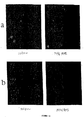
(薬物での処理の前および72時間後の、C3細胞中での細胞性遺伝子発現の分析)
9600個の遺伝子アレイを用いて遺伝子発現を研究した(図5)。72時間、M4N(40μm)で処理した(C3 M4N)および未処理(C3DMSO)のものに由来するそれぞれのポリA+RNAの5マイクログラムを、Genomics 51、313−324、1998に記載されている手順に従って、ヒトの9600個の遺伝子アレイハイブリダイゼーション研究の対において使用した。ハイブリダイゼーション画像を、Nikon 55mm AF micro Nikoレンズを備えたカラービデオカメラによってキャプチャーし、そしてMacintosh LC630コンピューターによってデジタル化した。単色または2色の態様のいずれかで色素形成酵素の酵素基質反応を通じるこのような検出は再現性があり、そして非常に敏感である(107個の細胞に由来するRNAを用いて、1個の細胞あたり転写物の5個未満のコピーを検出することができる)。
【0041】
示差的に発現される遺伝子を示すコンピューターのプリントアウト(C3 M4N/C3 DMSO >10、およびC3 DMSO/C3 M4N >10)を、検査のために列挙した。TIFF形式での画像ファイルおよびMSエクセル形式でのデータファイルを、ZIPディスケットに保存する。遺伝子の名称およびクローンのID番号は、将来のノーザンブロットでの確認のための画像のクローンを得るために利用可能である。
【0042】
M4Nでの処理の72時間後にアップレギュレートかまたはダウンレギュレートされるかのいずれかである遺伝子の群の中で、以下は細胞分裂およびアポトーシスに特に関係するものである。いくつかの他の細胞周期に関連する遺伝子もまた、M4Nに応答して大きくアップレギュレートされる。サイクリン依存性キナーゼCDC2(実施例11)に加えて、例えば、以下である:
【0043】
【化6】
【0044】
【化7】
【0045】
(実施例5)
(M4Nの局所的な注射によるマウス中でのC3腫瘍増殖の標的化)
36匹のC57bl−16 NCRマウスに、マウスの背中の両肩の間に、5×10⁵個のC3細胞を注射した。マウスのうちの24匹が、20日以内に腫瘍を生じた。毎日の注射(50μl〜100μlのM4NまたはM4N/G4N)(DMSO中の200mg/mlのM4N、PBS中の200mg/mlのG4N)は、表1および2、図6および7に示すように、動物中での腫瘍の増殖に対して重大な影響を示した。
【0046】
(表1.マウスにおいて生じた単一腫瘍の増殖に対するM4NおよびG4Nの効果)
【0047】
【表1】
**15日目に採取した
***ほとんどの壊死性の細胞に含まれる病変がまた、マウス6、7、11、14、15、17、19、21、28、19に由来する病変中に見出された(図6、7)。薬物での処置の後では、マウス#11および#22においては病変は存在しなかった。コントロールのマウス#1、2、3、4中で見出される腫瘍が、増殖している細胞を含んだ(図2)。
【0048】
(実験手順:)
36匹のC57bl−16NCRマウスに、5×10⁵個のC3細胞/マウスを注射した。注射は、マウスの背中の両肩の間に皮下で100μlを行った。細胞を、低塩HBSS中に懸濁し、そして均質な懸濁物を、穏やかにボルテックスすることによって維持した。
【0049】
24匹のマウスが腫瘍を発症した。それらの病変の大きさを、ダイアルキャリパーによって測定した。これらのマウスを、剃毛し、体重を量り、そして処置を開始した(1日目)。4匹のマウスをコントロールとして隔離した。コントロールマウスには、50μLのDMSOを毎日腫瘍内注射によって受容させた。実験マウス(10)には、DMSO中に溶解させた(200mg/mL)50μLのM4Nを与えた。さらに10匹のマウスには、8日間のM4Nでの処置、続いて、8日間の毎日のG4Nでの処置(50μL、PBS中の200mg/mL)を受容させた。注射を、腫瘍のいくつかの領域に対して行った。マウスを、注射の前にエーテルまたはメタファン(metaphane)で麻酔した。
【0050】
(表2.複数の腫瘍を有するマウスにおける、処置した病変の増殖に対するM4NおよびG4Nの効果)
【0051】
【表2】
**薬物を使わない隣接している腫瘍に由来する
(表3.マウスにおけるG4Nの毒性研究)
【0052】
【表3】
【0053】
全ての処置したマウス、コントロール(マウス番号(#)1〜4)、および実験マウス(マウス番号6、7、9、10、11、12、14、15、16、17 M4N、番号18〜22、24、26〜29 M4N/G4N)が、腫大を示した。病変の大きさの測定を、ダイアルキャリパーによって行った。いくつかのマウスは、注射に起因して穏やかな出血を経験した。処置レジメおよび結果は以下のとおりであった:
10日目:マウスを再び体重測定した。全てのマウスが、2グラムの成長を示した。
12日目:処置を行わなかった。
13日目:全てのマウスが、非常に異なる程度ではあったが、皮膚が盛り上がった。1匹のM4Nで処置したマウス(#7)の皮膚は、割れて開き、これは「乾燥した腫瘍」になった。
14日目:注射の容量を100μLに増大させた。
15日目:1匹のM4Nで処置したマウス(#17)が、麻酔の過剰用量/取り扱いによって死亡した。#17の病変の部位の皮膚にはひびが入り、これは「乾燥した腫瘍」を示していた。これを解剖し、そして病変を切り出しそして秤量した。
16日目:さらに4匹のM4Nで処置したマウス(#6、14、15、16)、3匹のM4N/G4Nで処置したマウス(#19、21、28)、および1匹のコントロールマウス(#2)を安楽死させ、解剖し、そして秤量した。残りのコントロールマウス(#1、3、4)を、非侵襲的に試験し、そしてこれらは腫瘍を保有していた。
21日目:コントロールマウスに由来する腫瘍の大きさを、ダイアルキャリパーによって測定した。観察:マウス#10および#12(M4Nで処置した領域)の病変部位の皮膚にはひびが入り、そして「乾燥した腫瘍」を示していた。
【0054】
24日目:マウス#7の皮膚は再び完全にカバーされた。実験を、この日で終了した。全ての残っているマウス、M4Nで処置したマウス(#7、9、10、11、12)、およびM4N/G4Nで処置したマウス(#18、20、24、26、29)を安楽死させ、解剖し、試験し、そして秤量した。
【0055】
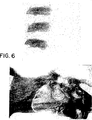
マウス中でのC3腫瘍の増殖に対するM4NおよびM4N/G4Nの効果を、表1および2、ならびに図5および6にまとめる。表1は、単一の腫瘍を保有しているマウス中でのC3細胞の増殖に対する薬物の影響を示す。コントロールの群の4個の切除した腫瘍の平均の重量は1.48gであり、一方、M4Nで処置したマウスおよびM4N/G4Nで処置したマウスに由来する病変の重量は、それぞれ、0.142gおよび0.51gであった。薬物で処置した病変は、主に、乾燥した壊死性の細胞から構成された(図6)。コントロールの群に由来する腫瘍は、均質であるようであり、そして活性に増殖する細胞を含んだ。表2は、複数の腫瘍を保有しているマウス中でのC3腫瘍の増殖に対する薬物の影響を示す。この研究においては、薬物を、腫瘍の1つに注射した。未処置の腫瘍の平均の重量は1.77gであり、一方、M4Nで処置した病変の平均の重量は、0.15gであった。同様の結果が、M4N/G4Nの注射の後に得られた−−未処置の腫瘍の平均の重量は1.27gであり、一方、薬物で処置した病変の平均の重量は、わずか0.103gであった。
【0056】
全体の実験期間の間の全てのマウスの体重の変化は、些細なものであるようであった(表1および2)。
【0057】
(実施例6)
マウスの2つの群に由来する薬物で処置した(M4N)腫瘍およびDMSOビヒクルで処置したマウスまたは未処置のマウス(CON)を、組織病理学的な試験のために調製した。切り出した腫瘍をすぐに固定し、次いでリン酸緩衝化生理食塩水中の4%のホルムアルデヒド中で保存した。次いで、固定した組織を、一連の段階的なアルコールおよびキシレンを通じて脱水し、そしてパラフィン中に胞埋した。パラフィン組織ブロックを薄く切片状にし、そしてヘマトキシリンおよびエオシンを用いて顕微鏡分析のために染色した。組織病理学的研究は、コントロールの腫瘍がDMSOでの処置によっては影響を受けず、そして増殖し続けることを示した。これらは、高い核/細胞質比、多形性の核の変化、高い有糸分裂形状、紡錘糸状の肉腫の形状、および癌細胞の特徴である周辺組織への浸潤を示す。
【0058】
対照的に、M4N処置を受容けた腫瘍は、処置が開始された直後に増殖をやめる。これらは、有意な壊死を示し、そしてもはや生存可能ではない。高倍率で生存可能な少量の薬物の沈殿が存在し、そして局所的な領域は慢性の炎症および繊維症を示す。この治癒効果は、この領域からのこれらの死んだ腫瘍細胞の分離を導く。同じ結果が、M4Nでの処置のみを用いた場合と同様に、M4N/G4Nでの処置を用いて見られる。しかし、G4Nが水可溶性であるので、これは、M4Nよりも腫瘍の大きな領域に対して広がり得る。M4Nとともに共働で使用される場合には、G4Nは、大きな大きさ(すなわち、2cm3よりも大きい)の腫瘍を標的化する(trating)際により有効であり得ることが、予想される。
【0059】
(実施例7:モルモットでのHSV−1の皮膚感染に対するM4Nの影響)
薬物M4Nをまた、モルモットの皮膚感染におけるHSV−1の複製の阻害において試験した。モルモットの皮膚を針で傷つけ(pinch)、そしてHSV抑制剤をそれぞれの穿刺した領域に感染させるように局所的に塗布した。次いで、M4Nを、注射後、毎日、6日間の間、穿刺して感染させた領域に対して塗布した。
【0060】
モルモットの裸にさせた背中の皮膚の6個の領域を、5=DIN針で滅菌的に穴を開けた。2つの領域を、HSV−1(HSV−C、培養上清、または生理食塩水中に単離したHSV、HSV−SC)で感染させた。他の4つの領域を、HSV−SCで感染させた。注射の15分後、30μlの試験化合物(DMSO中の、ABDS1、ABDS2、ACV、およびM4N(4N) 60mg/ml)を、1つの領域のそれぞれのパンチして感染させた領域に対して、1日に5回、6日間塗布した。ABDS1およびABDS2を、ネガティブコントロールとして含めだ。図8の写真を、6日目に撮影し、そしてこれは、薬物の非存在下(HSV−C、HSV−SC)、有効ではない薬物の存在下(HSV−ABDS1、HSV−ABDS2)、および有効な薬物の存在下(HSV−M4N、およびHSV−ACV)でのHSV−1の複製の程度を示す。6個の大きな融合性の水疱が、HSV−C、HSV−SC、HSV−ABDS1、HSV−ABDS2によって処置した領域中で生じ、一方、M4Nでの処置(4N)およびACVでの処置の後には、感染させた領域中では水疱は観察されなかった。
【0061】
M4NがHSVの複製をブロックし得るという明確な結果を、皮膚の病変の消失によって、そして薬物での処置の4日後にウイルスの脱殻がないことによって示されるように、このモデルシステムにおいて得た。最初の動物実験もまた、M4Nが、腹膜内で与えられた場合には300mg/kg程度の高い濃度で、そして皮下またはIVのいずれかによって与えられた場合には375mg/kg程度の高い濃度で、マウスに対して無毒性であることを示した(表3)(6)。
【0062】
(実施例8:インサイチュでの注射を使用する臨床的な処置のためのM4N)
薬物送達経路としての腫瘍内への直接のM4Nの投与は、いくつかの特有の欠点を提供する。1)M4Nは疎水性の化合物であり、そしてDMSO中に極めて可溶性である(200mg/ml)。従って、少量の薬物の溶液のみが、薬物の有効な投与量を達成するための注射に必要である。上記の実施例5に記載するマウスでの実験においては、数日間の間の、50μlから100μlの毎日の注射が、マウスでの腫瘍の増殖を完全に停止させるために十分であった。疾患を処置するためのDMSOの大きな投与量(1回の処置あたり30mlのIV)の使用について、いくつかの以前の研究が存在している(24)。結果は、明確ではなかった(25)。しかし、全世界で何千万人ものヒトが、過去に大きな量のDMSOを用いて安全に試験されている(26)ので、DMSOは、その極少量が使用される場合には、薬物の送達のためのビヒクルとして安全であるはずである。2)インサイチュでの注射によって、薬物の残りの大部分は不溶性であり、そして腫瘍の領域で濃縮され、そして循環系には入らない。従って、全身の毒性は回避される。さらに、その増殖を抑制するために十分な薬物が腫瘍内に残っているので、薬物の持続的な注射は、比較的少ない回数の処置の後には必要はない。実施例5のマウスでの研究においては、腫瘍細胞は、M4Nの注射の中断の後にもなお、死滅し続けた。従って、薬物が直接標的化される場合には、腫瘍の大きさは、投与される薬物の必要とされる量についての決定的な因子となる。ヒト対マウスの全体重間での差異は、無関係となる。マウスの腫瘍の研究においては、10日間の間の20mg/日が、腫瘍を排除するために十分以上であった。匹敵する大きさ(1〜1.5cm3)のヒトの腫瘍を処置するためには、これよりも高い投与量を使用するための理由は存在しないはずである。このことは、ヒトでの治験においてリスクを相当減少させるはずである。
【0063】
(実施例9:細胞のM4Nでの処理は、細胞増殖をブロックする)
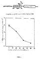
M4Nについての本発明者らの以前の研究は、Sp1依存性のプロモーターの非活性化によってウイルスの転写を阻害することができることを示した。多くの哺乳動物の細胞周期遺伝子もまた、欠くことのできないSp1プロモーターを含み、そして従って、M4Nはそれらの転写をブロックし得る。この仮説を、多数の種々の細胞株に対するM4Nの抗増殖効果を試験することによって試験した。本発明の化合物であるNDGAの低い濃度(10μM)は、哺乳動物細胞においてアポトーシスを誘導することが以前に示されている(27)。しかし、この効果は、カテコール酸素の1つをブロックすること、またはNDGAに対する親水性基の付加によって回避され得る(28)。漸増量のNDGA誘導体であるM4Nを、増殖を阻害するために必要とされる最適な濃度を決定するために、HPV−16/rasで形質転換したC3細胞株(29)の培養物について試験した(図9a)。細胞は、M4Nに対して十分に応答して、40〜60μMまでの濃度の範囲にわたって72時間後に細胞分裂をやめた。これらの濃度での3日後、細胞の数は、処置の開始時点(0日、図9)での数と等しいままであった。細胞の増殖におけるより穏やかな減少が、より低い薬物濃度で観察され、そしていくらかの細胞死が、60μMよりも大きい濃度で見られた。
【0064】
C3細胞株に対するM4Nの抗増殖の影響は、単にSp1依存性のHPV−16 E6/E7オンコジーンプロモーターを非活性化する薬物の能力に起因するのではない。なぜなら、同様の増殖の阻害が、そのE6/E7オンコジーンがSp1依存性ではないレトロウイルスプロモーターの制御下にある、HPV−16で形質転換されたTC−1細胞株において観察されたからである(30)(図9d)。さらに、C33a細胞株(図9c)、HPVネガティブであるヒトの頚癌細胞株、およびCEM−T4株(図9b)、ヒトの白血病細胞株(31)の増殖もまた、M4Nでの処理によってブロックされた。薬物で処理した4つの細胞株においては、停止した細胞のほぼ全て(>95%)が、M4Nの濃度が「閾」値(C3細胞については60μM、TC−1細胞については40μMなど)を超えるまでは、生存可能であった。これらの濃度を上回ると、生細胞の割合が、急激に減少する。興味深いことに、停止した細胞は、薬物への長期化した曝露の後にもなお、>95%の生存性を維持した。C3細胞は、40μMのM4Nでの処理の8日後に、細胞死の増大を示さなかった(図9e)。
【0065】
(実施例10:M4Nで処理された細胞はG2期で停止する)
一旦、M4Nで処理された細胞が、なお生存可能でありながら増殖を停止することが確立されると、細胞DNA内容物の分析および細胞構造の蛍光試験を、細胞が停止する細胞周期における時点を決定するために使用した。M4Nに対して72時間曝露した細胞は、コントロールと比較して、増大したG2/M DNA内容物を示した(図10a〜d)。最も極端な応答は、C3およびCEMT4細胞株によってら見られ、ここでは、>90%の細胞がG2/M DNA内容物を示す。
【0066】
G2での停止または有糸分裂のブロックとを区別するために、αチューブリン(緑)およびγチューブリン(赤)に対する抗体を、M4Nでの処理の72時間後のC3細胞株中での中心体の状態を決定するために使用した。図11aに示すように、M4Nで処理した細胞の中心体は、2倍であったが、細胞の核中になお互いに隣り合って配置されている。中心体は前記の初期の間に分離するので、これらの細胞は有糸分裂を開始していないと結論付けられ得る。反対に、コントロール細胞のγチューブリン染色は、G1またはS期に特徴的な散在性のパターンを有する(32)。M4Nで処理した細胞中でのクロマチンの濃縮の欠如もまた、DAPI染色を用いて観察され(図12b)、これは、細胞がG2期から先に進めないことのさらなる証拠である(33)。
【0067】
(実施例11:CDC2の産生は40μMのM4Nによって阻害される)
G2からの細胞の進行は、MPFの産生に依存する。そのタンパク質成分の状態を、40μMのM4Nで処理したC3細胞中で試験した。非同期の細胞を、1%のDMSO中にM4N、または1%のDMSOのみのいずれかを含有している培地中で24時間または72時間、増殖させた。細胞を回収し、そして等量の全細胞性タンパク質を、ウェスタンブロッティングによって分析した。CDC2の量における明らかな減少を、M4Nでの処理の72時間後に観察した(図12a)。しかし、同じメンブレンを切り出し、そして再度プローブすることによって検出したサイクリンBのレベルは、変化しないことを見出した。これらの結果は、これらの条件下で、停止はおそらく、p53に対する応答ではないようであることを示す。なぜなら、p53の過剰発現がサイクリンBの減少を導くことが示されているからである(34、35)。ウェスタン分析の結果と一致して、CDC2キナーゼ活性は、M4Nでの処理の72時間で排除された(図12a)。これらの実験は、薬物がCDC2タンパク質の産生を阻害することによって作用し、それによってMPFの活性の喪失を生じるという見解を支持する。
【0068】
Sp1依存性のウイルスの転写をブロックするM4Nの能力を実証する本発明者らの以前の研究は、CDC2タンパク質の減少の可能性のある機構として、CDC2 mRNAレベルの減少を示唆する。このことは、サイクリンBタンパク質(サイクリンBタンパク質の遺伝子はその発現のためにはSp1を必要とはしない)が、正常なレベルで産生され、一方、CDC2タンパク質(CDC2タンパク質の遺伝子はそのプロモーター中に2つの必須のSp1部位を有している)は、その量が実質的に減少されるという知見と一致する。この仮説を試験するために、ノーザンブロット分析を、5〜72時間の間、40μMのM4Nで処理したC3細胞から回収したRNAについて行った。図12bに示すように、CDC2 mRNAの量は、M4Nでの処理のわずか24時間後に減少し、そして72時間後にはほぼ排除された。Sp1によっては調節されないハウスキーピング遺伝子であるGAPDHの産生を、RNAローディングコントロールとして使用し、そしてそのレベルは、40μMのM4Nによっては影響を受けなかった。
【0069】
C3細胞株の使用は、本発明者らにとって、M4Nによって媒介される細胞周期の停止の機構の分析のためのさらなるコントロールとなる。なぜなら、他のSp1依存性の遺伝子プロモーターもまた、おそらく、M4Nでの処理によって阻害されるからである。この可能性を、Sp1依存性のHPV−16 E6/E7プロモーターからの転写物に対するM4Nの影響を分析することによって、C3細胞中で試験した。5から72時間の間40μMのM4Nで処理したC3細胞から単離したRNAのrtPCR分析は、E7転写物のレベルにおける明確な減少を示した(図12c)。GAPDHを、再び、この実験の内部コントロールとして使用し、そしてそのレベルは、薬物での処理によっては影響を受けなかった。これらの結果は、M4NがSp1によって調節されるプロモーターの転写を減少させることのさらなる証拠を提供する。
【0070】
(実施例12:ゲル移動度シフトの分析における、G4NによるSp1結合活性の阻害)
Sp1ファミリーのタンパク質は、結合の際にDNAの主溝に対する湾曲を誘導する(36)。Sp1タンパク質のzincフィンガードメインは、GC Box配列である5’−GGGGCGGGG−3’の結合を担う。コンピューターによる分析によって、NDGAのアミノエステル誘導体であるG4Nが、主溝においてこのような配列と安定な複合体を形成し得ると決定した。G4NがSp1ブロッカー、ならびにSp1置換因子(displacer)として作用し得るかどうかを決定するために、本発明者らは、Sp1/エンハンサー相互作用研究を、G4Nの存在下または非存在下で、試験するためのSp1のDNA結合ドメインのみを使用するゲル移動度シフトの分析によって行った。ブロッキング実験においては、種々の濃度のG4Nを、最初に32Pで標識したDNAとともに結合緩衝液中で25℃で30分間、インキュベートした。組換えSp1タンパク質のDNA結合ドメイン(Sp1−167D)を次に添加し、そしてさらに30分間、大過剰のBSAタンパク質の存在下でインキュベートした。置換実験においては、組換えSP1−167Dを最初にDNAに結合させ、次いでG4Nをインキュベーションの第2の工程で添加した。G4NおよびSp1−167D濃度、ならびにインキュベーションおよびゲル電気泳動の条件は、両方の実験において同一であった(実験の節)。図13に示すように、いずれの場合においても、G4Nは、Sp1−167Dタンパク質との相互作用からDNAを維持することが可能であることを見出した。Sp1のDNA結合ドメインのみが試験される場合には、G4Nは、ゲル移動度シフトの分析によって示されるように、エンハンサーに対して結合することからSp1をブロックするよりも、結合したSp1の置換においてより有効であるようであった(図13、A、B、D)。本発明者らはまた、結合したG4NがSp1−167Dによって置換され得るかどうかを試験した。この実験においては、G4NによるSp1−167Dの結合の阻害を、最初に移動度シフトの分析によって証明した(図13C、レーン2および5)。G4Nが結合した鋳型をさらなるSp1−167Dでチャレンジした場合には、本発明者らは、Sp1−167D/DNA複合体のバンドの強度の投与量依存性の増大を観察し(図6C、レーン6、7)、このことは、この鋳型からのSp1−167DによるG4Nの置換を示す。
【0071】
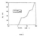
(実施例13:G4NによるHIVプロモーター活性のSp1によって調節されるTatトランス活性化の阻害) 以前に報告されているように、メチル化NDGA誘導体は、HIV、HSVのICP4、HPVのE6/E7遺伝子を含む種々のウイルスのプロモーターのエンハンサー部位へのSp1の結合をブロックし得る(37、38、39)。本発明者らはさらに、以前に記載されているような、SEAPアッセイにより、Cos細胞中でのHIVプロモーター活性のTatトランス活性化に対するG4Nの影響を試験した。基底レベルのHIV LTRによって駆動されるSEAPの発現は、Cos細胞中でかろうじて検出可能であることが以前に見出された。Cos細胞がCMVプロモーターによって駆動されるTat遺伝子で同時トランスフェクトされる場合には、SRAPの発現において60倍以上の増大が存在する(37)。このような、HIV LTRプロモーター活性のTatによって駆動されるトランス活性化は、Sp1によって調節されることが以前に示されている(37、40)。本発明者らは、G4Nの存在下で、用量依存性の様式でのHIVのトランス活性化の阻害を観察した(図14)。G4Nについての36μMのIC50値の平均値は、3−O−メチルNDGAであるMal.4のもの(IC50 25μM)に匹敵し、そしてテトラ−メチルNDGAであるM4Nのもの(IC50 11μM)よりもいくらか高かった。この差異はおそらく、細胞への薬物の取り込みに影響を与える試験化合物の化学的な性質に起因する。
【0072】
(実施例14:G4Nによる細胞培養物中でのSIV−1およびHIV−1の産生の阻害) HIV−1およびSIVの両方ともが、レトロウイルスであり、これらは、それらの複製を完全にするために宿主ゲノムへの組込みを必要とする。これらの両方ともが、それらのプロウイルスの転写のために宿主の転写因子に依存する。Sp1は、ほぼ同一の態様の転写の調節を共有しているこれらの2つのウイルスにおいて、このような発現のための中心的な役割を果たす。SIVに感染したアカゲザルをG4Nの抗ウイルス効果を試験するための動物モデルとして使用する予想においては、本発明者らは、174×CEM細胞中のSIVの阻害におけるG4Nの効果と、H9細胞中のHIVの阻害における効果とを研究し、そして比較した。これら2つの細胞株におけるG4Nの細胞毒性もまた試験した。SIVの阻害の研究のために、107個の174×CEM細胞を、高力価のSIVmac 239ストックとともに37℃で2時間混合し、次いで、吸着されていないウイルスを除去するために冷却したPBS緩衝液で2回洗浄した。細胞懸濁物を、3個の96ウェルプレートのそれぞれのウェルに等分した。種々の濃度のG4N溶液を、新しく調製したストックから作製し、そして別々に等分し、そして1つの96ウェルプレート中の列の6個のウェルのそれぞれに入れた。培養上清を、感染後(P.I.)4日毎に回収し、そして適切な濃度の薬物を含有している新しい培地を上清の回収後に培養物に添加した。ウイルスの産生を、示すように(図15)、改変したp27コア抗原捕捉ELISAによってアッセイした。5μMより高い濃度でG4Nを使用した場合には、SIVの産生は検出されなかった。2.5μMよりも低いG4N濃度では、SIVの産生は、薬物の非存在下でのウイルスの産生と比較して、4日目および8日目の感染後培養物に由来する培養上清中で検出された(図15)。G4N(250μM以下)は、MTTアッセイによって決定した場合は、感染させていない174×CEM細胞に対しては毒性の影響を示さなかった。同様の実験を、H9細胞においてG4NによるHIV−1の阻害の研究のために行った。H9細胞を、1×105/mlで継代培養し、そしてHIV−1のAZT耐性株(HIV−1RTMF)を用いて感染させた。種々の濃度のG4Nを、感染の2時間後に添加した。新しい培地との交換を、4日ごとに行った。G4Nの存在下での細胞の増殖を、9日の実験期間の間、注意深くモニターした。ウイルスの産生を、p24コア抗原捕捉ELISAによってアッセイした。示すように(図16)、80μMの濃度のG4Nは、H9細胞中でのHIVの複製を完全に阻害した。HIV−1 RTMFの阻害については、12μM G4NのIC50を見出した。再び、アッセイの範囲内(および250μM未満)では未感染のH9細胞に対する毒性は検出されなかった。
【0073】
本明細書中に引用した参考文献を、利便性のために以下に列挙し、そして参考として本明細書によって援用する。
【0074】
【表4】
【図面の簡単な説明】
【図1】 図1は、M4NおよびG4Nの構造を示す。
【図2】 図2Aは、E6/E7プロモーターの領域(pPV16P97)およびSp1タンパク質への結合部位を示す、HPV−16LCRを示す。
図2Bは、C−33A細胞中でのE6/E7プロモーター活性に対するM4Nの影響を示す(種々の濃度のM4Nによるルシフェラーゼ遺伝子の転写により駆動されるE6/E7プロモーターの阻害)。
【図3】 図3A〜3Cは、40μMのM4Nによる、ウイルスのE6およびE7 RNA転写物の阻害を示す。増殖培地中で71時間、40μMのM4NまたはDMSOのみのいずれかで処理したC3細胞から単離した全RNAを、比較RTPCRに供した。RTPCRサンプルを、増加した増幅サイクルの後で取り出し、そしてアガロースゲル上で分析した。ゲル写真(3Aおよび3B)は、これらのサイクルでの、増殖培地中でのM4Nの存在(+)または非存在(−)を示し、そしてサイズマーカーとして使用したpGMTベクターの2つの消化物を示した。増幅マップ(2C)は、2つの予想される大きさの増幅産物を示し、これらは、初期のウイルスのRNA転写物の異なるスプライシングによって得られた。
【図4A】 図4Aは、M4NによるC3細胞の増殖の阻害を示す。
【図4B】 図4Bは、M4Nの除去後のC3細胞の増殖の阻害を示す。
【図5】 図5A〜5Bは、GENE Assay分析によって試験した、C3細胞中での遺伝子発現に対するM4Nの影響を示す。5A.DMSA処理の>2時間後のC3細胞中で発現されるGENE(C3 DMSO)。5B.溶媒としてDMSOを使用するM4N処理の>2時間後のC3細胞中で発現されるGENE(C3 M4N)。
【図6】 図6A〜6Bは、M4N処理後の腫瘍保有マウスの目視観察を示す。6A.単一の腫瘍を保有しているマウスを、DMSO(#3)またはM4N(#7)のインサイチュでの注射で処置した。M4Nのインサイチュでの注射はまた、マウス#9において増殖した2つの腫瘍のうちの1つに対してもなされた。6B.表2に記載するような、同じマウス#9に由来する未処置の腫瘍およびM4Nで処置した腫瘍(白色の瘢痕)。
【図7】 図7は、図7は、マウス中での腫瘍の増殖に対する、M4NおよびM4N/G4Nの組織病理学的な影響を示す。
パネルの第1列目は、DMSOでの処置(CON)後の、マウス#12、10、27、および20(M4N)由来の比較的少量の薬物で処置した(M4NまたはM4N/G4N)病変と比較した、マウス#4、10、12由来の大きなサイズの腫瘍を示す。続く写真は、100×の倍率で試験したこれらの腫瘍の例である(A、B、C、DMSOで処置したマウス、D、未処置のマウス、E、F、G、H、M4NまたはM4N/G4Nで処置したマウス)(表1および表2)。
【図8】 図8は、薬物の非存在下(HSV−C、HSV−SC)、有効ではない薬物の存在下(ABDS1[「HSV−ABDS1」]、ABDS2[「HSV−ABDS2」])、および有効な薬物の存在下(M4N[「HSV−4N」]およびACV[「HSV−ACV」]でのHSV−1の複製を示す。
【図9】 図9は、M4Nが哺乳動物細胞中で増殖の停止を生じることを示す。(a〜d)C3、CEM−T4、C33a、およびTC−1細胞を、種々の濃度のM4Nで処理した。実験の開始時に存在する細胞の数を、0日として示す。3日後、生存可能な細胞の数を計数し、そしてM4Nの濃度に対してプロットした。(e)C3細胞を、T−25フラスコ中に分け、1つのフラスコあたり5×10³個の細胞を有するようにし、そして培地中の1%のDMSO中のM4N、または培地中の1%のDMSOのみのいずれかを入れた(最初の培地交換)。3日後、M4Nで処理した細胞の1/2に、1%のDMSOのみを含有している新しい培地(M−D)を入れ、一方、残りの細胞には、同じ条件で新しい培地を入れた(第2回目の培地交換)。細胞を毎日計数し、そして処理の回数に対してプロットした。
【図10】 図10は、M4Nで処理した細胞はG2/M中で停止することを示す。C3細胞(a)、C33a細胞(b)、CEM−T4細胞(c)、およびTC1細胞(d)を、1%のDMSO、またはM4Nとともに1%のDMSO(M4N)のいずれかを含有している培地中で3日間、増殖させた。細胞をトリプシン処理し、エタノールで固定し、ヨウ化プロピジウムで染色し、そして続いてフローサイトメトリーによって分析した。データを、細胞の数(3〜5×104個の全細胞)対ヨウ化プロピジウムでの染色の強度として表示した。細胞周期の示される段階が標識され、そして染色強度によって決定されるように、相対的な細胞性のDNAの引き立たせ(compliment)に対応する。
【図11】 図11は、40μMのM4Nで処理したC3細胞がG2細胞の構造を実証することを示す。C3細胞を、1%のDMSO(コントロール)、または40μMのM4Nとともに1%のDMSO(M4N)のいずれかを含有している培地中で3日間、カバースライド上で増殖させた。サンプルをエタノールで固定し、そしてα(緑)およびγ(オレンジ)チューブリンに対する抗体(a)、またはDAPI DNA染色(b)とともにインキュベートした。細胞を、蛍光顕微鏡で試験した。
【図12】 図12は、CDC2およびウイルス癌遺伝子がM4Nによって減少されることを示す。C3細胞を、1%のDMSO(D)、または40μMのM4Nを含む1%のDMSO(M)のいずれかを含有している培地中で、種々の時間(数字は時間で)の間増殖させた。示された時間の後、全タンパク質または全RNAを細胞から単離した。ウェスタンブロット(a−上段の2つのパネル)を、CDC2またはサイクリンBに対する抗体を使用して、同じニトロセルロースフィルターを用いて行った。キナーゼアッセイ(a−下段の2つのパネル)を、γ−32P ATPおよびヒストンH1とのインキュベーションによる、サイクリンBに対する抗体での免疫沈降の後で行った。PAGEゲルのクマシー染色を、ローディングのコントロールとして含んだ。24時間および72時間の薬物での処理についてのキナーゼアッセイを、別々に行った。
ノーザンブロット(b)を、全RNA抽出物について行った。フィルターを、CDC2またはGAPDHについてのランダムプライムした32P標識したDNAとともに一晩インキュベートし、洗浄し、そして3日間フィルムに対して暴露した。同じフィルターを、CDC2およびGAPDH RNAを試験するために使用した。
rtPCR分析(c)を、HPV−16 E7またはGAPDHのいずれかの領域に対してハイブリダイズするプライマーを用いて、全RNA抽出物について行った。両方のプライマー対を、同じ条件で使用し、そして生成物をアガロースゲル電気泳動によって分析した。
【図13】 図13は、HIV Sp1結合部位(−87から−49)とのG4Nの相互作用の、電気泳動の移動度のシフトアッセイ(EMSA)を示す。(A)32Pで標識したHIV Sp1 DNAテンプレートへのSp1−167Dの結合のG4Nによる阻害。レーン1、テンプレートのみ;レーン2、テンプレートおよび0.1μgのSp1−167D;レーン3〜9、漸増濃度のG4N(0.1μgのSP1−167Dの添加の前に、0.25〜1.75mM)とともにインキュベートしたテンプレート。(B)HIVテンプレートに結合したSp1−167DのG4N置換。レーン1、テンプレートのみ;レーン2、テンプレートおよび0.1μgのSp1−167Dおよび100倍過剰の未標識のテンプレート;レーン3、テンプレートおよび0.1μgのSp1−167D;レーン4〜10、漸増濃度のG4N(0.25〜1.75mM)とともにチャレンジしたSp1/DNA複合体;レーン11、1.75mMのG4Nを含有している反応緩衝液中でインキュベートしたテンプレート。(C)テンプレートに結合したG4NのSp1−167D置換。レーン1、テンプレートのみ;レーン2〜4、テンプレートおよび漸増量のSp1−167D(0.075、0.150.0.300μg);レーン5〜8、1.2mMのG4Nを含有している反応緩衝液中でインキュベートし、続いて漸増量のSp1−167D(0.075、0.150.0.300μg)でチャレンジしたテンプレート、レーン8、Sp1−167Dを受容させなかった。(D)G4Nの漸増濃度に応答して減少するSp1−167D/DNA複合バンドの強度のプロットは、(A)−−−・−−−および(B)――・――を使用した。使用したゲルは、5%の非変性ポリアクリルアミドであり、それぞれのレーンに、実験のセクションおよびRef[1]に記載するように、それぞれの反応容量の5μlを受容させた。
【図14】 図14は、G4NによるCos細胞中でのHIV Tatによって調節されるトランス活性化の阻害を示す。
【図15】 図15は、G4Nの存在を伴うSIVの産生を示す。10 7 174×細胞を、24時間混合し、37℃で2時間の間、SIV mac 239(4ngのp27)のストックを回収した。細胞を再懸濁し、そして100μlの培地中の1×10 5 細胞を、3個の96ウェルプレートのそれぞれのウェルに対して添加した。新しく作成したストックから種々の濃度のG4Nを調製し、そして6個の指定したウェルのそれぞれに添加した。培養上清を、ウイルスの産生の分析のために、4日および8日後に回収した。ウイルスの産生を、実施例のセクションに記載するように、改変したp27キャプシドタンパク質抗原捕捉ELISAによってアッセイした。
【図16】 図16は、G4NによるH9細胞中でのHIV p24抗原の産生の阻害を示す。阻害の割合を、AZT耐性HIV株であるHIV−1RTMFでのウイルス感染の9日後に、G4Nで処理したH9細胞、および処理していないH9細胞の2つの2連の培養物の平均からp24レベルを比較することによって計算した。[0001]
The invention described and claimed herein was made in part with grants from the National Institute of Health. The United States government has certain rights in this invention.
[0002]
(Background of the Invention)
(1. Field of the Invention)
The present invention relates to the use of nordihydroguaiaretic acid derivatives for the treatment of tumors and viral infections, in particular the use of derivatives containing substituents of naturally occurring amino acids.
[0003]
(2. Background information)
Carcinogenesis is a multistage event that is affected by various genetic and epigenetic factors and is symbolized by the onset of uncontrolled cell growth originating from various tissues. The universal goal for anticancer research is to develop clinical treatments that are highly effective in reducing tumor growth, are non-toxic to the host, and can be given for most patients . Drugs that focus on target inhibition that are specific to dividing cells should be effective chemotherapeutic agents without the risk of substantial side effects.
[0004]
Cells pass many checkpoints as they progress through the cell cycle. Certain criteria must be met to pass each of these checkpoints. In the G2 / M transition, the most essential regulator is CDC2, a cyclin-dependent kinase. This kinase binds tightly to the regulatory protein cyclin B, and this complex, called the maturation promoting factor (MPF), also stimulates many events that lead cells to enter early prophase. Play a role (1). Not surprisingly, deletion or deactivation of any component of MPF blocks cellular progression to G2.
[0005]
MPF expression and activity is regulated at various levels. Cyclin B protein levels rise slowly throughout the G1 and S phases of the cell cycle, peak during the transition from G2 to M phase, and decrease rapidly during mitosis (2). On the other hand, CDC2 protein is usually present during the cell cycle, but the level is slightly elevated at the last stage of G2 phase (3). The activity of the protein depends on association with the appropriate cyclin as well as dephosphorylation of its inhibitory site by the phosphatase CDC25C (4, 5). This dephosphorylation failure has been shown to initiate G2 arrest in response to DNA damage due to radiation or chemical action. Recent evidence also suggests that any remaining active CDC2 can be transported outside the nucleus after DNA damage (6).
[0006]
Numerous naturally occurring derivatives of the plant lignan nordihydroguaiaretic acid (NDGA) have been shown to block viral replication through inhibition of viral transcription. This initial study showed that NDGA derivatives (originally isolated from Larea Tridentata and then chemically synthesized) were sp1 dependent on HIV (7, 8), HSV (9), and HPV (10). It has been shown that deactivation of the sex promoter can inhibit the production of its HIV, HSV, and HPV transcripts. Unexpectedly, one of these derivatives, tetra-O-methyl NDGA, also appears to induce cell cycle arrest in mammalian cell lines. The evidence presented herein below demonstrates that M 4 N can induce G2 arrest in mammalian cells without detectable toxicity, which indicates that this arrest is a cyclin. We support the observation that it is due to inhibition of the dependent kinase CDC2.
[0007]
Human papillomavirus (HPV) infection causes unregulated cell proliferation in many types of squamous cells, thereby ranging from benign papillomae (warts) to cervical, penis and mouth cancers Cause pain. The strong relationship of these cancers with HPV and the widespread emergence of infection indicates the importance of developing anti-HPV therapies.
[0008]
Most, but not all, viruses (including their replicatively active variants) are host dependent. These require the involvement of specific cellular factors to support viral growth. Host cellular factors, unlike viral proteins, are not under mutational pressure and are generally structurally invariant. Therefore, compounds that block the use of these cellular factors at different stages of the viral life cycle are probably good candidates as antiviral agents that are not sensitive to mutations. Several studies using cellular factors as alternative targets for HIV-1 inhibition have been reviewed (11).
[0009]
Applicants first described that 3′-O-methylated NDGA (ie, Mal. 4) isolated from Creosote bush (Larrea tridentata) is a basal HIV transcription, Tat-regulatory. It has been reported that transactivation and HIV replication in human cell cultures can be specifically blocked (12, 13, 14). Mal. 4 exerts its effect by interfering with the binding of the transcription factor Sp1 to the promoter of the HIV proviral template. Mal. Four targets map to nucleotides -87 to -40, the Sp1 binding site of HIV long terminal repeat (LTR). Unmodified NDGA does not inhibit HIV transcription in vitro and has no effect on Sp1 binding (12).
[0010]
However, isolation and purification of plant lignans is a laborious and costly task. In predicting the potential clinical use of plant lignans in the control of Sp1-regulated virus and tumor growth in humans, the activity of nine different methylated NDGAs as a parent substrate at low cost and in large quantities Synthesized chemically using unmethylated NDGA (15). At drug concentrations below 30 μM, tetra-O-methyl NDGA was found to be most effective in controlling HIV replication through inhibition of Sp1-regulated proviral transcription and transactivation (15 ). This study has since been extended to the control of herpes simplex virus (HSV-1 and HSV-2) growth (16). The herpes simplex virus earliest (IE) ICP4 gene is essential for HSV replication (17). The promoter region carries 8 Sp1 consensus binding sites (18), 5 of which are required for the expression of the ICP4 gene. This therefore makes the ICP4 gene a good candidate for such tests. Applicants have noted that both 3-O-methyl NDGA (Mal. 4) and tetra-O-methyl NDGA (M 4 N) are both Sp1 to the ICP4 promoter as shown by electrophoretic mobility shift assay. It has been found that it is an effective transcriptional inhibitor of HSV ICP4 gene expression in Vero cells by blocking protein binding.
[0011]
In infected Vero cells, M 4 N and Mal. When the anti-HSV activity of 4 was tested and compared to the activity of acycloguanosine (acyclovir, ACV), Applicants found that the IC 50 for M 4 N was 10 passages of HSV-1 and We observed that it varied between 11.7 μM and 4 μM for 4 passages of HSV-2, with no tendency to clearly increase the need for high drug concentrations. However, the IC 50 for ACV is from 7 μM for the first passage of virus to 444 μM for the tenth passage of HSV-1 and> 88 μM for the fourth passage of HSV-2. Increased to. This indicates their rapid enhancement of drug resistance to ACV in Vero cells. As a result, the selection index S.I. I. (TC 50 / IC 50 ) remains relatively stable for M 4 N, but S. I. Decreased 60-fold after passage of the virus in Vero cells (16). Therefore, M 4 N is a drug that is not sensitive to mutations. This can effectively inhibit ACV resistant HSV (16).
[0012]
Due to the fact that Sp1 is an important cellular transcription factor (19), the potential inhibitory effect of this class of compounds on the expression of Sp1-regulated cellular genes should be addressed. Mal. 4 cannot replace Sp1 once it is stably bound to its binding site (12). Thus, it is likely that NDGA derivatives have a greater effect on Sp1-regulated genes in proliferating cells than on expression of Sp1-regulated housekeeping genes in stationary phase cells. . In the latter case, the drug can compete with the Sp1 protein for the Sp1 site in the promoter of the gene during DNA synthesis, while in the former case, the drug It has little effect on the transcriptional chromatin of the housekeeping gene, which already has an Sp1 protein that is stably bound to the promoter of. In fact, it has been shown to be in such a case. As demonstrated below, by using gene array studies involving 9600 expressed genes, Applicants have found that most Sp1-regulated gene products are present at similar levels and cultured It was found that the cervical cancer cell C3 in the product was not affected by treatment with the drug (FIG. 5). Nonetheless, the relatively low selectivity index of M 4 N limits its use to the lowest effective concentration if the drug must be used systemically. On the other hand, human papillomavirus initially induces solid cervical and oral tumors by Sp1-regulated expression of the HPV E 6 / E 7 gene (20). Applicants have found that if the drug can be delivered in situ and can only be maintained in the area of the tumor, high concentrations of the drug will effectively destroy the tumor with little damage to the patient. Thought it could be used.
[0013]
(Summary of the Invention)
Accordingly, one object of the present invention is to provide compounds and compositions for use in the treatment of cancerous and non-cancerous tumors in animals (particularly mammals, and most particularly humans). is there. In accordance with this aspect of the invention, novel nordihydroguaiaretic acid derivatives that inhibit tumor growth are provided.
[0014]
By nordihydroguaiaretic acid derivative is meant a compound of the following structure:
[0015]
[Formula 4]
[0016]
Particularly preferred compounds for use according to the present invention are M 4 N and G 4 N, which are shown in FIG.
[0017]
A further object of the present invention is the use of these novel derivatives and by similar derivatives known in the art but not previously used for tumor treatment, It is to provide a method for treating non-cancerous tumors. This method should be particularly effective against rapidly proliferating cell types containing the cyclin dependent kinase CDC2. It is a further object of the present invention to provide a method for inhibiting CDC2 in the eukaryotic cell cycle, particularly of animal cells, more particularly mammalian cells, and most particularly human cells.
[0018]
Tumors to be treated include any tumor that is sensitive to the above compounds used according to the methods of the present invention. Specifically, this includes rapidly dividing cancerous and benign tumors that are sensitive to inhibition of the cyclin-dependent kinase CDC2 cycle.
[0019]
The term “cancerous tumor” is intended to include any malignant tumor, which may or may not have metastasized. The term “non-cancerous tumor” is intended to include any benign tumor. These terms are used as customarily understood by those skilled in the art.
[0020]
Examples of benign and malignant tumors that can be treated by the compositions and methods of the present invention are described in Cancer Biology (Raymond W. Ruddon, Cancer Biology, 3rd edition, Oxford, incorporated herein by reference. (Univ. Press, 1995). Tumors to be treated include tumors known to be of viral origin as well as tumors known not to be of viral origin. The compositions and methods of the invention are expected to be particularly useful in the treatment of solid tumors.
[0021]
Yet another object of the present invention is to provide a method of inhibiting the cyclin dependent kinase CDC2 cycle. This method is useful in inhibiting cell proliferation, particularly rapidly dividing cell types.
[0022]
In a preferred embodiment, the compounds and compositions described herein are used in the treatment of HPV-induced tumors. Tumors induced by HPV include, but are not limited to, cervical, mouth, penis and head and neck cancers particularly associated with HPV infection. This method is directed to nordihydroguaiaretic acid derivatives, particularly tetra-O-methylnordihydroguaiaretic acid (M 4 N) and N, N-dimethyltetratetrahydrofuran for cancerous and non-cancerous HPV-induced tumors Includes topical application of glycinyl nordihydroguaiaretic acid (G 4 N).
[0023]
Yet another object of the present invention is to provide a method of inhibiting viral replication and growth by administration of a compound of formula I containing an amino acid substituent. Compounds in which the amino acid substituents R 1 , R 2 , R 3 , and R 4 are the same are preferred for use in this method.
[0024]
M 4 N, G 4 N, and other derivatives may be administered by local injection into the tumor, generally with pharmaceutically acceptable diluents, excipients and carriers. Intended. In a preferred embodiment, M 4 N is injected into the tumor in the form of a DMSO solution and G 4 N is administered in a PBS solution. The use of G 4 N complements the use of M 4 N, especially in large tumors (> 2 cm 3 ). This is due to its water solubility that allows it to diffuse to larger areas of the tumor. Other water-soluble and water-insoluble nordihydroguaiaretic acid derivatives can be used as well according to the present invention. They can also be used in lipid-based formulations for systemic delivery, as is known and used in the art.
[0025]
Known to those skilled in the art as being compatible with M 4 N, G 4 N and other similar derivatives by pharmaceutically acceptable diluents, excipients and carriers, and human or other according to the invention Such compounds that are suitable for topical administration to a mammal are meant. The following examples herein describe administration by means of local injection, but other means of local administration (eg, topical application or targeted delivery to the tumor site) are also possible. Can also be used.
[0026]
The amount of compound administered to obtain the desired therapeutic effect will vary, but can be readily determined by one skilled in the art. Dosage, frequency of administration, and length of treatment depend on the environment, mainly the size and type of the tumor. However, administration of either 10 mg to 20 mg of M 4 N alone or with a similar amount of G 4 N per gram of tumor weight at daily to weekly intervals or less frequently is exemplified May be described for purposes. Administration of 50 μl to 100 μl of M 4 N dissolved in DMSO at a concentration of 200 mg / ml, either alone or in combination with G 4 N, is effective in many cases of tumors from 1 to 1.5 cm 3 Is expected.
[0027]
(Detailed description of the invention)
(experimental method)
The NDGA derivative was chemically synthesized (15). Cell line C3 is a W. Martin Kast of Loyola University Medical Center, Chicago, Illinois, U.S.A. S. A. A cell line transformed with HPV16E + L and activated Ras from C57 BL / 6 kh, provided by. This is maintained and cultured as described by Greenstone et al. (21) and Feltkamp et al. (22, 23).
[0028]
(Synthesis of G 4 N :)
Standard for the preparation of meso-1,4-bis [3,4- (dimethylaminoacetoxy) phenyl] (2R, 3S) -dimethylbutane hydrochloride N, N-dimethyltetraglycinyl NDGA, G 4 N procedure. To a solution of dichloromethane (250 ml) containing NDGA (12.8 g, 42.3 mmol, 1.0 equiv) and N, N, -dimethylglycine (26.2 g, 254 mmol, 6.0 equiv) DCC (52.4 g, 254 mmol, 6.0 eq) and DMAP (2.32 g, 18.9 mmol, 1.0 eq) were added. The reaction mixture was stirred at room temperature under nitrogen for 24 hours. After the reaction mixture was filtered, the solution was concentrated under reduced pressure. Acetone (250 ml) was then added into the reaction flask and the solution was bubbled with excess HCl (g). The water-soluble precipitate was dissolved in H 2 O and reprecipitated twice from acetone at room temperature to give (1) (29.2 g, 36.8 mmol) as a white solid in 87% yield. It was. Proton NMR spectra were obtained on a Varian Unity-400 (400 MHz) spectrum analyzer using D 2 O solvent and TSP as internal standards. Carbon-13 NMR spectra were obtained on a Varian Unity-400 (400 MHz) spectrum analyzer using D 2 O as the solvent. The carbon-13 chemical shift is matched against the TSP singlet (δ 0.0 ppm).
[0029]
The synthesis is shown in
[0030]
[Chemical formula 5]
[0031]
Analytical thin layer chromatography (TLC) was obtained from Merck Inc. On pre-coated plates (silica gel 60 F-254) purchased from Gas chromatographic analysis was performed on a Hewlett-Packard 5890 Series II instrument equipped with a 25 m cross-linked methyl silicone gum capillary column (0.32 mm id). Nitrogen gas was used as the carrier gas and the flow rate was kept constant at 14.0 mL / min. The retention time t R was measured under the following conditions: injector temperature 260 ° C., isothermal column temperature 280 ° C. Gas chromatography and low resolution mass spectral analysis were performed on a Hewlett-Packard 5890 Series II instrument equipped with a Hewlett-Packard 5971A Mass Selective Detector and a capillary HP-1 column. Separation by medium pressure liquid chromatography (MPLC) was performed at a flow rate of 120 mL / hr by use of a Jasco Model 880-PU intelligent HPLC pump. MPLC packing material, Reversed Phase Silica Gel C18 (particle size 0.035-0.070 mm) is available from Knauer Co. Purchased from. Purification by weight column chromatography was performed by the use of Merek Reagents Silica Gel 60 (particle size 0.063-0.200 mm, 70-230 mesh ASTM).
[0032]
Infrared (JR) spectra were measured on a Bomem Michelson Series FT-IR spectrum analyzer. The reported wave number refers to polystyrene 1601 cm-1 absorption. Absorption intensity is recorded by the following abbreviations: s, strong; m, moderate; w, weak. Proton NMR spectra were obtained on a Varian Unity-400 (400 MHz) spectrum analyzer by using D 2 O as solvent and 3- (trimethylsilyl) propionic acid, sodium salt as internal standard. Carbon -13NMR spectra at Varian Unity-400 (100MHz) spectrum analysis reported were obtained by using D 2 O as solvent. The carbon-13 chemical shift is referenced to the center of the singlet (60.0 ppm) of 3- (trimethylsilyl) propionic acid, sodium salt. Multiplexes are recorded by the following abbreviations: s, singlet; d, doublet; t, triplet; q, quadruple; m, multiplet; J, coupling constant (hertz). High resolution mass spectra were obtained with a JEOL JMX-HX110 mass spectrometer.
[0033]
meso-l, 4-bis [3,4 (dimethylaminoacetophenone key) phenyl] -2R, 3S- dimethylbutane hydrochloride (2). For a solution of NDGA (1, 12.81 g, 42.37 mmol, 1.0 equiv) and N, N-dimethylglycine (26.21 g, 254.2 mmol, 6.0 equiv) in dichloromethane (250 mL), DCC (52.45 g, 254.2 mmol, 6.0 eq) and DMAP (5.176 g, 42.37 mmol, 1.0 eq) were added. The reaction mixture was stirred at room temperature under nitrogen for 24 hours. After dicyclohexylurea in the reaction mixture was filtered, the resulting solution was concentrated under reduced pressure. Acetone (250 mL) was then added to the residue and the resulting solution was bubbled with excess HCl (g). The precipitate was dissolved in water and reprecipitated twice by using acetone at room temperature to give 2 (28.97 g, 36.86 mmol) as a white solid in 87% yield: 1 H NMR (D 2 O, 400 MHz) δ 0.78 (d, J = 6.0 Hz, 6H.2 × CH 3 ), 1.73 (m, 2H.2 × CH), 2.38 (dd, J = 13. 2,9.6 Hz, 2H.2 × ArCH), 2.78 (dd, J = 13.2, 4.4 Hz, 2H.2 × ArCH), 3.03 (s, 24H.8 × CH 3 N) , 4.53 (s, 8H, 4 × CH 2 N), 7.22 (m, 4H.4 × ArH), 7.29 (d, J = 8.4 Hz, 2H.2 × ArH); 13 C NMR (D 2 O, 100MHz) δ18.11,40.82,41.73,46.75,59.59,125.79, 26.58, 131.63, 140.66, 142.47, 146.11, 167.84; IR (KBr) 3461 (br), 2963 (m), 1777 (s, C = O), 1620 (m ), 1478 (m), 1377 (m), 1210 (m), 1106 (m), 961 (w), 852 (w) cm −1 ; MS (FAB) m / z (2-4 HCl) (relative) Strength) 643 (M +, 30), 600 (20), 588 (43), 515 (20), 473 (42), 430 (13), 388 (26), 185 (18), 93 (38), 58 (100), 44 (22); calcd 642.3628 for HRMS (FAB) C 34 H 50 N 4 O 8 in (2-4HCl), Found 642.3614; Anal. Calculated for C 34 H 54 N 4 O 8 Cl 4 : C, 51.78; 6.90; 7.10; 16.23. Found: C, 51.70; 6.85; 7.05; 16.21.
[0034]
It is understood that further amino acid substituted compounds of the invention can be synthesized by appropriate substitution of other N, N-dimethyl substituted amino acids.
[0035]
Example 1
(Effects of M 4 N with HPV E 6 / E 7 promoter activity regulated by SP1 and some other NDGA derivatives)
The effects of M 4 N with HPV E 6 / E 7 promoter activity regulated by SP1 and several other NDGA derivatives were tested using luciferase as a reporter. Assay, by calcium phosphate method, HPV16LCR fused to a luciferase reporter gene (P 97 promoter), depending on the DNA transfection into C33A cells. C33A is a cervical tumor cell line (ATCC accession number HTB-31). It does not contain any integrated HPV DNA, but has an essential transcription factor for strong expression of the HPV early gene promoter. One day after DNA transfection, various drug concentrations lysed with the aid of dimethyl sulfoxide (DMSO) were added to the cells. After 30 hours of drug treatment (as a result, the assay is typically completed within 48 hours for transient transfection experiments), cells were lysed and specific luciferase activity was determined. (Luciferase Assay Systems, Promega, US Pat. No. 5,283,179). As the M 4 N drug concentration increased, specific luciferase activity decreased.
[0036]
The results (shown in FIG. 2) show that M 4 N dramatically reduces the transcription initiation regulated by Sp1 at the HPV E 6 / E 7 promoter in the luciferase assay.
[0037]
(Example 2)
(Inhibition of E 6 / E 7 mRNA synthesis after treatment with M 4 N)
Inhibition of E 6 / E 7 mRNA synthesis after treatment with M 4 N was measured by RT-PCR in cervical cell line C 3 . Comparative RT-PCR was performed using the amount of total cellular RNA normalized to the number of cells counted. RT-PCR products were analyzed on a 2% agarose gel. The results are shown in FIG. RT-PCR results show that amplified cDNA of the expected size for E7 (321 bp) and E6 (204 bp) was detected in DMSO-treated cells at an early stage, such as 22 cycles of amplification. It showed that. These same products were barely detectable in drug-treated RNA extracts after 30 cycles of amplification. No amplified product was detected for the template PCR control or from the total RNA extract of the HPV16-negative C33a cell line.
[0038]
(Example 3)
(Inhibition of cervical C3 cell proliferation by M 4 N treatment)
Immortal mouse epithelial cells (C3 cells) transformed with HPV-16 were plated at a density of 10 5 cells per vial. After 24 hours, 1/2 of the vial is given growth medium containing 40 μM M 4 N dissolved in 1% DMSO, while the other half contains only 1% DMSO. A growth medium containing was provided. The results are shown in FIG. 4A. Within 24 hours, differences in cell morphology were observed between drug-treated and control C3 cells. The proliferation and division of drug-treated cells is clearly reduced compared to untreated controls, whereas the percentage of viable cells compared to the total cell count is only for drug-treated cells and DMSO Remained constant for both control cells. This indicates that M 4 N dramatically reduces cell division.
[0039]
The effect on the growth of C3 after removal of M 4 N from the medium was also tested. C3 cells were plated at a density of 10 4 cells per vial. At time = 0, 2/3 of the vials were given growth medium supplemented with 40 μM M 4 N in 1% DMSO. The remaining vials were given growth medium containing only 1% DMSO. After 73 hours, 1/2 of the vials that received M 4 N in their growth media were washed and media containing only 1% DMSO was added. The other 2/3 of the cell vial was washed and replaced with the same medium as previously administered. The results shown in FIG. 4B show that the rate of cell growth did not increase significantly in samples treated with M 4 N after replacement with medium containing no drug. , Indicating that M 4 N continues to significantly reduce cell division even after its removal from the extracellular environment.
[0040]
Example 4
(Analysis of cellular gene expression in C3 cells before and 72 hours after drug treatment)
Gene expression was studied using an array of 9600 genes (FIG. 5). Five micrograms of each poly A + RNA derived from (C 3 M 4 N) and untreated (C 3 DMSO) treated with M 4 N (40 μm) for 72 hours were used in Genomics 51, 313-324. , 1998, in a pair of 9600 gene array hybridization studies of humans. Hybridization images were captured by a color video camera equipped with a Nikon 55mm AF micro Niko lens and digitized by a Macintosh LC630 computer. Such detection through an enzyme substrate reaction of a chromogenic enzyme in either a monochromatic or bichromatic embodiment is reproducible and very sensitive (using RNA from 10 7 cells, 1 <5 copies of transcript can be detected per cell).
[0041]
A printout of the computer indicating the genes that are differentially expressed (C 3 M 4 N / C 3 DMSO> 10 and C 3 DMSO / C 3 M 4 N> 10,), listed for examination. An image file in TIFF format and a data file in MS Excel format are saved on a ZIP diskette. The gene name and clone ID number can be used to obtain a clone of the image for future Northern blot confirmation.
[0042]
Of the group of genes that are either up-regulated or down-regulated after 72 hours of treatment with M 4 N, the following are particularly relevant to cell division and apoptosis. Several other cell cycle related genes are also greatly upregulated in response to M 4 N. In addition to the cyclin dependent kinase CDC2 (Example 11), for example:
[0043]
[Chemical 6]
[0044]
[Chemical 7]
[0045]
(Example 5)
(Targeting C3 tumor growth in mice by local injection of M 4 N)
36 C57bl-16 NCR mice were injected with 5 × 10 5 C3 cells between the shoulders of the back of the mice. 24 of the mice developed tumors within 20 days. Daily injections (50 μl to 100 μl M 4 N or M 4 N / G 4 N) (200 mg / ml M 4 N in DMSO, 200 mg / ml G 4 N in PBS) are shown in Tables 1 and 2, As shown in FIGS. 6 and 7, there was a significant effect on tumor growth in animals.
[0046]
Table 1. Effect of M 4 N and G 4 N on the growth of single tumors generated in mice
[0047]
[Table 1]
** Collected on
*** Lesions contained in most necrotic cells were also found in lesions derived from
[0048]
(Experimental procedure :)
36 C57bl-16 NCR mice were injected with 5 × 10 5 C3 cells / mouse. The injection was performed 100 μl subcutaneously between both shoulders of the mouse back. Cells were suspended in low salt HBSS and a homogeneous suspension was maintained by gentle vortexing.
[0049]
Twenty-four mice developed a tumor. The size of these lesions was measured with a dial caliper. The mice were shaved, weighed, and treatment started (Day 1). Four mice were isolated as controls. Control mice received 50 μL of DMSO by daily intratumoral injection. Experimental mice (10) received 50 μL of M 4 N dissolved in DMSO (200 mg / mL). An additional 10 mice received 8 days of treatment with M 4 N followed by 8 days of daily treatment with G 4 N (50 μL, 200 mg / mL in PBS). Injections were made to several areas of the tumor. Mice were anesthetized with ether or metaphane prior to injection.
[0050]
Table 2. Effect of M 4 N and G 4 N on growth of treated lesions in mice with multiple tumors
[0051]
[Table 2]
** Derived from adjacent tumor without drug (Table 3. Toxicity study of G 4 N in mice)
[0052]
[Table 3]
[0053]
All treated mice, controls (mouse numbers (#) 1-4), and experimental mice (
Day 10: The mice were weighed again. All mice showed 2 grams of growth.
Day 12: No treatment was performed.
Day 13: All mice had a very different degree, but the skin was raised. The skin of a mouse (# 7) treated with one M 4 N cracked open, which became a “dry tumor”.
Day 14: The injection volume was increased to 100 μL.
Day 15: 1 M 4 N treated mouse (# 17) died from anesthesia overdose / handling. The skin at the site of the # 17 lesion was cracked, indicating a “dry tumor”. This was dissected and the lesion was excised and weighed.
Day 16: an additional 4 mice treated with M 4 N (# 6, 14, 15, 16), 3 mice treated with M 4 N / G 4 N (# 19, 21, 28), and One control mouse (# 2) was euthanized, dissected and weighed. The remaining control mice (# 1, 3, 4) were tested non-invasively and these carried tumors.
Day 21: Tumor size from control mice was measured with a dial caliper. Observation: The skin at the lesion site of
[0054]
Day 24: The skin of
[0055]
The effects of M 4 N and M 4 N / G 4 N on the growth of C3 tumors in mice are summarized in Tables 1 and 2 and FIGS. 5 and 6. Table 1 shows the effect of drugs on C3 cell proliferation in mice bearing a single tumor. The average weight of four excised tumors of the group of control is 1.48 g, whereas the weight of the lesion from mice treated with mice treated with M 4 N and M 4 N / G 4 N is They were 0.142 g and 0.51 g, respectively. Drug-treated lesions consisted mainly of dry necrotic cells (Figure 6). Tumors from the control group appeared to be homogeneous and contained actively growing cells. Table 2 shows the effect of drugs on the growth of C3 tumors in mice bearing multiple tumors. In this study, the drug was injected into one of the tumors. The average weight of untreated tumors was 1.77 g, while the average weight of lesions treated with M 4 N was 0.15 g. Similar results were obtained after injection of M 4 N / G 4 N—the average weight of untreated tumors was 1.27 g, while the average weight of drug-treated lesions was only It was 0.103 g.
[0056]
The change in body weight of all mice during the entire experimental period appeared to be insignificant (Tables 1 and 2).
[0057]
(Example 6)
Tumors treated with drugs from two groups of mice (M 4 N) and mice treated with DMSO vehicle or untreated mice (CON) were prepared for histopathological studies. Excised tumors were immediately fixed and then stored in 4% formaldehyde in phosphate buffered saline. The fixed tissue was then dehydrated through a series of graded alcohols and xylene and embedded in paraffin. Paraffin tissue blocks were thinly sectioned and stained for microscopic analysis with hematoxylin and eosin. Histopathological studies showed that control tumors were unaffected by DMSO treatment and continue to grow. These show a high nucleus / cytoplasm ratio, a polymorphic nucleus change, a high mitotic shape, a spindle-like sarcoma shape, and an invasion of the surrounding tissue that is characteristic of cancer cells.
[0058]
In contrast, tumors that received M 4 N treatment cease growth immediately after treatment is initiated. These show significant necrosis and are no longer viable. There is a small amount of drug deposit that is viable at high magnification, and the localized areas show chronic inflammation and fibrosis. This healing effect leads to the separation of these dead tumor cells from this area. The same results are seen with treatment with M 4 N / G 4 N, just as with treatment with M 4 N only. However, since G 4 N is water soluble, it can spread to larger areas of the tumor than M 4 N. It is expected that when used in conjunction with M 4 N, G 4 N may be more effective in targeting large size (ie, greater than 2 cm 3 ) tumors. Is done.
[0059]
Example 7: Effect of M 4 N on HSV-1 Skin Infection in Guinea Pigs
The drug M 4 N was also tested in inhibiting HSV-1 replication in guinea pig skin infections. Guinea pig skin was pinched with a needle and an HSV inhibitor was applied topically to infect each puncture area. M 4 N was then applied to the infected area by puncture for 6 days daily after injection.
[0060]
Six areas of guinea pig's bare back skin were punctured aseptically with a 5 = DIN needle. Two regions were infected with HSV-1 (HSV-C, culture supernatant, or HSV isolated in saline, HSV-SC). The other four areas were infected with HSV-SC. 15 minutes after injection, 30 μl of test compound (ABDS 1 , ABDS 2 , ACV, and M 4 N (4N) 60 mg / ml in DMSO) was applied to each punched and infected area in one area. On the other hand, it was applied 5 times a day for 6 days. ABDS 1 and ABDS 2 were included as negative controls. The photograph of FIG. 8 was taken on
[0061]
A clear result that M 4 N can block HSV replication is obtained in this model system, as shown by the disappearance of skin lesions and by the absence of viral shelling after 4 days of drug treatment. It was. Initial animal experiments also show that M 4 N is as high as 300 mg / kg when given intraperitoneally and as high as 375 mg / kg when given either subcutaneously or IV. Concentration showed non-toxicity to mice (Table 3) (6).
[0062]
Example 8: M 4 N for clinical treatment using in situ injection
Administration of M 4 N directly into the tumor as a drug delivery route offers several unique disadvantages. 1) M 4 N is a hydrophobic compound and is very soluble in DMSO (200 mg / ml). Therefore, only a small amount of drug solution is required for injection to achieve an effective dose of the drug. In the mouse experiments described in Example 5 above, daily injections of 50 μl to 100 μl for several days were sufficient to completely stop tumor growth in the mice. There are several previous studies on the use of large doses of DMSO (30 ml IV per treatment) to treat disease (24). The result was not clear (25). However, since tens of millions of people around the world have been safely tested in the past with large amounts of DMSO (26), DMSO can be used to deliver drugs when very small amounts are used. Should be safe as a vehicle for. 2) By in situ injection, the majority of the rest of the drug is insoluble and concentrated in the area of the tumor and does not enter the circulatory system. Thus, systemic toxicity is avoided. Furthermore, since sufficient drug remains in the tumor to inhibit its growth, continuous injection of the drug is not necessary after a relatively small number of treatments. In the study with the mice of Example 5, the tumor cells continued to die even after interruption of the M 4 N injection. Thus, when the drug is targeted directly, the size of the tumor is a critical factor for the required amount of drug administered. The difference between the total weight of human versus mouse is irrelevant. In mouse tumor studies, 20 mg / day for 10 days was more than sufficient to eliminate tumors. There should be no reason to use higher doses to treat human tumors of comparable size (1-1.5 cm 3 ). This should significantly reduce the risk in human trials.
[0063]
(Example 9: Treatment of cells with M 4 N blocks cell proliferation)
Our previous studies on M 4 N have shown that viral transcription can be inhibited by Sp1-dependent promoter deactivation. Many mammalian cell cycle genes also contain an essential Sp1 promoter, and thus M 4 N can block their transcription. This hypothesis was tested by testing the antiproliferative effect of M 4 N on a number of different cell lines. A low concentration (10 μM) of NDGA, a compound of the present invention, has previously been shown to induce apoptosis in mammalian cells (27). However, this effect can be avoided by blocking one of the catechol oxygens or by adding a hydrophilic group to NDGA (28). For the culture of C3 cell line (29) transformed with HPV-16 / ras to determine the optimal concentration required to inhibit growth with increasing amounts of NDGA derivative M 4 N Tested (Figure 9a). The cells responded well to M 4 N and stopped cell division after 72 hours over a range of concentrations up to 40-60 μM. After 3 days at these concentrations, the number of cells remained equal to the number at the start of treatment (
[0064]
The anti-proliferative effect of M 4 N on the C3 cell line is not solely due to the ability of the drug to deactivate the Sp1-dependent HPV-16 E6 / E7 oncogene promoter. This is because similar growth inhibition was observed in the TC-1 cell line transformed with HPV-16 whose E6 / E7 oncogene is under the control of a retroviral promoter that is not Sp1-dependent (30 (FIG. 9d). Furthermore, the growth of the C33a cell line (FIG. 9c), the human cervical cancer cell line that is HPV negative, and the CEM-T4 line (FIG. 9b), the human leukemia cell line (31) was also treated with M 4 N. Blocked by. In the four cell lines treated with drugs, almost all of the arrested cells (> 95%) had a “threshold” value of M 4 N concentration (60 μM for C3 cells, 40 μM for TC-1 cells, etc.). Until it was exceeded, it was viable. Above these concentrations, the proportion of living cells decreases rapidly. Interestingly, arrested cells remained> 95% viable after prolonged exposure to the drug. C3 cells did not show increased cell death after 8 days of treatment with 40 μM M 4 N (FIG. 9e).
[0065]
(Example 10: M 4 N treated cells arrest in G2 phase)
Once it is established that cells treated with M 4 N are still viable but stop growing, analysis of cellular DNA contents and fluorescence examination of cell structure are performed in the cell cycle where the cells cease. Used to determine time points. Cells exposed for 72 hours to M 4 N showed increased G2 / M DNA content compared to controls (FIGS. 10a-d). The most extreme response is seen by C3 and CEMT4 cell lines, where> 90% of cells show G2 / M DNA content.
[0066]
To distinguish between G2 arrest or mitotic block, antibodies against α-tubulin (green) and γ-tubulin (red) were administered in the C3 cell line 72 hours after treatment with M 4 N. Used to determine the state of the centrosome. As shown in FIG. 11a, the centrosomes of cells treated with M 4 N were doubled but are still placed next to each other in the cell nucleus. Since the centrosomes segregate during the initial period, it can be concluded that these cells have not started mitosis. In contrast, gamma tubulin staining of control cells has a diffuse pattern characteristic of G1 or S phase (32). The lack of chromatin enrichment in cells treated with M 4 N was also observed using DAPI staining (FIG. 12b), which is further evidence that the cells cannot proceed beyond G2 (33). ).
[0067]
(Example 11: CDC2 production is inhibited by 40 μM M 4 N)
Cell progression from G2 depends on the production of MPF. The state of the protein component was tested in C3 cells treated with 40 μM M 4 N. Asynchronous cells were grown for 24 or 72 hours in media containing either M 4 N in 1% DMSO or 1% DMSO alone. Cells were harvested and equal amounts of total cellular protein were analyzed by Western blotting. A clear decrease in the amount of CDC2 was observed after 72 hours of treatment with M 4 N (FIG. 12a). However, it was found that the level of cyclin B detected by excising the same membrane and probing again did not change. These results indicate that under these conditions, cessation probably does not appear to be a response to p53. This is because it has been shown that overexpression of p53 leads to a decrease in cyclin B (34, 35). Consistent with the results of Western analysis, CDC2 kinase activity was eliminated at 72 hours of treatment with M 4 N (FIG. 12a). These experiments support the view that drugs act by inhibiting the production of CDC2 protein, thereby resulting in loss of MPF activity.
[0068]
Our previous work demonstrating the ability of M 4 N to block Sp1-dependent viral transcription suggests a decrease in CDC2 mRNA levels as a possible mechanism for the decrease in CDC2 protein. This means that cyclin B protein (the gene for cyclin B protein does not require Sp1 for its expression) is produced at normal levels, while the CDC2 protein (the gene for CDC2 protein is in its promoter) (Having two essential Sp1 sites) is consistent with the finding that the amount is substantially reduced. To test this hypothesis, Northern blot analysis was performed on RNA recovered from C3 cells treated with 40 μM M 4 N for 5-72 hours. As shown in FIG. 12b, the amount of CDC2 mRNA decreased after only 24 hours of treatment with M 4 N and was almost eliminated after 72 hours. Production of GAPDH, a housekeeping gene that is not regulated by Sp1, was used as an RNA loading control and its level was not affected by 40 μM M 4 N.
[0069]
The use of the C3 cell line provides a further control for the inventors for analysis of the mechanism of cell cycle arrest mediated by M 4 N. This is because other Sp1-dependent gene promoters are also probably inhibited by treatment with M 4 N. This possibility was tested in C3 cells by analyzing the effect of M 4 N on transcripts from the Sp1-dependent HPV-16 E6 / E7 promoter. RtPCR analysis of RNA isolated from C3 cells treated with 40 μM M 4 N for 5 to 72 hours showed a clear decrease in the level of E7 transcripts (FIG. 12c). GAPDH was again used as an internal control for this experiment and its level was not affected by treatment with the drug. These results provide further evidence that M 4 N reduces transcription of promoters regulated by Sp1.
[0070]
(Example 12: Inhibition of Sp1 binding activity by G 4 N in analysis of gel mobility shift)
Sp1 family proteins induce bending of the major groove of DNA upon binding (36). The zinc finger domain of the Sp1 protein is responsible for binding 5′-GGGGCGGGGG-3 ′, a GC Box sequence. By computer analysis, it was determined that G 4 N, an amino ester derivative of NDGA, can form a stable complex with such sequences in the major groove. To determine whether G 4 N can act as an Sp1 blocker, as well as an Sp1 displacer, we conducted Sp1 / enhancer interaction studies in the presence or absence of G 4 N. And performed by gel mobility shift analysis using only the Sp1 DNA binding domain for testing. In blocking experiments, various concentrations of G 4 N were incubated with DNA initially labeled with 32 P for 30 minutes at 25 ° C. in binding buffer. The DNA binding domain of the recombinant Sp1 protein (Sp1-167D) was then added and incubated for another 30 minutes in the presence of a large excess of BSA protein. In the replacement experiment, recombinant SP1-167D was first conjugated to DNA and then G 4 N was added in the second step of incubation. G 4 N and Sp1-167D concentrations, and incubation and gel electrophoresis conditions were the same in both experiments (experimental section). As shown in FIG. 13, in any case, G 4 N was found to be able to maintain DNA from the interaction with the Sp1-167D protein. If only the DNA binding domain of Sp1 is tested, G 4 N will bind to the enhancer, as shown by analysis of gel mobility shift, rather than blocking Sp1 from binding. It appeared to be more effective in substitution (Figure 13, A, B, D). We also tested whether bound G 4 N could be replaced by Sp1-167D. In this experiment, inhibition of Sp1-167D binding by G 4 N was first demonstrated by mobility shift analysis (FIG. 13C,
[0071]
Example 13 Inhibition of Sp1 Regulated Tat Transactivation of HIV Promoter Activity by G 4 N As previously reported, methylated NDGA derivatives are HIV, HSV ICP4, HPV E6 / It can block the binding of Sp1 to the enhancer sites of various viral promoters, including the E7 gene (37, 38, 39). We further tested the effect of G 4 N on Tat transactivation of HIV promoter activity in Cos cells by a SEAP assay, as previously described. It was previously found that SEAP expression driven by basal levels of HIV LTR is barely detectable in Cos cells. When Cos cells are co-transfected with the Tat gene driven by the CMV promoter, there is a 60-fold increase in SRAP expression (37). Such Tat-driven transactivation driven by HIV LTR promoter activity has been previously shown to be regulated by Sp1 (37, 40). We observed inhibition of HIV transactivation in a dose-dependent manner in the presence of G 4 N (FIG. 14). The average value of the IC 50 value of 36 μM for G 4 N is Mal. 4 of those comparable to (IC 50 25μM), and tetra - was somewhat higher than that of M 4 N methyl NDGA (IC 50 11μM). This difference is probably due to the chemical nature of the test compound that affects the uptake of the drug into the cell.
[0072]
Example 14: Inhibition of SIV-1 and HIV-1 production in cell culture by G 4 N Both HIV-1 and SIV are retroviruses, which are responsible for their complete replication. In order to be integrated into the host genome. Both of these rely on host transcription factors for transcription of their provirus. Sp1 plays a central role for such expression in these two viruses that share nearly identical aspects of transcriptional regulation. In anticipation of using rhesus macaques infected with SIV as an animal model for testing the antiviral effect of G 4 N, the present inventors have found that the effect of G 4 N in inhibiting SIV of 174 × CEM cells, The effect on the inhibition of HIV in H9 cells was studied and compared. The cytotoxicity of G 4 N in these two cell lines was also tested. For SIV inhibition studies, 10 7 174 × CEM cells were mixed with high titer SIVmac 239 stock for 2 hours at 37 ° C. and then cooled PBS to remove unadsorbed virus. Washed twice with buffer. The cell suspension was aliquoted into each well of three 96 well plates. Various concentrations of G 4 N solutions were made from freshly prepared stocks and aliquoted separately and placed in each of the six wells in a row in one 96 well plate. Culture supernatants were collected every 4 days after infection (P.I.) and fresh medium containing the appropriate concentration of drug was added to the cultures after collection of the supernatant. Virus production was assayed by a modified p27 core antigen capture ELISA as indicated (FIG. 15). When G 4 N was used at concentrations higher than 5 μM, SIV production was not detected. At G 4 N concentrations lower than 2.5 μM, SIV production is compared to the production of virus in the absence of drug, culture supernatants from post-infection cultures on
[0073]
References cited herein are listed below for convenience and are hereby incorporated by reference.
[0074]
[Table 4]
[Brief description of the drawings]
FIG. 1 shows the structure of M 4 N and G 4 N.
FIG. 2A shows HPV-16LCR showing the region of the E 6 / E 7 promoter (pPV16P97) and the binding site to the Sp1 protein.
2B is the E 6 / E 7 shows the effect of M 4 N for promoter activity (E 6 / E 7 promoter driven by transcription of the luciferase gene by M 4 N in different concentrations in the C-33A cells Inhibition).
FIGS. 3A-3C show inhibition of viral E 6 and E 7 RNA transcripts by 40 μM M 4 N. FIG. Total RNA isolated from C3 cells treated with either 40 μM M 4 N or DMSO alone in growth medium for 71 hours was subjected to comparative RTPCR. RTPCR samples were removed after increased amplification cycles and analyzed on agarose gels. The gel pictures (3A and 3B) show the presence (+) or absence (−) of M 4 N in the growth medium during these cycles and two digests of the pGMT vector used as size markers showed that. Amplification map (2C) shows two expected sized amplification products, which were obtained by different splicing of early viral RNA transcripts.
FIG. 4A shows inhibition of C3 cell proliferation by M 4 N.
FIG. 4B shows inhibition of proliferation of C3 cells after removal of M 4 N.
FIGS. 5A-5B show the effect of M 4 N on gene expression in C3 cells as tested by GENE Assay analysis. 5A. GENE (C3 DMSO) expressed in C3 cells> 2 hours after DMSA treatment. 5B. GENE (C3 M 4 N) expressed in C3 cells> 2 hours after M 4 N treatment using DMSO as solvent.
6A-6B show visual observation of tumor-bearing mice after M 4 N treatment. 6A. Mice bearing a single tumor were treated with in situ injections of DMSO (# 3) or M 4 N (# 7). An in situ injection of M 4 N was also made to one of the two tumors that grew in
FIG. 7 shows the histopathological effect of M 4 N and M 4 N / G 4 N on tumor growth in mice.
The first column of the panel was treated with a relatively small amount of drug from
FIG. 8 shows the absence of drugs (HSV-C, HSV-SC), in the presence of ineffective drugs (ABDS 1 [“HSV-ABDS 1 ”], ABDS 2 [“HSV-ABDS 2 ]]), And HSV-1 replication in the presence of effective drugs (M 4 N [“HSV-4N”] and ACV [“HSV-ACV”]).
FIG. 9 shows that M 4 N causes growth arrest in mammalian cells. (Ad) C3, CEM-T4, C33a, and TC-1 cells were treated with various concentrations of M 4 N. The number of cells present at the start of the experiment is shown as
FIG. 10 shows that cells treated with M 4 N stop in G2 / M. C3 cells (a), C33a cells (b), CEM-T4 cells (c), and TC1 cells (d), either 1% DMSO, or M 4 N with 1% DMSO (M 4 N) For 3 days. Cells were trypsinized, fixed with ethanol, stained with propidium iodide, and subsequently analyzed by flow cytometry. Data were expressed as the number of cells (3-5 × 10 4 total cells) versus the intensity of staining with propidium iodide. The indicated stage of the cell cycle is labeled and corresponds to the relative complementation of cellular DNA as determined by staining intensity.
FIG. 11 shows that C3 cells treated with 40 μM M 4 N demonstrate the structure of G2 cells. C3 cells were grown on cover slides for 3 days in medium containing either 1% DMSO (control) or 1% DMSO (M 4 N) with 40 μM M 4 N. Samples were fixed with ethanol and incubated with antibodies to α (green) and γ (orange) tubulin (a), or DAPI DNA staining (b). Cells were examined with a fluorescence microscope.
FIG. 12 shows that CDC2 and viral oncogenes are reduced by M 4 N. C3 cells were cultured in medium containing either 1% DMSO (D) or 1% DMSO (M) containing 40 μM M 4 N for various times (numbers in hours). Allowed to grow. After the indicated times, total protein or total RNA was isolated from the cells. Western blots (a-top two panels) were performed using the same nitrocellulose filter using antibodies against CDC2 or cyclin B. Kinase assays (a-bottom two panels) were performed after immunoprecipitation with antibodies to cyclin B by incubation with γ- 32 P ATP and histone H1. PAGE gel Coomassie staining was included as a loading control. Kinase assays for 24 hour and 72 hour drug treatments were performed separately.
Northern blot (b) was performed on the total RNA extract. Filters were incubated overnight with random primed 32 P-labeled DNA for CDC2 or GAPDH, washed and exposed to film for 3 days. The same filter was used to test CDC2 and GAPDH RNA.
rtPCR analysis (c) was performed on total RNA extracts using primers that hybridize to either HPV-16 E7 or GAPDH regions. Both primer pairs were used under the same conditions and the products were analyzed by agarose gel electrophoresis.
FIG. 13 shows an electrophoretic mobility shift assay (EMSA) of G 4 N interaction with the HIV Sp1 binding site (−87 to −49). (A) Inhibition of Sp1-167D binding to HIV Sp1 DNA template labeled with 32 P by G 4 N. Lane 1, template alone;
FIG. 14 shows inhibition of transactivation regulated by HIV Tat in Cos cells by G 4 N.
FIG. 15 shows the production of SIV with the presence of G 4 N. 10 7 174x cells were mixed for 24 hours and a stock of SIV mac 239 (4 ng p27) was collected for 2 hours at 37 ° C. Cells were resuspended and 1 × 10 5 cells in 100 μl medium were added to each well of three 96-well plates. Various concentrations of G 4 N were prepared from freshly made stock and added to each of the 6 designated wells. Culture supernatants were collected after 4 and 8 days for analysis of virus production. Viral production was assayed by a modified p27 capsid protein antigen capture ELISA as described in the Examples section.
FIG. 16 shows inhibition of HIV p24 antigen production in H9 cells by G 4 N. The percentage of inhibition was calculated from the average of two duplicate cultures of H9 cells treated with G 4 N and
Claims (27)
(a)NDGAおよびアミノ酸のN,N−ジメチル置換された誘導体を混合する工程;および
(b)HClを添加する工程、
を包含し、ここで、これらの工程は、塩酸塩の生成を可能にする条件下で実施される、方法。A method of synthesizing the compound of claim 1 , comprising the following steps:
(A) mixing NDGA and an N, N-dimethyl substituted derivative of an amino acid ; and (b) adding HCl;
Wherein these steps are performed under conditions that allow the formation of the hydrochloride salt .
Applications Claiming Priority (3)
| Application Number | Priority Date | Filing Date | Title |
|---|---|---|---|
| US09/418,594 US6214874B1 (en) | 1999-10-15 | 1999-10-15 | Treatment of HPV induced cancer using in situ application of two nordihydroguiaretic acid derivatives, tetramethyl NDGA M4N and tetraglycinal NDGA G4N |
| US09/418,594 | 1999-10-15 | ||
| PCT/US2000/028612 WO2001028494A2 (en) | 1999-10-15 | 2000-10-16 | Nordihydroguaiaretic derivatives for use in treatment of tumors |
Related Child Applications (1)
| Application Number | Title | Priority Date | Filing Date |
|---|---|---|---|
| JP2007263845A Division JP2008019278A (en) | 1999-10-15 | 2007-10-09 | Nordihydroguaiaretic acid derivative for use in treatment of tumor |
Publications (3)
| Publication Number | Publication Date |
|---|---|
| JP2003511477A JP2003511477A (en) | 2003-03-25 |
| JP2003511477A5 JP2003511477A5 (en) | 2005-12-22 |
| JP4062664B2 true JP4062664B2 (en) | 2008-03-19 |
Family
ID=23658774
Family Applications (2)
| Application Number | Title | Priority Date | Filing Date |
|---|---|---|---|
| JP2001531090A Expired - Lifetime JP4062664B2 (en) | 1999-10-15 | 2000-10-16 | Nordihydroguaiaretic acid derivatives for use in the treatment of tumors |
| JP2007263845A Pending JP2008019278A (en) | 1999-10-15 | 2007-10-09 | Nordihydroguaiaretic acid derivative for use in treatment of tumor |
Family Applications After (1)
| Application Number | Title | Priority Date | Filing Date |
|---|---|---|---|
| JP2007263845A Pending JP2008019278A (en) | 1999-10-15 | 2007-10-09 | Nordihydroguaiaretic acid derivative for use in treatment of tumor |
Country Status (11)
| Country | Link |
|---|---|
| US (4) | US6214874B1 (en) |
| EP (1) | EP1231914B1 (en) |
| JP (2) | JP4062664B2 (en) |
| CN (1) | CN1309381C (en) |
| AT (1) | ATE350030T1 (en) |
| AU (1) | AU783015C (en) |
| CA (1) | CA2387873C (en) |
| DE (1) | DE60032795T2 (en) |
| HK (1) | HK1050860A1 (en) |
| SG (1) | SG144712A1 (en) |
| WO (1) | WO2001028494A2 (en) |
Families Citing this family (28)
| Publication number | Priority date | Publication date | Assignee | Title |
|---|---|---|---|---|
| US6365787B1 (en) * | 1994-09-30 | 2002-04-02 | The Johns Hopkins University | Compounds for the suppression of HIV TAT transactivation |
| US6608108B2 (en) * | 1999-10-15 | 2003-08-19 | Johns Hopkins University | Method for treatment of tumors using nordihydroguaiaretic acid derivatives |
| US6214874B1 (en) * | 1999-10-15 | 2001-04-10 | John Hopkins University | Treatment of HPV induced cancer using in situ application of two nordihydroguiaretic acid derivatives, tetramethyl NDGA M4N and tetraglycinal NDGA G4N |
| US20060073530A1 (en) * | 2001-08-15 | 2006-04-06 | Olaf Schneewind | Methods and compositions involving sortase B |
| US7728036B2 (en) * | 2003-05-20 | 2010-06-01 | Erimos Pharmaceuticals, Llc | Methods for delivery of catecholic butanes for treatment of tumors |
| EP1631271A4 (en) * | 2003-05-20 | 2007-12-12 | Univ Johns Hopkins | Methods and compositions for delivery of catecholic butanes for treatment of tumors |
| US20060141029A1 (en) * | 2003-05-20 | 2006-06-29 | Erimos Pharmaceuticals Llc | Methods and compositions for delivery of catecholic butanes for treatment of diseases |
| CN100440843C (en) * | 2004-05-12 | 2008-12-03 | 华为技术有限公司 | Ring net and method for realizing service |
| ZA200609927B (en) * | 2004-05-28 | 2008-07-30 | Unigen Pharmaceuticals Inc | Diarylalkanes as potent inhibitors of binuclear enzymes |
| US8440648B2 (en) * | 2004-07-20 | 2013-05-14 | Erimos Pharmaceuticals Llc | Methods and compositions for treatment of intraepithelial neoplasia |
| WO2006014669A2 (en) * | 2004-07-20 | 2006-02-09 | Erimos Pharmaceuticals Llc | Methods and compositions for treatment of intraepithelial neoplasia |
| AU2005294432A1 (en) * | 2004-10-06 | 2006-04-20 | Chih-Chuan Chang | Use of nordihydroguaiaretic acid derivatives in the treatment of drug resistant cancer, viral and microbial infection |
| CA2595606C (en) * | 2005-01-27 | 2014-12-16 | Erimos Pharmaceuticals Llc | Oral formulations for delivery of catecholic butanes including ndga compounds |
| EP1996174A2 (en) * | 2006-02-23 | 2008-12-03 | Erimos Pharmaceuticals LLC | Methods of treating influenza viral infections |
| US9067875B2 (en) | 2006-10-02 | 2015-06-30 | Erimos Pharmaceuticals Llc | Tetra-substituted NDGA derivatives via ether bonds and carbamate bonds and their synthesis and pharmaceutical use |
| US8178527B2 (en) * | 2006-10-02 | 2012-05-15 | Erimos Pharmaceuticals Llc | Tetra-substituted NDGA derivatives via ether bonds and carbamate bonds and their synthesis and pharmaceutical use |
| US9005889B2 (en) * | 2007-01-16 | 2015-04-14 | The Johns Hopkins University | Synergistic antiviral compositions comprising a viral attachment inhibitor, integration inhibitor, and proviral transcription inhibitor |
| AU2008251471B2 (en) * | 2007-05-11 | 2014-06-12 | Convatec Technologies Inc. | Ostomy appliance |
| KR102527742B1 (en) | 2008-07-21 | 2023-04-28 | 유니젠, 인크. | Series of skin-whitening (lightening) compounds |
| KR20100011963A (en) * | 2008-07-25 | 2010-02-03 | 국립암센터 | A composition for transglutaminase inhibitor comprising ndga |
| US20100093872A1 (en) * | 2008-10-15 | 2010-04-15 | Erimos Pharmaceuticals Llc | Stable aqueous formulations of water insoluble or poorly soluble drugs |
| WO2010054264A1 (en) * | 2008-11-07 | 2010-05-14 | Triact Therapeutics, Inc. | Use of catecholic butane derivatives in cancer therapy |
| US8586799B2 (en) | 2011-03-24 | 2013-11-19 | Unigen, Inc. | Compounds and methods for preparation of diarylpropanes |
| US9084779B2 (en) | 2011-05-31 | 2015-07-21 | The Johns Hopkins University | Conjugates of nitroimidazoles and their use as chemotherapeutic agents |
| SG11201500397XA (en) | 2012-07-18 | 2015-03-30 | Univ Johns Hopkins | Methods for inhibition of bnip3 and prevention and treatment of ischemia reperfusion injury by tetra-o-methyl nordihydroguaiaretic acid |
| CA2941010A1 (en) | 2013-02-26 | 2014-09-04 | Triact Therapeutics, Inc. | Cancer therapy |
| WO2015035410A1 (en) | 2013-09-09 | 2015-03-12 | Triact Therapeutic, Inc. | Cancer therapy |
| MX2017002227A (en) | 2017-02-17 | 2018-08-16 | Promotora Tecnica Ind S A De C V | Composition based on nordihydroguaiaretic acid. |
Family Cites Families (11)
| Publication number | Priority date | Publication date | Assignee | Title |
|---|---|---|---|---|
| US5008294A (en) * | 1985-02-11 | 1991-04-16 | Chemex Pharmaceuticals, Inc. | Methods of treating tumors with compositions of catecholic butanes |
| US5276060A (en) * | 1979-06-19 | 1994-01-04 | Block/Chemex, G.P. | Methods of treating tumors with compositions of catecholic butanes |
| DE3009542A1 (en) * | 1980-03-13 | 1981-09-24 | Henkel KGaA, 4000 Düsseldorf | DESODORING COSMETIC COMPOSITIONS |
| US4774229A (en) * | 1982-04-05 | 1988-09-27 | Chemex Pharmaceuticals, Inc. | Modification of plant extracts from zygophyllaceae and pharmaceutical use therefor |
| US4880637A (en) * | 1985-02-11 | 1989-11-14 | Chemex Pharmaceuticals, Inc. | Compositions of catecholic butanes with zinc |
| US5559149A (en) * | 1990-01-29 | 1996-09-24 | Johnson & Johnson Consumer Products, Inc. | Skin care compositions containing retinoids |
| US5646186A (en) * | 1994-05-17 | 1997-07-08 | Johnson & Johnson Consumer Products, Inc. | Retinoid composition |
| US6071949A (en) * | 1995-03-14 | 2000-06-06 | The United States Of America As Represented By The Department Of Health And Human Services | Use of lipoxygenase inhibitors as anti-cancer therapeutic and intervention agents |
| US5837252A (en) | 1996-07-01 | 1998-11-17 | Larreacorp, Ltd. | Nontoxic extract of Larrea tridentata and method of making same |
| US5827898A (en) * | 1996-10-07 | 1998-10-27 | Shaman Pharmaceuticals, Inc. | Use of bisphenolic compounds to treat type II diabetes |
| US6214874B1 (en) * | 1999-10-15 | 2001-04-10 | John Hopkins University | Treatment of HPV induced cancer using in situ application of two nordihydroguiaretic acid derivatives, tetramethyl NDGA M4N and tetraglycinal NDGA G4N |
-
1999
- 1999-10-15 US US09/418,594 patent/US6214874B1/en not_active Expired - Lifetime
-
2000
- 2000-10-16 US US09/690,063 patent/US6417234B1/en not_active Expired - Lifetime
- 2000-10-16 AU AU12075/01A patent/AU783015C/en not_active Ceased
- 2000-10-16 JP JP2001531090A patent/JP4062664B2/en not_active Expired - Lifetime
- 2000-10-16 EP EP00973576A patent/EP1231914B1/en not_active Expired - Lifetime
- 2000-10-16 CA CA002387873A patent/CA2387873C/en not_active Expired - Lifetime
- 2000-10-16 WO PCT/US2000/028612 patent/WO2001028494A2/en active IP Right Grant
- 2000-10-16 CN CNB008160228A patent/CN1309381C/en not_active Expired - Lifetime
- 2000-10-16 AT AT00973576T patent/ATE350030T1/en not_active IP Right Cessation
- 2000-10-16 DE DE60032795T patent/DE60032795T2/en not_active Expired - Lifetime
- 2000-10-16 SG SG200402053-3A patent/SG144712A1/en unknown
-
2002
- 2002-04-11 US US10/119,899 patent/US6958411B2/en not_active Expired - Lifetime
-
2003
- 2003-05-06 HK HK03103192A patent/HK1050860A1/en not_active IP Right Cessation
-
2005
- 2005-05-04 US US11/121,093 patent/US20050267208A1/en not_active Abandoned
-
2007
- 2007-10-09 JP JP2007263845A patent/JP2008019278A/en active Pending
Also Published As
| Publication number | Publication date |
|---|---|
| JP2003511477A (en) | 2003-03-25 |
| US20050267208A1 (en) | 2005-12-01 |
| US6958411B2 (en) | 2005-10-25 |
| ATE350030T1 (en) | 2007-01-15 |
| US6214874B1 (en) | 2001-04-10 |
| CA2387873A1 (en) | 2001-04-26 |
| DE60032795D1 (en) | 2007-02-15 |
| HK1050860A1 (en) | 2003-07-11 |
| JP2008019278A (en) | 2008-01-31 |
| US6417234B1 (en) | 2002-07-09 |
| AU1207501A (en) | 2001-04-30 |
| EP1231914A2 (en) | 2002-08-21 |
| EP1231914B1 (en) | 2007-01-03 |
| WO2001028494A9 (en) | 2002-08-01 |
| WO2001028494A3 (en) | 2002-01-10 |
| CN1391471A (en) | 2003-01-15 |
| SG144712A1 (en) | 2008-08-28 |
| DE60032795T2 (en) | 2007-11-08 |
| WO2001028494A2 (en) | 2001-04-26 |
| CA2387873C (en) | 2008-12-30 |
| CN1309381C (en) | 2007-04-11 |
| US20030065016A1 (en) | 2003-04-03 |
| AU783015B2 (en) | 2005-09-15 |
| AU783015C (en) | 2006-08-24 |
| EP1231914A4 (en) | 2004-04-14 |
Similar Documents
| Publication | Publication Date | Title |
|---|---|---|
| JP4062664B2 (en) | Nordihydroguaiaretic acid derivatives for use in the treatment of tumors | |
| US8318815B2 (en) | Method for treatment of tumors using nordihydroguaiaretic acid derivatives | |
| AU2002311890A1 (en) | Method for treatment of tumors using nordihydroguaiaretic acid derivatives | |
| JPH10500657A (en) | Curcumin, curcumin analogs, and their new uses | |
| TWI311912B (en) | A pharmaceutical composition for treating cancer | |
| JPS62240619A (en) | Anticancer | |
| CA2967746A1 (en) | Titrated extracts of cynara scolymus and uses thereof | |
| AU2014319951B2 (en) | Filipendula vulgaris extract and uses thereof | |
| US20220090083A1 (en) | Targeting egln1 in cancer | |
| AU2014272656B2 (en) | Extract of Cynara ssp. and uses thereof | |
| CN113082208B (en) | Medicine for blocking microbial infection, reducing cholesterol and preventing and treating related tumors and application thereof | |
| MXPA02003799A (en) | Nordihydroguaiaretic derivatives for use in treatment of tumors |
Legal Events
| Date | Code | Title | Description |
|---|---|---|---|
| A521 | Request for written amendment filed |
Free format text: JAPANESE INTERMEDIATE CODE: A523 Effective date: 20040615 |
|
| A131 | Notification of reasons for refusal |
Free format text: JAPANESE INTERMEDIATE CODE: A131 Effective date: 20070410 |
|
| A601 | Written request for extension of time |
Free format text: JAPANESE INTERMEDIATE CODE: A601 Effective date: 20070709 |
|
| A602 | Written permission of extension of time |
Free format text: JAPANESE INTERMEDIATE CODE: A602 Effective date: 20070717 |
|
| A601 | Written request for extension of time |
Free format text: JAPANESE INTERMEDIATE CODE: A601 Effective date: 20070809 |
|
| A602 | Written permission of extension of time |
Free format text: JAPANESE INTERMEDIATE CODE: A602 Effective date: 20070816 |
|
| A601 | Written request for extension of time |
Free format text: JAPANESE INTERMEDIATE CODE: A601 Effective date: 20070910 |
|
| A602 | Written permission of extension of time |
Free format text: JAPANESE INTERMEDIATE CODE: A602 Effective date: 20070918 |
|
| A521 | Request for written amendment filed |
Free format text: JAPANESE INTERMEDIATE CODE: A523 Effective date: 20071009 |
|
| TRDD | Decision of grant or rejection written | ||
| A01 | Written decision to grant a patent or to grant a registration (utility model) |
Free format text: JAPANESE INTERMEDIATE CODE: A01 Effective date: 20071126 |
|
| A61 | First payment of annual fees (during grant procedure) |
Free format text: JAPANESE INTERMEDIATE CODE: A61 Effective date: 20071221 |
|
| R150 | Certificate of patent or registration of utility model |
Free format text: JAPANESE INTERMEDIATE CODE: R150 Ref document number: 4062664 Country of ref document: JP Free format text: JAPANESE INTERMEDIATE CODE: R150 |
|
| FPAY | Renewal fee payment (event date is renewal date of database) |
Free format text: PAYMENT UNTIL: 20110111 Year of fee payment: 3 |
|
| FPAY | Renewal fee payment (event date is renewal date of database) |
Free format text: PAYMENT UNTIL: 20120111 Year of fee payment: 4 |
|
| R250 | Receipt of annual fees |
Free format text: JAPANESE INTERMEDIATE CODE: R250 |
|
| FPAY | Renewal fee payment (event date is renewal date of database) |
Free format text: PAYMENT UNTIL: 20130111 Year of fee payment: 5 |
|
| R250 | Receipt of annual fees |
Free format text: JAPANESE INTERMEDIATE CODE: R250 |
|
| FPAY | Renewal fee payment (event date is renewal date of database) |
Free format text: PAYMENT UNTIL: 20130111 Year of fee payment: 5 |
|
| R250 | Receipt of annual fees |
Free format text: JAPANESE INTERMEDIATE CODE: R250 |
|
| R250 | Receipt of annual fees |
Free format text: JAPANESE INTERMEDIATE CODE: R250 |
|
| R250 | Receipt of annual fees |
Free format text: JAPANESE INTERMEDIATE CODE: R250 |
|
| R250 | Receipt of annual fees |
Free format text: JAPANESE INTERMEDIATE CODE: R250 |
|
| R250 | Receipt of annual fees |
Free format text: JAPANESE INTERMEDIATE CODE: R250 |
|
| R250 | Receipt of annual fees |
Free format text: JAPANESE INTERMEDIATE CODE: R250 |
|
| R250 | Receipt of annual fees |
Free format text: JAPANESE INTERMEDIATE CODE: R250 |
|
| R250 | Receipt of annual fees |
Free format text: JAPANESE INTERMEDIATE CODE: R250 |