JP3892902B2 - Use of antibodies to block the action of gram positive bacteria and mycobacteria - Google Patents
Use of antibodies to block the action of gram positive bacteria and mycobacteria Download PDFInfo
- Publication number
- JP3892902B2 JP3892902B2 JP51038296A JP51038296A JP3892902B2 JP 3892902 B2 JP3892902 B2 JP 3892902B2 JP 51038296 A JP51038296 A JP 51038296A JP 51038296 A JP51038296 A JP 51038296A JP 3892902 B2 JP3892902 B2 JP 3892902B2
- Authority
- JP
- Japan
- Prior art keywords
- antibody
- lps
- lbp
- gram
- cells
- Prior art date
- Legal status (The legal status is an assumption and is not a legal conclusion. Google has not performed a legal analysis and makes no representation as to the accuracy of the status listed.)
- Expired - Fee Related
Links
Images


Classifications
-
- A—HUMAN NECESSITIES
- A61—MEDICAL OR VETERINARY SCIENCE; HYGIENE
- A61K—PREPARATIONS FOR MEDICAL, DENTAL OR TOILETRY PURPOSES
- A61K39/00—Medicinal preparations containing antigens or antibodies
- A61K39/395—Antibodies; Immunoglobulins; Immune serum, e.g. antilymphocytic serum
-
- A—HUMAN NECESSITIES
- A61—MEDICAL OR VETERINARY SCIENCE; HYGIENE
- A61P—SPECIFIC THERAPEUTIC ACTIVITY OF CHEMICAL COMPOUNDS OR MEDICINAL PREPARATIONS
- A61P1/00—Drugs for disorders of the alimentary tract or the digestive system
- A61P1/16—Drugs for disorders of the alimentary tract or the digestive system for liver or gallbladder disorders, e.g. hepatoprotective agents, cholagogues, litholytics
-
- A—HUMAN NECESSITIES
- A61—MEDICAL OR VETERINARY SCIENCE; HYGIENE
- A61P—SPECIFIC THERAPEUTIC ACTIVITY OF CHEMICAL COMPOUNDS OR MEDICINAL PREPARATIONS
- A61P11/00—Drugs for disorders of the respiratory system
-
- A—HUMAN NECESSITIES
- A61—MEDICAL OR VETERINARY SCIENCE; HYGIENE
- A61P—SPECIFIC THERAPEUTIC ACTIVITY OF CHEMICAL COMPOUNDS OR MEDICINAL PREPARATIONS
- A61P13/00—Drugs for disorders of the urinary system
- A61P13/02—Drugs for disorders of the urinary system of urine or of the urinary tract, e.g. urine acidifiers
-
- A—HUMAN NECESSITIES
- A61—MEDICAL OR VETERINARY SCIENCE; HYGIENE
- A61P—SPECIFIC THERAPEUTIC ACTIVITY OF CHEMICAL COMPOUNDS OR MEDICINAL PREPARATIONS
- A61P15/00—Drugs for genital or sexual disorders; Contraceptives
-
- A—HUMAN NECESSITIES
- A61—MEDICAL OR VETERINARY SCIENCE; HYGIENE
- A61P—SPECIFIC THERAPEUTIC ACTIVITY OF CHEMICAL COMPOUNDS OR MEDICINAL PREPARATIONS
- A61P31/00—Antiinfectives, i.e. antibiotics, antiseptics, chemotherapeutics
- A61P31/04—Antibacterial agents
-
- A—HUMAN NECESSITIES
- A61—MEDICAL OR VETERINARY SCIENCE; HYGIENE
- A61P—SPECIFIC THERAPEUTIC ACTIVITY OF CHEMICAL COMPOUNDS OR MEDICINAL PREPARATIONS
- A61P7/00—Drugs for disorders of the blood or the extracellular fluid
- A61P7/02—Antithrombotic agents; Anticoagulants; Platelet aggregation inhibitors
-
- C—CHEMISTRY; METALLURGY
- C07—ORGANIC CHEMISTRY
- C07K—PEPTIDES
- C07K16/00—Immunoglobulins [IGs], e.g. monoclonal or polyclonal antibodies
- C07K16/18—Immunoglobulins [IGs], e.g. monoclonal or polyclonal antibodies against material from animals or humans
- C07K16/24—Immunoglobulins [IGs], e.g. monoclonal or polyclonal antibodies against material from animals or humans against cytokines, lymphokines or interferons
- C07K16/241—Tumor Necrosis Factors
-
- C—CHEMISTRY; METALLURGY
- C07—ORGANIC CHEMISTRY
- C07K—PEPTIDES
- C07K16/00—Immunoglobulins [IGs], e.g. monoclonal or polyclonal antibodies
- C07K16/18—Immunoglobulins [IGs], e.g. monoclonal or polyclonal antibodies against material from animals or humans
- C07K16/28—Immunoglobulins [IGs], e.g. monoclonal or polyclonal antibodies against material from animals or humans against receptors, cell surface antigens or cell surface determinants
- C07K16/2896—Immunoglobulins [IGs], e.g. monoclonal or polyclonal antibodies against material from animals or humans against receptors, cell surface antigens or cell surface determinants against molecules with a "CD"-designation, not provided for elsewhere
-
- A—HUMAN NECESSITIES
- A61—MEDICAL OR VETERINARY SCIENCE; HYGIENE
- A61K—PREPARATIONS FOR MEDICAL, DENTAL OR TOILETRY PURPOSES
- A61K39/00—Medicinal preparations containing antigens or antibodies
- A61K2039/505—Medicinal preparations containing antigens or antibodies comprising antibodies
-
- A—HUMAN NECESSITIES
- A61—MEDICAL OR VETERINARY SCIENCE; HYGIENE
- A61K—PREPARATIONS FOR MEDICAL, DENTAL OR TOILETRY PURPOSES
- A61K39/00—Medicinal preparations containing antigens or antibodies
- A61K2039/545—Medicinal preparations containing antigens or antibodies characterised by the dose, timing or administration schedule
-
- A—HUMAN NECESSITIES
- A61—MEDICAL OR VETERINARY SCIENCE; HYGIENE
- A61K—PREPARATIONS FOR MEDICAL, DENTAL OR TOILETRY PURPOSES
- A61K38/00—Medicinal preparations containing peptides
-
- C—CHEMISTRY; METALLURGY
- C07—ORGANIC CHEMISTRY
- C07K—PEPTIDES
- C07K2317/00—Immunoglobulins specific features
- C07K2317/70—Immunoglobulins specific features characterized by effect upon binding to a cell or to an antigen
- C07K2317/77—Internalization into the cell
Abstract
Description
関連米国特許出願
本出願は、1992年12月15日に米国特許庁に出願された米国出願番号07/990,378の一部継続出願である。
政府の権利に関する声明
本出願は、米国立衛生研究所からの認可番号AI15136, GM28485, HL23586,及びGM37696によって部分的に援助を受けた。米国政府は、本発明に関して大きな感心を寄せているのであろう。
本発明の背景
1.本発明の分野
本発明は、バクテリアに因って引き起こされる疾患を予防又は治療するための方法及び組成物に関する。特に、本発明は、グラム陽性細菌及びマイコバクテリアに応答した細胞活性化を媒介する抗体及び分子に関する。
2.関連技術の説明
敗血症性ショックは、バクテリア感染の悲劇的な合併症であり、抗療性低血圧によって特徴づけられ、不適当な器官潅流や多臓器不全を起こすとともに、しばしば死に至らしめる(Glauserら.,Lancet,338:732-736,1991;Bone,Chest,100:802-808,1991)。グラム陰性細菌のリポ多糖(エンドトキシン、LPS)は、グラム陰性敗血症時に観察されるような細胞性反応及び生理学的反応を惹き起こす(Glauserら.,前掲;UlevitchとTobias,Curr.Opin,Immunol.6:125-130,1994)。免疫系/炎症系の細胞は、血漿タンパク質及び膜タンパク質を伴う経路によってLPSに応答する(UlevitchとTobias,前掲,1994;Tobiasら.,Am.J.Respir.Cell Mol.Biol.,7:239-245,1992)。リポ多糖結合タンパク質(LBP)、すなわち、LPSに結合して第二分子であるCD14へのLPSの結合を可能にする可溶性血清タンパク質は、この種類のタンパク質に含まれる。ナノモル濃度のLPSを用いる場合、LBP/CD14依存経路は、生理学的条件の下で機能的であり、細胞活性化を制御する(Schumannら.,Science,249:1429-1433,1990;Wrightら.,Science,249:1431-1433,1990)。CD14は、グリコシルフォスファチジルイノシトールが固定された骨髄細胞の膜タンパク質(mCD14)として見い出されるか、血漿/血清中に可溶性タンパク質(sCD14)として見い出される(UlevitchとTobias,前掲,1994;Tobiasら.,前掲,1992;Puginら.,Proc.Natl.Acad.Sci.USA,90:2744-2748,1993a)。LPSがmCD14に結合すると、細胞が活性化し、様々な前炎症性分子が生成する(UlevitchとTobias,前掲,1994)。内皮細胞、上皮細胞、血管平滑筋細胞、及び星状細胞などのその他の種類の細胞は、CD14を有していないが、可溶性CD14−LPS複合体に反応する(Puginら.,前掲,1993a;Freyら.,J.Exp.Med.,176:1665-1671,1992)。LPS刺激に対するCD14及びLBP非依存性経路が観察されるのは、高濃度のLPSを用いる場合にのみである。
敗血症に関する最近の多中心試験(multicenter trial)において、グラム陽性細菌は細菌性敗血症の半分の症例の原因であることが判明した(Bone,Arch.Intern.Med.,154:26-34,1994)。グラム陽性細菌による敗血症の有病数はこの20年間で顕著に増加しており、それらの微生物は次の数年以内に敗血症の原因として優位を占めるであろう(Bone,前掲,1994;Schabergら.,Am.J.Med.,91:72S-75S,1991)。どのようにしてLPSが細胞を刺激するかに関する知見と比較して、グラム陽性細菌による細胞活性化の分子メカニズムに関する知見は少ない。宿主細胞を活性化できるグラム陽性細菌の生産物は、可溶性外毒素及び細胞壁成分を含む(Bone,前掲,1994)。様々なグラム陽性細菌株から単離した細胞壁、及び精製した細胞壁成分(例えば、ペプチドグリカン又はリポテイコ酸等)が、骨髄由来細胞を活性化して、LPSの細胞応答に非常に類似した細胞応答を引き起こすことが知られている(ChinとKostura,J.Immunol.,151:5574-5585,1993;Mattsonら.,FEMS Immun.Med.Microbiol.,7:281-288,1993;Rotta,Z.Immunol.-Forsch,Bd.,149-230-244,1975)。しかし、哺乳動物細胞に関して、グラム陽性細胞壁成分の受容体依存性認識のメカニズムを扱った研究はほとんどない。
ジェーンウェイ(Janeway)が発展させたパターン認識受容体の仮説(Today,13:11-16,1992)によると、共通の細胞認識経路が、種々の病原体と類似する構造上の特徴を有する分子への応答に関係することが示唆されている。ヒト結核菌(Mycobacterium tuberculosis)に由来するリポアラビノマンナン(LAM)が、CD14依存メカニズムによってヒト単球細胞系を活性化するという報告(Zhangら.,J.Clin.Invest.,91:2076-2083,1993)以外に、現在、この仮説を支持するデータはない。さらに、Espevikらのグループ(Eur.J.Immunol.,23:255-261,1993;Otterleiら.,infect.Immun.,61:1917-1925,1993)は、様々な細菌源、例えばCD14に依存した形でヒト単球を刺激する能力を有するシュードモナス属、からS1−4に結合したポリウロン酸ポリマーを同定した。しかし、最近の研究において、多量のグラム陽性細胞壁成分によって刺激されたヒト末梢血液単球による腫瘍壊死因子(TNF)の放出が、ある実験条件の下でLPS誘発TNF放出を制御するヒトCD14に対するモノクローナル抗体MY4によって阻害されないことが示唆された(Heumannら.,Infect.Immun.,69:2715-1721,1994)。
グラム陽性微生物由来の細胞壁調製物に対する細胞応答及びマイコバクテリアのLAMに対する細胞応答を媒介する際のmCD14又はsCD14の役割をさらに詳細に探求するために、CD14陽性細胞系及びCD14陰性細胞系のこれらの作用物質に対する応答を、抗CD14抗体の存在下及び非存在下で比較した。グラム陽性細胞壁調製物によってもLAMによっても、細胞がCD14に依存して活性化することの証拠が示される。これらのデータによって、グラム陽性細菌及びマイコバクテリアにより用いられる細胞活性化の経路に関する新しい情報が提供されるとともに、免疫系の細胞におけるパターン認識受容体の概念が支持される。
通説が支持するのは、宿主(ヒトを含む)のLPSに対する第一次応答が、単球/マクロファージ系によるLPSの認識後に、サイトカインとして知られる広範なグループを含む様々な細胞生産物の迅速な生産を伴うという主張である。敗血症、特にLPSに対する応答に関係していると考えられた他の種類の細胞は、多形核白血球及び内皮細胞である。これらの種類の細胞はそれぞれ、強力な炎症性物質の生産を伴ってLPSに対して応答することもできる。
LPSは、グラム陰性敗血症時の、特に、症状が成人呼吸促進症候群(ARDS)を包含するときの、ヒトの死の主要な原因であると考えられている(van Deventerら.,Lancet.1:605,1988;Zieglerら.,J.Infect.Dis.,136:19-28,1987)。例えば、ある特定のサイトカインである腫瘍壊死因子α/カケクチン(TNF)は、敗血症性ショックの主要な媒介物質であることが最近報告されている(Beutlerら.,N.Eng.J.Med.,316:379,1987)。バクテリア由来のLPSエンドトキシンを実験動物およびヒトに静注すると、TNFの迅速で一過性の放出が生じる(Beutlerら.,J.Immunol.,135:3972,1985;Mathisonら.,J.Clin.Invest.,81:1925,1988)。主として、抗TNF抗体で前処理した動物の致死率が減少するという実験(Beutlerら.,Science,229:869.1985;Mathisonら.,J.Chin.Invest.,81:1925,1988)から、TNFが敗血症ショックの決定的な媒介物質であることは明らかである。これらの報告によって、LPS又はその他の因子によるTNFの分泌の阻害が、致死という敗血症でしばしば見られる症状を改善することが示唆される。
LPSを血液中へ導入すると、LPSはリポ多糖結合タンパク質(LBP)と呼ばれるタンパク質に結合し得る。LBPは60kDの糖タンパク質であり、健康な動物及びヒトの血清中に100ng/ml未満の濃度で存在する。急性期に、LBPは肝細胞によって合成され、血清中の濃度は30-50μg/mlに達する。LBPは急性期のヒト及びウサギの血清から精製することができる(Tobiasら.,J.Exp.Med.,164:777-793,1986)。LBPはLPSの脂質A領域を認識するとともに、粗い形態及び滑らかな形態のLPSの両方と、高親和性の、1:1化学量論的複合体を形成する(Tobiasら.,264:10867-10871,1989)。LBPは殺菌性透過性亢進因子(BPI)として知られているLPS結合タンパク質と相同な配列をN-末端に有する(Tobiasら.,前掲,1988)。BPIはPMNの特定の顆粒の中に蓄えられている(Weissら.,Blood.69:652-659,1987)。また、BPIはLPSに結合して透過性障壁を破壊することによってグラム陰性細菌を殺す(Weissら.,J.Immunol.,132:3109-3115,1984)。BPIと対照的に、LBPはグラム陰性細菌に対して直接細胞毒性を有しておらず(Tobiasら.,J.Biol.Chem.,263:13479-13481,1988)、さらにその詳細な生理学的な機能についてはあまりよく知られていない。
さらに、本発明の背景に関して、単球/マクロファージ系細胞は多様な免疫機能を発揮する。例えば、微生物の食作用、抗原物質の取り込み、及びヘルパーT細胞に刺激を与える形態での抗原物質の掲示である。これらの細胞は、おそらく腫瘍に対する免疫監視にも関係しており、補体成分及びサイトカインを分泌する。表面膜抗原は、これらの活性の調節に関して重要な役割を果たす。いくつかの単球/マクロファージ表面抗原は同定され、その分子量が決定された。そのような抗原の一つであるCD14は、単球、マクロファージ及び活性化顆粒球によって発現される55kDの糖タンパク質であり、MO2, MY4, 3C10,及びLEUM3を含んだ多数のモノクローナル抗体(mAb)によって認識される。CD14に関してその生理学的機能は明らかではないが、成熟細胞上に限定された発現によって、重要なエフェクター機能が示唆される。単球細胞表面分化抗原CD14をコードする遺伝子のヌクレオチド配列は決定されており、CD14のアミノ酸残基配列がそれから推定されている(Ferreroら.,Nucleic Acids Research Vol.,16:4173,1988)。
発明の要約
サイトカインの産生および放出を主として調節するのは、CD14受容体、特に、単球/マクロファージ系細胞のCD14受容体である。サイトカイン分泌が敗血症の症状の発生に関して重要な役割を果たすので、本発明はサイトカイン、特にTNFの分泌を阻害する方法及び薬品を提供することを意図する。
よって、一つの態様において、本発明は、敗血症の危険のある患者、敗血症の症状を呈している患者又はLPS、グラム陽性細菌の毒素原性細胞壁成分、若しくはサイトカイン分泌を誘導するマイコバクテリア由来のLAMのような細菌毒素への暴露から生じるその他の症状を呈している患者に抗CD14抗体を投与すること、好ましくは静脈内に投与することを意図する。
また、一つの態様において、本発明は、サイトカインの分泌を阻止する抗CD14抗体の治療に効果的な量を投与することを含んでなる、グラム陽性細菌及びマイコバクテリアの感染に関係する症状の治療方法、並びにグラム陽性細菌の毒素原性細胞壁成分に関係する毒血症の治療方法を提供する。この方法を単独で実施してもよいし、敗血症の症状を予防又は改善するための公知の他の治療様式(例えば、抗生物質、ステロイド、抗TNF抗体、TNF拮抗薬などのうちの一つ以上を用いた治療)を実質的に同時に投与することと組み合わせて実施してもよい。
さらに、本発明で意図されている治療用組成物は、典型的には1回投与量形態であり、グラム陽性細菌の感染に関連する菌血症及びグラム陽性毒素原性細胞壁成分に関連する毒血症の症状(例えば、敗血症)を予防及び改善するのに有用である。この組成物は、活性成分として、一つ以上の抗CD14抗体又はサイトカインの産生を阻害するそのフラグメントを含有する、製剤学的に許容し得る担体を含んでなる。
好ましい態様において、本発明の治療用組成物は、さらに、活性成分として、敗血症の症状を予防又は改善する公知の薬品(例えば抗生物質、ステロイド、抗TNF抗体、TNF拮抗薬、可溶性CD14など)を単独で又はサブコンビネーション(sub-combination)若しくはコンビネーションの形態で含有する。
【図面の簡単な説明】
本発明の明細書に含まれる図面について以下に説明する。
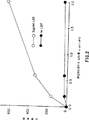
図1はLBPがELPSとMOとの相互作用を高めることを示す。単層MOを種々の投与量のLBPの存在下で、E又はELPloと共にインキュベートし、付着指数を記録した。対照急性期タンパク質、即ちマンノース結合タンパク質(MBP)(5μg/ml)はELPSloの結合を高めなかった(付着指数4.9)。結果は4つの別々の実験をまとめたものである。
図2はLBPに依存するELPSのMOへの結合が、E膜中のLPS密度に依存することを示す。ELPSはLPSと投与量を変化させることによって調製し、そして5μg/mlのLBPの存在下又は不存在下で単層MOと共にインキュベートした。結果は4つの別々の実験をまとめたものである。
図3はLPSの不存在下ではMOがLBPを認識しないことを示す。ビオチン及びストレプタビジン単独で被覆されたE(EBAV)をビオチン化したLBPと共にインキュベートし、ELBPを得た。ELBP及びEBAVどちらも、共にインキュベートするLPSの投与量を傾斜させて、37℃で20分間インキュベートし、洗浄して、単層MOへの結合を測定した。
図4はLBPがFc−仲介食作用を高めることを示す。単層MO(5日間培養)を様々な濃度の抗E-IgG存在下で、E、ELBP、又はEC3biと共に45分間インキュベートした。Eの食作用は”材料と方法”に記載の方法で測定した。ELPSlo(0.3μg LPS/3×10E)とMOを共にインキュベートしている途中で1μg/mlのLBPを加えることにより、ELBPを得た。抗E-IgG不存在下でのこれらEの付着は次のようであった。E、付着指数(AI)−0;EC3bi、AI−417;ELBP、AI−404。結果は6つの別々の実験をまとめたものである。
図5はリガンド被覆表面上でのMOの拡張中における過酸化水素の分泌を示す。3×10⁴のMOを被覆微量滴定ウエルに加え、過酸化水素の放出を時間区画ごとに測定した。過酸化物の活発な生成が、免疫複合体上での拡張中に(HSA−抗HSA、黒丸印)又は可溶性作用薬であるPMAに応答して(黒ダイヤ印)起こった。LPS被覆表面との相互作用において、低度であるが再生可能な過酸化物放出が見られた(白三角印)。しかし、LBP被覆表面上での拡張では放出はなく(白四角印)、LPS被覆表面をLBPで被覆すると、LPSに誘発される過酸化物の発生が妨げられた(白ダイヤ印)。LBP被覆ウエル中のMOがPMAへの応答として普通の過酸化物放出を見せることから、LBPは過酸化物の生成又は測定を害さなかった。
図6はモノクローナル抗CD14抗体によるLPS−LBP複合体結合の阻害を示す。単層ヒトMOを示された濃度のモノクローナル抗体と共に0℃で15分間インキュベートした。LPS、LBPの順で被覆された赤血球を加え、付着を測定した。結果は3つの別々の投与応答実験をまとめたものであり、固定濃度の抗体について行われた10の実験をまとめたものである。マクロファージ上の他の測定物に対して行ったmAbの、大パネルの高濃度における実験は結合ELBPに対して何の効果も示さなかった。
図7は表面結合抗CD−14mABがLBP−LPS複合体の結合を抑えることを示す。単層ヒトマクロファージを25μg/mlの示されたモノクローナル抗体で被覆した基質上に設置した。その細胞を洗浄し、ELPSloを加え、付着を測定した。
図8はLPSがTNF生成を誘発するには天然LBPが必要であることを示す。ウサギ腹膜浸出マクロファージ(PEM)を、示された濃度の天然LVP(LBP)、熱したLBP、ウシ血清アルブミン(BSA)又はウシ胎児血清(FCS)の存在下でLPSと共にチャレンジした。そして、前記PEMにより生成したTNFの量を測定した。
図9はLBPが結合するリガンド、即ちRe595 LPSの存在下又は不存在下でのトリプシン消化に対するLBPの感受性を示す。分子量マーカー(Pharmacia,Piscataway,N.J.;カタログ番号17-0446-01;ホスホリラーゼB 94キロダルトン(KD)、ウシ血清アルブミン 67kD、オバルブミン 43kD、カルボニックアンヒドラーゼ 30kD、大豆トリプシン阻害剤 20.1kD、及びαラクトアルブミン 14.4kD)はLBP含有物の隣のレーンに現れる。LBPがLPSに結合するとLBPの立体配座が変化し、よってLPS−LBP複合体の1部として存在するときだけLBPはCD14に結合できるという現象を説明できる、ということが結果から示唆される。
図10Aは、実施例21の記載に従って調製したウサギ抗マウスIgGで着色したホスホイノシトール特異性ホスホリパーゼCで処理したマウス細胞の上澄み液のウェスタンブロットである。RAWはマウスマクロファージRAW細胞系264.7;J774はマウスマクロファージ細胞系J774;LR9はJ774変異体LR9細胞;L929はマウス繊維芽細胞L929細胞;である。
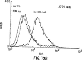
図10B及び10Cはそれぞれ、FITCヤギ抗ウサギ二次抗体を用いて、ウサギ抗マウスCD14IgG抗体由来のF(ab')2断片又は無免疫ウサギ由来の対照F(ab')2断片と免疫反応させた、J774細胞及びLR9細胞のFACS分析の結果を示すグラフである。
図11AはE.coli 0111:B4 LPSで刺激したマウスマクロファージ細胞系J774による亜硝酸塩生成を示すグラフである。LPSのみの場合は白丸印;LPSに0.25mg/ml抗マウスCD14IgGを加えた場合は黒三角印;で示す。
図11BはE.coli 0111:B4 LPSで刺激したマウスマクロファージ細胞系J774変異体LR9細胞による亜硝酸塩生成を示すグラフである。LPSのみの場合は白丸印;LPSに0.25mg/ml抗マウスCD14IgGを加えた場合に黒三角印;で示す。
図11CはB.subtilis細胞壁で刺激したマウスマクロファージ細胞系J774による亜硝酸塩生成を示すグラフである。細胞壁のみの場合は白丸印;細胞壁に0.25mg/ml抗マウスCD14IgGを加えた場合は黒三角印;で示す。
図11DはB.subtilis細胞壁で刺激したマウスマクロファージ細胞系J774変異体LR9細胞による亜硝酸塩生成を示すグラフである。細胞壁のみの場合は白丸印;細胞壁に0.25mg/ml抗マウスCD14IgGを加えた場合は黒三角印;で示す。
図12AはE.coli 0111:B4 LPSへの応答としての、C3H/FeJマウス系統由来のマウス腹膜顕在化マクロファージによる亜硝酸塩生成を示すグラフである。LPSのみの場合は白丸印;LPSに0.25mg/ml抗マウスCD14IgGを加えた場合は黒三角印;で示す。
図12BはE.coli 0111:B4 LPSへの応答としての、C3H/HeJマウス系統由来のマウス腹膜顕在化マクロファージによる亜硝酸塩生成を示すグラフである。LPSのみの場合は白丸印;LPSに0.25mg/ml抗マウスCD14IgGを加えた場合は黒三角印;で示す。
図12CはB.subtilis細胞壁への応答としての、C3H/FeJマウス系統由来のマウス腹膜顕在化マクロファージによる亜硝酸塩生成を示すグラフである。細胞壁のみの場合は白丸印;細胞壁に0.25mg/ml抗マウスCD14IgGを加えた場合は黒三角印;で示す。
図12DはB.subtilis細胞壁への応答としての、C3H/HeJマウス系統由来のマウス腹膜顕在化マクロファージによる亜硝酸塩生成を示すグラフである。作用薬のみの場合は白丸印;作用薬に0.25mg/ml抗マウスCD14IgGを加えた場合は黒三角印;で示す。
図12EはS.aureus細胞壁への応答としての、C3H/FeJマウス系統由来のマウス腹膜顕在化マクロファージによる亜硝酸塩生成を示すグラフである。作用薬のみの場合は白丸印;作用薬に0.25mg/ml抗マウスCD14IgGを加えた場合は黒三角印;で示す。
図12FはS.aureus細胞壁への応答としての、C3H/HeJマウス系統由来のマウス腹膜顕在化マクロファージによる亜硝酸塩生成を示すグラフである。作用薬のみの場合は白丸印;作用薬に0.25mg/ml抗マウスCD14IgGを加えた場合は黒三角印;で示す。
図13A、13B、及び13Cは、E.coli 0111:B4 LPS(パネルA)、B.subtilis細胞壁(パネルB)、及びマイコバクテリアのリポアラビノマンナン(LAM、パネルC)への応答としての、1,25ジヒドロ−ビタミンD3−分化THP-1細胞によるIL-8分泌を示すグラフである。加えた抗体:抗体無しの場合は小点付き四角印;0.25mg/mlヤギ抗ヒトCD14IgGの場合は白丸印;0.25mg/ml無免疫ヤギIgGの場合は黒丸印;0.25mg/mlヤギ抗ヒトCD14F(ab')2IgG断片の場合は白四角印;0.25mg/ml無免疫ヤギF(ab&aos;)2IgG断片の場合は黒四角印;10μg/ml抗CD14 mAb 28C5の場合は白三角印;10μg/ml抗CD14 mAb 63D3の場合は黒三角印;である。
図14A、14B、及び14Cは、E.coli 0111:B4 LPS(パネルA)、B.subtilis細胞壁(パネルB)、及びマイコバクテリアのリポアラビノマンナン(LAM、パネルC)への応答としての、マウスpre-B 70Z/3細胞による表面IgMアップレギュレーション(不定ユニット何で平均チャンネル蛍光として発現した)のFACS分析の結果を示すグラフである。ベクター移入細胞は白丸印;野性型CD14移入細胞は黒丸印;トランスメンブランキメラCD14移入細胞は黒三角印;である。
図15Aは、LPSへの応答としての、SW620細胞によるIL-8分泌のグラフである。作用薬のみの場合は白丸印;作用薬に2%正常とヒト血清(NHS)を加えた場合は白四角印;作用薬に2%NHSと0.25mg/mlヤギ抗ヒトCD14IgG抗体を加えた場合は白三角印;である。
図15Bは、B.subtilis細胞壁への応答としての、SW620細胞によるIL-8分泌のグラフである。作用薬のみの場合は白丸印;作用薬に2%正常ヒト血清(NHS)を加えた場合は白四角印;作用薬に2%NHSと0.25mg/mlヤギ抗ヒトCD14IgG抗体を加えた場合は白三角印;である。
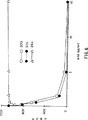
図16Aは35SsCD14のグラム陽性毒素原性細胞壁成分への結合を示すグラフである。
図16Bは、FITC-Re595-LPSの可溶性CD14への結合に対する、LAMによる競合を示すグラフである。LPSに対して重量/重量で0倍(上方の曲線)、50倍又は250倍(下方の曲線)のLAMを、FITC-LPS、LBP、及び可溶性CD14の混合物に加えた。FITC-Re595-LPSの蛍光における変化は、SLM6000蛍光計で時間を追って記録した。
発明の詳細な説明
A.定義
アミノ酸残基:本明細書で記載するアミノ酸残基は「L」異性体形であるのが好ましい。しかしながら、ポリペプチドによる免疫グロブリン結合性の所望の機能的性質が保持される限り、L−アミノ酸残基に代えて「D」異性体形の残基も用いうる。NH2は、ポリペプチドのアミノ末端に存在する遊離のアミノ基をいい、COOHはポリペプチドのカルボキシ末端に存在する遊離のカルボキシ基をいう。標準のポリペプチド命名法(J. Biol. Chem., 243:3522-59, 1969)に従い、アミノ酸残基の略語を以下の対応表に示す。

種々の文法形における「抗体」の語は、免疫グロブリン分子及び/又は免疫グロブリン分子の免疫学的活性部分、すなわち抗体断片又は抗体結合部位又はパラトープを含む分子、を含む組成物をいう。好ましい態様では、本発明で使用する抗体はアフィニティ精製されている。
「抗体結合部位」とは、抗原と特異的に結合するH鎖及びL鎖可変領域及び超可変領域を含む抗体分子の構造部分をいう。
本明細書で種々の文法形で用いる「抗体分子」の用語は、完全な免疫グロブリン分子及び免疫グロブリン分子の免疫学的活性部分を意味する。
抗体分子の例としては、完全な免疫グロブリン分子、実質的に完全な免疫グロブリン分子、及び当業者にFab、Fab’、F(ab’)2及びF(v)として知られている部分を含む、パラトープを含む免疫グロブリン分子の部分があり、これらの部分は本明細書で記載する治療法で使用するのに好ましい。
抗体分子のFab及びF(ab’)2部分は、公知の方法により実質的に完全な抗体分子をそれぞれパパイン及びペプシンによるタンパク質分解反応によって調製することができる。例えば、Theofilopolousらに付与された米国特許第4,342,566号(本明細書で引用する文献の開示を参考としてここに組み入れる)を参照されたい。Fab’抗体分子部分も公知であり、2つのH鎖部分をつなぐジスルフィド結合をメルカプトエタノールなどで還元し、次いで得られるタンパク質メルカプタンをヨードアセタミドなどの試薬でアルキル化することによりF(ab’)2から調製できる。完全な抗体分子を含む抗体が好ましく、ここではこれを説明として用いる。
種々の文法形における「モノクローナル抗体」の用語は、特定抗原と免疫反応することのできる抗体結合部位を1種だけもつ抗体をいう。従って、モノクローナル抗体は典型的にはそれが免疫反応する抗原に対して1つの結合親和性を示す。従ってモノクローナル抗体は、例えば二特異的(キメラ)モノクローナル抗体のように、複数の抗体結合部位をもつ抗体分子を含み、そのそれぞれが異なる抗原と免疫特異的である。
本明細書で用いる「実質的に同時に」の用語は、同時の結果を生じるのに十分な期間内を意味し、例えば抗生物質投与の結果としての細菌溶解、及び本明細書で記載する抗CD14抗体、抗LBP抗体、LBPペプチド類似体又はそのサブコンビネーション(subcombination)又はコンビネーションの投与によるその溶解の結果として起こる敗血症の症状改善又は予防をいう。
「製剤学的に許容される」の用語は、生理学的に我慢でき、ヒトに投与したときに典型的にはアレルギー又は同様の好ましくない反応、例えば消化管不良、眠気などを引き起こさない分子及び組成物をいう。
B.治療法
本発明は、細菌の感染と関連する敗血症などの疾患、特にTNFの血中濃度の一時的増加と関連する疾患の1以上の症状、例えば発熱、低血圧、好中球減少症、白血球減少症、血小板減少症、ショック及び多臓器不全などの症状を治療及び/又は予防する方法を企図する。このような治療を必要とする患者には、グラム陽性、グラム陰性細菌又はマイコバクテリア感染による内毒血症などの毒血症、ヘビ毒中毒、肝臓障害などの危険があるかこれに苦しむ患者を含む。さらに、ウイルス又は真菌感染患者のある者は毒血症の症状を示し、本発明の治療法の恩恵を受けるであろう。本発明の恩恵を特に受ける患者は、大腸菌(E. coli)、B型インフルエンザ菌(Haemophilus influenza B)、髄膜炎菌(Neisseria meningitides)、ブドウ球菌(staphylococci)又は肺炎球菌(pneumococci)による感染に苦しむ患者である。敗血症の危険のある患者には、火傷、銃撃傷、化学品の中毒又は乱用による腎臓又は肝臓障害などに苦しむ者を含む。
従って、ある態様では、本発明は、LPS、グラム陽性細菌毒素原性細胞壁成分又はサイトカイン分泌を誘導するマイコバクテリアからのLAMなどの細胞毒素にさらされることから生じる敗血症又はその他の症状の危険があるか、あるいはこれに苦しむ患者に、抗CD14抗体を好ましく静脈内投与することによって、そのような症状の1以上を改善する方法を企図する。
従って、本発明の1態様では、サイトカインの分泌をブロックする治療的有効量の抗CD14抗体を投与することを含んでなる、グラム陽性細菌及びマイコバクテリアによって引き起こされる菌血症及び毒血症と関連する症状の治療法を提供する。この方法は、単独で、あるいは1以上の抗生物質、ステロイド、抗TNF抗体、TNFアンタゴニストなどによる治療を含む敗血症及び毒血症を予防又は改善することが知られている他の治療様式を実質的に同時に投与することと組み合わせて実施できる。
さらに本発明では、グラム陰性細菌、グラム陽性細菌及びマイコバクテリア(グラム陽性細菌の抗酸型)によって引き起こされる菌血症、敗血症及びその他の毒血症などの感染症の症状を予防又は改善するのに有用な、典型的には単位投与形での治療組成物を企図する。組成物は、活性成分としてサイトカインの産生を阻害する1以上の抗CD14抗体、又はその断片を含む製剤学的に許容される担体を含んでいる。好ましい態様では、本発明の治療組成物は活性成分として、抗性物質、ステロイド、抗TNF抗体、TNFアンタゴニスト、可溶性CD14などの細菌症状及び敗血症を予防又は改善することが知られている薬剤を単独又はサブコンビネーション又はコンビネーションで含む。
「治療的有効量」の用語は、血漿中のTNF濃度の臨床的に有意な上昇を防ぐ、そして好ましくは少なくとも約30%、より好ましくは約50%、最も好ましくは約90%低下するのに十分な量を本明細書中ではいう。本明細書で活性成分として使用する薬剤の好ましい治療的有効量はC節で記載するものを含む。血漿中のTNF濃度の臨床的に有意な上昇とは、少なくとも約25pg/mlに上昇することである。血漿TNF濃度の測定法は当業界で公知であり、特に好ましい方法は本明細書に記載するものである。
正常な健常人又は健常実験動物におけるTNF濃度は、TNFに対して最も感度のよいアッセイでの検出限界値である約10pg/mlよりも多くないと推定される(Michie, et al., New Eng. J. Med., 318:1481-1486, 1988; Mathison, et al., J. Clin. Invest., 81:1925, 1988; Waage, et al., Lancet. 1:355-357,1987)。LPSに接触させた後、TNF濃度は400pg/mlまで10−20倍上昇した(前出参照)。最近になって、血清TNF濃度と,LPS含有性髄膜炎菌(meningococcal bacteria)であるグラム陽性細菌の感染による致命的結果との良好な相関が観察された(Waage, et al.,前出, 1987)。さらに類人猿の種を用いた敗血症動物モデルで同様のTNF増加が見られ、これらの変化は直接、致死率と相関する(Tracey, et al., Nature, 330:662-664, 1987)。
別の態様では、この方法は、治療的有効量の抗CD14抗体、好ましくは単球/マクロファージ系列、好ましくは単球由来のマクロファージの細胞などの細胞によりin vivoでのLPS、グラム陽性毒素原性細胞壁成分又はマイコバクテリアのLAMなどによって誘導されるTNF分泌を阻害するのに十分な量を、敗血症の治療を必要とするか、又はその危険性がある患者に投与することを含む。
好ましくは、本発明の治療法で用いる抗CD14抗体はアフィニティ精製されたポリクローナル抗体である。より好ましくは、抗体はモノクローナル抗体(mAb)である。さらに、本発明で用いる抗CD14抗体分子にとっては、全抗体分子のFab,Fab’,F(ab’)2又はF(v)部分の形であることが好ましい。
本発明を実施するために用いる好ましいモノクローナル抗体はAshmanら(Blood, 69:886-892, 1987)の記載する60bなどのハイブリドーマから生産されるものであり、最も好ましいのはVan Voorhisら(J. Exp. Med., 158:126-145, 1983)の記載する3C10(アメリカン・タイプ・カルチャー・コレクション、Rockville, MDに受託番号TIB22Bで寄託)から生産されるものである。mAb60b及び3C10はハイブリドーマ培養によって生産されるが、本発明はこれによって限定されない。60b及び/又は3C10のようなハイブリドーマからクローニングされた核酸発現する抗CD14免疫グロブリンによって産生されるmAbも使用できる。即ち、ハイブリドーマ3C10などによって分泌される抗CD14抗体分子を発現する核酸を別の細胞系に移して形質転換体を生産することができる。形質転換体はもとのハイブリドーマとは遺伝型的に異なるが、もとのハイブリドーマによって分泌されるものに対応する、全抗体分子の免疫学的に活性な断片を含む抗CD14抗体分子を産生できる。例えば、Readingに付与された米国特許第4,642,334号;Robinsonに付与されたPCT出願公開公報WO890099;Winterらのヨーロッパ特許出願公開第0239400号及びCabillyらの同第0125023号を参照されたい。
好ましいモノクローナル抗体は上述のハイブリドーマによって産生されるものと同様のCD14への免疫反応性を示す。本明細書では、種々の文法形における「免疫反応性」の用語は、一定量の抗体と,LPS、グラム陽性毒素原性細胞壁成分由来又はマイコバクテリアのLAM由来の一定量のCD14抗原との間の免疫反応を50%阻害するのに必要な抗原濃度をいう。即ち、免疫反応性とは、B/B0値0.5を達成するのに必要な抗原成分又は毒素原性成分の濃度であり、ここでB0は競合抗原の不在下において結合する抗体の最大量であり、Bは競合抗原の存在下において結合する抗体量であり、そしてB0及びBはいずれもバックグラウンドを予め調整してある(Robard, Clin. Chem., 20:1255-1270, 1974参照)。
別の態様では、本発明の治療法は、治療的有効量の抗LBP抗体、好ましくはアフィニティ精製したポリクローナル抗体、より好ましくはmAbを投与することを含む。さらに、本発明で用いる抗LBP抗体にとっては、全抗体分子のFab,Fab’,F(ab’)2又はF(v)部分の形であることが好ましい。好ましくは、投与する抗LBP抗体の量は、敗血症の少なくとも1つの症状を示す患者のTNFの血中濃度におけるLBP−LPS複合体に誘導された臨床的に有意な上昇を、少なくとも約30%、好ましくは約80%低下させるのに十分な量である。先に議論したように、本発明の方法から恩恵を受けることのできる患者には、グラム陰性細菌感染の結果として内毒血症に苦しむ者を含む。LBPの単離方法及び抗LBP抗体の誘導方法は当業者に公知である。例えば、Tobiasら(J. Exp. Med. 164:777-793, 1986)を参照のこと。LBP−LPS複合体のCD14への結合を阻害し、それによってLBP−誘導のTNF分泌を阻害するための、抗LBP抗体の能力の決定方法及び最適化方法は当業者に公知である。例えば、抗LBP抗体は実施例16に記載のアッセイにおいて抗CD14抗体の代わりに使用できる。
本発明の実施に有用な好ましい抗LBP抗体は、LBPのペプチド類似体と免疫学的に交差反応する。”LBPペプチド類似体”とは、単球由来のマクロファージの表面上で発現するCD14へのLPS−LBP複合体の結合を競合的に阻害することのできるポリペプチドである。好ましいLBPペプチド類似体を表1に示す。
ミエローマ細胞系はリンパ球と同じ種に由来するものであることが好ましい。典型的には、129GIX+株のマウスが好ましい哺乳動物である。本発明で使用するのに適しているマウスミエローマは、ヒポキサンチン−アミノプテリン−チミジン(HAT)感受性の細胞系であるP3X63−Ag8.653及びSp2/0−Ag14を含み、これらはアメリカン・タイプ・カルチャー・コレクション、Rockville, MDからそれぞれCRL1580及びCRL1581という名称で入手できる。
典型的にはポリエチレングリコール(PEG)6000を用いて脾臓細胞をミエローマと融合する。融合したハイブリッドをHATに対する感受性によって選択する。本発明の実施に使用できるモノクローナル抗体を産生するハイブリドーマは、CD14又はLBPとの免疫反応能及び実施例16に記載する方法を用いるLPSで誘導したTNF分泌の阻害能によって同定する。
本発明の実施に使用できるモノクローナル抗体は、適当な抗原特異性の抗体分子を分泌するハイブリドーマを含む栄養培地を含んでなるモノクローナルハイブリドーマ培養を開始することによって産生できる。ハイブリドーマが培地中に抗体分子を分泌するのに十分な条件及び時間で培養物を維持する。次に抗体含有培地を回収する。さらに抗体分子を公知の技術を用いて単離することができる。
これらの組成物の調製に使用できる材料は当業界で公知で市販されており、合成培地、同系交配されたマウスなどを含む。合成培地の例としては、グルコース4.5gm/l、グルタミン20mM、及び20%ウシ胎児血清を補充したダルベッコの最少必須培地(DMEM;Dulbecco, et al., Virol., 8:396, 1959)が挙げられる。同系交配されたマウス株の例としてはBalb/cがある。
モノクローナル抗ポリペプチド抗体の産生法も当業界で公知である(Niman, et al., Proc. Natl. Acad. Sci. USA, 80:4949-4953, 1983参照)。典型的には、上述した抗CD14モノクローナル抗体の産生法における免疫原と同様に、1以上のLBPペプチド類似体を単独又は免疫原性担体と結合させて用いる。LBPペプチド類似体及びLBPとの免疫反応をするか、あるいはグラム陽性細菌細胞壁(毒素原性成分)のCD14結合性部分又はマイコバクテリア由来のLAMとの免疫反応をする抗体の産生能についてハイブリドーマをスクリーニングする。
適当な免疫交差反応を示すモノクローナル抗体による、LPS−LBP複合体のCD14への結合を阻害する能力は、実施例16のアッセイを用いて確認できる。
別の態様では、本発明の治療法は治療的有効量のLBPペプチド類似体、好ましくは表1に示す配列を有する類似体を投与することを含む。
敗血症の危険性又は症状を示す患者は、これらの症状を予防又は改善する当業界で公知の治療様式の恩恵を受けることができる。従って、本発明は、治療的有効量の抗CD14抗体、抗LBP抗体、LBPペプチド類似体、そのサブコンビネーション又はコンビネーションを、敗血症の症状を予防又は治療するために知られている治療様式と実質的に同時に投与することを企図する。例えば、抗TNF抗体及び/又はTNFアンタゴニストの使用などによって直接又は間接に敗血症におけるTNFの役割に介在することにより、敗血症の症状を予防又は改善できる。特に好ましくは、文献(Tracey, et al., Nature, 330:662-664, 1987)に記載のものに対応するTNFに免疫学的特異性をもつモノクローナル抗体のような抗TNF抗体を活性成分として用いる。
同様に、本発明の治療法はコルチゾル、ヒドロコルチゾンなどのステロイドで実質的に同時に治療することをさらに含む。
敗血症の症状を示す患者は通常抗生物質、典型的にはゲンタマイシンなどのアミノグリコシド又はペニシリン、セファロスポリンなどのβ−ラクタムで治療する。従って、好ましい治療法は、治療的有効量の抗CD14抗体、抗LBP抗体、LBPペプチド類似体、本明細書で記載するそのサブコンビネーション又はコンビネーションの投与を、殺菌量の抗生物質の投与と実質的に同時に行うことを含む。本明細書における「殺菌量の」の用語は、治療を受ける患者の殺菌性血液濃度を達成するのに十分な量という。ヒトに投与して安全と一般に認識される抗生物質の殺菌量は当業界で公知であり、抗生物質の種類及び治療しようとする細菌感染の型により異なることも当業界で公知である。
好ましい態様では、本明細書で記載する抗CD14抗体、抗LBP抗体、LBPペプチド類似体又はそのコンビネーションの投与は、抗生物質投与の約48時間以内、好ましくは約12−36時間以内、より好ましくは約2−8時間以内、そして最も好ましくは実質的に同時に行う。
本発明の実施に有用な抗生物質は、Physicians' Desk Reference, Huff, B.B.編集, Medical Economics Company, Inc., Oradell, N.J., 1989に記載の製剤をもつ抗生物質、殺菌剤及び防腐剤を含む。別の態様では、本発明は治療的有効量のCD14、好ましくはLPS−LBP複合体と結合するその可溶性部分を単独で、あるいは治療的有効量の抗TNF抗体、抗LBP抗体及び抗生物質とサブコンビネーション又はコンビネーションで投与することを企図する。CD14をコードするcDNA及びその推定アミノ酸残基配列は当業界で公知である。Goyert, et al., Science, 139:497-500, 1988; Ferrero, et al., Nuc. Acids Res., 16:4173, 1988; Bazil, et al., Eur. J. Immunol., 16:1583-1589, 1986を参照のこと。
C.治療用組成物
本発明はさらに本発明の治療法を実施するのに有用な治療用組成物を企図する。主題の治療用組成物には、製剤学的に許容される賦形剤(担体)、及び活性成分として本明細書で記載する1以上の抗CD14抗体、抗LBP抗体及びLBPポリペプチド類似体を混合物の形で含む。好ましい態様では、組成物はCD14へのLPS−LBP複合体の結合を阻害することのできる抗CD14mAbを含んでなる。好ましいmAbは60b、より好ましくは3C10である。グラム陽性細菌又はマイコバクテリアによって引き起こされるサイトカン産生と関連する敗血症又はその他の疾患状態を治療するための好ましいモノクローナル抗体は63D3 mAb (ATCC#HB44)及び28C5(ATCC#HB11364)である。
別の好ましい態様では、組成物はCD14へのLPS−LBP複合体の結合を阻害する抗LBP抗体、好ましくはmAbを含んでなる。特に好ましいのは、抗LBP抗体が、表1に示す配列をもつLBPペプチド類似体と免疫反応するような組成物である。
好ましい組成物は、CD14へのLPS−LBP複合体の結合のアンタゴニストとして作用するLBPペプチド類似体を含んでなる。本発明の組成物に用いる好ましいLBPペプチド類似体は表1に示す配列をもつものである。
好ましい治療用組成物には、有効量の1以上の以下の活性成分をさらに含む:抗性物質、ステロイド、及び抗TNF抗体及びTNFアンタゴニスト。製剤例を以下に示す:
活性成分としてポリペプチド又は抗体分子を含む治療用組成物の調製は当業界でよく知られている。典型的には、このような組成物は注射可能な溶液又は懸濁液として調製されるが、注射前に溶液又は懸濁液とするのに適した固形も調製できる。調製物は乳化されていてもよい。活性治療用成分は製剤学的に許容され、かつ活性成分と適合性の賦形剤と混合することが多い。適当な賦形剤には例えば、水、生理食塩水、デキストロース、グリセロール、エタノールなど並びにそのコンビネーションがある。さらに、所望するならば、組成物には湿潤剤又は乳化剤、活性成分の有効性を増強するpH緩衝剤などの補助物質を少量含むことができる。
ポリペプチド又は抗体は中和された製剤学的に許容される塩の形で治療用組成物中に製剤化することができる。製剤学的に許容される塩には、酸付加塩(ポリペプチド又は抗体分子の遊離アミノ基と形成される)が含まれ、これらは例えば酸塩又はリン酸などの無機酸、あるいは酢酸、シュウ酸、酒石酒、マンデル酒などの有機酸とで形成される。遊離のカルボキシル基から形成される塩も、例えば水酸化ナトリウム、水酸化カリウム、水酸化アンモニウム、水酸化カルシウム又は水酸化鉄などの無機塩基、ならびにイソプロピルアミン、トリメチルアミン、2−エチルアミノエタノール、ヒスチジン、プロカインなどの有機塩基から誘導できる。
治療用ポリペプチド又は抗体を含む組成物は、例えば単位用量を注射することによって静脈内投与するのが便利である。本発明の治療用組成物について本明細書で用いる「単位用量」の用語は、それぞれの単位が所望の治療的効果を生じるように必要な希釈剤、すなわち担体又はベヒクルと関連する予め定められた量の活性物質を含む、ヒトに1回の投与を行うのに適した物理的に分かれた単位をいう。
組成物は投与製剤と適合した方法、及び治療的に有効な量で投与する。投与量は治療すべき被験者、被験者の活性成分を用いる免疫系の能力、及び所望するCD14又はLPS−LBP複合体結合能の阻害又は中和程度に依存する。投与に必要な活性成分の正確な量は医師の判断により、個々の場合によって異なる。しかしながら、適当な投与量は、1日当たり体重1kgにつき活性成分0.1−20mg、好ましくは約0.5−約10mg、より好ましくは1−数mgであり、投与経路にも依存する。最初の投与と追加免疫に適した処方もまた変更しうるものであるが、最初の投与を行った後1−数時間の間隔で追加免疫を繰り返すか別の投与を行うのが一般的である。あるいは、10ナノモルから10マイクロモルの血中濃度を維持するのに十分な静脈内連続注入でもよい。
本明細書では「pg」はピコグラム、「ng」はナノグラム、「ug」はマイクログラム、「mg」はミリグラム、「ul」はマイクロリットル、「ml」はミリリットル、「l」はリットルを意味する。
抗マウスCD14抗体は細胞活性化をブロックする
抗マウスCD14抗体がLPSで誘導した細胞活性化をブロックするかどうかを確証するために、まずマウス単球/マクロファージの活性化に対するその効果を測定した。ウサギ抗マウスCD14のポリクローナルIgG調製物は、RAW細胞及びマウス血中におけるLPSで誘導したTNF産生を阻害したが、免疫しなかったウサギからのIgGには何ら効果を示さなかった(データ示さず)。本明細書では示さない実験において、RAW細胞及びJ774細胞によるLPS依存性の亜硝酸塩産生も抗マウスCD14IgGによってブロックされた。抗CD14抗体は同じ細胞中でTNFで誘導した亜硝酸塩の産生に何ら効果を示さず、これは抗CD14抗体による阻害の特異性を示す(データ示さず)。抗マウスCD14IgGのF(ab’)2IgG断片な完全な抗体と同様に阻害し、これはFcドメインが細胞との相互作用に何ら関与していないことを示す(データ示さず)。
図10に示す結果は、ポリクローナル抗マウスCD14抗体が天然のマウスCD14を認識し、CD14を経由して起こるLPSで誘導される細胞活性化をブロックすることを示す(データ示さず)。これらの知見に基づき、CD14がグラム陽性細菌細胞壁及びLAMに対するマウスマクロファージの応答にある役割を果たしているという仮説を試験するためにさらに実験を行った。
図11に示すように、抗マウスCD14ポリクローナル抗体が、J774細胞中におけるLPS依存性又はB.サブチリス細胞壁依存性の亜硝酸塩の産生を阻害することが見いだされた。LR9細胞はLPS又は図11Bに示すようにグラム陽性細菌細胞壁のいずれかによる刺激に対して顕著に低応答性であった。LPS濃度を3ng/mlまで、又は細胞壁濃度を1000ng/mlまで増加してもこれらの細胞中における亜硝酸塩の産生を誘導しなかったが、この実験条件で抗CD14抗体を投与しても応答が低下しなかった(図11)。A群のストレプトコッカス(Streptococcus)株由来の細胞壁でも同様のデータが得られた(データ示さず)。図12に示すように、C3H/FeJ(LPS応答性マウス)由来のPEMはLPS及びB.サブチリス又はS.アウレウス由来の細胞壁調製物に応答し;抗マウスCD14ポリクローナル抗体はLPS及び細胞壁のいずれに対する応答も阻害した。C3H/HeJマウス(LPSに非応答性であることが知られている株)由来のPEMがLPSに応答しないのは驚くことではないが、2つの異なるグラム陽性微生物由来の細胞壁調製物で処理した後に亜硝酸塩を産生した(図12)。細胞壁調製物に対する濃度応答特性は2つの異なるマウス株からのPEMの場合と非常によく似ていた。B群のストレプトコッカスからの別の細胞壁調製物もPEMにおける亜硝酸塩の産生を刺激した(データ示さず)。
全く予期しないことに、図12に示すように、C3H/HeJ PEMによる細胞壁で誘導した亜硝酸塩の産生が抗CD14ポリクローナル抗体によって阻害されることが観察された。さらに、C3H/HeJマウス由来のマクロファージであるGG2EE細胞(Blasi, et al., 1987)はLAMによって刺激されて亜硝酸塩応答を生じ、これは抗マウスCD14IgGによってブロックされた(データを示さず)。
グラム陽性細胞壁調製物又はLPSによる刺激に対するCD14非依存性の経路も働いていることが見いだされた。なぜなら、刺激する濃度を増加することによって抗CD14の阻害効果が常に弱まってしまうからである。それでも、図11及び図12に示す知見は全体として、LPS及びグラム陽性細胞壁の両方に対する応答にCD14が重要な役割を果たしており、抗CD14抗体により治療的介入によってグラム陰性及びグラム陽性細菌の哺乳動物における毒性効果を調節及び改善できることを支持する。
CD14陽性及びCD14陰性細胞系における応答
J774(CD14陽性)及びL9(CD14陰性)細胞系で得られた結果は、グラム陽性細胞壁に対する応答にCD14が重要な役割を果たすことを示唆するが、これらのデータは注意深く解釈しなければならない。LR9細胞系は化学的に突然変異誘発したJ774細胞から選択したので、LR9細胞はCD14を欠失することを本発明者らは示してきたが、LPS低応答性の完全な理由は分っていない。従って、1,25−ジヒドロキシビタミンD3で処理した後に高濃度でCD14を発現するTHP−1細胞(Tobias, et al., J. Immunol., 150:3011-3021, 1993)、又はヒトCD14を発現する形質転換した70Z/3細胞(Lee, et al., Proc. Natl. Acad. Sci. USA, 90:9930-9934, 1993)のいずれかを用いて、追加の実験を行った。THP−1細胞をLPS又はグラム陽性毒素原性細胞壁成分で刺激してIL−8を放出させるにはビタミンD3で前処理する必要があった(データ示さず)。
これらの細胞におけるCD14発現の役割を決定するために、種々の抗ヒトCD14抗体の細胞活性化をブロックする能力を試験した。まずLPS、グラム陽性細胞壁調製物、及びLAMによるTHP−1活性化に及ぼす、ヒトCD14に対するポリクローナル及びモノクローナル抗体の効果を比較した。図13に示すように、ポリクローナル抗hCD14抗体(IgG画分又はF(ab’)2断片)はLPSで、グラム陽性細胞壁で、及びLAMで誘導したIL−8放出をブロックしたが、一方非免疫IgG又はそのF(ab’)2断片は何も効果を示さなかった。ヒトCD14に対するモノクローナル抗体である63D3(ATCC#HB44)及び28C5(ATCC#HB11364)を用いて、LPS又はグラム陽性細胞壁調製物を加える前に細胞を前処理した。
図13に示すように、mAB 28C5はLPS、細胞壁調製物、及びLAMに対する応答をブロックした。これとは対照的に、mAB 63D3はLPS刺激を阻害しなかったが、細胞壁調製物及びLAMによる刺激を部分的にブロックした。1−3pg/ml濃度でのB.サブチリス細胞壁又はLAMによるTHP−1細胞の活性化は、ポリクローナル抗hCD14抗体によってブロックされなかった(図13)。アゴニスト濃度がナノグラム/ml範囲にあるときには、THP−1細胞の活性化はCD14依存性であることが観察された。別の研究では、S.ニューモニアエ(pneumoniae)からの細胞壁調製物による刺激もCD14を必要とした(データ示さず)が、S.アウレウスからの可溶性ペプチドグリカンによるTHP−1細胞の刺激は、抗CD14ポリクローナルIgGを含めることによってブロックされなかった(データ示さず)。
マウスプレ−B細胞系である70Z/3のLPS応答におけるCD14発現の効果が報告されている(Lee, et al., J. Exp. Med. 174:1697-1705, 1992; Lee, et al.,前出, 1993)。CD14で形質転換した70Z/3細胞(70Z/3−hCD14細胞)は、LPS結合及び初期シグナル伝達事象に関してはマクロファージと同様に挙動する(Lee, et al.,前出, 1993)。hCD14形質転換細胞と空ベクターで形質転換した細胞との唯一の違いはヒトCD14の発現であるので、こらの細胞を用いてCD14媒介性の事象の役割をさらに決定的に分析することができる。図14は、70Z/3−hCD14細胞をLPS、B.サブチリス細胞壁又はLAMのいずれかとインキュベートする実験の結果を示す。hCD14が細胞表面上に発現しているときには、LPSと同様に、細胞壁及びLAMはIgMの上方制御に有意な増加を誘導し、これはこれらの異なるアゴニストの応答にCD14が決定的に関与していることを示す。LPSについて既に報告されているように(Lee, et al.,前出, 1993)、グラム陽性細胞壁及びLAMによる細胞活性化は、CD14がGPI−付着性タンパク質として発現しているか、又は膜貫通タンパク質として発現しているかとは無関係であった。
次に、膜結合したCD14を欠失するが、可溶性CD14(sCD14)依存性経路を介してLPSと応答することが知られている細胞系が、sCD14依存性メカニズムを介してグラム陽性細菌細胞壁によって活性化されるかを決定するための研究を行った。Puginらによる先の研究(前出、1993a)では、慢性腺癌に由来するSW620細胞などの細胞系の活性化におけるsCD14の重要性を指摘している。SW620細胞をB.サブチリス細胞壁で刺激したところ、この応答には血清の存在を必要とした。図15に示すように、ウサギ抗マウスCD14ポリクローナル抗体は、LPSで刺激した活性化システムで観察されたのと同程度にこれらの細胞によるIL−8放出をブロックした。本明細書では示さない研究において、グラム陽性細菌細胞壁調製物が、LPSで観察されたのと同程度にヒト内皮細胞活性化を誘導することも見いだされた。これらの結果は、可溶性CD14が、ヒト非CD14保持細胞のグラム陽性細胞壁依存性の活性化を媒介することを示す。
グラム陽性細胞壁又はリポアラビノマンナンのCD14への結合
CD14と細菌の細胞エンベロープ成分との間の直接的相互作用を示す生化学的証拠を2つの独立した実験によって提供する。グラム陽性細胞壁へのsCD14の結合を35S−sCD14を用いて得た。図16に示すように、35S−sCD14は細胞壁に結合し、その結合は過剰の未標識sCD14の存在によって阻害され、また図16Aに示すように35S−sCD14を100℃、5分加熱して変性することによって結合はなくなった。
FITC−ReS95/LPSとsCD14との間の相互作用をモニターするために分光蛍光アッセイを開発した。FITC−ReS95/LPSがsCD14と結合すると、蛍光強度の顕著な増加が観察され、これは迅速に数分間起きる(図16B、上の線)。反応混合物に過剰のLAMを添加すると、FITC−ReS95−LPSの蛍光強度の増加の顕著な抑制が観察された(図16B、中間及び下の線)。これらのデータはsCD14への結合に関してLAMとLPSが競合することを示す。
ここに示す研究に基づいて、骨髄受容体CD14がグラム陰性細菌由来のLPS、マイコバクテリアリポアラビノマンナン及びグラム陽性細胞壁の成分などの広範な種類の細菌エンベロープ分子を認識する分子として作用すると考えられる。CD14を介するこれらのアゴニストとマクロファージとの相互作用が細胞活性化に導く。分子認識のメカニズムに限定されることを望むものではないが、CD14は複数の微生物リガンド結合特異性をもつパターン認識受容体であると考えられる。
脊椎動物における感染性微生物に対する免疫応答は、2段階で起こり、最初は非適応性(先天性)免疫で、次いで特異的クローン防御の発展による適応性免疫が起こる。骨髄細胞は微生物に対する防御の非適応期(初期)で中心的役割を果たす。マクロファージによる感染性粒子の認識によって、非特異的防御の迅速な活性化が起こり、モノカイン(TNF、IL−1又はIL−6)、各種酵素、及び酸素及び窒素ラジカルが産生される。Janeway,前出, 1992が最近提案するように、非クローン性の免疫受容体が病原性微生物の共通の又は高度に保存された構成物を検出する可能性が極めて高い。進化の過程で広い認識性質をもつこのような受容体を選択してきたのであろう。同じ受容体を介する異なる微生物の表面構造の相互作用が先天性免疫に典型的な非特異的応答の引き金を引くのであろう。本明細書の実施例で示すような微生物構造に対する多特異性をもつCD14はこのような受容体の原型となる例である。哺乳動物細胞のその他の表面タンパク質は種々の細菌成分を認識する。スカベンジャー受容体ファミリーのメンバーは確かにこのような性質をもつことが示されている(Krieger, et al., J. Bio. Chem., 268:4569-4572, 1993)。しかしながら、CD14とは異なり、この群のタンパク質は細胞活性化に関与しないが、細胞外環境からリガンドを取り込む機能をするように思える。
多特異性の非適応性受容体によって認識される微生物構造は病原体の間で高度に保存されており、また微生物の完全性又は病原性にとって決定的であらねばならない。LPSはグラム陰性細菌群にとってこれらの基準を満たす。LPSはグラ陰性病原性にとって必要であり、高度に保存されており、CD14によって認識される。リポアラビノマンナン(LAM)もマイコバクテリアにおける保存された決定的病原性エンベロープ構造であり(Chatterjee, et al., Infect. Immun., 60:1249-1253, 1992)、CD14を介して細胞活性化の引き金となる。LPSとLAM構造には有意な類似がある。どちらの分子も両親媒性であり、一端に疎水性の脂質アシル鎖と、他端に親水性の多糖をもつ(Tobias, et al.,前出; Prinzis, et al., J. Gen. Microbiol., 139:2649-2658, 1993)。
グラム陽性細胞壁では、CD14を介するマクロファージ活性化を司る構造はまだ分かっていない。しかしながら、試験した全ての株由来の細胞壁がCD14依存的にマクロファージを活性化したので、この構造は種々のグラム陽性細菌で高度に保存されていると思われる。CD14と結合するグラム陽性細胞壁の主要なリガンドとなる候補には、ムロペプチドのモノマー又はオリゴマー、あるいはテイコ酸断片が含まれる。
CD14をもたない細胞、即ち内皮細胞及び上皮細胞が可溶性CD14依存性経路を介して広範な種々の微生物構造に応答することを本発明者らは発見した。これらの細胞はいったん活性化されると、組織中で白血球をやり取り(trafficking)するのに決定的であり、サイトカイン、酸素及び窒素ラジカルを分泌し、凝集を調節する。従ってこれらの細胞はマクロファージと協力して感染性微生物に対する免疫の初期の非特異的事象に関与するのかも知れない。内皮細胞及び一部の上皮細胞も有力な抗原提示細胞であり、免疫の適応性クローン期の開始にマクロファージ又は樹枝状細胞とともに関与するかもしれないことは興味深いことである(Hughes, et al., Immunol. Rev. 117:85-102, 1990)。
最近の研究(Heumann, et al.,前出)において、グラム陽性細胞壁産物によるヒト単球の活性化には血清が必要であることが示された(Heumann, et al.,前出)。この著者は、大量の(1−10μg/ml)グラム陽性細胞壁によって引き起こされた一次的ヒト単球の活性化を抗CD14mAb(MY4)がブロックしないことを見いだした。我々の研究では、低濃度のアゴニスト(300ng/ml及びそれ以下)の場合のみでCD14依存性が観察された。さらに、抗CD14mAbであるMY4はCD14へのグラム陽性細胞壁の結合にとって決定的でない機能的ドメインを認識する。これらの事実によりこれらの研究の明らかに矛盾した結果を説明することができるであろう。
C3H/HeJマウス由来のマクロファージを用いる実験は、各種のアゴニストがCD14を介する同様の細胞応答を誘導するという事実にもかかわらず、これらのアゴニストは同じ活性化経路を共有しないように思われる。C3H/HeJマクロファージは典型的にはLPSに耐性であるが、その他のアゴニスト、例えばLAM(Chatterjee, et al.,前出)又は完全加熱殺菌したグラム陽性細菌(Freudenberg and Galanos, Infect. Imuun., 59:2110-2115, 1991)で活性化されうる。重要なことは,C3H/HeJマクロファージがCD14依存的にLAM及びグラム陽性細胞壁と応答することを我々が見いだしたことである。CD14GのPIに付着した膜形が推定の膜貫通トランスデューサーを介する細胞内シグナル伝達を仲介することが以前に提案されている(Ulevitch and Tobias,前出)。本明細書で議論する実験の結果は、同じ推定のトランスデューサーが異なるアゴニストに対する異なるエピトープをもつか、あるいは細胞表面に異なるトランスデューサー分子が存在し、CD14によって提示される特異的アゴニストだけを認識するかのいずれかであることを示唆する。そうするとC3H/EleJマウスにおける非常に特異的なLPS欠失が多特異的シグナルトランスデューサーのLPS部位における突然変異によって、あるいはLPS特異的シグナルトランスデューサーの機能的欠失によって説明できる。
総括すると、CD14はその膜結合形(骨髄細胞)においてか、あるいはその可溶性形(内皮細胞及び上皮細胞)において、病原性細菌のエンベロープからの広範な種類の保存性分子に応答して細胞活性化を媒介することを我々は示す。我々は、CD14が病原性微生物に対する非適応性で非特異的な初期免疫応答のための受容体/媒介体の原型であることを提案する。CD14機能を調節する治療剤は多くの致死的細菌性疾患の治療及び/又は予防に大きな希望を与える。細菌性敗血症の場合、抗CD14抗体を用いてCD14機能をブロックすることにより、グラム陰性細菌及びグラム陽性細菌に対する有害かつ圧倒的な宿主の応答を防止することができる。
実施例
以下に実施例を開示するが、本発明はこれらに限定されるものではない。
実施例1〜11は、単球/マクロファージ系のヒト細胞が膜平面で移動可能な細胞表面受容体を介してLPS-LBP複合体に結合することを確立した実験である。実施例12は、抗CD14抗体がLPS-LBP複合体のCD14に対する結合を特異的に阻害できることを説明する。実施例13〜15は、CD14がLPS-LBP複合体に特異的に結合し、そしてその結合がMOからのTNF分泌を誘導するということを証明する。実施例16は、抗CD14mAbsが、ヒト血中でLPS-LBP複合体誘導TNF分泌を阻害するということを証明する。実施例17は、実施例1〜16の結果の概要と検討とを提供する。
1.試薬
LBPは、ウサギ急性期血清(Tobiasら、前出、1986)から精製し、銀染色ゲル上で均一であると思われた。抗−ウサギLBPはヤギ中で増加させた。MBPは、R.A.B.Ezekowitz博士(Boston, MA)より入手した。細菌/透過性−増殖因子(BPI)は、J.Gabay博士(New York, NY)より入手した。Salmonella minnesota(Re595または野性型)からのLPSはList Biological(Cambell, CA)から入手した。CD18に対するモノクローナル抗体(mAbs)IB4およびFcyRIIIに対するモノクローナル抗体(mAbs)3G8は、Wrightらの論文(Proc. Natl. Acad. Sci. USA, 80:5699-5703, 1983)に掲載されている。CR1に対するmAB 543はShhreiber博士(St. Louis, MO)より、FcyRIおよびFcyRIIに対するmAbs 22およびIV. 3は、Fanger博士(Hanover, NH)より入手した。パイロジェンフリーのヒト血清アルブミン(HSA)は、Armour Pharmaceuticalsより入手し、パイロジェンフリーのPBSとDGVB++はWhitaker MA Bioproductsから入手した。NHS−ビオチン、スルフォ−NHS−ビオチン、およびストレプトアビジンはPierce Chemicalから入手した。
2.表面
組織培養用プラスチック表面を、25μg/mLのタンパク質(抗体、LBP、またはHSA)、または1(μg/mL)/μg/mLのLPSで、20℃にて1時間、インキュベートした。免疫複合体を形成するために、HSA−被覆表面を、抗HSA抗血清(1:50)とともに、さらに30分間、インキュベートした。いくつかの場合には、その後、LPS被覆表面を10μg/mLのLBPとともに20℃にて30分間インキュベートした。過酸化水素産生のアッセイのために、すべての被覆表面を、貪食細胞を添加する前に1時間、1mg/mLのHSAと接触させた。被覆表面は、アッセイ前に注意深くパイロジェンフリーのPBSで洗浄した。
3.細胞
単球由来マクロファージ(MO)は、Wrightら(J. Exp. Med., 156:1149-1164, 1982)が記載したように、テフロンビーカー中で3〜10日間、精製したヒト単球を培養して得た。新鮮な単球の単層を、末梢血単核球をタンパク質被覆プラスチックに37℃で45分間付着させて得た。PMNは、Englishら(J. Immunol. Methods, 5:249, 1974))の方法によって、新鮮血から精製した。T細胞は、赤血球とのロゼット形成により精製したが、J. Ming(Rockfeller U.)より入手した。ヒト臍帯血内皮細胞単層(Loら、J. Exp. Med., 169:1779-1793, 1989)は、S.K.Lo博士(Rochfeller U.)より入手した。
ヒツジ赤血球(E)を、Wright(前出、1982)の記載のように、IgG(EIgG)またはIgM(EIgM)で被覆した。
C3biを、2〜10×108EIgMを10%のC5−欠乏ヒト血清(Sigma)1mL中で37℃にて30分間インキュベートすることによって、EIgM上に堆積(deposit)させた。ついで、赤血球を洗浄し、2.5mMのエチレンジアミン四酢酸塩(EDTA)を含む緩衝液中で、10分間、0℃にてインキュベートした。得られたEC3biは、MOとのEDTA−抵抗性ロゼット形成によってアッセイしたように、C3bを有していなかった。
Eを、Wrightら(J. Exp. Med., 164:1876-1888,1986)による記載に従って、LPSで被覆した。調製に使用したLPSの量は、ELPShi(1〜10μg/4×107E)またはELPSlo(0.2〜1μg/4×107E)を得るために変化させた。等容のELPSlo(108/mL)とLBP(10μg/mL)とで37℃で20分間インキュベートすることによって、ELPSloをLBPで被覆した。得られたLBP被覆ELPS(リガンド被覆E)を洗浄し、直ちに使用した。
幾つかの実験においては、Eはまた別の方法によってLBPで被覆された。最初に、5×10⁸Eを250μgのスルホ−NHS−ビオチンとともに0.1Mの炭酸ナトリウム液(pH 9.2)中にて5℃で20分間インキュベートすることによってEをビオチニル化し、そして、50μgのLBPを5μgスルホ−NHS−ビオチンとともにインキュベートしてLBPをビオチニル化し、PBSに対して透析した。次に、ビオチニル化タンパク質を、ストレプトアビジン架橋を介してビオチニル化されたEと連結した。108の、洗浄され、ビオチニル化されたE(EB)を10μgのストレプトアビジンとともに20℃にて30分間インキュベートし、アビジン被覆赤血球(EBAV)を得た。蛍光化したストレプトアビジンを用いた予備実験では、EBAVが均一で強い蛍光性を示し、凝集は見られなかったことが示された。2.5×10⁷の洗浄されたEBAVを、2.5μgのビオチニル化LBPとともに、30分間、20℃にてインキュベートし、EBAV-LBPを得た。
Salmonella typhimurium LT2 Ga/Eを、ガラクトースの存在下または非存在下で増殖させて、完全なLPSまたは末端切断型のLPSを有する細胞をそれぞれ得た(Wrightら、前出、1986)。対数増殖培養物を洗浄し、フルオレセインで標識し、そしてすでに記載したように(Wrightら、前出、1986)、PBS中で2×108/μLに調整した。
4.アッセイ
LPS−被覆赤血球の凝集(実施例3)は、丸底のマイクロテストプレートにおいて10μLの希釈LBP中に含まれる106のELPShiを21℃で30分間振盪して測定した。凝集は、沈降パターン(settling pattern)から読み取った。
リガンド被覆Eの(実施例3)MOに対する結合は、Wrightら、前出、1982に記載されたように測定した。すなわち、Terasakiの組織培養プレートをHSAまたは他のタンパク質(実施例2)で被覆し、そして5μLの細胞(3mMのグルコース、0.5mg/mLのHSA、および0.3u/mLのアプロチニン(Sigma)を含むPBS中0.5×106/mL)を37℃にて45分間インキュベーションして樹立した。リガンド被覆Eおよび示されたタンパク質を単層に添加した。Eを10分間0℃にて静置し、その後、プレートを37℃で15分間温めた。付着しなかったEを洗浄して除去し、接着したEを位相差顕微鏡で点数付けを行なった。フルオレセイン化Salmonellaの結合を、Wrightら、前出、1986に記載されたような、37℃にて15分間のインキュベーションを採用する同様の方法で評価した。結果を、100MOあたりのEの数または細菌数である付着指数として報告した。リガンド被覆Eの貪食は、EとMOとのインキュベーションを37℃にて45分間行うことを除いては同様の方法(Wrightら、前出、1986)によって測定し、そして、摂取されなかったEをウェルの点数付けの前に低張の培地に軽く接触させて溶解させた。
5.LBPは、赤血球膜に挿入されたLPSに結合する。
ELPShiに、できる限り少量の0.5μg/mLのLBPを添加すると、凝集が起こった。LPSはリン脂質との疎水的な相互作用によってEの膜中に分配する(partition)ため、この観察はLBPはマルチmerを形成するための能力を有するということを示唆する。ELPSは強くは凝集されず、穏やかなピペッティングで分裂され得る。
6.LBPは、ELPS及びSalmonellaのマクロファージに対する結合を増強する
グラム陰性菌およびLPS被覆赤血球は、白血球上の受容体のCD18複合体のメンバーとの相互作用を介してMOに結合する(Wrightら,前出、1986)。それゆえ、その相互作用を乱すためのLBPの能力を試験した。初期の研究では、高レベルのLPSで調製されたEを使用した。これらのELPShiはMOに貪欲に結合し、そしてLBPを添加すると結合がわずかに増強された。この増強の性質を試験するために、Eを低レベルのLPSで調製した。MOの単層を5μg/mLのLBPの存在下または非存在下でELPSloとインキュベートした。ELPSloにはMOはほとんど結合しないが、LBPを添加することにより結合が劇的に増強された(図1)。増強された結合は、1μg/mLのLBPで最大効果を示し、濃度依存的であった。この結果の特異性は、他の急性期反応物であるマンノース結合タンパク質が100μg/mLの高濃度でELPShiのMOに対する結合に影響しないこと;他のLPSの結合タンパク質であるBPIが10μg/mLの高濃度で結合に影響しないこと;そして、ポリクローナル抗LBP抗血清(1:200)がLBPによるELPSloのロゼット形成において20倍の減少を引き起こしたという観察によって示された。
ELPSとMOとの相互作用を増強するためのLBPの能力もまた、赤血球膜中のLPSの量に依存した(図2)。LBPは、ELPSとMOとの間の直接的な相互作用を持続させるために必要とされる量よりも20〜100倍少ないLPSで調製されたEの結合を、効果的に仲介することができた。
末端切断型のLPSを発現するグラム陰性菌株(R型株;(rough strain))にはMOが貪欲に結合するが、完全なLPSを有するS型株(smooth strain)にはほとんど結合しない(Wrightら、前出、1986)。LBPはS型およびR型のLPSのいずれにも均等によく結合するため(Tobiasら,前出, 1989)、LBPがS型のSalmonellaにオプソニン作用をするための能力を試験した。表IIに示したように、LBPを添加するとS型SalmonellaのMOへの結合が劇的に増強された。
7.MOは、LBPのLPSとの複合体を認識する。
実施例6においては、LBPをMOとELPSとともに加えた。LBPがMOまたはELPSに結合するかどうかを決定するために、細胞をそれぞれLBPとともにインキュベート(LBPで処理)し、洗浄し、ついで一緒にした。この研究の結果を表IIIに示した。
ELPSの表面上のLBPはLPSと複合体化する。MOが、LPSの非存在下でLBPに結合するかどうかを決定するために、LBPをビオチニル化し、そしてストレプトアビジン被覆赤血球に接着させた。生じたEBAV-LBPにはMOが結合しないが(図3)、LPSの添加により、MOに対するELPSの強い付着を引き起こした。LPSは、LBPに結合することによって、EBAV-LBPの付着を増強するように思われる。ELPSの付着を引き起こすために必要とされるLPSの量は、LBPを欠いているEの付着を引き起こすのに必要とされる量よりも50倍少なかったためである(図3)。さらに、LPS処理ELBPは、ELPSに結合しないCD18−欠損MOに貪欲に結合する。従って、LPは、MOに認識されるためにLPSと複合体化されなければならない。
8.LBPは単核貪食細胞に限定される可動性受容体によって認識される
LBP処理ELPSは事実上100%の単球およびMOに結合し、これは、結合活性がこれらの集団のすべてのメンバー上に存在することを示唆する。LBPが他の細胞型と相互作用するかどうかを決定するために、PMNの単層、T細胞、および臍帯血管内皮細胞をLBP処理ELPSloとともにインキュベートした。結合は観察されなかった。同様に、時折MO標品を混入するリンパ球は、LBP被覆Eへの結合を示さなかった。従って、LBP被覆粒子への結合能は、単核球貪食細胞に限定される性質であるらしい。
LBPの特異的受容体の存在は、MOをLPSとLBPとの複合体で被覆した表面上に広げることによって証明した。表IVには、表面結合LBPは強力にLBP処理ELPSの結合をダウンモジュレート(down-modulated)するが、結合しているEIgGまたはEC3biに影響しなかった。
9.LBPはCR3またはFcRと相互作用しない
LPSはCR3およびCD18複合体の他のメンバー(LFA-1およびp150,95)(Wrightら、前出、1986)によって認識されることが知られていることから、少量のLPSのこれら受容体との相互作用を促進することによって、LBPがELPSの結合を増強するということが可能のようである。しかしながら、幾つかの観察によれば、この可能性は除外される。表Vに示された結果から、LBPは、先天的なCD18欠損症の患者2例から単離された単球に対するELPSの強力な結合を引き起こすことが示された。
LBP認識におけるFc受容体の関与もまた除外される。免疫複合体被覆表面上に細胞が広がると、EIgGの結合によってアッセイされたようなFc受容体を強力にダウンモジュレートする。しかしながら、LBP被覆ELPSloの結合は影響を受けない(表IV)。同様の研究から、FcRI、FcRII、FcRIII、およびCR1に対する表面結合マンノース結合タンパク質および表面結合mAbsは、LBPとMOとの結合に対して全く影響しないということが示された。これらのデータは、LBPはCR1、CR3、FcR又はマンノース結合タンパク質によって認識されないことを示唆する。
10.LBP受容体はFc−仲介貪食能を増強する
抗EIgGの添加が、MOによって貪欲に貪食されるLBP被覆ELPSloを生じさせた。最大貪食の半分(half-maximal phagocytosis)に必要な抗EIgGの用量は、被覆されていないEの貪食を誘導するために必要とされる量の1/5であった(図4)。かくして、LBPは貪食応答を誘導するために相乗的にIgGと作用するように思われる。以前の報告(Ehlenbergerら、J. Exp. Med., 145:357-371, 1977)と一致して、E上のC3biの堆積が、IgGによって仲介される貪食を増強し、そしてこの増強の程度はLBPに起因するそれと同様であった(図4)。
LBPのみによって仲介される貪食もまた試験された。LBP被覆ELPSはMOと華麗なロゼットを形成するが、結合されたEは、休止MO、フィブロネクチン−MO、またはPMA刺激−MOのいずれによっても貪食されなかった。並行して行った研究より、EC3biのフィブロネクチン−およびPMA−刺激の強い貪食が示された。LBP仲介貪食が存在しないことについて考えられる説明は、赤血球の表面上のLPSの高度の横方向の移動性である。LPSはMOに付着したEの極を「キャップ」できるはずであり、進化している仮足をもつ原生動物を導くためのEの周辺上に不適当なリガンドが残っている。このようなキャッピングを妨げるために、上記実施例4に記載されたように、ビオチニル化LBPをビオチニル化Eに連結した。再び、この方法で結合されたEは、E被覆休止MOまたはE被覆PMA二重刺激MO(食作用係数=0)のいずれによっても貪食されなかった。並行して行った研究から、抗CD18mAb(IB4)のビチオニル化F(ab)2を用いたものは速やかに貪食されるということを示した(食作用係数−482)。従って、LBPの受容体は、それら自身によっては被覆赤血球の貪食を開始できない。
11.LBP受容体は酸化的破裂(burst)を開始しない
LBPとその受容体との相互作用がMOからの細胞障害性応答を開始させるかどうかを決定するために、被覆表面とMOとの相互作用の間の過酸化水素の産生を測定した。
被覆表面上にMOが拡散する間の過酸化水素の遊離は、delaHarpeら(J. Immunol. Methods, 78:323-336, 1985)によって記載されたように測定した。すなわち、3〜4×10⁴のMO(第3日または4日)を、西洋ワサビペルオキシダーゼと2.4nmoleのスコポレチンを含む、タンパク質で被覆した組織培養ウェルに加えた。このプレートを37℃でインキュベートし、スコポレチンの消費を一定間隔で自動化蛍光プレートリーダーを用いて測定した。1試料について3ウェルで試験した結果を平均し、ウェル当たりで産生されたnmole過酸化水素として表した。対照に刺激剤であるPMA(100ng/ml)を添加することにより、過酸化水素が速やかに放出され、この放出は試験されたすべての被覆表面について速度および程度が同じであった。
図5は、LPS被覆表面に対するMOの結合が過酸化水素のわずかな放出を引き起こすことを示す(免疫複合体またはPMAによって刺激された場合の12%)。しかしながら、LBPで被覆した表面は、ベースラインを超える過酸化水素の遊離を生じさせなかった。さらに、LPS被覆表面に対するLBPの添加により、LPSに起因する遊離がブロックされ、こうしてこの実験においてLBPがLPSと効果的に相互作用することが確認された。並行して行った実験より、LBPまたはLPS+LBP被覆表面上におけるMOの拡散がLBP処理ELPSloの結合のダウンモジュレーションを引き起こすことが示され、かくしてLBP受容体の連結が起こることが確認された。従って、LBP受容体は酸化的破裂の引き金となることはできないようである。
12.抗CD14抗体によるMOに結合するLPS-LBP複合体の阻害
MOに対するLPS-LBP複合体の結合を阻害する3つの抗CD14 mAbsの能力を試験した。ヒトMOの単層を、0μg/mL、0.15μg/mL、0.5μg/mL、1.5μg/mL、5μg/mL、および15μg/mLの濃度のmAbである3C10、60bまたは26icとともに0℃にて15分間インキュベートした。LBP処理ELPSlo(実施例3)への単層の結合能を実施例4で記載したようにアッセイした。
この研究の結果を実施例6に示したが、mAb 3C10と60bは、使用されたmAbの濃度が高くなるにつれて減少する付着指数を与える一方、mAb 3C10と60bによって認識されるものとは異なるエピトープを認識するmAb 26icは、対照のmAb濃度(0μg/mL)で達成されるレベル以下ではこの指数を減少させず、すなわち、結合を阻害しなかったことを示す。このため、mAbs 3C10と60bとはMOに対するLPS-LBP複合体の結合阻害能を有している。阻害の特異性は、CD11b、CD18、CD16およびHLAに対するmAbsが結合を阻害しなという観察によって示される(データは示していない)。
対照的に、図7は、mAbs 26ic、3C10および60bが、すべてMOに対するLPS-LBP複合体の結合をダウンモジュレーションすることができたことを示す。モノクローナル抗体は、MO単層を樹立する前に組織培養プレートに付着させた。これは、mAbを25μgタンパク質/mLの濃度でプレート中に混合し、プレート中のmAbを60分間20℃にて維持し、その後、MOをまく前にプレートから未結合のmAbを洗い落とすことによって達成される。他のmABsではなく、抗CD14 mAbで被覆された表面に付着するMOは、LPS-LBP複合体で被覆された赤血球の結合を減少させることを示した。かくして、付着したマクロファージの基底表面に再分散されるCD14は、LPS-LBP複合体の結合に必要である。この結果より、CD14がLPS-LBP複合体の受容体として作用するという図6の結論が確認された。
13.CD14は、LPS-LBP LBP複合体に特異的に結合する
精製されたCD14の、LPS-LBP複合体への特異的結合能を試験した。CD14を最初に抗CD14 mAbで表面を被覆し、次に単球のTriton X-100抽出物で被覆することによってCD14を表面に固定した。108個の単球を1%のTritonを含むPBSに懸濁し、0℃で15分間インキュベートし、その後、不溶性物質を遠心して除去した。CD14を含む抽出液を抗体被覆表面にアプライした。この操作により、CD14で被覆された表面が得られた。CD14以外の抗原に対する抗体を担持している対照ウェルでは、この操作により、CD14以外のタンパク質で被覆された表面が得られた。完全に洗浄した後、LPS-LBP複合体で被覆された赤血球を被覆したウェルに添加し、そして赤血球(ELPSlo)の付着を写真によって詳細に記録した。mAb 26icを介して表面に吸着されたCD14は、LPS-LBP結合部位への結合をブロックしないCD14に対する抗体であるが、被覆された赤血球に強く結合された。他の抗原で被覆された表面はこの活性をもっていなかった。このため、精製されたCD14分子はLPS-LBP複合体への結合能を有する。この観察は、CD14がLPS-LBP複合体のための受容体として作用するということを立証した。
14.LPS-LBP複合体は、MO中のTNF分泌を誘導する
LBP、加熱処理LBP、ウシ血清アルブミン(BSA)またはウシ胎児血清(FCS)の存在下における、LPSの、腹腔浸出マクロファージ(PEM)におけるTNF分泌誘導能を試験した。
ウサギPEMを産生させるために、NZWウサギ(2〜2.5kg)に10μgのBCG細胞壁標品(BCG細胞壁、R−200、Ribi Immunochem Research, Inc. Hamilton, MT)を含む35鉱物油(Drakeol 6VR, Pennereco, Bulter, PA)を腹腔内投与した。3日後、120mgのペントバルビタールナトリウム(Western Medical Supply Inc., Arcadia, CA)を静脈内に単回投与し、続いて、2mMのL−グルタミン、1mMのピルビン酸ナトリウム、50U/50μgのペニシリン/ストレプトマイシン/mL、10mMのHepes、2%のウシ胎児血清および5U/mLのヘパリンを添加した500mLの氷冷RPMI-1640(GIBCO, Grand Island, NY)で腹膜の無菌的洗浄を行った。回収された細胞を遠心し(1000×g、10分間、4℃)、FBSを含まない上記の培地(無血清培地)に再懸濁した。さらに遠心し、無血清培地に再懸濁し、血球計算板を用いて細胞数を数え、150cm2のフラスコに8〜10×107マクロファージ/フラスコの密度でまいた。37℃、5%CO2で2時間後、激しく洗浄することによって非付着細胞をフラスコから除去し、20mLの無血清培地を再補充した。鉱物油誘導腹腔浸出細胞は、Wrightの染色を行った細胞遠心調製物を用いて試験したときには、約60%のマクロファージ、35%の好中球および5%のリンパ球を含んでいた。プレーティングと洗浄の後、付着細胞は、90%以上のマクロファージを含んでいた。かくして、産生されたウサギPEMをSalmonella minesota Re595から単離したLPS(100pg/mL)で上記のタンパク質の存在下または非存在下に12時間処理し、無細胞上清をMathisonら(J. Clin. Invest., 81:1925, 1988)に記載されたRuffら(Lymphokines, 2:235-242, 1981)のL929アッセイの改良法を用いて、上記のようにTNFについてアッセイした。
すなわち、L929細胞(CCL1、American Type Culture Collection, Rockville, MD)を10mMのHepesと10%ウシ胎児血清(Hyclone, Rehatuin F.S., Reheis Chemical Co., Pheonix, AZ)を添加したRPMI 1640中で維持した。集密状の培養物(3〜4×10⁷ cells/75cm2フラスコ)を0.5%トリプシンを含む5mMのDETAおよび10mMのHepes含有生理食塩水、pH7.4でさっと洗浄し、アクチノマイシンD(1μg/mL)を含む新鮮培地に再懸濁し、96ウェルプレートに添加した(5〜7×104 cells/ウェル)。培養2時間後、段階希釈サンプルを各ウェルに添加し、プレートを終夜インキュベートした(5%CO2、37℃)。ついで顕微鏡で判定し、培地をデカントして捨て、ウェルを0.2%のクリスタルバイオレット、10%のホルマリンおよび0.01Mのリン酸塩の溶液、pH7〜7.5で5分間満たし、水で完全に洗浄して乾燥した。溶解の程度を、IBM-PCコンピューターと接続したBio-Tek Model EL310プレートリーダー(Bio-Tek instruments, Inc., Burlington, VT)を用いて分光分析(550nm)して定量した。アッセイ結果を、50%の細胞の溶解を生じさせるTNFの量として1単位(U)が定義される、U/mLで表した。
通常、1アッセイあたり8〜12枚のプレートを使用する。各プレートには、2つの実験室標準品、Re595 LPS処理RAW246.7細胞(6×10³U/mL)からの馴らし培地、およびRe595 LPS処理ウサギPEN(1.3×10³U/mL)からの馴らし培地が含まれる。これらの標準品は、順番に、ヒト組換え型TNF(Cetus Corporation, Emeryville, CA, 2×10⁷U/mg)に対してキャリブレーションし、アッセイの結果をそれに応じて標準化した。1サンプルについて4ウェルを用いてアッセイし、0.12±0.08(SD)の変動係数(SD/平均値)を観察した。このアッセイを用いて、10pg/mL程の少量のウサギマクロファージ由来TNF(比活性1×108U/mg)が検出された。しかしながら、10%を超える血清濃度は非特異的なL929細胞の球形化と付着の低下の原因となるため、血清中のウサギTNFの低濃度側の検出限界は20U/mL(0.2ng TNF/mLに相当)であった。
図8に示したように、この研究の結果から、TNFはLPSと活性なLBPとが両方存在する場合に産生されるに過ぎないことが示された。Re595LPSはSalmonellaのR型株由来であり、E.coli 0111:B4からのLPSなどのS型株の生物が用いられた場合にも同じ結果が得られ、ここで観察された効果の普遍性が示された。
15.LSPのLBPへの結合は、トリプシン分解からLBPを保護する
50mMのHepes、10mMのEDTA含有緩衝液pH7.4中に、終濃度0.3mg/mLのLBPを含むサンプルを調製した。1アンプルについて、LPSを終濃度0.125mg/mLとなるように混合した。第二のサンプルに硫酸デキストランを終濃度0.125mg/mLとなるように混合した。ついで、3つのサンプルすべてトリプシンを終濃度2μg/mLとなるように混合した。アリコートを、37℃で維持しながら5分、25分、60分および120分の間隔でトリプシン処理サンプルからとった。その後、このアリコートを,12%ゲルを用いたドデシル硫酸ナトリウムポリアクリルアミドゲル電気泳動(SDS-PAGE)で分析した。図9に示したように、この研究の結果より、LBPによりLPSの結合は酵素的な分解からLBPを保護することを示した。LPSは、分解を妨げるようにLBPのコンフォメーション変化を誘導するか、または分解部位への接近を立体的に障害することによって、LBPを保護することができる。
16.抗−CD14モノクローナル抗体は、ヒト全血中におけるLPS-LBP誘導TNF産生を阻害する
抗CD14 mAbの、ヒト血液におけるMOによるTNFの分泌阻害能を、Espevikら(J. Immunol. Meth., 95:99-105, 1986)によって記載されたTNF−誘導細胞障害活性アッセイを用いて試験した。すなわち、ヘパリンで抗凝固処理したヒト全血を調製し、終濃度1μg/mLのmAb 3c10、60bまたはIB4とともに、37℃にて30分間インキュベートした。次いで、終濃度0、0.01、0.1、または1.0ng/mLのRe595 LPSとともに、細胞を、加湿した10%CO2インキュベーター中で37℃で12時間インキュベートした。その後、各サンプルから血漿を集め、TNFの存在を試験した。
これらの研究のために、健常被験者の血中におけるLBPの本質的なレベルは100〜250ng/mLと概算されるため、さらにLBPを添加することは不要である(Tobiasら、前出、1986;及びTobiasら、Infect. Immun., 50:73-76, 1985)。Tobiasら、前出、1989のLBPに対するLPSの親和性の概算に基づいて、LBPの本質的レベルは添加されたLPS全量に結合するために十分な量を越えるものである。
WEHIクローン13細胞は、Trondheim大学(Norway)のT. Ezpevikから供与され、10%のFCS、0.1mMのグルタミンおよび30μg/mLのゲンタマイシンを含むRPMI1640培地(Gibco)で培養した。10μlのRPMI1640培養培地中、細胞を2×104cells/wellの濃度でマイクロタイタイープレートにまいた。次に、5〜50μLのMO培養上清をWEHIクローン13細胞の増殖用培地に混合し、37℃で20時間インキュベートした。続いて、5mg/mlの濃度でMTTテトラゾリウム(M-2128 Sigma Chemical Company, St. Louis, MO)を含むPBS10μLを、各マイクロタイタープレートのウェルに添加し、さらに37℃で4時間インキュベートした。各ウェルから100μLの上清を吸引して除き、0.04NのHClを含むイソプロパノール100μLを各ウェルに添加した。暗青色のホルマザンの結晶を溶解させた後、試験波長570nm及び対照波長630nmを用いて、プレートをプレートリーダーで読んだ。
死滅標的細胞のパーセンテージは以下のようにして決定した:
全血中で見られる細胞障害活性のTNF特異性を、ポリクローナルのヤギ抗ヒトTNF IgG抗体を用いて、Mathisonら(J. Clin. Invest., 81:1925, 1988)の記載にしたがって確立した。この抗体は、LPS処理全血のサンプル中に見出される細胞障害活性すべてを完全に中和した。
17.実施例1〜16の結果の検討
以上より、LBPが細菌と結合して、それらの結合とマクロファージによる貪食を促進することによって、LPSがオプソニンとして機能することが証明された。LBPはBPIのLPS結合ドメインと相同なドメインを介してLPSに結合するが、LBPの細胞への接着はLBPに独特のドメインで仲介されると考えられる。
LPS被覆粒子の表面上のLBPは特異的受容体、CD14によって認識され、MO上のCD14は膜表面を移動することができる。LBP被覆粒子は、MOなどのCD14発現細胞に結合するが、他の血液細胞には結合しない。MOの先端表面上のこの活性は、LPS-LBP複合体で被覆された基質上の細胞が伸張することによって失われる。LBPの受容体、CD14は、CR1、CR3およびFcRに対する表面結合抗体がLBP被覆粒子の結合を減少させないことから、他のオプソニン受容体とは区別される。
敗血症誘導感染物質(例えば、グラム陰性細菌)のクリアランスをオプソニンLBPが誘導する。しかしながら、敗血症の間、細菌の溶解が、補体および分解酵素を含む内因性の溶解機構の作用またはそれに続く抗生物質治療によって起こり得る。溶解は、LPSの全身的な遊離を惹起し、LPSの血中レベルを上昇させる。これらのレベルは1〜1000pg LPS/mLと概算されるため、十分なLBPが存在し、高親和性のLPS−LBP複合体が形成される(Sturkら, in Detection of Bacterial Endotoxins with the Limulus Amebocyte Lystate Test., Wats on, S.W. Allan R. Liss編、NY 1987:371-385; van Deventer, S.J.H.ら, Lancet, 1:605-608, 1988)。LPS-LBP複合体はマクロファージ/単球系統の細胞上のCD14に結合し、モノカイン、TNFの速やかな合成と遊離とを開始させ、それによって、全面的な敗血症の進行に有意に寄与する。
古典的なオプソニンであるIgGは、IgG被覆粒子の結合、それらの貪食取込み、および過酸化水素などの毒性化合物の遊離を促す。他の古典的オプソニン、C3は、原理的に、C3被覆粒子の結合を促進させる。刺激されていないMOによる貪食は、C3被覆粒子がIgGを担持している場合にのみ観察され(Ehlenbergerら, J. Exp. Med., 145:357-371, 1977)、そして過酸化水素の放出はされなかった(Wright, J. Exp. Med., 158:2016-2023, 1983)。
LBPのオプソニン活性はC3のそれにもっとも似ている。LBP被覆粒子にはMOが貪欲に結合するが、結合によって貪欲が開始されず、または過酸化水素も遊離されない(図5)。LBPはまたC3のように作用し、少量のIgGで被覆された貪食を増強した(図4)。LBPのオプソニン効果は、C3のそれとわずか1つの点で相違するにすぎない。MOをPMA(Wrightら,前出,1982)またはフィブロネクチン(Wrightら.,前出, 1983)などの補助刺激剤で処理したときに、補体タンパク質が貪食を開始させることができるが、LBPがこのような好適に刺激された細胞であっても貪食を仲介しない。
オプソニンとして作用することによって、LBPは動物中におけるグラム陰性細菌の拡散を制限する。急性期の間のLBPの出現は、感染とたたかうためによく適合し、それゆえ、LBPがグラム陰性細菌などの感染性物質に体する防御機構を表すと考えられている。
18.細胞
マウスマクロファージ RAW細胞系統264.7(RAW264.7)(ATCC#TIB71)、マウスマクロファージ細胞系統J744.1(J774.1)(ATCC#TIB67)、L929、SW620(ATCC#CCL227)およびTHP-1(ATCC#TIB202)をATCCから入手した。突然変異させたマウスマクロファージ細胞系統J774.1から単離したLR9細胞は、本明細書中に参考文献として取込まれている、Hara-Kuge, J. Biol. Chem., 265:6606-6610, 1990の記載のようにして得た。C3H/HeJマウス細胞由来のマクロファージ、GG2EE細胞は、L. Varesio(National Cancer Institute, Frederick, MD)から提供され、本明細書中に参考文献として取込まれている、Blasiら(Eur. J. Immunol., 17:1491-1498, 1987)の文献のようにして調製した。全ての細胞系統は、10%のウシ胎児血清(FCS)(HyClone, Logan, Utah)、2mMのL-グルタミン(GIBCO, Grand Island, NY)、50μg/mLのストレプトマイシン(GIBCO)および50U/mLのペニシリン(GIBCO)を添加した無エンドトキシンRPMI 1640(完全RPMI)(GIBCO)中で培養した。SW620細胞は、RPMI 1640をDMEMとした他は同じ培地(完全DMEM)で維持した。THP-1細胞を、Tobiasらの文献(前出、1993)に記載されたように、0.1μMの1,25ジヒドロキシビタミンD3(Biomol Research Lab, Plymouth Meeting, PA)で処理してCD14を発現させるために誘導した。グリコシルホスファチジルイノシトール(GPI)アンカーを発現しているマウスプレB70Z/3細胞(70Z-3hCD14)、もしくは全体膜ヒトCD14を発現しているマウスプレB70Z/3細胞(70Z/3-hCD14Cl)、または空ベクターでトランスフェクションしたマウスプレB70Z/3細胞(70Z/3-RSV)を産生し、参考文献として本明細書に取込まれている、Leeら(Proc. Natl. Acad. Sci. USA, 90:9930-9934, 1993)の文献に記載されたように維持した。チオグリコレート浸出マウス腹腔浸出マクロファージ(PEM)を、参考文献としてここに取込まれているHanら(J. Biol. Chem., 268:25009-25014, 1993)の文献に記載されたようにして得た。ヒトの臍帯血管内皮細胞(HUVEC)の単離と維持は、参考文献としてここに取込まれているPuginら(前出、1993a)およびPuginら(前出、1993b)の文献に記載されたように行なった。
ヘパリン処理(10U/mL)の全血は、心臓採血によってBalb/cマウスから得た。
19.試薬
Bacillus subtilis、Staphylococcus aureus、StreptococciのA群およびB群、Streptococcus pneumoniae、およびStreptococcus mitisからの細胞壁調製物は、他の文献(Graciaら, J. Biol. Chem., 262:15400-15405, 1987; DeJongeら, J.Biol. Chem., 267:11248-11254, 1992;Heumannら,前出)に記載されたようにして入手し、精製した。S. aureusの可溶性ペプチドグリカンは、R.Dziarski(Indiana University, Gary, IN)から供与された。ヒト結核菌H37Ra株のリポアラビノマンナン(LAM)は、P. Brennan(Colorado State University, Ft. Collins, CO)から供与された。マウスγ−インターフェロン(γ-IFN)は、Robert Schreiber博士(Washington University, St. Louis, MO)から供与され、そしてE.coli 0111:B4のLPSはList(Campbell, CA)から入手した。蛍光化ReS95(FITC-LPS)は、Skellyら(Infect. Immun., 23:287-283, 1979)に記載されたように産生した。抗CD14 63D3 mAb(ATCC, Rockville, MD)は、腹水から精製した。抗CD14 28C5 mAbは、D. LeturcqおよびA. Moriarty(R.W. Johnson Pharmaceutical Research Institute, San Diegeo, CA)から入手した。抗hIL-8抗血清は、S.L. Kunkel(University of Michigan Medical School, Ann Arbor, MI)から供与された。
LAM、グラム陽性細胞壁調製物、または可溶性ペプチドグリカンのLPS混入は、常に考慮する。検出可能なLPSがアゴニスト調製物に混入していないことは、クロモジェニック リムルス アッセイ(chromogenic limulus assay)(BioWhittaker, Walkersville, MD)を用いて確認した。50μg/mLのポリミキシンB(CalBiochem, San Diego, CA)が、LPSそれ自身を除いて試験されたいずれのアゴニストによる刺激をブロックした場合はなかった。さらに、LAMとグラム陽性細菌壁は100ng/mLのE.Coli 0111:B4のLPSに応答しないが、LPS−耐性C3H/HeJマクロファージを活性化した。
20.マウスCD14およびTNFの発現
マウスCD14のcDNAは、マウスマクロファージであるRAW細胞系統264.7(RAW細胞)cDNAをLeeら(J. Exp.Med., 175:1697-1705, 1992)の論文に記載されたプライマーを用いたPCRによって得、E.coli DHSaTMの形質転換に使用されるpDSps3原核細胞発現ベクターにサブクローニングした。0.5Lの終夜培養物から細菌をペレットにし、洗浄し、分解酵素ベースの緩衝液を用いて溶解させ、超音波処理し、そして7Mの塩酸グアニジンで可溶化する。可溶化タンパク質を、C−4カラム(Pierce Chemicals, Rockford, IL)とアセニトリル/トリフロロ酢酸の濃度勾配を用いた逆相HPLCによって精製した。画分をSDS-PAGEゲル上で41kDaのバンド(予想されるグリコシル化されていないマウスCD14の分子量)についてスクリーニングした。精製された材料のタンパク質のマイクロシーケンシング(microsequencing)を、Matsuuraら(Nucleic Acids Res., 17:2132, 1989)に提供されたようなマウスのCD14の予想されたNH2-末端配列が明らかになった。
組換えマウスTNF−α(mTNF−α)を、Kravchenkoらが提出した論文(1994)に記載されたようにマウスのTNF−αをコードするcDNAを含むプラスミドを除いて、上記と同じ発現手順および可溶化手順を用いて得た。精製は、DE-52およびヒドロキシアパタイトのイオン交換クロマトグラフィーを用いて行なった。精製された材料のN−末端の最初の20アミノ酸のマイクロシーケンシングは、マウスTNFαの公表されたN-末端配列とと同一であった。精製されたマウスTNF−αの生物活性を、EspevikとNissen-Meyer(J. Immunol. Methods, 95:99-105, 1986)に記載されたように、WEHIクローン13マウス線維芽細胞アッセイで測定し、7×10⁷単位/mgタンパク質であることが明らかになった。
21.抗−マウスCD14抗体の産生および特徴付け
フロイントの完全アジュバントで初回免疫し、その後はすべてフロイントの不完全アジュバントで免疫することによって、ニュージーランド白ウサギに100μgの組換えマウスCD14で24週にわたって8回皮下免疫した。図10Bに示すように、3匹のウサギのうちの一匹からの抗血清は、FACSの研究が実施されたときに(FACScan▲R▼, Becton Dickinson, Lincoln Park, NJ)、RAW264およびJ774細胞と反応した。AndrewとTitusによって記載されたように(Current Protocols in Immunology,編、New York: John Wiley & Sons, pp.2.8.5, 1991)調製した精製IgGのF(ab')2断片を全血清に代えて使用したときに、同様の染色が顕著に観察された。対照的に、LR9細胞は抗CD14抗体で染色されなかった。同様に、免疫していないウサギIgGから調製したF(ab')2のIgG断片は、いかなる細胞系統でも染色されなかった(示していない)。
天然のマウスCD14を認識する抗マウスCD14 IgG抗体の能力を評価するための別の実験を、天然のマウスCD14のソースとして、1 U/mLのホスファチジルイノシトール特異的ホスホリパーゼC(PI-PLC, Sigma)で37℃にて1時間処理したRAWまたはJ774細胞(5×10⁶ cells/mL)の無細胞上清を使用して行なった。無細胞上清はまた、PI-PLCで処理したLR9またはマウス線維芽L929細胞から調製した。これらの上清と等量のタンパク質をSDS-RAGEに供し、その後ニトロセルロースにトランスファーした。イムノブロティングを、上記のようにして得たウサギ抗マウスCD14 IgGまたは非免疫IgGのいずれかを用いて行ない、ついでペルオキシダーゼ結合ヤギ抗ウサギIgGを添加した。図10Aに示したように、PI-PLC処理により、RAWおよびJ774細胞から免疫反応性タンパク質の遊離が生じた。しかし、LR9およびL929細胞からの対応する画分は抗マウスCD14に反応しなかった。これらのデータを総合すると、抗マウスCD14抗体が天然のマウスCD14を認識するという主張、およびLR9細胞はCD14を発現できないという主張が支持された。同様の試験で、マウスCD14は市販の抗hCD14モノクローナル抗体、MY-4、6303、3C10のいずれによっても検出できなかった。
22.抗−ヒトCD14抗体の調製
Hanら(前出)によって記載されたように調製した組換えヒトsCD14を、固定化抗CD14 mAbである63D3を用いた細胞培養上清から免疫精製し、抗原として用いてヤギを免疫した。精製したIgGとF(ab')2IgG断片を、AndrewとTitus(前出)の論文に記載されたように調製した。これらの抗体画分の特異性を、表面に組換えCD14を発現している。トランスフェクションしたCHO細胞を用いて、ウェスタンブロッティング法、ELISA(sCD14を抗原として用いて)、およびFACSによって決定した(示していない)。
23.細胞活性化の測定
RAW、J774、GG2EE細胞並びにC3H/FeJマウスからの腹腔浸出マクロファージ(PEM)(LPS応答株)またはC3H/HeJマウスからの腹腔浸出マクロファージ(PEM)(LPS非応答株)を、滅菌したイマクロタイタープレートに、RAWおよびJ774細胞については2〜3×105 cells/ウェル、PEM細胞については105 cells/ウェルの密度でまいた。5時間インキュベートした後、完全RPMIを除去し、無血清RPMIで洗浄した。(1)LPS、(2)グラム陽性菌の細胞壁、(3)LAM、(4)マウスTNF−α、(5)実施例22に記載の精製抗ヒトCD14抗体画分の異なる混合物を、0.5mg/mLのヒト血清アルブミンを含む無血清RPMI 1640で希釈し、1サンプルを2ウェルまたは3ウェルに添加した。実験を終濃度5%のウシ胎児血清(Sigma)を含む200μLの容量で行なった。
いくつかの実験においては、WEHIクローン13バイオアッセイ(Espevik)を用いてTNFを測定するために、4時間後に上清をサンプリングした。他の実験では、混合物に10U/mLのマウスγ-IFNを添加し、15時間以上インキュベートした。上清は、Dingら(J. Immunol., 141:2407-2412, 1988)の論文に記載されたように、亜硝酸塩産生アッセイした。異なるアゴニストおよび抗体は、比色MTTアッセイ(Mosmann, J. Immunol. Methods, 65:56-63, 1983)で評価したときに、細胞の生存率に影響を及ぼさなかった(示していない)。図11に示したように、抗マウスCD14抗体は、J774細胞におけるLPS−またはB.subtilis細胞壁依存的亜硝酸塩産生を阻害した。LR9細胞は、LPSまたはグラム陽性細胞壁のいずれかによる刺激に対して、著しく低応答であった。LPS濃度を3ng/mLに増やすか、グラム陽性細胞濃度を1000ng/mLに増やすと、これらの細胞において亜硝酸産生が誘導されたが、これらの実験条件の下では、抗CD14は応答を低下させなかった。
ヘパリン処理したマウス全血をマイクロタイタープレート(200μL/ウェル)でまき、LPSとポリクローナル抗マウスCD14 IgGの存在下に、37℃で4時間インキュベートし、コンディションされた血漿をEspevikとNissen-Meyer(前出)の方法を用いたTNF生物活性について試験した。THP−1細胞を用いた実験では、細胞を0.5mg/mLのヒト血清アルブミンを含む無血清RPMIで2回洗浄し、無血清培地に再懸濁し、5〜7×104 cells/ウェルでまいた。ウシ胎児血清(Sigma)を終濃度5%となるように添加した。種々の濃度のLPS、細胞壁調製物、LAMまたは可溶性ペプチドグリカンを抗体とともにまたは抗体なしで2ウェルずつ添加し、37℃で7時間インキュベートした。無細胞上清を、その後サンプリングし、-20℃で凍結した。IL-8を上述したStandifordら(J. Immunol., 145:1435-1439, 1990)に記載されたようにELISAで測定し、図13に結果を示した。
70Z/3細胞によって発現されたIgMをLeeら(前出)に記載された方法で測定した。細胞を完全RPMIに懸濁し、48ウェルプレート(Costar)に5×105 cells/ウェル/0.5mLでまいた。5%ウシ胎児血清中で、種々の濃度のLPS、B.subtilis細胞壁調製物、またはマイコバクテリアのLAMで刺激した。IgMの発現は、37℃で18時間インキュベートした後に、FACS分析で評価した。図14に示したように、LPS、細胞壁およびLAMは、細胞表面にhCD14が発現されたときにIgMのアップレギュレーションを有意に上昇させ、これらの異なるアゴニストの応答においてCD14の明確な寄与が示唆された。
24.可溶性CD14と細胞壁またはリポアラビノマンナンとの間の生化学的相互作用
35S−sCD14を、Hanら(前出)によるCD14のcDNAでトランスフェクションしたチャイニーズハムスター卵巣細胞を用いて産生し、その後35S−メチオニン(Dupont NEN, Boston, MA)とともにインキュベートした。35S−sCD14を固定化抗CD14 mAbである63D3を用いて細胞の培養上清から精製した。
35S−sCD14の濃度をPuginら(前出)1993aに記載されたようにElISAで定量し、SDS-PAGEで95%を超える純度を有することが明らかになった。その特異性は、150cpm/ngであった。35S−sCD14の細胞壁に対する結合を以下のようにして評価した:120μg/mLのS.mitisの細胞壁(水に不溶性の画分)を、2mg/mLの低エンドヒドトキシンヒト血清アルブミンを添加したリン酸緩衝生理食塩水(PBS)、pH7.3に懸濁させ、120ng/mLの35S−sCD14と37℃で1時間インキュベートした。その後、細胞壁を高速遠心(13,000g)を用いてペレットにし、再懸濁し、ボルテックスし、氷冷PBS/HSAで3回洗浄し、放射活性をシンチレーションカウンターで計測した。
別の実験において、Skellyら(前出)の方法によって調製した、共有結合したフルオレセインを含むReS95(FITCReS95-LPS)の蛍光強度が、sCD14に結合した後に顕著に上昇することが示された。このような蛍光の変化は、sCD14を含むLPS結合タンパク質に結合しているLPSのリアルタイム分析を可能にした。マイコバクテリアのLAMを、この蛍光ベースのアッセイにおいて使用し、FITC-ReS95-LPSの、sCD14への結合を妨害する能力を試験した。この実験においては、LPSに対して50〜250倍(w/w)過剰のLAMを20ng/mLのFITC-ReS95LPSに、0.1μg/mLの精製ウサギLBPと10μg/mLの組換え可溶性CD14(最終容量0.25mL)の存在下に添加した。蛍光の変化は、SLM6000蛍光測定器(SLM, Amico, Urbana, IL)を用いて、励起波長490nmおよび発光波長520nmで、各々記録した(結果は示していない)。
上記の明細書は、態様および実施例を含めて、本発明を説明するものであり、限定するものではない。非常に多くの変形および改良が本発明の本意と範囲とから離れることなく可能である。 Related US patent applications
This application is a continuation-in-part of US Application No. 07 / 990,378 filed with the US Patent Office on December 15, 1992.
Government rights statement
This application was partially supported by grant numbers AI15136, GM28485, HL23586, and GM37696 from the National Institutes of Health. The US government may be very impressed with the present invention.
Background of the invention
1.Field of the invention
The present invention relates to methods and compositions for preventing or treating diseases caused by bacteria. In particular, the present invention relates to antibodies and molecules that mediate cellular activation in response to gram positive bacteria and mycobacteria.
2.Explanation of related technology
Septic shock is a tragic complication of bacterial infection, characterized by refractory hypotension, causing inappropriate organ perfusion and multiple organ failure and often leading to death (Glauser et al., Lancet, 338: 732-736, 1991; Bone, Chest, 100: 802-808, 1991). The gram-negative bacterial lipopolysaccharide (endotoxin, LPS) elicits cellular and physiological responses as observed during gram-negative sepsis (Glauser et al., Supra; Ulevitch and Tobias, Curr. Opin, Immunol. 6). : 125-130,1994). Immune / inflammatory cells respond to LPS by a pathway involving plasma and membrane proteins (Ulevitch and Tobias, supra, 1994; Tobias et al., Am. J. Respir. Cell Mol. Biol., 7: 239 -245,1992). Lipopolysaccharide binding protein (LBP), a soluble serum protein that binds to LPS and allows LPS to bind to the second molecule, CD14, is included in this class of proteins. When using nanomolar concentrations of LPS, the LBP / CD14-dependent pathway is functional under physiological conditions and regulates cell activation (Schumann et al., Science, 249: 1429-1433, 1990; Wright et al. Science, 249: 1431-1433, 1990). CD14 is found as a membrane protein (mCD14) in bone marrow cells to which glycosylphosphatidylinositol is immobilized or as a soluble protein (sCD14) in plasma / serum (Ulevitch and Tobias, supra, 1994; Tobias et al. , Supra, 1992; Pugin et al., Proc. Natl. Acad. Sci. USA, 90: 2744-2748, 1993a). When LPS binds to mCD14, cells are activated and various pro-inflammatory molecules are generated (Ulevitch and Tobias, supra, 1994). Other types of cells, such as endothelial cells, epithelial cells, vascular smooth muscle cells, and astrocytes, do not have CD14 but respond to soluble CD14-LPS complexes (Pugin et al., Supra, 1993a; Frey et al., J. Exp. Med., 176: 1665-1671, 1992). CD14 and LBP-independent pathways for LPS stimulation are only observed when using high concentrations of LPS.
In a recent multicenter trial on sepsis, Gram-positive bacteria were found to be responsible for half of the cases of bacterial sepsis (Bone, Arch. Intern. Med., 154: 26-34, 1994) . The prevalence of sepsis from Gram-positive bacteria has increased significantly over the last 20 years, and these microorganisms will dominate the cause of sepsis within the next few years (Bone, supra, 1994; Schaberg et al. ., Am. J. Med., 91: 72S-75S, 1991). Compared to the knowledge about how LPS stimulates cells, there is less knowledge about the molecular mechanism of cell activation by Gram-positive bacteria. Products of Gram positive bacteria that can activate host cells contain soluble exotoxins and cell wall components (Bone, supra, 1994). Cell walls isolated from various gram-positive bacterial strains and purified cell wall components (such as peptidoglycan or lipoteichoic acid) activate bone marrow-derived cells, causing cellular responses very similar to those of LPS (Chin and Kostura, J. Immunol., 151: 5574-5585, 1993; Mattson et al., FEMS Immun. Med. Microbiol., 7: 281-288, 1993; Rotta, Z. Immunol.- Forsch, Bd., 149-230-244, 1975). However, few studies have dealt with the mechanism of receptor-dependent recognition of Gram-positive cell wall components in mammalian cells.
According to the pattern recognition receptor hypothesis developed by Janeway (Today, 13: 11-16, 1992), a common cell recognition pathway is a molecule with structural features similar to various pathogens. It has been implicated in the response. Report that lipoarabinomannan (LAM) derived from Mycobacterium tuberculosis activates the human monocyte cell line by a CD14-dependent mechanism (Zhang et al., J. Clin. Invest., 91: 2076- There is currently no data to support this hypothesis other than 2083,1993). Furthermore, the group of Espevik et al. (Eur. J. Immunol., 23: 255-261, 1993; Otterlei et al., Infect. Immun., 61: 1917-1925, 1993) is dependent on various bacterial sources, such as CD14. Polyuronic acid polymers bound to S1-4 were identified from Pseudomonas, which has the ability to stimulate human monocytes in the form. However, in a recent study, the release of tumor necrosis factor (TNF) by human peripheral blood monocytes stimulated by large amounts of gram-positive cell wall components is a monoclonal to human CD14 that controls LPS-induced TNF release under certain experimental conditions. It was suggested that it is not inhibited by antibody MY4 (Heumann et al., Infect. Immun., 69: 2715-1721, 1994).
To explore in more detail the role of mCD14 or sCD14 in mediating cellular responses to cell wall preparations from Gram-positive microorganisms and cellular responses to mycobacterial LAM, these of CD14-positive and CD14-negative cell lines Responses to the agent were compared in the presence and absence of anti-CD14 antibody. Both gram positive cell wall preparations and LAM provide evidence that cells are activated in a CD14-dependent manner. These data provide new information about the cell activation pathways used by Gram positive bacteria and mycobacteria and support the concept of pattern recognition receptors in cells of the immune system.
The myth supports that the primary response of LPS of hosts (including humans) to the rapid response of various cell products, including a broad group known as cytokines, after LPS recognition by the monocyte / macrophage system. The claim is that it involves production. Other types of cells thought to be involved in sepsis, particularly in response to LPS, are polymorphonuclear leukocytes and endothelial cells. Each of these cell types can also respond to LPS with the production of potent inflammatory substances.
LPS is believed to be a major cause of human death during Gram-negative sepsis, particularly when symptoms include adult respiratory distress syndrome (ARDS) (van Deventer et al., Lancet. 1: 605, 1988; Ziegler et al., J. Infect. Dis., 136: 19-28, 1987). For example, a specific cytokine, tumor necrosis factor α / cachectin (TNF), has recently been reported to be a major mediator of septic shock (Beutler et al., N. Eng. J. Med., 316: 379,1987). Intravenous injection of bacterial LPS endotoxin into experimental animals and humans results in rapid and transient release of TNF (Beutler et al., J. Immunol., 135: 3972,1985; Mathison et al., J. Clin. Invest., 81: 1925, 1988). From the experiment (Beutler et al., Science, 229: 869.1985; Mathison et al., J. Chin. Invest., 81: 1925, 1988) that the mortality of animals pre-treated with anti-TNF antibody is reduced. It is clear that it is a critical mediator of septic shock. These reports suggest that inhibition of TNF secretion by LPS or other factors ameliorates the symptoms often seen in lethal sepsis.
When LPS is introduced into the blood, LPS can bind to a protein called lipopolysaccharide binding protein (LBP). LBP is a 60 kD glycoprotein and is present in healthy animal and human serum at concentrations below 100 ng / ml. In the acute phase, LBP is synthesized by hepatocytes and the serum concentration reaches 30-50 μg / ml. LBP can be purified from acute-phase human and rabbit serum (Tobias et al., J. Exp. Med., 164: 777-793, 1986). LBP recognizes the lipid A region of LPS and forms a high affinity, 1: 1 stoichiometric complex with both coarse and smooth forms of LPS (Tobias et al., 264: 10867- 10871, 1989). LBP has a sequence homologous to the LPS binding protein known as bactericidal permeability factor (BPI) at the N-terminus (Tobias et al., Supra, 1988). BPI is stored in specific granules of PMN (Weiss et al., Blood. 69: 652-659, 1987). BPI also kills Gram-negative bacteria by binding to LPS and breaking the permeability barrier (Weiss et al., J. Immunol., 132: 3109-3115, 1984). In contrast to BPI, LBP is not directly cytotoxic to Gram-negative bacteria (Tobias et al., J. Biol. Chem., 263: 13479-13481, 1988) and its detailed physiological Not much is known about these features.
Furthermore, in the context of the present invention, monocyte / macrophage lineage cells exert a variety of immune functions. For example, phagocytosis of microorganisms, uptake of antigenic substances, and posting of antigenic substances in a form that stimulates helper T cells. These cells are probably also involved in immune surveillance against the tumor and secrete complement components and cytokines. Surface membrane antigens play an important role in regulating these activities. Several monocyte / macrophage surface antigens have been identified and their molecular weights have been determined. One such antigen, CD14, is a 55 kD glycoprotein expressed by monocytes, macrophages and activated granulocytes, and a number of monoclonal antibodies (mAbs) including MO2, MY4, 3C10, and LEUM3. Recognized by. Although its physiological function is not clear with respect to CD14, limited expression on mature cells suggests important effector functions. The nucleotide sequence of the gene encoding the monocytic cell surface differentiation antigen CD14 has been determined, and the amino acid residue sequence of CD14 has been deduced therefrom (Ferrero et al., Nucleic Acids Research Vol., 16: 4173, 1988).
Summary of invention
It is the CD14 receptor, particularly the monocyte / macrophage cell lineage CD14 receptor, that primarily regulates cytokine production and release. Since cytokine secretion plays an important role with respect to the development of sepsis symptoms, the present invention intends to provide methods and medicaments that inhibit the secretion of cytokines, particularly TNF.
Thus, in one embodiment, the present invention provides a LAM derived from a mycobacteria that induces secretion of a patient at risk for sepsis, a patient presenting with symptoms of sepsis, or LPS, Gram-positive bacterial toxogenic cell wall components, or cytokines. It is contemplated that anti-CD14 antibodies will be administered, preferably intravenously, to patients presenting with other symptoms resulting from exposure to bacterial toxins such as
Also, in one embodiment, the present invention treats symptoms associated with infections with Gram-positive bacteria and mycobacteria comprising administering an effective amount of an anti-CD14 antibody that inhibits cytokine secretion. Methods are provided, as well as methods for the treatment of toxemias associated with the toxigenic cell wall components of Gram positive bacteria. This method may be performed alone, or one or more of other known treatment modalities (eg, antibiotics, steroids, anti-TNF antibodies, TNF antagonists, etc.) for preventing or ameliorating the symptoms of sepsis In combination with administration at substantially the same time.
Further, the therapeutic compositions contemplated by the present invention are typically in a single dosage form, with bacteremia associated with infection with gram positive bacteria and venom associated with gram positive toxigenic cell wall components. Useful for preventing and ameliorating symptomatic symptoms (eg, sepsis). The composition comprises a pharmaceutically acceptable carrier containing as an active ingredient one or more anti-CD14 antibodies or fragments thereof that inhibit the production of cytokines.
In a preferred embodiment, the therapeutic composition of the present invention further contains, as an active ingredient, a known drug that prevents or ameliorates symptoms of sepsis (eg, antibiotics, steroids, anti-TNF antibodies, TNF antagonists, soluble CD14, etc.). It is contained alone or in the form of a sub-combination or combination.
[Brief description of the drawings]
The drawings included in the specification of the present invention are described below.
FIG. 1 shows that LBP enhances the interaction between ELPS and MO. Monolayer MO in the presence of various doses of LBP, E or ELPloIncubate with and record the adhesion index. The control acute phase protein, mannose binding protein (MBP) (5 μg / ml) is ELPS.loDid not increase the bond (adhesion index 4.9). The results are a summary of four separate experiments.
FIG. 2 shows that the binding of ELPS to MO depending on LBP depends on the LPS density in the E film. ELPS was prepared by varying doses with LPS and incubated with monolayer MO in the presence or absence of 5 μg / ml LBP. The results are a summary of four separate experiments.
FIG. 3 shows that the MO does not recognize LBP in the absence of LPS. E (EBAV) coated with biotin and streptavidin alone was incubated with biotinylated LBP to obtain ELBP. Both ELBP and EBAV were dosed with LPS to be incubated together, incubated at 37 ° C. for 20 minutes, washed and measured for binding to monolayer MO.
FIG. 4 shows that LBP enhances Fc-mediated phagocytosis. Monolayer MO (5-day culture) was incubated with E, ELBP, or EC3bi for 45 minutes in the presence of various concentrations of anti-E-IgG. The phagocytosis of E was measured by the method described in “Materials and Methods”. ELPSloELBP was obtained by adding 1 μg / ml LBP during the incubation of (0.3 μg LPS / 3 × 10E) and MO together. The attachment of these E in the absence of anti-E-IgG was as follows. E, Adhesion Index (AI) -0; EC3bi, AI-417; ELBP, AI-404. The results are a summary of 6 separate experiments.
FIG. 5 shows the secretion of hydrogen peroxide during MO expansion on the ligand-coated surface. 3 × 10FourOf MO was added to the coated microtiter wells and the release of hydrogen peroxide was measured for each time zone. Vigorous production of peroxide occurred during expansion on immune complexes (HSA-anti-HSA, filled circles) or in response to the soluble agent PMA (filled diamonds). In the interaction with the LPS coated surface, a low but reproducible peroxide release was seen (white triangles). However, there was no release on expansion on the LBP coated surface (white squares) and coating the LPS coated surface with LBP prevented LPS-induced peroxide generation (white diamonds). LBP did not harm the production or measurement of peroxide because the MO in the LBP coated wells showed normal peroxide release in response to PMA.
FIG. 6 shows inhibition of LPS-LBP complex binding by monoclonal anti-CD14 antibody. Monolayer human MO was incubated with the indicated concentration of monoclonal antibody for 15 minutes at 0 ° C. Red blood cells coated in the order of LPS and LBP were added, and adhesion was measured. The results are a summary of three separate dose response experiments and a summary of 10 experiments performed on a fixed concentration of antibody. Experiments at high concentrations of large panels of mAbs performed on other analytes on macrophages did not show any effect on the bound ELBP.
FIG. 7 shows that surface-bound anti-CD-14 mAB suppresses binding of the LBP-LPS complex. Monolayer human macrophages were placed on a substrate coated with 25 μg / ml of the indicated monoclonal antibody. The cells are washed and ELPSloAnd the adhesion was measured.
FIG. 8 shows that native LBP is required for LPS to induce TNF production. Rabbit peritoneal exudate macrophages (PEM) were challenged with LPS in the presence of the indicated concentrations of native LVP (LBP), hot LBP, bovine serum albumin (BSA) or fetal calf serum (FCS). And the quantity of TNF produced | generated by the said PEM was measured.
FIG. 9 shows the sensitivity of LBP to trypsin digestion in the presence or absence of a ligand to which LBP binds, namely Re595 LPS. Molecular weight marker (Pharmacia, Piscataway, NJ; catalog number 17-0446-01;
FIG. 10A is a western blot of the supernatant of mouse cells treated with phosphoinositol-specific phospholipase C stained with rabbit anti-mouse IgG prepared as described in Example 21. RAW is mouse macrophage RAW cell line 264.7; J774 is mouse macrophage cell line J774; LR9 is J774 mutant LR9 cell; L929 is mouse fibroblast L929 cell;
Figures 10B and 10C respectively show F (ab ') derived from a rabbit anti-mouse CD14 IgG antibody using a FITC goat anti-rabbit secondary antibody.2Fragment or control F (ab ′) from an immune-free rabbit2It is a graph which shows the result of the FACS analysis of J774 cell and LR9 cell which were made to immunoreact with a fragment | piece.
FIG. 11A is a graph showing nitrite production by mouse macrophage cell line J774 stimulated with E. coli 0111: B4 LPS. In the case of LPS alone, a white circle mark; in the case of adding 0.25 mg / ml anti-mouse CD14 IgG to LPS, a black triangle mark;
FIG. 11B is a graph showing nitrite production by mouse macrophage cell line J774 mutant LR9 cells stimulated with E. coli 0111: B4 LPS. In the case of LPS alone, it is indicated by a white circle mark; when 0.25 mg / ml anti-mouse CD14 IgG is added to LPS, it is indicated by a black triangle mark.
FIG. 11C is a graph showing nitrite production by mouse macrophage cell line J774 stimulated with B. subtilis cell walls. In the case of only the cell wall, it is indicated by a white circle mark; in the case where 0.25 mg / ml anti-mouse CD14 IgG is added to the cell wall, it is indicated by a black triangle mark.
FIG. 11D is a graph showing nitrite production by murine macrophage cell line J774 mutant LR9 cells stimulated with B. subtilis cell walls. In the case of only the cell wall, it is indicated by a white circle mark; when 0.25 mg / ml anti-mouse CD14 IgG is added to the cell wall, it is indicated by a black triangle mark.
FIG. 12A is a graph showing nitrite production by mouse peritoneal-exposed macrophages from C3H / FeJ mouse strains in response to E. coli 0111: B4 LPS. In the case of LPS alone, a white circle mark; in the case of adding 0.25 mg / ml anti-mouse CD14 IgG to LPS, a black triangle mark;
FIG. 12B is a graph showing nitrite production by mouse peritoneal macrophages derived from C3H / HeJ mouse strains in response to E. coli 0111: B4 LPS. In the case of LPS alone, a white circle mark; in the case of adding 0.25 mg / ml anti-mouse CD14 IgG to LPS, a black triangle mark;
FIG. 12C is a graph showing nitrite production by mouse peritoneal-exposed macrophages from C3H / FeJ mouse strains in response to B. subtilis cell walls. In the case of only the cell wall, it is indicated by a white circle mark; in the case where 0.25 mg / ml anti-mouse CD14 IgG is added to the cell wall, it is indicated by a black triangle mark.
FIG. 12D is a graph showing nitrite production by mouse peritoneal manifestation macrophages from C3H / HeJ mouse strains in response to B. subtilis cell walls. In the case of an active agent alone, it is indicated by a white circle mark; when 0.25 mg / ml anti-mouse CD14 IgG is added to the active agent, it is indicated by a black triangle mark.
FIG. 12E is a graph showing nitrite production by mouse peritoneal manifestation macrophages from C3H / FeJ mouse strains as a response to S. aureus cell walls. In the case of an active agent alone, it is indicated by a white circle mark; when 0.25 mg / ml anti-mouse CD14 IgG is added to the active agent, it is indicated by a black triangle mark.
FIG. 12F is a graph showing nitrite production by mouse peritoneal manifestation macrophages from C3H / HeJ mouse strains as a response to S. aureus cell walls. In the case of an active agent alone, it is indicated by a white circle mark; when 0.25 mg / ml anti-mouse CD14 IgG is added to the active agent, it is indicated by a black triangle mark.
FIGS. 13A, 13B, and 13C are shown in response to E. coli 0111: B4 LPS (panel A), B. subtilis cell wall (panel B), and mycobacterial lipoarabinomannan (LAM, panel C). 1,25 dihydro-vitamin DThree-Is a graph showing IL-8 secretion by differentiated THP-1 cells. Added antibody: square mark with a dot in case of no antibody; white circle mark for 0.25 mg / ml goat anti-human CD14 IgG; black circle mark for 0.25 mg / ml non-immune goat IgG; 0.25 mg / ml goat anti-human CD14F (ab ')2White square mark for IgG fragment; 0.25mg / ml non-immune goat F (ab &aos;)2Black square marks for IgG fragments; white triangle marks for 10 μg / ml anti-CD14 mAb 28C5; black triangle marks for 10 μg / ml anti-CD14 mAb 63D3.
14A, 14B, and 14C are the responses to E. coli 0111: B4 LPS (panel A), B. subtilis cell wall (panel B), and mycobacterial lipoarabinomannan (LAM, panel C). It is a graph which shows the result of the FACS analysis of the surface IgM up-regulation by the mouse | mouth pre-B 70Z / 3 cell (what was expressed as an average channel fluorescence by what indefinite unit). Vector transfer cells are white circles; wild type CD14 transfer cells are black circles; transmembrane chimeric CD14 transfer cells are black triangles.
FIG. 15A is a graph of IL-8 secretion by SW620 cells in response to LPS. White circle mark for active agent only; white square mark for 2% normal and human serum (NHS) added to active agent; 2% NHS and 0.25 mg / ml goat anti-human CD14 IgG antibody added to active agent Is a white triangle mark.
FIG. 15B is a graph of IL-8 secretion by SW620 cells in response to B. subtilis cell walls. White circles for agonist only; white squares for 2% normal human serum (NHS) added to active agent; 2% NHS and 0.25 mg / ml goat anti-human CD14 IgG antibody added to active agent White triangle mark;
FIG. 16A shows35FIG. 5 is a graph showing binding of SsCD14 to gram positive toxigenic cell wall components.
FIG. 16B is a graph showing competition by LAM for FITC-Re595-LPS binding to soluble CD14. Weight / weight of LAM 0x (upper curve), 50x or 250x (lower curve) relative to LPS was added to the mixture of FITC-LPS, LBP and soluble CD14. Changes in the fluorescence of FITC-Re595-LPS were recorded over time with a SLM6000 fluorometer.
Detailed Description of the Invention
A. Definition
Amino acid residues: The amino acid residues described herein are preferably in the “L” isomeric form. However, residues of the “D” isomeric form may be used in place of the L-amino acid residue as long as the desired functional properties of immunoglobulin binding by the polypeptide are retained. NH2Refers to the free amino group present at the amino terminus of a polypeptide, and COOH refers to the free carboxy group present at the carboxy terminus of a polypeptide. In accordance with standard polypeptide nomenclature (J. Biol. Chem., 243: 3522-59, 1969), amino acid residue abbreviations are shown in the following correspondence table.
The term “antibody” in various grammatical forms refers to a composition comprising an immunoglobulin molecule and / or an immunologically active portion of an immunoglobulin molecule, ie, an antibody fragment or a molecule comprising an antibody binding site or paratope. In a preferred embodiment, the antibody used in the present invention is affinity purified.
“Antibody binding site” refers to the structural portion of an antibody molecule comprising heavy and light chain variable regions and a hypervariable region that specifically binds to an antigen.
The term “antibody molecule” as used herein in various grammatical forms means a complete immunoglobulin molecule and an immunologically active portion of an immunoglobulin molecule.
Examples of antibody molecules include complete immunoglobulin molecules, substantially complete immunoglobulin molecules, and Fab, Fab ', F (ab') to those skilled in the art.2And portions of immunoglobulin molecules that contain paratopes, including the portions known as F (v), and these portions are preferred for use in the therapeutic methods described herein.
Antibody molecules Fab and F (ab ')2The moieties can be prepared by proteolytic reactions with papain and pepsin, respectively, of substantially complete antibody molecules by known methods. See, for example, U.S. Pat. No. 4,342,566 to Theofilopolous et al., The disclosures of the references cited herein are hereby incorporated by reference. The Fab 'antibody molecular part is also known, and F (ab') is obtained by reducing the disulfide bond connecting two H chain parts with mercaptoethanol or the like, and then alkylating the resulting protein mercaptan with a reagent such as iodoacetamide.2Can be prepared from Antibodies comprising complete antibody molecules are preferred and are used here as an illustration.
The term “monoclonal antibody” in various grammatical forms refers to an antibody having only one antibody binding site capable of immunoreacting with a specific antigen. Thus, a monoclonal antibody typically displays one binding affinity for the antigen with which it immunoreacts. Monoclonal antibodies thus include antibody molecules with multiple antibody binding sites, such as bispecific (chimeric) monoclonal antibodies, each of which is immunospecific for a different antigen.
As used herein, the term “substantially simultaneously” means within a time period sufficient to produce simultaneous results, eg, bacterial lysis as a result of antibiotic administration, and anti-CD14 as described herein. It refers to the amelioration or prevention of symptoms of sepsis that occurs as a result of the dissolution of antibodies, anti-LBP antibodies, LBP peptide analogs or subcombination or combinations thereof.
The term “pharmaceutically acceptable” refers to molecules and compositions that are physiologically tolerable and typically do not cause allergies or similar undesirable reactions such as gastrointestinal dysfunction, sleepiness, etc. when administered to humans. Say things.
B. Treatment
The invention relates to one or more symptoms of diseases such as sepsis associated with bacterial infections, particularly those associated with temporary increases in blood levels of TNF, such as fever, hypotension, neutropenia, leukopenia Contemplates methods of treating and / or preventing symptoms such as thrombocytopenia, shock and multiple organ failure. Patients in need of such treatment include those who are at risk for or suffering from poisoning such as endotoxemia due to Gram positive, Gram negative bacteria or Mycobacterial infections, snake venom poisoning, liver damage, etc. Including. In addition, some individuals with viral or fungal infection will exhibit symptoms of venomemia and will benefit from the treatment of the present invention. Patients who particularly benefit from the present invention are susceptible to infection by E. coli, Haemophilus influenza B, Neisseria meningitides, staphylococci, or pneumococci. A patient who suffers. Patients at risk for sepsis include those suffering from burns, shots, kidney or liver damage due to chemical poisoning or abuse.
Thus, in certain aspects, the present invention is at risk for sepsis or other symptoms resulting from exposure to cytotoxins such as LPS, Gram-positive bacteriotoxic cell wall components or LAM from mycobacteria that induce cytokine secretion Alternatively, a method of ameliorating one or more of such symptoms by preferably intravenously administering an anti-CD14 antibody to a patient suffering from this is contemplated.
Accordingly, in one aspect of the invention, associated with bacteremia and toxemia caused by Gram-positive bacteria and mycobacteria comprising administering a therapeutically effective amount of an anti-CD14 antibody that blocks cytokine secretion. Provide treatment for symptoms. This method is substantially independent of other treatment modalities known to prevent or ameliorate sepsis and venom, including treatment with one or more antibiotics, steroids, anti-TNF antibodies, TNF antagonists, etc. Can be performed in combination with simultaneous administration.
Furthermore, the present invention prevents or ameliorates symptoms of infectious diseases such as bacteremia, sepsis and other venoms caused by gram-negative bacteria, gram-positive bacteria and mycobacteria (the acid-free form of gram-positive bacteria). A therapeutic composition, typically in unit dosage form, is contemplated. The composition comprises a pharmaceutically acceptable carrier comprising one or more anti-CD14 antibodies, or fragments thereof, that inhibit cytokine production as the active ingredient. In a preferred embodiment, the therapeutic composition of the present invention contains, as an active ingredient, an agent known to prevent or ameliorate bacterial symptoms and sepsis, such as an antimicrobial substance, a steroid, an anti-TNF antibody, a TNF antagonist, and soluble CD14. Or it is included in a sub-combination or combination.
The term “therapeutically effective amount” is intended to prevent a clinically significant increase in plasma TNF concentration and preferably reduce it by at least about 30%, more preferably about 50%, most preferably about 90%. A sufficient amount is referred to herein. Preferred therapeutically effective amounts of the agents used as active ingredients herein include those described in Section C. A clinically significant increase in plasma TNF concentration is an increase to at least about 25 pg / ml. Methods for measuring plasma TNF concentrations are known in the art, and particularly preferred methods are those described herein.
The TNF concentration in normal healthy individuals or healthy laboratory animals is estimated to be no more than about 10 pg / ml, which is the limit of detection in the most sensitive assay for TNF (Michie, et al., New Eng J. Med., 318: 1481-1486, 1988; Mathison, et al., J. Clin. Invest., 81: 1925, 1988; Waage, et al., Lancet. 1: 355-357, 1987). After contact with LPS, the TNF concentration increased 10-20 fold to 400 pg / ml (see above). Recently, a good correlation was observed between serum TNF levels and fatal consequences of infection with Gram-positive bacteria, LPS-containing meningococcal bacteria (Waage, et al., Supra). , 1987). In addition, similar TNF increases were seen in septic animal models using ape species, and these changes directly correlate with mortality (Tracey, et al., Nature, 330: 662-664, 1987).
In another aspect, the method comprises LPS, gram positive toxigenicity in vivo by a therapeutically effective amount of an anti-CD14 antibody, preferably a cell such as a monocyte / macrophage lineage, preferably a monocyte-derived macrophage cell. Including administering to a patient in need of or at risk of treating sepsis an amount sufficient to inhibit TNF secretion induced by cell wall components or mycobacterial LAM.
Preferably, the anti-CD14 antibody used in the therapeutic method of the present invention is an affinity-purified polyclonal antibody. More preferably, the antibody is a monoclonal antibody (mAb). Furthermore, for the anti-CD14 antibody molecule used in the present invention, Fab, Fab ', F (ab') of all antibody molecules.2Or it is preferable that it is the form of F (v) part.
Preferred monoclonal antibodies used to practice the invention are Ashman et al. (Blood,69:886-892, 1987), which is produced from a hybridoma such as 60b, most preferably Van Voorhis et al. (J. Exp. Med.,158:126-145, 1983), which is produced from 3C10 (American Type Culture Collection, deposited at Rockville, MD under deposit number TIB22B).
Preferred monoclonal antibodies show immunoreactivity for CD14 similar to that produced by the hybridomas described above. As used herein, the term “immunoreactivity” in various grammatical forms refers to between a certain amount of antibody and a certain amount of CD14 antigen from LPS, from Gram-positive toxigenic cell wall components or from mycobacterial LAM. The antigen concentration required to inhibit the immune response of 50%. That is, immunoreactivity is B / B0The concentration of antigenic or toxigenic components necessary to achieve the value 0.5, where B0Is the maximum amount of antibody that binds in the absence of competing antigen, B is the amount of antibody that binds in the presence of competing antigen, and B0And B have a pre-adjusted background (see Robard, Clin. Chem., 20: 1255-1270, 1974).
In another aspect, the therapeutic methods of the invention comprise administering a therapeutically effective amount of an anti-LBP antibody, preferably an affinity purified polyclonal antibody, more preferably a mAb. Further, for the anti-LBP antibody used in the present invention, Fab, Fab ', F (ab') of all antibody molecules are used.2Or it is preferable that it is the form of F (v) part. Preferably, the amount of anti-LBP antibody administered is at least about 30% of a clinically significant increase induced by the LBP-LPS complex in the blood concentration of TNF in patients exhibiting at least one symptom of sepsis, Preferably it is an amount sufficient to reduce by about 80%. As discussed above, patients that can benefit from the methods of the present invention include those who suffer from endotoxemia as a result of Gram-negative bacterial infection. Methods for isolating LBP and inducing anti-LBP antibodies are known to those skilled in the art. For example, Tobias et al. (J. Exp. Med.164: 777-793, 1986). Methods for determining and optimizing the ability of anti-LBP antibodies to inhibit the binding of LBP-LPS complexes to CD14 and thereby inhibit LBP-induced TNF secretion are known to those skilled in the art. For example, anti-LBP antibodies can be used in place of anti-CD14 antibodies in the assay described in Example 16.
Preferred anti-LBP antibodies useful in the practice of the invention immunologically cross-react with peptide analogs of LBP. An “LBP peptide analog” is a polypeptide that can competitively inhibit the binding of the LPS-LBP complex to CD14 expressed on the surface of monocyte-derived macrophages. Preferred LBP peptide analogs are shown in Table 1.
The myeloma cell line is preferably derived from the same species as the lymphocytes. Typically 129GIX+Strain mice are preferred mammals. Mouse myeloma suitable for use in the present invention includes the hypoxanthine-aminopterin-thymidine (HAT) sensitive cell lines P3X63-Ag8.653 and Sp2 / 0-Ag14, which are American type Available as CRL1580 and CRL1581 from the Culture Collection, Rockville, MD.
Typically, spleen cells are fused with myeloma using polyethylene glycol (PEG) 6000. Fused hybrids are selected by sensitivity to HAT. Hybridomas producing monoclonal antibodies that can be used in the practice of the present invention are identified by their ability to immunoreact with CD14 or LBP and to inhibit LPS-induced TNF secretion using the method described in Example 16.
Monoclonal antibodies that can be used in the practice of the present invention can be produced by initiating a monoclonal hybridoma culture comprising a nutrient medium containing a hybridoma that secretes antibody molecules of appropriate antigen specificity. The culture is maintained under conditions and for a time sufficient for the hybridoma to secrete the antibody molecule into the medium. Next, the antibody-containing medium is collected. Furthermore, antibody molecules can be isolated using known techniques.
Materials that can be used to prepare these compositions are known and commercially available in the art and include synthetic media, inbred mice and the like. Examples of synthetic media include Dulbecco's minimal essential medium (DMEM; Dulbecco, et al., Virol., Supplemented with 4.5 gm / l glucose, 20 mM glutamine, and 20% fetal calf serum.8:396, 1959). An example of an inbred mouse strain is Balb / c.
Methods for producing monoclonal anti-polypeptide antibodies are also known in the art (Niman, et al., Proc. Natl. Acad. Sci. USA,80:4949-4953, 1983). Typically, one or more LBP peptide analogs are used alone or in combination with an immunogenic carrier, similar to the immunogen in the above-described method for producing anti-CD14 monoclonal antibodies. Screen hybridomas for the ability to produce antibodies that immunoreact with LBP peptide analogs and LBP, or that immunoreact with CD14-binding portions of Gram-positive bacterial cell walls (toxogenic components) or mycobacterial LAM To do.
The ability to inhibit the binding of LPS-LBP complex to CD14 by a monoclonal antibody exhibiting appropriate immune cross-reactivity can be confirmed using the assay of Example 16.
In another aspect, the therapeutic methods of the invention comprise administering a therapeutically effective amount of an LBP peptide analog, preferably an analog having the sequence shown in Table 1.
Patients exhibiting the risk or symptoms of sepsis can benefit from treatment modalities known in the art that prevent or ameliorate these symptoms. Accordingly, the present invention provides a therapeutically effective amount of an anti-CD14 antibody, anti-LBP antibody, LBP peptide analog, sub-combination or combination thereof substantially equivalent to known modalities for preventing or treating sepsis symptoms. Are intended to be administered simultaneously. For example, the symptoms of sepsis can be prevented or ameliorated by mediating directly or indirectly the role of TNF in sepsis, such as by the use of anti-TNF antibodies and / or TNF antagonists. Particularly preferably, the literature (Tracey, et al., Nature,330:662-664, 1987), an anti-TNF antibody such as a monoclonal antibody having immunological specificity for TNF is used as the active ingredient.
Similarly, the methods of treatment of the present invention further comprise treating substantially simultaneously with steroids such as cortisol, hydrocortisone.
Patients with symptoms of sepsis are usually treated with antibiotics, typically aminoglycosides such as gentamicin or β-lactams such as penicillin and cephalosporin. Accordingly, preferred therapies include the administration of a therapeutically effective amount of an anti-CD14 antibody, an anti-LBP antibody, an LBP peptide analog, sub-combination or combination thereof as described herein, substantially equivalent to administration of a bactericidal amount of an antibiotic. To do at the same time. As used herein, the term “bactericidal amount” refers to an amount sufficient to achieve a bactericidal blood concentration in a patient undergoing treatment. It is known in the art that the sterilizing amount of antibiotics generally recognized as safe when administered to humans is known in the art and varies depending on the type of antibiotic and the type of bacterial infection to be treated.
In a preferred embodiment, administration of the anti-CD14 antibody, anti-LBP antibody, LBP peptide analog or combination thereof described herein is within about 48 hours, preferably within about 12-36 hours, more preferably, of antibiotic administration. Within about 2-8 hours and most preferably substantially simultaneously.
Antibiotics useful in the practice of the present invention include antibiotics, bactericides and preservatives having the formulations described in Physicians' Desk Reference, Huff, B.B. Editorial, Medical Economics Company, Inc., Oradell, N.J., 1989. In another aspect, the present invention provides a therapeutically effective amount of CD14, preferably a soluble portion thereof that binds to the LPS-LBP complex alone, or a therapeutically effective amount of anti-TNF antibody, anti-LBP antibody, and antibiotics. It is contemplated to be administered in combination or combination. The cDNA encoding CD14 and its deduced amino acid residue sequence are known in the art. Goyert, et al., Science, 139: 497-500, 1988; Ferrero, et al., Nuc. Acids Res., 16: 4173, 1988; Bazil, et al., Eur. J. Immunol., 16: 1583 See -1589, 1986.
C. Therapeutic composition
The present invention further contemplates therapeutic compositions useful for practicing the therapeutic methods of the present invention. The subject therapeutic compositions include a pharmaceutically acceptable excipient (carrier) and one or more anti-CD14 antibodies, anti-LBP antibodies and LBP polypeptide analogs described herein as active ingredients. In the form of a mixture. In a preferred embodiment, the composition comprises an anti-CD14 mAb that can inhibit the binding of the LPS-LBP complex to CD14. A preferred mAb is 60b, more preferably 3C10. Preferred monoclonal antibodies for treating sepsis or other disease states associated with cytocan production caused by gram positive bacteria or mycobacteria are 63D3 mAb (ATCC # HB44) and 28C5 (ATCC # HB11364).
In another preferred embodiment, the composition comprises an anti-LBP antibody, preferably a mAb, that inhibits binding of the LPS-LBP complex to CD14. Particularly preferred are compositions such that the anti-LBP antibody immunoreacts with an LBP peptide analog having the sequence shown in Table 1.
A preferred composition comprises an LBP peptide analog that acts as an antagonist of LPS-LBP complex binding to CD14. Preferred LBP peptide analogs for use in the compositions of the present invention are those having the sequences shown in Table 1.
Preferred therapeutic compositions further comprise an effective amount of one or more of the following active ingredients: anti-substances, steroids, and anti-TNF antibodies and TNF antagonists. Formulation examples are shown below:
The preparation of therapeutic compositions that contain polypeptide or antibody molecules as active ingredients is well known in the art. Typically, such compositions are prepared as injectable solutions or suspensions, but solids suitable for solution or suspension before injection can also be prepared. The preparation may be emulsified. The active therapeutic ingredient is often mixed with excipients that are pharmaceutically acceptable and compatible with the active ingredient. Suitable excipients include, for example, water, saline, dextrose, glycerol, ethanol and the like, as well as combinations thereof. In addition, if desired, the composition can contain minor amounts of auxiliary substances such as wetting or emulsifying agents, pH buffering agents which enhance the effectiveness of the active ingredient.
The polypeptide or antibody can be formulated into the therapeutic composition in the form of a neutralized pharmaceutically acceptable salt. Pharmaceutically acceptable salts include acid addition salts (formed with the free amino groups of the polypeptide or antibody molecule), which include, for example, inorganic acids such as acid salts or phosphoric acids, or acetic acid, Formed with organic acids such as acid, tartar, and mandel. Salts formed from free carboxyl groups also include inorganic bases such as sodium hydroxide, potassium hydroxide, ammonium hydroxide, calcium hydroxide or iron hydroxide, as well as isopropylamine, trimethylamine, 2-ethylaminoethanol, histidine, It can be derived from an organic base such as procaine.
A composition comprising a therapeutic polypeptide or antibody is conveniently administered intravenously, eg, by injecting a unit dose. The term “unit dose” as used herein for the therapeutic compositions of the present invention is a predetermined amount associated with a diluent, ie, carrier or vehicle, that is required for each unit to produce the desired therapeutic effect. A physically discrete unit suitable for a single administration to a human that contains a quantity of the active substance.
The composition is administered in a manner compatible with the dosage formulation and in a therapeutically effective amount. The dosage will depend on the subject to be treated, the ability of the immune system to use the subject's active ingredient, and the degree of inhibition or neutralization of the desired CD14 or LPS-LBP complex binding ability. The exact amount of active ingredient required for administration will vary from case to case, as determined by the physician. However, suitable dosages are 0.1-20 mg of active ingredient per kg body weight per day, preferably about 0.5 to about 10 mg, more preferably 1 to several mg, depending on the route of administration. The prescription appropriate for the first dose and booster can also vary, but it is common to repeat the booster immunization or give another dose at intervals of 1 to several hours after the first dose. . Alternatively, continuous intravenous infusion sufficient to maintain a blood concentration of 10 nanomolar to 10 micromolar.
As used herein, “pg” means picogram, “ng” means nanogram, “ug” means microgram, “mg” means milligram, “ul” means microliter, “ml” means milliliter, “l” means liter .
Anti-mouse CD14 antibody blocks cell activation
To confirm whether the anti-mouse CD14 antibody blocks LPS-induced cell activation, we first measured its effect on mouse monocyte / macrophage activation. Rabbit anti-mouse CD14 polyclonal IgG preparation inhibited LPS-induced TNF production in RAW cells and mouse blood, but had no effect on IgG from unimmunized rabbits (data not shown). . In experiments not shown here, LPS-dependent nitrite production by RAW and J774 cells was also blocked by anti-mouse CD14 IgG. The anti-CD14 antibody has no effect on TNF-induced nitrite production in the same cells, indicating the specificity of inhibition by the anti-CD14 antibody (data not shown). F (ab ') of anti-mouse CD14 IgG2Inhibition as well as intact antibodies with IgG fragments indicate that the Fc domain is not involved in any interaction with cells (data not shown).
The results shown in FIG. 10 indicate that the polyclonal anti-mouse CD14 antibody recognizes native mouse CD14 and blocks LPS-induced cell activation that occurs via CD14 (data not shown). Based on these findings, further experiments were conducted to test the hypothesis that CD14 plays a role in the response of mouse macrophages to Gram-positive bacterial cell walls and LAM.
As shown in FIG. 11, anti-mouse CD14 polyclonal antibody is LPS-dependent or It has been found to inhibit subtilis cell wall-dependent nitrite production. LR9 cells were significantly less responsive to stimulation by either LPS or Gram positive bacterial cell walls as shown in FIG. 11B. Increasing LPS concentration to 3 ng / ml or cell wall concentration to 1000 ng / ml did not induce nitrite production in these cells, but administration of anti-CD14 antibody in these experimental conditions did not respond. It did not decrease (FIG. 11). Similar data were obtained for cell walls derived from Group A Streptococcus strains (data not shown). As shown in FIG. 12, the PEM derived from C3H / FeJ (LPS responsive mouse) is LPS and B.I. Subtilis or S. In response to a cell wall preparation from Aureus; anti-mouse CD14 polyclonal antibody inhibited the response to both LPS and cell wall. Although it is not surprising that PEM from C3H / HeJ mice (a strain known to be non-responsive to LPS) does not respond to LPS, it was treated with cell wall preparations from two different Gram-positive microorganisms. Later nitrite was produced (FIG. 12). Concentration response characteristics for cell wall preparations were very similar to those of PEM from two different mouse strains. Another cell wall preparation from Group B Streptococcus also stimulated nitrite production in PEM (data not shown).
Quite unexpectedly, as shown in FIG. 12, it was observed that cell wall-induced nitrite production by C3H / HeJ PEM was inhibited by anti-CD14 polyclonal antibody. Furthermore, GG2EE cells (Blasi, et al., 1987), macrophages derived from C3H / HeJ mice, were stimulated by LAM to produce a nitrite response, which was blocked by anti-mouse CD14 IgG (data not shown).
It was also found that a CD14-independent pathway for stimulation by gram positive cell wall preparations or LPS also worked. This is because the inhibitory effect of anti-CD14 is always weakened by increasing the stimulating concentration. Nonetheless, the findings shown in FIGS. 11 and 12 as a whole show that CD14 plays an important role in the response to both LPS and Gram positive cell walls, and mammals of Gram negative and Gram positive bacteria through therapeutic intervention with anti-CD14 antibodies. Support the ability to regulate and improve toxic effects in
Response in CD14 positive and CD14 negative cell lines
Although the results obtained with the J774 (CD14 positive) and L9 (CD14 negative) cell lines suggest that CD14 plays an important role in the response to Gram positive cell walls, these data must be carefully interpreted. Since the LR9 cell line was selected from chemically mutated J774 cells, we have shown that LR9 cells lack CD14, but the full reason for LPS hyporesponsiveness is known. Absent. Therefore, THP-1 cells that express CD14 at high concentrations after treatment with 1,25-dihydroxyvitamin D3 (Tobias, et al., J. Immunol., 150: 3011-3021, 1993) or human CD14 are expressed Additional experiments were performed using any of the transformed 70Z / 3 cells (Lee, et al., Proc. Natl. Acad. Sci. USA, 90: 9930-9934, 1993). Stimulation of THP-1 cells with LPS or Gram-positive toxigenic cell wall components to release IL-8 required pretreatment with vitamin D3 (data not shown).
To determine the role of CD14 expression in these cells, the ability of various anti-human CD14 antibodies to block cellular activation was tested. First, the effects of polyclonal and monoclonal antibodies against human CD14 on LPS, gram positive cell wall preparations, and THP-1 activation by LAM were compared. As shown in FIG. 13, polyclonal anti-hCD14 antibody (IgG fraction or F (ab '))2Fragment) blocked LP-8, Gram-positive cell walls, and LAM-induced IL-8 release, while non-immune IgG or its F (ab ')2The fragment had no effect. Cells were pretreated with the monoclonal antibodies against human CD14, 63D3 (ATCC # HB44) and 28C5 (ATCC # HB11364), prior to adding LPS or Gram positive cell wall preparations.
As shown in FIG. 13, mAB 28C5 blocked responses to LPS, cell wall preparations, and LAM. In contrast, mAB 63D3 did not inhibit LPS stimulation, but partially blocked stimulation by cell wall preparations and LAM. B. at a concentration of 1-3 pg / ml. Activation of THP-1 cells by subtilis cell wall or LAM was not blocked by polyclonal anti-hCD14 antibody (FIG. 13). It was observed that THP-1 cell activation was CD14 dependent when the agonist concentration was in the nanogram / ml range. In another study, S. Stimulation with cell wall preparations from pneumoniae also required CD14 (data not shown). Stimulation of THP-1 cells with soluble peptidoglycan from Aureus was not blocked by including anti-CD14 polyclonal IgG (data not shown).
The effect of CD14 expression on the LPS response of the mouse pre-B cell line 70Z / 3 has been reported (Lee, et al., J. Exp. Med. 174: 1697-1705, 1992; Lee, et al., Supra, 1993). 70Z / 3 cells transformed with CD14 (70Z / 3-hCD14 cells) behave similarly to macrophages with respect to LPS binding and early signaling events (Lee, et al., Supra, 1993). Since the only difference between hCD14 transformed cells and cells transformed with the empty vector is the expression of human CD14, these cells can be used to further decisively analyze the role of CD14 mediated events. FIG. 14 shows 70Z / 3-hCD14 cells with LPS, B. Results of experiments incubating with either subtilis cell wall or LAM are shown. When hCD14 is expressed on the cell surface, like LPS, the cell wall and LAM induce a significant increase in IgM upregulation, which is critically involved in the response of these different agonists. Indicates that As already reported for LPS (Lee, et al., Supra, 1993), cell activation by Gram-positive cell walls and LAM is either expressed as CD14 as a GPI-adhesive protein or transmembrane protein. It was unrelated to whether it was expressed as.
Next, cell lines that lack membrane-bound CD14 but are known to respond to LPS via a soluble CD14 (sCD14) -dependent pathway are expressed by Gram-positive bacterial cell walls via sCD14-dependent mechanisms. A study was conducted to determine if it was activated. A previous study by Pugin et al. (Supra, 1993a) points out the importance of sCD14 in the activation of cell lines such as SW620 cells derived from chronic adenocarcinoma. SW620 cells When stimulated with the subtilis cell wall, this response required the presence of serum. As shown in FIG. 15, rabbit anti-mouse CD14 polyclonal antibody blocked IL-8 release by these cells to the same extent as observed with the LPS-stimulated activation system. In studies not shown here, it was also found that Gram positive bacterial cell wall preparations induce human endothelial cell activation to the same extent as observed with LPS. These results indicate that soluble CD14 mediates Gram-positive cell wall-dependent activation of human non-CD14-bearing cells.
Binding of gram positive cell wall or lipoarabinomannan to CD14
Two independent experiments provide biochemical evidence for a direct interaction between CD14 and bacterial cell envelope components. SCD14 binding to Gram-positive cell walls35Obtained using S-sCD14. As shown in FIG.35S-sCD14 binds to the cell wall, and the binding is inhibited by the presence of excess unlabeled sCD14, and as shown in FIG. 16A35Binding was lost by denaturing S-sCD14 by heating at 100 ° C. for 5 minutes.
A spectrofluorescence assay was developed to monitor the interaction between FITC-ReS95 / LPS and sCD14. When FITC-ReS95 / LPS binds to sCD14, a significant increase in fluorescence intensity is observed, which occurs rapidly for several minutes (FIG. 16B, upper line). When excess LAM was added to the reaction mixture, a marked suppression of the increase in FITC-ReS95-LPS fluorescence intensity was observed (FIG. 16B, middle and bottom lines). These data indicate that LAM and LPS compete for binding to sCD14.
Based on the studies presented here, it is believed that the bone marrow receptor CD14 acts as a molecule that recognizes a wide variety of bacterial envelope molecules such as LPS from gram-negative bacteria, mycobacterial lipoarabinomannan and gram-positive cell wall components. . Interaction of these agonists with macrophages via CD14 leads to cell activation. While not wishing to be limited to the mechanism of molecular recognition, CD14 is thought to be a pattern recognition receptor with multiple microbial ligand binding specificities.
The immune response to infectious microorganisms in vertebrates occurs in two stages, first non-adaptive (innate) immunity and then adaptive immunity due to the development of specific clonal defense. Bone marrow cells play a central role in the non-adapted phase (early) of defense against microorganisms. Recognition of infectious particles by macrophages results in rapid activation of nonspecific defenses, producing monokines (TNF, IL-1 or IL-6), various enzymes, and oxygen and nitrogen radicals. As recently proposed by Janeway, supra, 1992, non-clonal immune receptors are very likely to detect common or highly conserved constituents of pathogenic microorganisms. We have selected such receptors that have a wide range of cognitive properties in the course of evolution. Interaction of the surface structure of different microorganisms via the same receptor may trigger a nonspecific response typical of innate immunity. CD14 with multispecificity for microbial structures as shown in the examples herein is an example of such a receptor prototype. Other surface proteins of mammalian cells recognize various bacterial components. It has been shown that members of the scavenger receptor family indeed have this property (Krieger, et al., J. Bio. Chem., 268: 4569-4572, 1993). However, unlike CD14, this group of proteins does not participate in cell activation, but appears to function to take up ligands from the extracellular environment.
Microbial structures recognized by multispecific non-adaptive receptors must be highly conserved among pathogens and must be critical to microbial integrity or pathogenicity. LPS meets these criteria for Gram-negative bacterial populations. LPS is required for gra-negative virulence, is highly conserved, and is recognized by CD14. Lipoarabinomannan (LAM) is also a conserved critical pathogenic envelope structure in mycobacteria (Chatterjee, et al., Infect. Immun., 60: 1249-1253, 1992) and cell activation via CD14 Trigger. There is a significant similarity between LPS and LAM structures. Both molecules are amphipathic, with a hydrophobic lipid acyl chain at one end and a hydrophilic polysaccharide at the other end (Tobias, et al., Supra; Prinzis, et al., J. Gen. Microbiol , 139: 2649-2658, 1993).
In the gram-positive cell wall, the structure responsible for macrophage activation via CD14 is not yet known. However, since the cell walls from all strains tested activated macrophages in a CD14-dependent manner, this structure appears to be highly conserved in various gram positive bacteria. Candidates that are major ligands for Gram positive cell walls that bind to CD14 include muropeptide monomers or oligomers, or teichoic acid fragments.
The inventors have discovered that cells without CD14, ie endothelial cells and epithelial cells, respond to a wide variety of microbial structures via soluble CD14-dependent pathways. Once activated, these cells are crucial for trafficking leukocytes in tissues, secreting cytokines, oxygen and nitrogen radicals and regulating aggregation. Thus, these cells may participate in early nonspecific events of immunity against infectious microorganisms in cooperation with macrophages. It is interesting that endothelial cells and some epithelial cells are also potent antigen-presenting cells and may be involved with macrophages or dendritic cells in the initiation of the adaptive clonal phase of immunity (Hughes, et al., Immunol. Rev. 117: 85-102, 1990).
A recent study (Heumann, et al., Supra) showed that serum is required for human monocyte activation by gram-positive cell wall products (Heumann, et al., Supra). The authors found that anti-CD14 mAb (MY4) did not block the activation of primary human monocytes caused by large amounts (1-10 μg / ml) of gram positive cell walls. In our study, CD14 dependence was observed only with low concentrations of agonist (300 ng / ml and below). Furthermore, MY4, an anti-CD14 mAb, recognizes a functional domain that is not critical for Gram positive cell wall binding to CD14. These facts could explain the apparently contradictory results of these studies.
Experiments using macrophages from C3H / HeJ mice do not appear to share the same activation pathway, despite the fact that various agonists induce similar cellular responses via CD14. C3H / HeJ macrophages are typically resistant to LPS, but other agonists such as LAM (Chatterjee, et al., Supra) or fully heat sterilized Gram positive bacteria (Freudenberg and Galanos, Infect. Imuun., 59: 2110-2115, 1991). Importantly, we have found that C3H / HeJ macrophages respond to CD14-dependent LAM and gram positive cell walls. It has been previously proposed that the membrane shape attached to the CD14G PI mediates intracellular signaling through a putative transmembrane transducer (Ulevitch and Tobias, supra). The results of the experiments discussed herein indicate that the same putative transducer has different epitopes for different agonists, or that different transducer molecules are present on the cell surface and recognize only specific agonists presented by CD14. Suggest that either. A very specific LPS deletion in C3H / EleJ mice can then be explained by a mutation in the LPS site of the multispecific signal transducer or by a functional deletion of the LPS specific signal transducer.
In summary, CD14 activates cells in its membrane-bound form (bone marrow cells) or in its soluble form (endothelial cells and epithelial cells) in response to a wide variety of conserved molecules from the envelope of pathogenic bacteria. We show that it mediates. We propose that CD14 is a receptor / mediator prototype for a non-adaptive and non-specific initial immune response against pathogenic microorganisms. Therapeutic agents that modulate CD14 function offer great hope for the treatment and / or prevention of many lethal bacterial diseases. In the case of bacterial sepsis, blocking the CD14 function with anti-CD14 antibodies can prevent harmful and overwhelming host responses to gram-negative and gram-positive bacteria.
Example
Examples are disclosed below, but the present invention is not limited thereto.
Examples 1 to 11 are experiments that established that monocyte / macrophage human cells bind to the LPS-LBP complex via cell surface receptors that can move in the membrane plane. Example 12 illustrates that anti-CD14 antibodies can specifically inhibit the binding of LPS-LBP complexes to CD14. Examples 13-15 demonstrate that CD14 specifically binds to the LPS-LBP complex and that binding induces TNF secretion from MO. Example 16 demonstrates that anti-CD14 mAbs inhibit LPS-LBP complex-induced TNF secretion in human blood. Example 17 provides a summary and discussion of the results of Examples 1-16.
1. reagent
LBP was purified from rabbit acute phase serum (Tobias et al., Supra, 1986) and appeared to be homogeneous on a silver stained gel. Anti-rabbit LBP was increased in goats. MBP was obtained from Dr. R.A.B.Ezekowitz (Boston, MA). Bacteria / permeability-growth factor (BPI) was obtained from Dr. J. Gabay (New York, NY). LPS from Salmonella minnesota (Re595 or wild type) was obtained from List Biological (Cambell, CA). The monoclonal antibody (mAbs) against CD18 (mAbs) IB4 and the monoclonal antibody (mAbs) 3G8 against FcyRIII are described in Wright et al. (Proc. Natl. Acad. Sci. USA,80:5699-5703, 1983). MAB 543 for CR1 was obtained from Dr. Shhreiber (St. Louis, MO) and mAbs 22 and IV.3 for FcyRI and FcyRII were obtained from Dr. Fanger (Hanover, NH). Pyrogen-free human serum albumin (HSA) was obtained from Armor Pharmaceuticals, and pyrogen-free PBS and DGVB ++ were obtained from Whitaker MA Bioproducts. NHS-biotin, sulfo-NHS-biotin, and streptavidin were obtained from Pierce Chemical.
2. surface
Tissue culture plastic surfaces were incubated with 25 μg / mL protein (antibody, LBP, or HSA) or 1 (μg / mL) / μg / mL LPS for 1 hour at 20 ° C. To form immune complexes, the HSA-coated surface was incubated with anti-HSA antiserum (1:50) for an additional 30 minutes. In some cases, the LPS coated surface was then incubated with 10 μg / mL LBP at 20 ° C. for 30 minutes. For the assay of hydrogen peroxide production, all coated surfaces were contacted with 1 mg / mL HSA for 1 hour before adding phagocytic cells. The coated surface was carefully washed with pyrogen-free PBS prior to assay.
3. cell
Monocyte-derived macrophages (MO) were cultured in purified Teflon beakers for 3-10 days as described by Wright et al. (J. Exp. Med., 156: 1149-1164, 1982). I got it. Fresh monocyte monolayers were obtained by attaching peripheral blood mononuclear cells to protein-coated plastic at 37 ° C. for 45 minutes. PMN is based on English et al. (J. Immunol. Methods,Five:249, 1974)) and purified from fresh blood. T cells were purified by rosette formation with erythrocytes, but were obtained from J. Ming (Rockfeller U.). Human cord blood endothelial cell monolayer (Lo et al., J. Exp. Med.,169:1779-1793, 1989) was obtained from Dr. S.K.Lo (Rochfeller U.).
Sheep red blood cells (E) were coated with IgG (EIgG) or IgM (EIgM) as described by Wright (supra, 1982).
C3bi, 2-10x108EIgM was deposited on EIgM by incubating in 1 mL of 10% C5-deficient human serum (Sigma) for 30 minutes at 37 ° C. The erythrocytes were then washed and incubated at 0 ° C. for 10 minutes in a buffer containing 2.5 mM ethylenediaminetetraacetate (EDTA). The resulting EC3bi did not have C3b as assayed by EDTA-resistant rosette formation with MO.
E. Wright et al. (J. Exp. Med.,164: 1876-1888,1986) and coated with LPS. The amount of LPS used in the preparation was ELPShi(1-10μg / 4 × 10⁷E) or ELPSlo(0.2-1μg / 4 × 10⁷E) was changed to obtain. Equal volume ELPSlo(Ten8/ mL) and LBP (10 μg / mL) for 20 minutes at 37 ° C.loWas coated with LBP. The resulting LBP coated ELPS (ligand coated E) was washed and used immediately.
In some experiments, E was also coated with LBP by another method. First, 5x10⁸E was biotinylated by incubating with 250 μg sulfo-NHS-biotin in 0.1 M sodium carbonate solution (pH 9.2) for 20 minutes at 5 ° C., and 50 μg LBP with 5 μg sulfo-NHS-biotin. Incubation biotinylated LBP and dialyzed against PBS. The biotinylated protein was then ligated to biotinylated E via a streptavidin bridge. Ten8The washed, biotinylated E (EB) was incubated with 10 μg streptavidin for 30 minutes at 20 ° C. to obtain avidin-coated erythrocytes (EBAV). Preliminary experiments with fluorescent streptavidin showed that EBAV showed uniform and strong fluorescence and no aggregation was observed. 2.5 × 10⁷The washed EBAV was incubated with 2.5 μg of biotinylated LBP for 30 minutes at 20 ° C. to obtain EBAV-LBP.
Salmonella typhimurium LT2 Ga / E was grown in the presence or absence of galactose to obtain cells with complete or truncated LPS, respectively (Wright et al., Supra, 1986). Log growth cultures are washed, labeled with fluorescein, and 2 × 10 6 in PBS as previously described (Wright et al., Supra, 1986).8Adjusted to / μL.
4). Assay
Aggregation of LPS-coated erythrocytes (Example 3) is contained in 10 μL of diluted LBP in a round bottom microtest plate.6ELPShiWas measured by shaking at 21 ° C. for 30 minutes. Aggregation was read from the settling pattern.
Binding of ligand coating E to (Example 3) MO was measured as described in Wright et al., Supra, 1982. That is, a Terasaki tissue culture plate is coated with HSA or other protein (Example 2) and contains 5 μL of cells (3 mM glucose, 0.5 mg / mL HSA, and 0.3 u / mL aprotinin (Sigma). 0.5 × 10 in PBS6/ mL) was established by incubation at 37 ° C. for 45 minutes. Ligand coating E and the indicated protein were added to the monolayer. E was allowed to stand for 10 minutes at 0 ° C., after which the plate was warmed at 37 ° C. for 15 minutes. The E that did not adhere was cleaned and removed, and the adhered E was scored with a phase contrast microscope. Fluoresceinated Salmonella binding was assessed in a similar manner employing a 15 minute incubation at 37 ° C. as described in Wright et al., Supra, 1986. Results were reported as the adhesion index, which is the number of E or bacteria per 100 MO. Phagocytosis of ligand-coated E was measured by a similar method (Wright et al., Supra, 1986) except that incubation with E and MO was carried out at 37 ° C. for 45 minutes, and uningested E Prior to scoring wells, they were lysed by light contact with hypotonic medium.
5. LBP binds to LPS inserted into the erythrocyte membrane.
ELPShiIn addition, the addition of as little 0.5 μg / mL LBP as possible caused aggregation. This observation suggests that LBP has the ability to form multimers because LPS partitions into the membrane of E by hydrophobic interaction with phospholipids. ELPS is not strongly agglomerated and can be disrupted with gentle pipetting.
6). LBP enhances binding of ELPS and Salmonella to macrophages
Gram-negative bacteria and LPS-coated erythrocytes bind to MO through the interaction of receptors on leukocytes with members of the CD18 complex (Wright et al., Supra, 1986). Therefore, the ability of LBP to disrupt that interaction was tested. Early studies used E prepared with high levels of LPS. These ELPShiBound to MO greedyly and the addition of LBP slightly enhanced binding. To test the nature of this enhancement, E was prepared with low levels of LPS. MOPS monolayers in the presence or absence of 5 μg / mL LBPloAnd incubated. ELPSloAlmost no MO was bound to, but the binding was dramatically enhanced by the addition of LBP (FIG. 1). The enhanced binding was maximal with 1 μg / mL LBP and was concentration dependent. The specificity of this result is that ELPS at a high concentration of 100 μg / mL mannose-binding protein, another acute phase reactant.hiThe other LPS binding protein BPI does not affect the binding at concentrations as high as 10 μg / mL; and the polyclonal anti-LBP antiserum (1: 200) is ELPS by LBP.loIt was shown by the observation that it caused a 20-fold decrease in rosette formation.
The ability of LBP to enhance the interaction between ELPS and MO was also dependent on the amount of LPS in the erythrocyte membrane (FIG. 2). LBP can effectively mediate binding of E prepared with 20-100 times less LPS than is required to sustain the direct interaction between ELPS and MO. It was.
MO binds greedyly to gram-negative strains (R strain; (rough strain)) expressing truncated LPS, but rarely binds to S strain (smooth strain) with complete LPS (Wright Supra, 1986). Since LBP binds equally well to both S-type and R-type LPS (Tobias et al., Supra, 1989), the ability of LBP to opsonize S-type Salmonella was tested. As shown in Table II, the addition of LBP dramatically enhanced the binding of S-type Salmonella to MO.
7). The MO recognizes a complex of LBP with LPS.
In Example 6, LBP was added along with MO and ELPS. To determine whether LBP binds to MO or ELPS, the cells were each incubated with LBP (treated with LBP), washed and then combined. The results of this study are shown in Table III.
LBP on the surface of ELPS is complexed with LPS. To determine whether MO binds to LBP in the absence of LPS, LBP was biotinylated and adhered to streptavidin-coated erythrocytes. MO did not bind to the resulting EBAV-LBP (FIG. 3), but the addition of LPS caused strong ELPS adhesion to MO. LPS appears to enhance EBAV-LBP attachment by binding to LBP. This is because the amount of LPS required to cause ELPS attachment was 50 times less than that required to cause attachment of E lacking LBP (FIG. 3). Furthermore, LPS-treated ELBP binds greedyly to CD18-deficient MOs that do not bind to ELPS. Therefore, LP must be complexed with LPS in order to be recognized by MO.
8). LBP is recognized by mobile receptors restricted to mononuclear phagocytes
LBP-treated ELPS binds to virtually 100% monocytes and MO, suggesting that binding activity is present on all members of these populations. To determine whether LBP interacts with other cell types, PMN monolayers, T cells, and umbilical cord vascular endothelial cells were treated with LBP-treated ELPS.loIncubated with. No binding was observed. Similarly, lymphocytes occasionally contaminated with MO preparation did not show binding to LBP coated E. Thus, the ability to bind to LBP-coated particles appears to be a property limited to mononuclear phagocytes.
The presence of a specific receptor for LBP was demonstrated by spreading MO on a surface coated with a complex of LPS and LBP. In Table IV, surface-bound LBP strongly down-modulated the binding of LBP-treated ELPS, but did not affect bound EIgG or EC3bi.
9. LBP does not interact with CR3 or FcR
Since LPS is known to be recognized by other members of the CR3 and CD18 complexes (LFA-1 and p150,95) (Wright et al., Supra, 1986), small amounts of LPS and these receptors It seems possible that LBP enhances the binding of ELPS by promoting the interaction of. However, some observations rule out this possibility. The results shown in Table V indicated that LBP causes strong binding of ELPS to monocytes isolated from two patients with congenital CD18 deficiency.
The involvement of Fc receptors in LBP recognition is also excluded. Spreading of cells on the immune complex coated surface strongly downmodulates Fc receptors as assayed by EIgG binding. However, LBP coated ELPSloThe binding of is not affected (Table IV). Similar studies showed that surface-bound mannose-binding protein and surface-bound mAbs for FcRI, FcRII, FcRIII, and CR1 have no effect on the binding of LBP to MO. These data suggest that LBP is not recognized by CR1, CR3, FcR or mannose binding protein.
Ten. LBP receptor enhances Fc-mediated phagocytosis
Addition of anti-EIgG engulfed greedy by MO LBP coated ELPSloGave rise to The dose of anti-EIgG required for half-maximal phagocytosis was 1/5 of that required to induce uncoated E phagocytosis (FIG. 4). Thus, LBP appears to act synergistically with IgG to induce a phagocytic response. Previous reports (Ehlenberger et al., J. Exp. Med.,145:357-371, 1977), the deposition of C3bi on E enhanced phagocytosis mediated by IgG, and the extent of this enhancement was similar to that caused by LBP (FIG. 4).
Phagocytosis mediated only by LBP was also tested. LBP-coated ELPS formed a brilliant rosette with MO, but bound E was not phagocytosed by either resting MO, fibronectin-MO, or PMA-stimulated-MO. A parallel study showed a strong phagocytosis of EC3bi fibronectin- and PMA-stimulation. A possible explanation for the absence of LBP-mediated phagocytosis is the high degree of lateral mobility of LPS on the surface of red blood cells. LPS should be able to “cap” the pole of E attached to the MO, leaving unsuitable ligands on the periphery of E to guide protozoa with evolving pods. In order to prevent such capping, biotinylated LBP was linked to biotinylated E as described in Example 4 above. Again, E bound in this manner was not phagocytosed by either E-coated resting MO or E-coated PMA dual-stimulated MO (phagocytosis coefficient = 0). From a parallel study, the anti-CD18 mAb (IB4) bitionylated F (ab)2It was shown that those using phagocytosis were rapidly phagocytosed (phagocytosis coefficient -482). Thus, LBP receptors cannot initiate phagocytosis of coated erythrocytes by themselves.
11. LBP receptor does not initiate oxidative burst
To determine whether the interaction between LBP and its receptor initiates a cytotoxic response from MO, the production of hydrogen peroxide during the interaction between the coated surface and MO was measured.
The release of hydrogen peroxide during the diffusion of MO onto the coated surface was measured as described by delaHarpe et al. (J. Immunol. Methods, 78: 323-336, 1985). That is, 3-4x10FourOf MO (
FIG. 5 shows that the binding of MO to the LPS coated surface causes a slight release of hydrogen peroxide (12% when stimulated by immune complexes or PMA). However, the surface coated with LBP did not cause liberation of hydrogen peroxide beyond the baseline. Furthermore, the addition of LBP to the LPS-coated surface blocked the release caused by LPS, thus confirming that LBP interacts effectively with LPS in this experiment. From the experiments conducted in parallel, the diffusion of MO on the surface of LBP or LPS + LBP coatingloHas been shown to cause down-modulation of the binding of LBP, thus confirming that LBP receptor ligation occurs. Thus, it appears that LBP receptors cannot trigger oxidative rupture.
12. Inhibition of LPS-LBP complex binding to MO by anti-CD14 antibody
The ability of three anti-CD14 mAbs to inhibit LPS-LBP complex binding to MO was tested. Human MO monolayers at 0 ° C. with 3C10, 60b or 26ic mAbs at concentrations of 0 μg / mL, 0.15 μg / mL, 0.5 μg / mL, 1.5 μg / mL, 5 μg / mL, and 15 μg / mL Incubated for 15 minutes. LBP processing ELPSloMonolayer binding ability to (Example 3) was assayed as described in Example 4.
The results of this study are shown in Example 6, where mAbs 3C10 and 60b give an adhesion index that decreases as the concentration of mAb used increases, while epitopes differing from those recognized by mAbs 3C10 and 60b MAb 26ic that recognizes did not decrease this index below levels achieved at the control mAb concentration (0 μg / mL), ie, did not inhibit binding. Therefore, mAbs 3C10 and 60b have the ability to inhibit the binding of LPS-LBP complex to MO. The specificity of inhibition is shown by the observation that mAbs to CD11b, CD18, CD16 and HLA do not inhibit binding (data not shown).
In contrast, FIG. 7 shows that mAbs 26ic, 3C10 and 60b were all able to down-modulate LPS-LBP complex binding to MO. Monoclonal antibodies were attached to tissue culture plates before establishing MO monolayers. This is accomplished by mixing the mAb into the plate at a concentration of 25 μg protein / mL, maintaining the mAb in the plate for 60 minutes at 20 ° C., and then washing off unbound mAb from the plate before MO. Is done. MO attached to the surface coated with anti-CD14 mAb but not other mAbs was shown to reduce the binding of erythrocytes coated with LPS-LBP complex. Thus, CD14 redispersed on the basal surface of attached macrophages is required for LPS-LBP complex binding. This result confirmed the conclusion of FIG. 6 that CD14 acts as a receptor for the LPS-LBP complex.
13. CD14 specifically binds to the LPS-LBP LBP complex
The ability of purified CD14 to bind specifically to the LPS-LBP complex was tested. CD14 was immobilized on the surface by first coating the surface with anti-CD14 mAb and then with a triton X-100 extract of monocytes. Ten8The monocytes were suspended in PBS containing 1% Triton and incubated for 15 minutes at 0 ° C., after which insoluble material was removed by centrifugation. An extract containing CD14 was applied to the antibody-coated surface. By this operation, a surface coated with CD14 was obtained. In control wells carrying antibodies against antigens other than CD14, this procedure resulted in a surface coated with a protein other than CD14. After thorough washing, red blood cells coated with LPS-LBP complex are added to the coated wells and red blood cells (ELPSlo) Was recorded in detail by photographs. CD14 adsorbed to the surface via mAb 26ic is an antibody against CD14 that does not block binding to the LPS-LBP binding site, but was strongly bound to the coated erythrocytes. Surfaces coated with other antigens did not have this activity. For this reason, the purified CD14 molecule has the ability to bind to the LPS-LBP complex. This observation demonstrated that CD14 acts as a receptor for the LPS-LBP complex.
14. LPS-LBP complex induces TNF secretion in MO
The ability of LPS to induce TNF secretion in peritoneal exudate macrophages (PEM) in the presence of LBP, heat-treated LBP, bovine serum albumin (BSA) or fetal bovine serum (FCS) was tested.
To produce rabbit PEM, NZW rabbits (2-2.5 kg) contained 35 mineral oil (Drakeol 6VR, containing 10 μg BCG cell wall preparation (BCG cell wall, R-200, Ribi Immunochem Research, Inc. Hamilton, MT)). Pennereco, Bulter, PA) was administered intraperitoneally. Three days later, 120 mg of pentobarbital sodium (Western Medical Supply Inc., Arcadia, Calif.) Is administered once intravenously, followed by 2 mM L-glutamine, 1 mM sodium pyruvate, 50 U / 50 μg penicillin / streptomycin. Aseptic perfusion of the peritoneum was performed with 500 mL ice-cold RPMI-1640 (GIBCO, Grand Island, NY) supplemented with / mL, 10 mM Hepes, 2% fetal bovine serum and 5 U / mL heparin. The collected cells were centrifuged (1000 × g, 10 minutes, 4 ° C.) and resuspended in the above medium (serum-free medium) without FBS. Centrifuge further, resuspend in serum-free medium, count cells using hemocytometer, 150 cm28-10x10 in a flask7Seeded at macrophage / flask density. 37 ° C, 5% CO2After 2 hours, non-adherent cells were removed from the flask by vigorous washing and re-supplemented with 20 mL of serum-free medium. Mineral oil-induced peritoneal exudate cells contained approximately 60% macrophages, 35% neutrophils and 5% lymphocytes when tested with Wright stained cell centrifugation preparations. After plating and washing, adherent cells contained more than 90% macrophages. Thus, the produced rabbit PEM was treated with LPS (100 pg / mL) isolated from Salmonella minesota Re595 in the presence or absence of the above proteins for 12 hours, and the cell-free supernatant was treated with Mathison et al. (J. Clin. Invest., 81: 1925, 1988) was assayed for TNF as described above using the modified L929 assay of Ruff et al. (Lymphokines, 2: 235-242, 1981).
That is, L929 cells (CCL1, American Type Culture Collection, Rockville, MD) were maintained in RPMI 1640 supplemented with 10 mM Hepes and 10% fetal calf serum (Hyclone, Rehatuin FS, Reheis Chemical Co., Pheonix, AZ). . Confluent culture (3-4x10⁷ cells / 75cm2Flush the flask) with 5 mM DETA containing 0.5% trypsin and 10 mM Hepes in saline, pH 7.4, resuspend in fresh medium containing actinomycin D (1 μg / mL) and add to 96-well plate (5-7 × 10Four cells / well). After 2 hours of incubation, serially diluted samples were added to each well and the plates were incubated overnight (5% CO237 ° C). Then, determine with a microscope, decant and discard the medium, fill wells with 0.2% crystal violet, 10% formalin and 0.01M phosphate solution, pH 7-7.5 for 5 minutes and thoroughly wash with water Dried. The extent of lysis was quantified by spectroscopic analysis (550 nm) using a Bio-Tek Model EL310 plate reader (Bio-Tek instruments, Inc., Burlington, VT) connected to an IBM-PC computer. The assay results were expressed in U / mL, where 1 unit (U) is defined as the amount of TNF that results in 50% cell lysis.
Usually, 8-12 plates are used per assay. Each plate contains two laboratory standards, Re595 LPS-treated RAW246.7 cells (6x10ThreeConditioned medium from U / mL) and Re595 LPS-treated rabbit PEN (1.3 × 10 6ThreeConditioned medium from U / mL). These standards are, in turn, human recombinant TNF (Cetus Corporation, Emeryville, CA, 2 × 10⁷U / mg) and the assay results were normalized accordingly. One sample was assayed using 4 wells, and a coefficient of variation (SD / mean value) of 0.12 ± 0.08 (SD) was observed. Using this assay, as little as 10 pg / mL of rabbit macrophage-derived TNF (
As shown in FIG. 8, the results of this study showed that TNF is only produced in the presence of both LPS and active LBP. Re595LPS is derived from the R-type strain of Salmonella, and the same results were obtained when S-type organisms such as LPS from E. coli 0111: B4 were used, and the universality of the effects observed here was Indicated.
15. Binding of LSP to LBP protects LBP from trypsin degradation
Samples were prepared containing LBP at a final concentration of 0.3 mg / mL in 50 mM Hepes, 10 mM EDTA containing buffer pH 7.4. About 1 ampoule, LPS was mixed to a final concentration of 0.125 mg / mL. To the second sample, dextran sulfate was mixed to a final concentration of 0.125 mg / mL. Subsequently, all three samples were mixed with trypsin to a final concentration of 2 μg / mL. Aliquots were taken from trypsinized samples at 5 min, 25 min, 60 min and 120 min intervals while maintaining at 37 ° C. This aliquot was then analyzed by sodium dodecyl sulfate polyacrylamide gel electrophoresis (SDS-PAGE) using a 12% gel. As shown in FIG. 9, the results of this study showed that LPS binding by LBP protects LBP from enzymatic degradation. LPS can protect LBP by inducing conformational changes in LBP to prevent degradation, or by sterically hindering access to degradation sites.
16. Anti-CD14 monoclonal antibody inhibits LPS-LBP induced TNF production in human whole blood
The ability of anti-CD14 mAb to inhibit TNF secretion by MO in human blood was tested using the TNF-induced cytotoxic activity assay described by Espevik et al. (J. Immunol. Meth., 95: 99-105, 1986). did. Specifically, human whole blood anticoagulated with heparin was prepared and incubated at 37 ° C. for 30 minutes with mAb 3c10, 60b or IB4 at a final concentration of 1 μg / mL. The cells were then moistened with 10
For these studies, the essential level of LBP in the blood of healthy subjects is estimated to be 100-250 ng / mL, so no additional LBP is required (Tobias et al., Supra, 1986; And Tobias et al., Infect. Immun., 50: 73-76, 1985). Based on an approximation of LPS affinity for LBP in 1989, Tobias et al., Supra, the essential level of LBP exceeds that which is sufficient to bind to the total amount of LPS added.
WEHI clone 13 cells were donated by T. Ezpevik of the University of Trondheim (Norway) and cultured in RPMI 1640 medium (Gibco) containing 10% FCS, 0.1 mM glutamine and 30 μg / mL gentamicin. 2x10 cells in 10 μl RPMI1640 culture mediumFourWe spread on microtie plate at concentration of cells / well. Next, 5 to 50 μL of MO culture supernatant was mixed with the growth medium for WEHI clone 13 cells and incubated at 37 ° C. for 20 hours. Subsequently, 10 μL of PBS containing MTT tetrazolium (M-2128 Sigma Chemical Company, St. Louis, MO) at a concentration of 5 mg / ml was added to the wells of each microtiter plate and further incubated at 37 ° C. for 4 hours. 100 μL of the supernatant was aspirated from each well and 100 μL of isopropanol containing 0.04 N HCl was added to each well. After dissolving the dark blue formazan crystals, the plate was read on a plate reader using a test wavelength of 570 nm and a control wavelength of 630 nm.
The percentage of dead target cells was determined as follows:
TNF specificity of cytotoxic activity found in whole blood was established using polyclonal goat anti-human TNF IgG antibody as described by Mathison et al. (J. Clin. Invest., 81: 1925, 1988). This antibody completely neutralized all cytotoxic activity found in samples of LPS-treated whole blood.
17. Examination of the results of Examples 1-16
From the above, it was proved that LPS functions as an opsonin by binding LBP to bacteria and promoting their binding and phagocytosis by macrophages. Although LBP binds to LPS through a domain homologous to the LPS binding domain of BPI, LBP adhesion to cells is thought to be mediated by a domain unique to LBP.
LBP on the surface of LPS-coated particles is recognized by a specific receptor, CD14, and CD14 on MO can move across the membrane surface. LBP-coated particles bind to CD14-expressing cells such as MO, but not to other blood cells. This activity on the tip surface of the MO is lost by cell extension on the substrate coated with LPS-LBP complex. The LBP receptor, CD14, is distinguished from other opsonin receptors because surface-bound antibodies to CR1, CR3 and FcR do not reduce the binding of LBP-coated particles.
Opsonin LBP induces clearance of sepsis-inducing infectious agents (eg, gram-negative bacteria). However, during sepsis, bacterial lysis can occur by the action of endogenous lytic mechanisms, including complement and degrading enzymes, or subsequent antibiotic treatment. Lysis causes systemic release of LPS and increases blood levels of LPS. Since these levels are estimated to be 1-1000 pg LPS / mL, there is sufficient LBP to form a high affinity LPS-LBP complex (Sturk et al., In Detection of Bacterial Endotoxins with the Limulus Amebocyte Lystate Test., Wats on, SW Allan R. Liss, NY 1987: 371-385; van Deventer, SJH et al., Lancet, 1: 605-608, 1988). The LPS-LBP complex binds to CD14 on cells of the macrophage / monocyte lineage and initiates the rapid synthesis and release of monokines, TNF, thereby contributing significantly to the overall progression of sepsis.
The classical opsonin, IgG, promotes the binding of IgG-coated particles, their phagocytic uptake, and the release of toxic compounds such as hydrogen peroxide. Another classic opsonin, C3, in principle promotes the binding of C3 coated particles. Phagocytosis by unstimulated MO is observed only when C3-coated particles carry IgG (Ehlenberger et al., J. Exp. Med., 145: 357-371, 1977) and hydrogen peroxide release (Wright, J. Exp. Med., 158: 2016-2023, 1983).
The opsonic activity of LBP is most similar to that of C3. MO binds greedy to LBP coated particles, but binding does not initiate greedy or release hydrogen peroxide (FIG. 5). LBP also acted like C3 and enhanced phagocytosis coated with small amounts of IgG (FIG. 4). The opsonic effect of LBP differs from that of C3 in only one respect. Complement proteins can initiate phagocytosis when MO is treated with a co-stimulant such as PMA (Wright et al., Supra, 1982) or fibronectin (Wright et al., Supra, 1983). Even such suitably stimulated cells do not mediate phagocytosis.
By acting as an opsonin, LBP limits the spread of gram-negative bacteria in animals. The appearance of LBP during the acute phase is thought to be well adapted to fight infection and therefore represents a defense mechanism that LBP has on infectious agents such as gram-negative bacteria.
18. cell
Mouse macrophage RAW cell line 264.7 (RAW264.7) (ATCC # TIB71), mouse macrophage cell line J744.1 (J774.1) (ATCC # TIB67), L929, SW620 (ATCC # CCL227) and THP-1 (ATCC # TIB202) was obtained from ATCC. LR9 cells isolated from mutated murine macrophage cell line J774.1 have been incorporated by reference herein, Hara-Kuge, J. Biol. Chem., 265: 6606-6610, Obtained as described in 1990. Macrophages derived from C3H / HeJ mouse cells, GG2EE cells, provided by L. Varesio (National Cancer Institute, Frederick, MD) and incorporated herein by reference, Blasi et al. (Eur. J. Immunol., 17: 1491-1498, 1987). All cell lines consisted of 10% fetal calf serum (FCS) (HyClone, Logan, Utah), 2 mM L-glutamine (GIBCO, Grand Island, NY), 50 μg / mL streptomycin (GIBCO) and 50 U / mL The cells were cultured in endotoxin-free RPMI 1640 (complete RPMI) (GIBCO) supplemented with penicillin (GIBCO). SW620 cells were maintained in the same medium (complete DMEM) except RPMI 1640 was used as DMEM. THP-1 cells are treated with 0.1
Heparinized (10 U / mL) whole blood was obtained from Balb / c mice by cardiac bleed.
19. reagent
Cell wall preparations from Bacillus subtilis, Staphylococcus aureus, Streptococci groups A and B, Streptococcus pneumoniae, and Streptococcus mitis are described elsewhere (Gracia et al., J. Biol. Chem., 262: 15400-15405, 1987; DeJonge Et al., J. Biol. Chem., 267: 11248-11254, 1992; Heumann et al., Supra) and obtained and purified. S. aureus soluble peptidoglycan was provided by R. Dziarski (Indiana University, Gary, IN). Lipoarabinomannan (LAM) of Mycobacterium tuberculosis strain H37Ra was provided by P. Brennan (Colorado State University, Ft. Collins, CO). Murine γ-interferon (γ-IFN) was provided by Dr. Robert Schreiber (Washington University, St. Louis, MO) and E. coli 0111: B4 LPS was obtained from List (Campbell, CA). Fluorescent ReS95 (FITC-LPS) was produced as described in Skelly et al. (Infect. Immun., 23: 287-283, 1979). Anti-CD14 63D3 mAb (ATCC, Rockville, MD) was purified from ascites. Anti-CD14 28C5 mAb was obtained from D. Leturcq and A. Moriarty (R.W. Johnson Pharmaceutical Research Institute, San Diegeo, Calif.). Anti-hIL-8 antiserum was provided by S.L. Kunkel (University of Michigan Medical School, Ann Arbor, MI).
LAM, Gram-positive cell wall preparations, or LPS contamination of soluble peptidoglycan is always considered. The absence of detectable LPS in the agonist preparation was confirmed using the chromogenic limulus assay (BioWhittaker, Walkersville, MD). There was no case where 50 μg / mL polymyxin B (CalBiochem, San Diego, Calif.) Blocked stimulation by any agonist tested except LPS itself. Furthermore, LAM and Gram-positive bacterial walls did not respond to 100 ng / mL of E. Coli 0111: B4 LPS but activated LPS-resistant C3H / HeJ macrophages.
20. Expression of mouse CD14 and TNF
Mouse CD14 cDNA was obtained by PCR using a primer described in a paper by Lee et al. (J. Exp. Med., 175: 1697-1705, 1992). E.coli DHSaTMAnd subcloned into the pDSps3 prokaryotic expression vector used for the transformation. Bacteria are pelleted from a 0.5 L overnight culture, washed, lysed using a degrading enzyme based buffer, sonicated and solubilized with 7M guanidine hydrochloride. The solubilized protein was purified by reverse phase HPLC using a C-4 column (Pierce Chemicals, Rockford, IL) and a concentration gradient of acetonitrile / trifluoroacetic acid. Fractions were screened for a 41 kDa band (expected unglycosylated mouse CD14 molecular weight) on an SDS-PAGE gel. Microsequencing of purified material protein was predicted by the predicted NH of mouse CD14 as provided by Matsuura et al. (Nucleic Acids Res., 17: 2132, 1989).2-The terminal sequence was revealed.
Recombinant mouse TNF-α (mTNF-α) was isolated from the same expression procedure as described above, except for a plasmid containing cDNA encoding mouse TNF-α as described in a paper (1994) submitted by Kravchenko et al. Obtained using a solubilization procedure. Purification was performed using ion-exchange chromatography of DE-52 and hydroxyapatite. The microsequencing of the first 20 amino acids at the N-terminus of the purified material was identical to the published N-terminus sequence of mouse TNFα. The biological activity of purified mouse TNF-α was measured in a WEHI clone 13 mouse fibroblast assay as described in Espevik and Nissen-Meyer (J. Immunol. Methods, 95: 99-105, 1986). , 7 × 10⁷It was revealed to be unit / mg protein.
twenty one. Production and characterization of anti-mouse CD14 antibodies
New Zealand white rabbits were immunized subcutaneously 8 times over 24 weeks with 100 μg of recombinant mouse CD14 by first immunizing with Freund's complete adjuvant, followed by Freund's incomplete adjuvant. As shown in FIG. 10B, antisera from one of the three rabbits was removed when a FACS study was performed (FACScan▲ R ▼, Becton Dickinson, Lincoln Park, NJ), RAW264 and J774 cells. F (ab ′) of purified IgG prepared as described by Andrew and Titus (Current Protocols in Immunology, edited by New York: John Wiley & Sons, pp. 2.8.5, 1991)2Similar staining was significantly observed when the fragments were used instead of whole serum. In contrast, LR9 cells were not stained with anti-CD14 antibody. Similarly, F (ab ') prepared from unimmunized rabbit IgG2IgG fragments of were not stained in any cell line (not shown).
Another experiment to evaluate the ability of anti-mouse CD14 IgG antibodies to recognize native mouse CD14 was performed using 1 U / mL phosphatidylinositol-specific phospholipase C (PI-PLC, Sigma) as a source of native mouse CD14. RAW or J774 cells treated at 37 ° C for 1 hour (5x10⁶ cells / mL) cell-free supernatant was used. Cell-free supernatants were also prepared from LR9 or mouse fibroblast L929 cells treated with PI-PLC. An amount of protein equivalent to these supernatants was subjected to SDS-RAGE and then transferred to nitrocellulose. Immunoblotting was performed using either rabbit anti-mouse CD14 IgG or non-immune IgG obtained as described above, followed by the addition of peroxidase-conjugated goat anti-rabbit IgG. As shown in FIG. 10A, PI-PLC treatment resulted in the release of immunoreactive protein from RAW and J774 cells. However, the corresponding fractions from LR9 and L929 cells did not respond to anti-mouse CD14. Taken together, these data supported the claim that anti-mouse CD14 antibody recognizes native mouse CD14 and that LR9 cells cannot express CD14. In a similar test, mouse CD14 could not be detected by any of the commercially available anti-hCD14 monoclonal antibodies, MY-4, 6303, or 3C10.
twenty two. Preparation of anti-human CD14 antibody
Recombinant human sCD14 prepared as described by Han et al. (Supra) was immunopurified from cell culture supernatant using 63D3, an immobilized anti-CD14 mAb, and used as an antigen to immunize goats. Purified IgG and F (ab ')2IgG fragments were prepared as described in the paper by Andrew and Titus (supra). The specificity of these antibody fractions is expressed on the surface by recombinant CD14. Transfected CHO cells were used to determine by Western blotting, ELISA (using sCD14 as antigen), and FACS (not shown).
twenty three. Measurement of cell activation
RAW, J774, GG2EE cells and peritoneal exudate macrophages (PEM) (LPS-responsive strains) from C3H / FeJ mice or peritoneal exudate macrophages (PEM) (LPS non-responders) from C3H / HeJ mice On the plate, 2-3x10 for RAW and J774 cellsFive cells / well, 10 for PEM cellsFive We spread at the density of cells / well. After incubation for 5 hours, complete RPMI was removed and washed with serum-free RPMI. (1) LPS, (2) Gram-positive bacterial cell wall, (3) LAM, (4) mouse TNF-α, (5) 0.5 mg of a different mixture of purified anti-human CD14 antibody fractions described in Example 22 Dilute with serum-free RPMI 1640 containing / mL human serum albumin and add 1 sample to 2 or 3 wells. Experiments were performed in a volume of 200 μL containing 5% final concentration of fetal calf serum (Sigma).
In some experiments, supernatants were sampled after 4 hours to measure TNF using the WEHI clone 13 bioassay (Espevik). In other experiments, 10 U / mL mouse γ-IFN was added to the mixture and incubated for more than 15 hours. The supernatant was assayed for nitrite production as described in the article of Ding et al. (J. Immunol., 141: 2407-2412, 1988). Different agonists and antibodies did not affect cell viability (not shown) as assessed by a colorimetric MTT assay (Mosmann, J. Immunol. Methods, 65: 56-63, 1983). As shown in FIG. 11, anti-mouse CD14 antibody inhibited LPS- or B. subtilis cell wall-dependent nitrite production in J774 cells. LR9 cells were significantly less responsive to stimulation by either LPS or Gram positive cell walls. Increasing the LPS concentration to 3 ng / mL or increasing the gram positive cell concentration to 1000 ng / mL induced nitrite production in these cells, but under these experimental conditions, anti-CD14 decreased the response. There wasn't.
Heparinized mouse whole blood is plated in a microtiter plate (200 μL / well), incubated in the presence of LPS and polyclonal anti-mouse CD14 IgG for 4 hours at 37 ° C., and the conditioned plasma is transferred to Espevik and Nissen-Meyer (previous). Tested for TNF biological activity using the method of In experiments using THP-1 cells, the cells were washed twice with serum-free RPMI containing 0.5 mg / mL human serum albumin, resuspended in serum-free medium, and 5-7 × 10 5.Four We spread in cells / well. Fetal bovine serum (Sigma) was added to a final concentration of 5%. Various concentrations of LPS, cell wall preparation, LAM or soluble peptidoglycan were added in two wells with or without antibody and incubated at 37 ° C. for 7 hours. The cell-free supernatant was then sampled and frozen at -20 ° C. IL-8 was measured by ELISA as described in Standiford et al. (J. Immunol., 145: 1435-1439, 1990) described above, and the results are shown in FIG.
IgM expressed by 70Z / 3 cells was measured by the method described by Lee et al. (Supra). Suspend cells in complete RPMI and place 5 x 10 in 48 well plate (Costar).Five Cells / well / 0.5mL. Stimulation with various concentrations of LPS, B. subtilis cell wall preparation, or mycobacterial LAM in 5% fetal bovine serum. IgM expression was assessed by FACS analysis after 18 hours incubation at 37 ° C. As shown in FIG. 14, LPS, cell wall, and LAM significantly increased IgM upregulation when hCD14 was expressed on the cell surface, suggesting a clear contribution of CD14 in the response of these different agonists. It was.
twenty four. Biochemical interaction between soluble CD14 and cell wall or lipoarabinomannan
35S-sCD14 was produced using Chinese hamster ovary cells transfected with CD14 cDNA by Han et al.35Incubated with S-methionine (Dupont NEN, Boston, MA).35S-sCD14 was purified from the cell culture supernatant using 63D3, an immobilized anti-CD14 mAb.
35The concentration of S-sCD14 was quantified by ElISA as described in Pugin et al. (Supra) 1993a and was found to have a purity of over 95% by SDS-PAGE. Its specificity was 150 cpm / ng.35The binding of S-sCD14 to the cell wall was assessed as follows: 120 μg / mL S. mitis cell wall (water-insoluble fraction) was phosphorylated with 2 mg / mL low endotoxin human serum albumin. Suspended in acid buffered saline (PBS), pH 7.3, 120 ng / mL35Incubated with S-sCD14 at 37 ° C. for 1 hour. The cell wall was then pelleted using high speed centrifugation (13,000 g), resuspended, vortexed, washed 3 times with ice-cold PBS / HSA, and radioactivity was counted with a scintillation counter.
In another experiment, it was shown that the fluorescence intensity of ReS95 containing covalently bound fluorescein (FITCReS95-LPS) prepared by the method of Skelly et al. (Supra) increased significantly after binding to sCD14. Such a change in fluorescence allowed real-time analysis of LPS binding to LPS binding proteins including sCD14. Mycobacterial LAM was used in this fluorescence-based assay to test the ability of FITC-ReS95-LPS to interfere with binding to sCD14. In this experiment, a 50-250 fold (w / w) excess LAM over LPS was added to 20 ng / mL FITC-ReS95LPS, 0.1 μg / mL purified rabbit LBP and 10 μg / mL recombinant soluble CD14 (final). In the presence of a volume of 0.25 mL). Changes in fluorescence were recorded using an SLM6000 fluorimeter (SLM, Amico, Urbana, IL), respectively with an excitation wavelength of 490 nm and an emission wavelength of 520 nm (results not shown).
The above specification, including the embodiments and examples, describes the present invention and is not intended to be limiting. Numerous variations and modifications are possible without departing from the spirit and scope of the present invention.
Claims (12)
Applications Claiming Priority (3)
| Application Number | Priority Date | Filing Date | Title |
|---|---|---|---|
| US30793194A | 1994-09-16 | 1994-09-16 | |
| US08/307,931 | 1994-09-16 | ||
| PCT/US1995/011770 WO1996008272A1 (en) | 1994-09-16 | 1995-09-15 | Use of antibodies to block the effects of gram-positive bacteria and mycobacteria |
Publications (2)
| Publication Number | Publication Date |
|---|---|
| JPH10505839A JPH10505839A (en) | 1998-06-09 |
| JP3892902B2 true JP3892902B2 (en) | 2007-03-14 |
Family
ID=23191790
Family Applications (1)
| Application Number | Title | Priority Date | Filing Date |
|---|---|---|---|
| JP51038296A Expired - Fee Related JP3892902B2 (en) | 1994-09-16 | 1995-09-15 | Use of antibodies to block the action of gram positive bacteria and mycobacteria |
Country Status (9)
| Country | Link |
|---|---|
| US (3) | US6168790B1 (en) |
| EP (1) | EP0792162B1 (en) |
| JP (1) | JP3892902B2 (en) |
| AT (1) | ATE308994T1 (en) |
| CA (1) | CA2200054C (en) |
| DE (1) | DE69534601T2 (en) |
| DK (1) | DK0792162T3 (en) |
| ES (1) | ES2255708T3 (en) |
| WO (1) | WO1996008272A1 (en) |
Families Citing this family (12)
| Publication number | Priority date | Publication date | Assignee | Title |
|---|---|---|---|---|
| US5766593A (en) * | 1994-12-30 | 1998-06-16 | Amgen Inc. | Anti-inflammatory CD14 peptides |
| WO1997000081A1 (en) * | 1995-06-19 | 1997-01-03 | Goyert Sanna M | A method for inhibiting bacteremia and bacterial dissemination |
| US6610293B1 (en) * | 1997-06-16 | 2003-08-26 | The Henry M. Jackson Foundation For The Advancement Of Military Medicine | Opsonic and protective monoclonal and chimeric antibodies specific for lipoteichoic acid of gram positive bacteria |
| WO2000042994A2 (en) * | 1999-01-21 | 2000-07-27 | North Shore-Long Island Jewish Research Institute | Inhibition of bacterial dissemination |
| EP1213586A4 (en) * | 1999-09-17 | 2003-02-05 | Mochida Pharm Co Ltd | Method of fractional measurement of soluble cd14 protein |
| WO2001072993A1 (en) * | 2000-03-31 | 2001-10-04 | Mochida Pharmaceutical Co., Ltd. | Tlr/cd14 binding inhibitor |
| AU2001290315A1 (en) * | 2000-11-22 | 2002-06-03 | Mochida Pharmaceutical Co., Ltd. | Anti-cd14 monoclonal antibody having effect of inhibiting cd14/tlr binding |
| US20040028651A1 (en) * | 2001-03-29 | 2004-02-12 | Karrupiah Muthumani | Composition and methods of using hiv vpr |
| AU2002362753B9 (en) * | 2001-10-05 | 2008-05-15 | The Trustees Of The University Of Pennsylvania | Compositions for and methods of treating and preventing sirs/sepsis |
| CN102258784A (en) * | 2003-05-15 | 2011-11-30 | 唐纳士公司 | Methods and compositions for prevention and treatment of sepsis |
| US9315566B2 (en) | 2011-01-24 | 2016-04-19 | National University Of Singapore | Pathogenic mycobacteria-derived mannose-capped lipoarabinomannan antigen binding proteins |
| AU2018255489A1 (en) * | 2017-04-21 | 2019-11-07 | Implicit Bioscience Limited | CD 14 antagonist antibodies for treating neurodegenerative diseases |
Family Cites Families (3)
| Publication number | Priority date | Publication date | Assignee | Title |
|---|---|---|---|---|
| EP0485430B1 (en) * | 1989-08-01 | 1998-09-16 | The Scripps Research Institute | Methods and compositions for ameliorating the symptoms of sepsis |
| WO1991011464A1 (en) * | 1990-02-01 | 1991-08-08 | The Rockefeller University | Lipopolysaccharide binding opsonin and methods of use thereof |
| EP0751962B1 (en) * | 1993-05-28 | 2006-08-02 | The Scripps Research Institute | Methods for inhibiting cd14 mediated cell activation |
-
1995
- 1995-09-15 ES ES95933826T patent/ES2255708T3/en not_active Expired - Lifetime
- 1995-09-15 DE DE69534601T patent/DE69534601T2/en not_active Expired - Lifetime
- 1995-09-15 CA CA2200054A patent/CA2200054C/en not_active Expired - Fee Related
- 1995-09-15 JP JP51038296A patent/JP3892902B2/en not_active Expired - Fee Related
- 1995-09-15 WO PCT/US1995/011770 patent/WO1996008272A1/en active IP Right Grant
- 1995-09-15 DK DK95933826T patent/DK0792162T3/en active
- 1995-09-15 AT AT95933826T patent/ATE308994T1/en not_active IP Right Cessation
- 1995-09-15 EP EP95933826A patent/EP0792162B1/en not_active Expired - Lifetime
-
1998
- 1998-06-19 US US09/099,957 patent/US6168790B1/en not_active Expired - Lifetime
-
2000
- 2000-12-19 US US09/742,599 patent/US20010022969A1/en not_active Abandoned
-
2002
- 2002-07-25 US US10/206,382 patent/US20030103969A1/en not_active Abandoned
Also Published As
| Publication number | Publication date |
|---|---|
| ATE308994T1 (en) | 2005-11-15 |
| EP0792162A1 (en) | 1997-09-03 |
| WO1996008272A1 (en) | 1996-03-21 |
| US6168790B1 (en) | 2001-01-02 |
| DK0792162T3 (en) | 2006-03-27 |
| DE69534601T2 (en) | 2006-08-03 |
| AU703169B2 (en) | 1999-03-18 |
| DE69534601D1 (en) | 2005-12-15 |
| EP0792162B1 (en) | 2005-11-09 |
| EP0792162A4 (en) | 2003-03-19 |
| JPH10505839A (en) | 1998-06-09 |
| CA2200054A1 (en) | 1996-03-21 |
| US20030103969A1 (en) | 2003-06-05 |
| AU3633695A (en) | 1996-03-29 |
| ES2255708T3 (en) | 2006-07-01 |
| CA2200054C (en) | 2010-03-16 |
| US20010022969A1 (en) | 2001-09-20 |
Similar Documents
| Publication | Publication Date | Title |
|---|---|---|
| US6495332B2 (en) | Methods and compositions for ameliorating the symptoms of sepsis | |
| US9732149B2 (en) | Treatment of paroxysmal nocturnal hemoglobinuria patients by an inhibitor of complement | |
| AU2004241069B2 (en) | Methods and compositions for the prevention and treatment of sepsis | |
| JPH08510909A (en) | Methods and compositions for inhibiting CD14-mediated cell activation | |
| JP3892902B2 (en) | Use of antibodies to block the action of gram positive bacteria and mycobacteria | |
| JPH08504170A (en) | Evaluation and treatment of patients with progressive immunosuppression | |
| US20070258897A1 (en) | Antibodies Against Bacterial Antigens and Their Use in the Generation of Immune Responses Against Apoptotic Cells | |
| JP2018510870A (en) | Isolated peptides derived from the B7 ligand dimer interface and their use | |
| CN113194996A (en) | Clazalizumab for the treatment of chronic antibody-mediated organ transplant rejection | |
| AU719499B2 (en) | Use of antibodies to block the effects of Gram-positive bacteria and mycobacteria | |
| AU703169C (en) | Use of antibodies to block the effects of gram-positive bacteria and mycobacteria | |
| AU2013206214A1 (en) | Treatment of paroxysmal nocturnal hemoglobinuria patients by an inhibitor of complement |
Legal Events
| Date | Code | Title | Description |
|---|---|---|---|
| A131 | Notification of reasons for refusal |
Free format text: JAPANESE INTERMEDIATE CODE: A131 Effective date: 20060328 |
|
| A601 | Written request for extension of time |
Free format text: JAPANESE INTERMEDIATE CODE: A601 Effective date: 20060622 |
|
| A602 | Written permission of extension of time |
Free format text: JAPANESE INTERMEDIATE CODE: A602 Effective date: 20060807 |
|
| A521 | Request for written amendment filed |
Free format text: JAPANESE INTERMEDIATE CODE: A523 Effective date: 20060829 |
|
| TRDD | Decision of grant or rejection written | ||
| A01 | Written decision to grant a patent or to grant a registration (utility model) |
Free format text: JAPANESE INTERMEDIATE CODE: A01 Effective date: 20061114 |
|
| A61 | First payment of annual fees (during grant procedure) |
Free format text: JAPANESE INTERMEDIATE CODE: A61 Effective date: 20061208 |
|
| R150 | Certificate of patent or registration of utility model |
Free format text: JAPANESE INTERMEDIATE CODE: R150 |
|
| FPAY | Renewal fee payment (event date is renewal date of database) |
Free format text: PAYMENT UNTIL: 20101215 Year of fee payment: 4 |
|
| FPAY | Renewal fee payment (event date is renewal date of database) |
Free format text: PAYMENT UNTIL: 20101215 Year of fee payment: 4 |
|
| FPAY | Renewal fee payment (event date is renewal date of database) |
Free format text: PAYMENT UNTIL: 20111215 Year of fee payment: 5 |
|
| FPAY | Renewal fee payment (event date is renewal date of database) |
Free format text: PAYMENT UNTIL: 20111215 Year of fee payment: 5 |
|
| FPAY | Renewal fee payment (event date is renewal date of database) |
Free format text: PAYMENT UNTIL: 20121215 Year of fee payment: 6 |
|
| FPAY | Renewal fee payment (event date is renewal date of database) |
Free format text: PAYMENT UNTIL: 20121215 Year of fee payment: 6 |
|
| FPAY | Renewal fee payment (event date is renewal date of database) |
Free format text: PAYMENT UNTIL: 20131215 Year of fee payment: 7 |
|
| R250 | Receipt of annual fees |
Free format text: JAPANESE INTERMEDIATE CODE: R250 |
|
| LAPS | Cancellation because of no payment of annual fees |