JP3871456B2 - Particle image analyzer - Google Patents
Particle image analyzer Download PDFInfo
- Publication number
- JP3871456B2 JP3871456B2 JP37784598A JP37784598A JP3871456B2 JP 3871456 B2 JP3871456 B2 JP 3871456B2 JP 37784598 A JP37784598 A JP 37784598A JP 37784598 A JP37784598 A JP 37784598A JP 3871456 B2 JP3871456 B2 JP 3871456B2
- Authority
- JP
- Japan
- Prior art keywords
- particle image
- particle
- distribution map
- parameter
- image
- Prior art date
- Legal status (The legal status is an assumption and is not a legal conclusion. Google has not performed a legal analysis and makes no representation as to the accuracy of the status listed.)
- Expired - Lifetime
Links
- 239000002245 particle Substances 0.000 title claims description 116
- 238000004458 analytical method Methods 0.000 claims description 12
- 238000003384 imaging method Methods 0.000 claims description 9
- 238000004364 calculation method Methods 0.000 claims description 5
- 238000010191 image analysis Methods 0.000 claims description 5
- 238000003703 image analysis method Methods 0.000 claims 2
- 230000001186 cumulative effect Effects 0.000 description 12
- 239000007788 liquid Substances 0.000 description 12
- 210000004027 cell Anatomy 0.000 description 8
- 239000000843 powder Substances 0.000 description 6
- 238000010586 diagram Methods 0.000 description 3
- 238000003908 quality control method Methods 0.000 description 3
- 239000003082 abrasive agent Substances 0.000 description 2
- PNEYBMLMFCGWSK-UHFFFAOYSA-N aluminium oxide Inorganic materials [O-2].[O-2].[O-2].[Al+3].[Al+3] PNEYBMLMFCGWSK-UHFFFAOYSA-N 0.000 description 2
- 239000000919 ceramic Substances 0.000 description 2
- 238000011161 development Methods 0.000 description 2
- 230000000694 effects Effects 0.000 description 2
- 230000006870 function Effects 0.000 description 2
- 239000000049 pigment Substances 0.000 description 2
- 239000011164 primary particle Substances 0.000 description 2
- 238000012356 Product development Methods 0.000 description 1
- 230000005856 abnormality Effects 0.000 description 1
- 239000012615 aggregate Substances 0.000 description 1
- 210000000601 blood cell Anatomy 0.000 description 1
- 239000011362 coarse particle Substances 0.000 description 1
- 210000004748 cultured cell Anatomy 0.000 description 1
- 239000006185 dispersion Substances 0.000 description 1
- 239000011521 glass Substances 0.000 description 1
- 239000004973 liquid crystal related substance Substances 0.000 description 1
- 238000005259 measurement Methods 0.000 description 1
- 238000000034 method Methods 0.000 description 1
- 244000005700 microbiome Species 0.000 description 1
- 239000011163 secondary particle Substances 0.000 description 1
- 238000007619 statistical method Methods 0.000 description 1
Images
Classifications
-
- G—PHYSICS
- G06—COMPUTING; CALCULATING OR COUNTING
- G06T—IMAGE DATA PROCESSING OR GENERATION, IN GENERAL
- G06T7/00—Image analysis
- G06T7/0002—Inspection of images, e.g. flaw detection
- G06T7/0004—Industrial image inspection
Landscapes
- Engineering & Computer Science (AREA)
- Quality & Reliability (AREA)
- Computer Vision & Pattern Recognition (AREA)
- Physics & Mathematics (AREA)
- General Physics & Mathematics (AREA)
- Theoretical Computer Science (AREA)
- Image Processing (AREA)
- Investigating Or Analysing Biological Materials (AREA)
Description
【0001】
【発明の属する技術分野】
本発明は、粒子像を画像解析することによって、粒子の大きさや形状に関する情報を求める粒子画像分析装置に関する。
【0002】
【従来の技術】
ファインセラミックス、トナー、顔料、研磨剤等の粉体の品質を管理する上で、粒子の粒径を測定、管理することは非常に重要である。また最近では、より付加価値の高い粉体の開発、商品化が進められており、粒子の大きさだけでなく、形状パラメータの測定及びその品質管理も重要になってきている。さらに、粗大粒子や凝集粒子を測定することは粉体製品の品質を維持するために必要である。
【0003】
そこで、粒子含有試料液をフローセル中でシース液で囲んで試料流に変換し、その試料流を順次撮像して得られた粒子画像を解析処理して、粒径、形状パラメータを算出して分布図として表示したり、撮像された粒子画像を一括表示して粒子を分類して凝集粒子率を算出することが可能な粒子画像分析装置が知られている(特開平8−136439)。
【0004】
【発明が解決しようとする課題】
しかしながら、このような粒子画像分析装置では、記憶した多数の画像の中から所望の画像を選別して表示することが容易でないという問題点があった。この発明は、このような事情を考慮してなされたもので、分布上で粒子を指定することによってその粒子を容易に表示することが可能な粒子画像分析装置を提供することを課題とする。
【0005】
【課題を解決するための手段】
本発明の粒子画像分析装置は、撮像により得られた各粒子像について、少なくとも一つの特徴パラメータを算出するパラメータ算出手段と、各粒子像について粒子像と特徴パラメータとを関連づけて記憶する記憶手段と、特徴パラメータの分布図を作成する分布図作成手段と、分布図内の任意の領域を指定する指定手段と、指定手段によって指定された領域内の特徴パラメータに対応する粒子像を記憶手段から読出す読出し手段と、読出した粒子像を表示する表示手段とを備えたことを特徴とするものである。
ここで特徴パラメータとは、粒子像を解析して得られるパラメータであり、粒子の大きさを表す粒径パラメータ、粒子の丸さ加減を表す円形度パラメータ、粒子の縦横比を表す長短径比パラメータ等が使用できる。
【0006】
分布図は一つの特徴パラメータをパラメータとするヒストグラム(一次元分布図)でもあっても良いし、複数、例えば2つの特徴パラメータをパラメータとするスキャッタグラム(2次元分布図)であっても良い。
【0007】
また、指定手段によって指定された領域内の特徴パラメータについて分布状態を解析する分布解析手段を備えることが好ましい。分布解析手段による解析は、統計的解析を含むものであって、例えば粒径、円形度又は長短径比について、平均値、標準偏差、変動係数、メジアン値、モード値、10%累積値、50%累積値、90%累積値等の解析データの算出することである。ここで、%累積値とは頻度累積を100%としたきの、ある%累積に相当する値を示す。
【0008】
さらに、各粒子像に対して、粒子の状態(一次粒子、二次粒子、凝集塊等)を表す分類情報を付加する分類手段を備えることが好ましい。
【0009】
【発明の実施の形態】
撮像により得られた各粒子像は、スライドグラス上の粒子を顕微鏡と撮像装置の組み合わせた装置、粒子含有試料液をフローセル中でシース液で囲んで試料流に変換し、その試料流を順次撮像して粒子画像を得る粒子画像分析装置(特開平8−136439)などから得られた粒子画像が使用できる。
ここで、粒子とはファインセラミックス、トナー、顔料、研磨剤等の工業粉体であっても良いし、血球、培養細胞等の細胞、微生物、プランクトン等の粒子であっても良い。
【0010】
特徴パラメータを算出するパラメータ算出手段と、各粒子像について粒子像と特徴パラメータとを関連づけて記憶する記憶手段と、特徴パラメータの分布図を作成する分布図作成手段と、指定手段によって指定された領域内の特徴パラメータに対応する粒子像を記憶手段から読出す読出し手段、分布解析手段、および分類手段は、画像処理回路、並びにCPU、ROM、RAM及びI/Oポートからなるマイクロコンピュータ、または画像処理機能を有するパーソナルコンピュータで構成できる。
【0011】
また、分布図内の任意の領域を指定する指定手段とは、特徴パラメータの数値指定や分布図内を任意の形状で囲んで指定できるが、これにはキーボードやマウスなどの入力手段を用いることが好ましい。
【0012】
読出した粒子像を表示する表示手段とは、CRTや液晶ディスプレイなどの表示装置や、プリンタ等の印字装置を用いることができる。
【0013】
【実施例】
本発明の粒子画像分析装置の実施例の構成を図1および図2に示す。本実施例において、フロー式の粒子撮像機能が備えられている。分散処理された試料含有液はフローセル5に導かれ、サンプルノズル5aの先端から試料液が一定流量で押し出される。それと同時にシース液もフローセル5に送り込まれる。試料液はそのシース液で取り囲まれ、図2に示すように、液体力学的に試料液流は扁平に絞られ、フローセル5内を流れる。図2において、試料液は、紙面表裏方向には幅広く、左右方向には幅狭い扁平流となって流れる。
【0014】
このように扁平に絞られて試料液流に対して、ストロボ8からパルス光を1/30秒ごとに周期的に照射することによって、1/30秒ごとに粒子の静止画像が対物レンズ9を介してビデオカメラ10で撮像される。
【0015】
ビデオカメラ10からの画像信号は、画像処理装置11で画像処理され、モニターテレビ12に表示される。13は各種の入力操作や指令操作等を行うためのキーボードおよびマウスである。
ここで画像信号は、画像処理装置11に直接信号ケーブルで送られたり、一旦記録媒体に記憶されてから、画像処理装置11に供給されたりする。
【0016】
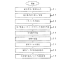
撮像により得られた各粒子画像に対する画像処理の手順(S1〜S9)を図3に示す。
【0017】
画像信号は、画像処理装置11に取り込まれてA/D変換され、画像データとして取り込まれる(ステップS1)。
【0018】
次に、取り込んだ画像データを所定の大きさに切り出し、粒子画像として画像処理装置11の画像メモリに格納する(ステップS2)。
【0019】
粒子像の画像データを画像処理装置11で処理することにより、粒子個々の面積、周囲長、長短径比、粒径、円形度などの特徴パラメータの算出を行う(ステップS3)。この場合の特徴パラメータの算出は、一般に市販されている画像処理ソフトなどを利用できる。
【0020】
このようにして、各粒子像に対して特徴パラメータが算出されると、画像メモリに記憶されている粒子画像とのリンク情報が図4に示すようなデータセットとして作成される(ステップS4)。図4に示すように各粒子番号nに粒子画像がGn(X,Y)対応し、同じく各粒子番号nに特徴パラメータ粒径値Dn、円形度値Cn、面積値Sn、周囲長Lnが対応し、リンク情報として記憶される。
【0021】
次に、必要な分布図(ヒストグラムやスキャッタグラム)を作成して表示(ステップS5)する。
【0022】
次に、キーボード13で分布図内で所望する特徴パラメータの領域を指定し(ステップS6)し、その指定領域に対応した特徴パラメータについて分布状態を表す値が算出され、解析結果が表示される(ステップS7、S8)。つまり、平均値、標準偏差、変動係数、メジアン値、モード値、10%累積値、50%累積値、90%累積値等の解析データの算出結果を表示させる。これらの統計的解析によって、粒子全体の特徴の把握が容易となる。
【0023】
図5にアルミナ粒子を測定したときの特徴パラメータとして粒径(円相当径)に基づいて作成されて表示された分布図(ヒストグラム)が太線で挟まれた領域(A)を指定されたときの表示例を示している。解析領域(A)が指定されたとき、解折結果は平均径(平均値)=2.25、粒径SD(標準偏差)=1.40、50%径(50%累積値)=1.71であり、バラツキの大きい粒径(円相当径)の小さな粒子が集まった分布領域であることが判る。
【0024】
次に、図4に示したリンク情報に基づき、領域指定に対応した粒子画像が画像処理装置11の画像メモリから呼び出され表示される(ステップ9)。
【0025】
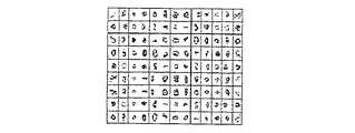
図6に図5の領域(A)が指定されたときの粒子画像を示した。選択したヒストグラムに対応した粒径の小さい粒子画像が多く表示されているのが判る。
【0026】
次に、図7に前記記載したものと同じアルミナ粒子を測定したときの特徴パラメータとして粒径(円相当径)と円形度に基づいて作成されて表示された分布図(スキャッタグラム)が太線で囲まれた領域(B)を指定されたときの表示例を示している。解析領域(B)が指定されたとき、解析結果は平均径(平均値)=29.38、粒径SD(標準偏差)=3.46、50%径(50%累積値)=29.58、平均円形度(平均値)=0.702、円形度SD(標準偏差)=0.086、50%円形度(50%累積値)=0.70であり、比較的にバラツキの少ない粒径(円相当径)の大きい、円形度の小さいな粒子が集まった分布領域であることが判る。
【0027】
また、図8に図7の領域(B)が指定されたときの粒子画像を示した。選択したスキャッタグラムに対応した粒径(円相当径)の大きく、角張った(円形度の小さい)粒子画像が多く表示されているのが判る。
【0028】
このように、分布図に対応した粒子画像を任意に表示できるので、分布上での異常の原因となる粒子について容易に解析でき、粉体の製品開発、工場での品質管理に有用である。
【0029】
また、本発明品は、各粒子像に対して粒子の種類を表す分類情報を付加する分類手段をさらに備えている(ステップS9)。つまり、表示された画像に対して、使用者がキーボード13を用いてマニュアル分類することが可能である。使用者が、各粒子像について、一次粒子(単独)粒子像か、2個凝集粒子像か、3個凝集粒子像か、高次凝集塊か、あるいは対象外の粒子かを指定する。その指定結果をもとにして、凝集している粒子の数の比率を自動的に計算することができる。
【発明の効果】
本発明は、次のような効果を奏する。
本発明は、撮像により得られた各粒子像について、少なくとも一つの特徴パラメータを算出するパラメータ算出手段と、各粒子像について粒子像と特徴パラメータとを関連づけて記憶する記憶手段と、特徴パラメータの分布図を作成する分布図作成手段と、分布図内の任意の領域を指定する指定手段と、指定手段によって指定された領域内の特徴パラメータに対応する粒子像を記憶手段から読出す読出し手段と、読出した粒子像を表示する表示手段とを備えているので、記憶した多数の画像の中から、表示させたい画像を分布上で指定することによって容易に表示することができる。また、分布上での異常の原因となる粒子について容易に解析でき、粉体の製品開発、工場での品質管理に有用である。
【図面の簡単な説明】
【図1】本発明の構成説明図である。
【図2】フローセル部分の拡大図である。
【図3】画像処理のフローチャート図である。
【図4】実施例の特徴パラメータのデータセットを示す説明図である。
【図5】実施例における特徴パラメータに基づいて作成されたヒストグラムの例である。
【図6】実施例における領域(A)の粒子画像を示したものである。
【図7】実施例における特徴パラメータに基づいて作成されたスキャタグラムの例である。
【図8】実施例における領域(B)の粒子画像を示したものである。
【符号の説明】
5 フローセル
8 パルス光源
10 ビデオカメラ
11 画像処理装置
12 CRT[0001]
BACKGROUND OF THE INVENTION
The present invention relates to a particle image analysis apparatus that obtains information related to the size and shape of particles by analyzing a particle image.
[0002]
[Prior art]
In managing the quality of powders such as fine ceramics, toners, pigments, and abrasives, it is very important to measure and manage the particle size of the particles. Recently, development and commercialization of powder with higher added value have been promoted, and not only the size of particles but also measurement of shape parameters and quality control thereof have become important. Furthermore, measuring coarse particles and agglomerated particles is necessary to maintain the quality of the powder product.
[0003]
Therefore, the particle-containing sample liquid is surrounded by a sheath liquid in the flow cell and converted into a sample flow, and the particle images obtained by sequentially imaging the sample flow are analyzed, and the particle size and shape parameters are calculated and distributed. There is known a particle image analyzer that can be displayed as a figure or that can collectively display captured particle images and classify the particles to calculate the aggregate particle ratio (Japanese Patent Laid-Open No. Hei 8-136439).
[0004]
[Problems to be solved by the invention]
However, such a particle image analyzer has a problem that it is not easy to select and display a desired image from among a large number of stored images. The present invention has been made in view of such circumstances, and an object of the present invention is to provide a particle image analyzer capable of easily displaying the particles by designating the particles on the distribution.
[0005]
[Means for Solving the Problems]
The particle image analysis apparatus of the present invention includes a parameter calculation unit that calculates at least one feature parameter for each particle image obtained by imaging, and a storage unit that stores the particle image and the feature parameter in association with each particle image. A distribution map creation means for creating a distribution map of feature parameters, a designation means for designating an arbitrary area in the distribution map, and a particle image corresponding to the feature parameter in the area designated by the designation means is read from the storage means. It is characterized by comprising reading means for outputting and display means for displaying the read particle image.
Here, the characteristic parameter is a parameter obtained by analyzing the particle image, a particle size parameter representing the size of the particle, a circularity parameter representing the roundness of the particle, and a long / short diameter ratio parameter representing the aspect ratio of the particle. Etc. can be used.
[0006]
The distribution chart may be a histogram (one-dimensional distribution chart) using one feature parameter as a parameter, or may be a scattergram (two-dimensional distribution chart) using a plurality of, for example, two feature parameters.
[0007]
In addition, it is preferable to include a distribution analysis unit that analyzes the distribution state of the feature parameter in the region designated by the designation unit. The analysis by the distribution analysis means includes statistical analysis. For example, the average value, standard deviation, coefficient of variation, median value, mode value, 10% cumulative value, 50 for the particle size, circularity, or long / short diameter ratio, 50 This is to calculate analysis data such as% cumulative value, 90% cumulative value and the like. Here, the% cumulative value indicates a value corresponding to a certain% cumulative when the frequency cumulative is 100%.
[0008]
Furthermore, it is preferable to provide classification means for adding classification information indicating the state of the particles (primary particles, secondary particles, aggregates, etc.) to each particle image.
[0009]
DETAILED DESCRIPTION OF THE INVENTION
Each particle image obtained by imaging is a device that combines particles on a slide glass with a microscope and an imaging device. The particle-containing sample liquid is surrounded by a sheath liquid in a flow cell and converted into a sample flow, and the sample flow is sequentially imaged. Thus, a particle image obtained from a particle image analyzer (JP-A-8-136439) for obtaining a particle image can be used.
Here, the particles may be industrial powders such as fine ceramics, toners, pigments, and abrasives, or may be particles such as cells such as blood cells and cultured cells, microorganisms, and plankton.
[0010]
Parameter calculation means for calculating feature parameters, storage means for associating and storing particle images and feature parameters for each particle image, distribution map creation means for creating feature parameter distribution maps, and regions designated by the designation means The readout means, the distribution analysis means, and the classification means for reading out the particle image corresponding to the characteristic parameter in the storage means are an image processing circuit and a microcomputer comprising a CPU, ROM, RAM and I / O port, or image processing A personal computer having a function can be used.
[0011]
In addition, the designation means for designating an arbitrary area in the distribution map can be specified by specifying numerical values of feature parameters or by enclosing the distribution chart in an arbitrary shape. For this purpose, an input means such as a keyboard or a mouse is used. Is preferred.
[0012]
As the display means for displaying the read particle image, a display device such as a CRT or a liquid crystal display, or a printing device such as a printer can be used.
[0013]
【Example】
The configuration of an embodiment of the particle image analyzer of the present invention is shown in FIGS. In this embodiment, a flow type particle imaging function is provided. The sample-containing liquid subjected to the dispersion treatment is guided to the
[0014]
In this way, the sample liquid flow is periodically irradiated with pulsed light from the strobe 8 every 1/30 seconds, so that a still image of the particles is passed through the objective lens 9 every 1/30 seconds. The image is captured by the
[0015]
The image signal from the
Here, the image signal is directly sent to the
[0016]
FIG. 3 shows an image processing procedure (S1 to S9) for each particle image obtained by imaging.
[0017]
The image signal is captured by the
[0018]
Next, the captured image data is cut out to a predetermined size and stored as a particle image in the image memory of the image processing apparatus 11 (step S2).
[0019]
By processing the image data of the particle image with the
[0020]
Thus, when the characteristic parameter is calculated for each particle image, link information with the particle image stored in the image memory is created as a data set as shown in FIG. 4 (step S4). As shown in FIG. 4, each particle number n corresponds to a particle image Gn (X, Y), and each particle number n corresponds to a characteristic parameter particle size value Dn, a circularity value Cn, an area value Sn, and a perimeter length Ln. And stored as link information.
[0021]
Next, a necessary distribution map (histogram or scattergram) is created and displayed (step S5).
[0022]
Next, a desired feature parameter region is designated in the distribution map using the keyboard 13 (step S6), a value representing the distribution state is calculated for the feature parameter corresponding to the designated region, and an analysis result is displayed ( Steps S7 and S8). That is, calculation results of analysis data such as average value, standard deviation, variation coefficient, median value, mode value, 10% cumulative value, 50% cumulative value, 90% cumulative value, and the like are displayed. These statistical analyzes make it easy to understand the characteristics of the entire particle.
[0023]
When the distribution map (histogram) created and displayed based on the particle size (equivalent circle diameter) as a characteristic parameter when measuring alumina particles in FIG. 5 is designated as an area (A) sandwiched by thick lines A display example is shown. When the analysis region (A) is designated, the result of the folding is as follows: average diameter (average value) = 2.25, particle size SD (standard deviation) = 1.40, 50% diameter (50% cumulative value) = 1. It is 71, and it can be seen that this is a distribution region in which small particles having a large variation in particle diameter (equivalent circle diameter) are gathered.
[0024]
Next, based on the link information shown in FIG. 4, a particle image corresponding to the area designation is called from the image memory of the
[0025]
FIG. 6 shows a particle image when the region (A) in FIG. 5 is designated. It can be seen that a large number of small particle images corresponding to the selected histogram are displayed.
[0026]
Next, a distribution map (scattergram) created and displayed based on the particle diameter (equivalent circle diameter) and circularity as a characteristic parameter when measuring the same alumina particles as described above in FIG. 7 is a bold line. The display example when the enclosed area | region (B) is designated is shown. When the analysis region (B) is designated, the analysis results are as follows: average diameter (average value) = 29.38, particle diameter SD (standard deviation) = 3.46, 50% diameter (50% cumulative value) = 29.58 The average circularity (average value) = 0.702, the circularity SD (standard deviation) = 0.086, and the 50% circularity (50% cumulative value) = 0.70. It can be seen that this is a distribution region in which particles having a large (equivalent circle diameter) and a small circularity gather.
[0027]
FIG. 8 shows a particle image when the region (B) in FIG. 7 is designated. It can be seen that many particle images having a large particle diameter (equivalent circle diameter) corresponding to the selected scattergram and being angular (small circularity) are displayed.
[0028]
As described above, since the particle image corresponding to the distribution map can be arbitrarily displayed, the particles causing the abnormality in the distribution can be easily analyzed, which is useful for the development of powder products and the quality control at the factory.
[0029]
The product of the present invention further includes classification means for adding classification information representing the type of particle to each particle image (step S9). That is, the user can manually classify the displayed image using the
【The invention's effect】
The present invention has the following effects.
The present invention relates to parameter calculation means for calculating at least one feature parameter for each particle image obtained by imaging, storage means for storing the particle image and feature parameters in association with each particle image, and distribution of feature parameters A distribution map creation means for creating a diagram; a designation means for designating an arbitrary region in the distribution map; a readout means for reading out a particle image corresponding to the feature parameter in the region designated by the designation means from the storage means; Since the display means for displaying the read particle image is provided, the image to be displayed can be easily displayed by designating it on the distribution from among the stored many images. In addition, it is easy to analyze particles that cause distribution anomalies, which is useful for powder product development and factory quality control.
[Brief description of the drawings]
FIG. 1 is a diagram illustrating the configuration of the present invention.
FIG. 2 is an enlarged view of a flow cell portion.
FIG. 3 is a flowchart of image processing.
FIG. 4 is an explanatory diagram illustrating a feature parameter data set according to the embodiment.
FIG. 5 is an example of a histogram created based on feature parameters in the embodiment.
FIG. 6 shows a particle image of a region (A) in an example.
FIG. 7 is an example of a scattergram created based on feature parameters in the embodiment.
FIG. 8 shows a particle image of a region (B) in an example.
[Explanation of symbols]
5 Flow Cell 8
Claims (8)
Priority Applications (2)
| Application Number | Priority Date | Filing Date | Title |
|---|---|---|---|
| JP37784598A JP3871456B2 (en) | 1998-12-10 | 1998-12-10 | Particle image analyzer |
| US09/455,466 US6522781B1 (en) | 1998-12-10 | 1999-12-06 | Particle image analyzer |
Applications Claiming Priority (1)
| Application Number | Priority Date | Filing Date | Title |
|---|---|---|---|
| JP37784598A JP3871456B2 (en) | 1998-12-10 | 1998-12-10 | Particle image analyzer |
Publications (3)
| Publication Number | Publication Date |
|---|---|
| JP2000180347A JP2000180347A (en) | 2000-06-30 |
| JP2000180347A5 JP2000180347A5 (en) | 2005-12-22 |
| JP3871456B2 true JP3871456B2 (en) | 2007-01-24 |
Family
ID=18509192
Family Applications (1)
| Application Number | Title | Priority Date | Filing Date |
|---|---|---|---|
| JP37784598A Expired - Lifetime JP3871456B2 (en) | 1998-12-10 | 1998-12-10 | Particle image analyzer |
Country Status (2)
| Country | Link |
|---|---|
| US (1) | US6522781B1 (en) |
| JP (1) | JP3871456B2 (en) |
Cited By (1)
| Publication number | Priority date | Publication date | Assignee | Title |
|---|---|---|---|---|
| JP2011203209A (en) * | 2010-03-26 | 2011-10-13 | Seishin Enterprise Co Ltd | Particle property analysis display device and program implementing the same |
Families Citing this family (40)
| Publication number | Priority date | Publication date | Assignee | Title |
|---|---|---|---|---|
| US7057732B2 (en) * | 1999-01-25 | 2006-06-06 | Amnis Corporation | Imaging platform for nanoparticle detection applied to SPR biomolecular interaction analysis |
| US8885913B2 (en) | 1999-01-25 | 2014-11-11 | Amnis Corporation | Detection of circulating tumor cells using imaging flow cytometry |
| US8131053B2 (en) * | 1999-01-25 | 2012-03-06 | Amnis Corporation | Detection of circulating tumor cells using imaging flow cytometry |
| US6975400B2 (en) * | 1999-01-25 | 2005-12-13 | Amnis Corporation | Imaging and analyzing parameters of small moving objects such as cells |
| US20060257884A1 (en) * | 2004-05-20 | 2006-11-16 | Amnis Corporation | Methods for preparing and analyzing cells having chromosomal abnormalities |
| US7450229B2 (en) * | 1999-01-25 | 2008-11-11 | Amnis Corporation | Methods for analyzing inter-cellular phenomena |
| US8406498B2 (en) * | 1999-01-25 | 2013-03-26 | Amnis Corporation | Blood and cell analysis using an imaging flow cytometer |
| US6583865B2 (en) | 2000-08-25 | 2003-06-24 | Amnis Corporation | Alternative detector configuration and mode of operation of a time delay integration particle analyzer |
| US6875973B2 (en) * | 2000-08-25 | 2005-04-05 | Amnis Corporation | Auto focus for a flow imaging system |
| US6778263B2 (en) * | 2000-08-25 | 2004-08-17 | Amnis Corporation | Methods of calibrating an imaging system using calibration beads |
| US6934408B2 (en) * | 2000-08-25 | 2005-08-23 | Amnis Corporation | Method and apparatus for reading reporter labeled beads |
| JP3580766B2 (en) * | 2000-08-29 | 2004-10-27 | 株式会社プラントテクノス | Online image analyzer for liquid particles |
| AU2001297843A1 (en) | 2000-10-12 | 2002-12-23 | Amnis Corporation | Imaging and analyzing parameters of small moving objects such as cells |
| WO2002100157A2 (en) * | 2001-02-21 | 2002-12-19 | Amnis Corporation | Method and apparatus for labeling and analyzing cellular components |
| AU2002308693A1 (en) | 2001-04-25 | 2002-11-05 | Amnis Corporation | Method and apparatus for correcting crosstalk and spatial resolution for multichannel imaging |
| AU2002319621A1 (en) * | 2001-07-17 | 2003-03-03 | Amnis Corporation | Computational methods for the segmentation of images of objects from background in a flow imaging instrument |
| US7352900B2 (en) * | 2003-10-22 | 2008-04-01 | Sysmex Corporation | Apparatus and method for processing particle images and program product for same |
| ATE538138T1 (en) | 2004-03-16 | 2012-01-15 | Amnis Corp | IMAGING-BASED QUANTIFICATION OF MOLECULAR TRANSLOCATION |
| EP1725854B1 (en) | 2004-03-16 | 2019-05-08 | Luminex Corporation | Method for imaging and differential analysis of cells |
| US8953866B2 (en) | 2004-03-16 | 2015-02-10 | Amnis Corporation | Method for imaging and differential analysis of cells |
| US7561756B1 (en) | 2005-05-02 | 2009-07-14 | Nanostellar, Inc. | Particle shape characterization from 2D images |
| US7430322B1 (en) * | 2005-05-02 | 2008-09-30 | Nanostellar, Inc. | Particle shape characterization from 2D images |
| WO2007035864A2 (en) * | 2005-09-20 | 2007-03-29 | Cell Biosciences, Inc. | Electrophoresis standards, methods and kits |
| WO2007067999A2 (en) * | 2005-12-09 | 2007-06-14 | Amnis Corporation | Extended depth of field imaging for high speed object analysis |
| US7526116B2 (en) * | 2006-01-19 | 2009-04-28 | Luigi Armogida | Automated microscopic sperm identification |
| JP4869843B2 (en) * | 2006-09-06 | 2012-02-08 | オリンパス株式会社 | Cell image processing apparatus and cell image processing method |
| CN101387599B (en) * | 2007-09-13 | 2011-01-26 | 深圳迈瑞生物医疗电子股份有限公司 | Method for distinguishing particle community and particle analyzer |
| JP2009244253A (en) * | 2008-03-10 | 2009-10-22 | Sysmex Corp | Particle analyzer, method for analyzing particles, and computer program |
| JP2011075340A (en) * | 2009-09-29 | 2011-04-14 | Sysmex Corp | Particle analyzer and computer program |
| US8451524B2 (en) * | 2009-09-29 | 2013-05-28 | Amnis Corporation | Modifying the output of a laser to achieve a flat top in the laser's Gaussian beam intensity profile |
| JP2011095182A (en) * | 2009-10-30 | 2011-05-12 | Sysmex Corp | Cell analyzing apparatus and cell analyzing method |
| US10012665B2 (en) * | 2010-02-09 | 2018-07-03 | Microjet Corporation | Discharge device for liquid material including particle-like bodies |
| US8817115B1 (en) | 2010-05-05 | 2014-08-26 | Amnis Corporation | Spatial alignment of image data from a multichannel detector using a reference image |
| US8681215B2 (en) | 2011-04-29 | 2014-03-25 | ProteinSimple | Method and particle analyzer for determining a broad particle size distribution |
| ES2882791T3 (en) | 2011-06-17 | 2021-12-02 | Roche Diagnostics Hematology Inc | System and procedure for viewing and reviewing samples |
| US9459196B2 (en) | 2011-07-22 | 2016-10-04 | Roche Diagnostics Hematology, Inc. | Blood analyzer calibration and assessment |
| WO2014076360A1 (en) * | 2012-11-16 | 2014-05-22 | Metso Automation Oy | Measurement of structural properties |
| GB201315196D0 (en) | 2013-08-23 | 2013-10-09 | Univ Dresden Tech | Apparatus and method for determining the mechanical properties of cells |
| US10386290B2 (en) | 2017-03-31 | 2019-08-20 | Life Technologies Corporation | Apparatuses, systems and methods for imaging flow cytometry |
| AT527149A1 (en) * | 2023-04-21 | 2024-11-15 | Anton Paar Gmbh | Device and method for characterizing a particle sample |
Citations (3)
| Publication number | Priority date | Publication date | Assignee | Title |
|---|---|---|---|---|
| JPH08136439A (en) * | 1994-11-04 | 1996-05-31 | Toa Medical Electronics Co Ltd | Grain image analysis device |
| JPH08178826A (en) * | 1994-12-26 | 1996-07-12 | Toa Medical Electronics Co Ltd | Flow cytometer |
| JPH10318904A (en) * | 1996-06-10 | 1998-12-04 | Toa Medical Electronics Co Ltd | Apparatus for analyzing particle image and recording medium recording analysis program therefor |
Family Cites Families (4)
| Publication number | Priority date | Publication date | Assignee | Title |
|---|---|---|---|---|
| US5548395A (en) * | 1991-09-20 | 1996-08-20 | Toa Medical Electronics Co., Ltd. | Particle analyzer |
| JPH07113738A (en) * | 1993-10-15 | 1995-05-02 | Hitachi Ltd | Inspection device for particles in fluid |
| DE69533469T2 (en) | 1994-12-26 | 2005-09-22 | Sysmex Corp. | flow cytometer |
| US5921934A (en) * | 1997-11-25 | 1999-07-13 | Scimed Life Systems, Inc. | Methods and apparatus for non-uniform rotation distortion detection in an intravascular ultrasound imaging system |
-
1998
- 1998-12-10 JP JP37784598A patent/JP3871456B2/en not_active Expired - Lifetime
-
1999
- 1999-12-06 US US09/455,466 patent/US6522781B1/en not_active Expired - Lifetime
Patent Citations (3)
| Publication number | Priority date | Publication date | Assignee | Title |
|---|---|---|---|---|
| JPH08136439A (en) * | 1994-11-04 | 1996-05-31 | Toa Medical Electronics Co Ltd | Grain image analysis device |
| JPH08178826A (en) * | 1994-12-26 | 1996-07-12 | Toa Medical Electronics Co Ltd | Flow cytometer |
| JPH10318904A (en) * | 1996-06-10 | 1998-12-04 | Toa Medical Electronics Co Ltd | Apparatus for analyzing particle image and recording medium recording analysis program therefor |
Cited By (1)
| Publication number | Priority date | Publication date | Assignee | Title |
|---|---|---|---|---|
| JP2011203209A (en) * | 2010-03-26 | 2011-10-13 | Seishin Enterprise Co Ltd | Particle property analysis display device and program implementing the same |
Also Published As
| Publication number | Publication date |
|---|---|
| JP2000180347A (en) | 2000-06-30 |
| US6522781B1 (en) | 2003-02-18 |
Similar Documents
| Publication | Publication Date | Title |
|---|---|---|
| JP3871456B2 (en) | Particle image analyzer | |
| JP3411112B2 (en) | Particle image analyzer | |
| JPH10318904A (en) | Apparatus for analyzing particle image and recording medium recording analysis program therefor | |
| CN1691740B (en) | Magnified display apparatus and magnified display method | |
| US4612614A (en) | Method of analyzing particles in a fluid sample | |
| US6873725B2 (en) | Simultaneous measurement and display of 3-D size distributions of particulate materials in suspensions | |
| JP3812185B2 (en) | Defect classification method and apparatus | |
| JPH0352573B2 (en) | ||
| US8649580B2 (en) | Image processing method, image processing apparatus, and computer-readable recording medium storing image processing program | |
| EP0549905B1 (en) | Method and apparatus for automated cell analysis | |
| JP2001337028A (en) | Particle size distribution measuring method and apparatus | |
| US11080512B2 (en) | Information processing device, information processing method, measurement system and non-transitory storage medium | |
| JP7609236B2 (en) | COMPUTER PROGRAM, LEARNING MODEL GENERATION DEVICE, DISPLAY DEVICE, PARTICLE IDENTIFICATION DEVICE, LEARNING MODEL GENERATION METHOD, DISPLAY METHOD, AND PARTICLE IDENTIFICATION METHOD | |
| WO1997043620A1 (en) | Selectively emphasizing particles of interest from a fluid sample for analysis | |
| JPH07104342B2 (en) | Method for determining the diagnostic significance of the content of particles in a biological sample containing particles | |
| JPH06229723A (en) | Method and equipment for measuring tissue intercept thickness | |
| WO1996020456A1 (en) | Method and apparatus of analyzing particles in a fluid sample and displaying same | |
| US6141624A (en) | Fluid sample for analysis controlled by total fluid volume and by total particle counts | |
| JP3702591B2 (en) | Display device for particle size distribution measurement data and display device for particle size distribution measurement device based on laser diffraction / scattering method | |
| JPH0473052A (en) | Method and apparatus for measuring diameter of hair and sample for measurement | |
| WO2023276361A1 (en) | Particle image analysis apparatus, particle image analysis system, particle image analysis method, and program for particle image analysis apparatus | |
| JPH05249103A (en) | Analyzer for blood cell | |
| JP2007078590A (en) | Particle property analysis display device | |
| JP4102286B2 (en) | Particle image analysis method and apparatus, program and recording medium | |
| JP2722866B2 (en) | Particle size distribution analyzer |
Legal Events
| Date | Code | Title | Description |
|---|---|---|---|
| A521 | Request for written amendment filed |
Free format text: JAPANESE INTERMEDIATE CODE: A523 Effective date: 20051101 |
|
| A621 | Written request for application examination |
Free format text: JAPANESE INTERMEDIATE CODE: A621 Effective date: 20051101 |
|
| A977 | Report on retrieval |
Free format text: JAPANESE INTERMEDIATE CODE: A971007 Effective date: 20060414 |
|
| A131 | Notification of reasons for refusal |
Free format text: JAPANESE INTERMEDIATE CODE: A131 Effective date: 20060509 |
|
| TRDD | Decision of grant or rejection written | ||
| A01 | Written decision to grant a patent or to grant a registration (utility model) |
Free format text: JAPANESE INTERMEDIATE CODE: A01 Effective date: 20061003 |
|
| A61 | First payment of annual fees (during grant procedure) |
Free format text: JAPANESE INTERMEDIATE CODE: A61 Effective date: 20061017 |
|
| R150 | Certificate of patent or registration of utility model |
Free format text: JAPANESE INTERMEDIATE CODE: R150 |
|
| FPAY | Renewal fee payment (event date is renewal date of database) |
Free format text: PAYMENT UNTIL: 20121027 Year of fee payment: 6 |
|
| FPAY | Renewal fee payment (event date is renewal date of database) |
Free format text: PAYMENT UNTIL: 20121027 Year of fee payment: 6 |
|
| RVTR | Cancellation due to determination of trial for invalidation | ||
| FPAY | Renewal fee payment (event date is renewal date of database) |
Free format text: PAYMENT UNTIL: 20121027 Year of fee payment: 6 |
|
| FPAY | Renewal fee payment (event date is renewal date of database) |
Free format text: PAYMENT UNTIL: 20121027 Year of fee payment: 6 |
|
| FPAY | Renewal fee payment (event date is renewal date of database) |
Free format text: PAYMENT UNTIL: 20121027 Year of fee payment: 6 |


