JP3776137B2 - Fusion proteins for prodrug activation - Google Patents
Fusion proteins for prodrug activation Download PDFInfo
- Publication number
- JP3776137B2 JP3776137B2 JP27129193A JP27129193A JP3776137B2 JP 3776137 B2 JP3776137 B2 JP 3776137B2 JP 27129193 A JP27129193 A JP 27129193A JP 27129193 A JP27129193 A JP 27129193A JP 3776137 B2 JP3776137 B2 JP 3776137B2
- Authority
- JP
- Japan
- Prior art keywords
- compound according
- compound
- sfv
- fusion protein
- nucleic acid
- Prior art date
- Legal status (The legal status is an assumption and is not a legal conclusion. Google has not performed a legal analysis and makes no representation as to the accuracy of the status listed.)
- Expired - Lifetime
Links
- 229940002612 prodrug Drugs 0.000 title claims abstract description 14
- 239000000651 prodrug Substances 0.000 title claims abstract description 14
- 102000037865 fusion proteins Human genes 0.000 title claims description 53
- 108020001507 fusion proteins Proteins 0.000 title claims description 53
- 230000004913 activation Effects 0.000 title description 4
- 150000001875 compounds Chemical class 0.000 claims abstract description 43
- 230000027455 binding Effects 0.000 claims abstract description 26
- 239000000427 antigen Substances 0.000 claims abstract description 24
- 102000036639 antigens Human genes 0.000 claims abstract description 24
- 108091007433 antigens Proteins 0.000 claims abstract description 24
- 102000004190 Enzymes Human genes 0.000 claims abstract description 21
- 108090000790 Enzymes Proteins 0.000 claims abstract description 21
- 108090000765 processed proteins & peptides Proteins 0.000 claims abstract description 12
- 150000001720 carbohydrates Chemical class 0.000 claims abstract description 10
- 229920001184 polypeptide Polymers 0.000 claims abstract description 8
- 102000004196 processed proteins & peptides Human genes 0.000 claims abstract description 8
- 239000003814 drug Substances 0.000 claims abstract description 5
- 239000012634 fragment Substances 0.000 claims description 21
- 240000004808 Saccharomyces cerevisiae Species 0.000 claims description 17
- 235000014680 Saccharomyces cerevisiae Nutrition 0.000 claims description 17
- 206010028980 Neoplasm Diseases 0.000 claims description 16
- 108010060309 Glucuronidase Proteins 0.000 claims description 13
- 102000053187 Glucuronidase Human genes 0.000 claims description 12
- 101000933465 Homo sapiens Beta-glucuronidase Proteins 0.000 claims description 12
- 108020004707 nucleic acids Proteins 0.000 claims description 11
- 102000039446 nucleic acids Human genes 0.000 claims description 11
- 150000007523 nucleic acids Chemical class 0.000 claims description 11
- 239000013598 vector Substances 0.000 claims description 11
- 241000588724 Escherichia coli Species 0.000 claims description 10
- 108090000204 Dipeptidase 1 Proteins 0.000 claims description 7
- 238000000034 method Methods 0.000 claims description 7
- 201000011510 cancer Diseases 0.000 claims description 4
- 241000320412 Ogataea angusta Species 0.000 claims description 3
- 108090000854 Oxidoreductases Proteins 0.000 claims description 3
- 102000004316 Oxidoreductases Human genes 0.000 claims description 3
- 241000787399 Protubera nipponica Species 0.000 claims description 3
- 244000082988 Secale cereale Species 0.000 claims description 3
- 102000006635 beta-lactamase Human genes 0.000 claims description 3
- 241000193755 Bacillus cereus Species 0.000 claims description 2
- 108010006303 Carboxypeptidases Proteins 0.000 claims description 2
- 102000005367 Carboxypeptidases Human genes 0.000 claims description 2
- 101710154385 D-aminopeptidase Proteins 0.000 claims description 2
- 108090000371 Esterases Proteins 0.000 claims description 2
- 102000003886 Glycoproteins Human genes 0.000 claims description 2
- 108090000288 Glycoproteins Proteins 0.000 claims description 2
- 102000005744 Glycoside Hydrolases Human genes 0.000 claims description 2
- 108010031186 Glycoside Hydrolases Proteins 0.000 claims description 2
- 241000238631 Hexapoda Species 0.000 claims description 2
- 244000061176 Nicotiana tabacum Species 0.000 claims description 2
- 235000002637 Nicotiana tabacum Nutrition 0.000 claims description 2
- 108091005804 Peptidases Proteins 0.000 claims description 2
- 102000003992 Peroxidases Human genes 0.000 claims description 2
- 239000004365 Protease Substances 0.000 claims description 2
- 108090000919 Pyroglutamyl-Peptidase I Proteins 0.000 claims description 2
- 235000007238 Secale cereale Nutrition 0.000 claims description 2
- 108010062699 gamma-Glutamyl Hydrolase Proteins 0.000 claims description 2
- 108010031620 mandelonitrile lyase Proteins 0.000 claims description 2
- 239000000126 substance Substances 0.000 claims description 2
- 101150047061 tag-72 gene Proteins 0.000 claims description 2
- 238000004519 manufacturing process Methods 0.000 claims 4
- 238000012258 culturing Methods 0.000 claims 2
- 108700019535 Phosphoprotein Phosphatases Proteins 0.000 claims 1
- 102000045595 Phosphoprotein Phosphatases Human genes 0.000 claims 1
- 241000589516 Pseudomonas Species 0.000 claims 1
- 102100037486 Reverse transcriptase/ribonuclease H Human genes 0.000 claims 1
- 230000003213 activating effect Effects 0.000 claims 1
- 230000013595 glycosylation Effects 0.000 claims 1
- 238000006206 glycosylation reaction Methods 0.000 claims 1
- 108040007629 peroxidase activity proteins Proteins 0.000 claims 1
- 235000014633 carbohydrates Nutrition 0.000 abstract description 8
- 231100000433 cytotoxic Toxicity 0.000 abstract description 4
- 230000001472 cytotoxic effect Effects 0.000 abstract description 4
- 231100001083 no cytotoxicity Toxicity 0.000 abstract description 3
- 229940079593 drug Drugs 0.000 abstract description 2
- 108090000623 proteins and genes Proteins 0.000 description 17
- 238000012360 testing method Methods 0.000 description 15
- 239000013612 plasmid Substances 0.000 description 14
- 108091034117 Oligonucleotide Proteins 0.000 description 10
- 238000010276 construction Methods 0.000 description 10
- 108010022366 Carcinoembryonic Antigen Proteins 0.000 description 8
- 102100025475 Carcinoembryonic antigen-related cell adhesion molecule 5 Human genes 0.000 description 8
- 210000004027 cell Anatomy 0.000 description 8
- 238000010586 diagram Methods 0.000 description 8
- WQZGKKKJIJFFOK-QTVWNMPRSA-N D-mannopyranose Chemical compound OC[C@H]1OC(O)[C@@H](O)[C@@H](O)[C@@H]1O WQZGKKKJIJFFOK-QTVWNMPRSA-N 0.000 description 7
- 230000000694 effects Effects 0.000 description 7
- HEMHJVSKTPXQMS-UHFFFAOYSA-M Sodium hydroxide Chemical compound [OH-].[Na+] HEMHJVSKTPXQMS-UHFFFAOYSA-M 0.000 description 6
- 229930182830 galactose Natural products 0.000 description 6
- 239000000243 solution Substances 0.000 description 6
- 239000005018 casein Substances 0.000 description 5
- BECPQYXYKAMYBN-UHFFFAOYSA-N casein, tech. Chemical compound NCCCCC(C(O)=O)N=C(O)C(CC(O)=O)N=C(O)C(CCC(O)=N)N=C(O)C(CC(C)C)N=C(O)C(CCC(O)=O)N=C(O)C(CC(O)=O)N=C(O)C(CCC(O)=O)N=C(O)C(C(C)O)N=C(O)C(CCC(O)=N)N=C(O)C(CCC(O)=N)N=C(O)C(CCC(O)=N)N=C(O)C(CCC(O)=O)N=C(O)C(CCC(O)=O)N=C(O)C(COP(O)(O)=O)N=C(O)C(CCC(O)=N)N=C(O)C(N)CC1=CC=CC=C1 BECPQYXYKAMYBN-UHFFFAOYSA-N 0.000 description 5
- 235000021240 caseins Nutrition 0.000 description 5
- 238000010353 genetic engineering Methods 0.000 description 5
- 239000000523 sample Substances 0.000 description 5
- 239000000758 substrate Substances 0.000 description 5
- 102000007298 Mucin-1 Human genes 0.000 description 4
- 108010008707 Mucin-1 Proteins 0.000 description 4
- 108010076504 Protein Sorting Signals Proteins 0.000 description 4
- 230000000903 blocking effect Effects 0.000 description 4
- 238000002474 experimental method Methods 0.000 description 4
- 230000002349 favourable effect Effects 0.000 description 4
- 238000005227 gel permeation chromatography Methods 0.000 description 4
- 239000000203 mixture Substances 0.000 description 4
- 210000000056 organ Anatomy 0.000 description 4
- 108020004414 DNA Proteins 0.000 description 3
- 238000002965 ELISA Methods 0.000 description 3
- LFQSCWFLJHTTHZ-UHFFFAOYSA-N Ethanol Chemical compound CCO LFQSCWFLJHTTHZ-UHFFFAOYSA-N 0.000 description 3
- 108700019828 Hinge Exons Proteins 0.000 description 3
- 241000699666 Mus <mouse, genus> Species 0.000 description 3
- WQZGKKKJIJFFOK-PHYPRBDBSA-N alpha-D-galactose Chemical compound OC[C@H]1O[C@H](O)[C@H](O)[C@@H](O)[C@H]1O WQZGKKKJIJFFOK-PHYPRBDBSA-N 0.000 description 3
- 239000003153 chemical reaction reagent Substances 0.000 description 3
- 230000002255 enzymatic effect Effects 0.000 description 3
- 239000013604 expression vector Substances 0.000 description 3
- 239000006228 supernatant Substances 0.000 description 3
- 239000011534 wash buffer Substances 0.000 description 3
- YBJHBAHKTGYVGT-ZKWXMUAHSA-N (+)-Biotin Chemical compound N1C(=O)N[C@@H]2[C@H](CCCCC(=O)O)SC[C@@H]21 YBJHBAHKTGYVGT-ZKWXMUAHSA-N 0.000 description 2
- HSHNITRMYYLLCV-UHFFFAOYSA-N 4-methylumbelliferone Chemical compound C1=C(O)C=CC2=C1OC(=O)C=C2C HSHNITRMYYLLCV-UHFFFAOYSA-N 0.000 description 2
- 108020004256 Beta-lactamase Proteins 0.000 description 2
- 241001660259 Cereus <cactus> Species 0.000 description 2
- NBSCHQHZLSJFNQ-QTVWNMPRSA-N D-Mannose-6-phosphate Chemical compound OC1O[C@H](COP(O)(O)=O)[C@@H](O)[C@H](O)[C@@H]1O NBSCHQHZLSJFNQ-QTVWNMPRSA-N 0.000 description 2
- AOJJSUZBOXZQNB-TZSSRYMLSA-N Doxorubicin Chemical compound O([C@H]1C[C@@](O)(CC=2C(O)=C3C(=O)C=4C=CC=C(C=4C(=O)C3=C(O)C=21)OC)C(=O)CO)[C@H]1C[C@H](N)[C@H](O)[C@H](C)O1 AOJJSUZBOXZQNB-TZSSRYMLSA-N 0.000 description 2
- 102000001301 EGF receptor Human genes 0.000 description 2
- 108060006698 EGF receptor Proteins 0.000 description 2
- 102000002464 Galactosidases Human genes 0.000 description 2
- 108010093031 Galactosidases Proteins 0.000 description 2
- 102000004366 Glucosidases Human genes 0.000 description 2
- 108010056771 Glucosidases Proteins 0.000 description 2
- DHMQDGOQFOQNFH-UHFFFAOYSA-N Glycine Chemical compound NCC(O)=O DHMQDGOQFOQNFH-UHFFFAOYSA-N 0.000 description 2
- 241000699660 Mus musculus Species 0.000 description 2
- 102000001068 Neural Cell Adhesion Molecules Human genes 0.000 description 2
- 108010069196 Neural Cell Adhesion Molecules Proteins 0.000 description 2
- 239000004793 Polystyrene Substances 0.000 description 2
- QAOWNCQODCNURD-UHFFFAOYSA-N Sulfuric acid Chemical compound OS(O)(=O)=O QAOWNCQODCNURD-UHFFFAOYSA-N 0.000 description 2
- DTQVDTLACAAQTR-UHFFFAOYSA-N Trifluoroacetic acid Chemical compound OC(=O)C(F)(F)F DTQVDTLACAAQTR-UHFFFAOYSA-N 0.000 description 2
- 239000007983 Tris buffer Substances 0.000 description 2
- 238000002835 absorbance Methods 0.000 description 2
- 238000001042 affinity chromatography Methods 0.000 description 2
- 150000001413 amino acids Chemical group 0.000 description 2
- 230000002494 anti-cea effect Effects 0.000 description 2
- 230000003302 anti-idiotype Effects 0.000 description 2
- 239000000872 buffer Substances 0.000 description 2
- 238000006243 chemical reaction Methods 0.000 description 2
- 238000010367 cloning Methods 0.000 description 2
- 239000002299 complementary DNA Substances 0.000 description 2
- 238000001514 detection method Methods 0.000 description 2
- 238000010790 dilution Methods 0.000 description 2
- 239000012895 dilution Substances 0.000 description 2
- 238000001035 drying Methods 0.000 description 2
- LIIALPBMIOVAHH-UHFFFAOYSA-N herniarin Chemical compound C1=CC(=O)OC2=CC(OC)=CC=C21 LIIALPBMIOVAHH-UHFFFAOYSA-N 0.000 description 2
- JHGVLAHJJNKSAW-UHFFFAOYSA-N herniarin Natural products C1CC(=O)OC2=CC(OC)=CC=C21 JHGVLAHJJNKSAW-UHFFFAOYSA-N 0.000 description 2
- 238000011534 incubation Methods 0.000 description 2
- 238000002347 injection Methods 0.000 description 2
- 239000007924 injection Substances 0.000 description 2
- 238000005621 mannosylation reaction Methods 0.000 description 2
- MYWUZJCMWCOHBA-VIFPVBQESA-N methamphetamine Chemical compound CN[C@@H](C)CC1=CC=CC=C1 MYWUZJCMWCOHBA-VIFPVBQESA-N 0.000 description 2
- ZLQJVGSVJRBUNL-UHFFFAOYSA-N methylumbelliferone Natural products C1=C(O)C=C2OC(=O)C(C)=CC2=C1 ZLQJVGSVJRBUNL-UHFFFAOYSA-N 0.000 description 2
- 238000011580 nude mouse model Methods 0.000 description 2
- 229920002223 polystyrene Polymers 0.000 description 2
- 239000013641 positive control Substances 0.000 description 2
- 239000000047 product Substances 0.000 description 2
- 235000018102 proteins Nutrition 0.000 description 2
- 102000004169 proteins and genes Human genes 0.000 description 2
- 102000005962 receptors Human genes 0.000 description 2
- 108020003175 receptors Proteins 0.000 description 2
- 238000002415 sodium dodecyl sulfate polyacrylamide gel electrophoresis Methods 0.000 description 2
- 239000007858 starting material Substances 0.000 description 2
- 230000001225 therapeutic effect Effects 0.000 description 2
- 230000009261 transgenic effect Effects 0.000 description 2
- LENZDBCJOHFCAS-UHFFFAOYSA-N tris Chemical compound OCC(N)(CO)CO LENZDBCJOHFCAS-UHFFFAOYSA-N 0.000 description 2
- BCHIXGBGRHLSBE-UHFFFAOYSA-N (4-methyl-2-oxochromen-7-yl) dihydrogen phosphate Chemical compound C1=C(OP(O)(O)=O)C=CC2=C1OC(=O)C=C2C BCHIXGBGRHLSBE-UHFFFAOYSA-N 0.000 description 1
- OWEGMIWEEQEYGQ-UHFFFAOYSA-N 100676-05-9 Natural products OC1C(O)C(O)C(CO)OC1OCC1C(O)C(O)C(O)C(OC2C(OC(O)C(O)C2O)CO)O1 OWEGMIWEEQEYGQ-UHFFFAOYSA-N 0.000 description 1
- MSWZFWKMSRAUBD-IVMDWMLBSA-N 2-amino-2-deoxy-D-glucopyranose Chemical compound N[C@H]1C(O)O[C@H](CO)[C@@H](O)[C@@H]1O MSWZFWKMSRAUBD-IVMDWMLBSA-N 0.000 description 1
- 101150112497 26 gene Proteins 0.000 description 1
- -1 4-hydroxy-3-nitrobenzyloxycarbonyl Chemical group 0.000 description 1
- ARQXEQLMMNGFDU-JHZZJYKESA-N 4-methylumbelliferone beta-D-glucuronide Chemical compound C1=CC=2C(C)=CC(=O)OC=2C=C1O[C@@H]1O[C@H](C(O)=O)[C@@H](O)[C@H](O)[C@H]1O ARQXEQLMMNGFDU-JHZZJYKESA-N 0.000 description 1
- STQGQHZAVUOBTE-UHFFFAOYSA-N 7-Cyan-hept-2t-en-4,6-diinsaeure Natural products C1=2C(O)=C3C(=O)C=4C(OC)=CC=CC=4C(=O)C3=C(O)C=2CC(O)(C(C)=O)CC1OC1CC(N)C(O)C(C)O1 STQGQHZAVUOBTE-UHFFFAOYSA-N 0.000 description 1
- 108020004774 Alkaline Phosphatase Proteins 0.000 description 1
- 102000002260 Alkaline Phosphatase Human genes 0.000 description 1
- 102000004092 Amidohydrolases Human genes 0.000 description 1
- 108090000531 Amidohydrolases Proteins 0.000 description 1
- 239000004475 Arginine Substances 0.000 description 1
- 108090001008 Avidin Proteins 0.000 description 1
- 241000283707 Capra Species 0.000 description 1
- 230000004544 DNA amplification Effects 0.000 description 1
- 206010015719 Exsanguination Diseases 0.000 description 1
- 102100037815 Fas apoptotic inhibitory molecule 3 Human genes 0.000 description 1
- 108010015133 Galactose oxidase Proteins 0.000 description 1
- 206010017993 Gastrointestinal neoplasms Diseases 0.000 description 1
- 108010010803 Gelatin Proteins 0.000 description 1
- 239000004471 Glycine Substances 0.000 description 1
- 101000878510 Homo sapiens Fas apoptotic inhibitory molecule 3 Proteins 0.000 description 1
- FBOZXECLQNJBKD-ZDUSSCGKSA-N L-methotrexate Chemical compound C=1N=C2N=C(N)N=C(N)C2=NC=1CN(C)C1=CC=C(C(=O)N[C@@H](CCC(O)=O)C(O)=O)C=C1 FBOZXECLQNJBKD-ZDUSSCGKSA-N 0.000 description 1
- GUBGYTABKSRVRQ-PICCSMPSSA-N Maltose Natural products O[C@@H]1[C@@H](O)[C@H](O)[C@@H](CO)O[C@@H]1O[C@@H]1[C@@H](CO)OC(O)[C@H](O)[C@H]1O GUBGYTABKSRVRQ-PICCSMPSSA-N 0.000 description 1
- 241000124008 Mammalia Species 0.000 description 1
- 108050006616 Mannose-6-phosphate receptors Proteins 0.000 description 1
- OVRNDRQMDRJTHS-UHFFFAOYSA-N N-acelyl-D-glucosamine Natural products CC(=O)NC1C(O)OC(CO)C(O)C1O OVRNDRQMDRJTHS-UHFFFAOYSA-N 0.000 description 1
- OVRNDRQMDRJTHS-FMDGEEDCSA-N N-acetyl-beta-D-glucosamine Chemical compound CC(=O)N[C@H]1[C@H](O)O[C@H](CO)[C@@H](O)[C@@H]1O OVRNDRQMDRJTHS-FMDGEEDCSA-N 0.000 description 1
- MBLBDJOUHNCFQT-LXGUWJNJSA-N N-acetylglucosamine Natural products CC(=O)N[C@@H](C=O)[C@@H](O)[C@H](O)[C@H](O)CO MBLBDJOUHNCFQT-LXGUWJNJSA-N 0.000 description 1
- 230000004988 N-glycosylation Effects 0.000 description 1
- 229930193140 Neomycin Natural products 0.000 description 1
- 229930182555 Penicillin Natural products 0.000 description 1
- JGSARLDLIJGVTE-MBNYWOFBSA-N Penicillin G Chemical compound N([C@H]1[C@H]2SC([C@@H](N2C1=O)C(O)=O)(C)C)C(=O)CC1=CC=CC=C1 JGSARLDLIJGVTE-MBNYWOFBSA-N 0.000 description 1
- 102000035195 Peptidases Human genes 0.000 description 1
- 108700020962 Peroxidase Proteins 0.000 description 1
- 108090000608 Phosphoric Monoester Hydrolases Proteins 0.000 description 1
- 102000004160 Phosphoric Monoester Hydrolases Human genes 0.000 description 1
- 208000005718 Stomach Neoplasms Diseases 0.000 description 1
- 108010006785 Taq Polymerase Proteins 0.000 description 1
- KBGAYAKRZNYFFG-BOHATCBPSA-N aceneuramic acid Chemical compound OC(=O)C(=O)C[C@H](O)[C@@H](NC(=O)C)[C@@H](O)[C@H](O)[C@H](O)CO KBGAYAKRZNYFFG-BOHATCBPSA-N 0.000 description 1
- XAGFODPZIPBFFR-UHFFFAOYSA-N aluminium Chemical compound [Al] XAGFODPZIPBFFR-UHFFFAOYSA-N 0.000 description 1
- 229910052782 aluminium Inorganic materials 0.000 description 1
- 238000004458 analytical method Methods 0.000 description 1
- 238000005571 anion exchange chromatography Methods 0.000 description 1
- 229940045799 anthracyclines and related substance Drugs 0.000 description 1
- ODKSFYDXXFIFQN-UHFFFAOYSA-N arginine Natural products OC(=O)C(N)CCCNC(N)=N ODKSFYDXXFIFQN-UHFFFAOYSA-N 0.000 description 1
- 125000001584 benzyloxycarbonyl group Chemical group C(=O)(OCC1=CC=CC=C1)* 0.000 description 1
- CXQCLLQQYTUUKJ-ALWAHNIESA-N beta-D-GalpNAc-(1->4)-[alpha-Neup5Ac-(2->8)-alpha-Neup5Ac-(2->3)]-beta-D-Galp-(1->4)-beta-D-Glcp-(1<->1')-Cer(d18:1/18:0) Chemical compound O[C@@H]1[C@@H](O)[C@H](OC[C@H](NC(=O)CCCCCCCCCCCCCCCCC)[C@H](O)\C=C\CCCCCCCCCCCCC)O[C@H](CO)[C@H]1O[C@H]1[C@H](O)[C@@H](O[C@]2(O[C@H]([C@H](NC(C)=O)[C@@H](O)C2)[C@H](O)[C@@H](CO)O[C@]2(O[C@H]([C@H](NC(C)=O)[C@@H](O)C2)[C@H](O)[C@H](O)CO)C(O)=O)C(O)=O)[C@@H](O[C@H]2[C@@H]([C@@H](O)[C@@H](O)[C@@H](CO)O2)NC(C)=O)[C@@H](CO)O1 CXQCLLQQYTUUKJ-ALWAHNIESA-N 0.000 description 1
- MSWZFWKMSRAUBD-UHFFFAOYSA-N beta-D-galactosamine Natural products NC1C(O)OC(CO)C(O)C1O MSWZFWKMSRAUBD-UHFFFAOYSA-N 0.000 description 1
- 230000015572 biosynthetic process Effects 0.000 description 1
- 229960002685 biotin Drugs 0.000 description 1
- 235000020958 biotin Nutrition 0.000 description 1
- 239000011616 biotin Substances 0.000 description 1
- 239000008280 blood Substances 0.000 description 1
- 210000004369 blood Anatomy 0.000 description 1
- 230000003197 catalytic effect Effects 0.000 description 1
- 238000004587 chromatography analysis Methods 0.000 description 1
- 239000007979 citrate buffer Substances 0.000 description 1
- 238000003776 cleavage reaction Methods 0.000 description 1
- 230000000295 complement effect Effects 0.000 description 1
- 238000011254 conventional chemotherapy Methods 0.000 description 1
- 239000000824 cytostatic agent Substances 0.000 description 1
- 229960000975 daunorubicin Drugs 0.000 description 1
- STQGQHZAVUOBTE-VGBVRHCVSA-N daunorubicin Chemical compound O([C@H]1C[C@@](O)(CC=2C(O)=C3C(=O)C=4C=CC=C(C=4C(=O)C3=C(O)C=21)OC)C(C)=O)[C@H]1C[C@H](N)[C@H](O)[C@H](C)O1 STQGQHZAVUOBTE-VGBVRHCVSA-N 0.000 description 1
- 239000012470 diluted sample Substances 0.000 description 1
- 238000009826 distribution Methods 0.000 description 1
- 229960004679 doxorubicin Drugs 0.000 description 1
- 238000011156 evaluation Methods 0.000 description 1
- 238000001704 evaporation Methods 0.000 description 1
- 230000005284 excitation Effects 0.000 description 1
- 238000012921 fluorescence analysis Methods 0.000 description 1
- 108010042430 galactose receptor Proteins 0.000 description 1
- 150000002270 gangliosides Chemical class 0.000 description 1
- 206010017758 gastric cancer Diseases 0.000 description 1
- 239000000499 gel Substances 0.000 description 1
- 239000008273 gelatin Substances 0.000 description 1
- 229920000159 gelatin Polymers 0.000 description 1
- 235000019322 gelatine Nutrition 0.000 description 1
- 235000011852 gelatine desserts Nutrition 0.000 description 1
- 229960002442 glucosamine Drugs 0.000 description 1
- 229930182480 glucuronide Natural products 0.000 description 1
- 150000008134 glucuronides Chemical class 0.000 description 1
- 229940046257 glyceryl phosphate Drugs 0.000 description 1
- 238000004128 high performance liquid chromatography Methods 0.000 description 1
- 230000007062 hydrolysis Effects 0.000 description 1
- 238000006460 hydrolysis reaction Methods 0.000 description 1
- 230000001900 immune effect Effects 0.000 description 1
- 238000003018 immunoassay Methods 0.000 description 1
- 238000000338 in vitro Methods 0.000 description 1
- 230000001939 inductive effect Effects 0.000 description 1
- BQINXKOTJQCISL-GRCPKETISA-N keto-neuraminic acid Chemical compound OC(=O)C(=O)C[C@H](O)[C@@H](N)[C@@H](O)[C@H](O)[C@H](O)CO BQINXKOTJQCISL-GRCPKETISA-N 0.000 description 1
- 210000004185 liver Anatomy 0.000 description 1
- 230000004807 localization Effects 0.000 description 1
- 230000007774 longterm Effects 0.000 description 1
- 239000003550 marker Substances 0.000 description 1
- 230000001404 mediated effect Effects 0.000 description 1
- 229960000485 methotrexate Drugs 0.000 description 1
- 239000000178 monomer Substances 0.000 description 1
- 150000002772 monosaccharides Chemical class 0.000 description 1
- 229950006780 n-acetylglucosamine Drugs 0.000 description 1
- 239000013642 negative control Substances 0.000 description 1
- 229960004927 neomycin Drugs 0.000 description 1
- CERZMXAJYMMUDR-UHFFFAOYSA-N neuraminic acid Natural products NC1C(O)CC(O)(C(O)=O)OC1C(O)C(O)CO CERZMXAJYMMUDR-UHFFFAOYSA-N 0.000 description 1
- 230000009871 nonspecific binding Effects 0.000 description 1
- 239000002773 nucleotide Substances 0.000 description 1
- 125000003729 nucleotide group Chemical group 0.000 description 1
- 239000012188 paraffin wax Substances 0.000 description 1
- 229940049954 penicillin Drugs 0.000 description 1
- 230000000144 pharmacologic effect Effects 0.000 description 1
- UYWQUFXKFGHYNT-UHFFFAOYSA-N phenylmethyl ester of formic acid Natural products O=COCC1=CC=CC=C1 UYWQUFXKFGHYNT-UHFFFAOYSA-N 0.000 description 1
- 238000002360 preparation method Methods 0.000 description 1
- 239000011541 reaction mixture Substances 0.000 description 1
- 230000007017 scission Effects 0.000 description 1
- 238000007789 sealing Methods 0.000 description 1
- 230000003248 secreting effect Effects 0.000 description 1
- 201000011549 stomach cancer Diseases 0.000 description 1
- 239000012089 stop solution Substances 0.000 description 1
- 238000003860 storage Methods 0.000 description 1
- 238000007920 subcutaneous administration Methods 0.000 description 1
- 238000003786 synthesis reaction Methods 0.000 description 1
- 229940124597 therapeutic agent Drugs 0.000 description 1
- 231100000331 toxic Toxicity 0.000 description 1
- 230000002588 toxic effect Effects 0.000 description 1
- 238000012546 transfer Methods 0.000 description 1
- 238000005303 weighing Methods 0.000 description 1
- 210000005253 yeast cell Anatomy 0.000 description 1
Images


Classifications
-
- C—CHEMISTRY; METALLURGY
- C07—ORGANIC CHEMISTRY
- C07K—PEPTIDES
- C07K16/00—Immunoglobulins [IGs], e.g. monoclonal or polyclonal antibodies
- C07K16/18—Immunoglobulins [IGs], e.g. monoclonal or polyclonal antibodies against material from animals or humans
- C07K16/28—Immunoglobulins [IGs], e.g. monoclonal or polyclonal antibodies against material from animals or humans against receptors, cell surface antigens or cell surface determinants
- C07K16/30—Immunoglobulins [IGs], e.g. monoclonal or polyclonal antibodies against material from animals or humans against receptors, cell surface antigens or cell surface determinants from tumour cells
-
- C—CHEMISTRY; METALLURGY
- C12—BIOCHEMISTRY; BEER; SPIRITS; WINE; VINEGAR; MICROBIOLOGY; ENZYMOLOGY; MUTATION OR GENETIC ENGINEERING
- C12Y—ENZYMES
- C12Y302/00—Hydrolases acting on glycosyl compounds, i.e. glycosylases (3.2)
- C12Y302/01—Glycosidases, i.e. enzymes hydrolysing O- and S-glycosyl compounds (3.2.1)
- C12Y302/01031—Beta-glucuronidase (3.2.1.31)
-
- A—HUMAN NECESSITIES
- A61—MEDICAL OR VETERINARY SCIENCE; HYGIENE
- A61K—PREPARATIONS FOR MEDICAL, DENTAL OR TOILETRY PURPOSES
- A61K47/00—Medicinal preparations characterised by the non-active ingredients used, e.g. carriers or inert additives; Targeting or modifying agents chemically bound to the active ingredient
- A61K47/50—Medicinal preparations characterised by the non-active ingredients used, e.g. carriers or inert additives; Targeting or modifying agents chemically bound to the active ingredient the non-active ingredient being chemically bound to the active ingredient, e.g. polymer-drug conjugates
- A61K47/51—Medicinal preparations characterised by the non-active ingredients used, e.g. carriers or inert additives; Targeting or modifying agents chemically bound to the active ingredient the non-active ingredient being chemically bound to the active ingredient, e.g. polymer-drug conjugates the non-active ingredient being a modifying agent
- A61K47/62—Medicinal preparations characterised by the non-active ingredients used, e.g. carriers or inert additives; Targeting or modifying agents chemically bound to the active ingredient the non-active ingredient being chemically bound to the active ingredient, e.g. polymer-drug conjugates the non-active ingredient being a modifying agent the modifying agent being a protein, peptide or polyamino acid
- A61K47/65—Peptidic linkers, binders or spacers, e.g. peptidic enzyme-labile linkers
-
- A—HUMAN NECESSITIES
- A61—MEDICAL OR VETERINARY SCIENCE; HYGIENE
- A61P—SPECIFIC THERAPEUTIC ACTIVITY OF CHEMICAL COMPOUNDS OR MEDICINAL PREPARATIONS
- A61P35/00—Antineoplastic agents
-
- C—CHEMISTRY; METALLURGY
- C07—ORGANIC CHEMISTRY
- C07K—PEPTIDES
- C07K14/00—Peptides having more than 20 amino acids; Gastrins; Somatostatins; Melanotropins; Derivatives thereof
- C07K14/435—Peptides having more than 20 amino acids; Gastrins; Somatostatins; Melanotropins; Derivatives thereof from animals; from humans
- C07K14/705—Receptors; Cell surface antigens; Cell surface determinants
-
- C—CHEMISTRY; METALLURGY
- C07—ORGANIC CHEMISTRY
- C07K—PEPTIDES
- C07K16/00—Immunoglobulins [IGs], e.g. monoclonal or polyclonal antibodies
- C07K16/18—Immunoglobulins [IGs], e.g. monoclonal or polyclonal antibodies against material from animals or humans
-
- C—CHEMISTRY; METALLURGY
- C12—BIOCHEMISTRY; BEER; SPIRITS; WINE; VINEGAR; MICROBIOLOGY; ENZYMOLOGY; MUTATION OR GENETIC ENGINEERING
- C12N—MICROORGANISMS OR ENZYMES; COMPOSITIONS THEREOF; PROPAGATING, PRESERVING, OR MAINTAINING MICROORGANISMS; MUTATION OR GENETIC ENGINEERING; CULTURE MEDIA
- C12N15/00—Mutation or genetic engineering; DNA or RNA concerning genetic engineering, vectors, e.g. plasmids, or their isolation, preparation or purification; Use of hosts therefor
- C12N15/09—Recombinant DNA-technology
- C12N15/11—DNA or RNA fragments; Modified forms thereof; Non-coding nucleic acids having a biological activity
- C12N15/62—DNA sequences coding for fusion proteins
-
- C—CHEMISTRY; METALLURGY
- C12—BIOCHEMISTRY; BEER; SPIRITS; WINE; VINEGAR; MICROBIOLOGY; ENZYMOLOGY; MUTATION OR GENETIC ENGINEERING
- C12N—MICROORGANISMS OR ENZYMES; COMPOSITIONS THEREOF; PROPAGATING, PRESERVING, OR MAINTAINING MICROORGANISMS; MUTATION OR GENETIC ENGINEERING; CULTURE MEDIA
- C12N9/00—Enzymes; Proenzymes; Compositions thereof; Processes for preparing, activating, inhibiting, separating or purifying enzymes
- C12N9/14—Hydrolases (3)
- C12N9/24—Hydrolases (3) acting on glycosyl compounds (3.2)
- C12N9/2402—Hydrolases (3) acting on glycosyl compounds (3.2) hydrolysing O- and S- glycosyl compounds (3.2.1)
- C12N9/2405—Glucanases
- C12N9/2434—Glucanases acting on beta-1,4-glucosidic bonds
-
- G—PHYSICS
- G01—MEASURING; TESTING
- G01N—INVESTIGATING OR ANALYSING MATERIALS BY DETERMINING THEIR CHEMICAL OR PHYSICAL PROPERTIES
- G01N33/00—Investigating or analysing materials by specific methods not covered by groups G01N1/00 - G01N31/00
- G01N33/48—Biological material, e.g. blood, urine; Haemocytometers
- G01N33/50—Chemical analysis of biological material, e.g. blood, urine; Testing involving biospecific ligand binding methods; Immunological testing
- G01N33/53—Immunoassay; Biospecific binding assay; Materials therefor
- G01N33/574—Immunoassay; Biospecific binding assay; Materials therefor for cancer
-
- A—HUMAN NECESSITIES
- A01—AGRICULTURE; FORESTRY; ANIMAL HUSBANDRY; HUNTING; TRAPPING; FISHING
- A01K—ANIMAL HUSBANDRY; AVICULTURE; APICULTURE; PISCICULTURE; FISHING; REARING OR BREEDING ANIMALS, NOT OTHERWISE PROVIDED FOR; NEW BREEDS OF ANIMALS
- A01K2217/00—Genetically modified animals
- A01K2217/05—Animals comprising random inserted nucleic acids (transgenic)
-
- A—HUMAN NECESSITIES
- A61—MEDICAL OR VETERINARY SCIENCE; HYGIENE
- A61K—PREPARATIONS FOR MEDICAL, DENTAL OR TOILETRY PURPOSES
- A61K39/00—Medicinal preparations containing antigens or antibodies
- A61K2039/505—Medicinal preparations containing antigens or antibodies comprising antibodies
-
- A—HUMAN NECESSITIES
- A61—MEDICAL OR VETERINARY SCIENCE; HYGIENE
- A61K—PREPARATIONS FOR MEDICAL, DENTAL OR TOILETRY PURPOSES
- A61K38/00—Medicinal preparations containing peptides
-
- C—CHEMISTRY; METALLURGY
- C07—ORGANIC CHEMISTRY
- C07K—PEPTIDES
- C07K2317/00—Immunoglobulins specific features
- C07K2317/60—Immunoglobulins specific features characterized by non-natural combinations of immunoglobulin fragments
- C07K2317/62—Immunoglobulins specific features characterized by non-natural combinations of immunoglobulin fragments comprising only variable region components
- C07K2317/622—Single chain antibody (scFv)
-
- C—CHEMISTRY; METALLURGY
- C07—ORGANIC CHEMISTRY
- C07K—PEPTIDES
- C07K2319/00—Fusion polypeptide
-
- C—CHEMISTRY; METALLURGY
- C07—ORGANIC CHEMISTRY
- C07K—PEPTIDES
- C07K2319/00—Fusion polypeptide
- C07K2319/01—Fusion polypeptide containing a localisation/targetting motif
- C07K2319/02—Fusion polypeptide containing a localisation/targetting motif containing a signal sequence
Landscapes
- Health & Medical Sciences (AREA)
- Life Sciences & Earth Sciences (AREA)
- Chemical & Material Sciences (AREA)
- Organic Chemistry (AREA)
- Genetics & Genomics (AREA)
- Engineering & Computer Science (AREA)
- Zoology (AREA)
- General Health & Medical Sciences (AREA)
- Bioinformatics & Cheminformatics (AREA)
- Molecular Biology (AREA)
- Wood Science & Technology (AREA)
- Biochemistry (AREA)
- Biomedical Technology (AREA)
- Medicinal Chemistry (AREA)
- General Engineering & Computer Science (AREA)
- Biotechnology (AREA)
- Immunology (AREA)
- Microbiology (AREA)
- Biophysics (AREA)
- Proteomics, Peptides & Aminoacids (AREA)
- Cell Biology (AREA)
- Physics & Mathematics (AREA)
- Urology & Nephrology (AREA)
- Hematology (AREA)
- Toxicology (AREA)
- Plant Pathology (AREA)
- Pharmacology & Pharmacy (AREA)
- Animal Behavior & Ethology (AREA)
- Public Health (AREA)
- Veterinary Medicine (AREA)
- Gastroenterology & Hepatology (AREA)
- General Physics & Mathematics (AREA)
- Nuclear Medicine, Radiotherapy & Molecular Imaging (AREA)
- Oncology (AREA)
- Chemical Kinetics & Catalysis (AREA)
- General Chemical & Material Sciences (AREA)
- Food Science & Technology (AREA)
- Analytical Chemistry (AREA)
- Hospice & Palliative Care (AREA)
- Pathology (AREA)
Abstract
Description
【0001】
【産業上の利用分野】
本発明は、ほとんどまたはまったく細胞毒性を持たない化合物(プロドラッグ)を細胞毒性化合物(薬物)に代謝することができる少なくとも1種類の酵素に結合する抗原結合領域を含む化合物であって、この抗原結合領域が単一のポリペプチド鎖から成ることを特徴とする化合物に関する。共有結合によって結合した炭水化物がポリペプチド鎖上に存在することが有利である。
【0002】
【従来の技術】
治療用組成物として用いられるプロドラッグと抗体−酵素抱合体との組み合わせは、既に専門家の文献に記載されている。このことにより、特定の組織に向けられており且つプロドラッグ開裂酵素が結合する抗体が生体中に注入され、続いてこの酵素で活性化することができるプロドラッグ化合物を投与することができることになる。標的組織に結合した抗体−酵素抱合体の作用は、プロドラッグ化合物を結合した組織に対して細胞毒性作用を示す化合物へと転換しようとするものである。しかしながら、抗体−酵素抱合体についての研究では、これらの化学的抱合体は好ましくない薬物動態を示し、プロドラッグの不適切な部位特異的な腫瘍選択的開裂しかないことが判っている。幾人かの著者は、この明らかな欠点を、抗酵素抗体を追加注入して血漿から抗体−酵素抱合体を速やかに除去しようとすることによって、除去しようとしている(Sharmaら、Brit. J. Cancer, 61, 659, 1990) 。抗体−酵素抱合体のもう一つの問題点は、再現性が高く且つ均質に多量を調製する可能性が限定されていることである。
【0003】
【発明が解決しようとする課題】
本発明の目的は、工業的規模で調製することができ且つその薬物動態および薬理特性により治療目的に好適な融合タンパク質を見出すことであった。
【0004】
【課題を解決するための手段】
これに関連して、単一ポリペプチド鎖から成る抗原結合領域を有する化合物は炭水化物が有利に結合する融合タンパク質の調製とプロドラッグの活性化に使用するのに予想外に有利であることを見出した。
それ故、本発明は少なくとも1個の酵素に結合する抗原結合領域を有する化合物であって、その抗原結合領域が単一のポリペプチド鎖から成り、炭水化物が融合タンパク質に有利に結合している化合物に関する。
抗原結合領域とは、本発明の目的に対しては、抗体の少なくとも2個の可変ドメイン、好ましくは重鎖抗体の1個の可変ドメインと軽鎖抗体(sFvフラグメント)の1個の可変ドメインとを含む領域を意味する。しかしながら、抗原結合領域は、二価または多価構造、すなわち、例えばEP−A−0 404 097号明細書に記載されているように2個以上の結合領域を有することもできる。しかしながら、ヒトのまたはヒト化sFvフラグメントが特に好ましく、中でもヒト化sFvフラグメントが好ましい。
【0005】
抗原結合領域は、腫瘍関連抗原(TAA)に結合するのが好ましく、下記のTAAが特に好ましい。
神経細胞接着分子(N−CAM)、
多形上皮ムチン(PEM)、表皮成長因子レセプター(EGF−R)、
トムセン・フリーデンライヒ抗原β(TFβ)、
消化管癌抗原(GICA)、
ガングリオシドGD3(GD3)、
ガングリオシドGD2(GD2)、
Sialyl−Lea、Sialyl−Lex、
TAG72、
MAb L6によって画定される24〜25kDaの糖タンパク質、
CA125、及び特に癌胎児性抗原(CEA)。
【0006】
好ましい酵素は、ほとんどまたは全く細胞毒性を持たない化合物を細胞毒性化合物に代謝することができる酵素である。例には、EP−A2−0 382 411号明細書またはEP−A2−0 392 745号明細書などに記載されているβ−ラクタマーゼ、ピログルタメートアミノペプチダーゼ、ガラクトシダーゼまたはD−アミノペプチダーゼ;例えばWO91/00108号明細書に記載されているオキシダーゼ、例えば、エタノールオキシダーゼ、ガラクトースオキシダーゼ、D−アミノ酸オキシダーゼまたはα−グリセリル−ホスフェートオキシダーゼ;例えばEP−A2−0 361 908号明細書に記載されているペルオキシダーゼ;例えばEP−A1−0 302 473号明細書に記載されているホスファターゼ;例えばWO91/11201号明細書に開示されているヒドロキシニトリルリアーゼまたはグルコシダーゼ;カルボキシペプチダーゼ、例えばカルボキシペプチダーゼG2(WO88/07378号明細書);アミダーゼ、例えばペニシリン5−アミダーゼ(Kerr, D.E.ら、Cancer Immunol. Immunther., 1990, 31) ;及びプロテアーゼ、エステラーゼまたはグリコシダーゼ、例えばWO91/08770号明細書に記載の例えばガラクトシダーゼ、グルコシダーゼまたはグルクロニダーゼである。
【0007】
β−グルクロニダーゼが好ましく、Kobayasia nipponica またはSecale cereale由来のものが好ましく、大腸菌またはヒトβ−グルクロニダーゼ由来が更に好ましい。個々の酵素の基質も前記の特許明細書で指摘されており、本願明細書の開示内容の一部を構成するものである。β−グルクロニダーゼの好ましい基質はN−(D−グリコピラノシル)ベンジルオキシカルボニルアントラサイクリン、特にN−(4−ヒドロキシ−3−ニトロベンジルオキシカルボニル)ドクソルビシン及びダウノルビシン=β−D−グルクロニドである(J.C. Florentら、(1992) Int. Carbohydr. Symp. Paris, A262, 297 またはS. Andrianomenjanaharyら、(1992) Int. Carbohydr. Symp. Paris, A264, 299 )。
【0008】
本発明は、本発明による化合物をコードする核酸にも関する。特に好ましいものは、核酸並びにその変体(variant )及び変異体(mutant)であって、ヒトのβ−グルクロニダーゼに連結したCEA(癌胎児性抗原)に対するヒト化sFvフラグメントをコードするものであり、好ましくは第1表に示す配列を有するものである(sFv−huβ−Gluc)。
【0009】
本発明による化合物は、通常は当業者に普通に知られている遺伝子操作の方法によって調製し、抗原結合領域を1種類以上の酵素に、直接またはリンカー、好ましくはペプチドリンカーを介して、結合させることができる。用いることができるペプチドリンカーは、例えば抗体のヒンジ領域またはヒンジ様アミノ酸配列である。この場合には、酵素は好ましくはN末端で抗原結合領域に直接またはペプチドリンカーを介して結合している。しかしながら、酵素または複数の酵素は、例えばWO91/00108号明細書に記載されているように抗原結合領域に化学的に結合することもできる。
【0010】
本発明の化合物のアミノ酸配列をコードする核酸は、通常は発現ベクターにクローニングされ、BHK、CHO、COS、HeLa、昆虫、タバコ植物、酵母または大腸菌細胞などの原核または真核宿主細胞に導入され、発現される。この方法で調製された化合物は、次に単離して、診断助剤または治療剤として用いることができる。本発明による化合物のもう一つの一般に知られている調製法は、ヒト以外のトランスジェニック哺乳類、好ましくはトランスジェニックヤギでコードする核酸を発現させることである。
【0011】
本発明による核酸でトランスフェクションしたBHK細胞は、CEAに対して特異的であるだけでなく、完全なβ−グルクロニダーゼ活性も有した融合タンパク質(sFv−huβ−Gluc)を発現する(例5を参照されたい)。
この融合タンパク質は、EP0501215A2号明細書(例M)に記載の方法によって抗−イディオタイプアフィニティークロマトグラフィによって精製した。この方法で精製した融合タンパク質の分子量は還元条件下でのSDS PAGEでは100kDaであるが、100及び200kDaの分子はそれぞれ非還元条件下で現れる。
【0012】
本来の条件下でのゲルクロマトグラフィ(TSK−3000ゲルクロマトグラフィ)では、1個のタンパク質ピーク(例6、図1)を示し、これは特異性酵素活性試験において活性ピークと相関する(EP0501215A2号明細書)。標準的な分子量マーカーと比較することによるピークの位置は、分子量が約200kDaであることを示している。この知見は、SDS PAGEからのデーターと一緒になって、機能的な酵素学的に活性なsFv−huβ−Gluc融合タンパク質は「二価分子」の形態をしており、すなわち2個の結合領域と2個の酵素分子を有する。本明細書には記載されていない実験から、融合タンパク質はある種の培養条件では4個の結合領域と4個の酵素分子を有する四量体(テトラマー)の形態をとることができる。sFv−huβ−Gluc融合タンパク質を精製して、イン・ビトロで機能の特性決定を行った後、融合タンパク質の薬物動態と腫瘍局在化をヒト胃癌を有するヌードマウスで決定した。機能的に活性な融合タンパク質の量を、器官を適宜処理した後様々な時間に器官及び腫瘍中で(例7)決定し、免疫学的測定法(3決定基試験、例8)によって決定した。代表的な実験の結果を、第4表に纏める。
【0013】
驚くべきことには、腫瘍/血漿比はたった48時間後で5/1に達する。更に時間が経てば、この比率は更に好ましくなり、>200/1の値に達する(5日目)。sFv−huβ−Gluc融合タンパク質がこのような好ましい薬物動態を示す理由は、腫瘍に結合していない融合タンパク質が主としてマンノース6−ホスフェート及びガラクトースのレセプターによるインターナリゼーションによって血漿及び正常な組織から除去されるからである。(この現象の証拠は、例えば肝臓で分子内β−グルクロニダーゼ濃度が増加することである)。
【0014】
第5表に示されるように、sFv−huβ−Glucは比較的多量のガラクトース、特に特定のレセプターへの結合に主に重要なマンノースを含んでいる。生成し融合タンパク質の炭水化物残基を介して結合している融合タンパク質/レセプター複合体を次に、インターナリゼーションによって細胞外区画から除去する。
主としてガラクトースとマンノースとによって媒介されるこの迅速インターナリゼーション機構は、本発明による融合タンパク質の有利な薬物動態と緊密に関連している。ガラクトース及び特にマンノースが結合する融合タンパク質のこれらの有利な薬物動態により、細胞外分布を行う親水性のプロドラッグを非特異的なプロドラッグ活性化を誘導することなく比較的初期に静脈内に投与することができる。この場合には、Sharmaら(Brit. J. Cancer, 61, 659, 1990)によって記載された除去工程は必要ない。第4表のデーターに基づけば、適当なプロドラッグの注入(S. Adrianomenjanahariら、1992, Int. Carbohydrate Symp., Parts A264, 299) を、sFv−huβ−Gluc融合タンパク質の注射から3日後には重大な副作用を起こすことなく行うことが可能である(データーは示されていない)。
【0015】
同様に炭水化物の融合タンパク質への有利な結合は、例えばSaccharomyces cerevisiaeまたはHansenula polymorphaのような特定の酵母株でのsFv−huβ−Gluc融合タンパク質の分泌発現によって行うこともできる。これらの生物は、適当なN−グリコシル化部位を有する融合タンパク質を極めて効果的にマンノシル化することができる(Goochee ら、Biotechnology, 9, 1347-1354, 1991 )。酵母細胞で分泌発現を行ったこのような融合タンパク質は、BHK細胞で発現したsFv−huβ−Gluc融合タンパク質に匹敵するほどの高度のマンノシル化及び好ましい薬物動態を示す(データーは示されていない)。この場合には、ガラクトースがないことは、融合タンパク質の極めて高度のマンノシル化によって補償される(第6表)。前記のsFv−huβ−Gluc融合タンパク質を遺伝子操作によって構築して、例9に詳細に記載するように、酵母中で発現させた。
【0016】
しかしながら、ヒトβ−グルクロニダーゼの代わりに、有利な特性を有する他のグルクロニダーゼを用いることも可能である。例えば、大腸菌のβ−グルクロニダーゼは、pH7.4におけるその触媒活性はヒトβ−グルクロニダーゼの活性より著しく高いといった特別な利点を有する。例10において、sFv−大腸菌β−Gluc構築物を遺伝子操作の方法によって調製し、Saccharomyces cerevisiaeにおいて機能的に活性なマンノシル化した融合タンパク質として分泌発現を行った。薬物動態データーは、酵母またはBHK細胞で発現したsFv−huβ−Gluc分子のものに匹敵する(第4表)。
【0017】
菌類(fungus)Kobayasia nipponica 及び植物Secale cereale由来のグルクロニダーゼは、例えば、それらが単量体(モノマー)として活性であるという利点を有する。例11では、遺伝子操作の方法を用いて、Saccharomyces cerevisiaeでの発現の後にsFv−B. cereus β−ラクタマーゼII融合タンパク質をマンノシル化した形態で優先的に放出する構築物を調製した。
この融合タンパク質も本発明による融合タンパク質と同様に、プロドラッグの活性化に好ましいβ−グルクロニダーゼ薬物動態に基づいている(第4表)。
【0018】
更に、本発明による化合物をプロドラッグと組合せで用いるだけでなく、グルクロニドとして代謝されて不活性化する細胞増殖抑制剤を、投与した化合物によって毒性のある形態に転換することができる従来の化学療法の構成に用いることもできる。
【0019】
下記の例により、sFv−β−Gluc融合タンパク質の遺伝子操作による合成と、機能性の論証を行う。
出発材料はプラスミドpABstop431/26humVH及びpABstop431/26humVHLを含んでいた。これらのプラスミドは、抗−CEA MAb BW431/26のVH遺伝子とVL遺伝子のヒト化したものを含む(Guessow とSeemann, 1991, Meth. Enzymology, 203, 99-121)。その他の出発材料は、プラスミドpABstop431/26VH−huβ−Gluc1H(EP−A2−0501215号明細書)であって、VHに固有のシグナル配列を含むVHエキソンと、続いてCH1エキソン、ヒトIgG3c遺伝子のヒンジエキソン及びヒトβ−グルクロニダーゼの完全なcDNAを含むものから成っていた。
【0020】
例1
MAb hum431/26のVH及びVL遺伝子の増幅
オリゴヌクレオチドpAB−Back及びリンカー−アンチ(第2表)を用いて、pABstop431VHhum(VH431/26)由来のVH遺伝子に固有のシグナル配列を有するVH遺伝子を増幅した(Guessow とSeemann, 1991, Meth. Enzymology, 203, 99-121)。オリゴヌクレオチドリンカー−センスとVL(Mut)−For(第3表)を用いてpABstop431VLhum(VL431/26)を増幅した。
【0021】
例2
VH431/26及びVL431/26遺伝子フラグメントの結合
オリゴヌクレオチドリンカー−アンチ及びリンカー−センスは互いに部分的に相補性であり、VHドメインとVLドメインとを結合してsFvフラグメントを得るためのポリペプチドリンカーをコードする。増幅したVHフラグメントとVLフラグメントを融合するために、これらを精製して、下記のような10サイクル反応に用いる。
H2O 37.5μl
dNTP(2.5mM) 5.0μl
PCR緩衝液(10×) 5.0μl
Taqポリメラーゼ(Perkin-Elmer Corp., エメリービル、
カリフォルニア)(2.5U/μl) 0.5μl
VLフラグメントの0.5μg/μlDNA 1.0μl
VHフラグメントの0.5μg/μlDNA 1.0μl
PCR緩衝液(10×): 100mMトリス、pH8.3、500mM KCl、15mM MgCl2、0.1%(w/v)ゼラチン。
反応混合物の表面をパラフィンで密封した後、94℃で1分、55℃で1分、72℃で2分プログラムしたPCR装置で10−サイクル反応を行い、フランキングプライマーpAB−Back及びVL(Mut)−Forを加え、更に20サイクルの反応を行う。生成するPCRフラグメントは、リンカーを介してVL遺伝子に結合しているVH遺伝子から成る。VH遺伝子に固有のシグナル配列もVH遺伝子の前にある。オリゴヌクレオチドVL(Mut)−ForもVL遺伝子の最後のヌクレオチド塩基となり、CはGに置き代わっている。このPCRフラグメントはヒト化した一本鎖Fv(sFv431/26)をコードする。
【0022】
例3
huβ−グルクロニダーゼ遺伝子を含む発現ベクター中へのsFv431/26フラグメントのクローニング
(2)からのsFvフラグメントをHindIIIとBamHIで切断し、HindIIIで完全に開裂させBglIIで部分的に開裂したベクターpAB431VHhum/CH1+1h/β−Glcに連結する。ベクターpABstop431/26VHhuβ−Gluc1Hは、VHに固有のシグナル配列を有するVHエキソン、CH1エキソン、ヒトIgG3C遺伝子のヒンジエキソン及びヒトβ−グルクロニダーゼの完全なcDNAを含む。ヒト化したsFv431/26、ヒンジエキソン及びヒトβ−グルクロニダーゼの遺伝子を含むプラスミドクローンpMCG−E1を単離する(pMCG−E1)。
【0023】
例4
BHK細胞でのsFv−huβ−Gluc融合タンパク質の発現
クローンpMCG−E1を、ネオマイシン耐性遺伝子を有するプラスミドpRMH140と、メトトレキセート耐性遺伝子を有するプラスミドpSV2とで、BHK細胞中にトランスフェクションする。BHK細胞は、次いで、MAb BW431/26humの抗原結合特性とヒトβ−グルクロニダーゼの酵素活性を両方とも有する融合タンパク質を発現する。
【0024】
例5
sFv−huβ−Gluc融合タンパク質の抗原結合特性及び酵素活性の証明
sFv−huβ−Gluc融合タンパク質が431/26によって画定されたCEAエピトープに特異的に結合することができ、同時にヒトβ−グルクロニダーゼの酵素活性を示すことができることを、特異性酵素活性試験(EP−A2−0501215号明細書)で示した。この試験は、融合タンパク質がsFv部分を介して抗原に結合した後に融合タンパク質のβ−グルクロニダーゼ部分によって4−メチルウンベリフェリルβ−グルクロニドから4−メチルウンベリフェロンの放出を測定する。測定した螢光値を相対的螢光単位(FU)として記録する。この試験では、CEAをコーティングしたプレートで融合タンパク質によるメチル−ウンベリフェロンの有意な放出を示す。対照的に、融合タンパク質は、PEM(多形性上皮ムチン)をコーティングしたコントロールプレートではメチルウンベリフェロンが全く放出されない。
【0025】
例6
TSK3000ゲルクロマトグラフィ
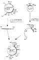
25μl中抗−イディオタイプアフィニティークロマトグラフィによって精製したsFv−huβ−Gluc融合タンパク質200ngを、適当な移動相(PBS、pH7.2、5g/lマルトースと4.2g/lアルギニンを含有)中、TSKゲルG3000SW XLカラム(TOSO HAAS 品番3.5WxN3211、7.8mm×300mm)上で、0.5ml/分の流速でクロマトグラフィ処理を行った。Merck Hitachi HPLC装置(L−4000UV検出器、L−6210インテリジェントポンプ、D−2500クロマト積分計)を約20バールで操作し、溶出液の吸光度を280nmで測定し、LKB2111Mutisacフラクションコレクターを用いて0.5ml画分ずつを集めた後、特異性酵素活性試験(SEAT)(EP0501215A2号明細書、例J)で分析した。この実験の結果を図1に示す。280nmの吸光度の測定によって検出できるピークの位置は、溶出液の特異性及び酵素活性(SEAT)を決定するピークと一致することは明らかである。矢印で示される標準的タンパク質の分子量の位置に基づいて、機能的に活性なsFv−huβ−Gluc融合タンパク質の分子量は本来の条件下では約200kDaであると結論することができる。
【0026】
例7
融合タンパク質の決定のための器官/腫瘍の処理
下記の遂次的工程を行った。
皮下腫瘍を有し、融合タンパク質または抗体−酵素包合体で処理したヌードマウス(CDI)で眼窩後部放血を行った後、屠殺し、
血液をただちにLiquemin25000(Hoffman−LaRoche AG製)10μlを予め入れておいたエッペンドルフ試験管に移し、
次に、遠心分離機(Megafuge1.0、Heraeus製)中で2500rpmで10分間遠心分離を行い、
血漿が得られ、試験まで冷凍し、
器官または腫瘍を除去し、秤量した後、
これらをPBS、pH7.2中で1%BSA2mlで完全にホモジナイズし、腫瘍ホモジネートを0.1N HClでpH4.2に調整し(β−グルクロニダーゼはpH<3.8では不活性化するので、試料には加え過ぎてはならない)、
総てのホモジネートを16000gで30分間遠心分離し、
透明な上澄液を除去し、
腫瘍上澄液を0.1N NaOHで中和すると、
上澄液と血漿を免疫学的試験で定量することができる。
【0027】
例8
3決定基試験
試験を、下記のようにして行う。
マウス抗−huβ−Gluc抗体(MAb2118/157 Behringwerke)をPBS、pH7.2中で2μg/mlまで希釈したもの75μlを、マイクロタイタープレート(ポリスチレン、U字形、B型、Nunc製、品番4−60445)のそれぞれのウェルに入れ、
マイクロタイタープレートをカバーをして室温で一晩インキュベーションし、次いで、マイクロタイタープレートを0.05Mトリス−クエン酸緩衝液、pH7.4のウェル当たり250μlで3回洗浄し、
この方法でコーティングしたこれらのマイクロタイタープレートを遮断溶液(1%カゼイン/PBS、pH7.2)250μlを用いてそれぞれのウェルで室温で30分間インキュベーションし(非特異的結合部位の遮断)(必要のないコーティングしたマイクロタイタープレートを室温で24時間乾燥した後、長期保存のためにコーティングしたアルミニウムバッグに乾燥カートリッジと共に密封し、
遮断中に、未処理の96ウェルのU字形マイクロタイタープレート(ポリスチレン、Renner製、品番12058)中で、10試料+2陽性コントロール+1陰性コントロールを1%カゼイン/PBS、pH7.2で1:2に8段階で希釈し(試料150μlから出発し、試料75μlを採取して、カゼイン溶液75μlに加える、など)、
遮断溶液を抗−huβ−Gluc抗体をコーティングしたマイクロタイタープレートから吸引し、ウェル当たり希釈試料50μlを希釈プレートから試験プレートへ移し、室温で30分間インキュベーションし、
試料のインキュベーション中に、ABC−AP試薬(Vectastain、品番AK−5000)を作成し、試薬A(Avidin DH)2滴を1%カゼイン/PBS、pH7.2、10ml中で十分に混合し、試薬B(ビオチン化アルカリ性ホスファターゼ)2滴を加えて十分に混合し(ABC−AP溶液は使用の少なくとも30分前に作成しなければならない)、
試験プレートをELISA洗浄緩衝液(Behringwerke、品番OSEW96)で3回洗浄し、
ビオチン標識した検出用抗体混合物(マウス抗431/26抗体(MAb2064/353、Behringwerke)及びマウス抗−CEA抗体(MAb250/183、Behringwerke)との1+1混合物を1%カゼイン/PBS、pH7.2、で希釈して5μg/mlの濃度としたもの、それぞれの抗体の最終濃度は2.5μg/ml)、50μlをそれぞれのウェルに入れ、
試験プレートをELISA洗浄緩衝液で3回洗浄し、
調整したABC−AP溶液50μlをそれぞれのウェルに入れて、室温で30分間インキュベーションし、
ABC−APインキュベーション中に、基質を作成し(それぞれの試験に対して新鮮な基質:1mM 4−メチルウンベリフェリルホスフェート、品番M−8843、Sigma製、0.5Mトリス+0.01%MgCl2、pH9.6)、
試験プレートをELISA洗浄緩衝液で7回洗浄し、
基質50μlをそれぞれのウェルに入れ、試験プレートをカバーして37℃で2時間インキュベーションした後、
停止溶液(0.2Mグリシン+0.2%SDS、pH11.7)150μlをそれぞれのウェルに加え、
螢光分析法による評価をFluoroscanII(ICN Biomedicals、カタログ番号78−611−00)中で、励起波長355nmで発光波長460nmで行い、
試料中の融合タンパク質の未知濃度を同一実験に含まれる陽性コントロールについての螢光値(精製したsFv−huβ−Glucを較正プロットとしてCEA5μg/mlと混合したものを有する希釈系)に基づいて測定する。
【0028】
例9
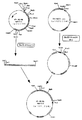
酵母中のsFv−huβ−Gluc融合タンパク質の発現
例2からの一本鎖Fv(sFv)をオリゴ2577及び2561(第7表)で増幅し、XbaI/HindIIIで消化したベクターpUC19中にクローニングする(図2)。
ヒトβ−グルクロニダーゼ遺伝子をプラスミドpAB431/26VHhum/CH1+1H/β−Gluc(例3)からのオリゴ2562及び2540で増幅し、BglII/HindIII(図3)で切断したpUC19(図2)中のプラスミドsFv431/26に連結する。
KpnI/NcoIフラグメントをsFv431/26由来のオリゴ2587及び2627(第9表)で増幅し、KpnI/NcoI(図4)で消化した酵母発現ベクターpIXY中にクローニングする。
pUC19(図3)のプラスミドsFv431/26huβ−Gluc由来のBstEII/HindIIIフラグメントを、VH遺伝子、リンカー及びVL遺伝子の一部を有し(VH/link/VKpart.、pIXY120中)、BstEIIで消化し、部分的にHindIIIで消化したベクターpIXY120中に連結する(図5)。
pIXY120中で得られるプラスミドsFv431/26huβ−GlucをSaccharomyces cerevisiae中に形質転換し、融合タンパク質を発現させる。
【0029】
例10
酵母中でのsFv−大腸菌−β−グルクロニダーゼ融合タンパク質の発現
大腸菌グルクロニダーゼ遺伝子をオリゴ2638及び2639(第10表)でpRAJ275(Jeffersonら、Proc.Natl.Acad.Sci,USA,83:8447−8451,1986)から増幅し、BglII/HindIII(図6)で切断したpUC19中(例9、図2)でsFv431/26中に連結する。
pUC19中のsFv431/26大腸菌β−GlucからのBstEII/HindIIIフラグメントを、BstEII/HindIII(図7)で部分的に消化したpIXY120(例9、図4)におけるベクターVH/link/VKpart中にクローニングする。
pIXY120中のプラスミドsFv431/26大腸菌β−GlucをSaccharomyces cerevisiae中に形質転換し、融合タンパク質を発現させる。
【0030】
例11
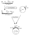
酵母中におけるsFv−β−ラクタマーゼ融合タンパク質の発現
例2からの一本鎖Fv(sFv)をオリゴ2587及び2669(第11表)で増幅し、KpnI/HindIIIで消化したpUC19ベクターに連結する(図8)。
β−ラクタマーゼII遺伝子(Hussainら、J.Bacteriol.,164:223−229,1985)をBacellus cereus の完全なDNAからのオリゴ2673及び2674で増幅し、EcoRI/HindIIIで消化したpUC19ベクターに連結する(図9)。β−ラクタマーゼ遺伝子のBclI/HindIIIフラグメントを、BglII/HindIIIで切断したpUC19中のsFv431/26に連結する(図10)。
KpnI/HindIIIsFv−β−ラクタマーゼフラグメントをKpnIで消化し、HindIIIで部分的に消化したpIXY120に連結する(図11)。プラスミドをSaccharomyces cerevisiae中で形質転換し、MAb431/26の抗原結合特性とBacillus cereus β−ラクタマーゼの酵素活性を両方とも有する融合タンパク質を発現させる。
【0031】
【表1】

【表2】
【表3】
【表4】
【表5】
【表6】
【表7】
【表8】
マンノース、グルコサミン及びガラクトースのモル比から、高マンノース型および/またはハイブリッド型構造(複合型構造の外に)が存在すると結論することができる。それ故、マンノース、ガラクトース、アセチルノイラミン酸或いはN−アセチルグルコサミンは末端に存在し、マンノースはマンノース6−ホスフェートとしても存在できる。
方法:
ノイラミン酸はHermentinとSeidat(1991)GBF Monographs 15巻、185〜188頁の方法(80℃で0.1N硫酸の存在下にて30分間加水分解した後、0.4N水酸化ナトリウム溶液で中和)によって、パルスアメロメトリー検出法による高pHアニオン交換クロマトグラフィ(HPAE−PAD)によって測定した。
単糖類成分も同様に、Hardyら、(1988)Analytical Biochemistry,170,54−62に記載の方法を基としたHPAE−PAD(100℃で2Nトリフルオロ酢酸の存在下にて4時間加水分解した後、SpeedVacで蒸発乾固)によって測定した。
【0039】
【表9】
【表10】
【表11】
【表12】
【表13】
【表14】
【図面の簡単な説明】
【図1】TSK3000ゲルクロマトグラフィ分析の結果を示す説明図。
【図2】pUC19中プラスミドsFv431/26の構築を示す説明図。
【図3】pUC19中プラスミドsFv431/26 huβ−Glucの構築を示す説明図。
【図4】pIXY120中ベクターVH/link/VK partの構築を示す説明図。
【図5】pIXY120中sFv431/26 huβ−Glucの構築を示す説明図。
【図6】pUC19中sFv431/26 E.Coli β−Glucの構築を示す説明図。
【図7】pIXY120中sFv431/26 E.Coli β−Glucの構築を示す説明図。
【図8】pUC19中sFv431/26の構築を示す説明図。
【図9】pUC19中β−ラクタマーゼ遺伝子の構築を示す説明図。
【図10】pUC19中431/26sFv/β−ラクタマーゼ遺伝子の構築を示す説明図。
【図11】pIXY120中431/26sFv/β−ラクタマーゼ遺伝子の構築を示す説明図。[0001]
[Industrial application fields]
The present invention relates to a compound comprising an antigen-binding region that binds to at least one enzyme capable of metabolizing a compound having little or no cytotoxicity (prodrug) into a cytotoxic compound (drug). The present invention relates to a compound characterized in that the binding region consists of a single polypeptide chain. Advantageously, covalently linked carbohydrates are present on the polypeptide chain.
[0002]
[Prior art]
Combinations of prodrugs and antibody-enzyme conjugates used as therapeutic compositions have already been described in the expert literature. This allows an antibody directed to a specific tissue and bound by a prodrug-cleaving enzyme to be injected into the body and subsequently administered a prodrug compound that can be activated by this enzyme. . The action of the antibody-enzyme conjugate bound to the target tissue is intended to convert it into a compound that exhibits a cytotoxic effect on the tissue bound to the prodrug compound. However, studies on antibody-enzyme conjugates have shown that these chemical conjugates exhibit unfavorable pharmacokinetics and only inappropriate site-specific tumor-selective cleavage of prodrugs. Some authors have attempted to remove this apparent disadvantage by attempting to remove the antibody-enzyme conjugate from plasma rapidly by injecting additional anti-enzyme antibody (Sharma et al., Brit. J. Cancer, 61, 659, 1990). Another problem with antibody-enzyme conjugates is that they are highly reproducible and have limited possibilities for preparing large quantities in a homogeneous manner.
[0003]
[Problems to be solved by the invention]
The object of the present invention was to find a fusion protein that can be prepared on an industrial scale and is suitable for therapeutic purposes due to its pharmacokinetic and pharmacological properties.
[0004]
[Means for Solving the Problems]
In this context, it has been found that compounds having an antigen binding region consisting of a single polypeptide chain are unexpectedly advantageous for use in the preparation of fusion proteins to which carbohydrates bind advantageously and in the activation of prodrugs. It was.
Therefore, the present invention is a compound having an antigen-binding region that binds to at least one enzyme, wherein the antigen-binding region consists of a single polypeptide chain, and a carbohydrate is advantageously bound to a fusion protein About.
An antigen binding region is, for the purposes of the present invention, at least two variable domains of an antibody, preferably one variable domain of a heavy chain antibody and one variable domain of a light chain antibody (sFv fragment) Means an area containing However, the antigen binding region can also have a bivalent or multivalent structure, ie, two or more binding regions as described, for example, in EP-A-0 404 097. However, human or humanized sFv fragments are particularly preferred, with humanized sFv fragments being particularly preferred.
[0005]
The antigen binding region preferably binds to a tumor associated antigen (TAA), and the following TAA is particularly preferable.
Neural cell adhesion molecule (N-CAM),
Polymorphic epithelial mucin (PEM), epidermal growth factor receptor (EGF-R),
Tomsen friedenreich antigen β (TFβ),
Gastrointestinal cancer antigen (GICA),
Ganglioside GD 3 (GD 3 ),
Ganglioside GD 2 (GD 2 ),
Sialyl-Le a , Sialyl-Le x ,
TAG72,
A 24-25 kDa glycoprotein defined by MAb L6;
CA125, and especially carcinoembryonic antigen (CEA).
[0006]
Preferred enzymes are those that can metabolize compounds with little or no cytotoxicity to cytotoxic compounds. Examples include β-lactamase, pyroglutamate aminopeptidase, galactosidase or D-aminopeptidase as described in EP-A2-0 382 411 or EP-A2-0 392 745; Oxidases described in US Patent No. 00108, such as ethanol oxidase, galactose oxidase, D-amino acid oxidase or α-glyceryl-phosphate oxidase; for example peroxidases described in EP-A2-0 361 908; Phosphatases described in EP-A1-0 302 473; for example, hydroxynitrile lyases or glucosidases disclosed in WO 91/11201; carboxypeptidases For example carboxypeptidase G2 (WO 88/07378); amidases such as penicillin 5-amidase (Kerr, DE et al., Cancer Immunol. Immunther., 1990, 31); and proteases, esterases or glycosidases such as WO 91/08770 For example, galactosidase, glucosidase or glucuronidase.
[0007]
β-glucuronidase is preferred, preferably derived from Kobayasia nipponica or Secale cereale, more preferably derived from E. coli or human β-glucuronidase. Individual enzyme substrates are also pointed out in the above-mentioned patent specifications and form part of the disclosure content of this application. Preferred substrates for β-glucuronidase are N- (D-glycopyranosyl) benzyloxycarbonyl anthracyclines, especially N- (4-hydroxy-3-nitrobenzyloxycarbonyl) doxorubicin and daunorubicin = β-D-glucuronide (JC Florent et al. (1992) Int. Carbohydr. Symp. Paris, A262, 297 or S. Andrianomenjanahary et al. (1992) Int. Carbohydr. Symp. Paris, A264, 299).
[0008]
The invention also relates to a nucleic acid encoding a compound according to the invention. Particularly preferred are nucleic acids and variants and variants thereof that encode humanized sFv fragments against CEA (carcinoembryonic antigen) linked to human β-glucuronidase, preferably Has the sequence shown in Table 1 (sFv-huβ-Gluc).
[0009]
The compounds according to the invention are usually prepared by methods of genetic manipulation commonly known to those skilled in the art, and the antigen binding region is linked to one or more enzymes directly or via a linker, preferably a peptide linker. be able to. Peptide linkers that can be used are, for example, antibody hinge regions or hinge-like amino acid sequences. In this case, the enzyme is preferably linked at the N-terminus to the antigen binding region either directly or via a peptide linker. However, the enzyme or enzymes can also be chemically bound to the antigen binding region as described, for example, in WO 91/00108.
[0010]
A nucleic acid encoding the amino acid sequence of a compound of the invention is typically cloned into an expression vector and introduced into a prokaryotic or eukaryotic host cell such as a BHK, CHO, COS, HeLa, insect, tobacco plant, yeast or E. coli cell, Expressed. The compound prepared in this way can then be isolated and used as a diagnostic aid or therapeutic agent. Another commonly known method for preparing the compounds according to the invention is to express nucleic acid encoding in a non-human transgenic mammal, preferably a transgenic goat.
[0011]
BHK cells transfected with a nucleic acid according to the invention express a fusion protein (sFv-huβ-Gluc) that is not only specific for CEA but also has complete β-glucuronidase activity (see Example 5). I want to be)
This fusion protein was purified by anti-idiotype affinity chromatography by the method described in EP 0501215 A2 (Example M). The molecular weight of the fusion protein purified by this method is 100 kDa in SDS PAGE under reducing conditions, whereas the 100 and 200 kDa molecules each appear under non-reducing conditions.
[0012]
Gel chromatography under native conditions (TSK-3000 gel chromatography) shows one protein peak (Example 6, FIG. 1), which correlates with the activity peak in the specific enzyme activity test (
[0013]
Surprisingly, the tumor / plasma ratio reaches 5/1 after only 48 hours. Over time, this ratio becomes more favorable and reaches a value> 200/1 (day 5). The reason that the sFv-huβ-Gluc fusion protein exhibits such favorable pharmacokinetics is that the non-tumor-bound fusion protein is removed from plasma and normal tissues primarily by internalization with mannose 6-phosphate and galactose receptors. This is because that. (Evidence for this phenomenon is, for example, increased intramolecular β-glucuronidase concentration in the liver).
[0014]
As shown in Table 5, sFv-huβ-Gluc contains a relatively large amount of galactose, particularly mannose, which is mainly important for binding to specific receptors. The fusion protein / receptor complex that is produced and bound through the carbohydrate residues of the fusion protein is then removed from the extracellular compartment by internalization.
This rapid internalization mechanism mediated primarily by galactose and mannose is closely related to the favorable pharmacokinetics of the fusion protein according to the invention. These advantageous pharmacokinetics of fusion proteins to which galactose and especially mannose binds allow hydrophilic prodrugs with extracellular distribution to be administered intravenously relatively early without inducing nonspecific prodrug activation can do. In this case, the removal step described by Sharma et al. (Brit. J. Cancer, 61, 659, 1990) is not necessary. Based on the data in Table 4, appropriate prodrug injections (S. Adrianomenjanahari et al., 1992, Int. Carbohydrate Symp., Parts A264, 299) were performed 3 days after injection of the sFv-huβ-Gluc fusion protein. It can be done without serious side effects (data not shown).
[0015]
Similarly, advantageous binding of carbohydrate to the fusion protein can also be effected by secretory expression of the sFv-huβ-Gluc fusion protein in certain yeast strains such as Saccharomyces cerevisiae or Hansenula polymorpha. These organisms can very effectively mannosylate fusion proteins with appropriate N-glycosylation sites (Goochee et al., Biotechnology, 9, 1347-1354, 1991). Such fusion proteins secreted in yeast cells show a high degree of mannosylation and favorable pharmacokinetics comparable to the sFv-huβ-Gluc fusion protein expressed in BHK cells (data not shown) . In this case, the absence of galactose is compensated by the very high degree of mannosylation of the fusion protein (Table 6). The sFv-huβ-Gluc fusion protein was constructed by genetic engineering and expressed in yeast as described in detail in Example 9.
[0016]
However, other glucuronidases with advantageous properties can be used instead of human β-glucuronidase. For example, E. coli β-glucuronidase has the particular advantage that its catalytic activity at pH 7.4 is significantly higher than that of human β-glucuronidase. In Example 10, the sFv-E. Coli β-Gluc construct was prepared by genetic engineering methods and secreted and expressed as a functionally active mannosylated fusion protein in Saccharomyces cerevisiae. Pharmacokinetic data is comparable to that of sFv-huβ-Gluc molecules expressed in yeast or BHK cells (Table 4).
[0017]
Glucuronidases from fungus Kobayasia nipponica and the plant Secale cereale have, for example, the advantage that they are active as monomers. In Example 11, a genetic engineering method was used to prepare a construct that preferentially releases the sFv-B. Cereus β-lactamase II fusion protein in mannosylated form after expression in Saccharomyces cerevisiae.
This fusion protein is also based on the β-glucuronidase pharmacokinetics preferred for prodrug activation, similar to the fusion protein according to the invention (Table 4).
[0018]
Furthermore, not only is the compound according to the present invention used in combination with a prodrug, but also a conventional chemotherapy that can convert a cytostatic agent that is metabolized and inactivated as a glucuronide into a toxic form by the administered compound. It can also be used for the configuration of
[0019]
The following example demonstrates the synthesis and functional demonstration of sFv-β-Gluc fusion protein by genetic engineering.
Starting material is plasmid pABstop431 / 26humV H And pABstop 431/26 humVH L Was included. These plasmids contain the anti-CEA MAb BW431 / 26 V H Gene and V L Includes humanized genes (Guessow and Seemann, 1991, Meth. Enzymology, 203, 99-121). Other starting materials are plasmid pABstop431 / 26V H -Huβ-Gluc1H (EP-A2-0501215), wherein V H V containing a unique signal sequence H It consisted of an exon followed by a CH1 exon, a human IgG3c gene hinge exon and a human β-glucuronidase complete cDNA.
[0020]
Example 1
Using oligonucleotide pAB-Back and linker-anti (Table 2), pABstop431V H hum (
[0021]
Example 2
The oligonucleotide linker-anti and linker-sense are partially complementary to each other and V H Domain and V L It encodes a polypeptide linker to join domains and obtain sFv fragments. Amplified V H Fragments and V L In order to fuse the fragments, they are purified and used in a 10 cycle reaction as described below.
H 2 O 37.5 μl
dNTP (2.5 mM) 5.0 μl
PCR buffer (10x) 5.0 μl
Taq polymerase (Perkin-Elmer Corp., Emeryville,
California) (2.5 U / μl) 0.5 μl
V L Fragment 0.5 μg / μl DNA 1.0 μl
V H Fragment 0.5 μg / μl DNA 1.0 μl
PCR buffer (10 ×): 100 mM Tris, pH 8.3, 500 mM KCl, 15 mM MgCl 2 0.1% (w / v) gelatin.
After sealing the surface of the reaction mixture with paraffin, a 10-cycle reaction was performed in a PCR apparatus programmed at 94 ° C. for 1 minute, 55 ° C. for 1 minute, 72 ° C. for 2 minutes, and flanking primers pAB-Back and V L (Mut) Add -For and react for another 20 cycles. The resulting PCR fragment is linked to the V via a linker. L V linked to a gene H It consists of genes. V H The signal sequence unique to the gene is also V H Before the gene. Oligonucleotide V L (Mut) -For also V L The last nucleotide base of the gene, C is replaced by G. This PCR fragment encodes a humanized single chain Fv (sFv431 / 26).
[0022]
Example 3
Cloning of sFv431 / 26 fragment into expression vector containing huβ-glucuronidase gene
The vector pAB431V, in which the sFv fragment from (2) was cleaved with HindIII and BamHI, completely cleaved with HindIII and partially cleaved with BglII H Link to hum / CH1 + 1h / β-Glc.
[0023]
Example 4
Expression of sFv-huβ-Gluc fusion protein in BHK cells
Clone pMCG-E1 is transfected into BHK cells with plasmid pRMH140 having a neomycin resistance gene and plasmid pSV2 having a methotrexate resistance gene. BHK cells then express a fusion protein that has both the antigen binding properties of MAb BW431 / 26hum and the enzymatic activity of human β-glucuronidase.
[0024]
Example 5
Demonstration of antigen binding properties and enzyme activity of sFv-huβ-Gluc fusion protein
It has been shown that the sFv-huβ-Gluc fusion protein can specifically bind to the CEA epitope defined by 431/26 and at the same time show the enzymatic activity of human β-glucuronidase (EP- A2-0501215 specification). This test measures the release of 4-methylumbelliferone from 4-methylumbelliferyl β-glucuronide by the β-glucuronidase portion of the fusion protein after the fusion protein binds to the antigen via the sFv portion. Record the measured fluorescence values as relative fluorescence units (FU). This test shows significant release of methyl-umbelliferone by the fusion protein on CEA coated plates. In contrast, the fusion protein does not release any methylumbelliferone in a control plate coated with PEM (polymorphic epithelial mucin).
[0025]
Example 6
TSK3000 gel chromatography
200 ng of sFv-huβ-Gluc fusion protein purified by anti-idiotype affinity chromatography in 25 μl in TSK gel in an appropriate mobile phase (containing PBS, pH 7.2, 5 g / l maltose and 4.2 g / l arginine) Chromatography was performed on a G3000SW XL column (TOSO HAAS product number 3.5WxN3211, 7.8 mm x 300 mm) at a flow rate of 0.5 ml / min. The Merck Hitachi HPLC apparatus (L-4000 UV detector, L-6210 intelligent pump, D-2500 chromatograph integrator) is operated at about 20 bar, the absorbance of the eluate is measured at 280 nm, and 0. 0 using an LKB2111 Mutisac fraction collector. Five ml fractions were collected and analyzed by a specific enzyme activity test (SEAT) (EP 0501215A2, specification J). The results of this experiment are shown in FIG. It is clear that the position of the peak that can be detected by measuring the absorbance at 280 nm coincides with the peak that determines the specificity and enzyme activity (SEAT) of the eluate. Based on the molecular weight position of the standard protein indicated by the arrow, it can be concluded that the molecular weight of the functionally active sFv-huβ-Gluc fusion protein is about 200 kDa under native conditions.
[0026]
Example 7
Organ / tumor treatment for fusion protein determination
The following sequential steps were performed.
Nude mice (CDI) with subcutaneous tumors treated with fusion proteins or antibody-enzyme conjugates were subjected to retroorbital exsanguination and then sacrificed,
Immediately transfer the blood to an Eppendorf tube containing 10 μl of Liquidin 25000 (Hoffman-LaRoche AG),
Next, centrifuge at 2500 rpm for 10 minutes in a centrifuge (Megafuge 1.0, manufactured by Heraeus)
Plasma is obtained, frozen until testing,
After removing the organ or tumor and weighing it,
These were completely homogenized with 2 ml of 1% BSA in PBS, pH 7.2, and the tumor homogenate was adjusted to pH 4.2 with 0.1 N HCl (because β-glucuronidase is inactivated at pH <3.8, the sample Must not be added too much)
Centrifuge all homogenates at 16000 g for 30 minutes,
Remove the clear supernatant,
When the tumor supernatant is neutralized with 0.1N NaOH,
Supernatant and plasma can be quantified by immunological tests.
[0027]
Example 8
3 Determinant test
The test is performed as follows.
75 μl of mouse anti-huβ-Gluc antibody (MAb2118 / 157 Behringwerke) diluted to 2 μg / ml in PBS, pH 7.2 was added to a microtiter plate (polystyrene, U-shaped, B-type, manufactured by Nunc, product number 4-60445). ) Into each well,
Cover the microtiter plate and incubate overnight at room temperature, then wash the
These microtiter plates coated in this way are incubated with 250 μl of blocking solution (1% casein / PBS, pH 7.2) in each well for 30 minutes at room temperature (blocking of non-specific binding sites) (required After drying the uncoated microtiter plate at room temperature for 24 hours, seal it with a drying cartridge in a coated aluminum bag for long-term storage,
During blocking, 10 samples + 2 positive control + 1 negative control 1: 2 with 1% casein / PBS, pH 7.2 in an untreated 96-well U-shaped microtiter plate (polystyrene, manufactured by Renner, part number 12058). Dilute in 8 steps (start with 150 μl sample, take 75 μl sample and add to 75 μl casein solution, etc.)
Blocking solution is aspirated from the anti-huβ-Gluc antibody coated microtiter plate and 50 μl of diluted sample per well is transferred from the dilution plate to the test plate and incubated for 30 minutes at room temperature,
During sample incubation, ABC-AP reagent (Vectastein, part number AK-5000) is made, 2 drops of reagent A (Avidin DH) are mixed well in 1% casein / PBS, pH 7.2, 10 ml, the
Wash the
Biotin-labeled detection antibody mixture (mouse anti-431 / 26 antibody (MAb 2064/353, Behringwerke) and mouse anti-CEA antibody (
Wash the
Add 50 μl of the prepared ABC-AP solution to each well and incubate for 30 minutes at room temperature.
During the ABC-AP incubation, a substrate was prepared (fresh substrate for each test: 1 mM 4-methylumbelliferyl phosphate, part number M-8843, Sigma, 0.5 M Tris + 0.01% MgCl 2 , PH 9.6),
Wash the test plate 7 times with ELISA wash buffer,
After 50 μl of substrate is placed in each well and the test plate is covered and incubated at 37 ° C. for 2 hours,
Add 150 μl of stop solution (0.2 M glycine + 0.2% SDS, pH 11.7) to each well,
Evaluation by fluorescence analysis is performed in Fluoroscan II (ICN Biomedicals, catalog number 78-611-00) at an excitation wavelength of 355 nm and an emission wavelength of 460 nm.
The unknown concentration of the fusion protein in the sample is measured based on the fluorescence value for a positive control included in the same experiment (dilution system with purified sFv-huβ-Gluc mixed with CEA 5 μg / ml as a calibration plot) .
[0028]
Example 9
Expression of sFv-huβ-Gluc fusion protein in yeast
Single chain Fv (sFv) from Example 2 is amplified with oligos 2577 and 2561 (Table 7) and cloned into the vector pUC19 digested with XbaI / HindIII (FIG. 2).
The human β-glucuronidase gene was transferred to the plasmid pAB431 / 26V. H Amplified with oligos 2562 and 2540 from hum / CH1 + 1H / β-Gluc (Example 3) and ligated to plasmid sFv431 / 26 in pUC19 (FIG. 2) cut with BglII / HindIII (FIG. 3).
The KpnI / NcoI fragment is amplified with
A BstEII / HindIII fragment derived from plasmid sFv431 / 26huβ-Gluc of pUC19 (FIG. 3) was combined with the VH gene, linker and V L It has a part of the gene (VH / link / VKpart., In pIXY120), digested with BstEII and ligated into the vector pIXY120 partially digested with HindIII (FIG. 5).
The plasmid sFv431 / 26huβ-Gluc obtained in pIXY120 is transformed into Saccharomyces cerevisiae to express the fusion protein.
[0029]
Example 10
Expression of sFv-Escherichia coli-β-glucuronidase fusion protein in yeast
The E. coli glucuronidase gene was amplified from pRAJ275 (Jefferson et al., Proc. Natl. Acad. Sci, USA, 83: 8447-8451, 1986) with oligos 2638 and 2639 (Table 10) and cut with BglII / HindIII (FIG. 6). In pUC19 (Example 9, FIG. 2).
Vector V in pIXY120 (Example 9, FIG. 4) in which the BstEII / HindIII fragment from sFv431 / 26 E. coli β-Gluc in pUC19 was partially digested with BstEII / HindIII (FIG. 7). H / Link / V K Cloning into part.
Plasmid sFv431 / 26 E. coli β-Gluc in pIXY120 is transformed into Saccharomyces cerevisiae to express the fusion protein.
[0030]
Example 11
Expression of sFv-β-lactamase fusion protein in yeast
Single chain Fv (sFv) from Example 2 is amplified with
The β-lactamase II gene (Hussain et al., J. Bacteriol., 164: 223-229, 1985) is amplified with oligos 2673 and 2673 from the complete DNA of Bacellus cereus and ligated into the pUC19 vector digested with EcoRI / HindIII (FIG. 9). The BclI / HindIII fragment of the β-lactamase gene is ligated to sFv431 / 26 in pUC19 cut with BglII / HindIII (FIG. 10).
The KpnI / HindIIIsFv-β-lactamase fragment is digested with KpnI and ligated to pIXY120 partially digested with HindIII (FIG. 11). The plasmid is transformed in Saccharomyces cerevisiae to express a fusion protein having both the antigen binding properties of
[0031]
[Table 1]
[Table 2]
[Table 3]
[Table 4]
[Table 5]
[Table 6]
[Table 7]
[Table 8]
From the molar ratio of mannose, glucosamine and galactose, it can be concluded that there is a high mannose and / or hybrid structure (outside the complex structure). Therefore, mannose, galactose, acetylneuraminic acid or N-acetylglucosamine is present at the terminal, and mannose can also exist as mannose 6-phosphate.
Method:
Neuraminic acid was hydrolyzed by Hermentin and Seidat (1991) GBF Monographs, Vol. 15, pages 185-188 (at 80 ° C. for 30 minutes in the presence of 0.1 N sulfuric acid and then neutralized with 0.4 N sodium hydroxide solution. ) By high pH anion exchange chromatography (HPAE-PAD) with pulse amelometry detection.
The monosaccharide component was similarly hydrolyzed for 4 hours in the presence of 2N trifluoroacetic acid at 100 ° C. based on the method described by Hardy et al. (1988) Analytical Biochemistry, 170, 54-62. After that, it was measured by evaporating to dryness with SpeedVac.
[0039]
[Table 9]
[Table 10]
[Table 11]
[Table 12]
[Table 13]
[Table 14]
[Brief description of the drawings]
FIG. 1 is an explanatory diagram showing the results of TSK3000 gel chromatography analysis.
FIG. 2 is an explanatory diagram showing the construction of plasmid sFv431 / 26 in pUC19.
FIG. 3 is an explanatory diagram showing the construction of a plasmid sFv431 / 26 huβ-Gluc in pUC19.
FIG. 4: Vector V in pIXY120 H / Link / V K Explanatory drawing which shows construction of part.
FIG. 5 is an explanatory diagram showing the construction of sFv431 / 26 huβ-Gluc in pIXY120.
FIG. 6:
7:
FIG. 8 is an explanatory diagram showing the construction of sFv431 / 26 in pUC19.
FIG. 9 is an explanatory diagram showing the construction of a β-lactamase gene in pUC19.
FIG. 10 is an explanatory diagram showing the construction of a 431/26 sFv / β-lactamase gene in pUC19.
FIG. 11 is an explanatory diagram showing the construction of a 431/26 sFv / β-lactamase gene in pIXY120.
Claims (24)
(b) この宿主細胞を培養し、
(c) 化合物を単離する、
ことを特徴とする、請求項1〜7のいずれか1項に記載の化合物の製造法。(A) introducing the nucleic acid according to any one of claims 13 to 15 or the vector according to claim 16 into a host cell;
(B) culturing the host cell;
(C) isolating the compound,
The method for producing a compound according to any one of claims 1 to 7, wherein
(b) 化合物を単離する、
ことを特徴とする請求項1〜7のいずれか1項に記載の化合物の製造法。(A) culturing the host cell according to claim 17 or 18,
(B) isolating the compound,
The method for producing a compound according to any one of claims 1 to 7, wherein:
Applications Claiming Priority (2)
| Application Number | Priority Date | Filing Date | Title |
|---|---|---|---|
| DE4233152A DE4233152A1 (en) | 1992-10-02 | 1992-10-02 | Antibody-enzyme conjugates for prodrug activation |
| DE4233152.8 | 1992-10-02 |
Publications (2)
| Publication Number | Publication Date |
|---|---|
| JPH06228195A JPH06228195A (en) | 1994-08-16 |
| JP3776137B2 true JP3776137B2 (en) | 2006-05-17 |
Family
ID=6469477
Family Applications (1)
| Application Number | Title | Priority Date | Filing Date |
|---|---|---|---|
| JP27129193A Expired - Lifetime JP3776137B2 (en) | 1992-10-02 | 1993-10-04 | Fusion proteins for prodrug activation |
Country Status (17)
| Country | Link |
|---|---|
| US (2) | US7060495B2 (en) |
| EP (1) | EP0590530B1 (en) |
| JP (1) | JP3776137B2 (en) |
| KR (1) | KR100321012B1 (en) |
| AT (1) | ATE192193T1 (en) |
| AU (1) | AU672431B2 (en) |
| CA (1) | CA2107513C (en) |
| DE (2) | DE4233152A1 (en) |
| DK (1) | DK0590530T3 (en) |
| ES (1) | ES2147189T3 (en) |
| GR (1) | GR3033417T3 (en) |
| IL (1) | IL107154A (en) |
| NO (1) | NO313884B1 (en) |
| NZ (1) | NZ248818A (en) |
| PT (1) | PT590530E (en) |
| UY (1) | UY23660A1 (en) |
| ZA (1) | ZA937299B (en) |
Families Citing this family (23)
| Publication number | Priority date | Publication date | Assignee | Title |
|---|---|---|---|---|
| DE4314556A1 (en) * | 1993-05-04 | 1994-11-10 | Behringwerke Ag | Modified antibody-enzyme conjugates and fusion proteins and their use in tumor-selective therapy |
| GB9324807D0 (en) * | 1993-12-03 | 1994-01-19 | Cancer Res Campaign Tech | Tumour antibody |
| GB9406974D0 (en) | 1994-04-08 | 1994-06-01 | Pharmaceutical Proteins Ltd | Transgenic production |
| US5866679A (en) * | 1994-06-28 | 1999-02-02 | Merck & Co., Inc. | Peptides |
| US6143864A (en) * | 1994-06-28 | 2000-11-07 | Merck & Co., Inc. | Peptides |
| US5599686A (en) * | 1994-06-28 | 1997-02-04 | Merck & Co., Inc. | Peptides |
| DE19513676A1 (en) | 1995-04-11 | 1996-10-17 | Behringwerke Ag | Cytoplasmic expression of antibodies, antibody fragments and antibody fragment fusion molecules in E. coli |
| DK0795334T3 (en) * | 1996-03-12 | 2006-06-06 | Sanofi Aventis Deutschland | New prodrugs for the treatment of tumors and inflammatory diseases |
| WO1999045127A2 (en) * | 1998-03-06 | 1999-09-10 | Oxford Biomedica (Uk) Limited | Enhanced prodrug activation |
| EP1194570A1 (en) | 1999-06-23 | 2002-04-10 | PPL Therapeutics (Scotland) Limited | Fusion proteins incorporating lysozyme |
| DE10133071A1 (en) * | 2001-07-07 | 2003-03-06 | Alexander Cherkasky | New compound containing protease and a recognition component, useful for treating diseases associated with specific proteins, e.g. multiple sclerosis |
| US7232888B2 (en) | 2002-07-01 | 2007-06-19 | Massachusetts Institute Of Technology | Antibodies against tumor surface antigens |
| US7456000B2 (en) * | 2004-08-26 | 2008-11-25 | Siemens Healthcare Diagnostics Inc. | Deactivation of linking moieties in antibody-enzyme conjugates |
| US8491891B2 (en) * | 2008-11-26 | 2013-07-23 | Academia Sinica | Human beta-glucuronidase mutants with elevated enzymatic activity under physiological conditions and method for identifying such |
| ES2584956T3 (en) | 2009-03-10 | 2016-09-30 | Baylor Research Institute | Anti-CD40 antibodies and uses thereof |
| EP2406290B1 (en) * | 2009-03-10 | 2017-07-05 | Baylor Research Institute | Antigen presenting cell targeted cancer vaccines |
| CN105837691B (en) * | 2009-03-10 | 2021-06-29 | 贝勒研究院 | Antigen presenting cell targeted cancer vaccines |
| WO2012012737A2 (en) | 2010-07-23 | 2012-01-26 | The University Of Toledo | Stable tregs and related materials and methods |
| AR085573A1 (en) | 2011-03-25 | 2013-10-09 | Baylor Res Inst | COMPOSITIONS AND IMMUNIZATION METHODS AGAINST THE VIRUS OF HEPATITIS C |
| CN104101711B (en) * | 2013-04-07 | 2016-03-23 | 广州瑞博奥生物科技有限公司 | A kind of ELISA measuring reagent kit of improvement and detection method thereof |
| US10286058B2 (en) | 2014-01-13 | 2019-05-14 | Baylor Research Institute | Vaccines against HPV and HPV-related diseases |
| CN105961304B (en) * | 2016-06-14 | 2019-06-28 | 浙江省海洋水产研究所 | A kind of Marsupenaeus japonicus cultivation feed coefficient and its cultural method |
| US10610585B2 (en) | 2017-09-26 | 2020-04-07 | Inserm (Institut National De La Sante Et De La Recherche Medicale) | Methods and compositions for treating and preventing HIV |
Family Cites Families (22)
| Publication number | Priority date | Publication date | Assignee | Title |
|---|---|---|---|---|
| US4870009A (en) | 1982-11-22 | 1989-09-26 | The Salk Institute For Biological Studies | Method of obtaining gene product through the generation of transgenic animals |
| GB8705477D0 (en) | 1987-03-09 | 1987-04-15 | Carlton Med Prod | Drug delivery systems |
| US5132405A (en) | 1987-05-21 | 1992-07-21 | Creative Biomolecules, Inc. | Biosynthetic antibody binding sites |
| US5258498A (en) * | 1987-05-21 | 1993-11-02 | Creative Biomolecules, Inc. | Polypeptide linkers for production of biosynthetic proteins |
| DE3856559T2 (en) * | 1987-05-21 | 2004-04-29 | Micromet Ag | Multifunctional proteins with predetermined objectives |
| US4873316A (en) | 1987-06-23 | 1989-10-10 | Biogen, Inc. | Isolation of exogenous recombinant proteins from the milk of transgenic mammals |
| NZ225599A (en) | 1987-08-04 | 1991-09-25 | Bristol Myers Co | Antibody-enzyme conjugates and combinations with prodrugs for the treatment of tumour cells |
| GB8809616D0 (en) * | 1988-04-22 | 1988-05-25 | Cancer Res Campaign Tech | Further improvements relating to drug delivery systems |
| EP0361908A3 (en) | 1988-09-28 | 1990-09-05 | Ideon Corporation | Combination enzyme immunotherapeutics |
| WO1990005144A1 (en) * | 1988-11-11 | 1990-05-17 | Medical Research Council | Single domain ligands, receptors comprising said ligands, methods for their production, and use of said ligands and receptors |
| AU633867B2 (en) | 1989-02-02 | 1993-02-11 | Eli Lilly And Company | Delivery of cytotoxic agents |
| GB8907617D0 (en) * | 1989-04-05 | 1989-05-17 | Celltech Ltd | Drug delivery system |
| US6258360B1 (en) * | 1989-05-04 | 2001-07-10 | Igen International, Inc. | Prodrugs activated by targeted catalytic proteins |
| DE3920358A1 (en) | 1989-06-22 | 1991-01-17 | Behringwerke Ag | BISPECIFIC AND OLIGO-SPECIFIC, MONO- AND OLIGOVALENT ANTI-BODY CONSTRUCTS, THEIR PRODUCTION AND USE |
| US6020145A (en) * | 1989-06-30 | 2000-02-01 | Bristol-Myers Squibb Company | Methods for determining the presence of carcinoma using the antigen binding region of monoclonal antibody BR96 |
| WO1991000108A1 (en) * | 1989-06-30 | 1991-01-10 | Brunswick Corporation | Antibody-oxidase conjugates with non-systemic substrates |
| DE4106389A1 (en) | 1991-02-28 | 1992-09-03 | Behringwerke Ag | FUSION PROTEINS FOR PRODRUG ACTIVATION, THEIR PRODUCTION AND USE |
| DE68928946T2 (en) * | 1989-12-11 | 1999-10-21 | Immunomedics, Inc. | METHOD FOR DETECTING DIAGNOSTIC OR THERAPEUTIC AGENTS BY ANTIBODIES |
| GB9001641D0 (en) | 1990-01-24 | 1990-03-21 | Imp Cancer Res Tech | Compounds |
| GB9200417D0 (en) * | 1992-01-09 | 1992-02-26 | Bagshawe Kenneth D | Cytotoxic drug therapy |
| DE19647273A1 (en) * | 1996-11-15 | 1998-05-20 | Zeiss Carl Fa | Modular infrared Kepler telescope |
| US6258498B1 (en) * | 1998-12-25 | 2001-07-10 | Canon Kabushiki Kaisha | Electrophotographic photosensitive member, and process cartridge and electrophotographic photosensitive member |
-
1992
- 1992-10-02 DE DE4233152A patent/DE4233152A1/en not_active Withdrawn
-
1993
- 1993-09-24 PT PT93115418T patent/PT590530E/en unknown
- 1993-09-24 DE DE59310018T patent/DE59310018D1/en not_active Revoked
- 1993-09-24 EP EP93115418A patent/EP0590530B1/en not_active Revoked
- 1993-09-24 AT AT93115418T patent/ATE192193T1/en not_active IP Right Cessation
- 1993-09-24 ES ES93115418T patent/ES2147189T3/en not_active Expired - Lifetime
- 1993-09-24 DK DK93115418T patent/DK0590530T3/en active
- 1993-09-29 IL IL10715493A patent/IL107154A/en not_active IP Right Cessation
- 1993-09-30 NZ NZ248818A patent/NZ248818A/en not_active IP Right Cessation
- 1993-10-01 CA CA002107513A patent/CA2107513C/en not_active Expired - Lifetime
- 1993-10-01 NO NO19933520A patent/NO313884B1/en not_active IP Right Cessation
- 1993-10-01 ZA ZA937299A patent/ZA937299B/en unknown
- 1993-10-01 UY UY23660A patent/UY23660A1/en unknown
- 1993-10-02 KR KR1019930020298A patent/KR100321012B1/en not_active IP Right Cessation
- 1993-10-04 JP JP27129193A patent/JP3776137B2/en not_active Expired - Lifetime
- 1993-10-04 AU AU48791/93A patent/AU672431B2/en not_active Expired
-
1997
- 1997-12-12 US US08/989,896 patent/US7060495B2/en not_active Expired - Fee Related
-
2000
- 2000-05-16 GR GR20000401107T patent/GR3033417T3/en not_active IP Right Cessation
-
2005
- 2005-05-23 US US11/135,155 patent/US7273727B2/en not_active Expired - Fee Related
Also Published As
| Publication number | Publication date |
|---|---|
| IL107154A (en) | 2004-07-25 |
| NO933520D0 (en) | 1993-10-01 |
| NO933520L (en) | 1994-04-05 |
| US7060495B2 (en) | 2006-06-13 |
| PT590530E (en) | 2000-08-31 |
| KR940008696A (en) | 1994-05-16 |
| US20060292139A1 (en) | 2006-12-28 |
| DK0590530T3 (en) | 2000-08-21 |
| NZ248818A (en) | 1995-12-21 |
| CA2107513A1 (en) | 1994-04-03 |
| US20020068329A1 (en) | 2002-06-06 |
| ZA937299B (en) | 1994-04-25 |
| DE4233152A1 (en) | 1994-04-07 |
| GR3033417T3 (en) | 2000-09-29 |
| NO313884B1 (en) | 2002-12-16 |
| ES2147189T3 (en) | 2000-09-01 |
| ATE192193T1 (en) | 2000-05-15 |
| CA2107513C (en) | 2009-12-01 |
| EP0590530A2 (en) | 1994-04-06 |
| DE59310018D1 (en) | 2000-05-31 |
| KR100321012B1 (en) | 2002-06-20 |
| EP0590530A3 (en) | 1997-03-26 |
| UY23660A1 (en) | 1993-10-13 |
| US7273727B2 (en) | 2007-09-25 |
| JPH06228195A (en) | 1994-08-16 |
| EP0590530B1 (en) | 2000-04-26 |
| AU4879193A (en) | 1994-04-14 |
| AU672431B2 (en) | 1996-10-03 |
Similar Documents
| Publication | Publication Date | Title |
|---|---|---|
| JP3776137B2 (en) | Fusion proteins for prodrug activation | |
| US6602688B1 (en) | Cytoplasmic expression of antibodies, antibody fragments and antibody fragment fusion proteins in E. coli | |
| JP2528721B2 (en) | Cellulose binding fusion protein | |
| US20050142141A1 (en) | Delivery of enzymes to the brain | |
| WO2004021861A2 (en) | Targeted release | |
| JPH05501360A (en) | Enzymatic cleavage method for recombinant proteins using IgA protease | |
| JP2001506967A (en) | Methods for suppressing immunoglobulin-induced toxicity as a result of the use of immunoglobulins in therapy and in vivo diagnosis | |
| JPS61501449A (en) | toxin complex | |
| JPH08509460A (en) | Targeting compounds | |
| KR100518953B1 (en) | Method for Whole Surrounding Surface Display of Target Proteins Using Exosporium of Bacillus cereus Group | |
| JP3524933B2 (en) | Fusion proteins for prodrug activation, their production and use | |
| JP2000505300A (en) | Recombinant ribonuclease protein | |
| IE83238B1 (en) | Fusion proteins with monoclonal antibody, linker and beta glucuronidase for prodrug activation; preparation and use thereof | |
| US6475486B1 (en) | Glycosyl-etoposide prodrugs, a process for preparation thereof and the use thereof in combination with functionalized tumor-specific enzyme conjugates | |
| EP1877102B1 (en) | Tab molecules | |
| KR100387452B1 (en) | Bifunctional glycoproteins with modified carbohydrate complements and pharmaceutical preparations comprising the same | |
| WO1999006072A1 (en) | Cyclized prodrugs | |
| Helfrich et al. | A rapid and versatile method for harnessing scFv antibody fragments with various biological effector functions | |
| US6610299B1 (en) | Glycosyl-etoposide prodrugs, a process for preparation thereof and the use thereof in combination with functionalized tumor-specific enzyme conjugates | |
| WO1994015942A9 (en) | Enhanced yeast expression using regulatory control sequences from yeast sorbitol dehydrogenase gene | |
| US7241595B2 (en) | Glycosyl-etoposide prodrugs, a process for preparation thereof and the use thereof in combination with functionalized tumor-specific enzyme conjugates | |
| EP0877622B1 (en) | Method of inactivation of ras subfamily proteins and agents therefor | |
| JP2002512774A (en) | Recombinant onconase and its chemical conjugates and fusion proteins |
Legal Events
| Date | Code | Title | Description |
|---|---|---|---|
| A131 | Notification of reasons for refusal |
Free format text: JAPANESE INTERMEDIATE CODE: A131 Effective date: 20031212 |
|
| A601 | Written request for extension of time |
Free format text: JAPANESE INTERMEDIATE CODE: A601 Effective date: 20040312 |
|
| A602 | Written permission of extension of time |
Free format text: JAPANESE INTERMEDIATE CODE: A602 Effective date: 20040317 |
|
| A131 | Notification of reasons for refusal |
Free format text: JAPANESE INTERMEDIATE CODE: A131 Effective date: 20041224 |
|
| A521 | Request for written amendment filed |
Free format text: JAPANESE INTERMEDIATE CODE: A523 Effective date: 20050324 |
|
| A02 | Decision of refusal |
Free format text: JAPANESE INTERMEDIATE CODE: A02 Effective date: 20050816 |
|
| A131 | Notification of reasons for refusal |
Free format text: JAPANESE INTERMEDIATE CODE: A131 Effective date: 20051125 |
|
| A521 | Request for written amendment filed |
Free format text: JAPANESE INTERMEDIATE CODE: A523 Effective date: 20051228 |
|
| TRDD | Decision of grant or rejection written | ||
| A01 | Written decision to grant a patent or to grant a registration (utility model) |
Free format text: JAPANESE INTERMEDIATE CODE: A01 Effective date: 20060127 |
|
| A61 | First payment of annual fees (during grant procedure) |
Free format text: JAPANESE INTERMEDIATE CODE: A61 Effective date: 20060222 |
|
| R150 | Certificate of patent or registration of utility model |
Free format text: JAPANESE INTERMEDIATE CODE: R150 |
|
| FPAY | Renewal fee payment (event date is renewal date of database) |
Free format text: PAYMENT UNTIL: 20100303 Year of fee payment: 4 |
|
| FPAY | Renewal fee payment (event date is renewal date of database) |
Free format text: PAYMENT UNTIL: 20110303 Year of fee payment: 5 |
|
| FPAY | Renewal fee payment (event date is renewal date of database) |
Free format text: PAYMENT UNTIL: 20120303 Year of fee payment: 6 |
|
| FPAY | Renewal fee payment (event date is renewal date of database) |
Free format text: PAYMENT UNTIL: 20130303 Year of fee payment: 7 |
|
| FPAY | Renewal fee payment (event date is renewal date of database) |
Free format text: PAYMENT UNTIL: 20130303 Year of fee payment: 7 |
|
| FPAY | Renewal fee payment (event date is renewal date of database) |
Free format text: PAYMENT UNTIL: 20140303 Year of fee payment: 8 |
|
| EXPY | Cancellation because of completion of term |