技術分野
本発明はホルモン依存性腫瘍の検査方法に関する。さらに詳しくは腫瘍組織のホルモン依存的増殖とホルモン合成酵素の存在を同時に判定する方法に関する。本発明により腫瘍の診断と治療指針を得ることができる。
背景技術
ホルモン依存性腫瘍と呼ばれる一群の腫瘍がある。これはおもにホルモンの標的臓器に発生し、ホルモンの存在下に発育、増殖が促進される腫瘍、即ちホルモン依存的増殖を呈する腫瘍をさす。たとえば乳癌(エストロジェン依存)、前立腺癌(アンドロジェン依存)、甲状腺癌(甲状腺刺激ホルモン依存)等を例示できる。これらのホルモン依存性腫瘍に対してはホルモン内分泌療法が広く適用されている。
ある種の乳癌は女性ホルモンエストロジェンによって癌が悪化することが知られており、このため、古くはエストロジェンの主産生臓器である卵巣の摘出がホルモン内分泌療法として行われてきた。現在では、エストロジェンと競合してエストロジェンレセプターに結合するタモキシフェンなどの抗エストロジェン剤投与による治療方法が広く適用されている。
乳癌の治療にあたって、抗エストロジェン剤の適応の可否を判断する根拠として、手術またはバイオプシーにより採取した組織のホモジェネートを調製し、このホモジェネート中のエストロジェンレセプターの存在を検査する方法が採用されている。これは上述したように、抗エストロジェン剤がエストロジェンと競合してエストロジェンレセプターに結合することによって薬効を示すからである。エストロジェンレセプターの存在が確認された場合には、抗エストロジェン剤が処方される。実際、抗エストロジェン剤の治療成績とエストロジェンレセプターの存在の間には相関関係があることが臨床的にも確認されている。しかしながら、エストロジェンレセプターを検査する方法は繁雑であり、しかもたとえエストロジェンレセプターの存在自体が確認されてもそれが生理的に機能していなければ、エストロジェン依存的な増殖は呈さない。実際エストロジェンレセプターの存在が確認された腫瘍の全てがエストロジェン依存的増殖を呈するわけではない。
一方、近年の研究によれば、一部のホルモン依存性疾患やホルモン依存性腫瘍は、その疾患の悪化や腫瘍の増殖に関与するホルモンを疾患の組織自体で産生する能力を有していることが明らかになってきている。例えば、乳癌のある種のタイプでは、その腫瘍組織中にアンドロジェンからエストロジェンを合成する酵素系(アロマターゼ)が存在し、この酵素によって、腫瘍組織内でエストロジェンが高濃度に合成されることが確認されている(内海俊明,日外会誌,第90回,920−927,1989)。
このような、組織中でホルモンが合成される疾患の場合には、そのホルモン合成を抑制することが治療の手段として試みられている。上述した乳癌の治療では、局所的なエストロジェン産生を抑制するために、エストロジェン合成酵素であるアロマターゼを阻害する目的で、この酵素の阻害剤が使用される例がある。
組織中のホルモン合成酵素の存在の有無を確認する方法としては、手術やバイオプシーで得られた組織を採取し、このホモジュネートの酵素活性を直接測定する。アロマターゼ活性の測定は、上述した内海らの報告に詳細な測定方法が開示されている。
アロマターゼ活性の存在が確認され、さらにエストロジェン依存的増殖の存在が確認された腫瘍組織であれば、アロマターゼ阻害剤による治療が可能である。しかしこのような腫瘍組織の検査は殆ど行われていないのが実状である。これは、目的とする腫瘍組織の採取が困難であるか、あるいは採取しても極微量であり、酵素活性を測定することが困難なためである。このため、組織を採取しても、微量分析が可能なエストロジェンレセプターの検査のみを行い、この結果のみでアロマターゼ阻害剤の投与を判断することとなる。しかしながらこの検査方法ではアロマターゼ活性の存在の有無が判らず、且つ上述したようにエストロジェンレセプターの存在が確認された腫瘍の全てがエストロジェン依存的増殖を呈するとは限らないため、誤った判断を下す場合が発生する。このような問題は、エストロジェン以外のホルモン依存性疾患や腫瘍についてもいえる。
一方、ホルモン標的臓器に発生する腫瘍は、一般にそのホルモン合成酵素の有無とホルモン依存的増殖の有無により、次の表1のように分類することができる。
本発明者等は、ホルモン合成酵素の存在の有無とホルモン依存的増殖の有無を同時にかつ容易に判定する方法について鋭意研究し、本発明に到達した。
発明の開示
本発明の目的は、ホルモン依存性疾患、とくにホルモン依存性腫瘍であって、腫瘍組織中でホルモン合成を行う腫瘍のホルモン合成系酵素の存在の有無と、ホルモン依存的増殖を同時に検査する簡便な方法を提供することにある。
本発明の他の目的は、ホルモン合成酵素が存在しかつホルモン依存的増殖が行われるタイプ(上記の表1のタイプ1)の腫瘍と他のタイプの腫瘍とを容易に鑑別する方法を提供することにある。
本発明は、腫瘍組織を(a)培養基、(b)ホルモン合成酵素の基質を含む培養基、および(c)ホルモン合成酵素の基質とホルモン合成酵素の阻害剤を含む培養基でそれぞれ培養し、各培養基での腫瘍細胞の成長を測定し、(i)培養基のみで培養した場合の腫瘍細胞の成長に対する、ホルモン合成基質の存在下の成長率および(ii)培養基のみで培養した場合の腫瘍細胞の成長に対する、ホルモン合成酵素の基質とホルモン合成酵素阻害剤の存在下での成長率を求めることにより、腫瘍組織のホルモン依存的増殖とホルモン合成酵素の存在を同時に判定することを特徴とするホルモン依存性腫瘍の検査方法に関する。
本発明によれば、上記表1に示したタイプ1および2のホルモン依存的増殖を示す腫瘍やタイプ3および4のホルモン非依存的増殖を示す腫瘍のうち、1のタイプの腫瘍を他のタイプの腫瘍と、容易に鑑別することが可能となる。
本発明の検査方法は次のような手順で検査を行う。
(1)ヒト腫瘍組織の摘出、またはバイオプシーなどの生検により検査試料を調製する。
(2)この試料を(a)培養基、(b)ホルモン合成酵素の基質を含む培養基、および(c)ホルモン合成酵素の基質とホルモン合成酵素の阻害剤を含む培養基でそれぞれ培養する。
(3)各培養基での検査試料(腫瘍組織)の増殖を測定する。
(4)増殖を確認して判定を行う。
この確認は、(i)培養基のみで培養した場合の腫瘍細胞の成長に対する、ホルモン合成基質の存在下の成長率および(ii)培養基のみで培養した場合の腫瘍細胞の成長に対する、ホルモン合成酵素の基質とホルモン合成酵素阻害剤の存在下での成長率とを求め、腫瘍組織のホルモン依存増殖とホルモン合成酵素の存在を同時に判定することにより行なう。
本発明の検査方法を、代表的なホルモン依存性腫瘍であるエストロジェン依存性腫瘍を例として説明する。
エストロジェン依存性腫瘍として代表的なものに乳癌を例示することができる。このエストロジェンはアンドロジェンからアロマターゼにより変換されるが従来はこのエストロジェンのレセプターを検査するのみであった。
入手した腫瘍組織、例えば、手術やバイオプシーにより採取した組織を無菌的に細切し、この組織を培養する。培養に用いる培養基は細胞培養に用いるものであればどのようなものであっても使用可能であるが、ソフトアガーを用いた培養基やコラーゲンマトリックスを用いた培養基が例示でき、また培地としては細胞培養に用いられる、イーグルの培地、RPMI培地などを例示できる。組織培養の方法やこれに用いる方法は、公知の方法であればどのような方法であってもさしつかえない。培養方法の詳細については中井準之助他編集の組織培養(朝倉書店刊、1976年)などの公知文献に詳細に説明されている。
この培養基に、エストロジェン合成酵素であるアロマターゼの基質であるテストステロンを添加したもの、およびアロマターゼの基質であるテストステロンとアロマターゼの阻害剤を添加したものを調製する。アロマターゼの基質であるテストステロンは1−1000nM濃度になるように調整するが、通常は1−100nMが好ましい。またアロマターゼ阻害剤は、酵素活性のみを選択的に阻害し、細胞毒性を示さないような化合物であればどのような化合物であっても使用できるが、4−ヒドロキシ−4−アンドロステン−3,17−ジオン(一般名 フォルメスタン、チバガイギー製)や特開昭63−192794号に開示されている14α−ヒドロキシ−4−アンドロステン−3,6,17−トリオン(略称 NKS01 雪印乳業製)が例示できる。
本発明においては14α−ヒドロキシ−4−アンドロステン−3,6,17−トリオン(以下NKS01と略記する)を用いて説明する。このアロマターゼ阻害剤は阻害活性に応じてその培養基中の濃度を変えることができるが、通常 NKS01を用いた場合は0.01〜100μM濃度になるように調製する。この培地に上述の組織切片を移植し、常法によって培養を行う。培養期間は通常1〜14日間とする。細胞増殖の判定は、ラベル化したチミジンの取り込み量で測定を行う。一般的には、トリチウムチミジンの取り込み量で測定する方法が簡便に実施できる。
トリチウムチミジンの取り込み量で測定する場合は、培養終了の3〜4日前に、各培地中にトリチウムチミジンを添加し、培養終了後、組織片を取り出しこの組織片中に取り込まれたトリチウムチミジン量を液体シンチレーションカウンターで計測し成長量とする。この際、この組織片中の核酸含量を測定し、取り込まれたチミジン量を核酸含量で除することにより、培養基毎のバラツキを少なくすることができる。
培養基のみの成長量に対する、基質を添加した培養基での成長量、あるいは阻害剤添加した培養基での成長量を求め、成長率を計算する。成長率は下記の計算式により求める。
成長率(%)=〔(各培養基中の組織3H−チミジン取り込み量/各培養基組織DNA濃度)/(培養基中のみで培養した組織3H−チミジン取り込み量/培養基のみで培養した組織のDNA濃度)〕×100
乳癌の患者の腫瘍組織で、ホルモン依存性と考えられる場合上記の方法で検査を行うことにより、癌患者の患部組織を従来と異なる観点から分類することが可能となる。例えば、手術により摘出された乳癌組織サンプルが、エストロジェン依存的増殖とエストロジェン合成酵素であるアロマターゼを共に有していれば、アロマターゼの基質であるテストステロンを添加した培養基で培養した場合には、テストステロンはエストロジェンに変換され、このエストロジェンによって増殖することとなる。またこの増殖はアロマターゼ阻害剤により抑制される。上述の表1に示したタイプ1のホルモン依存性の腫瘍であると判断される。
また手術により摘出された乳癌組織サンプルが、エストロジェン依存的増殖とエストロジェン合成酵素であるアロマターゼの一方または両方を欠如していればテストステロン存在下で成長率が促進されることはない。
本発明の実施により、ホルモン依存性腫瘍の、ホルモン合成酵素の存在とホルモン依存的増殖の有無を同時に且つ容易に判定することが可能となる。またこの判定結果は腫瘍のあらたな分類や、治療指針の選定などに利用することが可能である。
上記のタイプ1の腫瘍であれば、アロマターゼ阻害剤による治療が可能であると推測することができ、このような治療方針を立てることができる。
発明を実施するための最良の形態
以下に実施例および参考例を示しさらに本発明を詳細に説明する。本発明は本実施例に限定されるものではない。
実施例1
乳癌と診断された患者21例を対象として本発明方法により検査を行った。
乳癌の手術時に、腫瘍の中心部より採取した組織片(10mm角)を速やかに100U/mlのペニシリンとストレプトマイシンを含有するハンクス液(ギブコ製)中で、無菌的に壊死組織を除去した後、手術用メスで0.5〜1mm角程度の大きさに細片化した。この組織細片を4〜5個、24ウエルマイクロプレート内のコラーゲンマトリックス上に静置した。コラーゲンマトリックスは、あらかじめ、スポンジ状コラーゲンゲルマトリックス(スポンゴスタン製)を無菌的に1cm3大に細切し、24ウエルマイクロプレートの各ウエルに入れ、10%ウシ胎児血清(FCS)、100U/mlのペニシリン、100U/mlのストレプトマイシンを含むフェノールレッド不含のイーグル改変ミニマムエッセンシャル培地(ギブコ製)を培養基としてコラーゲンマトリックスの表面に到達するまで、ウエル中に注入しておいた。
この24ウエルマイクロプレートを二酸化炭素5%を含む空気中に置き、37℃で7日間培養を行った。培養基は1日おきに、全量交換したが、これに用いる培養基に添加するFCSは予めチャコールデキストラン処理を行って、ステロイド成分を除いたものを使用した。
培養組織の検査培養基としては、次の3群で行った。
(1)対照群(培地のみ)
(2)10nMテストステロン添加群(基質添加群)
(3)10nMテストステロン、1μM NKS01添加群(基質、酵素阻害剤添加群)
培養7日後、0.67μCiのトリチウムチミジンを含有する培養基に交換し、さらに3日間の培養を行った。培養終了後、各ウエルのコラーゲンスポンジ上に培養した組織片をウエルごとに、一つのチューブに移し、0.1mg/mlのコラゲナーゼを含むハンクス液1mlを加え、37℃で8時間インキュベートした。ついでピペッティング操作により、細胞塊を分散させ、3000rpmで10分間遠心を行った。沈殿に1%ドデシル硫酸ナトリウムおよび200μg/mlプロテナーゼを含む100mMトリス緩衝液(pH7.5)1mlを加え、50℃で3時間インキュベートした。
この溶液に1mlのフェノール/クロロフォルム/イソアミルアルコール(25:24:1)を加え、数分間混和した。その後、3000rpmで10分間遠心処理を行った後、上層の水層を分取した。ここに100μlの3M酢酸ナトリウム、2、5mlの冷エチルアルコールを添加し、析出してきたDNAをガラス棒に巻き取った。DNAはガラス棒に巻きつけたまま、70%、80%、90%濃度のエチルアルコール水溶液に順次浸した後、風乾させた。次いで100mMトリス緩衝液(pH7.5)1mlに溶解させ、DNA溶液を調製した。
このDNA溶液の濃度を260nmの波長の吸光度を測定し、さらに液体シンチレーションカウンターを用いて100μl当たりのトリチウムチミジン含量を測定した。測定はDNA1μg当たりのカウント数(dpm/μgDNA)で測定した。細胞の成長をDNAあたりのトリチウムチミジン含量で測定する方法は、広く普及しており、例えば福岡らの方法(福岡正晃他、ACTA OBST GYNAEC JPN,Vol.43,No.12,1667-1973)に従って行うことができる。本実施例においてもこの方法に準じて測定した。結果は表2に示した。
本発明検査方法により検査を行ったところ21例中6例が、エストロジェン依存的増殖とアロマターゼの両方を腫瘍組織中に有することが確認できた。この症例の乳癌はホルモン依存性であって、腫瘍組織内でホルモンが合成されて増殖している可能性が高いことが推測された。
実施例2
エストロジェンレセプターの存在がすでに確認されている、すなわち従来の検査に従えば「内分泌的治療(アロマターゼ阻害剤投与)が可」と判断されるヒト由来腫瘍細胞株4株(表3)を対象として、本発明方法により改めてアロマターゼ阻害剤適応可否の検査を行った。
各々の腫瘍細胞株を固形腫瘍にするべく7週齢メスヌードマウスの腋窩皮下に1×10⁵/0.1ml/mouseとなるように移植し、大きさが直径1cm程度になった時点でヌードマウスよりこの移植腫瘍を摘出した。これらの腫瘍を前述の本発明方法によって検査をした結果を表4に示した。
次に、これらの腫瘍株細胞を再度7週齢メスヌードマウスの腋窩皮下に1×10⁵/0.1ml/mouseとなるように移植し、腫瘍塊の大きさが直径約3mmとなった時点で10匹程度を1群として、アロマターゼ阻害剤を投与しない対照群と実験終了時まで毎日アロマターゼ阻害剤を投与するアロマターゼ阻害剤投与群とに分けた。なお、アロマターゼ阻害剤としては4-ヒドロキシ-4-アンドロステン-3、17-ジオン(一般名フォルメスタン、チバガイギー製)や4-(5、6、7、8-テトラヒドロイミダゾ−〔1、5a〕−ピリジン-5-イル)ベンゾニトリルモノヒドロクロリド(一般名ファドラゾール、チバガイギー製)、特開昭63−192794号に開示されている14α-ヒドロキシ-4-アンドロステン-3、6、17-トリオン(略称NKS01 雪印乳業製)をそれぞれ使用した。
投与期間は、各腫瘍のヌードマウス上での増殖速度により異なり、BG-1、R-27、ISHIKAWAでは28日間、MCF-7では42日間とした。最終投与日翌日に腫瘍の長径および短径をノギス等で測定し、以下の計算式によって推定腫瘍体積を算出し、その結果を表5に示した。
推定腫瘍体積=(長径)×(短径)2×0.5
効果の判定は、対照群とアロマターゼ阻害剤投与群との間で、腫瘍の大きさに統計学的に有意な差が認められた際に、「抗腫瘍効果あり」と判断した。統計手法には幾つかの種類が知られているが、今回は対照群とアロマターゼ阻害剤投与群との間でT検定を行い有意水準5%以下を効果の判断基準とした。今回使用した4種類の腫瘍細胞株に対するそれぞれのアロマターゼ阻害剤の抗腫瘍効果判定結果を併せて表5に示した。
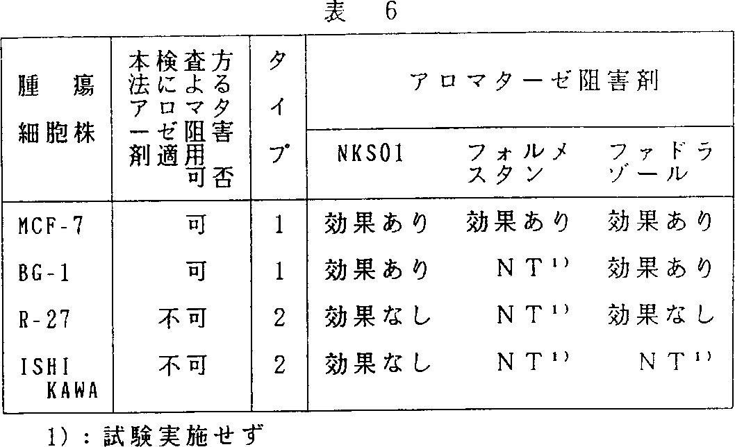
この様に、本発明方法によりアロマターゼ阻害剤による治療が有効であると判断した判定結果と実際抗腫瘍試験でのアロマターゼ阻害剤の有効性は一致していた(表6)。以上の実施例より明らかなように本発明方法をエストロジェン依存性腫瘍の患者に適用することによって同患者へのアロマターゼ阻害剤適応の可否をより正確に判断できる。
TECHNICAL FIELD The present invention relates to a method for examining hormone-dependent tumors. More particularly, the present invention relates to a method for simultaneously determining hormone-dependent growth of tumor tissue and the presence of hormone synthase. According to the present invention, tumor diagnosis and treatment guidelines can be obtained.
There is a group of tumors called hormone-dependent tumors. This refers to a tumor that develops mainly in the target organ of the hormone and is promoted to grow and grow in the presence of the hormone, that is, a tumor that exhibits hormone-dependent growth. For example, breast cancer (estrogen dependent), prostate cancer (androgen dependent), thyroid cancer (thyroid stimulating hormone dependent) and the like can be exemplified. Hormone endocrine therapy has been widely applied to these hormone-dependent tumors.
Certain types of breast cancer are known to be exacerbated by the female hormone estrogen. For this reason, the removal of the ovary, the main organ that produces estrogen, has long been used as a hormone endocrine therapy. Currently, treatment methods by administration of antiestrogens such as tamoxifen that compete with estrogen and bind to the estrogen receptor are widely applied.
In the treatment of breast cancer, a method of preparing a homogenate of a tissue collected by surgery or biopsy and examining the presence of an estrogen receptor in the homogenate is used as a basis for determining whether or not an anti-estrogen agent is applicable. This is because, as described above, the anti-estrogenic agent exhibits drug efficacy by binding to the estrogen receptor in competition with estrogen. If the presence of an estrogen receptor is confirmed, an anti-estrogen agent is prescribed. In fact, it has been clinically confirmed that there is a correlation between the therapeutic results of antiestrogens and the presence of estrogen receptors. However, methods for examining estrogen receptors are complicated, and even if the presence of the estrogen receptor itself is confirmed, it does not exhibit estrogen-dependent growth unless it is physiologically functioning. In fact, not all tumors that have been confirmed to have estrogen receptors exhibit estrogen-dependent growth.
On the other hand, according to recent research, some hormone-dependent diseases and hormone-dependent tumors have the ability to produce hormones that are involved in worsening the disease and tumor growth in the diseased tissue itself. It has become clear. For example, in certain types of breast cancer, there is an enzyme system (aromatase) that synthesizes estrogen from androgen in the tumor tissue, and this enzyme confirms that estrogen is synthesized at a high concentration in the tumor tissue. (Toshiaki Utsumi, Journal of the Japan-Japan Society, 90th, 920-927, 1989).
In the case of such diseases where hormones are synthesized in tissues, attempts have been made to suppress the hormone synthesis as a therapeutic means. In the above-mentioned treatment of breast cancer, there is an example in which an inhibitor of this enzyme is used for the purpose of inhibiting aromatase, which is an estrogen synthase, in order to suppress local estrogen production.
As a method for confirming the presence or absence of hormone synthase in the tissue, a tissue obtained by surgery or biopsy is collected, and the enzyme activity of this homomodulate is directly measured. The measurement method of aromatase activity is disclosed in detail in the above-mentioned report of Utsumi et al.
Any tumor tissue in which the presence of aromatase activity has been confirmed and the presence of estrogen-dependent growth has been confirmed can be treated with an aromatase inhibitor. However, in reality, such examination of tumor tissue has hardly been performed. This is because it is difficult to collect the target tumor tissue, or the amount is extremely small even when collected, and it is difficult to measure the enzyme activity. For this reason, even if the tissue is collected, only the estrogen receptor capable of microanalysis is examined, and the administration of the aromatase inhibitor is determined based only on the result. However, this test method does not determine the presence or absence of aromatase activity, and as described above, not all tumors that have been confirmed to have estrogen receptors exhibit estrogen-dependent growth, and therefore make erroneous judgments. Will occur. Such a problem also applies to hormone-dependent diseases and tumors other than estrogen.
On the other hand, tumors that occur in hormone target organs can be generally classified as shown in Table 1 according to the presence or absence of the hormone synthase and the presence or absence of hormone-dependent growth.
DISCLOSURE OF THE INVENTION The object of the present invention is to examine simultaneously the presence or absence of hormone synthesizing enzymes and hormone-dependent growth in hormone-dependent diseases, particularly hormone-dependent tumors, which perform hormone synthesis in tumor tissues. It is to provide a simple method to do.
Another object of the present invention is to provide a method for easily differentiating tumors of the type (type 1 in Table 1 above) in which hormone synthase is present and hormone-dependent growth is performed from other types of tumors. There is.
The present invention comprises culturing a tumor tissue in (a) a culture medium, (b) a culture medium containing a hormone synthase substrate, and (c) a culture medium containing a hormone synthase substrate and a hormone synthase inhibitor, respectively. And (ii) growth rate in the presence of a hormone-synthetic substrate and (ii) growth of tumor cells when cultured only with the culture medium. Hormone-dependent growth characterized by simultaneously determining hormone-dependent growth of tumor tissue and the presence of hormone synthase by determining the growth rate in the presence of hormone synthase substrate and hormone synthase inhibitor The present invention relates to a method for examining a tumor.
According to the present invention, among the tumors showing type 1 and 2 hormone-dependent growth and the types 3 and 4 hormone-independent growth shown in Table 1 above, one type of tumor is treated as another type. Can be easily distinguished from other tumors.
The inspection method of the present invention performs the inspection in the following procedure.
(1) A test sample is prepared by excision of human tumor tissue or biopsy such as biopsy.
(2) The sample is cultured in (a) a culture medium, (b) a culture medium containing a hormone synthase substrate, and (c) a culture medium containing a hormone synthase substrate and a hormone synthase inhibitor.
(3) The proliferation of the test sample (tumor tissue) in each culture medium is measured.
(4) Confirm the growth and make a determination.
This confirmation is based on (i) the growth rate in the presence of the hormone synthesis substrate relative to the growth of the tumor cells when cultured only in the culture medium and (ii) the hormone synthase for the growth of the tumor cells when cultured only in the culture medium. The growth rate in the presence of the substrate and the hormone synthase inhibitor is determined, and the hormone-dependent growth of the tumor tissue and the presence of the hormone synthase are determined at the same time.
The test method of the present invention will be described using an estrogen-dependent tumor, which is a typical hormone-dependent tumor, as an example.
A typical example of an estrogen-dependent tumor is breast cancer. This estrogen is converted from androgen by aromatase, but conventionally only the receptor for this estrogen has been examined.
The obtained tumor tissue, for example, a tissue collected by surgery or biopsy, is aseptically minced and cultured. Any culture medium can be used as long as it is used for cell culture, but examples include a culture medium using soft agar and a culture medium using a collagen matrix. Examples include Eagle's medium, RPMI medium, and the like. Any method may be used as long as it is a publicly known method for tissue culture and a method used therefor. Details of the culture method are described in detail in known literature such as tissue culture edited by Junnosuke Nakai et al. (Published by Asakura Shoten, 1976).
The culture medium is prepared by adding testosterone which is a substrate for aromatase which is an estrogen synthase, and by adding testosterone and aromatase inhibitors which are substrates for aromatase. Testosterone, which is a substrate for aromatase, is adjusted to a concentration of 1-1000 nM, but usually 1-100 nM is preferable. The aromatase inhibitor may be any compound that selectively inhibits only the enzyme activity and does not exhibit cytotoxicity, but 4-hydroxy-4-androstene-3, Examples include 17-dione (generic name: Formestane, manufactured by Ciba-Geigy) and 14α-hydroxy-4-androstene-3,6,17-trione (abbreviated as NKS01 Snow Brand Milk Products) disclosed in JP-A 63-192794. it can.
In the present invention, description will be made using 14α-hydroxy-4-androstene-3,6,17-trione (hereinafter abbreviated as NKS01). The concentration of this aromatase inhibitor in the culture medium can be changed according to the inhibitory activity, but is usually adjusted to a concentration of 0.01 to 100 μM when NKS01 is used. The above-mentioned tissue section is transplanted into this medium and cultured by a conventional method. The culture period is usually 1 to 14 days. Cell proliferation is determined by measuring the amount of labeled thymidine incorporated. In general, a method of measuring by tritium thymidine incorporation can be easily performed.
When measuring the amount of tritium thymidine incorporated, add tritium thymidine to each medium 3 to 4 days before the end of the culture, and after completion of the culture, remove the tissue piece and determine the amount of tritium thymidine incorporated into the tissue piece. Use a liquid scintillation counter to measure the growth. At this time, by measuring the nucleic acid content in the tissue piece and dividing the amount of thymidine incorporated by the nucleic acid content, variation from culture medium to culture medium can be reduced.
The growth rate is calculated by calculating the growth amount in the culture medium to which the substrate is added or the growth amount in the culture medium to which the inhibitor is added relative to the growth amount of the culture medium alone. The growth rate is determined by the following formula.
Growth rate (%) = [(Tissue 3H-thymidine incorporation in each culture medium / each culture medium tissue DNA concentration) / (Tissue 3 H-thymidine incorporation in culture medium only / DNA concentration in tissue cultured only in culture medium] )] X 100
When the tumor tissue of a breast cancer patient is considered to be hormone-dependent, the affected tissue of a cancer patient can be classified from a viewpoint different from the conventional one by performing the examination using the above method. For example, if a breast cancer tissue sample removed by surgery has both estrogen-dependent growth and aromatase, an estrogen synthase, when cultured in a culture medium to which testosterone, an aromatase substrate, is added, testosterone It is converted to estrogen and proliferates with this estrogen. This proliferation is suppressed by an aromatase inhibitor. It is determined that the tumor is a type 1 hormone-dependent tumor shown in Table 1 above.
In addition, if a breast cancer tissue sample removed by surgery lacks one or both of estrogen-dependent proliferation and aromatase, an estrogen synthase, the growth rate is not accelerated in the presence of testosterone.
By carrying out the present invention, it becomes possible to simultaneously and easily determine the presence of hormone synthase and the presence or absence of hormone-dependent growth in hormone-dependent tumors. This determination result can be used for new classification of tumors, selection of treatment guidelines, and the like.
If it is the above type 1 tumor, it can be assumed that treatment with an aromatase inhibitor is possible, and such a treatment policy can be established.
BEST MODE FOR CARRYING OUT THE INVENTION The following examples and reference examples further illustrate the present invention. The present invention is not limited to this embodiment.
Example 1
Examination was carried out by the method of the present invention for 21 patients diagnosed with breast cancer.
At the time of surgery for breast cancer, a tissue piece (10 mm square) collected from the center of the tumor was rapidly removed aseptically in a Hanks solution (manufactured by Gibco) containing 100 U / ml penicillin and streptomycin, The surgical knife was cut into pieces of about 0.5 to 1 mm square. Four to five tissue strips were placed on the collagen matrix in a 24-well microplate. As a collagen matrix, a sponge-like collagen gel matrix (manufactured by Spongostan) is aseptically minced to a size of 1 cm 3 and placed in each well of a 24-well microplate. 10% fetal calf serum (FCS), 100 U / ml A phenol red-free Eagle modified minimum essential medium (Gibco) containing penicillin and 100 U / ml streptomycin was injected into the wells until reaching the surface of the collagen matrix as a culture medium.
The 24-well microplate was placed in air containing 5% carbon dioxide and cultured at 37 ° C. for 7 days. The entire culture medium was changed every other day, but the FCS added to the culture medium used for this was used after pretreatment with charcoal dextran and excluding the steroid component.
The following three groups were used as the test culture medium for the cultured tissue.
(1) Control group (medium only)
(2) 10 nM testosterone addition group (substrate addition group)
(3) 10 nM testosterone, 1 μM NKS01 addition group (substrate, enzyme inhibitor addition group)
After 7 days of culture, the culture medium was replaced with a culture medium containing 0.67 μCi of tritium thymidine, and the culture was further continued for 3 days. After completion of the culture, the tissue pieces cultured on the collagen sponge of each well were transferred to one tube per well, 1 ml of Hanks' solution containing 0.1 mg / ml collagenase was added, and incubated at 37 ° C. for 8 hours. Subsequently, the cell mass was dispersed by pipetting, and centrifuged at 3000 rpm for 10 minutes. To the precipitate, 1 ml of 100 mM Tris buffer (pH 7.5) containing 1% sodium dodecyl sulfate and 200 μg / ml proteinase was added and incubated at 50 ° C. for 3 hours.
To this solution was added 1 ml phenol / chloroform / isoamyl alcohol (25: 24: 1) and mixed for several minutes. Thereafter, centrifugation was performed at 3000 rpm for 10 minutes, and then the upper aqueous layer was collected. 100 μl of 3M sodium acetate and 2, 5 ml of cold ethyl alcohol were added thereto, and the precipitated DNA was wound on a glass rod. The DNA was immersed in a 70%, 80%, and 90% aqueous ethyl alcohol solution while being wound around a glass rod, and then air-dried. Subsequently, it was dissolved in 1 ml of 100 mM Tris buffer (pH 7.5) to prepare a DNA solution.
The concentration of this DNA solution was measured for absorbance at a wavelength of 260 nm, and the tritium thymidine content per 100 μl was measured using a liquid scintillation counter. The measurement was performed by the count number per 1 μg of DNA (dpm / μg DNA). A method for measuring cell growth by tritium thymidine content per DNA is widely used. For example, according to the method of Fukuoka et al. (Fukuoka Masami et al., ACTA OBST GYNAEC JPN, Vol. 43, No. 12, 1667-1973) It can be carried out. Also in this example, the measurement was performed according to this method. The results are shown in Table 2.
As a result of examination by the examination method of the present invention, it was confirmed that 6 out of 21 cases had both estrogen-dependent proliferation and aromatase in the tumor tissue. It was speculated that the breast cancer in this case was hormone-dependent, and it was highly likely that hormones were synthesized and proliferated in the tumor tissue.
Example 2
Targeting four human-derived tumor cell lines (Table 3) whose estrogen receptor has already been confirmed, that is, according to conventional tests, it is judged that “endocrine treatment (administration of aromatase inhibitor) is possible” The method for aromatase inhibitor was examined again by the method of the present invention.
In order to make each tumor cell line a solid tumor, 7 weeks old female nude mice were transplanted subcutaneously into the axilla at 1 × 10 5 /0.1 ml / mouse and when the size became about 1 cm in diameter, nude mice This transplanted tumor was removed. Table 4 shows the results of examining these tumors by the above-described method of the present invention.
Next, these tumor cell lines were transplanted again under the axilla of 7-week-old female nude mice to 1 × 10 5 /0.1 ml / mouse, and when the tumor mass became about 3 mm in diameter. About 10 animals were divided into a control group in which no aromatase inhibitor was administered and an aromatase inhibitor administration group in which an aromatase inhibitor was administered every day until the end of the experiment. Examples of aromatase inhibitors include 4-hydroxy-4-androstene-3,17-dione (generic name formestane, manufactured by Ciba Geigy) and 4- (5,6,7,8-tetrahydroimidazo- [1,5a]. -Pyridin-5-yl) benzonitrile monohydrochloride (generic name: Fadazole, manufactured by Ciba Geigy), 14α-hydroxy-4-androstene-3,6,17-trione disclosed in JP-A-63-192794 The abbreviation NKS01 made by Snow Brand Milk Products) was used.
The administration period varied depending on the growth rate of each tumor on nude mice, and was 28 days for BG-1, R-27 and ISHIKAWA, and 42 days for MCF-7. The major axis and minor axis of the tumor were measured the day after the last administration with calipers, etc., and the estimated tumor volume was calculated by the following formula. The results are shown in Table 5.
Estimated tumor volume = (major axis) x (minor axis) 2 x 0.5
Evaluation of the effect was judged as “anti-tumor effect” when a statistically significant difference in tumor size was observed between the control group and the aromatase inhibitor administration group. Several types of statistical methods are known, but this time, a T test was performed between the control group and the aromatase inhibitor administration group, and a significance level of 5% or less was used as a criterion for judging the effect. Table 5 also shows the antitumor effect determination results of each aromatase inhibitor for the four types of tumor cell lines used this time.
Thus, the determination result judged that the treatment with the aromatase inhibitor was effective by the method of the present invention was consistent with the effectiveness of the aromatase inhibitor in the actual antitumor test (Table 6). As is clear from the above examples, the applicability of the aromatase inhibitor to the patient can be more accurately determined by applying the method of the present invention to a patient with an estrogen-dependent tumor.