JP3636886B2 - Ophthalmic equipment - Google Patents
Ophthalmic equipment Download PDFInfo
- Publication number
- JP3636886B2 JP3636886B2 JP11618898A JP11618898A JP3636886B2 JP 3636886 B2 JP3636886 B2 JP 3636886B2 JP 11618898 A JP11618898 A JP 11618898A JP 11618898 A JP11618898 A JP 11618898A JP 3636886 B2 JP3636886 B2 JP 3636886B2
- Authority
- JP
- Japan
- Prior art keywords
- light
- eye
- ophthalmologic apparatus
- reflected light
- amount
- Prior art date
- Legal status (The legal status is an assumption and is not a legal conclusion. Google has not performed a legal analysis and makes no representation as to the accuracy of the status listed.)
- Expired - Fee Related
Links
- 230000003287 optical effect Effects 0.000 claims description 77
- 238000001514 detection method Methods 0.000 claims description 75
- 238000005259 measurement Methods 0.000 claims description 48
- 230000005484 gravity Effects 0.000 claims description 11
- 238000012360 testing method Methods 0.000 claims description 9
- 238000010586 diagram Methods 0.000 description 15
- 238000012545 processing Methods 0.000 description 11
- 230000004907 flux Effects 0.000 description 7
- 210000004087 cornea Anatomy 0.000 description 6
- 238000003384 imaging method Methods 0.000 description 6
- 238000000034 method Methods 0.000 description 5
- 230000000630 rising effect Effects 0.000 description 5
- 238000005286 illumination Methods 0.000 description 4
- 238000006073 displacement reaction Methods 0.000 description 3
- 238000007689 inspection Methods 0.000 description 3
- 230000000007 visual effect Effects 0.000 description 3
- 201000009310 astigmatism Diseases 0.000 description 2
- 230000004397 blinking Effects 0.000 description 2
- 238000011161 development Methods 0.000 description 2
- 206010020675 Hypermetropia Diseases 0.000 description 1
- 238000013459 approach Methods 0.000 description 1
- 239000003990 capacitor Substances 0.000 description 1
- 230000000694 effects Effects 0.000 description 1
- 210000000720 eyelash Anatomy 0.000 description 1
- 210000000744 eyelid Anatomy 0.000 description 1
- 201000000766 irregular astigmatism Diseases 0.000 description 1
- 239000004973 liquid crystal related substance Substances 0.000 description 1
Images
Classifications
-
- A—HUMAN NECESSITIES
- A61—MEDICAL OR VETERINARY SCIENCE; HYGIENE
- A61B—DIAGNOSIS; SURGERY; IDENTIFICATION
- A61B3/00—Apparatus for testing the eyes; Instruments for examining the eyes
- A61B3/10—Objective types, i.e. instruments for examining the eyes independent of the patients' perceptions or reactions
- A61B3/103—Objective types, i.e. instruments for examining the eyes independent of the patients' perceptions or reactions for determining refraction, e.g. refractometers, skiascopes
-
- A—HUMAN NECESSITIES
- A61—MEDICAL OR VETERINARY SCIENCE; HYGIENE
- A61B—DIAGNOSIS; SURGERY; IDENTIFICATION
- A61B3/00—Apparatus for testing the eyes; Instruments for examining the eyes
- A61B3/10—Objective types, i.e. instruments for examining the eyes independent of the patients' perceptions or reactions
- A61B3/107—Objective types, i.e. instruments for examining the eyes independent of the patients' perceptions or reactions for determining the shape or measuring the curvature of the cornea
Landscapes
- Life Sciences & Earth Sciences (AREA)
- Health & Medical Sciences (AREA)
- Medical Informatics (AREA)
- Biophysics (AREA)
- Ophthalmology & Optometry (AREA)
- Engineering & Computer Science (AREA)
- Biomedical Technology (AREA)
- Heart & Thoracic Surgery (AREA)
- Physics & Mathematics (AREA)
- Molecular Biology (AREA)
- Surgery (AREA)
- Animal Behavior & Ethology (AREA)
- General Health & Medical Sciences (AREA)
- Public Health (AREA)
- Veterinary Medicine (AREA)
- Eye Examination Apparatus (AREA)
Description
【0001】
【発明の属する技術分野】
本発明は、被検眼の検査・測定を行う他覚式眼屈折力測定装置、角膜形状測定装置等の眼科装置に関する。
【0002】
【従来の技術】
他覚式眼屈折力測定装置等の眼科装置としては、据え置き型のものが一般的に知られている。このような据え置き型の装置は、検査測定部を固定基台に対して相対移動できるように構成されている。検査測定を行う際は、検者はジョイスティック等の操作により被検眼に対して検査測定部を移動し、片眼ずつアライメントして検査測定を行う。したがって、このような据え置き型の装置では、固定基台の中央に対して検査測定部が左右どちらに移動したかを検出することにより、被検眼の左右の判別を自動的に行うことができる。
【0003】
また一方、上記のような据え置き型の眼科装置は、乳幼児や寝ている状態の患者、動物等を検眼することは困難であり、持ち運びにも不都合であるため、手持ち型の眼科装置も提案されている。手持ち型の眼科装置では、据え置き型の眼科装置のように検査測定部の移動によって被検眼の左右の判別をすることができないため、検者が被検眼の左右どちらを検査測定しているかスイッチ等により入力する方法などを採用している。
【0004】
【発明が解決しようとする課題】
しかしながら、被検眼の左右の判別をスイッチ等により入力する方法は、操作が煩雑となり、検者が入力することを忘れてしまうため、測定ミスとなることがあり不都合であった。
【0005】
本発明は上記従来技術の欠点に鑑み、検査測定部が移動しなくても、被検眼の左右の判別を検者が入力する必要がない眼科装置を提供することを技術課題とする。
【0006】
また、簡単に操作できる手持ち型の眼科装置を提供することを技術課題とする。
【0007】
【課題を解決するための手段】
上記課題を解決するために、本発明は次のような構成を有することを特徴とする。
【0008】
(1) 被検眼と測定系とを所定の関係に位置合わせするためのアライメント光学系を備え、被検眼の検査測定を行う眼科装置において、被検者の顔面部に光を投射するための光源を備える投射手段と、該投射手段によって生じた反射光を受光する位置検出素子を持つ光学系であって右眼にアライメントされた状態で顔面部の右境界を含み左眼にアライメントされた状態で顔面部の左境界を含む検出領域を持つ受光手段と、該受光手段による検出結果に基づいて顔面部の境界が左右いずれにあるかにより被検眼の左右判別を行う判別手段と、を有することを特徴とする。
(2) (1)の眼科装置において、前記投射手段によって投射される光は近赤外域の波長を持ち、前記受光手段は近赤外域の光を透過しそれ以外の波長の光を遮断する波長選択手段を備えることを特徴とする。
(3) (1)の眼科装置において、前記判別手段は前記光源を点灯したときの反射光と消灯したときの反射光の検出結果を比較する比較手段を備えることを特徴とする。
(4) (1)の眼科装置において、前記位置検出素子はCCDであり、前記判別手段は受光量に対する所定の閾値を設定する閾値設定手段と、該閾値設定手段によって設定された閾値と前記CCDによる検出結果とから受光された反射光の中心位置又は光量バランスを求める演算手段と、を備えることを特徴とする。
(5) (4)の眼科装置は、さらに被検眼と前記測定系とのアライメントが完了したときの反射光の平均的中心位置を記憶する位置記憶手段と、該位置記憶手段に記憶された中心位置の座標と前記CCDが検出した反射光の中心位置の座標とに基づいて被検眼と前記測定系とのアライメントずれを求める演算手段と、求められたアライメントずれを表示する表示手段と、を有することを特徴とする。
(6) (1)の眼科装置は、さらに受光量に対する所定の閾値を設定する閾値設定手段と、該閾値と検出された反射光の受光量とから被検者と装置との距離関係を検知する距離検知手段と、該距離検知手段による検知結果に基づいて検者に警告する警告手段と、を有することを特徴とする。
(7) (1)の眼科装置は、さらに受光量に対する所定の閾値を設定する閾値設定手段と、該閾値と検出された反射光の受光量とから被検者の顔面部の有無又は被検者と装置との距離関係を検知する検知手段と、該検知手段による検知結果に基づいて動作モ−ドを制御する制御手段と、を有することを特徴とする。
(8) 被検眼と測定系とを所定の関係に位置合わせするためのアライメント光学系を備え、被検眼の検査測定を行う眼科装置において、被検者の顔面部に光を投射するための光源を備える投射手段と、該投射手段によって生じた反射光を受光する位置検出素子を持つ光学系であって該位置検出素子上の反射光受光部分と非受光部分との位置関係を検出する受光手段と、該受光手段による検出結果に基づいて被検眼の左右判別を行う判別手段と、を有することを特徴とする。
(9) (8)の眼科装置において、前記投射手段によって投射される光は近赤外域の波長を持ち、前記受光手段は近赤外域の光を透過しそれ以外の波長の光を遮断する波長選択手段を備えることを特徴とする。
(10) (8)の眼科装置において、前記位置検出素子はPSDであり、前記判別手段は該PSDによる検出結果に基づいて前記光源からの光による反射光の重心位置を求めることにより被検眼の左右判別を行うことを特徴とする。
(11) (10)の眼科装置は、さらに被検眼と前記測定系とのアライメントが完了したときの反射光の平均的重心位置を記憶する位置記憶手段と、該位置記憶手段に記憶された重心位置の座標と前記PSDが検出した反射光の重心位置の座標とに基づいて被検眼と前記測定系とのアライメントずれを求める演算手段と、求められたアライメントずれを表示する表示手段と、を有することを特徴とする。
(12) (8)の眼科装置において、前記位置検出素子はCCDであり、前記判別手段は受光量に対する所定の閾値を設定する閾値設定手段と、該閾値設定手段によって設定された閾値と前記CCDによる検出結果とから受光された反射光の中心位置又は光量バランスを求める演算手段と、を備えることを特徴とする。
(13) (12)の眼科装置は、さらに被検眼と前記測定系とのアライメントが完了したときの反射光の平均的中心位置を記憶する位置記憶手段と、該位置記憶手段に記憶された中心位置の座標と前記CCDが検出した反射光の中心位置の座標とに基づいて被検眼と前記測定系とのアライメントずれを求める演算手段と、求められたアライメントずれを表示する表示手段と、を有することを特徴とする。
(14) (8)の眼科装置は、さらに受光量に対する所定の閾値を設定する閾値設定手段と、該閾値と検出された反射光の受光量から被検者と装置との距離関係を検知する距離検知手段と、該距離検知手段の検知結果に基づいて検者に警告する警告手段と、を有することを特徴とする。
(15) (8)の眼科装置は、さらに受光量に対する所定の閾値を設定する閾値設定手段と、該閾値と検出された反射光の受光量から被検者の顔面部の有無又は被検者と装置との距離関係を検知する検知手段と、該検知手段による検知結果に基づいて動作モ−ドを制御する制御手段と、を有することを特徴とする。
【0033】
【実施例】
以下、本発明の一実施例を図面に基づいて説明する。この実施例の装置は他覚式眼屈折力測定装置である。
【0034】
図1は実施例の装置の一部光学系を横方向から見たときの概略配置を示す図である。光学系は、アライメント指標投影光学系、距離指標投影光学系、指標検出・観察光学系、レチクル投影光学系、固視標投影光学系、眼屈折力測定光学系、左右眼判別指標投影光学系、及び左右眼判別指標検出光学系に大別される。なお、図1では図示の都合上、左右眼判別指標投影光学系は省略している。
【0035】
ここで、Eは被検眼である。L1 は後述する測定光学系の測定光軸である。70は被検眼Eの前眼部を照明するための光源である。
【0036】
(アライメント指標投影光学系)
1はアライメント指標投影光学系である。アライメント用光源2を発した光は、コリメータレンズ3により略平行光束とされ、さらに、その投射軸をビームスプリッタ4による反射によって測定光軸L1 と同軸とされて、正面から被検眼Eに投影される。
【0037】
(距離指標投影光学系)
10a,10bは被検眼Eと装置との作動距離を検出するための指標を投影する距離指標投影光学系である。距離指標投影光学系10aは、光源11a、スポット絞り12a、及び光源11aからの光を略平行光束とするためのコリメータレンズ13によって構成され、被検眼Eに光学的に無限遠方からスポット絞り指標を投影する。一方、距離指標投影光学系10bは、光源11b及びスポット絞り12bによって構成され,被検眼Eに光学的に有限な距離からスポット絞り指標を投影する。距離指標投影光学系10a,10bは、測定光軸L1 に対してその投射軸が同一角度で交差するように配置されている。(図1では図示の都合上、被検眼Eの上下方向から投射しているが、距離指標投影光学系10a,10bは、まぶた及びまつげによる光束の欠られを防ぐため、水平方向に配置することが好ましい。)
【0038】
被検眼Eと装置との作動距離の検出は、精密な作動距離方向のアライメントを行う際に必要となる。しかし、本発明においては関連が少ないため、作動距離の検出方法についての説明は省略する。詳しくは、特開平6−46999号「アライメント検出装置」を参照されたい。
【0039】
(指標検出・観察光学系)
20は指標検出・観察光学系である。光源70による前眼部像、アライメント指標投影光学系1による角膜反射像、及び距離視標投影光学系10a,10bによる角膜反射像は、ダイクロイックミラー21,22によって反射され、結像レンズ23によって観察用カメラ24の撮像素子上に結像する。観察用カメラ24は本実施例では近赤外用CCDカメラを用いている。観察用カメラ24によって撮像された前眼部像及び角膜反射像は、テレビモニタ25によって表示される。
【0040】
(レチクル投影光学系)
30はレチクル投影光学系である。レチクル板32を通過した光源31からの光は、投影レンズ33を通過し、ビームスプリッタ34により反射され、結像レンズ23によって観察用カメラ24の撮像素子上に結像する。レチクル板32には、図2に示すようにレチクルマークが形成されており、観察用カメラ24によって撮像されたレチクルマーク像は、テレビモニタ25によって表示される。レチクルマークには、円環状のマーク32aと、測定系の乱視軸が0度方向を示すラインマーク32bとがある。ラインマーク32bは無くてもよく、また、逆に90度方向のラインマークをさらに設けてもよい。
【0041】
(固視標投影光学系)
40は固視標投影光学系である。固視標投影光学系40は前述したダイクロイックミラー22によって指標検出・観察光学系20と同軸とされている。光源41は固視標42を照明し、固視標42からの光束は投影レンズ43及びダイクロイックミラー22を通過する。その後、ダイクロイックミラー21で反射して被検眼Eに向かい、被検眼Eは固視標42を固視する。なお、投影レンズ43は光軸方向に移動することによって被検眼Eの雲霧を行う。
【0042】
(眼屈折力測定光学系)
100は眼屈折力測定光学系である。眼屈折力測定光学系100は、スリット投影光学系とスリット像検出光学系とから構成される。
【0043】
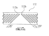
110はスリット投影光学系である。スリット照明用光源111からの光は、モータ113によって一定の速度で一定方向に回転する円筒状の回転セクター112に設けられたスリット開口112aまたは112bを照明する。回転セクター112の側面には、図3にその展開図を示すように、2種類の異なる傾斜角度のスリット開口112a,112bがそれぞれ複数個設けられている。スリット開口112aは、回転セクター112の回転方向に対して45度の傾斜角度を持つように設けられている。また、スリット開口112bは、スリット開口112aに直交するように回転方向に対して135度の傾斜角度を持つように設けられている。回転セクター112の回転により走査されたスリット光束は、光源111と被検眼Eの角膜近傍とを共役とする位置に配置された投影レンズ114を通過する。その後、制限絞り115によって制限され、ビームスプリッタ116による反射によってアライメント指標投影光学系1と同軸とされて被検眼Eに向かい、被検眼Eの角膜近傍で集光した後、眼底に投影される。なお、回転セクター112は、異なる傾斜角度のスリット開口を持つため、いずれの角度のスリット光束が投影されているかを図示なきセンサで検出するようになっている。
【0044】
120はスリット像検出光学系であり、その測定光軸L1 上に受光レンズ121、絞り122及び受光部123を備える。絞り122は受光レンズ121の後ろ側焦点位置に配置され、受光部123は受光レンズ121に関して被検眼Eの角膜と略共役な位置に配置される。受光部123はその受光面に、図4に示すように、4個の受光素子124a〜124dを有している。受光素子124a,124bは測定光軸L1 を中心にして対称に設けられ、同じく受光素子124c,124dは測定光軸L1 を中心にして対称に設けられている。この2対の受光素子は、2種類の傾斜角度を持つスリット開口112a,112bにより投影される被検眼Eの眼底上でのスリット光束の走査方向(眼底上でのスリット光束は、あたかもスリットの長手方向に直交する方向に走査されるようになる)にそれぞれ対応させて配置され、眼底で反射されたスリット光束を検出する。実施例の装置では、乱視を持たない遠視または近視の被検眼Eの眼底上で、スリット開口112aによるスリット光束が走査されたとき、受光部123上で受光されるスリットの長手方向に直交する方向に対応するように、一対の受光素子124a,124bを配置している。また同じように、スリット開口112bによるスリット光束が走査されたとき、受光部123上で受光されるスリットの長手方向に直交する方向に対応するように、一対の受光素子124c,124dを配置している。
【0045】
ある第1の方向のスリット光束が被検眼Eに投影されたとき、そのスリット光束の長手方向に直交する方向に対応して受光素子124a,124bが配置され、このときの受光素子124c,124dの位相差信号出力に基づいて被検眼Eの角膜中心または視軸中心を検出する。検出した角膜中心または視軸中心と、前記第1の方向に対応して配置された受光素子124a,124bのそれぞれの出力信号に基づいて、各々の受光素子位置に対応する角膜部位で屈折力を求める。これにより、角膜中心または視軸中心に対する経線方向の屈折力が求まり、不正乱視の有無を検知する。なお、眼屈折力測定については、詳しくは、特願平8−283280号「眼屈折力測定装置」を参照されたい。
【0046】
(左右眼判別指標検出光学系)
50は左右眼判別指標検出光学系である。後述する左右眼判別指標投影光学系によって投射された光による被検者の顔面部からの反射光は、広角の結像レンズ51によって位置検出素子53上に結像される。その際、可視光等を遮断し近赤外光を透過させるフィルタ52を介することによって、太陽光や部屋の照明光等の余分な外乱光を除去することができる。なお、位置検出素子53は、本実施例ではPSDを用いている。PSDは検出速度が速く、高速で点滅する光の明暗を検出することができる。さらに、PSDで検出した光成分をコンデンサによって直流成分をカットしてやれば、点滅する光源の光成分のみを抽出することができる。つまり、後述する左右眼判別指標投影光学系の光源を高速で点滅させることによって、フィルタ52を通過してしまった太陽光等の近赤外域の光成分を除去し、左右眼判別指標投影光学系の光源による反射光の光成分のみを抽出することができる。なお、結像レンズ51、フィルタ52、及び位置検出素子53は、測定光軸L1 に対して垂直な下方向に配置されている。
【0047】
図5は実施例の装置の一部光学系を上方向から見たときの概略配置を示す図である。なお、図5では図示の都合上、左右眼判別指標検出光学系及び左右眼判別指標投影光学系のみを示しており、他の光学系については省略している。
【0048】
(左右眼判別指標投影光学系)
60は左右眼判別指標投影光学系である。被検者の顔面部に光を投射するための光源61aは、検者側から見て装置右側に、光源61bは検者側から見て装置左側に、測定光軸L1 に対して対称に配置されている。また、光源61a,61bは、被検眼Eに装置を位置合わせしたときに、被検者の顔面部のある程度広い範囲を照明できるように配置されている。またさらに、光源61a,61bは、図示なき制御部によって高速で点滅するようになっている(点滅は1回でもよいし複数回でもよい)。
【0049】
なお、光源2、11a,11b、31、61a,61b、70及び111には、すべて近赤外域の光を発するLEDを用いており、被検眼Eに負担をかけることなく光を投射することができる。なお、ダイクロイックミラー22とビームスプリッタ34の間に、近赤外域の光を透過し可視光等を遮断するフィルタを配置すれば、観察用カメラ24は近赤外用でなくてもよく、また、レチクル投影光学系30の光源31は可視光を発する通常の光源でもよい。
【0050】
以上のような実施例の装置において、その動作について説明する。
【0051】
まず、装置を被検眼Eの前に移動し、図示なきスイッチ等により、前眼部照明用光源70、アライメント用光源2及び光源31を発光させる。光源70による前眼部像と、アライメント指標投影光学系1により被検眼Eに形成された角膜反射像と、レチクル投影光学系30によるレチクルマーク像とは、観察用カメラ24によって撮像され、テレビモニタ25(液晶ディスプレイ)に表示される。検者はテレビモニタ25上の角膜反射像及びレチクルマーク像を見ながら、レチクルマーク32aの中心に光源2の角膜反射像が位置するようにさらに装置を移動させ、上下左右方向のアライメントを行う。また、ラインマーク32bを設けている場合は、テレビモニタ25を見ながらラインマーク32bの像が水平になるように、装置の傾きを調整する。さらに、被検眼Eの角膜反射像にピントを合わせることによって、作動距離をラフに判断する。
【0052】
制御部は、画像処理解析技術を用いて検出された角膜反射像が所期する領域内に位置し、被検眼Eに上下左右方向のアライメント及び作動距離のラフなアライメントができたことを確認すると、光源61a,61bを高速で点滅発光させる(点滅は1回でもよいし複数回でもよい。また、ラフなアライメントの終了後のみだけでなく、ある時間ごとに光源61a,61bを発光させてもよい)。光源61a,61bから投射された光は、被検者の顔面部を照明し、その反射光の一部は、結像レンズ51及びフィルタ52を通過し、位置検出素子53に受光される(図6参照)。
【0053】
ここで、PSDは受光面に入射した光の重心位置によって両端から出力される電流の比が決まるので、この両端からの電流量をそれぞれ検出して比を求めれば受光された反射光の重心位置がわかる。そして、重心位置とPSD(位置検出素子53)の中心とを比較し、重心位置が中心よりもA側寄りであれば被検眼Eは左眼、重心位置が中心よりもB側寄りであれば被検眼Eは右眼と判別できる。図6の場合、被検眼Eは右眼であると判別される。
【0054】
また、位置検出素子53については、PSDの代わりに一次元CCDを用いてもよい。図6に示すように、光源61a,61bから投射された光が被検者の顔面部を照明すると、顔面部からの反射光の一部は、結像レンズ51及びフィルタ52によって位置検出素子53上に結像する。図6の場合(被検眼が右眼の場合)、位置検出素子53に受光される反射光の受光量と受光位置との関係は、図7のようになる。グラフMは光源61a,61bが点灯したときの反射光の受光量及び受光位置であり、グラフNは光源61a,61bが消灯したときの反射光の受光量及び受光位置である。従って、MからNを引くことによって光源61a,61bによる反射光の成分グラフRのみを抽出することができる。一次元CCDの場合、図8に示すように、位置検出素子53(一次元CCD)に受光された反射光の受光量に閾値処理することによって(閾値αは予め所定の値を設定し記憶させておいてもよいし、受光量のピーク値の1/2等としてもよい)、2点t1とt2の中心位置Pを求めることができる。中心位置Pが求められれば、PSDの場合と同じように、位置検出素子53(一次元CCD)の中心Qに対して中心位置PがA側寄りであるかB側寄りであるかを検出することによって、被検眼Eの左右の判別をすることができる。なお、必ずしも受光された反射光の中心位置を求めることは必要ではなく、受光された反射光の光量バランスが得られる指標であればよい。光量バランスは、閾値処理によって得られる反射光の立上りや立下りの位置から求めることができ、これによって被検眼Eの左右の判別をすることができる。図9は図8の受光量を閾値αにて処理したものである。この場合、立下り位置t2がBの位置にある(立上り位置t1のみがA−B間にある)ため、被検眼Eは右眼であると判別される。また、反射光の受光量を閾値処理したものが図10のようになった場合は、立上り位置t3がAの位置にある(立下り位置t4のみがA−B間にある)ため、被検眼Eは左眼であると判別される。立上り位置と立下り位置の両方がA−B間にある場合は、立上り位置と立下り位置の中心位置から左右の判別をすることができる。
【0055】
さらに、位置検出素子53を二次元CCDとすれば、反射光の受光エリアが広がり、複数の水平方向ラインの検出ができるため、より正確に被検眼Eの左右判別をすることができる。
【0056】
また、検出した画像をデジタイズし、画像メモリに記憶し、画像解析することによって、受光量の重心位置を求めることもできる。
【0057】
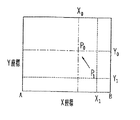
位置検出素子53が二次元CCDの場合、図11に示すように、測定光軸L1 と被検眼Eとが正しくアライメントされたときの受光量の重心(中心)位置P0 の座標(X0 ,Y0 )を予め記憶させておけば、検出した反射光の受光量の重心(中心)位置P1 の座標(X1 ,Y1 )と座標(X0 ,Y0 )のずれ量及びずれ方向から、大まかにアライメントずれを検出することができる。そして、検出したずれ量及びずれ方向に基づいて、テレビモニタ25上に矢印等で告知することによって、検者はアライメントを行うことができる。位置検出素子53が一次元CCD又はPSDの場合は、一方向(X方向)のみアライメントずれを検出することができる。
【0058】
位置検出素子53の反射光の受光量に対して閾値処理をすることによって、さらなる情報を得ることもできる。
【0059】
図12aに示すように、位置検出素子53の受光量に対しある閾値βを設定し、被検者と装置との距離が必要以上に近付かないように検者に警告させることができる。被検者の顔面部からの反射光の位置検出素子53による受光量がグラフCにように必要以上に大きい場合、つまり所定の閾値βを超えた場合は、音声またはテレビモニタ25に表示させるなどして、被検者と装置との距離が所定の閾値β内になるまで警告させる。
【0060】
また、図12bに示すように、位置検出素子53の受光量に対しある閾値γを設定し、被検者と装置との距離が必要以上に遠ざからないように検者に警告させることもできる。被検者の顔面部からの反射光の位置検出素子53による受光量がグラフDにように必要以上に小さい場合、つまり所定の閾値γ内にある場合は、音声またはテレビモニタ25に表示するなどして、被検者と装置との距離が所定の閾値γを超えるまで警告させる。
【0061】
装置が被検眼に対して自動的に動いて位置合わせするような場合には、これらの情報を基に動作を止めたり動かしたりすることができる。
【発明の効果】
以上説明したように、本発明によれば、検査測定部が移動しない装置でも、被検眼の左右判別を検者が入力する必要がなく、特に手持ち型の眼科装置として好適に利用することができる。
【図面の簡単な説明】
【図1】実施例の装置の一部光学系を横方向から見たときの概略配置を示す図である。
【図2】レチクル板に設けられているレチクルマークを説明する図である。
【図3】回転セクターの側面に設けられているスリット開口の展開図である。
【図4】受光部が有する4個の受光素子の配置を説明する図である。
【図5】実施例の装置の一部光学系を上方向から見たときの概略配置を示す図である。
【図6】被検者の顔面部への光の投射、照明範囲、及び反射光の受光を説明する図である。
【図7】位置検出素子によって検出された反射光の受光量と受光位置を説明する図である。
【図8】CCDによって検出された反射光の受光量と受光位置、及び閾値処理を説明する図である。
【図9】受光量を閾値処理したものを説明する図である。
【図10】受光量を閾値処理したものを説明する図である。
【図11】二次元CCDによるアライメントずれ検出を説明する図である。
【図12a】被検者と装置との距離を検知するための閾値βを設定した場合の閾値処理を説明する図である。
【図12b】被検者と装置との距離を検知するための閾値γを設定した場合の閾値処理を説明する図である。
【図12c】被検者と装置との距離を検知するための閾値δを設定した場合の閾値処理を説明する図である。
【符号の説明】
1 アライメント指標投影光学系
10a,10b 距離指標投影光学系
20 指標検出・観察光学系
30 レチクル投影光学系
40 固視標投影光学系
50 左右眼判別指標検出光学系
60 左右眼判別指標投影光学系
100 眼屈折力測定光学系[0001]
BACKGROUND OF THE INVENTION
The present invention relates to an ophthalmologic apparatus such as an objective eye refractive power measurement apparatus and a cornea shape measurement apparatus for inspecting and measuring an eye to be examined.
[0002]
[Prior art]
As an ophthalmologic apparatus such as an objective eye refractive power measuring apparatus, a stationary apparatus is generally known. Such a stationary apparatus is configured such that the inspection / measurement unit can be moved relative to the fixed base. When performing the test measurement, the examiner moves the test measurement unit with respect to the eye to be examined by operating a joystick or the like, and performs the test measurement by aligning one eye at a time. Therefore, in such a stationary apparatus, it is possible to automatically determine the left and right of the eye to be inspected by detecting whether the test and measurement unit has moved left or right with respect to the center of the fixed base.
[0003]
On the other hand, the stationary ophthalmic apparatus as described above is difficult to examine an infant, a sleeping patient, an animal, etc., and is inconvenient to carry. Therefore, a hand-held ophthalmic apparatus is also proposed. ing. In a hand-held ophthalmic device, the left and right of the eye to be examined cannot be discriminated by movement of the test measurement unit unlike the stationary ophthalmic device, so the switch or the like whether the examiner is inspecting and measuring the left or right of the eye to be examined The method to input by is adopted.
[0004]
[Problems to be solved by the invention]
However, the method of inputting the left / right discrimination of the eye to be inspected with a switch or the like is inconvenient because the operation becomes complicated and the examiner forgets to input, which may result in a measurement error.
[0005]
In view of the above-described drawbacks of the prior art, it is an object of the present invention to provide an ophthalmologic apparatus that does not require the examiner to input the left / right discrimination of the eye to be examined even if the test measurement unit does not move.
[0006]
It is another object of the present invention to provide a hand-held ophthalmic apparatus that can be easily operated.
[0007]
[Means for Solving the Problems]
In order to solve the above problems, the present invention is characterized by having the following configuration.
[0008]
(1) A light source for projecting light onto a face portion of a subject in an ophthalmologic apparatus that includes an alignment optical system for aligning a subject eye and a measurement system in a predetermined relationship, and performs inspection measurement of the subject eye And an optical system having a position detection element for receiving reflected light generated by the projection means, and aligned with the left eye including the right boundary of the face portion in a state aligned with the right eye. A light receiving means having a detection region including the left boundary of the face portion, and a determination means for determining the left and right of the eye to be examined based on whether the boundary of the face portion is on the left or right based on a detection result by the light receiving means. Features.
(2) In the ophthalmologic apparatus according to (1), the light projected by the projection means has a near-infrared wavelength, and the light-receiving means transmits the near-infrared light and blocks light of other wavelengths. It comprises a selection means.
(3) In the ophthalmologic apparatus according to (1), the determination unit includes a comparison unit that compares a detection result of reflected light when the light source is turned on and reflected light when the light source is turned off.
(4) In the ophthalmologic apparatus according to (1), the position detection element is a CCD, the discrimination means sets a threshold value for setting a predetermined threshold value for the amount of received light, the threshold value set by the threshold value setting means, and the CCD And a calculation means for obtaining a center position or a light amount balance of the reflected light received from the detection result of the above.
(5) The ophthalmologic apparatus according to (4) further includes position storage means for storing an average center position of reflected light when alignment between the eye to be examined and the measurement system is completed, and a center stored in the position storage means Calculation means for obtaining an alignment deviation between the eye to be examined and the measurement system based on the coordinates of the position and the coordinate of the center position of the reflected light detected by the CCD, and a display means for displaying the obtained alignment deviation. It is characterized by that.
(6) The ophthalmic apparatus according to (1) further detects a distance relationship between the subject and the apparatus from threshold setting means for setting a predetermined threshold for the amount of received light, and the received amount of reflected light detected. And a warning means for warning the examiner based on a detection result by the distance detection means.
(7) The ophthalmic apparatus according to (1) further includes threshold setting means for setting a predetermined threshold for the amount of received light, and the presence or absence of the face portion of the subject based on the threshold and the amount of received reflected light. And a control means for controlling an operation mode based on a detection result by the detection means.
(8) A light source for projecting light onto the face portion of the subject in an ophthalmic apparatus that includes an alignment optical system for aligning the eye to be examined and the measurement system in a predetermined relationship and performs inspection measurement of the eye to be examined And a light receiving means for detecting a positional relationship between a reflected light receiving part and a non-light receiving part on the position detecting element. And discriminating means for discriminating the left and right of the eye to be inspected based on the detection result of the light receiving means.
(9) In the ophthalmologic apparatus according to (8), the light projected by the projection means has a near-infrared wavelength, and the light-receiving means transmits the near-infrared light and blocks light of other wavelengths. It comprises a selection means.
(10) In the ophthalmologic apparatus according to (8), the position detection element is a PSD, and the determination means obtains the position of the center of gravity of the reflected light from the light source based on the detection result by the PSD. It is characterized by performing left / right discrimination.
(11) The ophthalmologic apparatus according to (10) further includes position storage means for storing an average gravity center position of reflected light when alignment between the eye to be examined and the measurement system is completed, and a gravity center stored in the position storage means Calculation means for obtaining an alignment deviation between the eye to be examined and the measurement system based on the coordinates of the position and the coordinates of the barycentric position of the reflected light detected by the PSD, and a display means for displaying the obtained alignment deviation. It is characterized by that.
(12) In the ophthalmologic apparatus according to (8), the position detection element is a CCD, the discrimination means sets a threshold value for setting a predetermined threshold value for the amount of received light, the threshold value set by the threshold value setting means, and the CCD And a calculation means for obtaining a center position or a light amount balance of the reflected light received from the detection result of the above.
(13) The ophthalmologic apparatus according to (12) further includes position storage means for storing an average center position of reflected light when alignment between the eye to be examined and the measurement system is completed, and a center stored in the position storage means Calculation means for obtaining an alignment deviation between the eye to be examined and the measurement system based on the coordinates of the position and the coordinate of the center position of the reflected light detected by the CCD, and a display means for displaying the obtained alignment deviation. It is characterized by that.
(14) The ophthalmologic apparatus according to (8) further detects a distance relationship between the subject and the apparatus from threshold setting means for setting a predetermined threshold for the amount of received light, and the received amount of reflected light detected from the threshold. It has a distance detection means and a warning means for warning an examiner based on the detection result of the distance detection means.
(15) The ophthalmologic apparatus according to (8) further includes threshold setting means for setting a predetermined threshold for the amount of received light, and the presence or absence of the face portion of the subject or the subject based on the threshold and the amount of received light detected. And a control means for controlling the operation mode based on a detection result by the detection means.
[0033]
【Example】
Hereinafter, an embodiment of the present invention will be described with reference to the drawings. The apparatus of this embodiment is an objective eye refractive power measuring apparatus.
[0034]
FIG. 1 is a diagram showing a schematic arrangement when a partial optical system of the apparatus of the embodiment is viewed from the lateral direction. The optical system includes an alignment index projection optical system, a distance index projection optical system, an index detection / observation optical system, a reticle projection optical system, a fixation target projection optical system, an eye refractive power measurement optical system, a left / right eye discrimination index projection optical system, And left and right eye discrimination index detection optical systems. In FIG. 1, the left and right eye discrimination index projection optical system is omitted for convenience of illustration.
[0035]
Here, E is the eye to be examined. L1 is a measurement optical axis of a measurement optical system described later.
[0036]
(Alignment index projection optical system)
Reference numeral 1 denotes an alignment index projection optical system. The light emitted from the
[0037]
(Distance index projection optical system)
[0038]
The detection of the working distance between the eye E and the device is necessary when performing precise alignment in the working distance direction. However, since there is little relation in the present invention, description of the method for detecting the working distance is omitted. For details, refer to Japanese Patent Application Laid-Open No. 6-46999 “Alignment Detection Device”.
[0039]
(Indicator detection / observation optics)
[0040]
(Reticle projection optics)
[0041]
(Fixed target projection optical system)
[0042]
(Eye refractive power measurement optical system)
[0043]
[0044]
A slit image detection
[0045]
When a slit light beam in a certain first direction is projected onto the eye E, the
[0046]
(Left / Right eye discrimination index detection optical system)
[0047]
FIG. 5 is a diagram showing a schematic arrangement when a partial optical system of the apparatus of the embodiment is viewed from above. In FIG. 5, for the convenience of illustration, only the left and right eye discrimination index detection optical system and the left and right eye discrimination index projection optical system are shown, and the other optical systems are omitted.
[0048]
(Left and right eye discrimination index projection optical system)
[0049]
The
[0050]
The operation of the apparatus of the above embodiment will be described.
[0051]
First, the apparatus is moved in front of the eye E, and the anterior eye
[0052]
When the control unit confirms that the corneal reflection image detected by using the image processing analysis technique is located in a desired region and that the eye E has been aligned vertically and horizontally and has a rough working distance. The
[0053]
Here, the PSD determines the ratio of the currents output from both ends depending on the position of the center of gravity of the light incident on the light receiving surface. Therefore, the position of the center of gravity of the received reflected light can be determined by detecting the amount of current from each of the both ends. I understand. Then, the barycentric position is compared with the center of the PSD (position detecting element 53). If the barycentric position is closer to the A side than the center, the eye E is the left eye, and the barycentric position is closer to the B side than the center. The eye E can be identified as the right eye. In the case of FIG. 6, it is determined that the eye E is the right eye.
[0054]
For the
[0055]
Furthermore, if the
[0056]
It is also possible to obtain the center of gravity of the received light amount by digitizing the detected image, storing it in an image memory, and analyzing the image.
[0057]
When the
[0058]
Further information can be obtained by performing threshold processing on the amount of reflected light received by the
[0059]
As shown in FIG. 12a, a certain threshold value β is set for the amount of light received by the
[0060]
Also, as shown in FIG. 12b, a certain threshold value γ can be set for the amount of light received by the
[0061]
When the device moves and aligns automatically with respect to the eye to be examined, it can stop or move based on this information.
【The invention's effect】
As described above, according to the present invention, even when the test measurement unit does not move, it is not necessary for the examiner to input the right / left discrimination of the eye to be examined, and it can be suitably used particularly as a hand-held ophthalmologic apparatus. .
[Brief description of the drawings]
FIG. 1 is a diagram showing a schematic arrangement when a partial optical system of an apparatus of an embodiment is viewed from a lateral direction.
FIG. 2 is a diagram for explaining a reticle mark provided on a reticle plate.
FIG. 3 is a development view of a slit opening provided on a side surface of a rotating sector.
FIG. 4 is a diagram illustrating an arrangement of four light receiving elements included in a light receiving unit.
FIG. 5 is a diagram showing a schematic arrangement when a partial optical system of the apparatus of the embodiment is viewed from above.
FIG. 6 is a diagram for explaining projection of light onto a face portion of a subject, illumination range, and reception of reflected light.
FIG. 7 is a diagram for explaining a light reception amount and a light reception position of reflected light detected by a position detection element;
FIG. 8 is a diagram for explaining the amount and position of reflected light detected by a CCD and threshold processing;
FIG. 9 is a diagram for explaining a received light amount subjected to threshold processing.
FIG. 10 is a diagram for explaining the received light amount subjected to threshold processing.
FIG. 11 is a diagram illustrating detection of misalignment by a two-dimensional CCD.
FIG. 12A is a diagram illustrating threshold processing when a threshold β is set for detecting the distance between the subject and the apparatus.
FIG. 12B is a diagram illustrating threshold processing when a threshold γ for detecting the distance between the subject and the apparatus is set.
FIG. 12c is a diagram illustrating threshold processing when a threshold δ for detecting the distance between the subject and the apparatus is set.
[Explanation of symbols]
1 Alignment index projection optical system
10a, 10b Distance index projection optical system
20 Index detection / observation optical system
30 Reticle projection optical system
40 Fixation target projection optical system
50 Left and right eye discrimination index detection optical system
60 Left and right eye discrimination index projection optical system
100 Optical power measurement system
Claims (15)
Priority Applications (2)
| Application Number | Priority Date | Filing Date | Title |
|---|---|---|---|
| JP11618898A JP3636886B2 (en) | 1997-07-03 | 1998-04-10 | Ophthalmic equipment |
| US09/108,242 US6056404A (en) | 1997-07-03 | 1998-07-01 | Ophthalmic apparatus |
Applications Claiming Priority (3)
| Application Number | Priority Date | Filing Date | Title |
|---|---|---|---|
| JP9-194834 | 1997-07-03 | ||
| JP19483497 | 1997-07-03 | ||
| JP11618898A JP3636886B2 (en) | 1997-07-03 | 1998-04-10 | Ophthalmic equipment |
Publications (2)
| Publication Number | Publication Date |
|---|---|
| JPH1170077A JPH1170077A (en) | 1999-03-16 |
| JP3636886B2 true JP3636886B2 (en) | 2005-04-06 |
Family
ID=26454572
Family Applications (1)
| Application Number | Title | Priority Date | Filing Date |
|---|---|---|---|
| JP11618898A Expired - Fee Related JP3636886B2 (en) | 1997-07-03 | 1998-04-10 | Ophthalmic equipment |
Country Status (2)
| Country | Link |
|---|---|
| US (1) | US6056404A (en) |
| JP (1) | JP3636886B2 (en) |
Families Citing this family (15)
| Publication number | Priority date | Publication date | Assignee | Title |
|---|---|---|---|---|
| DE69842020D1 (en) | 1997-01-31 | 2011-01-05 | Hitachi Ltd | Image display system with the predisposition for modifying display properties in a specific display area |
| JP3636886B2 (en) * | 1997-07-03 | 2005-04-06 | 株式会社ニデック | Ophthalmic equipment |
| JP3742288B2 (en) | 2000-09-05 | 2006-02-01 | 株式会社ニデック | Optometry equipment |
| JP3939229B2 (en) * | 2002-10-08 | 2007-07-04 | 株式会社ニデック | Ophthalmic equipment |
| AU2002952682A0 (en) * | 2002-11-14 | 2002-11-28 | Queensland University Of Technology | A Method or Apparatus for Inhibiting Myopia Developement in Humans |
| JP4138533B2 (en) * | 2003-02-28 | 2008-08-27 | 株式会社ニデック | Fundus camera |
| DE10315363A1 (en) * | 2003-04-03 | 2004-10-14 | Basf Ag | Aqueous slurries of finely divided fillers, process for their preparation and their use for the production of filler-containing papers |
| JP4469205B2 (en) * | 2004-03-31 | 2010-05-26 | 株式会社ニデック | Ophthalmic equipment |
| EP1788348A4 (en) * | 2004-08-12 | 2008-10-22 | A4 Vision S A | Device for biometrically controlling a face surface |
| WO2006031143A1 (en) * | 2004-08-12 | 2006-03-23 | A4 Vision S.A. | Device for contactlessly controlling the surface profile of objects |
| US20060126018A1 (en) * | 2004-12-10 | 2006-06-15 | Junzhong Liang | Methods and apparatus for wavefront sensing of human eyes |
| JP4578995B2 (en) * | 2005-02-04 | 2010-11-10 | 株式会社ニデック | Ophthalmic measuring device |
| US7281799B2 (en) * | 2005-07-21 | 2007-10-16 | Bausch & Lomb Incorporated | Corneal topography slit image alignment via analysis of half-slit image alignment |
| FI124966B (en) | 2011-09-06 | 2015-04-15 | Icare Finland Oy | Ophthalmic Apparatus and Eye Measurement Procedure |
| JP6927389B2 (en) * | 2016-06-03 | 2021-08-25 | 株式会社ニデック | Ophthalmic equipment |
Family Cites Families (15)
| Publication number | Priority date | Publication date | Assignee | Title |
|---|---|---|---|---|
| JP2706256B2 (en) * | 1988-04-26 | 1998-01-28 | 株式会社トプコン | Ophthalmic instruments |
| JP2953668B2 (en) * | 1990-07-13 | 1999-09-27 | キヤノン株式会社 | Optometry device |
| US5463430A (en) * | 1992-07-31 | 1995-10-31 | Nidek Co., Ltd. | Examination apparatus for examining an object having a spheroidal reflective surface |
| JP3269665B2 (en) * | 1992-07-31 | 2002-03-25 | 株式会社ニデック | Alignment detection device and ophthalmic apparatus using the alignment detection device |
| JP3254637B2 (en) * | 1992-08-31 | 2002-02-12 | 株式会社ニデック | Ophthalmic equipment |
| JPH06237897A (en) * | 1993-02-22 | 1994-08-30 | Nikon Corp | Eye test device |
| JP3283339B2 (en) * | 1993-04-30 | 2002-05-20 | 株式会社ニデック | Ophthalmic equipment |
| JPH07213485A (en) * | 1994-02-04 | 1995-08-15 | Nikon Corp | Hand holding eye refraction force measuring device |
| JPH07213486A (en) * | 1994-02-04 | 1995-08-15 | Nikon Corp | Optometrical device |
| JPH08256983A (en) * | 1995-01-23 | 1996-10-08 | Nikon Corp | Optometric device |
| JPH08256984A (en) * | 1995-03-23 | 1996-10-08 | Nikon Corp | Optometric device with right/left eye judging function |
| JP3953557B2 (en) * | 1996-09-12 | 2007-08-08 | 株式会社ニデック | Optometry equipment |
| JP3560746B2 (en) * | 1996-10-03 | 2004-09-02 | 株式会社ニデック | Eye refractive power measuring device |
| EP0836830B1 (en) * | 1996-10-03 | 2004-06-30 | Nidek Co., Ltd | Ophthalmic refraction measurement apparatus |
| JP3636886B2 (en) * | 1997-07-03 | 2005-04-06 | 株式会社ニデック | Ophthalmic equipment |
-
1998
- 1998-04-10 JP JP11618898A patent/JP3636886B2/en not_active Expired - Fee Related
- 1998-07-01 US US09/108,242 patent/US6056404A/en not_active Expired - Lifetime
Also Published As
| Publication number | Publication date |
|---|---|
| US6056404A (en) | 2000-05-02 |
| JPH1170077A (en) | 1999-03-16 |
Similar Documents
| Publication | Publication Date | Title |
|---|---|---|
| JP3709335B2 (en) | Ophthalmic equipment | |
| JP3523453B2 (en) | Optometrist | |
| JP4492847B2 (en) | Eye refractive power measuring device | |
| JP4233426B2 (en) | Eye refractive power measuring device | |
| JP3441159B2 (en) | Ophthalmic equipment | |
| JP5578542B2 (en) | Eye refractive power measuring device | |
| JP3636886B2 (en) | Ophthalmic equipment | |
| JP3630884B2 (en) | Ophthalmic examination equipment | |
| JP3703310B2 (en) | Hand-held ophthalmic device | |
| JP3798199B2 (en) | Ophthalmic equipment | |
| JP5301908B2 (en) | Eye refractive power measuring device | |
| JP3636917B2 (en) | Eye refractive power measurement device | |
| JP3533292B2 (en) | Eye refractive power measuring device | |
| JP4469205B2 (en) | Ophthalmic equipment | |
| JPH09149884A (en) | Ophthalmological device | |
| JP3576656B2 (en) | Alignment detection device for ophthalmic instruments | |
| JP3441156B2 (en) | Ophthalmic equipment | |
| JPH0975308A (en) | Corneal endothelial cell photographing device | |
| JP3387599B2 (en) | Fundus blood flow meter | |
| JP3396554B2 (en) | Ophthalmic measurement device | |
| JP4653576B2 (en) | Eye refractive power measuring device | |
| JPH06189905A (en) | Ophthalmologic optical measuring device | |
| JP5397893B2 (en) | Eye refractive power measuring device | |
| JP2003038442A (en) | Cornea shape measuring device | |
| JPH0654807A (en) | Ophthalmic device |
Legal Events
| Date | Code | Title | Description |
|---|---|---|---|
| A977 | Report on retrieval |
Free format text: JAPANESE INTERMEDIATE CODE: A971007 Effective date: 20041029 |
|
| TRDD | Decision of grant or rejection written | ||
| A01 | Written decision to grant a patent or to grant a registration (utility model) |
Free format text: JAPANESE INTERMEDIATE CODE: A01 Effective date: 20041214 |
|
| A61 | First payment of annual fees (during grant procedure) |
Free format text: JAPANESE INTERMEDIATE CODE: A61 Effective date: 20050106 |
|
| R150 | Certificate of patent or registration of utility model |
Free format text: JAPANESE INTERMEDIATE CODE: R150 |
|
| FPAY | Renewal fee payment (event date is renewal date of database) |
Free format text: PAYMENT UNTIL: 20080114 Year of fee payment: 3 |
|
| FPAY | Renewal fee payment (event date is renewal date of database) |
Free format text: PAYMENT UNTIL: 20090114 Year of fee payment: 4 |
|
| FPAY | Renewal fee payment (event date is renewal date of database) |
Free format text: PAYMENT UNTIL: 20090114 Year of fee payment: 4 |
|
| FPAY | Renewal fee payment (event date is renewal date of database) |
Free format text: PAYMENT UNTIL: 20100114 Year of fee payment: 5 |
|
| FPAY | Renewal fee payment (event date is renewal date of database) |
Free format text: PAYMENT UNTIL: 20110114 Year of fee payment: 6 |
|
| FPAY | Renewal fee payment (event date is renewal date of database) |
Free format text: PAYMENT UNTIL: 20110114 Year of fee payment: 6 |
|
| FPAY | Renewal fee payment (event date is renewal date of database) |
Free format text: PAYMENT UNTIL: 20120114 Year of fee payment: 7 |
|
| FPAY | Renewal fee payment (event date is renewal date of database) |
Free format text: PAYMENT UNTIL: 20130114 Year of fee payment: 8 |
|
| S531 | Written request for registration of change of domicile |
Free format text: JAPANESE INTERMEDIATE CODE: R313531 |
|
| R350 | Written notification of registration of transfer |
Free format text: JAPANESE INTERMEDIATE CODE: R350 |
|
| LAPS | Cancellation because of no payment of annual fees |


