JP3628333B2 - Human growth hormone mutant - Google Patents
Human growth hormone mutant Download PDFInfo
- Publication number
- JP3628333B2 JP3628333B2 JP51281497A JP51281497A JP3628333B2 JP 3628333 B2 JP3628333 B2 JP 3628333B2 JP 51281497 A JP51281497 A JP 51281497A JP 51281497 A JP51281497 A JP 51281497A JP 3628333 B2 JP3628333 B2 JP 3628333B2
- Authority
- JP
- Japan
- Prior art keywords
- hgh
- variant
- growth hormone
- mutant
- binding
- Prior art date
- Legal status (The legal status is an assumption and is not a legal conclusion. Google has not performed a legal analysis and makes no representation as to the accuracy of the status listed.)
- Expired - Fee Related
Links
Images


Classifications
-
- C—CHEMISTRY; METALLURGY
- C07—ORGANIC CHEMISTRY
- C07K—PEPTIDES
- C07K14/00—Peptides having more than 20 amino acids; Gastrins; Somatostatins; Melanotropins; Derivatives thereof
- C07K14/435—Peptides having more than 20 amino acids; Gastrins; Somatostatins; Melanotropins; Derivatives thereof from animals; from humans
- C07K14/575—Hormones
- C07K14/61—Growth hormones [GH] (Somatotropin)
-
- A—HUMAN NECESSITIES
- A61—MEDICAL OR VETERINARY SCIENCE; HYGIENE
- A61K—PREPARATIONS FOR MEDICAL, DENTAL OR TOILETRY PURPOSES
- A61K47/00—Medicinal preparations characterised by the non-active ingredients used, e.g. carriers or inert additives; Targeting or modifying agents chemically bound to the active ingredient
- A61K47/50—Medicinal preparations characterised by the non-active ingredients used, e.g. carriers or inert additives; Targeting or modifying agents chemically bound to the active ingredient the non-active ingredient being chemically bound to the active ingredient, e.g. polymer-drug conjugates
- A61K47/51—Medicinal preparations characterised by the non-active ingredients used, e.g. carriers or inert additives; Targeting or modifying agents chemically bound to the active ingredient the non-active ingredient being chemically bound to the active ingredient, e.g. polymer-drug conjugates the non-active ingredient being a modifying agent
- A61K47/56—Medicinal preparations characterised by the non-active ingredients used, e.g. carriers or inert additives; Targeting or modifying agents chemically bound to the active ingredient the non-active ingredient being chemically bound to the active ingredient, e.g. polymer-drug conjugates the non-active ingredient being a modifying agent the modifying agent being an organic macromolecular compound, e.g. an oligomeric, polymeric or dendrimeric molecule
- A61K47/59—Medicinal preparations characterised by the non-active ingredients used, e.g. carriers or inert additives; Targeting or modifying agents chemically bound to the active ingredient the non-active ingredient being chemically bound to the active ingredient, e.g. polymer-drug conjugates the non-active ingredient being a modifying agent the modifying agent being an organic macromolecular compound, e.g. an oligomeric, polymeric or dendrimeric molecule obtained otherwise than by reactions only involving carbon-to-carbon unsaturated bonds, e.g. polyureas or polyurethanes
- A61K47/60—Medicinal preparations characterised by the non-active ingredients used, e.g. carriers or inert additives; Targeting or modifying agents chemically bound to the active ingredient the non-active ingredient being chemically bound to the active ingredient, e.g. polymer-drug conjugates the non-active ingredient being a modifying agent the modifying agent being an organic macromolecular compound, e.g. an oligomeric, polymeric or dendrimeric molecule obtained otherwise than by reactions only involving carbon-to-carbon unsaturated bonds, e.g. polyureas or polyurethanes the organic macromolecular compound being a polyoxyalkylene oligomer, polymer or dendrimer, e.g. PEG, PPG, PEO or polyglycerol
-
- A—HUMAN NECESSITIES
- A61—MEDICAL OR VETERINARY SCIENCE; HYGIENE
- A61P—SPECIFIC THERAPEUTIC ACTIVITY OF CHEMICAL COMPOUNDS OR MEDICINAL PREPARATIONS
- A61P13/00—Drugs for disorders of the urinary system
- A61P13/12—Drugs for disorders of the urinary system of the kidneys
-
- A—HUMAN NECESSITIES
- A61—MEDICAL OR VETERINARY SCIENCE; HYGIENE
- A61P—SPECIFIC THERAPEUTIC ACTIVITY OF CHEMICAL COMPOUNDS OR MEDICINAL PREPARATIONS
- A61P3/00—Drugs for disorders of the metabolism
-
- A—HUMAN NECESSITIES
- A61—MEDICAL OR VETERINARY SCIENCE; HYGIENE
- A61P—SPECIFIC THERAPEUTIC ACTIVITY OF CHEMICAL COMPOUNDS OR MEDICINAL PREPARATIONS
- A61P3/00—Drugs for disorders of the metabolism
- A61P3/08—Drugs for disorders of the metabolism for glucose homeostasis
- A61P3/10—Drugs for disorders of the metabolism for glucose homeostasis for hyperglycaemia, e.g. antidiabetics
-
- A—HUMAN NECESSITIES
- A61—MEDICAL OR VETERINARY SCIENCE; HYGIENE
- A61P—SPECIFIC THERAPEUTIC ACTIVITY OF CHEMICAL COMPOUNDS OR MEDICINAL PREPARATIONS
- A61P35/00—Antineoplastic agents
-
- A—HUMAN NECESSITIES
- A61—MEDICAL OR VETERINARY SCIENCE; HYGIENE
- A61P—SPECIFIC THERAPEUTIC ACTIVITY OF CHEMICAL COMPOUNDS OR MEDICINAL PREPARATIONS
- A61P37/00—Drugs for immunological or allergic disorders
-
- A—HUMAN NECESSITIES
- A61—MEDICAL OR VETERINARY SCIENCE; HYGIENE
- A61P—SPECIFIC THERAPEUTIC ACTIVITY OF CHEMICAL COMPOUNDS OR MEDICINAL PREPARATIONS
- A61P43/00—Drugs for specific purposes, not provided for in groups A61P1/00-A61P41/00
-
- A—HUMAN NECESSITIES
- A61—MEDICAL OR VETERINARY SCIENCE; HYGIENE
- A61P—SPECIFIC THERAPEUTIC ACTIVITY OF CHEMICAL COMPOUNDS OR MEDICINAL PREPARATIONS
- A61P5/00—Drugs for disorders of the endocrine system
-
- A—HUMAN NECESSITIES
- A61—MEDICAL OR VETERINARY SCIENCE; HYGIENE
- A61P—SPECIFIC THERAPEUTIC ACTIVITY OF CHEMICAL COMPOUNDS OR MEDICINAL PREPARATIONS
- A61P5/00—Drugs for disorders of the endocrine system
- A61P5/06—Drugs for disorders of the endocrine system of the anterior pituitary hormones, e.g. TSH, ACTH, FSH, LH, PRL, GH
- A61P5/08—Drugs for disorders of the endocrine system of the anterior pituitary hormones, e.g. TSH, ACTH, FSH, LH, PRL, GH for decreasing, blocking or antagonising the activity of the anterior pituitary hormones
-
- A—HUMAN NECESSITIES
- A61—MEDICAL OR VETERINARY SCIENCE; HYGIENE
- A61P—SPECIFIC THERAPEUTIC ACTIVITY OF CHEMICAL COMPOUNDS OR MEDICINAL PREPARATIONS
- A61P5/00—Drugs for disorders of the endocrine system
- A61P5/10—Drugs for disorders of the endocrine system of the posterior pituitary hormones, e.g. oxytocin, ADH
- A61P5/12—Drugs for disorders of the endocrine system of the posterior pituitary hormones, e.g. oxytocin, ADH for decreasing, blocking or antagonising the activity of the posterior pituitary hormones
-
- A—HUMAN NECESSITIES
- A61—MEDICAL OR VETERINARY SCIENCE; HYGIENE
- A61K—PREPARATIONS FOR MEDICAL, DENTAL OR TOILETRY PURPOSES
- A61K38/00—Medicinal preparations containing peptides
-
- C—CHEMISTRY; METALLURGY
- C07—ORGANIC CHEMISTRY
- C07K—PEPTIDES
- C07K2319/00—Fusion polypeptide
-
- Y—GENERAL TAGGING OF NEW TECHNOLOGICAL DEVELOPMENTS; GENERAL TAGGING OF CROSS-SECTIONAL TECHNOLOGIES SPANNING OVER SEVERAL SECTIONS OF THE IPC; TECHNICAL SUBJECTS COVERED BY FORMER USPC CROSS-REFERENCE ART COLLECTIONS [XRACs] AND DIGESTS
- Y10—TECHNICAL SUBJECTS COVERED BY FORMER USPC
- Y10S—TECHNICAL SUBJECTS COVERED BY FORMER USPC CROSS-REFERENCE ART COLLECTIONS [XRACs] AND DIGESTS
- Y10S930/00—Peptide or protein sequence
- Y10S930/01—Peptide or protein sequence
- Y10S930/12—Growth hormone, growth factor other than t-cell or b-cell growth factor, and growth hormone releasing factor; related peptides
Abstract
Description
本出願は1995年9月21日に査定された出願番号08/537,067の一部継続出願であり、1995年9月21日に査定された出願番号08/537,068の一部継続出願であり、それらの明細書は全体として参考としてここに取り込まれる。
発明の背景
発明の分野
本発明はヒト成長ホルモンの作用剤または拮抗剤としての使用のための特定の成長ホルモン変異体及びそれらのペグ化形態に関する。
関連分野の説明
ヒト成長ホルモン(hGH)は正常なヒトの成長及び発達の調節の多くの面に関与している。この22,000ダルトンの下垂体ホルモンは、伸長成長(体形成)、乳汁分泌、マクロファージの活性化及びインスリン様と糖尿病誘発性効果、その他を含むたくさんの生物学的効果を示す。Chawla,Annu.Rev.Med.,34:519(1983);Edwards等,Science,239:769(1988);Isaksson等,Annu.Rev.Physiol.,47:483(1985);ThornerとVance,J.Clin.Invest.,82:745(1988);HughesとFriesen,Annu.Rev.Physiol.,47:469(1985)。これらの生物学的効果はhGHと特定の細胞受容体間の相互作用に由来する。子供における成長ホルモンの欠失は小人症を導き、それはここ十年以上の間にhGHの外因性投与によりうまく治療されている。hGHの遺伝学的及び翻訳後修飾形態の識別のため(Lewis,Ann.Rev.physiol.,46:33[1984])、hGHが臨床的に投与される場合それに対する免疫学的応答を特性指摘するため、そしてホルモンの循環濃度を定量するため、hGHの抗原性にも興味が持たれている。
hGHは胎盤ラクトゲン、プロラクチン及び成長ホルモンの他の遺伝学的そして種変異体を含む相同のホルモンのファミリーのメンバーである。Nichol等,Endocrine Reviews,7:169(1986)。hGHは広い種特異性を示し、クローン化された体形成受容体(Leung等,Nature,330:537[1987])またはプロラクチン受容体(Boutin等,Cell,53:69[1988])のそれぞれに結合する点においてこれらの間では変わっている。hGHに対するクローン化遺伝子は、大腸菌において分泌型で発現されており(Chang等,Gene,55:189[1987])、そのDNA及びアミノ酸配列が報告されている。Goeddel等,Nature,281:544(1979);Gray等,Gene,39:247(1985)。ブタ成長ホルモン(pGH)に対する三次元ホールディングパターンが、並みの分解能と精製で報告されている。Abdel−Meguid等,Proc.Natl.Acad.Sci.USA,84:6434(1987)。hGHの受容体と抗体エピトープが、本出願に対する優先出願及びCunningham等,Science,243:1330−1336(1989)そしてCunninghamとWells,Science,224:1081−1085(1989)に記述されているようなホモローグスキャニングミュータジェネシス及びアラニンスキャニングミュータジェネシスによって同定されている。
タンパク質−タンパク質界面の分子的詳細を示す多くの数の高分解能構造がある(レビューとしてArgos,Protein Eng.,2:101−113[1988];Janin等,J.Mol.Biol.,204:155−164[1988];Miller,Protein Eng.,3:77−83[1989];Davies等,Annu.Rev.Biochem.,59:439−473[1990]参照)。これらは接触残基を定義するが、それらに対するエネルギー論は定義せず、どのようにドッキングが生じているかも示さない。会合と解離に影響する接触残基の役割の包括的な理解は分子的認識プロセスの基本であり、合理的なタンパク質及び薬剤のデザインに対して特に重要である。
おそらく最もよく特性指摘されているホルモン−受容体複合体は、hGHとその受容体(hGHbp)の細胞外ドメインの間のものである。レビューとして、WellsとDe Vos,Annu.Rev.Biophys.Biomol.Struct.,22:329−351(1993)参照。高分解能構造的及び突然変異的分析(CunninghamとWells,上記参照;Cunningham等,Science,254:821−825[1999])そして構造的分析(De Vos等,Science,255:306−312[1992])により、hGHの一分子がサイト1及びサイト2と呼ばれるホルモン上の独特の部位を用いて二つの受容体分子と連続的に結合することが示されている。
hGHの自然に生じている突然変異のメンバーが同定されている。これらにはhGH−V[Seeberg,DNA,1:239(1982);米国特許第4,446,235号、第4,670,393号及び第4,665,180号]及びhGHの32−46残基の欠失を含む20KhGHが含まれる。Kostyo等,Biochem.Biophys.Acta,925:314(1987);Lewis等,J.Biol.Chem.,253:2679(1978)。
ある研究者は通常Cys−53とCys−165の間に存在するジスルフィド結合を破壊するために、hGHの165位のシステインをアラニンに置換することを報告している。Tokunaga等,Eur.J.Biochem.,153:445(1985)。この単一置換はhGHの三次元構造を明らかに維持しhGHに対する受容体によって認識されるミュータントを提供した。
また別の研究者は固体樹脂支持体上でのhGHのin vitro合成を報告している。この研究者による第一の報告は、hGHに対する不正確な188アミノ酸配列を開示した。Li等,J.Am.Chem.Soc.,88:2050(1966);米国特許第3,853,832号。第二の報告は190アミノ酸配列を開示した。米国特許第3,853,833号。この後者の配列もまた不正確である。特にhGHは68位の後に付加的なグルタミンを持ち、73位はグルタミンよりむしろグルタミン酸、106位はアスパラギンよりむしろアスパラギン酸、108位はアスパラギン酸よりむしろアスパラギンを持つ。
以上に加えて、特定のモノクローナル抗体への結合を改変したハイブリッドインターフェロンが報告されている。Peptides:Structure and Function,Proceedings of the Ninth American Peptide Symposium,Deber等,編(Pierce Chemical Co.,Chicago,Ill.,1985),pp.375−384中のCamble等,"Properties of Interferon−α2 Analogues Produced from Synthetic Genes"。ここで開示されているように、α−1インターフェロン由来の101−114アミノ酸残基またはγ−インターフェロン由来の98−114アミノ酸残基をα−2インターフェロンに置換した。α−2インターフェロンはNK−2モノクローナル抗体を結合する一方α−1インターフェロンはしない。α−1とα−2インターフェロンの間の27アミノ酸差異の7がこの領域に位置しているので、α−2インターフェロンのこの特定の領域が明らかに選択された。伝えるところによればこう得られたハイブリッドはNK−2モノクローナル抗体との活性を実質的に減少した。抗ウイルス活性をテストすると、該ハイブリッドは野生型α−2インターフェロンの活性と同等な抗ウイルス活性を示した。これらの領域の比較的小部分の置換もまた報告された。3から7アラニン残基のクラスターの連続的な置換もまた提案された。しかしながら一つのアナログ[Ala−30,32,33]IFN−α2のみが開示された。
ニワトリ卵白リゾチームの小ペプチド断片内のアラニン置換及び2A11または3A9細胞の刺激に対する該置換の効果もまた報告されている。Allen等,Nature,327:713−715(1987)。
他の者は結合性質が抗原結合ループ(Jones等,Nature,321:522−525[1986])またはDNA認識ヘリックスを含む二次構造の完全なユニットの置換によって操作し得ることを報告している。Wharton等,Nature,316:601−605(1985)。
ヒスチジン18及びヒスチジン21を含むhGHのスプライシングを受ける配列を含むアミノ末端メチオニルウシ成長ホルモン(bGH)の構造が示されている。米国特許第4,880,910号。付加的なhGH変異体が本出願の優先出願及び1991年6月14日に提出された共に継続している米国出願番号07/715,300と1991年8月9日に提出された07/743,614と1992年6月11日印刷されたWO92/09690に記述されている。一つ以上のアミノ酸でポリ(エチレングリコール)(PEG)に共有結合で付着されたGH部分を持つhGH変異体もまた開示されており(1993年1月7日に印刷されたWO93/00109)、それらの中にはPEG分子が41位のリシンに付着しているものもある。
GHと二つの受容体分子の間の1:2複合体二量体を開示するhGH変異体もまた1992年11月26日に印刷されたWO92/21029に報告されている。その変異体はその天然の構造において四つの両親媒性αヘリックスを含み連続的な順番で二つの部位を通じてその受容体と結合するモノマーポリペプチドリガンドである。リガンドがGHである場合、もし一つは野生型ホルモンのN末端約15残基内にあり、もう一つはヘリックスC内にある少なくとも2の残基でミューテーションが存在したならば、この変異体はサイト1またはサイト2内に導入されたミューテーションを持ち、サイト1はサイト1での受容体に対するリガンドのアフィニティーを増大するためにミューテートされる。
一価ファージディスプレー(Bass等,Proteins,8:309−314[1990])がhGHbpに対するhGH内のサイト1のアフィニティーを改良するために用いられ得ることが以前に示されている。Lowman等,Biochemistry,30:10832−10838(1991)。結合アフィニティーの適度の改良(野生型hGHより3から8倍タイトな)をそれぞれがサイト1中の四つの異なるコドンがミューテートされた三つの独立のライブラリーを分類することによって生産された。サイト1に対する結合アフィニティーをわずかに促進され、サイト2を結合する能力をブロックされたhGHミュータントは、単独のサイト2ミュータントよりよりよいhGH受容体の拮抗剤であった。fuh等,Science,256:1677−1680(1992)。さらに先端巨大症のようなGH過多の病気を治療する点で有用性を持ち得るさらによりよい拮抗剤を得るために、サイト1アフィニティーを改良することが望ましいであろう。
サイト1アフィニティーにおける付加的な改良は、ライブラリー当たりさらに多くの残基をミューテーションすることによって得られ得る。しかしながら約5より多いコドンを同時に無作為化する場合、全ての可能な残基の組み合わせを表すのを確認するのに十分なトランスフォーマントを生産するのは適しなかった。LowmanとWells,Methods:Companion Methods Enzymol.,3:205−216(1991)。タンパク質−タンパク質界面でのミューテーションは通常結合に対して付加的な効果を示す。Wells,Biochemistry,29:8509−8517(1990)。
アフィニティーにおいてさらにより大きい改良を得ることが望ましい。hGHとbGHのリシン残基が成長ホルモン受容体とのhGHとbGhの相互作用において関与していることが開示されており、構造−機能関係において特に41,64,70及び115位のリシンまたはアルギニン残基が関与していることが開示されている。Martal等,FEBS Lett.,180:295−299(1985)。ラジオリセプターアッセイによって減少した活性を引き起こすために、メチル化、エチル化、グアニジン化及びアセトイミジン化によってリシン残基が化学的に修飾された。
hGHとhGH異変体のin vivoでの効力をhGH受容体に対するアフィニティーによって、及び循環からのクリアランスの割合によって部分的に測定した。特定の他の治療上のタンパク質のin vivoでの半減期が、タンパク質をPEGと接合することによって増大されているが、それをここで「ペグ化」という。例えばAbuchowski等,J.Biol.Chem.,252:3582−3586(1977)参照。PEGはエチレン酸化物モノマー当たり3個の水分子を持つ非免疫原性非電荷ポリマーとして典型的に特性指摘される。PEGはペグ化タンパク質において増大した流体力学的ボリュームを提供することによって腎クリアランスを遅らせると考えられている。Maxfield等,Polymer,16:505−509(1975)。ある研究では、Katreと共同研究者(Knauf,M.J.等,J.Biol.Chem.,363:15064−15070[1988];Goodson,R.J.& Katre,N.V.,Bio/Technology,8:343−346[1990])は、PEG−インターロイキン−2のin vivoでの半減期が有効な分子量と共に増大することを示した。加えてペグ化は特定の治療上のタンパク質の免疫原性と毒性を減少することが報告されている。Abuchowski等,J.Biol.Chem.,252:3578−3581(1977)。
発明の概要
本発明は以下の一連のアミノ酸置換を含むヒト成長ホルモン(hGH)変異体を提供する:
H18D,H21N,R167N,K168A,D171S,K172R,E174S,I179T。
また以下の一連のアミノ酸置換を含むヒト成長ホルモン変異体が提供される:
H18A,Q22A,F25A,D26A,Q29A,E65A,K168A,E174A。
これらの置換はサイト1でのhGH受容体に対する結合アフィニティーを増大する。これら一連のアミノ酸置換を含むhGH変異体は、サイト2でのhGH受容体に対する結合を破壊する付加的な修飾の不存在下でhGH作用剤として機能する。
G120で異なるアミノ酸の置換は、サイト2結合を破壊する一つの修飾である。したがってG120でのアミノ酸置換を含むhGH変異体はhGH拮抗剤として機能する。本発明はG120アミノ酸置換が一連のサイト1アミノ酸置換の一つと結びついているhGH変異体を提供する。それゆえ一つの実施態様として、hGH変異体は以下の一連のアミノ酸置換を含む:
H18D,H21N,G120K,R167N,K168A,D171S,K172R,E174S,I179T(以下では「B2036変異体」という)。
もう一つの実施態様として、hGH変異体は以下の一連のアミノ酸置換を含む:
H18A,Q22A,F25A,D26A,Q29A,E65A,G120K,K168A,E174A(以下では「B2024変異体」という)。
本発明のさらなる一面として、核酸配列、ベクター、ホスト細胞及びこれらのhGH変異体の発現のためのプロセスが含まれる。
本発明はまたマススペクトロメトリーで測定されるような変異体の分子量(以下では「真の分子量」という)を少なくとも約40キロダルトンに増大する一つ以上の化学的グループと接合したhGH変異体を含む。一つの実施態様として、hGH変異体をポリ(エチレングリコール)(PEG)のような一つ以上のポリオールに接合する。またPEGに接合したhGH変異体の生産方法も提供される。
本発明のさらなる一面として、本発明の拮抗剤hGH変異体の有効量を患者に投与することを含む、患者における成長ホルモン機能を阻害するための方法がある。
【図面の簡単な説明】
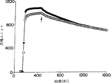
図1Aと1BはBIAcoreTMバイオセンサーに結合した(S237C)hGHbpへのヒト成長ホルモン(hGH)または(G120R)hGHの結合に対する反応(図1A)及び速度論(図1B)を表す。(S237C)hGHbpを1.2ng/mm2に相当する1220RU'sの濃度でチオール−デキストランマトリックス(図1A)に固定化した。結合プロフィール実施例では(図1B)、hGH(塗りつぶしていない印)または(G120R)hGH(塗りつぶした印)を、会合を追跡し化学量論を計算する結合ホルモンの限界量を確立するために、飽和濃度(>200nM)で注入した。飽和後、インジェクターループを解離を追跡するためにバッファーに切り替えた(矢印で示されている)。
図2Aと2BはBIAcoreTMバイオセンサーに結合した(S201C)hGHbpへのhGH(塗りつぶしていない丸印)または(G120R)hGH(塗りつぶした丸印)の結合に対する反応(図2A)及び速度論(図2B)を表す。(S201C)hGHbpを1480RU's(1.48ng/mm2)の濃度で固定化した。結合コンディション及びプロフィールは図1Aと1Bのものと類似している。
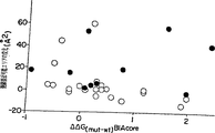
図3はRIA(y軸)またはBIAcoreTMバイオセンサー(x軸)によって得られたデータからhGHbpとの1:1複合体を形成した場合の、野生型hGHと相関的なhGHのアラニンミュータントに対して計算された結合の自由エネルギー(ΔΔG(mut-wt))における変化の相互間系を表す。値は表2から得られた。
図4Aと4Bは接触残基でのアラニンミュータントに対するオフ−レート(図4A)またはオン−レート(図4B)における相対的変化を表す。データは表2から得られた。
図5Aと5Bはアラニン置換における結合アフィニティーの変化と隠れた表面エリア(Å2)の間の関係(図5A)またはβ炭素以外の接触側鎖における原子に対するファンデルワールス接触の数(図5B)を表す。塗りつぶした丸印はサイト1で受容体と水素結合または塩架橋を形成する界面で隠れた残基に対してであり、塗りつぶしていない丸印はそうでない残基に対してである。データは表2からプロットされている。
図6A,6B及び6Cはアラニンスキャニングミュータジェネシス、x線結晶構造またはファージディスプレーのそれぞれによって定義された受容体結合エピトープの比較を表す。
図6AはhGHサイト1機能的エピトープを表す。アラニンスキャニングミュータジェネシスにしたがって、受容体結合に関与する残基はhGH(hGHbp)2結晶構造から由来したhGHの画像モデル上に示されている。de Vos等,上記参照。アラニン置換(またはK41の場合にはGln置換)の効果は、M14,H21,F54,E56,I58,S62,N63及びY164を除いてBIAcoreTM速度論測定に基づいて示されている。これらの部位ではBIAcoreTMデータは入手可能ではないかまたは結合に対してごくわずかな効果を示すのみであり、故に示される効果はRIAデータに基づいている。結合自由エネルギー(ΔΔG)の変化は−RTln[Kd(Alaミュータント)/Kd(hGH)]として計算された。黒い球は結合を改変する(ΔΔG=−1から−0.5kcal/モル)アラニン置換を示す。大きいサイズの4個の白い球はそれぞれ+0.5から1.0kcal/モル、+1.0から1.5kcal/モル、+1.5から2.0kcal/モルまたは+2.0から2.5kcal/モルまで結合エネルギーを減少するアラニン置換を表す。
図6BはhGHサイト1構造的エピトープである。大きいサイズの4個の白い球はhGH(hGHbp)2X線結晶構造から計算されたようにアラニン置換による各残基でそれぞれ−20から0Å2、0から20Å2、20から40Å2または40から60Å2の溶媒接近可能エリアの変化を示す。
図6Cは無作為化されたファージミドライブラリーにおけるhGH残基の保存を表す。ファージディスプレーされたhGHライブラリーにおいて一度に4位で無作為化された残基が示されている:ヘリックス−1[F10,M14,H18,H21];ミニヘリックス−1[K41,Y42,L45,Q46];Loop−A[F54,E56,I58,R64];ヘリックス−4A[K172,E174,E176,R178];ヘリックス−4B[R167,D171,T175,I179]。hGH結合のため分類した後の各位で見出される野生型hGH残基の画分[ここ及びLowman等,上記参照で報告されるデータ]は、黒い球のサイズによって示されている。最も小さい黒い球は0−10%保存されており、次に大きいものは10−25%保存されており、次に大きいものは25−50%保存されており、最も大きいものは>50%保存されている。
図7は受容体結合アフィニティーを促進するファージ由来ミューテーションと組み合わせるためのストラテジーを表す。最もよく選択されたものは野生型に対するアフィニティーにおける倍化と共に示されている。これらの変異体のそれぞれで見出される野生型由来のミューテーションの数もまた示されている(例えば4muts.)。ヘリックス−1、ヘリックス−4、ミニヘリックス−1またはヘリックス1とヘリックス2と連結したループにおけるそれぞれ4つのコドンで無作為化されたライブラリーが別々に分類された。Helix−4aで同定された2つのミューテーション(E174S/F176S)は他のヘリックス−4部位(Helix−4b;Lowman等,上記参照)での付加的な無作為化と選択のためのバックグランドとして用いられた。Helix−1とHelix−4bにおいて同定されたミューテーションをBD変異体を生産するために結び付けた;Minihelix−1とLoop−Aにおけるミューテーションを変異体852bを生産するために結び付けた。最後にこれら二つの変異体由来のミューテーションを変異体852dを生産するために結び付けた。
図8A,8B及び8CはそれぞれhGH構造的エピトープ、ファージ由来エピトープ及び発展変異体の間の関係を表す。野生型hGH残基が受容体結合のために分類されたhGH−ファージミドプール(Lowman等,上記参照)において現れた頻度についての自然対数がx軸に示されている。組み合わせたライブラリー由来のデータは示されなかった。logスケールは隠れた表面エリアとの比較から選択された。野生型残基が選択されたライブラリーの全てにおいて見出されなかったので、残基M14,H18,K41,Q46,R167およびE174はこのグラフに現れていない。
図8AはhGH−(hGHbp)2のx線構造との比較を表す。受容体−1結合により隠れたhGH残基の側鎖エリア([フリーhGH][hGH−hGHbp複合体]の溶媒接近可能エリア)がプロットされている。
図8Bはファージディスプレー及びアラニンスキャニングミュータジェネシスの結果を表す。hGHにおけるAla置換の機能的効果がln[Kd(Alaミュータント)/Kd(hGH)]としてプロットされている。結合データは速度論データが入手できない場合を除いて、BIAcoreTMバイオセンサー測定から得られた。これらの非接触残基(F10,F54,I58)に対して、ラジオ免疫沈降法アッセイから得られたKdに対する値を用いた。Cunningham等,1989,上記参照。
図8Cは発展変異体の間の残基の保存を表す。ヒト胎盤ラクトゲン、hGH(20K)及びhGH−Vと同様に、サル、ブタ、ゾウ、ハムスター、クジラ、アルパカ、キツネ、ウマ、ヒツジ、ラット、カメ、ニワトリ、ミンク、ウシ、サケ、カエル及びマス由来の成長ホルモンのアミノ酸配列(Genbank,vol.75,1993年2月)を野生型hGHのものと比較した。プロラクチン発展変異体は含めていなかった。野生型hGH残基がこれらの変異体の間に現れる頻度の自然対数がプロットされている。
図9はファージ由来ミューテーションの加法を開示する。野生型hGHのものに対する結合の自由エネルギーの変化が構成要素ミューテーションに対するΔΔGの和と比較された。示されているポイントは(1)変異体BD vs.[BプラスD];(2)変異体852b vs.[ミニヘリックス−1プラスループ−A];(3)変異体BF vs.[BプラスF];及び(4)変異体852d vs.[BDプラス852b]の組み合わせに相当する。エラーバーはエラー計算の広がりを用いて標準のズレから見積もった。Bevington,Data Reduction and Error Analysis for the Physical Sciences,pp.56−65(McGraw−Hill,New York,1969)。示されている直線はy=−0.94+0.60x;R2=0.96である。
図10は実施例Vに記述されているような、本発明の拮抗剤hGH変異体(B2036変異体)を発現するために用いられる例示的なベクターのためのプラスミドマップを示す。
図11はアカゲザルにおけるインスリン様成長因子−I(IGF−I)濃度に対する本発明の様々な拮抗剤hGH変異体の毎日の皮下注射(0.25mg/kg)の効果を示す。変異体のペグ化および非ペグ化形態の両者がテストされた。実施例XIII参照。
図12はアカゲザルに静脈または皮下に注射されたペグ化拮抗剤hGH変異体(B2036)調製物の単一投与薬力学を示す。拮抗剤効果はIGF−I濃度における減少パーセントとして測定された。実施例XIV参照。
好ましい実施態様の説明
変異体
ヒト成長ホルモン(hGH)のDNA及びアミノ酸配列を報告されている。Coeddel等,上記参照;Gray等,上記参照。本発明はアラニンスキャニング方法体系またはファージミド選択法のそれぞれを用いて生産された新しいhGH変異体を記述する。本発明のhGH変異体は野生型またはmethGHを発現可能ないかなる組換え系においても発現し得る。
変異体hGH配列表記は本発明のhGH変異体における現実のアミノ酸置換を定義する。変異体に対して、置換は野生型残基を表す文字(一文字コードで)、野生型配列のアミノ酸位置を示す数及び置換されたアミノ酸を示す第二の文字によって示される。例えばR64KはArg64がLysに変換されたミューテーションを示す。複数のミューテーションはコンマによって分割された一連の単一のミューテーションによって示される。
アラニンスキャニングミューテーション
一つの実施態様として、本発明はここでポリペプチドとその受容体の相互作用に関与するポリペプチド内の一つ以上の活性部位を検出するために、hGHの系統的な分析を利用する。該分析は組換えDNA法を用いて簡便に実施される。一般的にhGHをコードするDNAは便宜にホスト内でそれを発現するためにクローン化され操作される。hGHをコードするDNAはゲノムライブラリー、hGHを発現する細胞内のmRNAから由来するcDNA、または合成的に構築したDNA配列によって得られ得る。Maniatis等,Molecular Cloning:A Laboratory Manual,Cold Spring Harbor Laboratory,N.Y.(1982)。
それから野生型hGH DNAをホスト細胞をトランスフォームするために用いられる適したプラスミドまたはベクター内に挿入する。原核生物はhGH変異体を生産するためのDNA配列のクローニング及び発現にとって好ましい。例えば大腸菌B、大腸菌X1776(ATCC No.31537)、及び大腸菌c600とc600hfl、そして大腸菌W3110(F-,γ−、原栄養株性、ATCC No.27325)、枯草菌のようなバチルス、及びネズミチフス菌または霊菌及び様々なシュードモナス種のような他の腸内細菌科と同様に、大腸菌K12株294(ATCC No.31446)が用いられ得る。好ましい原核生物は大腸菌W3110(ATCC 27325)である。原核生物の細胞内で発現される場合、hGHは典型的にN末端メチオニンまたはホルミルメチオニンを含み、グリコシル化されていない。培地またはペリプラズム内の細胞外に発現される場合、hGHはN末端メチオニンを含んでいない。これらの例はもちろん限定することよりもむしろ実例となることを企図されている。
原核生物に加えて、酵母カルチャーのような真核生物細菌、または多細胞生物由来の細胞が用いられ得る。原則として、いかなる該細胞カルチャーも実施可能である。しかしながら興味は哺乳動物細胞において最大であり、カルチャー(組織カルチャー)における哺乳動物細胞の増殖は再現可能な方法となっている。Tissue Culture,Academic Press,KruseとPatterson,編者(1973)。該有用なホスト細胞系の例として、VERO及びHela、チャイニーズハムスター卵巣(CHO)、W138、BHK、COS−7及びMDCK細胞系がある。
一般的にホスト細胞と両立できる種由来の複製およびコントロール配列を含むプラスミドベクターがこれらのホストと連結して用いられる。ベクターはもともと複製部位を運んでおり、トランスフォームされた細胞において表現型選択を提供することが可能なタンパク質をコードする配列も同様である。例えば大腸菌は大腸菌種由来のプラスミドであるpBR322を用いてトランスフォームされ得る。Mandel等,J.Mol.Biol.,53:154(1970)。プラスミドpBR322はアンピシリン及びテトラサイクリン耐性のための遺伝子を含んでおり、それゆえ選択のための容易な手段を提供する。一つの好ましいベクターは本出願の優先出願(1989年10月26日に提出されたU.S.S.N.07/428,066)の実施例1に記述されているpBO475である。このベクターはファージ及び大腸菌のための複製のオリジンを含んでおり、それらはこのベクターを該ホストの間で往復することを可能にし、それによってミュータジェネシスと発現を容易にする。「発現ベクター」とは適したホストにおいて上記DNAの発現に影響することができる適したコントロール配列と実施可能にリンクしたDNA配列を含むDNA構築物をいう。該コントロール配列には、転写に影響するためのプロモーター、場合により該転写をコントロールするためのオペレーター配列、適したmRNAリボソーム結合部位をコードする配列及び転写と翻訳の終結をコントロールする配列が含まれる。ベクターはプラスミド、ファージ粒子または単純に潜在的なゲノム挿入物であり得る。一度適したホスト内にトランスフォームされると、ベクターはホストのゲノムとは独立に複製し機能し得、またはある場合にはゲノム自身に入り込み得る。現在ではプラスミドが最も一般的に用いられるベクターの形態であるので、本明細書においては「プラスミド」と「ベクター」なる語は時々互換的に用いられる。しかしながら本発明は同等の機能を果たし、本分野で既知であるまたは既知となる該他の形態の発現ベクターも含むことを企図している。
2つのDNAまたはポリペプチド領域に間の関係を記述する場合に「実施可能にリンクしている」とはそれらがお互いに機能的に関与していることを意味する。例えばもしプレ配列がシグナル配列として機能し、タンパク質の成熟型形態の分泌に関与し、大抵の場合にはシグナル配列の切断を含むのであれば、プレ配列はペプチドと実施可能にリンクしている。もしプロモーターが配列の転写をコントロールするのであれば、プロモーターは配列と実施可能にリンクしている;もしリボソーム結合部位が翻訳を許すように位置しているのであれば、リボソーム結合部位はコード配列と実施可能にリンクしている。
一度hGHがクローン化されると、部位特異的ミュータジェネシス(Carter等,Nucl.Acids.Res.,13:4331[1986];Zoller等,Nucl.Acids Res.,10:6487[1987])、カセットミュータジェネシス(Wells等,Gene,34,315[1985])、制限選択ミュータジェネシス(Wells等,Philos.Trans.R.Soc.London SerA,317:415[1986])または他の既知の方法が、クローン化hGH DNAに対して、置換されている残基によって定義されるアミノ酸配列の変化のためにコードする変異体DNAを生産するために、実施され得る。適した発現ベクターと実施可能にリンクされた場合、活性ドメイン置換hGH変異体が得られる。ある場合にはhGH変異体の回収は、hGHの元のまたは変異体をコードするDNA配列と実施可能にリンクした適したシグナル配列の使用によって、発現ホストから該分子を発現し分泌させることによって容易にし得る。該方法は当業者によく知られている。もちろん望ましいhGH変異体のin vitroの化学的合成のような他の方法も該ポリペプチドを生産するために用いられ得る。Barany等,in The Peptides,E.GrossとJ.Meienhofer編,(Academic Press:N.Y.1979),vol.2,pp.3−254。
一度異なるGH変異体が生産されるとそれらは受容体と接触され、もし可能であれば受容体と各変異体の間の相互作用が測定される。活性ドメイン中のどのアミノ酸残基が受容体との相互作用に関与しているかを決定するために、これらの活性が同じ受容体との野生型hGHの活性と比較される。該分析で用いられるアミノ酸のスキャニングは置換されたものと異なるアミノ酸のいくつかであり得る、すなわち19の他の天然に生じるアミノ酸のいくつかであり得る。
ターゲット受容体は天然のソースから単離され得または本分野で既知の方法によって組換え法で調製され得る。実例として、受容体はMcFarland等,Science,245:494−499(1989)によって記述されている方法によって調製され得る。
受容体と元の及び変異体の間の相互作用はいかなる便宜なin vitroまたはin vivoアッセイによっても測定され得る。それゆえin vitroアッセイは受容体とhGHの間のいかなる検出可能な相互作用を測定するためにも用いられ得る。該検出は比色定量変化、放射性活性の変化、可溶性の変化、ゲル電気泳動及び/またはゲル排除法等で測定されるような分子量の変化の測定を含み得る。in vivoアッセイは例えば体重増または電解質バランスにおける変化といった生理学的効果を検出するアッセイを制限することなく含む。一般的にいかなるin vivoアッセイも、可変的なパラメーターが興味ある受容体とhGHの間の相互作用における変化を検出するために存在する限りで用いられ得る。
いくつもの数の分析的な測定が活性を比較するために用いられ得る一方、受容体の結合のための簡便なものはhGH変異体と受容体の間で形成される複合体の解離定数Kdであり、それを野生型hGHのKdと比較することである。一般的に置換によって置換された類似の残基当たりのKdにおいて2倍の増大または減少は、置換された残基(類)がターゲットとの野生型hGHの相互作用において活性であることを示す。
想像されるまたは既知のアミノ酸残基がアミノ酸分析のスキャニングにかけられる場合、それらにすぐ近接したアミノ酸残基がスキャンされるべきである。3残基置換ポリペプチドが作られ得る。一つは想像されるまたは既知の活性アミノ酸であるN位でスキャニングアミノ酸、好ましくはアラニンを含む。他の2つはN+1位とN−1位でスキャニングアミノ酸を含む。もし各置換hGHが受容体に対するKdに対して約2倍より大きい効果を引き起こした場合、スキャニングアミノ酸はN+2位とN−2位で置換される。少なくとも1つ、好ましくは4つの残基がKdに対して約2倍より少ない効果をもつ各方向で同定されるかまたは野生型hGHのそれぞれの末端に到達するまで、これが繰り返される。この方式では特定の受容体との相互作用に関与する連続的なアミノ酸配列に沿って一つ以上のアミノ酸が同定され得る。
アミノ酸スキャンによって同定された活性アミノ酸残基は、典型的には受容体ターゲットと直接的に接触するものである。しかしながら活性アミノ酸はまた、他の残基またはH2Oのような小分子またはNa+,Ca2+,Mg2+またはZn2+のようなイオン種と形成した塩架橋を通じてターゲットと間接的に接触し得る。
ある場合には、一つ以上の残基でのスキャニングアミノ酸の置換が受容体とのその活性の分析を実施するのに十分な量の単離を許す濃度で発現されない残基置換ポリペプチドを引き起こす。該場合には異なるスキャニングアミノ酸、好ましくは等配電子アミノ酸が用いられ得る。
好ましいスキャニングアミノ酸の中には、比較的小さく中性のアミノ酸がある。該アミノ酸にはアラニン、グリシン、セリン及びシステインが含まれる。アラニンはβ−炭素以外の側鎖を除去し、変異体の主鎖構造を比較的改変しそうにないので、アラニンはこのグループの中では好ましいスキャニングアミノ酸である。アラニンは最も共通なアミノ酸であるので、アラニンはまた好ましい。さらにそれは隠れた位置とさらされた位置の両方で頻繁に見出される。Creighton,The Ptoteins(W.H.Freeman & Co.,N.Y.);Chothia,J.Mol.Biol.,150:1(1976)。もしアラニン置換が十分な量のhGH変異体を生産しなかったら、等配電子アミノ酸を用い得る。代わりに好ましさがこの順序で減少していく以下のアミノ酸を用い得る:Ser,Asn及びLeu。
一度活性アミノ酸残基が同定されると、等配電子アミノ酸が置換され得る。該等配電子置換は全ての場合で生じる必要はなく、活性アミノ酸が同定される前に実施され得る。該等配電子アミノ酸置換はいくつかの置換が引き起こし得る構造に対する潜在的な破壊効果を最小化するために実施される。等配電子アミノ酸は以下の表に示されている:

特にhGHに対しては、本発明の例示は体形成受容体(hGHbp)との活性を決定する活性ドメインと活性残基が同定される好ましい実施態様である。本発明のこの実施態様を実施するにおいて、天然で生じているhGHと比較するとhGHbpとの異なる結合相互作用を持つアミノ酸残基置換hGH変異体を含むhGH変異体が作製または同定されている。いくつかはhGHbpに対するより高いアフィニティーとラットにおける体形成のための促進された有効性を持ち得る。他のものはhGHbpとの減少したアフィニティーを持つ。該hGH変異体はhGH作用剤または拮抗剤として有用であり、もし該変異体がhGHbpとの実質的な相互作用から遊離しておれば、hGHに対する他の受容体を刺激するためのより高い有効性を持ち得る。さらに、該変異体はhGHスタンダードまたはトレーサーとしてhGHのためのイムノアッセイにおいて有用である。ある変異体は抗hGHポリクローナル抗体を含むヒト及びマウス血清との反応性において有意な減少を持つものを同定し得る。他のものはhGHと同じhGHbpに対する結合アフィニティーをもつが、成長を刺激する増大した有効性を持つ。
肝臓由来の体形成受容体と相互作用するhGHにとっての活性ドメイン及び残基を決定する方法が本出願の優先出願(1989年10月26日に提出されたU.S.S.N.07/428,066)の図1に概略的に示されており、選択された部分が図2に示されている。
ファージミドディスプレー法
加えて変異体はファージミドディスプレーによって分析し得る。この方法は(a)hGHをコードする第一の遺伝子、天然のまたは野生型ファージコートタンパク質の少なくとも一部をコードする第二の遺伝子を含む、第一と第二の遺伝子は異種のものであり、そして第一と第二の遺伝子と実施可能にリンクした転写調節エレメントを含み、それによって融合タンパク質をコードする遺伝子融合物を形成し;(b)第一の遺伝子内の一つ以上の選択された位置でベクターをミューテートしそれによって関連するプラスミドのファミリーを形成し;(c)プラスミドを用いて適したホスト細胞をトランスフォームし;(d)ファージコートタンパク質をコードする遺伝子を持つヘルパーファージを用いてトランスフォームされたホスト細胞に感染させ;(e)少なくともプラスミドの一部を含み、ホストをトランスフォームできる組換えファージミド粒子を形成するために適したコンディションの下で、トランスフォームされ感染されたホスト細胞を培養し、コンディションを少量のファージミド粒子だけが粒子の表面に融合タンパク質の一コピーより多くをディスプレーするように調節し;(f)少なくともファージミド粒子の一部が受容体分子に結合するようにファージミド粒子をhGH受容体分子(hGHbp)を用いて接触させ;(g)結合したファージミド粒子をしていないものと分離する工程より成る。好ましくはさらに本方法はhGHbpに結合する組換えファージミド粒子を用いて適したホスト細胞をトランスフォームすることと(d)から(g)の工程を一度以上繰り返すことを含む。
好ましくはこの方法においてプラスミドは転写調節エレメントのきついコントロールの下にあり、粒子の表面に存在する融合タンパク質の一コピーより多くをディスプレーするファージミド粒子の量または数が約1%より小さいように培養コンディションを調節する。また好ましくは融合タンパク質の一コピー以上をディスプレーするファージミド粒子の量は融合タンパク質の単一コピーをディスプレーするファージミド粒子の量の10%より小さい。最も好ましくはその量は20%より小さい。
この方法においては典型的には、さらに発現ベクターはポリペプチドの各サブユニットをコードするDNAと融合した分泌シグナル配列を含み、転写調節エレメントはプロモーター系である。好ましいプロモーター形はlacZ、λPL、tac、T7ポリメラーゼ、トリプトファン及びアルカリホスファターゼそしてそれらの組み合わせから選択される。また通常該方法はM13K07,M13R408,M13−VCS及びPhi X 174から選択されるヘルパーファージを用いる。好ましいヘルパーファージはM13K07であり、好ましいコートタンパク質はM13ファージ遺伝子IIIコートタンパク質である。好ましいホストは大腸菌であり、大腸菌のプロテアーゼ欠失株である。本発明の該方法によって選択される新しいhGH変異体が検出されている。ポリペプチドとファージコートタンパク質をコードする核酸の間に機能的に位置している抑制的終止コドンを含むファージミド発現ベクターが構築された。
詳細には、hGH選択の繰り返されるサイクルが複数の選択サイクルのファージミド選択によって次第に高いアフィニティー結合のための選択に用いられる。リガンドポリペプチドにおける第一の領域またはアミノ酸を含むファージミド選択の第一周に引き続いて、リガンドポリペプチドの他の領域またはアミノ酸におけるファージミド選択のさらなる周が実施される。ファージミド選択のサイクルはリガンドポリペプチドの望ましいアフィニティー性質が達成されるまで繰り返される。この工程を説明すると、hGHのファージミド選択はサイクルで実施された。第一のサイクルでは、hGHのアミノ酸172,174,176及び178がミューテートされファージミドが選択され得る。第二のサイクルでは、hGHのアミノ酸167,171,175及び179がファージミド選択され得る。第三サイクルでは、hGHのアミノ酸10,14,18及び21がファージミド選択され得る。前のサイクルから任意のアミノ酸変化が次の選択サイクルの前にポリペプチド内に取り込まれ得る。例えばhGHアミノ酸置換174(セリン)及び176(チロシン)がhGHのアミノ酸167,171,175及び179のファージミド選択の前にhGH内に取り込まれた。
前述したことからhGHの結合ドメインを形成するアミノ酸残基は連続しておらず、ポリペプチドの異なるサブユニット上に属し得ることが認められよう。それは結合ドメインが結合部位で特定の二次構造を形成し、一次構造ではないということである。それゆえ一般的に、受容体との相互作用する可能性を持つためにポリペプチドの内側から離れて向けられた部位で特定の二次構造内のアミノ酸をコードするコドンにミューテーションが導入される。hGH受容体への結合を強く調節するhGHにおける残基の位置(Cunningham等,Science,1990,上記参照)が知られている。故にミュータジェネシスに適した代表的な部位はヘリックス−4上の残基172,174,176及び178を含み、同様に「非順列」二次構造で位置する残基64を含むであろう。
このファージミドディスプレー法では、一度hGH遺伝子が単離されると、Sambrook等,Molecular Biology:A Laboratory Manual,Cold Spring Harbor Press,Cold Spring Harbor,New York 1989に一般的に記述されているように、それを増幅のため適したベクター(好ましくはプラスミド)内に挿入し得る。いくつかのタイプのベクターが入手可能であり本発明を実施するために用いられ得る一方、プラスミドベクターは比較的容易に構築でき容易に増幅し得るので、プラスミドベクターはここでの使用に好ましいベクターである。プラスミドベクターは一般的にプロモーター、シグナル配列、表現型選択遺伝子、複製のオリジン部位及び当業者に既知である他の必要な構成要素を含む。
原核生物ベクターで最も共通に用いられるプロモーターはlacZプロモーター系、アルカリホスファターゼphoAプロモーター、バクテリオファージλPLプロモーター(lacリプレッサーによって調節されるハイブリッドtrp−lacプロモーター)、トリプトファンプロモーター及びバクテリオファージT7プロモーターを含む。プロモーターについての一般的な記述はSambrook等,上記参照の第17章を参照。これらは最も広く用いられているプロモーターである一方で、他の適した微生物プロモーターも同様に使用し得る。
ファージミドディスプレーのために本発明を実施する上での好ましいプロモーターは、融合遺伝子の発現がコントロールされるようにきつく調節され得るものである。本分野で以前に認識されていなかった問題は、ファージミド粒子の表面での融合遺伝子の複数のコピーのディスプレーがターゲットにファージミドの複数点の付着を引き起こすことであると考えられている。ターゲットが「キレート化」されるため、融合タンパク質の複数コピーが互いに非常に近接してファージミド粒子上にディスプレーされる場合、「キレート効果」といわれているこの効果は、誤った「高アフィニティー」ポリペプチドの選択を引き起こすと考えられる。複数点での付着が生じると、有効なまたは明らかなKdがディスプレーされた融合タンパク質の各コピーに対する個々のKdの産物と同じくらい高くなり得る。
ファージミド粒子の少量だけ、すなわち約1%より小さい量だけ融合タンパク質の複数コピーを含むように融合タンパク質の発現をきつく調節することによって、「キレート効果」を克服し、高アフィニティーポリペプチドの正確な選択を可能にすることが開示されている。それゆえプロモーターに依存して、融合タンパク質の単一コピーを含むファージミド粒子の数を最大化し、融合タンパク質の複数コピーを含むファージミド粒子の数を最小化するために、ホストの培養コンディションを調節する。
本発明を実施するために用いられる好ましいプロモーターは、lacZプロモーターとphoAプロモーターである。lacZプロモーターはlacリプレッサータンパク質laciによって調節され、それゆえ融合遺伝子の転写はlacリプレッサータンパク質の濃度の操作によってコントロールされ得る。説明としてlacZプロモーターを含むファージミドをlacZプロモーターに対するリプレッサーであるlaciリプレッサー遺伝子の単一コピーを含む細胞株内で成育させる。laci遺伝子を含む例示的な細胞株にはJM101とXL1−ブルーが含まれる。代わりに、ホスト細胞をリプレッサーlaciとlacZプロモーターの両者を含むプラスミドを用いてコトランスフォームし得る。場合により上記方法の両者を同時に用いる、つまりlacZプロモーターを含むファージミド粒子をlaci遺伝子を含む細胞株内で成長させ、細胞株をlacZとlaciの両者を含むプラスミドを用いてコトランスフェクトさせる。
通常遺伝子を発現したいと考えた場合、上記トランスフェクトされたホストに対してイソプロピルチオガラクトシド(IPTG)のようなインデューサーを加えるであろう。しかしながら本発明においては、(a)遺伝子III融合タンパク質の発現を最小化し、それによってコピー数(すなわちファージミド数当たりの遺伝子III融合物の数)を最小化するために、及び(b)低濃度でさえIPTGのようなインデューサーによって引き起こされるファージミドの貧弱なまたは不適当なパッケージングを避けるために、この工程は省略される。典型的にはインデューサーを加えないと、ファージミド粒子当たりの融合タンパク質の数は約0.1である(ファージミド粒子の数当たりの全部の融合タンパク質の数)。本発明を実施するために用いられる最も好ましいプロモーターはphoAである。リン酸がプロモーターの活性をダウンレギュレーションするように機能する場合、このプロモーターは細胞内無機リン酸の濃度によって調節されると考えられる。それゆえ細胞からリン酸を枯渇させることによって、プロモーター活性を増大し得る。望ましい結果は2YTまたはLBのようなリン酸リッチ培地内で細胞を成育させることによって達成され、それによって遺伝子III融合物の発現をコントロールする。
本発明を実施するために用いられるベクターの他の有用な構成要素は、シグナル配列である。この配列は典型的には融合タンパク質をコードする遺伝子のすぐ5'に位置し、それゆえ融合タンパク質のアミノ末端で転写される。しかしながらある場合には、シグナル配列は分泌されるタンパク質をコードする遺伝子の5'以外の場所に位置することが示されている。この配列は付着されたタンパク質が細菌細胞の内膜を通過することを狙っている。シグナル配列をコードするDNAは、シグナル配列を持つタンパク質をコードする遺伝子由来の制限酵素断片として得られ得る。適した原核生物シグナル配列は例えばlamBまたはompF(Wong等,Gene,68:193[1983])、MalE、PhoA及び他の遺伝子から得られ得る。本発明を実施するための好ましい原核生物シグナル配列はChang等,上記参照によって記述されているような大腸菌熱安定性エンテロトキシンII(ST II)シグナル配列である。
ファージディスプレー法を実施するために用いられるベクターのもう一つの有用な構成要素は表現型選択遺伝子である。典型的な表現型選択遺伝子は、ホスト細胞に対して抗生物質耐性を与えるタンパク質をコードするものである。説明として、アンピシリン耐性遺伝子(amp)及びテトラサイクリン耐性遺伝子(tet)がこの目的にために容易に用いられる。
hGHをコードする遺伝子(遺伝子1)と同様に上述の構成要素を含む適したベクターの構築は、Sambrook等,上記参照に記述されているような標準的な組換えDNA法を用いて調製される。ベクターを形成するために結合されるための単離されたDNA断片は、望ましいベクターの生産のため特異的な順序及び配向で互いに切断され、調製され及び接合される。
DNAは適したバッファーにおいて適当な制限酵素または酵素類を用いて切断される。一般的に約0.2−1μgのプラスミドまたはDNA断片が約20μlのバッファー溶液において1−2ユニットの適当な制限酵素と共に用いられる。適当なバッファー、DNA濃度およびインキュベーション時間と温度は、制限酵素の商品によって特異的である。いくつかの酵素はより高い温度が必要であるが、一般的に37℃で約1または2時間のインキュベーション時間で十分である。インキュベーション後、酵素と他のコンタミネーションをフェノールとクロロホルムの混合物を用いて切断溶液の抽出によって除去し、DNAをエタノールを用いた沈殿によって水溶性画分から回収する。
機能的なベクターを形成するためDNA断片を互いにつなぐために、DNA断片の末端は互いに適合していなければならない。ある場合には末端は制限酵素切断の後直接的に適合している。しかしながら制限酵素切断によって広く生じる付着末端をライデーションのために適合させるために平滑末端に最初に変換することは必要であり得る。末端を平滑にするために、DNAを適したバッファー内で少なくとも15分15℃で10ユニットのDNAポリメラーゼI(Klenow)のKlenow断片を用いて、4種のデオキシリボヌクレオチド三リン酸の存在下で処理する。それからDNAをフェノール−クロロホルム抽出とエタノール沈殿によって精製する。
切断されたDNA断片はDNAゲル電気泳動を用いてサイズ分離され選択される。DNAはアガロースまたはポリアクリルアミドマトリックスのそれぞれで電気泳動し得る。マトリックスの選択は分離されるDNA断片のサイズに依存する。電気泳動後、DNAを電気溶出によって、Sambrook等,上記参照の6.30−6.33部分に記述されているように、またはマトリックスとして低溶性アガロースを用いているならアガロースを溶かしそこからDNAを溶出することによって、マトリックスから溶出する。
互いにつながれるべきDNA断片(つながれる各断片の末端が適合するように前に適切な制限酵素で切断されている)は、大体等モルの量で溶液中に置かれる。溶液はまたATP、リガーゼバッファー及び0.5μgのDNA当たり約10ユニットのT4DNAリガーゼのようなリガーゼを含む。DNA断片がベクター内につながれるのであれば、ベクターは適当な制限酵素(類)を用いて切断することによって最初に直線化される。それから直線化ベクターをアルカリホスファターゼまたはウシ腸ホスファターゼを用いて処理される。ホスファターゼ処理はライゲーション工程の間のベクターの自己ライゲーションを避ける。
ライゲーション後、それから挿入された外来遺伝子と共にベクターを適当なホスト細胞内にトランスフォームする。原核生物が本発明のための好ましいホスト細胞である。適した原核生物ホスト細胞は大腸菌JM101株、大腸菌K12株294(ATCCナンバー31,446)、大腸菌W3110株(ATCCナンバー27,325)、大腸菌X1776(ATCCナンバー31,537)、大腸菌XL−1ブルー(Stratagene)及び大腸菌Bを含む;しかしながらHB101,NM522,NM538及びNM539のような多くの他の大腸菌の株及び多くの他の種そして原核生物の属も同様に用いられ得る。上記挙げた大腸菌に加えて、枯草菌のようなバチルス、ネズミチフス菌及び霊菌のような他の腸内細菌科、及び様々なシュードモナス種がすべてホストとして用いられ得る。
原核生物細胞のトランスフォーメーションはSambrook等,上記参照の1.82部分に記述されているような塩化カルシウム法を用いて容易に成し遂げられる。代わりに電気穿孔法(Neumann等,EMBO J.,1:841[1982])がこれらの細胞をトランスフォームするために用いられ得る。トランスフォームされた細胞tetまたはampを持ち、ベクター上のtet及び/またはamp耐性遺伝子の存在のためそれらが耐性を示すことによってそれらが抗生物質上の成育によって選択される。
トランスフォームされた細胞の選択後、これらの細胞はカルチャーで成育され、それからプラスミドDNA(または外来遺伝子が挿入された他のベクター)が単離される。プラスミドDNAは本分野で既知の方法を用いて単離され得る。2つの適した方法はSambrook等,上記参照の1.25−1.33部分に記述されているようなDNAのスモールスケール調製及びDNAのラージスケール調製である。単離されたDNAはSambrook等,上記参照の1.40部分に記述されているような本分野で既知の方法によって精製される。それからこの精製されたプラスミドDNAを制限酵素地図および/またはDNAシークエンシングによって分析する。DNAシークエンシングはMessing等,Nucleic Acids Res.,9:309(1981)の方法、Maxam等,Meth.Enzymol.,65:499(1980)の方法またはSanger等,Proc.Natl.Acad.Sci.USA,74:5463−5467(1977)の方法のそれぞれによって一般的に実施される。
ファージミドディスプレー法はここで融合タンパク質が転写の間生産されるようにhGHをコードする遺伝子(遺伝子1)を第二の遺伝子(遺伝子2)に融合することを企図している。遺伝子2は典型的にはファージのコートタンパク質遺伝子であり、好ましくはそれはファージM13遺伝子IIIコートタンパク質またはその断片である。遺伝子1と2の融合は遺伝子1を含むプラスミドの特定の部位に遺伝子2を挿入することによって、または遺伝子2を含むプラスミドの特定の部位に遺伝子1を挿入することによって成し遂げられ得る。
プラスミド内への遺伝子の挿入には、プラスミドが遺伝子を挿入すべき正確な位置で切断されている必要がある。それゆえこの部分に制限酵素部位が存在していなければならない(好ましくは制限酵素切断の間プラスミドが単一の位置だけで切断されるような独特の部位が)。プラスミドは上述のように切断され、ホスファターゼ処理され、精製される。それから遺伝子を2つのDNAを互いにつなぐことによってこの直線化プラスミド内に挿入する。プラスミドの末端が挿入される遺伝子の末端と適合していたならば、ライゲーションを成し遂げられ得る。制限酵素がプラスミドを切断し、平滑末端または付着末端を作り出すため挿入される遺伝子を単離するために用いられるならば、Sambrook等,上記参照の1.68部分に記述されているようにATPとリガーゼバッファーの存在下で16℃で1−4時間混合物をインキュベートすることによってT4DNAリガーゼのようなリガーゼを用いてDNAを直接的に互いにつなぎ得る。末端が適合していないならば、それらは最初にDNAポリメラーゼIのKlenow断片またはバクテリオファージT4DNAポリメラーゼを用いて平滑にされなければならないが、両酵素共切断されたDNAの突出したシングルストランド末端をうめるために4つのデオキシリボヌクレオチド三リン酸を必要とする。
代わりにヌクレアーゼS1またはマングビーンヌクレアーゼのようなヌクレアーゼを用いて末端を平滑化しうるが、両酵素ともDNAの突出したシングルストランドを切除することによって機能する。それからDNAを上述したようなリガーゼを用いて再ライゲーションする。ある場合には、コード領域のリーディングフレームが改変されるため、挿入される遺伝子の末端を平滑化することは可能でないかもしれない。この問題を克服するために、オリゴヌクレオチドリンカーが用いられ得る。このリンカーはプラスミドを挿入される遺伝子と連結するための架橋として機能する。これらのリンカーは標準的な方法を用いてシングルストランドDNAまたはダブルストランドDNAとして合成的に作出され得る。該リンカーは挿入される遺伝子の末端と適合する一つの末端を持つ;リンカーは上述したようなライゲーション方法を用いて最初にこの遺伝子とつながれる。リンカーの他の末端はライゲーションのためのプラスミドと適合するようにデザインされている。リンカーをデザインする場合、挿入される遺伝子のリーディングフレームまたはプラスミドに含まれている遺伝子のリーディングフレームを破壊しないように気を付けなければならない。ある場合には、アミノ酸の一部に対してコードしているような、または一つ以上のアミノ酸に対してコードしているようなリンカーをデザインすることが必要となり得る。
遺伝子1と遺伝子2の間に、終止コドンをコードするDNAを挿入し得、該終止コドンはUAG(アンバー)、UAA(オーカー)及びUGA(オパール)である。Davis等,Microbiology(HarperとRow:New York,1980),ページ237,245−247及び274。野生型ホスト細胞で発現している終止コドンは遺伝子2タンパク質の付着なく遺伝子1タンパク質産物の合成を引き起こす。しかしながらサプレッサーホスト細胞での成育は融合されたタンパク質の検出可能な量の合成を引き起こす。該サプレッサーホスト細胞はmRNAの終止コドンの位置にアミノ酸を挿入するように修飾されたtRNAを含み、それによって融合タンパク質の検出可能な量の生産を引き起こす。該サプレッサーホスト細胞は大腸菌サプレッサー株のようによく知られており記述されている。Bullock等,BioTechniques,5:376−379(1987)。融合ポリペプチドをコードするmRNA内に該終止コドンを置くためのいかなる許容される方法も用いられ得る。
抑制可能コドンをhGH遺伝子とファージコートタンパク質の少なくとも一部をコードする第二の遺伝子の間に挿入し得る。代わりに抑制可能終止コドンをポリペプチド内の最後のアミノ酸トリプレットまたはファージコートタンパク質内の第一のアミノ酸を置換することによって融合部位に近接して挿入し得る。抑制可能コドンを含むファージミドがサプレッサーホスト細胞内で成育する場合、それはhGHとコートタンパク質を含む融合ポリペプチドの検出可能な生産を引き起こす。ファージミドが非サプレッサーホスト細胞内で成育する場合、UAG,UAAまたはUGAをコードする挿入された抑制可能トリプレットで終結するため、実質的にファージコートタンパク質に融合していないhGHが合成される。非サプレッサー細胞内では、ポリペプチドは、融合ファージコートタンパク質が不存在であるがそれにもかかわらずホスト細胞にそれを埋め込むために、ホスト細胞から合成され分泌される。
hGH遺伝子は一つ以上の選択されたコドンで改変し得る。改変はhGHの非改変配列または野生型配列と比較するとhGHのアミノ酸配列において変化を引き起こすhGHをコードする遺伝子内の一つ以上の置換、欠失または挿入として定義される。好ましくは改変は分子の一つ以上の領域内で少なくとも一つのアミノ酸をいかなる他のアミノ酸を用いても置換することによる。改変は本分野で既知の様々な方法によって提供され得る。これらの方法はオリゴヌクレオチド介在性ミュータジェネシス及びカセットミュータジェネシスを制限することなく含む。
オリゴヌクレオチド介在性ミュータジェネシスはhGHの変異体に置換、欠失または挿入を調製するための好ましい方法である。本方法はZoller等,上記参照によって記述されているように本分野でよく知られている。略記すると、テンプレートがhGHの非改変または野生型DNA配列を含むプラスミドのシングルストランド形態である場合、hGH遺伝子はDNAテンプレートに望ましいミューテーションをコードするオリゴヌクレオチドをハイブリダイズすることによって改変される。ハイブリダイゼーション後、テンプレートの完全な第二の相補的ストランドを合成するためにDNAポリメラーゼを用い、それゆえオリゴヌクレオチドプライマーが取り込まれhGH遺伝子内で選択された改変がコードされる。
一般的に長さにおいて少なくとも25ヌクレオチドのオリゴヌクレオチドが用いられる。より小さいオリゴヌクレオチドも用いられ得るが、最適なオリゴヌクレオチドはミューテーションをコードするヌクレオチド(類)の各サイドでテンプレートと相補的である12から15ヌクレオチドを持つ。これはオリゴヌクレオチドがシングルストランドDNAテンプレート分子に正確にハイブリダイズすることを確定する。オリゴヌクレオチドはCrea等,Proc.Natl.Acad.Sci.USA,75:5765(1978)に記述されているもののように本分野で既知の方法を用いて容易に合成される。
DNAテンプレートはバクテリオファージM13ベクター(商業的に入手可能なM13mp18及びM13mp19ベクターが適している)由来のもの、またはVieiraとMessing,Meth.Enzymol.,153:3−11(1987)に記載されているようなシングルストランドファージの複製のオリジンを含むベクターのそれぞれであるベクターによってのみ生産され得る。それゆえミューテートされるべきDNAはシングルストランドテンプレートを生産するためにこれらのベクターの一つ内に挿入されなければならない。シングルストランドテンプレートの生産はSambrook等,上記参照の4.21−4.41部分に記述されている。
野生型DNA配列を改変するため、オリゴヌクレオチドは適したハイブリダイゼーションコンディションの下でシングルストランドテンプレートにハイブリダイズされる。それからDNAポリメラーゼ酵素、通常はDNAポリメラーゼIのKlenow断片を、合成に対するプライマーとしてオリゴヌクレオチドを用いるテンプレートの相補的ストランドを合成するために加える。それゆえDNAの一つのストランドはhGH遺伝子のミューテートされた形態をコードし、他のストランド(もともとのストランド)はhGH遺伝子の野生型の非改変配列をコードするようなヘテロ二重鎖が形成される。それからこのヘテロ二重鎖分子を適したホスト細胞、通常は大腸菌JM101のような原核生物内にトランスフォームする。細胞が成長した後、それらをアガロースプレート上にまき、ミューテートされたDNAを含む細菌コロニーを同定するため32−リン酸を用いてラジオラベルされたオリゴヌクレオチドプライマーを用いてスクリーニングする。
すぐ前に記述した方法は、プラスミドの量ストランドがミューテーション(類)を含むホモ二重鎖分子を作出するように修飾し得る。修飾は以下のようになされる:シングルストランドオリゴヌクレオチドを上述したようなシングルストランドテンプレートとアニールさせる。デオキシリボアデノシン(dATP)、デオキシリボグアノシン(dGTP)及びデオキシリボチミジン(dTTP)の3種のデオキシリボヌクレオチドの混合物を、dCTP−(aS)と呼ばれるチオ−デオキシリボシトシン(Amershamから得られ得る)と組み合わせる。この混合物をテンプレート−オリゴヌクレオチド複合体に加える。この混合物にDNAポリメラーゼを添加すると、ミューテートされたベースを除くテンプレートに対して相同なDNAのストランドが生産される。加えてこのDNAの新しいストランドはdCTPの代わりにdCTP−(AS)を含んでおり、それは制限酵素切断からこのDNAを保護するために役立つ。ダブルストランドヘテロ二重鎖のテンプレートストランドに適当な制限酵素を用いてニックを入れた後、テンプレートストランドをミュータジェナイズされた部位(類)を含む領域を通過してExo IIIヌクレアーゼまたはもう一つの適当なヌクレアーゼを用いて切断し得る。それから部分的にだけシングルストランドである分子を引き離して反応を止める。それから完全なダブルストランドDNAホモ二重鎖を全ての4種のデオキシリボヌクレオチド三リン酸、ATP及びDNAリガーゼの存在下でポリメラーゼを用いて形成させる。それからこのホモ二重鎖分子を上述したように大腸菌JM101のような適したホスト細胞内にトランスフォームし得る。
置換された一つより多いアミノ酸を持つミュータントをいくつかの方法の一つで生産し得る。アミノ酸がポリペプチド鎖において互いに近接して位置しているならば、望ましいアミノ酸置換の全てをコードする一つのオリゴヌクレオチドを用いて同時にミューテートし得る。しかしながらアミノ酸がそれぞれからいくらか離れて位置している場合には(約10アミノ酸より多くによって分離されている)、望ましい変化の全てをコードする単一のオリゴヌクレオチドを生産することはさらに困難である。その代わりに二つの代わりの方法の一つが用いられ得る。
第一の方法では、置換される各アミノ酸に対して別々のオリゴヌクレオチドを生産する。それからオリゴヌクレオチドをシングルストランドテンプレートDNAに同時にアニールさせ、テンプレートから合成されるDNAの第二のストランドは望ましいアミノ酸置換の全てをコードする。代わりの方法は望ましいミューテーションを生産するためにミュータジェネシスの2周以上を含む。1周目は単一のミューテーションのために記述されたようなものである:野生型DNAをテンプレートとして用い、第一の望ましいアミノ酸置換(類)をコードするオリゴヌクレオチドをこのテンプレートにアニールさせ、それからヘテロ二重鎖DNA分子を生産する。ミュータジェネシスの2周目はテンプレートとしてミュータジェネシスの第1周で生産されたミューテートされたDNAを利用する。それゆえこのテンプレートは一つ以上のミューテーションを既に含んでいる。それから付加的な望ましいアミノ酸置換(類)をコードするオリゴヌクレオチドをこのテンプレートにアニールさせ、その結果として生じるDNAのストランドはミュータジェネシスの第1周と第2周の両者由来のミューテーションをコードする。この結果として生じるDNAをミュータジェネシスの第3周においてテンプレートとして用いうる、以下同様である。
カセットミュータジェネシスもまたhGH DNAの置換、欠失及び挿入変異体を調製するために好ましい方法である。本方法はWells等,Gene,上記参照に記述されたものに基づいている。スタート時の物質はミューテートされるhGH遺伝子を含むプラスミド(または他のベクター)である。ミューテートされるhGH遺伝子のコドン(類)を同定する。場合により同定されたミューテーション部位(類)の各サイドに独特の制限酵素部位が存在する;しかしながらこれは必要条件ではない。もし該制限部位が存在しないならば、hGH遺伝子に適当な位置でそれらを導入するために、上述したオリゴヌクレオチド介在性ミュータジェネシスを用いて該制限部位を生産し得る。制限部位をプラスミド内に導入した後に、プラスミドを直線化するためにこれらの部位で切断する。制限部位間にDNAの配列をコードするが望ましいミューテーション(類)を含むダブルストランドオリゴヌクレオチドを標準的な方法を用いて合成する。2つのストランドを別々に合成し、それから標準的な方法を用いて互いにハイブリダイズさせる。このダブルストランドオリゴヌクレオチドをカセットという。このカセットをそれをプラスミドに直接的につなげるように、直線化プラスミドの末端と適合する3'と5'末端を持つようにデザインする。そこでこのプラスミドはhGHのミューテートされたDNA配列を含む。
受容体分子を調製しそれをファージミドと結合するために、精製した受容体をアガロースビーズ、アクリルアミドビーズ、ガラスビーズ、セルロース、様々なアクリル酸コポリマー、ヒドロキシアルキルメタクリレートゲル、ポリアクリル酸、ポリメタクリル酸コポリマー、ナイロン、中性でイオン性キャリアー等のような適したマトリックスに付着させる。マトリックスへの受容体の付着はMeth.Enzymol.,44:(1976)に記述されている方法によって、または本分野で既知の他の方法によって成し遂げられ得る。
マトリックスへの受容体の付着の後、固定化したターゲットとファージミド粒子の少なくとも一部の結合に適したコンディションの下でファージミド粒子のライブラリーと固定化したターゲットを接触させる。通常はpH、イオン強度、温度等を含む該コンディションは生理的コンディションをまねる。
固定化した受容体に対して高アフィニティーを持つ結合ファージミド粒子(「バインダー」)を洗浄によって低アフィニティー(そしてそれによって受容体に結合していない)を持つものから分離する。バインダーは様々な方法によって固定化したターゲットから解離し得る。これらの方法には野生型リガンドを用いた競合的解離、pH及び/またはイオン強度の改変及び本分野で既知の方法が含まれる。
適したホスト細胞をバインダー及びヘルパーファージを用いて感染し、ホスト細胞をファージミド粒子の増幅に適したコンディションの下で培養する。それからファージミド粒子を集め、ターゲット分子に対する望ましいアフィニティーを持つバインダーを選択するまで選択工程を一度以上繰り返す。
場合によりファージミド粒子のライブラリーを一つの特定の受容体より多くと連続的に接触させ得る。それゆえhGHは一つの天然の受容体より多くを持つ:GH受容体とプロラクチン受容体である。プロラクチン受容体に優先してGH受容体に対するhGHの選択性を改変することが望ましいであろう。これは最初に固定化したGH受容体を用いてファージミド粒子のライブラリーを接触させ、溶液中に大変高濃度のプロラクチン受容体の存在下で結合が生じることを許容し、バインダーを選択することによって成し遂げられ得る。この場合、GH受容体に対するアフィニティーが野生型hGHのものより幾分低い場合でさえ、プロラクチン受容体に対する低アフィニティーを持つhGHミュータントが治療上の有用性を持つであろう。
hGH変異体の生産
本発明のhGH変異体は標準的な組換え法によって簡便に生産し得る。最も特異的には、hGH変異体はアラニンスキャニングの議論で上述したようなベクター−ホスト細胞系を用いて発現し得る。
一つの実施態様として、本発明のファージミドをファージタンパク質の存在しないhGH変異体を生産するために用いる。例えばpSO643及び誘導体は16C9のような非サプレッサー株において簡単に成長させ得る。この場合、アンバーコドン(TAG)が翻訳の終結を導き、遊離のホルモンを生ずる。hGH変異体をホスト細胞から分泌させ、以下に記述するようにカルチャー培地から単離し得る。
hGH変異体発現ベクターを含むホスト細胞を細胞成長とhGH変異体の発現に適したコンディションの下で培養する。特にカルチャー培地はホスト細胞が用いるのに適当な栄養素と増殖因子を含む。選択されるホスト細胞の成長に必要とされる栄養素と増殖因子は、多くの場合よく知られており当業者によって経験的に容易に決定され得る。哺乳動物ホスト細胞に対する適した培養コンディションは、例えばMammalian Cell Culture(Mather,J.P.等,編,Plenum Press 1984)及びBarnesとSato,Cell,22:649(1980)に記述されている。
加えて培養コンディションは転写、翻訳及び細胞区画の間でのタンパク質輸送を許容すべきである。これらのプロセスに影響する因子はよく知られており、例えばDNA/RNAコピー数;RANを安定化する因子;培地中に存在する栄養素、サプリメント及び転写インデューサーまたはリプレッサー;培地の温度、pH及び浸透度;及び細胞密度が含まれる。特定のベクター−ホスト細胞系において発現を促進するこれらの因子の調節は当業者の範囲内にある。
本発明のhGH変異体の生産に用いられる細胞培養法は、タンパク質の大規模または小規模生産のための多くのよく知られた方法のいかなるものであってもよい。これらには以下のものの使用が制限されることなく含まれる:流動化ベッドバイオリアクター、空洞ファイバーバイオリアクター、ローラーボトルカルチャーシステム及び攪拌タンクバイオリアクターシステム。hGH変異体は例えば、バッチ、フェドバッチまたは継続的モードプロセスで生産され得る。
上記のように生産された組換えタンパク質の回収法はよく知られており、用いられる発現系に依存して変化する。例えば、もし典型的にはそうであるが、発現ベクターが単一の配列を含んでいれば、hGH変異体は培地またはペリプラズムから回収される。簡便には、変異体は十分にプロセシングされたタンパク質(すなわち分泌シグナル配列を欠いている)としてペリプラズム空間に分泌される。しかしながらhGH変異体は細胞内にも発現し得、細胞溶解物からも回収し得る。
hGH変異体は変異体をホスト細胞の構成要素または培地から分離可能ないかなる方法によっても培地または細胞溶解物から精製し得る。典型的にはhGH変異体は、もし必要であれば、またはhGH変異体を診断用途にまたは治療用途に用いるならば、ペグ化に抵触するであろうホスト細胞及び/または培地構成要素から分離される。
第一の工程として、培地または細胞溶解物は通常細胞破片を除去するため遠心分離され、濾過される。それから典型的には上清を望ましい量に濃縮または希釈し、さらなる精製のため調製物のコンディションを整えるために適したバッファーにダイアフィルター(diafilter)する。hGH変異体のさらなる精製は典型的には、完全な形態のものからhGH変異体の脱アミド化形態及び短縮形態を分離することを含む。例えば、完全なhGH変異体は、N末端フェニルアラニンを欠失しているdes−phe−hGH変異体から分離され得る。
この実施態様の一つのバリエーションとして、hGH変異体は(1)スピニングカップシークエンサーを用いて少なくとも15残基のN末端または内部のアミノ酸を得るのに十分な程度に、(2)Coomassieブルー染色を用いて非還元または還元コンディションの下でのSDS−PAGEによって均質な程度に精製される。
以下の例示的な方法のいかなるものも、hGH変異体の精製に用いられうる:アフィニティークロマトグラフィー;アニオン−またはカオチン−交換クロマトグラフィー(例えばDEAE SEPHAROSEを用いて);シリカ上でのクロマトグラフィー;メタルキレートクロマトグラフィー;超濾過/ダイアフィルトレーション(ultrafiltration/diafiltration);エタノール沈殿;硫酸アンモニウム沈殿;クロマトフォーカシング;及び置換クロマトグラフィー。アニオン交換クロマトグラフィー及び疎水性インターラクションクロマトグラフィーの組み合わせを用いる、hGH変異体(B2036及びB2024)の精製の例示的なプロトコールは実施例V及びVIに示されている。
hGH変異体の修飾
本発明は一つ以上の化学的なグループと共有結合で付着した(以下では「接合した」という)hGH変異体を提供する。該接合は非修飾hGH変異体より大きい現実の分子量を持つhGH変異体接合物を生産する。ここで用いられているように、「現実の分子量」なる語はマススペクトロメトリー(例えばマトリックスアシステッドレーザー脱着イオン化マススペクトロメトリー)によって測定されるような分子量をいう。hGH変異体接合物の現実の分子量は通常少なくとも約30kDである;好ましくは約35kDから約55kDの範囲である;そしてより好ましくは約40kDから約50kDの範囲である。一般的にhGH変異体接合物の現実の分子量は100kDを越えない。
本発明のhGH変異体接合物の使用に対して適した化学的グループは、好ましくは有意に毒性ではなく免疫原性ではないものである、すなわちhGH変異体接合物で観察されるいかなる毒性または免疫原性も、相当する非修飾hGH変異体より有意に大きくない(すなわち50%より低い)。典型的には化学的グループは非修飾hGH変異体に関連する毒性及び/または免疫原性を減少するものから選択される。加えて、化学的グループは非修飾hGH変異体の貯蔵および使用に適したコンディションの下で、貯蔵され使用され得るhGH変異体接合物を生産するために適宜に選択される。例示的な化学的グループとしては、例えば糖タンパク質で天然に生じている炭水化物のような炭水化物、及びポリオールのような非タンパク質性ポリマーが含まれる。
ポリオールは例えばWO93/00109,上記参照に記述されているように、リシン残基を含む一つ以上のアミノ酸でhGH変異体分子に接合され得る。用いられるポリオールはいかなる水溶性ポリ(アルキレン酸化物)ポリマーでもあり得るし、直鎖状または分枝状の鎖を持っていてもよい。適したポリオールには、1と4の間の炭素を持つアルキル基のような化学的グループで、一つ以上の水酸基の位置が置換されているようなものも含まれる。典型的にはポリオールはポリ(エチレングリコール)のようなポリ(アルキレングリコール)であり、それゆえ記述を簡単にするために、本議論の残りの部分は、用いられるポリオールはPEGであり、hGH変異体にポリオールを接合する工程を「ペグ化」と呼ぶ例示的な実施態様に関する。しかしながら当業者には、例えばポリ(プロピレングリコール)及びポリエチレン−ポリプロピレングリコールコポリマーのような他のポリオールも、PEGに対してここで記述されている接合に対する方法を用いて使用し得ることは認識される。
PEGの平均的な分子量は約500から約30,000ダルトン(D)の範囲であり得る;好ましくは約1,000から約20,000Dの範囲である;そしてより好ましくは約4,000から約20,000Dである。一つの実施態様として、ペグ化は約5,000Dの平均分子量を持つPEGを用いて実施される(以下では「PEG(5000)」という)。以下の記述と実施例VIIのように、反応コンディションは約4と約6の間のPEG(5000)分子が接合したhGH変異体分子の生産を最大化するように調節される。もう一つの実施態様では、一分子のPEG(20,000)が接合したhGH分子の生産を最大化するように調節されたコンディションの下で、ペグ化が約20,000の平均分子量を持つPEGを用いて実施される。実施例VIII参照。この実施態様のバリエーションでは、それぞれ約10,000Dの二つの鎖を持つ分枝状鎖PEGが用いられる。実施例IX参照。
商業的に入手可能であり、本発明の使用に適したPEG調製物は、平均分子量にしたがって販売されている不均質調製物である。例えばPEG(5000)調製物は典型的には通常±500Dの分子量のわずかに変化している分子を含む。
タンパク質をペグ化するための様々な方法が記述されている。例えば、生理的に活性な非免疫原性構成物を生産するためにPEG及びポリプロピレングリコールにたくさんのホルモン及び酵素を接合することを開示する米国特許第4,179,337号(Davis等により提出された)参照。一般的に少なくとも一つの末端水酸基を持つPEGは、末端反応基を持つ活性化されたPEGを形成するためにカップリング試薬を用いて反応される。それからこの反応基は共有結合を形成するためにタンパク質のα−及びε−アミンを用いて反応し得る。適宜に、PEG分子の他方の末端をタンパク質分子のPEG架橋複合体の形成を減少するために、メトキシ基のような非反応性化学グループを用いて「ブロック」し得る。
hGH変異体のペグ化にとって、活性化されたPEGはサイト1結合活性を破壊しないコンディションの下で変異体と反応し得るものである。hGH変異体にとって、サイト2結合活性もまた保存されていなければならない。さらに作用剤及び拮抗剤hGH変異体にとって、接合体に毒性リンキンググループを導入する活性化PEGは通常避けられる。
適した活性化PEGは、たくさんの適宜な反応によって生産し得る。例えばPEGのN−ヒドロキシスクシンイミドエステル(M−NHS−PEG)をBuckmannとMerr,Makromol.Chem.,182:1379−1384(1981)の方法にしたがって、N,N'−ジシクロヘキシルカルボジイミド(DCC)及びN−ヒドロキシスクシンイミド(NHS)を用いた反応によって、PEG−モノメチルエーテル(Union Carbideから商業的に入手可能である)から調製し得る。
加えて、PEG末端水酸基を例えばPEG−Brを形成するために臭化チオニルを用いた反応、それに引き続くPEG−NH2を形成するために過度のアンモニアを用いたアミノリシスによって、アミノ基に変換し得る。それからPEG−NH2をWoodward's Reagent Kのような標準的なカップリング試薬を用いて、興味あるタンパク質に接合する。さらにPEG末端−CH2OH基を例えばMnO2を用いた酸化によって、アルデヒド基に変換し得る。アルデヒド基をシアノボロハイドライドのような試薬を用いて還元アルキル化によって、タンパク質に接合する。
代わりに、本発明の使用に適した活性化PEGをたくさんの業者から購入し得る。例えば、Shearwater Polymers,Inc.(Huntsville,Al)は、メトキシ−PEGのスクシンイミジルカルボネート(「SC−PEG」)及びメトキシ−PEGスクシンイミジルプロピオネート(「SPA−PEG」;以下ではメトキシブロッキンググループの存在を示すために「M−SPA−PEG」として示す)に加えて、「SCM−PEG」としてM−NHS−PEGを販売している。B2036変異体をペグ化するためのM−SPA−PEGの使用は、実施例VII及びVIIIに示されている。Shearwater Polymersはまた、二つの10,000D鎖を持つ分枝状鎖PEG(以下では「NHS−PEG2(20,000)」という)も販売しており、その使用は実際例IXに記述されている。
本発明のhGH変異体のペグ化の程度は、相当する非ペグ化タンパク質と比較して、望ましく増大されたin vivo半減期(以下では「半減期」という)を提供するように調製され得る。ペグ化hGH変異体の半減期は、ペグ化の程度の増大と共に典型的には増大すると考えられている。ペグ化野生型hGHの研究において、二つのPEG(5000)を含む野生型hGH接合物は非ペグ化タンパク質より約4倍長い半減期を持ち、五つのPEG(5000)を含む接合物は約11倍長い半減期を持ち、そして七つのPEGグループを含む接合物は約18倍長い半減期を持つことが出願類によって観察されている。それらのPEG野生型hGH接合物の現実の分子量は、非ペグ化タンパク質の22kDに比較すると、それぞれおよそ33,48及び57kDであった。
ペグ化が比較的高い程度では、ペグ化hGH変異体の半減期の増大はサイト1アフィニティーの減少を示すサイト1結合に対する解離定数(Kd)の増大によって部分的に相殺されると考えられる。このアフィニティーの減少は、相当する有効性の減少によって伴われており、その有効性は50%最大の効果(EC50)に必要とされる接合物の濃度の増大で反映される。PEG(5000)を用いてペグ化された野生型hGHの研究では、二つのPEG(5000)グループを含む接合物は非ペグ化タンパク質より細胞ベース二量体化アッセイにおいて約3倍低い有効性を持ち、五つのPEG(5000)グループを含む接合物は約170倍低い有効性を持ち、七つのPEG(5000)を含む接合物は約1500倍低い有効性を持つ。
サイト1結合は本発明の作用剤及び拮抗剤hGH変異体に対して本質的なものであるため、増大したペグ化は両タイプのhGH変異体の有効性を減少する。しかしながら、半減期の増大はペグ化hGH変異体のin vivo効力が相当する非ペグ化タンパク質で観察されるものと比較可能である、またはそれよりもよいと現在では考えられるために一般的に有効性の減少を補う。したがって、当業者は、非ペグ化タンパク質と比較して望ましい増大した半減期を持ち、未だin vivoで効力のある十分な有効性を維持した接合物を生産するために、hGH変異体に対するペグ化の適した程度を容易に決定し得る。
通常半減期は、少なくとも約5倍増大する;好ましくは少なくとも約10倍;より好ましくは少なくとも約50倍;そして最も好ましくは少なくとも約100倍増大する。加えてペグ化の程度及び部位は、PEG−hGH変異体接合物がSpencer等,J.Biol.Chem.,263:7862−7867(1988)に記述されているような平衡結合アッセイによって測定したところ、典型的には約400nM以下のKdで;好ましくは150nM以下のKdで;そして最も好ましくは100nM以下のKdでサイト1でhGH受容体を結合することが可能であるようなものである。
本発明の作用剤PEG−hGH変異体はサイト1と同様にサイト2でも結合可能であり、それゆえhGH受容体を二量体化する。二量体化能力は例えばCunningham等,Science,254:821−825(1991)の方法にしたがって蛍光のホモクエンチングによって、またはFuh等,Science,256:1677−1680(1992)及び実施例XIとXIIに記述されているような細胞ベース二量体化アッセイにおいて測定し得る。適宜にFuh等の細胞ベース二量体化アッセイにおいて測定されるようなペグ化作用剤hGH変異体に対するEC50は、約100nM以下であり、より好ましくは約5nM以下である。(おそらく利用可能なhGH受容体の画分だけが最大の応答を引き出すために二量体化されることが必要とされるので、EC50は典型的にはKdより低い。)これらの特徴にあったペグ化hGH変異体は少なくとも約40kDの現実の分子量を持つ。接合体の例としては、1分子のhGH変異体当たり約4から6、そして好ましくは5分子のPEG(5000)を持つ接合物が含まれ、1分子のhGH変異体当たり1分子のPEG(20,000)を持つ接合物が含まれる。
タンパク質のペグ化の程度及び部位は、(1)ペグ化部位の数及び反応性(すなわち第一級アミン)そして(2)ペグ化反応コンディションによって決定される。野生型hGHは活性化PEGと反応するために理論的には入手可能な10の第一級アミンを含む:N末端フェニルアラニンのα−アミノ基及び9のリシンのε−アミノ基。しかしながらhGH及びhGH変異体における第一級アミンのいくつかは比較的非反応性であり、標準的なペグ化反応は典型的には完全なペグ化より少ないものを引き起こす(例えば、野生型hGHの1分子当たり7から8PEG)。
タンパク質のペグ化の部位はまた、様々な第一級アミンの反応性によっていくぶん拘束される。例えばB2036変異体(K41)のサイト1ホルモン−受容体結合界面における潜在的なリシンは、M−SPA−PEG(5000)に対して比較的非反応性である。実施例X参照。それゆえ変異体分子当たりおよそ4から6のPEGを持つ、穏やかにペグ化されたB2036変異体調製物は、サイト1結合界面に潜在的なペグ化部位が存在しているのにも関わらず、サイト1でhGH受容体を結合する能力を維持している。
標準的なミュータジェネシス法をタンパク質中のリシンの数を改変するために用い得る。それゆえアミノ酸置換がリシンを導入または除去する範囲に対して、本発明のhGH変異体は野生型hGHより多いまたは少ない潜在的なペグ化部位の数を含む。B2036変異体は野生型hGHより一つ少ない9の潜在的なペグ化部位を含み、一方B2024変異体は10の潜在的な部位を含む。
さらにリシンを導入または除去するアミノ酸置換は潜在的なペグ化部位の位置を改変する。例えばB2036変異体では、K168A及びK172Rの置換はホルモン受容体サイト1結合界面でペグ化に有用な部位の数を減少する。G120の異なるアミノ酸を用いた除去は、サイト2でのhGH結合を妨害し、分子をhGH拮抗剤に変換する。この位置でのグリシンからリシンへの置換は、この部位でのいかなる残余の結合をも損なうサイト2での付加的な潜在的なペグ化を提供する。B2036変異体における第一級アミンの反応性は実施例Xに示されている。
ペグ化の程度及び部位はまたpHと同様に活性化PEGとタンパク質の相対的濃度のような、反応コンディションを調節することによって操作し得る。望ましいペグ化の程度のための適したコンディションは経験的に決定し得る。略記すると、標準的なペグ化反応は上記したパラメーターを変化させることによって調整される。例えば遊離アミノ基当たり相当するM−NHS−PEG(5000)の数が1から3に間で変化するhGH変異体ペグ化反応(0.05Mホウ酸ナトリウムバッファー,pH8.5内に10mg/ml hGH変異体を含む)は、以下に示される調製物を生産する。
調製物 PEG(5000)分子/hGH変異体分子
1 2,3,4,5
2 3,4,5,6
3 4,5,6,7
(活性化PEGに関して用いられているように、「遊離アミノ基当たり相当する」なる語は、分子内の遊離アミンの数を掛けられたペグ化される分子のモル量と等しい活性化PEGのモル量をいう。)制限されたペグ化を受ける調製物(調製物1のような)では、タンパク質は最も反応性の部位でペグ化され、一方もしペグ化がより拡大されれば(調製物3のような)、比較的小さい反応性の部位もまたペグ化される。
B2036のようなhGH変異体のペグ化は、いかなる適宜な方法ででも実施される。例示的な実施態様として、hGH変異体はM−SPA−PEG(5000)を用いてペグ化される。実施例VII参照。略記すると、個体のSPA−PEG(5000)を室温でhGH変異体の水溶液に攪拌しながら加える。典型的には、水溶液は反応が実施されるpH(一般的には約pH4−10)に近いpKaを持つバッファーを用いて緩衝される。pH7.5でペグ化のための適したバッファーの例として、例えばHEPES、リン酸、ホウ酸、Tris−HCl、EPPS及びTESが含まれる。pHは継続的にモニターされ、必要であれば調節される。反応は約1から約2時間継続することが可能である。
それから反応生成物を遊離M−SPA−PEG(5000)及びペグ化hGH変異体の高分子量複合体からペグ化hGH変異体を分離するため疎水性インターラクションクロマトグラフィーにかける。(高分子量複合体は、非ブロック化PEGが分子の両末端で活性化され、hGH変異体分子と架橋した場合に生ずる。)疎水性インターラクションクロマトグラフィーの間のコンディションは、遊離M−SPA−PEG(5000)がカラムを流れる一方、いかなる架橋されたペグ化hGH変異体複合体も一つ以上のPEGグループに接合された一つのhGH変異体分子を含む望ましい形態の後で溶出するようなものである。適したコンディションは望ましい接合物に対する架橋された複合体の相対的サイズに依存して変化し、当業者には容易に決定され得る。望ましい接合物を含む溶出液を超濾過で濃縮し、ダイアフィルトレーションによって脱塩化する。
この調製物はhGH変異体分子当たり3から6の間のPEGを含むPEG−hGH変異体接合物の異種物の混合体を表す。一つの実施態様として、この混合物をペグ化hGH変異体のさらなる均質な調製物を生産するために、付加的な精製工程にかける。より特異的には、混合物をペグ化の範囲にしたがってペグ化hGH変異体を分画するためにカチオン交換クロマトグラフィーにかける。コンディションはPEGグループのより大きな数を持つより高ペグ化されたhGH変異体が勾配で早く溶出するようなものである。
この方式では、主に1または2の形態を含むペグ化hGH変異体のプールを得ることが可能である。以下で用いられるように、ペグ化hGH変異体の「形態」とは特定の数のPEGグループを含むPEG−hGH変異体接合物である。したがって、ペグ化hGH変異体の異なる「形態」とは、同じhGH変異体に接合されたPEGグループの異なる数を持つ。例示的な実施態様として、主に二つの形態、すなわちhGH変異体1分子当たり4または5のPEGを持つ接合物を含むペグ化hGH変異体のプールを得る(以下では「PEG−4/5−hGH変異体調製物」という)。それからこのプールを濃縮し、脱塩化し、そして以下に記述するように投与のため処方する。
治療上の処方での使用のためのペグ化hGH変異体を含む構成物は、異種でもあり同種でもありうる、すなわち単一のPEG−hGH形態を含む。典型的には組成物は少なくとも70%の1または2のPEG−hGH変異体接合物を含む;好ましくは少なくとも80%の1または2の形態;より好ましくは少なくとも90%の1または2の形態を含む。
治療上の処方
治療上の投与のための本発明のhGH変異体の処方は、凍結乾燥塊または水溶液の形態で、適宜に製薬学的に許容可能なキャリアー、賦形剤または安定剤と望ましい純度を持ったhGH変異体を混合することによって貯蔵のため調製される(Remington's Phrmaceutical Sciences,第16編,Osol、A.編,[1980])。非経口的処方は製薬学的に許容可能なキャリアーとユニット投与量の注射形態の(溶液、懸濁液または乳濁液)hGH変異体を混合することによって調製され得る。製薬学的に許容可能なキャリアー、賦形剤または安定剤は用いられる投与量および濃度で受容者に非毒性であり、処方の他の構成要素と両立できる。例えば、処方は好ましくは酸化剤およびタンパク質に有害な他の既知の化合物を含まない。
適したキャリアーには、リン酸、ホウ酸、HEPES、クエン酸および他の有機酸を含むバッファー;アスコルビン酸を含む抗酸化剤;低分子量(約10残基より小さい)ポリペプチド;血清アルブミン、ゼラチンまたは免疫グロブリンのようなタンパク質;ポリビニルピロリドンのような親水性ポリマー;グリシン、グルタミン、アスパラギン、アルギニンまたはリシンのようなアミノ酸;グルコース、マンノース及びデキストランを含む単糖類、二糖類及び他の炭水化物;EDTAのようなキレート試薬;亜鉛、コバルトまたは銅のような二価金属イオン;マンニトールまたはソルビトールのような糖アルコール;ナトリウムのような塩形成カウンターイオン;及び/またはTween、PluronicsまたはPEGのような非イオン性界面活性剤が含まれる。
本発明の処方は付加的に製薬学的に許容可能なバッファー、アミノ酸、膨張試薬及び/または非イオン性界面活性剤を含む。これらには例えばバッファー、キレート試薬、抗酸化剤、防腐剤、共溶媒等が含まれる;これらの特異的な例として、トリメチルアミン塩類(Trisバッファー)及びエデト酸二ナトリウムが含まれ得る。
加えて、WO89/09614に示されているGH処方を用い得、そこではhGH変異体はグリシン、マンニトール及びリン酸バッファーのようなバッファーを含む構成物に含まれている。この処方の例示的なバージョンは以下のものである:0.68g/Lグリシン、18.0g/Lマンニトール、5mMリン酸ナトリウム,pH7.4。代わりに、hGH変異体をマンニトール及びグリシンを必ずしも含まず、ポリソルベートまたはポロキサマーのような0.1から5%(w/v)の非イオン性界面活性剤を含む液体処方に含み得る。この処方の例示的なバージョンは以下のものである:5mg/ml hGH変異体、8.77mg/ml NaCl、2.5mg/mlフェノール、2.0mg/mlポリソルベート20及び10mMクエン酸ナトリウム,pH6.0。
hGH変異体はまた持続放出システムで適宜投与される。持続放出構造物の適した例として、例えばフィルムまたはマイクロカプセルといった形作られた商品の形態で半透過性ポリマーマトリックスが含まれる。持続放出マトリックスはポリラクチド(米国特許第3,773,919号、EP58,481)、L−グルタミン酸とガンマ−エチル−L−グルタメートのコポリマー(U.Sidman等,Biopolymers,22,547−556[1983])、ポリ(2−ヒドロキシエチルメタクリレート)(Langer等,J.Biomed.Mater.Res.,15:167−277[1981];Langer,Chem.Tech.,12:98−105[1982])、エチレンビニルアセテート(Langer等,上記参照)またはポリ−D−(−)−3−ヒドロキシ酪酸(EP133,988)を含む。持続放出hGH変異体構成物はまたリポソームに封入されたhGH変異体をも含む。hGH変異体を含むリポソームは本質的に既知の方法によって調製される:DE3,218,121;Epstein等,Proc.Natl.Acad.Sci.U.S.A.,82:3688−3692(1985);Hwand等,Proc.Natl.Acad.Sci.U.S.A.,77:4030−4034(1980);EP52,322;EP36,676;EP88,046;EP143,949;EP142,641;日本国特許出願83−118008;米国特許第4,485,045号と4,544,545号;及びEP102,324。もともとリポソームは脂質含有量が約30モルパーセントコレステロールより大きく、選択される割合は適宜にhGH変異体治療に対して調節される、小さな(約200−800オングストローム)単層上のタイプである。
hGH変異体はまた局所的投与に対しても処方され得る。適した処方は投与部位に依存して変化し、本分野で既知のものと異ならない。例えば、hGHは目に投与するため平衡化した塩溶液内に処方され得る。
治療上の投与に対するhGH変異体の処方は滅菌的である。滅菌は滅菌濾過メンブレン(例えば0.2ミクロンメンブレン)を通した濾過によって容易に成し遂げられる。治療上のhGH変異体構成物は一般的に、例えば皮下注射針によって穴をあけることが可能なストッパーを持った静脈溶液バッグまたはバイアル内に入れられる。
hGH変異体は通常水溶液としてまたは再構成のための凍結乾燥処方として、封をされたアンプルまたはバイアルのようなユニットまたは複数投与容器内に貯蔵される。凍結乾燥処方の例として、5mlバイアルを2mlの滅菌濾過された0.5%(w/v)hGH変異体水溶液で満たし、その結果生じた混合物を凍結乾燥する。融合溶液を注射のための静菌水等を用いて凍結乾燥hGH変異体を再構成することによって調製する。
本発明のペグ化hGH変異体の処方はhGH変異体で一般的に上述されたように実施し得る。
治療上の使用
本発明はhGHの作用剤として機能する変異体及びhGHの拮抗剤として機能する変異体を含み、後者はサイト2−破壊的ミューテーションを含む。作用剤hGH変異体は哺乳動物の同化作用または成長を増大するのに有用である。成長とは通常の成長曲線によって表せられるような幼児、子供及び思春期の間の個体によって経験される伸長成長の発達の型をいう。それゆえここでいう成長は軟骨細胞によって由来する身長を生み出す骨板の成長をいい、骨の異なる部分に由来する骨芽細胞の成長とは区別される。通常の成長パターンの回復は、患者がより満足のいく成長曲線に近づくことを許容する。GHに比較的耐性であるが同化作用の効果を含む治療を必要とする患者の例として、ターナー症候群に罹患したもの、GH欠損の子供、彼らの成長部位が閉じる約2−3年前に通常の成長曲線の遅延または阻止を経験している子供、つまりショートノーマルな子供と呼ばれている子供、及びGHに応答するインスリン様増殖因子−I(IGF−I)が化学的に(すなわちグルココルチコイド治療により)または大人の患者においてGHに応答するIGF−Iが天然において減少しているような天然のコンディションによってブロックされている患者が含まれる。
免疫疾患もまた本発明の作用剤hGH変異体を用いた治療の影響を受けやすい。「免疫疾患」なる表現は、その脾臓サイズがあるべきものより小さいため、あるいは脾臓が部分的にのみ機能しているため、あるいは化学療法試薬のような薬剤が通常の免疫機能を抑制しているため、あるいは動物が機能的にIGF−I−(またはGH−)欠損しているため、または他の要因のため、動物と同様にヒトの免疫系が通常より小さい抗原に対する抗体応答をもついかなる病気をも含む。例として、高齢の患者、化学療法または放射線治療を受けている患者、重い風邪から回復した患者、手術を受けようとしている患者、AIDSをもつ患者、低ガンマグロブリン血症、一般的に変化する無ガンマグロブリン血症のような適した必要とされるB細胞欠損をもつ患者、及び例えばIgA欠損などの選択的な免疫グロブリン欠損、狂犬病のようなウイルスに感染し、患者の免疫応答より短いインキュベーション時間をもつ患者、及びディ・ジョージ症候群のような遺伝的疾患をもつ患者が含まれる。
作用剤hGH変異体は免疫機能を増大することにより哺乳動物の免疫機能を刺激するように機能し得るが、その増大は抗体介在または細胞介在のためであり、及び免疫系はhGHを用いて治療されるホストに異種のものであり、またはhGH変異体を与えられるホストに対してドナーから移植される(骨髄移植のように)。例えば該刺激は脾臓リンパ球数、脾臓T細胞群数(T細胞、CD4及びCD8)、または脾臓B細胞数のような脾臓細胞の増大した数から由来する、または胸腺細胞の増大した数から由来する。免疫系応答に含まれる他の細胞には、ナチュラルキラー細胞、マクロファージ及び好中球が含まれる。加えて該刺激は免疫原に応答する抗体生産の増大のためでもあり得る。
本発明の作用剤hGH変異体は心機能を刺激するためにも用いられ得る。
B2036及びB2024のような本発明の拮抗剤hGH変異体は、GH機能の阻害が望ましい病気を治療するのに有用である。特に拮抗剤hGHを用いた治療に影響を受けやすいものは、GHの循環レベルの減少またはIGF−IのようなGH機能のメディエーターの減少が治療上の利益を提供する病気である。該病気には例えば巨大症及び先端巨大症のようなGH過多の病気が含まれる。巨大症は長い骨の成長がいまだ可能である思春期前のGH過多から由来する。
先端巨大症は長い骨の成長が止まった思春期後のGH過多から由来する。内部器官、特に心臓の肥大と同様に、骨の異常成長と軟組織の膨張によって特性指摘される。先端巨大症は典型的にはGHを分泌する下垂体腫瘍によって引き起こされる。該疾患のホールマークは循環GHとIGF−Iの高レベルである。本発明のhGH変異体はGH機能を阻害することによって有意な治療上の利益を提供すると現在では考えられている。
拮抗剤hGH変異体はまたGH機能の阻害が治療上の利益を提供する他の病気を治療するのにも有用である。例として、例えば糖尿病性網膜症及び糖尿病性腎症のような糖尿病及びその合併症が含まれる。糖尿病性網膜症は網膜の血管を形成する細胞の増殖、網膜の先端の新しい血管の成長(新生血管形成)、微細動脈瘤の発達、及び囲んでいる網膜組織内への体液の漏出によって特性指摘される。糖尿病性腎症の早期のホールマークは腎肥大とハイパーフィルトレーション(hyperfiltration)である。疾患の進行に伴って、糸球体間質細胞の散乱拡大(それは腎臓の濾過装置を補強する)が観察され、糸球体間質細胞数の無制限の増大を伴う。
増殖的な新生血管形成を含む糖尿病性網膜症のような目の血管の疾患もまた、拮抗剤hGH変異体を用いた治療の影響を受けやすい。例として、早産児の網膜症、鎌状赤血球貧血と関連した網膜症、及び55歳以上の失明の最も一般的な原因である年齢関連性斑変質が含まれる。
GHレベルの減少が治療上の利益を提供すると現在では考えられている他の病気には、成長によってGHまたはGH機能のメディエーター(IGF−Iのような)に応答する悪性腫瘍が含まれる(以下では「GH応答性悪性腫瘍」という)。GH応答性悪性腫瘍の例として、ウィルムス腫、様々な肉腫(例えば骨肉腫)、及び胸部、大腸、前立腺及び甲状腺ガンが含まれる。
本発明の拮抗剤hGH変異体は、変異体が結合する受容体を発現している細胞の成長を阻害する。広い様々な組織が該受容体を発現している。例えばGH受容体mRNAは、通常の胎盤、胸腺、脳、唾液腺、前立腺、骨髄、骨格筋、気管、脊髄、網膜、リンパ節由来の細胞系で、及びバーキットリンパ腫、結腸直腸のガン腫、肺ガン腫、リンパ芽球白血病及びメラノーマ由来の細胞系で発現されている。それゆえ本発明の拮抗剤hGH変異体は、変異体が結合する受容体を発現しているガンを治療するのに一般的に有用であると現在では考えられている。
本発明の様々な目的のため、作用剤または拮抗剤hGH変異体は、非経口的なものを含めたいかなる適した方法によってでも哺乳動物に直接的に投与され、局所的にまたは全身的に投与され得る。特異的な投与経路は例えば、患者の病歴に依存し、hGH変異体を用いたいかなる認められるまたは予期される副作用をも含む。非経口的な投与の例としては、皮下、筋肉内、静脈内、動脈内及び腹膜内投与が含まれる。
投与は継続的な点滴(例えば浸透性ポンプのようなミニポンプを用いて)、または例えば静脈内や皮下の手段を用いる注射によってなされる。一つの実施態様として、hGH変異体は皮下に投与される。投与はまた単一の巨丸剤としてもなされ得るし、遅放出貯蔵物処方によってもなされ得る。
治療において用いられるhGH変異体構成物は、治療される特異的な病気、個々の患者の臨床上のコンディション(特にhGH変異体独特を用いた治療の副作用)、hGH変異体構成物の輸送部位、投与方法、投与スケジュール及び他の実施者に既知の因子を考慮に入れて適した医療上の実践に一致した方式で処方され投与される。それゆえ、ここで目的に対するhGH変異体の「有効量」(例えば先端巨大症を打ち消すための拮抗剤の有効量を含む)とは、該考慮によって決定される。
一般論としては、投与量当たり非経口的に投与されるhGH変異体の製薬学的な有効量は、上記注意したように治療上の裁量を受けるが、患者の体重の約1μg/kg/日から約100mg/kg/日の範囲にある。通常この投与量は約0.01から約10mg/kg/日の間であり、ヒトに対してより一般的には約0.01から約1mg/kg/日の間である。継続的に与えられる場合には、hGH変異体は一日当たり1から4の注射または例えばミニポンプを用いて継続的な点滴によって、約1μg/kg/時から約50μg/kg/時の投与量割合で典型的には投与される。静脈バッグ溶液もまた用いられ得る。適した投与量を選択する鍵となる因子は、例えば長い骨の成長、抗体生産、脾臓細胞(splenocyte)数または胸腺細胞数、及び脾臓B細胞などの増加によって作用剤に対して測定されるもののような、及び例えば血清GH、血清IGF−Iおよび腫瘍成長等の減少によって拮抗剤に対して測定されるもののような、得られた結果である。
一般的に本発明のペグ化hGH変異体は、上述したいかなる投与経路ででも投与され得る。しかしながらペグ化hGH変異体は非ペグ化hGH変異体ほど頻繁に投与される必要はないと現在では考えられている。非ペグ化hGH及びhGH変異体は、少なくとも一週間に3回及びしばしば毎日典型的には投与される。しかしながらこれらのタンパク質のペグ化形態は、およそ三日に1回から一月に1回、または典型的にはおよそ6−7日に1回から2週間に1回投与され得る。
ここでhGH変異体によって潜在的に治療可能な哺乳動物は、ウシ、ヒツジ及びブタのような経済的に重要な哺乳動物を含む。ここで好ましい哺乳動物はヒトである。
以下の記述は例示として存在し、本発明の範囲を制限するために解釈されるべきではない。ここで用いられている全ての引用は、参考としてここで明らかに取り込まれる。
実施例I
hGHのサイト1内の30の接触残基でのアラニン置換物に対する結合の速度論及びアフィニティーを評価した。固定化された受容体へのホルモンの結合による屈折指標の変化を測定するために、表面プラズモン共鳴に依存するBIAcoreTMバイオセンサーと呼ばれるバイオセンサー装置を用いた。本実施例では、アフィニティーが31の側鎖の4分の1より小さいものによって支配され、小パッチにおけるこれらのクラスターは接触エピトープの近くに存在することが見出された。それゆえ「構造的エピトープ」は「機能的結合エピトープ」よりかなり大きい。
実験プロトコール
hGHのサイト1に存在する残基のアラニンミューテーションはCunninghamとWells,上記参照に記述されている研究から利用され、新たに部位指定ミュータジェネシスによって作製された。Kunkel等,Methods Enzymol.,154:367−382(1987)。様々なタンパク質がCunninghamとWells,上記参照に記述されているように生産され精製された。硫安沈殿の継続時間を1時間に伸長することによって改良された。
hGHbp(WellsとDe Vos,上記参照)がPharmacia BIAcoreTMバイオセンサー上に固定化され、ホルモンの結合による屈折指標の変化が速度論的測定のため用いられた。会合定数及び解離定数が器械により提供されるソフトウェアーを用いて計算された。Karlsson等,J.Immunol.Methods,145:229−240(1991)。hGHbpを遊離チオールを経由してhGHbpを固定化することによってセンサーチップ上に別個の配向で固定化した。これは部位指定ミュータジェネシス(Kunkel等,上記参照)を用いて2の特異的部位の1にシステイン残基を導入することにより(S201CまたはS237C)成し遂げられた。hGHbpのチオール変異体を大腸菌で発現し均質に精製した。Fuh等,J.Biol.Chem.,265:3111−3115(1990)。これらのタンパク質をN−エチル−N'−(3−ジエチルアミノプロピル)−カルボジイミド(EDC)を用いてカルボキシル−デキストランマトリックスを活性化し、それをN−ヒドロキシスクシンイミド(NHS)を用いて反応させることによってチップ表面に結合させた。NHS−エステルを2−(2−ピリジニルジチオ)−エタンアミン(PEDA)を用いて反応させた。残存した非反応NHS−エステル基をエタノールアミンの添加によって除去した。hGHbp変異体をおよそ1000Ru'sが結合されるまで(1.0ng/mm2;BIAcoreTMマニュアル参照)マトリックスと反応させた(50mM酢酸ナトリウム,pH4.5内に50μg/ml)。
結合割合を各hGH変異体の増大していく濃度を注入することによって得られた結合プロフィールから測定した。5の連続的な希釈(それぞれ2倍)をhGHbpに対するアフィニティーに依存して200または1000nMホルモンで始めることによって作製した。20μl/分が最大の流速であった。高塩バッファー(150mM NaCl、10mMリン酸ナトリウム,pH7.4)を幅広い静電学的効果を避け、生理的イオン強度をまねるために用いた。非特異的結合を減少するため、0.02%Tween20もまた用いた。4.5M MgCl2を用いて20秒間洗浄することにより、マトリックスを再生した。これに示されるコントロール実験は全ての結合ホルモンを除去することで十分であり、マトリックスは結合速度論の有意な変化なく50回より多く再使用可能であった。
解離割合は5μM hGHミュータントを用いてバイオセンサーを飽和し、ホルモンの存在しないバッファーに切り替えることによって測定された。バッファー流速と再生コンディションは会合プロフィールを測定するために用いられたものと同様であった。潜在的な差異結合効果を、解離定数を計算するための各解離プロフィールの最初の10分だけ用いることによって最小化した。会合定数と解離定数の両者を、割合の方程式を解くためにPharmacia Kinetics Evaluationソフトウェアーを用いて決定した。Karlsson等,上記参照。
同じバイオセンサーチップでの会合定数の三重の測定における平均標準偏差は報告されている値の±4%であった。異なるバイオセンサーチップの間で測定された値は60%まで変化する。しかしながら野生型のリファレンスがいつも含まれていたため、ここで報告される相対的値に対する標準誤差は同一のチップで得られた測定と同様である。hGH及び変異体の濃度はSDSポリアクリルアミドゲル電気泳動後のCoomassieブルー染色タンパク質のデンシトメトリーによって測定された。この方法は±10%の正確性をもって置換とは独立なタンパク質濃度を提供するのと同様に、様々なホルモンの純度と完全性を確認する。CunninghamとWells,上記参照。それゆえ相関的会合、解離、及びアフィニティー定数における平均累積誤差は、それぞれ約17%、14%及び21%である。
結果
hGHbpに対するhGHの結合を、hGHbpの変異体、(S237C)hGHbp[hGHbp中のSer237をCysに変換している]を混合されたジスルフィド結合を経由してBIAcoreTMバイオセンサー上のチオール誘導化マトリックスに固定化することによって研究した。図1A。S237C(hGHbp)ミューテーションはhGHに対する結合アフィニティーに影響せず、単一のチオール特異的蛍光プローブを付着するために用いられ、溶液中のhGHbpのhGH誘導性二量体化を引き起こした。Cunningham等,1991,上記参照。この付着は、もし第一級アミン基を通じたランダムな結合が用いられていれば得られるものと異なるマトリックス上のhGHbpの同型の配向を確実にした。結合反応の間に生じる屈折指標共鳴ユニット(RUs)の変化から、付着したhGHbpの量がPharmaciaから供給された較正曲線から計算された(BIAcoreTMバイオセンサーマニュアル参照)。
過度のhGHが(S237C)hGHbp−マトリックスに加えられた場合、急速な会合と極端に遅い解離が観察された。図1B。RUの変化から固定化されたhGHbp当たり0.4のhGHの結合のモル比が計算された。表1参照。これはhGHが溶液中でするように固定化されたhGHbpを二量体化することを示した。図1A。さらにマトリックス上での二量体化がサイト2を結合する能力においてブロックされたhGHの非二量体化ミュータント、(G120R)hGHのhGHbpへの結合を測定することによって試験された。Fuh等,1992,上記参照。(G120R)hGHの飽和濃度が加えられると、約2倍量の結合されたホルモンを見出し、それは固定化されたhGHbp当たり0.7(G120R)hGHの計算された化学量論を持った(表1)。
オン−及びオフ−レートプロフィールの分析は野性型及び(G120R)hGHの両者が同様の割合で会合することを示した(表1)。しかしながら野生型に対するオフ−レートは信頼できる解離定数を計算するのがあまりに遅かった。これらのデータは提案されている連続的な結合メカニズムと一致した;つまり両ホルモンは第一の受容体に同じ方法で結合し、それ故にほぼ同じオン−レートを持つ。しかしながら野生型ホルモンは第二の受容体に結合し、それゆえ解離が極端に遅かった。
hGH上の隠れた側鎖をサイト1でhGHbpに結合すると溶媒に対する接近能が変化する側鎖原子を含むものとして定義した。溶媒接近能をサイト1を介して1のhGHbpに遊離または結合すると、hGHの表面上で1.4オングストロームラジウムプローブ(LeeとRichards,J.Mol.Biol.,55:379−400[1971])を回転することによって計算した。これらの計算のためX線コーディネートセットをhGH(hGHbp)2複合体に対して用いた。De Vos等,上記参照。この基準により、全てアラニンより長い30の側鎖が存在し、複合体化の場合同程度に隠れている。表2。
2ファンデルワールス接触の全数は、hGH(hGHbp)2複合体の調査に基づいて接触側鎖β−炭素以外のいかなる原子のものであっても4.4Å以内の受容体原子の数である。接触距離の80%以上が3.8から4.2Åである。水素結合(h)または塩架橋(s)を形成する基をhGHとhGHbpの間の互いの3.3Å以内のドナー−アクセプターまたは相補的電荷ペアによって決定する。例えばH18の次のhN218はhGH上のH18とhGHbpのN218の間の水素結合を示す。mcは主鎖アミドに対する水素結合を示す。
3オフ−レートの相対的変化は以下から計算された:
(S201C)hGHbp−マトリックスをサイト1界面の30の隠れた残基でのアラニンミュータントに対するアフィニティーを測定するために用いた(表2)。多くのこれらのミュータントに対する結合定数を測定するためにラジオイムノ沈降アッセイ(RIA)を以前に用いた。CunninghamとWells,1989と1991,上記参照。BIAcoreTMバイオセンサーデータに対するRIAデータによって計算されたアラニンミュータントに対する野生型と比較した自由エネルギーの変化のプロットは、均一性に近いスロープとゼロに近い切片をもつ整った相関関係(R2=0.94)を示す。図3。それゆえバイオセンサーマトリックスで得られたアフィニティーデータはRIAによって溶液中で測定されたものと非常に適合する。これはマトリックスが規則的な結合アーティファクトを引き起こさないことを示す。
アフィニティー定数の平均標準誤差はRIAでは約30%であるのに対してBIAcoreTMバイオセンサーを用いると約20%である。hGHbpの二量体化のある物はRIAで生じ得、それはアフィニティーにおける規則的な誤差を導く;これは(S201C)hGHbp−マトリックスを用いて避けられる。
30の隠れた側鎖のうち7のみ(L45,P61,R64,K172,T175,F176及びR178)がアラニン置換から引き起こされた結合自由エネルギーの全変化の約85%を説明できる。他の6(P48,E56,Q68,D171,I179及びR183)は本質的に残りの部分を説明できる(表2)。隠れた側鎖の他の8(M14,H21,Q46,S62,N63,Y164,R167及びE186)は、全アフィニティーに対して本質的に全く影響がない(それぞれはアフィニティーにおいて2倍より小さい減少を引き起こす)。隠れた残基の他の3(Q22,D26及びK168)は、結合アフィニティーに対して小さいが有意な影響を持つ。5の側鎖(H18,F25,Q29,E65及びE174)は、アラニンに変換した場合2から5倍のアフィニティーの促進があるので、現実に結合を妨害している。アラニン置換によって引き起こされる自由エネルギーの減少の合計(−14.2kcal/ml)は、BIAcoreTMセンサーによって測定されたhGHとhGHbpの間の結合の全自由エネルギー(−12.3kcal/ml)に匹敵する。
それゆえH18,Q22,F25,D26,Q29,E65,K168及びE174での変化を持ったhGHミュータントはhGHbpに対する結合アフィニティーを増大している。これらの位置で全てのアラニン残基を持つ変異体は、個々のアミノ酸変化の加法に基づいて野生型hGHのものの約200倍大きい結合アフィニティーをもつと計算される。ここで実施例IIのデータを統合すると、この組み合わせのミュータントの18位でのAsp及び/または174位でのSerもまた野生型hGHよりhGHbpに対して有意に大きな結合アフィニティーをもつと期待される。
オフ−レート効果はオン−レート効果よりはるかに大きい(表2;図4)。それゆえ、アフィニティーに最も影響する同じ7残基はオフ−レートでの増大のほとんどを説明する(25倍まで)。3のArg側鎖(R64,R167及びR178)のAlaへの変換は、オン−レートでの最大の減少を生み出すが高々約2倍である。2のGlu側鎖(E65及びE174)のAlaへの変換は、オン−レートでの最大の増大を引き起こす(ほぼ2倍の改良)。これは静電気的相互作用が受容体へホルモンを導くもっとも重要な側鎖の決定因子であることを示す。
オン−レートに最も影響する側鎖は全てがオフ−レートに最も影響するものと同じであるわけではない。図4。例えばR167Aはオン−レートでは最大の減少を引き起こすが、オフ−レートでは埋め合わせ可能な減少を起こすのみである。アフィニティーを支配する側鎖でのアラニンミューテーションの多く(P61A,K172A,T175A及びF176A)は、現実に会合レートには何の影響もない。これらの実験から、各ミューテーションの加法から由来する、野生型hGHよりGH受容体に対する200倍より大きい結合アフィニティーをもつ好ましい組み合わせは、H18A,Q22A,F25A,D26A,Q29A,E65A,K168A,E174Aをもつ。
結論
該データは界面のわずかな一連の隠れた側鎖だけが結合において機能的に重要であることを示す。いかなる理論に限定されることなく、これは分析方法のアーティファクトではないと考えられる。第一に、hGH・hGHbp複合体の構造が解決され、サイト1で隠れている残基がhGH(hGHbp)2複合体でhGHに対するサイト1でみられるものと実際に同一である。De Vos等,上記参照。それゆえ構造的エピトープが機能的エピトープよりずっと小さいという事実は、1:2複合体に対して1:1で結合するという接触差異のためではない(それは接触エピトープを定義するために用いられるコーディネートセットである)。
第二に、ミューテーション実験による側鎖の機能的重要性の分析は、構造的な妨害または異常な立体的、静電気的、または疎水性相互作用を強要することによってミュータントタンパク質が効果を過大視する予告をもつ。アラニンを用いた側鎖の規則的な置換は、構造をほとんど破壊しない。Wells,Methods in Enzymol.,202:390−411(1991)。アラニンミューテーションは付加的な好ましくないまたは好ましい相互作用を創り得る新しい原子を導入することなく原子を除去するので、判断するのが最も簡単である。アラニン置換により引き起こされる全ての破壊効果の合計(−14.3kcal/モル)は、全結合自由エネルギー(−12.3kcal/モル)を劇的に過大視することはない。これは該効果が個々の結合決定因子に局在し、全タンパク質構造または結合の態様を著しく変化することはないことを示す。多数の接触残基が与えられると、単一のアラニン置換が複合体における結合の態様を変化することもありそうになくなり、それは結合に付加的な効果を持つ二つのアラニン置換の数によって証明され、該部位が独立に機能することを示す。
ホルモン内で隠れており、さらに受容体が結合する場合には隠れていない、アフィニティーに影響するアラニンミューテーションのいくつかもまた同定されている。CunninghamとWells,1989,上記参照。例えば、P5A,L6A,F10A及びV185Aはそれぞれ2から4倍アフィニティーを破壊する。これらの側鎖のそれぞれはサイト1エピトープを支配するが結合には直接的には関与しないヘリックス1とヘリックス4の間の接触を形成する。同様に、F54及びI58はアフィニティーを破壊するが第二のミニヘリックスに位置するループ領域に隠れている。このミニヘリックスはR64及び他の重要な結合決定因子を含む。それゆえ結合に対するマイナーな効果が構造的エピトープではないがその近傍でのアラニンミューテーションに由来する。しかしながらサイト1構造的エピトープから離れた試験された残基の大部分が、アラニンに置換された場合結合に対して検出可能な効果を持たない。CunninghamとWells,1989,上記参照。
アラニンスキャニングデータは結合エネルギーの約85%を説明する界面で隠れた30の側鎖の7のみを示す。残りの全ては事実上6の他の側鎖によって説明され得る。ある残基は結合に対して重要であり、他のものはそうでない理由を説明し得る構造的なパラメーターの数を相関させることが試みられている。該残基は構造的エピトープの中央近くの小さな領域で結合クラスター(大抵ヘリックス4の末端に向けられている)に対して重要であると見出されている。機能的に「無効な」接触残基は、ヘリックス1の中央でヘリックス4の始まりの、周縁近くに存在しがちである。これはhPRL受容体に対するhGHの結合と(CunninghamとWells,1991,上記参照)、(Zn+2・hGH)2貯蔵複合体の形成に重要な残基である。Cunningham等,Science,253:545−548(1990)。それゆえこのエリアはhGH受容体に対する結合における役割がほとんど明らかでない一方、他の重要な機能を持っている。
他の規則的構造的相関関係は作り出すのがより困難である。ChothiaとJanin,Nature,256:705−708(1975)は隠れた表面エリアの変化は一般的に2のタンパク質間の会合の自由エネルギーと相関していることを見出した。アラニンミュータントのそれぞれに対して複合体の形成を生じる隠れた表面エリアの変化は、hGHとアラニンミュータントの間の遊離及び結合相での接触能の差異から計算された。表2。しかしながら側鎖をアラニンに変換した場合に結合の自由エネルギーの変化に対する結合の隠れた表面エリアの変化のプロットは、大変小さな相関関係しか与えない。図5A。ある場合には、接触能の変化に対する負の値が得られた。これはアラニンミュータント内の失われた側鎖が界面で空洞を形成し、それゆえさらなる表面エリアが複合体形成でカバーされ得るためである。隠れた側鎖を定義する基準であるβ−炭素以外の原子に対する結合に生じる側鎖接触能の変化もまた計算された(表2の第2列のカッコを参照)。さらに自由エネルギーの変化に対するこれらの値のプロットはよりよい相関関係を与えない。側鎖がアラニンに変換された場合のアフィニティーの変化に対するβ−炭素以外のhGHの原子によって形成されるファンデルワールス接触の数のプロットも、よい相関関係を示さない。相関関係は静電気学的相互作用の可能な側鎖を別々に考慮することによっても改良されない。
HortonとLewis,Protein Science,1:169−181(1992)は、隠れた表面エリアと接触側鎖に対する原子溶媒和パラメーターの機能的なスケーリング(EisenbergとMcLachlan,Nature,319:199−203[1986])に基づいて、半経験的な方法を用いて15の異なるタンパク質−タンパク質ペアに対するアフィニティーを予測することができた。それゆえこれらの測定された原子溶媒和パラメーターは個々のアラニン置換から由来する自由エネルギー変化を彼らがいかによく予測し得るかを示すために評価された。ほとんど相関関係はなかった。それゆえ隠れた表面エリア、ファンデルワールス接触の数、及び測定された原子溶媒和計算値は一般的な結合アフィニティーに対する相関関係には有用である一方、それらはこのエピトープ内の個々の側鎖の役割に予測には貧弱である。
概して静電気学的相互作用に対するエネルギー論は、酵素−基質複合体のミュータジェネシスから得られる見積もり値よりかなり弱い。チロシル−tRNAシンテターゼのミューテーションの分析から、電荷した水素結合ペアの破壊に対する自由エネルギーの損失は3.5−5kcal/モルであり、中性の水素結合ペアに対しては0.5−1.5kcal/モルである。Fersht等,Nature,314:235−238(1985)。hGH由来の7の側鎖がhGHbpと水素結合を形成している(H18,Q46,S62,K168,E174,T175及びR178)。これらの5は電荷水素結合であり(Q46,K168,E174,T175,R178)、しかしそれらをアラニンに変換した場合結合自由エネルギーの変化はそれぞれわずかに+0.1,−0.2,−0.9,+2.0及び+2.0kcal/モルであり、平均値は+0.6kcal/モルを与える。2の中性水素結合側鎖(H18及びS62)をミューテートすることに対するアフィニティーの変化はそれぞれわずかに−0.5及び+0.1である。3の他の側鎖はhGHbpと塩架橋を形成し(R64,R167及びD171)、しかしこれらはそれぞれわずかに+1.6,+0.3,及び+0.8kcal/モルの減少を引き起こすのみである。これらの値は+1.8から+2.3kcal/モルの範囲であるサブチリシンでの2の作製された塩架橋に対して見積もられたものより小さい。Wells等,Proc.Natl.Acad.Sci.USA,84:1219−1223(1987)。それゆえ接触の強さはhGH−hGHbp界面で広く変化し、相互作用は小分子結合部位のものと比較した場合かなり弱い。
タンパク質内部のミューテーションの研究から、各隠れたメチレン基が全ホールディングエネルギーに対して−1.0から−1.5kcal/モルの貢献をすることが見積もられている(最近のレビューとしてShortle,Quart.Rev.Biophys.,25:205−250(1992)及びそれの参考文献参照)。hGH内の疎水性側鎖の数をアラニンに変換することは、これらの研究から期待されるであろうよりずっと弱い効果を引き起こした。例えば疎水性側鎖でのミューテーションで見られる最大の効果はL45A,K172A(脂肪族部分のみ受容体と接触する),F176A及びI179Aに対してであり、それらはそれぞれ+1.2,+2.0,+1.9及び+0.8kcal/モルのアフィニティーにおける減少を引き起こす。さらに複合体形成に際してより高くまたは匹敵するほどに隠れた他のいくつかの疎水性基は、アラニンにミューテートされた場合ほとんど何の効果も持たない。
要約すると、hGH:受容体複合体の1:2の著しい性質が見出されている、すなわちサイト1で隠れたhGH由来のわずかな一連の側鎖のみが、アラニンに置換した場合結合アフィニティーに影響する。それゆえアラニンスキャニングミュータジェネシスで定義された機能的エピトープは、隠れた残基またはファンデルワールス接触によって定義される構造的エピトープよりかなり小さい。サイト1エピトープ内ではないが近傍のいくつかの残基は、アラニンに置換した場合、たとえ間接的な効果によってでも結合アフィニティーに穏やかに影響する。最後に、ほとんどの機能的に重要な側鎖は、受容体に対するホルモンのオフ−レートを調節するが、オン−レートはしない。
実施例II
目的
hGHのサイト1のどの程度のアフィニティーが促進されるのかを決定することが望ましかった。また、hGHのどの側鎖が結合アフィニティー−−アラニンスキャニングミュータジェネシスによって同定されるようなアフィニティーを調節するもの、接触するために結晶学によって同定されるもの、または両者−−を促進するためにミューテートされるべきかを決定することも望ましかった。最後にもしミューテーションが実質的にアフィニティーを促進し得た場合、それらがミューテートされたホルモンのオン−レートまたはオフ−レートのいずれに影響することによってそれをなしたかを知ることが望ましかった。
概要
hGHの非常に高アフィニティー変異体を、合計で約106タンパク質変異体をファージミド粒子に一価的にディスプレーしている5の別々のライブラリーから分類されたhGHのアフィニティー促進ミュータントを組み合わせることによって生産した。サイト1結合部位の全部で20の異なる残基がミューテートされた。アフィニティーにおいてほんのわずかな増大のみが各ミュータントの側鎖から寄与されたが、これらは結合の自由エネルギーの加法の増大を生み出した。このアプローチによって、野生型hGHより400倍もタイトに受容体を結合する15の置換をもつ一つのhGH変異体が生産された。
物質と方法
(a)一般的方法
制限酵素、ポリヌクレオチドキナーゼ、T7DNAポリメラーゼ及びT4DNAリガーゼをGibco−BRLまたはNew England Biolabsから得て、製品の説明書にしたがって用いた。Lowman等,上記参照、およびLowmanとWells,上記参照に記述されているように、ランダムオリゴヌクレオチドカセットをリン酸化し、アニールしそして構成物につないだ。Sequenase(登録商標)酵素をUnited States Biochemicalから購入し、シングルストランドシークエンシングのため製品の説明書にしたがって用いた。Sanger等,上記参照。
hGHのいくつかの部位特異的ミュータントを、シングルストランドテンプレートを用いてオリゴヌクレオチドディレクテッドミュータジェネシスにより構築した。Kunkel等,Methods Enzymol.,204:125−139(1991)。M13遺伝子IIIのカルボキシ末端ドメインに融合した野生型hGHをコードするプラスミドphGHam−g3(Lowman等,上記参照)を、カセットミュータジェネシスに対する元となるベクターを構築するために用いた。一価hGHディスプレーファージミド粒子をエレクトロトランスフォームした大腸菌XL1−ブルー細胞(Stratagene)によって調製し、M13K07ヘルパーファージを加えた。VieiraとMessing,上記参照。
可溶性ホルモンをコードするDNA分子を大腸菌において発現し(Chang等,上記参照)、浸透性ショックを受けた細胞から硫安沈殿し(Olson等,Nature,293:408[1981])、Coomassie染色されたSDS−PAGEゲルのレーザーデンシトメトリーによって定量した。Cunningham等,上記参照。さらにいくつかの変異体をMono−Qカラム(Pharmacia−LKB Biotechnology,Inc.)においてイオン交換クロマトグラフィーによって精製した。
(b)hGHファージミドライブラリーの調製
hGHのMinihelix−1(41−46残基)のミュータジェネシスのため、phGHam−g3において存在するAat II部位を、オリゴヌクレオチド#718(5'−GCC ACC TGA TGT CTA AGA AAC−3')(SEQ.ID NO.1)を用いて破壊した。独特なSfi IとAat II部位をオリゴヌクレオチド#782(5'−TTT GAA GAG GCC TAT ATG GCC AAG GAA CAG AAG−3')(SEQ.ID NO.2)と#821(5'−CAG AAC CCC CAT TGA CGT CCC TCT GTT TC−3')(SEQ.ID NO.3)をそれぞれ用いてpH0779を創るためにphGHam−g3内に導入した。後者のオリゴヌクレオチドはまた+2フレームシフトと49残基の後にTGAストップコドンを導入した。ランダムなカセットを相補的オリゴヌクレオチド#822(5'−TC CCG AAG GAG CAG NNS NNS TCG TTC NNS NNS AAC CCG CAG ACG T−3')(SEQ,ID NO.4)と#823(5'−CTG CGG GTT SNN SNN GAA CGA SNN SNN CTG CTC CTT CGG GAT AT−3')(SEQ.ID NO.5)から構築した。元となるDNA(pH0779)を制限酵素Sfi IとAat IIで切断し、大きい断片を精製しカセットとつないだ。ライゲーション生産物を、LowmanとWells,上記参照に記述されているように、2の等量でファージミド調製物に対してXL1−ブルー細胞内にエレクトロトランスフォームし、1×10⁶の独立なトランスフォーマントをそれぞれ生産した。
Loop−A(54−64残基)ライブラリーを構築するために、phGHam−g3に存在するAat II部位をオリゴヌクレオチド#718を用いて破壊した。独特なAat IIとBstE II制限部位をオリゴヌクレオチド#719(5'−AAC CCC CAG ACG TCC CTC TGT−3')(SEQ.ID NO.6)と#720(5'−GAA ACA CAA CAG TAA AGG TAA CCT AGA GCT GCT−3)(SEQ.ID NO.7)を用いてpH0709を構築するためにhGH遺伝子に導入した。後者のオリゴヌクレオチドはまた+1フレームシフトと69残基の後にストップコドンを導入した。加えて独特なEcoR I部位を、以前のライブラリー(LowmanとWells,上記参照)由来の考え得るコンタミネーティングクローンに対して制限選択を許容するために、オリゴヌクレオチド#536(5'−CGT CTT CAA GAG TTC AAC TTC TCC−3')(SEQ.ID NO.8)を用いて破壊した。ランダムカセットを相補的オリゴヌクレオチド#803(5'−pCC CTC TGT NNS TCA NNS TCT NNS CCG ACA CCC AGT AAT NNS GAG GAA ACA CAA CAG AAG A−3')(SEQ.ID NO.9)と#804(5'−pGTT ACT CTT CTG TTG TGT TTC CTC SNN ATT ACT GGG TGT CGG SNN AGA SNN TGA SNN ACA GAG GGA CGT−3')(SEQ.ID NO.10)から構築した。元となるDNA(pH0709)を制限酵素Aat IIとBstE IIを用いて切断し、大きい断片を精製しカセットにつないだ。ライゲーション生産物を、2の等量でファージミド調製物に対してXL1−ブルー細胞内にエレクトロトランスフォームし、1.6×10⁶と1.0×10⁶の独立なトランスフォーマントを生産した。
(c)hGHファージミドライブラリープール由来の組み合わせhGHライブラリー
Helix−1とHelix−4bプール由来のDNA(0,2または4周から選択された;Lowman等,上記参照)を精製し、制限酵素Acc IとBst XIを用いて切断した。それから各Helix−1プール(F10,M14,H18及びH21でランダムにミューテートされた)を精製し、3の組み合わせライブラリー707A(非選択Helix−1プールとHelix−4bプール)、707B(2度選択されたHelix−1プールと2度選択されたHelix4bプール)、707C(4度選択されたHelix−1プールと4度選択されたHelix4bプール)を生産するために、各Helix−4bプール(E174S,F176YバックグランドでR167,D171,T175,I179でランダムにミューテートされた)由来の小さな断片とつないだ。二重のライゲーションもまた、ベクターDNAの10分の1から2分の1を用いて作製し、707D,707E及び707Fとしてデザインし、それらはそれぞれ0−,2−及び4−周のスタートライブラリーに相当した。これらの変異体プールはまた早期のhGH−ファージミド−結合選択で得られたE174S,F176Yミューテーションを含んだ。Lowman等,上記参照。ライゲーション産物pH0707A−Fをプロセッシングし、XL1−ブルー細胞内にエレクトロトランスフォームした。コロニー形成ユニット(CFU)に基づいて、各プールから得られた独立のトランスフォーマントの数は以下のようであった:pHO707Aからは2.4×106、pHO707Bからは1.8×106、pHO707Cからは1.6×106、pHO707Dからは8×105、pHO707Eからは3×105、及びpHO707Fからは4×105。hGH−ファージミド粒子を調製し、Lowman等,上記参照に記述されているように2から7サイクル以上のhGHbp結合に対して選択した。
hGHのいくつかの変異体をHelix−1及びHelix−4bライブラリーから単離した変異体を組み合わせることによって構築した。元となる変異体は各ライブラリー由来の3の最も強い結合であった:A=H10,G14,N18,N21;B=A10,W14,D18,N21;C=F10,S14,F18,L21;D=N167,S171,S174,Y176,T179;E=E167,S171,S174,Y176,I179;F=N167,N171,S174,Y176,T179。hGH−ファージミドDNAを精製し、制限酵素EcoR IとBst XIで切断した。それから各Helix−4b変異体由来の大きい断片を精製し、Helix−1とHelix4bの両者でミューテーションを持つ組み合わせ変異体を生産するために、各Helix−1由来の小さい断片につないだ。これらの変異体を、Helix−1(A,BまたはC)及びHelix−4B(D,EまたはF)ミューテーションのそれぞれのペアの組み合わせを示す、AD,AE,AF,BD,BE,BF,CD,CE,CFとしてデザインした。
一連の5のオリゴヌクレオチドを、変異体BDにおけるファージ由来ミューテーションのいくつかを相当する野生型残基に戻すために用いた:D18H,N21Hに対して#797(5'−CTG CGT GCT CAC CGT CTT CAC CAG TTG GCC TTT G−3')(SEQ.ID NO.11);Y176Fに対して#798(5'−GTC AGC ACA TTG CTG CGC ACC−3')(SEQ.ID NO.12);A10F,W14Mに対して#799(5'−CTC TCG CGG CTC T TC GAC AAC GCG ATG CTG CGT GCT−3')(SEQ.ID NO.13);N167R,S171Dに対して#800(5'−TAC TGC TTC AGG AAG GAC ATG GAC AAG GTC AGC−3')(SEQ.ID NO.14);T179Iに対して#801(5'−CTG CGC ATC GTG CAG TGC)(SEQ.ID NO.15);A10Fに対して#875(5'−CTC TCG AGG CTC TTC GAC AAC GCG TGG−3')(SEQ.ID NO.16)。
hGH変異体852dをテンプレートとしてBD及び以下のオリゴヌクレオチドを用いて構築した:F54Pを付加することに対して#843(5'−CAG ACC TCC CTC TGT CCC TCA GAG TCT ATT CCG−3')(SEQ.ID NO.17);R64Kに対して#844(5'−ACA CCC TCC AAC AAG GAG GAA ACA CAA CAG−3')(SEQ.ID NO.18);K41I,Y42H,L45W,Q46Wに対して#846(5'−CCA AAG GAA CAG ATT CAT TCA TTC TGG TGG AAC CCC CAG ACC TCC−3')(SEQ.ID NO.19)。変異体852bをテンプレートphGHam−g3で同じオリゴヌクレオチドを用いて構築した。
(d)ラジオ免疫沈降アッセイ
hGHbpに対する平衡化結合アフィニティーを125IラベルhGH、ラベル変異体BDまたはラベル変異体852dとの競合により、結合バッファー:50mM Tris,pH7.5、10mM MgCl2、0.1%ウシ血清アルブミン、0.02%アジ化ナトリウムにおいてhGH変異体をアッセイすることにより測定した。Lowman等,J.Biol.Chem.,266:10982−10988(1991)。hGH−hGHbp複合体の免疫沈降を、MAb5と示されるモノクローナル抗体を用いて実施した。Barnard等,Endocrinology,115:1805−1813(1984)。解離定数をスキャッチャード分析により得た。CunninghamとWells,1989,上記参照。変異体BDと852dは、もしヨウ素で処理されるとホルモン−受容体界面を乱し得るF176Yを含む。しかしながらヨウ素化BDは結合に対して125IラベルBDと競合する非ラベルBDから区別がつかなかった(つけられなかった)。
(e)速度論アッセイ
固定化されたhGHbpへのhGH変異体結合に対する会合及び解離レート定数を、Pharmaia BIAcoreTMバイオセンサーを用いて表面プラズモン共鳴(SPR)の測定により得た。このシステムでは、hGHbpをバイオセンサーチップに付着したデキストランマトリックスに共有結合で結合する。一定の流速でこの表面を通過する液相でホルモンを一定の濃度で維持する。この装置はバイオセンサー近傍の屈折指標の変化のためSPRシグナルの変化を感受することにより、リアルタイムでマトリックス上に結合したタンパク質の量を測定する。LofasとJohnsson,J.Chem.Soc.Chem.Commun.,21:1526−1528(1990)。
hGHbp(S201C)の変異体をマトリックス上の第二の受容体をブロックするため固定化種として用いた(実施例I参照)。hGHbp(S201C)を減少し、10mM酢酸ナトリウム(pH5.0)を用いて、デキストラン層のEDC/NHS活性化を経て、及び2−(2−ピリジニルジチオ)エタンアミン塩酸(PDEA)(活性化チオール)化学を経て、バイオセンサーチップに結合した。試薬と手順はPharmacia Biosensorから得た。結合と溶出工程を0.05%Tween−20を含むPBSバッファー(pH7.4)で3−20μL/分の流速で実施した。
マトリックスに結合したhGHbpの密度は絶対的なKオン及びKオフには影響するが、野生型hGHに対して2倍までは相対的Kオン及びKオフには影響しない。それゆえ異なるバイオセンサーチップを用いた場合、野生型hGHに対する速度論パラメーターは、その速度論パラメーターが異なるバイオセンサーチップで測定され得る異なる変異体と比較することにより野生型hGHに対する速度論パラメーターが標準化されるために決定された。そのように得られた相対的速度論値は、異なるフロー細胞でも一致し、計算されたアフィニティー測定値はラジオ免疫沈降アッセイの結果とよく相関した。解離レート定数を時間に対するln(Ro/Rt)をプロットすることにより得た;会合レート定数をホルモン濃度に対する[R1に対する(dRt/dt)の傾斜]をプロットすることにより得(Karlsson等,上記参照)、またはBIAcoreTMバイオセンサー速度論評価ソフトウェアー(Pharmacia Biosensor)を用いてホルモン濃度に対するln(dRt/dt)を測定することにより得た。平衡化解離定数、Kd'sをKオフ/Kオンとして計算した。Kオンに対するδオンとKオフに対するδオフという標準偏差を、2以上の一連の2倍または3倍の希釈(Kオン)または2以上の濃縮(≧5μM)ホルモンサンプル(Kオフ)を用いて測定することから得た。計算されたKd'sにおける結果としての誤差(ε[K])を、K=f(Kオン,Kオフ)の全誘導体を用いて以下の公式にしたがって見積もった:(議論についてはBevington,上記参照を参照)
ε[K]=[(δK/δKオフ)2(d[Kオフ])2
+(δK/δKオン)2(d[Kオン])2]1/2 (1)
ε[K]=[(Kオン)-2(δオフ)2
+(Kオフ)2(Kオン)−4(δオン)2]1/2 (2)
結果
(a)hGH−受容体結合機能的エピトープ内の残基
hGH(hGHbp)2複合体の構造的分析(de Vos等,上記参照)により、第一の受容体が結合する場合にある程度隠れている状態にあるhGHのサイト1における30以上の側鎖が同定された(図6B)。これらのほとんどは構造的解明の前にアラニンミュータントとして試験されたが(CunninghamとWells,1989,上記参照;1991,上記参照)、第一のミニヘリックス(Minihelix−1)における4残基(K41,Y42,L45及びQ46)は評価されていなかった。それゆえこれらの残基をアラニンに変換し、結合アフィニティーにおける効果を[125I]−hGHを用いた競合的置換及び免疫沈降(CunninghamとWells,1989,上記参照)によってまたはPharmaciaから得たBIAcoreTMバイオセンサーを用いてそれぞれで測定した。両方法とも実施例Iで示されているように比較可能なアフィニティー測定値を与えた。
Y42とQ46の側鎖は受容体結合において高く隠れるようになったが、アラニン置換はアフィニティーにおいて2倍の減少より少ないものを引き起こした(表3)。Leu45はY42またはQ46より受容体に対して少ししか接触しないが、L45Aミュータントはアフィニティーにおいて10倍の減少を引き起こした。Lys41は受容体のGlu127と塩架橋を形成する。K41AミュータントをコードするDNAはアフィニティー測定のための物質を得るためにはあまり十分には発現しなかった;しかしながらより保存的な変異体、K41QをコードするDNAは十分によく発現した。この変異体は野生型hGHの2.6倍低いのアフィニティーを持った。それゆえMinihelix−1領域はhGHサイト1内の機能的エピトープの明確な一部である(図6A)。これらのデータと実施例Iより、第一の受容体がサイト1に結合した場合に側鎖が隠れるようになる残基での少なくとも一つの置換(大抵アラニン)に対して効果を測定していた。
構造的及び/または機能的サイト1エピトープ内の4残基を無作為化した5の別々のライブラリーを分類した(図7)。4のランダムコドンに各ライブラリーを制限することは、ライブラリーサイズ(平均約1×107の独立のトランスフォーマント)内にほとんどの考え得る変異体(約1×106DNA配列から生産される約2×105タンパク質配列)のサンプリングを許容した。
以前に、K172,E174,F176及びR178残基が無作為化され一価ファージミド粒子にディスプレーされた一つのライブラリー(Helix−4aと呼ばれる)を生産した。Lowman等,Biochemistry,上記参照。結合選択の2サイクル後、最もタイトな結合ミュータント(E174S,F176Y)は、野生型hGHの約5倍高いアフィニティーを持った。これらの2のミュータントをE174S,F176YバックグランドでR167,D171,T175及びI179がランダムにミューテートされた第二のライブラリー(Helix−4bと呼ばれる)内に固定した。選択の6周後、野生型hGHより約8倍タイトに結合する5のミュータントを単離した。別々のライブラリー(Helix−1と呼ばれる)において、F10,M14,H18及びH21残基をランダムにミューテートした。選択の4周後、野生型hGHより3倍タイトに結合する4のミュータント(F10A,M14W,H18D,H21N)を単離した。
ここで、ファージ選択研究をループ結合ヘリックス1及び2に拡大した。Minihelix−1内の4の接触残基(K41,Y42,L45及びQ46)を無作為化し、結合選択の2から7周後代表的なクローンをシークエンスした(表4)。スタートのライブラリー内の残基の頻度から期待されるものと比較して、いくつかの残基は与えられた位置で高く過剰に代表された。例えばクローンの約35%がQ46Wミューテーションを含んだ。これはライブラリー内のTrpに対してランダムな機会の生ずるより大きい7.6標準偏差ユニットであった。これは期待されるコドンの偏りとサンプリング統計を説明するので、コンセンサス配列を確立するために選択物のプールを記録するよい方法である。この基準によって、K41Rに対して穏やかな好み、Y42RまたはY42Qに対してわずかな好み、L45WまたはL45に対して強い好み、Q46Wに対してより強い好みがあった。
これらのミュータントに対する結合アフィニティーを、hGHの最後と遺伝子IIIドメインの最初でアンバーコドンで翻訳を終結する非サプレッサーホストにおいて遊離ホルモンを発現することによって分析した。Lowman等,Biochemistry,上記参照。Minihelix−1ライブラリー由来の結合選択の3から7周の間でテストされた事実上全てのクローンが、野生型hGHより強いアフィニティーを持った(表5)。最高のものはK41I,Y42H,L45W,Q46Wであり、それは野生型hGHのアフィニティーの4.5倍の改良となった。このDNA配列は、百万のクローンにおいて一つの頻度でランダムに生じることが期待され、それはアフィニティー選択の能力を示す。同様の結果は、最高の単離物がF54P,R64K及びF54P,E56D,I58T,R64KであるLoop−Aライブラリーからも得られ、それらは野生型hGHを約5倍改良した。
付加的要素にしたがって、タンパク質の非相互作用部分でのミューテーションが、結合の自由エネルギーの単純な付加的変化を生み出すために組み合わされるべきである(Wells,1990,上記参照)。それゆえファージディスプレーライブラリーから単離された置換と組み合わせることによってサイト−1を通じたhGH結合を改良するため調査された(図7)。Helix−1ライブラリー由来のhGHの3の最もタイトな結合変異体(A=F10H,M14G,H18N,H21N,B=F10A,M14W,H18D,H21N,C=M14S,H18F,H21L)をHelix−4bライブラリーで見出された3の最もタイトな結合変異体(D=R167N,D171S,E174S,F176Y,I179T,E=R167E,D171S,E174S,F176Y,F=R167N,D171N,E174S,F176Y,I179T)のそれぞれに接合した。全ての構築物を変異体Aを含むものを除いて野生型hGHの産出量に近づけた産出量で得た。変異体A及び組換え体AD,AE,AFは、非還元SDS−PAGEではダイマー(約44kDaのMW)、還元の場合にはモノマー(約22kDaのMW)で移動した。これらのタンパク質は付加的なCys残基を含んでいないが、それらが最初に非共有結合ダイマーを形成するのであれば、ジスルフィド交換が生じ得た。実際hGHはヘリックス1及び4での残基に関与する弱いダイマー複合体を形成することが知られている。Cunningham等,Science,253,1991上記参照。それにもかかわらず、これらのタンパク質はジスルフィドダイマーを形成したので、それらをさらに追求しなかった。変異体Cもまたジスルフィドダイマー形態で優勢に生産される;しかしながらCD,CE,CF組換え体は有意な量のダイマーを形成しなかった。
分析された全ての組換え体が元となる構成要素以上のアフィニティーにおける累積的な増大を示した(表6)。BD変異体は最も強いアフィニティーを持ち、それは野生型hGHの30倍タイトであった。Minihelix−1ライブラリー由来の最もタイトな結合変異体(K41I,Y42H,L45W,Q46W)と、Loop−Aライブラリー由来の最もタイトな形態の一つ(F54P,R64K)をヘキサミュータントhGH852bを生産するために組み合わせ、hGH852bのアフィニティーは野生型hGHの約40倍であった。これをhGH変異体852dを生産するためにBD組換え体に入れ込み、852dは野生型hGHより約400倍タイトに結合した。単純な加法を仮定すると、この変異体は個々の構成要素によるアフィニティーの改良の産物から由来して、hGHより約600倍タイトに結合するであろう;この計算値は本結果に合理的に近い。852d変異体は無作為化された20残基の5だけが野生型として維持されている(E56,I58,K172,T175,R178)。
独立に分類されたライブラリー由来の組み合わせミュータントで見出された単純な付加にもかかわらず、複雑な付加がある近傍の置換に対して観察されている(例えばF176YはE174Sと相互作用する)。Lowman等,Biochemistry,上記参照。ヘリックス1からミューテートされたいくつかの側鎖は(F10,M14,H18,H21)、ヘリックス4でミューテートされたもの(R167,D171,T175及びI179)と潜在的に接触し得る。それゆえHelix1とHelix4bライブラリー(Huse等,Science,246:1275−1281[1989];Clackson等,Nature,352:624−628[1991])から由来する分類ミュータントに対する組み合わせアプローチを調査した。独立の結合選択を0,2または4サイクルでHelix−1とHelix−4bで実施した。Helix−1プール由来のDNAを同じ周数に対するhGHbpへの結合によって分類されたHelix−4b由来のDNAと共につないだ。それから3の組み合わせライブラリーを付加的な2から7サイクルに分類し、68の代表的なクローンをシークエンスした(表7)。
同様の結果はヘリックス4での選択物でも得られた。Helix−4bライブラリーを独立に分類すると、R167N,D171SまたはN,T175(保存的)及びI179Tを含む配列数が得られた。Lowman等,上記参照。これらは組み合わせライブラリーA,B及びCで選択される傾向にあるものと同じ残基であった。実際以前に単離された最高のミュータントの一つ(R167N,D171S,T175,I179T)は、組み合わせ分類で共通に単離され、特に後期の周で支配的であった。
組み合わせ手段によって分類されたいくつかの配列は、2の独立のライブラリーから選択されたものと全く異なった;しかしこれは統計学的な理由が生じ得る。例えばHelix−1及びHelix−4bライブラリーは約106の異なるDNA配列を含み、もし(前選択なく)組み合わせられると、1012のあり得る組み合わせを含み得る。トランスフォーミング効率は−107の独立のクローン以下のサンプリングサイズが限界である。それゆえ同じ配列の選択はこれらのライブラリーで生じ得る配列の高多様性に顕著に与えられ、選択されたアフィニティーに穏やかな改良を与えられる。
これらの単離物の数に対するアフィニティーを測定した(表7)。全てが野生型hGHと比較すると結合アフィニティー(2から29倍)を改良した。ほとんどがE174S,F176Yに改良され、スタートクローンの全てにおいてそれらは存在し、野生型hGHのアフィニティーに対して5.6倍の増大を引き起こした。Lowman等,Biochemistry,上記参照。最高アフィニティー変異体は分類の後期の周から一般的に単離され、そのプールにおいて高く豊富である。例えば試験された最高アフィニティーミュータントはクローン717B.1であり、それは組み合わせライブラリーBの分類の7周後に単離された。この単離物はそのプールで第3に代表的なクローンであった。特筆すべきは、D171Sの代わりにそれは保存的置換、D171Aを含むことを除いて、このクローンはBD変異体と同一である(表6)。驚くべきではないが、717B.1とBD変異体は匹敵するアフィニティーで結合する(それぞれ12pM及び10pM)。これらのデータは組み合わせストラテジー及び付加的ストラテジーは、アフィニティーの成功した最適化のための匹敵する溶液を産することを示す。
(e)アフィニティー成熟における個々の側鎖の改良の試験
結合アフィニティーに対するファージ改良残基のいくつかの寄与を野生型hGHにそれらを導入することによって、またはアフィニティー成熟BD変異体における野生型残基にそれらを戻すことによって評価した(表8)。K41I,Y42H,L45W,Q46W変異体は、野生型hGHの4.5倍タイトに結合した。hGHにおける単一のミュータントのそれぞれはアフィニティーにおいて1.7から2.5倍の減少を引き起こした。これらはこの部位でのミューテーションの組み合わせがアフィニティー改良に対して重大であることを示す。これらの残基はミニヘリックス−1の一表面で近接した位置で存在する。
(f)結合の速度論に対するアフィニティー成熟の効果
実施例Iでは、受容体結合に際してサイト1で隠れるようになるhGH内の残基で生産されるアラニンミュータントに対する結合の速度論を測定するために、BIAcoreTMバイオセンサー装置を用いた。ここで選択されるアフィニティー改良に対する分子ベースのよりよい理解のため、hGHbpに対する結合の速度論を測定するためにBIAcoreTMバイオセンサーを用いた(表9)。一般的に野生型hGH由来のアフィニティーが増大するにつれて、オフ−レートはオン−レートのわずかな変化と共に減少した。実際最高アフィニティーミュータント、852dに対する野生型由来の作用では、オフ−レートにおいて>60倍の減少があるが、オン−レートでは4倍の増加があるに過ぎなかった。(オフ−レートはあまりに遅くて正確には測定できなかったが、もしRIAとオン−レートによって測定されたKdから計算すると、オフ−レートは野生型hGHより100倍遅いであろう。)hGH結合部位はhGHのホモローグ、ヒト胎盤ラクトゲン(hPL)内に以前に挿入された。これはhGHとは15%だけ配列において差異があり、〜2000倍弱く結合する。Lowman等,J.Biol.Chem.,上記参照。挿入されたhPL変異体は、hGHと同様の結合に対する速度論的パラメーターをもつ(表9)。アフィニティー成熟hGH変異体と同様に、この変異体は野生型hPLと相対的にオン−レート(約10倍)と比較してオフ−レート(〜100倍)でより大きい改良を示す。オフ−レートがファージ選択物の間で最も影響されるという事実は、分類が平衡に近づけるコンディションの元で実施されたことを示す。
受容体と接触する位置にあるため、またはアラニンに変換された場合結合アフィニティーに影響するため、重要であると考えられるhGHの領域をランダムにミューテートした。それゆえ、これらのライブラリー由来の平均的なランダムなミュータントは野生型hGH由来の結合アフィニティーを劇的に減少すべきである。しかし選択の2,3の周のみの後では、単離物は野生型と同等及びしばしばより高いアフィニティーで結合した。単離されたクローンは通常野生型とは異なるコンセンサス配列を示した(表4)。
アフィニティーにおける大変小さな改良なら、これらのライブラリーにおいて急速におよびほとんど限定的に集中して導かれた。例えばR64Kミュータントは単独で野生型hGHの約3倍タイトにのみ結合する(Cunningham等,1990,上記参照)。しかし結合選択のちょうど3周後、R64Kはライブラリーで支配的となった(表5)。同様にI179Tはアフィニティーにおいて1.7倍の改良のみに寄与した(表8)。しかしながらLowmanとWells,上記参照のHelix−4bライブラリーにおいて別々に分類され、またはHelix1においてミュータントと共に組み合わされて分類されると(表4と7)、I179Tはほとんど限定的に選択されることが見出された。アフィニティーにおけるこれらの些細な改良に対する強い選択は、プールにおける最高のアフィニティー変異体を回収するための本方法の能力を強調する。
全ての変異体がファージにディスプレーされるわけではない(WellsとLowman,Current Opinion in Struct.Biol.,2:597−604[1992])。これは、誤ってホールディングされ、不安定なミュータントがプロテアーゼにより切断され、凝集し、または分泌あるいはファージ上での集合をブロックされるかのいずれかのためである。特定のDNAに対する強い偏向があるようには思われないが、hGHミュータントに対する選択が以前に注目されていたCys−含有ミュータントに対する明確な選択がある(LowmanとWells,上記参照)。同時にミューテートされるコドンの数は故意に4に限定したが(約106DNA配列)、これはファージミドのスタートプール(約107の独立のトランスフォーマント)においてそれぞれを代表させるよい機会であろうためである。
第一の受容体によってサイト1で隠れるようになる側鎖の半分より少ないものが、アラニンに変換されると結合アフィニティーに有意に影響する[実施例1、図1A]。ミニヘリックス−1接触残基はこの良い例を提供する(表3)。Y42とQ46側鎖はより大きいファンデルワールス接触をなし、K41とL45の組み合わせより単独でより隠れるようになる。しかしY42AとQ46AはK41とL45でのミューテーションと比較して結合に対してほとんど影響をもたない。
これらの研究は、側鎖が受容体と接触する範囲によっては相関関係は容易には評価されないことを示す。これを評価するもう一つの方法は、野生型残基が受容体によって隠されている範囲と、結合選択の後野生型残基の保存性を相関することである。図8Aに示されているように、全般的にファージミドライブラリー由来の野生型残基の保存性と本質的に相関関係はない(R2=0.022)。これはまた図6Bと図6Cを比較することによっても明らかである。しかしながら3の最も保存的な側鎖(T175,R178,L45)は全て受容体との実質的な接触をなす。
アラニンスキャニングミュータジェネシスによって評価されたようなアフィニティーにおける減少と、ファージ分類に引き続く側鎖の保存性との間に穏当な相関関係(R2=0.71)がある(図8B;図6Bと図6Cを比較)。おおよその直線一致性は(y=3.9+1.0x)にみられる。もし組み合わせライブラリー由来のデータを含めば、R167が加えられ、相関関係は0.65に下降する。保存性に対する機能的な重要性についての傾向は、構造を考慮に入れる以上に無作為化される選択される残基に対する構造的情報を考慮することを主張する。
これらのデータは、アラニンスキャニングミュータジェネシスによって決定された機能性が結合選択の後配列保存性によって決定されたものと同様であることを示す。しかしながら天然の変異体成長ホルモンの間の与えられた残基の保存性の頻度と、ファージ−ディスプレーライブラリー由来の結合選択に引き続く保存性の間に何の相関関係もない(R2=0.005)(図8C)。天然では、成長ホルモンに対する機能的な選択圧がin vitro結合選択によるものと一致していない。
界面であれホルモン自体であれ、機能的に重要で高く隠された部位で選択された残基の多くが野生型残基または近いホモローグで維持されている傾向にある。例えば過度のファージ分類の後で野生型のように最も保存されている5の残基の全て(E56,I58,K172,T175及びR178)が、複合体において完全に隠されている;それらのアラニンへの置換はアフィニティーにおいて4から60倍の減少を引き起こした(CunninghamとWells,1989,上記参照)。これらの部位での置換が許容されると、それらは典型的には野生型残基と同等であった。例えば最高アフィニティー選択物は56位でAspまたはGluを、58位でベータ−分枝状残基を、172位でLysまたはArgを、175位でThrまたはSerを、178位でLysまたはArgを含んだ(表5;Lowman等,Biochemistry,上記参照)。
受容体結合において高く隠されるようになる機能的に重要な残基のもう一つのグループがある(K41,L45,R64,D171及びI179)。これらを無作為化した場合、野生型残基と性質において同等な傾向にある改良された置換が見出される。例えばK41はしばしばArgに置換される;L45はおおきな疎水性側鎖をもつものに置換される;R64はLysに最も頻繁に置換される;D171は場合によりAsnに、時々Serに置換される;I179は通常β−分枝状残基に置換される(表4,5及び7;Lowman等,Biochemistry,上記参照)。それゆえ改良は界面で隠された機能的に重要な残基でなされ得る−−それらは等配電子側鎖に向けられてまたは同等な化学的性質の一つである傾向にある。
無作為化された残基の2は(H18及びE174)、アラニンに変換された場合結合アフィニティーを促進し、複合体の中に完全に隠れた。これらはほとんどいつも野生型残基よりも小さいものに分類された。例えばH18に対する好ましい置換はAspまたはAsnであり、E174に対してはAla,SerまたはThrであった。Lowman等,Biochemistry,上記参照。障害決定基と呼ばれるこれらの位置でのパッキングはエネルギー的に好ましくない。
もう一つのクラスの残基(H21,Y42,Q46及びR167)は界面で高く隠れているが、アラニンに置換した場合結合アフィニティーにほとんど、または全く影響を持たない。これらの残基はまれに野生型残基に戻って分類される。例えばH21はAsnに分類される傾向があった;Y42はしばしばArgまたはGlnとして戻った;Q46はTrpを好み、R167はしばしばAsnに分類された(表4,5及び7;Lowman等,Biochemistry,上記参照)。これらの隠れた残基で見出されるコンセンサスにもかかわらず、それらからなされるアフィニティーの増大は大変小さかった(表8)。それゆえ機能的に不活性な接触残基からアフィニティーにおける改良を得るのはより困難と思われた。
残基のおおきなグループ(F10,M14及びF54)はホールディングされたホルモンの中で現実に隠れており、アラニンに置換された場合おそらく間接的な構造的影響によって、2から4倍まで結合アフィニティーに影響する。驚くべきことに、コンセンサス分類に示される根本的な置換はここで許容された(表4,5及び7;Lowman等、Biochemistry,上記参照)。例えばF54PはLoop−Aライブラリーにおけるほとんど唯一の溶液であった。Phe54はホルモンにおいて84%隠れており、受容体との接触をなすものから10Å離れている。ダブルミュータント(F54P,R64K)が4.8倍まで結合を改良し(表5)、R64Kミュータント単独では〜3の因子まで結合を増大する(Cunningham等,1991,上記参照)という事実にもとづいて、F54Pミュータントは約1.6倍の因子によってアフィニティーを増大すると見積もられる。残基10及び14は共に変化する傾向があり、それはヘリックス1でのそれらの近接した位置を仮定すれば驚くべきことではない。一般的に選択物におけるこれら2の側鎖の容量の合計は、F10プラスM14以下になる傾向があった。これはそれらのタイトにパックされた配列に一致する。
機能的及び構造的なデータを組み合わせることによってこれらのミュータントの一般的な性質を合理化することは可能であるが、分類を通じて混入する異常なミュータントがいつも存在する。例えばI179はβ−分枝状側鎖(特にIleまたはThr)によってほとんどいつも保存的に置換されるが、I179Sもまた出現した(表7)。同様にL45は大きな側鎖(LeuまたはTrp)によってほとんどいつも置換されるがL45Aもまた見出された(表5)。もしそれらが倍にすることが可能であるとすれば、該変異体は結合アフィニティーを改良できないまたは弱めさえし得るのだが、バックグランドのレベルまで選択の多くの周を通じて許容されると期待できよう。
これらの研究は一価ファージディスプレーを用いた結合界面のアフィニティー成熟に対するガイドラインを示す。アフィニティーの効率的な最適化に対する出発点は、関連した界面の完全なアラニンスキャンである。誰もが5または6のコドン以上を徹底的に探求することは容易にはできない(LowmanとWells,上記参照);それゆえライブラリーはアフィニティーを改良することを望み得る残基に焦点を絞る必要がある。コドン選択を限定することもまた可能である(例えばArkinとYouvan,Bio/Technology,10:297−300[1992]参照)が、これは有用な置換であり得るかどうかについての仮定をなす。もし仮定的に構造的界面の詳細な知識を持つならば、なすことに更に理由がある。
アフィニティーにおける最も明確な改良が生じる残基は、最も結合に影響するアラニンスキャニングミュータジェネシスによって示されるものであった。例えば、アフィニティーにおける最大の改良はR64K,E174S及びF176Yから由来した。E174Aはアフィニティーを増大することがわかったが、R64A及びF176Aはアフィニティーにおける大きな減少を引き起こした。それゆえファージ分類において最も高く保存された残基はアラニンスキャニングミュータジェネシスによって最も重要であるものであるにもかかわらず、見出された変異体を未だ改良した。
機能的データは構造的データより最適化のための目標となる残基についてさらに重要となり得る。例えば受容体と接触しない(F10,M14及びF54)が、アラニンに置換されると結合に影響するいくつかの残基は、ランダムにミューテートされるとアフィニティーの増大を生み出した。さらに受容体と接触するが、アラニンスキャニングミュータジェネシスによってほとんど機能的な有意性を持たないいくつかの残基(Y42,Q46)は、ファージミューテーションがポイントミューテーションとして調べられた場合、アフィニティーを改良しない(表8)。
理想的には、共なる変化を許容するために、同じミュータジェネシス工程で互いに接触し得る残基を無作為化するべきである。残基が接触部分に十分近い場合、共なる変化はHelix−1,Minihelix−1及びHelix4aライブラリーで見られた。組み合わせ手段によるライブラリーの分類は、ミューテーションが複合体に付加的影響を導き得る状況では特に有用である。例えばもし側鎖置換が大きな構造的変化を引き起こしたなら、それらは抗体における順応性のあるループであり得るので、組み合わせ分類はミュータントH鎖およびL鎖の最高の組み合わせに対してランダムに探求することによって改良を許容しうるであろう。Huse等,上記参照;Clackson等,上記参照;Collet等,Proc.Natl.Acad.Sci.USA,89:10026−10030(1992)。
それにもかかわらず、hGHにおける改良はライブラリー間及びライブラリー内の両者の単純な付加的影響によって生ずる傾向があり、側鎖が接触し得る場合でさえそうである。実際にこれは高アフィニティー変異体を得るために多くの残基を独立に無作為化し得、それらを末端に組み合わせ得ることを意味する。基本的にはそれは、側鎖間の相互作用は、結合受容体の自由エネルギーに対して、近傍のものであれしばしばほとんど影響を持たず、または有意な影響なく埋め合わせ得ることを示す。LowmanとWells,J.Mol.Biol.,234:564−578(1993)もまた参照し、それは全体として参考としてここに取り込まれる。
実施例III
B2036変異体
さらにGH変異体ポリペプチドをポリペプチドにおける置換残基の数を限定することによって潜在的な免疫原性を減少するが、サイト1での増大した結合アフィニティーを維持する目的を持って構築した。この実験の第2の目的は、分子内に存在する、特に受容体とのGHの結合において重要な部位に存在するリシン残基の数を限定し、それによってポリエチレングリコールを用いて修飾する(「ペグ化する」)ために変異体をよりよい候補にし、一方でその受容体に対する変異体の増大したアフィニティーを保護することであった。
それゆえ「スーパーミュータント」852dの生産のために上述したデータを用いて、さらなる変異体B2036を上述した方法を用いて構築した。852dは以下の置換を持つ:F10A,M14W,H18D,H21N,K41I,Y42H,L45W,Q46W,F54P,R64K,R167N,D171S,E174S,F176Y,I179T。
対照的にここで構築された変異体(B2036)は以下の置換をもつ:H18D,H21N,G120K,R167N,K168A,D171S,K172R,E174S,I179T。
120位での他の置換も許容されるが、G120K置換をよりよい拮抗剤候補を生産するために加えた。拮抗剤を生産するためにいかなるアミノ酸もG120で置換し得る;より好ましくは置換はリシン、アルギニン、トリプトファン、チロシン、フェニルアラニンまたはグルタミン酸である。R64K置換はペグ化からサイト1結合残基を保護するために省略した。同様にK168A及びK128R置換をホルモン−受容体サイト1結合界面でペグ化に有用な部位の数を減少するためにB2036に加えた。対照的に、G120K置換はペグ化のために付加的なリシンを入手し、一方で効果的なサイト2ブロックを生じる。
852dにおける残りの置換はヒトにおける変異体の潜在的な抗原性を減少するために、B2036の構築から省略した。アフィニティーにおけるある減少は852dとの比較で予想されるが、受容体に対するB2036の予想されるアフィニティーは野生型より未だ実質的に大きく、拮抗剤としての使用に望ましい。
B2036は120残基でグリシンをさらにもとに戻し得、それによって852dと比較してヒトにおける抗原性を減少していると予想される作用剤としての使用に対する候補を生産することが期待される。同様に、サイト1界面内のリシン残基の数が「スーパーミュータント」と比較して減少しているので、該候補はより最適にペグ化し得るであろう。
実施例IV
B2024変異体
さらにGH変異体ポリペプチドをポリペプチドにおける置換残基の数を限定することによって潜在的な免疫原性を減少するが、サイト1での増大した結合アフィニティーを維持する目的を持って構築した。この実験の第2の目的は、分子内に存在する、特に受容体とのGHの結合において重要な部位に存在するリシン残基の数を限定し、それによってポリエチレングリコールを用いて修飾する(「ペグ化する」)ために変異体をよりよい候補にし、一方でその受容体に対する変異体の増大したアフィニティーを保護することであった。
それゆえ、野生型成長ホルモンより成長ホルモン受容体からの遅い「オフ−レート」を持つ成長ホルモン受容体を生産するために、アラニンミューテーションを部位特異的ミュータジェネシスと組み合わせた。変異体B2024はそれゆえ以下の配列を持つ:H18A,Q22A,F25A,D26A,Q29A,E65A,K168A,E174A,G120K。
120位での他の置換も許容されるが、G120K置換をよりよい拮抗剤候補を生産するために加えた。拮抗剤を生産するためにいかなるアミノ酸もG120で置換し得る;より好ましくは置換はリシン、アルギニン、トリプトファン、チロシン、フェニルアラニンまたはグルタミン酸である。
B2024は120残基でグリシンをさらにもとに戻し得、それによってヒトにおける抗原性を減少していると予想される作用剤としての使用に対する候補を生産することが期待される。同様に、サイト1界面内のリシン残基の数が「スーパーミュータント」と比較して減少しているので、該候補は852dと比較してより最適にペグ化し得るであろう。
実施例V
B2036変異体の生産
B2036変異体を以下の例示的なプロトコールにしたがって生産した。
方法
(a)発現ベクターとホスト細胞
大腸菌においてB2036変異体の発現のため用いられるベクターはpMY233であった(図10)。プラスミドpMY223はよく特性指摘されたプラスミドpBR322が元になっており、hGH生産プラスミドpHGH4RとB2036コード配列がhGHコード配列に置き変わっていることを除いて同様である(Chang等,Gene,55:189−196[1987])。pMY223はテトラサイクリン系抗生物質耐性をコードするが、pBR322とは異なりβ−ラクタム系抗生物質(ペニシリン、アンピシリン等)に感受性である。
pMY223によってコードされるB2036変異体と野生型ヒト成長ホルモン配列の間のアミノ酸差異は、これらの部位でのコドンに沿って表10に示されている。
プラスミドpMY223はプラスミドpB2036とpHGH4R由来の断片の三方向ライゲーションによって構築された。より特異的には、B2036変異体コード配列を含むpB2036の565ベースペア(bp)Nsi I−Pvu II断片が、Nsi I−BamH IバックボーンとpHGH4Rの405bp Pvu II−BamH I断片にライゲートされた。
プラスミドpB2036はphGHam−g3(その構築はLowman等,Biochemistry,30:10832−10838[1991]に記述されている)としても知られているプラスミドpSO643から由来し、それは実施例IIに記述されているファージディスプレー研究で用いられるスタートプラスミドであった。pB2036はB2036コード配列がhGHコード配列と置き換わっている点でpSO643とは異なる。
B2036変異体の発現のためのホスト細胞は、大腸菌33B6であり、それは大腸菌W3110の誘導体である(Escherichia coli and Salmonella typhimurium:Cellular and Molecular Biology,2:1190−1219[Wachington,D.C.:American Society for Microbiology,1987]参照)。33B6の完全な遺伝子型は、ΔfhuA phoAΔE15 Δ(argF−lac)169 deoC2 degP41(ΔPst I−Kan')IN(rrnD−rrnE)1 ilvG2096(ValR)である。33B6の誘導体は以下に記述する。
スタート株、大腸菌W3110はF-とラムダ−耐性である大腸菌K−12の誘導体である。rrnDとrrnEの間の染色体の逆位を帯びることが示されている。
fhuA遺伝子(以前にtonAと示されていた)を、fhuA遺伝子内へのTn10の挿入に引き続き、Tn10の不正確な摘出によってW3110から欠失した。結果として得た株1A2はバクテリオファージT1,T5及びΦ80に耐性である。
2つの欠失ミュータントphoAΔE15とΔ(argF−lac)169を、proC遺伝子内にリンクしたTn5挿入物と共にP1コトランスダクションによって1A2内に同時に導入した。トランスポゾンの正確な摘出はproC遺伝子を復帰した。phoAΔE15ミューテーションはアルカリホスファターゼ発現を除去し、Δ(argF−lac)169ミューテーションはこの株、7C1のLac−表現型を与える。
deoC2ミュータントはデオキシリボースリン酸アルドラーゼを除去したものだが、それをP1コトランスダクションによって導入した。deoCローカスは一般的にスレオニン生合成ローカスにリンクしている。スレオニン栄養要求体をTn10挿入と不正確な摘出によって作った。それからスレオニン栄養要求体をdeoC2ミュータントで成育したP1ファージを用いてスレオニンプロトトロフィーに変換した。deoC2ミュータントの存在は結果として生じた株、16C9が炭素源として0.2%チミジン上では成育できないことによって確認した。
ペリプラズムのプロテアーゼに対する遺伝子にミューテーションをもつdegP41(ΔPst I−kan')をトランスダクションによって導入した。このミュータントはdegP遺伝子の一部分をカナマイシン耐性遺伝子で置換することによってin vitroで構築された。これはトランスポゾンではないが、カナマイシン耐性を用いて欠失株の選択が可能である。結果として生じた株が23E3である。
ilvG2096(Val')ミューテーションをホモゲノタイゼーション(homogenotization)によって導入した。このミューテーションは野生型K−12にバリンに対する感受性を引き起こすフレームシフトを繰り返す。23E3株をilvG2096(Val')マーカーとアンピシリン耐性遺伝子を含むプラスミドを用いてトランスフォームした。同時にプラスミドを失いilvG2096(Val')ローカスを得た33B6株をバリン耐性からアンピシリン感受性クローンをスクリーニングすることによって同定した。最終株、33B6のの重要な性質は、l1ファージに耐性であり、リン酸が枯渇した場合(それは産物合成を誘導するために用いられるコンディションである)、アルカリホスファターゼを過剰生産せず、プロテアーゼを欠きそしてバリン毒性に感受性でないというものである。
(b)発酵
B2036変異体の発現のためのpMY223ベクターを含む33B6細胞(以下では「33B6/pMY223細胞」という)の懸濁液を以下のように生産した。
1000−L発酵に必要とされるアミノ酸を以下の構成成分を無菌的に混ぜることによって調製した:
3.2kg 酵母抽出物;
24kg HY−CASE AMINO(Quest,Int'l,Hoffman Estates,IL);
50g メチオニン;
136L 脱イオン水。
以下の構成成分を3−5mMO2/L−分で流出可能な1000−L発酵槽に移した:
1.0L FERMAX ADJUVANT 27気泡防止剤(OSI Specialties Group,Witco Corp.,South Charleston,WV);
1820.0g リン酸ナトリウム二塩基酸;
910.0g リン酸ナトリウム一塩基酸二水和物;
3500.0g 硫酸アンモニウム;
700.0g クエン酸ナトリウム二水和物;
1050.0g 塩化カリウム;
700.0L 脱イオン水。
発酵槽を121℃で30分攪拌した。冷却後、以下のものを攪拌された発酵槽内に無菌的に移した:
50kg 上記したアミノ酸栄養物;
7.7L 1M硫酸マグネシウム;
350ml 2.7%塩化鉄(III);
350ml 微量元素溶液;
2L 5mg/mlテトラサイクリンアルコール;
1L 50%グルコース。
3と5mMO2/L−分の間を供給するために十分な空気混和と攪拌で、発酵槽を37℃で稼動し、pHをおよそpH7.3で維持した(すなわち7.0と7.5の間)。
33B6/pMY223細胞を、光学濃度(OD)が15の600nmで8−L材料として発酵槽に無菌的に移した。カルチャーの要求に合うように十分なグルコースを与え(しかし発酵槽内のグルコース蓄積を避けて)、空気飽和の30%以上に溶解酸素を維持して発酵槽を稼動させた。pHを15N水酸化アンモニウムまたは24%硫酸を用いてコントロールし、FERMAX ADJUVANT 27を気泡のコントロールのため用いた。カルチャーがODが600nmで20に到達したら、アミノ酸栄養物を約0.06kg/分で与え始めた。
接種後およそ32時間で、カルチャーを60℃で30秒の熱殺菌によって不活性化した。それから細胞懸濁液を遠心分離によって回収し、顆粒に凍結した。
(c)細胞抽出物と純化
発酵回収物由来の凍結顆粒(以下では「細胞ペレット」という)を使用の前−60℃以下で貯蔵した。kg細胞ペレット当たり5Lの抽出バッファー(室温で6M尿素、0.02M Tris,pH7.65)を、被覆された抽出タンクに攪拌しながらゆっくりと加えた。気泡形成を最小化した。ペレットの全てが溶液中にあるまで懸濁液を4度で混ぜた。pHを8.0に調節し、溶液を抽出物を形成するため4℃で2時間混ぜた。抽出物のリットル当たり3Lの水と10mlの5%ポリエチレンイミン(PEI)を攪拌しながら加えた。
抽出物をAlfa Laval AX213継続フロー遠心機を通過させて純化した。懸濁液を維持するために抽出物を継続的に攪拌し、遠心機内におよそ20リットル/分(LMP)の速度で与えた。上清を被覆回収タンクにおいて集め、温度を4℃に維持するようにセットした。抽出物が遠心機内で完全に与えられたら、およそ75Lの精製水(4℃)を遠心機から純化された大腸菌抽出物を回収するために遠心機内に与えた。
(d)アニオン交換クロマトグラフィーI
純化した大腸菌抽出物をDEAE TRISACRYL LS PLUS(容量=0.36L/kg細胞ペースト)のカラムを4℃で稼動して精製した。流す前に平衡化バッファー(0.05M Tris−HCl,pH8.0,4℃)で洗浄して平衡化した。それからカラムを純化した大腸菌抽出物と共に注入し、浴離剤のUV吸収がベースラインになるまたはそれに近づくまで、少なくとも3カラム容量の平衡化バッファーで洗浄した。カラムを溶出バッファー(3M尿素、プラスMES、MOPS、Tris−HCl、TEA−HCl、グリシン及びグリシルグリシン、それぞれ18mM,pH5.0)で溶出した。カラムの注入、洗浄及び溶出を本実施例における全てのクロマトグラフィー工程についての標準的な流速で実施した。UV吸収溶離物の画分を集め、SDS−PAGEによって分析した。B2036変異体を含む画分をプールした。
(e)アニオン交換クロマトグラフィーII
DEAE TRISACRYL LS PLUSプールをpH調節し、DEAE SEPHAROSE FAST FLOW(容量=1.47L/kg細胞ペースト)のカラムで精製した。DEAE TRISACRYL LS PLUSプールのpHを2%水酸化ナトリウムを用いて4℃で約7.2に調節した。平衡化バッファー(0.05M Tris−HCl,pH8.0,4℃)で洗浄し平衡化した。それからカラムをpH調節プールと共に注入し、溶離剤のUV吸収がベースラインになるまたはそれに近づくまで、少なくとも3カラム容量の平衡化バッファーで洗浄した。カラムを溶出バッファー(3M尿素、プラスMES、MOPS、Tris−HCl、TEA−HCl、グリシン及びグリシルグリシン、それぞれ18mM,pH5.0)で溶出し、UV吸収溶離物の画分を集め、SDS−PAGEによって分析した。B2036変異体を含む画分をプールした。
(f)Q SEPHAROSE FAST FLOWクロマトグラフィー
DEAE SEPHAROSE FAST FLOWプールをpH調節し、さらにQ SEPHAROSE FAST FLOW(容量=0.43L/kg細胞ペースト)のカラムを4℃で稼動して精製した。DEAE SEPHAROSE FAST FLOWプールのpHを2%水酸化ナトリウムを用いて4℃で約7.2に調節した。平衡化バッファー(0.05M Tris−HCl,pH8.0)で平衡化し、pH調節プールと共に注入した。カラムを1カラム容量の平衡化バッファーで洗浄し、0.05M NaCl、0.05M Tris,pH8.0でスタートし0.20M NaCl、0.05M Tris,pH8.0で終了する直線勾配を用い、各バッファー3カラム容量を用いて溶出した。画分を集め、SDS−PAGEによって分析し、B2036変異体を含む画分をプールした。
(g)PHENYL TOYOPEARL 650Mクロマトグラフィー
コンディションを整えた後、Q SEPHAROSE FAST FLOWプールをPHENYL TOYOPEARL 650Mカラム(容量=0.50L/kg細胞ペースト)を室温で稼動してさらに精製した。Q SEPHAROSE FAST FLOWプールを調節バッファー(1.2M硫酸ナトリウム、0.05M Tris,pH7.2)を用いて、Q SEPHAROSEプールの容量の1.5倍に等しい調節バッファーの容量を加え、約15分結果として生じた溶液を攪拌することによって整えた。それから溶液を室温に移した。カラムを0.22μインレットフィルターを通して3カラム容量の平衡化バッファー(0.75M硫酸ナトリウム、0.05M Tris,pH7.2)を用いて平衡化した。それから完全に調節されたプールを0.3μ Pall Profileインレットフィルターを通してカラムに注入した。0.75M硫酸ナトリウム、50mM Tris,pH7.2でスタートし、50mM Tris,pH7.5で終了する直線勾配を用いてカラムを溶出した。各バッファー3カラム容量を用いた。画分を集め、SDS−PAGEによって分析し、B2036変異体を含む画分をプールした。
(h)SEPHADEX G−25クロマトグラフィー
0.05M Trisバッファー内にB2036変異体を交換することによって、PHENYL TOYOPEARLプールにおける塩を減少するためSEPHADEX G−25カラムを用いた。カラムは4℃で稼動した。注入の容量をカラムの全層容量の最大30%に制限した。カラムを3カラム容量の平衡化バッファー(0.05M Tris,pH7.2)を用いて平衡化し、それからPHENYL TOYOPEARLプールと共に注入した。カラムを0.05M Tris,pH7.2を用いて溶出した。280nmでのODが増大し始めたら、OD280がベースライン近くに落ちるまでプールを回収した。
(i)DEAE SEPHAROSE FAST FLOWクロマトグラフィー
SEPHADEX G−25プールをDEAE SEPHAROSE FAST FLOWカラム(容量=0.04L/gタンパク質)を4℃で稼動させてさらに精製した。カラムを最低3カラム容量の平衡化バッファー(0.05M Tris,pH8.0)を用いて平衡化した。それからカラムをSEPHADEX G−25プールと共に注入した。カラムに対する注入リミットは樹脂のリットル当たり2gタンパク質であった。カラムをおよそ10カラム容量の溶出バッファー(2M尿素プラスMES、MOPS、Tris−HCl、TEA−HCl、グリシン及びグリシルグリシン、各17mM,pH5.0)を用いて溶出した。溶離物の280nmでのODが0.1の値に到達したら、OD280が0.5の値以下に下がるまで画分を集めた。該画分をSDS−PAGEによって分析し、B2036変異体を含む画分をプールした。
(j)Q SEPHAROSE FAST FLOWクロマトグラフィーによる濃縮
DEAE SEPHAROSE FAST FLOWプールをQ SEPHAROSE FAST FLOWカラム(容量=0.07L/タンパク質のg)を4℃で稼動させて濃縮した。カラムを少なくとも4カラム容量の0.1M HEPES,pH7.7を用いて平衡化した。完全なDEAE SEPHAROSE FAST FLOWプールをカラムに注入し、カラムを少なくとも4カラム容量のバッファーで洗浄した。カラムをおよそ2カラム容量の溶出バッファー(0.1M HEPES、0.22M NaCl,pH7.7)を用いて溶出した。溶離物の280nmのODが2.0を越えたら、OD280が2.0以下に落ちるまでプールを集めた。
結果
B2036変異体はホスト細胞不純物及びCoomassieブルー染色を用いたSDS−PAGEによって決定されるような変異体のいかなる既知の有意な減損形態から本質的に遊離した。
実施例VI
B2024変異体の生産
B2024変異体を以下の例示的なプロトコールにしたがって生産した。
方法
(a)発現ベクター及びホスト細胞
pMY216はB2036コード配列の代わりにB2024コード配列を含む点を除いて、pMY223(実施例Vに記述されている)と同様にプラスミドpMY216を用いて大腸菌33B6でB2024変異体を発現した。
(b)発酵
B2024変異体の発現のためのpMY216ベクターを含む33B6細胞(以下では「33B6/pMY216細胞」という)の懸濁液を以下のように生産した。
以下の構成成分を3−6mMO2/L−分で流出可能な60−L発酵槽に移した:
60.0ml FERMAX ADJUVANT 27気泡防止剤(OSI Specialties Group,Witco Corp.,South Charleston,WV);
156.0g リン酸ナトリウム二塩基酸;
78.0g リン酸ナトリウム一塩基酸二水和物;
300.0g 硫酸アンモニウム;
60.0g クエン酸ナトリウム二水和物;
90.0g 塩化カリウム;
36.0L 脱イオン水。
発酵槽を121℃で30分攪拌した。冷却後、以下のものを攪拌された発酵槽内に無菌的に移した:
600.0ml 1M硫酸マグネシウム;
60.0ml 2.7%塩化鉄(III);
60.0ml 微量元素溶液;
120.0ml 5mg/mlテトラサイクリンアルコール;
90ml 50%グルコース;
6.0L 10%NZ Amine A(Quest,Int'l,Hoffman Estates,IL);
48L 脱イオン水。
3と6mMO2/L−分の間を供給するために十分な空気混和と攪拌で、発酵槽を37℃で稼動し、pHをおよそpH7.0で維持した(すなわち6.8と7.2の間)。
33B6/pMY216細胞を、光学濃度(OD)が4の600nmで1−L材料として発酵槽に無菌的に移した。できるだけ長く空気飽和の0%に溶解酸素を維持して、カルチャーの要求に合うように十分なグルコースを与え、しかし発酵槽内のグルコース蓄積を避けて、発酵槽を稼動させた。形成されたいかなる酢酸も短時間(通常30分より短いが2時間を超えることはない)で再消費されるようにグルコースを与えた。pHを47g/LのL−ロイシンを含む12N水酸化アンモニウムまたは24%硫酸を用いてコントロールし、FERMAX ADJUVANT 27を気泡のコントロールのため用いた。
接種後およそ40時間で、カルチャーを60℃で30秒の熱殺菌によって不活性化した。それから細胞ペーストを遠心分離によって回収し凍結した。
(c)細胞抽出物と純化
発酵回収物由来の凍結細胞ペーストを使用の前−60℃以下で貯蔵した。細胞ペーストを4℃でオーバーナイトで溶解した。kg細胞ペレット当たり5Lの抽出バッファー(4℃で6M尿素、0.02M Tris,pH8.5)を細胞ペーストに加えた。細胞を攪拌し気泡形成を最小化しながら、ULTRATURREXホモゲナイザー(Tekmar,Cincinnati,OH)を用いてバッファーにおいてホモゲナイズした。温度を4℃に維持し、細胞の全てが懸濁液中にあるまで懸濁液を混ぜた。pHをおよそ8.1に調節した。溶液を抽出物を形成するため4℃で2時間混ぜた。抽出物のリットル当たり1Lの水と20mlの5%PEIを攪拌しながら加えた。
抽出物をAlfa Laval BTPX205継続フロー遠心機を通過させて純化した。懸濁液を維持するために抽出物を継続的に攪拌し、遠心機内におよそ2LPMの速度で与えた。上清を4℃で受容タンクにおいて集めた。全抽出物が遠心機内で与えられたら、およそ5−10Lの精製水(4℃)を遠心機から純化された大腸菌抽出物を置き換えるために遠心機内に与えた。純化した抽出物を、伝導度が2.0mSより小さく測定されるまで精製水(4℃)を用いて希釈した。
(d)アニオン交換クロマトグラフィー
純化した大腸菌抽出物をDEAE TRISACRYL LS PLUS(容量=0.50L/kg細胞ペースト)のカラムと、連続的なDEAE SEPHAROSE FAST FLOW(容量=2.6L/kg細胞ペースト)のカラムで4℃で稼動して精製した。流す前に平衡化バッファー(0.02M Tris−HCl,pH8.5,4℃)で洗浄して平衡化した。それからDEAE TRISACRYL LS PLUSカラムを純化した大腸菌抽出物と共に注入した。カラムを溶離剤のUV吸収がベースラインになるまたはそれに近づくまで、少なくとも3カラム容量の平衡化バッファーで洗浄した。それからDEAE TRISACRYL LS PLUSカラムをDEAE SEPHAROSE FAST FLOWカラムから取り外した。B2024変異体は、pH勾配溶出バッファー(3M尿素、プラスMES、MOPS、Tris−HCl、TEA−HCl、グリシン、及びグリシルグリシン、それぞれ10mM,pH5.0)で、DEAE SEPHAROSE FAST FLOWカラムから溶出した。カラムの注入、洗浄及び溶出を本実施例における全てのクロマトグラフィー工程についての標準的な流速で実施した。溶離物の光学濃度と画分のSDS−PAGE分析に基づいて、B2024変異体を含む画分を集めた。
(e)Q SEPHAROSE FAST FLOWクロマトグラフィー
DEAE SEPHAROSE FAST FLOWプールをpH調節し、さらにQ SEPHAROSE FAST FLOW(容量=0.67L/kg細胞ペースト)のカラムを4℃で稼動して精製した。DEAE SEPHAROSE FAST FLOWプールのpHを2%水酸化ナトリウムを用いて4℃でpH8.5に調節した。平衡化バッファー(0.02M Tris−HCl,pH8.5,)で平衡化し、pH調節プールと共に注入し、0.02M Tris,pH8.5でスタートし0.10M NaCl、0.08M MES,pH6.5で終了する直線勾配を用い、各バッファー2.5カラム容量を用いて溶出した。画分を集め、SDS−PAGEによって分析し、B2024変異体を含む画分をプールした。
(f)PHENYL TOYOPEARL 650Mクロマトグラフィー
コンディションを整えた後、Q SEPHAROSE FAST FLOWプールをPHENYL TOYOPEARL 650Mカラム(容量=0.20L/kg細胞ペースト)を室温で稼動してさらに精製した。Q SEPHAROSE FAST FLOWプールを調節バッファー(2M硫酸アンモニウム、0.04M Tris,pH7.2)を用いて、Q SEPHAROSEプールの容量に等しい調節バッファーの容量を加え、均一になるまで結果として生じた溶液を攪拌することによって整えた。それから溶液を室温に移した。カラムを2から3カラム容量の平衡化バッファー(1.0M硫酸アンモニウム、0.02M Tris,pH7.2)を用いて平衡化した。それから完全に調節されたプールをカラムに注入し、1M硫酸アンモニウム、20mM Tris,pH7.2でスタートし、精製水で終了する直線勾配を用いてカラムを溶出した。各バッファー4カラム容量を用いた。画分を集め、SDS−PAGEによって分析し、B2024変異体を含む画分をプールした。
(g)SEPHADEX G−25クロマトグラフィー
0.02M Trisバッファー内にB2024変異体を交換することによって、PHENYL TOYOPEARLプールにおける塩を減少するためSEPHADEX G−25カラムを用いた。カラムは4℃で稼動した。注入の容量をカラムの全層容量の最大30%に制限した。カラムを3カラム容量の平衡化バッファー(0.02M Tris,pH8.0)を用いて平衡化し、それからPHENYL TOYOPEARLプールと共に注入した。カラムを0.02M Tris,pH8.0を用いて溶出した。280nmでのODがおよそ0.2に増大したら、OD280が0.2以下に落ちるまでプールを回収した。
(h)DEAE SEPHAROSE FAST FLOWクロマトグラフィー
SEPHADEX G−25プールをDEAE SEPHAROSE FAST FLOWカラム(容量=0.04L/gタンパク質)を4℃で稼動させてさらに精製した。カラムを最低3カラム容量の平衡化バッファー(0.02M Tris,pH8.0)を用いて平衡化した。G−25プールを洗浄のため等量の水を用いて希釈し、結果として生じた溶液を均一になるまで混ぜ、それからカラムに注入した。カラムをおよそ10カラム容量の溶出バッファー(2M尿素プラスMES、MOPS、Tris−HCl、TEA−HCl、グリシン及びグリシルグリシン、各10mM,pH5.0)を用いて溶出した。溶離物の280nmでのODが0.1の値に到達したら、OD280が0.5の値以下に下がるまで画分を集めた。該画分をSDS−PAGEによって分析し、B2024変異体を含む画分をプールした。
(i)DEAE SEPHAROSE FAST FLOWクロマトグラフィーによる濃縮
DEAE SEPHAROSE FAST FLOWプールをDEAE SEPHAROSE FAST FLOWカラム(容量=0.06L/タンパク質のg)を4℃で稼動させて濃縮した。カラムを少なくとも4カラム容量の0.02M Tris,pH8.0を用いて平衡化した。完全なDEAE SEPHAROSE FAST FLOWプールをカラムに注入し、カラムを少なくとも2カラム容量の0.02M MESバッファー,pH6.5で洗浄した。カラムをおよそ2カラム容量の0.1M NaCl、0.02M MES,pH6.5で、引き続き2カラム容量の0.15M NaCl、0.02M MES,pH6.5を用いて溶出した。溶離物の280nmのODが2.0を越えたら、OD280が2.0以下に落ちるまでプールを集めた。
結果
B2024変異体はホスト細胞不純物及びCoomassieブルー染色を用いたSDS−PAGEによって決定されるような変異体のいかなる既知の有意な減損形態から本質的に遊離した。
実施例VII
PEG(5000)を用いたB2036変異体のペグ化
M−SPA−PEG(5000)をB2036hGH変異体をペグ化するために用いた。B2036変異体のペグ化を以下のプロトコールにしたがって実施し、それはまた野生型hGH及びB2024変異体のような他の変異体のペグ化にも適している。
方法
(a)ペグ化反応
実施例IVで示されたように生産された精製B2036変異体をM−SPA−PEG(5000)(Shearwater Polymers,Inc.,Huntsville,Al)と反応させたが、それはB2036変異体調製物に固体として加えられた。該反応は一定の攪拌と共に室温で実施することが可能であった。略記すると、B2036変異体調製物を0.1M HEPES,pH7.7を用いて最終タンパク質濃度が10mg B2036変異体/mlになるように希釈し、室温での実施を可能にした。室温溶液のpHは約7.5であった。固体のM−SPA−PEG(5000)を攪拌しながら20g/Lの濃度にするために調製物に加えた。pHを7.5±0.1に維持した。反応はM−SPA−PEG(5000)の添加後2時間以内に完成した。
(b)疎水性インターラクション(PHENYL TOYOPEARL 650M)クロマトグラフィー
ペグ化B2036変異体調製物を条件を整え、それからPHENYL TOYOPEARL 650Mカラム(容量=0.13L/g B2036変異体)を室温で稼動して精製した。ペグ化B2036変異体調製物の容量に等しい容量のクエン酸コンディショニング溶液(0.8Mクエン酸ナトリウム、0.05M Tris,pH7.7)を加え、室温で約15分結果として生じた溶液を攪拌することによって、ペグ化B2036変異体調製物の条件を整えた。少なくとも1カラム容量の平衡化バッファー(0.40Mクエン酸ナトリウム、0.05M Tris,pH7.5)を用いて室温でカラムを平衡化した。それから整えられたペグ化B2036変異体調製物をカラムに注入した。0.40Mクエン酸ナトリウム、50mM Tris,pH7.5でスタートし、50mM Tris,pH7.5で終了する4カラム容量直線勾配を用いてカラムを溶出した。カラム注入、洗浄及び溶出は本実施例の全てのクロマトグラフィー工程で通常の流速で実施した。画分を集め、SDS−PAGEで分析し、PEG−B2036変異体接合物を含む画分をプールした。
(c)超濾過/ダイアフィルトレーション
PHENYL TOYOPEARLプールをおよそ3倍に濃縮し、それから10kDの再生産セルロースメンブレン(Millipore,Bedford,MA)を装備した超濾過システムを用いて、6容量の0.025M酢酸ナトリウム,pH4.0に対してダイアフィルターした。
濃縮の第一工程はPHENYL TOYOPEARLプールを用いた濾過オープンモードでの全リサイクルであった。リサイクルは与えられた濃度の3%より小さく濾過器中のPEG−B2036変異体の濃度を減少することを目的として約15分実施された。PHENYL TOYOPEARLプールの現実の濃縮は、UFモードで始められた(すなわちリサイクルタンクに向けられた残余物、及び排出物に向けられた濾過物で)。最初のリサイクルからUFモードへの移動は、フィードポンプ閉鎖及びフィードレートの変化または残余物圧の変化を除いて、自動的になされた。濃縮は残余物容量における3倍の減少が達成されるまで続いた。濃縮されたPHENYL TOYOPEARLプールを、DFモードで0.025M酢酸ナトリウム,pH4.0の6容量に対してダイアフィルターした(すなわち、リサイクルタンクに向けられた残余物、排出物に向けられた濾過物、及びリサイクルタンクに移動したバッファーで)。UFモードからDFモードへの移動は、フィードポンプ閉鎖を除いて自動的になされた。
PHENYL TOYOPEARLプールをダイアフィルター及び濃縮後、低圧ドロップ(ΔP)リサイクルを濾過を閉じたモードで全リサイクルにおいて実施した。残余物バルブはこの工程の間十分に開いた。フィードレートを5PSI圧ドロップを与えるように維持した。再循環を少なくとも10分なした。それから生産物移動モードをプールタンク内に超濾過系の内容物を移動するために用いた。移動は2工程でなした。第一の工程はメンブレンユニットでの単離と共に、バルブを通過したリサイクルタンク内の残余物の排出に関与した。第二の工程では、生産物をシステムから押し出し、プールタンクに入れるために低圧流の不活性ガスを用いて、超濾過装備が完全に空にされた。
(d)カチオン交換(S SEPHAROSE FAST FLOW)クロマトグラフィー
さらにペグ化B2036変異体を、7gタンパク質/L樹脂を超えずに注入したS SEPHAROSE FAST FLOWカラムでのカチオン交換クロマトグラフィーによって精製した。カラムを少なくとも3カラム容量の0.025M酢酸ナトリウム,pH4.0を用いて室温で平衡化した。それから完全なPEG−B2036変異体UF/DFプールをカラムに注入し、カラムを0.025M酢酸ナトリウム,pH4.0でスタートし、0.25M NaCl、0.025M酢酸ナトリウム,pH4.0で終了する7カラム容量の直線勾配を用いて溶出した。280nmでのODが増加し始めた後、画分を集めSDS−PAGEとマススペクトロメトリーで分析した。
(e)超濾過/ダイアフィルトレーション
S SEPHAROSE FAST FLOWプールを10kDの再生産セルロースメンブレン(Millipore,Bedford,MA)を装備した超濾過システムを用いて、およそ10g/Lに濃縮した。濃度、低圧ドロップリサイクル及び生産物移動工程は、残余物容量において7倍の減少が達成されるように上記「超濾過/ダイアフィルトレーション」部分に記述されているように実施した。
(f)SEPHADEX G−25クロマトグラフィー
4℃で稼動させるSEPHADEX G−25カラムを、処方バッファー(18.0g/Lマンニトール、0.68g/Lグリシン、5mMリン酸ナトリウム,pH7.4)内にPEG−B2036変異体調製物を交換するために用いた。注入の容量はカラムの全層容量の25%に制限した。洗浄のためカラムを1カラム容量の精製水を用いて洗い、次いで少なくとも1.5カラム容量の処方バッファーを用いて平衡化した。それから完全なPEG−B2036変異体UFプールをカラムに注入し、カラムを処方バッファーを用いて溶出した。280nmでのODが0.5を超えたら、OD280がおよそ0.5以下に落ちるまで画分を集めた。それからSEPHADEX G−25プールを5.0mg/mlの濃度に処方バッファーを用いて希釈した。
結果
hGH変異体当たりのPEGの化学量論をVESTECレーザー脱着イオン化マススペクトロメーター(PerSeptive Biosystems,Inc.,Framingham,MA)によって評価した。その結果は調製物が4及び5のPEG基を含む主要な接合物(PEG−4/5−B2036)を含むことを示した。
実施例VIII
PEG(20,000)を用いたB2036変異体のペグ化
B2036変異体を以下の例示的なプロトコールにしたがってPEG(20,000)を用いてペグ化した。
実施例Vに記述されたように精製されたB2036変異体を、G−25 SEPHADEX PD−10カラム(Pharmacia,Piscataway,NJ)を用いて、0.05Mリン酸ナトリウム,pH7.5内にバッファー交換した。それからB2036変異体溶液を10mg/mlのタンパク質濃度に希釈した。60mgのM−SPA−PEG(20,000)(Shearwater Polymers,Inc.,Huntsville,AL)を、チューブに計り取り、6mlのB2036変異体溶液を加えた。反応を室温で75分インキュベートした。反応混合物をG−25 SEPHADEX PD−10カラムを用いて25mM酢酸ナトリウム,pH4.0内にバッファー交換した。
結果として生じたPEG(20,000)−B2036変異体溶液を、カラムが平衡化するまで25mM酢酸ナトリウム,pH4.0を用いて洗ったSP SEPHAROSE HPカラム(Pharmacia)にアプライした。カラムを室温で4.1mg/ml樹脂の濃度でPEG(20,000)−B2036変異体溶液と共に注入した。
それから25mM酢酸ナトリウム,pH4.0から25mM酢酸ナトリウム,pH4.0中に0.5M塩化ナトリウムまでよりなる直線勾配の5カラム容量の各バッファーを用いて溶出した。画分を集め、Daiichiゲル(Owl Scientific,Cambridge,MA)を前もって注いだ2−15%ポリアクリルアミドを用いてSDSゲル電気泳動によって分析した。各B2036分子に接合した単一のPEG(20,000)分子を持つPEG(20,000)−B2036形態を含む画分をプールし、CENTRIPREP 10濃縮器(Amicon,Inc.,Beverly,MA)を用いて超濾過によって濃縮した。CENTRIPREP 10濃縮器をSORVALL RT6000B遠心機(Dupont Instruments,Newtown,CT)において16℃で8,000rpmで遠心した。残余物を除去し、CENTRICON 10濃縮器(Amicon,Inc)を用いてさらに濃縮した。濃縮機をSORVALL RC−5B遠心機で16℃で6500rpmで遠心した。
それから濃縮したPEG(20,000)−B2036変異体を、G−25 SEPHADEX PD−10カラムを用いて室温で処方バッファー(18.0g/Lマンニトール、0.68g/Lグリシン、5mMリン酸ナトリウム,pH7.4)内にバッファー交換した。
実施例IX
Y−PEGを用いたB2036変異体のペグ化
B2036変異体を以下の例示的なプロトコールにしたがって2の10,000D鎖を持つ分枝状鎖PEG(PEG2(20,000))を用いてペグ化した。
実施例Vに記述されたように精製されたB2036変異体を、G−25 SEPHADEX PD−10カラム(Pharmacia,Piscataway,NJ)を用いて、0.05Mリン酸ナトリウム,pH7.5内にバッファー交換した。それからB2036変異体溶液を10mg/mlのタンパク質濃度に希釈した。100mgのNHS−PEG2(20,000)(Shearwater Polymers,Inc.,Huntsville,Al)を、チューブに計り取り、4mlのB2036変異体溶液を加えた。反応を室温で90分インキュベートした。反応混合物をG−25 SEPHADEX PD−10カラムを用いて25mM酢酸ナトリウム,pH4.0内にバッファー交換した。
結果として生じたPEG2(20,000)−B2036変異体溶液を、カラムが平衡化するまで25mM酢酸ナトリウム,pH4.0を用いて洗ったSP SEPHAROSE HPカラム(Pharmacia)にアプライした。カラムを室温で2.75mg/ml樹脂の濃度でPEG2(20,000)−B2036変異体溶液と共に注入した。
それから25mM酢酸ナトリウム,pH4.0から25mM酢酸ナトリウム,pH4.0中に0.5M塩化ナトリウムまでよりなる直線勾配の5カラム容量の各バッファーを用いて溶出した。画分を集め、Daiichiゲル(Owl Scientific,Cambridge,MA)を前もって注いだ2−15%ポリアクリルアミドを用いてSDSゲル電気泳動によって分析した。各B2036分子に接合した単一のPEG2(20,000)分子を持つPEG2(20,000)−B2036形態を含む画分をプールし、CENTRICON 10濃縮器(Amicon,Inc.,Beverly,MA)を用いて超濾過によって濃縮した。濃縮機をSORVALL RC−5B遠心機で16℃で6500rpmで遠心した(Dupont Instrument,Newtown,CT)。
濃縮したPEG2(20,000)−B2036変異体を、G−25 SEPHADEX PD−10カラムを用いて室温で処方バッファー(18.0g/Lマンニトール、0.68g/Lグリシン、5mMリン酸ナトリウム,pH7.4)内にバッファー交換した。
実施例X
PEG−4/5−B2036のペグ化部位
実施例V及びVIIに記述されたように生産されたPEG−4/5−B2036変異体調製物のPEG修飾の部位を、トリプシンマッピングによって分析した。精製されたPEG−4/5−B2036変異体サンプル(1M CaCl2、0.1M酢酸ナトリウム、10mM Tris,pH8.8内に1mg/ml)を、Kohr,W.J.等,Anal.Biochem.,122:348−359(1982)に記述されたように1:40(トリプシン:PEG−4/5−B2036変異体)のタンパク質重量比で、ウシトリプシン(Worthington Biochemical Corp.,Freehold,NJ)と共にインキュベートした。トリプシンを時間0で加え、切断の4時間で再び加えた。37℃で8時間のインキュベーション後、リン酸をpH2になるように加えることによって切断を止め、サンプルを4℃で貯蔵した。
切断サンプル(100μg)を、0.1%トリフルオロ酢酸水溶液において、15×0.46cm C−18カラム(5−μmビーズ、100−Åポアサイズ;NUCLEOSIL,Alltech Associates,Deerfield,IL)に注入し、40℃で0.4ml/分の流速で120分0から60%のアセトニトリル勾配を用いて溶出した。トリプシン処理ペプチドの溶出物を214nmの吸光度でモニターした。
トリプシン処理ペプチドへのPEG基の接合を、非ペグ化タンパク質のトリプシン切断から生産されたクロマトグラムと比較して、クロマトグラム上での相当するピークのサイズの減少によって検出した。その結果は、最も反応性があるものからより少ないものへの第一級アミンの反応性の順序(元のペプチドの修飾パーセントとして測定された)は:
F1>K145,K140,K38,K158>K120,K70
であった。相当するトリプシン処理ペプチドの修飾変異体が検出されなかったことに基づいて、K41及びK115は非反応性であると決定された。B2036変異体中の168と172残基は、この位置でのリシンが異なるアミノ酸に置換されているため、PEGと反応できなかった。最も反応性の残基はサイト1では存在しなかった。実際野生型hGHに存在する3のサイト1リシン(K41,K168及びK172)は、非反応性(K41)またはB2036には存在しない(K168及びK172)。それゆえB2036変異体のペグ化はサイト1結合を直接的にはブロックしない。
実施例XI
ペグ化B2036の作用剤活性の細胞ベースアッセイ
実施例V及びVIIに記述されているように生産されたPEG−4/5−B2036変異体調製物をFuh,G.等、Science,256:1677−1680(1992)とColosi,P.等,J.Biol.Chem.,268:12617−12629(1993)に記述されている細胞ベース二量体化アッセイにおいて活性をテストした。このアッセイで用いられる細胞を生産するために、フルレングスのhGH受容体を、hGHの存在下で増殖を誘導できるプレミエロイド細胞系、FDC−P1(Colosi,P.等,J.Biol.Chem.,268:12617−12629[1993])内に安定にトランスフェクトした。10%胎児ウシ血清と2−5nM hGHを含むRPMI培地において細胞を維持した。細胞を4時間hGHのない培地で取り込みを抑え、それからhGH変異体の増大していく濃度と共に37℃で10時間インキュベートした。細胞に4時間[+H]チミジンのパルスを与え、溶解し、そしてニトロセルロースフィルターに結合した放射性活性の量によってDNA合成を分析した。Fuh,G.等,Science,256:1677−1680(1992)。B2036とPEG−4/5−B2036の両者とも、0.001から1.0μg/mlのhGH変異体の範囲のいかなる濃度でも細胞増殖を刺激しなかった。
実施例XII
ペグ化B2036の拮抗剤活性の細胞ベースアッセイ
拮抗剤活性をテストするためにデザインされた研究において、実施例V及びVIIに記述されたように生産された非ペグ化B2036及びPEG−4/5−B2036変異体調製物を、11ng/ml組換えhGH及び増大していく濃度の変異体(104細胞/0.2ml全アッセイ容量)と共にインキュベートした。組換えhGH刺激細胞増殖の50%をブロックするのに必要とされる変異体の濃度、すなわち50%最大阻害濃度(IC50)を、両変異体について計算した。非ペグ化B2036のIC50は0.19μg/mlであり、一方PEG−4/5−B2036のIC50は13.01μg/mlであった。
別の実験では、PEG−4/5−B2036変異体調製物の拮抗剤活性をPEG(20,000)−B2036変異体及びPEG2(20,000)−B2036変異体のものと比較するアッセイを繰り返した(5×103細胞/0.15ml全アッセイ容量)。これらのペグ化変異体を実施例VII、VIII及びIXのそれぞれに記述されているように生産した。各ペグ化変異体のIC50を表11に示す。
hGH変異体のin vivo拮抗剤活性
IGF−Iレベルでの拮抗剤hGH変異体の毎日の注射の効果をアカゲザルにおいて研究した。テストされたhGH変異体は、G120K置換を含む変異体とB2036及びB2024変異体であった。加えてPEG(5000)の4から5の分子を持つこれらの変異体のペグ化形態をテストした。18.0g/Lマンニトール、0.68g/Lグリシン、5mMリン酸ナトリウム,pH7.4に処方された0.25mg/kg hGH変異体調製物の毎日の投与量を、治療グループ当たり2の若いオスアカゲザルに皮下に注射した。Amer.J.Primatology,11:53−62(1986)に記述されているように、IGF−Iレベルをイムノアッセイによって測定した。
その結果が図11に示されている。hGHで治療された全てのサルについて投与後7日でIGF−Iレベルの減少が観察され、PEG−4/5−B2036で治療されたサルでは最も有意な減少が観察された。14日後、IGF−IレベルはG120K及びB2024変異体で治療されたサルでは治療前レベルに戻った。IGH−Iレベルの減少は、G120K及びB2024変異体のペグ化形態と、非ペグ化B2036変異体で治療されたサルでは観察された。PEG−4/5−B2036変異体調製物で治療されたサルの14日のIGF−Iレベルは、7日後のものと同じであった。21日のIGF−Iレベルは全ての治療グループで7日のIGF−Iレベルとほとんど同じであった。
実施例XIV
PEG−4/5−B2036変異体調製物のin vivo拮抗剤活性:
単一投与量薬力学
IGH−IレベルにおけるPEG−4/5−B2036変異体調製物の単一注射の効果をアカゲザルにおいて研究した。実施例V及びVIIに記述したように生産され、18.0g/Lマンニトール、0.68g/Lグリシン、5mMリン酸ナトリウム,pH7.4に処方された1mg/kgのPEG−4/5−B2036変異体の単一投与量を、若いオスアカゲザルに静脈内または皮下のそれぞれで注射した。偽薬は皮下に投与された0.5ml処方バッファーであった。IGF−Iレベルを実施例XIIIのように測定した。
その結果が図12に示されている。投与経路に関わらず、PEG−4/5−B2036変異体調製物で治療された全てのサルのIGF−Iレベルが投与後1日で減少し、投与後4日まで減少が引き続き、7日間の研究を通じて低く維持されていた。
配列表
(1)一般情報
(i)出願人:Brian C.Cunningham
Henry B.Lowman
James A.Wells
Ross G.Clark
Kenneth Olson
Germaine G.Fuh
(ii)発明の名称:ヒト成長ホルモン変異体
(iii)配列の数:19
(iv)相当する住所
(A)住所:Skjerven,Morrill,MacPherson,Franklin & Friel
(B)通り:25 Metro Drive,Suite 700
(C)都市名:San Jose
(D)州:California
(E)国名:USA
(F)郵便番号:95110
(V)コンピューターが読み込むことのできる形態
(A)媒体腫:3.5インチ、1.44Mbフロッピーディスク
(B)コンピューター:IBM PC互換機
(C)オペレーティングシステム:PC−DOS/MS−DOS
(D)ソフトウェア:WinPatin(Genentech)
(vi)現出願の情報
(A)出願番号:M−2693−15P US
(B)出願日:1996年9月20日
(C)分類:
(vii)前出願の情報
(A)出願番号:08/537067
(B)出願日:1995年9月21日
(vii)前出願の情報
(A)出願番号:08/537068
(B)出願日:1995年9月21日
(viii)代理人の情報
(A)名前:Emily M.Haliday
(B)登録番号:38,903
(C)委託/事件番号:M−2693−15P US
(ix)通信情報
(A)テレフォーン:408/453−9200
(B)テレファックス:408/453−7979
(C)テレックス:
(2)配列番号(SEQ ID NO):1の情報:
(i)配列の特徴
(A)長さ:21ベースペアー
(B)型:核酸
(C)ストランド:シングル
(D)トポロジー:直鎖状
(xi)配列:SEQ ID NO:1:
![]()
(i)配列の特徴
(A)長さ:33ベースペアー
(B)型:核酸
(C)ストランド:シングル
(D)トポロジー:直鎖状
(xi)配列:SEQ ID NO:2:
![]()
(i)配列の特徴
(A)長さ:29ベースペアー
(B)型:核酸
(C)ストランド:シングル
(D)トポロジー:直鎖状
(xi)配列:SEQ ID NO:3:
![]()
(i)配列の特徴
(A)長さ:45ベースペアー
(B)型:核酸
(C)ストランド:シングル
(D)トポロジー:直鎖状
(xi)配列:SEQ ID NO:4:
![]()
(i)配列の特徴
(A)長さ:44ベースペアー
(B)型:核酸
(C)ストランド:シングル
(D)トポロジー:直鎖状
(xi)配列:SEQ ID NO:5:
![]()
(i)配列の特徴
(A)長さ:21ベースペアー
(B)型:核酸
(C)ストランド:シングル
(D)トポロジー:直鎖状
(xi)配列:SEQ ID NO:6:
![]()
(i)配列の特徴
(A)長さ:33ベースペアー
(B)型:核酸
(C)ストランド:シングル
(D)トポロジー:直鎖状
(xi)配列:SEQ ID NO:7:
![]()
(i)配列の特徴
(A)長さ:24ベースペアー
(B)型:核酸
(C)ストランド:シングル
(D)トポロジー:直鎖状
(xi)配列:SEQ ID NO:8:
![]()
(i)配列の特徴
(A)長さ:60ベースペアー
(B)型:核酸
(C)ストランド:シングル
(D)トポロジー:直鎖状
(xi)配列:SEQ ID NO:9:
![]()
(i)配列の特徴
(A)長さ:69ベースペアー
(B)型:核酸
(C)ストランド:シングル
(D)トポロジー:直鎖状
(xi)配列:SEQ ID NO:10:
![]()
(i)配列の特徴
(A)長さ:34ベースペアー
(B)型:核酸
(C)ストランド:シングル
(D)トポロジー:直鎖状
(xi)配列:SEQ ID NO:11:
![]()
(i)配列の特徴
(A)長さ:21ベースペアー
(B)型:核酸
(C)ストランド:シングル
(D)トポロジー:直鎖状
(xi)配列:SEQ ID NO:12:
![]()
(i)配列の特徴
(A)長さ:36ベースペアー
(B)型:核酸
(C)ストランド:シングル
(D)トポロジー:直鎖状
(xi)配列:SEQ ID NO:13:
![]()
(i)配列の特徴
(A)長さ:33ベースペアー
(B)型:核酸
(C)ストランド:シングル
(D)トポロジー:直鎖状
(xi)配列:SEQ ID NO:14:
![]()
(i)配列の特徴
(A)長さ:18ベースペアー
(B)型:核酸
(C)ストランド:シングル
(D)トポロジー:直鎖状
(xi)配列:SEQ ID NO:15:
![]()
(i)配列の特徴
(A)長さ:27ベースペアー
(B)型:核酸
(C)ストランド:シングル
(D)トポロジー:直鎖状
(xi)配列:SEQ ID NO:16:
![]()
(i)配列の特徴
(A)長さ:33ベースペアー
(B)型:核酸
(C)ストランド:シングル
(D)トポロジー:直鎖状
(xi)配列:SEQ ID NO:17:
![]()
(i)配列の特徴
(A)長さ:30ベースペアー
(B)型:核酸
(C)ストランド:シングル
(D)トポロジー:直鎖状
(xi)配列:SEQ ID NO:18:
![]()
(i)配列の特徴
(A)長さ:45ベースペアー
(B)型:核酸
(C)ストランド:シングル
(D)トポロジー:直鎖状
(xi)配列:SEQ ID NO:19:
![]()
Background of the Invention
Field of Invention
The present invention relates to certain growth hormone variants and their PEGylated forms for use as agonists or antagonists of human growth hormone.
Explanation of related fields
Human growth hormone (hGH) is involved in many aspects of the regulation of normal human growth and development. This 22,000 dalton pituitary hormone exhibits a number of biological effects including elongation growth (body formation), lactation, macrophage activation and insulin-like and diabetes-inducing effects, and others. Chawla, Annu. Rev. Med., 34: 519 (1983); Edwards et al., Science, 239: 769 (1988); Isaksson et al., Annu. Rev. Physiol., 47: 483 (1985); Thorner and Vance, J Clin. Invest., 82: 745 (1988); Hughes and Friesen, Annu. Rev. Physiol., 47: 469 (1985). These biological effects stem from the interaction between hGH and specific cell receptors. Growth hormone deficiency in children leads to dwarfism, which has been successfully treated with exogenous administration of hGH for over a decade. Characterize the immunological response to hGH when clinically administered to identify genetic and post-translationally modified forms of hGH (Lewis, Ann. Rev. physiol., 46:33 [1984]) In order to do this and to quantify circulating concentrations of hormones, the antigenicity of hGH is also of interest.
hGH is a member of a family of homologous hormones including placental lactogen, prolactin and other genetic and species variants of growth hormone. Nichol et al., Endocrine Reviews, 7: 169 (1986). hGH exhibits broad species specificity, with each of the cloned somatogenic receptors (Leung et al., Nature, 330: 537 [1987]) or prolactin receptors (Boutin et al., Cell, 53:69 [1988]) There is a change between them in terms of joining. The cloned gene for hGH is expressed in secreted form in E. coli (Chang et al., Gene, 55: 189 [1987]), and its DNA and amino acid sequences have been reported. Goeddel et al., Nature, 281: 544 (1979); Gray et al., Gene, 39: 247 (1985). A three-dimensional holding pattern for porcine growth hormone (pGH) has been reported with comparable resolution and purification. Abdel-Meguid et al., Proc. Natl. Acad. Sci. USA, 84: 6434 (1987). The receptors and antibody epitopes of hGH are as described in the priority application for this application and Cunningham et al., Science, 243: 1330-1336 (1989) and Cunningham and Wells, Science, 224: 1081-1085 (1989). Identified by homolog scanning mutagenesis and alanine scanning mutagenesis.
There are a large number of high-resolution structures showing molecular details of the protein-protein interface (for review, Argos, Protein Eng., 2: 101-113 [1988]; Janin et al., J. Mol. Biol., 204: 155 -164 [1988]; Miller, Protein Eng., 3: 77-83 [1989]; see Davies et al., Annu. Rev. Biochem., 59: 439-473 [1990]). These define contact residues, but do not define energetics for them, nor do they show how docking occurs. A comprehensive understanding of the role of contact residues that influence association and dissociation is fundamental to the molecular recognition process and is particularly important for rational protein and drug design.
Perhaps the best characterized hormone-receptor complex is between hGH and the extracellular domain of its receptor (hGHbp). For a review, see Wells and De Vos, Annu. Rev. Biophys. Biomol. Struct., 22: 329-351 (1993). High-resolution structural and mutational analysis (Cunningham and Wells, see above; Cunningham et al., Science, 254: 821-825 [1999]) and structural analysis (De Vos et al., Science, 255: 306-312 [1992] ) Shows that one molecule of hGH binds sequentially to two receptor molecules using unique sites on the hormone called
Members of naturally occurring mutations in hGH have been identified. These include hGH-V [Seeberg, DNA, 1: 239 (1982); U.S. Pat. Nos. 4,446,235, 4,670,393 and 4,665,180] and 20 KhGH including deletions of residues 32-46 of hGH. Kostyo et al., Biochem. Biophys. Acta, 925: 314 (1987); Lewis et al., J. Biol. Chem., 253: 2679 (1978).
One investigator reported replacing the cysteine at position 165 of hGH with alanine to break the disulfide bond that normally exists between Cys-53 and Cys-165. Tokunaga et al., Eur. J. Biochem., 153: 445 (1985). This single substitution clearly maintained the three-dimensional structure of hGH and provided a mutant that was recognized by the receptor for hGH.
Another researcher has reported the in vitro synthesis of hGH on solid resin supports. The first report by this investigator disclosed an incorrect 188 amino acid sequence for hGH. Li et al., J. Am. Chem. Soc., 88: 2050 (1966); US Pat. No. 3,853,832. The second report disclosed a 190 amino acid sequence. US Patent No. 3,853,833. This latter sequence is also incorrect. In particular, hGH has additional glutamine after position 68, position 73 has glutamic acid rather than glutamine, position 106 has aspartic acid rather than asparagine, and position 108 has asparagine rather than aspartic acid.
In addition to the above, hybrid interferons with modified binding to specific monoclonal antibodies have been reported. Peptides: Structure and Function, Proceedings of the Ninth American Peptide Symposium, Deber et al., Ed. (Pierce Chemical Co., Chicago, Ill., 1985), Camble et al., Pp. 375-384, “Properties of Interferon-α2 Analogues Produced from Synthetic Genes ". As disclosed herein, α-1 interferon-derived 101-114 amino acid residues or γ-interferon-derived 98-114 amino acid residues were substituted with α-2 interferon. α-2 interferon binds the NK-2 monoclonal antibody while α-1 interferon does not. This particular region of α-2 interferon was clearly selected because 7 of the 27 amino acid differences between α-1 and α-2 interferon are located in this region. Allegedly, the resulting hybrid substantially reduced activity with the NK-2 monoclonal antibody. When tested for antiviral activity, the hybrid showed antiviral activity comparable to that of wild-type α-2 interferon. Substitution of relatively small parts of these regions has also been reported. Sequential substitution of clusters of 3 to 7 alanine residues has also been proposed. However, only one analog [Ala-30,32,33] IFN-α2 has been disclosed.
The alanine substitution within the small peptide fragment of chicken egg white lysozyme and the effect of the substitution on stimulation of 2A11 or 3A9 cells has also been reported. Allen et al., Nature, 327: 713-715 (1987).
Others have reported that the binding properties can be manipulated by substitution of complete units of secondary structure including the antigen binding loop (Jones et al., Nature, 321: 522-525 [1986]) or the DNA recognition helix. . Wharton et al., Nature, 316: 601-605 (1985).
Shown is the structure of the amino terminal methionyl bovine growth hormone (bGH) containing the spliced sequence of hGH including histidine 18 and
An hGH variant that discloses a 1: 2 complex dimer between GH and two receptor molecules is also reported in WO 92/21029, printed on 26 November 1992. The variant is a monomeric polypeptide ligand that contains four amphipathic α-helices in its natural structure and binds its receptor through two sites in sequential order. If the ligand is GH, if one is within the N-terminal about 15 residues of the wild-type hormone and the other is at least two residues in helix C, this mutation The body has a mutation introduced in
It has previously been shown that monovalent phage display (Bass et al., Proteins, 8: 309-314 [1990]) can be used to improve the affinity of
Additional improvements in
It is desirable to obtain even greater improvements in affinity. It has been disclosed that lysine residues of hGH and bGH are involved in the interaction of hGH and bGh with growth hormone receptors, and lysine or arginine particularly at
The in vivo potency of hGH and hGH variants was measured in part by affinity for the hGH receptor and by the rate of clearance from the circulation. The in vivo half-life of certain other therapeutic proteins is increased by conjugating the protein with PEG, which is referred to herein as “pegylation”. See, for example, Abuchowski et al., J. Biol. Chem., 252: 3582-3586 (1977). PEG is typically characterized as a non-immunogenic uncharged polymer with 3 water molecules per ethylene oxide monomer. PEG is thought to delay renal clearance by providing increased hydrodynamic volume in PEGylated proteins. Maxfield et al., Polymer, 16: 505-509 (1975). In one study, Katre and collaborators (Knauf, MJ et al., J. Biol. Chem., 363: 15064-15070 [1988]; Goodson, RJ & Katre, NV, Bio / Technology, 8: 343-346 [1990] ]) Showed that the in vivo half-life of PEG-interleukin-2 increased with effective molecular weight. In addition, pegylation has been reported to reduce the immunogenicity and toxicity of certain therapeutic proteins. Abuchowski et al., J. Biol. Chem., 252: 3578-3581 (1977).
Summary of the Invention
The present invention provides human growth hormone (hGH) variants comprising the following series of amino acid substitutions:
H18D, H21N, R167N, K168A, D171S, K172R, E174S, I179T.
Also provided are human growth hormone variants comprising the following series of amino acid substitutions:
H18A, Q22A, F25A, D26A, Q29A, E65A, K168A, E174A.
These substitutions increase the binding affinity for the hGH receptor at
Substitution of amino acids that differ in G120 is one modification that breaks
H18D, H21N, G120K, R167N, K168A, D171S, K172R, E174S, I179T (hereinafter referred to as “B2036 mutant”).
In another embodiment, the hGH variant comprises the following series of amino acid substitutions:
H18A, Q22A, F25A, D26A, Q29A, E65A, G120K, K168A, E174A (hereinafter referred to as “B2024 mutant”).
Further aspects of the invention include nucleic acid sequences, vectors, host cells and processes for the expression of these hGH variants.
The present invention also provides an hGH variant conjugated with one or more chemical groups that increase the molecular weight of the variant as measured by mass spectrometry (hereinafter referred to as “true molecular weight”) to at least about 40 kilodaltons. Including. In one embodiment, the hGH variant is conjugated to one or more polyols such as poly (ethylene glycol) (PEG). Also provided are methods for producing hGH variants conjugated to PEG.
In a further aspect of the invention is a method for inhibiting growth hormone function in a patient comprising administering to the patient an effective amount of an antagonist hGH variant of the invention.
[Brief description of the drawings]
Figures 1A and 1B are
Figures 2A and 2B are BIAcoreTMFIG. 2 represents the response (FIG. 2A) and kinetics (FIG. 2B) to binding of hGH (unfilled circle) or (G120R) hGH (filled circle) to (S201C) hGHbp bound to the biosensor. (S201C) hGHbp 1480RU's (1.48ng / mm2). Binding conditions and profiles are similar to those of FIGS. 1A and 1B.
Figure 3 shows RIA (y-axis) or BIAcoreTMThe free energy of binding (ΔΔG) calculated for the alanine mutant of hGH relative to wild type hGH when a 1: 1 complex with hGHbp was formed from the data obtained by the biosensor (x-axis)(mut-wt)) Represents the mutual system of changes. Values were obtained from Table 2.
Figures 4A and 4B represent the relative change in off-rate (Figure 4A) or on-rate (Figure 4B) to alanine mutants at the contact residues. Data was obtained from Table 2.
Figures 5A and 5B show the change in binding affinity and the hidden surface area (Å2) (Figure 5A) or the number of van der Waals contacts for atoms in the contact side chain other than the β-carbon (Figure 5B). Filled circles are for residues hidden at the interface that form hydrogen bonds or salt bridges with the acceptor at
Figures 6A, 6B and 6C represent a comparison of receptor binding epitopes defined by alanine scanning mutagenesis, x-ray crystal structure or phage display, respectively.
FIG. 6A represents the
FIG. 6B is the
FIG. 6C represents the conservation of hGH residues in a randomized phagemid library. Residues randomized at
FIG. 7 represents a strategy for combining with phage-derived mutations that promote receptor binding affinity. The best choice is shown with doubling in affinity for wild type. The number of wild-type mutations found in each of these mutants is also shown (eg 4muts.). Libraries randomized at 4 codons each in a loop linked to helix-1, helix-4, minihelix-1 or
Figures 8A, 8B and 8C represent the relationship between hGH structural epitopes, phage derived epitopes and evolutionary variants, respectively. The natural logarithm for the frequency at which wild-type hGH residues appeared in the hGH-phagemid pool (Lowman et al., Supra) sorted for receptor binding is shown on the x-axis. Data from the combined library were not shown. The log scale was selected from comparison with the hidden surface area. Residues M14, H18, K41, Q46, R167 and E174 do not appear in this graph because wild type residues were not found in all of the selected libraries.
Figure 8A shows hGH- (hGHbp)2Represents a comparison with the x-ray structure. The side chain area of hGH residues hidden by receptor-1 binding (solvent accessible area of [free hGH] [hGH-hGHbp complex]) is plotted.
FIG. 8B represents the results of phage display and alanine scanning mutagenesis. The functional effect of Ala substitution in hGH is ln [Kd(Ala mutant) / Kd(HGH)]. Combined data is BIAcore unless kinetic data is not availableTMObtained from biosensor measurements. For these non-contact residues (F10, F54, I58), the K obtained from the radioimmunoprecipitation assaydThe value for was used. See Cunningham et al., 1989, above.
FIG. 8C represents the conservation of residues between evolutionary variants. Similar to human placental lactogen, hGH (20K) and hGH-V, derived from monkey, pig, elephant, hamster, whale, alpaca, fox, horse, sheep, rat, turtle, chicken, mink, cow, salmon, frog and trout The amino acid sequence of growth hormone (Genbank, vol. 75, February 1993) was compared with that of wild type hGH. Prolactin development variants were not included. The natural log of the frequency with which wild type hGH residues appear between these mutants is plotted.
FIG. 9 discloses the addition of phage-derived mutations. The change in binding free energy for that of wild-type hGH was compared to the sum of ΔΔG for component mutations. The indicated points are (1) mutant BD vs. [B plus D]; (2) mutant 852b vs. [minihelix-1 plus loop-A]; (3) mutant BF vs. [B plus F]; and (4) a combination of mutant 852d vs. [BD plus 852b]. Error bars were estimated from standard deviations using the error calculation spread. Bevington, Data Reduction and Error Analysis for the Physical Sciences, pp. 56-65 (McGraw-Hill, New York, 1969). The straight line shown is y = -0.94 + 0.60x; R2= 0.96.
FIG. 10 shows a plasmid map for an exemplary vector used to express an antagonist hGH variant of the invention (B2036 variant) as described in Example V.
FIG. 11 shows the effect of daily subcutaneous injections (0.25 mg / kg) of various antagonist hGH variants of the present invention on insulin-like growth factor-I (IGF-I) concentrations in rhesus monkeys. Both pegylated and non-pegylated forms of the mutant were tested. See Example XIII.
FIG. 12 shows single dose pharmacodynamics of PEGylated antagonist hGH variant (B2036) preparation injected intravenously or subcutaneously in rhesus monkeys. Antagonist effects were measured as percent decrease in IGF-I concentration. See Example XIV.
DESCRIPTION OF PREFERRED EMBODIMENTS
Mutant
The DNA and amino acid sequences of human growth hormone (hGH) have been reported. See Coeddel et al., Supra; Gray et al., Supra. The present invention describes new hGH variants produced using either the alanine scanning methodology or the phagemid selection method. The hGH variants of the invention can be expressed in any recombinant system capable of expressing wild type or methGH.
The variant hGH sequence notation defines the actual amino acid substitution in the hGH variant of the invention. For variants, the substitution is indicated by the letter representing the wild type residue (in the one letter code), the number indicating the amino acid position of the wild type sequence and the second letter indicating the amino acid replaced. For example, R64K indicates a mutation in which Arg64 is converted to Lys. Multiple mutations are indicated by a series of single mutations separated by commas.
Alanine scanning mutation
In one embodiment, the present invention now utilizes a systematic analysis of hGH to detect one or more active sites within the polypeptide involved in the interaction of the polypeptide with its receptor. The analysis is conveniently performed using recombinant DNA methods. In general, DNA encoding hGH is conveniently cloned and manipulated to express it in the host. DNA encoding hGH can be obtained from genomic libraries, cDNA derived from intracellular mRNAs expressing hGH, or synthetically constructed DNA sequences. Maniatis et al., Molecular Cloning: A Laboratory Manual, Cold Spring Harbor Laboratory, N.Y. (1982).
The wild type hGH DNA is then inserted into a suitable plasmid or vector that is used to transform host cells. Prokaryotes are preferred for cloning and expression of DNA sequences to produce hGH variants. For example, E. coli B, E. coli X1776 (ATCC No. 31537), E. coli c600 and c600hfl, and E. coli W3110 (F-, γ−Prototrophic, ATCC No. 27325), Bacillus such as Bacillus subtilis, and other Enterobacteriaceae such as Salmonella typhimurium or Pseudomonas and various Pseudomonas species, E. coli K12 strain 294 (ATCC No. .31446) may be used. A preferred prokaryote is E. coli W3110 (ATCC 27325). When expressed in prokaryotic cells, hGH typically contains an N-terminal methionine or formyl methionine and is not glycosylated. HGH does not contain an N-terminal methionine when expressed extracellularly in the medium or periplasm. These examples are, of course, intended to be illustrative rather than limiting.
In addition to prokaryotes, eukaryotic bacteria such as yeast culture, or cells from multicellular organisms can be used. In principle, any such cell culture can be performed. However, interest is greatest in mammalian cells, and the growth of mammalian cells in culture (tissue culture) has become a reproducible method. Tissue Culture, Academic Press, Kruse and Patterson, Editor (1973). Examples of such useful host cell lines are VERO and Hela, Chinese hamster ovary (CHO), W138, BHK, COS-7 and MDCK cell lines.
In general, plasmid vectors containing replication and control sequences from species compatible with the host cell are used in conjunction with these hosts. Vectors originally carry replication sites, as do sequences encoding proteins that can provide phenotypic selection in transformed cells. For example, E. coli can be transformed with pBR322, a plasmid derived from an E. coli species. Mandel et al., J. Mol. Biol., 53: 154 (1970). Plasmid pBR322 contains genes for ampicillin and tetracycline resistance and therefore provides an easy means for selection. One preferred vector is pBO475 as described in Example 1 of the priority application of this application (U.S.S.N.07 / 428,066 filed on October 26, 1989). This vector contains a replication origin for phage and E. coli, which allows the vector to shuttle between the hosts, thereby facilitating mutagenesis and expression. “Expression vector” refers to a DNA construct comprising a DNA sequence operably linked to a suitable control sequence capable of affecting the expression of the DNA in a suitable host. The control sequences include a promoter for influencing transcription, optionally an operator sequence for controlling the transcription, a sequence encoding a suitable mRNA ribosome binding site, and a sequence for controlling the termination of transcription and translation. The vector can be a plasmid, a phage particle, or simply a potential genomic insert. Once transformed into a suitable host, the vector can replicate and function independently of the host's genome, or in some cases can enter the genome itself. In the present specification, the terms “plasmid” and “vector” are sometimes used interchangeably as the plasmid is currently the most commonly used form of vector. However, the present invention is intended to serve equivalent functions and include other forms of expression vectors known or known in the art.
When describing the relationship between two DNA or polypeptide regions, “operably linked” means that they are functionally related to each other. For example, a presequence is operably linked to a peptide if it functions as a signal sequence, is involved in secretion of the mature form of the protein, and often includes cleavage of the signal sequence. If the promoter controls transcription of the sequence, the promoter is operably linked to the sequence; if the ribosome binding site is positioned to allow translation, the ribosome binding site is linked to the coding sequence. Linked to enable.
Once hGH has been cloned, site-specific mutagenesis (Carter et al., Nucl. Acids. Res., 13: 4331 [1986]; Zoller et al., Nucl. Acids Res., 10: 6487 [1987]), cassette Mutagenesis (Wells et al., Gene, 34, 315 [1985]), restriction selection mutagenesis (Wells et al., Philos. Trans. R. Soc. London SerA, 317: 415 [1986]) or other known methods are cloned For hGH DNA, it can be performed to produce variant DNA encoding for changes in the amino acid sequence defined by the substituted residue. When operably linked to a suitable expression vector, an active domain substituted hGH variant is obtained. In some cases, recovery of hGH variants is facilitated by expressing and secreting the molecule from the expression host by use of a suitable signal sequence operably linked to the DNA sequence encoding the original or variant of hGH. Can be. The method is well known to those skilled in the art. Of course, other methods such as in vitro chemical synthesis of the desired hGH variant can also be used to produce the polypeptide. Barany et al., In The Peptides, E. Gross and J. Meienhofer, (Academic Press: N.Y. 1979), vol. 2, pp. 3-254.
Once different GH variants are produced, they are contacted with the receptor and, if possible, the interaction between the receptor and each variant is measured. These activities are compared to the activity of wild-type hGH with the same receptor to determine which amino acid residues in the active domain are involved in the interaction with the receptor. The scanning of amino acids used in the analysis may be some of the amino acids that are different from the substituted ones, i.e. some of the 19 other naturally occurring amino acids.
Target receptors can be isolated from natural sources or prepared recombinantly by methods known in the art. Illustratively, the receptor can be prepared by the method described by McFarland et al., Science, 245: 494-499 (1989).
The interaction between the receptor and the original and variant can be measured by any convenient in vitro or in vivo assay. In vitro assays can therefore be used to measure any detectable interaction between the receptor and hGH. The detection may include measurement of changes in molecular weight as measured by colorimetric changes, changes in radioactivity, changes in solubility, gel electrophoresis and / or gel exclusion methods and the like. In vivo assays include, without limitation, assays that detect physiological effects such as weight gain or changes in electrolyte balance. In general, any in vivo assay can be used as long as variable parameters exist to detect changes in the interaction between the receptor of interest and hGH.
Any number of analytical measurements can be used to compare activities, while a convenient one for receptor binding is the dissociation constant K of the complex formed between the hGH variant and the receptor.dAnd it is wild type hGH KdIs to compare. K per similar residue generally substituted by substitutiondA two-fold increase or decrease in indicates that the substituted residue (s) are active in the interaction of wild-type hGH with the target.
When imagined or known amino acid residues are subjected to scanning of amino acid analysis, amino acid residues in close proximity to them should be scanned. Three residue substituted polypeptides can be made. One contains a scanning amino acid, preferably alanine, at the N position, which is the imagined or known active amino acid. The other two contain scanning amino acids at positions N + 1 and N-1. If each replacement hGH is a K for the receptordScanning causes the amino acid to be substituted at the N + 2 and N-2 positions. At least one, preferably 4 residues, of KdThis is repeated until it is identified in each direction with less than about twice the effect on or until the respective end of wild-type hGH is reached. In this manner, one or more amino acids can be identified along a continuous amino acid sequence that is involved in interaction with a particular receptor.
Active amino acid residues identified by amino acid scanning are typically those that come into direct contact with the receptor target. However, the active amino acid also has other residues or H2Small molecules like O or Na+, Ca2+, Mg2+Or Zn2+Can contact the target indirectly through salt bridges formed with ionic species such as
In some cases, substitution of a scanning amino acid at one or more residues results in a residue-substituted polypeptide that is not expressed at a concentration that allows sufficient isolation to perform an analysis of its activity with the receptor. . In such cases, different scanning amino acids, preferably isosteric amino acids may be used.
Among the preferred scanning amino acids are relatively small and neutral amino acids. Such amino acids include alanine, glycine, serine and cysteine. Alanine is the preferred scanning amino acid in this group because alanine removes side chains other than the β-carbon and is less likely to modify the mutant backbone structure. Alanine is also preferred because alanine is the most common amino acid. In addition, it is frequently found in both hidden and exposed locations. Creighton, The Ptoteins (W.H. Freeman & Co., N.Y.); Chothia, J. Mol. Biol., 150: 1 (1976). If alanine substitution does not produce a sufficient amount of hGH variant, an isosteric amino acid can be used. Alternatively, the following amino acids whose preference decreases in this order can be used: Ser, Asn and Leu.
Once the active amino acid residue is identified, the isosteric amino acid can be replaced. The isosteric substitution need not occur in all cases and can be performed before the active amino acid is identified. The isosteric amino acid substitution is performed to minimize potential disruptive effects on the structure that some substitutions can cause. Isosteric amino acids are shown in the following table:
For hGH in particular, the illustration of the present invention is a preferred embodiment in which active domains and active residues that determine activity with a body-forming receptor (hGHbp) are identified. In practicing this embodiment of the invention, hGH variants have been generated or identified, including amino acid residue-substituted hGH variants that have different binding interactions with hGHbp as compared to naturally occurring hGH. Some may have a higher affinity for hGHbp and an enhanced efficacy for body formation in rats. Others have reduced affinity with hGHbp. The hGH variant is useful as an hGH agonist or antagonist, and if it is free from substantial interaction with hGHbp, it is more effective to stimulate other receptors for hGH Can have sex. In addition, the variants are useful in immunoassays for hGH as hGH standards or tracers. Certain variants can be identified that have a significant decrease in reactivity with human and mouse sera containing anti-hGH polyclonal antibodies. Others have the same binding affinity for hGHbp as hGH, but have increased effectiveness in stimulating growth.
A method for determining the active domains and residues for hGH that interact with liver-derived somatogenic receptors is outlined in FIG. 1 of the priority application of this application (USSN07 / 428,066 filed on October 26, 1989). The selected part is shown in FIG.
Phagemid display method
In addition, mutants can be analyzed by phagemid display. The method comprises (a) a first gene encoding hGH, a second gene encoding at least a portion of a natural or wild type phage coat protein, wherein the first and second genes are heterologous And a transcriptional regulatory element operably linked to the first and second genes, thereby forming a gene fusion encoding the fusion protein; (b) one or more selected within the first gene Mutate the vector at the desired location thereby forming a family of related plasmids; (c) transforming a suitable host cell with the plasmid; (d) using a helper phage with a gene encoding a phage coat protein (E) contain at least part of the plasmid and transform the host into Culture the transformed and infected host cells under conditions suitable to form a formable recombinant phagemid particle, and only a small amount of phagemid particles will have more than one copy of the fusion protein on the particle surface. Adjusting to display; (f) contacting the phagemid particles with an hGH receptor molecule (hGHbp) so that at least a portion of the phagemid particles binds to the receptor molecule; It consists of the process of separating from what is not. Preferably, the method further comprises transforming a suitable host cell with recombinant phagemid particles that bind to hGHbp and repeating steps (d) through (g) one or more times.
Preferably, in this method, the plasmid is under tight control of transcriptional regulatory elements and is cultivated such that the amount or number of phagemid particles displaying more than one copy of the fusion protein present on the surface of the particle is less than about 1%. Adjust. Also preferably, the amount of phagemid particles displaying one or more copies of the fusion protein is less than 10% of the amount of phagemid particles displaying a single copy of the fusion protein. Most preferably the amount is less than 20%.
Typically in this method, the expression vector further comprises a secretory signal sequence fused to the DNA encoding each subunit of the polypeptide, and the transcriptional regulatory element is a promoter system. Preferred promoter forms are lacZ, λPL, Tac, T7 polymerase, tryptophan and alkaline phosphatase and combinations thereof. Usually, the method uses a helper phage selected from M13K07, M13R408, M13-VCS and Phi X174. A preferred helper phage is M13K07 and a preferred coat protein is the M13 phage gene III coat protein. A preferred host is E. coli, which is a protease-deficient strain of E. coli. New hGH variants selected by the method of the present invention have been detected. A phagemid expression vector was constructed containing a repressive stop codon functionally located between the polypeptide and the nucleic acid encoding the phage coat protein.
Specifically, repeated cycles of hGH selection are used to select for increasingly higher affinity binding by multiple selection cycles of phagemid selection. Following the first round of phagemid selection involving the first region or amino acid in the ligand polypeptide, a further round of phagemid selection in other regions or amino acids of the ligand polypeptide is performed. The cycle of phagemid selection is repeated until the desired affinity properties of the ligand polypeptide are achieved. To illustrate this process, the hGH phagemid selection was performed in cycles. In the first cycle, amino acids 172, 174, 176 and 178 of hGH can be mutated to select phagemids. In the second cycle, amino acids 167, 171, 175 and 179 of hGH can be phagemid selected. In the third cycle,
From the foregoing it will be appreciated that the amino acid residues forming the binding domain of hGH are not contiguous and may belong to different subunits of the polypeptide. That is, the binding domain forms a specific secondary structure at the binding site, not a primary structure. Therefore, mutations are generally introduced at codons that encode amino acids within a specific secondary structure at sites directed away from the inside of the polypeptide to have the potential to interact with the receptor. . The position of the residue in hGH that strongly regulates binding to the hGH receptor (Cunningham et al., Science, 1990, see above) is known. Thus, a typical site suitable for mutagenesis would include residues 172, 174, 176 and 178 on helix-4, as well as residues 64 located in a “non-permuted” secondary structure.
In this phagemid display method, once the hGH gene is isolated, it is It can be inserted into a vector (preferably a plasmid) suitable for amplification. While several types of vectors are available and can be used to practice the invention, plasmid vectors are preferred vectors for use herein because plasmid vectors are relatively easy to construct and amplify. is there. Plasmid vectors generally contain a promoter, signal sequence, phenotypic selection gene, origin of replication, and other necessary components known to those of skill in the art.
The most commonly used promoters in prokaryotic vectors are the lacZ promoter system, alkaline phosphatase phoA promoter, bacteriophage λPLIncludes promoters (hybrid trp-lac promoter regulated by lac repressor), tryptophan promoter and bacteriophage T7 promoter. For a general description of promoters, see Sambrook et al., Chapter 17 of the above reference. While these are the most widely used promoters, other suitable microbial promoters can be used as well.
Preferred promoters for practicing the present invention for phagemid display are those that can be tightly regulated such that expression of the fusion gene is controlled. A problem previously unrecognized in the field is believed to be the display of multiple copies of the fusion gene on the surface of the phagemid particle causing multiple points of attachment of the phagemid to the target. When the target is “chelated” and multiple copies of the fusion protein are displayed on the phagemid particle in close proximity to each other, this effect, referred to as the “chelating effect”, is the result of a false “high affinity” poly It is thought to cause selection of peptides. Effective or clear K when multi-point attachment occursdIndividual K for each copy of the fusion protein on which is displayeddCan be as high as the product of
Accurate selection of high affinity polypeptides by overcoming the “chelation effect” by tightly modulating the expression of the fusion protein to contain multiple copies of the fusion protein by only a small amount of phagemid particles, ie less than about 1% Is disclosed. Therefore, depending on the promoter, the host culture condition is adjusted to maximize the number of phagemid particles containing a single copy of the fusion protein and to minimize the number of phagemid particles containing multiple copies of the fusion protein.
Preferred promoters used to practice the present invention are the lacZ promoter and the phoA promoter. The lacZ promoter is regulated by the lac repressor protein laci and therefore transcription of the fusion gene can be controlled by manipulation of the concentration of the lac repressor protein. By way of illustration, a phagemid containing the lacZ promoter is grown in a cell line containing a single copy of the laci repressor gene, which is a repressor for the lacZ promoter. Exemplary cell lines containing the laci gene include JM101 and XL1-blue. Alternatively, host cells can be cotransformed with a plasmid containing both the repressor laci and lacZ promoters. Optionally, both of the above methods are used simultaneously, that is, phagemid particles containing the lacZ promoter are grown in a cell line containing the laci gene and the cell line is cotransfected with a plasmid containing both lacZ and laci.
If one would normally want to express a gene, an inducer such as isopropylthiogalactoside (IPTG) would be added to the transfected host. However, in the present invention, (a) to minimize the expression of the gene III fusion protein, thereby minimizing the copy number (ie the number of gene III fusions per phagemid number), and (b) IPTG even at low concentrations. This step is omitted to avoid poor or inadequate packaging of phagemids caused by inducers such as Typically, without the inducer, the number of fusion proteins per phagemid particle is about 0.1 (total number of fusion proteins per number of phagemid particles). The most preferred promoter used to practice the present invention is phoA. If phosphate functions to down regulate the activity of the promoter, it is believed that the promoter is regulated by the concentration of intracellular inorganic phosphate. Therefore, promoter activity can be increased by depleting phosphate from cells. The desired result is achieved by growing the cells in a phosphate-rich medium such as 2YT or LB, thereby controlling the expression of the gene III fusion.
Another useful component of the vectors used to practice the invention is a signal sequence. This sequence is typically located immediately 5 'to the gene encoding the fusion protein and is therefore transcribed at the amino terminus of the fusion protein. In some cases, however, the signal sequence has been shown to be located other than 5 'to the gene encoding the secreted protein. This sequence is aimed at passing the attached protein through the inner membrane of the bacterial cell. DNA encoding a signal sequence can be obtained as a restriction enzyme fragment derived from a gene encoding a protein having a signal sequence. Suitable prokaryotic signal sequences can be obtained, for example, from lamB or ompF (Wong et al., Gene, 68: 193 [1983]), MalE, PhoA and other genes. A preferred prokaryotic signal sequence for practicing the present invention is the E. coli thermostable enterotoxin II (ST II) signal sequence as described by Chang et al., Supra.
Another useful component of the vectors used to perform the phage display method is a phenotypic selection gene. A typical phenotypic selection gene is one that encodes a protein that confers antibiotic resistance to the host cell. By way of illustration, the ampicillin resistance gene (amp) and the tetracycline resistance gene (tet) are readily used for this purpose.
Construction of a suitable vector containing the above components as well as the gene encoding hGH (gene 1) is prepared using standard recombinant DNA methods as described in Sambrook et al., supra. . Isolated DNA fragments to be combined to form a vector are cleaved, prepared and joined together in a specific order and orientation for the production of the desired vector.
The DNA is cleaved using a suitable restriction enzyme or enzymes in a suitable buffer. Generally about 0.2-1 μg of plasmid or DNA fragment is used with about 1-2 units of the appropriate restriction enzyme in about 20 μl of buffer solution. Appropriate buffers, DNA concentrations and incubation times and temperatures are specific to the restriction enzyme product. Some enzymes require higher temperatures, but generally an incubation time of about 1 or 2 hours at 37 ° C is sufficient. After incubation, enzymes and other contaminants are removed by extraction of the cleavage solution with a mixture of phenol and chloroform, and DNA is recovered from the water-soluble fraction by precipitation with ethanol.
In order to join the DNA fragments together to form a functional vector, the ends of the DNA fragments must be compatible with each other. In some cases, the ends are directly compatible after restriction enzyme cleavage. However, it may be necessary to first convert the sticky ends that are widely generated by restriction enzyme cleavage to blunt ends in order to adapt them for validation. To smooth the ends, the DNA is treated with 10 units of the Klenow fragment of DNA polymerase I (Klenow) in a suitable buffer for at least 15 minutes at 15 ° C. in the presence of four deoxyribonucleotide triphosphates. To do. The DNA is then purified by phenol-chloroform extraction and ethanol precipitation.
Cleaved DNA fragments are size separated and selected using DNA gel electrophoresis. DNA can be electrophoresed on an agarose or polyacrylamide matrix, respectively. The choice of matrix depends on the size of the DNA fragments to be separated. After electrophoresis, the DNA is electroeluted, as described in Sambrook et al., 6.30-6.33, referenced above, or by dissolving the agarose and eluting the DNA from it if low solubility agarose is used as the matrix. Elute from the matrix.
The DNA fragments to be joined together (previously cut with appropriate restriction enzymes so that the ends of each joined fragment fit) are placed in solution in approximately equimolar amounts. The solution also contains ligase such as ATP, ligase buffer and about 10 units of T4 DNA ligase per 0.5 μg of DNA. If the DNA fragment is tethered into the vector, the vector is first linearized by cutting with the appropriate restriction enzyme (s). The linearized vector is then treated with alkaline phosphatase or bovine intestinal phosphatase. Phosphatase treatment avoids vector self-ligation during the ligation step.
After ligation, the vector is then transformed into a suitable host cell along with the foreign gene inserted therefrom. Prokaryotes are the preferred host cells for the present invention. Suitable prokaryotic host cells include E. coli JM101 strain, E. coli K12 strain 294 (ATCC number 31,446), E. coli W3110 strain (ATCC number 27,325), E. coli X1776 (ATCC number 31,537), E. coli XL-1 blue (Stratagene) and E. coli B. However; many other E. coli strains such as HB101, NM522, NM538 and NM539 and many other species and prokaryotic genera can be used as well. In addition to the E. coli listed above, Bacillus such as Bacillus subtilis, other enterobacteriaceae such as Salmonella typhimurium, and spirits, and various Pseudomonas species can all be used as hosts.
Prokaryotic cell transformation is easily accomplished using the calcium chloride method as described in Sambrook et al., Part 1.82 of the above reference. Alternatively, electroporation (Neumann et al., EMBO J., 1: 841 [1982]) can be used to transform these cells. Having transformed cells tet or amp, they are selected by growth on antibiotics by making them resistant due to the presence of tet and / or amp resistance genes on the vector.
After selection of the transformed cells, these cells are grown in culture from which plasmid DNA (or other vector into which the foreign gene has been inserted) is isolated. Plasmid DNA can be isolated using methods known in the art. Two suitable methods are the small scale preparation of DNA and the large scale preparation of DNA as described in Sambrook et al., Supra, 1.25-1.33. The isolated DNA is purified by methods known in the art as described in Sambrook et al., Part 1.40 of the above reference. The purified plasmid DNA is then analyzed by restriction map and / or DNA sequencing. DNA sequencing is the method of Messing et al., Nucleic Acids Res., 9: 309 (1981), Maxam et al., Meth. Enzymol., 65: 499 (1980) or Sanger et al., Proc. Natl. Acad. Sci. USA 74: 5463-5467 (1977).
The phagemid display method now contemplates fusing a gene encoding hGH (gene 1) to a second gene (gene 2) so that a fusion protein is produced during transcription.
Insertion of a gene into a plasmid requires that the plasmid be cut at the exact position where the gene is to be inserted. Therefore, a restriction enzyme site must be present in this part (preferably a unique site where the plasmid is cut at only a single position during restriction enzyme cleavage). The plasmid is cut, phosphatase treated and purified as described above. The gene is then inserted into this linearized plasmid by joining the two DNAs together. Ligation can be accomplished if the ends of the plasmid are compatible with the ends of the inserted gene. If a restriction enzyme is used to cut the plasmid and isolate the inserted gene to create blunt or sticky ends, ATP and ligase buffer as described in Sambrook et al., Part 1.68 of the above reference. DNA can be directly linked together using a ligase such as T4 DNA ligase by incubating the mixture for 1-4 hours at 16 ° C. in the presence of. If the ends are not compatible, they must first be blunted using the Klenow fragment of DNA polymerase I or bacteriophage T4 DNA polymerase, but both enzymes fill the protruding single strand ends of the cleaved DNA. Four deoxyribonucleotide triphosphates are required for this purpose.
Alternatively, nucleases such as nuclease S1 or mung bean nuclease can be used to blunt the ends, but both enzymes function by excision of the protruding single strand of DNA. The DNA is then re-ligated using a ligase as described above. In some cases, it may not be possible to smooth the ends of the inserted gene because the reading frame of the coding region is altered. To overcome this problem, oligonucleotide linkers can be used. This linker functions as a bridge for linking the plasmid to the inserted gene. These linkers can be made synthetically as single-stranded or double-stranded DNA using standard methods. The linker has one end that is compatible with the end of the inserted gene; the linker is first linked to this gene using a ligation method as described above. The other end of the linker is designed to be compatible with the plasmid for ligation. When designing the linker, care must be taken not to destroy the reading frame of the inserted gene or the reading frame of the gene contained in the plasmid. In some cases, it may be necessary to design a linker that codes for a portion of an amino acid or that codes for one or more amino acids.
A DNA encoding a stop codon can be inserted between
A suppressible codon can be inserted between the hGH gene and a second gene encoding at least a portion of the phage coat protein. Alternatively, a suppressible stop codon can be inserted adjacent to the fusion site by replacing the last amino acid triplet in the polypeptide or the first amino acid in the phage coat protein. When a phagemid containing a suppressible codon grows in a suppressor host cell, it causes detectable production of a fusion polypeptide comprising hGH and a coat protein. When the phagemid grows in a non-suppressor host cell, it terminates with an inserted repressible triplet encoding UAG, UAA or UGA, thus synthesizing hGH substantially unfused with the phage coat protein. Within the non-suppressor cell, the polypeptide is synthesized and secreted from the host cell in order to embed it in the host cell in the absence of the fusion phage coat protein.
The hGH gene can be modified with one or more selected codons. A modification is defined as one or more substitutions, deletions or insertions in the gene encoding hGH that cause a change in the amino acid sequence of hGH when compared to the unmodified or wild-type sequence of hGH. Preferably, the modification is by replacing at least one amino acid with any other amino acid within one or more regions of the molecule. Modifications can be provided by various methods known in the art. These methods include without limitation oligonucleotide-mediated mutagenesis and cassette mutagenesis.
Oligonucleotide-mediated mutagenesis is a preferred method for preparing substitutions, deletions or insertions in mutants of hGH. This method is well known in the art as described by Zoller et al., Supra. Briefly, when the template is an unmodified version of hGH or a single-stranded form of a plasmid containing a wild type DNA sequence, the hGH gene is modified by hybridizing an oligonucleotide that encodes the desired mutation in the DNA template. After hybridization, DNA polymerase is used to synthesize the complete second complementary strand of the template, thus incorporating the oligonucleotide primer and encoding the selected modification within the hGH gene.
Generally, oligonucleotides of at least 25 nucleotides in length are used. Smaller oligonucleotides can also be used, but optimal oligonucleotides have 12 to 15 nucleotides that are complementary to the template on each side of the nucleotide (s) encoding the mutation. This establishes that the oligonucleotide hybridizes correctly to the single-stranded DNA template molecule. Oligonucleotides are readily synthesized using methods known in the art, such as those described in Crea et al., Proc. Natl. Acad. Sci. USA, 75: 5765 (1978).
DNA templates are derived from bacteriophage M13 vectors (commercially available M13mp18 and M13mp19 vectors are suitable) or described in Vieira and Messing, Meth. Enzymol., 153: 3-11 (1987) Such a single-stranded phage can be produced only by vectors that are each of the vectors containing the origin of replication. Therefore, the DNA to be mutated must be inserted into one of these vectors to produce a single strand template. Single strand template production is described in Sambrook et al., 4.21-4.41, referenced above.
In order to modify the wild-type DNA sequence, the oligonucleotide is hybridized to a single-strand template under suitable hybridization conditions. A DNA polymerase enzyme, usually a Klenow fragment of DNA polymerase I, is then added to synthesize the template complementary strand using the oligonucleotide as a primer for synthesis. Therefore, one strand of DNA forms a heteroduplex that encodes the mutated form of the hGH gene and the other strand (the original strand) encodes the wild-type unmodified sequence of the hGH gene. . This heteroduplex molecule is then transformed into a suitable host cell, usually a prokaryote such as E. coli JM101. After the cells have grown, they are plated on agarose plates and screened with oligonucleotide primers radiolabeled with 32-phosphate to identify bacterial colonies containing mutated DNA.
The method just described can be modified to create a homoduplex molecule in which the quantity strand of the plasmid contains the mutation (s). The modification is made as follows: a single strand oligonucleotide is annealed with a single strand template as described above. A mixture of three deoxyribonucleotides, deoxyriboadenosine (dATP), deoxyriboguanosine (dGTP) and deoxyribothymidine (dTTP) is combined with a thio-deoxyribocytosine (which can be obtained from Amersham) called dCTP- (aS). This mixture is added to the template-oligonucleotide complex. Addition of DNA polymerase to this mixture produces strands of DNA that are homologous to the template excluding the mutated base. In addition, the new strand of this DNA contains dCTP- (AS) instead of dCTP, which serves to protect this DNA from restriction enzyme cleavage. After nicking the double-stranded heteroduplex template strand with an appropriate restriction enzyme, the template strand passes through the region containing the mutagenized site (s) and Exo III nuclease or another suitable Can be cleaved with any nuclease. The reaction is then stopped by pulling away molecules that are only partially single-stranded. A complete double-stranded DNA homoduplex is then formed using a polymerase in the presence of all four deoxyribonucleotide triphosphates, ATP and DNA ligase. This homoduplex molecule can then be transformed into a suitable host cell such as E. coli JM101 as described above.
Mutants with more than one substituted amino acid can be produced in one of several ways. If amino acids are located in close proximity to each other in the polypeptide chain, they can be mutated simultaneously using one oligonucleotide that encodes all of the desired amino acid substitutions. However, if the amino acids are located some distance from each other (separated by more than about 10 amino acids), it is more difficult to produce a single oligonucleotide that encodes all of the desired changes. Instead, one of two alternative methods can be used.
In the first method, a separate oligonucleotide is produced for each amino acid to be substituted. The oligonucleotide is then annealed to the single strand template DNA simultaneously, and the second strand of DNA synthesized from the template encodes all of the desired amino acid substitutions. Alternative methods include more than one round of mutagenesis to produce the desired mutation. The first round is as described for a single mutation: using wild-type DNA as a template, annealing an oligonucleotide encoding the first desired amino acid substitution (s) to this template, He then produces heteroduplex DNA molecules. The second round of mutagenesis uses the mutated DNA produced in the first round of mutagenesis as a template. Therefore, this template already contains one or more mutations. An oligonucleotide encoding additional desired amino acid substitution (s) is then annealed to this template, and the resulting strand of DNA encodes mutations from both the first and second rounds of mutagenesis. The resulting DNA can be used as a template in the third round of mutagenesis, and so on.
Cassette mutagenesis is also a preferred method for preparing substitution, deletion and insertion variants of hGH DNA. The method is based on that described in Wells et al., Gene, supra. The starting material is a plasmid (or other vector) containing the mutated hGH gene. Identify the codon (s) of the hGH gene to be mutated. There is a unique restriction enzyme site on each side of the optionally identified mutation site (s); however, this is not a requirement. If the restriction sites are not present, the restriction sites can be produced using the oligonucleotide-mediated mutagenesis described above to introduce them into the hGH gene at the appropriate location. After introducing restriction sites into the plasmid, the plasmid is cut at these sites to linearize it. Double-stranded oligonucleotides that encode the sequence of DNA between the restriction sites but contain the desired mutation (s) are synthesized using standard methods. The two strands are synthesized separately and then hybridized to each other using standard methods. This double-stranded oligonucleotide is called a cassette. This cassette is designed to have 3 'and 5' ends that are compatible with the ends of the linearized plasmid so that it can be directly attached to the plasmid. This plasmid therefore contains the mutated DNA sequence of hGH.
To prepare the receptor molecule and bind it to the phagemid, the purified receptor can be agarose beads, acrylamide beads, glass beads, cellulose, various acrylic acid copolymers, hydroxyalkyl methacrylate gels, polyacrylic acid, polymethacrylic acid copolymers. Adhere to a suitable matrix such as nylon, neutral, ionic carrier and the like. Attachment of the receptor to the matrix can be accomplished by the methods described in Meth. Enzymol., 44: (1976) or by other methods known in the art.
After attachment of the receptor to the matrix, the library of phagemid particles and the immobilized target are contacted under conditions suitable for binding of the immobilized target and at least a portion of the phagemid particle. Usually, the conditions including pH, ionic strength, temperature, etc. mimic physiological conditions.
Bound phagemid particles (“binder”) with high affinity for the immobilized receptor are separated from those with low affinity (and thereby not bound to the receptor) by washing. The binder can be dissociated from the immobilized target by various methods. These methods include competitive dissociation with wild type ligands, pH and / or ionic strength modifications and methods known in the art.
A suitable host cell is infected with a binder and helper phage, and the host cell is cultured under conditions suitable for amplification of phagemid particles. The phagemid particles are then collected and the selection process is repeated one or more times until a binder with the desired affinity for the target molecule is selected.
Optionally, the library of phagemid particles can be contacted sequentially with more than one specific receptor. Therefore hGH has more than one natural receptor: GH receptor and prolactin receptor. It would be desirable to modify the selectivity of hGH for the GH receptor over the prolactin receptor. This is done by first contacting the library of phagemid particles with the immobilized GH receptor, allowing binding to occur in the presence of very high concentrations of the prolactin receptor in solution, and selecting the binder. Can be accomplished. In this case, even if the affinity for the GH receptor is somewhat lower than that of wild-type hGH, hGH mutants with low affinity for the prolactin receptor will have therapeutic utility.
Production of hGH variants
The hGH variants of the present invention can be conveniently produced by standard recombinant methods. Most specifically, hGH variants can be expressed using a vector-host cell system as described above in the discussion of alanine scanning.
In one embodiment, the phagemids of the invention are used to produce hGH variants that are free of phage proteins. For example, pSO643 and derivatives can be easily grown in non-suppressor strains such as 16C9. In this case, the amber codon (TAG) leads to the termination of translation, resulting in a free hormone. hGH variants can be secreted from host cells and isolated from culture medium as described below.
Host cells containing the hGH variant expression vector are cultured under conditions suitable for cell growth and expression of the hGH variant. In particular, the culture medium contains nutrients and growth factors suitable for use by the host cell. The nutrients and growth factors required for the growth of the selected host cell are often well known and can be readily determined empirically by those skilled in the art. Suitable culture conditions for mammalian host cells are described, for example, in Mammalian Cell Culture (Mather, J.P. et al., Ed., Plenum Press 1984) and Barnes and Sato, Cell, 22: 649 (1980).
In addition, culture conditions should allow transcription, translation and protein transport between cell compartments. Factors affecting these processes are well known, such as DNA / RNA copy number; factors that stabilize RAN; nutrients, supplements and transcriptional inducers or repressors present in the medium; medium temperature, pH and Penetrance; and cell density. Regulation of these factors that promote expression in a particular vector-host cell system is within the skill of the art.
The cell culture method used to produce the hGH variants of the present invention may be any of a number of well-known methods for large-scale or small-scale production of proteins. These include, without limitation, the use of: fluidized bed bioreactors, hollow fiber bioreactors, roller bottle culture systems and stirred tank bioreactor systems. hGH variants can be produced, for example, in a batch, fed-batch or continuous mode process.
Methods for recovering recombinant proteins produced as described above are well known and vary depending on the expression system used. For example, as is typically the case, if the expression vector contains a single sequence, the hGH variant is recovered from the medium or periplasm. Conveniently, the variant is secreted into the periplasmic space as a fully processed protein (ie lacking a secretory signal sequence). However, hGH variants can be expressed intracellularly and can also be recovered from cell lysates.
hGH variants can be purified from media or cell lysates by any method that allows the variants to be separated from host cell components or media. Typically, hGH variants are separated from host cells and / or media components that would conflict with pegylation if necessary, or if hGH variants are used for diagnostic or therapeutic uses. The
As a first step, the medium or cell lysate is usually centrifuged and filtered to remove cell debris. The supernatant is then typically concentrated or diluted to the desired amount and diafiltered into a suitable buffer to condition the preparation for further purification. Further purification of the hGH variant typically involves separating the deamidated and truncated forms of the hGH variant from the complete form. For example, a complete hGH variant can be isolated from a des-phe-hGH variant lacking the N-terminal phenylalanine.
In one variation of this embodiment, the hGH variant uses (1) a Coomassie blue stain to a degree sufficient to (1) obtain an N-terminal or internal amino acid of at least 15 residues using a spinning cup sequencer. Purified to homogeneity by SDS-PAGE under non-reducing or reducing conditions.
Any of the following exemplary methods can be used to purify hGH variants: affinity chromatography; anion- or chaotic-exchange chromatography (eg, using DEAE SEPHAROSE); chromatography on silica; metal Chelate chromatography; ultrafiltration / diafiltration; ethanol precipitation; ammonium sulfate precipitation; chromatofocusing; and displacement chromatography. Exemplary protocols for purification of hGH variants (B2036 and B2024) using a combination of anion exchange chromatography and hydrophobic interaction chromatography are shown in Examples V and VI.
Modification of hGH mutant
The present invention provides hGH variants covalently attached (hereinafter “conjugated”) to one or more chemical groups. The conjugation produces an hGH variant conjugate with an actual molecular weight greater than the unmodified hGH variant. As used herein, the term “actual molecular weight” refers to molecular weight as measured by mass spectrometry (eg, matrix assisted laser desorption ionization mass spectrometry). The actual molecular weight of hGH variant conjugates is usually at least about 30 kD; preferably in the range of about 35 kD to about 55 kD; and more preferably in the range of about 40 kD to about 50 kD. In general, the actual molecular weight of hGH mutant conjugates does not exceed 100 kD.
A suitable chemical group for the use of the hGH variant conjugates of the invention is preferably one that is not significantly toxic or immunogenic, i.e. any toxicity or immunity observed with hGH variant conjugates. The originality is also not significantly greater than the corresponding unmodified hGH variant (ie less than 50%). Typically, the chemical group is selected from those that reduce toxicity and / or immunogenicity associated with unmodified hGH variants. In addition, chemical groups are appropriately selected to produce hGH variant conjugates that can be stored and used under conditions suitable for storage and use of unmodified hGH variants. Exemplary chemical groups include, for example, carbohydrates such as those naturally occurring in glycoproteins, and non-proteinaceous polymers such as polyols.
Polyols can be conjugated to hGH variant molecules with one or more amino acids containing lysine residues, as described, for example, in WO 93/00109, supra. The polyol used may be any water-soluble poly (alkylene oxide) polymer and may have a linear or branched chain. Suitable polyols also include those in which one or more hydroxyl positions are substituted in a chemical group such as an alkyl group having between 1 and 4 carbons. Typically the polyol is a poly (alkylene glycol) such as poly (ethylene glycol), so for simplicity of description, the remainder of this discussion is that the polyol used is PEG and the hGH mutation The process of joining the polyol to the body relates to an exemplary embodiment called “pegylation”. However, those skilled in the art will recognize that other polyols such as, for example, poly (propylene glycol) and polyethylene-polypropylene glycol copolymers may also be used with the methods for bonding described herein for PEG. .
The average molecular weight of PEG can range from about 500 to about 30,000 Daltons (D); preferably ranges from about 1,000 to about 20,000D; and more preferably ranges from about 4,000 to about 20,000D. In one embodiment, pegylation is performed using PEG having an average molecular weight of about 5,000 D (hereinafter referred to as “PEG (5000)”). As described below and in Example VII, reaction conditions are adjusted to maximize the production of hGH variant molecules conjugated between about 4 and about 6 PEG (5000) molecules. In another embodiment, pegylation is performed with PEG having an average molecular weight of about 20,000 under conditions adjusted to maximize production of hGH molecules conjugated with a single molecule of PEG (20,000). Is done. See Example VIII. In a variation of this embodiment, branched chain PEG with two chains of approximately 10,000D each is used. See Example IX.
PEG preparations that are commercially available and suitable for use in the present invention are heterogeneous preparations sold according to average molecular weight. For example, PEG (5000) preparations typically contain slightly changing molecules with a molecular weight of typically ± 500D.
Various methods for pegylating proteins have been described. See, for example, US Pat. No. 4,179,337 (filed by Davis et al.), Which discloses conjugating numerous hormones and enzymes to PEG and polypropylene glycol to produce physiologically active non-immunogenic constructs. In general, PEG with at least one terminal hydroxyl group is reacted using a coupling reagent to form an activated PEG with terminal reactive groups. The reactive group can then react with the α- and ε-amines of the protein to form a covalent bond. Optionally, the other end of the PEG molecule can be “blocked” with a non-reactive chemical group such as a methoxy group to reduce the formation of PEG cross-linked complexes of the protein molecule.
For PEGylation of hGH mutants, the activated PEG is capable of reacting with the mutant under conditions that do not destroy
Suitable activated PEGs can be produced by a number of suitable reactions. For example, N-hydroxysuccinimide ester of PEG (M-NHS-PEG) is prepared according to the method of Buckmann and Merr, Makromol. Chem., 182: 1379-1384 (1981) and N, N′-dicyclohexylcarbodiimide (DCC) and N Can be prepared from PEG-monomethyl ether (commercially available from Union Carbide) by reaction with hydroxysuccinimide (NHS).
In addition, a reaction using thionyl bromide to form a PEG terminal hydroxyl group, for example to form PEG-Br, followed by PEG-NH2Can be converted to an amino group by aminolysis using excess ammonia to form. Then PEG-NH2Is conjugated to the protein of interest using standard coupling reagents such as Woodward's Reagent K. Furthermore, PEG terminal -CH2OH group for example MnO2It can be converted to an aldehyde group by oxidation using The aldehyde group is conjugated to the protein by reductive alkylation using a reagent such as cyanoborohydride.
Alternatively, activated PEGs suitable for use in the present invention can be purchased from a number of vendors. For example, Shearwater Polymers, Inc. (Huntsville, Al.) Provides methoxy-PEG succinimidyl carbonate (“SC-PEG”) and methoxy-PEG succinimidyl propionate (“SPA-PEG”; In order to show the presence of the methoxy blocking group, M-NHS-PEG is sold as “SCM-PEG” in addition to “M-SPA-PEG”. The use of M-SPA-PEG to pegylate the B2036 variant is shown in Examples VII and VIII. Shearwater Polymers also sells branched PEG (hereinafter “NHS-PEG2 (20,000)”) with two 10,000D chains, the use of which is described in Example IX.
The degree of pegylation of the hGH variants of the invention can be prepared to provide a desirably increased in vivo half-life (hereinafter “half-life”) compared to the corresponding non-pegylated protein. It is believed that the half-life of PEGylated hGH variants typically increases with increasing degree of PEGylation. In the study of pegylated wild-type hGH, wild-type hGH conjugates containing two PEGs (5000) have a half-life about 4 times longer than non-pegylated proteins, and conjugates containing five PEGs (5000) are about 11 It has been observed by the applications that conjugates with double half-lives and containing seven PEG groups have half-lives about 18 times longer. The actual molecular weights of these PEG wild type hGH conjugates were approximately 33,48 and 57 kD, respectively, compared to 22 kD for the non-pegylated protein.
At a relatively high degree of pegylation, an increase in the half-life of the PEGylated hGH variant indicates a dissociation constant for
Since
Usually the half-life is increased by at least about 5-fold; preferably at least about 10-fold; more preferably at least about 50-fold; and most preferably at least about 100-fold. In addition, the extent and site of pegylation was determined by measuring the binding of the PEG-hGH mutant conjugate by an equilibrium binding assay as described in Spencer et al., J. Biol. Chem., 263: 7862-7867 (1988). K, typically about 400 nM or lessdPreferably; K of 150 nM or lessdAnd most preferably a K of 100 nM or lessdThus, it is possible to bind the hGH receptor at
The agent PEG-hGH variants of the present invention can bind at
The degree and site of protein pegylation is determined by (1) the number and reactivity of pegylation sites (ie, primary amines) and (2) pegylation reaction conditions. Wild-type hGH contains 10 primary amines that are theoretically available to react with activated PEG: N-terminal phenylalanine α-amino group and 9 lysine ε-amino group. However, some of the primary amines in hGH and hGH variants are relatively unreactive, and standard pegylation reactions typically cause less than full pegylation (eg, wild-
The site of protein pegylation is also somewhat constrained by the reactivity of various primary amines. For example, the potential lysine at the
Standard mutagenesis methods can be used to modify the number of lysines in a protein. Thus, to the extent that amino acid substitutions introduce or remove lysine, the hGH variants of the present invention contain more or fewer potential pegylation sites than wild type hGH. The B2036 variant contains 9 potential pegylation sites, one less than wild type hGH, while the B2024 variant contains 10 potential sites.
In addition, amino acid substitutions that introduce or remove lysine alter the position of potential pegylation sites. For example, in the B2036 mutant, substitution of K168A and K172R reduces the number of sites useful for pegylation at the
The degree and site of pegylation can also be manipulated by adjusting the reaction conditions, such as the relative concentration of activated PEG and protein as well as pH. Suitable conditions for the desired degree of pegylation can be determined empirically. In short, the standard pegylation reaction is adjusted by changing the parameters described above. For example, hGH mutant pegylation reaction in which the number of corresponding M-NHS-PEG (5000) per free amino group varies between 1 and 3 (0.05 mg sodium borate buffer, pH 8.5, 10 mg / ml hGH mutation) (Including the body) produces the preparation shown below.
Preparation PEG (5000) molecule / hGH variant molecule
1 2,3,4,5
2 3,4,5,6
3 4,5,6,7
(As used with activated PEG, the term “corresponding per free amino group” means the mole of activated PEG equal to the molar amount of PEGylated molecule multiplied by the number of free amines in the molecule. In preparations that undergo limited pegylation (such as Preparation 1), the protein is PEGylated at the most reactive site, while if pegylation is more extensive (
PEGylation of hGH variants such as B2036 is performed in any suitable manner. In an exemplary embodiment, the hGH variant is PEGylated using M-SPA-PEG (5000). See Example VII. Briefly, the individual's SPA-PEG (5000) is added to an aqueous solution of hGH variant at room temperature with stirring. Typically, the aqueous solution has a pK close to the pH at which the reaction is carried out (generally about pH 4-10).aBuffered using a buffer with Examples of suitable buffers for pegylation at pH 7.5 include, for example, HEPES, phosphoric acid, boric acid, Tris-HCl, EPPS and TES. The pH is continuously monitored and adjusted if necessary. The reaction can last from about 1 to about 2 hours.
The reaction product is then subjected to hydrophobic interaction chromatography to separate the PEGylated hGH variant from the high molecular weight complex of free M-SPA-PEG (5000) and the PEGylated hGH variant. (High molecular weight complexes occur when unblocked PEG is activated at both ends of the molecule and cross-links with hGH mutant molecules.) The condition during hydrophobic interaction chromatography is free M-SPA- While PEG (5000) flows through the column, any cross-linked PEGylated hGH variant complex elutes after the desired form containing one hGH variant molecule conjugated to one or more PEG groups It is. Suitable conditions will vary depending on the relative size of the crosslinked complex relative to the desired conjugate and can be readily determined by one skilled in the art. The eluate containing the desired conjugate is concentrated by ultrafiltration and dechlorinated by diafiltration.
This preparation represents a heterogeneous mixture of PEG-hGH mutant conjugates containing between 3 and 6 PEG per hGH mutant molecule. In one embodiment, this mixture is subjected to an additional purification step to produce a more homogeneous preparation of the PEGylated hGH variant. More specifically, the mixture is subjected to cation exchange chromatography to fractionate PEGylated hGH variants according to the extent of PEGylation. The condition is such that a higher PEGylated hGH variant with a larger number of PEG groups elutes earlier in the gradient.
In this manner, it is possible to obtain a pool of PEGylated hGH variants that primarily contain one or two forms. As used below, a “form” of a PEGylated hGH variant is a PEG-hGH variant conjugate that contains a specific number of PEG groups. Thus, different “forms” of pegylated hGH variants have different numbers of PEG groups conjugated to the same hGH variant. As an exemplary embodiment, a pool of pegylated hGH variants comprising mainly two forms, ie conjugates with 4 or 5 PEG per molecule of hGH variant is obtained (hereinafter “PEG-4 / 5-”). hGH variant preparation "). The pool is then concentrated, dechlorinated and formulated for administration as described below.
A composition comprising a pegylated hGH variant for use in a therapeutic formulation can be heterogeneous or homogenous, i.e. comprises a single PEG-hGH form. Typically, the composition comprises at least 70% 1 or 2 PEG-hGH variant conjugates; preferably at least 80% 1 or 2 forms; more preferably at least 90% 1 or 2 forms. Including.
Therapeutic prescription
Formulations of hGH variants of the present invention for therapeutic administration are in the form of lyophilized mass or aqueous solution, as appropriate hGH with the desired purity with a pharmaceutically acceptable carrier, excipient or stabilizer. It is prepared for storage by mixing variants (Remington's Phrmaceutical Sciences, 16th edition, Osol, A. edition, [1980]). Parenteral formulations can be prepared by mixing a pharmaceutically acceptable carrier and a unit dosage injectable form (solution, suspension or emulsion) of the hGH variant. Pharmaceutically acceptable carriers, excipients or stabilizers are nontoxic to recipients at the dosages and concentrations employed and are compatible with the other components of the formulation. For example, the formulation preferably does not include oxidizing agents and other known compounds that are detrimental to proteins.
Suitable carriers include buffers containing phosphate, boric acid, HEPES, citric acid and other organic acids; antioxidants including ascorbic acid; low molecular weight (less than about 10 residues) polypeptides; serum albumin, gelatin Or proteins such as immunoglobulins; hydrophilic polymers such as polyvinylpyrrolidone; amino acids such as glycine, glutamine, asparagine, arginine or lysine; monosaccharides, disaccharides and other carbohydrates including glucose, mannose and dextran; Chelating reagents such as; divalent metal ions such as zinc, cobalt or copper; sugar alcohols such as mannitol or sorbitol; salt-forming counterions such as sodium; and / or nonionic such as Tween, Pluronics or PEG A surfactant is included.
The formulations of the present invention additionally contain pharmaceutically acceptable buffers, amino acids, swelling reagents and / or nonionic surfactants. These include, for example, buffers, chelating reagents, antioxidants, preservatives, co-solvents, etc .; specific examples of these may include trimethylamine salts (Tris buffer) and disodium edetate.
In addition, the GH formulation set forth in WO89 / 09614 can be used, where hGH variants are included in constructs containing buffers such as glycine, mannitol and phosphate buffer. An exemplary version of this formulation is: 0.68 g / L glycine, 18.0 g / L mannitol, 5 mM sodium phosphate, pH 7.4. Alternatively, hGH variants may be included in liquid formulations that do not necessarily include mannitol and glycine, but include 0.1 to 5% (w / v) nonionic surfactant, such as polysorbate or poloxamer. An exemplary version of this formulation is: 5 mg / ml hGH variant, 8.77 mg / ml NaCl, 2.5 mg / ml phenol, 2.0 mg /
hGH variants are also administered as appropriate in a sustained release system. Suitable examples of sustained release structures include semipermeable polymer matrices in the form of shaped articles, such as films or microcapsules. Sustained release matrices include polylactide (US Pat. No. 3,773,919, EP 58,481), a copolymer of L-glutamic acid and gamma-ethyl-L-glutamate (U. Sidman et al., Biopolymers, 22, 547-556 [1983]), poly (2- Hydroxyethyl methacrylate) (Langer et al., J. Biomed. Mater. Res., 15: 167-277 [1981]; Langer, Chem. Tech., 12: 98-105 [1982]), ethylene vinyl acetate (Langer et al., See above) or poly-D-(-)-3-hydroxybutyric acid (EP133,988). Sustained release hGH variant constructs also include hGH variants encapsulated in liposomes. Liposomes containing hGH variants are prepared by methods known per se: DE 3,218,121; Epstein et al., Proc. Natl. Acad. Sci. USA, 82: 3688-3692 (1985); Hwand et al., Proc. Acad. Sci. USA, 77: 4030-4034 (1980); EP52,322; EP36,676; EP88,046; EP143,949; EP142,641; Japanese Patent Application 83-118008; U.S. Pat. No. 4,485,045 4,544,545; and EP 102,324. Originally liposomes are a type on a small (about 200-800 angstrom) monolayer with a lipid content greater than about 30 mole percent cholesterol and the proportion chosen is appropriately adjusted for hGH variant treatment.
hGH variants can also be formulated for topical administration. Suitable formulations vary depending on the site of administration and do not differ from those known in the art. For example, hGH can be formulated in a balanced salt solution for administration to the eye.
The hGH variant formulation for therapeutic administration is sterile. Sterilization is easily accomplished by filtration through a sterile filtration membrane (eg, a 0.2 micron membrane). The therapeutic hGH variant construct is typically placed in a venous solution bag or vial with a stopper that can be punctured, eg, by a hypodermic needle.
hGH variants are usually stored in units or multi-dose containers such as sealed ampoules or vials as aqueous solutions or as lyophilized formulations for reconstitution. As an example of a lyophilized formulation, a 5 ml vial is filled with 2 ml of sterile filtered 0.5% (w / v) hGH variant aqueous solution and the resulting mixture is lyophilized. The fusion solution is prepared by reconstitution of the lyophilized hGH variant with bacteriostatic water or the like for injection.
Formulations of PEGylated hGH variants of the present invention may be performed as generally described above with hGH variants.
Therapeutic use
The invention includes variants that function as agonists of hGH and variants that function as antagonists of hGH, the latter including site 2-destructive mutations. Agent hGH variants are useful for increasing the assimilation or growth of mammals. Growth refers to the type of development of elongational growth experienced by infants, children and individuals during adolescence as represented by a normal growth curve. Therefore, growth here refers to the growth of the bone plate that produces the height derived from the chondrocytes, and is distinguished from the growth of osteoblasts derived from different parts of the bone. Normal growth pattern recovery allows the patient to approach a more satisfactory growth curve. Examples of patients who are relatively resistant to GH but need treatment with anabolic effects include those with Turner syndrome, children with GH deficiency, usually about 2-3 years before their growth site closes Children who are experiencing a delay or inhibition of their growth curve, ie children called short normal children, and insulin-like growth factor-I (IGF-I) that responds to GH are chemically (ie glucocorticoids) Included are patients that are blocked by natural conditions, such as by treatment, or that IGF-I responding to GH in nature is reduced in nature.
Immune diseases are also susceptible to treatment with the agent hGH variants of the present invention. The expression “immune disease” is because the spleen size is smaller than it should be, or because the spleen is only partially functioning, or drugs such as chemotherapeutic reagents suppress normal immune function Any disease in which the human immune system, like animals, has an antibody response to a smaller antigen than usual, either because the animal is functionally IGF-I- (or GH-) deficient or due to other factors Is also included. Examples include elderly patients, patients receiving chemotherapy or radiation therapy, patients recovering from a severe cold, patients undergoing surgery, patients with AIDS, hypogammaglobulinemia, generally unchanged Patients with suitable required B cell deficiencies such as gamma globulinemia, and selective immunoglobulin deficiencies such as IgA deficiency, viruses such as rabies and shorter incubation times than the patient's immune response And patients with genetic disorders such as Di George syndrome.
Agent hGH variants may function to stimulate mammalian immune function by increasing immune function, but the increase is due to antibody-mediated or cell-mediated, and the immune system is treated with hGH The host to be transplanted is from a donor (such as a bone marrow transplant) to a host that is heterologous to the host being given or is given an hGH variant. For example, the stimulation may include the number of splenic lymphocytes, the number of splenic T cells (T cells, CDFourAnd CD8), Or from an increased number of spleen cells, such as the number of splenic B cells, or from an increased number of thymocytes. Other cells involved in the immune system response include natural killer cells, macrophages and neutrophils. In addition, the stimulation may be due to increased antibody production in response to the immunogen.
The agent hGH variants of the present invention can also be used to stimulate cardiac function.
The antagonist hGH variants of the present invention, such as B2036 and B2024, are useful for treating diseases where inhibition of GH function is desired. Particularly susceptible to treatment with the antagonist hGH are diseases in which decreased circulating levels of GH or decreased mediators of GH function, such as IGF-I, provide a therapeutic benefit. Such diseases include, for example, diseases with excessive GH, such as giant and acromegaly. Giant disease stems from pre-pubertal GH overload where long bone growth is still possible.
Acromegaly originates from a post-pubertal GH excess, where long bone growth has stopped. Characterized by abnormal bone growth and soft tissue expansion, as well as hypertrophy of internal organs, especially the heart. Acromegaly is typically caused by a pituitary tumor that secretes GH. The hallmark of the disease is a high level of circulating GH and IGF-I. It is now believed that hGH variants of the present invention provide significant therapeutic benefits by inhibiting GH function.
Antagonist hGH variants are also useful for treating other diseases where inhibition of GH function provides a therapeutic benefit. Examples include diabetes and its complications such as diabetic retinopathy and diabetic nephropathy. Diabetic retinopathy is characterized by proliferation of cells that form the blood vessels of the retina, growth of new blood vessels at the tip of the retina (neovascularization), development of microaneurysms, and leakage of fluid into the surrounding retinal tissue Is done. Early hallmarks of diabetic nephropathy are renal hypertrophy and hyperfiltration. As the disease progresses, scatter expansion of the glomerular stromal cells (which reinforces the renal filtration device) is observed, with an unlimited increase in the number of glomerular stromal cells.
Ocular vascular diseases such as diabetic retinopathy, including proliferative neovascularization, are also susceptible to treatment with antagonist hGH variants. Examples include retinopathy of premature infants, retinopathy associated with sickle cell anemia, and age-related plaque alterations, which are the most common causes of blindness over 55 years of age.
Other diseases that are currently thought to have reduced GH levels provide a therapeutic benefit include malignant tumors that respond to growth or mediators of GH function (such as IGF-I) (see below). Then, it is called “GH-responsive malignant tumor”). Examples of GH-responsive malignancies include Wilmsoma, various sarcomas (eg, osteosarcoma), and breast, large intestine, prostate and thyroid cancer.
The antagonist hGH variant of the invention inhibits the growth of cells expressing the receptor to which the variant binds. A wide variety of tissues express the receptor. For example, GH receptor mRNA is a cell line derived from normal placenta, thymus, brain, salivary gland, prostate, bone marrow, skeletal muscle, trachea, spinal cord, retina, lymph nodes, and Burkitt lymphoma, colorectal carcinoma, lung It is expressed in cell lines derived from carcinoma, lymphoblastic leukemia and melanoma. Therefore, it is now believed that the antagonist hGH variants of the present invention are generally useful for treating cancers that express the receptor to which the variant binds.
For various purposes of the invention, the agonist or antagonist hGH variant is administered directly to the mammal by any suitable method, including parenteral, and administered locally or systemically. Can be done. The specific route of administration depends, for example, on the patient's medical history and includes any observed or anticipated side effects with the hGH variant. Examples of parenteral administration include subcutaneous, intramuscular, intravenous, intraarterial and intraperitoneal administration.
Administration can be by continuous infusion (eg, using a minipump such as an osmotic pump) or injection, eg, using intravenous or subcutaneous means. In one embodiment, the hGH variant is administered subcutaneously. Administration can also be done as a single bolus or by a slow release storage formulation.
The hGH variant construct used in the treatment includes the specific disease being treated, the clinical condition of the individual patient (especially the side effects of treatment using the unique hGH variant), the transport site of the hGH variant construct, It is formulated and administered in a manner consistent with the appropriate medical practice, taking into account the method of administration, schedule of administration and factors known to other practitioners. Therefore, an “effective amount” of an hGH variant for the purpose herein (including, for example, an effective amount of an antagonist to counteract acromegaly) is determined by that consideration.
In general terms, the pharmaceutically effective amount of hGH variant administered parenterally per dose is subject to therapeutic discretion as noted above, but is approximately 1 μg / kg / day of patient weight. To about 100 mg / kg / day. Usually this dosage is between about 0.01 and about 10 mg / kg / day, and more generally between about 0.01 and about 1 mg / kg / day for humans. When given continuously, hGH variants are administered at a dosage rate of about 1 μg / kg / hr to about 50 μg / kg / hr by daily injection or continuous infusion using, for example, a minipump. Typically administered. An intravenous bag solution can also be used. Key factors in selecting an appropriate dose are those measured against the agent, for example, by long bone growth, antibody production, splenocyte or thymocyte count, and increase in spleen B cells. And obtained results, such as those measured against antagonists, for example by a decrease in serum GH, serum IGF-I and tumor growth and the like.
In general, the PEGylated hGH variants of the invention can be administered by any of the routes of administration described above. However, it is now believed that pegylated hGH variants do not need to be administered as frequently as non-pegylated hGH variants. Non-pegylated hGH and hGH variants are typically administered at least three times a week and often daily. However, pegylated forms of these proteins can be administered from about once every three days to once a month, or typically about once every six to seven days to once every two weeks.
Here, mammals potentially treatable by hGH variants include economically important mammals such as cows, sheep and pigs. The preferred mammal here is a human.
The following description exists by way of example and should not be construed to limit the scope of the invention. All citations used herein are expressly incorporated herein by reference.
Example I
The binding kinetics and affinity for alanine substitutions at 30 contact residues within
Experimental protocol
The alanine mutation of the residue present at
hGHbp (Wells and De Vos, see above) Pharmacia BIAcoreTMImmobilized on a biosensor, the change in refractive index due to hormone binding was used for kinetic measurements. Association and dissociation constants were calculated using software provided by the instrument. Karlsson et al., J. Immunol. Methods, 145: 229-240 (1991). hGHbp was immobilized in a separate orientation on the sensor chip by immobilizing hGHbp via free thiol. This was accomplished by introducing a cysteine residue at one of the two specific sites (S201C or S237C) using site-directed mutagenesis (Kunkel et al., See above). The hGHbp thiol mutant was expressed in E. coli and purified to homogeneity. Fuh et al., J. Biol. Chem., 265: 3111-3115 (1990). Chips by activating these proteins with N-ethyl-N ′-(3-diethylaminopropyl) -carbodiimide (EDC) to activate a carboxyl-dextran matrix and reacting it with N-hydroxysuccinimide (NHS) Bonded to the surface. The NHS-ester was reacted with 2- (2-pyridinyldithio) -ethanamine (PEDA). The remaining unreacted NHS-ester group was removed by adding ethanolamine. hGHbp mutant until approximately 1000 Ru's are bound (1.0 ng / mm2; BIAcoreTMReacted with matrix (50 μg / ml in 50 mM sodium acetate, pH 4.5).
The binding rate was determined from the binding profile obtained by injecting increasing concentrations of each hGH variant. Five serial dilutions (2x each) were made by starting with 200 or 1000 nM hormone depending on the affinity for hGHbp. 20 μl / min was the maximum flow rate. High salt buffer (150 mM NaCl, 10 mM sodium phosphate, pH 7.4) was used to avoid a wide range of electrostatic effects and mimic physiological ionic strength. 0.02% Tween20 was also used to reduce non-specific binding. 4.5M MgCl2The matrix was regenerated by washing with for 20 seconds. The control experiment shown here was sufficient to remove all bound hormones and the matrix could be reused more than 50 times without significant changes in binding kinetics.
The dissociation rate was measured by saturating the biosensor with 5 μM hGH mutant and switching to a hormone free buffer. Buffer flow rate and regeneration conditions were similar to those used to measure the association profile. Potential differential binding effects were minimized by using only the first 10 minutes of each dissociation profile to calculate the dissociation constant. Both association and dissociation constants were determined using Pharmacia Kinetics Evaluation software to solve the ratio equation. See Karlsson et al., Above.
The average standard deviation in triplicate association constant measurements on the same biosensor chip was ± 4% of the reported value. The value measured between different biosensor chips varies up to 60%. However, since a wild type reference was always included, the standard error for the relative values reported here is similar to the measurements obtained on the same chip. The concentrations of hGH and mutants were determined by densitometry of Coomassie blue stained protein after SDS polyacrylamide gel electrophoresis. This method confirms the purity and integrity of various hormones, as well as providing protein concentrations that are independent of substitution with an accuracy of ± 10%. See Cunningham and Wells, above. Therefore, the average cumulative errors in correlational association, dissociation, and affinity constants are about 17%, 14%, and 21%, respectively.
result
Binding of hGH to hGHbp, BIAcore via disulfide bond mixed with hGHbp mutant, (S237C) hGHbp [Ser237 in hGHbp is converted to Cys]TMIt was studied by immobilizing on a thiol derivatized matrix on a biosensor. Figure 1A. The S237C (hGHbp) mutation did not affect the binding affinity for hGH and was used to attach a single thiol-specific fluorescent probe, causing hGH-induced dimerization of hGHbp in solution. See Cunningham et al., 1991, above. This attachment ensured the same type of orientation of hGHbp on the matrix different from that obtained if random binding through primary amine groups was used. From the change in refractive index resonance units (RUs) that occur during the binding reaction, the amount of hGHbp attached was calculated from a calibration curve supplied by Pharmacia (BIAcoreTM(See Biosensor Manual).
When excessive hGH was added to the (S237C) hGHbp-matrix, rapid association and extremely slow dissociation were observed. Figure 1B. A molar ratio of 0.4 hGH binding per immobilized hGHbp was calculated from the change in RU. See Table 1. This indicated that hGH dimerized as hGH did in solution. Figure 1A. In addition, dimerization on the matrix was tested by measuring the binding of hGH to the non-dimerizing mutant, (G120R) hGH, blocked in its ability to bind
Analysis of on- and off-rate profiles showed that both wild type and (G120R) hGH were associated at similar rates (Table 1). However, the off-rate for the wild type was too slow to calculate a reliable dissociation constant. These data are consistent with the proposed continuous binding mechanism; that is, both hormones bind to the first receptor in the same way and therefore have approximately the same on-rate. However, the wild type hormone bound to the second receptor and was therefore extremely slow in dissociation.
A hidden side chain on hGH was defined as containing a side chain atom that changes its accessibility to the solvent when bound to hGHbp at
2The total number of van der Waals contacts is hGH (hGHbp)2The number of acceptor atoms within 4.4 cm of any atom other than the contact side chain β-carbon, based on complex studies. Over 80% of the contact distance is 3.8 to 4.2 mm. Groups that form hydrogen bonds (h) or salt bridges (s) are determined by a donor-acceptor or complementary charge pair within 3.3 cm of each other between hGH and hGHbp. For example, hN218 next to H18 indicates a hydrogen bond between H18 on hGH and N218 of hGHbp. mc represents a hydrogen bond to the main chain amide.
3The relative change in off-rate was calculated from:
The (S201C) hGHbp-matrix was used to measure the affinity for alanine mutants at 30 hidden residues at the
The average standard error of affinity constant is about 30% in RIA, whereas BIAcoreTMAbout 20% when using a biosensor. Some of the hGHbp dimerization can occur in RIA, which leads to regular errors in affinity; this is avoided using the (S201C) hGHbp-matrix.
Only 7 of the 30 hidden side chains (L45, P61, R64, K172, T175, F176 and R178) can account for about 85% of the total change in binding free energy caused by alanine substitution. The other 6 (P48, E56, Q68, D171, I179 and R183) can essentially explain the rest (Table 2). The other 8 hidden side chains (M14, H21, Q46, S62, N63, Y164, R167 and E186) have essentially no effect on the total affinity (each has a less than 2-fold decrease in affinity). cause). The other three hidden residues (Q22, D26 and K168) have a small but significant effect on binding affinity. The side chains of 5 (H18, F25, Q29, E65 and E174) actually interfere with binding because there is a 2- to 5-fold enhancement of affinity when converted to alanine. The total decrease in free energy caused by alanine substitution (−14.2 kcal / ml) is the BIAcoreTMIt is comparable to the total free energy of binding between hGH and hGHbp measured by the sensor (−12.3 kcal / ml).
Therefore, hGH mutants with changes at H18, Q22, F25, D26, Q29, E65, K168 and E174 have increased binding affinity for hGHbp. Mutants with all alanine residues at these positions are calculated to have a binding affinity about 200 times greater than that of wild-type hGH based on the addition of individual amino acid changes. Now integrating the data of Example II, the Asp at position 18 and / or the Ser at position 174 of this combination of mutants is also expected to have significantly greater binding affinity for hGHbp than wild type hGH. .
The off-rate effect is much greater than the on-rate effect (Table 2; FIG. 4). Therefore, the same 7 residues that most affect affinity account for most of the off-rate increase (up to 25-fold). Conversion of the 3 Arg side chains (R64, R167 and R178) to Ala yields the greatest reduction on-rate but is at most about 2-fold. Conversion of the two Glu side chains (E65 and E174) to Ala causes the greatest increase on-rate (almost 2-fold improvement). This indicates that electrostatic interactions are the most important side chain determinants that direct hormones to the receptor.
The side chains that most affect on-rate are not all the same as those that most affect off-rate. FIG. For example, R167A causes the greatest decrease at on-rate, but only causes a make-up decrease at off-rate. Many of the alanine mutations in side chains that dominate affinity (P61A, K172A, T175A and F176A) do not actually have any effect on the association rate. From these experiments, the preferred combinations derived from the addition of each mutation and having a binding affinity for the GH receptor greater than 200 times that of wild type hGH are H18A, Q22A, F25A, D26A, Q29A, E65A, K168A, E174A. Have.
Conclusion
The data show that only a small series of hidden side chains at the interface are functionally important in binding. Without being limited to any theory, it is believed that this is not an artifact of the analytical method. First, the structure of the hGH / hGHbp complex is solved, and the residue hidden at
Second, analysis of the functional importance of side chains by mutation experiments overestimates the effect of mutant proteins by forcing structural disturbances or abnormal steric, electrostatic, or hydrophobic interactions With notice. Regular substitution of the side chain with alanine hardly destroys the structure. Wells, Methods in Enzymol., 202: 390-411 (1991). Alanine mutation is easiest to judge because it removes atoms without introducing new atoms that can create additional undesirable or favorable interactions. The sum of all breaking effects caused by alanine substitution (-14.3 kcal / mol) does not dramatically overestimate the total binding free energy (-12.3 kcal / mol). This indicates that the effect is localized to the individual binding determinants and does not significantly change the overall protein structure or the manner of binding. Given a large number of contact residues, a single alanine substitution is unlikely to change the mode of binding in the complex, as evidenced by the number of two alanine substitutions that have an additional effect on binding. , Indicating that the site functions independently.
Several alanine mutations that affect affinity have also been identified that are hidden within the hormone and not when the receptor binds. See Cunningham and Wells, 1989, above. For example, P5A, L6A, F10A and V185A each destroy 2- to 4-fold affinity. Each of these side chains forms a contact between
The alanine scanning data shows only 7 of the 30 side chains hidden at the interface that account for about 85% of the binding energy. All the rest can be explained by virtually 6 other side chains. Attempts have been made to correlate a number of structural parameters that may explain why some residues are important for binding and others are not. The residue has been found to be important for the binding cluster (usually directed to the end of helix 4) in a small region near the center of the structural epitope. Functionally “invalid” contact residues tend to exist near the periphery, at the beginning of
Other regular structural correlations are more difficult to create. Chothia and Janin, Nature, 256: 705-708 (1975) found that changes in the hidden surface area are generally correlated with the free energy of association between the two proteins. The change in hidden surface area that resulted in the formation of a complex for each of the alanine mutants was calculated from the difference in the ability to contact between hGH and alanine mutants in the free and bound phases. Table 2. However, the plot of the change in the hidden surface area of the bond versus the change in the free energy of the bond when the side chain is converted to alanine gives a very small correlation. Figure 5A. In some cases, negative values for changes in contact ability were obtained. This is because the missing side chains in the alanine mutant form cavities at the interface and therefore additional surface area can be covered by complex formation. The change in side chain contact ability that occurs upon binding to atoms other than the β-carbon, which is the criterion for defining the hidden side chain, was also calculated (see parentheses in the second column of Table 2). Furthermore, plotting these values against changes in free energy does not give a better correlation. A plot of the number of van der Waals contacts formed by atoms of hGH other than the β-carbon against the change in affinity when the side chain is converted to alanine also does not show a good correlation. The correlation is not improved by considering separately the possible side chains of electrostatic interactions.
Horton and Lewis, Protein Science, 1: 169-181 (1992), Functional scaling of atomic solvation parameters for hidden surface areas and contact side chains (Eisenberg and McLachlan, Nature, 319: 199-203 [1986]. ), The affinity for 15 different protein-protein pairs could be predicted using a semi-empirical method. Therefore, these measured atomic solvation parameters were evaluated to show how well they can predict the free energy changes resulting from individual alanine substitutions. There was little correlation. Therefore, while the hidden surface area, the number of van der Waals contacts, and the measured atomic solvation calculations are useful for correlation to general binding affinity, they are useful for individual side chains within this epitope. Poor to predict on the role.
In general, the energetics for electrostatic interactions are much weaker than the estimates obtained from mutagenesis of enzyme-substrate complexes. Analysis of mutations in tyrosyl-tRNA synthetase shows 3.5-5 kcal / mole of free energy loss for charged hydrogen bond pairs and 0.5-1.5 kcal / mole for neutral hydrogen bond pairs . Fersht et al., Nature, 314: 235-238 (1985). Seven side chains derived from hGH form hydrogen bonds with hGHbp (H18, Q46, S62, K168, E174, T175 and R178). These 5 are charge hydrogen bonds (Q46, K168, E174, T175, R178), but when they are converted to alanine, the change in binding free energy is slightly +0.1, -0.2, -0.9, +2. 0 and +2.0 kcal / mol, the average value gives +0.6 kcal / mol. The affinity changes for mutating the two neutral hydrogen-bonded side chains (H18 and S62) are only -0.5 and +0.1, respectively. Three other side chains form salt bridges with hGHbp (R64, R167 and D171), but these only cause a slight decrease of +1.6, +0.3, and +0.8 kcal / mol, respectively. These values are smaller than those estimated for the two made salt bridges with subtilisin that range from +1.8 to +2.3 kcal / mol. Wells et al., Proc. Natl. Acad. Sci. USA, 84: 1219-1223 (1987). Therefore, the contact strength varies widely at the hGH-hGHbp interface, and the interaction is much weaker when compared to that of the small molecule binding site.
Studies of protein internal mutations estimate that each hidden methylene group contributes -1.0 to -1.5 kcal / mol to the total holding energy (Shortle, Quart. Rev Biophys., 25: 205-250 (1992) and references therein). Converting the number of hydrophobic side chains in hGH to alanine caused a much weaker effect than would be expected from these studies. For example, the greatest effects seen with mutations on hydrophobic side chains are for L45A, K172A (only the aliphatic moiety contacts the receptor), F176A and I179A, which are +1.2 and +2.0, respectively. Cause a decrease in the affinity of +1.9 and +0.8 kcal / mol. In addition, some other hydrophobic groups that are higher or comparable hidden during complex formation have little effect when mutated to alanine.
In summary, the 1: 2 striking nature of the hGH: receptor complex has been found, i.e., only a small series of hGH-derived side chains hidden at
Example II
the purpose
It was desirable to determine how much affinity of
Overview
a total of approximately 10 very high affinity variants of hGH6Protein variants were produced by combining affinity-promoting mutants of hGH sorted from 5 separate libraries monovalently displaying phagemid particles. A total of 20 different residues of the
Substances and methods
(A) General method
Restriction enzyme, polynucleotide kinase, T7DNA polymerase and TFourDNA ligase was obtained from Gibco-BRL or New England Biolabs and used according to product instructions. Random oligonucleotide cassettes were phosphorylated, annealed and attached to the construct as described in Lowman et al., Supra, and Lowman and Wells, supra. Sequenase® enzyme was purchased from United States Biochemical and used according to product instructions for single strand sequencing. See above, Sanger et al.
Several site-specific mutants of hGH were constructed by oligonucleotide directed mutagenesis using a single strand template. Kunkel et al., Methods Enzymol., 204: 125-139 (1991). Plasmid phGHam-g3 (Lowman et al., See above) encoding wild type hGH fused to the carboxy-terminal domain of M13 gene III was used to construct the original vector for cassette mutagenesis. Monovalent hGH display phagemid particles were prepared by electrotransformed E. coli XL1-blue cells (Stratagene) and M13K07 helper phage was added. See Vieira and Messing, above.
SDS molecules that express DNA molecules encoding soluble hormones in E. coli (Chang et al., See above), precipitate ammonium sulfate from osmotic shock cells (Olson et al., Nature, 293: 408 [1981]), and are stained with Coomassie. -Quantified by laser densitometry of PAGE gels. See above, Cunningham et al. Several further variants were purified by ion exchange chromatography on a Mono-Q column (Pharmacia-LKB Biotechnology, Inc.).
(B) Preparation of hGH phagemid library
For mutagenesis of Minihelix-1 (41-46 residues) of hGH, the Aat II site present in phGHam-g3 was replaced with oligonucleotide # 718 (5′-GCC ACC TGATGT CTA AGA AAC-3 ′) (SEQ. ID NO. 1) was used for destruction. Unique Sfi I and Aat II sites are oligonucleotide # 782 (5'-TTT GAA GAG GCC TAT ATG GCC AAG GAA CAG AAG-3 ') (SEQ. ID NO.2) and # 821 (5'-CAG AAC CCC CAT TGA CGTCCC TCT GTT TC-3 ′) (SEQ. ID NO. 3) was used to introduce pH0779 into phGHam-g3, respectively. The latter oligonucleotide also introduced a TGA stop codon after +2 frameshifts and 49 residues. Random cassettes with complementary oligonucleotides # 822 (5'-TC CCG AAG GAG CAG NNS NNS TCG TTC NNS NNS AAC CCG CAG ACG T-3 ') (SEQ, ID NO.4) and # 823 (5'-CTG CGG GTT SNN SNN GAA CGA SNN SNN CTG CTC CTT CGG GAT AT-3 ′) (SEQ. ID NO. 5). The original DNA (pH0779) was cleaved with restriction enzymes Sfi I and Aat II, and the large fragment was purified and connected to the cassette. The ligation product is electrotransformed into XL1-blue cells against phagemid preparations in equal amounts of 2 as described in Lowman and Wells, supra, 1 × 10 6.6Produced independent transformants.
To construct a Loop-A (54-64 residues) library, the Aat II site present in phGHam-g3 was destroyed using oligonucleotide # 718. The unique Aat II and BstE II restriction sites are oligonucleotide # 719 (5'-AAC CCC CAG ACG TCC CTC TGT-3 ') (SEQ. ID NO.6) and # 720 (5'-GAA ACA CAA CAGTAA AGG TAA CCT AGA GCT GCT-3) (SEQ. ID NO. 7) was used to introduce pH0709 into the hGH gene. The latter oligonucleotide also introduced a stop codon after +1 frame shift and 69 residues. In addition, a unique EcoR I site was added to oligonucleotide # 536 (5′-CGT CTT to allow restriction selection against possible contaminating clones from previous libraries (Lowman and Wells, see above). CAA GAG TTC AAC TTC TCC-3 ′) (SEQ. ID NO. 8). Random cassettes with complementary oligonucleotides # 803 (5'-pCC CTC TGT NNS TCA NNS TCT NNS CCG ACA CCC AGT AAT NNS GAG GAA ACA CAA CAG AAG A-3 ') (SEQ. ID NO. 9) and # 804 ( 5′-pGTT ACT CTT CTG TTG TGT TTC CTC SNN ATT ACT GGG TGT CGG SNN AGA SNN TGA SNN ACA GAG GGA CGT-3 ′) (SEQ. ID NO. 10) The original DNA (pH 0709) was cleaved with restriction enzymes Aat II and BstE II, and the large fragment was purified and connected to the cassette. The ligation product was electrotransformed into XL1-blue cells against the phagemid preparation in two equal volumes and 1.6 × 10 66And 1.0 × 10⁶Produced independent transformants.
(C) hGH phagemid library pool combined hGH library
DNA from the Helix-1 and Helix-4b pools (selected from 0, 2 or 4 rounds; Lowman et al., See above) was purified and cleaved using restriction enzymes Acc I and Bst XI. Each Helix-1 pool (randomly mutated with F10, M14, H18 and H21) was then purified and combined with three combinatorial libraries 707A (non-selected Helix-1 and Helix-4b pools), 707B (selected twice) Helix-1 pool and twice selected Helix4b pool), 707C (four selected Helix-1 pool and four selected Helix4b pool), each Helix-4b pool (E174S, F176Y background and randomly fragmented from R167, D171, T175, and I179). Duplicate ligations were also created using one-tenth to one-half of the vector DNA and designed as 707D, 707E, and 707F, which are 0-, 2-, and 4-lap start libraries, respectively. It corresponded to. These mutant pools also contained E174S, F176Y mutations obtained with early hGH-phagemid-binding selection. See Lowman et al., Above. The ligation product pH0707A-F was processed and electrotransformed into XL1-blue cells. Based on the colony forming unit (CFU), the number of independent transformants obtained from each pool was as follows: 2.4 × 10 from pHO707A6, 1.8 × 10 from pHO707B6, 1.6 x 10 from pHO707C6, 8 x 10 from pHO707DFive, 3 x 10 from pHO707EFiveAnd 4 × 10 from pHO707FFive. hGH-phagemid particles were prepared and selected for 2-7 cycles or more of hGHbp binding as described in Lowman et al., supra.
Several variants of hGH were constructed by combining variants isolated from Helix-1 and Helix-4b libraries. The original mutants were the three strongest bindings from each library: A = H10, G14, N18, N21; B = A10, W14, D18, N21; C = F10, S14, F18, L21; D = N167, S171, S174, Y176, T179; E = E167, S171, S174, Y176, I179; F = N167, N171, S174, Y176, T179. hGH-phagemid DNA was purified and cleaved with restriction enzymes EcoR I and Bst XI. The large fragments from each Helix-4b mutant were then purified and joined to the small fragments from each Helix-1 to produce combined mutants with mutations in both Helix-1 and Helix4b. These variants are designated AD, AE, AF, BD, BE, BF, which represent combinations of Helix-1 (A, B or C) and Helix-4B (D, E or F) mutation pairs, respectively. Designed as CD, CE, CF.
A series of 5 oligonucleotides were used to return some of the phage-derived mutations in the mutant BD back to the corresponding wild type residues: # 797 (5'-CTG CGT GCT against D18H, N21HCAC CGT CTTCAC CAGTTG GCC TTT G-3 ') (SEQ. ID NO. 11); # 798 against Y176F (5'-GTC AGC ACA TTG CTG CGC ACC-3 ′) (SEQ. ID NO. 12); # 799 (5′-CTC TCG for A10F, W14MCGG CTCT TC GAC AAC GCGATG CTG CGT GCT-3 ') (SEQ. ID NO. 13); # 800 for N167R, S171D (5'-TAC TGC TTC AGG AAG GAC ATGGAC AAG GTC AGC-3 ') (SEQ. ID NO. 14); # 801 against T179I (5'-CTG CGC ATC GTG CAG TGC) (SEQ. ID NO. 15); # 875 for A10F (5'-CTC TCG AGG CTCTTC GAC AAC GCG TGG-3 ′) (SEQ. ID NO. 16).
(D) Radioimmunoprecipitation assay
Equilibrium binding affinity for hGHbp125Binding buffer: 50 mM Tris, pH 7.5, 10 mM MgCl due to competition with I-labeled hGH, label mutant BD or
(E) Kinetic assay
Association and dissociation rate constants for hGH variant binding to immobilized hGHbp were determined using Pharmaia BIAcoreTMObtained by measurement of surface plasmon resonance (SPR) using a biosensor. In this system, hGHbp is covalently bound to a dextran matrix attached to a biosensor chip. The hormone is maintained at a constant concentration in a liquid phase passing through this surface at a constant flow rate. This device measures the amount of protein bound on the matrix in real time by sensing changes in the SPR signal due to changes in the refractive index near the biosensor. Lofas and Johnson, J. Chem. Soc. Chem. Commun., 21: 1526-1528 (1990).
A variant of hGHbp (S201C) was used as an immobilized species to block a second receptor on the matrix (see Example I). Reduced hGHbp (S201C), via EDC / NHS activation of dextran layer with 10 mM sodium acetate (pH 5.0), and 2- (2-pyridinyldithio) ethanamine hydrochloride (PDEA) (activated thiol) chemistry And then coupled to the biosensor chip. Reagents and procedures were obtained from Pharmacia Biosensor. Binding and elution steps were performed with PBS buffer (pH 7.4) containing 0.05% Tween-20 at a flow rate of 3-20 μL / min.
The density of hGHbp bound to the matrix is absolute KonAnd KoffUp to 2 times relative to wild-type hGHonAnd KoffDoes not affect. Therefore, when using different biosensor chips, the kinetic parameters for wild-type hGH are normalized by comparing the kinetic parameters for wild-type hGH with different variants whose kinetic parameters can be measured on different biosensor chips. Decided to be. The relative kinetic values so obtained were consistent for different flow cells and the calculated affinity measurements correlated well with the results of the radioimmunoprecipitation assay. The dissociation rate constant is expressed as ln (Ro/ Rt); Plotting the association rate constant against the hormone concentration [R1Against (dRtobtained by plotting the slope of / dt) (Karlsson et al., supra), or BIAcoreTMLn (dR) against hormone concentration using biosensor kinetic evaluation software (Pharmacia Biosensor)t/ dt). Equilibrium dissociation constant, Kd's Koff/ KonAs calculated. KonFor δonAnd KoffFor δoffIs a series of 2 or more dilutions (Kon) Or two or more concentrated (≧ 5 μM) hormone samples (Koff) Was used to measure. Calculated KdThe resulting error (ε [K]) in 's is expressed as K = f (Kon, Koff) Was estimated according to the following formula: (see Bevington, see above for discussion)
ε [K] = [(δK / δKoff)2(D [Koff])2
+ (ΔK / δKon)2(D [Kon])2]1/2 (1)
ε [K] = [(Kon)-2(Δoff)2
+ (Koff)2(Kon)-4(Δon)2]1/2 (2)
result
(A) Residues within the hGH-receptor binding functional epitope
hGH (hGHbp)2Structural analysis of the complex (de Vos et al., See above) identified over 30 side chains at
Although the side chains of Y42 and Q46 became highly hidden in receptor binding, alanine substitution caused less than a 2-fold decrease in affinity (Table 3). Leu45 has less contact with the receptor than Y42 or Q46, but the L45A mutant caused a 10-fold decrease in affinity. Lys41 forms a salt bridge with the receptor Glu127. The DNA encoding the K41A mutant was not well expressed to obtain a substance for affinity measurement; however, the DNA encoding the more conservative variant, K41Q, expressed well enough. This mutant had 2.6 times lower affinity than wild type hGH. The Minihelix-1 region is therefore a clear part of the functional epitope within hGH site 1 (FIG. 6A). From these data and Example I, the effect was measured on at least one substitution (usually alanine) with a residue that would hide the side chain when the first receptor binds to
Five separate libraries were randomized that randomized four residues within the structural and / or
Previously, a library (called Helix-4a) was produced in which residues K172, E174, F176 and R178 were randomized and displayed on monovalent phagemid particles. See Lowman et al., Biochemistry, supra. After two cycles of binding selection, the tightest binding mutant (E174S, F176Y) had about 5 times higher affinity than wild type hGH. These two mutants were immobilized in a second library (called Helix-4b) in which R167, D171, T175 and I179 were randomly mutated in the E174S, F176Y background. After 6 rounds of selection, 5 mutants were isolated that bind approximately 8 times tighter than wild type hGH. In separate libraries (referred to as Helix-1), F10, M14, H18 and H21 residues were randomly mutated. After 4 rounds of selection, 4 mutants (F10A, M14W, H18D, H21N) that bind 3 times tighter than wild type hGH were isolated.
Here, the phage selection study was expanded to
The binding affinity for these mutants was analyzed by expressing free hormone in a non-suppressor host that terminates translation with an amber codon at the end of hGH and at the beginning of the gene III domain. See Lowman et al., Biochemistry, supra. Virtually all clones tested between 3 and 7 rounds of binding selection from the Minihelix-1 library had a stronger affinity than wild type hGH (Table 5). The best were K41I, Y42H, L45W, Q46W, which was a 4.5-fold improvement over the affinity of wild-type hGH. This DNA sequence is expected to occur randomly at a frequency in one million clones, indicating the ability of affinity selection. Similar results were obtained from the Loop-A library where the best isolates were F54P, R64K and F54P, E56D, I58T, R64K, which improved wild-type hGH by about 5-fold.
According to additional factors, mutations in the non-interacting part of the protein should be combined to produce simple additional changes in the free energy of binding (see Wells, 1990, supra). It was therefore investigated to improve hGH binding through Site-1 by combining with substitutions isolated from phage display libraries (FIG. 7). The 3 tightest binding variants of hGH from the Helix-1 library (A = F10H, M14G, H18N, H21N, B = F10A, M14W, H18D, H21N, C = M14S, H18F, H21L) are Helix-4b The three tightest binding mutants found in the library (D = R167N, D171S, E174S, F176Y, I179T, E = R167E, D171S, E174S, F176Y, F = R167N, D171N, E174S, F176Y, I179T) Of each. All constructs were obtained with yields close to that of wild-type hGH except those containing mutant A. Mutant A and recombinant AD, AE, AF migrated with dimer (about 44 kDa MW) in non-reducing SDS-PAGE and in the case of reduction with monomer (about 22 kDa MW). Although these proteins do not contain additional Cys residues, disulfide exchange could occur if they initially formed non-covalent dimers. In fact, hGH is known to form a weak dimer complex involving residues in
All analyzed recombinants showed a cumulative increase in affinity over the original component (Table 6). The BD mutant had the strongest affinity, which was 30 times tighter than wild type hGH. Hexamutant hGH852b produces the tightest binding mutant (K41I, Y42H, L45W, Q46W) from the Minihelix-1 library and one of the tightest forms (F54P, R64K) from the Loop-A library. Because of the combination, the affinity of hGH852b was about 40 times that of wild-type hGH. This was inserted into a BD recombinant to produce
Despite simple additions found in combinatorial mutants from independently classified libraries, complex additions have been observed for nearby substitutions (eg F176Y interacts with E174S). See Lowman et al., Biochemistry, supra. Some side chains mutated from helix 1 (F10, M14, H18, H21) can potentially contact those mutated in helix 4 (R167, D171, T175 and I179). Therefore, we investigated a combinatorial approach to classification mutants derived from the Helix1 and Helix4b libraries (Huse et al., Science, 246: 1275-1281 [1989]; Clackson et al., Nature, 352: 624-628 [1991]). Independent binding selection was performed with Helix-1 and Helix-4b in 0, 2 or 4 cycles. DNA from the Helix-1 pool was ligated together with DNA from Helix-4b classified by binding to hGHbp for the same frequency. The 3 combinatorial libraries were then sorted into additional 2 to 7 cycles and 68 representative clones were sequenced (Table 7).
Similar results were obtained with the selection at
Some sequences classified by combinatorial means were quite different from those selected from two independent libraries; however, this can give rise to statistical reasons. For example, about 10 Helix-1 and Helix-4b libraries6Of different DNA sequences, and if combined (without preselection), 1012Of possible combinations. Transforming efficiency is -107The sampling size is less than the number of independent clones. Therefore, selection of the same sequence is notably conducive to the high sequence diversity that can occur in these libraries, and provides a modest improvement in the selected affinity.
The affinity for the number of these isolates was measured (Table 7). All improved binding affinity (2 to 29 fold) compared to wild type hGH. Mostly improved to E174S, F176Y, they were present in all of the start clones, causing a 5.6-fold increase in wild-type hGH affinity. See Lowman et al., Biochemistry, supra. The highest affinity variants are generally isolated from the late rounds of classification and are highly abundant in the pool. For example, the highest affinity mutant tested was clone 717B.1, which was isolated after seven rounds of combinatorial library B classification. This isolate was the third most representative clone in the pool. Of note, this clone is identical to the BD variant except that instead of D171S it contains a conservative substitution, D171A (Table 6). Not surprisingly, 717B.1 and the BD variant bind with comparable affinity (12 pM and 10 pM, respectively). These data indicate that the combined and additional strategies yield comparable solutions for successful optimization of affinity.
(E) Testing for improvement of individual side chains in affinity maturation
Several contributions of phage modified residues to binding affinity were assessed by introducing them into wild type hGH or by returning them to wild type residues in affinity matured BD mutants (Table 8). K41I, Y42H, L45W and Q46W mutants bound 4.5 times tighter than wild type hGH. Each single mutant in hGH caused a 1.7 to 2.5-fold decrease in affinity. These indicate that the combination of mutations at this site is critical for affinity improvement. These residues are located in close proximity on one surface of minihelix-1.
(F) Effect of affinity maturation on binding kinetics
In Example I, to measure the kinetics of binding to alanine mutants produced at residues in hGH that become hidden at
The regions of hGH that were considered important were randomly mutated either because they were in contact with the receptor or because they would affect binding affinity when converted to alanine. Therefore, the average random mutant from these libraries should dramatically reduce the binding affinity from wild type hGH. However, after only a few rounds of selection, the isolate bound with comparable and often higher affinity than the wild type. Isolated clones usually showed a consensus sequence different from the wild type (Table 4).
Very small improvements in affinity have led to a rapid and almost limited concentration in these libraries. For example, the R64K mutant alone binds only about 3 times tighter than wild-type hGH (Cunningham et al., 1990, see above). However, just after three laps of binding selection, R64K became dominant in the library (Table 5). Similarly, I179T contributed only a 1.7-fold improvement in affinity (Table 8). However, when sorted separately in Lowman and Wells, the Helix-4b library referenced above, or combined with mutants in Helix1 (Tables 4 and 7), I179T is seen to be almost limitedly selected. It was issued. The strong selection for these minor improvements in affinity highlights the ability of the method to recover the highest affinity variants in the pool.
Not all mutants are displayed on phage (Wells and Lowman, Current Opinion in Struct. Biol., 2: 597-604 [1992]). This is because misfolded and unstable mutants are either cleaved by proteases, aggregated, or blocked from secretion or assembly on phage. Although there does not appear to be a strong bias towards specific DNA, there is a clear selection for Cys-containing mutants for which selection for hGH mutants was previously noted (Lowman and Wells, see above). Although the number of codons that are simultaneously muted is deliberately limited to four (about 106DNA sequence), which is the phagemid start pool (approximately 107This would be a good opportunity to represent each of them in an independent transformant).
Less than half of the side chains that are hidden at
These studies indicate that the correlation is not easily assessed by the extent to which the side chain contacts the receptor. Another way to evaluate this is to correlate the extent to which wild-type residues are hidden by the receptor and the conservation of wild-type residues after binding selection. As shown in FIG. 8A, there is essentially no correlation with the conservation of wild-type residues from phagemid libraries in general (R2= 0.022). This is also evident by comparing FIG. 6B and FIG. 6C. However, the three most conserved side chains (T175, R178, L45) all make substantial contact with the receptor.
A moderate correlation between the decrease in affinity as assessed by alanine scanning mutagenesis and the conservation of side chains following phage sorting (R2= 0.71) (FIG. 8B; compare FIG. 6B and FIG. 6C). Approximate linear agreement is seen at (y = 3.9 + 1.0x). If data from the combinatorial library is included, R167 is added and the correlation drops to 0.65. The trend for functional importance to conservatives argues to consider structural information for selected residues that are randomized beyond taking structural into account.
These data indicate that the functionality determined by alanine scanning mutagenesis is similar to that determined by sequence conservation after binding selection. However, there is no correlation between the frequency of conservation of a given residue among native mutant growth hormones and the conservation following binding selection from phage-display libraries (R2= 0.005) (Figure 8C). In nature, the functional selective pressure for growth hormone is not consistent with that due to in vitro binding selection.
Whether at the interface or the hormone itself, many of the residues selected at functionally important and highly hidden sites tend to be maintained at wild-type residues or near homologues. For example, all of the 5 most conserved residues (E56, I58, K172, T175 and R178) as wild type after excessive phage sorting are completely hidden in the complex; their alanines Substitution caused a 4- to 60-fold decrease in affinity (Cunningham and Wells, 1989, see above). If substitution at these sites was allowed, they were typically equivalent to wild-type residues. For example, the highest affinity selection contains Asp or Glu at
There is another group of functionally important residues that become highly hidden in receptor binding (K41, L45, R64, D171 and I179). When these are randomized, improved substitutions are found that tend to be comparable in nature to wild-type residues. For example, K41 is often substituted with Arg; L45 is substituted with one with a large hydrophobic side chain; R64 is most frequently substituted with Lys; D171 is optionally substituted with Asn and sometimes Ser; I179 is usually substituted with a β-branched residue (Tables 4, 5 and 7; Lowman et al., Biochemistry, see above). Improvements can therefore be made with functionally important residues hidden at the interface--they tend to be directed to an isosteric side chain or one of the equivalent chemistries.
Randomized residues 2 (H18 and E174) promoted binding affinity when converted to alanine and were completely hidden within the complex. These were almost always classified as smaller than wild type residues. For example, the preferred substitution for H18 was Asp or Asn and for E174 was Ala, Ser or Thr. See Lowman et al., Biochemistry, supra. Packing at these positions, called obstacle determinants, is energetically unfavorable.
Another class of residues (H21, Y42, Q46 and R167) is highly hidden at the interface but has little or no effect on binding affinity when substituted with alanine. These residues are rarely classified back to wild-type residues. For example, H21 tended to be classified as Asn; Y42 often returned as Arg or Gln; Q46 preferred Trp and R167 was often classified as Asn (Tables 4, 5 and 7; Lowman et al., Biochemistry, See above). Despite the consensus found with these hidden residues, the increase in affinity made from them was very small (Table 8). Therefore, it seemed more difficult to obtain an improvement in affinity from functionally inactive contact residues.
A large group of residues (F10, M14 and F54) are actually hidden among the held hormones and affect the binding affinity from 2 to 4 times, possibly due to indirect structural effects when substituted with alanine. To do. Surprisingly, the fundamental substitutions shown in the consensus classification were allowed here (Tables 4, 5 and 7; Lowman et al., Biochemistry, see above). For example, F54P was almost the only solution in the Loop-A library. Phe54 is 84% hidden in the hormone and is 10 mm away from the receptor contact. Based on the fact that double mutant (F54P, R64K) improves binding up to 4.8 times (Table 5), and R64K mutant alone increases binding to a factor of ~ 3 (Cunningham et al., 1991, see above), F54P mutant. Is estimated to increase affinity by a factor of approximately 1.6.
Although it is possible to rationalize the general properties of these mutants by combining functional and structural data, there are always anomalous mutants that mix through classification. For example, I179 was almost always conservatively replaced by β-branched side chains (especially Ile or Thr), but I179S also appeared (Table 7). Similarly, L45 is almost always replaced by a large side chain (Leu or Trp), but L45A was also found (Table 5). If they can be doubled, the mutants may not be able to improve or even weaken the binding affinity, but can be expected to be tolerated through many rounds of selection to background levels. .
These studies provide guidelines for affinity maturation of binding interfaces using monovalent phage displays. The starting point for efficient optimization of affinity is a complete alanine scan of the relevant interface. No one can exhaustively explore more than 5 or 6 codons (Lowman and Wells, see above); therefore, the library needs to focus on residues that may want to improve affinity There is. It is also possible to limit the codon choice (see eg Arkin and Youvan, Bio / Technology, 10: 297-300 [1992]), but this makes an assumption as to whether it can be a useful substitution. If you hypothesized you have detailed knowledge of the structural interface, there are more reasons to do it.
The residue that produced the most pronounced improvement in affinity was that shown by alanine scanning mutagenesis that most affected binding. For example, the greatest improvement in affinity came from R64K, E174S and F176Y. While E174A was found to increase affinity, R64A and F176A caused a large decrease in affinity. Therefore, despite the most highly conserved residues in phage classification being the most important by alanine scanning mutagenesis, the mutants found were still improved.
Functional data can be more important for target residues for optimization than structural data. For example, several residues that do not contact the receptor (F10, M14, and F54) but that affect binding when substituted with alanine produced increased affinity when randomly mutated. In addition, some residues that contact the receptor but have little functional significance by alanine scanning mutagenesis (Y42, Q46) improve affinity when phage mutations are examined as point mutations. Not (Table 8).
Ideally, residues that can contact each other in the same mutagenesis process should be randomized to allow co-change. When the residue was close enough to the contact site, co-changes were seen in the Helix-1, Minihelix-1 and Helix4a libraries. Library classification by combinatorial means is particularly useful in situations where mutations can lead to additional effects on the complex. For example, if side chain substitutions have caused large structural changes, the combinatorial classification should be randomly explored for the best combination of mutant heavy and light chains, since they can be adaptive loops in antibodies. The improvement would be acceptable. Huse et al., Supra; Clackson et al., Supra; Collet et al., Proc. Natl. Acad. Sci. USA, 89: 10026-10030 (1992).
Nevertheless, improvements in hGH tend to arise from simple additional effects both between and within the library, even when side chains can be contacted. In practice, this means that many residues can be independently randomized to obtain high affinity variants and can be combined at the ends. Basically, it shows that the interactions between side chains often have little or no effect on the free energy of the bound receptor, whether nearby or can be compensated without significant influence. See also Lowman and Wells, J. Mol. Biol., 234: 564-578 (1993), which is incorporated herein by reference in its entirety.
Example III
B2036 mutant
In addition, GH variant polypeptides were constructed with the goal of reducing potential immunogenicity by limiting the number of substitution residues in the polypeptide, but maintaining increased binding affinity at
Therefore, using the data described above for the production of “Super Mutant” 852d, an additional mutant B2036 was constructed using the method described above. 852d has the following substitutions: F10A, M14W, H18D, H21N, K41I, Y42H, L45W, Q46W, F54P, R64K, R167N, D171S, E174S, F176Y, I179T.
In contrast, the mutant constructed here (B2036) has the following substitutions: H18D, H21N, G120K, R167N, K168A, D171S, K172R, E174S, I179T.
While other substitutions at
The remaining substitution at 852d was omitted from the construction of B2036 in order to reduce the potential antigenicity of the mutant in humans. Although some decrease in affinity is expected compared to 852d, the expected affinity of B2036 for the receptor is still substantially greater than wild type and is desirable for use as an antagonist.
B2036 is expected to produce a candidate for use as an agent that can further reverse glycine at 120 residues, thereby reducing antigenicity in humans compared to 852d . Similarly, the candidate could be more optimally PEGylated because the number of lysine residues within the
Example IV
B2024 mutant
In addition, GH variant polypeptides were constructed with the goal of reducing potential immunogenicity by limiting the number of substitution residues in the polypeptide, but maintaining increased binding affinity at
Therefore, alanine mutations were combined with site-specific mutagenesis to produce growth hormone receptors with a slower “off-rate” from growth hormone receptors than wild type growth hormone. Mutant B2024 therefore has the following sequence: H18A, Q22A, F25A, D26A, Q29A, E65A, K168A, E174A, G120K.
While other substitutions at
B2024 is expected to produce a candidate for use as an agent that can further reverse glycine at 120 residues, thereby reducing antigenicity in humans. Similarly, the candidate could be more optimally PEGylated compared to 852d because the number of lysine residues within the
Example V
Production of B2036 mutant
The B2036 variant was produced according to the following exemplary protocol.
Method
(A) Expression vector and host cell
The vector used for expression of the B2036 mutant in E. coli was pMY233 (FIG. 10). Plasmid pMY223 is based on the well-characterized plasmid pBR322, except that the hGH production plasmid pHGH4R and the B2036 coding sequence have been replaced by the hGH coding sequence (Chang et al., Gene, 55: 189 -196 [1987]). pMY223 codes for resistance to tetracycline antibiotics, but unlike pBR322, it is sensitive to β-lactam antibiotics (penicillin, ampicillin, etc.).
The amino acid differences between the B2036 variant encoded by pMY223 and the wild type human growth hormone sequence are shown in Table 10 along with the codons at these sites.
Plasmid pMY223 was constructed by three-way ligation of plasmid pB2036 and a fragment derived from pHGH4R. More specifically, the 565 base pair (bp) Nsi I-Pvu II fragment of pB2036 containing the B2036 variant coding sequence was ligated to the Nsi I-BamH I backbone and the 405 bp Pvu II-BamH I fragment of pHGH4R.
Plasmid pB2036 is derived from plasmid pSO643, also known as phGHam-g3 (its construction is described in Lowman et al., Biochemistry, 30: 10832-10838 [1991]), which is described in Example II. It was the start plasmid used in the phage display study. pB2036 differs from pSO643 in that the B2036 coding sequence replaces the hGH coding sequence.
The host cell for expression of the B2036 variant is E. coli 33B6, which is a derivative of E. coli W3110 (Escherichia coli and Salmonella typhimurium: Cellular and Molecular Biology, 2: 1190-1219 [Wachington, DC: American Society for Microbiology , 1987]). The complete genotype of 33B6 is ΔfhuAphoAΔE15 Δ (argF-lac) 169 deoC2 degP41 (ΔPst I-Kan ') IN (rrnD-rrnE) 1 ilvG2096 (ValR). Derivatives of 33B6 are described below.
Start strain, E. coli W3110 is F-And lambda-resistant E. coli K-12 derivatives. It has been shown to carry a chromosomal inversion between rrnD and rrnE.
The fhuA gene (previously indicated as tonA) was deleted from W3110 by incorrect excision of Tn10 following insertion of Tn10 into the fhuA gene. The resulting strain 1A2 is resistant to bacteriophages T1, T5 and Φ80.
Two deletion mutants phoAΔE15 and Δ (argF-lac) 169 were simultaneously introduced into 1A2 by P1 cotransduction with a Tn5 insert linked within the proC gene. Accurate excision of the transposon restored the proC gene. The phoAΔE15 mutation eliminates alkaline phosphatase expression and the Δ (argF-lac) 169 mutation gives the Lac-phenotype of this strain, 7C1.
The deoC2 mutant is deoxyribose phosphate aldolase removed but introduced by P1 cotransduction. deoC locus is generally linked to threonine biosynthetic locus. Threonine auxotrophs were made by Tn10 insertion and incorrect excision. The threonine auxotroph was then converted to threonine prototrophy using P1 phage grown in deoC2 mutant. The presence of the deoC2 mutant was confirmed by the inability of the resulting strain, 16C9, to grow on 0.2% thymidine as a carbon source.
DegP41 (ΔPst I-kan ′) having a mutation in the gene for the periplasmic protease was introduced by transduction. This mutant was constructed in vitro by replacing part of the degP gene with a kanamycin resistance gene. Although this is not a transposon, deletion strains can be selected using kanamycin resistance. The resulting strain is 23E3.
ilvG2096 (Val ') mutation was introduced by homogenotization. This mutation repeats a frameshift that causes wild type K-12 to be sensitive to valine. The 23E3 strain was transformed with a plasmid containing the ilvG2096 (Val ′) marker and an ampicillin resistance gene. At the same time, the 33B6 strain that lost the plasmid and obtained ilvG2096 (Val ') locus was identified by screening ampicillin sensitive clones from valine resistant. An important property of the final strain, 33B6, is that it is resistant to l1 phage and when phosphate is depleted (which is the condition used to induce product synthesis), it does not overproduce alkaline phosphatase, Lacking and not sensitive to valine toxicity.
(B) Fermentation
A suspension of 33B6 cells (hereinafter referred to as “33B6 / pMY223 cells”) containing the pMY223 vector for expression of the B2036 mutant was produced as follows.
The amino acids required for 1000-L fermentation were prepared by aseptically mixing the following components:
3.2kg yeast extract;
24kg HY-CASE AMINO (Quest, Int'l, Hoffman Estates, IL);
50 g methionine;
136L deionized water.
The following components are 3-5mMO2Transferred to a 1000-L fermentor that can drain in / L-minutes:
1.0L FERMAX ADJUVANT 27 anti-bubble agent (OSI Specialties Group, Witco Corp., South Charleston, WV);
1820.0 g sodium phosphate dibasic acid;
910.0 g sodium phosphate monobasic acid dihydrate;
3500.0 g ammonium sulfate;
700.0 g sodium citrate dihydrate;
1050.0 g potassium chloride;
700.0L deionized water.
The fermenter was stirred at 121 ° C. for 30 minutes. After cooling, the following were aseptically transferred into a stirred fermentor:
50kg amino acid nutrition as above;
7.7L 1M magnesium sulfate;
350 ml 2.7% iron (III) chloride;
350 ml trace element solution;
2L 5mg / ml tetracycline alcohol;
3 and 5mMO2With sufficient aeration and agitation to feed between / L-minutes, the fermentor was operated at 37 ° C. and the pH was maintained at approximately pH 7.3 (ie, between 7.0 and 7.5).
33B6 / pMY223 cells were aseptically transferred to the fermentor as 8-L material at 600 nm with an optical density (OD) of 15. Sufficient glucose was provided to meet culture requirements (but avoiding glucose accumulation in the fermentor) and the fermentor was operated with dissolved oxygen maintained at 30% or more of air saturation. The pH was controlled using 15N ammonium hydroxide or 24% sulfuric acid and FERMAX ADJUVANT 27 was used for air bubble control. When the culture reached 20 at 600nm, amino acid nutrition started at about 0.06kg / min.
Approximately 32 hours after inoculation, the cultures were inactivated by heat sterilization at 60 ° C. for 30 seconds. The cell suspension was then collected by centrifugation and frozen into granules.
(C) Cell extract and purification
Frozen granules (hereinafter referred to as “cell pellets”) from the fermented harvest were stored at −60 ° C. or lower before use. 5 L of extraction buffer per kg cell pellet (6 M urea at room temperature, 0.02 M Tris, pH 7.65) was slowly added to the coated extraction tank with stirring. Bubble formation was minimized. The suspension was mixed at 4 degrees until all of the pellets were in solution. The pH was adjusted to 8.0 and the solution was mixed for 2 hours at 4 ° C. to form an extract. Three liters of water per liter of extract and 10 ml of 5% polyethylenimine (PEI) were added with stirring.
The extract was purified by passing through an Alfa Laval AX213 continuous flow centrifuge. The extract was continuously stirred to maintain the suspension and fed into the centrifuge at a rate of approximately 20 liters / minute (LMP). The supernatant was collected in a coated collection tank and set to maintain the temperature at 4 ° C. Once the extract was completely fed in the centrifuge, approximately 75 L of purified water (4 ° C.) was fed into the centrifuge to recover the purified E. coli extract from the centrifuge.
(D) Anion exchange chromatography I
The purified Escherichia coli extract was purified by operating a column of DEAE TRISACRYL LS PLUS (volume = 0.36 L / kg cell paste) at 4 ° C. Before flowing, it was equilibrated by washing with an equilibration buffer (0.05 M Tris-HCl, pH 8.0, 4 ° C.). The column was then injected with purified E. coli extract and washed with at least 3 column volumes of equilibration buffer until the UV absorption of the bath release agent was at or near baseline. The column was eluted with elution buffer (3M urea, plus MES, MOPS, Tris-HCl, TEA-HCl, glycine and glycylglycine, 18 mM, pH 5.0, respectively). Column injection, washing and elution were performed at the standard flow rates for all chromatographic steps in this example. Fractions of UV absorbing eluate were collected and analyzed by SDS-PAGE. Fractions containing the B2036 mutant were pooled.
(E) Anion exchange chromatography II
The pH of the DEAE TRISACRYL LS PLUS pool was adjusted and purified with a column of DEAE SEPHAROSE FAST FLOW (volume = 1.47 L / kg cell paste). The pH of the DEAE TRISACRYL LS PLUS pool was adjusted to about 7.2 at 4 ° C. with 2% sodium hydroxide. Washing with equilibration buffer (0.05M Tris-HCl, pH 8.0, 4 ° C) for equilibration. The column was then injected with the pH control pool and washed with at least 3 column volumes of equilibration buffer until the UV absorption of the eluent was at or near baseline. The column is eluted with elution buffer (3M urea, plus MES, MOPS, Tris-HCl, TEA-HCl, glycine and glycylglycine, 18 mM, pH 5.0, respectively), and fractions of UV absorbing eluate are collected and SDS- Analyzed by PAGE. Fractions containing the B2036 mutant were pooled.
(F) Q SEPHAROSE FAST FLOW chromatography
The pH of the DEAE SEPHAROSE FAST FLOW pool was adjusted, and the column of Q SEPHAROSE FAST FLOW (volume = 0.43 L / kg cell paste) was run at 4 ° C. for purification. The pH of the DEAE SEPHAROSE FAST FLOW pool was adjusted to about 7.2 at 4 ° C. with 2% sodium hydroxide. Equilibrated with equilibration buffer (0.05M Tris-HCl, pH 8.0) and injected with pH adjusted pool. The column is washed with 1 column volume of equilibration buffer, using a linear gradient starting with 0.05M NaCl, 0.05M Tris, pH 8.0 and ending with 0.20M NaCl, 0.05M Tris, pH 8.0, 3 columns for each buffer. Elute using volume. Fractions were collected and analyzed by SDS-PAGE, and fractions containing the B2036 mutant were pooled.
(G) PHENYL TOYOPEARL 650M chromatography
After conditioning, the Q SEPHAROSE FAST FLOW pool was further purified by running a PHENYL TOYOPEARL 650M column (capacity = 0.50 L / kg cell paste) at room temperature. The Q SEPHAROSE FAST FLOW pool was added to the control buffer (1.2 M sodium sulfate, 0.05 M Tris, pH 7.2) using a volume of control buffer equal to 1.5 times the volume of the Q SEPHAROSE pool, resulting in approximately 15 minutes. The solution was trimmed by stirring. The solution was then transferred to room temperature. The column was equilibrated through a 0.22μ inlet filter with 3 column volumes of equilibration buffer (0.75 M sodium sulfate, 0.05 M Tris, pH 7.2). The fully conditioned pool was then injected into the column through a 0.3μ Pall Profile inlet filter. The column was eluted using a linear gradient starting with 0.75 M sodium sulfate, 50 mM Tris, pH 7.2 and ending with 50 mM Tris, pH 7.5. Three column volumes of each buffer were used. Fractions were collected and analyzed by SDS-PAGE, and fractions containing the B2036 mutant were pooled.
(H) SEPHADEX G-25 chromatography
A SEPHADEX G-25 column was used to reduce salt in the PHENYL TOYOPEARL pool by exchanging the B2036 mutant in 0.05M Tris buffer. The column was operated at 4 ° C. The volume of injection was limited to a maximum of 30% of the total bed volume of the column. The column was equilibrated with 3 column volumes of equilibration buffer (0.05 M Tris, pH 7.2) and then injected with the PHENYL TOYOPEARL pool. The column was eluted using 0.05M Tris, pH 7.2. When the OD at 280 nm began to increase, the pool was collected until OD280 dropped near the baseline.
(I) DEAE SEPHAROSE FAST FLOW chromatography
The SEPHADEX G-25 pool was further purified by running a DEAE SEPHAROSE FAST FLOW column (volume = 0.04 L / g protein) at 4 ° C. The column was equilibrated with a minimum of 3 column volumes of equilibration buffer (0.05 M Tris, pH 8.0). The column was then injected with a SEPHADEX G-25 pool. The injection limit for the column was 2 g protein per liter of resin. The column was eluted with approximately 10 column volumes of elution buffer (2M urea plus MES, MOPS, Tris-HCl, TEA-HCl, glycine and glycylglycine, 17 mM each, pH 5.0). When the OD at 280 nm of the eluate reached a value of 0.1, fractions were collected until the OD280 dropped below the value of 0.5. The fractions were analyzed by SDS-PAGE and fractions containing the B2036 mutant were pooled.
(J) Concentration by Q SEPHAROSE FAST FLOW chromatography
The DEAE SEPHAROSE FAST FLOW pool was concentrated by operating a Q SEPHAROSE FAST FLOW column (volume = 0.07 L / g protein) at 4 ° C. The column was equilibrated with at least 4 column volumes of 0.1 M HEPES, pH 7.7. The complete DEAE SEPHAROSE FAST FLOW pool was injected onto the column and the column was washed with at least 4 column volumes of buffer. The column was eluted with approximately 2 column volumes of elution buffer (0.1 M HEPES, 0.22 M NaCl, pH 7.7). When the 280nm OD of the eluate exceeded 2.0, the pool was collected until the OD280 dropped below 2.0.
result
The B2036 mutant was essentially free from any known significant impaired form of the mutant as determined by SDS-PAGE using host cell impurities and Coomassie blue staining.
Example VI
Production of B2024 mutant
The B2024 variant was produced according to the following exemplary protocol.
Method
(A) Expression vector and host cell
The B2024 mutant was expressed in E. coli 33B6 using plasmid pMY216 as in pMY223 (described in Example V) except that pMY216 contains the B2024 coding sequence instead of the B2036 coding sequence.
(B) Fermentation
A suspension of 33B6 cells (hereinafter referred to as “33B6 / pMY216 cells”) containing the pMY216 vector for expression of the B2024 mutant was produced as follows.
The following components are 3-6mMO2Transferred to a 60-L fermentor that can drain in / L-minutes:
60.0 ml FERMAX ADJUVANT 27 anti-bubble agent (OSI Specialties Group, Witco Corp., South Charleston, WV);
156.0 g sodium phosphate dibasic acid;
78.0 g sodium phosphate monobasic acid dihydrate;
300.0 g ammonium sulfate;
60.0 g sodium citrate dihydrate;
90.0 g potassium chloride;
36.0L deionized water.
The fermenter was stirred at 121 ° C. for 30 minutes. After cooling, the following were aseptically transferred into a stirred fermentor:
600.0 ml 1M magnesium sulfate;
60.0 ml 2.7% iron (III) chloride;
60.0 ml trace element solution;
120.0
90
6.0
48L deionized water.
3 and 6mMO2With sufficient aeration and agitation to feed between / L-minutes, the fermentor was run at 37 ° C. and the pH was maintained at approximately pH 7.0 (ie, between 6.8 and 7.2).
33B6 / pMY216 cells were aseptically transferred to the fermentor as 1-L material at 600 nm with an optical density (OD) of 4. The dissolved oxygen was maintained at 0% of air saturation for as long as possible to give enough glucose to meet culture requirements, but avoiding glucose accumulation in the fermentor, the fermentor was operated. Glucose was given so that any acetic acid formed was re-consumed in a short time (usually less than 30 minutes but not more than 2 hours). The pH was controlled using 12N ammonium hydroxide containing 47 g / L L-leucine or 24% sulfuric acid, and FERMAX ADJUVANT 27 was used for air bubble control.
Approximately 40 hours after inoculation, the cultures were inactivated by heat sterilization at 60 ° C. for 30 seconds. The cell paste was then collected by centrifugation and frozen.
(C) Cell extract and purification
The frozen cell paste derived from the fermented harvest was stored at −60 ° C. or lower before use. The cell paste was lysed overnight at 4 ° C. 5 L of extraction buffer per kg cell pellet (6 M urea, 0.02 M Tris, pH 8.5 at 4 ° C.) was added to the cell paste. The cells were homogenized in buffer using a ULTRATURREX homogenizer (Tekmar, Cincinnati, OH) while agitating the cells to minimize bubble formation. The temperature was maintained at 4 ° C. and the suspension was mixed until all of the cells were in suspension. The pH was adjusted to approximately 8.1. The solution was mixed at 4 ° C. for 2 hours to form an extract. 1 L of water and 20 ml of 5% PEI per liter of extract were added with stirring.
The extract was purified by passing through an Alfa Laval BTPX205 continuous flow centrifuge. The extract was continuously stirred to maintain the suspension and fed into the centrifuge at a rate of approximately 2LPM. The supernatant was collected in a receiving tank at 4 ° C. Once the total extract was given in the centrifuge, approximately 5-10 L of purified water (4 ° C.) was given in the centrifuge to replace the purified E. coli extract from the centrifuge. The purified extract was diluted with purified water (4 ° C.) until the conductivity was measured below 2.0 mS.
(D) Anion exchange chromatography
The purified E. coli extract was run at 4 ° C in a DEAE TRISACRYL LS PLUS (volume = 0.50 L / kg cell paste) column and a continuous DEAE SEPHAROSE FAST FLOW (volume = 2.6 L / kg cell paste) column. Purified. Before flowing, it was equilibrated by washing with an equilibration buffer (0.02M Tris-HCl, pH 8.5, 4 ° C.). A DEAE TRISACRYL LS PLUS column was then injected with the purified E. coli extract. The column was washed with at least 3 column volumes of equilibration buffer until the UV absorption of the eluent was at or near baseline. The DEAE TRISACRYL LS PLUS column was then removed from the DEAE SEPHAROSE FAST FLOW column. The B2024 variant was eluted from the DEAE SEPHAROSE FAST FLOW column with pH gradient elution buffer (3M urea, plus MES, MOPS, Tris-HCl, TEA-HCl, glycine, and glycylglycine, 10 mM, pH 5.0, respectively). . Column injection, washing and elution were performed at the standard flow rates for all chromatographic steps in this example. Fractions containing the B2024 mutant were collected based on the optical density of the eluate and SDS-PAGE analysis of the fractions.
(E) Q SEPHAROSE FAST FLOW chromatography
The pH of the DEAE SEPHAROSE FAST FLOW pool was adjusted, and the column of Q SEPHAROSE FAST FLOW (volume = 0.67 L / kg cell paste) was run at 4 ° C. for purification. The pH of the DEAE SEPHAROSE FAST FLOW pool was adjusted to pH 8.5 at 4 ° C. with 2% sodium hydroxide. Equilibrate with equilibration buffer (0.02M Tris-HCl, pH 8.5,), inject with pH adjustment pool, start with 0.02M Tris, pH 8.5 and end with 0.10M NaCl, 0.08M MES, pH 6.5 Elute with 2.5 column volumes of each buffer using a linear gradient. Fractions were collected and analyzed by SDS-PAGE, and fractions containing the B2024 mutant were pooled.
(F) PHENYL TOYOPEARL 650M chromatography
After conditioning, the Q SEPHAROSE FAST FLOW pool was further purified by running a PHENYL TOYOPEARL 650M column (volume = 0.20 L / kg cell paste) at room temperature. Use the Q SEPHAROSE FAST FLOW pool with the adjustment buffer (2M ammonium sulfate, 0.04M Tris, pH 7.2), add a volume of adjustment buffer equal to the volume of the Q SEPHAROSE pool, and stir the resulting solution until homogeneous It was arranged by. The solution was then transferred to room temperature. The column was equilibrated with 2 to 3 column volumes of equilibration buffer (1.0 M ammonium sulfate, 0.02 M Tris, pH 7.2). The fully regulated pool was then injected onto the column and the column was eluted using a linear gradient starting with 1M ammonium sulfate, 20 mM Tris, pH 7.2 and ending with purified water. Four column volumes of each buffer were used. Fractions were collected and analyzed by SDS-PAGE, and fractions containing the B2024 mutant were pooled.
(G) SEPHADEX G-25 chromatography
A SEPHADEX G-25 column was used to reduce salt in the PHENYL TOYOPEARL pool by exchanging the B2024 mutant in 0.02M Tris buffer. The column was operated at 4 ° C. The injection volume was limited to a maximum of 30% of the total bed volume of the column. The column was equilibrated with 3 column volumes of equilibration buffer (0.02M Tris, pH 8.0) and then injected with the PHENYL TOYOPEARL pool. The column was eluted using 0.02M Tris, pH 8.0. When the OD at 280 nm increased to approximately 0.2, the pool was collected until the OD280 dropped below 0.2.
(H) DEAE SEPHAROSE FAST FLOW chromatography
The SEPHADEX G-25 pool was further purified by running a DEAE SEPHAROSE FAST FLOW column (volume = 0.04 L / g protein) at 4 ° C. The column was equilibrated with a minimum of 3 column volumes of equilibration buffer (0.02M Tris, pH 8.0). The G-25 pool was diluted with an equal volume of water for washing and the resulting solution was mixed until homogeneous and then injected onto the column. The column was eluted with approximately 10 column volumes of elution buffer (2M urea plus MES, MOPS, Tris-HCl, TEA-HCl, glycine and glycylglycine, 10 mM each, pH 5.0). When the OD at 280 nm of the eluate reached a value of 0.1, fractions were collected until the OD280 dropped below the value of 0.5. The fractions were analyzed by SDS-PAGE and fractions containing the B2024 mutant were pooled.
(I) Concentration by DEAE SEPHAROSE FAST FLOW chromatography
The DEAE SEPHAROSE FAST FLOW pool was concentrated by running a DEAE SEPHAROSE FAST FLOW column (volume = 0.06 L / g protein) at 4 ° C. The column was equilibrated with at least 4 column volumes of 0.02M Tris, pH 8.0. The complete DEAE SEPHAROSE FAST FLOW pool was injected onto the column and the column was washed with at least 2 column volumes of 0.02 M MES buffer, pH 6.5. The column was eluted with approximately 2 column volumes of 0.1 M NaCl, 0.02 M MES, pH 6.5 followed by 2 column volumes of 0.15 M NaCl, 0.02 M MES, pH 6.5. When the 280nm OD of the eluate exceeded 2.0, the pool was collected until the OD280 dropped below 2.0.
result
The B2024 mutant was essentially free from any known significant impaired form of the mutant as determined by SDS-PAGE using host cell impurities and Coomassie blue staining.
Example VII
Pegylation of B2036 mutant using PEG (5000)
M-SPA-PEG (5000) was used to pegylate the B2036hGH mutant. Pegylation of the B2036 variant is performed according to the following protocol, which is also suitable for pegylation of other variants such as wild type hGH and B2024 variants.
Method
(A) PEGylation reaction
The purified B2036 variant produced as shown in Example IV was reacted with M-SPA-PEG (5000) (Shearwater Polymers, Inc., Huntsville, Al), which was solidified in the B2036 variant preparation. Added as. The reaction could be carried out at room temperature with constant stirring. In short, the B2036 variant preparation was diluted with 0.1M HEPES, pH 7.7 to a final protein concentration of 10 mg B2036 variant / ml, allowing it to be performed at room temperature. The pH of the room temperature solution was about 7.5. Solid M-SPA-PEG (5000) was added to the preparation to a concentration of 20 g / L with stirring. The pH was maintained at 7.5 ± 0.1. The reaction was completed within 2 hours after the addition of M-SPA-PEG (5000).
(B) Hydrophobic interaction (PHENYL TOYOPEARL 650M) chromatography
The PEGylated B2036 mutant preparation was conditioned and then purified by running a PHENYL TOYOPEARL 650M column (capacity = 0.13 L / g B2036 mutant) at room temperature. By adding a volume of citrate conditioning solution (0.8 M sodium citrate, 0.05 M Tris, pH 7.7) equal to the volume of the PEGylated B2036 variant preparation, and stirring the resulting solution for about 15 minutes at room temperature The conditions for preparing the pegylated B2036 mutant were prepared. The column was equilibrated at room temperature using at least one column volume of equilibration buffer (0.40 M sodium citrate, 0.05 M Tris, pH 7.5). The ordered pegylated B2036 mutant preparation was then injected onto the column. The column was eluted using a 4 column volume linear gradient starting with 0.40 M sodium citrate, 50 mM Tris, pH 7.5 and ending with 50 mM Tris, pH 7.5. Column injection, washing and elution were performed at normal flow rates in all chromatographic steps of this example. Fractions were collected and analyzed by SDS-PAGE, and fractions containing PEG-B2036 mutant conjugates were pooled.
(C) Ultrafiltration / diafiltration
Concentrate the PHENYL TOYOPEARL pool approximately three-fold and then dialysis against 6 volumes of 0.025 M sodium acetate, pH 4.0 using an ultrafiltration system equipped with a 10 kD regenerated cellulose membrane (Millipore, Bedford, MA). Filtered.
The first step of concentration was total recycle in filtered open mode using PHENYL TOYOPEARL pool. Recycling was carried out for about 15 minutes with the aim of reducing the concentration of PEG-B2036 variant in the filter to less than 3% of the given concentration. The actual enrichment of the PHENYL TOYOPEARL pool was started in UF mode (ie with residue directed to the recycle tank and filtrate directed to the effluent). The transition from initial recycle to UF mode was automatic except for feed pump closure and feed rate changes or residual pressure changes. Concentration continued until a 3-fold reduction in residual volume was achieved. The concentrated PHENYL TOYOPEARL pool was diafiltered in DF mode against 6 volumes of 0.025M sodium acetate, pH 4.0 (ie, residue directed to the recycle tank, filtrate directed to the effluent, and With the buffer moved to the recycling tank). The transition from UF mode to DF mode was automatic except for the feed pump closing.
After the PHENYL TOYOPEARL pool was diafiltered and concentrated, a low pressure drop (ΔP) recycle was performed in all recycles with the filtration closed mode. The residue valve was fully open during this process. The feed rate was maintained to give a 5 PSI pressure drop. Recirculation was at least 10 minutes. A product transfer mode was then used to move the contents of the ultrafiltration system into the pool tank. The move was in two steps. The first step involved the isolation of the membrane unit and the discharge of the residue in the recycle tank that passed through the valve. In the second step, the ultrafiltration equipment was completely emptied using a low pressure stream of inert gas to push the product out of the system and into the pool tank.
(D) Cation exchange (S SEPHAROSE FAST FLOW) chromatography
In addition, the PEGylated B2036 mutant was purified by cation exchange chromatography on an S SEPHAROSE FAST FLOW column injected without exceeding 7 g protein / L resin. The column was equilibrated at room temperature with at least 3 column volumes of 0.025 M sodium acetate, pH 4.0. The complete PEG-B2036 variant UF / DF pool is then injected onto the column, the column starts with 0.025 M sodium acetate, pH 4.0, and ends with 0.25 M NaCl, 0.025 M sodium acetate, pH 4.0, 7 column volumes The elution was performed using a linear gradient. After the OD at 280 nm began to increase, fractions were collected and analyzed by SDS-PAGE and mass spectrometry.
(E) Ultrafiltration / diafiltration
The S SEPHAROSE FAST FLOW pool was concentrated to approximately 10 g / L using an ultrafiltration system equipped with a 10 kD regenerated cellulose membrane (Millipore, Bedford, Mass.). The concentration, low pressure drop recycling and product transfer steps were performed as described in the “Ultrafiltration / Diafiltration” section above to achieve a 7-fold reduction in residual volume.
(F) SEPHADEX G-25 chromatography
SEPHADEX G-25 column operating at 4 ° C to exchange PEG-B2036 variant preparation in formulation buffer (18.0 g / L mannitol, 0.68 g / L glycine, 5 mM sodium phosphate, pH 7.4) Using. The volume of injection was limited to 25% of the total layer volume of the column. The column was washed for washing with 1 column volume of purified water and then equilibrated with at least 1.5 column volumes of formulation buffer. The complete PEG-B2036 mutant UF pool was then injected onto the column and the column was eluted using the formulation buffer. When the OD at 280 nm exceeded 0.5, fractions were collected until the OD280 dropped below approximately 0.5. The SEPHADEX G-25 pool was then diluted with formulation buffer to a concentration of 5.0 mg / ml.
result
The stoichiometry of PEG per hGH variant was assessed by a VESTEC laser desorption ionization mass spectrometer (PerSeptive Biosystems, Inc., Framingham, Mass.). The results indicated that the preparation contained the main conjugate (PEG-4 / 5-B2036) containing 4 and 5 PEG groups.
Example VIII
Pegylation of B2036 mutant using PEG (20,000)
The B2036 variant was PEGylated with PEG (20,000) according to the following exemplary protocol.
The B2036 mutant purified as described in Example V was buffer exchanged into 0.05 M sodium phosphate, pH 7.5 using a G-25 SEPHADEX PD-10 column (Pharmacia, Piscataway, NJ). . The B2036 variant solution was then diluted to a protein concentration of 10 mg / ml. 60 mg of M-SPA-PEG (20,000) (Shearwater Polymers, Inc., Huntsville, AL) was weighed into a tube and 6 ml of B2036 mutant solution was added. The reaction was incubated at room temperature for 75 minutes. The reaction mixture was buffer exchanged into 25 mM sodium acetate, pH 4.0 using a G-25 SEPHADEX PD-10 column.
The resulting PEG (20,000) -B2036 variant solution was applied to a SP SEPHAROSE HP column (Pharmacia) washed with 25 mM sodium acetate, pH 4.0 until the column was equilibrated. The column was injected with PEG (20,000) -B2036 mutant solution at a concentration of 4.1 mg / ml resin at room temperature.
It was then eluted with 5 column volumes of each buffer in a linear gradient consisting of 25 mM sodium acetate, pH 4.0 to 25 mM sodium acetate, pH 4.0 to 0.5 M sodium chloride. Fractions were collected and analyzed by SDS gel electrophoresis using 2-15% polyacrylamide pre-poured with Daiichi gel (Owl Scientific, Cambridge, Mass.). Fractions containing PEG (20,000) -B2036 form with a single PEG (20,000) molecule conjugated to each B2036 molecule are pooled and ultrafiltered using a
The concentrated PEG (20,000) -B2036 mutant was formulated into a formulation buffer (18.0 g / L mannitol, 0.68 g / L glycine, 5 mM sodium phosphate, pH 7.4) using a G-25 SEPHADEX PD-10 column at room temperature. The buffer was exchanged inside.
Example IX
PEGylation of B2036 mutant using Y-PEG
The B2036 variant was PEGylated using branched PEG (PEG2 (20,000)) with two 10,000D chains according to the following exemplary protocol.
The B2036 mutant purified as described in Example V was buffer exchanged into 0.05 M sodium phosphate, pH 7.5 using a G-25 SEPHADEX PD-10 column (Pharmacia, Piscataway, NJ). . The B2036 variant solution was then diluted to a protein concentration of 10 mg / ml. 100 mg NHS-PEG2 (20,000) (Shearwater Polymers, Inc., Huntsville, Al) was weighed into a tube and 4 ml B2036 mutant solution was added. The reaction was incubated for 90 minutes at room temperature. The reaction mixture was buffer exchanged into 25 mM sodium acetate, pH 4.0 using a G-25 SEPHADEX PD-10 column.
The resulting PEG2 (20,000) -B2036 mutant solution was applied to a SP SEPHAROSE HP column (Pharmacia) washed with 25 mM sodium acetate, pH 4.0 until the column was equilibrated. The column was injected with PEG2 (20,000) -B2036 variant solution at a concentration of 2.75 mg / ml resin at room temperature.
It was then eluted with 5 column volumes of each buffer in a linear gradient consisting of 25 mM sodium acetate, pH 4.0 to 25 mM sodium acetate, pH 4.0 to 0.5 M sodium chloride. Fractions were collected and analyzed by SDS gel electrophoresis using 2-15% polyacrylamide pre-poured with Daiichi gel (Owl Scientific, Cambridge, Mass.). Fractions containing PEG2 (20,000) -B2036 form with a single PEG2 (20,000) molecule conjugated to each B2036 molecule are pooled and ultrafiltered using a
Concentrated PEG2 (20,000) -B2036 mutant in formulation buffer (18.0 g / L mannitol, 0.68 g / L glycine, 5 mM sodium phosphate, pH 7.4) at room temperature using G-25 SEPHADEX PD-10 column The buffer was replaced.
Example X
PEGylation site of PEG-4 / 5-B2036
The site of PEG modification of the PEG-4 / 5-B2036 mutant preparation produced as described in Examples V and VII was analyzed by trypsin mapping. Purified PEG-4 / 5-B2036 mutant sample (1M CaCl2, 0.1 M sodium acetate, 10 mM Tris, 1 mg / ml in pH 8.8) at 1:40 (trypsin: as described in Kohr, WJ et al., Anal. Biochem., 122: 348-359 (1982). Incubated with bovine trypsin (Worthington Biochemical Corp., Freehold, NJ) at a protein weight ratio of PEG-4 / 5-B2036 variant). Trypsin was added at
Cleaved samples (100 μg) were injected into a 15 × 0.46 cm C-18 column (5-μm beads, 100-Å pore size; NUCLEOSIL, Alltech Associates, Deerfield, IL) in 0.1% aqueous trifluoroacetic acid at 40 ° C. Elution was performed using a 0 to 60% acetonitrile gradient for 120 minutes at a flow rate of 0.4 ml / min. The eluate of trypsinized peptide was monitored by absorbance at 214 nm.
Conjugation of PEG groups to trypsinized peptides was detected by a reduction in the size of the corresponding peak on the chromatogram compared to the chromatogram produced from trypsin cleavage of the non-pegylated protein. The result is the order of reactivity of primary amines from most reactive to less reactive (measured as percent modification of the original peptide):
F1> K145, K140, K38, K158> K120, K70
Met. K41 and K115 were determined to be non-reactive based on the fact that no corresponding modified variant of the trypsinized peptide was detected. Residues 168 and 172 in the B2036 mutant could not react with PEG because the lysine at this position was replaced with a different amino acid. The most reactive residue was not present at
Example XI
Cell-based assay for agonist activity of pegylated B2036
PEG-4 / 5-B2036 mutant preparations produced as described in Examples V and VII were prepared as described in Fuh, G. et al., Science, 256: 1677-1680 (1992) and Collosi, P. et al., Activity was tested in a cell-based dimerization assay described in J. Biol. Chem., 268: 12617-12629 (1993). To produce the cells used in this assay, the full-length hGH receptor is a premieroid cell line capable of inducing proliferation in the presence of hGH, FDC-P1 (Colosi, P. et al., J. Biol. Chem. 268: 12617-12629 [1993]). Cells were maintained in RPMI medium containing 10% fetal bovine serum and 2-5 nM hGH. Cells were inhibited from uptake in medium without hGH for 4 hours and then incubated for 10 hours at 37 ° C. with increasing concentrations of hGH mutants. 4 hours on cells+H] thymidine was pulsed, dissolved, and DNA synthesis was analyzed by the amount of radioactive activity bound to the nitrocellulose filter. Fuh, G. et al., Science, 256: 1677-1680 (1992). Neither B2036 nor PEG-4 / 5-B2036 stimulated cell proliferation at any concentration ranging from 0.001 to 1.0 μg / ml hGH variant.
Example XII
Cell-based assay for antagonist activity of pegylated B2036
In studies designed to test antagonist activity, non-pegylated B2036 and PEG-4 / 5-B2036 mutant preparations produced as described in Examples V and VII were prepared in 11 ng / ml pairs. Change hGH and increasing concentrations of mutants (10FourCells / 0.2 ml total assay volume). Mutant concentration required to block 50% of recombinant hGH stimulated cell proliferation, ie 50% maximum inhibitory concentration (IC50) Was calculated for both mutants. Non-pegylated B2036 IC50Is 0.19 μg / ml, while PEG-4 / 5-B2036 IC50Was 13.01 μg / ml.
In another experiment, the assay comparing the antagonist activity of the PEG-4 / 5-B2036 variant preparation with that of the PEG (20,000) -B2036 variant and the PEG2 (20,000) -B2036 variant was repeated (5 × TenThreeCells / 0.15 ml total assay volume). These PEGylated variants were produced as described in each of Examples VII, VIII and IX. IC of each PEGylated variant50Is shown in Table 11.
In vivo antagonist activity of hGH mutants
The effect of daily injections of antagonist hGH variants at the IGF-I level was studied in rhesus monkeys. The hGH mutants tested were mutants containing the G120K substitution and B2036 and B2024 mutants. In addition, pegylated forms of these variants with 4 to 5 molecules of PEG (5000) were tested. Daily doses of 0.25 mg / kg hGH variant preparation formulated in 18.0 g / L mannitol, 0.68 g / L glycine, 5 mM sodium phosphate, pH 7.4 subcutaneously in 2 young male rhesus monkeys per treatment group Injected. IGF-I levels were measured by immunoassay as described in Amer. J. Primatology, 11: 53-62 (1986).
The result is shown in FIG. A decrease in IGF-I levels was observed at 7 days post dose for all monkeys treated with hGH, with the most significant decrease observed in monkeys treated with PEG-4 / 5-B2036. After 14 days, IGF-I levels returned to pretreatment levels in monkeys treated with G120K and B2024 mutants. A decrease in IGH-I levels was observed in PEGylated forms of G120K and B2024 mutants and monkeys treated with non-pegylated B2036 mutants. The 14-day IGF-I levels in monkeys treated with the PEG-4 / 5-B2036 mutant preparation were the same as those after 7 days. The 21-day IGF-I level was almost the same as the 7-day IGF-I level in all treatment groups.
Example XIV
In vivo antagonist activity of PEG-4 / 5-B2036 variant preparation:
Single dose pharmacodynamics
The effect of a single injection of a PEG-4 / 5-B2036 variant preparation at the IGH-I level was studied in rhesus monkeys. 1 mg / kg PEG-4 / 5-B2036 variant produced as described in Examples V and VII and formulated in 18.0 g / L mannitol, 0.68 g / L glycine, 5 mM sodium phosphate, pH 7.4 A single dose of was injected into young male rhesus monkeys either intravenously or subcutaneously, respectively. The placebo was 0.5 ml formulation buffer administered subcutaneously. IGF-I levels were measured as in Example XIII.
The result is shown in FIG. Regardless of the route of administration, IGF-I levels in all monkeys treated with the PEG-4 / 5-B2036 variant preparation decreased 1 day after administration and continued to decline until 7 days, followed by 7 days It remained low throughout the study.
Sequence listing
(1) General information
(I) Applicant: Brian C. Cunningham
Henry B. Lowman
James A. Wells
Ross G. Clark
Kenneth Olson
Germaine G. Fuh
(Ii) Title of invention: Human growth hormone mutant
(Iii) Number of sequences: 19
(Iv) Corresponding address
(A) Address: Skjerven, Morrill, MacPherson, Franklin & Friel
(B) Street: 25 Metro Drive, Suite 700
(C) City name: San Jose
(D) State: California
(E) Country: USA
(F) Zip code: 95110
(V) A computer-readable form
(A) Medium tumor: 3.5 inch, 1.44Mb floppy disk
(B) Computer: IBM PC compatible
(C) Operating system: PC-DOS / MS-DOS
(D) Software: WinPatin (Genentech)
(Vi) Information on the current application
(A) Application number: M-2693-15P US
(B) Application date: September 20, 1996
(C) Classification:
(Vii) Information on previous application
(A) Application number: 08/537067
(B) Application date: September 21, 1995
(Vii) Information on previous application
(A) Application number: 08/537068
(B) Application date: September 21, 1995
(Viii) Agent information
(A) Name: Emily M.Haliday
(B) Registration number: 38,903
(C) Consignment / Case Number: M−2693−15P US
(Ix) Communication information
(A) Telephon: 408 / 453-9200
(B) Telefax: 408 / 453-7979
(C) Telex:
(2) Information of SEQ ID NO: 1:
(I) Sequence characteristics
(A) Length: 21 base pairs
(B) Type: Nucleic acid
(C) Strand: Single
(D) Topology: Linear
(Xi) Sequence: SEQ ID NO: 1:
![]()
(I) Sequence characteristics
(A) Length: 33 base pairs
(B) Type: Nucleic acid
(C) Strand: Single
(D) Topology: Linear
(Xi) Sequence: SEQ ID NO: 2:
![]()
(I) Sequence characteristics
(A) Length: 29 base pairs
(B) Type: Nucleic acid
(C) Strand: Single
(D) Topology: Linear
(Xi) Sequence: SEQ ID NO: 3:
![]()
(I) Sequence characteristics
(A) Length: 45 base pairs
(B) Type: Nucleic acid
(C) Strand: Single
(D) Topology: Linear
(Xi) Sequence: SEQ ID NO: 4:
![]()
(I) Sequence characteristics
(A) Length: 44 base pairs
(B) Type: Nucleic acid
(C) Strand: Single
(D) Topology: Linear
(Xi) Sequence: SEQ ID NO: 5:
![]()
(I) Sequence characteristics
(A) Length: 21 base pairs
(B) Type: Nucleic acid
(C) Strand: Single
(D) Topology: Linear
(Xi) Sequence: SEQ ID NO: 6:
![]()
(I) Sequence characteristics
(A) Length: 33 base pairs
(B) Type: Nucleic acid
(C) Strand: Single
(D) Topology: Linear
(Xi) Sequence: SEQ ID NO: 7:
![]()
(I) Sequence characteristics
(A) Length: 24 base pairs
(B) Type: Nucleic acid
(C) Strand: Single
(D) Topology: Linear
(Xi) Sequence: SEQ ID NO: 8:
![]()
(I) Sequence characteristics
(A) Length: 60 base pairs
(B) Type: Nucleic acid
(C) Strand: Single
(D) Topology: Linear
(Xi) Sequence: SEQ ID NO: 9:
![]()
(I) Sequence characteristics
(A) Length: 69 base pairs
(B) Type: Nucleic acid
(C) Strand: Single
(D) Topology: Linear
(Xi) Sequence: SEQ ID NO: 10:
![]()
(I) Sequence characteristics
(A) Length: 34 base pairs
(B) Type: Nucleic acid
(C) Strand: Single
(D) Topology: Linear
(Xi) Sequence: SEQ ID NO: 11:
![]()
(I) Sequence characteristics
(A) Length: 21 base pairs
(B) Type: Nucleic acid
(C) Strand: Single
(D) Topology: Linear
(Xi) Sequence: SEQ ID NO: 12:
![]()
(I) Sequence characteristics
(A) Length: 36 base pairs
(B) Type: Nucleic acid
(C) Strand: Single
(D) Topology: Linear
(Xi) Sequence: SEQ ID NO: 13:
![]()
(I) Sequence characteristics
(A) Length: 33 base pairs
(B) Type: Nucleic acid
(C) Strand: Single
(D) Topology: Linear
(Xi) Sequence: SEQ ID NO: 14:
![]()
(I) Sequence characteristics
(A) Length: 18 base pairs
(B) Type: Nucleic acid
(C) Strand: Single
(D) Topology: Linear
(Xi) Sequence: SEQ ID NO: 15:
![]()
(I) Sequence characteristics
(A) Length: 27 base pairs
(B) Type: Nucleic acid
(C) Strand: Single
(D) Topology: Linear
(Xi) Sequence: SEQ ID NO: 16:
![]()
(I) Sequence characteristics
(A) Length: 33 base pairs
(B) Type: Nucleic acid
(C) Strand: Single
(D) Topology: Linear
(Xi) Sequence: SEQ ID NO: 17:
![]()
(I) Sequence characteristics
(A) Length: 30 base pairs
(B) Type: Nucleic acid
(C) Strand: Single
(D) Topology: Linear
(Xi) Sequence: SEQ ID NO: 18:
![]()
(I) Sequence characteristics
(A) Length: 45 base pairs
(B) Type: Nucleic acid
(C) Strand: Single
(D) Topology: Linear
(Xi) Sequence: SEQ ID NO: 19:
![]()
Claims (17)
H18D,H21N,G120K,R167N,K168A,D171S,K172R,E174S,及びI179T
を含み、成長ホルモン受容体に対する増大されたアフィニティーを持つヒト成長ホルモン変異体。The following series of amino acid substitutions:
H18D, H21N, G120K, R167N, K168A, D171S, K172R, E174S, and I179T
And a human growth hormone variant with increased affinity for the growth hormone receptor.
(b)カチオン交換クロマトグラフィーカラムへのぺグ化ヒト成長ホルモン変異体のアプライ;及び
(c)ぺグ化ヒト成長ホルモン変異体の溶出
を含むぺグ化ヒト成長ホルモン変異体の生産法。(A) pegylation of the human growth hormone variant of claim 1;
Production Method of Fukumupeguka human growth hormone variant elution and (c) Peguka human growth hormone variant; (b) apply the pegs of human growth hormone variant to a cation exchange chromatography column.
Applications Claiming Priority (5)
| Application Number | Priority Date | Filing Date | Title |
|---|---|---|---|
| US537,067 | 1983-09-29 | ||
| US537,068 | 1983-09-29 | ||
| US53706895A | 1995-09-21 | 1995-09-21 | |
| US53706795A | 1995-09-21 | 1995-09-21 | |
| PCT/US1996/014830 WO1997011178A1 (en) | 1995-09-21 | 1996-09-20 | Human growth hormone variants |
Related Child Applications (1)
| Application Number | Title | Priority Date | Filing Date |
|---|---|---|---|
| JP2004126082A Division JP3693668B2 (en) | 1995-09-21 | 2004-04-21 | Human growth hormone mutant |
Publications (2)
| Publication Number | Publication Date |
|---|---|
| JPH11512298A JPH11512298A (en) | 1999-10-26 |
| JP3628333B2 true JP3628333B2 (en) | 2005-03-09 |
Family
ID=27065361
Family Applications (2)
| Application Number | Title | Priority Date | Filing Date |
|---|---|---|---|
| JP51281497A Expired - Fee Related JP3628333B2 (en) | 1995-09-21 | 1996-09-20 | Human growth hormone mutant |
| JP2004126082A Expired - Lifetime JP3693668B2 (en) | 1995-09-21 | 2004-04-21 | Human growth hormone mutant |
Family Applications After (1)
| Application Number | Title | Priority Date | Filing Date |
|---|---|---|---|
| JP2004126082A Expired - Lifetime JP3693668B2 (en) | 1995-09-21 | 2004-04-21 | Human growth hormone mutant |
Country Status (14)
| Country | Link |
|---|---|
| US (4) | US5849535A (en) |
| EP (3) | EP1568772B1 (en) |
| JP (2) | JP3628333B2 (en) |
| AT (2) | ATE455171T1 (en) |
| AU (1) | AU718439B2 (en) |
| CA (2) | CA2658039A1 (en) |
| DE (5) | DE122006000003I2 (en) |
| DK (2) | DK0851925T3 (en) |
| ES (2) | ES2190388T3 (en) |
| HK (1) | HK1012508A1 (en) |
| LU (1) | LU91204I2 (en) |
| NL (1) | NL300210I2 (en) |
| PT (1) | PT1568772E (en) |
| WO (1) | WO1997011178A1 (en) |
Families Citing this family (217)
| Publication number | Priority date | Publication date | Assignee | Title |
|---|---|---|---|---|
| US5545553A (en) * | 1994-09-26 | 1996-08-13 | The Rockefeller University | Glycosyltransferases for biosynthesis of oligosaccharides, and genes encoding them |
| WO1999003887A1 (en) | 1997-07-14 | 1999-01-28 | Bolder Biotechnology, Inc. | Derivatives of growth hormone and related proteins |
| US6753165B1 (en) * | 1999-01-14 | 2004-06-22 | Bolder Biotechnology, Inc. | Methods for making proteins containing free cysteine residues |
| US20080076706A1 (en) * | 1997-07-14 | 2008-03-27 | Bolder Biotechnology, Inc. | Derivatives of Growth Hormone and Related Proteins, and Methods of Use Thereof |
| US7270809B2 (en) * | 1997-07-14 | 2007-09-18 | Bolder Biotechnology, Inc. | Cysteine variants of alpha interferon-2 |
| US7495087B2 (en) * | 1997-07-14 | 2009-02-24 | Bolder Biotechnology, Inc. | Cysteine muteins in the C-D loop of human interleukin-11 |
| US7153943B2 (en) * | 1997-07-14 | 2006-12-26 | Bolder Biotechnology, Inc. | Derivatives of growth hormone and related proteins, and methods of use thereof |
| US7378506B2 (en) * | 1997-07-21 | 2008-05-27 | Ohio University | Synthetic genes for plant gums and other hydroxyproline-rich glycoproteins |
| US6639050B1 (en) | 1997-07-21 | 2003-10-28 | Ohio University | Synthetic genes for plant gums and other hydroxyproline-rich glycoproteins |
| US6570062B1 (en) * | 1997-07-21 | 2003-05-27 | Ohio University | Synthetic genes for plant gums and other hydroxyproline-rich glycoproteins |
| US6451986B1 (en) | 1998-06-22 | 2002-09-17 | Immunex Corporation | Site specific protein modification |
| US20070212422A1 (en) * | 1999-11-10 | 2007-09-13 | Manfred Keller | Dry powder for inhalation |
| US6696063B1 (en) * | 1998-12-30 | 2004-02-24 | Applied Research Systems Ars Holding N.V. | Treatment of HIV-associated dysmorphia/dysmetabolic syndrome (HADDS) with or without lipodystrophy |
| AU2008202371B2 (en) * | 1999-01-14 | 2012-08-02 | Bolder Biotechnology, Inc. | Methods for making proteins containing free cysteine residues |
| US8288126B2 (en) | 1999-01-14 | 2012-10-16 | Bolder Biotechnology, Inc. | Methods for making proteins containing free cysteine residues |
| JP2002543788A (en) * | 1999-05-12 | 2002-12-24 | ゼンコー | Novel nucleic acids and proteins with growth hormone activity |
| US6946265B1 (en) | 1999-05-12 | 2005-09-20 | Xencor, Inc. | Nucleic acids and proteins with growth hormone activity |
| AT408721B (en) | 1999-10-01 | 2002-02-25 | Cistem Biotechnologies Gmbh | PHARMACEUTICAL COMPOSITION CONTAINING AN ANTIG |
| AU7645700A (en) * | 1999-10-07 | 2001-05-10 | Maxygen Aps | Single-chain antagonist polypeptides |
| DE10005301A1 (en) * | 2000-02-07 | 2001-08-09 | Trutnau Hans Heinrich | Analytical measuring and evaluating process used in immunology, biology and pharmacology comprises adding a ligand to an analyte, recording the association phase, changing and evaluating the analyte molecules, and further processing |
| CA2409510A1 (en) * | 2000-05-12 | 2001-11-15 | University Of Wales College Of Medicine | Method for detecting growth hormone variations in humans, the variations and their uses |
| GB0011459D0 (en) * | 2000-05-12 | 2000-06-28 | Univ Wales Medicine | Sequences |
| ATE367398T1 (en) * | 2000-05-16 | 2007-08-15 | Bolder Biotechnology Inc | METHOD FOR REFOLDING PROTEINS WITH FREE CYSTEINE RESIDUE |
| AU2001274234A1 (en) | 2000-06-16 | 2001-12-24 | Asterion Limited | Binding agents: chimeric ligand/receptor proteins |
| US6984522B2 (en) * | 2000-08-03 | 2006-01-10 | Regents Of The University Of Michigan | Isolation and use of solid tumor stem cells |
| US20080194022A1 (en) * | 2000-08-03 | 2008-08-14 | Clarke Michael F | Isolation and use of solid tumor stem cells |
| US8044259B2 (en) | 2000-08-03 | 2011-10-25 | The Regents Of The University Of Michigan | Determining the capability of a test compound to affect solid tumor stem cells |
| CN1446077A (en) * | 2000-08-07 | 2003-10-01 | 耐科塔医药公司 | Inhaleable spray dried 4-holix bundle protein powders having minimized aggregation |
| EP2343317A1 (en) * | 2000-11-10 | 2011-07-13 | F. Hoffmann-La Roche Ltd. | Apolipoprotein analogues |
| JP2004524020A (en) * | 2001-01-11 | 2004-08-12 | マキシゲン・エイピーエス | Improved growth hormone molecule |
| US20060183197A1 (en) * | 2001-01-11 | 2006-08-17 | Andersen Kim V | Variant growth hormone molecules conjugated with macromolecules compounds |
| US7060675B2 (en) * | 2001-02-15 | 2006-06-13 | Nobex Corporation | Methods of treating diabetes mellitus |
| US6867183B2 (en) * | 2001-02-15 | 2005-03-15 | Nobex Corporation | Pharmaceutical compositions of insulin drug-oligomer conjugates and methods of treating diseases therewith |
| US6828305B2 (en) | 2001-06-04 | 2004-12-07 | Nobex Corporation | Mixtures of growth hormone drug-oligomer conjugates comprising polyalkylene glycol, uses thereof, and methods of making same |
| US6828297B2 (en) * | 2001-06-04 | 2004-12-07 | Nobex Corporation | Mixtures of insulin drug-oligomer conjugates comprising polyalkylene glycol, uses thereof, and methods of making same |
| US6835802B2 (en) | 2001-06-04 | 2004-12-28 | Nobex Corporation | Methods of synthesizing substantially monodispersed mixtures of polymers having polyethylene glycol moieties |
| US7713932B2 (en) * | 2001-06-04 | 2010-05-11 | Biocon Limited | Calcitonin drug-oligomer conjugates, and uses thereof |
| US6713452B2 (en) | 2001-06-04 | 2004-03-30 | Nobex Corporation | Mixtures of calcitonin drug-oligomer conjugates comprising polyalkylene glycol, uses thereof, and methods of making same |
| US6858580B2 (en) * | 2001-06-04 | 2005-02-22 | Nobex Corporation | Mixtures of drug-oligomer conjugates comprising polyalkylene glycol, uses thereof, and methods of making same |
| WO2002099134A1 (en) * | 2001-06-05 | 2002-12-12 | Auckland Uniservices Limited | Methods and compositions for assessment of pulmonary function and disorders |
| US7521175B2 (en) | 2001-06-14 | 2009-04-21 | The Regents Of The University Of California | Mutations in the Bcr-Abl tyrosine kinase associated with resistance to STI-571 |
| US7196059B2 (en) * | 2001-09-07 | 2007-03-27 | Biocon Limited | Pharmaceutical compositions of insulin drug-oligomer conjugates and methods of treating diseases therewith |
| JP2005508162A (en) * | 2001-10-02 | 2005-03-31 | ジェネンテック・インコーポレーテッド | Apo-2 ligand variants and methods of use |
| US6908963B2 (en) * | 2001-10-09 | 2005-06-21 | Nektar Therapeutics Al, Corporation | Thioester polymer derivatives and method of modifying the N-terminus of a polypeptide therewith |
| US7214660B2 (en) | 2001-10-10 | 2007-05-08 | Neose Technologies, Inc. | Erythropoietin: remodeling and glycoconjugation of erythropoietin |
| US7173003B2 (en) | 2001-10-10 | 2007-02-06 | Neose Technologies, Inc. | Granulocyte colony stimulating factor: remodeling and glycoconjugation of G-CSF |
| US7179617B2 (en) * | 2001-10-10 | 2007-02-20 | Neose Technologies, Inc. | Factor IX: remolding and glycoconjugation of Factor IX |
| US8008252B2 (en) | 2001-10-10 | 2011-08-30 | Novo Nordisk A/S | Factor VII: remodeling and glycoconjugation of Factor VII |
| US7696163B2 (en) | 2001-10-10 | 2010-04-13 | Novo Nordisk A/S | Erythropoietin: remodeling and glycoconjugation of erythropoietin |
| ES2411007T3 (en) * | 2001-10-10 | 2013-07-04 | Novo Nordisk A/S | Remodeling and glycoconjugation of peptides |
| US7157277B2 (en) | 2001-11-28 | 2007-01-02 | Neose Technologies, Inc. | Factor VIII remodeling and glycoconjugation of Factor VIII |
| US7795210B2 (en) | 2001-10-10 | 2010-09-14 | Novo Nordisk A/S | Protein remodeling methods and proteins/peptides produced by the methods |
| US7399613B2 (en) * | 2001-10-10 | 2008-07-15 | Neose Technologies, Inc. | Sialic acid nucleotide sugars |
| US20040161802A1 (en) * | 2001-10-30 | 2004-08-19 | Applied Molecular Evolution | Methods for producing and improving therapeutic potency of binding polypeptides |
| CA2466346A1 (en) * | 2001-11-09 | 2003-05-22 | Pharmacia & Upjohn Company | Single nucleotide polymorphisms in gh-1 |
| US20050233417A1 (en) * | 2001-11-12 | 2005-10-20 | Cooper David N | Growth hormone variations in humans and their uses |
| JP2005525302A (en) * | 2001-11-20 | 2005-08-25 | ファルマシア・コーポレーション | Chemically modified human growth hormone conjugates |
| US20030171285A1 (en) * | 2001-11-20 | 2003-09-11 | Finn Rory F. | Chemically-modified human growth hormone conjugates |
| US7473680B2 (en) | 2001-11-28 | 2009-01-06 | Neose Technologies, Inc. | Remodeling and glycoconjugation of peptides |
| GB2384001B (en) * | 2001-12-14 | 2004-02-04 | Asterion Ltd | Chimeric growth hormone-growth hormone receptor proteins |
| MXPA04012496A (en) | 2002-06-21 | 2005-09-12 | Novo Nordisk Healthcare Ag | Pegylated factor vii glycoforms. |
| WO2004001009A2 (en) | 2002-06-24 | 2003-12-31 | Genentech, Inc. | Apo-2 ligand/trail variants and uses thereof |
| US20040048315A1 (en) * | 2002-08-28 | 2004-03-11 | Pharmacia Corporation | Method for the preparation of growth hormone and antagonist thereof having lower levels of isoform impurities thereof |
| US20040121407A1 (en) * | 2002-09-06 | 2004-06-24 | Elixir Pharmaceuticals, Inc. | Regulation of the growth hormone/IGF-1 axis |
| WO2004022593A2 (en) * | 2002-09-09 | 2004-03-18 | Nautilus Biotech | Rational evolution of cytokines for higher stability, the cytokines and encoding nucleic acid molecules |
| BRPI0314120B8 (en) * | 2002-09-20 | 2021-05-25 | Pharmacia Corp | process for decreasing an aggregate level of a pegylated growth hormone (gh) antagonist and isoforms thereof |
| ZA200502320B (en) * | 2002-09-20 | 2006-10-25 | Pharmacia Corp | Process for decreasing aggregate levels of pegylated protein |
| TWI281864B (en) * | 2002-11-20 | 2007-06-01 | Pharmacia Corp | N-terminally monopegylated human growth hormone conjugates and process for their preparation |
| CA2423904A1 (en) * | 2002-12-19 | 2004-06-19 | University Of Wales College Of Medicine | Haplotype partitioning in the proximal promoter of the human growth hormone (gh1) gene |
| EP2460530A3 (en) * | 2002-12-31 | 2012-08-29 | Althea Technologies, Inc. | Human growth hormone crystals and methods for preparing them |
| WO2004083258A2 (en) | 2003-03-14 | 2004-09-30 | Neose Technologies Inc. | Branched water-soluble polymers and their conjugates |
| EP1613261A4 (en) * | 2003-04-09 | 2011-01-26 | Novo Nordisk As | Intracellular formation of peptide conjugates |
| CA2522345A1 (en) | 2003-04-09 | 2004-11-18 | Neose Technologies, Inc. | Glycopegylation methods and proteins/peptides produced by the methods |
| EP1616010A2 (en) * | 2003-04-09 | 2006-01-18 | Asterion Limited | Cytokine polypeptides and antibodies containing a signal sequence for the attachment of glycosylphosphatidylinositol |
| US8791070B2 (en) | 2003-04-09 | 2014-07-29 | Novo Nordisk A/S | Glycopegylated factor IX |
| WO2004103275A2 (en) | 2003-05-09 | 2004-12-02 | Neose Technologies, Inc. | Compositions and methods for the preparation of human growth hormone glycosylation mutants |
| US20060252120A1 (en) * | 2003-05-09 | 2006-11-09 | Kieliszewski Marcia J | Synthetic genes for plant gums and other hydroxyproline-rich glycoproteins |
| EP2481814A3 (en) * | 2003-06-09 | 2012-10-10 | The Regents of the University of Michigan | Compositions and methods for treating and diagnosing cancer |
| GB0315182D0 (en) * | 2003-06-28 | 2003-08-06 | Asterion Ltd | Cytokine variant polypeptides |
| WO2005012484A2 (en) | 2003-07-25 | 2005-02-10 | Neose Technologies, Inc. | Antibody-toxin conjugates |
| GB0326632D0 (en) | 2003-11-14 | 2003-12-17 | Jagotec Ag | Dry powder formulations |
| US20080305992A1 (en) | 2003-11-24 | 2008-12-11 | Neose Technologies, Inc. | Glycopegylated erythropoietin |
| AU2004293103C1 (en) * | 2003-11-24 | 2010-12-02 | Ratiopharm Gmbh | Glycopegylated erythropoietin |
| US8633157B2 (en) | 2003-11-24 | 2014-01-21 | Novo Nordisk A/S | Glycopegylated erythropoietin |
| US20080318850A1 (en) * | 2003-12-03 | 2008-12-25 | Neose Technologies, Inc. | Glycopegylated Factor Ix |
| EP1694274B1 (en) * | 2003-12-03 | 2013-04-24 | BioGeneriX AG | Glycopegylated granulocyte colony stimulating factor |
| US20060040856A1 (en) * | 2003-12-03 | 2006-02-23 | Neose Technologies, Inc. | Glycopegylated factor IX |
| US7956032B2 (en) * | 2003-12-03 | 2011-06-07 | Novo Nordisk A/S | Glycopegylated granulocyte colony stimulating factor |
| WO2005070138A2 (en) * | 2004-01-08 | 2005-08-04 | Neose Technologies, Inc. | O-linked glycosylation of peptides |
| WO2005069845A2 (en) | 2004-01-14 | 2005-08-04 | Ohio University | Methods of producing peptides/proteins in plants and peptides/proteins produced thereby |
| EP2033662B1 (en) | 2004-01-21 | 2012-10-17 | Novo Nordisk Health Care AG | Transglutaminase mediated conjugation of peptides |
| AU2005209926B2 (en) * | 2004-02-02 | 2007-09-06 | Ambrx, Inc. | Modified human four helical bundle polypeptides and their uses |
| CA2554779A1 (en) * | 2004-02-03 | 2005-08-18 | The Regents Of The University Of Michigan | Compositions and methods for characterizing, regulating, diagnosing, and treating cancer |
| JP2007522198A (en) * | 2004-02-09 | 2007-08-09 | ファルマシア コーポレーション | Chemically modified human growth hormone receptor antagonist conjugates |
| US20100028995A1 (en) * | 2004-02-23 | 2010-02-04 | Anaphore, Inc. | Tetranectin Trimerizing Polypeptides |
| EP1751177A4 (en) | 2004-04-19 | 2008-07-16 | Univ Ohio | Cross-linkable glycoproteins and methods of making the same |
| KR101347830B1 (en) | 2004-05-23 | 2014-01-07 | 에이치엠아이, 인코포레이티드 | Theramutein modulators |
| ATE502957T1 (en) * | 2004-06-23 | 2011-04-15 | Usv Ltd | CHIMERES, HUMAN GROWTH HORMONE DERIVED FROM THE PLACENTA AND HYPOPHYSE ISOFORMS AND METHOD FOR OBTAINING THE CHIMERS |
| CA2601716C (en) | 2004-06-25 | 2011-05-31 | Board Of Regents, The University Of Texas System | Methods and compositions for the treatment of attention deficit hyperactivity disorder and hyperphenylalanemia |
| WO2006010143A2 (en) | 2004-07-13 | 2006-01-26 | Neose Technologies, Inc. | Branched peg remodeling and glycosylation of glucagon-like peptide-1 [glp-1] |
| WO2006014673A2 (en) | 2004-07-19 | 2006-02-09 | Nobex Corporation | Insulin-oligomer conjugates, formulations and uses thereof |
| KR100891509B1 (en) * | 2004-07-26 | 2009-04-06 | 아스테리온 리미티드 | Linkers |
| US7597884B2 (en) | 2004-08-09 | 2009-10-06 | Alios Biopharma, Inc. | Hyperglycosylated polypeptide variants and methods of use |
| WO2006031811A2 (en) | 2004-09-10 | 2006-03-23 | Neose Technologies, Inc. | Glycopegylated interferon alpha |
| JP2008516621A (en) * | 2004-10-18 | 2008-05-22 | ノボ ノルディスク アクティーゼルスカブ | Growth hormone conjugate |
| ES2566670T3 (en) | 2004-10-29 | 2016-04-14 | Ratiopharm Gmbh | Remodeling and glucopegilation of fibroblast growth factor (FGF) |
| US7998930B2 (en) * | 2004-11-04 | 2011-08-16 | Hanall Biopharma Co., Ltd. | Modified growth hormones |
| WO2006054589A1 (en) * | 2004-11-18 | 2006-05-26 | Terumo Kabushiki Kaisha | Medicinal composition, medicinal preparation, and combination preparation |
| NZ555386A (en) | 2004-12-22 | 2011-01-28 | Ambrx Inc | Formulations of human growth hormone comprising a non-naturally encoded amino acid |
| EP2284191A3 (en) * | 2004-12-22 | 2011-07-20 | Ambrx, Inc. | Process for the preparation of hGH |
| MX2007007591A (en) | 2004-12-22 | 2007-07-25 | Ambrx Inc | Methods for expression and purification of recombinant human growth hormone. |
| US20100009902A1 (en) * | 2005-01-06 | 2010-01-14 | Neose Technologies, Inc. | Glycoconjugation Using Saccharyl Fragments |
| AU2006203792B2 (en) | 2005-01-10 | 2011-11-03 | Ratiopharm Gmbh | Glycopegylated Granulocyte Colony Stimulating Factor |
| US20060188498A1 (en) | 2005-02-18 | 2006-08-24 | Genentech, Inc. | Methods of using death receptor agonists and EGFR inhibitors |
| US7754861B2 (en) * | 2005-03-23 | 2010-07-13 | Bio-Rad Laboratories, Inc. | Method for purifying proteins |
| US20060233740A1 (en) * | 2005-03-23 | 2006-10-19 | Bossard Mary J | Conjugates of an hGH moiety and a polymer |
| JP4841618B2 (en) * | 2005-03-23 | 2011-12-21 | バイオ−ラッド ラボラトリーズ インコーポレーティッド | Methods for purifying proteins |
| US20090247421A1 (en) * | 2005-03-23 | 2009-10-01 | Egisto Boschetti | Diverse chemical libraries bound to small particles with paramagnetic properties |
| WO2006105426A2 (en) * | 2005-03-30 | 2006-10-05 | Neose Technologies, Inc. | Manufacturing process for the production of peptides grown in insect cell lines |
| US20070154992A1 (en) * | 2005-04-08 | 2007-07-05 | Neose Technologies, Inc. | Compositions and methods for the preparation of protease resistant human growth hormone glycosylation mutants |
| EP1874355A2 (en) * | 2005-04-13 | 2008-01-09 | Erasmus University Medical Center Rotterdam | Method of treatment using gh antagonist and somatostatin agonist |
| EP1896607A4 (en) * | 2005-05-10 | 2009-12-16 | Synergenz Bioscience Ltd | Methods and compositions for assessment of pulmonary function and disorders |
| EP1888777A4 (en) * | 2005-05-19 | 2008-11-26 | Synergenz Bioscience Ltd | Methods and compositions for assessment of pulmonary function and disorders |
| EP1888779A4 (en) * | 2005-05-20 | 2009-06-10 | Synergenz Bioscience Ltd | Methods of analysis of polymorphisms and uses thereof |
| US8431110B2 (en) | 2005-05-23 | 2013-04-30 | Hmi Medical Innovations, Llc. | Compounds and method of identifying, synthesizing, optimizing and profiling protein modulators |
| EP1888098A2 (en) | 2005-05-25 | 2008-02-20 | Neose Technologies, Inc. | Glycopegylated erythropoietin formulations |
| JP5216580B2 (en) * | 2005-05-25 | 2013-06-19 | ノヴォ ノルディスク アー/エス | Glycopegylated factor IX |
| WO2006135915A2 (en) | 2005-06-13 | 2006-12-21 | Rigel Pharmaceuticals, Inc. | Methods and compositions for treating degenerative bone disorders |
| US20070099209A1 (en) * | 2005-06-13 | 2007-05-03 | The Regents Of The University Of Michigan | Compositions and methods for treating and diagnosing cancer |
| WO2007008708A2 (en) * | 2005-07-08 | 2007-01-18 | Ohio University | Methods of predicting hyp-glycosylation sites for proteins expressed and secreted in plant cells, and related methods and products |
| US20070105755A1 (en) * | 2005-10-26 | 2007-05-10 | Neose Technologies, Inc. | One pot desialylation and glycopegylation of therapeutic peptides |
| CN102719508A (en) * | 2005-08-19 | 2012-10-10 | 诺和诺德公司 | Glycopegylated factor VII and factor VIIA |
| US7855279B2 (en) | 2005-09-27 | 2010-12-21 | Amunix Operating, Inc. | Unstructured recombinant polymers and uses thereof |
| WO2007041614A2 (en) * | 2005-10-03 | 2007-04-12 | Bolder Biotechnology, Inc. | Long acting vegf inhibitors and methods of use |
| CA2628255C (en) * | 2005-10-31 | 2016-04-19 | The Regents Of The University Of Michigan | Compositions and methods for treating and diagnosing cancer |
| US7723477B2 (en) | 2005-10-31 | 2010-05-25 | Oncomed Pharmaceuticals, Inc. | Compositions and methods for inhibiting Wnt-dependent solid tumor cell growth |
| ES2618785T3 (en) | 2005-10-31 | 2017-06-22 | Oncomed Pharmaceuticals, Inc. | Compositions and methods for treating cancer based on human FZD receptors |
| US20090048440A1 (en) * | 2005-11-03 | 2009-02-19 | Neose Technologies, Inc. | Nucleotide Sugar Purification Using Membranes |
| AU2006318383A1 (en) | 2005-11-23 | 2007-05-31 | Gerard M. Housey | Compounds and methods of identifying, synthesizing, optimizing and profiling protein modulators |
| US20130172274A1 (en) * | 2005-12-20 | 2013-07-04 | Duke University | Methods and compositions for delivering active agents with enhanced pharmacological properties |
| US8841255B2 (en) | 2005-12-20 | 2014-09-23 | Duke University | Therapeutic agents comprising fusions of vasoactive intestinal peptide and elastic peptides |
| EP1976551A4 (en) * | 2005-12-23 | 2009-12-30 | Altus Pharmaceuticals Inc | Compositions comprising polycation-complexed protein crystals and methods of treatment using them |
| GB0602897D0 (en) * | 2006-02-13 | 2006-03-22 | Jagotec Ag | Improvements In Or Relating To Dry Powder Inhaler Devices |
| US20070243192A1 (en) * | 2006-02-21 | 2007-10-18 | Regents Of The University Of Michigan | Growth hormone receptor antagonist cancer treatment |
| GB0606946D0 (en) * | 2006-04-06 | 2006-05-17 | Asterion Ltd | Polypeptide antagonist |
| US7531341B1 (en) | 2006-06-12 | 2009-05-12 | Biomarin Pharmaceutical Inc. | Compositions of prokaryotic phenylalanine ammonia-lyase and methods of using compositions thereof |
| US7534595B2 (en) | 2006-06-12 | 2009-05-19 | Biomarin Pharmaceutical Inc. | Compositions of prokaryotic phenylalanine ammonia-lyase and methods of using compositions thereof |
| JP2009543071A (en) * | 2006-07-06 | 2009-12-03 | モレキュラー ロジックス,インコーポレイテッド | Dominant negative ligand drug discovery system |
| US20080274958A1 (en) * | 2006-07-21 | 2008-11-06 | Neose Technologies, Inc. | Glycosylation of peptides via o-linked glycosylation sequences |
| WO2008057683A2 (en) | 2006-10-03 | 2008-05-15 | Novo Nordisk A/S | Methods for the purification of polypeptide conjugates |
| RU2460543C2 (en) * | 2006-10-04 | 2012-09-10 | Ново Нордиск А/С | Glycerol linked pegylated sugars and glycopeptides |
| US20080207487A1 (en) * | 2006-11-02 | 2008-08-28 | Neose Technologies, Inc. | Manufacturing process for the production of polypeptides expressed in insect cell-lines |
| GB0622818D0 (en) * | 2006-11-15 | 2006-12-27 | Jagotec Ag | Improvements in or relating to organic compounds |
| WO2008076933A2 (en) | 2006-12-14 | 2008-06-26 | Bolder Biotechnology, Inc. | Long acting proteins and peptides and methods of making and using the same |
| GB0625303D0 (en) * | 2006-12-19 | 2007-01-24 | Jagotec Ag | Improvements in and relating to metered dose inhalers |
| EP2106439B1 (en) | 2007-01-24 | 2014-11-12 | The Regents of the University of Michigan | Compositions and methods for treating and diagnosing pancreatic cancer |
| CA2682897C (en) | 2007-04-03 | 2016-11-22 | Biogenerix Ag | Methods of treatment using glycopegylated g-csf |
| US20090053167A1 (en) * | 2007-05-14 | 2009-02-26 | Neose Technologies, Inc. | C-, S- and N-glycosylation of peptides |
| EP2170919B8 (en) | 2007-06-12 | 2016-01-20 | ratiopharm GmbH | Improved process for the production of nucleotide sugars |
| US7560263B2 (en) * | 2007-08-17 | 2009-07-14 | Biomarin Pharmaceutical Inc. | Compositions of prokaryotic phenylalanine ammonia-lyase and methods of treating cancer using compositions thereof |
| US8207112B2 (en) | 2007-08-29 | 2012-06-26 | Biogenerix Ag | Liquid formulation of G-CSF conjugate |
| EP2195333A1 (en) * | 2007-09-12 | 2010-06-16 | Anaphore, Inc. | Hsp70-based treatment for autoimmune diseases |
| GB0719818D0 (en) * | 2007-10-11 | 2007-11-21 | Asterion Ltd | Growth hormone fusion polypeptides |
| ES2664822T3 (en) | 2007-10-16 | 2018-04-23 | Biocon Limited | A solid pharmaceutical composition orally administrable and a process thereof |
| JP2011502520A (en) * | 2007-11-09 | 2011-01-27 | アナフォア インコーポレイテッド | Mannose-binding lectin fusion protein for disease treatment |
| CN101939443B (en) | 2008-02-08 | 2014-01-29 | Ambrx公司 | Modified leptin polypeptides and their uses |
| EP2626080A3 (en) | 2008-02-27 | 2014-03-05 | Novo Nordisk A/S | Conjugated factor VIII molecules |
| CA2726894A1 (en) | 2008-06-27 | 2009-12-30 | Duke University | Therapeutic agents comprising elastin-like peptides |
| WO2010014225A2 (en) * | 2008-07-30 | 2010-02-04 | Biomarin Pharmaceutical Inc. | Assays for detection of phenylalanine ammonia-lyase and antibodies to phenylalanine ammonia-lyase |
| BRPI0917000A2 (en) * | 2008-08-06 | 2016-02-16 | Novo Nordisk Healthcare Ag | protein conjugates with prolonged in vivo efficacy |
| CN105079805A (en) | 2008-09-26 | 2015-11-25 | 昂考梅德药品有限公司 | Frizzled-binding agents and uses thereof |
| JP2012504969A (en) | 2008-10-10 | 2012-03-01 | アナフォア インコーポレイテッド | Polypeptides that bind to TRAIL-R1 and TRAIL-R2 |
| MX2011007736A (en) | 2009-01-22 | 2011-09-06 | Novo Nordisk Healthcare Ag | Stable growth hormone compounds. |
| US8680050B2 (en) | 2009-02-03 | 2014-03-25 | Amunix Operating Inc. | Growth hormone polypeptides fused to extended recombinant polypeptides and methods of making and using same |
| US8703717B2 (en) | 2009-02-03 | 2014-04-22 | Amunix Operating Inc. | Growth hormone polypeptides and methods of making and using same |
| NZ628987A (en) | 2009-02-03 | 2015-11-27 | Amunix Operating Inc | Extended recombinant polypeptides and compositions comprising same |
| US9849188B2 (en) | 2009-06-08 | 2017-12-26 | Amunix Operating Inc. | Growth hormone polypeptides and methods of making and using same |
| US8445953B2 (en) * | 2009-07-08 | 2013-05-21 | Taiwan Semiconductor Manufacturing Company, Ltd. | Structure for flash memory cells |
| US8841249B2 (en) | 2009-08-06 | 2014-09-23 | Novo Nordisk A/S | Growth hormones with prolonged in-vivo efficacy |
| WO2011043835A1 (en) * | 2009-10-09 | 2011-04-14 | Anaphore, Inc. | Polypeptides that bind il-23r |
| US20110086806A1 (en) * | 2009-10-09 | 2011-04-14 | Anaphore, Inc. | Polypeptides that Bind IL-23R |
| EP2332929A1 (en) | 2009-11-25 | 2011-06-15 | ArisGen SA | Orthoester derivatives of crown ethers as carriers for pharmaceutical and diagnostic compositions |
| TWI535445B (en) | 2010-01-12 | 2016-06-01 | 安可美德藥物股份有限公司 | Wnt antagonists and methods of treatment and screening |
| EP2525833A2 (en) | 2010-01-22 | 2012-11-28 | Novo Nordisk Health Care AG | Stable growth hormone compounds |
| TWI508737B (en) | 2010-01-22 | 2015-11-21 | 諾佛 儂迪克股份有限公司 | Growth hormones with prolonged in-vivo efficacy |
| AU2011212929B2 (en) | 2010-02-04 | 2016-10-27 | Biomarin Pharmaceutical Inc. | Compositions of prokaryotic phenylalanine ammonia-lyase variants and methods of using compositions thereof |
| CN102971337B (en) | 2010-04-01 | 2016-09-21 | 昂考梅德药品有限公司 | FZ combines medicament and application thereof |
| CA2798079A1 (en) | 2010-04-28 | 2011-11-10 | University Of Southern California | Growth hormone receptor deficiency causes a major reduction in pro-aging signaling, cancer and diabetes in humans |
| WO2012010516A1 (en) | 2010-07-22 | 2012-01-26 | Novo Nordisk Health Care Ag | Growth hormone conjugates |
| EP2446898A1 (en) | 2010-09-30 | 2012-05-02 | Laboratorios Del. Dr. Esteve, S.A. | Use of growth hormone to enhance the immune response in immunosuppressed patients |
| CA2834776A1 (en) | 2011-05-03 | 2012-11-08 | Genentech, Inc. | Therapeutic apo2l/trail polypeptides and death receptor agonist antibodies |
| EP2526971A1 (en) | 2011-05-25 | 2012-11-28 | ArisGen SA | Mucosal delivery of drugs |
| WO2013113074A1 (en) * | 2012-02-03 | 2013-08-08 | Antisense Therapeutics Ltd | Combination therapy comprising a growth hormone variant and an oligonucleotide targeted to the growth hormone receptor |
| WO2013119800A1 (en) | 2012-02-07 | 2013-08-15 | Massachusetts Institute Of Technology | Use of antagonists of ghrelin or ghrelin receptor to prevent or treat stress-sensitive psychiatric illness |
| WO2014066328A1 (en) | 2012-10-23 | 2014-05-01 | Oncomed Pharmaceuticals, Inc. | Methods of treating neuroendocrine tumors using wnt pathway-binding agents |
| CN105073195A (en) | 2013-02-04 | 2015-11-18 | 昂科梅德制药有限公司 | Methods and monitoring of treatment with a Wnt pathway inhibitor |
| WO2014139994A1 (en) * | 2013-03-11 | 2014-09-18 | Novo Nordisk Health Care Ag | Growth hormone compounds |
| CN105229035A (en) * | 2013-03-11 | 2016-01-06 | 诺和诺德保健股份有限公司 | Growth hormone compound |
| US9168300B2 (en) | 2013-03-14 | 2015-10-27 | Oncomed Pharmaceuticals, Inc. | MET-binding agents and uses thereof |
| US9724396B2 (en) | 2013-03-15 | 2017-08-08 | Massachusetts Institute Of Technology | Use of antagonists of growth hormone or growth hormone receptor to prevent or treat stress-sensitive psychiatric illness |
| CN105120887A (en) | 2013-04-05 | 2015-12-02 | 诺和诺德保健股份有限公司 | Growth hormone compound formulation |
| JP6463359B2 (en) | 2013-08-12 | 2019-01-30 | ジェネンテック, インコーポレイテッド | Compositions and methods for treating complement related conditions |
| WO2015168468A1 (en) | 2014-05-01 | 2015-11-05 | Genentech, Inc. | Anti-factor d antibody variants and uses thereof |
| CN104693433B (en) * | 2015-02-04 | 2017-06-06 | 国家纳米科学中心 | A kind of Pegylation Amifostine, preparation method and its usage |
| WO2016138099A1 (en) | 2015-02-24 | 2016-09-01 | Massachusetts Institute Of Technology | Use of ghrelin or functional ghrelin receptor agonists to prevent and treat stress-sensitive psychiatric illness |
| EP3303380B1 (en) | 2015-06-02 | 2020-01-15 | Novo Nordisk A/S | Insulins with polar recombinant extensions |
| MA43348A (en) | 2015-10-01 | 2018-08-08 | Novo Nordisk As | PROTEIN CONJUGATES |
| US10407510B2 (en) | 2015-10-30 | 2019-09-10 | Genentech, Inc. | Anti-factor D antibodies and conjugates |
| CN108472382A (en) | 2015-10-30 | 2018-08-31 | 豪夫迈·罗氏有限公司 | Anti- factor D antibody variants conjugate and application thereof |
| GB201520021D0 (en) | 2015-11-13 | 2015-12-30 | Asterion Ltd | Fusion polypeptide |
| JP2020513019A (en) | 2017-04-05 | 2020-04-30 | ノヴォ ノルディスク アー/エス | Oligomer-extended insulin-Fc conjugate |
| KR102167827B1 (en) * | 2017-12-20 | 2020-10-20 | 주식회사 알테오젠 | Novel Growth Hormone Receptor Antagonists and Fusion Proteins Thereof |
| WO2019125002A1 (en) * | 2017-12-20 | 2019-06-27 | (주)알테오젠 | Novel growth hormone receptor antagonist and fusion protein thereof |
| WO2019211842A1 (en) * | 2018-04-30 | 2019-11-07 | Opko Biologics Ltd. | Long-acting human growth hormone-antagonists and methods of producing the same |
| CA3141128A1 (en) | 2019-06-17 | 2020-12-24 | Molecular Technologies Laboratories Llc | Therapeutic pegylated growth hormone antagonists |
| MX2022011752A (en) | 2020-03-24 | 2022-10-18 | Genentech Inc | Tie2-binding agents and methods of use. |
| WO2024026224A1 (en) | 2022-07-29 | 2024-02-01 | University Of Rochester | Methods for improving muscle mass, strength, or function with a combination of testosterone and growth hormone |
Family Cites Families (26)
| Publication number | Priority date | Publication date | Assignee | Title |
|---|---|---|---|---|
| US3853832A (en) * | 1971-04-27 | 1974-12-10 | Harmone Res Foundation | Synthetic human pituitary growth hormone and method of producing it |
| US3853833A (en) * | 1971-04-27 | 1974-12-10 | Hormone Res Foundation | Synthetic human growth-promoting and lactogenic hormones and method of producing same |
| US4880910A (en) * | 1981-09-18 | 1989-11-14 | Genentech, Inc. | Terminal methionyl bovine growth hormone and its use |
| US4670393A (en) * | 1982-03-22 | 1987-06-02 | Genentech, Inc. | DNA vectors encoding a novel human growth hormone-variant protein |
| US4665160A (en) * | 1982-03-22 | 1987-05-12 | Genentech, Inc. | Novel human growth hormone like protein HGH-V encoded in the human genome |
| US4446235A (en) * | 1982-03-22 | 1984-05-01 | Genentech, Inc. | Method for cloning human growth hormone varient genes |
| US4699897A (en) * | 1983-06-04 | 1987-10-13 | Amgen | Biologically active peptides structurally related to regions within growth hormones |
| BG49718A3 (en) * | 1983-07-15 | 1992-01-15 | Bio Technology General Corp | Method for preparing of polypeptid with superoxiddismutasne activitty |
| US4888286A (en) * | 1984-02-06 | 1989-12-19 | Creative Biomolecules, Inc. | Production of gene and protein analogs through synthetic gene design using double stranded synthetic oligonucleotides |
| AU615194B2 (en) * | 1987-03-12 | 1991-09-26 | Amgen, Inc. | Bovine growth hormone analogs |
| DE3877300T2 (en) * | 1987-04-02 | 1993-06-03 | Genentech Inc | SUBSTRATE SUPPORTED CATALYSIS. |
| AU4308689A (en) * | 1988-09-02 | 1990-04-02 | Protein Engineering Corporation | Generation and selection of recombinant varied binding proteins |
| US5223409A (en) * | 1988-09-02 | 1993-06-29 | Protein Engineering Corp. | Directed evolution of novel binding proteins |
| ATE189526T1 (en) * | 1988-10-28 | 2000-02-15 | Genentech Inc | METHOD FOR DETECTING ACTIVE DOMAIN AND AMINO ACID RESIDUE IN POLYPEPTIDES AND HORMONE VARIANTS |
| US5534617A (en) * | 1988-10-28 | 1996-07-09 | Genentech, Inc. | Human growth hormone variants having greater affinity for human growth hormone receptor at site 1 |
| WO1990005185A1 (en) * | 1988-11-07 | 1990-05-17 | L'universite De L'etat A Liege | Modified human growth hormone |
| EP0456642A1 (en) * | 1989-01-31 | 1991-11-21 | The Upjohn Company | Somatotropin analogs |
| US5350836A (en) * | 1989-10-12 | 1994-09-27 | Ohio University | Growth hormone antagonists |
| GB9015198D0 (en) * | 1990-07-10 | 1990-08-29 | Brien Caroline J O | Binding substance |
| DK0564531T3 (en) * | 1990-12-03 | 1998-09-28 | Genentech Inc | Enrichment procedure for variant proteins with altered binding properties |
| WO1992019736A1 (en) * | 1991-05-01 | 1992-11-12 | Ohio University | Growth hormone antagonists |
| US5506107A (en) * | 1991-05-10 | 1996-04-09 | Genentech, Inc. | Selecting ligand agonists and antagonists |
| WO1993000109A1 (en) * | 1991-06-28 | 1993-01-07 | Genentech, Inc. | Method of stimulating immune response using growth hormone |
| AU7877194A (en) * | 1993-09-22 | 1995-04-10 | American Cyanamid Company | Porcine somatotropin having enhanced stability; process for producing |
| AU6255096A (en) * | 1995-06-07 | 1996-12-30 | Mount Sinai School Of Medicine Of The City University Of New York, The | Pegylated modified proteins |
| WO1996040203A1 (en) * | 1995-06-07 | 1996-12-19 | Ohio University/Edison Biotechnology Institute | Methods of treatment using growth hormone antagonists |
-
1996
- 1996-09-20 PT PT05007536T patent/PT1568772E/en unknown
- 1996-09-20 AT AT05007536T patent/ATE455171T1/en active
- 1996-09-20 DE DE122006000003C patent/DE122006000003I2/en active Active
- 1996-09-20 EP EP05007536A patent/EP1568772B1/en not_active Expired - Lifetime
- 1996-09-20 DE DE0851925T patent/DE851925T1/en active Pending
- 1996-09-20 JP JP51281497A patent/JP3628333B2/en not_active Expired - Fee Related
- 1996-09-20 DK DK96931597T patent/DK0851925T3/en active
- 1996-09-20 AT AT96931597T patent/ATE301196T1/en active
- 1996-09-20 WO PCT/US1996/014830 patent/WO1997011178A1/en active IP Right Grant
- 1996-09-20 DK DK05007536.5T patent/DK1568772T3/en active
- 1996-09-20 EP EP05007535A patent/EP1568771A3/en not_active Withdrawn
- 1996-09-20 CA CA002658039A patent/CA2658039A1/en not_active Abandoned
- 1996-09-20 EP EP96931597A patent/EP0851925B1/en not_active Expired - Lifetime
- 1996-09-20 ES ES96931597T patent/ES2190388T3/en not_active Expired - Lifetime
- 1996-09-20 CA CA002230492A patent/CA2230492C/en not_active Expired - Lifetime
- 1996-09-20 ES ES05007536T patent/ES2338431T3/en not_active Expired - Lifetime
- 1996-09-20 US US08/717,394 patent/US5849535A/en not_active Expired - Lifetime
- 1996-09-20 DE DE69638117T patent/DE69638117D1/en not_active Expired - Lifetime
- 1996-09-20 AU AU70733/96A patent/AU718439B2/en not_active Ceased
- 1996-09-20 DE DE69635026T patent/DE69635026T2/en not_active Expired - Lifetime
- 1996-09-20 DE DE1996635026 patent/DE122006000003I1/en active Pending
-
1998
- 1998-02-24 US US09/028,652 patent/US6136563A/en not_active Expired - Lifetime
- 1998-02-24 US US09/028,587 patent/US6057292A/en not_active Expired - Lifetime
- 1998-02-24 US US09/028,648 patent/US6004931A/en not_active Expired - Lifetime
- 1998-12-17 HK HK98113801A patent/HK1012508A1/en not_active IP Right Cessation
-
2004
- 2004-04-21 JP JP2004126082A patent/JP3693668B2/en not_active Expired - Lifetime
-
2005
- 2005-10-28 LU LU91204C patent/LU91204I2/en unknown
- 2005-11-30 NL NL300210C patent/NL300210I2/en unknown
Also Published As
Similar Documents
| Publication | Publication Date | Title |
|---|---|---|
| JP3628333B2 (en) | Human growth hormone mutant | |
| US6143523A (en) | Human growth hormone variants | |
| EP1715887B1 (en) | N-terminally monopegylated human growth hormone conjugates, process for their preparation, and use thereof | |
| NL1029828C2 (en) | Conjugates of glycerol-branched polyethylene glycol and human growth hormone, methods for their preparation and methods for their use. | |
| US7524940B2 (en) | OB protein compositions and methods | |
| US20040127417A1 (en) | N-terminally monopegylated human growth hormone conjugates and process for their preparation | |
| US20060134736A1 (en) | Human growth hormone conjugated with biocompatible polymer | |
| AU4866700A (en) | OB protein compositions and method |
Legal Events
| Date | Code | Title | Description |
|---|---|---|---|
| A072 | Dismissal of procedure [no reply to invitation to correct request for examination] |
Free format text: JAPANESE INTERMEDIATE CODE: A072 Effective date: 20031216 |
|
| A601 | Written request for extension of time |
Free format text: JAPANESE INTERMEDIATE CODE: A601 Effective date: 20040119 |
|
| A602 | Written permission of extension of time |
Free format text: JAPANESE INTERMEDIATE CODE: A602 Effective date: 20040301 |
|
| A521 | Request for written amendment filed |
Free format text: JAPANESE INTERMEDIATE CODE: A523 Effective date: 20040421 |
|
| A131 | Notification of reasons for refusal |
Free format text: JAPANESE INTERMEDIATE CODE: A131 Effective date: 20040615 |
|
| A521 | Request for written amendment filed |
Free format text: JAPANESE INTERMEDIATE CODE: A523 Effective date: 20040910 |
|
| TRDD | Decision of grant or rejection written | ||
| A01 | Written decision to grant a patent or to grant a registration (utility model) |
Free format text: JAPANESE INTERMEDIATE CODE: A01 Effective date: 20041109 |
|
| A61 | First payment of annual fees (during grant procedure) |
Free format text: JAPANESE INTERMEDIATE CODE: A61 Effective date: 20041208 |
|
| R150 | Certificate of patent or registration of utility model |
Free format text: JAPANESE INTERMEDIATE CODE: R150 |
|
| S531 | Written request for registration of change of domicile |
Free format text: JAPANESE INTERMEDIATE CODE: R313531 |
|
| R350 | Written notification of registration of transfer |
Free format text: JAPANESE INTERMEDIATE CODE: R350 |
|
| FPAY | Renewal fee payment (event date is renewal date of database) |
Free format text: PAYMENT UNTIL: 20071217 Year of fee payment: 3 |
|
| FPAY | Renewal fee payment (event date is renewal date of database) |
Free format text: PAYMENT UNTIL: 20081217 Year of fee payment: 4 |
|
| FPAY | Renewal fee payment (event date is renewal date of database) |
Free format text: PAYMENT UNTIL: 20081217 Year of fee payment: 4 |
|
| S202 | Request for registration of non-exclusive licence |
Free format text: JAPANESE INTERMEDIATE CODE: R315201 |
|
| FPAY | Renewal fee payment (event date is renewal date of database) |
Free format text: PAYMENT UNTIL: 20081217 Year of fee payment: 4 |
|
| R360 | Written notification for declining of transfer of rights |
Free format text: JAPANESE INTERMEDIATE CODE: R360 |
|
| FPAY | Renewal fee payment (event date is renewal date of database) |
Free format text: PAYMENT UNTIL: 20081217 Year of fee payment: 4 |
|
| R350 | Written notification of registration of transfer |
Free format text: JAPANESE INTERMEDIATE CODE: R350 |
|
| A01 | Written decision to grant a patent or to grant a registration (utility model) |
Free format text: JAPANESE INTERMEDIATE CODE: A01 |
|
| FPAY | Renewal fee payment (event date is renewal date of database) |
Free format text: PAYMENT UNTIL: 20081217 Year of fee payment: 4 |
|
| R153 | Grant of patent term extension |
Free format text: JAPANESE INTERMEDIATE CODE: R153 |
|
| FPAY | Renewal fee payment (event date is renewal date of database) |
Free format text: PAYMENT UNTIL: 20091217 Year of fee payment: 5 |
|
| FPAY | Renewal fee payment (event date is renewal date of database) |
Free format text: PAYMENT UNTIL: 20101217 Year of fee payment: 6 |
|
| FPAY | Renewal fee payment (event date is renewal date of database) |
Free format text: PAYMENT UNTIL: 20101217 Year of fee payment: 6 |
|
| FPAY | Renewal fee payment (event date is renewal date of database) |
Free format text: PAYMENT UNTIL: 20111217 Year of fee payment: 7 |
|
| FPAY | Renewal fee payment (event date is renewal date of database) |
Free format text: PAYMENT UNTIL: 20111217 Year of fee payment: 7 |
|
| FPAY | Renewal fee payment (event date is renewal date of database) |
Free format text: PAYMENT UNTIL: 20121217 Year of fee payment: 8 |
|
| FPAY | Renewal fee payment (event date is renewal date of database) |
Free format text: PAYMENT UNTIL: 20121217 Year of fee payment: 8 |
|
| FPAY | Renewal fee payment (event date is renewal date of database) |
Free format text: PAYMENT UNTIL: 20131217 Year of fee payment: 9 |
|
| R250 | Receipt of annual fees |
Free format text: JAPANESE INTERMEDIATE CODE: R250 |
|
| R250 | Receipt of annual fees |
Free format text: JAPANESE INTERMEDIATE CODE: R250 |
|
| R250 | Receipt of annual fees |
Free format text: JAPANESE INTERMEDIATE CODE: R250 |
|
| LAPS | Cancellation because of no payment of annual fees |