JP3615552B2 - Method for screening therapeutic agent using novel apoptosis-regulating protein - Google Patents
Method for screening therapeutic agent using novel apoptosis-regulating protein Download PDFInfo
- Publication number
- JP3615552B2 JP3615552B2 JP53199896A JP53199896A JP3615552B2 JP 3615552 B2 JP3615552 B2 JP 3615552B2 JP 53199896 A JP53199896 A JP 53199896A JP 53199896 A JP53199896 A JP 53199896A JP 3615552 B2 JP3615552 B2 JP 3615552B2
- Authority
- JP
- Japan
- Prior art keywords
- protein
- bak
- bhrf1
- virus
- proteins
- Prior art date
- Legal status (The legal status is an assumption and is not a legal conclusion. Google has not performed a legal analysis and makes no representation as to the accuracy of the status listed.)
- Expired - Lifetime
Links
- 108090000623 proteins and genes Proteins 0.000 title claims description 57
- 102000004169 proteins and genes Human genes 0.000 title claims description 52
- 238000000034 method Methods 0.000 title claims description 32
- 238000012216 screening Methods 0.000 title claims description 7
- 239000003814 drug Substances 0.000 title claims description 6
- 229940124597 therapeutic agent Drugs 0.000 title claims description 5
- 108700039689 bcl-2 Homologous Antagonist-Killer Proteins 0.000 claims description 72
- 102000055574 bcl-2 Homologous Antagonist-Killer Human genes 0.000 claims description 63
- 101150013616 BHRF1 gene Proteins 0.000 claims description 24
- 101100122503 Human herpesvirus 6A (strain Uganda-1102) gN gene Proteins 0.000 claims description 24
- 108010067390 Viral Proteins Proteins 0.000 claims description 15
- 150000001413 amino acids Chemical class 0.000 claims description 13
- 238000006467 substitution reaction Methods 0.000 claims description 12
- 230000014509 gene expression Effects 0.000 claims description 11
- 241000700605 Viruses Species 0.000 claims description 10
- 239000003795 chemical substances by application Substances 0.000 claims description 9
- 102000037865 fusion proteins Human genes 0.000 claims description 8
- 108020001507 fusion proteins Proteins 0.000 claims description 8
- 229940124606 potential therapeutic agent Drugs 0.000 claims description 8
- 238000012360 testing method Methods 0.000 claims description 6
- 238000012217 deletion Methods 0.000 claims description 5
- 230000037430 deletion Effects 0.000 claims description 5
- 238000012544 monitoring process Methods 0.000 claims description 5
- 241001529453 unidentified herpesvirus Species 0.000 claims description 5
- 238000007792 addition Methods 0.000 claims description 4
- 230000000694 effects Effects 0.000 claims description 4
- 239000008177 pharmaceutical agent Substances 0.000 claims description 4
- 229940126586 small molecule drug Drugs 0.000 claims description 4
- 230000000840 anti-viral effect Effects 0.000 claims description 3
- 238000000975 co-precipitation Methods 0.000 claims description 3
- 229940088597 hormone Drugs 0.000 claims description 3
- 239000005556 hormone Substances 0.000 claims description 3
- 230000008569 process Effects 0.000 claims description 3
- 102000004127 Cytokines Human genes 0.000 claims description 2
- 108090000695 Cytokines Proteins 0.000 claims description 2
- 238000002965 ELISA Methods 0.000 claims description 2
- 101900034680 Epstein-Barr virus Apoptosis regulator BHRF1 Proteins 0.000 claims description 2
- 102000015696 Interleukins Human genes 0.000 claims description 2
- 108010063738 Interleukins Proteins 0.000 claims description 2
- 102000004856 Lectins Human genes 0.000 claims description 2
- 108090001090 Lectins Proteins 0.000 claims description 2
- 239000000074 antisense oligonucleotide Substances 0.000 claims description 2
- 238000012230 antisense oligonucleotides Methods 0.000 claims description 2
- 230000003915 cell function Effects 0.000 claims description 2
- 230000002452 interceptive effect Effects 0.000 claims description 2
- 229940047122 interleukins Drugs 0.000 claims description 2
- 239000002523 lectin Substances 0.000 claims description 2
- 239000000816 peptidomimetic Substances 0.000 claims description 2
- 208000009889 Herpes Simplex Diseases 0.000 claims 1
- 241000701085 Human alphaherpesvirus 3 Species 0.000 claims 1
- 108091034117 Oligonucleotide Proteins 0.000 claims 1
- 125000003275 alpha amino acid group Chemical group 0.000 claims 1
- 108020001775 protein parts Proteins 0.000 claims 1
- 235000018102 proteins Nutrition 0.000 description 31
- 210000004027 cell Anatomy 0.000 description 24
- 230000006907 apoptotic process Effects 0.000 description 19
- 125000000539 amino acid group Chemical group 0.000 description 11
- 239000002299 complementary DNA Substances 0.000 description 10
- 230000003993 interaction Effects 0.000 description 10
- 102100021569 Apoptosis regulator Bcl-2 Human genes 0.000 description 9
- 101000971171 Homo sapiens Apoptosis regulator Bcl-2 Proteins 0.000 description 9
- 239000002773 nucleotide Substances 0.000 description 8
- 125000003729 nucleotide group Chemical group 0.000 description 8
- 239000013612 plasmid Substances 0.000 description 8
- 241000701044 Human gammaherpesvirus 4 Species 0.000 description 7
- 235000001014 amino acid Nutrition 0.000 description 6
- 108091026890 Coding region Proteins 0.000 description 5
- 108020004414 DNA Proteins 0.000 description 5
- 210000003719 b-lymphocyte Anatomy 0.000 description 5
- DHMQDGOQFOQNFH-UHFFFAOYSA-N Glycine Chemical compound NCC(O)=O DHMQDGOQFOQNFH-UHFFFAOYSA-N 0.000 description 4
- 229940024606 amino acid Drugs 0.000 description 4
- 230000008859 change Effects 0.000 description 4
- 238000000338 in vitro Methods 0.000 description 4
- 230000006916 protein interaction Effects 0.000 description 4
- 238000002415 sodium dodecyl sulfate polyacrylamide gel electrophoresis Methods 0.000 description 4
- 241000701447 unidentified baculovirus Species 0.000 description 4
- 108020004635 Complementary DNA Proteins 0.000 description 3
- 108091028043 Nucleic acid sequence Proteins 0.000 description 3
- 230000001640 apoptogenic effect Effects 0.000 description 3
- 230000030833 cell death Effects 0.000 description 3
- 230000001413 cellular effect Effects 0.000 description 3
- 238000010367 cloning Methods 0.000 description 3
- 201000010099 disease Diseases 0.000 description 3
- 208000037265 diseases, disorders, signs and symptoms Diseases 0.000 description 3
- 239000000499 gel Substances 0.000 description 3
- 210000003917 human chromosome Anatomy 0.000 description 3
- 239000012133 immunoprecipitate Substances 0.000 description 3
- 238000001727 in vivo Methods 0.000 description 3
- 210000000056 organ Anatomy 0.000 description 3
- 108091008146 restriction endonucleases Proteins 0.000 description 3
- 239000000523 sample Substances 0.000 description 3
- 210000002966 serum Anatomy 0.000 description 3
- 210000001519 tissue Anatomy 0.000 description 3
- 239000013598 vector Substances 0.000 description 3
- QTBSBXVTEAMEQO-UHFFFAOYSA-N Acetic acid Chemical compound CC(O)=O QTBSBXVTEAMEQO-UHFFFAOYSA-N 0.000 description 2
- 239000004475 Arginine Substances 0.000 description 2
- DCXYFEDJOCDNAF-UHFFFAOYSA-N Asparagine Natural products OC(=O)C(N)CC(N)=O DCXYFEDJOCDNAF-UHFFFAOYSA-N 0.000 description 2
- WHUUTDBJXJRKMK-UHFFFAOYSA-N Glutamic acid Natural products OC(=O)C(N)CCC(O)=O WHUUTDBJXJRKMK-UHFFFAOYSA-N 0.000 description 2
- 239000004471 Glycine Substances 0.000 description 2
- 241000725303 Human immunodeficiency virus Species 0.000 description 2
- QNAYBMKLOCPYGJ-REOHCLBHSA-N L-alanine Chemical compound C[C@H](N)C(O)=O QNAYBMKLOCPYGJ-REOHCLBHSA-N 0.000 description 2
- DCXYFEDJOCDNAF-REOHCLBHSA-N L-asparagine Chemical compound OC(=O)[C@@H](N)CC(N)=O DCXYFEDJOCDNAF-REOHCLBHSA-N 0.000 description 2
- CKLJMWTZIZZHCS-REOHCLBHSA-N L-aspartic acid Chemical compound OC(=O)[C@@H](N)CC(O)=O CKLJMWTZIZZHCS-REOHCLBHSA-N 0.000 description 2
- AGPKZVBTJJNPAG-WHFBIAKZSA-N L-isoleucine Chemical compound CC[C@H](C)[C@H](N)C(O)=O AGPKZVBTJJNPAG-WHFBIAKZSA-N 0.000 description 2
- ROHFNLRQFUQHCH-YFKPBYRVSA-N L-leucine Chemical compound CC(C)C[C@H](N)C(O)=O ROHFNLRQFUQHCH-YFKPBYRVSA-N 0.000 description 2
- COLNVLDHVKWLRT-QMMMGPOBSA-N L-phenylalanine Chemical compound OC(=O)[C@@H](N)CC1=CC=CC=C1 COLNVLDHVKWLRT-QMMMGPOBSA-N 0.000 description 2
- OUYCCCASQSFEME-QMMMGPOBSA-N L-tyrosine Chemical compound OC(=O)[C@@H](N)CC1=CC=C(O)C=C1 OUYCCCASQSFEME-QMMMGPOBSA-N 0.000 description 2
- KZSNJWFQEVHDMF-BYPYZUCNSA-N L-valine Chemical compound CC(C)[C@H](N)C(O)=O KZSNJWFQEVHDMF-BYPYZUCNSA-N 0.000 description 2
- ROHFNLRQFUQHCH-UHFFFAOYSA-N Leucine Natural products CC(C)CC(N)C(O)=O ROHFNLRQFUQHCH-UHFFFAOYSA-N 0.000 description 2
- 240000004808 Saccharomyces cerevisiae Species 0.000 description 2
- MTCFGRXMJLQNBG-UHFFFAOYSA-N Serine Natural products OCC(N)C(O)=O MTCFGRXMJLQNBG-UHFFFAOYSA-N 0.000 description 2
- AYFVYJQAPQTCCC-UHFFFAOYSA-N Threonine Natural products CC(O)C(N)C(O)=O AYFVYJQAPQTCCC-UHFFFAOYSA-N 0.000 description 2
- 239000004473 Threonine Substances 0.000 description 2
- KZSNJWFQEVHDMF-UHFFFAOYSA-N Valine Natural products CC(C)C(N)C(O)=O KZSNJWFQEVHDMF-UHFFFAOYSA-N 0.000 description 2
- 230000032683 aging Effects 0.000 description 2
- 235000004279 alanine Nutrition 0.000 description 2
- 238000004458 analytical method Methods 0.000 description 2
- ODKSFYDXXFIFQN-UHFFFAOYSA-N arginine Natural products OC(=O)C(N)CCCNC(N)=N ODKSFYDXXFIFQN-UHFFFAOYSA-N 0.000 description 2
- 235000009582 asparagine Nutrition 0.000 description 2
- 229960001230 asparagine Drugs 0.000 description 2
- 235000003704 aspartic acid Nutrition 0.000 description 2
- OQFSQFPPLPISGP-UHFFFAOYSA-N beta-carboxyaspartic acid Natural products OC(=O)C(N)C(C(O)=O)C(O)=O OQFSQFPPLPISGP-UHFFFAOYSA-N 0.000 description 2
- 230000009089 cytolysis Effects 0.000 description 2
- 230000006378 damage Effects 0.000 description 2
- 239000013604 expression vector Substances 0.000 description 2
- 230000006870 function Effects 0.000 description 2
- 235000013922 glutamic acid Nutrition 0.000 description 2
- 239000004220 glutamic acid Substances 0.000 description 2
- ZDXPYRJPNDTMRX-UHFFFAOYSA-N glutamine Natural products OC(=O)C(N)CCC(N)=O ZDXPYRJPNDTMRX-UHFFFAOYSA-N 0.000 description 2
- RWSXRVCMGQZWBV-WDSKDSINSA-N glutathione Chemical compound OC(=O)[C@@H](N)CCC(=O)N[C@@H](CS)C(=O)NCC(O)=O RWSXRVCMGQZWBV-WDSKDSINSA-N 0.000 description 2
- 230000001900 immune effect Effects 0.000 description 2
- 229960000310 isoleucine Drugs 0.000 description 2
- AGPKZVBTJJNPAG-UHFFFAOYSA-N isoleucine Natural products CCC(C)C(N)C(O)=O AGPKZVBTJJNPAG-UHFFFAOYSA-N 0.000 description 2
- 239000006166 lysate Substances 0.000 description 2
- 108020004999 messenger RNA Proteins 0.000 description 2
- 108020004707 nucleic acids Proteins 0.000 description 2
- 102000039446 nucleic acids Human genes 0.000 description 2
- 150000007523 nucleic acids Chemical class 0.000 description 2
- 239000002245 particle Substances 0.000 description 2
- COLNVLDHVKWLRT-UHFFFAOYSA-N phenylalanine Natural products OC(=O)C(N)CC1=CC=CC=C1 COLNVLDHVKWLRT-UHFFFAOYSA-N 0.000 description 2
- 108090000765 processed proteins & peptides Proteins 0.000 description 2
- 230000004952 protein activity Effects 0.000 description 2
- 238000001742 protein purification Methods 0.000 description 2
- 230000004850 protein–protein interaction Effects 0.000 description 2
- 238000000746 purification Methods 0.000 description 2
- 238000011084 recovery Methods 0.000 description 2
- 230000001105 regulatory effect Effects 0.000 description 2
- 238000003757 reverse transcription PCR Methods 0.000 description 2
- 238000013519 translation Methods 0.000 description 2
- OUYCCCASQSFEME-UHFFFAOYSA-N tyrosine Natural products OC(=O)C(N)CC1=CC=C(O)C=C1 OUYCCCASQSFEME-UHFFFAOYSA-N 0.000 description 2
- 239000004474 valine Substances 0.000 description 2
- 108091032973 (ribonucleotides)n+m Proteins 0.000 description 1
- 108020005065 3' Flanking Region Proteins 0.000 description 1
- 108020005029 5' Flanking Region Proteins 0.000 description 1
- 208000030507 AIDS Diseases 0.000 description 1
- 229920000936 Agarose Polymers 0.000 description 1
- 201000004384 Alopecia Diseases 0.000 description 1
- 108020000948 Antisense Oligonucleotides Proteins 0.000 description 1
- 102000010565 Apoptosis Regulatory Proteins Human genes 0.000 description 1
- 108010063104 Apoptosis Regulatory Proteins Proteins 0.000 description 1
- 241000894006 Bacteria Species 0.000 description 1
- 206010004446 Benign prostatic hyperplasia Diseases 0.000 description 1
- 229920006051 Capron® Polymers 0.000 description 1
- 208000024172 Cardiovascular disease Diseases 0.000 description 1
- 108700010070 Codon Usage Proteins 0.000 description 1
- 241000701022 Cytomegalovirus Species 0.000 description 1
- 230000007018 DNA scission Effects 0.000 description 1
- YQYJSBFKSSDGFO-UHFFFAOYSA-N Epihygromycin Natural products OC1C(O)C(C(=O)C)OC1OC(C(=C1)O)=CC=C1C=C(C)C(=O)NC1C(O)C(O)C2OCOC2C1O YQYJSBFKSSDGFO-UHFFFAOYSA-N 0.000 description 1
- 208000034951 Genetic Translocation Diseases 0.000 description 1
- 108010024636 Glutathione Proteins 0.000 description 1
- 102000005720 Glutathione transferase Human genes 0.000 description 1
- 108010070675 Glutathione transferase Proteins 0.000 description 1
- 101710154606 Hemagglutinin Proteins 0.000 description 1
- 241000700586 Herpesviridae Species 0.000 description 1
- 241000238631 Hexapoda Species 0.000 description 1
- XQFRJNBWHJMXHO-RRKCRQDMSA-N IDUR Chemical compound C1[C@H](O)[C@@H](CO)O[C@H]1N1C(=O)NC(=O)C(I)=C1 XQFRJNBWHJMXHO-RRKCRQDMSA-N 0.000 description 1
- 102000000646 Interleukin-3 Human genes 0.000 description 1
- 108010002386 Interleukin-3 Proteins 0.000 description 1
- 208000032420 Latent Infection Diseases 0.000 description 1
- 206010025323 Lymphomas Diseases 0.000 description 1
- 241000124008 Mammalia Species 0.000 description 1
- 206010027339 Menstruation irregular Diseases 0.000 description 1
- 241001465754 Metazoa Species 0.000 description 1
- 206010028980 Neoplasm Diseases 0.000 description 1
- 208000012902 Nervous system disease Diseases 0.000 description 1
- 208000025966 Neurological disease Diseases 0.000 description 1
- 238000000636 Northern blotting Methods 0.000 description 1
- 208000008589 Obesity Diseases 0.000 description 1
- 101710093908 Outer capsid protein VP4 Proteins 0.000 description 1
- 101710135467 Outer capsid protein sigma-1 Proteins 0.000 description 1
- 238000009004 PCR Kit Methods 0.000 description 1
- 208000004403 Prostatic Hyperplasia Diseases 0.000 description 1
- 101710176177 Protein A56 Proteins 0.000 description 1
- 108020004511 Recombinant DNA Proteins 0.000 description 1
- 208000035415 Reinfection Diseases 0.000 description 1
- 241000256251 Spodoptera frugiperda Species 0.000 description 1
- 101710137500 T7 RNA polymerase Proteins 0.000 description 1
- 208000027418 Wounds and injury Diseases 0.000 description 1
- 230000002159 abnormal effect Effects 0.000 description 1
- 229960000583 acetic acid Drugs 0.000 description 1
- 238000001042 affinity chromatography Methods 0.000 description 1
- 231100000360 alopecia Toxicity 0.000 description 1
- 208000007502 anemia Diseases 0.000 description 1
- 230000006368 anti-apoptosis response Effects 0.000 description 1
- 239000000427 antigen Substances 0.000 description 1
- 102000036639 antigens Human genes 0.000 description 1
- 108091007433 antigens Proteins 0.000 description 1
- 239000002246 antineoplastic agent Substances 0.000 description 1
- 239000003963 antioxidant agent Substances 0.000 description 1
- 230000003078 antioxidant effect Effects 0.000 description 1
- 239000003443 antiviral agent Substances 0.000 description 1
- 238000003782 apoptosis assay Methods 0.000 description 1
- 238000003556 assay Methods 0.000 description 1
- 108010058966 bacteriophage T7 induced DNA polymerase Proteins 0.000 description 1
- 108700041737 bcl-2 Genes Proteins 0.000 description 1
- 230000033228 biological regulation Effects 0.000 description 1
- 208000002352 blister Diseases 0.000 description 1
- 238000010804 cDNA synthesis Methods 0.000 description 1
- 201000011510 cancer Diseases 0.000 description 1
- 210000004413 cardiac myocyte Anatomy 0.000 description 1
- 230000015556 catabolic process Effects 0.000 description 1
- 230000010261 cell growth Effects 0.000 description 1
- 238000005119 centrifugation Methods 0.000 description 1
- 239000003153 chemical reaction reagent Substances 0.000 description 1
- 230000010428 chromatin condensation Effects 0.000 description 1
- 238000004587 chromatography analysis Methods 0.000 description 1
- 238000012411 cloning technique Methods 0.000 description 1
- 201000010897 colon adenocarcinoma Diseases 0.000 description 1
- 208000029742 colonic neoplasm Diseases 0.000 description 1
- 239000000356 contaminant Substances 0.000 description 1
- 230000008602 contraction Effects 0.000 description 1
- 230000001276 controlling effect Effects 0.000 description 1
- 230000001086 cytosolic effect Effects 0.000 description 1
- 229940127089 cytotoxic agent Drugs 0.000 description 1
- 238000006731 degradation reaction Methods 0.000 description 1
- 238000013461 design Methods 0.000 description 1
- 238000011161 development Methods 0.000 description 1
- 230000018109 developmental process Effects 0.000 description 1
- 206010012601 diabetes mellitus Diseases 0.000 description 1
- 238000003745 diagnosis Methods 0.000 description 1
- 229940079593 drug Drugs 0.000 description 1
- JBKVHLHDHHXQEQ-UHFFFAOYSA-N epsilon-caprolactam Chemical compound O=C1CCCCCN1 JBKVHLHDHHXQEQ-UHFFFAOYSA-N 0.000 description 1
- 208000030533 eye disease Diseases 0.000 description 1
- 201000003444 follicular lymphoma Diseases 0.000 description 1
- 239000012362 glacial acetic acid Substances 0.000 description 1
- 229960003180 glutathione Drugs 0.000 description 1
- 239000003102 growth factor Substances 0.000 description 1
- 230000036541 health Effects 0.000 description 1
- 239000000185 hemagglutinin Substances 0.000 description 1
- 230000006801 homologous recombination Effects 0.000 description 1
- 238000002744 homologous recombination Methods 0.000 description 1
- 230000007365 immunoregulation Effects 0.000 description 1
- 238000007901 in situ hybridization Methods 0.000 description 1
- 230000001939 inductive effect Effects 0.000 description 1
- 208000015181 infectious disease Diseases 0.000 description 1
- 230000004054 inflammatory process Effects 0.000 description 1
- 208000014674 injury Diseases 0.000 description 1
- 230000003834 intracellular effect Effects 0.000 description 1
- 230000005865 ionizing radiation Effects 0.000 description 1
- 238000002955 isolation Methods 0.000 description 1
- 210000003734 kidney Anatomy 0.000 description 1
- 210000004185 liver Anatomy 0.000 description 1
- 210000004072 lung Anatomy 0.000 description 1
- 230000003211 malignant effect Effects 0.000 description 1
- 238000004519 manufacturing process Methods 0.000 description 1
- 239000000463 material Substances 0.000 description 1
- 230000001404 mediated effect Effects 0.000 description 1
- 239000012528 membrane Substances 0.000 description 1
- 230000037353 metabolic pathway Effects 0.000 description 1
- 238000012986 modification Methods 0.000 description 1
- 230000004048 modification Effects 0.000 description 1
- 239000003607 modifier Substances 0.000 description 1
- 238000010369 molecular cloning Methods 0.000 description 1
- 230000035772 mutation Effects 0.000 description 1
- 210000003061 neural cell Anatomy 0.000 description 1
- 235000020824 obesity Nutrition 0.000 description 1
- 230000002018 overexpression Effects 0.000 description 1
- 210000000496 pancreas Anatomy 0.000 description 1
- 230000001717 pathogenic effect Effects 0.000 description 1
- 230000001575 pathological effect Effects 0.000 description 1
- 230000037361 pathway Effects 0.000 description 1
- 239000008188 pellet Substances 0.000 description 1
- 230000000149 penetrating effect Effects 0.000 description 1
- 230000000737 periodic effect Effects 0.000 description 1
- 230000035699 permeability Effects 0.000 description 1
- 230000035790 physiological processes and functions Effects 0.000 description 1
- 229920002401 polyacrylamide Polymers 0.000 description 1
- 239000002244 precipitate Substances 0.000 description 1
- 102000004196 processed proteins & peptides Human genes 0.000 description 1
- 239000000047 product Substances 0.000 description 1
- 230000005522 programmed cell death Effects 0.000 description 1
- 230000002062 proliferating effect Effects 0.000 description 1
- 201000004240 prostatic hypertrophy Diseases 0.000 description 1
- 230000001681 protective effect Effects 0.000 description 1
- 239000002510 pyrogen Substances 0.000 description 1
- 150000003254 radicals Chemical class 0.000 description 1
- 238000003259 recombinant expression Methods 0.000 description 1
- 230000025915 regulation of apoptotic process Effects 0.000 description 1
- 210000001995 reticulocyte Anatomy 0.000 description 1
- 238000012552 review Methods 0.000 description 1
- 239000012723 sample buffer Substances 0.000 description 1
- 238000012163 sequencing technique Methods 0.000 description 1
- 238000003998 size exclusion chromatography high performance liquid chromatography Methods 0.000 description 1
- 238000001542 size-exclusion chromatography Methods 0.000 description 1
- 210000002027 skeletal muscle Anatomy 0.000 description 1
- 238000010561 standard procedure Methods 0.000 description 1
- 239000000021 stimulant Substances 0.000 description 1
- 230000035882 stress Effects 0.000 description 1
- -1 superoxide radicals Chemical class 0.000 description 1
- 230000001225 therapeutic effect Effects 0.000 description 1
- 238000001890 transfection Methods 0.000 description 1
- 238000012546 transfer Methods 0.000 description 1
- 230000009466 transformation Effects 0.000 description 1
- 230000009261 transgenic effect Effects 0.000 description 1
- 230000029812 viral genome replication Effects 0.000 description 1
- 230000003612 virological effect Effects 0.000 description 1
- 230000001018 virulence Effects 0.000 description 1
Images


Classifications
-
- G—PHYSICS
- G01—MEASURING; TESTING
- G01N—INVESTIGATING OR ANALYSING MATERIALS BY DETERMINING THEIR CHEMICAL OR PHYSICAL PROPERTIES
- G01N33/00—Investigating or analysing materials by specific methods not covered by groups G01N1/00 - G01N31/00
- G01N33/48—Biological material, e.g. blood, urine; Haemocytometers
- G01N33/50—Chemical analysis of biological material, e.g. blood, urine; Testing involving biospecific ligand binding methods; Immunological testing
- G01N33/68—Chemical analysis of biological material, e.g. blood, urine; Testing involving biospecific ligand binding methods; Immunological testing involving proteins, peptides or amino acids
- G01N33/6803—General methods of protein analysis not limited to specific proteins or families of proteins
- G01N33/6845—Methods of identifying protein-protein interactions in protein mixtures
-
- A—HUMAN NECESSITIES
- A61—MEDICAL OR VETERINARY SCIENCE; HYGIENE
- A61P—SPECIFIC THERAPEUTIC ACTIVITY OF CHEMICAL COMPOUNDS OR MEDICINAL PREPARATIONS
- A61P31/00—Antiinfectives, i.e. antibiotics, antiseptics, chemotherapeutics
- A61P31/12—Antivirals
-
- C—CHEMISTRY; METALLURGY
- C07—ORGANIC CHEMISTRY
- C07K—PEPTIDES
- C07K14/00—Peptides having more than 20 amino acids; Gastrins; Somatostatins; Melanotropins; Derivatives thereof
- C07K14/435—Peptides having more than 20 amino acids; Gastrins; Somatostatins; Melanotropins; Derivatives thereof from animals; from humans
- C07K14/46—Peptides having more than 20 amino acids; Gastrins; Somatostatins; Melanotropins; Derivatives thereof from animals; from humans from vertebrates
- C07K14/47—Peptides having more than 20 amino acids; Gastrins; Somatostatins; Melanotropins; Derivatives thereof from animals; from humans from vertebrates from mammals
- C07K14/4701—Peptides having more than 20 amino acids; Gastrins; Somatostatins; Melanotropins; Derivatives thereof from animals; from humans from vertebrates from mammals not used
- C07K14/4747—Apoptosis related proteins
-
- C—CHEMISTRY; METALLURGY
- C07—ORGANIC CHEMISTRY
- C07K—PEPTIDES
- C07K14/00—Peptides having more than 20 amino acids; Gastrins; Somatostatins; Melanotropins; Derivatives thereof
- C07K14/82—Translation products from oncogenes
-
- G—PHYSICS
- G01—MEASURING; TESTING
- G01N—INVESTIGATING OR ANALYSING MATERIALS BY DETERMINING THEIR CHEMICAL OR PHYSICAL PROPERTIES
- G01N33/00—Investigating or analysing materials by specific methods not covered by groups G01N1/00 - G01N31/00
- G01N33/48—Biological material, e.g. blood, urine; Haemocytometers
- G01N33/50—Chemical analysis of biological material, e.g. blood, urine; Testing involving biospecific ligand binding methods; Immunological testing
- G01N33/68—Chemical analysis of biological material, e.g. blood, urine; Testing involving biospecific ligand binding methods; Immunological testing involving proteins, peptides or amino acids
Landscapes
- Health & Medical Sciences (AREA)
- Life Sciences & Earth Sciences (AREA)
- Chemical & Material Sciences (AREA)
- Molecular Biology (AREA)
- Engineering & Computer Science (AREA)
- Organic Chemistry (AREA)
- General Health & Medical Sciences (AREA)
- Medicinal Chemistry (AREA)
- Biochemistry (AREA)
- Proteomics, Peptides & Aminoacids (AREA)
- Biomedical Technology (AREA)
- Hematology (AREA)
- Immunology (AREA)
- Urology & Nephrology (AREA)
- Physics & Mathematics (AREA)
- Biophysics (AREA)
- Food Science & Technology (AREA)
- Oncology (AREA)
- Microbiology (AREA)
- Analytical Chemistry (AREA)
- Genetics & Genomics (AREA)
- Gastroenterology & Hepatology (AREA)
- General Physics & Mathematics (AREA)
- Pathology (AREA)
- Cell Biology (AREA)
- Biotechnology (AREA)
- Communicable Diseases (AREA)
- Chemical Kinetics & Catalysis (AREA)
- General Chemical & Material Sciences (AREA)
- Nuclear Medicine, Radiotherapy & Molecular Imaging (AREA)
- Virology (AREA)
- Pharmacology & Pharmacy (AREA)
- Animal Behavior & Ethology (AREA)
- Public Health (AREA)
- Veterinary Medicine (AREA)
- Bioinformatics & Cheminformatics (AREA)
- Bioinformatics & Computational Biology (AREA)
- Toxicology (AREA)
- Zoology (AREA)
- Medicines That Contain Protein Lipid Enzymes And Other Medicines (AREA)
Description
技術分野
本発明は、アポトーシス調整活性を有する新規なタンパク質を用いる治療剤をスクリーニングする方法に関する。
背景技術
アポトーシスは、個々の細胞死をもたらす正常な生理学的プロセスである。このプログラムされた細胞死のプロセスは、種々の正常および病原性の生物学的事象に関与し、そして多くの無関係な刺激により誘導され得る。アポトーシスの生物学的調節の変化はまた、加齢の間にも起こり、そして加齢に関連する多くの健康状態および疾患の原因ともなっている。アポトーシスの最近の研究は、細胞死に至る一般的な代謝経路が幅広い種々のシグナルにより開始され得ることを示唆している。これらのシグナルには、ホルモン、血清の成長因子涸渇、化学療法剤、電離放射線、およびヒト免疫不全ウイルス(HIV)による感染が挙げられる。Wyllie(1980)Nature 284:555−556;Kanterら、(1984)Biochem.Biophys.Res.Commun.118:392−399;DukeおよびCohen(1986)Lymphokine Res.5:289−299;Tomeiら、(1988)Biochem.Biophys.Res.Commun.155:324−331;Krumanら、(1991)J.Cell.Physiol.148:267−273;AmeisenおよびCapron(1991)Immunology Today 12:102;およびSheppardおよびAscher(1992)J.AIDS 5:143。従って、アポトーシスの生物学的制御を調整する薬剤は、幅広い種々の状態において治療的有用性を有する。
アポトーシス性細胞死は、細胞の収縮、クロマチン縮合、細胞質小疱形成(blebbing)、膜透過性の増大、および染色体内DNA開裂によって特徴付けられる。Kerrら、(1992)FASEB J.6:2450;およびCohenおよびDuke(1992)Ann.Rev.Immunol.10:267。小疱(bleb)は、小さく、膜のカプセルで包まれた球体であり、アポトーシス細胞の表面から切り取られ、周囲の細胞組織に損傷を与えるスーパーオキシドラジカルを産生し続け得、そして炎症プロセスに関与し得る。
Bcl−2遺伝子は、濾胞性リンパ腫における一般的な染色体転座部位t(14:18)で発見され、そしてbcl−2の異常な過剰発現を引き起こす。Tsujimotoら、(1984)Science 226:1097−1099;およびClearyら、(1986)Cell 47:19−28。bcl−2の正常な機能は、アポトーシスの防御である;B細胞におけるbcl−2の調節されない発現は、リンパ腫の発達における重要な要因であり得る増殖B細胞の数の増大につながると考えられている。McDonnellおよびKorsmeyer(1991)Nature 349:254−256;および概説については、Edgington(1993)Bio/Tech.11;787−792を参照のこと。Bcl−2はまた、γ線照射により誘導される細胞死をブロックし得る。Sentmanら、(1991)Cell 67:879−888;およびStrassen(1991)Cell 67:889−899。現在では、bcl−2がほとんどのタイプのアポトーシス性細胞死を阻害することは公知であり、そしてフリーラジカル生成部位において酸化防止経路を調節することにより機能すると考えられている。Hockenberyら、(1993)Cell 75:241−451。
正常な細胞事象であるアポトーシスはまた、病的な状態および種々の傷害によっても誘導され得る。アポトーシスは、以下に挙げる広範な種々の状態に関与するが、それらに限定されない:心臓血管疾患;ガンの退縮;免疫調節;ウイルス性疾患;貧血;神経学的障害;下痢および赤痢のような胃腸障害;糖尿病;脱毛;器官移植物の拒絶;前立腺肥大;肥満;眼障害;ストレス;および加齢。
Bcl−2は、タンパク質のファミリーに属し、それらのいくつかはクローニングされ配列決定されている。WilliamsおよびSmith(1993)Cell 74:777−779。種々のBcl−2のメンバーは、ヘテロ2量体として相互に会合する能力を有する。Oltvaiら(1993)Cell 74:609−619;およびSatoら(1994)Proc.Natl.Acad.Sci.USA 91:9238−9242。さらに、BHRF1は、Bcl−2に対して25%の配列同一性を示し(Clearyら(1986)Cell 47:17−28)、そしてB細胞をアポトーシスから防御することが遺伝子移入研究により示されている。Hendersonら(1993)Proc.Natl.Acad.Sci.USA 90:8479−8483。
ヘルペスウイルス科のウイルスは、代表的に潜伏感染および再発性感染を生じる。ヘルペスウイルスゲノムは、短い領域と長い領域とを有する配列からなる。ヘルペスウイルス粒子は、180nm〜200nmの直径を有する。多くの粒子はエンベロープを含まない。代表的には、DNAは、会合したタンパク質のまわりに巻き付けられる。ヘルペスウイルスは、不規則な期間、静止状態を維持する傾向がある。
本明細書に引用される全ての参考文献は、前出および後出のどちらも、本明細書中に参考として暖用される。
発明の要旨
BakおよびBak−2タンパク質の活性レベルを刺激する薬学的薬剤、ならびにBakおよびBak−2タンパク質の活性レベルを阻害する薬学的薬剤をスクリーニングする方法が提供される。この方法は、Bakタンパク質とウイルスタンパク質とを、これらが相互作用して試験サンプルを形成する条件下で組み合わせる工程、この試験サンプルを潜在的な治療剤に曝す工程、およびタンパク質の相互作用をモニタリングする工程を包含する。薬剤が全く添加されていないコントロールの試験サンプルと比較して、相互作用を混乱させる潜在的な治療剤が、さらなる研究のために選択される。
【図面の簡単な説明】
図1は、Bak cDNAヌクレオチド配列、およびそれによりコードされるアミノ酸配列を示す。
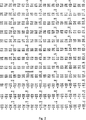
図2は、Bak−2 cDNAの配列および隣接配列、ならびにBak−2タンパク質の対応する推定アミノ酸配列を示す。
図3は、BakおよびFlag−Bak(F−Bak)融合タンパク質とエプスタイン−バールウイルスBHRF−1タンパク質との相互作用を示す。A欄において、レーン1はインビトロで同時に翻訳されたタンパク質F−Bak/BHRF−1から得られた結果を示し、そしてレーン2は抗FLAGアガロースに結合したF−Bak/BHRF−1タンパク質を示す。B欄において、レーンは、Bakタンパク質がBak−2であること以外は同じである。
発明の開示
本発明は、潜在的な抗ウイルス治療剤をスクリーニングする方法を提供する。新規なbcl−2相同物であるBakおよびBak−2タンパク質をコードするヌクレオチド配列によりコードされるタンパク質は、エプスタイン−バーウイルス(EBV)のタンパク質BHRF1と相互作用することが見出されている。これは、Bakタンパク質が疾患の病原性に寄与することを示す。BHRF1は、EBVの早期溶解サイクルタンパク質である。Pearsonら(1987)Virol.160:151−161。本発明は、Bakタンパク質とウイルスタンパク質またはそれらの機能的部分とを、潜在的な治療剤に曝す工程、およびタンパク質の相互作用をモニタリングする工程を含む方法を包含する。本発明はさらに、クローニングされたBakまたはBak−2遺伝子を発現する組換え細胞およびトランスジェニック動物を利用する。
Bakの遺伝子およびタンパク質のクローニングおよび分析は、共有に係るWO出願PCT/US94/13930に詳細に記載される。Bakの遺伝子およびタンパク質はまた、Kieferら(1995)Nature 374:736に記載される。Bakタンパク質のヌクレオチド配列および推定アミノ酸残基配列を図1に示し;そしてBak−2のヌクレオチド配列および推定アミノ酸残基配列を図2に示す。Bak mRNAは、種々のヒトの器官および組織においてノーザンブロット分析により検出されている。これらの器官は、肝臓;心臓;骨格筋;肺;腎臓;および膵臓を包含する。
これらの参考文献はまた、Bakタンパク質がアポトーシスを調整し得ることを開示する。リンパ芽球細胞株において、Bakタンパク質の発現が、Fas媒介性アポトーシスを減少させることが示された。マウスの始原B細胞株FL5.12において、Bak−2タンパク質およびBakタンパク質の誘導体がIL−3誘導性アポトーシスを減少させた。一方、Bakタンパク質はアポトーシスを増加させた。従って、細胞の型、Bakタンパク質の誘導体、およびアポトーシスの誘導方法に依存して、アポトーシスは、Bakタンパク質の濃度を制御することにより、高度に特異的な様式で調整され得る。
本明細書中で使用されるように、用語「Bak遺伝子」は、本明細書およびPCT/US94/13930に記載の核酸分子をいい、「Bakタンパク質」は、それらによりコードされるタンパク質をいう。ヌクレオチドは、cDNAおよび相補的DNA、ゲノムに由来するDNA、ならびに合成または半合成のDNAまたはRNAを包含するが、これらに限定されない。Bak cDNAのヌクレオチド配列を、制限エンドヌクレアーゼ部位の位置とともに図1に示す。
Bak−2 cDNAのヌクレオチド配列を、Bak−2タンパク質の推定アミノ酸配列および制限エンドヌクレアーゼ認識部位の位置とともに図に示す。Bak遺伝子はヒトの第6染色体上に位置し、そしてBak−2遺伝子はヒトの第20染色体上に位置する。ヒトの第11染色体上に位置し、このファミリーのメンバーでもあるBak−3も存在する。Bak−3は、偽遺伝子のようである。蛍光インサイチュハイブリダイゼーション(FISH)は、Bak遺伝子のおよその位置が6p21−23であることを示した。
本発明は、改変(例えば、欠失、置換、および付加)されたBak DNA配列の使用を含み、特に、ゲノムDNAの非コード領域において改変されたBak DNAの使用を含む。このような変化はクローニングを容易にし、そして遺伝子の発現を改変するのに有用である。BHRF1に結合するに十分なBakタンパク質の一部または任意の他の適切なウイルスタンパク質をコードする任意のDNAが、本明細書における使用に適切である。以下に記載するように、種々の融合タンパク質が本明細書における使用に適切である。
コードされるアミノ酸残基を変化させないか、あるいは結果として保存的に置換されるアミノ酸残基となるかのいずれかである種々の置換が、コード領域内で作製され得る。コードされるアミノ酸残基を変化させないヌクレオチドの置換は、異なる系での遺伝子発現を最適化させるのに有用である。適切な置換は当業者に公知であり、例えば、特定の発現系における好ましいコドン使用を反映するように行われる。
本発明は、Bak遺伝子の機能的に等価な改変体および誘導体の使用を含み、それらはBakタンパク質の特性を高め得るか、減少させ得るか、またはそれに重大な影響を与えないかもしれない。例えば、コードされるアミノ酸配列は変化しないDNA配列内の変化、および結果としてアミノ酸残基の保存的置換をもたらすDNA配列内の変化、1つまたは2、3のアミノ酸の欠失または付加、およびアミノ酸アナログによるアミノ酸残基の置換は、Bakの特性に重大な影響を与えない変化である。
相互に保存的に置換され得るアミノ酸残基には以下のものが挙げられるが、それらに限定されない:グリシン/アラニン;バリン/イソロイシン/ロイシン;アスパラギン/グルタミン;アスパラギン酸/グルタミン酸;セリン/トレオニン;リジン/アルギニン;およびフェニルアラニン/チロシン。Bakタンパク質の特性に重大な影響を与えない任意の保存的アミノ酸置換は、本発明に包含される。
本発明の実施に有用な核酸操作の技法は、以下の種々の文献に記載されているが、それらに限定されない;Molecular Cloning:A Laboratory Manual,第2版、第1〜3巻、Sambrookら編、Cold Spring Harbor Laboratory Press(1989);およびCurrent Protocols in Molecular Biology、Ausubelら編、Greene Publishing and Wiley−Interscience:New York(1987)、および定期改訂版。
Bak遺伝子のコード領域はまた、心筋細胞、GTI−7のような神経細胞株、およびヒトの結腸腺ガン細胞株HT29のようなTNF感受性細胞を包含するがこれらに限定されない他の細胞型に安定に組み込まれ得る発現ベクター内に連結され得、それによりBakタンパク質によるアポトーシスの調節をモニターするための種々のアッセイ系を提供し得る。
本明細書中で使用するように、「BHRF1」または「ウイルスタンパク質」は、Bakタンパク質またはその部分またはその誘導体に結合するに十分な全長のEBVタンパク質およびその部分またはその誘導体を包含する。このようなタンパク質は、任意のウイルス、例えば、サイトメガロウイルスおよび水疱帯状ヘルペスのような特に種々の形態のヘルペスウイルスおよびヘルペス様ウイルスにより発現される相同タンパク質を包含するがこれらに限定されない。
Bakタンパク質とBHRF−1のようなウイルスタンパク質との間の相互作用は、精製されたタンパク質を一緒に添加することにより生じ得る。しかし、好ましくは、タンパク質は、タンパク質−タンパク質相互作用を可能にする条件下で同時転写および翻訳される。同時翻訳は、天然または組換えのBakタンパク質およびウイルスタンパク質を発現する全細胞中でインビトロまたはインビボで実施され得る。任意の適切な組換え発現ベクターが使用され得る。Bakタンパク質はまた、別々に翻訳され、次いでタンパク質−タンパク質相互作用を可能にする条件下で組み合わされ得る。
タンパク質相互作用をモニタリングする方法は当該分野で公知であり、任意の方法が本明細書中での使用に適切である。好ましくは、共沈が使用される。タンパク質の1つまたはそれに融合された免疫学的タグの1つを沈降させる抗体の能力が、タンパク質を免疫沈降するために用いられ、そして免疫沈降物は両方のタンパク質の存在についてモニタリングされる。共沈の方法は当該分野で公知であり、以下の実施例に記載される。グルタチオンカラム上でのGST融合タンパク質固定化のようなタンパク質相互作用トラッピングおよびELISAを包含するが、それらに限定されない、当該分野の任意の他の方法が本明細書中の使用に適切である。免疫学的タグは、しばしば、融合タンパク質に取り込まれ、そして例えば、FLAG、ヘマグルチニン、およびグルタチオン−Sトランスフェラーゼを包含する。
組換えDNAにより発現されるかまたは生物学的供給源(例えば、組織)由来のいずれかのBakタンパク質の精製または単離は、当該分野において公知の任意の方法により達成され得る。タンパク質の精製方法は当該分野において公知である。一般に、実質的に精製されたタンパク質は、他の混入細胞物質(特に、タンパク質)を含まないタンパク質である。好ましくは、精製されたBakタンパク質は、80パーセントを越えて純粋であり、そして最も好ましくは95パーセントを越えて純粋である。下記のような臨床使用のために、Bakタンパク質は、好ましくは高度に精製されており、少なくとも約99パーセント純粋であり、そして発熱物質および他の混入物を含まない。
タンパク質精製の適切な方法は、当該分野において公知であり、そして以下を包含するが、これらに限定されない:アフィニティークロマトグラフィー、イムノアフィニティークロマトグラフィー、サイズ排除クロマトグラフィー、HPLC、およびFPLC。タンパク質の実質的な分解を生じない任意の精製スキームが、本明細書中の使用に適する。
本明細書中で使用されるように、「Bakタンパク質」は、特性に顕著に影響しないそれらの機能的に等価な変異体、および同一の全体アミノ酸配列を保持するが活性を増強または減弱させた変異体を包含する。例えば、アミノ酸残基の保存的置換、1または少数のアミノ酸の欠失または付加、およびアミノ酸アナログによるアミノ酸残基の置換は本発明の範囲内である。
相互に保存的に置換され得るアミノ酸残基には以下のものが挙げられるが、それらに限定されない:グリシン/アラニン;バリン/イソロイシン/ロイシン;アスパラギン/グルタミン;アスパラギン酸/グルタミン酸;セリン/トレオニン;リジン/アルギニン;およびフェニルアラニン/チロシン。Bakタンパク質の特性に重大な影響を与えない任意の保存的アミノ酸置換は、本発明に包含される。
本明細書中の使用に適切な抗体は、Bakタンパク質を抗原として、または好ましくはbclファミリーの他の遺伝子産物に対して実質的な相同性を欠くBakタンパク質領域を包含するペプチドを使用することにより産生される。ウイルスタンパク質に対する抗体もまた、本明細書中の使用に適切である。抗体を用いてタンパク質を検出する方法、およびタンパク質または合成ペプチドを用いて抗体を産生する方法は当該分野において公知であり、本明細書に詳細に記述されない。
治療的に有効な薬剤のスクリーニングは、Bakタンパク質とウイルスタンパク質との間の相互作用に直接的または間接的に影響を与え得るこのような薬剤に、Bakタンパク質およびウイルスタンパク質を曝すことにより行われる。適切な潜在的治療剤は、以下を包含するが、これらに限定されない:サイトカイン、小分子薬剤、細胞透過性小分子薬剤、ホルモン、インターロイキンとレクチンと他の刺激剤(例えば、PMA、LPS)との組み合わせ、二特異性抗体、ペプチド模倣物、アンチセンスオリゴヌクレオチド、および細胞機能またはタンパク質発現を改変する他の薬剤のような任意の薬学的薬剤。
タンパク質は同時に添加されるか、または同時に発現され、このような薬剤に生理学的に有効な濃度で曝され、そしてそれらの相互作用はこのような薬剤に曝されないコントロールに対して測定される。コントロールに対するBakタンパク質とウイルスタンパク質との間の相互作用を減少させるこれらの生物学的改変物質は、それらの抗ウイルス活性のさらなる研究のために選択される。
以前に示したように、過剰発現されたBakタンパク質は、血清回収または抗Fas処置の後にEBVで形質転換されたB細胞をアポトーシスから防御する。PCT/US94/13930。これらの結果は、Bak−BHRF1相互作用が存在し、それによってBHFR1がBakタンパク質の正常なアポトーシス効果を中和するだけでなく、さらに防御的活性を誘導することを示す。あるいは、Bak cDNAでトランスフェクトした細胞の増殖は、高レベルのBHRF1または他のEBVコードの抗アポトーシスタンパク質を発現する細胞について選択され得る。これは、細胞を血清回収のようなアポトーシスシグナルに曝した際に抗アポトーシス応答を導き得る。実施例2は、インビトロ翻訳したFlag−Bak(エピトープタグ化)およびBHRF1が、Flagエピトープを認識する抗体で共沈され得ることを示す。これは、Bakタンパク質とBHRF1とが相互に直接相互作用することを示す。
以下の実施例は、本発明を例示するために提供されるが、本発明を限定しない。特に記載されている場合を除いて、すべてのクローニング技術は本質的にSambrookら(1989)により記述された通りであり、そしてすべての試薬は製造業者の指示書に従って使用した。
実施例1
組換えBak遺伝子の発現
バキュロウイルス系で組換えBak遺伝子を発現させるために、PCT/US94/13930に記載のように作製したBak cDNAを用いて、PCRによって、クローニングを容易にするための制限部位を含有する遺伝子の3'および5'フランイング領域由来のプライマーを使用して、新規なBakベクターを作製した。プラスミドを、ジデオキシターミネータ法(Sangerら、1977)によって、配列決定キット(USB、Sequenase、バージョン2.0)および内部プライマーを用いて配列決定した。これにより、PCRによって生じた変異がないことを確認した。
クローンを用いて、プラスミドとAcNPV野生型のバキュロウイルスとの間のオーバーラップ配列のインビボでの相同組換えによって、組換えウイルスを作製した。昆虫Spodoptera Frugiperdaクローン9(SF9)細胞でのトランスフェクションの48時間後、組換えウイルスを収集し、PCRによって同定し、そしてさらに精製した。組換えバキュロウイルスの選択、スクリーニング、および増殖のための標準的な手順を製造者(Invitrogen)の説明書に従って行った。ドデシル硫酸ナトリウム−ポリアクリルアミドゲル電気泳動(SDS−PAGE)で、バキュロウイルス系で産生したタンパク質の分子量を、アミノ酸配列に従って予想したBakタンパク質の分子量と比較した。
さらに、類似のクローンは、細菌、酵母、昆虫、および哺乳動物を含むがこれらに限定されない、当該分野で公知の任意の発現系において発現され得る。適切な酵母細胞内発現系は、Barrら、(1992)Transgenesis JAH Murray編(Wiley and Sons)55−79頁に記載される。
Bak遺伝子のコード配列を切り出し、そしてプラスミドpCEP7、pREP7、およびpcDNA3(Invitrogen)の適合性制限酵素部位に導入した。pCEP7を、pREP7のRSV 3'−LTRをXba I/Asp718で除去し、そしてpCEP4(invitrogen)由来のCMVプロモーターに置換することにより作製した。25μgの各Bak含有プラスミドを、Bリンパ芽球細胞株WIL−2にエレクトロポレーションし、そしてBakを発現する安定なハイグロマイシン耐性形質転換体またはG418耐性形質転換体(pcDNA3構築物)を選択した。
実施例2
Bakタンパク質はエプスタイン−バーウイルスにコード されるBHRF1タンパク質と相互作用する
BHRF1 cDNAを、PCRキットおよびサーマルサイクラー(Perkin Elmer Cetus)の製造者の説明書に従って標準的なPCRプロトコールを用いて、RT−PCRによりWI−L2 mRNAから増幅した。Flag−BakおよびFlag−Bak−2のcDNAを、上記のように、RT−PCRにより、BakおよびBak−2/pcDNA3プラスミドテンプレートから作製したが、センスプライマー中に24塩基のFlagコード配列5'−GAC TAC AAG GAC GAC GAT GAC AAG−3'を含有した。これにより、N末端Flag−BakおよびFlag−Bak−2融合タンパク質をコードするcDNAが得られた。これらの融合タンパク質は、抗Flag M2抗体(Kodak−IBI)により認識および精製され得た。cDNAを、CMVおよびT7 RNAポリメラーゼプロモーターの制御下にあるpcDNA3ベクター中に連結した。次いで、Flag−BakおよびBHRF1プラスミドまたはFlag−Bak−2およびBHRF1プラスミドを、TnT連結網状赤血球溶解系を製造者の説明書(Promega)に従って用いて同時転写および同時翻訳した。
簡略には、0.5〜1.0μgの2つの環状プラスミドを、50μLのTnT溶解物中で32℃にて90分間、同時に転写および翻訳した。翻訳後、20μLの溶解物を、20μLの2×PBS+40μLの抗Flag M2アフィニティーゲル(Kodak)と混合し、そして穏やかに揺りながら4℃にて一晩インキュベートした。免疫沈降物を、エッペンドルフ微量遠心で1500rpmで4℃にて15分間遠心分離することにより回収した。ペレットを、1.5mLのPBSで4回洗浄し、最後の洗浄後に30μLのSDS−PAGEサンプル緩衝液中に再懸濁した。次いで、サンプルを、18%ポリアクリルアミドゲルでのSDS−PAGEにより分析した。ゲルを10%氷酢酸で固定し、乾燥し、そしてX線フィルムに室温にて一晩感光させた。
図3に示すように、Flag−BakおよびBHRF1ならびにHlag−Bak−2およびBHRF1は、効率的に同時転写および同時翻訳された(レーン1)。明らかに、抗Flag M2抗体により、Flag−BakおよびBHRF1、またはFlag−Bak−2およびBHRF1は効果的に共沈する(レーン2)。これは、BHRF1がBakタンパク質およびBak−2タンパク質の両方とインビトロで相互作用することを示し、そしてこのような相互作用がインビボで生じてアポトーシスの調整をもたらすことを示唆する。Bakタンパク質とウイルスタンパク質との相互作用は、宿主細胞のアポトーシス性の死の非存在下でウイルス複製を可能にするかまたは潜在し続けるように進化したようである。従って、これらの相互作用の妨害は、新規な抗ウイルス剤の設計のための重要な新しいストラテジーを示す。同様に、EBVのようなウイルスによる形質転換に由来する悪性細胞もまた、これらの薬剤を用いて診断または治療を受けやすい。
上記の発明は、理解を明確にする目的のための例示および実施例として詳細に記載されているが、特定の変更および改変が実施され得ることは当業者にとって明らかである。従って、明細書の記載および実施例は、本発明の範囲を制限するものとして解釈されるべきでなく、本発明の範囲は添付の請求の範囲によって明確に記述される。 TECHNICAL FIELD The present invention relates to a method for screening a therapeutic agent using a novel protein having apoptosis-modulating activity.
Background art Apoptosis is a normal physiological process leading to individual cell death. This programmed cell death process is involved in a variety of normal and pathogenic biological events and can be induced by many unrelated stimuli. Changes in the biological regulation of apoptosis also occur during aging and are responsible for many health conditions and diseases associated with aging. Recent studies of apoptosis suggest that the general metabolic pathway leading to cell death can be initiated by a wide variety of signals. These signals include hormones, serum growth factor depletion, chemotherapeutic agents, ionizing radiation, and infection with human immunodeficiency virus (HIV). Wyllie (1980) Nature 284: 555-556; Kanter et al. (1984) Biochem. Biophys. Res. Commun. 118: 392-399; Duke and Cohen (1986) Lymphokine Res. 5: 289-299; Tomei et al. ( 1988) Biochem. Biophys. Res. Commun. 155: 324-331; Kruman et al. (1991) J. Cell. Physiol. 148: 267-273; Ameisen and Capron (1991) Immunology Today 12: 102; and Sheppard and Ascher. (1992) J. AIDS 5: 143. Thus, agents that modulate the biological control of apoptosis have therapeutic utility in a wide variety of conditions.
Apoptotic cell death is characterized by cell contraction, chromatin condensation, cytoplasmic blebbing, increased membrane permeability, and intrachromosomal DNA cleavage. Kerr et al. (1992) FASEB J. 6: 2450; and Cohen and Duke (1992) Ann. Rev. Immunol. 10: 267. A bleb is a small, membrane-encapsulated sphere that can be excised from the surface of apoptotic cells, continue to produce superoxide radicals that damage surrounding cellular tissues, and is involved in the inflammatory process Can do.
The Bcl-2 gene is found at a common chromosomal translocation site t (14:18) in follicular lymphoma and causes abnormal overexpression of bcl-2. Tsujimoto et al. (1984) Science 226: 1097-1099; and Cleary et al. (1986) Cell 47: 19-28. The normal function of bcl-2 is to protect against apoptosis; unregulated expression of bcl-2 in B cells is thought to lead to an increase in the number of proliferating B cells that may be an important factor in lymphoma development Yes. See McDonnell and Korsmeyer (1991) Nature 349: 254-256; and for a review, see Edgington (1993) Bio / Tech. 11; 787-792. Bcl-2 can also block cell death induced by gamma irradiation. Sentman et al. (1991) Cell 67: 879-888; and Strassen (1991) Cell 67: 889-899. At present, it is known that bcl-2 inhibits most types of apoptotic cell death and is thought to function by regulating antioxidant pathways at free radical production sites. Hockenbery et al. (1993) Cell 75: 241-451.
Apoptosis, a normal cellular event, can also be induced by pathological conditions and various injuries. Apoptosis is involved in a wide variety of conditions including, but not limited to: cardiovascular disease; cancer regression; immunoregulation; viral disease; anemia; neurological disorders; Disability; Diabetes; Alopecia; Rejection of organ transplants; Prostatic hypertrophy; Obesity; Eye disorders; Stress;
Bcl-2 belongs to a family of proteins, some of which have been cloned and sequenced. Williams and Smith (1993) Cell 74: 777-779. Various Bcl-2 members have the ability to associate with each other as heterodimers. Oltvai et al. (1993) Cell 74: 609-619; and Sato et al. (1994) Proc. Natl. Acad. Sci. USA 91: 9238-9242. Furthermore, BHRF1 shows 25% sequence identity to Bcl-2 (Cleary et al. (1986) Cell 47: 17-28) and gene transfer studies have shown that B cells are protected from apoptosis. Yes. Henderson et al. (1993) Proc. Natl. Acad. Sci. USA 90: 8479-8848.
Herpesviridae viruses typically cause latent and recurrent infections. The herpesvirus genome consists of a sequence having a short region and a long region. Herpes virus particles have a diameter of 180 nm to 200 nm. Many particles do not contain an envelope. Typically, DNA is wrapped around associated proteins. Herpesviruses tend to remain stationary for an irregular period of time.
All references cited herein are hereby incorporated by reference, both above and below.
Summary of the Invention
Provided are methods for screening for pharmaceutical agents that stimulate Bak and Bak-2 protein activity levels, and pharmaceutical agents that inhibit Bak and Bak-2 protein activity levels. The method combines Bak protein and viral protein under conditions where they interact to form a test sample, exposing the test sample to a potential therapeutic agent, and monitoring protein interactions Process. A potential therapeutic agent that disrupts the interaction is selected for further study as compared to a control test sample to which no drug is added.
[Brief description of the drawings]
FIG. 1 shows the Bak cDNA nucleotide sequence and the amino acid sequence encoded thereby.
FIG. 2 shows the Bak-2 cDNA sequence and flanking sequences, and the corresponding deduced amino acid sequence of the Bak-2 protein.
FIG. 3 shows the interaction of Bak and Flag-Bak (F-Bak) fusion proteins with Epstein-Barr virus BHRF-1 protein. In column A,
DISCLOSURE OF THE INVENTION The present invention provides a method of screening for potential antiviral therapeutic agents. The protein encoded by the nucleotide sequence encoding Bak and the Bak-2 protein, a novel bcl-2 homolog, has been found to interact with the Epstein-Barr virus (EBV) protein BHRF1. This indicates that the Bak protein contributes to disease virulence. BHRF1 is an EBV early lysis cycle protein. Pearson et al. (1987) Virol. 160: 151-161. The present invention includes a method comprising exposing a Bak protein and a viral protein or functional part thereof to a potential therapeutic agent and monitoring the protein interaction. The invention further utilizes recombinant cells and transgenic animals that express the cloned Bak or Bak-2 gene.
Cloning and analysis of Bak genes and proteins is described in detail in the co-owned WO application PCT / US94 / 13930. Bak genes and proteins are also described in Kiefer et al. (1995) Nature 374: 736. The nucleotide sequence and predicted amino acid residue sequence of Bak protein is shown in FIG. 1; and the nucleotide sequence and predicted amino acid residue sequence of Bak-2 are shown in FIG. Bak mRNA has been detected by Northern blot analysis in various human organs and tissues. These organs include the liver; heart; skeletal muscle; lung; kidney; and pancreas.
These references also disclose that the Bak protein can modulate apoptosis. In lymphoblast cell lines, expression of Bak protein has been shown to reduce Fas-mediated apoptosis. In the mouse primordial B cell line FL5.12, Bak-2 protein and derivatives of Bak protein reduced IL-3-induced apoptosis. On the other hand, Bak protein increased apoptosis. Thus, depending on the cell type, the derivative of the Bak protein, and the method of inducing apoptosis, apoptosis can be regulated in a highly specific manner by controlling the concentration of the Bak protein.
As used herein, the term “Bak gene” refers to the nucleic acid molecules described herein and in PCT / US94 / 13930, and “Bak protein” refers to the protein encoded by them. Nucleotides include, but are not limited to, cDNA and complementary DNA, DNA derived from the genome, and synthetic or semi-synthetic DNA or RNA. The nucleotide sequence of Bak cDNA is shown in FIG. 1 along with the location of the restriction endonuclease site.
The nucleotide sequence of Bak-2 cDNA is shown in the figure along with the deduced amino acid sequence of Bak-2 protein and the location of the restriction endonuclease recognition site. The Bak gene is located on human chromosome 6 and the Bak-2 gene is located on
The present invention includes the use of modified Bak DNA sequences (eg, deletions, substitutions and additions), and particularly includes the use of Bak DNA modified in non-coding regions of genomic DNA. Such changes facilitate cloning and are useful for altering gene expression. Any DNA encoding a portion of the Bak protein sufficient to bind to BHRF1 or any other suitable viral protein is suitable for use herein. A variety of fusion proteins are suitable for use herein, as described below.
Various substitutions can be made within the coding region that either do not change the encoded amino acid residue or result in a conservatively substituted amino acid residue. Substitution of nucleotides that do not change the encoded amino acid residue is useful for optimizing gene expression in different systems. Appropriate substitutions are known to those skilled in the art and are made, for example, to reflect preferred codon usage in a particular expression system.
The present invention includes the use of functionally equivalent variants and derivatives of the Bak gene, which may enhance, decrease or not significantly affect the properties of the Bak protein. For example, changes in the DNA sequence that do not change the encoded amino acid sequence, and changes in the DNA sequence that result in conservative substitution of amino acid residues, deletion or addition of one or a few amino acids, and amino acids Substitution of amino acid residues with analogs is a change that does not significantly affect the properties of Bak.
Amino acid residues that can be conservatively substituted for each other include, but are not limited to: glycine / alanine; valine / isoleucine / leucine; asparagine / glutamine; aspartic acid / glutamic acid; serine / threonine; / Arginine; and phenylalanine / tyrosine. Any conservative amino acid substitution that does not significantly affect the properties of the Bak protein is encompassed by the present invention.
Nucleic acid manipulation techniques useful in the practice of the present invention are described in, but not limited to, the following various documents; Molecular Cloning: A Laboratory Manual, 2nd edition, volumes 1-3, edited by Sambrook et al. Cold Spring Harbor Laboratory Press (1989); and Current Protocols in Molecular Biology, edited by Ausubel et al., Greene Publishing and Wiley-Interscience: New York (1987), and periodic revisions.
The coding region of the Bak gene is also stable to other cell types including but not limited to cardiomyocytes, neural cell lines such as GTI-7, and TNF sensitive cells such as the human colon adenocarcinoma cell line HT29. Can be ligated into an expression vector that can be incorporated into a vector, thereby providing a variety of assay systems for monitoring the regulation of apoptosis by the Bak protein.
As used herein, “BHRF1” or “viral protein” encompasses a full length EBV protein and portion or derivative thereof sufficient to bind to a Bak protein or portion or derivative thereof. Such proteins include, but are not limited to, homologous proteins expressed by any virus, particularly various forms of herpesviruses and herpes-like viruses such as cytomegalovirus and vesicular herpes.
Interactions between Bak proteins and viral proteins such as BHRF-1 can occur by adding purified proteins together. Preferably, however, the protein is co-transcribed and translated under conditions that allow protein-protein interaction. Co-translation can be performed in vitro or in vivo in whole cells expressing natural or recombinant Bak and viral proteins. Any suitable recombinant expression vector can be used. Bak proteins can also be translated separately and then combined under conditions that allow protein-protein interactions.
Methods for monitoring protein interactions are known in the art, and any method is suitable for use herein. Preferably coprecipitation is used. The ability of the antibody to precipitate one of the proteins or one of the immunological tags fused thereto is used to immunoprecipitate the protein and the immunoprecipitate is monitored for the presence of both proteins. Co-precipitation methods are known in the art and are described in the examples below. Any other method in the art is suitable for use herein, including but not limited to protein interaction trapping and ELISA, such as GST fusion protein immobilization on a glutathione column. Immunological tags are often incorporated into fusion proteins and include, for example, FLAG, hemagglutinin, and glutathione-S transferase.
Purification or isolation of any Bak protein expressed by recombinant DNA or derived from a biological source (eg, tissue) can be accomplished by any method known in the art. Protein purification methods are known in the art. In general, a substantially purified protein is a protein that is free of other contaminating cellular material, particularly proteins. Preferably, the purified Bak protein is greater than 80 percent pure and most preferably greater than 95 percent pure. For clinical use as described below, the Bak protein is preferably highly purified, at least about 99 percent pure and free of pyrogens and other contaminants.
Suitable methods of protein purification are known in the art and include, but are not limited to: affinity chromatography, immunoaffinity chromatography, size exclusion chromatography, HPLC, and FPLC. Any purification scheme that does not result in substantial degradation of the protein is suitable for use herein.
As used herein, “Bak proteins” are those functionally equivalent variants that do not significantly affect properties, and retain the same overall amino acid sequence but enhance or attenuate activity. Includes variants. For example, conservative substitution of amino acid residues, deletion or addition of one or a few amino acids, and substitution of amino acid residues with amino acid analogs are within the scope of the invention.
Amino acid residues that can be conservatively substituted for each other include, but are not limited to: glycine / alanine; valine / isoleucine / leucine; asparagine / glutamine; aspartic acid / glutamic acid; serine / threonine; / Arginine; and phenylalanine / tyrosine. Any conservative amino acid substitution that does not significantly affect the properties of the Bak protein is encompassed by the present invention.
An antibody suitable for use herein is by using a peptide comprising a Bak protein region lacking substantial homology to the Bak protein as an antigen, or preferably to other gene products of the bcl family. Produced. Antibodies against viral proteins are also suitable for use herein. Methods for detecting proteins using antibodies and methods for producing antibodies using proteins or synthetic peptides are known in the art and will not be described in detail herein.
Screening for therapeutically effective agents is performed by exposing the Bak and viral proteins to such agents that can directly or indirectly affect the interaction between the Bak and viral proteins. Suitable potential therapeutic agents include, but are not limited to: cytokines, small molecule drugs, cell penetrating small molecule drugs, hormones, interleukins and lectins and other stimulants (eg, PMA, LPS) Any pharmaceutical agent, such as in combination with, bispecific antibodies, peptidomimetics, antisense oligonucleotides, and other agents that alter cellular function or protein expression.
Proteins are added simultaneously or expressed simultaneously, exposed to such agents at physiologically effective concentrations, and their interactions are measured relative to controls not exposed to such agents. Those biological modifiers that reduce the interaction between the Bak protein and the viral protein relative to the control are selected for further study of their antiviral activity.
As previously indicated, overexpressed Bak protein protects EBV transformed B cells from apoptosis following serum recovery or anti-Fas treatment. PCT / US94 / 13930. These results indicate that the Bak-BHRF1 interaction is present, whereby BHFR1 not only neutralizes the normal apoptotic effect of the Bak protein, but also induces protective activity. Alternatively, growth of cells transfected with Bak cDNA can be selected for cells expressing high levels of BHRF1 or other EBV-encoded anti-apoptotic proteins. This can lead to an anti-apoptotic response when cells are exposed to apoptotic signals such as serum recovery. Example 2 shows that in vitro translated Flag-Bak (epitope tagged) and BHRF1 can be co-precipitated with an antibody that recognizes the Flag epitope. This indicates that Bak protein and BHRF1 directly interact with each other.
The following examples are provided to illustrate the invention but do not limit the invention. Except where otherwise noted, all cloning techniques were essentially as described by Sambrook et al. (1989) and all reagents were used according to manufacturer's instructions.
Example 1
Recombinant Bak gene expression To facilitate expression of the recombinant Bak gene in a baculovirus system, cloning is facilitated by PCR using the Bak cDNA prepared as described in PCT / US94 / 13930. A novel Bak vector was made using primers from the 3 'and 5' flanking regions of the gene containing restriction sites for. Plasmids were sequenced by the dideoxy terminator method (Sanger et al., 1977) using a sequencing kit (USB, Sequenase, version 2.0) and internal primers. This confirmed that there was no mutation caused by PCR.
The clones were used to generate recombinant viruses by in vivo homologous recombination of overlapping sequences between the plasmid and the AcNPV wild type baculovirus. Forty-eight hours after transfection with insect Spodoptera Frugiperda clone 9 (SF9) cells, the recombinant virus was collected, identified by PCR and further purified. Standard procedures for selection, screening, and propagation of recombinant baculovirus were performed according to the manufacturer's (Invitrogen) instructions. The molecular weight of the protein produced in the baculovirus system was compared with the molecular weight of the Bak protein predicted according to the amino acid sequence by sodium dodecyl sulfate-polyacrylamide gel electrophoresis (SDS-PAGE).
Moreover, similar clones can be expressed in any expression system known in the art, including but not limited to bacteria, yeast, insects, and mammals. Suitable yeast intracellular expression systems are described in Barr et al. (1992) Transgenesis JAH Murray (Wiley and Sons) pages 55-79.
The coding sequence of the Bak gene was excised and introduced into compatible restriction enzyme sites of plasmids pCEP7, pREP7, and pcDNA3 (Invitrogen). pCEP7 was generated by removing the RSV 3′-LTR of pREP7 with Xba I / Asp718 and replacing it with a CMV promoter from pCEP4 (invitrogen). 25 μg of each Bak-containing plasmid was electroporated into the B lymphoblast cell line WIL-2 and stable hygromycin resistant or G418 resistant transformants (pcDNA3 construct) expressing Bak were selected.
Example 2
Bak protein interacts with Epstein-Barr virus encoded BHRF1 protein
BHRF1 cDNA was amplified from WI-L2 mRNA by RT-PCR using a standard PCR protocol according to the manufacturer's instructions for the PCR kit and thermal cycler (Perkin Elmer Cetus). Flag-Bak and Flag-Bak-2 cDNAs were generated from Bak and Bak-2 / pcDNA3 plasmid templates by RT-PCR as described above, but with a 24 base Flag coding sequence 5'- in the sense primer. GAC TAC AAG GAC GAC GAT GAC AAG-3 ′ was contained. As a result, cDNAs encoding the N-terminal Flag-Bak and Flag-Bak-2 fusion proteins were obtained. These fusion proteins could be recognized and purified by anti-Flag M2 antibody (Kodak-IBI). The cDNA was ligated into pcDNA3 vector under the control of CMV and T7 RNA polymerase promoter. Flag-Bak and BHRF1 plasmids or Flag-Bak-2 and BHRF1 plasmids were then co-transcribed and co-translated using a TnT-linked reticulocyte lysis system according to the manufacturer's instructions (Promega).
Briefly, 0.5-1.0 μg of two circular plasmids were simultaneously transcribed and translated for 90 minutes at 32 ° C. in 50 μL of TnT lysate. After translation, 20 μL lysate was mixed with 20
As shown in FIG. 3, Flag-Bak and BHRF1 and Hlag-Bak-2 and BHRF1 were efficiently co-transcribed and co-translated (lane 1). Apparently, anti-Flag M2 antibody effectively co-precipitates Flag-Bak and BHRF1, or Flag-Bak-2 and BHRF1 (lane 2). This indicates that BHRF1 interacts with both Bak and Bak-2 proteins in vitro, and suggests that such interactions occur in vivo resulting in modulation of apoptosis. The interaction between the Bak protein and the viral protein appears to have evolved to allow or remain latent for viral replication in the absence of apoptotic death of the host cell. Thus, interfering with these interactions represents an important new strategy for the design of new antiviral agents. Similarly, malignant cells derived from transformation with viruses such as EBV are also amenable to diagnosis or treatment using these agents.
Although the foregoing invention has been described in detail as illustrative and illustrative examples for purposes of clarity of understanding, it will be apparent to those skilled in the art that certain changes and modifications may be practiced. Accordingly, the description and examples should not be construed as limiting the scope of the invention, which is clearly set forth by the appended claims.
Claims (7)
(a)Bakタンパク質もしくはBak−2タンパク質または 該Bakタンパク質もしくはBak−2タンパク質の部分もし くは誘導体とウイルスタンパク質とをこれらが結合する条件下で組み合わせて試験サンプルを形成する工程であ って、該Bakタンパク質もしくはBak−2タンパク質また は該Bakタンパク質もしくはBak−2タンパク質の部分も しくは誘導体は、BHRF1に結合し、該誘導体は、該Bakタ ンパク質もしくはBak−2タンパク質に対して1または 数個のアミノ酸の置換、付加、もしくは欠失を有する、 工程;
(b)該試験サンプルを潜在的な治療剤に曝す工程;および
(c)該Bakタンパク質もしくは該Bak−2タンパク質と該ウイルスタンパク質との結合をモニターする工程、を包含し;
ここで、潜在的な治療剤が添加されていないコントロールの試験サンプルと比較して、潜在的な治療剤が該結合を混乱させる場合、該治療剤はさらなる研究のために選択され、
ここで、該ウイルスタンパク質は、BHRF1、もしくは該B HFR1に対する相同タンパク質、または該BHRF1もしくは 該相同タンパク質の部分もしくは誘導体であり、該BHRF 1、もしくは該BHFR1に対する相同タンパク質、または該 BHRF1もしくは該相同タンパク質の部分もしくは誘導体 は、該Bakタンパク質もしくは該Bak−2タンパク質に結 合し、該BHRF1もしくは該相同タンパク質の誘導体は、 該BHRF1もしくは該相同タンパク質に対して1または数 個のアミノ酸の置換、付加、または欠失を有する、方法。A method for screening potential antiviral therapeutic agents comprising the following steps:
(A) Bak protein or Bak-2 protein or portion of the Bak protein or Bak-2 protein if Ku is the derivative with viral proteins I step der to form a test sample by combining under conditions in which they are attached, moiety also properly derivative of the Bak protein or Bak-2 protein or the Bak protein or Bak-2 protein binds to BHRF1, the derivative, 1 or with respect to the Bak protein or Bak-2 protein Having several amino acid substitutions, additions or deletions ;
(B) exposing the test sample to a potential therapeutic agent; and (c) monitoring the binding of the Bak protein or the Bak-2 protein to the viral protein;
Here, if a potential therapeutic agent disrupts the binding as compared to a control test sample to which no potential therapeutic agent has been added, the therapeutic agent is selected for further study ;
Wherein the viral protein is BHRF1, or a homologous protein to the BHFR1, or a part or derivative of the BHRF1 or the homologous protein, the BHRF1 , or a homologous protein to the BHFR1 , or the BHRF1 or the homologous protein parts or derivatives, said combined binding to Bak protein or the Bak-2 protein, derivatives of the BHRF1 or the homologous protein, substitution of one or several amino acids with respect to the BHRF1 or the homologous protein, added, or that having a deletion, method.
Applications Claiming Priority (3)
| Application Number | Priority Date | Filing Date | Title |
|---|---|---|---|
| US42652995A | 1995-04-20 | 1995-04-20 | |
| US08/426,529 | 1995-04-20 | ||
| PCT/US1996/005639 WO1996033416A1 (en) | 1995-04-20 | 1996-04-19 | Methods of screening for therapeutic agents using novel apoptosis-modulating proteins |
Publications (2)
| Publication Number | Publication Date |
|---|---|
| JPH11504423A JPH11504423A (en) | 1999-04-20 |
| JP3615552B2 true JP3615552B2 (en) | 2005-02-02 |
Family
ID=23691171
Family Applications (1)
| Application Number | Title | Priority Date | Filing Date |
|---|---|---|---|
| JP53199896A Expired - Lifetime JP3615552B2 (en) | 1995-04-20 | 1996-04-19 | Method for screening therapeutic agent using novel apoptosis-regulating protein |
Country Status (5)
| Country | Link |
|---|---|
| EP (1) | EP0821793A1 (en) |
| JP (1) | JP3615552B2 (en) |
| AU (1) | AU5568396A (en) |
| CA (1) | CA2216856C (en) |
| WO (1) | WO1996033416A1 (en) |
Families Citing this family (4)
| Publication number | Priority date | Publication date | Assignee | Title |
|---|---|---|---|---|
| WO1995015084A1 (en) | 1993-11-30 | 1995-06-08 | Lxr Biotechnology Inc. | Novel apoptosis-modulating proteins, dna encoding the proteins and methods of use thereof |
| AU6679498A (en) * | 1997-03-20 | 1998-10-12 | Lxr Biotechnology Inc. | A novel bak binding protein, dna encoding the protein, and methods of use thereof |
| US6344330B1 (en) * | 1998-03-27 | 2002-02-05 | The Regents Of The University Of California | Pharmacophore recombination for the identification of small molecule drug lead compounds |
| WO2002074908A2 (en) * | 2001-03-02 | 2002-09-26 | Mds Proteomics, Inc. | Methods and reagents for regulating apoptosis |
Family Cites Families (3)
| Publication number | Priority date | Publication date | Assignee | Title |
|---|---|---|---|---|
| WO1995005738A1 (en) * | 1993-08-20 | 1995-03-02 | Massachusetts Institute Of Technology | Anticancer agents and apoptosis |
| US5700638A (en) * | 1993-08-26 | 1997-12-23 | Washington University | Cell death regulator |
| WO1995015084A1 (en) * | 1993-11-30 | 1995-06-08 | Lxr Biotechnology Inc. | Novel apoptosis-modulating proteins, dna encoding the proteins and methods of use thereof |
-
1996
- 1996-04-19 AU AU55683/96A patent/AU5568396A/en not_active Abandoned
- 1996-04-19 EP EP96913062A patent/EP0821793A1/en not_active Withdrawn
- 1996-04-19 JP JP53199896A patent/JP3615552B2/en not_active Expired - Lifetime
- 1996-04-19 WO PCT/US1996/005639 patent/WO1996033416A1/en not_active Ceased
- 1996-04-19 CA CA002216856A patent/CA2216856C/en not_active Expired - Lifetime
Also Published As
| Publication number | Publication date |
|---|---|
| AU5568396A (en) | 1996-11-07 |
| EP0821793A1 (en) | 1998-02-04 |
| WO1996033416A1 (en) | 1996-10-24 |
| CA2216856C (en) | 2008-02-19 |
| CA2216856A1 (en) | 1996-10-24 |
| JPH11504423A (en) | 1999-04-20 |
Similar Documents
| Publication | Publication Date | Title |
|---|---|---|
| JP4129055B2 (en) | Novel apoptosis-regulating protein, DNA encoding the protein, and method of use thereof | |
| WO1993003750A1 (en) | PROTEINS USEFUL IN THE REGULATION OF λB-CONTAINING GENES, CORRESPONDING DNA AND RNA SEQUENCES | |
| AU6031899A (en) | Leptin induced genes | |
| JP3615552B2 (en) | Method for screening therapeutic agent using novel apoptosis-regulating protein | |
| EP0830375A1 (en) | Human inhibitor of apoptosis gene 1 | |
| CN1198186A (en) | short form of chemokine beta-8 | |
| US9943567B2 (en) | Method for treating arthritis using IK factor or nucleic acid encoding IK factor | |
| US20030108552A1 (en) | Human inhibitor of apoptosis gene 1 | |
| JP3811733B2 (en) | Protein having DNase activity | |
| WO1999026980A1 (en) | Methods and reagents for the utilization of sap family member proteins, novel signal transduction regulators | |
| US6410714B1 (en) | Canine low affinity IgE receptor (CD23) nucleic acid molecules and uses thereof | |
| US5846768A (en) | Invertebrate apoptosis gene `GRIM` and methods of producing the protein encoded thereby | |
| US20010024808A1 (en) | Leptin induced genes | |
| EP0814661A1 (en) | UBIQUITIN CONJUGATING ENZYMES 7, 8 and 9 | |
| US6331412B1 (en) | Methods and compounds for modulating male fertility | |
| WO2002004505A1 (en) | A novel polypeptide - human semaphorin protein 9 and a polynucleotide encoding the same | |
| WO1999046290A1 (en) | A human angiotensin ii/vasopressin receptor (aii/avp) like gene (cbdakd01) | |
| DE69737660T2 (en) | TUMOR SUPPRESSOR FROM BASAL CELL CARCINOMA | |
| US5945321A (en) | Ubiquitin conjugating enzymes 7, 8 and 9 | |
| JPH10327872A (en) | Bcl-2 interacting protein gene bis | |
| JP2002369685A (en) | Human inhibitor of apoptosis gene 1 | |
| WO1999021982A1 (en) | Human m6b1 gene | |
| WO1999021983A1 (en) | Human atrophin-1 related gene | |
| JP2005253301A (en) | Protein having bone morphogenetic protein binding region and gene encoding the same | |
| CN1387535A (en) | A new human thrombin-1 mediator and its application and preparation method |
Legal Events
| Date | Code | Title | Description |
|---|---|---|---|
| A601 | Written request for extension of time |
Free format text: JAPANESE INTERMEDIATE CODE: A601 Effective date: 20040402 |
|
| A602 | Written permission of extension of time |
Free format text: JAPANESE INTERMEDIATE CODE: A602 Effective date: 20040517 |
|
| A521 | Request for written amendment filed |
Free format text: JAPANESE INTERMEDIATE CODE: A523 Effective date: 20040706 |
|
| TRDD | Decision of grant or rejection written | ||
| A01 | Written decision to grant a patent or to grant a registration (utility model) |
Free format text: JAPANESE INTERMEDIATE CODE: A01 Effective date: 20041005 |
|
| A61 | First payment of annual fees (during grant procedure) |
Free format text: JAPANESE INTERMEDIATE CODE: A61 Effective date: 20041101 |
|
| R150 | Certificate of patent or registration of utility model |
Free format text: JAPANESE INTERMEDIATE CODE: R150 |
|
| FPAY | Renewal fee payment (event date is renewal date of database) |
Free format text: PAYMENT UNTIL: 20081112 Year of fee payment: 4 |
|
| FPAY | Renewal fee payment (event date is renewal date of database) |
Free format text: PAYMENT UNTIL: 20091112 Year of fee payment: 5 |
|
| FPAY | Renewal fee payment (event date is renewal date of database) |
Free format text: PAYMENT UNTIL: 20091112 Year of fee payment: 5 |
|
| FPAY | Renewal fee payment (event date is renewal date of database) |
Free format text: PAYMENT UNTIL: 20101112 Year of fee payment: 6 |
|
| FPAY | Renewal fee payment (event date is renewal date of database) |
Free format text: PAYMENT UNTIL: 20111112 Year of fee payment: 7 |
|
| FPAY | Renewal fee payment (event date is renewal date of database) |
Free format text: PAYMENT UNTIL: 20111112 Year of fee payment: 7 |
|
| FPAY | Renewal fee payment (event date is renewal date of database) |
Free format text: PAYMENT UNTIL: 20121112 Year of fee payment: 8 |
|
| FPAY | Renewal fee payment (event date is renewal date of database) |
Free format text: PAYMENT UNTIL: 20121112 Year of fee payment: 8 |
|
| RD04 | Notification of resignation of power of attorney |
Free format text: JAPANESE INTERMEDIATE CODE: R3D04 |
|
| FPAY | Renewal fee payment (event date is renewal date of database) |
Free format text: PAYMENT UNTIL: 20121112 Year of fee payment: 8 |
|
| FPAY | Renewal fee payment (event date is renewal date of database) |
Free format text: PAYMENT UNTIL: 20131112 Year of fee payment: 9 |
|
| S531 | Written request for registration of change of domicile |
Free format text: JAPANESE INTERMEDIATE CODE: R313531 |
|
| R350 | Written notification of registration of transfer |
Free format text: JAPANESE INTERMEDIATE CODE: R350 |
|
| FPAY | Renewal fee payment (event date is renewal date of database) |
Free format text: PAYMENT UNTIL: 20131112 Year of fee payment: 9 |
|
| RD02 | Notification of acceptance of power of attorney |
Free format text: JAPANESE INTERMEDIATE CODE: R3D02 |
|
| S111 | Request for change of ownership or part of ownership |
Free format text: JAPANESE INTERMEDIATE CODE: R313113 |
|
| R350 | Written notification of registration of transfer |
Free format text: JAPANESE INTERMEDIATE CODE: R350 |
|
| R250 | Receipt of annual fees |
Free format text: JAPANESE INTERMEDIATE CODE: R250 |
|
| R250 | Receipt of annual fees |
Free format text: JAPANESE INTERMEDIATE CODE: R250 |
|
| EXPY | Cancellation because of completion of term |