JP3576184B2 - Diarrheal shellfish toxin-specific monoclonal antibody, hybridoma and diarrheal shellfish toxin detection method - Google Patents
Diarrheal shellfish toxin-specific monoclonal antibody, hybridoma and diarrheal shellfish toxin detection method Download PDFInfo
- Publication number
- JP3576184B2 JP3576184B2 JP04200893A JP4200893A JP3576184B2 JP 3576184 B2 JP3576184 B2 JP 3576184B2 JP 04200893 A JP04200893 A JP 04200893A JP 4200893 A JP4200893 A JP 4200893A JP 3576184 B2 JP3576184 B2 JP 3576184B2
- Authority
- JP
- Japan
- Prior art keywords
- antibody
- monoclonal antibody
- organic solvent
- methyl alcohol
- hybridoma
- Prior art date
- Legal status (The legal status is an assumption and is not a legal conclusion. Google has not performed a legal analysis and makes no representation as to the accuracy of the status listed.)
- Expired - Lifetime
Links
Images


Landscapes
- Peptides Or Proteins (AREA)
- Preparation Of Compounds By Using Micro-Organisms (AREA)
- Micro-Organisms Or Cultivation Processes Thereof (AREA)
Description
【0001】
【産業上の利用分野】
本発明は、下痢性貝毒に特異的なモノクローナル抗体、そのモノクローナル抗体を産生するハイブリドーマ、及びそのモノクローナル抗体を用いる下痢性貝毒の検出方法に関する。本発明による前記モノクローナル抗体は、特には、有機溶媒耐性抗体である。
【0002】
【従来の技術】
オカダ酸、7−O−アシル−オカダ酸、ジノフィシストキシン−1及びジノフィシストキシン−3(即ち、7−O−アシル−ジノフィシストキシン−1)の4種の下痢性貝毒の内、オカダ酸は黒磯海綿の一種(Halichondria okadai 及び Halichondria melanodocia)が、ジノフィシストキシン−1は渦鞭毛藻 (Dinophysis fortii ) が産生する脂溶性化合物である。また、ジノフィシストキシン−3は、前記ジノフィシストキシン−1が貝の体内で変換されて生成する脂溶性化合物であり、7−O−アシルオカダ酸は合成によって得られたジノフィシストキシン−3の類縁物質である。これらの化合物は、或る任意の時期又は地域の食用二枚貝の中腸腺に蓄積し、貝を毒化する。食中毒の発生件数ではフグに次いで二位であるが、患者数では第一位であり、食品衛生上、大きな問題である。
【0003】
従来、下痢性貝毒の検査法としてはマウスを用いた致死活性測定法が公定法として用いられているが、動物の管理、検出感度、精度及び特異性等の面で問題があった。一方、この検査を、高感度で、簡便かつ短時間に行うことを目的とした手法の開発が試みられている。
【0004】
例えば特開平1−96199号公報には、オカダ酸群に対するモノクローナル抗体及びその製法が開示されている。しかしながら、該抗体は、下痢性貝毒のうち、オカダ酸及びジノフィシストキシン−1に対して特異的に反応する抗体ではあるが、7−O−アシル−オカダ酸、ジノフィシストキシン−3を検出・測定することはできない。また、前記特開平1−96199号公報には、有機溶媒存在下で活性を維持することのできるモノクローナル抗体を得ることに関して一切記載がなく、それを示唆する記載もない。
【0005】
本発明者は、日本における下痢性貝毒による食中毒の主原因はジノフィシストキシン−3であること、及び、前記の各貝毒はいずれも脂溶性物質であるので、被検試料から貝毒成分を抽出するには有機溶媒の使用が避けられず、操作工程を簡略化するには、有機溶媒存在下で免疫反応を行うことが望ましいことに鑑み、前記下痢性貝毒本体のオカダ酸、ジノフィシストキシン−1及びジノフィシストキシン−3に特異的であり、しかも有機溶媒に耐性を示すマウスモノクローナル抗体を見出し、PCT/JP92/01021号に開示した。
【0006】
本発明者は、更に、ジノフィシストキシン−3の合成類縁化合物である7−O−アシル−オカダ酸に対する反応性、並びにアルコール類及びケトン類以外の有機溶媒中での反応性、更には前記の4種類の貝毒に対する反応性の比較を目的として鋭意研究したところ、オカダ酸、7−O−アシル−オカダ酸、ジノフィシストキシン−1及びジノフィシストキシン−3に特異的であり、しかも有機溶媒に耐性を示すマウスモノクローナル抗体を見出すことに成功し、このモノクローナル抗体を用いると、有機溶媒存在下でも免疫学的に迅速かつ特異的に下痢性貝毒を検出することができることを見出した。本発明は、こうした知見に基づくものである。
【0007】
従って、本発明は、オカダ酸、7−O−アシル−オカダ酸、ジノフィシストキシン−1及びジノフィシストキシン−3(即ち、7−O−アシル−ジノフィシストキシン−1)に特異的に反応し、メチルアルコール中ベンゼンの濃度が20%の溶液に耐性を示すことを特徴とする、モノクローナル抗体に関する。また、本発明は、前記モノクローナル抗体を産生するハイブリドーマ、及び前記モノクローナル抗体を用いることを特徴とする下痢性貝毒の免疫学的検出方法にも関する。
【0008】
以下、モノクローナル抗体、ハイブリドーマ及び免疫学的検出方法の順に説明する。
本発明によるモノクローナル抗体及びハイブリドーマの調製は常法、例えば、続生化学実験講座、免疫生化学研究法(日本生化学会編)に記載の方法で行うことができる。具体的には、免疫原としては、オカダ酸、7−O−アシル−オカダ酸、ジノフィシストキシン−1及びジノフィシストキシン−3に特異的なモノクローナル抗体をもたらすものを任意に用いることができるが、特にはオカダ酸、7−O−アシル−オカダ酸、ジノフィシストキシン−1若しくはジノフィシストキシン−3、又は、これらの塩類、更にはこれらを生体高分子(例えば、ウシ血清アルブミン又は免疫グロブリン)担体に結合させたものを用いるのが好ましい。これらの免疫原溶液を用いて哺乳動物(例えば、マウス、ラット、ウサギ、ヤギ又はウマ)をイン・ビボ免疫法により免疫する。例えば、免疫原溶液を等量のフロインド氏完全アジュバント又は不完全アジュバントと乳化混合し、マウスの皮下に投与する(第1回免疫)。以後、2〜4週間の間隔で同様の操作を行い、数回免疫する。最終免疫から数日後に脾臓を無菌的に取り出し、ステンレスメッシュなどで押しつぶして脾臓細胞を調製し、細胞融合工程に用いる。
【0009】
なお、本明細書で「アシル」とは、芳香族アシル基、及び飽和又は不飽和で、直鎖又は分枝鎖の脂肪族アシル基のいずれでもよく、例えば、炭素数8〜30個、好ましくは8〜24個の飽和又は不飽和脂肪酸(具体的には、カプリン酸、ミリスチン酸、パルミチン酸、オレイン酸又はリノレン酸など)から誘導されるアシル基である。
【0010】
細胞融合のもう一方の親細胞であるミエローマ細胞(骨髄腫細胞)としては、各種の公知の細胞株、例えば、p3(p3/×63−Ag8)[Nature, 256,495−497 (1975)]、p3−U1[Current Topics in Microbiology and Immunology, 81;1−7(1978) ]、NS−1[Eur. J. Immunol., 6;511−519(1976) ]、MPC−11[Cell,8;405−415(1976) ]、SP2/0[Nature, 276;269−270(1978) ]、FO[J. Immunol. Meth., 35;1−21(1980)]、×63.6.55.3[J.Immunol., 123;1548−1550(1979) ]、S194[J. Exp. Med., 148;313−323(1978) ]、又はラットにおけるR210[Nature, 277; 131−133(1979)]などを使用することができる。
【0011】
細胞融合は通常の方法、例えば、公知の融合促進剤(ポリエチレングリコールなど)及び場合により補助剤(ジメチルスルホキシドなど)を用いて行うことができ、使用比率も常法と同様に、例えば、脾臓細胞に対してミエローマ細胞を約1〜10倍程度の量で用いる。融合用培地としては、例えば、40%(w/v)ポリエチレングリコールを含むダルベッコ改変イーグル培地(DMEM)を用いることができる。融合は、前記の培地内で免疫脾臓細胞とミエローマ細胞とをよく混合することによって行う。
【0012】
続いて、選別用培地(例えば、HAT培地)を用いてハイブリドーマ以外の細胞を除去し、ハイブリドーマ培養上清の抗体(即ち、オカダ酸と7−O−アシル−オカダ酸とジノフィシストキシン−1とジノフィシストキシン−3とに同時に特異性を示すモノクローナル抗体)産生の有無を、例えばELISA法によって検出・測定し、目的とするハイブリドーマを分離する。特に、有機溶媒に対して耐性を有するモノクローナル抗体を産生するハイブリドーマを選別する場合には、各種濃度で前記有機溶媒を含む有機水性溶液ないし無水有機溶媒中に抗体を入れた後、下痢性貝毒を添加し、抗原抗体反応が正常に進行することを確認することによって、有機溶媒耐性モノクローナル抗体を産生するハイブリドーマを選別する。
【0013】
こうして得られた、目的のモノクローナル抗体を分泌する本発明のハイブリドーマは、通常の培地で継代培養することができ、また液体窒素等の中で容易に長期間保存することができる。ハイブリドーマを培養する培地としては、ハイブリドーマの培養に適した任意の培地を用いることができ、好適にはDMEMにウシ胎児血清、L−グルタミン、L−ピルビン酸及び抗生物質(ペニシリンGとストレプトマイシン)を含む培地が用いられる。ハイブリドーマの培養は、イン・ビトロの場合には例えば培地中で5%二酸化炭素濃度及び37℃で約3日間、またイン・ビボ例えばマウスの腹腔中で培養する場合には約14日間程度実施するのが好ましい。
【0014】
前記のハイブリドーマを常法によって培養した培養液から、あるいはハイブリドーマを投与した適当な哺乳動物(例えばマウス又はラット)の腹水から、目的とするモノクローナル抗体を分離し、精製することが可能である。培養液又はマウスの腹水からモノクローナル抗体を分離、精製する場合にはタンパク質の単離、精製に一般的に用いられる方法を用いることが可能である。そのような方法としては硫安塩析、イオン交換クロマトグラフィー、分子篩ゲルを用いる分子篩カラムクロマトグラフィー、プロテインA又はプロテインG結合多糖類を用いる親和性カラムクロマトグラフィー、透析、凍結乾燥の方法等がある。
【0015】
本発明による下痢性貝毒検出方法は、前記の本発明のモノクローナル抗体(即ち、オカダ酸と7−O−アシル−オカダ酸とジノフィシストキシン−1とジノフィシストキシン−3とに同時に特異性を示すモノクローナル抗体)を用いて実施するので、下痢性貝毒を漏れなく検出することができる。また、本発明検出方法の好ましい態様では、有機溶媒耐性モノクローナル抗体を用いるので、検査対象の下痢性貝毒を充分に溶解することのできる量の有機溶媒を含有する有機水性系(又は無水有機系)中で正確な抗原抗体反応を進行させることができる。
【0016】
本発明方法は、オカダ酸と7−O−アシル−オカダ酸とジノフィシストキシン−1とジノフィシストキシン−3とに同時に特異性を示すモノクローナル抗体を用いること、そして好ましくは有機溶媒耐性抗体を用いること(従って、有機溶媒の存在下で抗原抗体反応を実施すること)を除けば、それ以外の点では従来公知の免疫学的検出方法にそのまま適用することができる。従って、本発明方法は、具体的には例えば、
(1)試料を有機溶媒(例えば、水混和性有機溶媒)で処理して有機溶媒抽出液を調製する工程、
(2)下痢性貝毒に対して特異性を有すると共に前記有機溶媒に対する耐性を有する抗体と、前記有機溶媒抽出液を接触させる工程、
(3)標識を有する既知量の下痢性貝毒を前記工程(2)と同時又は前記工程(2)の終了後に前記抗体と接触させる工程、
(4)前記抗体と結合した標識化下痢性貝毒と、前記抗体と結合していない標識化貝毒とを分離する工程、
(5)前記工程(4)で分離したいずれか一方の標識化下痢性貝毒が有する標識からの信号を検出又は測定する工程を含む、試料中の下痢性貝毒の検出方法(競合法及び非競合法)からなる。
【0017】
更に、本発明方法の具体的態様としては、例えば
(1)試料を有機溶媒(例えば、水混和性有機溶媒)で処理して有機溶媒抽出液を調製する工程、
(2)下痢性貝毒に対して特異性を有すると共に前記有機溶媒に対する耐性を有する第1抗体を、不溶性担体に固定させる工程、
(3)下痢性貝毒を含む有機溶媒抽出液を、前記工程(2)の固定化第1抗体に接触させる工程、
(4)前記下痢性貝毒に対して、前記第1抗体とは異なる部位で結合すると共に標識を有する第2抗体を過剰量添加する工程、
(5)第1抗体と下痢性貝毒との複合体上の標識の信号を検出する工程を含む、試料中の下痢性貝毒の検出方法(サンドイッチ法)も含む。本発明は、その他公知の免疫反応検出方法に広く応用することができる。
【0018】
本発明で用いる有機溶媒は、アルコール類、ケトン類、エーテル類又はベンゼンであって、検査対象の試料から下痢性貝毒を抽出、希釈又は保存する際に用いる溶媒である。水混和性の有機溶媒を用いると、有機水性溶媒で抽出工程を行ったり、場合により有機溶媒抽出液を水で適当に希釈してから、抗原抗体反応を有機水性溶媒中で実施することができ、あるいは有機溶媒抽出液をそのまま用いて、無水有機溶媒中あるいは有機水性溶媒中で抗原抗体反応をさせることができるので好ましい。有機溶媒としては、例えば、アルコール化合物(例えば、炭素原子1〜3個の低級アルコール、特には、メチルアルコール、エチルアルコール、イソプロピルアルコール)、ケトン化合物(例えば、炭素原子1〜3個の低級脂肪族ケトン、特には、メチルエチルケトン、アセトン)、エーテル化合物(例えば、炭素原子1〜3個の低級脂肪族エーテル、特には、メチルエーテル、エチルエーテル、m−プロピルエーテル)及びベンゼン、あるいはこれらの混合物を挙げることができる。
【0019】
本発明の検出方法を具体的に実施する際には、最初に食用二枚貝の中腸腺検体を前記の有機溶媒で抽出する。得られた抽出液をそのまま又は水で希釈して次の接触工程に用いる。次に、例えばマウスから得た本発明のモノクローナル抗体であって、有機溶媒に対し耐性を有する抗体を、前記の方法で予め調製しておく。この抗体(サンドイッチ法では第1抗体)と前記有機溶媒抽出液とを接触させると、その有機溶媒抽出液に下痢性貝毒(抗原)が存在する場合には、有機溶媒存在下で抗原抗体反応が起きる。この抗原抗体反応は、有機溶媒存在下で実施することを除けば、通常の抗原抗体反応と同様に行うことができる。例えば、抗体を適当な不溶性支持体(ウエル又はラテックス粒子)上に担持させ、有機溶媒抽出液中の抗原と特異的に反応させる。
【0020】
本発明方法を競合法又は非競合法で実施する場合には、既知量の標識化抗原を用いて下痢性貝毒の存在の確認又は定量を行うことができる。また、サンドイッチ法を用いる場合には、過剰量の標識化第2抗体を用いる。貝毒の標識には、公知の標識体、例えば、放射性同位体(例えば、 32 P、 35 S、 3 H)、酵素(例えば、ペルオキシダーゼ、アルカリフォスファターゼ)、ビタミン(例えば、ビオチン)、蛍光物質(例えば、FITC)、化学発光物質(例えば、アクリジニウム)を用いることができる。
【0021】
標識化抗原は、前記抗体と有機溶媒抽出液との接触工程が終了してから(即ち、前記抗体と有機溶媒抽出液中の抗原との抗原抗体反応が終了してから)(非競合法)、あるいは前記抗体と有機溶媒抽出液との接触工程と同時に(即ち、前記抗体と有機溶媒抽出液中の抗原との抗原抗体反応と同時に)(競合法)、反応系に加えることができる。非競合法では、有機溶媒抽出液中の下痢性貝毒と結合していない抗体が標識化抗原と結合する。一方、競合法では、既知量の標識化抗原と前記有機溶媒抽出液中の未知量の抗原とが拮抗的に抗体と結合する。サンドイッチ法では、第1抗体を前記有機溶媒抽出液と接触させた後、非競合抗原を洗浄除去してから標識化第2抗体を加えると、第1抗体と抗原との複合体に対して標識化第2抗体が結合する。
【0022】
前記の抗体と有機溶媒抽出液との接触工程、及び標識化抗原又は標識化第2抗体の添加工程では、下痢性貝毒の溶解度と標識の不活性化とを考慮して有機溶媒の濃度を選択する。なお、非競合法においては抗原抗体反応を実施する条件と異なる条件下(例えば、水を添加して有機溶媒濃度を低下させるか、あるいは水系に完全に置き換える)で、前記標識化抗原を加えることもできる。一方、競合法では標識化抗原を前記抗原抗体反応と同時に行うので、その反応系に存在する有機溶媒によって標識が不活性化しないようにする必要がある。
【0023】
競合法及び非競合法では、標識化抗原と抗体との反応が終了した後で、抗体と結合した標識化抗原と、抗体と結合しなかった標識化抗原とを分離する。分離は、例えば、濾過、遠心処理又は緩衝液による洗浄によって行うことができる。サンドイッチ法では、第1抗体結合抗原と標識化第2抗体との反応が終了した後で、第1抗体結合抗原と結合しなかった標識化第2抗体を除去し、続いて、第1抗体結合抗原と結合した標識化第2抗体の標識からの信号を検出又は測定する。
【0024】
こうして分離した標識抗原に由来する信号(競合法又は非競合法)、あるいは、第1抗体結合抗原と結合した標識化第2抗体の標識に由来する信号(サンドイッチ法)を検出又は測定する。信号を検出又は測定する際には、標識化抗原を含む反応系を信号検出又は信号測定に好ましい条件に変えるのが好ましい。例えば、ビオチン−標識化アビジンの系を用いた場合には、反応系を水系に変えてから基質を加え、酵素活性を検出又は測定する。また、標識として蛍光又は化学発光物質を用いた場合には、消光が起こらない条件で信号を検出又は測定する。
【0025】
【実施例】
以下、実施例によって本発明を具体的に説明するが、これらは本発明の範囲を限定するものではない。
実施例1:モノクローナル抗体産生ハイブリドーマの作成
下痢性貝毒の一種であるオカダ酸(和光純薬)(以下、OAと称す)2mg、N−ヒドロキシサクシニミド0.31mg及びN,N−ジシクロヘキシルカルボジイミド0.57mgをジメチルホルムアミド(以下、DMFと称す)120μlに溶かし、室温で2時間反応させた。得られた反応液を2分割し、一方の反応液39μlにヒトIgG1.5mgを加えて溶解し、他方の反応液81μlにはウシ血清アルブミン(以下、BSAと称す)1.9mgを加えて溶解し、各々を室温で更に2時間反応させた。最後に、得られた反応液のゲル濾過を、リン酸含有生理食塩水(pH7.4)(以下、PBSと称す)で平衡させたセファデックスG−25カラムによって行った。得られたOA−ヒトIgG及びOA−BSAをそれぞれ0.826mg/ml及び1.04mg/mlの濃度で生理食塩水に溶かし、OA−ヒトIgGを免疫原、OA−BSAを分析用抗原として用いた。
OA−IgG溶液300μlに同量のフロインド完全アジュバントを加え、良く混合して均質のゾルを調製した。このゾル200μlを雌マウス(4週令;A/J)の腹腔内に投与した。2ケ月後に、同様に調製した抗原ゾルを同じ量で腹腔内に投与した。
【0026】
血清中の抗OA抗体の力価が高くなったマウスの脾臓を摘出し、5%ウシ胎児血清を含んだT−2培地によりシャーレ内で摘出脾臓を3回洗浄した後、注射針で傷を付けてから、絞り出すようにして単細胞の懸濁液を調製した。単細胞懸濁液をメッシュで濾過して大きな固形物を除いた。得られた濾液に、マウスのミエローマ細胞P3X−63−Ag8−6.5.3を細胞数の比で5:1(ミエローマ細胞:脾臓細胞)になるように混ぜ、遠心(300×g,4分)して細胞を集めた。次に、血清を含まないT−3培地に前記の沈殿細胞を再懸濁し、同じ条件で遠心し、遠心管を指で弾いて沈渣を攪拌してから、37℃に暖めておいた50%ポリエチレングリコール(分子量1,500 )溶液1mlを、遠心管を回転させながら、60秒かけてゆっくり加えた。細胞融合の停止は、細胞融合が進行している遠心管に、血清を含まないT−3培地を3回に分けて添加する(最初は前記培地3ml、次に前記培地9ml、そして最後に前記培地38mlをそれぞれ30秒かけて添加する)ことにより行った。前記培地の添加が終了した後、37℃で2分間、及び室温で8分間保持してから遠心し、得られた細胞を細胞数が1×10 6 /mlになるようにT−2培地に懸濁した。この細胞懸濁液を96穴のプラスチックプレートに100μl/ウエルの量で分注して、37℃にて5%二酸化炭素−95%空気の気相で培養した。24時間後に、T−4培地を100μl/ウエルの量で添加して、更に10日間から14日間、同じ条件で培養を続けた。培養液中の抗OA抗体の活性を調べ、目的とする抗体を産生しているウエルの細胞について、24穴のプラスチックプレートで、HT培地を用い、限外希釈法によりハイブリドーマのクローニングを行った。クローニングした結果、抗OA抗体を産生しているハイブリドーマ(細胞融合)20株を得た。
【0027】
実施例2:モノクローナル抗体の調製
実施例1で選抜したハイブリドーマ20株の各々を、ペニシリン及びストレプトマイシンをそれぞれ2.5μg/mlずつ含む組織培養用無血清培地セルグロッサーH(ハイブリドーマ用)(住友製薬)で培養した。得られた細胞を同じ培地に懸濁し、抗OA抗体を産生させる目的でミリポアダイナセルカルチャーシステム(ミリポア社)を用いて5%二酸化炭素−95%空気の気相の下で、37℃にて培養した。培養終了後、培養液を硫安分画し、得られたモノクローナル抗体を0.9%NaCl含有5mMトリス塩酸緩衝液(pH7.5)に溶かして透析した。
【0028】
実施例3:モノクローナル抗体の選定
20株のハイブリドーマが産生した各抗OAモノクローナル抗体を用いてELISA用プレートを作成した。即ち、実施例2で調製した抗OAモノクローナル抗体を10μg/mlになるように、0.083M硼酸含有生理食塩水(pH8.0)(以下、BBSと称す)に溶かし、96穴プレートに100μl/ウエルの量で洗浄した後、BBSに溶かしたゼラチン溶液(10mg/ml)250μlを各ウエルに分注し、室温で1時間放置してブロッキングを行った。
一方、水中のメチルアルコール濃度を0%(水)から100%(無水アルコール)まで10%ずつ徐々に変化させて調製したメチルアルコール水溶液のシリーズを調製した。
【0029】
前記の各ELISA用プレートに、前記のアルコール性水溶液100μlを各ウエルに加え、室温に放置した。一時間後に各ウエルをBBS250μlで3回洗浄した。次に、パーオキシダーゼで標識したOA(以下、OA−PODと称す)25〜100ng/mlを含む溶液(1.0%ゼラチンを含むBBS)100μlを各ウエルに加え、室温で一時間放置した。続いて、BBS250μlで5回洗浄してから、基質溶液[3,3’,5,5’−テトラメチルベンチジン100mgをDMF10mlに溶かした溶液100μlを0.1M酢酸ナトリウム溶液(pH5.5)9.9mlで希釈した後、3%過酸化水素水溶液15μlを加えて調製した;以下、TMBZ溶液と称す]100μlを各ウエルに分注し、室温で5〜40分間反応させ、1N硫酸100μlを加えて反応を止めた。反応液の吸光度を450nm又は415nmにて分光光度計(日立分光光度計U−1100)で測定し、検量線を作成した。
【0030】
その結果、ハイブリドーマOA958−2株(FERM P−13404)が産生する抗OAモノクローナル抗体(以下、OA958−2抗体と称す)が100%メチルアルコール中でも正しく抗原を認識して、正常な抗原抗体反応を行うことがわかった。更に、ハイブリドーマOA8−2株(FERM P−13401)が産生する抗OAモノクローナル抗体(以下、OA8−2抗体と称す)、ハイブリドーマOA10−8株(FERM P−13402)が産生する抗OAモノクローナル抗体(以下、OA10−8抗体と称す)、ハイブリドーマOA22−22株(FERM P−13403)が産生する抗OAモノクローナル抗体(以下、OA22−22抗体と称す)が産生する各抗OAモノクローナル抗体についても、50%メチルアルコール中でも正しく抗原を認識して、正常な抗原抗体反応を行うことがわかった。前記4種類の抗OA抗体の免疫グロブリンクラスをマウスIgGサブクラス識別用ビオチン標識抗体(Ig、IgM、IgG1 、IgG2a、IgG2b、IgG3 、IgA、λ型L鎖、κ型L鎖)を用いてELISA法で調べたところ、OA958−2抗体はIgG 1κ、OA8−2抗体はIgG2aκ、OA10−8抗体はIgG 1κ、そしてOA22−22抗体はIgG2aκであった。
【0031】
実施例4:非競合法による下痢性貝毒の測定
非競合法により、OA、7−O−パルミトイル−オカダ酸(以下、7−OAと称す)、ジノフィシストキシン−1(以下、DTX1と称す)及びジノフィシストキシン−3(7−O−パルミトイル−DTX1)(以下、DTX3と称す)の標準品を測定した。使用したELISA用プレートはそれぞれOA958−2抗体、OA8−2抗体、OA10−8抗体、そしてOA22−22抗体を用いて、実施例3に記載した条件下に調製した。標準試料溶液は、実検体からの抽出操作を考慮し、下痢性貝毒の100%メチルアルコール溶液を一度乾固させた後、各々の濃度のメチルアルコールを含むBBSに再溶解する方法で調製して用いた。
【0032】
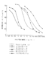
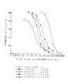
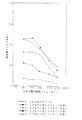
前記OA958−2抗体を担持したELISA用プレートを用いて、実施例3に記載した非競合法によりOA、7−OA、DTX1及びDTX3の標準品を測定した。メチルアルコール濃度を20%刻みで0%から100%までの6段階について調べ、検量線を作成し、結果を図1〜図4に示した。メチルアルコールに換えてアセトンを用いて、アセトン濃度を20%刻みで0%から70%までの5段階について調べ、検量線を作成し、結果を図5〜図8に示した。
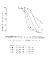
また、前記OA8−2抗体、OA10−8抗体及びOA22−22抗体をそれぞれ担持したELISA用プレートを用いて、実施例3に記載した非競合法によりOA、7−OA、DTX1及びDTX3の標準品を測定した。メチルアルコール濃度30%における検量線を作成し、結果を図9〜図11に示した。
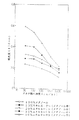
更に、OA958−2抗体を担持したELISA用プレートを用いて、実施例3に記載した非競合法により、無水有機溶媒系でOA標準品を測定した。メチルアルコール中のエチルアルコール濃度を0%及び70から100%までの5段階、メチルアルコール中のアセトン、エチルエーテル、ベンゼンの濃度を各々0から50%までの5段階について調べ、検量線を作成し、結果を図12〜図15に示した。
【0033】
OA958−2抗体を用いた非競合法によるメチルアルコール中での測定結果によれば、メチルアルコール濃度が高くなるに従い抗体活性は徐々に低下するものの、100%メチルアルコール中でも、OA、7−OA、DTX1及びDTX3のすべてについて十分に測定可能であった。各々の測定感度は、20%メチルアルコールではOAが0.1ng/ml、7−OAが1.0ng/ml、DTX1が0.3ng/ml、DTX3が1.0ng/mlであった。また100%メチルアルコールではOAが10ng/ml、7−OAが30ng/ml、DTX1が30ng/ml、DTX3が30ng/mlであった。また、OA、7−OA、DTX1及びDTX3に対するOA958−2抗体の反応性の比は、60%メチルアルコールでは1:0.3:1:0.2であり、80%メチルアルコールでは1:0.3:0.6:0.3であるが、100%メチルアルコールでは1:0.8:2:1.4となり、メチルアルコール濃度が増すにつれ反応性の差は少なくなった(図1〜図4)。
【0034】
OA958−2抗体を用いた非競合法によるアセトン中での測定結果によれば、メチルアルコールと同様にアセトン濃度が高くなるに従い抗体活性は徐々に低下し、70%アセトン中ではDTX3が多少の影響を受けるものの、測定は可能であった。その他のOA、7−OA及びDTX1は共に十分測定可能であった。アセトン濃度が70%を越えるとELISA用プレートの材質が変化するため測定不可能であった。各々の測定感度は、10%アセトンではOAが0.3ng/ml、7−OAが1.0ng/ml、DTX1が1.0ng/ml、DTX3が3.0ng/mlであった。また70%アセトンではOAが10ng/ml、7−OAが30ng/ml、DTX1が100ng/ml、DTX3が30ng/mlであった。また、OA、7−OA、DTX1及びDTX3に対するOA958−2抗体の反応性の比は、アセトン濃度による差は余りなく、50%アセトンでは1:0.2:0.5:0.1となり、DTX1及びDTX3に対する反応性が低下する傾向が見られた(図5〜図8)。
【0035】
OA8−2抗体を用いた非競合法による30%メチルアルコール中での測定結果によれば、OAに対する反応性が最も高く、測定感度は3.0ng/mlであった。また7−OA、DTX1及びDTX3に対する反応性は等しく、測定感度は10ng/mlであった(図9)。
OA10−8抗体を用いた非競合法による30%メチルアルコール中での測定結果によれば、DTX1及びDTX3に対する反応性が、OA及び7−OAに対する反応性よりも高かった。測定感度はOAが3.0ng/ml、7−OAが30ng/ml、DTX1が1.0ng/ml、そしてDTX3が1.0ng/mlであった(図10)。
OA22−22抗体を用いた非競合法による30%メチルアルコール中での測定結果は、OA8−2抗体の場合と類似しており、OAに対する反応性が最も高く、他の7−OA、DTX1及びDTX3に対する反応性は、ほぼ等しかった。測定感度はOAが0.3ng/ml、7−OAが3.0ng/ml、DTX1が10ng/ml、そしてDTX3が3.0ng/mlであった(図11)。
【0036】
OA958−2抗体を用いた非競合法によるエチルアルコールとメチルアルコールの混合物中での測定結果によれば、エチルアルコール濃度が70%及び80%では、バックグラウンド値が僅かに上昇する傾向が見られたが、抗体の反応性には変化が無かった。エチルアルコール濃度が90%及び100%になっても抗体の反応性の低下は僅かであった(図12)。
OA958−2抗体を用いた非競合法によるアセトンとメチルアルコールの混合物中での測定結果によれば、アセトン濃度が高くなるに従い、バックグラウンド値が僅かに上昇する傾向が見られたが、抗体の反応性には変化は無かった。アセトン濃度が50%を越えるとELISA用プレートの材質が変化するため測定不可能であった(図13)。
OA958−2抗体を用いた非競合法によるエチルエーテルとメチルアルコールの混合物中での測定結果によれば、エチルエーテル濃度が高くなるに従い、バックグラウンド値が僅かに上昇する傾向が見られたが、抗体の反応性には変化は無かった。エチルエーテル濃度が50%を越えるとELISA用プレートの材質が変化するため測定不可能であった(図14)。
OA958−2抗体を用いた非競合法によるベンゼンとメチルアルコールの混合物中での測定結果によれば、ベンゼン濃度が10%以下では、抗体の反応性に変化は無かった。ベンゼン濃度が20%になると抗体の反応性は約2/3、ベンゼン濃度が30%では抗体の反応性は約1/3になり、ベンゼン濃度が50%になると反応性はほぼ完全に失われた。また、ベンゼン濃度が50%になるとELISA用プレートの材質が変化するため測定が非常に困難となった(図15)。
【0037】
【発明の効果】
従来の致死活性測定法には、動物の管理、検出感度や精度及び特異性に問題があり、従来の競合酵素免疫学的測定法にも、下痢性貝毒のうちの7−O−アシル−オカダ酸及びDTX3を検出・測定することができないという問題点があったのに対し、本発明によれば、OA、7−OA、DTX1及びDTX3の4種の下痢性貝毒の全てに特異性を有するモノクローナル抗体が提供されるので、従来の欠点を解消することができる。また、前記モノクローナル抗体が有機溶媒耐性を有する場合には、試料からの有機溶媒抽出液をそのまま又は単に希釈するだけで検出又は測定工程に用いることができるので、検出又は測定工程が簡便になるだけで無く、高精度及び高感度を達成することができる。従って、食品衛生等に貢献することができる。
【図面の簡単な説明】
【図1】メチルアルコール濃度0〜100%の溶液中にてOAを非競合法により測定した場合の検量線を示すグラフである。
【図2】メチルアルコール濃度0〜100%の溶液中にて7−OAを非競合法により測定した場合の検量線を示すグラフである。
【図3】メチルアルコール濃度0〜100%の溶液中にてDTX1を非競合法により測定した場合の検量線を示すグラフである。
【図4】メチルアルコール濃度0〜100%の溶液中にてDTX3を非競合法により測定した場合の検量線を示すグラフである。
【図5】アセトン濃度0〜70%の溶液中にてOAを非競合法により測定した場合の検量線を示すグラフである。
【図6】アセトン濃度0〜70%の溶液中にて7−OAを非競合法により測定した場合の検量線を示すグラフである。
【図7】アセトン濃度0〜70%の溶液中にてDTX1を非競合法により測定した場合の検量線を示すグラフである。
【図8】アセトン濃度0〜70%の溶液中にてDTX3を非競合法により測定した場合の検量線を示すグラフである。
【図9】OA8−2抗体を用いてメチルアルコール濃度30%の溶液中で、OA、7−OA、DTX1及びDTX3を非競合法により測定した場合の検量線を示すグラフである。
【図10】OA10−8抗体を用いてメチルアルコール濃度30%の溶液中で、OA、7−OA、DTX1及びDTX3を非競合法により測定した場合の検量線を示すグラフである。
【図11】OA22−22抗体を用いてメチルアルコール濃度30%の溶液中で、OA、7−OA、DTX1及びDTX3を非競合法により測定した場合の検量線を示すグラフである。
【図12】OA958−2抗体を用いてメチルアルコール中エチルアルコールの濃度が0%及び70〜100%の溶液中で、OAを非競合法により測定した場合の検量線を示すグラフである。
【図13】OA958−2抗体を用いてメチルアルコール中アセトンの濃度が0〜50%の溶液中で、OAを非競合法により測定した場合の検量線を示すグラフである。
【図14】OA958−2抗体を用いてメチルアルコール中エチルエーテルの濃度が0〜50%の溶液中で、OAを非競合法により測定した場合の検量線を示すグラフである。
【図15】OA958−2抗体を用いてメチルアルコール中ベンゼンの濃度が0〜50%の溶液中で、OAを非競合法により測定した場合の検量線を示すグラフである。[0001]
[Industrial applications]
The present invention relates to a monoclonal antibody specific to diarrheal shellfish toxin, a hybridoma producing the monoclonal antibody, and a method for detecting diarrheal shellfish toxin using the monoclonal antibody. Said monoclonal antibodies according to the invention are, in particular, organic solvent-resistant antibodies.
[0002]
[Prior art]
Of the four diarrheal shellfish poisons of okadaic acid, 7-O-acyl-okadaic acid, dinophysistoxin-1 and dinophysicystoxin-3 (i.e., 7-O-acyl-dinoficistoxin-1), Okada The acid is a lipophilic compound produced by one of the Kuroiso sponges (Halichondria okadai and Halichondria melanodocia), and dinophycystoxin-1 is produced by a dinoflagellate (Dinophysis fortii). Further, dinophysistoxin-3 is a fat-soluble compound produced by converting the above-mentioned dinophysistoxin-1 in the body of the shellfish, and 7-O-acylocadaic acid is an analog of dinophysicystoxin-3 obtained by synthesis. Is a substance. These compounds accumulate in the midgut glands of edible bivalves at any time or region and poison the shellfish. Although it is second only to puffer fish in terms of the number of food poisoning cases, it is first in the number of patients and is a major problem in food hygiene.
[0003]
Conventionally, as a method for detecting diarrheal shellfish poison, a lethal activity measurement method using mice has been used as an official method, but there have been problems in terms of animal management, detection sensitivity, accuracy, specificity, and the like. On the other hand, development of a method aiming at performing this inspection with high sensitivity, simply and in a short time has been attempted.
[0004]
For example, JP-A-1-96199 discloses a monoclonal antibody against an okadaic acid group and a method for producing the same. However, among the diarrheal shellfish poisons, the antibody specifically reacts with okadaic acid and dinophysistoxin-1, but detects 7-O-acyl-okadaic acid and dinophysistoxin-3.・ It cannot be measured. Further, Japanese Patent Application Laid-Open No. 1-96199 does not disclose or suggest a monoclonal antibody capable of maintaining its activity in the presence of an organic solvent.
[0005]
The present inventor has found that the main cause of food poisoning due to diarrheal shellfish toxin in Japan is dinophysistoxin-3, and that each of the above shellfish toxins is a fat-soluble substance. In view of the fact that the use of an organic solvent is unavoidable for extracting, and that it is desirable to carry out an immune reaction in the presence of an organic solvent to simplify the operation process, okadaic acid, dino A mouse monoclonal antibody that is specific for phytostoxin-1 and dinophysistoxin-3 and that is resistant to organic solvents has been discovered and disclosed in PCT / JP92 / 01021.
[0006]
The present inventors further studied the reactivity of dinophysistoxin-3 to 7-O-acyl-okadaic acid, which is a synthetic analog, and the reactivity in organic solvents other than alcohols and ketones. After extensive studies for the purpose of comparing the reactivity to the four kinds of shellfish poisons, they were specific to okadaic acid, 7-O-acyl-okadaic acid, dinophysistoxin-1 and dinophysicystoxin-3, and furthermore, were organic solvents. We have succeeded in finding a mouse monoclonal antibody that is resistant to diarrhea, and have found that the use of this monoclonal antibody enables rapid and specific detection of diarrheal shellfish toxin even in the presence of an organic solvent. The present invention is based on these findings.
[0007]
Therefore, the present invention, OhSpecific for kadaic acid, 7-O-acyl-okadaic acid, dinophysistoxin-1 and dinophysistoxin-3 (i.e., 7-O-acyl-dinoficistoxin-1)Reacting with a solution having a concentration of benzene in methyl alcohol of 20%.Related to monoclonal antibodies. The present invention also relates to a hybridoma producing the monoclonal antibody, and a method for immunologically detecting diarrheal shellfish toxin using the monoclonal antibody.
[0008]
Hereinafter, the monoclonal antibody, the hybridoma, and the immunological detection method will be described in this order.
Preparation of the monoclonal antibody and the hybridoma according to the present invention can be carried out by a conventional method, for example, a method described in the Seikagaku Experimental Lecture, Immunobiochemical Research Method (edited by the Japanese Biochemical Society). Specifically, as the immunogen, those which produce monoclonal antibodies specific to okadaic acid, 7-O-acyl-okadaic acid, dinophysistoxin-1 and dinophysistoxin-3 can be used arbitrarily. In particular, okadaic acid, 7-O-acyl-okadaic acid, dinophysistoxin-1 or dinophysicystoxin-3, or salts thereof, and furthermore, these are biopolymers (for example, bovine serum albumin or immunoglobulin). It is preferable to use those bonded to a carrier. A mammal (eg, mouse, rat, rabbit, goat or horse) is immunized using these immunogen solutions by in vivo immunization. For example, the immunogen solution is emulsified and mixed with an equal volume of Freund's complete adjuvant or incomplete adjuvant and administered subcutaneously to mice (first immunization). Thereafter, the same operation is performed at intervals of 2 to 4 weeks, and immunization is performed several times. Several days after the final immunization, the spleen is aseptically removed, crushed with a stainless mesh or the like to prepare spleen cells, and used for the cell fusion step.
[0009]
In the present specification, “acyl” may be any of an aromatic acyl group and a saturated or unsaturated, linear or branched aliphatic acyl group, for example, having 8 to 30 carbon atoms, preferably Is an acyl group derived from 8 to 24 saturated or unsaturated fatty acids (specifically, capric acid, myristic acid, palmitic acid, oleic acid, linolenic acid, etc.).
[0010]
Examples of myeloma cells (myeloma cells) that are the other parent cell of cell fusion include various known cell lines, for example, p3 (p3 / x63-Ag8) [Nature, 256, 495-497 (1975)]. , P3-U1 [Current Topics in Microbiology and Immunology, 81; 1-7 (1978)], NS-1 [Eur. J. Immunol. , 6; 511-519 (1976)], MPC-11 [Cell, 8; 405-415 (1976)], SP2 / 0 [Nature, 276; 269-270 (1978)], FO [J. Immunol. Meth. , 35; 1-21 (1980)], × 63.6.55.3 [J. Immunol. , 123; 1548-1550 (1979)], S194 [J. Exp. Med. , 148; 313-323 (1978)], or R210 in rats [Nature, 277; 131-133 (1979)].
[0011]
Cell fusion can be performed by a usual method, for example, using a known fusion promoter (such as polyethylene glycol) and optionally an auxiliary (such as dimethyl sulfoxide). Myeloma cells are used in an amount of about 1 to 10 times. As the medium for fusion, for example, Dulbecco's modified Eagle medium (DMEM) containing 40% (w / v) polyethylene glycol can be used. The fusion is performed by thoroughly mixing the immune spleen cells and myeloma cells in the above-mentioned medium.
[0012]
Subsequently, cells other than the hybridoma were removed using a selection medium (for example, HAT medium), and the antibody (ie, okadaic acid, 7-O-acyl-okadaic acid, and dinophysistoxin-1) in the hybridoma culture supernatant was removed. The presence or absence of the production of a monoclonal antibody which simultaneously shows specificity with dinophysistoxin-3 is detected and measured by, for example, an ELISA method, and the desired hybridoma is isolated. In particular, when selecting a hybridoma that produces a monoclonal antibody having resistance to an organic solvent, diarrheal shellfish toxin after putting the antibody in an organic aqueous solution or an anhydrous organic solvent containing the organic solvent at various concentrations. To confirm that the antigen-antibody reaction proceeds normally, thereby selecting hybridomas producing an organic solvent-resistant monoclonal antibody.
[0013]
The thus obtained hybridoma of the present invention that secretes the desired monoclonal antibody can be subcultured in a usual medium, and can be easily stored in liquid nitrogen or the like for a long period of time. As a medium for culturing a hybridoma, any medium suitable for culturing a hybridoma can be used. Preferably, fetal bovine serum, L-glutamine, L-pyruvic acid and antibiotics (penicillin G and streptomycin) are added to DMEM. Containing medium is used. The culture of the hybridoma is carried out in vitro, for example, for about 3 days at 5% carbon dioxide concentration and 37 ° C. in a medium, and for about 14 days in vivo, for example, when culturing in the abdominal cavity of a mouse. Is preferred.
[0014]
It is possible to separate and purify the desired monoclonal antibody from a culture solution obtained by culturing the above-mentioned hybridoma by a conventional method, or from ascites of a suitable mammal (for example, mouse or rat) to which the hybridoma has been administered. When a monoclonal antibody is separated and purified from a culture solution or mouse ascites, a method generally used for protein isolation and purification can be used. Examples of such methods include ammonium sulfate precipitation, ion exchange chromatography, molecular sieve column chromatography using molecular sieve gel, affinity column chromatography using protein A or protein G-bound polysaccharide, dialysis, and lyophilization.
[0015]
The method for detecting diarrheal shellfish toxin according to the present invention provides the monoclonal antibody (i.e., okadaic acid, 7-O-acyl-okadaic acid, dinophysistoxin-1 and dinophysicystoxin-3) of the present invention simultaneously. (The monoclonal antibody shown), the diarrheal shellfish poison can be detected without omission. In a preferred embodiment of the detection method of the present invention, since an organic solvent-resistant monoclonal antibody is used, an organic aqueous system (or an anhydrous organic system) containing an amount of an organic solvent capable of sufficiently dissolving the diarrheal shellfish poison to be tested is used. ) Allows an accurate antigen-antibody reaction to proceed.
[0016]
The method of the present invention uses a monoclonal antibody that simultaneously shows specificity for okadaic acid, 7-O-acyl-okadaic acid, dinophysistoxin-1 and dinophysistoxin-3, and preferably uses an organic solvent-resistant antibody. Except for this (therefore, performing the antigen-antibody reaction in the presence of an organic solvent), the other points can be directly applied to conventionally known immunological detection methods. Therefore, the method of the present invention is specifically, for example,
(1) a step of treating the sample with an organic solvent (eg, a water-miscible organic solvent) to prepare an organic solvent extract;
(2) contacting an antibody having specificity to diarrheal shellfish poison and having resistance to the organic solvent with the organic solvent extract;
(3) a step of bringing a known amount of diarrheal shellfish toxin having a label into contact with the antibody simultaneously with the step (2) or after completion of the step (2);
(4) a step of separating labeled diarrheal shellfish toxin bound to the antibody and labeled shellfish toxin not bound to the antibody;
(5) A method for detecting diarrheal shellfish toxin in a sample, comprising a step of detecting or measuring a signal from a label of one of the labeled diarrheal shellfish toxins separated in step (4) (competition method and Non-competitive method).
[0017]
Further, specific embodiments of the method of the present invention include, for example,
(1) a step of treating the sample with an organic solvent (eg, a water-miscible organic solvent) to prepare an organic solvent extract;
(2) a step of immobilizing a first antibody having specificity to diarrheal shellfish toxin and resistance to the organic solvent on an insoluble carrier;
(3) contacting the organic solvent extract containing diarrheal shellfish toxin with the immobilized first antibody in step (2);
(4) a step of adding an excessive amount of a labeled second antibody to the diarrheal shellfish toxin, which binds at a different site from the first antibody;
(5) A method for detecting diarrheal shellfish toxin in a sample (sandwich method) including a step of detecting a signal of a label on a complex of the first antibody and diarrheal shellfish toxin is also included. The present invention can be widely applied to other known immune reaction detection methods.
[0018]
The organic solvent used in the present invention is alcohols, ketones, ethers or benzene, and is a solvent used when extracting, diluting or preserving diarrheal shellfish poison from a test sample. When a water-miscible organic solvent is used, the antigen-antibody reaction can be performed in the organic aqueous solvent after the extraction step is performed with the organic aqueous solvent or, if necessary, the organic solvent extract is appropriately diluted with water. Alternatively, the antigen-antibody reaction can be carried out in an anhydrous organic solvent or an organic aqueous solvent using the organic solvent extract as it is, which is preferable. Examples of the organic solvent include alcohol compounds (for example, lower alcohols having 1 to 3 carbon atoms, particularly methyl alcohol, ethyl alcohol, and isopropyl alcohol), ketone compounds (for example, lower aliphatics having 1 to 3 carbon atoms). Ketones, especially methyl ethyl ketone, acetone), ether compounds (for example, lower aliphatic ethers having 1 to 3 carbon atoms, particularly methyl ether, ethyl ether, m-propyl ether) and benzene, or mixtures thereof. be able to.
[0019]
When the detection method of the present invention is specifically performed, first, a midgut gland specimen of the edible bivalve is extracted with the organic solvent. The obtained extract is used as it is or diluted with water for the next contacting step. Next, for example, a monoclonal antibody of the present invention obtained from a mouse, which is resistant to an organic solvent, is prepared in advance by the above method. When this antibody (the first antibody in the sandwich method) is brought into contact with the organic solvent extract, when diarrheal shellfish poison (antigen) is present in the organic solvent extract, the antigen-antibody reaction is performed in the presence of the organic solvent. Happens. This antigen-antibody reaction can be performed in the same manner as a normal antigen-antibody reaction except that the reaction is performed in the presence of an organic solvent. For example, the antibody is supported on a suitable insoluble support (well or latex particles) and reacted specifically with the antigen in the organic solvent extract.
[0020]
When the method of the present invention is carried out by a competitive method or a non-competitive method, it is possible to confirm or quantify the presence of diarrheal shellfish toxin using a known amount of labeled antigen. When the sandwich method is used, an excessive amount of the labeled second antibody is used. Known labels, such as radioisotopes (for example, 32 P, 35 S, 3 H), enzymes (eg, peroxidase, alkaline phosphatase), vitamins (eg, biotin), fluorescent substances (eg, FITC), and chemiluminescent substances (eg, acridinium) can be used.
[0021]
The labeled antigen is obtained after the step of contacting the antibody with the organic solvent extract (ie, after the antigen-antibody reaction between the antibody and the antigen in the organic solvent extract) is completed (non-competitive method). Alternatively, it can be added to the reaction system simultaneously with the step of contacting the antibody with the organic solvent extract (ie, simultaneously with the antigen-antibody reaction between the antibody and the antigen in the organic solvent extract) (competition method). In the non-competitive method, an antibody not bound to diarrheal shellfish poison in the organic solvent extract binds to the labeled antigen. On the other hand, in the competition method, a known amount of the labeled antigen and an unknown amount of the antigen in the organic solvent extract are competitively bound to the antibody. In the sandwich method, after the first antibody is brought into contact with the organic solvent extract, the non-competing antigen is washed away, and then the labeled second antibody is added, whereby the complex between the first antibody and the antigen is labeled. The secondary antibody binds.
[0022]
In the step of contacting the antibody with the organic solvent extract and the step of adding the labeled antigen or the labeled second antibody, the concentration of the organic solvent is adjusted in consideration of the solubility of the diarrheal shellfish toxin and the inactivation of the label. select. In the non-competitive method, the labeled antigen is added under conditions different from the conditions under which the antigen-antibody reaction is performed (for example, by adding water to reduce the concentration of the organic solvent or completely replacing the aqueous system). You can also. On the other hand, in the competitive method, since the labeled antigen is performed simultaneously with the antigen-antibody reaction, it is necessary to prevent the label from being inactivated by the organic solvent present in the reaction system.
[0023]
In the competitive method and the non-competitive method, after the reaction between the labeled antigen and the antibody is completed, the labeled antigen bound to the antibody and the labeled antigen not bound to the antibody are separated. Separation can be performed, for example, by filtration, centrifugation, or washing with a buffer. In the sandwich method, after the reaction between the first antibody-bound antigen and the labeled second antibody is completed, the labeled second antibody that has not bound to the first antibody-bound antigen is removed, and then the first antibody-bound The signal from the label of the labeled second antibody bound to the antigen is detected or measured.
[0024]
A signal derived from the labeled antigen thus separated (competition method or non-competition method) or a signal derived from the label of the labeled second antibody bound to the first antibody-bound antigen (sandwich method) is detected or measured. When detecting or measuring a signal, it is preferable to change the reaction system containing the labeled antigen to conditions suitable for signal detection or signal measurement. For example, when a biotin-labeled avidin system is used, the reaction system is changed to an aqueous system, a substrate is added, and the enzyme activity is detected or measured. When a fluorescent or chemiluminescent substance is used as a label, a signal is detected or measured under conditions where quenching does not occur.
[0025]
【Example】
Hereinafter, the present invention will be described specifically with reference to Examples, but these do not limit the scope of the present invention.
Example 1: Preparation of monoclonal antibody-producing hybridoma
2 mg of okadaic acid (Wako Pure Chemical) (hereinafter referred to as OA), a kind of diarrhea shellfish, 0.31 mg of N-hydroxysuccinimide, and 0.57 mg of N, N-dicyclohexylcarbodiimide were added to dimethylformamide (hereinafter, DMF). ), And reacted at room temperature for 2 hours. The obtained reaction solution was divided into two portions, and 1.5 mg of human IgG was added and dissolved in 39 μl of one reaction solution, and 1.9 mg of bovine serum albumin (hereinafter referred to as BSA) was added and dissolved in 81 μl of the other reaction solution. Then, each was further reacted at room temperature for 2 hours. Finally, the obtained reaction solution was subjected to gel filtration using a Sephadex G-25 column equilibrated with phosphoric acid-containing physiological saline (pH 7.4) (hereinafter, referred to as PBS). The obtained OA-human IgG and OA-BSA were dissolved in physiological saline at a concentration of 0.826 mg / ml and 1.04 mg / ml, respectively, and OA-human IgG was used as an immunogen and OA-BSA as an antigen for analysis. Was.
The same amount of Freund's complete adjuvant was added to 300 μl of the OA-IgG solution and mixed well to prepare a homogeneous sol. 200 μl of this sol was intraperitoneally administered to female mice (4 weeks old; A / J). Two months later, a similarly prepared antigen sol was administered intraperitoneally in the same amount.
[0026]
The spleen of the mouse in which the anti-OA antibody titer in the serum became high was excised, and the excised spleen was washed three times in a Petri dish with a T-2 medium containing 5% fetal bovine serum, and the wound was removed with an injection needle. After application, a single cell suspension was prepared by squeezing. The single cell suspension was filtered through a mesh to remove large solids. To the obtained filtrate, mouse myeloma cells P3X-63-Ag8-6.5.3 are mixed at a cell number ratio of 5: 1 (myeloma cells: spleen cells), and centrifuged (300 × g, 4). Min) and the cells were collected. Next, the precipitated cells were resuspended in serum-free T-3 medium, centrifuged under the same conditions, and the sediment was stirred by flicking the centrifuge tube with a finger, followed by 50% warming to 37 ° C. 1 ml of a polyethylene glycol (molecular weight 1,500) solution was slowly added over 60 seconds while rotating the centrifuge tube. To stop the cell fusion, a serum-free T-3 medium is added in three portions to a centrifuge tube in which the cell fusion is progressing (first 3 ml of the medium, then 9 ml of the medium, and finally 38 ml of medium were added over 30 seconds each). After the addition of the medium was completed, the cells were kept at 37 ° C. for 2 minutes and at room temperature for 8 minutes, and then centrifuged. 6 / Ml in a T-2 medium. The cell suspension was dispensed into a 96-well plastic plate in an amount of 100 μl / well, and cultured at 37 ° C. in a gas phase of 5% carbon dioxide-95% air. Twenty-four hours later, T-4 medium was added in an amount of 100 μl / well, and the culture was continued under the same conditions for another 10 to 14 days. The activity of the anti-OA antibody in the culture solution was examined, and the cells of the wells producing the desired antibody were cloned by a limiting dilution method using a HT medium in a 24-well plastic plate using an HT medium. As a result of cloning, 20 hybridoma (cell fusion) strains producing anti-OA antibodies were obtained.
[0027]
Example 2: Preparation of monoclonal antibody
Each of the 20 hybridoma strains selected in Example 1 was cultured in Cell Grosser H, a tissue-free serum-free culture medium containing 2.5 μg / ml of penicillin and streptomycin (for hybridoma) (Sumitomo Pharmaceutical). The obtained cells are suspended in the same medium and used at a temperature of 37 ° C. under a gas phase of 5% carbon dioxide-95% air using a Millipore Dyna Cell Culture System (Millipore) for the purpose of producing anti-OA antibody. Cultured. After completion of the culture, the culture solution was fractionated with ammonium sulfate, and the obtained monoclonal antibody was dissolved in 5 mM Tris-HCl buffer (pH 7.5) containing 0.9% NaCl and dialyzed.
[0028]
Example 3: Selection of monoclonal antibody
An ELISA plate was prepared using each anti-OA monoclonal antibody produced by 20 hybridomas. That is, the anti-OA monoclonal antibody prepared in Example 2 was dissolved at a concentration of 10 μg / ml in 0.083 M boric acid-containing physiological saline (pH 8.0) (hereinafter referred to as BBS), and 100 μl / After washing with the amount of the well, 250 μl of a gelatin solution (10 mg / ml) dissolved in BBS was dispensed to each well, and left at room temperature for 1 hour to perform blocking.
On the other hand, a series of aqueous methyl alcohol solutions was prepared by gradually changing the concentration of methyl alcohol in water from 0% (water) to 100% (anhydrous alcohol) in steps of 10%.
[0029]
To each of the above-mentioned ELISA plates, 100 μl of the above-mentioned alcoholic aqueous solution was added to each well, and left at room temperature. One hour later, each well was washed three times with 250 μl of BBS. Next, 100 μl of a solution (BBS containing 1.0% gelatin) containing 25 to 100 ng / ml of OA labeled with oxidized peroxidase (hereinafter referred to as OA-POD) was added to each well, and left at room temperature for 1 hour. Subsequently, after washing 5 times with 250 μl of BBS, 100 μl of a substrate solution [100 μl of a solution of 100 mg of 3,3 ′, 5,5′-tetramethylbenzidine in 10 ml of DMF is added to a 0.1 M sodium acetate solution (pH 5.5) 9 It was prepared by adding 15 μl of a 3% aqueous hydrogen peroxide solution; hereinafter, referred to as a TMBZ solution]. 100 μl was dispensed into each well, reacted at room temperature for 5 to 40 minutes, and 100 μl of 1N sulfuric acid was added. To stop the reaction. The absorbance of the reaction solution was measured at 450 nm or 415 nm with a spectrophotometer (Hitachi spectrophotometer U-1100) to prepare a calibration curve.
[0030]
As a result, the anti-OA monoclonal antibody (hereinafter, referred to as OA958-2 antibody) produced by the hybridoma OA958-2 strain (FERM P-13404) correctly recognizes the antigen even in 100% methyl alcohol, and a normal antigen-antibody reaction is performed. Found to do. Furthermore, an anti-OA monoclonal antibody (hereinafter referred to as an OA8-2 antibody) produced by the hybridoma OA8-2 strain (FERM P-13401), and an anti-OA monoclonal antibody (hereinafter referred to as the OA8-2 antibody) produced by the hybridoma OA10-8 strain (FERM P-13402) Hereinafter, each of the anti-OA monoclonal antibodies produced by the hybridoma OA22-22 strain (FERM P-13403) (hereinafter referred to as OA22-22 antibody) produced by the hybridoma OA22-22 strain (FERM P-13403) is also 50%. It was found that the antigen was correctly recognized even in% methyl alcohol, and a normal antigen-antibody reaction was performed. The immunoglobulin classes of the four types of anti-OA antibodies were determined using biotin-labeled antibodies (Ig, IgM, IgG) for discriminating mouse IgG subclasses.1 , IgG2a, IgG2b, IgG3 IA958-2 antibody, IgGA, λ-type light chain, and κ-type light chain).1κ, OA8-2 antibody is IgG2aκ, OA10-8 antibody is IgG1κ, and OA22-22 antibodies are IgG2awas κ.
[0031]
Example 4: Measurement of diarrheal shellfish poison by non-competitive method
By the non-competitive method, OA, 7-O-palmitoyl-okadaic acid (hereinafter, referred to as 7-OA), dinophysistoxin-1 (hereinafter, referred to as DTX1), and dinophysicystoxin-3 (7-O-palmitoyl-). DTX1) (hereinafter referred to as DTX3) was measured. The ELISA plates used were prepared under the conditions described in Example 3 using OA958-2 antibody, OA8-2 antibody, OA10-8 antibody, and OA22-22 antibody, respectively. The standard sample solution was prepared by a method in which a 100% methyl alcohol solution of diarrheal shellfish poison was once dried to dryness and then redissolved in BBS containing methyl alcohol at each concentration in consideration of the extraction operation from the actual sample. Used.
[0032]
Using the ELISA plate carrying the OA958-2 antibody, standard products of OA, 7-OA, DTX1, and DTX3 were measured by the non-competitive method described in Example 3. The methyl alcohol concentration was examined in six steps from 0% to 100% in 20% steps, and a calibration curve was prepared. The results are shown in FIGS. Using acetone instead of methyl alcohol, the acetone concentration was examined in five steps from 0% to 70% in increments of 20%, and calibration curves were prepared. The results are shown in FIGS.
In addition, using OA8-2 antibody, OA10-8 antibody and OA22-22 antibody, respectively, using ELISA plates carrying the respective antibodies, OA, 7-OA, DTX1 and DTX3 standard products were obtained by the noncompetitive method described in Example 3. Was measured. A calibration curve was prepared at a methyl alcohol concentration of 30%, and the results are shown in FIGS.
Furthermore, using an ELISA plate carrying the OA958-2 antibody, an OA standard was measured in an anhydrous organic solvent system by the non-competitive method described in Example 3. The concentration of ethyl alcohol in methyl alcohol was examined at 0% and 5 steps from 70 to 100%, and the concentrations of acetone, ethyl ether and benzene in methyl alcohol were examined at 5 steps from 0 to 50%, respectively, and a calibration curve was prepared. The results are shown in FIGS.
[0033]
According to the results of measurement in methyl alcohol by the non-competitive method using the OA958-2 antibody, although the antibody activity gradually decreases as the methyl alcohol concentration increases, even in 100% methyl alcohol, OA, 7-OA, All of DTX1 and DTX3 were sufficiently measurable. The sensitivity of each measurement was 0.1 ng / ml for OA, 1.0 ng / ml for 7-OA, 0.3 ng / ml for DTX1, and 1.0 ng / ml for DTX3 in 20% methyl alcohol. In the case of 100% methyl alcohol, OA was 10 ng / ml, 7-OA was 30 ng / ml, DTX1 was 30 ng / ml, and DTX3 was 30 ng / ml. The ratio of the reactivity of the OA958-2 antibody to OA, 7-OA, DTX1 and DTX3 was 1: 0.3: 1: 0.2 for 60% methyl alcohol and 1: 0 for 80% methyl alcohol. 0.3: 0.6: 0.3, but with 100% methyl alcohol, the ratio was 1: 0.8: 2: 1.4, and the difference in reactivity became smaller as the methyl alcohol concentration increased (FIG. 1). (Fig. 4).
[0034]
According to the measurement results in acetone by the non-competitive method using the OA958-2 antibody, the antibody activity gradually decreased as the acetone concentration increased, similar to methyl alcohol, and DTX3 had some influence in 70% acetone. However, the measurement was possible. Other OA, 7-OA and DTX1 were all sufficiently measurable. When the acetone concentration exceeded 70%, the measurement was impossible because the material of the ELISA plate changed. The sensitivity of each measurement was 0.3 ng / ml for OA, 1.0 ng / ml for 7-OA, 1.0 ng / ml for DTX1, and 3.0 ng / ml for DTX3 in 10% acetone. In 70% acetone, OA was 10 ng / ml, 7-OA was 30 ng / ml, DTX1 was 100 ng / ml, and DTX3 was 30 ng / ml. In addition, the ratio of the reactivity of the OA958-2 antibody to OA, 7-OA, DTX1 and DTX3 was not significantly different depending on the acetone concentration, and was 1: 0.2: 0.5: 0.1 with 50% acetone. There was a tendency for the reactivity to DTX1 and DTX3 to decrease (FIGS. 5 to 8).
[0035]
According to the measurement result in 30% methyl alcohol by the non-competitive method using the OA8-2 antibody, the reactivity with OA was the highest, and the measurement sensitivity was 3.0 ng / ml. The reactivity to 7-OA, DTX1, and DTX3 was equal, and the measurement sensitivity was 10 ng / ml (FIG. 9).
According to the measurement result in 30% methyl alcohol by the non-competitive method using the OA10-8 antibody, the reactivity to DTX1 and DTX3 was higher than the reactivity to OA and 7-OA. The measurement sensitivities were 3.0 ng / ml for OA, 30 ng / ml for 7-OA, 1.0 ng / ml for DTX1, and 1.0 ng / ml for DTX3 (FIG. 10).
The results of measurement in 30% methyl alcohol by the non-competitive method using the OA22-22 antibody are similar to those of the OA8-2 antibody, and the reactivity to OA is the highest, and the other 7-OA, DTX1 and Reactivity to DTX3 was almost equal. The measurement sensitivities were 0.3 ng / ml for OA, 3.0 ng / ml for 7-OA, 10 ng / ml for DTX1, and 3.0 ng / ml for DTX3 (FIG. 11).
[0036]
According to the measurement results in a mixture of ethyl alcohol and methyl alcohol by a non-competitive method using the OA958-2 antibody, the background value tends to slightly increase when the ethyl alcohol concentration is 70% or 80%. However, there was no change in antibody reactivity. Even when the ethyl alcohol concentration reached 90% and 100%, the decrease in the reactivity of the antibody was slight (FIG. 12).
According to the result of measurement in a mixture of acetone and methyl alcohol by the non-competitive method using the OA958-2 antibody, the background value tended to slightly increase as the acetone concentration was increased. There was no change in reactivity. When the acetone concentration exceeded 50%, measurement was impossible because the material of the ELISA plate changed (FIG. 13).
According to the measurement results in a mixture of ethyl ether and methyl alcohol by the non-competitive method using the OA958-2 antibody, the background value tended to slightly increase as the ethyl ether concentration increased, There was no change in antibody reactivity. When the ethyl ether concentration exceeded 50%, measurement was impossible because the material of the ELISA plate changed (FIG. 14).
According to the result of measurement in a mixture of benzene and methyl alcohol by a non-competitive method using the OA958-2 antibody, the reactivity of the antibody did not change when the benzene concentration was 10% or less. When the benzene concentration is 20%, the reactivity of the antibody is about 2/3, when the benzene concentration is 30%, the antibody reactivity is about 1/3, and when the benzene concentration is 50%, the reactivity is almost completely lost. Was. Further, when the benzene concentration became 50%, the measurement became very difficult because the material of the ELISA plate changed (FIG. 15).
[0037]
【The invention's effect】
Conventional methods for measuring lethal activity have problems in animal management, detection sensitivity, accuracy and specificity. Conventional competitive enzyme immunoassays also require 7-O-acyl- In contrast to the problem that okadaic acid and DTX3 cannot be detected and measured, according to the present invention, specificity is obtained for all four types of diarrheal shellfish poisons of OA, 7-OA, DTX1 and DTX3. Thus, the conventional disadvantages can be solved. Further, when the monoclonal antibody has an organic solvent resistance, since the organic solvent extract from the sample can be used for the detection or measurement step by simply or simply diluting, the detection or measurement step is only simplified. However, high accuracy and high sensitivity can be achieved. Therefore, it can contribute to food hygiene and the like.
[Brief description of the drawings]
FIG. 1 is a graph showing a calibration curve when OA is measured by a non-competitive method in a solution having a methyl alcohol concentration of 0 to 100%.
FIG. 2 is a graph showing a calibration curve when 7-OA is measured by a non-competitive method in a solution having a methyl alcohol concentration of 0 to 100%.
FIG. 3 is a graph showing a calibration curve when DTX1 is measured by a non-competitive method in a solution having a methyl alcohol concentration of 0 to 100%.
FIG. 4 is a graph showing a calibration curve when DTX3 is measured by a non-competitive method in a solution having a methyl alcohol concentration of 0 to 100%.
FIG. 5 is a graph showing a calibration curve when OA is measured by a non-competitive method in a solution having an acetone concentration of 0 to 70%.
FIG. 6 is a graph showing a calibration curve when 7-OA is measured by a non-competitive method in a solution having an acetone concentration of 0 to 70%.
FIG. 7 is a graph showing a calibration curve when DTX1 is measured by a non-competitive method in a solution having an acetone concentration of 0 to 70%.
FIG. 8 is a graph showing a calibration curve when DTX3 is measured by a non-competitive method in a solution having an acetone concentration of 0 to 70%.
FIG. 9 is a graph showing a calibration curve when OA, 7-OA, DTX1, and DTX3 are measured by a non-competitive method in a solution having a methyl alcohol concentration of 30% using an OA8-2 antibody.
FIG. 10 is a graph showing a calibration curve when OA, 7-OA, DTX1, and DTX3 are measured by a non-competitive method in a solution having a methyl alcohol concentration of 30% using an OA10-8 antibody.
FIG. 11 is a graph showing a calibration curve when OA, 7-OA, DTX1, and DTX3 are measured by a noncompetitive method in a solution having a methyl alcohol concentration of 30% using an OA22-22 antibody.
FIG. 12 is a graph showing a calibration curve when OA is measured by a non-competitive method in a solution in which the concentration of ethyl alcohol in methyl alcohol is 0% or 70 to 100% using the OA958-2 antibody.
FIG. 13 is a graph showing a calibration curve when OA is measured by a non-competitive method in a solution in which the concentration of acetone in methyl alcohol is 0 to 50% using the OA958-2 antibody.
FIG. 14 is a graph showing a calibration curve when OA is measured by a non-competitive method in a solution in which the concentration of ethyl ether in methyl alcohol is 0 to 50% using the OA958-2 antibody.
FIG. 15 is a graph showing a calibration curve when OA is measured by a non-competitive method in a solution in which the concentration of benzene in methyl alcohol is 0 to 50% using the OA958-2 antibody.
Claims (5)
Priority Applications (1)
| Application Number | Priority Date | Filing Date | Title |
|---|---|---|---|
| JP04200893A JP3576184B2 (en) | 1993-02-05 | 1993-02-05 | Diarrheal shellfish toxin-specific monoclonal antibody, hybridoma and diarrheal shellfish toxin detection method |
Applications Claiming Priority (1)
| Application Number | Priority Date | Filing Date | Title |
|---|---|---|---|
| JP04200893A JP3576184B2 (en) | 1993-02-05 | 1993-02-05 | Diarrheal shellfish toxin-specific monoclonal antibody, hybridoma and diarrheal shellfish toxin detection method |
Publications (2)
| Publication Number | Publication Date |
|---|---|
| JPH06225787A JPH06225787A (en) | 1994-08-16 |
| JP3576184B2 true JP3576184B2 (en) | 2004-10-13 |
Family
ID=12624160
Family Applications (1)
| Application Number | Title | Priority Date | Filing Date |
|---|---|---|---|
| JP04200893A Expired - Lifetime JP3576184B2 (en) | 1993-02-05 | 1993-02-05 | Diarrheal shellfish toxin-specific monoclonal antibody, hybridoma and diarrheal shellfish toxin detection method |
Country Status (1)
| Country | Link |
|---|---|
| JP (1) | JP3576184B2 (en) |
-
1993
- 1993-02-05 JP JP04200893A patent/JP3576184B2/en not_active Expired - Lifetime
Also Published As
| Publication number | Publication date |
|---|---|
| JPH06225787A (en) | 1994-08-16 |
Similar Documents
| Publication | Publication Date | Title |
|---|---|---|
| JP3673522B2 (en) | Anti-annexin V monoclonal antibodies and their use and hybridoma cell lines producing them | |
| US5374533A (en) | Method for determining chondrocalcin | |
| Paul et al. | Enzyme-linked immunosorbent assays for sperm antibody detection and antigenic analysis | |
| WO1986002364A1 (en) | Monoclonal antibodies and their use | |
| JP3154724B2 (en) | Methods for detecting monoclonal antibodies, hybridomas and diarrheal shellfish poisons specific to diarrheal shellfish toxins | |
| WO1994023302A1 (en) | Immunological assay of oxidatively modified human low density lipoproteins in plasma | |
| JP3576184B2 (en) | Diarrheal shellfish toxin-specific monoclonal antibody, hybridoma and diarrheal shellfish toxin detection method | |
| AU2015280292A1 (en) | Binding partners specific for vitamin D epimers | |
| US5137807A (en) | Method for determining beta-subunit of human prolyl 4-hydroxylase by immunoassay to detect hepatic disease | |
| US5298397A (en) | Method of assaying d-vanillylmandelic acid | |
| JP4663831B2 (en) | Monoclonal antibodies, cell lines, and methods for measuring N1, N12-diacetylspermine | |
| EP0199755A1 (en) | Monoclonal antibodies and their use | |
| WO1986002355A1 (en) | Monoclonal antibodies and their use | |
| JP2007106696A (en) | Anti-quercetin monoclonal antibody, cell producing the same, method for detection of quercetin and detection reagent thereof | |
| EP0193935B1 (en) | Method for determining human prolyl 4-hydroxylase by immunoassay | |
| WO1986002362A1 (en) | Monoclonal antibodies and their use | |
| JPH06153979A (en) | Monoclonal antibody against fish iridovirus, hybridoma for producing the antibody and production method | |
| WO1986002363A1 (en) | Monoclonal antibodies and their use | |
| WO1986002365A1 (en) | Monoclonal antibodies and their use | |
| WO1986003498A1 (en) | Monoclonal antibodies and their use | |
| JP3414856B2 (en) | Method for immunological measurement of diphenyl ether compounds | |
| WO1986002360A1 (en) | Monoclonal antibodies and their use | |
| WO1986002354A1 (en) | Monoclonal antibodies and their use | |
| WO1986002356A1 (en) | Monoclonal antibodies and their use | |
| JPH0690784A (en) | Monoclonal antibody and its use |
Legal Events
| Date | Code | Title | Description |
|---|---|---|---|
| A131 | Notification of reasons for refusal |
Free format text: JAPANESE INTERMEDIATE CODE: A131 Effective date: 20040323 |
|
| A521 | Written amendment |
Free format text: JAPANESE INTERMEDIATE CODE: A523 Effective date: 20040524 |
|
| TRDD | Decision of grant or rejection written | ||
| A01 | Written decision to grant a patent or to grant a registration (utility model) |
Free format text: JAPANESE INTERMEDIATE CODE: A01 Effective date: 20040706 |
|
| A61 | First payment of annual fees (during grant procedure) |
Free format text: JAPANESE INTERMEDIATE CODE: A61 Effective date: 20040707 |
|
| R150 | Certificate of patent (=grant) or registration of utility model |
Free format text: JAPANESE INTERMEDIATE CODE: R150 |
|
| S531 | Written request for registration of change of domicile |
Free format text: JAPANESE INTERMEDIATE CODE: R313531 |
|
| R350 | Written notification of registration of transfer |
Free format text: JAPANESE INTERMEDIATE CODE: R350 |
|
| FPAY | Renewal fee payment (prs date is renewal date of database) |
Free format text: PAYMENT UNTIL: 20080716 Year of fee payment: 4 |
|
| FPAY | Renewal fee payment (prs date is renewal date of database) |
Free format text: PAYMENT UNTIL: 20090716 Year of fee payment: 5 |
|
| FPAY | Renewal fee payment (prs date is renewal date of database) |
Free format text: PAYMENT UNTIL: 20100716 Year of fee payment: 6 |
|
| S111 | Request for change of ownership or part of ownership |
Free format text: JAPANESE INTERMEDIATE CODE: R313115 |
|
| FPAY | Renewal fee payment (prs date is renewal date of database) |
Free format text: PAYMENT UNTIL: 20100716 Year of fee payment: 6 |
|
| R371 | Transfer withdrawn |
Free format text: JAPANESE INTERMEDIATE CODE: R371 |
|
| S111 | Request for change of ownership or part of ownership |
Free format text: JAPANESE INTERMEDIATE CODE: R313115 |
|
| FPAY | Renewal fee payment (prs date is renewal date of database) |
Free format text: PAYMENT UNTIL: 20100716 Year of fee payment: 6 |
|
| R350 | Written notification of registration of transfer |
Free format text: JAPANESE INTERMEDIATE CODE: R350 |
|
| FPAY | Renewal fee payment (prs date is renewal date of database) |
Free format text: PAYMENT UNTIL: 20110716 Year of fee payment: 7 |
|
| FPAY | Renewal fee payment (prs date is renewal date of database) |
Free format text: PAYMENT UNTIL: 20110716 Year of fee payment: 7 |
|
| FPAY | Renewal fee payment (prs date is renewal date of database) |
Free format text: PAYMENT UNTIL: 20120716 Year of fee payment: 8 |
|
| FPAY | Renewal fee payment (prs date is renewal date of database) |
Free format text: PAYMENT UNTIL: 20130716 Year of fee payment: 9 |
|
| EXPY | Cancellation because of completion of term | ||
| FPAY | Renewal fee payment (prs date is renewal date of database) |
Free format text: PAYMENT UNTIL: 20130716 Year of fee payment: 9 |